Recent Advances in Mupirocin Delivery Strategies for ... - MDPI
24
Review Recent Advances in Mupirocin Delivery Strategies for the Treatment of Bacterial Skin and Soft Tissue Infection Aishwarya Gangwar 1 , Parveen Kumar 2 , Ranjit Singh 3 and Preeti Kush 3, * Citation: Gangwar, A.; Kumar, P.; Singh, R.; Kush, P. Recent Advances in Mupirocin Delivery Strategies for the Treatment of Bacterial Skin and Soft Tissue Infection. Future Pharm. 2021, 1, 80–103. https://doi.org/ 10.3390/futurepharmacol1010007 Academic Editor: Daniele Ribeiro de Araujo Received: 15 November 2021 Accepted: 16 December 2021 Published: 20 December 2021 Publisher’s Note: MDPI stays neutral with regard to jurisdictional claims in published maps and institutional affil- iations. Copyright: © 2021 by the authors. Licensee MDPI, Basel, Switzerland. This article is an open access article distributed under the terms and conditions of the Creative Commons Attribution (CC BY) license (https:// creativecommons.org/licenses/by/ 4.0/). 1 Department of Pharmaceutics, Chandigarh College of Pharmacy, Mohali 140307, Punjab, India; [email protected] 2 Exigo Recycling Pvt. Ltd., Noida 201309, Uttar Pradesh, India; [email protected] 3 School of Pharmacy, Adarsh Vijendra Institute of Pharmaceutical Sciences, Shobhit University Gangoh, Saharanpur 247341, Uttar Pradesh, India; [email protected] * Correspondence: [email protected] Abstract: Skin and soft tissue infections (SSTIs) have increased problematically in hospital and am- bulatory settings due to the poor immunity of hosts and multidrug-resistant pathogens. Mupirocin (MUP), a global topical antibiotic, is used for the treatment of SSTIs caused by various pathogens due to its unique mechanism of action. However, the therapeutic efficiency of MUP is hampered due to the protein binding and drug resistance caused by frequent use. A combined report covering the various aspects of MUP, such as the synthesis of the novel formulation, loading of the drug, and application against various skin infections, is missing. This comprehensive review focuses on various novel drug delivery strategies such as composite biomaterials/scaffold, hydrogel dressings, liposomes, liposo- mal hydrogel, microparticles/microspheres, microsponges, nanocapsules, nanofibers, silicone-based adhesive patches, and topical sprays. The therapeutic effect of the MUP can be synergized by com- bining with other agents and using novel strategies. The objective is to enhance patient compliance, decrease the resistance, magnify the delivery of MUP, and overcome the limitations of conventional formulations. Moreover, the carriers/dressing materials are biocompatible, biodegradable, stimulate wound healing, protect the wound from external environmental contamination, adsorb the wound exudates, and are permeable to oxygen and moisture. This review will help researchers to explore further the treatment of various bacterial skin infections by using MUP-loaded novel formulations with better efficacy, utilizing the novel nanostructures or combinatorial methods. Keywords: skin and soft tissue infections; mupirocin; bacterial infection; drug resistance; novel drug delivery system; magnified delivery 1. Introduction The skin is the largest organ of the human body, which is daily invaded by various environmental factors such as dryness, cold bites, bacteria, fungi, and accidental fires as well. All these factors may damage skin, leading to skin and skin structure infections (SSIs) or skin and soft tissue infections (SSTIs). These are the most common type of bacterial infection that involves breaching of the integumentary part of the skin (accidental or in- tentional), ranging from mild severity (pyoderma) to life-threatening (necrotizing fasciitis) incidences [1–3]. In recent decades, the incidence rate of SSTIs has increased problematically in hospital and ambulatory settings in the United States due to the poor immunity of the population affected and the multidrug resistance in pathogens [4–6]. Traditionally, Staphy- lococcus aureus (S. aureus) and Streptococcus pyogenes (group A β-hemolytic streptococci, S. pyogenes) were the main culprits for the SSTIs, but, recently, either methicillin-resistant S. aureus (MRSA) or macrolide-resistant S. pyogenes or both in combination are the main cause of these infections [7]. SSTIs are classified in various forms based on the infection location, progression rate, clinical symptoms, causative agent, extension depth, and sever- ity [1,7–9]. In 1998, the United States Food and Drug Administration (USFDA) categorized Future Pharm. 2021, 1, 80–103. https://doi.org/10.3390/futurepharmacol1010007 https://www.mdpi.com/journal/futurepharmacol
-
Upload
khangminh22 -
Category
Documents
-
view
0 -
download
0
Transcript of Recent Advances in Mupirocin Delivery Strategies for ... - MDPI

Review
Recent Advances in Mupirocin Delivery Strategies for theTreatment of Bacterial Skin and Soft Tissue Infection
Aishwarya Gangwar 1, Parveen Kumar 2, Ranjit Singh 3 and Preeti Kush 3,*
�����������������
Citation: Gangwar, A.; Kumar, P.;
Singh, R.; Kush, P. Recent Advances
in Mupirocin Delivery Strategies for
the Treatment of Bacterial Skin and
Soft Tissue Infection. Future Pharm.
2021, 1, 80–103. https://doi.org/
10.3390/futurepharmacol1010007
Academic Editor: Daniele Ribeiro
de Araujo
Received: 15 November 2021
Accepted: 16 December 2021
Published: 20 December 2021
Publisher’s Note: MDPI stays neutral
with regard to jurisdictional claims in
published maps and institutional affil-
iations.
Copyright: © 2021 by the authors.
Licensee MDPI, Basel, Switzerland.
This article is an open access article
distributed under the terms and
conditions of the Creative Commons
Attribution (CC BY) license (https://
creativecommons.org/licenses/by/
4.0/).
1 Department of Pharmaceutics, Chandigarh College of Pharmacy, Mohali 140307, Punjab, India;[email protected]
2 Exigo Recycling Pvt. Ltd., Noida 201309, Uttar Pradesh, India; [email protected] School of Pharmacy, Adarsh Vijendra Institute of Pharmaceutical Sciences, Shobhit University Gangoh,
Saharanpur 247341, Uttar Pradesh, India; [email protected]* Correspondence: [email protected]
Abstract: Skin and soft tissue infections (SSTIs) have increased problematically in hospital and am-bulatory settings due to the poor immunity of hosts and multidrug-resistant pathogens. Mupirocin(MUP), a global topical antibiotic, is used for the treatment of SSTIs caused by various pathogens dueto its unique mechanism of action. However, the therapeutic efficiency of MUP is hampered due to theprotein binding and drug resistance caused by frequent use. A combined report covering the variousaspects of MUP, such as the synthesis of the novel formulation, loading of the drug, and applicationagainst various skin infections, is missing. This comprehensive review focuses on various novel drugdelivery strategies such as composite biomaterials/scaffold, hydrogel dressings, liposomes, liposo-mal hydrogel, microparticles/microspheres, microsponges, nanocapsules, nanofibers, silicone-basedadhesive patches, and topical sprays. The therapeutic effect of the MUP can be synergized by com-bining with other agents and using novel strategies. The objective is to enhance patient compliance,decrease the resistance, magnify the delivery of MUP, and overcome the limitations of conventionalformulations. Moreover, the carriers/dressing materials are biocompatible, biodegradable, stimulatewound healing, protect the wound from external environmental contamination, adsorb the woundexudates, and are permeable to oxygen and moisture. This review will help researchers to explorefurther the treatment of various bacterial skin infections by using MUP-loaded novel formulationswith better efficacy, utilizing the novel nanostructures or combinatorial methods.
Keywords: skin and soft tissue infections; mupirocin; bacterial infection; drug resistance; novel drugdelivery system; magnified delivery
1. Introduction
The skin is the largest organ of the human body, which is daily invaded by variousenvironmental factors such as dryness, cold bites, bacteria, fungi, and accidental fires aswell. All these factors may damage skin, leading to skin and skin structure infections (SSIs)or skin and soft tissue infections (SSTIs). These are the most common type of bacterialinfection that involves breaching of the integumentary part of the skin (accidental or in-tentional), ranging from mild severity (pyoderma) to life-threatening (necrotizing fasciitis)incidences [1–3]. In recent decades, the incidence rate of SSTIs has increased problematicallyin hospital and ambulatory settings in the United States due to the poor immunity of thepopulation affected and the multidrug resistance in pathogens [4–6]. Traditionally, Staphy-lococcus aureus (S. aureus) and Streptococcus pyogenes (group A β-hemolytic streptococci,S. pyogenes) were the main culprits for the SSTIs, but, recently, either methicillin-resistantS. aureus (MRSA) or macrolide-resistant S. pyogenes or both in combination are the maincause of these infections [7]. SSTIs are classified in various forms based on the infectionlocation, progression rate, clinical symptoms, causative agent, extension depth, and sever-ity [1,7–9]. In 1998, the United States Food and Drug Administration (USFDA) categorized
Future Pharm. 2021, 1, 80–103. https://doi.org/10.3390/futurepharmacol1010007 https://www.mdpi.com/journal/futurepharmacol

Future Pharm. 2021, 1 81
SSTIs into complicated and uncomplicated treatment. To explain further, complicatedtreatment addresses deeper tissue infection and requires surgical treatment and uncom-plicated treatments are to cure superficial infections. However, this classification did notcategorize the patients who were recovering from these infections [10]. Therefore, in 2013,the USFDA adopted a new guideline for pharmaceutical industries and classified all SSTIsinto a consolidated term: acute bacterial skin and skin structure infections (ABSSSI) [2].ABSSSI is defined as a skin bacterial infection with a lesion size of 75 cm2 area (measuredby area of redness, edema, or induration), including bacterial cellulitis/erysipelas, woundinfection, and cutaneous abscess [3]. This guideline excludes impetigo, minor cutaneousabscess, diabetic foot infections (DFI), infection from human or animal bites, decubitus ulcerinfection, myonecrosis, necrotizing fasciitis, ecthyma gangrenosum, and chronic woundinfections [10,11]. In 2014, the Infectious Diseases Society of America (IDSA) proposed amore relevant and practical classification of SSTIs [12]. The IDSA classified SSTIs based on“(i) skin extension, complicated infection (deep structures of the skin) and uncomplicated(superficial infections); (ii) rate of progression, acute and chronic wound infections; (iii)tissue necrosis, necrotizing and not necrotizing infections” [11]. All the classifications in-clude the patients who possess various clinical manifestations such as cellulitis/erysipelas,wound infection, and major cutaneous abscess, etc. (Table 1) [6]. Moreover, these may becategorized into primary (bullous impetigo, cellulitis, carbuncles, furuncles impetigo conta-giosa, and folliculitis), and secondary SSTIs (atopic dermatitis, prurigo, contact dermatitis,and neurodermatitis) [9].
Table 1. Descriptive details of various skin and soft tissue infections.
SSTIs Infection Pathogen Description Ref.
Non-purulent SSTIs
Impetigo Staphylococcus aureus,Streptococcus pyogenes
Superficial infection developed viadirect or indirect invasion of bacteria. It
is the most common infection inchildren and presents in two forms, i.e.,
bullous and non-bullous impetigo.
[7,9,13]
Cellulitis
Staphylococcus aureus,beta-hemolytic
streptococci (groups A, B,C, or G)
Subcutaneous infections areaccompanied by lymphadenopathy and
lymphangitis. It is characterized byredness, edema, or induration and
usually affects lower limbs.
[7]
Erysipelas Staphylococcus aureus,Streptococcus pyogenes
Superficial lymphatics and upperdermis infection, usually affects the faceand sometimes lower limbs. It possesses
well-defined sharp raised borders incontrast to non-infected areas.
[7,13]
FolliculitisPseudomonas aeruginosa,Staphylococcus aureus,Streptococcus pyogenes
It is a superficial inflammation of hairfollicles, mainly affecting moist skin
with hair.[7,9,14]
Purulent SSTIs
Furuncle Staphylococcus aureus
Furuncle or boil is a deep inflammatoryinfection developed from folliculitis.
Initially, it is a firm, tender,erythematous nodule that becomes
fluctuant and painful. It usually infectsthe face, buttocks, and axillae.
[9,13]
Carbuncle Staphylococcus aureus,Streptococcus pyogenes
It is an aggregation of multiplefuruncles, involves infection of the hair
follicle, and is further extended tosubcutaneous tissues. The infection ispainful and tender but the patient is
well. It is usually observed at the neck,back, and thighs.
[7,9,13]

Future Pharm. 2021, 1 82
Table 1. Cont.
SSTIs Infection Pathogen Description Ref.
Abscess
Staphylococcus aureus,Streptococcus pyogenes,Streptococcus milleri,viridans, streptococci,
coagulase–staphylococci
Focal collection of pus in dermis andhypodermis, characterized by tender,
red nodules surrounded byerythematous swelling.
[7,9,15,16]
Complex SSTIs
Burn wound Anaerobes
Burn wound infection possesses a highbacteria concentration (>105 coloniesforming unit). It arises immediately
after the injury due to the damage of thecutaneous barrier and adaptive
immunity. The surrounding tissues ofthe burn wound exhibit warmth,
tenderness, induration, and erythema.
[6,17]
Surgical siteinfection Escherichia coli
It usually arises 4 days after surgery andis categorized into superficial incisional,
deep incisional, and organ or spaceinfection. It is diagnosed by incisional
discharge, swelling, tenderness,and erythema.
[18]
Diabetic footinfection
Staphylococcus aureus,Enterococci,
Pseudomonas aeruginosa,Enterobacteriaceae,Acinetobacter spp.,
Bacteroides spp.
This infection is most common indiabetic patients and possesses high
mortality. This infection encompasses arange from nails to necrotizing limbs.
Nails serve as an entry portal forbacterial infection due to poor hygiene.
[6,19,20]
Necrotizing SSTIs Monomicrobial,Polymicrobial
Staphylococcus aureus,Streptococcus pyogenes
Gram-negatives,Clostridium species,Anaerobic bacteria
Necrosis of soft tissues or muscles isinitially characterized by erythema andinduration with pain followed by skin
color change to blue/purple. Thepatient suffers from systemic toxicity,
multi-organ failure, andhemodynamic instability.
[6,7,9]
Bite wounds Human andanimal bite
Eikenella corrodens,Pasteurella multocida,
Pasteurella canis,Capnocytophaga
canimorsus,Staphylococcus aureus
It usually arises after biting. [7,21,22]
SSTIs can be complicated when Gram-negative bacteria accumulate in highly riskyareas, such as the rectum, and invade deeper skin areas. Therefore, a systemic approachis required for the optimal management of complicated SSTIs. Optimal managementinitially comprises physical examination followed by identification of pathogen via asmear of discharge from the lesions. Further, based on culture and susceptibility results,adequate antibacterial therapy should be implemented [19]. Topical antibiotics are anextensively used therapy for the management of SSTIs due to their capability to providehigher concentration to the target area with minimized adverse effects. Additionally, thetopical application offers various advantages in contrast to systemic administration, suchas patient compliance and regular inspection of infection, and allows the use of suchtherapeutic agents (bacitracin or neomycin) that can not be systematically administered.Clinically, various topical antibiotics, such as bacitracin, neomycin, polymyxin B, fusidicacid, and mupirocin (MUP), are currently used for the treatment of SSTIs [23].
MUP is one of the widely used topical antibiotics that is effectively used to treatsuperficial skin infections caused by Gram-positive and Gram-negative bacteria, especially

Future Pharm. 2021, 1 83
nasal MRSA due to its broad antibacterial spectrum [23–25] and antibiofilm property [18,26].Commercially, MUP (2%) is available as cream and ointments (Bactroban, Bactoderm,Mupirocin, Turixin) [24]. Due to the unique mechanism of action, it does not possess anycross-resistance with other antibiotics, leading to global use in various hospital departments.However, the potential efficacy of MUP is hampered due to its short half-life (<30 min),high protein binding, and different resistance rates (1–81%) [23,24,27]. Furthermore, theconventional formulations possess some adverse effects such as burning, dryness, itching,rashes, redness, nausea, pain, stinging, swelling, or tenderness [18]. The purpose of thispaper is to review the clinical status of MUP for the management of bacterial skin infectionand then provide more comprehension on various drug delivery strategies to augmentMUP delivery and overcome the limitations of the conventional formulation.
2. Mupirocin: A Drug of Choice for the Treatment of Skin Infection
MUP (C26H44O9) is one of the first drugs which was synthesized by the modularpolyketide synthases and isolated from soil bacteria Pseudomonas fluorescens NCIMB 10586.This unique polyketide topical antibiotic is a mixture of four pseudomonic acids (A-D)and is composed of monic acid and 9-hydroxynonanoic acid (Figure 1). It is a well-tolerated topical antibiotic owing to its minimal systemic absorption through intact skin [24].Additionally, it exhibits poor in vitro antibacterial activity against the normal skin defenseflora (e.g., Corynebacterium, Micrococcus, and Propionibacterium). Multiple studies revealedthat topical MUP was more effective for the treatment of impetigo in contrast to varioussystemic antibiotics (ampicillin cloxacillin, cephalexin, dicloxacillin, erythromycin, andflucloxacillin) [28]. However, it is not recommended for systemic administration due toin vivo degradation into an inactive metabolite (monic acid) and high protein binding [27].
Future Pharm. 2021, 1, FOR PEER REVIEW 4
MUP is one of the widely used topical antibiotics that is effectively used to treat su-perficial skin infections caused by Gram-positive and Gram-negative bacteria, especially nasal MRSA due to its broad antibacterial spectrum [23–25] and antibiofilm property [18,26]. Commercially, MUP (2%) is available as cream and ointments (Bactroban, Bacto-derm, Mupirocin, Turixin) [24]. Due to the unique mechanism of action, it does not pos-sess any cross-resistance with other antibiotics, leading to global use in various hospital departments. However, the potential efficacy of MUP is hampered due to its short half-life (<30 min), high protein binding, and different resistance rates (1–81%) [23,24,27]. Fur-thermore, the conventional formulations possess some adverse effects such as burning, dryness, itching, rashes, redness, nausea, pain, stinging, swelling, or tenderness [18]. The purpose of this paper is to review the clinical status of MUP for the management of bac-terial skin infection and then provide more comprehension on various drug delivery strat-egies to augment MUP delivery and overcome the limitations of the conventional formu-lation.
2. Mupirocin: A Drug of Choice for the Treatment of Skin Infection MUP (C26H44O9) is one of the first drugs which was synthesized by the modular
polyketide synthases and isolated from soil bacteria Pseudomonas fluorescens NCIMB 10586. This unique polyketide topical antibiotic is a mixture of four pseudomonic acids (A-D) and is composed of monic acid and 9-hydroxynonanoic acid (Figure 1). It is a well-tolerated topical antibiotic owing to its minimal systemic absorption through intact skin [24]. Additionally, it exhibits poor in vitro antibacterial activity against the normal skin defense flora (e.g., Corynebacterium, Micrococcus, and Propionibacterium). Multiple studies revealed that topical MUP was more effective for the treatment of impetigo in contrast to various systemic antibiotics (ampicillin cloxacillin, cephalexin, dicloxacillin, erythromy-cin, and flucloxacillin) [28]. However, it is not recommended for systemic administration due to in vivo degradation into an inactive metabolite (monic acid) and high protein bind-ing [27].
Figure 1. Mupirocin structure encompasses monic acid (a heptaketide) with pyran ring connected to the 9-hydroxynonanoic acid via an ester linkage.
2.1. Clinical Application and Mechanism of Action MUP is mainly used to treat primary and secondary skin infections, skin appendages
[29], peritonitis [30], nasal, and mucus membrane infections [31] caused by S. aureus and S. epidermidis, including MRSA and beta-lactamase-producing strains. Clinically, it exhib-its approximately 80% improvement in infected patients and 90% abolishment in S. aureus isolates [32]. Moreover, it can also prevent the colonization of MRSA in health care pro-fessionals, intensive care units (ICU), dialysis units, and cardiothoracic and orthopedic surgery wards [28]. Decolonization can decrease the risk of MRSA infections in patients and reduce the transmission to other patients [33]. Furthermore, clinical studies revealed that monotherapy with MUP 2% ointment was effective for the treatment of erythrasma
Figure 1. Mupirocin structure encompasses monic acid (a heptaketide) with pyran ring connected tothe 9-hydroxynonanoic acid via an ester linkage.
2.1. Clinical Application and Mechanism of Action
MUP is mainly used to treat primary and secondary skin infections, skin appendages [29],peritonitis [30], nasal, and mucus membrane infections [31] caused by S. aureus and S. epi-dermidis, including MRSA and beta-lactamase-producing strains. Clinically, it exhibitsapproximately 80% improvement in infected patients and 90% abolishment in S. aureusisolates [32]. Moreover, it can also prevent the colonization of MRSA in health care pro-fessionals, intensive care units (ICU), dialysis units, and cardiothoracic and orthopedicsurgery wards [28]. Decolonization can decrease the risk of MRSA infections in patients andreduce the transmission to other patients [33]. Furthermore, clinical studies revealed thatmonotherapy with MUP 2% ointment was effective for the treatment of erythrasma [34]and Zoon’s balanitis [35]. Additionally, MUP-loaded nanoliposomes were used for thetreatment of multidrug-resistant Neisseria gonorrhoeae [27].
It reversibly binds to the isoleucine (Ile)-binding site of the isoleucyl-transfer RNA(Ile-tRNA) synthetase enzyme, which catalyzes the formation of Ile-tRNA from Ile andt-RNA. This enzyme complex prevents Ile incorporation into the protein chains, leading toinhibiting RNA and protein synthesis (Figure 2) [36]. This peculiar mechanism of action

Future Pharm. 2021, 1 84
is due to the similarity between the one moiety of MUP (C-14 and C-11 of monic acid)and Ile. It does not possess any cross-resistance with other antibiotics owing to its uniquemechanism of action [23].
Future Pharm. 2021, 1, FOR PEER REVIEW 5
[34] and Zoon’s balanitis [35]. Additionally, MUP-loaded nanoliposomes were used for the treatment of multidrug-resistant Neisseria gonorrhoeae [27].
It reversibly binds to the isoleucine (Ile)-binding site of the isoleucyl-transfer RNA (Ile-tRNA) synthetase enzyme, which catalyzes the formation of Ile-tRNA from Ile and t-RNA. This enzyme complex prevents Ile incorporation into the protein chains, leading to inhibiting RNA and protein synthesis (Figure 2) [36]. This peculiar mechanism of action is due to the similarity between the one moiety of MUP (C-14 and C-11 of monic acid) and Ile. It does not possess any cross-resistance with other antibiotics owing to its unique mechanism of action [23].
Figure 2. Unique mechanism of action of mupirocin.
MUP exhibits a bacteriostatic property at low concentration but possesses bacteri-cidal activity after 24 h of topical application (20,000 µg/mL with 2% formulation) [28]. Moreover, the antibacterial property is pH-dependent, i.e., at acidic pH, it displays mag-nified in vitro antibacterial activity, so the low pH of the skin provides a favorable envi-ronment for effective treatment [24,28].
2.2. Drug Resistance: More Than a Challenge The first MUP-resistant S. aureus was reported in 1987 during MRSA treatment at St.
Thomas’ hospital, London [32,33]. The MUP resistance frequency varies (0–65%) in differ-ent MRSA strains. Globally, MUP resistance is increasing in the S. aureus species, espe-cially MRSA, due to an increase in over-the-counter, unjustified, and long-term usage, leading to the potential efficacy being hampered. The prevalence of MUP resistance in S. aureus depends on various factors such as selection of patient population (community, hospital, or ICU), surveillance nature, e.g., active surveillance in previously MUP-treated patients vs. passive surveillance of isolates from all the patients, and associated resistance
Figure 2. Unique mechanism of action of mupirocin.
MUP exhibits a bacteriostatic property at low concentration but possesses bactericidalactivity after 24 h of topical application (20,000 µg/mL with 2% formulation) [28]. Moreover,the antibacterial property is pH-dependent, i.e., at acidic pH, it displays magnified in vitroantibacterial activity, so the low pH of the skin provides a favorable environment foreffective treatment [24,28].
2.2. Drug Resistance: More Than a Challenge
The first MUP-resistant S. aureus was reported in 1987 during MRSA treatment at St.Thomas’ hospital, London [32,33]. The MUP resistance frequency varies (0–65%) in differ-ent MRSA strains. Globally, MUP resistance is increasing in the S. aureus species, especiallyMRSA, due to an increase in over-the-counter, unjustified, and long-term usage, leadingto the potential efficacy being hampered. The prevalence of MUP resistance in S. aureusdepends on various factors such as selection of patient population (community, hospital, orICU), surveillance nature, e.g., active surveillance in previously MUP-treated patients vs.passive surveillance of isolates from all the patients, and associated resistance profile. Basedon the minimum inhibitory concentration (MIC), MUP possesses two types of resistance:low-level resistance (LLR) and high-level resistance (HLR). LLR (MIC ≤ 8–256 µg/mL)is a more common chromosomal resistance that usually arises due to the spontaneouspoint mutation of the Ile-tRNA synthetase gene, leading to the retardation of the binding

Future Pharm. 2021, 1 85
affinity of MUP to the Ile-tRNA synthetase enzyme [23]. LLR resistance is clinically in-significant and non-transferable, but further mutations can lead to HLR via the acquisitionof a pSK41-like plasmid (staphylococcal multi-resistant conjugative plasmids) [32,33,37].HLR (MIC > 500 µg/mL) is a plasmid-coded resistance that possesses two resistance mech-anisms. The first mechanism comprises the acquisition of plasmid-mediated mupA or theIle-tRNA synthetase-2 gene [32]. Moreover, the mupA gene is self-transmissible, leadingto creating resistance to other antibiotics such as clindamycin, erythromycin, gentamicin,tetracycline, levofloxacin, and trimethoprim [33,38]. Molecular studies revealed that themupA gene is mainly responsible for HLR in almost all S. aureus isolates. The second mech-anism is due to the synthesis of another paralog mupB that shares 45.5% sequence identitywith Ile synthetase and 65.5% identity with mupA. Additionally, some aminoacyl-tRNAsynthetases paralogs are also responsible for MUP resistance [24,38].
3. Novel Strategies to Augment Mupirocin Delivery in Bacterial Skin Infection
Though MUP is used for the treatment of various bacterial skin infections, it possessescertain limitations such as a short half-life, high protein binding, and drug resistance [23,27].The resistance can be overcome by the controlled use of MUP for the target decolonization,limiting the treatment duration up to 10 days, and not repeating the treatment for upto 30 days minimum. Further, the antibacterial effect of the MUP can be synergized bycombining with other agents such as anesthetics, other topical antibiotics, and naturalherbs. Thus, various novel drug delivery strategies have been adopted to enhance patientcompliance, decrease the resistance, magnify the delivery of mupirocin, and overcome thelimitations of conventional formulations. In this section, various novel formulations suchas composite biomaterials/scaffold, hydrogel dressings, liposomes, liposomal hydrogel,microparticles/microspheres, microsponges, nanocapsules, nanofibers, topical sprays,nanostructured lipid carriers, and silicone-based adhesive patches, etc., will be discussed(Table 2).

Future Pharm. 2021, 1 86
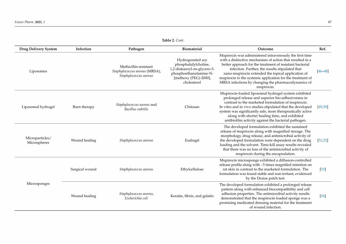
Table 2. Various mupirocin-loaded drug delivery systems for the treatment of skin and soft tissue infections.
Drug Delivery System Infection Pathogen Biomaterial Outcome Ref.
Compositebiomaterials/scaffold
Wound healing Staphylococcus aureus, Bacillussubtilis, Escherichia coli
Collagen, Silica
The collagen scaffolds exhibited more therapeuticpotential for the treatment of wound infection anddisplayed a promising carrier approach for tissue
engineering.
[39]
The developed bio-composite exhibited enhanced wateruptake, sustained release, and antimicrobial activity.
In vivo results stipulated that the biomaterial showedenhanced adhesion and wound contraction rate,
supported by histopathological analysis.
[40]
Hydrogel dressings
Wound healing
Escherichia coli(ATCC 8739), Enterococcus hirae
(ATCC 10541), S. aureus(ATCC 6538), Pseudomonas
aeruginosa (ATCC 27853), Bacilluscereus (ATCC 7064), Klebsiella
pneumonia
Chitosan, sodium alginate,carbopol
The developed composite film accelerated theregeneration of the epidermal layer in contrast to the
marketed commercial formulation.[41]
Diabetic wound Polyvinyl alcohol The developed gel was effective for the treatment ofdiabetic wound and accelerated the wound closure. [42]
Primary and secondary Gram-positive and Gram-negativebacteria Chitosan
The prepared polymeric membrane was spherical, stable,and elastic, along with having the controlled release
property. Furthermore, the membrane exhibitedmagnified retention of the drug in the skin without any
irritation.
[43]
Surgical wound Staphylococcus aureus Chitosan
The formulated spherical membrane exhibited superioradhesion and elasticity along with progressive drug
release. The Draize patch test revealed that the developedmembrane was non-irritant to the skin, along with
having magnified antimicrobial efficiency and enhancedretention to the skin.
[44]
Skin injuries Acrylic acid
The developed patches exhibited good elasticity andtensile strength, along with enhanced permeation and
retention into the skin. The patches were non-irritant tothe skin, evidenced by the Draize patch test.
[45]
Future Pharm. 2021, 1 87
Table 2. Cont.
Drug Delivery System Infection Pathogen Biomaterial Outcome Ref.
LiposomesMethicillin-resistant
Staphylococcus aureus (MRSA),Staphylococcus aureus
Hydrogenated soyphosphatidylcholine,
1,2-distearoyl-sn-glycero-3-phosphoethanolamine-N-
[methoxy (PEG)-2000],cholesterol
Mupirocin was administered intravenously the first timewith a distinctive mechanism of action that resulted in a
better approach for the treatment of resistant bacterialinfection. Further, the results stipulated that
nano-mupirocin extended the topical application ofmupirocin to the systemic application for the treatment ofMRSA infections by changing the pharmacodynamics of
mupirocin.
[46–48]
Liposomal hydrogel Burn therapy Staphylococcus aureus andBacillus subtilis Chitosan
Mupirocin-loaded liposomal hydrogel system exhibitedprolonged release and superior bio-adhesiveness incontrast to the marketed formulation of mupirocin.
In vitro and in vivo studies stipulated that the developedsystem was significantly safe, more therapeutically active
along with shorter healing time, and exhibitedantibiofilm activity against the bacterial pathogen.
[49,50]
Microparticles/Microspheres Wound healing Staphylococcus aureus Eudragit
The developed formulation exhibited the sustainedrelease of mupirocin along with magnified storage. Themorphology, drug release, and antimicrobial activity ofthe developed formulation were dependent on the drugloading and the solvent. Time-kill assay results revealed
that there was no loss of the antimicrobial activity ofmupirocin during the encapsulation.
[51,52]
Microsponges
Surgical wound Staphylococcus aureus Ethylcellulose
Mupirocin microsponge exhibited a diffusion-controlledrelease profile along with ~5 times magnified retention on
rat skin in contrast to the marketed formulation. Theformulation was found stable and non-irritant, evidenced
by the Draize patch test.
[53]
Wound healing Staphylococcus aureus,Escherichia coli Keratin, fibrin, and gelatin
The developed formulation exhibited a prolonged releasepattern along with enhanced biocompatibility and celladhesion properties. The antimicrobial activity resultsdemonstrated that the mupirocin-loaded sponge was a
promising medicated dressing material for the treatmentof wound infection.
[54]

Future Pharm. 2021, 1 88
Table 2. Cont.
Drug Delivery System Infection Pathogen Biomaterial Outcome Ref.
Nanocapsule/nanoparticles Wound healing
Poly(ε-caprolactone) The developed nanocapsules showed excellent stability at40 ◦C and room temperature. [55]
Methicillin-resistantStaphylococcus aureus (MRSA) Chitosan, selenium
The tailored formulation showed remarkable therapeuticpotential in terms of diabetic wound healing and wound
contraction compared to the native mupirocin.[56]
Staphylococcus aureus,Staphylococcus epidermidis,
Pseudomonas aeruginosa, andEscherichia coli
Poly(ethylene oxide)–poly(propylene
oxide)–poly(ethylene oxide)(PEO–PPO–PEO)
The tailored formulation exhibited reduced minimuminhibitory concentrations and minimum bactericidal
concentrations against S. aureus, S. epidermidis,Pseudomonas aeruginosa, and E. coli compared to the
mupirocin ointment. Further, the developed formulationwas safe, effective, and biocompatible for the treatment of
wound infection.
[57,58]
Nanofibers
Wound healing Staphylococcus aureus Poly-l-lactic acid
The tailored scaffold exhibited a different release profilefor both drugs, suggesting that the release kinetics of onedrug was altered by keeping the two different drugs in
the same polymer matrix. The dual drug scaffold releaseda significantly higher drug and even compensated the
inactive monic acid to act on the applied area, resulting inthe maintainence of a sufficient concentration of
mupirocin in the infected wound for more than a 72 hperiod, resulting in profound wound healing.
[59]
Burn wound
Staphylococcus aureus, Pseudomonasaeruginosa, and Escherichia coli Polyurethane
The developed fiber mat was enough for woundhydration via providing adequate environmental
humidity. Moreover, the tailored nanofiber exhibitedsufficient cell spreading and attachment. The cytotoxicity
results revealed that the antibacterial activity of thescaffold was increased proportionally with the increase in
mupirocin concentration (2–5%). Further, thehistopathological study revealed that the nanofibrous
mat was enough for burn wound healing due tonegligible inflammation.
[60]

Future Pharm. 2021, 1 89
Table 2. Cont.
Drug Delivery System Infection Pathogen Biomaterial Outcome Ref.
Staphylococcus aureus, Pseudomonasaeruginosa, and Escherichia coli Polycaprolactone
The tailored multifunctional double-layer nanofibrousscaffold (MDLS) was effective for the management ofwound infection, along with superior tensile strength
with enhanced contact angle and swelling ratio.Furthermore, cytotoxicity results revealed that the MDLSwas more biocompatible due to the addition of chitosan
in contrast to polycaprolactone nanofibers.
[61]
Staphylococcus aureus, andEscherichia coli
Keratin, and coenzyme Q10,and polyvinyl alcohol
The tailored formulations were biocompatible, evidencedby the skin irritancy test. Further, the therapeutic efficacyof the tailored formulation was assessed by antimicrobial
activity against various strains of S. aureus (2583, 2586,2587, 2590), MRSA 2555, and E. coli 1808. Moreover, cell
proliferation results evidenced the ability of nanofibers tosupport the keratinocytes’ growth due to the presence of
coenzyme Q10.
[62]
Topical spray Burn woundStaphylococcus aureus, Pseudomonasaeruginosa, and Escherichia coli, and
Streptococcus suis (S. suis)Eudragit E100
The developed spray exhibited magnified antimicrobialactivity (18-fold) against S. suis, in contrast to the
marketed formulation due to close contact between sprayand skin, leading to the formation of a thin film on the
infected surface. Moreover, the topical formulation wasfound non-irritant to the human skin without any toxicity
to the monocytes, keratinocytes, and fibroblasts cells.Additionally, the safety profile of the formulation wasalso confirmed by zero production of nitric oxide andinflammatory cytokines (IL-1b and TNF-a) due to its
antiendotoxin effect.
[18,63]
Nanostructured lipidcarrier
Cetyl palmitate,caprylic acid
Nanostructured lipid carrier (NLC) reduced themetabolic degradation of MUP via the protective lipid
layer of NLC which resulted in a 40-fold and 55-fold areaunder the curve and half-life, respectively, in contrast to
native MUP.
[64]

Future Pharm. 2021, 1 90
3.1. Composite Materials/Scaffolds
A composite material comprises a synergistic approach by combining two or morematerials to achieve a novel material possessing the specific properties not manifested bythe individual components [65]. There are various types of composite materials based ontheir reinforcement and matrix orientation. These materials are widely used in variousfields, including civil construction, aerospace, aeronautics, and automotive and biomedicalapplication. Biomedical applications include cardiovascular, dentistry, oral, skin infections,and tissue engineering due to their biocompatibility, biodegradability, porosity, and tissueregeneration capacity [66,67].
Inspired by this approach, Ramadass et al. fabricated MUP-loaded collagen hy-drolysate composite scaffold (CHCS) by sol-gel process for the treatment of wound infec-tion [39]. Collagen hydrolysate, the daughter fragment of collagen, was used as a betteralternative to the conventionally used collagen due to enhanced solubility accompanied bybetter bioavailability [68]. Collagen is a three-dimensional biomaterial that plays a vitalrole in the treatment of various purulent and non-purulent skin infections, such as burns,pressure sores, wounds, legs, and decubitus ulcers, but possesses some drawbacks suchas multistep extraction, which increases the cost, restricted solubility, low porosity, andfaster collagen turnover [39,69]. The CHCS was prepared by the sol-gel process, usingtetraethoxysilane as the silica precursor. Porosity and stability were the main reasons forthe magnified adsorption of the water present in the exudate, resulting in the encourage-ment of autolytic debridement of the wound infection [70]. Further, the CHCS scaffoldwas found more biostable in contrast to the collagen with a minimum frequency of dress-ing change. The in vitro drug release results suggested that the MUP was released in asustained manner over (~88.14 ± 6.21%) more than 72 h due to the CHCS scaffold. Theantimicrobial study revealed that the CHCS exhibited enhanced activity against the usedpathogens, S. aureus, Bacillus subtilis (B. subtilis), Pseudomonas aeruginosa (P. aeruginosa),and Escherichia coli (E. coli), due to the synergistic effect of MUP and collagen hydrolysate.Additionally, in vivo studies on the rat model revealed that the tailored formulation healedthe infection better than the conventional formulation, hence omitting frequent dressing.Thus, MUP-loaded CHCS offers an alternative and cost-effective alternative for woundtreatment [39].
In another study, Perumal et al. synthesized a bio-composite by combining MUP asan antimicrobial, silica microspheres as a drug delivery carrier, and collagen as a woundhealer. The synthesis of the bio-composite involved a two-step process: (1) synthesis ofMUP-loaded silica microspheres by the sol-gel process (MUP-SM) and (2) incorporation ofMUP-SM into collagen solution (MUP-SM incorporated collagen scaffold) (Figure 3). TheMUP-SM-incorporated collagen scaffold delivered 81.65 ± 5.02% MUP for up to 3 days,in contrast to MUP incorporated into the collagen scaffold (88.01 ± 2.01%, 1 day). Thissustained release was due to the silica microspheres, leading to maintaining the drugconcentration for a longer time and reducing the frequency of drug application which willbe beneficial to combat the wound infection cost-effectively. Furthermore, magnified wateruptake, tensile strength, cell proliferation, and antimicrobial assay revealed the efficacyof the developed formulation. Additionally, wound closure analysis stipulated that thecomplete epithelialization was at 14.2 ± 0.44 days for MUP-SM-loaded collagen, whereas itwas at 17.4 ± 0.44 days and 20.6 ± 0.54 days for collagen and control groups, respectively.This enhanced efficacy was due to the synergistic effect of MUP-SM and collagen scaffoldprotecting against the microbial invasion to the wound and tissue regeneration facilita-tion, respectively. Histopathological analysis revealed better dermal collagenization andfibroblast proliferation. In conclusion, this combined strategy exhibited a sustained-releaseproperty and better adhesion, along with enhanced wound contraction rate. Thus, it wouldbe a better approach for the augmented delivery of MUP for the treatment of various skininfections [40].

Future Pharm. 2021, 1 91
Future Pharm. 2021, 1, FOR PEER REVIEW 10
contrast to MUP incorporated into the collagen scaffold (88.01 ± 2.01%, 1 day). This sus-tained release was due to the silica microspheres, leading to maintaining the drug concen-tration for a longer time and reducing the frequency of drug application which will be beneficial to combat the wound infection cost-effectively. Furthermore, magnified water uptake, tensile strength, cell proliferation, and antimicrobial assay revealed the efficacy of the developed formulation. Additionally, wound closure analysis stipulated that the com-plete epithelialization was at 14.2 ± 0.44 days for MUP-SM-loaded collagen, whereas it was at 17.4 ± 0.44 days and 20.6 ± 0.54 days for collagen and control groups, respectively. This enhanced efficacy was due to the synergistic effect of MUP-SM and collagen scaffold protecting against the microbial invasion to the wound and tissue regeneration facilita-tion, respectively. Histopathological analysis revealed better dermal collagenization and fibroblast proliferation. In conclusion, this combined strategy exhibited a sustained-re-lease property and better adhesion, along with enhanced wound contraction rate. Thus, it would be a better approach for the augmented delivery of MUP for the treatment of vari-ous skin infections [40].
Figure 3. A synergistic bio-composite scaffold comprises mupirocin-loaded silica microspheres embedded in the wound healer collagen scaffold, enabling enhanced wound healing.
3.2. Hydrogel Membrane Hydrogels are hydrophilic, porous, three-dimensional, cross-linked, macromolecular
polymeric networks, stabilized chemically or physically via interlinkage in their lattice structure [45,71]. They have been extensively used for topical application as an alternative material due to their durability, elasticity, biocompatibility, biodegradability, and adhe-siveness. Due to their inherent hydrophilicity, hydrogels can absorb an enormous number of biological fluids or water, which makes them a soft material. These structures can imi-tate the mechanical properties of various animal tissues and revive the actual extracellular matrix, leading to the promising carrier for regenerative medicine [72]. Furthermore, var-ious forms of hydrogels are also used in drug delivery and tissue engineering (Figure 4).
Figure 3. A synergistic bio-composite scaffold comprises mupirocin-loaded silica microspheres embedded in the woundhealer collagen scaffold, enabling enhanced wound healing.
3.2. Hydrogel Membrane
Hydrogels are hydrophilic, porous, three-dimensional, cross-linked, macromolecularpolymeric networks, stabilized chemically or physically via interlinkage in their latticestructure [45,71]. They have been extensively used for topical application as an alternativematerial due to their durability, elasticity, biocompatibility, biodegradability, and adhesive-ness. Due to their inherent hydrophilicity, hydrogels can absorb an enormous number ofbiological fluids or water, which makes them a soft material. These structures can imitatethe mechanical properties of various animal tissues and revive the actual extracellular ma-trix, leading to the promising carrier for regenerative medicine [72]. Furthermore, variousforms of hydrogels are also used in drug delivery and tissue engineering (Figure 4).
Okur et al. developed and optimized various natural polymer-based (chitosan, sodiumalginate, and carbomer) hydrogel dressings by using the solvent casting method. All the for-mulations exhibited a spherical shape, magnified drug content with an improved in vitrodrug release profile, and permissible thickness for the wound dressing. Antimicrobialactivity and ex vivo bio-adhesion study revealed that the developed formulation possessedsuperior antibacterial activity and could be safely applied to the infected area withoutcausing any skin irritation. Furthermore, in vivo studies concluded that the developed filmwas more effective for wound healing as it accelerated the regeneration of the epidermallayer, in contrast to the marketed commercial formulation (Bactroban cream; GlaxoSmithK-line, Turkey), due to its unique physicochemical properties [41]. Recently, a MUP-loadedpolyvinyl alcohol (PVA)-based hydrogel cross-linked by a reactive oxygen species (ROS)responsive linker was developed by Zhao et al. The developed ROS-scavenging hydrogelwas able to treat the bacterially infected diabetic wound and accelerated the wound clo-sure due to the loading of MUP and granulocyte-macrophage colony-stimulating factor(GM-CSF), respectively [42].

Future Pharm. 2021, 1 92Future Pharm. 2021, 1, FOR PEER REVIEW 11
Figure 4. Various types of hydrogels used in drug delivery and tissue engineering.
Okur et al. developed and optimized various natural polymer-based (chitosan, so-dium alginate, and carbomer) hydrogel dressings by using the solvent casting method. All the formulations exhibited a spherical shape, magnified drug content with an im-proved in vitro drug release profile, and permissible thickness for the wound dressing. Antimicrobial activity and ex vivo bio-adhesion study revealed that the developed for-mulation possessed superior antibacterial activity and could be safely applied to the in-fected area without causing any skin irritation. Furthermore, in vivo studies concluded that the developed film was more effective for wound healing as it accelerated the regen-eration of the epidermal layer, in contrast to the marketed commercial formulation (Bac-troban cream; GlaxoSmithKline, Turkey), due to its unique physicochemical properties [41]. Recently, a MUP-loaded polyvinyl alcohol (PVA)-based hydrogel cross-linked by a reactive oxygen species (ROS) responsive linker was developed by Zhao et al. The devel-oped ROS-scavenging hydrogel was able to treat the bacterially infected diabetic wound and accelerated the wound closure due to the loading of MUP and granulocyte-macro-phage colony-stimulating factor (GM-CSF), respectively [42].
A hydrogel membrane (HM) possesses the permeability and porosity of a thin mem-brane, along with the mechanical, elastic, and hydrophilic properties of the hydrogel. HMs are of great interest for the treatment of skin infection due to their hydrating nature, non-irritability to the adjacent tissues, lower interfacial tension, and enhanced oxygen per-meability [73–75]. Ahmad et al. fabricated MUP-loaded chitosan-based HM for the treat-ment of primary and secondary skin infections by using radical polymerization tech-niques. The MUP-loaded chitosan-based HM exhibited magnified retention of the MUP in the skin without any irritation in comparison to the marketed formulation (MUPIR)[43]. This enhanced retention was due to the interaction of the cationic chitosan and hydropho-bic moiety of MUP with the negative charge of the skin [76]. Further, Sarfaraz Ahmad et al. prepared another MUP-loaded low-molecular-weight chitosan-based HM by using the same method. The formulated spherical HM exhibited superior adhesion and elasticity, along with progressive drug release. The Draize patch test revealed that the developed membrane was non-irritant to the rabbit skin, along with having magnified antimicrobial efficiency against S. aureus on the rat’s skin. The developed MUP-loaded HM exhibited enhanced retention of MUP in the skin in contrast to commercial ointment, leading to patient compliance and better efficacy with a lower frequency of topical application [44].
Figure 4. Various types of hydrogels used in drug delivery and tissue engineering.
A hydrogel membrane (HM) possesses the permeability and porosity of a thin mem-brane, along with the mechanical, elastic, and hydrophilic properties of the hydrogel.HMs are of great interest for the treatment of skin infection due to their hydrating nature,non-irritability to the adjacent tissues, lower interfacial tension, and enhanced oxygenpermeability [73–75]. Ahmad et al. fabricated MUP-loaded chitosan-based HM for thetreatment of primary and secondary skin infections by using radical polymerization tech-niques. The MUP-loaded chitosan-based HM exhibited magnified retention of the MUP inthe skin without any irritation in comparison to the marketed formulation (MUPIR) [43].This enhanced retention was due to the interaction of the cationic chitosan and hydrophobicmoiety of MUP with the negative charge of the skin [76]. Further, Sarfaraz Ahmad et al.prepared another MUP-loaded low-molecular-weight chitosan-based HM by using thesame method. The formulated spherical HM exhibited superior adhesion and elasticity,along with progressive drug release. The Draize patch test revealed that the developedmembrane was non-irritant to the rabbit skin, along with having magnified antimicrobialefficiency against S. aureus on the rat’s skin. The developed MUP-loaded HM exhibitedenhanced retention of MUP in the skin in contrast to commercial ointment, leading topatient compliance and better efficacy with a lower frequency of topical application [44].
In another study, an acrylic, acid-based topical patch was prepared for the controlledrelease application of MUP by using a modified, aqueous-based polymerization technique.These hydrogel patches were prepared by cross-linking acrylic acid with 2-acrylamido-2-methylpropane sulfonic acid by using N, N-methylenebisacrylamide as a crosslinker. Thedeveloped patches were spherical, thick, and elastic, exhibiting the controlled drug releasefor prolonged therapy. The patches were non-irritant to the skin, which led to improvedefficacy and minimized the dosing frequency. Enhanced retention and permeation studystipulated superior potential application of these patches for the treatment of different skininjuries as compared to the marketed ointment [45].
3.3. Liposomes and Liposomal Hydrogel
Skin infection therapy is a challenging process due to the colonization of variousbacteria, leading to the formation of biofilms which hinder the therapeutic efficacy ofthe formulation [77]. MUP is a golden drug for the treatment of various skin infectionsbut is restricted to be used topically due to its in vivo inactivation and plasma protein

Future Pharm. 2021, 1 93
binding [46]. Topical antimicrobials are the best alternative for the treatment of woundinfections but possess certain limitations such as insufficient concentration and microbialresistance [49]. Therefore, to overcome such limitations, an ideal drug delivery systemshould contain the drug in sufficient concentration and release the antimicrobials in asustained manner for a prolonged time, along with skin biocompatibility [50].
PEGylated liposomes have been identified as a potential carrier for the systemic andeffective delivery of antimicrobials for the treatment of skin infection due to their biocom-patibility, enhanced permeability (EP), immunogenicity, low toxicity, and amphiphilicitysustained and controlled release properties (Figure 5) [46,49,78,79]. Cern et al. developedMUP-loaded PEGylated nanoliposomes for the systemic delivery of MUP by providingmetabolic protection and passive targeting to the infected area by the liposomal EP effect.The results revealed that HPCD enhances drug loading and inhibits the drug release inplasma. Nano-mupirocin (MUP-loaded PEGylated nanoliposomes comprising HPCD)was found stable [46]. Further, the therapeutic efficacy of the tailored nano-mupirocinwas analyzed by intravenous administration in the mice necrotizing fasciitis model; nofurther disease signs and mortality were observed, in contrast to native MUP. Additionally,pharmacokinetic (PK) analysis was also carried out to assess the metabolic protection effectof the tailored formulation. Nano-mupirocin displayed magnified plasma concentrationand was detectable for up to 24 h, while native MUP showed rapid elimination and wasdetectable for up to 15 min. After systemic administration, the area under the curve (AUC)of MUP was only 1% of the nano-mupirocin. The same trend was also observed in otherPK parameters. The PK profile of the tailored formulation confirmed the passive targetingof MUP to the target infection under the EPR effect of the PEGylated nanoliposomes. Asimilar effect was observed in the (MRSA-induced) rabbit endocarditis model. In this study,MUP was administered intravenously the first time and portrayed a distinctive mechanismof action that resulted in a better treatment of resistant bacterial infection [47].
Future Pharm. 2021, 1, FOR PEER REVIEW 13
Figure 5. Therapeutic moiety-loaded biodegradable liposomes and liposomal hydrogel for biomed-ical application. The therapeutic moiety resides in the core of these liposomes.
In continuation of this, recently, the same group formulated MUP-loaded nanolipo-somes (74–85 nm) for the systemic treatment of S. aureus infections. The bactericidal effect of the tailored formulation was significantly superior to the native MUP against S. aureus in the presence of plasma. The superior efficacy of the developed formulation was also evidenced by the significantly lower weight loss and reduced plasma concentration of inflammatory markers, such as Interleukin 6, in contrast to native MUP. The study demon-strated that nano-mupirocin was proficiently engulfed by phagocytosis via phagocytic cells, resulting in a magnified bactericidal property of MUP against S. aureus. In conclu-sion, the results stipulated that nano-mupirocin extended the topical application of MUP to the systemic application for the treatment of MRSA infections by changing the pharma-codynamics of MUP [48].
Liposomal hydrogels, a combination of liposomes and biocompatible polymeric hy-drogels, are widely used in biomedical applications due to the improved formulation sta-bilities and dual property of liposomes and hydrogels (Figure 5) [80]. Hydrogel possesses durability, elasticity, biocompatibility, biodegradability, and adhesiveness [72]. Lipo-somes incorporated in the hydrogel delivery system exhibited satisfactory antimicrobial potential, making them suitable for improved wound therapy due to their sustained re-lease property, along with bio-adhesives, leading to prolonged retention of the drug.
Hurler et al. developed liposomes containing MUP in chitosan hydrogel for the suc-cessful treatment of burn infections. Initially, MUP-loaded phosphatidylcholine lipo-somes were prepared by the conventional film method, and, later on, these liposomes were successfully incorporated into high-molecular-weight chitosan (10%, w/w) hydrogel. Due to the sustained release potential of liposomes, the developed formulation showed a reduced frequency of administration in contrast to the MUP (2% w/w, 5–14 days daily), leading to less stress and pain in the burn patients. Antimicrobial activity against S. aureus and B. subtilis revealed that the developed formulation was comparable to the marketed formulation [49]. Furthermore, the in vitro results revealed that the developed formula-tion was non-toxic to the HaCaT cells and possessed antibiofilm activity against S. aureus biofilms. The in vivo study results stipulated that the liposomes-MUP hydrogel intensifies wound healing and is equally potent to the marketed MUP formulation. However, a shorter healing time was observed in contrast to the marketed formulation [50].
3.4. Microcapsule Microcapsules are designed for the sustained and controlled release of the therapeu-
tic moiety to the target area. Microcapsules are micron-sized (1–100 µm), spherical parti-cles composed of natural/synthetic or semisynthetic polymers. Microparticles are widely
Figure 5. Therapeutic moiety-loaded biodegradable liposomes and liposomal hydrogel for biomedi-cal application. The therapeutic moiety resides in the core of these liposomes.
In continuation of this, recently, the same group formulated MUP-loaded nanoli-posomes (74–85 nm) for the systemic treatment of S. aureus infections. The bactericidaleffect of the tailored formulation was significantly superior to the native MUP againstS. aureus in the presence of plasma. The superior efficacy of the developed formulationwas also evidenced by the significantly lower weight loss and reduced plasma concen-tration of inflammatory markers, such as Interleukin 6, in contrast to native MUP. Thestudy demonstrated that nano-mupirocin was proficiently engulfed by phagocytosis viaphagocytic cells, resulting in a magnified bactericidal property of MUP against S. aureus.In conclusion, the results stipulated that nano-mupirocin extended the topical application

Future Pharm. 2021, 1 94
of MUP to the systemic application for the treatment of MRSA infections by changing thepharmacodynamics of MUP [48].
Liposomal hydrogels, a combination of liposomes and biocompatible polymeric hydro-gels, are widely used in biomedical applications due to the improved formulation stabilitiesand dual property of liposomes and hydrogels (Figure 5) [80]. Hydrogel possesses dura-bility, elasticity, biocompatibility, biodegradability, and adhesiveness [72]. Liposomesincorporated in the hydrogel delivery system exhibited satisfactory antimicrobial potential,making them suitable for improved wound therapy due to their sustained release property,along with bio-adhesives, leading to prolonged retention of the drug.
Hurler et al. developed liposomes containing MUP in chitosan hydrogel for the suc-cessful treatment of burn infections. Initially, MUP-loaded phosphatidylcholine liposomeswere prepared by the conventional film method, and, later on, these liposomes were suc-cessfully incorporated into high-molecular-weight chitosan (10%, w/w) hydrogel. Due tothe sustained release potential of liposomes, the developed formulation showed a reducedfrequency of administration in contrast to the MUP (2% w/w, 5–14 days daily), leadingto less stress and pain in the burn patients. Antimicrobial activity against S. aureus and B.subtilis revealed that the developed formulation was comparable to the marketed formula-tion [49]. Furthermore, the in vitro results revealed that the developed formulation wasnon-toxic to the HaCaT cells and possessed antibiofilm activity against S. aureus biofilms.The in vivo study results stipulated that the liposomes-MUP hydrogel intensifies woundhealing and is equally potent to the marketed MUP formulation. However, a shorterhealing time was observed in contrast to the marketed formulation [50].
3.4. Microcapsule
Microcapsules are designed for the sustained and controlled release of the therapeuticmoiety to the target area. Microcapsules are micron-sized (1–100 µm), spherical particlescomposed of natural/synthetic or semisynthetic polymers. Microparticles are widelyutilized in different fields such as coating, composites, adhesives, cosmetics, and drugdelivery applications [81]. In the drug delivery system, microparticles are categorized intovarious forms depending upon the location of the active pharmacophore.
A microcapsule-based drug delivery system was designed for the local delivery ofMUP without unnecessary distribution to systemic circulation, leading to higher con-centration to the skin for a prolonged time. The spray drying method was used for theformulation development by using Eudragit RS 100 polymer [51]. The same group devel-oped and optimized other MUP-loaded microparticles by varying the different solventconcentrations (methanol, methanol:ethanol, 50:50 w/w), which significantly influencedthe performance and stability of the formulation. Optimization results revealed that drugloading and solvent significantly influenced the morphology, in vitro drug release, andantimicrobial activity. Moreover, a drop of 4.8–5.0 and 4.8–4.9 log cfu/mL was observedfor MRSA and ATCC 29213, respectively. Despite the high bactericidal concentrationsemployed, the time-kill assay was sensitive enough to detect differences between the drugand MP, providing strong evidence of the ability of the MP to prolong drug release. It isalso important to note that any prospective usage of such microsystems in skin therapyrequires adjustment of the initial dose of a once-a-day formulation. Moreover, a negligibleimpact was observed on the drug-loaded microparticle performance after 10 months ofstorage [52].
3.5. Microsponges
Microsponges have been commercially used for the topical delivery of various ther-apeutic agents due to their stability and efficient delivery of drugs at a comparativelyminimal dose [82]. These drug delivery systems displayed magnified retention of ther-apeutic moiety on the skin surface due to minimal transdermal penetration of the drug.Microsponges are used for the treatment of various skin diseases including acne, skin

Future Pharm. 2021, 1 95
infections, and diabetic wound healing due to their porosity, biocompatibility, and tailoreddrug release property.
Amrutiya et al. developed and optimized microsponges for the sustained and effectivedelivery of MUP against surgical wound infections. The developed microsponges wereincorporated into an emulgel base, owning certain benefits of both emulsions and gels withuniquely fulfilling requirements of a carrier for topical drug delivery systems. Ex vivo drugdisposition studies revealed ~5-fold retention on rat skin in contrast to MP ointment due tothe occlusive effect. A Draize patch test evidenced that the optimized formulation was non-irritant to the skin. In vivo studies agreed that the optimized formulation was significantlysafe and more therapeutically active in contrast to the marketed ointment. In conclusion,microsponge-based emulgels were a promising carrier for the effective treatment of primaryand secondary skin infections due to biocompatibility, patient compliance, and magnifiedretention to the skin [53].
In another study, MUP was incorporated into a keratin (K), fibrin (F), and gelatin (G)porous three-dimensional (3D) sponge (KFG-SPG-MP) for wound healing. The spongewas prepared by the freeze-drying technique. The developed formulation was highlyporous with a fibril-like network that promotes oxygen transport, cell seeding, and imitatesthe function of the extracellular matrix, leading to magnified wound healing [83]. Thedeveloped formulation was biocompatible and stable due to the presence of KFG. Acell viability assay using HaCaT and NIH 3T3 fibroblast cell lines stipulated that the 3-D microsponge showed better cell viability in comparison to the control. Furthermore,the magnified therapeutic efficiency of the tailored formulation was witnessed by theantimicrobial study using the S. aureus and E. coli bacterial strain. In conclusion, the MUP-loaded KFG sponge was a promising medicated dressing material for the treatment ofwound infection [54].
3.6. Nanoparticles/Naocapsules
Nanoparticles/nanocapsules (NPs-1–100 nm) are colloidal particulate systems thathave been widely used for the treatment of various skin diseases due to their low toxi-city, shielding effect, and sustained and targeted drug delivery [84,85]. These vesicularsystems are widely used for topical administration due to their stability and controlledrelease property [86]. To investigate the potential of NPs, MUP-loaded polymeric nanocap-sules were prepared by nanoprecipitation method using two different oils, i.e., rosemaryand caprylic/capric triglyceride. The nanoparticles manifested adequate particle size,monomodal size distributions, and low polydispersity index. All samples displayed adecrease in pH value after storing at 40 ◦C. Additionally, the formulations presented satis-factory zeta potential and an appropriate quantity of MUP loading. Further, the resultsdemonstrated that the rosemary-oil-based formulation (NCMR) exhibited higher stabilityand therapeutic potential than caprylic/capric triglyceride (NCMT) [55].
Recently, a novel selenium-chitosan-MUP nanohybrid formulation (M-SeNPs-CCH)was fabricated to overcome MRSA resistance. The nanohybrid system was fabricated usingMUP (M) and selenium nanoparticles (SeNPs) embedded in a chitosan-cetyltrimethylammoniumbromide (CTAB) hydrogel (CCH). The antimicrobial susceptibility results demonstratedthat M-SeNPs-CCH exhibited a threefold reduction in MIC. The disc diffusion resultsrevealed a magnified zone of inhibition of M-SeNPs-CCH, M-SeNPs, and M-CCH (24,22, and 23 mm, respectively) in contrast to MUP (19 mm), corroborating the enhancedantibacterial potential. The wound healing results revealed that M-SeNPs-CCH showedremarkable therapeutic potential in terms of wound healing and wound contraction com-pared to the native MUP. Furthermore, the histopathological analysis results stipulatedenhanced collagenization and epimerization in all three groups of M-SeNPs, M-CCH, andM-SeNPs-CCH for 21 days. In conclusion, the developed hybrid system was more effectivethan the bare MUP owing to the substantial property of CCH and SeNPs (Figure 6) [56].

Future Pharm. 2021, 1 96Future Pharm. 2021, 1, FOR PEER REVIEW 16
Figure 6. Selenium-chitosan-mupirocin (M) nanohybrid system for wound healing (a) scanning electron microscopy im-age (SEM) of selenium nanoparticles (SeNPs); (b) SEM image of chitosan-N-cetyl-N, N, N-trimethylammonium bromide-based hydrogel (CCH); (c) SeNPs-loaded CCH; (d) the histopathological images of wound healing of untreated (infectious wound; IW) and treated groups (M, M-CCH, M-SeNPs, M-SeNPs-CCH) on days 3, 7, 10, 14, 17, and 21. The results were obtained based on triple independent tests (p ≤ 0.05) Reproduced from [56], Nature, 2020.
A novel formulation MUP-NPs-loaded hydrogel (MLH) was designed to treat wound healing. MLH comprises thermosensitive amphiphilic poly (ethylene oxide)–poly (propylene oxide)–poly (ethylene oxide) (PEO–PPO–PEO) block copolymers (P407, polox-amer) NPs in PVA. The tailored formulation was synthesized by a two-step procedure, initially by synthesis of MUP-loaded gelatin/carbopol 934 nanoparticles by spray drying method and then followed by loading to these nanoparticles into the poloxamer hydrogel. In contrast to MUP, MLH exhibited a progressive controlled drug release ointment due to the cubic phase behavior of thermosensitive polymer and PVA hydrogel. The MLH ex-hibited reduced MIC and minimum bactericidal concentrations against S. aureus, S. epi-dermidis, Pseudomonas aeruginosa, and E.coli compared to the MUP ointment [57]. Recently, the same group studied the biocompatibility and safety of MLH for wound healing in the rat model. The cytocompatibility results revealed that the formulation was non-toxic to human fibroblast cells and human epidermal keratinocytes. Moreover, MLH exhibited improved wound healing, reduced necrosis, and lower inflammation in contrast to MUP [58].
Figure 6. Selenium-chitosan-mupirocin (M) nanohybrid system for wound healing (a) scanning electron microscopy image(SEM) of selenium nanoparticles (SeNPs); (b) SEM image of chitosan-N-cetyl-N, N, N-trimethylammonium bromide-basedhydrogel (CCH); (c) SeNPs-loaded CCH; (d) the histopathological images of wound healing of untreated (infectious wound;IW) and treated groups (M, M-CCH, M-SeNPs, M-SeNPs-CCH) on days 3, 7, 10, 14, 17, and 21. The results were obtainedbased on triple independent tests (p ≤ 0.05) Reproduced from [56], Nature, 2020.
A novel formulation MUP-NPs-loaded hydrogel (MLH) was designed to treat woundhealing. MLH comprises thermosensitive amphiphilic poly (ethylene oxide)–poly (propy-lene oxide)–poly (ethylene oxide) (PEO–PPO–PEO) block copolymers (P407, poloxamer)NPs in PVA. The tailored formulation was synthesized by a two-step procedure, initially bysynthesis of MUP-loaded gelatin/carbopol 934 nanoparticles by spray drying method andthen followed by loading to these nanoparticles into the poloxamer hydrogel. In contrast toMUP, MLH exhibited a progressive controlled drug release ointment due to the cubic phasebehavior of thermosensitive polymer and PVA hydrogel. The MLH exhibited reduced MICand minimum bactericidal concentrations against S. aureus, S. epidermidis, Pseudomonasaeruginosa, and E. coli compared to the MUP ointment [57]. Recently, the same groupstudied the biocompatibility and safety of MLH for wound healing in the rat model. Thecytocompatibility results revealed that the formulation was non-toxic to human fibroblastcells and human epidermal keratinocytes. Moreover, MLH exhibited improved woundhealing, reduced necrosis, and lower inflammation in contrast to MUP [58].
3.7. Nanofibers
Nanofibers are excellent, three-dimensional, bioengineered therapeutic materials forthe treatment of acute and chronic wounds and burns. These are mainly used as dress-

Future Pharm. 2021, 1 97
ing material due to their structural and functional resemblance to the fibrillar structureof the native extracellular matrix (ECM), wound healing stimulation, biocompatibility,biodegradability leading to support cell adhesion, proliferation, and differentiation [62,87].Nanofibers also protect the wound from external environmental contamination and adsorbthe wound exudates, and their porous nature permeates oxygen, moisture, and eliminatesthe metabolic waste [88,89]. Further, these possess superior loading capacity (antibiotics,genes, herbal medicines, growth factors) due to their enormous surface area, leading toincreased homeostasis and tissue growth (Figure 7). Moreover, high surface area boosts theabsorption of various active proteins such as fibronectin, albumin, and laminin. Addition-ally, traumas can be avoided during the detachment of dressings owing to their controlledbiodegradation [62,88,90]. Various techniques are used for the fabrication of nanofibers,but, currently, the electrospinning technique is preferable, owing to its cost-effectivenessand the facile fabrication of nanofibers with a size range of 5–100 nm from the naturaland synthetic polymer in contrast to other techniques [87,89]. Thakur et al. fabricated anelectrospun polymeric (poly-l-lactic acid) nanofiber scaffold comprising MUP and lido-caine hydrochloride (LH) as a wound dressing material. The tailored scaffold exhibiteda different release profile for both drugs. Initially, LH exhibited burst release (80% up to1 h), and, later on, sustained release was observed, whereas MUP exhibited only 5% releaseinitially (1 h), followed by sustained release for up to 72 h, leading to maintaining theantibacterial effect. The in vitro drug release results suggest that the release kinetics of onedrug was altered by keeping the two different drugs in the same polymer matrix. The dualdrug scaffold released a significantly higher drug than the MIC and even compensatedthe inactive monic acid to act on the applied area, resulting in maintaining a sufficientconcentration of MUP in the infected wound for more than a 72 h period. Further, the cellviability results evidenced the biocompatibility of the tailored formulation. The resultsrevealed that both drugs did not show any significant inhibitory effect on cell prolifera-tion together with profound wound healing [59]. In a study, electrospun polyurethane(PU) nanofibers comprising MUP (PU/MUP) were developed as burn wound dressingfor the prevention of wound infection. The developed fiber mat was enough for woundhydration via providing adequate ambient humidity. Moreover, the PU/MUP nanofiberexhibited sufficient cell spreading and attachment. Furthermore, the therapeutic efficiencyof PU/MUP-nanofiber-based dressing material was assessed by the antibacterial activityagainst S. aureus. The results revealed that the antibacterial activity of the scaffold wasincreased proportionally with the increase in MUP concentration (2–5%). However, the for-mulation was not effective against P. aeruginosa and E. coli, suggesting a lower probabilityof cross-resistance. The histopathological study revealed that the PU/MUP nanofibrousmat was enough for burn wound healing due to negligible inflammation. In conclusion,these MUP-loaded PU nanofibers were potential candidates for the management of earlyburn injury infection together with magnified wound healing [60].
Li et al. fabricated a multifunctional double-layer nanofibrous scaffold (MDLS) forthe management of wound infection. The first layer that is exposed to the environmentcomprises MUP and polycaprolactone (PCL), imparting an antibacterial effect. The secondlayer, in direct contact with the infection, was composed of chitosan nanofibers impregnatedwith lidocaine (Lid) as a pain reliever. Further, the antibacterial effect also supported thesustained release pattern of MUP via showing more effectiveness of MDLS against S. aureus,P. aeruginosa, and E. coli. Additionally, cytotoxicity results revealed that the MDLS was morebiocompatible due to the addition of chitosan in contrast to PCL nanofibers [61]. Recently,MUP (2%), keratin (0.01–0.1% w/w), and coenzyme Q10-loaded (0.05–0.15% w/w) polyvinylalcohol (PVA, 10% w/v) electrospun nanofibers were fabricated for the treatment of woundinfection. The diameter of all fabricated nanofibers was 2.11 ± 0.20 to 3.27 ± 0.10 nm.The in vitro results showed a biphasic release pattern: initial burst release followed bysustained release for up to 2 h. All the formulations were biocompatible, evidenced bythe skin irritancy test. Further, the therapeutic efficacy of the tailored formulation wasassessed by antimicrobial activity against various strains of S. aureus (2583, 2586, 2587,

Future Pharm. 2021, 1 98
2590), MRSA 2555, and E. coli 1808. The results revealed that all the formulations exhibitedmagnified activity against S. aureus, hence were more effective for the treatment of woundinfection. Moreover, cell proliferation results evidenced the ability of nanofibers to supportthe keratinocytes’ growth due to the presence of coenzyme Q10. In conclusion, the tailoredformulations have the potential as a wound dressing material due to the antibacterialproperty of MUP, PVA biocompatibility, bioactive nature of coenzyme Q10, and keratin [62].
Future Pharm. 2021, 1, FOR PEER REVIEW 18
Figure 7. Nanofibers-based dressing material for the treatment of wound infection.
Li et al. fabricated a multifunctional double-layer nanofibrous scaffold (MDLS) for the management of wound infection. The first layer that is exposed to the environment comprises MUP and polycaprolactone (PCL), imparting an antibacterial effect. The second layer, in direct contact with the infection, was composed of chitosan nanofibers impreg-nated with lidocaine (Lid) as a pain reliever. Further, the antibacterial effect also sup-ported the sustained release pattern of MUP via showing more effectiveness of MDLS against S. aureus, P. aeruginosa, and E. coli. Additionally, cytotoxicity results revealed that the MDLS was more biocompatible due to the addition of chitosan in contrast to PCL nanofibers [61]. Recently, MUP (2%), keratin (0.01–0.1% w/w), and coenzyme Q10-loaded (0.05–0.15% w/w) polyvinyl alcohol (PVA, 10% w/v) electrospun nanofibers were fabri-cated for the treatment of wound infection. The diameter of all fabricated nanofibers was 2.11 ± 0.20 to 3.27 ±0.10 nm. The in vitro results showed a biphasic release pattern: initial burst release followed by sustained release for up to 2 h. All the formulations were bio-compatible, evidenced by the skin irritancy test. Further, the therapeutic efficacy of the tailored formulation was assessed by antimicrobial activity against various strains of S. aureus (2583, 2586, 2587, 2590), MRSA 2555, and E. coli 1808. The results revealed that all the formulations exhibited magnified activity against S. aureus, hence were more effective for the treatment of wound infection. Moreover, cell proliferation results evidenced the ability of nanofibers to support the keratinocytes’ growth due to the presence of coenzyme Q10. In conclusion, the tailored formulations have the potential as a wound dressing ma-terial due to the antibacterial property of MUP, PVA biocompatibility, bioactive nature of coenzyme Q10, and keratin [62].
3.8. Miscellaneous The topical spray has been identified as a versatile formulation for the treatment of
skin infection due to thin film formation on the wound infections. Film flexibility permits adaptation to body contours and its mechanical properties may be increased by repeated spraying to build up several overlapping layers. Regarding this, Sritharadol et al. devel-oped MUP-loaded topical spray by using a film-forming agent (Eudragit E100) for wound healing and bacterial skin infection treatment. The developed formulation exhibited su-perior physical and chemical stability for up to one year, along with 80% of the drug con-tent. The optimized formulation and marketed ointment (Bactroban) exhibited almost
Figure 7. Nanofibers-based dressing material for the treatment of wound infection.
3.8. Miscellaneous
The topical spray has been identified as a versatile formulation for the treatmentof skin infection due to thin film formation on the wound infections. Film flexibilitypermits adaptation to body contours and its mechanical properties may be increased byrepeated spraying to build up several overlapping layers. Regarding this, Sritharadolet al. developed MUP-loaded topical spray by using a film-forming agent (Eudragit E100)for wound healing and bacterial skin infection treatment. The developed formulationexhibited superior physical and chemical stability for up to one year, along with 80% of thedrug content. The optimized formulation and marketed ointment (Bactroban) exhibitedalmost similar antimicrobial activity against S. aureus and S. epidermidis. However, thespray exhibited magnified antimicrobial activity (18-fold) against S. suis, in contrast tothe marketed formulation. This significant difference was due to the close contact ofspray and skin, leading to the formation of a thin film on the infected surface. Moreover,the topical formulation was found non-irritant to the human skin without any toxicityto the monocytes, keratinocytes, and fibroblasts cells. Additionally, the safety profile ofthe formulation was also confirmed by zero production of nitric oxide and inflammatorycytokines (IL-1b and TNF-a) due to its antiendotoxin effect [63]. In another study, thesame group assessed the antibiofilm property of the developed formulation against E. coli-induced infections. The results revealed that topical spray prevented the biofilm formationtogether with disruption and removal of the preformed biofilm, leading to enhancing theMUP antibiofilm property [18].
A nanostructured lipid carrier (NLC) is another promising cargo for the enhanceddelivery of MUP due to higher loading, prolonged release, improved bioavailability, andlong-term storage stability [64]. NLCs are a blend of solid and liquid lipids in which

Future Pharm. 2021, 1 99
the oil core is uniformly assimilated into the solid-liquid lipid core, leading to higherdrug loading and controlled release [91]. The lipid matrix of NLCs acts as a physicalbarrier, which may protect the encapsulated sensitive active compound from damagingfactors in the aqueous phase. MUP-loaded NLCs (MNLCs) were prepared by using solid-lipid (cetyl palmitate) and liquid-lipid (caprylic/caprylic acid) in 5 different ratios andfurther characterized for i.v administration. All the formulations displayed mean particlesize in the range of 99.8–235 nm. It was observed that the encapsulation efficiency wassignificantly influenced by the varying ratio of solid and liquid-lipid. In vitro releaseresults revealed that the drug release was remarkably influenced by the particle size. Thesmallest size MNLC released 94.51% drug within 7 h together with magnified storagestability. The selected formulation was safe at a 250 mg/kg dose in rats, with no signsof appetite loss, mortality, and hypersensitivity. Additionally, the MIC value of MNLC1was 1.56 µg/mL and 0.78 µg/mL, while the MIC value of MUP was 6.25 µg/mL and1.56 µg/mL MIC against S. pyogenes and S. aureus, respectively. Moreover, the protectiveeffect of NLCs was confirmed by pharmacokinetic analysis. The results revealed thatthe metabolic degradation of MUP was hampered by the protective lipid layer of NLCS,resulting in a 40-fold and 55-fold area under the curve and half-life, respectively, in contrastto native MUP [64].
Further, the therapeutic efficacy of MUP can also be enhanced by combining MUP withother therapeutic modalities, such as sucralfate, and various monoterpenes. It was foundthat the antibacterial efficacy of MUP was enhanced by combing with topical sucralfatein contrast to MUP alone. The results revealed that the skin ulcer area was reducedsignificantly in the combination treatment [92]. In another study, antimicrobial activityagainst S. aureus, MUP alone, or both in combination with any of monoterpenes, such asthymol, menthol, and 1,8-cineole, was tested. MUP-1,8 cineole, MUP–menthol exhibiteda strong biofilm-eliminating effect and antagonism respectively, while the MUP-thymolmixture was indecisive [93]. Recently, Jagdale et al. developed MUP-loaded emulgel forthe treatment of skin infection. The developed gel was stable and MUP was released in acontrolled manner together with superior antimicrobial property [94].
4. Conclusions
Mupirocin is a globally used topical antibiotic for the treatment of various bacterialskin infections owing to its unique mechanism of action; however, the therapeutic efficacyis hampered due to resistance, poor half-life, and protein binding. The therapeutic efficacyof MUP can be augmented by combining with other agents and using a suitable biocom-patible carrier that promotes and support cell viability and cell proliferation together withsustained release of MUP. The sustained and progressive release of MUP from the novelcarrier can maintain the drug concentration in the therapeutic window without adverseeffects and resistance. Moreover, dual drug strategies/multifunctional nanofibers also playan important role in the prevention of biodegradation of MUP via altering the drug releasekinetics. All the carriers/dressing materials are biocompatible, biodegradable, stimulatewound healing, protect the wound from external environmental contamination, adsorb thewound exudates, and are permeable to oxygen and moisture. Further, the resistance can beovercome by the controlled use of MUP for the target decolonization, and, when used as aprophylactic, can limit the treatment duration by up to 10 days, and means there is no needto repeat the treatment for up to 30 days.
Author Contributions: Conceptulization, writing—original draft preparation P.K. (Preeti Kush) andA.G.; writing—review and editing, P.K. (Parveen Kumar) and R.S. All authors have read and agreedto the published version of the manuscript.
Funding: Not applicable.
Institutional Review Board Statement: Not applicable.
Informed Consent Statement: Not Applicable.

Future Pharm. 2021, 1 100
Data Availability Statement: Not Applicable.
Acknowledgments: Preeti Kush and Ranjit Singh thanks to Shobhit University, Gangoh, Saharanpur,Uttar Pradesh 247341 Saharanpur, U.P.
Conflicts of Interest: The authors declare that they have no conflict of interest.
References1. Ibrahim, F.; Khan, T.; Pujalte, G.G. Bacterial Skin Infections. Prim. Care 2015, 42, 485–499. [CrossRef]2. Kaye, K.S.; Petty, L.A.; Shorr, A.F.; Zilberberg, M.D. Current Epidemiology, Etiology, and Burden of Acute Skin Infections in the
United States. Clin. Infect. Dis. 2019, 68, S193–S199. [CrossRef]3. Kollipara, R.; Downing, C.; Lee, M.; Guidry, J.; Curtis, S.; Tyring, S. Current and emerging drugs for acute bacterial skin and skin
structure infections: An update. Expert Opin. Emerg. Drugs 2014, 19, 431–440. [CrossRef] [PubMed]4. Mistry, R.D. Skin and Soft Tissue Infections in Ambulatory Care Settings: Setting a New Trend. J. Clin. Infect. Dis. 2019, 70,
2719–2720. [CrossRef] [PubMed]5. Edelsberg, J.; Taneja, C.; Zervos, M.; Haque, N.; Moore, C.; Reyes, K.; Spalding, J.; Jiang, J.; Oster, G. Trends in US hospital
admissions for skin and soft tissue infections. Emerg. Infect. Dis. 2009, 15, 1516–1518. [CrossRef] [PubMed]6. Esposito, S.; Ascione, T.; Pagliano, P. Management of bacterial skin and skin structure infections with polymicrobial etiology.
Expert Rev. Anti-Infect. Ther. 2019, 17, 17–25. [CrossRef]7. Sukumaran, V.; Senanayake, S. Bacterial skin and soft tissue infections. Aust. Prescr. 2016, 39, 159–163. [CrossRef]8. Esposito, S.; Noviello, S.; Leone, S. Epidemiology and microbiology of skin and soft tissue infections. Curr. Opin. Infect. Dis. 2016,
29, 109–115. [CrossRef]9. Breyre, A.; Frazee, B.W. Skin and Soft Tissue Infections in the Emergency Department. Emerg. Med. Clin. N. Am. 2018, 36, 723–750.
[CrossRef]10. Russo, A.; Concia, E.; Cristini, F.; De Rosa, F.G.; Esposito, S.; Menichetti, F.; Petrosillo, N.; Tumbarello, M.; Venditti, M.; Viale, P.;
et al. Current and future trends in antibiotic therapy of acute bacterial skin and skin-structure infections. Clin. Microbiol. Infect.2016, 22 (Suppl. 2), S27–S36. [CrossRef]
11. Lipsky, B.A.; Silverman, M.H.; Joseph, W.S. A Proposed New Classification of Skin and Soft Tissue Infections Modeled on theSubset of Diabetic Foot Infection. Open Forum. Infect. Dis. 2017, 4, ofw255. [CrossRef]
12. Stevens, D.L.; Bisno, A.L.; Chambers, H.F.; Dellinger, E.P.; Goldstein, E.J.; Gorbach, S.L.; Hirschmann, J.V.; Kaplan, S.L.; Montoya,J.G.; Wade, J.C. Practice guidelines for the diagnosis and management of skin and soft tissue infections: 2014 update by theInfectious Diseases Society of America. Clin. Infect. Dis. 2014, 59, 147–159. [CrossRef]
13. Ni Riain, U. Recommended management of common bacterial skin infections. Prescriber 2011, 22, 14–24. [CrossRef]14. Clebak, K.T.; Malone, M.A. Skin Infections. Prim. Care 2018, 45, 433–454. [CrossRef]15. Bergstein, J.M.; Baker, E.J.t.; Aprahamian, C.; Schein, M.; Wittmann, D.H. Soft tissue abscesses associated with parenteral drug
abuse: Presentation, microbiology, and treatment. Am. Surg. 1995, 61, 1105–1108.16. Talan, D.A.; Summanen, P.H.; Finegold, S.M. Ampicillin/sulbactam and cefoxitin in the treatment of cutaneous and other
soft-tissue abscesses in patients with or without histories of injection drug abuse. Clin. Infect. Dis. 2000, 31, 464–471. [CrossRef][PubMed]
17. Norbury, W.; Herndon, D.N.; Tanksley, J.; Jeschke, M.G.; Finnerty, C.C. Infection in Burns. Surg. Infect. 2016, 17, 250–255.[CrossRef] [PubMed]
18. Bakkiyaraj, D.; Sritharadol, R.; Padmavathi, A.R.; Nakpheng, T.; Srichana, T. Anti-biofilm properties of a mupirocin sprayformulation against Escherichia coli wound infections. Biofouling 2017, 33, 591–600. [CrossRef] [PubMed]
19. Fung, H.B.; Chang, J.Y.; Kuczynski, S. A practical guide to the treatment of complicated skin and soft tissue infections. Drugs2003, 63, 1459–1480. [CrossRef]
20. Macdonald, K.E.; Jordan, C.Y.; Crichton, E.; Barnes, J.E.; Harkin, G.E.; Hall, L.M.L.; Jones, J.D. A retrospective analysis of themicrobiology of diabetic foot infections at a Scottish tertiary hospital. BMC Infect. Dis. 2020, 20, 218. [CrossRef]
21. Goldstein, E.J.; Citron, D.M.; Merriam, C.V.; Tyrrell, K.; Warren, Y. Activity of gatifloxacin compared to those of five otherquinolones versus aerobic and anaerobic isolates from skin and soft tissue samples of human and animal bite wound infections.Antimicrob. Agents Chemother. 1999, 43, 1475–1479. [CrossRef] [PubMed]
22. Gentry, L.O. Review of quinolones in the treatment of infections of the skin and skin structure. J. Antimicrob. Chemother. 1991, 28(Suppl. C), 97–110. [CrossRef]
23. Williamson, D.A.; Carter, G.P.; Howden, B.P. Current and Emerging Topical Antibacterials and Antiseptics: Agents, Action, andResistance Patterns. Clin. Microbiol. Rev. 2017, 30, 827–860. [CrossRef] [PubMed]
24. Tucaliuc, A.; Blaga, A.C.; Galaction, A.I.; Cascaval, D. Mupirocin: Applications and production. Biotechnol. Lett. 2019, 41, 495–502.[CrossRef]
25. Rakshit, T.; Shenoy, M.S. How resistant is Staphylococcus aureus to the topical antibiotic mupirocin? J. Glob. Antimicrob. Resist.2017, 8, 102–103. [CrossRef]
26. Ha, K.R.; Psaltis, A.J.; Butcher, A.R.; Wormald, P.-J.; Tan, L.W. In Vitro Activity of Mupirocin on Clinical Isolates of Staphylococcusaureus and its Potential Implications in Chronic Rhinosinusitis. Laryngoscope 2008, 118, 535–540. [CrossRef]

Future Pharm. 2021, 1 101
27. Cern, A.; Connolly, K.L.; Jerse, A.E.; Barenholz, Y. In Vitro Susceptibility of Neisseria gonorrhoeae Strains to Mupirocin, anAntibiotic Reformulated for Parenteral Administration in Nanoliposomes. Antimicrob. Agents Chemother. 2018, 62. [CrossRef]
28. O’Donnell, J.A.; Gelone, S.P.; Safdar, A. 37-Topical Antibacterials. In Mandell, Douglas, and Bennett’s Principles and Practice ofInfectious Diseases, 8th ed.; Bennett, J.E., Dolin, R., Blaser, M.J., Eds.; W.B. Saunders: Philadelphia, PA, USA, 2015; pp. 452–462.e452.
29. Breneman, D.L. Use of mupirocin ointment in the treatment of secondarily infected dermatoses. J. Am. Acad. Dermatol. 1990, 22,886–892. [CrossRef]
30. David, S.R.; Malek, N.; Mahadi, A.H.; Chakravarthi, S.; Rajabalaya, R. Development of controlled release silicone adhesive-basedmupirocin patch demonstrates antibacterial activity on live rat skin against Staphylococcus aureus. Drug. Des. Devel. Ther. 2018,12, 481–494. [CrossRef]
31. Tacconelli, E.; Foschi, F.; Forstner, C.; Finch, R.G.; Pletz, M.W. 136-Principles of Anti-infective Therapy and Surgical Prophylaxis.In Infectious Diseases, 4th ed.; Cohen, J., Powderly, W.G., Opal, S.M., Eds.; Elsevier: Amsterdam, The Netherlands, 2017;pp. 1145–1161.e1142.
32. Khoshnood, S.; Heidary, M.; Asadi, A.; Soleimani, S.; Motahar, M.; Savari, M.; Saki, M.; Abdi, M. A review on mechanism ofaction, resistance, synergism, and clinical implications of mupirocin against Staphylococcus aureus. Biomed. Pharmacother. 2019,109, 1809–1818. [CrossRef] [PubMed]
33. Gurney, R.; Thomas, C.M. Mupirocin: Biosynthesis, special features and applications of an antibiotic from a gram-negativebacterium. Appl. Microbiol. Biotechnol. 2011, 90, 11–21. [CrossRef]
34. Greywal, T.; Cohen, P.R. Erythrasma: A report of nine men successfully managed with mupirocin 2% ointment monotherapy.Dermatol. Online J. 2017, 23. [CrossRef]
35. Bari, O.; Cohen, P.R. Successful Management of Zoon’s Balanitis with Topical Mupirocin Ointment: A Case Report and LiteratureReview of Mupirocin-Responsive Balanitis Circumscripta Plasmacelluaris. Dermatol. Ther. (Heidelb.) 2017, 7, 203–210. [CrossRef]
36. van Bambeke, F.; Mingeot-Leclercq, M.-P.; Glupczynski, Y.; Tulkens, P.M. 137-Mechanisms of Action. In Infectious Diseases, 4th ed.;Cohen, J., Powderly, W.G., Opal, S.M., Eds.; Elsevier: Amsterdam, The Netherlands, 2017; pp. 1162–1180.e1161.
37. Thomas, C.M.; Hothersall, J.; Willis, C.L.; Simpson, T.J. Resistance to and synthesis of the antibiotic mupirocin. Nat. Rev. Microbiol.2010, 8, 281–289. [CrossRef]
38. Poovelikunnel, T.; Gethin, G.; Humphreys, H. Mupirocin resistance: Clinical implications and potential alternatives for theeradication of MRSA. J. Antimicrob. Chemother. 2015, 70, 2681–2692. [CrossRef] [PubMed]
39. Ramadass, S.K.; Perumal, S.; Gopinath, A.; Nisal, A.; Subramanian, S.; Madhan, B. Sol-gel assisted fabrication of collagenhydrolysate composite scaffold: A novel therapeutic alternative to the traditional collagen scaffold. ACS Appl. Mater. Interfaces2014, 6, 15015–15025. [CrossRef]
40. Perumal, S.; Ramadass, S.; Madhan, B. Sol-gel processed mupirocin silica microspheres loaded collagen scaffold: A synergisticbio-composite for wound healing. Eur. J. Pharm. Sci. 2014, 52, 26–33. [CrossRef] [PubMed]
41. Üstündag Okur, N.; Hökenek, N.; Okur, M.E.; Ayla, S.; Yoltas, A.; Siafaka, P.I.; Cevher, E. An alternative approach to woundhealing field; new composite films from natural polymers for mupirocin dermal delivery. Saudi Pharm. J. 2019, 27, 738–752.[CrossRef]
42. Zhao, H.; Huang, J.; Li, Y.; Lv, X.; Zhou, H.; Wang, H.; Xu, Y.; Wang, C.; Wang, J.; Liu, Z. ROS-scavenging hydrogel to promotehealing of bacteria infected diabetic wounds. Biomaterials 2020, 258, 120286. [CrossRef]
43. Ahmad, S.; Minhas, M.U.; Ahmad, M.; Sohail, M.; Abdullah, O.; Badshah, S.F. Preparation and Evaluation of Skin Wound HealingChitosan-Based Hydrogel Membranes. AAPS PharmSciTech 2018, 19, 3199–3209. [CrossRef]
44. Ahmad, S.; Minhas, M.U.; Ahmad, M.; Sohail, M.; Khalid, Q.; Abdullah, O. Synthesis and evaluation of topical hydrogelmembranes; a novel approach to treat skin disorders. J. Mater. Sci. Mater. Med. 2018, 29, 191. [CrossRef]
45. Ahmad, S.; Usman Minhas, M.; Ahmad, M.; Sohail, M.; Abdullah, O.; Khan, K.U. Topical hydrogel patches of vinyl monomerscontaining mupirocin for skin injuries: Synthesis and evaluation. Adv. Polym. Technol. 2018, 37, 3401–3411. [CrossRef]
46. Cern, A.; Nativ-Roth, E.; Goldblum, A.; Barenholz, Y. Effect of solubilizing agents on mupirocin loading into and release fromPEGylated nanoliposomes. J. Pharm. Sci. 2014, 103, 2131–2138. [CrossRef] [PubMed]
47. Cern, A.; Michael-Gayego, A.; Bavli, Y.; Koren, E.; Goldblum, A.; Moses, A.E.; Xiong, Y.Q.; Barenholz, Y. Nano-mupirocin:Enabling the parenteral activity of mupirocin. J. Eur. J. Nanomed. 2016, 8, 139. [CrossRef]
48. Goldmann, O.; Cern, A.; Müsken, M.; Rohde, M.; Weiss, W.; Barenholz, Y.; Medina, E. Liposomal mupirocin holds promise forsystemic treatment of invasive Staphylococcus aureus infections. J. Control. Release 2019, 316, 292–301. [CrossRef] [PubMed]
49. Hurler, J.; Berg, O.A.; Skar, M.; Conradi, A.H.; Johnsen, P.J.; Skalko-Basnet, N. Improved burns therapy: Liposomes-in-hydrogeldelivery system for mupirocin. J. Pharm. Sci. 2012, 101, 3906–3915. [CrossRef] [PubMed]
50. Hurler, J.; Sørensen, K.K.; Fallarero, A.; Vuorela, P.; Škalko-Basnet, N. Liposomes-in-Hydrogel Delivery System with Mupirocin:InVitro Antibiofilm Studies and In Vivo Evaluation in Mice Burn Model. BioMed. Res. Int. 2013, 2013, 498485. [CrossRef]
51. Dürrigl, M.; Kwokal, A.; Hafner, A.; Segvic Klaric, M.; Dumicic, A.; Cetina-Cižmek, B.; Filipovic-Grcic, J. Spray dried microparti-cles for controlled delivery of mupirocin calcium: Process-tailored modulation of drug release. J. Microencapsul. 2011, 28, 108–121.[CrossRef]
52. Dürrigl, M.; Kregar, M.L.; Hafner, A.; Klaric, M.; Filipovic-Grcic, J. Mupirocin calcium microencapsulation via spray drying: Feedsolvent influence on microparticle properties, stability and antimicrobial activity. Drug Dev. Ind. Pharm. 2011, 37, 1402–1414.[CrossRef] [PubMed]

Future Pharm. 2021, 1 102
53. Amrutiya, N.; Bajaj, A.; Madan, M. Development of microsponges for topical delivery of mupirocin. AAPS PharmSciTech 2009, 10,402–409. [CrossRef]
54. Singaravelu, S.; Ramanathan, G.; Raja, M.D.; Nagiah, N.; Padmapriya, P.; Kaveri, K.; Sivagnanam, U.T. Biomimetic interconnectedporous keratin-fibrin-gelatin 3D sponge for tissue engineering application. Int. J. Biol. Macromol. 2016, 86, 810–819. [CrossRef]
55. Rubenick, J.B.; Rubim, A.M.; Bellé, F.; Nogueira-Librelotto, D.R.; Rolim, C.M.B. Preparation of mupirocin-loaded polymericnanocapsules using essential oil of rosemary. J Braz. J. Pharm. Sci. 2017, 53. [CrossRef]
56. Golmohammadi, R.; Najar-Peerayeh, S.; Tohidi Moghadam, T.; Hosseini, S.M.J. Synergistic Antibacterial Activity and WoundHealing Properties of Selenium-Chitosan-Mupirocin Nanohybrid System: An in Vivo Study on Rat Diabetic Staphylococcusaureus Wound Infection Model. Sci. Rep. 2020, 10, 2854. [CrossRef]
57. Kamlungmak, S.; Rugmai, S.; Tinpun, K.; Nakpheng, T.; Srichana, T. Phase behavior, in vitro drug release, and antibacterialactivity of thermoresponsive poloxamer–polyvinyl alcohol hydrogel-loaded mupirocin nanoparticles. Appl. Polym. 2020, 137,49325. [CrossRef]
58. Kamlungmak, S.; Nakpheng, T.; Kaewpaiboon, S.; Mudhar Bintang, M.A.K.; Prom-in, S.; Chunhachaichana, C.; Suwandecha, T.;Srichana, T. Safety and Biocompatibility of Mupirocin Nanoparticle-Loaded Hydrogel on Burn Wound in Rat Model. Biol. Pharm.Bull. 2021, 44, 1707–1716. [CrossRef]
59. Thakur, R.A.; Florek, C.A.; Kohn, J.; Michniak, B.B. Electrospun nanofibrous polymeric scaffold with targeted drug release profilesfor potential application as wound dressing. Int. J. Pharm. 2008, 364, 87–93. [CrossRef]
60. Chen, X.; Zhao, R.; Wang, X.; Li, X.; Peng, F.; Jin, Z.; Gao, X.; Yu, J.; Wang, C. Electrospun mupirocin loaded polyurethane fibermats for anti-infection burn wound dressing application. J. Biomater. Sci. Polym. Ed. 2017, 28, 162–176. [CrossRef] [PubMed]
61. Li, X.; Wang, C.; Yang, S.; Liu, P.; Zhang, B. Electrospun PCL/mupirocin and chitosan/lidocaine hydrochloride multifunctionaldouble layer nanofibrous scaffolds for wound dressing applications. Int. J. Nanomed. 2018, 13, 5287–5299. [CrossRef]
62. Amajuoyi, J.N.; Ilomuanya, M.O.; Asantewaa-Osei, Y.; Azubuike, C.P.; Adeosun, S.O.; Igwilo, C.I. Development of electrospunkeratin/coenzyme Q10/poly vinyl alcohol nanofibrous scaffold containing mupirocin as potential dressing for infected wounds.Future J. Pharm. Sci. 2020, 6, 25. [CrossRef]
63. Sritharadol, R.; Nakpheng, T.; Wan Sia Heng, P.; Srichana, T. Development of a topical mupirocin spray for antibacterial andwound-healing applications. Drug Dev. Ind. Pharm. 2017, 43, 1715–1728. [CrossRef]
64. Alcantara, K.P.; Zulfakar, M.H.; Castillo, A.L. Development, characterization and pharmacokinetics of mupirocin-loadednanostructured lipid carriers (NLCs) for intravascular administration. Int. J. Pharm. 2019, 571, 118705. [CrossRef] [PubMed]
65. Salernitano, E.; Migliaresi, C. Composite Materials for Biomedical Applications: A Review. J. Appl. Biomater. Funct. Mater. 2003, 1,3–18. [CrossRef]
66. Serpooshan, V.; Zhao, M.; Metzler, S.A.; Wei, K.; Shah, P.B.; Wang, A.; Mahmoudi, M.; Malkovskiy, A.V.; Rajadas, J.; Butte,M.J.; et al. The effect of bioengineered acellular collagen patch on cardiac remodeling and ventricular function post myocardialinfarction. Biomaterials 2013, 34, 9048–9055. [CrossRef]
67. Lin, J.; Li, C.; Zhao, Y.; Hu, J.; Zhang, L.-M. Co-electrospun Nanofibrous Membranes of Collagen and Zein for Wound Healing.ACS Appl. Mater. Interfaces 2012, 4, 1050–1057. [CrossRef] [PubMed]
68. Ficai, A.; Albu, M.G.; Birsan, M.; Sonmez, M.; Ficai, D.; Trandafir, V.; Andronescu, E. Collagen hydrolysate based colla-gen/hydroxyapatite composite materials. J. Mol. Struct. 2013, 1037, 154–159. [CrossRef]
69. Ma, L.; Gao, C.; Mao, Z.; Zhou, J.; Shen, J.; Hu, X.; Han, C. Collagen/chitosan porous scaffolds with improved biostability for skintissue engineering. Biomaterials 2003, 24, 4833–4841. [CrossRef]
70. Zahedi, P.; Rezaeian, I.; Ranaei-Siadat, S.-O.; Jafari, S.-H.; Supaphol, P. A review on wound dressings with an emphasis onelectrospun nanofibrous polymeric bandages. Polym. Adv. Technol. 2010, 21, 77–95. [CrossRef]
71. Li, S.; Dong, S.; Xu, W.; Tu, S.; Yan, L.; Zhao, C.; Ding, J.; Chen, X. Antibacterial Hydrogels. Adv. Sci. 2018, 5, 1700527. [CrossRef][PubMed]
72. Yazdi, M.K.; Vatanpour, V.; Taghizadeh, A.; Taghizadeh, M.; Ganjali, M.R.; Munir, M.T.; Habibzadeh, S.; Saeb, M.R.; Ghaedi, M.Hydrogel membranes: A review. Mater. Sci. Eng. C 2020, 114, 111023. [CrossRef]
73. Mohamad, N.; Mohd Amin, M.C.; Pandey, M.; Ahmad, N.; Rajab, N.F. Bacterial cellulose/acrylic acid hydrogel synthesized viaelectron beam irradiation: Accelerated burn wound healing in an animal model. Carbohydr. Polym. 2014, 114, 312–320. [CrossRef]
74. Yasasvini, S.; Anusa, R.S.; VedhaHari, B.N.; Prabhu, P.C.; RamyaDevi, D. Topical hydrogel matrix loaded with Simvastatinmicroparticles for enhanced wound healing activity. Mater. Sci. Eng. C Mater. Biol. Appl. 2017, 72, 160–167. [CrossRef] [PubMed]
75. Bhattarai, N.; Gunn, J.; Zhang, M. Chitosan-based hydrogels for controlled, localized drug delivery. Adv. Drug Deliv. Rev. 2010,62, 83–99. [CrossRef]
76. Dorrani, M.; Kaul, M.; Parhi, A.; LaVoie, E.J.; Pilch, D.S.; Michniak-Kohn, B. TXA497 as a topical antibacterial agent: Comparativeantistaphylococcal, skin deposition, and skin permeation studies with mupirocin. Int. J. Pharm. 2014, 476, 199–204. [CrossRef]
77. Kennedy, P.; Brammah, S.; Wills, E. Burns, biofilm and a new appraisal of burn wound sepsis. Burns 2010, 36, 49–56. [CrossRef]78. Nisini, R.; Poerio, N.; Mariotti, S.; De Santis, F.; Fraziano, M. The Multirole of Liposomes in Therapy and Prevention of Infectious
Diseases. Front. Immunol. 2018, 9, 155. [CrossRef]79. Wang, D.Y.; van der Mei, H.C.; Ren, Y.; Busscher, H.J.; Shi, L. Lipid-Based Antimicrobial Delivery-Systems for the Treatment of
Bacterial Infections. Front. Chem. 2019, 7, 872. [CrossRef]

Future Pharm. 2021, 1 103
80. Grijalvo, S.; Mayr, J.; Eritja, R.; Díaz, D.D. Biodegradable liposome-encapsulated hydrogels for biomedical applications: Amarriage of convenience. Biomater. Sci. 2016, 4, 555–574. [CrossRef]
81. Campos, E.; Branquinho, J.; Carreira, A.S.; Carvalho, A.; Coimbra, P.; Ferreira, P.; Gil, M.H. Designing polymeric microparticlesfor biomedical and industrial applications. Eur. Polym. J. 2013, 49, 2005–2021. [CrossRef]
82. Singhvi, G.; Manchanda, P.; Hans, N.; Dubey, S.K.; Gupta, G. Microsponge: An emerging drug delivery strategy. Drug Dev. Res.2019, 80, 200–208. [CrossRef] [PubMed]
83. de la Puente, P.; Ludeña, D.; Fernández, A.; Aranda, J.L.; Varela, G.; Iglesias, J. Autologous fibrin scaffolds cultured dermalfibroblasts and enriched with encapsulated bFGF for tissue engineering. J. Biomed. Mater. Res. Part A 2011, 99A, 648–654.[CrossRef] [PubMed]
84. Kumar, P.; Burman, U.; Kaul, R.K. Chapter 19 - Ecological Risks of Nanoparticles: Effect on Soil Microorganisms. In Nanomaterialsin Plants, Algae, and Microorganisms; Tripathi, D.K., Ahmad, P., Sharma, S., Chauhan, D.K., Dubey, N.K., Eds.; Academic Press:Cambridge, MA, USA, 2018; pp. 429–452.
85. Mihai, M.M.; Dima, M.B.; Dima, B.; Holban, A.M. Nanomaterials for Wound Healing and Infection Control. Materials 2019, 12,2176. [CrossRef]
86. Gupta, S.; Bansal, R.; Gupta, S.; Jindal, N.; Jindal, A. Nanocarriers and nanoparticles for skin care and dermatological treatments.Indian Dermatol. Online J. 2013, 4, 267–272. [CrossRef] [PubMed]
87. Norouzi, M.; Boroujeni, S.M.; Omidvarkordshouli, N.; Soleimani, M. Advances in skin regeneration: Application of electrospunscaffolds. Adv. Healthc. Mater. 2015, 4, 1114–1133. [CrossRef]
88. Homaeigohar, S.; Boccaccini, A.R. Antibacterial biohybrid nanofibers for wound dressings. Acta Biomater. 2020, 107, 25–49.[CrossRef] [PubMed]
89. Iacob, A.-T.; Drăgan, M.; Ionescu, O.-M.; Profire, L.; Ficai, A.; Andronescu, E.; Confederat, L.G.; Lupas, cu, D. An Overview ofBiopolymeric Electrospun Nanofibers Based on Polysaccharides for Wound Healing Management. Pharmaceutics 2020, 12, 983.[CrossRef] [PubMed]
90. Abrigo, M.; McArthur, S.L.; Kingshott, P. Electrospun nanofibers as dressings for chronic wound care: Advances, challenges, andfuture prospects. Macromol. Biosci. 2014, 14, 772–792. [CrossRef] [PubMed]
91. Chinsriwongkul, A.; Chareanputtakhun, P.; Ngawhirunpat, T.; Rojanarata, T.; Sila-on, W.; Ruktanonchai, U.; Opanasopit, P.Nanostructured Lipid Carriers (NLC) for Parenteral Delivery of an Anticancer Drug. AAPS PharmSciTech 2012, 13, 150–158.[CrossRef]
92. Chatterjee, S.; Sen, S.; Hazra, A.; Das, A.K. Randomized controlled trial of topical mupirocin versus mupirocin with sucralfatecombination in chronic skin ulcers. Indian J. Pharmacol. 2019, 51, 316–322. [CrossRef]
93. Kifer, D.; Mužinic, V.; Klaric, M. Antimicrobial potency of single and combined mupirocin and monoterpenes, thymol, mentholand 1,8-cineole against Staphylococcus aureus planktonic and biofilm growth. J. Antibiot. 2016, 69, 689–696. [CrossRef]
94. Jagdale, S.C.; Kothekar, P.V. Development of Emulgel Delivery of Mupirocin for Treatment of Skin Infection. Recent Pat. Antiinfect.Drug Discov. 2020, 15, 137–156. [CrossRef]