Recent advances in macromolecular hydrodynamic modeling
14
Review Article Recent advances in macromolecular hydrodynamic modeling Sergio R. Aragon Department of Chemistry and Biochemistry, San Francisco State University, 1600 Holloway Avenue, San Francisco 94132, United States article info Article history: Accepted 28 October 2010 Available online 10 November 2010 Keywords: Hydrodynamics Proteins Nucleic acids Boundary elements Molecular dynamics abstract The modern implementation of the boundary element method [23] has ushered unprecedented accuracy and precision for the solution of the Stokes equations of hydrodynamics with stick boundary conditions. This article begins by reviewing computations with the program BEST of smooth surface objects such as ellipsoids, the dumbbell, and cylinders that demonstrate that the numerical solution of the integral equa- tion formulation of hydrodynamics yields very high precision and accuracy. When BEST is used for mac- romolecular computations, the limiting factor becomes the definition of the molecular hydrodynamic surface and the implied effective solvation of the molecular surface. Studies on 49 different proteins, ranging in molecular weight from 9 to over 400 kDa, have shown that a model using a 1.1 Å thick hydra- tion layer describes all protein transport properties very well for the overwhelming majority of them. In addition, this data implies that the crystal structure is an excellent representation of the average solution structure for most of them. In order to investigate the origin of a handful of significant discrepancies in some multimeric proteins (about 20% observed in the intrinsic viscosity), the technique of Molecular Dynamics simulation (MD) has been incorporated into the research program. A preliminary study of dimeric a-chymotrypsin using approximate implicit water MD is presented. In addition I describe the successful validation of modern protein force fields, ff03 and ff99SB, for the accurate computation of solu- tion structure in explicit water simulation by comparison of trajectory ensemble average computed transport properties with experimental measurements. This work includes small proteins such as lyso- zyme, ribonuclease and ubiquitin using trajectories around 10 ns duration. We have also studied a 150 kDa flexible monoclonal IgG antibody, Trastuzumab, with multiple independent trajectories encom- passing over 320 ns of simulation. The close agreement within experimental error of the computed and measured properties allows us to conclude that MD does produce structures typical of those in solution, and that flexible molecules can be properly described using the method of ensemble averaging over a tra- jectory. We review similar work on the study of a transfer RNA molecule and DNA oligomers that dem- onstrate that within 3% a simple uniform hydration model 1.1 Å thick provides agreement with experiment for these nucleic acids. In the case of linear oligomers, the precision can be improved close to 1% by a non-uniform hydration model that hydrates mainly in the DNA grooves, in agreement with high resolution X-ray diffraction. We conclude with a vista on planned improvements for the BEST pro- gram to decrease its memory requirements and increase its speed without sacrificing accuracy. Ó 2010 Elsevier Inc. All rights reserved. 1. Introduction Hydrodynamic modeling plays an important role in the inter- pretation and study of molecular motion in liquids. There are many experimental measurements that depend on the transport proper- ties because the diffusion coefficients are parameters in the diffu- sion equation – this equation governs the probability distribution of molecular positions and/or orientations in the fluid. For exam- ple, polarized dynamic light scattering determines the average translational diffusion coefficient and a combination with rotation if the objects are large compared to the wave length [1]; depolar- ized dynamic light scattering is sensitive to the combination of translation and rotation [1]; transient electric birefringence is sen- sitive to the rotational diffusion tensor [2], as is fluorescence depo- larization spectroscopy [3]. The translational diffusion coefficient can also be readily determined by NMR pulsed field gradient spin echo techniques [4], and the NMR T1 and T2 time constants are sensitive to a combination of local librations and overall molecular rotational diffusion [5]. An important method apart from these purely spectroscopic methods is ultra centrifugation. This tech- nique induces the molecule to flow in the presence of a centrifugal field and its steady state drift is carefully measured to obtain the sedimentation coefficient [6]. The sedimentation coefficient is proportional to the average translational diffusion coefficient and includes a term that contains the specific volume of the molecule in question. Great advances have been made in ultracentrifugation 1046-2023/$ - see front matter Ó 2010 Elsevier Inc. All rights reserved. doi:10.1016/j.ymeth.2010.10.005 E-mail address: [email protected] Methods 54 (2011) 101–114 Contents lists available at ScienceDirect Methods journal homepage: www.elsevier.com/locate/ymeth
-
Upload
independent -
Category
Documents
-
view
3 -
download
0
Transcript of Recent advances in macromolecular hydrodynamic modeling

Methods 54 (2011) 101–114
Contents lists available at ScienceDirect
Methods
journal homepage: www.elsevier .com/locate /ymeth
Review Article
Recent advances in macromolecular hydrodynamic modeling
Sergio R. AragonDepartment of Chemistry and Biochemistry, San Francisco State University, 1600 Holloway Avenue, San Francisco 94132, United States
a r t i c l e i n f o a b s t r a c t
Article history:Accepted 28 October 2010Available online 10 November 2010
Keywords:HydrodynamicsProteinsNucleic acidsBoundary elementsMolecular dynamics
1046-2023/$ - see front matter � 2010 Elsevier Inc. Adoi:10.1016/j.ymeth.2010.10.005
E-mail address: [email protected]
The modern implementation of the boundary element method [23] has ushered unprecedented accuracyand precision for the solution of the Stokes equations of hydrodynamics with stick boundary conditions.This article begins by reviewing computations with the program BEST of smooth surface objects such asellipsoids, the dumbbell, and cylinders that demonstrate that the numerical solution of the integral equa-tion formulation of hydrodynamics yields very high precision and accuracy. When BEST is used for mac-romolecular computations, the limiting factor becomes the definition of the molecular hydrodynamicsurface and the implied effective solvation of the molecular surface. Studies on 49 different proteins,ranging in molecular weight from 9 to over 400 kDa, have shown that a model using a 1.1 Å thick hydra-tion layer describes all protein transport properties very well for the overwhelming majority of them. Inaddition, this data implies that the crystal structure is an excellent representation of the average solutionstructure for most of them. In order to investigate the origin of a handful of significant discrepancies insome multimeric proteins (about �20% observed in the intrinsic viscosity), the technique of MolecularDynamics simulation (MD) has been incorporated into the research program. A preliminary study ofdimeric a-chymotrypsin using approximate implicit water MD is presented. In addition I describe thesuccessful validation of modern protein force fields, ff03 and ff99SB, for the accurate computation of solu-tion structure in explicit water simulation by comparison of trajectory ensemble average computedtransport properties with experimental measurements. This work includes small proteins such as lyso-zyme, ribonuclease and ubiquitin using trajectories around 10 ns duration. We have also studied a150 kDa flexible monoclonal IgG antibody, Trastuzumab, with multiple independent trajectories encom-passing over 320 ns of simulation. The close agreement within experimental error of the computed andmeasured properties allows us to conclude that MD does produce structures typical of those in solution,and that flexible molecules can be properly described using the method of ensemble averaging over a tra-jectory. We review similar work on the study of a transfer RNA molecule and DNA oligomers that dem-onstrate that within 3% a simple uniform hydration model 1.1 Å thick provides agreement withexperiment for these nucleic acids. In the case of linear oligomers, the precision can be improved closeto 1% by a non-uniform hydration model that hydrates mainly in the DNA grooves, in agreement withhigh resolution X-ray diffraction. We conclude with a vista on planned improvements for the BEST pro-gram to decrease its memory requirements and increase its speed without sacrificing accuracy.
� 2010 Elsevier Inc. All rights reserved.
1. Introduction
Hydrodynamic modeling plays an important role in the inter-pretation and study of molecular motion in liquids. There are manyexperimental measurements that depend on the transport proper-ties because the diffusion coefficients are parameters in the diffu-sion equation – this equation governs the probability distributionof molecular positions and/or orientations in the fluid. For exam-ple, polarized dynamic light scattering determines the averagetranslational diffusion coefficient and a combination with rotationif the objects are large compared to the wave length [1]; depolar-ized dynamic light scattering is sensitive to the combination of
ll rights reserved.
translation and rotation [1]; transient electric birefringence is sen-sitive to the rotational diffusion tensor [2], as is fluorescence depo-larization spectroscopy [3]. The translational diffusion coefficientcan also be readily determined by NMR pulsed field gradient spinecho techniques [4], and the NMR T1 and T2 time constants aresensitive to a combination of local librations and overall molecularrotational diffusion [5]. An important method apart from thesepurely spectroscopic methods is ultra centrifugation. This tech-nique induces the molecule to flow in the presence of a centrifugalfield and its steady state drift is carefully measured to obtain thesedimentation coefficient [6]. The sedimentation coefficient isproportional to the average translational diffusion coefficient andincludes a term that contains the specific volume of the moleculein question. Great advances have been made in ultracentrifugation

102 S.R. Aragon / Methods 54 (2011) 101–114
in recent times allowing the deconvolution of mixtures of severalmolecules [7]. As experimental techniques advance in precision,a greater need in accuracy and precision in hydrodynamic model-ing arises for the proper interpretation of experimental measure-ments that depend on hydrodynamic transport properties.
The field of hydrodynamic modeling started with ellipsoidalmodels of molecules because the triaxial ellipsoid (and its degener-ate brethren such as a sphere) is one of the few finite shapes forwhich exact analytic solutions to the Stokes Creeping Flow equa-tions exist (the toroid and the dumbbell complete the set) [8].The next advance occurred mainly through the work of Bloomfieldand co-workers [9,10] and Teller et al. [11] using an assembly ofbeads to represent a molecule of any shape, at first as a coarsegrained representation. In time, such bead modeling reached atom-istic resolution but these methods use approximate hydrodynamicinteraction tensors and have limitations for the case of overlappingbeads of different sizes [12]. The hydrodynamic interaction of twospheres is given in general as an infinite series expansion in thedistance between the spheres. When that distance betweenspheres exceeds the sum of the diameters, the tensor to first orderin the bead size, is given by a variational expression first obtainedby Rotne and Prager [13] for the case of equal diameter spheres,and generalized to two unequal bead sizes by Bloomfield andGarcia de la Torre [14]. However, when atomistic resolution is at-tempted, the spheres representing individual atoms will overlapwith nearest neighbors and the bead methodology reaches an im-passe: while there exists a limiting form of the Rotne–Prager ten-sor for overlapping beads of equal diameter, no such tensor hasbeen obtained for unequal diameter spheres. This has led beadmodelers to use a basic approximation: resize spheres to makethem of equal diameter if they overlap, or even more coarsely, as-sign a single atomic effective radius (AER) to all the heavy atoms ofa molecule in order to avoid this problem [15]. The methodology issuccessful to a certain degree – the results are generally not accu-rate enough to correctly interpret subtle effects of hydration ormolecular conformation that other more accurate hydrodynamictreatments are able to handle. In this paper, we emphasize a muchmore accurate methodology to solve the Stokes equations ofhydrodynamics, the boundary element method.
A full implementation of the boundary element method wasfirst provided in hydrodynamics by Youngren and Acrivos [16]in 1975. These authors pointed out that the Stokes equations,ordinarily written as partial differential equations with specifiedboundary conditions, could also be written down exactly as anintegral equation for the velocity field outside an arbitrarilyshaped body and implemented an algorithm for its solution. Itis interesting to note that the integral equation formulation wasknown since the work of Odqvist [17] as early as 1930, but thepractical implementation of the BE method had to await theemergence of inexpensive and fast computers in modern times.In addition, in integral equation form, it is a simple matter totreat stick, slip, or a mixture of the two boundary conditions be-cause they are incorporated into the integral equation [16,18,19].In bead methodology, a rigorous treatment of the slip boundarycondition does not exist and only adhoc approximations havebeen attempted so far [20,21]. The introduction of a tensor beadfrictional element in the work of Pastor and Zwanzig [21] hasbeen shown to capture some important elements of slip hydrody-namics, however. Another fundamental advantage of the integralequation approach is that the focus is on the hydrodynamic sur-face of the body being modeled and this surface can be repre-sented by a continuous tessellation of surface elements and theproblem of overlapping beads never arises. The starting equationis exact, as emphasized much later by Wegener [22], while in thebead methodology the hydrodynamic interaction tensors areapproximate.
The integral equation of hydrodynamics is a Fredholm integralequation of the first kind. After Youngren and Acrivos, Kim andKarilla [8] expounded at length in their modern microhydrody-namics treatise about the pitfalls of using this equation due to thatfact that it is ill-conditioned. These authors developed a complexmethodology in order to overcome this difficulty – the CompletedDouble Layer Boundary Integral Method, which has not foundmuch favor so far. However, what was essentially missing is aneffective method of regularization of the first kind integral equa-tion. It turns out that the condition that the divergence of theOseen Tensor is zero yields a zero eigenvalue for the hydrodynamicmatrix that arises when the integral equation is discretized. Thiseigenvalue makes the matrix singular and not invertible. This ac-counts for the observation of early implementers of the BE methodsuch as Allison [19], that as the number of surface elements was in-creased, the results of the BE method decreased in quality. Essen-tially, the round-off error in the matrix computation allowed it tobe invertible for small sizes but as the matrix size increases, insta-bility arises.
However, in 2004, Aragon [23] published a new implementa-tion of the BE method in which a robust regularization methodwas incorporated in a program called BEST. This allowed the solu-tion of the Stokes equations to unprecedented accuracy, as we willdemonstrate below. Solutions to the large memory requirementsof BEST are in the horizon, and they will be mentioned at theend of this article.
In addition to an accurate solution to the Stokes equations, re-cent work has also demonstrated that it is possible to treat flexiblemolecules with a high degree of accuracy when the structures aregenerated by state of the art molecular dynamics simulations. Thiswork will also be reviewed below.
Lastly, it should be mentioned that even though this volume ismainly concerned with the sedimentation coefficient, we will dis-cuss translation, rotation and intrinsic viscosity in our effort todemonstrate that a properly formulated hydrodynamic modelmust yield accurate results using the same parameters for alltransport properties, not just translation.
2. The integral equation of Stokes flow for stick boundaryconditions
For macromolecules, consideration of the solvent as a contin-uum is an excellent approximation and the governing equations,in the limit of small Reynolds number appropriate for the diffu-sion process, are known as the Stokes or Creeping Flowequations [8]. Whereas bead methods aim to directly solve amobility problem which cannot be formulated exactly, an alter-native method is to solve a resistance problem which can beformulated exactly as an integral equation. As is shown below,once one has precise friction tensors, it is straightforward tocompute the mobility: diffusion tensors. For the case of macro-molecules in aqueous solution, ‘‘stick’’ boundary conditions areappropriate. In this case, the velocity field of the flow, v(y) at po-sition y in the fluid, can be written exactly as an integral over theparticle surface (SP),
vðyÞ ¼ uoðyÞ þZ
spT$ðx; yÞ � fðxÞdSx ð1Þ
where uo(y) is the flow velocity of the fluid if the particle was notthere (which can be taken to be zero for diffusive motion), andT$ðx; yÞ is the Oseen hydrodynamic interaction tensor. The surface
stress force, f(x) is the unknown quantity that we must obtain. Oncethis quantity is known, the transport properties of the macromole-cule can be directly computed, as shown below. The Oseen Tensor,given by [8,24],

S.R. Aragon / Methods 54 (2011) 101–114 103
T$ðx; yÞ ¼ 1
8pgjx� yj I$þ ðx� yÞðx� yÞ
jx� yj2
" #ð2Þ
is an exact representation of the hydrodynamic interaction of theinfinitesimal surface elements. Thus the starting expressions forthe calculation, unlike the bead modeling case, are exact [16,22],moreover, the equation is applicable to bodies of arbitrary shape.
Since Eq. (1) is an integral equation, the solution requires anapproximate numerical method. The method, however, can be iter-ated to obtain arbitrary precision. The first step is to discretize thesurface by replacing it with a collection of N patches that smoothlytile the molecular surface. We can then write,
SP ¼XN
j¼1
Dj ð3Þ
We place the coordinate xj at the center of the small patch Dj andtake the surface stress force f(x) to be a constant over the entirepatch area. This is the basic approximation: it is clear that it will be-come a better and better approximation as the patch is made small.Thus, an extrapolation to zero size patch leads to a very precise va-lue for the transport properties. With this approximation, Eq. (1)becomes a set of 3 N equations for 3 N unknowns f(x),
vðykÞ ¼XN
j¼1
G$
kj� f j ð4Þ
The centerpiece of this set of equations is a set of N completelyknown 3�3 matrices of coefficients that contain all geometricinformation, the integrals of the Oseen Tensor over a surface patch,
G$
kj¼Z
Dj
T$ðx; ykÞdSx ð5Þ
The regularization method perturbs the hydrodynamic supermatrix by different factors along the diagonal than for the off-diagonal elements. The perturbation factors tend to one as thenumber of elements increases, making the perturbation disappearduring the extrapolation. In addition to the introduction of arobust regularization method, the other significant advance madein our work is the essentially exact integration of the OseenTensor in the above expression [23]. The set of 3 N equationscan be written all at once,
v1
� � �� � �vN
26664
37775
3Nx1
¼
G$
11 � � � � � � G$
1N
� � � � � � � � � � � �� � � � � � � � � � � �G$
N1 � � � � � � G$
NN
266664
377775
3Nx3N
f1
� � �� � �fN
26664
37775
3Nx1
ð6Þ
from which the unknown surface stress forces can be readily ob-tained by matrix inversion of the 3Nx3N super matrix G
$(or more
efficiently by direct LAPACK [25] solution of the linear system),
½f�3Nx1 ¼ ½G$��13Nx3N½v�3Nx1 ð7Þ
The total force and torque on the body can be computed fromthe surface stress forces and these are directly related to the fric-tion tensors K
$of the body,
F ¼XN
j¼1
f jðxÞDj ¼ �K$
tt � vp � K$
tr � ~xp ð8Þ
T ¼XN
j¼1
xp � f jðxÞDj ¼ �K$
rt � vp � K$
rr � ~xp ð9Þ
The body can be assumed to have specific translation velocityvp and angular velocity xp (for example xþ p = 0 andvp = (vx, 0, 0)) to solve the above equations. Thus, six calculations
suffice to determine all components of the friction tensors. Thefriction tensors form part of a larger 6�6 tensor that contains infor-mation about the pure translational friction (tt), the pure rotationalfriction (rr) and the coupling that may exist between these (rt andtr). There are actually only three independent 3�3 friction tensorsbecause the K
$tr tensor is the transpose of the K
$rt tensor. This cou-
pling is small unless the body has a screw-like axis of symmetry[26]. The 6�6 translation–rotation diffusion tensor is given exactlyas the inverse of the 6�6 complete friction tensor whose four 3�3blocks are the K
$mentioned above. It is straightforward to show
that the 3�3 diagonal blocks of the complete diffusion tensor canbe obtained from the friction tensors by an easy 3�3 matrixinversion,
D$
tt ¼ kT½K$
tt � K$
tr � K$�1rr � K
$rt��1 ð10Þ
D$
rr ¼ kT½K$
rr � K$
tr � K$�1tt � K
$rt��1 ð11Þ
Note that the above expressions show that unless the rotation–translation coupling is strictly zero (as it is for spheres and sym-metric tops), it is not correct to simply invert the friction tensorsto obtain the diffusion tensors – other authors have glossed overthis fact [27]. A completely correct treatment for the bead casehad been presented earlier by Goldstein [28], who showed thatthe problems addressed by the ‘‘volume’’ and other correctionsused in the popular bead codes arise because of the disregard ofthe rotation–translation coupling which is always present in theunconstrained general friction matrix, regardless of the bodysymmetry.
BEST computes diffusion tensors in the Center of Diffusion andthe friction tensors in the Center of Resistance. Details are pre-sented in Aragon [23]. Furthermore, the more complex expressionsfor the computation of the intrinsic viscosity are available inAllison [19] and Hahn and Aragon [29]. In the paper by Hahn andAragon it is also shown that the center of viscosity is not equivalentto the center of diffusion and that a full matrix inversion is re-quired to calculate the viscosity factor in the center of viscosity.These authors also found that the viscosity factor calculated atthe body centroid is an excellent approximation to the true valuefor globular proteins. In centro-symmetric particles, all of these‘‘centers’’ coincide.
3. Modeling of bodies with a smooth surface
The intrinsic accuracy and precision of the BE method imple-mented in BEST can be demonstrated by modeling ellipsoids, cylin-ders and a dumbbell. The smooth surface of these objects impliesthat there is no error in defining the hydrodynamic surface com-pared to the molecular case where some detail is necessarily lostdue to surface roughness. High precision computations of thetransport properties of polygons have also been published [29]but are omitted here for brevity. In Table 1 we show the computa-tions for the viscosity factor, and the parallel and perpendicularcomponents of the translational and rotational diffusion tensorsfor symmetric ellipsoids as a function of axial ratio, p. The accuracyshows that these calculations are essentially numerically exact.The viscosity factor has been calculated to six digits of precision,while the diffusion tensors have been calculated to five digits ofprecision. For smooth surfaces, the precision is limited ultimatelyby the number digits in the triangle coordinates, and the smooth-ness with which the tessellation extrapolates to the exact surfaceas the number of triangles is increased.
In the case of cylinders there are no analytic formulas to com-pare with and the precision of the calculation is equal to the accu-racy because there is little systematic error in the calculation when

Table 1The transport properties of ellipsoids of revolution as a function of axial ratio, p: the viscosity factor, n, and the perpendicular and parallel components of the translational androtational diffusion tensor. The % columns indicate the error by comparison with the analytic formulas. Oblate has p > 1, prolate has p < 1. Dtt has units of 1/A and kT/8pga hasbeen factored out, while Drr has units of 1/A3 and kT/8pga3 has been factored out – ‘‘a’’ is the radius of the circular circumference of the ellipsoid.
p n % Dtt\ % Dtt|| % Drr\ % Drr|| %
1/8 10.1023 �0.003 0.40954 0.0007 0.57596 0.04 0.013296 0.008 0.18224 0.041/4 4.66306 �0.006 0.64839 0.006 0.83443 0.003 0.073631 0.01 0.34671 0.0031/2 2.90751 �0.004 0.96702 0.007 1.10782 0.03 0.3323 0.02 0.61999 0.031 2.50004 0.002 1.33356 0.02 1.33356 0.02 1.0003 0.03 1.0003 0.032 2.85443 0.002 0.84104 0.004 0.73655 0.02 0.22105 0.06 0.17728 0.014 4.05940 0.002 0.48853 0.004 0.38441 0.02 0.033933 0.07 0.027774 0.038 6.70083 0.009 0.26664 0.01 0.19514 0.02 4.5054 � 10�3 0.07 3.9623 � 10�3 0.04
Table 2Analysis of viscosity raw data.
Parameter Value Standard error TStat
1 104.579 0.00111243 94009.5x �2353.82 19.5104 �120.64x2 1.57524 � 106 71695.9 21.97
RSquared = 1.0 and estimated variance = 1.01 � 10�7.
104 S.R. Aragon / Methods 54 (2011) 101–114
suitable tessellations are designed (example shown in Fig. 4).Aragon and Flamik [30] have presented improved numericalcomputations of the transport properties of cylindrical shapes(rectangular, hemispherical, and open cylinders) for axial ratios be-tween 1 and 100. An example of the precision of the extrapolationsas a function of the number of surface elements is shown in Fig. 1for the case of the intrinsic viscosity. The graph is a plot of thetransport property as a function of the inverse of the number of tri-angles used to represent the surface – the precise value of theproperty is the intercept of this graph. Note that the Mathematica(Wolfram Research) extrapolation uses a quadratic to represent thecurve and the statistical properties of the fit validate all the param-eters used. The precision of the intercept extends to six significantfigures as automatically determined by Mathematica.
The actual data and statistics of the results are given in Table 2.Note that the standard error (SE) in the intercept, the viscosity fac-tor at infinite number of triangles, is 0.001, validating the 6th digitin the extrapolation, and the number of digits provided by BEST.Thus the value of the intercept is 104.579 in this example.
In the work of Aragon and Flamik, calculations of this type wereused to produce interpolation formulas for all the transport prop-erties of rectangular, open and spherical cylinders to near exactprecision valid in the range, 1 � p <1. Careful attention was givento the mathematical form of the interpolation formulas to yieldexpressions that are correct in the asymptotic limit. It is worthmentioning that a new mathematical form for the intrinsic viscos-ity was proposed that is accurate in the entire range of p. The for-mula is:
½g� � NoVvðpÞM
ð12Þ
where the new expression for the dimensionless viscosity factor asa function of axial ratio p is:
Fig. 1. The least squares fit line for the viscosity factor extrapolation vs. 1
vðpÞ ¼ 8p2
45lnðpÞ þ 8
45 � vð1Þ
� ��1
½1þ XgðpÞ� ð13Þ
In Eq. (12), V is the volume of the body, No is Avogadro’s num-ber, and M is the molecular weight, giving the intrinsic viscositythe normal units of cm3/g. Eq. (13) depends applies to rod-likebodies with specific values of the viscosity factor at unit axial ratiovð1Þ, and a short polynomial in inverse powers of p characteristicof each shape, XgðpÞ. Details are given in Ref. [30].
In Figs. 2 and 3 we compare the precise calculations of Aragonand Flamik to others available in the literature for the case of therectangular cylinder as an example.
The figures show that only the path integral method of Mansfieldand Douglas [31] has comparable accuracy to the BE method andproperly satisfies the asymptotic behavior of the properties. Similarresults are obtained for the rotational diffusion tensor for all the cyl-inder types modeled. Much more detail is available in Ref. [30].These results show that the regularization method implementedin BEST works extremely well and yields a small dependence ofthe property as a function of inverse triangle number. The extrapo-lations make small corrections yielding very high precision.
A final comparison in the case of the dumbbell composed of twospheres of radius ‘‘a’’ gives an example where high precision calcu-lations can help evaluate published data. The results from a BEST
/N of a rectangular cylinder of axial ratio = 40. Taken from Ref. [29].

Fig. 2. The percent difference for the average translational diffusion tensor of literature values to our accurate formulas as a function of axial ratio for the rectangular cylinderis shown. The symbols represent the works of Mansfield and Douglas [31] (MD), Ortega and Garcia de la Torre [32] (OG), Tirado and Garcia de la Torre [33] (TG), and Broersma[34] (B). Taken from Ref. [30].
Fig. 3. The % difference for the viscosity factor between our results and the literature for the rectangular cylinder as a function of axial ratio is shown. The symbols representthe referenced works of Ortega and Garcia de la Torre [32] (OG) and Mansfield and Douglas [31] (MD). Taken from Ref. [30].
Fig. 4. Triangular tessellations of a rectangular cylinder, the dumbbell, andlysozyme. Note that the sharp edge of the cylinder is well defined by placing smalltriangles on both edge surfaces, and the polar tessellation of a dumbbell placessmall triangles where the beads touch.
S.R. Aragon / Methods 54 (2011) 101–114 105
computation can be expressed succinctly as follows. The transla-tional diffusion tensor is Dtt = {0.919999, 1.03337} kT/(8pga), therotational diffusion tensor is Drr = {0.919999, 1.03337} kT/(8pga3), and the dimensionless viscosity factor is v = 3.44923.The quantities in curly brackets represent the perpendicular andthe parallel component values of the tensor, in that order. The ana-lytic computation of the viscosity factor of the dumbbell, whosetessellation is seen in Fig. 4, can be found published in two theoret-ical papers. Wakiya [35] gives a value of 3.45, while Brenner [36]gives 3.58. BEST yields a precise value of 3.44923, clearly indicatingthat the Wakiya value is the correct one. As a further example, thevalue of the parallel component of the translational friction tensor,6pga � 1.29028 = 8pga/1.03337, agrees to all six significant figuresanalytically computed by Goldman et al. [37]. Unlike the beadmethodology [12,27], BEST produces essentially exact results forall transport properties during the same computation.
4. Studies of globular proteins
Connolly’s program MSROLL [38–40] provides a convenientmethod to define the hydrodynamic surface of a rigid molecule.Given a pdb structure file that contains atomic coordinates, pro-duced by either crystallography or molecular modeling, a probe
sphere of solvent size rolls around the atomic arrangement defin-ing the Molecular Surface. The probe size is obtained by usingthe same procedure on a water molecule, measuring the surfacearea obtained for a probe sphere of zero size, and extracting theradius of the equivalent sphere with the same area. The value

106 S.R. Aragon / Methods 54 (2011) 101–114
obtained by this procedure is the default 1.5 Å used in MSROLL.MSROLL enables one to define atomic radii but it comes with a de-fault set of atomic van der Waals radii that have been enlargedslightly due to bonding to hydrogen atoms typically absent in acrystallographic file. In the work of Aragon and Hahn [29,41],where 49 proteins ranging from 9 to over 465 kDa where studied,the default Connolly radii were used. This radius set gives differentvalues to each atom type and to different hydrogen contents of thesame atom type. For example NH = 1.65 Å, while NH2 = 1.70 Å.However, if one makes a calculation of transport properties usinga triangulation of the naked molecular surface, the values are sys-tematically lower than experiment. Thus, the hydrodynamic sur-face must contain a certain amount of hydration in aqueoussystems and this amount must be determined by comparison toexperiment. A simple way to generate the hydrated surface is to‘‘inflate’’ all the atoms by uniformly increasing the radius of allatoms in the radius file. This new surface is probed and triangu-lated by MSROLL to produce input files for BEST. Thus, the atomicradii are only used to define the hydrodynamic surface to be trian-gulated. The fine triangulations produced by MSROLL are furtherprocessed by COALESCE [23], a program that can generate sub-tri-angulations of a given one, preserving the topological properties ofthe surface. A sequence of such sub-triangulations with increasingnumbers of triangles are analyzed by BEST to produce accuratetransport properties via extrapolation, as shown in Fig. 1.
Several authors have tackled the hydration assignment usingdifferent methodologies. Zhou [42] used a hydrated surface com-posed of overlapping atomic spheres for ease of triangulation inan approximate integral equation approach by analogy with elec-trostatics and obtained a value of 0.9 Å, while Garcia de la Torreand co-workers [12], utilizing the traditional hydrodynamicallyinteracting beads methodology obtained a value of 1.2 Å. In theseapproaches it was not generally possible to obtain accurate valuesof all transport properties with the same parameter, however.Aragon and Hahn [29,41] demonstrated that it was possible to as-sign a value of 1.1 Å to the hydration thickness and simultaneouslyobtain intrinsic viscosity, translational diffusion, and rotational dif-fusion tensors in agreement with experiment using the programBEST for a large set of proteins, starting from crystallographic coor-dinates. The value of the hydration thickness was assigned by sim-ply matching the measured translational diffusion coefficient of aset of four well characterized small proteins (ribonuclease, myo-globin, lysozyme, and chymotrypsinogen) with the uniform in-crease in atomic size required for the computation to agree.
Table 3The intrinsic viscosity and translational diffusion coefficient of multimeric proteins from the[28].
Protein sa Mass (kDa) [g] (cm3/g)
Calc.
Superoxide dismu. (2SOD) 2 32.5 3.57b-Lactoglobulin (1BEB)c 2 36.7 3.65a-Chymotrypsin (4CHA) 2 50.4 3.31Concanavalin (1GKB) 2 51.4 3.95Triosephos. isom. (8TIM) 2 53.2 3.59Ricin (2AAI) 2 61.5 3.33Oxyhemoglobin A (1HHO) 4 63.2 2.89Alkaline phosphat. (1ALK) 2 94.6 3.09Citrate synthase (1CTS) 2 98.0 3.20Inorganic pyrophos. (1FAJ) 6 117.3 2.93Aldolase (1ADO) 4 157.1 3.84Catalase (4BLC) 4 235.7 3.08b-Galactosidase (1BGL) 4 465.8 3.84
a Number of subunits.b The percent difference between the calculated and experimental value determinedc Heavy atoms only.
Thereafter ALL proteins, large or small, were treated with the samevalue of the hydration parameter for all the transport properties.Recently, Venable et al. [43] have presented a hydrodynamic treat-ment using explicit waters distributed around a solute moleculeusing energy criteria instead of an average hydration model asused in the present work.
Note that the hydration parameter is smaller than the radius ofa water molecule, indicating the uniform hydration model is anapproximation to the average distribution of water molecules onthe surface of proteins. Nevertheless, the amount of hydrationwater deduced from the increase in volume has reasonably goodagreement with that measured by other techniques [41]. The typ-ical experimental errors of 3% for translation and 5% for rotationand the intrinsic viscosity limit our ability to distinguish thissimple hydration model from more elaborate models in whichnon-uniform hydration is considered for proteins. An exceptionto this case is reviewed below in the nucleic acid section of thispaper.
The few significant discrepancies with experiment for proteinsfound by Hahn and Aragon [29] are worth mentioning in more de-tail. Whereas the computed transport properties for 18 monomericproteins treated as rigid objects generally agreed within experi-mental error (and the discrepancies were randomly distributed),there was a subset of 4 out of 13 multimeric proteins (a-chymo-trypsin, citrate synthase, inorganic pyrophosphates, catalase) thatshowed large negative systematic deviations in the intrinsic vis-cosity exceeding �20%. A portion of the data of Aragon and Hahnis shown in Table 3. Whereas the translational diffusion coefficientis a functional of shape divided by a characteristic length, and therotational diffusion tensor components are functionals of shape di-vided by a volume, the intrinsic viscosity is exclusively a functionalof shape and is thus the most sensitive of the measurements tochanges in molecular shape. The results of our protein study indi-cated that for monomeric proteins, and most multimeric proteins,the crystal structure was a good representation of the averagestructure in solution. Given that there were only four very deviantcases out of 13 in the multimeric protein set, the most reasonableconclusion is that the crystal structure and the average solutionstructure are significantly different for these proteins. This is agood example of a case where the extra precision available in theBE method enables one to catch a significant molecular behaviorthat other researchers have missed. In addition, this observationprompted this laboratory to combine the technique of moleculardynamics simulation with hydrodynamic computations and our
crystal structure. References for experimental work are available in the original paper
Dt (10�7 cm2/s)
Exp. Db Calc. Exp. Db
3.3 9 8.10 8.27 �23.4–4.2 �5 7.74 7.3 54.1, 4.25 �21 7.17 7.1, 7.40 �14.1 �2 6.72 6.2 83.75 �4 6.88 6.76 22.96 13 6.61 6.0 102.77 4 7.03 6.78 43.4 �7 5.96 5.7 43.95 �20 5.82 5.8 04.0 �28 5.62 5.7 �23.4, 4.0, 4.04 0 4.66 4.29–4.8 43.9 �21 4.49 4.1 103.78 2 3.26 3.13 4
from the average of the experimental values.

Fig. 5. Fluid stagnation in Albumin (1AO6). The solid line represents a deep pocket in the middle of the protein, the dotted line represents a medium pocket at top left of theprotein, while the dashed line represents a triangle at the bottom of the protein, where there is no pocket. Mx = 3 represents protein motion parallel to the vector, whileMx = 1, 2 constitutes motion perpendicular to the vector. Taken from Ref. [42].
S.R. Aragon / Methods 54 (2011) 101–114 107
new results from this combination will be described in the nextsection. Our preliminary results validate the above conclusion.
Before continuing, we can also ask what other information canbe obtained from a precise solution to the integral Eq. (1). Anothergreat advantage of the integral equation formulation is that theequation references the fluid velocity flow field. Once the surfacestress forces have been obtained for a specific rigid motion of thebody under study, Eq. (1) becomes a tool for the direct computa-tion of the velocity field at any point outside the hydrodynamicsurface. Aragon and Hahn [44] used this method to compute thevelocity field in the pockets of several small proteins (lysozyme,myoglobin and albumin) and demonstrated that there is significanthydrodynamic stagnation of fluid in such pockets. The fluid essen-tially moved with the body in this pocket and the velocity magni-tude does not decay significantly in the pocket. The calculationswere accurate enough to show traces of small eddies in proteinpockets. Some typical results are shown in Fig. 5.
5. Combination of molecular dynamics simulation andhydrodynamic modeling
In the previous section we described work in which proteinswere assumed to be rigid objects with the crystal structure repre-senting the average solution structure. This picture works verywell for most proteins, however, we would like to know what ef-fect the structural fluctuations present in solution have on themeasured transport properties of globular proteins and also howto describe proteins that are flexible or have flexible subdomains.The technique of Molecular Dynamics (MD) simulation is well sui-ted for this task. Note, however, that we use MD to generate a set ofconformations from which transport properties are computed viahydrodynamics, and we do not attempt to generate the diffusioncoefficients from the MD dynamics trajectory itself – that methodrequires very long trajectories which are not practical for proteins
in the size range of our interest. Modern day computers do enableus to generate solution conformations for even moderately sizedproteins with an explicit solvent simulation. MD work on the largemultimeric proteins is ongoing in this laboratory to test the conclu-sion that some have different structure in solution than in the crys-tal. We have made a good start in that direction.
5.1. Implicit water MD of a-chymotrypsin
One of the multimeric proteins that may have a significantlydifferent structure in solution compared to the crystal is a-chymotrypsin [29]. This protein has a dimer-monomer equilibriumthat is pH dependent [45] and may be treated with AMBER’s sandermodule (Version 9) at constant pH [46] using an implicit solventmodel (explicit solvent simulations are not presently availablefor constant pH). In an implicit solvent model, water is approxi-mately represented by a continuum fluid with no viscosity, thusthe dynamics occur much faster than in a real molecular system,allowing a short simulation to display significant changes. The pro-tocol is similar to that of explicit water simulations describedbelow.
Simulations done at pH 7, where the protein exists as a mono-mer in solution, do indeed demonstrate that the initial crystalstructure falls apart, and, the two pieces separate in time (notshown). At pH 3, however, where the protein is a dimer in solution,the simulation keeps the protein together and deforms its shape,elongating somewhat as the simulation proceeds over 3 ns. The ini-tial and a sample deformed shapes are shown in Fig. 6. The trajec-tory graphs for the translational diffusion coefficient and for theintrinsic viscosity are shown in Fig. 7. The graphs clearly showthe deformation of the structure as the simulation proceeds be-cause relaxation of the values occurs within the first 1–2 ns of tra-jectory. The transport properties computed as an average over thelast 1 ns of simulation agree much better with experiment [44]

Fig. 6. a-Chymotrypsin structures. (Top panel) Crystal structure (4cha.pdb).(Bottom panel) Amber 9 typical geometry after 1 ns molecular dynamics withimplicit solvent.
108 S.R. Aragon / Methods 54 (2011) 101–114
than those of the crystal structure, but some discrepancy remains.The hydrodynamic analysis is shown in Table 4, where data for an
Fig. 7. The translational diffusion coefficient and the intrinsic viscosity of a-chymotrydeforms from the initial crystal structure, the transport properties evolve and settle dow
additional monomeric protein, b-lactoglobulin [47,48] is shown asa control. The b-lactoglobulin MD results are only slightly im-proved from the crystal structure results, indicating that the forcefield is sufficiently accurate to model the system well. This is a re-sult in the right direction but the implicit solvent model is a coarserepresentation of the aqueous medium. What can we learn from amore realistic solvent model?
5.2. Explicit water MD of small proteins
The explicit water MD simulations of the four proteins with sig-nificant discrepancies mentioned above are more computationallyintensive and the results will be presented in a separate publica-tion. Here we report on MD simulations of several small proteins,some with flexible subdomains. We have used the AMBER (Version9.10) suite of programs [49], and in particular the parallel programpmemd, to perform explicit water simulations with a TIP3P watermodel in an octahedral box with periodic boundary conditions. Atypical simulation protocol consists of four steps: (1) Energy min-imization of the solvated system at constant volume and fixed pro-tein coordinates to relax close contacts with solvent, (2) Energyminimization of the entire system at constant volume with no re-straints on the protein atoms, (3) 20 ps of MD simulation at con-stant volume with temperature increasing from 0 to 300 K withmild restraints on protein atoms, and (4) Production run of MDsimulation at constant pressure of 1 atm, and temperature of300 K with no restraints.
The first issue we must confront is the accuracy of the forcefields that will yield the computed structures during the simulation.
psin from an MD trajectory with implicit water at pH 3.0. As the molecule shapen after 2 ns.

Table 4The intrinsic viscosity and translational diffusion coefficient of a-chymotrypsin(4cha) and b-lactoglobulin (1beb) from implicit water MD.
Geometry n Mass (kDa) [g] (cm3/g) Dt (10�7cm2/s)
Calc. Exp. D Calc. Exp. D
1beb.pdb 1 36.7 3.7 4.1[43] �7.5 7.7 7.3[44] 5.5Sander 4.0 �2.4 7.5 2.74cha.pdb 2 49.7 3.3 4.1[45] �19 7.2 7.1[45] 1Sander 3.7 �10 6.9 �3
S.R. Aragon / Methods 54 (2011) 101–114 109
We have used two modern force fields that have been developed foraccurate modeling of proteins: ff03 and ff99SB. These are comparedin detail by Hornak et al. [50] who show that ff03 performs slightlybetter for small systems such as ubiquitin, while ff99SB performsbetter for larger systems in the prediction of NMR order parameterswhich are sensitive to detailed local conformational structure. Inorder to validate these force fields for whole molecule scale struc-ture probed by hydrodynamics we can perform computations ofsmall monomeric proteins whose crystal structure is a good predic-tor of solution structure and see if this agreement is maintainedduring MD simulation. If the force field and simulation processare good, the agreement with experiment will be maintained oronly slightly improved.
Aragon and Hong [51] have studied several small proteins withexplicit water MD simulation (lysozyme, ribonuclease, bpti, human
Fig. 8. (Top panel) The translational diffusion tensor eigenvalues along the MD trajectorythe use of the average. (Bottom panel) The rotational diffusion tensor eigenvalues alongcases, the data shows only small thermal fluctuations characteristic of a globular protei
and mouse ubiqutin) using the Amber pmemd parallel programwith a protocol as described previously and an electrostatic cutoffthat varied between 15 and 12 Å, depending on the size of the octa-hedral solvent box. The solvent contained only as many ions tomake the system neutral, but no added salt. The typical buffer usedin experiments has a viscosity about 1% higher than pure waterand around 0.1 mM salt which serves to screen electrostatics. Inaddition, this work included a comparison with implicit waterMD (not shown) on the same proteins and found a systematic dis-crepancy of about 15% compared to explicit water simulations. Themore salient points of the data obtained in this study will be re-viewed here. Some of the transport properties of lysozyme areshown in Fig. 8. Note that, unlike the MD trajectory observed inFig. 7 for a-chymotrypsin, the graph of the transport propertiesfor lysozyme along the trajectory do not show a relaxation at smalltimes. The graph fluctuates about the average from the initialpoints in the trajectory, indicating that the crystal structure is al-ready close to the minimum energy in solution and the structureshows only thermal fluctuations, not a deformation. This result istypical of all the small proteins in this MD study. Average transportproperties of lysozyme, ribonuclease and human ubiquitin areshown in Table 5. The first two molecules belong to the initialparametrization set for the determination of the hydrationthickness of proteins from the translational diffusion coefficient,so the discrepancy between experiment and the crystal structureis much less than 1%. It is noteworthy, however, that the MD
for lysozyme (6LYZ). Note the small difference between the eigenvalues, justifyingthe MD trajectory. Note the symmetric top appearance of the eigenvalues. In both
n.

Table 5Transport properties for lysozyme, ribonuclease, and ubiquitin. References to experimental work are available in reference [46].
Protein Intrinsic viscosity (cm3/g) Translational diffusion Rotational diffusion tensor
Dt (10�6 cm2/s) Dr1 (107 1/s) Dr2 (107 1/s) Ave. Dr (107 1/s)
Lysozyme (6lyz) 14.3 kDa MD TIP3P 3.33 ± 0.01 1.09 ± 0.001 1.79 ± 0.01 2.52 ± 0.01 2.03 ± 0.01Crystal 3.21 1.10 1.82 2.57 2.07Exp. 2.98–3.00 1.11 ± 0.05
1.12 ± 0.021.06 ± 0.01
– – 1.67 ± 0.082.0
Ribonuclease (7rsa) 13.7 kDa MD TIP3P 3.59 ± 0.01 1.077 ± 0.002 1.74 ± 0.01 2.34 ± 0.02 1.94 ± 0.01Crystal 3.39 1.10 1.83 2.50 2.05Exp. 3.30–3.50 1.068 – – 2.01
Ubiquitin (1ubq) 8.6 kDa MD TIP3P 3.54 ± 0.02 1.267 ± 0.003 2.75 ± 0.03 3.97 ± 0.02 3.16 ± 0.02MD TIP4P 3.57 ± 0.02 1.263 ± 0.002 2.73 ± 0.02 3.90 ± 0.02 3.16 ± 0.02Crystal 3.31 1.270 2.80 3.98 3.19Exp. 1.30 ± 0.01 3.17 ± 0.17 3.70 ± 0.14 3.34 ± 0.16
Table 6Summary of hydrodynamic analysis of Trastuzumab MD simulation data, experi-mental hydrodynamic results, and results from literature; all values correspond to atemperature of 20 �C in pure water.
Trajectory Dt (�10�7 cm2/s)a,b sr (ns)b,c [g] (cm3/g)b
1 4.00 (±0.04) 184 (±9) 6.6 (±0.2)2 4.12 (±0.05) 168 (±8) 6.0 (±0.3)3 4.15 (±0.06) 158 (±10) 6.1 (±0.3)4 3.97 (±0.03) 190 (±5) 6.7 (±0.1)5 4.05 (±0.03) 179 (±5) 6.4 (±0.2)6 4.11 (±0.03) 168 (±5) 6.0 (±0.1)7 4.17 (±0.07) 164 (±10) 5.9 (±0.4)8 4.09 (±0.04) 172 (±8) 6.2 (±0.2)AVERAGEd 4.08 (±0.07) 173 (±11) 6.24 (±0.3)EXPERIMENTe 4.09 (±0.01) 6.37 (±0.2)LITERATUREf 168, 180 6.20 (±0.5)
a For computational results: Dt = 1/3 Tr (Dtt).b For individual trajectories, value quoted is the trajectory average and its stan-
dard deviation.c For computational results: sr = (6Dr)�1, where Dr = 1/3 Tr (Drr).d Values quoted are the average of each trajectory’s average and their standard
deviation.e See methods section; uncertainties quoted are the standard error of extrapo-
lations to c = 0.f See Ref. [53] for references to experimental values. Experimental sr values are
for rabbit IgG and bovine IgG; Intrinsic viscosity values values are for human IgG1.
110 S.R. Aragon / Methods 54 (2011) 101–114
simulation value for Dt also agrees to this level of precision, indi-cating that the ff03 force field is an excellent descriptor of thestructure in solution. The agreement is less satisfactory for theintrinsic viscosity, but the experimental error in these determina-tions can vary between 5–10%, making both the crystal structurevalues and the MD simulation values statistically equivalent.
The human ubiquitin molecule has a six residue end chainwhose last four residues are quite flexible, compared to the fairlyrigid structures of the other two proteins. However, despite thisflexibility, the crystal structure is quite a good representative ofthe translational diffusion coefficient. In the crystal structure, theconformation of the chain sticks straight out of the molecule, whilein the molecular dynamics structures it is generally folded in-wards. The MD average intrinsic viscosity has a substantial differ-ence with that from the crystal but unfortunately we are not awareof an experimental measurement to make a fruitful comparison.This example shows that the translational diffusion coefficient isnot very sensitive to small conformational changes in solution.The effects of shape can be offset by a change in size, leaving thevalue of Dt relatively unchanged. The intrinsic viscosity is sensitiveonly to shape and is a much better discriminator – the MD trajec-tory structures of ubiquitin show that only the last four residues,comprising about 5% of the molecule are actually flexible. In thecase of ubiquitin, the table also shows that making the water mod-el more realistic by using a four point model yields insignificantchange in the computed transport properties. Thus, we can con-clude that a TIP3P water model yields an excellent descriptor ofthe conformations in solution even though the diffusion coefficientof water is more than twice the experimental value [52]. The tim-ing of the dynamics is faster than in a real solution (allowing usefuldata to be obtained from shorter trajectories), but the range ofstructures thermally sampled is unaffected. A more critical test ofthis conclusion would replace the water model by a more realisticmodels such as the SCP/E or polarizable water models – this isbeing pursued in our laboratory.
The experimental rotational diffusion data for lysozyme in Ta-ble 5 tell an interesting story. At first glance it may seem thatthe two values imply a range of experimental error but theoreti-cally the value measured by fluorescence [53], which samples allthe eigenvalues of the diffusion tensor, may be different from thedepolarized dynamic light scattering value [54]. If the rotationaldiffusion tensor is diagonalized in the same principal axes systemas the polarizability of lysozyme, then the birefringence value willnot depend on the faster ‘‘axial’’ eigenvalue, called Dr2 in Table 5.Aragon [55] has also implemented a very accurate BE method(POL) for the solution of the electrostatics equations for the deter-mination of classical polarizabilities. Using the program POL, withthe identical triangulation input file used for the hydrodynamics, itcan be shown that both the polarizability and rotational diffusion
tensor are diagonalized in essentially the same principal axes – de-spite its irregular shape, lysozyme is optically a symmetric top!Thus, the depolarized light scattering value should be comparedto the average of the two smaller eigenvalues shown in Table 5as Dr1. The MD value of Dr1 = 1.79 � 107 s�1 is in good agreementwith the depolarized dynamic light scattering value of1.67 � 107 s�1 [54]. The fluorescence value samples all the eigen-values because the transition moment is unlikely to be orientedalong the principal axes of the rotational diffusion tensor. The fluo-rescence value [53] Dr = 2.0 � 107 s�1 compares very well with theaverage of the MD (2.03) or crystal structure (2.07) eigenvalues ofDr (Table 5).
The MD simulations in explicit water appear to provide a verygood description of the solution structure of small proteins as mea-sured by hydrodynamic transport properties. Thus, in combinationwith the data from local structure provided by NMR order param-eters, both the whole molecule scale structure and the local struc-ture are well described by the ff03 force field. In the next sectionwe describe similar results for a large flexible protein.
5.3. Explicit water MD simulations of Trastuzumab
Brandt et al. [56,57] have carried out explicit water MD simula-tions of a medium sized flexible protein, Trastuzumab, a monoclonal
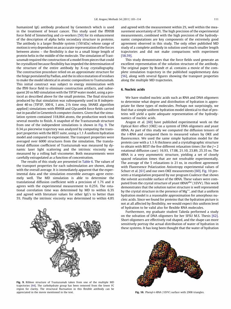
S.R. Aragon / Methods 54 (2011) 101–114 111
humanized IgG antibody produced by Genentech which is usedin the treatment of breast cancer. This study used the ff99SBforce field of Simmerling and co-workers [50] for its enhancementof the description of alpha helix secondary structure in proteins.The antibody is a larger flexible system (150 kDa) whose range ofmotion is very dependent on an accurate representation of the forcesbetween atoms – the flexibility is due to a small hinge length ofprotein helix in the middle of the molecule. The simulation of Trast-uzumab required the construction of a model from pieces that couldbe crystallized because flexibility has impeded the determination ofthe structure of the entire antibody by X-ray crystallography.The construction procedure relied on an approximate structure forthe hinge postulated by Padlan, and the in silico mutation of residuesto make the model identical in atomic composition to Trastuzumab.This initial construct was subject to energy minimization withthe ff99 force field to eliminate construction artifacts, and subse-quent 20 ns MD simulation with the TIP3P water model, using a pro-tocol as described above for the small proteins. The final structureproduced by that simulation was subsequently used in 8 indepen-dent 40 ns (TIP3P, 300 K, 1 atm, 2 fs time step, SHAKE algorithmapplied) simulations with ff99SB and Glycam04 force fields carriedout in parallel in Genentech computer clusters. Given that the simu-lation system contained 318,064 atoms, the production work tookseveral months to finish. A snapshot of the Trastuzumab structurefrom one of the independent simulations is shown in Fig. 9. The0.34 ls piecewise trajectory was analyzed by computing the trans-port properties with the BEST suite, using a 1.1 Å uniform hydrationmodel and compared to experiment. The transport properties wereaveraged over 6000 structures from the simulation. The transla-tional diffusion coefficient of Trastuzumab was measured by dy-namic laser light scattering and the intrinsic viscosity wasmeasured by a rolling ball viscometer. Both measurements werecarefully extrapolated as a function of concentration.
The results of this study are presented in Table 6. The values ofthe transport properties for each subsimulation are shown, alongwith the overall average. It is immediately apparent that the exper-imental data and the simulation ensemble averages agree extre-mely well. The MD simulation is able to determine thetranslational diffusion coefficient with a precision of 1.7% and itagrees with the experimental measurement to 0.25%. The rota-tional correlation time was determined by MD to within 6.3%and agreed with literature values for other IgG’s to better than5%. Finally the intrinsic viscosity was determined to within 4.8%
Fig. 9. Ribbon structure of Trastuzumab taken from one of the multiple MDtrajectories [64]. The carbohydrate group has been removed from the lower FCregion for clarity. The structural fluctuation in this flexible antibody can beappreciated in the movie mentioned in the text.
and agreed with the measurement within 2%, well within the mea-surement uncertainty of 3%. The high precision of the experimentalmeasurements, combined with the high precision of the hydrody-namic computations are key components of the extremely goodagreement observed in this study. The only other published MDstudy of a complete antibody in solution used much smaller lengthtrajectories and did not make comparisons with experiment[58,59].
This study demonstrates that the force fields used generate anexcellent representation of the solution structure of the antibody.The original paper by Brandt et al. contains a movie of the com-plete simulation trajectory in the published supplementary data[56], along with several figures showing the transport propertiesalong the multiple MD trajectories.
6. Nucleic acids
We have studied nucleic acids such as RNA and DNA oligomersto determine what degree and distribution of hydration is appro-priate for these types of molecules. Perhaps not surprisingly, wefind that a simple uniform hydration model identical to that of pro-teins will yield a quite adequate representation of the hydrody-namics of nucleic acids.
Aragon et al. [60] have published experimental work on theOptical Kerr effect (OKE) on a system of DNA oligomers and yeasttRNA. As part of this study we computed the diffusion tensors ofthe t-RNA and compared them to measured values by OKE andfluorescence. We used the same simple hydration model for theprotein case with a 1.1 Å thickness and a crystallographic structureto obtain with BEST the five different relaxation times (for the J = 2rotational diffusion case): 16.93, 17.08, 21.10, 23.89, 25.35 ns. ThetRNA is a very asymmetric structure, yielding a set of closelyspaced relaxation times that are not resolvable experimentally.The average of the 5 relaxations is 21 ns, in excellent agreementwith Fluoresence Polarization Anisotropy experimental values ofSchurr et al. [61] and our own OKE measurements [60]. Fig. 10 pre-sents a triangulation prepared by our program Coalesce that showsthe solvent accessible surface of the tRNA. These values were com-puted from the crystal structure of yeast tRNAphe (1EVV). This workdemonstrates that the solution native structure is well representedby the crystal structure in the presence of Mg++, and that a uniformhydration model is a reasonable approximation for amorphous nu-cleic acids. Since we found for proteins that the hydration picture isnot at all affected by flexibility, we would expect this uniform levelof hydration to be valid also for flexible RNA molecules.
Furthermore, my graduate student Takeda performed a studyon the solvation of DNA oligomers for her SFSU M.S. Thesis [62].Short oligomers are effectively rod shaped, and the shape can moresensitively portray the actual distribution of water of hydration inthese systems. It has long been thought that the water of hydration
Fig. 10. Phenyl-t-RNA (1EVV) surface with 2908 triangles.

-6
-5
-4
-3
-2
-1
0
1
2
3
4
3.4 3.6 3.8 4 4.2
Nitrogen Inflation (Angstrom)
Diff
eren
ce fr
om E
xper
imen
tal D
ata
(%)
Drr
Dtt
Fig. 12. The discrepancy of the BEST computation for both translation (Dtt) andtumbling (Drr) as a function of the added radius of the nitrogen atom which ispresent only in the DNA bases. The best value is that which splits the error acrosszero between each measurement. In this case the value occurs at 3.65 Å for the DNA12mer.
112 S.R. Aragon / Methods 54 (2011) 101–114
is more concentrated in the grooves [63], and X-ray crystallo-graphic data [64] of sufficient resolution that shows the distribu-tion of water and other co-crystallizing molecules and ions in aDNA dodecamer corroborates this picture. In the case of DNA olig-omers, there is the high quality study of Eimer and Pecora [65] inwhich both translational and rotational diffusion coefficients ofthree oligomers (8, 12, 20 mer) have been measured by a combina-tion of polarized and depolarized dynamic light scattering. In ourstudy we computed both diffusion tensors and we compared a uni-form hydration model, as found appropriate for proteins, to modelsin which the solvation (which includes ions) was distributed on thebackbone, or in the grooves. We find that the uniform hydrationmodel requires different amount of water for each oligomer in or-der to match experiment and a discrepancy with experiment of 3%,while solvating the grooves by 3.6 ± 0.3 Å thick layer (just slightlythicker than one molecule of water, and independent of oligomerlength, and sequence) produces an agreement with experimentbetter than 1%. Solvating the backbone produced a discrepancythree times larger. Fig. 11 shows tessellations of a DNA oligomerwith three different hydration models. Fig. 12 shows a typical dataset in which the discrepancy with experiment for both translationand rotation are plotted as a function of nitrogen inflation (the ex-tra radius added to the nitrogen atom). Two simultaneously mea-sured hydrodynamic properties are required to pinpoint ahydration parameter.
These preliminary results are very interesting, however, theyare dependent on two assumptions that need to be mentioned.First, the uncharged molecule hydrodynamic computation was ap-plied. Since the ionic strength of the solutions was around 0.1 M,the effect of phosphate charges is not expected to be large, but thisshould be tested. Second, the DNA coordinates for the computa-tions where produced by Spartan (Wavefunction, Inc.), a molecularmodeling program that makes DNA of uniform helical diameter,irrespective of sequence. DNA oligomer crystallographic data showdifferences in the GC and AT phosphorous distances bringing intoquestion the suitability of using molecular modeling programs toconstruct DNA when we are pursuing questions of fine detail. Wedid, however, compare the computed transport values from theWilliams [63] dodecamer crystal structure to those obtained fromSpartan and found an insignificant difference. In addition, ignoringthe difference in sequence between the Williams dodecamer and apure GC tract dodecamer studied by Eimer and Pecora [65] yieldsthe same solvation picture as before, indicating that our assump-tions are reasonable. This study demonstrates that one can im-prove the hydration model for DNA by making the hydration
Fig. 11. (Left panel) Groove hydration produced by inflated the nitrogen atoms by3.6 Å. (Middle panel) Mixed groove and backbone hydration produced by inflatingthe oxygen atoms. (Right panel) Pure backbone hydration produced by inflation ofphosphorous atoms.
non-uniform, a result that depends on the high accuracy of thecomputations produced by BEST. The penalty for omitting this le-vel of detail is not large – a uniform hydration model can still pre-dict transport properties with an error no larger than 3%. Otherauthors have arrived at similar conclusions using a uniform hydra-tion model for nucleic acids but to our knowledge, no one else hasstudied non-uniform hydration hydrodynamic models for DNA.
7. Future improvement of the BEST hydrodynamic program
We are presently developing a more memory efficient BE meth-od for performing hydrodynamics computations, the Gradient Cor-rected Boundary Element Method (GCBE). The only significantnumerical approximation in our implementation of BE hydrody-namics is taking the surface stress force to be constant over a tri-angular surface element. This discretization error is eliminatedby an extrapolation to an infinite number of triangles. However,fairly large numbers of triangles are required for high accuracyand the memory grows as Mem = 0.5 + 0.06705 N2 Gbytes of ramper N thousand triangles (the constant allows for the OS require-ments). For 15,000 triangles, N = 15 and Mem = 15.6 Gbytes. Thislarge memory requirement has several disadvantages. First, in amultiprocessor machine, one scalar job uses all the available mem-ory, forcing the other processors to be idle. Second, such largememory machines require a 64 bit OS and are not common, inhib-iting other research laboratories from using our code. This problemcan be partially ameliorated by performing an approximate extrap-olation from data obtained at only around 3000 triangles with aconsequent small reduction in precision. Lastly, using a very largenumber of triangles slows the computation considerably becausethe solution of the linear system scales as 27 N3. The GCBE is analternative methodology that preserves the full accuracy of the cal-culation but uses about half the number of triangles requiredpresently.
The basic idea behind the GCBE is to represent the unknownfunction in the integral equation as a Taylor series expansion asshown in Eq. (12) below, and use information about the gradientof the surface stress to correct a computation at a smaller numberof triangles. Thus, one produces the hydrodynamic super matrixonce, but solves the linear system twice, once with a constant sur-face stress force over a triangle, and a second time with a represen-tation of the surface stress force containing up to quadratic termsin the Taylor expansion. The gradient is estimated by making ageneral fit to a quadratic basis set with the value of~f k from severalneighboring triangles. This method also has a greater potential forparallel processing.

S.R. Aragon / Methods 54 (2011) 101–114 113
~f kð~xÞ ¼~f kð~xkÞ þ ð~x�~xkÞ � ~r~f kð~xÞÞ ð14Þ
The second area of improvement of BEST is the parallelization ofthe code to make it much faster. Processing hundreds to thousandsof structures that result from an MD trajectory can take more thana day of CPU time. The recent introduction of the CUDA SoftwareDevelopment Kit by Nvidia (www.nvidia.com/object/cuda_home_new.html) for the use of advanced graphics cards (GPU) as ageneral purpose computational engine provides an interestingopportunity to parallelize BEST. The GPU has hundreds of process-ing units with memories up to 4 GB in the present Tesla cards. Thegeneration of the hydrodynamic matrix is a trivially parallelizable.The solution of the linear system can be parallelized by the utiliza-tion of cuBLAS, provided by Nvidia, to generate a LAPACK systemwith high throughput. This has already been achieved by a com-mercial group called CULA (www.culatools.com/html_guide), butour own implementation will be license free. The utility of theCUDA SDK has already been demonstrated for scientific applica-tions by the porting of AMBER 11 to the CUDA architecture. Wehave obtained speedups of a factor of eight for small proteins usingthis system.
8. Conclusions
The high precision implemented via the BE method in BEST hasallowed us to generate a general model to numerically treat thetransport properties of proteins with a single hydration parameterfor all proteins regardless of size or flexibility. The hydration thick-ness of 1.1 Å represents the average hydration over the protein sur-face. Experimental data are presently not sufficiently precise toattempt non-uniform hydration models for proteins. The hydrationmodel we have utilized allows for atomic size variation, unlike theapproximate models of other authors [15] who have proposed asingle atomic equivalent radius (AER) for all heavy atoms. A similarpicture is obtained for nucleic acids. The number of nucleic acidsystems we have studied is smaller than the protein data set, butour data yield a simple hydration picture for RNA and DNA, similarto that of proteins, capable of achieving experimental agreementwithin 3% or better with a uniform hydration model of 1.1 Å thick.For DNA oligomers, it is possible to generate a statistically moreaccurate model, with half the uncertainty, by concentrating thehydration in the grooves, in agreement with X-ray crystallography.
Our studies of proteins led us to propose that some multimericproteins have a conformational rearrangement upon going intosolution from the crystal. Our preliminary work using MD simula-tion appears to bear this out. In order to validate that the structuresgenerated by MD are actually representative of solution structure,we have performed simulations on a number of small proteins, ri-gid and flexible, and one medium sized flexible protein. The goodagreement we obtain with experiment demonstrates that we havevalidated both the force fields and the hydrodynamic hydrationmodel for proteins. Furthermore, we do not find any evidence, asclaimed by Wright and co-workers [66], that there is any need toimplement a special screened hydrodynamic interaction to treatflexible molecules, but this topic merits further work. Our applica-tion with the precise hydrodynamics in BEST in combination withthe trajectory ensemble average method yields very good agree-ment with experiment for both small and large proteins, flexibleor not.
These studies have opened the door to the study of proteinswhose sequence is known, but whose structure is unknown but ex-pected to be similar to other known structures, by a combination ofhydrodynamic measurements, accurate hydrodynamic modeling,and molecular dynamics. Careful measurements of the intrinsicviscosity, which do not require very expensive equipment, can pro-vide data with sufficient discriminating power to test different
structure hypothesis. This methodology, a generalization of theconstruction work done on the antibody, would be particularlyuseful for large proteins not amenable to study by solution NMRmethods.
Acknowledgments
The author thanks David Hahn for performing implicit waterMD for the a-chymotrypsin and b-lactoglobulin molecules. Finan-cial support for some of the small protein and Trastuzumab workwas provided by Genentech. This work was funded in part byNIH Grant GM52588 to S. Aragon.
References
[1] B. Berne, R. Pecora, Dynamic Light Scattering: with Applications to Chemistry,Biology and Physics, Wiley-Interscience, New York, 1976.
[2] D. Eden, J.G. Elias, Transient Electric Birefringence of DNA restriction fragmentsand the filamentous virus Pf3, in: B. Dahneke (Ed.), Measurement ofSuspended Particles by Quasi-Elastic Light Scattering, Wiley-Interscience,New York, 1983.
[3] L. Stryer, Fluorescence spectroscopy of proteins, Science 162 (1968) 526–533.[4] F. Stallmach, P. Galvosas, Spin echo NMR diffusion studies, Ann. Rep. NMR
Spectrosc. 61 (2007) 51–131.[5] J.K.M. Sanders, B.K. Hunter, Modern NMR Spectroscopy, Oxford University
Press, New York, 1987.[6] E.G. Richards, An Introduction to Physical Properties of Large Molecules in
Solution, Cambridge University Press, London, 1980.[7] P. Schuck, Size-distribution analysis of macromolecules by sedimentation
velocity ultracentrifugation and Lamm equation modeling, Biophys. J. 78(2000) 1606–1619.
[8] S. Kim, S.J. Karilla, Microhydrodynamics, Butterworth-Heinemann, New York,1991.
[9] V.A. Bloomfield, W.O. Dalton, K.E. van Holde, Frictional coefficients ofmultisubunit structures. I. Theory, Biopolymers 5 (1967) 135–148;V.A. Bloomfield, W.O. Dalton, K.E. Van Holde, Frictional coefficients ofmultisubunit structures. II. Application to proteins and viruses, Biopolymers5 (1967) 149–159.
[10] J. Garcia de la Torre, V.A. Bloomfield, Hydrodynamic properties ofmacromolecular complexes III. Bacterial viruses, Biopolymers 16 (1977)1779–1793.
[11] D.C. Teller, E. Swanson, C. de Haen, The translational friction coefficients ofproteins, Methods Enzymol. 61 (1979) 103–124.
[12] J. Garcia de la Torre, M.L. Huertas, B. Carrasco, Calculation of hydrodynamicproperties of globular proteins from their atomic-level structure, Biophys. J. 78(2000) 719–730.
[13] J. Rotne, S. Prager, Variational treatment of hydrodynamic interaction inpolymers, J. Chem. Phys. 50 (1969) 4831–4837.
[14] J. Garcıa de la Torre, V.A. Bloomfield, Hydrodynamic properties ofmacromolecular complexes. I. Translation, Biopolymers 16 (1977) 1747–1763.
[15] J. Garcia de la Torre, M.L. Huertas, B. Carrasco, HYDRONMR: prediction of NMRrelaxation of globular proteins from atomic-level structures andhydrodynamic calculations, J. Magn. Reson. 147 (2000) 138–146.
[16] G.K. Youngren, A. Acrivos, Stokes flow past a particle of arbitrary shape: anumerical method of solution, J. Fluid Mech. 69 (1975) 377–402.
[17] F.G.K. Odqvist, On the boundary value problems in hydrodynamics of viscousfluids, Math. Z. 32 (1930) 329–375 (German).
[18] C.M. Hu, R. Zwanzig, Rotational friction coefficients for spheroids with theslipping boundary condition, J. Chem. Phys. 60 (1974) 4354–4357.
[19] S.A. Allison, Low Reynolds number transport properties of axisymmetricparticles employing stick and slip boundary conditions, Macromolecules 32(1999) 5304–5312.
[20] R.M. Venable, R.W. Pastor, Frictional models for stochastic simulations ofproteins, Biopolymers 27 (1988) 1001–1014.
[21] R.W. Pastor, R. Zwanzig, Anisotropic bead models for molecularhydrodynamics, J. Chem. Phys. 90 (1989) 5729–5734.
[22] W.A. Wegener, On an exact starting expression for macromolecularhydrodynamic models, Biopolymers 25 (1986) 627–637.
[23] S.R. Aragon, A precise boundary element method for macromoleculartransport properties, J. Comput. Chem. 25 (2004) 1191–1205.
[24] C.W. Oseen, Hydrodynamik, Academiches Verlag, Leipzig, 1927.[25] E. Anderson, Z. Bai, C. Bischof, S. Blackford, J. Demmel, J. Dongarra, J. Du Croz, A.
Greenbaum, S. Hammarling, A. McKenney, D. Sorensen, LAPACK User’s Guide,third ed., SIAM, Philadelphia, 1999.
[26] H. Brenner, Coupling between the translational and rotational Brownianmotions of rigid particles of arbitrary shape. II. General theory, J. ColloidInterface Sci. 23 (1967) 407–436.
[27] B. Carrasco, J. Garcia de la Torre, Hydrodynamic properties of rigid particles:comparison of different modeling and computational procedures, Biophys. J.75 (1999) 3044–3057.

114 S.R. Aragon / Methods 54 (2011) 101–114
[28] R.F. Goldstein, Macromolecular diffusion constants: a calculational strategy, J.Chem. Phys. 83 (1985) 2390–2397.
[29] D.K. Hahn, S.R. Aragon, Intrinsic viscosity of proteins and platonic solids byboundary element methods, J. Chem. Theory Comput. 2 (2006) 1416–1428.
[30] S.R. Aragon, D. Flamik, Precise computation of transport properties of cylindersby the boundary element method, Macromolecules 42 (2009) 6290–6299.
[31] M.L. Mansfield, J.F. Douglas, Transport properties of rod-like particles,Macromolecules 41 (2008) 5422–5432.
[32] A. Ortega, J. Garcia de la Torre, Hydrodynamic properties of rodlike anddisklike particles in dilute solution, J. Chem. Phys. 119 (2003) 9914–9919.
[33] M.M. Tirado, J. Garcia de la Torre, Translational friction coefficients of rigid,symmetric top macromolecules. Application to circular cylinders, J. Chem.Phys. 71 (1979) 2581–2587.
[34] S.J. Broersma, Viscous force and torque constants for a cylinder, J. Chem. Phys.74 (1981) 6989–6990.
[35] S. Wakiya, Slow motion in shear flow of a doublet of two spheres in contact, J.Phys. Soc. Japan 31 (1971) 1581–1587.
[36] H. Brenner, Rheology of a dilute suspension of axisymmetric Brownianparticles, Int. J. Multiphase Flow 1 (1974) 195–341.
[37] A.J. Goldman, R.G. Cox, H. Brenner, The slow motion of two identical arbitrarilyoriented spheres through a viscous fluid, Chem. Eng. Sci. 21 (1966) 1151–1170.
[38] M.L. Connolly, Molecular surface program, QCPE Bull. 1 (1981) 75–83.[39] M.L. Connolly, The molecular surface package, J. Mol. Graph. 11 (1993) 139–
141.[40] M.L. Connolly, Analytical molecular surface calculation, J. Appl. Crystallogr. 16
(1983) 548–558.[41] S.R. Aragon, D.K. Hahn, Precise boundary element computation of protein
transport properties: diffusion tensors, specific volume, and hydration,Biophys. J. 91 (2006) 1591–1603.
[42] H. Zhou, Boundary element solution of macromolecular electrostatics:interaction energy between two proteins, Biophys. J. 65 (1993) 955–963.
[43] R.M. Venable, E. Hatcher, O. Guvench, A.D. MacKerell Jr., R.W. Pastor,Comparing simulated and experimental translation and rotation constants:range of validity for viscosity scaling, J. Phys. Chem. B 114 (2010) 12501–12507.
[44] S.R. Aragon, D.K. Hahn, Stagnation of flow in protein cavities by boundaryelement microhydrodynamics, J. Phys. Chem. B 113 (2009) 2657–2663.
[45] G.W. Schwert, S. Kaufman, The molecular size and shape of the pancreaticproteases. III. a-Chymotrypsin, J. Biol. Chem. 190 (1951) 807–816.
[46] J. Mongan, D.A. Case, J.A. McCammon, Constant pH molecular dynamics ingeneralized born implicit solvent, J. Comput. Chem. 25 (2004) 2038–2048.
[47] H.A. McKenzie, W.H. Sawyer, Effect of pH on b-lactoglobulin, Nature 214(1967) 1101–1104.
[48] A.G. Ogston, The Guoy diffusiometer; further calibration, Proc. R. Soc. (London)196 (1949) 272–285.
[49] D.A. Perlman, D.A. Case, J.W. Caldwell, W.S. Ross, T.E. Cheathamm III, S. Debold,D. Ferguson, G. Seibel, P. Kollman, AMBER, a package of computer programs for
applying molecular mechanics, normal mode analysis, molecular dynamicsand free energy calculations to simulate the structural and energeticproperties of molecules, Comput. Phys. Commun. 91 (1995) 1–41.
[50] V. Hornak, R. Abel, A. Okur, B. Strockbine, A. Roitberg, C. Simmerling,Comparison of multiple Amber force fields and development of improvedprotein backbone parameters, Proteins 65 (2006) 712–725.
[51] S.R. Aragon, Q. Hong, Hydrodynamics and MD of small proteins in solution, J.Phys. Chem. submitted for pulication; Q. Hong, MD of small proteins insolution, M.S. Thesis, SFSU, 2009.
[52] P. Mark, L. Nilsson, Structure and dynamics of the TIP3P, SPC, and SPC/E watermodels at 298 K, J. Phys. Chem. A 105 (2001) 9954–9960.
[53] A.J. Cross, G.R. Fleming, Influence of inhibitor binding on the internal motionsof lysozyme, Biophys. J. 50 (1986) 507–512.
[54] S.B. Dubin, N.A. Clar, G.B. Benedek, Measurement of the rotational diffusioncoefficient of lysozyme by depolarized light scattering: configuration oflysozyme in solution, J. Chem. Phys. 54 (1971) 5158–5164.
[55] S.R. Aragon, D.K. Hahn, Polarizability and Kerr constant of Proteins byboundary element methods, Colloids Surf. B: Biointerfaces 56 (2007) 19–25.
[56] J.P. Brandt, T.A. Patapoff, S.R. Aragon, Construction, MD simulation, andhydrodynamic validation of an all-atom model of a monoclonal IgG antibody,Biophys. J. 99 (2010) 905–913.
[57] P. Brandt, Construction, Molecular Dynamics Simulation, and HydrodynamicValidation of an All-Atom Model of a Monoclonal Antibody, M.S. Thesis, SFSU,2010.
[58] N. Chennamsetty, B. Helk, V. Voynov, V. Kayser, B.L. Trout, Aggregation-pronemotifs in human immunoglobulin G, J. Mol. Biol. 391 (2009) 404–413.
[59] N. Chennamsetty, V. Voynov, B. Helk, V. Kayser, B.L. Trout, Design oftherapeutic proteins with enhanced stability, Proc. Natl. Acad. Sci. USA 106(2009) 1937–11942.
[60] S.R. Aragon, M. Perez, K. Ng, D. Eden, Optical Kerr effect of DNA oligomers andRNA, in: S.P. Stoylov, M.V. Stoimenova (Eds.), Molecular and Colloidal Electro-optics, Taylor & Francis, New York, 2006, pp. 265–284.
[61] J.C. Thomas, J.M. Schurr, B.R. Reid, N.S. Riberiro, D.R. Hare, Effect ofmagnesium(2+) on the solution conformation of two different transferribonucleic acids, Biochemistry 23 (1984) 5414–5420.
[62] T. Takeda, Determination of Water Hydration Location in DNA Oligomersthrough Diffusion Coefficient Calculation, M.S. Thesis, SFSU, August, 2005.
[63] H.R. Drew, R.E. Dickerson, Structure of a B-DNA dodecamer. III. Geometry ofhydration, J. Mol. Biol. 15 (1981) 535–556.
[64] X. Shui, L. McFail-Isom, G. Hu, L.D. Williams, The B-DNA dodecamer at highresolution reveals a spine of water on sodium, Biochemistry 37 (1998) 8341–8355.
[65] W. Eimer, R. Pecora, Rotational and translational diffusion of short rod-likemolecule in solution: oligonucleotides, J. Chem. Phys. 94 (1991) 2324–2329.
[66] S.-H. Bae, H.J. Dyson, P.E. Wright, Prediction of the rotational tumbling time forproteins with disordered segments, JACS 131 (2009) 6814–6821.