Sun1 forms immobile macromolecular assemblies at the nuclear envelope
12
Sun1 forms immobile macromolecular assemblies at the nuclear envelope Wenshu Lu a,f , Josef Gotzmann b, ⁎, Lucia Sironi c , Verena-Maren Jaeger a,f , Maria Schneider a,f , Yvonne Lüke a , Mathias Uhlén e , Cristina Al-Khalili Szigyarto e , Andreas Brachner b , Jan Ellenberg c , Roland Foisner b , Angelika A. Noegel a,d , Iakowos Karakesisoglou f, ⁎ a Center for Biochemistry, Medical Faculty, University of Cologne, Joseph-Stelzmann-Strasse 52, 50931 Cologne, Germany b Max F. Perutz Laboratories, Medical University of Vienna, A-1030 Vienna, Austria c Gene Expression and Cell Biology/Biophysics Programmes, European Molecular Biology Laboratory (EMBL), Meyerhofstrasse 1, D-69117 Heidelberg, Germany d Center for Molecular Medicine Cologne, Medical Faculty, University of Cologne, Joseph-Stelzmann-Strasse 52, 50931 Cologne, Germany e The Human Proteome Resource, Department of Biotechnology, Royal Institute of Technology, SE-10691 Stockholm, Sweden f Department of Biological Sciences, The School of Biological and Biomedical Sciences, The University of Durham, DH1 3LE Durham, UK abstract article info Article history: Received 28 July 2008 Received in revised form 3 September 2008 Accepted 5 September 2008 Available online 19 September 2008 Keywords: SUN-domain Sun1 Sun2 Nuclear envelope Nesprin KASH-domain iFRAP SUN-domain proteins form a novel and conserved family of inner nuclear membrane (INM) proteins, which establish physical connections between the nucleoplasm and the cytoskeleton. In the current study, we provide evidence that within the nuclear envelope (NE) Sun1 proteins form highly immobile oligomeric complexes in interphase cells. By performing inverse fluorescence recovery after photobleaching analysis, we demonstrate in vivo that both perinuclear and nucleoplasmic Sun1 segments are essential for maintenance of Sun1 immobility at the NE. Our data in particular underline the self-association properties of the C-terminal coiled-coil Sun1 segment, the ability of which to form dimers and tetramers is demonstrated. Furthermore, the Sun1 tertiary structure involves interchain disulfide bonds that might contribute to higher homo- oligomer formation, although the overall dynamics of the Sun1 C-terminus remains unaffected when the cysteins involved are mutated. While a major Sun1 pool colocalizes with nuclear pore complex proteins, a large fraction of the Sun1 protein assemblies colocalize with immunoreactive foci of Sun2, another SUN- domain paralogue at the NE. We demonstrate that the Sun1 coiled-coil domain permits these heterophilic associations with Sun2. Sun1 therefore provides a non-dynamic platform for the formation of different macromolecular assemblies at the INM. Our data support a model in which SUN-protein-containing multi- variate complexes may provide versatile outer nuclear membrane attachment sites for cytoskeletal filaments. Crown Copyright © 2008 Published by Elsevier B.V. All rights reserved. 1. Introduction The nuclear envelope (NE) is a hallmark of eukaryotic cells, functioning as a selective structural barrier separating the cytoplasm from the nucleus. It is composed of a pair of biochemically and functionally distinct membranes, the inner and outer nuclear membranes (INM and ONM, respectively) that are joined together by large macromolecular assemblies the nuclear pore complexes (NPCs) [1]. NPCs form aqueous gated channels spanning the NE regulating the transport of macromolecules between the nucleus and cytoplasm [2,3]. The ONM is continuous with the rough endoplasmic reticulum (ER) and provides attachment sites for cytoplasmic structural elements [4]. In contrast, the INM is composed by a distinct set of membrane proteins, which perform close associations with the underlying nuclear lamina and chromatin [5]. Encompassed by the two NE membranes lies an evenly spaced lumen, the perinuclear space (PNS), which is usually ∼ 20–100 nm in width and continuous with the ER [6]. KASH- and SUN-domain proteins are emerging evolutionarily conserved families of NE-associated molecules [7–9]. From yeast to mammals a conserved mechanism has been established, by which ONM positioned KASH-domain proteins interact with INM positioned SUN-family members in the PNS. These physical “bridges” span both nuclear membranes, thereby providing a structural and possible function-integrating connection between cytoplasmic and nucleo- plasmic compartments [7,10–12]. In C. elegans and higher eukaryotes, the KASH-domain proteins require SUN-domain proteins for their proper NE localization [10–15]. The majority of KASH-domain proteins appear so far to function as exclusive ONM adaptors mediating interactions with all major cytoskeletal filaments, the centrosome and the microtubule motor protein apparatus [16]. To date, Sad1p in S. pombe, Mps3 in S. cerevisiae, Dd-Sun1 in D. discoideum, UNC-84 and matefin in C. elegans, predicted proteins Biochimica et Biophysica Acta 1783 (2008) 2415–2426 ⁎ Corresponding authors. J. Gotzmann is to be contacted at Max F. Perutz Laboratories, Medical University of Vienna, Vienna, Austria. Tel.: +43 1 4277 61672; fax: +43 1 4277 9616. I. Karakesisoglou, Department of Biological Sciences, The School of Biological and Biomedical Sciences, The University of Durham, DH1 3LE Durham, UK. Tel.: +44 191 3341299; fax: +44 191 3341201. E-mail addresses: [email protected] (J. Gotzmann), [email protected] (I. Karakesisoglou). 0167-4889/$ – see front matter. Crown Copyright © 2008 Published by Elsevier B.V. All rights reserved. doi:10.1016/j.bbamcr.2008.09.001 Contents lists available at ScienceDirect Biochimica et Biophysica Acta journal homepage: www.elsevier.com/locate/bbamcr
-
Upload
nottingham -
Category
Documents
-
view
3 -
download
0
Transcript of Sun1 forms immobile macromolecular assemblies at the nuclear envelope

Biochimica et Biophysica Acta 1783 (2008) 2415–2426
Contents lists available at ScienceDirect
Biochimica et Biophysica Acta
j ourna l homepage: www.e lsev ie r.com/ locate /bbamcr
Sun1 forms immobile macromolecular assemblies at the nuclear envelope
Wenshu Lu a,f, Josef Gotzmann b,⁎, Lucia Sironi c, Verena-Maren Jaeger a,f, Maria Schneider a,f, Yvonne Lüke a,Mathias Uhlén e, Cristina Al-Khalili Szigyarto e, Andreas Brachner b, Jan Ellenberg c, Roland Foisner b,Angelika A. Noegel a,d, Iakowos Karakesisoglou f,⁎a Center for Biochemistry, Medical Faculty, University of Cologne, Joseph-Stelzmann-Strasse 52, 50931 Cologne, Germanyb Max F. Perutz Laboratories, Medical University of Vienna, A-1030 Vienna, Austriac Gene Expression and Cell Biology/Biophysics Programmes, European Molecular Biology Laboratory (EMBL), Meyerhofstrasse 1, D-69117 Heidelberg, Germanyd Center for Molecular Medicine Cologne, Medical Faculty, University of Cologne, Joseph-Stelzmann-Strasse 52, 50931 Cologne, Germanye The Human Proteome Resource, Department of Biotechnology, Royal Institute of Technology, SE-10691 Stockholm, Swedenf Department of Biological Sciences, The School of Biological and Biomedical Sciences, The University of Durham, DH1 3LE Durham, UK
⁎ Corresponding authors. J. Gotzmann is to beLaboratories, Medical University of Vienna, Vienna, Aufax: +43 14277 9616. I. Karakesisoglou, Department of BiBiological and Biomedical Sciences, The University of DTel.: +44 191 3341299; fax: +44 191 3341201.
E-mail addresses: [email protected]@durham.ac.uk (I. Karakesisoglo
0167-4889/$ – see front matter. Crown Copyright © 20doi:10.1016/j.bbamcr.2008.09.001
a b s t r a c t
a r t i c l e i n f oArticle history:
SUN-domain proteins form Received 28 July 2008Received in revised form 3 September 2008Accepted 5 September 2008Available online 19 September 2008Keywords:SUN-domainSun1Sun2Nuclear envelopeNesprinKASH-domainiFRAP
a novel and conserved family of inner nuclear membrane (INM) proteins, whichestablish physical connections between the nucleoplasm and the cytoskeleton. In the current study, weprovide evidence that within the nuclear envelope (NE) Sun1 proteins form highly immobile oligomericcomplexes in interphase cells. By performing inverse fluorescence recovery after photobleaching analysis, wedemonstrate in vivo that both perinuclear and nucleoplasmic Sun1 segments are essential for maintenance ofSun1 immobility at the NE. Our data in particular underline the self-association properties of the C-terminalcoiled-coil Sun1 segment, the ability of which to form dimers and tetramers is demonstrated. Furthermore,the Sun1 tertiary structure involves interchain disulfide bonds that might contribute to higher homo-oligomer formation, although the overall dynamics of the Sun1 C-terminus remains unaffected when thecysteins involved are mutated. While a major Sun1 pool colocalizes with nuclear pore complex proteins, alarge fraction of the Sun1 protein assemblies colocalize with immunoreactive foci of Sun2, another SUN-domain paralogue at the NE. We demonstrate that the Sun1 coiled-coil domain permits these heterophilicassociations with Sun2. Sun1 therefore provides a non-dynamic platform for the formation of differentmacromolecular assemblies at the INM. Our data support a model in which SUN-protein-containing multi-variate complexes may provide versatile outer nuclear membrane attachment sites for cytoskeletal filaments.
Crown Copyright © 2008 Published by Elsevier B.V. All rights reserved.
1. Introduction
The nuclear envelope (NE) is a hallmark of eukaryotic cells,functioning as a selective structural barrier separating the cytoplasmfrom the nucleus. It is composed of a pair of biochemically andfunctionally distinct membranes, the inner and outer nuclearmembranes (INM and ONM, respectively) that are joined togetherby large macromolecular assemblies the nuclear pore complexes(NPCs) [1]. NPCs form aqueous gated channels spanning the NEregulating the transport of macromolecules between the nucleus andcytoplasm [2,3]. The ONM is continuous with the rough endoplasmicreticulum (ER) and provides attachment sites for cytoplasmicstructural elements [4]. In contrast, the INM is composed by a distinct
contacted at Max F. Perutzstria. Tel.: +43 1 4277 61672;ological Sciences, The School ofurham, DH1 3LE Durham, UK.
t (J. Gotzmann),u).
08 Published by Elsevier B.V. All rig
set of membrane proteins, which perform close associations with theunderlying nuclear lamina and chromatin [5]. Encompassed by thetwo NE membranes lies an evenly spaced lumen, the perinuclearspace (PNS), which is usually ∼20–100 nm in width and continuouswith the ER [6].
KASH- and SUN-domain proteins are emerging evolutionarilyconserved families of NE-associated molecules [7–9]. From yeast tomammals a conserved mechanism has been established, by whichONM positioned KASH-domain proteins interact with INM positionedSUN-family members in the PNS. These physical “bridges” span bothnuclear membranes, thereby providing a structural and possiblefunction-integrating connection between cytoplasmic and nucleo-plasmic compartments [7,10–12]. In C. elegans and higher eukaryotes,the KASH-domain proteins require SUN-domain proteins for theirproper NE localization [10–15]. Themajority of KASH-domain proteinsappear so far to function as exclusive ONM adaptors mediatinginteractions with all major cytoskeletal filaments, the centrosome andthe microtubule motor protein apparatus [16].
To date, Sad1p in S. pombe, Mps3 in S. cerevisiae, Dd-Sun1 in D.discoideum, UNC-84 and matefin in C. elegans, predicted proteins
hts reserved.

2416 W. Lu et al. / Biochimica et Biophysica Acta 1783 (2008) 2415–2426
Q9V996 and Q9VKG2 in Drosophila, and human Sun1, Sun2, Sun3,SPAG4 and SPAG4L proteins have been characterized as SUN-domainproteins [8,17–21]. Proteomic approaches, however, indicate theirpresence in most eukaryotes [18]. Hallmark of the family is theconserved C-terminal SUN-domain (Sad1 and UNC-84 homologydomain) [20]. UNC-84 is independently required for both nuclearmigration and nuclear anchorage during C. elegans development andmight also play a role in lipid metabolism [8]. Sad1p localizes to thespindle pole body and the NE when ectopically overexpressed [22].Sad1p is essential for viability and required for proper mitotic spindleformation and function [23]. In meiotic cells Rap1p bound telomeresare tethered to Bqt1/Bqt2 complexes, which themselves are connectedthrough Sad1 to the NE, underscoring the essential function of SUN-domain proteins in telomere attachment during meiosis [22].Interestingly, mouse Sun1 ablation also prevents NE telomereattachment during meiosis and leads to defective gametogenesis[23]. Although controversial results exist, a recent report showed thatrat Sun2 is also involved in tetheringmammalian meiotic telomeres tothe NE [24]. Sun1 is involved in nuclear reassembly after mitosis,regulating chromosome decondensation [25]. Most recently, anassociation between Sun1, but not Sun2, and the NPCs has beenproposed [26].
Besides the SUN-domain, most SUN-domain proteins have at leastone predicted transmembrane domain [26] and in addition at leastone coiled-coil domain, which in human and mouse Sun1 is proposedto mediate homo and hetero-dimerization with Sun2 [10,11,27]. Todate all of them have been identified as exclusive INM components,except SPAG4, which displays an ER-resident localization upon ectopicexpression [28]. The N-termini of SUN-proteins all face the nucleo-plasm, whereas their C-termini reside in the PNS. Interestingly, the N-and C-termini of mammalian Sun1 can independently localize to theNE [11,26]. However, expression of tagged recombinant Sun1 proteinand fragments thereof revealed that Sun1 retention at the NE andassociation with components of the NPCs require both the nucleo-plasmic and luminal domains to a yet unidentified extent [26,28]. TheN-termini of UNC-84, SUN-1/Matefin in C. elegans and the mammalianSun1/2 proteins have been shown to associate with the nuclear lamina[20,21,29,30]. However, only UNC-84 requires lamins for its NElocalization in nematodes [20].
In this study, we explore in more detail both in vivo and in vitro theSun1 oligomerization properties and identify the implicated domains.We demonstrate that full-length Sun1 is among the most immobileINM proteins described to date and assign this biological feature to itsoligomerization capacity. Truncation of either the nucleoplasmic orluminal Sun1 segments drastically affects its NE dynamics. Our dataunderline in particular the self-association properties of the C-terminal coiled-coil Sun1 segment, which allows Sun1 tetrameriza-tion and permits also heterophilic associations with Sun2. Finally, weprovide evidence that SUN-domain proteins assemble into versatilecomplexes and discuss the biological significance of these macro-molecular immobile complexes.
2. Materials and methods
2.1. Cloning strategies
Mouse Sun1 DNA fragments were amplified from IMAGE cloneAH48156 by PCR and cloned into pGEMTeasy (Promega) vectorsbefore cloning into GFP, GST or yeast two-hybrid vectors. All GFP-fusion proteins generated carry GFP at their N-terminus. GFP-TM-C (aa358–913) was cloned into the EcoRI/SalI site of EGFP-C2. GFP-Sun1-FL(aa 1–913), GFP-2TM-N (aa 1–412), GFP-TM-SD1, 2 (aa 358–737), GFP-TM-ΔCC-SUN (aa 358–491, 633–913), GFP-SD1, 2 (aa 432–737) andGFP-SD2 (aa 432–491, 633–737) were cloned into the EcoRI/BamHIsite of EGFP-C2. GST-SD1, 2 (aa 432–737) and GST-SD1 (aa 432–632)were cloned into the EcoRI/XhoI site of pGEX4T-1.
Sun1-V5 containing full-length human Sun1 and Sun2-V5-Hiscontaining full-length human Sun2 were cloned into pTracer-Vector.hSun1-SD1 (aa 404–493) was cloned into EcoRI/BamHI-digestedpGBKT7 and pGADT7. hSun2-CC (aa 400–505) was cloned into thepGADT7 EcoRI/BamHI sites. hSun1 TM-C (aa 240–812) and hSun1 TM-SD1, 2 (aa 240–632) were cloned into the pHA (Roche) or EGFP-C2HindIII/EcoRI sites.
2.2. Inverse fluorescence recovery after photobleaching experiments(iFRAP)
For live-cell imaging, HeLa cells were transiently transfected withGFP-fusion constructs and examined 48 h after transfection. iFRAPswere performed and normalized as described elsewhere [31]. Thefluorescence equilibration between the unbleached and the bleachedregions of interest (ROI) in the nucleus was then monitored over aperiod of up to 10 h. To calculate the loss of fluorescence attributed tothe imaging process alone, the sum of pixel intensities was alsocalculated for the entire cell and used to normalize the fluorescenceintensity for each ROI. At least four independent experiments wereperformed for each construct. We also calculated the relativefluorescence intensity within the bleached areas of the same cellsand analyzed these cells by FRAP assays (Fig. S1).
2.3. Antibodies and immunofluorescence microscopy
Western blotting and immunofluorescence (IF) studies wereperformed as described [32,33]. The following antibodies were used:rabbit polyclonal anti-GST antibody [19], GFP-specific mAb K3-184-2[34], mouse monoclonal anti-LAP2β (1:250 IF) [35], rat anti-HAantibody (1:250 IF; 1:1000 Western; Sigma), mouse monoclonal anti-V5 antibody (1:5000 Western, 1:500 IF; Invitrogen), mouse mono-clonal anti-nuclear pore complex mAb414 (1:5000 IF, Abcam), Newrabbit polyclonal antibodies to human Sun1 (Sun1A=HPRK1500010and Sun1B= HPRK1500015; both 1:400 IF) and Sun2 (HPA001209;1:500 IF), respectively, were produced within a collaborative projectby the Human Proteome Resource [36]. Anti-Sun1 sera 281 were usedas described recently [11]. The secondary antibodies used wereconjugated with Cy3 (Sigma), FITC (Sigma) and Alexa 568 (Invitrogen).Samples were analyzed by confocal laser-scanning microscopy usingeither a TCS-SP1 (Leica) or a LSM510-Meta (Zeiss).
2.4. Purification of GST fusion proteins and in vitro binding assays
Purification of GST fusion proteins and GST pull-down experimentswere performed as described [37]. The GST free SD1, 2 polypeptideswere generated by thrombin digestion of GST-SD1, 2.
2.5. His-tag pull-down assays
GFP-hSun1 and Sun2-V5-His, or GFP and Sun2-V5-His weretransiently cotransfected into COS7 cells. The transfected cells werelysed using lysate buffer (50 mM NaH2PO4, 150 mM NaCl, 1% NP-40,0.5% Na-desoxycholate, pH 8.0). Same amount of proteins from eachcotransfectionwere coupled to Ni-NTA agarose beads (Qiagen, Hilden)by incubation for 4 h at 4 °C in 1 ml of lysate buffer. The Ni-NTAagarose beads were washed 5 times with PBS, and 3 times with lysatebuffer. The beads were precipitated by centrifugation at 2000 rpm,and proteins bound to the beads were eluted with SDS-sample buffer,resolved by 12% SDS-PAGE and subjected to western blotting analysis.
2.6. Native polyacrylamide gel-electrophoresis (Native-PAGE)
Native gels were prepared similar to SDS-polyacrylamide gels,except that SDS omitted from the gels and buffers employed. Eachsample was mixed with 2× native sample buffer (pH 6.8) containing

2417W. Lu et al. / Biochimica et Biophysica Acta 1783 (2008) 2415–2426
20% glycerol and 0.01% bromophenol blue in 125 mM Tris–HCl (pH6.8) and applied onto the native polyacrylamide gel without heating.Native molecular mass markers (Amersham Biosciences) and BSAwere used as standards.
2.7. Chemical cross-linking experiments
Cross-linking experiments with the bacterially expressed mouseSun1 SD1, 2 polypeptides were performed in a buffer containing20 mM Hepes, pH 8.0, 150 mM NaCl, 5 mM EDTA, and incubated with0.001%–0.03% glutaraldehyde for 30 min at room temperature. SDS-sample buffer was added to the reactions, and the samples wereresolved by 12% SDS-PAGE and stained by Coomassie Blue.
2.8. Yeast two-hybrid assays
The protocols for performing the Yeast two-hybrid analysis aredescribed in detail elsewhere (Yeast Protocols Handbook PT3024-1;Clontech).
2.9. Site-directed mutagenesis
Construction of the point mutant (C526A) in human Sun1 TMC-HAand TMSD1, 2-HA was done with the QuikChange XL Site-DirectedMutagenesis Kit (Stratagene, La Jolla, CA) as described in the manualusing the following primers: tmcDelta526sense: 5′-GCGACACGTGAA-GACCGGCGCCGAGACAGTGGATGCCGTAC-3′ and tmcDelta526anti-sense: 5′-GTACGGCATCCACTGTCTCGGCGCCGGTCTTCACGTGTCGC-3′.The mutations were verified by DNA sequencing.
2.10. Immunohistochemistry
Formalin-fixed and paraffin-embedded tissue microarray slidesfrom human tissue were, after deparaffinization, subjected to antigenretrieval by heat induction in Tris-EDTA, pH 9 at 125 °C for 4 min in apressure boiler. The sections were incubated with the new antiseradirected against human Sun-proteins (Sun1A,1:250; Sun1B,1:250 andSun2, 1:150), washed and incubated with a peroxidase-coupled anti-rabbit antibody (Dako). Immunoreactivity as a brownish-black colourwas assayed after final incubation with DAB chromagen. All tissuesections were counterstained with hematoxylin. These antibodies arenow available at http://www.atlasantibodies.com/home.
3. Results
3.1. Sun1 is a highly immobile NE-associated protein, requiring bothN- and C-termini for sustained immobility
To investigate more precisely the Sun1 kinetic properties at theNE, we performed inverse fluorescence recovery after photobleach-ing experiments (iFRAPs) in interphase cells using GFP-tagged Sun1full-length (GFP-Sun1-FL) and various N- and C-terminal polypep-tides containing different functional domains, respectively (Fig. 1A).iFRAPs are more suitable to analyze binding interactions thanclassical fluorescence recovery after photobleaching assays (FRAP)because the free pool of the tagged protein is bleached away,allowing precise measurement of the fluorescence changes reflect-ing the pool of NE-tethered molecules only. The NE topologies of theSun1 GFP-fusions were verified by implementing membrane-selective permeabilization assays ([38]; Fig. 1B–F). All used Sun1GFP-fusion proteins displayed an expected INM localization,although varying in prominence. For the iFRAP experiments thewhole cell except ∼1/3 of the NE region was bleached. GFP-Sun1-FLfluorescence in the unbleached area dropped very slowly andhad not completely equilibrated within the bleached area after10 h (Fig. 2A), indicating that a large proportion of Sun1 at the
nuclear membrane is highly immobile within the examined timeframe. GFP-N-2TM and GFP-TM-C also display enriched localizationat the NE. In many cases we also observed, however, an accumulationof the fusion proteins in the ER or perinuclear aggregates as shown inthe pre-bleach images of Fig. 2B and C (arrows). GFP-N-2TMequilibrated after about 35 min and GFP-TM-C after about 29 min(Fig. 2B and C). Thus, both constructs display similar moderatelydynamic binding and a significantly reduced immobilization at theNE, when compared to GFP-Sun1-FL. These dynamics data could alsobe corroborated by performing an alternative bleaching tool, namelyfluorescence recovery after photobleaching (FRAP) assays (Fig. S1).Thus, these results suggest that both Sun1 N- and C- termini arerequired to keep Sun1 immobile and also underscore the initialfinding that these domains independently from each other maintainSun1's stable localization at the NE [11].
Although the Sun1 N-terminus interacts with lamin A, thepresence of lamin A/C is not required for Sun1 NE localization[11,28,29]. Therefore, Sun1 was proposed to bind to additional nuclearfactors. In fact chromatin immunoprecipitation assays (ChIP) indicatethat Sun1 is able to interact with chromatin (data not shown), whichhas been also reported by Chi et al. [25].
3.2. The SD1-domain is a crucial determinant for reduced Sun1 dynamicsat the INM
GFP-TM-SD1, 2 and GFP-TM-ΔCC-SUN appear equally distributedbetween the NE and the peripheral ER, as indicated in the pre-bleachimages (Fig. 2D and E). GFP-TM-SD1, 2 equilibrated after ∼26 min (Fig.2D plot), very similar to GFP-TM-C and GFP-N-2TM. By contrast, forGFP-TM-ΔCC-SUN a major fraction of the fluorescence signal droppedrapidly within the first 2 min, equilibrating within ∼8–10 min (Fig.2E), thus displaying more rapid kinetics than GFP-TM-C and GFP-TM-SD1, 2 indicating transient NE binding. The latter construct containingthe SD1-domain (the predicted coiled-coil region; aa 432–632) andlacking the SUN-domain appears evenmore stable than GFP-TM-ΔCC-SUN, which contains the SUN-domain but lacks the SD1-domain.These in vivo data provide evidence that the SUN-domain confers akinetically more dynamic attachment to the NE compared to the N-terminus and the lumenal SD1-domain.
We next examined the biochemical fractionation properties ofGFP-TM-C and GFP-TM-ΔCC-SUN-proteins. Nuclear fractions of COS7cells overexpressing equal amounts of these GFP-fusions wereextracted with buffers containing urea, non-ionic detergents, or acombination of the latter with high-salt concentrations. The distribu-tion of GFP-fusion proteins in soluble (S) and insoluble (I) fractionswas analyzed by immunoblot analysis (Fig. 3A). The well-character-ized INM protein LAP2β served as a control. As shown in Fig. 3A, GFP-TM-C was resistant to Triton X-100 extraction and slightly soluble inTriton X-100/high-salt buffer. On the other hand, GFP-TM-ΔCC-SUNwas partially soluble upon Triton X-100 treatment and was efficientlyextracted by Triton X-100/high-salt-containing buffer. Moreover, noneof the two GFP-fusion proteins was extractable with 7 M urea,indicating therefore the anticipated association within nuclearmembranes. Collectively, our data unravel the Sun1 SD1 segment asan important stabilizing and potential interaction domain, which has amajor impact on Sun1 dynamics.
3.3. Mammalian Sun1 oligomerizes via its SD1-domain
To further investigate biochemically the Sun1 C-terminal domainproperties in vitro, we recombinantly purified the SD1, 2-segmentsand performed Native-PAGE and cross-linking assays. SDS-PAGEanalysis of the purified SD1, 2 fragments demonstrated that underreduced conditions the protein migrates as a monomer (32 kDa) (Fig.3B, left panel). In contrast, Native-PAGE analysis (Fig. 3B, right panel)demonstrated that SD1, 2 proteins migrate preferentially as dimers
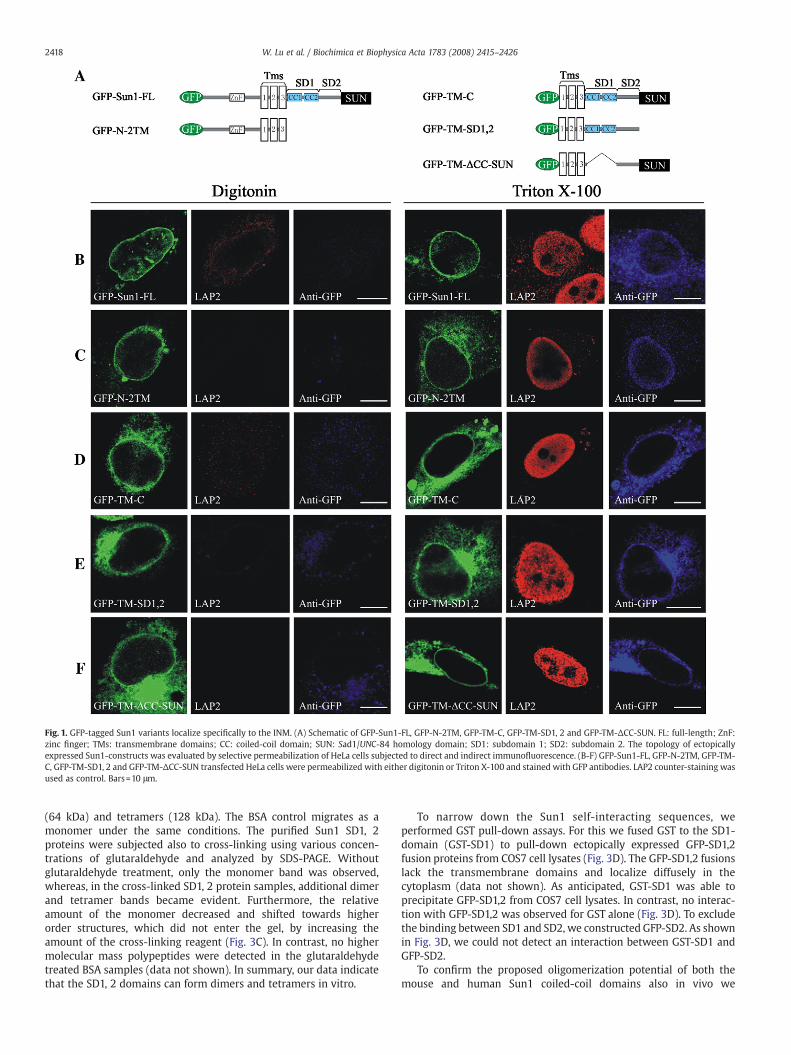
Fig. 1. GFP-tagged Sun1 variants localize specifically to the INM. (A) Schematic of GFP-Sun1-FL, GFP-N-2TM, GFP-TM-C, GFP-TM-SD1, 2 and GFP-TM-ΔCC-SUN. FL: full-length; ZnF:zinc finger; TMs: transmembrane domains; CC: coiled-coil domain; SUN: Sad1/UNC-84 homology domain; SD1: subdomain 1; SD2: subdomain 2. The topology of ectopicallyexpressed Sun1-constructs was evaluated by selective permeabilization of HeLa cells subjected to direct and indirect immunofluorescence. (B-F) GFP-Sun1-FL, GFP-N-2TM, GFP-TM-C, GFP-TM-SD1, 2 and GFP-TM-ΔCC-SUN transfected HeLa cells were permeabilized with either digitonin or Triton X-100 and stained with GFP antibodies. LAP2 counter-staining wasused as control. Bars=10 μm.
2418 W. Lu et al. / Biochimica et Biophysica Acta 1783 (2008) 2415–2426
(64 kDa) and tetramers (128 kDa). The BSA control migrates as amonomer under the same conditions. The purified Sun1 SD1, 2proteins were subjected also to cross-linking using various concen-trations of glutaraldehyde and analyzed by SDS-PAGE. Withoutglutaraldehyde treatment, only the monomer band was observed,whereas, in the cross-linked SD1, 2 protein samples, additional dimerand tetramer bands became evident. Furthermore, the relativeamount of the monomer decreased and shifted towards higherorder structures, which did not enter the gel, by increasing theamount of the cross-linking reagent (Fig. 3C). In contrast, no highermolecular mass polypeptides were detected in the glutaraldehydetreated BSA samples (data not shown). In summary, our data indicatethat the SD1, 2 domains can form dimers and tetramers in vitro.
To narrow down the Sun1 self-interacting sequences, weperformed GST pull-down assays. For this we fused GST to the SD1-domain (GST-SD1) to pull-down ectopically expressed GFP-SD1,2fusion proteins from COS7 cell lysates (Fig. 3D). The GFP-SD1,2 fusionslack the transmembrane domains and localize diffusely in thecytoplasm (data not shown). As anticipated, GST-SD1 was able toprecipitate GFP-SD1,2 from COS7 cell lysates. In contrast, no interac-tion with GFP-SD1,2 was observed for GST alone (Fig. 3D). To excludethe binding between SD1 and SD2, we constructed GFP-SD2. As shownin Fig. 3D, we could not detect an interaction between GST-SD1 andGFP-SD2.
To confirm the proposed oligomerization potential of both themouse and human Sun1 coiled-coil domains also in vivo we

Fig. 2. Sun1 is an immobile NE protein. (A–E) HeLa cells expressing GFP-Sun1-FL, GFP-N-2TM, GFP-TM-C, GFP-TM-SD1, 2 or GFP-TM-ΔCC-SUN were imaged before and afterphotobleaching of the entire nucleus with the exception of a small NE region. Arrows indicate the accumulation of the GFP-fusions in the ER. Corresponding plots for fluorescencedecay kinetics with standard deviation from the unbleached region of iFRAP experiments are shown on the right side. Insets depict the early dynamics at a standardized time scale of40 min. At least 4 experiments were performed for each GFP-fusion and used to deduce the standard deviation.
2419W. Lu et al. / Biochimica et Biophysica Acta 1783 (2008) 2415–2426
performed a yeast two-hybrid system analysis. For that, therespective coiled-coil domains were fused either with GAL4 DNA-binding domains in the pGBKT7 vectors (BD-mSD1, 2, residues 432–737; BD-hSD1, residues 404–493) or the GAL4 transcriptionactivation domains in the pGADT7 vectors (AD-mSD1, 2, residues432–737; AD-hSD1, residues 404–493), respectively. Fig. 3E showsthat cells cotransformed with pGADT7-mSD1, 2 and pGBKT7-mSD1,2, or pGADT7-hSD1 and pGBKT7-hSD1 grew on the selection platesand were positive in the β-galactosidase test. No interaction wasdetected in negative controls. Conclusively, our studies clearlyindicate that mammalian Sun1 forms homo-oligomers through itsSD1-domains.
3.4. Sun1 oligomers contain interchain disulfide bonds
To gain further insights into Sun1 oligomerization, we next focusedour studies on the full-length Sun1 protein. We transfected C-terminalV5-tagged full-length human Sun1 into COS7 cells and analyzed thecell lysates by anti-V5western blotting. Sun1 immunoblotting analysisindicated the presence of SDS-resistant dimers under non-reducedconditions. Without heating and in the absence of dithiothreitol (DTT)in the COS7 lysates, monomer, dimer and tetramer bands weredetected even under high SDS concentrations (0.5%). In the presenceof 100 mM DTT, however, dimer and higher molecular mass signalswere abolished (Fig. 4A). Heating of the samples at 70 °C or 95 °C

Fig. 3. The Sun1 C-terminal coiled-coil domain oligomerizes and stabilizes Sun1 at the NE. (A) Solubilization properties of GFP-TM-C and GFP-TM-ΔCC-SUN under various extractionconditions. Total (T), cytoplasmic (C) and nuclear (N) fractions were produced from GFP-TM-C or GFP-TM-ΔCC-SUN transfected COS7 cells. Nuclear fractions were extracted inbuffers containing Triton X-100, Triton X-100 plus salt and urea, as indicated. Soluble (S) and insoluble (I) fractions were analyzed by western blotting using anti-GFP antibodies.The same lysates were analyzed for LAP2β. (B) Purified mouse Sun1 SD1, 2 fractions were supplemented with SDS-sample buffer containing β-mercaptoethanol and subjected toSDS-PAGE using a 12% SDS-polyacrylamide gel (left panel). The same purified SD1, 2 fractions and BSA (Bovine serum albumin) were supplemented with blue native sample bufferlacking SDS and β-mercaptoethanol. After electrophoresis on a 10% native polyacrylamide gel, the gel was visualized by Coomassie staining. Mono: monomer; Di: dimer; Tetra:tetramer. (C) Equal amounts of the purified mouse Sun1 SD1, 2 were cross-linked with different glutaraldehyde concentrations. Reactions were terminated by addition of SDS-sample buffer. The samples were loaded on a 12% SDS gel and stained by Coomassie Blue. (D) COS7 cells were transiently transfected with GFP-SD1, 2 and GFP-SD2, respectively.The cell lysates were analyzed by immunoblotting using anti-GFP antibodies (Input). COS7 cell lysates expressing the GFP-SD1, 2 or GFP-SD2 were incubated with beads coupled toGST-SD1 or GST. Specifically bound proteins were subjected to SDS-PAGE followed by anti-GST western blotting. The same membrane was later stripped and submitted to anti-GFPimmunoblotting. (E) Yeast two-hybrid assays demonstrate that the coiled-coils of both mouse and human Sun1 interact with each other. Empty BD and AD plasmids were used asnegative controls. Y190 yeast cells were cotransformed with AD/BD-mSD1, 2, BD/AD-mSD1, 2, AD-mSD1, 2/BD-mSD1, 2, or AD/BD-hSD1, BD/AD-hSD1, AD-hSD1/BD-hSD1. Theinteraction was monitored by growth on SD-Trp-Leu-His (60 mM 3AT) plates. X-Gal assays were also performed to confirm the interaction. AD: GAL4 activation domain; BD: GAL4DNA-binding domain.
2420 W. Lu et al. / Biochimica et Biophysica Acta 1783 (2008) 2415–2426
resulted in a decrease of the monomeric signal intensity, through anirreversible Sun1-V5 aggregation (arrow in Fig. 4A), a known effect forsome transmembrane proteins [39]. Similar results were also obtainedfor endogenous Sun1 proteins (Fig. S2A, asterisk).
The presented data strongly suggested the existence of disulfidebridges between Sun1 molecules. Therefore, we next examined theprimary structures of mouse and human Sun1 for cysteines. Asdepicted in Fig. 4B and C, four conserved cysteine residues are presentin both human and mouse Sun1 C-termini. One is located in the SD2domain (human Sun1 Cys526), the other three (human Sun1 Cys657,Cys695 and Cys800) are located in the SUN-domain.
In order to identify which cysteines were sufficient to mediate theformation of disulfide-linked oligomers, four additional constructswere made. Human Sun1 TMC-HA (residues 240–812) containing allfour conserved cysteins (Cys526, Cys657, Cys695, Cys800) in the SD2/SUN-domains, and TMSD1, 2-HA (residues 240–632), which lacks theSUN-domain and contains only the SD2 domain Cys526. We alsogenerated TMC C/A-HA in which Cys526 was replaced by an alanineand TMSD1, 2 C/A-HA, which contains the same C526A mutation andlacks the SUN-domain (Fig. 4C). The constructs were transiently
transfected in COS7 cells and lysed with RIPA buffer containing 0.1%SDS and either 100 mM iodoacetamide (IA) or 20 mM NEM to preventartificial disulfide bond formation. The lysates were submitted to non-reduced or reduced conditions, respectively. Equal amount of proteinfrom each sample was loaded on a 12% gel, analyzed by SDS-PAGE andconcomitant anti-HA western blotting. In the absence of DTT, bandscorresponding to Sun1-dimers were detectable in the absence orpresence of 0.5% SDS in TMC-HA or TMSD1, 2-HA lysates. However, inthe presence of DTT, dimer bands were completely absent (Fig. 4D).Replacing Cys526 by an alanine completely abolished the dimerformation of TMC C/A-HA or TMSD1, 2 C/A-HA (Fig. 4D), respectively.SUN-domain cysteines are therefore not involved in this process,whereas the SD2 domain resident Cys526 might contribute to theformation of Sun1 oligomers.
To address the biological relevance of the Sun1 interchain disulfidebridge in Sun1 NE dynamics, we transiently transfected GFP-TMC andGFP-TMC C/A in HeLa cells and performed iFRAP analysis on thetransfected cells. Four individual cells for each construct werefollowed. However, no significant differences were observed betweenthese two constructs (Fig. 5), suggesting that the disulfide bond

Fig. 4. Sun1 oligomers contain interchain disulfide bonds formed via C-terminal cysteines. (A) Sun1-V5 was transiently transfected in COS7 cells. RIPA-buffer cell lysates wereprepared and divided into four equal portions, and treated under different experimental conditions as indicated on the top of the blot. Lysates were resolved by 6% SDS-PAGE anddetected by anti-V5 immunoblotting analysis. (B) Alignment of the mouse and human Sun1 C-terminus. Identical residues and conservative substitutions are shown in black(Cysteines: marked by red frame) and gray, respectively. Mouse and human Sun1 coiled-coil domains: blue frame; SUN-domains: green frame; SD2 domain: unboxed area. (C)Schematic of the human Sun1 TMC-HA and TMSD1, 2-HA fusion proteins, and the point mutated TMC C/A-HA and TMSD1, 2 C/A-HA. Major Sun1 domains are depicted and theconserved cysteines in SD2 and SUN-domains are indicated below the schematic. In TMC C/A-HA and TMSD1, 2 C/A-HA the SD2 resident cysteine wasmutated to an alanine (red). (D)COS7 cells were transiently transfected with TMC-HA, TMSD1, 2-HA, TMC C/A-HA or TMSD1, 2 C/A-HA. Cell lysates containing either 100 mM IA or 20 mM NEM were treated underdifferent experimental conditions as indicated on the top of the western blots and detected by anti-HA western blotting. Both TMC-HA and TMSD1, 2-HA chimeric proteins yielddimers in the absence of DTT. No dimers were formed in TMC C/A-HA or TMSD1, 2 C/A-HA cell lysates in the presence of DTT.
2421W. Lu et al. / Biochimica et Biophysica Acta 1783 (2008) 2415–2426
between the Sun1 lumenal segments does not play a major role in theSun1 C-terminal domain NE turnover dynamics, although from ourinteraction data we are prompted to assign a stabilizing function tointerchain disulfide bonding for the full-length Sun1 protein.
3.5. Sun1 provides a stable platform for multiple macromolecularassemblies
Our data so far show that Sun1 molecules oligomerize and formstable complexes with itself at the NE. In order to verify this, we nextperformed an indirect immunofluorescence analysis of endogenousSun1 proteins in HeLa cells. Confocal microscopy clearly indicated theorganization of Sun1 into clustered assemblies at the NE (Fig. 6A′). Weconfirmed this further by employing two new Sun1 antibodies (Sun1Aand Sun1B, characterized in Fig. S2). As indicated these new antiseraalso recognized a clustered Sun1 pattern at the NE surface, highlyreminiscent of NPC staining (Fig. 6B, B′; similar results obtained in othercell lines, such as HaCaT, COS7, Ramos, SW480-data not shown). Co-staining with the NPC marker mAb414 revealed a pool of Sun1 thatconcentrated in the vicinity or overlapped with NPCs (Fig. 6A″, B and B′notched arrows in the insets). This result is in agreement with Liu et al.[26],whodemonstrated thatNPCs colocalizewith ectopicallyexpressed
GFP-tagged Sun1. Interestingly, a second pool of endogenous Sun1structures also became evident,which did not colocalizewithNPCs (Fig.6A″, B and B′ arrows in the insets). Similar results were observed inother cell lines, although the percentage of Sun1-NPC overlappingentities varied, most likely due to the metabolic status or the cell cyclestatus of the cells (data not shown). Moreover, ectopic expression ofSun1 full-length constructs indicated also the presence of two distinctNE Sun1 topologies (data not shown). One fraction of Sun1 focicolocalizes with NPCs, whereas the second appears distinct from NPCs.A newly developed antibody recognizing mammalian Sun2 (character-ized in Fig. S2)was then used to investigate the association of Sun2withNPCs. Endogenous Sun2, like observed for Sun1, also displayed aclustered appearance at theNEsurface. As already suggestedby Liu et al.[26] endogenous Sun2 antigenic clusters were almost absent at sites ofnuclear pores (Fig. 6B″). These results and already presented data on apossible Sun1/2 interaction [27] prompted us to investigate thepossibility that Sun2 assemblies at the NE could be identical withSun1 antigenic assemblies, which did not overlap with the NPC.
Unfortunately no monoclonal antibodies to any of Sun1 or Sun2,respectively, are available and we had to analyze local interactions byexamination of endogenous Sun1 in Sun2-V5 transfected HaCaT cells.As shown in Fig. 7A–A″, a large Sun1 proportion colocalizes with Sun2
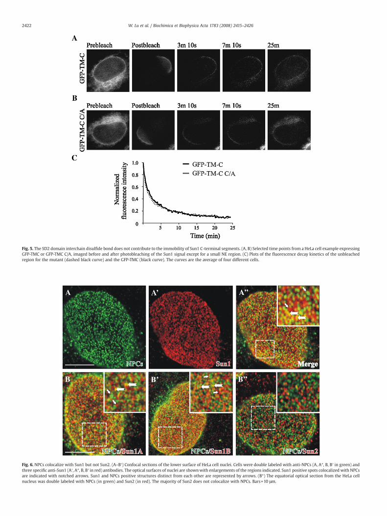
Fig. 6. NPCs colocalize with Sun1 but not Sun2. (A–B′) Confocal sections of the lower surface of HeLa cell nuclei. Cells were double labeled with anti-NPCs (A, A″, B, B′ in green) andthree specific anti-Sun1 (A′, A″, B, B′ in red) antibodies. The optical surfaces of nuclei are shownwith enlargements of the regions indicated. Sun1 positive spots colocalized with NPCsare indicated with notched arrows. Sun1 and NPCs positive structures distinct from each other are represented by arrows. (B″) The equatorial optical section from the HeLa cellnucleus was double labeled with NPCs (in green) and Sun2 (in red). The majority of Sun2 does not colocalize with NPCs. Bars=10 μm.
Fig. 5. The SD2 domain interchain disulfide bond does not contribute to the immobility of Sun1 C-terminal segments. (A, B) Selected time points from a HeLa cell example expressingGFP-TMC or GFP-TMC C/A, imaged before and after photobleaching of the Sun1 signal except for a small NE region. (C) Plots of the fluorescence decay kinetics of the unbleachedregion for the mutant (dashed black curve) and the GFP-TMC (black curve). The curves are the average of four different cells.
2422 W. Lu et al. / Biochimica et Biophysica Acta 1783 (2008) 2415–2426
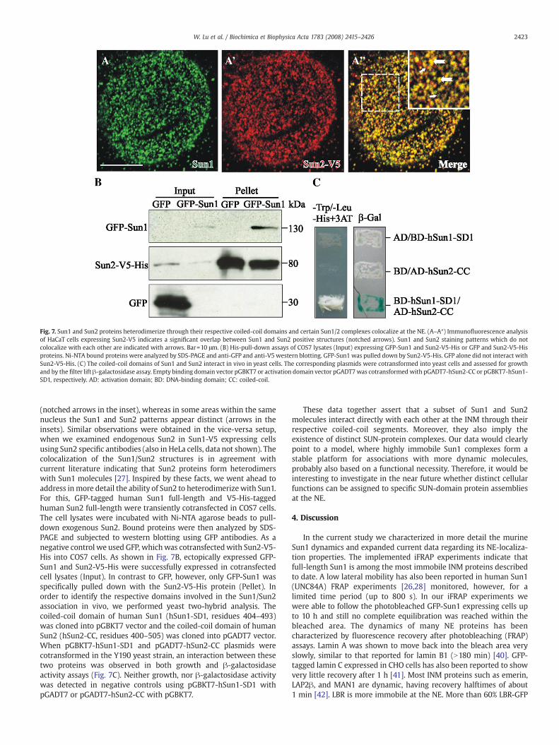
Fig. 7. Sun1 and Sun2 proteins heterodimerize through their respective coiled-coil domains and certain Sun1/2 complexes colocalize at the NE. (A–A″) Immunofluorescence analysisof HaCaT cells expressing Sun2-V5 indicates a significant overlap between Sun1 and Sun2 positive structures (notched arrows). Sun1 and Sun2 staining patterns which do notcolocalize with each other are indicated with arrows. Bar=10 μm. (B) His-pull-down assays of COS7 lysates (Input) expressing GFP-Sun1 and Sun2-V5-His or GFP and Sun2-V5-Hisproteins. Ni-NTA bound proteins were analyzed by SDS-PAGE and anti-GFP and anti-V5 western blotting. GFP-Sun1 was pulled down by Sun2-V5-His. GFP alone did not interact withSun2-V5-His. (C) The coiled-coil domains of Sun1 and Sun2 interact in vivo in yeast cells. The corresponding plasmids were cotransformed into yeast cells and assessed for growthand by the filter lift β-galactosidase assay. Empty binding domain vector pGBKT7 or activation domain vector pGADT7was cotransformedwith pGADT7-hSun2-CC or pGBKT7-hSun1-SD1, respectively. AD: activation domain; BD: DNA-binding domain; CC: coiled-coil.
2423W. Lu et al. / Biochimica et Biophysica Acta 1783 (2008) 2415–2426
(notched arrows in the inset), whereas in some areas within the samenucleus the Sun1 and Sun2 patterns appear distinct (arrows in theinsets). Similar observations were obtained in the vice-versa setup,when we examined endogenous Sun2 in Sun1-V5 expressing cellsusing Sun2 specific antibodies (also in HeLa cells, data not shown). Thecolocalization of the Sun1/Sun2 structures is in agreement withcurrent literature indicating that Sun2 proteins form heterodimerswith Sun1 molecules [27]. Inspired by these facts, we went ahead toaddress inmore detail the ability of Sun2 to heterodimerizewith Sun1.For this, GFP-tagged human Sun1 full-length and V5-His-taggedhuman Sun2 full-length were transiently cotransfected in COS7 cells.The cell lysates were incubated with Ni-NTA agarose beads to pull-down exogenous Sun2. Bound proteins were then analyzed by SDS-PAGE and subjected to western blotting using GFP antibodies. As anegative control we used GFP, whichwas cotransfectedwith Sun2-V5-His into COS7 cells. As shown in Fig. 7B, ectopically expressed GFP-Sun1 and Sun2-V5-His were successfully expressed in cotransfectedcell lysates (Input). In contrast to GFP, however, only GFP-Sun1 wasspecifically pulled down with the Sun2-V5-His protein (Pellet). Inorder to identify the respective domains involved in the Sun1/Sun2association in vivo, we performed yeast two-hybrid analysis. Thecoiled-coil domain of human Sun1 (hSun1-SD1, residues 404–493)was cloned into pGBKT7 vector and the coiled-coil domain of humanSun2 (hSun2-CC, residues 400–505) was cloned into pGADT7 vector.When pGBKT7-hSun1-SD1 and pGADT7-hSun2-CC plasmids werecotransformed in the Y190 yeast strain, an interaction between thesetwo proteins was observed in both growth and β-galactosidaseactivity assays (Fig. 7C). Neither growth, nor β-galactosidase activitywas detected in negative controls using pGBKT7-hSun1-SD1 withpGADT7 or pGADT7-hSun2-CC with pGBKT7.
These data together assert that a subset of Sun1 and Sun2molecules interact directly with each other at the INM through theirrespective coiled-coil segments. Moreover, they also imply theexistence of distinct SUN-protein complexes. Our data would clearlypoint to a model, where highly immobile Sun1 complexes form astable platform for associations with more dynamic molecules,probably also based on a functional necessity. Therefore, it would beinteresting to investigate in the near future whether distinct cellularfunctions can be assigned to specific SUN-domain protein assembliesat the NE.
4. Discussion
In the current study we characterized in more detail the murineSun1 dynamics and expanded current data regarding its NE-localiza-tion properties. The implemented iFRAP experiments indicate thatfull-length Sun1 is among the most immobile INM proteins describedto date. A low lateral mobility has also been reported in human Sun1(UNC84A) FRAP experiments [26,28] monitored, however, for alimited time period (up to 800 s). In our iFRAP experiments wewere able to follow the photobleached GFP-Sun1 expressing cells upto 10 h and still no complete equilibration was reached within thebleached area. The dynamics of many NE proteins has beencharacterized by fluorescence recovery after photobleaching (FRAP)assays. Lamin A was shown to move back into the bleach area veryslowly, similar to that reported for lamin B1 (N180 min) [40]. GFP-tagged lamin C expressed in CHO cells has also been reported to showvery little recovery after 1 h [41]. Most INM proteins such as emerin,LAP2β, and MAN1 are dynamic, having recovery halftimes of about1 min [42]. LBR is more immobile at the NE. More than 60% LBR-GFP
2424 W. Lu et al. / Biochimica et Biophysica Acta 1783 (2008) 2415–2426
fractions reside within the NE membranes 10 min after photobleach-ing [43]. The extensive immobilization of GFP-Sun1 suggests a tightbinding to fixed structural components of the nucleus or/andretention by assembly into multimeric complexes, which would beexpected to result in low diffusion rates. GFP labeled nuclear porecomplex components for example, which are known to formoligomeric complexes have been shown to diffuse slowly within theNE [31,44].
Our data do not assign amajor role for the SUN-domain itself in theSun1 dynamics. They rather suggest important roles for the Sun1coiled-coil domain, through which Sun1 forms dimers. Geneticevidence indicates that C. elegans UNC-84 exists as dimers at the NE[45] similar to matefin, another SUN-domain protein in C. elegansreported to homodimerize [21]. Sun-1 in D. discoideum also formsoligomers via the C-terminal coiled-coil domains [19]. Sun2 forms alsohomodimers through the N-terminal region and the coiled-coildomains [27]. These data together suggest that oligomerization atthe NE is a common feature for SUN-family members.
The lumenal domain of Sun1 has been suggested to permit homo-oligomerization [26,27]. We were able to narrow down the oligomer-ization domain to the coiled-coil region (SD1-domain). Furthermore,this Sun1 C-terminal region resides in the PNS, which is a part of theER lumen. Unlike the cytoplasm, the ER provides an oxidizingenvironment favoring disulfide bond formation [46,47]. Indeed, V5-tagged full-length Sun1 has the ability to form SDS-resistant dimersand tetramers under non-reduced conditions. Mutagenesis assaysdemonstrated that the cysteine residue (Cys526) in the Sun1 SD2domain, adjacent to the C-terminal border of the coiled-coil region, isresponsible for the formation of the disulfide bond. Thus, Sun1 homo-oligomers form through coiled-coil interactions and interchaindisulfide bonds (Fig. 8). We also tested the dynamics of GFP-taggedTM-C and TMC C/A by iFRAP analysis. However, no significantdifferences were observed between these two constructs, suggestingthat the disulfide bond between the Sun1 lumenal segments does notcontribute to the Sun1 immobility in the PNS. Nonetheless, we cannotexclude that such associations may exhibit important stabilizingfunctions in the full-length Sun1 protein. It is also interesting to notethat the cysteine residue forming the disulfide bond resides within themajor Nesprin binding site of Sun1 [11]. Therefore, the interchain
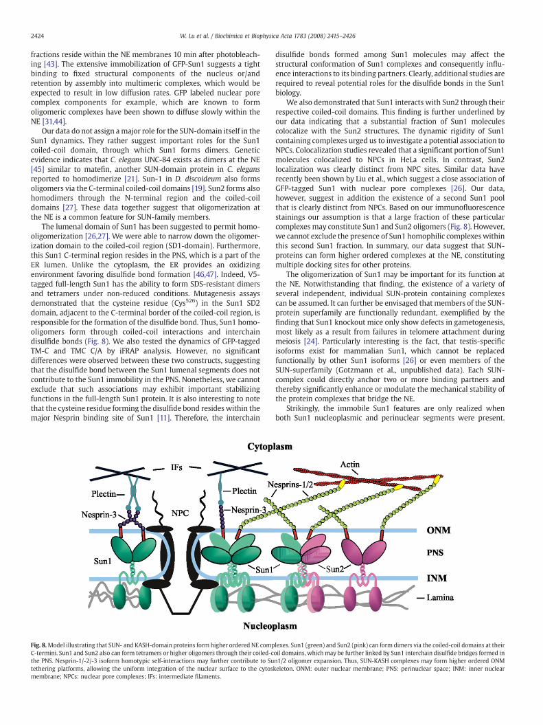
Fig. 8.Model illustrating that SUN- and KASH-domain proteins form higher ordered NE compC-termini. Sun1 and Sun2 also can form tetramers or higher oligomers through their coiled-cthe PNS. Nesprin-1/-2/-3 isoform homotypic self-interactions may further contribute to Sutethering platforms, allowing the uniform integration of the nuclear surface to the cytoskmembrane; NPCs: nuclear pore complexes; IFs: intermediate filaments.
disulfide bonds formed among Sun1 molecules may affect thestructural conformation of Sun1 complexes and consequently influ-ence interactions to its binding partners. Clearly, additional studies arerequired to reveal potential roles for the disulfide bonds in the Sun1biology.
We also demonstrated that Sun1 interacts with Sun2 through theirrespective coiled-coil domains. This finding is further underlined byour data indicating that a substantial fraction of Sun1 moleculescolocalize with the Sun2 structures. The dynamic rigidity of Sun1containing complexes urged us to investigate a potential association toNPCs. Colocalization studies revealed that a significant portion of Sun1molecules colocalized to NPCs in HeLa cells. In contrast, Sun2localization was clearly distinct from NPC sites. Similar data haverecently been shown by Liu et al., which suggest a close association ofGFP-tagged Sun1 with nuclear pore complexes [26]. Our data,however, suggest in addition the existence of a second Sun1 poolthat is clearly distinct from NPCs. Based on our immunofluorescencestainings our assumption is that a large fraction of these particularcomplexes may constitute Sun1 and Sun2 oligomers (Fig. 8). However,we cannot exclude the presence of Sun1 homophilic complexes withinthis second Sun1 fraction. In summary, our data suggest that SUN-proteins can form higher ordered complexes at the NE, constitutingmultiple docking sites for other proteins.
The oligomerization of Sun1 may be important for its function atthe NE. Notwithstanding that finding, the existence of a variety ofseveral independent, individual SUN-protein containing complexescan be assumed. It can further be envisaged that members of the SUN-protein superfamily are functionally redundant, exemplified by thefinding that Sun1 knockout mice only show defects in gametogenesis,most likely as a result from failures in telomere attachment duringmeiosis [24]. Particularly interesting is the fact, that testis-specificisoforms exist for mammalian Sun1, which cannot be replacedfunctionally by other Sun1 isoforms [26] or even members of theSUN-superfamily (Gotzmann et al., unpublished data). Each SUN-complex could directly anchor two or more binding partners andthereby significantly enhance or modulate the mechanical stability ofthe protein complexes that bridge the NE.
Strikingly, the immobile Sun1 features are only realized whenboth Sun1 nucleoplasmic and perinuclear segments were present.
lexes. Sun1 (green) and Sun2 (pink) can form dimers via the coiled-coil domains at theiroil domains, which may be further linked by Sun1 interchain disulfide bridges formed inn1/2 oligomer expansion. Thus, SUN-KASH complexes may form higher ordered ONMeleton. ONM: outer nuclear membrane; PNS: perinuclear space; INM: inner nuclear

2425W. Lu et al. / Biochimica et Biophysica Acta 1783 (2008) 2415–2426
Several possibilities may account for this result. First, Sun1 N- andC-termini could bind to distinct NE-associated proteins thatcontribute collectively to its NE retention. This scenario is plausible,considering that Sun1 domains are clearly distinct in their primarystructure and more importantly in their topology. In line with theabove is that the Sun1 N-terminus interacts with lamin A, althoughits NE localization seems to be largely independent from thepresence of A-type lamins [10,11]. The Sun1 N-terminus is alsocapable of self-association, a feature potentially required forassociation with DNA [27,29]. Most recent data also point to aninvolvement of N-terminal domains of Sun1 in the degree ofchromosome compaction after mitosis [25]. The presence of avariety of N-terminal splicing isoforms may account for differencesin the binding spectrum for lamins and/or chromatin [26]. More-over, the mouse Sun1 nucleoplasmic segment contains a Zinc-fingermotif, which associates with DNA, whereas the C-terminusassociates with the Nesprin-1/-2/-3 KASH-domains.
The second possibility is that Sun1 N-terminal association to itsbinding partners has immediate structural effects on the C-terminus.Binding for example to the nuclear lamina or/and chromatin mayunfold the C-terminus in such a way, so that inter- or intra-molecularinteractions are modulated. If this holds true, signals from the nucleusmay be efficiently transmitted to the cytoplasm and vice-versa.Support for this attractive model comes from the fact that while GFP-TM-C interacts with the endogenous Sun1 in vitro, its NE lateralmobility is clearly reduced when compared to the Sun1 full-lengthprotein. Additional experiments are needed to validate all thosethoughts. Thus, the combined oligomerization capacity of the Sun1 N-and C-termini may account for the dramatic immobility of the full-length Sun1 protein.
Current data also suggest that the biochemical properties of SUNand KASH-domain proteins may jointly organize these molecules intocomplexes. In fact Nesprin molecules themselves may contribute to anexpansion and the formation of larger macromolecular complexes(Fig. 8). Nesprin-1 was shown to oligomerize with itself through its C-terminal spectrin repeats [48]. Nesprin-2 giant isoforms function asNE guardian molecules that tether smaller Nesprin-2 C-terminalisoforms at the ONM [49,50]. Also, Nesprin-3 oligomerizes with itselfthrough its N-terminal spectrin repeats [51]. Hetero-oligomerizationaspects among Nesprins-1/-2/-3 have not been examined so far; wecannot exclude, however, the possibility that Nesprins may form afilament network by their own at the ONM and contribute also to theimmobility of Sun1 at the NE. The fact that Sun1/2 oligomerizesuggests that such an assembly may structurally and functionallyintegrate various KASH-domain containing proteins within thesecomplexes. This view is further supported by a recent reportdemonstrating that mammalian KASH-proteins interact promiscu-ously with Sun1/2 proteins [52].
Collectively these thoughts indicate that more research is neededto shed light in a new cell biological area governing the structural andfunctional integration of cytoplasmic and nuclear compartments.
Acknowledgements
This work was supported by grants from the Knut and AliceWallenberg foundation to C.S. and M.U., from the “Hochschuljubi-läumsstiftung der Stadt Wien” and a collaborative project supportfrom the Knut and Alice Wallenberg foundation to J.G.; from theAustrian Science Research Fund (FWF P17871) and the EURO-Laminopathies research project of the European Commission (Con-tract LSHM-CT-2005-018690) to R.F.; from the German ResearchCouncil (DFG EL 246/3-1) to J.E.; a postdoctoral fellowship from theHuman Science Foundation (HFSP) to L.S.; from the CMMC to A.A.N.;from the NRW International Graduate School (IGS) in Genetics andFunctional Genomics to W.L.; from the DFG (KA 2778/1-1) and aWellcome Trust ‘Value in People’ award to I.K.
Appendix A. Supplementary data
Supplementary data associated with this article can be found, inthe online version, at doi:10.1016/j.bbamcr.2008.09.001.
References
[1] L. Gerace, B. Burke, Functional organization of the nuclear envelope, Annu. Rev.Cell Biol. 4 (1988) 335–374.
[2] D. Gorlich, U. Kutay, Transport between the cell nucleus and the cytoplasm, Annu.Rev. Cell Dev. Biol. 15 (1999) 607–660.
[3] I.G. Macara, Transport into and out of the nucleus, Microbiol. Mol. Biol. Rev. 65(2001) 570–594.
[4] B. Burke, J. Ellenberg, Remodelling thewalls of the nucleus, Nat. Rev., Mol. Cell Biol.3 (2002) 487–497.
[5] N. Stuurman, S. Heins, U. Aebi, Nuclear lamins: their structure, assembly, andinteractions, J. Struct. Biol. 122 (1998) 42–66.
[6] J.L. Broers, F.C. Ramaekers, G. Bonne, R.B. Yaou, C.J. Hutchison, Nuclear lamins:laminopathies and their role in premature ageing, Physiol. Rev. 86 (2006)967–1008.
[7] D.A. Starr, J.A. Fischer, KASH 'n Karry: the KASH domain family of cargo-specificcytoskeletal adaptor proteins, BioEssays 27 (2005) 1136–1146.
[8] Y.B. Tzur, K.L. Wilson, Y. Gruenbaum, SUN-domain proteins: ‘Velcro’ that links thenucleoskeleton to the cytoskeleton, Nat. Rev., Mol. Cell Biol. 7 (2006) 782–788.
[9] K. Wilhelmsen, M. Ketema, H. Truong, A. Sonnenberg, KASH-domain proteins innuclear migration, anchorage and other processes, J. Cell Sci. 119 (2006)5021–5029.
[10] M. Crisp, Q. Liu, K. Roux, J.B. Rattner, C. Shanahan, B. Burke, P.D. Stahl, D. Hodzic,Coupling of the nucleus and cytoplasm: role of the LINC complex, J. Cell Biol. 172(2006) 41–53.
[11] V.C. Padmakumar, T. Libotte, W. Lu, H. Zaim, S. Abraham, A.A. Noegel, J. Gotzmann,R. Foisner, I. Karakesisoglou, The inner nuclear membrane protein Sun1 mediatesthe anchorage of Nesprin-2 to the nuclear envelope, J. Cell Sci. 118 (2005)3419–3430.
[12] P.J. Stewart-Hutchinson, C.M. Hale, D. Wirtz, D. Hodzic, Structural requirementsfor the assembly of LINC complexes and their function in cellular mechanicalstiffness, Exp. Cell Res. 314 (2008) 1892–1905.
[13] C.J. Malone, L. Misner, N. Le Bot, M.C. Tsai, J.M. Campbell, J. Ahringer, J.G. White,The C. elegans hook protein, ZYG-12, mediates the essential attachment betweenthe centrosome and nucleus, Cell 115 (2003) 825–836.
[14] M.D. McGee, R. Rillo, A.S. Anderson, D.A. Starr, UNC-83 IS a KASH protein requiredfor nuclear migration and is recruited to the outer nuclearmembrane by a physicalinteraction with the SUN protein UNC-84, Mol. Biol. Cell 17 (2006) 1790–1801.
[15] D.A. Starr, M. Han, Role of ANC-1 in tethering nuclei to the actin cytoskeleton,Science 298 (2002) 406–409.
[16] D.A. Starr, Communication between the cytoskeleton and the nuclear envelope toposition the nucleus, Mol. Biosyst. 3 (2007) 583–589.
[17] F. Miki, A. Kurabayashi, Y. Tange, K. Okazaki, M. Shimanuki, O. Niwa, Two-hybridsearch for proteins that interact with Sad1 and Kms1, two membrane-boundcomponents of the spindle pole body in fission yeast, Mol. Genet. Genomics 270(2004) 449–461.
[18] S.L. Jaspersen, A.E. Martin, G. Glazko, T.H. Giddings Jr., G. Morgan, A. Mushegian, M.Winey, The Sad1-UNC-84 homology domain in Mps3 interacts with Mps2 toconnect the spindle pole body with the nuclear envelope, J. Cell Biol. 174 (2006)665–675.
[19] H. Xiong, F. Rivero, U. Euteneuer, S. Mondal, S. Mana-Capelli, D. Larochelle, A.Vogel, B. Gassen, A.A. Noegel, Dictyostelium sun-1 connects the centrosome tochromatin and ensures genome stability, Traffic 9 (2008) 708–724.
[20] K.K. Lee, D. Starr, M. Cohen, J. Liu, M. Han, K.L. Wilson, Y. Gruenbaum, Lamin-dependent localization of UNC-84, a protein required for nuclear migration inCaenorhabditis elegans, Mol. Biol. Cell 13 (2002) 892–901.
[21] A. Fridkin, E. Mills, A. Margalit, E. Neufeld, K.K. Lee, N. Feinstein, M. Cohen, K.L.Wilson, Y. Gruenbaum, Matefin, a Caenorhabditis elegans germ line-specific SUN-domain nuclear membrane protein, is essential for early embryonic and germ celldevelopment, Proc. Natl. Acad. Sci. U. S. A. 101 (2004) 6987–6992.
[22] Y. Chikashige, C. Tsutsumi, M. Yamane, K. Okamasa, T. Haraguchi, Y. Hiraoka,Meiotic proteins bqt1 and bqt2 tether telomeres to form the bouquet arrangementof chromosomes, Cell 125 (2006) 59–69.
[23] I. Hagan, M. Yanagida, The product of the spindle formation gene sad1+ associateswith the fission yeast spindle pole body and is essential for viability, J. Cell Biol. 129(1995) 1033–1047.
[24] J. Schmitt, R. Benavente, D. Hodzic, C. Hoog, C.L. Stewart, M. Alsheimer,Transmembrane protein Sun2 is involved in tethering mammalian meiotictelomeres to the nuclear envelope, Proc. Natl. Acad. Sci. U. S. A. 104 (2007)7426–7431.
[25] Y.H. Chi, K. Haller, J.M. Peloponese Jr., K.T. Jeang, Histone acetyltransferase hALPand nuclear membrane protein hsSUN1 function in de-condensation of mitoticchromosomes, J. Biol. Chem. 282 (2007) 27447–27458.
[26] Q. Liu, N. Pante, T. Misteli, M. Elsagga, M. Crisp, D. Hodzic, B. Burke, K.J. Roux,Functional association of Sun1with nuclear pore complexes, J. Cell Biol. 178 (2007)785–798.
[27] Q. Wang, X. Du, Z. Cai, M.I. Greene, Characterization of the structures involved inlocalization of the SUN proteins to the nuclear envelope and the centrosome, DNACell Biol. 25 (2006) 554–562.

2426 W. Lu et al. / Biochimica et Biophysica Acta 1783 (2008) 2415–2426
[28] S. Hasan, S. Guttinger, P. Muhlhausser, F. Anderegg, S. Burgler, U. Kutay, Nuclearenvelope localization of human UNC84A does not require nuclear lamins, FEBSLett. 580 (2006) 1263–1268.
[29] F. Haque, D.J. Lloyd, D.T. Smallwood, C.L. Dent, C.M. Shanahan, A.M. Fry, R.C.Trembath, S. Shackleton, SUN1 interacts with nuclear lamin A and cytoplasmicnesprins to provide a physical connection between the nuclear lamina and thecytoskeleton, Mol. Cell. Biol. 26 (2006) 3738–3751.
[30] D.M. Hodzic, D.B. Yeater, L. Bengtsson, H. Otto, P.D. Stahl, Sun2 is a novel mammalianinner nuclear membrane protein, J. Biol. Chem. 279 (2004) 25805–25812.
[31] G. Rabut, V. Doye, J. Ellenberg, Mapping the dynamic organization of the nuclearpore complex inside single living cells, Nat. Cell Biol. 6 (2004) 1114–1121.
[32] J. Gotzmann, S. Vlcek, R. Foisner, Caspase-mediated cleavage of the chromosome-binding domain of lamina-associated polypeptide 2 alpha, J. Cell Sci. 113 (2000)3769–3780.
[33] Y.Y. Zhen, T. Libotte,M.Munck, A.A. Noegel, E. Korenbaum, NUANCE, a giant proteinconnecting the nucleus and actin cytoskeleton, J. Cell Sci. 115 (2002) 3207–3222.
[34] A.A. Noegel, R. Blau-Wasser, H. Sultana, R. Muller, L. Israel, M. Schleicher, H. Patel,C.J. Weijer, The cyclase-associated protein CAP as regulator of cell polarity andcAMP signaling in Dictyostelium, Mol. Biol. Cell 15 (2004) 934–945.
[35] T. Dechat, J. Gotzmann, A. Stockinger, C.A. Harris, M.A. Talle, J.J. Siekierka, R. Foisner,Detergent-salt resistance of LAP2alpha in interphase nuclei and phosphorylation-dependent association with chromosomes early in nuclear assembly impliesfunctions in nuclear structure dynamics, EMBO J. 17 (1998) 4887–4902.
[36] A. Persson, S. Hober, M. Uhlen, A human protein atlas based on antibodyproteomics, Curr. Opin. Mol. Ther. 8 (2006) 185–190.
[37] C. Dreuillet, J. Tillit, M. Kress, M. Ernoult-Lange, In vivo and in vitro interactionbetween human transcription factor MOK2 and nuclear lamin A/C, Nucleic AcidsRes. 30 (2002) 4634–4642.
[38] S.A. Adam, R.S.Marr, L. Gerace, Nuclear protein import inpermeabilizedmammaliancells requires soluble cytoplasmic factors, J. Cell Biol. 111 (1990) 807–816.
[39] C. Favreau, R. Bastos, J. Cartaud, J.C. Courvalin, P. Mustonen, Biochemicalcharacterization of nuclear pore complex protein gp210 oligomers, Eur. J.Biochem. 268 (2001) 3883–3889.
[40] R.D. Moir, M. Yoon, S. Khuon, R.D. Goldman, Nuclear lamins A and B1: differentpathways of assembly during nuclear envelope formation in living cells, J. CellBiol. 151 (2000) 1155–1168.
[41] J.L. Broers, B.M. Machiels, G.J. van Eys, H.J. Kuijpers, E.M. Manders, R. van Driel, F.C.Ramaekers, Dynamics of the nuclear lamina as monitored by GFP-tagged A typelamins, J. Cell Sci. 112 (1999) 3463–3475.
[42] T. Shimi, T. Koujin, M. Segura-Totten, K.L. Wilson, T. Haraguchi, Y.J. Hiraoka,Dynamic interaction between BAF and emerin revealed by FRAP, FLIP, and FRETanalyses in living HeLa cells, Struct. Biol. 147 (2004) 31–41.
[43] Y. Okada, T. Suzuki, Y. Sunden, Y. Orba, S. Kose, N. Imamoto, H. Takahashi, S. Tanaka,W.W. Hall, K. Nagashima, H. Sawa, Dissociation of heterochromatin protein 1 fromlamin B receptor induced by human polyomavirus agnoprotein: role in nuclearegress of viral particles, EMBO Rep. 6 (2005) 452–457.
[44] M. Bucci, S.R. Wente, In vivo dynamics of nuclear pore complexes in yeast, J. CellBiol. 136 (1997) 1185–1199.
[45] C.J. Malone, W.D. Fixsen, H.R. Horvitz, M. Han, UNC-84 localizes to the nuclearenvelope and is required for nuclear migration and anchoring during C. elegansdevelopment, Development 126 (1999) 3171–3181.
[46] J.F. Collet, J.C. Bardwell, Oxidative protein folding in bacteria, Mol. Microbiol. 44(2002) 1–8.
[47] A.R. Frand, J.W. Cuozzo, C.A. Kaiser, Pathways for protein disulphide bondformation, Trends Cell Biol. 10 (2000) 203–210.
[48] J.M. Mislow, J.M. Holaska, M.S. Kim, K.K. Lee, M. Segura-Totten, K.L. Wilson, E.M.McNally, Nesprin-1alpha self-associates and binds directly to emerin and lamin Ain vitro, FEBS Lett. 525 (2002) 135–140.
[49] S. Kandert, Y. Luke, T. Kleinhenz, S. Neumann, W. Lu, V.M. Jaeger, M. Munck, M.Wehnert, C.R. Muller, Z. Zhou, A.A. Noegel, M.C. Dabauvalle, I. Karakesisoglou,Nesprin-2 giant safeguards nuclear envelope architecture in LMNA S143F progeriacells, Hum. Mol. Genet. 16 (2007) 2944–2959.
[50] Y. Lüke, H. Zaim, I. Karakesisoglou, V.M. Jaeger, L. Sellin, W. Lu, M. Schneider, S.Neumann, A. Beijer, M. Munck, V.C. Padmakumar, J. Gloy, G. Walz, A.A. Noegel,Nesprin-2 Giant (NUANCE) maintains nuclear envelope architecture and compo-sition in skin, J. Cell Sci. 121 (2008) 1887–1898.
[51] M. Ketema, K. Wilhelmsen, I. Kuikman, H. Janssen, D. Hodzic, A. Sonnenberg,Requirements for the localization of nesprin-3 at the nuclear envelope and itsinteraction with plectin, J. Cell Sci. 120 (2007) 3384–3394.
[52] P.J. Stewart-Hutchinson, C.M. Hale, D. Wirtz, D. Hodzic, Structural requirementsfor the assembly of LINC complexes and their function in cellular mechanicalstiffness, Exp. Cell Res. 314 (2008) 1892–1905.