
The Primary Structure of Trypanosoma( Nannomonas) congolenseVariant Surface Glycoproteins
Upload
khangminh22Category
view
0download
0
HAL Id: tel-01230548https://tel.archives-ouvertes.fr/tel-01230548
Submitted on 18 Nov 2015
HAL is a multi-disciplinary open accessarchive for the deposit and dissemination of sci-entific research documents, whether they are pub-lished or not. The documents may come fromteaching and research institutions in France orabroad, or from public or private research centers.
L’archive ouverte pluridisciplinaire HAL, estdestinée au dépôt et à la diffusion de documentsscientifiques de niveau recherche, publiés ou non,émanant des établissements d’enseignement et derecherche français ou étrangers, des laboratoirespublics ou privés.
Structural characterization of viral envelopeglycoproteinsIeva Vasiliauskaite
To cite this version:Ieva Vasiliauskaite. Structural characterization of viral envelope glycoproteins. Structural Biology[q-bio.BM]. Université Pierre et Marie Curie - Paris VI, 2014. English. �NNT : 2014PA066507�. �tel-01230548�

Université Pierre et Marie Curie Ecole doctorale Complexité du Vivant
Institut Pasteur, Departement de Virologie, Unité de Virologie Structurale
Structural Characterization of Viral Envelope
Glycoproteins
Ieva Vasiliauskaite
Thèse de doctorat de Virologie Structurale
Dirigée par Felix Rey
Présentée et soutenue publiquement le 14 Novembre 2014
Devant un jury composé de:
Rapporteur: Prof. Jean Dubuisson
Rapporteur: Prof. Yves Gaudin
Examinateur: Prof. Germain Trugnan
Examinateur: Prof. Andreas Herrmann
Examinateur: Dr. Berend-Jan Bosch
Directeur de thèse: Prof. Felix Rey
Co-directeur: Dr. Thomas Krey

2
Acknowledgements
I would like to express my sincere gratitude to a number of people (co-workers,
colleagues, friends and family) who helped me and supported me during my doctoral studies.
First I would like to thank my thesis director Prof. Felix Rey for providing me with the
opportunity to work in his laboratory, his guidance and advice throughout my thesis.
I also want to express my immense gratitude to my supervisor Dr. Thomas Krey for
his extensive supervision, teaching me various techniques, never ending ideas, many
stimulating discussions and for open door at any time. Your contribution to my PhD and my
development as a researcher has been invaluable. Thank you for all your help on every aspect
of the projects and teaching me how to work and think as a scientist.
I would like to thank my thesis reviewers Prof. Yves Gaudin and Prof. Jean Dubuisson
for critical reading of the manuscript and all of their helpful suggestions and Prof. Andreas
Herrmann, Prof. Germain Trugnan and Dr. Berend Jan Bosch for accepting to serve on my
defense committee. Also thanks to my tutor Nicolas Wolff for his support during the past four
years.
I am very grateful for all my collaborators for interesting and fruitful interactions: Dr.
Annette Martin and Dr. Caroline Marnata (Institut Pasteur) for GBV-B infection inhibition
assays, Dr. Patrick England (Plate-Forme de Biophysique des Macromolécules et de leurs
Interactions, Institut Pasteur) for introducing me to SPR techniques, Prof. Peter Rottier, Dr.
Berend Jan Bosch, Dr. Qiushi Wang (Utrecht University) and the former post-doc of our lab
Dr. Scott Jeffers for all collaboration on baculovirus F project, Prof. Arvind Patel and Ania
Owsianka (MRC - University of Glasgow Centre for Virus Research) for all contribution to
DAO5 project, Steven K. H. Foung for providing a number of antibodies and Christine
Girard-Blanc for her initial work on GBV-B E2 and her help with protein expression. Special
thank to Dr. Pierre Legrand for his immense help with experimental phasing and all
invaluable lessons in protein crystallography.
I am very thankful to all current and former group members for creating a nice
working environment and keeping our lab running smoothly as well as sharing their expertise
with me. I extend my gratitude to Danielle Senlecques for excellent administrative work and
assistance.
I also would like to acknowledge Ahmed Haouz and Patrick Weber from the
crystallization platform for help in crystallization and staff of the synchrotron beamlines PX-I

3
at the Swiss Light Source and Proxima-1 at Synchrotron Soleil for help during data collection.
This work was supported by two grants: Marie Curie Initial Training Network “Virus
Entry” and ANRS grant for Thomas Krey. “Virus Entry” network was a very useful training
program and also provided me the opportunity to present my work at many different
meetings. I want to thank to all the fellows within “Virus Entry” network for all nice moments
together.
Finally, I cannot express how much I appreciate emotional and practical support of my
closest family and friends. Thanks to my parents and grandparents, my brother and his family
for their love and belief in me that always give me so much strength in everything I do. I
would like to end with my immense gratitude for Bryan for his support and patience during
this stressful period. Thank you so much for helping me through hard times and filling my life
with love. No words can express what your love and support have meant to me.

4
Table of Contents
Acknowledgements..........................................................................................................................2!
Table.of.Contents...............................................................................................................................4!
List.of.abbreviations.........................................................................................................................6!
General.Introduction.....................................................................................................................11!Virus.Entry..................................................................................................................................................11!The!role!of!viral!envelope!proteins!in!virus!entry!...................................................................................!11!Virus!interaction!with!cell!surface!molecules!............................................................................................!11!Principles!of!viral!membrane!fusion!.............................................................................................................!12!Mechanisms!to!trigger!viral!membrane!fusion!.........................................................................................!13!Structures!of!viral!fusion!proteins!..................................................................................................................!15!The!role!of!viral!envelope!proteins!in!immune!evasion!........................................................................!21!
Chapter.I............................................................................................................................................22!
Hepaciviruses.and.their.entry.to.target.cells........................................................................22!Introduction...............................................................................................................................................22!Hepatitis!C!Virus!.....................................................................................................................................................!22!HCV!pathogenesis!..................................................................................................................................................!22!Animal!models!for!HCV!.......................................................................................................................................!24!GBVGB!as!a!surrogate!model!for!HCV!.............................................................................................................!25!Genome!organization!of!Hepaciviruses!.........................................................................................................!26!Structural!proteins!................................................................................................................................................!28!NonGstructural!proteins!......................................................................................................................................!29!HCV!genotypes!........................................................................................................................................................!29!Current!treatment!options!.................................................................................................................................!30!Experimental!systems!for!studying!HCV!and!GBVGB!entry!in,vitro!..................................................!31!Infectious!cycle!of!HCV!and!GBVGB!.................................................................................................................!34!Role!of!neutralizing!antibodies!in!HCV!infection!.....................................................................................!46!HCV!immune!escape!strategies!........................................................................................................................!54!3D!structures!of!the!main!neutralizing!epitopes!......................................................................................!58!Therapeutic!potential!of!neutralizing!antibodies!....................................................................................!66!Immune!responses!to!GBVGB!virus!.................................................................................................................!69!
I..Structural.Characterization.of.the.GB.virus.B.Envelope.Glycoprotein.E2.........................70!Background!...............................................................................................................................................................!70!Objectives!..................................................................................................................................................................!70!Results!.........................................................................................................................................................................!71!Discussion!.................................................................................................................................................................!90!Materials!and!methods!........................................................................................................................................!94!
II..The.Structure.Of.the.Hepatitis.C.Virus.Envelope.Glycoprotein.E2.Antigenic.Site.529N540.in.Complex.With.Antibody.DAO5.............................................................................................102!Background!............................................................................................................................................................!102!Objective!.................................................................................................................................................................!102!Results!......................................................................................................................................................................!103!Discussion!..............................................................................................................................................................!147!Materials!and!Methods!.....................................................................................................................................!155!
Chapter.II.........................................................................................................................................164!

5
Baculovirus.Envelope.Protein.F..............................................................................................164!Introduction............................................................................................................................................164!Baculoviridae!family!..........................................................................................................................................!164!Replication!cycle!..................................................................................................................................................!166!Baculovirus!envelope!fusion!proteins!.......................................................................................................!168!Characteristics!of!baculovirus!F!protein!...................................................................................................!168!The!structural!similarity!between!baculovirus!F!and!paramyxovirus!F!proteins!..................!170!ReceptorGbinding!function!of!baculovirus!F!............................................................................................!173!Relationship!between!insect!retroviruses!and!baculoviruses!........................................................!173!Cellular!orthologs!of!baculovirus!F!protein!.............................................................................................!174!
Structural.characterization.of.a.baculovirus.fusion.protein.ectodomain..........................177!Background!............................................................................................................................................................!177!Objectives!...............................................................................................................................................................!177!Results!......................................................................................................................................................................!178!Discussion!..............................................................................................................................................................!217!Materials!and!Methods!.....................................................................................................................................!223!
Final.Discussion............................................................................................................................230!
Supplementary.Materials.and.Methods................................................................................233!
References......................................................................................................................................242!
Appendix.........................................................................................................................................278!Crystallography.techniques.and.terms.used.in.the.thesis.......................................................278!
Table.of.Figures.............................................................................................................................285!
Table.of.Tables..............................................................................................................................289!

6
List of abbreviations
3D
three-dimensional
6HB six helix bundle
aa amino acid
Ab antibody
ALT alanine aminotransferase
apo apolipoprotein
ATP adenosine-5'-triphosphate
BVDV Bovine viral diarrhea virus
cDNA complementary DNA
CDR complementarity-determining region
CLDN claudin
DNA deoxyribonucleic acid
dNTP deoxynucleotide triphosphates
DTT dithiothreitol
DUF domain of unknown function
EDTA ethylenediaminetetraacetic acid, C10H16N2O8
EGFR epidermal growth factor receptor
EK enterokinase
ELISA enzyme linked immunosorbant assay
EM electron microscopy
EndoD Endo-β-N-acetylglucosaminidase D
EndoH Endo-β-N-acetylglucosaminidase H
EPHA2 ephrin type A receptor 2
ER endoplasmic reticulum
Fab Fragment antigen binding
g acceleration of gravity at the Earth's surface, g = 9.81 m/s2

7
GAG glycosaminoglycan
GBV-B GB virus B
HC heavy chain
HCV hepatitis c virus
HCVcc HCV cell culture
HCVpp HCV pseudo-particle
HDL high-density lipoproteins
HEK human embryonic kidney (Agnello et al.)
HEPES 2-[4-(2-hydroxyethyl)piperazin-1-yl]ethanesulfonic acid, C8H18N2O4S
HIV human immunodeficiency virus
HN hemagglutinin-neuraminidase
hPIV3 human parainfluenza virus 3
HR heptad repeat region
HRP horseradish peroxidase
HVR hypervariable region
Ig immunoglobulin
igVR intergenotypic variable region
JFH-1 Japanese fulminant hepatitis-1
kD kiloDalton, 1 Da = 1.660538921(73)×10-27 kg
LB lysogeny broth
L/DC-SIGN liver/dendritic cell specific intracellular adhesion molecule 3-grabbing nonintegrins
LC light chain
LDL low-density lipoproteins
LDL-R low-density-lipoprotein receptor
LEL large extracellular loop
L-SIGN liver-specific intracellular adhesion molecule-3-grabbing non- integrin
mAb monoclonal antibody
MAD multi-wavelength anomalous diffraction
MALDI-TOF_MS matrix assisted laser desorption ionisation time-of-flight mass

8
spectrometry
MALLS multi-angle laser light scattering
MES 2-(N-morpholino)ethanesulfonic acid, C6H13NO4S
MLV murine leukemia virus
MPD 2-Methyl-2,4-pentanediol
nAb neutralizing antibody
NCS non-crystallographic symmetry
NDV Newcastle disease virus
NMR nuclear magnetic resonance
NPC1L1 Niemann-Pick C1-like 1
NS non-structural
nt nucleotide
oC degrees Celsius
PBS phosphate buffered saline
PCR polymerase chain reaction
PDB Protein Data Bank
PEG Polyethylene glycol
PIV5 parainfluenza virus 5
PNGase peptide-N-Glycosidase F
OCLN occludin
RBV ribavirin
RMSD root-mean-square deviation
RNA ribonucleic acid
RSV Respiratory syncytial virus
SAD single-wavelength anomalous diffraction
scFv single chain antibody fragment
SDS-PAGE sodium dodecyl sulfate-polyacrylamide gel electrophoresis
sE2ΔHVR1 soluble E2 harboring HVR1 deletion
SEC size-exclusion chromatography

9
SeFe Spodoptera exigua multicapsid nucleopolyhedrovirus F protein ectodomain
SeFet trimer of Spodoptera exigua multicapsid nucleopolyhedrovirus F protein ectodomain
SeMet selenomethionine
SeMNPV Spodoptera exigua multicapsid nucleopolyhedrovirus
SFV Semliki Forest virus
SPR surface plasmon resonance
SR-BI scavenger receptor class B type I
S-SAD sulphur SAD
SV5 simian virus 5
TB terrific Broth
TBEV tick-borne encephalitis virus
TfR1 transferrin receptor
TLS Translation/Libration/Screw
Tris tris(hydroxymethyl)aminomethane, (HOCH2)3CNH2
UTR untranslated region
VH Variable heavy
VL Variable light
VLDL very-low-density lipoprotein
VLP virus like particle
VSV Vesicular stomatitis virus

10
A alanine
V valine
L leucine
I isoleucine
F phenylalanine
M methionine
T threonine
K Lysine
L leucine
D aspartate
N asparagine
E glutamate
G glycine
H histidine
Y tyrosine
S serine
C cysteine
Q glutamine
W tryptophan
P proline

11
General Introduction
Virus Entry
The role of viral envelope proteins in virus entry
Viruses are small infectious agents that depend on cellular machinery for their own
replication. The delivery of the viral genome into the cell is, therefore, an essential step in the
virus life cycle. In order to release their genome into the cytoplasm of the host cell, viruses
have to go through barriers such as the plasma membrane imposed by the cell. Virus entry is
composed of three main steps: (1) attachment, (2) penetration and (3) uncoating. The genome
of enveloped viruses is protected by the lipid bilayer membrane surrounding these viruses.
Therefore, the penetration step for enveloped viruses requires fusing two lipid bilayers, i.e.
the viral envelope and the host cell membrane.
The viral envelope is derived from a host cell membrane during budding, and it
contains the viral transmembrane proteins. The viral envelope proteins mediate the steps of
attachment and penetration mentioned above and are indispensable for virus infection. The
virus envelope usually contains several different proteins, nevertheless, in many viruses the
same envelope protein can function both as a receptor-binding protein and a fusion protein
(for example influenza hemagglutinin and flavivirus E protein) (Anderson et al., 1992; Chen
et al., 1997; Rey et al., 1995; Skehel & Wiley, 2000). However, viruses often use two distinct
and specialized envelope proteins that associate in the viral envelope forming envelope spikes
(for instance alphavirus E1 and E2, paramyxovirus F and HN) (Kielian et al., 2010) (Chang &
Dutch, 2012). The receptor-binding proteins are much more diverse than the fusion proteins
which are categorized into several classes based on their structural features (White et al.,
2008) (Schneider-Schaulies, 2000).
Virus interaction with cell surface molecules
Virus entry begins when envelope proteins bind to a host cell through specific
receptors and/or attachment factors on the cell surface (reviewed in (Grove & Marsh, 2011;

12
Mercer et al., 2010). Virus binding to attachment factors is a relatively unspecific process and
leads to an accumulation of viral particles at the cell surface . Many viruses anchor onto the
cells via interactions with heparan sulfate or other carbohydrate structures that contain
negatively charged moieties (Jolly & Sattentau, 2013). In contrast, the interaction of the virus
with specific receptors results in an active entry process. The latter interactions can be
sufficient to trigger the fusion event directly at the cell surface. They may also lead to the
internalization of the virus into specific endocytic compartments where exposure to low pH,
cleavage by cellular proteases, and/or other cellular factors lead to the release of the genome
into the cytoplasm (reviewed in (Mercer et al., 2010; Smith & Helenius, 2004)).
The interaction between the viral envelope proteins and cell surface receptors
frequently determines the host tropism and/or the susceptible cell type for virus infection
(reviewed in (Schneider-Schaulies, 2000). A virus can interact with one or several
receptors/entry factors, a process that may also vary during the course of an infection. In
addition, the evolutionary pressure for productive infection and entry may result in the
emergence of new virus variants with altered infectivity.
Principles of viral membrane fusion
In order to translocate its genome into the cytoplasm of a cell, an enveloped virus must
fuse its membrane to a cellular membrane. The fusion of two lipid bilayers, which is the
crucial step of enveloped virus penetration to the target cell, proceeds through several stages
(Figure 1) (reviewed in (Chernomordik & Kozlov, 2008; Harrison, 2008; White et al., 2008).
First, it requires two membranes to approach one another overcoming the repulsive forces
generated by the ordered membrane-surface water molecules. Subsequently, a local distortion
of the individual bilayers is required to eventually lead to their merging into a single
membrane.
These processes have high kinetic barriers (Chernomordik & Kozlov, 2003; 2008).
Viral fusion proteins overcome these kinetic barriers by providing the free energy liberated
during a conformational change of the protein from a metastable pre-fusion form to a more
stable post-fusion form. Independent of the triggering event, all viral fusion proteins undergo
structural rearrangements that lead to the exposure of a distinct hydrophobic patch (a fusion
peptide or a fusion loop). Insertion of this hydrophobic peptide into a target membrane results
in the formation of an extended pre-hairpin intermediate.

13
The pre-hairpin intermediate of the fusion protein bridges the viral and target
membranes by having its C-terminal transmembrane anchor embedded in the viral membrane
and the fusion peptide inserted into the target membrane. When the pre-hairpin intermediate
collapses into a stable “hairpin” conformation it brings these two membrane inserted elements
together resulting in the distortion of the two lipid bilayers (Figure 1). This process is
followed by lipid mixing of the two proximal leaflets (a step called hemifusion) and the
opening of the fusion pore. Pore expansion requires the coordinated action of several fusion
proteins at, and outside, the contact sites (Danieli et al., 1996; Roche & Gaudin, 2002).
Despite varied oligomeric forms of pre-fusion forms, all post-fusion structures of the viral
fusion protein described to date display a trimeric hairpin conformation.
Figure 1. Membrane fusion induced by viral fusion proteins. (A) The viral glycoproteins are present in a viral membrane in a metastable pre-fusion form. (B) Certain triggers such as low pH induce conformational changes in the viral fusion proteins resulting in the insertion of the fusion peptides (light green) into the target membrane and the formation of a trimeric extended intermediate. (C) The extended intermediate collapses when the C-terminal region of the protein (blue) folds back along the N-terminal portion of the protein (red) pulling viral and cellular membranes closer to each other. (D) The proximal leaflets of the opposing membranes start to merge leading to hemifusion. (E) The fusion protein folds into the final hairpin-like, post-fusion conformation with the fusion peptides and transmembrane domains at the same end of the trimer which promotes opening of the fusion pore. Steps C to E may require the concerted action of several trimmers. Adapted from (Harrison, 2008).
Mechanisms to trigger viral membrane fusion
The activity of the fusion proteins must be controlled to ensure that the fusion reaction
occurs at the location and time optimal for virus infection. This regulation is achieved by
keeping the fusion machinery inactive until a specific switch induces the fusion cascade
(reviewed in (White et al., 2008).
During virus biogenesis and maturation the fusion protein adopts the pre-fusion state.
Some viral fusion proteins (e.g. the fusion protein of influenza virus) are synthesized as an
inactive precursor and must be proteolytically cleaved to generate the metastable pre-fusion
form (McCune et al., 1988; Wiley & Skehel, 1987). Other fusion proteins such as those of
alphaviruses and flaviviruses are synthesized together with a “chaperone protein” which

14
assists in folding the fusion protein into the metastable form (Andersson et al., 1997; Lorenz
et al., 2002).
The pre-fusion form exists at a local energy minimum and is thus metastable. The
energy barrier around the local minimum prevents the pre-fusion form from spontaneous
refolding to a more stable post-fusion form and thus releasing the stored energy. The
transition between the pre-fusion and post-fusion forms requires a specific trigger that lowers
this energy barrier. Despite the common mechanism to merge lipid bilayers used by all viral
fusion proteins characterized so far, the specific trigger depends on the individual virus. In
general, the fusion mechanisms are classified into pH independent or pH dependent. The
triggers described to date include low pH, receptor binding, or a combination of both. Thus,
the ligand binding - whether it is a receptor or it is a proton - is a crucial factor for initiating
membrane fusion.
Low pH is the main trigger for a number of viruses (e.g. orthomyxo-, alpha-, flavi,
bunya-, arenaviruses). In this case, the virus is internalized by one of the endocytic pathways,
for example, clathrin-mediated endocytosis, which transports the virus together with their
receptors into early and late endosomes. After being endocytosed, the viruses fuse with early
or late endosomes depending on the pH optimal for conformational changes of the fusion
protein.
Other viruses (e.g. retroviruses or paramyxoviruses) initiate the fusion at neutral pH
directly at the cell surface after the fusion protein interactions with the host cell receptor. In
this case, the binding of the virus to its receptor activates the fusogenic potential of the virus
envelope proteins by inducing certain conformational changes. In the case of HIV-1
membrane fusion is triggered by receptor plus co-receptor binding. HIV envelope proteins
gp120 and gp41 are synthesized as a single 160 kD protein precursor which is
posttranslationally cleaved into a surface subunit (gp120) mediating receptor binding and a
trans-membrane subunit (gp41) mediating membrane fusion. Both proteins remain associated
until gp120 binds to CD4 receptor on CD4+ T lymphocytes. This causes structural
rearrangements in gp120 and its further interaction with cellular co-receptors, which leads to
gp120 dissociation from gp41. The dissociation of gp120 is accompanied by a conformational
change in gp41, which exposes the fusion peptide and leads to membrane fusion (reviewed in
(Doms & Moore, 2000)). Paramyxo- and herpesviruses, similarly, require receptor binding to
a separate attachment/receptor protein, which indirectly activates the fusion protein (reviewed
in (Chang & Dutch, 2012; Connolly et al., 2011)).

15
Some viruses use an intermediate mechanism between the two described above. For
example, avian retroviruses employ both receptor binding and low pH to induce the fusion
(Mothes et al., 2000). Receptor binding induces conformational changes within the fusion
protein, thus allowing it to become sensitive to the acidic pH of endocytic compartments.
For many fusion proteins, the interactions with receptor or with protons trigger a
conformational change that may involve changes of the oligomeric state (Allison et al., 1995;
Baquero et al., 2013; Stiasny et al., 2002). These rearrangements lead first to the exposure of
the fusion peptide and its projection toward the target membrane, and subsequently the
folding back of the C-terminal region of the protein onto a trimeric N-terminal region
resulting in a final post-fusion conformation with both fusion peptide and transmembrane
domain anchored in the merged viral and cellular membranes.
Structures of viral fusion proteins
A detailed characterization of the 3D structures of a number of viral fusion proteins
has contributed substantially to our understanding of the molecular mechanisms of specific
fusion reactions. Based on their structural and functional characteristics, viral fusion proteins
have been classified into three classes (class I, class II and class III) (reviewed in (Backovic &
Jardetzky, 2011; Harrison, 2008; Kielian & Rey, 2006; Plemper, 2011; White et al., 2008)).
Although the sequences of different viral fusion proteins vary considerably even within the
same class, each class shares certain structural features and undergoes similar rearrangements
during the fusion of the virus envelope and host cell membrane. Examples of the atomic
structures of class I, II and III fusion proteins in their pre-fusion and post-fusion forms are
provided in Figure 2.

16
Figure 2. Examples of the atomic structures of class I, II and III fusion proteins in their pre-fusion and post-fusion forms. The domains bearing the fusion peptide/loop (itself colored in green) are colored in yellow. The domains forming the core of the post-fusion trimers are shown in red and blue. The viral membranes would be located at the bottom of the proteins in their pre-fusion forms. For the post-fusion forms both the trimers and protomers composing the trimers are depicted. The merged viral and cellular membranes would be situated at the top of the proteins in their post-fusion forms. Left panel: the influenza hemagglutinin (HA) in its pre-fusion (PDB code 1RUZ) and post-fusion (PDB code 1QU1) forms. HA1 and HA2 are indicated in grey and in colors, respectively. Only HA2 is shown in the post-fusion form. The fusion peptide is not present in the structure, thus, it is specified by a green triangle. Middle panel: the alphavirus fusion protein E1 in its pre-fusion and post-fusion forms. The pre-fusion structure depicts a mature E1-E2-E3 glycoprotein complex of the Chikungunya virus with E1 in colors, E2 in grey, and E3 in pink (PDB code 1RUZ). The post-fusion structure shows the Semliki Forest virus fusion glycoprotein E1 (PDB code 1RER). Right panel: The rhabdovirus vesicular stomatitis G in its pre-fusion (PDB code 2J6J) and post-fusion (PDB code 2CMZ) forms. Both a trimer and a protomer of the pre-fusion form are displayed. Adapted from (Kielian, 2014).
Class I viral fusion proteins are trimers in their pre-fusion and post-fusion forms. They
are synthesized as a precursor, which oligomerizes and is then cleaved into two subunits that
often remain linked by a disulphide bond. This cleavage is in general mediated by a cellular
protease (like furin) in the host secretory pathway (reviewed in (Klenk & Garten, 1994), but it
may also occur during entry into a target cells, via endosomal proteases such as chathepsins
(like in Ebola virus). Cleavage of the precursor occurs right upstream of the fusion peptide,
leaving the fusion protein in a metastable pre-fusion form. The pre-fusion structures of the
majority of class I fusion proteins reveal a trimer with a large globular head region and a long
$-helical coiled-coil stalk region. External triggers, such as a receptor binding or
environmental acidification in the endosomal compartment, induce a conformational change

17
that results in exposure of a previously hidden fusion peptide (which is in general buried at
the trimer interface) and its insertion into the target membrane.
A characteristic features of all class I fusion proteins is a central, parallel coiled coil,
identifiable in the sequence by a heptad repeat (HR) motif located downstream of the fusion
peptide (in many cases, there is also a second HR motif adjacent to the viral transmembrane
region (Chambers et al., 1990)). HRs consists of a pattern of seven amino acids (abcdefg) that
are repeated in sequence, in which amino acid residues at positions a and d are typically
hydrophobic. This feature of HRs favors a tendency to form α-helical coiled coils (Bruccoleri
et al., 1986). The HR segments found in class I fusion proteins play a very important role in
the membrane fusion reaction (Figure 3). Upon triggering, the N-terminal HR segment first
extends towards the target membrane, resulting in the insertion of the fusion peptide into the
membrane and formation of a central N-helical coiled-coil (pre-hairpin intermediate). This
structural reorganization is followed by the inversion of the C-terminal HR helix (which
sometimes can be just an extended segment, like in influenza HA) that packs into the grooves
of the central N-terminal coiled-coil and forms a very stable six-helix bundle (6HB) (Lamb &
Jardetzky, 2007).
In this conformation, the fusion peptide and the transmembrane domain are brought
into the same end of the trimer, which leads to destabilization and merging of viral and
cellular membranes. The central coiled coil is a characteristic feature of the post-fusion
conformation of all class I fusion proteins. Its length as well as the region where the
polypeptide “turns” to make the hairpin varies significantly among class I fusion proteins.
The majority of these proteins (except those found in lentiviruses) contain a conserved
stutter, which breaks the HR pattern of the coiled-coil in the form of four-residue insertion
(“defg” introduced between two “abcdefg” repeats). Fusion proteins containing coiled-coils
can be aligned based on a stutter position allowing a comparison of the relative positions of
the membrane interacting elements (i.e. fusion loop and transmembrane domain) with respect
to the conserved core of the molecule (Igonet et al., 2011).

18
Figure 3. A schematic model for class I fusion protein induced membrane fusion. Class I fusion proteins contain two hydrophobic domains: fusion peptide (colored in black) and transmembrane domain (colored in blue) adjacent to N-terminal and C-terminal HRs (HR-N and HR-C), respectively. In pre-fusion trimer of class I proteins, the fusion peptide is solvent inaccessible. After binding to a receptor or exposure to the low pH, the protein undergoes a conformational change: N-terminal HR (HR-N) (colored in green) extends towards the target membrane resulting in the insertion of the hydrophobic fusion peptide (colored in black) into the host cell membrane. This results in formation of an extended pre-hairpin intermediate containing a central N-helical coiled-coil. Several fusion proteins may mediate fusion process. Subsequently, structural rearrangement of the fusion protein begins during which HR-N and HR-C are gradually brought together and resulting in a bending of the host and viral cell membranes. Several fusion proteins may mediate this process. The prehairpin intermediate resolves to the post-fusion trimer in which HR-N and HR-C are packed in an antiparallel manner forming a six-helix bundle (6HB). Such a conformation brings the fusion peptide into close proximity to the transmembrane domain (hairpin formation), which leads to a close apposition of the cellular and viral membranes and subsequently the actual membrane-fusion.
Comparison of the three-dimensional structures of the central cores of different class I
fusion proteins shows that there are two subtypes of these proteins, and it is not clear whether
the two have converged from different origins into a central coiled-coil pattern, or whether
they diverged from a common ancestor (Igonet et al., 2011). One subtype includes the fusion
proteins from paramyxoviruses and coronaviruses which may have evolved from a common
ancestor, but not enough structural information is available to confirm this hypothesis, since

19
the 3D structure of the coronavirus intact spike protein ectodomain in its pre-fusion form is
not known. The fusion proteins of retro-, lenti-, filo-, orthomyxo- and arenaviruses appear to
have diverged from a common ancestor, and belong to the second subtype of class I fusion
proteins, with a short “turn” region of the hairpin.
All class II viral fusion proteins described to date belong to viruses within the families
Flaviviridae, Togaviridae and Bunyaviridae (reviewed in (Kielian, 2006; Kielian & Rey,
2006; Modis, 2014). The class II fusion proteins are co-translated with the second N-
terminally-located envelope protein (p62 for alphaviruses, prM for flaviviruses and Gn for
bunyaviruses), which functions as a chaperone in folding and transport of the fusion protein.
Class II fusion proteins form homo- or heterodimers lying parallel to the viral membrane in
their pre-fusion conformation, but after exposure to acidic pH in the endosomes they
rearrange to form the post-fusion homotrimers.
In contrast to class I fusion proteins in which the main secondary structure element is
an α-helix, class II fusion proteins are composed essentially of β-sheets and have a three-
domain architecture in both their pre-fusion and post-fusion conformations. The N-terminal
domain I is a β-barrel with up-and-down topology. An elongated domain II, which is formed
by two segments extending from domain I, consists mostly of the β-strands and includes a
fusion loop at its tip, which is buried at the dimer interface. Domain III, positioned at the
opposite end of domain I, has an immunoglobulin-superfamily fold. During membrane fusion
the overall fold of the domains remain the same but their relative orientation to one another
changes. The most significant rearrangement is the foldback of domain III towards the fusion
loop positioning the transmembrane domain and the fusion loop at the same end of the
molecule.
All viruses from the genus Flavivirus within the family Flaviviridae carry a class II
fusion protein. Based on a putative common signature and the similar genome organization
amongst members of this family it was therefore predicted that viruses belonging to other
genera within this family (i.e. hepaciviruses and pestiviruses) also encode class II fusion
proteins. However, the recently determined structures of hepatitis C E2 and bovine viral
diarrhea virus E2 revealed an unexpected fold of these two proteins unrelated to class II viral
fusion proteins (El Omari et al., 2013; Khan et al., 2014; Kong et al., 2013; Li et al., 2013).
This discovery suggests that E1 is likely to be the fusion effector protein in these viruses,
which apparently display a fusion protein belonging to a separate structural class, which has
not been fully characterized. Indeed, biochemical data and primary sequence analysis
indicates that E1 lacks features characteristic of any the three currently described viral fusion

20
protein classes. These findings suggest that viruses within the same family have evolved to
acquire envelope proteins from different sources.
Class III viral fusion proteins are five-domain molecules composed of both secondary
structure elements: α-helices and β-strands (reviewed in (Backovic & Jardetzky, 2011)). The
known class III fusion proteins (rhabdovirus G protein (Roche et al., 2006), herpesvirus gB
(Backovic et al., 2009; Heldwein et al., 2006), and baculovirus gp64 (Kadlec et al., 2008))
belong to different viral families. Although the three-dimensional organization of these
proteins differs significantly from those of class I and class II, some common features with
the other classes can be identified. For example, in their post-fusion form they display a
central trimeric α-helical coiled-coil similar to those found in class I fusion proteins and
which is downstream of a “fusion domain”. which is a long β-strand-rich domain reminiscent
of the domain II of class II proteins, with two internal fusion loops. A special feature of the
best characterized class III fusion protein, VSV G, is that the conformational change induced
by low pH is reversible if a target membrane is not available to induce the membrane fusion
reaction (Baquero et al., 2013; Gaudin et al., 1991).
Comparison of the viral fusion proteins of different classes is provided in Table 1.
Table 1. Comparison of viral fusion proteins of different classes.
Property Class I Class II Class III
Major secondary structure α-helix β-sheet α-helix and β-sheet
Oligomeric state of pre-fusion form
Trimer Homodimer or heterodimer
Trimer
Requires proteolytic processing for activation
Yes (of fusion protein) Yes (of companion/chaperone protein) ?
No
Fusion trigger
Low pH, receptor(s) binding, receptor binding plus low pH
Low pH
Low pH or receptor binding
Location of the fusion peptide
Peptide at N-terminus, buried in the trimer interface
Internal loop buried at the dimer interface
Internal loop buried at the trimer interface (except VSV G)
Post-fusion structure Trimer of hairpins with
a central with a central α-helical coiled-coil (six helix bundle)
Trimer of hairpins composed of mainly β-sheets
Trimer of hairpins with a central α-helical coiled-coil and domains composed of β-

21
sheets Type of transmembrane protein
Type I Type I Type I
Examples Influenza HA,
respiratory syncytial virus F, HIV gp41
Tick-borne encephalitis virus E, Semliki Forest virus E1, Rift Valley fever virus Gc
Vesicular stomatitis virus G, Herpes simplex virus gB, baculovirus gp64
The role of viral envelope proteins in immune evasion
In addition to their functional role in mediating cell attachment and membrane fusion,
viral envelope proteins play an important role in evasion of the host’s immune system. Being
exposed on the surface of the virion, the envelope proteins are the primary targets of the
humoral immune response. Antibodies binding to the epitopes within the domains essential
for the viral glycoprotein function (i.e. receptor recognition or induction of the membrane
fusion) inhibit viral entry. The presence of such neutralizing antibodies is crucial for viral
clearance and a robust immunity against viral infections.
The function of the majority of existing preventive and therapeutic vaccines against
diseases caused by enveloped viruses is based on induction of neutralizing antibodies directed
against the viral glycoproteins. Many viruses, however, including major human pathogens
such as HIV and hepatitis C, have evolved to use elaborate strategies to escape and/or
suppress the adaptive and innate immune systems of their hosts. Epitope masking, high
mutation rate resulting in altering the antigens within envelope proteins, and cell-to-cell
transmission are just a few of many viral envelope protein-associated mechanisms employed
by viruses to evade host defenses. As a result, understanding the structure and behavior of
viral envelope proteins is crucial to the development of more efficient methods of combatting
viral infections.

22
Chapter I
Hepaciviruses and their entry to target cells
Introduction
Hepatitis C Virus
In the 1970s the increasing concerns of physicians and epidemiologists about the
existence of a chronic non-A, non-B hepatitis (Choo et al.) transmitted via blood prompted
intensive research efforts to identify the causative agent of this new type of hepatitis (Alter et
al., 1975a; Alter et al., 1975b; Feinstone et al., 1975; Prince et al., 1974). In 1989 medical
researchers identified this pathogen as a new virus related to flaviviruses and pestiviruses,
which was subsequently named hepatitis C virus (HCV) (Choo et al., 1989). HCV was
classified as the only member of the new genus Hepacivirus within the Flaviviridae family.
The development of effective diagnostic tests for HCV revealed that the virus is
widespread globally. According to the estimations of the World Health Organization ~185
million people are chronically infected with HCV worldwide, and more than 35000 people
die from liver diseases caused by the virus every year (Mohd Hanafiah et al., 2013; Thomas,
2013).
HCV is primarily transmitted via blood whereas sexual and vertical transmissions are
infrequent (Alter, 1997). Although the introduction for blood screening for HCV in 1990
reduced the number of new infections, 3–4 million people are still infected with HCV every
year. The virus spreads mainly due to poor accessibility or quality of diagnostics in certain
regions, and to intravenous drug injections in developed countries (Drucker et al., 2001).
HCV pathogenesis
The first six months following an initial HCV infection is referred to as the acute
phase, which is asymptomatic in 80% of infected individuals. In the other cases, the acute
phase of the disease is associated with jaundice and symptoms similar to those of other mild

23
infections, such as fever, fatigue, decreased appetite and nausea. Acute HCV infection may
resolve itself spontaneously in ~25% of the cases (Figure 4). Unfortunately, it progresses to
chronic infection in most patients (Maasoumy & Wedemeyer, 2012). Viral clearance is more
efficient in people who develop symptoms during the acute phase, whereas persistent HCV
infection usually establishes in the case of an asymptomatic acute phase (Gerlach et al.,
2003).
HCV infection is marked by an increased concentration of alanine aminotransferase
(ALT) up to ten times the normal limit, however, very rarely results in fulminant hepatic
failure. In those rare cases, fulminant hepatic failure is likely to be caused by highly virulent
strains (Farci et al., 1996a). The only HCV strain (JFH1) capable of replicating in cell
cultures was isolated from a patient with such a fulminant hepatitis (Wakita et al., 2005).
Production of antibodies (seroconversion) against HCV is usually delayed for up to 3 months.
In contrast, viral RNA is detected within 1-2 weeks of HCV exposure, therefore, PCR
techniques for detection of viral RNA are usually utilized for diagnosis of the infection.
Figure 4. Flow chart of the clinical course of HCV infection.
Chronic HCV infection may remain symptomless for decades and slowly progresses
towards chronic liver disease. The outcome of chronic HCV infection depends on a number of
determinants such as viral genotype, the person’s age, race, gender, and lifestyle. Chronically
infected individuals have a high risk of developing liver cirrhosis and hepatocellular
carcinoma with a mortality rate of 1-5% without treatment. HCV is estimated to be the
underlying cause of liver cancer in 25% of liver cancer patients and the most common cause
of liver transplantation in the world (Brown, 2005; Muhlberger et al., 2009; Verna & Brown,
2006). However, the reinfection of the liver graft usually results in damage and loss of the

24
new liver and the need for re-transplantation. (Hsu et al., 2013). The level of liver enzymes
ALT and viremia do not necessarily correlate with the degree of liver injury. During the
chronic stage of the disease a lower but relatively stable viral load of ~1012 virions per patient
per day is commonly detected (Neumann et al., 1998).
Animal models for HCV
HCV has a very narrow host tropism with chimpanzees being the only primates beside
humans susceptible to HCV infection. As a result, the chimpanzee has been the primary
animal model used to study HCV for more than a decade (Bukh, 2004). Studies in
chimpanzees have provided valuable insights about many important aspects of HCV infection
such as the course of infection and immune responses to the virus. Nevertheless, differences
between HCV infection in chimpanzees and humans exists: exposure of chimpanzees to HCV
less frequently results in chronic infection, and in case of chronic infection animals do not
normally develop a significant liver disease. Due to ethical issues, the use of these animals is
forbidden now, encouraging HCV researchers to search for other animal models.
The tree shrew (Tupaia belangeri), a small non-primate mammal, has also been
discovered to be susceptible to HCV infection. HCV causes mild hepatitis in these animals
with relatively low viral loads and the infection is self-limited in the majority of cases (Xie et
al., 1998). Three years after infection, however, liver stearosis, fibrosis and cirrhosis have
been observed in some animals (Amako et al., 2010). Though these results are promising, the
use of the tree shrews as an HCV model animal is limited by their genetical heterogeneity as
outbred species.
A chimeric uPA/SCID (urokinase plasminogen activator/ severe combined
immunodeficiency) mouse model harboring human hepatocytes is the most frequently used
small animal model for HCV infection (Meuleman & Leroux-Roels, 2008) and has been
particularly valuable for testing antiviral compounds. However, the main limitations of this
model are that uPA/SCI mice are difficult to generate and have a high mortality rate. In
addition, using human hepatocytes within a heterologous organism requires that the mice are
immunodeficient in order to prevent rejection of a graft, which does not allow to study the
role of the immune system in the pathogenicity of HCV.
During recent years new small animal models overcoming these shortcomings have
been developed. Genetically humanized mice expressing human receptors supporting viral

25
entry were shown to produce new infectious particles allowing investigation of the entire
replication cycle of HCV (Dorner et al., 2013). Another model using humanized mice
(HIS/HuHEP) possessing a human immune system (HIS) and human hepatocytes (HuHEP),
represents a valuable tool to study human immune responses to HCV and to evaluate
vaccine efficacy (Yusa et al., 2011). Despite the growing availability of different mouse
models no universal small animal model addressing all aspects of HCV infection exists.
Therefore, the chimpanzee represents the most relevant model for the study of HCV
infection in humans, however, they use in biomedical research is banned.
GBV-B as a surrogate model for HCV
In 1966, in the course of searching a nonhuman primate susceptible for the agents
causing human hepatitis, the serum of a surgeon diagnosed with acute hepatitis was used to
inoculate tamarins, a small New World monkey (Saguinus labiatus). These monkeys
developed acute hepatitis, and the serum from the inoculated animals was infectious when
passed into a new group of tamarins (Deinhardt et al., 1967). This agent causing hepatitis in
tamarins was named GB agent based on the initials of the surgeon, George Barker, from
whom the serum was obtained.
Already in the 1970s it was revealed that GB agent is a virus, and might be related to
HCV (Almeida et al., 1976; Deinhardt et al., 1975; Parks et al., 1969). However, GB agent
was characterized only in 1995 when two flavivirus-like genomes (GB-A and GB-B) were
identified in the serum of tamarins inoculated with infectious serum from the 11th passage of
the GB agent (Simons et al., 1995). The genomes of these viruses were cloned, and the
viruses were named GB virus A (GBV-A), and GB virus B (GBV-B) respectively. Only the
GBV-B genome was isolated from the liver of the animals inoculated with the GB agent, thus,
it was concluded that GBV-B is the hepatotropic virus causing hepatitis in tamarins.
The amino acid sequence analysis of GBV-B revealed that it is phylogenetically
closely related to HCV (28% amino sequence identity across the whole open reading frames)
(Muerhoff et al., 1995). It has been concluded that GBV-B was originally a New World
primate virus and that the human serum had been inoculated into already infected tamarins.
The fact that chimpanzees are not susceptible to GBV-B also argues against its human origin
(Bukh et al., 2001; Tabor et al., 1980). However, GBV-B has never been directly isolated
from tamarins in the wild, probably due to rapidly resolving infection and the difficulty of

26
studying colonies of the animals in nature. Subsequent studies showed that GBV-B is able to
replicate in some other New World monkeys such as marmosets (Callitrichidae family)
(Lanford et al., 2003) and owl monkeys (Cebidae family) (Bukh et al., 2001), albeit less
efficiently than in tamarins. The peak viral titers reach 105-108 genome equivalents/ml of
serum in tamarins but are 102-103ge/ml lower in marmosets and owl monkeys (Bright et al.,
2004; Bukh et al., 2001; Lanford et al., 2003).
The natural course of the infection of GBV-B in susceptible New World monkeys is
substantially different from that of HCV in humans. While HCV infection usually leads to
chronic hepatitis, GBV-B infection causes an acute hepatitis, which resolves spontaneously
within 1-3 months. Occasionally, GBV-B also induces prolonged viremia (more than 6
months), and a disease resembling chronic HCV infection (Jacob et al., 2004; Weatherford et
al., 2009). Moreover, in some studies the virus was still detected in the serum of tamarins or
marmosets infected by intrahepatic injection of GBV-B RNA transcripts for more than 1-2
years (Iwasaki et al., 2011; Martin et al., 2003; Nam et al., 2004; Takikawa et al., 2006).
The discovery of GBV-B, the closest relative to HCV at the time, raised great interest
among researchers because it was believed that studies of GBV-B might help to complement
certain less understood/difficult to study aspects of HCV. The fact that GBV-B is closely
phylogenetically related to HCV, and is able to occasionally cause chronic hepatitis in non-
human primates have led to suggestions that GBV-B infection in small New World monkeys
can serve as a surrogate model to study HCV pathogenesis and host-pathogen interactions.
This has greatly fostered the molecular studies of GBV-B and characterization of the virus.
Since the majority of studies on GBV-B are based on prior knowledge obtained for HCV,
data on both virus systems will be discussed in parallel throughout the next sections.
Genome organization of Hepaciviruses
Both HCV and GBV-B have been classified in the genus Hepacivirus in the
Flaviviridae family. Besides hepaciviruses, the Flaviviridae family includes three other
genera: Flavivirus (e.g. Dengue virus, Yellow fever virus, West Nile virus, tick-borne
encephalitis virus), Pestivirus (e.g. Bovine viral diarrhea virus), and Pegivirus (e.g. GBV-A).
Recently, the Hepacivirus genus was expanded to include non-primate hepaciviruses
discovered in dogs, horses and rodents (Burbelo et al., 2012; Drexler et al., 2013; Kapoor et
al., 2011). Though all members of Flaviviridae family share some common features,

27
particularly in their genome organization and replication strategy, they differ considerably in
their virulence and tropism.
Figure 5. Schematic illustration of hepacivirus genome (A) and HCV and GBV-B polyproteins (B). Cleavage of the polyprotein by host and viral proteases are indicated by arrow heads (black-host signal peptidases, red-NS2/3 protease and blue-NS3/4A protease generates the mature viral proteins: the structural proteins core (C), E1 and E2, and the non-structural proteins p7/p13, NS2-5B. Cleavage of the polyprotein by host and viral proteases and protein functions are indicated only for HCV, however, they also apply to GBV-B. C protein requires an additional cleavage by host signal peptide peptidase to release a mature C protein (green arrow).
Like other members of the family Flaviviridae, HCV and GBV-B have an uncapped
single stranded positive-sense RNA genome containing a single open reading frame (ORF)
that encodes a polyprotein of ~3000 amino acid residues (Figure 5). This polyprotein is
cleaved co- and post-translationally into structural and non-structural proteins by host and
viral proteases (Muerhoff et al., 1995; Simons et al., 1995). The ORFs of both HCV and

28
GBV-B are flanked by 5’ and 3’ untranslated regions (UTRs), which form highly structured
RNA elements crucial for protein translation and viral replication (Iizuka et al., 1994; Wang
et al., 1993). Despite relatively low amino acid identity between the HCV and GBV-B
proteins (~28%), the main functions of the corresponding proteins are conserved.
Structural proteins
The structural proteins (i.e. the core (C) protein) and two envelope glycoproteins (E1
and E2) are encoded in the N-terminal moiety of the ORF. As in other members of the
Flaviviridae family, the envelope glycoproteins are found in tandem within the polyprotein.
The structural part of the polyprotein is processed by host signal peptidases liberating the
individual proteins, which are then integrated into a virion. The core (C) protein is a RNA-
binding protein that forms the nucleocapsid of the virion. E1 and E2 proteins are essential
components of the virion envelope and are crucial for viral entry. Since the envelope
proteins of HCV and GBV-B are of particular interest in this thesis due to their role in virus
entry, they will be described later in a separate section.
In between the structural and non-structural proteins, HCV and GBV-B encodes a
small hydrophobic protein p7 and p13, respectively, which has been assigned neither to
structural nor to non-structural proteins. p7, encoded by HCV, is a small hydrophobic protein
belonging to the viroporin family. After oligomerization p7 forms hexameric or heptameric
hydrophilic channels in host cell membranes that modify membrane permeability to ions
(Clarke et al., 2006; Griffin et al., 2003; Luik et al., 2009; Montserret et al., 2010). GBV-B
also contains a p7 homologue, a small protein p13, which is believed to have a similar
function to HCV p7 (Ghibaudo et al., 2004). p13 is processed into two components (p6 and
p7) (Takikawa et al., 2006). Interestingly, GBV-B lacking p6 is fully infectious in vivo. This
finding led to attempts to generate a GBV-B/HCV chimera that contains HCV p7 instead of
GBV-B p13. GBV-B/HCV chimeras containing HCV p7 instead of the full sequence of
GBV-B p13 or N-terminal p6 portion were shown to be infectious in marmosets; although
they replicated to low levels (Griffin, 2008).

29
Non-structural proteins
The majority of the non-structural proteins of HCV and GBV-B are primarily
involved in viral genome amplification by participating in the viral replication complex. NS2
is a cysteine protease that catalyzes the cleavage between NS2 and NS3 (Grakoui et al., 1993;
Hijikata et al., 1993a; Kolykhalov et al., 1996; Lohmann et al., 1999). NS3 is a
multifunctional protein encoding chymotrypsin-like serine protease in the N-terminal third of
the protein and nucleoside triphosphotase (NTPase)/RNA helicase in the remaining two thirds
(Kim et al., 1995; Suzich et al., 1993) (Bartenschlager et al., 1993; Hahm et al., 1995; Tai et
al., 1996). NS4A serves as a cofactor for the protease activity of NS3 (Satoh et al., 1995).
NS3 and NS4A form a non-covalent complex that mediates the processing of the HCV
polyprotein to individual mature proteins downstream of NS3 (Miller & Purcell, 1990). NS4B
is an integral membrane protein that induces formation of membranous vesicles or
invaginations (so called membranous web), where the viral genome replication occurs (Egger
et al., 2002; Gosert et al., 2003). NS4B is considered to play a key role in the assembly of
other non-structural proteins into replication complexes (Blight, 2011; Butkiewicz et al.,
2000). NS5A is a RNA-binding phosphoprotein involved in HCV genome replication and
viral particle assembly (Egger et al., 2002; Lohmann et al., 1999). It is associated with the
RNA-dependent RNA polymerase NS5B, which is the key enzyme of the HCV replication
complex (Behrens et al., 1996; Lohmann et al., 1997).
HCV genotypes
HCV isolated from different patients shows a remarkable genetic diversity, which led
to the assignment of different genotypes and subtypes. According to the most recent
classification, HCV is currently grouped into 7 main genotypes (1 to 7) and 67 different
subtypes (designated a, b, c, etc.) (Smith et al., 2014). Different HCV genotypes are
associated with distinct geographical areas, antigenic properties, disease progression, and
response to therapy. The different genotypes of HCV can vary 30-35% in their nucleotide
sequence (20-25% among the subtypes). The 5’UTR and 3’UTR are the most conserved
regions among different genotypes and are used in HCV detection by PCR assays. The viral
glycoproteins E1 and E2 show the highest degree of sequence variation. Genotypes 1-3 have
the widest geographical distribution with HCV subtypes 1a and 1b being the most common.

30
The most likely driving force for the origin of different HCV genotypes was the selective
pressure of the host immune system (Pang et al., 2009) and/or geographical isolation
(Grenfell et al., 2004). Moreover, in infected individuals HCV exists as a mixture of related
but genetically distinct variants called quasispecies (Law et al., 2008). Quasisipecies are
continuously generated during HCV replication largely as a result of the errors made by viral
RNA-dependent RNA polymerase, which lacks proof-reading activity (Bukh et al., 1995).
Current treatment options
The treatment of chronic HCV infection has fundamentally changed during the last
few years. Until very recently the traditional therapy for chronic HCV infection was a
combination of pegylated interferon-α (PEG-IFN) and the broad-spectrum antiviral agent
ribavirin (RBV) (Glue et al., 2000; Manns et al., 2001; McHutchison & Fried, 2003). This
therapy, however, was not only poorly tolerated (Russo & Fried, 2003) and therefore applied
to only a small percentage of the patients, but also gave a sustained virological response in
only ~50% of the treated individuals depending on the HCV genotype (Fried et al., 2002;
Hadziyannis et al., 2004; Manns et al., 2001).
Approval of a number of direct-acting antivirals (DAAs) during the last few years has
tremendously advanced HCV therapy. NS3/4A serine protease inhibitors biceprevir (Chang et
al., 2012) and telaprevir (Forestier & Zeuzem, 2012)) were the first two commercialized
DAAs. They have significantly improved the patient cure rates when used in combination
with PEG-IFN and RBV (McHutchison et al., 2009). However, the regimens containing
biceprevir and telaprevir are efficient only in the treatment of HCV genotype 1 infections.
Moreover, they are associated with significant toxicity and a burdensome dosing schedule.
Another serious issue is the emergence of drug-resistance viral variants (Aghemo et al., 2013;
Ferenci & Reddy, 2011; Wyles, 2012; Zhu & Chen, 2013).
In 2014 this list was supplemented with two new NS3 protease inhibitors (simeprevir
and faldaprevir), a non-nucleoside polymerase inhibitor (sofosbuvir) and an NS5a replication
complex inhibitor daclatasvir. In the majority of cases, the treatment with the DAAs still
includes RBV, however, in most cases it allows the exclusion of PEG-IFN (Muir, 2014).
Treatment with a combination of drugs from different classes has shown very promising
results. For example, 12-week triple therapy including the nucleotide polymerase inhibitor
sofosbuvir, a NS5A inhibitor such as daclatasvir or ledipasvir, and RBV resulted in sustained

31
virological response rates close to 100% regardless of HCV genotype (Sulkowski et al., 2014)
(Afdhal et al., 2014). The development of treatments eliminating RBV is under way with a
combination tablet of ledipasvir/sofosbuvir for the treatment of genotype 1 HCV infection
being submitted for FDA approval at the beginning of 2014.
The development of new compounds to fight chronic HCV infection is a very rapidly
evolving field, with a number of novel DAAs already in late-stage clinical trials. This is
expected to result in well-tolerated all-oral regimens in the near future (De Clercq, 2014).
However, the selection of resistance-associated variants is a possible threat to the success of
these new therapies and has to be carefully evaluated (Poveda et al., 2014).
Although tremendous progress has been made in the treatment of chronic HCV
infection since its discovery, the development of efficient prophylactic or/and therapeutic
vaccines advanced much slower. Currently, there is no vaccine against HCV, although the
need for a safe and efficient prophylactic and/or therapeutic vaccine is evident. The treatment
of the infection is not only associated with side effects making it unsuitable for many patients,
but is also very expensive (a course of treatment with sofosbuvir can cost from $84,000 to
$168,000). Moreover, most people are not aware that they are infected until the late stages of
the disease when they experience noticeable complications. To control HCV epidemics
globally, a preventive vaccine would be the most efficient means. Development of an
effective vaccine requires a thorough understanding of the immune responses against a
pathogen. Vaccination against a number of viral infections is based on induction of
neutralizing antibodies. However, the extreme genetic diversity of HCV greatly impedes
development of a vaccine, which in order to be successful must be effective against different
isolates. Therefore, characterization of the mode of HCV neutralization by antibodies and
identification of conserved B cell epitopes has been an important HCV research area.
Experimental systems for studying HCV and GBV-B entry in vitro
Since the discovery of HCV, the studies of the virus cycle and host-pathogen
interactions were hampered by difficulties in propagating the virus in vitro. The attempts to
grow HCV in primary hepatocytes or established cell lines resulted only in low-level
replication, which was not sufficient for investigation of the full viral cycle (Carloni et al.,
1993; Fournier et al., 1998; Iacovacci et al., 1993). The A first infectious cDNA clone of
HCV was generated in 1997, but it replicated only in chimpanzees (Yanagi et al., 1997).

32
The first breakthrough in the development of a HCV permissive cell culture system
was the establishment of the subgemomic replicon system (Lohmann et al., 1999). The
subgenomic replicons could self-replicate in human hepatoma cell cultures and were
extensively used to characterize HCV replication. However, the subgenomic replicons lacked
the structural protein genes, and thus were not able to produce infectious viral particles.
Therefore, other cell culture models were required to obtain better understanding of HCV
entry and viral particle assembly.
Studies of HCV entry were greatly facilitated first by the development of retroviral
HCV pseudoparticles (HCVpp) and, a few years later, infectious cell-cultured virus (HCVcc),
the two most widely used tools to investigate various aspects of the HCV entry pathway.
HCVpp are replication-deficient retroviruses carrying non-modified HCV glycoproteins
(Bartosch et al., 2003b; Drummer et al., 2003; Hsu et al., 2003). They are produced in human
embryonic kidney (HEK) 293T cells by co-transfection of vectors encoding the gag-pol
proteins of human immunodeficiency virus or murine leukemia virus, the HCV glycoproteins,
and a packaging-competent retroviral genome harboring a reporter gene such as luciferase.
The production of HCVpp is relatively easy and it is possible to incorporate patient-derived
glycoproteins, facilitating the analysis of cross-neutralizing antibodies (Bartosch et al., 2003b;
Tarr et al., 2007b). Moreover, the use of HCVpp allows the study of HCV entry into cells that
are not able to support HCV replication (Evans et al., 2007; Ploss et al., 2009). HCVpp have
been used for the identification of a number of cell receptors and attachment factors used by
the virus. However, this system fails to mimic the association of infectious virus particles
with lipoproteins, and the retroviral particles assemble in post-Golgi compartments or/and at
the plasma membrane, which causes changes to the glycosylation pattern and oligomerization
of the HCV glycoproteins compared with infectious virus particles. These features may have
an impact on studies concentrating on antibody neutralization and the role of HCV receptors
involved in lipid metabolism.
An important milestone in HCV research was the development of HCVcc,
representing the first tissue culture system that allowed the completion of the viral cycle
(Lindenbach et al., 2005; Wakita et al., 2005; Zhong et al., 2005). HCVcc are based on an
infectious clone derived from the Japanese Fulminant Hepatitis-1 (JFH-1) strain of genotype
2a. This system allows studies of all steps of the HCV cycle, from viral entry to virion
assembly and release. HCVcc have been shown to be infectious in cell cultures (mostly
human hepatoma Huh-7 cells) and in vivo (in chimpanzees and transgenic mice with human
liver xenografts) (Lindenbach et al., 2005; Wakita et al., 2005). In recent years, further

33
improvements of the HCVcc system have been made. First, chimeric genomes harboring the
structural genes of other HCV genotypes were constructed and were shown to produce
infectious virions (Gottwein et al., 2009; Jensen et al., 2008; Pietschmann et al., 2006; Scheel
et al., 2008; Yi et al., 2007). Second, the incorporation of reporter genes resulted in an easier
and more quantitative way to quantify the number of infectious particles (Gottwein et al.,
2011; Gottwein et al., 2007; Koutsoudakis et al., 2006; Lindenbach et al., 2005).
Hepatocytes in the liver are polarized, i.e. their basal surface faces the circulation while
adjacent hepatocytes and hepatocytes face bile canaliculi form the basal surface (Decaens et
al., 2008). Since HCV engages receptors at the basal surface of hepatocytes, polarization
limits HCV entry probably by restricting mobility of CD81 receptor (Harris et al., 2013; Mee
et al., 2009). One of the drawbacks of HCVcc is that human hepatoma cell lines like Huh-7
have no or poor polarization, though some advances were made to overcome this problem
(Decaens et al., 2008; Kambara et al., 2012; Narbus et al., 2011). Another issue is that
HCVcc particles differ in lipoprotein composition from the viral particles found in the serum
of HCV infected patients (Icard et al., 2009; Nielsen et al., 2006).
Another important tool for studying HCV entry, and which has been extensively used
for describing HCV interaction with receptors and neutralization by antibodies, consists in the
use of different versions of the soluble ectodomain of the glycoprotein E2 expressed in
mammalian or insect cells (Michalak et al., 1997; Pileri et al., 1998; Scarselli et al., 2002;
Spaete et al., 1992) (Krey et al., 2010).
Since each of the available systems for studying HCV entry have different
shortcomings, only the use of a combination of approaches has allowed researchers to
uncover important components of the virus entry process. Nevertheless, the development of
new host/virus systems or improvement of the existing ones remains essential for further
investigation of HCV entry.
The first infectious clone of GBV-B was reported in 1999, and corresponded to the
full-length GBV-B genome (Bukh et al., 1999). The intrahepatic inoculation of tamarins with
RNA transcripts of this clone resulted in high viral titers of infectious virus in the serum of
the animals. GBV-B has also been reported to infect and replicate in primary cultures of
hepatocytes from tamarins or from marmosets (Beames et al., 2000; Bright et al., 2004).
However, the investigation of different aspects of the GBV-B life cycle in primary cultures is
difficult due to variability between preparations, and requirements for special conditions and
handling.

34
Infectious cycle of HCV and GBV-B
The infectious cycle of HCV and GBV-B includes a number of steps, with the major
events being: 1) cell attachment and entry into the host cell, 2) translation of viral proteins and
replication of viral RNA, and 3) assembly of new viral particles and their release from the
host cell (Figure 6).
Figure 6. HCV replication cycle (simplified representation). The cycle starts by virus entry into the cell, which involves the interaction with a number of receptors and occurs by clathrin-mediated endocytosis. In early endosomes, low pH induces the fusion of viral and endosomal membranes to release viral genome. This step is mediated by the viral glycoproteins. +RNA genome is subsequently translated into a single precursor

35
polyprotein, which is processed into the individual mature proteins. Replication of the genome occurs in a membranous web. Assembly of progeny virions takes place at the ER membrane. Subsequently, maturation of the viral particles occurs when they travel through the secretory pathway before being released from the cell.
HCV Entry
HCV cell entry is a multi-step process that can be subdivided into three main events:
1) virus attachment to the cell, 2) clathrin-mediated endocytosis of the viral particle, and 3)
fusion of viral and cellular membranes. The key players in HCV cell entry are the envelope
glycoproteins E1 and E2. Viruses that use clathrin-mediated endocytosis are typically
internalized after interaction with cellular receptors. One of the hallmarks of HCV
internalization is that it requires a large number of viral and cellular factors to initiate
endocytosis.
Due to their importance in host-cell interaction, viral factors such as the envelope
glycoproteins or the composition of the HCV particle will be discussed first followed by an
introduction of the cellular factors that are engaged in HCV entry.
HCV particle
The composition of the HCV particle plays a major role in virus-host interactions and
is a key element of the sophisticated entry mechanism of this virus into the target cell. The
HCV particle contains both viral and host-derived components, which results in a complex
composition and morphology of the particle.
A unique feature of HCV is that the virion circulates in the bloodstream of the
infected individuals in complex with host lipoproteins. Patients’ sera contain distinct viral
populations. A very low-density fraction is associated with very-low density lipoproteins
(VLDL) and low density-lipoproteins (LDL), which results in a low buoyant density (<1.10
g/mL) of the particles, which is atypical for an enveloped RNA virus. An intermediate
fraction (1.10-1.21 g/ml) harbors high-density lipoproteins (HDL). Naked capsids and virions
associated with antibodies constitutes the densest viral population (>1.21 g/ml) (Hijikata et
al., 1993b; Kanto et al., 1995; Merz et al., 2011; Prince et al., 1996; Thomssen et al., 1992;
Thomssen et al., 1993).
Interestingly, the infectivity of HCV lipoviral particles is inversely correlated with
their density, with the light-density fractions exhibiting a higher specific infectivity in vivo

36
and in cell culture than the high-density fractions (Andre et al., 2002). A number of
lipoproteins such as apolipoprotein A-I (apoA-I), apoE, apoB and apoC-I has been identified
to be incorporated into HCV lipoviral particles (Andre et al., 2002; Chang et al., 2007; Diaz
et al., 2006; Meunier et al., 2008a; Thomssen et al., 1992). However, the lipoprotein
composition differs depending on the host cell and the system in which the particles were
produced (Bartenschlager et al., 2011; Lindenbach et al., 2006; Podevin et al., 2010).
Lipoproteins were proposed to have several roles in HCV entry and contribute to the
complexity of this process. Firstly, they mask envelope proteins from the host’s immune
system (Bankwitz et al., 2010; Dreux et al., 2006; Grove et al., 2008; Prentoe et al., 2011).
Secondly, lipoproteins act as host-derived ligands on the surface of the particles to facilitate
interactions with the target cells (Mazumdar et al., 2011). Finally, they were also reported to
bind to cellular receptors/attachment factors (e.g., glycosaminoglycans, LDL receptor and SR-
BI) that will be described later in more detail.
Due to difficulties producing and purifying the viral particles, the architecture of HCV
particles still remains elusive. Recent cryo-EM and cryo-ET studies indicate that HCV
particles are spherical, heterogeneous in size (40-100 nm in diameter), and contain spike-like
projections. It seems that apoE is better exposed on the particle surface than E2 (Catanese et
al., 2013). The arrangement of viral glycoproteins on HCV particles is not defined yet,
however, it seems to be different from the well ordered and symmetrical glycoprotein shell
typically observed for viruses belonging to the genus Flavivirus within the family
Flaviviridae.
HCV envelope glycoproteins E1 and E2
E1 and E2 are type I trans-membrane proteins composed of an N-terminal ectodomain
of ~160 and 334 amino acids respectively, and a short C-terminal membrane spanning domain
of ~30 amino acids. It is believed that E1 and E2 function as non-covalent heterodimers
(Deleersnyder et al., 1997; Dubuisson et al., 1994; Op De Beeck et al., 2004). However, more
recent studies reported that virion-associated E1 and E2 form high molecular weight
disulfide-linked complexes, whereas the intracellular E1 and E2 exist as non-covalent
heterodimers (Vieyres et al., 2010). The two glycoproteins heterodimerize in the ER even if
they are expressed in trans (Ralston et al., 1993). Separately, E1 and E2 are not sufficient for
HCVpp infectivity (Bartosch et al., 2003b).

37
The E1 and E2 ectodomains are heavily glycosylated and possess 4-5 and 11 N-linked
glycans, respectively (Goffard et al., 2005). 4 glycosylation sites of E1 and 9 in E2 are highly
conserved, indicating that they are required in the virus life cycle (Helle et al., 2007). The
glycans have been reported to play a role in proper folding of E1 and E2, and HCV entry
(Falkowska et al., 2007; Goffard et al., 2005; Helle et al., 2007; Helle et al., 2010).
The transmembrane domains of E1 and E2 not only function as the membrane anchors
but are also important for ER retention (Cocquerel et al., 1999; Cocquerel et al., 1998) and
heterodimerization of E1 and E2 (Ciczora et al., 2007; Cocquerel et al., 2002; Op De Beeck et
al., 2000).
The E2 glycoprotein has gained a lot of attention in HCV research for several reasons:
1) It directly interacts with a number of host cell membrane proteins, leading to virus entry, 2)
Most HCV neutralizing antibodies are directed against E2, and 3) A recombinant soluble E2
lacking the trans-membrane domain adopts a native fold and inhibits HCVcc infection.
The E2 glycoprotein is composed of a receptor binding domain (aa residues 384–661)
connected to the transmembrane domain via a stem region (aa residues 716–746). The stem
region contains a heptad repeat (residues 675–699) segment that has been shown to be
important for E1E2 heterodimerization and is supposed to be involved in the reorganization of
the E1E2 complex during the fusion process (Albecka et al., 2011; Drummer & Poumbourios,
2004; Perez-Berna et al., 2006).
HCV E2 exhibits high genetic variability. The most distant HCV genotypes can differ
in their E2 amino acid sequence by up to 20%. The ectodomain of E2 has been reported to
contain four hypervariable regions (HVR1-3) and the inter-genotype variable region (igVR)
(Hijikata et al., 1991; Troesch et al., 2006; Weiner et al., 1991). HVR1 is a 27 amino acid
region (aa residues 384–410 of the viral polyprotein) at the N-terminus of E2 that has been
demonstrated to play an important role in virus entry, antibody neutralization, and disease
outcome (Bartosch et al., 2003c; Farci et al., 2000; von Hahn et al., 2007). The other two
HVRs, HVR2 and HVR3, span aa residues 474–482 and 431–466, respectively. HVR3 has
been reported to overlap with the antigenic region (epitope II) targeted by some broadly
neutralizing antibodies (Lapierre et al., 2011). The fourth hypervariable region, the igVR,
spans aa residues 570–580) (McCaffrey et al., 2007). HVR2 and igVR have been suggested to
be important for E1-E2 heterodimerization and infectivity (McCaffrey et al., 2011). Three
regions, HVR1, HVR2 and igVR, can be deleted in a recombinant soluble E2 without
affecting the native overall fold of the glycoprotein core (McCaffrey et al., 2007), deletion of

38
all three regions result in the loss of infectivity of the HCVcc, HCVpp or in vivo produced
virus (Bankwitz et al., 2010; Forns et al., 2000; McCaffrey et al., 2011).
Recently, the structure of the core ectodomain of HCV E2 was independently reported
by two different laboratories (Khan et al., 2014; Kong et al., 2013). Kong and colleagues
crystallized E2 spanning aa residues 412-645 (according to the numbering based on strain
H77 (genotype 1a) polyprotein) in complex with a Fab derived from the human broadly
neutralizing antibody AR3C (PDB ID 4MWF) (Kong et al., 2013). To facilitate the
crystallization, the protein was truncated at the N and C termini (resulting in removal of
HVR1, stem region, and transmembrane domain) and had the HVR2 region substituted by a
short linker. This modified version of E2 was designated HCV E2 core (E2c).
Khan et al. determined the crystal structure of a slightly different HCV E2c in
complex with a non-neutralizing murine Fab 2A12 (PDB ID 4NX3) (Khan et al., 2014). E2c
crystallized by Khan et al. is shorter at N-terminus and encompasses aa residues 456-656 (J6
strain, genotype 2a). Its architecture is identical to the crystal structure reported by Kong and
co-workers with a root mean square deviation (RMSD) of 0.8 Å between the corresponding α
carbon atoms of the two structures.
Overall, the E2c structure shows of a central Ig-fold β-sandwich domain (residues 492
to 566), which is covered by a layer of loops, short helices, and β-sheets on the front and the
back faces (Figure 7). The E2c β-sandwich is composed of 6 strands, 4 strands forming an
inner sheet and two strands forming a solvent exposed outer sheet. E2c contains many regions
which are disordered or do not possess regular secondary structure. E2c crystallized by Kong
et al. lacks high resolution data for the region spanning aa 586-596 and the regions spanning
aa 456-491 (HVR2 and HVR3), 523-538 and 572-595 (igVR) are absent in the E2 core
structure determined by Khan et al.
To obtain insight into the full-length E2 structure, negative stain electron microscopy
(EM) of the complete E2 ectodomain bound to a Fab AR2A was performed (Kong et al.,
2013). The EM reconstruction indicated that the full-length E2 ectodomain has a globular
compact shape. Similarly, Khan and colleagues performed solution based small-angle X-ray
scattering (SAXS) to correlate the dimension of the crystallized E2c with the dimensions of
glycosylated full-length E2 ectodomain. The similar gyration radius of the ab initio SAXS
envelopes of both proteins also suggests that the full-length ectodomain possess a globular
fold (Khan et al., 2014).

39
Figure 7. Structure of HCV E2 core fragment. (A) Schematic representation of HCV E1 and E2 glycoproteins and the E2 core domain crystallized by Kong et al. The conserved glycosylation sites are depicted as branched tree. The transmembrane domains are shown in dark red. The hypervariable regions within E2 are depicted in red. Schematic representation of the E2 core is colored as in B. (B) Crystal structure of the E2 core domain displayed as cartoon and colored by main structural components: the inner sheet of the Ig !-sandwich (cyan), the outer sheet of the Ig !-sandwich (magenta), the CD81 receptor binding loop (519-535) (purple), the front layer (blue) and the back layer (violet). Disulphides are displayed as yellow sticks. Disordered regions are shown as dotted lines.
HCV E2 does not show similarity to an extended, multi-domain class II fusion protein
fold found in flavivirus envelope fusion proteins despite being closely related. Moreover, in
contrast to flavivirus glycoproteins, no structural or oligomeric rearrangements were observed
in HCV E2 by solution based small-angle X-ray scattering (SAXS) analysis on protein
exposure to pH5 (Khan et al., 2014). Interestingly, the pestivirus bovine viral diarrhea virus 1
(BVDV-1) E2 structure shows divergent organization from both HCV E2 and flavivirus E

40
glycoprotein structures (El Omari et al., 2013; Li et al., 2013) (Figure 8). Pestivirus E2 is
composed of four !-sandwich domains A to D, arranged linearly from the N to the C terminus
and likewise does not have the characteristic class II fusion protein fold. Both HCV and
BVDV-1 E2 lack a lipophilic fusion peptide and most likely function as cell-attachment and
receptor binding proteins and do not play a role in fusion of the cellular and viral membranes.
It is possible that hepaciviruses and pestiviruses evolved to use different mechanisms for
membrane fusion than the flaviviruses, which are prototypic representatives of the
Flaviviridae family.
Figure 8. Comparison of the crystal structures of the major envelope glycoproteins in the Flaviviridae family: tick-born encephalitis virus (TBEV) E protein for the flaviviruses (PDB ID 1SVB) (Rey et al., 1995), BVDV E2 protein for the pestiviruses (PDB ID 2YQ2 (El Omari et al., 2013) and HCV E2 protein for the hepaciviruses (PDB ID 4MWF (Kong et al., 2013)). The structures are depicted as cartoon and are ramp-colored from N-terminus (blue) to C-terminus (red) through green. TBEV E2 is a prototype class II fusion protein, folded into three domains, domain II harboring the fusion peptide. BVDV E2 is folded into 4 domains, arranged linearly from the N- to the C-terminus. HCV E2 adopts a compact and globular conformation.

41
Compared with E2, little is known about the smaller HCV glycoprotein E1 (acids aa
192-383 of the strain H77 polyprotein). For a long time it was considered to play a chaperone-
like role in the folding of E2 (Michalak et al., 1997). However, since the recently reported
structure of HCV E2 argues against E2 being a fusion protein, HCV E1 became a promising
candidate for carrying out the membrane fusion activity. E1 has been studied in the context of
E1-E2 heterodimer as the folding of E1 in the absence of E2 was reported to be difficult to
achieve (Botti et al., 2011; Merola et al., 2001; Michalak et al., 1997). Though E1 is much
shorter than prototypic class II fusion proteins, it has been suggested previously to be a
truncated class II fusion protein and to contain a putative fusion peptide (Drummer et al.,
2007; Flint et al., 1999b; Garry & Dash, 2003). However, recently reported crystal structure
of the N-terminal domain (residues 1–79) of the HCV E1 ectodomain revealed a covalently
linked homodimer which does not resemble the expected truncated class II fusion protein fold
(El Omari et al., 2014).
Interestingly, one study indicated that secretion of recombinant E1 protein requires
removal of the C-terminal hydrophobic domain after aa 340 as well as an internal
hydrophobic region spanning aa 263-289 (Matsuura et al., 1994). This internal hydrophobic
sequence has been suggested to have similarities with the flavivirus and paramyxovirus fusion
peptides. It is known that fusion peptides may anchor the protein to cellular membranes and
prevent protein secretion (Paterson & Lamb, 1987). Moreover, peptides comprising aa 267-
284 and aa 274-291 were shown to interfere with HCVcc infectivity (Cheng et al., 2008)
Nevertheless, deciphering the fusion mechanism used by HCV requires more structural data
on the smaller glycoprotein E1.
Very little is known about the corresponding GBV-B glycoproteins. GBV-B E2 is
smaller than HCV E2 (264 and 363 aa residues, respectively). It is also less glycosylated
containing 6 putative N-linked glycosylation sites versus 11 found in HCV E2 (Ghibaudo et
al., 2004). GBV-B E1 contains 192 aa residues (193 aa residues in HCV E1) and harbors
three putative N-linked glycosylation sites. Although GBV-B and HCV glycoproteins share a
similar hydropathy profiles he amino acid sequence identity between them is rather low
(~28% between GBV-B and HCV E1 and ~15% between GBV-B and HCV E2). GBV-B
glycoproteins are believed to mediate viral entry to target cells, however, no experimental
results have been reported to support this hypothesis.

42
HCV attachment
In order to interact with specific cellular receptors, HCV needs to be brought into
close proximity with the target cell surface. The initial attachment of HCV to cells is
mediated by low affinity interactions with low-density-lipoprotein receptor (LDL-R), cell
surface glycosaminoglycans (GAGs) and C-type lectins such as liver or dendritic cell specific
intracellular adhesion molecule 3-grabbing nonintegrins (L/DC-SIGN).
GAGs are long negatively charged polysaccharides found at the surface of cells. It has
been demonstrated that HCVpp, HCVcc, and recombinant E1 and E2 can bind GAGs such as
highly sulfated heparansulfate proteoglycans (Barth et al., 2003; Barth et al., 2006; Jiang et
al., 2012; Kobayashi et al., 2012; Koutsoudakis et al., 2006; Morikawa et al., 2007). The
interaction between HCV and GAGs is likely mediated by the positively charged N-terminal
portion of E2 (Penin et al., 2001). Moreover, there is some evidence that apoE can contribute
to HCV attachment via GAGs (Jiang et al., 2012). LDL-R was initially proposed to works as
an attachment factor for the lipoproteins associated with the HCV particle (Agnello et al.,
1999). LDL-R is able to recognize apoB and apoE on the HCV lipoviral particles (Huang et
al., 2007). Since E1 was shown to interact both with apoE and apoB, the association of these
lipoproteins with E1 may facilitate virus entry through LDL-R (Mazumdar et al., 2011).
However, another study suggests that LDL-R is involved in non-productive HCV entry,
which does not lead to viral infection, and rather plays a role in HCV replication. The
observation that soluble LDL-R blocks HCVcc infectivity might be related to the fact that it
affects the interaction of ApoE present in HCV lipoviroparticles with cell-surface heparan
sulfate proteoglycans (Albecka et al., 2012).
L-SIGN and DC-SIGN recognize mannose-type oligosaccharides present on
glycoproteins. Since L-SIGN is found on endothelial cells and on liver sinusoid cells, and
DC-SIGN is expressed in dendritic cells, they are likely to serve as tissue-specific capture
receptors (Cormier et al., 2004a; Gardner et al., 2003; Lozach et al., 2003).
Essential cellular factors for HCV entry
The first and the best-characterized cellular entry factor for HCV entry is the human
tetraspanin CD81 (CD = cluster of differentiation). CD81 is expressed on most human cells
and is involved in a number of cellular processes (Jones et al., 2011; Rocha-Perugini et al.,

43
2008; van Spriel, 2011). CD81 is a type III membrane protein (defined by multiple
transmembrane domains) belonging to the tetraspanin family. It possesses short intracellular
N and C termini, two extracellular loops (a long and a short one), and four transmembrane
domains. The long extracellular loop (McLellan et al.) interacts with HCV E2 and is critical
for virus entry (Pileri et al., 1998). The requirement of CD81 for HCV entry was confirmed
by a number of studies using different techniques such as anti-CD81 antibodies and inhibition
of infection by soluble recombinant CD81-LEL (Bartosch et al., 2003c; Heo et al., 2006;
McKeating et al., 2004; Meuleman et al., 2008; Molina et al., 2008; Zhang et al., 2004).
CD81 is also important in defining the tropism of HCV for human cells; however, recent
studies indicate that it is not the sole factor participating in species restriction (Bartosch et al.,
2003c; Cormier et al., 2004b; Masciopinto et al., 2002; Meola et al., 2000; Rocha-Perugini et
al., 2009). CD81 is not thought to be involved in initial HCV virion binding to target cells and
most likely acts in post-attachment steps of viral entry (Cormier et al., 2004b).
Disrupting the interaction of CD81 and E2 prevents HCV entry. Most of the
neutralizing antibodies block HCV infection by preventing E2 binding to this receptor.
Therefore, the regions of E2 responsible for the interaction between the two proteins have
been extensively studied, revealing several discontinuous regions involved in CD81 binding
(Drummer et al., 2006; Owsianka et al., 2006). The E2 binding site within CD81-LEL is
conformational and amino acid F186 has been identified to be particularly critical for binding
E2 (Drummer et al., 2002; Higginbottom et al., 2000).
HCV particles have been shown to tolerate acidic environments. Interestingly,
treatment of HCV with soluble CD81 can induce HCV fusion with the cell plasma membrane
at low pH (Sharma et al., 2011). Therefore, it is likely that the interaction of CD81 with HCV
glycoproteins induces conformational changes in the glycoproteins that render them capable
of low-pH dependent fusion.
Kong and co-workers provided the first insight into CD81 binding to E2 by negative
stain electron microscopy (EM) reconstruction of a ternary complex between E2 ectodomain,
CD81 LEL and a Fab AR2A. The EM density for CD81 corresponded to the CD81 dimer
interacting with the E2c front layer in which the residues previously shown to be critical for
CD81 binding are located (Kong et al., 2012b).
SR-BI (Scavenger receptor BI) is another indispensable cellular factor for HCV entry.
This protein is expressed on hepatocytes where it functions as a receptor for different classes
of lipoproteins (Krieger, 2001). SR-BI interacts with lipoproteins displayed on the HCV
particles (Catanese et al., 2007; Dao Thi et al., 2012; Maillard et al., 2006), but also

44
participates in post-binding events. It has been demonstrated to bind to the HVR1 region in
E2 (Scarselli et al., 2002), which presumably leads to exposure of the CD81 binding site and
subsequent interaction between CD81 and E2 (Bankwitz et al., 2010). Also, SR-BI is believed
to facilitate HCV internalization through its lipid transfer activity (Dreux et al., 2006).
The tight junction proteins claudin-1 (CLDN1) and occludin (OCLN), involved in the
formation of tight junctions, are two additional HCV entry factors (Evans et al., 2007). They
form a barrier regulating permeability of endothelial and epithelial cells and also are
important in maintaining cell polarity.
CLDN1 is present not only in tight junctions but also in lower amounts at the
basolateral surface of hepatocytes. CLDN1 does not interact directly with the HCV
glycoproteins, but contributes to HCV entry via association with CD81 (Harris et al., 2010).
OCLN has been also implicated in HCV entry, since silencing its gene in permissive cells
rendered them resistant to both HCVpp and HCVcc infection (Ploss et al., 2009). CLDN1 and
OCLN have been shown to contribute to the tropism of HCV to human cells together with
CD81 and SR-BI (Ploss et al., 2009) (Dorner et al., 2011).
The observation that HCV patients tend to accumulate iron in the liver led to
identification of the iron uptake receptor, transferrin receptor (TfR1), as one more essential
cellular factor for HCV entry (Martin & Uprichard, 2013). A TfR1 knock-down or its
inhibition by antibodies, prevents HCV infection and kinetic studies suggested that TfR1 acts
downstream of the interaction with CD81, but the exact molecular mechanism of how TfR1 is
involved in virus entry remains to be determined.
Niemann-pick C1-like 1 (NPC1L1) protein was also included in the list of
indispensable host factors for HCV entry. In contrast to other entry factors that are located at
the basolateral surface of hepatocytes or in tight junctions, NPC1L1 is found on the apical
side where its role is the reabsorption of unesterified cholesterol in bile. NPC1L1 associated
with cholesterol is endocytosed (Jia et al., 2011). Silencing NPC1L1 or using specific
inhibitor ezetimibe or antibodies directed against NPC1L1 impairs HCVcc entry in cell
culture (Sainz et al., 2012). The precise role of NPC1L1 in HCV entry is unknown but it
might be related to cholesterol uptake.
In addition, efficient HCV entry depends on host cell kinases that are involved in
specific signal transduction pathways. For example, protein kinase A (PKA) and two receptor
tyrosine kinases - epidermal growth factor receptor (EGFR) and ephrin type A receptor 2
(EPHA2) – signaling is important for the interaction between CD81 and CLDN1 (Farquhar et
al., 2008; Lupberger et al., 2011). (Simmonds et al., 1993).

45
No experimental evidence has been reported to illustrate, which cellular factors are
required for GBV-B entry.
An integrated overview of HCV entry
Although the exact role of each HCV entry factor has not been deciphered yet, current
knowledge allows suggesting a potential HCV entry model. Circulating viral particles are
guided to permissive cells by their interaction with L-SIGN and DC-SIGN that are believed to
function as “capture receptors”. Then GAGs and LDL-R facilitate initial attachment of HCV
particles to target cells, likely occurring via binding of the lipoprotein components of HCV
virions to LDL-R and GAGs and/or direct interaction with the envelope glycoproteins. After
the initial attachment to hepatocytes, HCV particles interact with SR-BI and CD81. It has
been proposed that the interaction with SR-BI occurs upstream of the binding to CD81. The
HCV-receptor complex may then laterally migrate to the tight junctions or trigger cytoskeletal
rearrangements, which results in bringing the virus to sites containing CLDN-1 and OCLN.
There is then formation of an E2-CD81-CLDN-1 complex, which facilitates internalization of
the virus particle via clathrin-mediated endocytosis. OCLN is also internalized via clathrin-
mediated endocytosis though its role in the entry process is not clear yet. The virus particle is
transported to the endosomes, where pH-dependent fusion of the viral envelope with
endosomal plasma membranes is believed to take place. The fusion mechanism of HCV
remains a grey area. Nevertheless, the HCV entry process is definitely more complex than
described. For example, the exact functions of recently identified entry factors TfR1,
NPC1L1, EGFR and EPHA2 still need to be elucidated in order to complete a scheme of
HCV entry.
HCV replication and assembly
Once the viral particle has been internalized into endosomes, fusion of viral and
cellular membranes takes place, triggered by environmental acidic pH in the endosomes as
described for other viruses (Meertens et al., 2006) (Lavillette et al., 2006). As a result, the
capsid is released into the cytoplasm and dissociates from the viral genome. The viral genome
directly serves as a messenger RNA (mRNA) for translation of the viral polyprotein. An

46
internal ribosome entry site (IRES) element in the 5′-UTR of the viral RNA initiates the
translation by binding to ribosomes. The polyprotein, which is inserted into the ER
membrane, is thereafter processed by host and viral proteases generating the viral proteins.
NS3, 4A, 4B, 5A, and 5B as well as cis-acting elements in the viral RNA are involved in the
viral genome replication. The replication sites are located in a vesicular membrane structure
designated as the membranous web (reviewed in (Niepmann, 2013)).
Viral RNA synthesis proceeds via a negative-strand RNA intermediate, which serves
as a template for production of the positive-sense, single-stranded RNA genome, which is
encapsidated by the core (C) protein into nucleocapsids. The exact mechanism for HCV
particle assembly is not completely clear. HCV core protein accumulates around lipid droplets
(LDs), where nucleocapsid formation and packaging of the viral genome are thought to occur.
It has been assumed that the nucleocapsids acquire their lipid envelopes containing the
glycoproteins E1 and E2 by budding into the ER lumen. The release of the viral particles from
the cell is believed to occur through the cellular secretory pathway (reviewed in (Lindenbach,
2013; Lohmann, 2013; Niepmann, 2013)).
Role of neutralizing antibodies in HCV infection
During the last decade evidence has accumulated that supports the role of the humoral
immune response in controlling HCV infection. Initially it was believed that only cell
mediated immune response was important as indicated by a number of studies demonstrating
correlation between viral clearance and strong cell mediated immune response. The
importance of the other part of the adaptive immune system in the control of HCV infection,
the humoral immune response, has been more difficult to study due to 1) the absence of a
convenient cell culture system that allows evaluation of the neutralizing activity of anti-HCV
antibodies together with their autologous virus isolate and 2) the heterogeneity of patient
cohorts.
Antibodies against both structural and non-structural proteins are elicited in HCV
infected patients (Sillanpaa et al., 2009). Typically, HCV RNA can be detected within 1–3
weeks post-infection, but antibody responses are delayed for 7-10 weeks post-infection in
both self-resolving and chronically evolving HCV (Bowen & Walker, 2005). In general,
antibodies against HCV glycoproteins appear later and at lower titers than those to the non-
structural proteins. The majority of raised antibodies are restricted to the IgG1 isotype,

47
suggesting that the immunoglobulin class-switching process might also be disturbed (Chen et
al., 1999b; Netski et al., 2005).
Evidence that HCV specific antibodies can contribute to control HCV in vivo is
derived from studies carried out in chimpanzees and mice. One of the earliest studies showed
that HCV pre-treated with serum from a chronically infected individual was not infectious
when inoculated into chimpanzees, suggesting the presence of neutralizing antibodies in the
serum (Farci et al., 1994). In another study, chimpanzees immunized with recombinant HCV
glycoproteins E1 and E2 were protected against experimental challenge with a homologous
HCV strain, which correlated with the presence of antibodies against HCV envelope
glycoproteins (Choo et al., 1994). In addition, the chimpanzee with the highest-level of E2-
specific antibody response after vaccination was also protected against heterologous virus
challenge (Youn et al., 2005). One more study revealed that the monoclonal antibody HCV1,
directed against E2 amino acids 412-423, was able to completely prevent HCV infection
when administered to a naïve chimpanzee (Morin et al., 2012).
The studies in mice also showed that passive administration of human anti-HCV
polyclonal antibodies or neutralizing anti-E2 mAbs can protect human liver-chimeric mice
against heterologous virus challenge (Law et al., 2008; Meuleman et al., 2012). Immunization
of immunocompetent humanized mice with a vaccinia virus vector expressing structural HCV
proteins elicited humoral response protecting some animals against heterologous challenge
(Dorner & Ploss, 2011).
One of the first reports revealing a protective effect of anti-HCV antibodies in humans
is a retrospective cohort study of patients who underwent liver transplantation. Patients
receiving polyclonal immunoglobulins against hepatitis B virus surface antigen (HBIGs) had
a lower incidence of HCV infection after transplantation in comparison with those not
receiving HBIGs. HBIGs used for the treatment were shown to contain anti-HCV antibodies
implying a passive transfer of anti-HCV antibodies to the patients receiving HBIGs (Feray et
al., 1998). HCV infection in hypogammaglobulinaemic patients is associated with rapid
disease progression (Bjoro et al., 1994). In addition, rituximab-combination chemotherapy
results in exacerbation of HCV infection due to depletion of B cells (Ennishi et al., 2008).
One of the rare cases when HCV infection was cleared after >65 weeks without therapy was
associated with antibodies neutralizing the infecting HCV genotype and the absence of viral
escape mutants (Raghuraman et al., 2012).
The quality and magnitude of the antibody response in the early stages of an HCV
infection has been shown to predetermine the course of the infection. Several studies showed

48
that individuals who rapidly develop a broad neutralizing antibody (nAb) response after HCV
exposure are more likely to resolve the infection (Dowd et al., 2009; Logvinoff et al., 2004;
Osburn et al., 2014; Pestka et al., 2007). Pestka and colleagues studied a cohort of individuals
infected with a single-source HCV genotype 1b inoculum and discovered that the induction of
high titers of cross-reactive nAbs during the acute phase of infection correlated with viral
clearance (Pestka et al., 2007). In contrast, a weak antibody response, or none at all, was
detected during the acute phase in patients who later developed chronic disease. In chronically
infected patients the antibody response increased over time and high antibody titers could be
detected after 10 or more years post-infection. A recent study of a cohort of HCV infected
patients showed a similar pattern, i.e. a broad nAb response in an early stage of infection was
associated with spontaneous recovery (Osburn et al., 2014). Moreover, this study also
revealed that the breadth of nAb response during acute infection is not related to the infection
genotype. Thus, the early appearance of broad nAb response is contributing to the efficient
control of HCV infection.
In conclusion, in persistently infected patients antibody responses are delayed and are
less broad. While the nAb response diminishes after the clearance of the virus and is
undetectable in the majority of patients after 10-17 years, nAb responses are gradually
broadened in patients with persistent infection. Thus, high titers of cross-reactive neutralizing
antibodies can be detected in the majority of individuals with a chronic HCV infection, but
they are not able to clear the virus.
Beside their direct neutralizing activity, a role of Fc-effector function of anti-HCV
antibodies in controlling HCV infection is emerging. Anti-HCV antibodies have been
reported to mediate antibody dependent cytotoxicity, complement-dependent cytotoxicity, and
antibody-dependent phagocytosis by cells carrying Fcγ receptors (Meyer et al., 2002;
Nattermann et al., 2005).
Antibodies targeting HCV glycoproteins
The most common way in which nAbs prevent infection is by binding directly to virus
particles and by interfering with viral entry or post entry processes such as viral uncoating.
NAbs usually block virus entry due to direct competition for binding residues involved in
receptor interactions or steric hindrance and shielding of receptor binding sites (Corti &
Lanzavecchia, 2013). The nAbs elicited during HCV infection primarily target linear or

49
conformational epitopes within the envelope glycoproteins E1 and E2 and block interaction
with the cellular receptors SR-BI and CD81 (Figure 9).
Figure 9. Main regions containing neutralizing epitopes within HCV E2 glycoprotein. Hypervariable region 1 (HVR1) is a prime target for neutralizing antibodies, although antibodies targeting it tend to be genotype and isolate specific. Antibodies that show broadly neutralizing activity are directed against three conserved discontinuous regions (orange) within E2: 1) epitope I (aa 412-423), 2) epitope II (aa 435-446) and 3) region comprising aa 523-540. These three regions overlap with a composite CD81 binding site, thus, antibodies targeting them inhibit E2 interaction with CD81. Several neutralizing antibodies targeting each of the three regions are listed below.
Neutralizing antibodies against HVR1
HVR1, spanning the first 27 amino acids at the N-terminus of E2 (aa384-410), is an
immunodominant region and a major target for nAbs. HVR1 plays an important role in SR-BI
binding (see section Essential cellular factors for HCV entry). Anti-HVR antibodies have
been observed in vivo and were shown to neutralize HCV infection. But due to high genetic
variability of this region, these nAbs exhibit isolate-specific neutralization (Kato et al., 1994;
Kato et al., 1993; Taniguchi et al., 1993; Weiner et al., 1992; Zucchelli et al., 2001). The
protective potential of anti-HVR1 antibodies was demonstrated in chimpanzees. An antibody
raised against a peptide located in HVR1 protected chimpanzees against the challenge with
HCV carrying the autologous HVR1 sequence (Farci et al., 1996b).
Anti-HVR1 antibodies are either neutralizing or non-neutralizing depending on the
location of their epitope within HVR1. Two linear antigenic regions have been described
within the HVR1, one located at the N-terminus and the other one located rather at the C-
terminus, but only the one located at the C-teminus contains neutralizing epitopes. For
example, the epitope of mAb 9/27 has been mapped to aa 396-407 within HVR1 and this

50
potent nAb blocks SR-BI binding to E2 and neutralizes infectivity of genotype 1a HCVpp. In
contrast, rat mAbs 6/16, 7/59, 6/82, which target aa 384-395, and are non-neutralizing
(Bartosch et al., 2003a; Hsu et al., 2003).
Some studies have claimed that resolved HCV infections correlate with stable HVR1
sequences and early induction of anti-HVR1 antibodies, while considerable sequence changes
within HVR1 are detected in the case of chronic infections (Farci et al., 2000; Ray et al.,
1999; Zibert et al., 1997). However, Liu and colleagues contradict this hypothesis by showing
that a spontaneous clearance of HCV can be associated with a rapid evolution of the HVR1
sequence, but which is driven by a strong and early nAb response during the acute phase of
infection. On the other hand, the changes within the HVR1 sequence were more convergent
and occurred at a slower rate due to the later and weaker nAb response in chronically
evolving infection (Liu et al., 2012). Therefore, it is likely that a strong nAb response directed
against other, more conserved, epitopes within E2 during an early stage of infection may have
more impact on the outcome of the infection.
Neutralizing antibodies interfering with CD81 binding to E2
The majority of broadly nAbs (bnAbs) are directed against both linear and
conformational epitopes overlapping with the CD81 binding site within the envelope
glycoprotein E2 and block interaction between CD81 and E2. Specifically, their epitopes
include at least one of the highly conserved residues critical for CD81 binding (i.e. W420,
Y527, W529, G530 and D535) as well as the amino acid segment 436-GWLAGLFY-443
(Drummer et al., 2006; Owsianka et al., 2006).
Two linear epitopes within E2, designated epitope I and II, have been described to
elicit bnAbs. Epitope I is located immediately downstream of HVR1 (aa 412-423) and is
recognized by a mouse monoclonal antibody (mAb) AP33 exhibiting broad neutralizing
activities against all major genotypes and isolates (Owsianka et al., 2005). Some other nAbs
(bnAbs) broadly targeting this region include the rat mAb 3/11 (Flint et al., 1999a; Tarr et al.,
2006), mAb HCV1 and 95-2 isolated from a transgenic mice containing human antibody
genes (Broering et al., 2009) as well as a subset of human mAbs designated HC33-related
mAbs (Keck et al., 2013). The bnAb HCV1 has been shown to prevent and treat HCV
infection in chimpanzees (Morin et al., 2012). For all these bnAbs tryptophan residue 420,
which is conserved among all HCV genotypes and is required for interaction with CD81

51
(Owsianka et al., 2006), has been reported to be an essential contact residue. However, human
nAbs targeting this epitope are very rare. Different groups have reported that they could
identify such mAbs in only 2-3% (Tarr et al., 2007a; Tarr et al., 2012) or 15% (Keck et al.,
2013) of sera samples from infected individuals. The observed variation probably is related to
differences in the studied populations of the infected individuals and their stages of HCV
infection. Keck and colleagues have suggested that the observed low frequency of mAb
responses to aa 412 to 423 is due to shielding this site by N-glycans and /or diversion of the
immune response from this site to more immunodominant regions (Keck et al., 2013).
Another conserved epitope targeted by broadly neutralizing antibodies encompasses
amino acids 434-446 of E2, and is referred to as epitope II. The sequence 436-GWLAGLFY-
443 within epitope II has been reported to contribute to CD81 binding (Drummer et al., 2006)
suggesting a direct overlap with the CD81 binding site. This region contains both variable and
conserved residues. As a result, both neutralizing and non-neutralizing antibodies recognizing
this antigenic site have been identified (Duan et al., 2012; Hsu et al., 2003; Keck et al., 2012;
Tarr et al., 2012) (Zhang et al., 2007; Zhang et al., 2009). Initial studies suggested that non-
neutralizing antibodies targeting this region interfere with neutralization mediated by bnAbs
directed to epitope I (Zhang et al., 2007; Zhang et al., 2009). Subsequent studies, however,
were unable to confirm this relationship between antibodies binding to the two epitopes, and
revealed mainly additive neutralization when both sets of antibodies were combined (Keck et
al., 2013; Tarr et al., 2012).
nAbs directed against epitope II were reported by a number of different groups (Duan
et al., 2012; Hsu et al., 2003; Keck et al., 2012; Tarr et al., 2012). A panel of epitope II
specific human mAbs HC84-1–HC84-27 isolated by Keck et al. has been demonstrated to not
only possess broad neutralizing activity but also to be resistant to neutralization escape in a
cell culture system. Though the epitope of HC84-related mAbs is predominately linear,
alanine scanning mutagenesis experiments revealed that some of them also bind to residues in
a second E2 segment spanning aa611-613.
The core-binding region of the majority of human bnAbs is the E2 region
encompassing aa 523-540 (reviewed in Ball et al., 2014, Edwards et al., 2012. This region is
targeted by many well-characterized human bnAbs, e.g., A8 (Johansson et al., 2007), e137
(Perotti et al., 2008), CBH-7 (Hadlock et al., 2000), HC11, HC-1 (Keck et al., 2008b), AR3A
(Law et al., 2008). Importantly, human bnAbs targeting this E2 segment are exclusively
conformation-sensitive and for some of them, such as the human mAbs AR3C and e137, or an
alpaca nanobody D03 (Tarr et al., 2013), alanine scanning mutagenesis suggested also some

52
contact residues located within epitope I and/or epitope II. The conserved amino acid residues
Y527, W529, G530 and D535 within this region are critical for CD81 interaction with E2
(Owsianka et al., 2006). The bnAbs directed against aa 523-540 have been described to bind
to at least two out the four residues G523, W529, G530 and D535 and, as a result, to inhibit
the interaction with CD81. Human bnAbs to overlapping epitopes located within this site
have been isolated from a number of different patients, indicating that this region is
immunogenic, although they typically appear later during the natural course of HCV
infection. However, murine antibodies binding to aa 523-540 are rare and they also generally
exhibit conformation dependent binding to E2 (for example, mAb 9/75 (Hsu et al., 2003),
H35 and H48 (Cocquerel et al., 1998; Owsianka et al., 2006)). To date, only a few murine
nAbs, recognizing a linear epitope within aa 523 -540 have been reported, and include mAb
1H8 (aa 524-529) (Zhao et al., 2014) and mAb 2/64a (Hsu et al., 2003).
Other neutralizing epitopes within E2
Although the majority of nAbs target epitopes within the CD81 binding site or HVR1
region, some nAbs recognize epitopes outside those regions. AR4A and AR5A are two nAbs
that specifically recognize the E1E2 heterodimer (Giang et al., 2012). E2 residues D698 and
R639 are essential for binding of those nAbs, indicating that this region outside the CD81
binding site is able to elicit a nAb response. However, the exact neutralization mechanism
used by these nAbs remains to be determined.
Kachko and colleagues also found, after vaccination of mice with recombinant E1E2,
that aa 448-483 and aa 496-515 of E2 contain neutralizing epitopes (Kachko et al., 2011).
While the region encompassing aa 448-483 is poorly conserved among different HCV
isolates, the segment spanning aa 496-515 is highly conserved. Nevertheless, in a natural
infection, antibodies against this region are rarely observed, suggesting that it is less
immunogenic when displayed on native virions.
Non-neutralizing anti-HCV E2 antibodies
As mentioned previously, HCV infection also induces antibodies that do not interfere
with the viral cycle. Non-neutralizing antibodies (non-nAbs) recognizing linear epitopes

53
within the N-terminal region of HVR1 and other regions within E2 have been described
(Clayton et al., 2002; Flint et al., 1999a; Hsu et al., 2003). In addition, six human non-nAbs
designated CBH-4D, -4B, -4G, -20, -21 and -22, recognizing a specific region within E2
known as “antigenic domain A”, were shown to be conformation-sensitive (Hadlock et al.,
2000; Keck et al., 2005; Keck et al., 2004). Those non-nAbs do not cross-compete with nAbs
directed against the CD81 binding site, implying that their epitopes are located in a spatially
different surface of the E2 glycoprotein. Antigenic domain A is more sensitive to low-pH
treatment than the regions involved in CD81 binding, as indicated by significantly decreased
binding of non-nAbs to low-pH-treated HCVpp. It was suggested that low pH induces
conformational change within antigenic domain A, which may be related to E1E2
disassociation (Keck et al., 2005).
Anti-HCV E1 antibodies
The envelope glycoprotein E1 can also be targeted by antibodies, although these
antibodies are infrequent in patient sera (Leroux-Roels et al., 1996; Penin et al., 2001). The
low immunogenicity of E1 may be due to the shielding of immunogenic regions of E1 by the
E2 glycoprotein or glycan moieties, or due to immunodominance of E2 (Fournillier et al.,
2001; Garrone et al., 2011). In addition, the possibility that E1 antibodies remain undetected
because of technical issues cannot be ruled out, particularly in view of the fact that E1
misfolds in the absence of E2 (Michalak et al., 1997).
Despite difficulties triggering the anti-E1 antibody response, several neutralizing anti-
E1 antibodies have been described. MAb H-111 targeting aa 192-211 can neutralize HCV
genotypes 1a, 1b, 2b and 3a, and reduce virus entry into Molt-4 cells. The human mAbs
IGH505 and IGH526, which recognize a linear epitope spanning E1 aa 313-327, have been
shown to possess broadly neutralizing activity (Meunier et al., 2008b).
An E1 protein vaccine has been explored in several trials and was shown to induce
anti-E1 antibody responses (Garrone et al., 2011; Leroux-Roels et al., 2004; Nevens et al.,
2003; Verstrepen et al., 2011). Verstrepen et al. demonstrated that vaccination with a HCV
E1 subunit vaccine elicits nAbs in chimpanzees and protects the vaccinated animals against
experimental infection (Verstrepen et al., 2011). In another study, vaccination of chronically
infected patients with HCV E1 did not have any effect on viral RNA levels, but did result in
improved liver fibrosis in some patients (Nevens et al., 2003). Garrone et al. studied a prime-

54
boost vaccination strategy in chimpanzees using VLPs pseudotyped with E1 and E1E2. These
VLPs were generated by coexpressing the Gag protein of Moloney murine leukemia virus
(MLV) with the E1 and E2 envelope proteins of HCV in mammalian cells. These constructs
lacked the MLV enzymes (protease, reverse transcriptase,integrase) to make them safer, as
with other vaccine platforms. Intriguingly, induced anti-E1 antibodies were non-neutralizing
unless the animals were boosted with the VLPs pseudotyped with E1E2 (Garrone et al.,
2011).
The neutralization mechanism of anti-E1 antibodies is still unknown. Since the E2
structure revealed that it is unlikely to carry membrane fusion activity (see above), E1 may
represent the HCV fusion protein. It is possible that the currently identified anti-E1 nAbs may
possibly inhibit the fusion step.
HCV immune escape strategies
HCV is able to persist in an infected patient despite the presence of nAbs, implying
that the virus evades the host immune response. Several immune evasion strategies to escape
from the nAb response have been described for HCV.
Mutational escape
The primary reason for the virus’ persistence in chronically infected persons despite
the presence of nAbs is the rapid evolution of neutralization resistant viral variants via
mutations in neutralizing epitopes. The error-prone genome amplification strategy leads to the
generation of a diverse but related population of viral variants, referred to as quasispecies. It
has been estimated that more than 1012 quasispecies can be generated in an infected individual
each day. Because the HCV RNA-dependent RNA polymerase lacks proof-reading activity,
the error rate is about ~10−4 base substitutions per site per virus generation (Cuevas et al.,
2009), meaning that on average, each replicate genome will have 1 mutation.
Amino acid substitutions occur at the highest rate in the envelope glycoproteins E1
and E2, notably the main targets of nAbs. As a result, viral variants capable of escaping
immune surveillance appear in the virus population. Since such viral mutants have a selective
advantage over neutralization sensitive viral variants, they quickly become the dominant
circulating species. Not surprisingly, the nAb response lags behind due to a continual

55
evolution of the envelope glycoprotein sequence. For instance, the serum samples from
infected patients were shown to be inefficient for neutralizing the coexisting virus but were
able to neutralize the viral variants from earlier time points (Dowd et al., 2009; von Hahn et
al., 2007).
Positively selected mutations mostly concentrate within or close to the binding sites of
cellular receptors and nAb epitopes (Brown et al., 2005; Brown et al., 2007). As described
earlier, HVR1 is the most variable and immunogenic region of the E2 glycoprotein. Immune
pressure has been suggested to be a driving force of mutation of HVR1 and one of the main
contributors to the establishment of chronic infections. Despite a high rate of amino acid
substitutions in HVR1, it retains the overall basic charge, which may be crucial for HVR1
interaction with negatively charged entry factors on the cell surface (Penin et al., 2001).
Mutations that result in an increase of the basic charge within HVR1 are associated with
improved viral infectivity (Callens et al., 2005).
The discontinuous CD81 binding region contains a number of universally conserved
residues among different HCV isolates. As they are crucial for virus entry, viral variants
harboring substitutions of those conserved residues are non-viable. As a consequence, HCV
evolved to use additional evasion strategies in order to prevent neutralization by nAbs
targeting CD81 binding sites.
Shielding
The CD81 binding site is the most conserved part of the E2 glycoprotein, thus, HCV
has developed various mechanisms for shielding this site from the nAb response. One of the
mechanisms employed by HCV is shielding of the conserved CD81 binding region by the
HVR1, which protects this site from recognition by nAbs.
HVR1 has been shown to be non-essential for HCV infectivity, even though the
removal of this region has a negative effect on viral replication (Forns et al., 2000). Several
studies imply that HVR-1 masks the CD81 binding region within E2, because viruses lacking
the HVR1 are more sensitive to neutralization by nAbs targeting the CD81 binding site
(Bankwitz et al., 2010; Prentoe et al., 2011; Zucchelli et al., 2001).
A second mechanism to mask the CD81 binding site is via the so-called “glycan
shield” provided by the extensive glycosylation. HCV glycoproteins E1 and E2 carry 11 and
4-5 (depending on the strain) glycosylation sites, respectively. Nine out of eleven N-linked

56
glycosylation sites within E2 are conserved across HCV genotypes. The long and flexible
carbohydrate chains have been implicated to interfere with the access of nAbs to the
important neutralizing epitopes (Falkowska et al., 2007; Helle et al., 2007; Helle et al., 2010).
Site-directed mutagenesis revealed that ablation of any of the five N-linked glycans on E2
(E2N1, E2N2, E2N4, E2N6 and E2N11; positions 417, 423, 448, 532 and 645) resulted in an
increased sensitivity of HCVcc to neutralization by nAbs recognizing the CD81 binding site.
Specifically, the glycans E2N1, E2N2, E2N4 and E2N6 were suggested to shield the CD81
binding region. E2N7 (position 540) is also considered to contribute to the masking of the
CD81 binding site, although it is absent in genotypes 3 and 6. In addition, changes in N-
linked glycosylation can also lead to escape from nAbs. Asparagine 417 within epitope I has
been described to be glycosylated in the context of virions. Also, a virus bearing a substitution
of asparagine 417 to serine or threonine was shown to have the glycan attachment site shifted
from N417 to N415, giving rise to a virus that is resistant to a number of broadly neutralizing
nAbs (Pantua et al., 2013).
Lipoproteins associated with HCV particles are also considered to protective HCV
particles from the nAb response. HCV circulates in patient sera as lipoviralparticles (LVPs).
The low density LVPs, which are associated with LDL or VLDL such as apoB and apoE are
the most infectious (Andre et al., 2002). In contrast, the high density LVPs are found to be
associated with Igs indicated that they are recognized easier by antibodies (Hijikata et al.,
1993b). The density of viral particles has been demonstrated to correlate with sensitivity to
nAbs, suggesting that lipoproteins modulate the accessibility of neutralizing epitopes (Tao et
al., 2009) (Grove et al., 2008). HCVcc lipoprotein content can be reduced by certain
mutations in E2, resulting in viral particles more sensitive to neutralization by anti-E2 nAbs.
In addition, the increased sensitivity to neutralization of the viral particles produced in serum-
free medium might also be a consequence of an altered lipoprotein composition such as
reduced level of apoE associated with the virions (Akazawa et al., 2011).
The high density lipoproteins (HDL) of human serum alter the antibody-mediated
neutralization of HCV by enhancing internalization of the virus via HVR1 and SR-BI-
dependent mechanism. Inhibiting the lipid transfer function of SR-BI significantly reduces the
nAb titers required for complete inhibition of viral infectivity. Due to acceleration of viral
entry, nAbs have a reduced time window to interact with the viral particles (Dreux et al.,
2006). Interestingly, the HDL related enhancement of viral entry affects in particular nAb
directed against the CD81 binding region, suggesting that such nAbs are probably the most
efficient at the early stage virus/cell interactions.

57
Cell-to-cell transmission
HCV is also able to spread by direct transmission between contacting cells in a tissue.
This transmission mode exploits cell-cell contacts and has been observed for a number of
enveloped viruses, including HIV, human T-cell lymphotropic virus (HTLV), measles virus
and herpes viruses. Cell-to-cell transmission not only results in a rapid spread of the infection
but also protects virus from the exposition to circulating nAbs as viral particles are released
only to a confined space between the cells where antibodies are less likely to access (reviewed
in (Mothes et al., 2010)). HCV has been shown to use this infection route by its ability to
spread from cell to cell in the presence of polyclonal and monoclonal nAbs targeting HCV
envelope glycoproteins in the surrounding milieu (Brimacombe et al., 2011; Timpe et al.,
2008; Witteveldt et al., 2009).
The exact mechanism of HCV cell-to-cell transmission is unknown and is likely to
differ in certain aspects from the entry mechanism of extracellular virus. However, so far it
has been demonstrated that both entry mechanisms share the cellular entry factors CD81, SR-
BI, claudin-1, occludin, NPC1L1, EGFR, and EphA2 (Barretto et al., 2014; Brimacombe et
al., 2011; Lupberger et al., 2011). SR-BI seems to play the most important role in this
transmission mode since HCV entry inhibitors and mAbs targeting SRB-I have been shown to
have the largest effect on blocking cell-to-cell transmission not only in vitro but also in vivo
(Brimacombe et al., 2011; Meuleman et al., 2012).
In contrast, the role of CD81 is still controversial, since some studies revealed that at
least a fraction of cell-to-cell transmission of HCV could occur in a CD81-independent
manner (Jones et al., 2010; Witteveldt et al., 2009). The involvement of CD81 in cell-to-cell
transmission is supported by the fact that inhibiting the interaction between CD81 and E2 also
restrains cell-to-cell spread of HCV. For example, an anti-CD81 mAb blocks this cell-to-cell
transmission in a dose-dependent manner (Fofana et al., 2013). Furthermore, an anti-E2
camelid nanobody interfering with E2 binding to CD81 has also been shown to block cell-to-
cell-transmission of HCV (Tarr et al., 2013).
In chronic infections, cell-to-cell transmission may prove advantageous for the virus
compared to the cell-free virus, given the fact that it is not accessible for neutralization by the
nAbs present in the majority of chronically infected patients. Direct HCV cell-to-cell spread
may pose limitations on antibody-based therapeutics. Therefore, development of nAbs
capable to neutralize both cell-free virus and cell-to-cell transmission would be desirable.
Such antibody-based therapeutics would be especially relevant in prevention of re-infection of

58
the liver allograph in chronic HCV patients.
Interfering non-neutralizing antibodies and other immune evasion mechanisms
Both nAbs and non-nAbs are detected in the serum of HCV infected patients. As
described earlier, non-nAbs targeting epitope II were suggested to disrupt the neutralizing
activity of nAbs directed against epitope I (Duan et al., 2012; Zhang et al., 2007; Zhang et al.,
2009). However, this area still needs further investigation as different studies have revealed
contradictory results (Keck et al., 2013; Tarr et al., 2012).
In addition, HCV was proposed to infect B lymphocytes and induce hypermutations of
heavy-chain immunoglobulins, which consequently can lower the affinity of nAbs and
negatively affect their neutralizing activity (Machida et al., 2008). Also, exosome-mediated
transmission of HCV has recently been identified as another immune evasion strategy.
Exosomes derived from HCV infected cells were demonstrated to contain viral particles and
able to transmit infection in the presence of nAbs.
3D structures of the main neutralizing epitopes
A 3D structure of conserved epitopes targeted by nAbs can provide important
information about the neutralization mechanisms employed by specific nAbs and assist in the
development of efficient vaccines or other therapeutics. However, until very recently, a 3D
model of HCV E2 was not available, making it difficult to understand the molecular basis of
HCV neutralization by nAbs.
Antibodies recognizing linear epitopes, and in exceptional cases also conformational
epitopes, often bind to synthetic peptides corresponding to the epitope sequence. Therefore,
structural information about a linear antibody epitope can be obtained by co-crystallizing an
antibody fragment in complex with a synthetic epitope peptide. In most cases, antibody
fragments (Fab or scFv) are used, since they are more amenable to crystallization than full
IgG molecules. This strategy can help to overcome difficulties in structure determination for
an entire antigen-antibody complex, as is often the case for viral glycoproteins. Antibody
fragment/peptide co-crystallization was employed to gain insight into the atomic structure of
the epitopes for a number of proteins from different pathogens and in the case of HCV for the

59
structural characterization of epitopes I and II.
Crystal structure of epitope I
An epitope I peptide was co-crystallized with Fab fragments derived from the
antibodies AP33, HCV1, 3/11, hu5B3.v3 and humanized AP33 (MRCT10.v362) (Kong et al.,
2012a; Pantua et al., 2013; Potter et al., 2012) (Meola A, 2014). The first reported crystal
structure of epitope I was determined by co-crystallizing nAb HCV1 in complex with a
peptide corresponding to aa 412-423 of E2 (Kong et al., 2012b). The crystal structure
revealed that the peptide adopts an extended β-hairpin conformation with a hydrophilic face
exposed to solvent and a hydrophobic face interacting with the Fab. L413, N415, G418 and
W420 were identified as the main contact residues of the peptide to the Fab and all of them
were shown to be crucial for HCV1 mAb binding to E2. In addition, they are highly
conserved among different HCV isolates. Normally it would be expected that the hydrophobic
face of epitope I should be buried in the interface with the protein in context of the full-length
folded E2. However, in that case it would be not accessible for the binding of the antibody
due to steric clashes. The hydrophilic face of the β-hairpin harbors two N-linked glycans at
N417 and N423 and, thus, should also be exposed. Therefore, epitope I was suggested to
extend away from the folded core of the protein resulting in both sides of the β-hairpin being
solvent exposed.
The Epitope I peptide was also crystallized with a Fab fragment derived from the
mouse bnAb AP33 (Kong et al., 2012a). The peptide co-crystallized with AP33 Fab adopted a
very similar conformation to the one observed in complex with HCV1 Fab, although the
CDRs of the antibodies are remarkably different (Figure 10). Moreover, the same residues of
epitope I, i.e. L413, N415, G418 and W420, are buried at the interface with both antibodies
and are crucial for antibody binding. The AP33 epitope has been suggested to be partially
conformational based on the fact that the antibody binds weaker to denatured E2 than to intact
E2 (Tarr et al., 2006). Moreover, it was shown to cross-compete with conformation-sensitive
nAbs HC-1, HC-11 and CBH-5, which interact with most of the critical binding residues
within aa 523-535, indicating some degree of sterical hindrance between AP33 and those
nAbs.

60
Figure 10. Epitope I peptide structure in complex with the Fab derived from the neutralizing antibody AP33. (PDB ID 4G6A). View on the paratope of Fab AP33 in complex with the epitope I peptide. The peptide is shown as cartoon with side chains as sticks and colored by atom type (orange, blue and red for carbon, nitrogen and oxygen, respectively. The molecular surface of the light chain and heavy chain are colored in light grey and dark grey, respectively.
Pantua et al. also reported crystal structures of an epitope I peptide in complex with
Fab fragments derived from humanized AP33 antibody called MRCT10.v362 and another
humanized mouse antibody hu5B3.v3, which superposed well with a !-hairpin conformation
observed in complexes with HCV1 and AP33 Fabs (Pantua et al., 2013).
Neither of the two currently reported crystal structures of the E2 core molecule
encompasses epitope I, although in one case, this segment was present in the construct used
for crystallization, suggesting that this region is highly flexible and mobile. Recently, the
crystal structure of an epitope I peptide in complex with a Fab fragment derived from the rat
nAb 3/11 mAb has been determined. In contrast to all the other reported structures, the
epitope I peptide adopts a completely different extended conformation in this complex (Meola
A, 2014). This is a strong indication that epitope I may exhibit intrinsic flexibility at the
surface of infectious HCV particles.

61
Epitope II
Two groups determined the 3D structure of epitope II independently by co-
crystallizing the corresponding peptide (E2 aa 434-446) in complex with nAbs (Deng et al.,
2013; Krey et al., 2013) (Deng et al., 2014). In the complexes with the Fabs from human
nAbs HC84-1 and HC84-27, the peptide adopted a short α-helical turn encompassing aa 437-
442 with aa 443-446 present in an extended conformation (Krey et al., 2013) (Figure 11).
The crystal structures revealed that L441, F442, Y443, and K446 form extensive interactions
with the nAbs, which is in agreement with alanine scanning mutagenesis results. In addition,
NAb HC84-27 was previously described to utilize W616 as an additional contact residue.
Interestingly, some extra electron density in the HC84-27/epitope II peptide complex was
observed that could not be attributed to epitope II, but was proposed to provide a glance on
the 3D arrangement of these two parts of the epitope.
The aromatic side chains of F442 and Y443, as well as the aliphatic side chains of
L438, A439 and L441, are all on one side of the epitope II α-helix. The residues L441, F442
and Y443 have also been previously described to be crucial for CD81 binding (Drummer et
al., 2006). The structures of the epitope II peptide show that these three residues form a
hydrophobic protrusion, which might interact with a hydrophobic surface patch of CD81. In
addition, F442 and Y443 are in close proximity with glycan N4 (aa residue N448), which is in
line with this glycan shielding the CD81 binding site. L441 and Y443 are highly conserved
among epitope II sequences from different HCV isolates, while F442 is conserved in only
~60% of the sequences. Notably, in the other 40% of sequences, F442 is substituted by other
large hydrophobic residues, which most likely retain surface complementarity between the
hydrophobic protrusion of epitope II and the CD81 binding patch. Involvement of the critical
CD81 binding residues L441, Y442 and F443 in the interaction with the nAbs HC84-1 and
HC84-27 may explain, why no neutralization escape has been observed for those nAbs.

62
Figure 11. Epitope II peptide structure in complex with the HC84-1 Fab (PDB ID 4JZN). (A) View on the paratope of the epitope II / Fab HC84-1 complex. The peptide is shown as cartoon with side chains as sticks and colored by atom type (orange, blue and red for carbon, nitrogen and oxygen, respectively). The molecular surface of the light and heavy chains are colored in light and dark grey, respectively). (B) The conformation of epitope II adopted in crystal structure of E2 core. The epitope II is shown in orange, with the rest of E2 core colored as described in Figure 7. Disulphides are displayed as yellow sticks. (C) Superimposition of epitope II from HC84-1/epitope II and E2 core crystal structures colored in orange and cyan, respectively.
In parallel, Deng et al. obtained the crystal structure of the epitope II peptide (aa 430-
446) in complex with Fab #8, derived from the murine genotype 1a specific nAb #8 (Deng et
al., 2013). The crystal structure revealed a similar conformation to the one observed in
complex with nAbs HC84-1 and HC84-27, with the N-terminal part of the peptide in an
extended conformation and the C-terminal part folding into a 1.5-turn $-helix. The binding of
nAb #8 to the epitope II peptide is mostly dependent on hydrophobic residues (W437 and
L438), which are different to the ones involved in HC84 interaction, providing a possible
explanation for the restriction of the neutralization activity of nAb #8 to genotype 1a.

63
Recently, a crystal structure of epitope II in complex with a Fab derived from non-nAb
#12, which is nearly identical to the Fab #8/epitope II structure, was also reported (Deng et
al., 2014). Both mAb #8 and mAb #12 use the residues W437 and L438 for the interaction
with epitope II peptide. The main difference between the two structures is that the N-terminal
segment of epitope II (aa 434–436) adopts different conformations depending on the
antibody, turning at a flexible G436 residue that acts as hinge.
The epitope II peptide in complex with the HC84 antibodies adopts a similar
conformation as in the crystal structure of the E2 core (Kong et al., 2013), where epitope II
also forms an α-helix at aa 437-442 designated α1. As depicted in Figure 11 (C), the helical
parts of epitope II from both structures are superimposable. α1 helix is a part of the E2 core
front layer and has been suggested to be a portion of CD81 binding site together. In the E2
core structure, the conserved residues L441, F442 and Y443 previously identified to be
crucial for E2-CD81 interaction form a solvent exposed hydrophobic cluster, which strongly
suggests that this structural feature of the E2 surface participates in interaction with CD81
(Drummer et al., 2006). As these residues are solvent exposed they are also accessible for
binding of HC84 antibodies.
The helical parts of the epitope II structures obtained in complex with the Fab
fragments derived from mAbs #8 and #12 also superimpose well with α1 helix. As mentioned
above, the crystal structures of the Fabs derived from nAb #8 and non-nAb #12 in complex
with epitope II peptide reveal that the residues W437 and L438 are crucial for the interaction
between those mAbs and epitope II peptide. This implies that W437 and L438 residues have
to be accessible on E2 surface in order for nAb #8 non-nAb #12 to bind to the glycoprotein.
This is in disagreement with these residues being on the side of α1 helix pointing towards the
E2 hydrophobic core. As a result, those mAbs would have severe steric hindrance with the
rest of the protein for binding to α1 helix in this orientation.
Since α1 helix packs relatively loosely against the major part of E2, it is possible that
this region within the protein may undergo local conformational changes. Deng et al. suggest
that epitope II can exist in an open and closed state in the context of the E2 glycoprotein,
inducing antibodies against both conformations. MAbs #8 and #12 binds to epitope II in an
open conformation in which the residues W437 and L438 are solvent exposed and, thus,
available for binding of those mAbs. In contrast, mAbs AR3C and HC84 recognize epitope II
in its closed conformation, which is likely to be closer to the one interacting with CD81.

64
E2 region spanning aa 523-540
A number of amino acid residues critical for CD81 binding (Y527, W529, G530 and
D535) are located within the E2 region comprising aa 523-540 (Owsianka et al., 2006).
Although this region is targeted by the majority of human bnAbs, the strategy of co-
crystallization epitope peptide with any of the known bnAbs was not applied due to the
conformation-sensitive nature of those nAbs. Therefore, structural information on this
important antigenic region was not available until the recently reported E2 core crystal
structure in complex with neutralizing AR3C.
Figure 12. Structure of E2 antigenic region spanning aa 523-540. (A) The conformation of aa 523-540 adopted in crystal structure of E2 core. The region comprising aa 523-540 is shown as cartoon with side chains as sticks and colored by atom type (orange, blue and red for carbon, nitrogen and oxygen, respectively). The rest of E2 core structure is colored as described in Figure 7. Disulphides are displayed as sticks and colored in yellow. (B) A closer view of the region comprising aa 523-540 in E2 core crystal structure.
The crystal structure of the E2 core reveals that aa 523-535 form a big, bi-lobed
surface loop further extending as a short !-strand (aa 536-538) (Figure 12). This !-strand is
part of an Ig-like domain forming the inner core of the protein. The last residues of this region
(aa 539-540) adopt an extended conformation. Y527, G530 and D535, which have been
implicated in CD81 binding, are solvent exposed and, as a result, would be available for the
interaction with the receptor.

65
AR3C Fab binds to a large part of the front layer of the E2 (aa 421-446) (Figure 13).
In addition, AR3C Fab interacts with residues S528, W529 and A531 residing within the
CD81 binding loop as well as a few other residues (P515 and Y613). Unexpectedly, the
majority of the residues previously shown to be essential for nAb AR3C binding by alanine
scanning mutagenesis of E2 (S424, P525, G530 and D535) do not participate in direct
interaction between the antibody and the glycoprotein. However, they might be important for
the correct fold of the E2 regions compromising the AR3C epitope.
Figure 13. HCV E2 core interaction with Fab AR3C. Both AR3C Fab and E2 core are displayed as cartoon. E2 core is colored by structural components (see Figure 7) and the Fab heavy and light chains are colored in dark red and yellow, respectively. The AR3C epitope on the E2 core protein is shown as cartoon with side chains as sticks and colored by atom type (orange, blue and red for carbon, nitrogen and oxygen, respectively).
The heavy chain of AR3C dominates binding to E2 and accounts for ~86% of the
buried surface area in the E2-AR3C interface. Importantly, the buried surface area of the
AR3C epitope encompasses almost exclusively residues that are 80-100% conserved in all
HCV genotypes and also covers a number of the residues (G536, L438, L441, F442 and

66
W529) critical for CD81 binding as established by site-directed mutagenesis. Thus, the broad
neutralization of mAb AR3C can be explained by an overlapping epitope that directly
competes for contact residues with CD81 in epitopes I and II, and sterically blocks access to
the CD81 binding loop.
Therapeutic potential of neutralizing antibodies
Despite the recent advances in HCV treatment, vaccination would still represent the
best way to reduce the global burden of HCV. However, to date no licensed vaccine exists
against this virus. Most of the successful vaccines are based primarily on the induction of
potent nAb responses (Lambert et al., 2005) and to date a number of studies have
demonstrated that nAbs are capable of controlling HCV infection (as described above). This
is encouraging for the potential development of at least a partially anti-HCV vaccine ideally
capable of inducing long-term B-cell and T-cell memory responses. Unfortunately, a number
of difficulties, such as the emergence of neutralization escape variants, HCV cell-to-cell
transmission, and the ability of HCV to re-infect previously exposed persons, makes the
development of an efficient HCV vaccine a major challenge.
The traditional approach of using inactivated viruses for vaccination against HCV has
received little attention due to the difficulties in producing large quantities of infectious HCV
particles in cell culture. Akazawa et al., who used the inactivated genotype 2 (J6/JFH-1)
HCVcc to immunize mice, demonstrated the feasibility of an inactivated whole virus vaccine.
Induced NAbs were able to neutralize genotypes 1a, 1b and 2a in vitro and could protect
human liver chimeric uPA-SCID mice from experimental challenge with the lowest virus
dose. Interestingly, immunization of mice with inactivated HCVcc resulted in more efficiently
neutralizing serum than vaccination with recombinant E1 and E2, implying that HCV
particles may be more immunogenic than just the individual recombinant envelope proteins.
In addition, another study demonstrated that VLPs pseudotyped with HCV envelope
glycoproteins induced high titer of bnAbs in mice and macaques (Garrone et al., 2011).
The only anti-HCV vaccine tested in humans was developed by Chiron Corporation
(now Novartis, Basel, Switzerland). It used as immunogen a heterodimer of recombinant E1
and E2 produced in mammalian cells. In initial trials this vaccine induced an anti-E2 antibody
response in chimpanzees, which was protective against challenge with homologous virus
(Choo et al., 1994). However, the challenge with a heterologous virus strain protected only

67
one chimpanzee of nine, indicating that in most vaccinated animals the induced immune
response was not sterilizing (Houghton & Abrignani, 2005). A recombinant vaccine
containing genotype 1a glycoproteins E1/E2 combined with an oil–water adjuvant has also
been assessed in human volunteers. The initial results revealed that approximately half of the
vaccinated people had antibodies against HVR1 and some also possessed antibodies against
epitope I, and epitope II as well as the E1 region 313-327.
Moreover, vaccinees did not experience any significant adverse events (Frey et al.,
2010; Ray et al., 2010). In a follow-up study, the serum of one out of sixteen persons
vaccinated with a single HCV strain of genotype 1a possessed cross-neutralizing activity of
all seven HCV genotypes (Law et al., 2013). These results prove that bnAbs can be induced
by recombinant E1/E2 vaccines at least in some individuals. However, a number of
difficulties associated with the development of an efficient vaccine, such as relatively low
titers of bnAbs and average levels of neutralization, need to be addressed. One problem is that
in the case of immunization with unmodified E2 or E1E2, HVR1 is immunodominant,
implying that engineered immunogens lacking HVR1 may be needed. Finally, due to the
multiple escape mechanisms of HCV from the immune response, targeting a single viral
component is not likely to be sufficient. As a result, vaccine development efforts should focus
on designing immunogens that are capable of inducing nAbs targeting several epitopes on the
envelope proteins. In addition, it would be interesting to better analyze the nature and
specificity of nAbs during the acute phase of self-resolving HCV infection, since those
antibodies are likely to have the highest protective potency. One study has revealed the
higher prevalence of a mAbs recognizing discontinuous epitopes (E1 (aa 297-306)-E2 (aa
480-494)-E2 (aa613-621)) in patients who spontaneously cleared infection (Ndongo et al.,
2010).
Hepatocellular carcinoma and liver cirrhosis are the common complications of chronic
HCV infection and require liver transplantation as the ultimate treatment. Unfortunately,
reinfection of the liver allograft is almost inevitable, which commonly results in rapid
progression to cirrhosis, allograft failure and death (Crespo et al., 2012; Gallegos-Orozco et
al., 2009). Serum HCV RNA levels decrease after removal of the infected liver, and the
circulating virus infects the transplanted organ within a few days (Powers et al., 2006). No
efficient and well-tolerated prophylaxis for HCV infection of the graft exists. Therefore,
antibody-based therapy before and immediately after transplantation would be greatly
beneficial. When applied before transplantation it would reduce the circulating virus, which
would lessen the possibility of re-infection of the transplanted donor liver. Administration of

68
the anti-HCV antibodies after liver transplantation would aim to completely eradicate HCV
before the re-establishment of the chronic disease within the allograft.
MBL-HCV1 (Medarex, Inc., a subsidiary of Bristol-Myers Squibb) is a humanized
version of mAb HCV1 recognizing epitope I (aa 412-423). Previously, MBL-HCV1 had been
shown to prevent HCV infection and to suppress the viral load in chronically infected
chimpanzees (Morin et al., 2012). The original murine HCV1 has been crystallized with its
epitope peptide (see above) (Kong et al., 2012b). It is the only mAb so far evaluated in a
phase II clinical trial to demonstrate an ability to prevent HCV re-infection of an allograft
after liver transplantation. This mAb significantly reduced viral load in all 6 treated patients
for a period ranging from 1 to 4 weeks, and delayed time to HCV rebound compared with the
group receiving a placebo (Chung et al., 2013).
The rebound of HCV infection correlated with the appearance of resistant viral
variants indicating that monotherapy is not sufficient to completely clear the virus. MBL-
HCV1 resistant variants contained substitution at position 415 (N415D, N415K, N415S) or
417 (N417S) of the epitope I (Babcock et al., 2014). The available crystal structures of
epitope I with HCV1 and AP33 Fabs indicate that N415 stabilizes the β-hairpin structure by
forming a hydrogen bond with G418 (Kong et al., 2012a; Potter et al., 2012). In addition,
Pantua and colleagues observed that N417S substitution results in the shift of glycosylation
site N-X-S from N417 to N415, which in turn blocks the neutralization by anti-epitope I nAbs
(Pantua et al., 2013). Thus, mutation at positions N415 or N417 are likely responsible for the
observed viral escape from mAb MBL-HCV1 by disrupting the conformation of the β-
hairpin. Interestingly, both mutations were never detected in the same virus (Babcock et al.,
2014). The mutations at N415 and N417 have been also observed to interfere with
neutralization activity of other mAbs targeting epitope I (Gal-Tanamy et al., 2008; Keck et
al., 2014). Moreover, the glycosylation site shift has been also observed to occur
spontaneously when passaging HCVcc in the absence of selection by nAbs targeting this
region (Keck et al., 2011; Keck et al., 2012). The combination of MBL-HCV1 with HCV
NS3 protease inhibitor telaprevir was demonstrated to suppress the appearance of resistant
viral variants to both agents in vitro. Such a therapeutic regime, including a second direct-
acting antiviral, can potentially be applied in HCV patients undergoing liver transplantation
and will possibly allow to reduce the required antibody dose administered to patients, which
in the presented study was relatively high (50 mg/kg).

69
Immune responses to GBV-B virus
Studies of immune responses against GBV-B infection in tamarins and marmosets
have revealed that animals with resolved infection usually are not completely protected
against re-infection (Beames et al., 2000; Bright et al., 2004; Bukh et al., 2008). Tamarins re-
challenged with GBV-B resolved the infection earlier and had lower peak viral titers
compared with the animals with the primary infection. Sterilizing immunity was observed
only in one tamarin. In addition, re-challenge did not progress to hepatitis in most animals
(Bukh et al., 2008). Similar results were observed in marmosets. Viral clearance in these
animals correlated with the appearance of virus-specific T-cell responses mostly directed
against NS3 and NS4A (Woollard et al., 2008). nAbs were not detected in marmosets neither
at the time of viral clearance nor a few weeks after clearance (although the possibility that it is
related with the lack of a good detection system exists). In this study, marmosets had a
protective immunity against re-infection with GBV-B when they were re-challenged several
months after resolving the primary infection, which was associated with the increased T-cell
responses (Woollard et al., 2008). These studies might lead to the development of a small-
animal model for HCV, although more work is required to evaluate the relevance for testing
vaccine strategies against HCV.

70
I. Structural Characterization of the GB virus B Envelope
Glycoprotein E2
Background
My thesis project was initially focused on structural studies of the envelope
glycoprotein E2 of GB virus B (GBV-B), which infects tamarins and is a close relative of the
hepatitis C virus (HCV). For many years the crystallization of HCV glycoproteins, which
mainly focused on E2, has been a serious challenge. The crystal structure of a core fragment
of HCV E2 was determined just recently (Khan et al., 2014; Kong et al., 2013), showing that
is has a fold that does not resemble a viral fusion protein. In particular, it does not display a
“class II fusion” fold, as predicted by comparison to other members of the Flaviviridae
family, in the flavivirus genus.
Because the envelope proteins of GBV-B are less extensively glycosylated than those of
HCV, we expected that it would provide an opportunity to carry out structural studies that
would be more straightforward than the study of the HCV glycoproteins. In addition, a
comparative analysis would provide insight to better understand their common function. The
crystallization of GBV-B E2 turned out to be as – or perhaps more – challenging than the
studies of HCV E2. Although diffracting crystals were not obtained and we were therefore not
able to determine the 3D structure, we were able to show that the recombinant soluble
fragment of GBV-B E2 can inhibit infection by GBV-B. Therefore, I will present first the
data showing the inhibition of infection by the soluble GBV-B E2 fragment and then, for
interested readers, I will describe in detail the efforts to crystallize the GBV-B E2
ectodomain.
Objectives
The objective of this study was to determine the structure of the GBV-B envelope
glycoprotein E2 by X-ray crystallography. The structural characterization of GBV-B E2 was
expected to help with the identification of common elements between GBV-B and HCV that
could provide important clues in terms of both evolution and of function.

71
Results
Infection inhibition by the recombinant GBV-B E2e
A number of different constructs of the recombinant GBVB E2 ectodomain (E2e) were
produced during the study. However, all of the mAbs we obtained from the immunization of
mice with the recombinant GBV-B E2e appeared to recognize linear epitopes, as they reacted
with E2 in Western blot, thereby making it difficult to verify the correct conformation of our
recombinant GBV-B E2e constructs. Such verification can be done by measuring the
recombinant GBV-B E2e ability to inhibit infection of primary hepatocytes from marmosets,
which likely occurs by competition with infectious GB virus B for entry receptors. We
therefore initiated a collaboration with the laboratory of Annette Martin at Institut Pasteur,
who studies GBV-B entry and replication. We tried to inhibit infection with GBV-B E2350-581,
which represents the full-length ectodomain of GBV-B E2, and a shorter variant of GBV-B
ectodomain, E2350-540 (Figure 14).
Figure 14. The diagram showing the organization of GBV-B polyprotein indicating GBV-B E2 full-length ectodomain (E2530-581) and the shorter variant of the E2 ectodomain (E2530-540) used in the experiments to inhibit GBV-B infection of primary hepatocytes from marmosets. The transmembrane domains are depicted as grey boxes. The expression system of those constructs is described in detail in the following section.
As a control, we tested in parallel the effect of the ectodomain of glycoprotein E2 from
the bovine viral diarrhea virus (BVDV), which belongs to the Flavivirus genus of the
Flaviviridae, produced under identical conditions. BVDV E2 has no detectable sequence
similarity with GBVB E2. In contrast to the control protein, GBV-B E2350-581 exerted a clear
inhibition of the infection (Figure 15). However, the shorter variant of GBV-B E2e (E2350-540)
was unable to block the infection.

72
Figure 15. Inhibition of infection of primary hepatocytes from marmosets with GBV-B E2350-581. (A) and GBV-B E2350-540 (B). Controls: 1) no protein added 2) BVDV E2 (irrelevant protein) added 3) only the buffer of the proteins added. The experiment was performed by Caroline Marnata, a former PhD student in Annette Martin’s laboratory.

73
These results indicated that at least the recombinant full-length GBV-B E2e adopts a
conformation that is similar to the one present in virions. Also, it suggested that a receptor-
binding site, or segments that contribute to this site, might reside within the last 41 GBV-B
E2e amino acid residues.
Production of a soluble GBV-B E2 ectodomain
In order to perform crystallization trials, a large quantity of soluble material (in this
case, the GBV-2 ectodomain) is required. Therefore, we first undertook to develop an
expression system for the soluble ectodomain of GBV-B E2 (GBV-B E2e) based on the
Drosophila Expression System, which can produce sufficient amounts of the protein secreted
into the supernatant from the corresponding stable Drosophila S2 cell line. The expression
construct for the soluble ectodomain of GBV-B E2 contained the E1-E2ΔTM portion of the
genome, codon-optimized (for Drosophila melanogaster) in a synthetic DNA. The absence of
the transmembrane (TM) segment in E2 allowed secretion of its ectodomain after folding in
the presence of E1, which was proposed to work as a chaperone. The expression vector also
contained a double Strep-Tag allowing a simple two-step purification strategy, downstream
from an enterokinase cleavage site, which enabled the proteolytic cleavage of the tag for
crystallization purposes. Initially GBV-B E2e was truncated at the amino acid residue K581
(GBV-B polyprotein numbering) (UniProtKB accession number NP_056931) eliminating the
predicted TM domain (residues 582-605) and the C-terminal cytoplasmic tail domain present
in the full-length protein. Later, however, another construct shorter by 17 amino acid residues
upstream from the predicted TM domain of E2 (i.e. truncated at Q564) was chosen for
crystallization because it was expressing better (Figure 2). Further in the work this construct
is referred as GBV-B E2e.
Figure 16. GBV-B E2 ectodomain (E2e) construct. The top diagram shows the organization of GBV-B polyprotein. The transmembrane domains are depicted as grey boxes. The bottom diagram shows the construct inserted in the plasmid for production in Drosophila S2 cells with BiP signal sequence in frame and an

74
enterokinase cleavage site (EK) followed by a double Strep-tag (2×ST). The numbers corresponds to the polyprotein numbering, position 350 being E2 residue 1. This construct was designed by analogy to constructs used in our laboratory to produce the E protein from the dengue virus, which has first the chaperone glycoprotein prM followed by the fusion protein E. This was done before it was known that hepaciviruses do not have class II fusion proteins.
This system normally yielded about 400 µg of purified, monomeric GBV-B E2e per
litre of cell culture. Representative protein elution profiles from Streptactin affinity
purification and size exclusion chromatography (SEC) columns are displayed in Figure 17.
Although we carried out extensive crystallization trials with this protein, we were not
successful in obtaining crystals suitable for structural studies.

75

76
Figure 17. Purification of GBV-B E2e. (A and B) GBV-B E2e is purified by affinity chromatography using a Streptactin Superflow column and further separated by size exclusion chromatography (SEC) using HiLoad 26/60 Superdex 200 column (GE Healthcare Life Sciences). Protein elution was monitored by absorbance at 280 nm (blue curves). (C) The purity of the pooled and concentrated fractions of SEC was determined by SDS-PAGE under non-reducing conditions followed by Coomassie staining.
Crystallization trials of GBV-B E2e with antibody fragments
Multi-domain, heavily glycosylated viral envelope proteins are often difficult to
crystallize, and different strategies can be applied to achieve crystallization in these cases.
One of these strategies is the use of crystallization “chaperones”, which are ligands that
promote the formation of a crystal lattice when assayed in complex with the protein being
studied. We decided to use this strategy and so, to obtain specific ligands for GBV-B E2e to
perform co-crystallization experiments.
For this purpose, we immunized mice with the recombinant ectodomain and obtained a
number of monoclonal antibodies (mAbs) specifically recognizing the glycoprotein. The main
characteristics of these mAbs are summarized in Table 2. I determined the antibody sequence
from the hybridoma cDNA and cloned the variable domains into a plasmid for production of
recombinant antibody fragments (Fabs and single chain variable fragments (scFvs)) in
Drosophila S2 cells. This enabled me to produce and purify large quantities of these ligands.
Competition analysis of those antibody fragments by SEC revealed that three (E19.4, F7.7
and F16.1) out of five mAbs cross-compete for binding to the same antigenic region on GBV-
B E2 (Figure 18). The mAb F24.3 had a lower affinity for GBV-B E2e, rendering it more
difficult to determine cross-competition patterns with other antibodies by SEC. Two of the
mAbs (C23.21 and D18.6) were found to bind the affinity purification tag (double Strep-tag®
II) (http://www.iba-lifesciences.com/strep-tag.html), which had not been proteolytically
removed from the protein used for immunizations. A stable monoclonal HEK293T cell line
expressing the Fab derived from C23.21 mAb was established, enabling the production of this
ligand, and which is used for very specific recognition of the Strep-Tag on many different
proteins.
Table 2. Characteristics of the monoclonal antibodies (mAbs) against GBV-B E2e. In total 8 mAbs were obtained after the immunization of mice. Two of the mAbs recognized the double Strep-tag fused to GBV-B E2e. The other two were of an isotype IgM and although the Fab derived from the G8.3 mAb sequence was produced, binding of this Fab to GBV-B E2e was detected neither by SEC nor by ELISA (the antibody production platform of the Institut Pasteur). The remaining four mAbs specifically recognized E2e. The

77
corresponding Fabs and scFvs derived from the sequences of those mAbs were produced for co-crystallization trials. However, three of them were cross-competing indicating that they bind to the same antigenic region of E2e. The affinity of the mAbs was measured by ELISA by Farida Nato (antibody production platform of Institut Pasteur).
Antibody Isotype Affinity Antigenic region of E2e Ligands produced C23.21 IgG1, κ 1.4×10⁻¹⁰ Strep-tag Fab D18.6 IgG1, κ 1.6×10⁻⁸ Strep-tag - E19.4 IgG1, κ 1.2×10⁻⁸ A Fab and scFv F7.7 IgG1, κ 2.3×10⁻⁸ A Fab and scFv F16.1 IgG1, κ 1.0×10⁻⁸ A Fab and scFv F24.3 IgG1, κ 1.03×10⁻⁷ B Fab and scFv G8.3 IgM, κ 8.2×10⁻⁹ ? - A18.6 IgM, κ 2.48×10⁻⁸ ? -

78
Figure 18. Complex formations between GBV-B E2e and Fab fragments derived from different mAbs. (A, B and C) E2e, the respective Fab fragment, and a mixture of the two pre-incubated overnight at 4°C (molar ratio 1:1) were loaded to a Sdx200 size exclusion column (in three different runs) (E2e ~ 40kD, Fab ~ 50kD, binary

79
complex ~ 90kD). Appearance of a peak at higher molecular weight indicated binary complex formation. (D, E and F). Cross-competition analysis of Fabs derived from conformation-sensitive mAbs. No ternary complex (140kD) was observed when two Fabs were pre-incubated together with E2e.
Notably, the three cross-competing mAbs shared an unusual, conserved cysteine residue
in the complementarity-determining region of the heavy chain (CDR H2). In order to analyze
the impact of the free cysteine, I crystallized one F16.1 Fab alone and refined this structure to
~2Å. The molecular surface representation of the Fab fragment revealed that the cysteine
residue is entirely buried and most likely not involved in interaction with GBV-B E2 (Figure
19). However, we observed the appearance of covalent dimers of the antibody fragments over
time upon storage at 4°C, due to reactivity of the free cysteine. The observed reactivity of this
buried cysteine residue was likely due to the flexibility of CDR H2. Because this dimerization
makes them unsuitable for co-crystallization trials, I mutated the free cysteine to serine.
Control experiments confirmed that this mutation did not affect binding of the antibody
fragments to GBV-B E2.
The co-crystallization trials of GBV-B E2e were performed with each of the three
antibodies binding to the antigenic region A (F16.1, F7.7 and E19.4) both as Fabs and scFvs. I
also tried to crystallize GBV-B E2e with the antibody fragments derived from mAb F24.3
binding to the antigenic region B alone or in combination with the antibody fragments
targeting the antigenic region A because a single antibody fragment bound to the protein may
not be enough to obtain diffraction quality crystals. Nevertheless, none of the co-
crystallization trials of GBV-E2e in complex with antibody fragments yielded crystals. In this
context, we performed a phage display to screen a synthetic scFv library based on human
sequences (in collaboration with Jean-Luc Jestin in the laboratory) leading to the
identification of three additional human scFvs (C11, C6 and C7) that interact with GBV-B
E2. The selected scFvs were also expressed in Drosophila melanogaster S2 cells and purified
for co-crystallization experiments with GBV-B E2. Unfortunately, the affinity of these scFvs
was not high enough to detect their binding to GBV-B E2 by analytical SEC. In spite of
extensive crystallization trials of GBV-B E2e in binary or ternary complexes with all of the
above mentioned antibody fragments (the scFvs C11, C6 and C7 as well as Fabs and scFvs
binding to the antigenic region A and B) in different combinations, we did not obtain
diffraction quality crystals.

80
Figure 19. F16.1 Fab crystal structure. The crystal structure of Fab fragment F16.1 was refined to ~2Å. (A) A cartoon representation, and (B) the molecular surface of the variable region, both viewed from the top. The light and heavy chain are displayed in light and dark blue respectively. The CDR loops are colored in dark yellow (CDR-H1), dark green (CDR-H2), dark red (CDR-H3), light yellow (CDR-L1), light green (CDR-L2), and orange (CDR-L3). The CDR-L2 region showed a high degree of disorder and could therefore not be modeled. It is thus represented by a yellow dashed line. The side chains of the residues within the CDR loops are shown as lines, and the sulfur atom of the free cysteine residue in CDR-H2 is shown as sphere (left). The molecular surface representation indicates that this cysteine residue is entirely buried in a crystal structure.

81
Limited proteolysis of GBV-B E2e
In parallel, I carried out limited proteolysis experiments on GBV-B E2 in order to
identify possible protease-resistant fragments that could be more amenable to crystallization
than intact E2e. GBV-B E2e was subjected to limited proteolysis using trypsin and two
resistant fragments were observed by SDS-PAGE (Figure 20).
Figure 20. Limited proteolysis of GBV-B E2e with trypsin. The bar above indicates the increasing amounts of trypsin used in proteolysis of GBV-B E2e. Two degradation resistant fragments were observed by SDS-PAGE and were further characterized by N-terminal sequencing and SELDI-TOF (surface-enhanced laser desorption ionization time-of-flight) mass spectrometry. SDS-PAGE was performed under non-reducing conditions followed by Coomassie staining.
N-terminal sequencing analysis of the trypsin treated protein samples showed that both
fragments retained the N-terminus of the intact protein, indicating that the protease had
removed the C-terminal end of GBV-B E2.
I also performed limited proteolysis with trypsin for GBV-B E2 in a complex with the
recombinant antibody fragments (F7.7 Fab and F24.3 scFv). Our assumption was that these
ligands might protect the protease sensitive parts of GVB-B E2, which are cleaved off when
the protein alone is subjected to limited proteolysis. However, we observed the same
digestion pattern resulting in identical fragments as with the GBV-B E2 alone (Figure 21).

82
Figure 21. Limited proteolysis of E2e/F7.7 Fab (left panel) and E2e/F24.3 scFv complexes with trypsin. E2e complexes with F7.7 Fab or F24.3 scFv were subjected to limited proteolysis using trypsin. The bars above indicate the increasing amount of trypsin used. Two trypsin resistant fragments (framed by red boxes) were observed by SDS-PAGE and were further characterized by N-terminal sequencing, and SELDI-TOF mass spectrometry revealing that the fragments are identical to the ones obtained by the proteolysis of E2e alone. SDS-PAGE was performed under non-reducing conditions followed by Coomassie staining.
Based on proteomic analysis of these fragments and secondary structure predictions, we
decided to clone and produce three different C-terminally truncated E2 species (E2350-482,
E2350-540 and E2350-513) (Figure 22).
Figure 22. GBV-B E2e constructs truncated at the C-terminus. Red dashed lines indicate the borders of GBV-B E2e C-terminally truncated constructs produced based on proteomic analysis of trypsin resistant fragments and secondary structure predictions. Glycosylation sites and cysteine residues are labeled with ) and yellow lines, respectively. The numbering corresponds to the precursor polyprotein.
The question arose if the recombinant antibody fragments would still bind to the new
variants of GBV-B E2 I was generating. To test this question, interaction between trypsin
treated GBV-B E2e and the recombinant Fab F7.7 was evaluated by SEC (Figure 23).
Several peak fractions were analyzed by N-terminal sequencing, revealing that the N-terminus
of GBV-B E2 co-eluted together with the N-termini of the light and the heavy chain of the
Fab. These results indicated that the epitope of the Fab is located within the trypsin resistant
fragments of GBV-B E2e.

83
Figure 23. Interaction between trypsin treated GBV-B E2e and the recombinant Fab F7.7. F7.7 Fab and a preincubated mixture of the trypsin treated E2e and F7.7 Fab (molar ratio 1:1) were loaded to a Sdx200 size exclusion column (in two different runs). The fractions 1, 2, 3 and 4 were subjected to N-terminal sequencing. The fractions 1, 2, and 3 contained the N-terminus of GBV-B E2e, as well as the N-termini of the light and heavy chains of the Fab, indicating that the trypsin treated E2e and F7.7 Fab co-eluted together. The analysis of fraction 4 revealed the N-terminus of the C-terminal fragment.
All of the new C-terminally truncated variants of E2e were monomeric in SEC. The
shortest construct GBV-B E2350-482 (called E2#C) was chosen as a new target for
crystallization given that it contained all cysteine residues, and therefore likely all disulfide
bridges, and 5 out of 6 N-linked glycosylation sites present in GBV-B E2e (Figure 22).
Moreover, it also retained the epitopes for all four mAbs described above. However, the
initial co-crystallization trials of GBV-B E2#C with the antibody fragments described
previously did not yield crystals.
Crystallization of deglycosylated GBV-B E2!C
Although GBV-B E2 is less extensively glycosylated than its HCV counterpart (6
predicted N-linked glycans in GBV-B E2 instead of 11 in HCV E2), the glycans present may
hinder crystallization of the protein given the high degree of flexibility of the sugar chains.
One way of avoiding this problem is enzymatic deglycosylation of the protein after

84
purification from the supernatant. This is preferable to mutating the N-glycosylation sites
since glycosylation may be important during folding in the endoplasmic reticulum. In
addition, the Strep tag, which is fused at the C-terminal end of the recombinant glycoprotein
as well as to the Fab C-terminal end, may also interfere with crystallization. Therefore, I
developed a protocol for deglycosylation and the enzymatic removal of the affinity tag from
GBV-B E2ΔC and the Fabs. The double Strep affinity tag was removed from E2ΔC and the
Fab by specific proteolytic cleavage with EKMax Enterokinase (Invitrogen, San Diego,
USA). The amount of enterokinase required to achieve complete removal of the tag was
optimized for each protein in a small-scale reaction (Figure 24). The reaction was then scaled
up in a linear manner and the protein without the Strep-tag was purified from the reaction
mixture.
Figure 24. Enzymatic removal of the Strep affinity tag from E2ΔC. E2ΔC was incubated with increasing amounts of enterokinase as indicated by the bar above. E2ΔC mobility shift corresponding to the protein without the tag was observed on the SDS-PAGE gel (left panel). A Western blotting of the same samples was performed using an anti-Strep antibody (right panel). The ratio of protein: enterokinase for which no signal was detected in Western blotting was chosen for linear upscaling of the reaction. A control sample (without enterokinase treatment) is labeled by (0).
The deglycosylation of E2ΔC after the removal of the affinity tag was attempted using
deglycosylases PNGase F and EndoH. The extent of deglycosylation of E2ΔC by different
endodeglycosidases was assessed by mobility shift of the deglycosylated protein versus the
intact glycoprotein on SDS-PAGE gels. A clear mobility shift of E2ΔC deglycosylated by
PNGase F (Figure 25) was observed on SDS-PAGE gel. Deglycosylation of E2ΔC by EndoH
did not have any evident effect on protein mobility on the SDS-PAGE gel indicating that this
endodeglycosidase most likely is not able to remove the sugars from the protein. However,

85
the protein deglycosylated with PNGase aggregated as indicated by SEC. Therefore, I chose
an alternative approach of deglycosylating E2#C in a complex with F16.1 Fab which helped
to prevent aggregation. The SEC profile of the deglycosylated E2#C/F16.1 Fab is depicted in
Figure 26.
Figure 25. Enzymatic deglycosylation of E2#C with EndoH and PNGase F. E2#C was incubated with PNGase F over night at 30°C using different E2#C:endodeglycosidase ratios. The aliquots were removed and analyzed on SDS-PAGE gel under non-reducing conditions for the extent of deglycosylation. A clear mobility shift of E2#C deglycosylated by PNGase F versus non-deglycosylated E2#C (labeled (0)) was observed. Deglycosylation of E2#C by EndoH did not have any evident effect on protein mobility on the SDS-PAGE gel. Lane 1: protein ladder, lane 2: non-deglycosylated E2#C. EndoH and PNGase appear on the gel as 29 kD and 36 kD bands respectively (framed in blue).

86
Figure 26. SEC profile of the deglycosylated E2#C/F16.1 Fab. E2#C after the removal of the Strep-tag was preincubated with F16.1 Fab over night. The preformed complex was subjected to deglycosylation by PNGase F at 30°C over night. After deglycosylation with PNGase F, E2#C/F16.1 Fab was separated from the endodeglycosidase by SEC. The first peak in the chromatogram corresponds to the deglycosylated E2#C, while the second peak corresponds to PNGase F.
The crystallization trials of the deglycosylated GBV-B E2#C in complex with F16.1
Fab as well as in a binary complex with F7.7 and F24.3 Fabs yielded crystals under several
conditions (Figure 27), but the diffraction analysis and the determination of the crystal
structure revealed that they contained only the Fab fragment. These crystals grew, however, at
pH 4.6, which potentially leads to the dissociation of the Fab/glycoprotein complex.
Therefore, I performed an interaction analysis of the complex at acidic and neutral pH, which
revealed that the complex indeed dissociates at pH 4.6. This suggested that only the crystals
growing at neutral or alkaline pH possibly contain glycoprotein in complex with a Fab
molecule. Next, we decided to use the Fab crystals to microseed into a solution containing the
purified complex to see if they would promote crystallization of the complex under conditions
at neutral pH. I obtained multiple hits but most of them grew at acidic pH, meaning that the
crystals were again crystals of the Fab alone. Nevertheless, crystals were also obtained in
some conditions at neutral pH. These crystals were further optimized and subjected to X-ray
diffraction analysis, which again revealed that they also contained only the Fab.

87
Figure 27. Photographs of the crystals obtained in crystallization trials of GBV-B E2ΔC complex with F16.1 Fab. These needle-like crystals grew in a condition containing sodium acetate pH4.6 as a buffer. The crystals diffracted to ~2Å resolution, however, they contained only the Fab molecules.
Crystallization trials of the C-terminal domain of GBV-B E2e
In view of the difficulty obtaining crystals of GBV-B E2ΔC, crystallization of a
deletion mutant consisting only of the C-terminal residues 483-564 was attempted. We
assumed that it might correspond to domain III as in other class II fusion proteins. Domain III
of flaviviruses and alphaviruses has an Ig-like fold. It can be produced in E.coli as inclusion
bodies and refolded. Moreover, the C-terminal fragment of GBV-B E2e (called E2483-564) did
not contain any disulphide bonds making E.coli a preferable expression system. I therefore
expressed E2483-564 in E.coli as inclusion bodies and tried to refold it from the solubilized
inclusion bodies without success. Therefore, I cloned and expressed the protein in Drosophila
S2 cells. Although I obtained many aggregates in SEC (Figure 28A), the elution profile
showed a fraction corresponding to monomeric E2483-564 and which was hopefully correctly
folded. The fractions from SEC corresponding to the monomeric E2483-564 were pooled and
concentrated. The concentrated protein was again loaded to a SEC column to verify if it
stayed monomeric after concentration. The protein eluted as a single symmetric peak
containing the monomeric E2483-564 as judged by comparison of its elution volume with those
of known protein standards (Figure 28B).
Unfortunately, the crystallization trials of E2483-564 did not yield crystals suitable for
X-ray diffraction experiments.

88
Figure 28. SEC profiles of the C-terminal fragment of GBV-B E2483-564. (A) After the Streptactin affinity purification step the protein was further separated by SEC using a HiLoad 26/60 Superdex 75 column (GE Healthcare Life Sciences). A large proportion of the protein eluted as large molecular weight species, however, the peak corresponding to the monomeric E2483-564 was also obtained. (B) The pooled and concentrated fractions corresponding to the monomeric E2483-564 from (A) were analyzed by SEC using a Superdex 5/150 column. The protein eluted as a single monomeric peak as judged by comparison of its elution volume with those of known protein standards.
Production of GBV-B E2 domains based on the crystal structure of BVDV E2
At the beginning of 2013, the structure of the glycoprotein E2 of BVDV was reported
by two different laboratories (PDB accession code 2YQ2) {El Omari, 2013 #131; Li, 2013
#130}. The reported structure revealed that pestivirus E2 does not have the characteristic class

89
II fusion protein fold like flaviviruses and alphaviruses {Kielian, 2006 #218}, but is
composed of four β-sandwich domains A to D, arranged linearly from the N to the C terminal
end. It has been suggested that pestivirus E2 may be the receptor-binding protein and is not
involved in direct fusion of viral and cellular membranes. The structure of HCV E2 core was
still unknown at the time. We hypothesized that a structural homology between the
glycoprotein counterparts of hepaciviruses and pestiviruses is conceivable since viruses in the
pestivirus and hepacivirus genera appear closer to each other than to the flavivirus genus.
To test this hypothesis, we tried to identify the corresponding domains in GBV-B E2
based on cysteine positions and previously obtained proteolysis data. I made three new
constructs of GBV-B E2 for production of the putative domains A (E2350-416), B (E2417-523)
and AB (E2350-523) in Drosophila melanogaster S2 cells. The stable cell lines for these
constructs were established and the proteins were expressed and purified. The yield of the
putative domain A was ~100 µg/L and the SEC profile indicated that the protein is most likely
a monomer, although further characterization of its oligomerization state has not been
performed. A putative domain B was expressed in high amounts (~20 mg/L), but SEC
analysis revealed that the protein was aggregated. The construct for the putative AB domain
protein was produced at levels similar to GBV-B E2350-482 (~300 µg/L) and ran as a monomer
in SEC. The fact that the putative domain B was shown to be aggregated in SEC, and thus
probably not folded correctly, indicated that either we did not succeed in identifying the
correct borders for domains A and B, or that the pestivirus model does not apply to
hepaciviruses (which was recently confirmed by HCV E2 core crystal structure) (Khan et al.,
2014; Kong et al., 2013). Based on BVDV E2 and HCV E2 crystal structures, GBV-B E2 is
likely to display receptor binding function but not membrane fusion. It is possible that it has a
completely unrelated fold to BVDV as well, as the hepacivirus E2 as viral attachment
glycoproteins would be much less evolutionary conserved than the fusion glycoproteins.

90
Discussion
Hepacivirus fusion machinery is still unknown
The Flaviviridae family consists of four genera: Flavivirus, Hepacivirus, Pestivirus
and Pegivirus. Until 2013, the structural information about envelope glycoproteins was
available only for flaviviruses, which encode class II fusion proteins catalyzing viral and
cellular membrane fusion (reviewed in (Kielian, 2006). The structures of envelope
glycoproteins of other members of the Flaviviridae family (hepaciviruses, pestiviruses and
pegiviruses) remained unknown until recently despite intensive research efforts worldwide.
Most of attempts were directed towards crystallization of the E2 glycoprotein of the major
human pathogen HCV.
Based on a number of key characteristics such as genome organization between
flaviviruses and hepaciviruses, HCV E2 was postulated to have a class II fusion protein
architecture (Garry & Dash, 2003; Krey et al., 2010). Nevertheless, the smaller size of E2
glycoproteins of hepaciviruses suggested that they would not represent typical class II fusion
proteins.
At the beginning of my thesis (September, 2010), GBV-B was the only virus other
than HCV classified as a hepacivirus and, thus, the closest relative for HCV. Therefore, we
chose to pursue the structural characterization of GBV-B E2 hoping that the structure of
GBV-B E2 would provide important insights into the structure and function of hepacivirus
glycoproteins. GBV-B E2, being less glycosylated and smaller than its HCV counterpart, was
expected to be more amenable to crystallization than HCV E2. However, all attempts to
crystallize the soluble ectodomain of GBV-B E2 (GBV-B E2e) remained unsuccessful despite
the fact that I applied different strategies (such as co-crystallization with antibody fragments,
deglycosylation and crystallization of the protein fragments obtained by limited proteolysis).
Our main finding during this project (a result of our collaboration with Annette Martin
at the Institut Pasteur) was that GBV-B E2350-581, which represents the full-length recombinant
ectodomain of GBV-B E2, is able to inhibit infection of primary marmoset hepatocytes. This
finding not only indicates that the recombinant full-length GBV-B E2e adopts a conformation
that is similar to the one present on virions, but also implies that GBV-B E2 is responsible for
binding to a receptor on the surface of host cells - as are E2 proteins of other members of
hepaci- and pestiviruses.

91
The shorter variant of GBV-B E2e (E2350-540) has also been tested for its ability to
compete with infectious GBV-B for entry receptors, but inhibition was not observed. These
results suggest that the C-terminal residues 541-581 of GBV-B E2 are involved in receptor
binding or are required for the protein to adopt an active conformation. However, the
inhibition assays with E2350-540 and E2350-581 were done as separate experiments at different
times, and it can not be ruled out that the observed differences in inhibition are due to
variations in experimental conditions. Testing all different C-terminal deletion mutants in the
same experiment would allow further verification of the observed results. The receptor/s
involved in GBV-B entry are not yet identified, and the recombinant GBV-B E2350-581 could
potentially be used for identification of a cellular receptor.
The main problem encountered while working on this project was the low
crystallizability of GBV-B E2e, either by itself or in complex with ligands. Of note, obtaining
the crystal structure of the HCV E2 core required designing and expressing 41 different
soluble E2 constructs in order to identify 7 constructs that were further screened with a
number of E2-specific Fabs in crystallization trials (Kong et al., 2013). Although it is not
known yet if HCV and GBV-B E2 display similar folds, HCV E2 crystallization illustrates the
degree of difficulty for crystallizing hepacivirus glycoproteins, which seems to also apply to
GBV-B. Another major obstacle was the low expression level of the protein (0.3-0.5 mg/l)
which was a limiting factor in performing crystallization trials. Eventually, no useful
crystallization chaperons were identified given that: 1) three out of four mAbs cross-competed
for binding to E2 and 2) the complex of GBV-B E2e with these antibody fragments appeared
to be unstable at acidic pH, which limited the number of screened crystallization conditions.
Several strategies could still be explored in order to obtain GBV-B E2e crystals. It
would be beneficial to obtain more ligands for GBV-B E2 that might help in crystallization of
this protein. Recently, as a result of the collaboration with Annette Martin (Pasteur Institute,
Paris) and Nicola J. Rose (National Institute for Biological Standards and Control,
Hertfordshire, UK), B-lymphocytes from a marmoset infected with GBV-B were obtained.
Antibody genes could be isolated from these B-lymphocytes and used to create an antibody
fragment library by combinatorial phage display technology. Screening of the library would
allow identification of new GBV-B E2 specific ligands.
Another feature that might be responsible for hindering crystallization of GBV-B E2e
is its glycosylation. The glycoprotein was partially deglycosylated with PNGase F but its
solubility was reduced, becoming prone to aggregation, which made it unsuitable for
crystallization trials. After deglycosylation of E2e, I added the Fab fragment in order to form

92
a complex of the deglycosylated E2e, which is more soluble than the deglycosylated E2
alone, although this did not result in crystals. An alternative strategy to protein
deglycosylation is the targeted knockout of N-linked glycosylation sites by mutagenesis of the
asparagine residue to aspartic acid. This strategy would allow us to determine whether any of
the glycans are not required for folding and secretion of the glycoprotein. Such
mutant/mutants would constitute promising new target/s for crystallization. Alternatively, a
combination of peptide-N-glycosidases EndoH and EndoD could be tried as an alternative to
PNGase. Since these enzymes cleave between the first and second N-acetylglucosamine
moiety attached to the asparagine, the deglycosylated protein may be more soluble. Another
option would be to express the protein in the presence of the N-linked glycosylation inhibitor
tunicamycin, which is often used to reduce glycosylation of the recombinant proteins used for
crystallization trials.
The atomic structures of the heterodimeric Chikungunya virus envelope glycoproteins
(Voss et al., 2010) were determined by using a construct that contained covalently linked
ectodomains of p62 and E1. This strategy could be potentially exploited for GBV-B E1 and
E2 glycoproteins which would require replacing the transmembrane region of E1 by a flexible
(GGGGS)4 linker that may allow the secretion of a covalently linked soluble E1-E2
heterodimer.
Moreover, other biophysical methods such as nuclear magnetic resonance (NMR)
might be used to provide additional structural information. Francois Bontems in our
laboratory has developed a protocol for efficient labeling of recombinant proteins in S2 cells
with stable isotopes (15N and 13C) for NMR (Meola et al., 2014).
In 2013, the atomic structures of BVDV E2 ectodomain and a core fragment of HCV
E2 were reported (El Omari et al., 2013; Khan et al., 2014; Kong et al., 2013; Li et al., 2013).
Unexpectedly, both proteins were found to have novel folds unrelated to the class II fusion
protein fold. HCV and BVDV E2 structures revealed that these proteins are not likely to
function as fusogens as they lack structural features of fusion proteins, in particular a
distinguishable fusion peptide or fusion loop. It remains an open question if the GBV-B E2
fold resembles the folds of HCV or BVDV. The trials to express putative GBV-B E2e
domains based on BVDV E2e structure were unsuccessful, implying that either GBV-B E2
might have yet a different fold, or we did not identify the correct borders of the domains.
Alternatively, GBV-B E2 might have a fold similar to the globular fold of HCV E2, which
contains an Ig-like β-sandwich. It is possible, however, that GBV-B E2 harbors a fold
unrelated to the ones observed in HCV and BVDV. HCV and BVDV E2 have been shown to

93
play a role in virus entry as receptor-binding proteins (interacting with CD81 and CD46,
respectively) (Cormier et al., 2004b; Maurer et al., 2004; Pileri et al., 1998). Usually
receptor-binding viral envelope proteins are to a less degree evolutionary conserved than
fusion proteins, so these proteins could evolve to have different folds even in phylogenetically
related viruses. Elucidation of HCV and BVDV E2 protein architecture also raised the
hypothesis that E1 glycoproteins of hepaciviruses and pestiviruses are likely to be the fusion
protein in these two viral genera. A putative fusion motif has been identified in both of these
proteins (El Omari et al., 2013; Li et al., 2013; (Drummer et al., 2007). However, it is
unlikely that E1 has a class I, II or III fusion protein fold for several reasons: it is much
smaller (~180 aa) than all fusion proteins of known structure and is not likely to span the
distance between viral and cellular membranes with any of the above folds; and it is also not
likely to adopt a class I fusion protein fold because it lacks important features typical for these
proteins such as proteolytic activation and heptad repeat motif involved in forming a helical
bundle. These observations have been supported by recently reported crystal structure of the
N-terminal domain of HCV E1 revealing that its fold is not related to any class I, II or III
fusion proteins (El Omari et al., 2014). Nevertheless, it is possible that in spite of the
different folds of E2 proteins observed in pestiviruses and hepaciviruses, the E1 protein still
has a conserved architecture in both genera. In light of the current information about
hepacivirus and pestivirus glycoproteins, it seems that viruses belonging to these genera have
evolved to use a novel fusion machinery which still remains to be deciphered. To achieve this,
further structural studies of hepacivirus and pestivirus glycoproteins, in particular of E1E2
complex, is needed.

94
Materials and methods
Constructs used or generated in the study
All GBV-B E2 glycoprotein constructs were inserted into the pT350 vector (described
in Supplementary materials and methods) and expressed in Drosophila melanogaster S2 cells.
GBV-B E2350-564 (also encoding E1) and GBV-B E2350-581 were generated in the lab by
Christine Girard-Blanc and Thomas Krey before the PhD project was started by using a
synthetic gene of GBV-B E2 ectodomain. The constructs of the proteolytic fragments of
GBV-B E2 (GBV-B E1-E2350-482 called E2ΔC), E1-E2350-540, and E1-E2350-513, as well as
putative domains A (E2350-416), B (E2417-523) and AB (E2350-523) were produced by deletion
mutagenesis.
Deletion mutagenesis was performed using inverse PCR with two inverted tail-to-tail
primers to amplify the entire plasmid sequence excluding the region to be deleted (Imai et al.,
1991). PCR was performed using Phusion High-Fidelity DNA polymerase (Finnzymes) (see
Supplementary methods for PCR conditions). The PCR product was then incubated with 30 U
DpnI (NEB) for 1-2 h at 37°C to digest the parental DNA and purified using Macherey Gel
and PCR Clean-up Kit (Macherey-Nagel). Purified DNA was eluted in 20 µl water and the 5'-
OH group of DNA was phosphorylated using T4 Polynucleotide Kinase (Thermo Fisher
Scientific) according to the manufacturer’s protocol. The purified DNA was self-ligated and
transformed into competent Top 10 E. coli cells. The primers used to generate the constructs
are provided in Table 3 at the end of this chapter.
Generation and production of recombinant Fab and scFv molecules
Antibody fragments (scFv and Fab) are routinely used in co-crystallization studies
with peptides and proteins. They contain the entire antigen binding region, and thus the
specificity of the parent mAb. Fab molecules are composed of a light chain (LC) and
approximately half of the Ig heavy chain (HC), termed the ‘Fd fragment’. Both LC and Fd
fragment contain a variable domain and a constant domain, which is called LC and CH1
respectively. scFv molecules are composed of the variable regions of LC and HC (VL and VC)
(Figure 29).

95
Figure 29. Representation of IgG, Fab and scFv molecules. Monoclonal antibodies can be used in form of a Fab (fragment antigen binding) or an scFv (single chain variable fragment) for co-crystallization.
Anti-GBV-B E2 mAbs were obtained by immunizing mice with the recombinant
GBV-B E2350-564. The isotype of MAbs was detected using a Pierce Rapid Elisa Mouse mAb
Isotyping Kit (Thermo Fisher Scientific). Total RNA from mouse hybridoma cells was
isolated using a RNeasy Mini kit (Qiagen) and the first-strand cDNA synthesis was carried
out using SuperScript III Reverse Transcriptase (Invitrogen) according to the protocols
provided by the manufacturers. The sequences encoding the Fd (VH-CH1) and LC (VL-CL)
were amplified by PCR using Phusion polymerase (Finnzymes) and one of the pairs of the
primers (Table 3) annealing to the 5′ and 3′ ends of the Fd and LC segments of mouse Igs.
The functional sequences of the LC genes were not obtained with these primers for
mAbs F24.3 and F7.7. Instead of the functional sequences of the LC an aberrant kappa LC
transcript coexisting in the hybridoma was obtained. The aberrant LC transcript has a
premature termination codon at position 105 (according to the Kabat numbering system) and
it is not translated into fully functional LC. Often hybridomas express the aberrant LC
transcript to a greater extent than the functional LC gene which makes identifying the
functional LC transcript difficult.
Based on two reports in the literature (Juste et al., 2006; Yuan et al., 2004), a specific
strategy to obtain the functional LC sequences was created and applied. First, the N-terminal
sequencing for F24.3 and F7.7 LCs was carried out in order to design the gene specific
primers. Then the PCR using cDNA isolated from the hybridomas was performed with a
molar excess of a primer complementary to the CDR3 of the aberrant LC sequence in addition
to the primers designed to amplify the functional LC. The obtained PCR products were
digested with BciVI (NEB) restriction endonuclease. The BciVI restriction site is present in

96
the aberrant kappa LC transcript, however, it is absent in the majority of known LC
sequences.
The PCR products of ~750 bp (corresponding to the size of LC and Fd sequences)
were cloned in the pCR-Blunt II-Topo vector (Zero Blunt PCR Topo cloning, Invitrogen) and
sequenced. Once the whole coding sequences for Fd and LC were known, new gene-specific
primers were designed.
The pMT-Fab-Strep vector contains two multiple cloning sites allowing co-expression
of two target genes (Backovic et al., 2010). The pMT-scFv-Strep vector also contains two
multiple cloning sites separated by a linker sequence which allows insertion of VH and VL
genes in a way that they are joined by a linker sequence (Gilmartin et al., 2011). The genes
coding for VH and VL or Fd and LC sequences were amplified by PCR. The gene encoding
LC was inserted into pMT-Fab-Strep between the BglII and XbaI restriction sites, while the
Fd gene was inserted into the vector between the AvrII and NheI restriction sites. The
amplified VL gene was cloned into the pMT-scFv-Strep vector using NheI and NotI restriction
sites while the VH gene was inserted between the NcoI and KpnI restriction sites.
Transfection of Drosophila S2 cells, generation of the stable cell lines, and production
and purification of the recombinant Fab and scFv molecules were carried out as described in
Supplementary materials and methods.
Site directed-mutagenesis of the free cysteine residue to serine residue in GBV-B E2
antibody fragments F16.1, F7.7 and E19.4 was performed using QuikChange II Site-Directed
Mutagenesis kit (Agilent Technologies) according to a protocol provided by the manufacturer.
Deglycosylation of GBV-B E2ΔC
Production of endodeglycosidases PNGase, EndoH and EndoD is described in
Supplementary materials and methods. GBV-B E2ΔC was deglycosylated with PNGase at 30 oC for 16 h at PNGaseF:glycoprotein ratio on a weight basis 1:3. Deglycosylation was carried
out at the final concentration of GBV-B E2 of 0.167 mg/ml. The deglycosylated protein had a
relatively low solubility. Therefore, the deglycosylation was performed at low concentration
of the protein in order to avoid the aggregation of the deglycosylated protein. To improve the
solubility of the deglycosylated protein, anti-GBV-B E2 Fab was added to the reaction
mixture after 16 h and the proteins incubated at room temperature for an additional 2 h.
Deglycosylated GBV-B E2/Fab complex was separated from PNGase F by SEC on Superdex
200 26/60 column (GE Healthcare). The purified complex was concentrated and used for
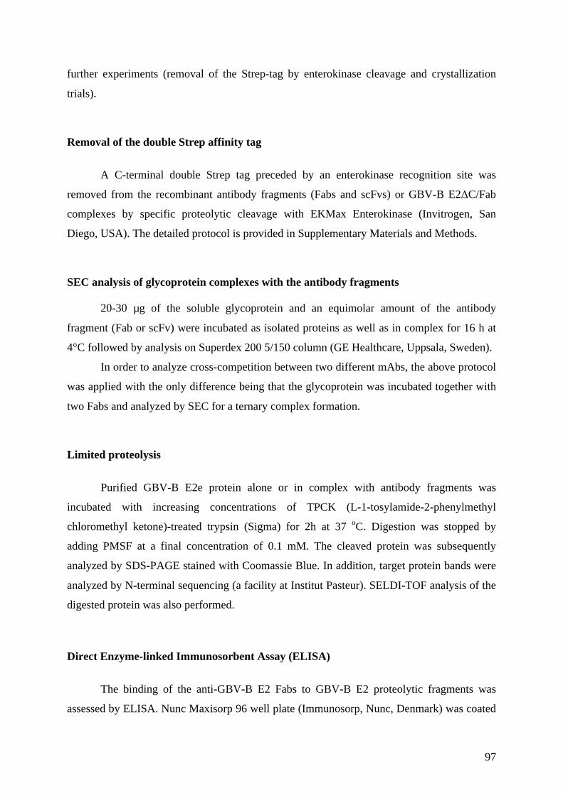
97
further experiments (removal of the Strep-tag by enterokinase cleavage and crystallization
trials).
Removal of the double Strep affinity tag
A C-terminal double Strep tag preceded by an enterokinase recognition site was
removed from the recombinant antibody fragments (Fabs and scFvs) or GBV-B E2ΔC/Fab
complexes by specific proteolytic cleavage with EKMax Enterokinase (Invitrogen, San
Diego, USA). The detailed protocol is provided in Supplementary Materials and Methods.
SEC analysis of glycoprotein complexes with the antibody fragments
20-30 µg of the soluble glycoprotein and an equimolar amount of the antibody
fragment (Fab or scFv) were incubated as isolated proteins as well as in complex for 16 h at
4°C followed by analysis on Superdex 200 5/150 column (GE Healthcare, Uppsala, Sweden).
In order to analyze cross-competition between two different mAbs, the above protocol
was applied with the only difference being that the glycoprotein was incubated together with
two Fabs and analyzed by SEC for a ternary complex formation.
Limited proteolysis
Purified GBV-B E2e protein alone or in complex with antibody fragments was
incubated with increasing concentrations of TPCK (L-1-tosylamide-2-phenylmethyl
chloromethyl ketone)-treated trypsin (Sigma) for 2h at 37 oC. Digestion was stopped by
adding PMSF at a final concentration of 0.1 mM. The cleaved protein was subsequently
analyzed by SDS-PAGE stained with Coomassie Blue. In addition, target protein bands were
analyzed by N-terminal sequencing (a facility at Institut Pasteur). SELDI-TOF analysis of the
digested protein was also performed.
Direct Enzyme-linked Immunosorbent Assay (ELISA)
The binding of the anti-GBV-B E2 Fabs to GBV-B E2 proteolytic fragments was
assessed by ELISA. Nunc Maxisorp 96 well plate (Immunosorp, Nunc, Denmark) was coated

98
with 50 µl/well of 1 µg/ml glycoprotein in PBS over night at 4oC. Next day, 50 µl/well of
monoclonal antibodies serially diluted in 0.5% bovine gelatin+0.1% Tween in PBS were
added, and the plates were incubated at room temperature for 2 h. The dilutions of the
antibodies used in the assay were 50.00, 25.00, 12.50, 6.25, 3.13, 1.56 and 0.78 µg/ml. Non-
relevant monoclonal antibody (negative control) was also included on each plate. After
washing the plates 5× with PBS-Tween (0.05%), 50 µl of horseradish peroxidase conjugated
anti-mouse total IgG (0.4 mg/ml from Thermo Fisher Scientific) diluted 1/10000 in 0.5%
bovine gelatin+0.1% Tween in PBS was added to each well. Then the plates were incubated
for 1 h at 37oC. Subsequently, the plates were washed as described above. The bound
antibodies were detected by adding 100 µl/well of 3,3',5,5'-Tetramethylbenzidine
(TMB) Microwell Peroxidase Substrate System (KPL, Gaithersburg, MD) solution prepared
according to the instructions included with the product. The reaction was stopped after 4 min
with 100 µl 2M H2SO4. The absorbance at 450 nm was measured by VICTOR 1420
Multilabel Counter (Perkin Elmer,Waltham, MA).
Detection of free cysteine residues in the antibody fragments
Free cysteine residues in anti-E2 antibody fragments were detected using the DTNB-
thiols assay. The detailed protocol is provided in Supplementary materials and methods.
Crystallization
Crystallization screening, crystal optimization and crystal cryo-protection techniques
are in detailed described in Supplementary materials and methods

99
Table 3. Selected protein constructs and PCR primers.
Protein Construct Forward primer (5'to 3'direction)
Reverse primer (5'to 3'direction)
Construct generation
GBV-B E2350-482 (E2ΔC)
pT350/ E2350-482(E2ΔC)
TTCGAAGACGATGACGATAAGGCCGGTTG TTTGCTGCCGGGGTACTGCAGGATG Deletion using pT350/E1-E2350-564
GBV-B E2350-540 pT350/E2350-540 TTCGAAGACGATGACGATAAGGCCGGTTG GGAATTCACGGGGTCGTAGAAGTACACG Deletion using pT350/E1-E2350-564
GBV-B E2350-513 pT350/E2350-513 TTCGAAGACGATGACGATAAGGCCGGTTG CTGGGGCACCTGCAGCCAG Deletion using pT350/E1-E2350-564
GBV-B E2350-416 (domain A)
pT350/E2350-416
TTCGAAGACGATGACGATAAGGCCGGTTG CCTAGCCCACCTCTCCGGTGGTAGGAGAG Deletion using pT350/ E2350-581
GBV-B E2417-523 (domain B)
pT350/E2417-523 ATGGTTAAATTCAAAAATAACACATGGGG CCCGAGCGAGAGGCCAACAAAG Deletion using pT350/ E2350-523
GBV-B E2350-523 (domain AB)
pT350/E2350-523 TTCGAAGACGATGACGATAAGGCCGGTTG CCTAGCCCACCTCTCCGGTGGTAGGAGAG Deletion using pT350/ E2350-581
E2483-564 pT350/E2483-564 ATGGTTAAATTCAAAAATAACACATGGGG CCCGAGCGAGAGGCCAACAAAG Deletion using pT350/E1-E2350-564
F16.1_LC pCR™-Blunt/ F16.1_LC
CCAGTTCCGAGCTCGTGATGACA CAGTCTCCA
GCGCCGTCTAGAATTAACACTCATTCCTGTTGAA
Blunt-cloning
F16.1_HC pCR™-Blunt/ GAGGTTCAGCTC GAGCAGTCTGGAGC AGGCTTACTAGTACAATCCCTGGGCACAAT Blunt-

100
F16.1_HC cloning E19.4_LC pCR™-Blunt/
E19.4_LC CCAGTTCCGAGCTCGTGATGACA CAGTCTCCA
GCGCCGTCTAGAATTAACACTCATTCCTGTTGAA
Blunt-cloning
E19.4_HC pCR™-Blunt/ E19.4_HC
GAGGTCCAGCTCGAGCAGTCTGGACC AGGCTTACTAGTACAATCCCTGGGCACAAT Blunt-cloning
F7.7_LC pCR™-Blunt/ F7.7_LC
AAAAAGATCTGACATCGTGCTGACACAGTCTCCA
GCGCCGTCTAGAATTAACACTCATTCCTGTTGAA
Blunt-cloning
F7.7_HC pCR™-Blunt/ F7.7_HC
GAGGTCCAGCTCGAGCAGTCTGGACC AGGCTTACTAGTACAATCCCTGGGCACAAT Blunt-cloning
F24.3_LC pCR™-Blunt/ F24.3_LC
AAAAAAAGATCTGATATCCAGATGACACAGACTACWTCCTCC
GCGCCGTCTAGAATTAACACTCATTCCTGTTGAA
Blunt-cloning
F24.3_HC pCR™-Blunt/ F24.3_HC
GAGGTAAAGCTCGAGGAGTCTGGAGG AGGCTTACTAGTACAATCCCTGGGCACAAT Blunt-cloning
F16.1Fab pMT/F16.1Fab_LC AAAAAGATCTGAGCTCGTGATGACACAG AAAATCTAGATTAACACTCATTCCTGTTGAAGC
Restriction cloning (BglII-XbaI)
pMT/F16.1Fab_LC_HC
TTTTTTGCTAGCACCACAATCCCTGGGCACAATTTTCTTGTCCACCTTGGT
Restriction cloning (AvrII-NheI)
E19.4Fab
pMT/E19.4Fab_LC AAAAAGATCTGAGCTCGTGATGACACAG AAAATCTAGATTAACACTCATTCCTGTTGAAGC
Restriction cloning (BglII-XbaI)
pMT/E19.4Fab_LC_HC
AAAACCTAGGGGAGGTTCAGCTCGAGCAG TTTTTTGCTAGCACCACAATCCCTGGGCACAATTTTCTTGTCCACCTTGGT
Restriction cloning (AvrII-NheI)
F7.7Fab pMT/F7.7Fab_LC AAAAAGATCTGACATCGTGCTGACACAGTCTCCA
AAAATCTAGATTAACACTCATTCCTGTTGAAGC
Restriction cloning (BglII-XbaI)
pMT/F7.7Fab_LC_HC
TTTTTTGCTAGCACCACAATCCCTGGGCACAATTTTCTTGTCCACCTTGGT
Restriction cloning (AvrII-NheI)
F24.3Fab pMT/F24.3Fab_LC AAAAAAAGATCTGATATCCAGATGACACAGACTACWTCCTCC
AAAATCTAGATTAACACTCATTCCTGTTGAAGC
Restriction cloning (BglII-XbaI)
pMT/F24.3Fab_LC AAAACCTAGGGGAGGTAAAGCTCGAGGAG TTTTTTGCTAGCACCACAATCCCTGGGCACAA Restriction

101
_HC TTTTCTTGTCCACCTTGGT cloning (AvrII-NheI)
F16.1scFv pMT/F16scFv_LC AAAAAAGCTAGCGAGCTCGTGATGACACAGTCTCCAG
TTTTTTGCGGCCGCTTTGATTTCCAGCTTGGTGCCTC
Restriction cloning (NheI-NotI)
pMT/F16scFv_LC_HC
AAAAAACCATGGGCGAAGTTCAGCTCGAGCAGTCTGG
TTTTTTGGTACCTGAGGAGACTGTGAGAGTGGTGC
Restriction cloning (NcoI-KpnI)
E19.4scFv pMT/E19.4scFv_LC
AAAAAAGCTAGCGAGCTCGTGATGACACAGTCTCCAG
TTTTTTGCGGCCGCTTTGATTTCCAGCTTGGTGCCTC
Restriction cloning (NheI-NotI)
pMT/E19.4scFv_LC_HC
AAAATCATGAGCGAGGTTCAGCTCGAGCAGTCTG
TTTTTTGGTACCTGAGGAGACTGTGAGAGTGGTGC
Restriction cloning (BspHI-KpnI)
F7.7scFv_LC pMT/F7.7scFv_LC AAAAAAGCTAGCGACATCGTGCTGACACAGTCTCCAC
TTTTTTGCGGCCGCTTTGCTTTCCAGCTTGGTGCCTC
Restriction cloning (NheI-NotI)
pMT/F7.7scFv_LC_HC
AAAATCATGAGCGAGGTCCAGCTCGAGCAGTCT
TTTTTTGGTACCTGAGGAGACTGTGAGAGTGGTGC
Restriction cloning (BspHI-KpnI)
F24.3scFv pMT/F24.3scFv_LC
AAAAAAGCTAGCGATATCCAGATGACACAGACTACTTCCTCC
TTTTTTGCGGCCGCTTTGATTTCCAGCTTGGTGCCTC
Restriction cloning (NheI-NotI)
pMT/F24.3scFv_LC_HC
AAAAAACCATGGGCGAGGTAAAGCTCGAGGAGTCTGGAG
TTTTTTGGTACCTGAGGAGACTGTGAGAGTGGTGC
Restriction cloning (NcoI-KpnI)
F7.7Fab C_to_S_mut
GAGTGGATTGGATATATTAATTCTCACAGTGTTACTTCAACCTAC
GTAGGTTGAAGTAACACTGTGAGAATTAATATATCCAATCCACTC
Site-directed mutagenesis
Enzyme restriction sites and codons encoding mutated amino acids are color-coded: BglII site – yellow, XbaI site – bright green, AvrII site – magenta, NheI site – light blue, KpnI site – grey, BspHI site – red, NcoI codon – green. A codon encoding mutated cysteine to serine is underlined. Light chain (LC) and heavy chain (HC) sequences cloned to pCR™-Blunt were amplified using cDNA isolated from hybridomas expressing the corresponding monoclonal antibodies.

102
II. The Structure Of the Hepatitis C Virus Envelope
Glycoprotein E2 Antigenic Site 529-540 in Complex With
Antibody DAO5
Background
In the Introduction about HCV, I have described a number of crystal structures of
HCV anti-E2 antibodies in complex with its peptide epitopes. This led to structural
characterizations of two important epitopes (epitope I and epitope II) targeted by neutralizing
antibodies (Deng et al., 2014; Deng et al., 2013; Kong et al., 2012a; Krey et al., 2013; Pantua
et al., 2013; Potter et al., 2012). The third region (aa 523-540 following HCV polyprotein
numbering) is recognized almost exclusively by conformation sensitive antibodies that do not
bind peptides. Four residues within this region (G523, W529, G530 and D535) are recognized
by a number of conformation-dependent human monoclonal antibodies - as determined by
alanine-scanning mutagenesis - and are involved in CD81 binding (Owsianka et al., 2006). At
the time of this project, structural characterization of this third region had not been reported.
Together with our collaborators from MRC-University of Glasgow Centre for Virus Research
(Arvind Patel and Ania Owsianka) we obtained a murine antibody DAO5 recognizing a linear
epitope within aa 523-540 of HCV E2. To gain insight into this important, uncharacterized
region of E2, we applied a strategy of co-crystallization of DAO5 antibody in complex with
its peptide epitope.
Objective
The objective of this project was to determine the crystal structures of antibody
fragments (scFv and Fab) from the murine antibody DAO5 in complex with its peptide
epitope corresponding to HCV E2 aa 529-540 derived from two different HCV genotypes. In
addition, we aimed to prove that this antibody recognizes the glycoprotein E2 in its native

103
conformation as present in the virion. The study was intended to provide a first glimpse into
this important region of E2 for virus neutralization and vaccine design efforts.
Results
Expression and purification of DAO5 scFv and DAO5 Fab
Monoclonal antibody (mAb) DAO5 was generated and its epitope characterized by
our collaborators in Glasgow (Arvind Patel and Ania Owsianka). In order to raise broadly
neutralizing antibodies against HCV E2, mice were immunized and boosted with a series of
recombinant soluble HCV E2 derived from different genotypes produced in our laboratory.
The epitope mapping studies revealed that DAO5 mAb binds specifically to E2 residues 533-
ETDVMLLN-540 with residue D535 being an essential contact residue for DAO5 mAb. The
main features of DAO5 mAb as a result of the characterization by Arvind Patel and Ania
Owsianka are summarized in Table 1.
Table 4. Summary of DAO5 mAb characterization.
Feature Result Method Epitope 533-ETDVMLLN-540 Phage display+ELISA Essential residues for binding D535 Alanine scanning mutagenesis
of HCVcc Binding to the genotypes 2a, 2b, 3a, 4 ELISA Competition analysis Does not cross-compete with
other mAbs binding to D535 ELISA
Neutralization no HCVpp, HCVcc Binding to viral particles Initial results indicate that it
does bind but it needs to be confirmed
Immunopreciptation with HCVcc and HCVpp
Conformational mAb no Western blot, phage display of peptides
For crystallization studies with the peptides corresponding to DAO5 mAb epitope, we
produced the recombinant DAO5 scFv and DAO5 Fab in Drosophila melanogaster S2 cells.
Representative protein elution profiles from SEC are shown in Figure 30. DAO5 Fab eluted
as a single peak corresponding to a monomeric Fab. Although the majority of the DAO5 scFv
eluted from a SEC column at a volume (225-260 ml) corresponding to monomer, some extra
peaks corresponding to dimeric and higher oligomeric scFv could be observed. In order to get
homogenous monomeric DAO5 scFv for crystallization trials, the fractions from the peak
corresponding to the monomeric scFv were pooled, concentrated and subjected to the second

104
SEC. As judged from the SEC profile, the second SEC resulted in a clear separation of
monomeric scFv from other oligomeric species. The final yield of the DAO5 Fab and DAO5
scFv was ~21 and ~8.5 mg per liter of culture supernatant.
Figure 30. Size-exclusion chromatography (SEC) of DAO5 Fab (A) and DAO5 scFv (B and C). Separation by SEC was performed using HiLoad 26/60 Superdex 200 column (GE Healthcare Life Sciences) at a flow speed of 2 ml/min in 10 mM Tris-HCl pH 8.0, 150 mM NaCl. Protein elution was monitored by absorbance at 280 nm (blue curves). Chromatogram A reveals a single major peak for DAO5 Fab corresponding to a monomeric Fab (~50 kD). For DAO5 scFv, the first separation by SEC (B) resulted in a major peak corresponding to a monomeric scFv (~26 kD) and some additional overlapping peaks corresponding to dimeric and higher oligomeric scFvs. Fractions corresponding to the monomeric scFv were pooled, concentrated and then analyzed again by SEC. DAO5 scFv eluted as a single major peak (C) corresponding to a monomeric scFv, which

105
indicated a successful separation of monomeric scFv from higher oligomeric state species, by removing the fractions to the left (smaller volumes) of the peak.
Crystallization and structure determination of DAO5 scFv and DAO5 Fab in complex with the epitope HCV E2529-540 peptides
Two peptides spanning the residues 529-540 from HCV genotype 1b isolate J4 and 2a
isolate JFH-1 (referred to as peptide_J4 and peptide_JFH, respectively) were selected for
crystallization with DAO5 antibody fragments. The sequences of the peptides are provided in
Table 5.
Table 5. The sequences of the peptides selected for crystallization studies with DAO5 antibody fragments. The peptides span the residues 529-540 from HCV genotype 1b isolate J4 (peptide_JFH) and 2a genotype isolate JFH-1 (peptide_JFH) and differ by one amino acid at the position 537 (colored in red).
Peptide Sequence Peptide_JFH WGENETDVFLLN Peptide_J4 WGENETDVMLLN
The HCV E2 residues 529-540 are highly conserved among different HCV genotypes
(Table 6). Therefore, the selected peptides differ only by one amino acid at the position 537
(M in the peptide_J4 and F in the peptide_JFH-1). The rationale to use these two peptides was
that they represent the DAO5 epitope from the two most phylogenetically distant HCV
genotypes.
For crystallization trials with DAO5 antibody fragments, we chose peptides longer by
4 residues at the N-terminal end (i.e. the peptides spanning aa 529-540) than the central
DAO5 mAb epitope (HCV E2 residues 533-540) because the residues W529 and G530
belong to the amino acids that are conserved across all genotypes and are critical for CD81
binding. We were hoping that we could probably see the electron density for those residues in
the crystal structures, which would give more information about this important HCV E2
region.
Table 6. The sequences of HCV E2529-540 from six major HCV genotypes. The amino acid residues that have been shown to be critical for CD81 binding are displayed in bold. Gt: genotype. JFH 2a: GenBank accession number AB047645. H77 1a: GenBank accession number NC_004102. Gt 1b (J4): GenBank accession number AF054259. Gt 3a: GenBank accession number D28917. Gt 4a: GenBank accession number DQ41878.Gt 5a: GenBank accession number Y13184. Gt 6: GenBank accession number AY859526. The residues critical for CD81 binding (Owsianka et al., 2006)are colored by red. The residues that are variable among six major HCV genotypes are colored in grey.
HCV genotype Sequence of HCV E2529-540 JFH 2a WGENETDVFLLN H77 1a WGANDTDVFVLN Gt 1b (J4) WGENETDVMLLN

106
Gt 3a WGANKTDVFLLE Gt 4a WGENESDVFLLN Gt 5a WGSNETDILLLN Gt6 WGENETDVFMLE
Crystallization of DAO5 scFv in complex with the epitope HCV E2529-540 peptides
First, we tried to co-crystallize DAO5 scFv with the peptide_J4 and peptide_JFH.
However, no crystals of DAO5 scFv in complex with the peptides were obtained. Therefore,
we tried to induce the crystallization of DAO5 scFv-peptide complexes by microseed matrix
screening using unliganded DAO5 scFv crystals as a source of the microseed stock.
Unfortunately, this microseeding technique did not promote the crystallization of DAO5
scFv-peptide complexes.
An alternative strategy to obtain crystals of protein–ligand complexes is soaking
ligands into protein crystals. DAO5 scFv was previously crystallized (space group P41 21 2)
and its 3D structure at 2.05 Å determined by Thomas Krey (unpublished data). These crystals
allowed soaking because the analysis of crystal lattice contacts indicated that the antigen-
binding site is accessible and might be able to accommodate the peptide. Moreover, the
crystals had a high solvent content, which is favorable for peptide diffusion. To obtain a
number of diffraction quality crystals for soaking experiments, the crystallization conditions
were further refined in 24 well plates. DAO5 scFv crystals were grown by hanging-drop
vapor diffusion method in drops containing 1 µl DAO5 scFv (9 mg/ml) and 1 µl of reservoir
solution composed of 100 mM MES pH 6.5, 26-29% PEG 400 and 20-150 mM sodium
acetate. Long rod-shaped crystals formed in all conditions during three days (Figure 31).
DAO5 scFv crystals were transferred to the mother liquor supplemented with 0.1 mM peptide
for 5 h (with peptide_J4) or overnight (with peptide_JFH). DAO5 scFv crystals were stable in
the soaking solutions containing the peptides and no evident changes in DAO5 scFv crystal
morphology were observed (determined by visual inspection) even after overnight soaking.
Since the cryoprotectant was present in a soaking solution (26-29% PEG 400), soaked DAO5
scFv were directly flash-frozen in liquid nitrogen. Diffraction data sets at 2.0 and 1.9 Å were
collected for DAO5 scFv crystals soaked in peptide_J4 and peptide_JFH solutions,
respectively. (Figure 32 and Figure 33).

107
Figure 31. DAO5 scFv crystals used in soaking with the peptides.
Figure 32. (A) X-ray diffraction image of the DAO5 scFv crystal soaked in peptide_J4 solution. (B) High resolution reflections of the same diffraction image and corresponding resolution.

108
Figure 33. (A) X-ray diffraction image of the DAO5 scFv crystal soaked in peptide_JFH solution. (B) High resolution reflections of the same diffraction image and corresponding resolution.
The structures of both complexes were determined by molecular replacement method
as described in detail in the section of Materials and Methods. Each crystallographic
asymmetric unit contained two DAO5 scFv-peptide complexes. The final electron density
map of the peptides allowed to build an atomic model of the peptide residues 530-540 and
532-540 for the first and the second copy of the peptide in the asymmetric unit, respectively
(Figure 34). The overview of the structures of the complexes is shown in Figure 35.
Crystallographic data collection and refinement statistics are summarized in Table 7.

109
Figure 34. Composite omit maps around the peptide_J4 (A) and the peptide_JFH (B) calculated after refinement of the crystal structures of DAO5 scFv in complex with the peptide J4, and the peptide JFH, respectively. The omit maps are shown as grey mesh contoured at 1 ! level. The density of the central "-helix is well defined for both peptides and allowed unambiguous placement of the peptide.

110
Figure 35. Crystal structures of DAO5 scFv in complex with peptide_J4 (A) and peptide_JFH (B). The crystal structures of the DAO5 scFv in complex with peptide_J4 and peptide _JFH were determined and refined to 2.0 and 1.7 Å resolution, respectively. The crystal structures are shown as cartoon. The light chain and heavy chain are colored in light grey and dark grey, respectively. The peptide is colored in yellow.
Crystallization of DAO5 Fab in complex with the epitope HCV E2529-540 peptides
It has been shown that the structure of a protein in complex with a ligand may differ
depending on whether it was obtained by soaking or co-crystallization experiments (Hiller et
al., 2006; Zhu et al., 1999) as the binding of a ligand to a protein in solution could be
different. Moreover, regardless of how the crystals of the complex were obtained, crystal-
packing contacts could influence the conformation of the ligand.
In order to exclude a putative influence of the crystalline environment on the peptide
conformation, I also performed co-crystallization trials for the DAO5 Fab in complex with the
J4 peptide. To form the DAO5 Fab/peptide complex, 6-fold molar excess of the peptide
dissolved in 20 mM Tris pH 9 was added to the protein solution (18 mg/ml of Fab+3 mg/ml
peptide). The complex was incubated overnight at 4°C. Crystal screening was performed
using commercially available crystallization screening kits as described in Section 2. A
cluster of needle-like crystals was obtained in condition 10% PEG8k 200 mM ZnAc, 100 mM
pH 6.5.
Since the quality of the crystals was not suitable for X-ray diffraction measurement,
the crystal growth condition was further optimized in 24-well plates by hanging-drop vapor
diffusion method by fine-tuning pH, salt and PEG concentration as well as protein

111
concentration. In addition, streak-seeding experiments were performed. Rod shaped crystals
were obtained after the streak-seeding above reservoir solutions composed of 8% PEG8k, 350
mM ZnAc, 100 mM pH 6.5 as well as 8% PEG8k, 350 mM ZnAc, 100 mM Tris pH 8. The
crystals from these two conditions were cryoprotected in 20% glycerol and subjected to X-ray
diffraction experiments on a home source, a Rigaku MicroMax-O7 copper anode generator
with a Mar345 image plate detector (a Platform for crystallography at the Pasteur Institute).
However, all tested crystals diffracted just to ~8 Å, which was not sufficient for the structure
determination.
To promote crystal growth, microseed matrix-screening was performed with the seed
stock prepared from the crystals obtained in condition 11% PEG 8k, 350 mM ZnAc, and 100
mM pH6.5. The crystals grew in ~100 different conditions (mostly PEG or salt based). The
conditions that yielded the morphologically best crystals were selected for refinement in 24
well plates. The diffraction-quality crystals obtained in conditions containing 10% PEG 20K,
100 mM MgCl2, Tris pH 8.5; 600 mM (NH4)2SO4, 100 mM MES pH 6 ; 600 mM LiSO4, 100
mM Tris pH 8.5, 50 mM NiCl2 were cryoprotected using reservoir solutions supplemented
with 25% glycerol and tested for X-ray diffraction at the synchrotron Soleil beamline Proxima
I and the Swiss Light Source beamline PXI. The crystals grown in condition 600 mM LiSO4,
100 mM Tris pH 8.5, 50 mM NiCl2 diffracted X-rays to ~3.7-4 Å, but the diffraction images
were difficult to index due to multiple crystal lattices.
All co-crystallization experiments of DAO5 Fab with peptide J4 defined above were
performed with DAO5 Fab containing a C-terminal double Strep tag. The affinity tags
sometimes interfere with crystal lattice formation. Therefore, the C-terminal double Strep tag,
preceded by an enterokinase recognition site, was removed from DAO5 Fab by specific
proteolytic cleavage with EKMax Enterokinase (Invitrogen, San Diego, USA). The amount of
enterokinase required to achieve complete removal of the tag was optimized in a small-scale
reaction (Section 2). The reaction was scaled up in a linear manner and the DAO5 Fab
without the Strep-tag was purified from the reaction mixture (Section 2). DAO5 Fab-peptide
J4 complex was formed and the crystallization screening performed as described previously.
Rod-shaped diffraction quality crystals appeared after 5 days in a drop containing 0.2 µl of the
complex (10 mg/ml of Fab+1.72 mg/ml peptide in 10 mM TRIS pH 8.0, 150 mM NaCl)
mixed with an equal amount of reservoir solution containing 20% PEG3350 and 200 mM
sodium thiocyanate (Figure 36) The crystals were flash-frozen in liquid nitrogen after
transferring them to a cryo-protective solution containing the mother liquor and 20% (v/v)

112
glycerol. A diffraction data set at 1.5 Å was collected from a single crystal on beamline
Proxima I at the synchrotron Soleil (Figure 37).
Figure 36. DAO5 Fab/peptide J4 crystal obtained by co-crystallizing the peptide J4 with the DAO5 Fab from which the Strep-tag was removed by specific proteolytic cleavage with enterokinase. The crystal was used to collect an X-ray diffraction data set at 1.5 Å on a beamline Proxima I at the synchrotron Soleil.
Figure 37. (A) X-ray diffraction image of the DAO5 Fab crystal co-crystallized with the peptide_J4. (B) High resolution reflections of the same diffraction image and corresponding resolution.
The DAO5 Fab-peptide J4 crystal belonged to space group P1 21 1 with unit cell
parameters a=48.678, b=80.717, c=54.565, α=90.00, β=95.64, γ=90.00 and contained one
complex per asymmetric unit. The Matthews’ coefficient (Vm) for the DAO5 Fab-peptide J4
crystals was estimated to be 2.18 Å3/Da (corresponding to a solvent content of 43.51%).
Usually more tightly packed crystals (containing lower solvent content) tend to diffract X-
rays to higher resolution (Kantardjieff & Rupp, 2003). This observation held true for DAO5

113
Fab-peptide and DAO5 scFv-peptide crystals (solvent content 43.51% and 63% accordingly)
with DAO5 Fab-peptide-J4 crystal diffracting X-rays better. The data was integrated and
scaled as described in Materials and Methods.
The resolution limit of 1.7 Å was chosen for the refinement of DAO5 Fab-peptide_J4
model. Though well-defined positive electron density for a peptide was already visible in
proximity of the CDRs after molecular replacement, the atomic model of the peptide was built
after the coordinates of the Fab were fully refined. The final electron density allowed
unambiguous fitting of the J4 peptide residues 532-540. The overview of the structure of the
complex is shown in Figure 38. Crystallographic data collection and refinement statistics are
summarized in Table 7.

114
Figure 38. Crystal structure of DAO5 Fab in complex with peptide_J4. The crystal structure of the DAO5 Fab in complex with peptide_J4 was determined and refined 1.5 Å resolution and is shown as cartoon. The light and heavy chains are colored in light and dark grey, respectively. The peptide is colored in yellow.

115
Table 7. Data collection and refinement statistics for the DAO5 scFv and DAO5 Fab crystals in complex with the indicated peptides. Values in parentheses represent those in the highest resolution bin.
DAO5 scFv-peptide J4
DAO5 scFv-peptide JFH
DAO5 Fab-peptide J4
Data collection Space group P41 21 2 P41 21 2 P21 Cell dimensions a, b, c (Å) 155.94 155.94 61.51 155.69 155.69 61.65 48.68 80.71 54.57 a, b, g (°) 90.00 90.00 90.00 90.00 90.00 90.00 90.00, 95.64 90.00 Resolution (Å) 49.31-2.00 (2.11-
2.00) 49.23-1.90 (2.00-1.90)
48.44-1.70 (1.79-1.70)
Solvent content (%) (molecules per asymmetric unit)
63 (2) 63 (2) 43.51 (1)
Rmerge Total number of observations Total number of unique reflections
0.107 (0.516) 298752 (18087)
50405 (6158)
0.137 (0.614) 261291 (37230)
58977 (8629)
0.04 (0.228) 255101 (36579)
44810 (6417)
I / sI 10.3 (1.9) 9.5 (3.2) 27.6 (6.8) Completeness (%) 97.6 (83.5) 98.5 (99.8) 97.1 (95.6) Redundancy 5.9 (2.9) 4.4 (4.3) 5.7 (5.7)
Refinement Resolution (Å) 20.16-2.00: (2.05-
2.00) 38.00-1.90 (1.95-1.90)
48.44-1.70 (1.74-1.70)
No. reflections 47116 58950 44787 Rwork / Rfree 0.1979/0.2176 0.1875/0.2039 0.1774/0.2032 No. atoms
Protein 3727 3728 3361 Water 269 326 329
B-factors Wilson B-factor (Å2) 28.69 19.77 19.53 Average B-factor (Å2) 28.67 20.84 20.02
R.m.s. deviations Bond lengths (Å) 0.010 0.010 0.010 Bond angles (°) 1.08 1.06 1.12
Ramachandran statistics# Favored (%) Number of outliers
96.62 0
97.03 0
97.9 1
# Ramachandran statistic according to Molprobity server.
The crystal packing interfaces in proximity of the peptide_J4 differ considerably in the
DAO5 Fab-peptide J4 and DAO5 scFv-peptide JFH crystals. DAO5 Fab-peptide J4 crystal
has denser packing in comparison with the DAO5 scFv-peptide crystals. As a result, the
peptide J4 environment in the DAO5 Fab-peptide crystal is crowded by symmetry-mates with
some of them packed close to the antigen-binding site (Figure 39).

116
Figure 39. Crystal packing of interfaces for DAO5 Fab and DAO5 scFv in complex with the peptide J4. One
molecule of the complex is shown as cartoon (with side chains displayed for the peptide J4). The light chain and
the heavy chain are colored in light blue and cyan, respectively, and the peptide is colored purple. Symmetry
mates are shown in light grey as C" traces. The peptide J4 has completely different packing interfaces in DAO5
scFv/peptide J4 and DAO5 Fab/peptide J4 crystals but adopts the same conformation, indicating that the crystal
packing does not have an effect on the peptide conformation.
Nevertheless, the peptide J4 conformation is very similar in both crystal forms as
indicated by the root mean square deviation (rmsd) values calculated between each pair of
aligned residues 532-540 (Figure 40). The main difference observed between the two peptide
structures is the different side chain conformations of residues M537 and N540. It is widely
accepted that if the same ligand conformation is found in multiple protein-ligand crystal
forms, the conformation is not likely to be induced by the crystalline environment and thus
represents a biologically relevant interaction. In conclusion, analysis of the crystal packing
interfaces revealed unrelated interfaces for Fab and scFv complexes respectively, indicating
that the J4 peptide reflects the conformation that is recognized by the functional monoclonal
antibody in the context of the native glycoprotein.
Crystal packing of DAO5 Fab/peptide J4 Space group: P 1 21 1
Crystal packing of DAO5 scFv/peptide J4Space group: P 41 21 2

117
Figure 40. Root mean square deviation (rmsd) calculated after superposition of peptide J4 in complex with scFv and Fab, respectively using Chimera including all atoms (dark grey) or main chain atoms only (light grey) in the calculation and represented per residue. The peptides were superposed using Superpose 1.0.
Comparison of the epitope HCV E2529-540 peptide structures
The crystallization of DAO5 scFv-peptide and DAO5 Fab peptide complexes yielded
in total five atomic models of the peptides (two copies of each scFv complex per asymmetric
unit and one copy of the DAO5 Fab in complex with the J4 peptide) that are listed in Table 8.
Table 8. Summary of the obtained crystal structures of the epitope HCV E2529-540 peptides.
Number
Peptide Crystal Modeled residues Average B factor for the peptide residues 532-540
1 Peptide J4 DAO5 scFv-peptide_J4 (asymmetric unit 1)
530-GENETDVMLLN-540 32.66
2 Peptide J4 DAO5 scFv-peptide_J4 (asymmetric unit 2)
532-NETDVMLLN-540 36.53
3 Peptide JFH DAO5 scFv-peptide_JFH (asymmetric unit 1)
530-GENETDVFLLN-540 23.38
4 Peptide JFH DAO5 scFv-peptide_JFH (asymmetric unit 2)
532-NETDVMLLN-540 26.66
5 Peptide J4 DAO5 Fab-peptide_J4
532-NETDVMLLN-540 25.46
Since the peptides differed in the number of modeled residues at the N-terminal end,
only residues that were present in all five peptide structures (aa 532-540) could be used for
comparison. Superposition of the segment 532-540 from all five available crystal structures of

118
the peptide epitope (two copies of each scFv complex per asymmetric unit and one copy of
the DAO5 Fab in complex with the J4 peptide) revealed an rmsd of 0.114 Å calculated over
the backbone atoms of the peptide, which confirms identical peptide conformations (Figure
41).
Figure 41. Superposition of five atomic models of the peptides (two copies of each scFv complex per asymmetric unit and one copy of the DAO5 Fab in complex with the J4 peptide) using Superpose 1.0.
The temperature factor (B-factor) analysis of the segment 532-540 of all five
independent peptide structures indicates that peptide J4 (Nr.5 in Table 8) of DAO5 Fab-
peptide crystal has the lowest average B-factor value. Also, it showed that the longer peptides
(Nr.1 and Nr.3) encompassing aa 530-540 in the first asymmetric unit of DAO5 scFv-peptide
crystals have the lower average B-factor values in comparison with peptides Nr.2 and Nr.4 in
the second asymmetric unit, implying that they are more ordered.
Peptide conformation and its binding to DAO5 scFv
We did not observe electron density for W529 in any of the crystal structures, which is
consistent with this residue not being part of the DAO5 mAb epitope, as shown by alanine

119
scanning mutagenesis of E2. The electron density for the residues N532-N540 was clearly
defined in all five complexes. The residues 530-531 are visible only in the electron density
map of the J4 peptide or the JFH-1 peptide complex with DAO5 scFv. Moreover, the electron
density for G530 and E531 in both peptides is present only in the same more ordered
counterpart of the scFv-peptide complex in the asymmetric unit. All together, it suggests that
the N-terminal residues G530 and E531 are highly flexible and most likely stabilized by
crystal packing of scFv molecules. Thus we conclude that the interactions they form with
DAO5 scFv in the crystal structures are nonspecific and do not participate in the natural
interactions of DAO5 mAb with its epitope. Therefore, they will not be discussed further in
this study. The peptide forms one α-helical turn comprising the residues 535-539 (535-
DVM/FLL-539) which is stabilized by canonical intramolecular hydrogen bonds of an α-helix
(Table S2). At the N-terminus, the residues 533-534 make a hydrogen bonded turn, and the
chain continues as an extended loop (residues G530-N532).
The J4 and JFH-1 peptide interaction with the paratope buries an area of 734 Å2 and
723 Å2 of the peptide and 645 Å2 and 656.2 Å2 of the antibody, respectively. For both
peptides, the total buried surface area amounts to 1379 Å2 in the interface, which is very close
to the interface (1680±260 Å2) described for other antibody-antigen complexes (Lo Conte et
al., 1999). The shape complementarity index is 0.810 and 0.771, respectively, for the J4 and
JFH-1 peptides, similar to indices reported for other antibody-peptide complexes (Lawrence
& Colman, 1993). In both structures, the peptides bind to a cleft between the variable regions
of the heavy chain (HC) and the light chain (LC) and interact with all six CDR loops of the
antibody.
As illustrated by the analysis of surface hydrophobicity of epitope and paratope, the α-
helical turn of the peptide establishes mainly hydrophobic interactions with the paratope
(Figure 42). The hydrophobic residues M/F537, L538 and L539 within the short α-helical
turn insert deeply into a hydrophobic groove formed by aromatic and aliphatic residues within
the complementarity determining region 3 of the heavy chain (CDR-H3) (F99, Y103, P104
and Y105), CDR-H2 (A59) and all CDR loops of the LC (Y32, Y50, W94 and L96) (Table
S1). Calculation of the solvent-accessible surface area reveals that more than 94% of the
solvent-accessible surface area of the residues 537M/F-539L is buried in this hydrophobic
groove (Figure 14). Moreover, the side chain of K52 within the CDR-H2 loop forms an
extensive hydrogen-bonding network with the main chain carbonyl groups of residues V536,
M/F537 and N540, which further stabilizes the interaction of the α-helical turn with the heavy
chain.

120
Figure 42. Interaction between DAO5 scFv and the epitope peptide_JFH. DAO5 scFv (A and B) is shown as molecular surface. The peptide (A and B) is shown as a cartoon with side chains displayed as sticks and colored by atom-type (green, red and blue for carbon, oxygen and nitrogen, respectively). (A) The molecular surface of DAO5 scFv and the epitope peptide are colored according to a normalized hydrophobicity scale from white (hydrophobic) to bright yellow (hydrophilic). (B) Electrostatic potential [&5 kT/e (red) to 5 kT/e (blue)] across the molecular surface of DAO5 scFv and the peptide epitope calculated using the adaptive Poisson-Boltzmann solver. The molecular surface of the peptide in A and B is shown looking from the paratope.
In the case of JFH-1 peptide, in which M537 is replaced by F537, the side chain of
K52 is also involved in a cation-' interaction with F537. Cation–' interaction is a
predominantly electrostatic interaction between the face of an electron-rich ' –system (e.g.
aromatic amino acid side chain) and an adjacent cation (e.g. protonated arginine or lysine side
chain), which has been discovered to play an important stabilizing role in protein-protein

121
interaction (Gallivan & Dougherty, 1999). In addition, F537 is engaged in aromatic stacking
interaction with W94 within CDR-H3. F537 inserts in the same hydrophobic pocket as M537
with 90.2% of solvent-accessible surface area buried (Figure 43). M537 has a different side
chain conformation in the complex with DAO5 scFv and DAO5 Fab, suggesting a higher
structural flexibility within this pocket than the bulkier F537. This flexibility is further
supported by the higher B-factor for the M537 side chain compared to other residues within
the "-helical turn. Also, it is known that methionine residues have very often several
conformations, even when they are buried in the hydrophobic core of a globular protein,
whereas phenylalanines do not have such features.
Heavy chain and light chain contribute differently to the binding of the peptide
segment V536-N540 into the hydrophobic pocket, burying ~351 Å2 and ~100 Å2 solvent-
accessible surfaces of these residues, respectively. This confirms that the heavy chain
dominates the interaction between the "-helical turn and the hydrophobic antigen-binding
groove.
Figure 43. Percentages of accessible surface area (ASA) of peptide J4 (A) and peptide JFH (B) buried in the complex with DAO5 scFv, calculated using PISA and represented per residue as stacked columns for heavy (dark grey) and light (light grey) chains of DAO5 scFv.
The N-terminal peptide residues N532-D535 make contacts exclusively with the light
chain. The contact surface here is more planar, which results in a smaller buried surface area
on the peptide (~200 Å2). Both side chain carboxyls of D535 form hydrogen bonds with the
hydroxyl group of Y52, consistent with the results of E2 alanine-scanning mutagenesis
identifying D535 as an essential contact residue for binding of the DAO5 mAb. Since the
hydrophobic V536 side chain is completely exposed and not involved in any antibody
interactions, D535 is likely required to stabilize the interaction of the solvent-exposed part of
the "-helical turn with the paratope. Moreover, its position in the junction between N-terminal

122
and C-terminal portions of the peptide suggests that the interaction of D535 with Y52 anchors
the epitope in the binding position.
The other residues in the N-terminal stretch N532-D535 also form hydrogen bonds to
the light chain. E533 establishes a salt bridge with K93. In the J4 peptide complex with
DAO5 scFv, it also forms two main-side chain hydrogen bonds with W94. In the J4 peptide
co-crystal structure, an extra hydrogen bond between N532 and N92 is observed. T534 is the
only residue within N532-D535 peptide portion fixed in a deeper pocket of the antigen-
binding site. Within this pocket, the side chain of T534 is stabilized by a hydrogen bond with
the carbonyl oxygen of T91. Upon antibody binding, 99% of its solvent-accessible surface
area is buried in the interaction interface with the light chain (Figure 43). T534 substitution to
S534 most likely does not disrupt this hydrogen-bonding pattern: E2 of HCV genotype 4
possesses this substitution and is still recognized by mAb DAO5.
The B-factor analysis indicates a stable and strong interaction with the paratope
(Figure 43). In contrast, the residues at the termini of the peptide (N532, E533 and N540)
have higher B-factors compared to all other peptide residues. The root mean square deviation
(rmsd) between the two peptides is also higher at the N- and C-termini (Figure 45). At least
for residues N532 and N540, an increased flexibility can be expected since they are normally
glycosylated in native E2. The modeling of the hypothetical glycan chains attached to N532
and N540 reveals that the helical peptide conformation is compatible with the N-linked
glycosylation of those residues in the native glycoprotein (Figure 46).

123
Figure 44. Peptide J4 and peptide JFH colored by temperature factor (B-factor) per atom according to a scale from blue (low B-factor) to red (high B-factor). The peptide (A and B) is shown as cartoon with side chains as sticks. DAO5 scFv is shown as molecular surface with the light chain and heavy chain colored in light grey and dark grey, respectively. Average temperature factors of peptide J4 (C) and peptide JFH (D) in complex with DAO5 scFv (light grey) calculated per residue including all atoms (dark grey) or main chain atoms only (light grey).

124
Figure 45. Root mean square deviation (rmsd) upon superposition of the peptide J4 and peptide JFH in complex with DAO5 scFv calculated using Chimera including all atoms (dark grey) or main chain atoms only (light grey) in the calculation and represented per residue. The peptides were superposed using Superpose 1.0.
Figure 46. Compatibility of the peptide_J4 conformation in complex with DAO5 scFv with N-linked glycosylation. Hypothetical glycan chains containing two N-acetylglucosamin moieties (light blue) are modeled to be attached to ND2 atoms of N532 and N540. The peptide is displayed as cartoon and colored in orange. The side chains of N532 and N540 are displayed as sticks and ND2 atoms, to which the glycans are linked, and OD1 atoms are colored in blue and red, respectively.
The stark difference between conformations of aa 532-540 in the E2 core fragment and DAO5/peptide crystal structures
The conformation of residues 532-540 in the JFH-1 peptide structure was compared to
the one of the corresponding peptide in E2 crystal structure in complex with the Fab derived

125
from neutralizing antibody AR3C (PDB 4MWF) (Kong et al., 2013). AR3C Fab also requires
D535 for binding to E2 (Law et al., 2008). In the E2 structure published by Kong and
colleagues, amino acid residues 532-540 adopts a completely different extended conformation
(with aa 536-538 forming a $-strand) than the equivalent peptide in complex with DAO5 scFv
(with aa 535-539 forming an "-helix) (Figure 47). The extended conformation of the peptide
in the context of the E2 core fragment spans 20.6 Å while the helical conformation observed
in the epitope peptide in complex with DAO5 scFv spans only 11.6 Å, respectively. T534,
F537 and L539 that inserts deeply in the antigen-binding groove of DAO5 are completely
buried in E2 core meaning that these residues would not be accessible for DAO5 binding.
Thus, a change of conformation of this E2 core region would be required in order for DAO5
to bind to the protein. Intriguingly, a similar situation has been observed for the epitope II as
the binding of mAbs #8 and #12 to E2 core is also impossible unless there is a change of
conformation of the epitope from the closed to the open conformation (see Introduction)
(Deng et al., 2014).
Figure 47. The conformation of residues 532-540 in the JFH-1 peptide structure and the corresponding peptide in the context of an E2 core fragment (PDB 4MWF). (A and C) Epitope aa 532-540 conformation in E2 core fragment. Note that F537 and L539 residues are buried in E2 crystal structure indicating that it must become exposed in E2 in order for DAO5 mAb to bind. (B) Epitope aa 532-540 (peptide_JFH) conformation observed in DAO5 scFv/peptide_JFH structure. All side chain residues of the epitope aa 530-540 are displayed as sticks and colored by atom-type (orange and red for oxygen and nitrogen, respectively, carbon atoms are ramp-colored from N-terminus (blue) to C-terminus (red) through green. E2 core structure (except aa 530-540) is depicted as cartoon and colored in grey.

126
Two populations of sE2!HVR1 can be distinguished by DAO5 and e137 antibody fragments
Due to observed discrepancy of the conformation of the epitope spanning aa 532-540
in E2 core/AR3C Fab and DAO5 scFv/peptide_JFH complexes, I decided to verify if DAO5
scFv is able to bind to the E2 core. For these experiments, I selected the recombinant soluble
E2(HVR1 (sE2(HVR1) expressed in S2 cells. The recombinant sE2(HVR1 was chosen
because it lacks HVR1 region but comprises the entire E2 core and, thus, is similar to the
construct crystallized by Kong et al. The protein elutes as a single monomeric peak in SEC
(Figure 48). The correct folding of sE2(HVR1 was verified by binding of the Fabs derived
from conformation-sensitive mAbs (described later). Also, it has been previously described to
induce broadly neutralizing antibodies after immunization, which is a strong indication that
the protein is correctly folded (Tarr et al., 2013).
Figure 48. Size-exclusion chromatography (SEC) of sE2(HVR1. Separation by SEC was performed using HiLoad 26/60 Superdex 200 column (GE Healthcare Life Sciences) at a flow speed of 1 ml/min in 10 mM Tris-HCl pH 8.0, 150 mM NaCl. Protein elution was monitored by absorbance at 280 nm (blue curves). The chromatogram reveals a single major peak for sE2(HVR1, corresponding to a monomeric protein (~39 kD not counting the sugar chains).
Binding of DAO5 scFv to sE2(HVR1 was tested in a pull-down assay. sE2(HVR1
was bound to a Streptactin mini column and then an equimolar amount of DAO5 scFv lacking
the Strep-tag was added. The eluted fraction was analyzed by SDS-PAGE under non-reducing
conditions (Figure 49). Two bands, one representing sE2(HVR1 and another one DAO5
scFv were observed. However, the DAO5 scFv band was much weaker than sE2(HVR1

127
indicating that only a small fraction of added DAO5 scFv was bound to sE2(HVR1. Thus,
the interaction between sE2(HVR1and DAO5 scFv in pull-down assay was not
stoichiometric suggesting that a fraction of sE2(HVR1 might adopt the conformation not
compatible with binding of DAO5 scFv.
Figure 49. Pull-down experiment showing that sE2(HVR1 specifically reacts with DAO5 scFv. sE2(HVR1 was affinity loaded onto a Streptactin column, DAO5 scFv lacking the tag was passed through the column and the proteins were eluted from the column after washing. Elution fraction was analyzed by SDS-PAGE under non-reducing conditions (lane 4) and stained by Coomassie Blue. The bands corresponding to both the sE2(HVR1 and DAO5 scFv were observed in the elution fraction indicating DAO5 scFv binding to sE2(HVR1. Lane 1: Page Ruler Prestained Marker. Lane 2: sE2(HVR1. Lane 3: empty, lane 4: elution fraction from the column.
These results together with the observed discrepancy of the conformation of the
epitope spanning aa 532-540 in E2 core/AR3C Fab and DAO5 scFv/peptide_JFH complexes
provided some implications that this epitope can exist in different conformations in the E2
glycoprotein. To further explore this interpretation, I chose to repeat the above-described
experiment including a Fab fragment which potentially recognizes a different conformation of
aa 532-540 than DAO5 scFv.
As we did not have AR3C Fab, we selected a neutralizing conformation-sensitive Fab
E2+DAO5 scFv
E2
55
130
35
25
15
130130100
70
250
DAO5 scFv DAO5 scFv
E2

128
e137 (Perotti et al., 2008) as a substitute. Alanine scanning mutagenesis of E2 has previously
indicated that both Fabs use similar residues including D535 for binding to E2 (Law et al.,
2008) and as a result should recognize closely related epitopes. Since both DAO5 scFv and
e137 Fab use D535 as an essential binding residue, they should cross-compete with each other
unless they recognize distinct conformation of aa 532-540.
Intriguingly, simultaneous binding of both DAO5 scFv and e137 Fab to sE2ΔHVR1
could be detected in a pull-down assay on the Streptactin affinity column (Figure 50A). In
this assay sE2ΔHVR1 was affinity bound to the column. DAO5 scFv and e137 Fab (both
lacking the Strep-tags) were loaded on the column in separate steps (DAO5 scFv in the first
step and e137 in the second step including a washing step in between). The molar excess of
both DAO5 scFv and e137 Fab were used to make sure that they saturate all available binding
sites in sE2ΔHVR1. Surprisingly, the eluted fractions contained all three proteins, meaning
that both DAO5 scFv and e137 were able to bind to sE2ΔHVR1 despite the fact that they both
should cross-compete for binding to D535. Thus, DAO5 scFv was not able to recognize all
sE2ΔHVR1 molecules when it was passed through the column, indicating that its epitope was
occluded or present in a different conformation in some sE2ΔHVR1 molecules at a given
moment. However, the binding of Fab e137 demonstrated that the population of molecules
that was not bound by scFv DAO5 was available for binding by Fab e137, suggesting that
DAO5 and e137 mAbs should recognize distinct conformations of the same epitope in
sE2ΔHVR1.
I also repeated the pull-down experiment by replacing the order in which scFv DAO5
and Fab e137 were loaded on the column, i.e. loading Fab e137 in the first step followed by
scFv DAO5. Again, the eluted fraction contained all three proteins, indicating that Fab e137
also binds only to the fraction of the molecules that presents its epitope in a certain
conformation at a given moment.
This observation might be related with the conformational flexibility of the region
spanning aa 532-540: at a given time this epitope is present in a few or more distinct
conformations, thus, there is a fraction of sE2ΔHVR1 molecules in the conformation
recognized by DAO5 mAb and the fraction of the molecules in the conformation recognized
by mAb e137.
In order to confirm our hypothesis that the epitope spanning aa 532-540 is flexible, we
also performed a similar experiment to the above-described pull-down using surface plasmon
resonance (SPR) (Figure 50B). sE2ΔHVR1 was immobilized on the chip via the Strep-tag
and then Fab DAO5, e137 or a control Fab (after removing the Strep-tag) was injected over

129
the glycoprotein followed by the second Fab. In all cases, the second Fab was injected when a
saturation of all accessible binding sites on sE2ΔHVR1 was reached with the first injected
Fab. Similar to the pull-down assay, binding of both Fabs DAO5 and e137 was observed. Of
note, the binding of both of these Fabs together corresponded to ~ 400 response units while
~800 response units of sE2ΔHVR1 were immobilized non-covalently via the Strep-tag.
Therefore, a much higher response would be expected if 100% of immobilized sE2ΔHVR1
were recognized when both Fabs are injected. Non-covalent immobilization via the Strep-tag
should neither affect the conformation of the glycoprotein nor sterically interfere with the
accessibility of the Fabs to the epitope spanning aa 532-540. Therefore, the lower than
expected response might indicate that due to flexibility of the epitope, a fraction of
sE2ΔHVR1 molecules had the conformation of the epitope recognized neither by Fab DAO5
nor by Fab e137.
Figure 50. Two populations of sE2ΔHVR1 can be distinguished by Fabs DAO5 and e137. (A) A pull-down experiment similar to Figure 5A, immobilizing sE2ΔHVR1 on a Streptactin column followed by a molar excess of DAO5 scFv and subsequently by e137 Fab. Elution of the complex revealed binding of both antibody fragments to immobilized sE2ΔHVR1. Given that no ternary complex containing sE2ΔHVR1/DAO5/e137 was observed (see Figure 5C), this suggests the presence of two populations of E2 molecules - one recognized by DAO5 and the second one recognized by e137. (B) Real-time SPR analysis of Fab binding to immobilized sE2ΔHVR1 recording the binding response in resonance units (RU) as a function of time. Fabs lacking the Strep-tag (DAO5, e137 or a control Fab) were injected over HCV sE2ΔHVR1 immobilized using an anti-Strep-tag antibody at a flow rate of 5 µL/ml. After saturation, a second Fab lacking the Strep-tag (DAO5, e137 or a control Fab) was injected. The association/dissociation time-course profiles support the presence of two distinct populations recognized by either DAO5 or e137.

130
DAO5 antibody fragments interact with their cognate antigen in a temperature dependent manner
I also further analyzed the complex formation between DAO5 Fab and sE2ΔHVR1 by
SEC. DAO5 Fab and sE2ΔHVR1were mixed at equimolar ratio and incubated for 18-24 h at
4°C followed by analysis on a Superdex 200 10/300 column (column volume 24 ml, GE
Healthcare). The complex of sE2ΔHVR1/DAO5 Fab eluted considerably earlier than the
respective individual proteins (Figure 51A). However, a significant amount of sE2ΔHVR1
and DAO5 Fab did not form a complex and eluted at volumes corresponding to the molecular
mass of the individual proteins, which was in agreement with the results obtained in the pull-
down assay with DAO5 scFv. To rule out the possibility that sE2ΔHVR1 and DAO5 Fab
were not mixed at exactly equimolar ratio due to pipetting errors, the experiment was repeated
a second time using exactly the same amounts of the proteins. The complex formation this
time was carried out at room temperature over night. Interestingly, SEC analysis revealed that
there was less free sE2ΔHVR1 and DAO5 Fab in the mixture after the complex formation at
room temperature (Figure 51B).
Detection of free sE2ΔHVR1 and DAO5 Fab beside the complex in SEC could be
related with relatively low affinity between the two proteins. Complex stability is known to
depend on the affinity between two proteins. For lower affinity interactions, a complex can
dissociate on the column since it is diluted during SEC. In such a case, a complex reaches
equilibrium with individual proteins on the column. If the eluted complex is concentrated and
analyzed by SEC the second time, the equilibrium is established again, i.e. the fraction of the
complex dissociates to individual proteins. To assess the stability of sE2ΔHVR1/DAO5 Fab
complex, I pooled the fractions containing the complex, concentrated it, and loaded it on the
column. Only a negligible amount of free sE2ΔHVR1 and DAO5 Fab were eluted, indicating
that the complex is stable once it is formed (Figure 51C and D).

131
Figure 51. Stoichiometric complex formation between HCV sE2(HVR1 and DAO5 Fab. sE2(HVR1, DAO5 Fab and a mixture of the two (molar ratio 1:1) were loaded to the column (in three different runs) (E2 (HVR1:39 kD, DAO5 Fab:51 kD, complex: 90 kD). The complex was formed by incubating mixed proteins at 4°C over night (A) or room temperature over night (B). Peak fractions of the eluted complex from (B) were concentrated and loaded on the column again (C). The peak fraction of sE2(HVR1/DAO5 Fab was injected on SEC column. No significant peaks corresponding to either of the isolated proteins were observed in the profile of the purified complex, indicating that the complex of sE2(HVR1/DAO5 Fab is stable once it is formed. (D) The fraction of E2 (HVR1 in complex with the respective Fab was calculated by integrating areas under the curves in A, B and C using UNICORN control software (GE Healthcare).
The initial analysis of complex formation by SEC already revealed that the incubation
temperature might have an effect on the DAO5 Fab binding to sE2(HVR1. Therefore, I
performed a more detailed investigation of the temperature effect on the interaction. The
formation of sE2(HVR1/DAO5Fab complex was carried out at four different temperatures
(4°C, 18°C, 30°C and 37°C) overnight and subsequently analyzed by SEC on a Superdex 200
10/30 column (GE Healthcare). Clear temperature-dependence of the sE2(HVR1/DAO5 Fab
complex formation was observed (Figure 51A). The calculation of the fraction of sE2(HVR1
in complex with DAO5 was calculated based on the area under the curves (Figure 51B)).
Approximately 90% of sE2(HVR1were in complex with DAO5 Fab if the proteins were pre-
incubated at 30°C or 37°C, while at 4°C the fraction of the sE2(HVR1in complex was less
than 60%.

132
Figure 52. Complex formation between HCV E2 ΔHVR1 and Fab fragments derived from DAO5 and e137 mAbs, respectively, at different temperatures. (A) The mixture of E2 ΔHVR1 and the respective Fab fragment (molar ratio 1:1) was incubating at 4°C, 18 °C, 30°C and 37°C, respectively, overnight. E2 ΔHVR1, the Fab and a mixture of the two were loaded to the column (in different runs) (sE2ΔHVR1: 39 kD, Fab: 51 kD, complex: 90 kD). (B and D) The fraction of E2 ΔHVR1 in complex with the respective Fab was calculated based on the area under the curve using UNICORN control software (GE Healthcare). The columns are colored according to the curves in (A and C): E2/ Fab complex formed at 4°C (red), E2/DAO5 Fab complex formed at 18°C (orange), E2/DAO5 Fab complex formed at 30°C (dark red), E2/DAO5 Fab complex formed at 37°C (grey).
I also used a conformation-sensitive e137 Fab as a control to check if the temperature
dependence observed for DAO5 Fab binding to sE2ΔHVR1 is not due to partial denaturation
of the glycoprotein. In addition to residues within epitope I and epitope II, e137 mAb interacts
with residues within the aa 523-540 segment. D535 has been also characterized as an
essential binding residue for this mAb. As DAO5 Fab recognizes a linear epitope, the
observed temperature dependence could be due to the fact that DAO5 epitope gets more
exposed as the sE2ΔHVR1 becomes partially denatured during the overnight incubation at

133
higher temperatures. The complexes of sE2ΔHVR1 with e137 Fab were formed and analyzed
by SEC in the same way as described earlier for sE2ΔHVR1 /DAO5 Fab complexes. No
temperature-dependent binding was detected for e137 indicating that the overall conformation
of the sE2ΔHVR1 is not affected by incubation of the protein at higher temperatures
overnight (Figure 22).
In conclusion, the flexibility of the epitope is also supported by the observed
temperature-dependent binding of DAO5 mAb. Since at elevated temperatures the mobility of
the flexible epitope is likely to increase, DAO5 mAb is able to bind to all sE2ΔHVR1
molecules given enough time.
DAO5 binding to sE2ΔHVR1 might happen due to molecular breathing of HCV E2 protein
Based on the results of the complex formation observed in the SEC and pull-down
assay, we developed a hypothesis that the epitope of DAO5 mAb might become exposed due
to so-called molecular breathing of E2 protein.
Sabo and colleagues have demonstrated that the neutralization potency of some anti-
HCV E2 antibodies depends on temperature and pre-incubation time (Sabo et al., 2012).
Some mAbs were not able to neutralize HCV when they were pre-incubated with the virus at
4oC for 1 hour, but were neutralizing if this step was performed at 37 or 40°C. In addition, the
neutralizing activity of these mAbs was improved if the pre-binding step was extended to 8h.
The difference between those mAbs in neutralizing activity was not associated with the
change of binding kinetics or antibody-virus aggregation at different temperatures.
Furthermore, immune sera from acute or chronically infected patients also displayed
enhanced neutralization when pre-incubated at elevated temperatures and longer periods.
Immunoprecipitation studies with those mAbs revealed that greater amounts of HCV were
immunoprecipitated when pre-incubation was carried out 37 or 40oC in comparison with 4oC.
These results suggested that temperature is likely to affect epitope exposure of the studied
mAbs.
Moreover, prior studies revealed that the virions of some flaviviruses such as West
Nile virus and Dengue virus display a dynamic motion in their envelope proteins, which is
referred to as virus “breathing” (Dowd et al., 2011; Lok et al., 2008). Virus “breathing” was
discovered based on antibody binding studies and shown to significantly modulate epitope
accessibility at the surface of West Nile and Dengue virus particles, allowing antibodies to

134
bind otherwise obscured epitopes. Increased temperatures can promote dynamic motion,
which is illustrated by the fact that panels of mono- and polyclonal antibodies specific against
these viruses have been shown to neutralize in a temperature dependent manner.
In addition, prolonged interaction times between antibody and virus also enhanced
neutralization activity, as it is likely to result in engagement of less accessible epitopes
through virus “breathing”. It has been shown that neutralization of some flaviviruses requires
antibody binding to exceed a threshold of bound antibody molecules per virion, likely related
to the number of symmetry related epitopes that need to be occupied for neutralization
(Diamond et al., 2008; Pierson et al., 2007). In agreement with this observation, longer time
intervals of interaction dramatically improved the neutralization potency of the mAbs, which
were defined as weakly or non-neutralizing by standard neutralization assays.
The dynamic motion of the flavivirus virions is supported by high resolution structures
available for the envelope glycoprotein E in conjunction with electron microscopy maps of
virions at high resolution. For example, neutralizing mAb 1A1D-2 - which binds to Dengue
virions only at 37°C, but not at 4°C - binds to a β-strand in domain III of E protein, which is
partially occluded in the context of the virion, because E proteins cluster tightly around the 3-
fold symmetry axes on the mature virion (Lok et al., 2008). Cryo-electron microscopy
revealed that Fab 1A1D-2 binds to two out of the three E proteins arranged around the 3-fold
symmetry axes, thereby trapping E in the conformation differing from the one present on the
mature virion. This implies that an elevated temperature increases the mobility of the E
proteins at the vertices of the virion, exposing otherwise hidden epitopes and providing a
possibility for the antibody to capture it in this conformation. Once the antibody is bound to
its epitope it is likely to stabilize that particular conformation by restricting the “breathing”
motions because of the steric hindrance with the adjacent E proteins. The role of virus
“breathing” in vivo on antibody neutralization has not been investigated yet, thus it is not clear
if an elevated body temperature or the virus entry rate into target cells in vivo might affect the
efficiency of antibody neutralization.
HCV is closely related to flaviviruses, thus a phenomenon of virion “breathing” might
also be characteristic to this virus. Unfortunately, the organization of HCV envelope proteins
on the virion surface is unknown making it difficult to assess if temperature can facilitate
dynamic motions of the virion and, as a result, alter epitope accessibility. However, the study
of Sabo et al. strongly supports the possibility that HCV virions may also undergo
“breathing” motions.

135
Verification of sE2ΔHVR1 folding
Since DAO5 recognizes a linear epitope within HCV E2, it is essential to prove that
recombinant sE2ΔHVR1 is correctly folded and, thus, represents the conformation of E2
present on HCV virions. Usually the binding of conformation-sensitive antibodies to a
recombinant protein is used to confirm that the recombinant protein adopts a relevant
conformation. Therefore, I tested the complex formation between sE2ΔHVR1 and a number
of recombinant Fabs derived from neutralizing antibodies, including conformation-sensitive
ones, by SEC analysis (Figure 53). Fabs e137 (Perotti et al., 2008), A8 (Johansson et al.,
2007), CBH-7 (Hadlock et al., 2000), CBH-23 (Hadlock et al., 2000), HC-1 (Broering et al.,
2009) and HC84-1 (Keck et al., 2012) recognize at least a few residues within aa 523-540 of
HCV E2, while mAbs AP33 (Tarr et al., 2006) and HC84-1 (Keck et al., 2012) recognize
epitope I and epitope II, respectively. The SEC analysis demonstrated that all Fabs derived
from conformation-sensitive mAbs efficiently bind to the recombinant sE2ΔHVR1.

136
Figure 53. Stoichiometric complex formation between HCV sE2(HVR1 and the Fabs derived from conformation-sensitive mAbs: (A) e137 (B) A8 (C) CBH-7 (D) CBH-23 (E) HC-1 (F) AP33 (G) HC84-1. E2 (HVR1, one of the Fabs and a mixture of the two (molar ratio 1:1) were loaded to the column (in three different runs) (sE2(HVR1: 39 kD (green curve), Fab: 51 kD (red curve), complex: 90 kD (blue curve). The complexes were formed by incubating mixed proteins at room temperature overnight.
Structurally intact conformation of sE2(HVR1 was also evaluated in a pull-down

137
assay using a Fab derived from non-neutralizing conformation-dependent mAb CBH-4D.
mAb CBH-4D is binding to a different conformational antigenic domain on E2 (Hadlock et
al., 2000; Keck et al., 2004) than DAO5 mAb and is not expected to cross-compete for
binding to sE2ΔHVR1. Cross-competition between those two mAbs was tested in a pull
down-assay using DAO5 scFv and CBH-4D Fab (Figure 54). The purified complex of
sE2ΔHVR1 (containing the Strep-tag) and DAO5 scFv (without the Strep-tag) was affinity
bound on Streptactin column and then CBH-4D Fab (without the Strep-tag) passed through
the column. The eluted fraction contained all three proteins (sE2ΔHVR1, DAO5 scFv and
CBH-4D Fab) indicating that DAO5 induces just a local conformational change in
sE2ΔHVR1 and do not interfere with the binding of CBH-4D Fab (Figure 54).
Figure 54. Pull-down experiment showing that sE2ΔHVR1 specifically reacts with DAO5 scFv and CBH-4D Fab. sE2ΔHVR1 or sE2ΔHVR1/DAO5 scFv complex were affinity loaded onto two separate Streptactin columns followed by DAO5 scFv lacking the Strep-tag and the proteins were eluted from both columns. Elution fractions were analyzed by SDS-PAGE under non-reducing conditions and stained by Coomassie Blue. Bands corresponding to all three proteins (sE2ΔHVR1, CBH4D and DAO5 scFv) were observed in the elution fraction from the column onto which sE2ΔHVR1/DAO5 scFv complex was loaded indicating that both DAO5 scFv and CBH4D Fab bind to sE2ΔHVR1.

138
DAO5 mAb cross-competition with other anti-HCV mAbs
As the crystal structures of DAO5 mAb peptide epitope revealed, the antibody
interacts with D535. Though this residue is not completely buried in the interface with the
DAO5 antibody fragments, it is not likely to be accessible for binding to another mAb that
uses D535 as contact residue while it is interacting with the DAO5 Fab. To confirm that
DAO5 mAb indeed cross-compete with other mAbs (e137, A8, CBH-7, CBH-23 and HC-1),
which have been described to require D535 for binding to E2, I performed cross-competition
analysis using SEC. Since the formation of sE2ΔHVR1/DAO5 Fab was almost equally
efficient at 30°C and 37°C, I incubated sE2ΔHVR1 plus DAO5 Fab at 30°C overnight to
obtain sE2ΔHVR1/DAO5 Fab complex. The excess of unbound DAO5 Fab was removed by
SEC. The purified sE2ΔHVR1/DAO5 Fab complex was used to set up the complexes with
Fabs e137, A8, CBH-7, CBH-23 and HC-1. In addition, I also set up complexes with AP33
and HC84-1 Fabs that bind to epitope I and epitope II, respectively, and cross-compete with
CD81.
The complexes were analyzed by SEC after overnight incubation of the mixed
proteins at room temperature (Figure 55). For the complexes between sE2ΔHVR1/DAO5 Fab
complex e137, A8, CBH-7, CBH-23, HC-1 and HC84-1 two peaks, one corresponding to the
molecular weight of sE2ΔHVR1/Fab complex and the second corresponding to the molecular
weight of a Fab molecule, were detected in SEC. The ternary complex was observed only
with Fab AP33, indicating that Fab DAO5 does not sterically block the binding of Fab AP33
to epitope I. However, DAO5 Fab interfered with binding of the epitope II Fab HC84-1.
The described competition analysis by SEC showed that once a stable
sE2ΔHVR1/DAO5 Fab complex is formed, other Fabs (including Fab e137) binding to the
same epitope in sE2ΔHVR1 are excluded from binding to the glycoprotein. This means that
Fab DAO5 locks the epitope in a certain conformation that makes its replacement by other
Fabs difficult.
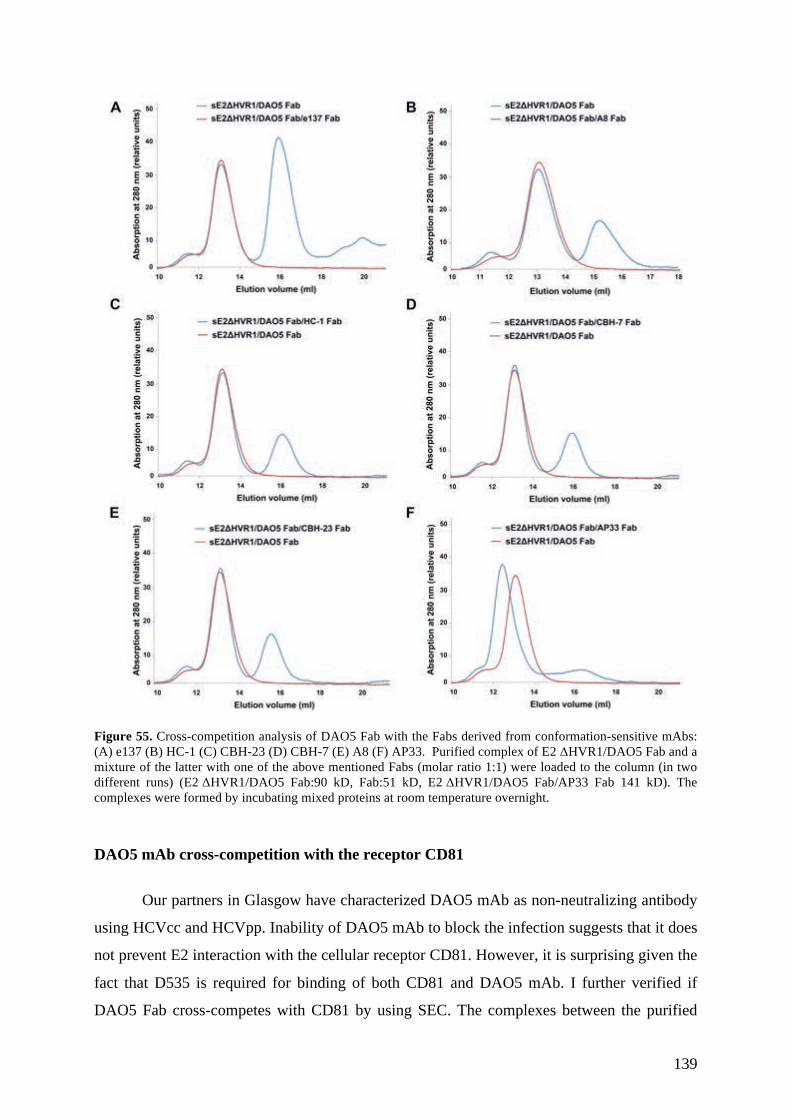
139
Figure 55. Cross-competition analysis of DAO5 Fab with the Fabs derived from conformation-sensitive mAbs: (A) e137 (B) HC-1 (C) CBH-23 (D) CBH-7 (E) A8 (F) AP33. Purified complex of E2 (HVR1/DAO5 Fab and a mixture of the latter with one of the above mentioned Fabs (molar ratio 1:1) were loaded to the column (in two different runs) (E2 (HVR1/DAO5 Fab:90 kD, Fab:51 kD, E2 (HVR1/DAO5 Fab/AP33 Fab 141 kD). The complexes were formed by incubating mixed proteins at room temperature overnight.
DAO5 mAb cross-competition with the receptor CD81
Our partners in Glasgow have characterized DAO5 mAb as non-neutralizing antibody
using HCVcc and HCVpp. Inability of DAO5 mAb to block the infection suggests that it does
not prevent E2 interaction with the cellular receptor CD81. However, it is surprising given the
fact that D535 is required for binding of both CD81 and DAO5 mAb. I further verified if
DAO5 Fab cross-competes with CD81 by using SEC. The complexes between the purified

140
sE2ΔHVR1/DAO5 Fab complex or only sE2ΔHVR1 and the recombinant large extracellular
loop (McLellan et al.) of CD81 was formed overnight and analyzed by SEC on a Superdex
200 10/300 column the following day. Since the molecular weight of CD81 LEL is only ~10
kD, the shift of sE2ΔHVR1/DAO5 Fab complex would not be visible in SEC even if CD81
binds. Therefore, the peak fractions of the complexes were concentrated and analyzed by
SDS-PAGE. No binding of CD81 LEL to sE2ΔHVR1/DAO5 Fab complex was detected as
indicated by SDS-PAGE of eluted fraction of sE2ΔHVR1/DAO5 Fab complex plus CD81
LEL (Figure 56). In contrast, CD81 LEL was binding to sE2ΔHVR1 alone in the same assay.
Figure 56. DAO5 Fab cross-competition with CD81 LEL for binding to sE2ΔHVR1. sE2ΔHVR1 and sE2ΔHVR1/DAO5 Fab complex was pre-incubated with CD81 LEL overnight at room temperature. The complexes were separated by size-exclusion chromatography and the peak fractions were concentrated and analyzed by SDS-PAGE under non-reducing conditions followed by Coomassie staining. sE2ΔHVR1/DAO5 Fab does not bind the CD81 LEL, suggesting a direct interference between DAO5 and CD81 binding in spite of the lack of neutralizing activity observed for mAb DAO5.
These results are in agreement with D535 residue being an essential binding residue
for both DAO5 mAb and CD81 LEL. Thus, DAO5 mAb is interfering with CD81 binding
though it is not able to neutralize viral particles. Notably, our collaborators at Glasgow

141
performed the neutralization experiments by incubating DAO5 mAb with HCVpp and HCVcc
at 37oC for 1h. This incubation time could be too short to achieve neutralization given the
fact that the temperature dependent binding between DAO5 antibody fragments and
sE2ΔHVR1 was observed after overnight pre-incubation of the proteins. Unfortunately, the
investigation of neutralization using longer incubation times of DAO5 mAb with HCVpp and
HCVcc at 37oC was not possible due to instability of the viral particles.
All the above-described results together suggest that HCV E2 region aa 530-540,
which is crucial for the glycoprotein-CD81 interaction and, thus, infection of cells, is very
flexible. The possible benefits of this flexibility for virus infection will be discussed in the
next section.

142
Supplementary data Table S1. Fab – peptide interactions. HEAVY CHAIN Fab residue Peptide residue Dist. [Å] DAO5 Fab/peptide_J4 Hydrogen bonds main chain - side chain Lys 52 NZ Val 536 O 2.94 Lys 52 NZ Met 537 O 3.25 Lys 52 NZ Asn 540 O 2.87 Hydrophobic Interactions < 5 Ala 59 Met 537 Phe 99
Phe 99 Leu 538 Leu 539
Tyr 103 Leu 539 Pro 104 Leu 539 Tyr 105 Leu 539 DAO5 scFv/peptide_J4 Hydrogen bonds main chain - side chain Lys 52 NZ
Lys 52 NZ Lys 52 NZ
Val 536 O Met 537 O Asn 540 O
2.88 3.29 2.81
Hydrophobic Interactions
Ala 59 Phe 99 Phe 99 Tyr 103 Pro104 Tyr 105
Met 537 Leu 538 Leu 539 Leu 539 Leu 539 Leu 539
DAO5 scFv/peptide_JFH
Hydrogen bonds main chain - side chain < 5 Lys 52 NZ
Lys 52 NZ Lys 52 NZ
Val 536 O Phe 537 O Asn 540 O
3.05 2.87 2.74
Hydrophobic Interactions
Ala 59 Phe 99 Tyr 103 Pro 104 Tyr 105 Phe 99
Phe 537 Leu 538 Leu 539 Leu 539 Leu 539 Leu 539
Aromatic-aromatic interactions 4.5-7 Trp 94 Phe 537 5.42 Cation-Pi interactions
Lys 52
Phe 537
< 6 5.68

143
Table S1. Fab – peptide interactions continued. LIGHT CHAIN Fab residue Peptide residue Dist. [Å] DAO5 Fab – peptide 1 Hydrogen bonds main chain - side chain Thr 91 O Thr 534 OG1 2.63 Trp 94 N Glu 533 OE2 2.89 side chain - side chain Tyr 32 OH Asp 535 OD1 2.71 Tyr 32 OH
Lys 93 NZ Asp 535 OD2 Glu 533 OE1
3.28 3.5
Hydrophobic Interactions < 5 Trp 94 Met 537 Trp 94 Leu 538 Leu 96 Leu 538 Tyr 32 Leu 539 Tyr 50 Leu 539 Ionic interactions < 5 Lys 93 Glu 533 DAO5 scFv /peptide_J4 Hydrogen bonds main chain - side chain Asn 92 OD1
Lys 93 NZ Trp 94 N Trp 94 NE1 Thr 91 O Asn 92 ND2
Glu 531 O Gly 530 O Glu 533 OE2 Glu 533 O Thr 534 OG1 Glu 531 O
3.05 3.32 2.84 3.31 2.64 3.48
side chain - side chain Tyr 30 OH Glu 531 OE1 2.89 Tyr 32 OH Asn 532 OD1 3.46 Tyr 32 OH
Tyr 32 OH Asn 92 OD1
Asp 535 OD1 Asp 535 OD2 Asn 532 OD1
2.82 3.05 3.48
Hydrophobic Interactions < 5 Tyr 32 Leu 539 Tyr 50 Leu 539 Trp 94
Trp 94 Leu 96
Phe 537 Leu 538 Leu 538
Ionic Interactions < 6 Lys 93
Lys 93 Glu 531 Glu 533
DAO5 scFv – peptide_JFH
Hydrogen bonds

144
main chain - side chain Asn 92 OD1
Asn 92 ND2 Glu 531 O Glu 531 O
3.46 2.94
Lys 93 NZ Trp 94 N Trp 94 NE1
Gly 530 O Glu 531 OE2 Glu 531 O
3.13 2.86 3.35
Thr 91 O Thr 534 OG1 2.65 side chain - side chain Tyr 32 OH
Tyr 32 OH Tyr 32 OH
Asn 532 OD1 Asn 532 ND2 Glu 531OE1
3.36 3.26 2.99
Tyr 32 OH Tyr 32 OH
Asp 535 OD1 Asp 535 OD2
2.72 3.03
Hydrophobic Interactions < 5 Tyr 32 Leu 539 Tyr 50 Leu 539 Trp 94
Trp 94 Leu 96
Phe 537 Leu 538 Leu 538
Ionic Interactions < 6 Lys 93
Lys 93 Glu 533 Glu 531

145
Table S2. Intrapeptide interactions. Contact residue 1 Contact residue 2 Dist. [Å] Peptide_J4 (complexed with DAO5 Fab)
Hydrogen bonds main chain - main chain Thr 534 N Asn 532 O 3.12 Asp 535 N Asn 532 O 3.27 Val 536 N Thr 534 O 3.39 Met 537 N
Met 537 N Thr 534 O Asp 535 O
3.30 3.31
Met 538 N Thr 534 O 2.85 Leu 538 N Asp535 O 3.41 Leu 539 N
Asn 540 N Asp 535 O Asp 535 O
2.78 3.14
Hydrophobic Interactions < 5 Met 537 Leu 538 Leu 538 Leu 539 Peptide_J4 (complexed with DAO5 scFv)
Hydrogen bonds main chain - main chain Thr 534 N Asn 532 O 3.26 Asp 535 N Asn 532 O 3.40 Val 536 N Thr 534 O 3.31 Met 537 N
Met 537 N Thr 534 O Asp 535 O
3.49 3.48
Leu 538 N Thr 534 O 2.88 Leu 539 N
Asn 540 N Asp 535 O Asp 535 O
2.84 3.17
main chain - side chain Asn 540 OD1 Asp 535 O 1 3.11 Asn 540 OD1 Asp 535 O 2 3.11 side chain – side chain
Asn 532 ND2 Asp 535 OD2 1
3.35
Hydrophobic Interactions < 5 Met 537 Leu 538 Leu 538 Leu 539 Peptide_JFH (complexed with DAO5 scFv)
Hydrogen bonds main chain - main chain Thr 534 N Asn 532 O 3.25 Asp 535 N Asn 532 O 3.36 Val 536 N Thr 534 O 3.38 Met 537 N
Met 537 N Thr 534 O Asp 535 O
3.29 3.48
Leu 538 N Thr 534 O 2.96 Leu 539 N
Asn 540 N Asp 535 O Asp 535 O
2.85 3.28
main chain - side chain Asn 540 OD1 Asp 535 O 3.36

146
Hydrophobic Interactions < 5 Phe 537 Leu 538 Leu 538 Leu 539

147
Discussion
Intrinsic structural flexibility of the main antigenic region in HCV E2 glycoprotein
HCV E2 interaction with the host entry factor CD81 is an essential step in the HCV
entry process. The majority of HCV neutralizing antibodies interfere with CD81-mediated
virus entry, suggesting that a conserved CD81 binding site could represent a potential
candidate for immunogen design. The CD81 binding site on the E2 surface is composed of
discontinuous segments and the epitopes of most broadly neutralizing antibodies overlap with
these segments or reside in close vicinity. First structural insights into those antigenic regions
came from crystal structures of E2-specific antibodies in complex with synthetic peptides
mimicking their respective epitopes (Kong et al., 2012a; Kong et al., 2012b) (Deng et al.,
2014; Deng et al., 2013; Krey et al., 2013; Pantua et al., 2013; Potter et al., 2012).
However, the most comprehensive analysis of structural basis of HCV neutralization
was gained from a recent study of the structure of a core fragment of E2 glycoprotein in
complex with a Fab fragment of the neutralizing antibody AR3C (Kong et al., 2013). The
structure of this complex has significantly contributed to our understanding of antibody-
mediated neutralization of HCV by providing evidence that AR3C Fab binds to the same E2
surface interface as CD81. The crystal structure also revealed that more than half of the E2
core residues, especially in the solvent exposed areas, are disordered or present in loops
suggesting the overall high degree of flexibility of the glycoprotein. Moreover, it implies that
some regions of E2 involved in CD81 binding might depend on association with AR3C Fab
for their stabilization. The segments comprising CD81 binding site as well as the major
antigenic regions targeted by neutralizing antibodies reside in the flexible area of the E2
protein.
In the presented thesis I describe the crystal structure of a peptide encompassing aa
529-540 in complex with the non-neutralizing mAb DAO5. In our structure the epitope
peptide forms an α-helix at the C-terminal end of the peptide (D535-L539) while the N-
terminal region (N532-T534) has an extended conformation. The observed conformation of
the peptide epitope was unexpected given that this region adopts a ~20Å long, extended
conformation encompassing a short β-strand (aa 536-538) in complex with neutralizing
antibody ARC3. The epitope peptide in complex with DAO5 Fab and DAO5 scFv crystallized
in different space groups but adopted an identical conformation as indicated by RMSD values

148
between two structures suggesting that the observed conformation is not due to crystal
packing. Moreover, DAO5 scFv was crystallized with the peptide epitope from two different
HCV genotypes (genotype 2a (strain JFH-1) and genotype 1b (strain J4)), which also revealed
almost identical conformations.
The structure of the peptide bound to DAO5 mAb fragments suggests that residues
F/M537 and L539 would be accessible on the E2 surface as these residues insert into the
hydrophobic groove formed by the CDRs of DAO5 mAb. However, the crystal structure of
the E2 core fragment reveals that these residues point towards the hydrophobic core of E2.
The stark contrast between the α-helical conformation of the peptide epitope in complex with
DAO5 and the extended β-strand conformation observed in the structure of the E2 core
fragment indicate the dramatic structural flexibility of this crucial CD81 binding site.
By SPR and pull-down experiments we demonstrated that a given solution of
recombinant sE2ΔHVR1 can bind both DAO5 Fab and a Fab fragment derived from the
human neutralizing conformation-sensitive mAb e137 simultaneously. e137 mAb is a broadly
neutralizing conformation-sensitive human mAb requiring D535 for binding to E2. Since
e137 and AR3C mAbs use a number of the same contact residues within E2 as previously
indicated by alanine scanning mutagenesis, we believe that both of them bind to E2 when aa
536-538 adopts the β-strand conformation. In contrast, DAO5 mAb selectively binds to
sE2ΔHVR1 when its epitope within the protein adopts the α-helical conformation.
Our data demonstrating temperature-dependent binding of DAO5 antibody fragments
to the recombinant sE2ΔHVR1 further supports the structural flexibility of this epitope.
Certainly at higher temperatures the motion, i.e. flexibility, of the epitope increases resulting
in a higher number of events when the epitope adopts the α-helical conformation recognized
by DAO5 mAb. The fact that we did not observe temperature-dependent binding of e137 Fab
suggests that this region in the sE2ΔHVR1 fluctuates mostly around the β-strand
conformation while the energetic barrier to reach the α-helical conformation is higher and
requires additional activation energy. We also demonstrated that the binding of the antibody
locks the epitope in that particular conformation and prevents the binding of the second
antibody recognizing a different conformation of the epitope. None of the tested human
conformation-sensitive mAbs using D535 as an essential contact residue was able to bind to
the purified sE2ΔHVR1/DAO5 Fab complex. Thus, the two observed conformations of the
epitope are likely to be in dynamic equilibrium, which can be shifted in either direction by
antibody binding. The energetic barrier for the epitope to adopt the α-helical conformation

149
should be higher, which is in agreement with the fact that the equilibrium towards this
conformation can be shifted at physiological temperature in the presence of DAO5 mAb.
The β-strand conformation is also likely to be a predominant conformation because it
is stabilized by the overall E2 core fold. The crystal structure of E2 core fragment shows that
the short β-strand spanning aa 536-538 is part of the central β-sandwich, which resembles an
IgC2-like domain and consists of four strands forming an inner sheet and two strands forming
an outer sheet. The outer sheet is composed of two anti-parallel strands comprising aa 536-
538 and aa 496-498, respectively. The interaction between these two strands includes
hydrophobic contacts between I496 and V536, I496 and V538, V497 and F537, and P498 and
V536 as well as the main chain-main chain hydrogen bond between V497 and F537. Most
likely, the conformation of the β-strand spanning aa 496-498 would not be stabilized and
retained if aa 535-539 adopted the α-helical conformation. Alternatively, it would require a
change in the interactions between these two stretches of amino acids. Since the β-strands
composing the outer sheet are short and do not form a number of stabilizing hydrogen bonds
characteristic of longer β-sheets, the interactions between the strands are not strong enough to
fix them in this conformation. The binding of AR3C Fab seems to have a stabilizing effect on
this region: the Fab captures it in a certain (presumably more-stable) conformation and
prevents it from structural fluctuation between different conformations. In conclusion, our
results imply that the outer sheet of the IgC2-like domain composing the E2 core is displays a
higher degree of flexibility. This is also supported by the fact that this region is disordered in
E2 core crystal structure determined by Khan and colleagues (Khan et al., 2014).
One possible explanation for the presence of two different conformations within the
recombinant sE2ΔHVR1 is that DAO5 mAb binds to partially denatured or misfolded protein.
It is known that HCV glycoproteins contain a number of disulphide bridges, thus, the folding
of the proteins is difficult and often results in the aggregation of a fraction of the protein when
expressed in vitro (reviewed in (Op De Beeck et al., 2001)). Though sE2ΔHVR1 has been
demonstrated to be recognized by human conformation-sensitive antibodies and induce
neutralizing antibodies after immunization (Tarr et al., 2013), we cannot completely rule out
the possibility that a small fraction of the immunogen used to raise DAO5 mAb was
misfolded or denatured. Giving the fact that DAO5 mAb recognizes a linear epitope,
verification of whether or not it binds to the natively folded glycoprotein is essential.
Neutralization of infection is the most straightforward way to demonstrate the binding to the
envelope glycoprotein in its native conformation, as it is present on virions. However, DAO5
mAb is non-neutralizing, eliminating neutralization assay as a method to prove that DAO5

150
mAb recognizes the natively folded protein at the surface of virus particles. Nevertheless, we
showed that the antigen recognized by DAO5 mAb is likely to be natively folded
glycoprotein. First, we measured binding of the conformation-sensitive, non-neutralizing
human antibody CBH-4D to sE2ΔHVR1/DAO5 scFv complex by pull-down assay. The
obtained results indicated that sE2ΔHVR1 in complex with DAO5 scFv bound CBH-4D Fab
as efficiently as the glycoprotein alone, which is a strong indication that sE2ΔHVR1
recognized by DAO5 scFv is not fully denatured. In addition, these results imply that DAO5
mAb binding does not affect the overall fold of the glycoprotein, which could be a possibility
given the fact that aa 536-538 might not be the part of an IgC2-like domain anymore in the α-
helical conformation.
Our collaborators in Glasgow also demonstrated that HCVcc particles could be
immunoprecipitated with DAO5 mAb and were able to detect E2 in immunoprecipitated
material. However, DAO5 may bind to immature forms of E2 that are secreted or found in the
exosomes instead of the E2 displayed on viral particles. To confirm if the
immunoprecipitated material actually contains viral particles our collaborators in Glasgow are
planning to perform additional experiments: 1) test the infectivity of immunoprecipitated
material, 2) try to quantitate viral RNA in immunoprecipitated material, 3) try to detect the
core protein in immunoprecipitated material using anti-core antibodies.
It is possible but still unproven that the flexibility of the epitope observed in
recombinant sE2ΔHVR1 also applies for E2 displayed at the surface of viral particles. Since
DAO5 mAb is non-neutralizing, we still lack evidence that E2 displayed on the virions can
adopt the α-helical conformation. Little is known about the virion structure of HCV and how
the glycoprotein complexes are arranged in the viral envelope. It is possible that the presence
of E1 and/or packing of the glycoprotein complexes on the virion surface might have a
stabilizing effect and reduce the flexibility of the epitope. Therefore, there are two
possibilities: 1) DAO5 mAb does not bind to the viral particles at all because the
tertiary/quaternary structural constraints limit the flexibility of the epitope on viral particles
and it does not adopt the α-helical conformation, 2) E2 displayed on the virions can adopt the
α-helical conformation, however, due to the high energetic barrier to reach this conformation
these events are relatively rare.
We could demonstrate that DAO5 mAb cross-competes with CD81 LEL for binding
to sE2ΔHVR1 if all available sE2ΔHVR1 molecules are in complex with DAO5 Fab. This
finding is contradictory to the fact that DAO5 is non-neutralizing, since inhibiting CD81
interaction with E2 normally prevents infection. There are several possible explanations why

151
DAO5 mAb is not able to neutralize virus infectivity. If the epitope does not adopt the α-
helical conformation at the surface of viral particles, this explains why DAO5 does not
neutralize HCVcc or HCVpp. Another possible explanation is that DAO5 mAb binds only to
some glycoprotein complexes displayed on the virions (because this event is rare) while the
majority of the glycoproteins are not bound by the antibody and, thus, can interact with CD81
and subsequently initiate virus entry. It has been shown that for some viruses neutralization
occurs only when virions are bound by a number of antibodies that exceeds a required
threshold. For example for flaviviruses ~30 antibody molecules per virion are required for
efficient neutralization (Pierson et al., 2007). Stoichiometric requirements for neutralization
of HCV particles are unknown. Inhibition of infection, however, might require covering most
of the available CD81 binding sites on the virion. Since neutralization potency of the antibody
depends on a number of factors including the number of accessible epitopes on the surface of
the virion, it is likely that DAO5 mAb is not neutralizing because it does not reach the
required stoichiometry for neutralization.
Our preliminary results showed that HCVcc could be immunoprecipitated with DAO5
mAb, which would indicate that the antibody binds to the virions. To prove the binding of the
mAb to the virions, we are planning to perform additional experiments. In addition to the
identification of the origin of E2 in immunoprecipitated material as described above, we will
also verify DAO5 mAb binding to the purified HCVpp particles by SPR. Furthermore, it
might be possible to address this question by doing an additive neutralization by combining
e137 and DAO5 mAbs. If we reach a higher level of neutralization by combining both mAbs,
it would be a strong indication that DAO5 mAb contributes to the virus neutralization. Alone,
however, it is not able to reach the stoichiometric requirements for neutralization. Also, we
would like to verify if the observed flexibility of the recombinant glycoprotein ectodomain is
related with the protein expression system or the chosen construct. Therefore, we are going to
produce sE2ΔHVR1, the full-length sE2 and E2 core fragment described by Kong et al. in
HEK293F cells and test if the epitope flexibility is similar for the produced proteins.
Intriguingly, the flexibility of epitope I and epitope II, two other regions involved in
CD81 binding, has been recently demonstrated. Epitope I (aa 412-423) has been shown to
adopt at least two different conformations. A recent crystal structure of the epitope I (aa 412-
423) peptide in complex with the neutralizing Fab 3/11 revealed an extended conformation of
the peptide (Meola A, 2014) that it is in stark contrast to the previously reported β-hairpin
obtained for the same peptide in complex with mAbs AP33, HCV1 and Hu5B3.v3 (Kong et
al., 2012b; Pantua et al., 2013; Potter et al., 2012). Although available crystal structures of the

152
epitope II (aa 427-446) so far suggest that this epitope retains its overall conformation (an N-
terminal loop and a C-terminal 1.5-turn α-helix), the spatial arrangement of these components
is different in complexes with distinct mAbs (Deng et al., 2014; Deng et al., 2013) (Krey et
al., 2013). Therefore, the epitope seems to undergo a transition process between open and
closed conformations in the context of the E2 core molecule (Kong et al., 2013) Deng, 2014
#665). Deng et al., claim that epitope II can induce both neutralizing and non-neutralizing
epitopes depending on which conformation of E2 on the virions is presented to the host
immune system (Deng et al., 2014). Our results further support the prevailing hypothesis that
the CD81 binding region within E2 is highly flexible. Although more evidence is still needed,
it is very likely that this flexibility is characteristic not only for in vitro produced E2 but also
for the E2 on the virion surface.
Whether the flexibility of CD81 binding region plays a substantial role in the natural
course of HCV infection remains to be determined. Further, I provide the possible
interpretations how this phenomenon might impact the course of HCV infection.
It is tempting to speculate that the flexibility of CD81 binding region may have
significant outcomes in HCV infection and disease progression since the virus may use it as a
strategy to escape from the host immune system. Another major human pathogen, human
immunodeficiency virus (HIV), has also been reported to have a flexible receptor-binding site
within envelope glycoprotein gp120 (Myszka et al., 2000). The unoccupied CD4 receptor-
binding site has been shown to be disordered or exist in substantially different conformation
in comparison with this site bound by the receptor. This is in agreement with the finding that
the binding of CD4 receptor site-specific antibodies is accompanied by large negative changes
in entropy, which is indicative of protein folding (Kwong et al., 2002). This structural
flexibility has been suggested to be used by viruses as an additional immune evasion strategy,
helping viruses to avoid antibody-mediated neutralization. It appears likely that the CD81
receptor-binding site within HCV E2 preexists in several conformations in the state unbound
by CD81. As a result, the conformational fluctuations may create structural heterogeneity
within the receptor-binding region despite its high degree of sequence conservation, which in
turn may reduce immunogenicity of this important antigenic site and impede the efficiency
with which neutralizing antibodies are elicited. Therefore, the structural transitions of the
CD81 binding site within E2 may be beneficial to the virus allowing it to simultaneously
retain its receptor-binding function and to evade humoral immune response.
In addition, the structural flexibility of the CD81 binding site may lead to induction of
substantial amounts of non-neutralizing antibodies against it. It has already been demonstrated

153
that epitope II elicits both neutralizing and non-neutralizing antibodies (Duan et al., 2012).
Non-neutralizing antibodies have been reported to have detrimental effects for the outcome of
a number of viral infections as they can interfere with the binding of neutralizing antibodies to
conserved epitopes due to steric hindrance or contribute to infection through interaction with
Fc receptors or complement receptors (Takada & Kawaoka, 2003). These host immune
system escape mechanisms facilitated by non-neutralizing antibodies may also be valid in the
case of HCV infection. First, non-neutralizing antibodies against the CD81 binding loop may
prevent the binding of neutralizing antibodies to conserved epitopes due to steric hindrance. It
has been described that non-neutralizing antibodies against epitope II interfere with
neutralization activity of antibodies specific to a proximal epitope I (Duan et al., 2012; Zhang
et al., 2007; Zhang et al., 2009). Our cross-competition experiments with recombinant
sE2ΔHVR1 revealed that DAO5 mAb completely abrogates the binding of the neutralizing
conformation-sensitive antibodies harboring essential contact residues within the antigenic
region aa 523-540. If non-neutralizing antibodies such as DAO5 mAb are elicited during
natural HCV infection, they could cross-compete with the neutralizing antibodies and reduce
their neutralization potency.
In conclusion, our results indicate that the previously reported structural flexibility
epitope I and epitope II composing CD81-binding site further extends to the antigenic site
spanning aa 529-540 at least in the soluble glycoprotein. As these three discontinuous
segments comprise the CD81 binding site on the surface of E2 glycoprotein, our study
supports the hypothesis that the entire CD81 binding site is highly flexible. We have
demonstrated that the antigenic site spanning aa 529-540 undergoes structural fluctuations in
the recombinantly produced sE2. Further examination of DAO5 mAb binding to HCV
particles should provide us with better understanding if the flexibility of this region is also
characteristic for E2 displayed on the virions. The intrinsic structural flexibility of epitope I
and epitope II has already been proven to occur at the surface of infectious particles.
Therefore, we believe that this feature also applies to the third segment of E2 (aa 529-540)
involved in CD81 binding. Notably, the two available E2 core structures (PDB IDs 4MWF
and 4NX3) differ in their disulphide bridge pattern and share only three disulfide bonds
(C494-C564, C508-C552 and C607-C644). Castelli and colleagues has recently assessed the
impact of each cysteine residue in E2 expressed in near-native conditions on the binding of
conformation sensitive mAbs. This study revealed the presence of alternative cysteine
disulfide pairs than the ones observed in E2 core crystal structures obtained by Kong et al.,
and Khan et al (Castelli et al., 2014). These observations suggest that E2 glycoprotein is

154
extremely flexible and the two crystallized E2 core domains may be just a snapshot of a few
E2 forms existing at the surface of viral particles.
Normally receptor-binding sites are the most susceptible parts of viral glycoproteins
for antibody-mediated neutralization. The conformational flexibility of the CD81 binding site
might translate to inefficiency in generating neutralizing antibodies against this site as well as
the reduced neutralization potency of these antibodies. This is in agreement with the fact that
broadly neutralizing antibodies are usually not detected at the early stages of HCV infection
(Logvinoff et al., 2004). A better understanding of the properties of HCV E2 that contribute
to neutralization escape of the virus are crucial for development of vaccines or other
therapeutics targeting the receptor-binding. Our results imply that the CD81 binding site
might be not an ideal candidate for creating a vaccine due to the high degree of flexibility.
Presumably, conformational stabilization of the CD81 binding site on E2 may be beneficial in
the development an efficient vaccine.

155
Materials and Methods
Constructs used or generated in the study
The HCV E2ΔHVR1 expression construct (pT350/ E2ΔHVR1) was previously
described, and the produced protein was extensively characterized (Tarr et al., 2013).
Anti-HCV E2 mAb DAO5 was obtained by immunizing mice with HCV E2
recombinant proteins from different genotypes. The sequences of mAb were determined by
Arvind Patel and Ania Owsianka (Glasgow, UK). The construct expressing a recombinant
DAO5 scFv was generated in the lab. The construct expressing recombinant DAO5 Fab was
generated as described in a following chapter.
The sequences of anti-HCV E2 mAb were previously published. Anti-HCV CBH-4D
(Hadlock et al., 2000), e137 (Perotti et al., 2008), A8 (Allander et al., 2000; Johansson et al.,
2007), HC84-1 (Keck et al., 2012; Krey et al., 2013), CBH-23 (Keck et al., 2012), CBH-7
(Keck et al., 2004), HC-1 (Keck et al., 2008a; Wang et al., 2011) and AP33 (Potter et al.,
2012; Tarr et al., 2006) Fabs were previously produced recombinantly or by papain digest.
Generating DAO5 Fab construct
DAO5 VL and VH were previously cloned into the pMT-scFv-Strep vector in the lab.
In order to generate the sequences of the LC (VL-CL) and the Fd fragment (VH-CH1) which
could be cloned into the pMT-Fab-Strep vector, the VL and VH of DAO5 were fused,
respectively, to the LC and the CH1, of previously characterized F16 Fab. The fusion was
carried out by PCR-driven overlap extension (Heckman & Pease, 2007), which is illustrated
in Figure 57. The resulted PCR products were cloned into the Fab expression vector for
production of recombinant DAO5 Fab in Drosophila melanogaster S2 cells. The sequences of
the primers are:
Fwd_DAO5_HC_fusion GTGATCGTGAGTAGTGCCAAAACGACACCCCCATCTG Rev_DAO5_HC_fusion ACTACTCACGATCACGGATGTTCC Fwd_DAO5_LC_fusion GAAGCTGGAGCTGAAGCGGGCTGATGCTGCACCAACTGTATCC Rev_DAO5_LC_fusion CTTCAGCTCCAGCTTCGTTCC

156
Figure 57. Generating DAO5 Fab construct. Chimeric gene product between DAO5 F16 Fab VL and F16 Fab CL was generated by two PCRs. Primers Fw_DAO5_LC_fusion and Rv_DAO5_LC_fusion contained overlapping sequences to DAO5 VL and F16.1 Fab CL, respectively (solid line and dashed line). In the first PCR two gene products were generated containing the junction segments between VL and CL. The second PCR generated the hybrid product DAO5 VL- F16 Fab CH which was inserted into a pMT-Fab-Strep vector by restriction cloning. Similarly, DAO5 VH- F16 Fab CH1 gene was generated and inserted into the same vector (not shown).

157
CD81 LEL was produced based on the previously described protocol (Kitadokoro et
al., 2001). CD81-LEL with a hexahistidine tag (6His) was expressed in E. coli as a fusion
protein with the IgG binding domain of the Staphylococcus aureus protein A (ProtA). The
hinge region between CD81-LEL-ProtA contained a thrombin cleavage site.
6 l of TB medium containing 50 pg/ml kanamycin were inoculated 30/1000 with an
overnight culture of E. coli Rosetta-gami (DE3) strain transformed with CD81-LEL-ProtA in
pET28. After growing the cells at 37 oC to an OD550 of 0.78 the culture was cooled to room
temperature. Expression was induced with 0.5 IPTG and the culture was incubated for 20
hours at 28°C. The cells were harvested by centrifugation at 3000 g for 12 min and EndoH
was purified from the periplasm of the bacteria. The pelleted cells were resuspended in 2.5%
of the original culture volume in ice cold 20% sucrose, 0.1 M Tris pH 8 and 1 mM EDTA and
pelleted again by centrifugation at 12000 g for 15 min. Then the cells were resuspended in
equivalent volume of ice-cold water and incubated for 30 min on ice. After pelleting the cells
at 70000g for 25 min, the supernatant was collected and filtered through 0.22 µM cut-off
membrane. The supernatant was loaded onto IgG Sepharose FF (GE) column (~7ml resin)
equilibrated with 50 mM TRIS pH 7.6, 150 mM NaCl, 0.05% Tween 20. After washing the
column with the same buffer, the protein was eluted with 500 mM Na-Acetate pH 3.4. Elution
fractions (2.5ml) contained 1.5ml of TRIS pH 9.0 to neutralize pH. The elution fractions were
pooled and concentrated to ~1 ml using Vivaspin centrifugal concentrator (Sartorius). 20ml of
200 mM TRIS pH 8.5 were added and the protein was re-concentrated to ~1 ml. CD81-LEL-
ProtA was cleaved with thrombin using Thrombin Cleavage Capture kit (EMD Millipore,
Darmstadt, Germany) according to the manufacture’s instructions. 1 µl of thrombin per 1 mg
of CD81-LEL-ProtA was used. CD81-LEL was further purified using Ni2+ion affinity
chromatography followed by SEC on Superdex 75 16/60 column (GE Healthcare).
Removal of the double Strep affinity tag from the recombinant antibody fragments
A C-terminal double Strep tag preceded by an enterokinase recognition site was
removed from the recombinant antibody fragments and by specific proteolytic cleavage with
EKMax Enterokinase (Invitrogen, San Diego, USA). The detailed protocol is provided in
Supplementary Materials and Methods.

158
Peptides and complex formation
Synthetic peptides comprising HCV E2 residues 529-540 of the J4 strain
(GENETDVMLLN) and JFH strain (GENETDVMLLN) were ordered from GenScript and
dissolved in 20 mM Tris pH 9 at 10 mg/ml. The DAO5 Fab/peptide complex was formed by
mixing protein with the peptide at 1:6 molar ratio (10 mg/ml of Fab+1.72 mg/ml peptide).
The complex formation was carried out overnight at 4°C.
Crystallization and crystal soaking in peptide solution
Crystallization conditions for the DAO5 Fab in complex with the peptide_J4 were
screened by a Mosquito robot (TTP LabTech Ltd, Royston, UK) at 293 K using the sitting-
drop vapor-diffusion method. Rod-shaped diffraction quality crystals appeared after 5 days in
a drop containing 0.2 µl of the complex (10 mg/ml of Fab+1.72 mg/ml peptide in 10 mM
TRIS pH 8.0, 150 mM NaCl) mixed with an equal amount of reservoir solution containing
20% PEG3350 and 200 mM sodium thiocyanate. The crystals were flash-frozen in liquid
nitrogen after transferring crystals to a cryo-protective solution containing the mother liquor
and 20% (v/v) glycerol.
Crystals of DAO5 scFv in complex with the peptide_J4 and peptide_JFH were
obtained by soaking experiments of unliganded DAO5 scFv crystals obtained by hanging-
drop vapor diffusion method in drops containing 1 µl DAO5 scFv (9 mg/ml in in 10 mM
TRIS pH 8.0, 150 mM NaCl) and 1 µl of reservoir solution (29% PEG 400, 200 mM MES pH
6.5 and 150 mM sodium acetate). Unliganded DAO5 scFv crystals were transferred to the
mother liquor supplemented with 0.1 mM peptide for 5 h (with peptide_J4) or overnight (with
peptide_JFH) and were directly flash-frozen in liquid nitrogen.
Structure determination of DAO5 antibody fragments in complex with the peptides
Data were collected at the Synchrotron Soleil beamline Proxima 1, the Swiss Light
source beamline PX I at 100 K using a Pilatus detector (Dectris, Baden, Switzerland). The
programs iMosflm (Leslie, 2006; Powell, 1999) or xdsme (https://code.google.com/p/xdsme/)
were used to determine the optimum orientation of the crystal for the complete data set
collection. X-ray diffraction data were processed using XDS (Kabsch, 2010). Scaling and

159
reduction of data were performed using Pointless (Evans, 2006) and programs from CCP4
suite (CCP4, 1994).
The structure of DAO5 scFv was previously determined by molecular replacement
with the program Phaser (McCoy et al., 2007) by Thomas Krey (space group P41212.). A
search model used for molecular replacement was assembled from the VL region of a Fab
with PDB accession code 1FH5 and the VH region of a Fab with PDB accession code 3RHV.
In the case of the data set for DAO5 scFv-peptide-JFH crystal, the P4122 space group
was suggested by POINTLESS, but the actual space group of the DAO5 scFv crystals used in
soaking experiments was previously determined to be P41212. However, detection of
systematic absences is sometimes is unreliable because they may be missing from the data set
if they lie in the blind region (i.e. along spindle rotation axis in the data collection). This
situation is more common for long needle or rod-shaped crystals; thus, it was likely to occur
during the data collection on the DAO5 scFv-peptide-JFH crystal. Therefore, the list of
reflections was analyzed to check if there are any systemic absences along the k reciprocal
axis. The systematic absence of the spots with Miller indices 0k0 where k=2n+1 was observed
suggesting a two-fold axis along the b-axis in real space. The output from SCALA revealed
unusually high Rmerge values for the data set of DAO5 scFv-peptide_JFH crystal when all 800
frames were used. Therefore, the Rmerge was analyzed versus the batch number to determine if
the data quality had declined during the collection time. Indeed, an increase in Rmerge was
detected for the second half of the batches, likely as a result of radiation damage. Since the
crystal belonged to a high symmetry space group (P41212), the batches with high Rmerge could
be excluded from the data set without affecting its completeness. The data processing, scaling
and reduction was performed on the first 300 out of 800 frames.
Since the structure of unliganded DAO5 scFv was already available, the phases for the
structures of DAO5 scFv in complex with the peptides were calculated by the molecular
replacement method with the program Phaser (McCoy et al., 2007) using the previously
determined coordinates from the native crystal structure of DAO5 scFv as a search model.
Each crystallographic asymmetric unit contained two DAO5 scFv/peptide complexes.
Fab fragments have a flexible linker region (elbow angle) between the variable
domains (VL and VH) and the constant domains (CH1 and CL), which requires a Fab search
model with an approximately correct elbow angle for molecular replacement. Alternatively,
the individual CH1/CL and VH/VL domains of Fab (excluding the residues composing the
elbow angle) can be used as search models. The molecular replacement solution for the
DAO5 Fab was obtained by using two ensembles as a search model: the VLVH region of

160
DAO5 scFv, and the CHCL region from anti-GBV-B E2 F16.1 Fab (unpublished). The
residues composing the elbow angle were excluded from the search model. Each
crystallographic asymmetric unit contained one DAO5 scFv/peptide_JFH complex. The
resolution limit of 1.7 Å was chosen for the refinement of DAO5 Fab-peptide_J4 model.
Refinement for all three crystal structures was implemented using AutoBuster
(Bricogne G, 2010). Because DAO5 scFv/peptide_J4 and DAO5 scFv/peptide_JFH
complexes contained two molecules per asymmetric unit, non-crystallographic symmetry
(NCS) restraints were applied throughout model refinement. The same R-free test set which
was used in the refinement of previously determined DAO5 scFv crystal structure had to be
maintained throughout the refinement of the models of the DAO5 scFv-peptide complexes.
The datasets of the DAO5 scFv-peptide complexes were of higher resolution (1.9 and 2.00 Å
in compared to 2.05 Å for native DAO5 scFv crystals). Therefore, the R-free test set of
unliganded DAO5 scFv contained only the reflections going to 2.05 Å. The existing R-free
test set was extended to the entire resolution range of DAO5 scFv-peptide complexes by
adding reflections from the highest resolution bins of those datasets.
Manual model building was carried out using Coot (Emsley et al., 2010) Although
after molecular replacement, electron density maps of all three complexes revealed
unambiguous density for the peptide in the antigen-binding site, the peptides were omitted in
the initial refinement and model building cycles and were built into the electron density in the
antigen binding site after the refinement and building of the scFv/Fab structures was
completed. Water molecules were added by using AutoBuster and verified manually. Details
of the statistics of data collection and refinement are presented in the chapter Results.
Crystal structure analysis
Multiple structural superposition of the peptides from three crystal structures and
calculations of root mean square deviation (RMSD) over all atoms, CA atoms, and backbone
atoms (N, CA, C, O) were performed using SuperPose 1 (Maiti et al., 2004). To compare the
crystal structures of two peptides (peptide_J4 in Fab and scFv structures or peptide_J4 and
peptide_JFH in scFv structures), the corresponding peptides were superposed using USSF
Chimera (Pettersen et al., 2004) and RMSD were calculated between every residue in two
peptides including all atoms or only backbone atoms (N, CA, C, O). Surface complementarity
coefficients were calculated using SC from the CCP4 suite. Electrostatic potentials were
calculated using the adaptive Poisson Boltzmann solver (Baker et al., 2001). The protein

161
interaction calculator (Molina et al.) (Tina et al., 2007) was used to identify the interactions
between the peptides and scFv/Fab. The surface area buried upon complex formation for the
interfaces and for the individual residues within the peptide was estimated using the PISA
server (Krissinel, 2007). Figures of the crystal structures were prepared in the PyMOL
Molecular Graphics System (www.pymol.org). Composite omit maps were generated using
CNSsolve (Brunger et al., 1998).
SEC analysis of sE2ΔHVR1 complexes with the antibody fragments
20-30 µg of sE2ΔHVR1 and an equimolar amount of the antibody fragment (Fab or
scFv) were incubated as isolated proteins as well as in complex for 16 h at certain temperature
(4°C, 18°C, 30°C or 37°C) followed by analysis on a Superdex 200 10/300 column (GE
Healthcare, Uppsala, Sweden).
sE2ΔHVR1/DAO5 Fab complex purified by SEC was used. The second Fab was
added to sE2ΔHVR1/DAO5 Fab complex and the proteins were incubated for 16 h at 4°C
before analyzing them on a Superdex 200 10/300 column (GE Healthcare).
Cross-competition analysis of DAO5 Fab and CD81 LEL by SEC
100 µg of E2ΔHVR1 and pre-formed sE2ΔHVR1/DAO5 Fab complex was incubated
with a molar excess of CD81 LEL over night at room temperature. The next day the
complexes as well as individual proteins (E2ΔHVR1 and CD81 LEL) were loaded onto a
Superdex 200 10/300 column (GE Healthcare, Uppsala, Sweden) and the peak fractions were
collected. The peak fractions were concentrated and analyzed by SDS-PAGE under non-
reducing conditions. The gel was stained with Coomassie Blue.
Pull-down experiments
Pull-down experiments were performed to analyze the conformation and cross-
competition profile of sE2ΔHVR1/DAO5 complex. The pull-down assays were performed on
a Streptactin Superflow mini column (0.2ml column bed volume) using the Strep-tagged
E2ΔHVR1 and the Fabs without the Strep-tag.
To analyze the cross-competition between mAb CBH-4D and DAO5, 400 µg of pre-

162
formed E2ΔHVR1/DAO5 scFv complex was affinity bound to a Streptactin Superflow mini
column and washed with 10 column volumes of washing buffer. Subsequently, an excess of
conformation dependent Fab CBH-4D Fab was loaded onto the column followed by a
washing step. The complex was eluted in 4.5 column volumes of elution buffer.
To analyze the cross-competition between mAb e137 and DAO5, a molar excess of
e137 Fab was passed through the column with affinity bound E2ΔHVR1 followed by DAO5
scFv. The column was washed with 10 column volumes after addition of each protein. The
complexes were eluted as described above. The experiment was also performed by first
loading a molar excess of DAO5 scFv followed by e137 Fab. 15 µl of elution fractions were
analyzed by SDS-PAGE followed by Coomassie Blue staining.
Surface plasmon resonance (SPR) experiments
SPR experiments were performed on a Biacore 2000 system, equilibrated at 18°C in
10 mM Tris pH 8.0, 500 mM NaCl buffer using a CM5 sensor chip with a density of around
17000 response units (RU) of covalently immobilized anti-Strep antibody. E2ΔHVR1 used in
the experiments contained the Strep-tag allowing its capture on the chip via the Strep-tag.
The anti-Strep antibody was immobilized via primary amines using the following
protocol:
1) The sensor chip surface was activated with a freshly prepared 1:1
mixture of 0.4 M EDC (N-(3-dimethylaminopropyl)-N′-ethylcarbodiimide
hydrochloride) (GE Healthcare) and 0.1 M NHS (N-hydroxysuccinimide) (GE
Healthcare) in water;
2) Monoclonal anti-Strep antibody diluted in PBS to 37 µg/ml in 10 mM
sodium acetate, pH 5.5 (Biacore, Uppsala, Sweden) was injected into the
experimental flow cells of the sensor at a flow rate 5 µl/min for 20 min in order
to saturate the surface.
3) The sensor chip surface was deactivated with 1 M 2-aminoethanol pH
8.5 (GE Healthcare) by injecting a reagent at flow rate 5 µl/min for 12 min)
Diluted E2ΔHVR1 were injected over the sensor chip (3 min, 5 µl/min). After 5 min
of dissociation time, DAO5 Fab and e137 Fab were sequentially injected over pT424 (2 min
each, 5 µl/min). The injections of the Fabs were also carried out in reversed order (e137 Fab

163
followed by DAO5 Fab). The second Fab was injected after the first Fab reached the
saturation of the surface with non-covalently immobilized pT424.
E2ΔHVR1 / DAO5 Fab and E2ΔHVR1 / e137 Fab complexes pre-formed by
incubating the proteins overnight at 30 oC were injected over the sensor chip (4 min, 5
µl/min). After 5 min of dissociation time, DAO5 Fab or e137 Fab were injected over
E2ΔHVR1 / DAO5 Fab and E2ΔHVR1 / e137 Fab complexes, respectively (6 min, 5 µl/min).
All Fabs used in the experiment did not contain the Strep-tag in order to measure a
specific binding of the Fab to E2ΔHVR1 and E2ΔHVR1 / DAO5 Fab and E2ΔHVR1 / e137
complexes immobilized non-covalently on the chip via anti-Strep antibody. The second Fab
was injected after the first Fab reached a saturation state. After injections of the Fabs, the
sensor chip surface was regenerated using glycine-HCl pH 2.0 (2 min, 5 µl/min) and 0.1%
SDS (1min, 5 µl/min). All injections were carried out in triplicate. The data were processed
using Scrubber software (BioNavis, Ylöjärvi, Finland) and double referenced by subtraction
of the blank surface and buffer-only injection before local fitting of the data.

164
Chapter II.
Baculovirus Envelope Protein F
Introduction
Baculoviridae family
Baculoviruses have been known for many centuries since the first written accounts
describing the infection of Chinese silkworms. These large rod-shaped (30–60 × 250–300 nm)
viruses belong to the virus family Baculoviridae and are pathogenic almost exclusively to
insects of the order Lepidoptera, Hymenoptera and Diptera. The genomes of baculoviruses
consist of a large (80-180 kbp) circular double-stranded DNA genome containing
approximately 120 to 160 open reading frames. Classically, baculoviruses were divided into
two genera, nuclear polyhedrosis viruses (NPVs) and granulosis viruses, based on virion
morphology. The current classification, however, is based on phylogeny of baculoviruses and
classifies its members into four genera: Alphabaculovirus (lepidopteran-specific NPVs),
Betabaculovirus (lepidopteran-specific granulosis viruses), Gamabaculovirus (hymenopteran-
specific NPV) and Deltabaculovirus (dipteran-specific NPV) (Jehle et al., 2006) (Figure 58).
The Alphabaculovirus genus has been further subdivided into two groups (group I and group
II), which correlates with the usage of two different membrane fusion proteins, gp64 and F
protein, respectively. Protein “F” stands for “fusion” protein, and has amino acid sequence
features characteristic of the paramyxovirus fusion F. The structural characterization of the
baculovirus F protein is the subject of the second part of my PhD thesis.

165
Figure 58. The baculovirus classification. The baculovirus tree consists of 4 major clades based on alignment of amino acid positions of sequenced baculoviruses. The best-studied genus is Alphabaculovirus, which includes all the lepidopteran-specific nucleopolyhedrovirus. Alphabaculoviruses can be subdivided into two groups, group I and group II, based on the membrane fusion protein, which in budded virions (BV) of Group I is GP64 and in Group II F, which is also version of F. Gammabaculoviruses exist only as occlusion derived viruses (ODV). Adapted from (Jehle et al., 2006).
Baculoviruses have a number of useful applications. First of all, since they are natural
enemies of many insects, they can be applied as bioinsecticides. They are also used as
efficient eukaryotic expression vectors for production of recombinant proteins and vaccines.
Recently, certain baculoviruses have been demonstrated to represent potential gene therapy
vectors due to their capacity to transduce mammalian cells (Airenne et al., 2013).

166
Replication cycle
During their lifecycle the baculoviruses commonly can be found in two distinct
phenotypes: occlusion-derived virus (ODV), and budded virus (BV). ODVs are immobilized
in a protein matrix (polyhedrin or granulin) and are responsible for the infection of the midgut
epithelial cells (Coulibaly et al., 2009) (Figure 59). NPVs form large (1–15 µm) polyhedral
inclusion bodies, and based on the number of the nucleocapsids in the inclusion body, are
subdivided to single (S) NPVs or multiple (M) NPVs. Inclusion bodies of granuloviruses are
small and contain only a single virion. The virus released from the midgut epithelial cells is
BV, which is highly infectious and is responsible for systemic infection of an insect.
Figure 59. Structure of baculovirus occlusion bodies (OB) of nucleopolyhedroviruses (NPVs), occlusion-derived virion (ODV) and budded virion (BV) and form occlusion bodies (OBs). ODVs are responsible for the infection of the midgut epithelial cells. BV envelope contains the fusion protein GP64 (NPV group I) or F protein (NPV group II). BVs are responsible for systemic infection of an insect. Adapted from (Au et al., 2013).
Since insect populations are seasonal, baculoviruses evolved a strategy to survive in
the environment until their hosts reappear by immobilizing the virus in a protein matrix
(polyhedrin or granulin) of the occlusion bodies. These occlusion bodies are crystalline, and
the crystals diffract to high resolution resulting in determination of the structure of
baculovirus polyhedra determined by X-ray crystallography (Coulibaly et al., 2009). A
baculoviral infection starts with the ingestion of occlusion bodies by a larva. In the midgut of
the insect, occlusion bodies dissolve due to the alkaline pH liberating ODVs that infect the
midgut columnar epithelial cells. When nucleopcapsids reach the nucleus of the cells, the
DNA is released and expression and replication of the viral genome occurs.

167
The viral envelope proteins (gp64 or F) are transported to the plasma membrane of the
infected cell (Figure 60). The nucleocapsids assemble in the nucleus and migrate towards the
plasma membrane, where they bud and acquire the viral glycoproteins. BVs propagate the
infection throughout the insect, entering cells via clathrin-mediated endocytosis (Long et al.,
2006a). They fuse with the endosomal membranes in an acidic pH-induced membrane fusion
reaction, as with many other viruses. The released nucleocapsids are transported to the
nucleus, where transcription and replication is initiated. In the late stages of infection, the
occlusion bodies are formed in the nuclei of infected cells and are released from the cells after
the death and disintegration of the host (Rohrmann, 2013).
Figure 60. Budded virus infection cycle. See text for detailed description. Adapted from (Au et al., 2013).

168
Baculovirus envelope fusion proteins
The entry of baculovirus BVs into the host cells is mediated by a specific envelope
glycoprotein, either gp64 or F (Blissard & Rohrmann, 1989; WF et al., 2000; Whitford et al.,
1989). Both proteins have similar biological functions, and have been demonstrated to be
involved in receptor binding, low-pH dependent fusion of virus and cellular membranes, and
efficient budding (Blissard & Wenz, 1992; Hefferon et al., 1999; Long et al., 2006b; Oomens
& Blissard, 1999; WF et al., 2000). However, the molecular basis of action of these proteins
is significantly different.
The three-dimensional structure of gp64 has been determined by X-ray
crystallography, revealing that it belongs to the structural class III fusion proteins, which do
not require proteolytic cleavage in order to be activated (Kadlec et al., 2008). The F protein
has been suggested to perform an analogous function to gp64 based on the fact that infectivity
gp64-null Autographa californica multiple nucleopolyhedrovirus (AcMNPV) can be restored
by the introducing F (Lung et al., 2002).
F proteins are not only more widespread within Baculoviridae family but also more
diverse (with amino acid identity 20-40% and >74% for F and gp64, respectively).
Phylogenetic analyses imply that the F protein is likely to be an ancestral fusion protein of
baculoviruses while gp64 has been incorporated into the baculovirus genome relatively
recently (Jiang et al., 2009; Pearson & Rohrmann, 2002). Group I alphabaculoviruses still
encode a non-fusogenic F protein homologue (F-like protein) which most likely lost its
fusogenic function due to the acquisition of gp64 during evolution (Lung et al., 2002; Wang
et al., 2008).
Characteristics of baculovirus F protein
One of the most studied F proteins comes from the group I alphabaculovirus
Spodoptera exigua multicapsid nucleopolyhedrovirus (SeMNPV). SeMNPV is very species-
specific and infects only the larvae of the beet armyworm (Spodoptera exigua). The beet
armyworm is a pest for many cultivated crops including vegetables, flowers, and cotton,
causing significant economic losses. SeMNPV is licensed as a bioinsecticide and is quite
effective when applied in greenhouses, where it is protected from ultraviolet light from the
sun (Bianchi et al., 2002). The complete SeMNPV genome was sequenced in 1999, revealing

169
that it lacked a homologue of gp64. The ORF8 of SeMNPV encoded another envelope
protein, which was shown to be sufficient to mediate membrane fusion in syncytium
formation assays at low pH (WF et al., 2000). This functional homologue of gp64 was called
fusion (F) protein.
Biochemical characterization of SeMNPV F protein has revealed that this protein
contains the features characteristic to class I fusion proteins. SeMNPV F protein is translated
as ~76kD precursor called F0. As for other class I fusion viral proteins, the precursor is
posttranslationally cleaved. In the case of SeMNPV F, this cleavage is mediated by subtilisin-
like endoprotease furin. The mutation of the furin cleavage site RSKR (amino acid residues
145-149) results in a loss of the fusogenic activity, indicating that the cleavage is essential to
generate a fusion-competent protein (Westenberg et al., 2002).
Figure 61. SeMNPV F protein after furin cleavage. Furin cleavage generates two disulphide-linked subunits F2 and F1. The larger F1 subunit is membrane anchored by its transmembrane domain. TM: transmembrane domain, SP: signal peptide.
The furin cleavage yields two disulphide-linked subunits: a small N-terminal subunit
F2 (theoretical molecular weight ~15kD), and a bigger membrane-anchored C-terminal
subunit F1 (theoretical molecular weight ~59kD) (Figure 61). F2 subunit contains only one
cysteine residue C94, which must pair with one of the ten cysteine residues in the F2 subunit
located upstream from the predicted transmembrane domain. Thus, the ectodomain of the F2
subunit contains a single cysteine residue.
The class I fusion proteins form homotrimers at the surface of a viral particle. The
exact oligomeric state of SeMNPV F is unknown but it is predicted to be trimeric based on
the fact that the F protein of closely related group II alphabaculovirus, Helicoverpa armigera
multicapsid nucleopolyhedrovirus (HearMNPV), assembles as trimers on the virus envelope
(Long et al., 2006b). Some viral fusion proteins have been shown to form higher-order
oligomers via covalent disulphide links. Whether or not the free cysteine residue in the F2
subunit is involved in oligomerization of SeMNPV F by making interprotein disulphide
linkages remains to be determined.

170
The furin cleavage occurs upstream of the hydrophobic sequence located at the N-
terminus of the membrane anchored F1 subunit. The stretch of the first 18 amino acid residues
at the N-terminus of F1 subunit (150-GLFNFMGHVDKYLFGIMDS-168) has been
suggested to represent a putative fusion peptide because it contains features characteristic of
previously described viral fusion peptides (White, 1992): 1) it is hydrophobic, 2) it can form
an amphipathic helix with conserved glycines at one side, and 3) it shows a high degree of
conservation among baculovirus F proteins. However, some differences with vertebrate viral
fusion proteins can be identified such as the absence of alanine residues and a higher number
of polar residues. The deletion of amino acid residues 151-170 in F protein resulted in the loss
of virus infectivity even though the protein was incorporated into viral particles and was
cleaved by furin, supporting the role of this amino acid stretch as a fusion peptide
(Westenberg et al., 2004).
The structural similarity between baculovirus F and paramyxovirus F
proteins
The SeMNPV F protein has been predicted to share structural features with the
paramyxovirus F protein. In contrast to baculoviruses, which are DNA viruses,
paramyxoviruses are single-stranded, negative-sense RNA viruses. The envelope glycoprotein
F is responsible for virus fusion with the cellular membrane. Paramyxovirus F proteins are
also synthesized as F0 precursors and cleaved in the trans-Golgi complex by furin into two
subunits, F1 and F2, with the fusion peptide located at the newly generated N-terminus of the
F1 subunit.
The important difference between the entry of baculoviruses and paramyxoviruses is
that the latter fuse at the cell surface in a pH-independent manner, while baculoviruses
mediate fusion in the endosomes in an acidic-pH environment. Moreover, paramyxoviruses
uses a separate attachment protein for binding to the cell surface, in contrast to baculoviruses,
which have both membrane fusion and receptor binding activities in the same envelope
glycoprotein F. The interaction of the paramyxovirus attachment protein with the cellular
receptor triggers conformational changes in the prefusion F trimers, which eventually lead to
the fusion of viral and cellular membranes (reviewed in (Bossart et al., 2013; Jardetzky &
Lamb, 2014; Plattet & Plemper, 2013).

171
Of all paramyxoviruses, the F protein of human respiratory syncytial virus (RSV) was
identified as having the highest amino acid identity (12%) to SeMNPV F. Despite low amino
acid sequence identity, these two proteins display conserved positioning of the furin cleavage
sites, the secondary structure elements, and the transmembrane domains which is an
indication of homology between their corresponding genes.
The crystal structures of F in pre-fusion and/or post-fusion from has been determined
for a number of paramyxoviruses: human parainfluenza virus 3 (hPIV3), human parainflueza
virus 5 (hPIV5), New Castle disease virus (NDV) fusion protein The crystal structures of
RSV F in its post-fusion and pre-fusion forms are available (Swanson et al., 2011) (McLellan
et al., 2013) revealing a class I fusion protein fold observed in previously solved F structures
from other paramyxoviruses (human parainfluenza virus 3 (hPIV3), parainflueza virus 5
(PIV5), Newcastle disease virus (NDV), Simian virus 5 (SV5) and metapneumovirus (MV))
((Baker et al., 1999; Chen et al., 2001; Swanson et al., 2010; Welch et al., 2012; Wen et al.,
2012; Yin et al., 2005). The RSV F post-fusion trimer (Figure 62) has an elongated shape
with a globular head domain on the top and long stalk domain constituted by intertwined α-
helices that form a stable 6HB at the membrane proximal end of the molecule. The helices of
the 6HB are composed by two HRs: HRA adjacent to the fusion peptide, and HRB located
upstream from the transmembrane region. HRAs of three protomers form a central core of a
triple-stranded coiled-coil creating three grooves into which the C-terminal HRBs pack
antiparallel to the central core. In contrast, the most prototypical class I fusion protein,
influenza HA, forms just a small 6HB at the membrane distal end of the protein as it contains
just an extended segment the C-terminal HR helix that packs into the grooves of the central
N-terminal coiled-coil. Each protomer of RSV F in its post-fusion form is composed of three
domains, termed DI, DII and DIII. The globular head domain is mostly composed of D1 and
DII. At the base of the head domain is DIII, which carries a long HRA helix that extends
down and intertwines with HRA helices of the other two protomers to form a central coiled-
coil of the 6HB. HRB helices extending from DII interact with the central coiled-coil to form
the outer helices of a 6HB. The fusion peptide is located at the N-terminus of HRA while the
transmembrane region (not present in the crystal structure) is positioned at the C-terminus of
HRB. These two elements are located at the bottom of the stalk and are inserted into the
cellular membrane in the full-length F.
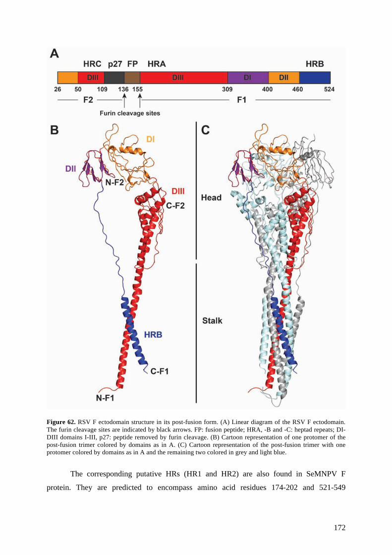
172
Figure 62. RSV F ectodomain structure in its post-fusion form. (A) Linear diagram of the RSV F ectodomain. The furin cleavage sites are indicated by black arrows. FP: fusion peptide; HRA, -B and -C: heptad repeats; DI-DIII domains I-III, p27: peptide removed by furin cleavage. (B) Cartoon representation of one protomer of the post-fusion trimer colored by domains as in A. (C) Cartoon representation of the post-fusion trimer with one protomer colored by domains as in A and the remaining two colored in grey and light blue.
The corresponding putative HRs (HR1 and HR2) are also found in SeMNPV F
protein. They are predicted to encompass amino acid residues 174-202 and 521-549

173
respectively. The three-dimensional structure of SeMNPV F is predicted to be similar to that
of RSV F (Misseri et al., 2003).
Receptor-binding function of baculovirus F
The insect cellular receptor used by baculoviruses has not yet been identified. Some
studies indicate that gp64 and F proteins of baculoviruses interact with distinct insect cell
receptors (Hefferon et al., 1999; Westenberg et al., 2007; Wickham et al., 1992). The
prototypic member of group II alphabaculoviruses Autographa californica (AcMNPV)
containing gp64 is able to transduce a number of mammalian cell types through the
endocytosis pathway, indicating that that the cells carry the receptor recognized by gp64 at
their surface. gp64, however, is also able to enter mammalian cells via direct fusion with the
plasma membrane under low pH (Dong et al., 2010). gp64-null Autographa californica
(AcMNPV) pseudotyped with baculovirus F is unable to enter mammalian cells suggesting
that the F protein receptor is not present on mammalian cells.
F-null group II baculovirus pseudotyped with gp64 and non-fusogenic F protein
(mimicking the composition of the envelope glycoproteins in group I alphabaculoviruses),
however, has been demonstrated to successfully enter insect cells by endocytosis. In this
study, non-fusogenic F protein was the primary contributor to the binding to the cells while
gp64 was only a minor contributor to the binding process. These results imply that F-like
protein may be a major receptor binding protein for entry to insect cells (Wang et al., 2014).
Relationship between insect retroviruses and baculoviruses
Retroelements with long-terminal repeats (LTRs) are found in the majority of
eukaryotic genomes. The genomes of all cells contain a number of transposable elements
integrated into their genomes. Exogenous retroviruses, endogenous retroviruses (ERVs), and
LTR-retrotransposons are one of the sources of retroelements. These transposable elements
have been described as being able to cross species barriers by horizontal transfer (Jordan et
al., 1999). The insect retroelements encoding an envelope (env) gene have been classified into
the Errantivirus genus of the Metaviridae family and their env gene has been shown to share
common ancestry with the gene encoding baculovirus F (Malik et al., 2000). The sequence

174
similarity is the highest in the region that includes the furin cleavage signal and a predicted
fusion peptide.
Figure 63. Baculovirus F protein orthologs are found in genomes of some insect retroviruses (errantiviruses). Evolutionary, they most likely originated from a retrotrasposons that incorporated F protein from baculovirus.
The baculovirus origin of env genes found in insect errantiviruses has been proposed
based on the fact that both errantiviruses and baculoviruses have the same hosts and LTR-
retrotransposons have been previously found incorporated into baculovirus genomes (Friesen
& Nissen, 1990) (Figure 63). As a result, env genes could have been acquired via
intragenomic recombination events that occurred after integration of LTR-retrotransposons
into the genome of baculoviruses (Pearson and Rohrmann, 2002)(Malik et al., 2000).
Drosophila melanogaster genome encodes a number of errantiviruses with gypsy being the
most studied retrovirus-like element in this organism. In contrast to retroviruses,
errantiviruses are considered to be non-infectious. However, gypsy Env has been
demonstrated to localize to the cell membrane of insect cells and to posses fusogenic
properties (Song et al., 1997) (Kim et al., 1994) (Misseri et al., 2004).
Cellular orthologs of baculovirus F protein
In addition to insect retroviruses, the F protein gene was identified in the genomes of
four dipteran species: Anopheles gambiae, and the fruit flies Drosophila melanogaster,
Drosophila yakuba, and Drosophila pseudoobscura (Lung & Blissard, 2005). Phylogenetic

175
studies have suggested that these F-like genes (named iris) were incorporated into the
Drosophila genome 25 million years ago from endogenous retroviruses. However, the Iris
found in Drosophila species (D.melanogaster, D. yakuba and D. pseudoobscura) lack the
structural elements of a fusion protein such as a predicted furin cleavage site, a predicted
fusion peptide, and a coiled-coil domain. Moreover, a study that analyzed a possible
membrane fusion activity of D.melanogaster Iris did not detect this protein as capable to
mediate membrane fusion and showed that it localized to mitochondria and not to the cellular
membrane as baculovirus F or gypsy Env (Lung & Blissard, 2005). The Anopheles gambiae F
protein has a potential furin cleavage site but the cleavage at this site has not yet been
demonstrated.
The ectodomains of lepidopteran baculovirus F proteins contain 10 conserved cysteine
residues, but only 6 of them (C5-C10) are found in Drosophila Iris and the F protein from
mosquito baculovirus CuniNPV. In Anopheles gambiae mosquitoes, C9 is also missing. The
spacing between those cysteine residues in lepidopteran baculovirus F proteins and insect
cellular F protein is conserved. In addition, 6 highly conserved non-cysteine residues were
identified between the cellular F and lepidopteran baculovirus F. Those conserved residues
are located in the central and C-terminal portion of F and reside within the borders of so-
called domain of unknown function (DUF3609) (conserved domain accession: pfam12259).
This domain of ~360 amino acids in length has been previously recognized in eukaryotes and
in viruses.
Although it has been proposed that iris was incorporated into the Drosophila genome
from endogenous viruses, the opposite hypothesis should also be considered. It could be that
the f gene in baculoviruses was acquired directly or indirectly from their insect hosts and
evolved to acquire fusion activity. Although baculoviruses infecting the Drosophila and
Anopheles species are not known, baculoviruses infecting other dipterans have been reported
(Becnel et al., 2001; Federici, 1980). It could be that ancestral baculoviruses existed only as
occlusion derived virions and were able to replicate only in the epithelial cells of the insect
midgut (which is still the case for sawfly baculoviruses). By acquiring a cellular f gene they
were able to infect the insect hemocel, which resulted in the evolution of a BV phenotype.
The indirect route of acquiring the f gene in baculoviruses might involve insect
endogenous viruses. As mentioned earlier, insect endogenous viruses encode f-like genes that
could potentially be of cellular origin. Combined with the fact that they can insert into the
baculovirus genome, insect endogenous viruses may represent a source for the indirect
transfer of the f-gene (Fraser et al., 1985; Malik et al., 2000; Miller & Miller, 1982).

176
Regardless, f gene homologues are very likely to be present in many insects including
not only the four above-mentioned dipteran insects but also lepidopteran insects. Given the
fact that the similarity of the F proteins of baculoviruses is rather low (less that 20% amino
acid identity in some cases) it is possible that cellular F proteins are also divergent and still
remain to be identified in insect genomes.

177
Structural characterization of a baculovirus fusion protein
ectodomain
Background
As mentioned in the introduction, group II alphabaculoviruses use protein F for fusion,
whereas group I use GP64. Amino acid sequence analyses indicate that the F protein of
baculoviruses displays a class I viral fusion protein fold and that it is related to the
paramyxovirus fusion protein F. Therefore, it is interesting to understand the organization of
F protein counterparts from DNA viruses such as the baculoviruses, in order to provide
insight into their evolution.. Paramyxovirus F protein appears related to the spike protein of
the coronaviruses with a large intervening domain between N-terminal and C-terminal HRs.
These proteins are thus more distant from, and perhaps not true structural homologs to, other
class I fusion proteins characterized to date such as those from retro-, filo-, arena- and
influenza viruses. Importantly, there are no DNA viruses known to encode a class I fusion
protein, and a crystal structure of SeMNPV F would provide important insight into
evolutionary aspects relating class I viral fusion proteins from RNA and DNA viruses.
Objectives
The aim of our research was to obtain structural insight into the fusion process of the
group II alphabaculovirus by determining the structure of the soluble F ectodomain of
SeMNPV by X-ray crystallography. This project was initiated as a collaborative research
project within the 7th framework program-Marie Curie Initial Training (Vecerek et al.)
network Virus Entry between our laboratory and our partners from Utrecht University,
Netherlands.

178
Results
Production and purification of SeFe
Together with our partners from Utrecht University, we established an efficient
expression system for to obtain the soluble ectodomain of the SeMNPV F protein, called SeFe
in Drosophila S2 cells. The construct containing the SeFe encompasses aa residues A18-T553
(aa 1-17 comprise a signal peptide) of the full-length SeMNPV F protein (UniProtKB
accession number Q9J8C6). It lacks the predicted transmembrane (TM) anchor domain
(residues 580-602) and the C-terminal cytoplasmic tail (CT) domain (residues 603-665)
present in the full-length protein (Figure 64) in order to allow secretion from cells. Our first
approach was to crystallize SeFe in its pre-fusion form. To stabilize the pre-fusion form, the
furin cleavage site was mutated (RRSKR to SGSKK) to prevent proteolytic processing into
F1 and F2 subunits by furin in the transfected S2 cells.
Figure 64. Primary structure of SeMNPV F and the expression system of its ectodomain (SeFe) in Drosophila melanogaster S2 cells. The domains indicated are F2 and F1, the fusion peptide (FP), the signal peptide (SP), three heptad repeat regions (HR1, HR2, HR3 and H4), the transmembrane domain (TM), enterokinase cleavage site (EK), triple strep-tag (3*ST) and Drosophila secretion signal (BiP). Predicted N-glycosylation sites are marked by (+), furin cleavage site in SEMNPV F (RRSKR) and the mutated furin cleavage site in SeFe (SGSKK) are indicated by black arrows. The disulfide bridge connecting domains F2 and F1 is shown as a thin dashed line. The thick dashed line indicates the borders of the F ectodomain cloned into the expression vector. The scale below corresponds to amino acid numbering of SeMNPV F (UniProtKB accession number Q9J8C6).
The large-scale expression and purification of SeFe was performed using standard
procedures (see section 2). Isolation of pure SeFe employed a combination of Streptactin
affinity column (Figure 65A) and size exclusion chromatography using a HiLoad 26/60
Superdex 200 gel filtration column (GE Healthcare) (Figure 65B). The protein eluted from

179
the gel filtration column as a single peak corresponding to monomeric SeFe (Figure 65B).
The final yield of SeFe was ~10 mg per litre of culture supernatant.
Figure 65. Purification of SeFe. The concentrated supernatant was loaded on 8 ml Streptactin column (A). After washing step, the Strep-tagged protein was eluted with 2.5 mM desthiobiotin. The fractions of the eluent were pooled and subjected to size exclusion chromatography (SEC) (B). Separation by SEC was performed using HiLoad Superdex 200 26/60 column (GE Healthcare) at a flow speed of 2 ml/min in 5 mM Hepes pH 7.0 150 mM NaCl and 0.1 mM EDTA. Protein elution was monitored by absorbance at 280nm (blue curves). Chromatogram B reveals a single major peak for SeFe corresponding to the monomeric protein.
Initially Scott Jeffers in our laboratory crystallized monomeric SeFe at neutral pH and
obtained crystals (most likely corresponding to the pre-fusion form of SeFe) but the crystals
diffracted only to ~8 Å, which was not sufficient to determine the crystal structure. The

180
crystallized protein was monomeric as judged by size exclusion chromatography, suggesting
that it may represent a pre-fusion conformation. Crystallizing viral fusion proteins in their
pre-fusion form is usually challenging because they are metastable. Based on the fact that the
paramyxovirus F protein is a stable homotrimer in its post-fusion conformation (Swanson et
al., 2011), we decided to attempt crystallization of the SeFe in its post-fusion form as an
alternative strategy.
Trypsin proteolysis of SeFe yields a trypsin-resistant fragment
The main problem in crystallizing the post-fusion conformation of viral fusion proteins
is that the fusion peptide is exposed, usually resulting in aggregation of the protein. Our
collaborators from Utrecht University established a protocol to obtain a trypsin resistant
fragment of SeFe. Furthermore, they demonstrated that lowering the pH from pH7 to pH5
resulted in an altered oligomeric state of the trypsin resistant fragment, suggesting the
formation of a post-fusion trimer. N-terminal sequencing results, together with SDS-PAGE
and Western blotting analysis of the trypsin-resistant SeFe trimer, revealed that:
1. Trypsin cleavage occurs in the region between the predicted HR1 and HR3
regions at residues R210 and R212 (210-RéMRéD-213) (based on N-terminal
sequencing).
2. Trypsin also cleaves at the mutated furin cleavage site at residues K147 or K148
(SGSKéKé) (based on SDS-PAGE).
3. The Strep-tag can no longer be detected by anti-strep antibodies in the Western
blot, thus it is completely removed by trypsin cleavage.
4. The trypsin cleavage at the C-terminal end of the F1 subunit most likely occurs
at one of the trypsin target sites located at either end of the HR2 domain.
Although trypsin cleavage results in the removal of the internal protein part composed of the
fusion peptide and the HR1 domain, F1 and F2 still remain linked by the disulphide bridge
(Figure 66).

181
Figure 66. Schematic representation of SeFe and its trypsin-resistant fragment. The trypsin cleavage sites are indicated with red arrowheads. The domains indicated are F2 and F1, the fusion peptide (FP), three heptad repeat regions (HR1, HR2, HR3 and H4), enterokinase cleavage site (EK), and triple strep-tag (3*ST). Predicted N-glycosylation sites are marked by (+). The disulfide bridge connecting domains F2 and F1 is shown as a thin dashed line.
I undertook to reproduce the trypsin cleavage of SeFe in our laboratory. The limited
trypsin proteolysis with increasing concentrations of trypsin led to the formation of a
predominant product, which was observed as a single band of ~50 kD in SDS-PAGE under
non-reducing conditions (Figure 67).
Figure 67. Limited proteolysis of SeFe by trypsin. SeFe was mixed with trypsin at different ratios, incubated at 22°C for 30 min and analyzed by SDS-PAGE under non-reducing conditions. The gel was stained with Coomassie blue. A predominant product of ~50 kD was observed by SDS-PAGE when using trypsin:SeFe ratio from 1:800 to 1:200. Lane 1: protein ladder.
On the intact virions, furin-cleaved baculovirus F proteins mediate membrane fusion
upon exposure to acidic pH encountered during endocytosis(WF et al., 2000). Therefore, I
investigated the effect of acid treatment of the SeFe and the SeFe trypsin-resistant fragment.
!"#"$
!"#"$%&'()*+,&")*)%-+%$.&-/0"+%$
12!3$45$#6$ 789$ 78:$781$
9$$$$$$$$$$$$$$$$$$$$$$$$$$9;;$$$$$$$$$$$$$$$$$$$$$$$$$$$:;;$$$$$$$$$$$$$$$$$$$$$$$$$1;;$$$$$$$$$$$$$$$$$$$$$$$$$$<;;$$$$$$$$$$$$$$$$$$$$$$$$$=;;$$$$$$$$$$$$$$$$$$$$$$$$$>;;$$$$$$$$$$$$$$$$$$$$$$$$$?;;$
@$ @$ @$ @$ @$ @$
!"#!$#
78:$781$@$ @$ @$ @$ @$
!"#!$# A7<$
7<$
!"#
!"# A
!$#
!$#
182
Both proteins were exposed to pH5.5 or pH7 and subsequently analyzed by SEC and multi-
angle laser light scattering (MALLS). The SeFe trypsin resistant fragment at pH5.5 eluted
earlier from the SEC column than the SeFe trypsin resistant fragment at pH7, suggesting the
change in its oligomerization at acidic pH (Figure 68).
Figure 68. Effect of acid treatment on SeFe and SeFe trypsin resistant fragment. Non-digested or trypsin digested SeFe was exposed to neutral (pH7) or acidic (pH5.5) pH and analyzed by SEC using Superdex 200 10/300 column (GE Healthcare). SeFe trypsin resistant fragment at pH5.5 eluted earlier from the SEC column than the SeFe trypsin resistant fragment at pH7, suggesting that there was a change in oligomerization at acidic pH.
The analysis of the oligomeric state of the SeFe and SeFe trypsin resistant fragment by
MALLS confirmed that the initially crystallized protein (SeFe, pH7) was a monomer while
the trypsin resistant fragment forms a trimer at acidic pH (Table 9). The acid-induced
trimerization of the trypsin resistant SeFe fragment was irreversible since the titration of pH
back to neutral prior to MALLS analysis did not lead to trimer dissociation.
0"
50"
100"
150"
200"
250"
300"
350"
3" 5" 7" 9" 10" 12" 14" 15" 17" 19" 20" 22"
Absorption*at*280*nm*(relative*units)*
Elution*volume*(ml)*
*trypsin"pH7"
+trypsin"pH7"
+trypsin"pH5"
*trypsin"pH5"

183
Table 9. MALLS analysis of the oligomeric state of SeFe and SeFe trypsin resistant fragment at neutral (pH7) or acidic (pH5.5) pH.
Molecular weight [kD] Mass fraction (%) Oligomeric state
- Trypsin pH7 95.4 (±5.5%) 100 Monomer
- Trypsin pH5 237.4 (±0.1%) and 91.3
(±0.2%)
36.4 and 63.6 Trimer and monomer
+ Trypsin pH7 77.5(±0.2%) 100 Monomer
+ Trypsin pH5 188.0 (±1.9%) 100 Trimer
We decided to use this protease resistant trimer for crystallization as it most likely
represents the stable post-fusion conformation. I made a large-scale preparation of the
trimeric trypsin-resistant fragment of SeFe (called SeFet) and purified it by SEC on a
Superdex 200 26/60 column, from which the protein eluted in a single symmetric peak
corresponding to a SeFet trimer, as expected (Figure 69).
Figure 69. Large scale SeFet formation. The trypsin proteolysis reaction of SeFe and acid induced trimerization were upscaled in a linear manner. SeFet was purified from the reaction mixture by SEC on Superdex 200 26/60 column (GE Healthcare). SeFet eluted from SEC column as a single symmetric peak corresponding to the trimeric SeFe trypsin resistant fragment.
Crystallization of SeFet
The crystallization screening for SeFet was performed as described in Materials and
Methods at protein concentration 6 and 10 mg/ml. The protein crystallized in a number of

184
conditions that were further optimized in 24-well plates. The crystals that were subjected to
X-ray diffraction analysis are listed in Table 10. Most of the crystals diffracted to 10-12 Å or
lower resolution, with the exception of one crystal form that diffracted to 6.5 Å, which was
sufficient for determination of the space group (P212121) and unit cell parameters (a=109.68,
b=346.24, c=111.34). In addition, we observed a peak in self-rotation function at 120 degrees
indicating that the crystallized molecule is a trimer. According to the Matthew’s coefficient,
the unit cell probably accommodated three trimers per asymmetric unit. These crystals of
SeFet formed after three weeks in 2µl hanging drops by vapor diffusion against a reservoir
solution containing 18% PEG 6000 and 100 mM Tris pH8 (1:1 protein-to-reservoir-solution
ratio) (Figure 70) and reproducibly diffracted to 6.5-7 Å resolution.
Figure 70. Best diffracting SeFet crystals. These plate-shaped crystals of SeFet were formed after three weeks in 2µl hanging drops by vapor diffusion against reservoir solution containing 18% PEG 6000 and 100 mM Tris pH8 (1:1 protein-to-reservoir-solution ratio) and reproducibly diffracted to 6.5-7 Å resolution.
In order to obtain better diffracting crystals, I performed a number of optimization
experiments (microseeding and streak seeding, crystallization at different protein
concentrations, testing crystallization additives, crystallization at 4˚C, crystallization of the
protein after an extra purification step by ion-exchange chromatography and vapor diffusion
dehydration). By vapor diffusion dehydration of the crystals for 2 days above the reservoir
solution containing 23% PEG 6000, 100 mM Tris pH8, I was able to improve resolution and
collect a complete dataset to ~5.5 Å.

185
Table 10. The crystals of SeFet subjected to X-ray diffraction experiments. The ability of the crystals to diffract X-rays were tested at Proxima I line, Synchrotron Soleil, France or ID 23 line European Synchrotron Radiation Facility (ESRF), France.
Crystallization condition Concentration (mg/ml)
Resolution (Å)
18% PEG 6000, 100 mM Tris pH 8 6 6.5 16% PEG 4000, 100 mM Tris pH 7.5, 15% 2-propanol 6 12 18% PEG 6000, 100 mM Tris pH 8 6 8 14% PEG 8000, 100 mM Tris pH 7.5 6 7 23% PEG 6000, 100 mM Tris pH 7.5 6 9 12% PEG 6000, 10 mM NaAc, 40% ethanol 6 13 20% PEG 6000, 100 mM imidazole pH 8 6 10 26.5 % PEG 8000, 100 mM Hepes pH 7.5, 10% 2-propanol, 100 mM NaAc
6 20
20.5% PEG 10000, 100 mM Tris pH7.5 6 10 19% PEG 4000, 100 mM NaAc, 100 mM Hepes pH7.5 6 20 15% PEG 8000, 40 mM potassium phosphate monobasic 6 >30 16% PEG 6000, 100 mM NaCl, 100 mM Tris pH 7.5 6 25 19% PEG 6000, 200 mM NaCl, 100 mM Tris pH 8 6 9 19% PEG 6000, 100 mM NaCl, 100 mM Tris pH 8 6 10 16% PEG 8000, 170 mM NaAc, 90 mM sodium cacodylate pH 6.5 6 15 16% PEG 8000, 170 mM NaAc 6 9 14% PEG 8000, 100 mM Tris pH 7.5 6 17.4% PEG 3350, 200 mM NaCl, 200 mM MgCl2 6 >30
Crystallization of the deglycosylated SeFet
SeFe contains six predicted N-linked glycosylation sites (N86, N132, N179, N270,
N508 and N551) (Figure 71). One of the advantages of the expression of the recombinant
proteins for crystallization in S2 cells is that N-linked glycosylation in Drosophila is less
complex and more homogenous than in mammalian cells. Proteins expressed in S2 cells have
high-mannose N-linked glycosylation and are not sialylated. In general, a deglycosylated
protein might form a more rigid protein lattice than a protein containing all sugar chains and,
thus, yield better diffracting crystals. Although one or two sugars are removed in trypsin
resistant fragments because they are present in the parts of the protein cleaved off by trypsin,
the rest of the sugars might still hinder the formation of a rigid protein lattice. As a result, I
attempted to deglycosylate SeFet with different endoglysosidases for crystallization trials.

186
Figure 71. Putative glycosylation sites in SeMNP F protein predicted by NetN Glyc 1.0 server (http://www.cbs.dtu.dk/services/NetNGlyc). The graph illustrates predicted N-glycosylation sites across the protein chain. Positions with potential (vertical lines) crossing the threshold (horizontal lines at 0.5) are predicted glycosylated.
The deglycosylases PNGase F, EndoH and EndoD were produced using periplasmic
expression in E.coli and purified as described in Materials and Methods. The extent of
deglycosylation of SeFet by different endodeglycosidases was assessed by mobility shift of
the deglycosylated protein versus the intact glycoprotein on SDS-PAGE gels. A clear
mobility shift of SeFet deglycosylated by PNGase F (Figure 72) as well as EndoD/EndoH
(not shown) was observed. Deglycosylation of SeFet by EndoH alone (not shown) did not
have any evident effect on protein mobility on the SDS-PAGE gel, indicating that this
endodeglycosidase most likely is not able to remove the sugars from the protein.
Subsequently, I performed a large-scale preparation of SeFet deglycosylated by PNGase
F. After deglycosylation, SeFet was separated from PNGase F by SEC (Figure 73A). In
addition, a large-scale preparation of SeFet deglycosylated by EndoD/EndoH was carried out.
EndoD/EndoH were removed from the reaction mixture by Ni-affinity chromatography and
subsequent SEC (Figure 73B). The crystallization screening of SeFet deglycosylated with
PNGase F and EndoD/EndoH was performed as described in Materials and Methods at
protein concentrations of 4 and 6 mg/ml, respectively. Since the endodeglycosidase EndoD
was obtained later during the project, the crystallization of SeFet deglycosylated with
EndoD/EndoH will be described later.

187
Figure 72. Enzymatic deglycosylation of SeFet with PNGase F. SeFet was incubated with PNGase F overnight at 37°C using the ratio 1:16 of endoglycosidase:SeFet on a weight basis. A clear mobility shift of SeFet deglycosylated by PNGase F versus non-deglycosylated SeFet was observed. Lane 1: protein ladder, lane 2: non-deglycosylated SeFet, lane 3: SeFet deglycosylated with PNGase F.
!!"
#$%"
$!"
&!"
#!"
#%%"
'%"
&!%"

188
Figure 73. Purification of deglycosylated SeFet. After deglycosylation with PNGase F, SeFet was separated from the endodeglycosidase by SEC (A). The first peak in the chromatogram corresponds to the deglycosylated SeFet, while the second peak corresponds to PNGase F. After deglycosylation with EndoD/EndoH, SeFet was separated from endodeglycosidases by Ni-affinity chromatography and subsequent SEC (B). The first peak in the chromatogram corresponds to the deglycosylated SeFet, while the second peak corresponds to the fraction of EndoH that was not fully removed by Ni-affinity chromatography.
SeFet deglycosylated with PNGase F crystallized under fewer conditions, which and
were different from those of the crystals of non-deglycosylated protein. The crystallization
conditions were further optimized in 24-well plates and the best crystals subjected to X-ray
diffraction. However, none of the crystals of deglycosylated protein diffracted to higher than
8Å resolution. In addition, the deglycosylated protein did not crystallize any more in the
condition in which the best diffracting crystals of the sugar-containing SeFet were obtained

189
(16% PEG 6000, 100 mM Tris pH8). Therefore, I tried to induce the crystal growth of
deglycosylated protein under these conditions by seeding with the crystals of the glycosylated
protein as a seed source. Although seeding helped to obtain the crystals of deglycosylated
SeFet under the same conditions of the not-deglycosylated protein, the resulting crystals again
only diffracted to about 8Å resolution (Table 11).
Table 11. Crystals of the deglycosylated SeFet subjected to X-ray diffraction at Proxima I line, Synchrotron Soleil, France or PXI line, Swiss Light Source (SLS), Switzerland.
Crystallization condition Concentration (mg/ml) Resolution 21 % PEG 3350 4 30 19 % PEG 3350, 200 mM ammonium formate 3 3.8 19 % PEG 3350, 200 mM sodium formate 3 5 18 % PEG 3350, 200 mM ammonium chloride 2 2.7 18 % PEG 6000, 100 mM Tris pH8 4 8 20 % PEG 2000 mono-methyl polyethylene glycol 4 10 22 % PEG 3350, 200 mM sodium nitrate 4 10 19 % PEG 3350, 200 mM ammonium fluoride 4 6 9% PEG 4000, 100 mM imidazole pH 8 and 30% MPD 4 10
To obtain different crystals of deglycosylated SeFet, I used microseed matrix
screening to sample many more crystallization conditions. This is a seeding technique where
crystals grown in one condition are ground to use as seeds, and are then distributed by a
robotized procedure into hundreds of different crystallization conditions. The crystals of
deglycosylated SeFet grown in 9% PEG 4000, 100 mM imidazole pH 8 and 30% 2-methyl-
2,4-pentanediol (MPD) were used as a seed stock for microseed matrix screening. This
technique helped to obtain crystals in different crystallization conditions that were further
optimized in 24-well plates and subjected to X-ray diffraction analysis.
For one crystal form (Figure 74) (which grew in 18% PEG 3350, 200 mM ammonium
chloride) we were able to collect a complete native data set at 2.7 Å (space group P1) (). The
unit cell parameters (a=73.94, b=74.86, c=78.11, α=94.5, β=114.3, γ=114.2) could
accommodate one trimer per asymmetric unit. The Matthews’ coefficient (Vm) was estimated
to be 2.04 Å3/Da (corresponding to a solvent content of 39.69%). The self-rotation function
(Figure 76) clearly showed the existence of a 3-fold non-crystallographic symmetry (NCS)
axis. This crystal form was reproducible and grew in ~48 h.
The quality and morphology of the crystals were very tightly dependent on the seed
stock dilution, protein concentration, and PEG 3350 concentration used. Native crystals
reproducibly diffracted to ~3 Å. All tested cryoprotectants (PEG 400, glycerol, MPD and
ethylene glycol) were suitable for cryopreservation of the crystals. Screening for

190
crystallization additives was performed, expecting to further improve the diffraction of the
crystals but the identified additives did not enhance the diffraction of the crystals. It was
possible to grow crystals using pH gradient from 6-8. However, pH did not have significant
influence on crystal morphology or diffraction. Crystals growing at 4°C also did not help to
improve the diffraction either.
Figure 74. Photographs of typical SeFet crystals obtained after seeding. (A and B). The crystals were grown at 2
mg/ml SeFet in 2.5 µl hanging drops (1:1:0.5 protein:reservoir solution:seed stock ratio) by vapor diffusion
against reservoir solution containing 16-18% PEG 3350 and 200 mM ammonium chloride and diffracted to ~3 Å
resolution. The crystallization drops usually used to contain different quality crystals. The crystals had a
tendency to grow as clusters of multiple crystals (B).
Figure 75. An X-ray diffraction pattern from the best native deglycosylated SeFet crystal on a Pilatus detector (Dectris, Baden, Switzerland). (A) High resolution reflections of the same diffraction image and corresponding resolution (B). The data were collected on the PXI beam line at the SLS in Switzerland.

191
Figure 76. Self-rotation function of SeFet P1 crystal form as determined from the program MOLREP (Vagin & Teplyakov, 2010). The peak on the sections (chi = 120) indicates the presence of non-crystallographic 3-fold axis.
Phasing of SeFet crystals
In order to obtain phase information, we resorted to experimental phasing given that the
paramyxovirus F model was not sufficient to obtain accurate phases by molecular
replacement. I searched for heavy atom derivatives of the crystals by soaking them in various
heavy atom solutions. Derivatization depends on parameters such as the exposure of
functional groups, local chemical environment, and ionization state. SeFe contains an odd
number of cysteines so the first choice was soaking the crystals with different mercury
compounds that are known to specifically react with free cysteine thiols. In addition, I tried to
Self Rotation FunctionMOLREP
Rad : 36.83 Resmax : 3.32RF(theta,phi,chi)_max : 4304. rms : 128.2
Chi = 180.0
X
Y
RFmax = 552.5
Chi = 90.0
X
Y
RFmax = 184.5
Chi = 120.0
X
Y
RFmax = 980.5
Chi = 60.0
X
Y
RFmax = 275.9
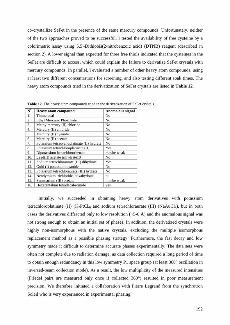
192
co-crystallize SeFet in the presence of the same mercury compounds. Unfortunately, neither
of the two approaches proved to be successful. I tested the availability of free cysteine by a
colorimetric assay using 5,5′-Dithiobis(2-nitrobenzoic acid) (DTNB) reagent (described in
section 2). A lower signal than expected for three free thiols indicated that the cysteines in the
SeFet are difficult to access, which could explain the failure to derivatize SeFet crystals with
mercury compounds. In parallel, I evaluated a number of other heavy atom compounds, using
at least two different concentrations for screening, and also testing different soak times. The
heavy atom compounds tried in the derivatization of SeFet crystals are listed in Table 12.
Table 12. The heavy atom compounds tried in the derivatization of SeFet crystals.
No Heavy atom compound Anomalous signal 1. Thimerosal No 2. Ethyl Mercuric Phosphate No 3. Methylmercury (II) chloride No 4. Mercury (II) chloride No 5. Mercury (II) cyanide No 6. Mercury (II) acetate No 7. Potassium tetracyanoplatinate (II) hydrate No 8. Potassium tetrachloroplatinate (II) Yes 9. Dipotassium hexachlororhenate maybe weak 10. Lead(II) acetate trihydrate10 No 11. Sodium tetrachloraurate (III) dihydrate Yes 12. Gold (I) potassium cyanide No 13. Potassium tetrachloraurate (III) hydrate No 14. Neodymium trichloride, hexahydrate no 15. Sammarium (III) acetate maybe weak 16. Hexatantalum tetradecabromide yes
Initially, we succeeded in obtaining heavy atom derivatives with potassium
tetrachloroplatinate (II) (K2PtCl4) and sodium tetrachloraurate (III) (NaAuCl4), but in both
cases the derivatives diffracted only to low resolution (~5-6 Å) and the anomalous signal was
not strong enough to obtain an initial set of phases. In addition, the derivatized crystals were
highly non-isomorphous with the native crystals, excluding the multiple isomorphous
replacement method as a possible phasing strategy. Furthermore, the fast decay and low
symmetry made it difficult to determine accurate phases experimentally. The data sets were
often not complete due to radiation damage, as data collection required a long period of time
to obtain enough redundancy in this low symmetry P1 space group (at least 360° oscillation in
inversed-beam collection mode). As a result, the low multiplicity of the measured intensities
(Friedel pairs are measured only once if collected 360°) resulted in poor measurement
precision. We therefore initiated a collaboration with Pierre Legrand from the synchrotron
Soleil who is very experienced in experimental phasing.

193
First, we optimized the data collection strategy by attenuating the beam to reduce
radiation damage, which allowed us to use the Multiple Anomalous Diffraction (MAD)
method by collecting data in small wedges in the inverse beam mode while alternating
between several wavelengths. We also collected a single-wavelength anomalous diffraction
(Drexler et al.) (Drexler et al.) data set at 3.1 Å at the sulphur edge on native crystals, which
allowed us to identify the locations of the disulfide bonds. But even after optimization of the
data collection strategy, the anomalous signal of the crystals derivatized with K2PtCl4 or
NaAuCl4 were not strong enough to obtain an initial set of phases good enough to determine
the structure.
Next we tried using the hexatantalum tetradecabromide (Ta6Br12+) cluster compound,
which is known to be a powerful derivatization reagent for phasing crystals at low resolution.
The soaking of SeFet crystals in 1mM Ta6Br12+ over night led to the incorporation of the
compound into the crystals, which was apparent due to the change of the crystal color. We
applied the double-inflection MAD collection strategy using one crystal derivatized with
Ta6Br12+ and detected significant anomalous differences up to ~6 Å. This data set was used to
locate the heavy atom sites in the unit cell of the crystal, and to calculate an initial set of
phases, which resulted in an interpretable electron-density map with clearly identifiable
helices resembling six-helix bundle characteristic for class I fusion proteins (Figure 77).
We then applied density modification techniques to further improve the map and started
manual-building of the initial model as polyalanine chains within this map, along with
iterative cycles of electron density modification (solvent flattening, non-crystallographic
symmetry (NCS) averaging). The initial maps indicated that the SeFet molecule is a very
flexible trimer, with the angles between three protomers varying along the trimer axis. As a
result, NCS averaging of the SeFet crystals required defining several masks and NCS
operators for different parts of the molecule. In addition, the low solvent content of the
crystals was not beneficial for solvent-flattening density modification, which is the more
powerful the larger the solvent content. The phases were transferred to the native SeFe
crystal (P1) by molecular replacement, which resulted in an electron density map at ~3 Å
resolution and the model further improved. However, the quality of the map was still not good
enough for building a full model of SeFet.

194
Figure 77. The electron density map of SeFet with the first helices built in alanine residues. The side view of SeFet helices (A). The view along the trimer axis (B).
The main breakthrough in the experimental phasing of the SeFet crystals was obtaining
a higher symmetry crystal form (space group P43212) with cell parameters (a=80.3, b=80.3,
c=478.7, α=90, β=90, γ=90) that could accommodate one trimer of SeFe per asymmetric unit.
This crystal form grew under identical conditions as the P1 crystal form. However, instead of
the seed stock solution containing the seeds, the same solution (9% PEG 4000, 100 mM
imidazole pH 8 and 30% MPD) without the seeds was used to set up the crystallization drops.
Those crystallization drops were intended to be used for streak seeding, but self-nucleated
crystals appeared after ~12 h (Figure 78). Though these crystals were difficult to reproduce,
and approximately only one out of 20 crystals diffracted, we managed to collect a complete
native data set at 2.9 Å as well as double-inflection MAD data on a derivative with Ta6Br12+
that diffracted to ~3.6 Å.
We were able to resolve the individual tantalum atoms in the cluster (using double-
inflection MAD data for a crystal form P43212), which resulted in an improved anomalous
signal in comparison with the anomalous signal of the cluster as a super-atom. We also
applied multi-crystal averaging to further improve the experimental electron density map. The
multi-crystal averaging was complicated due to difficulties finding the right symmetry
operators between SeFet molecules in different crystal forms. Nevertheless, performing a few
multi-crystal averaging cycles resulted in a better-quality map. The experimental electron
density map calculated from this MAD dataset was further used for model building. The
phases were gradually extended to 2.7 Å (the best native data set in P1 crystal form).

195
Figure 78. Deglycosylated SeFet crystals of P43212 space group (A and B). Self-nucleated crystals appeared after ~12 h in 2.5 µl hanging drops (1:1:0.5 protein:reservoir solution:seed stock solution ratio) by vapor diffusion against reservoir solution containing 16-18% PEG 3350 and 200 mM ammonium chloride. Instead of seed stock solution containing the seeds the same solution (9% PEG 4000, 100 mM imidazole pH 8 and 30% MPD) without the seeds was used to set up the crystallization drops.
I also introduced an anomalous scatterer into the SeFet cell culture (i.e. I incorporated
selenomethionine (SeMet) replacing methionine within the native protein). The incorporation
of SeMet into proteins expressed in insect cells is difficult in comparison to proteins produced
in E. coli, where the incorporation of SeMet is usually 100 %. Moreover, the yields of SeMet
labeled protein are usually lower than those of a native protein due to SeMet toxicity to the
cells. Although the expression levels of SeMet-labeled SeFet dropped significantly in
comparison with the native protein (0.150 mg of labeled SeFet from 1 L of cell culture
supernatant), it was enough to obtain crystals. One of these crystals diffracted to 3.2 Å and
was used to collect a SAD data set. SeMet incorporated in SeFe gave a good anomalous
signal with usable SAD phases to about 4 Å. In total, approximately 30 SeMet sites were
identified (36 methionines in the SeFet trimer) meaning that SeMet incorporation into SeFe
was more than 80%. This data set was used to identify the position of the methionines in the
protein, which together with previously obtained information about the position of the
disulphide bonds helped in assigning the correct amino acid sequence during model building
(Figure 79). Moreover, the SeMet SAD data provided us with another set of good
experimental phases.

196
Figure 79. Electron density for the anomalous scatterers (selenium and sulphur) calculated using AnoDe (Thorn & Sheldrick, 2011). (A) Electron density maps of SeFet showing the density (yellow) for selenium within selenomethionine residues (SeMet-SAD data set for P1 crystal form) and (B) for sulphur within the disulphide bond between two cysteine residues (S-SAD data set for P1 crystal form).
In addition, I obtained crystals of SeFet deglycosylated with EndoH/EndoD. One crystal
form, which grew in 14% PEG 4000, 100 mM Tris pH8.5, 200 mM lithium sulphate
diffracted X-ray to 3.4 Å resolution. The crystals belonged to P321 space group with cell
parameters (a=66.92, b=66.92, c=180.79, "=90, $=90, %=120). I also tried to prepare heavy
atom derivatives of this crystal form, but soaking the crystals in heavy atom solutions resulted
in loss of diffraction.
The model building and refinement of SeFet was performed alternating between the
electron density maps in different crystal forms. During refinement, NCS restraints and TLS
groups were applied. The SeFet model was built and fully refined in the P1 crystal form
(using the best native data set of 2.7 Å) and the P43212 crystal form (using the best native data
set of 2.9 Å). Data collection and refinement statistics are summarized in Table 13.

197
Table 13. Data collection and refinement statistics for the native SeFe crystals. Values in parentheses represent those in the highest resolution bin.
SeFe (native) SeFe (native)
Data collection
Space group P1 P43212
Cell dimensions
a, b, c (Å) 73.670, 75.080, 78.420 80.230, 80.230, 477.860
a, b, c (°) 94.06, 114.52, 114.72 90.00, 90.00, 90.00
Resolution (Å) 41.38-2.70 (2.85-2.70) 48.78-2.9 (3.005-2.901)
Solvent content (%) (molecules per asymmetric unit) 39.69 (1) 42.41 (1)
Rmerge
Total number of observations
Total number of unique reflections
0.071 (0.227)
89018 (5710)
32435 (3748)
0.15 (0.222)
312580 (48778)
36060 (3481)
I / sI 9.5 (1.3) 9.91 (1.07)
Completeness (%) 89.3 (72.4) 99.87 (99.40)
Redundancy 2.7 (1.5) 4.2 (2.5)
Refinement
Resolution (Å) 40.85-2.70 (2.79-2.70) 48.78-2.90 (2.98-2.90)
No. reflections 47116 36059
Rwork / Rfree 0.2295/0.2538 0.2144/ 0.2456
No. atoms
Protein 9358 9442
Water 15 23
B-factors
Wilson B-factor (Å2) 77.18 106.48
Average B-factor (Å2) 89.53 103.48
R.m.s. deviations
Bond lengths (Å) 0.008 0.009
Bond angles (°) 0.99 1.08
Ramachandran statistics#
Favored (%)
94.46
94.81
Number of outliers 2 2 # Ramachandran statistic according to Molprobity server.
Key features of SeFet crystal structure
The model of SeFet at 2.9 Å resolution built in the tetragonal crystal form is shown in
Figure 80. The SeFet crystal structure is consistent with the classification of this protein as a
class I fusion glycoprotein as predicted from the primary sequence. The structure of SeFet
possesses structural features characteristic to other class I fusion proteins in their post-fusion

198
form. It is composed of three tightly intertwined protomers that form a “globular head
domain”, which is involved in receptor binding in many class I fusion proteins. Below the
head, the trimer forms a “stalk region”, which is known to assemble into a 6HB when the
class I fusion proteins adopt the post-fusion structure during membrane fusion. The protomer
of SeFet is wedge-shaped and consists of a globular, predominantly $-sheet-containing head
domain, and a stalk region formed mostly by "-helices (Figure 80). ~20% of the surface area
of each protomer is buried in the trimer interface, resulting in a total buried surface area of
14370 Å2. A number of inter-chain salt bridges stabilize the head region of the trimer, in
addition to inter-chain hydrogen bonds and hydrophobic interactions. In contrast, the stalk
region is mainly stabilized by inter-chain hydrogen bonds and hydrophobic interactions. Each
protomer contains five disulphide bridges and one free cysteine residue (C229). A disulphide
bond between residues C94 and C218 keeps the SeFe subunits F1 and F2 connected.
Topology diagram of SeFet protomer in the post-fusion form is shown in Figure 81.
Figure 80. Crystal structure of SeFet. (A) Cartoon representation of the crystal structure of SeFet with each subunit in different colors. (B) A single SeFe protomer ramp-colored from blue (N terminus) to red (C terminus), through cyan, green, yellow and orange. The head and stalk regions are indicated in the middle.

199
Figure 81. Topology diagram of SeFet protomer in the post-fusion form. " helices are shown as red cylinders, and $ strands are shown as pink arrows. Amino acid numbers at the boundaries of each secondary structure element correspond to the full length SeMNPV F numbering (including secretion signal). The topology diagram was generated using the program PDBsum (Laskowski, 2009) for chain C of the SeFet crystal structure.
Comparison of SeFet and RSV F
I compared the SeFet structure with other protein structures available in the PDB by
using a DALI server (Holm & Rosenstrom, 2010). As expected, the DALI server assigned the
highest Z-scores to the fusion proteins from paramyxoviruses with a Z-score of 11.7 for RSV
F protein in its post-fusion conformation (Table 14). A Z-score above 2 indicates that
structures have significant similarities, and have similar folds. The superposition of SeFet and
RSV F indeed reveals that the proteins are very similar overall in domain organization
(Figure 82).
Table 14. Z-scores assigned by DALI server using SeFet as a query protein structure.
DALI Z-score Protein Reference
11.7 Respiratory syncytial virus F protein (Swanson et al., 2011)
11.1 Parainfluenza virus 3 F protein (Yin et al., 2005)

200
11.1 Newcastle disease virus F protein (Swanson et al., 2010)
10.5 Parainfluenza virus 5 F protein (Yin et al., 2006)
9.0 Human metapneumovirus F protein (Wen et al., 2012)
Figure 82. Comparison of the RSV F and SeFet structures. (A) Structure of SeFet (B) Structure of the RSV F post-fusion trimer (PDB ID code 3RKI). Note the absence of the N-terminal region 6HB in SeFet structure which leads to a shortening of the stalk region. The SeFet structure displays close overall correspondence with the RSV F structure in the head and the first half of the stalk.
Comparison of the superposed post-fusion trimer of RSV F and SeFet side by side
(Figure 82) shows that the major difference between the two structures is located at the base
of the stalk region. The RSV F structure extends further to reveal an intact 6HB formed by the
HRA and HRB regions, while the structure of SeFet lacks this domain due to trypsin cleavage

201
before low pH induced trimerization (Figure 83). One of the missing regions in the SeFet
structure, from amino acid 137 to 213, includes the fusion peptide and the HR1. Also missing
is the region from amino acid 509 to 553, which includes the HR2 domain. As HR1 and HR2
correspond to the N-terminal part of HRA and HRB, respectively, in RSV F, this explains
why the 6HB is not present in the crystal structures of trypsin treated low pH induced SeFet.
In addition, no electron density was observed for residues 86 to 93, connecting two "-helices
in the F2 subdomain. Those amino acid residues are likely to form a flexible loop that is
disordered in the crystal structure. Figure 83 shows missing regions in SeFet inserted as
dashed lines. The HRA helix in RSV F extends further down the stalk by about 12 additional
helical turns (45 residues), which corresponds approximately to the number of residues (42)
between the fusion peptide and the N-terminal end of HR3 in SeFet. Thus, the stalk region in
SeFet should be extended to approximately the same extent as in RSV F in a non-proteolysed
SeFet structure.
Figure 83. Comparison of SeFet and RSV F monomer structures colored by key location within the primary sequences. The domains indicated are F2 and F1, the fusion peptide (FP), and the heptad repeat regions (HR1, HR2, HR3 and H4 in SeFe; and HRA, HRB and HRC in RSV F, respectively). Furin and trypsin cleavage sites are marked by black arrows. The missing regions in SeFet are drawn as dashed lines.
Another obvious difference between the superposed models of SeFet and RSV F is
present at the beginning of the stalk region. SeFe has a larger F2 domain, which results in an

202
additional "-helix at the C-terminus of the F2 domain which, in turn, packs against HR3 and
HR4 helices (Figure 84). Therefore, the upper part of the stalk region in SeFet is composed of
9 helices in total and is broader than the corresponding region of the RSV F trimer, which
consists only of 6 helices (HRA and HRC from each monomer).
Figure 84. SeFe and RSV F comparison. (A and B) Cartoon representation of the superposed SeFe (green) and RSV F (grey) monomers showing that SeFe contains an additional "-helix at the C-terminus of the F2 domain, which packs against the HR3 and HR4 helices. In the F2 domain of RSV F, the corresponding helix is missing. (A) Superposed SeFe and RSV F viewed along the three-fold axis from the bottom of the stalk, clipped by planes that cut at the N- and C-terminal ends of the HRA and HRC helices of SeFe. (B) Superposed SeFe and RSV F viewed perpendicular to the three-fold axis.
The structures of the majority of the post-fusion viral fusogenic proteins containing
trimeric coiled coils have a characteristic feature called an x-layer type stutter. The stutter is a
perturbation of the heptad repeat pattern of the coiled coil due to the insertion of a four-
residue motif “defg” between two “abcdefg” repeats (Lupas et al., 1995). The stutter results in
an opening of the coiled coil and has been shown to be a useful reference for superposing the
central coiled coils of the fusion proteins from different viral families (Igonet et al., 2011).
Such superposition allows the comparison of the relative positions of the membrane-
interacting elements (i.e. fusion loop and TM region).
The stutter position is also conserved in the Paramyxoviridae family with the residues
199-IDKQ-202 corresponding to the stutter in RSV F protein. The alignment of SeFe and
RSV F sequences reveals that the RSV F stutter aligns with the residues 214-ADKQ-217 in
203
SeFe (Figure 85). All four residues 214-ADKQ-217 are present in SeFet structure only in
chain B because of the trypsin cleavage upstream (210-RMRDADKQ-217). Nevertheless, the
residues 214-ADKQ-217 superpose with the residues 199-IDKQ-202 in RSV F (Figure 86).
Thus, the residues 214-ADKQ-217 in SeFe correspond to the stutter in the post-fusion form
of the baculovirus F protein.

204
Figure 85. Sequence alignment of the F ectodomains of SeMNPV and RSV. The sequences were aligned using MultAlin (Corpet, 1988); http://multalin.toulouse.inra.fr/) and formatted with ESPript 3 (Gouet et al., 2003); http://espript.ibcp.fr/). Numbering starts at the first residue of the ectodomains after the signal peptide removal. The conserved residues are highlighted in red. The conserved position of the x layer type stutter is framed by a black box. Secondary structure features of SeFe are indicated above the sequence, and secondary structure features of RSV F are indicated below the sequence. Green numbers designate residues that form disulfide bonds with the same number for each partner in a disulfide-linked pair.

205
Figure 86. Stutter conservation in RSV F (magenta) and SeMNPV F (blue). (A) The position of the stutter in the superposed RSV F and SeFe models is framed by the black box. (B) A closer view of the stutter in the superposed models. The side chains of the four residues composing the x-layer stutter are displayed as sticks.
The surface of both molecules lack extensive positively or negatively charged areas
(Figure 87). The most obvious difference between the electrostatic profiles of SeFet and RSV
F is that the top of the head domain of SeFet seems to be more negatively charged. The
hydrophobicity of the surface of SeFet and RSV F prevents the identification of any
distinctive features between the two molecules (Figure 88).

206
Figure 87. Electrostatic properties of the surfaces of SeFet (A and C) and RSV F (B and D). The proteins are shown as solvent-accessible surfaces colored by electrostatic potential calculated using the adaptive Poisson-Boltzmann solver and contoured at ± 5 kT/e (red, acidic/negative; blue, basic/positive). (A and B) Surface representation viewed along the three-fold axis from the top of the head. (C and D) Surface representation viewed perpendicular to the three-fold axis.
Figure 88. Hydrophobicity of the SeFet (A and C) and RSV F (B and D) surfaces. The proteins are shown as solvent-accessible surfaces colored according to a normalized hydrophobicity scale from white (hydrophobic) to bright yellow (hydrophilic). (A and B) Surface representations viewed along the three-fold axis from the top of the head. (C and D) Surface representations viewed perpendicular to the three-fold axis.

207
Conserved residues in baculovirus F
It has been proposed that baculovirus F protein also acts as a receptor binding protein
(Westenberg et al., 2007). Thus, it is possible that the F protein surface-exposed residues
involved in receptor binding are evolutionarily conserved among different baculovirus F
proteins. Although it is not known if the post-fusion form is still able to interact with a
receptor, the residues interacting with the receptor in the post-fusion form of F protein are
likely to stay exposed after the rearrangement of the protein in the post-fusion form.
In order, to identify residues that potentially could be involved in the receptor binding,
I analyzed which surface exposed residues of the F protein are conserved among the 39
different baculovirus F proteins. The conserved residues were mapped using CONSURF
(Glaser et al., 2003). The SeFet surface colored by residue conservation is shown in Figure
89. The conserved residues are scattered around the whole protein. As a result, there is no
easily identifiable highly conserved site. The residues around the axial canal of SeFet form
the most distinct highly conserved patch. In addition, there are some conserved surface
exposed residues on the side of the SeFet head region as well as the stalk region. The analysis
yielded several clearly conserved residue patches on the SeFet surface that could potentially
act in receptor recognition. One pretty strong cluster is present at the top of the trimer axis
(Figure 89F). In order to further investigate the role of the conserved residues exposed on the
surface of SeFet, mutagenesis of those residues combined with cell-binding/infectivity assays
should be performed, but is may be wise to await for the structure of the pre-fusion form in
order to carry out such an experiment.

208
Figure 89. Molecular surface of SeFe colored by sequence conservation among SeFe and 39 F proteins from different baculoviruses analyzed by the program ConSurf (Glaser et al., 2003). The colors vary from dark red for highly conserved residues to white for residues with little conservation. (A, B, C and D) Surface representation of SeFet viewed perpendicular to the three-fold axis at angles 0, 90, 180 and 270o ; (E) surface representation viewed along the three-fold axis from the top of the head; (F) surface representation of a monomer viewed perpendicular to the three-fold axis.
Cellular F homologues
The PSI-BLAST search for remote homology using the SeFe sequence (including only the
residues present in the 3D model of SeFet) yielded proteins containing an approximately 360
amino acid long domain of unknown function (DUF3609). DUF 3609 has been previously
found in the F proteins of baculoviruses as well as in the coding sequences of env genes of
endogenous insect retroviruses (Rohrmann & Karplus, 2001). In addition, it was also

209
identified in eukaryotic genomes of Drosophila and mosquito Anopheles genomes encoding
cellular proteins (Malik & Henikoff, 2005). However, the PSI-BLAST search also yielded the
protein sequences from other eukaryotes that have not yet been proposed to display homology
with baculovirus F proteins (Figure 91). The highest degree of conservation is present within
the sequences corresponding to the DUF3609 domain (Figure 90). All those sequences were
found in insect genomes with the exception of Branchiostoma floridae (Florida lancelet) and
Lottia gigantea (Giant owl limpet). As a result, I looked for conserved surface exposed
residues between the SeFe and cellular F-homologues within DUF3609 domain (Figure 90).
The analysis reveals that a number of highly conserved residues are exposed on the protein
surface forming a few distinct patches. The functional and/or structural importance of these
evolutionary conserved residues in DUF3609 domain still remains to be elucidated.

210
Figure 90. DUF3609 domain. (A) Cartoon representation of SeFet with the sequence of DUF3609 domain colored in magenta. (B, C and D) Molecular surface of DUF3609 domain colored by sequence conservation among SeFe and 20 cellular F homologues analyzed by the program ConSurf (Glaser et al., 2003). The colors vary from dark red for highly conserved residues to white for residues with little conservation.

211
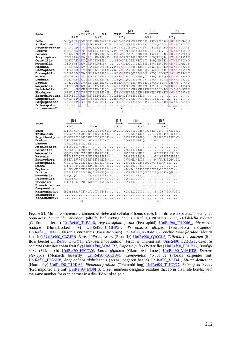
212
Figure 91. Multiple sequence alignment of SeFe and cellular F homologues from different species. The aligned sequences: Megachile rotundata (alfalfa leaf cutting bee) UniRef90_UPI000258F7DF, Helobdella robusta (Californian leech) UniRef90_T1FA15, Acyrthosiphon pisum (Pea aphid) UniRef90_J9LX66_, Megaselia scalaris (Humpbacked fly) UniRef90_T1GHP5_, Psorophora albipes (Psorophora mosquitoe) UniRef90_T1DI06, Nasonia vitripennis (Parasitic wasp) UniRef90_K7JGM3, Branchiostoma floridae (Florida lancelet) UniRef90_C3ZJR6, Drosophila lutescens (Fruit fly) UniRef90_Q30CL5, Tribolium castaneum (Red flour beetle) UniRef90_D7GY13, Harpegnathos saltator (Jerdon's jumping ant) UniRef90_E2BQZ1, Ceratitis capitata (Mediterranean fruit fly) UniRef90_W8AJR2, Daphnia pulex (Water flea) UniRef90_E9HB17, Bombyx mori (Silk moth) UniRef90_H9JCV6, Lottia gigantea (Giant owl limpet) UniRef90_V4AHE8, Danaus plexippus (Monarch butterfly) UniRef90_G6CIW0, Camponotus floridanus (Florida carpenter ant) UniRef90_E2A5H8, Anoplophora glabripennis (Asian longhorn beetle) UniRef90_V5I841, Musca domestica (House fly) UniRef90_T1PDA3, Rhodnius prolixus (Triatomid bug) UniRef90_T1HQD7, Solenopsis invicta (Red imported fire ant) UniRef90_E9J0H3. Green numbers designate residues that form disulfide bonds, with the same number for each partner in a disulfide-linked pair.

213
Structural studies of SeF full-length ectodomain
In order to obtain structural information about 6HB which is lacking in SeFet crystal
structure, I attempted to obtain and crystallize the post-fusion trimer of the full-length SeF
ectodomain. Previous observations have shown that low-pH triggered trimerization of the
SeFe containing a wild type fusion peptide results in exposure of the hydrophobic fusion
peptide, which leads to protein aggregation. Trypsin treatment of SeFe harbouring a mutated
furin cleavage site yielded a proteolytic fragment of the protein lacking the fusion peptide,
allowing successful acid-induced trimerization. It has been described in the literature that the
post-fusion forms of viral fusion protein ectodomains can be obtained when the hydrophobic
fusion peptide is deleted or its hydrophobic nature altered. Therefore, in order to achieve the
trimerization of the full-length SeFe, I produced three new constructs of SeFe (all harboring a
wild type furin cleavage site): 1) SeFeΔ1 containing the deletion of the fusion peptide
residues 150-GLFNFMGHV-158 2) SeFeΔ2 containing the deletion of the fusion peptide
residues 150-GLFNFMGHVDKYLF-163 and 3) SeFe-mut containing the hydrophobic
residues within the fusion peptide replaced by the hydrophilic ones (150-GLFNFMGHV-
158è150-GQTNSHGHN-158). In SeFeΔ1 and SeFeΔ2 constructs a short GGS linker was
introduced instead of the deleted fusion peptide sequences. All proteins were expressed in
Drosophila S2 cells yielding 6-10 mg/L of supernatant. SEC purification of SeFeΔ1 and
SeFeΔ2 revealed that these proteins are monomeric at pH 8. In contrast, SeFe-mut formed
both monomers and trimers at pH8. The SeFe-mut trimer was stable at pH 8 and presumably
represented a pre-fusion trimer. This trimer was subjected to crystallization trials and crystals
were obtained, albeit diffracting only to ~10 Å. The formation of post-fusion trimers was
attempted with monomeric forms of SeFeΔ1, SeFeΔ2 and SeFe-mut by incubating them in 30
mM NaAc pH 5. Incubation of SeFeΔ2 at room temperature for two hours in 30 mM NaAc
was sufficient to achieve a complete trimerization, while SeFeΔ1 and SeFe-mut required
overnight incubation at room temperature for the formation of a post-fusion trimer.
Intensive crystallization trials with these proteins were performed, which resulted in
obtaining crystals under different crystallization conditions. Unfortunately, all tested crystals
diffracted to low resolution. The best diffraction (6.5 Å) was obtained for the SeFeΔ1 trimer
after removal of the Strep-tag and deglycosylation of the protein with EndoD (space group
I222; cell unit dimensions: a=111.13 b=117.09 c=348.37 α = β = γ = 90.000 deg.). The
packing of this new crystal form provides an extra space for accommodating the extension of
the trimer, which is lacking in the trypsin truncated crystal form. Moreover, the analysis of the

214
crystal lattice shows that the crystal packing would not be stable without the extra chains
joining the consecutive layers in the direction of the long axis (c=348.0) (Figure 92). At the
moment I am trying to further improve the resolution of this crystal form, which would allow
the extension of the current model of SeFet.
Figure 92. Crystal packing of SeFeΔ1 timer. The model of SeFet (truncated post-fusion trimer) was used for molecular replacement. Crystal packing analysis reveals an extra space for extension of the molecule.
To further delineate the structure of the full-length SeFet post-fusion form, we
performed electron microscopy (EM) studies on the SeFe-mut post-fusion trimer. EM was
performed by Xiaokang Zhang, a post-doc in our laboratory (Figure 93). Docking SeFet
crystal structure into the 30-50 Å resolution reconstruction map revealed an extra extensive
density corresponding to the 6HB which is absent in the crystal structure (Figure 94).

215
Figure 93. Reference-free 2D class averages of negatively stained SeFe-mut post-fusion trimer.
Figure 94. EM reconstruction of SeFe-mut post-fusion trimer. The crystal structure of SeFet (displayed as cartoon) is fitted within the electron density. (A and B) Side view and top view, respectively, at contour level of the density 0.651. (C and D) Side view and top view, respectively, at contour level of the density 0.162.

216
Supplementary data
Figure 95. The amino acid sequence of the SeFe model adorned with secondary structure elements by ENDscript 2 (helices with squiggles, β-strands with arrows and turns with TT letters)(Gouet et al., 2003). Green numbers designate residues that form disulfide bonds, with the same number for each partner in a disulfide-linked pair. Solvent accessibility is rendered by a first bar below the sequence (blue is accessible, cyan is intermediate, white is buried) and hydropathy by a second bar below (pink is hydrophobic, white is neutral, cyan is hydrophilic).
TT SeFet_C
TT SeFet_C
TT TT TT TT TT TT TTT SeFet_C
TT SeFet_C
β1 β2 α1 α2 α3
α4 α5 η1 α6 β3
β4 β5 β6 β7 β8 β9 β10 β11 β12 α7 β13 β14 β15
β16 β17 β18 β19 β20 β21
30 40 50 60 70 80 90 100 110 120 130 140 150 160
SeFet_C KDIVQVTPLPSTSGLYFQYINRMQFVNIIWHFVIEMDHGSVFYRLQSIHQQAQKLQQSFISLRQXXXXXXXXCANVKYLKLEIDHMLSTVIPNLAQQHNLLDQKVPLTPSNATLTXXXXXXXXXXXXXXXXXXXXXXXXX acc hyd
1 SeFet_C A A A AAA A A A A A A AA A A A A A A B C B C C C CC
170 180 190 200 210 220 230 240 250 260 270 280 290 300
SeFet_C XXXXXXXXXXXXXXXXXXXXXXXXXXXXXXXXXXXXXXXXXXXXXXXXXXXXXXXQCRYIIENYNILCKQLDEVATLYNKLDLAVDNAKLNHLNSFVVSPERLLNEMNNVSGHLAGLSWPVPLTEKAMHVLIDNVINVHV acc hyd
1 SeFet_C ABAA A B AA A A A B B BA A A A B B B A
310 320 330 340 350 360 370 380 390 400 410 420 430 440
SeFet_C FVTAERKLLFIIEVPLVSSEAFDVFHSIPLPYCDKSHKCAIMLPDSKYLGVSVDRRNYVRLDDTTSCRMSDKVMLCFRPQIIYDVNQAKLCDVRIFMKNDAAIDYAKDCDVRVGRFESELFYATSDYNNWLYVLQNDIDL acc hyd
2 2 3 3 4 4 SeFet_C A A A AAA A AA B B BB A A AAAA A A B B B C CC C
450 460 470 480 490 500
SeFet_C NIQCIPSATITDGFGIAPVVLRAGVGIIHATGNDNCKLTTKKSRLTVHDLYNNLNTVIEIPMGLSYN acc hyd
5 5 SeFet_C B B B B BB B B B B B B B B B B B B B B B A BB
217
Discussion
Evolutionary links revealed by baculovirus F protein structure
Together with our collaborators from Utrecht University we characterized the
trypsin-truncated SeMNPV F ectodomain (SeFe) and showed that low pH-induces
conformational changes within the protein leading to formation of a stable post-fusion
trimer. After intensive crystallization attempts, diffracting crystals of the post-fusion
trimer of SeFe trypsin fragment (SeFet) were obtained. The structure determination of
SeFet confirmed the previous predictions that baculovirus F protein exhibits a class I
fusion protein fold.
The recombinant SeFe contained a mutated furin cleavage site in order to
prevent furin cleavage and, as a result, stabilize the protein in its pre-fusion form.
Based on the knowledge that furin cleavage occurs in the trans-Golgi network, the
mutation was not expected to impede protein folding. In addition, available pre-fusion
structures of other class I fusion proteins (influenza HA and paramyxovirus F) in their
cleaved and uncleaved forms, are largely superimposable (Chen et al., 1999a; Welch
et al., 2012). Proteolytic activation is required for the majority of class I fusion
proteins to induce membrane fusion, i.e. for conformational changes to reach the post-
fusion state. When altering the furin cleavage site, two arginine residues were
introduced within this site in order to allow proteolytic activation of the protein by
trypsin cleavage. We have previously observed that the wild type SeFe cleaved by
furin aggregates if incubated at low pH, which could be explained by exposure of the
hydrophobic fusion peptides that cluster together in the absence of cellular membrane.
As illustrated by examples of other class I fusion proteins, obtaining a soluble form of
post-fusion trimer usually requires removal of the fusion peptide (Ruigrok et al.,
1988; Swanson et al., 2011).
Trypsin cleavage of the mutated SeFe yielded a stable proteolytic fragment of
the protein lacking the fusion peptide, which was able to form a trimer when exposed
to low pH. This trimer was presumably corresponding to the post-fusion form based
on the observed low pH dependent fusion of the baculovirus F protein (Westenberg et
al., 2002). The conformational changes induced in the SeFe trypsin fragment by low
pH were irreversible, which is consistent with the transition of a metastable pre-fusion
form to a more stable post-fusion conformation, which is a general feature of the

218
majority of viral fusion proteins. SeFe with knocked-down furin cleavage site
expressed in S2 cells was secreted as a monomer, which is in contrast to the observed
trimeric state of the full-length protein (Long et al., 2006b). The monomeric state of
SeFe in solution could indicate the importance of the transmembrane (Holland et al.)
domain in protein trimerization. Both class I and class III fusion proteins lacking TM
domain have been reported to trimerize inefficiently (Albertini et al., 2012; Bullough
et al., 1994; Singh et al., 1990; Smith et al., 2013; Wurdinger et al., 2005). TM-TM
interactions have been suggested to be important for the stability of pre-fusion forms
of paramyxovirus F proteins (Smith et al., 2013). The crystal structures of pre-fusion
forms of parainfluenza virus 5 (PIV 5) F protein and RSV F protein trimers were
obtained only when the TM domain was replaced with a trimerization domain
(GCN4t or T4 fibritin, respectively) (McLellan et al., 2013; Yin et al., 2006).
Trimeric interfaces of paramyxovirus F and influenza HA proteins are significantly
different in their pre-and post-fusion forms (Bullough et al., 1994; Wilson et al.,
1981; Yin et al., 2005; Yin et al., 2006) revealing weaker interprotomer interactions
in the pre-fusion forms. Post-fusion forms of these proteins are stabilized via
hydrophobic interactions of the HR regions forming the 6-helix bundle. The trypsin
cleaved SeFe formed a stable post-fusion trimer after acid-induced conformational
changes, which also promoted transition from a monomeric to a trimeric state.
The crystal structure revealed that SeFet lacks HR1 and HR2 regions and, as a
result, does not form a 6-helix bundle, which is believed to contribute the most to the
stability of the post-fusion state of class I fusion proteins. Instead, the assembly and
stabilization of SeFet seems to depend mainly on a trimeric coiled coil formed by the
HR3 segments. We complemented our crystal structure with a negative stain EM
reconstruction of the post-fusion form of the full-length SeFe ectodomain. The
visualization of the molecule in the EM map revealed that the molecule has a shape
and size characteristic of the post-fusion trimer of the paramyxovirus F protein and
allowed us to confirm the presence of the six-helix bundle at the trimer axis. The
trypsin-truncated SeFet crystal structure was docked into the EM map, clearly
revealing extra density corresponding to the 6-helix bundle, which is absent in the
crystal structure. Taken together, the EM density map and the X-ray crystallography
data provide evidence that baculovirus F protein is a typical class I fusion protein.
SeFe requires low pH in order to undergo conformational changes from the
pre-fusion to the post-fusion form. We did not observe formation of post-fusion

219
trimeric forms at neutral pH for either trypsin-cleaved or uncleaved ectodomain. This
is in contrast to F proteins from paramyxoviruses, which are able to catalyze the
membrane fusion reaction at neutral pH. Thus, despite structural similarities between
baculovirus F and paramyxovirus F proteins, conformational changes within these
proteins leading to the fusion reaction are induced by different triggers.
Conformational changes in paramyxovirus F proteins from PIV3 and PIV5 are
triggered by the HN attachment protein and in others like RSV and SV5 F proteins
upon receptor binding (reviewed in (Bossart et al., 2013)). The secreted hPIV3 F
ectodomain with a knocked-down furin cleavage site has been shown to adopt
primarily the post-fusion conformation instead of the expected pre-fusion form. This
indicates that the energetic barrier for transition of paramyxovirus F ectodomain from
pre-fusion to post-fusion form is rather low, once the stabilizing attachment protein is
removed.
Remarkably, SeFe contains 20 histidine residues compared to only 3 found in
the RSV F ectodomain. The protonation of histidine residues has been reported to
trigger conformational changes of pH-dependent fusion proteins leading to membrane
fusion (Kampmann et al., 2006). In the post-fusion form of viral fusion proteins, a
number of conserved histidine residues often tend to form salt bridges with negatively
charged residues that are supposed to have a stabilizing effect. Low numbers of
histidine residues in RSV F ectodomain are in line with its low-pH independent fusion
mechanism. Surprisingly, despite a high number of histidine residues, SeFet crystal
structure indicates that none of these residues participate in salt bridges, suggesting
that they have only a more general effect of increasing surface charge.
The SeFet crystal structure represents the first crystal structure of a class I
fusion protein from a DNA virus. Moreover, it also confirmed the predicted
similarities between baculovirus F and paramyxovirus F proteins, with the post-fusion
form of RSV F protein (PDB accession code: 3RKI) having the highest DALI score.
The conservation of the structural features between baculovirus F and paramyxovirus
F proteins suggests that both proteins have a common ancestor, which has been
already predicted from the sequence similarity (Misseri et al., 2003). Remarkably,
baculoviruses and paramyxoviruses are distant viral families. First of all they belong
to different groups of viruses: group I (double-stranded DNA viruses) and group V
(negative-sense single stranded RNA viruses), respectively (Baltimore, 1971).
Second, they have different hosts: baculoviruses infect invertebrates, primarily

220
insects, while paramyxoviruses infect vertebrates and have a very broad host range.
Finally, these viruses use different entry mechanisms to their host cells: baculovirus F
proteins function both as receptor binding and fusion proteins and induce fusion in the
endosomes upon acidification while most of paramyxoviruses have separate receptor
and fusion proteins and fuses directly with the cellular membranes at neutral pH.
VSV G and herpes simplex virus glycoprotein B is another example of the
homologous fusion protein in very different viruses, one RNA and the other one DNA
(Heldwein et al., 2006; Roche et al., 2006). Moreover, they do not infect the same
hosts either. Therefore, the route by which baculoviruses and paramyxoviruses
acquired the same common ancestral fusion protein is uncertain. The origin of RNA
and DNA viruses is still debatable (Forterre, 2006; Holmes, 2003), therefore, it is not
clear if paramyxoviruses and baculoviruses acquired F proteins independently during
evolution (probably from a cellular F ortholog) or if one of the two viral families was
the source of the F protein.
Intriguingly, the DUF3609 domain found in baculovirus proteins has been also
discovered in some cellular proteins and retroviral elements suggesting that that they
evolved from the same ancestral protein, which could be either viral or eukaryotic
origin (Lung & Blissard, 2005). The SeFet crystal structure revealed that the
DUF3609 domain resides within the globular head domain of SeFet. Therefore, it is
unlikely that the DUF3609 domain is involved in membrane fusion. Presumably, it
plays a role in cellular receptor recognition. Analysis of the conserved solvent-
exposed residues in the DUF3609 domain revealed a few conserved patches on the
surface of the domain that could potentially represent a receptor binding site and/or
some other unidentified functional site. The fusion activity of currently identified F
cellular proteins from Drosophila melanogaster, Drosophila yakuba, Drosophila
pseudoobscura and Anopheles gambiae have not yet been demonstrated. Although it
is possible that the fusion activity was not observed due to assay conditions and these
proteins do contain other domains than DUF3609 mediating fusion, it is also likely
that cellular F proteins have completely different function.
The prevalence of f-like genes in the genomes of many insects suggests that
baculovirus f gene or at least a DUF3609 domain could be acquired directly or
indirectly from an insect host. The indirect route might include insect endogenous
retroviruses encoding f-like genes as they have been reported to be able to insert into
the baculovirus genome (Malik et al., 2000; Rohrmann & Karplus, 2001). However,

221
the direction of horizontal gene transfer is ambiguous given the fact that errantiviruses
have been proposed to contain the env gene that they acquired from baculoviruses
(Malik et al., 2000).
We further extended the search of F-like proteins in different organisms using
the SeFe sequence. A PSI-BLAST search yielded previously unidentified F-like
protein sequences in the genomes of a number of eukaryotes, mainly insects (phylum
Arthropoda). Intriguingly, putative homologous proteins to baculovirus F were found
in Branchiostoma floridae (Florida lancelet) and Lottia gigantea (Giant owl limpet)
belonging to phyla Chordata and Mollusca, respectively. Branchiostoma floridae
represents one of the closest living invertebrate relatives of vertebrates (Delsuc et al.,
2006). Although lancelets split from vertebrates more than 520 million years ago,
some studies revealed that vertebrates have employed the genes found in lancelets and
changed their regulation or even function (Holland et al., 2008). If vertebrates also
contain yet unidentified f genes, the f gene found in Branchiostoma floridae may
represent a common ancestor from which they evolved. Our results indicate that f-
like genes might be even more widespread than previously thought. It is important to
recognize, however, that more detailed analysis is required to distinguish if these
genes are not part of a previously integrated retrovirus genome. Determination of the
structures of F-related cellular proteins would be helpful to elucidate the evolutionary
link between viral and cellular F proteins. In addition, it might also help to understand
the function of cellular F proteins.
Baculovirus F protein not only functions as a fusion protein but also interacts
with unknown cellular receptors at the surface of host cells. In order, to identify
residues that potentially could be involved in the receptor binding, we attempted to
identify surface exposed residues conserved among the 39 different baculovirus F
proteins. The analysis revealed several well-defined conserved patches on the surface
of the SeFet post-fusion trimer. Nevertheless, since the F protein should interact with
the cellular receptor before the fusion, the structure of pre-fusion form of F would be
more appropriate to identify the conserved surface residues potentially involved in
receptor recognition.
Though our EM data of the full-length SeFe ectodomain post-fusion trimer
revealed the presence of a 6-helix bundle in this molecule, the crystal structure of the
full-length molecule would provide more insights into the fusion mechanism of
baculovirus F protein. We have produced several constructs of SeFe allowing us to

222
successfully form the full-length post-fusion trimer. The crystallization of these SeFet
variants is underway. Though crystals of the full-length SeFet in its post-fusion form
have already been obtained, they diffracted to low resolution (~7 Å). In addition, we
observed that the full-length SeFe containing the fusion peptide in which the
hydrophobic residues were mutated to hydrophilic ones yields a stable trimer at
neutral pH in addition to a monomer. This trimer presumably corresponds to the pre-
fusion trimer. Initial crystallization trials of the putative pre-fusion trimer have been
performed and the crystals obtained, however, the crystals diffracted only to ~10 Å.
In summary, we have developed the protocols to form a stable trimer of post-
fusion baculovirus F ectodomain and determined the crystal structure of trypsin
truncated SeFet. The formation of a trimer is induced by low pH treatment, which
induces irreversible conformational changes within the protein and the transition from
monomeric pre-fusion state to a stable trimeric post-fusion state. In order to prevent
the pH triggered aggregation of the protein, the fusion peptide must be removed or
hydrophobic residues within the fusion peptide must be replaced by the hydrophilic
ones. The crystal structure of trypsin-truncated SeFet confirmed the evolutionary ties
between baculovirus and paramyxovirus F proteins. Our results also suggest that
solving the structures of F homologues found in eukaryotes is important for a better
understanding of the evolutionary link between viral and cellular F proteins.

223
Materials and Methods
Constructs used or generated in the study
The gene encoding a soluble ectodomain of baculovirus F protein encoding
amino acid residues 18-553 of the full-length SeMNPV F protein (UniProtKB
accession number Q9J8C6) was inserted into pT350 (described in Supplementary
materials and methods) by Qiushi Wang (Utrecht University, the Netherlands). The
residues 1-17 were excluded from the construct because they comprise the signal
peptide. This construct exceptionally contained a triple Strep-tag instead of the double
Strep-tag. The stable cell line was generated by a previous lab member Scott Jeffers.
SeFeΔ1 containing the deletion of the fusion peptide residues 150-
GLFNFMGHV-158 2) SeFeΔ2 containing the deletion of the fusion peptide residues
150-GLFNFMGHVDKYLF-163 and 3) SeFe-mut containing the hydrophobic
residues within the fusion peptide replaced by the hydrophilic ones (150-
GLFNFMGHV-158è150-GQTNSHGHN-158) were generated site-directed ligase
independent mutagenesis (SLIM)(Chiu et al., 2008). In SeFeΔ1 and SeFeΔ2
constructs a short GGS linker was introduced at the same time instead of the deleted
fusion peptide sequences.
Limited proteolysis
Purified SeFe protein in HNE buffer (0.15 NaCl, 0.1 M EDTA, 5 mM Hepes
pH7) at 1 mg/ml was incubated with the indicated concentrations of TPCK (L-1-
tosylamide-2-phenylmethyl chloromethyl ketone)-treated trypsin (Sigma) at a
trypsin:SeFe ratio of 1:800 (w/w) for 30 min at 23 oC Digestion was terminated by
addition of PMSF to the final concentration of 0.2 mM (Sigma). The cleaved protein
was subsequently analyzed by SDS-PAGE stained with Coomassie Blue. Target
protein bands were analyzed by N-terminal sequencing.

224
Acid-treatment of SeFe samples
Different variants of purified SeF ectodomain protein as well as the (SeFe,
SeFe trypsin truncated fragment, SeFe-mut, SeFeΔdel1 and SeFeΔdel2) were exposed
to low pH by adding NaAc pH5 to a final concentration of 30 mM and incubated over
night at +4 oC.
Multiangle light-scattering analysis
Multi-Angle Light Scattering (MALLS) is an analytical technique for
determining absolute molar masses and the average size of particles in solution of all
types of macromolecules including proteins by calculating the amount of scattered
light at different angles. The sensitivity of the light scattering detector becomes
increases with molar mass of the analyte. Thus it is an excellent tool for detecting
oligomeric state of the proteins. The higher the aggregation number, the more
sensitive the detector becomes. MALLS detector is often coupled downstream to a
chromatographic system allowing using SEC, which together provides means for
measuring the molar mass, size, and distribution
(http://www.wyatt.eu/index.php?id=multi-angle-light-scattering).
The absolute molecular masses of different SeFe samples were determined by
gel filtration combined with detection using MALLS and refractrometry (Wyatt,
1998). Purified protein (90 µg) was loaded onto a Superdex 200 10/300 column (GE
Healthcare) connected to an MALS instrument and an interferometric refractometer
(DAWN HELEOS II, Wyatt Technologies, Santa Barbara, CA). The column was
equilibrated either at pH5 (10 mM NaAc pH5, 150 mM NaCl, 0.1 mM EDTA) or pH7
(5 mM HEPES pH7, 150 mM NaCl, 0.1 mM EDTA), respectively. The absolute
molecular masses were calculated using the ASTRA software (Wyatt Technology
Corp., Santa Barbara, CA).
Deglycosylation of SeFet
Production of endodeglycosidases PNGase, EndoH and EndoD is described in
Supplementary materials and methods. For crystallization trials, SeFet was
deglycosylated with PNGase F using the ratio 1:16 of endoglycosidase:SeFet on a

225
weight basis. For deglycosylation of SeFet with EndoD-EndoH, the ratio 1:4:28
(SeFet:EndoD:EndoH) on a weight basis was used. Deglycosylation reactions were
carried out at 37oC. Deglycosylated SeFet was separated from PNGase by SEC on a
Superdex 200 column (GE Healthcare). EndoD and EndoH were removed from the
reaction mixture by Ni2+ion affinity chromatography.
Removal of the Strep affinity tag
A C-terminal triple Strep tag preceded by an enterokinase recognition site was
removed from the monomeric SeFe-mut, SeFeΔdel1 and SeFeΔdel2 prior
trimerization at low pH by specific proteolytic cleavage with EKMax Enterokinase
(Invitrogen, San Diego, USA). The detailed protocol is provided in Supplementary
Materials and Methods.
Evaluation of accessibility of free cysteine residues in SeFet
The accessibility of the cysteine residues in SeFet was evaluated by DTNB-
thiols assay. The detailed protocol is provided in Supplementary materials and
methods.
Crystallization
Crystallization screening, crystal optimization and crystal cryo-protection
techniques are in detailed described in Supplementary materials and methods. The
best-diffracting crystals of SeFet deglycosylated with PNGase (P1 crystal form) were
grown at 2 mg/ml SeFet in 2.5 µl hanging drops (1:1:0.5 protein:reservoir
solution:seed stock volume ratio) by vapor diffusion against a reservoir solution
containing 16-18% PEG 3350 and 200 mM ammonium chloride. The crystals of
deglycosylated SeFet grown in 9% PEG 4000, 100 mM imidazole pH 8 and 30% 2-
methyl-2,4-pentanediol (MPD) were used as a seed stock for microseeds. The P43212
crystal form grew under the same conditions when instead of the seed stock just a
solution of 9% PEG 4000, 100 mM imidazole pH 8 and 30% 2-methyl-2,4-
pentanediol (MPD) was used.

226
Dehydration of the crystals
The dehydration experiments were performed on SeFet crystals to improve the
diffraction resolution of those crystals. Dehydration removes excess solvent from the
crystals which may result in tighter packing of protein molecules and a subsequent
increase of the X-ray diffraction of the crystals. Two techniques were applied for the
dehydration of SeFet crystals: 1) the cover slip with a hanging drop containing the
crystals was transferred over a reservoir solution with a higher percentage of
precipitant and allowed to equilibrate for 3 days, and 2) the crystals were directly
transferred into a dehydrating solution consisting of the mother liquor with a higher
percentage of precipitant, and dehydrated over a reservoir solution containing the
same dehydrating solution for 3 days.
Soaking crystals in heavy atom solutions
In order to obtain the heavy atom derivative of SeFet crystals, the crystals
were soaked in a number of different heavy atom compounds listed in Table 3 in the
Results chapter. All compounds except hexatantalum tetradecabromide (Ta6Br122+)
were available from Heavy atom screens (Hampton Research). Ta6Br122+was kindly
provided by Gérard Bricogne (Global Phasing Limited, Cambridge, UK). Ta6Br122+
has been reported in the literature to be a powerful derivatization agent.
For soaking experiments, 100 mM stock solutions of different heavy atom
compounds were prepared in water. The heavy atom soak solution for soaking SeFet
crystals was composed of mother liquor containing 20 % (v/v) glycerol and a heavy
compound at a specific concentration. Various concentrations of the heavy atom
compound (0.5-20 mM) as well as various soaking times (from 10 min to 18 h) were
tried. 2 µl of the heavy atom soak solution was pipetted onto a new siliconized cover
slip and the crystals were transferred from the mother liquor to the drops of the heavy
atom soak solution. The cover slip with a drop was placed above a well containing
mother liquor supplemented with 20 % (v/v) glycerol.
SeFet crystals derivatized with Ta6Br12+ were obtained by soaking crystals in a
soak solution containing 1 mM Ta6Br12+ for 18 h. The crystals were back-soaked by
transferring the crystals into mother liquor containing 20% glycerol (v/v) and flash-
frozen in liquid nitrogen.

227
Production of selenomethionine substituted SeFe
A selenomethionine (SeMet) substituted SeFe was produced in ESF-921
serum-free medium methionine-free medium (Expression Systems) supplemented
with L-SeMet. Initially, 3 l of the S2-SeFe-expressing cells were grown in Insect
Express medium until it reached the density of ~20×106/ml. The cells were collected
by centrifugation for 5 min at 200 g and resuspended in 1.5 L of ESF-921 serum-free
methionine-free medium supplemented with 0.8 g/L L-cysteine. After 4 h of starving,
300 mg/L L-SeMet (Acros Organics, Geel, Belgium) and 4 µM CdCl2 were added.
An extra 300 mg/L L-SeMet were added at day 3. The supernatant was harvested 5
days after induction. The substituted protein was purified in the same way as the
native protein but the yields were 10-fold lower.
Structure determination of SeFet
Native as well as MAD (Multi-wavelength anomalous dispersion) and SAD
(Single-wavelength anomalous diffraction) data sets were collected at the Synchrotron
Soleil beamline Proxima 1, the Swiss Light source beamline PX I or European
Synchrotron Radiation Facility (ESRF) beamlines ID23-1, ID14-4 and ID23-2 at 100
K. Typically, diffraction data were collected using Pilatus (Dectris, Baden,
Switzerland) or charge-coupled device (CCD)-based detectors. Programs iMosflm
(Leslie, 2006; Powell, 1999) or xdsme were used to determine the optimum
orientation of the crystal for the complete data set collection. The best native data sets
of X-ray diffraction data for P43212 and P1 crystal forms (2.9 Å and 2.7 Å resolution,
respectively) were processed using XDS (Kabsch, 2010).
The initial experimental phases were obtained by the MAD method using the
Ta6Br122+ derivative of P1 crystal forms. Data for this heavy-atom derivative were
collected on a single crystal using an inverse beam data collection strategy with
wedges of 10 degrees while cycling through three wavelengths (peak, remote
inflection). This strategy was used in order to optimize the measurement of Friedel
pairs. In addition, due to crystal sensitivity to radiation damage, the collection of
MAD data sets was performed with reduced beam intensity. The data sets obtained at
each wavelength were processed using XDS and scaled together using XSCALE
(Kabsch, 2010). SHELX C/D (Schneider & Sheldrick, 2002) within SHARP

228
(Bricogne et al., 2003) was used to locate the heavy atom sites (Schneider &
Sheldrick, 2002). Experimental phasing was performed with SHARP followed by
solvent flattening, which provided initial experimental phases. The obtained map was
used to build an initial model in polyalanines. NCS averaging was applied to further
improve the map. Better phases were obtained with a MAD experiment using the
Ta6Br122+ derivative of the P43212 crystal form. This data set was collected using an
inverse beam data collection strategy with wedges of 10 degrees while cycling
through the two wavelengths of falling and mounting inflection (1.255070 (9879 eV),
1.254140 (9886eV). In the case of the P43212 crystal form Ta6Br122+ derivative,
individual Ta atoms were identified using SHARP.
The phases in P43212 crystal form were combined with the phases of the initial
model built in P1 crystal form. After applying density modification and multi-crystal
averaging techniques, better quality maps were obtained for both crystal forms
allowing the complete building of the model. NCS averaging and multi-crystal
averaging was performed using the DM (CCP4 suite), and
phenix.multi_crystal_average (Terwilliger, 2002). The building of the model was
performed manually in Coot (Emsley et al., 2010) alternating between the electron
density maps in different crystal forms. Manual building was supported by anomalous
data for a SeMet derivative from the P43212 crystal form collected at the peak
wavelength as well as the highly redundant Sulfur-SAD data set at a wavelength of
1.7995 Å from the native crystal in P1 form. The density for the anomalous scatterers
(selenium and sulphur) was calculated using AnoDe (Thorn & Sheldrick, 2011).
Refinement was performed using AutoBuster (Bricogne G, 2010) against both the P1
and the P4 data sets imposing NCS restraints and TLS groups. Refinement was
monitored following the Rfree Rwork and Rfree values. Rfree values were calculated for a
random subset (5 %) of reflections omitted from refinement. Water molecules were
added manually using Coot. Throughout the refinement, a structure-validation web
service MolProbity (Davis et al., 2004) was used to monitor all-atom contact analysis
as well as Ramachandran and rotamer distributions.
Crystal structure analysis
Electrostatic potentials were calculated using the adaptive Poisson Boltzmann
solver (Baker et al., 2001). The protein interaction calculator (Molina et al.) (Tina et

229
al., 2007) was used to identify the interactions between the protomers of SeFet. The
surface area buried upon trimer formation was estimated using the PISA server
(Krissinel, 2007). The topology diagram of SeFet protomer was generated using
program PDBsum (Laskowski, 2009). SeFet structure comparison with other protein
structures available in the PDB was carried out using DALI server (Holm &
Rosenstrom, 2010). Sequence alignments were performed using MultAlin (Corpet,
1988); http://multalin.toulouse.inra.fr/) and formatted with ESPript 3 (Gouet et al.,
2003); http://espript.ibcp.fr/). Sequence conservation among SeFe and F proteins from
different baculoviruses or different putative cellular F orthologs was analyzed by the
program ConSurf (Glaser et al., 2003). Figures of the crystal structures were prepared
in the PyMOL Molecular Graphics System (www.pymol.org).

230
Final Discussion
My thesis research was initially focused on the structural analysis of the GB
virus B envelope protein E2. Despite extensive efforts described in this thesis I was
unable to obtain diffraction-quality crystals of this protein. During the course of this
project I did, however, learn a number of important techniques such as construct
design and optimization, protein expression and purification using several different
techniques, protein deglycosylation, limited proteolysis, protein crystallization as well
as generation and expression of ligands in form of antibody fragments.
As a result, the experience obtained during this initial project was extremely
useful and subsequently applied to the following two projects I pursued. The first of
these projects focused on the characterization of an important neutralizing epitope
within HCV E2 and the second project focused on the structural analysis of the
baculovirus F fusion protein.
In the course of the first project, I determined three structures of antibody
fragments in complex with a synthetic peptide mimicking the HCV E2 epitope
spanning aa 529-540 by molecular replacement. This experience allowed me to
improve my skills in this phasing technique that is frequently used in macromolecular
crystals.
In the second project, I determined the structure of a trypsin truncated
fragment of the baculovirus F protein using experimental phasing techniques such as
Multi-wavelength Anomalous Dispersion (Albertini et al.) and Single wavelength
anomalous dispersion (Drexler et al.). As described in this thesis, the phasing of
baculovirus F protein crystals was a difficult case that gave me the opportunity to
learn experimental phasing of challenging crystallization targets. Already the
challenge to obtain diffraction quality crystals of the baculovirus F protein fragment
allowed me to practice different crystal optimization techniques. Therefore, these
projects contributed greatly to my profound training in structural biology approaches.
Neither GBV-B nor baculovirus are viruses that infect humans, but the
structural characterization of the envelope glycoproteins of these viruses can provide
important insights, including both evolutionary and functional aspects, to understand
the mechanism-of-action of their counterparts in important human pathogens. For
example, the same fusion protein fold can be found in different viral families (e.g.

231
class II fusion proteins in Flaviviridae, Bunyaviridae and Togaviridae). However, as
the crystal structures of HCV and BVDV E2 glycoproteins reveal, in some cases
structurally divergent viral glycoproteins might exist within the same virus family.
The evolutionary origin of fusion proteins is still not clear and debatable. At
the moment there is not a single general principle explaining the origin of these
proteins. In some viruses the similar overall fold of the fusion protein seems to be the
result of divergent evolution (i.e., evolution from a common ancestor), while in other
viruses the similar overall fold likely results from convergent evolution (i.e., a similar
overall fold implying a specific fusion mechanism evolved in parallel from distinct
ancestor proteins).
For example, class II fusion proteins from the Flaviviridae, Bunyaviridae and
Togaviridae families appear to have evolved from a common ancestor. The amino
acid sequences of these proteins do not display any sequence similarity, but the
proteins adopt the same overall fold. Structural relationships between class I fusion
proteins from paramyxoviruses and baculoviruses described in this thesis offer a
second example of fusion proteins with a common ancestor, although their structural
homology was predicted from sequence analysis. VSV G and herpes simplex virus
glycoprotein B belonging to class III fusion proteins also represent homologous
fusion proteins and may have a common ancestor.
The role of divergent evolution in the origin of fusion proteins can be deduced
from the fact that structures of different class I fusion proteins can have distinct
characteristics despite the fact that they use a common fusion mechanism. Post-fusion
forms of class I fusion proteins from retro-, filo-, arena- and influenza viruses do not
harbor a globular head domain found in paramyxoviruses or coronaviruses although
all of these proteins form a trimeric coiled coil, indicating that they are not true
structural homologs. Therefore, they most likely do not have a common ancestry and
evolved independently to mediate membrane fusion. Interestingly, class I viral fusion
proteins share an α-helical coiled coil architecture and a similar fusion mechanism
driven by the zippering of the coiled coils with the SNARE family of intracellular
vesicle fusion proteins (Donald et al., 2011; Skehel & Wiley, 1998). This is also
considered to be an example of convergent evolution.
The fact that homologous viral fusion proteins are found in evolutionary
distant viral families (e.g., F proteins of paramyxoviruses and baculoviruses, which

232
are RNA and DNA viruses, respectively) raises the question how viral fusion proteins
were distributed across different viral families. There are two prevailing hypothesis
explaining this: 1) the origin of the fusion proteins is viral and different viruses
acquired their fusion proteins by horizontal virus-to-virus transfer, and 2) viruses
acquired their fusion machinery from their hosts (host-to-virus horizontal transfer).
While the true origin of viral fusion proteins remains to be elucidated, an
extensive horizontal gene transfer between different viruses as well as between
viruses and cells becomes more and more evident. In some cases, direct transfer of
cellular fusion protein genes between the viral genome and the germline is very
likely. The best example for this transfer are the syncytins, a protein family catalyzing
syncytial cell layer formation during placental development that is derived from
envelope proteins of endogenous retroviruses (reviewed in (Mi et al., 2000)).
Syncytin genes from different endogenous viruses were captured and domesticated by
various mammalian species independently on different occasions during evolution.
Another example that is less obvious is the C. elegans cell-cell fusion protein EFF-1
(Perez-Vargas et al., 2014), which was recently reported to adopt a class II fusion
protein fold and was possibly acquired from endogenous retroviruses carrying a
bunyavirus-like glycoprotein (Frame et al., 2001; Malik et al., 2000) - a family that
also carries class II fusion proteins (Dessau & Modis, 2013). On the other hand, in
some cases virus-to-virus horizontal transfer of a common ancestral membrane fusion
protein seems to be more likely (e.g., baculoviruses and paramyxovirus F proteins)
Given the complicated relationship between viruses and their hosts it is
difficult to elucidate the origin of specific fusion proteins. Which of these
mechanisms apply in the case of baculovirus F protein remains to be answered.
However, the benefit of structural studies on viral glycoproteins to understand these
evolutionary relationships cannot be debated anymore.

233
Supplementary Materials and Methods Cell lines
The Drosophila Schneider 2 (S2) cell line was purchased from Invitrogen.
This cell line was originally derived from a primary culture of late stage Drosophila
melanogaster S2 embryos. Drosophila Schneider 2 (S2) cells were grown at 28°C in a
normal atmosphere in a Schneider’s Drosophila medium (Invitrogen, Carlsbad, USA)
supplemented with 10% fetal bovine serum (FBS), 50 U/ml penicillin, and 50 µg/ml
streptomycin. The cells were maintained applying standard protocols provided by
Invitrogen. A remarkable feature of S2 cells is that they are able to integrate a high
copy number of plasmids into their genome which renders them suitable for high-
level expression of recombinant proteins. Stable cell lines expressing the recombinant
proteins were generated by co-transfection of the expression and selection plasmids as
described later. Stable cell lines were adapted to, and cultured in, serum-free Insect
Xpress media (Lonza, Basel, Switzerland) which was also used for protein
production. S2 cells grow as a loose, semi-adherent monolayer in tissue culture flasks
and are, thus, easily adapted to grow in suspension. For protein production, stable,
suspension-adapted cells were propagated in spinner flasks of different sizes (from 1
to 4 l) under continuous agitation at 90 RPM.
Expression vectors
For expression the recombinant proteins in Drosophila cells, a modified
version of the pMT/BiP/V5-His vector (Invitrogen) plasmid (Invitrogen) designated
pT350 was used. pT350 contains a double Strep-tag (IBA, http://www.iba-go.com/)
with a linker region (GlyGlySer)4 in between for efficient purification of recombinant
protein. An enterokinase cleavage site (DDDDK↑X) is introduced upstream of the
Strep-tag to allow specific removal of the tag. The vector contains the inducible
metallothionein promoter which is induced upon addition of divalent cations (Cu2+ or
Cd2+). A gene of recombinant protein is cloned in frame with Drosophila BiP signal
sequence at the N-terminus which serves for translocation of recombinant protein into
the endoplasmic reticulum lumen and directs the protein through the secretory
pathway of S2 cells into the culture medium.

234
The modified versions of the pT350 vector were used for expression of
recombinant Fab and scFv molecules as previously described (Backovic et al., 2010;
Gilmartin et al., 2012). The vectors are designated pMT-Fab-Strep and pMT-scFv-
Strep, respectively.
Polymerase chain reaction
Polymerase chain reaction (PCR) for amplification of a specific insert for
restriction cloning or blunt-cloning was performed using Phusion High-Fidelity DNA
polymerase (Finnzymes) as recommended by the producer. Typically the PCR
reaction contained: 0.5 µM of each primer (MWG Operon), 50 ng template plasmid
DNA, 200 µM dNTPs, 1 U Phusion High-Fidelity DNA polymerase (Finnzymes), 3%
DMSO, and 5×Phusion HF Buffer in a total volume of 50 µl. PCR was performed
under the following conditions:
Step Temperature,
oC Time Number of
cycles Initial denaturation 98 3 min 1 Denaturation Primer annealing Extension
98 X# 72
10 s 40 s 30s/kb
40
Final extension 72 10 min 1 Cooling 4 ∞ 1
#Primer annealing temperature was calculated according to the nearest-neighbour method
(Breslauer et al., 1986) DNA restriction digest, ligation and transformation
Purified PCR products and appropriate vectors were digested for 1–2 hours
with the corresponding restriction enzymes (New England Biolabs, Ipswich, MA)
according to the manufacturer's protocols. The digested DNA was verified by agarose
gel electrophoresis and purified using Macherey Gel and a PCR Clean-up Kit
(Macherey-Nagel). Linearized vectors were de-phosphorylated by Antarctic
Phosphatase (NEB) applying a protocol provided by the manufacturer. The DNA
vector and insert were ligated (3:1 molar ratio of insert DNA termini to vector DNA)
for 1 h at 16–25 °C using T4 DNA ligase (New England Biolabs). 2-5 µl of the
ligation mixture was used to transform 50 µl of TOP10 DH5α chemically competent
cells (Invitrogen) using standard procedures. 50-300 µl of cell suspension was spread

235
on LB -agar plates containing an appropriate antibiotic and incubated for 16–20 hours
at 37 °C. Typically one colony of the transformed DH5α cells was inoculated into 4
ml of LB supplemented with antibiotic and grown with agitation for 16-18 h at 37 °C.
The plasmid DNA was purified using a NucleoSpin Miniprep Kit (Macherey-Nagel)
as described in the kit manual. To verify the presence of an insert the purified plasmid
DNA was digested with appropriate restriction enzymes and analyzed by agarose gel
electrophoresis. The plasmid DNA containing an insert of the expected size was
sequenced for final validation (GATC Biotech AG, Constance, Germany or
Sequencing Facility of Institut Cochin, Paris, France).
Production of recombinant proteins in Drosophila S2 cells
For the large scale production of the recombinant proteins in Drosophila S2
cells, the corresponding stable cell line was expanded at 28°C to a final volume of 1-4
l in spinner flasks depending on recombinant protein expression level. The cells were
induced with 4 µM CdCl2 when they reached the density >8×10⁶ cells/ml. After 6-10
days the cells were pelleted by centrifugation for 15 min at 15 000 × g and the
supernatant concentrated using a Vivaflow 200 flip filtration concentration system
with 10 kD cutoff membrane (Sartorius Stedim Biotech GmbH, Goettingen,
Germany) to ~50 ml. The pH of the concentrated supernatant was adjusted by adding
Tris pH 8 to a final concentration of 100 mM. Avidin was added to 15 µg/ml to
prevent binding of the biotin present in the medium.
The supernatant was cleared by centrifugation at 20 000 × g for 30 min and
filtered with 0.2 mm cutoff membrane and loaded onto a Streptactin Superflow
column (IBA, Gottingen, Germany). The column was washed with 5 column volumes
(CV) of 0.1 M Tris pH 8, 0.15 M NaCl, 1 mM EDTA, and a recombinant protein was
eluted with 2 CV of the elution buffer containing 2.5 mM desthiobiotin, 0.1 M Tris
pH 8, 0.15 M NaCl, 1 mM EDTA. The eluate was further purified by size exclusion
chromatography using Superdex 200 or Superdex 75 column (GE Healthcare,
Uppsala, Sweden). Typically, SEC was performed at 15 in 10 mM Tris pH 8 and 150
mM NaCl. In the case of SeFe, a monomer was purified by SEC in 5 mM Hepes, 150
mM NaCl, and 0.1 mM EDTA (HNE) buffer. Protein fractions were analyzed by
SDS-PAGE. Based on the SDS-PAGE analysis fractions containing >95 % pure
protein were pooled and concentrated to approximately 20 mg/ml using a Vivaspin

236
centrifugal concentrator (Sartorius Stedim Biotech GmbH, Goettingen, Germany).
Protein concentrations were determined from the absorbance at 280 nm on a
NanoDrop ND-1000 spectrophotometer (NanoDrop Technologies, Wilmington, DE)
using calculated molar extinction coefficients from a program ProtParam (Gasteiger
et al., 2005). Purified recombinant proteins were immediately used for subsequent
experiments or stored at +4 °C.
Production of endodeglycosidases and deglycosylation
For expression of PNGase, 3 l of LB medium containing 50 pg/ml ampicillin
was inoculated 1/100 with an overnight culture of E. coli BL21 (DE3) strain
transformed with pBlueScript (Agilent Technologies)-EndoH. After growing the cells
at 37 oC to an OD550 of 0.5-0.6 the culture was cooled to room temperature.
Expression was induced with 0.5 mM isopropyl- I-thio-P-D-galactoside (IPTG) and
the culture was incubated overnight at 30°C.
The cells were harvested by centrifugation at 6000 g for 15 min and EndoH
was purified from the periplasm of the bacteria. The pelleted cells were resuspended
in 5% of the original culture volume in ice cold 20% sucrose, 0.1 M Tris pH 8 and 1
mM EDTA and pelleted again by centrifugation at 12000g 20 min. Then the cells
were resuspended in equivalent volumes of ice-cold water+Protease Cocktail (Sigma)
and incubated for 10 min on ice. After pelleting the cells at 16000g for 1h the
supernatant was collected, adjusted to 50mM TRIS pH 8.0, 1mM MgCl2, and filter
through a 0.22 µM cut-off membrane. The supernatant was loaded onto 5 ml of pre-
packed HP Nickel-sepharose (GE Healthcare) at 3.0 ml/min. After washing with
40mM imidazole, 50mM Tris 8.0, and 500mM NaCl, the protein was eluted with a
two-step gradient with 75 mM imidazole, 50mM Tris 8.0, 500mM NaCl and 300 mM
imidazole, 50mM Tris 8.0, 500mM NaCl. The elution fractions containing EndoH
were pooled and further purified by SEC on a Superdex 200 26/60 column (GE
Healthcare).
Essentially the same procedure was followed for the expression of EndoH
except that IPTG induction was performed at 20 hours at 20°C.
For expression of EndoD, 2 l of LB medium containing 50 pg/ml ampicillin
were inoculated 1/30 with an overnight culture of E. coli BL21 (DE3) strain

237
transformed with pET28a (Novagen)-EndoD. After growing the cells at 37 °C to an
OD550 of 1.0 the culture was cooled to room temperature. Expression was induced
with 0.5 mM isopropyl- I-thio-P-D-galactoside (IPTG) and the culture was incubated
for 20 h at 20 °C. The cells were harvested by centrifugation at 4000 g for 15 min and
EndoD was purified from the cytoplasm of the bacteria. The pelleted cells were
resuspended in 25ml 40 mM imidazole, 50 mM Tris 8.0, 500 mM NaCl buffer and
homogenized by two passages through an Emulsiflex-C5 homogenizer (Avestin). The
soluble fraction was separated by centrifugation at 35000 g for 30 min and filtered
through 0.22 µM cut-off membrane. EndoD was purified from the supernatant
applying the same protocol as for PNGase and EndoH.
Typically, deglycosylation of the native glycoproteins was carried out
overnight (approximately 16 hours) at 37°C in 50 mM sodium citrate pH5.5 (Endo H
and EndoD) or 50 mM Tris pH8. Initially, the amount of each endodeglycosidase
used for deglycosylation of the target proteins was optimized in small-scale reactions
at 37 oC. The glycoprotein:endodeglycosylase ratio at which maximal deglycosylation
was achieved as based on SDS-PAGE analysis was chosen and the reaction was
upscaled in a linear manner.
Removal of the Strep affinity tag
A C-terminal Strep tag preceded by an enterokinase recognition site was
removed from the recombinant proteins by specific proteolytic cleavage with EKMax
Enterokinase (Invitrogen, San Diego, USA). To cleave the tag from the recombinant
proteins, typically 1 unit of EKMax enterokinase was added to a protein solution at
0.66 mg/ml in 50 mM Tris pH 8, 50 mM NaCl, 1 mM CaCl2 and the reaction mixture
was incubated at 37°C. After 16 h 0.1 mM phenylmethylsulphonyl fluoride (PMSF)
was added in order to inactivate the protease. To remove any residual molecules
harboring Strep tag, the reaction mixture was loaded on to a Streptactin column and
the flow-through containing the protein molecules without the double Strep tag
collected. Subsequently, the collected flow-through was loaded onto the Superdex
200 column (GE Healthcare, Uppsala, Sweden). The fractions containing purified
protein without the Strep-tag were pooled and the protein was concentrated to
approximately 25 mg/ml in 10 mM Tris pH 8 and 150 mM NaCl buffer.

238
SDS-PAGE and Western blot analysis
For electrophoresis under reducing and non-reducing conditions, samples in
Laemmli sample buffer (125 mM Tris-HCl, 2% sodium dodecyl sulfate (SDS), 10%
glycerol and 0.001% bromophenol blue, pH 6.8) with or without 100 mM DTT were
denatured for 5 min at 98°C. Protein samples were subjected to SDS-PAGE
electrophoresis using standard methods (Sambrook and Russell, 2000) and stained
with Coomassie Blue (Bio-Rad) or subsequently transferred to PVDF membranes
(Bio-Rad) by wet electrophoresis transfer (Bio-Rad).
The membrane was blocked for 1h at room temperature in PBS-T (PBS with
the addition of 0.1% Tween 20) containing 5% dry milk. For detection of strep-tagged
proteins, the membrane was first incubated for 1 h at room temperature with mouse-
anti-Strep IgG (StrepMAB-Classic, IBA) for 1h at a dilution of 1:3000 in blocking
buffer. After washing the membrane 3 times with PBS-T for 5 min, rabbit anti-mouse
IgG conjugated with horseradish peroxidase was added at a dilution of 1:10000 in
blocking buffer and the membrane was incubated for 1h. After washing the membrane
three times for 15 min each time in PBS-T and once for 15 min in PBS, proteins were
detected using the ECL kit (GE Healthcare).
DTNB-Thiols Assay
Free cysteine residues in proteins were detected using the DTNB-thiols assay
which measures sulfhydryl groups with 5-5dithiobis[2nitrobenzoic acid] (DTNB)
reagent forming a measurable yellow-colored product when it reacts with sulfhydryls.
750 µM DTNB in 100 mM Tris pH8.0 was used as a working solution. 5 µl of L-
cysteine standards (500 µM, 750 µM, 1 mM 1.25 mM, 1.5 mM, 1.75 mM, 2 mM, 2.5
mM, 3 mM and 3.5 mM) were mixed with 160 µl working solution and 35 µl of
buffer (10 mM Tris, 150 mM NaCl) in microtiter wells and incubated at room
temperature for 10 min in order to obtain a standard curve. Similarly, 40 µl of diluted
protein sample was mixed with 160 µl working solution and incubated at room
temperature for 10 min. After 10 min absorbance of the solutions was measured at
412 nm. Absorbance values of L-cysteine standards were plotted versus concentration
to generate a standard curve. The concentration of the sulfhydryl groups in protein
samples was estimated from the standard curve.

239
Screening for crystallization conditions
To assess the appropriate protein concentration for crystallization screens, a
pre-crystallization experiment using PCT Pre-Crystallization Test kit (Hampton
Research) was performed for each new protein sample. Initial crystallization trials
were carried out in 96-well crystallization plates at 293 K using the sitting drop-vapor
diffusion method. Liquid nanoliter scale pipetting was performed using a Mosquito
robot (TTP LabTech Ltd, Royston, UK). Sitting drops contained 200 nl protein
solution and 200 nl reservoir solution. To identify initial crystallization conditions
screening initially was performed using the commercially available crystallization
screening kits listed below:
• Crystal screen 1 and 2 (Hampton Research, HR2-110 and HR2-112)
• Wizard I and II (Jena Biosciences, EBS$WIZF)
• JBS Screen 1-8 bulk (Jena Biosciences, CS$101L$108L) • Structure Screen 1 and 2 (Molecular Dimensions, MD1-01 and MD1-
02)
• PEG/Ion Screen (Hampton Research, HR2-126)
• Crystal screen CRYO (Hampton Research, HR2-122)
In total, 576 different reservoir solutions were tested during initial screening.
Crystallization plates were placed and sitting drops were regularly imaged using a
Rock Imager 1000 (Formulatrix, MA, USA). Crystallization conditions for SeFe as
well as DAO5 Fab and DAO5 scFv in complex with the peptides are provided in the
Results chapter.
Optimization of crystallization conditions
Multiple, small or poorly X-ray diffracting crystals obtained in 96 well plates
during initial screening were further optimized using the hanging drop method in 24-
well VDX plates (Hampton Research). Typically, drops of 2 µl (containing 1 µl of
protein solution and 1 µl of reservoir solution) were placed on siliconized cover slides
(Hampton Research) over 0.75 ml well solution. Optimization was performed around

240
the conditions that yielded crystals during initial screening. Optimization strategies
included: 1) varying precipitant and/or other compounds concentration, 2) varying the
pH, 3) screening with different protein concentrations, 4) performing crystallization at
different temperatures, 5) using different compounds from Hampton Additive Screens
1–3 (Hampton Research) as additives to original crystallization condition (in 96 well
plates), and 6) using seeding. Different methods of seeding are described in the
following section.
Seeding techniques
It is believed that the optimal conditions for crystal nucleation can be different
from the ones optimal for crystal growth (Kam et al., 1978). Introduction of the seeds
into crystallization drops provides a preformed crystal surface which may further
promote the ordered assembly of molecules at a lower degree of supersaturation than
needed for nucleation (Bergfors, 2003). Seeding approach has been demonstrated to
be efficient in crystal optimization or promoting crystal growth (Korkhin et al., 1996)
(Stura, 1991).
In order to apply seeding techniques, seed stocks were prepared using the
crystals from 4-6 crystallization drops. The crystals were transferred to a tube
containing 50-100 µl of reservoir solution. Several 425-600 µm glass beads (Sigma)
were added and the crystals were crushed by stirring for 5-10 min using a laboratory
vortex. The seed-stocks were stored at +4°C. Three seeding techniques (direct
seeding, streak seeding, and microseed matrix seeding) were applied.
1) In the case of direct seeding, serial 10-fold dilutions were prepared
from the seed stock. The crystallization drops were set up by
mixing 1 µl of reservoir solution, 0.5 µl of diluted seeds, and 1 µl
of protein solution. The seed stock dilution yielding the best quality
crystals was chosen for growing crystals for soaking or/and X-ray
diffraction analysis.
2) Streak seeding was performed using a cat whisker on the drops pre-
equilibrated for at least 8 h.
3) Microseed Matrix Screening (MMS) is a seeding technique where
the seed of the crystals grown in one condition are seeded into new

241
conditions as part of the screening procedure (D'Arcy et al., 2007).
This technique has been successfully applied to promote
crystallization of different macromolecules (Ireton & Stoddard,
2004) as well as antigen-antibody complexes (Obmolova et al.,
2010) . MMS can be further subdivided into self-seeding MMS
(crystals of the same protein are used as seed-stock) and cross-
seeding MMS (crystals of the related protein -- a mutant or a
homologous protein -- are used as seed stock) (Walter et al., 2008).
Typically, MMS was performed by adding seeds during screening
with the commercial screening kits described above. The drops
consisted of 200 nl protein solution, 200 nl reservoir solution, and
100 nl of solution containing seeds. The specific application of
seeding techniques is described in a chapter Results.
Cryo-protection and freezing of crystals
Cryo-protection of protein crystals and cooling during data collection is
necessary to reduce radiation damage when crystals are irradiated with high intensity
X-ray sources (reviewed in (Garman & Owen, 2006)). Good cryoprotectants prevent
the formation of crystalline ice in the cooled crystals and results in vitrification of the
water molecules. The cryo-protection method was optimized depending on the crystal
growth conditions. 20% glycerol (v/v) in mother liquor was used for cryoprotection of
a majority of the crystals. Crystals growing in conditions containing 20-30% PEG400,
MPD or glycerol were frozen using a mother liquor as a cryo-protectant solution.
Cryo-cooling procedures typically included three steps: 1) crystals were transferred
from the crystallization drops to the drops containing cryo-protectant solution using a
nylon cryoloop, 2) the crystals were kept in the cryoprotectant solution for several
seconds, and 3) the crystals were fished using a cryoloop and flash frozen by plunging
into liquid nitrogen (-196 °C).

242
References
Afdhal, N., Reddy, K. R., Nelson, D. R., Lawitz, E., Gordon, S. C., Schiff, E., Nahass, R., Ghalib, R., Gitlin, N., Herring, R., Lalezari, J., Younes, Z. H., Pockros, P. J., Di Bisceglie, A. M., Arora, S., Subramanian, G. M., Zhu, Y., Dvory-Sobol, H., Yang, J. C., Pang, P. S., Symonds, W. T., McHutchison, J. G., Muir, A. J., Sulkowski, M. & Kwo, P. (2014). Ledipasvir and sofosbuvir for previously treated HCV genotype 1 infection. The New England journal of medicine 370, 1483-1493.
Aghemo, A., Degasperi, E. & Colombo, M. (2013). Directly acting antivirals for the treatment of chronic hepatitis C: unresolved topics from registration trials. Dig Liver Dis 45, 1-7.
Agnello, V., Abel, G., Elfahal, M., Knight, G. B. & Zhang, Q. X. (1999). Hepatitis C virus and other flaviviridae viruses enter cells via low density lipoprotein receptor. Proc Natl Acad Sci U S A 96, 12766-12771.
Airenne, K. J., Hu, Y. C., Kost, T. A., Smith, R. H., Kotin, R. M., Ono, C., Matsuura, Y., Wang, S. & Yla-Herttuala, S. (2013). Baculovirus: an insect-derived vector for diverse gene transfer applications. Mol Ther 21, 739-749.
Akazawa, D., Morikawa, K., Omi, N., Takahashi, H., Nakamura, N., Mochizuki, H., Date, T., Ishii, K., Suzuki, T. & Wakita, T. (2011). Production and characterization of HCV particles from serum-free culture. Vaccine 29, 4821-4828.
Albecka, A., Montserret, R., Krey, T., Tarr, A. W., Diesis, E., Ball, J. K., Descamps, V., Duverlie, G., Rey, F., Penin, F. & Dubuisson, J. (2011). Identification of new functional regions in hepatitis C virus envelope glycoprotein E2. J Virol 85, 1777-1792.
Albecka,)A.,)Belouzard,)S.,)Op)de)Beeck,)A.,)Descamps,)V.,)Goueslain,)L.,)Bertrand=Michel,)J.,)Terce,)F.,)Duverlie,)G.,)Rouille,)Y.)&)Dubuisson,)J.)(2012)./Role/of/low$density/lipoprotein/receptor/in/the/hepatitis/C/virus/life/cycle./Hepatology/55,/998$1007./
Albertini, A. A., Merigoux, C., Libersou, S., Madiona, K., Bressanelli, S., Roche, S., Lepault, J., Melki, R., Vachette, P. & Gaudin, Y. (2012). Characterization of monomeric intermediates during VSV glycoprotein structural transition. PLoS Pathog 8, e1002556.
Allander, T., Drakenberg, K., Beyene, A., Rosa, D., Abrignani, S., Houghton, M., Widell, A., Grillner, L. & Persson, M. A. (2000). Recombinant human monoclonal antibodies against different conformational epitopes of the E2 envelope glycoprotein of hepatitis C virus that inhibit its interaction with CD81. J Gen Virol 81, 2451-2459.
Allison, S. L., Schalich, J., Stiasny, K., Mandl, C. W., Kunz, C. & Heinz, F. X. (1995). Oligomeric rearrangement of tick-borne encephalitis virus envelope proteins induced by an acidic pH. J Virol 69, 695-700.
Almeida, J. D., Deinhardt, F., Holmes, A. W., Peterson, D. A., Wolfe, L. & Zuckerman, A. J. (1976). Morphology of the GB hepatitis agent. Nature 261, 608-609.
243
Alter, H. J., Holland, P. V., Morrow, A. G., Purcell, R. H., Feinstone, S. M. & Moritsugu, Y. (1975a). Clinical and serological analysis of transfusion-associated hepatitis. Lancet 2, 838-841.
Alter, H. J., Holland, P. V. & Purcell, R. H. (1975b). The emerging pattern of post-transfusion hepatitis. Am J Med Sci 270, 329-334.
Alter, M. J. (1997). The epidemiology of acute and chronic hepatitis C. Clin Liver Dis 1, 559-568, vi-vii.
Amako, Y., Tsukiyama-Kohara, K., Katsume, A., Hirata, Y., Sekiguchi, S., Tobita, Y., Hayashi, Y., Hishima, T., Funata, N., Yonekawa, H. & Kohara, M. (2010). Pathogenesis of hepatitis C virus infection in Tupaia belangeri. J Virol 84, 303-311.
Anderson, R., King, A. D. & Innis, B. L. (1992). Correlation of E protein binding with cell susceptibility to dengue 4 virus infection. J Gen Virol 73 ( Pt 8), 2155-2159.
Andersson, H., Barth, B. U., Ekstrom, M. & Garoff, H. (1997). Oligomerization-dependent folding of the membrane fusion protein of Semliki Forest virus. J Virol 71, 9654-9663.
Andre, P., Komurian-Pradel, F., Deforges, S., Perret, M., Berland, J. L., Sodoyer, M., Pol, S., Brechot, C., Paranhos-Baccala, G. & Lotteau, V. (2002). Characterization of low- and very-low-density hepatitis C virus RNA-containing particles. J Virol 76, 6919-6928.
Au, S., Wu, W. & Pante, N. (2013). Baculovirus nuclear import: open, nuclear pore complex (NPC) sesame. Viruses 5, 1885-1900.
Babcock, G. J., Iyer, S., Smith, H. L., Wang, Y., Rowley, K., Ambrosino, D. M., Zamore, P. D., Pierce, B. G., Molrine, D. C. & Weng, Z. (2014). High-throughput sequencing analysis of post-liver transplantation HCV e2 glycoprotein evolution in the presence and absence of neutralizing monoclonal antibody. PLoS One 9, e100325.
Backovic, M. & Jardetzky, T. S. (2011). Class III viral membrane fusion proteins. Adv Exp Med Biol 714, 91-101.
Backovic, M., Johansson, D. X., Klupp, B. G., Mettenleiter, T. C., Persson, M. A. & Rey, F. A. (2010). Efficient method for production of high yields of Fab fragments in Drosophila S2 cells. Protein Eng Des Sel 23, 169-174.
Backovic, M., Longnecker, R. & Jardetzky, T. S. (2009). Structure of a trimeric variant of the Epstein-Barr virus glycoprotein B. Proc Natl Acad Sci U S A 106, 2880-2885.
Baker, K. A., Dutch, R. E., Lamb, R. A. & Jardetzky, T. S. (1999). Structural basis for paramyxovirus-mediated membrane fusion. Mol Cell 3, 309-319.
Baker, N. A., Sept, D., Joseph, S., Holst, M. J. & McCammon, J. A. (2001). Electrostatics of nanosystems: application to microtubules and the ribosome. Proc Natl Acad Sci U S A 98, 10037-10041.
Baltimore, D. (1971). Expression of animal virus genomes. Bacteriol Rev 35, 235-241.
Bankwitz, D., Steinmann, E., Bitzegeio, J., Ciesek, S., Friesland, M., Herrmann, E., Zeisel, M. B., Baumert, T. F., Keck, Z. Y., Foung, S. K., Pecheur, E. I. & Pietschmann, T. (2010). Hepatitis C virus hypervariable region 1 modulates receptor interactions, conceals the CD81 binding site, and protects conserved neutralizing epitopes. J Virol 84, 5751-5763.

244
Baquero, E., Albertini, A. A., Vachette, P., Lepault, J., Bressanelli, S. & Gaudin, Y. (2013). Intermediate conformations during viral fusion glycoprotein structural transition. Curr Opin Virol 3, 143-150.
Barretto, N., Sainz, B., Jr., Hussain, S. & Uprichard, S. L. (2014). Determining the involvement and therapeutic implications of host cellular factors in hepatitis C virus cell-to-cell spread. J Virol 88, 5050-5061.
Bartenschlager, R., Ahlborn-Laake, L., Mous, J. & Jacobsen, H. (1993). Nonstructural protein 3 of the hepatitis C virus encodes a serine-type proteinase required for cleavage at the NS3/4 and NS4/5 junctions. J Virol 67, 3835-3844.
Bartenschlager, R., Penin, F., Lohmann, V. & Andre, P. (2011). Assembly of infectious hepatitis C virus particles. Trends Microbiol 19, 95-103.
Barth, H., Schafer, C., Adah, M. I., Zhang, F., Linhardt, R. J., Toyoda, H., Kinoshita-Toyoda, A., Toida, T., Van Kuppevelt, T. H., Depla, E., Von Weizsacker, F., Blum, H. E. & Baumert, T. F. (2003). Cellular binding of hepatitis C virus envelope glycoprotein E2 requires cell surface heparan sulfate. J Biol Chem 278, 41003-41012.
Barth, H., Schnober, E. K., Zhang, F., Linhardt, R. J., Depla, E., Boson, B., Cosset, F. L., Patel, A. H., Blum, H. E. & Baumert, T. F. (2006). Viral and cellular determinants of the hepatitis C virus envelope-heparan sulfate interaction. J Virol 80, 10579-10590.
Bartosch, B., Bukh, J., Meunier, J. C., Granier, C., Engle, R. E., Blackwelder, W. C., Emerson, S. U., Cosset, F. L. & Purcell, R. H. (2003a). In vitro assay for neutralizing antibody to hepatitis C virus: evidence for broadly conserved neutralization epitopes. Proc Natl Acad Sci U S A 100, 14199-14204.
Bartosch, B., Dubuisson, J. & Cosset, F. L. (2003b). Infectious hepatitis C virus pseudo-particles containing functional E1-E2 envelope protein complexes. J Exp Med 197, 633-642.
Bartosch, B., Vitelli, A., Granier, C., Goujon, C., Dubuisson, J., Pascale, S., Scarselli, E., Cortese, R., Nicosia, A. & Cosset, F. L. (2003c). Cell entry of hepatitis C virus requires a set of co-receptors that include the CD81 tetraspanin and the SR-B1 scavenger receptor. J Biol Chem 278, 41624-41630.
Beames, B., Chavez, D., Guerra, B., Notvall, L., Brasky, K. M. & Lanford, R. E. (2000). Development of a primary tamarin hepatocyte culture system for GB virus-B: a surrogate model for hepatitis C virus. J Virol 74, 11764-11772.
Becnel, J., White, S., Moser, B., Fukuda, T., Rotstein, M., Undeen, A. & Cockburn, A. (2001). Epizootiology and transmission of a newly discovered baculovirus from the mosquitoes Culex nigripalpus and C. quinquefasciatus. J Gen Virol 82, 275-282.
Behrens, S. E., Tomei, L. & De Francesco, R. (1996). Identification and properties of the RNA-dependent RNA polymerase of hepatitis C virus. Embo J 15, 12-22.
Bergfors, T. (2003). Seeds to crystals. J Struct Biol 142, 66-76. Bianchi, F. J. J. A., Vlak, J. M. & van der Werf, W. (2002). Evaluation of the
control of beet armyworm, Spodoptera exigua, with baculoviruses in greenhouses using a process-based simulation model. Biol Control 24, 277-284.
Bjoro, K., Froland, S. S., Yun, Z., Samdal, H. H. & Haaland, T. (1994). Hepatitis C infection in patients with primary hypogammaglobulinemia after treatment

245
with contaminated immune globulin. The New England journal of medicine 331, 1607-1611.
Blight, K. J. (2011). Charged residues in hepatitis C virus NS4B are critical for multiple NS4B functions in RNA replication. J Virol 85, 8158-8171.
Blissard, G. W. & Rohrmann, G. F. (1989). Location, sequence, transcriptional mapping, and temporal expression of the gp64 envelope glycoprotein gene of the Orgyia pseudotsugata multicapsid nuclear polyhedrosis virus. Virology 170, 537-555.
Blissard, G. W. & Wenz, J. R. (1992). Baculovirus gp64 envelope glycoprotein is sufficient to mediate pH-dependent membrane fusion. J Virol 66, 6829-6835.
Bossart, K. N., Fusco, D. L. & Broder, C. C. (2013). Paramyxovirus entry. Adv Exp Med Biol 790, 95-127.
Botti, V., Bianchi, A., Foung, S. K. & Merola, M. (2011). The hepatitis C virus E1 glycoprotein undergoes productive folding but accelerated degradation when expressed as an individual subunit in CHO cells. PLoS One 6, e23838.
Bowen, D. G. & Walker, C. M. (2005). Adaptive immune responses in acute and chronic hepatitis C virus infection. Nature 436, 946-952.
Breslauer, K. J., Frank, R., Blocker, H. & Marky, L. A. (1986). Predicting DNA duplex stability from the base sequence. Proc Natl Acad Sci U S A 83, 3746-3750.
Bricogne G, B. E., Brandl M, Flensburg C, Keller P, et al. (2010). BUSTER version 2.9. Cambridge (United Kingdom): Global Phasing Ltd.
Bricogne, G., Vonrhein, C., Flensburg, C., Schiltz, M. & Paciorek, W. (2003). Generation, representation and flow of phase information in structure determination: recent developments in and around SHARP 2.0. Acta Crystallogr D Biol Crystallogr 59, 2023-2030.
Bright, H., Carroll, A. R., Watts, P. A. & Fenton, R. J. (2004). Development of a GB virus B marmoset model and its validation with a novel series of hepatitis C virus NS3 protease inhibitors. J Virol 78, 2062-2071.
Brimacombe, C. L., Grove, J., Meredith, L. W., Hu, K., Syder, A. J., Flores, M. V., Timpe, J. M., Krieger, S. E., Baumert, T. F., Tellinghuisen, T. L., Wong-Staal, F., Balfe, P. & McKeating, J. A. (2011). Neutralizing antibody-resistant hepatitis C virus cell-to-cell transmission. J Virol 85, 596-605.
Broering, T. J., Garrity, K. A., Boatright, N. K., Sloan, S. E., Sandor, F., Thomas, W. D., Jr., Szabo, G., Finberg, R. W., Ambrosino, D. M. & Babcock, G. J. (2009). Identification and characterization of broadly neutralizing human monoclonal antibodies directed against the E2 envelope glycoprotein of hepatitis C virus. J Virol 83, 12473-12482.
Brown, R. J., Juttla, V. S., Tarr, A. W., Finnis, R., Irving, W. L., Hemsley, S., Flower, D. R., Borrow, P. & Ball, J. K. (2005). Evolutionary dynamics of hepatitis C virus envelope genes during chronic infection. J Gen Virol 86, 1931-1942.
Brown, R. J., Tarr, A. W., McClure, C. P., Juttla, V. S., Tagiuri, N., Irving, W. L. & Ball, J. K. (2007). Cross-genotype characterization of genetic diversity and molecular adaptation in hepatitis C virus envelope glycoprotein genes. J Gen Virol 88, 458-469.
Brown, R. S. (2005). Hepatitis C and liver transplantation. Nature 436, 973-978. Bruccoleri, R. E., Novotny, J., Keck, P. & Cohen, C. (1986). Two-stranded alpha-
helical coiled-coils of fibrous proteins: theoretical analysis of supercoil formation. Biophys J 49, 79-81.

246
Brunger, A. T., Adams, P. D., Clore, G. M., DeLano, W. L., Gros, P., Grosse-Kunstleve, R. W., Jiang, J. S., Kuszewski, J., Nilges, M., Pannu, N. S., Read, R. J., Rice, L. M., Simonson, T. & Warren, G. L. (1998). Crystallography & NMR system: A new software suite for macromolecular structure determination. Acta Crystallogr D Biol Crystallogr 54, 905-921.
Bukh, J. (2004). A critical role for the chimpanzee model in the study of hepatitis C. Hepatology 39, 1469-1475.
Bukh, J., Apgar, C. L., Govindarajan, S. & Purcell, R. H. (2001). Host range studies of GB virus-B hepatitis agent, the closest relative of hepatitis C virus, in New World monkeys and chimpanzees. J Med Virol 65, 694-697.
Bukh, J., Apgar, C. L. & Yanagi, M. (1999). Toward a surrogate model for hepatitis C virus: An infectious molecular clone of the GB virus-B hepatitis agent. Virology 262, 470-478.
Bukh, J., Engle, R. E., Govindarajan, S. & Purcell, R. H. (2008). Immunity against the GBV-B hepatitis virus in tamarins can prevent productive infection following rechallenge and is long-lived. J Med Virol 80, 87-94.
Bukh, J., Miller, R. H. & Purcell, R. H. (1995). Genetic heterogeneity of hepatitis C virus: quasispecies and genotypes. Semin Liver Dis 15, 41-63.
Bullough, P. A., Hughson, F. M., Skehel, J. J. & Wiley, D. C. (1994). Structure of influenza haemagglutinin at the pH of membrane fusion. Nature 371, 37-43.
Burbelo, P. D., Dubovi, E. J., Simmonds, P., Medina, J. L., Henriquez, J. A., Mishra, N., Wagner, J., Tokarz, R., Cullen, J. M., Iadarola, M. J., Rice, C. M., Lipkin, W. I. & Kapoor, A. (2012). Serology-enabled discovery of genetically diverse hepaciviruses in a new host. J Virol 86, 6171-6178.
Butkiewicz, N., Yao, N., Zhong, W., Wright-Minogue, J., Ingravallo, P., Zhang, R., Durkin, J., Standring, D. N., Baroudy, B. M., Sangar, D. V., Lemon, S. M., Lau, J. Y. & Hong, Z. (2000). Virus-specific cofactor requirement and chimeric hepatitis C virus/GB virus B nonstructural protein 3. J Virol 74, 4291-4301.
Callens, N., Ciczora, Y., Bartosch, B., Vu-Dac, N., Cosset, F. L., Pawlotsky, J. M., Penin, F. & Dubuisson, J. (2005). Basic residues in hypervariable region 1 of hepatitis C virus envelope glycoprotein e2 contribute to virus entry. J Virol 79, 15331-15341.
Carloni, G., Iacovacci, S., Sargiacomo, M., Ravagnan, G., Ponzetto, A., Peschle, C. & Battaglia, M. (1993). Susceptibility of human liver cell cultures to hepatitis C virus infection. Arch Virol Suppl 8, 31-39.
Castelli, M., Clementi, N., Sautto, G. A., Pfaff, J., Kahle, K. M., Barnes, T., Doranz, B. J., Dal Peraro, M., Clementi, M., Burioni, R. & Mancini, N. (2014). HCV E2 core structures and mAbs: something is still missing. Drug Discov Today.
Catanese, M. T., Graziani, R., von Hahn, T., Moreau, M., Huby, T., Paonessa, G., Santini, C., Luzzago, A., Rice, C. M., Cortese, R., Vitelli, A. & Nicosia, A. (2007). High-avidity monoclonal antibodies against the human scavenger class B type I receptor efficiently block hepatitis C virus infection in the presence of high-density lipoprotein. J Virol 81, 8063-8071.
Catanese, M. T., Uryu, K., Kopp, M., Edwards, T. J., Andrus, L., Rice, W. J., Silvestry, M., Kuhn, R. J. & Rice, C. M. (2013). Ultrastructural analysis of hepatitis C virus particles. Proc Natl Acad Sci U S A 110, 9505-9510.

247
Chambers, P., Pringle, C. R. & Easton, A. J. (1990). Heptad repeat sequences are located adjacent to hydrophobic regions in several types of virus fusion glycoproteins. J Gen Virol 71 ( Pt 12), 3075-3080.
Chang, A. & Dutch, R. E. (2012). Paramyxovirus fusion and entry: multiple paths to a common end. Viruses 4, 613-636.
Chang, K. S., Jiang, J., Cai, Z. & Luo, G. (2007). Human apolipoprotein e is required for infectivity and production of hepatitis C virus in cell culture. J Virol 81, 13783-13793.
Chang, M. H., Gordon, L. A. & Fung, H. B. (2012). Boceprevir: a protease inhibitor for the treatment of hepatitis C. Clin Ther 34, 2021-2038.
Chen, J., Skehel, J. J. & Wiley, D. C. (1999a). N- and C-terminal residues combine in the fusion-pH influenza hemagglutinin HA(2) subunit to form an N cap that terminates the triple-stranded coiled coil. Proc Natl Acad Sci U S A 96, 8967-8972.
Chen, L., Gorman, J. J., McKimm-Breschkin, J., Lawrence, L. J., Tulloch, P. A., Smith, B. J., Colman, P. M. & Lawrence, M. C. (2001). The structure of the fusion glycoprotein of Newcastle disease virus suggests a novel paradigm for the molecular mechanism of membrane fusion. Structure 9, 255-266.
Chen, M., Sallberg, M., Sonnerborg, A., Weiland, O., Mattsson, L., Jin, L., Birkett, A., Peterson, D. & Milich, D. R. (1999b). Limited humoral immunity in hepatitis C virus infection. Gastroenterology 116, 135-143.
Chen, Y., Maguire, T., Hileman, R. E., Fromm, J. R., Esko, J. D., Linhardt, R. J. & Marks, R. M. (1997). Dengue virus infectivity depends on envelope protein binding to target cell heparan sulfate. Nat Med 3, 866-871.
Cheng, G., Montero, A., Gastaminza, P., Whitten-Bauer, C., Wieland, S. F., Isogawa, M., Fredericksen, B., Selvarajah, S., Gallay, P. A., Ghadiri, M. R. & Chisari, F. V. (2008). A virocidal amphipathic {alpha}-helical peptide that inhibits hepatitis C virus infection in vitro. Proc Natl Acad Sci U S A 105, 3088-3093.
Chernomordik, L. V. & Kozlov, M. M. (2003). Protein-lipid interplay in fusion and fission of biological membranes. Annu Rev Biochem 72, 175-207.
Chernomordik, L. V. & Kozlov, M. M. (2008). Mechanics of membrane fusion. Nat Struct Mol Biol 15, 675-683.
Chiu, J., Tillett, D., Dawes, I. W. & March, P. E. (2008). Site-directed, Ligase-Independent Mutagenesis (SLIM) for highly efficient mutagenesis of plasmids greater than 8kb. J Microbiol Methods 73, 195-198.
Choo, Q. L., Kuo, G., Ralston, R., Weiner, A., Chien, D., Van Nest, G., Han, J., Berger, K., Thudium, K., Kuo, C. & et al. (1994). Vaccination of chimpanzees against infection by the hepatitis C virus. Proc Natl Acad Sci U S A 91, 1294-1298.
Choo, Q. L., Kuo, G., Weiner, A. J., Overby, L. R., Bradley, D. W. & Houghton, M. (1989). Isolation of a cDNA clone derived from a blood-borne non-A, non-B viral hepatitis genome. Science 244, 359-362.
Chung, R. T., Gordon, F. D., Curry, M. P., Schiano, T. D., Emre, S., Corey, K., Markmann, J. F., Hertl, M., Pomposelli, J. J., Pomfret, E. A., Florman, S., Schilsky, M., Broering, T. J., Finberg, R. W., Szabo, G., Zamore, P. D., Khettry, U., Babcock, G. J., Ambrosino, D. M., Leav, B., Leney, M., Smith, H. L. & Molrine, D. C. (2013). Human monoclonal antibody MBL-HCV1 delays HCV viral rebound following liver transplantation: a randomized controlled study. Am J Transplant 13, 1047-1054.

248
Ciczora, Y., Callens, N., Penin, F., Pecheur, E. I. & Dubuisson, J. (2007). Transmembrane domains of hepatitis C virus envelope glycoproteins: residues involved in E1E2 heterodimerization and involvement of these domains in virus entry. J Virol 81, 2372-2381.
Clarke, D., Griffin, S., Beales, L., Gelais, C. S., Burgess, S., Harris, M. & Rowlands, D. (2006). Evidence for the formation of a heptameric ion channel complex by the hepatitis C virus p7 protein in vitro. J Biol Chem 281, 37057-37068.
Clayton, R. F., Owsianka, A., Aitken, J., Graham, S., Bhella, D. & Patel, A. H. (2002). Analysis of antigenicity and topology of E2 glycoprotein present on recombinant hepatitis C virus-like particles. J Virol 76, 7672-7682.
Cocquerel, L., Duvet, S., Meunier, J. C., Pillez, A., Cacan, R., Wychowski, C. & Dubuisson, J. (1999). The transmembrane domain of hepatitis C virus glycoprotein E1 is a signal for static retention in the endoplasmic reticulum. J Virol 73, 2641-2649.
Cocquerel, L., Meunier, J. C., Pillez, A., Wychowski, C. & Dubuisson, J. (1998). A retention signal necessary and sufficient for endoplasmic reticulum localization maps to the transmembrane domain of hepatitis C virus glycoprotein E2. J Virol 72, 2183-2191.
Cocquerel, L., Op de Beeck, A., Lambot, M., Roussel, J., Delgrange, D., Pillez, A., Wychowski, C., Penin, F. & Dubuisson, J. (2002). Topological changes in the transmembrane domains of hepatitis C virus envelope glycoproteins. Embo J 21, 2893-2902.
Connolly, S. A., Jackson, J. O., Jardetzky, T. S. & Longnecker, R. (2011). Fusing structure and function: a structural view of the herpesvirus entry machinery. Nat Rev Microbiol 9, 369-381.
Cormier, E. G., Durso, R. J., Tsamis, F., Boussemart, L., Manix, C., Olson, W. C., Gardner, J. P. & Dragic, T. (2004a). L-SIGN (CD209L) and DC-SIGN (CD209) mediate transinfection of liver cells by hepatitis C virus. Proc Natl Acad Sci U S A 101, 14067-14072.
Cormier, E. G., Tsamis, F., Kajumo, F., Durso, R. J., Gardner, J. P. & Dragic, T. (2004b). CD81 is an entry coreceptor for hepatitis C virus. Proc Natl Acad Sci U S A 101, 7270-7274.
Corpet, F. (1988). Multiple sequence alignment with hierarchical clustering. Nucleic Acids Res 16, 10881-10890.
Corti, D. & Lanzavecchia, A. (2013). Broadly neutralizing antiviral antibodies. Annu Rev Immunol 31, 705-742.
Coulibaly, F., Chiu, E., Gutmann, S., Rajendran, C., Haebel, P. W., Ikeda, K., Mori, H., Ward, V. K., Schulze-Briese, C. & Metcalf, P. (2009). The atomic structure of baculovirus polyhedra reveals the independent emergence of infectious crystals in DNA and RNA viruses. Proc Natl Acad Sci U S A 106, 22205-22210.
Crespo, G., Marino, Z., Navasa, M. & Forns, X. (2012). Viral hepatitis in liver transplantation. Gastroenterology 142, 1373-1383 e1371.
Cuevas, J. M., Gonzalez-Candelas, F., Moya, A. & Sanjuan, R. (2009). Effect of ribavirin on the mutation rate and spectrum of hepatitis C virus in vivo. J Virol 83, 5760-5764.
D'Arcy, A., Villard, F. & Marsh, M. (2007). An automated microseed matrix-screening method for protein crystallization. Acta Crystallogr D Biol Crystallogr 63, 550-554.

249
Danieli, T., Pelletier, S. L., Henis, Y. I. & White, J. M. (1996). Membrane fusion mediated by the influenza virus hemagglutinin requires the concerted action of at least three hemagglutinin trimers. J Cell Biol 133, 559-569.
Dao Thi, V. L., Granier, C., Zeisel, M. B., Guerin, M., Mancip, J., Granio, O., Penin, F., Lavillette, D., Bartenschlager, R., Baumert, T. F., Cosset, F. L. & Dreux, M. (2012). Characterization of hepatitis C virus particle subpopulations reveals multiple usage of the scavenger receptor BI for entry steps. J Biol Chem 287, 31242-31257.
Davis, I. W., Murray, L. W., Richardson, J. S. & Richardson, D. C. (2004). MOLPROBITY: structure validation and all-atom contact analysis for nucleic acids and their complexes. Nucleic Acids Res 32, W615-619.
De Clercq, E. (2014). Current race in the development of DAAs (direct-acting antivirals) against HCV. Biochem Pharmacol 89, 441-452.
Decaens, C., Durand, M., Grosse, B. & Cassio, D. (2008). Which in vitro models could be best used to study hepatocyte polarity? Biol Cell 100, 387-398.
Deinhardt, F., Holmes, A. W., Capps, R. B. & Popper, H. (1967). Studies on the transmission of human viral hepatitis to marmoset monkeys. I. Transmission of disease, serial passages, and description of liver lesions. J Exp Med 125, 673-688.
Deinhardt, F., Peterson, D., Cross, G., Wolfe, L. & Holmes, A. W. (1975). Hepatitis in marmosets. The American journal of the medical sciences 270, 73-80.
Deleersnyder, V., Pillez, A., Wychowski, C., Blight, K., Xu, J., Hahn, Y. S., Rice, C. M. & Dubuisson, J. (1997). Formation of native hepatitis C virus glycoprotein complexes. J Virol 71, 697-704.
Delsuc, F., Brinkmann, H., Chourrout, D. & Philippe, H. (2006). Tunicates and not cephalochordates are the closest living relatives of vertebrates. Nature 439, 965-968.
Deng, L., Ma, L., Virata-Theimer, M. L., Zhong, L., Yan, H., Zhao, Z., Struble, E., Feinstone, S., Alter, H. & Zhang, P. (2014). Discrete conformations of epitope II on the hepatitis C virus E2 protein for antibody-mediated neutralization and nonneutralization. Proc Natl Acad Sci U S A.
Deng, L., Zhong, L., Struble, E., Duan, H., Ma, L., Harman, C., Yan, H., Virata-Theimer, M. L., Zhao, Z., Feinstone, S., Alter, H. & Zhang, P. (2013). Structural evidence for a bifurcated mode of action in the antibody-mediated neutralization of hepatitis C virus. Proc Natl Acad Sci U S A 110, 7418-7422.
Dessau, M. & Modis, Y. (2013). Crystal structure of glycoprotein C from Rift Valley fever virus. Proc Natl Acad Sci U S A 110, 1696-1701.
Diamond, M. S., Pierson, T. C. & Fremont, D. H. (2008). The structural immunology of antibody protection against West Nile virus. Immunol Rev 225, 212-225.
Diaz, O., Delers, F., Maynard, M., Demignot, S., Zoulim, F., Chambaz, J., Trepo, C., Lotteau, V. & Andre, P. (2006). Preferential association of Hepatitis C virus with apolipoprotein B48-containing lipoproteins. J Gen Virol 87, 2983-2991.
Doms, R. W. & Moore, J. P. (2000). HIV-1 membrane fusion: targets of opportunity. J Cell Biol 151, F9-14.
Donald, J. E., Zhang, Y., Fiorin, G., Carnevale, V., Slochower, D. R., Gai, F., Klein, M. L. & DeGrado, W. F. (2011). Transmembrane orientation and

250
possible role of the fusogenic peptide from parainfluenza virus 5 (PIV5) in promoting fusion. Proc Natl Acad Sci U S A 108, 3958-3963.
Dong, S., Wang, M., Qiu, Z., Deng, F., Vlak, J. M., Hu, Z. & Wang, H. (2010). Autographa californica multicapsid nucleopolyhedrovirus efficiently infects Sf9 cells and transduces mammalian cells via direct fusion with the plasma membrane at low pH. J Virol 84, 5351-5359.
Dorner, M., Horwitz, J. A., Donovan, B. M., Labitt, R. N., Budell, W. C., Friling, T., Vogt, A., Catanese, M. T., Satoh, T., Kawai, T., Akira, S., Law, M., Rice, C. M. & Ploss, A. (2013). Completion of the entire hepatitis C virus life cycle in genetically humanized mice. Nature 501, 237-241.
Dorner, M., Horwitz, J. A., Robbins, J. B., Barry, W. T., Feng, Q., Mu, K., Jones, C. T., Schoggins, J. W., Catanese, M. T., Burton, D. R., Law, M., Rice, C. M. & Ploss, A. (2011). A genetically humanized mouse model for hepatitis C virus infection. Nature 474, 208-211.
Dorner, M. & Ploss, A. (2011). Deconstructing hepatitis C virus infection in humanized mice. Ann N Y Acad Sci 1245, 59-62.
Dowd, K. A., Jost, C. A., Durbin, A. P., Whitehead, S. S. & Pierson, T. C. (2011). A dynamic landscape for antibody binding modulates antibody-mediated neutralization of West Nile virus. PLoS Pathog 7, e1002111.
Dowd, K. A., Netski, D. M., Wang, X. H., Cox, A. L. & Ray, S. C. (2009). Selection pressure from neutralizing antibodies drives sequence evolution during acute infection with hepatitis C virus. Gastroenterology 136, 2377-2386.
Dreux, M., Pietschmann, T., Granier, C., Voisset, C., Ricard-Blum, S., Mangeot, P. E., Keck, Z., Foung, S., Vu-Dac, N., Dubuisson, J., Bartenschlager, R., Lavillette, D. & Cosset, F. L. (2006). High density lipoprotein inhibits hepatitis C virus-neutralizing antibodies by stimulating cell entry via activation of the scavenger receptor BI. J Biol Chem 281, 18285-18295.
Drexler, J. F., Corman, V. M., Muller, M. A., Lukashev, A. N., Gmyl, A., Coutard, B., Adam, A., Ritz, D., Leijten, L. M., van Riel, D., Kallies, R., Klose, S. M., Gloza-Rausch, F., Binger, T., Annan, A., Adu-Sarkodie, Y., Oppong, S., Bourgarel, M., Rupp, D., Hoffmann, B., Schlegel, M., Kummerer, B. M., Kruger, D. H., Schmidt-Chanasit, J., Setien, A. A., Cottontail, V. M., Hemachudha, T., Wacharapluesadee, S., Osterrieder, K., Bartenschlager, R., Matthee, S., Beer, M., Kuiken, T., Reusken, C., Leroy, E. M., Ulrich, R. G. & Drosten, C. (2013). Evidence for novel hepaciviruses in rodents. PLoS Pathog 9, e1003438.
Drucker, E., Alcabes, P. G. & Marx, P. A. (2001). The injection century: massive unsterile injections and the emergence of human pathogens. Lancet 358, 1989-1992.
Drummer, H. E., Boo, I., Maerz, A. L. & Poumbourios, P. (2006). A conserved Gly436-Trp-Leu-Ala-Gly-Leu-Phe-Tyr motif in hepatitis C virus glycoprotein E2 is a determinant of CD81 binding and viral entry. J Virol 80, 7844-7853.
Drummer, H. E., Boo, I. & Poumbourios, P. (2007). Mutagenesis of a conserved fusion peptide-like motif and membrane-proximal heptad-repeat region of hepatitis C virus glycoprotein E1. J Gen Virol 88, 1144-1148.
Drummer, H. E., Maerz, A. & Poumbourios, P. (2003). Cell surface expression of functional hepatitis C virus E1 and E2 glycoproteins. FEBS Lett 546, 385-390.
Drummer, H. E. & Poumbourios, P. (2004). Hepatitis C virus glycoprotein E2 contains a membrane-proximal heptad repeat sequence that is essential for

251
E1E2 glycoprotein heterodimerization and viral entry. J Biol Chem 279, 30066-30072.
Drummer, H. E., Wilson, K. A. & Poumbourios, P. (2002). Identification of the hepatitis C virus E2 glycoprotein binding site on the large extracellular loop of CD81. J Virol 76, 11143-11147.
Duan, H., Kachko, A., Zhong, L., Struble, E., Pandey, S., Yan, H., Harman, C., Virata-Theimer, M. L., Deng, L., Zhao, Z., Major, M., Feinstone, S. & Zhang, P. (2012). Amino acid residue-specific neutralization and nonneutralization of hepatitis C virus by monoclonal antibodies to the E2 protein. J Virol 86, 12686-12694.
Dubuisson, J., Hsu, H. H., Cheung, R. C., Greenberg, H. B., Russell, D. G. & Rice, C. M. (1994). Formation and intracellular localization of hepatitis C virus envelope glycoprotein complexes expressed by recombinant vaccinia and Sindbis viruses. J Virol 68, 6147-6160.
Egger, D., Wolk, B., Gosert, R., Bianchi, L., Blum, H. E., Moradpour, D. & Bienz, K. (2002). Expression of hepatitis C virus proteins induces distinct membrane alterations including a candidate viral replication complex. J Virol 76, 5974-5984.
El Omari, K., Iourin, O., Harlos, K., Grimes, J. M. & Stuart, D. I. (2013). Structure of a pestivirus envelope glycoprotein E2 clarifies its role in cell entry. Cell Rep 3, 30-35.
El Omari, K., Iourin, O., Kadlec, J., Sutton, G., Harlos, K., Grimes, J. M. & Stuart, D. I. (2014). Unexpected structure for the N-terminal domain of hepatitis C virus envelope glycoprotein E1. Nat Commun 5, 4874.
Emsley, P., Lohkamp, B., Scott, W. G. & Cowtan, K. (2010). Features and development of Coot. Acta Crystallogr D Biol Crystallogr 66, 486-501.
Ennishi, D., Terui, Y., Yokoyama, M., Mishima, Y., Takahashi, S., Takeuchi, K., Okamoto, H., Tanimoto, M. & Hatake, K. (2008). Monitoring serum hepatitis C virus (HCV) RNA in patients with HCV-infected CD20-positive B-cell lymphoma undergoing rituximab combination chemotherapy. Am J Hematol 83, 59-62.
Evans, G. & Wilson, K. S. (1999). A MAD experiment performed at the white line of the iridium LIII absorption edge in lysozyme. Acta Crystallogr D Biol Crystallogr 55, 67-76.
Evans, M. J., von Hahn, T., Tscherne, D. M., Syder, A. J., Panis, M., Wolk, B., Hatziioannou, T., McKeating, J. A., Bieniasz, P. D. & Rice, C. M. (2007). Claudin-1 is a hepatitis C virus co-receptor required for a late step in entry. Nature 446, 801-805.
Evans, P. (2006). Scaling and assessment of data quality. Acta Crystallogr D Biol Crystallogr 62, 72-82.
Falkowska, E., Kajumo, F., Garcia, E., Reinus, J. & Dragic, T. (2007). Hepatitis C virus envelope glycoprotein E2 glycans modulate entry, CD81 binding, and neutralization. J Virol 81, 8072-8079.
Farci, P., Alter, H. J., Shimoda, A., Govindarajan, S., Cheung, L. C., Melpolder, J. C., Sacher, R. A., Shih, J. W. & Purcell, R. H. (1996a). Hepatitis C virus-associated fulminant hepatic failure. The New England journal of medicine 335, 631-634.
Farci, P., Alter, H. J., Wong, D. C., Miller, R. H., Govindarajan, S., Engle, R., Shapiro, M. & Purcell, R. H. (1994). Prevention of hepatitis C virus

252
infection in chimpanzees after antibody-mediated in vitro neutralization. Proc Natl Acad Sci U S A 91, 7792-7796.
Farci, P., Shimoda, A., Coiana, A., Diaz, G., Peddis, G., Melpolder, J. C., Strazzera, A., Chien, D. Y., Munoz, S. J., Balestrieri, A., Purcell, R. H. & Alter, H. J. (2000). The outcome of acute hepatitis C predicted by the evolution of the viral quasispecies. Science 288, 339-344.
Farci, P., Shimoda, A., Wong, D., Cabezon, T., De Gioannis, D., Strazzera, A., Shimizu, Y., Shapiro, M., Alter, H. J. & Purcell, R. H. (1996b). Prevention of hepatitis C virus infection in chimpanzees by hyperimmune serum against the hypervariable region 1 of the envelope 2 protein. Proc Natl Acad Sci U S A 93, 15394-15399.
Farquhar, M. J., Harris, H. J., Diskar, M., Jones, S., Mee, C. J., Nielsen, S. U., Brimacombe, C. L., Molina, S., Toms, G. L., Maurel, P., Howl, J., Herberg, F. W., van Ijzendoorn, S. C., Balfe, P. & McKeating, J. A. (2008). Protein kinase A-dependent step(s) in hepatitis C virus entry and infectivity. J Virol 82, 8797-8811.
Federici, B. A. (1980). Mosquito baculovirus: sequence of morphogenesis and ultrastructure of the virion. Virology 100, 1-9.
Feinstone, S. M., Kapikian, A. Z., Purcell, R. H., Alter, H. J. & Holland, P. V. (1975). Transfusion-associated hepatitis not due to viral hepatitis type A or B. N Engl J Med 292, 767-770.
Feray, C., Gigou, M., Samuel, D., Ducot, B., Maisonneuve, P., Reynes, M., Bismuth, A. & Bismuth, H. (1998). Incidence of hepatitis C in patients receiving different preparations of hepatitis B immunoglobulins after liver transplantation. Ann Intern Med 128, 810-816.
Ferenci, P. & Reddy, K. R. (2011). Impact of HCV protease-inhibitor-based triple therapy for chronic HCV genotype 1 infection. Antivir Ther 16, 1187-1201.
Flint, M., Maidens, C., Loomis-Price, L. D., Shotton, C., Dubuisson, J., Monk, P., Higginbottom, A., Levy, S. & McKeating, J. A. (1999a). Characterization of hepatitis C virus E2 glycoprotein interaction with a putative cellular receptor, CD81. J Virol 73, 6235-6244.
Flint, M., Thomas, J. M., Maidens, C. M., Shotton, C., Levy, S., Barclay, W. S. & McKeating, J. A. (1999b). Functional analysis of cell surface-expressed hepatitis C virus E2 glycoprotein. J Virol 73, 6782-6790.
Fofana, I., Xiao, F., Thumann, C., Turek, M., Zona, L., Tawar, R. G., Grunert, F., Thompson, J., Zeisel, M. B. & Baumert, T. F. (2013). A novel monoclonal anti-CD81 antibody produced by genetic immunization efficiently inhibits Hepatitis C virus cell-cell transmission. PLoS One 8, e64221.
Forestier, N. & Zeuzem, S. (2012). Telaprevir for the treatment of hepatitis C. Expert Opin Pharmacother 13, 593-606.
Forns, X., Thimme, R., Govindarajan, S., Emerson, S. U., Purcell, R. H., Chisari, F. V. & Bukh, J. (2000). Hepatitis C virus lacking the hypervariable region 1 of the second envelope protein is infectious and causes acute resolving or persistent infection in chimpanzees. Proc Natl Acad Sci U S A 97, 13318-13323.
Forterre, P. (2006). The origin of viruses and their possible roles in major evolutionary transitions. Virus Res 117, 5-16.
Fournier, C., Sureau, C., Coste, J., Ducos, J., Pageaux, G., Larrey, D., Domergue, J. & Maurel, P. (1998). In vitro infection of adult normal human

253
hepatocytes in primary culture by hepatitis C virus. J Gen Virol 79 ( Pt 10), 2367-2374.
Fournillier, A., Wychowski, C., Boucreux, D., Baumert, T. F., Meunier, J. C., Jacobs, D., Muguet, S., Depla, E. & Inchauspe, G. (2001). Induction of hepatitis C virus E1 envelope protein-specific immune response can be enhanced by mutation of N-glycosylation sites. J Virol 75, 12088-12097.
Frame, I. G., Cutfield, J. F. & Poulter, R. T. (2001). New BEL-like LTR-retrotransposons in Fugu rubripes, Caenorhabditis elegans, and Drosophila melanogaster. Gene 263, 219-230.
Fraser, M. J., Brusca, J. S., Smith, G. E. & Summers, M. D. (1985). Transposon-mediated mutagenesis of a baculovirus. Virology 145, 356-361.
Frey, S. E., Houghton, M., Coates, S., Abrignani, S., Chien, D., Rosa, D., Pileri, P., Ray, R., Di Bisceglie, A. M., Rinella, P., Hill, H., Wolff, M. C., Schultze, V., Han, J. H., Scharschmidt, B. & Belshe, R. B. (2010). Safety and immunogenicity of HCV E1E2 vaccine adjuvanted with MF59 administered to healthy adults. Vaccine 28, 6367-6373.
Fried, M. W., Shiffman, M. L., Reddy, K. R., Smith, C., Marinos, G., Goncales, F. L., Jr., Haussinger, D., Diago, M., Carosi, G., Dhumeaux, D., Craxi, A., Lin, A., Hoffman, J. & Yu, J. (2002). Peginterferon alfa-2a plus ribavirin for chronic hepatitis C virus infection. The New England journal of medicine 347, 975-982.
Friesen, P. D. & Nissen, M. S. (1990). Gene organization and transcription of TED, a lepidopteran retrotransposon integrated within the baculovirus genome. Mol Cell Biol 10, 3067-3077.
Gal-Tanamy, M., Keck, Z. Y., Yi, M., McKeating, J. A., Patel, A. H., Foung, S. K. & Lemon, S. M. (2008). In vitro selection of a neutralization-resistant hepatitis C virus escape mutant. Proc Natl Acad Sci U S A 105, 19450-19455.
Gallegos-Orozco, J. F., Yosephy, A., Noble, B., Aqel, B. A., Byrne, T. J., Carey, E. J., Douglas, D. D., Mulligan, D., Moss, A., de Petris, G., Williams, J. W., Rakela, J. & Vargas, H. E. (2009). Natural history of post-liver transplantation hepatitis C: A review of factors that may influence its course. Liver Transpl 15, 1872-1881.
Gallivan, J. P. & Dougherty, D. A. (1999). Cation-pi interactions in structural biology. Proc Natl Acad Sci U S A 96, 9459-9464.
Gardner, J. P., Durso, R. J., Arrigale, R. R., Donovan, G. P., Maddon, P. J., Dragic, T. & Olson, W. C. (2003). L-SIGN (CD 209L) is a liver-specific capture receptor for hepatitis C virus. Proc Natl Acad Sci U S A 100, 4498-4503.
Garman, E. F. & Owen, R. L. (2006). Cryocooling and radiation damage in macromolecular crystallography. Acta Crystallogr D Biol Crystallogr 62, 32-47.
Garrone, P., Fluckiger, A. C., Mangeot, P. E., Gauthier, E., Dupeyrot-Lacas, P., Mancip, J., Cangialosi, A., Du Chene, I., LeGrand, R., Mangeot, I., Lavillette, D., Bellier, B., Cosset, F. L., Tangy, F., Klatzmann, D. & Dalba, C. (2011). A prime-boost strategy using virus-like particles pseudotyped for HCV proteins triggers broadly neutralizing antibodies in macaques. Sci Transl Med 3, 94ra71.
Garry, R. F. & Dash, S. (2003). Proteomics computational analyses suggest that hepatitis C virus E1 and pestivirus E2 envelope glycoproteins are truncated class II fusion proteins. Virology 307, 255-265.

254
Gaudin, Y., Tuffereau, C., Segretain, D., Knossow, M. & Flamand, A. (1991). Reversible conformational changes and fusion activity of rabies virus glycoprotein. J Virol 65, 4853-4859.
Gerlach, J. T., Diepolder, H. M., Zachoval, R., Gruener, N. H., Jung, M. C., Ulsenheimer, A., Schraut, W. W., Schirren, C. A., Waechtler, M., Backmund, M. & Pape, G. R. (2003). Acute hepatitis C: high rate of both spontaneous and treatment-induced viral clearance. Gastroenterology 125, 80-88.
Ghibaudo, D., Cohen, L., Penin, F. & Martin, A. (2004). Characterization of GB virus B polyprotein processing reveals the existence of a novel 13-kDa protein with partial homology to hepatitis C virus p7 protein. J Biol Chem 279, 24965-24975.
Giang, E., Dorner, M., Prentoe, J. C., Dreux, M., Evans, M. J., Bukh, J., Rice, C. M., Ploss, A., Burton, D. R. & Law, M. (2012). Human broadly neutralizing antibodies to the envelope glycoprotein complex of hepatitis C virus. Proc Natl Acad Sci U S A 109, 6205-6210.
Gilmartin, A. A., Lamp, B., Rumenapf, T., Persson, M. A., Rey, F. A. & Krey, T. (2012). High-level secretion of recombinant monomeric murine and human single-chain Fv antibodies from Drosophila S2 cells. Protein Eng Des Sel 25, 59-66.
Glaser, F., Pupko, T., Paz, I., Bell, R. E., Bechor-Shental, D., Martz, E. & Ben-Tal, N. (2003). ConSurf: identification of functional regions in proteins by surface-mapping of phylogenetic information. Bioinformatics 19, 163-164.
Glue, P., Rouzier-Panis, R., Raffanel, C., Sabo, R., Gupta, S. K., Salfi, M., Jacobs, S. & Clement, R. P. (2000). A dose-ranging study of pegylated interferon alfa-2b and ribavirin in chronic hepatitis C. The Hepatitis C Intervention Therapy Group. Hepatology 32, 647-653.
Goffard, A., Callens, N., Bartosch, B., Wychowski, C., Cosset, F. L., Montpellier, C. & Dubuisson, J. (2005). Role of N-linked glycans in the functions of hepatitis C virus envelope glycoproteins. J Virol 79, 8400-8409.
Gosert, R., Egger, D., Lohmann, V., Bartenschlager, R., Blum, H. E., Bienz, K. & Moradpour, D. (2003). Identification of the hepatitis C virus RNA replication complex in Huh-7 cells harboring subgenomic replicons. J Virol 77, 5487-5492.
Gottwein, J. M., Jensen, T. B., Mathiesen, C. K., Meuleman, P., Serre, S. B., Lademann, J. B., Ghanem, L., Scheel, T. K., Leroux-Roels, G. & Bukh, J. (2011). Development and application of hepatitis C reporter viruses with genotype 1 to 7 core-nonstructural protein 2 (NS2) expressing fluorescent proteins or luciferase in modified JFH1 NS5A. J Virol 85, 8913-8928.
Gottwein, J. M., Scheel, T. K., Hoegh, A. M., Lademann, J. B., Eugen-Olsen, J., Lisby, G. & Bukh, J. (2007). Robust hepatitis C genotype 3a cell culture releasing adapted intergenotypic 3a/2a (S52/JFH1) viruses. Gastroenterology 133, 1614-1626.
Gottwein, J. M., Scheel, T. K., Jensen, T. B., Lademann, J. B., Prentoe, J. C., Knudsen, M. L., Hoegh, A. M. & Bukh, J. (2009). Development and characterization of hepatitis C virus genotype 1-7 cell culture systems: role of CD81 and scavenger receptor class B type I and effect of antiviral drugs. Hepatology 49, 364-377.
255
Gouet, P., Robert, X. & Courcelle, E. (2003). ESPript/ENDscript: Extracting and rendering sequence and 3D information from atomic structures of proteins. Nucleic Acids Res 31, 3320-3323.
Grakoui, A., McCourt, D. W., Wychowski, C., Feinstone, S. M. & Rice, C. M. (1993). A second hepatitis C virus-encoded proteinase. Proc Natl Acad Sci U S A 90, 10583-10587.
Grenfell, B. T., Pybus, O. G., Gog, J. R., Wood, J. L., Daly, J. M., Mumford, J. A. & Holmes, E. C. (2004). Unifying the epidemiological and evolutionary dynamics of pathogens. Science 303, 327-332.
Griffin, S. D., Beales, L. P., Clarke, D. S., Worsfold, O., Evans, S. D., Jaeger, J., Harris, M. P. & Rowlands, D. J. (2003). The p7 protein of hepatitis C virus forms an ion channel that is blocked by the antiviral drug, Amantadine. FEBS Lett 535, 34-38.
Grove, J. & Marsh, M. (2011). The cell biology of receptor-mediated virus entry. J Cell Biol 195, 1071-1082.
Grove, J., Nielsen, S., Zhong, J., Bassendine, M. F., Drummer, H. E., Balfe, P. & McKeating, J. A. (2008). Identification of a residue in hepatitis C virus E2 glycoprotein that determines scavenger receptor BI and CD81 receptor dependency and sensitivity to neutralizing antibodies. J Virol 82, 12020-12029.
Hadlock, K. G., Lanford, R. E., Perkins, S., Rowe, J., Yang, Q., Levy, S., Pileri, P., Abrignani, S. & Foung, S. K. (2000). Human monoclonal antibodies that inhibit binding of hepatitis C virus E2 protein to CD81 and recognize conserved conformational epitopes. J Virol 74, 10407-10416.
Hadziyannis, S. J., Sette, H., Jr., Morgan, T. R., Balan, V., Diago, M., Marcellin, P., Ramadori, G., Bodenheimer, H., Jr., Bernstein, D., Rizzetto, M., Zeuzem, S., Pockros, P. J., Lin, A. & Ackrill, A. M. (2004). Peginterferon-alpha2a and ribavirin combination therapy in chronic hepatitis C: a randomized study of treatment duration and ribavirin dose. Ann Intern Med 140, 346-355.
Hahm, B., Han, D. S., Back, S. H., Song, O. K., Cho, M. J., Kim, C. J., Shimotohno, K. & Jang, S. K. (1995). NS3-4A of hepatitis C virus is a chymotrypsin-like protease. J Virol 69, 2534-2539.
Harris, H. J., Clerte, C., Farquhar, M. J., Goodall, M., Hu, K., Rassam, P., Dosset, P., Wilson, G. K., Balfe, P., Ijzendoorn, S. C., Milhiet, P. E. & McKeating, J. A. (2013). Hepatoma polarization limits CD81 and hepatitis C virus dynamics. Cell Microbiol 15, 430-445.
Harris, H. J., Davis, C., Mullins, J. G., Hu, K., Goodall, M., Farquhar, M. J., Mee, C. J., McCaffrey, K., Young, S., Drummer, H., Balfe, P. & McKeating, J. A. (2010). Claudin association with CD81 defines hepatitis C virus entry. J Biol Chem 285, 21092-21102.
Harrison, S. C. (2008). Viral membrane fusion. Nat Struct Mol Biol 15, 690-698. Heckman, K. L. & Pease, L. R. (2007). Gene splicing and mutagenesis by PCR-
driven overlap extension. Nat Protoc 2, 924-932. Hefferon, K. L., Oomens, A. G., Monsma, S. A., Finnerty, C. M. & Blissard, G.
W. (1999). Host cell receptor binding by baculovirus GP64 and kinetics of virion entry. Virology 258, 455-468.
Heldwein, E. E., Lou, H., Bender, F. C., Cohen, G. H., Eisenberg, R. J. & Harrison, S. C. (2006). Crystal structure of glycoprotein B from herpes simplex virus 1. Science 313, 217-220.

256
Helle, F., Goffard, A., Morel, V., Duverlie, G., McKeating, J., Keck, Z. Y., Foung, S., Penin, F., Dubuisson, J. & Voisset, C. (2007). The neutralizing activity of anti-hepatitis C virus antibodies is modulated by specific glycans on the E2 envelope protein. J Virol 81, 8101-8111.
Helle, F., Vieyres, G., Elkrief, L., Popescu, C. I., Wychowski, C., Descamps, V., Castelain, S., Roingeard, P., Duverlie, G. & Dubuisson, J. (2010). Role of N-linked glycans in the functions of hepatitis C virus envelope proteins incorporated into infectious virions. J Virol 84, 11905-11915.
Heo, T. H., Lee, S. M., Bartosch, B., Cosset, F. L. & Kang, C. Y. (2006). Hepatitis C virus E2 links soluble human CD81 and SR-B1 protein. Virus Res 121, 58-64.
Higginbottom, A., Quinn, E. R., Kuo, C. C., Flint, M., Wilson, L. H., Bianchi, E., Nicosia, A., Monk, P. N., McKeating, J. A. & Levy, S. (2000). Identification of amino acid residues in CD81 critical for interaction with hepatitis C virus envelope glycoprotein E2. J Virol 74, 3642-3649.
Hijikata, M., Kato, N., Ootsuyama, Y., Nakagawa, M., Ohkoshi, S. & Shimotohno, K. (1991). Hypervariable regions in the putative glycoprotein of hepatitis C virus. Biochem Biophys Res Commun 175, 220-228.
Hijikata, M., Mizushima, H., Akagi, T., Mori, S., Kakiuchi, N., Kato, N., Tanaka, T., Kimura, K. & Shimotohno, K. (1993a). Two distinct proteinase activities required for the processing of a putative nonstructural precursor protein of hepatitis C virus. J Virol 67, 4665-4675.
Hijikata, M., Shimizu, Y. K., Kato, H., Iwamoto, A., Shih, J. W., Alter, H. J., Purcell, R. H. & Yoshikura, H. (1993b). Equilibrium centrifugation studies of hepatitis C virus: evidence for circulating immune complexes. J Virol 67, 1953-1958.
Hiller, N., Fritz-Wolf, K., Deponte, M., Wende, W., Zimmermann, H. & Becker, K. (2006). Plasmodium falciparum glutathione S-transferase--structural and mechanistic studies on ligand binding and enzyme inhibition. Protein Sci 15, 281-289.
Holland, L. Z., Albalat, R., Azumi, K., Benito-Gutierrez, E., Blow, M. J., Bronner-Fraser, M., Brunet, F., Butts, T., Candiani, S., Dishaw, L. J., Ferrier, D. E., Garcia-Fernandez, J., Gibson-Brown, J. J., Gissi, C., Godzik, A., Hallbook, F., Hirose, D., Hosomichi, K., Ikuta, T., Inoko, H., Kasahara, M., Kasamatsu, J., Kawashima, T., Kimura, A., Kobayashi, M., Kozmik, Z., Kubokawa, K., Laudet, V., Litman, G. W., McHardy, A. C., Meulemans, D., Nonaka, M., Olinski, R. P., Pancer, Z., Pennacchio, L. A., Pestarino, M., Rast, J. P., Rigoutsos, I., Robinson-Rechavi, M., Roch, G., Saiga, H., Sasakura, Y., Satake, M., Satou, Y., Schubert, M., Sherwood, N., Shiina, T., Takatori, N., Tello, J., Vopalensky, P., Wada, S., Xu, A., Ye, Y., Yoshida, K., Yoshizaki, F., Yu, J. K., Zhang, Q., Zmasek, C. M., de Jong, P. J., Osoegawa, K., Putnam, N. H., Rokhsar, D. S., Satoh, N. & Holland, P. W. (2008). The amphioxus genome illuminates vertebrate origins and cephalochordate biology. Genome Res 18, 1100-1111.
Holm, L. & Rosenstrom, P. (2010). Dali server: conservation mapping in 3D. Nucleic Acids Res 38, W545-549.
Holmes, E. C. (2003). Molecular clocks and the puzzle of RNA virus origins. J Virol 77, 3893-3897.
Houghton, M. & Abrignani, S. (2005). Prospects for a vaccine against the hepatitis C virus. Nature 436, 961-966.

257
Hsu, M., Zhang, J., Flint, M., Logvinoff, C., Cheng-Mayer, C., Rice, C. M. & McKeating, J. A. (2003). Hepatitis C virus glycoproteins mediate pH-dependent cell entry of pseudotyped retroviral particles. Proc Natl Acad Sci U S A 100, 7271-7276.
Hsu, S. H., Yeh, M. L. & Wang, S. N. (2013). New insights in recurrent HCV infection after liver transplantation. Clin Dev Immunol 2013, 890517.
Huang, H., Sun, F., Owen, D. M., Li, W., Chen, Y., Gale, M., Jr. & Ye, J. (2007). Hepatitis C virus production by human hepatocytes dependent on assembly and secretion of very low-density lipoproteins. Proc Natl Acad Sci U S A 104, 5848-5853.
Iacovacci, S., Sargiacomo, M., Parolini, I., Ponzetto, A., Peschle, C. & Carloni, G. (1993). Replication and multiplication of hepatitis C virus genome in human foetal liver cells. Res Virol 144, 275-279.
Icard, V., Diaz, O., Scholtes, C., Perrin-Cocon, L., Ramiere, C., Bartenschlager, R., Penin, F., Lotteau, V. & Andre, P. (2009). Secretion of hepatitis C virus envelope glycoproteins depends on assembly of apolipoprotein B positive lipoproteins. PLoS One 4, e4233.
Igonet, S., Vaney, M. C., Vonrhein, C., Bricogne, G., Stura, E. A., Hengartner, H., Eschli, B. & Rey, F. A. (2011). X-ray structure of the arenavirus glycoprotein GP2 in its postfusion hairpin conformation. Proc Natl Acad Sci U S A 108, 19967-19972.
Iizuka, M., Chiba, M., Masamune, O., Kaga, E., Nakagomi, T. & Nakagomi, O. (1994). A highly conserved genomic RNA constellation of Japanese isolates of human rotaviruses carrying G serotype 3 and P serotype 9. Res Virol 145, 21-24.
Imai, Y., Matsushima, Y., Sugimura, T. & Terada, M. (1991). A simple and rapid method for generating a deletion by PCR. Nucleic Acids Res 19, 2785.
Ireton, G. C. & Stoddard, B. L. (2004). Microseed matrix screening to improve crystals of yeast cytosine deaminase. Acta Crystallogr D Biol Crystallogr 60, 601-605.
Iwasaki, Y., Mori, K., Ishii, K., Maki, N., Iijima, S., Yoshida, T., Okabayashi, S., Katakai, Y., Lee, Y. J., Saito, A., Fukai, H., Kimura, N., Ageyama, N., Yoshizaki, S., Suzuki, T., Yasutomi, Y., Miyamura, T., Kannagi, M. & Akari, H. (2011). Long-Term Persistent GBV-B Infection and Development of a Chronic and Progressive Hepatitis C-Like Disease in Marmosets. Front Microbiol 2, 240.
Jacob, J. R., Lin, K. C., Tennant, B. C. & Mansfield, K. G. (2004). GB virus B infection of the common marmoset (Callithrix jacchus) and associated liver pathology. J Gen Virol 85, 2525-2533.
Jardetzky, T. S. & Lamb, R. A. (2014). Activation of paramyxovirus membrane fusion and virus entry. Curr Opin Virol 5C, 24-33.
Jehle, J. A., Blissard, G. W., Bonning, B. C., Cory, J. S., Herniou, E. A., Rohrmann, G. F., Theilmann, D. A., Thiem, S. M. & Vlak, J. M. (2006). On the classification and nomenclature of baculoviruses: a proposal for revision. Arch Virol 151, 1257-1266.
Jensen, T. B., Gottwein, J. M., Scheel, T. K., Hoegh, A. M., Eugen-Olsen, J. & Bukh, J. (2008). Highly efficient JFH1-based cell-culture system for hepatitis C virus genotype 5a: failure of homologous neutralizing-antibody treatment to control infection. J Infect Dis 198, 1756-1765.

258
Jia, L., Betters, J. L. & Yu, L. (2011). Niemann-pick C1-like 1 (NPC1L1) protein in intestinal and hepatic cholesterol transport. Annu Rev Physiol 73, 239-259.
Jiang, J., Cun, W., Wu, X., Shi, Q., Tang, H. & Luo, G. (2012). Hepatitis C virus attachment mediated by apolipoprotein E binding to cell surface heparan sulfate. J Virol 86, 7256-7267.
Jiang, Y., Deng, F., Rayner, S., Wang, H. & Hu, Z. (2009). Evidence of a major role of GP64 in group I alphabaculovirus evolution. Virus Res 142, 85-91.
Johansson, D. X., Voisset, C., Tarr, A. W., Aung, M., Ball, J. K., Dubuisson, J. & Persson, M. A. (2007). Human combinatorial libraries yield rare antibodies that broadly neutralize hepatitis C virus. Proc Natl Acad Sci U S A 104, 16269-16274.
Jolly, C. L. & Sattentau, Q. J. (2013). Attachment factors. Adv Exp Med Biol 790, 1-23.
Jones, C. T., Catanese, M. T., Law, L. M., Khetani, S. R., Syder, A. J., Ploss, A., Oh, T. S., Schoggins, J. W., MacDonald, M. R., Bhatia, S. N. & Rice, C. M. (2010). Real-time imaging of hepatitis C virus infection using a fluorescent cell-based reporter system. Nat Biotechnol 28, 167-171.
Jones, E. L., Demaria, M. C. & Wright, M. D. (2011). Tetraspanins in cellular immunity. Biochem Soc Trans 39, 506-511.
Jordan, I. K., Matyunina, L. V. & McDonald, J. F. (1999). Evidence for the recent horizontal transfer of long terminal repeat retrotransposon. Proc Natl Acad Sci U S A 96, 12621-12625.
Juste, M., Muzard, J. & Billiald, P. (2006). Cloning of the antibody kappa light chain V-gene from murine hybridomas by bypassing the aberrant MOPC21-derived transcript. Anal Biochem 349, 159-161.
Kabsch, W. (2010). Xds. Acta Crystallogr D Biol Crystallogr 66, 125-132. Kachko, A., Kochneva, G., Sivolobova, G., Grazhdantseva, A., Lupan, T.,
Zubkova, I., Wells, F., Merchlinsky, M., Williams, O., Watanabe, H., Ivanova, A., Shvalov, A., Loktev, V., Netesov, S. & Major, M. E. (2011). New neutralizing antibody epitopes in hepatitis C virus envelope glycoproteins are revealed by dissecting peptide recognition profiles. Vaccine 30, 69-77.
Kadlec, J., Loureiro, S., Abrescia, N. G., Stuart, D. I. & Jones, I. M. (2008). The postfusion structure of baculovirus gp64 supports a unified view of viral fusion machines. Nat Struct Mol Biol 15, 1024-1030.
Kam, Z., Shore, H. B. & Feher, G. (1978). On the crystallization of proteins. J Mol Biol 123, 539-555.
Kambara, H., Fukuhara, T., Shiokawa, M., Ono, C., Ohara, Y., Kamitani, W. & Matsuura, Y. (2012). Establishment of a novel permissive cell line for the propagation of hepatitis C virus by expression of microRNA miR122. J Virol 86, 1382-1393.
Kampmann, T., Mueller, D. S., Mark, A. E., Young, P. R. & Kobe, B. (2006). The Role of histidine residues in low-pH-mediated viral membrane fusion. Structure 14, 1481-1487.
Kantardjieff, K. A. & Rupp, B. (2003). Matthews coefficient probabilities: Improved estimates for unit cell contents of proteins, DNA, and protein-nucleic acid complex crystals. Protein Sci 12, 1865-1871.
Kanto, T., Hayashi, N., Takehara, T., Hagiwara, H., Mita, E., Naito, M., Kasahara, A., Fusamoto, H. & Kamada, T. (1995). Density analysis of

259
hepatitis C virus particle population in the circulation of infected hosts: implications for virus neutralization or persistence. J Hepatol 22, 440-448.
Kapoor, A., Simmonds, P., Gerold, G., Qaisar, N., Jain, K., Henriquez, J. A., Firth, C., Hirschberg, D. L., Rice, C. M., Shields, S. & Lipkin, W. I. (2011). Characterization of a canine homolog of hepatitis C virus. Proc Natl Acad Sci U S A 108, 11608-11613.
Kato, N., Ootsuyama, Y., Sekiya, H., Ohkoshi, S., Nakazawa, T., Hijikata, M. & Shimotohno, K. (1994). Genetic drift in hypervariable region 1 of the viral genome in persistent hepatitis C virus infection. J Virol 68, 4776-4784.
Kato, N., Sekiya, H., Ootsuyama, Y., Nakazawa, T., Hijikata, M., Ohkoshi, S. & Shimotohno, K. (1993). Humoral immune response to hypervariable region 1 of the putative envelope glycoprotein (gp70) of hepatitis C virus. J Virol 67, 3923-3930.
Keck, Z., Wang, W., Wang, Y., Lau, P., Carlsen, T. H., Prentoe, J., Xia, J., Patel, A. H., Bukh, J. & Foung, S. K. (2013). Cooperativity in virus neutralization by human monoclonal antibodies to two adjacent regions located at the amino terminus of hepatitis C virus E2 glycoprotein. J Virol 87, 37-51.
Keck, Z. Y., Angus, A. G., Wang, W., Lau, P., Wang, Y., Gatherer, D., Patel, A. H. & Foung, S. K. (2014). Non-random Escape Pathways from a Broadly Neutralizing Human Monoclonal Antibody Map to a Highly Conserved Region on the Hepatitis C Virus E2 Glycoprotein Encompassing Amino Acids 412-423. PLoS Pathog 10, e1004297.
Keck, Z. Y., Li, T. K., Xia, J., Bartosch, B., Cosset, F. L., Dubuisson, J. & Foung, S. K. (2005). Analysis of a highly flexible conformational immunogenic domain a in hepatitis C virus E2. J Virol 79, 13199-13208.
Keck, Z. Y., Li, T. K., Xia, J., Gal-Tanamy, M., Olson, O., Li, S. H., Patel, A. H., Ball, J. K., Lemon, S. M. & Foung, S. K. (2008a). Definition of a conserved immunodominant domain on hepatitis C virus E2 glycoprotein by neutralizing human monoclonal antibodies. J Virol 82, 6061-6066.
Keck, Z. Y., Olson, O., Gal-Tanamy, M., Xia, J., Patel, A. H., Dreux, M., Cosset, F. L., Lemon, S. M. & Foung, S. K. (2008b). A point mutation leading to hepatitis C virus escape from neutralization by a monoclonal antibody to a conserved conformational epitope. J Virol 82, 6067-6072.
Keck, Z. Y., Op De Beeck, A., Hadlock, K. G., Xia, J., Li, T. K., Dubuisson, J. & Foung, S. K. (2004). Hepatitis C virus E2 has three immunogenic domains containing conformational epitopes with distinct properties and biological functions. J Virol 78, 9224-9232.
Keck, Z. Y., Saha, A., Xia, J., Wang, Y., Lau, P., Krey, T., Rey, F. A. & Foung, S. K. (2011). Mapping a region of hepatitis C virus E2 that is responsible for escape from neutralizing antibodies and a core CD81-binding region that does not tolerate neutralization escape mutations. J Virol 85, 10451-10463.
Keck, Z. Y., Xia, J., Wang, Y., Wang, W., Krey, T., Prentoe, J., Carlsen, T., Li, A. Y., Patel, A. H., Lemon, S. M., Bukh, J., Rey, F. A. & Foung, S. K. (2012). Human monoclonal antibodies to a novel cluster of conformational epitopes on HCV E2 with resistance to neutralization escape in a genotype 2a isolate. PLoS Pathog 8, e1002653.
Khan, A. G., Whidby, J., Miller, M. T., Scarborough, H., Zatorski, A. V., Cygan, A., Price, A. A., Yost, S. A., Bohannon, C. D., Jacob, J., Grakoui, A. & Marcotrigiano, J. (2014). Structure of the core ectodomain of the hepatitis C virus envelope glycoprotein 2. Nature.

260
Kielian, M. (2006). Class II virus membrane fusion proteins. Virology 344, 38-47. Kielian, M. (2014). Mechanisms of Virus Membrane Fusion Proteins. Annual Review
of Virology 1, 171-189. Kielian, M., Chanel-Vos, C. & Liao, M. (2010). Alphavirus Entry and Membrane
Fusion. Viruses 2, 796-825. Kielian, M. & Rey, F. A. (2006). Virus membrane-fusion proteins: more than one
way to make a hairpin. Nat Rev Microbiol 4, 67-76. Kim, A., Terzian, C., Santamaria, P., Pelisson, A., Purd'homme, N. & Bucheton,
A. (1994). Retroviruses in invertebrates: the gypsy retrotransposon is apparently an infectious retrovirus of Drosophila melanogaster. Proc Natl Acad Sci U S A 91, 1285-1289.
Kim, D. W., Gwack, Y., Han, J. H. & Choe, J. (1995). C-terminal domain of the hepatitis C virus NS3 protein contains an RNA helicase activity. Biochem Biophys Res Commun 215, 160-166.
Kitadokoro, K., Bordo, D., Galli, G., Petracca, R., Falugi, F., Abrignani, S., Grandi, G. & Bolognesi, M. (2001). CD81 extracellular domain 3D structure: insight into the tetraspanin superfamily structural motifs. Embo J 20, 12-18.
Klenk, H. D. & Garten, W. (1994). Host cell proteases controlling virus pathogenicity. Trends Microbiol 2, 39-43.
Kobayashi, F., Yamada, S., Taguwa, S., Kataoka, C., Naito, S., Hama, Y., Tani, H., Matsuura, Y. & Sugahara, K. (2012). Specific interaction of the envelope glycoproteins E1 and E2 with liver heparan sulfate involved in the tissue tropismatic infection by hepatitis C virus. Glycoconj J 29, 211-220.
Kolykhalov, A. A., Feinstone, S. M. & Rice, C. M. (1996). Identification of a highly conserved sequence element at the 3' terminus of hepatitis C virus genome RNA. J Virol 70, 3363-3371.
Kong, L., Giang, E., Nieusma, T., Kadam, R. U., Cogburn, K. E., Hua, Y., Dai, X., Stanfield, R. L., Burton, D. R., Ward, A. B., Wilson, I. A. & Law, M. (2013). Hepatitis C virus E2 envelope glycoprotein core structure. Science 342, 1090-1094.
Kong, L., Giang, E., Nieusma, T., Robbins, J. B., Deller, M. C., Stanfield, R. L., Wilson, I. A. & Law, M. (2012a). Structure of hepatitis C virus envelope glycoprotein E2 antigenic site 412 to 423 in complex with antibody AP33. J Virol 86, 13085-13088.
Kong, L., Giang, E., Robbins, J. B., Stanfield, R. L., Burton, D. R., Wilson, I. A. & Law, M. (2012b). Structural basis of hepatitis C virus neutralization by broadly neutralizing antibody HCV1. Proc Natl Acad Sci U S A 109, 9499-9504.
Korkhin, Y., Frolow, F., Bogin, O., Peretz, M., Kalb, A. J. & Burstein, Y. (1996). Crystalline alcohol dehydrogenases from the mesophilic bacterium Clostridium beijerinckii and the thermophilic bacterium Thermoanaerobium brockii: preparation, characterization and molecular symmetry. Acta Crystallogr D Biol Crystallogr 52, 882-886.
Koutsoudakis, G., Kaul, A., Steinmann, E., Kallis, S., Lohmann, V., Pietschmann, T. & Bartenschlager, R. (2006). Characterization of the early steps of hepatitis C virus infection by using luciferase reporter viruses. J Virol 80, 5308-5320.
Krey, T., d'Alayer, J., Kikuti, C. M., Saulnier, A., Damier-Piolle, L., Petitpas, I., Johansson, D. X., Tawar, R. G., Baron, B., Robert, B., England, P., Persson, M. A., Martin, A. & Rey, F. A. (2010). The disulfide bonds in
261
glycoprotein E2 of hepatitis C virus reveal the tertiary organization of the molecule. PLoS Pathog 6, e1000762.
Krey, T., Meola, A., Keck, Z. Y., Damier-Piolle, L., Foung, S. K. & Rey, F. A. (2013). Structural basis of HCV neutralization by human monoclonal antibodies resistant to viral neutralization escape. PLoS Pathog 9, e1003364.
Krieger, M. (2001). Scavenger receptor class B type I is a multiligand HDL receptor that influences diverse physiologic systems. J Clin Invest 108, 793-797.
Krissinel, E. (2007). On the relationship between sequence and structure similarities in proteomics. Bioinformatics 23, 717-723.
Kwong, P. D., Doyle, M. L., Casper, D. J., Cicala, C., Leavitt, S. A., Majeed, S., Steenbeke, T. D., Venturi, M., Chaiken, I., Fung, M., Katinger, H., Parren, P. W., Robinson, J., Van Ryk, D., Wang, L., Burton, D. R., Freire, E., Wyatt, R., Sodroski, J., Hendrickson, W. A. & Arthos, J. (2002). HIV-1 evades antibody-mediated neutralization through conformational masking of receptor-binding sites. Nature 420, 678-682.
Lamb, R. A. & Jardetzky, T. S. (2007). Structural basis of viral invasion: lessons from paramyxovirus F. Curr Opin Struct Biol 17, 427-436.
Lambert, P. H., Liu, M. & Siegrist, C. A. (2005). Can successful vaccines teach us how to induce efficient protective immune responses? Nat Med 11, S54-62.
Lanford, R. E., Chavez, D., Notvall, L. & Brasky, K. M. (2003). Comparison of tamarins and marmosets as hosts for GBV-B infections and the effect of immunosuppression on duration of viremia. Virology 311, 72-80.
Lapierre, P., Troesch, M., Alvarez, F. & Soudeyns, H. (2011). Structural basis for broad neutralization of hepatitis C virus quasispecies. PLoS One 6, e26981.
Laskowski, R. A. (2009). PDBsum new things. Nucleic Acids Res 37, D355-359. Lavillette, D., Bartosch, B., Nourrisson, D., Verney, G., Cosset, F. L., Penin, F. &
Pecheur, E. I. (2006). Hepatitis C virus glycoproteins mediate low pH-dependent membrane fusion with liposomes. J Biol Chem 281, 3909-3917.
Law, J. L., Chen, C., Wong, J., Hockman, D., Santer, D. M., Frey, S. E., Belshe, R. B., Wakita, T., Bukh, J., Jones, C. T., Rice, C. M., Abrignani, S., Tyrrell, D. L. & Houghton, M. (2013). A hepatitis C virus (HCV) vaccine comprising envelope glycoproteins gpE1/gpE2 derived from a single isolate elicits broad cross-genotype neutralizing antibodies in humans. PLoS One 8, e59776.
Law, M., Maruyama, T., Lewis, J., Giang, E., Tarr, A. W., Stamataki, Z., Gastaminza, P., Chisari, F. V., Jones, I. M., Fox, R. I., Ball, J. K., McKeating, J. A., Kneteman, N. M. & Burton, D. R. (2008). Broadly neutralizing antibodies protect against hepatitis C virus quasispecies challenge. Nat Med 14, 25-27.
Lawrence, M. C. & Colman, P. M. (1993). Shape complementarity at protein/protein interfaces. J Mol Biol 234, 946-950.
Leroux-Roels, G., Depla, E., Hulstaert, F., Tobback, L., Dincq, S., Desmet, J., Desombere, I. & Maertens, G. (2004). A candidate vaccine based on the hepatitis C E1 protein: tolerability and immunogenicity in healthy volunteers. Vaccine 22, 3080-3086.
Leroux-Roels, G., Esquivel, C. A., DeLeys, R., Stuyver, L., Elewaut, A., Philippe, J., Desombere, I., Paradijs, J. & Maertens, G. (1996). Lymphoproliferative responses to hepatitis C virus core, E1, E2, and NS3 in patients with chronic hepatitis C infection treated with interferon alfa. Hepatology 23, 8-16.

262
Leslie, A. G. (2006). The integration of macromolecular diffraction data. Acta Crystallogr D Biol Crystallogr 62, 48-57.
Li, Y., Wang, J., Kanai, R. & Modis, Y. (2013). Crystal structure of glycoprotein E2 from bovine viral diarrhea virus. Proc Natl Acad Sci U S A 110, 6805-6810.
Lindenbach, B. D. (2013). Virion assembly and release. Curr Top Microbiol Immunol 369, 199-218.
Lindenbach, B. D., Evans, M. J., Syder, A. J., Wolk, B., Tellinghuisen, T. L., Liu, C. C., Maruyama, T., Hynes, R. O., Burton, D. R., McKeating, J. A. & Rice, C. M. (2005). Complete replication of hepatitis C virus in cell culture. Science 309, 623-626.
Lindenbach, B. D., Meuleman, P., Ploss, A., Vanwolleghem, T., Syder, A. J., McKeating, J. A., Lanford, R. E., Feinstone, S. M., Major, M. E., Leroux-Roels, G. & Rice, C. M. (2006). Cell culture-grown hepatitis C virus is infectious in vivo and can be recultured in vitro. Proc Natl Acad Sci U S A 103, 3805-3809.
Liu, L., Fisher, B. E., Thomas, D. L., Cox, A. L. & Ray, S. C. (2012). Spontaneous clearance of primary acute hepatitis C virus infection correlated with high initial viral RNA level and rapid HVR1 evolution. Hepatology 55, 1684-1691.
Lo Conte, L., Chothia, C. & Janin, J. (1999). The atomic structure of protein-protein recognition sites. J Mol Biol 285, 2177-2198.
Logvinoff, C., Major, M. E., Oldach, D., Heyward, S., Talal, A., Balfe, P., Feinstone, S. M., Alter, H., Rice, C. M. & McKeating, J. A. (2004). Neutralizing antibody response during acute and chronic hepatitis C virus infection. Proc Natl Acad Sci U S A 101, 10149-10154.
Lohmann, V. (2013). Hepatitis C virus RNA replication. Curr Top Microbiol Immunol 369, 167-198.
Lohmann, V., Korner, F., Herian, U. & Bartenschlager, R. (1997). Biochemical properties of hepatitis C virus NS5B RNA-dependent RNA polymerase and identification of amino acid sequence motifs essential for enzymatic activity. J Virol 71, 8416-8428.
Lohmann, V., Korner, F., Koch, J., Herian, U., Theilmann, L. & Bartenschlager, R. (1999). Replication of subgenomic hepatitis C virus RNAs in a hepatoma cell line. Science 285, 110-113.
Lok, S. M., Kostyuchenko, V., Nybakken, G. E., Holdaway, H. A., Battisti, A. J., Sukupolvi-Petty, S., Sedlak, D., Fremont, D. H., Chipman, P. R., Roehrig, J. T., Diamond, M. S., Kuhn, R. J. & Rossmann, M. G. (2008). Binding of a neutralizing antibody to dengue virus alters the arrangement of surface glycoproteins. Nat Struct Mol Biol 15, 312-317.
Long, G., Pan, X., Kormelink, R. & Vlak, J. M. (2006a). Functional entry of baculovirus into insect and mammalian cells is dependent on clathrin-mediated endocytosis. J Virol 80, 8830-8833.
Long, G., Westenberg, M., Wang, H., Vlak, J. M. & Hu, Z. (2006b). Function, oligomerization and N-linked glycosylation of the Helicoverpa armigera single nucleopolyhedrovirus envelope fusion protein. J Gen Virol 87, 839-846.
Lorenz, I. C., Allison, S. L., Heinz, F. X. & Helenius, A. (2002). Folding and dimerization of tick-borne encephalitis virus envelope proteins prM and E in the endoplasmic reticulum. J Virol 76, 5480-5491.
Lozach, P. Y., Lortat-Jacob, H., de Lacroix de Lavalette, A., Staropoli, I., Foung, S., Amara, A., Houles, C., Fieschi, F., Schwartz, O., Virelizier, J. L., Arenzana-Seisdedos, F. & Altmeyer, R. (2003). DC-SIGN and L-SIGN are

263
high affinity binding receptors for hepatitis C virus glycoprotein E2. J Biol Chem 278, 20358-20366.
Luik, P., Chew, C., Aittoniemi, J., Chang, J., Wentworth, P., Jr., Dwek, R. A., Biggin, P. C., Venien-Bryan, C. & Zitzmann, N. (2009). The 3-dimensional structure of a hepatitis C virus p7 ion channel by electron microscopy. Proc Natl Acad Sci U S A 106, 12712-12716.
Lung, O. & Blissard, G. W. (2005). A cellular Drosophila melanogaster protein with similarity to baculovirus F envelope fusion proteins. J Virol 79, 7979-7989.
Lung, O., Westenberg, M., Vlak, J. M., Zuidema, D. & Blissard, G. W. (2002). Pseudotyping Autographa californica multicapsid nucleopolyhedrovirus (AcMNPV): F proteins from group II NPVs are functionally analogous to AcMNPV GP64. J Virol 76, 5729-5736.
Lupas, A., Muller, S., Goldie, K., Engel, A. M., Engel, A. & Baumeister, W. (1995). Model structure of the Omp alpha rod, a parallel four-stranded coiled coil from the hyperthermophilic eubacterium Thermotoga maritima. J Mol Biol 248, 180-189.
Lupberger, J., Zeisel, M. B., Xiao, F., Thumann, C., Fofana, I., Zona, L., Davis, C., Mee, C. J., Turek, M., Gorke, S., Royer, C., Fischer, B., Zahid, M. N., Lavillette, D., Fresquet, J., Cosset, F. L., Rothenberg, S. M., Pietschmann, T., Patel, A. H., Pessaux, P., Doffoel, M., Raffelsberger, W., Poch, O., McKeating, J. A., Brino, L. & Baumert, T. F. (2011). EGFR and EphA2 are host factors for hepatitis C virus entry and possible targets for antiviral therapy. Nat Med 17, 589-595.
Maasoumy, B. & Wedemeyer, H. (2012). Natural history of acute and chronic hepatitis C. Best Pract Res Clin Gastroenterol 26, 401-412.
Machida, K., Kondo, Y., Huang, J. Y., Chen, Y. C., Cheng, K. T., Keck, Z., Foung, S., Dubuisson, J., Sung, V. M. & Lai, M. M. (2008). Hepatitis C virus (HCV)-induced immunoglobulin hypermutation reduces the affinity and neutralizing activities of antibodies against HCV envelope protein. J Virol 82, 6711-6720.
Maillard, P., Huby, T., Andreo, U., Moreau, M., Chapman, J. & Budkowska, A. (2006). The interaction of natural hepatitis C virus with human scavenger receptor SR-BI/Cla1 is mediated by ApoB-containing lipoproteins. Faseb J 20, 735-737.
Maiti, R., Van Domselaar, G. H., Zhang, H. & Wishart, D. S. (2004). SuperPose: a simple server for sophisticated structural superposition. Nucleic Acids Res 32, W590-594.
Malik, H. S. & Henikoff, S. (2005). Positive selection of Iris, a retroviral envelope-derived host gene in Drosophila melanogaster. PLoS Genet 1, e44.
Malik, H. S., Henikoff, S. & Eickbush, T. H. (2000). Poised for contagion: evolutionary origins of the infectious abilities of invertebrate retroviruses. Genome Res 10, 1307-1318.
Manns, M. P., McHutchison, J. G., Gordon, S. C., Rustgi, V. K., Shiffman, M., Reindollar, R., Goodman, Z. D., Koury, K., Ling, M. & Albrecht, J. K. (2001). Peginterferon alfa-2b plus ribavirin compared with interferon alfa-2b plus ribavirin for initial treatment of chronic hepatitis C: a randomised trial. Lancet 358, 958-965.
Martin, A., Bodola, F., Sangar, D. V., Goettge, K., Popov, V., Rijnbrand, R., Lanford, R. E. & Lemon, S. M. (2003). Chronic hepatitis associated with GB

264
virus B persistence in a tamarin after intrahepatic inoculation of synthetic viral RNA. Proc Natl Acad Sci U S A 100, 9962-9967.
Martin, D. N. & Uprichard, S. L. (2013). Identification of transferrin receptor 1 as a hepatitis C virus entry factor. Proc Natl Acad Sci U S A 110, 10777-10782.
Masciopinto, F., Freer, G., Burgio, V. L., Levy, S., Galli-Stampino, L., Bendinelli, M., Houghton, M., Abrignani, S. & Uematsu, Y. (2002). Expression of human CD81 in transgenic mice does not confer susceptibility to hepatitis C virus infection. Virology 304, 187-196.
Matsuura, Y., Suzuki, T., Suzuki, R., Sato, M., Aizaki, H., Saito, I. & Miyamura, T. (1994). Processing of E1 and E2 glycoproteins of hepatitis C virus expressed in mammalian and insect cells. Virology 205, 141-150.
Matthews, B. W. (1968). Solvent content of protein crystals. J Mol Biol 33, 491-497. Maurer, K., Krey, T., Moennig, V., Thiel, H. J. & Rumenapf, T. (2004). CD46 is
a cellular receptor for bovine viral diarrhea virus. J Virol 78, 1792-1799. Mazumdar, B., Banerjee, A., Meyer, K. & Ray, R. (2011). Hepatitis C virus E1
envelope glycoprotein interacts with apolipoproteins in facilitating entry into hepatocytes. Hepatology 54, 1149-1156.
McCaffrey, K., Boo, I., Poumbourios, P. & Drummer, H. E. (2007). Expression and characterization of a minimal hepatitis C virus glycoprotein E2 core domain that retains CD81 binding. J Virol 81, 9584-9590.
McCaffrey, K., Gouklani, H., Boo, I., Poumbourios, P. & Drummer, H. E. (2011). The variable regions of hepatitis C virus glycoprotein E2 have an essential structural role in glycoprotein assembly and virion infectivity. J Gen Virol 92, 112-121.
McCoy, A. J., Grosse-Kunstleve, R. W., Adams, P. D., Winn, M. D., Storoni, L. C. & Read, R. J. (2007). Phaser crystallographic software. J Appl Crystallogr 40, 658-674.
McCune, J. M., Rabin, L. B., Feinberg, M. B., Lieberman, M., Kosek, J. C., Reyes, G. R. & Weissman, I. L. (1988). Endoproteolytic cleavage of gp160 is required for the activation of human immunodeficiency virus. Cell 53, 55-67.
McHutchison, J. G., Everson, G. T., Gordon, S. C., Jacobson, I. M., Sulkowski, M., Kauffman, R., McNair, L., Alam, J. & Muir, A. J. (2009). Telaprevir with peginterferon and ribavirin for chronic HCV genotype 1 infection. The New England journal of medicine 360, 1827-1838.
McHutchison, J. G. & Fried, M. W. (2003). Current therapy for hepatitis C: pegylated interferon and ribavirin. Clin Liver Dis 7, 149-161.
McKeating, J. A., Zhang, L. Q., Logvinoff, C., Flint, M., Zhang, J., Yu, J., Butera, D., Ho, D. D., Dustin, L. B., Rice, C. M. & Balfe, P. (2004). Diverse hepatitis C virus glycoproteins mediate viral infection in a CD81-dependent manner. J Virol 78, 8496-8505.
McLellan, J. S., Chen, M., Leung, S., Graepel, K. W., Du, X., Yang, Y., Zhou, T., Baxa, U., Yasuda, E., Beaumont, T., Kumar, A., Modjarrad, K., Zheng, Z., Zhao, M., Xia, N., Kwong, P. D. & Graham, B. S. (2013). Structure of RSV fusion glycoprotein trimer bound to a prefusion-specific neutralizing antibody. Science 340, 1113-1117.
Mee, C. J., Harris, H. J., Farquhar, M. J., Wilson, G., Reynolds, G., Davis, C., van, I. S. C., Balfe, P. & McKeating, J. A. (2009). Polarization restricts hepatitis C virus entry into HepG2 hepatoma cells. J Virol 83, 6211-6221.

265
Meertens, L., Bertaux, C. & Dragic, T. (2006). Hepatitis C virus entry requires a critical postinternalization step and delivery to early endosomes via clathrin-coated vesicles. J Virol 80, 11571-11578.
Meola, A., Deville, C., Jeffers, S. A., Guardado-Calvo, P., Vasiliauskaite, I., Sizun, C., Girard-Blanc, C., Malosse, C., van Heijenoort, C., Chamot-Rooke, J., Krey, T., Guittet, E., Petres, S., Rey, F. A. & Bontems, F. (2014). Robust and low cost uniform N-labeling of proteins expressed in Drosophila S2 cells and Spodoptera frugiperda Sf9 cells for NMR applications. J Struct Biol.
Meola, A., Sbardellati, A., Bruni Ercole, B., Cerretani, M., Pezzanera, M., Ceccacci, A., Vitelli, A., Levy, S., Nicosia, A., Traboni, C., McKeating, J. & Scarselli, E. (2000). Binding of hepatitis C virus E2 glycoprotein to CD81 does not correlate with species permissiveness to infection. J Virol 74, 5933-5938.
Meola A, T. A. W., England P, Meredith L W, McClure C P, Foung S K H, McKeating J A, Ball, J K, Rey F A, Krey T (2014). Structural Flexibility of a Highly Conserved Neutralizing Epitope in Hepatitis C Virus Glycoprotein E2 In 21st International Symposium on Hepatitis C and Related Viruses
Mercer, J., Schelhaas, M. & Helenius, A. (2010). Virus entry by endocytosis. Annu Rev Biochem 79, 803-833.
Merola, M., Brazzoli, M., Cocchiarella, F., Heile, J. M., Helenius, A., Weiner, A. J., Houghton, M. & Abrignani, S. (2001). Folding of hepatitis C virus E1 glycoprotein in a cell-free system. J Virol 75, 11205-11217.
Merz, A., Long, G., Hiet, M. S., Brugger, B., Chlanda, P., Andre, P., Wieland, F., Krijnse-Locker, J. & Bartenschlager, R. (2011). Biochemical and morphological properties of hepatitis C virus particles and determination of their lipidome. J Biol Chem 286, 3018-3032.
Meuleman, P., Catanese, M. T., Verhoye, L., Desombere, I., Farhoudi, A., Jones, C. T., Sheahan, T., Grzyb, K., Cortese, R., Rice, C. M., Leroux-Roels, G. & Nicosia, A. (2012). A human monoclonal antibody targeting scavenger receptor class B type I precludes hepatitis C virus infection and viral spread in vitro and in vivo. Hepatology 55, 364-372.
Meuleman, P., Hesselgesser, J., Paulson, M., Vanwolleghem, T., Desombere, I., Reiser, H. & Leroux-Roels, G. (2008). Anti-CD81 antibodies can prevent a hepatitis C virus infection in vivo. Hepatology 48, 1761-1768.
Meuleman, P. & Leroux-Roels, G. (2008). The human liver-uPA-SCID mouse: a model for the evaluation of antiviral compounds against HBV and HCV. Antiviral Res 80, 231-238.
Meunier, J. C., Russell, R. S., Engle, R. E., Faulk, K. N., Purcell, R. H. & Emerson, S. U. (2008a). Apolipoprotein c1 association with hepatitis C virus. J Virol 82, 9647-9656.
Meunier, J. C., Russell, R. S., Goossens, V., Priem, S., Walter, H., Depla, E., Union, A., Faulk, K. N., Bukh, J., Emerson, S. U. & Purcell, R. H. (2008b). Isolation and characterization of broadly neutralizing human monoclonal antibodies to the e1 glycoprotein of hepatitis C virus. J Virol 82, 966-973.
Meyer, K., Basu, A., Przysiecki, C. T., Lagging, L. M., Di Bisceglie, A. M., Conley, A. J. & Ray, R. (2002). Complement-mediated enhancement of antibody function for neutralization of pseudotype virus containing hepatitis C virus E2 chimeric glycoprotein. J Virol 76, 2150-2158.
266
Mi, S., Lee, X., Li, X., Veldman, G. M., Finnerty, H., Racie, L., LaVallie, E., Tang, X. Y., Edouard, P., Howes, S., Keith, J. C., Jr. & McCoy, J. M. (2000). Syncytin is a captive retroviral envelope protein involved in human placental morphogenesis. Nature 403, 785-789.
Michalak, J. P., Wychowski, C., Choukhi, A., Meunier, J. C., Ung, S., Rice, C. M. & Dubuisson, J. (1997). Characterization of truncated forms of hepatitis C virus glycoproteins. J Gen Virol 78 ( Pt 9), 2299-2306.
Miller, D. W. & Miller, L. K. (1982). A virus mutant with an insertion of a copia-like transposable element. Nature 299, 562-564.
Miller, R. H. & Purcell, R. H. (1990). Hepatitis C virus shares amino acid sequence similarity with pestiviruses and flaviviruses as well as members of two plant virus supergroups. Proc Natl Acad Sci U S A 87, 2057-2061.
Misseri, Y., Cerutti, M., Devauchelle, G., Bucheton, A. & Terzian, C. (2004). Analysis of the Drosophila gypsy endogenous retrovirus envelope glycoprotein. J Gen Virol 85, 3325-3331.
Misseri, Y., Labesse, G., Bucheton, A. & Terzian, C. (2003). Comparative sequence analysis and predictions for the envelope glycoproteins of insect endogenous retroviruses. Trends Microbiol 11, 253-256.
Modis, Y. (2014). Relating structure to evolution in class II viral membrane fusion proteins. Curr Opin Virol 5, 34-41.
Mohd Hanafiah, K., Groeger, J., Flaxman, A. D. & Wiersma, S. T. (2013). Global epidemiology of hepatitis C virus infection: new estimates of age-specific antibody to HCV seroprevalence. Hepatology 57, 1333-1342.
Molina, S., Castet, V., Pichard-Garcia, L., Wychowski, C., Meurs, E., Pascussi, J. M., Sureau, C., Fabre, J. M., Sacunha, A., Larrey, D., Dubuisson, J., Coste, J., McKeating, J., Maurel, P. & Fournier-Wirth, C. (2008). Serum-derived hepatitis C virus infection of primary human hepatocytes is tetraspanin CD81 dependent. J Virol 82, 569-574.
Montserret, R., Saint, N., Vanbelle, C., Salvay, A. G., Simorre, J. P., Ebel, C., Sapay, N., Renisio, J. G., Bockmann, A., Steinmann, E., Pietschmann, T., Dubuisson, J., Chipot, C. & Penin, F. (2010). NMR structure and ion channel activity of the p7 protein from hepatitis C virus. J Biol Chem 285, 31446-31461.
Morikawa, K., Zhao, Z., Date, T., Miyamoto, M., Murayama, A., Akazawa, D., Tanabe, J., Sone, S. & Wakita, T. (2007). The roles of CD81 and glycosaminoglycans in the adsorption and uptake of infectious HCV particles. J Med Virol 79, 714-723.
Morin, T. J., Broering, T. J., Leav, B. A., Blair, B. M., Rowley, K. J., Boucher, E. N., Wang, Y., Cheslock, P. S., Knauber, M., Olsen, D. B., Ludmerer, S. W., Szabo, G., Finberg, R. W., Purcell, R. H., Lanford, R. E., Ambrosino, D. M., Molrine, D. C. & Babcock, G. J. (2012). Human monoclonal antibody HCV1 effectively prevents and treats HCV infection in chimpanzees. PLoS Pathog 8, e1002895.
Mothes, W., Boerger, A. L., Narayan, S., Cunningham, J. M. & Young, J. A. (2000). Retroviral entry mediated by receptor priming and low pH triggering of an envelope glycoprotein. Cell 103, 679-689.
Mothes, W., Sherer, N. M., Jin, J. & Zhong, P. (2010). Virus cell-to-cell transmission. J Virol 84, 8360-8368.
Muerhoff, A. S., Leary, T. P., Simons, J. N., Pilot-Matias, T. J., Dawson, G. J., Erker, J. C., Chalmers, M. L., Schlauder, G. G., Desai, S. M. &

267
Mushahwar, I. K. (1995). Genomic organization of GB viruses A and B: two new members of the Flaviviridae associated with GB agent hepatitis. J Virol 69, 5621-5630.
Muhlberger, N., Schwarzer, R., Lettmeier, B., Sroczynski, G., Zeuzem, S. & Siebert, U. (2009). HCV-related burden of disease in Europe: a systematic assessment of incidence, prevalence, morbidity, and mortality. BMC Public Health 9, 34.
Muir, A. J. (2014). The rapid evolution of treatment strategies for hepatitis C. Am J Gastroenterol 109, 628-635; quiz 636.
Myszka, D. G., Sweet, R. W., Hensley, P., Brigham-Burke, M., Kwong, P. D., Hendrickson, W. A., Wyatt, R., Sodroski, J. & Doyle, M. L. (2000). Energetics of the HIV gp120-CD4 binding reaction. Proc Natl Acad Sci U S A 97, 9026-9031.
Nam, J. H., Faulk, K., Engle, R. E., Govindarajan, S., St Claire, M. & Bukh, J. (2004). In vivo analysis of the 3' untranslated region of GB virus B after in vitro mutagenesis of an infectious cDNA clone: persistent infection in a transfected tamarin. J Virol 78, 9389-9399.
Narbus, C. M., Israelow, B., Sourisseau, M., Michta, M. L., Hopcraft, S. E., Zeiner, G. M. & Evans, M. J. (2011). HepG2 cells expressing microRNA miR-122 support the entire hepatitis C virus life cycle. J Virol 85, 12087-12092.
Nattermann, J., Schneiders, A. M., Leifeld, L., Langhans, B., Schulz, M., Inchauspe, G., Matz, B., Brackmann, H. H., Houghton, M., Sauerbruch, T. & Spengler, U. (2005). Serum antibodies against the hepatitis C virus E2 protein mediate antibody-dependent cellular cytotoxicity (ADCC). J Hepatol 42, 499-504.
Ndongo, N., Berthillon, P., Pradat, P., Vieux, C., Bordes, I., Berby, F., Maynard, M., Zoulim, F., Trepo, C. & Petit, M. A. (2010). Association of anti-E1E2 antibodies with spontaneous recovery or sustained viral response to therapy in patients infected with hepatitis C virus. Hepatology 52, 1531-1542.
Netski, D. M., Mosbruger, T., Depla, E., Maertens, G., Ray, S. C., Hamilton, R. G., Roundtree, S., Thomas, D. L., McKeating, J. & Cox, A. (2005). Humoral immune response in acute hepatitis C virus infection. Clin Infect Dis 41, 667-675.
Neumann, A. U., Lam, N. P., Dahari, H., Gretch, D. R., Wiley, T. E., Layden, T. J. & Perelson, A. S. (1998). Hepatitis C viral dynamics in vivo and the antiviral efficacy of interferon-alpha therapy. Science 282, 103-107.
Nevens, F., Roskams, T., Van Vlierberghe, H., Horsmans, Y., Sprengers, D., Elewaut, A., Desmet, V., Leroux-Roels, G., Quinaux, E., Depla, E., Dincq, S., Vander Stichele, C., Maertens, G. & Hulstaert, F. (2003). A pilot study of therapeutic vaccination with envelope protein E1 in 35 patients with chronic hepatitis C. Hepatology 38, 1289-1296.
Nielsen, S. U., Bassendine, M. F., Burt, A. D., Martin, C., Pumeechockchai, W. & Toms, G. L. (2006). Association between hepatitis C virus and very-low-density lipoprotein (VLDL)/LDL analyzed in iodixanol density gradients. J Virol 80, 2418-2428.
Niepmann, M. (2013). Hepatitis C virus RNA translation. Curr Top Microbiol Immunol 369, 143-166.

268
Obmolova, G., Malia, T. J., Teplyakov, A., Sweet, R. & Gilliland, G. L. (2010). Promoting crystallization of antibody-antigen complexes via microseed matrix screening. Acta Crystallogr D Biol Crystallogr 66, 927-933.
Oomens, A. G. & Blissard, G. W. (1999). Requirement for GP64 to drive efficient budding of Autographa californica multicapsid nucleopolyhedrovirus. Virology 254, 297-314.
Op De Beeck, A., Cocquerel, L. & Dubuisson, J. (2001). Biogenesis of hepatitis C virus envelope glycoproteins. J Gen Virol 82, 2589-2595.
Op De Beeck, A., Montserret, R., Duvet, S., Cocquerel, L., Cacan, R., Barberot, B., Le Maire, M., Penin, F. & Dubuisson, J. (2000). The transmembrane domains of hepatitis C virus envelope glycoproteins E1 and E2 play a major role in heterodimerization. J Biol Chem 275, 31428-31437.
Op De Beeck, A., Voisset, C., Bartosch, B., Ciczora, Y., Cocquerel, L., Keck, Z., Foung, S., Cosset, F. L. & Dubuisson, J. (2004). Characterization of functional hepatitis C virus envelope glycoproteins. J Virol 78, 2994-3002.
Osburn, W. O., Snider, A. E., Wells, B. L., Latanich, R., Bailey, J. R., Thomas, D. L., Cox, A. L. & Ray, S. C. (2014). Clearance of hepatitis C infection is associated with the early appearance of broad neutralizing antibody responses. Hepatology.
Owsianka, A., Tarr, A. W., Juttla, V. S., Lavillette, D., Bartosch, B., Cosset, F. L., Ball, J. K. & Patel, A. H. (2005). Monoclonal antibody AP33 defines a broadly neutralizing epitope on the hepatitis C virus E2 envelope glycoprotein. J Virol 79, 11095-11104.
Owsianka, A. M., Timms, J. M., Tarr, A. W., Brown, R. J., Hickling, T. P., Szwejk, A., Bienkowska-Szewczyk, K., Thomson, B. J., Patel, A. H. & Ball, J. K. (2006). Identification of conserved residues in the E2 envelope glycoprotein of the hepatitis C virus that are critical for CD81 binding. J Virol 80, 8695-8704.
Pang, P. S., Planet, P. J. & Glenn, J. S. (2009). The evolution of the major hepatitis C genotypes correlates with clinical response to interferon therapy. PLoS One 4, e6579.
Pantua, H., Diao, J., Ultsch, M., Hazen, M., Mathieu, M., McCutcheon, K., Takeda, K., Date, S., Cheung, T. K., Phung, Q., Hass, P., Arnott, D., Hongo, J. A., Matthews, D. J., Brown, A., Patel, A. H., Kelley, R. F., Eigenbrot, C. & Kapadia, S. B. (2013). Glycan shifting on hepatitis C virus (HCV) E2 glycoprotein is a mechanism for escape from broadly neutralizing antibodies. J Mol Biol 425, 1899-1914.
Parks, W. P., Melnick, J. L., Voss, W. R., Singer, D. B., Rosenberg, H. S., Alcott, J. & Casazza, A. M. (1969). Characterization of marmoset hepatitis virus. J Infect Dis 120, 548-559.
Paterson, R. G. & Lamb, R. A. (1987). Ability of the hydrophobic fusion-related external domain of a paramyxovirus F protein to act as a membrane anchor. Cell 48, 441-452.
Pearson, M. N. & Rohrmann, G. F. (2002). Transfer, incorporation, and substitution of envelope fusion proteins among members of the Baculoviridae, Orthomyxoviridae, and Metaviridae (insect retrovirus) families. J Virol 76, 5301-5304.
Penin, F., Combet, C., Germanidis, G., Frainais, P. O., Deleage, G. & Pawlotsky, J. M. (2001). Conservation of the conformation and positive charges of

269
hepatitis C virus E2 envelope glycoprotein hypervariable region 1 points to a role in cell attachment. J Virol 75, 5703-5710.
Perez-Berna, A. J., Moreno, M. R., Guillen, J., Bernabeu, A. & Villalain, J. (2006). The membrane-active regions of the hepatitis C virus E1 and E2 envelope glycoproteins. Biochemistry 45, 3755-3768.
Perez-Vargas, J., Krey, T., Valansi, C., Avinoam, O., Haouz, A., Jamin, M., Raveh-Barak, H., Podbilewicz, B. & Rey, F. A. (2014). Structural basis of eukaryotic cell-cell fusion. Cell 157, 407-419.
Perotti, M., Mancini, N., Diotti, R. A., Tarr, A. W., Ball, J. K., Owsianka, A., Adair, R., Patel, A. H., Clementi, M. & Burioni, R. (2008). Identification of a broadly cross-reacting and neutralizing human monoclonal antibody directed against the hepatitis C virus E2 protein. J Virol 82, 1047-1052.
Pestka, J. M., Zeisel, M. B., Blaser, E., Schurmann, P., Bartosch, B., Cosset, F. L., Patel, A. H., Meisel, H., Baumert, J., Viazov, S., Rispeter, K., Blum, H. E., Roggendorf, M. & Baumert, T. F. (2007). Rapid induction of virus-neutralizing antibodies and viral clearance in a single-source outbreak of hepatitis C. Proc Natl Acad Sci U S A 104, 6025-6030.
Pettersen, E. F., Goddard, T. D., Huang, C. C., Couch, G. S., Greenblatt, D. M., Meng, E. C. & Ferrin, T. E. (2004). UCSF Chimera--a visualization system for exploratory research and analysis. J Comput Chem 25, 1605-1612.
Pierson, T. C., Xu, Q., Nelson, S., Oliphant, T., Nybakken, G. E., Fremont, D. H. & Diamond, M. S. (2007). The stoichiometry of antibody-mediated neutralization and enhancement of West Nile virus infection. Cell Host Microbe 1, 135-145.
Pietschmann, T., Kaul, A., Koutsoudakis, G., Shavinskaya, A., Kallis, S., Steinmann, E., Abid, K., Negro, F., Dreux, M., Cosset, F. L. & Bartenschlager, R. (2006). Construction and characterization of infectious intragenotypic and intergenotypic hepatitis C virus chimeras. Proc Natl Acad Sci U S A 103, 7408-7413.
Pileri, P., Uematsu, Y., Campagnoli, S., Galli, G., Falugi, F., Petracca, R., Weiner, A. J., Houghton, M., Rosa, D., Grandi, G. & Abrignani, S. (1998). Binding of hepatitis C virus to CD81. Science 282, 938-941.
Plattet, P. & Plemper, R. K. (2013). Envelope protein dynamics in paramyxovirus entry. MBio 4.
Plemper, R. K. (2011). Cell entry of enveloped viruses. Curr Opin Virol 1, 92-100. Ploss, A., Evans, M. J., Gaysinskaya, V. A., Panis, M., You, H., de Jong, Y. P. &
Rice, C. M. (2009). Human occludin is a hepatitis C virus entry factor required for infection of mouse cells. Nature 457, 882-886.
Podevin, P., Carpentier, A., Pene, V., Aoudjehane, L., Carriere, M., Zaidi, S., Hernandez, C., Calle, V., Meritet, J. F., Scatton, O., Dreux, M., Cosset, F. L., Wakita, T., Bartenschlager, R., Demignot, S., Conti, F., Rosenberg, A. R. & Calmus, Y. (2010). Production of infectious hepatitis C virus in primary cultures of human adult hepatocytes. Gastroenterology 139, 1355-1364.
Potter, J. A., Owsianka, A. M., Jeffery, N., Matthews, D. J., Keck, Z. Y., Lau, P., Foung, S. K., Taylor, G. L. & Patel, A. H. (2012). Toward a hepatitis C virus vaccine: the structural basis of hepatitis C virus neutralization by AP33, a broadly neutralizing antibody. J Virol 86, 12923-12932.
Poveda, E., Wyles, D. L., Mena, A., Pedreira, J. D., Castro-Iglesias, A. & Cachay, E. (2014). Update on hepatitis C virus resistance to direct-acting antiviral agents. Antiviral Res 108C, 181-191.

270
Powell, H. R. (1999). The Rossmann Fourier autoindexing algorithm in MOSFLM. Acta Crystallogr D Biol Crystallogr 55, 1690-1695.
Powers, K. A., Ribeiro, R. M., Patel, K., Pianko, S., Nyberg, L., Pockros, P., Conrad, A. J., McHutchison, J. & Perelson, A. S. (2006). Kinetics of hepatitis C virus reinfection after liver transplantation. Liver Transpl 12, 207-216.
Prentoe, J., Jensen, T. B., Meuleman, P., Serre, S. B., Scheel, T. K., Leroux-Roels, G., Gottwein, J. M. & Bukh, J. (2011). Hypervariable region 1 differentially impacts viability of hepatitis C virus strains of genotypes 1 to 6 and impairs virus neutralization. J Virol 85, 2224-2234.
Prince, A. M., Brotman, B., Grady, G. F., Kuhns, W. J., Hazzi, C., Levine, R. W. & Millian, S. J. (1974). Long-incubation post-transfusion hepatitis without serological evidence of exposure to hepatitis-B virus. Lancet 2, 241-246.
Prince, A. M., Huima-Byron, T., Parker, T. S. & Levine, D. M. (1996). Visualization of hepatitis C virions and putative defective interfering particles isolated from low-density lipoproteins. J Viral Hepat 3, 11-17.
Raghuraman, S., Park, H., Osburn, W. O., Winkelstein, E., Edlin, B. R. & Rehermann, B. (2012). Spontaneous clearance of chronic hepatitis C virus infection is associated with appearance of neutralizing antibodies and reversal of T-cell exhaustion. J Infect Dis 205, 763-771.
Ralston, R., Thudium, K., Berger, K., Kuo, C., Gervase, B., Hall, J., Selby, M., Kuo, G., Houghton, M. & Choo, Q. L. (1993). Characterization of hepatitis C virus envelope glycoprotein complexes expressed by recombinant vaccinia viruses. J Virol 67, 6753-6761.
Ray, R., Meyer, K., Banerjee, A., Basu, A., Coates, S., Abrignani, S., Houghton, M., Frey, S. E. & Belshe, R. B. (2010). Characterization of antibodies induced by vaccination with hepatitis C virus envelope glycoproteins. J Infect Dis 202, 862-866.
Ray, S. C., Wang, Y. M., Laeyendecker, O., Ticehurst, J. R., Villano, S. A. & Thomas, D. L. (1999). Acute hepatitis C virus structural gene sequences as predictors of persistent viremia: hypervariable region 1 as a decoy. J Virol 73, 2938-2946.
Rey, F. A., Heinz, F. X., Mandl, C., Kunz, C. & Harrison, S. C. (1995). The envelope glycoprotein from tick-borne encephalitis virus at 2 A resolution. Nature 375, 291-298.
Rocha-Perugini, V., Lavie, M., Delgrange, D., Canton, J., Pillez, A., Potel, J., Lecoeur, C., Rubinstein, E., Dubuisson, J., Wychowski, C. & Cocquerel, L. (2009). The association of CD81 with tetraspanin-enriched microdomains is not essential for Hepatitis C virus entry. BMC Microbiol 9, 111.
Rocha-Perugini, V., Montpellier, C., Delgrange, D., Wychowski, C., Helle, F., Pillez, A., Drobecq, H., Le Naour, F., Charrin, S., Levy, S., Rubinstein, E., Dubuisson, J. & Cocquerel, L. (2008). The CD81 partner EWI-2wint inhibits hepatitis C virus entry. PLoS One 3, e1866.
Roche, S., Bressanelli, S., Rey, F. A. & Gaudin, Y. (2006). Crystal structure of the low-pH form of the vesicular stomatitis virus glycoprotein G. Science 313, 187-191.
Roche, S. & Gaudin, Y. (2002). Characterization of the equilibrium between the native and fusion-inactive conformation of rabies virus glycoprotein indicates that the fusion complex is made of several trimers. Virology 297, 128-135.

271
Rohrmann, G. F. (2013). In Baculovirus Molecular Biology: Third Edition. Bethesda (MD).
Rohrmann, G. F. & Karplus, P. A. (2001). Relatedness of baculovirus and gypsy retrotransposon envelope proteins. BMC Evol Biol 1, 1.
Ruigrok, R. W., Aitken, A., Calder, L. J., Martin, S. R., Skehel, J. J., Wharton, S. A., Weis, W. & Wiley, D. C. (1988). Studies on the structure of the influenza virus haemagglutinin at the pH of membrane fusion. J Gen Virol 69 ( Pt 11), 2785-2795.
Russo, M. W. & Fried, M. W. (2003). Side effects of therapy for chronic hepatitis C. Gastroenterology 124, 1711-1719.
Sabo, M. C., Luca, V. C., Ray, S. C., Bukh, J., Fremont, D. H. & Diamond, M. S. (2012). Hepatitis C virus epitope exposure and neutralization by antibodies is affected by time and temperature. Virology 422, 174-184.
Sainz, B., Jr., Barretto, N., Martin, D. N., Hiraga, N., Imamura, M., Hussain, S., Marsh, K. A., Yu, X., Chayama, K., Alrefai, W. A. & Uprichard, S. L. (2012). Identification of the Niemann-Pick C1-like 1 cholesterol absorption receptor as a new hepatitis C virus entry factor. Nat Med 18, 281-285.
Satoh, S., Tanji, Y., Hijikata, M., Kimura, K. & Shimotohno, K. (1995). The N-terminal region of hepatitis C virus nonstructural protein 3 (NS3) is essential for stable complex formation with NS4A. J Virol 69, 4255-4260.
Scarselli, E., Ansuini, H., Cerino, R., Roccasecca, R. M., Acali, S., Filocamo, G., Traboni, C., Nicosia, A., Cortese, R. & Vitelli, A. (2002). The human scavenger receptor class B type I is a novel candidate receptor for the hepatitis C virus. Embo J 21, 5017-5025.
Scheel, T. K., Gottwein, J. M., Jensen, T. B., Prentoe, J. C., Hoegh, A. M., Alter, H. J., Eugen-Olsen, J. & Bukh, J. (2008). Development of JFH1-based cell culture systems for hepatitis C virus genotype 4a and evidence for cross-genotype neutralization. Proc Natl Acad Sci U S A 105, 997-1002.
Schneider, T. R. & Sheldrick, G. M. (2002). Substructure solution with SHELXD. Acta Crystallogr D Biol Crystallogr 58, 1772-1779.
Schneider-Schaulies, J. (2000). Cellular receptors for viruses: links to tropism and pathogenesis. J Gen Virol 81, 1413-1429.
Sharma, N. R., Mateu, G., Dreux, M., Grakoui, A., Cosset, F. L. & Melikyan, G. B. (2011). Hepatitis C virus is primed by CD81 protein for low pH-dependent fusion. J Biol Chem 286, 30361-30376.
Sillanpaa, M., Melen, K., Porkka, P., Fagerlund, R., Nevalainen, K., Lappalainen, M. & Julkunen, I. (2009). Hepatitis C virus core, NS3, NS4B and NS5A are the major immunogenic proteins in humoral immunity in chronic HCV infection. Virol J 6, 84.
Simmonds, P., Holmes, E. C., Cha, T. A., Chan, S. W., McOmish, F., Irvine, B., Beall, E., Yap, P. L., Kolberg, J. & Urdea, M. S. (1993). Classification of hepatitis C virus into six major genotypes and a series of subtypes by phylogenetic analysis of the NS-5 region. J Gen Virol 74 ( Pt 11), 2391-2399.
Simons, J. N., Pilot-Matias, T. J., Leary, T. P., Dawson, G. J., Desai, S. M., Schlauder, G. G., Muerhoff, A. S., Erker, J. C., Buijk, S. L., Chalmers, M. L. & et al. (1995). Identification of two flavivirus-like genomes in the GB hepatitis agent. Proc Natl Acad Sci U S A 92, 3401-3405.
Singh, I., Doms, R. W., Wagner, K. R. & Helenius, A. (1990). Intracellular transport of soluble and membrane-bound glycoproteins: folding, assembly and secretion of anchor-free influenza hemagglutinin. Embo J 9, 631-639.

272
Skehel, J. J. & Wiley, D. C. (1998). Coiled coils in both intracellular vesicle and viral membrane fusion. Cell 95, 871-874.
Skehel, J. J. & Wiley, D. C. (2000). Receptor binding and membrane fusion in virus entry: the influenza hemagglutinin. Annu Rev Biochem 69, 531-569.
Smith, A. E. & Helenius, A. (2004). How viruses enter animal cells. Science 304, 237-242.
Smith, D. B., Bukh, J., Kuiken, C., Muerhoff, A. S., Rice, C. M., Stapleton, J. T. & Simmonds, P. (2014). Expanded classification of hepatitis C virus into 7 genotypes and 67 subtypes: updated criteria and genotype assignment web resource. Hepatology 59, 318-327.
Smith, E. C., Smith, S. E., Carter, J. R., Webb, S. R., Gibson, K. M., Hellman, L. M., Fried, M. G. & Dutch, R. E. (2013). Trimeric transmembrane domain interactions in paramyxovirus fusion proteins: roles in protein folding, stability, and function. J Biol Chem 288, 35726-35735.
Song, S. U., Kurkulos, M., Boeke, J. D. & Corces, V. G. (1997). Infection of the germ line by retroviral particles produced in the follicle cells: a possible mechanism for the mobilization of the gypsy retroelement of Drosophila. Development 124, 2789-2798.
Spaete, R. R., Alexander, D., Rugroden, M. E., Choo, Q. L., Berger, K., Crawford, K., Kuo, C., Leng, S., Lee, C., Ralston, R. & et al. (1992). Characterization of the hepatitis C virus E2/NS1 gene product expressed in mammalian cells. Virology 188, 819-830.
Stiasny, K., Allison, S. L., Schalich, J. & Heinz, F. X. (2002). Membrane interactions of the tick-borne encephalitis virus fusion protein E at low pH. J Virol 76, 3784-3790.
Stura, E. A. W., Ian A. (1991). Applications of the streak seeding technique in protein crystallization. Journal of Crystal Growth 110, 270-282.
Sulkowski, M. S., Gardiner, D. F., Rodriguez-Torres, M., Reddy, K. R., Hassanein, T., Jacobson, I., Lawitz, E., Lok, A. S., Hinestrosa, F., Thuluvath, P. J., Schwartz, H., Nelson, D. R., Everson, G. T., Eley, T., Wind-Rotolo, M., Huang, S. P., Gao, M., Hernandez, D., McPhee, F., Sherman, D., Hindes, R., Symonds, W., Pasquinelli, C. & Grasela, D. M. (2014). Daclatasvir plus sofosbuvir for previously treated or untreated chronic HCV infection. The New England journal of medicine 370, 211-221.
Suzich, J. A., Tamura, J. K., Palmer-Hill, F., Warrener, P., Grakoui, A., Rice, C. M., Feinstone, S. M. & Collett, M. S. (1993). Hepatitis C virus NS3 protein polynucleotide-stimulated nucleoside triphosphatase and comparison with the related pestivirus and flavivirus enzymes. J Virol 67, 6152-6158.
Swanson, K., Wen, X., Leser, G. P., Paterson, R. G., Lamb, R. A. & Jardetzky, T. S. (2010). Structure of the Newcastle disease virus F protein in the post-fusion conformation. Virology 402, 372-379.
Swanson, K. A., Settembre, E. C., Shaw, C. A., Dey, A. K., Rappuoli, R., Mandl, C. W., Dormitzer, P. R. & Carfi, A. (2011). Structural basis for immunization with postfusion respiratory syncytial virus fusion F glycoprotein (RSV F) to elicit high neutralizing antibody titers. Proc Natl Acad Sci U S A 108, 9619-9624.
Tabor, E., Peterson, D. A., April, M., Seeff, L. B. & Gerety, R. J. (1980). Transmission of human non-A, non-B hepatitis to chimpanzees following failure to transmit GB agent hepatitis. J Med Virol 5, 103-108.

273
Tai, C. L., Chi, W. K., Chen, D. S. & Hwang, L. H. (1996). The helicase activity associated with hepatitis C virus nonstructural protein 3 (NS3). J Virol 70, 8477-8484.
Takada, A. & Kawaoka, Y. (2003). Antibody-dependent enhancement of viral infection: molecular mechanisms and in vivo implications. Rev Med Virol 13, 387-398.
Takikawa, S., Engle, R. E., Emerson, S. U., Purcell, R. H., St Claire, M. & Bukh, J. (2006). Functional analyses of GB virus B p13 protein: development of a recombinant GB virus B hepatitis virus with a p7 protein. Proc Natl Acad Sci U S A 103, 3345-3350.
Taniguchi, S., Okamoto, H., Sakamoto, M., Kojima, M., Tsuda, F., Tanaka, T., Munekata, E., Muchmore, E. E., Peterson, D. A. & Mishiro, S. (1993). A structurally flexible and antigenically variable N-terminal domain of the hepatitis C virus E2/NS1 protein: implication for an escape from antibody. Virology 195, 297-301.
Tao, W., Xu, C., Ding, Q., Li, R., Xiang, Y., Chung, J. & Zhong, J. (2009). A single point mutation in E2 enhances hepatitis C virus infectivity and alters lipoprotein association of viral particles. Virology 395, 67-76.
Tarr, A. W., Lafaye, P., Meredith, L., Damier-Piolle, L., Urbanowicz, R. A., Meola, A., Jestin, J. L., Brown, R. J., McKeating, J. A., Rey, F. A., Ball, J. K. & Krey, T. (2013). An alpaca nanobody inhibits hepatitis C virus entry and cell-to-cell transmission. Hepatology 58, 932-939.
Tarr, A. W., Owsianka, A. M., Jayaraj, D., Brown, R. J., Hickling, T. P., Irving, W. L., Patel, A. H. & Ball, J. K. (2007a). Determination of the human antibody response to the epitope defined by the hepatitis C virus-neutralizing monoclonal antibody AP33. J Gen Virol 88, 2991-3001.
Tarr, A. W., Owsianka, A. M., Szwejk, A., Ball, J. K. & Patel, A. H. (2007b). Cloning, expression, and functional analysis of patient-derived hepatitis C virus glycoproteins. Methods Mol Biol 379, 177-197.
Tarr, A. W., Owsianka, A. M., Timms, J. M., McClure, C. P., Brown, R. J., Hickling, T. P., Pietschmann, T., Bartenschlager, R., Patel, A. H. & Ball, J. K. (2006). Characterization of the hepatitis C virus E2 epitope defined by the broadly neutralizing monoclonal antibody AP33. Hepatology 43, 592-601.
Tarr, A. W., Urbanowicz, R. A., Jayaraj, D., Brown, R. J., McKeating, J. A., Irving, W. L. & Ball, J. K. (2012). Naturally occurring antibodies that recognize linear epitopes in the amino terminus of the hepatitis C virus E2 protein confer noninterfering, additive neutralization. J Virol 86, 2739-2749.
Terwilliger, T. C. (2002). Rapid automatic NCS identification using heavy-atom substructures. Acta Crystallogr D Biol Crystallogr 58, 2213-2215.
Thomas, D. L. (2013). Global control of hepatitis C: where challenge meets opportunity. Nat Med 19, 850-858.
Thomssen, R., Bonk, S., Propfe, C., Heermann, K. H., Kochel, H. G. & Uy, A. (1992). Association of hepatitis C virus in human sera with beta-lipoprotein. Med Microbiol Immunol 181, 293-300.
Thomssen, R., Bonk, S. & Thiele, A. (1993). Density heterogeneities of hepatitis C virus in human sera due to the binding of beta-lipoproteins and immunoglobulins. Med Microbiol Immunol 182, 329-334.
Thorn, A. & Sheldrick, G. M. (2011). ANODE: anomalous and heavy-atom density calculation. J Appl Crystallogr 44, 1285-1287.

274
Timpe, J. M., Stamataki, Z., Jennings, A., Hu, K., Farquhar, M. J., Harris, H. J., Schwarz, A., Desombere, I., Roels, G. L., Balfe, P. & McKeating, J. A. (2008). Hepatitis C virus cell-cell transmission in hepatoma cells in the presence of neutralizing antibodies. Hepatology 47, 17-24.
Troesch, M., Meunier, I., Lapierre, P., Lapointe, N., Alvarez, F., Boucher, M. & Soudeyns, H. (2006). Study of a novel hypervariable region in hepatitis C virus (HCV) E2 envelope glycoprotein. Virology 352, 357-367.
Vagin, A. & Teplyakov, A. (2010). Molecular replacement with MOLREP. Acta Crystallogr D Biol Crystallogr 66, 22-25.
van Spriel, A. B. (2011). Tetraspanins in the humoral immune response. Biochem Soc Trans 39, 512-517.
Vecerek, B., Rajkowitsch, L., Sonnleitner, E., Schroeder, R. & Blasi, U. (2008). The C-terminal domain of Escherichia coli Hfq is required for regulation. Nucleic Acids Res 36, 133-143.
Verna, E. C. & Brown, R. S., Jr. (2006). Hepatitis C virus and liver transplantation. Clin Liver Dis 10, 919-940.
Verstrepen, B. E., Depla, E., Rollier, C. S., Mares, G., Drexhage, J. A., Priem, S., Verschoor, E. J., Koopman, G., Granier, C., Dreux, M., Cosset, F. L., Maertens, G. & Heeney, J. L. (2011). Clearance of genotype 1b hepatitis C virus in chimpanzees in the presence of vaccine-induced E1-neutralizing antibodies. J Infect Dis 204, 837-844.
Vieyres, G., Thomas, X., Descamps, V., Duverlie, G., Patel, A. H. & Dubuisson, J. (2010). Characterization of the envelope glycoproteins associated with infectious hepatitis C virus. J Virol 84, 10159-10168.
von Hahn, T., Yoon, J. C., Alter, H., Rice, C. M., Rehermann, B., Balfe, P. & McKeating, J. A. (2007). Hepatitis C virus continuously escapes from neutralizing antibody and T-cell responses during chronic infection in vivo. Gastroenterology 132, 667-678.
Voss, J. E., Vaney, M. C., Duquerroy, S., Vonrhein, C., Girard-Blanc, C., Crublet, E., Thompson, A., Bricogne, G. & Rey, F. A. (2010). Glycoprotein organization of Chikungunya virus particles revealed by X-ray crystallography. Nature 468, 709-712.
Wakita, T., Pietschmann, T., Kato, T., Date, T., Miyamoto, M., Zhao, Z., Murthy, K., Habermann, A., Krausslich, H. G., Mizokami, M., Bartenschlager, R. & Liang, T. J. (2005). Production of infectious hepatitis C virus in tissue culture from a cloned viral genome. Nat Med 11, 791-796.
Walter, T. S., Mancini, E. J., Kadlec, J., Graham, S. C., Assenberg, R., Ren, J., Sainsbury, S., Owens, R. J., Stuart, D. I., Grimes, J. M. & Harlos, K. (2008). Semi-automated microseeding of nanolitre crystallization experiments. Acta Crystallogr Sect F Struct Biol Cryst Commun 64, 14-18.
Wang, B. C. (1985). Resolution of phase ambiguity in macromolecular crystallography. Methods Enzymol 115, 90-112.
Wang, C., Sarnow, P. & Siddiqui, A. (1993). Translation of human hepatitis C virus RNA in cultured cells is mediated by an internal ribosome-binding mechanism. J Virol 67, 3338-3344.
Wang, M., Tan, Y., Yin, F., Deng, F., Vlak, J. M., Hu, Z. & Wang, H. (2008). The F-like protein Ac23 enhances the infectivity of the budded virus of gp64-null Autographa californica multinucleocapsid nucleopolyhedrovirus pseudotyped with baculovirus envelope fusion protein F. J Virol 82, 9800-9804.

275
Wang, M., Wang, J., Yin, F., Tan, Y., Deng, F., Chen, X., Jehle, J. A., Vlak, J. M., Hu, Z. & Wang, H. (2014). Unraveling the entry mechanism of baculoviruses and its evolutionary implications. J Virol 88, 2301-2311.
Wang, Y., Keck, Z. Y., Saha, A., Xia, J., Conrad, F., Lou, J., Eckart, M., Marks, J. D. & Foung, S. K. (2011). Affinity maturation to improve human monoclonal antibody neutralization potency and breadth against hepatitis C virus. J Biol Chem 286, 44218-44233.
Weatherford, T., Chavez, D., Brasky, K. M. & Lanford, R. E. (2009). The marmoset model of GB virus B infections: adaptation to host phenotypic variation. J Virol 83, 5806-5814.
Weiner, A. J., Brauer, M. J., Rosenblatt, J., Richman, K. H., Tung, J., Crawford, K., Bonino, F., Saracco, G., Choo, Q. L., Houghton, M. & et al. (1991). Variable and hypervariable domains are found in the regions of HCV corresponding to the flavivirus envelope and NS1 proteins and the pestivirus envelope glycoproteins. Virology 180, 842-848.
Weiner, A. J., Geysen, H. M., Christopherson, C., Hall, J. E., Mason, T. J., Saracco, G., Bonino, F., Crawford, K., Marion, C. D., Crawford, K. A. & et al. (1992). Evidence for immune selection of hepatitis C virus (HCV) putative envelope glycoprotein variants: potential role in chronic HCV infections. Proc Natl Acad Sci U S A 89, 3468-3472.
Welch, B. D., Liu, Y., Kors, C. A., Leser, G. P., Jardetzky, T. S. & Lamb, R. A. (2012). Structure of the cleavage-activated prefusion form of the parainfluenza virus 5 fusion protein. Proc Natl Acad Sci U S A 109, 16672-16677.
Wen, X., Krause, J. C., Leser, G. P., Cox, R. G., Lamb, R. A., Williams, J. V., Crowe, J. E., Jr. & Jardetzky, T. S. (2012). Structure of the human metapneumovirus fusion protein with neutralizing antibody identifies a pneumovirus antigenic site. Nat Struct Mol Biol 19, 461-463.
Westenberg, M., Uijtdewilligen, P. & Vlak, J. M. (2007). Baculovirus envelope fusion proteins F and GP64 exploit distinct receptors to gain entry into cultured insect cells. J Gen Virol 88, 3302-3306.
Westenberg, M., Veenman, F., Roode, E. C., Goldbach, R. W., Vlak, J. M. & Zuidema, D. (2004). Functional analysis of the putative fusion domain of the baculovirus envelope fusion protein F. J Virol 78, 6946-6954.
Westenberg, M., Wang, H., WF, I. J., Goldbach, R. W., Vlak, J. M. & Zuidema, D. (2002). Furin is involved in baculovirus envelope fusion protein activation. J Virol 76, 178-184.
WF, I. J., Westenberg, M., Goldbach, R. W., Blissard, G. W., Vlak, J. M. & Zuidema, D. (2000). A novel baculovirus envelope fusion protein with a proprotein convertase cleavage site. Virology 275, 30-41.
White, J. M. (1992). Membrane fusion. Science 258, 917-924. White, J. M., Delos, S. E., Brecher, M. & Schornberg, K. (2008). Structures and
mechanisms of viral membrane fusion proteins: multiple variations on a common theme. Crit Rev Biochem Mol Biol 43, 189-219.
Whitford, M., Stewart, S., Kuzio, J. & Faulkner, P. (1989). Identification and sequence analysis of a gene encoding gp67, an abundant envelope glycoprotein of the baculovirus Autographa californica nuclear polyhedrosis virus. J Virol 63, 1393-1399.
Wickham, T. J., Shuler, M. L., Hammer, D. A., Granados, R. R. & Wood, H. A. (1992). Equilibrium and kinetic analysis of Autographa californica nuclear

276
polyhedrosis virus attachment to different insect cell lines. J Gen Virol 73 ( Pt 12), 3185-3194.
Wiley, D. C. & Skehel, J. J. (1987). The structure and function of the hemagglutinin membrane glycoprotein of influenza virus. Annu Rev Biochem 56, 365-394.
Wilson, I. A., Skehel, J. J. & Wiley, D. C. (1981). Structure of the haemagglutinin membrane glycoprotein of influenza virus at 3 A resolution. Nature 289, 366-373.
Witteveldt, J., Evans, M. J., Bitzegeio, J., Koutsoudakis, G., Owsianka, A. M., Angus, A. G., Keck, Z. Y., Foung, S. K., Pietschmann, T., Rice, C. M. & Patel, A. H. (2009). CD81 is dispensable for hepatitis C virus cell-to-cell transmission in hepatoma cells. J Gen Virol 90, 48-58.
Woollard, D. J., Haqshenas, G., Dong, X., Pratt, B. F., Kent, S. J. & Gowans, E. J. (2008). Virus-specific T-cell immunity correlates with control of GB virus B infection in marmosets. J Virol 82, 3054-3060.
Wurdinger, T., Verheije, M. H., Broen, K., Bosch, B. J., Haijema, B. J., de Haan, C. A., van Beusechem, V. W., Gerritsen, W. R. & Rottier, P. J. (2005). Soluble receptor-mediated targeting of mouse hepatitis coronavirus to the human epidermal growth factor receptor. J Virol 79, 15314-15322.
Wyatt, P. J. (1998). Submicrometer Particle Sizing by Multiangle Light Scattering following Fractionation. J Colloid Interface Sci 197, 9-20.
Wyles, D. L. (2012). Beyond telaprevir and boceprevir: resistance and new agents for hepatitis C virus infection. Top Antivir Med 20, 139-145.
Xie, Z. C., Riezu-Boj, J. I., Lasarte, J. J., Guillen, J., Su, J. H., Civeira, M. P. & Prieto, J. (1998). Transmission of hepatitis C virus infection to tree shrews. Virology 244, 513-520.
Yanagi, M., Purcell, R. H., Emerson, S. U. & Bukh, J. (1997). Transcripts from a single full-length cDNA clone of hepatitis C virus are infectious when directly transfected into the liver of a chimpanzee. Proc Natl Acad Sci U S A 94, 8738-8743.
Yi, M., Ma, Y., Yates, J. & Lemon, S. M. (2007). Compensatory mutations in E1, p7, NS2, and NS3 enhance yields of cell culture-infectious intergenotypic chimeric hepatitis C virus. J Virol 81, 629-638.
Yin, H. S., Paterson, R. G., Wen, X., Lamb, R. A. & Jardetzky, T. S. (2005). Structure of the uncleaved ectodomain of the paramyxovirus (hPIV3) fusion protein. Proc Natl Acad Sci U S A 102, 9288-9293.
Yin, H. S., Wen, X., Paterson, R. G., Lamb, R. A. & Jardetzky, T. S. (2006). Structure of the parainfluenza virus 5 F protein in its metastable, prefusion conformation. Nature 439, 38-44.
Youn, J. W., Park, S. H., Lavillette, D., Cosset, F. L., Yang, S. H., Lee, C. G., Jin, H. T., Kim, C. M., Shata, M. T., Lee, D. H., Pfahler, W., Prince, A. M. & Sung, Y. C. (2005). Sustained E2 antibody response correlates with reduced peak viremia after hepatitis C virus infection in the chimpanzee. Hepatology 42, 1429-1436.
Yuan, X., Gubbins, M. J. & Berry, J. D. (2004). A simple and rapid protocol for the sequence determination of functional kappa light chain cDNAs from aberrant-chain-positive murine hybridomas. J Immunol Methods 294, 199-207.
Yusa, K., Rashid, S. T., Strick-Marchand, H., Varela, I., Liu, P. Q., Paschon, D. E., Miranda, E., Ordonez, A., Hannan, N. R., Rouhani, F. J., Darche, S., Alexander, G., Marciniak, S. J., Fusaki, N., Hasegawa, M., Holmes, M. C., Di Santo, J. P., Lomas, D. A., Bradley, A. & Vallier, L. (2011). Targeted

277
gene correction of alpha1-antitrypsin deficiency in induced pluripotent stem cells. Nature 478, 391-394.
Zhang, J., Randall, G., Higginbottom, A., Monk, P., Rice, C. M. & McKeating, J. A. (2004). CD81 is required for hepatitis C virus glycoprotein-mediated viral infection. J Virol 78, 1448-1455.
Zhang, P., Wu, C. G., Mihalik, K., Virata-Theimer, M. L., Yu, M. Y., Alter, H. J. & Feinstone, S. M. (2007). Hepatitis C virus epitope-specific neutralizing antibodies in Igs prepared from human plasma. Proc Natl Acad Sci U S A 104, 8449-8454.
Zhang, P., Zhong, L., Struble, E. B., Watanabe, H., Kachko, A., Mihalik, K., Virata-Theimer, M. L., Alter, H. J., Feinstone, S. & Major, M. (2009). Depletion of interfering antibodies in chronic hepatitis C patients and vaccinated chimpanzees reveals broad cross-genotype neutralizing activity. Proc Natl Acad Sci U S A 106, 7537-7541.
Zhao, Z., Zhong, L., Elrod, E., Struble, E., Ma, L., Yan, H., Harman, C., Deng, L., Virata-Theimer, M. L., Liu, P., Alter, H., Grakoui, A. & Zhang, P. (2014). A neutralization epitope in the hepatitis C virus E2 glycoprotein interacts with host entry factor CD81. PLoS One 9, e84346.
Zhong, J., Gastaminza, P., Cheng, G., Kapadia, S., Kato, T., Burton, D. R., Wieland, S. F., Uprichard, S. L., Wakita, T. & Chisari, F. V. (2005). Robust hepatitis C virus infection in vitro. Proc Natl Acad Sci U S A 102, 9294-9299.
Zhu, D. W., Campbell, R., Labrie, F. & Lin, S. X. (1999). Crystallization and preliminary crystal structure of the complex of 17beta-hydroxysteroid dehydrogenase with a dual-site inhibitor. J Steroid Biochem Mol Biol 70, 229-235.
Zhu, Y. & Chen, S. (2013). Antiviral treatment of hepatitis C virus infection and factors affecting efficacy. World J Gastroenterol 19, 8963-8973.
Zibert, A., Meisel, H., Kraas, W., Schulz, A., Jung, G. & Roggendorf, M. (1997). Early antibody response against hypervariable region 1 is associated with acute self-limiting infections of hepatitis C virus. Hepatology 25, 1245-1249.
Zucchelli, S., Roccasecca, R., Meola, A., Ercole, B. B., Tafi, R., Dubuisson, J., Galfre, G., Cortese, R. & Nicosia, A. (2001). Mimotopes of the hepatitis C virus hypervariable region 1, but not the natural sequences, induce cross-reactive antibody response by genetic immunization. Hepatology 33, 692-703.

278
Appendix
Crystallography techniques and terms used in the
thesis
Vapor diffusion crystallization
Vapor diffusion is the most frequently used method of protein crystallization.
In this technique, a small droplet containing purified protein is mixed with a similar
volume of crystallization solution typically containing a buffer, a precipitant and salt.
The resulting droplet is allowed to equilibrate in a reservoir containing the same
crystallization solution as added to the protein droplet.
Initially, the droplet of protein solution contains a lower concentration of
precipitant and/or salt than the reservoir solution, therefore, water evaporates from the
drop into the reservoir. This results in a gradual increase of both protein and
precipitant in the droplet which may lead to crystal growth.
The two most common formats of vapor diffusion are hanging-drop and
sitting-drop. In the case of the hanging-drop format a droplet of protein solution is
placed on a siliconized cover slip, which is then inverted and sealed over a reservoir
containing the crystallization solution. Sitting-drop crystallization set up involves
placing a droplet on a small pedestal surrounded by the crystallization solution and
then the chamber is sealed. Crystallization robots used for setting up crystallization
droplets are very useful for screening a large number of crystallization conditions.
The main advantage of these robots is that they can handle very small volumes of
protein solution, and the reproducibility of the results.
Non-Crystallographic Symmetry
Non-crystallographic symmetry (NCS) exists when more than one copy of a
molecule is present in the asymmetric unit of the crystal (for example, more than one

279
protomer, complex, etc). These objects usually adopt the same folds which can be
exploited in density modification procedures and structure refinement.
The Matthews Coefficient
The Matthews Coefficient Vm is the ratio between the volume of the
asymmetric crystallographic unit and the molecular weight of the molecules in the
unit cell:
Vm=V(unit cell)/MW×Z×X
Z is the number of asymmetric units in the unit cell, X is the number of molecules in
the asymmetric unit, and MW is the molecular weight of a macromolecule (Matthews,
1968). It is measured in Å3Da-1 and usually values 1.5-6 Å3Da-1.Vm can be easily
converted to the solvent volume of the crystal by the simple relationship:
x(p)=1.66×υ/Vm
x(p) is the fraction of the asymmetric volume occupied by a protein molecule and υ is
the partial specific volume of the protein which is 0.74 cm3/g for proteins. As a result,
the fraction of solvent can be calculated as:
x(s)=1-x(p)
Proteins having a lower solvent content tend to diffract better.
Self-rotation function
The self-rotation function is a means of detecting if the molecules in the
asymmetric unit are related by rotational symmetry axis. A map in stereographic
projection of self-rotation function may provide information about the oligomeric

280
state of the crystallized protein as proper rotational NCS results in peaks in the
κ=360/n degree section. However, the peaks arising from NCS are sometimes
difficult to distinguish from the peaks resulting from crystallographic symmetry. The
calculation of self-rotation function requires unit cell parameters, space group, and
observed structure factors.
Solving the phase problem
Single-wavelength and multi-wavelength anomalous diffraction
Single-wavelength and multi-wavelength anomalous
diffraction/dispersion (SAD/MAD) are techniques for phase angle determination in
X-ray crystallography. Both SAD and MAD exploit anomalous scattering of certain
atoms near their X-ray absorption edges. If a wavelength near the element absorption
edge is used, the recorded intensities of symmetry related reflections hkl and -h-k-l
(called Friedel’s pairs) are no longer equal.
Anomalous scattering is stronger for the heavier elements than for the light
elements in the periodic system. Thus, the anomalous scattering of typical protein
atoms such as nitrogen, carbon and oxygen do not contribute to anomalous scattering
at the wavelengths used in X-ray crystallography. In order to observe anomalous
diffraction, heavy atoms are introduced into protein crystals. When X-ray wavelength
close to the element absorption edge is used, the obtained difference in intensity
between Friedel’s pairs can be exploited for determination of initial phases for a
structure.
MAD requires chosing the wavelenghts for data collection, which optimize
the difference of intensities between Friedel’s pairs (anomalous differences) and the
difference of the same intensity at different X-ray energies (dispersive differences).
Typically MAD data are collected for at least two wavelengths (Figure 96): the
absorption (f’’) peak ((λ1), and at the point of inflection on the absorption curve (λ2)
where the dispersive term f′ (a derivative of the f′′ curve) has its minimum. If the third
wavelength is used, data are usually collected at a remote wavelength (λ3) to
maximize the dispersive difference to λ2. Since the absorption of the heavy atom is
affected by its environment within the protein, the absorption curve should be

281
recorded by performing a fluorescence scan on the crystal at a synchrotron. The
absorption curve for selenomethionine recorded on SeFet crystal with incorporated
SeMet is showed in Figure X.
Figure 96. Fluorescense scan of SeFet crystal derivativized with SeMet in order to determine experimental values for f' and f'' as a function of X-ray energy. X-ray energy in keV = 12.398/' in Å. A MAD data set is typically collected at three wavelengths: the absorption (f’’) peak (('1), the point of inflection on the absorption curve, and at a remote wavelength ('3). A double inflection MAD data set is collected at the point of rising inflection ('2) and at the point of falling inflection ('4).
Anomalous intensity differences are usually small (just a few percentage
points), therefore, high multiplicity of the measurements and data completeness are
important in order to increase the statistical significance. In processing MAD data, the
first step is locating the heavy atoms in the unit cell (the heavy atom substructure).
SAD data, in contrast to MAD data, are collected on a single wavelength (at
the absorption (f(() peak ('1)). Thus, SAD provides only measurements for
anomalous, or Friedel pair, differences. SAD requires resolving two ambiguities:
phase angle ambiguity and substructure-handedness. The ambiguity of the phase
remains in SAD due to two maxima in each probability distribution, however, it can
be successfully broken by density-modification procedures. One advantage of this
technique is that the crystal spends less time in the beam while collecting data, which
reduces potential radiation damage of the crystal.
Moreover, SAD can also use the anomalous scattering coming from intrinsic
scatterers present in the protein such as sulfur atoms in cysteines and methionines.
This technique is referred as sulfur-SAD (S-SAD). This requires very high precision

282
in the measurement of reflections since the difference in intensities between the
Friedel’s pairs resulting from the anomalous scattering of sulfur is very low.
Molecular replacement
Molecular replacement is a technique used to solve phase problem in X-ray
crystallography and can be applied when a search model with sufficient structural
similarity to the crystallized molecule is available. During molecular replacement the
proper orientation and location of the search model in the target unit cell is
determined. RMSD calculated for the main chain atoms between a search model and a
target molecule should not exceed 1.5-2 Å.
Inverse-beam collection mode
Inverse-beam collection mode is often used to collect anomalous diffraction
data. Typically a data set is divided into small wedges. A thin wedge of data (5-20
degrees) is collected at phi and phi+180 degrees and then the crystal is rotated to
record the equivalent wedge away from the current wedge (Table 1). If MAD is used
it is combined with cycling between different wavelengths. The advantage of this
collection mode is that the reflections hkl and -h-k-l are recorded close in time which
results in more precise measurement of the intensity difference between Friedel’s
pairs.
Table 15. Inverse-beam collection mode example.
Start Wedge Energy
Phi=0 o 0-10o Peak
Phi=180 o 180-190 o Peak
Phi=0 o 0-10o Inflection
Phi=180 o 180-190 o Inflection
Phi=10 o 10-20o Peak
Phi=190 o 190-200 o Peak
Phi=10 o 0-10o Inflection
Phi=190 o 190-200 o Inflection
Etc.

283
Double inflection MAD
Double inflection MAD is a modified version of the traditional MAD data set
collection mode. In this case, the MAD data set is collected on two inflection points:
rising infection (λ2) and falling inflection (λ4). Double inflection MAD has been
described to have certain advantages over traditional MAD data set collection mode
and may result in better experimental maps (Evans & Wilson, 1999).
Density modification techniques
Density modification is a set of techniques for improving the quality of
electron density maps. These techniques usually exploit prior knowledge of the
“correct electron density map”. Density modification techniques aim to improve
phase estimates at moderate resolution and can be very useful with experimental
(MAD, SAD, MIR etc) phases. In addition, it could also help to reduce phase bias of
molecular replacement solutions.
Solvent-flattening density modification
Solvent flattening is based on the fact that the electron density is rather flat in
the solvent region (due to thermal motion and disorder of solvent molecules) and
contains little variation. Therefore, the electron density within this region can be set to
a constant value (typical value is 0.33e-/Å3). The method requires defining the
protein–solvent boundary, which was developed by Wang (Wang, 1985).
NCS map averaging
NCS map averaging is a density modification technique used to improve the
initial electron density maps obtained by experimental phasing when an electron
density is averaged across NCS related objects. NCS averaging requires defining
NCS, i.e. finding NCS operators and defining envelopes.

284
Multi-crystal averaging
Multi-crystal (or cross-crystal) averaging is a density modification technique
when averaging of electron density from the same molecule is performed between
different crystal forms or non-isomorphous crystals.
TLS (Translation/Libration/Screw) refinement
In refinement of macromolecular structures it is important to retain a realistic
data to parameter ratio. Therefore, the choice of atomic displacement parameters (the
temperature factors or B-factors), which describethe vibration of an atom, should be
carefully considered. Typically isotropic temperature factors are used to describe the
motion of atoms in crystals that diffract to lower than 2 Å resolution assuming that
atoms vibrate the same in all directions, which results in only one parameter defining
the temperature factor.
Nevertheless, atoms in crystals diffracting to lower resolution also move
anisotropically, i.e. not equally to all directions. Moving to anisotropic temperature
factor for individual atoms, however, results in a six-fold increase of the number of
parameters. TLS refinement is based on defining an entire molecule or a domain as a
TLS group. This provides a good approximation of anisotropy but describes
anisotropic motion with much fewer parameters (20 for an entire group of atoms
versus six for an individual atom). TLS refinement has been described to improve R
and R free values by several percentage points.

285
Table of Figures
Figure 1. Membrane fusion induced by viral fusion proteins. ..................................... 13/Figure 2. Examples of the atomic structures of class I, II and III fusion proteins in their pre-fusion and post-fusion forms. ........................................................................ 16/Figure 3. A schematic model for class I fusion protein induced membrane fusion. .... 18/Figure 4. Flow chart of the clinical course of HCV infection. .................................... 23/Figure 5. Schematic illustration of hepacivirus genome. ............................................. 27/Figure 6. HCV replication cycle (simplified representation). ...................................... 34/Figure 7. Structure of HCV E2 core fragment. ............................................................ 39/Figure 8. Comparison of the crystal structures of the major envelope glycoproteins in the Flaviviridae family. ............................................................................................... 40/Figure 9. Main regions containing neutralizing epitopes within HCV E2 glycoprotein........................................................................................................................................ 49/Figure 10. Epitope I peptide structure in complex with the Fab derived from the neutralizing antibody AP33. ........................................................................................ 60/Figure 11. Epitope II peptide structure in complex with the HC84-1 Fab (PDB ID 4JZN). .......................................................................................................................... 62/Figure 12. Structure of E2 antigenic region spanning aa 523-540. ............................. 64/Figure 13. HCV E2 core interaction with Fab AR3C. ................................................. 65/Figure 14. The diagram showing the organization of GBV-B polyprotein indicating GBV-B E2 full-length ectodomain (E2530-581) and the shorter variant of the E2 ectodomain (E2530-540) used in the experiments to inhibit GBV-B infection of primary hepatocytes from marmosets. ....................................................................................... 71/Figure 15. Inhibition of infection of primary hepatocytes from marmosets with GBV-B E2350-581.. ................................................................................................................... 72/Figure 16. GBV-B E2 ectodomain (E2e) construct. .................................................... 73/Figure 17. Purification of GBV-B E2e. ....................................................................... 76/Figure 18. Complex formations between GBV-B E2e and Fab fragments derived from different mAbs. ............................................................................................................ 78/Figure 19. F16.1 Fab crystal structure. ........................................................................ 80/Figure 20. Limited proteolysis of GBV-B E2e with trypsin. ....................................... 81/Figure 21. Limited proteolysis of E2e/F7.7 Fab (left panel) and E2e/F24.3 scFv complexes with trypsin. ............................................................................................... 82/Figure 22. GBV-B E2e constructs truncated at the C-terminus. .................................. 82/Figure 23. Interaction between trypsin treated GBV-B E2e and the recombinant Fab F7.7. ............................................................................................................................. 83/Figure 24. Enzymatic removal of the Strep affinity tag from E2ΔC. .......................... 84/Figure 25. Enzymatic deglycosylation of E2ΔC with EndoH and PNGase F. ............ 85/Figure 26. SEC profile of the deglycosylated E2ΔC/F16.1 Fab. ................................. 86/Figure 27. Photographs of the crystals obtained in crystallization trials of GBV-B E2ΔC complex with F16.1 Fab. ................................................................................... 87/Figure 28. SEC profiles of the C-terminal fragment of GBV-B E2483-564. .................. 88/Figure 29. Representation of IgG, Fab and scFv molecules. ....................................... 95/Figure 30. Size-exclusion chromatography (SEC) of DAO5 Fab. ............................ 104/Figure 31. DAO5 scFv crystals used in soaking with the peptides. .......................... 107/

286
Figure 32. (A) X-ray diffraction image of the DAO5 scFv crystal soaked in peptide_J4 solution. (B) High resolution reflections of the same diffraction image and corresponding resolution. ........................................................................................... 107/Figure 33. (A) X-ray diffraction image of the DAO5 scFv crystal soaked in peptide_JFH solution. (B) High resolution reflections of the same diffraction image and corresponding resolution. .................................................................................... 108/Figure 34. Composite omit maps around the peptide_J4 (A) and the peptide_JFH (B) calculated after refinement of the crystal structures of DAO5 scFv in complex with the peptide J4, and the peptide JFH, respectively. ..................................................... 109/Figure 35. Crystal structures of DAO5 scFv in complex with peptide_J4 (A) and peptide_JFH (B). ........................................................................................................ 110/Figure 36. DAO5 Fab/peptide J4 crystal obtained by co-crystallizing the peptide J4 with the DAO5 Fab from which the Strep-tag was removed by specific proteolytic cleavage with enterokinase. ....................................................................................... 112/Figure 37. (A) X-ray diffraction image of the DAO5 Fab crystal co-crystallized with the peptide_J4. (B) High resolution reflections of the same diffraction image and corresponding resolution. ........................................................................................... 112/Figure 38. Crystal structure of DAO5 Fab in complex with peptide_J4. .................. 114/Figure 39. Crystal packing of interfaces for DAO5 Fab and DAO5 scFv in complex with the peptide J4. .................................................................................................... 116/Figure 40. Root mean square deviation (rmsd) calculated after superposition of peptide J4 in complex with scFv and Fab, respectively. ............................................ 117/Figure 41. Superposition of five atomic models of the peptides (two copies of each scFv complex per asymmetric unit and one copy of the DAO5 Fab in complex with the J4 peptide). ........................................................................................................... 118/Figure 42. Interaction between DAO5 scFv and the epitope peptide_JFH. .............. 120/Figure 43. Percentages of accessible surface area (ASA) of peptide J4 (A) and peptide JFH (B) buried in the complex with DAO5 scFv. .................................................... 121/Figure 44. Peptide J4 and peptide JFH colored by temperature factor (B-factor) per atom according to a scale from blue (low B-factor) to red (high B-factor). .............. 123/Figure 45. Root mean square deviation (rmsd) upon superposition of the peptide J4 and peptide JFH in complex with DAO5 scFv. ......................................................... 124/Figure 46. Compatibility of the peptide_J4 conformation in complex with DAO5 scFv with N-linked glycosylation. ...................................................................................... 124/Figure 47. The conformation of residues 532-540 in the JFH-1 peptide structure and the corresponding peptide in the context of an E2 core fragment (PDB 4MWF). .... 125/Figure 48. Size-exclusion chromatography (SEC) of sE2ΔHVR1. ........................... 126/Figure 49. Pull-down experiment showing that sE2ΔHVR1 specifically reacts with DAO5 scFv. ............................................................................................................... 127/Figure 50. Two populations of sE2ΔHVR1 can be distinguished by Fabs DAO5 and e137. ........................................................................................................................... 131 Figure 52. Complex formation between HCV E2 ΔHVR1 and Fab fragments derived from DAO5 and e137 mAbs, respectively, at different temperatures. ...................... 132/Figure 53. Stoichiometric complex formation between HCV sE2ΔHVR1 and the Fabs derived from conformation-sensitive mAbs. ............................................................. 136/Figure 54. Pull-down experiment showing that sE2ΔHVR1 specifically reacts with DAO5 scFv and CBH-4D Fab. .................................................................................. 137/Figure 55. Cross-competition analysis of DAO5 Fab with the Fabs derived from conformation-sensitive mAbs. ................................................................................... 139/

287
Figure 56. DAO5 Fab cross-competition with CD81 LEL for binding to sE2ΔHVR1..................................................................................................................................... 140/Figure 57. Generating DAO5 Fab construct. ............................................................. 156/Figure 58. The baculovirus classification.. ................................................................ 165/Figure 59. Structure of baculovirus occlusion bodies (OB) of nucleopolyhedroviruses (NPVs), occlusion-derived virion (ODV) and budded virion (BV) and form occlusion bodies (OBs). ............................................................................................................. 166/Figure 60. Budded virus infection cycle. ................................................................... 167/Figure 61. SeMNPV F protein after furin cleavage. .................................................. 172 Figure 63. Baculovirus F protein orthologs are found in genomes of some insect retroviruses (errantiviruses). ...................................................................................... 174/Figure 64. Primary structure of SeMNPV F and the expression system of its ectodomain (SeFe) in Drosophila melanogaster S2 cells. ......................................... 178/Figure 65. Purification of SeFe. ................................................................................. 179/Figure 66. Schematic representation of SeFe and its trypsin-resistant fragment. ...... 181/Figure 67. Limited proteolysis of SeFe by trypsin. ................................................... 181/Figure 68. Effect of acid treatment on SeFe and SeFe trypsin resistant fragment. .... 182/Figure 69. Large scale SeFet formation. .................................................................... 183/Figure 70. Best diffracting SeFet crystals. ................................................................. 184/Figure 71. Putative glycosylation sites in SeMNP F protein predicted by NetN Glyc 1.0 server (http://www.cbs.dtu.dk/services/NetNGlyc). ............................................ 186/Figure 72. Enzymatic deglycosylation of SeFet with PNGase F. .............................. 187/Figure 73. Purification of deglycosylated SeFet. ....................................................... 188/Figure 74. Photographs of typical SeFet crystals obtained after seeding. ................. 190/Figure 75. An X-ray diffraction pattern from the best native deglycosylated SeFet crystal on a Pilatus detector (Dectris, Baden, Switzerland). ...................................... 190/Figure 76. Self-rotation function of SeFet P1 crystal form as determined from the program MOLREP (Vagin & Teplyakov, 2010). ...................................................... 191/Figure 77. The electron density map of SeFet with the first helices built in alanine residues. ..................................................................................................................... 194/Figure 78. Deglycosylated SeFet crystals of P43212 space group. ........................... 195/Figure 79. Electron density for the anomalous scatterers (selenium and sulphur) calculated using AnoDe (Thorn & Sheldrick, 2011)... .............................................. 196/Figure 80. SeFet crystal structure.. ............................................................................ 198/Figure 81. Topology diagram of SeFet protomer in the post-fusion form. ................ 199/Figure 82. Comparison of the RSV F and SeFet structures. ...................................... 200/Figure 83. Comparison of SeFet and RSV F monomer structures colored by key location within the primary sequences. ...................................................................... 201/Figure 84. SeFe and RSV F comparison. ................................................................... 202/Figure 85. Sequence alignment of the F ectodomains of SeMNPV and RSV. .......... 204/Figure 86. Stutter conservation in RSV F (magenta) and SeMNPV F (blue). ........... 205/Figure 87. Electrostatic properties of the surfaces of SeFet (A and C) and RSV F (B and D). ........................................................................................................................ 206/Figure 88. Hydrophobicity of the SeFet (A and C) and RSV F (B and D) surfaces. . 206/Figure 89. Molecular surface of SeFe colored by sequence conservation among SeFe and 39 F proteins from different baculoviruses analyzed by the program ConSurf. . 208/Figure 90. DUF3609 domain. .................................................................................... 210/Figure 91. Multiple sequence alignment of SeFe and cellular F homologues from different species.. ....................................................................................................... 212/Figure 92. Crystal packing of SeFeΔ1 timer.. ........................................................... 214/

288
Figure 93. Reference-free 2D class averages of negatively stained SeFe-mut post-fusion trimer. .............................................................................................................. 215/Figure 94. EM reconstruction of SeFe-mut post-fusion trimer. ................................. 215/Figure 95. The amino acid sequence of the SeFe model adorned with secondary structure elements by ENDscript 2. ........................................................................... 216 Figure 96. Fluorescense scan of SeFet crystal derivativized with SeMet in order to determine experimental values for f' and f'' as a function of X-ray energy.. ............. 216/

289
Table of Tables
Table 1. Comparison of viral fusion proteins of different classes. .............................. 20/Table 2. Characteristics of the monoclonal antibodies (mAbs) against GBV-B E2e. . 76/Table 3. Selected protein constructs and PCR primers. ............................................... 99/Table 4. Summary of DAO5 mAb characterization. ................................................. 103/Table 5. The sequences of the peptides selected for crystallization studies with DAO5 antibody fragments. .................................................................................................... 105/Table 6. The sequences of HCV E2529-540 from six major HCV genotypes. ............. 105/Table 7. Data collection and refinement statistics for the DAO5 scFv and DAO5 Fab crystals in complex with the indicated peptides. ....................................................... 115/Table 8. Summary of the obtained crystal structures of the epitope HCV E2529-540 peptides. ..................................................................................................................... 117/Table 9. MALLS analysis of the oligomeric state of SeFe and SeFe trypsin resistant fragment at neutral (pH7) or acidic (pH5.5) pH. ....................................................... 183/Table 10. The crystals of SeFet subjected to X-ray diffraction experiments. ............ 185/Table 11. Crystals of the deglycosylated SeFet subjected to X-ray diffraction at Proxima I line, Synchrotron Soleil, France or PXI line, Swiss Light Source (SLS), Switzerland. ............................................................................................................... 189/Table 12. The heavy atom compounds tried in the derivatization of SeFet crystals. 192/Table 13. Data collection and refinement statistics for the native SeFe crystals. Values in parentheses represent those in the highest resolution bin. ..................................... 197/Table 14. Z-scores assigned by DALI server using SeFet as a query protein structure..................................................................................................................................... 199/Table 15. Inverse-beam collection mode example. ................................................... 282/

290
Abstract
Viral glycoproteins are responsible for the two major steps in entry into host cells by enveloped viruses: 1) attachment to cellular receptor/s and 2) fusion of the viral and cellular membranes.
My thesis concentrated first on the structural analysis of the major envelope glycoprotein E2 of two hepaciviruses: GB virus B (GBV-B) and hepatitis C virus (HCV). Crystallization of the GBV-B E2 ectodomain remained unsuccessful, but the characterization of truncated versions of E2 suggested an important role of its C-terminal moiety in receptor binding. In parallel, I co-crystallized a synthetic peptide mimicking HCV E2 with an antibody fragment directed against the major receptor-binding loop of E2 that is targeted by broadly neutralizing antibodies. The structure unexpectedly revealed an α-helical peptide conformation, which is in stark contrast to the extended conformation of this region observed in the structure of an E2 core fragment. Together with further biochemical evidence this suggests an unanticipated structural flexibility within this region in the context of the soluble E2 ectodomain.
Secondly, I focused on the structural analysis of the baculovirus glycoprotein F. I determined the crystal structure of the post-fusion trimer of a trypsin-truncated F fragment. This structure confirmed previous predictions that baculovirus F protein adopts a class I fusion protein fold and is homologous to the paramyxovirus F protein. Baculovirus F is therefore the first class I fusion protein encoded by a DNA virus. My results support the hypothesis that F proteins may have a common ancestor and imply interesting evolutionary links between DNA and RNA viruses and their hosts. Résumé
Les glycoprotéines virales sont impliquées dans les deux principales étapes permettant aux virus enveloppés de pénétrer leurs cellules hôtes : l’attachement des virus aux récepteurs cellulaires et la fusion des membranes virale et cellulaire.
Lors de ma thèse, je me suis tout d’abord attachée à l’étude structurale de la principale glycoprotéine, E2, de deux hépacivirus : la forme B du virus GB (GBV-B) et le virus de l’hépatite C (HCV). Mes tentatives de cristallisation de l’ectodomaine de la protéine E2 du GBV-B sont restées vaines, mais l’analyse des propriétés de fragments de la protéine a suggéré un rôle de son extrémité C-terminale dans la liaison à son récepteur. En parallèle, j’ai co-cristallisé un fragment synthétique de la protéine E2 du HCV avec un fragment d’anticorps dirigé contre la principale boucle de liaison à son récepteur, cible de nombreux anticorps neutralisants. De façon surprenante le peptide forme une hélice α, en nette contradiction avec la conformation étendue qu’il adopte dans un fragment du cœur de E2. Associé à des données biochimiques, cela suggère une flexibilité inattendue de cette région dans le contexte de l’ectodomaine d’E2.
Dans un second temps, je me suis intéressée à la glycoprotéine F des baculovirus. J’ai résolu la structure du trimère d’un fragment tryptique de F dans sa conformation post-fusion. Cette structure a validé une prédiction selon laquelle la protéine F était une protéine de fusion de classe I homologue a celle des paramyxovirus. La protéine F des baculovirus est donc le premier exemple d’une protéine de fusion de classe I encodée par un virus à ADN. Mes résultats confortent donc l’hypothèse que toutes les protéines F ont un ancêtre commun et suggèrent un lien évolutif intéressant entre les virus à ADN, à ARN et leurs hôtes.
Copyright © 2022 FDOKUMEN