Photodynamic Therapy Using a Protoporphyrinogen Oxidase Inhibitor1
7
1997;57:4551-4556. Cancer Res Victor H. Fingar, T. Jeffery Wieman, Kimberly S. McMahon, et al. Inhibitor Photodynamic Therapy Using a Protoporphyrinogen Oxidase Updated version http://cancerres.aacrjournals.org/content/57/20/4551 Access the most recent version of this article at: E-mail alerts related to this article or journal. Sign up to receive free email-alerts Subscriptions Reprints and . [email protected] Department at To order reprints of this article or to subscribe to the journal, contact the AACR Publications Permissions . [email protected] Department at To request permission to re-use all or part of this article, contact the AACR Publications Research. on February 28, 2014. © 1997 American Association for Cancer cancerres.aacrjournals.org Downloaded from Research. on February 28, 2014. © 1997 American Association for Cancer cancerres.aacrjournals.org Downloaded from
Transcript of Photodynamic Therapy Using a Protoporphyrinogen Oxidase Inhibitor1

1997;57:4551-4556. Cancer Res Victor H. Fingar, T. Jeffery Wieman, Kimberly S. McMahon, et al. InhibitorPhotodynamic Therapy Using a Protoporphyrinogen Oxidase
Updated version
http://cancerres.aacrjournals.org/content/57/20/4551
Access the most recent version of this article at:
E-mail alerts related to this article or journal.Sign up to receive free email-alerts
Subscriptions
Reprints and
To order reprints of this article or to subscribe to the journal, contact the AACR Publications
Permissions
To request permission to re-use all or part of this article, contact the AACR Publications
Research. on February 28, 2014. © 1997 American Association for Cancercancerres.aacrjournals.org Downloaded from
Research. on February 28, 2014. © 1997 American Association for Cancercancerres.aacrjournals.org Downloaded from

(CANCER RESEARCH 57. 4551-4556. October 15. I997|
Photodynamic Therapy Using a Protoporphyrinogen Oxidase Inhibitor1
Victor H. Pingar,2 T. Jeffery Wieman, Kimberly S. McMahon, Pamela S. Haydon, Blaik P. Hailing, Debra A. Yuhas,
and James \V. \\inkelinanDivision of Surgical Oncology, Henry Vogt Cancer InstÃlate. James Graham Brown Cancer Center. University of Louisville. Louisville. Kentucky 40292 ¡V.H. F.. T. J. W..K. S. M.. P. S. H.]: FMC Corporation. Agricultural Chemical Croup. Research and Development. Princeton. New Jersey OS543 ¡B.P. H.. D. A. Y.]: and Department of Pathologyand Clinical Laboratories, Brigham and Women's Hospital, Harvard Medical School, Boston, Massachusetts 021 IS ¡J.W. W.¡
ABSTRACT
The use of endogenously created porphyrins as an alternative to pho-tosensitizer injection for photodynamic therapy is a rapidly evolving areaof study. One common method to induce porphyrin synthesis and accumulation in cells is the topical, oral, or parenteral administration of5-aminolevulinic acid, a precursor for heme biosynthesis. Porphyrin accumulation may also be elicited by the use of enzyme inhibitors of theheme biosynthetic pathway. Groups of DBA/2 mice bearing SMI -I mammary tumors were placed on a diet containing 0-4000 ppm of a proto-porphyrinogen oxidase inhibitor, FP-846. This agent blocks a critical stepin porphyrin metabolism and results in elevated intracellular levels ofprotoporphyrin IX. Light treatment of tumors produced both initial andlong-term regression that was dependent on the amount of inhibitor, theduration of inhibitor exposure to animals, and the amount of light used inPDT. Tumor regression occurred without significant destruction of normal tissues in the treatment field and without initial vascular constrictionor blood flow stasis. Tumor cure in animals given 4000 ppm FP-846 in feedfor 3 days and 300 J/riir 602-670 nm light (23% cure) was similar to theresponse in animals given 10 mg/kg Photofrin and the same light dose(20%).
INTRODUCTION
PDT3 is a new modality for the treatment of solid tumors. Tradi
tionally, PDT involves the administration of a tumor-localizing pho-
tosensitizer, followed by excitation of this photosensitizer with light(1). Light excitation results in the generation of toxic species including singlet oxygen (2, 3). Damage to cellular targets by these toxiccompounds initiates a multistep cascade that results in selective tumordestruction (4).
An alternative approach to tissue photosensitization for PDT usesspecific agents that induce cells to either generate or accumulateendogenous photosensitizing molecules. Tumor photosensitizationfollowing modification of the PPIX synthesis pathway in cells ispresently under investigation. PPIX is an efficient photosensitizer andis capable of generating toxic species in vivo (5, 6). Two differenttechniques have been developed to cause PPIX accumulation in cells.
Administration of 5-ALA results in increased biosynthesis and
accumulation of PPIX as a result of an overload of the naturalbiosynthetic pathway for heme (5, 7). Topical administration or injection of ALA has been shown to produce high PPIX levels in tumorcompared to surrounding tissues with tumonsurrounding skin ratiosthat vary between 10:1 and 30:1 (8), depending on the model systeminvestigated. These high ratios of photosensitizer accumulation result
Received 4/9/97; accepted 8/16/97.The costs of publication of this article were defrayed in part by the payment of page
charges. This article must therefore be hereby marked advertisement in accordance with18 U.S.C. Section 1734 solely to indicate this fact.
' This investigation was supported by USPHS Grant CA51771 awarded by the Na
tional Cancer Institute, Department of Health and Human Services, by the Department ofSurgery, the Henry Vogt Cancer Institute at the James Graham Brown Cancer Center, andFMC Corporation. Agricultural Chemical Group.
2 To whom requests for reprints should be addressed, at Division of Surgical Oncology.
Henry Vogt Cancer Institute. James Graham Brown Cancer Center, University of Louisville. 529 South Jackson Street. Louisville. KY 40292. Phone: (502) 852-8036; Fax:(502)852-8031; E-mail: [email protected].
1The abbreviations used are: PDT. photodynamic therapy; PPIX, protoporphyrin IX;
5-ALA. 5-aminolevulinic acid; PPO, proioporphyrinogen oxidase.
in selective destruction of tumor when both tumor and surroundingskin are exposed to light following 5-ALA treatment (6, 9). Clinicalapplication of topical 5-ALA and light has been used for the treatment
of basal cell carcinoma, squamous cell carcinoma, carcinoma of thesebaceous gland, and carcinoma of the breast with reports of short-term regression (10-13).
A second approach to induce photosensitizer accumulation in cellsinvolves the use of specific inhibitors of enzymes responsible forheme synthesis. Inhibitors of ferrochelatase have been used in vitroand cause increased PPIX accumulation in both normal and tumorcells both in the presence and absence of exogenous 5-ALA (14, 15).
Bacteria with a defect for the ferrochelatase gene show similar accumulation of PPIX (16). A large number of studies have used specificinhibitors of the tetrapyrrole synthesis pathway to cause PPIX accumulation in plants and insects (17-19). Although these inhibitors caninduce PPIX accumulation alone, the addition of 5-ALA along with
these metabolic inhibitors has been shown to increase the amount ofPPIX created (20).
Several PPO inhibitors have been recently identified as potentialcompounds useful in the generation of endogenous PPIX for photo-
dynamic therapy of solid tumors (21, 22). These agents block PPO, amitochondrial enzyme responsible for the conversion of protoporphy-
rinogen to form PPIX. Paradoxically, this results in increased PPIXbecause protoporphyrinogen accumulates in the mitochondria anddiffuses into the cytoplasm, where it spontaneously oxidizes to formPPIX (22). Because cytoplasmic PPIX is not a substrate for mitochondrial ferrochelatase (which converts PPIX to heme), this PPIX is notmetabolized to form heme in the mitochondria, and no feedbackinhibition of the synthetic pathway occurs. In this study, we report theuse of a PPO inhibitor, FP-846, in mice as a means to elevate PPIX
levels in tumors before PDT. The data from the use of other enzymeinhibitors and preliminary data from FP-846 in vitro suggest that this
agent may provide sufficient levels of PPIX in tissue to permit tumordestruction after light treatment (22). Mice with SMT-F tumors wereexposed to FP-846 in their diet for different regimens, and tumors
were exposed to various doses of light in an attempt to optimizetherapy using this new compound. These data were compared toresults of tumor response after PDT using Photofrin. The relativeability for PDT using FP-846 or Photofrin to induce vascular stasis
was also investigated to evaluate the mechanism(s) that lead to tumordestruction.
MATERIALS AND METHODS
Mouse and Tumor System. Female DBA/2 mice (HarÃanLaboratories,Indianapolis. IN). 4-6 weeks old (14-22 g). were used in all experiments (23).For tumor implantation into these animals. SMT-F tumor was asepticallyremoved from donor animals and cut into 1-mm' fragments. These tumor
pieces were placed in HBSS containing penicillin and streptomycin. Tumorpieces were implanted via a trochar into the center of the right flank of shavedmice. Tumors were treated when they reached a diameter of 5-7 mm (8 daysafter implantation). The skin covering and surrounding the tumor was shavedand depilated 2-3 h before light treatment. Approval for this project wasobtained from the Animal Care and Use Committee at the University ofLouisville and was in compliance with National Institute of Health guidelines.
4551
Research. on February 28, 2014. © 1997 American Association for Cancercancerres.aacrjournals.org Downloaded from

PDT USINO A PRO INHIBITOR
FP-846 Administration Protocols. FP-846 was administered to mice intheir feed for a period of 3-10 days before light treatment. FP-846 powder was
mixed at 2(XK).3000, and 4000 ppm with rodent chow and given to animals adlibitum. Mixed feed was kept in darkness at 18°C.The average feed intake was
measured in animals given FP-846 in their diet and was approximately 4 gfeed/mouse/day and did not vary significantly with the dose of FP-846. Bodyweights of animals given FP-846 were measured daily prior to treatment. Foran average animal weight of 17 g. administration of 2000 ppm FP-846 in feedroughly converts to 470 mg/kg FP-846/day. 3000 ppm converts to 700 mg/kgFP-868/day. and 4000 ppm converts to 940 mg/kg FP-846/day.
Photofrin Administration Protocol. Photofrin (porfimer sodium: QLTPhotoTherapeutics. Vancouver. Canada) was resuspended from lyophilizedpowder in 5% dextrose to a concentration of 2.5 mg/ml. This was furtherdiluted to a concentration of 0.5 mg/ml in normal saline. Solutions containingPhotofrin were kept in darkness at 4°C.Animals were injected i.v. (tail vein)
with either 5 or 10 mg/kg Photofrin 24 h before light treatment.Light Treatment Protocols. Tumors on the right flank of animals given
FP-846 or controls were illuminated with red light from a 1000-W Xenon arc
lamp system (Oriel Corporation) with a condenser and trifurcated fiberopticbundle. A collimating lens was placed at the end of each fiberoptic to focus theimage of the fiberoptic bundle on a treatment area of 1.3-cm diameter (1.33cm2). The design of the fiberoptic bundle and collimating lens ensured uniform
distribution of light over this area. The output spectrum of the arc lamp wastuned to a peak intensity of 630 nm (range. 602-670 nm) using a water filter,a heat-reflecting filter, and a 610-nm long pass filter. Light was delivered at a
power density of 135 mW/cnr. The possibility of tissue hyperthermia inducedby this power density was evaluated by placing thermocouples at selecteddepths into the tumor during treatment and measuring any temperature rise. Noincrease in tumor temperature greater than 2.4°C(final temperature of 37.5°C)
was observed.Animals were restrained without anesthesia in specially designed holders.
Tumors and surrounding skin were exposed to light from 0 to 450 J/cm2
(0-3333 s treatment) at 0. 3. 7. or 10 days after administration of FP-846 in
feed or 24 h after injection of Photofrin.Tumor Response Protocol. Animals were examined daily for tumor re
gression or regrowth for 14 days after treatment and weekly for up to a totalof 91 days after treatment. Mice that remained tumor free at 35 days aftertreatment were considered cured. No tumor regrowth was noted in any micebetween 35 and 91 days after treatment. The number of animals that diedbefore the initiation of light treatment (in animals given 3000 or 4000 ppmFP-846 for 10 days) was excluded in calculations of tumor response and cure.
Fluorescein Dye Exclusion Assay. Animals were given an injection of 150mg/kg sodium fluorescein i.v. immediately after the completion of lighttreatment or controls. Animals were sacrificed after 5 min. and a skin flapcontaining the treated area was raised. Skin flaps were fixed to a glass slide andplaced on the stage of a Zeiss model 20 T microscope modified for lowfluorescence intensity detection (Carl Zeiss, Inc., Thomwood, NY). The innerand outer surfaces of the epithelium were examined by fluorescence microscopy to visualize the location of fluorescein dye (450-490 nm excitation
wavelength). Microscope images were recorded on videotape with a SIT(silicon intensifying target) video camera (COHU model 9900; COHU Electronics, San Diego. CA). Camera voltage was standardized at 0.1 V using a 10ng/ml fluorescein diacciate standard before each experiment.
The concentration of fluorescein dye in specified areas of the skin wasexamined using digital image analysis. Images were taken from video tape andwere digitized at 512 x 512 pixel resolution and 256 gray levels for each videoframe and stored in computer memory (PC Vision Plus; Imaging Technology,Inc., Woburn. MA). Spatial averages of tissue fluorescence for sites within thetreatment field and adjacent to the treatment site were measured using animage analysis software developed at the University of Louisville, and ratiosof fluorescence in tumor compared to skin adjacent to the treatment site werecalculated. Three to five measurements of pixel data were taken for eachanimal and averaged.
Cremaster Muscle Preparation for Microvascular Studies. DBA mice(20-22 g body weight) were anesthetized with sodium pentobarbital (55mg/kg. i.p.) and placed on their backs on a temperature-controlled heating pad.Rectal temperature was maintained at 37°C.and back temperature was mon
itored with a thermocouple to avoid local overheating of the skin. The rightcremaster muscle was prepared for microvascular observations in the manner
reported previously (24). The left carotid artery was cannulated for the measurement of mean arterial blood pressure and heart rate and for the infusion ofFITC-labeled albumin. Arteriole and venule pairs in normal cremaster werechosen based on their diameter (20-30 /xm) and branching order (fourth
order). Vessels in tumor could not be readily classified as arterioles or venulesand were chosen based on vessel diameter. The diameter of the column ofRBCs in vessels (RBC column diameter) and vessel wall diameter weremeasured every 5 min for the 37 min of light treatment and every IO min fora 1-h observation period following light therapy.
RESULTS
Determination of the Optimal Dose of FP-846 for Tumor Destruction. Animals were given 2000, 3000, or 4000 ppm FP-846 in
feed daily for 10 days before light treatment of tumor. Tumor implantation was performed on the second day after initiation of FP-846feeding so that tumors would be at the correct size (5-7-mm diameter)after 10 days total exposure to feed. Administration of FP-846 in feed
did not alter the time required for implanted tumors to reach adiameter of 5-7 mm, when compared to controls. Animals wereexposed to 300 J/cm2 red light as described in "Materials and Methods," and tumor response was evaluated (Fig. 1). For each FP-846
dose studied, response was characterized by necrosis and flattening ofthe tumor mass and development of an eschar over the tumor sitewithin the first week after light treatment. Slight edema and occasional blanching of tissue was noted in the skin surrounding tumorthat was exposed to light during treatment, but this damage did notlead to necrosis. Tumors were cured in 9.1% of the animals given2000 ppm FP-846 in feed (n = 33) with no apparent systemic toxicity.Animals given 3000 ppm FP-846 in feed for 10 days had tumor curein 31.6% of surviving animals (n = 19), and those given 4000 ppmFP-846 had tumor cure of 11.8% in surviving animals (n = 17). Theanimals given 3000 or 4000 ppm FP-846 showed a different timing of
tumor regression than animals given 2000 ppm. At the lowest dose ofFP-846, tumor regression occurred over 7-10 days as described pre
viously. At either 3000 or 4000 ppm, tumor regression was observedwithin 24 h of light treatment. Animals given feed without FP-846(/? = 14) and light treatment or animals given FP-846 alone (/! = 5)
showed no evidence of tumor regression. Long duration of exposureof animals to either 3000 or 4000 ppm FP-846 in feed was accom-
100-,
90-
2 4 6 8 10 12 14 21 28 35 42
Days After Light Treatment
Fig. 1. DBA/2 mice were given 0. 2000. 3000. or 4000 ppm FP-846 mixed in rodentchow for 10 days prior to treatment. Mice had SMT-F tumors implanted into the rightflank 8 days before light Ireatment; tumors were 5-7 mm in diameter at the time oftreatment. Tumors were exposed lo 300 J/cm2 light at 630 nm (bandwidth. 602-670 nm).
The curves represent the percentage of animals with treatment areas free of apparenttumor. Animals that were irradiated but did not receive FP-846 showed no short- orlong-term tumor response to therapy. Animals given 2000 ppm FP-846 (•:n = 33),animals given 3000 ppm FP-846 (•;'n = 19). and animals given 4000 ppm FP-846 (A;
n = 17) are shown.
4552
Research. on February 28, 2014. © 1997 American Association for Cancercancerres.aacrjournals.org Downloaded from

PDT USING A PPO INHIBITOR
100
6 8 10 12 14 21
Days After Light Treatment
28 35 42
Fig. 2. DBA/2 mice were given 4(XX)ppm FP-846 mixed in rodenl chow for 0. 3, 7.or IO days prior to treatment. Mice had SMT-F tumors implanted into the right Hank Xdays before light treatment and were 5-7 mm in diameter at the time of treatment. Tumorswere exposed to 300 J/cm2 light at 630 nm (bandwidth. 602-670 nm). The curves
represent the percentage of animals with treatment areas free of apparent tumor. Animalsgiven no FP-846 and 300 J/cm2 light showed no short- or long-term tumor response totherapy. Animals given 4(XK)ppm FP-846 for 3 days {•;n = 22). animals given 40(X)ppmFP-846 for 7 days (•:n = 27). and animals given 4000 ppm FP-846 for IO days (A:n —¿�17) are shown.
panied by toxicity. Two animals of 30 given 3000 ppm FP-846 and 21
of 45 animals given 4(X)0 ppm drug died before the light treatment. Atthe highest dose of FP-846. animals died after 7-10 days of exposure
to feed, although no apparent toxicity was observed prior to that time.At 3000 ppm FP-846. animal death was observed after 8-10 days of
exposure to feed. Necropsy revealed dehydration and weight loss inthese animals, but the exact cause of death could not be discerned.Toxicity was unrelated to exposure to ambient lighting because similar results were found in a second group of animals that were kept incomplete darkness.
Determination of the Optimal Duration of Exposure of FP-846in Diet. Mice were exposed to 4000 ppm FP-846 in feed for a total of
0, 3, 7, or 10 days before light treatment. Tumors were implanted inmice 8 days before treatment with 300 J/cm2 red light as described
previously (Fig. 2). Five of 22 animals (22.7%) given 4000 ppmFP-846 in feed for 3 days were cured. Tumors became flattened and
necrotic within 24 h after treatment, and destruction appeared morphologically similar to tumors in animals given PPO inhibitor for 10days. As before, only slight damage to the skin surrounding the tumorwas observed. No animal deaths occurred, and no other toxicity wasnoted in these animals. Tumor response occurred in 7 of 27 mice(25.9%) given 4000 ppm FP-846 in the feed for 7 days beforetreatment. One ar'inal had severe weight loss by day 7 and was too
sick to be treated. The other animals in this group showed a weightloss of 1-2 g. Animals given 4000 ppm FP-846 in the feed for 10 days
had tumor response in 2 of 17 surviving mice (11.8%), as describedpreviously. This long duration of feeding produced severe toxicity inanimals, such that 21 of 45 animals started on the diet died by day 10.The surviving mice showed significant weight loss (2-3 g) and were
lethargic before treatment.Determination of the Optimal Light Dose for FP-846 Activa
tion. Mice were given 4000 ppm FP-846 in feed for a total of 3 days
because this was the highest tested dose that was without any apparenttoxicity. Animals were exposed to red light from the arc lamp systemfor a total of 0, 150, 300, or 450 J/cm2. The results are described inFig. 3. Tumors given either no light treatment or 150 J/cm2 red light
had no observable damage within 7 days after treatment. Treatmentsof 300 J/cm2 red light and higher caused initial and long-term tumordestruction. At a light dose of 300 J/cm2. tumors in 6 of 22 mice
(27.2%) initially responded to treatment. All but one of these tumorsremained absent through 42 days after treatment (22.7% cure). A lightdose of 450 J/cm2 caused tumor regression in 13 of 21 mice (61.9%;day 2). "Regression" of 90% of tumors was observed the day follow
ing treatment: however, it is likely that the edema present in many ofthese animals masked the presence of small amounts of residualtumor. This would account for the noted "regrowth" of nearly 28% of
the tumors between days 1 and 2. Tumor cure after a treatment of 450J/cm2 was 33.3%.
Comparison of PDT Using FP-846 to PDT Using Photofrin. A
series of animals was given either 5 or 10 mg/kg Photofrin 24 h beforelight treatment. SMT-F tumors were exposed to a single dose of 300J/cm2 red light from the same arc lamp system used in the FP-846
studies. The resulting tumor destruction is illustrated in Fig. 4. Greaterthan 90% of tumors initially responded to PDT using Photofrin at bothdrug doses. Long-term cure of 7.14% (1 of 14 animals) and 20% (3 of
15 animals) was observed for injected Photofrin doses of 5 and 10mg/kg. respectively. Tumor response at the injected Photofrin dose of
6 8 10 12 14 21
Days After Light Treatment
28 35 42
Fig. 3. DBA/2 mice were given 4000 ppm FP-846 mixed in rodent chow for 3 daysprior to treatment. Mice had SMT-F tumors implanted into the right Hank 8 days beforelight treatment and were 5-7 mm in diameter at the time of treatment. Tumors wereexposed to 0. 150. 300, and 450 J/cm- light at 630 nm (bandwidth. 602-670 nm). The
curves represent the percentage of animals with treatment areas free of apparent tumor.Tumor response in animals given 150 J/cm2 •¿�/( = 15). 300 J/cm2 (•;n = 22). or 450l/cm2 (A: n = 21) are shown.
6 8 10 12 14 21
Days After Light Treatment
28 35 42
Fig. 4. DBA/2 mice were injected with either 5 or l() mg/kg Photofrin 24 h before lighttreatment to tumors. Mice had SMT-F tumors implanted into the right Hank 8 days beforelight treatment: tumors were 5-7 mm in diameter at the time of treatment. Tumors wereexposed to 300 J/cm2 light at 630 nm (bandwidth. 602-670 nml. The curves represent the
percentage of animals with treatment areas free of apparent tumor. Animals given noPhotofrin and 300 J/cm2 light showed no short- or long-term tumor response to therapy.Tumor response in animals given 300 J/cm2 at 5 mg/kg (•)or 10 mg/kg (•)are shown.
4553
Research. on February 28, 2014. © 1997 American Association for Cancercancerres.aacrjournals.org Downloaded from
PDT USING A PPO INHIBITOR
5 mg/kg occurred with only minor damage and necrosis of skinsurrounding the tumor. Treatment at the higher Photofrin dose and300 J/cm2 light was not selective; both tumor and surrounding skin
were equally damaged. Animals given Photofrin alone (n = 5) or lighttreatment alone (n = 5) showed no tumor regression and exhibited
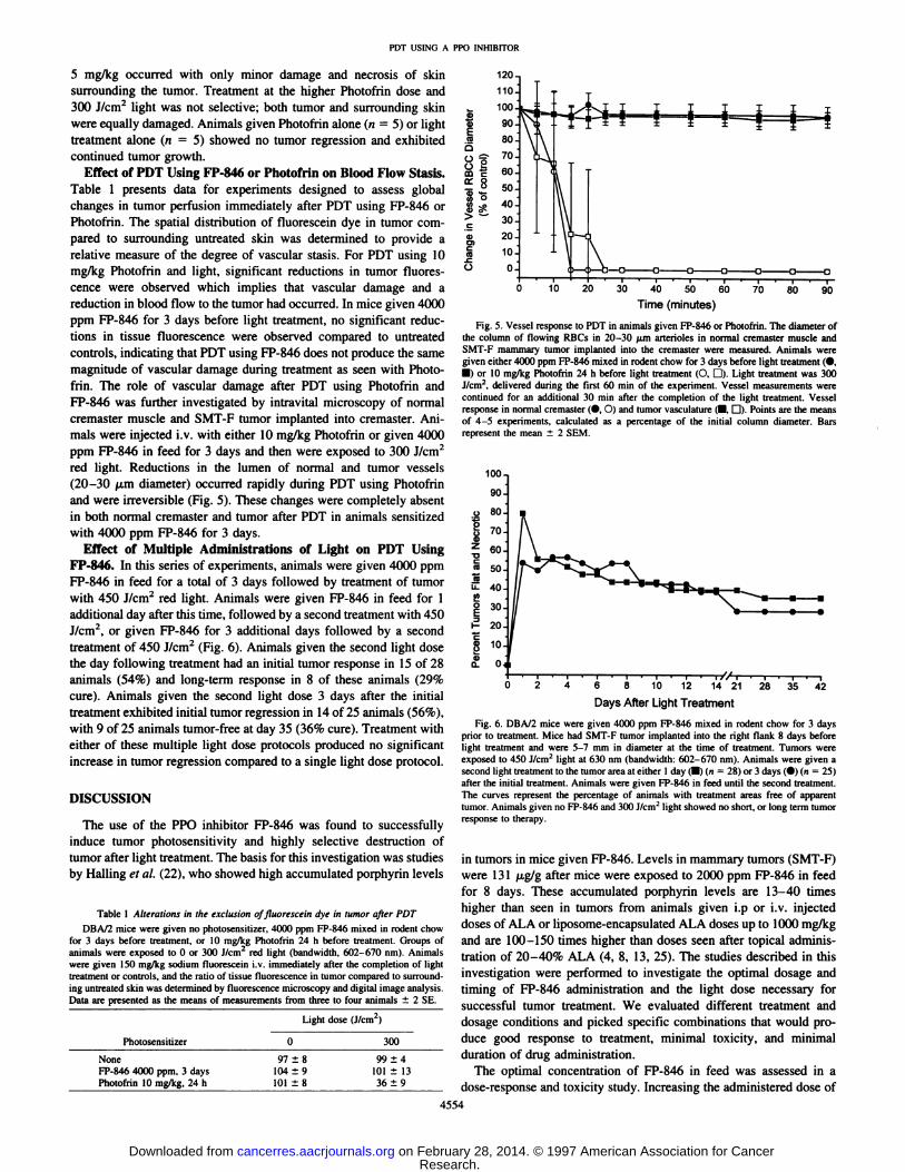
continued tumor growth.Effect of PDT Using FP-846 or Photofrin on Blood Flow Stasis.
Table 1 presents data for experiments designed to assess globalchanges in tumor perfusion immediately after PDT using FP-846 or
Photofrin. The spatial distribution of fluorescein dye in tumor compared to surrounding untreated skin was determined to provide arelative measure of the degree of vascular stasis. For PDT using 10mg/kg Photofrin and light, significant reductions in tumor fluorescence were observed which implies that vascular damage and areduction in blood flow to the tumor had occurred. In mice given 4000ppm FP-846 for 3 days before light treatment, no significant reduc
tions in tissue fluorescence were observed compared to untreatedcontrols, indicating that PDT using FP-846 does not produce the same
magnitude of vascular damage during treatment as seen with Photofrin. The role of vascular damage after PDT using Photofrin andFP-846 was further investigated by intravital microscopy of normalcremaster muscle and SMT-F tumor implanted into cremaster. Ani
mals were injected i.v. with either 10 mg/kg Photofrin or given 4000ppm FP-846 in feed for 3 days and then were exposed to 300 J/cm2
red light. Reductions in the lumen of normal and tumor vessels(20-30 /j,m diameter) occurred rapidly during PDT using Photofrin
and were irreversible (Fig. 5). These changes were completely absentin both normal cremaster and tumor after PDT in animals sensitizedwith 4000 ppm FP-846 for 3 days.
Effect of Multiple Administrations of Light on PDT UsingFP-846. In this series of experiments, animals were given 4000 ppmFP-846 in feed for a total of 3 days followed by treatment of tumorwith 450 J/cm2 red light. Animals were given FP-846 in feed for 1
additional day after this time, followed by a second treatment with 450J/cm2, or given FP-846 for 3 additional days followed by a secondtreatment of 450 J/cm2 (Fig. 6). Animals given the second light dose
the day following treatment had an initial tumor response in 15 of 28animals (54%) and long-term response in 8 of these animals (29%
cure). Animals given the second light dose 3 days after the initialtreatment exhibited initial tumor regression in 14 of 25 animals (56%),with 9 of 25 animals tumor-free at day 35 (36% cure). Treatment with
either of these multiple light dose protocols produced no significantincrease in tumor regression compared to a single light dose protocol.
DISCUSSION
The use of the PPO inhibitor FP-846 was found to successfully
induce tumor photosensitivity and highly selective destruction oftumor after light treatment. The basis for this investigation was studiesby Hailing el al. (22), who showed high accumulated porphyrin levels
Table 1 Alterations in the exclusion of fluorescein dye in tumor after PDTDBA72 mice were given no photosensiuzer. 4000 ppm FP-846 mixed in rodent chow
for 3 days before treatment, or 10 mg/kg Photofrin 24 h before treatment. Groups ofanimals were exposed to 0 or 300 J/cm2 red light (bandwidth. 602-670 nm). Animals
were given 150 mg/kg sodium fluorescein i.v. immediately after the completion of lighttreatment or controls, and the ratio of tissue fluorescence in tumor compared to surrounding untreated skin was determined by fluorescence microscopy and digital image analysis.Data are presented as the means of measurements from three to four animals ±2 SE.
120-
PhotosensitizerNone
FP-846 4000 ppm, 3 daysPhotofrin 10 mg/kg, 24 hLight
dose(J/cm2)0
30097
±8 99 ±4104 ±9 101 ±13101 ±8 36 ±9
10 20 30 40 50 60
Time (minutes)
Fig. 5. Vessel response to PDT in animals given FP-846 or Photofrin. The diameter ofthe column of flowing RBCs in 20-30 /¿marterioles in normal cremaster muscle andSMT-F mammary tumor implanted into the cremaster were measured. Animals weregiven either 4000 ppm FP-846 mixed in rodent chow for 3 days before light treatment (•.
•¿�)or 10 mg/kg Photofrin 24 h before light treatment (O. D). Light treatment was 300J/cm". delivered during the first 60 min of the experiment. Vessel measurements were
continued for an additional 30 min after the completion of the light treatment. Vesselresponse in normal cremaster (•.O) and tumor vasculature (•.D). Points are the meansof 4-5 experiments, calculated as a percentage of the initial column diameter. Barsrepresent the mean ±2 SEM.
100
Tr-> » * •¿�
Days After Light Treatment
Fig. 6. DBA/2 mice were given 4000 ppm FP-846 mixed in rodent chow for 3 daysprior to treatment. Mice had SMT-F tumor implanted into the right flank 8 days beforelight treatment and were 5-7 mm in diameter at the time of treatment. Tumors wereexposed to 450 J/cm2 light at 630 nm (bandwidth: 602-670 nm). Animals were given a
second light treatment to the tumor area at either 1 day (•)(n = 28) or 3 days (•)(n = 25)after the initial treatment. Animals were given FP-846 in feed until the second treatment.The curves represent the percentage of animals with treatment areas free of apparenttumor. Animals given no FP-846 and 300 J/cm2 light showed no short, or long term tumor
response to therapy.
in tumors in mice given FP-846. Levels in mammary tumors (SMT-F)were 131 f¿g/gafter mice were exposed to 2000 ppm FP-846 in feedfor 8 days. These accumulated porphyrin levels are 13-40 times
higher than seen in tumors from animals given i.p or i.v. injecteddoses of ALA or liposome-encapsulated ALA doses up to 1000 mg/kgand are 100-150 times higher than doses seen after topical administration of 20-40% ALA (4, 8, 13, 25). The studies described in this
investigation were performed to investigate the optimal dosage andtiming of FP-846 administration and the light dose necessary for
successful tumor treatment. We evaluated different treatment anddosage conditions and picked specific combinations that would produce good response to treatment, minimal toxicity, and minimalduration of drug administration.
The optimal concentration of FP-846 in feed was assessed in adose-response and toxicity study. Increasing the administered dose of
4554
Research. on February 28, 2014. © 1997 American Association for Cancercancerres.aacrjournals.org Downloaded from

PDT USING A PRO INHIBITOR
FP-846 from 2(XX)-4(KX) ppm in feed resulted in greater short-termtumor destruction in animals. The long-term tumor response in animals given 4(KK) ppm FP-846 was unexpectedly low compared to
treatment at lower doses. The reasons for the diminished response arenot understood at this time but may be related to the toxicity of thecompound when given for a period of 10 days.
Administration of FP-846 for 3 days prior to light treatment pro
duced significant levels of tumor destruction without toxicity toanimals. This regimen may be more attractive for clinical use of PPOinhibitors because patients are exposed to the agent for only a shortperiod. Animals submitted to longer feeding periods before lighttreatment had greater short-term tumor response but no greater tumor
cure.A light dose-response study was initiated to evaluate how tumor
destruction could be altered by changing the light dose for FP-846
activation. The results of this study correlate with PDT using exogenous photosensitizers where a threshold dose for response and effective doses were observed (26). Light doses of 150 J/cm2 or less did not
produce any observable change in the tumors. The two higher lightdoses used in these experiments, 300 and 450 J/cm2, produced goodinitial response and high levels of tumor cure. The 450 J/cm2 light
dose caused initial regression in a greater number of animals than theother light doses investigated and slightly higher tumor cure.
The treatment efficacy of PDT after FP-846-induced photosensiti-
zation was compared to that of PDT using Photofrin in the sameanimal tumor model and for identical light doses to place these agentsin proper perspective. Animals given 300 J/cm2 and 10 mg/kg Pho
tofrin had levels of tumor cure that were indistinguishable from curein animals given the same light dose and 4000 ppm FP-846 for 3 days.Comparison of response in animals given FP-846 and light compared
to published reports of response for PDT using ALA suggest that theuse of PPO inhibitors has several advantages: («)administration ofFP-846 produces significantly higher tumor concentrations of photo
sensitizers compared to ALA. The consequence of this remains unknown, but it implies that greater levels of direct tumor cytotoxicitymay occur; and (b) the use of a systemic approach to induce PPIXaccumulation may eliminate the observed problems with topical ALApenetration into deep layers of tumor. Systemic administration of PPOinhibitors may allow more homogeneous distribution of PPIX intumor, particularly at the tumor base where treatment often fails afterPDT using topical ALA, and thus offer better long-term tumor
control.An examination of the kinetics of the tumor response curves for
PDT using Photofrin compared to PDT using FP-846 illustrates an
important difference in early response to therapy. Fewer animalsgiven FP-846 and light showed initial complete tumor response com
pared to Photofrin, but those tumors that initially responded did notgenerally reappear over time. This is different from Photofrin, wherealmost all tumors initially respond to treatment but most recur. Thereason why all tumors do not respond equally during the initial daysafter treatment may be explained by inherent biological differencesbetween animals or more likely by variation in porphyrin accumulation in tumor. Detection of porphyrin fluorescence in tumor beforelight treatment revealed that animals did not accumulate the sameamount of PPIX (data not shown). Animals that had higher initialfluorescence levels tended to respond more favorably to treatmentcompared to those with lower tumor fluorescence. All animals showedan increase in tumor fluorescence. The lack of complete regression ina significant proportion of treated animals suggests that there may beways of further optimizing FP-846 administration.
The observation that an initial complete response to PDT usingFP-846 was closely predictive of tumor cure (as opposed to PDT
using Photofrin) suggests that different mechanisms of damage may
be involved with this treatment regimen. Initial tumor destruction afterPDT using Photofrin is mediated by blood flow stasis and vasculardamage, leading to tissue anoxia and nutrient deprivation (27). At therelatively low Photofrin doses used in treatment, direct damage totumor cells does not play a significant role in the initial tumorregression (28). The factors that account for the low ratio of tumorcures compared to those tumors that initially responded to PDT usingPhotofrin remain unknown. In contrast. PDT using FP-846 causes no
initial vascular response or change in blood flow during the first hourafter the completion of light treatment. These results imply that tumoroxygénationlevels remain high during PDT with FP-846, and the
treatment is efficient compared to the oxygen limitations of treatmentobserved during PDT using Photofrin. The combination of high levelsof PPIX found in cells in FP-846-treated animals and continued
perfusion and oxygénationof tissues may result in sufficient tumorcell death to permit tumor regression and possibly cure. Vasculardamage and blood flow stasis presumably occur at some late timeafter the completion of light treatment, either as a result of directdamage to endothelial cells or breakdown of the entire tumor environment. The contribution of direct cytotoxicity and late vasculardamage and stasis for PDT using FP-846 has not yet been addressed.
An absence of a vascular response in tumors or normal tissues inanimals sensitized with FP-846 and light is also different from the
response observed in animals given PDT using ALA. Although initialreports suggested that direct cytotoxicity was responsible for tumordestruction and was independent of vascular damage (29, 30), recentdata find that vascular damage plays a large role in tumor response (6,31). Treatment of tissues photosensitized by PPIX in response to ALAadministration causes endothelial cell damage in vitro (32) and in vivo(33) as well as vessel constriction and reduction or stoppage of bloodflow in tumors (6, 34). This response is dependent on the ALA doseand time between drug application and light treatment and is likelyrelated to high circulating levels of PPIX in blood (31).
The lack of initial vascular damage during phototherapy usingFP-846 raised the possibility of attempting multiple light treatments to
increase the efficacy of tumor treatment. The hypothesis was thattumor cells that were not responsive to the initial treatment may beresponsive to a second treatment, provided they could accumulateadditional PPIX between treatments and tissue oxygénationremainedhigh. We investigated two different time delays between the initialand second light treatment, but the response of tumors given either ofthese treatment protocols was disappointing. Further administration ofFP-846 in feed and a second light treatment either 1 or 3 days after the
initial therapy produced no greater tumor response than the singletreatment alone. The reason for this is not clear. This response mayresult from an inability of additional FP-846 to accumulate in tumor
after the initial light treatment due to blood flow stasis or otherreasons. Damage to tumor cell mitochondrial enzymes, as seen withPDT using ALA or Photofrin (35), may result in decreased PPIXgeneration between treatments. Tissue hypoxia resulting from latevessel stasis from the initial treatment or decreased light penetrationinto tissue because of tissue darkening may also contribute to the lackof additional response.
Photodynamic therapy using FP-846 and red light appears to be a
promising alternative to PDT using Photofrin or other exogenousporphyrins. Although a number of basic questions regarding the use ofFP-846 still need to be addressed, these data suggest that PPO inhib
itors can be successfully used to result in PPIX accumulation in tissueand tumor destruction after light treatment. These studies raise thehope that alternative PPO inhibitors with decreased toxicity and rapidaction may be found and raise the possibility that the combined use ofPPO inhibitors and exogenous ALA may result in increased tumorresponse and treatment efficacy.
4555
Research. on February 28, 2014. © 1997 American Association for Cancercancerres.aacrjournals.org Downloaded from

PDT USING A PPO INHIBITOR
ACKNOWLEDGMENTS
We thank QLT PhotoTherapeutics, Inc. for providing the Photofrin used inthis study.
REFERENCES
1. Dougherty. T. J.. and Marcus. S. L. Photodynamic Therapy. Eur. J. Cancer. 2SA:1734-1742, 1992.
2. Weishaupt. K. R., Gomer. C. J.. and Dougherty. T. J. Identification of singlet oxygenas the cytotoxic agent in the inactivation of a murine tumor. Cancer Res.. 36:2326-2329. 1976.
3. Foole, C. S. Chemical mechanisms of photodynamic action. In: C. 1. Gomer (ed.).Future Directions and Applications in Photodynamic Therapy. Vol. 1S6. pp. 115-126.Bellingham. Washington: SPIE Optical Engineering Press. 1990.
4. Henderson. B. W., and Dougherty, T. J. How does photodynamic therapy work?Pholochem. Photobiol.. 55.- 145-157, 1992.
5. Peng, Q.. Berg. K.. Moan, J.. Kongshaug. M., and Nesland, J. M. 5-Aminolevulinicacid-based photodynamic therapy: principles and experimental research. Photochem.Photobiol.. 65; 235-251, 1997.
6. Henderson, B. W.. Vaughan. L.. Bellnier. D. A., van Leengoed. H.. Johnson. P. G.,and Oseroff. A. R. Photosensitizalion of murine tumor, vasculature and skin by5-aminolevulinic acid-induced porphyrin. Photochem. Photobiol.. 62: 780-789,
1995.7. Malik. Z.. and Lugaci. H. Destruction of erythroleukaemic cells by photoactivation of
endogenous porphyrins. Br. J. Cancer. 56: 589-595, 1987.8. Fukuda. H.. Paredes, S.. Del, C., and Batle, A. M. Tumour-localizing properties of
porphyrins. In vivo studies using free and liposome encapsulated aminolevulinic acid.Comp. Biochem. Physiol., I02B: 433-436, 1992.
9. Peng. W.. Moan, J.. Warlow. T.. Nesland, J. M., and Rimington. C. Distribution andphotosensitizing efficiency of porphyrins induced by application of exogenous 5-uminolevulinic acid in mice bearing mammary carcinoma. Int. J. Cancer, 52: 433-443, 1992.
10. Kennedy, J. C.. and Pottier. R. H. Endogenous protoporphyrin IX. a clinically usefulphotosensitizer for photodynamic therapy. J. Photochem. Photobiol. B Biol., 14:275-292, 1992.
11. Svanberg. K., Andersson, T., Killander, D., Wang. I., Stenram. U.. Andersson-Engles.S.. Berg. R.. Hohansson. J., and Svanberg, S. Photodynamic therapy of non-melanoma malignant tumours of the skin using topical •¿�y-aminolevulinicacid sensitizationand laser irradiation. Br. J. Dermatol., 130: 743-751, 1994.
12. Shanler, S. D., Buscaglia. D. A., van Leengoed, H.. Wan. W.. Whitaker. J. E.. Mang,T. S., Barcos, M., Stoll, H. L., and Oseroff, A. R. PDT with topical y-aminolevulinicacid (ALAI for the treatment of patch and plaque stage cutaneous T-cell lymphoma.J. Invest. Dermatol., 102: 615, 1994.
13. Peng, Q.. Warloe, T., Moan, J., Heyerdahl, H., Steen, H. B., Nesland. J. M., andGiercksky. K. E. Distribution of 5-aminolevulinic acid-induced porphyrins in nodu-loulcerative basal cell carcinoma. Photochem. Photobiol., 62: 906-913, 1995.
14. He. D., Sassa, S., and Lim. H. W. Effect of UVA and blue light on porphyrinbiosynthesis in epidermal cells. Photochem. Photobiol., 57: 525-829, 1993.
15. Oriel. B.. Tanew, A., and Honigsmann, H. Lethal photosensitization by endogenousporphyrins of PAM cells: modification by desferrioxamine. J. Photochem. Photobiol.B Biol., 17: 273-278, 1993.
16. Miyamoto, K.. Nishimura, K., Masuda. T., Tsuji. H.. and Inokuchi, H. Accumulationof protoporphyrin IX in light-sensitive mutants of Escherichia coli. FEBS Lett., 310:246-248, 1992.
17. Mayasich. J. M.. Mayasich, S. A., and Rebeiz. C. A. Response of com (Zea mays).soybean (Glvcine max), and several weed species to dark-applied photodynamicherbicide modulators. Weed Sci.. 38: 10-15. 1990.
18. Rebeiz, C. A., Juvik, J. A., Rebeiz. C. C.. Bouton. C. E., and Gut, L. J. Porphyricinsecticides 2. 1,10-Phenanthroline. a potent porphyric insecticide modulator. Pestic.Biochem. Physiol.. 36: 201-207, 1990.
19. Bandihalli. U. B., and Rebeiz, C. A. Photodynamic herbicides. 9. Structure activitystudy of substituted 1.10-phenanthrolines as potent photodynamic herbicide modulators. Pestic. Biochem. Physiol., 40: 27-46, 1991.
20. Rebeiz, C. A.. Reddy. K. N., Nandihalli. U. B.. and Velu, J. Tetrapyrrole-dependentphotodynamic herbicides. Photochem. Photobiol.. 52: 1099-1117. 1990.
21. Rebeiz, N.. Rebeiz. C. C.. Arkins. S., Kelley, K. W.. and Rebeiz. C. A. Photodestruction of tumor cells by induction of endogenous accumulation of protoporphyrinIX: enhancement by 1,10-phenanthroline. Photochem. Photobiol., 55: 431-435,
1992.22. Hailing. R. P., Yuhas, D. A.. Pingar. V. H.. and Winkelman. J. W. Proloporphyrino-
gen oxidase inhibitors for tumor therapy. In: S. O. Duke and C. A. Rebeiz (eds.).Porphyrin Pesticides. ACS Symposium Series 559. pp. 280-290. Washington. DC:American Chemical Society, 1994.
23. Henderson. B. W.. Dougherty. T. J.. and Malone. P. B. Studies on the mechanism oftumor destruction by photoradiation therapy. In: D. R. Doiron and C. J. Gomer (eds.).Porphyrin Localization and Treatment of Tumor, pp. 601-612. New York: Alan R.
Liss, Inc., 1984.24. Fingar, V. H., Wieman, T. J.. Wiehle, S. A., and Cerrito. P. B. The role of
microvascular damage in photodynamic therapy: the effect of treatment on vesselconstriction, permeability and leukocyte adhesion. Cancer Res., 52: 4914-4921,1992.
25. Hua. Z.. Gibson. S. L., Foster, T. H.. and Hilf, R. Effectiveness of S-aminolevulinicacid-induced protoporphyrin as a photosensitizer for photodynamic therapy in vivo.Cancer Res.. 55: 1723-1731. 1995.
26. Fingar, V. H.. and Henderson. B. W. Drug and light dose dependence of photodynamic therapy: a study of tumor and normal tissue response. Photochem. Photobiol.,46: 837-841. 1987.
27. Henderson. B. W.. and Fingar. V. H. Relationship of tumor hypoxia and response tophotodynamic treatment in an experimental mouse tumor. Cancer Res., 47: 3110-3114, 1987.
28. Henderson. B. W., and Fingar. V. H. Oxygen limitation of direct tumor cell kill duringphotodynamic treatment of a murine tumor model. Photochem. Photobiol., 49:299-304, 1989.
29. Divaris. D. X. G.. Kennedy. J. C.. and Pottier, R. H. Phototoxic damage to sebaceousglands and hair follicles of mice after systemic administration of 5-aminolevulinicacid correlates with localized protoporphyrin IX fluorescence. Am. J. Pathol.. 136:891-897, 1990.
30. Bedwell. J. A.. MacRobert, A. J.. Phillips, D., and Bown. S. G. Fluorescencedistribution and photodynamic effect of ALA-induced PPIX in the DMH rat colonietumour model. Br. J. Cancer, 65: 818-824. 1992.
31. van der Veen, N.. van Leengoed. H. L. L. M.. and Star. W. M. In vivo fluorescencekinetics and photodynamic therapy using ALA-induced porphyrin: increased damageafter multiple irradiations. Br. J. Cancer, 70: 867-872, 1994.
32. He, D., Behar, S., Nomura. N., Sassa, S., and Lim, H. W. The effect of ALA andradiation on porphyrin/heme biosynthesis in endothelial cells. Photochem. Photobiol..61: 656-661, 1995.
33. Nyamekye, I., Anglin. S., McEwan, J.. MacRobert, A.. Bown, S., and Bishop, C.Photodynamic therapy of normal and balloon-injured rat carotid arteries using 5-amino-levulinic acid. Circulation, 91: 417-425, 1995.
34. Roberts, D. J. H.. Cairnduff, F., Driver, I., Dixon, B.. and Brown. S. B. Tumourvascular shutdown following photodynamic therapy based on polyhaematoporphyrinor 5-aminolaevulinic acid. Int. J. Oncol., 5: 763-768, 1994.
35. Hilf. R.. Murant. R. S.. Narayanan. U.. and Gibson. S. L. Relationship of mitochondria] function and cellular adenosine triphosphate levels to hematoporphyrin derivative induced photosensitization in R3230 AC mammary tumors. Cancer Res., 46:211-217, 1986.
4556
Research. on February 28, 2014. © 1997 American Association for Cancercancerres.aacrjournals.org Downloaded from