Treatment of experimental periodontal disease by photodynamic therapy in immunosuppressed rats
9
Treatment of experimental periodontal disease with antimicrobial photodynamic therapy in nicotine-modified rats Garcia VG, Fernandes LA, Macarini VC, de Almeida JM, Martins TM, Bosco AF, Nagata MJH, Cirelli JA, Theodoro LH. Treatment of experimental periodontal disease with antimicrobial photodynamic therapy in nicotine-modified rats. J Clin Periodontal 2011; 38: 1106–1114. doi: 10.1111/j.1600-051X.2011.01785.x. Abstract Background: The aim of this study was to compare antimicrobial photodynamic therapy (aPDT) as an adjunctive treatment to scaling and root planing (SRP) for induced periodontitis in nicotine-modified rats. Material & Methods: A total of 240 rats were evenly divided into two groups: C – saline solution treatment; N – nicotine treatment. Periodontal disease was induced in both groups at the first mandibular molar. After 7 days, the ligature was removed. All animals were submitted to SRP and were divided according to the following treatments: SRP – irrigation with saline solution; Toluidine Blue-O (TBO) – irrigation with phenothiazinium dye (100 lg/ml); LLLT – laser irradia- tion (660 nm; 0.03 W; 4 J); and aPDT – TBO and laser irradiation. Ten animals in each group/treatment were euthanized at 7, 15 and 30 days. The histometric and immunohistochemical values were statistically analysed. Results: Intragroup analysis demonstrated that in both groups the aPDT treat- ment resulted in lower bone loss (BL) when compared to SRP in all experimental periods. Intergroup analysis demonstrated that aPDT treatment resulted in lower BL in Group N than in Group C treated with SRP in all experimental periods. Conclusion: Antimicrobial photodynamic therapy was an effective adjunctive treatment to SRP for induced periodontitis in nicotine-modified rats. Valdir Gouveia Garcia 1,3 , Leandro Arau ´ jo Fernandes 1,2 , Valmir Campos Macarini 1 , Juliano Milanezi de Almeida 1 , Thiago Marchi Martins 1 , Alvaro Francisco Bosco 1 , Maria Jose ´ Hitomi Nagata 1 , Joni Augusto Cirelli 4 and Letı ´cia Helena Theodoro 1,3 1 Group of Research and Study on Laser in Dentistry (GEPLO), Department of Surgery and Integrated Clinic, Division of Periodontics, Sa ˜ o Paulo State University, Arac ¸atuba, Sa ˜o Paulo, Brazil; 2 Department of Clinic and Surgery, Federal University of Alfenas, Alfenas, MG, Brazil; 3 Master course, University Center of the Educational Foundation of Barretos (UNIFEB), Dental School of Barretos, Barretos, SP, Brazil; 4 Department of Diagnosis and Surgery, Division of Periodontics, Sa ˜o Paulo State University, Araraquara, Sa ˜o Paulo, Brazil Key words: alveolar bone loss; lasers; nicotine; osteoclasts; periodontitis; photochemotherapy Accepted for publication 23 July 2011 Periodontal disease (PD) is a multi- factorial pathological condition that involves both host systemic altera- tions and the presence of local path- ogenic microbiota (Cullinan et al. 2009). The main signs that character- ize PD are attachment loss caused by apical migration of the junctional epithelium and alveolar bone loss (BL) (Ebisu & Noiri 2007). The metabolism of alveolar bone is controlled by proteins such as RANK, RANKL and OPG, which mark cell activity. RANK is present on the membranes of osteoclasts and dendritic cells (Evans et al. 2006), and its dimerization allows for the formation of multinuclear TRAP- positive cells with clastic cell immuno- reactivity (Iwamoto et al. 2004). RANKL, in turn, is produced by osteoblasts, bone stromal cells and activated T-lymphocytes, and pro- motes bone resorption when linked to RANK (Khosla 2001). By con- trast, OPG inhibits osteoclast forma- Conflict of interest and source of funding statement The authors declare no conflicts of interest. The present study was conducted in the Periodontics Division (Department of Sur- gery and Integrated Clinic of Arac¸atuba Dental School – UNESP) and supported by the Sa˜o Paulo State Research Support Foundation – Sa˜o Paulo, SP, Brazil (FA- PESP – Process no. 2008/04927-3), CNPq – National Council for Scientific and Technological Development – Brasilia, DF, Brazil (CNPq 301085/2007-2) and CAPES – Coordination for the Improvement of Higher Education Personnel (Brasi- lia, DF, Brazil). © 2011 John Wiley & Sons A/S 1106 J Clin Periodontol 2011; 38: 1106–1114 doi: 10.1111/j.1600-051X.2011.01785.x
-
Upload
independent -
Category
Documents
-
view
1 -
download
0
Transcript of Treatment of experimental periodontal disease by photodynamic therapy in immunosuppressed rats

Treatment of experimentalperiodontal disease withantimicrobial photodynamictherapy in nicotine-modified ratsGarcia VG, Fernandes LA, Macarini VC, de Almeida JM, Martins TM, Bosco AF,Nagata MJH, Cirelli JA, Theodoro LH. Treatment of experimental periodontaldisease with antimicrobial photodynamic therapy in nicotine-modified rats. J ClinPeriodontal 2011; 38: 1106–1114. doi: 10.1111/j.1600-051X.2011.01785.x.
AbstractBackground: The aim of this study was to compare antimicrobial photodynamictherapy (aPDT) as an adjunctive treatment to scaling and root planing (SRP) forinduced periodontitis in nicotine-modified rats.Material & Methods: A total of 240 rats were evenly divided into two groups:C – saline solution treatment; N – nicotine treatment. Periodontal disease wasinduced in both groups at the first mandibular molar. After 7 days, the ligaturewas removed. All animals were submitted to SRP and were divided according tothe following treatments: SRP – irrigation with saline solution; Toluidine Blue-O(TBO) – irrigation with phenothiazinium dye (100 lg/ml); LLLT – laser irradia-tion (660 nm; 0.03 W; 4 J); and aPDT – TBO and laser irradiation. Ten animalsin each group/treatment were euthanized at 7, 15 and 30 days. The histometricand immunohistochemical values were statistically analysed.Results: Intragroup analysis demonstrated that in both groups the aPDT treat-ment resulted in lower bone loss (BL) when compared to SRP in all experimentalperiods. Intergroup analysis demonstrated that aPDT treatment resulted in lowerBL in Group N than in Group C treated with SRP in all experimental periods.Conclusion: Antimicrobial photodynamic therapy was an effective adjunctivetreatment to SRP for induced periodontitis in nicotine-modified rats.
Valdir Gouveia Garcia1,3, Leandro
Araujo Fernandes1,2, Valmir CamposMacarini1, Juliano Milanezi de
Almeida1, Thiago Marchi Martins1,Alvaro Francisco Bosco1, Maria Jose
Hitomi Nagata1, Joni Augusto Cirelli4
and Letıcia Helena Theodoro1,3
1Group of Research and Study on Laser in
Dentistry (GEPLO), Department of Surgery
and Integrated Clinic, Division of
Periodontics, Sao Paulo State University,
Aracatuba, Sao Paulo, Brazil; 2Department
of Clinic and Surgery, Federal University of
Alfenas, Alfenas, MG, Brazil; 3Master
course, University Center of the Educational
Foundation of Barretos (UNIFEB), Dental
School of Barretos, Barretos, SP, Brazil;4Department of Diagnosis and Surgery,
Division of Periodontics, Sao Paulo State
University, Araraquara, Sao Paulo, Brazil
Key words: alveolar bone loss; lasers;
nicotine; osteoclasts; periodontitis;
photochemotherapy
Accepted for publication 23 July 2011
Periodontal disease (PD) is a multi-factorial pathological condition thatinvolves both host systemic altera-tions and the presence of local path-ogenic microbiota (Cullinan et al.
2009). The main signs that character-ize PD are attachment loss causedby apical migration of the junctionalepithelium and alveolar bone loss(BL) (Ebisu & Noiri 2007).
The metabolism of alveolar boneis controlled by proteins such asRANK, RANKL and OPG, whichmark cell activity. RANK is presenton the membranes of osteoclasts anddendritic cells (Evans et al. 2006),and its dimerization allows for theformation of multinuclear TRAP-positive cells with clastic cell immuno-reactivity (Iwamoto et al. 2004).RANKL, in turn, is produced byosteoblasts, bone stromal cells andactivated T-lymphocytes, and pro-motes bone resorption when linkedto RANK (Khosla 2001). By con-trast, OPG inhibits osteoclast forma-
Conflict of interest and source of funding statement
The authors declare no conflicts of interest.The present study was conducted in the Periodontics Division (Department of Sur-gery and Integrated Clinic of Aracatuba Dental School – UNESP) and supportedby the Sao Paulo State Research Support Foundation – Sao Paulo, SP, Brazil (FA-PESP – Process no. 2008/04927-3), CNPq – National Council for Scientific andTechnological Development – Brasilia, DF, Brazil (CNPq 301085/2007-2) andCAPES – Coordination for the Improvement of Higher Education Personnel (Brasi-lia, DF, Brazil).
© 2011 John Wiley & Sons A/S1106
J Clin Periodontol 2011; 38: 1106–1114 doi: 10.1111/j.1600-051X.2011.01785.x

tion in the development stage, pre-venting the binding of RANKL toRANK (Shalhoub et al. 1999).
Tobacco is one of the most sig-nificant risk factors for the onsetand progression of PD (Genco1996). This association may be dueto local vasoconstriction (whichimpairs periodontal tissue blood sup-ply), increased concentrations ofperiodontal pathogens in the subgin-gival microbiota or alterations intheir composition (Ramseier & Fun-dak 2009). Furthermore, nicotine incigarettes may increase collagen fibredegradation by fibroblasts (Zhanget al. 2009), inhibit immune systemresponses (Breivik et al. 2009), pro-mote decreases in both phagocytosisand polymorphonuclear leucocytechemotaxis and reduce antibody pro-duction and the viability of T-lym-phocytes (Holt & Keast 1977).
The treatment of PD is based onthe elimination of pathogenic subgin-gival microbiota through scaling androot planing (SRP) (Kaldahl et al.1993). However, mechanical therapyalone may be ineffective at eliminat-ing pathogenic bacteria, which arefrequently inserted into both soft andhard tissues and even in areas thatare inaccessible to periodontal instru-ments (Adriaens et al. 1988).
Considering these limitations,adjunctive methods that promote theelimination of periodontal pathogenshave been investigated, as with anti-microbial photodynamic therapy(aPDT). This therapy consists of theassociation of a photosensitizer withan intense light source to promotecell death (Bauer et al. 2001). Theactive mechanism of aPDT beginswhen the photosensitizer’s electronsabsorb energy from a light sourcewith the same wavelength and arethus induced into an excited state(Dortbudak et al. 2001). When thephotosensitizer returns to its primarystate, it can transfer the energy to asubstrate when one is present, form-ing short lifespan species that arehighly reactive to singlet oxygen,which may injure microorganismsdue to the irreversible oxidation ofcell components (Tomaselli et al.2001). The lethal photosensitizationof periodontopathogenic bacteriamust involve changes in membranesand/or plasma membrane proteinsand DNA damage mediated by sin-glet oxygen (Maisch 2007).
The major advantages of aPDTare that it is a specific therapy tar-geting cells that, it has no collateraleffects, that its activity is initiatedonly upon exposure to light, andthat its use will not lead to the selec-tion of resistant bacterial species(Maisch 2007).
Some studies in animals have alsoshown satisfactory results with theuse of aPDT for the treatment ofexperimental periodontal disease(Komerik et al. 2003, Almeida et al.2008a,b, Fernandes et al. 2009).Recent articles from the presentresearch group have demonstratedthe effectiveness of aPDT as anadjunct to conventional periodontaltreatment in animals with diabetesmellitus (Almeida et al. 2008a) andinhibited with immunosuppressivedrugs (Fernandes et al. 2009).
In this context, the aim of thepresent study was to histologically,
histometrically and immunohisto-chemically evaluate the efficacy ofaPDT as an adjunct to conventionalmechanical therapy (scaling and rootplaning) for alveolar BL in both nor-mal and systemically nicotine-modi-fied rats with experimentally inducedperiodontitis.
Animals
This study was conducted on 240adult male Wistar rats (120–140 g).The animals were kept in plasticcages with access to food and waterad libitum. Prior to the surgical pro-cedures, all animals were allowed toacclimatize to the laboratory envi-ronment for a period of 5 days. Allprotocols described below wereapproved by the Institutional ReviewBoard of Aracatuba Dental School,Sao Paulo State University, Araca-tuba, SP, Brazil (no. 1372/08).
a b
c d
Fig. 1. Photomicrograph illustrating the areas of bone loss (BL) in the furcationregion of the mandibular left first molar with induced periodontal disease in the differ-ent groups (C and N): (a) C Group, scaling and root planing (SRP) treatment for30 days (HE, original magnification 59); (b) C Group, SRP treatment for 30 days(HE, original magnification 409); (c) N Group, SRP treatment for 30 days (HE, origi-nal magnification 59) and (d) N Group, SRP treatment for 30 days (HE, originalmagnification 409).
© 2011 John Wiley & Sons A/S
aPDT in nicotine-modified rats 1107

Experimental design
Drug administration protocol
The animals were numbered andrandomly divided into two groups:C (n = 120) – Control – injectedwith saline solution at 3 mg/kg ofbody weight; N (n = 120) – Nicotine– subcutaneously injected with nico-tine hemisulphate preparation at3 mg/kg of body weight (Pinto et al.2002). Nicotine hemisulphate prepa-ration (Sigma Corporation, SaoPaulo, SP, Brazil) was obtained bydiluting it in saline solution at a con-centration of 5 mg/ml (Okamotoet al. 1994).
All injections were divided intotwo applications, one in the morningand the other in the evening, alwayswith a 12-h interval between them.The injections were initiated 30 daysbefore PD induction and performeddaily up to the end of the respectiveeuthanasia period. The administra-tion site was the back of the animal,next to the cephalic region. The ani-mals were weighed weekly withregard to dose maintenance through-out the experimental period.
Experimental periodontal disease protocol
General anaesthesia was induced byadministering ketamine (0.4 ml/kg)together with xylazine (0.2 ml/kg)via intramuscular injection. Onemandibular left first molar fromeach animal in both the C and Ngroups was selected to receive a cot-ton ligature in a submarginal posi-tion to induce experimentalperiodontitis (Johnson 1975). After7 days of experimental periodontaldisease induction, the mandibularligature was removed from the leftfirst molar of all animals in the Cand N groups. The left molars weresubjected to SRP with manual #1–2micro mini five curettes (Hu-FriedyCo. Inc., Chicago, IL, USA) through10 distal–mesial traction movementsin both the buccal and lingualaspects. The furcation and interprox-imal areas were scaled with the samecurettes through cervical-occlusaltraction movements. The entire SRPprocedure was performed by thesame experienced operator. The 120animals of each group (C and N)were randomly allocated by the useof a computer-generated table to
treatments of SRP, TBO, LLLT oraPDT. For better standardization,animal 1 was the first choice, fol-lowed by animals 2 and 3, respec-tively. Thus, all animals in eachgroup (C and N) were randomlyassigned to one of the four treat-ments (30 animals/treatment): SRP –mandibular left molar submitted toSRP and irrigation with 1 ml of sal-ine solution; Toluidine Blue-O(TBO) – mandibular left molar sub-mitted to SRP and irrigation with1 ml of phenothiazinium dye (TBOsolution, 100 lg/ml; Sigma ChemicalCo., St. Louis, MO, USA); LLLT –SRP and irrigation with 1 ml ofsaline solution followed by low-levellaser therapy (LLLT) after 1 min.;and aPDT – mandibular left molarsubmitted to SRP and irrigationwith 1 ml of TBO solution (100 lg/ml) followed by the application ofLLLT after 1 min.
LLLT treatment
The laser used in this study was gal-lium–aluminium–arsenide (GaAlAs)(Bio Wave; Kondortech EquipmentLtd, Sao Carlos, SP, Brazil), with awavelength of 660 nm and a spotsize of 0.07 cm2. After 1 min. of sal-
ine solution application, LLLT wasapplied to three equidistant points ateach buccal and lingual aspect of themandibular first molar in contactwith the tissue. The therapeutic laserwas released with a power of 0.03 Wfor 133 s/point, a power densityof 0.428 W/cm2, and energy of4 J/point (57.14 J/cm2/point) (Fer-nandes et al. 2009).
aPDT treatment
After 1 min. of TBO application,LLLT was applied using the aboveparameters. The TBO solution wasslowly poured into the periodontalpocket using a syringe (1 ml) and aninsulin needle (13 mm 9 0.45 mm)(Becton Dickinson Ind. Ltd, Curiti-ba, PR, Brazil) without a bevel.
Experimental periods
Ten animals from each group/treat-ment were euthanized at 7, 15 and30 days after the periodontal diseasetreatment by the administration of alethal dose of thiopental (150 mg/kg)(Cristalia, Ltd, Itapira, SP, Brazil).Their jaws were removed and fixedin 10% neutral formalin for 48 h.
Table 1. Means and standard deviations (M ± SD) of the histometric data for bone loss(BL, mm2) in the furcation region of the mandibular left first molar according to eachgroup, treatment and period
C Group – M ± SDPeriods 7 days 15 days 30 daysTreatments
SRP 1.09 ± 0.13‡,§ 1.02 ± 0.19‡,§ 0.99 ± 0.28‡,§
TBO 0.79 ± 0.99*,† 0.73 ± 0.26*,† 0.66 ± 0.14*,†
LLLT 0.65 ± 0.12*,† 0.56 ± 1.07*,† 0.50 ± 0.45*aPDT 0.41 ± 0.03* 0.40 ± 0.02* 0.38 ± 0.05*
N Group – M ± SDPeriods 7 days 15 days 30 daysTreatments
SRP 1.92 ± 0.34‡ 1.91 ± 0.21‡ 1.89 ± 0.22‡
TBO 0.89 ± 0.22*,† 0.82 ± 0.26*,† 0.76 ± 0.09*,†
LLLT 0.68 ± 0.28*,† 0.58 ± 2.03* 0.51 ± 3.54*aPDT 0.44 ± 0.08*,§ 0.42 ± 0.06*,§ 0.40 ± 0.03*,§
N 80 80 80
aPDT, antimicrobial photodynamic therapy; LLLT, low-level laser therapy; SRP, scalingand root planing; TBO, Toluidine Blue-O.*Significant difference with SRP treatment, in the same group and period (ANOVA andTukey; p < 0.05).†Significant difference with aPDT treatment, in the same group and period (ANOVA andTukey; p < 0.05).‡Significant difference between groups, in the same period and treatment (ANOVA and Tukey;p < 0.05).§Significant difference between groups and treatments, in the same period (ANOVA andTukey; p < 0.05).
© 2011 John Wiley & Sons A/S
1108 Garcia et al.

Laboratory procedures
The specimens were demineralized ina solution consisting of equal partsof 50% formic acid and 20% sodiumcitrate for 15 days. Paraffin serialsections (4 lm) were obtained in themesial–distal direction and dyed withhaematoxylin and eosin (HE) andMasson’s Trichrome. To performimmunohistochemical reactions, thesections were submitted to indirectimmunoperoxidase using polyclonalprimary antibodies for RANKL(1:200, SC 7628, Lot#20908, goatanti-RANKL; Santa Cruz Biotech-nology, Santa Cruz, CA, USA),OPG (1:100, SC 8468, Lot#H2208,goat anti-OPG; Santa Cruz Biotech-nology) and TRAP (1:100, SC30833, goat anti-TRAP; Santa CruzBiotechnology).
Histological analysis
The sections dyed with HE wereanalysed under light microscopy at amagnification of 409 to establish theBL level and characterize the peri-odontal ligamentation of the furca-tion region of the first molar(Almeida et al. 2008a, Fernandeset al. 2009).
Histometric analysis
Masson’s Trichrome staining wasused to assess inter-radicular bonelevels at 12.59 magnification. Thearea of BL in the furcation regionwas determined histometrically usingan image analysis system (ImageTool; University of Texas HealthScience Center at San Antonio, SanAntonio, TX, USA). After excludingthe first and last sections where thefurcation region was evident, fiveequidistant sections of each specimenblock were selected and imaged by adigital camera coupled to a lightmicroscope (Cesar-Neto et al. 2006).The mean values were averaged andstatistically compared.
Immunohistochemical analysis
The markers RANKL and OPGwere analysed immunohistochemical-ly in both the bone and periodontalligament in the furcation area of thefirst molar with experimentallyinduced periodontal disease by per-centages: no mark (0%), weak mark-
ing (<25% of cells); moderatemarking (<50% of cells); and strongintensity (<75% of cells) (Kim et al.2007).
The immunohistochemical markerTRAP was assessed by countingTRAP-positive cells located in theboundary of the most coronal por-
a b
c d
e f
Fig. 2. Photomicrograph illustrating the areas of bone loss (BL) in the furcation regionof the mandibular left first molar with induced periodontal disease in the differentgroups (C and N) and treatments: (a) C Group – scaling and root planing (SRP) treat-ment for 7 days; (b) N Group – antimicrobial photodynamic therapy (aPDT) treatmentfor 7 days; (c) C Group – SRP treatment for 15 days; (d) N Group – aPDT treatmentfor 15 days; (e) C Group – SRP treatment for 30 days and (f) N Group – aPDT treat-ment for 30 days (original magnification 12.59; Masson’s Trichrome).
© 2011 John Wiley & Sons A/S
aPDT in nicotine-modified rats 1109

tion of the bone tissue of the furca-tion region in mandibular firstmolars with periodontal disease (Jinet al. 2007, Khojasteh et al. 2008,Liu et al. 2010). Mature osteoclastscontaining three or more nuclei wereconsidered positive TRAP cells(Hassumi et al. 2009). A blindedtrained examiner selected the sec-tions for the histological, histometricand immunohistochemical analyses.Another blinded calibrated examinerconducted the data analysis. The val-ues for each specimen section weremeasured three times by the sameexaminer on different days to reducevariations in the data.
Intra-examiner reproducibility
Before the histometric and immuno-histochemical analyses were per-formed, the examiner was trained bydouble measurements of 30 speci-mens with a 1-week interval. Pairedt-test statistics was calculated, andno differences were observed in themean values for comparison(p > 0.05). In addition, the calcula-tion of Pearson’s correlation coeffi-cient revealed a very high correlation(0.95) between the two measure-ments for both the histometric andimmunohistochemical analyses.
Statistical analysis
The hypothesis that neither BL ratesnor the number of TRAP-positivecells in the furcation region differedamong groups/treatments/periodswas tested by BIOESTAT 3.0 software(Bioestat, Windows 1995; SonopressBrazilian Industry, Manaus, AM,Brazil).
After normality testing, the histo-metric and immunohistochemicaldata were analysed by the Shapiro–Wilk test, and the intra-group andinter-group analyses were performedwith a two-way analysis of variance(ANOVA; p < 0.05). When ANOVA
detected a statistical difference, mul-tiple comparisons were performedwith the Tukey test (p < 0.05).
Results
Histological assessment
At 7, 15 and 30 days, disorganizedconnective tissue was visible in bothgroups submitted to SRP treatment
with numerous neutrophils and adiscrete number of fibroblasts. Thebone tissue showed thin bone trabec-ulae (Fig. 1).
In both the C and N Groups, theanimals that received the TBO andLLLT treatments showed poorlyorganized connective tissue in allexperimental periods, with a pre-dominantly chronic inflammatoryprocess and a moderate number offibroblasts. The bone tissue showedthin bone trabeculae.
At 7 and 15 days, animals fromthe C and N Groups, which were alltreated with aPDT showed welldeveloped connective tissues with amoderate number of fibroblasts, dis-crete chronic inflammatory infiltrateand moderately developed bone tis-sue. At 30 days, they showed com-
plete and organized periodontalligaments with parallel collagenfibres. The connective tissue was welldeveloped, complete and showedmild inflammatory infiltrate.
Histometric assessment
Intra-group assessments demon-strated that in the C Group, animalstreated with aPDT showed BL levelslower than the SRP or TBO animalsat 7, 15 and 30 days and lower thanthe LLLT animals at 7 and 15 days(p < 0.05). In the N Group, theaPDT animals also presented BLlevels lower than the SRP or TBOanimals at 7, 15 and 30 days andlower than those in the LLLT ani-mals at 7 days (p < 0.05) (Table 1).
a b
Fig. 3. Immunostaining for RANKL in the areas of bone loss (BL) in the furcationregion of the mandibular left first molar with induced periodontal disease: (a) NGroup, scaling and root planing (SRP) treatment for 7 days (HE, original magnifica-tion 59) and (b) N Group, SRP treatment for 7 days (arrow) (original magnification1009).
a b
Fig. 4. Immunostaining for OPG in the areas of bone loss (BL) in the furcation regionof the mandibular left first molar with induced periodontal disease: (a) N Group, anti-microbial photodynamic therapy (aPDT) treatment for 15 days (HE, original magnifi-cation 59) and (b) N Group, aPDT treatment for 15 days (arrow) (originalmagnification 1009).
© 2011 John Wiley & Sons A/S
1110 Garcia et al.
With regard to inter-group assess-ments, the SRP treatment caused asignificantly higher BL level in the NGroup animals than in the C Groupanimals at 7, 15 and 30 days(p < 0.05). Furthermore, this studyshowed that in the N group, aPDTcaused a lower BL level than wasobserved with the C group animals,which were treated with SRP at 7,15 and 30 days (p < 0.05) (Table 1and Fig. 2).
Immunohistochemical assessment
An immunohistochemical analysis ofthe patterns of the RANKL andOPG antigenic markers revealed thespecificity of the employed antibod-ies. The tissue structures that showedimmunoreactivity to RANKL andOPG were similar. The signal wasrestricted to the cytosol of cells simi-lar to osteoblasts, fibroblasts andmononuclear macrophages. RANKLimmunoreactivity was most evidentat 7 and 15 days, whereas OPGimmunoreactivity was significant at15 days. An analysis of these mark-ers between the experimental groupsrevealed strong RANKL immunore-activity in animals treated with SRPat 7 days (Fig. 3) and weak OPGimmunoreactivity at 15 days. Theanimals treated with aPDT showedweak RANKL immunoreactivity at7 days and strong OPG immunore-activity at 15 days (Fig. 4).
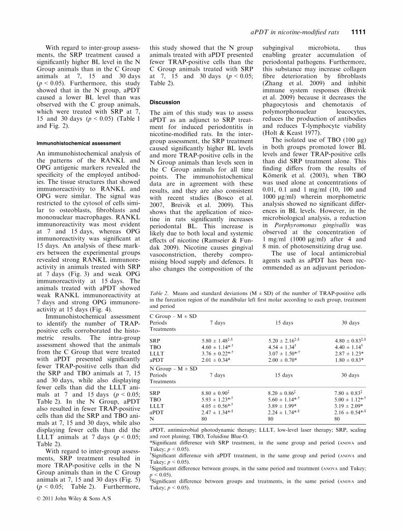
Immunohistochemical assessmentto identify the number of TRAP-positive cells corroborated the histo-metric results. The intra-groupassessment showed that the animalsfrom the C Group that were treatedwith aPDT presented significantlyfewer TRAP-positive cells than didthe SRP and TBO animals at 7, 15and 30 days, while also displayingfewer cells than did the LLLT ani-mals at 7 and 15 days (p < 0.05;Table 2). In the N Group, aPDTalso resulted in fewer TRAP-positivecells than did the SRP and TBO ani-mals at 7, 15 and 30 days, while alsodisplaying fewer cells than did theLLLT animals at 7 days (p < 0.05;Table 2).
With regard to inter-group assess-ments, SRP treatment resulted inmore TRAP-positive cells in the NGroup animals than in the C Groupanimals at 7, 15 and 30 days (Fig. 5)(p < 0.05; Table 2). Furthermore,
this study showed that the N groupanimals treated with aPDT presentedfewer TRAP-positive cells than theC Group animals treated with SRPat 7, 15 and 30 days (p < 0.05;Table 2).
Discussion
The aim of this study was to assessaPDT as an adjunct to SRP treat-ment for induced periodontitis innicotine-modified rats. In the inter-group assessment, the SRP treatmentcaused significantly higher BL levelsand more TRAP-positive cells in theN Group animals than levels seen inthe C Group animals for all timepoints. The immunohistochemicaldata are in agreement with theseresults, and they are also consistentwith recent studies (Bosco et al.2007, Breivik et al. 2009). Thisshows that the application of nico-tine in rats significantly increasesperiodontal BL. This increase islikely due to both local and systemiceffects of nicotine (Ramseier & Fun-dak 2009). Nicotine causes gingivalvasoconstriction, thereby compro-mising blood supply and defences. Italso changes the composition of the
subgingival microbiota, thusenabling greater accumulation ofperiodontal pathogens. Furthermore,this substance may increase collagenfibre deterioration by fibroblasts(Zhang et al. 2009) and inhibitimmune system responses (Breiviket al. 2009) because it decreases thephagocytosis and chemotaxis ofpolymorphonuclear leucocytes,reduces the production of antibodiesand reduces T-lymphocyte viability(Holt & Keast 1977).
The isolated use of TBO (100 lg)in both groups promoted lower BLlevels and fewer TRAP-positive cellsthan did SRP treatment alone. Thisfinding differs from the results ofKomerik et al. (2003), when TBOwas used alone at concentrations of0.01, 0.1 and 1 mg/ml (10, 100 and1000 lg/ml) wherein morphometricanalysis showed no significant differ-ences in BL levels. However, in themicrobiological analysis, a reductionin Porphyromonas gingivallis wasobserved at the concentration of1 mg/ml (1000 lg/ml) after 4 and8 min. of photosensitizing drug use.
The use of local antimicrobialagents such as aPDT has been rec-ommended as an adjuvant periodon-
Table 2. Means and standard deviations (M ± SD) of the number of TRAP-positive cellsin the furcation region of the mandibular left first molar according to each group, treatmentand period
C Group – M ± SDPeriods 7 days 15 days 30 daysTreatments
SRP 5.80 ± 1.48‡,§ 5.20 ± 2.16‡,§ 4.80 ± 0.83‡,§
TBO 4.60 ± 1.14*,† 4.54 ± 1.34† 4.40 ± 1.14†
LLLT 3.76 ± 0.22*,† 3.07 ± 1.50*,† 2.87 ± 1.23*aPDT 2.01 ± 0.34* 2.00 ± 0.70* 1.80 ± 0.83*
N Group – M ± SDPeriods 7 days 15 days 30 daysTreatments
SRP 8.80 ± 0.90‡ 8.20 ± 0.86‡ 7.80 ± 0.83‡
TBO 5.93 ± 1.23*,† 5.60 ± 1.14*,† 5.00 ± 1.12*,†
LLLT 4.05 ± 0.56*,† 3.89 ± 1.99* 3.19 ± 2.09*aPDT 2.47 ± 1.34*,§ 2.24 ± 1.74*,§ 2.16 ± 0.54*,§
N 80 80 80
aPDT, antimicrobial photodynamic therapy; LLLT, low-level laser therapy; SRP, scalingand root planing; TBO, Toluidine Blue-O.*Significant difference with SRP treatment, in the same group and period (ANOVA andTukey; p < 0.05).†Significant difference with aPDT treatment, in the same group and period (ANOVA andTukey; p < 0.05).‡Significant difference between groups, in the same period and treatment (ANOVA and Tukey;p < 0.05).§Significant difference between groups and treatments, in the same period (ANOVA andTukey; p < 0.05).
© 2011 John Wiley & Sons A/S
aPDT in nicotine-modified rats 1111

tal therapy to SRP (Almeida et al.2008b, Fernandes et al. 2009). In thepresent study, the groups locallytreated with aPDT showed lower BLlevels and fewer TRAP-positive cellsthan did the TBO- or SRP-treatedanimals across all time points. Fur-thermore, lower BL levels and fewerTRAP-positive cells were observedin the N Group animals receivingaPDT treatment than in the CGroup animals receiving SRP treat-ment alone. The beneficial propertyof aPDT as an adjunctive technique
to mechanical treatment for peri-odontal disease probably lies in itsphoto-destructive effects on the vari-ous bacterial species that are presentin the areas of induced periodontaldisease. The photodynamic activityof the photosensitizer is based onphoto-oxidant reactions that pro-voke both biochemical and morpho-logical alterations in the target cells(Prates et al. 2007). When a mole-cule of the photosensitizer drugabsorbs photons from a light sourceresonant to the absorbance band of
the photosensitizer, it transformsfrom a basic state into a simpleexcited state. Depending on thestructure of the molecule and itsenvironment, it may lose its energythrough either an electronic or phys-ical process and return to its basicstate, or else transition to a tripleexcited state (triplet state). In thisstate, the molecule may againundergo electronic decay and returnits basic state through redox reac-tions with its environment (Type-Ireaction). Otherwise its excitationenergy may be transferred to an oxy-gen molecule, allowing the formationof singlet oxygen (Type-II reaction).These reactive oxygen species areresponsible for irreversible damageto the bacterial cytoplasmic mem-brane (Wainwright 1998).
In the present study, the groupsthat were treated locally by LLLTshowed lower BL levels and fewerTRAP-positive cells than groupsreceiving SRP treatment at all exper-imental periods. This fact is proba-bly related to the ability of LLLT topromote angiogenesis and cell prolif-eration control inflammation andaccelerate events involved in tissuerepair (Houreld & Abrahamse 2007).However, the animals treated withaPDT showed lower BL than did theLLLT animals at 7 and 15 days inthe C Group and at 7 days in the NGroup.
Studies have reported that theuse of LLLT favours cellular chemo-taxis (Al-Watban & Zhang 1997,Houreld & Abrahamse 2007), pro-viding vasodilatation and localangiogenesis that increase oxygendiffusion through the tissue, whichfavours the repair process becausecollagen secretion by fibroblasts inthe extracellular space occurs only inthe presence of high levels of oxygenpressure.
Stabilization of the bone tissuedepends on balanced dynamicsbetween the activity of osteoblasts(bone formation cells) and osteoclasts(bone resorption cells) (Takayanagi2005). During bone resorption, pro-inflammatory cytokines such as inter-leukin 1 a, interleukin 1 b and tumournecrosis factor a (TNF-a) recruit anddifferentiate precursors of osteoclastsin peri-alveolar bone areas (Wanget al. 1997). This entire system is influ-enced by multiple factors. For exam-ple, LLLT may tip this balance
a b
c d
Fig. 5. Immunostaining for TRAP in the areas of bone loss (BL) in the furcationregion of mandibular left first molars with induced periodontal disease in the differentgroups (C and N): (a) C Group, scaling and root planing (SRP) treatment for 7 days(arrow) (original magnification 1009); (b) C Group, SRP treatment for 7 days (arrow)(original magnification 1009); (c) N Group, SRP treatment for 7 days (HE, originalmagnification 59) and (d) N Group, SRP treatment for 7 days (arrow) (original mag-nification 1009).
© 2011 John Wiley & Sons A/S
1112 Garcia et al.

towards osteoblasts, accelerating theirformation and stimulating their activ-ity (Freitas et al. 2000), while it inhib-its the production of inflammatorymediators by periodontal ligamentcells (Saito et al. 1991).
In our study, RANKL immuno-reactivity was most evident at 7 and15 days, whereas OPG immunoreac-tivity was significant at 15 days. Thisfinding likely occurred becauseRANKL plays an important role inosteoclast differentiation, acting as asurvival factor for osteoclast precur-sors and promoting pre-resorption,whereas OPG acts as a protectiveagent by inhibiting clastic differentia-tion (Lacey et al. 1998). Analysis ofthese markers between the experi-mental groups revealed weakRANKL immunoreactivity in ani-mals treated with aPDT at 7 daysand strong OPG immunoreactivityat 15 days.
Within the limits of this study, itcan be concluded that aPDT was aneffective adjunct to conventionalnon-surgical SRP treatment thatreduced BL in systemically nicotine-modified rats with induced experi-mental periodontitis.
References
Adriaens, P. A., De Boever, J. A. & Loesche, W.J. (1988) Bacterial invasion in root cementumand radicular dentin of periodontally diseasedteeth in humans. A reservoir of periodonto-pathic bacteria. Journal of Periodontology 59,222–230.
Almeida, J. M., Theodoro, L. H., Bosco, A. F.,Nagata, M. J., Bonfante, S. & Garcia, V. G.(2008a) Treatment of experimental periodontaldisease by photodynamic therapy in rats withdiabetes. Journal of Periodontology 79, 2156–2165.
Almeida, J. M., Theodoro, L. H., Bosco, A. F.,Nagata, M. J., Oshiiwa, M. & Garcia, V. G.(2008b) In vivo effect of photodynamic therapyon periodontal bone loss in dental furcations.Journal of Periodontology 79, 1081–1088.
Al-Watban, F. A. H. & Zhang, X. Y. (1997)Comparison of wound healing process usingargon and krypton lasers. Journal of ClinicalLaser Medicine Surgery 15, 209–215.
Bauer, T. W., Hahn, S. M., Spitz, F. R., Kach-ura, A., Glatstein, E. & Fraker, D. L. (2001)Preliminary report of photodynamic therapyfor intraperitoneal sarcomatosis. Annals of Sur-gical Oncology 8, 254–259.
Bosco, A. F., Bonfante, S., Almeida, J. M., Luize,D. S., Nagata, M. J. H. & Garcia, V. G. (2007)A histologic and histometric assessment of theinfluence of nicotine on alveolar bone loss inrats. Journal of Periodontology 78, 527–532.
Breivik, T., Gundersen, Y., Gjermo, P., Von Hor-sten, S. & Opstad, P. K. (2009) Nicotinic ace-tylcholine receptor activation mediatesnicotine-induced enhancement of experimental
periodontitis. Journal of Periodontal Research44, 297–304.
Cesar-Neto, J. B., Benatti, B. B., Sallum, E. A.,Casati, M. Z. & Nociti Jr, F. H. (2006) Theinfluence of cigarette smoke inhalation and itscessation on the tooth-supporting alveolarbone: a histometric study in rats. Journal ofPeriodontal Research 41, 118–123.
Cullinan, M. P., Ford, P. J. & Seymour, G. J.(2009) Periodontal disease and systemic health:current status. Australian Dental Journal 54,62–69.
Dortbudak, O., Haas, R., Bernhart, T & Mailath-pokorny, G. (2001) Lethal photosensitizationfor decontamination of implant surfaces in thetreatment of peri-implantitis. Clinical OralImplants Research 12, 104–108.
Ebisu, S. & Noiri, Y. (2007) Oral biofilms andbone resorption. Clinical Calcium 17, 179–184.
Evans, C. E., Mylchreest, S. & Andrew, J. G.(2006) Age of donor alters the effect of cyclichydrostatic pressure on production by humanmacrophages and osteoblasts of sRANKL,OPG and RANK. BMC Musculoskeletal Disor-ders 7, 21.
Fernandes, L. A., Almeida, J. M., Theodoro, L.H., Bosco, A. F., Nagata, M. J., Martins, T.M., Okamoto, T. & Garcia, V. G. (2009)Treatment of experimental periodontal diseaseby photodynamic therapy in immunosup-pressed rats. Journal of Clinical Periodontology36, 219–228.
Freitas, L. G. F., Baranauskas, V. & Cruz-Hofling, M. A. (2000) Laser effects on osteo-genesis. Applied Surface Science 154–158,548–554.
Genco, R. J. (1996) Current view of risk factorsfor periodontal diseases. Journal of Periodontol-ogy 67, 1041–1049.
Hassumi, M. Y., Silva-Filho, V. J., Campos-Junior, J. C., Vieira, S. M., Cunha, F. Q., Al-ves, P. M., Alves, J. B., Kawai, T., Goncalves,R. B. & Napimoga, M. H. (2009) PPAR-gamma agonist rosiglitazone prevents inflam-matory periodontal bone loss by inhibitingosteoclastogenesis. International Immunophar-macology 9, 1150–1158.
Holt, P. G. & Keast, D. (1977) Environmentallyinduced changes in immunological function:acute and chronic effects of inhalation oftobacco smoke and other atmospheric contami-nants in man and experimental animals. Bacte-riological Reviews 41, 205–216.
Houreld, N. & Abrahamse, H. (2007) In vitroexposure of wounded diabetic fibroblast cells toa helium-neon laser at 5 and 16 J/cm². Photo-medicine and Laser Surgery 25, 78–84.
Iwamoto, K., Miyamoto, T., Sawatani, Y.,Hosogane, N., Hamaguchi, I., Takami, M.,Nomiyama, K., Takagi, K. & Suda, T.(2004) Dimer formation of receptor activatorof nuclear factor kappaB induces incompleteosteoclast formation. Biochemical and Bio-physical Research Communications 325, 229–234.
Jin, Q., Cirelli, J. A., Park, C. H., Sugai, J. V.,Taba Jr, M., Kostenuik, P. J & Giannobile, W.V. (2007) RANKL inhibition through osteo-protegerin blocks bone loss in experimentalperiodontitis. Journal of Periodontology 78,1300–1308.
Johnson, J. H. (1975) Effects of local irritationand dextran and sulphate administration onthe periodontium of the rat. Journal of Peri-odontal Research 10, 332–345.
Kaldahl, W. B., Kalkwarf, K. L. & Patil, K. D.(1993) A review of longitudinal studies that
compared periodontal therapies. Journal ofPeriodontology 64, 243–253.
Khojasteh, A., Eslaminejad, M. B. & Nazarian,H. (2008) Mesenchymal stem cells enhancebone regeneration in rat calvarial critical sizedefects more than platelete-rich plasma. OralSurgery Oral Medicine Oral Pathology OralRadiology & Endodontics 106, 356–362.
Khosla, S. (2001) Minireview: The OPG/RANKL/RANK system. Endocrinology 142,5050–5055.
Kim, Y. D., Kim, S. S., Hwang, D. S., Kim, S.G., Kwon, Y. H., Shin, S. H., Kim, U. K.,Kim, J. R. & Chung, I. K. (2007) Effect oflow-level laser treatment after installation ofdental titanium implant-immunohistochemicalstudy of RANKL, RANK, OPG: an experi-mental study in rats. Lasers in Surgery andMedicine 39, 441–450.
Komerik, N., Nakanishi, H., MacRobert, A. J.,Henderson, B., Speight, P. & Wilson, M.(2003) In vivo killing of Porphyromonas gingi-valis by toluidine blue-mediated photosensitiza-tion in an animal model. Antimicrobial AgentsChemotherapy 47, 932–940.
Lacey, D. L., Timms, E., Tan, H. L., Kelley,M. J., Dunstan, C. R., Burgess, T., Elliott, R.,Colombero, A., Elliott, G., Scully, S., Hsu, H.,Sullivan, J., Hawkins, N., Davy, E., Capparelli,C., Eli, A., Qian, Y. X., Kaufman, S.,Sarosi, I., Shalhoub, V., Senaldi, G., Guo, J.,Delaney, J. & Boyle, W. J. (1998) Osteoproteg-erin ligand is a cytokine that regulates osteo-clast differentiation and activation. Cell 93,165–176.
Liu, S., Cheng, Y., Xu, W. & Bian, Z. (2010)Protective effects of follicle-stimulating hor-mone inhibitor on alveolar bone loss resultingfrom experimental periapical lesions in ovari-ectomized rats. Journal of Endodontics 36,658–663.
Maisch, T. (2007) Anti-microbial photodynamictherapy: useful in the future? Lasers in MedicalScience 22, 83–91.
Okamoto, M., Kita, T., Okuda, H., Tanaka, T. &Nakashima, T. (1994) Effects of aging on acutetoxicity of nicotine in rats. Pharmacology &Toxicology 75, 1–6.
Pinto, J. R., Bosco, A. F., Okamoto, T., Guerra,J. B. & Piza, I. G. (2002) Effects of nicotine onthe healing of extraction sockets in rats. A his-tological study. Brazilian Dental Journal 13,3–9.
Prates, R. A., Yamada, J. R. A. M., Suzuki, L.C., Eiko Hashimoto, M. C., Cai, S., Gouw-So-ares, S., Gomes, L. & Ribeiro, M. S. (2007)Bactericidal effect of malachite green and redlaser on actinobacillus actinomycetemcomitans.Journal of Photochemistry and Photobiology B86, 70–76.
Ramseier, C. A. & Fundak, A. (2009) Tobacco usecessation provided by dental hygienists. Interna-tional Journal of Dental Hygiene 7, 39–48.
Saito, S., Ngan, P., Rosol, T., Saito, M., Shimizu,H., Shinjo, N., Shanfeld, J. & Davidovitch, Z.(1991) Involvement of PGE synthesis in theeffect of intermittent pressure and interleukin-1beta on bone resorption. Journal of DentalResearch 70, 27–33.
Shalhoub, V., Faust, J., Boyle, W. J., Dunstan, C.R., Kelley, M., Kaufman, S., Scully, S., Van,G. & Lacey, D. L. (1999) Osteoprotegerin andosteoprotegerin ligant effects on osteoclast for-mation from human peripheral blood mononu-clear cell precursors. Journal of CellularBiochemistry 72, 251–261.
© 2011 John Wiley & Sons A/S
aPDT in nicotine-modified rats 1113

Takayanagi, H. (2005) Inflammatory bonedestruction and osteoimmunology. Journal ofPeriodontal Research 40, 287–293.
Tomaselli, F., Maier, A., Sankin, O., Anegg, U.,Stranzl, U., Pinter, H., Kapp, K. & Smolle-Juttner, F. M. (2001) Acute effects of combinedphotodynamic therapy and hyperbaric oxygen-eration in lung cancer – a clinical pilot study.Lasers in Surgery and Medicine 28, 399–403.
Wainwright, M. (1998) Photodynamic antimicro-bial chemotherapy (PACT). Journal of Antimi-crobial Chemotherapy 42, 13–28.
Wang, C. Y., Tani-Ishii, N. & Stashenko, P.(1997) Bone-resorptive cytokine gene expres-sion in periapical lesions in the rat. Oral Micro-biology and Immunology 12, 65–71.
Zhang, W., Song, F. & Windsor, L. J. (2009)Cigarette smoke condensate affects the colla-gen-degrading ability of human gingival fibro-blasts. Journal of Periodontal Research 44, 704–713.
Address:Valdir Gouveia GarciaFaculdade de Odontologia de Aracatuba-UNESPRua Jose Bonifacio, 1193CEP: 16015-050, Aracatuba, SP, BrazilE-mail: [email protected]
Clinical Relevance
Scientific rationale for the study:Smoking is a risk factor for peri-odontal disease. In these cases,SRP alone is inadequate for caseresolution. aPDT has shown satis-factory results as an adjunctive
periodontal treatment, althougharticles regarding its applicationin smoking patients are scarce inthe literature.Main results: aPDT was efficientas an adjunctive treatment toSRP; it reduced BL in systemi-
cally nicotine-modified rats withexperimentally induced periodon-titis.Practical implications: aPDT maybe used as an adjuvant to theconventional non-surgical treat-ment in smoking patients.
© 2011 John Wiley & Sons A/S
1114 Garcia et al.