Oral Diagnostic Methods for the Detection of Periodontal ...
13
diagnostics Article Oral Diagnostic Methods for the Detection of Periodontal Disease Liza L. Ramenzoni 1,2, * , Marc P. Lehner 1 , Manuela E. Kaufmann 1 , Daniel Wiedemeier 3 , Thomas Attin 1 and Patrick R. Schmidlin 1,2, * Citation: Ramenzoni, L.L.; Lehner, M.P.; Kaufmann, M.E.; Wiedemeier, D.; Attin, T.; Schmidlin, P.R. Oral Diagnostic Methods for the Detection of Periodontal Disease. Diagnostics 2021, 11, 571. https://doi.org/ 10.3390/diagnostics11030571 Academic Editor: Timo Sorsa Received: 8 February 2021 Accepted: 18 March 2021 Published: 22 March 2021 Publisher’s Note: MDPI stays neutral with regard to jurisdictional claims in published maps and institutional affil- iations. Copyright: © 2021 by the authors. Licensee MDPI, Basel, Switzerland. This article is an open access article distributed under the terms and conditions of the Creative Commons Attribution (CC BY) license (https:// creativecommons.org/licenses/by/ 4.0/). 1 Clinic of Conservative and Preventive Dentistry, Center of Dental Medicine, University of Zurich, 8032 Zurich, Switzerland; [email protected] (M.P.L.); [email protected] (M.E.K.); [email protected] (T.A.) 2 Laboratory of Applied Periodontal and Peri-Implantitis Sciences, Clinic of Conservative and Preventive Dentistry, Center of Dental Medicine, University of Zurich, 8032 Zurich, Switzerland 3 Statistical Services, Center of Dental Medicine, University of Zurich, 8032 Zurich, Switzerland; [email protected] * Correspondence: [email protected] (L.L.R.); [email protected] (P.R.S.); Tel.: +41-44-634-3417 (P.R.S.) Abstract: Periodontitis is a common immune-inflammatory oral disease. Early detection plays an important role in its prevention and progression. Saliva is a reliable medium that mirrors periodontal health and is easily obtainable for identifying periodontal biomarkers in point-of-care diagnostics. The aim of this study is to evaluate the effectiveness of diagnostic salivary tests to determine periodontal status. Whole saliva (stimulated/unstimulated) from twenty healthy and twenty stage III grade B generalized periodontitis patients was tested for lactoferrin, alkaline phosphatase, calcium, density, osmolarity, pH, phosphate, buffer capacity, salivary flow rate and dynamic viscosity. A semi- quantitative urinary strip test was used to evaluate markers of inflammation in saliva (erythrocytes, leukocytes, urobilinogen, nitrite, glucose, bilirubin, and ketones), clinical periodontal parameters and pathogenic bacteria. Concentrations of lactoferrin, hemoglobin, and leukocytes were found to be significantly higher in the stimulated and unstimulated saliva in periodontitis patients compared to healthy patients, whereas alkaline phosphatase levels were higher in unstimulated saliva of periodontitis patients (p < 0.05). Periodontal biomarker analysis using test strips may be considered rapid and easy tool for distinguishing between periodontitis and healthy patients. The increase in lactoferrin, hemoglobin, and leucocytes—determined by strip tests—may provide a non-invasive method of periodontal diagnosis. Keywords: saliva; point-of-care diagnostics; periodontitis; biomarkers; oral disease 1. Introduction Periodontitis is considered one of the most prevalent immune-inflammatory diseases of the oral cavity. It derives from a specific pathogenic bacteria–host interaction and leads to periodontal tissue destruction [1,2]. The progression of periodontitis is often characterized by irregular phases of increased activity and dormant remission [3–5]. Traditional clinical periodontal assessment methods, such as pocket probing depth (PPD), bleeding on probing (BOP), clinical attachment level (CAL), and radiological assessment of the alveolar bone volume, are widely used and documented [1,2]. However, these traditional periodontal classification parameters fail to provide noteworthy information on current disease ac- tivity, severity and extent of breakdown, future progression and therapy response [2,6]. More importantly, the biological phenotype of the patient is not properly reflected by the clinical assessment methods [7] and the host response to periodontal bacteria and the subsequent inflammatory burden, i.e., the influence of biological phenotype, may largely determine periodontitis progression. Further, an early diagnosis may lead to more success- ful treatment [8,9]. In an attempt to redesign the periodontal disease framework, the new Diagnostics 2021, 11, 571. https://doi.org/10.3390/diagnostics11030571 https://www.mdpi.com/journal/diagnostics
-
Upload
khangminh22 -
Category
Documents
-
view
0 -
download
0
Transcript of Oral Diagnostic Methods for the Detection of Periodontal ...

diagnostics
Article
Oral Diagnostic Methods for the Detection ofPeriodontal Disease
Liza L. Ramenzoni 1,2,* , Marc P. Lehner 1, Manuela E. Kaufmann 1 , Daniel Wiedemeier 3 , Thomas Attin 1
and Patrick R. Schmidlin 1,2,*
�����������������
Citation: Ramenzoni, L.L.; Lehner,
M.P.; Kaufmann, M.E.; Wiedemeier,
D.; Attin, T.; Schmidlin, P.R. Oral
Diagnostic Methods for the Detection
of Periodontal Disease. Diagnostics
2021, 11, 571. https://doi.org/
10.3390/diagnostics11030571
Academic Editor: Timo Sorsa
Received: 8 February 2021
Accepted: 18 March 2021
Published: 22 March 2021
Publisher’s Note: MDPI stays neutral
with regard to jurisdictional claims in
published maps and institutional affil-
iations.
Copyright: © 2021 by the authors.
Licensee MDPI, Basel, Switzerland.
This article is an open access article
distributed under the terms and
conditions of the Creative Commons
Attribution (CC BY) license (https://
creativecommons.org/licenses/by/
4.0/).
1 Clinic of Conservative and Preventive Dentistry, Center of Dental Medicine, University of Zurich,8032 Zurich, Switzerland; [email protected] (M.P.L.); [email protected] (M.E.K.);[email protected] (T.A.)
2 Laboratory of Applied Periodontal and Peri-Implantitis Sciences, Clinic of Conservative and PreventiveDentistry, Center of Dental Medicine, University of Zurich, 8032 Zurich, Switzerland
3 Statistical Services, Center of Dental Medicine, University of Zurich, 8032 Zurich, Switzerland;[email protected]
* Correspondence: [email protected] (L.L.R.); [email protected] (P.R.S.);Tel.: +41-44-634-3417 (P.R.S.)
Abstract: Periodontitis is a common immune-inflammatory oral disease. Early detection plays animportant role in its prevention and progression. Saliva is a reliable medium that mirrors periodontalhealth and is easily obtainable for identifying periodontal biomarkers in point-of-care diagnostics.The aim of this study is to evaluate the effectiveness of diagnostic salivary tests to determineperiodontal status. Whole saliva (stimulated/unstimulated) from twenty healthy and twenty stage IIIgrade B generalized periodontitis patients was tested for lactoferrin, alkaline phosphatase, calcium,density, osmolarity, pH, phosphate, buffer capacity, salivary flow rate and dynamic viscosity. A semi-quantitative urinary strip test was used to evaluate markers of inflammation in saliva (erythrocytes,leukocytes, urobilinogen, nitrite, glucose, bilirubin, and ketones), clinical periodontal parametersand pathogenic bacteria. Concentrations of lactoferrin, hemoglobin, and leukocytes were found to besignificantly higher in the stimulated and unstimulated saliva in periodontitis patients comparedto healthy patients, whereas alkaline phosphatase levels were higher in unstimulated saliva ofperiodontitis patients (p < 0.05). Periodontal biomarker analysis using test strips may be consideredrapid and easy tool for distinguishing between periodontitis and healthy patients. The increase inlactoferrin, hemoglobin, and leucocytes—determined by strip tests—may provide a non-invasivemethod of periodontal diagnosis.
Keywords: saliva; point-of-care diagnostics; periodontitis; biomarkers; oral disease
1. Introduction
Periodontitis is considered one of the most prevalent immune-inflammatory diseasesof the oral cavity. It derives from a specific pathogenic bacteria–host interaction and leads toperiodontal tissue destruction [1,2]. The progression of periodontitis is often characterizedby irregular phases of increased activity and dormant remission [3–5]. Traditional clinicalperiodontal assessment methods, such as pocket probing depth (PPD), bleeding on probing(BOP), clinical attachment level (CAL), and radiological assessment of the alveolar bonevolume, are widely used and documented [1,2]. However, these traditional periodontalclassification parameters fail to provide noteworthy information on current disease ac-tivity, severity and extent of breakdown, future progression and therapy response [2,6].More importantly, the biological phenotype of the patient is not properly reflected by theclinical assessment methods [7] and the host response to periodontal bacteria and thesubsequent inflammatory burden, i.e., the influence of biological phenotype, may largelydetermine periodontitis progression. Further, an early diagnosis may lead to more success-ful treatment [8,9]. In an attempt to redesign the periodontal disease framework, the new
Diagnostics 2021, 11, 571. https://doi.org/10.3390/diagnostics11030571 https://www.mdpi.com/journal/diagnostics

Diagnostics 2021, 11, 571 2 of 13
periodontal classification includes multidimensional staging and grading system parame-ters, which allows one to partially assess the future periodontal risk [2,8]. Although the newgrading risk factors, i.e., smoking and diabetes, are included to predict the likelihood offuture periodontal breakdown, they are still not able to determine the exact time of diseasemanifestation [2,8]. Thus, adding more robust biomarkers to the new classification systemwill improve the identification of active periods of periodontitis, monitor progression andavoid consequent mismanagement of periodontal treatment.
To better clinically identify the biological progression or phases of periodontitis withinthe new classification system, the development of new diagnostic tests is still required.Periodontitis is closely associated with tempering specific immune responses, and itsmodifying factors are typically found in the gingival crevicular fluid (GCF) and salivaduring progression of the disease [2,10]. Oral fluid-based point-of-care diagnostics havebeen previously documented as potential chairside tests for determining periodontaldiseases [2,11]. In order to measure disease activity, samples of saliva and GCF have beenshown to mirror the periodontal condition and may be reliable mediums for biomarkerdetection [12]. Saliva offers many advantages in point-of-care diagnostics as it is easilyobtainable, may be collected non-invasively and is rich in more than a thousand differentdiagnostic biomarker molecules for chronic inflammation and tissue destruction [13,14]. Inaddition, many compounds found in the blood are also found in saliva. Thus, saliva is avery useful tool for monitoring not only oral, but also systemic health [15]. In this respect,several molecular approaches, such as PCR for RNA/DNA or ELISAs for proteins, havebeen proposed for detecting periodontal biomarker molecules in saliva using advancedlaboratorial techniques [14,16]. However, subject to further study, simpler and more easilyapplicable salivary techniques may provide the pathophysiological parameters necessaryfor a diagnosis of periodontitis and obviate the need for blood samples or histologicalsections, which are traditionally required. There is also a lack of simple and affordablemethods to identify biomarkers that may both diagnose the disease and predict the risk offuture disease activity.
The aim of this study is to evaluate a simple, well-established, and cost-effective urineanalysis test to indicate and diagnose periodontitis. A semi-quantitative strip test thathas not previously been applied in oral studies was used in an attempt to easily identifyand distinguish between six salivary biomarkers in periodontitis patients as compared tohealthy patients. In addition, differences in salivary composition, both in a stimulated andunstimulated state, were analyzed. We hypothesized that a strip test traditionally usedfor urine analyses may perform equally well as a diagnostic test for periodontal diseasebiomarkers and aid the determination of periodontal inflammatory severity (stage) andrisk (grade) in periodontitis patients.
2. Materials and Methods2.1. Participants and Study Design
A total of 40 patients (24 men and 16 women) referred for treatment to the Clinic ofConservative and Preventive Dentistry, Center of Dental Medicine, University of Zurichagreed to participate in this study. The clinical considerations for periodontitis patientswere made following the new classification of 2018 [2]. Half of the participants wererecruited from patients classified with the new periodontitis classification system [2] whopresented generalized periodontitis stage III (interdental CAL ≥ 5 mm, radiographic boneloss extending to middle third of root and beyond, loss of 4–5 teeth due to periodontitis)and grade B progression (radiographic bone loss < 2 mm over 5 years, half pack or lessper day smoking, biofilm commensurate with destruction). Periodontitis patients alsopresented PPD ≥ 6 mm, vertical bone loss ≥ 3 mm, furcation involvement class II orIII, moderate ridge defects [2]. The remaining 20 individuals were periodontally healthy(PPD ≤ 3 mm) with no CAL or BOP (≤10%), and no signs of gingival inflammation(redness, clinical swelling, edema or pain) [2]. All patients were comprehensively informedabout the content and purpose of the study before becoming involved. They were also
Diagnostics 2021, 11, 571 3 of 13
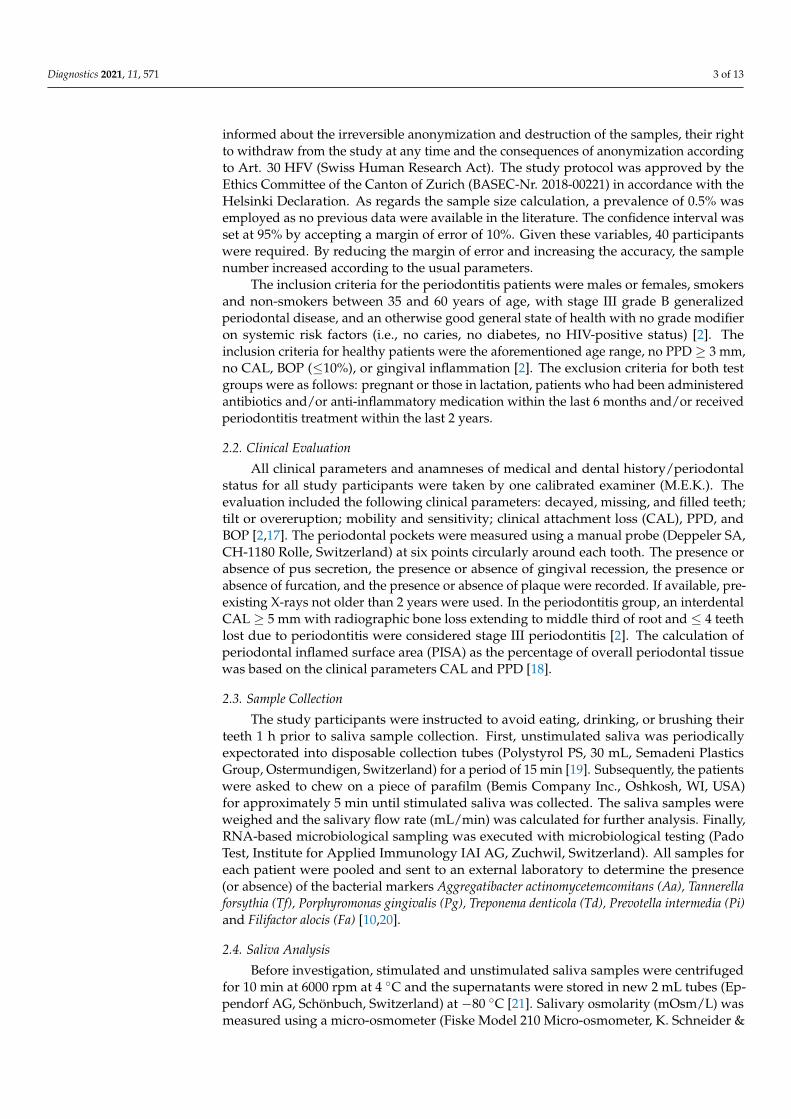
informed about the irreversible anonymization and destruction of the samples, their rightto withdraw from the study at any time and the consequences of anonymization accordingto Art. 30 HFV (Swiss Human Research Act). The study protocol was approved by theEthics Committee of the Canton of Zurich (BASEC-Nr. 2018-00221) in accordance with theHelsinki Declaration. As regards the sample size calculation, a prevalence of 0.5% wasemployed as no previous data were available in the literature. The confidence interval wasset at 95% by accepting a margin of error of 10%. Given these variables, 40 participantswere required. By reducing the margin of error and increasing the accuracy, the samplenumber increased according to the usual parameters.
The inclusion criteria for the periodontitis patients were males or females, smokersand non-smokers between 35 and 60 years of age, with stage III grade B generalizedperiodontal disease, and an otherwise good general state of health with no grade modifieron systemic risk factors (i.e., no caries, no diabetes, no HIV-positive status) [2]. Theinclusion criteria for healthy patients were the aforementioned age range, no PPD ≥ 3 mm,no CAL, BOP (≤10%), or gingival inflammation [2]. The exclusion criteria for both testgroups were as follows: pregnant or those in lactation, patients who had been administeredantibiotics and/or anti-inflammatory medication within the last 6 months and/or receivedperiodontitis treatment within the last 2 years.
2.2. Clinical Evaluation
All clinical parameters and anamneses of medical and dental history/periodontalstatus for all study participants were taken by one calibrated examiner (M.E.K.). Theevaluation included the following clinical parameters: decayed, missing, and filled teeth;tilt or overeruption; mobility and sensitivity; clinical attachment loss (CAL), PPD, andBOP [2,17]. The periodontal pockets were measured using a manual probe (Deppeler SA,CH-1180 Rolle, Switzerland) at six points circularly around each tooth. The presence orabsence of pus secretion, the presence or absence of gingival recession, the presence orabsence of furcation, and the presence or absence of plaque were recorded. If available, pre-existing X-rays not older than 2 years were used. In the periodontitis group, an interdentalCAL ≥ 5 mm with radiographic bone loss extending to middle third of root and ≤ 4 teethlost due to periodontitis were considered stage III periodontitis [2]. The calculation ofperiodontal inflamed surface area (PISA) as the percentage of overall periodontal tissuewas based on the clinical parameters CAL and PPD [18].
2.3. Sample Collection
The study participants were instructed to avoid eating, drinking, or brushing theirteeth 1 h prior to saliva sample collection. First, unstimulated saliva was periodicallyexpectorated into disposable collection tubes (Polystyrol PS, 30 mL, Semadeni PlasticsGroup, Ostermundigen, Switzerland) for a period of 15 min [19]. Subsequently, the patientswere asked to chew on a piece of parafilm (Bemis Company Inc., Oshkosh, WI, USA)for approximately 5 min until stimulated saliva was collected. The saliva samples wereweighed and the salivary flow rate (mL/min) was calculated for further analysis. Finally,RNA-based microbiological sampling was executed with microbiological testing (PadoTest, Institute for Applied Immunology IAI AG, Zuchwil, Switzerland). All samples foreach patient were pooled and sent to an external laboratory to determine the presence(or absence) of the bacterial markers Aggregatibacter actinomycetemcomitans (Aa), Tannerellaforsythia (Tf), Porphyromonas gingivalis (Pg), Treponema denticola (Td), Prevotella intermedia (Pi)and Filifactor alocis (Fa) [10,20].
2.4. Saliva Analysis
Before investigation, stimulated and unstimulated saliva samples were centrifugedfor 10 min at 6000 rpm at 4 ◦C and the supernatants were stored in new 2 mL tubes (Ep-pendorf AG, Schönbuch, Switzerland) at −80 ◦C [21]. Salivary osmolarity (mOsm/L) wasmeasured using a micro-osmometer (Fiske Model 210 Micro-osmometer, K. Schneider &

Diagnostics 2021, 11, 571 4 of 13
Co. AG, Zurich, Switzerland). Calibration was performed using the Gerber osmolaritystandard 300 mOsm/kg H2O for a result of 300 mOsm/L saliva. In addition, the pH valueof the saliva specimens was analyzed (Metrohm, Herisau, Switzerland). The pH meterwas calibrated before the measurements with two buffer solutions of pH 7.00 and pH 4.00(Thermo Fisher Scientific AG, Reinach, Switzerland). Moreover, the buffer capacity of thesaliva (mL/0.1 M HCl) was determined by titration with 0.1 M/L HCl solution (PanReacAppliChem, Darmstadt, Germany). The buffer capacity corresponded to the consumptionof 0.1 M/L HCl solution up to a pH of 5.7. Phosphate determination (mmol/L) was per-formed with the spectrophotometer (Portmann Instruments AG, Biel-Benken, Switzerland)at 750 nm. The obtained data were multiplied by 100 to determine the phosphate concentra-tion in g P/mL saliva. Calcium (Ca (mmol/L)) was also determined with a spectrophotome-ter at 422 nm (Analytic Jena AG, Jena, Germany). Finally, the dynamic viscosity (mPa.s) anddensity (g/cm3) of samples was investigated with a micro-viscometer (Lovis 2000 M/ME,Anton Paar GmbH, Graz, Austria). The determination was carried out at 20 ◦C according tothe specifications of Anton Paar GmbH. A reagent strip Combur9-Test Cobas (Roche Diag-nostics GmbH, Mannheim, Germany) was performed on saliva samples to determine pH,leukocytes, erythrocytes, levels of nitrites, proteins, glucose, ketone bodies, urobilinogen,and bilirubin. The procedures were performed according to manufacturer’s instructions.The test strips were moistened at room temperature with 10 µL of unstimulated saliva and10 µL of stimulated saliva from samples from each participant. After 1 min, a visual checkfor a possible color change was conducted and the color reference indicated by individualtests was used as the basis for comparison (Figure 1). The most clearly visually recognizablecolor change compared to the manufacturer’s color reference was registered. The semi-quantitative parameters determined by the test included the following: (1) pH (5/6/7/8/9);(2) leucocytes (negative/10-25/75/500 leucocytes/µL); (3) nitrite (negative/positive);(4) protein (negative/30/100/500 mg/dL); (5) glucose (50/100/300/1000 mg/dL); (6) ke-tone bodies (negative/10/50/150 mg/dL); (7) urobilinogen (normal/1/4/8/12 mg/dL);(8) bilirubin (negative/1+/2+/3+); and (9) hemoglobin/hemolyzed erythrocytes (nega-tive/10/25/50/250 Ery/µL).
2.5. Alkaline Phosphatase and Lactoferrin Analysis of Saliva
Alkaline phosphatase (mU/mL) was detected using a kinetic color test (ab83371,Abcam, Alkaline Phosphatase Assay Kit, Cambridge, UK) and a 96-well plate (Thermo-fisher Scientific/Life Technologies, Waltham, MA, USA) at 450 nm in a spectrophotometer(Bucher Biotec AG, Basel, Switzerland). Lactoferrin (µg/mL) was analyzed with ELISA(enzyme-linked immunosorbent assay kit, Abcam, Cambridge, UK) following a previouslydescribed protocol [21]. The absorbance at 450 nm was recorded for each ELISA on amicroplate reader (EZ Read 400 Microplate Reader; Biochrom, Cambourne, UK) and theabsorbance reference value (540 or 570 nm) was subtracted from the test values. Experi-ments were performed on three specimens from each test group in order to confirm thedilution factor of each biomarker. All experiments were conducted in triplicate.
2.6. Statistical Analysis
The data were analyzed and plotted using the statistical software R (The R Foundationfor Statistical Computing, Vienna, Austria) and Microsoft Excel for Mac (v16.37, 20051002,Microsoft Corporation, Redmond, WA, USA). The statistical evaluation of all parametersin Tables 1–3 was performed using the Wilcoxon rank-sum test, with the exception of thebinary data for nitrite, which was tested using Fisher’s exact test. The statistical significancelevel was set at α ≤ 0.05.

Diagnostics 2021, 11, 571 5 of 13
Figure 1. Reagent strip (Combur9-Test Cobas test strips) performed on saliva samples in order to determine: pH, leukocytes,erythrocytes, levels of nitrites, proteins, glucose, ketone bodies, urobilinogen, and bilirubin. (A) Blank test strip; (B) controlwith 10 µL water/test field each; (C) saliva sample 10 µL/test field each; (D) color reference.
Table 1. Descriptive statistics of patient characteristics, clinical parameters, and a comparison ofinflammation values of bleeding on probing (BOP), and periodontal inflamed surface area (PISA)as median (IQR) in healthy and periodontal patients (the statistical significance level was set atα ≤ 0.05).
Patient Characteristics Healthy PeriodontitisStage III Grade B
Age (mean ± SD) 50.47 ± 11.27 51.16 ± 12.35Gender (m/f) 9/11 15/5Smoking (yes/no) 5/15 12/8
Clinical parameters
CALa (mean ± SD) 1.86 ± 0.23 3.45 ± 1.12PPD b (mean ± SD) 2.01 ± 0.31 4.24 ± 1.35BOP c (mean ± SD) 26.73 ± 22.96 28.59 ± 23.29
Inflammation values
BOP c (%) 10 (9) e 41 (28) e
PISA d (mm2) 1010 (366) e 2796 (2112) e
a CAL: clinical attachment level. b PPD: pocket probing depth. c BOP: bleeding on probing. d PISA: periodontalinflamed surface area calculated [18]. e p-value: < 0.001.

Diagnostics 2021, 11, 571 6 of 13
Table 2. Specific salivary parameters in median (IQR) obtained from stimulated and unstimulated saliva in healthy andperiodontal patients.
Unstimulated Saliva Stimulated Saliva Comparison Comparison
Healthy (A)
PeriodontitisStage IIIGrade B
(B)
Healthy (C)Periodontitis
Stage IIIGrade B (D)
(A) vs. (B)(p-Value) a
(C) vs. (D)(p-Value) a
Sample volume (g) 5.7 (7.7) 2.2 (5.1) 5.2 (7.8) 4.7 (8.2) 0.003 0.7Salivary flow rate b (mL/min) 0.51 (0.38) 0.34 (0.16) 1.67 (0.98) 1.64 (0.94) 0.002 0.6
Alkaline phosphatase (mU/mL) 0.97 (0.68) 2.46 (3.94) 0.64 (0.95) 0.90 (2.21) 0.03 0.2Buffer capacity(mL 0.1 M HCl) 0.070 (0.022) 0.066 (0.053) 0.120 (0.061) 0.114 (0.064) 0.6 0.5
Calcium (mmol/L) 1.36 (0.30) 1.51 (0.68) 1.10 (0.19) 1.07 (0.20) 0.2 0.3Density (g/cm3) 1 (0.00045) 1 (0.00015) 1 (0.00018) 1 (0.00090) 0.3 0.7
Dynamic viscosity(mPa·s) 1.276 (0.251) 1.117 (0.065) 1.107 (0.251) 1.085 (0.067) 0.1 0.3
Lactoferrin (µg/mL) 9.3 (1.9) 15.9 (1.7) 9.6 (1.8) 28.5 (3.5) <0.001 <0.001Osmolarity (mOsm/L) 66 (17) 98 (38) 71 (10) 76 (13) <0.001 0.006
pH 7.43 (0.77) 7.19 (0.57) 7.97 (0.29) 7.95 (0.75) 0.1 0.4Phosphate (mmol/L) 5.3 (3.1) 6.2 (3.2) 4.0 (1.3) 4.2 (1.5) 0.07 0.3
a p-value: statistical significance was set at p ≤ 0.05. b Saliva ml per minute: calculated value.
Table 3. Median (+/− IQR) of the saliva parameters bilirubin, erythrocytes, glucose, hemoglobin, ketones, leukocytes,nitrite, pH, protein and urobilinogen.
Unstimulated Saliva Stimulated Saliva Comparison Comparison
Healthy(A)
PeriodontitisStage III
Grade B (B)
Healthy(C)
PeriodontitisStage III
Grade B (D)
(A) vs. (B)(p-Value) b
(C) vs. (D)(p-Value) b
Bilirubin (pos./neg.) 0 (0) 0 (0) 0 (0) 0 (0) NA NAGlucose (mg/dL) 0 (0) 0 (0) 0 (0) 0 (0) NA NAHemoglobin (Ery/mL) 10 (25) 50 (225) 18 (25) 50 (225) 0.0007 0.006Ketones (mg/dL) 0 (0) 0 (0) 0 (0) 0 (0) NA NALeukocytes (Leu/µL) 75 (0) 500 (425) 75 (425) 500 (0) 0.002 0.002Nitrite a (pos./neg.) (−)1/(+)19 (−)1/(+)19 (−)1/(+)19 (−)2/(+)18 NA NApH 7 (1) 7 (1) 8 (0) 8 (0) 0.01 0.2Protein (mg/dL) 65 (70) 65 (70) 30 (70) 30 (70) 0.9 0.3Urobilinogen (mg/dL) 0 (0) 0 (0) 0 (0) 0 (0) NA NA
a Nitrite: the evaluation of nitrites was performed using Fisher’s exact test. All other tests were performed using the Wilcoxon rank-sumtest with continuity correction. b p-value: statistical significance was set at p ≤ 0.05.
3. Results3.1. Clinical Evaluation and Sample Collection
The descriptive statistical analysis for all participants is listed in Table 1. In brief, thisstudy evaluated forty patients in total aged between 35 to 60 years old; twenty participantshad stage III grade B generalized periodontal disease and twenty were periodontallyhealthy participants. For the periodontal patients, the mean collective PPD was 4.24 mm,with a range of 5–12 mm. The healthy patients had a mean collective sulci depth of2.01 mm, with a range of 2–3 mm. Next, clinical inflammation values were determined byrecording the BOP and by calculating the periodontal inflamed surface area (PISA). Thehealthy patients exhibited limited BOP with a mean clinical attachment level (CAL) of1.86 mm. The difference between the highest and the lowest range values was calculatedand BOP was found to be lower in healthy patients, with an interquartile range (IQR) of9 in comparison to an IQR of 28 for the periodontitis patients. As expected, periodontitispatients exhibited increased BOP with a higher mean CAL of 3.45 mm. The IQR PISA valuefor the healthy patients was 366, while the IQR value for the periodontitis patients was2112, hence, almost six times greater. Both BOP and PISA were significantly higher for theperiodontitis patients compared to the healthy patients (p < 0.001).

Diagnostics 2021, 11, 571 7 of 13
3.2. Salivary Analysis
The salivary parameters for unstimulated and stimulated saliva are given in Table 2.Within the unstimulated saliva samples, a significantly higher volume (p = 0.003) andhigher salivary flow rate (p = 0.002) were found for the healthy patients. Results from acomparison of stimulated saliva between the healthy and periodontitis patients were notstatistically significant for volume and salivary flow rate. The level of alkaline phosphatasein unstimulated saliva from periodontitis patients was significantly higher than that of thehealthy patients (p = 0.03). No significance was found for these levels in stimulated saliva(p = 0.2). The osmolarity showed significantly higher levels for periodontitis patients inboth unstimulated (p < 0.001) and stimulated saliva (p = 0.006).
Lactoferrin was the most distinct parameter between the healthy and periodontitispatients stage III grade B in both unstimulated and stimulated saliva (Table 3); it was foundto be two to three times higher in periodontitis patients compared to healthy patients(Figure 2).
Figure 2. Comparison between lactoferrin measurements in stimulated and unstimulated saliva samples from healthy andstage III grade B generalized periodontal patients.
For the other salivary parameters—buffer capacity, calcium, density, dynamic vis-cosity, pH, and phosphate—no significant difference was found between stimulated orunstimulated saliva of healthy or stage III grade B periodontitis patients. Regarding themicrobiological test, the only bacterium not detected in any of the samples was Aa. Incontrast, Tf, Pg, Td, Pi, and Fa were all detected in small quantities, with a detection limit of< 0.3% (Figure 3).

Diagnostics 2021, 11, 571 8 of 13
Figure 3. Marker load (%) of known pathogenic markers for periodontal bacterial species tested in healthy and periodontalpatients. TML: Total marker load.
The evaluation of salivary inflammation was determined using a urine reagent stripCombur 9-Test Cobas to identify bilirubin, erythrocytes, glucose, hemoglobin, ketones,leukocytes, nitrite, pH, protein and urobilinogen (Table 3). No bilirubin, glucose, ketones,or urobilinogen was detected in the unstimulated or stimulated saliva. Hemoglobin,as a component for blood determination in urine, was detected in the stimulated andunstimulated saliva of both healthy and stage III grade B periodontitis patients. In addition,hemoglobin was found to be higher in periodontitis patients compared to healthy patientsin both unstimulated (p = 0.0007) and stimulated saliva (p = 0.006). In the leukocytemeasurement, a significant increase was also detected for periodontitis patients in bothstimulated (p = 0.002) and unstimulated (p = 0.002) saliva. Nitrite values were found tobe positive for both unstimulated and stimulated saliva, without significant differencesbetween healthy and periodontal patients. The pH parameter only showed a significantincrease in the unstimulated saliva, which was the case for both healthy and periodontitispatients (p = 0.01). Furthermore, the pH of stimulated saliva was slightly more basic (pH = 8)than unstimulated saliva (pH = 7). Protein detection was positive for all study participants,yet with no significant difference between stimulated (p = 0.3) and unstimulated (p = 0.9)saliva in healthy and periodontal patients.
4. Discussion
Newly developed point-of-care periodontal diagnostic tests led us to infer that theremay be a better means of diagnosing periodontitis and assessing its stage activity thanthe traditional clinical assessment methods [22–25]. The main purpose of this pilot studywas to determine whether a commercially available, rapid and inexpensive urine teststrip could be used to determine salivary inflammation parameters in stage III grade Bgeneralized periodontitis patients. To our knowledge, urine reagent strip tests so far havenot been evaluated with saliva for testing for periodontal inflammation indicators. Ourfindings showed that saliva is a suitable medium to identify inflammation by using fast andcost-effective urine test strips. The urine semi-quantitative diagnostic test was chosen fortesting in this study, as its results are both available within 1 min and it has a user-friendlycolor scale for evaluation of the results. In comparison with point-of-care diagnostictests, traditional parameters, such as BOP and PPD, have been suggested as major clinicalmeasurements within the old periodontal classification to determine on-going periodontaldisease. Nevertheless, BOP and PPD alone fail to predict future periodontal breakdown.

Diagnostics 2021, 11, 571 9 of 13
Although the new classification system uses 10% of BOP as a cut-off to define periodontalhealth, the risk of false positive diagnosis could be increased by possible overlappingamong stages and grades of patients [22–25]. Within the various point-of-care tests, urinetest strips have proven to be effective over time in medical institutions and are bothinexpensive and easy to use at room temperature. In fact, the semi-quantitative rapid striptest was originally designed to recognize the presence of polymorphonuclear leukocytesand other abnormalities associated with infection in urine through the detection of enzymeleukocyte esterase [26]. Beyond its utility for urinary tract infection screening, the leukocyteesterase test has been evaluated, achieving variable results and reliability, for a great arrayof bacterial inflammatory diseases, including meningitis, peritonitis, peritoneal lavage inabdominal trauma, Helicobacter pylori in gastric mucosa, inflammatory synovial fluid, andvaginitis [26–35]. In addition, the urine strip test was developed for the analysis of ninedifferent parameters, not all of which, however, are present in saliva. Accordingly, theparameters bilirubin, glucose, ketones, and urobilinogen are not detectable in saliva andwere found to be negative in this study. Nevertheless, meaningful results of the test showedstrikingly enhanced levels of hemoglobin and leukocytes in the periodontitis patients forstage III grade B, as compared to the healthy patients, in both unstimulated and stimulatedsaliva. One explanation for the increased numbers of leukocytes is that polymorphonuclearleukocytes are known to play a pivotal role in gingival inflammation, and the numberof these cells may be increased in order to maintain oral health [36]. A previous studyalso showed a positive correlation between levels of leukocyte produced proteins, LFA-1(lymphocyte-function-associated antigen-1) and ICAM-1 (intercellular adhesion molecule-1), with Stage III Grade C generalized periodontitis [37]. This correlation is confirmed inour results and corroborates the leukocyte evaluation as a reliable parameter to determinethe pathogenesis and progression of more advanced stages of periodontal disease. In recentyears, the quantification of polymorphonuclear leukocytes has been proposed as a screeningtool for gingivitis, periodontitis and even systemic inflammation [37–40]. Regarding thepresence of high levels of hemoglobin, one explanation is that, in saliva, it may be derivedfrom the bleeding of the periodontal tissue. In this respect, the salivary hemoglobin levels inthis study are in accordance with previous studies [41,42]. As shown here, hemoglobin andits iron content may also be a good candidate for assessing periodontal advanced stages, asincreased iron levels were previously identified in untreated generalized periodontitis stageIII–IV grade B–C and detected from subtle bleeding in inflamed gingival tissue associatedwith periodontal parameters and bone loss [40–42]. Hemoglobin salivary detection testsincorporated into dental check-ups could substantially assist early periodontitis diagnosisand help maintain oral health [43].
Regarding the other laboratory results, the values of alkaline phosphatase in unstimu-lated saliva were found to be higher in stage III grade B periodontitis patients as comparedto healthy patients. Our findings are similar to those found by other authors, which pre-sented high levels of salivary alkaline phosphatase in stage III–IV grade B–C periodontitispatients compared to the healthy control group [44]. Additionally, a marginally significantalkaline phosphatase difference could be observed in the unstimulated saliva of healthyand stage III grade B periodontitis patients. Under stimulated conditions, only a tendencywas noted, which could be due to the high dilution level. The difference was not signifi-cant. Alkaline phosphatase is a homodimeric cellular protein enzyme that may serve asan indicator of extensive cell damage and can be detected in elevated concentrations inthe presence of inflammation and prevalent tissue destruction [45]. The higher level ofalkaline phosphatase present in stage III grade B generalized periodontitis patients in thisstudy confirms that it could be a valuable parameter to identify periodontitis. Furthermore,alkaline phosphatase is a well-known indicator of inflammation and cell damage in chronicperiodontitis [45], with reduced levels being found in healthy patients and in patientsafter periodontal therapy [46]. Another key periodontal parameter used in this studywas lactoferrin. The results showed significantly elevated levels of lactoferrin in both theunstimulated and stimulated saliva of stage III grade B periodontitis patients, as compared

Diagnostics 2021, 11, 571 10 of 13
to the lower concentrations found in healthy patients. In fact, periodontitis patients wereclearly distinguishable from healthy patients based on lactoferrin measurements alone. Fur-thermore, the range of lactoferrin measured in the present study is similar to that observedin a previous study on advanced stages of periodontitis patients [21]. Lactoferrin is secretedby myeloid and secretory epithelial cells. One feature of lactoferrin is its ability to bindiron, which removes one of the important elements involved in oral bacterial cell growthfrom saliva [47]. Therefore, lactoferrin is purported to have antibacterial activity [48]. Theeffect of unsaturated lactoferrin is greater than that of lactoferrin, which is fully saturatedwith iron. This fact is also evident in patients with stage III grade B generalized periodontaldisease, i.e., the antibacterial activity of lactoferrin is lower in periodontitis patients than inhealthy individuals [47,48]. As was found in this study, elevated levels and accumulation oflactoferrin are characteristics of patients with gingivitis [49] and periodontal disease [50,51].Low concentrations of lactoferrin promote bacterial growth, as a result of the reducedantibacterial activity [51]. Among many other known point-of-care diagnostics biomarkers,increased levels of oral fluid matrix metalloproteinases, such as matrix metalloproteinases-8(especially in activated/active form) are associated with periodontal and peri-implantitisdiseases. Additionally, successful periodontal treatment has shown to halt progressionof the disease, with a consequent reduction in matrix metalloproteinase-8 levels in thesaliva [22–25]. Both qualitative and quantitative point-of-care immunotest technologieshave been developed for the fast detection of pathologically elevated levels of active ma-trix metalloproteinase-8 in the saliva and may in fact assist in diagnosis and predict theprognosis of periodontitis [23]. Many reports have examined various different biomarkersin oral fluids in order to achieve diagnostic tools for periodontal disease, which have beendesigned into the latest classification system [23,52]. The majority of studies have focusedon active MMP8 due to its importance. However, by also focusing on other biomarkers,such as lactoferrin, hemoglobin and leukocytes, as presented here, the increases in ourunderstanding of the role of biomarkers in periodontal health and disease can be expected.This will lead to further point-of-care technology developments and consequently enableclinicians to diagnose periodontitis with prognosis prediction during therapy.
The final results of this study indicate the value of a rapid, cost-effective screening toolfor periodontitis diagnostics. As discussed here, many periodontal salivary biomarkershave been suggested to be associated with periodontitis. However, some biomarkers, suchas lactoferrin and alkaline phosphatase, require specialized testing capabilities and aremainly used in research. Additionally, the cost of measuring these specific biomarkers isstill high, which further limits their application in routine clinical practice. However, thesensitivity and specificity of hemoglobin and leukocyte tests are higher than those of otherperiodontal biomarkers [41,53] and are significantly more reliable than information ob-tained using self-administered questionnaires for the screening of periodontal disease [54].The further development of point-of-care periodontal diagnostic tests, such as for alka-line phosphatase or lactoferrin, may enable dental practitioners to measure inflammatoryload using a rapid, non-invasive chairside approach, as opposed to relying on clinicalparameters alone. Moreover, these tests could aid medical doctors in terms of assessingthe periodontal status of patients with many different systemic diseases that are associatedwith periodontitis as grade modifiers, such as, endocarditis, atherosclerosis, and diabetesmellitus.
In conclusion, measuring hemoglobin and leukocyte levels in saliva using urine striptests is less invasive than standard clinical periodontal assessments. Furthermore, salivatesting may be a viable alternative to the community periodontal index for periodontalscreening. The analyses performed in this study show that the periodontal biomarkerslactoferrin, hemoglobin, and leukocytes are elevated in unstimulated and stimulatedsaliva in stage III grade B generalized periodontitis patients and their identification maycomplement current periodontal diagnostics. The development of a point-of-care testfor lactoferrin would enlarge the diagnostic portfolio for periodontitis. The search for

Diagnostics 2021, 11, 571 11 of 13
additional suitable parameters and the application of the data collected in this study forthe evaluation of a new point-of-care test should be the focus of future studies.
Author Contributions: Conceptualization, P.R.S.; methodology, L.L.R., M.E.K. and M.P.L.; validation,L.L.R., M.P.L., M.E.K. and D.W.; formal analysis, L.L.R., M.P.L., M.E.K. and D.W.; investigation, L.L.R.and M.P.L.; data curation, L.L.R., M.E.K., M.P.L. and D.W.; writing—original draft preparation,L.L.R., M.E.K. and M.P.L.; writing—review and editing, L.L.R., D.W., P.R.S. and T.A.; supervision,P.R.S.; project administration, P.R.S. All authors have read and agreed to the published version of themanuscript.
Funding: This research received no external funding.
Institutional Review Board Statement: The study was conducted according to the guidelines of theDeclaration of Helsinki and approved by the Institutional Review Board (or Ethics Committee) ofCanton of Zurich Ethics Committee (approval date: 20 March 2018, BASEC-Nr. 2018-00221).
Informed Consent Statement: Informed consent was obtained from all participants involved in thestudy.
Data Availability Statement: The data presented in this study are available on request from thecorresponding author. The data are not publicly available due to ethical reasons.
Acknowledgments: This study was supported by Clinic of Conservative and Preventive Dentistry,Center of Dental Medicine, University of Zurich, Zurich, Switzerland. We thank Beatrice Sener forcoordinating all the laboratory analytics and Anton Paar GmbH for kindly providing the micro-viscometer.
Conflicts of Interest: The authors declare no conflict of interests in this study.
References1. Socransky, S.S.; Haffajee, A.D.; Cugini, M.A.; Smith, C.; Kent, R.L., Jr. Microbial complexes in subgingival plaque. J. Clin.
Periodontol. 1998, 25, 134–144. [CrossRef] [PubMed]2. Tonetti, M.S.; Sanz, M. Implementation of the new classification of periodontal diseases: Decision-making algorithms for clinical
practice and education. J. Clin. Periodontol. 2019, 46, 398–405. [CrossRef] [PubMed]3. Offenbacher, S. Periodontal diseases: Pathogenesis. Ann. Periodontol. 1996, 1, 821–878. [CrossRef] [PubMed]4. Tonetti, M.S.; Claffey, N.; European Workshop in Periodontology group, C. Advances in the progression of periodontitis and
proposal of definitions of a periodontitis case and disease progression for use in risk factor research. Group C consensus report ofthe 5th European Workshop in Periodontology. J. Clin. Periodontol. 2005, 6, 210–213. [CrossRef] [PubMed]
5. Lindhe, J.; Haffajee, A.D.; Socransky, S.S. Progression of periodontal disease in adult subjects in the absence of periodontaltherapy. J. Clin. Periodontol. 1983, 10, 433–442. [CrossRef] [PubMed]
6. Sahingur, S.E.; Cohen, R.E. Analysis of host responses and risk for disease progression. Periodontol. 2000 2004, 34, 57–83.[CrossRef] [PubMed]
7. Srivastava, N.; Nayak, P.A.; Rana, S. Point of care—A novel approach to periodontal diagnosis—A review. J. Clin. Diagn. Res.2017, 11, ZE01–ZE06. [CrossRef]
8. Kinane, D.F.; Stathopoulou, P.G.; Papapanou, P.N. Periodontal diseases. Nat. Rev. Dis. Primers 2017, 3, 17038. [CrossRef][PubMed]
9. Giannobile, W.V. Salivary diagnostics for periodontal diseases. J. Am. Dent. Assoc. 2012, 143, 6S–11S. [CrossRef] [PubMed]10. Belibasakis, G.N.; Schmidlin, P.R.; Sahrmann, P. Molecular microbiological evaluation of subgingival biofilm sampling by paper
point and curette. Apmis 2014, 122, 347–352. [CrossRef]11. Giannobile, W.V.; Beikler, T.; Kinney, J.S.; Ramseier, C.A.; Morelli, T.; Wong, D.T. Saliva as a diagnostic tool for periodontal disease:
Current state and future directions. Periodontol. 2000 2009, 50, 52–64. [CrossRef]12. Islas-Granillo, H.; Borges-Yanez, S.A.; Medina-Solis, C.E.; Galan-Vidal, C.A.; Navarrete-Hernandez, J.J.; Escoffie-Ramirez, M.;
Maupomé, G. Salivary Parameters (Salivary Flow, pH and Buffering Capacity) in Stimulated Saliva of Mexican Elders 60 YearsOld and Older. West. Indian Med. J. 2014, 63, 758–765.
13. Priyanka, N.; Kalra, N.; Shanbhag, N.; Kumar, K.; Seema Brijet, B.; Avani, P.R. Recent approaches in saliva as a credible periodontaldiagnostic and prognostic marker. AOSR 2012, 2, 40–46.
14. Giannobile, W.V.; McDevitt, J.T.; Niedbala, R.S.; Malamud, D. Translational and clinical applications of salivary diagnostics. Adv.Dent. Res. 2011, 23, 375–380. [CrossRef] [PubMed]
15. Spielmann, N.; Wong, D.T. Saliva: Diagnostics and therapeutic perspectives. Oral Dis. 2011, 17, 345–354. [CrossRef] [PubMed]16. Paqué, P.N.; Herz, C.; Jenzer, J.S.; Wiedemeier, D.B.; Attin, T.; Bostanci, N.; Belibasakis, G.N.; Bao, K.; Körner, P.; Fritz, T.; et al.
Microbial analysis of saliva to identify oral diseases using a point-of-care compatible qPCR assay. J. Clin. Med. 2020, 9, 2945.[CrossRef]

Diagnostics 2021, 11, 571 12 of 13
17. Papapanou, P.N.; Sanz, M.; Buduneli, N.; Dietrich, T.; Feres, M.; Fine, D.H.; Flemmig, T.F.; Garcia, R.; Giannobile, W.V.; Graziani,F.; et al. Periodontitis: Consensus report of workgroup 2 of the 2017 World Workshop on the Classification of Periodontal andPeri-Implant Diseases and Conditions. J. Periodontol. 2018, 89, 173–182. [CrossRef] [PubMed]
18. Nesse, W.; Abbas, F.; van der Ploeg, I.; Spijkervet, F.K.; Dijkstra, P.U.; Vissink, A. Periodontal inflamed surface area: Quantifyinginflammatory burden. J. Clin. Periodontol. 2008, 35, 668–673. [CrossRef] [PubMed]
19. Navazesh, M. Methods for collecting saliva. Ann. N. Y. Acad. Sci. 1993, 694, 72–77. [CrossRef] [PubMed]20. Cosgarea, R.; Baumer, A.; Pretzl, B.; Zehaczek, S.; Kim, T.S. Comparison of two different microbiological test kits for detection of
periodontal pathogens. Acta Odontol. Scand. 2010, 68, 115–121. [CrossRef]21. Shimizu, E.; Kobayashi, T.; Wakabayashi, H.; Yamauchi, K.; Iwatsuki, K.; Yoshie, H. Effects of orally administered lactoferrin and
lactoperoxidase-containing tablets on clinical and bacteriological profiles in chronic periodontitis patients. Int. J. Dent. 2011, 2011,405139. [CrossRef]
22. Sorsa, T.; Alassiri, S.; Grigoriadis, A.; Räisänen, I.T.; Pärnänen, P.; Nwhator, S.O.; Gieselmann, D.R.; Sakellari, D. Active MMP-8(aMMP-8) as a grading and staging biomarker in the periodontitis classification. Diagnostics 2020, 10, 61. [CrossRef]
23. Sorsa, T.; Gursoy, U.K.; Nwhator, S.; Hernandez, M.; Tervahartiala, T.; Leppilahti, J.; Gursoy, M.; Könönen, E.; Emingil, G.;Pussinen, P.J.; et al. Analysis of matrix metalloproteinases, especially MMP-8, in gingival creviclular fluid, mouthrinse and salivafor monitoring periodontal diseases. Periodontol. 2000 2016, 70, 142–163. [CrossRef]
24. Rathnayake, N.; Gieselmann, D.-R.; Heikkinen, A.M.; Tervahartiala, T.; Sorsa, T. Salivary Diagnostics—Point-of-Care diagnosticsof MMP-8 in dentistry and medicine. Diagnostics 2017, 7, 7. [CrossRef]
25. Lähteenmäki, H.; Umeizudike, K.A.; Heikkinen, A.M.; Räisänen, I.T.; Rathnayake, N.; Johannsen, G.; Tervahartiala, T.; Nwhator,S.O.; Sorsa, T. aMMP-8 point-of-care/chairside oral fluid technology as a rapid, non-invasive tool for periodontitis and peri-implantitis screening in a medical care setting. Diagnostics 2020, 10, 562. [CrossRef]
26. Sam, R.; Sahani, M.; Ulozas, E.; Leehey, D.J.; Ing, T.S.; Gandhi, V.C. Utility of a peritoneal dialysis leukocyte test strip in thediagnosis of peritonitis. Artif. Organs 2002, 26, 546–548. [CrossRef] [PubMed]
27. Moosa, A.A.; Quortum, H.A.; Ibrahim, M.D. Rapid diagnosis of bacterial meningitis with reagent strips. Lancet 1995, 345,1290–1291. [CrossRef]
28. DeLozier, J.S.; Auerbach, P.S. The leukocyte esterase test for detection of cerebrospinal fluid leukocytosis and bacterial meningitis.Ann. Emerg. Med. 1989, 18, 1191–1198. [CrossRef]
29. Smalley, D.L.; Doyle, V.R.; Duckworth, J.K. Correlation of leukocyte esterase detection and the presence of leukocytes in bodyfluids. Am. J. Med. Technol. 1982, 48, 135–137.
30. Castellote, J.; Lopez, C.; Gornals, J.; Tremosa, G.; Fariña, E.R.; Baliellas, C.; Domingo, A.; Xiol, X. Rapid diagnosis of spontaneousbacterial peritonitis by use of reagent strip. Hepatology 2003, 37, 745–747. [CrossRef]
31. Cropper, E.; Coleclough, S.; Griffiths, S.; Saunders, S.; Williams, J.; Rutherford, P.A. Rapid diagnosis of peritonitis in peritonealdialysis patients. J. Nephrol. 2003, 379–383.
32. Githaiga, J.W.; Adwok, J.A. Diagnostic peritoneal lavage in the evaluation of abdominal trauma using the dipstick. East. Afr. Med.J. 2002, 79, 457–460.
33. Matsuda, M.; Noda, Y.; Takemori, Y. Novel diagnostic method of testing for Helicobacter pylori infection using the rapid leukocytestrip test, Leukostix. J. Gastroenterol. Hepatol. 2003, 18, 1196–1201. [CrossRef] [PubMed]
34. Ravaud, P.; Hudry, C.; Giraudeau, B.; Weill, B.; Dougados, M. Rapid diagnosis of inflammatory synovial fluid with reagent strips.Rheumatology 2002, 41, 815–818. [CrossRef] [PubMed]
35. Mardh, P.A.; Novikova, N.; Niklasson, O.; Bekassy, Z.; Skude, L. Leukocyte esterase activity in vaginal fluid of pregnant andnon-pregnant women with vaginitis/vaginosis and in controls. Infect. Dis. Obstet. Gynecol. 2003, 11, 19–26. [CrossRef] [PubMed]
36. Rijkschroeff, P.; Jansen, I.D.; van der Weijden, F.A.; Keijser, B.J.; Loos, B.G.; Nicu, E.A. Oral polymorphonuclear neutrophilcharacteristics in relation to oral health: A cross-sectional, observational clinical study. Int. J. Oral Sci. 2016, 8, 191–198. [CrossRef]
37. Tayman, M.A.; Önder, C.; Kurgan, S.; Serdar, M.A.; Günhan, M. Endocan (ESM-1) levels in gingival crevicular fluid correlate withICAM-1 and LFA-1 in periodontitis. Braz. Oral Res. 2020, 35, e005. [CrossRef] [PubMed]
38. Landzberg, M.; Doering, H.; Aboodi, G.M.; Tenenbaum, H.C.; Glogauer, M. Quantifying oral inflammatory load: Oral neutrophilcounts in periodontal health and disease. J. Periodont. Res. 2015, 50, 330–336. [CrossRef]
39. Bhadbhade, S.J.; Acharya, A.B.; Thakur, S. Correlation between probing pocket depth and neutrophil counts in dental plaque,saliva, and gingival crevicular fluid. Quintessence Int. 2012, 43, 111–117. [PubMed]
40. Romano, F.; Castiblanco, A.; Spadotto, F.; Di Scipio, F.; Malandrino, M.; Berta, G.N.; Aimetti, M. ICP-Mass-Spectrometry IonicProfile of Whole Saliva in Patients with Untreated and Treated Periodontitis. Biomedicines 2020, 8, 354. [CrossRef]
41. Nomura, Y.; Okada, A.; Tamaki, Y.; Miura, H. Salivary levels of hemoglobin for screening periodontal disease: A systematicreview. Int. J. Dent. 2018, 2018, 2541204. [CrossRef]
42. Nam, S.H.; Jung, H.I.; Kang, S.M.; Inaba, D.; Kwon, H.K.; Kim, B.I. Validity of screening methods for periodontitis using salivaryhemoglobin level and self-report questionnaires in people with disabilities. J. Periodontol. 2015, 86, 536–545. [CrossRef]
43. Maeng, Y.J.; Kim, B.R.; Jung, H.I.; Jung, U.W.; Kim, H.E.; Kim, B.I. Diagnostic accuracy of a combination of salivary hemoglobinlevels, self-report questionnaires, and age in periodontitis screening. J. Periodontal Implant Sci. 2016, 46, 10–21. [CrossRef][PubMed]
Diagnostics 2021, 11, 571 13 of 13
44. Koss, M.A.; Castro, C.E.; Guarnieri, S.M.; Hermosilla, D. Determination of Salivary Alkaline Phosphatase and β Glucuronidase inTreated Periodontal Disease Patients. EC Dental Sci. 2019, 18, 1225–1231.
45. Patel, R.M.; Varma, S.; Suragimath, G.; Zope, S. Estimation and comparison of salivary calcium, phosphorous, alkaline phos-phatase and pH levels in periodontal health and disease: A cross-sectional biochemical study. J. Clin. Diagn. Res. 2016, 10,ZC58–ZC61. [CrossRef] [PubMed]
46. Dabra, S.; Singh, P. Evaluating the levels of salivary alkaline and acid phosphatase activities as biochemical markers forperiodontal disease: A case series. Dent. Res. J. 2012, 9, 41–45. [CrossRef]
47. Ferreira, S.M.; Goncalves, L.S.; Torres, S.R.; Nogueira, S.A.; Meiller, T.F. Lactoferrin levels in gingival crevicular fluid and saliva ofHIV-infected patients with chronic periodontitis. J. Investig. Clin. Dent. 2015, 6, 16–24. [CrossRef]
48. Fine, D.H.; Furgang, D.; Beydouin, F. Lactoferrin iron levels are reduced in saliva of patients with localized aggressive periodontitis.J. Periodontol. 2002, 73, 624–630. [CrossRef] [PubMed]
49. Hong, I.; Pae, H.C.; Song, Y.W.; Cha, J.K.; Lee, J.S.; Paik, J.W.; Choi, S.H. Oral Fluid Biomarkers for Diagnosing Gingivitis inHuman: A Cross-Sectional Study. J. Clin. Med. 2020, 9, 1720. [CrossRef]
50. Groenink, J.; Walgreen-Weterings, E.; Nazmi, K.; Bolscher, J.G.M.; Veerman, E.C.I.; Van Winkelhoff, A.J.; Nieuw Amerongen,A.V. Salivary lactoferrin and low-Mr mucin MG2 in Actinobacillus actinomycetemcomitans-associated periodontitis. J. Clin.Periodontol. 1999, 26, 269–275. [CrossRef] [PubMed]
51. Yamauchi, K.; Tomita, M.; Giehl, T.J.; Ellison, R.T. Antibacterial activity of lactoferrin and a pepsin-derived lactoferrin peptidefragment. Infec. Immun. 1993, 61, 719–728. [CrossRef] [PubMed]
52. Gul, S.S.; Abdulkareem, A.A.; Sha, A.M.; Rawlinson, A. Diagnostic Accuracy of Oral Fluids Biomarker Profile to Determine theCurrent and Future Status of Periodontal and Peri-Implant Diseases. Diagnostics 2020, 10, 838. [CrossRef] [PubMed]
53. Nomura, Y.; Okada, A.; Kakuta, E.; Gunji, T.; Kajiura, S.; Hanada, N. A new screening method for periodontitis: An alternative tothe community periodontal index. BMC Oral Health 2016, 16, 64. [CrossRef] [PubMed]
54. Eke, P.I.; Dye, B. Assessment of self-report measures for predicting population prevalence of periodontitis. J. Periodontol. 2009, 80,1371–1379. [CrossRef]