The periodontal abscess: a review
11
ORIGINAL ARTICLE Oral Pathology Therapeutic effect of topical ozonated oil on the epithelial healing of palatal wound sites: a planimetrical and cytological study Punit Vaibhav Patel 1 , Veerendra Kumar 1 , Sheela Kumar 1 , Vidya GD 2 & Amrita Patel 3 1 Department of Periodontology, Jagadguru Sri Shivarathreeshwara Dental College and Hospital (JSS University), Mysore, Karnataka, India 2 Department of Oral Pathology and Microbiology, Jagadguru Sri Shivarathreeshwara Dental College and Hospital (JSS University), Mysore, Karnataka, India 3 Kalindi Oro Care, Varanasi, Uttar Pradesh, India Introduction Oral wound healing is a complex and dynamic process of restoring cellular structures and tissue layers. The wound- healing response can be divided into several distinct but overlapping phases: inflammation, re-epithelialization, granulation tissue formation, matrix formation, and tissue remodelling. 1 Re-epithelialization or epithelial healing is an important, complex process that involves the interac- tions between keratinocytes and the extracellular matrix upon which cells migrate, proliferate, and differentiate, thereby restoring tissue structure and function. 1,2 The oral cavity provides the unique environmental challenge for the epithelial healing of oral wounds produced during various periodontal procedures. 3,4 Trauma from mastication, relatively large commensal flora, and elevated levels of dental plaque impair the nor- mal sequence of the healing process. 3,4 Therefore, there is concern regarding the delayed healing of oral cavity wounds. 5 Appropriate antimicrobial treatment and oral wound care can accelerate the epithelial healing process, thereby preventing infection and chronicity of the wound. Thus far, different adjunctive chemotherapeutic agents have been used to achieve enhanced epithelial bridging or shorter healing times. Despite extensive efforts to improve wound healing, the outcomes of existing chemothera- peutic agents are far from optimal. 6 Moreover, studies 6,7 Keywords cytological technique, gingival recession, ozonide, postoperative, wound healing. Correspondence Dr Punit Vaibhav Patel, Department of Periodontology, JSS Dental College and Hospital, (JSS University) S.S Nagar, Mysore, Karnataka 570015, India. Tel: +91-973-1505109 Fax: +91-821-2548352 Email: [email protected] Received 25 October 2010; accepted 17 April 2011. doi: 10.1111/j.2041-1626.2011.00072.x Abstract Aim: To evaluate the effect of ozonated oil on palatal wounds. Methods: Eighteen patients were randomized and allocated to either the ozone group (n = 8) or control (n = 10) group. Free gingival graft surgery was per- formed, and post-harvested palatal wounds were treated with either 2 mL ozo- nated oil or control oil daily for 1 week. A planimetrical analysis analyzed the digital image for the wound sizes and shape factor at baseline, at 24 h, and days 5, 7, 14, 21, and 28, postoperatively. A cytological analysis used the kerati- nization and superficial cell indices at baseline, 24 h, and days 3, 7, 14, and 21 and the second and third months, postoperatively. Results: Planimetrical results showed a significant (P £ 0.05) improvement in wound size on days 5, 7, 14, 21, and 28, postoperatively, in the ozone group compared to the control group. Cytological results showed a significant (P £ 0.001) improvement in epithelial healing on days 7, 14, and 21, and the second and third months, postoperatively, after the application of ozonated oil compared to control oil. Conclusion: Our results showed significant improvement in wound size and epithelial healing after topical ozonated oil application compared to control oil on palatal wounds. Journal of Investigative and Clinical Dentistry (2011), 2, 248–258 248 ª 2011 Blackwell Publishing Asia Pty Ltd
-
Upload
independent -
Category
Documents
-
view
5 -
download
0
Transcript of The periodontal abscess: a review

ORIGINAL ARTICLE
Oral Pathology
Therapeutic effect of topical ozonated oil on the epithelialhealing of palatal wound sites: a planimetrical andcytological studyPunit Vaibhav Patel1, Veerendra Kumar1, Sheela Kumar1, Vidya GD2 & Amrita Patel3
1 Department of Periodontology, Jagadguru Sri Shivarathreeshwara Dental College and Hospital (JSS University), Mysore, Karnataka, India
2 Department of Oral Pathology and Microbiology, Jagadguru Sri Shivarathreeshwara Dental College and Hospital (JSS University), Mysore,
Karnataka, India
3 Kalindi Oro Care, Varanasi, Uttar Pradesh, India
Introduction
Oral wound healing is a complex and dynamic process of
restoring cellular structures and tissue layers. The wound-
healing response can be divided into several distinct but
overlapping phases: inflammation, re-epithelialization,
granulation tissue formation, matrix formation, and tissue
remodelling.1 Re-epithelialization or epithelial healing is
an important, complex process that involves the interac-
tions between keratinocytes and the extracellular matrix
upon which cells migrate, proliferate, and differentiate,
thereby restoring tissue structure and function.1,2
The oral cavity provides the unique environmental
challenge for the epithelial healing of oral wounds
produced during various periodontal procedures.3,4
Trauma from mastication, relatively large commensal
flora, and elevated levels of dental plaque impair the nor-
mal sequence of the healing process.3,4 Therefore, there is
concern regarding the delayed healing of oral cavity
wounds.5
Appropriate antimicrobial treatment and oral wound
care can accelerate the epithelial healing process, thereby
preventing infection and chronicity of the wound. Thus
far, different adjunctive chemotherapeutic agents have
been used to achieve enhanced epithelial bridging or
shorter healing times. Despite extensive efforts to improve
wound healing, the outcomes of existing chemothera-
peutic agents are far from optimal.6 Moreover, studies6,7
Keywords
cytological technique, gingival recession,
ozonide, postoperative, wound healing.
Correspondence
Dr Punit Vaibhav Patel, Department of
Periodontology, JSS Dental College and
Hospital, (JSS University) S.S Nagar,
Mysore, Karnataka 570015, India.
Tel: +91-973-1505109
Fax: +91-821-2548352
Email: [email protected]
Received 25 October 2010; accepted 17 April
2011.
doi: 10.1111/j.2041-1626.2011.00072.x
AbstractAim: To evaluate the effect of ozonated oil on palatal wounds.
Methods: Eighteen patients were randomized and allocated to either the ozone
group (n = 8) or control (n = 10) group. Free gingival graft surgery was per-
formed, and post-harvested palatal wounds were treated with either 2 mL ozo-
nated oil or control oil daily for 1 week. A planimetrical analysis analyzed the
digital image for the wound sizes and shape factor at baseline, at 24 h, and
days 5, 7, 14, 21, and 28, postoperatively. A cytological analysis used the kerati-
nization and superficial cell indices at baseline, 24 h, and days 3, 7, 14, and 21
and the second and third months, postoperatively.
Results: Planimetrical results showed a significant (P £ 0.05) improvement in
wound size on days 5, 7, 14, 21, and 28, postoperatively, in the ozone group
compared to the control group. Cytological results showed a significant
(P £ 0.001) improvement in epithelial healing on days 7, 14, and 21, and the
second and third months, postoperatively, after the application of ozonated oil
compared to control oil.
Conclusion: Our results showed significant improvement in wound size and
epithelial healing after topical ozonated oil application compared to control oil
on palatal wounds.
Journal of Investigative and Clinical Dentistry (2011), 2, 248–258
248 ª 2011 Blackwell Publishing Asia Pty Ltd

have also shown that chemotherapeutic agents, such as
chlorhexidine, sodium hypochlorite, povidone iodine, and
hydrogen peroxide have cytotoxic effects on human oral
epithelial and gingival fibroblast cells.
Currently, ozone gas dissolved in water or in plant oils,
such as olive oil (ozonized oil), is being discussed in den-
tistry for its excellent antimicrobial property, without the
development of drug resistance and facilitation of wound
healing in the oral cavity. The most cited explanation for
ozone’s bactericidal effects centers on the disruption of
cell membrane integrity through oxidation of its phos-
pholipids and lipoproteins, penetration of ozone through
the cell membrane, reaction with cytoplasmic contents,
and conversion of the closed circular plasmid DNA to
open circular DNA, which would presumably diminish
the efficiency of bacterial procreation.8–10
Moreover, under the influence of ozone, improved
rheological properties,11 activated cellular metabolism,12
raised intracellular ATP concentrations,13and the expres-
sion of cytokines relevant to wound healing, especially
transforming growth factor-b1 (TGF-b1),14 have been
observed.
It has been hypothesized that the wound-healing action
mechanism of ozonized oil might be in part connected to
its antimicrobial effect, but also with its ability to pro-
mote the liberation of growth factors, activate local anti-
oxidant mechanisms, and promote tissue repair.15
However, there is no consensus or direct evidence that
ozone plays a central role in the healing process. Thus,
this clinical trial was designed to investigate the effect of
ozone on the epithelial healing of palatal excisional
wounds (wounds generated after free gingival autograft
surgery) with large epithelial and connective tissue defects
that heal with secondary intention. The rate of re-epithe-
lialization was a criterion to indicate the influence of the
ozonated oil on the healing of palatal wound sites.
Materials and methods
Initially, a total of 26 patients (10 males and 16 females,
mean age: 30.19 ± 8.57 years) diagnosed with localized
gingival recession were considered eligible for the study.
All the patients were recruited between February 2010
and June 2010, and were randomly selected from the out-
patient department at Jagadguru Sri Shivarathreeshwara
Dental College and Hospital, Karnataka, India. Four
patients were excluded, as they did not meet the inclusion
criteria. The remaining 22 (nine males and 13 females)
were randomized and allocated to either the ozone group
(n = 11) or the control group (n = 11). During the study,
one patient from the control group and three patients
from the ozone group failed to appear for follow up or
discontinued the study. Finally, a total of 18 patients
(eight males and 10 females, mean age: 28.13 ±
6.38 years) were considered, and they completed the
study.
The study was a single-centered (Jagadguru Sri Shiva-
rathreeshwara [JSS] Dental College and Hospital, India),
longitudinal, triple-blinded (patients, clinical investiga-
tor/cytologist, and statistician) randomized, placebo-
controlled, parallel-armed, clinical trial. The duration of
the study was 3 months, during which planimetrical
parameters were recorded at baseline, 24 h, and days 5, 7,
14, 21, and 28, postoperatively; cytological parameters
were recorded at baseline, 24 h, days 3, 7, 14, and 21, the
second and third months, postoperatively.
Prior written, informed consent was obtained from
each patient, based on the recommendations of the Insti-
tutional Review Board (IRB) of JSS University. The IRB
approved the study. All procedures in this clinical trial
were performed according to the ethical principles estab-
lished by the Declaration of Helsinki.16
Inclusion criteria were age of 20–40 years, male/female,
systemically healthy, single tooth labial recession in lower
anterior sextant that required soft tissue graft involving
donor tissue from the palate, and all patients should have
read, understood, and signed an IRB-approved informed
consent form, and should be able and willing to follow
study procedures and instructions. Exclusion criteria were
patients who had used any tobacco product within the
past 3 months; systemic conditions (i.e. uncontrolled dia-
betes mellitus, cancer, HIV, bone metabolic diseases) that
could compromise wound healing, and systemic cortico-
steroids, immunosuppressive agents, radiation therapy,
and/or chemotherapy prescribed or received within
2 months prior to study entry, which could compromise
wound healing and preclude periodontal surgery.
The patients were randomly allocated to either the
ozone group (where the patients received cold-pressed
olive oil treated with ozone at a concentration of 14 lg/
mL) or the control group (where the patients received
olive oil that had not been treated with ozone). One of
the authors (SKG) generated the random allocation
sequence. The random allocation sequence was concealed
from the main investigator (PVP) and cytological investi-
gator (VGD) until the study was completed.
Clinical procedure
At the initial study visit, all of the included patients
underwent free gingival graft surgery17 involving donor
tissue removal from the palate. The masked main investi-
gator (PVP) performed all the surgical procedures. In
order to obtain a uniform palatal wound, a standard-
ized tinfoil template (9 · 10 mm) was used to mark the
donor area (Figure 1a). Subsequently, a standardized
P.V. Patel et al. Effect of ozonated oil on palatal wounds
ª 2011 Blackwell Publishing Asia Pty Ltd 249

cuboid-shaped palatal graft measuring 9 · 10 mm was
harvested from premolars and the first molar region of
the hard palate, leaving behind a 2 mm-deep, standard-
sized (9 · 10 mm), post-harvested wound area in the
donor site (Figure 1b). Finally, the harvested graft under-
went a routine free gingival graft procedure to cover the
denuded root surface, and the generated standard sized
palatal wound (9 · 10 mm) was considered and evaluated
in the study.
After the surgical procedure, routine postoperative
instructions were given to the each patient. Oral hygiene
status was monitored by measuring the plaque index at
various time intervals. All of the treated patients were
scheduled for recall appointments. During the first-week
visit, all of the patients were asked to report to the clinic
on a daily basis and have their allocated therapeutic med-
ication applied by one of the masked investigators (PVP)
to their respective palatal donor site wounds (Figure 2).
The wounds of the ozone group patients were treated
with 2 mL ozonated oil per day, with a concentration of
14 lg of ozone per mL of olive oil. The control group
patients’ palatal wounds were treated with 2 mL of non-
ozonated oil per day. A soft stent was used to protect the
palatal donor wound site in both groups. Patients were
instructed not to disturb the stent for 1 week. The stent
was removed and reseated by one of the investigators
(PVP), only during the application of medications, collec-
tion of smears, and capturing of digital photographs for
the analysis. The protective stent also facilitated the reten-
tion of test oils in the post-harvested palatal wound sites.
Postoperatively, protective stents were discontinued after
7 days.
Digital photographs of the palatal wounds were taken
at baseline, 24 h, and days 5, 7, 14, 21, and 28, postopera-
tively, by using a digital camera (Kodak C713; Eastman
Kodak Company, Rochester, NY, USA). The digital cam-
era was placed at a 20-cm distance from an intraoral
mirror placed at the level of the occlusal plane of the
lower teeth. The image was captured only when the
palatal wound margin was clearly visible on the LCD
screen of the digital camera. A small acrylic plate with a
standard diameter of 3 mm was also placed adjacent to
the wound as a reference, as suggested by Filippi,18 for
the planimetric measurement of human palatal wounds.
Thus, any difference (and thus, possible mistake in mea-
surement) in camera–wound distance or angle could be
adjusted and/or corrected. A set of 50 images was
captured, and the 20 best reproducible images of each
interval were analyzed with 10 analyses per image, and an
average was considered for each time interval. The photo-
graphic images were analyzed by image analysis computer
software (UTHSCSA Image Tool program, University of
Texas Health Science Center, San Antonio, TX, USA)
for the size and shape of the wounds19 at the indicated
time intervals. For the measurement, the software was
(a)
(b)
Figure 1. Standard donor site wound (10 · 9 mm) (a) obtained after
placement of standard sized tinfoil (10 · 9 mm) (b).
Figure 2. Application of ozonated oil using cotton applicator.
Effect of ozonated oil on palatal wounds P.V. Patel et al.
250 ª 2011 Blackwell Publishing Asia Pty Ltd

calibrated, and the scale setting was changed from pixels
to millimeters (mm). The instructions were followed, as
per the software manual, for measuring the wound sizes.
The wound outline was traced using a cursor on the
screen, and the software automatically calculated the
wound size (area) in square millimeters (mm2), and
the perimeter of the wound in millimeters (mm). The
shape factor19 was calculated using the obtained data of
wound size (area in mm2) and perimeter (in mm) using
the following formula: SF = 4pS/P2, where SF is the shape
factor, and S and P are the surface area and perimeter of
the wound, respectively. All of the wound dimension
measurements were performed double blinded so that the
operator could not identify anyone in the groups during
the analysis.
Cytological procedures
Cytological technique was used in the study to evaluate
epithelial keratinization, regeneration, and degeneration,
as this technique facilitated the repeated scraping of epi-
thelial cells, which can be evaluated over a period of time.
A sterilized, disposable, interproximal brush (STIM inter-
prox brushes; DENT–AIDS, New Delhi, India) was used
to scrape the palatal donor wound site. The flat surface of
the interproximal brush was placed against the margin of
the wound with firm pressure, and rotated four to five
times to collect cellular material (Figure 3).20 The col-
lected material was smeared onto the coded glass slide in
parallel fashion, and rapidly fixed with spray fixative
(RAPID PAP spray fixative; Biolab Diagnostics, Mumbai,
India) to avoid the smears from air drying. Smeared
slides were sent to the histopathlogical laboratory for
cytological analysis. Smears from donor wound sites were
collected at baseline (before the surgical procedure), 24 h,
days 3, 7, 14, and 21, and the second and third months,
postoperatively.
Staining and examination of sample smear
The smeared slides were stained by rapid Papanicolaou
technique (RAPID PAP stain; Biolab Diagnostics, India).
The masked cytological investigator (VGD) performed all
of the staining and examinations of the selected smear
slides. The stained smeared slides were examined under a
microscope at 40· resolution, and the cell count was sys-
tematically performed via a meandering movement of the
stage. For each smear, a minimum of 200 cells were
examined for cellular and nuclear changes. The cellular
and nuclear characteristics were evaluated according to
the cell-type classification of Lange et al.21 Cells were clas-
sified into superficial cells (ST2 and ST1), intermediate
cells (IT2 and IT1), parabasal cells (OBT), and basal cells
(BT) (Figure 4).
Regenerative changes that consisted of new intermedi-
ate cells (IT2 and IT1), OBT and BT, and degenerative
changes consisting of cytolysis, karyolysis, perinuclear
halo formation, karyorrhexis, nuclear wall hyperchrom-
asia, and pyknotic nucleus formation were analyzed.
Noted pyknotic changes were considered when their
nuclei were spherical and shrunk with the loss of nucleoli.
The analysis was followed by calculation of the keratiniza-
tion and superficial cell indices.21
Figure 3. Collection of cytological smear from margin of wound using
an interprox brush.
Superficial cellsST0
ST1
IT0
IT1
Intermediate cells
Parabasal cells
Basal cells
OBT
BT
Figure 4. Cellular and nuclear type classification. BT, basal cells; IT,
intermediate cells; OBT, parabasal cells; ST, superficial cells.
P.V. Patel et al. Effect of ozonated oil on palatal wounds
ª 2011 Blackwell Publishing Asia Pty Ltd 251

The parameters recorded for the exfoliative cytological
analysis were:
1. Keratinization index:21
Keratinization index
¼ Cells without nuclei
Total no. examined cells ðn ¼ 200Þ � 100: ð1Þ
2. Superficial cell index:21
Superficial cell index
¼Cells with pyknotic nuclei and with no nuclei
Total no. examined cells ðn¼ 200Þ �100: ð2Þ
Statistical analysis
Recorded planimetrical and cytological data were analyzed
using SPSS software (version 11; SPSS, Chicago, IL,
USA). All parametric variables were analyzed by two-
factor anova with repeated measures and Student’s t-test
(independent samples). Two-factor anova with repeated
measures was conducted to determine whether there was
a statistical significance between two different treatment
groups for improvement in healing over the period of
time. Student’s t-test (independent samples) was used for
the comparison of mean differences between treatment
groups at specific time intervals. An alpha level of 0.05
was utilized for both statistical analyses.
Results
There were no unwanted side-effects observed in the
ozone or control group after the application of the
respective oils. A clinical view of the typical healing
sequence of palatal wounds treated by ozonated oil and
placebo oil at various time intervals is shown in Figure 5.
The mean plaque score of both the groups at various
time intervals decreased over time. The result of the
Student’s t-test showed that there was a significant
decrease in the mean plaque score on day 7 in the ozone
group (0.16 ± 0.033) compared to the control group
(0.8 ± 0.191; t (16) = )9.32, P < 0.001). However, by day
14 and 21, and the second and third months, the differ-
ences in the mean plaque scores of both groups were not
statistically significant (P > 0.05).
Planimetrical analysis
Comparison of wound size and shape factor
The mean wound size and mean shape factor over time
for the ozone and control groups are shown in Table 1.
The mean wound size and shape factor changed over time
in both groups, with a significant change in the
ozone-treatment group when compared to the control
group. The result of the two-factor, repeated-measures
anova for differences in wound size and shape factor
over time showed that there was a statistically significant
interaction between the treatment groups and time inter-
vals for wound size (F = 22.119 [d.f. 6.96], P < 0.001)
and shape factor (F = 42.669 [d.f. = 6.96], P < 0.001).
The result of the main effect of time interval was also
significant for wound size (F = 2625.609 [d.f. = 6.96],
P < 0.001) and shape factor (F = 70.332 [d.f. = 6.96],
P < 0.001). Further, there was a significant main effect
in the treatment groups for wound size (F = 63.311
[d.f. = 1.16], P < 0.001), but the main effect in the treat-
ment groups did not reach significance for the shape
factor (F = 3.542 [d.f. = 1.16], P < 0.078).
The results of the Student’s t-test (independent sam-
ples) for differences in the mean wound size and mean
shape factor between the ozone and control groups at
specific time intervals are shown in Table 1. No signifi-
cant difference was found in the mean wound size at
24 h between the groups. On days 5, 7, 14, 21, and 28,
there was a significant decrease (P £ 0.05) in both the
mean wound size, and a significant differential change
(P £ 0.05) in the mean shape factor in the ozone group
as compared to the control group. On day 28, the mean
wound size and mean shape factor were reduced to zero
in the ozone group, indicating complete epithelization
and no residual wound areas in the donor site wounds.
These results suggest that the topical application of ozone
on palatal wound sites has an enhancing effect on epithe-
lial healing.
Cytological analysis
The stained epithelial cells revealed no sign of morpho-
logical alterations or pathological condition. The epithe-
lial cells observed during the initial phase of wound
healing and after complete epithelization are shown in
Figure 6.
Comparison of keratinization index and superficial cell
index
The mean keratinization index and superficial cell index
over time for the ozone and control groups are shown in
Table 2. The mean keratinization index and superficial
cell index increased over time in both groups, with a
significant differential increase in the ozone group com-
pared to the control group. The results of the two-factor
anova with repeated measures showed that the main
effect of the treatment groups was significant for the
keratinization index (F = 359.752 [d.f. = 1.16], P < 0.001)
and the superficial cell index (F = 599.066 [d.f. = 1.16],
Effect of ozonated oil on palatal wounds P.V. Patel et al.
252 ª 2011 Blackwell Publishing Asia Pty Ltd

P < 0.001). The main effect of time was also significant
for the keratinization index (F = 7005.493 [d.f. = 7.112],
P < 0.001) and the superficial cell index (F = 19355.209
[d.f. = 7.112], P < 0001). Moreover, the interaction of
these two factors was also significant for the kerati-
nization index (F = 53.754 [d.f. = 7.112], P < 0.001) and
the superficial cell index (F = 205.145 [d.f. = 7.112],
P < 0.001).
(a) (b)
(i)
(ii)
(iii)
(iv)
(v)
(i)
(ii)
(iii)
(iv)
(v)
Figure 5. Clinical view of typical healing
sequence of palatal wounds treated by ozo-
nated oil (a) (same patient) and placebo oil (b)
(same patient) at baseline (i), at day 7 (ii), at
day 14 (iii), at day 21 (iv), and at day 28 (v).
P.V. Patel et al. Effect of ozonated oil on palatal wounds
ª 2011 Blackwell Publishing Asia Pty Ltd 253

The results of the Student’s t-test (independent sam-
ples) for differences in the mean keratinization and super-
ficial cell indices between the two groups at specific time
intervals are shown in Table 2. The mean keratinization
and superficial cell indices of the ozone group showed
rapid decreases, similar to the control group, from base-
line to 24 h. On day 3, the ozone group showed a sig-
nificant (P £ 0.001) decrease in the keratinization and
superficial cell indices compared to the control group.
From day 7 to 21, the ozone group showed rapid and
significant (P £ 0.001) increases in the keratinization and
superficial cell indices compared to the control group. On
the second and third months, the mean keratinization
index was significantly (P £ 0.001) higher in the ozone
group compared to the control group. These results sug-
gest that the topical application of ozone on palatal
wound sites has an enhancing effect on the re-epithelial-
ization rate of healing epithelial tissue.
Discussion
The present study was undertaken to evaluate the thera-
peutic effects of topical ozonated olive oil on the healing
of post-harvested palatal donor wound sites, with large
epithelial and connective tissue deficiencies that heal by
secondary intention. Wound healing is a complex biologi-
cal process commonly divided into overlapping phases:
inflammation, re-epithelialization, granulation tissue for-
mation, matrix formation, and tissue remodelling.1,2 The
use of antimicrobial agents, such as ozonated oil, can
affect the nature and quality of the inflammatory infiltrate
Table 1. Mean and standard deviation (SD) of variables measured at various time intervals, and comparison between ozone and control groups
using Student’s t-test (independent sample)
Time
intervals
Treatment
group n
Wound size (mm2) Shape factor (%)
Mean
(mm2) SD t P
Mean
(%) SD t P
Baseline Ozone 8 90.00 0� – – 0.78 0 )0.757 0.460
Control 10 90.00 0.000� 0.78 0
24 h Ozone 8 80.10 6.492 )0.911 0.376 0.86 0.041 )0.285 0.780
Control 10 82.67 5.480 0.87 0.025
Day 5 Ozone 8 39.58 3.240 )6.21 0.000 0.93 0.031 7.700 0.000
Control 10 53.93 5.820 0.80 0.038
Day 7 Ozone 8 11.80 2.550 )10.70 0.000 1.07 0.208 2.854 0.011
Control 10 30.01 4.210 0.87 0.066
Day 14 Ozone 8 5.51 1.470 )6.204 0.000 0.81 0.068 )3.000 0.008
Control 10 13.49 3.370 0.94 0.105
Day 21 Ozone 8 3.10 0.440 )9.523 0.000 1.19 0.303 2.570 0.020
Control 10 08.98 1.690 0.92 0.127
Day 28 Ozone 8 0 0 )9.332 0.000 0 0 )20.00 0.000
Control 10 2.05 0.618 0.75 0.105
�t could be computed because the SD of both groups was 0.
(a)
(b)
Figure 6. Cytological photomicrograph showing epithelial cells obser-
ved at the initial phase of palatal wound healing: days 5, 7, and
14 (a), and epithelial cells observed after complete epithelization: day
28 onwards (b). IT, intermediate cells; KC, keratinized cells; OBT, para-
basal cells; PN, pyknotic nucleus; ST, superficial cells.
Effect of ozonated oil on palatal wounds P.V. Patel et al.
254 ª 2011 Blackwell Publishing Asia Pty Ltd

and that of the granulation tissue component.4,5,15 In the
present study, the rate of re-epithelialization was evalu-
ated to indicate the influence of the ozonated oil on the
healing of palatal tissue. The rate of epithelial healing was
assessed by planimetrical and cytological analyses. The
result revealed that ozonated oil significantly enhanced
re-epithelialization of the palatal donor site wounds.
Re-epithelialization is a major component of the
wound-healing process, which is achieved through a com-
plex interplay of diverse growth factors, cytokines, and
cell-cycle regulators.1,2,22 Currently, ozone or its products
are being investigated for its influence on growth factors,
cytokines, and cell-cycle regulators in biological systems.
Bocci et al. conducted a series of studies14,23–25 and
showed that the contact of ozone with human blood led
to an increased release of TGF-b1; interferons-a, -b, and
-c; interleukins-1, -2, -6, and -8), and tumor necrosis
factor-a (TNF-a), which are important for human wound
healing.
Epithelial cell migration is an essential step of wound
closure, where epithelial cells migrate from the periphery
of the wound towards the central part of the defect. The
migration and proliferation of keratinocytes and the close
relationship between the migratory cells and the collagen
of the newly-formed connective tissue determine the rate
of re-epithelialization of the wound.2 In our study, these
epithelial changes were evident on cytological smears and
were confirmed by the appearance of newly-generated
OBT and BT in the cytological smears. Moreover, epithe-
lial migration was also confirmed by progressive increase
in the pyknotic and keratinized cells (ST1 and ST2) in
the cytological smears, ultimately resulting in the gradual
increase in the keratinization and superficial cell indices
with advancing time intervals.
Our cytological analysis results confirm the findings of
Lange et al.,21 where the keratinization index increased
gradually from 0% to 50% in a 3-month period, demon-
strating the long-term cytological changes occurring in
healing human palatal mucosa.
The rate of re-epithelialization, when analyzed by the
cytological method, demonstrated that the ozone group
had a rapid and accelerated migration of epithelial cells
compared to the control group, as measured by the kerat-
inization and superficial cell indices. The cytological anal-
ysis was undertaken on days 7, 14, and 21, and the
second and third month observations found that the
keratinization index was significantly higher in the ozone
group compared to the control group. Similarly, the
planimetrical analysis also revealed the significantly
decreased wound area after the application of ozonated
oil when compared to control oil on days 5, 7, and 14 of
the analysis. These changes clearly suggest that after the
application of ozonated oil, there is an accelerated migra-
tion of epithelial cells as part of the healing process in
post-harvested palatal wounds.
Our planimetrical analysis results confirm the findings
of Filippi,18 who concluded that the application of ozo-
nated water accelerates wound healing in the oral cavity
within the first 48 h compared to placebo water, resulting
in earlier epithelial wound closure after 7 days. Our study
Table 2. Mean and standard deviation (SD) of variables measured at various time intervals, and comparison between ozone and control groups
using Student’s t-test (independent sample)
Time
intervals
Treatment
group n
Keratinization index Superficial cell index
Mean SD t P Mean SD t P
Baseline Ozone 8 97.08 1.940 0.292 0.774 100.00 0� – –
Control 10 96.73 2.910 100.00 0�
24 h Ozone 8 0.19 0.072 )1.735 0.102 1.55 0.43 1.699 0.109
Control 10 0.24 0.043 1.29 0.21
Day 3 Ozone 8 2.45 0.330 12.447 0.000 2.38 0.43 3.483 0.003
Control 10 0.94 0.170 1.87 0.15
Day 7 Ozone 8 5.71 0.340 24.531 0.000 16.82 1.82 9.065 0.000
Control 10 2.00 0.290 7.47 2.40
Day 14 Ozone 8 21.65 1.920 8.927 0.000 62.85 4.04 14.980 0.000
Control 10 11.39 2.750 41.89 1.66
Day 21 Ozone 8 38.81 2.490 11.325 0.000 92.57 1.51 30.445 0.000
Control 10 25.02 2.620 72.34 1.30
Month 2 Ozone 8 52.35 1.850 11.653 0.000 100.00 0� – –
Control 10 42.49 1.720 100.00 0�
Month 3 Ozone 8 65.70 2.220 17.782 0.000 100.00 0� – –
Control 10 51.61 1.05 100.00 0�
�t could be computed because the SD of both groups was 0.
P.V. Patel et al. Effect of ozonated oil on palatal wounds
ª 2011 Blackwell Publishing Asia Pty Ltd 255
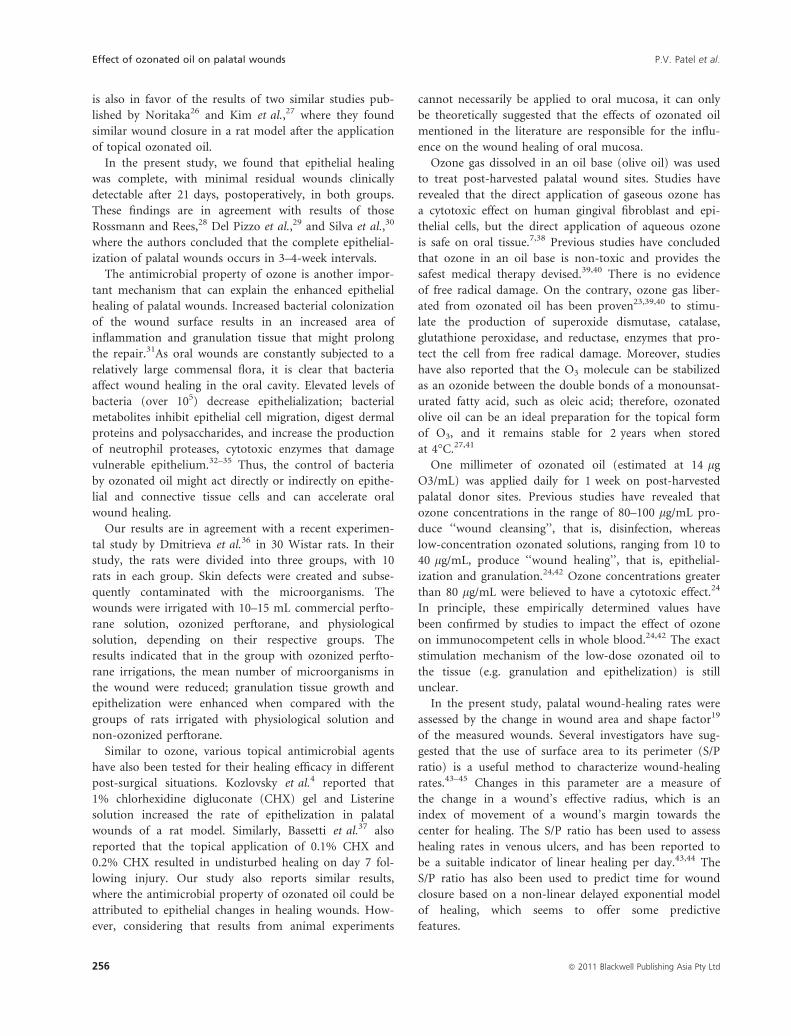
is also in favor of the results of two similar studies pub-
lished by Noritaka26 and Kim et al.,27 where they found
similar wound closure in a rat model after the application
of topical ozonated oil.
In the present study, we found that epithelial healing
was complete, with minimal residual wounds clinically
detectable after 21 days, postoperatively, in both groups.
These findings are in agreement with results of those
Rossmann and Rees,28 Del Pizzo et al.,29 and Silva et al.,30
where the authors concluded that the complete epithelial-
ization of palatal wounds occurs in 3–4-week intervals.
The antimicrobial property of ozone is another impor-
tant mechanism that can explain the enhanced epithelial
healing of palatal wounds. Increased bacterial colonization
of the wound surface results in an increased area of
inflammation and granulation tissue that might prolong
the repair.31As oral wounds are constantly subjected to a
relatively large commensal flora, it is clear that bacteria
affect wound healing in the oral cavity. Elevated levels of
bacteria (over 105) decrease epithelialization; bacterial
metabolites inhibit epithelial cell migration, digest dermal
proteins and polysaccharides, and increase the production
of neutrophil proteases, cytotoxic enzymes that damage
vulnerable epithelium.32–35 Thus, the control of bacteria
by ozonated oil might act directly or indirectly on epithe-
lial and connective tissue cells and can accelerate oral
wound healing.
Our results are in agreement with a recent experimen-
tal study by Dmitrieva et al.36 in 30 Wistar rats. In their
study, the rats were divided into three groups, with 10
rats in each group. Skin defects were created and subse-
quently contaminated with the microorganisms. The
wounds were irrigated with 10–15 mL commercial perfto-
rane solution, ozonized perftorane, and physiological
solution, depending on their respective groups. The
results indicated that in the group with ozonized perfto-
rane irrigations, the mean number of microorganisms in
the wound were reduced; granulation tissue growth and
epithelization were enhanced when compared with the
groups of rats irrigated with physiological solution and
non-ozonized perftorane.
Similar to ozone, various topical antimicrobial agents
have also been tested for their healing efficacy in different
post-surgical situations. Kozlovsky et al.4 reported that
1% chlorhexidine digluconate (CHX) gel and Listerine
solution increased the rate of epithelization in palatal
wounds of a rat model. Similarly, Bassetti et al.37 also
reported that the topical application of 0.1% CHX and
0.2% CHX resulted in undisturbed healing on day 7 fol-
lowing injury. Our study also reports similar results,
where the antimicrobial property of ozonated oil could be
attributed to epithelial changes in healing wounds. How-
ever, considering that results from animal experiments
cannot necessarily be applied to oral mucosa, it can only
be theoretically suggested that the effects of ozonated oil
mentioned in the literature are responsible for the influ-
ence on the wound healing of oral mucosa.
Ozone gas dissolved in an oil base (olive oil) was used
to treat post-harvested palatal wound sites. Studies have
revealed that the direct application of gaseous ozone has
a cytotoxic effect on human gingival fibroblast and epi-
thelial cells, but the direct application of aqueous ozone
is safe on oral tissue.7,38 Previous studies have concluded
that ozone in an oil base is non-toxic and provides the
safest medical therapy devised.39,40 There is no evidence
of free radical damage. On the contrary, ozone gas liber-
ated from ozonated oil has been proven23,39,40 to stimu-
late the production of superoxide dismutase, catalase,
glutathione peroxidase, and reductase, enzymes that pro-
tect the cell from free radical damage. Moreover, studies
have also reported that the O3 molecule can be stabilized
as an ozonide between the double bonds of a monounsat-
urated fatty acid, such as oleic acid; therefore, ozonated
olive oil can be an ideal preparation for the topical form
of O3, and it remains stable for 2 years when stored
at 4�C.27,41
One millimeter of ozonated oil (estimated at 14 lg
O3/mL) was applied daily for 1 week on post-harvested
palatal donor sites. Previous studies have revealed that
ozone concentrations in the range of 80–100 lg/mL pro-
duce ‘‘wound cleansing’’, that is, disinfection, whereas
low-concentration ozonated solutions, ranging from 10 to
40 lg/mL, produce ‘‘wound healing’’, that is, epithelial-
ization and granulation.24,42 Ozone concentrations greater
than 80 lg/mL were believed to have a cytotoxic effect.24
In principle, these empirically determined values have
been confirmed by studies to impact the effect of ozone
on immunocompetent cells in whole blood.24,42 The exact
stimulation mechanism of the low-dose ozonated oil to
the tissue (e.g. granulation and epithelization) is still
unclear.
In the present study, palatal wound-healing rates were
assessed by the change in wound area and shape factor19
of the measured wounds. Several investigators have sug-
gested that the use of surface area to its perimeter (S/P
ratio) is a useful method to characterize wound-healing
rates.43–45 Changes in this parameter are a measure of
the change in a wound’s effective radius, which is an
index of movement of a wound’s margin towards the
center for healing. The S/P ratio has been used to assess
healing rates in venous ulcers, and has been reported to
be a suitable indicator of linear healing per day.43,44 The
S/P ratio has also been used to predict time for wound
closure based on a non-linear delayed exponential model
of healing, which seems to offer some predictive
features.
Effect of ozonated oil on palatal wounds P.V. Patel et al.
256 ª 2011 Blackwell Publishing Asia Pty Ltd

We used an exfoliative cytological technique to assess
the epithelial healing of palatal wounds. Selection of the
cytological technique was based on the fact that exfolia-
tive cytology, in contrast to histological studies, permits
repeated, non-destructive observations. Moreover, cyto-
logical techniques have also been widely used for testing
therapeutic effects.46 However, if errors of diagnostic
interpretation are avoided, and well-established cell indi-
ces are used, sufficiently accurate results can be achieved.
Experimental long-term observations have also confirmed
the consistency of smear results in healing tissues.
Our results showed significant improvement in epithe-
lial healing after topical ozone application on palatal
wound sites. At present, there are few published data and
anecdotal reports suggesting enhancement of wound heal-
ing after the application of ozonated oil. The exact mech-
anism behind it is still unclear. Therefore, for future
prospects, longer-term studies with histopathological and
immunohistochemical evaluations should be undertaken
to ascertain the efficacy of ozone on the healing of oral
wounds.
Acknowledgment
The authors express their gratitude to Mr Paul Harvey,
Director of Gnosis Wellness Centre Inc. British Colombia,
Canada, for his support in providing the ozonated
organic olive oil used in this study.
References
1 Broughton G 2nd, Janis JE, Attinger
CE. The basic science of wound
healing. Plast Reconstr Surg 2006;
117: 12–34.
2 Hakkinen L, Uitto VJ, Larjava H.
Cell biology of gingival wound heal-
ing. Periodontol 2000 2000; 24: 127–
52.
3 Nooh N, Graves DT. Healing is
delayed in oral compared to dermal
excisional wounds. J Periodontol
2003; 74: 242–6.
4 Kozlovsky A, Artzi Z, Hirshberg A,
Israeli-Tobias C, Reich L. Effect of
local antimicrobial agents on exci-
sional palatal wound healing: a clin-
ical and histomorphometric study
in rats. J Clin Periodontol 2007; 34:
164–71.
5 Nyman S, Lindhe J, Rosling B. Peri-
odontal surgery in plaque-infected
dentitions. J Clin Periodontol 1977;
4: 240–9.
6 Thomas GW, Rael LT, Bar-Or R
et al. Mechanisms of delayed wound
healing by commonly used anti-
septics. J Trauma 2009; 66: 82–90.
7 Huth Karin C, Jakob FM, Saugel B
et al. Effect of ozone on oral cells
compared with established anti-
microbials. Eur J Oral Sci 2006; 14:
435–40.
8 Nagayoshi M, Fukuizumi T, Kitam-
ura C, Yano J, Terashita M, Nishi-
hara T. Efficacy of ozone on
survival and permeability of oral
micro-organisms. Oral Microbiol
Immunol 2004; 19: 240–6.
9 Mudd JB, Leavitt R, Ongun A,
McManus T. Reaction of ozone
with amino acids and proteins.
Atmos Environ 1969; 3: 669–82.
10 Ishizaki K, Sawadaishi D, Miura K,
Shinriki N. Effect of ozone on plas-
mid DNA of Escherichia coli in situ.
Water Res 1987; 21: 823–8.
11 Verrazzo G, Coppola L, Luongo C
et al. Hyperbaric oxygen, oxygen–
ozone therapy, and rheologic
parameters of blood in patients with
peripheral occlusive arterial disease.
Undersea Hyperb Med 1995; 22: 17–
22.
12 Shiratori R, Kaneko Y, Kobayashi Y
et al. Can ozone administration
activate the tissue metabolism – a
study on brain metabolism during
hypoxic hypoxia (in Japanese).
Masui 1993; 42: 2–6.
13 Oosting RS, Van Rees-Verhoef M,
Verhoef J, Van Golde LM, Van Bree
L. Effects of ozone on cellular ATP
levels in rat and mouse alveolar
macrophages. Toxicology 1991; 70:
195–202.
14 Bocci V, Luzzi E, Corradeschi F,
Silvestri S. Studies on the biological
effects of ozone: production of
transforming growth factor 1 by
human blood after ozone treatment.
J Biol Regul Homeost Agents 1994; 8:
108–12.
15 Martinez-Sanchez G, Al-Dalain SM,
Menendez S et al. Therapeutic
efficacy of ozone in patients with dia-
betic foot. Eur J Pharmacol 2005; 523:
151–61.
16 Puri KS, Suresh KR, Gogtay NJ,
Thatte UM. Declaration of Helsinki,
2008: implications for stakeholders in
research. J Postgrad Med 2009; 55:
131–4.
17 Miller PD. Coverage using free soft
tissue autograft following citric acid
application, III: a successful and pre-
dictable procedure in areas of deep-
wide recession. Jr Int J Periodontics
Restorative Dent 1985; 5: 14–37.
18 Filippi A. The influence of ozonised
water on the epithelial wound healing
process in oral cavity (in German).
Dtsch Zahnarztl 2001; 56: 104–8.
19 Mayrovitz HN, Soontupe LB. Wound
areas by computerized planimetry of
digital images: accuracy and reliability.
Adv Skin Wound Care 2009; 22: 222–9.
20 Sciubba JJ. Improving detection of
precancerous and cancerous oral
lesions. Computer-assisted analysis of
the oral brush biopsy. U.S. Collabora-
tive OralCDx Study Group. J Am
Dent Assoc 1999; 130: 1445–57.
21 Lange DE, Bernimoulin JP. Exfoliative
cytological studies in evaluation of
free gingival graft healing. J Clin Peri-
odontol 1974; 1: 89–96.
22 Werner S., Grose R. Regulation of
wound healing by growth factors and
cytokines. Physiol Rev 2003; 83: 835–
70.
23 Bocci V, Valacchi G, Corradeschi F
et al. Studies on the biological effects
P.V. Patel et al. Effect of ozonated oil on palatal wounds
ª 2011 Blackwell Publishing Asia Pty Ltd 257

of ozone. Generation of reactive
oxygen species (ROS) after exposure
of human blood to ozone. J Biol Re-
gul Homeost Agents 1998; 12: 67–75.
24 Bocci V. Ozone as a bioregulator.
Pharmacology and toxicology of
ozone therapy today. J Biol Regul
Homeost Agents 1996; 10: 31–53.
25 Bocci V. Biological and clinical
effects of ozone. Has ozonetherapy
a future in medicine? Br J Biomed
Sci 1999; 56: 270–9.
26 Noritaka K. [Histo-Pathological
Research of Wound Healing by
Ozone Ointment Application] [Arti-
cle in Japanese]. Journal of Showa
University Dental Society 2006; 26:
338–47.
27 Kim HS, Noh SU, Han YW et al.
Therapeutic effects of topical appli-
cation of ozone on acute cutaneous
wound healing. J Korean Med Sci
2009; 24: 368–74.
28 Rossmann JA, Rees TD. A compara-
tive evaluation of hemostatic agents
in the management of soft tissue
graft donor site bleeding. J Perio-
dontol 1999; 70: 1369–75.
29 Del Pizzo M, Modica F, Bethaz N,
Priotto P, Romagnoli R. The con-
nective tissue graft: a comparative
clinical evaluation of wound healing
at the palatal donor site. A prelimin-
ary study. J Clin Periodontol 2002;
29: 848–54.
30 Silva CO, Ribeiro Edel P, Sallum
AW, Tatakisi DN. Free gingival
grafts: graft shrinkage and donor-
site healing in smokers and non-
smokers. J Periodontol 2010; 81:
692–701.
31 Bucknall TE. The effect of local
infection upon wound healing: an
experimental study. Br J Surg 1980;
67: 851–5.
32 Robson MC, Stenberg BD, Heggers
JP. Wound healing alterations
caused by infection. Clin Plast Surg
1990; 17: 485–92.
33 Thomson P. The microbiology of
wounds. J Wound Care 1998; 7:
477–8.
34 Edwards R, Harding KG. Bacteria
and wound healing. Curr Opin
Infect Dis. 2004; 17: 91–6.
35 McGuckin M, Goldman R, Bolton
L, Salcido R. The clinical relevance
of microbiology in acute and
chronic wounds. Adv Skin Wound
Care. 2003; 16: 12–23.
36 Dmitrieva NA, Zyrianova NV, Grig-
or’ian AS, Vasilishina SIu. [Micro-
flora dynamics in purulent skin
wound in rats after ozonized perfto-
rane applications] [in Russian].
Stomatologiia (Mosk) 2009; 88:
14–6.
37 Bassetti C, Kallenberger A.
Influence of chlorhexidine rinsing
on haling of oral mucosa and osse-
ous lesion. J Clin Periodontol 1980;
7: 20–5.
38 Azarpazhooh A, Limeback H. The
application of ozone in dentistry: a
systematic review of literature. J Dent
2008; 36: 104–16.
39 Leon OS, Menendez S, Merino N
et al. Ozone oxidative precondition-
ing: a protection against cellular
damage by free radical. Mediators
Inflamm 1998; 7: 289–94.
40 Menendez S, Falcon L, Simon DR,
Landa N. Efficacy of ozonized sun-
flower oil in the treatment of tinea
pedis. Mycoses 2002; 45: 329–32.
41 Valacchi G, Fortino V, Bocci V. The
dual action of ozone on the skin. Br J
Dermatol 2005; 153: 1096–100.
42 Bocci V. Autohaemotherapy after
treatment of blood with ozone. A
reappraisal. J Int Med Res 1994; 22:
121–6.
43 Gorin DR, Cordts PR, LaMorte WW,
Manzoian JO. The influence of wound
geometry on the measurement of
wound healing rates in clinical trials.
J Vasc Surg. Mar 1996; 23: 524–8.
44 Cukjati D, Rebersek S, Karba R,
Miklavcic D. Modelling of chronic
wound healing dynamics. Med Biol
Eng Comput 2000; 38: 339–47.
45 Gilman TH. Parameter for measure-
ment of wound closure. Wounds
1990; 3: 95–10.
46 Stegemann EA, Lange DE, Wegner C.
[Experimental studies on the stan-
dardization of oral exfoliative cytolog-
ical determination procedures using
the keratinization index] [in Ger-
man]. Dtsch Zahnarztl Z 1974; 29:
472–7.
Effect of ozonated oil on palatal wounds P.V. Patel et al.
258 ª 2011 Blackwell Publishing Asia Pty Ltd