The Rare Case of Perirenal Abscess in a Child ... - MDPI
7
medicina Case Report The Rare Case of Perirenal Abscess in a Child—Possible Mechanisms and Methods of Treatment: A Case Report and Literature Review Patrycja Sosnowska-Sienkiewicz 1, * , Ewa Bu´ cko 2 and Przemyslaw Ma ´ nkowski 1 Citation: Sosnowska-Sienkiewicz, P.; Bu´ cko, E.; Ma ´ nkowski, P. The Rare Case of Perirenal Abscess in a Child—Possible Mechanisms and Methods of Treatment: A Case Report and Literature Review. Medicina 2021, 57, 154. https://doi.org/10.3390/ medicina57020154 Academic Editors: Johannes Mayr and Benjamin Frei Received: 22 December 2020 Accepted: 5 February 2021 Published: 9 February 2021 Publisher’s Note: MDPI stays neutral with regard to jurisdictional claims in published maps and institutional affil- iations. Copyright: © 2021 by the authors. Licensee MDPI, Basel, Switzerland. This article is an open access article distributed under the terms and conditions of the Creative Commons Attribution (CC BY) license (https:// creativecommons.org/licenses/by/ 4.0/). 1 Department of Pediatric Surgery, Traumatology and Urology, Poznan University of Medical Sciences, 60-572 Poznan, Poland; [email protected] 2 Department of Pediatric Surgery, Traumatology and Urology, Karol Jonscher Hospital, 60-572 Poznan, Poland; [email protected] * Correspondence: [email protected]; Tel.: +48-61-8491578; Fax: +48-61-8491228 Abstract: Renal and perirenal abscesses are very rare in children. They can be present as an acute emergency condition or insidiously as a chronic disease. The diagnosis is not so obvious, and it is a big challenge, especially when it can simulate a kidney tumor. The treatment can be conservative, preferably with targeted antibiotics, or surgical, consisting primarily of drainage. This publication aims to present a clinical case in which both diagnosis and treatment were a big challenge for the entire treatment team. A 10-year-old male patient was admitted to the hospital because of mild abdominal pain and a temperature of 37.5 ◦ C. The symptoms lasted for a week. In the computed tomography (CT), the lesion’s dimensions were 11.1 × 8.2 × 25 cm, and inflammation, abscess, cyst, and abdominal tumor have been suggested. The decision about surgical treatment was made. An enormous abscess near the right kidney was localized. The patient’s condition stabilized after surgery. Unfortunately, due to persistent purulent reservoirs, a second laparotomy was necessary. During the extensive diagnostic cystourethrography performed, vesicoureteral reflux was visualized. In conclusion, though a perinephric abscess is very rare in children, it should be taken into consideration in patients with non-specific abdominal symptoms. The imaging using ultrasound and CT scan with contrast enhancement is crucial to recognize and properly treat the condition. In terms of a small abscess, the only antimicrobial treatment using antibiotics of a broad spectrum can be considered. However, the drainage of an abscess, either percutaneous or open, should be used. For the large abscess, the open drainage seems to be a primary method of treatment. The importance of cooperation in a multidisciplinary team is crucial, as the diagnosis and treatment of underlying causes are essential. Keywords: abscess; children; perirenal; renal; surgery 1. Introduction Renal and perirenal abscesses are very rare in children [1]. The incidence ranges from 1 to 10 patients for every 10,000 hospital admissions. About one-third of patients with this diagnosis have diabetes. It can be present as an acute emergency condition or insidiously as a chronic disease [2]. The diagnosis is not so obvious, and it is a big challenge, especially when it can simulate a kidney tumor. The proper diagnostics must be carried out carefully, because improper treatment may result in the loss of the kidney or even death of the patient [3]. Symptoms that may suggest a renal or perirenal abscess include a fever and pain of the abdomen. Therefore, the differential diagnosis of these patients is extremely difficult. For this reason, frequently, this condition is detected as a result of incidental imaging, particularly computed tomography (CT). An abscess around the kidney may be related to upper tract urosepsis associated with an infective staghorn renal calculus [2,4–6]. The second cause may be infection coming from Medicina 2021, 57, 154. https://doi.org/10.3390/medicina57020154 https://www.mdpi.com/journal/medicina
-
Upload
khangminh22 -
Category
Documents
-
view
0 -
download
0
Transcript of The Rare Case of Perirenal Abscess in a Child ... - MDPI

medicina
Case Report
The Rare Case of Perirenal Abscess in a Child—PossibleMechanisms and Methods of Treatment: A Case Report andLiterature Review
Patrycja Sosnowska-Sienkiewicz 1,* , Ewa Bucko 2 and Przemysław Mankowski 1
�����������������
Citation: Sosnowska-Sienkiewicz, P.;
Bucko, E.; Mankowski, P. The Rare
Case of Perirenal Abscess in a
Child—Possible Mechanisms and
Methods of Treatment: A Case Report
and Literature Review. Medicina 2021,
57, 154. https://doi.org/10.3390/
medicina57020154
Academic Editors: Johannes Mayr
and Benjamin Frei
Received: 22 December 2020
Accepted: 5 February 2021
Published: 9 February 2021
Publisher’s Note: MDPI stays neutral
with regard to jurisdictional claims in
published maps and institutional affil-
iations.
Copyright: © 2021 by the authors.
Licensee MDPI, Basel, Switzerland.
This article is an open access article
distributed under the terms and
conditions of the Creative Commons
Attribution (CC BY) license (https://
creativecommons.org/licenses/by/
4.0/).
1 Department of Pediatric Surgery, Traumatology and Urology, Poznan University of Medical Sciences,60-572 Poznan, Poland; [email protected]
2 Department of Pediatric Surgery, Traumatology and Urology, Karol Jonscher Hospital, 60-572 Poznan, Poland;[email protected]
* Correspondence: [email protected]; Tel.: +48-61-8491578; Fax: +48-61-8491228
Abstract: Renal and perirenal abscesses are very rare in children. They can be present as an acuteemergency condition or insidiously as a chronic disease. The diagnosis is not so obvious, and it is abig challenge, especially when it can simulate a kidney tumor. The treatment can be conservative,preferably with targeted antibiotics, or surgical, consisting primarily of drainage. This publicationaims to present a clinical case in which both diagnosis and treatment were a big challenge for theentire treatment team. A 10-year-old male patient was admitted to the hospital because of mildabdominal pain and a temperature of 37.5 ◦C. The symptoms lasted for a week. In the computedtomography (CT), the lesion’s dimensions were 11.1 × 8.2 × 25 cm, and inflammation, abscess, cyst,and abdominal tumor have been suggested. The decision about surgical treatment was made. Anenormous abscess near the right kidney was localized. The patient’s condition stabilized after surgery.Unfortunately, due to persistent purulent reservoirs, a second laparotomy was necessary. Duringthe extensive diagnostic cystourethrography performed, vesicoureteral reflux was visualized. Inconclusion, though a perinephric abscess is very rare in children, it should be taken into considerationin patients with non-specific abdominal symptoms. The imaging using ultrasound and CT scanwith contrast enhancement is crucial to recognize and properly treat the condition. In terms ofa small abscess, the only antimicrobial treatment using antibiotics of a broad spectrum can beconsidered. However, the drainage of an abscess, either percutaneous or open, should be used. Forthe large abscess, the open drainage seems to be a primary method of treatment. The importanceof cooperation in a multidisciplinary team is crucial, as the diagnosis and treatment of underlyingcauses are essential.
Keywords: abscess; children; perirenal; renal; surgery
1. Introduction
Renal and perirenal abscesses are very rare in children [1]. The incidence ranges from1 to 10 patients for every 10,000 hospital admissions. About one-third of patients with thisdiagnosis have diabetes. It can be present as an acute emergency condition or insidiouslyas a chronic disease [2]. The diagnosis is not so obvious, and it is a big challenge, especiallywhen it can simulate a kidney tumor.
The proper diagnostics must be carried out carefully, because improper treatment mayresult in the loss of the kidney or even death of the patient [3].
Symptoms that may suggest a renal or perirenal abscess include a fever and pain ofthe abdomen. Therefore, the differential diagnosis of these patients is extremely difficult.For this reason, frequently, this condition is detected as a result of incidental imaging,particularly computed tomography (CT).
An abscess around the kidney may be related to upper tract urosepsis associated withan infective staghorn renal calculus [2,4–6]. The second cause may be infection coming from
Medicina 2021, 57, 154. https://doi.org/10.3390/medicina57020154 https://www.mdpi.com/journal/medicina

Medicina 2021, 57, 154 2 of 7
the renal carbuncle or renal abscess perforated into the perirenal space. This cause is moreprobable in suspected immunocompromised patients. In these instances, Staphylococcusaureus is often diagnosed. Younger or malnourished patients are more likely to have anacute presentation with a high temperature and acute abdomen symptoms.
The treatment can be conservative, preferably with targeted antibiotics, or surgical,consisting primarily of drainage.
This publication aims to present a clinical case in which both diagnosis and treatmentwere a big challenge for the entire treatment team.
2. Case Report
A 10-year-old male patient was transferred to the surgical department from thepediatric ward due to severe symptoms of the abdomen. The child was admitted to thehospital because of mild abdominal pain and a temperature of 37.5 ◦C. The symptomslasted for a week.
In the infectious disease ward, COVID-19 was excluded. During hospitalization, thepatient developed acute abdominal symptoms. The acute condition of the boy appearedsuddenly. Results of laboratory tests showed deviations in white blood cells (WBC) =11.89 × 10³/µL, and hemoglobin (HGB) = 6.9 g/dL, with hematocrit (HCT) = 20.6%. Theinflammatory markers were elevated. Urinalysis showed 75 mg/dL of proteins. The ultra-sound of the abdomen and computed tomography were performed. The diagnosis wasunclear. Inflammation, abscess, cyst, and abdominal tumor were suggested. In the com-puted tomography (CT), the lesion had dimensions: 11.1 × 8.2 × 25 cm (Figures 1 and 2).
Medicina 2021, 57, x FOR PEER REVIEW 2 of 7
For this reason, frequently, this condition is detected as a result of incidental imaging, particularly computed tomography (CT).
An abscess around the kidney may be related to upper tract urosepsis associated with an infective staghorn renal calculus [2,4–6]. The second cause may be infection coming from the renal carbuncle or renal abscess perforated into the perirenal space. This cause is more probable in suspected immunocompromised patients. In these instances, Staphylo-coccus aureus is often diagnosed. Younger or malnourished patients are more likely to have an acute presentation with a high temperature and acute abdomen symptoms.
The treatment can be conservative, preferably with targeted antibiotics, or surgical, consisting primarily of drainage.
This publication aims to present a clinical case in which both diagnosis and treatment were a big challenge for the entire treatment team.
2. Case Report A 10-year-old male patient was transferred to the surgical department from the pe-
diatric ward due to severe symptoms of the abdomen. The child was admitted to the hos-pital because of mild abdominal pain and a temperature of 37.5 °C. The symptoms lasted for a week.
In the infectious disease ward, COVID-19 was excluded. During hospitalization, the patient developed acute abdominal symptoms. The acute condition of the boy appeared suddenly. Results of laboratory tests showed deviations in white blood cells (WBC)= 11.89 × 10³/µL, and hemoglobin (HGB) = 6.9 g/dL, with hematocrit (HCT) = 20.6%. The inflam-matory markers were elevated. Urinalysis showed 75 mg/dL of proteins. The ultrasound of the abdomen and computed tomography were performed. The diagnosis was unclear. Inflammation, abscess, cyst, and abdominal tumor were suggested. In the computed to-mography (CT), the lesion had dimensions: 11.1 × 8.2 × 25 cm (Figures 1 and 2).
Figure 1. The lesion of unknown character in the area of the right kidney, shown in the transverse plane of the CT scan. The lesion is marked by an arrow.
Figure 1. The lesion of unknown character in the area of the right kidney, shown in the transverseplane of the CT scan. The lesion is marked by an arrow.
Medicina 2021, 57, x FOR PEER REVIEW 2 of 7
For this reason, frequently, this condition is detected as a result of incidental imaging, particularly computed tomography (CT).
An abscess around the kidney may be related to upper tract urosepsis associated with an infective staghorn renal calculus [2,4–6]. The second cause may be infection coming from the renal carbuncle or renal abscess perforated into the perirenal space. This cause is more probable in suspected immunocompromised patients. In these instances, Staphylo-coccus aureus is often diagnosed. Younger or malnourished patients are more likely to have an acute presentation with a high temperature and acute abdomen symptoms.
The treatment can be conservative, preferably with targeted antibiotics, or surgical, consisting primarily of drainage.
This publication aims to present a clinical case in which both diagnosis and treatment were a big challenge for the entire treatment team.
2. Case Report A 10-year-old male patient was transferred to the surgical department from the pe-
diatric ward due to severe symptoms of the abdomen. The child was admitted to the hos-pital because of mild abdominal pain and a temperature of 37.5 °C. The symptoms lasted for a week.
In the infectious disease ward, COVID-19 was excluded. During hospitalization, the patient developed acute abdominal symptoms. The acute condition of the boy appeared suddenly. Results of laboratory tests showed deviations in white blood cells (WBC)= 11.89 × 10³/µL, and hemoglobin (HGB) = 6.9 g/dL, with hematocrit (HCT) = 20.6%. The inflam-matory markers were elevated. Urinalysis showed 75 mg/dL of proteins. The ultrasound of the abdomen and computed tomography were performed. The diagnosis was unclear. Inflammation, abscess, cyst, and abdominal tumor were suggested. In the computed to-mography (CT), the lesion had dimensions: 11.1 × 8.2 × 25 cm (Figures 1 and 2).
Figure 1. The lesion of unknown character in the area of the right kidney, shown in the transverse plane of the CT scan. The lesion is marked by an arrow.
Figure 2. The lesion of unknown character in the area of the right kidney, shown in the frontal planeof the CT scan. The lesion is marked by an arrow.
Medicina 2021, 57, 154 3 of 7
It displaced and compressed the inferior vena cava and right renal vein. The lesionadhered to and modeled the liver and pancreas. It was pushing the intestinal loops.The lesion reached down into the bladder. After multidisciplinary consultation witha radiological and oncological team, the decision about surgical treatment was made.Exploratory laparotomy was performed. During the surgery, there was a large amount ofpurulent content in the peritoneal cavity. A large abscess was visible in the vesicorectalrecess. An enormous abscess near the right kidney was localized. The abdominal cavitywas thoroughly rinsed. The abdominal cavity and the perirenal space were drained. Thebladder was thick and fibrous.
Antibiotic therapy was started. MSCNS (Staphylococcus coagulase-negative suscep-tible to methicillin), a strain susceptible to beta-lactam antibiotics except for penicillin,amino-, carbo-, and ureidopenicillins, was grown from the inoculation of the materialcollected from the abdominal cavity. The urine, blood, and tracheal aspirate cultureswere also collected and showed no abnormalities. After surgery, the patient’s conditionimproved slightly. On the seventh day after the procedure, the patient again presentedacute symptoms of the abdomen. He had a fever. The decision about relaparotomy wasmade. A vast amount of purulent content was evacuated from the vesicorectal and rightperirenal recesses one more time. Additionally, the bladder, with large amounts of residualurine, was visualized. The wall of the bladder was thickened.
The peritoneal cavity was rinsed again, and drains were inserted. The patientwas catheterized.
A broader spectrum antibiotic was introduced. Due to the significant exhaustionof the patient, gastroenterological consultation was made, and nutritional treatment wasintroduced. To exclude an accompanying disease responsible for the level of malnutrition,endocrinological consultation was made. Broad laboratory diagnostics excluded anyhormonal abnormalities. The patient’s condition was monitored by a CT scan, whichshowed duplication of the right kidney system and altered parenchyma of the kidney.Periodic control ultrasound of the abdomen showed the progressive reduction of thefluid reservoirs. The patient’s condition improved slightly. A febrile episode of up to38.5 ◦C occurred, and infection of the central intravenous catheter was suspected. After itsremoval, the general condition of the boy was improved. The drains from the abdomenwere removed. When an attempt was made to take out the catheter from the bladder, theinflammatory parameters increased after 24 h. There was a fever. The abdomen was tightand tender. After re-catheterization of the patient, 500 mL of residual urine was evacuated.The unwanted symptoms disappeared. The patient’s condition was stabilized.
After the urological consultation, a cystoscopy was performed. It showed a bladderwith abnormal trabeculation. The cystofix was fixed. Broad-spectrum antibiotic therapywas maintained. Laboratory inflammatory markers improved gradually. No microbialgrowth was achieved in the control cultures. The last antibiotics were discontinued afterseven days.
A multidisciplinary team consisting of an oncologist, immunologist, and nephrologistdecided the necessary diagnostics and treatment. After the extensive physical, laboratory,and radiological studies, the oncological consultation determined that the probability ofneoplastic growth was low. Acquired immunodeficiency, such as AIDS or tuberculosis, wasexcluded by an infectious disease specialist after appropriate laboratory tests. A detailedimmunological study was performed. On this basis, functional disorders in the scope ofclass switching and possible quantitative B lymphocytes were found. The distribution ofT-cytotoxic lymphocytes may indicate functional disorders. Abnormalities were diagnosedin the phagoburst tests and the percentage of lymphocytes, and the tests required repeatingand control, and suggested an immune disorder.
From the moment of the surgery, the patient’s condition improved and stabilizedsignificantly.
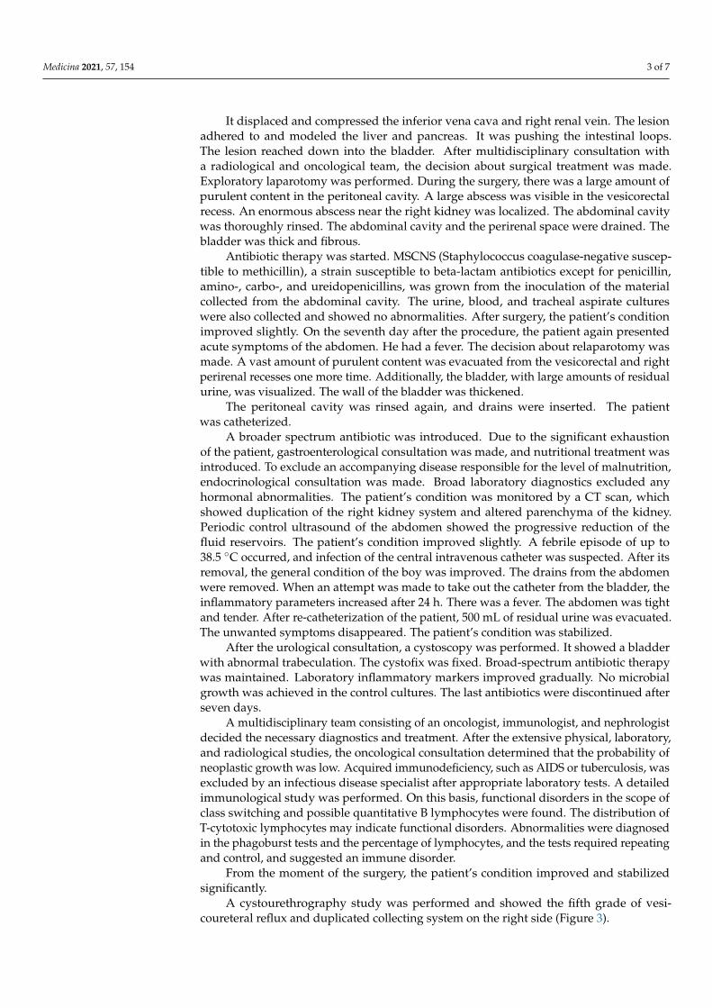
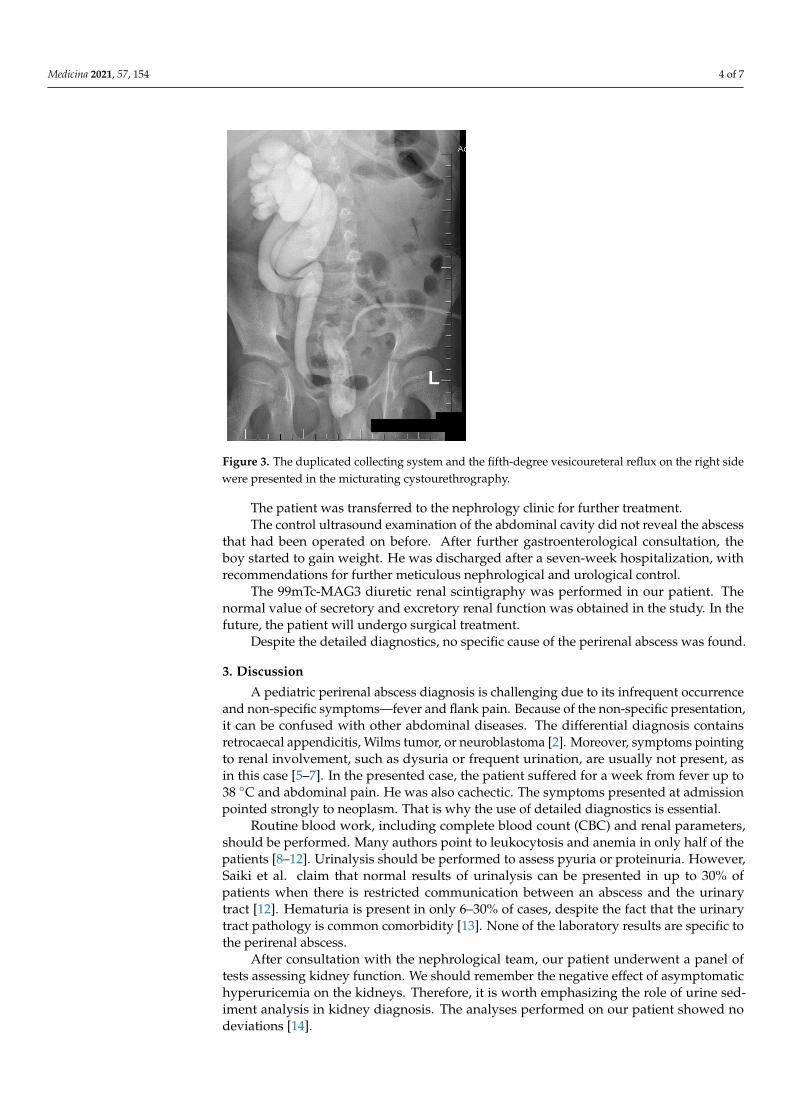
A cystourethrography study was performed and showed the fifth grade of vesi-coureteral reflux and duplicated collecting system on the right side (Figure 3).
Medicina 2021, 57, 154 4 of 7
Medicina 2021, 57, x FOR PEER REVIEW 4 of 7
From the moment of the surgery, the patient’s condition improved and stabilized significantly.
A cystourethrography study was performed and showed the fifth grade of vesicoureteral reflux and duplicated collecting system on the right side (Figure 3).
Figure 3. The duplicated collecting system and the fifth-degree vesicoureteral reflux on the right side were presented in the micturating cystourethrography.
The patient was transferred to the nephrology clinic for further treatment. The control ultrasound examination of the abdominal cavity did not reveal the ab-
scess that had been operated on before. After further gastroenterological consultation, the boy started to gain weight. He was discharged after a seven-week hospitalization, with recommendations for further meticulous nephrological and urological control.
The 99mTc-MAG3 diuretic renal scintigraphy was performed in our patient. The nor-mal value of secretory and excretory renal function was obtained in the study. In the fu-ture, the patient will undergo surgical treatment.
Despite the detailed diagnostics, no specific cause of the perirenal abscess was found.
3. Discussion A pediatric perirenal abscess diagnosis is challenging due to its infrequent occur-
rence and non-specific symptoms—fever and flank pain. Because of the non-specific presentation, it can be confused with other abdominal diseases. The differential diagnosis contains retrocaecal appendicitis, Wilms tumor, or neuroblastoma [2]. Moreover, symp-toms pointing to renal involvement, such as dysuria or frequent urination, are usually not present, as in this case [5–7]. In the presented case, the patient suffered for a week from fever up to 38 °C and abdominal pain. He was also cachectic. The symptoms presented at admission pointed strongly to neoplasm. That is why the use of detailed diagnostics is essential.
Routine blood work, including complete blood count (CBC) and renal parameters, should be performed. Many authors point to leukocytosis and anemia in only half of the patients [8–12]. Urinalysis should be performed to assess pyuria or proteinuria. However, Saiki et al. claim that normal results of urinalysis can be presented in up to 30% of patients when there is restricted communication between an abscess and the urinary tract [12].
Figure 3. The duplicated collecting system and the fifth-degree vesicoureteral reflux on the right sidewere presented in the micturating cystourethrography.
The patient was transferred to the nephrology clinic for further treatment.The control ultrasound examination of the abdominal cavity did not reveal the abscess
that had been operated on before. After further gastroenterological consultation, theboy started to gain weight. He was discharged after a seven-week hospitalization, withrecommendations for further meticulous nephrological and urological control.
The 99mTc-MAG3 diuretic renal scintigraphy was performed in our patient. Thenormal value of secretory and excretory renal function was obtained in the study. In thefuture, the patient will undergo surgical treatment.
Despite the detailed diagnostics, no specific cause of the perirenal abscess was found.
3. Discussion
A pediatric perirenal abscess diagnosis is challenging due to its infrequent occurrenceand non-specific symptoms—fever and flank pain. Because of the non-specific presentation,it can be confused with other abdominal diseases. The differential diagnosis containsretrocaecal appendicitis, Wilms tumor, or neuroblastoma [2]. Moreover, symptoms pointingto renal involvement, such as dysuria or frequent urination, are usually not present, asin this case [5–7]. In the presented case, the patient suffered for a week from fever up to38 ◦C and abdominal pain. He was also cachectic. The symptoms presented at admissionpointed strongly to neoplasm. That is why the use of detailed diagnostics is essential.
Routine blood work, including complete blood count (CBC) and renal parameters,should be performed. Many authors point to leukocytosis and anemia in only half of thepatients [8–12]. Urinalysis should be performed to assess pyuria or proteinuria. However,Saiki et al. claim that normal results of urinalysis can be presented in up to 30% ofpatients when there is restricted communication between an abscess and the urinarytract [12]. Hematuria is present in only 6–30% of cases, despite the fact that the urinarytract pathology is common comorbidity [13]. None of the laboratory results are specific tothe perirenal abscess.
After consultation with the nephrological team, our patient underwent a panel oftests assessing kidney function. We should remember the negative effect of asymptomatichyperuricemia on the kidneys. Therefore, it is worth emphasizing the role of urine sed-iment analysis in kidney diagnosis. The analyses performed on our patient showed nodeviations [14].

Medicina 2021, 57, 154 5 of 7
As laboratory diagnostics usually cannot deliver a conclusive answer, radiologicaldiagnostics should always be used. The recommended means of radiological diagnosticsconsist of ultrasonography, followed by CT scan with contrast enhancement [4]. However,the imaging can mimic other conditions, such as Wilms tumor, renal cell carcinoma, orxanthogranulomatous pyelonephritis. In the presented case, the CT scan was chosen asa primary diagnosis method, as a neoplastic tumor was suspected. The study was notconclusive, but it suggested an inflammatory process. Since the final diagnosis could notbe reached, a diagnostic laparotomy was made. In the presented case, the diagnosis wasmade intraoperatively on day three of hospitalization; however, the severity of the illnesssuggested a much longer process [4]. As the complications range from sepsis and fistula toperitoneal rupture or perforation through a diaphragm [7], a fast diagnosis seems crucialto avoid an increase in morbidity. Therefore, in the case of uncertain imaging combinedwith a lack of clinical improvement, surgical exploration seems to be the proper solution todiagnostic doubts. The suggested treatment for a perirenal abscess is antibiotic therapycovering both aerobic and anaerobic pathogens and drainage of the abscess [15,16]. Inthe publication by Coelho et al., interventional treatment for a perinephric abscess waspreferable, either by performing percutaneous drainage or open surgical drainage [3]. Thepublication of Angel et al. acknowledges the possibility of only antibiotic therapy for anabscess less than 3 cm in diameter. However, in the case of failure of antibiotic therapytreatment and the delay of an invasive procedure, the need for nephrectomy may occur.Therefore, to avoid complications, the authors recommend the invasive procedure [16].In the presented case, antibiotic therapy was introduced on the first day of admission,with no improvement in the patient’s condition. The size of the lesion visualized was11 × 8 × 25 cm. As was mentioned, due to uncertain diagnosis, laparotomy, washing,and consequent drainage were performed. According to the analyses, the use of catheterdrainage (i.e., leaving drainage in the abdominal cavity permanently) is more effectivethan percutaneous drainage. The extent of the abscess suggests that percutaneous drainagewould not be sufficient in that case. There is always a method of drainage to consider,or exploratory laparotomy vs. a direct surgical approach [17,18]. Moreover, despite theextensive irrigation used during the first procedure, after a week, the patient experienceda recurrence of pain and fever. Another intervention should be considered in the case ofextensive abdominal abscess and symptoms presented post-operatively after the fourthday of surgery [17]. In this case, the decision for relaparotomy was made. Washing anddrainage of the cavity were used again. Drainage, after both the first and second operation,was maintained for seven days, after the first laparotomy until the second intervention.Furthermore, after the second intervention, drainage was maintained for seven days untilthe amount of fluid in the abdominal cavity was almost completely absorbed. In bothcases, it was classic drainage without flushing. After the surgical intervention, the patient’scondition improved.
Gardiner et al. propose that the most common pathophysiology for perirenal abscessis ascending urinary tract infection, often associated with a staghorn calculus. The othercauses may come from renal carbuncles or renal abscess perforating the perirenal space.This is probable for immunocompromised and younger patients [3]. There is also a lesscommon hematogenous pathway [12]. In the case of our patient, all of the pathways weretaken into consideration.
According to Gardiner et al., the most common Gram-negative pathogen is Escherichia coli,and its presence implies the ascending origin of the abscess. For that path, pathogenslike Klebsiella spp. and Enterobacter spp. should also be considered [3]. The presence ofStaphylococcus Aureus suggests the hematogenous spread [16].
In the present case, MSCNS was the isolated pathogen. This supports the ascendingurinary tract infection hypothesis, because this is a pathogen often observed in urinary tractinfection (UTI) [5,8]. Coagulase-negative Staphylococcus is one of the common pathogenscausing UTI in children with anatomical or functional abnormalities of the urinary tract ora compromised immune system [9].

Medicina 2021, 57, 154 6 of 7
In the publication by Rote et al., the authors suggested that, in the case of renalparenchymal infection with Gram-negative bacteria, the patient should undergo cys-tourethrography [1]. In our clinic, such a procedure was performed, as it was suspectedthat the origin of the process ascended from the urinary tract. The presence of fifth-gradevesicoureteral reflux and duplication of the ureter, combined with the abnormalities of thebladder, pointed to the increased risk of ascending UTI [6,8].
The underlying cause of the perirenal abscess should be diagnosed and treated. Theurinary tract obstruction should be excluded or treated appropriately [2]. In the presentedcase, cystoscopy was performed. Except from an abnormal bladder, no additional obstruc-tion was visualized. It is suspected that thickened walls and bladder trabeculation werecaused by chronic bladder inflammation of uncertain origin. The cystofix was fixed inorder to facilitate urinary flow, which significantly improved the patient’s condition.
Another underlying cause of the abscess might be the compromise of the immunesystem, diabetes mellitus, or coexisting chronic disease [2,4]. Screening must be performedin order to remove accompanying problems. In our case, the only abnormality was presentin the phagoburst tests and the percentage of lymphocytes. Both of the tests might suggestimmunodeficiency; however, they should be repeated, as the patient’s condition will bemore stable.
4. Conclusions
In conclusion, though perinephric abscess is very rare in children, it should be takeninto consideration in patients with non-specific abdominal symptoms. Diabetes mellitus,urinary tract abnormalities, and immunodeficiency should be considered while treatinga patient with perirenal abscess. Laboratory examination is necessary to stabilize thepatient, but does not facilitate the diagnosis. Imaging using ultrasound and CT scan withcontrast enhancement is crucial to recognize and properly treat the condition. In termsof a small abscess, only antimicrobial treatment using antibiotics of a broad spectrum canbe considered. However, in any doubt, the drainage of an abscess, either percutaneous oropen, should be used. For a large abscess, open drainage seems to be the primary methodof treatment.
The importance of cooperation in a multidisciplinary team is crucial, as the diagnosisand treatment of underlying causes are essential.
Author Contributions: Conceptualization: P.S.-S., E.B. and P.M. Methodology: P.S.-S., E.B. and P.M.Software: P.S.-S. Validation: P.S.-S., E.B. and P.M. Formal Analysis: P.S.-S., E.B. and P.M. Investigation:P.S.-S. Resources: P.S.-S. Data Curation: P.S.-S. and E.B. Writing—original draft preparation: P.S.-S.Writing—review and editing: P.S.-S., E.B. and P.M. Visualization: P.S.-S. Supervision: P.M. ProjectAdministration: P.S.-S. All authors have read and agreed to the published version of the manuscript.
Funding: This research did not receive any specific grant from funding agencies in the public,commercial, or not-for-profit sectors.
Institutional Review Board Statement: Not applicable.
Informed Consent Statement: The patient has provided informed consent for publication of thecase.
Data Availability Statement: Data available on request due to restrictions.
Conflicts of Interest: The authors have no conflict of interest.
References1. Rote, A.R.; Bauer, S.B.; Retik, A.B. Renal Abscess in Children. J. Urol. 1978, 119, 254–258. [CrossRef]2. Gardiner, R.A.; Gwynne, R.A.; Roberts, S.A. Perinephric abscess. BJU Int. 2011, 107, 20–23. [CrossRef] [PubMed]3. Coelho, R.F.; Schneider-Monteiro, E.D.; Mesquita, J.L.B.; Mazzucchi, E.; Lucon, A.M.; Srougi, M. Renal and Perinephric Abscesses:
Analysis of 65 Consecutive Cases. World J. Surg. 2007, 31, 431–436. [CrossRef] [PubMed]4. Blandy, J.P.; Singh, M. The Case for a More Aggressive Approach to Staghorn Stones. J. Urol. 1976, 115, 505–506. [CrossRef]

Medicina 2021, 57, 154 7 of 7
5. Keren, R.; Shaikh, N.; Pohl, H.G.; Gravens-Mueller, L.; Ivanova, A.; Zaoutis, L.B.; Patel, M.; DeBerardinis, R.; Parker, A.; Bhatnagar,S.; et al. Risk Factors for Recurrent Urinary Tract Infection and Renal Scarring. Pediatrics 2015, 136, e13–e21. [CrossRef] [PubMed]
6. Okafor, C.N.; Onyeaso, E.E. Perinephric Abscess; StarPearls Publishing: Treasure Island, FL, USA, 2020.7. Timmons, J.W.; Perlmutter, A.D. Renal Abscess: A Changing Concept. J. Urol. 1976, 115, 299–301. [CrossRef]8. Leung, A.K.C.; Wong, A.H.C.; Leung, A.A.M.; Hon, K.L. Urinary Tract Infection in Children. Recent Pat. Inflamm. Allergy Drug
Discov. 2019, 13, 2–18. [CrossRef] [PubMed]9. Pérez-López, L.M.; Vara-Patudo, I.; Torner-Rubies, F.; Moreno-Romo, D.; de Sena-de Cabo, L.; Fortuny, C.; Knörr, G. Pediatric
Psoas Abscess, Early Diagnosis of a Challenging Condi-tion. J. Acute Med. 2017, 7, 158–166. [PubMed]10. Edelstein, H.; McCabe, R.E. Perinephric abscess. Modern diagnosis and treatment in 47 cases. Medicine 1988, 67, 118–131.
[CrossRef] [PubMed]11. Saiki, J.; Vaziri, N.D.; Barton, C. Perinephric and Intranephric Abscesses: A Review of the Literature. West. J. Med. 1982, 136,
95–102. [PubMed]12. Tsukagoshi, D.; Dinkovski, B.; Dasan, S.; Jethwa, J. Perinephric abscess secondary to a staghorn calculus presenting as a
subcutaneous abscess. CJEM 2006, 8, 285–288. [CrossRef] [PubMed]13. Bacha, K.; Miladi, M.; Ben Hassine, L.; Hajri, M.; Tanazaghti, F.; Ayed, M. Therapeutic aspects of renal abscess. Report of 50 cases.
Prog. Urol. 2001, 11, 444–449. [PubMed]14. Viggiano, D.; Gigliotti, G.; Vallone, G.; Giammarino, A.; Nigro, M.; Capasso, G. Urate-Lowering Agents in Asymptomatic
Hyperuricemia: Role of Urine Sediment Analysis and Musculoskeletal Ultrasound. Kidney Blood Press. Res. 2018, 43, 606–615.[CrossRef] [PubMed]
15. Angel, C.; Shu, T.; Green, J.; Orihuela, E.; Rodriquez, G.; Hendrick, E. Renal and peri-renal abscesses in children: Proposedphysio-pathologic mechanisms and treatment algorithm. Pediatr. Surg. Int. 2003, 19, 35–39. [CrossRef]
16. Górecki, W. Zakazenia w Chirurgii Dzieciecej in Chirurgia Dziecieca; Bagłaj, M., Kalicinski, P., Eds.; Wydawnictwo Lekarskie PZWL:Warsaw, Poland, 2016; pp. 145–159.
17. Petrowsky, H.; Demartines, N.; Rousson, V.; Clavien, P.A. Evidence-based value of prophylactic drainage in gastrointestinalsurgery: A systematic review and meta-analyzes. Ann. Surg. 2004, 240, 1074–1085. [CrossRef] [PubMed]
18. Holzheimer, R.G.; Mannick, J.A. (Eds.) Surgical Treatment: Evidence-Based and Problem-Oriented; W. Zuckschwerdt Verlag Munchen:München, Germany, 2001.


![[Meticilin resistant Staphylococcus aureus and liver abscess: a retrospective analysis of 117 patients]](https://static.fdokumen.com/doc/165x107/632546fd545c645c7f099e01/meticilin-resistant-staphylococcus-aureus-and-liver-abscess-a-retrospective-analysis.jpg)