IRDR - Intractable & Rare Diseases Research
90
ISSN 2186-3644 Online ISSN 2186-361X IRDR Intractable & Rare Diseases Research www.irdrjournal.com Volume 6, Number 1 February, 2017
-
Upload
khangminh22 -
Category
Documents
-
view
4 -
download
0
Transcript of IRDR - Intractable & Rare Diseases Research

ISSN 2186-3644 Online ISSN 2186-361X
IRDRIntractable & Rare Diseases Research
www.irdrjournal.com
Volume 6, Number 1February, 2017


www.irdrjournal.com
Intractable & Rare Diseases Research is one of a series of peer-reviewed journals of the International Research and Cooperation Association for Bio & Socio-Sciences Advancement (IRCA-BSSA) Group and is published quarterly by the International Advancement Center for Medicine & Health Research Co., Ltd. (IACMHR Co., Ltd.) and supported by the IRCA-BSSA, Shandong Academy of Medical Sciences, and Shandong Rare Disease Association.
Intractable & Rare Diseases Research devotes to publishing the latest and most significant research in intractable and rare diseases. Articles cover all aspects of intractable and rare diseases research such as molecular biology, genetics, clinical diagnosis, prevention and treatment, epidemiology, health economics, health management, medical care system, and social science in order to encourage cooperation and exchange among scientists and clinical researchers.
Intractable & Rare Diseases Research publishes Original Articles, Brief Reports, Reviews, Policy Forum articles, Case Reports, News, and Letters on all aspects of the field of intractable and rare diseases research. All contributions should seek to promote international collaboration.
ISSN: 2186-3644Online ISSN: 2186-361X
CODEN: IRDRA3Issues/Year: 4
Language: EnglishPublisher: IACMHR Co., Ltd.
IRCA-BSSA Group Journals
ISSN: 2186-3644Online ISSN: 2186-361X CODEN: IRDRA3Issues/Year: 4Language: EnglishPublisher: IACMHR Co., Ltd. www.irdrjournal.com
ISSN: 1881-7831Online ISSN: 1881-784X CODEN: DDTRBXIssues/Year: 6Language: EnglishPublisher: IACMHR Co., Ltd. www.ddtjournal.com
ISSN: 1881-7815 Online ISSN: 1881-7823 CODEN: BTIRCZ Issues/Year: 6Language: EnglishPublisher: IACMHR Co., Ltd.www.biosciencetrends.com
IRDRIntractable & Rare Diseases Research
i

www.irdrjournal.com
Editorial Board
Intractable & Rare Diseases ResearchEditorial and Head OfficePearl City Koishikawa 603, 2-4-5 Kasuga, Bunkyo-ku, Tokyo 112-0003, Japan
Tel: +81-3-5840-9968, Fax: +81-3-5840-9969E-mail: [email protected]: www.irdrjournal.com
Tetsuya ASAKAWA(Hamamatsu, Japan)Karen BRØNDUM-NIELSEN(Glostrup, Denmark)Yazhou CUI (Jinan, China)John DART (Crowthorne, UK)Masahito EBINA (Sendai, Japan)Clodoveo FERRI(Modena, Italy)Toshiyuki FUKAO(Gifu, Japan)Ruoyan GAI (Jinan, China)Shiwei GONG (Wuhan, China)Jeff GUO (Cincinnati, OH, USA)Toshiro HARA (Fukuoka, Japan)Lihui HUANG(Beijing, China)Reiko HORIKAWA(Tokyo, Japan)Takahiko HORIUCHI(Fukuoka, Japan)Yoshinori INAGAKI(Tokyo, Japan)Masaru IWASAKI(Yamanashi, Japan)Baoan JI(Houston, TX, USA)Xunming JI(Beijing, China)Guosheng JIANG(Jinan, China)
Si JIN(Wuhan, China)Yasuhiro KANATANI(Saitama, Japan)Mureo KASAHARA(Tokyo, Japan)Jun-ichi KIRA(Fukuoka, Japan)Toshiro KONISHI(Tokyo, Japan)Masato KUSUNOKI(Mie, Japan)Shixiu LIAO(Zhengzhou, China)Zhibin LIN(Beijing, China)Reymundo LOZANO(New York, NY, USA)Kuansheng MA(Chongqing, China)Katia MARAZOVA(Paris, France)Chikao MORIMOTO(Tokyo, Japan)Noboru MOTOMURA(Tokyo, Japan)Masanori NAKAGAWA(Kyoto, Japan)Jun NAKAJIMA(Tokyo, Japan)Takashi NAKAJIMA(Kashiwazaki, Japan)Ming QIU(Shanghai, China)Phillips ROBBINS(Boston, MA, USA)Hironobu SASANO(Sendai, Japan)
Editor-in-Chief:Masatoshi MAKUUCHI Japanese Red Cross Medical Center, Tokyo, Japan
Chief Director & Executive Editor:Wei TANG The University of Tokyo, Tokyo, Japan
Co-Editors-in-Chief:Jinxiang HAN Shandong Academy of Medical Sciences, Jinan, China
Jose-Alain SAHELPierre and Marie Curie University, Paris, France
Shinichi SATO(Tokyo, Japan)Yasuyuki SETO(Tokyo, Japan)Jian SUN(Guangzhou, China)Qingfang SUN(Shanghai, China)ZhiPeng SUN(Beijing, China)Samia TEMTAMY(Cairo, Egypt)Yisha TONG(Heidelberg, Australia)Hisanori UMEHARA(Ishikawa, Japan)Chenglin WANG(Shenzhen, China)Haibo WANG(Hong Kong, China)Huijun WANG(Shanghai, China)Qinghe XING(Shanghai, China)Zhenggang XIONG(New Orleans, LA, USA)Toshiyuki YAMAMOTO(Tokyo, Japan)Huijun YUAN(Beijing, China)Wenhong ZHANG(Shanghai, China)Xianqin ZHANG(Wuhan, China)Yanjun ZHANG(Cincinnati, OH, USA)Yumin ZHANG(Bethesda, MD, USA)
Editorial Board MembersYuesi ZHONG(Guangzhou, China)Jiayi ZHOU(Boston, MA, USA)Wenxia ZHOU(Beijing, China)
Web Editor:
Yu CHEN (Tokyo, Japan)
Proofreaders:
Curtis BENTLEY (Roswell, GA, USA)Thomas R. LEBON (Los Angeles, CA, USA)
Office Staff:
Apolline SONG (Tokyo, Japan)
Editorial and Head Office:
Pearl City Koishikawa 6032-4-5 Kasuga, Bunkyo-kuTokyo 112-0003, JapanTel: +81-3-5840-9968Fax: +81-3-5840-9969E-mail: [email protected]
(As of February 2017)
ii

www.irdrjournal.com
Innovative measures to combat rare diseases in China: The national rare diseases registry system, larger-scale clinical cohort studies, and studies in combination with precision medicine research.Peipei Song, Jiangjiang He, Fen Li, Chunlin Jin
Novel and emerging therapies in the treatment of recessive dystrophic epidermolysis bullosa.Ellie Rashidghamat, John A. McGrath
Pancreatic neuroendocrine tumors.Jian Sun
From promising molecules to orphan drugs: Early clinical drug development.Marc Dooms
Interferon-stimulated gene 20-kDa protein (ISG20) in infection and disease: Review and outlook.Zhiwei Zheng, Lin Wang, Jihong Pan
Diagnosis and treatment of Dent disease in 10 Chinese boys.Guohua He, Hongwen Zhang, Fang Wang, Xiaoyu Liu, Huijie Xiao, Yong Yao
Prevalence of vestibular symptoms in individuals with auditory neuropathy spectrum disorder – A retrospective study.Prashanth Prabhu, Pratyasha Jamuar
Expression of GPR17, a regulator of oligodendrocyte differentiation and maturation, in Nasu-Hakola disease brains.Jun-ichi Satoh, Yoshihiro Kino, Motoaki Yanaizu, Youhei Tosaki, Kenji Sakai, Tusyoshi Ishida, Yuko Saito
Tuberculosis and Guillain-Barre syndrome: A chance association?Srikant Mohta, Manish Soneja, Surabhi Vyas, Wasim Khot
Policy Forum
1 - 5
Reviews
6 - 20
21 - 28
29 - 34
35 - 40
Original Articles
41 - 45
46 - 49
Brief Report
50 - 54
Case Reports
55 - 57
CONTENTS Volume 6, Number 1, 2017
iii

www.irdrjournal.com
CONTENTS (Continued )
Guide for Authors
Copyright
(This journal was partially supported by a Grant-in-Aid for Scientific Research from Japan Society for the Promotion of Science.)
Gomez-Lopez-Hernández syndrome: First reported case from the Indian subcontinent.Anita Choudhary, Priyanka Minocha, Sadasivan Sitaraman
Microdeletion of chromosome 1q21.3 in fraternal twins is associated with mental retardation, microcephaly, and epilepsy.Fatma Mujgan Sonmez, Eyyup Uctepe, Dilek Aktas, Mehmet Alikasifoglu
Lesch-Nyhan syndrome: The saga of metabolic abnormalities and self-injurious behavior.Nitesh Tewari, Vijay Prakash Mathur, Divesh Sardana, Kalpana Bansal
Thrombocytosis in a patient with upper gastrointestinal bleeding.Xingshun Qi, Valerio De Stefano, Xiaodong Shao, Xiaozhong Guo
Audiological findings from an adult with thin cochlear nerves.Prashanth Prabhu, Jyothi Shivaswamy
Griscelli syndrome subtype 2 with hemophagocytic lymphohistiocytosis: A case report and review of literature.Priyanka Minocha, Richa Choudhary, Anika Agrawal, Sadasivan Sitaraman
58 - 60
61 - 64
65 - 68
69 - 71
72 - 75
76 - 79
iv

www.irdrjournal.com
Intractable & Rare Diseases Research. 2017; 6(1):1-5. 1
Innovative measures to combat rare diseases in China: The national rare diseases registry system, larger-scale clinical cohort studies, and studies in combination with precision medicine research
Peipei Song1,2, Jiangjiang He1, Fen Li1, Chunlin Jin1,*
1 Shanghai Health Development Research Center, Shanghai Medical Information Center, Shanghai, China;2 Graduate School of Frontier Sciences, The University of Tokyo, Kashiwa-shi, Chiba, Japan.
1. Introduction
Rare diseases are a major public health issue and a challenge to medical care (1). The World Health Organization (WHO) defines a disease as a rare disease when its incidence ranges approximately from 0.65-1‰ in the entire population. In different countries, standards for classification as a rare disease vary based
on specific legislation, such as that in the United States of America (USA), Japan, Australia, the European Union (EU), and South Korea (2,3). In China, a rare disease has yet to be officially defined due to a lag in legislation. A consensus on the definition of a rare disease is emerging in accordance with the Expert Seminar on the Definition of Rare Diseases in China that was held in 2010. The Seminar proposed that a disease be classified as a rare disease if it is prevalent in fewer than 1/500,000 or it has a neonatal morbidity of fewer than 1/10,000 (4). Although each specific disease affects a limited number of patients because of its rarity, the total number of patients with rare diseases represents a striking proportion of the total population because there are estimated 5,000-7,000 distinct rare diseases worldwide (5). There are an estimated 16 million patients with rare diseases in China (6).
Summary China is facing the great challenge of treating the world's largest rare disease population, an estimated 16 million patients with rare diseases. One effort offering promise has been a pilot national project that was launched in 2013 and that focused on 20 representative rare diseases. Another government-supported special research program on rare diseases – the "Rare Diseases Clinical Cohort Study" – was launched in December 2016. According to the plan for this research project, the unified National Rare Diseases Registry System of China will be established as of 2020, and a large-scale cohort study will be conducted from 2016 to 2020. The project plans to develop 109 technical standards, to establish and improve 2 national databases of rare diseases – a multi-center clinical database and a biological sample library, and to conduct studies on more than 50,000 registered cases of 50 different rare diseases. More importantly, this study will be combined with the concept of precision medicine. Chinese population-specific basic information on rare diseases, clinical information, and genomic information will be integrated to create a comprehensive predictive model with a follow-up database system and a model to evaluate prognosis. This will provide the evidence for accurate classification, diagnosis, treatment, and estimation of prognosis for rare diseases in China. Numerous challenges including data standardization, protecting patient privacy, big data processing, and interpretation of genetic information still need to be overcome, but research prospects offer great promise.
Keywords: Rare diseases, registry system, precision medicine, big data, predictive model, diagnosis and treatment
DOI: 10.5582/irdr.2017.01003Policy Forum
Released online in J-STAGE as advance publication February 17, 2017.
*Address correspondence to:Dr. Chunlin Jin, Shanghai Health Development Research Center, Shanghai Medical Information Center, Shanghai 200040, China.E-mail: [email protected]

www.irdrjournal.com
Intractable & Rare Diseases Research. 2017; 6(1):1-5.
Government-supported special research programs and information platforms are a key measure to combat rare diseases. These programs should be implemented and these platforms should be established to promote the development of rare diseases research and to improve the quality of care for patients with those diseases (7,8). Many countries and regions around the world have developed national strategies regarding rare diseases research. For example, the Office of Rare Diseases Research (ORDR) was established in the USA in 1993 within the National Institutes of Health (NIH) to coordinate and support rare disease research, explore opportunities to research rare diseases, and provide information on rare diseases. In the EU, the Rare Disease Task Force (RDTF) was established in 2004 within the European Commission Public Health Directorate to provide evidence to support policymaking, provide medical services, and provide community support for rare diseases and orphan drugs through coordination among member states. In Japan, measures to combat rare diseases have been part of the Japanese national health system for decades (9,10). Within this national framework, multifaceted research on rare diseases, including epidemiological studies, basic research, clinical research, and applied research, has been conducted. As of 2014, epidemiological data have been collected on 925,646 patients with rare diseases. As of 2015, 98 standardized guides for diagnosis (75.38%) and 72 standardized guides for treatment (55.38%) of 130 rare diseases have been issued. In addition, 121 hospitals have been certified as centers for treatment of rare diseases and 1,456 hospitals have been certified as hospitals collaborating in the treatment of rare diseases (8). China is facing the great challenge of treating the world's largest rare disease population. However, China has a weak overall capacity for clinical diagnosis and treatment due to the long-standing lack of investment in rare diseases research and imbalances in research resources. To deal with these circumstances, China launched a pilot national project in 2013 that focused on 20 representative rare diseases to promote improved levels of care for rare diseases. To implement this project, a national collaborate network involving more than 100 provincial and municipal medical centers was established by the China Rare Diseases Prevention and Treatment Alliance (11). On December 2016, another government-supported special research program on rare diseases – the "Rare Diseases Clinical Cohort Study" – was launched in China (12). In combination with precision medicine research, this study will establish the unified National Rare Diseases Registry System of China as of 2020 and it will conduct a large-scale cohort study to provide the evidence for accurate classification, diagnosis, treatment, and estimation of prognosis for rare diseases in China.
2. The unified national rare diseases registry system of China and larger-scale clinical cohort studies
The characteristics of rare diseases have not been fully investigated until now because few patients were affected and those that were affected were widely scattered. The accumulation of knowledge of rare diseases takes time, so the development of scientific research and technology to diagnose and treat those diseases lags behind that of other more common diseases. Registering cases of rare diseases can effectively solve this problem. Scattered rare disease resources are collected together for clinicians and researchers to better conduct research and treat rare diseases, and this can also greatly promote the development of orphan drugs by pharmaceutical companies. In addition, current studies on clinical manifestations in patients with rare diseases are often based on a large number of case reports, but the system to conduct such studies is poorly structured and inconsistent. Obtaining reliable epidemiological data is extremely difficult. Large cohort studies and case registration are one of the best solutions to the aforementioned problems. Several large cohort studies of rare diseases have been conducted and registries of rare diseases have been established around the world (13-16). These efforts provide a vital platform for the development of rare diseases, the evaluation of adverse reactions and the effectiveness of interventions, the compilation of basic epidemiological data and health economic parameters, the development of drug targets, and the provision of clinical trial support. These efforts effectively improve the level of diagnosis and treatment of rare diseases and the development of scientific research. In China, the "Rare Diseases Clinical Cohort Study" is a joint program implemented by the Peking Union Medical College Hospital and the country's 19 major rare disease research facilities. According to the plan for this research project, the unified National Rare Diseases Registry System of China will be established as of 2020, and a large-scale cohort study will be conducted from 2016 to 2020. The project plans to develop 109 technical standards, to establish and improve 2 national databases of rare diseases – a multi-center clinical database and a biological sample library, and to conduct studies on more than 50,000 registered cases of 50 different rare diseases (17). On the basis of that large-scale cohort study, the natural course of disease can be ascertained, prognosis can be estimated, treatment response can be determined, and costs can be assessed. This will greatly provide an important scientific basis for the promotion of rare diseases research and policies related to rare diseases. The "Rare Diseases Clinical Cohort Study" will be conducted based on the existing rare diseases collaboration network and it will continue to register cases, establish cohorts, and follow-up on the course
2

www.irdrjournal.com
Intractable & Rare Diseases Research. 2017; 6(1):1-5. 3
In research on rare diseases of the heart and lungs, gene mutations will be detected and that information will be integrated with clinical information from patients in order to create a digital model of clinical phenotypes and a model to evaluate genotype. In research on rare diseases of the endocrine and metabolic systems, the genetic and molecular characteristics of those diseases will be analyzed. The generation sequencing technology and functional test platforms for genomics will be used to identify new pathogenic genes. In research on rare diseases of the blood, the genome of biological samples will be analyzed, and the diagnosis and treatment (including molecular typing) will be standardized along with molecular diagnosis of rare diseases in children and prenatal diagnosis to facilitate prenatal diagnosis and guidance during pregnancy (17).
4. Challenges to the promotion of rare diseases research in China
Compared to other countries, China has vast research resources and the largest rare disease population. However, most of the current studies on rare diseases in China are conducted by researchers at single or multiple centers. Research resources are scattered, research capacity is weak, and information is seldom exchanged or shared, so resource advantages do not translate into scientific advantages. With the launch of the "Rare Diseases Clinical Cohort Study," a unified National Rare Diseases Registry System should be established. However, many challenges need to be overcome in order to establish a unified national registry system. Data standardization The establishment of a unified registry system first requires the development of unified standards, and especially data transmission standards, terminology and ontology, and research protocols (22). The level of diagnosis and treatment at domestic hospitals in China varies more widely than that in other countries, resulting in substantial problems with inconsistent, non-standard diagnosis and treatment. Therefore, the establishment and maintenance of standards for registry systems is a major challenge. Protection of patient privacy Data on rare diseases involves safeguarding patient privacy. However, standards for patient privacy protection are lacking in China, leading to ethical problems with rare diseases research. Independent research facilities need to accurately record patient information, so protecting patient privacy is a major issue (23). The security of network platforms needs to be enhanced and personal data needs to be protected to study rare diseases in China. Big data processing Large-scale cohort studies will yield large amounts of various types of data that need to be processed with scalable, high-throughput systems (24). Once data are collected, medical informatics tools need to be used for further precise analysis. Therefore,
of clinical diagnosis and treatment services. Four categories of diseases have been initially selected: rare diseases of the heart, lungs and kidneys, rare diseases of the endocrine and metabolic systems and the blood, rare diseases of the skeleton and skin, and rare diseases in children (12).
3. Rare diseases research in combination with precision medicine
Eighty percent of rare diseases have identified genetic origins, 50% of rare diseases affect children, and 30% of patients with rare diseases die before the age of 5 (18). The delay in diagnosis is a huge challenge to overcome. A survey of 18,000 individuals found that 25% of patients waited for 5-30 years before being correctly diagnosed and 40% of patients were diagnosed incorrectly before being diagnosed correctly (19). With current advances in technology, the diagnosis of rare diseases depends more on the combination of clinical and omics information and the classification of diseases with a consistent clinical phenotype. Precision medicine is a medical model that takes into account the individual differences in genetics, environment, and lifestyle in order to achieve the most effective individualized diagnosis and treatment of diseases (20). The main breakthrough lies in the collection of large amounts of clinical data and proteomic data, extraction and standardization of phenotypic data, data compilation and phenotypic data grouping, the depth of data analysis, and the integration of corresponding life sciences data. Rare diseases research has been plagued by a small sample size, scattered patients, a lack of follow-up, a lack of data, and other factors. Precision medicine will provide support for rare diseases research. In combination with innovative methods of diagnosing clinical phenotypes and groups, early diagnosis of and intervention in some rare diseases can be achieved to improve prognosis. In addition, the use of the core concept of precision medicine and the full examination of the genome, affected groups, microbial environments, living habits, and other forms of information (21) will help enhance the level of individual treatment for patients with rare diseases. In China, the "Rare Diseases Clinical Cohort Study" will be combined with the concept of precision medicine. Based on the clinical cohort study data and use of the sample database, information on clinical diagnosis and treatment will be integrated to fully analyze the correlation between clinical phenotypes and genotype. The study will use individual information to create a comprehensive predictive model with a follow-up database system and a model to evaluate prognosis (12). This will provide the evidence for accurate classification, diagnosis, treatment, and estimation of prognosis for rare diseases in China.

www.irdrjournal.com
Intractable & Rare Diseases Research. 2017; 6(1):1-5.4
research on rare diseases requires medical knowledge as well as technical assistance from medical informatics. The processing of big data from large-scale cohort studies is a challenge that needs to be overcome. Interpretation of genetic information With a decline in the cost of generation sequencing technology and analysis, the gene sequencing technology are being widely used in genetic disease research and clinical testing. In the genetic diagnosis of disease, clinicians are generally concerned about the problem of what type of genetic testing is suitable for patients with a given clinical phenotype. In addition, the results of genetic testing will directly guide clinical treatment for patients with rare diseases. However, many organizations are detecting genes, and currently there are no uniform protocols and standards for sequencing and analysis. When clinicians receive the results of sequencing, how they should judge the quality of those results and how they should interpret the genetic information depicted by those results is also a challenge. In light of the challenges mentioned, relevant solutions have put forward in a report on the "Rare Diseases Clinical Cohort Study" (25) and those solutions have been interpreted by representative experts (17). Solutions include: i) establishing 109 technical standards to ensure the accuracy of data; ii) formulation of a strategy for safe data storage by separating keys and encrypted data; iii) developing software for a network platform to register rare diseases to provide advanced data storage and a computing architecture; and iv) promoting the training of medical personnel and medical informaticians. However, compared to the proposed research plan, the effect of its implementation is more worthy of attention and expectations.
5. Conclusion
Efforts related to rare diseases research that offer promise are government-supported special research programs and information platforms in China. These programs are being implemented and these platforms are being established to promote the development of rare diseases research and to improve the quality of care for patients with those diseases. According to the plan for the "Rare Diseases Clinical Cohort Study" launched in 2016, the unified National Rare Diseases Registry System of China will be established as of 2020. In combination with precision medicine research, the large-scale cohort study will collect and analyze Chinese population-specific information on rare diseases. This will provide the evidence for accurate classification, diagnosis, treatment, and estimation of prognosis for rare diseases in China. China is facing the great challenge of treating the world's largest rare disease population. More government-supported special research programs should be implemented and information platforms should be
established in China to promote the development of rare disease research and to effectively improve the level of diagnosis and treatment for patients with rare diseases.
References
1. Schieppati A, Henter JI, Daina E, Aperia A. Why rare diseases are an important medical and social issue. Lancet. 2008; 371:2039-2041.
2. Song P, Gao J, Inagaki Y, Kokudo N, Tang W. Intractable and rare diseases research in Asia. Biosci Trends. 2012; 6:48-51.
3. Song P, Gao J, Inagaki Y, Kokudo N, Tang W. Rare diseases, orphan drugs, and their regulation in Asia: Current status and future perspectives. Intractable Rare Dis Res. 2012; 1:3-9.
4. Ma D, Li DG, Zhang X, He L. The prevention and treatment of rare diseases in China: Opportunities and challenges. Chinese Journal of Evidence Based Pediatrics. 2011; 6:81-82. (in Chinese)
5. Tang W, Makuuchi M. Intractable and rare diseases research. Intractable Rare Dis Res. 2012; 1:1-2.
6. Chinese Center for Disease Control and Prevention. The total number of cases of rare diseases: Affecting 16.8 million patients in China. http://www.chinacdc.cn/mtdx/mxfcrxjbxx/201409/t20140912_104369.htm (accessed January 3, 2017; in Chinese)
7. Inagaki Y, Song P. Necessity of cooperation with government on publication of scientific research results for intractable diseases. Intractable Rare Dis Res. 2013; 2:69-71.
8. Tang Q, Song P, Chen Y. Measures to combat rare diseases and promote orphan drug development in Japan: Government-funded special biomedical research programs to enhance basic and applied research. Expert Opin Orphan Drugs. 2016; 4:613-619.
9. Hayashi S, Umeda T. 35 years of Japanese policy on rare diseases. Lancet. 2008; 372:889-890.
10. Song P, Kokudo N. Revision of measures to combat intractable diseases in Japan: Three pillars will play an even greater role in the future. Intractable Rare Dis Res. 2013; 2:33-34.
11. Cui Y, Zhou X, Han J. China launched a pilot project to improve its rare disease healthcare levels. Orphanet J Rare Dis. 2014; 9:14.
12. Rare Disease in China. The launch of the national rare disease registration platform: A new milestone for the prevention and treatment of rare diseases. http://www.hanjianbing.org/content/details_12_3358.html (accessed December 20, 2016; in Chinese)
13. Maaroufi M, Choquet R, Landais P, Jaulent MC. Towards data integration automation for the French rare disease registry. AMIA Annu Symp Proc. 2015; 2015:880-885.
14. Tattersfield AE, Glassberg MK. Lymphangioleiomyo-matosis: A national registry for a rare disease. Am J Respir Crit Care Med. 2006; 173:2-4.
15. Nagel G, Unal H, Rosenbohm A, Ludolph AC, Ro thenbacher D; ALS Reg i s t ry S tudy Group . Implementation of a population-based epidemiological rare disease registry: Study protocol of the amyotrophic lateral sclerosis (ALS) ‒ registry Swabia. BMC Neurol. 2013; 13:22.
16. Lau CT, Kan A, Shek N, Tam P, Wong KK. Is congenital pulmonary airway malformation really a rare disease?

www.irdrjournal.com
Intractable & Rare Diseases Research. 2017; 6(1):1-5. 5
Result of a prospective registry with universal antenatal screening program. Pediatr Surg Int. 2017; 33:105-108.
17. Shi F, Gong M, Zhang S. The national rare diseases registry system of China and the related cohort studies: Vision and roadmap. Chinese Journal of Endocrinology and Metabolism. 2016, 32:977-982. (in Chinese)
18. EURORDIS. What is a rare disease? http://www.eurordis.org/content/what-rare-disease (accessed December 28, 2016).
19. EURORDIS. Survey of the delay in diagnosis for 8 rare diseases in Europe ('EurordisCare2'). http://www.eurordis.org/publication/survey-delay-diagnosis-8-rare-diseases-europe-%E2%80%98eurordiscare2%E2%80%99 (accessed December 28, 2016).
20. Collins FS, Varmus H. A new initiative on precision medicine. N Engl J Med. 2015; 372:793-795.
21. Klonoff DC. Precision medicine for managing diabetes. J
Diabetes Sci Technol. 2015; 9:3-7.22. Groza T, Köhler S, Moldenhauer D, et al. The human
phenotype ontology: Semantic unification of common and rare disease. Am J Hum Genet. 2015; 97:111-24.
23. Hansson MG, Lochmüller H, Riess O, Schaefer F, Orth M, Rubinstein Y, Molster C, Dawkins H, Taruscio D, Posada M, Woods S. The risk of re-identification versus the need to identify individuals in rare disease research. Eur J Hum Genet. 2016; 24:1553-1558.
24. Frey LJ, Bernstam EV, Denny JC. Precision medicine informatics. J Am Med Inform Assoc. 2016; 23:668-670.
25. National Rare Diseases Registry System of China. Notice of project initiation. http://www.nrdrs.org/emh/data/page/rare/bulletin_artical.html (accessed December 23, 2016).
(Received January 9, 2017; Revised February 6, 2017; Accepted February 11, 2017)

www.irdrjournal.com
Intractable & Rare Diseases Research. 2017; 6(1):6-20.6
Novel and emerging therapies in the treatment of recessive dystrophic epidermolysis bullosa
Ellie Rashidghamat, John A. McGrath*
St. John's Institute of Dermatology, King's College London, London, United Kingdom.
1. Introduction
Epidermolysis bullosa (EB) comprises a phenotypically diverse group of inherited blistering diseases that affect the skin and, in some subtypes, mucous membranes and other organs (1). Clinically, individuals with EB have fragile skin and are susceptible to blistering following minimal trauma. Depending on the level of blistering within the dermal-epidermal basement membrane zone, EB is classified into four main categories; simplex, junctional, dystrophic and Kindler syndrome (1). The sub-classification of EB extends to over 30 clinical subtypes with pathogenic mutations in at least 18 distinct
genes (2). Within the spectrum of EB, ~ 5% of affected individuals have the clinically more severe recessive dystrophic (RDEB) variant. Dystrophic EB is caused by mutations in the COL7A1 gene encoding type VII collagen (C7) the major component of anchoring fibril adhesion structures that link the epidermal basement membrane to the subjacent dermis (3,4). Inheritance of DEB can be autosomal dominant (DDEB) or autosomal recessive (RDEB) and all cases result from COL7A1 mutations; more than 1,500 mutations have been reported globally, most of which are specific to individual families (5). In RDEB, the COL7A1 pathology usually involves bi-allelic loss-of-function mutations with point mutations or small insertions/deletions leading to nonsense, splice site, frameshift, or occasionally missense mutations disrupting C7 synthesis, secretion and polymerisation and thereby causing structurally defective anchoring fibrils leading to skin fragility. The most severe forms of RDEB are associated with a complete absence of expression of C7 in skin basement membrane leading to no discernible anchoring fibrils (6). In this review, we
Summary Epidermolysis bullosa (EB) is a clinically and genetically heterogeneous group of inherited blistering diseases that affects ~ 500,000 people worldwide. Clinically, individuals with EB have fragile skin and are susceptible to blistering following minimal trauma, with mucous membrane and other organ involvement in some subtypes. Within the spectrum of EB, ~ 5% of affected individuals have the clinically more severe recessive dystrophic (RDEB) variant with a prevalence of 8 per one million of the population. RDEB is caused by loss-of-function mutations in the type VII collagen gene, COL7A1, which leads to reduced or absent type VII collagen (C7) and a paucity of structurally effective anchoring fibrils at the dermal-epidermal junction (DEJ). Currently, there is no cure for RDEB, although considerable progress has been made in testing novel treatments including gene therapy (lentiviral and gamma retroviral vectors for COL7A1 supplementation in keratinocytes and fibroblasts), as well as cell therapy (use of allogeneic fibroblasts, mesenchymal stromal cells (MSCs), and bone marrow transplantation (BMT)). Here, we review current treatment modalities available as well as novel and emerging therapies in the treatment of RDEB. Clinical trials of new translational therapies in RDEB offer hope for improved clinical management of patients as well as generating broader lessons for regenerative medicine that could be applicable to other inherited or acquired abnormalities of wound healing or scarring.
Keywords: Epidermolysis bullosa, treatment, protein, cell, gene
DOI: 10.5582/irdr.2017.01005Review
Released online in J-STAGE as advance publication February 21, 2017.
*Address correspondence to:Dr. John A. McGrath, Dermatology Research Laboratories, Floor 9 Tower Wing, Guy's Hospital, Great Maze Pond, London SE1 9RT, United Kingdom.E-mail: [email protected]

www.irdrjournal.com
Intractable & Rare Diseases Research. 2017; 6(1):6-20.
asses novel and emerging therapies in the treatment of RDEB.
2. Current management: Symptoms and complications
The management of RDEB remains complex with no curative therapy currently available. The main principle of care is to manage blisters and erosions, control infection and prevent complications. Symptom relief is very important as both pain and itch have severely deleterious impacts on quality of life. In RDEB, blisters form following minor trauma and/or friction. These blisters need to be lanced to prevent extension of the blister and further skin damage. Pain is a common and constant feature seen in patients with RDEB and arises from four major sources: skin, pain associated with procedures, bone and gastrointestinal (7). For skin care, semi-occlusive dressings that are non-adhesive such as silicone and foam dressings are preferable for treating erosions and reducing skin pain as they absorb exudate and offer some physical protection, thereby providing a moist, clean barrier against bacteria (8). Opioids in the form of morphine, oxycodone, codeine and fentanyl given by a variety of routes including oral, subcutaneous and sublingual are an effective method of relieving most types of pain in RDEB (9). For oesophageal pain, H2 blockers and proton pump inhibitors for gastro-oesophageal reflux can be used and systemic steroids can be utilised during episodes of acute oesophageal blistering (10). Tricyclic antidepressants such as amitriptyline and doxepin taken orally have anecdotally been shown to be beneficial to manage pain in junctional EB (11). Pruritus is a common problem and often correlates with the severity of EB, with RDEB individuals often experiencing significant skin itching (12). The primary cause of pruritus in RDEB remains unclear but has been postulated that wound healing and inflammation may contribute to an itch-scratch-blister cycle leading to further skin damage (13). Menthol containing, oil-based products may be partially helpful in relieving itch (see www.debra-international.org for best practice guidelines). Oral care is difficult in RDEB due to microstomia, ankyloglossia and vestibule obliteration and so there is a tendency to develop dental abscesses and periodontal disease, both of which can cause pain (14). Caries in RDEB can be reduced through regular dental follow up to optimise oral hygiene and professional cleaning with fluoride therapy (15). Extraction of teeth was previously considered the mainstay of treatment (16) but today prevention of dental disease is the main aim with dentists working closely as part of a multidisciplinary approach (17). Oral pain can be minimised by rinsing the mouth with coating products such as sucralfate or with the use of topical anaesthetics (18). Insensible losses and thermal dysregulation from chronic wounds leads to a hypercatabolic inflammatory
state requiring an increased calorie intake (19). As a result, the severity of EB often correlates with malnutrition and so RDEB patients often have an inadequate nutrition with growth retardation commonly seen in at least half of all children with RDEB (19). One consequence of inadequate nutrition is pubertal delay and short stature. In most patients with RDEB, bone mineral density is reduced due to poor nutritional status, low 25-[OH] vitamin D levels and reduced mobility (20). In RDEB, bone mineral density and serum bone profile should be monitored and managed with the use of calcium and vitamin D supplements and bisphosphonates to reduce the risk of fractures (21). If pubertal delay is present in RDEB, it is important to attain age appropriate secondary sexual characteristics for psychological reasons and to optimise growth and acquiring peak bone mineral content, therefore, hormonal induction of puberty is often recommended (22).
3. Infection control
Extensive areas of denuded skin pose a risk of skin infection due to the accumulation of serum and moisture that enhances the accumulation of bacteria. Prevention and management of infection is important, as wounds that are chronically colonised heal poorly and slowly (23). In critically colonised wounds, the bacterial load can be reduced with topical agents such as diluted bleach baths, topical antiseptics and topical antibiotics (24). Wounds showing clinical evidence of frank infection require administration of systemic antibiotics with the choice based on culture and sensitivity results.
4. Surgery for contractures
Blisters and wounds in RDEB heal with scarring. This scarring leads to contractures and is most notable on the hands and feet (25). The changes affecting the hands include flexion contractures of the interphalyngeal joints, metacarpophalyngeal, and wrist joints. In severe forms of RDEB a "mitten" deformity develops with epidermal "cocooning" that encases the hand (26). With minor trauma to the hands and feet, ulceration occurs which can be followed by fibrinous adhesions and scarring, destroying the web spaces and progressing to the finger tips leading to pseudosyndactyly. The term pseudosyndactyly is used as the dermis of the adjacent fused digits remains and separates the fused digits. Pseudosyndactyly of the hands and feet starts in childhood and is characteristic of severe forms of RDEB (27). The formation of scar tissue and contractures causes pain when extending the affected joints (7,9). As dermis abuts dermis in the fused digits, surgery releasing the contractures can exploit this level of fusion, although finding a distinct plane of tissue separation can be difficult in older children and adults. Despite the complexity of surgery, intervention is often
7

www.irdrjournal.com
Intractable & Rare Diseases Research. 2017; 6(1):6-20.8
6. Wound grafting and topical therapies
A number of biological dressings and wound grafting approaches have been used to treat intractable ulcers in RDEB (42-46). Autologous and allogeneic skin grafting have been developed for RDEB with some reported success, mostly in small case series or anecdotal reports. In one study, cultured epidermal autograft (CEA) was manufactured by taking a full-thickness biopsy specimen of skin from an RDEB subject and culturing keratinocytes to confluence. The resultant CEA was then grafted onto a designated area of ulceration with epithelialisation observed 2 weeks later (42). Allogeneic cultured dermal substitutes (CDS) have also been used to treat intractable ulcerated wounds in patients with RDEB (44,47). Apligraf® (Organogenesis, Canton, MA, USA) is an allogeneic cultured skin substitute consisting of keratinocytes and fibroblasts supported on a scaffold and was initially used in the treatment of venous ulcers. However, Apligraf® has also been used to treat EB skin ulcers with benefit, although mainly in subtypes of EB other than RDEB (43,44). CDS have been used in several patients with RDEB with reported success (45,46) although long term improvements may be limited and repeated preparation and application of skin grafts may not be practicable or economically feasible. Alternatively, amniotic membrane, which possesses biological properties that can promote wound healing (48), has been used in EB to promote healing of chronic wounds (49). In a retrospective, proof-of-concept study, amniotic membrane grafting was efficacious in promoting the healing of non-healing wounds in EB with a reduction in pain but complete re-epithelialisation was not achieved (49). An additional study in DEB examined clinical application of amniotic membranes if the wound was debrided and found there was spontaneous reepithelialisation in a week and pain and immobility improved within hours (50). Placental material has also been used to manage acute and chronic wounds. Cryopreserved placental membrane (CPM) (Grafix, Osiris Therapeutics, Inc., Columbia, Md.) is a cellular matrix composed of placental membrane matrix that provides the wound with mesenchymal stem cells, neonatal fibroblasts, epithelial cells, growth factors (GFs), and angiogenic factors and has been licensed for the management of EB (51,52). Although trial data for RDEB are lacking, CPM showed superior results to standard wound care in a randomised controlled trial comparing the two treatment modalities to treat diabetic foot ulcers, and finding that wound closure at 12 weeks was significantly higher in the CPM group (62% in the CPM arm vs 21% when standard wound care was used) (53). Acellular dressings with collagen derived from a variety of sources have also been utilised to improve wound healing in RDEB (54). The rationale for their use,
successful in releasing the contractures and separating the fingers, although recurrence of pseudosyndactyly typically occurs. Skin grafting is often required and post-surgical splinting to minimise the speed of recurrence is challenging (26,28).
5. Squamous cell carcinoma
The most serious complication associated with RDEB is the development of clinically aggressive squamous cell carcinoma (SCC) often arising in areas of non-healing cutaneous wounds (29). Based on a US nationwide registry of EB patients, the cumulative risk of first SCC development in severe generalised EB is 7.5% by the age of 20 years. This risk increases to 67.8% by the age of 35, 80.2% aged 45 and 90.1% by the age of 55 (29). Approximately 80% of RDEB patients that develop SCC generally die of metastatic disease within 5 years of excision of the primary lesion (29). SCCs in RDEB can be multifocal and multiclonal with multiple primary tumours co-existing in one individual (30). Following a systematic literature review and expert consensus, recommendations have been made on the management of cutaneous SCC in EB (31). Wide local excision is considered the treatment of choice for EB-associated SCCs. Imaging with a PET-CT scan evaluates distant disease and should underlying vessels, nerves or tendons be involved, then more radical surgery such as amputation may be more appropriate (31). Under circumstances when there has been local recurrence of disease or regional or distant metastasis, non-surgical treatment such as radiotherapy or chemotherapy may be considered. Topical preparations such as photodynamic therapy and 5-fluorouracil have been used in a small number of patients with in-situ disease (31). When using radiotherapy, consideration needs to be given to severe desquamation that can follow larger total radiation doses. Conventional chemotherapy has been used in cases of advanced EB SCCs (32-35). Agents have included cisplatin, carboplatin, paclitaxel, fluorouracil, doxorubicin and methotrexate. Partial remission has been described in some reports although follow up data are limited. Newer biologic agents such as epidermal growth factor receptor (EGFR) antagonists and tyrosine kinase inhibitors have been used in non-EB SCCs (36-38), but reports of their use in EB are few. Cetuximab, a monoclonal antibody that binds the extracellular domain of EGFR has shown favourable results in metastatic EB SCCs strongly expressing EGFR, although numbers of cases are limited and long term survival remains poor (39,40). Systemic retinoids have been trialled in RDEB as a chemopreventative agent to reduce the risk of SCC. A phase 1 trial of isotretinoin in twenty RDEB patients (41) showed no adverse reactions at a low dose of isotretinoin however, increased mechanical fragility was observed at therapeutic doses and so currently, retinoids are not recommended for long term chemoprophylaxis.
www.irdrjournal.com
Intractable & Rare Diseases Research. 2017; 6(1):6-20. 9
includes observations that type I collagen may decrease MMP activity and act as an anti-inflammatory agent by binding pro-inflammatory cytokines (55). Integra® (Integra LifeSciences, Plainsboro, NJ) is a bilayer wound dressing with acellular bovine collagen and chondroitin-6-sulphate. Helicoll® (Encoll, Fremont, CA) is a single-layer acellular matrix of purified bovine type 1 collagen and has been trialled in patients with RDEB with the primary outcome being wound size measurement (56), with a statistically significant improvement in wounds treated with Helicoll® compared to standard dressings. However, upon discontinuation of the type 1 collagen treatment, wounds that had re-epithelialised, soon broke down again with recurrent ulceration. In addition to wound grafting, topical therapies are also being developed to aid wound healing in RDEB such as thymosin β4, a small molecular weight protein involved in cell proliferation, migration and differentiation, as well as actin polymerisation, which appears to enhance epithelial wound healing when applied topically to wounds in animal studies. The basis of the positive response may involve promoting the migration and adherence of keratinocytes on wounds, and the upregulation of one or more extracellular matrix proteins, particularly laminin-332. A clinical trial to explore the potential of thymosin β4 to promote wound re-epithelialisation in EB was initiated in 2005; this was a randomised double-blind study involving three concentrations of the agent and a placebo control. However, the study had to be terminated early due to lack of subject recruitment, although no adverse events were reported in those who participated (57). Topical growth factors have been used in wound healing in venous leg ulcers (58) and diabetic foot ulcers (59). However, a topical preparation of PDGF (platelet-derived growth factor) named Regranex® (Smith and Nephew, London, UK) was trialled in a randomised, placebo controlled, double blind trial which showed no significant improvement in the healing of diabetic foot ulcers (59). Generally, however, the overall efficacy of topical growth factor preparations has been relatively disappointing, and there have been no reported studies in RDEB.
7. Systemic treatment
Before the genetic basis of dystrophic EB was discovered, ultrastructural studies indicated possible collagen degradation and phagocytosis of collagen fibrils in areas of blistering in RDEB skin (60). Thus early attempts at systemic treatment for RDEB focused on inhibiting collagenase. Phenytoin, an anticonvulsant that also has properties as a collagenase activity inhibitor, was trialled in 17 unselected RDEB patients (61). After up to a maximum of 15 months of therapy, blisters and erosions were significantly decreased in most of the patients (61). In 1992, however, a multi-centre
randomised, placebo-controlled, double blind, cross over study of phenytoin in RDEB was performed which showed unequivocally that phenytoin had no significant therapeutic effect (62). Thus, there is currently absolutely no clinical rationale for the ongoing prescribing of phenytoin for the treatment of RDEB. Following on from the proven failure of phenytoin therapy, but still pursuing the anti-collagenase strategy, minocycline was trialled in two patients with DEB (63), on the basis that tetracyclines (including minocycline) have anti-collagenase activity (64). After commencing minocycline at a dose of either 100mg twice daily or 50mg three times daily blistering was reduced in both subjects (63). Similar benefits have also been reported in a patient with dominant DEB. Regarding mechanism of action, it has been shown that levels of matrix metalloproteinase-9 (MMP-9) are raised in RDEB blisters (65) and it was thought that the clinical improvement might be due to inhibition of MMP-9 by minocycline (66). Nevertheless, minocycline also has a tendency to induce skin hyperpigmentation as a side effect. To date there has been no larger clinical trials to assess clinical use of minocycline in RDEB and thus its use cannot be recommended for routine treatment. Other antibiotics have been trialled in RDEB, including trimethoprim for its anti-inflammatory effects based on diminished chemotaxis of polymorphonuclear leukocytes, modification of complement pathways and inhibition of MMPs (67). In a proof-of concept double blind randomised cross-over trial comparing trimethoprim to placebo in RDEB, there was a trend towards improved wound healing with trimethoprim compared to placebo (68) although further assessment will be required before trimethoprim might be recommended for routine clinical use. Another preparation that is able to regulate MMP activity in vitro and ex vivo is the green tea extract, epigallocatechin-3-gallate (EGCG) (69). A multicentre, randomised, crossover, double blind, placebo controlled clinical trial in 17 RDEB individuals evaluated whether a 4 month course of oral EGCG might be efficacious in improving skin impairment (70). Despite the EGCG group having less daily blisters and shorter wound healing times, however, the study failed to demonstrate statistical significance between the two groups. Thus no formal recommendations can be based about the use of oral EGCG in RDEB based on this single study. Regarding other anti-inflammatory drugs, ciclosporin was discovered to have clinical benefits in the treatment of DEB when prescribed to prevent graft rejection in a child with DEB (71). However, given the increased risk of skin malignancy in RDEB, long term use of ciclosporin cannot be recommended. For other immunosuppressant drugs, a randomised controlled double blinded study in 35 patients with DEB was conducted to evaluate ciclosporin versus mycophenolate mofetil (MMF). The percentage of improvement in the

www.irdrjournal.com
Intractable & Rare Diseases Research. 2017; 6(1):6-20.10
ciclosporin group was statistically significantly higher than the MMF group but there was no difference in the number of new blisters or the rate of healing of new blisters between the groups (72). As for ciclosporin, however, long term use of MMF in RDEB is not advisable. In other anecdotal reports, the tumour necrosis factor alpha (TNF-α) inhibitor etanercept has been assessed in RDEB (73). Etanercept is a fusion protein produced by recombinant DNA and is used to treat a variety of disorders mediated by excess TNF-α such as psoriasis and psoriatic arthritis. A 29-year-old woman with concomitant DEB and psoriatic arthritis was given etanercept to treat her psoriatic arthritis. A progressive improvement in her DEB was noted in the first 3 months of treatment with subcutaneous etanercept, 50mg twice a week, with an improvement in pruritus and fewer blisters; notably, the clinical improvement persisted over the 3 years she was receiving etanercept (73). A patient with RDEB undergoing bone-marrow transplantation (BMT) for her disease (see bone marrow transplantation section) observed that there was a significant improvement in her wound healing during autologous peripheral blood stem cell mobilisation with systemic granulocyte colony-stimulating factor (G-CSF) prior to the transplant (74). Based on this anecdotal finding, a pilot trial was designed to confirm the safety of daily doses of G-CSF, (10 μg/kg/dose) in 6 RDEB and one DDEB subject. The patients were re-evaluated at Day 7 and for all patients combined, median reductions of 75.5% in wound size and 36.6% in blister/erosion counts were observed. G-CSF was well tolerated and no adverse events were noted. At the request of some individuals, further injections of G-CSF were administered which demonstrated that the response was reproducible and safe (74). In addition to strategies employed to correct the causative pathology in RDEB, there is also a need to treat collateral pathology such as scarring. The functional limitation of movement secondary to extensive scarring and fibrosis is a major complication of RDEB. A hypomorphic mouse model suggests that this scarring and fibrosis is driven by transforming growth factor beta-1 (TGF-β) signalling, as reflected by transition of dermal fibroblasts to myofibroblasts with capacity for ECM production (75). Losartan, an angiotensin II type 1 receptor antagonist, that is primarily used to treat hypertension, has also been shown to possess anti-fibrotic effects resulting from suppression of TGF-β1 via angiotensin II type 1 receptor mediated down regulation of TGF-β1 activators such as thrombospondin 1 (TSP-1). TGF-β activity is elevated in injured RDEB skin, and so by targeting TGF-β activity, fibrosis may be reduced and in turn, delay mitten deformity development (75). In murine studies, losartan has been shown to reduce TGF-β levels in RDEB cells in vitro, and in the skin and the circulation of RDEB mice. As a result of
reduced TGF-β activity, there was significantly slower progression to fibrotic digit fusion and mitten deformities (76). The role of TGF-β signalling has been highlighted as a potential modifier of disease severity following the study of monozygotic twins with RDEB with markedly different clinical phenotypes and similar amounts of C7 expression (77). In this study, genome wide expression analysis in twins' fibroblasts showed differential expression of the genes associated with TGF-β pathway inhibition. Decorin, a skin matrix component with anti-fibrotic properties was more expressed in the skin of the less severely affected twin. Fibroblasts from the more affected twin were characterised by enhanced α-smooth muscle actin and plasminogen activator inhibitor 1 expression, collagen I release and collagen lattice contraction. Preclinical studies are also ongoing to evaluate the reparative potential of high mobility group (HMG) proteins, specifically by mobilising key epithelial progenitors from bone marrow which are then recruited to damaged RDEB skin. Murine studies have demonstrated that one of the HMG proteins, high mobility group box-1 (HMGB-1), is rapidly released from hypoxic keratinocytes, such as from blister roofs, and upon release into the circulation, reparative epithelial progenitor cells (Lin-/PDGFRα+) are mobilised from within the MSC-BM population (78). These cells are recruited along a concentration gradient to the area of hypoxic skin damage. Differentiation of these cells into keratinocytes (rather than fusion) was clearly demonstrated, with persistence of the differentiated BM cells in the skin after several renewals of the murine epidermis, data which support engraftment of a murine BM population that has generated keratinocyte stem cells (78).
8. Cell therapies
8.1. Allogeneic fibroblasts
Fibroblasts have the capacity to synthesise C7 as well as modulating wound healing (79). On this basis, a number of RDEB murine and human studies have been conducted injecting allogeneic normal human fibroblasts intradermally with the aim of potentially increasing C7 expression and also improving wound healing (80). A proof-of-concept study in 5 RDEB individuals demonstrated that a single intradermal injection of allogeneic fibroblasts (5 × 10⁶ cells injected into the superficial dermis over ~ 1 cm2) increased COL7A1 expression for at least 3 months in most subjects (80). The study also demonstrated the low immunogenicity of allogeneic fibroblasts and lack of host response at an immunological and histological level. The injected cells were not detectable at 2 weeks post-injection, the time-point at which an increase in C7 protein at the DEJ was

www.irdrjournal.com
Intractable & Rare Diseases Research. 2017; 6(1):6-20. 11
seen. In murine studies, it has been suggested that this increase in C7 protein at the DEJ may be secondary to donor fibroblasts releasing wild-type full length C7 that can be incorporated into the DEJ for the short time that these donor fibroblasts are present (81). Of note, in the human studies, the increase in C7 was most apparent in RDEB individuals who had some baseline expression of C7 compared to those who had a complete absence of the protein. The source of the new C7 is likely to reflect upregulation of the RDEB subjects' own mutant, but partially functional C7, a mechanism supported by a lack of new normal-appearing anchoring fibrils. A further study showed that a single injection of allogeneic fibroblasts could increase COL7A1 expression for 3-6 months and C7 protein for 9-12 months (82). The expression of heparin binding-EGF-like growth factor (HB-EGF) was thought to mediate this increase in endogenous C7 expression (82). With regard to wound healing, a phase II double-blinded, randomised, controlled trial in RDEB patients comparing injections of allogeneic cultured fibroblasts in suspension solution versus suspension solution alone, with the injections given across eroded areas found that in both arms there was a reduction in erosion size, suggesting that perhaps the trauma of either injection might, at least in part, be responsible for the clinical responses (83). On the other hand, a further prospective, randomised, double-blind, within-patient, vehicle-controlled trial of subjects with RDEB was conducted in 11 patients. Twenty-six erosions were treated; 14 with a single treatment of 5 × 10⁶ allogeneic fibroblasts per linear cm of erosion margin and 12 with vehicle. Fibroblast injections produced a greater reduction in erosion area than did vehicle alone during the first 28 days. After 28 days, there was no significant difference between fibroblasts and vehicle although further injections were not administered (84).
8.2. Mesenchymal stromal cells
Multipotent mesenchymal cells are found in several tissues, including the bone marrow (85,86) and have the ability to migrate to injured tissue and stimulate tissue regeneration, thus making this therapy potentially relevant to RDEB wounds. The clinical use of MSCs in RDEB was first reported in a 13-year-old and 25-year-old patient from Chile in 2010 (87). The MSCs were derived from the bone marrow of healthy, unrelated individuals and injected intradermally. Both subjects had clinically severe blistering with a complete absence of C7 expression. Either 0.5 × 10⁶ MSCs or vehicle were injected into both intact and chronically ulcerated sites. At week 12, wounds treated with MSCs had almost healed compared to sites treated with placebo with benefits lasting for 4 months post injection. Thereafter, skin fragility resembled baseline with ulceration. New C7 was seen in a linear pattern at the
junction between the epidermis and dermis, suggesting that intradermal administration of allogeneic MSCs may lead to de novo C7 expression in the skin as well as prevention of blistering and improvements in wound healing in patients with RDEB. Subsequently, El Darouti et al. (88) conducted a double-blind study, randomising 14 patients with clinically severe RDEB into two equal groups. Both groups received intravenous MSCs derived from healthy bone marrow aspiration from one healthy parent but group one was also given 5 mg/kg/day of ciclosporin to reduce inflammation or protect against rejection with the patients in group two receiving a placebo suspension. Both groups were seen fortnightly for 12 weeks and were reported to have fewer new blisters, to have an increased rate of wound healing, and to demonstrate new anchoring fibrils on skin biopsies. Two individuals demonstrated clinical benefit at 12 months, whereas the improvements in the remainder peaked 3 months after infusion and waned thereafter. Petrof et al. (89) enrolled 10 children aged 1-11 years in the U.K. with RDEB who had partial or complete absence of C7 protein, in an open-label, phase I/II clinical trial. Each child received three IV infusions of either 20 × 106 cells per infusion (weight ≤ 20 kg) or 40 × 106 cells per infusion (weight > 20 kg) (equivalent to 1-3 × 10⁶ cells per kg) of BM-MSCs on days 0, 7 and 28. No severe adverse events occurred (other than the transient noxious smell associated with the preservative dimethyl sulphoxide). Skin biopsies revealed no increase in C7 and no new anchoring fibrils at day 60 post infusion. One subject showed no clinical benefit, whereas two had sustained improvement at one year, and in the others there were transient improvements such as less skin redness, less skin pain and itching, and better wound healing that lasted for 4-6 months after the third infusion of MSCs. The optimal dosing, route of administration and consequences of multiple repeat dosing of allogeneic MSCs in RDEB has yet to be fully evaluated. However, murine studies have shown the impact and superiority of high density intradermal injections of MSCs compared to fibroblasts, suggesting that further human clinical trials are needed if the maximal benefits of MSC cell therapy in RDEB are to be realised (90). The mechanism by which MSCs lead to a clinical improvement in wound healing in RDEB has not yet been established but seems to be indirect and trophic through the release of various growth factors and cytokines (91), i.e. without the need for the MSCs to engraft. MSCs express tumour necrosis factor alpha (TNFα)-stimulated protein 6 (TSG-6), which in other studies has been associated with an improvement in wound healing and downregulation of B-cell proliferation, monocyte maturation, secretion of IFN-γ and TNF-α at wounded tissue sites (92), while also promoting increased secretion of anti-inflammatory

www.irdrjournal.com
Intractable & Rare Diseases Research. 2017; 6(1):6-20.12
IL-10 from macrophages (93). In addition to TSG-6, MSCs also mediate immunosuppression through the secretion of nitric oxide, transforming growth factor- beta (TGF-β) and indoleamine 2,3-dioxygenase (94). Regarding other cells, potentially with stem rather than stromal functionality, human umbilical cord blood derived unrestricted somatic stem cells (USSCs) have shown potential to regenerate RDEB skin in animal models (95). In murine models, it has been shown that USSCs express C7 and accelerate wound healing when injected intradermally in mice that have full-thickness excisional wounds (96). An intradermal injection of USSCs modified with a luciferase reporter gene, injected at a distant site to the wound revealed specific migration to the wound (96). These data suggest that CB-derived USSCs may contribute to wound repair and may be worth exploring as cell therapy for patients with RDEB. In terms of optimizing MSCs for clinical use, preconditioning of MSCs with TGF-β, TNF-α, and SDF-1α, induces a simultaneous upregulation in COL7A1, TSG-6, and CXCR4 which results in a six to eight-fold increase in COL7A1 expression by MSCs (97). This pre-conditioning increased C7 levels towards the 30% of the amount of wild-type C7 believed to ameliorate the blistering seen in RDEB (75). Such pre-conditioning effects, however, have yet to be assessed therapeutically in humans.
8.3. Bone marrow transplantation
Following the effectiveness of bone marrow (BM) stem cells in murine RDEB (98,99), a clinical trial of whole bone marrow transplantation (BMT) was performed in children with RDEB. In 2010, Wagner et al. (100) reported use of high dose chemotherapy to immunoablate individuals with RDEB to permit more reliable lymphohaematopoietic engraftment, followed by unfiltered whole bone marrow transplantation, usually from a tissue-matched sibling donor. Seven patients entered the trial and 6 underwent BMT. One patient died before the BMT because of heart failure, possibly related to cyclophosphamide toxicity and pre-existing renal failure. All RDEB subjects had more than 50% body surface area coverage with blisters and erosions. Following BMT, 3 subjects showed clinical improvement with only 10% BSA involvement and 3 showed an improvement with 25% BSA involvement. A further patient died 6 months post-transplant from infection secondary to graft failure. Of note, donor cells homed to injured skin with increased C7 expression seen at the DEJ in 5 of the 6 subjects. The subject that did not show evidence of increased C7 expression post-BMT was still reported to show an improvement in their clinical status, similar to that seen in the other 5 subjects that did show an increase in C7 expression. Clinical response seems to have been sustained; none of the treated subjects has been cured of their RDEB but
several have had markedly fewer blisters in follow up to 8 years post-BMT. Donor-skin chimerism was seen in the skin of BMT recipients (101). A substantial number of cells of donor origin were found in BMT recipient skin, confirming that donor cells home to injured skin in patients with severe RDEB. Donor cells of both haematopoietic (CD45+), and non-haematopoietic, non-endothelial cells (CD45-, CD31-) were found in the epidermis and dermis of BMT recipients, although donor non-haematopoietic cells were considered to be the most likely source of new C7 (101). Despite the increase in C7 expression, there was a lack of mature anchoring fibrils on transmission electron microscopy (TEM), although later evaluation will be needed given the several years anchoring fibril maturation may take. Regarding the interconnectivity between BM cells and skin repair, the release of HMGB-1 from hypoxic keratinocytes and the mobilisation of Lin-/PDGFRα+ epithelial progenitor cells from bone marrow to the circulation and differentiating into keratinocytes capable of generating new C7 in the skin, supports the potential mechanism of action of BMT (78). However, the homing of these cells to injured skin post-BMT has not yet been fully established. Reports suggest that the C-X-C type chemokine ligand 12 (CXCL12), known as stromal cell-derived factor 1α (SDF-1α), and its receptor, CXCR4 may direct the migration of progenitor cells to various tissues (102). The transcription factor hypoxia inducible factor-1 alpha, HIF-1α, in endothelial cells in ischaemic tissue regulates the expression of SDF-1α, enabling CXCR4+ progenitor cells to home from the circulation to target ischaemic tissue (103). Overall, despite the clinical data, the precise mechanism by which BMT leads to clinical improvement has not yet been fully elucidated. Of clinical significance, however, immunoablative conditioning in RDEB pre-BMT has been associated with mortality rates in excess of 25%. To lessen mortality, several refined stem cell transplantation protocols have been developed that focus on reduced intensity conditioning (RIC). Combination conditioning has been reduced from using busulfan, fludarabine, and cyclophosphamide to combination therapy with fludarabine and low doses of cyclophosphamide and radiation (101), although further refinements continue to be applied. Thus far, it appears that RIC is associated with less toxicity and relatively good disease amelioration, but published data are currently lacking.
8.4. Grafting revertant mosaicism skin/keratinocytes
In patients with various inherited cutaneous diseases, patches of spontaneously appearing normal skin can be seen where the inherited mutation has genetically corrected itself in those sites. This phenomenon is referred to as revertant mosaicism or "natural gene therapy" (104) and a key goal has been to try to exploit these natural events in the treatment of

www.irdrjournal.com
Intractable & Rare Diseases Research. 2017; 6(1):6-20. 13
RDEB. Thus far, revertant mosaicism has not been explored therapeutically in RDEB although in some forms of junctional EB, grafting of cultured revertant keratinocytes (105) or punch grafting of revertant skin has been undertaken, with sustained improvement in recipient mutant skin sites being demonstrated for the latter (106). The opportunity to expand keratinocytes derived from a patch of revertant mosaicism offers a personalised and patient specific form of therapy. As these cells have naturally corrected part of the deleterious mutation, there is no need for further genetic manipulation. Gostynski et al. (105) isolated revertant keratinocytes from an individual with generalised intermediate junctional EB and expanded these into epidermal sheets to graft on to areas of mutant skin lacking an epidermis. The surgical approach led to successful grafting although functional benefits were not apparent. Of note, despite cultured keratinocytes displaying 30% reversion, when grafted, less than 3% of keratinocytes remained reverted in the graft; the reasons for this relative loss of reverted cells is not known. More successful was punch graft transplantation of revertant skin in an individual with junctional EB that resulted in successful transfer of the donor cell genotype and phenotype with enhanced expression of laminin-332 and better skin integrity maintained for at least 18 months (106). Nevertheless a key challenge is to find methods for higher in vitro expansion of revertant keratinocytes as well as being able to more readily identify the revertant skin patches (107). One new approach has been to generate inducible pluripotent stem cells (iPSCs) from revertant keratinocytes (see gene therapy section below) (108,109), which potentially then offers copious functional cells that can be differentiated into multiple tissue lineages.
8.5. Gene therapy
Gene therapy strategies in RDEB aim to provide therapeutic benefit through manipulation of DNA or RNA. Typically, viral mediated ex vivo gene transfer approaches have been used whereby the patient's skin cells are cultured, transduced with a viral vector encoding the transgene expressing the wild-type protein and then these gene modified cells can then either be transplanted back via grafting of epithelial sheets or skin equivalents (epidermis/dermis), or by intradermal injections (e.g. of genetically supplemented fibroblasts). Viral mediated gene transfer has been the preferred gene delivery method, firstly, due to the ability to deliver a transgene and integrate it into the host genomic DNA, and secondly because viral vector approaches achieve higher transduction efficiencies for longer-term gene expression. Gamma retroviral (RV) and lentiviral (LV) vectors have been the main delivery methods for RDEB gene therapy studies, despite the large size of the COL7A1 cDNA (> 9 kb) (110-113). Regarding specific pre-clinical work for
RDEB, one study used an LV-mediated system to make intradermal injections of corrected patient-derived RDEB fibroblasts to restore C7 at the dermal-epidermal junction for 4 months in an RDEB skin model (111). Moreover, it was subsequently shown that direct intradermal injections of an LV vector containing COL7A1 cDNA could produce stable expression of human C7 in fibroblasts and endothelial cells for at least 3 months in a murine model (114). To compensate for the large size of COL7A1, an RV vector with a truncated COL7A1 ‘‘mini-gene'' was first assessed (115). Immortalised RDEB keratinocytes could be transduced to express a mini-C7 protein product that improved cell motility, adhesion, and proliferation, although mini-gene therapy approaches have not been pursued to clinical trials. The first clinical study of ex vivo gene therapy for EB was in an individual with junctional EB, with restoration of laminin-332 expression following RV-mediated transfection of epidermal stem cells with the LAMB3 gene, leading to phenotypic correction in the grafted skin (116). Of note, follow up for more than 8 years has shown sustained synthesis of laminin-332 protein with no evidence of blistering, inflammation, tumourigenesis or immune response in the grafted area (117). In a second case, the same RV gene therapy protocol was used in an Austrian junctional EB patient in whom ex vivo skin gene therapy targeting autologous epidermal stem cells was used to produce five skin sheets each measuring 5 × 7 cm that were grafted onto wounded areas on the patient's thighs; clinical responses in this patient are still being evaluated (118). The first gene therapy trial in RDEB involved graf t ing of ex vivo autologous COL7A1 gene supplemented epidermal sheets in 4 adults in a phase I clinical trial. In this study, autologous keratinocytes were transduced with GMP grade gamma-RV containing full-length COL7A1. Autologous epidermal sheets measuring ~35cm2 (approximately the size of a playing card) were grafted onto 6 wounds in each of the patients. No serious adverse events were reported and there was C7 expression at the dermal-epidermal junction on graft sites in 90% of biopsies at 3 months, 66% of biopsies at 6 months and 42% at 12 months. Wound healing was variable and generally waned over one year. Longer term follow-up will be required to ascertain long-term efficacy and safety (119). The risk of insertional mutagenesis arising from use of certain classical viral vectors has led to a new generation of self-inactivating (SIN) viral vectors which incorporate deletion of the U3 region of the 3'-long terminal repeat that renders them unable to activate cellular genes in the host's genome. A SIN-LV-based vector was used to deliver full-length COL7A1 cDNA sequence into patient-derived RDEB keratinocytes and fibroblasts (110). This approach gave close to 95% transduction efficiency and demonstrated persistent synthesis and secretion of normal C7 over a 5 month

www.irdrjournal.com
Intractable & Rare Diseases Research. 2017; 6(1):6-20.
observation period in vitro (110). These corrected cells were also able to produce normal anchoring fibrils when grafted onto immunodeficient mice. Other investigators are currently carrying out a clinical trial of SIN-LV vector COL7A1 addition to autologous fibroblasts for intradermal injection (ClinicalTrials.gov identifier: NCT02493816), and others are developing a SIN-RV vector containing full length COL7A1 with the aim being to transplant bioengineered skin containing genetically supplemented keratinocytes and fibroblasts (www.genegraft.eu). As an alternative to viral-mediated transduction, a phage-mediated platform has been used to deliver COL7A1 cDNA into patient-derived RDEB primary epidermal progenitor cells (120). The authors used a phiC31 phage integrase, which can integrate large (up to 10 kb) DNA sequences. The experimental data revealed relatively lower transfection efficiency rates (~ 45% at 2 days) compared to viral transduction methods, but through culture expansion and selection of C7-producing cells, a ~ 99% success rate after a 10-day selection period was noted. Moreover, C7 production by epidermal progenitor cells was suggested by persistent expression for 14 weeks, i.e. spanning multiple turnover cycles of keratinocytes. The same phiC31 phage integrase platform was subsequently used to correct patient-derived RDEB fibroblasts. Corrected fibroblasts were then injected into an RDEB skin model and were shown to restore C7 expression in the skin (121). Nevertheless, the requirement to include the phiC31 integrase gene, the lack of responsiveness to endogenous gene regulation, and the potential for random insertional mutagenesis may be limiting factors for phage therapy. Cationic polymers such as linear poly (β-amino ester)s (LPAEs) have also emerged as an effective gene delivery vector. Branched poly (β-amino ester)s (HPAEs) have a three-dimensional spatial structure and are thought to improve the interaction of polymers with DNA, prevent DNA degradation by enzymes and increase cellular uptake of polyplexes. HPAEs have not been developed for gene delivery as yet, as synthesising these highly branched polymers remains a technical challenge. A novel design of the HPAEs has been derived from the functional LPAE components to see whether this may provide an effective gene delivery vector. This has been assessed in vivo in various cell types including RDEB keratinocytes to deliver therapeutic COL7A1 cDNA (122). Gene s i lencing technologies such as RNA interference (RNAi) are useful in dominant forms of DEB, if designed to knockdown the mutant allele without silencing the wild-type allele, with pre-clinical data to support therapeutic use of such an approach (123,124). Another methodology, pertinent mainly to RDEB but possibly also dominant disease, is to try to modulate splicing of pre-messenger RNA to induce
skipping of the mutated exon. Using 2'-O-methyl antisense oligoribonucleotides (AONs) in an RDEB skin equivalent xenograft model, one or two subcutaneous injections of AONs at doses ranging from 400 μg up to 1 mg was able to induce skipping of exons containing loss-of-function mutations (in exons 73 and 80) and thereby restore C7 expression and anchoring fibril formation (125). A further method is to apply spliceosome-mediated RNA trans-splicing (SMaRT) to address target mutations at a post transcriptional level. Splicing is induced in trans between the exogenous RNA and target endogenous pre-mRNA via an engineered RNA trans-splicing molecule (RTM). Specifically, RV transduction of RDEB keratinocytes with a 3' pre-trans-splicing molecule resulted in correction of full-length C7 expression (126). Transduced cells showed normal localisation of C7 at the basement membrane zone in skin equivalents with assembly into anchoring fibril-like structures, i.e. demonstrating correction of an RDEB phenotype in vitro (126). In further work, a 5' RTM capable of replacing COL7A1 exons 1 to 15 in murine keratinocytes was injected into the skin of wild-type mice using a gene gun with vector delivery and expression in the skin (127). Approximately 15% of all pathogenic mutations in COL7A1 involve premature termination codons (PTCs) that lead to truncated proteins and/or nonsense-mediated mRNA decay (128). Both in vitro and in vivo studies have revealed that aminoglycoside antibiotics can suppress primary PTCs and produce some degree of full length functional protein in genetic disorders such as cystic fibrosis (CF) and Duchenne's muscular dystrophy (DMD) (129,130). In RDEB, preclinical analysis has been performed using two RDEB keratinocyte cell lines harbouring nonsense mutations and primary fibroblast cultures from two RDEB patients with nonsense mutations. Aminoglycosides (G418, gentamicin, and paramomycin) were able to induce PTC read-through and restore functional full-length C7. Aminoglycoside therapy may provide a non-invasive option in treating RDEB patients that carry nonsense mutations but has not yet been trialled. Potential toxicity and the extent of the readthrough necessary to generate functional correction, however, remain important considerations that may limit immediate clinical translation. Genomic editing techniques including zinc-finger nucleases (ZFNs), meganucleases (MN), transcription activator–like effector nucleases (TALENs) (131,132) and the clustered regularly interspaced short palindromic repeats (CRISPR)-Cas9 nuclease system are being developed (133), some or all of which may have relevance to RDEB therapeutics. Moreover, the advent reprogramming of somatic cells into induced pluripotent stem cells (iPSCs) that can differentiate into any cell type, is an exciting new development in RDEB therapy (134). It is possible to correct RDEB fibroblasts through homologous recombination using transcription activator-like effector
14

www.irdrjournal.com
Intractable & Rare Diseases Research. 2017; 6(1):6-20.
nucleases (TALENs) and then reprogram these into iPSCs, which then differentiate into keratinocytes (135). Murine studies have also successfully generated iPSCs in culture from multipotent keratinocyte lineages capable of forming a fully developed epidermis (136). Subsequently, others have reported successful generation of iPSCs from healthy human skin fibroblasts and individuals with RDEB (137). Another study took a different approach using direct injections and teratoma formation which allows spontaneous differentiation of iPS cells into an epidermis (138). Regarding new therapeutic opportunities, an approach in which iPSCs generated from naturally corrected revertant RDEB cells could be used to enable the production of autologous epithelial and mesenchymal cells, perhaps paving the way for personalised therapy in EB (108,109).
8.6. Protein therapy
Given that the essential skin pathology in RDEB is a lack of C7 in epidermal basement membrane, C7 protein replacement therapy has been evaluated using animal models for preclinical studies. Initial studies successfully demonstrated that intradermal injections of recombinant human C7 can lead to incorporation of the new protein specifically into basement membrane of Col7a1 null mice, resulting in an improvement in the blistering phenotype for up to 2 months (139). Furthermore, topical application of human recombinant C7 accelerated wound healing in mice (140), and intravenously administered rC7 homed to engrafted RDEB mouse skin and restored C7, anchoring fibrils, and epidermal-dermal adherence (139,141). Concerning larger animal studies, intravenous administration of C7 in a spontaneous animal model of inbred mini Retriever dogs with mild RDEB revealed no side effects and led to reduced wound erythema and blistering (142). Initially, no serious immunological reactions were observed, and although anti-C7 antibodies were detectable in serum, none was shown to bind to the skin or exacerbate blistering (143,144). The development of human C7 protein trials was expected thereafter, although thus far additional possible toxicology concerns have stalled clinical application, and further research will be required to assess the efficacy and safety of this therapy before clinical testing in patients with RDEB.
9. The Future
There is an urgent need for curative therapies for genetic disorders like RDEB that carry significant morbidity and mortality. In future, optimal treatment of RDEB will most likely involve combinations of drug, small molecule, gene, cell and protein therapies, with the collective ambition of reducing disease burden and compensating for, or repairing, the inherent skin pathology underscoring the blistering.
References
1. Fine JD, Bruckner-Tuderman L, Eady RA, et al. Inherited epidermolysis bullosa: Updated recommendations on diagnosis and classification. J Am Acad Dermatol. 2014; 70:1103-1126.
2. Hsu CK, Wang SP, Lee JY, McGrath JA. Treatment of hereditary epidermolysis bullosa: Updates and future prospects. Am J Clin Dermatol. 2014; 15:1-6.
3. Sakai LY, Keene DR, Morris NP, Burgeson RE. Type VII collagen is a major structural component of anchoring fibrils. J Cell Biol. 1986; 103:1577-1586.
4. Shimizu H, McGrath JA, Christiano AM, Nishikawa T, Uitto J. Molecular basis of recessive dystrophic epidermolysis bullosa: Genotype/phenotype correlation in a case of moderate clinical severity. J Invest Dermatol. 1996; 106:119-124.
5. D a n g N , M u r r e l l D F. M u t a t i o n a n a l y s i s a n d characterization of COL7A1 mutations in dystrophic epidermolysis bullosa. Exp Dermatol. 2008; 17:553-568.
6. Leigh IM, Eady RA, Heager ty AH, Purkis PE, Whitehead PA, Burgeson RE. Type VII collagen is a normal component of epidermal basement membrane, which shows altered expression in recessive dystrophic epidermolysis bullosa. J Invest Dermatol. 1988; 90:639-642.
7. Denyer J. Managing pain in children with epidermolysis bullosa. Nurs Times. 2012; 108:21-23.
8. Fine JD, Mellerio JE. Extracutaneous manifestations and complications of inherited epidermolysis bullosa: Part II. Other organs. J Am Acad Dermatol. 2009; 61:387-402; quiz 403-404.
9. Goldschneider KR, Good J, Harrop E, Liossi C, Lynch-Jordan A, Martinez AE, Maxwell LG, Stanko-Lopp D, Dystrophic Epidermolysis Bullosa Research Association I. Pain care for patients with epidermolysis bullosa: Best care practice guidelines. BMC Med. 2014; 12:178.
10. Freeman EB, Koglmeier J, Martinez AE, Mellerio JE, Haynes L, Sebire NJ, Lindley KJ, Shah N. Gastrointestinal complications of epidermolysis bullosa in children. Br J Dermatol. 2008; 158:1308-1314.
11. Chiu YK, Prendiville JS, Bennett SM, Montgomery CJ, Oberlander TF. Pain management of junctional epidermolysis bullosa in an 11-year-old boy. Pediatr Dermatol. 1999; 16:465-468.
12. van Scheppingen C, Lettinga AT, Duipmans JC, Maathuis CG, Jonkman MF. Main problems experienced by children with epidermolysis bullosa: A qualitative study with semi-structured interviews. Acta Derm Venereol. 2008; 88:143-150.
13. Danial C, Adeduntan R, Gorell ES, Lucky AW, Paller AS, Bruckner A, Pope E, Morel KD, Levy ML, Li S, Gilmore ES, Lane AT. Prevalence and characterization of pruritus in epidermolysis bullosa. Pediatr Dermatol. 2015; 32:53-59.
14. Wright JT, Fine JD, Johnson L. Dental caries risk in hereditary epidermolysis bullosa. Pediatr Dent. 1994; 16:427-432.
15. Fortuna G, Aria M, Cepeda-Valdes R, Pollio A, Moreno-Trevino MG, Salas-Alanis JC. Clinical features of gingival lesions in patients with dystrophic epidermolysis bullosa: A cross-sectional study. Aust Dent J. 2015; 60:18-23.
16. Crawford EG, Jr, Burkes EJ, Jr, Briggaman RA. Hereditary epidermolysis bullosa: Oral manifestations and dental therapy. Oral Surg Oral Med Oral Pathol. 1976;
15

www.irdrjournal.com
Intractable & Rare Diseases Research. 2017; 6(1):6-20.
42:490-500.17. Wright JT. Comprehensive dental care and general
anesthetic management of hereditary epidermolysis bullosa. A review of fourteen cases. Oral Surg Oral Med Oral Pathol. 1990; 70:573-578.
18. Marini I, Vecchiet F. Sucralfate: A help during oral management in patients with epidermolysis bullosa. J Periodontol. 2001; 72:691-695.
19. Colomb V, Bourdon-Lannoy E, Lambe C, Sauvat F, Hadj Rabia S, Teillac D, De Prost Y, Bodemer C. Nutritional outcome in children with severe generalized recessive dystrophic epidermolysis bullosa: A short- and long-term evaluation of gastrostomy and enteral feeding. Br J Dermatol. 2012; 166:354-361.
20. Fewtrell MS, Allgrove J, Gordon I, Brain C, Atherton D, Harper J, Mellerio JE, Martinez AE. Bone mineralization in children with epidermolysis bullosa. Br J Dermatol. 2006; 154:959-962.
21. Martinez AE, Mellerio JE. Osteopenia and osteoporosis in epidermolysis bullosa. Dermatol Clin. 2010; 28:353-355.
22. Martinez AE, Allgrove J, Brain C. Growth and pubertal delay in patients with epidermolysis bullosa. Dermatol Clin. 2010; 28:357-359.
23. Pillay E. Epidermolysis bullosa. Part 1: Causes, presentation and complications. Br J Nurs. 2008; 17:292-296.
24. Pope E, Lara-Corrales I, Mellerio J, Martinez A, Schultz G, Burrell R, Goodman L, Coutts P, Wagner J, Allen U, Sibbald G. A consensus approach to wound care in epidermolysis bullosa. J Am Acad Dermatol. 2012; 67:904-917.
25. Marin-Bertolin S, Amaya Valero JV, Neira Gimenez C, Marquina Vila P, Amorrortu-Velayos J. Surgical management of hand contractures and pseudosyndactyly in dystrophic epidermolysis bullosa. Ann Plast Surg. 1999; 43:555-559.
26. Bernardis C, Box R. Surgery of the hand in recessive dystrophic epidermolysis bullosa. Dermatol Clin. 2010; 28:335-341, xi.
27. McGrath JA, O'Grady A, Mayou BJ, Eady RA. Mitten deformity in severe generalized recessive dystrophic epidermolysis bullosa: Histological, immunofluorescence, and ultrastructural study. J Cutan Pathol. 1992; 19:385-389.
28. Terrill PJ, Mayou BJ, Pemberton J. Experience in the surgical management of the hand in dystrophic epidermolysis bullosa. Br J Plast Surg. 1992; 45:435-442.
29. Fine JD, Johnson LB, Weiner M, Li KP, Suchindran C. Epidermolysis bullosa and the risk of life-threatening cancers: The National EB Registry experience, 1986-2006. J Am Acad Dermatol. 2009; 60:203-211.
30. Tomita Y, Sato-Matsumura KC, Sawamura D, Matsumura T, Shimizu H. Simultaneous occurrence of three squamous cell carcinomas in a recessive dystrophic epidermolysis bullosa patient. Acta Derm Venereol. 2003; 83:225-226.
31. Meller io JE, Rober tson SJ , Bernardis C, et al . Management of cutaneous squamous cell carcinoma in patients with epidermolysis bullosa: Best clinical practice guidelines. Br J Dermatol. 2016; 174:56-67.
32. Lentz SR, Raish RJ, Orlowski EP, Marion JM. Squamous cell carcinoma in epidermolysis bullosa. Treatment with systemic chemotherapy. Cancer. 1990; 66:1276-1278.
33. Wechsler HL, Krugh FJ, Domonkos AN, Scheen SR, Davidson CL, Jr. Polydysplastic epidermolysis bullosa and deelopment of epidermal neoplasms. Arch Dermatol.
1970; 102:374-380.34. Mallipeddi R. Epidermolysis bullosa and cancer. Clin Exp
Dermatol. 2002; 27:616-623.35. Schwartz RA, Birnkrant AP, Rubenstein DJ, Kim U,
Burgess GH, Stoll HL, Jr, Chai SW, Southwick GJ, Milgrom H. Squamous cell carcinoma in dominant type epidermolysis bullosa dystrophica. Cancer. 1981; 47:615-620.
36. Gebbia V, Giuliani F, Valori VM, Agueli R, Colucci G, Maiello E. Cetuximab in squamous cell head and neck carcinomas. Ann Oncol. 2007; 18 Suppl 6:vi5-7.
37. Sharafinski ME, Ferris RL, Ferrone S, Grandis JR. Epidermal growth factor receptor targeted therapy of squamous cell carcinoma of the head and neck. Head Neck. 2010; 32:1412-1421.
38. Maubec E, Petrow P, Scheer-Senyarich I, et al. Phase II study of cetuximab as first-line single-drug therapy in patients with unresectable squamous cell carcinoma of the skin. J Clin Oncol. 2011; 29:3419-3426.
39. Kim M, Li M, Intong LR, Tran K, Melbourne W, Marucci D, Bucci J, de Souza P, Mallesara G, Murrell DF. Use of cetuximab as an adjuvant agent to radiotherapy and surgery in recessive dystrophic epidermolysis bullosa with squamous cell carcinoma. Br J Dermatol. 2013; 169:208-210.
40. Arnold AW, Bruckner-Tuderman L, Zuger C, Itin PH. Cetuximab therapy of metastasizing cutaneous squamous cell carcinoma in a patient with severe recessive dystrophic epidermolysis bullosa. Dermatology. 2009; 219:80-83.
41. Fine JD, Johnson LB, Weiner M, Stein A, Suchindran C. Chemoprevention of squamous cell carcinoma in recessive dystrophic epidermolysis bullosa: Results of a phase 1 trial of systemic isotretinoin. J Am Acad Dermatol. 2004; 50:563-571.
42. Shinkuma S, Sawamura D, Fujita Y, Kawasaki H, Nakamura H, Inoie M, Nishie W, Shimizu H. Long-term follow-up of cultured epidermal autograft in a patient with recessive dystrophic epidermolysis bullosa. Acta Derm Venereol. 2014; 94:98-99.
43. Fivenson DP, Scherschun L, Cohen LV. Apligraf in the treatment of severe mitten deformity associated with recessive dystrophic epidermolysis bullosa. Plast Reconstr Surg. 2003; 112:584-588.
44. Falabella AF, Valencia IC, Eaglstein WH, Schachner LA. Tissue-engineered skin (Apligraf) in the healing of patients with epidermolysis bullosa wounds. Arch Dermatol. 2000; 136:1225-1230.
45. Hasegawa T, Suga Y, Mizoguchi M, Ikeda S, Ogawa H, Kubo K, Matsui H, Kagawa S, Kuroyanagi Y. Clinical trial of allogeneic cultured dermal substitute for the treatment of intractable skin ulcers in 3 patients with recessive dystrophic epidermolysis bullosa. J Am Acad Dermatol. 2004; 50:803-804.
46. Natsuga K, Sawamura D, Goto M, Homma E, Goto-Ohguchi Y, Aoyagi S, Akiyama M, Kuroyanagi Y, Shimizu H. Response of intractable skin ulcers in recessive dystrophic epidermolysis bullosa patients to an allogeneic cultured dermal substitute. Acta Derm Venereol. 2010; 90:165-169.
47. Falabella AF, Schachner LA, Valencia IC, Eaglstein WH. The use of tissue-engineered skin (Apligraf) to treat a newborn with epidermolysis bullosa. Arch Dermatol. 1999; 135:1219-1222.
48. Niknejad H, Peirovi H, Jorjani M, Ahmadiani A, Ghanavi
16

www.irdrjournal.com
Intractable & Rare Diseases Research. 2017; 6(1):6-20.
J, Seifalian AM. Properties of the amniotic membrane for potential use in tissue engineering. Eur Cell Mater. 2008; 15:88-99.
49. Lo V, Lara-Corrales I, Stuparich A, Pope E. Amniotic membrane grafting in patients with epidermolysis bullosa with chronic wounds. J Am Acad Dermatol. 2010; 62:1038-1044.
50. Martinez Pardo ME, Reyes Frias ML, Ramos Duron LE, Gutierrez Salgado E, Gomez JC, Marin MA, Luna Zaragoza D. Clinical application of amniotic membranes on a patient with epidermolysis bullosa. Ann Transplant. 1999; 4:68-73.
51. Gibbons GW. Grafix®, a cryopreserved placental membrane, for the treatment of chronic/stalled wounds. Adv Wound Care (New Rochelle). 2015; 4:534-544.
52. Nevala-Plagemann C, Lee C, Tolar J. Placenta-based therapies for the treatment of epidermolysis bullosa. Cytotherapy. 2015; 17:786-795.
53. Lavery LA, Fulmer J, Shebetka KA, Regulski M, Vayser D, Fried D, Kashefsky H, Owings TM, Nadarajah J; Grafix Diabetic Foot Ulcer Study Group. The efficacy and safety of Grafix® for the treatment of chronic diabetic foot ulcers: Results of a multi-centre, controlled, randomised, blinded, clinical trial. Int Wound J. 2014; 11:554-560.
54. Dagregorio G, Guillet G. Artificial skin as a valuable adjunct to surgical treatment of a large squamous cell carcinoma in a patient with epidermolysis bullosa. Dermatol Surg. 2005; 31:474-476.
55. Wiegand C, Schonfelder U, Abel M, Ruth P, Kaatz M, Hipler UC. Protease and pro-inflammatory cytokine concentrations are elevated in chronic compared to acute wounds and can be modulated by collagen type I in vitro. Arch Dermatol Res. 2010; 302:419-428.
56. Gorell ES, Leung TH, Khuu P, Lane AT. Purified type I collagen wound matrix improves chronic wound healing in patients with recessive dystrophic epidermolysis bullosa. Pediatr Dermatol. 2015; 32:220-225.
57. Fine JD. Epidermolysis bullosa: A genetic disease of altered cell adhesion and wound healing, and the possible clinical utility of topically applied thymosin beta4. Ann N Y Acad Sci. 2007; 1112:396-406.
58. Margolis DJ, Morris LM, Papadopoulos M, Weinberg L, Filip JC, Lang SA, Vaikunth SS, Crombleholme TM. Phase I study of H5.020CMV.PDGF-beta to treat venous leg ulcer disease. Mol Ther. 2009; 17:1822-1829.
59. Ma C, Hernandez MA, Kirkpatrick VE, Liang LJ, Nouvong AL, Gordon, II. Topical platelet-derived growth factor vs placebo therapy of diabetic foot ulcers offloaded with windowed casts: A randomized, controlled trial. Wounds. 2015; 27:83-91.
60. Pearson RW. Studies on the pathogenesis of epidermolysis bullosa. J Invest Dermatol. 1962; 39:551-575.
61. Bauer EA, Cooper TW, Tucker DR, Esterly NB. Phenytoin therapy of recessive dystrophic epidermolysis bullosa. Clinical trial and proposed mechanism of action on collagenase. N Engl J Med. 1980; 303:776-781.
62. Caldwell-Brown D, Stern RS, Lin AN, Carter DM. Lack of efficacy of phenytoin in recessive dystrophic epidermolysis bullosa. Epidermolysis Bullosa Study Group. N Engl J Med. 1992; 327:163-167.
63. White JE. Minocycline for dystrophic epidermolysis bullosa. Lancet. 1989; 1:966.
64. Golub LM, Wolff M, Lee HM, McNamara TF, Ramamurthy NS, Zambon J, Ciancio S. Further evidence that tetracyclines inhibit collagenase activity in human
crevicular fluid and from other mammalian sources. J Periodontal Res. 1985; 20:12-23.
65. Lettner T, Lang R, Bauer JW, Wally V. Increased levels of matrix metalloproteinase-9 and interleukin-8 in blister fluids of dystrophic and junctional epidermolysis bullosa patients. J Eur Acad Dermatol Venereol. 2015; 29:396-398.
66. Leung J, Kuzel P, Kurian A, Brassard A. A Case of Dominant Dystrophic Epidermolysis Bullosa Responding Well to an Old Medication. JAMA Dermatol. 2015; 151:1264-1265.
67. Esterly NB, Furey NL, Flanagan LE. The effect of antimicrobial agents on leukocyte chemotaxis. J Invest Dermatol. 1978; 70:51-55.
68. Lara-Corrales I, Parkin PC, Stephens D, Hamilton J, Koren G, Weinstein M, Sibbald RG, Pope E. The efficacy of trimethoprim in wound healing of patients with epidermolysis bullosa: A feasibility trial. J Am Acad Dermatol. 2012; 66:264-270.
69. Changotade SI, Assoumou A, Gueniche F, Fioretti F, Seguier S, de Prost Y, Bodemer C, Godeau G, Senni K. Epigallocatechin gallate's protective effect against MMP7 in recessive dystrophic epidermolysis bullosa patients. J Invest Dermatol. 2007; 127:821-828.
70. Chiaverini C, Roger C, Fontas E, Bourrat E, Bourdon-Lanoy E, Labreze C, Mazereeuw J, Vabres P, Bodemer C, Lacour JP. Oral epigallocatechin-3-gallate for treatment of dystrophic epidermolysis bullosa: A multicentre, randomized, crossover, double-blind, placebo-controlled clinical trial. Orphanet J Rare Dis. 2016; 11:31.
71. del-Rio E. Prevention of blisters in dystrophic recessive epidermolysis bullosa with cyclosporine. J Am Acad Dermatol. 1993; 29:1038-1039.
72. El-Darouti MA, Fawzy MM, Amin IM, Abdel Hay RM, Hegazy RA, Abdel Halim DM. Mycophenolate mofetil: A novel immunosuppressant in the treatment of dystrophic epidermolysis bullosa, a randomized controlled trial. J Dermatolog Treat. 2013; 24:422-426.
73. Gubinelli E, Angelo C, Pacifico V. A case of dystrophic epidermolysis bullosa improved with etanercept for concomitant psoriatic arthritis. Am J Clin Dermatol. 2010; 11 Suppl 1:53-54.
74. Fine JD, Manes B, Frangoul H. Systemic granulocyte colony-stimulating factor (G-CSF) enhances wound healing in dystrophic epidermolysis bullosa (DEB): Results of a pilot trial. J Am Acad Dermatol. 2015; 73:56-61.
75. Fritsch A, Loeckermann S, Kern JS, Braun A, Bosl MR, Bley TA, Schumann H, von Elverfeldt D, Paul D, Erlacher M, Berens von Rautenfeld D, Hausser I, Fassler R, Bruckner-Tuderman L. A hypomorphic mouse model of dystrophic epidermolysis bullosa reveals mechanisms of disease and response to fibroblast therapy. J Clin Invest. 2008; 118:1669-1679.
76. Nystrom A, Thriene K, Mittapalli V, Kern JS, Kiritsi D, Dengjel J, Bruckner-Tuderman L. Losartan ameliorates dystrophic epidermolysis bullosa and uncovers new disease mechanisms. EMBO Mol Med. 2015; 7:1211-1228.
77. Odorisio T, Di Salvio M, Orecchia A, Di Zenzo G, Piccinni E, Cianfarani F, Travaglione A, Uva P, Bellei B, Conti A, Zambruno G, Castiglia D. Monozygotic twins discordant for recessive dystrophic epidermolysis bullosa phenotype highlight the role of TGF-beta signalling in modifying disease severity. Hum Mol Genet. 2014;
17

www.irdrjournal.com
Intractable & Rare Diseases Research. 2017; 6(1):6-20.
23:3907-3922.78. Tamai K, Yamazaki T, Chino T, et al. PDGFRalpha-
positive cells in bone marrow are mobilized by high mobility group box 1 (HMGB1) to regenerate injured epithelia. Proc Natl Acad Sci U S A. 2011; 108:6609-6614.
79. Stanley JR, Rubinstein N, Klaus-Kovtun V. Epidermolysis bullosa acquisita antigen is synthesized by both human keratinocytes and human dermal fibroblasts. J Invest Dermatol. 1985; 85:542-545.
80. Wong T, Gammon L, Liu L, Mellerio JE, Dopping-Hepenstal PJ, Pacy J, Elia G, Jeffery R, Leigh IM, Navsaria H, McGrath JA. Potential of fibroblast cell therapy for recessive dystrophic epidermolysis bullosa. J Invest Dermatol. 2008; 128:2179-2189.
81. Kern JS, Loeckermann S, Fritsch A, Hausser I, Roth W, Magin TM, Mack C, Muller ML, Paul O, Ruther P, Bruckner-Tuderman L. Mechanisms of fibroblast cell therapy for dystrophic epidermolysis bullosa: High stability of collagen VII favors long-term skin integrity. Mol Ther. 2009; 17:1605-1615.
82. Nagy N, Almaani N, Tanaka A, Lai-Cheong JE, Techanukul T, Mellerio JE, McGrath JA. HB-EGF induces COL7A1 expression in keratinocytes and fibroblasts: Possible mechanism underlying allogeneic fibroblast therapy in recessive dystrophic epidermolysis Bullosa. J Invest Dermatol. 2011; 131:1771-1774.
83. Venugopal SS, Yan W, Frew JW, Cohn HI, Rhodes LM, Tran K, Melbourne W, Nelson JA, Sturm M, Fogarty J, Marinkovich MP, Igawa S, Ishida-Yamamoto A, Murrell DF. A phase II randomized vehicle-controlled trial of intradermal allogeneic fibroblasts for recessive dystrophic epidermolysis bullosa. J Am Acad Dermatol. 2013; 69:898-908.
84. Petrof G, Martinez-Queipo M, Mellerio JE, Kemp P, McGrath JA. Fibroblast cell therapy enhances initial healing in recessive dystrophic epidermolysis bullosa wounds: Results of a randomized, vehicle-controlled trial. Br J Dermatol. 2013; 169:1025-1033.
85. Najar M, Raicevic G, Boufker HI, Fayyad Kazan H, De Bruyn C, Meuleman N, Bron D, Toungouz M, Lagneaux L. Mesenchymal stromal cells use PGE2 to modulate activation and proliferation of lymphocyte subsets: Combined comparison of adipose tissue, Wharton's Jelly and bone marrow sources. Cell Immunol. 2010; 264:171-179.
86. Montesinos JJ, Flores-Figueroa E, Castillo-Medina S, Flores-Guzman P, Hernandez-Estevez E, Fajardo-Orduna G, Orozco S, Mayani H. Human mesenchymal stromal cells from adult and neonatal sources: Comparative analysis of their morphology, immunophenotype, differentiation patterns and neural protein expression. Cytotherapy. 2009; 11:163-176.
87. Conget P, Rodriguez F, Kramer S, Allers C, Simon V, Palisson F, Gonzalez S, Yubero MJ. Replenishment of type VII collagen and re-epithelialization of chronically ulcerated skin after intradermal administration of allogeneic mesenchymal stromal cells in two patients with recessive dystrophic epidermolysis bullosa. Cytotherapy. 2010; 12:429-431.
88. El-Darouti M, Fawzy M, Amin I, Abdel Hay R, Hegazy R, Gabr H, El Maadawi Z. Treatment of dystrophic epidermolys is bul losa wi th bone marrow non-hematopoeitic stem cells: A randomized controlled trial. Dermatol Ther. 2016; 29:96-100.
89. Petrof G, Lwin SM, Martinez-Queipo M, Abdul-Wahab A, Tso S, Mellerio JE, Slaper-Cortenbach I, Boelens JJ, Tolar J, Veys P, Ofuya M, Peacock JL, Martinez AE, McGrath JA. Potential of Systemic Allogeneic Mesenchymal Stromal Cell Therapy for Children with Recessive Dystrophic Epidermolysis Bullosa. J Invest Dermatol. 2015; 135:2319-2321.
90. Kuhl T, Mezger M, Hausser I, Handgretinger R, Bruckner-Tuderman L, Nystrom A. High local concentrations of intradermal MSCs restore skin integrity and facilitate wound healing in dystrophic epidermolysis bullosa. Mol Ther. 2015; 23:1368-1379.
91. Prockop DJ. Repair of tissues by adult stem/progenitor cells (MSCs): Controversies, myths, and changing paradigms. Mol Ther. 2009; 17:939-946.
92. Qi Y, Jiang D, Sindrilaru A, et al. TSG-6 released from intradermally injected mesenchymal stem cells accelerates wound healing and reduces tissue fibrosis in murine full-thickness skin wounds. J Invest Dermatol. 2014; 134:526-537.
93. Wang M, Windgassen D, Papoutsakis ET. Comparative analysis of transcriptional profiling of CD3+, CD4+ and CD8+ T cells identifies novel immune response players in T-cell activation. BMC Genomics. 2008; 9:225.
94. Ren G, Zhang L, Zhao X, Xu G, Zhang Y, Roberts AI, Zhao RC, Shi Y. Mesenchymal stem cell-mediated immunosuppression occurs via concerted action of chemokines and nitric oxide. Cell Stem Cell. 2008; 2:141-150.
95. Liao Y, Geyer MB, Yang AJ, Cairo MS. Cord blood transplantation and stem cell regenerative potential. Exp Hematol. 2011; 39:393-412.
96. Liao Y, Itoh M, Yang A, Zhu H, Roberts S, Highet AM, Latshaw S, Mitchell K, van de Ven C, Christiano A, Cairo MS. Human cord blood-derived unrestricted somatic stem cells promote wound healing and have therapeutic potential for patients with recessive dystrophic epidermolysis bullosa. Cell Transplant. 2014; 23:303-317.
97. Perdoni C, McGrath JA, Tolar J. Preconditioning of mesenchymal stem cells for improved transplantation efficacy in recessive dystrophic epidermolysis bullosa. Stem Cell Res Ther. 2014; 5:121.
98. Tolar J, Ishida-Yamamoto A, Riddle M, McElmurry RT, Osborn M, Xia L, Lund T, Slattery C, Uitto J, Christiano AM, Wagner JE, Blazar BR. Amelioration of epidermolysis bullosa by transfer of wild-type bone marrow cells. Blood. 2009; 113:1167-1174.
99. Chino T, Tamai K, Yamazaki T, Otsuru S, Kikuchi Y, Nimura K, Endo M, Nagai M, Uitto J, Kitajima Y, Kaneda Y. Bone marrow cell transfer into fetal circulation can ameliorate genetic skin diseases by providing fibroblasts to the skin and inducing immune tolerance. Am J Pathol. 2008; 173:803-814.
100. Wagner JE, Ishida-Yamamoto A, McGrath JA, Hordinsky M, Keene DR, Woodley DT, Chen M, Riddle MJ, Osborn MJ, Lund T, Dolan M, Blazar BR, Tolar J. Bone marrow transplantation for recessive dystrophic epidermolysis bullosa. N Engl J Med. 2010; 363:629-639.
101. Tolar J, Wagner JE. Allogeneic blood and bone marrow cells for the treatment of severe epidermolysis bullosa: Repair of the extracellular matrix. Lancet. 2013; 382:1214-1223.
102. Iinuma S, Aikawa E, Tamai K, Fujita R, Kikuchi Y, Chino T, Kikuta J, McGrath JA, Uitto J, Ishii M, Iizuka H,
18

www.irdrjournal.com
Intractable & Rare Diseases Research. 2017; 6(1):6-20.
Kaneda Y. Transplanted bone marrow-derived circulating PDGFRalpha+ cells restore type VII collagen in recessive dystrophic epidermolysis bullosa mouse skin graft. J Immunol. 2015; 194:1996-2003.
103. Ceradini DJ, Kulkarni AR, Callaghan MJ, Tepper OM, Bastidas N, Kleinman ME, Capla JM, Galiano RD, Levine JP, Gurtner GC. Progenitor cell trafficking is regulated by hypoxic gradients through HIF-1 induction of SDF-1. Nat Med. 2004; 10:858-864.
104. Jonkman MF, Scheffer H, Stulp R, Pas HH, Nijenhuis M, Heeres K, Owaribe K, Pulkkinen L, Uitto J. Revertant mosaicism in epidermolysis bullosa caused by mitotic gene conversion. Cell. 1997; 88:543-551.
105. Gostynski A, Deviaene FC, Pasmooij AM, Pas HH, Jonkman MF. Adhesive stripping to remove epidermis in junctional epidermolysis bullosa for revertant cell therapy. Br J Dermatol. 2009; 161:444-447.
106. Yuen WY, Huizinga J, Jonkman MF. Punch grafting of chronic ulcers in patients with laminin-332-deficient, non-Herlitz junctional epidermolysis bullosa. J Am Acad Dermatol. 2013; 68:93-97.e2.
107. Pasmooij AM, Pas HH, Deviaene FC, Nijenhuis M, Jonkman MF. Multiple correcting COL17A1 mutations in patients with revertant mosaicism of epidermolysis bullosa. Am J Hum Genet. 2005; 77:727-740.
108. Tolar J, McGrath JA, Xia L, Riddle MJ, Lees CJ, Eide C, Keene DR, Liu L, Osborn MJ, Lund TC, Blazar BR, Wagner JE. Patient-specific naturally gene-reverted induced pluripotent stem cells in recessive dystrophic epidermolysis bullosa. J Invest Dermatol. 2014; 134:1246-1254.
109. Umegaki-Arao N, Pasmooij AM, Itoh M, Cerise JE, Guo Z, Levy B, Gostynski A, Rothman LR, Jonkman MF, Christiano AM. Induced pluripotent stem cells from human revertant keratinocytes for the treatment of epidermolysis bullosa. Sci Transl Med. 2014; 6:264ra164.
110. Chen M, Kasahara N, Keene DR, Chan L, Hoeffler WK, Finlay D, Barcova M, Cannon PM, Mazurek C, Woodley DT. Restoration of type VII collagen expression and function in dystrophic epidermolysis bullosa. Nat Genet. 2002; 32:670-675.
111. Woodley DT, Krueger GG, Jorgensen CM, Fairley JA, Atha T, Huang Y, Chan L, Keene DR, Chen M. Normal and gene-corrected dystrophic epidermolysis bullosa fibroblasts alone can produce type VII collagen at the basement membrane zone. J Invest Dermatol. 2003; 121:1021-1028.
112. Baldeschi C, Gache Y, Rattenholl A, Bouille P, Danos O, Ortonne JP, Bruckner-Tuderman L, Meneguzzi G. Genetic correction of canine dystrophic epidermolysis bullosa mediated by retroviral vectors. Hum Mol Genet. 2003; 12:1897-1905.
113. Titeux M, Pendaries V, Zanta-Boussif MA, Decha A, Pironon N, Tonasso L, Mejia JE, Brice A, Danos O, Hovnanian A. SIN retroviral vectors expressing COL7A1 under human promoters for ex vivo gene therapy of recessive dystrophic epidermolysis bullosa. Mol Ther. 2010; 18:1509-1518.
114. Woodley DT, Keene DR, Atha T, Huang Y, Ram R, Kasahara N, Chen M. Intradermal injection of lentiviral vectors corrects regenerated human dystrophic epidermolysis bullosa skin tissue in vivo. Mol Ther. 2004; 10:318-326.
115. Chen M, O'Toole EA, Muel lenhoff M, Medina E, Kasahara N, Woodley DT. Development and
characterization of a recombinant truncated type VII collagen "minigene". Implication for gene therapy of dystrophic epidermolysis bullosa. J Biol Chem. 2000; 275:24429-24435.
116. Mavilio F, Pellegrini G, Ferrari S, et al. Correction of junctional epidermolysis bullosa by transplantation of genetically modified epidermal stem cells. Nat Med. 2006; 12:1397-1402.
117. De Rosa L, Carulli S, Cocchiarella F, Quaglino D, Enzo E, Franchini E, Giannetti A, De Santis G, Recchia A, Pellegrini G, De Luca M. Long-term stability and safety of transgenic cultured epidermal stem cells in gene therapy of junctional epidermolysis bullosa. Stem Cell Reports. 2013; 2:1-8.
118. Murauer EM, Koller U, Pellegrini G, De Luca M, Bauer JW. Advances in gene/cell therapy in epidermolysis bullosa. Keio J Med. 2015; 64:21-25.
119. Siprashvili Z, Nguyen NT, Gorell ES, Loutit K, Khuu P, Furukawa LK, Lorenz HP, Leung TH, Keene DR, Rieger KE, Khavari P, Lane AT, Tang JY, Marinkovich MP. Safety and wound outcomes following genetically corrected autologous epidermal grafts in patients with recessive dystrophic epidermolysis bullosa. JAMA. 2016; 316:1808-1817.
120. Ortiz-Urda S, Thyagarajan B, Keene DR, Lin Q, Fang M, Calos MP, Khavari PA. Stable nonviral genetic correction of inherited human skin disease. Nat Med. 2002; 8:1166-1170.
121. Ortiz-Urda S, Lin Q, Green CL, Keene DR, Marinkovich MP, Khavari PA. Injection of genetically engineered fibroblasts corrects regenerated human epidermolysis bullosa skin tissue. J Clin Invest. 2003; 111:251-255.
122. Cutlar L, Zhou D, Hu X, Duarte B, Greiser U, Larcher F, Wang W. A non-viral gene therapy for treatment of recessive dystrophic epidermolysis bullosa. Exp Dermatol. 2016; 25:818-820.
123. Pendaries V, Gasc G, Titeux M, Tonasso L, Mejia JE, Hovnanian A. siRNA-mediated allele-specific inhibition of mutant type VII collagen in dominant dystrophic epidermolysis bullosa. J Invest Dermatol. 2012; 132:1741-1743.
124. Morgan CP, Allen DS, Millington-Ward S, O'Dwyer GE, Palfi A, Farrar GJ. A mutation-independent therapeutic strategy for dominant dystrophic epidermolysis bullosa. J Invest Dermatol. 2013; 133:2793-2796.
125. Turczynski S, Titeux M, Tonasso L, Decha A, Ishida-Yamamoto A, Hovnanian A. Targeted exon skipping restores type VII collagen expression and anchoring fibril formation in an in vivo RDEB model. J Invest Dermatol. 2016; 136:2387-2395.
126. Murauer EM, Gache Y, Gratz IK, Klausegger A, Muss W, Gruber C, Meneguzzi G, Hintner H, Bauer JW. Functional correction of type VII collagen expression in dystrophic epidermolysis bullosa. J Invest Dermatol. 2011; 131:74-83.
127. Peking P, Koller U, Hainzl S, Kitzmueller S, Kocher T, Mayr E, Nystrom A, Lener T, Reichelt J, Bauer JW, Murauer EM. A gene gun-mediated nonviral RNA trans-splicing strategy for Col7a1 repair. Mol Ther Nucleic Acids. 2016; 5:e287.
128. Zingman LV, Park S, Olson TM, Alekseev AE, Terzic A. Aminoglycoside-induced translat ional read-through in disease: Overcoming nonsense mutations by pharmacogenetic therapy. Clin Pharmacol Ther. 2007; 81:99-103.
19

www.irdrjournal.com
Intractable & Rare Diseases Research. 2017; 6(1):6-20.
129. Bedwell DM, Kaenjak A, Benos DJ, Bebok Z, Bubien JK, Hong J, Tousson A, Clancy JP, Sorscher EJ. Suppression of a CFTR premature stop mutation in a bronchial epithelial cell line. Nat Med. 1997; 3:1280-1284.
130. Wagner KR, Hamed S, Hadley DW, Gropman AL, Burstein AH, Escolar DM, Hoffman EP, Fischbeck KH. Gentamicin treatment of Duchenne and Becker muscular dystrophy due to nonsense mutations. Ann Neurol. 2001; 49:706-711.
131. Christian M, Cermak T, Doyle EL, Schmidt C, Zhang F, Hummel A, Bogdanove AJ, Voytas DF. Targeting DNA double-strand breaks with TAL effector nucleases. Genetics. 2010; 186:757-761.
132. Cermak T, Doyle EL, Christian M, Wang L, Zhang Y, Schmidt C, Baller JA, Somia NV, Bogdanove AJ, Voytas DF. Efficient design and assembly of custom TALEN and other TAL effector-based constructs for DNA targeting. Nucleic Acids Res. 2011; 39:e82.
133. Jinek M, Chylinski K, Fonfara I, Hauer M, Doudna JA, Charpentier E. A programmable dual-RNA-guided DNA endonuclease in adaptive bacterial immunity. Science. 2012; 337:816-821.
134. Yamanaka S, Takahashi K. Induction of pluripotent stem cells from mouse fibroblast cultures. Tanpakushitsu Kakusan Koso. 2006; 51:2346-2351. (in Japanese)
135. Osborn MJ, Starker CG, McElroy AN, et al. TALEN-based gene correction for epidermolysis bullosa. Mol Ther. 2013; 21:1151-1159.
136. Bilousova G, Chen J, Roop DR. Differentiation of mouse induced pluripotent stem cells into a multipotent keratinocyte lineage. J Invest Dermatol. 2011; 131:857-864.
137. Itoh M, Kiuru M, Cairo MS, Christiano AM. Generation of keratinocytes from normal and recessive dystrophic epidermolysis bullosa-induced pluripotent stem cells. Proc Natl Acad Sci U S A. 2011; 108:8797-8802.
138. Tolar J, Xia L, Riddle MJ, Lees CJ, Eide CR, McElmurry RT, Titeux M, Osborn MJ, Lund TC, Hovnanian A, Wagner JE, Blazar BR. Induced pluripotent stem cells from individuals with recessive dystrophic epidermolysis bullosa. J Invest Dermatol. 2011; 131:848-856.
139. Woodley DT, Wang X, Amir M, Hwang B, Remington J, Hou Y, Uitto J, Keene D, Chen M. Intravenously injected recombinant human type VII collagen homes to skin wounds and restores skin integrity of dystrophic epidermolysis bullosa. J Invest Dermatol. 2013; 133:1910-1913.
140. Wang X, Ghasri P, Amir M, Hwang B, Hou Y, Khalili M, Lin A, Keene D, Uitto J, Woodley DT, Chen M. Topical application of recombinant type VII collagen incorporates into the dermal-epidermal junction and promotes wound closure. Mol Ther. 2013; 21:1335-1344.
141. Hovnanian A. Systemic protein therapy for recessive dystrophic epidermolysis bullosa: How far are we from clinical translation? J Invest Dermatol. 2013; 133:1719-1721.
142. Palazzi X, Marchal T, Chabanne L, Spadafora A, Magnol JP, Meneguzzi G. Inherited dystrophic epidermolysis bullosa in inbred dogs: A spontaneous animal model for somatic gene therapy. J Invest Dermatol. 2000; 115:135-137.
143. South AP, Uitto J. Type VII collagen replacement therapy in recessive dystrophic epidermolysis bullosa-how much, how often? J Invest Dermatol. 2016; 136:1079-1081.
144. Woodley DT, Cogan J, Wang X, Hou Y, Haghighian C, Kudo G, Keene DR, Chen M. De novo anti-type VII collagen antibodies in patients with recessive dystrophic epidermolysis bullosa. J Invest Dermatol. 2014; 134:1138-1140.
(Received February 7, 2017; Revised February 15, 2017; Accepted February 16, 2017)
20

www.irdrjournal.com
Intractable & Rare Diseases Research. 2017; 6(1):21-28. 21
Pancreatic neuroendocrine tumors
Jian Sun1,2,*
1 Department of Hepatobiliary and Pancreatic Surgery, Sun Yat-sen Memorial Hospital of Sun Yat-sen University, Guangzhou, China;
2 Guangdong Provincial Key Laboratory of Malignant Tumor Epigenetics and Gene Regulation, Medical Research Center, Sun Yat-Sen Memorial Hospital, Sun Yat-Sen University, Guangzhou, China.
1. Introduction
Pancreatic neuroendocrine neoplasms (pNENs), originating from diffuse neuroendocrine cells, are a clinically rare and heterogeneous disease of the pancreas. pNENs comprise only 1% to 2% of all pancreatic neoplasms, but have increased significantly in incidence over the past few decades (1,2). Increasing interest in research on neuroendocrine neoplasms (NENs) has grown in the past 10 years, however, our understanding of this disease is not thorough and is controversial. This review will summarize the epidemiology, clinical features and management of sporadic pNENs.
2. Epidemiology
Islet cell tumors were initially used to describe pNENs, furthermore the pNENs were redefined by the World Health Organization (WHO) in 2010. Although this tumor is rare, the incidence has been substantially
increasing more than twice as much in the last 20-30 years (1,3). This increase is due in large part to increased physician awareness and improvements in diagnostic imaging. Most of the pNENs were sporadic in adults between the sixth and eighth decades, sometimes it was associated with hereditary diseases, such as multiple endocrine neoplasia (MEN) 1, Von Hippel Lindau (VHL) and Neurofibromatosis type 1 (NF-1). PNETs represent a heterogeneous group of neoplasms in tumor behavior and a wide spectrum of clinical manifestations (1,4-6). pNENs are classified as two general categories, functional and nonfunctional, based on whether the patients present a clinical syndrome caused by the hypersecreted hormones. Patients with functional pNENs were diagnosed earlier than patients with nonfunctional pNENs (mean age of presentation 55 vs. 59 years) due to the different specific hormonal syndromes including gastrin, insulin, glucagon, somatostatin, vasoactive intestinal polypetide (VIP), growth hormone-releasing factor and adrenocorticotrophic hormone (7). The nonfunctional pNENs account for 40-90% pNENs (8,9). As a result, PNETs often present as a significant clinical challenge to diagnosis and prognosis for physicians.
3. Clinical presentation and Classification
For the functional pNENs, the clinical presentations are mainly determined by the hypersecreted hormones
Summary Pancreatic neuroendocrine neoplasms (pNENs) are a heterogeneous group of tumors including well differentiated pancreatic neuroendocrine tumors (pNETs) and neuroendocrine carcinomas (pNECs). The incidence of pNENs has increased over the past few decades. Although, the understanding and interest for this tumor have also increased significantly, the debate about classification and diagnosis continues. Although the primary treatment for pNENs is surgical resection, there is still a lack of effective therapeutic options for patients with advanced unresectable pNENs. Although many therapeutic methods have proven effective, the choice of treatment and specific programs are still unclear. Our article presents an overview of pNENs, with a focus on their diagnostic work-up, clinical presentation and treatment options.
Keywords: Pancreatic neuroendocrine neoplasms, surgery, diagnosis, prognosis
DOI: 10.5582/irdr.2017.01007Review
Released online in J-STAGE as advance publication February 27, 2017.
*Address correspondence to:Dr. Jian Sun, Department of Biliopancreatic Surgery, Sun Yat-sen Memorial Hospital, Sun Yat-sen University, 107 Yanjiangxi Road, Guangzhou 510120, China.E-mail: [email protected]

www.irdrjournal.com
Intractable & Rare Diseases Research. 2017; 6(1):21-28.
produced by the tumor (Table 1). Insulinomas are the most common pNENs type, followed in decreasing order by gastrinomas, glucagonomas, VIPomas, somatostatinomas, and other rare types (10). For the nonfunctional pNENs, the clinical presentations are more likely to be associated with the symptoms of local compression and metastatic lesions, such as obstructive jaundice, pain and liver metastasis. In addition, an increasing percentage of pNENs were diagnosed in asymptomatic patients who received diagnostic evaluation for unrelated problems (11). From the perspective of biological characteristics, except for the insulinomas which are predominantly benign, most pNENs are slow growing but ultimately malignant. The classification and staging of pNENs is not uniform and has undergone a great number of changes. Up to now, there are three guidelines for the pNENs, which are widely used including World Health Organization (WHO) grading scheme (12), European Neuroendocrine Tumors Society (ENETS) classification (13) and c (AJCC) staging system (14). The 2010 WHO classification system combined the differentiation and grading features to classify the biological aggressiveness of pNENs based on the proliferative activity of the tumor as measured by mitotic count and the expression of nuclear antigen Ki-67. Grade 1 tumors have fewer than 2 mitoses per 10 high power fields and less than or equal to 3% Ki-67 staining. Grade 2 tumors have 2-10 mitoses per 10 high power fields or 3-20% Ki-67 staining. Grade 3 tumors have greater than 20 mitoses per 10 high power fields or greater than 20% Ki-67 staining. Grade 1 and 2 lesions
are well differentiated and classified as neuroendocrine neoplasms (NET), while Grade 3 lesions are poorly differentiated and classified as neuroendocrine carcinomas (NEC) (Table 2). This classification system is simple and useful to standardize diagnosis and treatment. However, previous studies demonstrated that some pNENs with a high proliferative activity, but well-differentiated degree, are also classified into NEC (15,16). Furthermore, this subtype of pancreatic NEC, named well differentiated NET G3 normally, presented significantly better disease-specific survival than in the poorly differentiated subtype, which suggests that the biological behaviors of the two are different (17-19). Therefore, a separation of well differentiated NET G3 from poorly differentiated NEC G3 is emerging. The AJCC and ENETs guidelines both have TNM/staging systems. However, the two staging systems differ from each other in the definitions of T stage groupings and are used in the United States and Europe respectively. Confusion often arises because of the coexistence of the two parallel staging systems in practice (20). Furthermore, each staging system showed some shortcomings which was observed in the previous studies (21-24). So, based on the data of the Surveillance, Epidemiology, and End Results (SEER) registry (2529 patients) and a multicentric series from China (1143 patients), we proposed a modified ENETS staging classifications by maintaining the ENETS T, N, and M definitions and adopting the AJCC staging definitions (Table 3). This modified ENETS staging classification may be more suitable for pNENs than either the AJCC or ENETS systems (25). In addition,
22
Table 1. WHO classification of neuroendocrine tumors 2010: Functional classification
Name
Insulinoma
Gastrinoma
VIPoma
Glucagonoma
Somatostatinoma
GRFoma
ACTHoma
Carcinoid
Cell type
B
G
D1
A
D
PP
NT
EC
Hormones secreted
Insulin
Gastrin
Vasoactiveintestinal peptide
Glucagon
Somatostatin
Growth hormone-releasing hormone
ACTH
Serotonin, tachykinins
Malignancy (%)
10
60-90
40-70
50-80
70
60
95
60-88
Pancreatic involvement (%)
99
25
90
100
55
30
4-16
1
Syndrome
Hypoglycemic symptoms, whipple triad
ZES (peptic ulcer, epigastric pain, diarrhea)
Watery diarrhea, hypokalemia, dehydration, achlorhydria
Rash, migratory erythema, diabetes mellitus, cachexia
Diabetesmellitus, cholelithiasis, and diarrhea
Acromegaly
Cushing's syndrome
Diarrhea,flushing,pain, asthma, and heart disease
Table 2. 2010 WHO grading system for pNENs
Items
Ki-67 indexMitotic countDifferentiation
Grade 1 (G1)
< 3%< 2/10 HPFWell differentiated
Grade 2 (G2)
3-20%2-20/10 HPFWell differentiated
Grade 3 (G3)
> 20%> 20/10 HPFPoorly differentiated

www.irdrjournal.com
Intractable & Rare Diseases Research. 2017; 6(1):21-28. 23
for imaging of the liver and pancreas due to improved tissue contrast (28). However, the CT is superior to MRI on the imaging of anatomy and cost, and in most centers, CT also would be the first choice for the detection of pNENs. The drawback of CT/MRI is that the sensitivity will be decreased in small tumors with a diameter less than 2 cm (29).
5.2. EUS
EUS provides high resolution imaging of the pancreas, and is suitable to detect small size (2-5 mm) pNENs with mean detection rates of over 90% (30). Furthermore, EUS can guide fine-needle biopsy for cytology or core biopsy and provide a histologic diagnosis for lesion of the pancreas and duodenum (31). However, the availability of EUS is limited by the requirement of a highly skilled endoscopist.
5.3. Functional imaging techniques
Most pNENs (about 70%) express high levels of somatostatin receptors, mainly somatostatin receptor type 2 (SSTR2) (32), and can therefore be imaged with a radiolabeled form of the somatostatin analogue octreotide (also known as somatostatin receptor scintigraphy, SRS), such as 111In-DTPA-octreotide and 99mTc-EDDA/hydrazinonicotinyl-Tyr3-octreotide. SRS provides for scanning of the whole body and allows detection of metastases outside of the abdominal region. Furthermore, SRS can offer functional information based on the levels of somatostatin receptor expression and contributes to selection of appropriate candidates for somatostatin-based therapies (33). However, SRS is limited by the expression of somatostatin receptors. Poorly differentiated pNENs and insulinomas are less likely to be detected and the SRS does not provide information on anatomy and surgical resectability (34).
the T stage definitions of the TNM/staging system in the 8th edition of the AJCC has been changed and will be used in 2018.
4. Diagnosis
The diagnosis of pNENs depends on the pathological examination. The other techniques, such as the imaging examination and tumor markers, also plays an important role in the preoperative diagnosis and observation of the disease.
5. Imaging examination
Imaging techniques for detecting of pNENs include morphological and functional imaging techniques, such as computed tomography (CT), magnetic resonance imaging (MRI), endoscopic ultrasonography (EUS), somatostatin receptor scintigraphy (SRS) and positron emission tomography (PET).
5.1. Computed tomography (CT) and magnetic resonance imaging (MRI)
CT/MRI are the most used and generally readily available techniques for the diagnosis of pNENs, especially for nonfunctional pNENs, and has sensitivity and specificity over 80%. The images of CT/MRI can be routinely reformatted in 2D or 3D image volumes to better display vascular anatomy and contribute to surgical strategy. For the pNENs, CT has a mean sensitivity and specificity of 73% and 96% respectively. For the liver metastases, mean sensitivity and specificity is 82% and 92% (13,26). When the MRI techniques are chosen, the sensitivity and specificity is 93% and 88% for the detection of pNENs, and the mean detection rate is 82% for the liver metastases (13,27). Generally, the radiological specialist tends to use MRI rather than CT
Table 3. The European Neuroendocrine Tumors Society (ENETS) staging definitions, and the modified ENETS (mENETS) staging definitions for pancreatic neuroendocrine tumors with cross-tabulation of stage distributions
StageIIIAIIBIIIAIIIBIV
TT1T2T3T4Any TAny T
NN0N0N0N0N1Any N
MM0M0M0M0M0M1
*The mENETS staging classification was proposed by maintaining the ENETS T, N, and M definitions and adopting the AJCC staging definitions.
StageIIIAIIBIIIAIIIBIV
TT1T2T3T4Any TAny T
NN0N0N0N0N1Any N
MM0M0M0M0M0M1
ENETS staging classification
T1 Tumor limited to the pancreas, < 2 cmT2 Tumor limited to the pancreas, 2-4 cmT3 Tumor limited to the pancreas, > 4 cm, or invading duodenum or common bile ductT4 Tumor invades adjacent structuresN0 No regional lymph node metastasisN1 Regional lymph node metastasisM0 No distant metastasisM1 Distant metastasis
ENETS mENETS*

www.irdrjournal.com
Intractable & Rare Diseases Research. 2017; 6(1):21-28.24
Currently, the sensitivity of SRS has improved with the addition of single photon emission computed tomography (SPECT). A novel class of somatostatin analogs labeled with the positron-emitting radionuclide 68Ga for PET/CT imaging has emerged as the current gold standard for NETs (13,35). Combining the advantages of PET/CT and affinity for the somatostatin receptor, the sensitivity of 68Ga for PET/CT imaging for pNENs was reported to be around 90%, even if false positive findings or false negative findings may occur (29,36).
6. Tumor markers
Tumor markers, including serum tumor markers and immunohistochemical tumor markers, are useful for the diagnosis and prognosis, especially with nonfunctional pNENs. Plasma chromogranin A (CgA), a most widely used serum marker, was found elevated in 88-100% of pNENs. However, the diagnostic value of CgA is moderate in pNENs. The diagnostic sensitivity of CgA is less than 50% in patents with small tumors, and increases to 60-100% in patients with metastases (37). Therefore, serum CgA was used to reflect tumor burden, evaluate therapeutic response, and predict survival outcomes for pNENs (37,38). Other serum markers, including plasma neuron-specific enolase (NSE), pancreatic polypeptide (PP), pancreastatin and subunits of human chorionic gonadotropin, are also limited due to the similar phenomenon in the application of CgA (39). There are no immunohistochemical markers specific for pNENs. The most used label for the diagnosis of neuroendocrine tumors are synaptophysin and chromogranin. For functional pNENs, the specific peptides can be used as a label for the diagnosis of a subset of pNENs, such as insulin and glucagon.
7. Surgical management
Surgical resection is the only curative strategy for pNENs. However, because of the wide range of biological behavior and recurrence risk, the surgical treatment strategy should be considered for the functional and nonfunctional pNENs, in addition, pNETs and pNECs. For the functional pNENs or pNETs, surgical treatment tends to be positive. In addition to radical surgery, cytoreductive surgery can also be recommended for control of secretion of activated hormones and improvement of the survival of patients with advanced pNET (40,41). Different from the surgical indication of pancreatic cancer, the partial hepatectomy and non-radical operations are often performed in pNET patients with liver metastases and local progressive disease (42). For the nonfunctional pNENs with synchronous liver metastasis, a consensus from the Chinese study group for neuroendocrine tumors (CSNET) agree with a
biopsy prior to treatment (43). The consensus including surgical strategy is as follows: Curative surgery is recommended for G1/G2 p-NET with type I LM (single metastasis regardless of size) and R1 resection also seems to improve overall survival rate. Cytoreductive surgery is recommended for G1/G2 p-NET with type II LM (isolated metastatic bulk accompanied by smaller deposits) in select patients, and should meet stated requirements. Surgical resection for G1/G2 p-NET with type III LM (disseminated metastatic spread) and p-NEC with LM should be avoided, and insufficient evidence exists to guide the surgical treatment of G3 p-NET with LM. For local progressive disease, aggressive surgery, including superior mesenteric vein reconstruction or major pancreatic resection combined with multiple organ resection, can be done with an acceptable morbidity and mortality rate and improved survival of patients (44,45). Unlike functional pNENs and advanced pNETs which will affect the quality of life and survival of patients, the small nonfunctional pNETs present a more indolent behavior. In consideration of an increasing incidence of small pNETs and only 6% of small (< 2 cm) pNETs will be metastatic at diagnosis, some suggest a conservative strategy (46,47). The guidelines ENETS recommends that both surgical treatment and observation are suitable for asymptomatic sporadic nonfunctional pNET ≤ 2 cm (13), while the guidelines of National Comprehensive Cancer Network (NCCN) recommends surgical resection for nonfunctional pNETs > 1 cm. The CSNET also provided a consensus statement about the management of small (≤ 2 cm) nonfunctional pNETs (48). First, the pathological confirmation should be obtained before the optimal treatment strategy is decided. Second, a more aggressive approach is suggested to be taken, except for some selected patients with nonfunctional pNETs < 1 cm, incidentally discovered and unacceptable surgical risks, all others with NF-pNETs ≤ 2 cm should undergo tumor resection and careful postoperative surveillance. For the patients with poorly differentiated pNEC, the role of surgery is limited, because many cases are unresectable and most resectable cases have a high risk of recurrence or metastasis (49,50). Therefore, systemic medical management is the main therapeutic option for this disease.
8. Systemic medical management
While the primary treatment for pNENs is surgical, the treatment of patients with advanced or metastatic disease requires a multidisciplinary approach. Many therapeutic modalities play a pivotal role in controlling both symptoms and tumors and prolonging survival in the majority of patients. Nonsurgical therapeutic approaches include chemotherapy, biotherapies, targeted therapies, peptide receptor radiotherapy (PRRT), local ablation and interventional therapy (51-53).

www.irdrjournal.com
Intractable & Rare Diseases Research. 2017; 6(1):21-28. 25
9. Cytotoxic chemotherapy
The pNENs demonstrate a relative sensitivity to chemotherapy. However, there is no established standard chemotherapy for this disease and the chemosensitivity varies with type and differentiation status. The poorly differentiated (G3) pNECs have a better response than the well differentiated (G1/G2) pNETs. First-line therapy is traditionally platinum with etoposide for pNECs and present a response rates from 31% to 67% (54,55). pNECs Patients with a lower proliferative rate (Ki-67 < 55%) had a lower response rate to chemotherapy (15% vs. 42%) but a better overall survival (OS) (14 vs. 10 months) compared with patients with a Ki-67 over 55% (54). Well differentiatied pNETs proliferate slowly and are generally resistant to most chemotherapeutic agents with reported response rates varying from 8% to 45% (56). Given these findings, the oral alkylating agent temozolomide, particularly in combination with capecitabine, has shown promise. In a series of 30 patients treated with temozolomide in combination with capecitabine, 70% of patients demonstrated a radiographic tumor response (57). However, the effect of temozolomide was relative to the state of O6-methylguanine-DNA methyltransferase (MGMT), a low expression of MGMT in tumor cells will increase susceptibility to the temozolomide.
10. Somatostatin analogs (SSAs)
SSAs have shown a significant impact on functional pNENs patients with hormonal symptoms. Furthermore, SSAs also have cytostatic effects that can stabilize metastatic disease without tumor regression in most cases. The SSAs currently available in clinical practice are octreotide and lanreotide. Two phase III controlled studies of SSAs antiproliferative response in neuroendocrine tumor trials, CLARINET trial and PROMID trial, both have significantly better progression-free survival (PFS) (58,59). Although, SSAs have long been the workhorse in medical NET therapy, combination with newer targeted therapeutic agents is the most used type of treatment and may further improve outcomes (60).
11. Peptide receptor radiotherapy (PRRT)
PRRT is a newer treatment option that can be used for tumors that express a high density of somatostatin receptors on somatostatin receptor imaging. This is approved for use in Europe and is being studied in trials in the United States. One series of 504 patients with gastroenteropancreatic NETs treated with Lu-177 labeled PRRT reported complete and partial tumor response in 2% and 28% of patients respectively (61). The first phase III trial of PRRT, NETTER-1, demonstrated a significant increase in the median PFS duration of patients with
midgut NETs who received DOTATATE compared with those treated with LAR octreotide. This trial succeeded in establishing an additional effective therapeutic agent against these tumors (62).
12. Targeted therapy
12.1. Agents for antiangiogeneis
pNENs are highly vascularized neoplasms and express an abundance of VEG-F and platelet-derived growth factor (PDGF) receptors. This characteristic is associated with the overexpression of both ligand and related receptor of vascular endothelial factor (VEGF) (63), particularly in hepatic metastases (64). Sunitinib is an oral, small-molecule, multi-targeted tyrosine kinase inhibitor with activity against VEG-F and PDGF. A recent phase III trial randomized 171 patients with advanced well differentiated pNENs compared therapy with sunitinib versus placebo (65). The study was discontinued early because of the clear advantage of sunitinib versus the placebo group. Bevacizumab is a humanized monoclonal antibody that inhibits VEG-F and has not yet been approved by the FDA for use in pNENs. Combination therapy with bevacizumab has also been investigated in pNENs. Combination therapy with mTOR inhibitor temsirolimus and bevacizumab showed a response rate (RR) of 41% (66).
12.2. mTOR inhibitors
As aberrant mTOR pathway genes have been found in 16% of pNETs, it is expected, then, that inhibiting mTOR signaling would inhibit tumor growth in at least a subset of patients. The oral mTOR inhibitor everolimus has been extensively studied in GEP-NETs. A randomized phase III study evaluating the efficacy of everolimus in advanced pNENs had been demonstrated to prolong PFS duration in patients with advanced-stage pNENs when compared with placebo (67). As a result of the significant improvement in PFS, everolimus was approved by the FDA for treatment of patients with advanced pancreatic NETs.
13. Conclusion
pNENs are a group of pancreatic neoplasms with high heterogeneity and a better prognosis than exocrine pancreatic cancer. The incidence of pNENs is increasing and the marjority of pNENs are nonfunctional. Localization and staging of pNENs are essential for correct management. Surgical resection remains the only curative modality for pNENs. However, the selecting and operative approach for pNENs is a complex decision that must consider a myriad of factors. An expanding number of systemic treatment options are available for clinicians treating pNENs.

www.irdrjournal.com
Intractable & Rare Diseases Research. 2017; 6(1):21-28.26
Cytotoxic chemotherapy and/or SSA used to be the primary treatment for patients with unresectable tumors, but the role of cytotoxic chemotherapy continues to be debated, followed by peptide receptor radionuclide therapy. Targeted drugs inhibiting angiogenesis and mTOR pathways have been developed. There are still many unanswered questions about optimized classification, staging and treatment of pNENs.
References
1. F raenke l M, Kim MK, Fagg iano A , Va lk GD. Epidemiology of gastroenteropancreatic neuroendocrine tumours. Best Pract Res Clin Gastroenterol. 2012; 26:691-703.
2. Halfdanarson TR, Rubin J, Farnell MB, Grant CS, Petersen GM. Pancreatic endocrine neoplasms: Epidemiology and prognosis of pancreatic endocrine tumors. Endocr Relat Cancer. 2008; 15:409-427.
3. Yao JC, Eisner MP, Leary C, Dagohoy C, Phan A, Rashid A, Hassan M, Evans DB. Population-based study of islet cell carcinoma. Ann Surg Oncol. 2007; 14:3492-3500.
4. Meeker A, Heaphy C. Gastroenteropancreatic endocrine tumors. Mol Cell Endocrinol. 2014; 386:101-120.
5. Fraenkel M, Kim M, Faggiano A, de Herder WW, Valk GD. Incidence of gastroenteropancreatic neuroendocrine tumours: A systematic review of the literature. Endocr Relat Cancer. 2014; 21:R153-163.
6. Birnbaum DJ, Turrini O, Ewald J, Barbier L, Autret A, Hardwigsen J, Brunet C, Moutardier V, Le Treut YP, Delpero JR. Pancreatic neuroendocrine tumor: A multivariate analysis of factors influencing survival. Eur J Surg Oncol. 2014; 40:1564-1571.
7. Halfdanarson TR, Rabe KG, Rubin J, Petersen GM. Pancreatic neuroendocrine tumors (PNETs): Incidence, prognosis and recent trend toward improved survival. Ann Oncol. 2008; 19:1727-1733.
8. Valle JW, Eatock M, Clueit B, Gabriel Z, Ferdinand R, Mitchell S. A systematic review of non-surgical treatments for pancreatic neuroendocrine tumours. Cancer Treat Rev. 2014; 40:376-389.
9. Kuo JH, Lee JA, Chabot JA. Nonfunctional pancreatic neuroendocrine tumors. Surg Clin North Am. 2014; 94:689-708.
10. Turaga KK, Kvols LK. Recent progress in the understanding, diagnosis, and treatment of gastroentero-pancreatic neuroendocrine tumors. CA Cancer J Clin. 2011; 61:113-132.
11. Vagefi PA, Razo O, Deshpande V, McGrath DJ, Lauwers GY, Thayer SP, Warshaw AL, Fernandez-Del Castillo C. Evolving patterns in the detection and outcomes of pancreatic neuroendocrine neoplasms: The Massachusetts General Hospital experience from 1977 to 2005. Arch Surg. 2007; 142:347-354.
12. Bosman F T, Carneiro F, Hruban R H, et al. WHO classification of tumours of the digestive system.International Agency for Research on Cancer, 2010:1089.
13. Falconi M, Eriksson B, Kaltsas G, Bartsch DK, Capdevila J, Caplin M, Kos-Kudla B, Kwekkeboom D, Rindi G, Klöppel G, Reed N, Kianmanesh R, Jensen RT; Vienna Consensus Conference participants. ENETS consensus guidelines update for the management of patients with functional pancreatic neuroendocrine tumors and
non-functional pancreatic neuroendocrine tumors. Neuroendocrinology. 2016; 103:153-171.
14. Edge SB, Compton CC. The American Joint Committee on Cancer: The 7th edition of the AJCC cancer staging manual and the future of TNM. Ann Surg Oncol. 2010; 17:1471-1474.
15. Basturk O, Tang L, Hruban RH, et al. Poorly differentiated neuroendocr ine carcinomas of the pancreas: A clinicopathologic analysis of 44 cases. Am J Surg Pathol. 2014; 38:437-447.
16. Vélayoudom-Céphise FL, Duvillard P, Foucan L, et al. Are G3 ENETS neuroendocrine neoplasms heterogeneous? Endocr Relat Cancer. 2013; 20:649-657.
17. Tang LH, Untch BR, Reidy DL, O'Reilly E, Dhall D, Jih L, Basturk O, Allen PJ, Klimstra DS. Well-differentiated neuroendocrine tumors with a morphologically apparent high-grade component: A pathway distinct from poorly differentiated neuroendocrine carcinomas. Clin Cancer Res. 2016; 22:1011-1017.
18. Basturk O, Yang Z, Tang LH, Hruban RH, Adsay V, McCall CM, Krasinskas AM, Jang KT, Frankel WL, Balci S, Sigel C, Klimstra DS. The high-grade (WHO G3) pancreatic neuroendocrine tumor category is morphologically and biologically heterogenous and includes both well differentiated and poorly differentiated neoplasms. Am J Surg Pathol. 2015; 39:683-690.
19. Garcia-Carbonero R, Sorbye H, Baudin E, Raymond E, Wiedenmann B, Niederle B, Sedlackova E, Toumpanakis C, Anlauf M, Cwikla JB, Caplin M, O'Toole D, Perren A; Vienna Consensus Conference participants. ENETS consensus guidelines for high-grade gastroenteropancreatic neuroendocrine tumors and neuroendocrine carcinomas. Neuroendocrinology. 2016; 103:186-194.
20. Klöppel G, Rindi G, Perren A, Komminoth P, Klimstra DS. The ENETS and AJCC/UICC TNM classifications of the neuroendocrine tumors of the gastrointestinal tract and the pancreas: A statement. Virchows Arch. 2010; 456:595-597.
21. Scarpa A, Mantovani W, Capelli P, Beghelli S, Boninsegna L, Bettini R, Panzuto F, Pederzoli P, delle Fave G, Falconi M. Pancreatic endocrine tumors: Improved TNM staging and histopathological grading permit a clinically efficient prognostic stratification of patients. Mod Pathol. 2010; 23:824-833.
22. Bilimoria KY, Talamonti MS, Tomlinson JS, Stewart AK, Winchester DP, Ko CY, Bentrem DJ. Prognostic score predicting survival after resection of pancreatic neuroendocrine tumors: Analysis of 3851 patients. Ann Surg. 2008; 247:490-500.
23. Pomianowska E, Gladhaug IP, Grzyb K, Røsok BI, Edwin B, Bergestuen DS, Mathisen O. Survival following resection of pancreatic endocrine tumors: Importance of R-status and the WHO and TNM classification systems. Scand J Gastroenterol. 2010; 45:971-979.
24. Rindi G, Falconi M, Klersy C, et al. TNM staging of neoplasms of the endocrine pancreas: Results from a large international cohort study. J Natl Cancer Inst. 2012; 104:764-777.
25. Luo G, Javed A, Strosberg JR, et al. Modified staging classification for pancreatic neuroendocrine tumors on the basis of the american joint committee on cancer and european neuroendocrine tumor society systems. J Clin Oncol. 2017; 35:274-280.
26. Kumbasar B, Kamel IR, Tekes A, Eng J, Fishman EK, Wahl RL. Imaging of neuroendocrine tumors: Accuracy

www.irdrjournal.com
Intractable & Rare Diseases Research. 2017; 6(1):21-28. 27
of helical CT versus SRS. Abdom Imaging. 2004; 29:696-702.
27. Thoeni RF, Mueller-Lisse UG, Chan R, Do NK, Shyn PB. Detection of small, functional islet cell tumors in the pancreas: Selection of MR imaging sequences for optimal sensitivity. Radiology. 2000; 214:483-490.
28. van Essen M, Sundin A, Krenning EP, Kwekkeboom DJ. Neuroendocrine tumours: The role of imaging for diagnosis and therapy. Nat Rev Endocrinol. 2014; 10:102-114.
29. Dromain C, Déandréis D, Scoazec JY, Goere D, Ducreux M, Baudin E, Tselikas L. Imaging of neuroendocrine tumors of the pancreas. Diagn Interv Imaging. 2016; 97:1241-1257.
30. Khashab MA, Yong E, Lennon AM, Shin EJ, Amateau S, Hruban RH, Olino K, Giday S, Fishman EK, Wolfgang CL, Edil BH, Makary M, Canto MI. EUS is still superior to multidetector computerized tomography for detection of pancreatic neuroendocrine tumors. Gastrointest Endosc. 2011; 73:691-696.
31. Kawakami H, Kubota Y, Sakamoto N. Endoscopic ultrasound-guided fine-needle aspiration of gastrointestinal and pancreatic tumors: Is negative pressure helpful or does it suck? Dig Dis Sci. 2016; 61:660-662.
32. Reubi JC, Waser B, Schaer JC, Laissue JA. Somatostatin receptor sst1-sst5 expression in normal and neoplastic human tissues using receptor autoradiography with subtype-selective ligands. Eur J Nucl Med. 2001; 28:836-846.
33. Deroose CM, Hindié E, Kebebew E, Goichot B, Pacak K, Taïeb D, Imperiale A. Molecular imaging of gastroenteropancreatic neuroendocrine tumors: Current status and future directions. J Nucl Med. 2016; 57:1949-1956.
34. Krenning EP, Kwekkeboom DJ, Bakker WH, et al. Somatostatin receptor scintigraphy with [111In-DTPA-D-Phe1]- and [123I-Tyr3]-octreotide: The rotterdam experience with more than 1000 patients. Eur J Nucl Med. 1993; 20:716-731.
35. Sadowski SM, Neychev V, Millo C, Shih J, Nilubol N, Herscovitch P, Pacak K, Marx SJ, Kebebew E. Prospective study of 68Ga-DOTATATE positron emission tomography/computed tomography for detecting gastro-entero-pancreatic neuroendocrine tumors and unknown primary sites. J Clin Oncol. 2016; 34:588-596.
36. Naswa N, Sharma P, Kumar A, Nazar AH, Kumar R, Chumber S, Bal C. Gallium-68-DOTA-NOC PET/CT of patients with gastroenteropancreatic neuroendocrine tumors: A prospective single-center study. AJR Am J Roentgenol. 2011; 197:1221-1228.
37. Lawrence B, Gustafsson BI, Kidd M, Pavel M, Svejda B, Modlin IM. The clinical relevance of chromogranin A as a biomarker for gastroenteropancreatic neuroendocrine tumors. Endocrinol Metab Clin North Am. 2011; 40:111-134, viii.
38. Han X, Zhang C, Tang M, Xu X, Liu L, Ji Y, Pan B, Lou W. The value of serum chromogranin A as a predictor of tumor burden, therapeutic response, and nomogram-based survival in well-moderated nonfunctional pancreatic neuroendocrine tumors with liver metastases. Eur J Gastroenterol Hepatol. 2015; 27:527-535.
39. de Herder WW. Biochemistry of neuroendocrine tumours. Best Pract Res Clin Endocrinol Metab. 2007; 21:33-41.
40. Chiruvella A, Kooby DA. Surgical management of pancreatic neuroendocrine tumors. Surg Oncol Clin N
Am. 2016; 25:401-421.41. Clancy TE. Surgical management of pancreatic
neuroendocrine tumors. Hematol Oncol Clin North Am. 2016; 30:103-118.
42. Frilling A, Modlin IM, Kidd M, et al. Recommendations for management of patients with neuroendocrine liver metastases. Lancet Oncol. 2014; 15:e8-21.
43. Jin K, Xu J, Chen J, et al. Surgical management for non-functional pancreatic neuroendocrine neoplasms with synchronous liver metastasis: A consensus from the Chinese Study Group for Neuroendocrine Tumors (CSNET). Int J Oncol. 2016; 49:1991-2000.
44. Teh SH, Deveney C, Sheppard BC. Aggressive pancreatic resection for primary pancreatic neuroendocrine tumor: Is it justifiable? Am J Surg. 2007; 193:610-613.
45. Norton JA, Harris EJ, Chen Y, Visser BC, Poultsides GA, Kunz PC, Fisher GA, Jensen RT. Pancreatic endocrine tumors with major vascular abutment, involvement, or encasement and indication for resection. Arch Surg. 2011; 146:724-732.
46. Kuo EJ, Salem RR. Population-level analysis of pancreatic neuroendocrine tumors 2 cm or less in size. Ann Surg Oncol. 2013; 20:2815-2821.
47. Gaujoux S, Partelli S, Maire F, D'Onofrio M, Larroque B, Tamburrino D, Sauvanet A, Falconi M, Ruszniewski P. Observational study of natural history of small sporadic nonfunctioning pancreatic neuroendocrine tumors. J Clin Endocrinol Metab. 2013; 98:4784-4789.
48. Yang G, Ji M, Chen J, et al. Surgery management for sporadic small (≤ 2 cm), non-functioning pancreatic neuroendocrine tumors: A consensus statement by the Chinese study group for neuroendocrine tumors (CSNET). Int J Oncol. 2017; 50:567-574.
49. Haugvik SP, Kaemmerer D, Gaujoux S, Labori KJ, Verbeke CS, Gladhaug IP. Pathology and surgical treatment of high-grade pancreatic neuroendocrine carcinoma: An evolving landscape. Curr Oncol Rep. 2016; 18:28.
50. Fischer L, Bergmann F, Schimmack S, Hinz U, Prieß S, Müller-Stich BP, Werner J, Hackert T, Büchler MW. Outcome of surgery for pancreatic neuroendocrine neoplasms. Br J Surg. 2014; 101:1405-1412.
51. Kulke MH. Sequencing and combining systemic therapies for pancreatic neuroendocrine tumors. J Clin Oncol. 2015; 33:1534-1538.
52. Raymond E, Garcia-Carbonero R, Wiedenmann B, Grande E, Pavel M. Systemic therapeutic strategies for GEP-NETS: What can we expect in the future? Cancer Metastasis Rev. 2014; 33:367-372.
53. Strosberg JR. Systemic treatment of gastroentero-pancreatic neuroendocrine tumors (GEP-NETS): Current approaches and future options. Endocr Pract. 2014; 20:167-175.
54. Sorbye H, Welin S, Langer SW, et al. Predictive and prognostic factors for treatment and survival in 305 patients with advanced gastrointestinal neuroendocrine carcinoma (WHO G3): The NORDIC NEC study. Ann Oncol. 2013; 24:152-160.
55. Moertel CG, Kvols LK, O'Connell MJ, Rubin J. Treatment of neuroendocrine carcinomas with combined etoposide and cisplatin. Evidence of major therapeutic activity in the anaplastic variants of these neoplasms. Cancer. 1991; 68:227-232.
56. Chan JA, Kulke MH. New treatment options for patients with advanced neuroendocrine tumors. Curr Treat Options

www.irdrjournal.com
Intractable & Rare Diseases Research. 2017; 6(1):21-28.28
Oncol. 2011; 12:136-148.57. Strosberg JR, Fine RL, Choi J, Nasir A, Coppola D,
Chen DT, Helm J, Kvols L. First-line chemotherapy with capecitabine and temozolomide in patients with metastatic pancreatic endocrine carcinomas. Cancer. 2011; 117:268-275.
58. Yang F, J in C, Fu D. Lanreot ide in metas ta t ic enteropancreatic neuroendocrine tumors. N Engl J Med. 2014; 371:1556.
59. Rinke A, Müller HH, Schade-Brittinger C, Klose KJ, Barth P, Wied M, Mayer C, Aminossadati B, Pape UF, Bläker M, Harder J, Arnold C, Gress T, Arnold R; PROMID Study Group. Placebo-controlled, double-blind, prospective, randomized study on the effect of octreotide LAR in the control of tumor growth in patients with metastatic neuroendocrine midgut tumors: A report from the PROMID Study Group. J Clin Oncol. 2009; 27:4656-4663.
60. Maxwell JE, Sherman SK, Howe JR. Translational diagnostics and therapeutics in pancreatic neuroendocrine tumors. Clin Cancer Res. 2016; 22:5022-5029.
61. Kwekkeboom DJ, de Herder WW, Kam BL, van Eijck CH, van Essen M, Kooij PP, Feelders RA, van Aken MO, Krenning EP. Treatment with the radiolabeled somatostatin analog [177 Lu-DOTA 0,Tyr3]octreotate: Toxicity, efficacy, and survival. J Clin Oncol. 2008; 26:2124-2130.
62. NETTER-1 phase I I I i n pa t i en t s w i th midgu t neuroendocrine tumors treated with 177Lu-DOTATATE: Efficacy and safety results. Clin Adv Hematol Oncol.
2016; 14:8-9.63. Capozzi M, VON Arx C, DE Divitiis C, Ottaiano A,
Tatangelo F, Romano GM, Tafuto S. Antiangiogenic therapy in pancreatic neuroendocrine tumors. Anticancer Res. 2016; 36:5025-5030.
64. Du S, Wang Z, Sang X, Lu X, Zheng Y, Xu H, Xu Y, Chi T, Zhao H, Wang W, Cui Q, Zhong S, Huang J, Mao Y. Surgical resection improves the outcome of the patients with neuroendocrine tumor liver metastases: Large data from Asia. Medicine (Baltimore). 2015; 94:e388.
65. Faivre S, Niccoli P, Castellano D, et al. Sunitinib in pancreatic neuroendocrine tumors: Updated progression-free survival and final overall survival from a phase III randomized study. Ann Oncol. 2016; doi: 10.1093/annonc/mdw561.
66. Hobday TJ, Qin R, Reidy-Lagunes D, Moore MJ, Strosberg J, Kaubisch A, Shah M, Kindler HL, Lenz HJ, Chen H, Erlichman C. Multicenter phase II trial of temsirolimus and bevacizumab in pancreatic neuroendocrine tumors. J Clin Oncol. 2015; 33:1551-1556.
67. Yao JC, Pavel M, Lombard-Bohas C, Van Cutsem E, Voi M, Brandt U, He W, Chen D, Capdevila J, de Vries EG, Tomassetti P, Hobday T, Pommier R, Öberg K. Everolimus for the treatment of advanced pancreatic neuroendocrine tumors: Overall survival and circulating biomarkers from the randomized, phase III RADIANT-3 study. J Clin Oncol. 2016; doi: 10.1200/JCO.2016.68.0702.
(Received February 1, 2017; Revised February 14, 2017; Accepted February 22, 2017)

www.irdrjournal.com
Intractable & Rare Diseases Research. 2017; 6(1):29-34. 29
From promising molecules to orphan drugs: Early clinical drug development
Marc Dooms*
Department of Clinical Pharmacology, University Hospitals Leuven, Leuven, Belgium.
1. Introduction
Clinical research is scientifically sound, and ethically acceptable, research involving humans and conducted by certified investigators according to Good Clinical Practice (www.efgcp.eu) in order to improve our knowledge of a disease or its treatment (www.ecrin.org, www.clinicaltrials.gov, www.clinicaltrialsregister.eu, https://clinicaldata.ema.europa.eu/web/cdp/home, http://ec.europa.eu/health/documents/eudralex/index_en.htm). Clinical trials evaluate the efficacy and safety of one or more investigational medicinal product(s) for a specific disease. On average, approximately 10 percent of potential therapeutics that effectively pass preclinical development make it to market (1). Since the famous 1747 scurvy trial conducted by James Lind (2), potential therapies for rare diseases have often languished in early clinical development. This can, in part, be explained by the low odds of success, the small number of participants, unknown/sparse
natural history, high staff turnover and the sometimes high cost of development due to increased complexity and administrative burdens. Some diseases may be rare in some parts of the world, and not so rare in other parts of the world, so that these areas would be more practical for clinical trial development at research naïve sites (for example sickle-cell disease). Biomarker identification, and adaptive clinical trial design, may increase the chances of success. Pivotal trials for recently approved orphan drugs for cancer are more likely to use nonrandomized, unblended trial designs and surrogate endpoints to assess efficacy (3,4). Information on medicines in clinical trial can be found in the Investigator Brochure of the product. A European Union Portal and Database will be implemented in October 2018. Devices are excluded here as they follow different legislation. Observational studies such as case and cohort studies, are not clinical trials but studies to understand the disease and propose possible medical intervention. Phase 1 clinical trials ("First-in-Man") initiate the testing of candidate future medicinal products in humans (www.bapu.be, www.kks-netzwerk.de, www.agah.eu). They involve a small number of healthy volunteers and sometimes also research subjects with a specific condition (Dose Limiting Toxicity in patient volunteers) potentially relevant to the
Summary Phase-1 (also known as "First-in-Man") clinical trials initiate the early clinical development of possible new medicines. Patient participation in this early phase of clinical trials is rather limited. After successful phase 1 trials, further phase 2 and phase 3 clinical trials in patients may lead to a marketing authorization. In the first 15 years of the European Union Orphan Drug Directive, 4.5% of the orphan drug applications were authorized. However, for many of these orphan drugs, no phase 1 studies were required, as these products were already well known pharmaceutical substances, with a clearly defined pharmacological profile. Furthermore, for 19 orphan drugs, already authorized by the European Medicines Agency (EMA), the original rare indication was extended to another rare disease and no phase 1 trials were needed. Phase 1 studies need to be performed in a sufficient number of volunteers even for medicinal products intended for a very limited number of patients.
Keywords: Rare diseases, orphan drugs, exploratory clinical trial, phase-1, first-in-man
DOI: 10.5582/irdr.2016.01094Mini-Review
Released online in J-STAGE as advance publication February 27, 2017.
*Address correspondence to:Dr. Marc Dooms, Phase 1 Unit, University Hospitals Leuven, Herestraat 49, 3000 Leuven, Belgium.E-mail: [email protected]

www.irdrjournal.com
Intractable & Rare Diseases Research. 2017; 6(1):29-34.
disease studied (5-7). Innovative trial design such as "combined integrated protocol/basket trial design" can streamline early clinical drug development in rare diseases (8). In a recent phase 1 clinical trial with a gene silencing compound for the treatment of Huntington disease (IONIS HTT Rx) patients were studied and not volunteers. All clinical trials have inclusion and exclusion criteria that screen possible candidates for the study. In phase 1 trials, doctors slowly increase the dose of the drug and the subjects are carefully monitored as the dose is increased; safety parameters (recorded in adverse event forms: severe/serious adverse reactions/events, suspected unexpected serious adverse drug reaction), pharmacodynamic and pharmacokinetic parameters are measured. Trial@home systems may record several parameters with electronic devices outside the study center. For patients with rare diseases without treatment the risk-benefit balance may be somewhat different than for patients with treatable disorders. Legal and ethical principles about human experimentation are defined in the Declaration of Helsinki of the World Medical Association, the EU Clinical Trials Directive (EC 20/2001/EC) and the International Council of Harmonization of Technical Requirements for Registration of Pharmaceuticals for humans Use (www.ich.org). The EU Clinical Trial Regulation 536/2014 is expected to come into force in 2018. A positive outcome of a phase 1 clinical trial is no guarantee of the safety and efficacy of the final product in patients. In 2003, the Office of Rare Diseases Research (ORDR) established the Rare Diseases Clinical Research Network (RDCRN: www.rarediseasesnetwork.org ) with (phase1) clinical trial data from multiple clinical consortia conducting research with orphan drugs (9,10). The Orphanet website (http://www.orpha.net/consor/cgi-bin/ResearchTrials_ClinicalTrials.php?lng=EN) contains information on clinical trials in patients with rare diseases. Orphan designation can be granted at any point during the clinical development process: more often in phase 2 (in 88 percent of instances), and not in phase 1. Due to current transparency initiatives, it is likely that much more clinical trial data will become publicly available over the next few years. Phase 1 trials will be more commonly represented than any other, with fewer trials available from each successive phase. The National Institute of Health (NIH) recently issued their policy to include phase 1 studies in their registering and reporting of clinical trials. This could be promising for researchers in the areas of rare disease diagnosis, prevention and treatment. For some designated orphan drugs such as olipudase (designated by the Food and Drug Administration on 08MAR2000) a within-patient dose-escalation strategy was required (11,12). In a sample of 605 candidate orphan drugs designated by the European Medicines Agency (EMA) between 2002 and 2012 only 110 (18 percent) (13) were in phase 1 and in
another sample of 1406 EMA orphan drug applications between 2000 and 2014 only 183 (13 percent) (14) were in phase 1. The GlaxoSmithKline Clinical Data Sharing System contains 17 phase 1 trials with an orphan designation for rare neurological disorders, rare cancers and rare autoimmune diseases (15). In Japan, 5 sites are active in phase 1 clinical trials for Duchenne Muscular Dystrophy (16). Patient participation (also called "shared decision making") in this early stage of clinical development is rather limited (17,18). The European Federation of Pharmaceutical Industries and Associations (EFPIA) published some considerations in their Code Of Practice (http://transparency.efpia.eu/the-efpia-code-2). In the participation ladder of Arnstein (19), it is called tokenism: placation, consultation and informing. Patient involvement in phase 1 clinical trials designs is not feasible, as these trials are performed under very strict guidelines defined by the sponsor. However, patient preferences (20,21) are useful to study the pharmacokinetics and pharmacodynamics of the new substance by the preferred route of administration (eventually with retard release galenical forms). Understanding the text of the informed consent is an issue that eventually can be verified by patients: the information should be adapted to the patients' needs and capacity of understanding. Ultimately, only the patients themselves can evaluate the real-life consequences (risk-benefit ratio) of possible serious/severe side-effects already detected in this early stage of clinical investigation. Ongoing rare disease research, s t imulated by initiatives such as the Rare Disease Research Consortium (http://www.irdirc.org/wp-content/uploads/2015/09/IRDiRC_State-of-Play-2015.pdf), and the EU Horizon 2020, will result in an ongoing expansion of orphan drug authorizations by the competent authorities, such as the EMA, the Food and Drug Administration (FDA) and Therapeutic Goods Administration (TGA). Academic investigator-initiated clinical trials, or non-commercial experiments, are not exceptional for rare disorders (https://kce.fgov.be/sites/default/files/page_documents/KCE_246_Public_funded_clinical_trials_Report.pdf). However these trials require the same legal regulations and Good Clinical Practice (GCP) guidelines (22) as commercial trials, also supervised by a clinical trial coordinator, following Standard Operating Procedures.
2. First 15 years of EMA orphan drug directive
In the first 15 years of the EMA's Orphan Drug Directive (EC 141/2000), 2,340 applications were submitted, with 1,599 (72 percent) positive opinions having been formulated, 602 (27 percent) having been withdrawn by the sponsor and 21 (1 percent) receiving a final negative opinion by the Committee of Orphan
30

www.irdrjournal.com
Intractable & Rare Diseases Research. 2017; 6(1):29-34. 31
Medicinal Products (COMP). This resulted in 1,581 orphan drug designations and 111 authorized orphan medicinal products for rare conditions. The most frequently designated include acute myeloid leukemia, cystic fibrosis, pulmonary arterial hypertension, glioma, pancreatic carcinoma, ovarian cancer, multiple myeloma, chronic lymphoblastic leukemia and hepatocellular carcinoma. 4.7 percent of the original 2,340 applications received a final orphan drug market authorization; half the general score for all potential therapeutics that pass preclinical development. Figure 1 gives a graphic representation of these data. Orphan drugs often have more years of market exclusivity, Figure 1. EMA orphan drug authorizations over the years
Table 1. EMA orphan drugs without new molecular entities
No.
1234567891011121314151617181920212223242526272829303132333435363738394041424344454647484950515253
Product
TrisenoxZavescaCarbagluBusilvexVentavisXagridOrfadinGlivecTracleerPedeaPrialtCystadaneWilzinSaveneLitakOnsenalThalidomideDiacomitEvoltraVidazaPhotoBarrLysodrenNexobridGliolanFirdapaseOrphacolPeyonaXyremTobiSiklosRevatioKuvanCaystonMepactDefitelioInovelonEsbrietCepleneAtrianceBronchitolToriselPlenadrenVyndagelTepadinaRaxoneAfinitorQuinsairXaluprineSigniforVotubiaProcysbiGranupasKetoconazole
Active ingredient
Arsenic TrioxideMiglustatCarglumic AcidBusulfanIloprostAnagrilideNitisinoneImatinibBosentanIbuprofenZiconotideBetaineZinc AcetateDexrazoxaneCladribineCelecoxibThalidomideStiripentolClofarabineAzacitidinePorfimer SodiumMitotaneBromelain5 Aminolevulinic AcidAmifampridineCholic AcidCaffeine CitrateSodium OxybateTobramycinHydroxycarbamideSildenafilSapropterinAztreonamMifarmurtideDefibrotideRufinamidePirfenidonHistamineNelarabineMannitolTemsirolimusHydrocortisoneTafamidisThiotepaIdebenoneEverolimusLevofloxacineMercaptopurinePasireotideEverolimusCysteaminep Aminosalicylic AcidKetoconazol
Route
POOPBOOOOPPOOPPOOOPPPOTOOOOOBOOOBPPOOPPBPOOPOOBOPOOOO
Molec weight
197.80 219.30 190.20 246.30 360.50 292.50 329.20 589.70 551.60 206.302639.10 117.10 219.50 268.30 285.70 381.40 258.23 234.30 303.70 244.201179.36 320.00
131.10 109.10 408.57 408.57 126.09 467.51 76.05 666.70 314.20 435.401237.50
238.20 185.22 184.10 297.30 182.171030.30 362.46 308.11 189.20 338.44 958.20 361.37 152.181107.26 958.20 77.15 153.14 531.44
ATC
L01XX27A16AX06A16AA05L01AB01B01AC11L01XX35A16AX04L01XE01C02KX01C01EB16N02BG08A16AA06A16AX05V03AF02L01BB04L01XX33L04AX02N03AX17L01BB06L01BC07L01XD01L01XX23D03BA03L01XD04N07XX05A05AA03A05AA03N07XX04J01GB01L01XX05G04BE03A16AX07J01DF01L03AX15B01AX01N03AF03L04AX05L03AX14L01BB07R05CB16L01XE09H02AB09N07XX08L01AC01N06BX13L01XE10J01MA12L01BB02H01CB05L01XE10A16AA04J04AA01J02AB02
DDD/mg
9300200
0.152
2060025030
0.0126000150
1500
800100
1000
3000
40
400750030025060
40000.7
14002400
0.5
8001000
3020
500
1.2
200012000
Designation
2000/10/182000/10/182000/10/182000/12/292000/12/292000/12/292000/12/292001/02/142001/02/142001/02/142001/07/092001/07/092001/07/312001/09/102001/09/182001/11/202001/11/202001/12/052002/02/052002/02/062002/03/062002/06/122002/07/302002/11/132002/12/182002/12/182002/12/182003/02/032003/03/172003/07/092003/12/122004/06/082004/06/212004/06/212004/07/292004/10/202004/11/162005/04/112005/06/162005/11/072006/04/062006/05/222006/08/282007/01/292007/02/152007/06/052008/09/232009/04/302009/10/082010/08/042010/09/202010/12/172012/04/23
Authorization
2002/03/052002/11/202003/01/242003/07/092003/09/162004/11/162005/02/212001/08/272002/05/152004/07/292005/02/212007/02/152004/10/132006/07/282004/04/142003/10/172008/04/162007/01/042006/05/292008/12/172004/03/252004/04/282012/12/202007/09/072009/12/232010/12/162010/12/162005/10/182010/09/232007/06/292005/10/282008/12/022009/09/212009/03/062013/10/222007/01/162011/02/282008/10/072007/08/222012/04/132007/11/192011/11/032011/11/162010/03/152015/09/102009/08/032015/03/302012/03/092012/04/242011/09/022013/09/102014/04/092014/11/21
Days
503763828922991
14181515194455
12611323204711701782939696
2339185615742506750686
37961759256229202920988
27471451686
1638191817193372818
22951275797
2349592
1991190611413129790
23791044929394
10861209942
Synthesis
19631979194219531980197619861996199219641987195719121969196019951957197319871964198719451961195019351939195919291967186919921963198119811975198619701938198819571994193720041954197519941986195220021994194018901978
1ste use
19961994200419881994197919921996199419791992198119922000199320001963197819991972199319732014199319841990197519791996198720021977198419921998200519951970199819782004195520111968199220062002197620102006197819461985
Years
33156235143602
155
24803133565
1286
2853434951165029
11810143
1123192532102110187
14171216248
1238567

www.irdrjournal.com
Intractable & Rare Diseases Research. 2017; 6(1):29-34.32
as protected marketing starts the day marketing authorization is received, and not upon orphan drug designation. Several new legal procedures (Adaptive Pathways, Breakthrough Therapy Designation, Accelerated Approval, Fast-track Designation, Priority Review, Expanded Access, etc) open new possibilities in market protection. Table 1 represents the 53 EMA authorized orphan drugs that contain an active pharmaceutical ingredient for which no phase 1 clinical trials were required, as these chemicals were already established pharmaceutical compounds with well-documented safety data and pharmacodynamic and pharmacokinetic pa ramete r s . These ou t -o f -pa ten t r epurposed pharmaceutical ingredients received a designation as
an orphan drug and ten years of market protection for a rare disease indication without phase 1 clinical trials. Moreover for several of these substances there was already some evidence about the specific rare disease indication through a scientific publication in the open medical literature (see Table 1, column "1st use"). Preclinical research was also not necessary. Cholic acid is the active ingredient of two orphan drugs (Kolbam and Orphacol) for the same rare indication (inborn errors of primary bile acid synthesis) and everolimus for two different rare indications: tubular sclerosis as Votubia and renal cell carcinoma as Afinitor. Both substances were well known substances with an already established pharmaceutical profile for which development began in phase 2 or phase 3.
Table 2. EMA orphan drugs with multiple rare diseases indications
Name
ADCETRIS
CARBAGLU
CRESEMBA
GLIVEC
ICLUSIG
IMBRUVICA
JAKAVI
LENVIMA
NEXAVAR
RAVICTI
REVLIMID
SIGNIFOR
SOLIRIS
SPRYCEL
TORISEL
TRACLEER
VIDAZA
YONDELIS
ZAVESCA
Rare diseases
Treatment of anaplastic large cell lymphomaTreatment of Hodgkin lymphomaTreatment of N-acetylglutamate synthetase (NAGS) deficiencyTreatment of isovaleric acidaemiaTreatment of methylmalonic acidaemiaTreatment of propionic acidaemiaTreatment of invasive aspergillosisTreatment of mucormycosisTreatment of chronic myeloid leukaemiaTreatment of malignant gastrointestinal stromal tumoursTreatment of acute lymphoblastic leukaemiaTreatment of chronic eosinophilic leukaemiaTreatment of dermatofibrosarcoma protuberansTreatment of myelodysplastic / myeloproliferative diseasesTreatment of acute lymphoblastic leukaemiaTreatment of chronic myeloid leukaemiaTreatment of chronic lymphocytic leukaemiaTreatment of mantle cell lymphomaTreatment of lymphoplasmatic lymphomaTreatment of chronic idiopathic myelofibrosisTreatment of myelofibrosisTreatment of follicular thyroid cancerTreatment of papillary thyroid cancerTreatment of renal cell carcinomaTreatment of hepatocellular carcinomaTreatment of follicular thyroid cancerTreatment of papillary thyroid cancerTreatment of argininosuccinic aciduriaTreatment of carbamoyl-phosphate synthase-1 deficiencyTreatment of citrullinaemia type 1Treatment of hyperargininaemiaTreatment of ornithine carbamoyltransferase deficiencyTreatment of ornithine translocase deficiency (hyperornithinaemia-hyperammonaemia homocitrullinuriaTreatment of multiple myelomaTreatment of myelodysplastic syndromesTreatment of Cushing's diseaseTreatment of acromegalyTreatment of paroxysmal nocturnal haemoglobinuriaTreatment of atypical haemolytic uremic syndromeTreatment of acute lymphoblastic leukaemiaTreatment of chronic myeloid leukaemiaTreatment of renal cell carcinomaTreatment of mantle cell lymphomaTreatment of pulmonary arterial hypertensionTreatment of systemic sclerosisTreatment of acute myeloid leukaemiaTreatment of myelodysplastic syndromesTreatment of soft tissue sarcomaTreatment of ovarian cancerTreatment of Gaucher DiseaseTreatment of Niemann-Pick disease, type C
Year
201220122003201120112011201520152001200220062006200620062013201320142014201520122012201520152006200720142014201520152015201520152015200720132012201420072011200620062007200920022007200820082007200920022009

www.irdrjournal.com
Intractable & Rare Diseases Research. 2017; 6(1):29-34. 33
In Table 1 you also can find the designation and the authorization date by EMA together with the days between designation and authorization. You also can find the year of synthesis of the primary ingredient and the years between the chemical synthesis and the year of the first report in the medical literature ("1st use"). Further you can find the Anatomical Therapeutic Chemical-code (column "ATC"), molecular weight (indicating the molecular size), the Divided Daily Dose in mg (column "DDD/mg") and the route of administration (column "Route": Oral, Parenteral, Buccal). Blank items in the table are data still to be determined by the official organizations. In Table 2 you will find 19 orphan drugs with multiple rare disease indications for which the market has been extended but no phase 1 clinical trials were needed for the extensions. Based on new evidence, the marketing authorization holder extended the use of its product to other non-lead therapeutic indications within the same rare condition. Although such extensions are of benefit to patients, it may be considered that the variation of the marketing authorization should only be allowed after formal verification that the new therapeutic indications are of significant benefit when compared to existing treatments. Rare disease trials are less likely to use blinding and randomization than trials in other areas (23). In the early years of the Orphan Drug Directive, mainly academic centers, public research organizations and small and medium sized, public and private, enterprises were involved in orphan drug discovery, research and development especially for Advanced Therapy Medicinal Products (three EMA orphan drug authorizations: Glybera, Holoclar, Strimvelis). Exploratory studies to demonstrate safety and proof of concept/initial efficacy of these complex medicines are difficult to set up especially for gene editing products. Primary endpoints including safety, dose finding and secondary endpoints including biodistribution, and pharmacodynamic/pharmacokinetic parameters will be needed. For radio-active orphan drugs (several designations but no authorizations yet) precautions need to be made for the production as well as for their administration (24).
3. Conclusion
For every new molecular entity (NME) there comes a time when it will be given for the first time in man. The predictive power of human efficacy and safety by animal testing or computer simulation today is relatively poor. It is important that this early testing in humans is performed by certified investigators under strict conditions so as not to lose a valuable new treatment or spend too much money for the research and development of a disappointing (orphan) drug. Digital technology, by using more modern electronic tools to
collect data, can help to bring costs down. Only when the active compound of the designated orphan drug is an already well known pharmaceutical ingredient phase 1 randomized clinical trials are not required. In all other cases (NME) phase 1 studies need to be performed in a sufficient number of volunteers even for medicinal products intended for a very limited number of patients.
Acknowledgements
I would like to thank the Phase 1 Unit team of the University Hospitals Leuven for their stimulating discussions on early clinical drug development for rare diseases.
References
1. Hay M, Thomas DW, Craighead JL, Economides C, Rosenthal J. Clinical development success rates for investigational drugs. Nat Biotech. 2014; 32:40-51.
2. Bhatt A. Evolution of clinical research: A history before and beyond james lind. Perspect Clin Res. 2010; 1:6-10.
3. Kesselheim AS, Myers JA, Avorn J. Characteristics of clinical trials to support approval of orphan vs nonorphan drugs forcancer. JAMA. 2011; 305:2320-2326.
4. Bell SA, Tudur Smith C. A comparison of interventional clinical trials in rare versus non-rare diseases: An analysis of ClinicalTrials.gov. Orphanet J Rare Dis. 2014; 9:170-180.
5. Francillon A, Pickering G, Belorgey C. Exploratory clinical trials: Implementation modes & guidelines, scope and regulatory fFramework. Therapie. 2009; 64:155-159.
6. Song P, Gao J, Kokudo N, Tang W. New opportunity for orphan drug development in Japan: Early exploratory clinical trial bases promote drug translation from basic studies to clinical application. Intractable Rare Dis Res. 2012; 1:95-97.
7. Eisenhauer E A, Twelves C and Buyse M. Phase 1 Cancer Clinical Trials: A Practical Guide. Oxford University Press, 2015.
8. Derhaschnig U, Gilbert J, Jäger U, Böhmig G, Stingl G, Jilma B. Combined integrated protocol/basket trial design for a first-in-human trial. Orphanet J Rare Dis. 2016; 11:134.
9. Kirscher JP, Gopal-Srivastava R, Groft SC, Eckstein DJ; Rare Diseases Clinical Research Network. The Rare Diseases Clinical Research Network's Organization and approach to observational research and health outcomes research. J Gen Inter Med. 2014; 29:S739-S744.
10. Merkel PA, Manion M, Gopal-Srivastava R, Groft S, Jinnah HA, Robertson D, Krischer JP; Rare Diseases Clinical Research Network. The partnership of patient advocacy groups and clinical investigators in the rare diseases clinical research network. Orphanet J Rare Dis. 2016; 11:66.
11. McGovern MM, Wasserstein MP, Kirmse B, Duvall WL, Schiano T, Thurberg BL, Richards S, Cox GF. Novel first-dose adverse drug reactions during a phase I trial of olipudase alfa (recombinant human acid sphingomyelinase) in adults with Niemann-Pick disease type B (acid sphingomyelinase deficiency). Genet Med. 2016; 18:34-40.

www.irdrjournal.com
Intractable & Rare Diseases Research. 2017; 6(1):29-34.34
12. Wasserstein MP, Jones SA, Soran H, Diaz GA, Lippa N, Thurberg BL, Culm-Merdek K, Shamiyeh E, Inguilizian H, Cox GF, Puga AC. Successful within-patient dose escalation of olipudase alfa in acid sphingomyelinase deficiency. Mol Genet Metab. 2015; 116:88-97.
13. Morel T, Lhoir A, Picavet E, Mariz S, Sepodes B, Llinares J, Cassiman D. Regulatory watch: The orphan drug pipeline in Europe. Nat Rev Drug Discov. 2016; 15:376.
14. Mariz S, Reese JH, Westermark K, Greene L, Goto T, Hoshino T, Llinares-Garcia J, Sepodes B. Worldwide collaboration for orphan drug designation. Nat Rev Drug Discov. 2016; 15:440-441.
15. So D, Joly Y, Knoppers BM. Clinical trial transparency and orphan drug development: Recent trends in data sharing by the pharmaceutical industry. Public Health Genomics. 2013; 16:322-335.
16. Shimizu R, Ogata K, Tamaura A, Kimura E, Ohata M, Takeshita E, Nakamura H, Takeda S, Komaki H. Clinical trial network for the promotion of clinical research for rare diseases in Japan: Musculardystrophy clinical trial network. BMC Health Serv Res. 2016; 16:241.
17. Smith SK, Selig W, Harker M, Roberts JN, Hesterlee S, Leventhal D, Klein R, Patrick-Lake B, Abernethy AP. Patient engagement practices in clinical research among patient groups, industry, and academia in the United States: A survey. PLoS One. 2015; 10:e0140232.
18. Gallin EK, Bond E, Califf RM, Crowley WF Jr, Davis
P, Galbraith R, Reece EA. Forging stronger partnerships between academic health centers and patient-driven organizations. Acad Med. 2013; 88:1220-1224.
19. Arnstein SR. A Ladder of Citizen Participation. J Am Inst Plann. 1969; 35:216-224.
20. Postmus D, Mavris M, Hillege HL, Salmonson T, Ryll B, Plate A, Moulon I, Eichler HG, Bere N, Pignatti F. Incorporating patient preferences into drug development and regulatory decision making: Results from a quantitative pilot study with cancer patients, carers, and regulators. Clin Pharmacol Ther. 2016; 99:548-554.
21. Bronstein MG, Kakkis ED. Patients as key partners in rare disease drug development. Nat Rev Drug Discov. 2016; 15:731-732.
22. Georgias C, Grunow A, Olderog M, May A, Paulus U. Academic investigator-initiated trials and the challenge of sponsor responsibility: The Cologne Sponsor Model. Clin Trials. 2012; 9:781-787.
23. Miller KL, Lanthier M. Trends in orphan new molecular entities, 1983-2014: Half were first in class, and rare cancers were the most frequent target. Health Aff (Millwood). 2016; 35:464-470.
24. Beck M. Orphan drugs: New opportunities for the treatment of rare diseases. Internist (Berl). 2016; 57:1132-1138. (in German)
(Received November 30, 2016; Revised February 20, 2017; Accepted February 22, 2017)

www.irdrjournal.com
Intractable & Rare Diseases Research. 2017; 6(1):35-40. 35
Interferon-stimulated gene 20-kDa protein (ISG20) in infection and disease: Review and outlook
Zhiwei Zheng1,2,3,4, Lin Wang1,2,3, Jihong Pan1,2,3,*
1 Shandong Medicinal Biotechnology Center, Ji'nan, China;2 Key Laboratory for Rare & Uncommon Diseases of Shandong Province, Ji'nan, China;3 Key Laboratory for Biotech-drugs of the Ministry of Health, Ji'nan, China;4 School of Medicine and Life Sciences, University of Jinan-Shandong Academy of Medical Sciences, Ji'nan, China.
1. Introduction
Interferons comprise a family of secretory proteins characterized chiefly by their ability to induce cellular antiviral proteins (1). The 20-kDa protein of interferon-stimulated exonuclease gene 20 (ISG20) is a protein induced by interferons or double-stranded RNA (2,3). Its involvement in antiviral mechanisms was elucidated in a recent study (4), where overexpression of recombinant ISG20 in cultured cells was found to increase cellular resistance to infection by some RNA genomic viruses (1). ISG20 is an exonuclease that can cleave single-stranded RNA and DNA (5,6). It plays a role in mediating interferon's antiviral activities. Levels
of ISG20 expression differ in some diseases (such as rheumatoid arthritis) in comparison to those in healthy individuals (7). In addition, interferons, estrogens, and polyIC can increase the expression of ISG20 in several cell lines (2,8,9). However, the specific functions and mechanisms of ISG20 in different diseases need to be explored further. The current review provides an overview of the current understanding of the clinical significance of ISG20.
2. ISG20: Molecular characterization
The initial discovery of ISG20 occurred in 1997 (2). When Gongora et al. used differential screening to search for as-yet unidentified IFN-regulated genes, they identified interferon-regulated genes after treating cultured human lymphoblastoid Daudi cells with 500 IU of human α/β-interferon (IFN). One of those genes is induced by both type I and II IFNs in various cell lines. Designated interferon-stimulated exonuclease gene 20 (ISG20), this gene codes for a 20-kDa protein (2). A separate study identified this protein around the same
Summary Interferon-stimulated exonuclease gene 20 (ISG20) is an RNA exonuclease in the yeast RNA exonuclease 4 homolog (REX4) subfamily and the DEDDh exonuclease family, and this gene codes for a 20-kDa protein. Those exonucleases are involved in cleaving single-stranded RNA and DNA. ISG20 is also referred to as HEM45 (HeLa estrogen-modulated, band 45). Expression of ISG20 can be induced or regulated by both type I and II interferons (IFNs) in various cell lines. ISG20 plays a role in mediating interferon's antiviral activities. In addition, ISG20 may be a potential susceptibility biomarker or pharmacological target in some inflammatory conditions. Exonucleases are useful components of many physiological processes. Despite recent advances in our understanding of the functions of ISG20, much work remains to be done with regard to uncovering the mechanism of action of ISG20 in specific diseases and adapting ISG20 for use as a biomarker of disease. This review describes current information on ISG20 and its potential use in marking disease. This review describes several research achievements thus far and it seeks to provide some new ideas for future related research.
Keywords: ISG20, interferon-stimulated gene, antiviral, exonuclease, clinical use
DOI: 10.5582/irdr.2017.01004Mini-Review
Released online in J-STAGE as advance publication February 27, 2017.
*Address correspondence to:Dr. Jihong Pan, Shandong Medicinal and Biotechnology Center, Shandong Academy of Medical Sciences, 18877 Jingshi Road, Ji'nan, Shandong 250062, China.E-mail: [email protected]

www.irdrjournal.com
Intractable & Rare Diseases Research. 2017; 6(1):35-40.
time. Pentecost used differential display PCR on mRNA from a human cervical cancer cell line (UP1) stably transfected with an estrogen receptor (ER) expression construct (8). An mRNA was widely expressed at low levels in cell lines and was up regulated by E2 in ER-positive breast cancer lines; this was named HeLa estrogen-modulated, band 45 (HEM45) (8). ISG20 is localized to human chromosome 15q26 (10). Further, the encoded amino acid sequence shares homology with other species (11). ISG20 encodes a 181-amino acid protein of 20.4 kDa with a theoretical isoelectric point (pI) of 9.5. The optimum pH for ISG20 is about 7.0, and the protein prefers Mn2+ as a metal cofactor (5). These properties help to explain the optimal conditions for the exonuclease activity of ISG20. This exonuclease preferentially degrades RNA at a rate 35-fold higher than it degrades single-stranded DNA (5,6). ISG20 also belongs to the DEDDh exonuclease family, which is defined by four conserved acidic residues, three aspartates (D), and one glutamate (E), distributed among three separate sequence motifs (Exo I–III) (6,12) and with a fifth conserved residue of histidine (H). The protein also shares striking homology with the product of the Xenopus laevis XPMC2 gene (2).
2.1. Expression
Expression of ISG20 can be induced in cell lines through exposure to both type I (IFN-α/β) and type II (IFN-γ) IFNs; HuIFN-α/β is a stronger inducer of that expression than IFN-γ (2). ISG20 expression can also be induced by poly IC (an authentic double-stranded RNA that mimics viral infections when applied to cells) in human vascular endothelial cells (HUVEC) (9). The upregulation of ISG20 following IFN exposure occurs at the transcriptional level, in keeping with the majority of IFN-induced genes (2). Further, the constitutive transcriptional activity of ISG20 following IFN exposure may be attributable to the interferon-stimulated response element (ISRE). Zeng et al. found that the levels of ISG20 mRNAs were significantly upregulated in vivo in the spleen and lungs of goats infected with goatpox virus (GTPV) (13). However, ISG20 can also be expressed at high levels in peripheral blood leukocytes, lymphoid tissues (such as spleen or thymus), the colon, and the lungs without exogenous IFN treatment (2). When the location of ISG20 protein was assessed immunohistochemically in human HeLa and lymphoblastoid Daudi cells, diffuse cytoplasmic and nucleoplasmic localization was observed, but ISG20 also appeared in the nucleus, including the nucleolus and Cajal bodies (CBs) (14). Further, electron microcopy analysis revealed that ISG20 was principally concentrated in the dense fibrillar component of the nucleolus, the major site for rRNA processing (14). Similarly, laser confocal microscopy detected porcine ISG20 primarily in the
nucleus, with only a small amount in the cytoplasm (15). ISG20 localizes in spherical nuclear particles termed promyelocytic leukemia protein oncogenic domains (PODs), which are also known as nuclear domain 10 or the Kr body. Gongora et al. also reported that ISG20 is distributed diffusely throughout the nucleoplasm in 30% of the positive CCL13 cells. This finding strongly suggests that progression of the cell cycle may change the distribution of ISG20 in the intranuclear compartment (16).
2.2. Function and mechanism
Along with ribonuclease (RNase) L, ISG20 is the second known RNase that is regulated by interferon. RNases may play an important role in protection against various pathogens, including viruses and bacteria, in both cellular and extracellular regions (17). Like other exonucleases, ISG20 may play a role in cleaving both DNA and RNA. However, ISG20 has distinctive residues, Met14 and Arg53, to accommodate hydrogen bonds with the 2'-OH group of the UMP ribose, which promotes a preference for RNA substrates (1). Further, a stem-loop structure at the 3' end of RNA substrates leads to a strong reduction in the activity of ISG20 RNase, and ISG20 operates poorly on double-stranded regions (5). Interestingly, substitution of a single conserved aspartic acid with a glycine or using an aspartate to replace an alanine mutation at the structurally equivalent residue may cause a significant decrease in the 3'-5' exonuclease activity of ISG20 (5). Eliminating the exonuclease activity of ISG20 through use of a single amino acid substitution in the conserved exonuclease motif ExoII verified the relationship between ISG20's antiviral activities and its functioning as an exonuclease (4). Evidence also suggests that ISG20 is a promyelocytic leukemia (PML) nuclear body (NB)-associated protein (2). A PML NB is a subnuclear structure in mammalian cells that participates in various cellular events including transcription regulation, maintenance of genomic stability, antiviral activity, cell apoptosis, and tumor inhibition (15). Gongora et al. concluded that PML NBs may play a role in the viral infection process (2). PML NBs are also known sites of hormone-dependent RNA polymerase II transcription and oncogenic DNA viral transcription and replication. ISG20 (HEM45) is reported to play a role in controlling cellular proliferation and differentiation by mediating estrogen (8), and the human gene was identified independently on the basis of its increased level of expression in response to either interferon or estrogen hormone (2,8). Further, ISG20 degrades viral RNAs as part of the interferon-regulated antiviral response and/or cellular mRNAs as a regulatory component of interferon and estrogen signaling (5). Although ISG20 is important for cellular function,
36

www.irdrjournal.com
Intractable & Rare Diseases Research. 2017; 6(1):35-40. 37
encephalomyocarditis virus (EMCV). This finding suggests that the contribution of ISG20 is likely minor in the presence of these viruses, in comparison to other IFN-induced pathways (4). Similarly, ISG20 only partly mediates the antiviral action of IFN against the vesicular stomatitis virus (VSV) (4). A study screened 29 types of ISGs that are induced in Huh7 cells by IFN-α and/or up-regulated in HCV-infected livers, and results revealed that viperin, ISG20, and PKR inhibited the replication of hepatitis C virus (HCV) replicons in a non-cytolytic manner (19). This means that many interferon-stimulated genes are mediated by the products of specific but usually overlapping sets of cellular genes induced in target cells in diverse biological processes (2). Zhou et al. (20) and Espert et al. (4) failed to find that ISG20 had any significant role in inhibiting the replication of the DNA genomic adenovirus. However, ISG20 is effective against the hepatitis virus (HBV), which is a DNA virus. Lu et al. suggested that adenoviruses are a special type of DNA virus, so a failure to inhibit adenovirus does not preclude a general DNase function for this protein (21). In addition, Jiang et al. found that level of HCV RNA was significantly reduced but β-actin mRNA was not apparently affected even in the same wild-type ISG20-expressing cells, indicating that ISG20 selectively attacks viral RNA but not cellular mRNA (22). Further studies on the molecular mechanism of the substrate selection of ISG20 exonuclease are clearly warranted (22). Interestingly, ISG20 exhibits no demonstrable effect on yellow fever virus (YFV) in Huh7.5-derived cells but it potently inhibits YFV replication in HEK293 cells (23). In addition, assay data for some gene products, including ISG20, display differing antiviral activity in vitro versus in vivo (24). These findings suggest that caution must be used when interpreting results obtained
overexpression of exogenous ISG20 is detrimental to cell survival (17) Thus, the activity of ISG20 must be tightly regulated (17). In addition, ISG20 may be a biomarker of some diseases and it represents a potential new target for drug screening.
3. Physiological and pathological roles of ISG20
3.1. ISG20 and viruses
Interferons (IFNs) are a family of secreted proteins that provide the front line of defense against viral infections (4). The antiviral activities of IFNs are generally regarded to operate through three pathways: the double-stranded RNA-dependent protein kinase R (PKR), the 2-5A/RNase L system, and the Mx proteins (4). However, in fibroblasts of mice triply deficient in PKR, RNase L (ribonuclease L), and Mx (myxovirus resistance), IFN still protected against viral infections. This finding indicates the existence of additional IFN-induced antiviral pathway(s) (18). ISG20 can inhibit the replication of RNA viruses. Some hypotheses propose that ISG20 affects the development of viruses by degrading viral RNA, but it may also act indirectly on cellular factors required for viral replication or transcription (17). The mechanism underlying this activity remains unclear, but the 3'-5'exonuclease activity of ISG20 is believed to be the effector mechanism through which ISG20 mediates IFN-antiviral activity against viral RNAs (2). Indeed, a number of viruses are reported to be susceptible to ISG20-mediated antiviral activity (see Table 1) in vitro. Studies of viral susceptibility to the antiviral effects of ISG20 provide insight that will prove helpful to future research. Espert et al. found that the expression of an inactive form of ISG20 has no effect on the ability of IFN to fight against the influenza virus and
Table 1. List of viruses associated with ISG20-mediated antiviral activity
Name of Virus
Hepatitis A virus (HAV)Hepatitis virus (HBV)Hepatitis C virus (HCV)
Yellow fever virus (YFV)Bovine viral diarrhea virus (BVDV)Vesicular stomatitis virus (VSV)Encephalomyocarditis virus (EMCV)Influenza virus
Human immunodeficiency virus (HIV)Sindbis virus (SB)West Nile virusDengue virusKaposi's sarcoma-associated herpesvirus (KSHV)Porcine reproductive and respiratory syndrome virus (PRRSV)Rabies virus (RABV)Epstein-Barr virus (EBV)Cytomegalovirus
Main Biological Mechanism
ISG20 exonuclease activity
ISG20 exonuclease activity
ISG20 exonuclease activityISG20 exonuclease activityISG20 exonuclease activity
ISG20 exonuclease activity
Experimental Host Cells
Huh7.5, HEK293 FLP-IN T-Rex HepG2HEK293 Huh7.5 HEK293 MBDK HeLa HeLa HeLa 293T/A549 CEM, Peripheral blood mononuclear cellsTet-off MEF HEK293HEK293 PDLF /HGFSJPL NeuronalB cellFibroblast
Ref.
2336,373823232344439402419193015414243

www.irdrjournal.com
Intractable & Rare Diseases Research. 2017; 6(1):35-40.38
from different cell types and in different settings (24). In addition to these antiviral actions, ISG20 can participate in other processes related to viral infection. For example, Zeng et al. found that GTPV infection can significantly induce mRNA expression of type I IFN, inflammatory cytokines, signal paths, Toll-like receptors, and some critical interferon-stimulated genes, including ISG20 (13). This indicates that GTPV infection could activate host innate immune signaling, leading to cytokine response and antiviral defense (13). In the liver, ISG20 acts to prevent chronic liver disease caused by infections with the hepatitis A, B, or C virus. The protein functions downstream of IFN signaling in the innate defense of the liver, exhibiting broad antiviral activities against multiple, distinct hepatitis viruses (23). Although progress has been made, many of the specific mechanisms by which ISG20 inhibits different viruses still need to be explained.
3.2. ISG20 and potential biomarkers
ISG20 has generated interest as a potential biomarker for certain diseases. Biomarkers are important because they can reduce costs to patients, eliminate the incidence of adverse reactions, and avoid the risk of causing further damage. Many studies involving the use of miRNAs as biomarkers to diagnose disease, predict prognosis, and facilitate treatment are ongoing. Biomarkers can also help to study disease. Fertility in dairy cattle Microarray analysis and statistical validation of selected genes using qRT-PCR revealed that nine genes, including ISG20, were differentially expressed between repeat breeder (a normal estrous cycling animal that did not become pregnant after three inseminations despite the absence of clinically detectable reproductive disorders) and normally fertile Holstein Friesian heifers (25). This finding is interesting given the low heritability of traditional fertility traits, which are based on phenotype observation, and the recent trend toward an increasing use of genomic selection tools in dairy cattle breeding programs (25). The results of that study identified genes that are potential markers of fertility in dairy cattle, and those genes would prove useful when incorporated in genomic selection tools in dairy cattle breeding programs. Mature and activated dendritic cells Many genes, including ISG20 (HEM45), are differentially expressed between mature and activated dendritic cells (MADCs) and immature dendritic cells (IMDCs) (26). The comprehensive identification of specific genes expressed in human IMDCs and MADCs should identify potential genes to define heterogeneous subsets as well as the function and stage of maturation of dendritic cells (DCs) (26). This stratification can contribute to further understanding of the function of DCs in the host defense system and it may also be useful in diagnosing or
monitoring human diseases in which DCs play a role. Multiple sclerosis Multiple sclerosis (MS) is a chronic, progressive, and disabling immune-mediated disorder of the central nervous system. In the pursuit to develop treatments for MS, IFN-β, a type I IFN, was the first agent to show clinical efficacy in treating relapsing–remitting (RR) MS and it is still the most commonly used agent. Expression of ISG20 can be induced in cell lines with IFN-β (2). Is there some relationship between ISG20 and MS? Martire et al. analyzed the baseline level of expression of a panel of 25 genes (potential biomarkers to predict the response to IFN-beta treatment) including ISG20 in whole blood of 20 patients with RR MS (10 responders and 10 non-responders) to verify the ability of those genes to predict the clinical response to the drug. However, the levels of ISG20 expression were not correlated with clinical features such as the duration of disease, relapse frequency before treatment, and the baseline Expanded Disability Status Scale score (27). No statistically significant differences in levels of expression were observed for any of the genes analyzed. Sensitive and specific biomarkers for diagnosis of MS, prediction of its prognosis, and prediction of treatment efficacy are being identified (28). However, about 40% of patients respond poorly or not at all to IFN-β treatment. Could ISG20 be a perfect replacement for IFN-β? Several studies have suggested that patterns of IFN-stimulated genes in RR MS can predict a clinical response to treatment, but most of the suggested biomarkers have not been confirmed in a completely independent analysis. Further insight into ISG20 as a potential biomarker will depend on increasing the number of patients, while maintaining rigor in patient selection, and providing a sufficiently long follow-up (27). Rheumatoid arthritis Chang et al. used the Illumina Human HT-12 v4 Expression BeadChip to examine expression of ISG20 in synovial tissues of patients with rheumatoid arthritis (RA) compared to patients with osteoarthritis, and they verified their results using qRT-PCR (7). Results revealed that expression of ISG20 was upregulated in synovial tissues of patients with RA and findings suggested that ISG20 may play a role in RA pathogenesis. Recombinant IFN-γ has been reported to be effective in RA treatment (29). However, no study has examined the specific contribution of ISG20 to RA. Thus, further research is needed. In recent studies, the current authors found that some inflammatory cytokines, including IL-6 and IL-10, are upregulated when ISG20 is overexpressed in RA fibroblast-like synoviocytes (unpublished data). Dai et al. reported that some genes, and particularly ISG15 and ISG20, are required for maintenance of virus latency through regulation of specific Kaposi's sarcoma-associated herpesvirus (KSHV) microRNAs (30). MicroRNA (miRNA) regulation is an emerging field to understand

www.irdrjournal.com
Intractable & Rare Diseases Research. 2017; 6(1):35-40. 39
the mechanisms regulating a variety of inflammation-mediated diseases. miRNAs play important roles in cell physiology and the pathogenesis of human diseases. miRNAs may regulate gene expression at the posttranscriptional level. In addition, miRNAs exhibit tissue-specific or developmental stage-specific patterns of expression and are associated with diverse biological events such as cell growth, apoptosis, cell differentiation, cancer, and autoimmune arthritis (31). Some microRNAs downregulate the expression of inflammatory cytokines (31). ISG20 may regulate some putative microRNA(s) that possibly inhibit inflammatory cytokines in RA. Further, several microRNAs are associated with the pathogenesis of RA through chronic inflammation and hyperplasia in synovial lining cells (32). Thus, future studies are needed to verify whether ISG20 is a potential biomarker or an important target in the pathogenesis of RA and whether ISG20 can be used in drug screening or immunotherapy related to RA.
3.3. Other progress
ISG20 has also been mentioned in other studies. Imaizumi et al. reported that ISG20 may be involved in innate immunity against viral infection in vascular endothelial cells (9). Additionally, some evidence indicates that ISG20 plays a role in the innate immune response against various pathogens, including bacteria and parasites (17). For example, ISG20 may be a major effector of innate immune response against Listeria infection in macrophage cells or Mycobacterium tuberculosis and Toxoplasma gondii in DCs (33,34). Finally, chronic stress can affect genes involved in the functioning of the vascular system, injury response, and inflammation, and ISG20 is a gene involved in this inflammatory process (35). The transcription of ISG20 is induced by chronic stress and possible vascular injury due to increased blood pressure. Findings suggest that stress may affect brain functions as a result of the stress-induced dysfunction of the vascular system.
4. Conclusions and outlook
Great progress has been in understanding ISG20. Indeed, much is known about the structure, expression, and function of ISG20. Current efforts are focused on the antiviral activity of ISG20. Recent studies describing the role of ISG20 in other types of inflammatory responses, beyond its antiviral activity, will likely lead to more work on its potential as a biomarker, drug target, or immunotherapy option for diseases like MS and RA. However, the biological activities of ISG20 and specific mechanisms of action of ISG20 in these diseases remain unclear. Future studies must attempt to uncover the specific mechanisms of ISG20's action in a variety of diseases and to explore its potential in as-yet-unexplored settings.
Acknowledgements
This work was supported by grants from the National Natural Science Foundation of China (Grant No. 81671624), the Natural Science Foundation of Shandong Province (Grant No. ZR2015YL029), and the Innovation Project of the Shandong Academy of Medical Sciences.
References
1. Horio T, Murai M, Inoue T, Hamasaki T, Tanaka T, Ohgi T. Crystal structure of human ISG20, an interferon-induced antiviral ribonuclease. FEBS Letters. 2004; 577:111-116.
2. Gongora C, David G, Pintard L, Tissot C, Hua TD, Dejean A, Mechti N. Molecular cloning of a new interferon-induced PML nuclear body-associated protein. J Biol Chem. 1997; 272:19457-19463.
3. Espert L, Rey C, Gonzalez L, Degols G, Chelbi-Alix MK, Mechti N, Gongora C. The exonuclease ISG20 is directly induced by synthetic dsRNA via NF-κB and IRF1 activation. Oncogene. 2004; 23:4636-4640.
4. Espert L, Degols G, Gongora C, Blondel D, Williams BR, Silverman RH, Mechti N. ISG20, a new interferon-induced RNase specific for single-stranded RNA, defines an alternative antiviral pathway against RNA genomic viruses. J Biol Chem. 2003; 278:16151-16158.
5. Nguyen LH, Espert L, Mechti N, Wilson DM. The human interferon-and estrogen-regulated ISG20/HEM45 gene product degrades single-stranded RNA and DNA in vitro. Biochemistry. 2001; 40: 7174-7179.
6. Moser MJ, Holley WR, Chatterjee A, Mian IS. The proofreading domain of escherichia coli DNA polymerase I and other DNA and/or RNA exonuclease domains. Nucleic Acids Res. 1997; 25:5110-5118.
7. Chang X, Yue L, Liu W, Wang Y, Wang L, Xu B, Wang Y, Pan J, Yan X. CD38 and E2F transcription factor 2 have uniquely increased expression in rheumatoid arthritis synovial tissues. Clin Exp Immunol. 2014; 176:222-231.
8. Pentecost BT. Expression and estrogen regulation of the HEM45 mRNA in human tumor lines and in the rat uterus. J Steroid Biochem Mol Biol. 1998; 64:25-33.
9. Imaizumi T, Mechti N, Matsumiya T, Sakaki H, Kubota K, Yoshida H, Kimura H, Satoh K. Expression of interferon-stimulated gene 20 in vascular endothelial cells. Microbiol Immunol. 2008; 52:30-35.
10. Mattei M, Tissot C, Gongora C, Mechti N. Assignment of ISG20 encoding a new interferon-induced PML nuclear body-associated protein, to chromosome 15q26 by in situ hybridization. Cytogenet Cell Genet. 1997; 79:286-287.
11. Yan L. Cloning, eukaryotic expression of porcine interferon-stimulated gene ISG20 and investigation of its anti-viral effect on PRRSV proliferation. Huazhong Agricultural University. 2010:1-48. (in Chinese)
12. Zuo Y, Deutscher MP. Exoribonuclease superfamilies: Structural analysis and phylogenetic distribution. Nucleic Acids Res. 2001; 29:1017-1026.
13. Zeng X, Wang S, Chi X, Chen SL, Huang S, Lin Q, Xie B, Chen JL. Infection of goats with goatpox virus triggers host antiviral defense through activation of innate immune signaling. Res Vet Sci. 2016; 104:40-49.
14. Espert L, Eldin P, Gongora C, Bayard B, Harper F, Chelbi-Alix MK, Bertrand E, Degols G, Mechti N. The exonuclease ISG20 mainly localizes in the nucleolus and

www.irdrjournal.com
Intractable & Rare Diseases Research. 2017; 6(1):35-40.40
the Cajal (Coiled) bodies and is associated with nuclear SMN protein-containing complexes. J Cell Biochem. 2006; 98:1320-1333.
15. Couté Y, Kindbeiter K, Belin S, Dieckmann R, Duret L, Bezin L, Sanchez JC, Diaz JJ. ISG20L2, a novel vertebrate nucleolar exoribonuclease involved in ribosome biogenesis. Mol Cell Proteomics. 2008; 7:546-559.
16. Gongora C, Degols G, Espert L, Hua TD, Mechti N. A unique ISRE, in the TATA-less human Isg20 promoter, confers IRF-1-mediated responsiveness to both interferon type I and type II. Nucleic Acids Res. 2000; 28:2333-2341.
17. Degols G, Eldin P, Mechti N. ISG20, an actor of the innate immune response. Biochimie. 2007; 89:831-835.
18. Zhou A, Paranjape JM, Der SD, Williams BR, Silverman RH. Interferon action in triply deficient mice reveals the existence of alternative antiviral pathways. Virology. 1999; 258:435-440.
19. Jiang D, Weidner JM, Qing M, Pan XB, Guo H, Xu C, Zhang X, Birk A, Chang J, Shi PY, Block TM, Guo JT. Identification of five interferon-induced cellular proteins that inhibit west nile virus and dengue virus infections. J Virol. 2010; 84:8332-8341.
20. Zhou A, Hassel BA, Silverman RH. Expression cloning of 2-5A-dependent RNAase: A uniquely regulated mediator of interferon action. Cell. 1993; 72:753-765.
21. Lu X, Qin B, Ma Q, Yang C, Gong XY, Chen LM. Differential expression of ISG20 in chronic hepatitis B patients and relation to interferon-alpha therapy response. J Med Virol. 2013; 85:1506-1512.
22. Jiang D, Guo H, Xu C, Chang J, Gu B, Wang L, Block TM, Guo JT. Identification of three interferon-inducible cellular enzymes that inhibit the replication of hepatitis C virus. J Virol. 2008; 82:1665-1678.
23. Zhou Z, Wang N, Woodson SE, Dong Q, Wang J, Liang Y, Rijnbrand R, Wei L, Nichols JE, Guo JT, Holbrook MR, Lemon SM, Li K. Antiviral activities of ISG20 in positive-strand RNA virus infections. Virology. 2011; 409:175-188.
24. Zhang Y, Burke CW, Ryman KD, Klimstra WB. Identification and characterization of interferon-induced proteins that inhibit alphavirus replication. J Virol. 2007; 81:11246-11255.
25. Puglisi R, Cambuli C, Capoferri R, Giannino L, Lukaj A, Duchi R, Lazzari G, Galli C, Feligini M, Galli A, Bongioni G. Differential gene expression in cumulus oocyte complexes collected by ovum pick up from repeat breeder and normally fertile Holstein Friesian heifers. Anim Reprod Sci. 2013; 141:26-33.
26. Hashimoto SI, Suzuki T, Nagai S, Yamashita T, Toyoda N, Matsushima K. Identification of genes specifically expressed in human activated and mature dendritic cells through serial analysis of gene expression. Blood. 2000; 96:2206-2214.
27. Martire S, Navone ND, Montarolo F, Perga S, Bertolotto A. A gene expression study denies the ability of 25 candidate biomarkers to predict the interferon-beta treatment response in multiple sclerosis patients. J Neuroimmunol. 2016; 292:34-39.
28. Buck D, Hemmer B. Biomarkers of treatment response in multiple sclerosis. Expert Rev Neurother. 2014; 14:165-172.
29. Lemmel EM, Brackertz D, Franke M, et al. Results of a
multicenter placebo-controlled double-blind randomized phase III clinical study of treatment of rheumatoid arthritis with recombinant interferon-gamma. Rheumatol Int. 1988; 8:87-93.
30. Dai L, Bai L, Lin Z, Qiao J, Yang L, Flemington EK, Zabaleta J, Qin Z. Transcriptomic analysis of KSHV-infected primary oral fibroblasts: The role of interferon-induced genes in the latency of oncogenic virus. Oncotarget. 2016; 7:47052-47060.
31. Chen XM, Huang QC, Yang SL, Chu YL, Yan YH, Han L, Huang Y, Huang RY. Role of microRNAs in the pathogenesis of rheumatoid arthritis: Novel perspectives based on review of the literature. Medicine (Baltimore). 2015; 94:e1326.
32. Zhao Y, Wang L, Pan J. The development of research about microRNAs in rheumatoid arthritis. Chinese Bulletin of Life Sciences. 2015; 1140-1145. (in Chinese)
33. Staege H, Brauchlin A, Schoedon G, Schaffner A. Two novel genes FIND and LIND differentially expressed in deactivated and Listeria-infected human macrophages. Immunogenetics. 2001; 53:105-113.
34. Chaussabel D, Semnani RT, McDowell MA, Sacks D, Sher A, Nutman TB. Unique gene expression profiles of human macrophages and dendritic cells to phylogenetically distinct parasites. Blood. 2003; 102:672-681.
35. Stankiewicz AM, Goscik J, Majewska A, Swiergiel AH, Juszczak GR. The effect of acute and chronic social stress on the hippocampal transcriptome in mice. PLoS One. 2015; 10:e0142195.
36. Hao Y, Yang D. Cloning, eukaryotic expression of human ISG20 and preliminary study on the effect of its anti-HBV. J Huazhong Univ Sci Technolog Med Sci. 2008; 28:11-13.
37. Wieland S, Thimme R, Purcell RH, Chisari FV. Genomic analysis of the host response to hepatitis B virus infection. Proc Natl Acad Sci U S A. 2004; 101:6669-6674.
38. Jiang D, Guo H, Xu C, Chang J, Gu B, Wang L, Block TM, Guo JT. Identification of three interferon-inducible cellular enzymes that inhibit the replication of hepatitis C virus. J Virol. 2008; 82:1665-1678.
39. Qu H, Li J, Yang L, Sun L, Liu W, He H. Influenza A virus-induced expression of ISG20 inhibits viral replication by interacting with nucleoprotein. Virus Genes. 2016; 759-767.
40. Espert L, Degols G, Lin YL, Vincent T, Benkirane M, Mechti N. Interferon-induced exonuclease ISG20 exhibits an antiviral activity against human immunodeficiency virus type 1. J Gen Virol. 2005; 86:2221-2229.
41. Prehaud C, Megret F, Lafage M, Lafon M. Virus infection switches TLR-3-positive human neurons to become strong producers of beta interferon. J Virol. 2005; 79:12893-12904.
42. Yuan J, Cahir-McFarland E, Zhao B, Kieff E. Virus and cell RNAs expressed during Epstein-Barr virus replication. J Virol. 2006; 80:2548-2565.
43. Simmen KA, Singh J, Luukkonen BG, Lopper M, Bittner A, Miller NE, Jackson MR, Compton T, Früh K. Global modulation of cellular transcription by human cytomegalovirus is initiated by viral glycoprotein B. Proc. Natl. Acad. Sci. U.S.A. 2001;98:7140-7145.
(Received February 6, 2017; Revised February 16, 2017; Accepted February 20, 2017)

www.irdrjournal.com
Intractable & Rare Diseases Research. 2017; 6(1):41-45. 41
Diagnosis and treatment of Dent disease in 10 Chinese boys
Guohua He1,2,§, Hongwen Zhang1,§, Fang Wang1, Xiaoyu Liu1, Huijie Xiao1, Yong Yao1,*
1 Department of Pediatric, Peking University First Hospital, Beijing, China;2 Department of Pediatric, Foshan Maternal and Children Hospital, Foshan, China.
1. Introduction
Dent disease is a rare X-linked recessive proximal tubulopathy that presents with hypercalciuria, low molecular weight proteinuria (LMWP), nephrolithiasis, nephrocalcinosis. and progressive renal failure. Based on its phenotype, Dent disease is divided into two types, Dent disease 1 (OMIM #300009) and Dent disease 2 (OMIM#300555). The former is caused by mutations in the CLCN5 gene on chromosome Xp11.22 and the latter is caused by mutations in the OCRL gene on chromosome Xq25. Dent disease mainly affects males, whereas female carriers may exhibit a milder phenotype. Patients are usually diagnosed in childhood or in young adulthood. LMWP is the most consistent feature, occurring in 99% of affected males (1,2). This study has focused on analyzing the clinical features, diagnosis, and treatment of childhood Dent disease in China.
2. Subjects and Methods
2.1. Participants
This study was approved by the ethics committee of the Peking University First Hospital and was conducted in accordance with the guidelines of the 2000 Declaration of Helsinki and the Declaration of Istanbul 2008. Consent was obtained from all patients and their family members. Data on 10 Chinese patients with childhood Dent disease were collected from January 1, 2014 to December 31, 2015 and retrospectively analyzed. The patients in question belonged to 9 families (Patients 6 and 7 were brothers). The clinical diagnosis of Dent disease is based on the presence of all three of the following criteria: i) LMWP (elevation of urinary excretion of α1-microglobulin at least 100-fold above the upper limit of normality, or LMWP above 50 percent in urine protein electrophoresis); ii) hypercalciuria (> 0.1 mmol/kg in a 24-hour urine collection or > 0.21 mg/mg calcium to creatinine ratio in a spot sample); and iii) at least one of the following: nephrocalcinosis, kidney stones, hematuria, hypophosphatemia, or renal insufficiency. The identification of a mutation in either CLCN5 or OCRL1 confirms the diagnosis (3,4).
2.2. Clinic and laboratory examinations
Summary Dent disease is a rare X-linked recessive proximal tubular disorder that affects mostly male patients in childhood or early adult life. Dent disease is clinically characterized by the presence of low molecular weight proteinuria (LMWP), hypercalciuria, medullary nephrocalcinosis, nephrolithiasis, and progressive renal failure. The clinical features, diagnosis, and treatment of Dent disease were examined in 10 Chinese boys. All 10 childhood cases of Dent disease in China presented with tubular proteinuria in the nephrotic range and hypercalciuria. The ratio of α1-microglobulinuria to microalbuminuria, if close to or above 1, can be used as a diagnostic criterion for tubuloproteinuria. Lotensin was ineffective at treating proteinuria while dihydrochlorothiazide reduced urine calcium excretion.
Keywords: Dent disease, diagnosis, treatment, childhood, China
DOI: 10.5582/irdr.2016.01088
Released online in J-STAGE as advance publication February 14, 2017.§These authors contributed equally to this works. *Address correspondence to:Dr. Yong Yao, Department of Pediatric, Peking University First Hospital, No.1 Xi An Men Da Jie, Beijing 100034, China.E-mail: [email protected]
Original Article

www.irdrjournal.com
Intractable & Rare Diseases Research. 2017; 6(1):41-45.
General information such as age of onset and age at diagnosis and medications used was obtained from all subjects. The ratio of urinary calcium to creatinine (UC/Ucr) was calculated and 24-h urinary calcium (UCE) and protein were quantitatively determined. α1-microglobinuria and microalbuminuria, renal function, and electrolytes were monitored and urine protein electrophoresis and renal ultrasound were performed.
2.3. Detection of mutations in CLCN5 and OCRL1
Mutations in CLCN5 (MIM:300008) and OCRL1 (MIM:300535) were detected by amplifying the exons of both genes with PCR (5-7).
2.4. Treatment
Once diagnosis was confirmed, all 10 patients were orally given dihydrochlorothiazide (HCTZ) 0.5-1 mg/kg and potassium citrate 2-3 mmol/kg twice daily, and patients were also instructed to consume a calcium-free diet. Seven patients over the age of 5 and weighing over 20 kg were orally given Lotensin 5 mg once daily, while the remaining 3 patients (Nos. 4, 7, and 8) were not since they were under the age of 5 and weighed less than 20 kg.
2.5. Statistics
Statistical analysis was performed with the software SPSS 12.0. Every index was measured three times and expressed as the mean ± SD. Differences before and after treatment were analyzed using a paired-samples t test. The relationship between the ratio of α1-microglobulinuria to microalbuminuria and LMWP in urine protein electrophoresis was analyzed using Pearson's correlation coefficient. p values less than 0.05 were considered statistically significant.
3. Results
All 10 patients were boys, with an age of onset from
1.2 to 5.7 years and an age at diagnosis from 1.5 to 9.8 years. The median time to clinical diagnosis was a year or longer in 9 patients, and up to 4.1 years in 1. Eight of the patients were administered glucocorticoids and more than one immunosuppressive agent. Five patients underwent a renal biopsy. The biopsy revealed focal segmental glomerulosclerosis (FSGS) in 2 patients, minimal change disease (MCD) in 2, and mesangial proliferative glomerulonephritis (MsPGN) in 1. Nephrocalcinosis was noted in 2 patients (Nos. 2 and 5), growth deficiency was noted in 3 (Nos. 4, 9, and 10), and rickets was noted in 1 (No.10), but renal impairment was not noted. The patient with a OCRL gene mutation (No.10) also presented with congenital cataracts (zonular). No other relevant findings such as hypophosphatemia, hyperphosphaturia, hypokalemia, or microscopic hematuria were noted, as shown in Table 1. All of the patients had proteinuria in the nephrotic range (51-64 mg/kg/24 h), hypercalciuria (UC/Ucr 0.31-0.56, UCE 0.16-0.28 mmol/kg/24h), and aminoaciduria. α1-microglobinuria and microalbuminuria markedly increased, with α1-microglobinuria 100-300-fold above the upper limit of normality. In all of the patients, LMWP was above 50 percent in urine protein electrophoresis and the ratio of α1-microglobulinuria to microalbuminuria was above 1. Those 2 findings were closely correlated (r = 0.972, p < 0.001). Nine patients carried a mutation in the CLCN5 gene while one carried a mutation in the OCRL gene. The mutations included 8 missense mutations and 1 deletion. Six mutations were noted for the first time, as shown in Table 2. After 2 weeks of therapy with dihydrochlorothiazide and potassium citrate, the ratio of urinary calcium to creatinine and urinary calcium excretion decreased to a median excretion of 0.16 (range: 0.15-0.25, n = 10) and 0.09 mmol/kg/24 h (range: 0.07-0.13, n = 10) compared to 0.35 (range: 0.31-0.56, n = 10) and 0.19 mmol/kg/24 h (range: 0.16-0.28, n = 10) at initial measurement (p < 0.001). Eight patients became normocalciuric. Marked hypokalemia was not noted in a follow-up at 6-15 months. The 2 patients who had nephrocalcinosis
42
Table 1. Clinical data on 10 cases of Dent disease
Patient
No.1No.2No.3No.4No.5No.6*No.7*No.8No.9No.10
Age of onset
5.7 y4.7 y4.8 y3.2 y3.2 y4.1 y3.5 y1.2 y3.8 y3.4 y
Age at diagnosis
9.8 y5.8 y5.2 y4.5 y5.7 y5.8 y3.5 y1.5 y5.1 y 5.2 y
*Patients 6 and 7 are brothers; N: no; Y: yes; CsA: Cyclosporin A; CTX: Cyclophosphamide; FSGS: Focal segmental glomerulosclerosis; MCD: Minimal change disease; MMF: Mycophenolic acid; MsPGN: Mesangial proliferative glomerulonephritis; Pre: Prednisone; Tac: Tacrolimus; TWP: Tripterygium Glycosides.
Nephrocalcinosis
YNNNYNNNNN
Renal function
normalnormalnormalnormalnormalnormalnormalnormalnormalnormal
Renal biopsy
FSGSMCD
NMsPGN
NFSGS
NNN
MCD
Drugs used
Pre, CsA, CTXPre, CTX, Tac
Pre, CsAPre, CsA, TWPPre, Tac, MMFPre, CTX, CsA
NN
Pre, CTX, MMFPre, CTX, CsA
Other
NNN
growth deficiencyN
ricketsNN
growth deficiencycongenital cataract and growth deficiency

www.irdrjournal.com
Intractable & Rare Diseases Research. 2017; 6(1):41-45. 43
(No.2 and No.5) had no changes on renal ultrasound in a follow-up at 8 months and 13 months, respectively. The 7 patients treated with Lotensin had no change in proteinuria in a follow-up at 3-6 months (p > 0.05), as shown in Tables 3 and 4.
4. Discussion
Dent disease is an X-linked recessive renal tubulopathy, and LMWP is its most consistent feature. Dent disease mainly affects male children, and female carriers are generally asymptomatic. In two-thirds of patients, the disease is caused by mutations in the CLCN5 gene, which encodes the electrogenic chloride/proton exchanger ClC-5. A few patients have mutations in OCRL, which encodes a phosphatidylinositol-4, 5-biphosphate-5-phosphatase (OCRL). Both ClC-5 and OCRL1 are involved in the endocytic pathway for reabsorption of low molecular weight proteins in the proximal tubules (8,9). There are few reports on mutations in CNCL5 in Chinese cases of childhood Dent disease (7). The clinical characteristics of Dent disease in China are unclear. The current study has explored the clinical features, diagnosis, and treatment of 10 cases of childhood Dent disease in China. First, all of the current patients had LMWP and hypercalciuria, although proteinuria was in the nephrotic range (51-64 mg/kg/24 h). This finding was similar to the results of other studies, which found that patients with childhood Dent disease all presented with proteinuria in the nephrotic range (7,10). This is presumably because Dent disease is incorrectly diagnosed in China. The median time to clinical diagnosis was a year or longer in 9 patients and up to 4.1 years in 1 patient. Eight patients were administered glucocorticoids and more than one immunosuppressive agent. This finding was consistent with the results of other studies, which reported that many patients with Dent disease presented with nephrotic-range proteinuria and some were treated for nephrotic syndrome (11-13). The current results suggested that Dent disease should be considered in all male patients with nephrotic-range proteinuria without hypoalbuminemia or edema, and especially in young or adolescent patients, in order to prevent adverse reactions to unnecessarily administered immunosuppressive agents (14,15). Second, there are many factors that may contribute to the underdiagnosis of Dent disease, including incomplete penetrance, variable expressivity, and occurrence in individuals with no family history. To improve detection, urinary low molecular weight proteins should be measured as a practical screening test for male patients with unexplained proteinuria. LMWP was defined by excessive urinary loss of α1-microglobulin or β2-microglobulin or urinary protein electrophoresis. The current results indicated
Tabl
e 2.
Lab
orat
ory
data
on
10 c
ases
of D
ent d
isea
se
Patie
nt
No.
1N
o.2
No.
3N
o.4
No.
5N
o.6*
No.
7*N
o.8
No.
9N
o.10
Alb
(g/L
)
32 ±
836
± 6
36 ±
832
± 6
35 ±
733
± 8
36 ±
736
± 5
35 ±
636
± 5
UPE
(mg/
kg/2
4 h)
64 ±
18
58 ±
17
55 ±
15
51 ±
16
53 ±
14
53 ±
13
62 ±
19
59 ±
17
56 ±
11
57 ±
10
* Pat
ient
s 6
and
7 ar
e br
othe
rs; Y
: yes
; α1M
G: α
1-m
icro
glob
inur
ia; A
lb: A
lbum
in; L
MW
P: L
ow m
olec
ular
wei
ght p
rote
inur
ia; M
A: M
icro
albu
min
uria
; UC:
Urin
e ca
lciu
m; U
CE: U
rine
calc
ium
exc
retio
n; U
cr: U
rine
crea
tinin
e;
UPE
: Urin
ary
prot
ein
excr
etio
n.
α1M
G (m
g/L)
402
± 11
334
9 ±
125
305
± 10
834
6 ±
101
375
± 13
438
2 ±
120
353
± 13
125
8 ±
102
341
± 11
635
5 ±
124
MA
mg/
L
285
± 89
301
± 81
257
± 76
273
± 86
268
± 79
258
± 82
249
± 73
197
± 68
238
± 62
248
± 61
α1M
G/M
A
1.5
± 0.
121.
3 ±
0.14
1.3
± 0.
111.
3 ±
0.12
1.4
± 0.
101.
5 ±
0.16
1.3
± 0.
151.
4 ±
0.13
1.4
± 0.
121.
4 ±
0.13
LMW
P (%
)
61.3
± 9
.353
.1 ±
7.5
53.5
± 8
.451
.3 ±
8.1
56.5
± 7
.259
.5 ±
7.6
52.8
± 9
.056
.3 ±
6.5
56.1
± 7
.555
.9 ±
6.3
UC/
Ucr
0.31
± 0
.07
0.56
± 0
.12
0.48
± 0
.13
0.35
± 0
.09
0.38
± 0
.10
0.33
± 0
.09
0.32
± 0
.10
0.31
± 0
.09
0.35
± 0
.09
0.35
± 0
.12
UCE
(mm
ol/k
g/24
h)
0.19
± 0
.02
0.28
± 0
.03
0.27
± 0
.01
0.19
± 0
.01
0.24
± 0
.02
0.23
± 0
.02
0.18
± 0
.01
0.19
± 0
.02
0.16
± 0
.02
0.18
± 0
.03
Am
inoa
cidu
ria
Y Y Y Y Y Y Y Y Y Y
CLCN
5 c.1
633A
>CCL
CN5
c.779
G>A
CLCN
5 c.2
59G
>TCL
CN5
c.174
4G>C
CLCN
5 c.2
110C
>TCL
CN5
c.731
C>T
CLCN
5 c.7
31C>
TCL
CN5
del w
hole
gen
eCL
CN5
c.167
7G>A
OCR
L c.2
435T
>C
p.S5
45R
p.G
260D
p.E8
7Xp.
G58
2Rp.
R704
Xp.
S244
Lp.
S244
Lp.
del
who
lep.
W55
9Xp.
L812
P
Gen
e m
utat
ion

www.irdrjournal.com
Intractable & Rare Diseases Research. 2017; 6(1):41-45.44
that urinary α1-microglobinuria increased markedly (i.e.100-300-fold above the upper limit of normality) in all 10 patients. The ratio of α1-microglobulinuria to microalbuminuria was above 1, and this finding was closely correlated with the percentage of low molecular weight proteins in urinary protein electrophoresis (r = 0.972, p < 0.001). Third, patients with Dent disease are currently given supportive care in order to prevent nephrolithiasis. The current results indicated that the ratio of urinary calcium to creatinine and urinary calcium excretion decreased after 2 weeks of therapy with dihydrochlorothiazide and potassium citrate. Eight of the patients became normocalciuric. This finding is similar to that reported in other studies (12,16,17). Angiotensin-converting enzyme inhibitors (ACEI) have been suggested as an option to control proteinuria in Dent disease, but the current results indicated that Lotensin was ineffective at treating proteinuria in 7 patients according to a follow-up at 3-6 months (p > 0.05). This finding is similar to the results of other studies (12,13,18). However, Copelovitch et al. (19) noted moderate improvement in proteinuria in a patient with Dent disease after initiation of enalapril. Further studies need to be conducted with larger cohorts in order to establish treatment guidelines for Dent disease.
The current study has reported the clinical features, diagnosis, and treatment of 10 cases of childhood Dent disease in China. All of the cases presented with proteinuria in the nephrotic range, hampering diagnosis. Results suggested that the ratio of α1-microglobulinuria to microalbuminuria, if close to or above 1, can be used as a diagnostic criterion for tubuloproteinuria. Dihydrochlorothiazide and potassium citrate effectively controlled hypercalciuria while Lotensin was ineffective at treating proteinuria.
References
1. Bald M, Vester U, Wingen AM. Dent's disease. Nephrol Dial Transplant. 2002; 17:520-521.
2. Devuyst O, Thakker RV. Dent's disease. Orphanet J Rare Dis. 2010; 5:28.
3. Hoopes RR Jr, Raja KM, Koich A, Hueber P, Reid R, Knohl SJ, Scheinman SJ. Evidence for genetic heterogeneity in Dent's disease. Kidney Int. 2004; 65:1615-1620.
4. Grand T, L'Hoste S, Mordasini D, Defontaine N, Keck M, Pennaforte T, Genete M, Laghmani K, Teulon J, Lourdel S. Heterogeneity in the processing of CLCN5 mutants related to Dent disease. Hum Mutat. 2011; 32:476-483.
5. Grand T, Mordasini D, L'Hoste S, Pennaforte T, Genete M, Biyeyeme MJ, Vargas-Poussou R, Blanchard A, Teulon J, Lourdel S. Novel CLCN5 mutations in patients with
Table 3. Changes in urine calcium excretion in 10 cases of Dent disease before and after treatment with dihydrochlorothiazide and potassium citrate
Patient
No.1No.2No.3No.4No.5No.6*No.7*No.8No.9No.10Statistics
Before
0.31 ± 0.070.56 ± 0.120.48 ± 0.130.35 ± 0.090.38 ± 0.100.33 ± 0.090.32 ± 0.100.31 ± 0.090.35 ± 0.090.35 ± 0.12t = 13.254
After
0.15 ± 0.020.25 ± 0.080.23 ± 0.060.17 ± 0.030.17 ± 0.030.16 ± 0.040.15 ± 0.030.15 ± 0.040.16 ± 0.050.16 ± 0.08p < 0.001
*No.6 and No.7 are brothers; UC: Urine calcium; UCE: Urine calcium excretion; Ucr: Urine creatinine.
Decreased percent
51.61%55.36%50.08%51.43%55.26%51.52%53.13%51.61%54.29%54.29%
Before
0.19 ± 0.020.28 ± 0.030.27 ± 0.010.19 ± 0.010.24 ± 0.020.23 ± 0.020.18 ± 0.010.19 ± 0.020.16 ± 0.020.18 ± 0.03t = 16.515
After
0.09 ± 0.010.13 ± 0.020.13 ± 0.010.08 ± 0.010.10 ± 0.010.10 ± 0.010.09 ± 0.010.08 ± 0.010.07 ± 0.020.08 ± 0.02p < 0.001
Decreased percent
52.63%53.57%51.85%57.89%58.33%56.52%50.00%57.89%56.25%55.56%
UC/Ucr (g/gcr) Urine calcium excretion (mmol/kg/24 h)
Table 4. Changes in urine protein excretion before and after treatment with Lotensin in 7 cases of Dent disease
Patient
No.1No.2No.3No.5No.6No.9No.10Statistics
Before
402 ± 113349 ± 125305 ± 108375 ± 134382 ± 120341 ± 116355 ± 124t = 1.303
After
389 ± 135354 ± 133312 ± 112368 ± 129371 ± 127338 ± 107351 ± 115p = 0.240
α1MG: α1-microglobinuria; LMWP: Low molecular weight proteinuria; MA: Microalbuminuria; UPE: Urinary protein excretion.
α1MG (mg/L)
Before
285 ± 89301 ± 81257 ± 76268 ± 79256 ± 82239 ± 62248 ± 61t = 0.785
After
276 ± 84315 ± 89248 ± 78271 ± 75267 ± 85227 ± 58219 ± 63p = 0.462
MA (mg/L)
Before
64 ± 1858 ± 1755 ± 1553 ± 1453 ± 1356 ± 1157 ± 10
t = 1.341
After
58 ± 1755 ± 1858 ± 1350 ± 1655 ± 1554 ± 1255 ± 14
p = 0.229
UPE (mg/kg/24 h)
Before
61.3 ± 9.353.1 ± 7.553.5 ± 8.456.5 ± 7.259.5 ± 7.656.1 ± 7.555.9 ± 6.3t = 1.131
After
58.7 ± 6.857.4 ± 8.351.7 ± 9.655.6 ± 9.258.7 ± 8.254.2 ± 6.751.6 ± 6.8p = 0.301
LMWP (%)

www.irdrjournal.com
Intractable & Rare Diseases Research. 2017; 6(1):41-45. 45
Dent's disease result in altered ion currents or impaired exchanger processing. Kidney Int. 2009; 76:999-1005.
6. Hoopes RR Jr, Shrimpton AE, Knohl SJ, Hueber P, Hoppe B, Matyus J, Simckes A, Tasic V, Toenshoff B, Suchy SF, Nussbaum RL, Scheinman SJ. Dent disease with mutations in OCRL1. Am J Hum Genet. 2005; 76:260-267.
7. Li P, Huang JP. Phenotype and genotype of Dent's disease in three Chinese boys. Nephrology (Carlton). 2009; 14:139-142.
8. Claverie-Martin F, Ramos-Trujillo E, Garcia-Nieto V. Dent's disease: Clinical features and molecular basis. Pediatr Nephrol. 2011; 26:693-704.
9. Devuyst O. Dent's disease: Chloride-proton exchange controls proximal tubule endocytosis. Nephrol Dial Transplant. 2010; 25:3832-3835.
10. Jian S, Wei M, He YY, Wang W, Kang YL, Sun ZX. Clinical and genetic analysis of Dent disease in 4 Chinese children. Zhongguo Dang Dai Er Ke Za Zhi. 2015; 17:1261-1266. (in Chinese)
11. Frishberg Y, Dinour D, Belostotsky R, Becker-Cohen R, Rinat C, Feinstein S, Navon-Elkan P, Ben-Shalom E. Dent's disease manifesting as focal glomerulosclerosis: Is it the tip of the iceberg? Pediatr Nephrol. 2009; 24:2369-2373.
12. Szczepanska M, Zaniew M, Recker F, Mizerska-Wasiak M, Zaluska-Lesniewska I, Kilis-Pstrusinska K, Adamczyk P, Zawadzki J, Pawlaczyk K, Ludwig M, Sikora P. Dent disease in children: Diagnostic and therapeutic considerations. Clin Nephrol. 2015; 84:222-230.
13. Cramer MT, Charlton JR, Fogo AB, Fathallah-Shaykh
SA, Askenazi DJ, Guay-Woodford LM. Expanding the phenotype of proteinuria in Dent disease. A case series. Pediatr Nephrol. 2014; 29:2051-2054.
14. Valina MR, Larsen CP, Kanosky S, Suchy SF, Nield LS, Onder AM. A novel CLCN5 mutation in a boy with asymptomatic proteinuria and focal global glomerulosclerosis. Clin Nephrol. 2013; 80:377-384.
15. Kaneko K, Hasui M, Hata A, Hata D, Nozu K. Focal segmental glomerulosclerosis in a boy with Dent-2 disease. Pediatr Nephrol. 2010; 25:781-782.
16. Raja KA, Schurman S, D'Mello R G, Blowey D, Goodyer P, Van Why S, Ploutz-Snyder RJ, Asplin J, Scheinman SJ. Responsiveness of hypercalciuria to thiazide in Dent's disease. J Am Soc Nephrol. 2002; 13:2938-2944.
17. Blanchard A, Vargas-Poussou R, Peyrard S, Mogenet A, Baudouin V, Boudailliez B, Charbit M, Deschesnes G, Ezzhair N, Loirat C, Macher MA, Niaudet P, Azizi M. Effect of hydrochlorothiazide on urinary calcium excretion in Dent disease: An uncontrolled trial. Am J Kidney Dis. 2008; 52:1084-1095.
18. Vaisbich MH, Henriques Ldos S, Igarashi T, Sekine T, Seki G, Koch VH. The long-term use of enalapril and hydrochlorothiazide in two novel mutations patients with Dent's disease type 1. J Bras Nefrol. 2012; 34:78-81.
19. Copelovitch L, Nash MA, Kaplan BS. Hypothesis: Dent disease is an underrecognized cause of focal glomerulosclerosis. Clin J Am Soc Nephrol. 2007; 2:914-918.
(Received October 31, 2016; Revised December 27, 2016; Accepted January 11, 2017)

www.irdrjournal.com
Intractable & Rare Diseases Research. 2017; 6(1):46-49.46
Prevalence of vestibular symptoms in individuals with auditory neuropathy spectrum disorder – A retrospective study
Prashanth Prabhu*, Pratyasha Jamuar
Department of Audiology, All India Institute of Speech and Hearing, Mysore, India.
1. Introduction
Auditory neuropathy spectrum disorder (ANSD) can be defined as a clinical disorder in which individuals have normal otoacoustic emissions (OAE), and auditory brainstem response (ABR) is abnormal or absent (1-4). In the Indian population, Kumar and Jayaram (5) reported that 1 in 183 were diagnosed as ANSD among individuals with sensorineural hearing loss. ANSD is a retrocochlear disorder affecting the vestibulo-cochlear nerve which can lead to auditory and vestibular symptoms. ANSD affects their communication abilities because of poor speech perception (1). Vertigo is also one of the symptoms reported by individuals with ANSD. These individuals also exhibit vestibular neuropathy along with auditory difficulties (6-8).The individuals with ANSD are also reported to have vestibular neuropathy along with auditory difficulties (6-8). The involvement of the vestibular branch in
individuals with ANSD is extensively reported in the literature (6-9). Kumar et al.(6) suggested using the terminology "vestibuloacoustic neuropathy" in individuals who have both auditory and vestibular involvement. Sazgar et al. (9) reported that isolated auditory neuropathy or vestibular neuropathy is rare and the most common pathology is "audio-vestibular neuropathy" affecting both branches of the eighth cranial nerve. Sinha, Shankar and Raja (8) reported that cervical and ocular vestibular evoked myogenic potentials (VEMP) were abnormal in individuals with ANSD. Sinha et al. (7) showed that VEMP were absent and caloric tests showed hypofunctional responses in individuals with ANSD.This suggested involvement of both the inferior and superior vestibular nerve in individuals with ANSD. However, considering all these test findings, most of the studies have not given importance to the vestibular symptoms in individuals with ANSD. There is dearth of literature determining the prevalence of vestibular symptoms in individuals with ANSD. The focus of assessment and rehabilitation of ANSD has always been on the auditory symptoms and the vestibular symptoms were usually ignored. Hence, it is essential to understand the prevalence and characteristics of vestibular symptoms in individuals with ANSD which would guide audiologists for appropriate assessment
Summary The objective of the study was to retrospectively determine the prevalence of vestibular symptoms in individuals with auditory neuropathy spectrum disorder (ANSD). It was also attempted to determine the prevalence of vestibular symptoms and factors (gender and age of reported hearing loss) that could affect the prevalence in individuals with ANSD. The vestibular symptoms reported in the case history were analyzed in individuals diagnosed with ANSD. The symptoms reported by a total of 316 individuals (185 females and 131 males) with ANSD were analyzed. The result of the study showed that one in five individuals with ANSD reported at least one of the vestibular symptom. The vestibular symptoms were in more females and in individuals with earlier onset of hearing loss. The result of the study supports that there is a vestibular damage in individuals with ANSD. However, it is essential to carry out prospective studies validating these vestibular symptoms with objective vestibular tests before generalizing the results.
Keywords: Vertigo, vestibular symptoms, gender, onset of hearing loss
DOI: 10.5582/irdr.2016.01098Original Article
Released online in J-STAGE as advance publication February 22, 2017.
*Address correspondence to:Dr. Prashanth Prabhu, Department of Audiology, All India Institute of Speech and Hearing, Manasagangothri, Naimisham Campus, Mysore, Karnataka 570006, India.E-mail: [email protected]

www.irdrjournal.com
Intractable & Rare Diseases Research. 2017; 6(1):46-49.
and management of these symptoms. Thus, the present study attempted to determine prevalence of vestibular symptoms in a large group of individuals with ANSD. The present study also attempted to determine the prevalence of vestibular symptoms and factors (gender and age of reported hearing loss) affecting the prevalence in individuals with ANSD retrospectively.
2. Materials and Methods
2.1. Participants
The individuals who were diagnosed as ANSD in the Department of Audiology between January 2001 and August 2016 were reviewed retrospectively. A total of 316 individuals diagnosed (185 females and 131 males) with ANSD was considered for the study. The selected participants were in the age range of 13 years to 42 years with mean age of 23.53 with a standard deviation of 10.29. The reported onset of hearing loss was noted and it ranged from 6 months to 180 months. The mean age of onset of hearing loss was 63.4 months (SD = 44.3). The participants were diagnosed as having ANSD in our clinic based on the criteria reported by Starr et al. (10). They had no history and presence of middle ear pathology with an A type tympanogram (11) and absent acoustic reflexes. The diagnosis of ANSD was confirmed by a neurologist.
2.2. Procedure
Pure tone air conduction (AC) and bone conduction (BC) thresholds were estimated using a modified Hughson and Westlake procedure (12). AC thresholds were obtained for pure tone frequencies from 250 Hz to 8 kHz and BC thresholds from 250 Hz to 4 kHz at octave frequencies. A two channel diagnostic audiometer was used to obtain air conduction and bone conduction pure tone thresholds and speech identification scores. Speech identification scores using headphones were obtained for phonemically balanced words in Kannada. Recorded word lists were routed from a personal computer through a 2 channel diagnostic audiometer at 40 dB SL (re: Speech Recognition Threshold). An immittance meter Grason Stadler Inc. Tympstar (GSI-TS) was used for immittance testing. The better ear of the participant was tested to obtain tympanogram and acoustic reflexes for a probe tone frequency of 226 Hz. Acoustic reflexes were measured using 500, 1,000, 2,000 and 4,000 Hz pure tones, presented to both ipsi-lateral and contra-lateral ears. Otodynamics ILO v.6 OAE analyzer was used to obtain Transient Evoked Oto-acoustic Emissions (TEOAEs). Waveform reproducibility of more than 50% (13), and an overall signal to noise ratio of more than 3 dB SPL (14) at least at two frequency bands was required to be considered as presence of TEOAEs. A
Biologic Navigator Pro (Bio-logic, Mundelein, IL) AEP system with Etymotic Research 3A insert earphones was used to record ABR. Click evoked ABR was recorded twice and replicated for 100 µsec click stimuli delivered at a repetition rate of 11.1 clicks/second at 90 dB nHL. The recording was obtained for a total of 1,500 sweeps and a filter setting of 100 Hz to 3,000 Hz was used. ABR was considered absent if peaks were not clearly identified in both the recordings and lacked replication. A detailed case history was taken from all the participants of the study according to the protocol of the clinic. The case history taking included questions pertaining to presence or absence of vestibular symptoms. The details regarding the vestibular symptoms such as vertigo, imbalance, headache, nausea/vomiting and visual problems (Nystagmus/blurring of vision) were recorded from all participants of the study. Out of 316 participants with ANSD, 57 reported at least one of the reported symptoms. These symptoms were analyzed to determine the most common vestibular symptom reported by individuals with ANSD. The effect of gender and degree of onset of hearing loss on vestibular symptoms was also determined.
2.3. Ethical considerations
In the present study, all testing procedures were done using non-invasive technique adhering to conditions of the ethical approval committee of the institute and complied with the Declaration of Helsinki.
3. Results
The results of the study showed that 57 out of 316 (18.0%) individuals with ANSD reported vestibular symptoms. This shows that approximately 1 out of 5 individuals with ANSD report at least one vestibular symptom. The results of the study showed that 29 out of 57 (50.8%) reported vertigo, 9 out of 57 (15.8%) reported balance problems, 7 out of 57 (12.3%) reported headache, 4 out of 57 (7.0%) reported nausea/vomiting and 8 out of 57 (14.0%) reported problems in vision as shown in Figure 1. Interestingly, the results of the study showed that
47
Figure 1. The percentage of participants with different vestibular symptoms.

www.irdrjournal.com
Intractable & Rare Diseases Research. 2017; 6(1):46-49.48
In addition, ocular vestibular evoked myogenic potential are also affected and is suggestive of abnormal superior vestibular nerve functioning (8). The neuropathy may not be restricted only to the auditory branch or the vestibular branch of the eighth cranial nerve. The most common variant is "audio-vestibular neuropathy" involving both the auditory and vestibular branch (9). Thus, the study highlights that vestibular symptoms are seen in many individuals with ANSD. The study warrants appropriate assessment and management of these vestibular symptoms in such individuals. The results of the study also showed that the vestibular symptoms were in more females compared to males. ANSD is more common in females compared to males (5). In general, vestibular symptoms are reported to be more in females than males (15,16). Neuhauser et al. (15) studied the prevalence and incidence of vestibular problems in a large population group. They reported that females had more vestibular problems than males and the female gender was found to be a good predictor of vestibular symptoms (15). Yardley (16) also reported that the demand for vestibular rehabilitation was found more in the female population. The increased vestibular symptoms in females are usually associated with changes in levels of sex hormones (estrogen and progesterone) in females (17). ANSD is also reported to be more in females than males because of abnormal changes in hormones (5). In addition, the vestibular symptoms are also linked with increased risk of migraine in females (18). However, the exact reason for female preponderance is not well understood. In addition, the results of the study showed that the symptoms were more common in individuals with earlier reported onset of hearing loss. Thus, we can hypothesize that there could be some form of vestibular compensation which could have resulted in a reduction of symptoms in individuals with a longer onset of hearing loss. The audiological assessment and rehabilitation has always been the primary focus in individuals with ANSD. The present study highlights that there could be substantial vestibular involvement, which requires attention from an audiologist in individuals with ANSD. In conclusion, the result of the study showed that one in five individuals with ANSD reported at least one vestibular symptom. Vestibular symptoms reported in a few individuals with ANSD should be appropriately assessed and appropriate management should be suggested. It was also found that the vestibular symptoms were greater in females and in individuals with earlier onset of hearing loss. However, it is essential to carry out prospective studies validating these vestibular symptoms with objective vestibular tests before generalizing the results.
References
1. Berlin CI, Hood LJ, Morlet T, Wilensky D, Li L,
out of 57 participants considered, 45 were females and 12 were males. Among females, vertigo was reported in 25 out of 45 (55.5%), balance problems in 7 out of 45 (15.5%), headache in 6 out of 45 (13.3%), nausea/vomiting in 4 out of 45 (8.8%) and problems in vision in 3 out of 45 (6.6%). Among males, vertigo was reported in 4 out of 12 (33.3%), balance problems in 2 out of 12 (16.6%), headache in 1 out of 12 (8.3%), problems in vision in 5 out of 12 (41.6%) and none of them reported nausea/vomiting. The differences in vestibular symptoms across gender are shown in Figure 2. In addition, age of reported onset of hearing loss was less than 60 months in 40 out of 57 (70.2%) participants and reported onset of hearing loss greater than 60 months in 17 out of 57 (29.8%) participants as shown in Figure 3. This result suggests that the vestibular symptoms are lower in individuals with longer onset of hearing loss. A similar trend was observed in individuals from all age groups.
4. Discussion
The results of the study showed that vertigo was a common symptom and these vestibular symptoms are seen in 18% of individuals with ANSD. The previous studies on ANSD have reported substantial vestibular nerve damage in individuals with ANSD (6,7,9). The abnormal results on cervical vestibular evoked myogenic potential suggests a sacculo-collicpathway dys-function affecting the inferior vestibular nerve (6).
Figure 2. Percentage of participants with different vestibular symptoms across gender.
Figure 3. Percentage of participants with vestibular problems across age of reported onset of hearing loss.

www.irdrjournal.com
Intractable & Rare Diseases Research. 2017; 6(1):46-49. 49
Mattingly KR, Taylor-Jeanfreau J, Keats BJ, John PS, Montgomery E, Shallop JK, Russell BA, Frisch SA. Multi-site diagnosis and management of 260 patients with auditory neuropathy/dys-synchrony (auditory neuropathy spectrum disorder). Int J Audiol. 2010; 49:30-43.
2. Berlin CI, Hood L, Rose K. On renaming auditory neuropathy as auditory dys-synchrony. Audiology Today. 2001; 13:15-17.
3. Berlin CI, Jeanfreau J, Hood L, Morlet T, Keats B. Managing and renaming auditory neuropathy as part of a continuum of auditory dys-synchrony. ARO Abstracts. 2001; 24:137.
4. Starr A, Picton TW, Sininger Y, Hood LJ, Berlin CI. Auditory neuropathy. Brain. 1996; 119:741-753.
5. Kumar UA, Jayaram MM. Prevalence and audiological characteristics in individuals with auditory neuropathy/auditory dys-synchrony. Int J Audiol. 2006; 45:360-366.
6. Kumar K, Sinha SK, Singh NK, Bharti AK, Barman A. Vestibular evoked myogenic potential as a tool to identify vestibular involvement in auditory neuropathy. Asia Pac J Speech Lang Hear. 2007; 10:181-187.
7. Sujeet KS, Niraj KS, Animesh B, Rajeshwari G, Sharanya R. Cervical vestibular evoked myogenic potentials and caloric test results in individuals with auditory neuropathy spectrum disorders. J Vestib Res. 2014; 24:313-323.
8. Sinha SK, Shankar K, Sharanya R. Cervical and ocular vestibular evoked myogenic potentials test results in individuals with auditory neuropathy spectrum disorders. Audiol Res. 2013; 3:e4.
9. Sazgar AA, Yazdani N, Rezazadeh N, Yazdi AK. Vestibular evoked myogenic potential (VEMP) in patients with auditory neuropathy: Auditory neuropathy
or audiovestibular neuropathy? Acta Otolaryngol. 2010; 130:1130-1134.
10. Starr A, Sininger YS, Pratt H. The varieties of auditory neuropathy. J Basic Clin Physiol Pharmacol. 2000; 11:215-230.
11. Jerger J. Clinical experience with impedence audiometry. Arch Otolaryngol. 1970; 92:311-324.
12. Carhart R, Jerger JF. Preferred method for clinical determination of pure-tone thresholds. J Speech Hear Disord. 1959; 24:330-345.
13. Kemp DT, Ryan S, Bray P. A guide to the effective use of otoacoustic emissions. Ear Hear. 1990; 11:93-105.
14. Harrison WA, Norton SJ. Characteristics of transient evoked otoacoustic emissions in normal-hearing and hearing-impaired children. Ear Hear. 1999; 20:75-86.
15. Neuhauser HK, von Brevern M, Radtke A, Lezius F, Feldmann M, Ziese T, Lempert T.Epidemiology of vestibular vertigo: A neurotologic survey of the general population. Neurology. 2005; 65:898-904.
16. Yardley L, Burgneay J, Andersson G, Owen N, Nazareth I, Luxon L. Feasibility and effectiveness of providing vestibular rehabilitation for dizzy patients in the community. Clin Otolaryngol Allied Sci. 1998; 23:442-448.
17. Schmidt PM, Flores Fda T, Rossi AG, Silveira AF.. Hearing and vestibular complaints during pregnancy. Braz J Otorhinolaryngol. 2010; 76:29-33.
18. Neuhauser HK. Epidemiology of vertigo. Curr Opin Neurol. 2007; 20:40-46.
(Received December 13, 2016; Revised January 30, 2017; Accepted February 11, 2017)

www.irdrjournal.com
Intractable & Rare Diseases Research. 2017; 6(1):50-54.50
Expression of GPR17, a regulator of oligodendrocyte differentiation and maturation, in Nasu-Hakola disease brains
Jun-ichi Satoh1,*, Yoshihiro Kino1, Motoaki Yanaizu1, Youhei Tosaki1, Kenji Sakai1, Tusyoshi Ishida2, Yuko Saito3
1 Department of Bioinformatics and Molecular Neuropathology, Meiji Pharmaceutical University, Tokyo, Japan;2 Department of Pathology and Laboratory Medicine, Kohnodai Hospital, NCGM, Chiba, Japan;3 Department of Laboratory Medicine, National Center Hospital, NCNP, Tokyo, Japan.
1. Introduction
The G protein-coupled receptor 17 (GPR17), a Gi-coupled GPCR, acts as an intrinsic timer of oligodendrocyte differentiation and myelination (1,2). In the central nervous system (CNS), GPR17 is expressed in the oligodendrocyte lineage cells, and its levels
are elevated during differentiation of NG2-positive oligodendrocyte precursor cells (OPCs) into O4-positive premyelinating oligodendrocytes (preoligodendrocytes), whereas it is markedly downregulated during terminal maturation of myelinating oligodendrocytes that express myelin basic protein (MBP) and 2',3'-cyclic-nucleotide 3'-phosphodiesterase (CNPase), suggesting that GPR17 serves as a valid marker to identify an intermediate phase of OPC differentiation (1-3). Phylogenetically, GPR17 is located at an intermediate position between purinergic P2Y receptor and cysteinyl leukotriene (CysLT) receptor (4). GPR17 recognizes two distinct types of endogenous ligands, including uracil nucleotides, such as uridine diphosphate (UDP) and UDP-glucose and CysLTs, such as leukotriene D4
Summary The G protein-coupled receptor 17 (GPR17), a Gi-coupled GPCR, acts as an intrinsic timer of oligodendrocyte differentiation and myelination. The expression of GPR17 is upregulated during differentiation of oligodendrocyte precursor cells (OPCs) into premyelinating oligodendrocytes (preoligodendrocytes), whereas it is markedly downregulated during terminal maturation of myelinating oligodendrocytes. Nasu-Hakola disease (NHD) is a rare autosomal recessive disorder caused by a loss-of-function mutation of either TYROBP (DAP12) or TREM2. Pathologically, the brains of NHD patients exhibit extensive demyelination designated leukoencephalopathy, astrogliosis, accumulation of axonal spheroids, and activation of microglia predominantly in the white matter of frontal and temporal lobes. Although GPR17 is a key regulator of oligodendrogenesis, a pathological role of GPR17 in NHD brains with relevance to development of leukoencephalopathy remains unknown. We studied the expression of GPR17 in five NHD brains and eight control brains by immunohistochemistry. We identified GPR17-immunoreactive preoligodendrocytes with a multipolar ramified morphology distributed in the white matter and the grey matter of all cases examined. However, we did not find statistically significant differences in the number of GPR17-expressing cells between NHD and control brains both in the white matter and the grey matter due to great variability from case to case. These observations do not support the view that GPR17-positive preoligodendrocytes play a central role in the development of leukoencephalopathy in NHD brains.
Keywords: GPR17, leukoencephalopathy, Nasu-Hakola disease, oligodendrocytes, preoligodendrocytes
DOI: 10.5582/irdr.2016.01097Brief Report
Released online in J-STAGE as advance publication February 14, 2017.
*Address correspondence to:Dr. Jun-ichi Satoh, Department of Bioinformatics and Molecular Neuropathology, Meiji Pharmaceutical University, 2-522-1 Noshio, Kiyose, Tokyo, Japan.E-mail: [email protected]

www.irdrjournal.com
Intractable & Rare Diseases Research. 2017; 6(1):50-54.
(LTD4) and leukotriene E4 (LTE4) (4). The endogenous ligands for GPR17 are released extracellularly from damaged cells at sites of trauma, ischemia, and inflammation, where GPR17 expression is upregulated markedly around the lesions, suggesting that GPR17 serves as a sensor of brain damage (2,5). GPR17 transgenic mice under the control of the CNP promoter show generalized tremors, hindlimb paralysis, and seizure due to a defect in myelinogenesis by blockade of oligodendrocyte maturation (1). GRP17 overexpression induces nuclear localization of differentiation inhibitors, such as ID2 and ID4, which in turn inhibit oligodendrocyte differentiation and maturation by sequestrating Olig1, the basic helix-loop-helix (bHLH) transcription factor essential for oligodendrocyte differentiation and myelination (1). In contrast, GPR17 knockout mice reveal accelerated differentiation of OPCs with an early onset of myelination (1). These observations suggest that GPR17 acts as a negative regulator of oligodendrocyte differentiation and maturation. Importantly, GPR17 activation and inactivation by natural or synthetic agonists and antagonists show different effects on OPC differentiation and maturation. The exposure of OPCs to GPR17 endogenous ligands, such as UDP glucose and LTD4, promotes differentiation of preoligodendrocytes to mature myelinating oligodendrocytes (5). In OPCs and preoligodendrocytes, GPR17 stimulation by UDP glucose enhances outward K+ currents responsible for GPR17-induced maturation and migration (6). In contrast, GPR17 activation in differentiating oligodendrocytes by synthetic agonist MDL29,951 downregulates MBP expression by reducing the activity of the adenyl cyclase-cyclic adenosine monophosphate (cAMP)-protein kinase A (PKA)-cAMP response element-binding protein (CREB) cascade (7). On the contrary, GPR17 inhibition by an antagonist cangrelor or siRNAs, inhibits OPC differentiation and maturation (3). The apparently inconsistent results are partly derived from distinct pathophysiological conditions examined, availability of endogenous ligands, and the stage-specific roles of GPR17, that is a positive role for differentiation in early OPCs and a negative role for oligodendrocyte maturation in late OPCs (2). In addition, GPR17-mediated responses are changeable, depending on the heterodimerization with other GPCR receptors, such as CysLT1R (8). Furthermore, internalization, degradation, recycling, and desensitization of GPR17 regulate GPR17-mediated signaling pathway (2,9). Thus, GPR17 plays a complex role in regulation of oligodendrocyte differentiation and maturation. OPCs are present in demyelinating lesions of multiple sclerosis (MS), where GRP17 expression is upregulated substantially but they fail to differentiate into mature oligodendrocytes (1,10). It remains unknown whether an enhanced expression of GPR17 is beneficial or detrimental for OPC differentiation, maturation, and
myelination in adult human brain diseases. Nasu-Hakola disease (NHD), also designated polycystic lipomembranous osteodysplasia with sclerosing leukoencephalopathy (PLOSL), is a rare autosomal recessive disorder, characterized by progressive presenile dementia and formation of multifocal bone cysts, caused by genetic mutations of either TYROBP (DAP12) or TREM2 (11). TREM2 and DAP12 constitute a receptor/adaptor signaling complex expressed exclusively on osteoclasts, dendritic cells, macrophages, and microglia. Although NHD patients are clustered in Japan and Finland, approximately 200 NHD cases are presently reported worldwide. Clinically, the patients with NHD show recurrent bone fractures during the third decade of life, and a frontal lobe syndrome during the fourth decade of life, and progressive dementia and death until the fifth decade of life (12). Pathologically, the brains of NHD patients exhibit extensive demyelination designated leukoencephalopathy, astrogliosis, accumulation of axonal spheroids, and remarkable activation of microglia predominantly in the white matter of frontal and temporal lobes and the basal ganglia (13). At present, molecular mechanisms responsible for development of leukoencephaolpathy in NHD brains remain totally unknown. Since GPR17 is a key regulator of oligodendrogenesis, we propose the hypothesis that progression of leukoencephalopathy in NHD brains is attributable to a failure of oligodendrocyte differentiation and maturation, possibly associated with aberrant expression of GPR17 on the oligodendrocyte-lineage cells. In the present study, we have attempted to clarify the expression of GPR17 in NHD brains by immunohistochemistry.
2. Materials and Methods
2.1. Human brain tissues
The brain autopsies were performed at the National Center Hospital, National Center of Neurology and Psychiatry (NCNP), Japan, Kohnodai Hospital, National Center for Global Health and Medicine (NCGM), Japan, and affiliated hospitals of Research Resource Network (RRN), Japan. The comprehensive examination by established neuropathologists (YS and TI) validated the pathological diagnosis. Written informed consent was obtained in all cases. The Ethics Committee of the NCNP for the Human Brain Research, the Ethics Committee of the NCGM on the Research Use of Human Samples, and the Human Research Ethics Committee (HREC) of the Meiji Pharmaceutical University (MPU) approved the present study. For immunohistochemical studies, paraffin sections of the cerebral cortex were prepared from four subjects who died of non-neurological causes (NC), composed of a 63-year-old man who died of prostate cancer and acute
51

www.irdrjournal.com
Intractable & Rare Diseases Research. 2017; 6(1):50-54.52
The number of GPR17-positive cells in ten fields of the white matter or the grey matter were manually counted at a 200× magnification on the Olympus BX51 universal microscope. The difference in the average of immunopositive cell numbers between NHD and the controls was evaluated by one-way analysis of variance (ANOVA) followed by post-hoc Tukey's test.
3. Results and Discussion
By immunohistochemistry, we identified GPR17-immunoreactive cells distributed in the white matter and the grey matter in all cases of NC, MD, and NHD brains. The virtually all of GPR17-immunoreactive cells showed a multipolar ramified morphology corresponding to preoligodendrocytes (3) (Figure 1, panels a-d for NC, Figure 2, panels a-d for MD, and Figure 3, panels a-d for NHD). They did not express
myocardial infarction (NC1), a 67-year-old man who died of dissecting aortic aneurysm (NC2), a 57-year-old man who died of alcoholic liver cirrhosis (NC3), and a 61-year-old man who died of rheumatoid arthritis with interstitial pneumonia (NC4), four neuropsychiatric disease controls affected with myotonic dystrophy (MD), composed of a 68-year-old man (MD1), a 61-year-old man (MD2), a 60-year-old man (MD3), and a 53-year-old woman (MD4), and five NHD patients, composed of a 42-year-old man (NHD1), a 48-year-old woman (NHD2), a 44-year-old man (NHD3), a 32-year-old woman (NHD4), and a 38-year-old man (NHD5). The homozygous mutation of a single base deletion of 141G (c.141delG) in exon 3 of DAP12 was identified in NHD1, NHD2, and NHD5, while the genetic analysis was not performed in NHD3 or NHD4, as described previously (14).
2.2. Immunohistochemistry
After deparaffination, tissue sections were heated in 10 mM citrate sodium buffer, pH 6.0 by autoclave at 110℃ for 15 min in a temperature-controlled pressure chamber (Biocare Medical, Concord, CA, USA). They were treated at room temperature (RT) for 15 min with 3% hydrogen peroxide-containing methanol to block the endogenous peroxidase activity. They were then incubated with phosphate-buffered saline (PBS) containing 10% normal goat serum at RT for 15 min to block non-specific staining, followed by incubation in a moist chamber at 4℃ overnight with rabbit polyclonal anti-GPR17 antibody (HPA029766, Sigma, St. Louis, MO, USA). After washing with PBS, tissue sections were incubated at RT for 30 min with the horseradish peroxidase (HRP)-conjugated anti-rabbit secondary antibody (Nichirei, Tokyo, Japan), followed by incubation with diaminobenzidine tetrahydrochloride (DAB) substrate (Vector, Burlingame, CA, USA). They were processed for a counterstain with hematoxylin. Negative controls underwent all the steps except for exposure to primary antibody. In limited experiments, double immunolabeling of rabbit anti-GPR17 antibody with mouse monoclonal antibodies against GFAP (GA5, Nichirei) for astrocytes, gp91phox (ab139371, Abcam, Cambridge, UK) for microglia (14), NeuN (ab104224, Abcam) for neurons, NG2 (LHM2, Novus Biologicals, Littleton, CO, USA) for early OPCs, O4 (a gift from Dr. Seung U. Kim, University of British Columbia) for preoligodendrocytes, or CNPase (11-5B, Sigma) and MBP (ab62631, Abcam) for mature oligodendrocytes, was performed, followed by incubation with HRP-conjugated or alkaline phosphatase-conjugated anti-rabbit or anti-mouse secondary antibody and exposure to DAB substrate and Warp Red chromogen (Biocare Medical).
2.3. Quantification of GPR17 immunoreactive cells
Figure 1. Expression of GPR17 in NC brains. GRP17-immuoreactive ramified cells in (a) the white matter (NC2), (b) the higher magnification photograph of (a), (c) the white matter (NC3), and (d) the grey matter (NC3).
Figure 2. Expression of GPR17 in MD brains. GRP17-immuoreactive ramified cells in (a) the white matter (MD4), (b) the higher magnification photograph of (a), (c) the white matter (MD4), and (d) the grey matter (MD1).

www.irdrjournal.com
Intractable & Rare Diseases Research. 2017; 6(1):50-54. 53
GFAP, gp91phox, NeuN, CNPase, or MBP, indicating that they are not astrocytes, microglia, neurons, or mature oligodendrocytes (Figure 4, panels a-e). We could not label the cells with mouse monoclonal antibodies against NG2 or O4, a maker for OPCs and preoligodendrocytes, because they seem to be only applicable for frozen tissues that are unavailable in the present study. We found considerable variability in the number of GRP17-positive cells from case to case and between the white matter and the grey matter of NC, MD, and NHD brains. Therefore, we did not identify statistically significant differences in the number of GPR17-expressing cells among NC, MD, and NHD brains both in the white matter (p = 0.417) and the grey matter (p = 0.545) (Figure 5, panels a, b), although NHD1 and NHD3 showed a trend for decrease in the number of GPR17-immunoreactive preoligodendrocytes in inactive demyelinated lesions. In our study, it is impossible to evaluate the levels of endogenous agonists for GPR17, such as uracil nucleotides and CysLTs in paraffin-embedded NHD and control brains.
Importantly, GPR17-expressing OPCs serve as a reserve pool for tissue repair after brain damage (15). GPR17-positive OPCs rapidly react to the damage and undergo maturation in a mouse model of cerebral injury and ischemia (15). The GPR17 agonist UDP-glucose promotes the recovery of myelination in ischemic periventricular leukomalacia (PVL), a rat model of cerebral white matter injury (16), suggesting that GPR17 activation facilitates myelination in vivo. These results suggest that GPR17 plays a beneficial role in remyelination after brain tissue damage. In contrast, in vivo inhibition of GPR17 by GPR17 antagonists or antisense oligonucleotides dramatically reduces ischemic damage in rodent models of focal ischemia, suggesting that GPR17 plays a detrimental role in brain tissue repair (4,5). GPR17 expression is upregulated inside the contused core of brains of the patients with acute traumatic brain injury (TBI), where not only OPCs but also dying neurons, reactive astrocytes, and activated microglia/macrophages express GPR17 (17). Previous studies also showed that neurons surviving in ischemic lesions express GPR17 (4,5). In contrast, we did not find any GPR17-immunoreactive astrocytes, microglia, or neurons in the human brains examined. In conclusion, we did not find statistically significant differences in the number of GPR17-expressing cells distributed in the white matter and the grey matter among NC, MD, and NHD brains. These observations do not support the view that GPR17-immunoreactive preoligodendrocytes play a central role in the development of leukoencephalopathy in NHD brains.
Figure 5. Quantitative analysis of GRP17 expression in the white matter and the grey matter of NC, MD, and NHD brains. (a) GPR17-positive cell numbers in the white matter of NC, MD, and NHD cases. (b) GPR17-positive cell numbers in the grey matter of NC, MD, and NHD cases. The difference in the average of immunoreactive cell numbers was evaluated by one-way ANOVA with post-hoc Tukey's test.
Figure 3. Expression of GPR17 in NHD brains. GRP17-immuoreactive ramified cells in (a) the demyelinated white matter (NHD4), (b) the higher magnification photograph of (a), (c) the demyelinated white matter (NHD2), and (d) the grey matter (NHD2).
Figure 4. Double immunolabeling of GPR17 with cell type-specific markers. Double labeling of GRP17 (brown) with (a) GFAP (red), (b) gp91phox (red), (c) NeuN (red), (d) CNP (red), and (e) MBP (red). All derived from MD4.

www.irdrjournal.com
Intractable & Rare Diseases Research. 2017; 6(1):50-54.54
Acknowledgements
The authors thank Drs. Kenji Jinnai, Nobutaka Arai, Kiyotaka Nakamagoe, Nobutaka Motohashi, and Saburo Yagishita for providing us brain samples. This work was supported by grants from the Research on Intractable Diseases, entitled "Clinicopathological and genetic studies of Nasu-Hakola disease" (H21-Nanchi-Ippan-201; H22-Nanchi-Ippan-136), the Ministry of Health, Labour and Welfare of Japan, and grants from the JSPS KAKENHI (C25430054 and 16K07043) and the Dementia Drug Development Research Center (DRC) project (S1511016), the Ministry of Education, Culture, Sports, Science and Technology (MEXT), Japan.
References
1. Chen Y, Wu H, Wang S, Koito H, Li J, Ye F, Hoang J, Escobar SS, Gow A, Arnett HA, Trapp BD, Karandikar NJ, Hsieh J, Lu QR. The oligodendrocyte-specific G protein-coupled receptor GPR17 is a cell-intrinsic timer of myelination. Nat Neurosci. 2009; 12:1398-1406.
2. Fumaga l l i M, Lecca D , Abbracch io MP. CNS remyelination as a novel reparative approach to neurodegenerative diseases: The roles of purinergic s i g n a l i n g a n d t h e P 2 Y- l i k e r e c e p t o r G P R 1 7 . Neuropharmacology. 2016; 104:82-93.
3. Fumagalli M, Daniele S, Lecca D, Lee PR, Parravicini C, Fields RD, Rosa P, Antonucci F, Verderio C, Trincavelli ML, Bramanti P, Martini C, Abbracchio MP. Phenotypic changes, signaling pathway, and functional correlates of GPR17-expressing neural precursor cells during oligodendrocyte differentiation. J Biol Chem. 2011; 286:10593-10604.
4. Ciana P, Fumagalli M, Trincavelli ML, Verderio C, Rosa P, Lecca D, Ferrario S, Parravicini C, Capra V, Gelosa P, Guerrini U, Belcredito S, Cimino M, Sironi L, Tremoli E, Rovati GE, Martini C, Abbracchio MP. The orphan receptor GPR17 identified as a new dual uracil nucleotides/cysteinyl-leukotrienes receptor. EMBO J. 2006; 25:4615-4627.
5. Lecca D, Trincavelli ML, Gelosa P, Sironi L, Ciana P, Fumagalli M, Villa G, Verderio C, Grumelli C, Guerrini U, Tremoli E, Rosa P, Cuboni S, Martini C, Buffo A, Cimino M, Abbracchio MP. The recently identified P2Y-like receptor GPR17 is a sensor of brain damage and a new target for brain repair. PLoS One. 2008; 3:e3579.
6. Coppi E, Maraula G, Fumagalli M, Failli P, Cellai L, Bonfanti E, Mazzoni L, Coppini R, Abbracchio MP, Pedata F, Pugliese AM. UDP-glucose enhances outward K+ currents necessary for cell differentiation and stimulates cell migration by activating the GPR17 receptor in oligodendrocyte precursors. Glia. 2013; 61:1155-1171.
7. Simon K, Hennen S, Merten N, Blättermann S, Gillard M, Kostenis E, Gomeza J. The Orphan G Protein-coupled
Receptor GPR17 Negatively Regulates Oligodendrocyte Differentiation via Gαi/o and Its Downstream Effector Molecules. J Biol Chem. 2016; 291:705-718.
8. Maekawa A, Balestrieri B, Austen KF, Kanaoka Y. GPR17 is a negative regulator of the cysteinyl leukotriene 1 receptor response to leukotriene D4. Proc Natl Acad Sci U S A. 2009; 106:11685-11690.
9. Fratangeli A, Parmigiani E, Fumagalli M, Lecca D, Benfante R, Passafaro M, Buffo A, Abbracchio MP, Rosa P. The regulated expression, intracellular trafficking, and membrane recycling of the P2Y-like receptor GPR17 in Oli-neu oligodendroglial cells. J Biol Chem. 2013; 288:5241-5256.
10. Wolswijk G. Oligodendrocyte precursor cells in the demyelinated multiple sclerosis spinal cord. Brain. 2002; 125:338-349.
11. Klünemann HH, Ridha BH, Magy L, Wherrett JR, Hemelsoet DM, Keen RW, De Bleecker JL, Rossor MN, Marienhagen J, Klein HE, Peltonen L, Paloneva J. The genetic causes of basal ganglia calcification, dementia, and bone cysts: DAP12 and TREM2. Neurology. 2005; 64:1502-1507.
12. Bianchin MM, Capella HM, Chaves DL, Steindel M, Grisard EC, Ganev GG, da Silva Júnior JP, Neto Evaldo S, Poffo MA, Walz R, Carlotti Júnior CG, Sakamoto AC. Nasu-Hakola disease (polycystic lipomembranous osteodysplasia with sclerosing leukoencephalopathy - PLOSL): A dementia associated with bone cystic lesions. From clinical to genetic and molecular aspects. Cell Mol Neurobiol. 2004; 24:1-24.
13. Satoh J, Tabunoki H, Ishida T, Yagishita S, Jinnai K, Futamura N, Kobayashi M, Toyoshima I, Yoshioka T, Enomoto K, Arai N, Arima K. Immunohistochemical characterization of microglia in Nasu-Hakola disease brains. Neuropathology. 2011; 31:363-375.
14. Satoh JI, Kino Y, Yanaizu M, Tosaki Y, Sakai K, Ishida T, Saito Y. Expression of gp91phox and p22phox, catalytic subunits of NADPH oxidase, on microglia in Nasu-Hakola disease brains. Intractable Rare Dis Res. 2016; 5:275-279.
15. Viganò F, Schneider S, Cimino M, Bonfanti E, Gelosa P, Sironi L, Abbracchio MP, Dimou L. GPR17 expressing NG2-Glia: Oligodendrocyte progenitors serving as a reserve pool after injury. Glia. 2016; 64:287-299.
16. Mao FX, Li WJ, Chen HJ, Qian LH, Buzby JS. Periventricular leukomalacia long-term prognosis may be improved by treatment with UDP-glucose, GDNF, and memantine in neonatal rats. Brain Res. 2012; 1486:112-120.
17. Franke H, Parravicini C, Lecca D, Zanier ER, Heine C, Bremicker K, Fumagalli M, Rosa P, Longhi L, Stocchetti N, De Simoni MG, Weber M, Abbracchio MP. Changes of the GPR17 receptor, a new target for neurorepair, in neurons and glial cells in patients with traumatic brain injury. Purinergic Signal. 2013; 9:451-462.
(Received December 13, 2016; Revised January 30, 2017; Accepted January 31, 2017)

www.irdrjournal.com
Intractable & Rare Diseases Research. 2017; 6(1):55-57. 55
Tuberculosis and Guillain-Barre syndrome: A chance association?
Srikant Mohta1, Manish Soneja1,*, Surabhi Vyas2, Wasim Khot3
1 Department of Medicine, All India Institute of Medical Sciences, New Delhi, India;2 Department of Radiology, All India Institute of Medical Sciences, New Delhi, India;3 Department of Infectious Disease, All India Institute of Medical Sciences, New Delhi, India.
1. Introduction
Guillain-Barre syndrome (GBS) is a rare autoimmune demyelinating polyneuroradiculopathy seen after bacterial or viral infections and sometimes even after vaccinations. The incidence of GBS is about 1-2/100,000 per year (1). Subtypes are described based on electrophysiological patterns, the most common being acute inflammatory demyelinating polyneuropathy (AIDP) and rarer ones being acute motor axonal neuropathy (AMAN) and acute motor and sensory axonal neuropathy (AMSAN). Tuberculosis is rampant in India and myriad presentations have been reported. Neurological manifestations of tuberculosis include meningitis, tuberculomas, brain abscesses, and radiculomyelitis (2). Tuberculosis and GBS were first reported to occur together in 1966 and a few cases have been reported since, but the nature of the association between
the two is not yet clear. Reported here is a case involving a young boy who presented with acute onset quadriparesis and who was subsequently found to have GBS and disseminated tuberculosis.
2. Case Report
An 18-year-old boy presented with acute-onset weakness that developed in all 4 limbs 1 month prior. Weakness had started abruptly and progressed from the lower limbs to the upper limbs over a period of 2 days. Weakness in the upper limbs gradually increased for the first 15 days, plateaued, and then started to improve at the point when the boy was seen. During the development of muscle weakness, the boy also had a low-grade intermittent fever and he lost about 6 kg in weight. He had received antibiotics previously and was sent to this Hospital for further management. There was no sensory, bowel, or bladder involvement. He had no history of antecedent diarrhea, upper respiratory tract symptoms, or a history of recent vaccination for influenza or meningococcus. On examination, he had quadriparesis with a grade of 0 (according to the Medical Research Council (MRC) Scale for Muscle Strength) in the lower limbs and 2 in the upper limbs. The deep tendon reflexes were diminished and the boy had a flexor plantar
Summary An 18-year-old boy presented with acute-onset quadriparesis that had developed 4 weeks prior. He had an intermittent fever and significant weight loss during this period. After extensive investigations, the patient was diagnosed with an acute motor and sensory axonal neuropathy (AMSAN) variant of Guillain-Barre syndrome (GBS) and disseminated tuberculosis with mediastinal lymphadenopathy, pericarditis, and pleural effusion. Plasmapheresis was performed and first-line anti-tubercular therapy was administered. At the follow-up at 6 months, the patient was asymptomatic, he had no residual weakness and could walk without support, and tuberculosis had completely resolved on X-rays. Many infectious agents have been known to trigger GBS, but only a few cases of GBS and tuberculosis have been reported. This association needs to be evaluated further.
Keywords: Acute motor and sensory axonal neuropathy (AMSAN), disseminated tuberculosis, plasmapheresis
DOI: 10.5582/irdr.2016.01073Case Report
Released online in J-STAGE as advance publication January 24, 2017.
*Address correspondence to:Dr. Manish Soneja, Department of Medicine, All India Institute of Medical Sciences, New Delhi 110029, India.E-mail: [email protected]
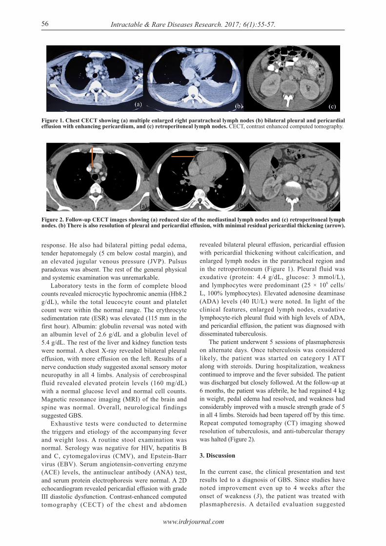
www.irdrjournal.com
Intractable & Rare Diseases Research. 2017; 6(1):55-57.
response. He also had bilateral pitting pedal edema, tender hepatomegaly (5 cm below costal margin), and an elevated jugular venous pressure (JVP). Pulsus paradoxus was absent. The rest of the general physical and systemic examination was unremarkable. Laboratory tests in the form of complete blood counts revealed microcytic hypochromic anemia (Hb8.2 g/dL), while the total leucocyte count and platelet count were within the normal range. The erythrocyte sedimentation rate (ESR) was elevated (115 mm in the first hour). Albumin: globulin reversal was noted with an albumin level of 2.6 g/dL and a globulin level of 5.4 g/dL. The rest of the liver and kidney function tests were normal. A chest X-ray revealed bilateral pleural effusion, with more effusion on the left. Results of a nerve conduction study suggested axonal sensory motor neuropathy in all 4 limbs. Analysis of cerebrospinal fluid revealed elevated protein levels (160 mg/dL) with a normal glucose level and normal cell counts. Magnetic resonance imaging (MRI) of the brain and spine was normal. Overall, neurological findings suggested GBS. Exhaustive tests were conducted to determine the triggers and etiology of the accompanying fever and weight loss. A routine stool examination was normal. Serology was negative for HIV, hepatitis B and C, cytomegalovirus (CMV), and Epstein-Barr virus (EBV). Serum angiotensin-converting enzyme (ACE) levels, the antinuclear antibody (ANA) test, and serum protein electrophoresis were normal. A 2D echocardiogram revealed pericardial effusion with grade III diastolic dysfunction. Contrast-enhanced computed tomography (CECT) of the chest and abdomen
revealed bilateral pleural effusion, pericardial effusion with pericardial thickening without calcification, and enlarged lymph nodes in the paratracheal region and in the retroperitoneum (Figure 1). Pleural fluid was exudative (protein: 4.4 g/dL, glucose: 3 mmol/L), and lymphocytes were predominant (25 × 106 cells/L, 100% lymphocytes). Elevated adenosine deaminase (ADA) levels (40 IU/L) were noted. In light of the clinical features, enlarged lymph nodes, exudative lymphocyte-rich pleural fluid with high levels of ADA, and pericardial effusion, the patient was diagnosed with disseminated tuberculosis. The patient underwent 5 sessions of plasmapheresis on alternate days. Once tuberculosis was considered likely, the patient was started on category I ATT along with steroids. During hospitalization, weakness continued to improve and the fever subsided. The patient was discharged but closely followed. At the follow-up at 6 months, the patient was afebrile, he had regained 4 kg in weight, pedal edema had resolved, and weakness had considerably improved with a muscle strength grade of 5 in all 4 limbs. Steroids had been tapered off by this time. Repeat computed tomography (CT) imaging showed resolution of tuberculosis, and anti-tubercular therapy was halted (Figure 2).
3. Discussion
In the current case, the clinical presentation and test results led to a diagnosis of GBS. Since studies have noted improvement even up to 4 weeks after the onset of weakness (3), the patient was treated with plasmapheresis. A detailed evaluation suggested
56
Figure 1. Chest CECT showing (a) multiple enlarged right paratracheal lymph nodes (b) bilateral pleural and pericardial effusion with enhancing pericardium, and (c) retroperitoneal lymph nodes. CECT, contrast enhanced computed tomography.
Figure 2. Follow-up CECT images showing (a) reduced size of the mediastinal lymph nodes and (c) retroperitoneal lymph nodes. (b) There is also resolution of pleural and pericardial effusion, with minimal residual pericardial thickening (arrow).

www.irdrjournal.com
Intractable & Rare Diseases Research. 2017; 6(1):55-57. 57
leading to autoimmunity and damage to nerves is also a possibility.
References
1. Yuki N, Hartung HP. Guillain-Barre syndrome. N Engl J Med. 2012; 366:2294-2304.
2. Kocen RS, Parsons M. Neurological complications of tuberculosis: Some unusual manifestations. Q J Med. 1970; 39:17-30.
3. Hughes RA, Swan AV, van Doorn PA. Intravenous immunoglobulin for Guillain-Barre syndrome. Cochrane Database Syst Rev. 2014; CD002063.
4. Canham EM, Iseman MD. Guillain-Barre syndrome related to pulmonary tuberculosis. Ann Am Thorac Soc. 2014; 11:855-857.
5. de la Torre RG, Moris G, Martinez DP, Montes IC. Guillain-Barre syndrome, tuberculosis and inflammatory bowel disease: A multiple association. Int Arch Med. 2010; 3:15.
6. Soehardy Z, Yuhanisa A, Thein SS, Rohana AG, Fauzi AR, Norlinah MI, Hamidon BB, Rozaidi SW. AMSAN variant of Guillain Barre syndrome progressing to chronic inflammatory demyelinating polyneuropathy in a patient with Marfan's syndrome and pulmonary tuberculosis. Med J Malaysia. 2005; 60:655-656.
7. Taha AA, Tee KH. Guillain-Barre syndrome associated with pulmonary tuberculosis. BMJ Case Rep. 2012; 13:2012.
8. Vyravanathan S, Senanayake N. Guil lain-Barre syndrome associated with tuberculosis. Postgrad Med J. 1983; 59:516-517.
9. Kalita J, Misra UK, Goyal G, Das M. Guillain-Barre syndrome: Subtypes and predictors of outcome from India. J Peripher Nerv Syst. 2014; 19:36-43.
(Received September 14, 2016; Revised November 21, 2016; Accepted December 2, 2016)
tuberculosis, but the patient's neurological status precluded confirmation of that diagnosis with endobronchial ultrasound (EBUS)-guided lymph node sampling. The diagnosis of tuberculosis was confirmed by the treatment response (complete clinical and radiological response) at the 6-month follow-up. To the extent known, 5 cases (4-8) involving tuberculosis in association with GBS have been published thus far (Table 1). The association has mostly been reported with pulmonary tuberculosis. All previously reported cases had features suggesting tuberculosis prior to the onset of weakness. This is the second such case with an AMSAN variant of GBS. The subtypes of GBS in India (9) follow the same distribution as in the rest of the world: AIDP is the most common (73.8%), followed by AMAN (13.4%) and AMSAN (4.6%). The AMSAN variant is very rare and has a worse prognosis and delayed recovery. The current patient presented quite late in the course of his illness, but prompt management and treatment of the underlying condition resulted in a satisfactory response. Plasmapheresis had not been used in any of the previously reported cases. In all of the cases, patients responded to treatment but those with pericardial effusion had constrictive pericarditis (including the current patient). The temporal association between GBS and the patient's other symptoms suggests that this association is unlikely to be due to chance. Hence, the ability of Mycobacterium tuberculosis to trigger GBS needs to be investigated. The pathogenesis of GBS is believed to be immune-mediated. A greater delay in cell-mediated immunity in tuberculosis has been suggested as the mechanism responsible. Molecular mimicry
Table 1. Summary of reported cases of GBS and tuberculosis
Author and year (ref. No.)
Vyravanathan et al. 1983 (8)
Soehardy et al. 2005 (6)
de la Torre et al. 2010 (5)
Taha et al. 2012 (7)
Canham et al. 2014 (4)
Current case
Diagnosis
Pulmonary tuberculosis with GBS
Pulmonary tuberculosis and GBS (AMSAN variant)
Extrapulmonary tuberculosis (cervical lymph node) with GBS
Pulmonary tuberculosis with GBS (AIDP)
Pulmonary tuberculosis with pericardial effusion and GBS (AIDP)
Disseminated tuberculosis (Mediastinal and retroperitoneal lymph nodes, pleural effusion, pericardial effusion with thickening) and GBS(AMSAN)
Treatment
ATT and physiotherapy
ATT and IVIG
ATT and IVIG
ATT and IVIG
ATT and IVIG
ATT and plasmapheresis
AIDP, acute inflammatory demyelinating neuropathy; AMSAN, acute motor sensory axonal neuropathy; ATT, antitubercular therapy; IBD, inflammatory bowel disease; IVIG, intravenous immunoglobulin.
Outcome
Recovered completely
Recovered completely
Recovered (patient developed IBD 2 months later)
Recovered completely
Difficulty walking with restricted ankle movement and constrictive pericarditis
Constrictive pericarditis without functional disability

www.irdrjournal.com
Intractable & Rare Diseases Research. 2017; 6(1):58-60.58
Gomez-Lopez-Hernández syndrome: First reported case from the Indian subcontinent
Anita Choudhary, Priyanka Minocha*, Sadasivan Sitaraman
Department of Pediatrics, SMS Medical College and Hospital, Jaipur, Rajasthan, India.
1. Introduction
Gomez-Lopez-Hernández syndrome (GLHS) (OMIM 601853) is a rare neurocutaneous syndrome that is characterized by triad of findings: partial alopecia of the scalp, trigeminal anaesthesia, and rhombencephalosynapsis. Thus far, only 35 cases had been described with varying symptomatology (1,2). Genetic factors responsible for GLHS are poorly understood. Recently, possibility of autosomal recessive transmission was suggested by Vinicius et al. in view of reported consanguinity in parents of some reported cases (2). GLHS was first described by Manuel Gomez in 1979 in a girl. He postulated that the patient had a cerebellotrigeminal and focal dermal dysplasia due to a developmental arrest of the ectoderm, which gives rise to the alar plate of the rhombencephalon, the overlying epidermis, the motor nucleus of V, and the trigeminal placodes (3). In 1982, Lopez Hernández described a new neurocutaneous syndrome in two Mexican girls with
a similar presentation in which a cerebellar anomaly (ponsvermis fusion anomaly with atresia of the fourth ventricle) was confirmed by a CT scan (4). Rush et al. and Sukhudyan et al. attempted to describe the diagnostic criteria for GLHS since the triad of findings are not found in all patients (1,5). Reported here is the case of a child with the classic triad of findings, i.e. partial alopecia of the scalp, trigeminal anaesthesia, and rhombencephalosynapsis. This chi ld was the product of a third-degree consanguineous marriage, making autosomal recessive transmission a likely cause. To the extent known, this is the first case of GLHS reported from India.
2. Case Report
A 4-year-old boy who was second in birth order and born from a third-degree consanguineous marriage and who had an uneventful antenatal and perinatal history presented with developmental delay, abnormal head movements, and seizures since 2 years of age. There was no family history of similar features. On clinical examination, the boy's body measurements were as follows: weight 10 kg (≤ 3 SD), height 94 cm (≤ 2 SD), and head circumference (HC) 48 cm. His characteristic craniofacial features (Figure 1, Table 1) included a flat occiput, brachyturricephaly, low-set ears, a patch of alopecia (Figure 2) present above both ears, thin
Summary Gomez-Lopez-Hernández syndrome (GLHS) is a rare neurocutaneous syndrome char-acterized by a triad of findings: partial alopecia of the scalp, trigeminal anaesthesia, and rhombencephalosynapsis. GLHS is also known as cerebello-trigeminal-dermal dysplasia. Besides this triad, a number of varying traits have been described in 35 previously reported cases. Reported here is a case of a four-year-old boy, born out of consanguineous marriage, presenting with the classic triad of findings, i.e. partial alopecia of the scalp, trigeminal anaesthesia, and rhombencephalosynapsis. To the extent known, this is the first case of GLHS reported from India. If a child presents with alopecia and rhom-bencephalosynapsis, GLHS should be considered in the differential diagnosis. A host of studies can be used to determine the exact pathogenesis, and confirming the diagnosis of GLHS is an important step in prenatal testing for at-risk pregnancies.
Keywords: Alopecia, rhombencephalosynapsis, trigeminal anaesthesia, autosomal recessive
DOI: 10.5582/irdr.2016.01074Case Report
Released online in J-STAGE as advance publication December 21, 2016.
*Address correspondence to:Dr. Priyanka Minocha, Department of Pediatrics, SMS Medical College and Hospital, Jaipur, Rajasthan 302004, India.E-mail: [email protected]

www.irdrjournal.com
Intractable & Rare Diseases Research. 2017; 6(1):58-60.
lips, and hypertelorism. Informed written consent was obtained from the parents of the child. The child had stereotypical head shaking (yes-yes). Anesthesia was present along the path of the trigeminal nerve and corneal opacity was present in the left eye. He had moderate intellectual disability (IQ 58). Karyotyping, abdominal ultrasonography, an X-ray of the spine, two-dimensional echocardiography, and fundoscopy were normal. MRI of the brain (Figure 3) revealed rhombencephalosynapsis (absent cerebellar vermis with apparent fusion of cerebellar hemispheres) and cerebellar hypoplasia. EEG showed generalized epileptiform discharges.
3. Discussion
GLHS is a rare neurocutaneous syndrome. In previously described cases, the most consistent findings were scalp alopecia and rhombencephalosynapsis while
the third finding of the classical triad, i.e. trigeminal anaesthesia, was present in only 56% of cases. Other craniofacial features of this syndrome are low-set ears, brachyturricephaly, mid-face retrusion, widely spaced eyes, strabismus, craniosynostosis, and plagiocephaly. Based on these findings, Rush et al. (1) described criteria for defining definitive, probable, and possible GLHS. The current case qualifies as definitive GLHS since all of the classic findings were present. Ataxia, hypotonia, stereotypical head movements, and intellectual disabili ty are some important neurological features described in GLHS. Stereotypical head movements are typically described as side-to-side "no" movements, up-and-down "yes" movements, or shoulder-to-shoulder movements (6,7). The child in the current case displayed repeated up and down "yes" movements of the head. These stereotypical movements are characteristic of GLHS and provide a clue to the diagnosis of rhombencephalosynapsis and GLHS. Rhombencephalosynapsis is defined as midline brain malformation characterized by absent cerebellar vermis with apparent fusion of the cerebellar hemispheres. It is usually noted in GLHS along with characteristic findings of alopecia and trigeminal anaesthesia, but it can also occur as isolated entity. Rhombencephalosynapsis is also seen in patients with the VACTERL association, i.e. vertebral anomalies, anal atresia, cardiovascular anomalies, tracheaesophageal fistula, renal anomalies,
59
Figure 1. Craniofacial features showing brachyturricephaly, low- set ears, thin lips, and hypertelorism.
Table 1. Comparison of traits in previously described cases and the current case
Trait
Craniofacial Alopecia of the scalp Low-set ears Brachycephaly and/or turricephaly Mid-face retrusion Strabismus Trigeminal anesthesia Absent corneal reflexes Widely spaced eyes Interstitial keratitis or corneal clouding Lambdoid craniosynostosis PlagiocephalyNeurodevelopmental Delayed motor milestones Ataxia Hypotonia Intellectual disability Head shaking or other stereotypical behavior Radiologic Rhombencephalosynapsis Ventriculomegaly/hydrocephalus Cerebellar hypoplasia Other Hypoplastic labia majora Normal growth at birth Short stature
Currentcase
++++-+++---
+--+++-+
NA++
Previouslyreported
cases
35/3530/3329/3427/3322/2918/3218/3121/3114/3210/2114/20
27/2819/2620/2721/3015/1934/3421/3112/31
5/918/231726
Figure 2. A patch of alopecia present above both ears.
Figure 3. MRI of the brain revealed rhombencephalosynapsis i.e. absent cerebellar vermis with apparent fusion of the cerebellar hemispheres.

www.irdrjournal.com
Intractable & Rare Diseases Research. 2017; 6(1):58-60.60
sanguineous parents: New evidence for an autosomal-recessive pattern of inheritance? Pediatr Neurol. 2014; 50:612-615.
3. Gomez MR. Cerebellotrigeminal and focal dermal dysplasia: A newly recognized neurocutaneous syndrome. Brain Dev. 1979; 1:253-256.
4. Lopez-Hernandez A. Craniosynostosis, ataxia, trigeminal anaesthesia and parietal alopecia with ponsvermis fusion anomaly (atresia of the fourth ventricle). Report of two cases. Neuropediatrics. 1982; 13:99-102.
5. Sukhudyan B, Jaladyan V, Melikyan G, Schlump JU, Boltshauser E, Poretti A. Gomez–Lopez-Hernandez syndrome: Reappraisal of the diagnostic criteria. Eur J Pediatr. 2010; 169:1523-1528.
6. Bonnet C, Roubertie A, Doummar D, Bahi-Buisson N, Cochen de Cock V, Roze E. Developmental and benign movement disorders in childhood. Mov Disord. 2010; 25:1317-1334.
7. Harris KM, Mahone EM, Singer HS. Nonautistic motor stereotypies: Clinical features and longitudinal follow-up. Pediatr Neurol. 2008; 38:267-272.
8. Ishak GE, Dempsey JC, Shaw DW, Tully H, Adam MP, Sanchez-Lara PA, Glass I, Rue TC, Millen KJ, Dobyns WB, Doherty D. Rhombencephalosynapsis: A hindbrain malformation associated with incomplete separation of midbrain and forebrain, hydrocephalus and a broad spectrum of severity. Brain. 2012; 135:1370-1386.
9. Gomy I, Heck B, Santos AC, Figueiredo MS, Martinelli CE Jr, Nogueira MP, Pina-Neto JM. Two new Brazilian patients with Gómez-López-Hernández syndrome: Reviewing the expanded phenotype with molecular insights. Am J Med Genet A. 2008; 146A:649-657.
(Received September 21, 2016; Revised November 10, 2016; Accepted November 16, 2016)
and limb defects. More than 90 individuals with rhombencephalosynapsis have been reported in the literature, and 25 of those individuals were found to have GLHS (8). This indicates that rhombencephalosynapsis is a consistent finding in GLHS. Other neuroimaging findings noted in GLHS are ventriculomegaly/hydrocephalus and cerebellar hypoplasia. Consanguinity has been described in only 2 previously reported cases (2,9) and the child in the current case is the third such case involving parents with a history of consanguineous marriage, making autosomal recessive transmission a likely cause. That said, further studies and whole-genome sequencing need to be performed to pinpoint the exact pathogenesis. In conclusion, GLHS is rare neurocutaneous syndrome with unknown genetic causes. High degree of suspicion in a child presenting with characteristic alopecia and rhombencephalosynapsis has a great importance in diagnosis of GLHS. A host of studies can be used to determine the exact pathogenesis, and confirming the diagnosis of GLHS is an important step in prenatal testing for at-risk pregnancies
References
1. Rush ET, Adam MP, Clark RD, Curry C, Hartmann JE, Dobyns WB, Olney AH. Four new patients with Gomez-Lopez-Hernandez syndrome and proposed diagnostic criteria. Am J Med Genet. 2013; 161A:320-326.
2. de Mattos VF, Graziadio C, Machado Rosa RF, Lenhardt R, Alves RP, Trevisan P, Paskulin GA, Zen PR. Gómez-López-Hernández syndrome in a child born to con-

www.irdrjournal.com
Intractable & Rare Diseases Research. 2017; 6(1):61-64. 61
Microdeletion of chromosome 1q21.3 in fraternal twins is associated with mental retardation, microcephaly, and epilepsy
Fatma Mujgan Sonmez1,*, Eyyup Uctepe2, Dilek Aktas3, Mehmet Alikasifoglu3
1 Developmental Child Neurology Association, Ankara, Turkey;2 Department of Medical Genetics, Health Sciences University, Diskapi Yildirim Beyazit Training and Research Hospital, Ankara,
Turkey;3 Damagen Genetic Diagnostic Center, Ankara, Turkey.
1. Introduction
The introduction of genome-wide approaches to identify deletions and duplications throughout the human genome has facilitated the discovery of numerous novel causes of intellectual disability (ID) and epilepsy (1,2). In a clinical work-up of undiagnosed intellectual disability, array comparative genomic hybridization (CGH) can facilitate a diagnosis in 10-30% of cases. Although there is vast amounts of data on the clinical features of and standardized management guidelines for well-known classic microdeletion syndromes, systematic characterization of newly identified patients provides a host of essential information for clinicians and patients. Only one previous report described 1q21.3 microdeletion syndrome (3), and the deleted region
in question spanned about 1.4 Mb with approximate genomic location chr1:152,511,593-153,993,103 including at least 30 genes such as CHRNB2, KCNN3, HAX1, ADAR, PKLR, EFNA1, EFNA2, and EFNA3 (NCBI genome build 36). The current case report seeks to further characterize 1q21.3 microdeletion syndrome. Described here are two siblings with a 1q21.3 microdeletion that was associated with mental retardation, microcephaly, epilepsy, and some dysmorphic features.
2. Case Report
2.1. Case 1
A baby girl was born by cesarean section at 29 weeks 5 days' gestation as a set of triplets, and the girl had a birth weight of 1,240 g (Figure 1). The girl was kept in the neonatal intensive care unit because of an intraventricular hemorrhage and she also underwent surgery for a duodenal obstruction and perforation. At 4 months of age, the girl underwent surgery for hydrocephalus and a ventriculoperitoneal shunt was placed. At 5 years of age, right focal motor convulsions started. In spite of therapy with three antiepileptics,
Summary Reported here are twins, both of whom have a 1q21.3 microdeletion and who exhibit key features common to previously reported cases such as microcephaly and developmental delay. However, some clinical findings and deleted genes differed from those in previously reported cases. The karyotype was normal 46, XX for both of the twins. Array comparative genomic hybridization (CGH) identified a 2.6 Mb deletion on chromosome 1q21.3 (chr1: 153,514,121-156,171,335 bp) in case 1 and a 1.6 Mb deletion on chromosome 1q21.3 (chr1: 154,748,365-156,358,923 bp) in case 2. The deleted region includes DPM3, MUC1, GBA, PKLR, RIT1, and LAMTOR2 in both siblings. To the extent known, this is the second report of a 1q21.3 microdeletion in a family with mental retardation, developmental delay, seizures, and some dysmorphic features, thus expanding the phenotypic spectrum.
Keywords: 1q21.3 microdeletion syndrome, developmental delay, mental retardation, microarray-CGH, seizures
DOI: 10.5582/irdr.2016.01075Case Report
Released online in J-STAGE as advance publication December 5, 2016.
*Address correspondence to:Dr. Fatma Mujgan Sonmez, Developmental Child Neurology Association, Tunalı Hilmi Caddesi, Buğday Sokak. Kozlar İşhanı No:6/28, Kavaklıdere/Ankara, Turkey.E-mail: [email protected]

www.irdrjournal.com
Intractable & Rare Diseases Research. 2017; 6(1):61-64.
in t ractable seizure resumed and the gir l was hospitalized for status epilepticus. The girl is now 11 years old and she has been seizure-free for three years with three antiepileptics. A physical and neurological examination indicated growth retardation (weight and height under the 3rd percentile), pectus carinatus, scoliosis, microcephaly, spastic quadriparesis, laxity in both hands, and severe spasticity in the extremities. The girl only speaks single words, she cannot walk, and she can only sit with support. Laboratory results revealed a normal hemogram and normal biochemical test results. Electroencephalography revealed a right frontocentral epileptiform abnormality. Brain MRI revealed periventricular leukomalacia and pontocerebellar hypoplasia (Figure 2A). Results of a karyotype analysis were normal. The girl is receiving Na-valproate, levetiracetam, and oxcarbazepine therapy. Array CGH analysis identified a 2.6 Mb deletion on chromosome 1q21.3 (chr1: 153,514,121-156,171,335
bp). The deleted region contains 68 OMIM genes. Several of these genes cause disease, including GATAD2B, TPM3, HAX1, IL6R, CHRNB2, ADAR, DPM3, MUC1, GBA, PKLR, RIT1, LAMTOR2, LMNA, and SEMA4A. Some disease-causing genes such as GATAD2B, TPM3, LMNA, and SEMA4A are present in this case but not in the case reported by Reddy et al. (Figure 3) (3). Parental array CGH analysis and FISH testing for balanced translocations were not performed because of family's decision to forego further testing. Parental testing could help to determine if the parents are carriers in terms of investigating whether the microdeletion in question is de novo or inherited. If parental testing was performed, the mutation would presumably be a de novo germline mutation because the parents are healthy and two sisters in the same family were affected with the same disorder.
2.2. Case 2
The patient in this case is the sibling of the patient in case 1. After birth, the girl in case 2 was kept in the neonatal intensive care unit because of respiratory distress syndrome, hyperbilirubinemia, neonatal convulsions, and cholelithiasis. The girl could walk and speak at 2 years of age. At 6 years of age, the girl was hospitalized at another facility because of status epilepticus, hyperglycemia, and encephalitis. At that time, EEG and cranial MRI were normal, and Na-valproate therapy was started. The girl can now walk without support and she only receives risperidone therapy for attention deficit hyperactivity disorder. On physical and neurological examination, the girl's weight was 25 kg (25%) and her height was 132 cm (50%). The girl has microcephaly, a systolic murmur, distal laxity, genu valgus, and sacral dimples. The girl can walk with a long gait. Laboratory results revealed a normal hemogram and normal biochemical test results except for low levels of vitamin D. EEG was normal, and cranial MRI revealed
62
Figure 1. Pedigree of patients with 1q21.3 microdeletions.
Figure 2. Cranial MRI findings. (a) periventricular leukomalacia and pontocerebellar hypoplasia in Case 1; (b) Periventricular hyperintensity in Case 2.
Figure 3. Comparison of fraternal twins to the earlier patient with a 1q21.3 microdeletion in terms of clinical-radiologic features and the deleted chromosomal region.

www.irdrjournal.com
Intractable & Rare Diseases Research. 2017; 6(1):61-64. 63
carinatum, spastic quadriplegia, pontocerebellar hypoplasia, and periventricular leukomalacia on brain MRI and the patient in case 2 has additional clinical features like strabismus, myopia, genu valgus, attention deficit hyperactivity disorder, and a myelination delay on brain MRI. These results conform with the case previously reported by Reddy et al. and expand the phenotypic spectrum of 1q21.3 syndromes. T h e o c c u r r e n c e o f r a r e d i s e a s e - c a u s i n g microdeletions in the genome is not random and is possibly affected by neighboring genes in the genome (6). The high sequence similarity over large stretches of DNA makes low copy repeats (LCRs) susceptible to germline non-allelic homologous recombination (NAHR) events that cause deletion or duplication. More than 30 genomic disorders originate from NAHR-mediated events occurring in the germ line (7). Copy number variations with the same length and clustered breakpoints for a group of patients with the same disorder originate from NAHR-mediated microdeletions (6). Since the observed deletions range from 1.4 kb to 2.6 Mb in length and the breakpoint locations differ in the 2 cases reported here, a common mechanism of deletion, such as NAHR-mediated by LCRs, can be ruled out in 1q21.31 microdeletion syndrome. However, non-recurrent rearrangements vary in length, have scattered breakpoints, and the majority of these microdeletions can result from microhomology-mediated mechanisms like microhomology-mediated end-joining (MMEJ), fork stalling and template switching (FoSTeS), microhomology-mediated break-induced replication (MMBIR), serial replication slippage (SRS), or break-induced SRS (BISRS). Hence, the only option to explore the underlying mechanisms in 1q21.31 microdeletion syndrome is to characterize it at the level of base pairs and to examine the sequences found at breakpoints. Some variations in the genomic architecture in a parent might stimulate the development of these rare germline deletions via a non-recurrent CNV, increasing the susceptibility to DNA breakage or promoting replication fork stalling. This means that recurrence of a microdeletion in different sizes would be apparent in the same family. Although there is no parental consanguinity, the
bilateral periventricular hyperintensity commensurate with leukomalacia, and delayed myelination (Figure 2B). Spinal MRI was normal. The girl is now seizure-free and she can attend school. A karyotype analysis yielded normal results. Array CGH analysis identified a 1.6 Mb deletion on chromosome 1q21.3 (chr1:154748365-156358923 bp). The deleted region in question contains 47 OMIM genes. Several of these genes cause disease, including DPM3, MUC1, GBA, PKLR, RIT1, LAMTOR2, LMNA, and SEMA4A. Some disease-causing genes such as LAMTOR2, LMNA, and SEMA4A are present in this case but not in the case reported by Reddy et al. (3). However, genes like HAX1, IL6R, CHRNB2, and ADAR were deleted in case 1 and the previously reported case of 1q21.3 microdeletion syndrome but those genes were not deleted in case 2 (Figure 3). The third of the triplets was a girl who died 12 hours after birth because of respiratory distress. There is no other information available concerning that child.
3. Discussion
Several novel chromosome aberration syndromes have already been characterized, and undoubtedly, more descriptions will follow. Earl ier reports indicated that proximal 1q (1q21-q25) deletions present with developmental delay, microcephaly, cardiac, renal and genital abnormalities, and some dysmorphic features (4,5). A submicroscopic deletion with a location similar/identical to the one in the current patients was previously reported by Reddy et al. (3). Reddy et al. described a 2 and a half-year-old girl with moderate mental retardation, microcephaly, arching eyebrows, low-set ears, long eyelashes, persistent fetal pads, and clinodactyly. They detected a 1.4 Mb deleted region with approximate genomic location at chr1: 152,511,593-153,993,103 (NCBI genome build 36). The deleted region in that case mostly coincided with the deleted region noted in the current patients (Figure 4). Major clinical features such as microcephaly and developmental delay were common to the current patients and the previously reported case. Furthermore, the patient in case 1 has additional clinical features like hydrocephalus, pectus
Figure 4. Results of array CGH analysis in patients (Deleted regions). CGH, comparative genomic hybridization.

www.irdrjournal.com
Intractable & Rare Diseases Research. 2017; 6(1):61-64.64
current patients could have some other recessive disorder. Array CGH is known to be unable to exclude a recessive disease in instances of a small DNA substitution or rearrangement that does not result in any loss or gain of DNA. At this point, whole-exome sequencing (WES) would provide more comprehensive information, but WES was not possible in the current cases. Epilepsy is one of the features of this syndrome. Muhle et al. described a boy with absence seizures whose parents both had childhood absence epilepsy. A 192-kb duplication in 1q21.3, encompassing the genes IL6R, SHE, TDRD10, UBE2Q1, CHRNB2, and ADAR, was identified in the proband and his father. Both CHRNB2 and ADAR are genes potentially responsible for seizure disorders. All of the duplicated genes in that case were also deleted in the patient in Case 1. The duplication was not identified in 191 patients with independent idiopathic generalized epilepsy or in 1,157 population controls (8). The current report has several limitations. One is that parental testing was not possible. Another is that WES was not performed. WES can be used to investigate whether there are other mutations that array CGH has failed to detect, such as small deletions and single nucleotide polymorphisms. A third limitation is that this report did not verify breakpoints in both of the current patients. Breakpoints could be mapped using methods such as quantitative PCR (qPCR), long-range PCR, and Sanger sequencing. Hence, this is an initial report, and interpretations in this report may change over time as additional cases are assembled. In conclusion, the current report found that the 1q21.3 deletion might be a genetic risk factor in this family, contributing to mental retardation, microcephaly, epilepsy, and some dysmorphic features. This is the second report of a deletion of the 1q21.3 region and further cases are required to clarify which of the genes
in the deleted interval contribute to the phenotype of the children and their long-term outcomes.
Acknowledgements
The authors wish to thank the patients and their family for their participation in this study.
References
1. Mefford HC, Eichler EE. Duplication hotspots, rare genomic disorders, and common disease. Curr Opin Genet Dev. 2009; 19:196-204.
2. Slavotinek AM. Novel microdeletion syndromes detected by chromosome microarrays. Hum Genet. 2008; 124:1-17.
3. Reddy S, Dolzhanskaya N, Krogh J, Velinov M. A novel 1.4 Mb de novo microdeletion of chromosome 1q21.3 in a child with microcephaly, dysmorphic features and mental retardation. Eur J Med Genet. 2009; 52:443-445.
4. Lo LJ, Noordhoff MS, Huang CS, Chen KT, Chen YR. Proximal deletion of the long arm of chromosome 1: [del(1)(q23-q25)]. Cleft Palate Craniofac J. 1993; 30:586-589.
5. Pallotta R, Dalpra L, Miozzo M, Ehresmann T, Fusilli P. A patient defines the interstitial 1q deletion syndrome characterized by antithrombin III deficiency. Am J Med Genet. 2001; 104:282-286.
6. Verdin H, D'Haene B, Beysen D, Novikova Y, Menten B, Sante T, Lapunzina P, Nevado J, Carvalho CM, Lupski JR, De Baere E. Microhomology-mediated mechanisms underlie non-recurrent disease-causing microdeletions of the FOXL2 gene or its regulatory domain. PLoS Genet. 2013; 9:e1003358.
7. Sasaki M, Lange J, Keeney S. Genome destabilization by homologous recombination in the germ line. Nat Rev Mol Cell Biol. 2010; 11:182-195.
8. Muhle H, Steinich I, von Spiczak S, et al. A duplication in 1q21.3 in a family with early onset and childhood absence epilepsy. Epilepsia. 2010; 51:2453-2456.
(Received September 25, 2016; Revised November 3, 2016; Accepted November 7, 2016)

www.irdrjournal.com
Intractable & Rare Diseases Research. 2017; 6(1):65-68. 65
Lesch-Nyhan syndrome: The saga of metabolic abnormalities and self-injurious behavior
Nitesh Tewari*, Vijay Prakash Mathur, Divesh Sardana, Kalpana Bansal
Pedodontics & Preventive Dentistry, Center for Dental Education and Research, All India Institute of Medical Sciences, New Delhi, India.
1. Introduction
Lesch-Nyhan syndrome (LNS) is an X-linked recessive disorder of purine metabolism with a prevalence of 1:100,000 to 1:380,000 (1,2). The primary cause of LNS is a mutation in Xq26.2-q26.3 (OMIM 308000.0004) that leads to a reduced or complete deficiency in the hypoxanthine guanine phosphoribosyl-transferase 1 (HPRT1) enzyme in affected individuals (2,3) . The phenotype of LNS varies and it depends upon the level of HPRT activity. When the level of HPRT1 activity > 8%, only hyperuricemia manifests. A level of HPRT1 activity between 2% to 8% is referred to as an HPRT-related neurological dysfunction (HND) that is characterized by neurological, neurocognitive, and behavioral abnormalities. A level of HPRT activity < 2% is referred to as LNS, which involves significant neuro-motor and cognitive defects and the display of self-inflicted injurious behavior (SIB) (3).
SIB has been reported to usually begin with eruption of the primary teeth at 12 months of age, however it may manifest at 4-5 years of age. SIB has been attributed to defective maturation of certain dopamine neuronal pathways in the brain (1-3). One of the distinct features of SIB seen in LNS is the presence of pain and the display of remorse after an episode. Patients display an inability to overcome a compulsive desire to hurt themselves ‒ both in the form of physical injury and/or emotional distress. This can be extremely distressing to caregivers and it also puts these patients at risk (4). A rare case of LNS with complications and SIB has been presented here.
2. Case Report
A 4-year-old male child was admitted to the Pediatric Emergency Department with altered sensorium and poor oral intake for 20 days prior along with seizures for 5 days and a fever for 3 days. A history revealed that the boy was born from non-consanguineous parents and a normal full-term vaginal delivery. Neck holding was absent and self-mutilation by finger biting was described as starting at 10 months. At 3 years and 6 months of age, the boy's parents noted an intermittent tightening of the limbs and finger and lip biting with increasing severity. A family history revealed that his elder male sibling had succumbed to a similar illness, dying suddenly at the age
Summary Lesch-Nyhan syndrome (LNS) is an X-linked recessive disorder of purine metabolism caused by a mutation in Xq26.2-q26.3 (OMIM 308000.0004). The presence of the diagnostic triad, i.e. signs of self-injurious behavior (SIB) and results of pedigree analysis and novel molecular biology & genetic testing, confirms the diagnosis of LNS. With a level of hypoxanthine guanine phosphoribosyl-transferase 1 (HPRT1) enzyme activity < 2%, patients develop neurological, neurocognitive, and neuromotor symptoms along with SIB. Described here is a case of 4-year-old boy who was diagnosed with LNS. The boy displayed SIB, i.e. biting of the lips and fingers, and he had cerebral venous sinus thrombosis caused by LNS.
Keywords: Lesch-Nyhan disease, self-injurious behavior, X-linked disorder
DOI: 10.5582/irdr.2016.01076Case Report
Released online in J-STAGE as advance publication December 21, 2016.
*Address correspondence to:Dr. Nitesh Tewari, Division of Pedodontics & Preventive Dentistry, 6th Floor, Center for Dental Education & Research, All India Institute of Medical Sciences, New Delhi-110029, India.E-mail: [email protected]

www.irdrjournal.com
Intractable & Rare Diseases Research. 2017; 6(1):65-68.
7. His three sisters were normal and were age 6, 8, and 12. One of his maternal uncles had died at the age of 3 and had SIB while two others had suffered from renal failure and had subsequently dies at age 30 and age 32 (Figure 1). A physical examination upon admission revealed a generalized pallor and fresh wounds on the fingers of both hands and the lower lip. The parents had bandaged the hands and feet to prevent any further damage due to biting (Figure 2 a-c). His weight was 8 kg with no abnormalities in the CNS or abdomen. Neurological assessment revealed a GCS score of E2V2M3, pupils were 3 mm and reactive bilaterally, and all four extremities had reduced bulk and tone. Reflexes were absent on the right side and 1+ in the left upper extremity and 2+ in the left lower extremity. Blood results revealed a hemoglobin level of 5.9, a urea/creatinine ratio of 49/0.5, and a Na/K level of 148/4.2. Blood results suggested megaloblastic anemia. On oral examination, the lower lip had atrophied due to self-injury and there were traumatic ulcers with raw margins, although the upper lip appeared normal. The child also had fair oral hygiene and a normal complement of all primary teeth
(Figure 2b). The level of HPRT1 enzyme activity was assessed and found to be 0.8%. Genetic testing revealed that the patient had a G580A substitution in the HPRT1 gene. These features were pathognomic for LNS. Magnetic resonance imaging revealed cerebral venous sinus thrombosis (CVST) and right cortical venous thrombosis (frontal and parietal veins) with thrombosis of the superior and left transverse sinus veins and a venous infarct in the right superior frontal gyrus (with cortical laminar necrosis). Cortical venous congestion was noted in the rest of the frontal and parietal cortices on the right. This was appropriately managed with a satisfactory course. Pediatric consultants advised the extraction of all primary teeth, but a conservative approach was taken to manage self-injury. A maxillary impression was taken using a size 0 tray and alginate, and a soft resin mouthguard was made. This "bite guard" was affixed to the maxillary teeth using light-cured resin cement (Figure 3a). The frequency of self-biting initially decreased and epithelization of lip and finger wounds was visible (Figure 3b). However, SIB, i.e. tongue biting, soon started, warranting extraction of the primary mandibular incisors (Figure 3c). Upon discharge, sensorium was normal and feeding was accomplished through Ryle's tube. Two subsequent follow-ups indicated a further reduction in SIB, but hypotonia and irritability persisted (Figure 3c). During the third follow-up, a new pattern of biting was noted, and this biting had resulted in ulcers on the lateral borders and ventral surface of the tongue (Figure 3d). All of the remaining primary mandibular teeth had to be extracted under local anesthesia in two visits. At the age of 4 years and 5 months, the patient died suddenly at night like his elder sibling.
66
Figure 1. Pedigree chart showing survivors as blue boxes/circles. Red boxes represent individuals with LNS who died at age 3 (1), age 7 (2), and age 4 years and 5 months (3); green boxes represent maternal uncles with renal failure who died at age 30 and age 32. LNS, Lesch-Nyhan syndrome.
Figure 2. Ryle's tube is in place in a 4-year-old boy with LNS displaying altered sensorium (a), severe atrophy of the lower lip as a result of SIB (b), and bandaged upper limbs (c). LNS, Lesch-Nyhan syndrome; SIB, self-injurious behavior.
Figure 3. A soft resin mouthguard was created to prevent SIB. (a) Signs that lower lip wounds were healing after the mouthguard was affixed, (b) A substantial reduction in SIB and healing of lip wounds after extraction of the primary mandibular incisors, and (c) Recurrence of SIB in a different biting pattern leading to ulceration of the lateral borders and ventral surface of the tongue (d). SIB, self-injurious behavior.

www.irdrjournal.com
Intractable & Rare Diseases Research. 2017; 6(1):65-68. 67
management team. Although the extraction of both primary and permanent teeth can completely eradicate the chances of injuries, a conservative modality should be attempted first.
Acknowledgements
The authors wish to thank the parents of the child in this case, Mr. Ram Kumar and Mrs. Gayatri Devi, for permission to use their child's medical records and photographs in this article.
References
1. Lesch M, Nyhan WL. A familial disorder of uric acid metabolism and central nervous system function. Am J Med. 1964; 36:561-570.
2. Cauwels RG, Martens LC. Self-mutilation behaviour in Lesch-Nyhan syndrome. J Oral Pathol Med. 2005; 34:573-575.
3. Fu R, Sutcliffe D, Zhao H, Huang X, Schretlen DJ, Benkovic S, Jinnah HA. Clinical severity in Lesch-Nyhan disease: The role of residual enzyme and compensatory pathways. Mol Genet Metab. 2015; 114:55-61.
4. Robey KL, Reck JF, Giacomini KD, Barabas G, Eddey GE. Modes and patterns of self-mutilation in persons with Lesch-Nyhan disease. Dev Med Child Neurol. 2003; 45:167-171.
5. Limeres J, Feijoo JF, Baluja F, Seoane JM, Diniz M, Diz P. Oral self-injury: An update. Dent Traumatol. 2013; 29:8-14.
6. Rashid N, Yusuf H. Oral self-mutilation by a 17-month-old child with Lesch-Nyhan syndrome. Int J Paediatr Dent. 1997; 7:115-117.
7. Jathar P, Panse AM, Jathar M, Gawali PN. Lesch-Nyhan Syndrome: Disorder of self-mutilating behavior. Int J Clin Pediatr Dent. 2016; 9:139-142.
8. Riaz IB, Husnain M, Ateeli H. Recurrent thrombosis in a patient with Lesch-Nyhan syndrome. Am J Med. 2014; 127:e11-e12.
9. Fardi K, Topouzelis N, Kotsanos N. Lesch-Nyhan syndrome: A preventive approach to self-mutilation. Int J Paediatr Dent. 2003; 13:51-56.
10. Romero Maroto M, Ruiz Duque C, Vincent G, Garcia Recuero I, Romance A. Management of oral lesions in Lesch-Nyham syndrome. J Clin Pediatr Dent. 2014;
3. Discussion
SIB is considered to be a characteristic feature of LNS (1). This behavior is secondary to a neuro-metabolic abnormality caused by a disorder in HPRT1-mediated purine metabolism (3). SIB has also been observed in congenital insensitivity to pain with anhidrosis, mental retardation, and syndromes as Cornelia de Lange syndrome, Munchausen syndrome, Moebius syndrome, Gilles de la Tourette syndrome, and Rett syndrome (5).Since the oral cavity is the earliest primary focus and the means of interaction with environment, it falls prey to any attempt at self-injury, with sharp teeth being the preferred tool (5,6). After the eruption of the primary teeth, SIB is displayed, as was noted in the current case. The presence of the diagnostic triad, i.e. signs of SIB and results of pedigree analysis and novel molecular biology & genetic testing, confirmed the diagnosis of LNS (Figure 4) (2,3,7). When the level of HPRT activity is below 2%, the dopaminergic pathway in the brain is severely affected and neuro-cognitive, neurological, and motor dysfunction result (2,3). CVST was noted in the current case. CVST is a rare condition that is due to the hypercoagulability of blood. Patients with abnormal purine metabolism and hyperuricemia have been reported to be susceptible to thrombosis and infarction (8). This further aggravated the patient's condition. SIB in LNS has been found to decrease when restraints are used, such as hand and feet bandages (4). However, oral injuries, and especially those of the lower lip and tongue, can still occur. Conservative modalities such as a mouthguard, bite guard, lip bumpers, or bite planes have been tried in the past but they have not been effective (9-11). Extraction of both primary and permanent teeth may be the ultimate solution in most cases (2,5,6). In the current case, the patient's pattern of self-biting changed after a bite guard was affixed and later when the primary mandibular incisors were extracted. Use of light-cured resin cement in the current case was found to be advantageous since it set immediately and was less likely to dislodge. LNS has been associated with a shorter life expectancy, with death occurring in the second or third decade of life due to renal failure (1,3). Sudden death has also been reported in younger children due to a respiratory obstruction, aspiration of gastric fluids, laryngospasms, central apnea, cyanotic breath-holding, or high cervical spine damage (12). In the current case, the patient and his older male sibling both died suddenly.
4. Conclusion
LNS is a rare disorder but it can easily be diagnosed. The predominant feature of SIB, i.e. self-biting, requires a pediatric dentist to play a key role in the
Figure 4. LNS diagnostic triad. LNS, Lesch-Nyhan syndrome.

www.irdrjournal.com
Intractable & Rare Diseases Research. 2017; 6(1):65-68.68
38:247-249.11. Ragazzini G, Delucchi A, Calcagno E, Servetto R,
Denotti G. A modified intraoral resin mouthguard to prevent self-mutilations in Lesch-Nyhan patients. Int J Dent. 2014; 2014:396830.
12. Neychev VK, Jinnah HA. Sudden death in Lesch-Nyhan disease. Dev Med Child Neurol. 2006; 48:923-926.
(Received September 28, 2016; Revised November 7, 2016; Accepted November 16, 2016)

www.irdrjournal.com
Intractable & Rare Diseases Research. 2017; 6(1):69-71. 69
Thrombocytosis in a patient with upper gastrointestinal bleeding
Xingshun Qi1,§,*, Valerio De Stefano2,§, Xiaodong Shao1, Xiaozhong Guo1,*
1 Liver Cirrhosis Study Group, Department of Gastroenterology, General Hospital of Shenyang Military Area, Shenyang, China;2 Institute of Hematology, Catholic University, Rome, Italy.
1. Introduction
Depending to its etiology, thrombocytosis is divided into two main categories, primary (i.e . , clonal thrombocytosis) or secondary thrombocytosis (i.e., reactive thrombocytosis) (1). The most common cause of clonal thrombocytosis is essential thrombocythemia. The common causes of reactive thrombocytosis include iron deficiency, inflammatory conditions or infection, malignancy, splenectomy, and acute blood loss. Described here is a case of upper gastrointestinal bleeding in which thrombocytosis was far more apparent than that usually observed in similar cases and in which thrombocytosis subsided after the cessation of bleeding.
2. Case Report
On July 26, 2016, a 59-year-old man with recurrent upper abdominal pain for a week prior was seen at a local hospital. The man had a history of cigarette smoking. Routine blood tests revealed a white blood cell (WBC) count of 22.45 × 10⁹/L (normal range: 4-10 × 109/L), a percentage of neutrophils of 91.1% (normal range: 50-70%), a red blood cell (RBC) count of 2.78
× 1012/L (normal range: 4-5.5 × 10¹²/L), a hemoglobin (Hb) level of 64 g/L (normal range: 120-160 g/L), a hematocrit (Hct) level of 21.8% (normal range: 40-50%), a mean corpuscular volume (MCV) of 78.5 fL (normal range: 83.0-100.0 fL), a mean corpuscular hemoglobin concentration (MCHC) of 293 g/L (normal range: 320-360 g/L), a platelet (PLT) count of 989 × 109/L (normal range: 100-300 × 109/L), and a plateletcrit (PCT) level of 0.735 (normal range: 0.108-0.282). Abdominal color Doppler ultrasound did not reveal any abnormalities in the liver, spleen, or pancreas. On July 28, 2016, the man was transferred to Hematology at another local hospital. Myeloproliferative neoplasms (MPNs) were suspected. Laboratory tests revealed that the WBC count was 19.77 × 10⁹/L (normal range: 3.5-9.5 × 10⁹/L), the percentage of neutrophils was 82.3% (normal range: 40-75%), the RBC count was 2.78 × 10¹²/L (normal range: 4.3-5.8 × 10¹²/L), Hb was 68 g/L (normal range: 130-175 g/L), Hct was 22.4% (normal range: 40-50%), the MCV was 80.6 fL (normal range: 82.0-100.0 fL), the MCHC was 304.0 g/L (normal range: 316-354 g/L), the PLT count was 910 × 109/L (normal range: 125-350 × 109/L), PCT was 0.8 (normal range: 0.2-0.4), folate was 8.85 nmol/L (normal range: 10.4-42.4 nmol/L), vitamin B12 was 966.9 pmol/L (normal range: 145-637 pmol/L), and serum ferritin was 109.6 ug/L (normal range: 30-400 ug/L). The man tested negative for the BCR/ABL P210 or BCR/ABL P190 fusion gene. The man also tested negative for the JAK2 V617F mutation, the JAK2 (EXON12) N542-E543 and N542-E544 deficiency, the JAK2 (EXON12) gene K539L1/L2 mutation, the MPL (EXON10) W515K\A\L\R1\R2\S) mutation, the MPL (EXON10)
Summary Reported here is a case of upper gastrointestinal bleeding secondary to a peptic ulcer involving an extremely high platelet count of 989 × 109/L. Myeloproliferative neoplasms were ruled out on the basis of gene mutation testing and a bone marrow biopsy. After the cessation of index bleeding, the platelet count decreased markedly. Thus, reactive thrombocytosis was considered as a possibility.
Keywords: Bleeding, platelet count, thrombocytosis, myeloproliferative neoplasm, gastrointestinal
DOI: 10.5582/irdr.2016.01080Case Report
Released online in J-STAGE as advance publication December 5, 2016.§These authors contributed equally to this works. *Address correspondence to:Dr. Xiaozhong Guo, and Dr. Xingshun Qi, Department of Gastroenterology, General Hospital of Shenyang Military Area, No. 83 Wenhua Road, Shenyang, 110840 China.E-mail: [email protected] (XG), [email protected] (XQ)

www.irdrjournal.com
Intractable & Rare Diseases Research. 2017; 6(1):69-71.
S505N mutation, and the CALR (EXON9) L367fs*46/k385fs*47 mutation. A bone marrow biopsy revealed active proliferation of nucleated bone marrow cells. The ratio of anucleate cells to nucleated cells was 8.20/1, the proportion of granulocytes was 79.6%, the proportion of erythrocytes was 18.4%, and the ratio of granulocytes to erythrocytes was 4.33/1. There was active proliferation of granulocytes, less active erythropoiesis, and the megakaryocyte count in a bone marrow smear was 192 cells, including 27 promegakaryocytes, 41 granular megakaryocytes, 30 platelet-producing megakaryocytes, and 2 bare nuclei. As of August 6, 2016, the man developed melena and fatigue. On August 9, 2016, he was transferred to this Department. On physical examination, the skin was pallid and the upper abdomen was tender without rebound. Laboratory tests revealed that the WBC count was 8.9 × 10⁹/L (normal range: 3.5-9.5 × 10⁹/L), the percentage of neutrophils was 80.6% (normal range: 40-75%), the RBC count was 2.86 × 10¹²/L (normal range: 4.3-5.8 × 10¹²/L), Hb was 67 g/L (normal range: 130-175 g/L), Hct was 21.7% (normal range: 40-50%), the PLT count was 831 × 109/L (normal range: 125-350 × 109/L), and PCT was 0.414 (normal range: 0.108-0.272). Upper gastrointestinal endoscopy revealed multiple duodenal ulcers with active bleeding (Figure 1). The patient tested positive for a Helicobacter pylori infection. Thus, the patient was diagnosed with a duodenal ulcer. Three U of RBCs were intravenously infused and esomeprazole was administered. On August 12, 2016, upper gastrointestinal endoscopy was performed again and revealed no active bleeding from lesions (Figure 2). On August 17, 2016, laboratory tests revealed that the WBC count was 7.4 × 10⁹/L (normal range: 3.5-9.5 × 10⁹/L), the percentage of neutrophils was 75.2% (normal range: 40-75%), the RBC count was 3.13 × 10¹²/L (normal range: 4.3-5.8 × 10¹²/L), Hb was 80 g/L (normal range: 130-175 g/L), Hct was 25.3% (normal range: 40-50%), the PLT count was 448 × 109/L (normal range: 125-350 × 109/L), and PCT was 0.301 (normal range: 0.108-0.272). After a hematologist was consulted, MPNs were ruled out. The same day, the patient was discharged due to the cost of hospitalization. Unfortunately, a telephone follow-up with the patient’s son indicated that the patient died from recurrent massive upper gastrointestinal bleeding on August 28, 2016.
3. Discussion
Physicians need to differentiate the nature of thrombocythemia due to an extremely high PLT. Classic Philadelphia chromosome-negative MPNs include polycythemia vera, essential thrombocythemia, and primary myelofibrosis (2). Once a patient has a sustained PLT of more than 450 × 109/L, essential thrombocythemia should be suspected. Additionally, novel gene mutations greatly facilitate the diagnosis of
essential thrombocythemia based on current evidence (2). In the current patient, clonal thrombocytosis was initially suspected. However, the current patient did not test positive for any MPN-related gene mutations. A profound increase in PLT subsided after upper gastrointestinal bleeding was appropriately diagnosed and treated. Thus, MPNs were ruled out in the current case. According to Tefferi et al., reactive thrombocytosis is defined as: i) no history of chronic myeloproliferative disorders; ii) a condition associated with secondary thrombocytosis; and iii) PLT returns to its normal level once an acute condition subsides (3). The current patient met these criteria for reactive thrombocytosis. Upper gastrointestinal bleeding is one of the most frequent and lethal gastrointestinal conditions (4,5). In patients with acute upper gastrointestinal bleeding, hematemesis and melena are key clinical symptoms, anemia is a key physical sign, and a lower Hb level is evident in laboratory results. Upper gastrointestinal endoscopy is the method of choice for identifying the source of upper gastrointestinal bleeding. A peptic ulcer is one of the most common sources of upper
70
Figure 1. Upper gastrointestinal endoscopy before treatment.
Figure 2. Upper gastrointestinal endoscopy after treatment.

www.irdrjournal.com
Intractable & Rare Diseases Research. 2017; 6(1):69-71. 71
presenting with a thrombocytosis. Br J Haematol. 2010; 149:352-375.
2. Arber DA, Orazi A, Hasserjian R, Thiele J, Borowitz MJ, Le Beau MM, Bloomfield CD, Cazzola M, Vardiman JW. The 2016 revision to the World Health Organization classification of myeloid neoplasms and acute leukemia. Blood. 2016; 127:2391-2405.
3. Tefferi A, Ho TC, Ahmann GJ, Katzmann JA, Greipp PR. Plasma interleukin-6 and C-reactive protein levels in reactive versus clonal thrombocytosis. Am J Med. 1994; 97:374-378.
4. Hearnshaw SA, Logan RF, Lowe D, Travis SP, Murphy MF, Palmer KR. Use of endoscopy for management of acute upper gastrointestinal bleeding in the UK: Results of a nationwide audit. Gut. 2010; 59:1022-1029.
5. Hearnshaw SA, Logan RF, Lowe D, Travis SP, Murphy MF, Palmer KR. Acute upper gastrointestinal bleeding in the UK: Patient characteristics, diagnoses and outcomes in the 2007 UK audit. Gut. 2011; 60:1327-1335.
6. Patrono C, Rocca B, De Stefano V. Platelet activation and inhibition in polycythemia vera and essential thrombocythemia. Blood. 2013; 121:1701-1711.
7. Mital A, Prejzner W, Bieniaszewska M, Hellmann A. Prevalence of acquired von Willebrand syndrome during essential thrombocythemia: A retrospective analysis of 170 consecutive patients. Pol Arch Med Wewn. 2015; 125:914-920.
(Received October 3, 2016; Revised November 4, 2016; Accepted November 7, 2016)
gastrointestinal bleeding. In the current patient, thrombocythemia was primarily secondary to upper gastrointestinal bleeding according to endoscopic findings. The precise mechanism for upper gastrointestinal bleeding and reactive thrombocytosis remains unclear. In essential thrombocythemia, a PLT level of > 800-1,000 × 109/L has been reported to be associated with a higher incidence of severe bleeding independent of the use of aspirin (6). Some bleeding events in MPNs may be caused by the reported depletion of high-molecular weight von Willebrand factor multimers, i.e. acquired von Willebrand disease. This condition has been noted in 20% of patients with essential thrombocythemia and is associated with a higher risk of bleeding (7). In the current patient, however, thrombocytosis was primarily secondary to upper gastrointestinal bleeding, which was verified by endoscopy, and thrombocytosis subsided after the cessation of bleeding. In conclusion, patients with upper gastrointestinal bleeding and an increased level of PLT should be carefully screened for reactive thrombocytosis.
References
1. Harrison CN, Bareford D, Butt N, et al. Guideline for investigation and management of adults and children

www.irdrjournal.com
Intractable & Rare Diseases Research. 2017; 6(1):72-75.72
Audiological findings from an adult with thin cochlear nerves
Prashanth Prabhu*, Jyothi Shivaswamy
Department of Audiology, All India Institute of Speech and Hearing, Mysore, India.
1. Introduction
The assessment of structural malformations of the inner ear and vestibulocochlear nerve plays an important role in assessment and management of individuals with hearing loss. A structural abnormality in the auditory nerve could be the possible reason for the limited benefit of hearing aids and cochlear implants. Magnetic resonance imaging (MRI) plays an important role identifying such structural abnormalities (1,2). Audiological findings from individuals with auditory nerve malformations are key to providing appropriate counseling to the patient. In addition, audiological findings are also key to deciding rehabilitation options. Miyanohara et al. (3) reported a case of a 6-year-old child who had congenital absence of the cochlear nerve and moderate high-frequency sensorineural hearing loss. Otoacoustic emissions were present and an auditory brainstem response was absent. Furuta et al. (4) reported a 12-year-old child with a thin cochlear nerve in one ear. The child had a structurally normal cochlea
but MRI revealed a thin cochlear nerve. Furuta et al. posited that the thin cochlear nerve could have been due to mumps or a developmental malformation that caused sensorineural hearing loss in the affected ear. However, there is a dearth of information on audiological findings from adults with thin cochlear nerves. Thus, reported here are findings from an audiological evaluation of an adult with thin cochlear nerves. This report also attempts to describe the possible pathophysiology that resulted in those findings.
2. Case Report
A 30-year-old adult male was seen by the audiology clinic with reduced hearing sensitivity in both ears since childhood. Hearing sensitivity in the right ear was described as better than that in the left ear. He described tinnitus in both ears, with more intense tinnitus in the left ear. He also complained of difficulty understanding speech, especially in situations involving a large amount noise. He mentioned no vestibular problems. There was no family history of hearing loss. The symptoms had manifested in early infancy and had progressed over time. A body level hearing aid was recommended 10 years prior. He used the hearing aid for 6 months and discontinued its use because of its limited benefit. He underwent a detailed audiological evaluation that included pure tone audiometry, speech audiometry, immittance testing, a transient evoked otoacoustic
Summary Reported here are audiological findings from an adult with thin cochlear nerves. Magnetic resonance imaging (MRI) revealed that he had a thinner cochlear nerve in the left ear than in the right ear. He had a higher degree of hearing loss in the left ear and poor speech recognition scores for both ears. He had normal middle ear and cochlear functioning. The auditory brainstem response and acoustic reflexes were absent, indicating a retrocochlear pathology. Long latency responses (LLR) revealed normal cortical functioning. Hence, implantation of an auditory brainstem implant might be an option, but the patient would need to be aware of its limitations. This case highlights the importance of MRI in evaluating congenital malformations of the cochlear nerve when audiological findings indicate a retrocochlear pathology.
Keywords: Magnetic resonance imaging, cochlear nerve, auditory brainstem response, long latency responses
DOI: 10.5582/irdr.2016.01081Case Report
Released online in J-STAGE as advance publication January 24, 2017.
*Address correspondence to:Dr. Prashanth Prabhu, Department of Audiology, All India Institute of Speech and Hearing, Manasagangothri, Naimisham Campus, Mysore, Karnataka 570006, India.E-mail: [email protected]

www.irdrjournal.com
Intractable & Rare Diseases Research. 2017; 6(1):72-75.
emission (TEOAE) test, a distortion product otoacoustic emission (DPOAE) test, an auditory brainstem response (ABR) test, and a long latency response (LLR) test using standard protocols. Neurological evaluation was performed and included a clinical neurological examination and a computed tomography (CT) scan of the brain with MRI of the temporal bone to identify any space-occupying lesion in the auditory nerve. Informed consent to participation in this research and publication of its findings was obtained. Clinical neurological evaluation suggested that the man had a retrocochlear pathology and X-rays were obtained. X-rays revealed that the man had thin cochlear nerves. The left cochlear nerve was thinner than the right cochlear nerve. Figure 1 shown the MRI image of both auditory nerves with respect to the facial canal. The internal auditory canals were normal in size. The vestibular branch of the vestibulocochlear nerve was normal in size. MRI revealed no signs of an abnormal mass in the canal or the nerve. No abnormalities were detected in the structures of the middle and inner ear. No fracturing of the temporal bone was noted. X-rays verified the thinness of the cochlear nerves, and no other abnormalities were detected in the structures of the middle or inner ear. An audiological evaluation revealed that the man had minimal sensorineural hearing loss in the right ear and moderately severe sensorineural hearing loss in the left ear based on Clark's classification of degree of hearing loss (5). The pure tone average of 500 Hz, 1,000 Hz, 2,000 Hz, and 4,000 Hz in the right ear was 21.25 dB HL and that in the left ear was 62.5 dB HL. An audiogram for both ears is shown in Figure 2. Speech recognition scores were determined using the word list developed by Yathiraj and Vijayalakshmi (6). The speech recognition scores were 28% in the right ear and 12% in the left ear. He had a type 'A' tympanogram with acoustic reflexes absent for pure tones at 500 Hz, 1,000 Hz, 2,000 Hz, and 4,000 Hz in response to both ipsilateral and contralateral stimulation. TEOAE and DPOAE were present in both ears with a signal-to-noise ratio greater than 6 dB, indicative of normal outer hair cell functioning. ABR testing was performed (90-dB click stimuli at a repetition rate of 11.1/s), and
the ABR was absent in both ears. LLR testing was performed (click stimuli at a repetition rate of 1.1/s), and LLR was present in the right ear at normal latencies and amplitude but absent in the left ear. Thus, the audiological evaluation revealed a higher degree of hearing loss in the left ear with poor speech recognition in both ears. He had normal cochleae and auditory cortices with abnormal auditory nerve functioning. Ethical Considerations: All testing was done using non-invasive techniques approved by the ethics committee of the Institute and in accordance with the Declaration of Helsinki. All test procedures were explained to the individual and his family before testing and informed consent was obtained.
3. Discussion
MRI revealed thin cochlear nerves. MRI results also revealed that all of the structures of the middle and inner ear were normal, suggesting a cochlear nerve abnormality alone. Studies have reported that MRI is an important and accurate means of identifying structural abnormalities of the cochlea and auditory nerve (1,2). Adunka et al. (7) found that most individuals with a cochlear nerve deficiency had a normal internal auditory canal (IAC) morphology. Thus, Adunka et al. suggested that high-resolution MRI is a better way to identify cochlear nerve deficiencies than CT. In the current case, MRI was used and it revealed that the patient had normal IACs but thin cochlear nerves in both ears. Hence, MRI is a key imaging study when a retrocochlear pathology is indicated during audiological evaluation. A pure tone audiogram revealed a higher degree of hearing loss in the left ear than in the right ear. Thus, more severe hearing loss in the left ear suggested that
73
Figure 1. MRI of the left and right cochlea showing abnormal cochlear nerves with respect to the facial canal. MRI, magnetic resonance imaging.
Figure 2. A pure tone audiogram showing air- and bone-conduction thresholds for the left and right ear. Thresholds for the right ear are plotted in red and those for the left ear are plotted in blue.

www.irdrjournal.com
Intractable & Rare Diseases Research. 2017; 6(1):72-75.74
understanding speech (11,12). Given these limitations, implantation of an ABI may be an option, but the patient would need to have realistic expectations of what benefit the ABI would have. In the current case, detailed immunological testing of the inner ear and genetic testing were not performed, and such testing could provide further insight into the pathophysiology of the patient's condition.
4. Conclusion
Reported here are audiological findings from an adult with thin cochlear nerves. The individual in question had minimal hearing loss in the right ear and moderately severe sensorineural hearing loss in the left ear. The degree of hearing loss was greater in the ear with the thinner cochlear nerve. He had normal otoacoustic emissions, suggesting normal cochlear functioning. ABR was absent in both ears, indicating a retrocochlear pathology. LLRs were present in the right ear, suggesting normal auditory cortex functioning. Thus, implantation of an ABI may be an option, albeit with limited benefit. Larger samples of individuals with thin cochlear nerves need to be studied further.
References
1. Westerhof JP, Rademaker J, Weber BP, Becker H. Congenital malformations of the inner ear and the vestibulocochlear nerve in children with sensorineural hearing loss: Evaluation with CT and MRI. J Comput Assist Tomogr. 2001; 25:719-726.
2. Carner M, Colletti L, Shannon R, Cerini R, Barillari M, Mucelli RP, Colletti V. Imaging in 28 children with cochlear nerve aplasia. Acta Otolaryngol. 2009; 129:458-461.
3. Miyanohara I, Miyashita K, Takumi K, Nakajo M, Kurono Y. A case of cochlear nerve deficiency without profound sensorineural hearing loss. Otol Neurotol. 2011; 32:529-532.
4. Furuta S, Ogura M, Higano S, Takahashi S, Kawase T. Reduced s ize of the cochlear branch of the vestibulocochlear nerve in a child with sensorineural hearing loss. AJNR Am J Neuroradiol. 2000; 21:328-330.
5. Clark JG. Uses and abuses of hearing loss classification. ASHA. 1981; 23:493-500.
6. Yathiraj A, Vijayalakshmi CS. Phonemically Balanced Word List in Kannada: Developed in Department of Audiology. Mysore: AIISH; 2005.
7. Adunka OF, Roush PA, Teagle HFB, Brown CJ, Zdanski CJ, Jewells V, Buchman CA. Internal auditory canal morphology in children with cochlear nerve deficiency. Otol Neurotol. 2006; 27:793-801.
8. Narne VK, Prabhu P, Chandan HS, Deepthi M. Audiological profiling of 198 individuals with auditory neuropathy spectrum disorder. Hearing Balance Commun. 2014; 12:112-120.
9. Berlin CI, Hood LJ, Morlet T, Wilensky D, Li L, Mattingly KR, Taylor-Jeanfreau J, Keats BJ, John PS, Montgomery E, Shallop JK, Russell BA, Frisch SA. Multi-site diagnosis and management of 260 patients with
the cochlear nerve in the left ear was less intact. These findings coincided with MRI results, which revealed a thinner cochlear nerve on the left. Similar results were obtained by Furuta et al. (4), who reported a child with a thin cochlear nerve in one ear who had a higher degree of hearing loss in the affected ear. The current patient had worse speech recognition scores for the left ear (12%) compared to those for the right ear (28%). In individuals with a poor understanding of speech, low speech recognition scores are more indicative of a retrocochlear pathology than a cochlear pathology (8). The auditory nerve in the left ear was thinner and could have led to poorer speech recognition scores in the left ear compared to that in the right ear. However, speech recognition scores for both ears differed little. This suggests that even a slight thinning of the auditory nerve can significantly affect speech perception scores and may not be correlated with auditory nerve thickness. The current patient had normal middle ear functioning with absent acoustic reflex, which correlates with clinical findings of a retrocochlear pathology (8,9). The absence of appropriate conduction of sound from an abnormal auditory nerve could have led to the absence of acoustic reflexes. The current patient had normal otoacoustic emissions, indicating normal cochlear functioning. MRI revealed no abnormalities in the cochleae, which clearly indicates that the disorder is retrocochlear. In the current case, an auditory brainstem response was absent in both ears. This also indicates abnormal functioning of the auditory nerve. ABR can be an indication of a retrocochlear pathology, suggesting the need for further investigation using X-rays. ABR is usually reported to be abnormal in individuals with structural anomalies of the cochlear nerve (3,4) and auditory neuropathy spectrum disorder (8,9). LLR testing assesses the auditory cortex and was performed in the current case. LLR testing revealed normal latencies and amplitudes in the right ear, suggesting normal cortical functioning. The LLR to click stimuli was absent in the left ear because of a higher degree of high-frequency hearing loss in that ear. Normal auditory cortical function was noted, suggesting that auditory problems were mainly due to abnormal cochlear nerves. Other peripheral and central structures appeared to be normal. Thus, hearing aids and cochlear implants may provide a limited benefit. Auditory brainstem implants (ABIs) are recommended for individuals with cochlear nerve deficiencies since they bypass the cochlear nerve and directly stimulate the cochlear nucleus (2,10). Thus, an ABI might be an option to improve the current patient's ability to understand speech. However, studies of ABIs recommend that an ABI be implanted early on for more benefit and those studies indicate that an ABI mainly improves awareness of sound (11,12). There are also risks associated with implantation of an ABI and an ABI provides a limited benefit in terms of

www.irdrjournal.com
Intractable & Rare Diseases Research. 2017; 6(1):72-75. 75
auditory neuropathy/dys-synchrony (auditory neuropathy spectrum disorder). Int J Audiol. 2010; 49:30-43.
10. Cerini R, Faccioli N, Cicconi D, Schenal G, Cugini C, Giarbini N, Colletti V, Pozzi Mucelli R. Role of CT and MRI in the preoperative evaluation of auditory brainstem implantation in patients with congenital inner ear pathology. Radiol Med. 2006; 111:978-988.
11. da Costa Monsanto R, Bittencourt AG, Bobato Neto NJ, Beilke SC, de Lima NF, Lorenzetti FT, Salomone R. Auditory brainstem implants in children: Results based
on a review of the literature. Int Adv Otol. 2014; 10:284-290.
12. Colletti L, Wilkinson EP, Colletti V. Auditory brainstem implantation after unsuccessful cochlear implantation of children with clinical diagnosis of cochlear nerve deficiency. Ann Otol Rhinol Laryngol. 2013; 122:605-612.
(Received October 8, 2016; Revised November 19, 2016; Accepted December 2, 2016)

www.irdrjournal.com
Intractable & Rare Diseases Research. 2017; 6(1):76-79.76
Griscelli syndrome subtype 2 with hemophagocytic lympho-histiocytosis: A case report and review of literature
Priyanka Minocha, Richa Choudhary*, Anika Agrawal, Sadasivan Sitaraman
Department of Pediatrics, Sawai Man Singh Medical College and Hospital, Jaipur, Rajasthan, India.
1. Introduction
Griscelli syndrome (GS) is a rare autosomal recessive disorder resulting in pigmentary dilution of the skin and hair with variable phenotypes dependent upon subtypes. It is also known as hypopigmentation immunodeficiency disease and partial albinism with immunodeficiency. The most prominent features are silvery gray hair and hypopigmentation of skin. Mutations in three different genes- MYO5A, RAB27A, and MLPH are responsible for the 3 subtypes of GS respectively. GS subtype 1 usually manifests with primary dysfunction of the central nervous system without immunological involvement. Subtype 2 is characterized by severe immunological
impairment and commonly develops hemophagocytic lymphohistiocytosis (HLH) and recurrent infections while subtype 3 manifests with only partial albinism. Depending on the subtype, the prognosis of GS is variable. There is no specific treatment for GS-1 and prognosis depends on the severity of neurological dysfunction. GS-2 is often fatal as the patients develop accelerated an hemophagocytic syndrome phase secondary to immunological impairment. Therefore, early recognition of patients with GS-2 and prompt intervention with bone marrow transplant is critical. GS-3 does not require treatment. Subtype 2 is the most common and only 13 cases are reported in the literature from India (1-3), which we have summarized in Table 1 and compared with our case in an attempt to highlight the most consistent findings in GS.
2. Case Report
A 20-month-old boy, third in birth order, born of a non consanguineous marriage, with uneventful antenatal and perinatal history, presented to us with complaints
Summary Griscelli syndrome (GS) is a rare autosomal recessive disorder resulting in pigmentary dilution of the skin and hair with variable phenotypes depending upon subtypes. Mutations in 3 distinct genes MYO5A, RAB27A, MLPH are responsible for 3 subtypes (GS1, GS2, and GS3) of GS respectively. GS subtype 2 commonly develops hemophagocytic lymphohistiocytosis (HLH) and recurrent infections due to immunodeficiency. We hereby report a 20 month old male child presenting with silvery gray hair, hypomelanosis and features of hemophagocytosis. The diagnosis of a type 2 GS was made in response to a set of clinical features: hypopigmentation of skin and the silvered reflection of the hair, absence of psychomotor retardation, the occurrence of an accelerated phase (hemophagocytosis) and, above all, a pathognomonic appearance by microscopic examination of a hair. The absence of giant granules in the nucleated cells made it possible to eliminate Chediak-Higashi syndrome, which shares a close clinical spectrum with GS. This case promotes awareness about this rare case of GS as a high indicator of suspicion about this potentially fatal condition and aids in prompt diagnosis and foresees complications. Early bone marrow transplant is the only curative treatment for GS-2.
Keywords: Griscelli syndrome, silvery grey hairs, hemophagocytic lymphohistiocytosis, chediak-higashi syndrome, immunodeficiency
DOI: 10.5582/irdr.2016.01084Case Report
Released online in J-STAGE as advance publication January 24, 2017.
*Address correspondence to:Dr. Richa Choudhary, Department of Pediatrics, SPMCHI, Sawai Man Singh Medical College and Hospital, Jaipur, Rajasthan 302004, India.E-mail: [email protected]
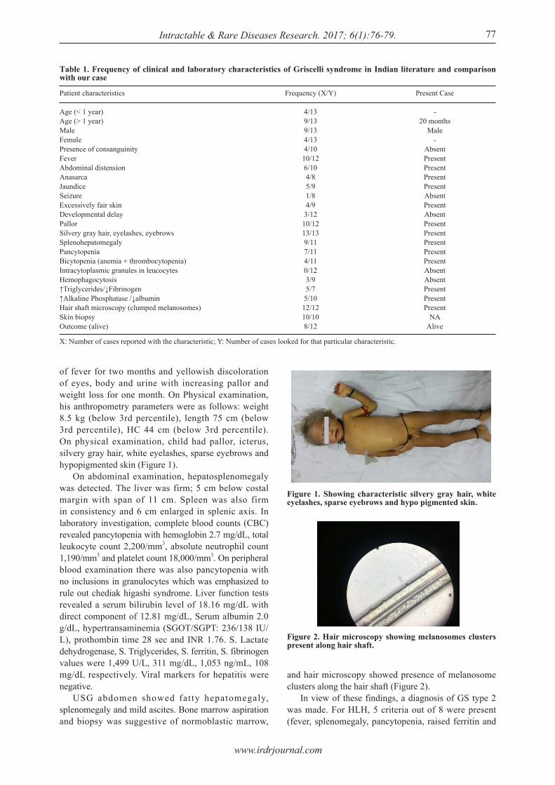
www.irdrjournal.com
Intractable & Rare Diseases Research. 2017; 6(1):76-79.
of fever for two months and yellowish discoloration of eyes, body and urine with increasing pallor and weight loss for one month. On Physical examination, his anthropometry parameters were as follows: weight 8.5 kg (below 3rd percentile), length 75 cm (below 3rd percentile), HC 44 cm (below 3rd percentile). On physical examination, child had pallor, icterus, silvery gray hair, white eyelashes, sparse eyebrows and hypopigmented skin (Figure 1). On abdominal examination, hepatosplenomegaly was detected. The liver was firm; 5 cm below costal margin with span of 11 cm. Spleen was also firm in consistency and 6 cm enlarged in splenic axis. In laboratory investigation, complete blood counts (CBC) revealed pancytopenia with hemoglobin 2.7 mg/dL, total leukocyte count 2,200/mm3, absolute neutrophil count 1,190/mm3 and platelet count 18,000/mm3. On peripheral blood examination there was also pancytopenia with no inclusions in granulocytes which was emphasized to rule out chediak higashi syndrome. Liver function tests revealed a serum bilirubin level of 18.16 mg/dL with direct component of 12.81 mg/dL, Serum albumin 2.0 g/dL, hypertransaminemia (SGOT/SGPT: 236/138 IU/L), prothombin time 28 sec and INR 1.76. S. Lactate dehydrogenase, S. Triglycerides, S. ferritin, S. fibrinogen values were 1,499 U/L, 311 mg/dL, 1,053 ng/mL, 108 mg/dL respectively. Viral markers for hepatitis were negative. USG abdomen showed fatty hepatomegaly, splenomegaly and mild ascites. Bone marrow aspiration and biopsy was suggestive of normoblastic marrow,
and hair microscopy showed presence of melanosome clusters along the hair shaft (Figure 2). In view of these findings, a diagnosis of GS type 2 was made. For HLH, 5 criteria out of 8 were present (fever, splenomegaly, pancytopenia, raised ferritin and
77
Table 1. Frequency of clinical and laboratory characteristics of Griscelli syndrome in Indian literature and comparison with our case
Patient characteristics
Age (< 1 year)Age (> 1 year)MaleFemalePresence of consanguinityFeverAbdominal distensionAnasarcaJaundiceSeizureExcessively fair skinDevelopmental delayPallorSilvery gray hair, eyelashes, eyebrowsSplenohepatomegalyPancytopeniaBicytopenia (anemia + thrombocytopenia)Intracytoplasmic granules in leucocytesHemophagocytosis↑Triglycerides/↓Fibrinogen↑Alkaline Phosphatase /↓albuminHair shaft microscopy (clumped melanosomes)Skin biopsyOutcome (alive)
Frequency (X/Y)
4/139/139/134/134/1010/126/104/85/91/84/93/1210/1213/139/117/114/110/123/95/75/1012/1210/108/12
Present Case
-20 months
Male-
AbsentPresentPresentPresentPresentAbsentPresentAbsentPresentPresentPresentPresentPresentAbsentAbsentPresentPresentPresent
NAAlive
X: Number of cases reported with the characteristic; Y: Number of cases looked for that particular characteristic.
Figure 1. Showing characteristic silvery gray hair, white eyelashes, sparse eyebrows and hypo pigmented skin.
Figure 2. Hair microscopy showing melanosomes clusters present along hair shaft.

www.irdrjournal.com
Intractable & Rare Diseases Research. 2017; 6(1):76-79.78
the diagnosis of GS-2 with HLH was made. GS-2, the type reported here, is the most common of all three subtypes. There are only 13 case reports from India of GS-2(1-3), one report of GS-3 (9) with report of no cases of GS-1. Table 1 shows the frequency of clinical and laboratory characteristics of these Indian cases and comparison with our case. The features that were consistently present in all the 13 cases reported, were silvery gray hair, eyelashes, eyebrows; clusters of melanosomes on hair microscopy and hypomelanosis with irregular melanin pigmentation in basal melanocytes on light microscopy of skin. Other frequently occurring features were fever, pallor, hepatosplenomegaly and pancytopenia. Our patient manifested with all the clinical features of GS-2 with HLH although skin biopsy could not be done. Only two previously reported cases (2,10) fulfilled 5 out of 8 criteria to be labeled as HLH, along with documenting hemophagocytosis in bone marrow. In one other case (3), lymphohistiocytosis was demonstrated in an enlarged cervical lymph node, although this case did not meet the criteria of HLH as demonstration of hemophagocytosis alone is neither pathognomonic nor diagnostic for HLH. Stem cell transplant is the only curative treatment for HLH associated with GS subtype 2 as these children succumb to recurrent infections and secondary neurological involvement (11). Secondary CNS involvement is caused by the infiltration of lymphocytes and histiocytes as a result of hemophagocytic syndrome. In conclusion, GS is a rare entity, which shares a close clinical spectrum with Chediak-Higashi syndrome and other immunodeficiencies. Often the greatest barrier to a successful outcome is a delay in diagnosis, which is difficult because of the rarity of this syndrome. High index of suspicion about this condition aids in early recognition and foresees the life threatening complications like HLH. Prompt initiation of treatment with bone marrow transplant is essential for the survival of such affected patients.
Acknowledgements
We are grateful to the parents of the patient who provided their informed consent for images and clinical information to be reported in the journal.
References
1. Singh A, Garg A, Kapoor S, Khurana N, Entesarian M, Tesi B. An Indian boy with griscelli syndrome type 2: Case report and review of literature. Indian J Dermatol. 2014; 59:394-397.
2. Mahalingashetti PB, Krishnappa MH, Kalyan PS, Subramanian RA, Padhy S. Griscel l i sndrome: Hemophagocytic lymphohistiocytosis with silvery hair. J Lab Physicians. 2012; 4:129-130.
3. Rajyalakshmi R, Chakrapani RN. Griscelli syndrome type 2: A rare and fatal syndrome in a South Indian boy. Indian
hypertriglyceridemia/hypofibrinogenemia) but bone marrow aspiration cytology did not reveal findings of HLH. Supportive therapy including blood transfusion was given to the child and immunosuppresion therapy (mycophenolate mofetil) was started while bone marrow transplantation was advised. Child is currently under follow up and clinical as well as hematological parameters have improved.
3. Discussion
GS was first described by Griscelli and Siccardi in 1978 (4). GS usually manifests in persons aged 4 months to 4 years, though the youngest reported is 1 month with no sex predilection (5). GS is inherited in an autosomal recessive manner and caused by mutations in one of the three genes. Clinical features of GS depend upon subtype. Hypomelanosis and silvery gray hair are hallmarks of all three GS subtypes. Hypopigmentation of the skin and the hair is accompanied by the presence of large aggregates of pigment in hair shafts and the accumulation of mature melanosomes in melanocytes. GS1 caused by mutations in Myosin-Va gene (MYO5A) presents with neurological involvement without immune dysfunction. GS2 is caused by mutations in the RAB27A gene and is associated with immunological dysfunction without primary neurological impairment. The cytotoxic defect caused by RAB27A mutations is responsible for triggering the hemophagocytic syndrome characterized by acute onset of uncontrolled T-lymphocyte and macrophage activation. Neurological symptoms in GS-2 may be due to infiltration of brain by the activated hematopoietic cells. RAB27A and MYO5A are located at band 15q21 (6,7). The third form of GS, being the least common of all, is caused by mutation in the gene MLPH that encodes melanophilin and phenotype of this form is limited to characteristic hypopigmentation without neurologic or immunologic abnormalities. In our case, genetic analysis could not be performed owing to the poor affordability of the parents. The constellation of symptoms of silvery grey hairs and hepatosplenomegaly raised the possibility of Chediak Higashi and GS as primary diagnosis for this child. Absence of inclusions in granulocytes on peripheral blood examination and hair microscopy findings confirmed the diagnosis of GS and ruled out Chediak Higashi syndrome (8). Consistent with symptoms described in subtype 2, this child also shared features like fever, pallor, jaundice, hepatosplenomegaly, and fulfilled five criteria of HLH - fever, pancytopenia, splenomegaly, raised ferritin (1,053 ng/mL) and hypertriglyceridemia (triglycerides- 311 mg/dL) / hypofibrinogenemia (108 mg/dL). This child did not have any neurological manifestations like seizures, spasticity, or developmental delay, which are usually described in subtype 1. Hence,

www.irdrjournal.com
Intractable & Rare Diseases Research. 2017; 6(1):76-79. 79
J Pathol Microbiol. 2016; 59:113-116.4. Griscelli C, Durandy A, Guy-Grand D, Daguillard F,
Herzog C, Prunieras M. A syndrome associating partial albinism and immunodeficiency. Am J Med. 1978; 65: 691-702.
5. Dotta L, Parolini S, Prandini A, Tabellini G, Antolini M, Kingsmore SF, Badolato R. Clinical, laboratory and molecular signs of immunodeficiency in patients with partial oculo-cutaneous albinism. Orphanet J Rare Dis. 2013; 8:168.
6. Ménasché G, Pastural E, Feldmann J, Certain S, Ersoy F, Dupuis S, Wulffraat N, Bianchi D, Fischer A, Le Deist F, de Saint Basile G. Mutations in RAB27A cause Griscelli syndrome associated with haemophagocytic syndrome. Nat Genet. 2000; 25:173-176.
7. Pastural E, Ersoy F, Yalman N, Wulffraat N, Grillo E, Ozkinay F, Tezcan I, Gediköglu G, Philippe N, Fischer A, de Saint Basile G. Two genes are responsible for Griscelli syndrome at the same 15q21 locus. Genomics. 2000; 63:299-306.
8. Valente NY, Machado MC, Boggio P, Alves AC, Bergonse
FN, Casella E, Vasconcelos DM, Grumach AS, de Oliveira ZN. Polarized light microscopy of hair shafts aids in the differential diagnosis of Chédiak-Higashi and Griscelli-Prunieras syndromes. Clinics (Sao Paulo). 2006; 61:327-332.
9. Khemka P, Kundu R, Niyogi P, Tudu J. A case of silvery hair syndrome: Griscelli syndrome. Indian Journal of Paediatric Dermatology. 2015; 16:72-74.
10. Malhotra AK, Bhaskar G, Nanda M, Kabra M, Singh MK, Ramam M. Griscelli syndrome. J Am Acad Dermatol. 2006; 55:337-340.
11. Cesaro S, Locatelli F, Lanino E, Porta F, Di Maio L, Messina C, Prete A, Ripaldi M, Maximova N, Giorgiani G, Rondelli R, Aricò M, Fagioli F. Hematopoietic stem cell transplantation for hemophagocytic lymphohistiocytosis: A retrospective analysis of data from the Italian Association of Pediatric Hematology Oncology (AIEOP). Haematologica. 2008; 93:1694-1701.
(Received October 22, 2016; Revised December 20, 2016; Accepted December 24, 2016)

Guide for Authors
1. Scope of Articles
Intractable & Rare Diseases Research is an international peer-reviewed journal. Intractable & Rare Diseases Research devotes to publishing the latest and most significant research in intractable and rare diseases. Articles cover all aspects of intractable and rare diseases research such as molecular biology, genetics, clinical diagnosis, prevention and treatment, epidemiology, health economics, health management, medical care system, and social science in order to encourage cooperation and exchange among scientists and clinical researchers.
2. Submission Types
Original Articles should be well-documented, novel, and significant to the field as a whole. An Original Article should be arranged into the following sections: Title page, Abstract, Introduction, Materials and Methods, Results, Discussion, Acknowledgments, and References. Original articles should not exceed 5,000 words in length (excluding references) and should be limited to a maximum of 50 references. Articles may contain a maximum of 10 figures and/or tables.
Brief Reports definitively documenting either experimental results or informative clinical observations will be considered for publication in this category. Brief Reports are not intended for publication of incomplete or preliminary findings. Brief Reports should not exceed 3,000 words in length (excluding references) and should be limited to a maximum of 4 figures and/or tables and 30 references. A Brief Report contains the same sections as an Original Article, but the Results and Discussion sections should be combined.
Reviews should present a full and up-to-date account of recent developments within an area of research. Normally, reviews should not exceed 8,000 words in length (excluding references) and should be limited to a maximum of 100 references. Mini reviews are also accepted.
Policy Forum articles discuss research and policy issues in areas related to life science such as public health, the medical care system, and social science and may address governmental issues at district, national, and international levels of discourse. Policy Forum articles should not exceed 2,000 words in length (excluding references).
Case Reports should be detailed reports of the symptoms, signs, diagnosis, treatment, and follow-up of an individual patient. Case reports may contain a demographic profile of the patient but usually describe an unusual
or novel occurrence. Unreported or unusual side effects or adverse interactions involving medications will also be considered. Case Reports should not exceed 3,000 words in length (excluding references).
News articles should report the latest events in health sciences and medical research from around the world. News should not exceed 500 words in length.
Letters should present considered opinions in response to ar t ic les publ ished in Intractable & Rare Diseases Research in the last 6 months or issues of general interest. Letters should not exceed 800 words in length and may contain a maximum of 10 references.
3. Editorial Policies
Ethics : Intractable & Rare Diseases Research requires that authors of reports of investigations in humans or animals indicate that those studies were formally approved by a relevant ethics committee or review board.
Conflict of Interest : All authors are required to disclose any actual or potential conflict of interest including financial interests or relationships with other people or organizations that might raise questions of bias in the work reported. If no conflict of interest exists for each author, please state "There is no conflict of interest to disclose".
S u b m i s s i o n D e c l a r a t i o n : W h e n a manuscript is considered for submission to Intractable & Rare Diseases Research, the authors should confirm that 1) no part of this manuscript is currently under consideration for publication elsewhere; 2) this manuscript does not contain the same information in whole or in part as manuscripts that have been published, accepted, or are under review elsewhere, except in the form of an abstract, a letter to the editor, or part of a published lecture or academic thesis; 3) authorization for publication has been obtained from the authors' employer or institution; and 4) all contributing authors have agreed to submit this manuscript.
Cover Letter: The manuscript must be accompanied by a cover letter signed by the corresponding author on behalf of all authors. The letter should indicate the basic findings of the work and their significance. The letter should also include a statement affirming that all authors concur with the submission and that the material submitted for publication has not been published previously or is not under consideration for publication elsewhere. The cover letter should be submitted in PDF format. For example of Cover Letter, please visithttp://www.irdrjournal.com/downcentre.php (Download Centre).
C o p y r i g h t : A s i g n e d J O U R N A L PUBLISHING AGREEMENT (JPA) form must be provided by post, fax, or as a scanned file before acceptance of the article. Only forms with a hand-written signature are accepted. This copyright will ensure the widest possible dissemination of information.
A form facilitating transfer of copyright can be downloaded by clicking the appropriate link and can be returned to the e-mail address or fax number noted on the form (Please visit Download Centre). Please note that your manuscript will not proceed to the next step in publication until the JPA Form is received. In addition, if excerpts from other copyrighted works are included, the author(s) must obtain written permission from the copyright owners and credit the source(s) in the article.
Suggested Reviewers: A list of up to 3 reviewers who are qualified to assess the scientific merit of the study is welcomed. Reviewer information including names, affiliations, addresses, and e-mail should be provided at the same time the manuscript is submitted online. Please do not suggest reviewers with known conflicts of interest, including participants or anyone with a stake in the proposed research; anyone from the same institution; former students, advisors, or research collaborators (within the last three years); or close personal contacts. Please note that the Editor-in-Chief may accept one or more of the proposed reviewers or may request a review by other qualified persons.
Language Editing: Manuscripts prepared by authors whose native language is not English should have their work proofread by a native English speaker before submission. If not, this might delay the publication of your manuscript in Intractable & Rare Diseases Research.
The Editing Support Organization can provide English proofreading, Japanese-English translation, and Chinese-English translation services to authors who want to publish in Intractable & Rare Diseases Research and need assistance before submitting a manuscript. Authors can visit this organization directly at http://www.iacmhr.com/iac-eso/support.php?lang=en. IAC-ESO was established to facilitate manuscript preparation by researchers whose native language is not English and to help edit works intended for international academic journals.
4. Manuscript Preparation
Manuscripts should be written in clear, grammatically correct English and submitted as a Microsoft Word file in a single-column format. Manuscripts must be paginated and typed in 12-point Times New Roman font with 24-point line spacing. Please do not embed figures in the text. Abbreviations should be used as little as possible and should be explained at first mention unless the term is a well-known abbreviation (e.g. DNA). Single words should not be abbreviated.
Title page: The title page must include 1) the title of the paper (Please note the title should be short, informative, and contain the major key words); 2) full name(s) and affiliation(s) of the author(s), 3) abbreviated names of the author(s), 4) full name, mailing address, telephone/fax numbers, and e-mail address of the corresponding author; and 5) conflicts of interest (if you have an actual or potential conflict of interest to disclose, it must be included as a footnote on the title page of the
IRDRIntractable & Rare Diseases Research

manuscript; if no conflict of interest exists for each author, please state "There is no conflict of interest to disclose"). Please visit Download Centre and refer to the title page of the manuscript sample.
Abstract: The abstract should briefly state the purpose of the study, methods, main findings, and conclusions. For article types including Original Article, Brief Report, Review, Policy Forum, and Case Report, a one-paragraph abstract consisting of no more than 250 words must be included in the manuscript. For News and Letters, a brief summary of main content in 150 words or fewer should be included in the manuscript. Abbreviations must be kept to a minimum and non-standard abbreviations explained in brackets at first mention. References should be avoided in the abstract. Key words or phrases that do not occur in the title should be included in the Abstract page.
Introduction: The introduction should be a concise statement of the basis for the study and its scientific context.
Materials and Methods: The description should be brief but with sufficient detail to enable others to reproduce the experiments. Procedures that have been published previously should not be described in detail but appropriate references should simply be cited. Only new and significant modifications of previously published procedures require complete description. Names of products and manufacturers with their locations (city and state/country) should be given and sources of animals and cell lines should always be indicated. All clinical investigations must have been conducted in accordance with Declaration of Helsinki principles. All human and animal studies must have been approved by the appropriate institutional review board(s) and a specific declaration of approval must be made within this section.
R e s u l t s : T h e d e s c r i p t i o n o f t h e experimental results should be succinct but in suff ic ient deta i l to a l low the experiments to be analyzed and interpreted by an independent reader. If necessary, subheadings may be used for an orderly presentation. All figures and tables must be referred to in the text.
Discussion: The data should be interpreted concisely without repeating material already presented in the Results section. Speculation is permissible, but it must be well-founded, and discussion of the wider implications of the findings is encouraged. Conclusions derived from the study should be included in this section.
Acknowledgments: All funding sources should be credited in the Acknowledgments section. In addition, people who contributed to the work but who do not meet the criteria for authors should be listed along with their contributions.
References: References should be numbered in the order in which they appear in the text. Citing of unpublished results, personal
communications, conference abstracts, and theses in the reference list is not recommended but these sources may be mentioned in the text. In the reference list, cite the names of all authors when there are fifteen or fewer authors; if there are sixteen or more authors, list the first three followed by et al. Names of journals should be abbreviated in the style used in PubMed. Authors are responsible for the accuracy of the references. Examples are given below:
Example 1 (Sample journal reference): Inagaki Y, Tang W, Zhang L, Du GH, Xu WF, Kokudo N. Novel aminopeptidase N (APN/CD13) inhibitor 24F can suppress invasion of hepatocellular carcinoma cells as well as angiogenesis. Biosci Trends. 2010; 4:56-60.
Example 2 (Sample journal reference with more than 15 authors):Darby S, Hill D, Auvinen A, et al. Radon in homes and risk of lung cancer: Collaborative analysis of individual data from 13 European case-control studies. BMJ. 2005; 330:223. Example 3 (Sample book reference):Shalev AY. Post-traumatic stress disorder: Diagnosis, history and life course. In: Post-traumatic Stress Disorder, Diagnosis, Management and Treatment (Nutt DJ, Davidson JR, Zohar J, eds.). Martin Dunitz, London, UK, 2000; pp. 1-15.
Example 4 (Sample web page reference):World Health Organization. The World Health Report 2008 – primary health care: Now more than ever. http://www.who.int/whr/2008/whr08_en.pdf (accessed September 23, 2010).
Tables: All tables should be prepared in Microsoft Word or Excel and should be arranged at the end of the manuscript after the References section. Please note that tables should not in image format. All tables should have a concise title and should be numbered consecutively with Arabic numerals. If necessary, additional information should be given below the table.
Figure Legend: The figure legend should be typed on a separate page of the main manuscript and should include a short title and explanation. The legend should be concise but comprehensive and should be understood without referring to the text. Symbols used in figures must be explained.
Figure Preparation: All figures should be clear and cited in numerical order in the text. Figures must fit a one- or two-column format on the journal page: 8.3 cm (3.3 in.) wide for a single column, 17.3 cm (6.8 in.) wide for a double column; maximum height: 24.0 cm (9.5 in.). Please make sure that artwork files are in an acceptable format (TIFF or JPEG) at minimum resolution (600 dpi for illustrations, graphs, and annotated artwork, and 300 dpi for micrographs and photographs). Please provide all figures as separate files. Please note that low-resolution images are one of the leading causes of article resubmission and schedule delays. All color figures will be reproduced in full color in the online edition of the journal at no cost to authors.
Units and Symbols: Units and symbols conforming to the International System of Units (SI) should be used for physicochemical quantities. Solidus notation (e.g. mg/kg, mg/mL, mol/mm2/min) should be used. Please refer to the SI Guide www.bipm.org/en/si/ for standard units.
Supplemental data: Supplemental data might be useful for supporting and enhancing your scientific research and Intractable & Rare Diseases Research accepts the submission of these materials which will be only published online alongside the electronic version of your article. Supplemental files (figures, tables, and other text materials) should be prepared according to the above guidelines, numbered in Arabic numerals (e.g., Figure S1, Figure S2, and Table S1, Table S2) and referred to in the text. All figures and tables should have titles and legends. All figure legends, tables and supplemental text materials should be placed at the end of the paper. Please note all of these supplemental data should be provided at the time of initial submission and note that the editors reserve the right to limit the size and length of Supplemental Data.
5. Submission Checklist
The Submission Checklist will be useful during the final checking of a manuscript prior to sending it to Intractable & Rare Diseases Research for review. Please visit Download Centre and download the Submission Checklist file.
6. Online submission
Manuscr ipts should be submit ted to Intractable & Rare Diseases Research online at http://www.irdrjournal.com. The manuscript file should be smaller than 5 MB in size. If for any reason you are unable to submit a file online, please contact the Editorial Office by e-mail at [email protected]
7. Accepted manuscripts
Proofs: Galley proofs in PDF format will be sent to the corresponding author via e-mail. Corrections must be returned to the editor ([email protected]) within 3 working days.
Offprints: Authors will be provided with electronic offprints of their article. Paper offprints can be ordered at prices quoted on the order form that accompanies the proofs.
Page Charge: No page charges will be levied to authord for the publication of their article except for reprints.
(As of February 2013)
Editorial and Head Office:
Pearl City Koishikawa 6032-4-5 Kasuga, Bunkyo-kuTokyo 112-0003, JapanTel: +81-3-5840-9968Fax: +81-3-5840-9969E-mail: [email protected]

JOURNAL PUBLISHING AGREEMENT (JPA)------------------------------------------------------------------------------------------------------------------------------------------Manuscript No.:
Title:
Corresponding Author:------------------------------------------------------------------------------------------------------------------------------------------
The International Advancement Center for Medicine & Health Research Co., Ltd. (IACMHR Co., Ltd.) is pleased to accept the above article for publication in Intractable & Rare Diseases Research. The International Research and Cooperation Association for Bio & Socio-Sciences Advancement (IRCA-BSSA) reserves all rights to the published article. Your written acceptance of this JOURNAL PUBLISHING AGREEMENT is required before the article can be published. Please read this form carefully and sign it if you agree to its terms. The signed JOURNAL PUBLISHING AGREEMENT should be sent to the Intractable & Rare Diseases Research office (Pearl City Koishikawa 603, 2-4-5 Kasuga, Bunkyo-ku, Tokyo 112-0003, Japan; E-mail: [email protected]; Tel: +81-3-5840-9968; Fax: +81-3-5840-9969).
1. Authorship CriteriaAs the corresponding author, I certify on behalf of all of the authors that:1) The article is an original work and does not involve fraud, fabrication, or plagiarism.2) The article has not been published previously and is not currently under consideration for publication elsewhere. If accepted by Intractable & Rare Diseases Research, the article will not be submitted for publication to any other journal.3) The article contains no libelous or other unlawful statements and does not contain any materials that infringes upon individual privacy or proprietary rights or any statutory copyright.4) I have obtained written permission from copyright owners for any excerpts from copyrighted works that are included and have credited the sources in my article.5) All authors have made significant contributions to the study including the conception and design of this work, the analysis of the data, and the writing of the manuscript.6) All authors have reviewed this manuscript and take responsibility for its content and approve its publication. 7) I have informed all of the authors of the terms of this publishing agreement and I am signing on their behalf as their agent.
2. Copyright Transfer AgreementI hereby assign and transfer to IACMHR Co., Ltd. all exclusive rights of copyright ownership to the above work in the journal Intractable & Rare Diseases Research, including but not limited to the right 1) to publish, republish, derivate, distribute, transmit, sell, and otherwise use the work and other related material worldwide, in whole or in part, in all languages, in electronic, printed, or any other forms of media now known or hereafter developed and the right 2) to authorize or license third parties to do any of the above. I understand that these exclusive rights will become the property of IACMHR Co., Ltd., from the date the article is accepted for publication in the journal Intractable & Rare Diseases Research. I also understand that IACMHR Co., Ltd. as a copyright owner has sole authority to license and permit reproductions of the article. I understand that except for copyright, other proprietary rights related to the Work (e.g. patent or other rights to any process or procedure) shall be retained by the authors. To reproduce any text, figures, tables, or illustrations from this Work in future works of their own, the authors must obtain written permission from IACMHR Co., Ltd.; such permission cannot be unreasonably withheld by IACMHR Co., Ltd.
3. Conflict of Interest DisclosureI confirm that all funding sources supporting the work and all institutions or people who contributed to the work but who do not meet the criteria for authors are acknowledged. I also confirm that all commercial affiliations, stock ownership, equity interests, or patent-licensing arrangements that could be considered to pose a financial conflict of interest in connection with the article have been disclosed.---------------------------------------------------------------------------------------------------------------------------------------------------------
Corresponding Author's Name (Signature):
Date:
Intractable & Rare Diseases Research (www.irdrjournal.com)Pearl City Koishikawa 603, 2-4-5 Kasuga, Bunkyo-ku, Tokyo 112-0003, Japan; E-mail: [email protected]; Tel: +81-3-5840-9968; Fax: +81-3-5840-9969
IRDRIntractable & Rare Diseases Research