Diagnostic Parasitology - UQ eSpace
94
Diagnostic Parasitology Guide to Lectures and Practicals Saturday 29 th November 2014 Lecturer: Prof Peter O’Donoghue Tutor: Linda Ly Department of Parasitology School of Chemistry and Molecular Biosciences The University of Queensland
-
Upload
khangminh22 -
Category
Documents
-
view
0 -
download
0
Transcript of Diagnostic Parasitology - UQ eSpace
Diagnostic Parasitology
Guide to Lectures and Practicals
Saturday 29th November 2014
Lecturer: Prof Peter O’Donoghue Tutor: Linda Ly
Department of Parasitology
School of Chemistry and Molecular Biosciences The University of Queensland
2
Table of contents page
Diagnostic Parasitology. . . . . . . . . 3.
Microscopy. . . . . . . . . . . 4.
Parasite Diagnosis Parasite Taxonomy. . . . . . . 5.
Parasite Taxonomy. . . . . . . . . . 7.
Lecture 1: Principles of Diagnoses. . . . . . . 11.
Workshop 1: Interpretation of Diagnostic Tests. . . . . . 20.
Exercise 1:
Exercise 2:
Lecture 2: Working with faeces . . . . . . . 24.
Workshop 2: Coprology . . . . . . . . 31.
a) Faecal floatation
b) Worm egg count
c) Slide sets
Lecture 3: Working with blood. . . . . . . . 42.
Workshop 3: Haematology / Serology. . . . . . . 51.
a) Blood smears
b) Microhaematocrit tube centrifugation
c) Indirect Haemagglutination Test
d) Slide sets
Lecture 4: Working with tissues . . . . . . 64.
Workshop 4: Histopathology . . . . . . 72.
a) Squash preparation
b) Skin scraping digest
c) Slide sets
Taxonomic Classification of Parasites. . . . . . . 81.
3
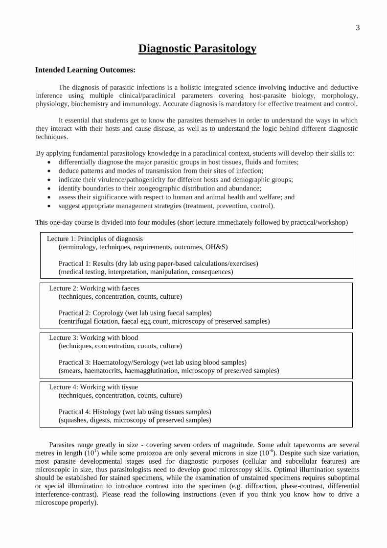
Diagnostic Parasitology Intended Learning Outcomes:
The diagnosis of parasitic infections is a holistic integrated science involving inductive and deductive
inference using multiple clinical/paraclinical parameters covering host-parasite biology, morphology,
physiology, biochemistry and immunology. Accurate diagnosis is mandatory for effective treatment and control.
It essential that students get to know the parasites themselves in order to understand the ways in which
they interact with their hosts and cause disease, as well as to understand the logic behind different diagnostic
techniques.
By applying fundamental parasitology knowledge in a paraclinical context, students will develop their skills to:
differentially diagnose the major parasitic groups in host tissues, fluids and fomites;
deduce patterns and modes of transmission from their sites of infection;
indicate their virulence/pathogenicity for different hosts and demographic groups;
identify boundaries to their zoogeographic distribution and abundance;
assess their significance with respect to human and animal health and welfare; and
suggest appropriate management strategies (treatment, prevention, control).
This one-day course is divided into four modules (short lecture immediately followed by practical/workshop)
Lecture 1: Principles of diagnosis
(terminology, techniques, requirements, outcomes, OH&S)
Practical 1: Results (dry lab using paper-based calculations/exercises)
(medical testing, interpretation, manipulation, consequences)
Lecture 2: Working with faeces
(techniques, concentration, counts, culture)
Practical 2: Coprology (wet lab using faecal samples)
(centrifugal flotation, faecal egg count, microscopy of preserved samples)
Lecture 3: Working with blood
(techniques, concentration, counts, culture)
Practical 3: Haematology/Serology (wet lab using blood samples)
(smears, haematocrits, haemagglutination, microscopy of preserved samples)
Lecture 4: Working with tissue
(techniques, concentration, counts, culture)
Practical 4: Histology (wet lab using tissues samples)
(squashes, digests, microscopy of preserved samples)
Parasites range greatly in size - covering seven orders of magnitude. Some adult tapeworms are several
metres in length (101) while some protozoa are only several microns in size (10
-6). Despite such size variation,
most parasite developmental stages used for diagnostic purposes (cellular and subcellular features) are
microscopic in size, thus parasitologists need to develop good microscopy skills. Optimal illumination systems
should be established for stained specimens, while the examination of unstained specimens requires suboptimal
or special illumination to introduce contrast into the specimen (e.g. diffraction, phase-contrast, differential
interference-contrast). Please read the following instructions (even if you think you know how to drive a
microscope properly).
4
Microscopy
The light microscope is one of the most important tools available in the study of microorganisms. The
amount of information one can get from a light microscope depends not only on the type of microscope, but also
to a large degree on how the microscope is set up and how well it is cared for. You can also see more detail
through the microscope by eye than revealed by photomicroscopy (photos do not do most parasites justice).
When you carry your microscope, always use both hands. Grasp the microscope arm firmly with one
hand and lift it carefully. Place your other hand under the base of the microscope for support as you carry it.
Keep the microscope vertical to ensure that the ocular lens does not fall out. Each time you use your
microscope, clean the optical system (ocular lens, objectives, condenser lens) before and after use. Clean the oil
immersion lens last so that you do not transfer oil onto the other lenses. Use lint-free Kim wipes to clean the
lenses. DO NOT USE FACIAL TISSUES. Avoid touching any of the optical system with your fingers, as skin
oils can be difficult to remove. When using oil immersion, always double check that it is the oil immersion
(100x) lens that you are lowering into the oil. It is clearly identifiable by a black band around its base. The other
objectives are not designed to be used in oil and may be damaged if used in this way. If you accidentally lower
the wrong lens into oil, clean it immediately with Kim wipe tissue. When you have finished with your
microscope for the day and have cleaned it properly, swing the lowest power objective into position. This is to
prevent the other two longer objectives from being accidentally lowered into the condenser.
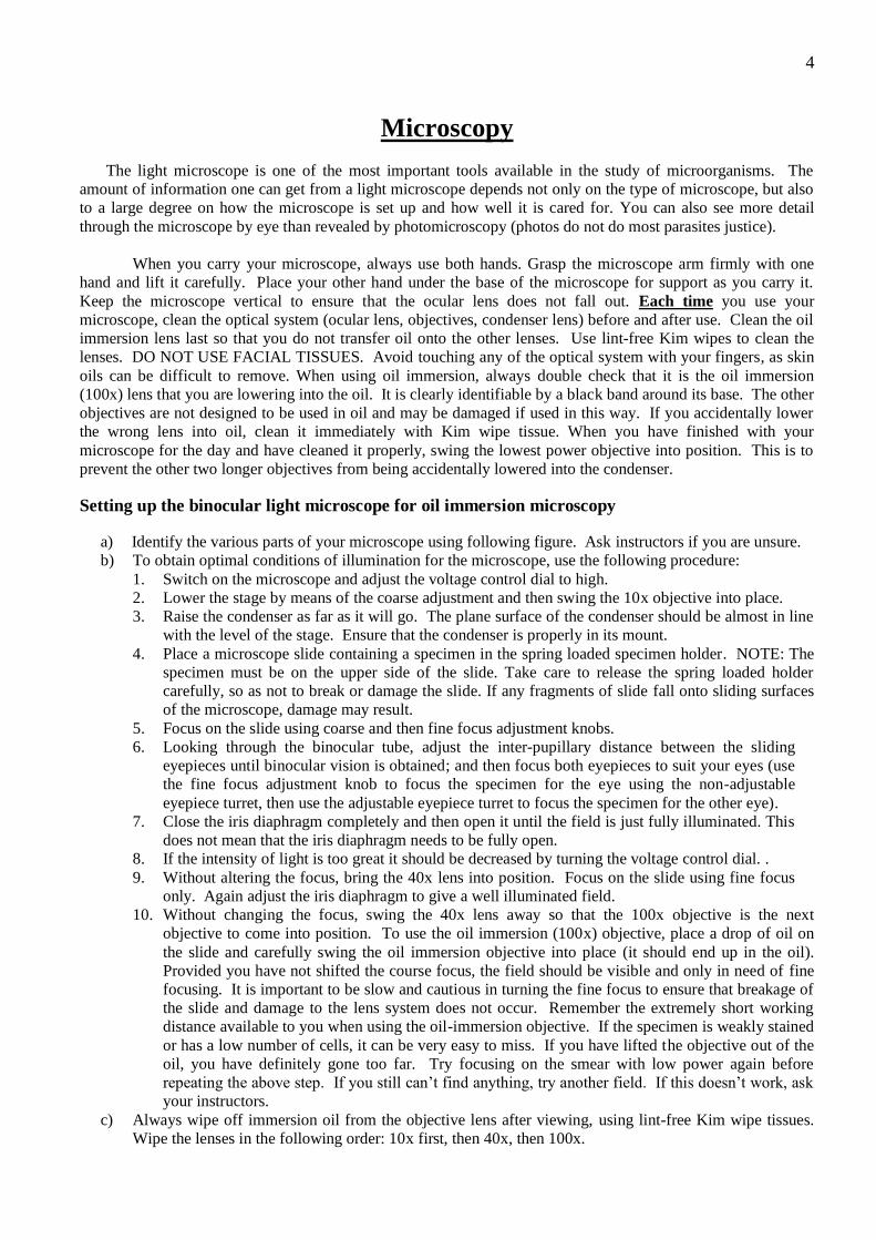
Setting up the binocular light microscope for oil immersion microscopy
a) Identify the various parts of your microscope using following figure. Ask instructors if you are unsure.
b) To obtain optimal conditions of illumination for the microscope, use the following procedure:
1. Switch on the microscope and adjust the voltage control dial to high.
2. Lower the stage by means of the coarse adjustment and then swing the 10x objective into place.
3. Raise the condenser as far as it will go. The plane surface of the condenser should be almost in line
with the level of the stage. Ensure that the condenser is properly in its mount.
4. Place a microscope slide containing a specimen in the spring loaded specimen holder. NOTE: The
specimen must be on the upper side of the slide. Take care to release the spring loaded holder
carefully, so as not to break or damage the slide. If any fragments of slide fall onto sliding surfaces
of the microscope, damage may result.
5. Focus on the slide using coarse and then fine focus adjustment knobs.
6. Looking through the binocular tube, adjust the inter-pupillary distance between the sliding
eyepieces until binocular vision is obtained; and then focus both eyepieces to suit your eyes (use
the fine focus adjustment knob to focus the specimen for the eye using the non-adjustable
eyepiece turret, then use the adjustable eyepiece turret to focus the specimen for the other eye).
7. Close the iris diaphragm completely and then open it until the field is just fully illuminated. This
does not mean that the iris diaphragm needs to be fully open.
8. If the intensity of light is too great it should be decreased by turning the voltage control dial. .
9. Without altering the focus, bring the 40x lens into position. Focus on the slide using fine focus
only. Again adjust the iris diaphragm to give a well illuminated field.
10. Without changing the focus, swing the 40x lens away so that the 100x objective is the next
objective to come into position. To use the oil immersion (100x) objective, place a drop of oil on
the slide and carefully swing the oil immersion objective into place (it should end up in the oil).
Provided you have not shifted the course focus, the field should be visible and only in need of fine
focusing. It is important to be slow and cautious in turning the fine focus to ensure that breakage of
the slide and damage to the lens system does not occur. Remember the extremely short working
distance available to you when using the oil-immersion objective. If the specimen is weakly stained
or has a low number of cells, it can be very easy to miss. If you have lifted the objective out of the
oil, you have definitely gone too far. Try focusing on the smear with low power again before
repeating the above step. If you still can’t find anything, try another field. If this doesn’t work, ask
your instructors.
c) Always wipe off immersion oil from the objective lens after viewing, using lint-free Kim wipe tissues.
Wipe the lenses in the following order: 10x first, then 40x, then 100x.
5
Eyepiece: View specimens through the 10X eye-pieces after you have adjusted their distance apart for your eyes
using the knurled dovetail slides and focussed both lenses for binocular vision using the diopter adjustment ring.
Objective lens: You can change the power of magnification by using different objective lenses fitted onto the
revolving nosepiece. Most microscopes are fitted with 10x, 20x, 40x and 100x objective lenses (giving final
magnification of 100x, 200x, 400x and 1,000x).
Condenser: This is a system of lenses whose function is to concentrate light on the object. It contains an iris
diaphragm which can be opened or closed to vary the amount of illumination.
Variable light control: This rotating or slide control permits adjustment of brightness.
Stage: This flat plate supports the microscope slide containing the specimen. The slide is held in place by a
spring-loaded lever and the whole stage is moved by means of two rotating knobs.
Course focus: This is a large serrated screw which is attached to the upper part of the limb. Its function is to
alter the distance between the object under examination and the objective. If stiff, report it to a tutor.
Fine focus: This is a smaller serrated screw which is attached to the limb. Its function is to finely adjust the
distance between the object under examination and the objective.
6
PARASITE DIAGNOSIS PARASITE TAXONOMY
Diagnostic parasitology seeks to identify the aetiological (causative) agent of disease thus enabling
appropriate management (treatment/control) options. Diagnosis is based on a sound working knowledge of the
taxonomy of living organisms, but the identification/characterization/classification of individual parasite taxa
(species/strains/genotypes) is not the primary aim. Often, the identity of the actual parasite species is not
required by the clinician, e.g. knowledge that gastro-intestinal nematodes have been implicated as the cause of
neonatal scours may be sufficient to commence treatment with an anthelmintic.
Diagnostic parasitology is not a dry esoteric exercise conducted by technicians/scientists in remote
laboratories, but rather a detailed reasoning exercise conducted by the clinician who interacts with the patient
and is ideally cognizant of most relevant facts and uses a variety of technologies and support services to gain
further knowledge to make a differential diagnosis.
While the mental processes involved in clinical reasoning and differential diagnosis are difficult to
categorize (involving many cognitive and metacognitive processes), there are certain things that are obviously
known to the clinician which can be used to make some common sense predictions.
The clinician should always know the host species involved - medical practitioners should recognize their
patients as humans, and veterinary practitioners should be able to identify the animal species they are
working on. This simple information already allows to clinician to consciously/subconsciously access
information on host specificity, host range, host distribution, host susceptiblity/resistance, etc.
The clinician should be able to determine why the patient has presented – symptoms can be described, signs
can be observed. This allows an assessment of how serious and urgent the situation may be, as well as helps
to pin-point the tissue/organ systems involved. Most parasites have predilection sites of infection and
exhibit tissue trophism and this information helps to facilitate diagnosis, to assess disease development and
progression, and to predict possible outcome (prognosis).
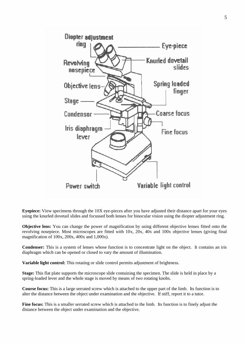
Samples can then be collected for further examination, and it is a poor clinician who does not know that
they are up to their armpits in faeces, urine, vomitus, sputum, blood, tissue aspirates, tissue biopsies, etc. In
selecting specific samples, the clinician is seeking further information to help rule in or rule out certain
aetiological agents. It is also up to the clinician to request the most appropriate tests to be conducted (e.g.
full blood count, differential, haematocrit, liver enzymes, worm egg count, etc.).
However self-evident, all of this host-parasite information helps in the clinical reasoning process so that the
clinician can make a working diagnosis to begin disease management. In addition to providing detailed
information about the specific case in hand, it also allows the clinician to predict the mode of transmission of the
parasite so that preventive measures can be implemented to avoid further cases. Clinicians not only have a
responsibility for their individual patients, but also a broader responsibility for the general community to identify
disease clusters, prevent outbreaks, recommend control strategies, etc.
7
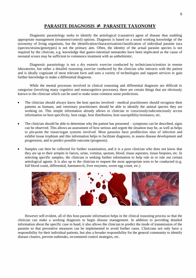
As a broad generalization, there are three major integrated (common-sense) concepts:
THERE ARE THREE MAIN SITES OF INFECTION!
THERE ARE THREE MAIN MODES OF TRANSMISSION!
THERE ARE THREE MAIN TYPES OF PARASITIC DISEASE!
faecal-oral vector-borne predator-prey
transmission transmission transmission
Knowledge of the site of infection helps explain the clinical signs and predicts the mode of transmission:
e.g. many parasites in the gut cause enteritis and diarrhoea and are transmitted by faecal-oral route
many parasites in the circulation cause fever and anaemia and are vector-borne
many parasites in the viscera cause lesions and organ malfunction and are transmitted by carnivorism
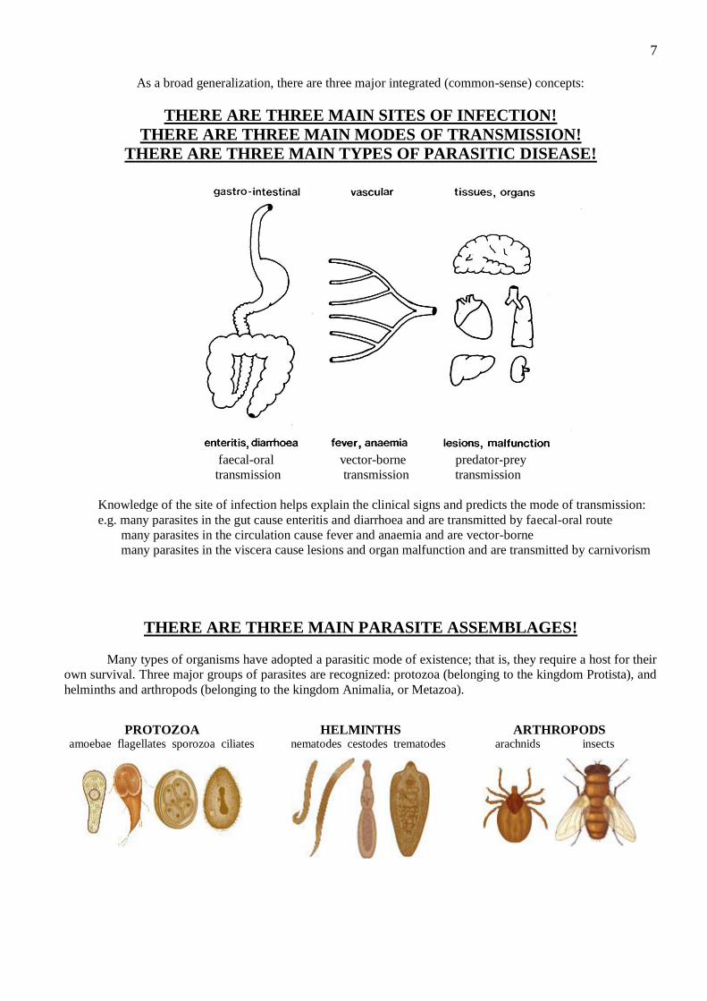
THERE ARE THREE MAIN PARASITE ASSEMBLAGES!
Many types of organisms have adopted a parasitic mode of existence; that is, they require a host for their
own survival. Three major groups of parasites are recognized: protozoa (belonging to the kingdom Protista), and
helminths and arthropods (belonging to the kingdom Animalia, or Metazoa).
PROTOZOA
amoebae flagellates sporozoa ciliates
HELMINTHS nematodes cestodes trematodes
ARTHROPODS arachnids insects
8
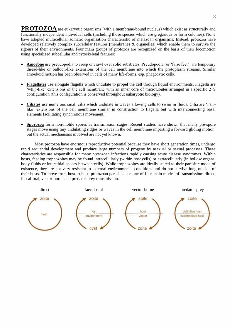
PROTOZOA are eukaryotic organisms (with a membrane-bound nucleus) which exist as structurally and
functionally independent individual cells (including those species which are gregarious or form colonies). None
have adopted multicellular somatic organisation characteristic of metazoan organisms. Instead, protozoa have
developed relatively complex subcellular features (membranes & organelles) which enable them to survive the
rigours of their environments. Four main groups of protozoa are recognized on the basis of their locomotion
using specialized subcellular and cytoskeletal features:
Amoebae use pseudopodia to creep or crawl over solid substrates. Pseudopodia (or ‘false feet’) are temporary
thread-like or balloon-like extensions of the cell membrane into which the protoplasm streams. Similar
amoeboid motion has been observed in cells of many life-forms, esp. phagocytic cells.
Flagellates use elongate flagella which undulate to propel the cell through liquid environments. Flagella are
‘whip-like’ extensions of the cell membrane with an inner core of microtubules arranged in a specific 2+9
configuration (this configuration is conserved throughout eukaryotic biology).
Ciliates use numerous small cilia which undulate in waves allowing cells to swim in fluids. Cilia are ‘hair-
like’ extensions of the cell membrane similar in construction to flagella but with interconnecting basal
elements facilitating synchronous movement.
Sporozoa form non-motile spores as transmission stages. Recent studies have shown that many pre-spore
stages move using tiny undulating ridges or waves in the cell membrane imparting a forward gliding motion,
but the actual mechanisms involved are not yet known.
Most protozoa have enormous reproductive potential because they have short generation times, undergo
rapid sequential development and produce large numbers of progeny by asexual or sexual processes. These
characteristics are responsible for many protozoan infections rapidly causing acute disease syndromes. Within
hosts, feeding trophozoites may be found intracellularly (within host cells) or extracellularly (in hollow organs,
body fluids or interstitial spaces between cells). While trophozoites are ideally suited to their parasitic mode of
existence, they are not very resistant to external environmental conditions and do not survive long outside of
their hosts. To move from host-to-host, protozoan parasites use one of four main modes of transmission: direct,
faecal-oral, vector-borne and predator-prey transmission.
direct
faecal-oral
vector-borne
predator-prey
9
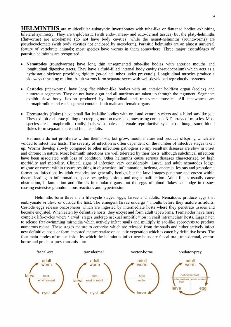
HELMINTHS are multicellular eukaryotic invertebrates with tube-like or flattened bodies exhibiting
bilateral symmetry. They are triploblastic (with endo-, meso- and ecto-dermal tissues) but the platy-helminths
(flatworms) are acoelomate (do not have body cavities) while the nemat-helminths (roundworms) are
pseudocoelomate (with body cavities not enclosed by mesoderm). Parasitic helminths are an almost universal
feature of vertebrate animals; most species have worms in them somewhere. Three major assemblages of
parasitic helminths are recognized:
Nematodes (roundworms) have long thin unsegmented tube-like bodies with anterior mouths and
longitudinal digestive tracts. They have a fluid-filled internal body cavity (pseudocoelum) which acts as a
hydrostatic skeleton providing rigidity (so-called ‘tubes under pressure’). Longitudinal muscles produce a
sideways thrashing motion. Adult worms form separate sexes with well-developed reproductive systems.
Cestodes (tapeworms) have long flat ribbon-like bodies with an anterior holdfast organ (scolex) and
numerous segments. They do not have a gut and all nutrients are taken up through the tegument. Segments
exhibit slow body flexion produced by longitudinal and transverse muscles. All tapeworms are
hermaphroditic and each segment contains both male and female organs.
Trematodes (flukes) have small flat leaf-like bodies with oral and ventral suckers and a blind sac-like gut.
They exhibit elaborate gliding or creeping motion over substrates using compact 3-D arrays of muscles. Most
species are hermaphroditic (individuals with male and female reproductive systems) although some blood
flukes form separate male and female adults.
Helminths do not proliferate within their hosts, but grow, moult, mature and produce offspring which are
voided to infect new hosts. The severity of infection is often dependent on the number of infective stages taken
up. Worms develop slowly compared to other infectious pathogens so any resultant diseases are slow in onset
and chronic in nature. Most helminth infections are well tolerated by their hosts, although subclinical infections
have been associated with loss of condition. Other helminths cause serious diseases characterized by high
morbidity and mortality. Clinical signs of infection vary considerably. Larval and adult nematodes lodge,
migrate or encyst within tissues resulting in obstruction, inflammation, oedema, anaemia, lesions and granuloma
formation. Infections by adult cestodes are generally benign, but the larval stages penetrate and encyst within
tissues leading to inflammation, space-occupying lesions and organ malfunction. Adult flukes usually cause
obstruction, inflammation and fibrosis in tubular organs, but the eggs of blood flukes can lodge in tissues
causing extensive granulomatous reactions and hypertension.
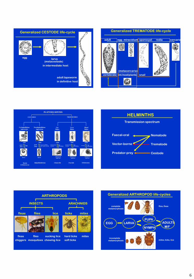
Helminths form three main life-cycle stages: eggs, larvae and adults. Nematodes produce eggs that
embryonate in utero or outside the host. The emergent larvae undergo 4 moults before they mature as adults.
Cestode eggs release oncospheres which are ingested by intermediate hosts where they penetrate tissues and
become encysted. When eaten by definitive hosts, they excyst and form adult tapeworms. Trematodes have more
complex life-cycles where ‘larval’ stages undergo asexual amplification in snail intermediate hosts. Eggs hatch
to release free-swimming miracidia which actively infect snails and multiply in sac-like sporocysts to produce
numerous rediae. These stages mature to cercariae which are released from the snails and either actively infect
new definitive hosts or form encysted metacercariae on aquatic vegetation which is eaten by definitive hosts. The
four main modes of transmission by which the helminths infect new hosts are faecal-oral, transdermal, vector-
borne and predator-prey transmission:
faecal-oral
transdermal
vector-borne
predator-prey
10
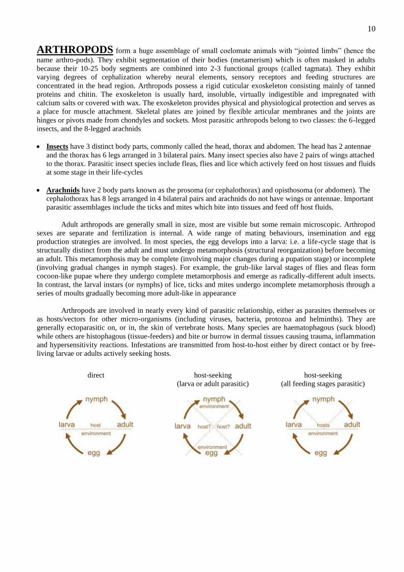
ARTHROPODS form a huge assemblage of small coelomate animals with “jointed limbs” (hence the
name arthro-pods). They exhibit segmentation of their bodies (metamerism) which is often masked in adults
because their 10-25 body segments are combined into 2-3 functional groups (called tagmata). They exhibit
varying degrees of cephalization whereby neural elements, sensory receptors and feeding structures are
concentrated in the head region. Arthropods possess a rigid cuticular exoskeleton consisting mainly of tanned
proteins and chitin. The exoskeleton is usually hard, insoluble, virtually indigestible and impregnated with
calcium salts or covered with wax. The exoskeleton provides physical and physiological protection and serves as
a place for muscle attachment. Skeletal plates are joined by flexible articular membranes and the joints are
hinges or pivots made from chondyles and sockets. Most parasitic arthropods belong to two classes: the 6-legged
insects, and the 8-legged arachnids
Insects have 3 distinct body parts, commonly called the head, thorax and abdomen. The head has 2 antennae
and the thorax has 6 legs arranged in 3 bilateral pairs. Many insect species also have 2 pairs of wings attached
to the thorax. Parasitic insect species include fleas, flies and lice which actively feed on host tissues and fluids
at some stage in their life-cycles
Arachnids have 2 body parts known as the prosoma (or cephalothorax) and opisthosoma (or abdomen). The
cephalothorax has 8 legs arranged in 4 bilateral pairs and arachnids do not have wings or antennae. Important
parasitic assemblages include the ticks and mites which bite into tissues and feed off host fluids.
Adult arthropods are generally small in size, most are visible but some remain microscopic. Arthropod
sexes are separate and fertilization is internal. A wide range of mating behaviours, insemination and egg
production strategies are involved. In most species, the egg develops into a larva: i.e. a life-cycle stage that is
structurally distinct from the adult and must undergo metamorphosis (structural reorganization) before becoming
an adult. This metamorphosis may be complete (involving major changes during a pupation stage) or incomplete
(involving gradual changes in nymph stages). For example, the grub-like larval stages of flies and fleas form
cocoon-like pupae where they undergo complete metamorphosis and emerge as radically-different adult insects.
In contrast, the larval instars (or nymphs) of lice, ticks and mites undergo incomplete metamorphosis through a
series of moults gradually becoming more adult-like in appearance
Arthropods are involved in nearly every kind of parasitic relationship, either as parasites themselves or
as hosts/vectors for other micro-organisms (including viruses, bacteria, protozoa and helminths). They are
generally ectoparasitic on, or in, the skin of vertebrate hosts. Many species are haematophagous (suck blood)
while others are histophagous (tissue-feeders) and bite or burrow in dermal tissues causing trauma, inflammation
and hypersensitivity reactions. Infestations are transmitted from host-to-host either by direct contact or by free-
living larvae or adults actively seeking hosts.
direct
host-seeking
(larva or adult parasitic)
host-seeking
(all feeding stages parasitic)
1
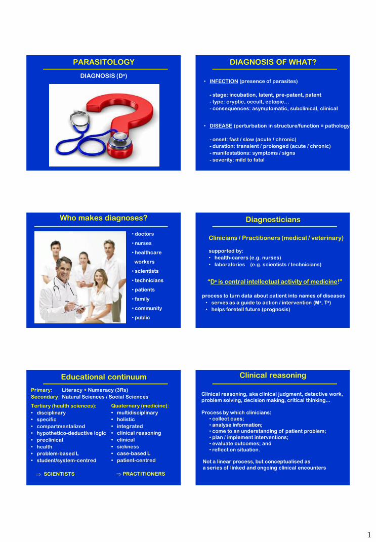
PARASITOLOGY
DIAGNOSIS (Dx)
DIAGNOSIS OF WHAT?
• INFECTION (presence of parasites)
- stage: incubation, latent, pre-patent, patent
- type: cryptic, occult, ectopic…
- consequences: asymptomatic, subclinical, clinical
• DISEASE (perturbation in structure/function = pathology)
- onset: fast / slow (acute / chronic)
- duration: transient / prolonged (acute / chronic)
- manifestations: symptoms / signs
- severity: mild to fatal
Who makes diagnoses?
• doctors
• nurses
• healthcare
workers
• scientists
• technicians
• patients
• family
• community
• public
Diagnosticians
Clinicians / Practitioners (medical / veterinary)
supported by:
• health-carers (e.g. nurses)
• laboratories (e.g. scientists / technicians)
“Dx is central intellectual activity of medicine!”
process to turn data about patient into names of diseases
• serves as a guide to action / intervention (Mx, Tx)
• helps foretell future (prognosis)
Educational continuum
Tertiary (health sciences):
• disciplinary
• specific
• compartmentalized
• hypothetico-deductive logic
• preclinical
• health
• problem-based L
• student/system-centred
SCIENTISTS
Primary: Literacy + Numeracy (3Rs)
Secondary: Natural Sciences / Social Sciences
Quaternary (medicine):
• multidisciplinary
• holistic
• integrated
• clinical reasoning
• clinical
• sickness
• case-based L
• patient-centred
PRACTITIONERS
Not a linear process, but conceptualised as
a series of linked and ongoing clinical encounters
Clinical reasoning, aka clinical judgment, detective work,
problem solving, decision making, critical thinking...
Clinical reasoning
Process by which clinicians:
• collect cues;
• analyse information;
• come to an understanding of patient problem;
• plan / implement interventions;
• evaluate outcomes; and
• reflect on situation.
2
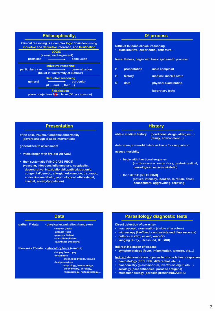
Philosophically,
Clinical reasoning is a complex logic chain/loop using
inductive and deductive inference, and falsification
LOGIC
(= reasoned argument)
premises conclusion
Inductive reasoning
particular case generalization
(belief in ‘uniformity of Nature’)
Deductive reasoning
general particular
(if ... and ..., then ...)
Falsification
prove conjecture true / false (Dx by exclusion) x
Dx process
Difficult to teach clinical reasoning
• quite intuitive, experiential, reflective…
Nevertheless, begin with basic systematic process:
P presentation - main complaint
H history - medical, morbid state
D data - physical examination
- laboratory tests
Presentation
often pain, trauma, functional abnormality
(severe enough to seek intervention)
general health assessment
• vitals (begin with firs-aid DR ABC)
• then systematic [VINDICATE PECS]
(vascular, infectious/inflammatory, neoplastic,
degenerative, intoxication/idiopathic/iatrogenic,
congenital/genetic, allergic/autoimmune, traumatic,
endocrine/metabolic, psychological, ethico-legal,
clinical, society/population)
History
obtain medical history (conditions, drugs, allergies…)
(family, environment…)
assess morbidity
• begin with functional enquiries
(cardiovascular, respiratory, gastrointestinal,
neurological, musculoskeletal)
• then details [NILDOCAR]
(nature, intensity, location, duration, onset,
concomitant, aggravating, relieving)
determine pre-morbid state as basis for comparison
Data
gather 10 data - physical examination (hands-on)
- inspect (look)
- palpate (feel)
- percuss (listen)
- auscultate (listen)
- quantitate (measure)
then seek 20 data - laboratory tests (remote)
- biopsy / necropsy
- test matrix
- stool, blood/fluids, tissues
- test procedure
- coprology, haematology,
biochemistry, serology,
microbiology, histopathology…
Parasitology diagnostic tests
Direct detection of parasites
• macroscopic examination (visible characters)
• microscopy (live/fixed, contrast/stained, fluorescence)
• culture (in vitro, in vivo, xeno-Dx)
• imaging (X-ray, ultrasound, CT, MRI)
Indirect demonstration of parasite products/host responses
• haematology (FBC, ESR, differential, etc…)
• biochemistry (plasma/serum, liver/muscle/gut, etc…)
• serology (host antibodies, parasite antigens)
• molecular biology (parasite proteins/DNA/RNA)
Indirect indication of disease
• symptomatology (fever, inflammation, wheeze, etc…)
3
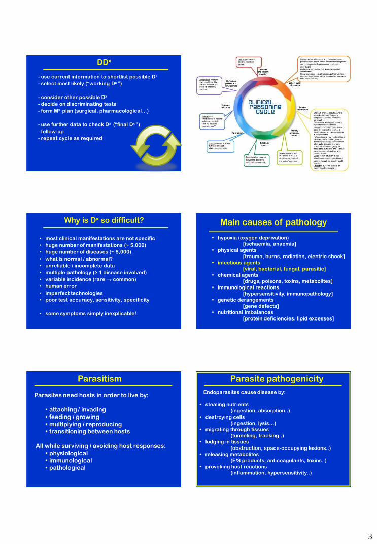
DDx
- use current information to shortlist possible Dx
- select most likely (“working Dx ”)
- consider other possible Dx
- decide on discriminating tests
- form Mx plan (surgical, pharmacological…)
- use further data to check Dx (“final Dx ”)
- follow-up
- repeat cycle as required
Why is Dx so difficult?
• most clinical manifestations are not specific
• huge number of manifestations (~ 5,000)
• huge number of diseases (~ 5,000)
• what is normal / abnormal?
• unreliable / incomplete data
• multiple pathology (> 1 disease involved)
• variable incidence (rare common)
• human error
• imperfect technologies
• poor test accuracy, sensitivity, specificity
• some symptoms simply inexplicable!
Main causes of pathology
• hypoxia (oxygen deprivation)
[ischaemia, anaemia]
• physical agents
[trauma, burns, radiation, electric shock]
• infectious agents
[viral, bacterial, fungal, parasitic]
• chemical agents
[drugs, poisons, toxins, metabolites]
• immunological reactions
[hypersensitivity, immunopathology]
• genetic derangements
[gene defects]
• nutritional imbalances
[protein deficiencies, lipid excesses]
Parasitism
Parasites need hosts in order to live by:
• attaching / invading
• feeding / growing
• multiplying / reproducing
• transitioning between hosts
All while surviving / avoiding host responses:
• physiological
• immunological
• pathological
Parasite pathogenicity
Endoparasites cause disease by:
• stealing nutrients
(ingestion, absorption..)
• destroying cells
(ingestion, lysis…)
• migrating through tissues
(tunneling, tracking..)
• lodging in tissues
(obstruction, space-occupying lesions..)
• releasing metabolites
(E/S products, anticoagulants, toxins..)
• provoking host reactions
(inflammation, hypersensitivity..)
4
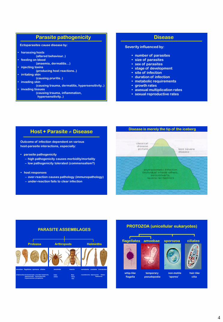
Parasite pathogenicity
Ectoparasites cause disease by:
• harassing hosts
(altered behaviour..)
• feeding on blood
(anaemia, dermatitis…)
• injecting toxins
(producing host reactions..)
• irritating skin
(causing pruritis..)
• invading skin
(causing trauma, dermatitis, hypersensitivity..)
• invading tissues
(causing trauma, inflammation,
hypersensitivity..)
Disease
Severity influenced by:
• number of parasites
• size of parasites
• sex of parasites
• stage of development
• site of infection
• duration of infection
• metabolic requirements
• growth rates
• asexual multiplication rates
• sexual reproductive rates
Host + Parasite Disease
Outcome of infection dependent on various
host-parasite interactions, especially:
• parasite pathogenicity
– high pathogenicity causes morbidity/mortality
– low pathogenicity tolerated (commensalism?)
• host responses
– over-reaction causes pathology (immunopathology)
– under-reaction fails to clear infection
Disease is merely the tip of the iceberg
Protozoa Arthropods Helminths
amoebae flagellates sporozoa ciliates
entamoebae trichomonads coccidia holotrichs diplomonads haemosporidia trypanosomes piroplasms
arachnids insects
ticks flies
mites fleas
lice
nematodes cestodes trematodes
roundworms tapeworms flukes flatworms
PARASITE ASSEMBLAGES PROTOZOA (unicellular eukaryotes)
flagellates amoebae sporozoa ciliates
whip-like temporary non-motile hair-like
flagella pseudopodia ‘spores’ cilia
5
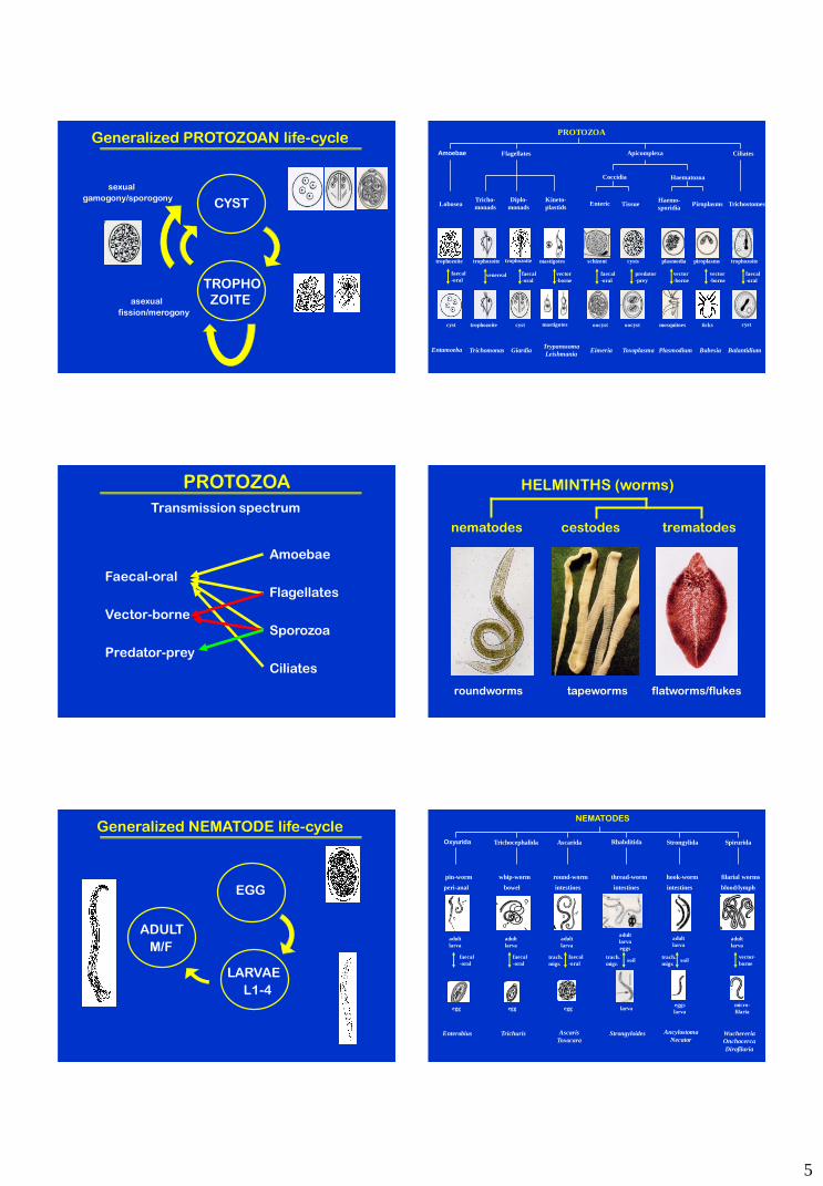
Generalized PROTOZOAN life-cycle
CYST
TROPHO
ZOITE asexual
fission/merogony
sexual
gamogony/sporogony
Amoebae Flagellates Apicomplexa Ciliates
Coccidia Haematozoa
Enteric Tissue Piroplasms Haemo-
sporidia
Tricho-
monads
Diplo-
monads
Kineto-
plastids
PROTOZOA
Entamoeba
P
A T
E
Trichomonas Giardia Trypanosoma
Leishmania Eimeria Toxoplasma Plasmodium Babesia Balantidium
trophozoite trophozoite trophozoite mastigotes schizont cysts trophozoite piroplasms plasmodia
cyst cyst cyst oocyst oocyst mosquitoes ticks trophozoite mastigotes
faecal
-oral
faecal
-oral
faecal
-oral venereal vector
-borne
vector
-borne
vector
-borne
faecal
-oral
predator
-prey
Lobosea Trichostomes
PROTOZOA
Faecal-oral
Vector-borne
Predator-prey
Amoebae
Flagellates
Sporozoa
Ciliates
Transmission spectrum
HELMINTHS (worms)
nematodes cestodes trematodes
roundworms tapeworms flatworms/flukes
Generalized NEMATODE life-cycle
ADULT
M/F
EGG
LARVAE
L1-4
Oxyurida Trichocephalida Rhabditida Spirurida
NEMATODES
Trichuris
adult
larva
egg
faecal
-oral
whip-worm
Ascarida Strongylida
bowel
Enterobius
adult
larva
egg
faecal
-oral
pin-worm
peri-anal
Ascaris
Toxocara
adult
larva
egg
faecal
-oral
round-worm
intestines
Strongyloides
adult
larva
eggs
larva
soil
thread-worm
intestines
Ancylostoma
Necator
adult
larva
eggs
larva
soil
hook-worm
intestines
Wuchereria
Onchocerca
Dirofilaria
adult
larva
micro-
filaria
vector-
borne
filarial worms
blood/lymph
trach.
migr.
trach.
migr.
trach.
migr.
6
larva (metacestode)
in intermediate host
Generalized CESTODE life-cycle
egg
adult tapeworm
in definitive host
(metacercaria)
adult egg miracidium sporocyst redia cercaria
Generalized TREMATODE life-cycle
snail vertebrate int.host/plants
CESTODES TREMATODES
Cyclophyllidean
tapeworms
Pseudophyllidean
tapeworms
Gut
flukes
Liver
flukes
Blood
flukes
PLATYHELMINTHS
Clonorchis Fasciola Schistosoma Taenia
Echinococcus
Diphyllobothrium
adult adult adults adult adult
sporocyst in
snail host larva larva
sporocyst in
snail host
sporocyst in
snail host
egg,
miracidium
metacercaria
cercaria
egg,
miracidium
metacercaria,
cercaria egg,
miracidium cercaria egg pred-
ation
egg, coracidium,
pro-, plero-cercoid
pred-
ation
Faecal-oral
Vector-borne
Predator-prey
Nematode
Trematode
Cestode
HELMINTHS
Transmission spectrum
ARTHROPODS
INSECTS ARACHNIDS
fleas flies lice ticks mites
fleas flies sucking lice hard ticks mites
chiggers mosquitoes chewing lice soft ticks
Generalized ARTHROPOD life-cycles
ADULTS
M/F EGG
PUPA
NYMPH
LARVA
complete
metamorphosis
incomplete
metamorphosis
flies, fleas
mites, ticks, lice
7
INSECTS ARACHNIDS
Mites Ticks Flies Fleas Lice
ARTHROPODS
Lucilia
Culicoides
Anopheles
Pulex
Ctenocephalides Pediculus
Phthirus
Sarcoptes
Chorioptes
Demodex
Ixodes
Rhipicephalus
adult adult adult adult adult
larva larva larva larva nymph
egg
(1-, 2-,3-host)
pupa
(skin-blood) (skin-blood) (skin-blood) (skin-blood) (skin-blood)
C
M
egg pupa C
M
egg nymph I
M
egg nymph I
M
egg nymph I
M
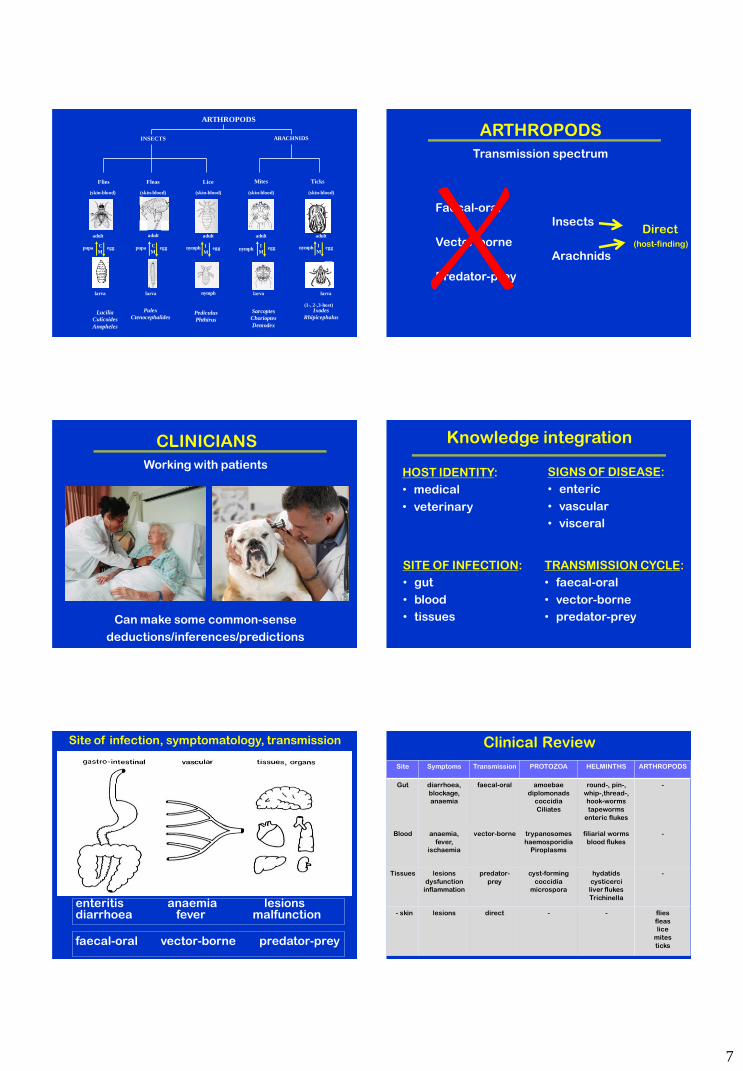
Faecal-oral
Vector-borne
Predator-prey
Insects
Arachnids
Direct (host-finding)
ARTHROPODS
Transmission spectrum
CLINICIANS
Working with patients
Can make some common-sense
deductions/inferences/predictions
Knowledge integration
HOST IDENTITY:
• medical
• veterinary
SITE OF INFECTION:
• gut
• blood
• tissues
SIGNS OF DISEASE:
• enteric
• vascular
• visceral
TRANSMISSION CYCLE:
• faecal-oral
• vector-borne
• predator-prey
Site of infection, symptomatology, transmission
enteritis anaemia lesions diarrhoea fever malfunction
faecal-oral vector-borne predator-prey
Clinical Review
Site Symptoms Transmission PROTOZOA HELMINTHS ARTHROPODS
Gut diarrhoea,
blockage,
anaemia
faecal-oral amoebae
diplomonads
coccidia
Ciliates
round-, pin-,
whip-,thread-,
hook-worms
tapeworms
enteric flukes
-
Blood anaemia,
fever,
ischaemia
vector-borne trypanosomes
haemosporidia
Piroplasms
filiarial worms
blood flukes
-
Tissues lesions
dysfunction
inflammation
predator-
prey
cyst-forming
coccidia
microspora
hydatids
cysticerci
liver flukes
Trichinella
-
- skin lesions
direct - - flies
fleas
lice
mites
ticks
1
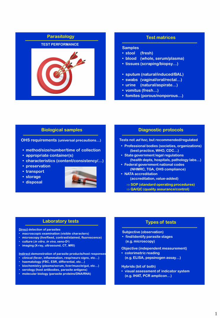
Parasitology
TEST PERFORMANCE
Test matrices
Samples
• stool (fresh)
• blood (whole, serum/plasma)
• tissues (scraping/biopsy…)
• sputum (natural/induced/BAL)
• swabs (vaginal/oral/rectal…)
• urine (natural/aspirate…)
• vomitus (fresh…)
• fomites (porous/nonporous…)
Biological samples
OHS requirements (universal precautions…)
• method/size/number/time of collection
• appropriate container(s)
• characteristics (content/consistency/…)
• preservation
• transport
• storage
• disposal
Diagnostic protocols
Tests not ad hoc, but recommended/regulated
SOP (standard operating procedures)
QA/QC (quality assurance/control)
• Professional bodies (societies, organizations)
(best practice, WHO, CDC…)
• State government legal regulations
(health depts, hospitals, pathology labs…)
• Federal government national codes
(NHMRC, TGA, OHS compliance)
• NATA accreditation
(accreditation, value-added)
Laboratory tests
Direct detection of parasites
• macroscopic examination (visible characters)
• microscopy (live/fixed, contrast/stained, fluorescence)
• culture (in vitro, in vivo, xeno-Dx)
• imaging (X-ray, ultrasound, CT, MRI)
Indirect demonstration of parasite products/host responses
• clinical (fever, inflammation, resp/neuro signs, etc…)
• haematology (FBC, ESR, differential, etc…)
• biochemistry (plasma/serum, liver/muscle/gut, etc…)
• serology (host antibodies, parasite antigens)
• molecular biology (parasite proteins/DNA/RNA)
Types of tests
Subjective (observation)
• find/identify parasite stages
(e.g. microscopy)
Objective (independent measurement)
• colorimetric reading
(e.g. ELISA, pepsinogen assay…)
Hybrids (bit of both)
• visual assessment of indicator system
(e.g. IHAT, PCR amplicon…)
2
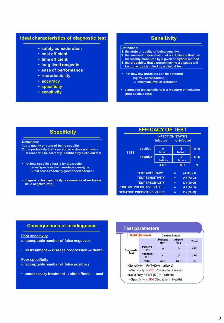
Ideal characteristics of diagnostic test
• safety consideration
• cost efficient
• time efficient
• long-lived reagents
• ease of performance
• reproducibility
• accuracy
• specificity
• sensitivity
Sensitivity
• diagnostic test sensitivity is a measure of inclusion
(true positive rate)
Definitions:
1. the state or quality of being sensitive
2. the smallest concentration of a substance that can
be reliably measured by a given analytical method
3. the probability that a person having a disease will
be correctly identified by a clinical test
• not how few parasites can be detected
(ng/mL, parasitaemia…)
minimum level of detection
Specificity
Definitions:
1. the quality or state of being specific
2. the probability that a person who does not have a
disease will be correctly identified by a clinical test
• not how specific a test is for a parasite
genus/species/strain/serotype/genotype
test cross-reactivity (presence/absence)
• diagnostic test specificity is a measure of exclusion
(true negative rate)
EFFICACY OF TEST
INFECTION STATUS
infected not infected
positive A B
TEST
negative C D
TEST ACCURACY = (A+D) / N
true + false +
false - true -
A+B
C+D
A+C B+D N
TEST SENSITIVITY = A / (A+C)
TEST SPECIFICITY = D / (B+D)
POSITIVE PREDICTIVE VALUE = A / (A+B)
NEGATIVE PREDICTIVE VALUE = D / (C+D)
Consequences of misdiagnosis
Poor sensitivity
unacceptable number of false negatives
• no treatment disease progression death
Poor specificity
unacceptable number of false positives
• unnecessary treatment side effects cost
20
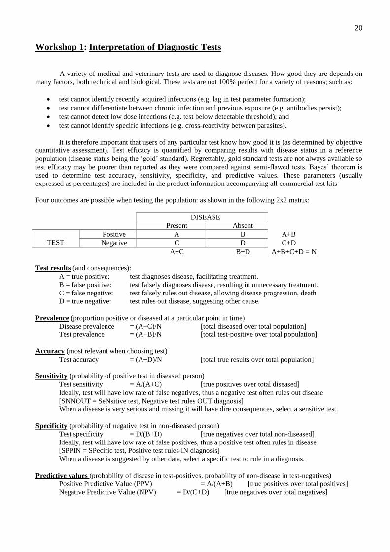
Workshop 1: Interpretation of Diagnostic Tests
A variety of medical and veterinary tests are used to diagnose diseases. How good they are depends on
many factors, both technical and biological. These tests are not 100% perfect for a variety of reasons; such as:
test cannot identify recently acquired infections (e.g. lag in test parameter formation);
test cannot differentiate between chronic infection and previous exposure (e.g. antibodies persist);
test cannot detect low dose infections (e.g. test below detectable threshold); and
test cannot identify specific infections (e.g. cross-reactivity between parasites).
It is therefore important that users of any particular test know how good it is (as determined by objective
quantitative assessment). Test efficacy is quantified by comparing results with disease status in a reference
population (disease status being the ‘gold’ standard). Regrettably, gold standard tests are not always available so
test efficacy may be poorer than reported as they were compared against semi-flawed tests. Bayes’ theorem is
used to determine test accuracy, sensitivity, specificity, and predictive values. These parameters (usually
expressed as percentages) are included in the product information accompanying all commercial test kits
Four outcomes are possible when testing the population: as shown in the following 2x2 matrix:
DISEASE
Present Absent
TEST
Positive A B A+B
Negative C D C+D
A+C B+D A+B+C+D = N
Test results (and consequences):
A = true positive: test diagnoses disease, facilitating treatment.
B = false positive: test falsely diagnoses disease, resulting in unnecessary treatment.
C = false negative: test falsely rules out disease, allowing disease progression, death
D = true negative: test rules out disease, suggesting other cause.
Prevalence (proportion positive or diseased at a particular point in time)
Disease prevalence = (A+C)/N [total diseased over total population]
Test prevalence = (A+B)/N [total test-positive over total population]
Accuracy (most relevant when choosing test)
Test accuracy = (A+D)/N [total true results over total population]
Sensitivity (probability of positive test in diseased person)
Test sensitivity = A/(A+C) [true positives over total diseased]
Ideally, test will have low rate of false negatives, thus a negative test often rules out disease
[SNNOUT = SeNsitive test, Negative test rules OUT diagnosis]
When a disease is very serious and missing it will have dire consequences, select a sensitive test.
Specificity (probability of negative test in non-diseased person)
Test specificity = D/(B+D) [true negatives over total non-diseased]
Ideally, test will have low rate of false positives, thus a positive test often rules in disease
[SPPIN = SPecific test, Positive test rules IN diagnosis]
When a disease is suggested by other data, select a specific test to rule in a diagnosis.
Predictive values (probability of disease in test-positives, probability of non-disease in test-negatives)
Positive Predictive Value (PPV) = A/(A+B) [true positives over total positives]
Negative Predictive Value (NPV) = D/(C+D) [true negatives over total negatives]
21
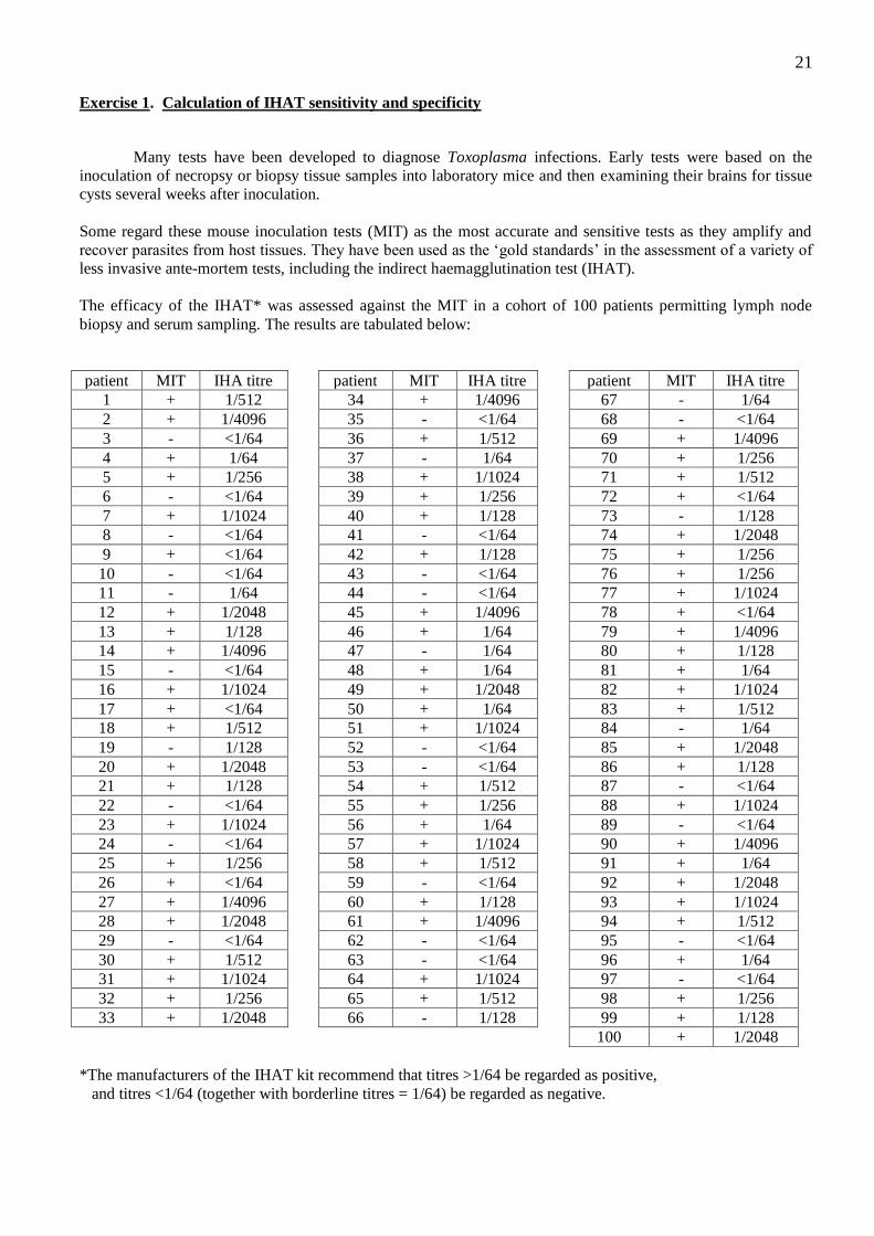
Exercise 1. Calculation of IHAT sensitivity and specificity
Many tests have been developed to diagnose Toxoplasma infections. Early tests were based on the
inoculation of necropsy or biopsy tissue samples into laboratory mice and then examining their brains for tissue
cysts several weeks after inoculation.
Some regard these mouse inoculation tests (MIT) as the most accurate and sensitive tests as they amplify and
recover parasites from host tissues. They have been used as the ‘gold standards’ in the assessment of a variety of
less invasive ante-mortem tests, including the indirect haemagglutination test (IHAT).
The efficacy of the IHAT* was assessed against the MIT in a cohort of 100 patients permitting lymph node
biopsy and serum sampling. The results are tabulated below:
patient MIT IHA titre patient MIT IHA titre patient MIT IHA titre
1 + 1/512 34 + 1/4096 67 - 1/64
2 + 1/4096 35 - <1/64 68 - <1/64
3 - <1/64 36 + 1/512 69 + 1/4096
4 + 1/64 37 - 1/64 70 + 1/256
5 + 1/256 38 + 1/1024 71 + 1/512
6 - <1/64 39 + 1/256 72 + <1/64
7 + 1/1024 40 + 1/128 73 - 1/128
8 - <1/64 41 - <1/64 74 + 1/2048
9 + <1/64 42 + 1/128 75 + 1/256
10 - <1/64 43 - <1/64 76 + 1/256
11 - 1/64 44 - <1/64 77 + 1/1024
12 + 1/2048 45 + 1/4096 78 + <1/64
13 + 1/128 46 + 1/64 79 + 1/4096
14 + 1/4096 47 - 1/64 80 + 1/128
15 - <1/64 48 + 1/64 81 + 1/64
16 + 1/1024 49 + 1/2048 82 + 1/1024
17 + <1/64 50 + 1/64 83 + 1/512
18 + 1/512 51 + 1/1024 84 - 1/64
19 - 1/128 52 - <1/64 85 + 1/2048
20 + 1/2048 53 - <1/64 86 + 1/128
21 + 1/128 54 + 1/512 87 - <1/64
22 - <1/64 55 + 1/256 88 + 1/1024
23 + 1/1024 56 + 1/64 89 - <1/64
24 - <1/64 57 + 1/1024 90 + 1/4096
25 + 1/256 58 + 1/512 91 + 1/64
26 + <1/64 59 - <1/64 92 + 1/2048
27 + 1/4096 60 + 1/128 93 + 1/1024
28 + 1/2048 61 + 1/4096 94 + 1/512
29 - <1/64 62 - <1/64 95 - <1/64
30 + 1/512 63 - <1/64 96 + 1/64
31 + 1/1024 64 + 1/1024 97 - <1/64
32 + 1/256 65 + 1/512 98 + 1/256
33 + 1/2048 66 - 1/128 99 + 1/128
100 + 1/2048
*The manufacturers of the IHAT kit recommend that titres >1/64 be regarded as positive,
and titres <1/64 (together with borderline titres = 1/64) be regarded as negative.
22
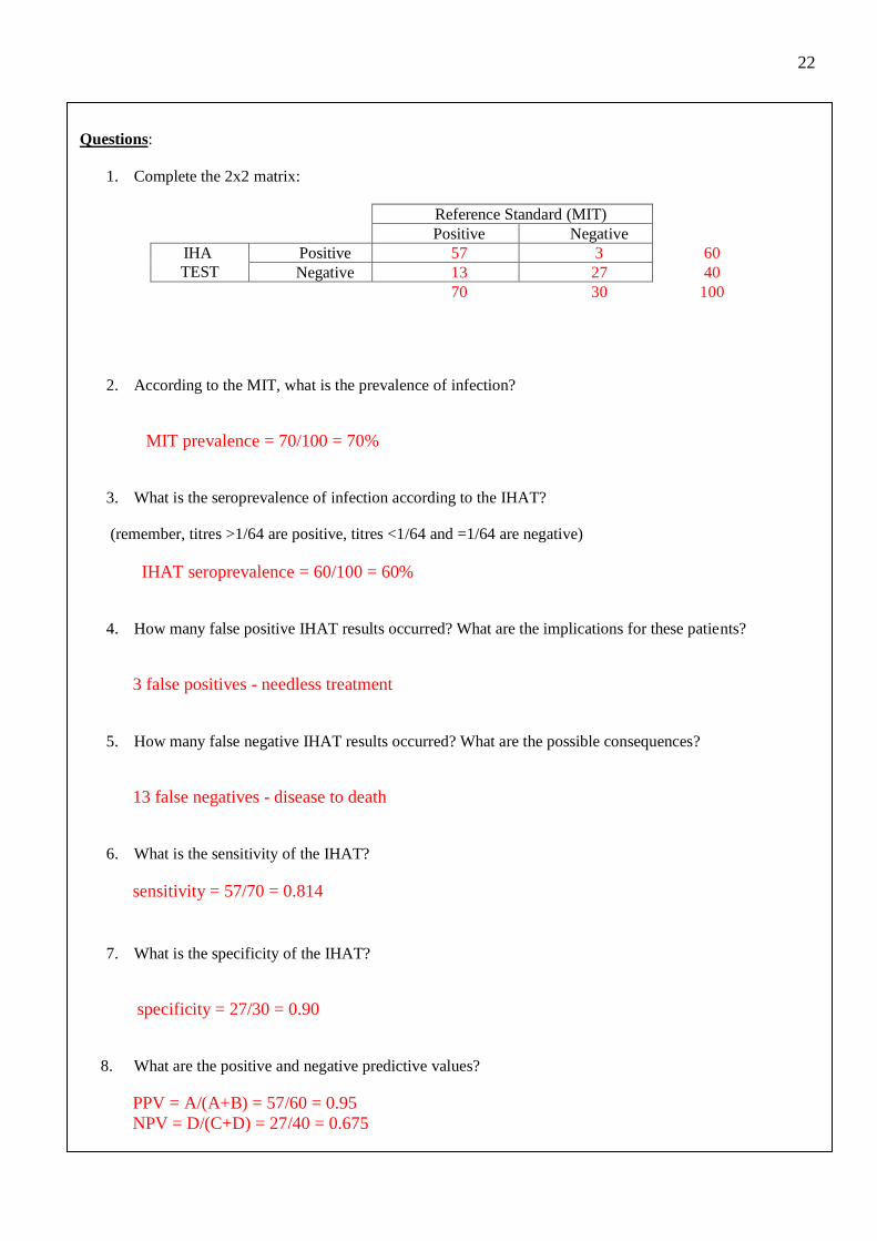
Questions:
1. Complete the 2x2 matrix:
Reference Standard (MIT)
Positive Negative
IHA
TEST
Positive 57 3 60
Negative 13 27 40
70 30 100
2. According to the MIT, what is the prevalence of infection?
MIT prevalence = 70/100 = 70%
3. What is the seroprevalence of infection according to the IHAT?
(remember, titres >1/64 are positive, titres <1/64 and =1/64 are negative)
IHAT seroprevalence = 60/100 = 60%
4. How many false positive IHAT results occurred? What are the implications for these patients?
3 false positives - needless treatment
5. How many false negative IHAT results occurred? What are the possible consequences?
13 false negatives - disease to death
6. What is the sensitivity of the IHAT?
sensitivity = 57/70 = 0.814
7. What is the specificity of the IHAT?
specificity = 27/30 = 0.90
8. What are the positive and negative predictive values?
PPV = A/(A+B) = 57/60 = 0.95
NPV = D/(C+D) = 27/40 = 0.675
23
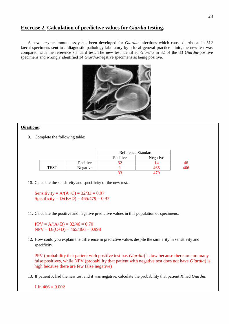
Exercise 2. Calculation of predictive values for Giardia testing.
A new enzyme immunoassay has been developed for Giardia infections which cause diarrhoea. In 512
faecal specimens sent to a diagnostic pathology laboratory by a local general practice clinic, the new test was
compared with the reference standard test. The new test identified Giardia in 32 of the 33 Giardia-positive
specimens and wrongly identified 14 Giardia-negative specimens as being positive.
Questions:
9. Complete the following table:
Reference Standard
Positive Negative
TEST
Positive 32 14 46
Negative 1 465 466
33 479
10. Calculate the sensitivity and specificity of the new test.
Sensitivity = A/(A+C) = 32/33 = 0.97
Specificity = D/(B+D) = 465/479 = 0.97
11. Calculate the positive and negative predictive values in this population of specimens.
PPV = A/(A+B) = 32/46 = 0.70
NPV = D/(C+D) = 465/466 = 0.998
12. How could you explain the difference in predictive values despite the similarity in sensitivity and
specificity.
PPV (probability that patient with positive test has Giardia) is low because there are too many
false positives, while NPV (probability that patient with negative test does not have Giardia) is
high because there are few false negative)
13. If patient X had the new test and it was negative, calculate the probability that patient X had Giardia.
1 in 466 = 0.002
1
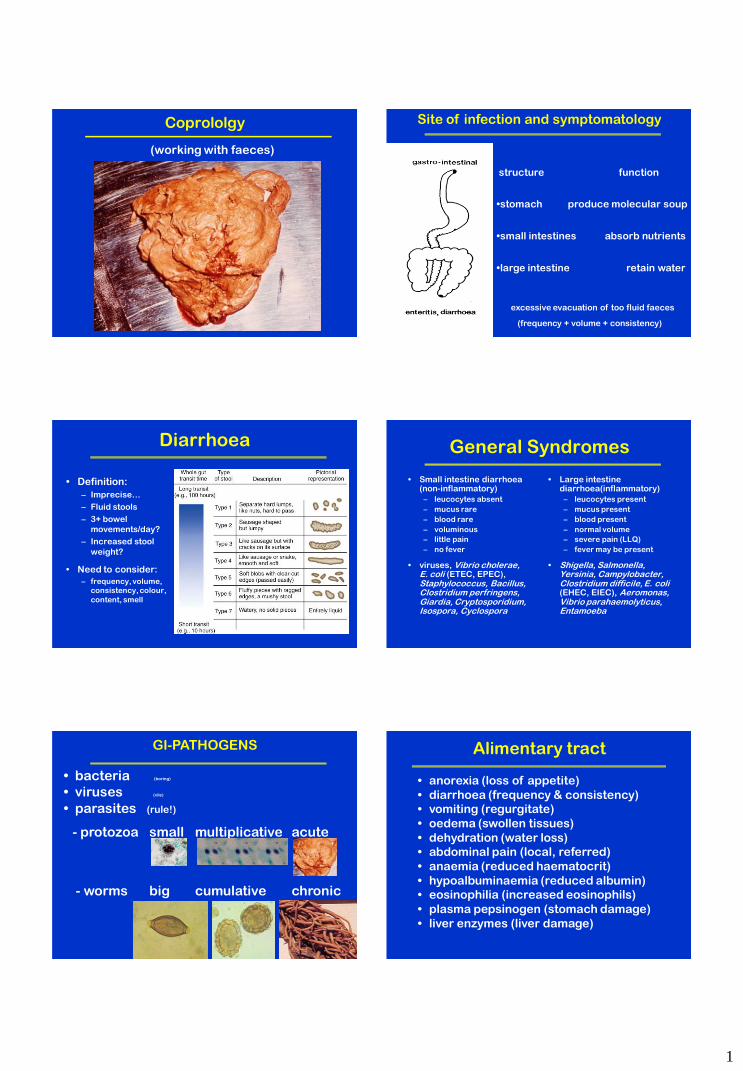
Coprololgy
(working with faeces)
Site of infection and symptomatology
structure function
•stomach produce molecular soup
•small intestines absorb nutrients
•large intestine retain water
excessive evacuation of too fluid faeces
(frequency + volume + consistency)
Diarrhoea
• Definition:
– Imprecise…
– Fluid stools
– 3+ bowel
movements/day?
– Increased stool
weight?
• Need to consider:
– frequency, volume, consistency, colour,
content, smell
General Syndromes
• Small intestine diarrhoea (non-inflammatory)
– leucocytes absent
– mucus rare
– blood rare
– voluminous
– little pain
– no fever
• viruses, Vibrio cholerae, E. coli (ETEC, EPEC), Staphylococcus, Bacillus, Clostridium perfringens, Giardia, Cryptosporidium, Isospora, Cyclospora
• Large intestine diarrhoea(inflammatory)
– leucocytes present
– mucus present
– blood present
– normal volume
– severe pain (LLQ)
– fever may be present
• Shigella, Salmonella, Yersinia, Campylobacter, Clostridium difficile, E. coli (EHEC, EIEC), Aeromonas, Vibrio parahaemolyticus, Entamoeba
GI-PATHOGENS
- protozoa
- worms
small
big
multiplicative
cumulative
acute
chronic
• bacteria (boring)
• viruses (vile)
• parasites (rule!)
Alimentary tract
• anorexia (loss of appetite)
• diarrhoea (frequency & consistency)
• vomiting (regurgitate)
• oedema (swollen tissues)
• dehydration (water loss)
• abdominal pain (local, referred)
• anaemia (reduced haematocrit)
• hypoalbuminaemia (reduced albumin)
• eosinophilia (increased eosinophils)
• plasma pepsinogen (stomach damage)
• liver enzymes (liver damage)
2
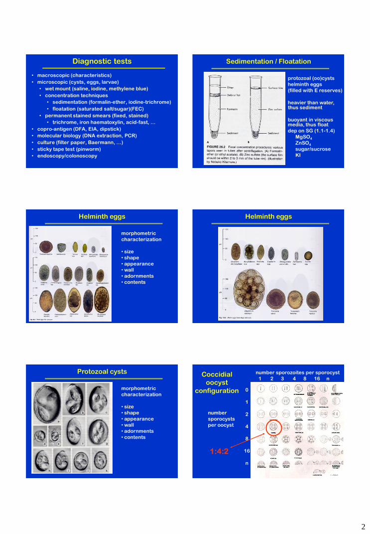
Diagnostic tests
• macroscopic (characteristics)
• microscopic (cysts, eggs, larvae)
• wet mount (saline, iodine, methylene blue)
• concentration techniques
• sedimentation (formalin-ether, iodine-trichrome)
• floatation (saturated salt/sugar)(FEC)
• permanent stained smears (fixed, stained)
• trichrome, iron haematoxylin, acid-fast, …
• copro-antigen (DFA, EIA, dipstick)
• molecular biology (DNA extraction, PCR)
• culture (filter paper, Baermann, …)
• sticky tape test (pinworm)
• endoscopy/colonoscopy
Sedimentation / Floatation
protozoal (oo)cysts
helminth eggs
(filled with E reserves)
heavier than water, thus sediment
buoyant in viscous media, thus float
dep on SG (1.1-1.4)
MgSO4
ZnSO4
sugar/sucrose
KI
Helminth eggs
morphometric
characterization
• size
• shape
• appearance
• wall
• adornments
• contents
Helminth eggs
Protozoal cysts
morphometric
characterization
• size
• shape
• appearance
• wall
• adornments
• contents
Coccidial
oocyst
configuration
number sporozoites per sporocyst
1 2 3 4 8 16 n
0
1
2
4
8
16
n
number
sporocysts
per oocyst
1:4:2
3
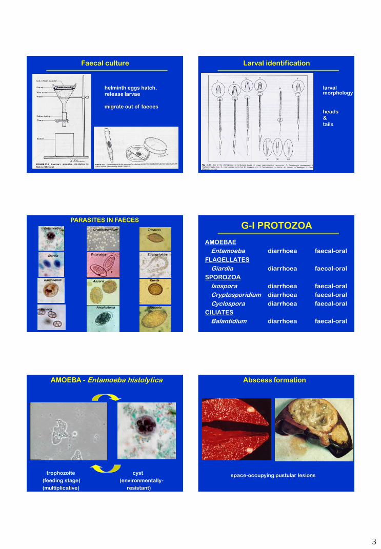
Faecal culture
helminth eggs hatch,
release larvae
migrate out of faeces
Larval identification
larval morphology
heads
&
tails
Trichuris
Giardia
Balantidium
Entamoeba
Eimeria
Cryptosporidium
Enterobius
Ascaris
Strongyloides
Taenia
Fasciola
PARASITES IN FAECES
Ancylostoma
G-I PROTOZOA
AMOEBAE
Entamoeba diarrhoea faecal-oral
FLAGELLATES
Giardia diarrhoea faecal-oral
SPOROZOA
Isospora diarrhoea faecal-oral
Cryptosporidium diarrhoea faecal-oral
Cyclospora diarrhoea faecal-oral
CILIATES
Balantidium diarrhoea faecal-oral
AMOEBA - Entamoeba histolytica
trophozoite cyst
(feeding stage) (environmentally-
(multiplicative) resistant)
Abscess formation
space-occupying pustular lesions
4
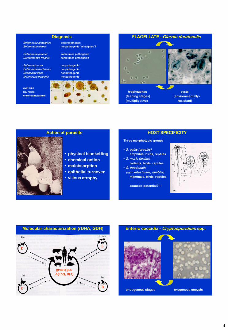
Diagnosis
Entamoeba histolytica enteropathogen
Entamoeba dispar nonpathogenic ‘histolytica’?
Entamoeba polecki sometimes pathogenic
Dientamoeba fragilis sometimes pathogenic
Entamoeba coli nonpathogenic
Entamoeba hartmanni nonpathogenic
Endolimax nana nonpathogenic
Iodamoeba butschlii nonpathogenic
cyst size
no. nuclei
chromatin pattern
FLAGELLATE - Giardia duodenalis
trophozoites cysts
(feeding stages) (environmentally-
(multiplicative) resistant)
Action of parasite
• physical blanketting
• chemical action
• malabsorption
• epithelial turnover
• villous atrophy
Three morphotypic groups
• G. agilis (gracilis)
amphibia, birds, reptiles
• G. muris (ardae)
rodents, birds, reptiles
• G. duodenalis
(syn. intestinalis, lamblia)
mammals, birds, reptiles
zoonotic potential?!!!
HOST SPECIFICITY
Molecular characterization (rDNA, GDH)
genotypes
A(1/2), B(3)
C
D
R
L
Enteric coccidia - Cryptosporidium spp.
endogenous stages exogenous oocysts
5
parasite carpet Genetic characterization
C. hominis
C. parvum
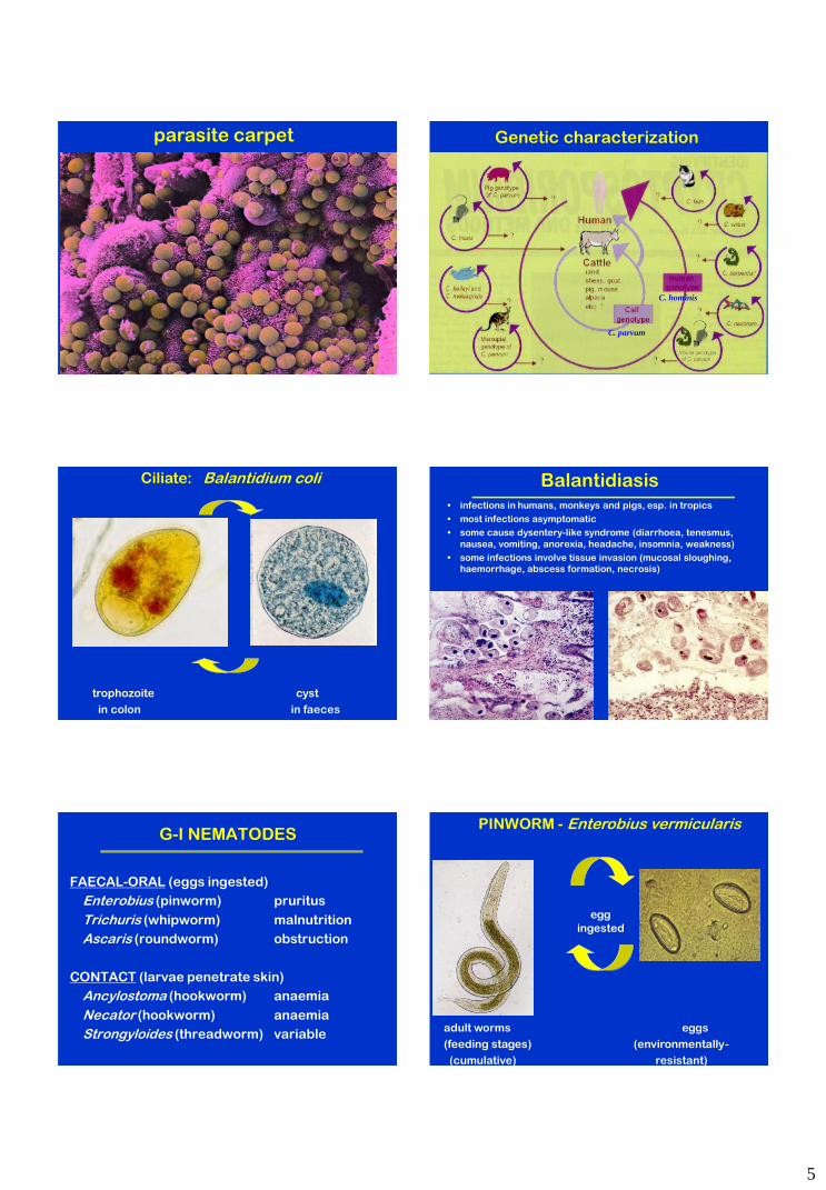
Ciliate: Balantidium coli
trophozoite cyst
in colon in faeces
Balantidiasis • infections in humans, monkeys and pigs, esp. in tropics
• most infections asymptomatic
• some cause dysentery-like syndrome (diarrhoea, tenesmus,
nausea, vomiting, anorexia, headache, insomnia, weakness)
• some infections involve tissue invasion (mucosal sloughing,
haemorrhage, abscess formation, necrosis)
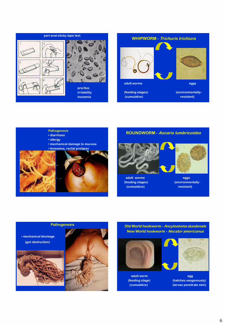
G-I NEMATODES
FAECAL-ORAL (eggs ingested)
Enterobius (pinworm) pruritus
Trichuris (whipworm) malnutrition
Ascaris (roundworm) obstruction
CONTACT (larvae penetrate skin)
Ancylostoma (hookworm) anaemia
Necator (hookworm) anaemia
Strongyloides (threadworm) variable
PINWORM - Enterobius vermicularis
egg
ingested
adult worms eggs
(feeding stages) (environmentally-
(cumulative) resistant)
6
peri-anal sticky tape test
pruritus
irritability
insomnia
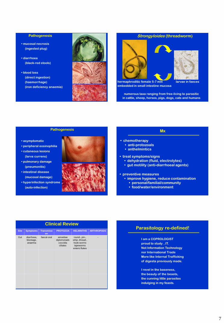
WHIPWORM - Trichuris trichiura
adult worms eggs
(feeding stages) (environmentally-
(cumulative) resistant)
Pathogenesis
• diarrhoea
• allergy
• mechanical damage to mucosa
• tenesmus, rectal prolapse
ROUNDWORM - Ascaris lumbricoides
adult worms eggs
(feeding stages) (environmentally-
(cumulative) resistant)
Pathogenesis
• mechanical blockage
(gut obstruction)
Old World hookworm - Ancylostoma duodenale
New World hookworm - Necator americanus
adult worm egg
(feeding stage) (hatches exogenously)
(cumulative) (larvae penetrate skin)
7
Pathogenesis
• mucosal necrosis
(ingested plug)
• diarrhoea
(black-red stools)
• blood loss
(direct ingestion)
(haemorrhage)
(iron deficiency anaemia)
Strongyloides (threadworm)
hermaphroditic female 5-7 mm larvae in faeces
embedded in small intestine mucosa
numerous taxa ranging from free-living to parasitic
in cattle, sheep, horses, pigs, dogs, cats and humans
Pathogenesis
• asymptomatic
• peripheral eosinophilia
• cutaneous lesions
(larva currens)
• pulmonary damage
(pneumonitis)
• intestinal disease
(mucosal damage)
• hyperinfection syndrome
(auto-infection)
Mx
• treat symptoms/signs
• dehydration (fluid, electrolytes)
• gut motility (anti-diarrhoeal agents)
• chemotherapy
• anti-protozoals
• anthelmintics
• preventive measures
• improve hygiene, reduce contamination
• personal/familial/community
• food/water/environment
Clinical Review
Site Symptoms Transmission
PROTOZOA HELMINTHS ARTHROPODS
Gut diarrhoea, blockage,
anaemia
faecal-oral amoebae diplomonads
coccidia ciliates
round-, pin-, whip-,thread-,
hook-worms tapeworms
enteric flukes
-
Parasitology re-defined!
I am a COPROLOGIST
proud to study ..IT.
Not Information Technology
nor International Trade
More like Internal Trafficking
of digesta previously made.
I revel in the baseness,
the beauty of the beasts,
the cunning little parasites
indulging in my feasts.
31
Workshop 2: Coprology (working with faeces)
In the next hour, you should complete the following three activities:
a) wet lab – conduct a centrifugal flotation of chicken faecal slurry for coccidial oocysts
b) wet lab – conduct a worm egg count using a McMaster/Whitlock chamber
c) dry lab – examine a range of gastroenteric parasites by light microscopy
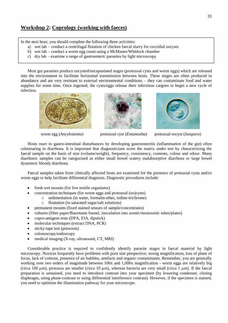
Most gut parasites produce encysted/encapsulated stages (protozoal cysts and worm eggs) which are released
into the environment to facilitate horizontal transmission between hosts. These stages are often produced in
abundance and are very resistant to external environmental conditions – they can contaminate food and water
supplies for some time. Once ingested, the cysts/eggs release their infectious cargoes to begin a new cycle of
infection.
worm egg (Ancylostoma) protozoal cyst (Entamoeba) protozoal oocyst (Isospora)
Hosts react to gastro-intestinal disturbances by developing gastroenteritis (inflammation of the gut) often
culminating in diarrhoea. It is important that diagnosticians score the matrix under test by characterizing the
faecal sample on the basis of size (volume/weight), frequency, consistency, contents, colour and odour. Many
diarrhoeic samples can be categorized as either small bowel watery malabsorptive diarrhoea or large bowel
dysenteric bloody diarrhoea.
Faecal samples taken from clinically affected hosts are examined for the presence of protozoal cysts and/or
worm eggs to help facilitate differential diagnosis. Diagnostic procedures include:
fresh wet mounts (for live motile organisms)
concentration techniques (for worm eggs and protozoal (oo)cysts)
o sedimentation (in water, formalin-ether, iodine-trichrome)
o floatation (in saturated sugar/salt solutions)
permanent mounts (fixed stained smears of sample/concentrates)
cultures (filter paper/Baermann funnel, inoculation into axenic/monoxenic tubes/plates)
copro-antigens tests (DFA, EIA, dipstick)
molecular techniques (extract DNA, PCR)
sticky-tape test (pinworm)
colonoscopy/endoscopy
medical imaging (X-ray, ultrasound, CT, MRI)
Considerable practice is required to confidently identify parasite stages in faecal material by light
microscopy. Novices frequently have problems with poor size perspective, wrong magnification, loss of plane of
focus, lack of contrast, presence of air bubbles, artefacts and organic contaminants. Remember, you are generally
working over two orders of magnitude between 100x and 1,000x magnification - worm eggs are relatively big
(circa 100 m), protozoa are smaller (circa 10 m), whereas bacteria are very small (circa 1 m). If the faecal
preparation is unstained, you need to introduce contrast into your specimen (by lowering condenser, closing
diaphragm, using phase-contrast or using differential interference contrast). However, if the specimen is stained,
you need to optimize the illumination pathway for your microscope.
32
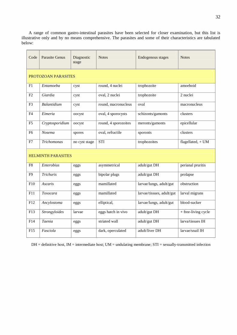
A range of common gastro-intestinal parasites have been selected for closer examination, but this list is
illustrative only and by no means comprehensive. The parasites and some of their characteristics are tabulated
below:
Code
Parasite Genus
Diagnostic
stage
Notes
Endogenous stages
Notes
PROTOZOAN PARASITES
F1
Entamoeba cyst round, 4 nuclei trophozoite amoeboid
F2
Giardia cyst oval, 2 nuclei trophozoite 2 nuclei
F3
Balantidium cyst round, macronucleus oval macronucleus
F4
Eimeria oocyst oval, 4 sporocysts schizonts/gamonts clusters
F5
Cryptosporidium oocyst round, 4 sporozoites meronts/gamonts epicellular
F6
Nosema spores oval, refractile sporonts clusters
F7
Trichomonas no cyst stage STI trophozoites flagellated, + UM
HELMINTH PARASITES
F8
Enterobius eggs asymmetrical adult/gut DH perianal pruritis
F9
Trichuris eggs bipolar plugs adult/gut DH prolapse
F10
Ascaris eggs mamillated larvae/lungs, adult/gut obstruction
F11
Toxocara eggs mamillated larvae/tissues, adult/gut larval migrans
F12
Ancylostoma eggs elliptical, larvae/lungs, adult/gut blood-sucker
F13
Strongyloides larvae eggs hatch in vivo adult/gut DH + free-living cycle
F14
Taenia eggs striated wall adult/gut DH larva/tissues IH
F15
Fasciola eggs dark, operculated adult/liver DH larvae/snail IH
DH = definitive host, IM = intermediate host; UM = undulating membrane; STI = sexually-transmitted infection
33
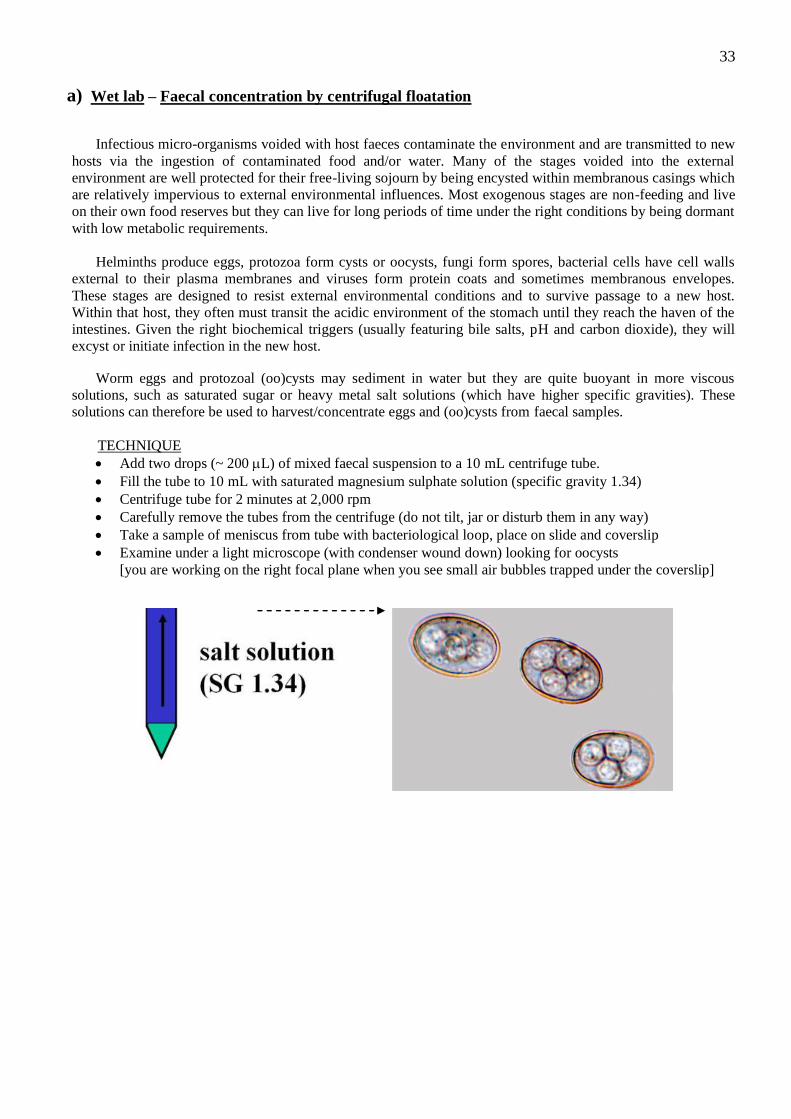
a) Wet lab – Faecal concentration by centrifugal floatation
Infectious micro-organisms voided with host faeces contaminate the environment and are transmitted to new
hosts via the ingestion of contaminated food and/or water. Many of the stages voided into the external
environment are well protected for their free-living sojourn by being encysted within membranous casings which
are relatively impervious to external environmental influences. Most exogenous stages are non-feeding and live
on their own food reserves but they can live for long periods of time under the right conditions by being dormant
with low metabolic requirements.
Helminths produce eggs, protozoa form cysts or oocysts, fungi form spores, bacterial cells have cell walls
external to their plasma membranes and viruses form protein coats and sometimes membranous envelopes.
These stages are designed to resist external environmental conditions and to survive passage to a new host.
Within that host, they often must transit the acidic environment of the stomach until they reach the haven of the
intestines. Given the right biochemical triggers (usually featuring bile salts, pH and carbon dioxide), they will
excyst or initiate infection in the new host.
Worm eggs and protozoal (oo)cysts may sediment in water but they are quite buoyant in more viscous
solutions, such as saturated sugar or heavy metal salt solutions (which have higher specific gravities). These
solutions can therefore be used to harvest/concentrate eggs and (oo)cysts from faecal samples.
TECHNIQUE
Add two drops (~ 200 L) of mixed faecal suspension to a 10 mL centrifuge tube.
Fill the tube to 10 mL with saturated magnesium sulphate solution (specific gravity 1.34)
Centrifuge tube for 2 minutes at 2,000 rpm
Carefully remove the tubes from the centrifuge (do not tilt, jar or disturb them in any way)
Take a sample of meniscus from tube with bacteriological loop, place on slide and coverslip
Examine under a light microscope (with condenser wound down) looking for oocysts
[you are working on the right focal plane when you see small air bubbles trapped under the coverslip]
34
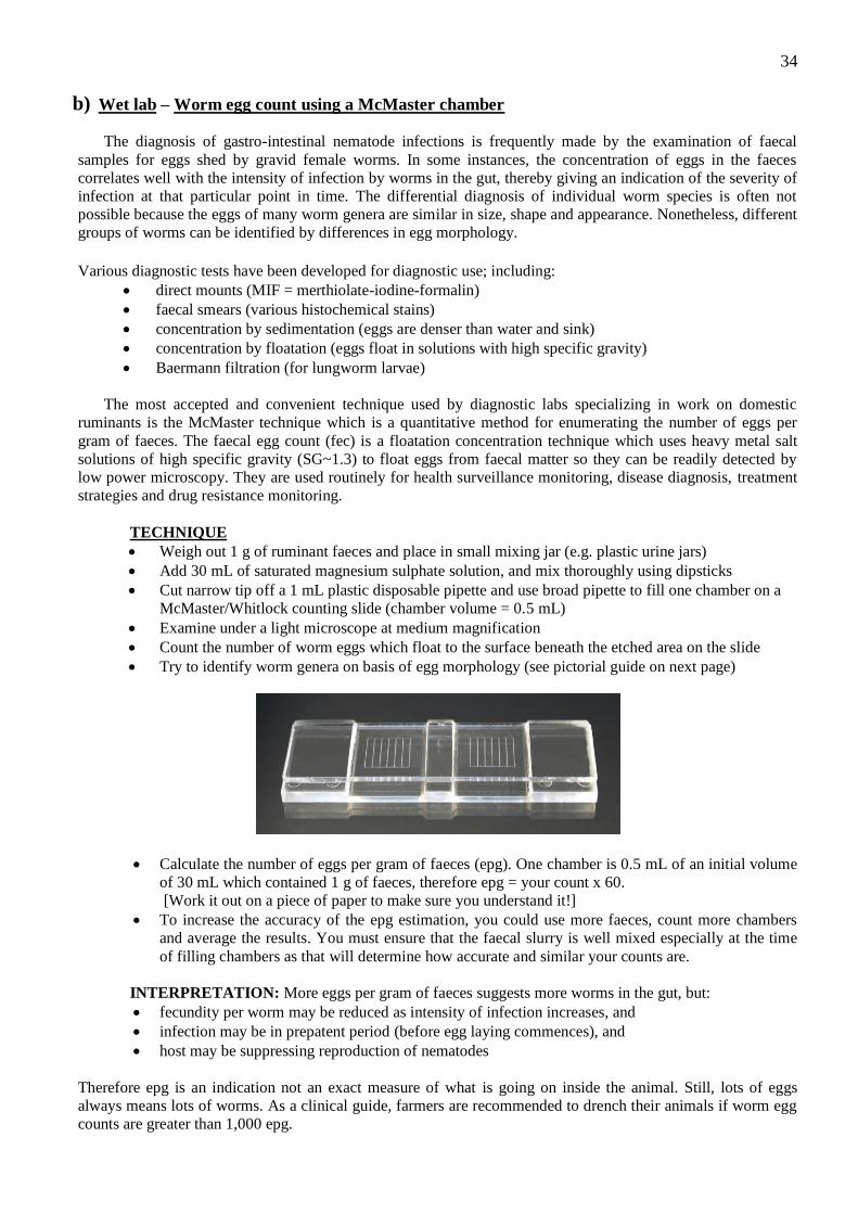
b) Wet lab – Worm egg count using a McMaster chamber
The diagnosis of gastro-intestinal nematode infections is frequently made by the examination of faecal
samples for eggs shed by gravid female worms. In some instances, the concentration of eggs in the faeces
correlates well with the intensity of infection by worms in the gut, thereby giving an indication of the severity of
infection at that particular point in time. The differential diagnosis of individual worm species is often not
possible because the eggs of many worm genera are similar in size, shape and appearance. Nonetheless, different
groups of worms can be identified by differences in egg morphology.
Various diagnostic tests have been developed for diagnostic use; including:
direct mounts (MIF = merthiolate-iodine-formalin)
faecal smears (various histochemical stains)
concentration by sedimentation (eggs are denser than water and sink)
concentration by floatation (eggs float in solutions with high specific gravity)
Baermann filtration (for lungworm larvae)
The most accepted and convenient technique used by diagnostic labs specializing in work on domestic
ruminants is the McMaster technique which is a quantitative method for enumerating the number of eggs per
gram of faeces. The faecal egg count (fec) is a floatation concentration technique which uses heavy metal salt
solutions of high specific gravity (SG~1.3) to float eggs from faecal matter so they can be readily detected by
low power microscopy. They are used routinely for health surveillance monitoring, disease diagnosis, treatment
strategies and drug resistance monitoring.
TECHNIQUE
Weigh out 1 g of ruminant faeces and place in small mixing jar (e.g. plastic urine jars)
Add 30 mL of saturated magnesium sulphate solution, and mix thoroughly using dipsticks
Cut narrow tip off a 1 mL plastic disposable pipette and use broad pipette to fill one chamber on a
McMaster/Whitlock counting slide (chamber volume = 0.5 mL)
Examine under a light microscope at medium magnification
Count the number of worm eggs which float to the surface beneath the etched area on the slide
Try to identify worm genera on basis of egg morphology (see pictorial guide on next page)
Calculate the number of eggs per gram of faeces (epg). One chamber is 0.5 mL of an initial volume
of 30 mL which contained 1 g of faeces, therefore epg = your count x 60.
[Work it out on a piece of paper to make sure you understand it!]
To increase the accuracy of the epg estimation, you could use more faeces, count more chambers
and average the results. You must ensure that the faecal slurry is well mixed especially at the time
of filling chambers as that will determine how accurate and similar your counts are.
INTERPRETATION: More eggs per gram of faeces suggests more worms in the gut, but:
fecundity per worm may be reduced as intensity of infection increases, and
infection may be in prepatent period (before egg laying commences), and
host may be suppressing reproduction of nematodes
Therefore epg is an indication not an exact measure of what is going on inside the animal. Still, lots of eggs
always means lots of worms. As a clinical guide, farmers are recommended to drench their animals if worm egg
counts are greater than 1,000 epg.
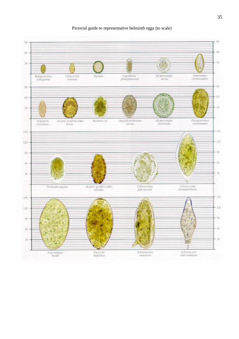
35
Pictorial guide to representative helminth eggs (to scale)
36
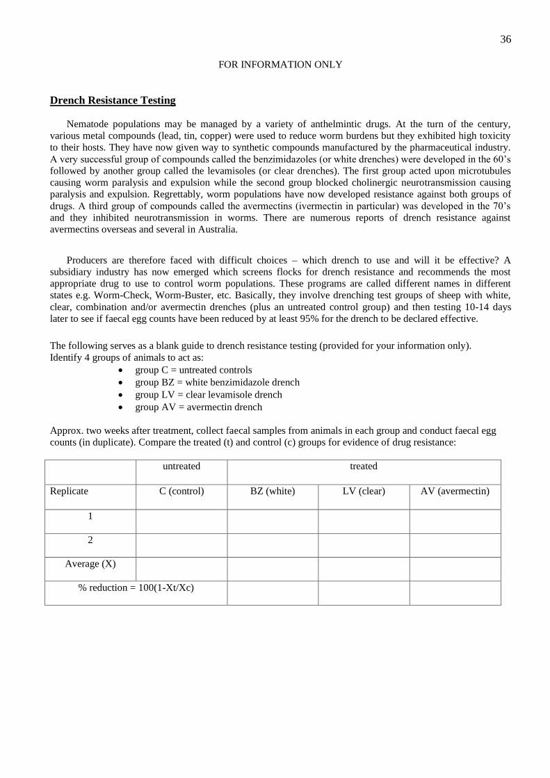
FOR INFORMATION ONLY
Drench Resistance Testing
Nematode populations may be managed by a variety of anthelmintic drugs. At the turn of the century,
various metal compounds (lead, tin, copper) were used to reduce worm burdens but they exhibited high toxicity
to their hosts. They have now given way to synthetic compounds manufactured by the pharmaceutical industry.
A very successful group of compounds called the benzimidazoles (or white drenches) were developed in the 60’s
followed by another group called the levamisoles (or clear drenches). The first group acted upon microtubules
causing worm paralysis and expulsion while the second group blocked cholinergic neurotransmission causing
paralysis and expulsion. Regrettably, worm populations have now developed resistance against both groups of
drugs. A third group of compounds called the avermectins (ivermectin in particular) was developed in the 70’s
and they inhibited neurotransmission in worms. There are numerous reports of drench resistance against
avermectins overseas and several in Australia.
Producers are therefore faced with difficult choices – which drench to use and will it be effective? A
subsidiary industry has now emerged which screens flocks for drench resistance and recommends the most
appropriate drug to use to control worm populations. These programs are called different names in different
states e.g. Worm-Check, Worm-Buster, etc. Basically, they involve drenching test groups of sheep with white,
clear, combination and/or avermectin drenches (plus an untreated control group) and then testing 10-14 days
later to see if faecal egg counts have been reduced by at least 95% for the drench to be declared effective.
The following serves as a blank guide to drench resistance testing (provided for your information only).
Identify 4 groups of animals to act as:
group C = untreated controls
group BZ = white benzimidazole drench
group LV = clear levamisole drench
group AV = avermectin drench
Approx. two weeks after treatment, collect faecal samples from animals in each group and conduct faecal egg
counts (in duplicate). Compare the treated (t) and control (c) groups for evidence of drug resistance:
untreated treated
Replicate
C (control) BZ (white) LV (clear) AV (avermectin)
1
2
Average (X)
% reduction = 100(1-Xt/Xc)

37
c) Dry lab – Examination of slide set by light microscopy (work with your neighbours) F1. (amoeba)
Entamoeba coli trophozoites
human large intestine
stained section
Scan slide at low power looking for
vacuolated areas containing basophilic
trophozoites (right); examine invasive foci
at higher power noting tissue destruction.
The diagnostic stage in coprology is the
spherical cyst with four vesicular nuclei
(below). Examination of fresh wet faecal
mounts sometimes reveals pleomorphic
trophozoite stages exhibiting amoeboid
motion (below right).
F2. (flagellate)
Giardia duodenalis
trophozoites
dog small intestines
stained section
Examine the intervillous spaces in the
intestines looking for numerous flattened
(not rounded) trophozoites at the mucosal
surface (right). The diagnostic stage for
coprology is the ovoid cyst, often with a
tangential axostyle and surrounding ‘halo’
(below). Examination of gut content or
culture material reveals pyriform
trophozoites with 2 nuclei (below right).
F3. (ciliate)
Balantidium coli
trophozoites
pig colon
stained section
Scan the epithelial surface of the colon at
low power looking for large eosinophilic
trophozoites within erosive mucosal
lesions (right). High power reveals the
trophozoites to have a relatively enormous
macronucleus (below right). The
coprological diagnostic stage is the round
cyst with conspicuous macronucleus
(below).
38
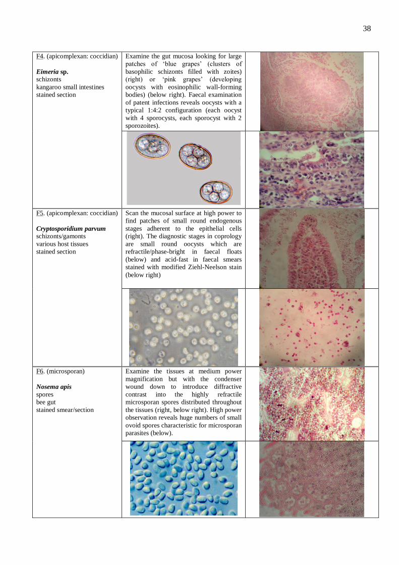
F4. (apicomplexan: coccidian)
Eimeria sp. schizonts
kangaroo small intestines
stained section
Examine the gut mucosa looking for large
patches of ‘blue grapes’ (clusters of
basophilic schizonts filled with zoites)
(right) or ‘pink grapes’ (developing
oocysts with eosinophilic wall-forming
bodies) (below right). Faecal examination
of patent infections reveals oocysts with a
typical 1:4:2 configuration (each oocyst
with 4 sporocysts, each sporocyst with 2
sporozoites).
F5. (apicomplexan: coccidian)
Cryptosporidium parvum schizonts/gamonts
various host tissues
stained section
Scan the mucosal surface at high power to
find patches of small round endogenous
stages adherent to the epithelial cells
(right). The diagnostic stages in coprology
are small round oocysts which are
refractile/phase-bright in faecal floats
(below) and acid-fast in faecal smears
stained with modified Ziehl-Neelson stain
(below right)
F6. (microsporan)
Nosema apis
spores
bee gut
stained smear/section
Examine the tissues at medium power
magnification but with the condenser
wound down to introduce diffractive
contrast into the highly refractile
microsporan spores distributed throughout
the tissues (right, below right). High power
observation reveals huge numbers of small
ovoid spores characteristic for microsporan
parasites (below).
39
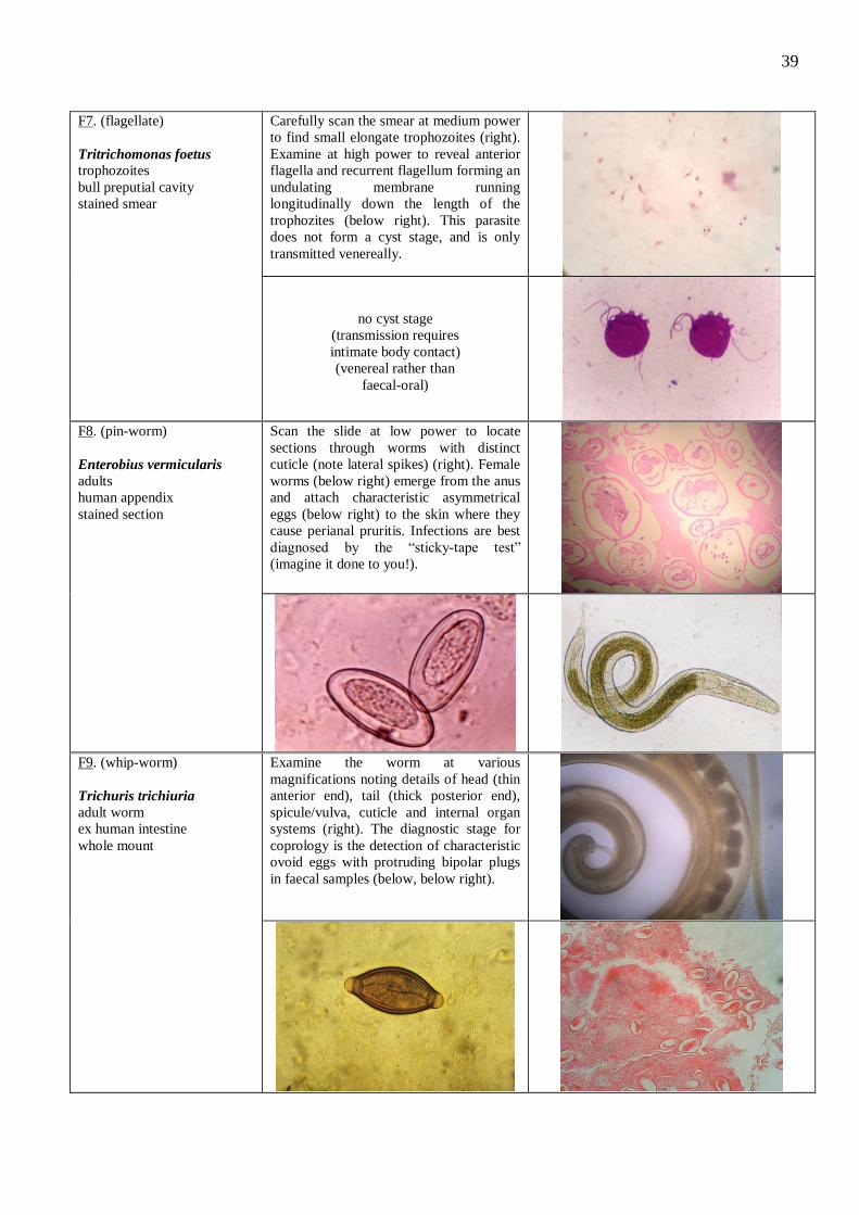
F7. (flagellate)
Tritrichomonas foetus trophozoites
bull preputial cavity
stained smear
Carefully scan the smear at medium power
to find small elongate trophozoites (right).
Examine at high power to reveal anterior
flagella and recurrent flagellum forming an
undulating membrane running
longitudinally down the length of the
trophozites (below right). This parasite
does not form a cyst stage, and is only
transmitted venereally.
no cyst stage
(transmission requires
intimate body contact)
(venereal rather than
faecal-oral)
F8. (pin-worm)
Enterobius vermicularis
adults
human appendix
stained section
Scan the slide at low power to locate
sections through worms with distinct
cuticle (note lateral spikes) (right). Female
worms (below right) emerge from the anus
and attach characteristic asymmetrical
eggs (below right) to the skin where they
cause perianal pruritis. Infections are best
diagnosed by the “sticky-tape test”
(imagine it done to you!).
F9. (whip-worm)
Trichuris trichiuria
adult worm
ex human intestine
whole mount
Examine the worm at various
magnifications noting details of head (thin
anterior end), tail (thick posterior end),
spicule/vulva, cuticle and internal organ
systems (right). The diagnostic stage for
coprology is the detection of characteristic
ovoid eggs with protruding bipolar plugs
in faecal samples (below, below right).
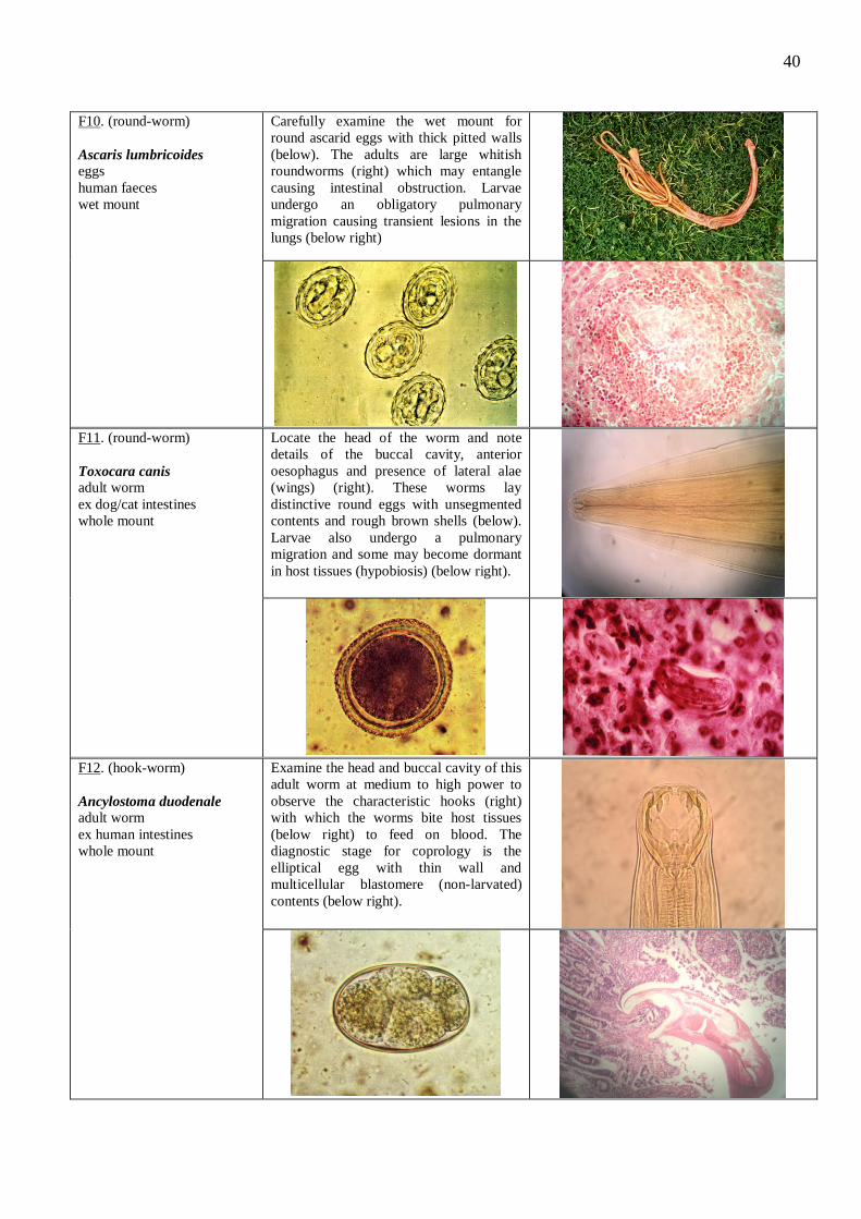
40
F10. (round-worm)
Ascaris lumbricoides
eggs
human faeces
wet mount
Carefully examine the wet mount for
round ascarid eggs with thick pitted walls
(below). The adults are large whitish
roundworms (right) which may entangle
causing intestinal obstruction. Larvae
undergo an obligatory pulmonary
migration causing transient lesions in the
lungs (below right)
F11. (round-worm)
Toxocara canis adult worm
ex dog/cat intestines
whole mount
Locate the head of the worm and note
details of the buccal cavity, anterior
oesophagus and presence of lateral alae
(wings) (right). These worms lay
distinctive round eggs with unsegmented
contents and rough brown shells (below).
Larvae also undergo a pulmonary
migration and some may become dormant
in host tissues (hypobiosis) (below right).
F12. (hook-worm)
Ancylostoma duodenale adult worm
ex human intestines
whole mount
Examine the head and buccal cavity of this
adult worm at medium to high power to
observe the characteristic hooks (right)
with which the worms bite host tissues
(below right) to feed on blood. The
diagnostic stage for coprology is the
elliptical egg with thin wall and
multicellular blastomere (non-larvated)
contents (below right).
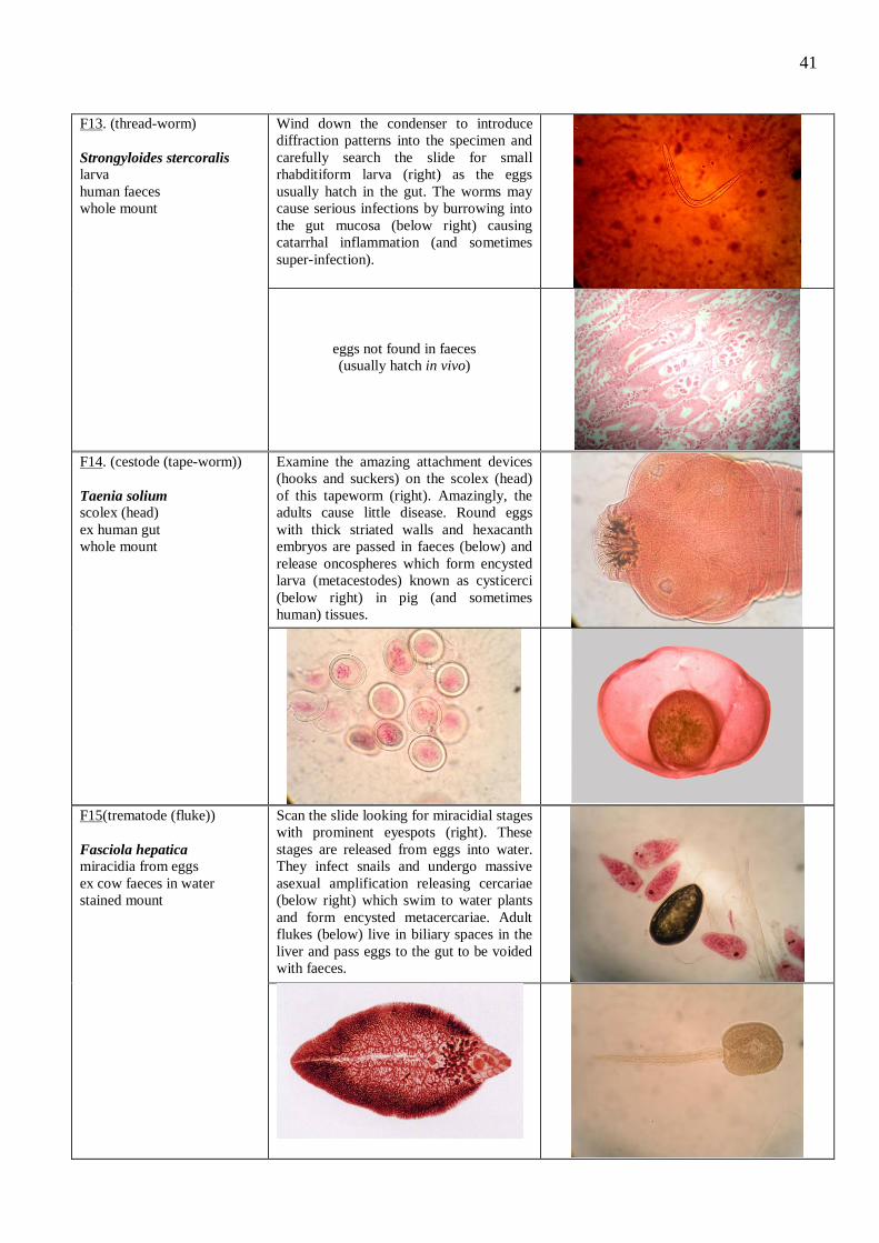
41
F13. (thread-worm)
Strongyloides stercoralis larva
human faeces
whole mount
Wind down the condenser to introduce
diffraction patterns into the specimen and
carefully search the slide for small
rhabditiform larva (right) as the eggs
usually hatch in the gut. The worms may
cause serious infections by burrowing into
the gut mucosa (below right) causing
catarrhal inflammation (and sometimes
super-infection).
eggs not found in faeces
(usually hatch in vivo)
F14. (cestode (tape-worm))
Taenia solium scolex (head)
ex human gut
whole mount
Examine the amazing attachment devices
(hooks and suckers) on the scolex (head)
of this tapeworm (right). Amazingly, the
adults cause little disease. Round eggs
with thick striated walls and hexacanth
embryos are passed in faeces (below) and
release oncospheres which form encysted
larva (metacestodes) known as cysticerci
(below right) in pig (and sometimes
human) tissues.
F15(trematode (fluke))
Fasciola hepatica miracidia from eggs
ex cow faeces in water
stained mount
Scan the slide looking for miracidial stages
with prominent eyespots (right). These
stages are released from eggs into water.
They infect snails and undergo massive
asexual amplification releasing cercariae
(below right) which swim to water plants
and form encysted metacercariae. Adult
flukes (below) live in biliary spaces in the
liver and pass eggs to the gut to be voided
with faeces.
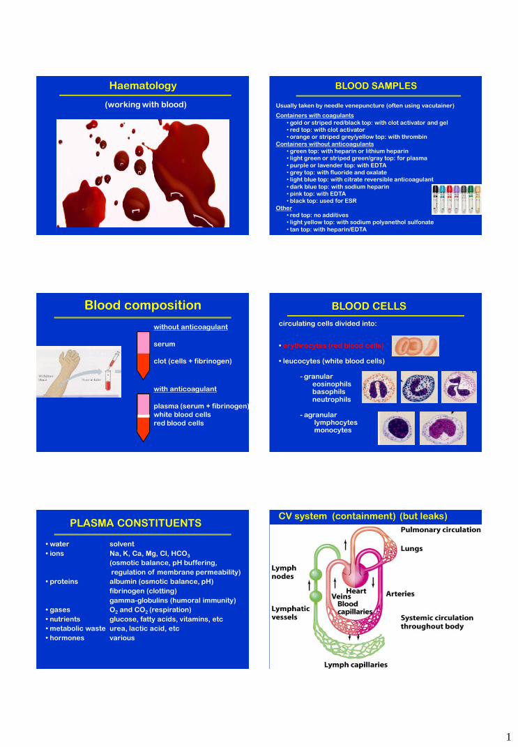
1
Haematology
(working with blood)
BLOOD SAMPLES
Containers with coagulants
• gold or striped red/black top: with clot activator and gel
• red top: with clot activator
• orange or striped grey/yellow top: with thrombin
Containers without anticoagulants
• green top: with heparin or lithium heparin
• light green or striped green/gray top: for plasma
• purple or lavender top: with EDTA
• grey top: with fluoride and oxalate
• light blue top: with citrate reversible anticoagulant
• dark blue top: with sodium heparin
• pink top: with EDTA
• black top: used for ESR
Other
• red top: no additives
• light yellow top: with sodium polyanethol sulfonate
• tan top: with heparin/EDTA
Usually taken by needle venepuncture (often using vacutainer)
Blood composition
without anticoagulant
serum
clot (cells + fibrinogen)
with anticoagulant
plasma (serum + fibrinogen)
white blood cells
red blood cells
BLOOD CELLS
circulating cells divided into:
• erythrocytes (red blood cells)
• leucocytes (white blood cells) - granular eosinophils basophils neutrophils - agranular lymphocytes monocytes
PLASMA CONSTITUENTS
• water solvent
• ions Na, K, Ca, Mg, Cl, HCO3
(osmotic balance, pH buffering,
regulation of membrane permeability)
• proteins albumin (osmotic balance, pH)
fibrinogen (clotting)
gamma-globulins (humoral immunity)
• gases O2 and CO2 (respiration)
• nutrients glucose, fatty acids, vitamins, etc
• metabolic waste urea, lactic acid, etc
• hormones various
CV system (containment) (but leaks)
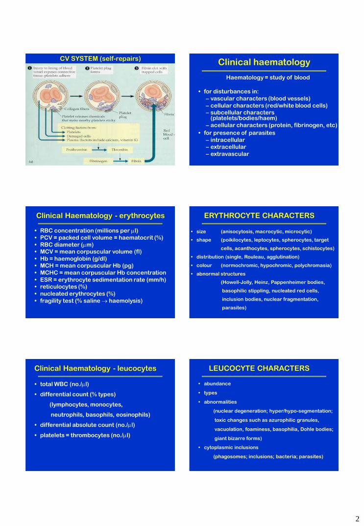
2
CV SYSTEM (self-repairs) Clinical haematology
Haematology = study of blood
• for disturbances in:
– vascular characters (blood vessels)
– cellular characters (red/white blood cells)
– subcellular characters (platelets/bodies/haem)
– acellular characters (protein, fibrinogen, etc)
• for presence of parasites
– intracellular
– extracellular
– extravascular
Clinical Haematology - erythrocytes
• RBC concentration (millions per l)
• PCV = packed cell volume = haematocrit (%)
• RBC diameter (m)
• MCV = mean corpuscular volume (fl)
• Hb = haemoglobin (g/dl)
• MCH = mean corpuscular Hb (pg)
• MCHC = mean corpuscular Hb concentration
• ESR = erythrocyte sedimentation rate (mm/h)
• reticulocytes (%)
• nucleated erythrocytes (%)
• fragility test (% saline haemolysis)
ERYTHROCYTE CHARACTERS
• size (anisocytosis, macrocytic, microcytic)
• shape (poikilocytes, leptocytes, spherocytes, target
cells, acanthocytes, spherocytes, schistocytes)
• distribution (single, Rouleau, agglutination)
• colour (normochromic, hypochromic, polychromasia)
• abnormal structures
(Howell-Jolly, Heinz, Pappenheimer bodies,
basophilic stippling, nucleated red cells,
inclusion bodies, nuclear fragmentation,
parasites)
Clinical Haematology - leucocytes
• total WBC (no./l)
• differential count (% types)
(lymphocytes, monocytes,
neutrophils, basophils, eosinophils)
• differential absolute count (no./l)
• platelets = thrombocytes (no./l)
LEUCOCYTE CHARACTERS
• abundance
• types
• abnormalities
(nuclear degeneration; hyper/hypo-segmentation;
toxic changes such as azurophilic granules,
vacuolation, foaminess, basophilia, Dohle bodies;
giant bizarre forms)
• cytoplasmic inclusions
(phagosomes; inclusions; bacteria; parasites)
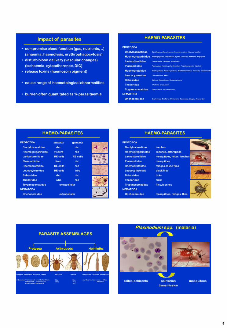
3
Impact of parasites
• compromise blood function (gas, nutrients, ..)
(anaemia, haemolysis, erythrophagocytosis)
• disturb blood delivery (vascular changes)
(ischaemia, cytoadherence, DIC)
• release toxins (haemozoin pigment)
• cause range of haematological abnormalities
• burden often quantitated as % parasitaemia
HAEMO-PARASITES
PROTOZOA
Dactylosomatidae
Haemogregarinidae
Lankesterellidae
Plasmodiidae
Haemoproteidae
Leucocytozoidae
Babesiidae
Theileridae
Trypanosomatidae
NEMATODA
Onchocercidae
Dactylosoma, Babesiosoma, Haemohormidium, Haematractidium
Haemogregarina, Hepatozoon, Cyrilia, Desseria, Hemolivia, Karyolysus
Lankesterella, Lainsonia, Schellackia
Plasmodium, Hepatocystis, Mesnilium, Polychromophilus, Nycteria
Haemoproteus, Haemocystidium, Parahaemoproteus, Simondia, Haemamoeba
Leucocytozoon, Akiba
Babesia, Sauroplasma, Serpentoplasma
Theileria, Cytoauxzoon
Trypanosoma, Sauroleishmania
Onchocerca, Dirofilaria, Wuchereria, Mansonella, Brugia, Setaria, Loa
HAEMO-PARASITES
PROTOZOA
Dactylosomatidae
Haemogregarinidae
Lankesterellidae
Plasmodiidae
Haemoproteidae
Leucocytozoidae
Babesiidae
Theileridae
Trypanosomatidae
NEMATODA
Onchocercidae
meronts gamonts
rbc rbc
viscera rbc
RE cells RE cells
liver rbc
RE cells rbc
RE cells wbc
rbc rbc
wbc rbc
extracellular
extracellular
HAEMO-PARASITES
PROTOZOA
Dactylosomatidae
Haemogregarinidae
Lankesterellidae
Plasmodiidae
Haemoproteidae
Leucocytozoidae
Babesiidae
Theileridae
Trypanosomatidae
NEMATODA
Onchocercidae
leeches
leeches, arthropods
mosquitoes, mites, leeches
mosquitoes
midges, louse flies
black flies
ticks
ticks
flies, leeches
mosquitoes, midges, flies
Protozoa Arthropods Helminths
amoebae flagellates sporozoa ciliates
entamoebae trichomonads coccidia holotrichs diplomonads haemosporidia trypanosomes piroplasms
arachnids insects
ticks flies
mites fleas
lice
nematodes cestodes trematodes
roundworms tapeworms flukes flatworms
PARASITE ASSEMBLAGES
Plasmodium spp. (malaria)
zoites-schizonts salivarian mosquitoes
transmission
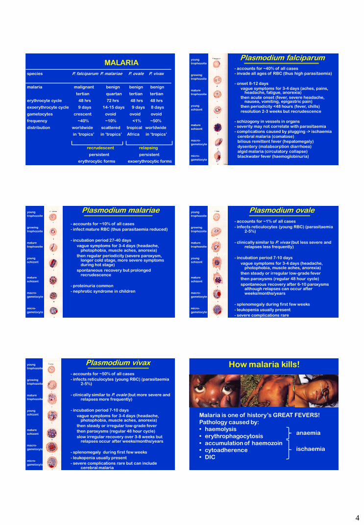
4
species P. falciparum P. malariae P. ovale P. vivax
malaria malignant benign benign benign
tertian quartan tertian tertian
erythrocyte cycle 48 hrs 72 hrs 48 hrs 48 hrs
exoerythrocyte cycle 9 days 14-15 days 9 days 8 days
gametocytes crescent ovoid ovoid ovoid
frequency ~40% ~10% <1% ~50%
distribution worldwide scattered tropical worldwide
in ‘tropics’ in ‘tropics’ Africa in ‘tropics’
recrudescent relapsing
persistent persistent
erythrocytic forms exoerythrocytic forms
MALARIA Plasmodium falciparum
- accounts for ~40% of all cases
- invade all ages of RBC (thus high parasitaemia)
- onset 8-12 days
vague symptoms for 3-4 days (aches, pains, headache, fatigue, anorexia)
then acute onset (fever, severe headache, nausea, vomiting, epigastric pain)
then periodicity <48 hours (fever, chills)
resolution 2-3 weeks but recrudescence
- schizogony in vessels in organs
- severity may not correlate with parasitaemia
- complications caused by plugging -> ischaemia
cerebral malaria (comatose)
bilious remittent fever (hepatomegaly)
dysentery (malabsorption diarrhoea)
algid malaria (circulatory collapse)
blackwater fever (haemoglobinuria)
young
trophozoite
growing
trophozoite
mature
trophozoite
young
schizont
mature
schizont
macro-
gametocyte
micro-
gametocyte
Plasmodium malariae
young
trophozoite
growing
trophozoite
mature
trophozoite
young
schizont
mature
schizont
macro-
gametocyte
micro-
gametocyte
- accounts for ~10% of all cases
- infect mature RBC (thus parasitaemia reduced)
- incubation period 27-40 days
vague symptoms for 3-4 days (headache, photophobia, muscle aches, anorexia)
then regular periodicity (severe paroxysm, longer cold stage, more severe symptoms during hot stage)
spontaneous recovery but prolonged recrudescence
- proteinuria common
- nephrotic syndrome in children
Plasmodium ovale
young
trophozoite
growing
trophozoite
mature
trophozoite
young
schizont
mature
schizont
macro-
gametocyte
micro-
gametocyte
- accounts for ~1% of all cases
- infects reticulocytes (young RBC) (parasitaemia 2-5%)
- clinically similar to P. vivax (but less severe and relapses less frequently)
- incubation period 7-10 days
vague symptoms for 3-4 days (headache, photophobia, muscle aches, anorexia)
then steady or irregular low-grade fever
then paroxysms (regular 48 hour cycle)
spontaneous recovery after 6-10 paroxysms although relapses can occur after weeks/months/years
- splenomegaly during first few weeks
- leukopenia usually present
- severe complications rare
Plasmodium vivax
young
trophozoite
growing
trophozoite
mature
trophozoite
young
schizont
mature
schizont
macro-
gametocyte
micro-
gametocyte
- accounts for ~50% of all cases
- infects reticulocytes (young RBC) (parasitaemia 2-5%)
- clinically similar to P. ovale (but more severe and relapses more frequently)
- incubation period 7-10 days
vague symptoms for 3-4 days (headache, photophobia, muscle aches, anorexia)
then steady or irregular low-grade fever
then paroxysms (regular 48 hour cycle)
slow irregular recovery over 3-8 weeks but relapses occur after weeks/months/years
- splenomegaly during first few weeks
- leukopenia usually present
- severe complications rare but can include cerebral malaria
How malaria kills!
Malaria is one of history’s GREAT FEVERS!
Pathology caused by:
• haemolysis
• erythrophagocytosis
• accumulation of haemozoin
• cytoadherence
• DIC
anaemia
ischaemia
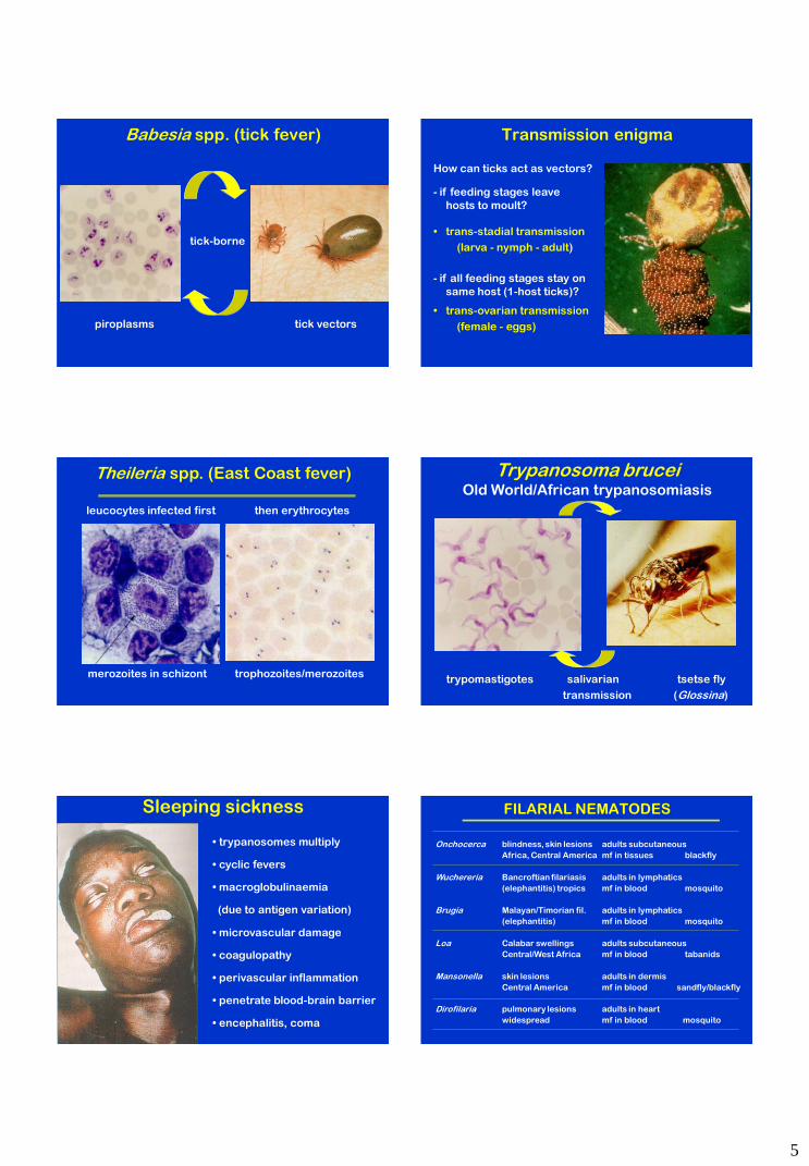
5
Babesia spp. (tick fever)
tick-borne
piroplasms tick vectors
Transmission enigma
How can ticks act as vectors?
- if feeding stages leave
hosts to moult?
- if all feeding stages stay on
same host (1-host ticks)?
• trans-stadial transmission
(larva - nymph - adult)
• trans-ovarian transmission
(female - eggs)
Theileria spp. (East Coast fever)
leucocytes infected first then erythrocytes
merozoites in schizont trophozoites/merozoites trypomastigotes salivarian tsetse fly
transmission (Glossina)
Trypanosoma brucei Old World/African trypanosomiasis
• trypanosomes multiply
• cyclic fevers
• macroglobulinaemia
(due to antigen variation)
• microvascular damage
• coagulopathy
• perivascular inflammation
• penetrate blood-brain barrier
• encephalitis, coma
Sleeping sickness FILARIAL NEMATODES
Onchocerca blindness, skin lesions adults subcutaneous
Africa, Central America mf in tissues blackfly
Wuchereria Bancroftian filariasis adults in lymphatics
(elephantitis) tropics mf in blood mosquito
Brugia Malayan/Timorian fil. adults in lymphatics
(elephantitis) mf in blood mosquito
Loa Calabar swellings adults subcutaneous
Central/West Africa mf in blood tabanids
Mansonella skin lesions adults in dermis
Central America mf in blood sandfly/blackfly
Dirofilaria pulmonary lesions adults in heart
widespread mf in blood mosquito
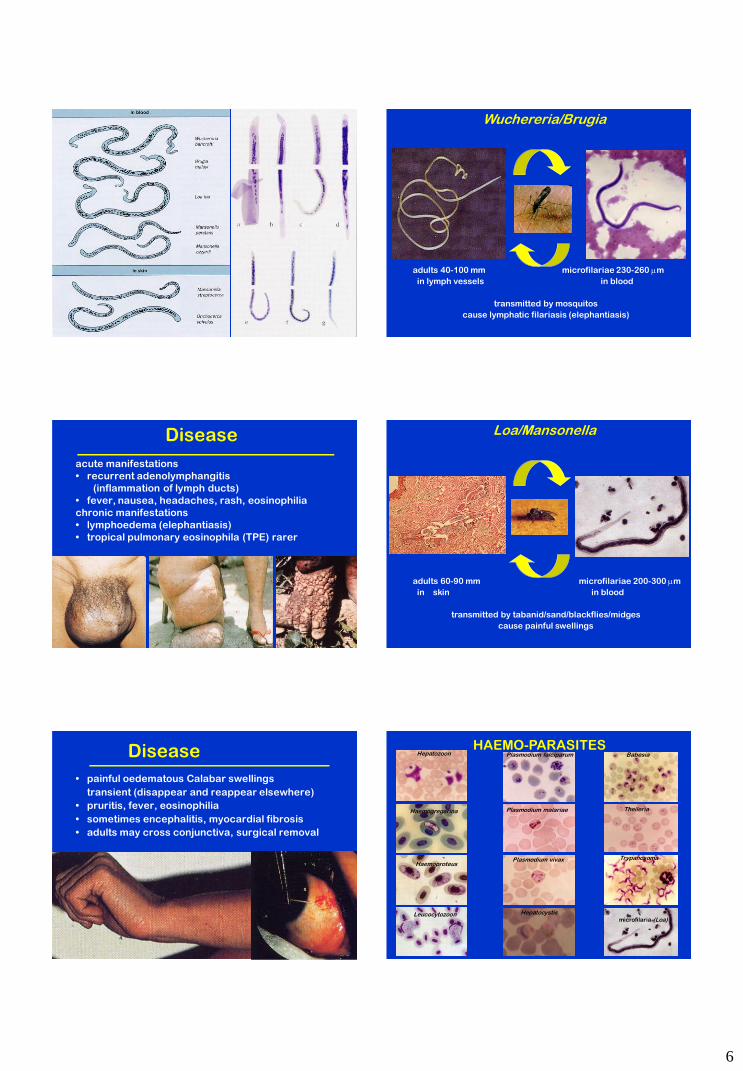
6
Wuchereria/Brugia
adults 40-100 mm microfilariae 230-260 m
in lymph vessels in blood
transmitted by mosquitos
cause lymphatic filariasis (elephantiasis)
Disease
acute manifestations
• recurrent adenolymphangitis
(inflammation of lymph ducts)
• fever, nausea, headaches, rash, eosinophilia
chronic manifestations
• lymphoedema (elephantiasis)
• tropical pulmonary eosinophila (TPE) rarer
Loa/Mansonella
adults 60-90 mm microfilariae 200-300 m
in skin in blood
transmitted by tabanid/sand/blackflies/midges
cause painful swellings
Disease
• painful oedematous Calabar swellings
transient (disappear and reappear elsewhere)
• pruritis, fever, eosinophilia
• sometimes encephalitis, myocardial fibrosis
• adults may cross conjunctiva, surgical removal
Babesia
Haemogregarina
Haemoproteus
Hepatozoon
Leucocytozoon
Plasmodium falciparum
Plasmodium malariae
Plasmodium vivax
Theileria
Trypanosoma
microfilaria (Loa) Hepatocystis
HAEMO-PARASITES
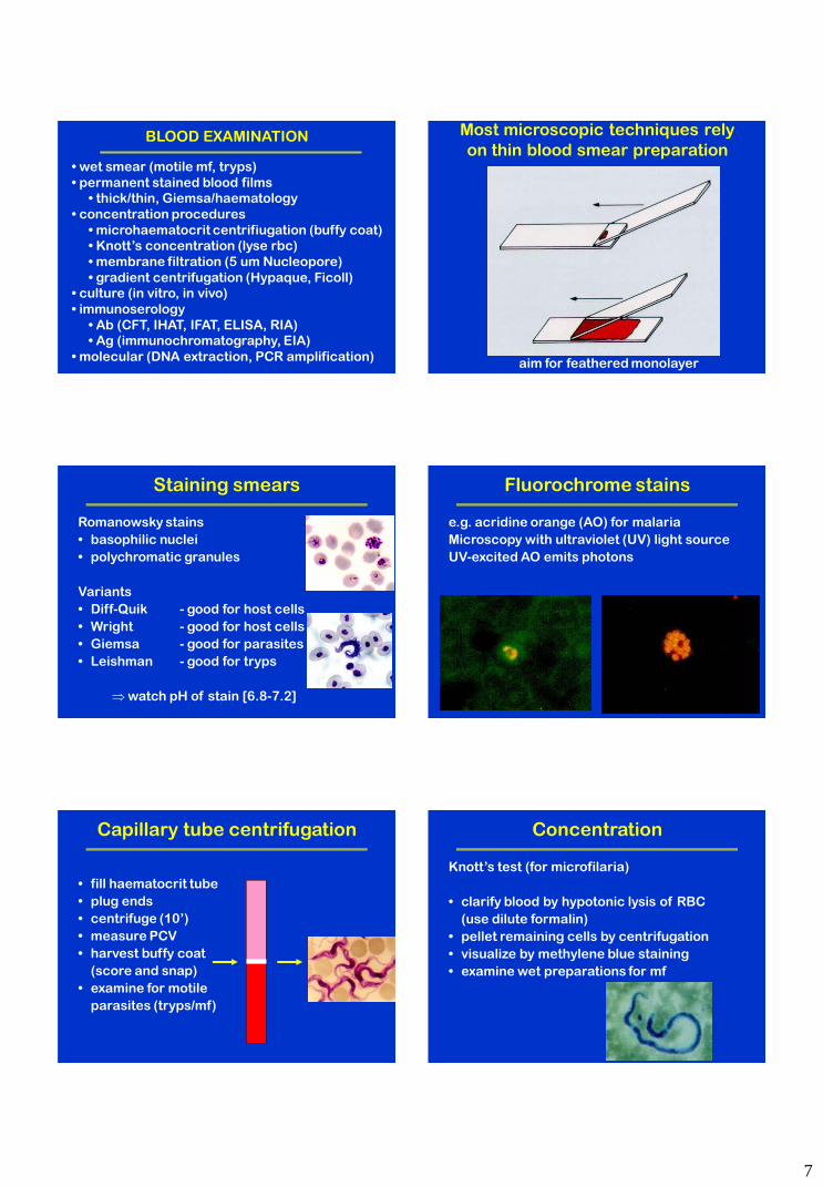
7
BLOOD EXAMINATION
• wet smear (motile mf, tryps)
• permanent stained blood films
• thick/thin, Giemsa/haematology
• concentration procedures
• microhaematocrit centrifiugation (buffy coat)
• Knott’s concentration (lyse rbc)
• membrane filtration (5 um Nucleopore)
• gradient centrifugation (Hypaque, Ficoll)
• culture (in vitro, in vivo)
• immunoserology
• Ab (CFT, IHAT, IFAT, ELISA, RIA)
• Ag (immunochromatography, EIA)
• molecular (DNA extraction, PCR amplification)
Most microscopic techniques rely
on thin blood smear preparation
aim for feathered monolayer
Staining smears
Romanowsky stains
• basophilic nuclei
• polychromatic granules
Variants
• Diff-Quik - good for host cells
• Wright - good for host cells
• Giemsa - good for parasites
• Leishman - good for tryps
watch pH of stain [6.8-7.2]
Fluorochrome stains
e.g. acridine orange (AO) for malaria
Microscopy with ultraviolet (UV) light source
UV-excited AO emits photons
Capillary tube centrifugation
• fill haematocrit tube
• plug ends
• centrifuge (10’)
• measure PCV
• harvest buffy coat
(score and snap)
• examine for motile
parasites (tryps/mf)
Concentration
Knott’s test (for microfilaria)
• clarify blood by hypotonic lysis of RBC
(use dilute formalin)
• pellet remaining cells by centrifugation
• visualize by methylene blue staining
• examine wet preparations for mf
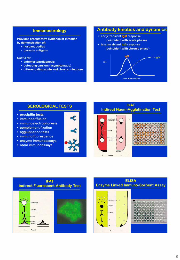
8
Immunoserology
Provides presumptive evidence of infection
by demonstration of:
• host antibodies
• parasite antigens
Useful for:
• antemortem diagnosis
• detecting carriers (asymptomatic)
• differentiating acute and chronic infections
Antibody kinetics and dynamics
• early transient IgM response
(coincident with acute phase)
• late persistent IgG response
(coincident with chronic phase)
time after infection
titre
IgM IgG
SEROLOGICAL TESTS
• precipitin tests
• immunodiffusion
• immunoelectrophoresis
• complement fixation
• agglutination tests
• immunofluorescence
• enzyme immunoassays
• radio immunoassays
IHAT
Indirect Haem-Agglutination Test
IFAT
Indirect Fluorescent-Antibody Test
ELISA
Enzyme Linked Immuno-Sorbent Assay
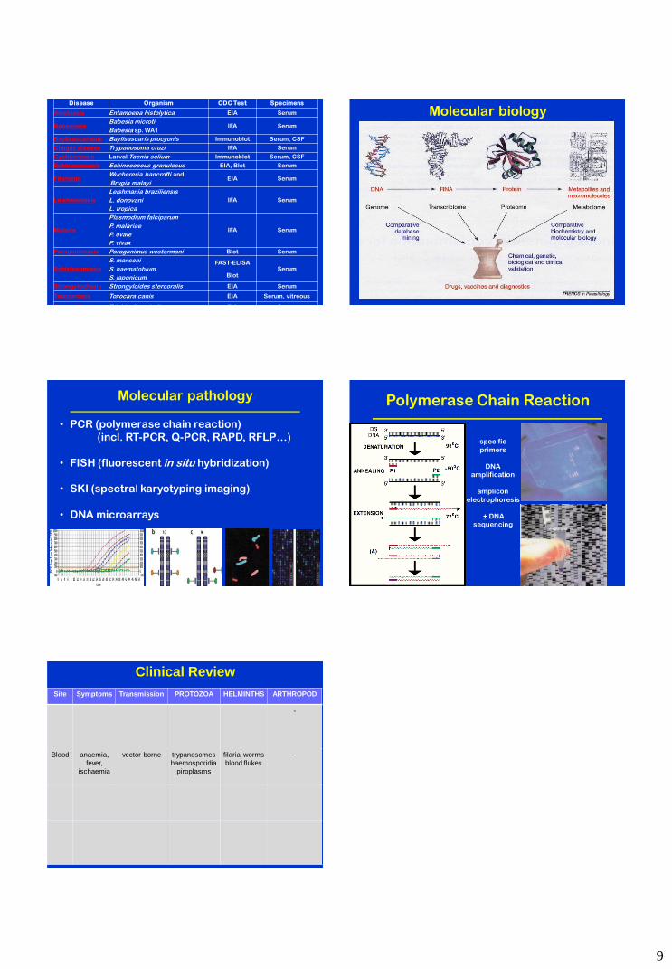
9
Disease Organism CDC Test Specimens
Amebiasis Entamoeba histolytica EIA Serum
Babesiosis Babesia microti
Babesia sp. WA1 IFA Serum
Baylisascariasis Baylisascaris procyonis Immunoblot Serum, CSF
Chagas disease Trypanosoma cruzi IFA Serum
Cysticercosis Larval Taenia solium Immunoblot Serum, CSF
Echinococcosis Echinococcus granulosus EIA, Blot Serum
Filariasis Wuchereria bancrofti and
Brugia malayi EIA Serum
Leishmaniasis
Leishmania braziliensis
L. donovani
L. tropica
IFA Serum
Malaria
Plasmodium falciparum
P. malariae
P. ovale
P. vivax
IFA Serum
Paragonimiasis Paragonimus westermani Blot Serum
Schistosomiasis
S. mansoni
S. haematobium
S. japonicum
FAST-ELISA
Blot Serum
Strongyloidiasis Strongyloides stercoralis EIA Serum
Toxocariasis Toxocara canis EIA Serum, vitreous
Trichinellosis Trichinella spiralis EIA Serum
Molecular biology
Molecular pathology
• PCR (polymerase chain reaction)
(incl. RT-PCR, Q-PCR, RAPD, RFLP…)
• FISH (fluorescent in situ hybridization)
• SKI (spectral karyotyping imaging)
• DNA microarrays
Polymerase Chain Reaction
specific
primers
DNA amplification
amplicon
electrophoresis
+ DNA sequencing
Clinical Review
Site Symptoms Transmission PROTOZOA HELMINTHS ARTHROPOD
-
Blood anaemia, fever,
ischaemia
vector-borne trypanosomes haemosporidia
piroplasms
filarial worms blood flukes
-
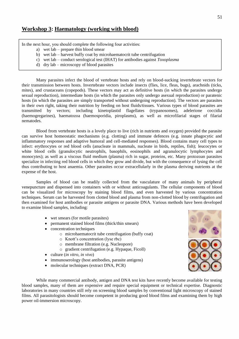
51
Workshop 3: Haematology (working with blood)
In the next hour, you should complete the following four activities:
a) wet lab – prepare thin blood smear
b) wet lab – harvest buffy coat by microhaematocrit tube centrifugation
c) wet lab – conduct serological test (IHAT) for antibodies against Toxoplasma
d) dry lab – microscopy of blood parasites
Many parasites infect the blood of vertebrate hosts and rely on blood-sucking invertebrate vectors for
their transmission between hosts. Invertebrate vectors include insects (flies, lice, fleas, bugs), arachnids (ticks,
mites), and crustaceans (copepods). These vectors may act as definitive hosts (in which the parasites undergo
sexual reproduction), intermediate hosts (in which the parasites only undergo asexual reproduction) or paratenic
hosts (in which the parasites are simply transported without undergoing reproduction). The vectors are parasites
in their own right, taking their nutrition by feeding on host fluids/tissues. Various types of blood parasites are
transmitted by vectors; including kinetoplastid flagellates (trypanosomes), adelerione coccidia
(haemogregarines), haematozoa (haemosporidia, piroplasms), as well as microfilarial stages of filarial
nematodes.
Blood from vertebrate hosts is a lovely place to live (rich in nutrients and oxygen) provided the parasite
can survive host homeostatic mechanisms (e.g. clotting) and immune defences (e.g. innate phagocytic and
inflammatory responses and adaptive humoral and cell-mediated responses). Blood contains many cell types to
infect: erythrocytes or red blood cells (anucleate in mammals, nucleate in birds, reptiles, fish); leucocytes or
white blood cells (granulocytic neutrophils, basophils, eosinophils and agranulocytic lymphocytes and
monocytes); as well as a viscous fluid medium (plasma) rich in sugar, proteins, etc. Many protozoan parasites
specialize in infecting red blood cells in which they grow and divide, but with the consequence of lysing the cell
thus contributing to host anaemia. Other parasites occur extracellularly in the plasma deriving nutrients at the
expense of the host.
Samples of blood can be readily collected from the vasculature of many animals by peripheral
venepuncture and dispensed into containers with or without anticoagulants. The cellular components of blood
can be visualized for microscopy by staining blood films, and even harvested by various concentration
techniques. Serum can be harvested from clotted blood and plasma from non-clotted blood by centrifugation and
then examined for host antibodies or parasite antigens or parasite DNA. Various methods have been developed
to examine blood samples, including:
wet smears (for motile parasites)
permanent stained blood films (thick/thin smears)
concentration techniques
o microhaematocrit tube centrifugation (buffy coat)
o Knott’s concentration (lyse rbc)
o membrane filtration (e.g. Nucleopore)
o gradient centrifugation (e.g. Hypaque, Ficoll)
culture (in vitro, in vivo)
immunoserology (host antibodies, parasite antigens)
molecular techniques (extract DNA, PCR)
While many commercial antibody, antigen and DNA test kits have recently become available for testing
blood samples, many of them are expensive and require special equipment or technical expertise. Diagnostic
laboratories in many countries still rely on screening blood samples by conventional light microscopy of stained
films. All parasitologists should become competent in producing good blood films and examining them by high
power oil-immersion microscopy.
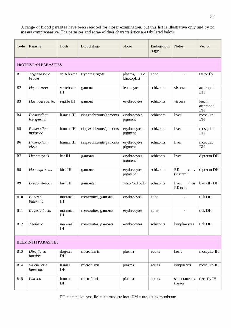
52
A range of blood parasites have been selected for closer examination, but this list is illustrative only and by no
means comprehensive. The parasites and some of their characteristics are tabulated below:
Code
Parasite
Hosts
Blood stage
Notes
Endogenous
stages
Notes
Vector
PROTOZOAN PARASITES
B1 Trypanosoma
brucei
vertebrates trypomastigote plasma, UM,
kinetoplast
none - tsetse fly
B2 Hepatozoon
vertebrate
IH
gamont leucocytes schizonts viscera arthropod
DH
B3 Haemogregarina reptile IH gamont erythrocytes schizonts viscera leech,
arthropod
DH
B4 Plasmodium
falciparum
human IH rings/schizonts/gamonts erythrocytes,
pigment
schizonts liver mosquito
DH
B5 Plasmodium
malariae
human IH rings/schizonts/gamonts erythrocytes,
pigment
schizonts liver mosquito
DH
B6 Plasmodium
vivax
human IH rings/schizonts/gamonts erythrocytes,
pigment
schizonts liver mosquito
DH
B7 Hepatocystis
bat IH gamonts erythrocytes,
pigment
schizonts liver dipteran DH
B8 Haemoproteus
bird IH gamonts erythrocytes,
pigment
schizonts RE cells
(viscera)
dipteran DH
B9 Leucocytozoon
bird IH gamonts white/red cells schizonts liver, then
RE cells
blackfly DH
B10 Babesia
bigemina
mammal
IH
merozoites, gamonts erythrocytes none - tick DH
B11 Babesia bovis
mammal
IH
merozoites, gamonts erythrocytes none - tick DH
B12 Theileria
mammal
IH
merozoites, gamonts erythrocytes schizonts lymphocytes tick DH
HELMINTH PARASITES
B13 Dirofilaria
immitis
dog/cat
DH
microfilaria plasma adults heart mosquito IH
B14 Wuchereria
bancrofti
human
DH
microfilaria plasma adults lymphatics mosquito IH
B15 Loa loa
human
DH
microfilaria plasma adults subcutaneous
tissues
deer fly IH
DH = definitive host, IM = intermediate host; UM = undulating membrane
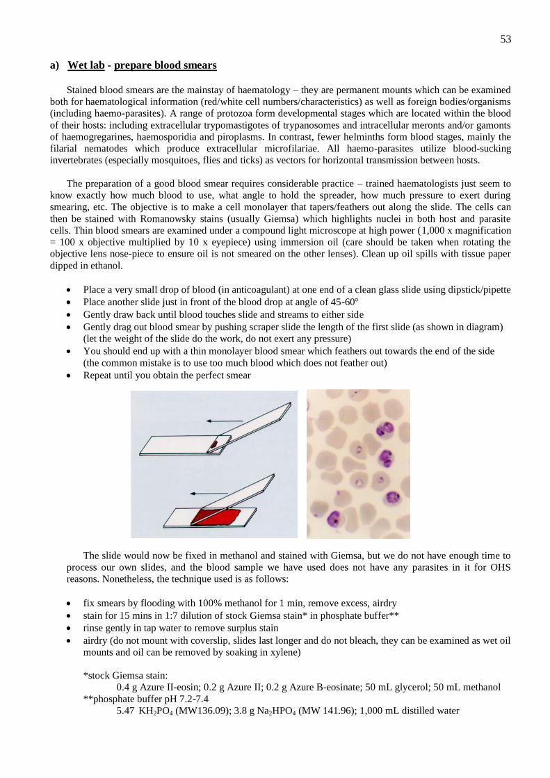
53
a) Wet lab - prepare blood smears
Stained blood smears are the mainstay of haematology – they are permanent mounts which can be examined
both for haematological information (red/white cell numbers/characteristics) as well as foreign bodies/organisms
(including haemo-parasites). A range of protozoa form developmental stages which are located within the blood
of their hosts: including extracellular trypomastigotes of trypanosomes and intracellular meronts and/or gamonts
of haemogregarines, haemosporidia and piroplasms. In contrast, fewer helminths form blood stages, mainly the
filarial nematodes which produce extracellular microfilariae. All haemo-parasites utilize blood-sucking
invertebrates (especially mosquitoes, flies and ticks) as vectors for horizontal transmission between hosts.
The preparation of a good blood smear requires considerable practice – trained haematologists just seem to
know exactly how much blood to use, what angle to hold the spreader, how much pressure to exert during
smearing, etc. The objective is to make a cell monolayer that tapers/feathers out along the slide. The cells can
then be stained with Romanowsky stains (usually Giemsa) which highlights nuclei in both host and parasite
cells. Thin blood smears are examined under a compound light microscope at high power (1,000 x magnification
= 100 x objective multiplied by 10 x eyepiece) using immersion oil (care should be taken when rotating the
objective lens nose-piece to ensure oil is not smeared on the other lenses). Clean up oil spills with tissue paper
dipped in ethanol.
Place a very small drop of blood (in anticoagulant) at one end of a clean glass slide using dipstick/pipette
Place another slide just in front of the blood drop at angle of 45-60
Gently draw back until blood touches slide and streams to either side
Gently drag out blood smear by pushing scraper slide the length of the first slide (as shown in diagram)
(let the weight of the slide do the work, do not exert any pressure)
You should end up with a thin monolayer blood smear which feathers out towards the end of the side
(the common mistake is to use too much blood which does not feather out)
Repeat until you obtain the perfect smear
The slide would now be fixed in methanol and stained with Giemsa, but we do not have enough time to
process our own slides, and the blood sample we have used does not have any parasites in it for OHS
reasons. Nonetheless, the technique used is as follows:
fix smears by flooding with 100% methanol for 1 min, remove excess, airdry
stain for 15 mins in 1:7 dilution of stock Giemsa stain* in phosphate buffer**
rinse gently in tap water to remove surplus stain
airdry (do not mount with coverslip, slides last longer and do not bleach, they can be examined as wet oil
mounts and oil can be removed by soaking in xylene)
*stock Giemsa stain:
0.4 g Azure II-eosin; 0.2 g Azure II; 0.2 g Azure B-eosinate; 50 mL glycerol; 50 mL methanol
**phosphate buffer pH 7.2-7.4
5.47 KH2PO4 (MW136.09); 3.8 g Na2HPO4 (MW 141.96); 1,000 mL distilled water
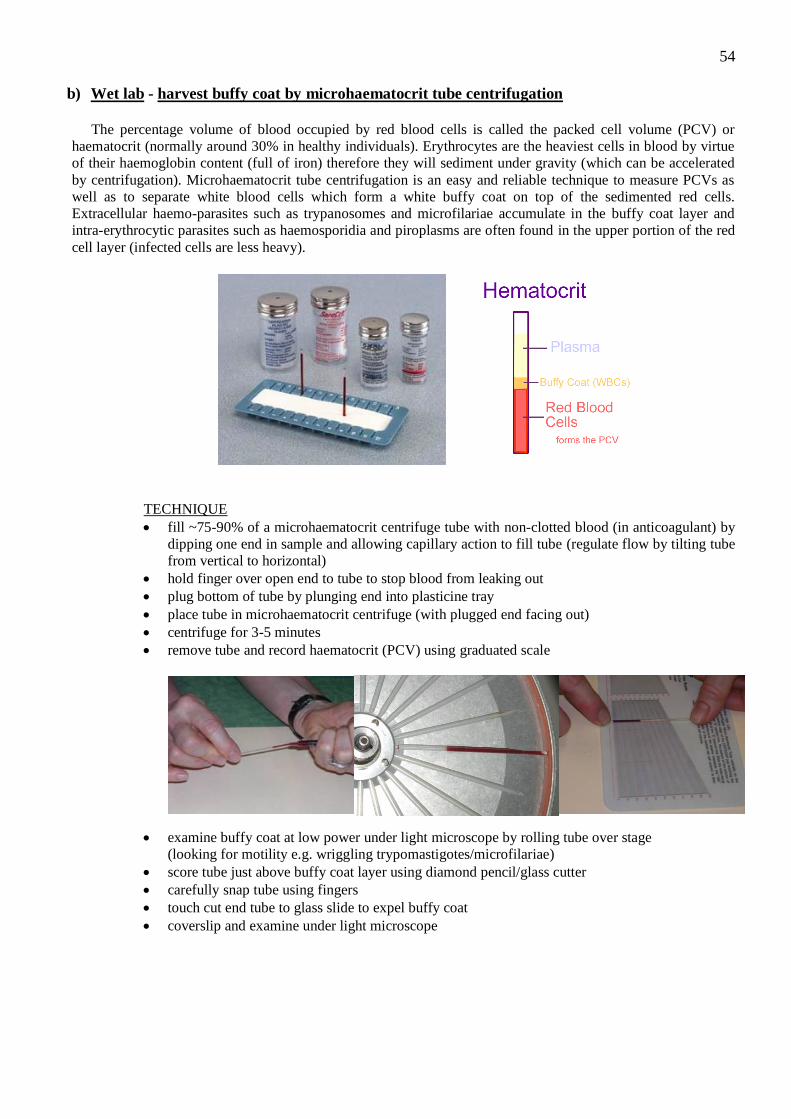
54
b) Wet lab - harvest buffy coat by microhaematocrit tube centrifugation
The percentage volume of blood occupied by red blood cells is called the packed cell volume (PCV) or
haematocrit (normally around 30% in healthy individuals). Erythrocytes are the heaviest cells in blood by virtue
of their haemoglobin content (full of iron) therefore they will sediment under gravity (which can be accelerated
by centrifugation). Microhaematocrit tube centrifugation is an easy and reliable technique to measure PCVs as
well as to separate white blood cells which form a white buffy coat on top of the sedimented red cells.
Extracellular haemo-parasites such as trypanosomes and microfilariae accumulate in the buffy coat layer and
intra-erythrocytic parasites such as haemosporidia and piroplasms are often found in the upper portion of the red
cell layer (infected cells are less heavy).
TECHNIQUE
fill ~75-90% of a microhaematocrit centrifuge tube with non-clotted blood (in anticoagulant) by
dipping one end in sample and allowing capillary action to fill tube (regulate flow by tilting tube
from vertical to horizontal)
hold finger over open end to tube to stop blood from leaking out
plug bottom of tube by plunging end into plasticine tray
place tube in microhaematocrit centrifuge (with plugged end facing out)
centrifuge for 3-5 minutes
remove tube and record haematocrit (PCV) using graduated scale
examine buffy coat at low power under light microscope by rolling tube over stage
(looking for motility e.g. wriggling trypomastigotes/microfilariae)
score tube just above buffy coat layer using diamond pencil/glass cutter
carefully snap tube using fingers
touch cut end tube to glass slide to expel buffy coat
coverslip and examine under light microscope
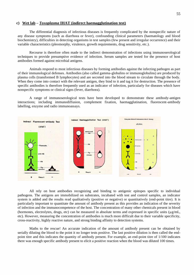
55
c) Wet lab – Toxoplasma IHAT (indirect haemagglutination test)
The differential diagnosis of infectious diseases is frequently complicated by the nonspecific nature of
any disease symptoms (such as diarrhoea or fever), confounding clinical parameters (haematology and blood
biochemistry), difficulties in detecting organisms in test samples (few present and irregular occurrence) and their
variable characteristics (pleomorphy, virulence, growth requirements, drug sensitivity, etc.).
Recourse is therefore often made to the indirect demonstration of infections using immunoserological
techniques to provide presumptive evidence of infection. Serum samples are tested for the presence of host
antibodies formed against microbial antigens.
Animals respond to most infectious diseases by forming antibodies against the infecting pathogen as part
of their immunological defenses. Antibodies (also called gamma-globulins or immunoglobulins) are produced by
plasma cells (transformed B lymphocytes) and are secreted into the blood stream to circulate through the body.
When they come into contact with the relevant antigen, they bind to it and tag it for destruction. The presence of
specific antibodies is therefore frequently used as an indicator of infection, particularly for diseases which have
nonspecific symptoms or clinical signs (fever, diarrhoea).
A range of immunoserological tests have been developed to demonstrate these antibody-antigen
interactions; including immunodiffusion, complement fixation, haemagglutination, fluorescent-antibody
labelling, enzyme and radio immunoassays.
All rely on host antibodies recognizing and binding to antigenic epitopes specific to individual
pathogens. The antigens are immobilized on substrates, incubated with test and control samples, an indicator
system is added and the results read qualitatively (positive or negative) or quantitatively (end-point titre). It is
particularly important to quantitate the amount of antibody present as this provides an indication of the severity
of infection and the immunocompetence of the host. The concentration of many other chemicals present in blood
(hormones, electrolytes, drugs, etc) can be measured in absolute terms and expressed in specific units (g/mL,
etc). However, measuring the concentration of antibodies is much more difficult due to their variable specificity,
cross-reactivity, highly reactive nature, and strong binding affinity to detection systems.
Maths to the rescue! An accurate indication of the amount of antibody present can be obtained by
serially diluting the blood to the point it no longer tests positive. The last positive dilution is then called the end-
point titre and this indicates the quantity of antibody present. For example, an end-point titre of 1/100 indicates
there was enough specific antibody present to elicit a positive reaction when the blood was diluted 100 times.
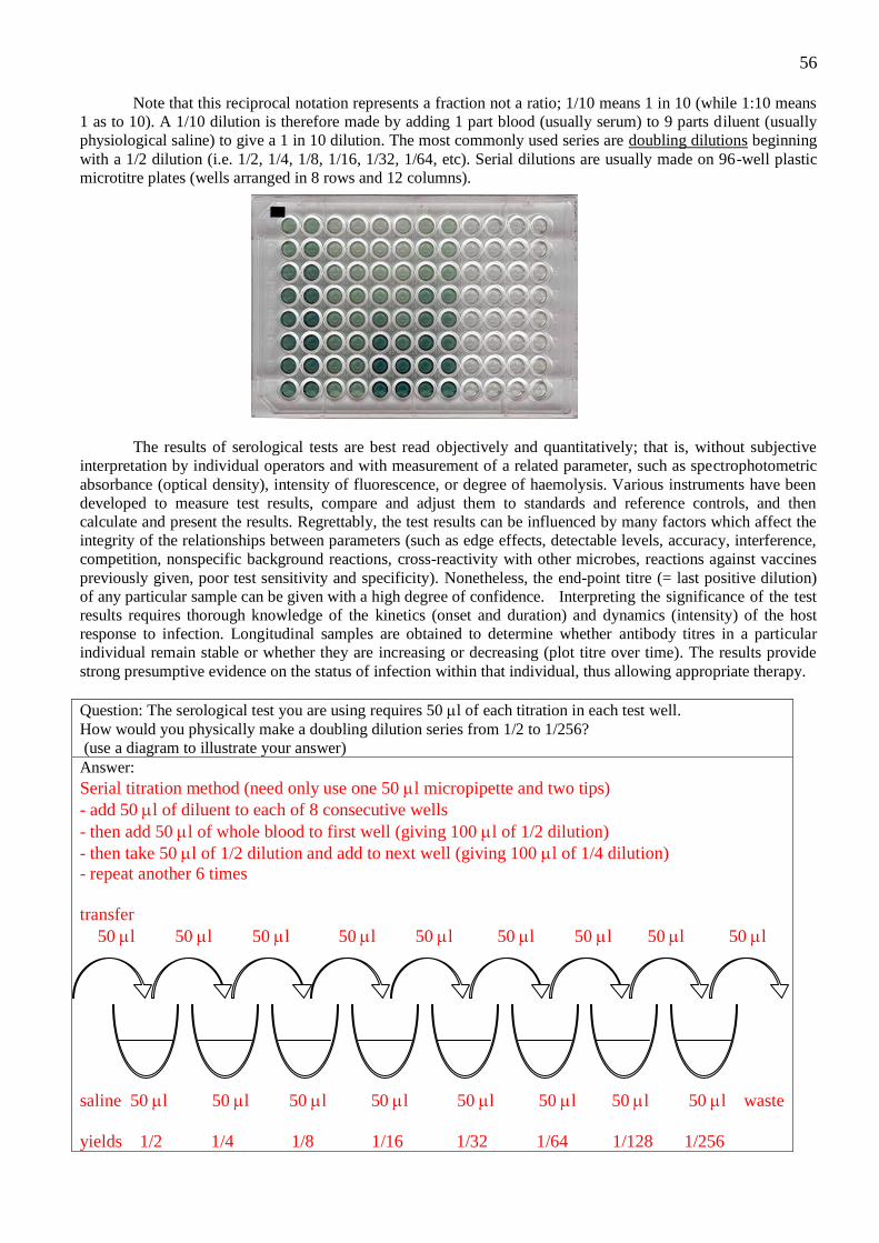
56
Note that this reciprocal notation represents a fraction not a ratio; 1/10 means 1 in 10 (while 1:10 means
1 as to 10). A 1/10 dilution is therefore made by adding 1 part blood (usually serum) to 9 parts diluent (usually
physiological saline) to give a 1 in 10 dilution. The most commonly used series are doubling dilutions beginning
with a 1/2 dilution (i.e. 1/2, 1/4, 1/8, 1/16, 1/32, 1/64, etc). Serial dilutions are usually made on 96-well plastic
microtitre plates (wells arranged in 8 rows and 12 columns).
The results of serological tests are best read objectively and quantitatively; that is, without subjective
interpretation by individual operators and with measurement of a related parameter, such as spectrophotometric
absorbance (optical density), intensity of fluorescence, or degree of haemolysis. Various instruments have been
developed to measure test results, compare and adjust them to standards and reference controls, and then
calculate and present the results. Regrettably, the test results can be influenced by many factors which affect the
integrity of the relationships between parameters (such as edge effects, detectable levels, accuracy, interference,
competition, nonspecific background reactions, cross-reactivity with other microbes, reactions against vaccines
previously given, poor test sensitivity and specificity). Nonetheless, the end-point titre (= last positive dilution)
of any particular sample can be given with a high degree of confidence. Interpreting the significance of the test
results requires thorough knowledge of the kinetics (onset and duration) and dynamics (intensity) of the host
response to infection. Longitudinal samples are obtained to determine whether antibody titres in a particular
individual remain stable or whether they are increasing or decreasing (plot titre over time). The results provide
strong presumptive evidence on the status of infection within that individual, thus allowing appropriate therapy.
Question: The serological test you are using requires 50 l of each titration in each test well.
How would you physically make a doubling dilution series from 1/2 to 1/256?
(use a diagram to illustrate your answer)
Answer:
Serial titration method (need only use one 50 l micropipette and two tips)
- add 50 l of diluent to each of 8 consecutive wells
- then add 50 l of whole blood to first well (giving 100 l of 1/2 dilution)
- then take 50 l of 1/2 dilution and add to next well (giving 100 l of 1/4 dilution)
- repeat another 6 times
transfer
50 l 50 l 50 l 50 l 50 l 50 l 50 l 50 l 50 l
saline 50 l 50 l 50 l 50 l 50 l 50 l 50 l 50 l waste
yields 1/2 1/4 1/8 1/16 1/32 1/64 1/128 1/256
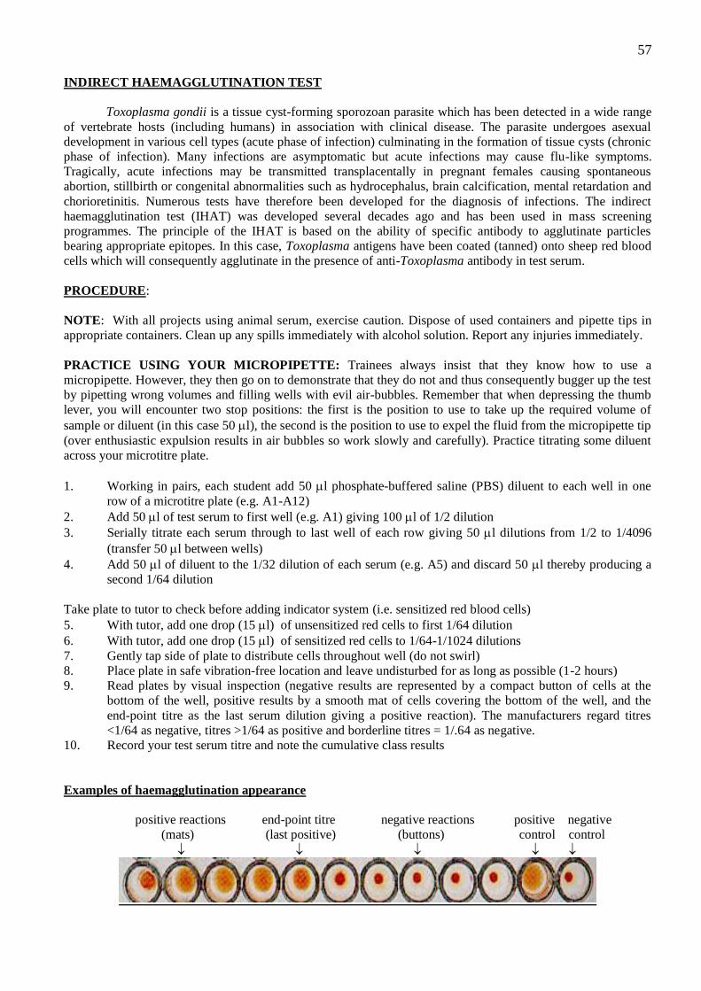
57
INDIRECT HAEMAGGLUTINATION TEST
Toxoplasma gondii is a tissue cyst-forming sporozoan parasite which has been detected in a wide range
of vertebrate hosts (including humans) in association with clinical disease. The parasite undergoes asexual
development in various cell types (acute phase of infection) culminating in the formation of tissue cysts (chronic
phase of infection). Many infections are asymptomatic but acute infections may cause flu-like symptoms.
Tragically, acute infections may be transmitted transplacentally in pregnant females causing spontaneous
abortion, stillbirth or congenital abnormalities such as hydrocephalus, brain calcification, mental retardation and
chorioretinitis. Numerous tests have therefore been developed for the diagnosis of infections. The indirect
haemagglutination test (IHAT) was developed several decades ago and has been used in mass screening
programmes. The principle of the IHAT is based on the ability of specific antibody to agglutinate particles
bearing appropriate epitopes. In this case, Toxoplasma antigens have been coated (tanned) onto sheep red blood
cells which will consequently agglutinate in the presence of anti-Toxoplasma antibody in test serum.
PROCEDURE:
NOTE: With all projects using animal serum, exercise caution. Dispose of used containers and pipette tips in
appropriate containers. Clean up any spills immediately with alcohol solution. Report any injuries immediately.
PRACTICE USING YOUR MICROPIPETTE: Trainees always insist that they know how to use a
micropipette. However, they then go on to demonstrate that they do not and thus consequently bugger up the test
by pipetting wrong volumes and filling wells with evil air-bubbles. Remember that when depressing the thumb
lever, you will encounter two stop positions: the first is the position to use to take up the required volume of
sample or diluent (in this case 50 l), the second is the position to use to expel the fluid from the micropipette tip
(over enthusiastic expulsion results in air bubbles so work slowly and carefully). Practice titrating some diluent
across your microtitre plate.
1. Working in pairs, each student add 50 l phosphate-buffered saline (PBS) diluent to each well in one
row of a microtitre plate (e.g. A1-A12)
2. Add 50 l of test serum to first well (e.g. A1) giving 100 l of 1/2 dilution
3. Serially titrate each serum through to last well of each row giving 50 l dilutions from 1/2 to 1/4096
(transfer 50 l between wells)
4. Add 50 l of diluent to the 1/32 dilution of each serum (e.g. A5) and discard 50 l thereby producing a
second 1/64 dilution
Take plate to tutor to check before adding indicator system (i.e. sensitized red blood cells)
5. With tutor, add one drop (15 l) of unsensitized red cells to first 1/64 dilution
6. With tutor, add one drop (15 l) of sensitized red cells to 1/64-1/1024 dilutions
7. Gently tap side of plate to distribute cells throughout well (do not swirl)
8. Place plate in safe vibration-free location and leave undisturbed for as long as possible (1-2 hours)
9. Read plates by visual inspection (negative results are represented by a compact button of cells at the
bottom of the well, positive results by a smooth mat of cells covering the bottom of the well, and the
end-point titre as the last serum dilution giving a positive reaction). The manufacturers regard titres
<1/64 as negative, titres >1/64 as positive and borderline titres = 1/.64 as negative.
10. Record your test serum titre and note the cumulative class results
Examples of haemagglutination appearance
positive reactions end-point titre negative reactions positive negative
(mats) (last positive) (buttons) control control
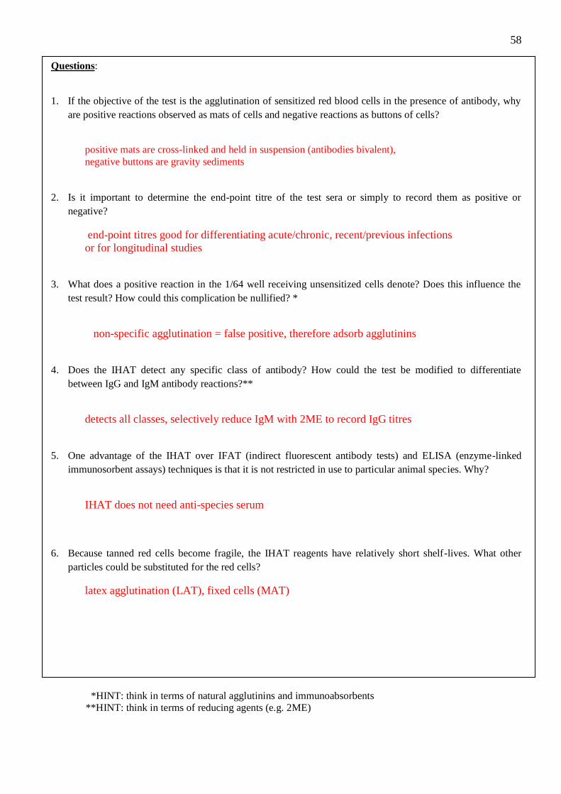
58
Questions:
1. If the objective of the test is the agglutination of sensitized red blood cells in the presence of antibody, why
are positive reactions observed as mats of cells and negative reactions as buttons of cells?
positive mats are cross-linked and held in suspension (antibodies bivalent),
negative buttons are gravity sediments
2. Is it important to determine the end-point titre of the test sera or simply to record them as positive or
negative?
end-point titres good for differentiating acute/chronic, recent/previous infections
or for longitudinal studies
3. What does a positive reaction in the 1/64 well receiving unsensitized cells denote? Does this influence the
test result? How could this complication be nullified? *
non-specific agglutination = false positive, therefore adsorb agglutinins
4. Does the IHAT detect any specific class of antibody? How could the test be modified to differentiate
between IgG and IgM antibody reactions?**
detects all classes, selectively reduce IgM with 2ME to record IgG titres
5. One advantage of the IHAT over IFAT (indirect fluorescent antibody tests) and ELISA (enzyme-linked
immunosorbent assays) techniques is that it is not restricted in use to particular animal species. Why?
IHAT does not need anti-species serum
6. Because tanned red cells become fragile, the IHAT reagents have relatively short shelf-lives. What other
particles could be substituted for the red cells?
latex agglutination (LAT), fixed cells (MAT)
*HINT: think in terms of natural agglutinins and immunoabsorbents
**HINT: think in terms of reducing agents (e.g. 2ME)
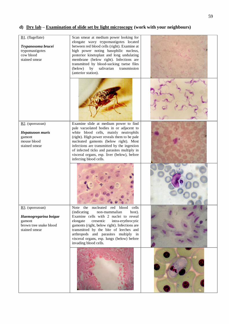
59
d) Dry lab – Examination of slide set by light microscopy (work with your neighbours)
B1. (flagellate)
Trypanosoma brucei
trypomastigotes
cow blood
stained smear
Scan smear at medium power looking for
elongate wavy trypomastigotes located
between red blood cells (right). Examine at
high power noting basophilic nucleus,
posterior kinetoplast and long undulating
membrane (below right). Infections are
transmitted by blood-sucking tsetse files
(below) by salivarian transmission
(anterior station).
B2. (sporozoan)
Hepatozoon muris
gamont
mouse blood
stained smear
Examine slide at medium power to find
pale vacuolated bodies in or adjacent to
white blood cells, mainly neutrophils
(right). High power reveals them to be pale
nucleated gamonts (below right). Most
infections are transmitted by the ingestion
of infected ticks and parasites multiply in
visceral organs, esp. liver (below), before
infecting blood cells.
B3. (sporozoan)
Haemogregarina boigae
gamont
brown tree snake blood
stained smear
Note the nucleated red blood cells
(indicating non-mammalian host).
Examine cells with 2 nuclei to reveal
elongate cresentic intra-erythrocytic
gamonts (right, below right). Infections are
transmitted by the bite of leeches and
arthropods and parasites multiply in
visceral organs, esp. lungs (below) before
invading blood cells.
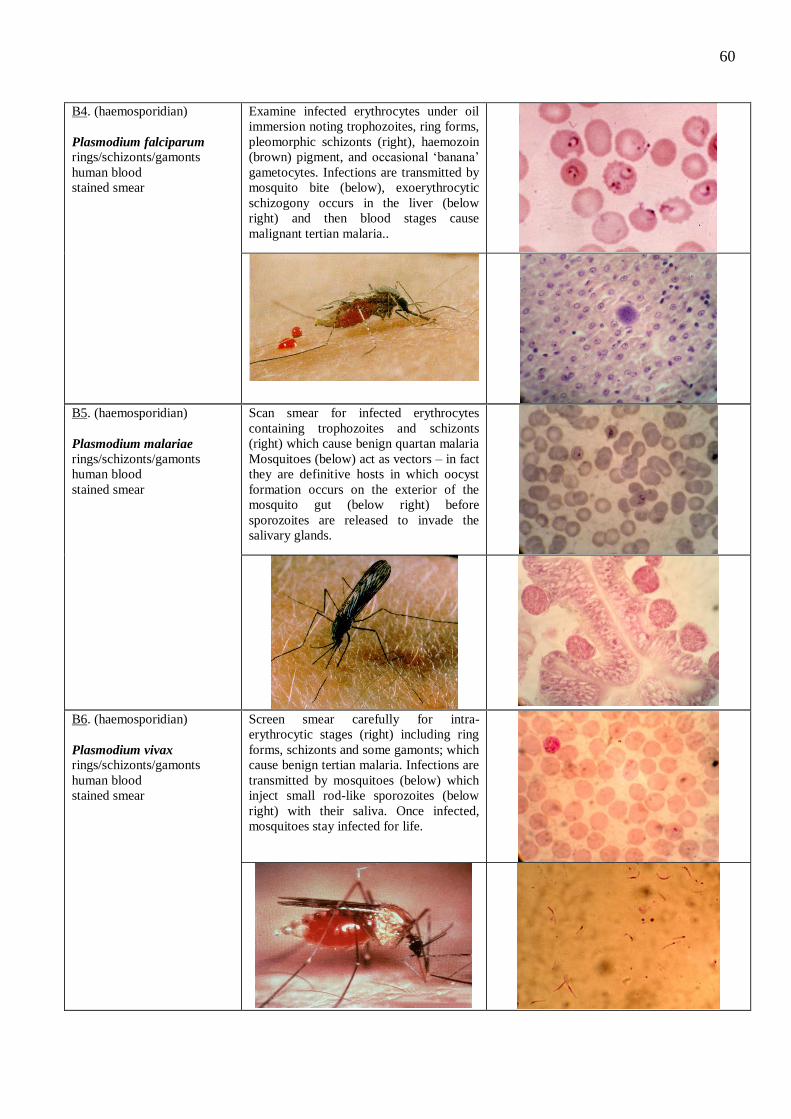
60
B4. (haemosporidian)
Plasmodium falciparum rings/schizonts/gamonts
human blood
stained smear
Examine infected erythrocytes under oil
immersion noting trophozoites, ring forms,
pleomorphic schizonts (right), haemozoin
(brown) pigment, and occasional ‘banana’
gametocytes. Infections are transmitted by
mosquito bite (below), exoerythrocytic
schizogony occurs in the liver (below
right) and then blood stages cause
malignant tertian malaria..
B5. (haemosporidian)
Plasmodium malariae
rings/schizonts/gamonts
human blood
stained smear
Scan smear for infected erythrocytes
containing trophozoites and schizonts
(right) which cause benign quartan malaria
Mosquitoes (below) act as vectors – in fact
they are definitive hosts in which oocyst
formation occurs on the exterior of the
mosquito gut (below right) before
sporozoites are released to invade the
salivary glands.
B6. (haemosporidian)
Plasmodium vivax rings/schizonts/gamonts
human blood
stained smear
Screen smear carefully for intra-
erythrocytic stages (right) including ring
forms, schizonts and some gamonts; which
cause benign tertian malaria. Infections are
transmitted by mosquitoes (below) which
inject small rod-like sporozoites (below
right) with their saliva. Once infected,
mosquitoes stay infected for life.
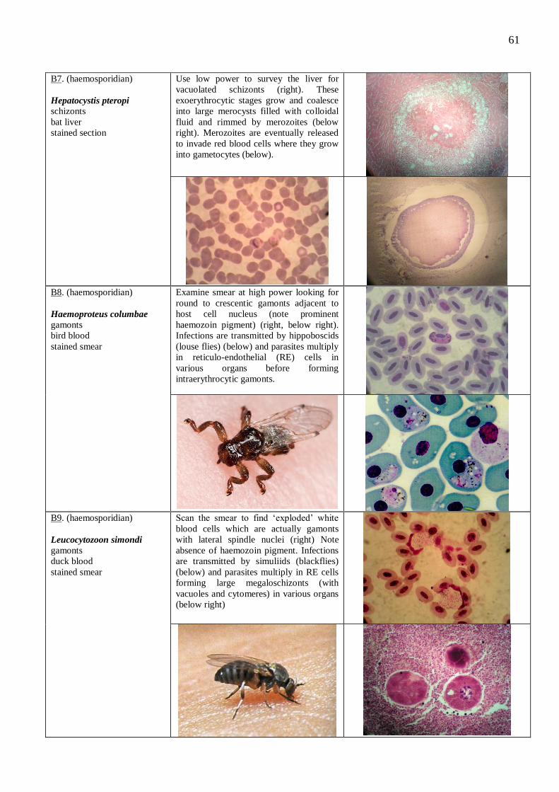
61
B7. (haemosporidian)
Hepatocystis pteropi schizonts
bat liver
stained section
Use low power to survey the liver for
vacuolated schizonts (right). These
exoerythrocytic stages grow and coalesce
into large merocysts filled with colloidal
fluid and rimmed by merozoites (below
right). Merozoites are eventually released
to invade red blood cells where they grow
into gametocytes (below).
B8. (haemosporidian)
Haemoproteus columbae
gamonts
bird blood
stained smear
Examine smear at high power looking for
round to crescentic gamonts adjacent to
host cell nucleus (note prominent
haemozoin pigment) (right, below right).
Infections are transmitted by hippoboscids
(louse flies) (below) and parasites multiply
in reticulo-endothelial (RE) cells in
various organs before forming
intraerythrocytic gamonts.
B9. (haemosporidian)
Leucocytozoon simondi
gamonts
duck blood
stained smear
Scan the smear to find ‘exploded’ white
blood cells which are actually gamonts
with lateral spindle nuclei (right) Note
absence of haemozoin pigment. Infections
are transmitted by simuliids (blackflies)
(below) and parasites multiply in RE cells
forming large megaloschizonts (with
vacuoles and cytomeres) in various organs
(below right)
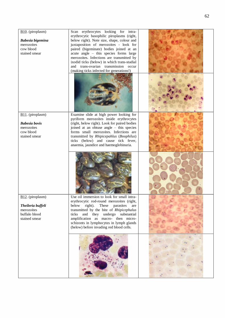
62
B10. (piroplasm)
Babesia bigemina
merozoites
cow blood
stained smear
Scan erythrocytes looking for intra-
erythrocytic basophilic piroplasms (right,
below right). Note size, shape, colour and
juxtaposition of merozoites - look for
paired (bigeminate) bodies joined at an
acute angle – this species forms large
merozoites. Infections are transmitted by
ixodid ticks (below) in which trans-stadial
and trans-ovarian transmission occur
(making ticks infected for generations!)
B11. (piroplasm)
Babesia bovis
merozoites
cow blood
stained smear
Examine slide at high power looking for
pyriform merozoites inside erythrocytes
(right, below right). Look for paired bodies
joined at an obtuse angle – this species
forms small merozoites. Infections are
transmitted by Rhipicepahlus (Boophilus)
ticks (below) and cause tick fever,
anaemia, jaundice and haemoglobinuria.
B12. (piroplasm)
Theileria buffeli
merozoites
buffalo blood
stained smear
Use oil immersion to look for small intra-
erythrocytic rod-round merozoites (right,
below right). These parasites are
transmitted by the bite of Rhipicephalus
ticks and they undergo substantial
amplification as macro- then micro-
schizonts in lymphocytes in lymph glands
(below) before invading red blood cells.
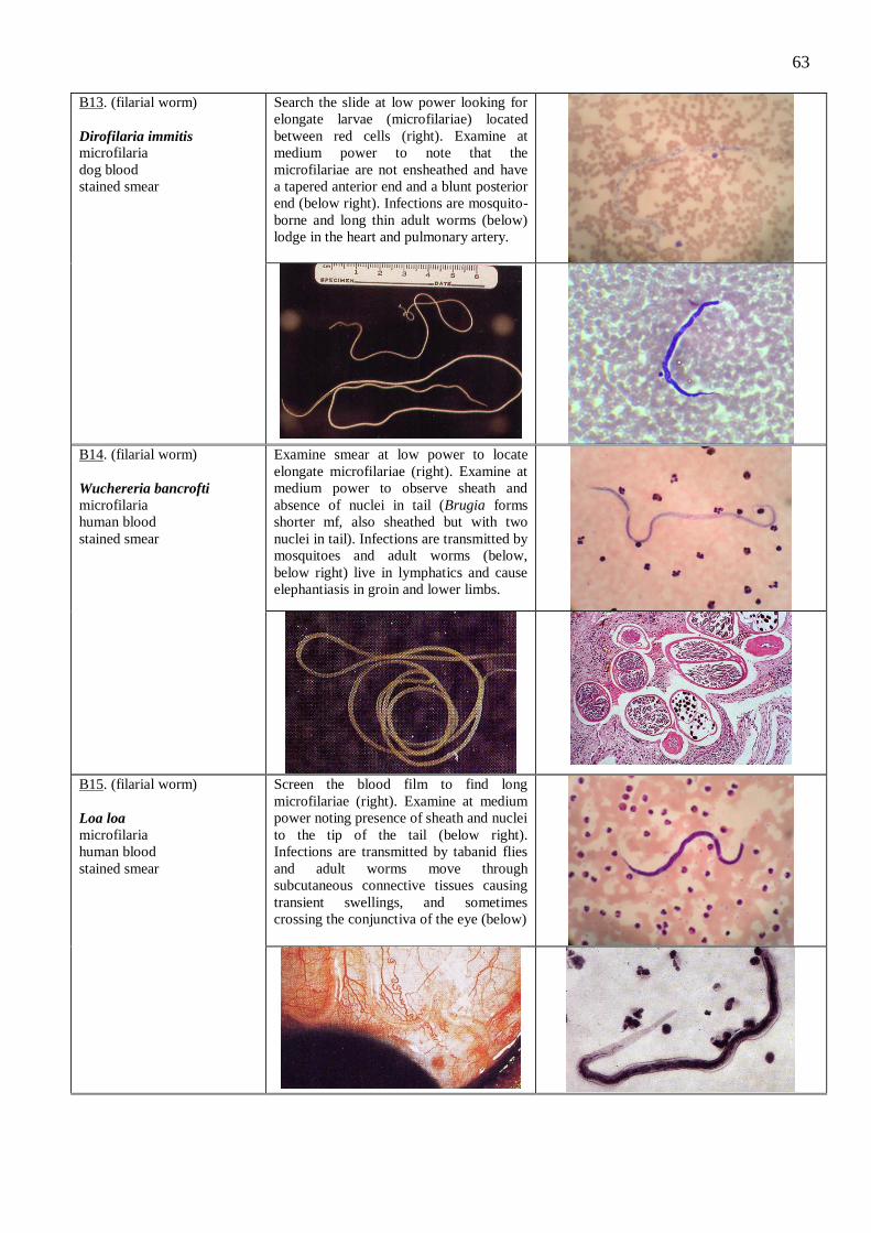
63
B13. (filarial worm)
Dirofilaria immitis microfilaria
dog blood
stained smear
Search the slide at low power looking for
elongate larvae (microfilariae) located
between red cells (right). Examine at
medium power to note that the
microfilariae are not ensheathed and have
a tapered anterior end and a blunt posterior
end (below right). Infections are mosquito-
borne and long thin adult worms (below)
lodge in the heart and pulmonary artery.
B14. (filarial worm)
Wuchereria bancrofti
microfilaria
human blood
stained smear
Examine smear at low power to locate
elongate microfilariae (right). Examine at
medium power to observe sheath and
absence of nuclei in tail (Brugia forms
shorter mf, also sheathed but with two
nuclei in tail). Infections are transmitted by
mosquitoes and adult worms (below,
below right) live in lymphatics and cause
elephantiasis in groin and lower limbs.
B15. (filarial worm)
Loa loa
microfilaria
human blood
stained smear
Screen the blood film to find long
microfilariae (right). Examine at medium
power noting presence of sheath and nuclei
to the tip of the tail (below right).
Infections are transmitted by tabanid flies
and adult worms move through
subcutaneous connective tissues causing
transient swellings, and sometimes
crossing the conjunctiva of the eye (below)
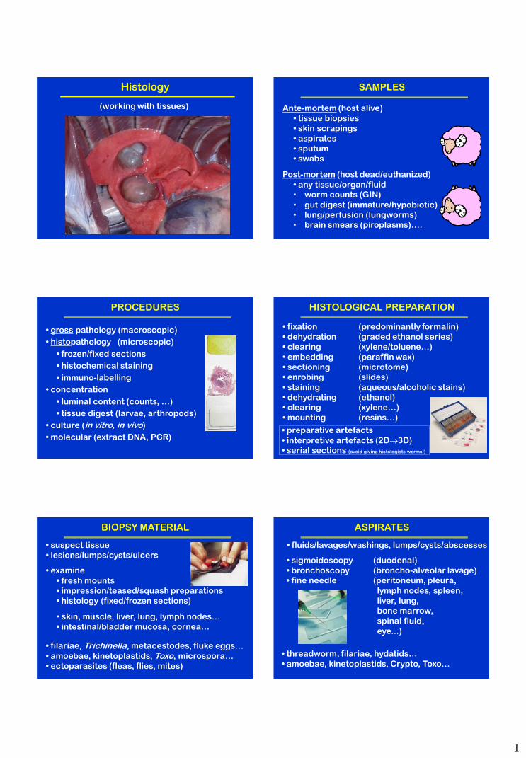
1
Histology
(working with tissues)
SAMPLES
Ante-mortem (host alive)
• tissue biopsies
• skin scrapings
• aspirates
• sputum
• swabs
X X
Post-mortem (host dead/euthanized)
• any tissue/organ/fluid
• worm counts (GIN)
• gut digest (immature/hypobiotic)
• lung/perfusion (lungworms)
• brain smears (piroplasms)….
PROCEDURES
• gross pathology (macroscopic)
• histopathology (microscopic)
• frozen/fixed sections
• histochemical staining
• immuno-labelling
• concentration
• luminal content (counts, …)
• tissue digest (larvae, arthropods)
• culture (in vitro, in vivo)
• molecular (extract DNA, PCR)
HISTOLOGICAL PREPARATION
• fixation (predominantly formalin)
• dehydration (graded ethanol series)
• clearing (xylene/toluene…)
• embedding (paraffin wax)
• sectioning (microtome)
• enrobing (slides)
• staining (aqueous/alcoholic stains)
• dehydrating (ethanol)
• clearing (xylene…)
• mounting (resins…)
• preparative artefacts
• interpretive artefacts (2D3D)
• serial sections (avoid giving histologists worms!)
BIOPSY MATERIAL
• examine
• fresh mounts
• impression/teased/squash preparations
• histology (fixed/frozen sections)
• filariae, Trichinella, metacestodes, fluke eggs…
• amoebae, kinetoplastids, Toxo, microspora…
• ectoparasites (fleas, flies, mites)
• skin, muscle, liver, lung, lymph nodes…
• intestinal/bladder mucosa, cornea…
• suspect tissue
• lesions/lumps/cysts/ulcers
ASPIRATES
• sigmoidoscopy (duodenal)
• bronchoscopy (broncho-alveolar lavage)
• fine needle (peritoneum, pleura,
lymph nodes, spleen,
liver, lung,
bone marrow,
spinal fluid,
eye...)
• threadworm, filariae, hydatids…
• amoebae, kinetoplastids, Crypto, Toxo…
• fluids/lavages/washings, lumps/cysts/abscesses
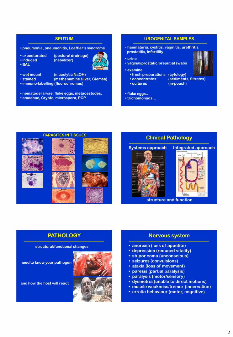
2
SPUTUM
• expectorated (postural drainage)
• induced (nebulizer)
• BAL
• wet mount (mucolytic NaOH)
• stained (methenamine silver, Giemsa)
• immuno-labelling (fluorochromes)
• pneumonia, pneumonitis, Loeffler’s syndrome
• nematode larvae, fluke eggs, metacestodes,
• amoebae, Crypto, microspora, PCP
UROGENITAL SAMPLES
• examine
• fresh preparations (cytology)
• concentrates (sediments, filtrates)
• cultures (in-pouch)
• fluke eggs…
• trichomonads…
• haematuria, cystitis, vaginitis, urethritis,
prostatitis, infertility
• urine
• vaginal/prostatic/preputial swabs
Trichinella
Leishmania
Toxoplasma
Trypanosoma cruzi
Sarcocystis
Thelohania
Kudoa
Onchocerca
Echinococcus
Schistosoma
PARASITES IN TISSUES Toxoplasma Clinical Pathology
Systems approach Integrated approach
structure and function
PATHOLOGY
structural/functional changes
and how the host will react
need to know your pathogen
Nervous system
• anorexia (loss of appetite)
• depression (reduced vitality)
• stupor coma (unconscious)
• seizures (convulsions)
• ataxia (loss of movement)
• paresis (partial paralysis)
• paralysis (motor/sensory)
• dysmetria (unable to direct motions)
• muscle weakness/tremor (innervation)
• erratic behaviour (motor, cognitive)
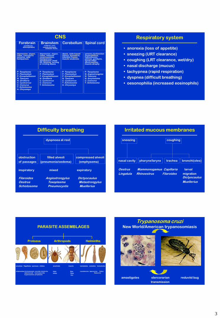
3
CNS Forebrain
(cerebrum, diencephalon)
Brainstem (midbrain, pons,
medulla oblongata, vestibular nuclei)
Cerebellum
Spinal cord
depression, stupor, coma, convulsive seizures, mild hemiparesis
depression, stupor, coma, reduced consciousness, hemiparesis, tetraparesis, head tilt, twisting, turning or circling ataxia
ataxia, wide-based stance, dysmetria, intention tremor, muscle weakness
sensory dysfunction (hypersthesia, hyposthesia), paresis, paralysis, spinal reflex abnormalities, muscle atrophy
P: Toxoplasma P: Plasmodium P: Encephalitozoon N: Toxocara N: Dirofilaria C: Cysticercus C: Coenurus T: Schistosoma A: Chrysomya
P: Toxoplasma P: Plasmodium P: Encephalitozoon N: Dirofilaria C: Cysticercus C: Coenurus T: Schistosoma
P: Toxoplasma P: Plasmodium P: Encephalitozoon N: Toxocara C: Cysticercus T: Schistosoma A: Chrysomya
P: Toxoplasma N: Angiostrongylus N: Toxocara N: Ancylostoma C: Coenurus T: Schistosoma
Respiratory system
• anorexia (loss of appetite)
• sneezing (URT clearance)
• coughing (LRT clearance, wet/dry)
• nasal discharge (mucus)
• tachypnea (rapid respiration)
• dyspnea (difficult breathing)
• oesonophilia (increased eosinophils)
Difficulty breathing
dyspnoea at rest
obstruction filled alveoli compressed alveoli
of passages (pneumonia/oedema) (emphysema)
inspiratory mixed expiratory
Filaroides Angiostrongylus Dictyocaulus
Oestrus Toxoplasma Metastrongylus
Schistosoma Pneumocystis Muellerius
Irritated mucous membranes
sneezing coughing
nasal cavity pharynx/larynx trachea bronchi(oles)
Oestrus Mammonogamus Capillaria larval
Lingatula Rhinoestrus Filaroides migration Dictyocaulus
Muellerius
Protozoa Arthropods Helminths
amoebae flagellates sporozoa ciliates
entamoebae trichomonads coccidia holotrichs diplomonads haemosporidia trypanosomes piroplasms
arachnids insects
ticks flies
mites fleas
lice
nematodes cestodes trematodes
roundworms tapeworms flukes flatworms
PARASITE ASSEMBLAGES
Trypanosoma cruzi New World/American trypanosomiasis
amastigotes stercorarian reduviid bug
transmission
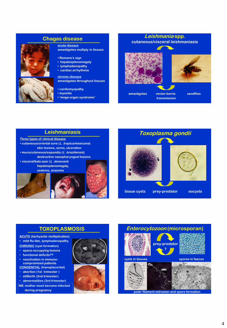
4
Chagas disease acute disease
amastigotes multiply in tissues
• Romana’s sign
• hepatosplenomegaly
• lymphadenopathy
• cardiac arrhythmia
chronic disease
amastigotes throughout tissues
• cardiomyopathy
• myositis
• ‘mega-organ syndrome’
amastigotes vector-borne sandflies
transmission
Leishmania spp. cutaneous/visceral leishmaniasis
Three types of clinical disease
• cutaneous/oriental sore (L. tropica/mexicana)
skin lesions, sores, ulceration
• mucocutaneous/espundia (L. braziliensis)
destructive nasopharyngeal lesions
• visceral/kala azar (L. donovani)
hepatosplenomegaly,
oedema, anaemia
Leishmaniasis Toxoplasma gondii
tissue cysts prey-predator oocysts
TOXOPLASMOSIS
CHRONIC (cyst formation)
• space-occupying lesions
• functional deficits??
• reactivation in immuno-
compromised patients
ACUTE (tachyzoite multiplication)
• mild flu-like, lymphadenopathy
CONGENITAL (transplacental)
• abortion (1st trimester )
• stillbirth (2nd trimester)
• abnormalities (3rd trimester)
NB: mother must become infected
during pregnancy
Enterocytozoon (microsporan)
prey-predator
?
cysts in tissues spores in faeces
polar filament extrusion and spore formation
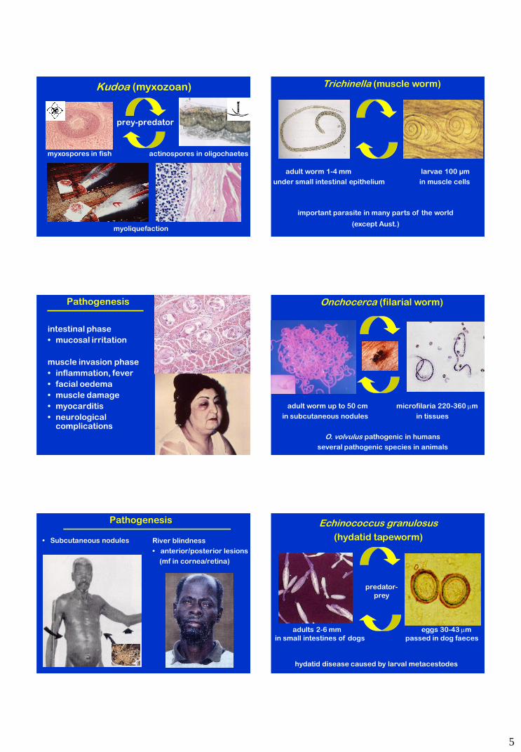
5
Kudoa (myxozoan)
prey-predator
myxospores in fish actinospores in oligochaetes
myoliquefaction
Trichinella (muscle worm)
adult worm 1-4 mm larvae 100 µm
under small intestinal epithelium in muscle cells
important parasite in many parts of the world
(except Aust.)
Pathogenesis
intestinal phase
• mucosal irritation
muscle invasion phase
• inflammation, fever
• facial oedema
• muscle damage
• myocarditis
• neurological complications
Onchocerca (filarial worm)
adult worm up to 50 cm microfilaria 220-360 m
in subcutaneous nodules in tissues
O. volvulus pathogenic in humans
several pathogenic species in animals
Pathogenesis
• Subcutaneous nodules River blindness
• anterior/posterior lesions
(mf in cornea/retina)
Echinococcus granulosus
(hydatid tapeworm)
predator-
prey
adults 2-6 mm eggs 30-43 m
in small intestines of dogs passed in dog faeces
hydatid disease caused by larval metacestodes
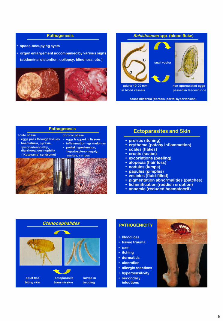
6
Pathogenesis
• space-occupying cysts
• organ enlargement accompanied by various signs
(abdominal distention, epilepsy, blindness, etc.)
Schistosoma spp. (blood fluke)
snail vector
adults 10-20 mm non-operculated eggs
in blood vessels passed in faeces/urine
cause bilharzia (fibrosis, portal hypertension)
O +
O
Pathogenesis
chronic phase
• eggs trapped in tissues
• inflammationgranulomas
• portal hypertension,
hepatosplenomegaly,
ascites, varices
acute phase
• eggs pass through tissues
• haematuria, pyrexia,
lymphadenopathy, diarrhoea, oesinophilia
(‘Katayama’ syndrome)
Ectoparasites and Skin
• pruritis (itching) • erythema (patchy inflammation) • scales (flakes) • crusts (scabs) • excoriations (peeling) • alopecia (hair loss) • nodules (lumps) • papules (pimples) • vesicles (fluid-filled) • pigmentation abnormalities (patches) • lichenification (reddish eruption) • anaemia (reduced haematocrit)
adult flea ectoparasite larvae in
biting skin transmission bedding
Ctenocephalides PATHOGENICITY
• blood loss
• tissue trauma
• pain
• itching
• dermatitis
• ulceration
• allergic reactions
• hypersensitivity
• secondary
infections
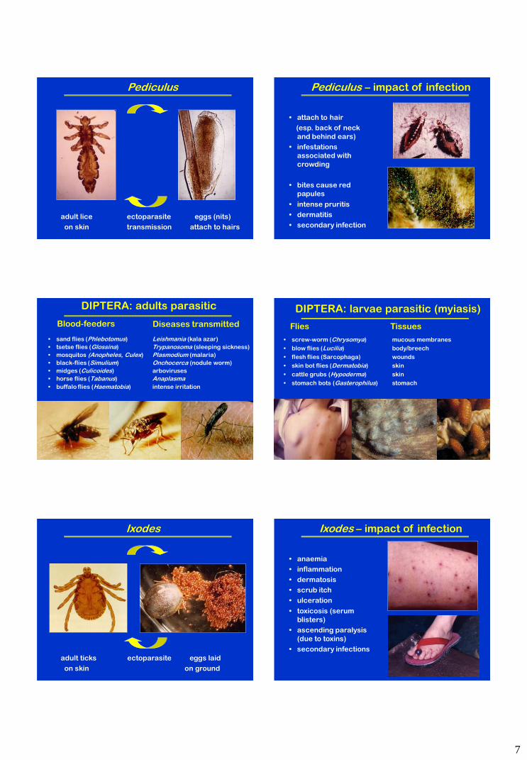
7
adult lice ectoparasite eggs (nits)
on skin transmission attach to hairs
Pediculus Pediculus – impact of infection
• attach to hair
(esp. back of neck
and behind ears)
• infestations
associated with
crowding
• bites cause red
papules
• intense pruritis
• dermatitis
• secondary infection
DIPTERA: adults parasitic
• sand flies (Phlebotomus)
• tsetse flies (Glossina)
• mosquitos (Anopheles, Culex)
• black-flies (Simulium)
• midges (Culicoides)
• horse flies (Tabanus)
• buffalo flies (Haematobia)
Leishmania (kala azar)
Trypanosoma (sleeping sickness)
Plasmodium (malaria)
Onchocerca (nodule worm)
arboviruses
Anaplasma
intense irritation
Diseases transmitted Blood-feeders
DIPTERA: larvae parasitic (myiasis)
• screw-worm (Chrysomya) mucous membranes
• blow flies (Lucilia) body/breech
• flesh flies (Sarcophaga) wounds
• skin bot flies (Dermatobia) skin
• cattle grubs (Hypoderma) skin
• stomach bots (Gasterophilus) stomach
Flies Tissues
adult ticks ectoparasite eggs laid
on skin on ground
Ixodes Ixodes – impact of infection
• anaemia
• inflammation
• dermatosis
• scrub itch
• ulceration
• toxicosis (serum
blisters)
• ascending paralysis
(due to toxins)
• secondary infections
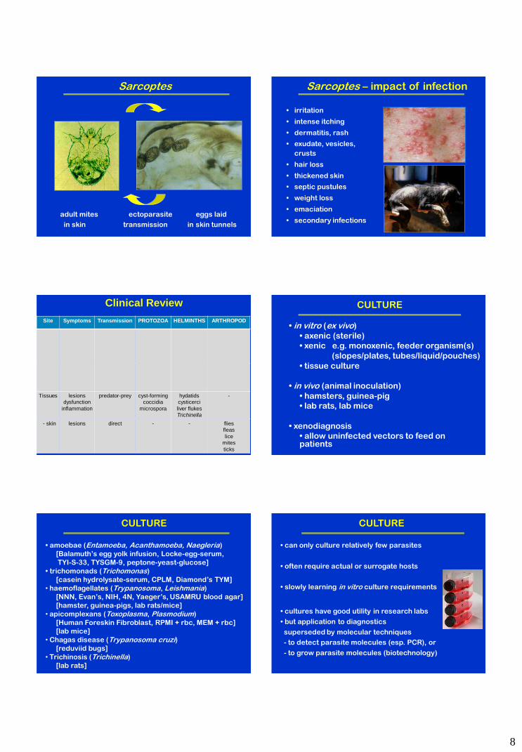
8
adult mites ectoparasite eggs laid
in skin transmission in skin tunnels
Sarcoptes Sarcoptes – impact of infection
• irritation
• intense itching
• dermatitis, rash
• exudate, vesicles,
crusts
• hair loss
• thickened skin
• septic pustules
• weight loss
• emaciation
• secondary infections
Clinical Review
Site Symptoms Transmission PROTOZOA HELMINTHS ARTHROPOD
Tissues lesions dysfunction
inflammation
predator-prey cyst-forming coccidia
microspora
hydatids cysticerci
liver flukes
Trichinella
-
- skin lesions
direct - - flies fleas
lice mites
ticks
CULTURE
• in vitro (ex vivo)
• axenic (sterile)
• xenic e.g. monoxenic, feeder organism(s)
(slopes/plates, tubes/liquid/pouches)
• tissue culture
• in vivo (animal inoculation)
• hamsters, guinea-pig
• lab rats, lab mice
• xenodiagnosis
• allow uninfected vectors to feed on patients
CULTURE
• amoebae (Entamoeba, Acanthamoeba, Naegleria)
[Balamuth’s egg yolk infusion, Locke-egg-serum,
TYI-S-33, TYSGM-9, peptone-yeast-glucose]
• trichomonads (Trichomonas)
[casein hydrolysate-serum, CPLM, Diamond’s TYM]
• haemoflagellates (Trypanosoma, Leishmania)
[NNN, Evan’s, NIH, 4N, Yaeger’s, USAMRU blood agar]
[hamster, guinea-pigs, lab rats/mice]
• apicomplexans (Toxoplasma, Plasmodium)
[Human Foreskin Fibroblast, RPMI + rbc, MEM + rbc]
[lab mice]
• Chagas disease (Trypanosoma cruzi) [reduviid bugs]
• Trichinosis (Trichinella)
[lab rats]
CULTURE
• can only culture relatively few parasites
• often require actual or surrogate hosts
• slowly learning in vitro culture requirements
• cultures have good utility in research labs
• but application to diagnostics
superseded by molecular techniques
- to detect parasite molecules (esp. PCR), or
- to grow parasite molecules (biotechnology)
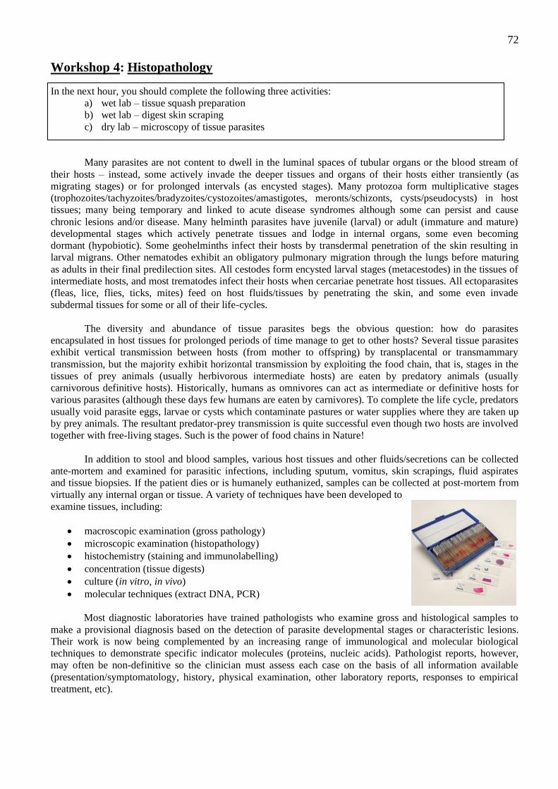
72
Workshop 4: Histopathology
In the next hour, you should complete the following three activities:
a) wet lab – tissue squash preparation
b) wet lab – digest skin scraping
c) dry lab – microscopy of tissue parasites
Many parasites are not content to dwell in the luminal spaces of tubular organs or the blood stream of
their hosts – instead, some actively invade the deeper tissues and organs of their hosts either transiently (as
migrating stages) or for prolonged intervals (as encysted stages). Many protozoa form multiplicative stages
(trophozoites/tachyzoites/bradyzoites/cystozoites/amastigotes, meronts/schizonts, cysts/pseudocysts) in host
tissues; many being temporary and linked to acute disease syndromes although some can persist and cause
chronic lesions and/or disease. Many helminth parasites have juvenile (larval) or adult (immature and mature)
developmental stages which actively penetrate tissues and lodge in internal organs, some even becoming
dormant (hypobiotic). Some geohelminths infect their hosts by transdermal penetration of the skin resulting in
larval migrans. Other nematodes exhibit an obligatory pulmonary migration through the lungs before maturing
as adults in their final predilection sites. All cestodes form encysted larval stages (metacestodes) in the tissues of
intermediate hosts, and most trematodes infect their hosts when cercariae penetrate host tissues. All ectoparasites
(fleas, lice, flies, ticks, mites) feed on host fluids/tissues by penetrating the skin, and some even invade
subdermal tissues for some or all of their life-cycles.
The diversity and abundance of tissue parasites begs the obvious question: how do parasites
encapsulated in host tissues for prolonged periods of time manage to get to other hosts? Several tissue parasites
exhibit vertical transmission between hosts (from mother to offspring) by transplacental or transmammary
transmission, but the majority exhibit horizontal transmission by exploiting the food chain, that is, stages in the
tissues of prey animals (usually herbivorous intermediate hosts) are eaten by predatory animals (usually
carnivorous definitive hosts). Historically, humans as omnivores can act as intermediate or definitive hosts for
various parasites (although these days few humans are eaten by carnivores). To complete the life cycle, predators
usually void parasite eggs, larvae or cysts which contaminate pastures or water supplies where they are taken up
by prey animals. The resultant predator-prey transmission is quite successful even though two hosts are involved
together with free-living stages. Such is the power of food chains in Nature!
In addition to stool and blood samples, various host tissues and other fluids/secretions can be collected
ante-mortem and examined for parasitic infections, including sputum, vomitus, skin scrapings, fluid aspirates
and tissue biopsies. If the patient dies or is humanely euthanized, samples can be collected at post-mortem from
virtually any internal organ or tissue. A variety of techniques have been developed to
examine tissues, including:
macroscopic examination (gross pathology)
microscopic examination (histopathology)
histochemistry (staining and immunolabelling)
concentration (tissue digests)
culture (in vitro, in vivo)
molecular techniques (extract DNA, PCR)
Most diagnostic laboratories have trained pathologists who examine gross and histological samples to
make a provisional diagnosis based on the detection of parasite developmental stages or characteristic lesions.
Their work is now being complemented by an increasing range of immunological and molecular biological
techniques to demonstrate specific indicator molecules (proteins, nucleic acids). Pathologist reports, however,
may often be non-definitive so the clinician must assess each case on the basis of all information available
(presentation/symptomatology, history, physical examination, other laboratory reports, responses to empirical
treatment, etc).
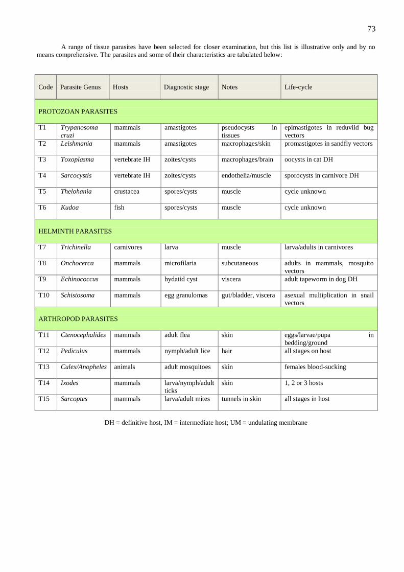
73
A range of tissue parasites have been selected for closer examination, but this list is illustrative only and by no
means comprehensive. The parasites and some of their characteristics are tabulated below:
Code
Parasite Genus
Hosts
Diagnostic stage
Notes
Life-cycle
PROTOZOAN PARASITES
T1 Trypanosoma
cruzi
mammals amastigotes pseudocysts in
tissues
epimastigotes in reduviid bug
vectors
T2 Leishmania
mammals amastigotes macrophages/skin promastigotes in sandfly vectors
T3 Toxoplasma
vertebrate IH zoites/cysts macrophages/brain oocysts in cat DH
T4 Sarcocystis
vertebrate IH zoites/cysts endothelia/muscle sporocysts in carnivore DH
T5 Thelohania
crustacea spores/cysts muscle cycle unknown
T6 Kudoa
fish spores/cysts muscle cycle unknown
HELMINTH PARASITES
T7 Trichinella
carnivores larva muscle larva/adults in carnivores
T8 Onchocerca
mammals microfilaria subcutaneous adults in mammals, mosquito
vectors
T9 Echinococcus
mammals hydatid cyst viscera adult tapeworm in dog DH
T10 Schistosoma mammals egg granulomas gut/bladder, viscera asexual multiplication in snail
vectors
ARTHROPOD PARASITES
T11 Ctenocephalides mammals adult flea skin eggs/larvae/pupa in
bedding/ground
T12 Pediculus
mammals nymph/adult lice hair all stages on host
T13 Culex/Anopheles
animals adult mosquitoes skin females blood-sucking
T14 Ixodes mammals larva/nymph/adult
ticks
skin 1, 2 or 3 hosts
T15 Sarcoptes
mammals larva/adult mites tunnels in skin all stages in host
DH = definitive host, IM = intermediate host; UM = undulating membrane
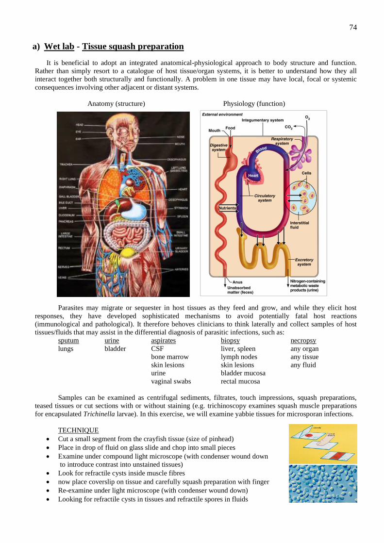
74
a) Wet lab - Tissue squash preparation
It is beneficial to adopt an integrated anatomical-physiological approach to body structure and function.
Rather than simply resort to a catalogue of host tissue/organ systems, it is better to understand how they all
interact together both structurally and functionally. A problem in one tissue may have local, focal or systemic
consequences involving other adjacent or distant systems.
Anatomy (structure) Physiology (function)
Parasites may migrate or sequester in host tissues as they feed and grow, and while they elicit host
responses, they have developed sophisticated mechanisms to avoid potentially fatal host reactions
(immunological and pathological). It therefore behoves clinicians to think laterally and collect samples of host
tissues/fluids that may assist in the differential diagnosis of parasitic infections, such as:
sputum urine aspirates biopsy necropsy
lungs bladder CSF liver, spleen any organ
bone marrow lymph nodes any tissue
skin lesions skin lesions any fluid
urine bladder mucosa
vaginal swabs rectal mucosa
Samples can be examined as centrifugal sediments, filtrates, touch impressions, squash preparations,
teased tissues or cut sections with or without staining (e.g. trichinoscopy examines squash muscle preparations
for encapsulated Trichinella larvae). In this exercise, we will examine yabbie tissues for microsporan infections.
TECHNIQUE
Cut a small segment from the crayfish tissue (size of pinhead)
Place in drop of fluid on glass slide and chop into small pieces
Examine under compound light microscope (with condenser wound down
to introduce contrast into unstained tissues)
Look for refractile cysts inside muscle fibres
now place coverslip on tissue and carefully squash preparation with finger
Re-examine under light microscope (with condenser wound down)
Looking for refractile cysts in tissues and refractile spores in fluids
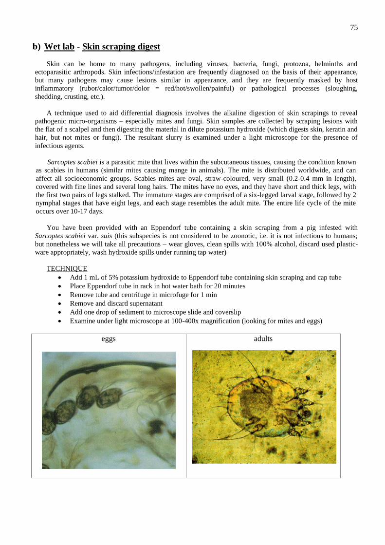
75
b) Wet lab - Skin scraping digest
Skin can be home to many pathogens, including viruses, bacteria, fungi, protozoa, helminths and
ectoparasitic arthropods. Skin infections/infestation are frequently diagnosed on the basis of their appearance,
but many pathogens may cause lesions similar in appearance, and they are frequently masked by host
inflammatory (rubor/calor/tumor/dolor = red/hot/swollen/painful) or pathological processes (sloughing,
shedding, crusting, etc.).
A technique used to aid differential diagnosis involves the alkaline digestion of skin scrapings to reveal
pathogenic micro-organisms – especially mites and fungi. Skin samples are collected by scraping lesions with
the flat of a scalpel and then digesting the material in dilute potassium hydroxide (which digests skin, keratin and
hair, but not mites or fungi). The resultant slurry is examined under a light microscope for the presence of
infectious agents.
Sarcoptes scabiei is a parasitic mite that lives within the subcutaneous tissues, causing the condition known
as scabies in humans (similar mites causing mange in animals). The mite is distributed worldwide, and can
affect all socioeconomic groups. Scabies mites are oval, straw-coloured, very small (0.2-0.4 mm in length),
covered with fine lines and several long hairs. The mites have no eyes, and they have short and thick legs, with
the first two pairs of legs stalked. The immature stages are comprised of a six-legged larval stage, followed by 2
nymphal stages that have eight legs, and each stage resembles the adult mite. The entire life cycle of the mite
occurs over 10-17 days.
You have been provided with an Eppendorf tube containing a skin scraping from a pig infested with
Sarcoptes scabiei var. suis (this subspecies is not considered to be zoonotic, i.e. it is not infectious to humans;
but nonetheless we will take all precautions – wear gloves, clean spills with 100% alcohol, discard used plastic-
ware appropriately, wash hydroxide spills under running tap water)
TECHNIQUE
Add 1 mL of 5% potassium hydroxide to Eppendorf tube containing skin scraping and cap tube
Place Eppendorf tube in rack in hot water bath for 20 minutes
Remove tube and centrifuge in microfuge for 1 min
Remove and discard supernatant
Add one drop of sediment to microscope slide and coverslip
Examine under light microscope at 100-400x magnification (looking for mites and eggs)
eggs
adults
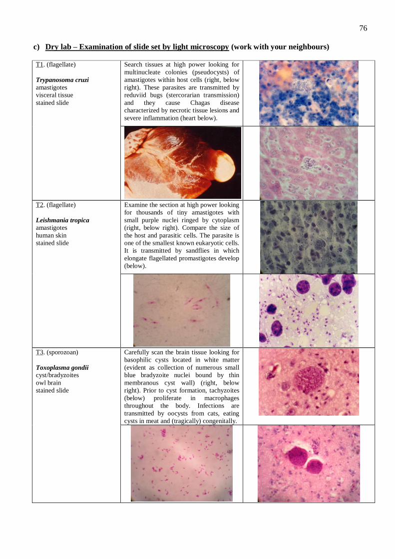
76
c) Dry lab – Examination of slide set by light microscopy (work with your neighbours)
T1. (flagellate)
Trypanosoma cruzi
amastigotes
visceral tissue
stained slide
Search tissues at high power looking for
multinucleate colonies (pseudocysts) of
amastigotes within host cells (right, below
right). These parasites are transmitted by
reduviid bugs (stercorarian transmission)
and they cause Chagas disease
characterized by necrotic tissue lesions and
severe inflammation (heart below).
T2. (flagellate)
Leishmania tropica amastigotes
human skin
stained slide
Examine the section at high power looking
for thousands of tiny amastigotes with
small purple nuclei ringed by cytoplasm
(right, below right). Compare the size of
the host and parasitic cells. The parasite is
one of the smallest known eukaryotic cells.
It is transmitted by sandflies in which
elongate flagellated promastigotes develop
(below).
T3. (sporozoan)
Toxoplasma gondii
cyst/bradyzoites
owl brain
stained slide
Carefully scan the brain tissue looking for
basophilic cysts located in white matter
(evident as collection of numerous small
blue bradyzoite nuclei bound by thin
membranous cyst wall) (right, below
right). Prior to cyst formation, tachyzoites
(below) proliferate in macrophages
throughout the body. Infections are
transmitted by oocysts from cats, eating
cysts in meat and (tragically) congenitally.
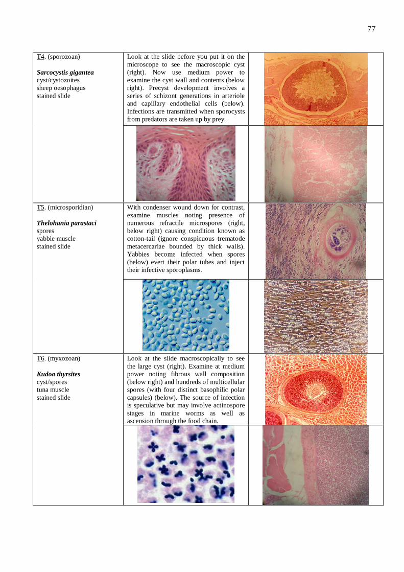
77
T4. (sporozoan)
Sarcocystis gigantea
cyst/cystozoites
sheep oesophagus
stained slide
Look at the slide before you put it on the
microscope to see the macroscopic cyst
(right). Now use medium power to
examine the cyst wall and contents (below
right). Precyst development involves a
series of schizont generations in arteriole
and capillary endothelial cells (below).
Infections are transmitted when sporocysts
from predators are taken up by prey.
T5. (microsporidian)
Thelohania parastaci
spores
yabbie muscle
stained slide
With condenser wound down for contrast,
examine muscles noting presence of
numerous refractile microspores (right,
below right) causing condition known as
cotton-tail (ignore conspicuous trematode
metacercariae bounded by thick walls).
Yabbies become infected when spores
(below) evert their polar tubes and inject
their infective sporoplasms.
T6. (myxozoan)
Kudoa thyrsites
cyst/spores
tuna muscle
stained slide
Look at the slide macroscopically to see
the large cyst (right). Examine at medium
power noting fibrous wall composition
(below right) and hundreds of multicellular
spores (with four distinct basophilic polar
capsules) (below). The source of infection
is speculative but may involve actinospore
stages in marine worms as well as
ascension through the food chain.
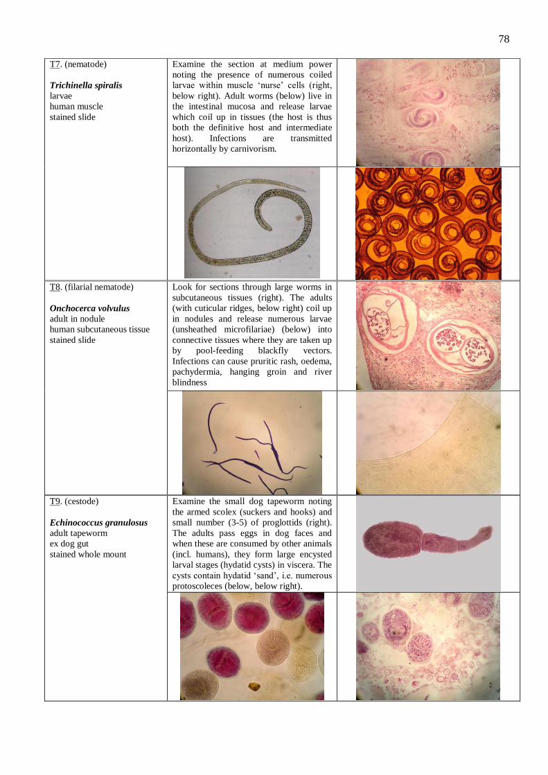
78
T7. (nematode)
Trichinella spiralis
larvae
human muscle
stained slide
Examine the section at medium power
noting the presence of numerous coiled
larvae within muscle ‘nurse’ cells (right,
below right). Adult worms (below) live in
the intestinal mucosa and release larvae
which coil up in tissues (the host is thus
both the definitive host and intermediate
host). Infections are transmitted
horizontally by carnivorism.
T8. (filarial nematode)
Onchocerca volvulus
adult in nodule
human subcutaneous tissue
stained slide
Look for sections through large worms in
subcutaneous tissues (right). The adults
(with cuticular ridges, below right) coil up
in nodules and release numerous larvae
(unsheathed microfilariae) (below) into
connective tissues where they are taken up
by pool-feeding blackfly vectors.
Infections can cause pruritic rash, oedema,
pachydermia, hanging groin and river
blindness
T9. (cestode)
Echinococcus granulosus
adult tapeworm
ex dog gut
stained whole mount
Examine the small dog tapeworm noting
the armed scolex (suckers and hooks) and
small number (3-5) of proglottids (right).
The adults pass eggs in dog faces and
when these are consumed by other animals
(incl. humans), they form large encysted
larval stages (hydatid cysts) in viscera. The
cysts contain hydatid ‘sand’, i.e. numerous
protoscoleces (below, below right).
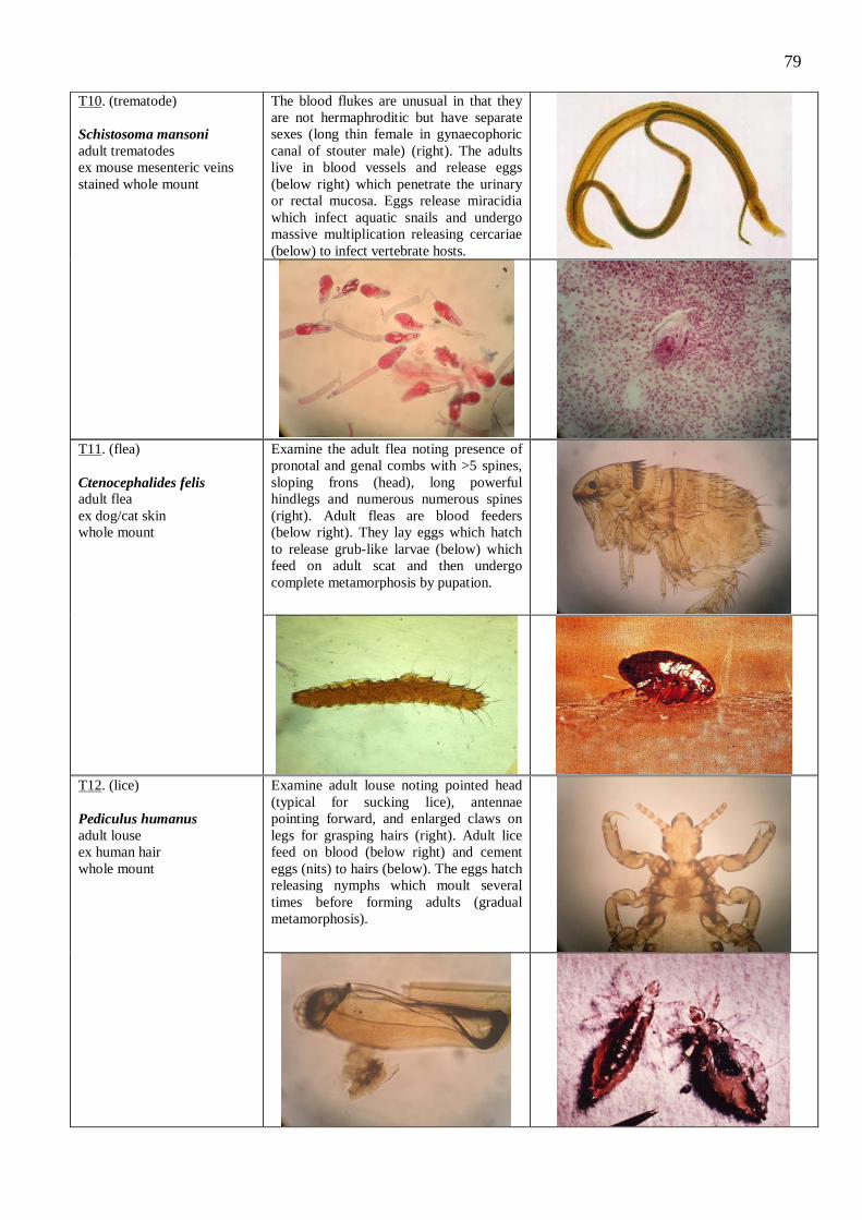
79
T10. (trematode)
Schistosoma mansoni
adult trematodes
ex mouse mesenteric veins
stained whole mount
The blood flukes are unusual in that they
are not hermaphroditic but have separate
sexes (long thin female in gynaecophoric
canal of stouter male) (right). The adults
live in blood vessels and release eggs
(below right) which penetrate the urinary
or rectal mucosa. Eggs release miracidia
which infect aquatic snails and undergo
massive multiplication releasing cercariae
(below) to infect vertebrate hosts.
T11. (flea)
Ctenocephalides felis adult flea
ex dog/cat skin
whole mount
Examine the adult flea noting presence of
pronotal and genal combs with >5 spines,
sloping frons (head), long powerful
hindlegs and numerous numerous spines
(right). Adult fleas are blood feeders
(below right). They lay eggs which hatch
to release grub-like larvae (below) which
feed on adult scat and then undergo
complete metamorphosis by pupation.
T12. (lice)
Pediculus humanus
adult louse
ex human hair
whole mount
Examine adult louse noting pointed head
(typical for sucking lice), antennae
pointing forward, and enlarged claws on
legs for grasping hairs (right). Adult lice
feed on blood (below right) and cement
eggs (nits) to hairs (below). The eggs hatch
releasing nymphs which moult several
times before forming adults (gradual
metamorphosis).
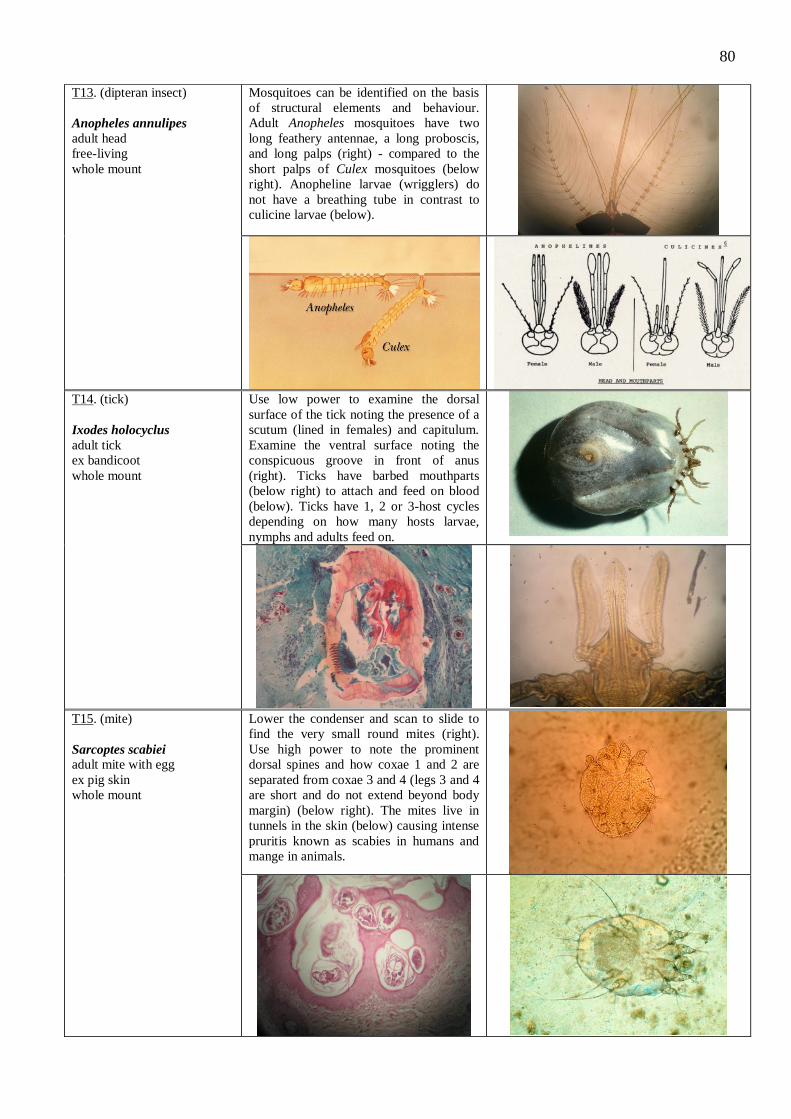
80
T13. (dipteran insect)
Anopheles annulipes
adult head
free-living
whole mount
Mosquitoes can be identified on the basis
of structural elements and behaviour.
Adult Anopheles mosquitoes have two
long feathery antennae, a long proboscis,
and long palps (right) - compared to the
short palps of Culex mosquitoes (below
right). Anopheline larvae (wrigglers) do
not have a breathing tube in contrast to
culicine larvae (below).
T14. (tick)
Ixodes holocyclus
adult tick
ex bandicoot
whole mount
Use low power to examine the dorsal
surface of the tick noting the presence of a
scutum (lined in females) and capitulum.
Examine the ventral surface noting the
conspicuous groove in front of anus
(right). Ticks have barbed mouthparts
(below right) to attach and feed on blood
(below). Ticks have 1, 2 or 3-host cycles
depending on how many hosts larvae,
nymphs and adults feed on.
T15. (mite)
Sarcoptes scabiei
adult mite with egg
ex pig skin
whole mount
Lower the condenser and scan to slide to
find the very small round mites (right).
Use high power to note the prominent
dorsal spines and how coxae 1 and 2 are
separated from coxae 3 and 4 (legs 3 and 4
are short and do not extend beyond body
margin) (below right). The mites live in
tunnels in the skin (below) causing intense
pruritis known as scabies in humans and
mange in animals.
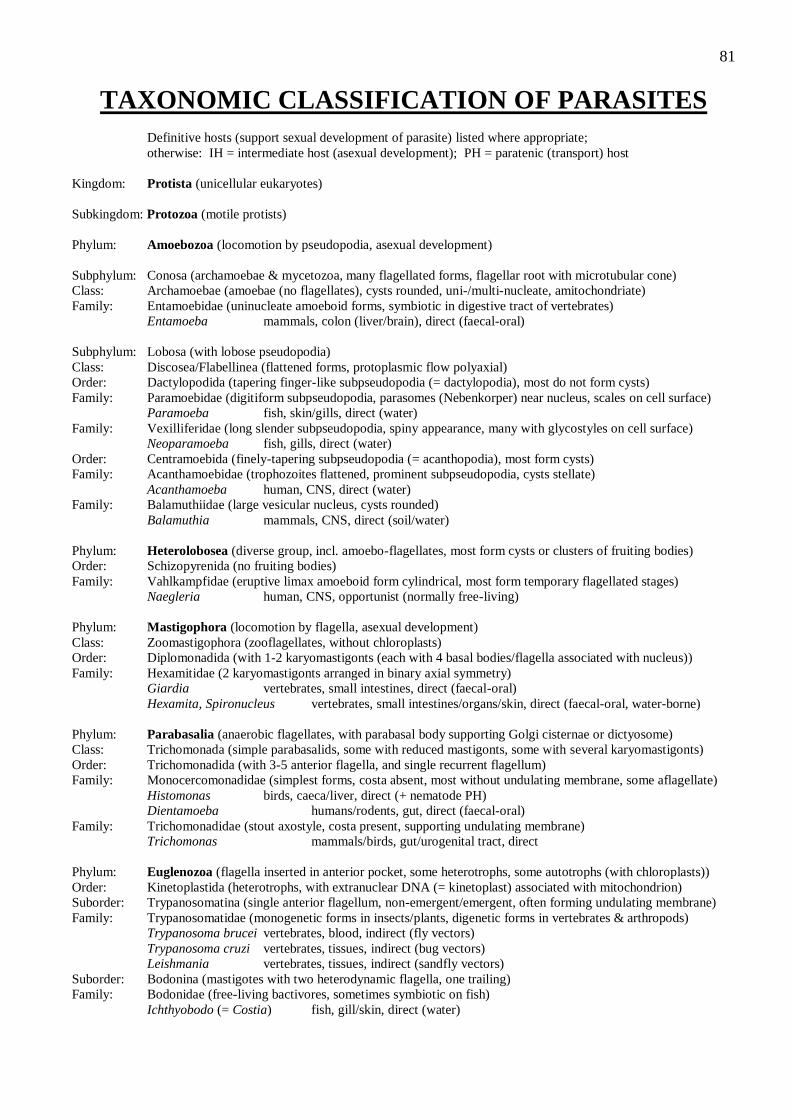
81
TAXONOMIC CLASSIFICATION OF PARASITES
Definitive hosts (support sexual development of parasite) listed where appropriate;
otherwise: IH = intermediate host (asexual development); PH = paratenic (transport) host
Kingdom: Protista (unicellular eukaryotes)
Subkingdom: Protozoa (motile protists)
Phylum: Amoebozoa (locomotion by pseudopodia, asexual development)
Subphylum: Conosa (archamoebae & mycetozoa, many flagellated forms, flagellar root with microtubular cone)
Class: Archamoebae (amoebae (no flagellates), cysts rounded, uni-/multi-nucleate, amitochondriate)
Family: Entamoebidae (uninucleate amoeboid forms, symbiotic in digestive tract of vertebrates)
Entamoeba mammals, colon (liver/brain), direct (faecal-oral)
Subphylum: Lobosa (with lobose pseudopodia)
Class: Discosea/Flabellinea (flattened forms, protoplasmic flow polyaxial)
Order: Dactylopodida (tapering finger-like subpseudopodia (= dactylopodia), most do not form cysts)
Family: Paramoebidae (digitiform subpseudopodia, parasomes (Nebenkorper) near nucleus, scales on cell surface)
Paramoeba fish, skin/gills, direct (water)
Family: Vexilliferidae (long slender subpseudopodia, spiny appearance, many with glycostyles on cell surface)
Neoparamoeba fish, gills, direct (water)
Order: Centramoebida (finely-tapering subpseudopodia (= acanthopodia), most form cysts)
Family: Acanthamoebidae (trophozoites flattened, prominent subpseudopodia, cysts stellate)
Acanthamoeba human, CNS, direct (water)
Family: Balamuthiidae (large vesicular nucleus, cysts rounded)
Balamuthia mammals, CNS, direct (soil/water)
Phylum: Heterolobosea (diverse group, incl. amoebo-flagellates, most form cysts or clusters of fruiting bodies)
Order: Schizopyrenida (no fruiting bodies)
Family: Vahlkampfidae (eruptive limax amoeboid form cylindrical, most form temporary flagellated stages)
Naegleria human, CNS, opportunist (normally free-living)
Phylum: Mastigophora (locomotion by flagella, asexual development)
Class: Zoomastigophora (zooflagellates, without chloroplasts)
Order: Diplomonadida (with 1-2 karyomastigonts (each with 4 basal bodies/flagella associated with nucleus))
Family: Hexamitidae (2 karyomastigonts arranged in binary axial symmetry)
Giardia vertebrates, small intestines, direct (faecal-oral)
Hexamita, Spironucleus vertebrates, small intestines/organs/skin, direct (faecal-oral, water-borne)
Phylum: Parabasalia (anaerobic flagellates, with parabasal body supporting Golgi cisternae or dictyosome)
Class: Trichomonada (simple parabasalids, some with reduced mastigonts, some with several karyomastigonts)
Order: Trichomonadida (with 3-5 anterior flagella, and single recurrent flagellum)
Family: Monocercomonadidae (simplest forms, costa absent, most without undulating membrane, some aflagellate)
Histomonas birds, caeca/liver, direct (+ nematode PH)
Dientamoeba humans/rodents, gut, direct (faecal-oral)
Family: Trichomonadidae (stout axostyle, costa present, supporting undulating membrane)
Trichomonas mammals/birds, gut/urogenital tract, direct
Phylum: Euglenozoa (flagella inserted in anterior pocket, some heterotrophs, some autotrophs (with chloroplasts))
Order: Kinetoplastida (heterotrophs, with extranuclear DNA (= kinetoplast) associated with mitochondrion)
Suborder: Trypanosomatina (single anterior flagellum, non-emergent/emergent, often forming undulating membrane)
Family: Trypanosomatidae (monogenetic forms in insects/plants, digenetic forms in vertebrates & arthropods)
Trypanosoma brucei vertebrates, blood, indirect (fly vectors)
Trypanosoma cruzi vertebrates, tissues, indirect (bug vectors)
Leishmania vertebrates, tissues, indirect (sandfly vectors)
Suborder: Bodonina (mastigotes with two heterodynamic flagella, one trailing)
Family: Bodonidae (free-living bactivores, sometimes symbiotic on fish)
Ichthyobodo (= Costia) fish, gill/skin, direct (water)
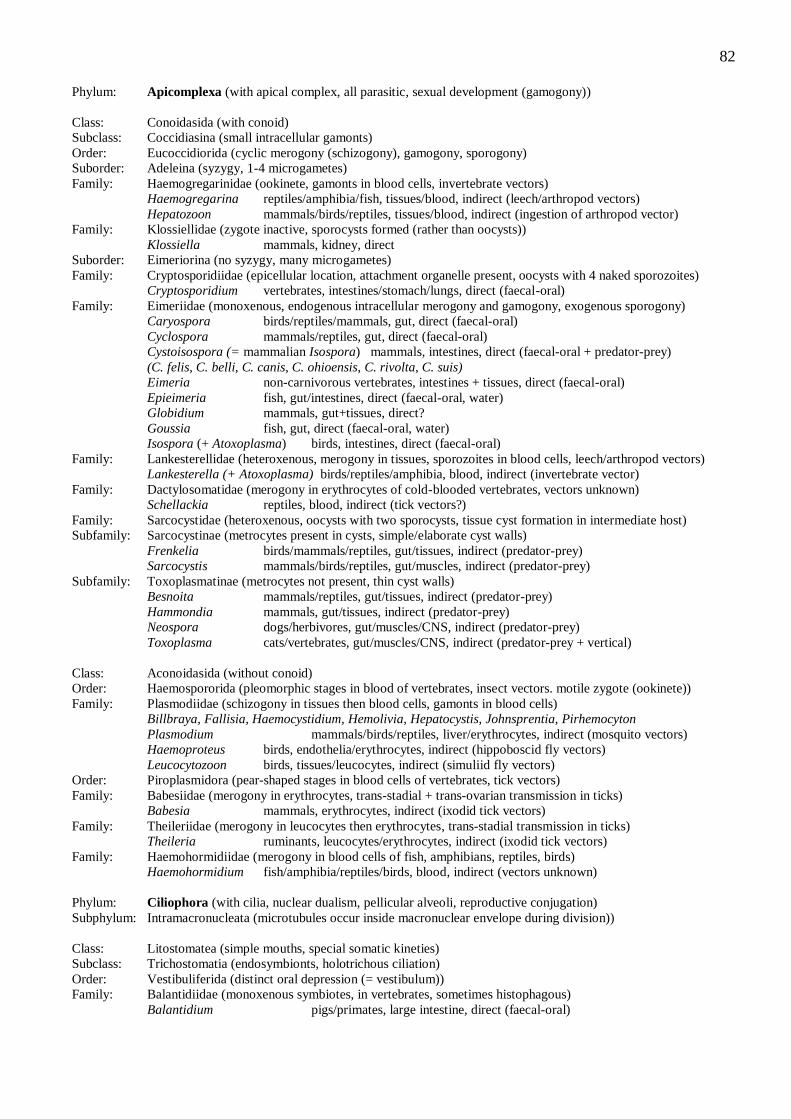
82
Phylum: Apicomplexa (with apical complex, all parasitic, sexual development (gamogony))
Class: Conoidasida (with conoid)
Subclass: Coccidiasina (small intracellular gamonts)
Order: Eucoccidiorida (cyclic merogony (schizogony), gamogony, sporogony)
Suborder: Adeleina (syzygy, 1-4 microgametes)
Family: Haemogregarinidae (ookinete, gamonts in blood cells, invertebrate vectors)
Haemogregarina reptiles/amphibia/fish, tissues/blood, indirect (leech/arthropod vectors)
Hepatozoon mammals/birds/reptiles, tissues/blood, indirect (ingestion of arthropod vector)
Family: Klossiellidae (zygote inactive, sporocysts formed (rather than oocysts))
Klossiella mammals, kidney, direct
Suborder: Eimeriorina (no syzygy, many microgametes)
Family: Cryptosporidiidae (epicellular location, attachment organelle present, oocysts with 4 naked sporozoites)
Cryptosporidium vertebrates, intestines/stomach/lungs, direct (faecal-oral)
Family: Eimeriidae (monoxenous, endogenous intracellular merogony and gamogony, exogenous sporogony)
Caryospora birds/reptiles/mammals, gut, direct (faecal-oral)
Cyclospora mammals/reptiles, gut, direct (faecal-oral)
Cystoisospora (= mammalian Isospora) mammals, intestines, direct (faecal-oral + predator-prey)
(C. felis, C. belli, C. canis, C. ohioensis, C. rivolta, C. suis)
Eimeria non-carnivorous vertebrates, intestines + tissues, direct (faecal-oral)
Epieimeria fish, gut/intestines, direct (faecal-oral, water)
Globidium mammals, gut+tissues, direct?
Goussia fish, gut, direct (faecal-oral, water)
Isospora (+ Atoxoplasma) birds, intestines, direct (faecal-oral)
Family: Lankesterellidae (heteroxenous, merogony in tissues, sporozoites in blood cells, leech/arthropod vectors)
Lankesterella (+ Atoxoplasma) birds/reptiles/amphibia, blood, indirect (invertebrate vector)
Family: Dactylosomatidae (merogony in erythrocytes of cold-blooded vertebrates, vectors unknown)
Schellackia reptiles, blood, indirect (tick vectors?)
Family: Sarcocystidae (heteroxenous, oocysts with two sporocysts, tissue cyst formation in intermediate host)
Subfamily: Sarcocystinae (metrocytes present in cysts, simple/elaborate cyst walls)
Frenkelia birds/mammals/reptiles, gut/tissues, indirect (predator-prey)
Sarcocystis mammals/birds/reptiles, gut/muscles, indirect (predator-prey)
Subfamily: Toxoplasmatinae (metrocytes not present, thin cyst walls)
Besnoita mammals/reptiles, gut/tissues, indirect (predator-prey)
Hammondia mammals, gut/tissues, indirect (predator-prey)
Neospora dogs/herbivores, gut/muscles/CNS, indirect (predator-prey)
Toxoplasma cats/vertebrates, gut/muscles/CNS, indirect (predator-prey + vertical)
Class: Aconoidasida (without conoid)
Order: Haemospororida (pleomorphic stages in blood of vertebrates, insect vectors. motile zygote (ookinete))
Family: Plasmodiidae (schizogony in tissues then blood cells, gamonts in blood cells)
Billbraya, Fallisia, Haemocystidium, Hemolivia, Hepatocystis, Johnsprentia, Pirhemocyton
Plasmodium mammals/birds/reptiles, liver/erythrocytes, indirect (mosquito vectors)
Haemoproteus birds, endothelia/erythrocytes, indirect (hippoboscid fly vectors)
Leucocytozoon birds, tissues/leucocytes, indirect (simuliid fly vectors)
Order: Piroplasmidora (pear-shaped stages in blood cells of vertebrates, tick vectors)
Family: Babesiidae (merogony in erythrocytes, trans-stadial + trans-ovarian transmission in ticks)
Babesia mammals, erythrocytes, indirect (ixodid tick vectors)
Family: Theileriidae (merogony in leucocytes then erythrocytes, trans-stadial transmission in ticks)
Theileria ruminants, leucocytes/erythrocytes, indirect (ixodid tick vectors)
Family: Haemohormidiidae (merogony in blood cells of fish, amphibians, reptiles, birds)
Haemohormidium fish/amphibia/reptiles/birds, blood, indirect (vectors unknown)
Phylum: Ciliophora (with cilia, nuclear dualism, pellicular alveoli, reproductive conjugation)
Subphylum: Intramacronucleata (microtubules occur inside macronuclear envelope during division))
Class: Litostomatea (simple mouths, special somatic kineties)
Subclass: Trichostomatia (endosymbionts, holotrichous ciliation)
Order: Vestibuliferida (distinct oral depression (= vestibulum))
Family: Balantidiidae (monoxenous symbiotes, in vertebrates, sometimes histophagous)
Balantidium pigs/primates, large intestine, direct (faecal-oral)
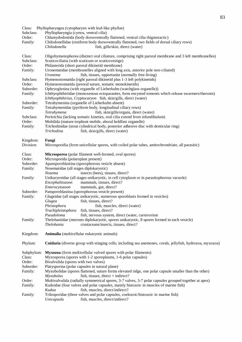
83
Class: Phyllopharyngea (cytopharynx with leaf-like phyllae)
Subclass: Phyllopharyngia (cyrtos, ventral cilia)
Order: Chlamydodontida (body dorsoventrally flattened, ventral cilia thigmotactic)
Family: Chilodonellidae (reniform body dorsoventrally flattened, two fields of dorsal ciliary rows)
Chilodonella fish, gills/skin, direct (water)
Class: Oligohymenophorea (distinct oral ciliature, comprising right paroral membrane and 3 left membranelles)
Subclass: Scuticociliatia (with scuticum or scuticovestige)
Order: Philasterida (short paroral dikinetid membrane)
Family: Uronematidae (membranelles aligned with long axis, anterior pole non-ciliated)
Uronema fish, tissues, opportunist (normally free-living)
Subclass: Hymenostomatida (right paroral dikinetid plus 1-3 left polykinetids)
Order: Hymenostomatida (preoral suture, somatic monokinetids)
Suborder: Ophryoglenina (with organelle of Lieberkuhn (watchglass organelle))
Family: Ichthyophthiriidae (monoxenous ectoparasites, form encysted tomonts which release swarmers/theronts)
Ichthyophthirius, Cryptocaryon fish, skin/gills, direct (water)
Suborder: Tetrahymenina (organelle of Lieberkuhn absent)
Family: Tetrahymenidae (pyriform body, longitudinal ciliary rows)
Tetrahymena fish, skin/gills/organs, direct (water)
Subclass: Peritrichia (lacking somatic kineties, oral cilia extend from infundibulum)
Order: Mobilida (mature trophont mobile, aboral holdfast organelle)
Family: Trichodinidae (stout cylindrical body, posterior adhesive disc with denticular ring)
Trichodina fish, skin/gills, direct (water)
Kingdom: Fungi
Division: Microsporidia (form unicellular spores, with coiled polar tubes, amitochrondriate, all parasitic)
Class: Microsporea (polar filament well-formed, oval spores)
Order: Microsporida (polaroplast present)
Suborder: Apansporoblastina (sporophorous vesicle absent)
Family: Nosematidae (all stages diplokaryotic)
Nosema insects (bees), tissues, direct?
Family: Unikaryonidae (all stages unikaryotic, in cell cytoplasm or in parasitophorous vacuole)
Encephalitozoon mammals, tissues, direct?
Enterocytozoon mammals, gut, direct?
Suborder: Pansporoblastina (sporophorous vesicle present)
Family: Glugeidae (all stages unikaryotic, numerous sporoblasts formed in vesicles)
Glugea fish, tissues, direct?
Pleistophora fish, muscles, direct (water)
Trachipleistophora fish, tissues, direct?
Pseudoloma fish, nervous system, direct (water, carnivorism
Family: Thelohaniidae (meronts diplokaryotic, spores unikaryotic, 8 spores formed in each vesicle)
Thelohania crustaceans/insects, tissues, direct?
Kingdom: Animalia (multicellular eukaryotic animals)
Phylum: Cnidaria (diverse group with stinging cells; including sea anemones, corals, jellyfish, hydrozoa, myxozoa)
Subphylum: Myxozoa (form multicellular valved spores with polar filaments)
Class: Myxosporea (spores with 1-2 sporoplasms, 1-6 polar capsules)
Order: Bivalvulida (spores with two valves)
Suborder: Platysporina (polar capsules in sutural plane)
Family: Myxobolidae (spores flattened, suture forms elevated ridge, one polar capsule smaller than the other)
Myxobolus fish, tissues, direct + indirect?
Order: Multivalvulida (radially symmetrical spores, 3-7 valves, 3-7 polar capsules grouped together at apex)
Family: Kudoidae (four valves and polar capsules, mainly histozoic in muscles of marine fish)
Kudoa fish, muscles, direct/indirect?
Family: Trilosporidae (three valves and polar capsules, coelozoic/histozoic in marine fish)
Unicapsula fish, muscles, direct/indirect?
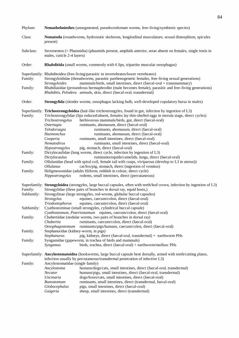
84
Phylum: Nemathelminthes (unsegmented, pseudocoelomate worms, free-living/symbiotic species)
Class: Nematoda (roundworms, hydrostatic skeletons, longitudinal musculature, sexual dimorphism, spicules
present)
Subclass: Secernentea (= Phasmidia) (phasmids present, amphids anterior, setae absent on females, single testis in
males, cuticle 2-4 layers)
Order: Rhabditida (small worms, commonly with 6 lips, tripartite muscular oesophagus)
Superfamily: Rhabditoidea (free-living/parasitic in invertebrates/lower vertebrates)
Family: Strongyloididae (threadworms, parasitic parthenogenetic females, free-living sexual generations)
Strongyloides mammals/birds, small intestines, direct (faecal-oral + transmammary)
Family: Rhabdiasidae (protandrous hermaphrodite (male becomes female), parasitic and free-living generations)
Rhabditis, Pelodera animals, skin, direct (faecal-oral, transdermal)
Order: Strongylida (slender worms, oesophagus lacking bulb, well-developed copulatory bursa in males)
Superfamily: Trichostrongyloidea (hair-like trichostrongyles, found in gut, infection by ingestion of L3)
Family: Trichostrongylidae (lips reduced/absent, females lay thin-shelled eggs in morula stage, direct cycles)
Trichostrongylus herbivorous mammals/birds, gut, direct (faecal-oral)
Ostertagia ruminants, abomasum, direct (faecal-oral)
Telodorsagia ruminants, abomasum, direct (faecal-oral)
Haemonchus ruminants, abomasum, direct (faecal-oral)
Cooperia ruminants, small intestines, direct (faecal-oral)
Nematodirus ruminants, small intestines, direct (faecal-oral)
Hypostrongylus pig, stomach, direct (faecal-oral)
Family: Dictylocaulidae (lung worms, direct cycle, infection by ingestion of L3)
Dictylocaulus ruminants/equids/camelids, lungs, direct (faecal-oral)
Family: Ollulanidae (head with spiral coil, female tail with cusps, viviparous (develop to L3 in uterus))
Ollulanus cat/fox/pig, stomach, direct (ingestion of vomitus)
Family: Heligmosomidae (adults filiform, reddish in colour, direct cycle)
Nippostrongylus rodents, small intestines, direct (percutaneous)
Superfamily: Strongyloidea (strongyles, large buccal capsules, often with teeth/leaf crown, infection by ingestion of L3)
Family: Strongylidae (three pairs of branches in dorsal ray, equid hosts,)
Subfamily: Strongylinae (large strongyles, red-worms, globular buccal capsules)
Strongylus equines, caecum/colon, direct (faecal-oral)
Triodontophorus equines, caecum/colon, direct (faecal-oral)
Subfamily: Cyathostominae (small strongyles, cylindrical buccal capsule)
Cyathostomum, Poteriostomum equines, caecum/colon, direct (faecal-oral)
Family: Chabertiidae (nodular worms, two pairs of branches in dorsal ray)
Chabertia ruminants, caecum/colon, direct (faecal-oral)
Oesophagostomum ruminants/pigs/humans, caecum/colon, direct (faecal-oral)
Family: Stephanuridae (kidney-worm, in pigs)
Stephanurus pig, kidneys, direct (faecal-oral, transdermal) + earthworm PHs
Family: Syngamidae (gapeworm, in trachea of birds and mammals)
Syngamus birds, trachea, direct (faecal-oral) + earthworm/mollusc PHs
Superfamily: Ancylostomatoidea (hookworms, large buccal capsule bent dorsally, armed with teeth/cutting plates,
infection usually by percutaneous/transdermal penetration of infective L3)
Family: Ancylostomatidae (single family)
Ancylostoma humans/dogs/cats, small intestines, direct (faecal-oral, transdermal)
Necator humans/pigs, small intestines, direct (faecal-oral, transdermal)
Uncinaria dogs/foxes/cats, small intestines, direct (faecal-oral)
Bunostomum ruminants, small intestines, direct (transdermal, faecal-oral)
Globocephalus pigs, small intestines, direct (faecal-oral)
Gaigeria sheep, small intestines, direct (transdermal)
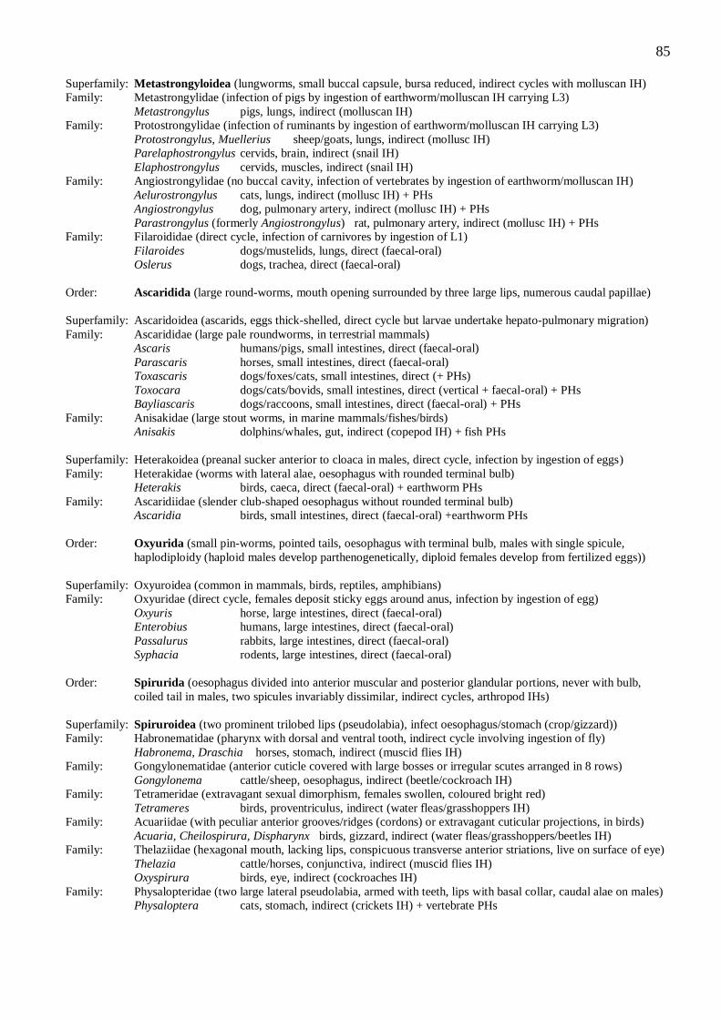
85
Superfamily: Metastrongyloidea (lungworms, small buccal capsule, bursa reduced, indirect cycles with molluscan IH)
Family: Metastrongylidae (infection of pigs by ingestion of earthworm/molluscan IH carrying L3)
Metastrongylus pigs, lungs, indirect (molluscan IH)
Family: Protostrongylidae (infection of ruminants by ingestion of earthworm/molluscan IH carrying L3)
Protostrongylus, Muellerius sheep/goats, lungs, indirect (mollusc IH)
Parelaphostrongylus cervids, brain, indirect (snail IH)
Elaphostrongylus cervids, muscles, indirect (snail IH)
Family: Angiostrongylidae (no buccal cavity, infection of vertebrates by ingestion of earthworm/molluscan IH)
Aelurostrongylus cats, lungs, indirect (mollusc IH) + PHs
Angiostrongylus dog, pulmonary artery, indirect (mollusc IH) + PHs
Parastrongylus (formerly Angiostrongylus) rat, pulmonary artery, indirect (mollusc IH) + PHs
Family: Filaroididae (direct cycle, infection of carnivores by ingestion of L1)
Filaroides dogs/mustelids, lungs, direct (faecal-oral)
Oslerus dogs, trachea, direct (faecal-oral)
Order: Ascaridida (large round-worms, mouth opening surrounded by three large lips, numerous caudal papillae)
Superfamily: Ascaridoidea (ascarids, eggs thick-shelled, direct cycle but larvae undertake hepato-pulmonary migration)
Family: Ascarididae (large pale roundworms, in terrestrial mammals)
Ascaris humans/pigs, small intestines, direct (faecal-oral)
Parascaris horses, small intestines, direct (faecal-oral)
Toxascaris dogs/foxes/cats, small intestines, direct (+ PHs)
Toxocara dogs/cats/bovids, small intestines, direct (vertical + faecal-oral) + PHs
Bayliascaris dogs/raccoons, small intestines, direct (faecal-oral) + PHs
Family: Anisakidae (large stout worms, in marine mammals/fishes/birds)
Anisakis dolphins/whales, gut, indirect (copepod IH) + fish PHs
Superfamily: Heterakoidea (preanal sucker anterior to cloaca in males, direct cycle, infection by ingestion of eggs)
Family: Heterakidae (worms with lateral alae, oesophagus with rounded terminal bulb)
Heterakis birds, caeca, direct (faecal-oral) + earthworm PHs
Family: Ascaridiidae (slender club-shaped oesophagus without rounded terminal bulb)
Ascaridia birds, small intestines, direct (faecal-oral) +earthworm PHs
Order: Oxyurida (small pin-worms, pointed tails, oesophagus with terminal bulb, males with single spicule,
haplodiploidy (haploid males develop parthenogenetically, diploid females develop from fertilized eggs))
Superfamily: Oxyuroidea (common in mammals, birds, reptiles, amphibians)
Family: Oxyuridae (direct cycle, females deposit sticky eggs around anus, infection by ingestion of egg)
Oxyuris horse, large intestines, direct (faecal-oral)
Enterobius humans, large intestines, direct (faecal-oral)
Passalurus rabbits, large intestines, direct (faecal-oral)
Syphacia rodents, large intestines, direct (faecal-oral)
Order: Spirurida (oesophagus divided into anterior muscular and posterior glandular portions, never with bulb,
coiled tail in males, two spicules invariably dissimilar, indirect cycles, arthropod IHs)
Superfamily: Spiruroidea (two prominent trilobed lips (pseudolabia), infect oesophagus/stomach (crop/gizzard))
Family: Habronematidae (pharynx with dorsal and ventral tooth, indirect cycle involving ingestion of fly)
Habronema, Draschia horses, stomach, indirect (muscid flies IH)
Family: Gongylonematidae (anterior cuticle covered with large bosses or irregular scutes arranged in 8 rows)
Gongylonema cattle/sheep, oesophagus, indirect (beetle/cockroach IH)
Family: Tetrameridae (extravagant sexual dimorphism, females swollen, coloured bright red)
Tetrameres birds, proventriculus, indirect (water fleas/grasshoppers IH)
Family: Acuariidae (with peculiar anterior grooves/ridges (cordons) or extravagant cuticular projections, in birds)
Acuaria, Cheilospirura, Dispharynx birds, gizzard, indirect (water fleas/grasshoppers/beetles IH)
Family: Thelaziidae (hexagonal mouth, lacking lips, conspicuous transverse anterior striations, live on surface of eye)
Thelazia cattle/horses, conjunctiva, indirect (muscid flies IH)
Oxyspirura birds, eye, indirect (cockroaches IH)
Family: Physalopteridae (two large lateral pseudolabia, armed with teeth, lips with basal collar, caudal alae on males)
Physaloptera cats, stomach, indirect (crickets IH) + vertebrate PHs
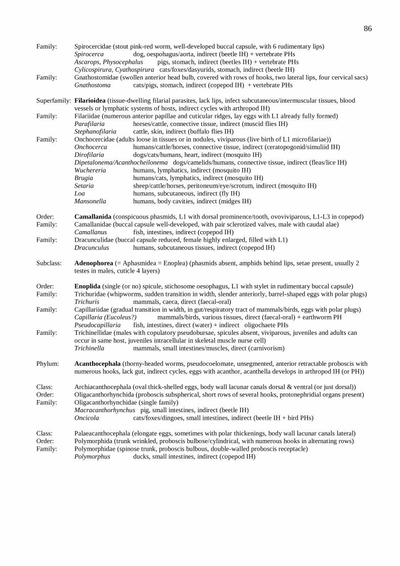
86
Family: Spirocercidae (stout pink-red worm, well-developed buccal capsule, with 6 rudimentary lips)
Spirocerca dog, oespohagus/aorta, indirect (beetle IH) + vertebrate PHs
Ascarops, Physocephalus pigs, stomach, indirect (beetles IH) + vertebrate PHs
Cylicospirura, Cyathospirura cats/foxes/dasyurids, stomach, indirect (beetle IH)
Family: Gnathostomidae (swollen anterior head bulb, covered with rows of hooks, two lateral lips, four cervical sacs)
Gnathostoma cats/pigs, stomach, indirect (copepod IH) + vertebrate PHs
Superfamily: Filarioidea (tissue-dwelling filarial parasites, lack lips, infect subcutaneous/intermuscular tissues, blood
vessels or lymphatic systems of hosts, indirect cycles with arthropod IH)
Family: Filariidae (numerous anterior papillae and cuticular ridges, lay eggs with L1 already fully formed)
Parafilaria horses/cattle, connective tissue, indirect (muscid flies IH)
Stephanofilaria cattle, skin, indirect (buffalo flies IH)
Family: Onchocercidae (adults loose in tissues or in nodules, viviparous (live birth of L1 microfilariae))
Onchocerca humans/cattle/horses, connective tissue, indirect (ceratopogonid/simuliid IH)
Dirofilaria dogs/cats/humans, heart, indirect (mosquito IH)
Dipetalonema/Acanthocheilonema dogs/camelids/humans, connective tissue, indirect (fleas/lice IH)
Wuchereria humans, lymphatics, indirect (mosquito IH)
Brugia humans/cats, lymphatics, indirect (mosquito IH)
Setaria sheep/cattle/horses, peritoneum/eye/scrotum, indirect (mosquito IH)
Loa humans, subcutaneous, indirect (fly IH)
Mansonella humans, body cavities, indirect (midges IH)
Order: Camallanida (conspicuous phasmids, L1 with dorsal prominence/tooth, ovoviviparous, L1-L3 in copepod)
Family: Camallanidae (buccal capsule well-developed, with pair sclerotized valves, male with caudal alae)
Camallanus fish, intestines, indirect (copepod IH)
Family: Dracunculidae (buccal capsule reduced, female highly enlarged, filled with L1)
Dracunculus humans, subcutaneous tissues, indirect (copepod IH)
Subclass: Adenophorea (= Aphasmidea = Enoplea) (phasmids absent, amphids behind lips, setae present, usually 2
testes in males, cuticle 4 layers)
Order: Enoplida (single (or no) spicule, stichosome oesophagus, L1 with stylet in rudimentary buccal capsule)
Family: Trichuridae (whipworms, sudden transition in width, slender anteriorly, barrel-shaped eggs with polar plugs)
Trichuris mammals, caeca, direct (faecal-oral)
Family: Capillariidae (gradual transition in width, in gut/respiratory tract of mammals/birds, eggs with polar plugs)
Capillaria (Eucoleus?) mammals/birds, various tissues, direct (faecal-oral) + earthworm PH
Pseudocapillaria fish, intestines, direct (water) + indirect oligochaete PHs
Family: Trichinellidae (males with copulatory pseudobursae, spicules absent, viviparous, juveniles and adults can
occur in same host, juveniles intracellular in skeletal muscle nurse cell)
Trichinella mammals, small intestines/muscles, direct (carnivorism)
Phylum: Acanthocephala (thorny-headed worms, pseudocoelomate, unsegmented, anterior retractable proboscis with
numerous hooks, lack gut, indirect cycles, eggs with acanthor, acanthella develops in arthropod IH (or PH))
Class: Archiacanthocephala (oval thick-shelled eggs, body wall lacunar canals dorsal & ventral (or just dorsal))
Order: Oligacanthorhynchida (proboscis subspherical, short rows of several hooks, protonephridial organs present)
Family: Oligacanthorhynchidae (single family)
Macracanthorhynchus pig, small intestines, indirect (beetle IH)
Oncicola cats/foxes/dingoes, small intestines, indirect (beetle IH + bird PHs)
Class: Palaeacanthocephala (elongate eggs, sometimes with polar thickenings, body wall lacunar canals lateral)
Order: Polymorphida (trunk wrinkled, proboscis bulbose/cylindrical, with numerous hooks in alternating rows)
Family: Polymorphidae (spinose trunk, proboscis bulbous, double-walled proboscis receptacle)
Polymorphus ducks, small intestines, indirect (copepod IH)
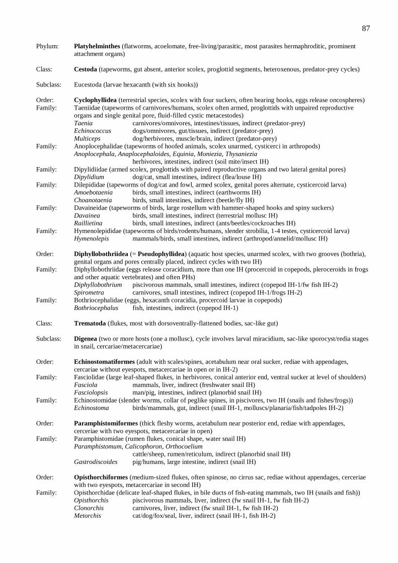
87
Phylum: Platyhelminthes (flatworms, acoelomate, free-living/parasitic, most parasites hermaphroditic, prominent
attachment organs)
Class: Cestoda (tapeworms, gut absent, anterior scolex, proglottid segments, heteroxenous, predator-prey cycles)
Subclass: Eucestoda (larvae hexacanth (with six hooks))
Order: Cyclophyllidea (terrestrial species, scolex with four suckers, often bearing hooks, eggs release oncospheres)
Family: Taeniidae (tapeworms of carnivores/humans, scolex often armed, proglottids with unpaired reproductive
organs and single genital pore, fluid-filled cystic metacestodes)
Taenia carnivores/omnivores, intestines/tissues, indirect (predator-prey)
Echinococcus dogs/omnivores, gut/tissues, indirect (predator-prey)
Multiceps dog/herbivores, muscle/brain, indirect (predator-prey)
Family: Anoplocephalidae (tapeworms of hoofed animals, scolex unarmed, cysticerci in arthropods)
Anoplocephala, Anaplocephaloides, Equinia, Moniezia, Thysaniezia
herbivores, intestines, indirect (soil mite/insect IH)
Family: Dipylidiidae (armed scolex, proglottids with paired reproductive organs and two lateral genital pores)
Dipylidium dog/cat, small intestines, indirect (flea/louse IH)
Family: Dilepididae (tapeworms of dog/cat and fowl, armed scolex, genital pores alternate, cysticercoid larva)
Amoebotaenia birds, small intestines, indirect (earthworms IH)
Choanotaenia birds, small intestines, indirect (beetle/fly IH)
Family: Davaineidae (tapeworms of birds, large rostellum with hammer-shaped hooks and spiny suckers)
Davainea birds, small intestines, indirect (terrestrial mollusc IH)
Raillietina birds, small intestines, indirect (ants/beetles/cockroaches IH)
Family: Hymenolepididae (tapeworms of birds/rodents/humans, slender strobilia, 1-4 testes, cysticercoid larva)
Hymenolepis mammals/birds, small intestines, indirect (arthropod/annelid/mollusc IH)
Order: Diphyllobothriidea (= Pseudophyllidea) (aquatic host species, unarmed scolex, with two grooves (bothria),
genital organs and pores centrally placed, indirect cycles with two IH)
Family: Diphyllobothriidae (eggs release coracidium, more than one IH (procercoid in copepods, pleroceroids in frogs
and other aquatic vertebrates) and often PHs)
Diphyllobothrium piscivorous mammals, small intestines, indirect (copepod IH-1/fw fish IH-2)
Spirometra carnivores, small intestines, indirect (copepod IH-1/frogs IH-2)
Family: Bothriocephalidae (eggs, hexacanth coracidia, procercoid larvae in copepods)
Bothriocephalus fish, intestines, indirect (copepod IH-1)
Class: Trematoda (flukes, most with dorsoventrally-flattened bodies, sac-like gut)
Subclass: Digenea (two or more hosts (one a mollusc), cycle involves larval miracidium, sac-like sporocyst/redia stages
in snail, cercariae/metacercariae)
Order: Echinostomatiformes (adult with scales/spines, acetabulum near oral sucker, rediae with appendages,
cercariae without eyespots, metacercariae in open or in IH-2)
Family: Fasciolidae (large leaf-shaped flukes, in herbivores, conical anterior end, ventral sucker at level of shoulders)
Fasciola mammals, liver, indirect (freshwater snail IH)
Fasciolopsis man/pig, intestines, indirect (planorbid snail IH)
Family: Echinostomidae (slender worms, collar of peglike spines, in piscivores, two IH (snails and fishes/frogs))
Echinostoma birds/mammals, gut, indirect (snail IH-1, molluscs/planaria/fish/tadpoles IH-2)
Order: Paramphistomiformes (thick fleshy worms, acetabulum near posterior end, rediae with appendages,
cerceriae with two eyespots, metacercariae in open)
Family: Paramphistomidae (rumen flukes, conical shape, water snail IH)
Paramphistomum, Calicophoron, Orthocoelium
cattle/sheep, rumen/reticulum, indirect (planorbid snail IH)
Gastrodiscoides pig/humans, large intestine, indirect (snail IH)
Order: Opisthorchiformes (medium-sized flukes, often spinose, no cirrus sac, rediae without appendages, cerceriae
with two eyespots, metacercariae in second IH)
Family: Opisthorchidae (delicate leaf-shaped flukes, in bile ducts of fish-eating mammals, two IH (snails and fish))
Opisthorchis piscivorous mammals, liver, indirect (fw snail IH-1, fw fish IH-2)
Clonorchis carnivores, liver, indirect (fw snail IH-1, fw fish IH-2)
Metorchis cat/dog/fox/seal, liver, indirect (snail IH-1, fish IH-2)
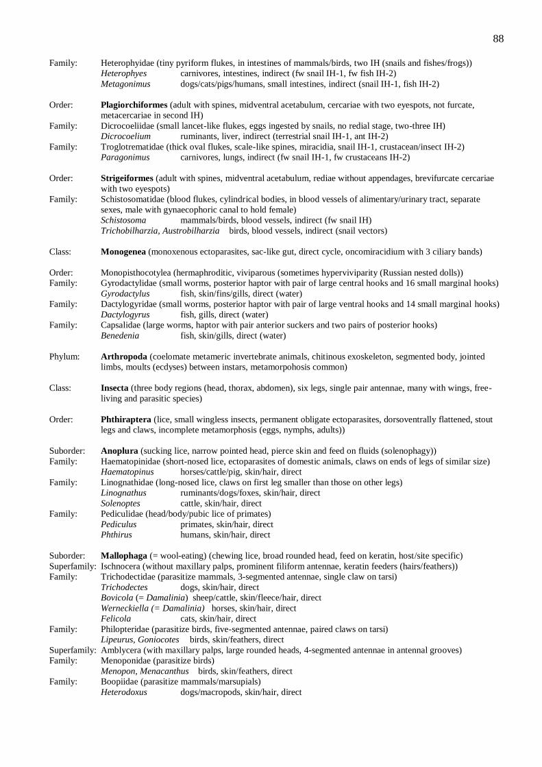
88
Family: Heterophyidae (tiny pyriform flukes, in intestines of mammals/birds, two IH (snails and fishes/frogs))
Heterophyes carnivores, intestines, indirect (fw snail IH-1, fw fish IH-2)
Metagonimus dogs/cats/pigs/humans, small intestines, indirect (snail IH-1, fish IH-2)
Order: Plagiorchiformes (adult with spines, midventral acetabulum, cercariae with two eyespots, not furcate,
metacercariae in second IH)
Family: Dicrocoeliidae (small lancet-like flukes, eggs ingested by snails, no redial stage, two-three IH)
Dicrocoelium ruminants, liver, indirect (terrestrial snail IH-1, ant IH-2)
Family: Troglotrematidae (thick oval flukes, scale-like spines, miracidia, snail IH-1, crustacean/insect IH-2)
Paragonimus carnivores, lungs, indirect (fw snail IH-1, fw crustaceans IH-2)
Order: Strigeiformes (adult with spines, midventral acetabulum, rediae without appendages, brevifurcate cercariae
with two eyespots)
Family: Schistosomatidae (blood flukes, cylindrical bodies, in blood vessels of alimentary/urinary tract, separate
sexes, male with gynaecophoric canal to hold female)
Schistosoma mammals/birds, blood vessels, indirect (fw snail IH)
Trichobilharzia, Austrobilharzia birds, blood vessels, indirect (snail vectors)
Class: Monogenea (monoxenous ectoparasites, sac-like gut, direct cycle, oncomiracidium with 3 ciliary bands)
Order: Monopisthocotylea (hermaphroditic, viviparous (sometimes hyperviviparity (Russian nested dolls))
Family: Gyrodactylidae (small worms, posterior haptor with pair of large central hooks and 16 small marginal hooks)
Gyrodactylus fish, skin/fins/gills, direct (water)
Family: Dactylogyridae (small worms, posterior haptor with pair of large ventral hooks and 14 small marginal hooks)
Dactylogyrus fish, gills, direct (water)
Family: Capsalidae (large worms, haptor with pair anterior suckers and two pairs of posterior hooks)
Benedenia fish, skin/gills, direct (water)
Phylum: Arthropoda (coelomate metameric invertebrate animals, chitinous exoskeleton, segmented body, jointed
limbs, moults (ecdyses) between instars, metamorpohosis common)
Class: Insecta (three body regions (head, thorax, abdomen), six legs, single pair antennae, many with wings, free-
living and parasitic species)
Order: Phthiraptera (lice, small wingless insects, permanent obligate ectoparasites, dorsoventrally flattened, stout
legs and claws, incomplete metamorphosis (eggs, nymphs, adults))
Suborder: Anoplura (sucking lice, narrow pointed head, pierce skin and feed on fluids (solenophagy))
Family: Haematopinidae (short-nosed lice, ectoparasites of domestic animals, claws on ends of legs of similar size)
Haematopinus horses/cattle/pig, skin/hair, direct
Family: Linognathidae (long-nosed lice, claws on first leg smaller than those on other legs)
Linognathus ruminants/dogs/foxes, skin/hair, direct
Solenoptes cattle, skin/hair, direct
Family: Pediculidae (head/body/pubic lice of primates)
Pediculus primates, skin/hair, direct
Phthirus humans, skin/hair, direct
Suborder: Mallophaga (= wool-eating) (chewing lice, broad rounded head, feed on keratin, host/site specific)
Superfamily: Ischnocera (without maxillary palps, prominent filiform antennae, keratin feeders (hairs/feathers))
Family: Trichodectidae (parasitize mammals, 3-segmented antennae, single claw on tarsi)
Trichodectes dogs, skin/hair, direct
Bovicola (= Damalinia) sheep/cattle, skin/fleece/hair, direct
Werneckiella (= Damalinia) horses, skin/hair, direct
Felicola cats, skin/hair, direct
Family: Philopteridae (parasitize birds, five-segmented antennae, paired claws on tarsi)
Lipeurus, Goniocotes birds, skin/feathers, direct
Superfamily: Amblycera (with maxillary palps, large rounded heads, 4-segmented antennae in antennal grooves)
Family: Menoponidae (parasitize birds)
Menopon, Menacanthus birds, skin/feathers, direct
Family: Boopiidae (parasitize mammals/marsupials)
Heterodoxus dogs/macropods, skin/hair, direct

89
Order: Hemiptera (true bugs/aphids/scale insects, mouthparts with stylet-like mandibles/maxillae, gradual
metamorphosis)
Suborder: Heteroptera (some plant-feeders, some predatory on other arthropods, some blood-feeders on vertebrates)
Family: Cimicidae (small wingless bugs, incl. bed-bugs, blood feeders on animals)
Cimex mammals/birds, skin, direct
Family: Reduviidae (large winged cone-nose/kissing/assasin bugs, incl. triatome bugs, blood feeders on animals)
Triatoma mammals, skin, direct
Order: Siphonaptera (fleas, wingless (=-aptera), adults feed on blood (“siphon-), laterally compressed, third pair of
legs adapted for jumping, complete metamorphosis with vermiform larvae, pupation in silk cocoons)
Family: Pulicidae (parasites of mammals)
Pulex humans/dogs/cats, skin, direct
Echidnophaga mammals/birds, skin, direct
Ctenocephalides dogs/cats/humans, skin, direct
Xenopsylla rats/dogs/cats/humans, skin, direct
Spilopsyllus rabbit, skin, direct
Family: Tungidae (chigoes/jiggers/chiggers/chique/sand fleas, females burrow under skin, enclosed in sinus)
Tunga mammals, skin, direct
Order: Diptera (flies, midges, mosquitoes, with single pair of membranous forewings (diptera), hindwings modified
into halteres, complete metamorphosis with vermiform larvae)
Suborder: Nematocera (small midges/mosquitoes, long filamentous segmented antennae (= nemato-cera), aquatic life-
cycles (larval/pupal stages associated with water), female adults require blood meal before they can lay eggs)
Family: Culicidae (mosquitoes, elongate mouthparts form proboscis, slender wings with scales on veins/margins)
Subfamily: Culicinae (scutellum with trilobed posterior margin, scaly abdomen, larva with prominent air-tube)
Culex, Aedes, Mansonia mammals/birds, skin, direct
Subfamily: Anophelinae (scutellum rounded or straight, addominal sternites lack scales, larva lacks air-tube)
Anopheles mammals/birds, skin, direct
Family: Ceratopogonidae (small biting midges/sand flies, narrow spotted wings, maritime species associated with
mangroves/swamps; native species associated with freshwater; introduced species associated with dung)
Culicoides (incl. Lasiohelea) mammals/birds, skin, direct
Family: Simuliidae (small black flies/buffalo gnats, characteristic humped backs, wings not patterned or hairy)
Simulium, Austrosimulium mammals/birds, skin, direct
Family: Psychodidae (moth flies/sandflies, incl. phlebotomines, characteristically hairy bodies and wings)
Phlebotomus/Sergentomyia/Lutzomyia mammals/birds, skin, direct
Suborder: Brachycera (large tabanid/March flies, with stout and fewer antennal segments (= brachy-cera), females with
slashing-sponging mouthparts to pierce skin and feed on pool of blood (telmophagy))
Family: Tabanidae (large stout horse/deer/March flies, often brightly coloured, painful bite, daytime feeders)
Chrysops, Tabanus mammals, skin, direct
Suborder: Cyclorrhapha (Muscomorpha) (small-medium sized flies, short pendulous antennae composed of 3 segments
usually with feather-like arista, sponging/biting mouthparts, some cause larval myiases)
Family: Glossinidae (tsetse flies, biting mouthparts, characteristic proboscis bulb, both sexes blood-feeders)
Glossina mammals, skin, direct
Family: Hippoboscidae (flat/louse flies, leathery abdomen, piercing mouthparts, strong claws on feet)
Melophagus sheep, skin/fleece, direct
Hippobosca mammals, skin, direct
Family: Muscidae (house/bush/stable/buffalo flies, nuisance flies, synanthropic (associated with human activity))
Subfamily: Muscinae (with sucking mouthparts adapted to feeding on decaying organic matter)
Musca mammals, nonparasitic
Subfamily: Stomoxinae (with elongate biting mouthparts adapted to blood feeding)
Stomoxys mammals, skin, direct
Haematobia (= Lyperosia) bovines, skin, direct
Family: Calliphoridae (blow flies, often metallic, larvae cause myiases (flystrike/screw-worm infestation))
Lucilia (primary), Calliphora (secondary), Chrysomyia (secondary/primary)
mammals, skin/subcutaneous tissues, direct
Cochliomyia (primary screw-worm) Chrysomyia (Old World screw-worm)
mammals, skin/subcutaneous tissues, direct
Cordylobia (tumbu fly) mammals, skin/subcutaneous tissues, direct
Family: Sarcophagidae (flesh flies, not metallic, breed in excrement/carrion/decomposing organic matter)
Sarcophaga mammals, skin/subcutaneous tissues, direct
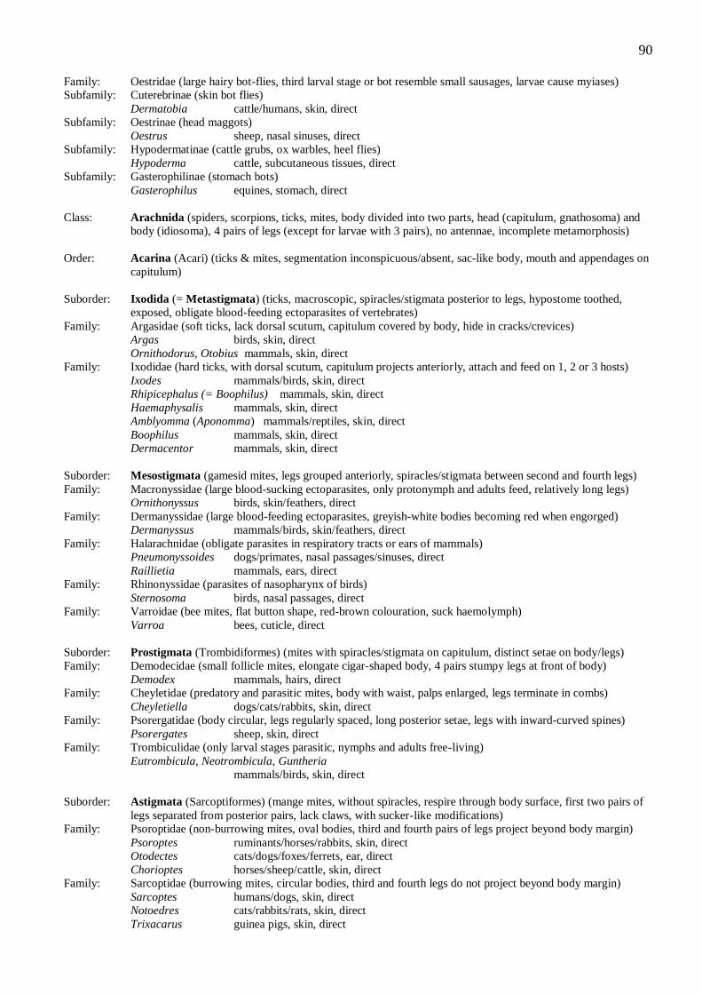
90
Family: Oestridae (large hairy bot-flies, third larval stage or bot resemble small sausages, larvae cause myiases)
Subfamily: Cuterebrinae (skin bot flies)
Dermatobia cattle/humans, skin, direct
Subfamily: Oestrinae (head maggots)
Oestrus sheep, nasal sinuses, direct
Subfamily: Hypodermatinae (cattle grubs, ox warbles, heel flies)
Hypoderma cattle, subcutaneous tissues, direct
Subfamily: Gasterophilinae (stomach bots)
Gasterophilus equines, stomach, direct
Class: Arachnida (spiders, scorpions, ticks, mites, body divided into two parts, head (capitulum, gnathosoma) and
body (idiosoma), 4 pairs of legs (except for larvae with 3 pairs), no antennae, incomplete metamorphosis)
Order: Acarina (Acari) (ticks & mites, segmentation inconspicuous/absent, sac-like body, mouth and appendages on
capitulum)
Suborder: Ixodida (= Metastigmata) (ticks, macroscopic, spiracles/stigmata posterior to legs, hypostome toothed,
exposed, obligate blood-feeding ectoparasites of vertebrates)
Family: Argasidae (soft ticks, lack dorsal scutum, capitulum covered by body, hide in cracks/crevices)
Argas birds, skin, direct
Ornithodorus, Otobius mammals, skin, direct
Family: Ixodidae (hard ticks, with dorsal scutum, capitulum projects anteriorly, attach and feed on 1, 2 or 3 hosts)
Ixodes mammals/birds, skin, direct
Rhipicephalus (= Boophilus) mammals, skin, direct
Haemaphysalis mammals, skin, direct
Amblyomma (Aponomma) mammals/reptiles, skin, direct
Boophilus mammals, skin, direct
Dermacentor mammals, skin, direct
Suborder: Mesostigmata (gamesid mites, legs grouped anteriorly, spiracles/stigmata between second and fourth legs)
Family: Macronyssidae (large blood-sucking ectoparasites, only protonymph and adults feed, relatively long legs)
Ornithonyssus birds, skin/feathers, direct
Family: Dermanyssidae (large blood-feeding ectoparasites, greyish-white bodies becoming red when engorged)
Dermanyssus mammals/birds, skin/feathers, direct
Family: Halarachnidae (obligate parasites in respiratory tracts or ears of mammals)
Pneumonyssoides dogs/primates, nasal passages/sinuses, direct
Raillietia mammals, ears, direct
Family: Rhinonyssidae (parasites of nasopharynx of birds)
Sternosoma birds, nasal passages, direct
Family: Varroidae (bee mites, flat button shape, red-brown colouration, suck haemolymph)
Varroa bees, cuticle, direct
Suborder: Prostigmata (Trombidiformes) (mites with spiracles/stigmata on capitulum, distinct setae on body/legs)
Family: Demodecidae (small follicle mites, elongate cigar-shaped body, 4 pairs stumpy legs at front of body)
Demodex mammals, hairs, direct
Family: Cheyletidae (predatory and parasitic mites, body with waist, palps enlarged, legs terminate in combs)
Cheyletiella dogs/cats/rabbits, skin, direct
Family: Psorergatidae (body circular, legs regularly spaced, long posterior setae, legs with inward-curved spines)
Psorergates sheep, skin, direct
Family: Trombiculidae (only larval stages parasitic, nymphs and adults free-living)
Eutrombicula, Neotrombicula, Guntheria
mammals/birds, skin, direct
Suborder: Astigmata (Sarcoptiformes) (mange mites, without spiracles, respire through body surface, first two pairs of
legs separated from posterior pairs, lack claws, with sucker-like modifications)
Family: Psoroptidae (non-burrowing mites, oval bodies, third and fourth pairs of legs project beyond body margin)
Psoroptes ruminants/horses/rabbits, skin, direct
Otodectes cats/dogs/foxes/ferrets, ear, direct
Chorioptes horses/sheep/cattle, skin, direct
Family: Sarcoptidae (burrowing mites, circular bodies, third and fourth legs do not project beyond body margin)
Sarcoptes humans/dogs, skin, direct
Notoedres cats/rabbits/rats, skin, direct
Trixacarus guinea pigs, skin, direct
91
Family: Knemidocoptidae (burrowing scaly leg and face mites, round body, no dorsal spines, short stubby legs)
Knemidocoptes, Neocnemidocoptes birds, skin, direct
Family: Ctyoditidae (respiratory parasites of birds, chelicerae absent, palps fused to form sucking organ)
Cytodites birds, air sacs, direct
Family: Listrophoridae (parasitic on fur-bearing mammals, distinct dorsal shield, legs modified for grasping hairs)
Lynxacarus cats, skin, direct
Lepoacarus rabbits/hares, skin, direct
Myocoptes mice, skin/hair, direct
Family: Atopomelidae (fur and feather mites)
Chirodiscoides guinea pigs, skin, direct
Family: Laminosioptidae (small mites, smooth elongated body, few setae, affect muscles of birds)
Laminosioptes birds, subcutaneous tissues, direct
Subphylum: Crustacea (chitinous cuticle, gills, 2 pairs antennae, mouthparts comprise pair mandibles and 2 pairs
maxillae, segments with pair biramous extremities (podia), metamorphosis involving larval nauplius/zoea)
Class: Copepoda (elongate body, thorax with 7 somites (first few fused with head to form cepahalothorax), gradual
metamorphosis with series of copepodod instars succeeding naupliar instars, some ectoparasitic forms)
Order: Cyclopoida (antennules short with 10-16 articles, buccal cavity open)
Family: Lernaeidae (females insert anterior attachment organ into host tissues, develop paired egg sacs, free-
swimming naupliar stage, five copepodid stages and adults on hosts)
Lernaea fish, gills, direct (water)
Order: Poecilostomatoida (simple parasitic forms of fishes to bizarre symbiotic forms of invertebrates)
Family: Ergasilidae (antennae modified into powerful organs of prehension, parasitic in freshwater and marine fishes)
Ergasilus fish, gills, direct (water)
Class: Branchiura (head with flattened bilobed cephalic fold, antennae reduced, carapace expands laterally to form
respiratory alae, blood suckers on fish)
Order: Argulidea (single order)
Family: Argulidae (fish lice, discoid body, attaches using hooks/suckers/barbs, stylus inserted to feed on blood)
Argulus fish, skin/gills, direct
Subphylum: Pentastomatida (tongue worms, crustacean-related parasites, lost virtually all appendages, elongate,
segmented, anterior end with mouth and two pairs of tiny claws (penta-stome appearance))
Order: Porocephalida (mouth between or below anterior hooks, hooks with fulcrum, vuvla near posterior)
Family: Porocephalidae (parasites of snakes)
Porocephalus snakes, respiratory passages, indirect (rodent IH)
Family: Linguatulidae (parasites of mammals)
Linguatula mammals, respiratory passages, indirect (mammalian IH)
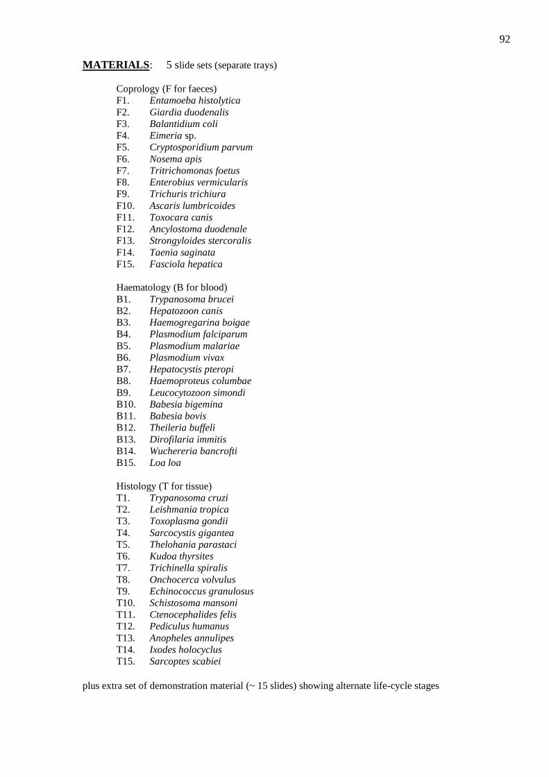
92
MATERIALS: 5 slide sets (separate trays)
Coprology (F for faeces)
F1. Entamoeba histolytica
F2. Giardia duodenalis
F3. Balantidium coli
F4. Eimeria sp.
F5. Cryptosporidium parvum
F6. Nosema apis
F7. Tritrichomonas foetus
F8. Enterobius vermicularis
F9. Trichuris trichiura
F10. Ascaris lumbricoides
F11. Toxocara canis
F12. Ancylostoma duodenale
F13. Strongyloides stercoralis
F14. Taenia saginata
F15. Fasciola hepatica
Haematology (B for blood)
B1. Trypanosoma brucei
B2. Hepatozoon canis
B3. Haemogregarina boigae
B4. Plasmodium falciparum
B5. Plasmodium malariae
B6. Plasmodium vivax
B7. Hepatocystis pteropi
B8. Haemoproteus columbae
B9. Leucocytozoon simondi
B10. Babesia bigemina
B11. Babesia bovis
B12. Theileria buffeli
B13. Dirofilaria immitis
B14. Wuchereria bancrofti
B15. Loa loa
Histology (T for tissue)
T1. Trypanosoma cruzi
T2. Leishmania tropica
T3. Toxoplasma gondii
T4. Sarcocystis gigantea
T5. Thelohania parastaci
T6. Kudoa thyrsites
T7. Trichinella spiralis
T8. Onchocerca volvulus
T9. Echinococcus granulosus
T10. Schistosoma mansoni
T11. Ctenocephalides felis
T12. Pediculus humanus
T13. Anopheles annulipes
T14. Ixodes holocyclus
T15. Sarcoptes scabiei
plus extra set of demonstration material (~ 15 slides) showing alternate life-cycle stages
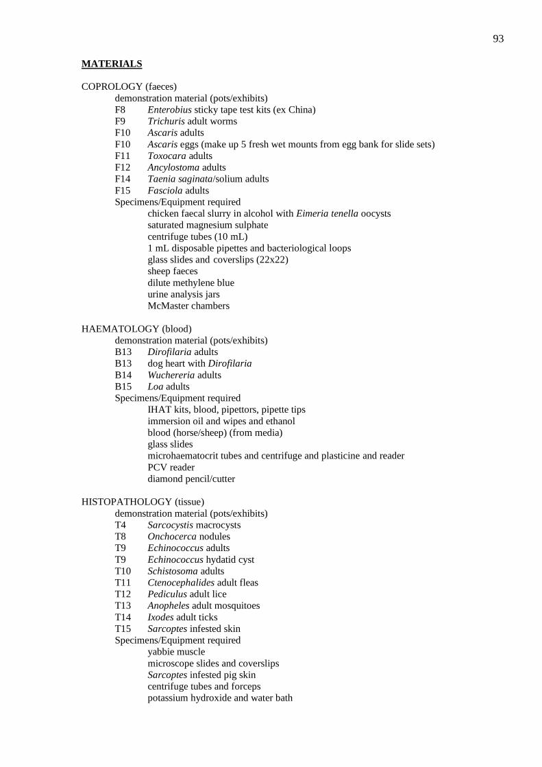
93
MATERIALS
COPROLOGY (faeces)
demonstration material (pots/exhibits)
F8 Enterobius sticky tape test kits (ex China)
F9 Trichuris adult worms
F10 Ascaris adults
F10 Ascaris eggs (make up 5 fresh wet mounts from egg bank for slide sets)
F11 Toxocara adults
F12 Ancylostoma adults
F14 Taenia saginata/solium adults
F15 Fasciola adults
Specimens/Equipment required
chicken faecal slurry in alcohol with Eimeria tenella oocysts
saturated magnesium sulphate
centrifuge tubes (10 mL)
1 mL disposable pipettes and bacteriological loops
glass slides and coverslips (22x22)
sheep faeces
dilute methylene blue
urine analysis jars
McMaster chambers
HAEMATOLOGY (blood)
demonstration material (pots/exhibits)
B13 Dirofilaria adults
B13 dog heart with Dirofilaria
B14 Wuchereria adults
B15 Loa adults
Specimens/Equipment required
IHAT kits, blood, pipettors, pipette tips
immersion oil and wipes and ethanol
blood (horse/sheep) (from media)
glass slides
microhaematocrit tubes and centrifuge and plasticine and reader
PCV reader
diamond pencil/cutter
HISTOPATHOLOGY (tissue)
demonstration material (pots/exhibits)
T4 Sarcocystis macrocysts
T8 Onchocerca nodules
T9 Echinococcus adults
T9 Echinococcus hydatid cyst
T10 Schistosoma adults
T11 Ctenocephalides adult fleas
T12 Pediculus adult lice
T13 Anopheles adult mosquitoes
T14 Ixodes adult ticks
T15 Sarcoptes infested skin
Specimens/Equipment required
yabbie muscle
microscope slides and coverslips
Sarcoptes infested pig skin
centrifuge tubes and forceps
potassium hydroxide and water bath
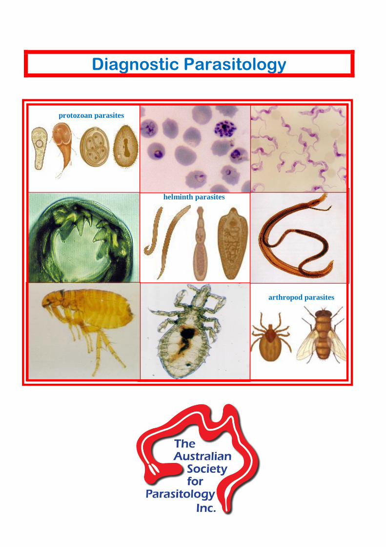
94
Diagnostic Parasitology
protozoan parasites
helminth parasites
arthropod parasites