Veterinary Parasitology - CiteSeerX
7
Veterinary Parasitology 184 (2012) 147–153 Contents lists available at SciVerse ScienceDirect Veterinary Parasitology jo u rn al hom epa ge : www.elsevier.com/locate/vetpar Apoptosis in T lymphocytes from spleen tissue and peripheral blood of L. (L.) chagasi naturally infected dogs Valéria Marc ¸ al Felix de Lima a,∗ , Karina Reinaldo Fattori b , Fausto de Souza a , Flavia Rezende Eugênio a , Paulo Sérgio Patto dos Santos a , Daniele Bernadete Rozza a , Gisele Fabrino Machado a a Departamento de Clinica, Cirurgia e Reproduc ¸ ão Animal, Faculdade de Medicina Veterinária, Universidade Estadual Paulista, Rua Clóvis Pestana, 793, ZIP 16050-400 Arac ¸ atuba, São Paulo, Brazil 1 b Faculdade de Ciências Agrárias e Veterinárias Programa de Pós-graduac ¸ ão em Microbiologia Agropecuária, Universidade Estadual Paulista, Via de Acesso Professor Paulo Donato Castellane s/n, ZIP 14884-900 Jaboticabal, São Paulo, Brazil a r t i c l e i n f o Article history: Received 26 November 2010 Received in revised form 10 August 2011 Accepted 12 August 2011 Keywords: Leishmaniasis Apoptosis Dogs Spleen Peripheral blood a b s t r a c t Dogs are the main domestic reservoirs of L. (L.) chagasi. Once in the vertebrate host, the par- asite may cause visceral leishmaniasis, which can also be transmitted to humans. Infected symptomatic dogs show disorganization in the white pulp in spleen tissue and a reduction in T lymphocytes in peripheral blood. To investigate whether apoptosis is involved in white pulp disorganization and diminished T cell counts in peripheral blood, apoptotic T cells from the spleen and peripheral blood of dogs naturally infected with L. (L.) chagasi and presenting clinical manifestations were quantified and compared with healthy dogs. Thirteen symp- tomatic adult dogs infected by L. (L.) chagasi and six healthy dogs from a nonendemic area (controls) were included in the study. Samples from spleen and peripheral blood were used to quantify apoptosis in CD3 lymphocytes by flow cytometry using Anexin V and Multicas- pase kits; the results were compared using the Mann Whitney test. The percentage of total T cells was lower in Leishmania infected dogs compared to healthy controls (P < 0.05). Apo- ptosis levels in T cells from PBMC and spleen were higher in infected dogs than in controls (P < 0.05). The least squares method test was used to determine the effect between the degree of structural organization of spleen white pulp and the percentage of apoptosis in the spleen. A significant effect on the level of white pulp morphological disorganization and percentage of apoptosis in spleen T cells was observed (F = 20.45; P = 0.0014). These data suggest that apoptosis is an important for the immunopathogenesis of canine visceral leishmaniasis. © 2011 Elsevier B.V. All rights reserved. 1. Introduction Leishmaniasis is caused by a protozoan of the try- panosomatidae family, genus Leishmania. It occurs in 88 countries of which 65 present the visceral form. Most cases ∗ Corresponding author. Tel.: +55 18 36363285. E-mail address: vmfl[email protected] (V.M.F.d. Lima). 1 Tel.: +55 18 3636 14 22. (90%) of visceral leishmaniasis (VL) in humans occur in the rural or suburban areas of five countries, including Brazil (Desjeux, 2004). In Brazil, like other countries in South America, migration into urban areas contributed to the expansion of VL (Desjeux, 2004). In Brazil, this expan- sion occurred especially in the northeast (Dantas-Torres, 2006) and southeast areas (Santiago et al., 2007). Besides the high incidence and broader distribution that the expan- sion itself represents, the spread into new areas also carries the threat that severe and lethal forms of the disease may 0304-4017/$ – see front matter © 2011 Elsevier B.V. All rights reserved. doi:10.1016/j.vetpar.2011.08.024
-
Upload
khangminh22 -
Category
Documents
-
view
1 -
download
0
Transcript of Veterinary Parasitology - CiteSeerX

AL
VFGa
1b
P
a
ARRA
KLADSP
1
pc
0d
Veterinary Parasitology 184 (2012) 147– 153
Contents lists available at SciVerse ScienceDirect
Veterinary Parasitology
jo u rn al hom epa ge : www.elsev ier .com/ locate /vetpar
poptosis in T lymphocytes from spleen tissue and peripheral blood of. (L.) chagasi naturally infected dogs
aléria Marc al Felix de Limaa,∗, Karina Reinaldo Fattorib, Fausto de Souzaa,lavia Rezende Eugênioa, Paulo Sérgio Patto dos Santosa, Daniele Bernadete Rozzaa,isele Fabrino Machadoa
Departamento de Clinica, Cirurgia e Reproduc ão Animal, Faculdade de Medicina Veterinária, Universidade Estadual Paulista, Rua Clóvis Pestana, 793, ZIP6050-400 Arac atuba, São Paulo, Brazil1
Faculdade de Ciências Agrárias e Veterinárias Programa de Pós-graduac ão em Microbiologia Agropecuária, Universidade Estadual Paulista, Via de Acessorofessor Paulo Donato Castellane s/n, ZIP 14884-900 Jaboticabal, São Paulo, Brazil
r t i c l e i n f o
rticle history:eceived 26 November 2010eceived in revised form 10 August 2011ccepted 12 August 2011
eywords:eishmaniasispoptosisogspleeneripheral blood
a b s t r a c t
Dogs are the main domestic reservoirs of L. (L.) chagasi. Once in the vertebrate host, the par-asite may cause visceral leishmaniasis, which can also be transmitted to humans. Infectedsymptomatic dogs show disorganization in the white pulp in spleen tissue and a reductionin T lymphocytes in peripheral blood. To investigate whether apoptosis is involved in whitepulp disorganization and diminished T cell counts in peripheral blood, apoptotic T cells fromthe spleen and peripheral blood of dogs naturally infected with L. (L.) chagasi and presentingclinical manifestations were quantified and compared with healthy dogs. Thirteen symp-tomatic adult dogs infected by L. (L.) chagasi and six healthy dogs from a nonendemic area(controls) were included in the study. Samples from spleen and peripheral blood were usedto quantify apoptosis in CD3 lymphocytes by flow cytometry using Anexin V and Multicas-pase kits; the results were compared using the Mann Whitney test. The percentage of totalT cells was lower in Leishmania infected dogs compared to healthy controls (P < 0.05). Apo-ptosis levels in T cells from PBMC and spleen were higher in infected dogs than in controls(P < 0.05). The least squares method test was used to determine the effect between the
degree of structural organization of spleen white pulp and the percentage of apoptosis inthe spleen. A significant effect on the level of white pulp morphological disorganizationand percentage of apoptosis in spleen T cells was observed (F = 20.45; P = 0.0014). Thesedata suggest that apoptosis is an important for the immunopathogenesis of canine visceralleishmaniasis.© 2011 Elsevier B.V. All rights reserved.
. Introduction
Leishmaniasis is caused by a protozoan of the try-anosomatidae family, genus Leishmania. It occurs in 88ountries of which 65 present the visceral form. Most cases
∗ Corresponding author. Tel.: +55 18 36363285.E-mail address: [email protected] (V.M.F.d. Lima).
1 Tel.: +55 18 3636 14 22.
304-4017/$ – see front matter © 2011 Elsevier B.V. All rights reserved.oi:10.1016/j.vetpar.2011.08.024
(90%) of visceral leishmaniasis (VL) in humans occur inthe rural or suburban areas of five countries, includingBrazil (Desjeux, 2004). In Brazil, like other countries inSouth America, migration into urban areas contributed tothe expansion of VL (Desjeux, 2004). In Brazil, this expan-sion occurred especially in the northeast (Dantas-Torres,
2006) and southeast areas (Santiago et al., 2007). Besidesthe high incidence and broader distribution that the expan-sion itself represents, the spread into new areas also carriesthe threat that severe and lethal forms of the disease may
ry Paras
148 V.M.F.d. Lima et al. / Veterinaemerge when associated with malnutrition (Gontijo andMelo, 2004) and HIV/AIDS infection (Ashford, 2000).
Dogs are considered the main domestic reservoirs ofL. (L.) chagasi (Moreno and Alvar, 2002). The parasite istransmitted from one infected dog to another through thebite of phlebotominae and possibly through other arthro-pod vectors such as fleas and ticks (Ferreira et al., 2009;Coutinho et al., 2005), and through blood transfusions, asreported by Owens et al. (2001). After the phlebotominaefeed on the dogs’ blood, the parasites rapidly spread intothe lymph nodes and spleen through the lymph and bloodand eventually reach the kidneys and liver. They may alsoaffect the reproductive organs, skin, digestive and respi-ratory systems and the bladder (Molyneux and Ashford,1983).
Once in the vertebrate host, the parasite can causelesions and symptoms that are characteristic of caninevisceral leishmaniasis, although some infected dogs maybe oligosymptomatic or asymptomatic (Mancianti andMeciani, 1988), and some may evolve to spontaneouscure (Fisa et al., 1999). The most frequent signs ofVL are lymphadenopathy, onychogryphosis, cutaneouslesions, weight lost, cachexia and locomotor abnormali-ties (Semião-Santos et al., 1995). The asymptomatic formrepresents 20–40% of the serum-positive population, ofwhich 80% actually develops the disease (Noli, 1999).In the Brazil in urban area of the northeast region, theasymptomatic form represent 30% of the serum positivepopulation (Queiroz et al., 2009).
The suppression of cellular immunity is the most impor-tant aspect in the pathogenesis and progression of caninedisease. The absence of T cell response to antigens of Leish-mania sp. is observed in vivo, with a negative leishmaninskin test (Dos Santos et al., 2008). In dogs infected withLeishmania infantum, a reduction in the number of T lym-phocytes in PBMC occurs (Bourdoiseau et al., 1997) anddisorganization of white pulp in spleen tissue has beenpreviously described (Santana et al., 2008), but the mech-anisms that are responsible for these changes have not yetbeen elucidated.
In human acute infection, the reduction in T lympho-cytes and mononuclear cells of peripheral blood and failurein immunity has been associated with apoptosis (Potestioet al., 2004). In mice experimentally infected with Leish-mania donovani, an increase in the level of spontaneousapoptosis in the spleen and liver compared to noninfectedmice was also observed (Alexander et al., 2001). In vitrofindings also suggest the involvement of apoptosis in themechanism of suppression observed in visceral leishmani-asis, the infection of macrophages by L. donovani increasedthe level of FAS in the membrane and sFASL in the cul-ture supernatant, a mechanism that may contribute toincreased sensitivity to apoptosis for T cells specific forLeishmania sp. (Eidsmo et al., 2002).
To investigate whether apoptosis is involved in thereduction in lymphocytes in peripheral blood and alter-ations in white pulp, apoptosis was quantified in dogs
naturally infected with L. (L.) chagasi presenting clinicalmanifestations and the structural disorganization in whitepulp was correlated with the percentage of apoptosis in Tcells. If proven, such findings could contribute to improvingitology 184 (2012) 147– 153
our present understanding of the immunopathogenesis ininfected dogs.
2. Materials and methods
2.1. The study area
The county of Arac atuba (21◦12′32′′ S; 50◦25′38′′ W),with an area of 1,167,311 km2, is located in the state of SãoPaulo, Brazil. It is a region known to be endemic for canineVL.
2.2. Ethical issue
This study was approved by the institutional Ethicsand Animal Welfare Committee (Comissão de Ética emExperimentac ão Animal, CEEA, UNESP, process number02232).
2.3. Animals and diagnosis of VL
A total of 13 adult dogs were used, males and females,aged between 2 and 4 years-old, of undefined breedand different weights, from the Zoonosis Control Cen-ter of Arac atuba (CCZA). The dogs were symptomaticand showing at least three clinical signs of canine VL.These could include fever, dermatitis, lymphoadenopathy,onychogryphosis, weight loss, cachexia, locomotor abnor-malities, conjunctivitis, epistaxis, hepatosplenomegaly,edema, and apathy. Briefly, the diagnosis of VL was con-firmed by detecting anti-Leishmania antibodies for L. (L.)chagasi by indirect ELISA, according to Lima et al. (2003),and simultaneously positivity in rapid test rK39 and in PCRamplification of Leishmania spp. DNA in spleen tissue.
A group of 6 healthy dogs, both males and females, froma nonendemic area (Londrina, State of Paraná, Brazil) wereincluded in the study as negative controls. These dogs wereserum negative for L. (L.) chagasi by indirect ELISA (Limaet al., 2003), negative in the rapid test rK39 and in PCRamplification of Leishmania spp. DNA in spleen tissue.
2.4. Sample collection
Samples of spleen from both groups were removed bysurgical excision. The dogs were premedicated with thecombination of morphine (0.4 mg kg−1 IM) and acepro-mazine (0.05 mg kg−1 IM). Fifteen minutes later, propofol(4.0 mg kg−1 IV) and midazolam (0.1 mg kg−1 IV) were usedfor induction. Immediately, the dogs were positioned indorsal recumbency and anesthesia was maintained withisoflurane (1.5 V%). The heart and respiratory rates, thesystolic arterial blood pressure and end-tidal CO2 measure-ments were monitored during all anesthetic procedure.The samples were maintained in RPMI 1640 supplementedwith 10% (v/v) fetal calf serum (Sigma) at 4 ◦C and processedimmediately to evaluate apoptosis.
From each dog, 4 ml of blood was collected fromthe cephalic veins, clotted at room temperature for 4 hand subsequently centrifuged to extract the serum. Theserum samples were stored at–20 ◦C prior to analysis and

ry Paras
aa
2U
bsa
2
fipstbtawErGCaoPo1(Ac51a−ncpuf
2
a(lpatUtttTcw
V.M.F.d. Lima et al. / Veterina
dditional blood samples were collected with sodium EDTAnd processed immediately to evaluate apoptosis.
.5. Kalazar detect rapid test rK-39 (InBios International,SA)
To perform this test, blood samples were collectedy venipuncture and centrifuged and the serum waseparated. The procedure of the test was performed inccordance with the manufacturer’s recommendations.
.6. PCR
DNA obtained from spleen samples was extracted byreezing and thawing the cells 3 times and washing themn 1× SSC buffer solution (NaCl 3 M, sodium citrate 0.3 M,H 7.0). For cell lysis and protein digestion, 300 �l of lysingolution was added (10% SDS in 0.2 M sodium acetate)ogether with 20 �g/ml proteinase K. Samples were incu-ated at 56 ◦C for 2 h and the DNA was extracted usinghe phenol/chloroform/isoamyl alcohol method (25:24:1),ccording to Sambrook et al. (1989). After extraction, DNAas resuspended in 50 �l TE (10 mM Tris–HCl pH 8.0, 1 mM
DTA pH 8.0) and incubated for 3 min at 60 ◦C. The mate-ial was stored at −20 ◦C until used. The 13A (3′-GTG GGGAG GGG CGT TCT-5′) and 13B (3′-ATT TTA CAC CAA CCCCA GTT-5′) primers were used (Rodgers et al., 1990) tomplify a 120 bp fragment located in the constant regionf the kinetoplast minicircles in all Leishmania species. TheCR was performed in a 60 �l volume containing 30 pmolf each primer (Invitrogen®), 0.2 mM DNTPs (Invitrogen®),.5 mM MgCl2 (Invitrogen®), 5 U Taq DNA PolymeraseInvitrogen®), 50 nM buffer solution, milliQ water and DNA.mplification was performed in an Eppendorf® Mastercy-ler Thermocycler gradient with initial heating to 95 ◦C for
min, followed by 33 cycles at 95–57–72 ◦C for 1.5 min,.5 min, and 2 min, respectively. Extension was performedt 72 ◦C for 10 min and the final product was stored at20 ◦C until analysis. Reaction mixes containing eithero DNA or DNA extracted from a L. chagasi promastigoteulture (MHOM/BR00/MER02) were used as negative andositive controls, respectively. The amplified 120 bp prod-ct was analyzed by electrophoresis on acrylamide gelsollowed by silver staining.
.7. Apoptosis evaluation in T cells
Peripheral blood cells were stained following the sep-ration of mononuclear cells on a Ficoll-Paque gradientAmersham Biosciences). Spleen cells were processed afterysis of red blood cells. For staining, the cells were sus-ended in PBS containing 1% bovine serum albumin, 0.1%zide and 20% fetal bovine serum to block the Fc recep-or (FCR). Anti-canine CD3 monoclonal antibodies (Serotec,K) were added and incubated for 30 min. Isotype con-
rol (Serotec, UK) antibody was added in a separate tubeo control for nonspecific labeling. Following incubation,
he tubes were centrifuged at 1000 × g for 3 min at 4 ◦C.he supernatant was discarded by quick inversion and theell pellet briefly vortexed to resuspend the cells. The cellsere washed four times with ice-cold PBS with 10% bovineitology 184 (2012) 147– 153 149
calf sera. After the final wash, the cells were resuspendedwith PBS.
After immune staining for CD3 in PBMC and in leuco-cytes from spleen the apoptosis was detected using twodifferent methods. The Nexin assay, which uses Annexin V,is a calcium-dependent phospholipid binding protein withhigh affinity for phosphatidylserine, a membrane compo-nent normally located in the internal face; however, duringearly activation of apoptotic pathways, these molecules aretranslocated to the outer surface of the cell membrane,where Annexin V can bind directly to them (Vermes et al.,1995). The second method, MultiCaspase SR kit, detectscaspase pathways by a fluorescent labeled inhibitor of cas-pase reagent that specifically identifies active caspases.These methods have been used in similar studies (Colgateet al., 2007).
The percentage of apoptosis was determined using theNexin assay Kit (Guava, Hayward, CA) and Mutilcaspase SRkit (Guava, Hayward, CA). The procedure of each test wasperformed in accordance with the manufacturer’s recom-mendations. Proper instrument performance was verifiedby running the Guava Check application with Guava Checkreagents. Data were acquired regarding the Guava Easy-CyteMini system using CytoSoft software, as describedin the Guava PCA User’s Guide and respective packageinserts. For Guava MultiCaspase and Guava Nexin, 10,000events were usually acquired. Negative and positive controlCamptothecin (Sigma, USA) (0.15 �g/mL) in DMSO (Sigma)(Tao et al., 2004) were used to verify reagent performanceand set analysis markers, delineating the negative and pos-itive populations.
2.8. Spleen samples and histological analysis
Three to 4 mm-thick tissue slices were cut transversallyto the capsule and fixed in 10% formalin. After fixation, tis-sue slides were embedded in paraffin. Four to 5 �m-thicksections were cut and stained with hematoxylin and eosin(HE). These sections were examined by at least two of theauthors who were blind to previous knowledge of the dogs’health and identities. The authors scored the level of whitepulp organization: (1) slightly disorganized, with eitherhyperplastic or hypoplastic changes leading to a loss of def-inition of any of the regions of the white pulp and (2) formoderately or extensively disorganized, when the whitepulp regions were poorly individualized or indistinct.
2.9. Immunohistochemistry (IHC)
IHC stains were performed using the standardstreptavidin–biotin peroxidase (HRP) immunostainingprocedure with polyclonal antibody. Anti-CD3 (A0452)(DAKO, CA, USA) was used to detect T lymphocytes. Thisantibody was previously used to stain CD3 in canine lym-phomas (Sueiro et al., 2004). Slides were deparaffinized andhydrated. Antigen retrieval was achieved by steam heatingin citrate buffer for 30 min. For inhibition of endogenous
peroxidase, slides were incubated with 2% (v/v) hydrogenperoxide 30 vol diluted in 50% (v/v) methanol for 30 minand nonspecific binding was blocked with 3% (w/v) nonfatdry milk in PBS for 30 min. Primary antibody against CD3
150 V.M.F.d. Lima et al. / Veterinary Paras
Fig. 1. Percentage of CD3 lymphocytes in PBMC from Leishmania infected
dogs (INF) and healthy controls (CT). The results are expressed as the meanpercentage ± SD. P < 0.05: significant differences between the mean valuesfor Leishmania infected dogs and healthy controls.(1:100) was applied for 18–22 h at 4 ◦C in a humidifiedchamber. Slides were washed in PBS, incubated with abiotinylated secondary antibody (LSAB+ Kit, DAKO K0690,CA, USA) for 45 min at room temperature, washed oncemore with PBS, and incubated with streptavidin–HRPcomplex (LSAB+ Kit, DAKO K0690, CA, USA) for 45 min atroom temperature. The reaction was developed with 3,3′-diaminobenzidine (DAKO K3468, CA, USA). The slides werecounterstained with Harris’s hematoxylin, dehydrated,cleared and mounted with coverslips. Spleen tissue fromhealthy dogs was used as a positive control.
2.10. Statistical analysis
The data were analyzed by a nonparametric test. Groupmeans were compared using Mann Whitney test. Theleast squares method was used to evaluate the effect ofgroup (degree of correlation for the structural organiza-tion of white pulp) and quantitative variable (percentageof apoptosis). The results were considered significant whenP < 0.05. The SAS software was used (SAS 9.1, SAS Institute,Cary, NC, USA) for all statistical analyses performed in thisstudy.
3. Results
3.1. Decrease in T lymphocytes in PBMC
Flow cytometry analysis of CD3 lymphocytes in PMBCfrom infected dogs showed significantly lower num-bers (58 ± 12, mean ± SD) compared to healthy controls(80.6 ± 5, mean ± SD) (Fig. 1) (P = 0.001, Mann Whitneytest).
3.2. Apoptosis in T cells from peripheral blood and spleen
To examine apoptosis in T cells from PBMC and spleenof L. chagasi-infected dogs, PBMC and spleen samples wereevaluated immediately following collection. Apoptosis of T
cells from PBMC and spleen was detected using commercialkits for both, Annexin V (Guava, Hayward, CA) and Caspases(Guava, Hayward, CA) and simultaneously anti-CD3 mAbs(Serotec, UK).itology 184 (2012) 147– 153
The percentage of apoptotic T cells was evaluated afterCD3 positive cells were gated in Plot 1 (Fig. 2a), followedby detection double stained for apoptosis detection in cellsgated in Plot 1 (Fig. 2b). The PBMC showed higher numbersof apoptotic T cells in infected dogs (Fig. 2c) compared tohealthy dogs (Fig. 2d) and in the spleen, similar results wereobtained (Fig. 2e and f).
Data presented in Fig. 3 clearly indicate that T lympho-cytes from PBMC and spleen tissue of infected dogs showeda significantly higher level of apoptosis compared to thatobserved in healthy dogs (P < 0.05, Mann Whitney test). Ingeneral both kits used showed similar values of apoptoticcells in controls and infected dogs (Fig. 3).
3.3. White pulp structural organization and CD3 IHQ
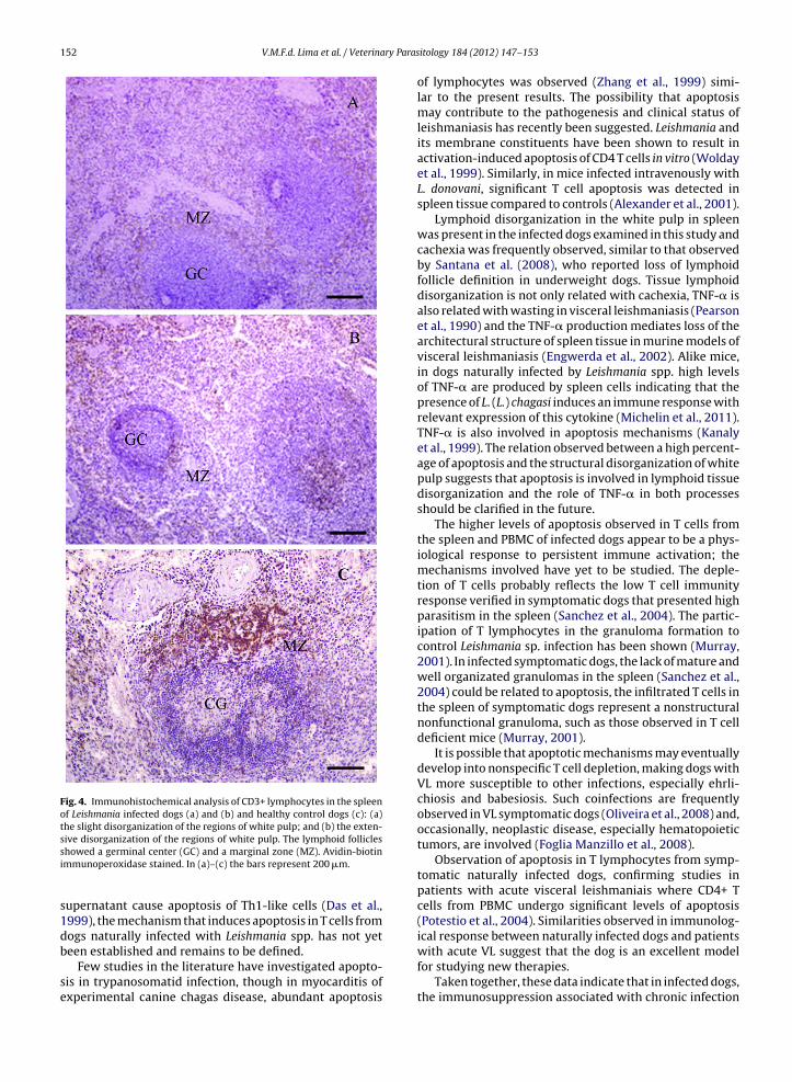
The percentage of dogs with slightly disorganized whitepulp, with either hyperplastic or hypoplastic changes lead-ing to a loss of definition of any of the regions of the whitepulp areas was 63.6%, while 36.3% dogs showed white pulpareas that were moderately or extensively disorganizedand were poorly individualized or indistinct. The CD3 IHCconfirmed the structural disorganization of the white pulpin infected dogs compared to that observed in healthy dogs(Fig. 4). A significant effect on the level of white pulp mor-phological disorganization and percentage of apoptosis inT cells from the spleen was observed (F = 20.45; P = 0.0014,least squares method).
4. Discussion
The level of apoptosis in T cells from PBMC and spleenfrom VL dogs from an endemic area was analyzed and thevalues compared to uninfected control dogs to investigatethe role this mechanism plays in the pathology and infec-tious process of the disease, as well as in the host immuneresponse.
Apoptosis is essential for homeostatic control of lym-phocyte numbers, particularly following the developmentof an immune response to an invasive microorganism(Scaffidi et al., 1999). However, some microbes are ableto either directly manipulate this response or have devel-oped strategies to survive in the host until homeostaticmechanisms are activated to reduce effector T cell numbers(Freire-de-Lima et al., 2000).
The apoptosis level in T cells from the spleen and periph-eral blood were higher in infected dogs compared to that ofhealthy canines, suggesting that the infection by L. (L.) cha-gasi could induce apoptosis in T cells. Since the progressionof infection is related to the impairment of cell-mediatedimmunity (Dos Santos et al., 2008), the detection of Tcell apoptosis may contribute to inefficient cell immuneresponse during L. chagasi infection.
Several mechanisms of T cell apoptosis have been pro-posed. Deprivation of growth factors, cytotoxicity by TNF-�and FAS and FAS-L interaction are among the major media-tors (Scaffidi et al., 1999). Leishmania spp. infection studies
have shown that macrophages infected by L. donovaniincreased the level of FAS in the membrane and sFASL in theculture supernatant (Eidsmo et al., 2002) and in L. donovaniinfected Balb/c mice, low IL-2 levels in the T cell culture
V.M.F.d. Lima et al. / Veterinary Parasitology 184 (2012) 147– 153 151
Fig. 2. Flow cytometry analysis: (a) fluorescence plot for gated lymphocytes from Leishmania infected dogs (Anti-CD3 monoclonal antibody FITC). Percentageinside upper right quadrants correspond to lymphocyte CD3 positive cells; (b) a representative fluorescence plot for gated lymphocytes and stained forapoptosis (positive control, the cells were incubated with apoptosis inductor Camptothecin); (c) a representative fluorescence plot for gated lymphocytesfrom PBMC from Leishmania infected dogs and stained for apoptosis; (d) a representative fluorescence plot for gated lymphocytes from PBMC from healthydogs and stained for apoptosis; (e) a representative fluorescence plot for gated lymphocytes from the spleen of Leishmania-infected dogs and stainedfor apoptosis; and (f) a representative fluorescence plot for gated lymphocytes from the spleen of healthy dogs and stained for apoptosis. Percentageinside upper right quadrants correspond to lymphocytes stained with Annexin V-PE and 7-AAD fluorescence (double stained), late stage of apoptotic cells.Similarly dot plot figures were observed using the multicaspase kit.
CT- Anexin V INF-Anexin V 0
5
10
15
P=0.009
Spleen
% a
popt
otic
T ly
mph
ocyt
es
CT-MulticaspaseINF-Multicaspase0
10
20
P=0.001
Spleen
% a
popt
otic
T ly
mph
ocyt
es
CT-MulticaspaseINF-Multicaspase0
5
10
15
P=0.001
Blood
% a
popt
otic
T ly
mph
ocyt
es
CT-Anexin VINF-Anexin V0
5
10
15
20P=0.0007
Blood
% a
popt
otic
T ly
mph
ocyt
es
Fig. 3. Apoptosis of T lymphocytes in infected dogs (INF) and control dogs (CT). Each circle represents an individual dog. Bars represent the mean value ineach group. P < 0.05: significant differences between the mean values for Leishmania infected dogs and healthy controls.
152 V.M.F.d. Lima et al. / Veterinary Paras
Fig. 4. Immunohistochemical analysis of CD3+ lymphocytes in the spleenof Leishmania infected dogs (a) and (b) and healthy control dogs (c): (a)the slight disorganization of the regions of white pulp; and (b) the exten-sive disorganization of the regions of white pulp. The lymphoid follicles
with acute VL suggest that the dog is an excellent model
showed a germinal center (GC) and a marginal zone (MZ). Avidin-biotinimmunoperoxidase stained. In (a)–(c) the bars represent 200 �m.
supernatant cause apoptosis of Th1-like cells (Das et al.,1999), the mechanism that induces apoptosis in T cells fromdogs naturally infected with Leishmania spp. has not yetbeen established and remains to be defined.
Few studies in the literature have investigated apopto-sis in trypanosomatid infection, though in myocarditis ofexperimental canine chagas disease, abundant apoptosis
itology 184 (2012) 147– 153
of lymphocytes was observed (Zhang et al., 1999) simi-lar to the present results. The possibility that apoptosismay contribute to the pathogenesis and clinical status ofleishmaniasis has recently been suggested. Leishmania andits membrane constituents have been shown to result inactivation-induced apoptosis of CD4 T cells in vitro (Woldayet al., 1999). Similarly, in mice infected intravenously withL. donovani, significant T cell apoptosis was detected inspleen tissue compared to controls (Alexander et al., 2001).
Lymphoid disorganization in the white pulp in spleenwas present in the infected dogs examined in this study andcachexia was frequently observed, similar to that observedby Santana et al. (2008), who reported loss of lymphoidfollicle definition in underweight dogs. Tissue lymphoiddisorganization is not only related with cachexia, TNF-� isalso related with wasting in visceral leishmaniasis (Pearsonet al., 1990) and the TNF-� production mediates loss of thearchitectural structure of spleen tissue in murine models ofvisceral leishmaniasis (Engwerda et al., 2002). Alike mice,in dogs naturally infected by Leishmania spp. high levelsof TNF-� are produced by spleen cells indicating that thepresence of L. (L.) chagasi induces an immune response withrelevant expression of this cytokine (Michelin et al., 2011).TNF-� is also involved in apoptosis mechanisms (Kanalyet al., 1999). The relation observed between a high percent-age of apoptosis and the structural disorganization of whitepulp suggests that apoptosis is involved in lymphoid tissuedisorganization and the role of TNF-� in both processesshould be clarified in the future.
The higher levels of apoptosis observed in T cells fromthe spleen and PBMC of infected dogs appear to be a phys-iological response to persistent immune activation; themechanisms involved have yet to be studied. The deple-tion of T cells probably reflects the low T cell immunityresponse verified in symptomatic dogs that presented highparasitism in the spleen (Sanchez et al., 2004). The partic-ipation of T lymphocytes in the granuloma formation tocontrol Leishmania sp. infection has been shown (Murray,2001). In infected symptomatic dogs, the lack of mature andwell organizated granulomas in the spleen (Sanchez et al.,2004) could be related to apoptosis, the infiltrated T cells inthe spleen of symptomatic dogs represent a nonstructuralnonfunctional granuloma, such as those observed in T celldeficient mice (Murray, 2001).
It is possible that apoptotic mechanisms may eventuallydevelop into nonspecific T cell depletion, making dogs withVL more susceptible to other infections, especially ehrli-chiosis and babesiosis. Such coinfections are frequentlyobserved in VL symptomatic dogs (Oliveira et al., 2008) and,occasionally, neoplastic disease, especially hematopoietictumors, are involved (Foglia Manzillo et al., 2008).
Observation of apoptosis in T lymphocytes from symp-tomatic naturally infected dogs, confirming studies inpatients with acute visceral leishmaniais where CD4+ Tcells from PBMC undergo significant levels of apoptosis(Potestio et al., 2004). Similarities observed in immunolog-ical response between naturally infected dogs and patients
for studying new therapies.Taken together, these data indicate that in infected dogs,
the immunosuppression associated with chronic infection

ry Paras
iatTuwtie
A
f
R
A
A
B
C
C
D
D
D
D
E
E
F
F
F
F
G
K
V.M.F.d. Lima et al. / Veterina
s due to accelerated rates of T cell apoptosis and this mech-nism could contribute to white pulp disorganization inhe spleen and diminished T cell levels in peripheral blood.he present results could contribute to improving currentnderstanding of the immune response in dogs infectedith L. (L.) chagasi, while additional studies would fur-
her our understanding concerning apoptosis and othermmune mediators in dogs naturally infected with this dis-ase.
cknowledgements
The authors are grateful to the FAPESP and FUNDUNESPor financially supporting this project.
eferences
lexander, C.E., Kaye, P.M., Engwerda, C.R., 2001. CD95 is required forthe early control of parasite burden in liver of Leishmania donovani-infected mice. Eur. J. Immunol. 31, 1199–1210.
shford, R.W., 2000. The leishmaniases as emerging and reemergingzoonoses. Int. J. Parasitol. 30, 1269–1281.
ourdoiseau, G., Bonnefont, C., Magnol, J.P., Saint Andre, I., Chabanne, L.,1997. Lymphocyte subset abnormalities in canine leishmaniasis. Vet.Immunol. Immunopathol. 56, 345–351.
outinho, M.T., Bueno, L.L., Sterzik, A., Fujiwara, R.T., Botelho, J.R., DeMaria, M., Genaro, O., Linardi, P.M., 2005. Participation of Rhipicephalussanguineus (Acari: Ixodidae) in the epidemiology of canine visceralleishmaniasis. Vet. Parasitol. 128, 149–155.
olgate, E.C., Miranda, C.L., Stevens, J.F., Bray, T.M., Ho, E., 2007. Xantho-humol, a prenylflavonoid derived from hops induces apoptosis andinhibits NF-kappaB activation in prostate epithelial cells. Cancer Lett.8, 201–209.
as, G., Vohra, H., Rao, K., Saha, B., Mishra, G.C., 1999. Leishmania donovaniinfection of a susceptible host results in CD4+ T-cell apoptosis anddecreased Th1 cytokine production. Scand. J. Immunol. 49, 307–310.
antas-Torres, F., 2006. Current epidemiological status of visceral leish-maniasis in Northeastern Brazil. Rev. Saude Publica 40, 537–541.
esjeux, P., 2004. Leishmaniasis: current situation and new perspectives.Comp. Immunol. Microbiol. Infect. Dis. 27, 305–318.
os Santos, W.L., Jesus, E.E., Paranhos-Silva, M., Pereira, A.M., Santos,J.C., Baleeiro, C.O., Nascimento, E.G., Moreira, E.D., Oliveira, G.G.,Pontes-de-Carvalho, L.C., 2008. Associations among immunological,parasitological and clinical parameters in canine visceral leishmania-sis: emaciation, spleen parasitism, specific antibodies and leishmaninskin test reaction. Vet. Immunol. Immunopathol. 123, 251–259.
idsmo, L., Wolday, D., Berhe, N., Sabri, F., Satti, I., El Hassan, A.M., Sundar,S., Chiodi, F., Akuffo, H., 2002. Alteration of Fas and Fas ligand expres-sion during human visceral leishmaniasis. Clin. Exp. Immunol. 130,307–313.
ngwerda, C.R., Ato, M., Cotterell, S.E., Mynott, T.L., Tschannerl, A., Gorak-Stolinska, P.M., Kaye, P.M., 2002. A role for tumor necrosis factor-alphain remodeling the splenic marginal zone during Leishmania donovaniinfection. Am. J. Pathol. 161, 429–437.
erreira, M.G., Fattori, K.R., Souza, F., Lima, V.M., 2009. Potential role fordog fleas in the cycle of Leishmania spp. Vet. Parasitol. 165, 150–154.
isa, R., Gállego, M., Castillejo, S., Aisa, M.J., Serra, T., Riera, C., Carrió, J.,Gállego, J., Portús, M., 1999. Epidemiology of canine leishmaniosisin Catalonia (Spain) the example the Priorat focus. Vet. Parasitol. 83,87–97.
oglia Manzillo, V., Pagano, A., Guglielmino, R., Gradoni, L., Restucci, B.,Oliva, G., 2008. Extranodal gamma delta-T-cell lymphoma in a dogwith leishmaniasis. Vet. Clin. Pathol. 37, 298–301.
reire-de-Lima, C.G., Nascimento, D.O., Soares, M.B., Bozza, P.T., Castro-Faria-Neto, H.C., de Mello, F.G., Dos Reis, G.A., Lopes, M.F., 2000. Uptakeof apoptotic cells drives the growth of a pathogenic trypanosome inmacrophages. Nature 13, 199–203.
ontijo, C.M.F., Melo, M.N., 2004. Leishmaniose Visceral no Brasil: quadroatual, desafios e perspectivas. Rev. Bras. Epidemiol. 7, 338–349.
analy, S.T., Nashleanas, M., Hondowicz, B., Scott, P., 1999. TNF receptorp55 is required for elimination of inflammatory cells following controlof intracellular pathogens. J. Immunol. 163, 3883–3889.
itology 184 (2012) 147– 153 153
Lima, V.M.F., Gonc alves, M.E., Ikeda, F.A., Luvizotto, M.C.R., Feitosa, M.M.,2003. Anti-leishmania antibodies in cerebrospinal fluid from dogswith visceral leishmaniasis. Braz. J. Med. Biol. Res. 36, 485–489.
Mancianti, F., Meciani, N., 1988. Specific serodiagnosis of canineleishmaniasis by indirect immunofluorescence, indirect hemagglu-tination, and counterimmunoelectrophoresis. Am. J. Vet. Res. 49,1409–1411.
Michelin, A.F., Perri, S.H.V., Lima, V.M.F., 2011. Evaluation of TNF-a IL-4,and IL-10 and parasite density in spleen and liver of L. (L.) chagasinaturally infected dogs. Ann. Trop. Med. Parasitol. 105, 1–12.
Molyneux, D.H., Ashford, R.W., 1983. The Biology of Trypanosoma andLeishmania, Parasites of Man and Domestic Animals. Taylor & Francis,London.
Moreno, J., Alvar, J., 2002. Canine leishmaniasis: epidemiological risk andthe experimental model. Trends Parasitol. 18, 399–405.
Murray, H.W., 2001. Tissue granuloma structure–function in experimentalvisceral leishmaniasis. Int. J. Exp. Pathol. 82, 249–267.
Noli, C., 1999. Canine leishmaniosis. Waltham Focus 9, 16–24.Owens, S.D., Oakley, D.A., Marryott, K., Hatchett, W., Walton, R., Nolan, T.J.,
Newton, A., Steurer, F., Schantz, P., Giger, U., 2001. Transmission of vis-ceral leishmaniasis through blood transfusions from infected Englishfoxhounds to anemic dogs. J. Am. Vet. Med. Assoc. 219, 1076–1083.
Oliveira, T.M., Furuta, P.I., de Carvalho, D., Machado, R.Z., 2008. A study ofcross-reactivity in serum samples from dogs positive for Leishmaniasp., Babesia canis and Ehrlichia canis in enzyme-linked immunosorbentassay and indirect fluorescent antibody test. Rev. Bras. Parasitol. Vet.17, 7–11.
Pearson, R.D., Cox, G., Evans, T., Smith, D.L., Weidel, D., Castracane, J.,1990. Wasting and macrophage production of tumor necrosis fac-tor/cachectin and interleukin 1 in experimental visceral leishmaniasis.Am. J. Trop. Med. Hyg. 43, 640–649.
Potestio, M., D’Agostino, P., Romano, G.C., Milano, S., Ferlazzo, V., Aquino,A., Di Bella, G., Caruso, R., Gambino, G., Vitale, G., Mansueto, S., Cil-lari, E., 2004. CD4+ CCR5+ and CD4+ CCR3+ lymphocyte subset andmonocyte apoptosis in patients with acute visceral leishmaniasis.Immunology 113, 260–268.
Queiroz, P.V., Monteiro, G.R., Macedo, V.P., Rocha, M.A., Batista, L.M.,Queiroz, J.W., Jerônimo, S.M., Ximenes, M.F., 2009. Canine visceralleishmaniasis in urban and rural areas of Northeast Brazil. Res. Vet.Sci. 86, 267–273.
Rodgers, M.R., Popper, S.J., Wirth, D.F., 1990. Amplification of kinetoplastas tool in the detection and diagnosis of Leishmania. Exp. Parasitol. 71,267–275.
Sambrook, J., Fritsch, E.F., Maniatis, T., 1989. Molecular Cloning: A Lab-oratory Manual, 2nd ed. Cold Spring Harbour Laboratory Press, ColdSpring Harbour, NY.
Scaffidi, C., Kirchhoff, S., Krammer, P.H., Peter, M.E., 1999. Apoptosis sig-naling in lymphocytes. Curr. Opin. Immunol. 11, 277–285.
Santana, C.C., Vassallo, J., de Freitas, L.A., Oliveira, G.G., Pontes-de-Carvalho, L.C., dos-Santos, W.L., 2008. Inflammation and structuralchanges of splenic lymphoid tissue in visceral leishmaniasis: a studyon naturally infected dogs. Parasite Immunol. 30, 515–524.
Sanchez, M.A., Diaz, N.L., Zerpa, O., Negron, E., Convit, J., Tapia, F.J., 2004.Organ-specific immunity in canine visceral leishmaniasis: analysis ofsymptomatic and asymptomatic dogs naturally infected with Leish-mania chagasi. Am. J. Trop. Med. Hyg. 70, 618–624.
Santiago, M.E.B., Vasconcelos, R.O., Fattori, K.R., Munari, D.P., Michelin,A.F., Lima, V.M.F., 2007. An investigation of Leishmania spp. in Didelphisspp. from urban and peri-urban areas in Bauru (São Paulo, Brazil). Vet.Parasitol. 150, 283–290.
Semião-Santos, S.J., el Harith, A., Ferreira, E., Pires, C.A., Sousa, C., Gus-mão, R., 1995. Évora district as a new focus for canine leishmaniasisin Portugal. Parasitol. Res. 81, 235–239.
Sueiro, F.A., Alessi, A.C., Vassallo, J., 2004. Canine lymphomas: a morpho-logical and immunohistochemical study of 55 cases, with observationson p53 immunoexpression. J. Comp. Pathol. 131, 207–213.
Tao, D., Wu, J.I., Feng, Y., Qin, J.I., Hu, J., Gong, J., 2004. New method for theanalysis of cell cycle–specific apoptosis. Cytometry Part A 57, 70–74.
Vermes, I., Haanen, C., Steffens-Nakken, H., Reutelingsperger, C., 1995.A novel assay for apoptosis Flow cytometric detection of phos-phatidylserine expression on early apoptotic cells using fluoresceinlabelled Annexin V. J. Immunol. Methods 184, 39–51.
Wol, D., Akuffo, H., Demissie, A., Britton, S., 1999. Role of Leishmaniadonovani and its lipophosphoglycan in CD4+ T-cell activation-
induced human immunodeficiency virus replication. Infect. Immun.67, 5258–5264.Zhang, J., Andrade, Z.A., Yu, Z.X., Andrade, S.G., Takeda, K., Sadirgursky,M., Ferrans, V.J., 1999. Apoptosis in canine model of acute chagasicmyocarditis. J. Mol. Cell. Cardiol. 31, 581–596.