Belgrade Veterinary Neurology Conference Beogradska ...
206
May 23 - 25th 2014, Crowne Plaza Belgrade, Serbia Belgrade Veterinary Neurology Conference Beogradska Veterinarska Neurološka Konferencija 2014 Richard A. LeCouteur Marc Vandevelde omas Flegel Holger A. Volk
-
Upload
khangminh22 -
Category
Documents
-
view
1 -
download
0
Transcript of Belgrade Veterinary Neurology Conference Beogradska ...

May 23 - 25th 2014, Crowne PlazaBelgrade, Serbia
BelgradeVeterinaryNeurology Conference
BeogradskaVeterinarskaNeurološka Konferencija
2014Richard A. LeCouteurMarc VandeveldeThomas FlegelHolger A. Volk
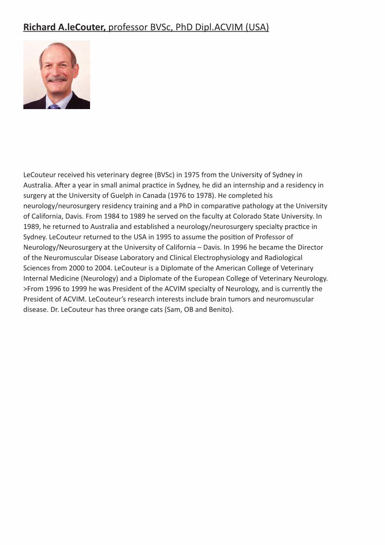
Richard A.leCouter, professor BVSc, PhD Dipl.ACVIM (USA)
LeCouteur received his veterinary degree (BVSc) in 1975 from the University of Sydney inAustralia. After a year in small animal practice in Sydney, he did an internship and a residency insurgery at the University of Guelph in Canada (1976 to 1978). He completed hisneurology/neurosurgery residency training and a PhD in comparative pathology at the Universityof California, Davis. From 1984 to 1989 he served on the faculty at Colorado State University. In1989, he returned to Australia and established a neurology/neurosurgery specialty practice inSydney. LeCouteur returned to the USA in 1995 to assume the position of Professor ofNeurology/Neurosurgery at the University of California – Davis. In 1996 he became the Directorof the Neuromuscular Disease Laboratory and Clinical Electrophysiology and RadiologicalSciences from 2000 to 2004. LeCouteur is a Diplomate of the American College of VeterinaryInternal Medicine (Neurology) and a Diplomate of the European College of Veterinary Neurology.>From 1996 to 1999 he was President of the ACVIM specialty of Neurology, and is currently thePresident of ACVIM. LeCouteur’s research interests include brain tumors and neuromusculardisease. Dr. LeCouteur has three orange cats (Sam, OB and Benito).

Update on Brain Tumors Richard A.leCouter, professor BVSc, PhD Dipl.ACVIM (USA)
1
Update on Brain Tumor Management Richard A. LeCouteur, BVSc, PhD, Diplomate ACVIM(Neurology), Diplomate ECVN
Department of Surgical & Radiological Sciences, School of Veterinary Medicine, University of California, Davis, California, CA 95616-‐8745, USA
Key Facts
• Primary brain tumors are a significant cause of morbidity and mortality in small animal companion animals, particularly in dogs
• With more widespread availability of advanced imaging techniques, the number of animals being diagnosed with intracranial mass lesions has increased significantly
• The most commonly diagnosed tumors are meningioma, followed by glial tumors (astrocytoma, oligodendroglioma) and choroid plexus tumors
• Information relating to imaging characteristics of specific tumor types has been published, however imaging can never provide a specific diagnosis
• As with neoplastic disease affecting any other organ system in the body, definitive diagnosis has been historically confirmed primarily based on the results of biopsy, histopathology and immunohistochemistry
• Biopsy of primary brain tumors presents a number of location specific problems, primarily involving the relative inaccessibility of lesions, together with the significant risks associated with surgical biopsy in many cases
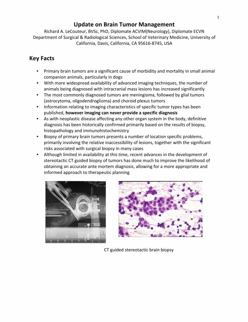
• Although limited in availability at this time, recent advances in the development of stereotactic CT guided biopsy of tumors has done much to improve the likelihood of obtaining an accurate ante mortem diagnosis, allowing for a more appropriate and informed approach to therapeutic planning
CT guided stereotactic brain biopsy

2
Conventional therapy
• In general, conventional therapeutic approaches to brain tumors in people and animals have involved a combination of surgical debulking/resection, chemotherapy, and radiation therapy
• A large body of clinical data exists in human medicine pertaining to the relative efficacy of these therapies for specific tumors, together with the expected prognosis
• Very little similar objective information is available for the dog, even relating to the normal progression of brain tumors in the absence of treatment
• Small case study series, lack of ante mortem (or post mortem) diagnoses, differing treatment plans, the high degree of variability associated with an end point often defined by euthanasia, and variation in clinical severity at presentation have made the comparison of canine and human data very difficult
• In general, the majority of meningiomas in humans are treated surgically with a mortality rate of less than 10%
• Recurring tumors (and aggressive tumors) may be treated with radiation and repeat surgical resection
• Human gliomas, grades I-‐II, II and IV are generally treated with surgical resection followed by radiation and chemotherapy, particularly with high grade tumors
• The prognosis for these tumors varies from a 15 year survival rate of ~ 15% for Grade I,II tumors to a median survival of 4-‐16 months for high grade tumors
• In fact, despite improvements in surgical techniques, radiation therapy (including radio surgery) and new chemotherapeutic agents, the prognosis for human patients with high grade gliomas has not altered significantly over the last 20 years!
• With the exception of meningiomas in cats, and sometimes in dogs, the prognosis for the majority of primary brain tumors (particularly intra axial tumors) even with conventional treatments is guarded to poor
• Survival with symptomatic treatment alone is often measured in terms of weeks in most cases
• Meningiomas treated with surgery +/-‐ radiation or radiation alone may have a survival in the region of 6m-‐3 years in dogs
• Survival times are often significantly less for intra-‐axial tumors regardless of the treatment used
Advances in Conventional Therapy
• Improved pre surgical imaging capabilities, together with improved surgical techniques and equipment are likely to lead to some modest improvements in prognosis, particularly for readily accessible extra axial tumors
• Conventional chemotherapy has advanced very little in both the human and veterinary fields in the last 2 decades
• Use of adjunctive chemotherapy such as hydroxyurea in the treatment of meningioma, either following surgery or following recurrence post surgery may be beneficial, however objective data are lacking at this time
3
• The only “new” chemotherapeutic agent to be approved for the treatment of human glioma in the last 2 decades is the alkylating agent temozolamide (Temodar); however clinical gains are small
• It is unclear whether Temodar offers any significant advantages over standard (much less expensive!) alkylating agents such as CCNU in dogs with gliomas
• Although little data are available to make specific conclusions, radiation therapy is generally accepted to be the most useful adjunctive or sole therapy (where surgery is not possible), particularly for intra-‐axial tumors
• Standard radiation treatment involves 15-‐20 fractionated doses of radiation over a 3-‐4 week treatment course, and significant expense
• Advances in the ability to deliver radiation to tumors while sparing normal brain (e.g., intensity modulated radiation therapy, IMRT) are likely to result in improved survival, and are becoming available at a limited number of veterinary institutions
• More advanced stereotactic radiosurgical techniques such as the Gamma Knife and LINAC knife deliver very high doses of radiation to tumors in a single treatment while sparing normal tissues
• These facilities are available to veterinarians at only a small number of research facilities, however this is likely to change in the next several years
• Radiation involving a single treatment, and therefore a single anesthesia, has many potential benefits for veterinary patients
• Preliminary studies in dogs with brain tumors suggested that single dose radiosurgery can be as effective as standard fractionated radiation treatment in selected tumors
Molecular Diagnostics and Targeted Therapy Over the past 15-‐ 20 years, there has been a large effort to understand the specific molecular abnormalities underlying the development and progression of human primary brain tumors. Many of these abnormalities involve tumor suppressor genes, oncogenes and pathways involved in cell cycle regulation and angiogenesis. This has had an impact in two major ways. 1) By defining tumors in terms of their molecular characteristics, it has been possible to further classify apparently histologically identical tumors into separate groups. This has had a major impact on the ability to predict prognosis and response to conventional therapies. For example: Many oligodendroglial tumors exhibit loss of chromosomes 1p and 19q. Loss of 1p or combined loss of 1p and 19q is associated with increased chemosensitivity and increased survival. Over expression of the epidermal growth factor receptor (EGFR) insulin like growth factor receptor (IGF1R) in gliomas is associated with radio resistance; similarly, over expression of the vascular endothelial growth factor (VEGF) and its receptors (VEGFR) is associated with a poor prognosis. The ability to predict response to treatment based on the presence or absence of specific molecular markers has taken clinical pathology/histology to a new level and not only helps to select appropriate patients for specific treatments, but also helps to more realistically assess efficacy of therapeutic regimens which may have appeared ineffective when applied to a “mixed” population of uncharacterized and potentially inappropriate tumors. 2) Because of the relatively poor response of many primary brain tumors to conventional therapies, many novel approaches have been designed. Many of these approaches target the
4
molecular abnormalities known to be present in specific tumors such as replacing abnormal or absent tumor suppressor gene function (eg TP53), or inhibiting growth factors known to be important in angiogenesis or tumor growth (eg VEGF, EGF). If appropriate pathways are present, such targeted treatments can be extremely effective, as has been shown in the remarkable success of ST1571 (“Gleevac”) in the treatment of chronic myeloid leukemia. (ST1571 targets the constitutively activated BCR-‐ABL tyrosine kinase receptor) Many similar treatments are currently in development and clinical trials in brain tumor patients. Additionally, over expression of markers specific to brain tumors can be used to target non specific therapeutics such as toxins or more conventional chemotherapeutic agents. Gene therapy using viral vectors such as adenovirus, retrovirus and adeno-‐associated virus has also been assessed in both experimental and clinical tumors. The ability of many viruses to transduce tumor cells (or normal brain) depends on many factors including appropriate cell surface targets. Generation of promoter specific viral constructs also adds an additional targeting step helping ensure that therapeutic gene expression occurs only in the appropriate cell types. There is little published data documenting the molecular characteristics of canine brain tumors, however several research groups are currently involved in work in several areas including expression and altered regulation of growth factor pathways; tumor suppressor gene function; telomerase activity, gene array expression profiling and chromosomal alterations. The recent release of the canine genome data will help enormously in promoting this basic research and help ensure that the veterinary profession is able to benefit from current and future advances in human brain tumor therapy, as well as potentially playing an integral part in both basic and applied clinical research.
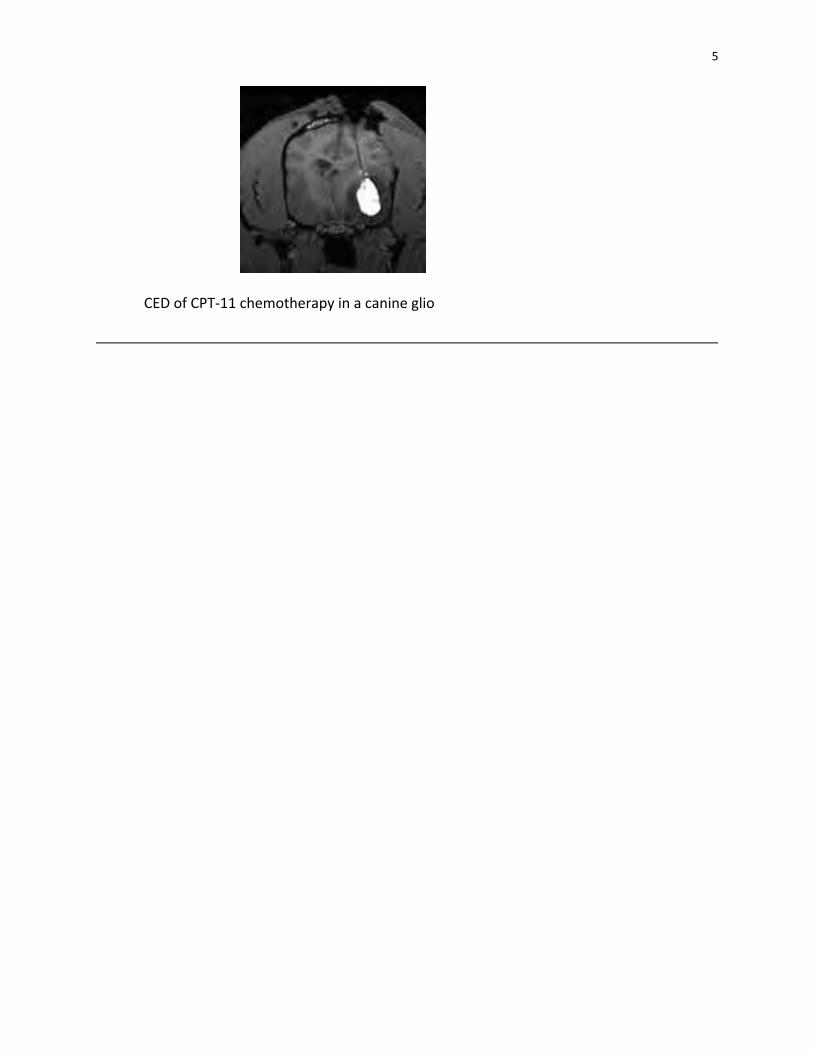
Delivery of Therapeutic Agents A wide variety of methods have been employed to deliver therapeutic agents to brain tumors. Many strategies have involved systemic delivery of agents either orally or intravenously. Some drugs (eg standard chemotherapeutic agents) are relatively non specific with respect to their potential targets, whereas others (eg small molecule tyrosine kinase inhibitors such as Gleevac) may have a more precisely defined target despite the systemic delivery. Even with the use of targeted therapies, the likelihood of significant systemic side effects is a major concern with drugs delivered in this manner. Ability of drugs to cross the blood brain barrier is also a factor that can significantly limit the efficacy of systemically delivered therapies, and many factors such as molecular weight, permeability of vasculature, drug stability and diffusion characteristics as well as tumor related factors are critical to attain effective cellular levels of anti tumor drugs. Local “targeted” delivery of therapies directly into tumor tissue has been advocated as a way to increase both the efficacy of many therapeutic agents whilst at the same time decreasing the likelihood of significant systemic toxicity. Therapeutic agents may be delivered directly at surgery following excision/debulking of tumors, or by stereotactic injection. Recent advances in injection of agents by convection enhanced delivery (CED) (over several hours), have shown great promise, and may allow highly accurate and comprehensive delivery of therapeutics to a defined area of tumor and/or surrounding brain. Preliminary results with CED using a novel chemotherapeutic agent CPT-‐11 (topoisomerase I inhibitor) in an ongoing clinical trial in dogs with spontaneous gliomas are encouraging, and demonstrate the feasibility of targeted delivery in veterinary patients.
5
CED of CPT-‐11 chemotherapy in a canine glio

6
Advanced Diagnostics for Cerebral Neoplasia Magnetic Resonance Imaging (MRI)
• In general, many neoplastic lesions have similar imaging characteristics on T1 and T2 weighted images
• Most tumors will be iso-‐hypo intense on T1W images and hyperintense on T2W images • Hemorrhage associated with higher grade tumors may result in areas of hyperintensity
on T1 weighted precontrast images (see below) and hypointensity on T2 weighted images
• Many tumors are associated with a significant amount of peritumoral edema • This is typically of a vasogenic nature and is most often seen predominantly involving
the white matter tracts in the surrounding tissue • Edema is most easily seen on T2 or FLAIR images as a hyperintense signal. Animals with
brain tumors and significant edema suggested on T2 images are likely to respond favourably to anti-‐inflammatory doses of glucocorticoids
• Contrast enhancement can be extremely variable and is thought to be secondary to abnormal tumor vasculature or disruption of normal vasculature in the region of the tumor
• Typical contrast patterns are seen with, but are NOT diagnostic of, certain tumor types. (see below)
Primary CNS tumors Meningioma
• Tumors are extra-‐axial and may arise rostrally involving the cerebral convexities, falx, ventrally, within the caudal fossa, or spinal cord
• There may be considerable variation in appearance form well circumscribed mass lesions to plaque like tumors
• Spinal cord meningiomas are most commonly found in the cervical region • Meningiomas are usually uniformly contrast enhancing although this can be variable • Significant cystic components may be present • Presence of a “dural tail” on post contrast T1W images is a common finding, though not
diagnostic • Many tumors have associated edema that may be severe in some cases
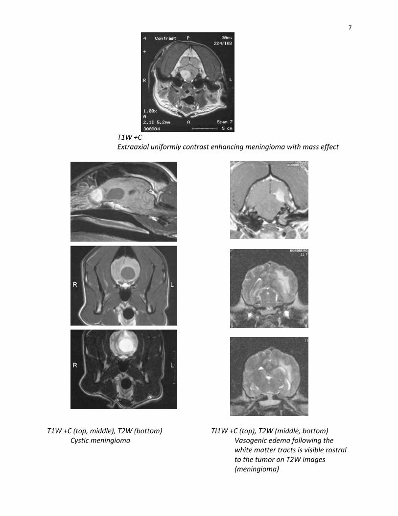
7
T1W +C Extraaxial uniformly contrast enhancing meningioma with mass effect
T1W +C (top, middle), T2W (bottom) TI1W +C (top), T2W (middle, bottom)
Cystic meningioma Vasogenic edema following the white matter tracts is visible rostral to the tumor on T2W images (meningioma)
8
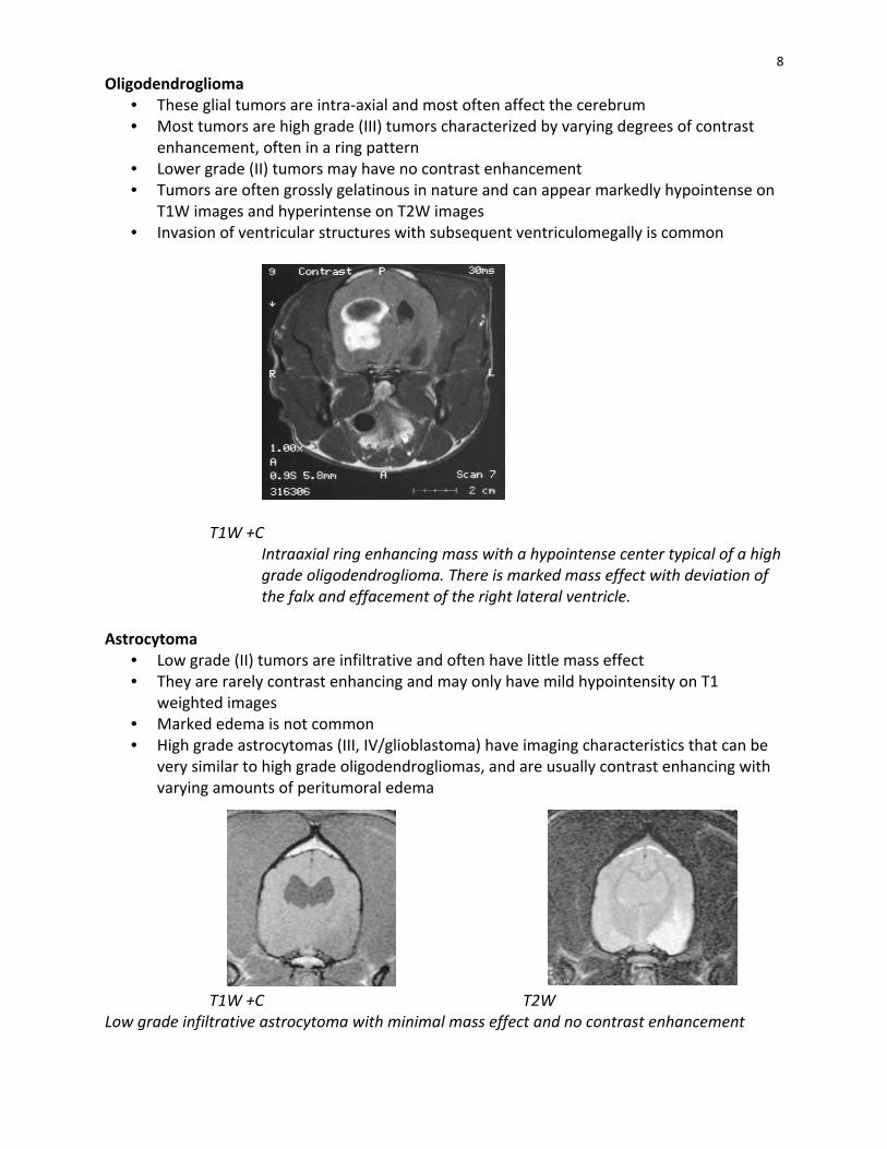
Oligodendroglioma • These glial tumors are intra-‐axial and most often affect the cerebrum • Most tumors are high grade (III) tumors characterized by varying degrees of contrast
enhancement, often in a ring pattern • Lower grade (II) tumors may have no contrast enhancement • Tumors are often grossly gelatinous in nature and can appear markedly hypointense on
T1W images and hyperintense on T2W images • Invasion of ventricular structures with subsequent ventriculomegally is common
T1W +C
Intraaxial ring enhancing mass with a hypointense center typical of a high grade oligodendroglioma. There is marked mass effect with deviation of the falx and effacement of the right lateral ventricle.
Astrocytoma
• Low grade (II) tumors are infiltrative and often have little mass effect • They are rarely contrast enhancing and may only have mild hypointensity on T1
weighted images • Marked edema is not common • High grade astrocytomas (III, IV/glioblastoma) have imaging characteristics that can be
very similar to high grade oligodendrogliomas, and are usually contrast enhancing with varying amounts of peritumoral edema
T1W +C T2W Low grade infiltrative astrocytoma with minimal mass effect and no contrast enhancement

9
T1W +C T1W +C High grade (IV) glioblastoma multiforme. Variable contrast enhancement is present with significant mass effect. Choroid plexus tumor
• These tumors (papillomas and carcinomas) are characterised by their location within the ventricular system, often resulting in secondary hydrocephalus
• Most tumors are uniformly strongly contrast enhancing • Choroid plexus carcinomas can metastasise down the ventricular system resulting in
multiple contrast enhancing lesions and/or enhancement of the ventricular system due to multiple micrometastases
T1W +C T1W +C Choroid plexus carcinoma. Strongly contrast enhancing mass lesion within the lateral ventricle. Multiple “drop metastases” have resulted in contrast enhancement of the lining of the ventricle. Ependymoma
• These tumors are also associated with the ventricular system due their origin in the lining cells of the ventricles
• They also tend to be uniformly contrast enhancing. Ependymomas appear to less common than choroids plexus tumors and rarely metastasize

10
T1 +C T1 +C Uniformly contrast enhancing ependymomas involving the 4th and 3rd ventricles Secondary/metastatic tumors
• Metastatic spread of neoplastic lesions to the CNS is relatively uncommon in the dog and cat
• In the author’s experience, the most frequently recorded tumors are haemangiosarcoma, histiocytic sarcoma, melanoma and lymphoma
• Metastatic lesions may be multiple or solitary, and often have a predilection for the grey/white matter junctions of the cerebrum
• Marked edema is a common finding with many metastatic lesions such as haemangiosarcoma
• Many metastatic lesions are contrast enhancing. Lymphoma can present with a variety of imaging characteristics
• It is usually contrast enhancing and may involve the meninges and nerve roots • It may present as a solitary mass lesions or as multifocal disease or diffusely infiltrative
disease
T1W (upper), T1W +C (lower) Metastasis of haemangiosarcoma to the cerebrum. Another example of a ring enhancing lesion!
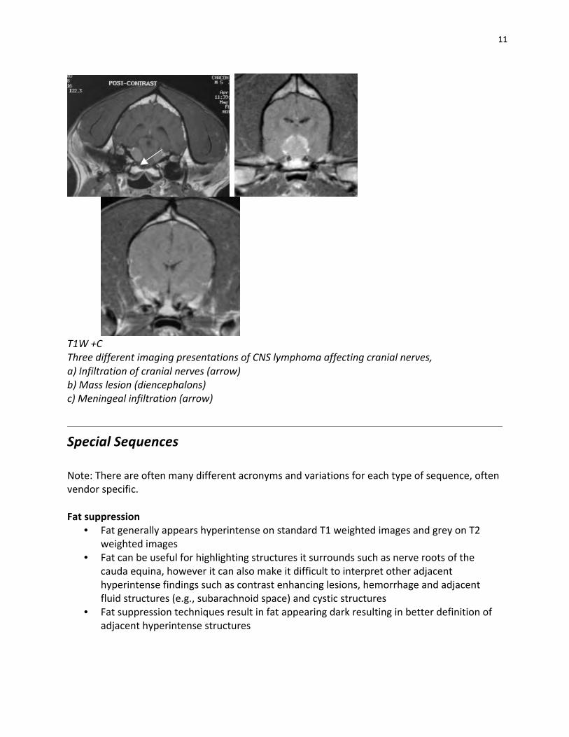
11
T1W +C Three different imaging presentations of CNS lymphoma affecting cranial nerves, a) Infiltration of cranial nerves (arrow) b) Mass lesion (diencephalons) c) Meningeal infiltration (arrow)
Special Sequences Note: There are often many different acronyms and variations for each type of sequence, often vendor specific. Fat suppression
• Fat generally appears hyperintense on standard T1 weighted images and grey on T2 weighted images
• Fat can be useful for highlighting structures it surrounds such as nerve roots of the cauda equina, however it can also make it difficult to interpret other adjacent hyperintense findings such as contrast enhancing lesions, hemorrhage and adjacent fluid structures (e.g., subarachnoid space) and cystic structures
• Fat suppression techniques result in fat appearing dark resulting in better definition of adjacent hyperintense structures
12
Fluid attenuated inversion recovery (FLAIR) • This technique nulls the bright fluid signal from free water and CSF • This can be very useful when hyeprintense lesions are adjacent to fluid filled structures
such as ventricles • It can also help distinguish whether cystic structures contain CSF or
necrotic/proteinaceous material Diffusion weighted images (DWI)
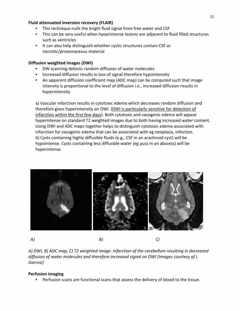
• DW scanning detects random diffusion of water molecules • Increased diffusion results in loss of signal therefore hypointensity • An apparent diffusion coefficient map (ADC map) can be computed such that image
intensity is proportional to the level of diffusion i.e., increased diffusion results in hyperintensity
a) Vascular infarction results in cytotoxic edema which decreases random diffusion and therefore gives hyperintensity on DWI. (DWI is particularly sensitive for detection of infarction within the first few days). Both cytotoxic and vasogenic edema will appear hyperintense on standard T2 weighted images due to both having increased water content. Using DWI and ADC maps together helps to distinguish cytotoxic edema associated with infarction for vasogenic edema that can be associated with eg neoplasia, infection. b) Cysts containing highly diffusible fluids (e.g., CSF in an arachnoid cyst) will be hypointense. Cysts containing less diffusible water (eg puss in an abscess) will be hyperintense.
A) B) C) A) DWI, B) ADC map, C) T2 weighted image. Infarction of the cerebellum resulting in decreased diffusion of water molecules and therefore increased signal on DWI (Images courtesy of L Garrosi) Perfusion imaging
• Perfusion scans are functional scans that assess the delivery of blood to the tissue.
13
a) Tumors often have increased signal due to their high vascularity and may be distinguishable from radiation necrosis post operative scar tissue which has low signal (perfusion). b) Infarcts have low signal due to loss or reduction in blood flow
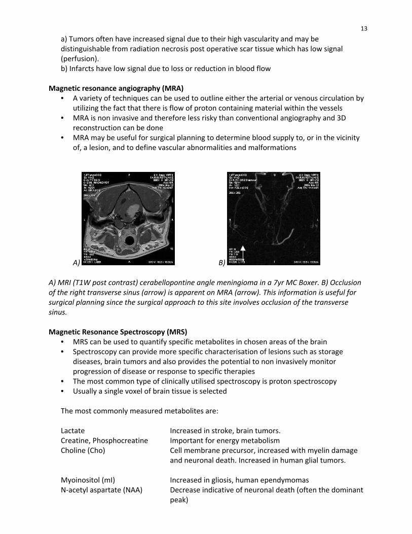
Magnetic resonance angiography (MRA)
• A variety of techniques can be used to outline either the arterial or venous circulation by utilizing the fact that there is flow of proton containing material within the vessels
• MRA is non invasive and therefore less risky than conventional angiography and 3D reconstruction can be done
• MRA may be useful for surgical planning to determine blood supply to, or in the vicinity of, a lesion, and to define vascular abnormalities and malformations
A) B) A) MRI (T1W post contrast) cerabellopontine angle meningioma in a 7yr MC Boxer. B) Occlusion of the right transverse sinus (arrow) is apparent on MRA (arrow). This information is useful for surgical planning since the surgical approach to this site involves occlusion of the transverse sinus. Magnetic Resonance Spectroscopy (MRS)
• MRS can be used to quantify specific metabolites in chosen areas of the brain • Spectroscopy can provide more specific characterisation of lesions such as storage
diseases, brain tumors and also provides the potential to non invasively monitor progression of disease or response to specific therapies
• The most common type of clinically utilised spectroscopy is proton spectroscopy • Usually a single voxel of brain tissue is selected
The most commonly measured metabolites are:
Lactate Increased in stroke, brain tumors. Creatine, Phosphocreatine Important for energy metabolism Choline (Cho) Cell membrane precursor, increased with myelin damage
and neuronal death. Increased in human glial tumors.
Myoinositol (mI) Increased in gliosis, human ependymomas N-‐acetyl aspartate (NAA) Decrease indicative of neuronal death (often the dominant
peak)
14
Glutamate Increased in hypoxia
Ratios of the individual peaks may also be used to characterise lesions. Functional MRI (fMRI)
• FMRI detects areas of increased blood flow and therefore oxygenation to areas of the brain. Increased oxygen means less deoxyhaemaglobin and more oxyhaemaglobin
• Deoxyhaemaglobin is paramagnetic so that loss causes a decrease in T2 relaxation. (See hemorrhage)
Magnetic transfer imaging (MTI)
• MTI examines the non water components of the brain and has been used amongst other things, to more specifically characterise disease affecting white matter (myelinated axons), neurodegeneration , trauma and neoplastic lesions
Real-‐time MRI guided Convection enhanced Delivery (CED)
• Infusion of therapeutic agents into the brain using convection enhanced delivery utilizes bulk flow of infusates through the interstitial space
• Large volumes of brain can be “covered”, and by using simultaneous infusion of contrast agents such as gadolinium, real-‐time monitoring of the infusion can be done to ensure optimal infusion while minimizing potential toxicity to non targeted tissues
Angiography
• Initial use of positive contrast cerebral angiography in small animals was essentially for the delineation of intracranial lesions, rather than as a tool to investigate primary vascular disease
• Space occupying masses could be identified based on their secondary effects on local vasculature
• Both venous and arterial contrast techniques can be used in the dog (arterial angiography is extremely difficult in the cat due to lack of a patent internal carotid artery and “reversal of flow in the basilar artery)
• The advent of CT and MRI has essentially supplanted angiography and other related techniques such as pneumoventriculography in the diagnosis of intracranial mass lesions
• Use of positive contrast angiography is currently restricted to cases where specific vascular disease such as arteriovenous malformation is suspected
• Iodinated contrast agents may be visualised using fluoroscopy/radiography, or CT • Use of magnetic resonance angiography (see below) is likely to replace standard
contrast angiography in the future in most cases
15
A) B)
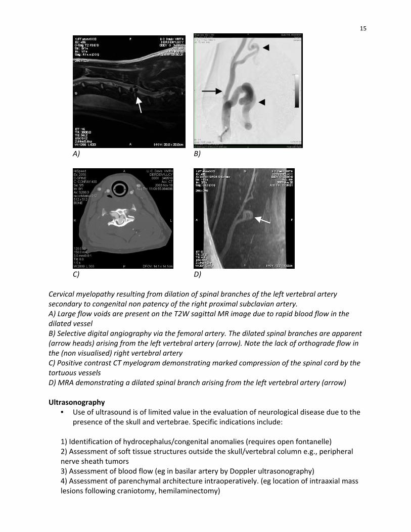
C) D) Cervical myelopathy resulting from dilation of spinal branches of the left vertebral artery secondary to congenital non patency of the right proximal subclavian artery. A) Large flow voids are present on the T2W sagittal MR image due to rapid blood flow in the dilated vessel B) Selective digital angiography via the femoral artery. The dilated spinal branches are apparent (arrow heads) arising from the left vertebral artery (arrow). Note the lack of orthograde flow in the (non visualised) right vertebral artery C) Positive contrast CT myelogram demonstrating marked compression of the spinal cord by the tortuous vessels D) MRA demonstrating a dilated spinal branch arising from the left vertebral artery (arrow) Ultrasonography
• Use of ultrasound is of limited value in the evaluation of neurological disease due to the presence of the skull and vertebrae. Specific indications include:
1) Identification of hydrocephalus/congenital anomalies (requires open fontanelle) 2) Assessment of soft tissue structures outside the skull/vertebral column e.g., peripheral nerve sheath tumors 3) Assessment of blood flow (eg in basilar artery by Doppler ultrasonography) 4) Assessment of parenchymal architecture intraoperatively. (eg location of intraaxial mass lesions following craniotomy, hemilaminectomy)

16
Scintigraphy
• Conventional scintigraphy identifies the accumulation of radioactive nuclides (most commonly technetium-‐99m-‐DTPA) within the nervous system due to compromise of the blood brain barrier
• Its use has largely been supplanted by CT and MRI and PET

Brain Tumors - Diagnosis and Therapy Richard A.leCouter, professor BVSc, PhD Dipl.ACVIM (USA)
1
BRAIN TUMORS: DIAGNOSIS & THERAPY
Richard A. LeCouteur, BVSc, PhD, Diplomate ACVIM(Neurology), Diplomate ECVN Department of Surgical & Radiological Sciences, School of Veterinary Medicine, University of
California, Davis, California, CA 95616-‐8745, USA
The Past: Surgery, Irradiation and Chemotherapy The major goals of therapy for a brain tumor have been to control secondary effects, such as increased intracranial pressure or cerebral edema, and to eradicate the tumor or reduce its size. Beyond general efforts to maintain homeostasis, palliative therapy for dogs or cats with a brain tumor has consisted of glucocorticoids for edema reduction, and in some cases (e.g., lymphoma), for retardation of tumor growth. Some animals with a brain tumor will demonstrate dramatic improvement in clinical signs for weeks or months with sustained glucocorticoid therapy. Should anti-‐seizure medications be needed, phenobarbital or bromide are the drugs best suited for the control of generalized seizures.
Three major methods of therapy for a brain tumor have been available for use in dogs and cats: surgery, irradiation, and chemotherapy.
Surgery In association with the availability of CT and MRI, and the development of advanced neurosurgical, anesthetic, and critical care techniques, complete or partial surgical removal of intracranial neoplasms is being practiced with increasing frequency. Neurosurgical intervention is an essential consideration in the management of intracranial neoplasms of cats or dogs, whether for complete excision, partial removal, or biopsy.
Radiation Therapy The use of radiation therapy for the treatment of primary brain tumors of dogs and cats is well established. Irradiation may be used either alone or in combination with other treatments. Radiation therapy also is recommended for the treatment of secondary brain tumors. Metastases, pituitary macroadenomas or macrocarcinomas, and skull tumors have been successfully managed by means of either radiation therapy alone or as an adjunct to surgery. Lymphoma may also be sensitive to radiation therapy.
Chemotherapy Traditionally, cytotoxic drugs have had a limited role in the treatment of dogs or cats with brain tumors, and progress in the development of truly effective chemotherapeutic protocols for humans or companion animals has been slow. Several factors affect the use of chemotherapeutic agents for the treatment of brain tumors in dogs or cats. The first, unique to the brain, is that the blood-‐brain barrier (BBB) may prevent exposure of all or some of the tumor to a chemotherapeutic agent injected parenterally. Second, tumor cell heterogeneity may be such that only certain cells within a tumor are sensitive to a given agent. Third, a tumor may be sensitive only at dosages that are toxic to the normal brain or other organs (kidney or liver).
2
The Present: Therapeutic Delivery Strategies for Canine Brain Tumors The use of Surgery, irradiation and chemotherapy remiains the mainstay of brain tumor therapy today. Development of novel therapeutic strategies to combat primary brain tumors has followed closely behind elucidation of the basic molecular and genetic mechanisms underlying both tumorigenesis and subsequent progression. Despite the wealth of data documenting successful treatment of experimental tumors, translation into the clinical setting has been slow. Many existing therapeutics are rendered ineffective in the treatment of brain tumors due to the inability to effectively deliver and sustain them within the brain. The major obstacle to therapeutic delivery via the vascular route (following either orally administration or direct vascular administration) is the BBB. Transport across the brain vascular endothelium is essentially trans-‐cellular, therefore the ideal substance to be transported should be:
• Small (< 400Da) • Lipophilic (lipid soluble) • Non-‐polar at physiological pH • Non-‐protein bound
Unfortunately, a majority of chemotherapeutic agents are large positively charged, hydrophilic molecules. Many therapeutic molecules such as cyclosporine, doxorubicin and vincristine have poor BBB penetration despite being lipophilic (cyclosporine A is more lipophilic than diazepam). This is the result of additional “barriers” such as high levels of degrading enzymes within the endothelial cells, and high concentrations of efflux transporter proteins such as P-‐glycoprotein, multiple organic anion transporter proteins (MOAT) and multi-‐drug-‐resistance proteins (MRP). In addition to barriers preventing movement of therapeutic agents from the blood into the brain parenchyma, mechanisms are also present to limit movement into the cerebrospinal fluid (CSF). Passage of substances through the arachnoid membrane is prevented by tight junctions and is generally impermeable to hydrophilic molecules. While the capillaries of the choroids plexus are fenestrated, non-‐continuous and allow free movement of small molecules, the adjacent choroidal epithelial cells form tight junctions preventing the passage of most macromolecules. An active organic acid transporter system in the choroid plexus also is capable of driving therapeutic organic acids such as penicillin or methotrexate back into the blood from the CSF. Entry of drugs into the CSF does not necessarily guarantee that they will reach the interstitial fluid in the brain, suggesting the presence of the so-‐called CSF-‐brain barrier, mainly attributed to insurmountable diffusion distances required to equilibrate CSF with brain interstitial fluid. Although the BBB may be inconsistently compromised in tumor vasculature, a variety of obstacles still restrict delivery of therapeutic agents. Tumor microvascular supply often is heterogeneous and chaotic, with significant areas of inefficient or poor blood flow, vascular shunting, blind-‐ending vessels, etc., resulting in erratic distribution of drugs that are able to penetrate the BBB.
3
Improving delivery of therapeutic agents to brain tumors in the face of these obstacles has focused on the following areas of research:
• Improve entry through the BBB by modification of therapeutic drugs
a. Increase influx b. Decrease efflux c. Utilization of carriers/receptors
• Disruption of the BBB
a. A variety of approaches have been used to disrupt BBB integrity including:
i. Chemical (often toxic), DMSO, ethanol, aluminium, irradiation, hypertension, hypercapnia, hypoxia ii. Osmotic agents such as mannitol and arabinose iii. Biochemical agents such as leukotriene C4, bradykinin, histamine etc.
• Circumventing the BBB
a. Using non-‐vascular delivery of therapeutic agents directly into the CNS is appealing in many ways. Apart from removing the BBB as a restriction to delivery of many potent anticancer therapies, targeting the drugs directly potentially reduces systemic toxicity, degradation and immunological stimulation (particularly with protein and virally based therapies). However strategies are generally more invasive requiring craniotomy or insertion of catheters
i. Intraventricular/intrathecal infusion ii. Wafers/microspheres/microchips iii. Delivery from biological tissues (Gene Therapy)
b. Delivery of therapies directly from living cells within the brain or tumor itself can provide sustained levels of drugs in specific targeted regions. The two main strategies examined to date are:
i. Implantation of transfected cell lines ii. Transduction of resident CNS cells or brain tumor cells with
gene therapy constructs
• Interstitial delivery
a. Both gene therapies and direct acting drugs, such as chemotherapeutics, can be delivered directly into tumor or brain parenchyma. AAV vectors carrying thymidine kinase suicide constructs and antiangiogenic agents have been shown to be efficacious in both in vitro and in vivo models, and direct injection into canine primary brain tumors has been done. Results in clinical tumors however have been disappointing mainly due to limited distribution of the therapy beyond the local region of the injection site

4
The Future: Brain Biopsy and Convection Enhanced Delivery (CED)
Brain Biopsy Biopsy remains the sole method available for the ante mortem definitive diagnosis of brain tumor type in cats or dogs, and is an essential step prior to consideration of any type of therapy. However, biopsy is not always attempted because of practical considerations, such as cost and morbidity. The most recent advance in the biopsy of brain tumors of dogs and cats has been the development of CT-‐guided stereotactic brain biopsy systems for use in cats and dogs. These CT-‐guided stereotactic biopsy systems provide a relatively rapid and extremely accurate means of tumor biopsy, with a low rate of complications. Cytological evaluation of brain tumor smear preparations, rapidly fixed in 95% alcohol and stained with hematoxylin and eosin, may be done within minutes of biopsy collection. Diagnostically accurate information from this rapid technique is generally available from both primary and metastatic nervous system tumors.
Convection enhanced delivery (CED) CED is a local delivery technique that utilizes a bulk-‐flow mechanism to deliver and distribute macromolecules over clinically relevant volumes of targeted tissue. Unlike local injection techniques, CED uses a pressure gradient established at the tip of an infusion catheter that pushes the infusate through the interstitial space. Volumes of distribution of infused molecules are significantly increased compared to local injection or surgical implantation methods that rely primarily on diffusion and are limited by concentration gradients and molecular weight of the delivered substance. Distribution of infusates over centimeters, rather than millimeters, has been reported in a variety of experimental model systems using CED. Real time in vivo imaging of CED is an essential consideration if adequate drug distribution is to be confirmed ante mortem. Additionally, the ability to detect and minimize distribution or leakage of drugs to normal tissues during delivery has the potential to significantly decrease toxicity and increase therapeutic effectiveness. Several surrogate marker systems have been described, facilitating image-‐guided CED, including magnetic resonance imaging (MRI) systems utilizing T2 imaging correlated with 123I-‐labelled serum albumin, single photon emission computed tomography (SPECT), and liposomes co-‐labeled with gadolinium. Liposomes are phospholipid nanoparticles composed of a bi-‐layered membrane capable of encapsulating a variety of therapeutic molecules. Liposomal encapsulation of a variety of drugs, including chemotherapeutics, has been shown to result in prolonged half-‐life, sustained release, and decreased toxicity. CED of liposomes, containing therapeutic drugs, directly into targeted brain tissue offers several advantages over systemic delivery of un-‐encapsulated drug, including bypassing of the BBB, increased volume of distribution within the target tissue, and increased therapeutic index as a result of both liposomal encapsulation and minimal systemic exposure. Irinotecan/CPT-‐11 is a camptothecin derivative and topoisomerase I inhibitor with activity against a variety of cancer types, including brain tumors. The efficacy and safety of direct delivery of liposomally encapsulated camptothecin analogs in rodent models of glioma has been reported. Translation of this promising therapeutic approach into clinical trials will require demonstration of the safety and efficacy of combined real time gadolinium based imaging and liposomally encapsulated CPT-‐11 treatment in a large animal model system. The advantages of a canine model system over established rodent and primate models are several and include the ability to investigate aspects of feasibility and toxicity on a scale

5
relevant to human clinical patients, and the unique potential to investigate CED efficacy, and adverse effects in large, spontaneously occurring tumors.
6
References Dickinson PJ, LeCouteur RA, Higgins RJ et al. 2008. Canine model of convection-‐enhanced delivery of liposomes containing CPT-‐11 monitored with real-‐time magnetic resonance imaging: laboratory investigation. J Neurosurg 108, 989-‐98. Dickinson PJ, LeCouteur RA, Higgins RJ et al. 2010. Canine spontaneous glioma: a translational model system for convection-‐enhanced delivery. Neuro Oncol 12, 928-‐40. Thomas R, Duke SE, Wang HJ, et al. 2009. 'Putting our heads together': insights into genomic conservation between human and canine intracranial tumors. J Neurooncol 94, 333-‐49. Wisner ER, Dickinson PJ, Higgins RJ. 2011. Magnetic resonance imaging features of canine intracranial neoplasia. Vet Radiol Ultrasound.52(1 Suppl 1):S52-‐61.
Brain Biopsy in Dogs and Cats Richard A.leCouter, professor BVSc, PhD Dipl.ACVIM (USA)

1
BRAIN BIOPSY IN DOGS & CATS
Richard A. LeCouteur, BVSc, PhD, Diplomate ACVIM(Neurology), Diplomate ECVN Department of Surgical & Radiological Sciences, School of Veterinary Medicine, University of
California, Davis, California, CA 95616-‐8745, USA The detection, localization and characterization of brain lesions has been greatly improved through the use of computed tomography (CT) and magnetic resonance (MR) imaging. However, there remains a need to obtain an intra-‐operative neuro-‐pathological diagnosis from tissue samples of the lesion. The intra-‐operative cytological evaluation of smear preparations of brain lesions has become a routine procedure, providing a rapid, highly accurate diagnosis. In addition, future therapies may involve intra-‐lesional administration of drugs, following results of a brain biopsy. The need to obtain biopsy material for diagnosis and/or to deliver therapeutic agents with precision and without an invasive surgical procedure has stimulated the development and refinement of image-‐guided brain biopsy. Neoplastic, vascular, infectious, or inflammatory diseases of dogs and cats frequently result in focal brain involvement. Although CT and MRI are sensitive in determining location, extent, and relationships to adjacent structures, of brain lesions, both have limited specificity. For example, non-‐neoplastic lesions (such as those seen in association with infectious, inflammatory, or vascular diseases) may mimic the CT or MRI appearance of a neoplasm. In most instances, results of CT or MRI provide only a broad list of differential diagnoses for a focal brain lesion. Accurate histologic diagnosis of an intracranial lesion is critical before recommending a specific management or treatment strategy.
CT-‐Guided Stereotactic Brain Biopsy
Contraindications Brain biopsy should be approached with caution in animals with underlyiong coagulopathies, clinical signs consistent with increased intracranial pressure (ICP), animals with brainstem lesions, or systemic problems that result in increased anesthetic risk.
Equipment & Anesthetic Considerations Essentially all closed stereotactic brain biopsy methods rely on the three-‐dimensional CT-‐generated coordinates identifying the lesion location. These coordinates are used to plot an optimal trajectory and depth needed for a biopsy needle to reach a target and obtain a diagnostic tissue sample. Technical impediments exist to the direct application of most human stereotactic systems to dogs and cats. Most commercially available systems use a cumbersome head-‐frame and localizing system, designed specifically for the human skull, and require dedicated, expensive computer software for the planning phase. Several different systems for image-‐guided stereotactic brain biopsy have been reported for use in dogs and cats. General anesthesia is required for stereotactic brain biopsy. Typically, premedication utilizes an opioid (a pure mu agonist because of using fentanyl in the maintenance phase). The dose and use of this drug will depend on the concern over changes in ICP and the mental status of the patient. Anesthesia usually is induced with propofol (± a

2 benzodiazepine). Propofol (0.1-‐0.4 mg/kg/minute) and fentanyl (0.3-‐0.7 µg/kg/minute) are recommended for maintenance. . Animals should be ventilated to maintain ETCO2 at a value of 30-‐35 mmHg. For recovery the fentanyl should be stopped about 30 minutes before the end of the procedure.
Anticipated Time Two to three hours depending on the number of biopsy specimens collected and the number of trajectories planned.
Animal Preparation Stereotactic biopsy begins with proper patient selection. The possibility of non-‐neoplastic disorders such as infection, cerebral infarction, or vasculitis, must be considered and investigated with other tests in appropriate patients prior to biopsy. When the differential diagnosis list is long and may include neoplasms and inflammatory lesions, the appropriate handling of tissue samples should be discussed with a neuropathologist in advance of the procedure. All patients should be tested for coagulation parameters (prothrombin time [PT], partial thromboplastin time [PTT]) prior to the procedure and should have a platelet count greater than 100,000. Patients should not receive aspirin products for 1 week before surgery. Ideally, MRI or CT images should be completed within five days prior to completion of the biopsy procedure.
Possible Complications Although stereotactic brain biopsy is minimally invasive (compared to open biopsy procedures), complications may occur. Morbidity may include seizures, hemorrhage, development of biopsy-‐induced neurologic deficits, brain infection, tumor seeding, and lack of definitive diagnosis.
Procedure Biopsy generally is done on the CT-‐scanner table. For those lesions not well identified on CT images, MRI images that demonstrate a lesion may be used to localize the lesion on CT images, using well-‐defined anatomic landmarks (e.g., lateral ventricles). Transverse CT images are used to define the CT coordinates of reference markers and the biopsy target. Dorsal or sagittal images may be used for trajectory planning. An entry point should be selected that is associated with a low risk for neurologic deficit or hemorrhage (e.g., avoidance of dorsal sagittal sinus). Ependymal puncture should be avoided where possible. A small craniotomy (2-‐mm diameter) is made by means of a twist drill, the dura mater is punctured with an 18-‐gauge needle, and biopsies may be done with a side-‐cutting aspirator biopsy needle (Nashold Biopsy Needle, Integra Radionics, Burlington MA) with a 10-‐mm side opening. On average, one to three specimens are harvested from each biopsy site. The intra-‐operative goal should be to confirm by means of smear or touch preparations whether tissue satisfactory for an eventual diagnosis has been obtained. A specific histologic diagnosis may require routine formalin fixation and paraffin embedding of the biopsy tissue. At the conclusion of the biopsy procedure, the needle is withdrawn in increments to assess
3 any possibility of hemorrhage. In the case of hemorrhage, blood should be permitted to egress from the needle spontaneously until the bleeding stops.
Post-‐Procedure Considerations A series of CT images of the brain should be completed immediately following completion of the brain biopsy procedure in order to assess the possibility of intracranial hemorrhage. Animals should recover from anesthesia in sternal recumbency with the head elevated slightly above the level of the heart. Animals should be closely monitored for 12 hours post-‐biopsy before being discharged from the hospital.
Relative Merits of Alternative Brain Biopsy Procedures Open (surgical) brain biopsy may be appropriate in certain clinical situations in which cortical architecture needs to be preserved, for leptomeningeal sampling, for superficially located lesions, and when a decompressive craniectomy with good cortical visualization may be helpful in addition to obtaining a biopsy sample.
Intra-‐operative Diagnosis Using the Smear Technique The rapid cytological evaluation of a brain lesion from a biopsy sample can provide crucial information on operative management, medical management, chemotherapy, or radiation therapy. In people intra-‐operative cytological evaluation of smear preparations of brain tumors, supported by frozen and paraffin-‐embedded tissue, has become a routine procedure, and cytological profiles of smears of various types of human brain tumors have been well described. Smear preparations are generally wet fixed in 95% alcohol and stained with hematoxylin and eosin although toluidine blue, Geimsa, or Papanicolaou’s stain may also be used. In a recent study, tissue samples were obtained from lesions either by CT-‐guided stereotactic brain biopsy (44 samples) or intra-‐operatively during craniotomy (49 samples) and the results from the smear technique compared with those from sections of paraffin-‐embedded tissue. The overall diagnostic accuracy from samples obtained by both craniotomy and stereo-‐biopsy was about 80%. This compares favorably with the 69-‐94% accuracy reported in some large series of human cases. The main advantages of this method of intra-‐operative diagnosis are speed, ease of preparation, technical simplicity, need for minimal equipment, high degree of cytological resolution compared to frozen preparations, low cost and small sample size required. A limitation of this system is that it is difficult to prepare adequate smear preparations in certain tough and coherent tumors (e.g., Schwannomas, fibrillary astrocytomas, and some meningiomas). Smear preparations provide excellent cytologic detail, however these differ from the conventional histologic appearance of HE-‐stained paraffin-‐embedded tissue. Experience is required in the correct interpretation of smear preparations.
4
References Koblik PD, LeCouteur RA, Higgins RJ, et al. 1999. CT-‐guided brain biopsy using a modified Pelorus Mark III stereotactic system: Experience with 50 dogs. Veterinary Radiology & Ultrasound 40, 434-‐440. Koblik PD, LeCouteur RA, Higgins RJ, et al. 1999. Modification and application of a Pelorus Mark III Stereotactic system for CT-‐guided brain biopsy in 50 dogs. Veterinary Radiology & Ultrasound 40, 424-‐433. Moissonnier P, Bordeau W, Devauchelle P, et al. 1998. CT-‐guided stereotaxic biopsy of intracranial lesions. Vet Surg 27, 293 (abstr). Moissonnier P, Blot S, Devauchelle P, et al. 2001. Stereotactic CT-‐guided brain biopsy in the dog: Cytological and histological diagnosis and early complications in 23 dogs. Vet Surg 30, 296 (abstr). Vernau KM, Higgins RJ, Bollen AW, et al. 2001. Primary canine and feline nervous system tumors: Intraoperative diagnosis using the smear technique. Vet Pathol 38, 47-‐57.
Neuromuscular Disorders Richard A.leCouter, professor BVSc, PhD Dipl.ACVIM (USA)
1
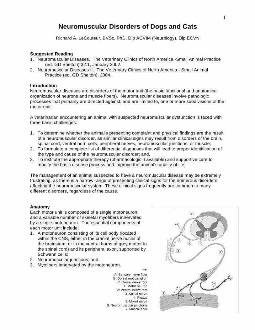
Neuromuscular Disorders of Dogs and Cats
Richard A. LeCouteur, BVSc, PhD, Dip ACVIM (Neurology), Dip ECVN
Suggested Reading 1. Neuromuscular Diseases. The Veterinary Clinics of North America -Small Animal Practice
(ed. GD Shelton) 32:1, January 2002. 2. Neuromuscular Diseases II. The Veterinary Clinics of North America - Small Animal
Practice (ed. GD Shelton), 2004. Introduction Neuromuscular diseases are disorders of the motor unit (the basic functional and anatomical organization of neurons and muscle fibers). Neuromuscular diseases involve pathologic processes that primarily are directed against, and are limited to, one or more subdivisions of the motor unit: A veterinarian encountering an animal with suspected neuromuscular dysfunction is faced with three basic challenges: 1. To determine whether the animal’s presenting complaint and physical findings are the result
of a neuromuscular disorder, as similar clinical signs may result from disorders of the brain, spinal cord, ventral horn cells, peripheral nerves, neuromuscular junctions, or muscle;
2. To formulate a complete list of differential diagnoses that will lead to proper identification of the type and cause of the neuromuscular disorder; and,
3. To institute the appropriate therapy (pharmacologic if available) and supportive care to modify the basic disease process and improve the animal’s quality of life.
The management of an animal suspected to have a neuromuscular disease may be extremely frustrating, as there is a narrow range of presenting clinical signs for the numerous disorders affecting the neuromuscular system. These clinical signs frequently are common to many different disorders, regardless of the cause. Anatomy Each motor unit is composed of a single motoneuron, and a variable number of skeletal myofibers innervated by a single motoneuron. The essential components of each motor unit include: 1. A motoneuron consisting of its cell body (located
within the CNS, either in the cranial nerve nuclei of the brainstem, or in the ventral horns of grey matter in the spinal cord) and its peripheral axon, supported by Schwann cells;
2. Neuromuscular junctions; and, 3. Myofibers innervated by the motoneuron.
→ A: Sensory nerve fiber
B: Dorsal root ganglion C: Dorsal nerve root
1: Motor neuron 2: Ventral nerve root
3. Spinal nerve 4. Plexus
5. Mixed nerve 6. Neuromuscular junctions
7. Muscle fiber
2
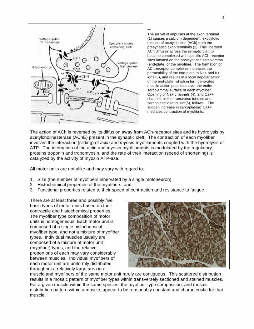
← The arrival of impulses at the axon terminal (1) causes a calcium dependent, exocytotic release of acetylcholine (ACh) from the presynaptic axon terminals (2). This liberated ACh diffuses across the synaptic cleft to become complexed with specific ACh-receptor sites located on the postsynaptic sarcolemma (end-plate) of the myofiber. The formation of ACh-receptor complexes increases the permeability of the end-plate to Na+ and K+ ions (3), and results in a local depolarization of the end-plate, which in turn generates muscle action potentials over the entire sarcolemmal surface of each myofiber. Opening of Na+ channels (4), and Ca++ channels in the transverse tubules and sarcoplasmic reticulum(5), follows. The sudden increase in sarcoplasmic Ca++ mediates contraction of myofibrils.
The action of ACh is reversed by its diffusion away from ACh-receptor sites and its hydrolysis by acetylcholinesterase (AChE) present in the synaptic cleft. The contraction of each myofiber involves the interaction (sliding) of actin and myosin myofilaments coupled with the hydrolysis of ATP. The interaction of the actin and myosin myofilaments is modulated by the regulatory proteins troponin and tropomyosin, and the rate of their interaction (speed of shortening) is catalyzed by the activity of myosin ATP-ase. All motor units are not alike and may vary with regard to: 1. Size (the number of myofibers innervated by a single motoneuron), 2. Histochemical properties of the myofibers, and, 3. Functional properties related to their speed of contraction and resistance to fatigue. There are at least three and possibly five basic types of motor units based on their contractile and histochemical properties. The myofiber type composition of motor units is homogeneous. Each motor unit is composed of a single histochemical myofiber type, and not a mixture of myofiber types. Individual muscles usually are composed of a mixture of motor unit (myofiber) types, and the relative proportions of each may vary considerably between muscles. Individual myofibers of each motor unit are uniformly distributed throughout a relatively large area in a muscle and myofibers of the same motor unit rarely are contiguous. This scattered distribution results in a mosaic pattern of myofiber types within transversely sectioned and stained muscles. For a given muscle within the same species, the myofiber type composition, and mosaic distribution pattern within a muscle, appear to be reasonably constant and characteristic for that muscle.

3
Pathoanatomic Classification of Neuromuscular Diseases
General Classification Motor Unit Component Involved NEUROPATHIES LOWER MOTOR NEURONS Central: Motoneuron Diseases Neuronal Cell Bodies Peripheral: Axonopathies Axons Demyelinating Diseases Schwann Cells Mixed Axonal/Demyelinating Both Axons & Schwann Cells
“JUNCTIONOPATHIES” NEUROMUSCULAR JUNCTIONS Presynaptic Transmitter Synthesis &/or Release Synaptic Acetylcholinesterase Postsynaptic Acetylcholine Receptors
MYOPATHIES MYOFIBERS
Sarcolemma Transverse Tubules Organelles Myofilaments Inclusions
NEUROMYOPATHIES ELEMENTS OF BOTH MOTONEURONS & MYOFIBERS
Clinical Signs of Neuromuscular Disorders 1. Generalized or localized muscular weakness 2. Functional manifestations: Paresis/paralysis Gait abnormalities Exercise related weakness Dysphagia Regurgitation Dyspnea Dysphonia 3. Physical manifestations: Muscle atrophy/hypotrophy Muscle hypertrophy Skeletal deformities
Classification of Neuromuscular Disorders A useful classification scheme of neuromuscular diseases is based on the anatomic motor unit components that are primarily involved in the pathogenesis of the muscle weakness. Using this classification, neuromuscular diseases are broadly subdivided into: 1. Neuropathies - disorders of the neuron, its cell body, axon, and/or Schwann cells (myelin) 2. “Junctionopathies” - disorders of neuromuscular junctions 3. Myopathies - disorders of muscle fibers 4 Neuromyopathies - disorders of both the neurons and muscle fibers.
Clinical Signs of Neuromuscular Disorders Dysfunction of the motor unit results in lower motor neuron signs, seen clinically as muscle weakness. The expression of this weakness may vary considerably, and may include: paresis/paralysis, gait abnormalities, exercise related weakness, dysphagia, regurgitation, dyspnea, and dysphonia. The distribution of involvement may be local, regional, or generalized. In addition there may be gross deformities of muscle mass (i.e., atrophy, hypertrophy, and skeletal deformities). Any patient presented with some form of clinical weakness, should be viewed as potentially having a motor unit disorder.
4
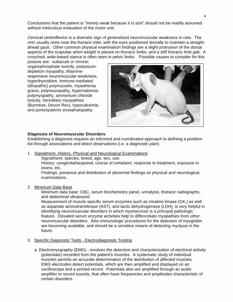
Conclusions that the patient is "merely weak because it is sick" should not be readily assumed without meticulous evaluation of the motor unit. Cervical ventroflexion is a dramatic sign of generalized neuromuscular weakness in cats. The chin usually rests near the thoracic inlet, with the eyes positioned dorsally to maintain a straight-ahead gaze. Other common physical examination findings are a slight protrusion of the dorsal aspects of the scapulae when weight is placed on thoracic limbs, and a stiff thoracic limb gait. A crouched, wide-based stance is often seen in pelvic limbs. Possible causes to consider for this posture are: subacute or chronic organophosphate toxicity, potassium-depletion myopathy, thiamine-responsive neuromuscular weakness, hyperthyroidism, immune-mediated (idiopathic) polymyositis, myasthenia gravis, polyneuropathy, hypernatremic polymyopathy, ammonium chloride toxicity, hereditary myopathies (Burmese, Devon Rex), hypocalcemia, and portosystemic encephalopathy. Diagnosis of Neuromuscular Disorders Establishing a diagnosis requires an informed and coordinated approach to defining a problem list through associations and direct observations (i.e. a diagnostic plan). 1. Signalment, History, Physical and Neurological Examinations
Signalment: species, breed, age, sex, use. History: congenital/acquired, course of complaint, response to treatment, exposure to toxins, etc. Findings: presence and distribution of abnormal findings on physical and neurological examinations.
2. Minimum Data Base Minimum data base: CBC, serum biochemistry panel, urinalysis, thoracic radiographs, and abdominal ultrasound. Measurement of muscle specific serum enzymes such as creatine kinase (CK,) as well as aspartate aminotransferase (AST), and lactic dehydrogenase (LDH), is very helpful in identifying neuromuscular disorders in which myonecrosis is a principal pathologic feature. Elevated serum enzyme activities help to differentiate myopathies from other neuromuscular disorders. Also immunologic procedures for the detection of myoglobin are becoming available, and should be a sensitive means of detecting myolysis in the future.
3. Specific Diagnostic Tests - Electrodiagnostic Testing
a. Electromyography (EMG) - involves the detection and characterization of electrical activity (potentials) recorded from the patient's muscles. A systematic study of individual muscles permits an accurate determination of the distribution of affected muscles. EMG electrodes detect potentials, which are then amplified and displayed on an oscilloscope and a printed record. Potentials also are amplified through an audio amplifier to record sounds, that often have frequencies and amplitudes characteristic of certain disorders.
5
In animals, EMG examinations usually are conducted with the muscles at rest, (i.e. not contracting) and usually under general anesthesia. Under these conditions, resting potentials across muscle fiber membranes are maintained, and hence, normal muscles at rest are "electrically silent". With depolarization of muscle fiber membranes, potentials are generated that have a wave form usually consisting of negative and positive phases. The potentials generated are evaluated for amplitude, duration, number of phases and polarity, frequency and repetition. i) Insertional activity - brief bursts of electrical activity (potential changes) are induced by
irritation of single muscle fibers caused by insertion of the EMG needles. After insertion and cessation of needle movement, normal muscles become electrically silent. Increased insertional activity (an increase in amplitude and prolonged duration) may be observed in neuromuscular diseases. Affected muscle fibers are hyperexcitable, having a lowered threshold due to diminished membrane potentials.
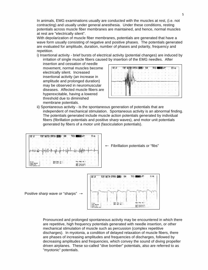
ii) Spontaneous activity - is the spontaneous generation of potentials that are independent of mechanical stimulation. Spontaneous activity is an abnormal finding. The potentials generated include muscle action potentials generated by individual fibers (fibrillation potentials and positive sharp waves), and motor unit potentials generated by fibers of a motor unit (fasciculation potentials).
← Fibrillation potentials or “fibs”
Positive sharp wave or “sharps” →
Pronounced and prolonged spontaneous activity may be encountered in which there are repetitive, high frequency potentials generated with needle insertion, or other mechanical stimulation of muscle such as percussion (complex repetitive discharges). In myotonia, a condition of delayed relaxation of muscle fibers, there are phases of increasing amplitudes and frequencies of discharges, followed by decreasing amplitudes and frequencies, which convey the sound of diving propeller driven airplanes. These so-called "dive bomber" potentials, also are referred to as "myotonic" potentials.
6
When the tip of the EMG needle is placed near the end-plate region, spontaneous small amplitude (miniature) end-plate potentials ("mepp's") may be detected. These are normal potentials due to the spontaneous release of individual quanta of ACh. This activity is also referred to as end-plate "noise".
b. Motor Nerve Conduction Velocity - provides information about the integrity of nerve fibers in peripheral nerves. Recordings are conducted while the patient is anesthetized. Demyelinating disorders cause slowed conduction in peripheral nerves. Ulnar and sciatic (peroneal-tibial) nerves most often are employed for evaluation. c. Evoked Potential Recordings - with repetitive nerve stimulation provides information about the integrity of neuromuscular transmission (see myasthenia gravis).
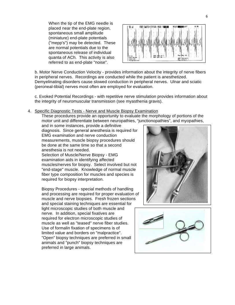
4. Specific Diagnostic Tests - Nerve and Muscle Biopsy Examination
These procedures provide an opportunity to evaluate the morphology of portions of the motor unit and differentiate between neuropathies, “junctionopathies”, and myopathies, and in some instances, provide a definitive diagnosis. Since general anesthesia is required for EMG examination and nerve conduction measurements, muscle biopsy procedures should be done at the same time so that a second anesthesia is not needed. Selection of Muscle/Nerve Biopsy - EMG examination aids in identifying affected muscles/nerves for biopsy. Select involved but not "end-stage" muscle. Knowledge of normal muscle fiber type composition for muscles and species is required for biopsy interpretation. Biopsy Procedures - special methods of handling and processing are required for proper evaluation of muscle and nerve biopsies. Fresh frozen sections and special staining techniques are essential for light microscopic studies of both muscle and nerve. In addition, special fixatives are required for electron microscopic studies of muscle as well as "teased" nerve fiber studies. Use of formalin fixation of specimens is of limited value and borders on "malpractice". "Open" biopsy techniques are preferred in small animals and "punch" biopsy techniques are preferred in large animals.
7
HISTOPATHOLOGICAL FEATURES OF NEUROPATHIES 1. General Features
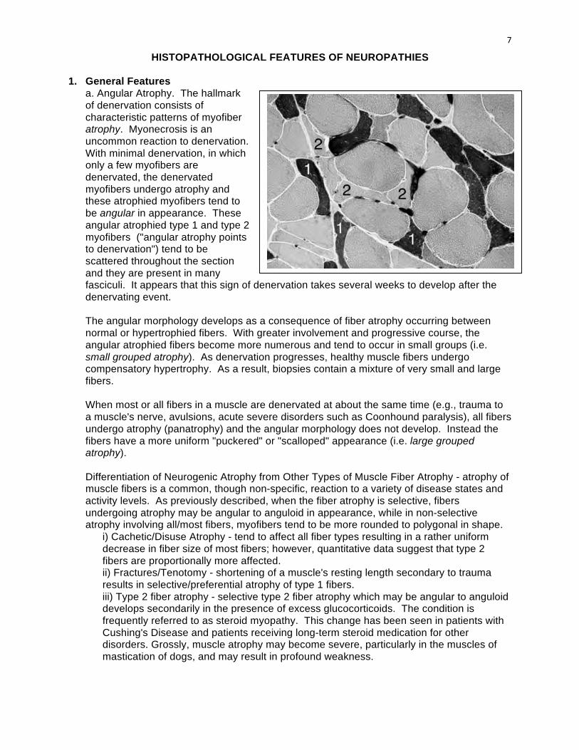
a. Angular Atrophy. The hallmark of denervation consists of characteristic patterns of myofiber atrophy. Myonecrosis is an uncommon reaction to denervation. With minimal denervation, in which only a few myofibers are denervated, the denervated myofibers undergo atrophy and these atrophied myofibers tend to be angular in appearance. These angular atrophied type 1 and type 2 myofibers ("angular atrophy points to denervation") tend to be scattered throughout the section and they are present in many fasciculi. It appears that this sign of denervation takes several weeks to develop after the denervating event. The angular morphology develops as a consequence of fiber atrophy occurring between normal or hypertrophied fibers. With greater involvement and progressive course, the angular atrophied fibers become more numerous and tend to occur in small groups (i.e. small grouped atrophy). As denervation progresses, healthy muscle fibers undergo compensatory hypertrophy. As a result, biopsies contain a mixture of very small and large fibers. When most or all fibers in a muscle are denervated at about the same time (e.g., trauma to a muscle's nerve, avulsions, acute severe disorders such as Coonhound paralysis), all fibers undergo atrophy (panatrophy) and the angular morphology does not develop. Instead the fibers have a more uniform "puckered" or "scalloped" appearance (i.e. large grouped atrophy).
Differentiation of Neurogenic Atrophy from Other Types of Muscle Fiber Atrophy - atrophy of muscle fibers is a common, though non-specific, reaction to a variety of disease states and activity levels. As previously described, when the fiber atrophy is selective, fibers undergoing atrophy may be angular to anguloid in appearance, while in non-selective atrophy involving all/most fibers, myofibers tend to be more rounded to polygonal in shape.
i) Cachetic/Disuse Atrophy - tend to affect all fiber types resulting in a rather uniform decrease in fiber size of most fibers; however, quantitative data suggest that type 2 fibers are proportionally more affected. ii) Fractures/Tenotomy - shortening of a muscle's resting length secondary to trauma results in selective/preferential atrophy of type 1 fibers. iii) Type 2 fiber atrophy - selective type 2 fiber atrophy which may be angular to anguloid develops secondarily in the presence of excess glucocorticoids. The condition is frequently referred to as steroid myopathy. This change has been seen in patients with Cushing's Disease and patients receiving long-term steroid medication for other disorders. Grossly, muscle atrophy may become severe, particularly in the muscles of mastication of dogs, and may result in profound weakness.
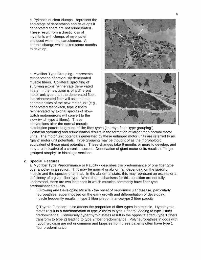
8
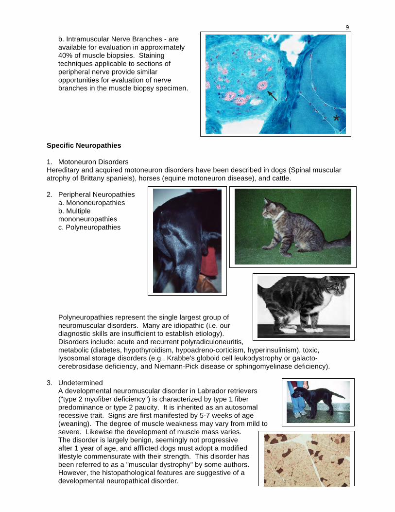
b. Pyknotic nuclear clumps - represent the end-stage of denervation and develops if denervated fibers are not reinnervated. These result from a drastic loss of myofibrils with clumps of myonuclei enclosed within the sarcolemma. A chronic change which takes some months to develop. c. Myofiber Type Grouping - represents reinnervation of previously denervated muscle fibers. Collateral sprouting of surviving axons reinnervate denervated fibers. If the new axon is of a different motor unit type than the denervated fiber, the reinnervated fiber will assume the characteristics of the new motor unit (e.g., denervated fast-twitch, type 2 fibers reinnervated by axonal sprouts of slow-twitch motoneurons will convert to the slow-twitch type 1 fibers). These conversions alter the normal mosaic distribution pattern to groups of like fiber types (i.e. myo-fiber "type grouping"). Collateral sprouting and reinnervation results in the formation of larger than normal motor units. The motor unit potentials generated by these enlarged motor units are referred to as "giant" motor unit potentials. Type grouping may be thought of as the morphologic equivalent of these giant potentials. These changes take 6 months or more to develop, and they are indicative of a chronic disorder. Denervation of giant motor units results in "large grouped atrophy" in histologic sections.
2. Special Features
a. Myofiber Type Predominance or Paucity - describes the predominance of one fiber type over another in a section. This may be normal or abnormal, depending on the specific muscle and the species of animal. In the abnormal state, this may represent an excess or a deficiency of a given fiber type. While the mechanisms for this condition are not fully understood, there are two instances in which muscles commonly have fiber type predominance/paucity.
i) Growing and Developing Muscle - the onset of neuromuscular disease, particularly neuropathies, superimposed on the early growth and differentiation of developing muscle frequently results in type 1 fiber predominance/type 2 fiber paucity.
ii) Thyroid Function - also affects the proportion of fiber types in a muscle. Hypothyroid states result in a transformation of type 2 fibers to type 1 fibers, leading to type 1 fiber predominance. Conversely hyperthyroid states result in the opposite effect (type 1 fibers transform to type 2) leading to type 2 fiber predominance. Polyneuropathies in dogs with hypothyroidism are not uncommon and biopsies from these patients often have type 1 fiber predominance.
9
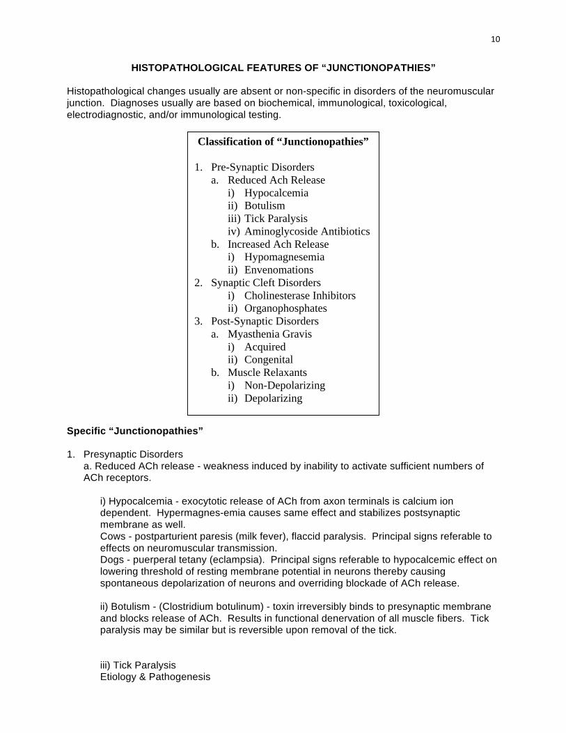
b. Intramuscular Nerve Branches - are available for evaluation in approximately 40% of muscle biopsies. Staining techniques applicable to sections of peripheral nerve provide similar opportunities for evaluation of nerve branches in the muscle biopsy specimen.
Specific Neuropathies 1. Motoneuron Disorders Hereditary and acquired motoneuron disorders have been described in dogs (Spinal muscular atrophy of Brittany spaniels), horses (equine motoneuron disease), and cattle. 2. Peripheral Neuropathies
a. Mononeuropathies b. Multiple mononeuropathies c. Polyneuropathies Polyneuropathies represent the single largest group of neuromuscular disorders. Many are idiopathic (i.e. our diagnostic skills are insufficient to establish etiology). Disorders include: acute and recurrent polyradiculoneuritis, metabolic (diabetes, hypothyroidism, hypoadreno-corticism, hyperinsulinism), toxic, lysosomal storage disorders (e.g., Krabbe's globoid cell leukodystrophy or galacto-cerebrosidase deficiency, and Niemann-Pick disease or sphingomyelinase deficiency).
3. Undetermined A developmental neuromuscular disorder in Labrador retrievers ("type 2 myofiber deficiency") is characterized by type 1 fiber predominance or type 2 paucity. It is inherited as an autosomal recessive trait. Signs are first manifested by 5-7 weeks of age (weaning). The degree of muscle weakness may vary from mild to severe. Likewise the development of muscle mass varies. The disorder is largely benign, seemingly not progressive after 1 year of age, and afflicted dogs must adopt a modified lifestyle commensurate with their strength. This disorder has been referred to as a "muscular dystrophy" by some authors. However, the histopathological features are suggestive of a developmental neuropathical disorder.
10
Classification of “Junctionopathies” 1. Pre-Synaptic Disorders a. Reduced Ach Release i) Hypocalcemia ii) Botulism iii) Tick Paralysis iv) Aminoglycoside Antibiotics b. Increased Ach Release i) Hypomagnesemia ii) Envenomations 2. Synaptic Cleft Disorders i) Cholinesterase Inhibitors ii) Organophosphates 3. Post-Synaptic Disorders a. Myasthenia Gravis i) Acquired ii) Congenital b. Muscle Relaxants i) Non-Depolarizing ii) Depolarizing
HISTOPATHOLOGICAL FEATURES OF “JUNCTIONOPATHIES”
Histopathological changes usually are absent or non-specific in disorders of the neuromuscular junction. Diagnoses usually are based on biochemical, immunological, toxicological, electrodiagnostic, and/or immunological testing. Specific “Junctionopathies” 1. Presynaptic Disorders
a. Reduced ACh release - weakness induced by inability to activate sufficient numbers of ACh receptors.
i) Hypocalcemia - exocytotic release of ACh from axon terminals is calcium ion dependent. Hypermagnes-emia causes same effect and stabilizes postsynaptic membrane as well. Cows - postparturient paresis (milk fever), flaccid paralysis. Principal signs referable to effects on neuromuscular transmission. Dogs - puerperal tetany (eclampsia). Principal signs referable to hypocalcemic effect on lowering threshold of resting membrane potential in neurons thereby causing spontaneous depolarization of neurons and overriding blockade of ACh release.
ii) Botulism - (Clostridium botulinum) - toxin irreversibly binds to presynaptic membrane and blocks release of ACh. Results in functional denervation of all muscle fibers. Tick paralysis may be similar but is reversible upon removal of the tick.
iii) Tick Paralysis Etiology & Pathogenesis
11
Tick Paralysis is a flaccid, afebrile ascending motor paralysis produced by a neurotoxin generated by some but not all strains of certain species of ticks. Not all infested animals become paralyzed. Cats in the U.S. appear to be relatively resistant to tick paralysis, although signs of paralysis have been reported. In North America, the common wood tick, Dermacentor variabilis, and Dermacentor andersoni (the Rocky Mountain wood tick) are incriminated most often. In Australia, especially along the east coast, Ixodes holocyclus is the most important species. Other species that occasionally cause paralysis are Ixodes cornuatus and Ixodes hirsti. Ixodes scapularis, the principal vector of the agent of Lyme disease (Borrelia burgdorferi) in the Northeast, Midwest, and Southeast of the United States, also may cause tick paralysis in dogs. This tick also is a primary vector of the agent of human and rodent babesiosis. Ixodes pacificus has also been incriminated in dogs in the Grass Valley area of northern California. In Australia, Ixodes holocyclus is the vector for Lyme disease and spotted fever, caused by Rickettsia australis. There is circumstantial evidence that some dogs bitten by Ixodes holocyclus develop signs of chronic illness similar to Lyme disease. With tick paralysis, adult ticks, especially females, produce a salivary neurotoxin that circulates in the host animal and interferes with acetylcholine liberation at the neuromuscular junction and/or impulse propagation along motor axon terminals. In Australia, heavy infestations with nymphs or larvae may result in paralysis. Clinical Signs Onset of clinical signs is gradual, paralysis first becoming evident as an incoordination in the pelvic limbs, resulting in an unsteady gait. Altered voice, cough, and dysphagia can be early signs. Dogs become recumbent in 24 to 72 hours. Reflexes are lost but sensation is preserved. Jaw muscle weakness and facial paresis may be present. Death may occur within several days from respiratory paralysis. Diagnosis Electromyographic studies reveal absence of spontaneous potentials and lack of motor unit action potentials. No muscle response follows direct nerve stimulation. Motor and sensory nerve conduction velocity may be slower that normal. Treatment Prognosis usually is good, with recovery occurring in 1 to 3 days following tick removal or dipping the animal in an insecticide solution. Administration of a systemic insecticide (e.g., cythioate, 3 - 6 mg/kg, PO) ,ay be used to kill any hidden ticks on dogs. Assisted ventilation is necessary in cases with respiratory failure.
iv) Aminoglycoside Antibiotics - inhibit ACh release (neomycin>kanamycin>gentamycin and possibly streptomycin). These antibiotics also decrease ACh sensitivity of postsynaptic membrane and are contraindicated for use in patients with postsynaptic disorders such as myasthenia gravis.
b. Increased ACh release - weakness induced by continued depolarization (hyperexcitability) of postsynaptic membrane.
i) Hypomagnesemia - grass and transport tetany of sheep and cattle. Hypercalcemia produces similar effect.
ii) Black Widow Spider Toxin - binds to presynaptic membrane and stimulates ACh release.
2. Synaptic Cleft Disorders
Cholinesterase Inhibitors - inhibit breakdown of ACh and thereby prolong action of ACh on postsynaptic membrane. Important pharmacologic agents include edrophonium chloride (Tensilon; ultrashort acting, minutes), pyridostigmine bromide (Mestinon; short acting, hours), and neostigmine bromine (Prostigmin; medium acting, hours).
12
Also organophosphates commonly used in insecticides inhibit acetylcholinesterase and some intoxications may cause neuropathies as well.
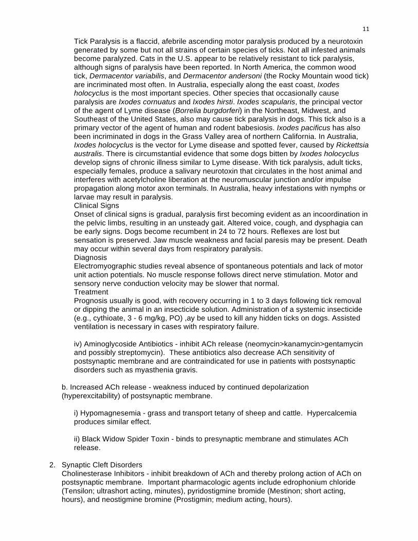
3. Postsynaptic Disorders a. Myasthenia Gravis (MG)
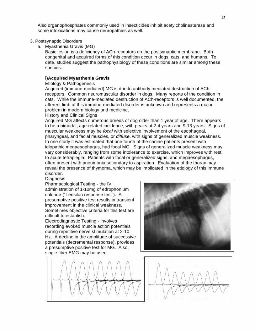
Basic lesion is a deficiency of ACh-receptors on the postsynaptic membrane. Both congenital and acquired forms of this condition occur in dogs, cats, and humans. To date, studies suggest the pathophysiology of these conditions are similar among these species.
i)Acquired Myasthenia Gravis Etiology & Pathogenesis Acquired (immune-mediated) MG is due to antibody mediated destruction of ACh-receptors. Common neuromuscular disorder in dogs. Many reports of the condition in cats. While the immune-mediated destruction of ACh-receptors is well documented, the afferent limb of this immune-mediated disorder is unknown and represents a major problem in modern biology and medicine. History and Clinical Signs Acquired MG affects numerous breeds of dog older than 1 year of age. There appears to be a bimodal, age-related incidence, with peaks at 2-4 years and 9-13 years. Signs of muscular weakness may be focal with selective involvement of the esophageal, pharyngeal, and facial muscles, or diffuse, with signs of generalized muscle weakness. In one study it was estimated that one fourth of the canine patients present with idiopathic megaesophagus, had focal MG. Signs of generalized muscle weakness may vary considerably, ranging from some intolerance to exercise, which improves with rest, to acute tetraplegia. Patients with focal or generalized signs, and megaesophagus, often present with pneumonia secondary to aspiration. Evaluation of the thorax may reveal the presence of thymoma, which may be implicated in the etiology of this immune disorder. Diagnosis Pharmacological Testing - the IV administration of 1-10mg of edrophonium chloride (“Tensilon response test”). A presumptive positive test results in transient improvement in the clinical weakness. Sometimes objective criteria for this test are difficult to establish. Electrodiagnostic Testing - involves recording evoked muscle action potentials during repetitive nerve stimulation at 2-10 Hz. A decline in the amplitude of successive potentials (decremental response), provides a presumptive positive test for MG. Also, single fiber EMG may be used.
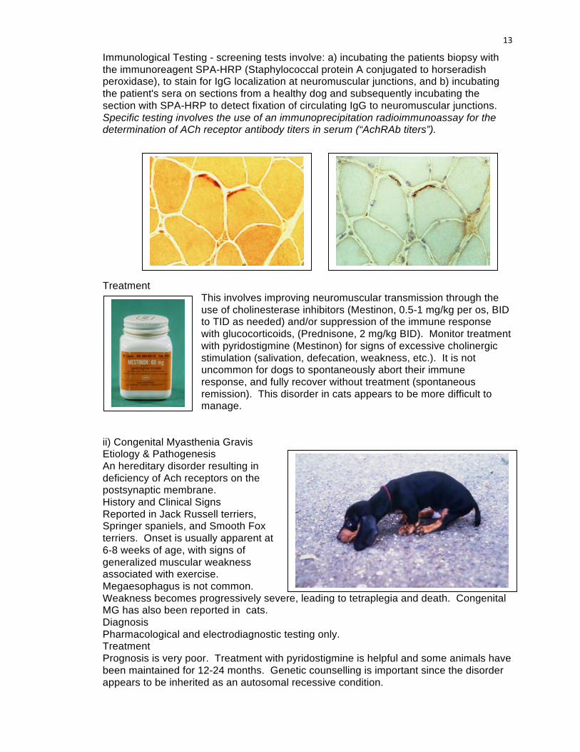
13
Immunological Testing - screening tests involve: a) incubating the patients biopsy with the immunoreagent SPA-HRP (Staphylococcal protein A conjugated to horseradish peroxidase), to stain for IgG localization at neuromuscular junctions, and b) incubating the patient's sera on sections from a healthy dog and subsequently incubating the section with SPA-HRP to detect fixation of circulating IgG to neuromuscular junctions. Specific testing involves the use of an immunoprecipitation radioimmunoassay for the determination of ACh receptor antibody titers in serum (“AchRAb titers”).
Treatment This involves improving neuromuscular transmission through the use of cholinesterase inhibitors (Mestinon, 0.5-1 mg/kg per os, BID to TID as needed) and/or suppression of the immune response with glucocorticoids, (Prednisone, 2 mg/kg BID). Monitor treatment with pyridostigmine (Mestinon) for signs of excessive cholinergic stimulation (salivation, defecation, weakness, etc.). It is not uncommon for dogs to spontaneously abort their immune response, and fully recover without treatment (spontaneous remission). This disorder in cats appears to be more difficult to manage.
ii) Congenital Myasthenia Gravis Etiology & Pathogenesis An hereditary disorder resulting in deficiency of Ach receptors on the postsynaptic membrane. History and Clinical Signs Reported in Jack Russell terriers, Springer spaniels, and Smooth Fox terriers. Onset is usually apparent at 6-8 weeks of age, with signs of generalized muscular weakness associated with exercise. Megaesophagus is not common. Weakness becomes progressively severe, leading to tetraplegia and death. Congenital MG has also been reported in cats. Diagnosis Pharmacological and electrodiagnostic testing only. Treatment Prognosis is very poor. Treatment with pyridostigmine is helpful and some animals have been maintained for 12-24 months. Genetic counselling is important since the disorder appears to be inherited as an autosomal recessive condition.
14
b. Pharmacologic Use of Neuromuscular Blocking Agents (Muscle Relaxants) The effects of 2 classes of neuromuscular blocking agents commonly used as muscle relaxants during anesthesia deserve mention here. i) Non-depolarizing agents: include d-Tubocurarine, gallamine, and pancuronium. These agents compete with ACh for the ACh-receptor. When bound to the receptor, the ion channel does not open, preventing depolarization of the membrane. ii) Depolarizing agents: include succinylcholine and decamethonium. These agents compete with ACh for the ACh-receptor. When bound to the receptor the ion channel opens, causing depolarization of the membrane.
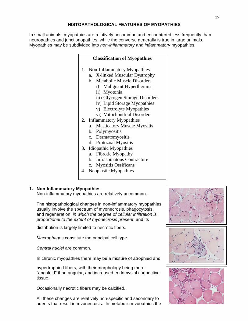
15
Classification of Myopathies
1. Non-Inflammatory Myopathies a. X-linked Muscular Dystrophy b. Metabolic Muscle Disorders i) Malignant Hyperthermia ii) Myotonia iii) Glycogen Storage Disorders iv) Lipid Storage Myopathies v) Electrolyte Myopathies vi) Mitochondrial Disorders 2. Inflammatory Myopathies a. Masticatory Muscle Myositis b. Polymyositis c. Dermatomyositis d. Protozoal Myositis 3. Idiopathic Myopathies a. Fibrotic Myopathy b. Infraspinatous Contracture c. Myositis Ossificans 4. Neoplastic Myopathies
HISTOPATHOLOGICAL FEATURES OF MYOPATHIES In small animals, myopathies are relatively uncommon and encountered less frequently than neuropathies and junctionopathies, while the converse generally is true in large animals. Myopathies may be subdivided into non-inflammatory and inflammatory myopathies. 1. Non-Inflammatory Myopathies
Non-inflammatory myopathies are relatively uncommon. The histopathological changes in non-inflammatory myopathies usually involve the spectrum of myonecrosis, phagocytosis, and regeneration, in which the degree of cellular infiltration is proportional to the extent of myonecrosis present, and its
distribution is largely limited to necrotic fibers. Macrophages constitute the principal cell type. Central nuclei are common. In chronic myopathies there may be a mixture of atrophied and
hypertrophied fibers, with their morphology being more "anguloid" than angular, and increased endomysial connective tissue. Occasionally necrotic fibers may be calcified. All these changes are relatively non-specific and secondary to agents that result in myonecrosis. In metabolic myopathies the

16
fibers frequently contain storage products that appear as vacuoles. a. X-Linked Muscular Dystrophy of Dogs and Cats
Etiology and Pathogenesis This is an hereditary myopathy in which there is a deficiency of dystrophin, a cytoskeletal protein associated with the inner surface of the sarcolemma. The pathobiology of this disorder appears to be identical to Duchenne muscular dystrophy, affecting young male children. The condition also occurs in mice. History and Clinical Signs The disorder is best characterized in Golden Retrievers. Other breeds of dog affected include Rottweilers, Samoyeds, and possibly Irish Terriers. In cats it has been documented in domestic short hair cats. In dogs the disorder usually is recognized between 8 and 10 weeks of age. Signs may include generalized weakness, a stiff-limbed, short strided, shuffling gait, some reduced mobility of the jaws, and difficulty with chewing and swallowing associated with excess salivation. The weakness and signs are progressive, with poorly developed muscle mass, body stature, and spinal curvature. In advanced stages some muscle groups may appear grossly hypertrophied. Cardiac muscle may also become involved. Recognition of this disorder in cats is more recent and the findings are similar to those in dogs. While the onset is likely similar to that in the dog, reported cases have been somewhat later in onset, 5 to 25 months of age. Gross hypertrophy of the tongue and diaphragm have been a common finding in cats. Diagnosis These include serum CK, which may be elevated to 300 times normal; EMG examination which reveals repetitive, high frequency discharges; and muscle biopsy examination which reveals multifocal areas of necrosis, necrosis and phagocytosis, and regeneration. In the latter stages, muscle fiber hypertrophy and fiber splitting is common along with fibrosis. Specific information is provided through the use of immunocytochemical staining techniques to detect dystrophin (or lack thereof) in sections and/or the muscle may be assayed for the presence of dystrophin. Treatment/Prognosis Currently there is no cure and lifespan is shortened due to secondary complications. Future research advances in this area will have widespread benefits for both animals and humans.
b. Metabolic Muscle Disorders
These are relatively uncommon disorders of carbohydrate, lipid, and oxygen metabolism that generally induce muscle weakness by supplying insufficient energy (ATP) to sustain muscle contraction, and other cellular functions with a high energy requirement (e.g., maintenance of active transport and ion gradients across the cell membrane).
i) Malignant Hyperthermia Etiology & Pathogenesis Malignant hyperthermia (MH) is a life-threatening hypermetabolic and contractile condition that is triggered in humans, pigs, dogs and cats by certain anesthetic agents (e.g., halothane and succinylcholine). The underlying defect in calcium (Ca) homeostasis occurs at the level of the skeletal muscle sarcoplasmic reticulum where there is hypersensitive and heightened ligand-gating of the Ca-release channel. The Ca channel is readily opened by certain drugs, such as caffeine and halothane. Caffeine- or
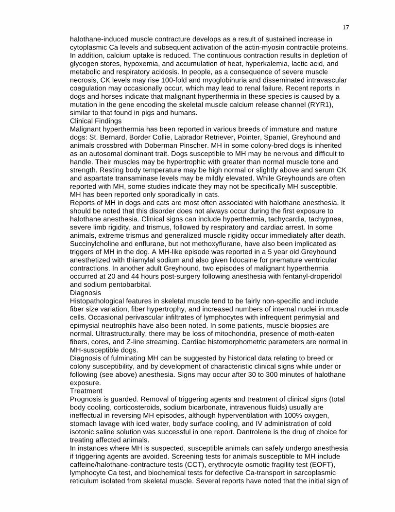
17
halothane-induced muscle contracture develops as a result of sustained increase in cytoplasmic Ca levels and subsequent activation of the actin-myosin contractile proteins. In addition, calcium uptake is reduced. The continuous contraction results in depletion of glycogen stores, hypoxemia, and accumulation of heat, hyperkalemia, lactic acid, and metabolic and respiratory acidosis. In people, as a consequence of severe muscle necrosis, CK levels may rise 100-fold and myoglobinuria and disseminated intravascular coagulation may occasionally occur, which may lead to renal failure. Recent reports in dogs and horses indicate that malignant hyperthermia in these species is caused by a mutation in the gene encoding the skeletal muscle calcium release channel (RYR1), similar to that found in pigs and humans. Clinical Findings Malignant hyperthermia has been reported in various breeds of immature and mature dogs: St. Bernard, Border Collie, Labrador Retriever, Pointer, Spaniel, Greyhound and animals crossbred with Doberman Pinscher. MH in some colony-bred dogs is inherited as an autosomal dominant trait. Dogs susceptible to MH may be nervous and difficult to handle. Their muscles may be hypertrophic with greater than normal muscle tone and strength. Resting body temperature may be high normal or slightly above and serum CK and aspartate transaminase levels may be mildly elevated. While Greyhounds are often reported with MH, some studies indicate they may not be specifically MH susceptible. MH has been reported only sporadically in cats. Reports of MH in dogs and cats are most often associated with halothane anesthesia. It should be noted that this disorder does not always occur during the first exposure to halothane anesthesia. Clinical signs can include hyperthermia, tachycardia, tachypnea, severe limb rigidity, and trismus, followed by respiratory and cardiac arrest. In some animals, extreme trismus and generalized muscle rigidity occur immediately after death. Succinylcholine and enflurane, but not methoxyflurane, have also been implicated as triggers of MH in the dog. A MH-like episode was reported in a 5 year old Greyhound anesthetized with thiamylal sodium and also given lidocaine for premature ventricular contractions. In another adult Greyhound, two episodes of malignant hyperthermia occurred at 20 and 44 hours post-surgery following anesthesia with fentanyl-droperidol and sodium pentobarbital. Diagnosis Histopathological features in skeletal muscle tend to be fairly non-specific and include fiber size variation, fiber hypertrophy, and increased numbers of internal nuclei in muscle cells. Occasional perivascular infiltrates of lymphocytes with infrequent perimysial and epimysial neutrophils have also been noted. In some patients, muscle biopsies are normal. Ultrastructurally, there may be loss of mitochondria, presence of moth-eaten fibers, cores, and Z-line streaming. Cardiac histomorphometric parameters are normal in MH-susceptible dogs. Diagnosis of fulminating MH can be suggested by historical data relating to breed or colony susceptibility, and by development of characteristic clinical signs while under or following (see above) anesthesia. Signs may occur after 30 to 300 minutes of halothane exposure. Treatment Prognosis is guarded. Removal of triggering agents and treatment of clinical signs (total body cooling, corticosteroids, sodium bicarbonate, intravenous fluids) usually are ineffectual in reversing MH episodes, although hyperventilation with 100% oxygen, stomach lavage with iced water, body surface cooling, and IV administration of cold isotonic saline solution was successful in one report. Dantrolene is the drug of choice for treating affected animals. In instances where MH is suspected, susceptible animals can safely undergo anesthesia if triggering agents are avoided. Screening tests for animals susceptible to MH include caffeine/halothane-contracture tests (CCT), erythrocyte osmotic fragility test (EOFT), lymphocyte Ca test, and biochemical tests for defective Ca-transport in sarcoplasmic reticulum isolated from skeletal muscle. Several reports have noted that the initial sign of
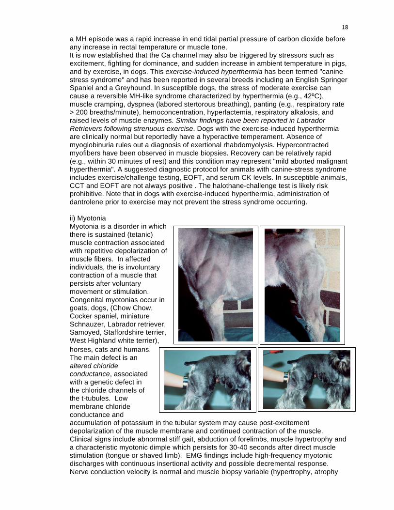
18
a MH episode was a rapid increase in end tidal partial pressure of carbon dioxide before any increase in rectal temperature or muscle tone. It is now established that the Ca channel may also be triggered by stressors such as excitement, fighting for dominance, and sudden increase in ambient temperature in pigs, and by exercise, in dogs. This exercise-induced hyperthermia has been termed "canine stress syndrome" and has been reported in several breeds including an English Springer Spaniel and a Greyhound. In susceptible dogs, the stress of moderate exercise can cause a reversible MH-like syndrome characterized by hyperthermia (e.g., 42ºC), muscle cramping, dyspnea (labored stertorous breathing), panting (e.g., respiratory rate > 200 breaths/minute), hemoconcentration, hyperlactemia, respiratory alkalosis, and raised levels of muscle enzymes. Similar findings have been reported in Labrador Retrievers following strenuous exercise. Dogs with the exercise-induced hyperthermia are clinically normal but reportedly have a hyperactive temperament. Absence of myoglobinuria rules out a diagnosis of exertional rhabdomyolysis. Hypercontracted myofibers have been observed in muscle biopsies. Recovery can be relatively rapid (e.g., within 30 minutes of rest) and this condition may represent "mild aborted malignant hyperthermia". A suggested diagnostic protocol for animals with canine-stress syndrome includes exercise/challenge testing, EOFT, and serum CK levels. In susceptible animals, CCT and EOFT are not always positive . The halothane-challenge test is likely risk prohibitive. Note that in dogs with exercise-induced hyperthermia, administration of dantrolene prior to exercise may not prevent the stress syndrome occurring. ii) Myotonia Myotonia is a disorder in which there is sustained (tetanic) muscle contraction associated with repetitive depolarization of muscle fibers. In affected individuals, the is involuntary contraction of a muscle that persists after voluntary movement or stimulation. Congenital myotonias occur in goats, dogs, (Chow Chow, Cocker spaniel, miniature Schnauzer, Labrador retriever, Samoyed, Staffordshire terrier, West Highland white terrier), horses, cats and humans. The main defect is an altered chloride conductance, associated with a genetic defect in the chloride channels of the t-tubules. Low membrane chloride conductance and accumulation of potassium in the tubular system may cause post-excitement depolarization of the muscle membrane and continued contraction of the muscle. Clinical signs include abnormal stiff gait, abduction of forelimbs, muscle hypertrophy and a characteristic myotonic dimple which persists for 30-40 seconds after direct muscle stimulation (tongue or shaved limb). EMG findings include high-frequency myotonic discharges with continuous insertional activity and possible decremental response. Nerve conduction velocity is normal and muscle biopsy variable (hypertrophy, atrophy
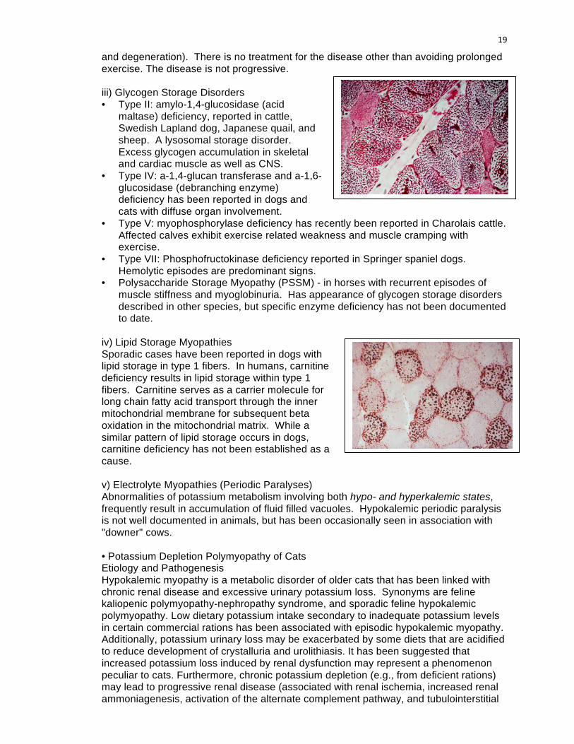
19
and degeneration). There is no treatment for the disease other than avoiding prolonged exercise. The disease is not progressive.
iii) Glycogen Storage Disorders • Type II: amylo-1,4-glucosidase (acid
maltase) deficiency, reported in cattle, Swedish Lapland dog, Japanese quail, and sheep. A lysosomal storage disorder. Excess glycogen accumulation in skeletal and cardiac muscle as well as CNS.
• Type IV: a-1,4-glucan transferase and a-1,6-glucosidase (debranching enzyme) deficiency has been reported in dogs and cats with diffuse organ involvement.
• Type V: myophosphorylase deficiency has recently been reported in Charolais cattle. Affected calves exhibit exercise related weakness and muscle cramping with exercise.
• Type VII: Phosphofructokinase deficiency reported in Springer spaniel dogs. Hemolytic episodes are predominant signs.
• Polysaccharide Storage Myopathy (PSSM) - in horses with recurrent episodes of muscle stiffness and myoglobinuria. Has appearance of glycogen storage disorders described in other species, but specific enzyme deficiency has not been documented to date.
iv) Lipid Storage Myopathies Sporadic cases have been reported in dogs with lipid storage in type 1 fibers. In humans, carnitine deficiency results in lipid storage within type 1 fibers. Carnitine serves as a carrier molecule for long chain fatty acid transport through the inner mitochondrial membrane for subsequent beta oxidation in the mitochondrial matrix. While a similar pattern of lipid storage occurs in dogs, carnitine deficiency has not been established as a cause.
v) Electrolyte Myopathies (Periodic Paralyses) Abnormalities of potassium metabolism involving both hypo- and hyperkalemic states, frequently result in accumulation of fluid filled vacuoles. Hypokalemic periodic paralysis is not well documented in animals, but has been occasionally seen in association with "downer" cows.
• Potassium Depletion Polymyopathy of Cats Etiology and Pathogenesis Hypokalemic myopathy is a metabolic disorder of older cats that has been linked with chronic renal disease and excessive urinary potassium loss. Synonyms are feline kaliopenic polymyopathy-nephropathy syndrome, and sporadic feline hypokalemic polymyopathy. Low dietary potassium intake secondary to inadequate potassium levels in certain commercial rations has been associated with episodic hypokalemic myopathy. Additionally, potassium urinary loss may be exacerbated by some diets that are acidified to reduce development of crystalluria and urolithiasis. It has been suggested that increased potassium loss induced by renal dysfunction may represent a phenomenon peculiar to cats. Furthermore, chronic potassium depletion (e.g., from deficient rations) may lead to progressive renal disease (associated with renal ischemia, increased renal ammoniagenesis, activation of the alternate complement pathway, and tubulointerstitial
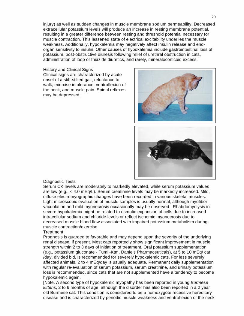
20
injury) as well as sudden changes in muscle membrane sodium permeability. Decreased extracellular potassium levels will produce an increase in resting membrane potential, resulting in a greater difference between resting and threshold potential necessary for muscle contraction. This lessened state of electrical excitability underlies the muscle weakness. Additionally, hypokalemia may negatively affect insulin release and end-organ sensitivity to insulin. Other causes of hypokalemia include gastrointestinal loss of potassium, post-obstructive diuresis following relief of urethral obstruction in cats, administration of loop or thiazide diuretics, and rarely, mineralocorticoid excess.
History and Clinical Signs Clinical signs are characterized by acute onset of a stiff-stilted gait, reluctance to walk, exercise intolerance, ventroflexion of the neck, and muscle pain. Spinal reflexes may be depressed. Diagnostic Tests Serum CK levels are moderately to markedly elevated, while serum potassium values are low (e.g., < 4.0 mEq/L). Serum creatinine levels may be markedly increased. Mild, diffuse electromyographic changes have been recorded in various skeletal muscles. Light microscopic evaluation of muscle samples is usually normal, although myofiber vacuolation and mild myonecrosis occasionally may be observed. Rhabdomyolysis in severe hypokalemia might be related to osmotic expansion of cells due to increased intracellular sodium and chloride levels or reflect ischemic myonecrosis due to decreased muscle blood flow associated with impaired potassium metabolism during muscle contraction/exercise. Treatment Prognosis is guarded to favorable and may depend upon the severity of the underlying renal disease, if present. Most cats reportedly show significant improvement in muscle strength within 2 to 3 days of initiation of treatment. Oral potassium supplementation (e.g., potassium gluconate - Tumil-Ktm, Daniels Pharmaceuticals), at 5 to 10 mEq/ cat /day, divided bid, is recommended for severely hypokalemic cats. For less severely affected animals, 2 to 4 mEq/day is usually adequate. Permanent daily supplementation with regular re-evaluation of serum potassium, serum creatinine, and urinary potassium loss is recommended, since cats that are not supplemented have a tendency to become hypokalemic again. [Note. A second type of hypokalemic myopathy has been reported in young Burmese kittens, 2 to 6 months of age, although the disorder has also been reported in a 2 year old Burmese cat. This condition is considered to be a homozygote recessive hereditary disease and is characterized by periodic muscle weakness and ventroflexion of the neck
21
associated with intermittent hypokalemia (e.g., < 3.0 mE/L) and increased serum creatine kinase values, sometimes reaching very high values, e.g., > 50,000 - 90,000 IU/L. The condition has also been termed periodic hypokalemic myopathy. Attacks occur suddenly and are transient and may be precipitated by stress or vigorous exercise. The variable clinical course is characterized by improvement followed by relapse, and there may be weeks between episodes. A head tremor is seen in some cats. Cats are reluctant to walk and tire easily, have a stiff, stilted gait with thoracic limb hypermetria, and a wide-based stance in the hind limbs. Carpal knuckling can be a distinctive clinical feature and some cats sink on their hocks. There are only minor electromyographic and histopathologic changes seen in muscle. Neither decreased potassium intake nor increased renal potassium loss have been found in affected Burmese cats. Continued dietary supplementation of oral potassium usually produces a favorable response (e.g., potassium gluconate solution at 2 to 4 mEq or mmol/cat PO daily, until serum potassium levels are stable) [133]. Some kittens improve without treatment. The periodic hypokalemic attacks in these cats are similar to those seen in humans with hypokalemic periodic paralysis, an inherited calcium channelopathy disorder associated with abnormal muscle membrane excitability and influx of potassium into the muscle fiber that causes muscle fiber depolarization and inexcitability.] • Hyperkalemic Periodic Paralysis (HYPP) This is an autosomal dominant disorder of humans and horses (Quarter Horses, Appaloosas and Paints), in which episodes of weakness, muscle fasciculations, myotonia, and recumbency develop in association with hyperkalemia (5.0 to 11.0 mEq K+/L; normal = 3.0 to 4.0 mEqK+/L). Attacks occur following diet changes, fasting, stress, alfalfa consumption and the consumption of other high potassium diets. Muscles are hyperexcitable due to an increase in permeability to sodium ions as a result of an abnormality of voltage-gated sodium channel alpha-subunits of the sarcolemma. The condition appears to be analogous to the human disorder, which is due to a single base pair change in the sodium ion channel gene. The physiological defect appears to be impaired inactivation of the sodium channel. Molecular diagnostic methods are now available. Muscle biopsy changes include vacuolar changes.
vi) Mitochondrial Disorders Mitochondrial disorders appear to be rare. However, a deficiency of Complex I respiratory chain enzyme has been documented in an Arabian horse with exercise intolerance. Lactic acidosis develops in response to exercise. Also, a recent case of lactic acidosis has been described in an Old English Sheepdog. More details and characterization of these metabolic problems likely will be forthcoming over the next few years.
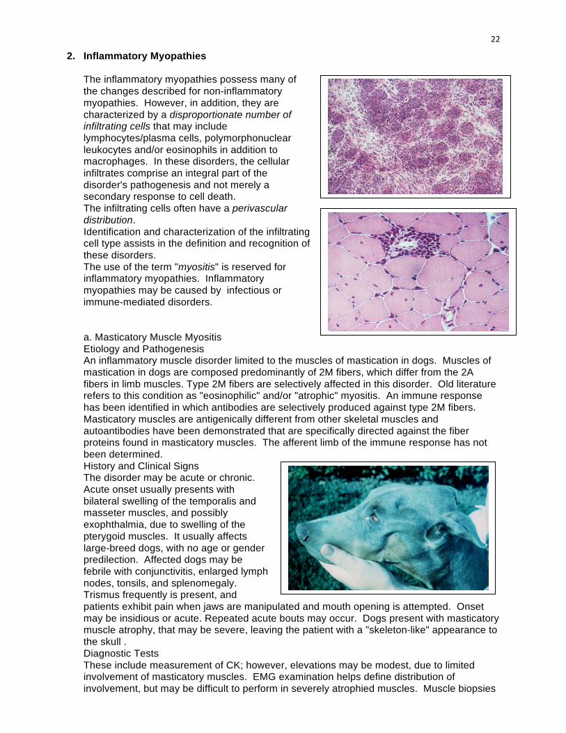
22
2. Inflammatory Myopathies
The inflammatory myopathies possess many of the changes described for non-inflammatory myopathies. However, in addition, they are characterized by a disproportionate number of infiltrating cells that may include lymphocytes/plasma cells, polymorphonuclear leukocytes and/or eosinophils in addition to macrophages. In these disorders, the cellular infiltrates comprise an integral part of the disorder's pathogenesis and not merely a secondary response to cell death. The infiltrating cells often have a perivascular distribution. Identification and characterization of the infiltrating cell type assists in the definition and recognition of these disorders. The use of the term "myositis" is reserved for inflammatory myopathies. Inflammatory myopathies may be caused by infectious or immune-mediated disorders.
a. Masticatory Muscle Myositis Etiology and Pathogenesis An inflammatory muscle disorder limited to the muscles of mastication in dogs. Muscles of mastication in dogs are composed predominantly of 2M fibers, which differ from the 2A fibers in limb muscles. Type 2M fibers are selectively affected in this disorder. Old literature refers to this condition as "eosinophilic" and/or "atrophic" myositis. An immune response has been identified in which antibodies are selectively produced against type 2M fibers. Masticatory muscles are antigenically different from other skeletal muscles and autoantibodies have been demonstrated that are specifically directed against the fiber proteins found in masticatory muscles. The afferent limb of the immune response has not been determined. History and Clinical Signs The disorder may be acute or chronic. Acute onset usually presents with bilateral swelling of the temporalis and masseter muscles, and possibly exophthalmia, due to swelling of the pterygoid muscles. It usually affects large-breed dogs, with no age or gender predilection. Affected dogs may be febrile with conjunctivitis, enlarged lymph nodes, tonsils, and splenomegaly. Trismus frequently is present, and patients exhibit pain when jaws are manipulated and mouth opening is attempted. Onset may be insidious or acute. Repeated acute bouts may occur. Dogs present with masticatory muscle atrophy, that may be severe, leaving the patient with a "skeleton-like" appearance to the skull . Diagnostic Tests These include measurement of CK; however, elevations may be modest, due to limited involvement of masticatory muscles. EMG examination helps define distribution of involvement, but may be difficult to perform in severely atrophied muscles. Muscle biopsies
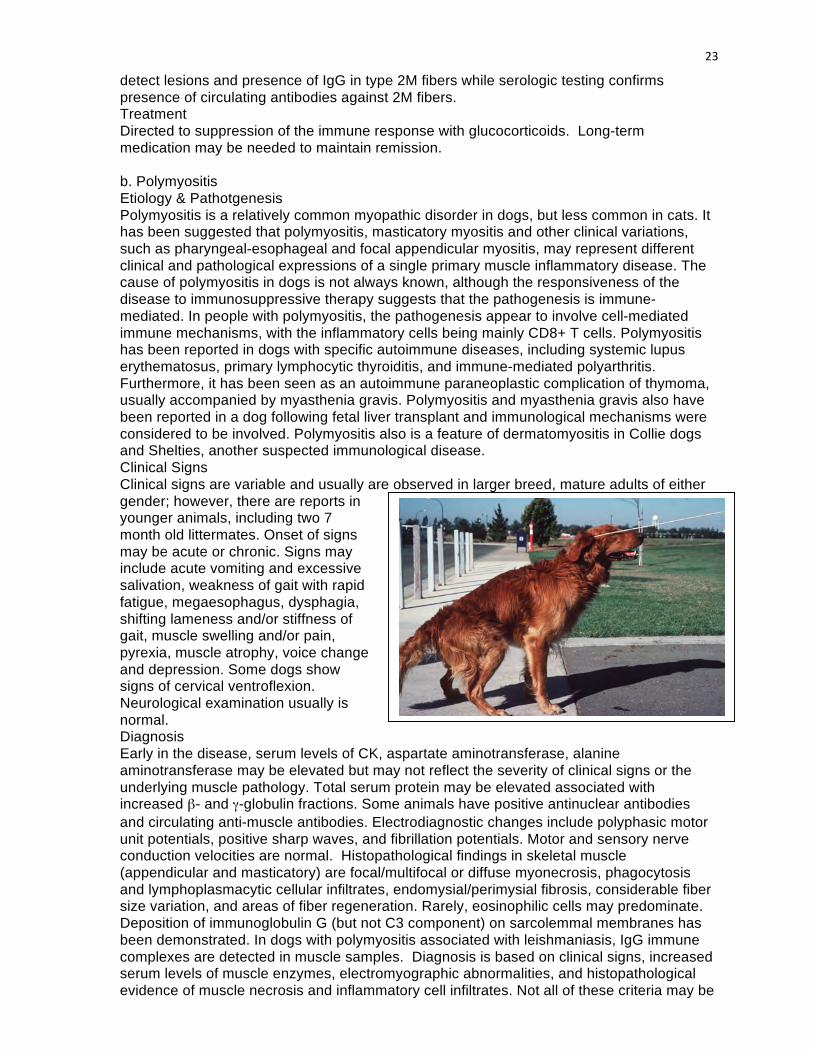
23
detect lesions and presence of IgG in type 2M fibers while serologic testing confirms presence of circulating antibodies against 2M fibers. Treatment Directed to suppression of the immune response with glucocorticoids. Long-term medication may be needed to maintain remission.
b. Polymyositis Etiology & Pathotgenesis Polymyositis is a relatively common myopathic disorder in dogs, but less common in cats. It has been suggested that polymyositis, masticatory myositis and other clinical variations, such as pharyngeal-esophageal and focal appendicular myositis, may represent different clinical and pathological expressions of a single primary muscle inflammatory disease. The cause of polymyositis in dogs is not always known, although the responsiveness of the disease to immunosuppressive therapy suggests that the pathogenesis is immune-mediated. In people with polymyositis, the pathogenesis appear to involve cell-mediated immune mechanisms, with the inflammatory cells being mainly CD8+ T cells. Polymyositis has been reported in dogs with specific autoimmune diseases, including systemic lupus erythematosus, primary lymphocytic thyroiditis, and immune-mediated polyarthritis. Furthermore, it has been seen as an autoimmune paraneoplastic complication of thymoma, usually accompanied by myasthenia gravis. Polymyositis and myasthenia gravis also have been reported in a dog following fetal liver transplant and immunological mechanisms were considered to be involved. Polymyositis also is a feature of dermatomyositis in Collie dogs and Shelties, another suspected immunological disease. Clinical Signs Clinical signs are variable and usually are observed in larger breed, mature adults of either gender; however, there are reports in younger animals, including two 7 month old littermates. Onset of signs may be acute or chronic. Signs may include acute vomiting and excessive salivation, weakness of gait with rapid fatigue, megaesophagus, dysphagia, shifting lameness and/or stiffness of gait, muscle swelling and/or pain, pyrexia, muscle atrophy, voice change and depression. Some dogs show signs of cervical ventroflexion. Neurological examination usually is normal. Diagnosis Early in the disease, serum levels of CK, aspartate aminotransferase, alanine aminotransferase may be elevated but may not reflect the severity of clinical signs or the underlying muscle pathology. Total serum protein may be elevated associated with increased β- and γ-globulin fractions. Some animals have positive antinuclear antibodies and circulating anti-muscle antibodies. Electrodiagnostic changes include polyphasic motor unit potentials, positive sharp waves, and fibrillation potentials. Motor and sensory nerve conduction velocities are normal. Histopathological findings in skeletal muscle (appendicular and masticatory) are focal/multifocal or diffuse myonecrosis, phagocytosis and lymphoplasmacytic cellular infiltrates, endomysial/perimysial fibrosis, considerable fiber size variation, and areas of fiber regeneration. Rarely, eosinophilic cells may predominate. Deposition of immunoglobulin G (but not C3 component) on sarcolemmal membranes has been demonstrated. In dogs with polymyositis associated with leishmaniasis, IgG immune complexes are detected in muscle samples. Diagnosis is based on clinical signs, increased serum levels of muscle enzymes, electromyographic abnormalities, and histopathological evidence of muscle necrosis and inflammatory cell infiltrates. Not all of these criteria may be
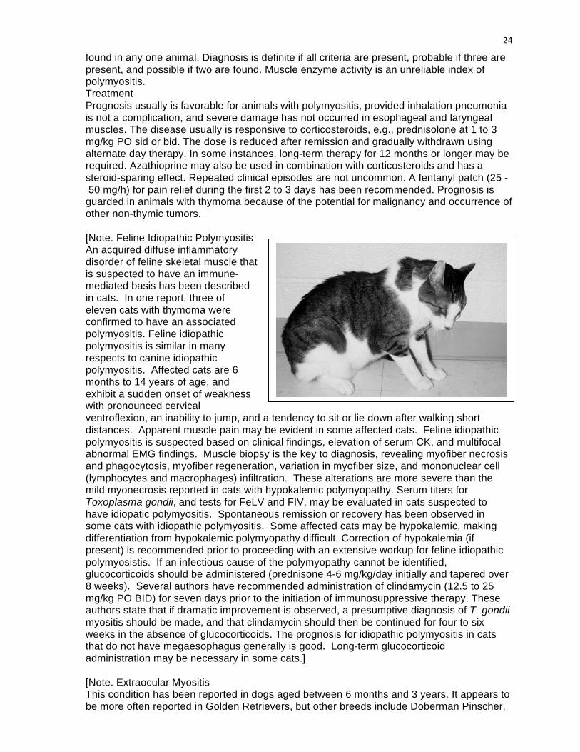
24
found in any one animal. Diagnosis is definite if all criteria are present, probable if three are present, and possible if two are found. Muscle enzyme activity is an unreliable index of polymyositis. Treatment Prognosis usually is favorable for animals with polymyositis, provided inhalation pneumonia is not a complication, and severe damage has not occurred in esophageal and laryngeal muscles. The disease usually is responsive to corticosteroids, e.g., prednisolone at 1 to 3 mg/kg PO sid or bid. The dose is reduced after remission and gradually withdrawn using alternate day therapy. In some instances, long-term therapy for 12 months or longer may be required. Azathioprine may also be used in combination with corticosteroids and has a steroid-sparing effect. Repeated clinical episodes are not uncommon. A fentanyl patch (25 - 50 mg/h) for pain relief during the first 2 to 3 days has been recommended. Prognosis is guarded in animals with thymoma because of the potential for malignancy and occurrence of other non-thymic tumors.
[Note. Feline Idiopathic Polymyositis An acquired diffuse inflammatory disorder of feline skeletal muscle that is suspected to have an immune-mediated basis has been described in cats. In one report, three of eleven cats with thymoma were confirmed to have an associated polymyositis. Feline idiopathic polymyositis is similar in many respects to canine idiopathic polymyositis. Affected cats are 6 months to 14 years of age, and exhibit a sudden onset of weakness with pronounced cervical ventroflexion, an inability to jump, and a tendency to sit or lie down after walking short distances. Apparent muscle pain may be evident in some affected cats. Feline idiopathic polymyositis is suspected based on clinical findings, elevation of serum CK, and multifocal abnormal EMG findings. Muscle biopsy is the key to diagnosis, revealing myofiber necrosis and phagocytosis, myofiber regeneration, variation in myofiber size, and mononuclear cell (lymphocytes and macrophages) infiltration. These alterations are more severe than the mild myonecrosis reported in cats with hypokalemic polymyopathy. Serum titers for Toxoplasma gondii, and tests for FeLV and FIV, may be evaluated in cats suspected to have idiopatic polymyositis. Spontaneous remission or recovery has been observed in some cats with idiopathic polymyositis. Some affected cats may be hypokalemic, making differentiation from hypokalemic polymyopathy difficult. Correction of hypokalemia (if present) is recommended prior to proceeding with an extensive workup for feline idiopathic polymyosistis. If an infectious cause of the polymyopathy cannot be identified, glucocorticoids should be administered (prednisone 4-6 mg/kg/day initially and tapered over 8 weeks). Several authors have recommended administration of clindamycin (12.5 to 25 mg/kg PO BID) for seven days prior to the initiation of immunosuppressive therapy. These authors state that if dramatic improvement is observed, a presumptive diagnosis of T. gondii myositis should be made, and that clindamycin should then be continued for four to six weeks in the absence of glucocorticoids. The prognosis for idiopathic polymyositis in cats that do not have megaesophagus generally is good. Long-term glucocorticoid administration may be necessary in some cats.]
[Note. Extraocular Myositis This condition has been reported in dogs aged between 6 months and 3 years. It appears to be more often reported in Golden Retrievers, but other breeds include Doberman Pinscher,
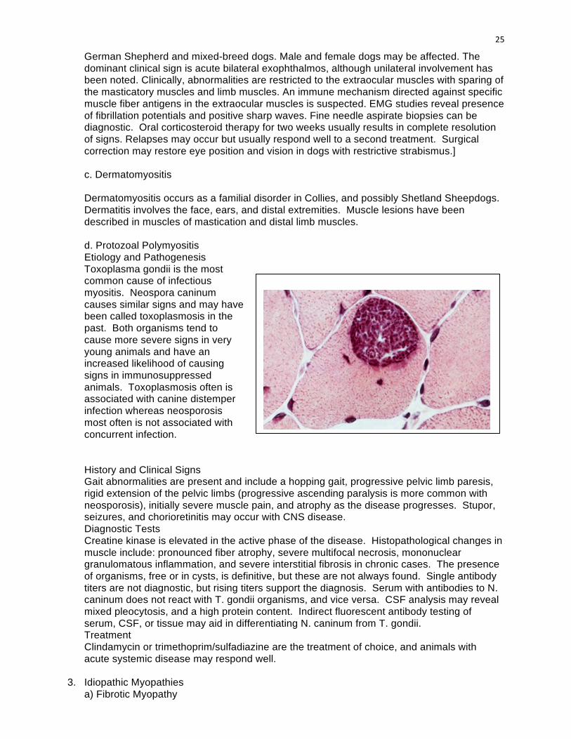
25
German Shepherd and mixed-breed dogs. Male and female dogs may be affected. The dominant clinical sign is acute bilateral exophthalmos, although unilateral involvement has been noted. Clinically, abnormalities are restricted to the extraocular muscles with sparing of the masticatory muscles and limb muscles. An immune mechanism directed against specific muscle fiber antigens in the extraocular muscles is suspected. EMG studies reveal presence of fibrillation potentials and positive sharp waves. Fine needle aspirate biopsies can be diagnostic. Oral corticosteroid therapy for two weeks usually results in complete resolution of signs. Relapses may occur but usually respond well to a second treatment. Surgical correction may restore eye position and vision in dogs with restrictive strabismus.]
c. Dermatomyositis
Dermatomyositis occurs as a familial disorder in Collies, and possibly Shetland Sheepdogs. Dermatitis involves the face, ears, and distal extremities. Muscle lesions have been described in muscles of mastication and distal limb muscles. d. Protozoal Polymyositis Etiology and Pathogenesis Toxoplasma gondii is the most common cause of infectious myositis. Neospora caninum causes similar signs and may have been called toxoplasmosis in the past. Both organisms tend to cause more severe signs in very young animals and have an increased likelihood of causing signs in immunosuppressed animals. Toxoplasmosis often is associated with canine distemper infection whereas neosporosis most often is not associated with concurrent infection. History and Clinical Signs Gait abnormalities are present and include a hopping gait, progressive pelvic limb paresis, rigid extension of the pelvic limbs (progressive ascending paralysis is more common with neosporosis), initially severe muscle pain, and atrophy as the disease progresses. Stupor, seizures, and chorioretinitis may occur with CNS disease. Diagnostic Tests Creatine kinase is elevated in the active phase of the disease. Histopathological changes in muscle include: pronounced fiber atrophy, severe multifocal necrosis, mononuclear granulomatous inflammation, and severe interstitial fibrosis in chronic cases. The presence of organisms, free or in cysts, is definitive, but these are not always found. Single antibody titers are not diagnostic, but rising titers support the diagnosis. Serum with antibodies to N. caninum does not react with T. gondii organisms, and vice versa. CSF analysis may reveal mixed pleocytosis, and a high protein content. Indirect fluorescent antibody testing of serum, CSF, or tissue may aid in differentiating N. caninum from T. gondii. Treatment Clindamycin or trimethoprim/sulfadiazine are the treatment of choice, and animals with acute systemic disease may respond well.
3. Idiopathic Myopathies a) Fibrotic Myopathy
26
Etiology and Pathogenesis Fibrotic myopathies are chronic, progressive disorders that result in severe muscle contracture and fibrosis. This condition has been reported in the semitendinosus, quadriceps, supraspinatus, infraspinatus, rectus femoris, and gracilis muscles of the dog and the semitendinosus muscle in the cat. The cause is not known and the condition may be the result of primary neuropathy or myopathy, frequent intramuscular injections, exercise-induced trauma, or chronic trauma with tearing and stretching of muscle fibers, or it may be congenital. Muscle is replaced by dense collagenous connective tissue, resulting in a taut fibrous band. History and Clinical Signs The limb involved develops a nonpainful, mechanical lameness, the severity of which depends on which muscle is involved and the extent of the fibrosis. Diagnostic Tests On physical examination a thin fibrous band may be palpated, that replaces the muscle belly. Histopathology of affected muscles reveals dense collagenous connective tissue replacing muscle fibers and there is minimal inflammation. EMG activity is not detected from the fibrous band and bizarre high-frequency discharges may occur with incomplete replacement of muscle fibers. Treatment Surgery is not recommended unless the lameness is disabling. If surgery is undertaken the goal is to release the fibrous band. Prognosis is guarded because of the likely recurrence of the fibrous band within 3-8 months.
b) Infraspinatous Muscle Contracture Etiology and Pathogenesis Fibrosis of the infraspinatous muscle occurs primarily in hunting or working breeds of dogs and is usually unilateral, but may be bilateral. The cause is not known, but it is thought to be a primary muscle disorder. It may be associated with trauma, either self-induced or external. Trauma is thought to cause incomplete rupture of the muscle, resulting in progressive fibrosis and contracture over 2-4 weeks. History and Clinical Signs Initially, the dog has an acute onset of pain in the shoulder during or soon after exercise. The lameness gradually subsides, but never resolves. Two to four weeks after the initial injury, a nonpainful mechanical lameness of the thoracic limb develops and the gait is characterized by adduction of the elbow and abduction of the limb, with outward rotation of the antebrachium and carpus. The limb is laterally circumducted with each stride, and the foot "flips" forward. Diagnosis Palpation of the thoracic limb reveals that the humerus rotates outward when the elbow joint is flexed and range of motion in the shoulder joint is limited. Disuse atrophy of the infraspinatus, supraspinatus, and deltoid muscles is evident from the prominent scapular spine. Treatment Tenotomy of the tendon of insertion of the infraspinatous muscle should allow the thoracic limb to be more easily adducted and the shoulder range of motion is improved. Activity should be limited for 1-2 weeks after surgery and prognosis for a full recovery is excellent.
c) Myositis Ossificans
Etiology and Pathogenesis Myositis ossificans is the heterotropic formation of bone in muscle. There is usually minimal inflammation, and muscle is not always involved. Generalized and localized forms have been described based on clinical behavior. The localized form is characterized by heterotopic, nonneoplastic bone formation in one muscle or a group of muscles. It is reported in dogs and cats and has been well described in Dobermans where some authors believe it may be a separate disease entity (von Willebrand heterotopic
27
osteochondrofibrosis in Doberman pinschers). Progressive or generalized myositis ossificans is also known as progressive ossifying fibrodysplasia or progressive or generalized ossifying myositis. It is characterized by the development of excessive fibrous connective tissue, which results in widespread muscle degeneration and ultimately leads to dystrophic calcification and ossification. It has been reported in young to middle-aged cats. The cause is not known and the localized form may be associated with trauma, infection, ossifying hematoma, and metaplasia of muscle and connective tissue to cartilage and bone. In Doberman pinschers, it is speculated that the combination of bleeding tendencies associated with von Willebrand's disease and trauma results in microvascular bleeding with subsequent fibrosis or mineralization. The progressive form is thought to be congenital or hereditary and may be a defect of fibroblasts in collagenous connective tissue with secondary degeneration of muscle, rather than a primary muscular lesion. History and Clinical Signs Localized ossifying myositis results in a palpable, firm enlargement in affected muscles, with chronic lameness, pain after exercise and muscle atrophy. Progressive ossifying fibrodysplasia occurs in young to middle-aged cats and results in firm nodules anywhere on the body, but predominantly on the neck and back. Limbs, especially the pelvic limbs, may be stiff which may progress to difficulty walking. Proximal limb musculature is enlarged and firm, painful, and has limited range of motion. Affected cats may be disabled within 2 weeks to several months. Diagnostic Tests Radiographs of affected areas show multiple mineralized densities. Soft tissue mineralization occurs within 3-6 weeks of injury and mature bone in soft tissue after 2-6 months. There may or may not be a periosteal reaction. On histopathology of the lesion, "zone phenomena" occur, with progression of bone maturation centrally to peripherally. The central zone contains undifferentiated cells and fibroblasts and may resemble a sarcoma, the intermediate zone contains osteoid and some areas of immature bone, and the peripheral zone contains mature bone. It does not invade the surrounding soft tissue. Affected muscle has excessive connective tissue between muscle fibers, mononuclear infiltration and muscle atrophy and hyaline degeneration. Treatment If clinical signs are minimal, treatment is not recommended for localized ossifying myositis. Rest, compressive bandages, and aspirin may help to relieve the acute pain. Surgical excision may be indicated to alleviate discomfort and restore normal limb function. In humans if surgery is performed within 6 months (i.e. when the lesion is immature) there is a high rate of recurrence. Post operative physical therapy is important. There is no effective treatment for progressive ossifying fibrodysplasia.
4. Neoplasia
Muscle tumors may be primary, originating from skeletal muscle (rhabdomyoma, rhabdomyosarcoma), or secondary (metastatic spread from tumors originating elsewhere, or local invasion of tumors into muscle from rapidly expanding cutaneous or bone tumors). Age of onset is variable although secondary tumor spread usually occurs in middle-aged animals. Clinical signs depend on the muscle group affected. Firm, nonpainful swelling, distortion of the area, and lameness are common. Diagnosis is confirmed by histopathologic assessment of biopsy material. Staging of the disease with lymph node biopsy and thoracic radiographs is important. For primary neoplasia, surgical excision is preferred, but may be ineffectual if the neoplasm is difficult to reach or invasive. Amputation of the limb may be necessary to attain adequate surgical margins. Treatment of secondary neoplasia depends on the therapy indicated for the primary tumor.
Involuntary Movement Disorders Richard A.leCouter, professor BVSc, PhD Dipl.ACVIM (USA)
1 Involuntary Movement Disorders
Richard A. LeCouteur, BVSc, PhD, Diplomate ACVIM(Neurology), Diplomate ECVN
Department of Surgical & Radiological Sciences, School of Veterinary Medicine, University of California, Davis, California, CA 95616-‐8745, USA
KEY FACTS
A variety of unusual movement disorders exist in cats and dogs From benign sleep-‐related muscle twitches to life-‐altering tremors, these
movements are representative of a range of underlying neuropathology, toxicology, neuro-‐degeneration, or even normal behaviors
Movement disorders include a wide range of neurologic disturbances characterized by:
o Excess Movements (hyperkinesia) or o Reduced Movements (bradykinesia)
Abnormal involuntary movements include a number of muscle jerks, twitches, postures, and oscillations that have been classified in human neurology with terms such as tic, chorea, tremor, dystonia, and myoclonus
Uncontrolled muscle contractions may be of muscle or neuronal origin Many of these disorders are the result of neurodegenerative changes of the
basal ganglia in people, a condition not well documented in non-‐primate species
The purpose of this lecture is to review and discuss the clinical presentation of movement disorders with emphasis on tremors and fasciculations
Summary of Abnormal Involuntary Movements in Cats & Dogs
A. Hyperkinetic abnormal involuntary movements 1. Neuromuscular disorders a. Myotonia b. Tetany and tremor Hypocalcemia
c. Fasciculations i. Benign
Exercise Stress
d. Toxicity e. Metabolic: hypercalcemia f. Other causes of neuropathy
g. Myokymia h. Environmental (hypothermia)
2. Central nervous system disorders a. Noncerebellar-‐related tremor disorders

2 i. Myoclonus Spinal Proprioceptive Epileptic Toxicity
ii. Tetanus iii. Tremor Essential iv. Toxicity v. Idiosyncratic drug-‐induced vi. Metabolic Hypoglycemia Hepatic or uremic encephalopathy
vii. Degenerative disease
b. Cerebellar-‐related tremor disorders i. Congenital Neonatal syndromes
Hypoplasia Malformation Granuloprival degeneration Hypodysmyelinogenesis Axonopathy
Postnatal syndromes Abiotrophy
Lysosomal storage diseases II. Acquired Inflammatory Infectious Immune-‐mediated
Neoplasm Vascular/traumatic Toxin Idiopathic
B. Hypokinetic abnormal involuntary movements
1. Parkinsonian syndromes Primary Secondary: drug reaction
C. Paroxysmal abnormal involuntary movements 1. Epileptic seizures 2. Nonepileptic seizures 3. Sleep-‐related movements
3
Clinical History
Dealing with movement disorders is a challenging diagnostic prospect The saying ‘‘a picture is worth a thousand words’’ is made for movement disorders A detailed history should be combined with visualization of the characteristics of
Abnormal Involuntary Movements in order to characterize the nature of the movement disorder
Essential in the determination of the neuroanatomic localization and potential etiology of Abnromal Movement Disorders is information regarding:
o Anatomic distribution o Rhythmicity o Amplitude o Frequency o Speed of onset and “offset” of the movement o Relation to posture and activity o Situations that alleviate or exacerbate the movement o Presence or absence during sleep, and o Affected littermates
If the movement disorder is not present at the time of the examination, owners should be encouraged to videotape the events for future review
Many times, the initial few minutes of observation solidify the clinical perspective, allowing an accurate diagnostic and therapeutic course of action to proceed
There are three diagnostic methods of approach for movement disorders: o 1. Pattern recognition. This method is available to a select group of
experienced and highly trained senior neurologists who can recognize the underlying disease just by history and evaluation. This method is by no means ‘‘foolproof’’
o 2. Irrational method. Here, the clinician relies on as many diagnostic tests as possible to try to screen out as many underlying diseases as possible. This method is neither cost-‐efficient nor practical for the patient or the client
o 3. Rational diagnostic approach. The goal for this method is to identify the type of movement disorder, to neuro-‐localize the lesion as to whether the signs are more suggestive of peripheral nervous system (PNS) or central nervous system (CNS) disease, to perform the proper diagnostic testing for this neuro-‐localization, and then to implement the associated treatment plan. Many movement disorders are early hallmark signs of diseases that can potentially progress to life-‐threatening situations, emphasizing the importance of proper identification of an underlying cause
Review of Voluntary Control of Motor Unit Activity
• Most movement disorders are the outcome of loss of normal voluntary muscle control
Review of normal voluntary control of the motor unit is important to an understanding of the origin of movement disorders
4 The neuromuscular system consists of an efferent component and an afferent
component The efferent neuron of the PNS connecting the PNS with the CNS is known as the
lower motor neuron (LMN) The afferent neuron is the first sensory neuron in the ascending spinal cord
pathways, or the dorsal root ganglion There are three divisions of the LMN system:
o 1. General somatic efferent (GSE) system, that innervates striated voluntary muscle of the tongue, extraocular apparatus, and limbs;
o 2. Special visceral efferent (SVE) system, that innervates striated voluntary muscle associated with the respiratory and digestive systems; and
o 3. General visceral efferent (GVE) system, that innervates the involuntary smooth muscle associated with autonomic function.
The SVE system cell bodies are located in cranial nerve nuclei V, VII, IX, X, and XI The GVE system is divided into sympathetic and parasympathetic autonomic
systems with a thoracolumbar and craniospinal location, respectively The GSE cell bodies are located in cranial nerve nuclei III, IV, VI, and XII along with
spinal motor neurons that innervate appendicular and axial skeletal muscles The motor unit is composed of the LMN (GSE cell body, its axon, and neuromuscular
junctions) and the muscle innervated Lesions of these LMN systems could include involvement of any component of the
motor unit: neuronal cell bodies in the spinal cord or cranial nerve nuclei, spinal or cranial nerve roots, spinal or cranial nerves, peripheral nerves, neuromuscular junction, and muscle
In the spinal cord, these cell bodies are located in the ventral gray column, with the axial musculature arranged medially and the appendicular musculature located laterally
Proximal limb muscles are arranged along the ventral aspect, whereas the distal limb neurons are in the dorsal aspect of the lateral ventral gray column
The respective axons course from the dendritic zone in the gray matter to course distally as the ventral root, spinal nerve, and peripheral nerve to innervate the appropriate muscle
Each axon divides into branches to terminate on a motor end plate at the muscle cell The final branching occurs at the neuromuscular junction The number of muscle cells innervated by one motor neuron varies according to the
muscle group In general, the greater the degree of coordination involved, the smaller is the motor
unit Voluntary movement is finalized with the coordinated firing of the motor neuron,
which produces depolarization of all muscle fibers within a motor unit The initial information for excitation of the motor neuron derives from input from
the upper motor neurons in the brain, which descends via white matter pathways in the spinal cord to terminate at either the cervical C6–T2) or lumbosacral (L4–S1) spinal segments
The depolarization then spreads throughout the muscle fiber, followed by release of calcium, and culminates in the contraction of myofibrils
5 Muscle fibers are composed of myofibrils that contain the myofilaments actin (thin)
and myosin (thick), which are attached to the muscle cell membrane (sarcolemma) by cytoskeletal proteins
Cytoskeletal proteins give muscle fibers their shape and transmit the force of muscle contraction to the sarcolemma
Muscle contraction and relaxation are active energy-‐dependent processes that result from either shortening or lengthening of the myofibrils, respectively
Calcium acts as a ‘‘security’’ system and must be released for muscle contraction Free fatty acids are the major energy substrate for muscle metabolism These free fatty acids enter muscle mitochondria via a carnitine-‐dependent
transport process for oxidative phosphorylation to take place Thus, the process of initiation and completion of voluntary movement is a complex
one that relies on coordination of serial communications between multiple components of the CNS and PNS
Failure of ANY ONE of these multiple components to complete their assigned tasks can result in loss of the desired movement or excessive abnormal movements
Hyperkinetic Movement Disorders
Myotonia
Myotonia is a sustained muscle contraction with delayed relaxation Myotonia congenita has been reported in the Chow Chow and Miniature Schnauzer
breeds and seen sporadically in a number of other breeds The disease is caused by a failure of normal myocyte chloride conductance, resulting
in delayed muscle hyperpolarization, and thus delayed relaxation As an inherited autosomal recessive disease, puppies are affected from birth Clinical signs are seen within the first few months of life and are characterized by a
stiff ‘‘sawhorse’’ stance on ambulation, with improvement in gait as exercise time increases
Some dogs may suffer from dysphagia and respiratory problems caused by contraction of the tongue and oropharyngeal muscles
Affected dogs have hypertrophied glossal and proximal appendicular muscles that exhibit percussion dimpling on being struck with a percussion hammer
Electromyography (EMG) recordings demonstrate the classic myotonic discharge of a high-‐frequency waxing-‐waning spontaneous discharge that produces a ‘‘dive-‐bomber’’ or revving motorcycle sound
Muscle biopsy is usually normal, although a type 1 fiber predominance or fiber hypertrophy may be found
Treatment consists of trial with an antiarrhythmic drug, such as mexiletine (2 mg/kg administered two or three times daily) or procainamide, but it may not alleviate the clinical signs
Many dogs can have a good quality of life by avoiding excessive exercise in the cold and maintaining a normal exercise routine
Acquired myotonic myopathy has been reported secondary to exposure to herbicides containing 2,4-‐dichlorophenoxyacetic acid (2,4-‐D) or 2-‐,methoxy-‐3,6-‐
6 dichlorobenzoic acid (dicamba) [8–10] and as an idiopathic,condition associated with a myotonic dystrophy–like disorder in a Rhodesian Ridgeback dog. The clinical signs are similar to those of the congenital form of myotonia
Tetanus and Tetany Tetanus
Tetanus is a continuous sustained extensor muscle contraction The cause is the tetanus toxin released by Clostridium tetani bacterial infection of
domestic animals. The disease occurs when the tetanus spores localized to an anaerobic environment, such as a necrotic wound, transform into the toxin-‐producing form
The exotoxin, tetanospasmin, travels from the infected site via peripheral nerves to the CNS
The toxin prevents the release of the inhibitory neurotransmitter glycine from interneurons in the spinal cord and brain, resulting in excessive excitation of brain stem and motor neurons
Cats and dogs are fairly resistant to tetanus; however, when infected, they can exhibit an extreme stiffness progressing to extensor rigidity of all limbs, spastic facial muscles, and trismus within 5 to 10 days of infection
A ‘‘grimacing’’ expression with the ears pulled back is usually seen Occasionally, localized tetanus may affect only a single limb or be confined to the
facial muscles Many animals are hypersensitive to external stimuli The diagnosis is based predominantly on a history of recent wounds and clinical
signs Initial treatment consists of aggressive wound debridement,mparenteral penicillin or
metronidazole treatment, muscle relaxant therapy (diazepam continuous rate infusion at 0.3 mg/kg/h or pentobarbital, 3–10 mg/kg, administered intravenously [IV] to effect), and nutritional support
Tetanus antitoxin (100–1000 IU/kg administered IV) has been reported useful to reduce the duration of clinical effects. A test dose of 0.2 mL administered subcutaneously (SC) should be given first, with observation for anaphylactic reaction
Careful attention should be given to hydration and nutritional support, because the trismus can prevent adequate fluid and food intake
Complete remission of clinical signs usually occurs within several weeks to months Tetany
Tetany is a variable and intermittent extensor muscle contraction It may accompany CNS and PNS diseases In dogs, tetany is most commonly seen with hypocalcemia-‐associated parturition or
hypoparathyroidism Total calcium is typically below 7.0 mg/dL. Dogs often suffer from an inability to rise,
extensor muscle contractions, and hyperthermia (from muscle contractions) Secondary hypoglycemia may also be seen because of the excessive muscle
movements
7 Treatment is focused on initial muscle relaxation with benzodiazepines, followed by
calcium and vitamin D supplementation
Myoclonus
Myoclonus is defined as a sudden, rapid, involuntary muscle movement of short duration caused by active muscle contractions (positive myoclonus) or pauses in muscle activity (negative myoclonus)
Classification can be based on clinical presentation, site of origin, or etiology. Reflex myoclonus to auditory stimuli has been reported in the retriever dog breeds
Spinal myoclonus arises from abnormal neuronal discharges originating in the spinal cord and is of two main types: segmental and propriospinal
Segmental myoclonus can occur in dogs infected with canine distemper, producing a repetitive myoclonic jerk motion of one or more limbs, even during sleep
Epileptic myoclonus and drug-‐induced myoclonus in dogs are examples of etiologic classification
Fasciculations and Myokymia Fasciculations
Muscle fasciculations are spontaneous contractions of muscle fibers within a motor unit and arise from ectopic electrical activity in the distal axon
Fasciculations typically are the manifestation of irritability of the neuronal cell body (motor neuron) or its accompanying axon
As such, they are most often associated with motor neuron disease and peripheral nerve disorders
Fasciculations can be more readily observed in the distal appendicular muscles and the tongue but can be difficult to detect in more obese animals
One can usually detect a fine ‘‘rippling’’ movement of the muscle as a sign of fasciculation
The differential diagnoses of muscle fasciculations range from benign to more severe neuromuscular disease
Benign contraction fasciculations can be seen after strenuous exercise and possibly in tense animals
Other transient muscle fasciculations can be associated with hypercalcemia, hypomagnesemia, and certain toxic drug reactions to theophylline, terbutaline, caffeine, and methylxanthine (chocolate toxicity), for example, as well as with reactions to anticholinesterase agents (ie, pyridostigmine and neostigmine used for the treatment of myasthenia gravis)
Fasciculations associated with neuromuscular disease are most likely to be present after the onset of clinical signs of weakness
Degenerative feline and canine motor unit disease and more advanced cases of distal axonopathy in the dog are conditions that are more likely to be associated with muscle fasciculations
8 Myokymia
Myokymia is a pattern of abnormal muscle contraction that produces a rippling or ‘‘writhing’’ appearance of the area involved
The change is the result of spontaneous discharges of large motor units Myokymia is an indication of neuronal disease, followed by sprouting of the motor
unit territory in response to the denervation Myokymia or myokymia-‐like syndrome has been described in Jack Russell Terriers
and is associated with hyperthermia and collapse
Tremor Syndromes
A number of tremor syndromes that have been described in human beings are also seen in small animals
Tremor is classified according to its anatomic distribution as well as frequency and amplitude during rest, postural maintenance, movement, intention, and the performance of specific tasks
The degree of tremor (amplitude) is often variable and can be exacerbated with emotional (anxiety) and physical activity
The two main types of tremor are known to occur at rest or with action 1. Resting tremor describes an involuntary rhythmic oscillation of a body part
completely supported against gravity. This tremor can be seen in a leg, with the animal lying down and not supporting weight
2. Action tremor occurs during voluntary contraction of skeletal muscle and is classified as postural, kinetic, isometric, or task specific
Postural tremor describes oscillation of a body part that is voluntarily maintained against gravity. This tremor type is rare in small animals
Kinetic tremor describes oscillation during guidedmvoluntary movement. This intentional tremor is the most common typemseen with cerebellar disease in small animals
Isometric tremors and task-‐specific tremors are seen with primate species that can hold objects andminitiate specific movements of the hands and arms
Essential Tremors
Physiologic and essential tremor syndromes are the most common syndromes in people and can occur in older dogs, particularly in aging terrier breeds
In affected people, such a syndrome is believed to be an autosomal dominant inherited trait with variable penetrance
It is a pure clinical syndrome characterized by progressive action tremor of pelvic limbs that worsens with activity and excitement
Severity can range from barely perceptible tremor to altered gait and balance problems
Signs can progress as the dog ages Tremor associated with Neuromuscular Disease
Tremor attributed to weakness is almost always associated with underlying nerve disease
9 Most likely, this type of tremor is an exaggeration of the normal physiologic tremor
that results from the synchronized discharge of enlarged motor units in patients that have a reduced number of surviving motor neurons
Dogs with advanced peripheral neuropathies can present with this type of tremor in the pelvic limbs
The tremor is exaggerated after exercise and while standing Other conditions that directly affect nerve function with this type of tremor include
compressive neuropathy from lumbosacral disk disease or stenosis, nerve sheath tumors, and other mass effects or entrapment syndromes involving the nerve
Drug-‐Induced Tremors
Drug-‐induced tremor has been reported in the cat and dog Predictable tremor can be seen with stimulant toxicity (eg, caffeine, amphetamines,
cocaine) Other potential drugs that have induced tremor in human beings include valproic
acid, amiodarone, procainamide, and lithium Tardive dyskinesia represents a wide variety of involuntary movements in human
beings, including chorea, dystonia, akathisia, myoclonus, tremor, and stereotypies Stereotypy, or rhythmic involuntary movement, is the most common manifestation
resulting from exposure to dopamine receptor–blocking agents, such as phenothiazine (eg, acepromazine) or antiemeticmdrugs (eg, metoclopramide)
Loss of D2 receptors in the neostriatum is postulated to contribute to the pathophysiology
Cerebellar-‐Related Tremors
By far, the most common cause of tremor in small animals is cerebellar syndromes and disease
Cerebellar diseases are often associated with a conglomeration of signs related to abnormal motor activity to include any or all of the following: tremors, bilaterally symmetric ataxia without paresis, dysmetria, vestibular signs (e.g., head tilt, nystagmus, falling), absent menace with preservation of vision, and pupillary changes
An altered resting posture is often present, with affected animals demonstrating truncal ataxia (swaying of the body back and forth or side to side) and a compensatory broad-‐based stance for balance
Cerebellar tremors are associated with diffuse cerebellar cortical diseases These intention tremors are characterized by a fine head tremor that worsens with
initiation of voluntary head movements ‘‘Titubation’’ is a cerebellar postural tremor that affects the head and trunk. The more acute-‐onset diseases affecting the cerebellar cortex usually result in more
pronounced tremor disturbances Severe tremors may affect the entire body, with complete loss of all muscular
coordination, failure to posture, and failure to prehend food Ensuing hyperthermia, rhabdomyolysis, and related complications from continuous
muscle activity require that these patients be aggressively treated on an emergency basis (see below)
10 Fortunately, many ‘‘pure’’ cerebellar diseases can be treated or compensated for
by the patient Cerebellar syndromes associated with tremor can be divided into congenital and
acquired diseases Congenital neonatal syndromes represent diseases of the newborn animal in which
the clinical signs are present from birth and are non-‐progressive In contrast, the clinical signs of congenital postnatal diseases begin in the pediatric
animal after birth and are slowly progressive Acquired cerebellar diseases can be acute or chronic in onset, with rapid progression
most commonly seen with inflammatory diseases and toxic exposures (eg, mycotoxins, metaldehyde, macadamia nuts)
Hypokinetic Movement Disorders: Parkinson Syndromes
Parkinson disease is a neurodegenerative disease of the nigrostriatal dopaminergic system in human beings
A parallel naturally occurrindisease has not been reported in nonprimate species. Secondary parkinsonism, however, can occur in a number of species Potential causes of secondary disruption of normal dopaminergic function can be
classified as drug induced, toxin induced, associated with a metabolic disorder, vascular-‐related disease, postencephalitic/postinfectious disease, and posttraumatic events
Drug-‐induced parkinsonism is the most common cause of symptomatic parkinsonism in human beings and can occur in animals
Because of the ability of neuroleptics (eg, haloperidol, droperidol) to block dopamine receptors, a higher prevalence is reported with longer treatment
Doberman Pinschers seem to be sensitive to an acute-‐onset type of secondary parkinsonism with tremor that is reversible
Other potential drugs include metoclopramide, prochlorperazine, calcium channel blockers, fluoxetine, pyridostigmine, and meperidine
Paroxysmal Movement Disorders
Paroxysmal movement disorders can be classified as seizure disorders that can be caused by an associated epileptic change in brain activity (epileptic seizures) or can occur without such a change (nonepileptic seizures)
These sudden changes take the form of many manifestations of body position, motion, ability to stand, facial expressions, and limb movements, for example. Severity varies from involvement of the whole body to movement of only a single muscle group
It is important for the clinician to recognize the presence of these movements as benign or suggestive of more serious underlying neurologic problem
11 Epileptic Seizures
Epileptic seizure types can be classified into two major categories: partial and generalized
Partial seizures are the manifestation of a focal epileptogenic event in the cerebral cortex
With simple partial seizures, there are usually asymmetric motor or sensory signs without a change in consciousness. Examples include facial focal seizures or excessive pawing or biting of a body part
Animals with complex partial seizures, also termed psychomotor seizures, have impaired consciousness, often with bizarre behavioral activity and possible motor disturbances
Generalized seizures are subdivided into convulsive (‘‘grand mal’’) and nonconvulsive (‘‘petit mal’’) seizures
These seizures are characterized by impaired consciousness coupled with bilateral motor signs of a tonic-‐clonic, tonic, myoclonic, or even atonic nature
The major form of nonconvulsive seizure is them‘‘absence’’ variety manifested as impaired consciousness only. This seizure type is poorly documented in animals
Nonepileptic Seizures
Many events may mimic epileptic seizures Two major categories of nonepileptic seizures in people are psychologic and organic Veterinarians are fortunate in not having to determine the presence of hysterical
seizures in cats and dogs Behavioral disorders, however, may obscure this distinction In particular, obsessive-‐compulsive behaviors, such as tail-‐chasing and repetitive
licking, can occur and end suddenly The organic nonepileptic seizures can be broken down into nonneurologic and
neurologic causes More common causes of nonneurologic and nonepileptic seizures are syncope of
cardiac origin, metabolic disturbances (eg, transient hypoglycemia, endocrine diseases), and toxicities
Two major neurologic causes of nonepileptic seizures are acute vestibular attacks (often peripheral in nature) and narcolepsy
In practically all instances, dogs with non-‐ epileptic seizures do not exhibit postictal effects
Diagnostic Approach
The diagnostic approach to an animal with AIMs starts with an evaluation of whether the movement disorder is hyperkinetic, hypokinetic, or paroxysmal
The next step is to determine if the animal exhibits any signs of tremor of the head, neck, or other areas of the body
Animals without tremor should then be evaluated for signs of excessive rigidity or stiffness
Constant extensor muscle rigidity is more likely to be a sign of tetanus, whereas variable extensor muscle rigidity is associated with tetany
12 Algorithm for the Diagnostic Approach to Abnormal Involuntary Movements in Cats & Dogs
An initial metabolic evaluation, including a complete blood cell count, serum
chemistry panel, ionized calcium measurement, and creatine kinase measurement, should be performed
Tetanus is diagnosed based predominantly on clinical signs and history, whereas tetany is typically diagnosed based on the presence of hypocalcemia or another metabolic disorder
If no extensor muscle rigidity is detected, signs of abnormal muscle movement, including myotonia, fasciculation, and myokymia, should be suspected
These signs are all indicators of underlying neuromuscular disease; as such, they merit further specific diagnostic testing for peripheral neuropathy or myopathy
Again, an initial metabolic evaluation, including a complete blood cell count, serum chemistry panel, ionized calcium measurement, and creatine kinase measurement, should be performed
The next level of testing is electrodiagnostic evaluation with EMG and nerve conduction testing
EMG tests the stability of the muscle membrane Major causes for muscle membrane instability resulting in spontaneous firing, or
activity of muscle cells, are denervation, inflammation, or intrinsic or extrinsic metabolic abnormalities
13 The type of activity pattern is useful in categorizing the etiology Motor and sensory nerve conduction velocity testing is often done in conjunction
with EMG to evaluate peripheral nerve function Reduced velocity is more indicative of a demyelinating process, whereas decreased
amplitude of evoked motor or sensory action potentials is more representative of a primary axonopathy
If tremor is present as part of the history or clinical examination, the next step is to determine if any paresis is present
Care must be taken not to confuse weakness with falling from incoordination A reliable test is to hop an animal on each leg individually to see if it collapses on
that leg during the testing A weak animal cannot support weight on that limb, whereas an incoordinated one
can. If paresis is present, one should consider a multifocal or noncerebellar CNS disease
A history of possible toxic exposure should be ruled out before pursuing more advanced testing
If the metabolic evaluation is normal as stated previously, an MRI scan of the brain or spinal cord, with possible cerebrospinal fluid (CSF) analysis, is warranted to evaluate for the underlying etiology
If paresis is not present with the tremor syndrome, it is most likely that the animal is suffering from a pure cerebellar disease process
An acute onset of clinical signs is more suggestive of a toxic reaction or inflammatory disease process
If toxicity is documented, no further diagnostic testing may be needed and symptomatic therapy can be instituted
If toxicity cannot be documented, CSF analysis is recommended. If the CSF is normal, MRI scanning of the brain may be necessary
If the disease process is more chronic and progressive in nature, the patient should be evaluated for a possible mass lesion of the cerebellum with a MRI scan before collection of CSF for analysis
For hypokinetic movement disorders, the most likely scenario is a possible reaction to recent drug therapy or exposure
Owners should be questioned about any possible accidental exposure to their own medications
Paroxysmal movement disorders have a history of an animal going from a normal state to a sudden change of body movement for a finite period of time, followed by either an immediate return to normalcy or a period of postictal changes for epileptic seizures
Treatment Approaches
The goal for the control of tremors in small animals is to determine the etiology, remove any inciting cause (toxin or iatrogenic), and provide immediate and prolonged symptomatic relief for acquired diseases
A number of treatments for essential chronic tremor disorders have been proposed for people, with varying results

14 First-‐line treatments useful in the dog include phenobarbital, 2.5 mg/kg,
administered orally (PO) twice daily or a b-‐adrenergic antagonist (eg, propranolol, 2.5–10 mg, administered PO two to three times daily)
Benzodiazepines do not seem to be effective in human beings [39] or dogs (personal experience)
Clozapine, a D2 dopamine receptor antagonist, significantly reduced tremor severity in human patients with essential tremor with chronic dosing between 18 and 36 mg/day
Topiramate, a new antiepileptic drug, has recently been shown to be efficacious in refractory essential tremor cases in human beings at a dose range between 100 and 200 mg/day
Documentation of the success of these therapies in animals is not currently available
Recommended emergency treatment for acute onset of suspected acquired tremor disease (i.e., steroid-‐responsive tremor syndrome or toxicity) is listed below
This situation can be a life-‐threatening disease; as such, it requires a rapid therapeutic approach
Care should be given to avoid the use of corticosteroid therapy, because this treatment can alter the ability to obtain a diagnosis of an inflammatory disease with CSF analysis
Insights
Abnormal Involuntary Movement disorders are common neurologic problems in small animals
Most animals exhibit hyperkinetic uncontrolled movements that are the result of underlying cerebellar or neuromuscular diseases
Using precise historical and examination information, a well-‐planned diagnostic approach can be formulated
Most important for the clinician is the ability to discern if a primary brain or neuromuscular disease is present
The use of a guiding algorithm has been presented to aid in this decision-‐making process
Advanced diagnostic testing with electrodiagnostic testing or biopsy of the PNS or imaging or CSF fluid analysis of the CNS is critical in the definitive diagnosis of many of the diseases associated with Abnormal Involuntary Movements in small animals
Fortunately, with proper diagnostic testing that leads to appropriate treatment strategies, many animals affected by these often unusual problems can go on to lead quality lives
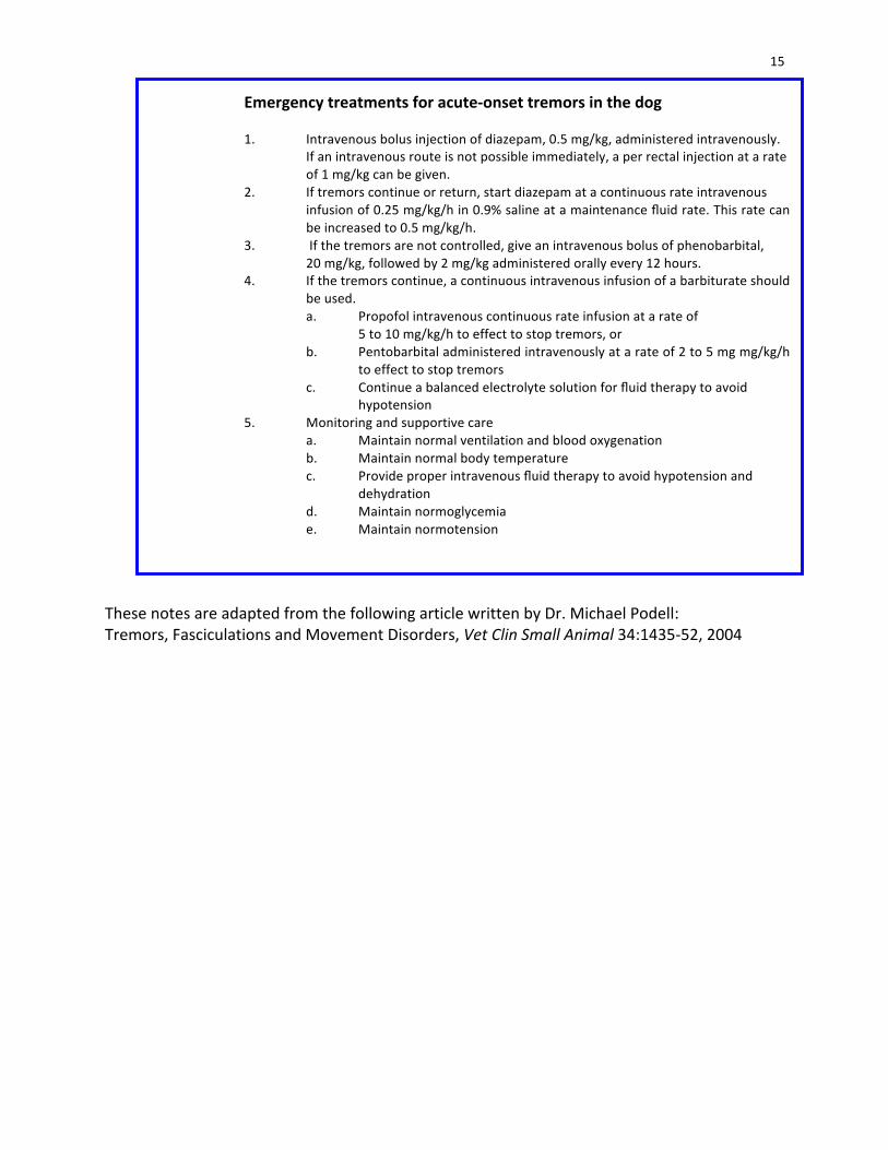
15 These notes are adapted from the following article written by Dr. Michael Podell: Tremors, Fasciculations and Movement Disorders, Vet Clin Small Animal 34:1435-‐52, 2004
Emergency treatments for acute-‐onset tremors in the dog 1. Intravenous bolus injection of diazepam, 0.5 mg/kg, administered intravenously. If an intravenous route is not possible immediately, a per rectal injection at a rate of 1 mg/kg can be given. 2. If tremors continue or return, start diazepam at a continuous rate intravenous infusion of 0.25 mg/kg/h in 0.9% saline at a maintenance fluid rate. This rate can be increased to 0.5 mg/kg/h. 3. If the tremors are not controlled, give an intravenous bolus of phenobarbital, 20 mg/kg, followed by 2 mg/kg administered orally every 12 hours. 4. If the tremors continue, a continuous intravenous infusion of a barbiturate should be used.
a. Propofol intravenous continuous rate infusion at a rate of 5 to 10 mg/kg/h to effect to stop tremors, or b. Pentobarbital administered intravenously at a rate of 2 to 5 mg mg/kg/h to effect to stop tremors c. Continue a balanced electrolyte solution for fluid therapy to avoid hypotension
5. Monitoring and supportive care a. Maintain normal ventilation and blood oxygenation b. Maintain normal body temperature c. Provide proper intravenous fluid therapy to avoid hypotension and dehydration d. Maintain normoglycemia e. Maintain normotension
Marc Vandevelde, Dr. med.vet. DECVN
• 1965-1971:Studiesveterinarymedicine,Univ.Gent,Belgium:Dr.med.vet.• 1972-1974:TrainingincomparativeneuropathologyattheBungeinstitute(Antwerp,Belgium)andinstituteofcomparativeneurology,UniversityofBern,Switzerland.• 1974-1979:AssistantprofessorforneuropathologyattheScott-Ritcheyfoundation,Auburnuniversi-tyUSA• 1979-1984:Assistantprofessorininstituteofcomparativeneurology,Univ.ofBern,Switzerland.Ha-bilitationandappointmentasPrivatdozent.• 1985:Appointmentasfullprofessorandheadoftheinstituteofanimalneurology,UniversityofBern• 1995:DiplomateoftheEuropeancollegeofveterinaryneurology• 1994-1996:Deanofthefacultyofveterinarymedicine,univ.ofBern• 2000-2005:Directorofthedepartmentofclinicalveterinarymedicine,univ.ofBern• 2006-2012:Chiefofneurologysection,teaching,research,diagnosticwork• 2012-:Profemeritusandparttimeassignementforresearchatthedivisionofneurologicalsciences,VetsuissefacultyBern.SpecialInterestPathogenesisofinfectiousdiseasesofthenervoussystem.Neuropathology,neurovirology,neuroimmunolo-gy. Memberships• DiplomateEuropeancollegeofveterinaryneurology• BoardofDirectors,EuropeanAssociationforVeterinarySpecialization• Swissneuropathologicalsociety• Worldassociationforneuropathology• EuropeanSocietyforVeterinaryNeurology(exPresident)• EditorialBoardActaNeuropathologica• EditorialboardTheveterinaryjournal.• EditorialboardAnn.Med.Vet.GrantsobtainedfromSwissNationalsciencefoundation,Swissfederalveterinaryoffice,SwissMultipleScle-rosisSociety,EuropeanUnion. Awards• HonorarymemberoftheItaliansocietyforveterinaryscience.• WSAVAinternationalawardforscientificachievement2004• Dr.honoriscausaoftheFacultyofveterinarymedicineofHannoverGermany2012PublicationsCa.300Publicationsinpeerreviewedjournals,bookchapters,books
Cranial trauma Marc Vandevelde, Dr. med.vet. DECVN

28.04.2014
1
Cranial Trauma Belgrad 2014
Prof. Dr. Marc Vandevelde DECVN Vetsuisse faculty Bern Switzerland
Cranial trauma • Very common • Caused by car accidents, falls, fights….. • A clear history is often lacking: “Animal found with neurological signs, the animal was normal a few hours ago…..”
Always think about trauma
Pathogenesis
Is very complex because: • Primary damage at the moment of impact
– Direct mechanical injury – Indirect mechanical injury
• Secondary damage in the following hours/days
Primary damage • Mechanical destruction • Rupture of blood vessels :
Subdural hemorrhage Intraperenchymal hemorrhage
Coup – contrecoup The brain is suspended in spinal fluid
Ventral
Trauma: Inertia will cause the brain to bounce against the skull
Dorsal
Coup-contre coup may induce complex lesion patterns
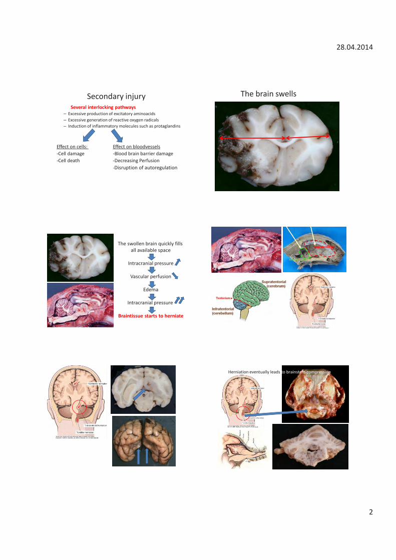
28.04.2014
2
Secondary injury Several interlocking pathways
– Excessive production of excitatory aminoacids – Excessive generation of reactive oxygen radicals – Induction of inflammatory molecules such as protaglandins
Effect on cells: Effect on bloodvessels -Cell damage -Blood brain barrier damage -Cell death -Decreasing Perfusion
-Disruption of autoregulation
The brain swells
The swollen brain quickly fills all available space
Intracranial pressure
Vascular perfusion
Edema
Intracranial pressure
Braintissue starts to herniate
infratentorial
supratentorial
Herniation eventually leads to brainstem compression
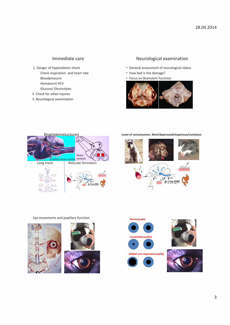
28.04.2014
3
Immediate care
1. Danger of hypovolemic shock Check respiration and heart rate Bloodpressure Hematocrit PCV Glucose/ Electrolytes 2. Check for other injuries 3. Neurological examination
Neurological examination
• General assessment of neurological status • How bad is the damage? • Focus on Brainstem function
Cranial nerve nuclei Reticular formation Long tracts
Motor. centres
Brainstemstuctures Level of consciousnes: Alert/depressed/stuporous/comatose
Eye movements and pupillary function Normal pupils
Constricted pupil(s)
Dilated non-responsive pupil(s)
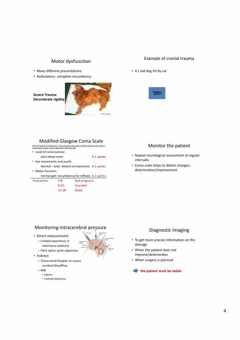
28.04.2014
4
Motor dysfunction
• Many different presentations • Ambulatory- complete recumbency
Vetmri.exblog.jp
Severe Trauma Decerebrate rigidity
Example of cranial trauma
• 4 y old dog hit by car
Modified Glasgow Coma Scale Platt SR, Radaelli ST, McDonnell JJ. The prognostic value of the modified Glasgow coma scale in
head trauma in dogs. J Vet Int Med 2001; 15(6):581–584.
• Level of conciousness alert-deep coma 6-1 points • Eye movements and pupils Normal – bilat. dilated unresponsive 6-1 points • Motor function: normal gait- recumbency/no reflexes 6-1 points Total points: 3-8: Bad prognosis 8-15: Guarded 15-18: Good
Monitor the patient
• Repeat neurological assessment at regular intervalls
• Coma scale helps to detect changes: deterioration/improvement
Monitoring intracerebral pressure • Direct measurement
– Limited experience in veterinary medicine – Fibre optics quite expensive
• Indirect – Transcranial Doppler to assess cerebral bloodflow – MRI
• edema • contrast behaviour
Diagnostic imaging
• To get more precise information on the damage
• When the patient does not improve/deteriorates
• When surgery is planned
the patient must be stable

28.04.2014
5
Diagnostic imaging Radiology -Rarely productive -Fractures,dislocations
Computed tomography -short examination time (spiral CT) -fractures, dislocations -acute (<24h) haemorrhage (subarachnoidal)
Magnetic resonance imaging -long examination time -very high soft tissue contrast; -morphology of brain including brain pathology -subacute /chronic intracerebral haemorrhage
Radiology of skull fractures
Courtesy Prof. J. Lang DECVDI Univ. Bern
CT Bone window Shows fractures nicely
CT Soft tissue window Shows hemorrhage
Sagital reconstruction
Courtesy Prof. J. Lang DECVDI Univ. Bern
Use of computerized tomography to detect traumatic lesions
Magnetic Resonance Imaging findings in Impression fracture and open wound after dog fight
Courtesy Prof. J. Lang DECVDI Univ. Bern
MRI Detection of Herniation
Treatment
• Place animal with head elevated • Provide oxygen (incubator/nasal tube) • Restore+maintain blood volume/ cerebral
blood volume, cerebral blood flow – Hypertonic salt solution – Colloids, cristalloids
• Combat brain swelling
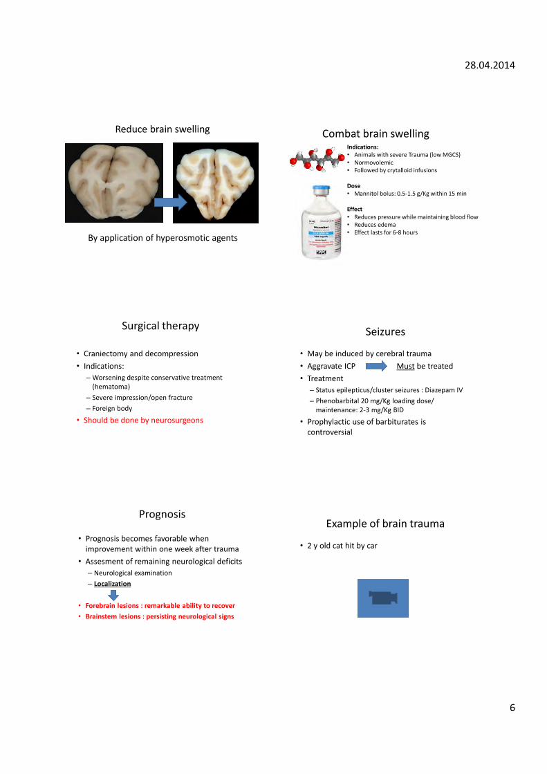
28.04.2014
6
Reduce brain swelling
By application of hyperosmotic agents
Combat brain swelling Indications: • Animals with severe Trauma (low MGCS) • Normovolemic • Followed by crytalloid infusions Dose • Mannitol bolus: 0.5-1.5 g/Kg within 15 min Effect • Reduces pressure while maintaining blood flow • Reduces edema • Effect lasts for 6-8 hours
Surgical therapy
• Craniectomy and decompression • Indications:
– Worsening despite conservative treatment (hematoma)
– Severe impression/open fracture – Foreign body
• Should be done by neurosurgeons
Seizures
• May be induced by cerebral trauma • Aggravate ICP Must be treated • Treatment
– Status epilepticus/cluster seizures : Diazepam IV – Phenobarbital 20 mg/Kg loading dose/
maintenance: 2-3 mg/Kg BID
• Prophylactic use of barbiturates is controversial
Prognosis
• Prognosis becomes favorable when improvement within one week after trauma
• Assesment of remaining neurological deficits – Neurological examination – Localization
• Forebrain lesions : remarkable ability to recover • Brainstem lesions : persisting neurological signs
Example of brain trauma
• 2 y old cat hit by car
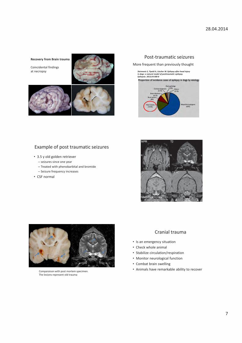
28.04.2014
7
Recovery from Brain trauma Coincidental findings at necropsy
Post-traumatic seizures More frequent than previously thought
Steinmetz S, Tipold A, Löscher W. Epilepsy after head injury in dogs: a natural model of posttraumatic epilepsy. Epilepsia. 2013;54:580-8
Example of post traumatic seizures
• 3.5 y old golden retriever – seizures since one year – Treated with phenobarbital and bromide – Seizure frequency increases
• CSF normal
MRI Multifocal lesions
Comparaison with post mortem specimen. The lesions represent old trauma
Cranial trauma
• Is an emergency situation • Check whole animal • Stabilize circulation/respiration • Monitor neurological function • Combat brain swelling • Animals have remarkable ability to recover
Degenerative diseases Marc Vandevelde, Dr. med.vet. DECVN
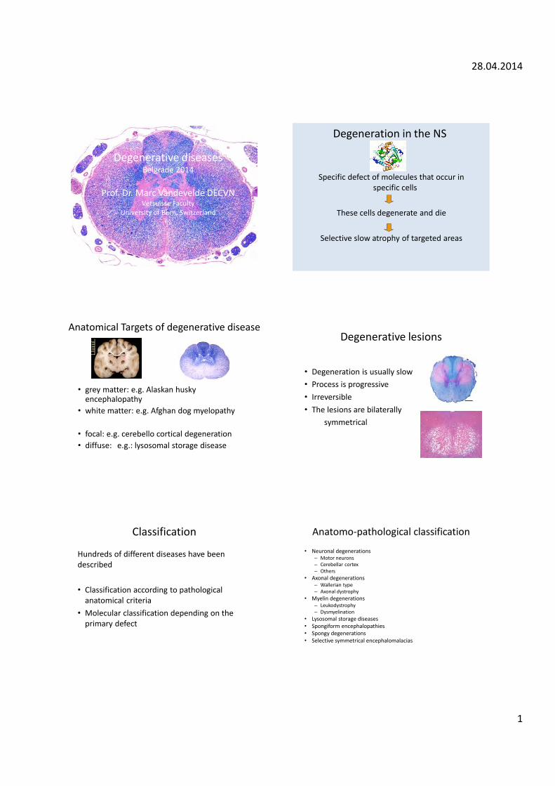
28.04.2014
1
Degenerative diseases Belgrade 2014
Prof. Dr. Marc Vandevelde DECVN
Vetsuisse Faculty University of Bern, Switzerland
Degeneration in the NS
Specific defect of molecules that occur in specific cells
These cells degenerate and die
Selective slow atrophy of targeted areas
Anatomical Targets of degenerative disease
• grey matter: e.g. Alaskan husky
encephalopathy • white matter: e.g. Afghan dog myelopathy
• focal: e.g. cerebello cortical degeneration • diffuse: e.g.: lysosomal storage disease
Degenerative lesions
• Degeneration is usually slow • Process is progressive • Irreversible • The lesions are bilaterally symmetrical
Classification
Hundreds of different diseases have been described • Classification according to pathological
anatomical criteria • Molecular classification depending on the
primary defect
Anatomo-pathological classification
• Neuronal degenerations – Motor neurons – Cerebellar cortex – Others
• Axonal degenerations – Wallerian type – Axonal dystrophy
• Myelin degenerations – Leukodystrophy – Dysmyelination
• Lysosomal storage diseases • Spongiform encephalopathies • Spongy degenerations • Selective symmetrical encephalomalacias

28.04.2014
2
Degenerative diseases general clinical aspects
• Juvenile animals (sometimes late onset) • Family history • Hereditary in specific breeds • Slowly progressive • Symmetrical signs • Neurological signs depend on localization: =Target of the degenerative process
Diagnosis • History, species, breed, genetic background • Neurological examination
– May reveal localisation – Hardly truly specific signs
• Diagnostic imaging – Detection of bilateral lesions – Distribution pattern
• Consult the lists of hereditary diseases • Molecular genetics: tests are increasingly
available
Degenerative diseases examples
• Neuronal degenerations – Cerebello-cortical degeneration – Neuronal vacuolation
• Lysosomal storage diseases – Ceroid lipofuscinosis
• Axonal diseases – Degenerative myelopathy
• Myelin diseases – Congenital tremor – Leukoencephalomyelopathy
• Spongy degenerations • Selective symmetrical encephalomalacias
Degenerative diseases
• Neuronal degenerations • Lysosomal storage diseases • Axonal diseases • Myelin diseases • Spongy degenerations • Selective symmetrical encephalomalacias
Motor neuron diseases Spinal Muscular Atrophy in: - Britanny Spaniels - Lapland dogs - Rottweilers - ……
of Neurons degenerate
In spinal cord and brainstem
Spinal Muscular Atrophy
Neurogenic Atrophy
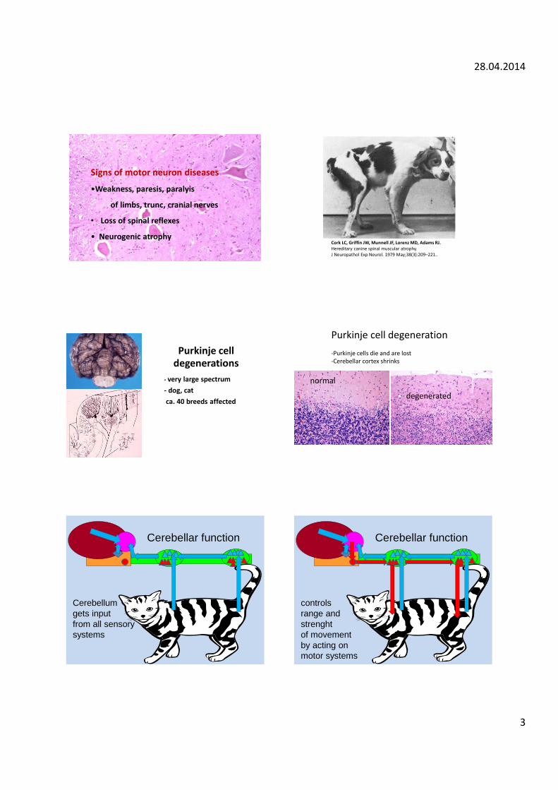
28.04.2014
3
Signs of motor neuron diseases
•Weakness, paresis, paralyis
of limbs, trunc, cranial nerves
• Loss of spinal reflexes
• Neurogenic atrophy Cork LC, Griffin JW, Munnell JF, Lorenz MD, Adams RJ. Hereditary canine spinal muscular atrophy. J Neuropathol Exp Neurol. 1979 May;38(3):209–221..
Purkinje cell degenerations
- very large spectrum - dog, cat ca. 40 breeds affected
Purkinje cell degeneration -Purkinje cells die and are lost -Cerebellar cortex shrinks
normal
degenerated
Cerebellar function
Cerebellum gets input from all sensory systems
Cerebellar function
controls range and strenght of movement by acting on motor systems
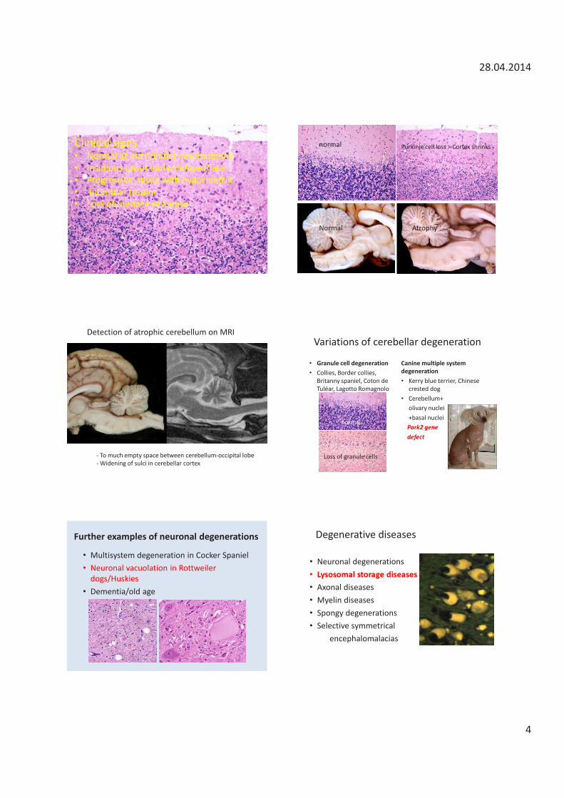
28.04.2014
4
Clinical signs • Normal at birth/initial development • Insidious onset early/delayed/late • Progressive ataxia with hypermetria • Intention tremor • Loss of menace response
Normal
normal Purkinje cell loss > Cortex shrinks
Atrophy
Detection of atrophic cerebellum on MRI
- To much empty space between cerebellum-occipital lobe - Widening of sulci in cerebellar cortex
Variations of cerebellar degeneration
• Granule cell degeneration • Collies, Border collies,
Britanny spaniel, Coton de Tuléar, Lagotto Romagnolo
Canine multiple system degeneration • Kerry blue terrier, Chinese
crested dog • Cerebellum+ olivary nuclei +basal nuclei Park2 gene defect
normal
Loss of granule cells
Further examples of neuronal degenerations
• Multisystem degeneration in Cocker Spaniel • Neuronal vacuolation in Rottweiler
dogs/Huskies • Dementia/old age
Degenerative diseases
• Neuronal degenerations • Lysosomal storage diseases • Axonal diseases • Myelin diseases • Spongy degenerations • Selective symmetrical encephalomalacias
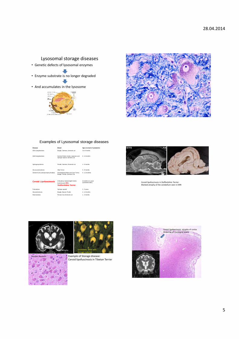
28.04.2014
5
Lysosomal storage diseases • Genetic defects of lysosomal enzymes
• Enzyme substrate is no longer degraded
• And accumulates in the lysosome
Disease Breed Age at onset of symptoms
GM1 Gangliosidosis Beagle, Siamese, domestic cat 3 to 6 months
GM2 Gangliosidosis German Pointer (male), Japanese and Springer Spanie; domestic cat
6 - 12 months
Sphingomyelinosis Poodle; Siamese, Domestic cat 4 - 6 months
Glucocerebrosidosis Silky Terrier 6 - 8 months
Globoid-Cell-Leukodystrophy (Krabbe) Westhighland White and Cairn Terrier, Beagle. Poodle; Domestic Cat
6 - 12 monbths
Ceroid Lipofuscinosis Chihuahua, Saluki,English Setter, Dachshound, Am. Staffordshire Terrier
6 months to 2 years (sometimes later)
Fukosidosis Springer spaniel 2 - 3 years
Glycoproteinosis Beagle, Basset, Poodle 2 - 12 months
Mannosidosis Persian Cat, Domestic cat :
1 - 3 months
Examples of Lysosomal storage diseases
Ceroid lipofuscinosis in Staffordshire Terrier: Marked atrophy of the cerebellum seen in MRI
Example of Storage disease: Ceroid lipofuscinosis in Tibetan Terrier
Brain Atrophy Lysosomes filled with Storage material
Swollen Neurons
Ceroid lipofuscinosis: atrophy of cortex Widening of meningeal space

28.04.2014
6
Degenerative diseases
• Neuronal degenerations • Lysosomal storage diseases • Axonal diseases • Myelin diseases • Spongy degenerations • Selective symmetrical encephalomalacias
Axonal Transport • Everything is produced in the cell body • Transportation along the axon often over long distances
A lot can go wrong
Clinical signs
• Insidious onset • Ataxia • Hindlimbs progressing to all limbs • Progressive • Proprioception but not nociception affected • In some conditions cerebellar signs
Axonal diseases
• Ataxia in smooth haired Fox/JR Terriers • Labrador retriever axonopathy • Degenerative myelopathy in Welsh Corgi-
Pembroke • Distal axononopathy in Birman cats • Neuroaxonal dystrophy in Rottweiler dogs • Degenerative myelopathy in large breed dogs • ……………..
German shepherd dog ; degenerative myelopathy: diffuse loss of axons (red areas) Degenerative Myelopathy in large breed
dogs
• Large breed dogs, >7 years • Slow onset and progression of paresis and
ataxia in hind limbs • Diffuse/dissiminated loss of axons in spinal
cord • Cause/pathogenesis?
– Hereditary factors/ SOD 1 mutations?
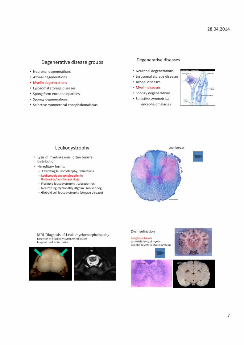
28.04.2014
7
Degenerative disease groups
• Neuronal degenerations • Axonal degenerations • Myelin degenerations • Lysosomal storage diseases • Spongiform encephalopathies • Spongy degenerations • Selective symmetrical encephalomalacias
Degenerative diseases
• Neuronal degenerations • Lysosomal storage diseases • Axonal diseases • Myelin diseases • Spongy degenerations • Selective symmetrical encephalomalacias
Leukodystrophy
• Lysis of myelin+axons, often bizarre distribution
• Hereditary forms: – Cavitating leukodystrophy, Dalmatians – Leukomyeloencephalopathy in
Rottweiler/Leonberger dogs – Fibrinoid leucodystrophy , Labrador ret. – Necrotizing myelopathy Afghan, Kooiker dog – Globoid cell leucodystrophy (storage disease)
Leonberger
MRI Diagnosis of Leukomyeloencephalopathy Detection of bilaterally symmetrical lesions In spinal cord white matter
Control
Dalmatian Chow chow
Dysmyelination Congenital tremor Lack/deficiency of myelin Genetic defects in Myelin proteins
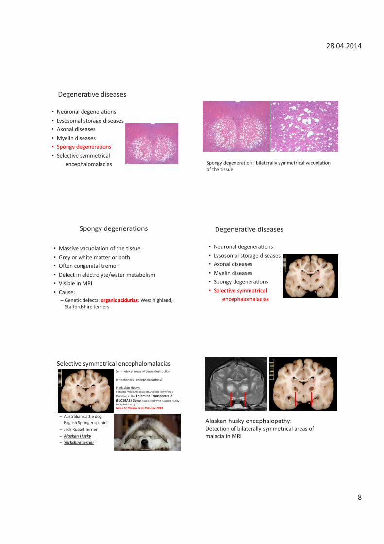
28.04.2014
8
Degenerative diseases
• Neuronal degenerations • Lysosomal storage diseases • Axonal diseases • Myelin diseases • Spongy degenerations • Selective symmetrical encephalomalacias Spongy degeneration : bilaterally symmetrical vacuolation
of the tissue
Spongy degenerations
• Massive vacuolation of the tissue • Grey or white matter or both • Often congenital tremor • Defect in electrolyte/water metabolism • Visible in MRI • Cause:
– Genetic defects: organic acidurias: West highland, Staffordshire terriers
Degenerative diseases
• Neuronal degenerations • Lysosomal storage diseases • Axonal diseases • Myelin diseases • Spongy degenerations • Selective symmetrical encephalomalacias
Selective symmetrical encephalomalacias
– Australian cattle dog – English Springer spaniel – Jack Russel Terrier – Alaskan Husky – Yorkshire terrier
Symmetrical areas of tissue destruction Mitochondrial encephalopathies? In Alaskan Husky: Genome-Wide Association Analysis Identifies a Mutation in the Thiamine Transporter 2 (SLC19A3) Gene Associated with Alaskan Husky Encephalopathy. Karen M. Vernau et al. Plos One 2013
Alaskan husky encephalopathy: Detection of bilaterally symmetrical areas of malacia in MRI

28.04.2014
9
Degenerative diseases
• Juvenile animals, sometimes late onset • Usually pure bred • Family history • Slowly progressive • Symmetrical • Normal CSF Get a post mortem/ save fresh frozen material
Inflammatory diseases Marc Vandevelde, Dr. med.vet. DECVN
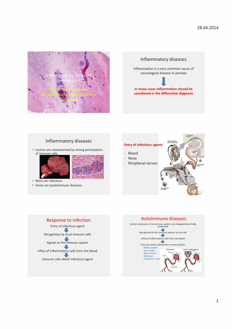
28.04.2014
1
Inflammatory diseases Belgrade 2014
Prof. Dr. Marc Vandevelde Vetsuisse Faculty, Universtity of Bern
Switzerland
Inflammatory diseases
Inflammation is a very common cause of neurological disease in animals
In many cases inflammation should be considered in the differential diagnosis
Inflammatory diseases
• Lesions are characterized by strong participation of immune cells
• Most are infectious • Some are (auto)immune diseases
Entry of infectious agents - Blood - Nose - Peripheral nerves
Response to infection Entry of infectious agent
Recognition by local immune cells
Signals to the immune system
Influx of inflammatory cells from the blood
Immune cells attack infectious agent
Autoimmune diseases Certain molecules in the nervous system are changed/abnormally
presented
Recognized by the immune-system as non-self
Influx of inflammatory cells from the blood
Immune system attacks the nervous system -White matter
-Grey matter -Blood vessels -Meninges -Peripheral nerves
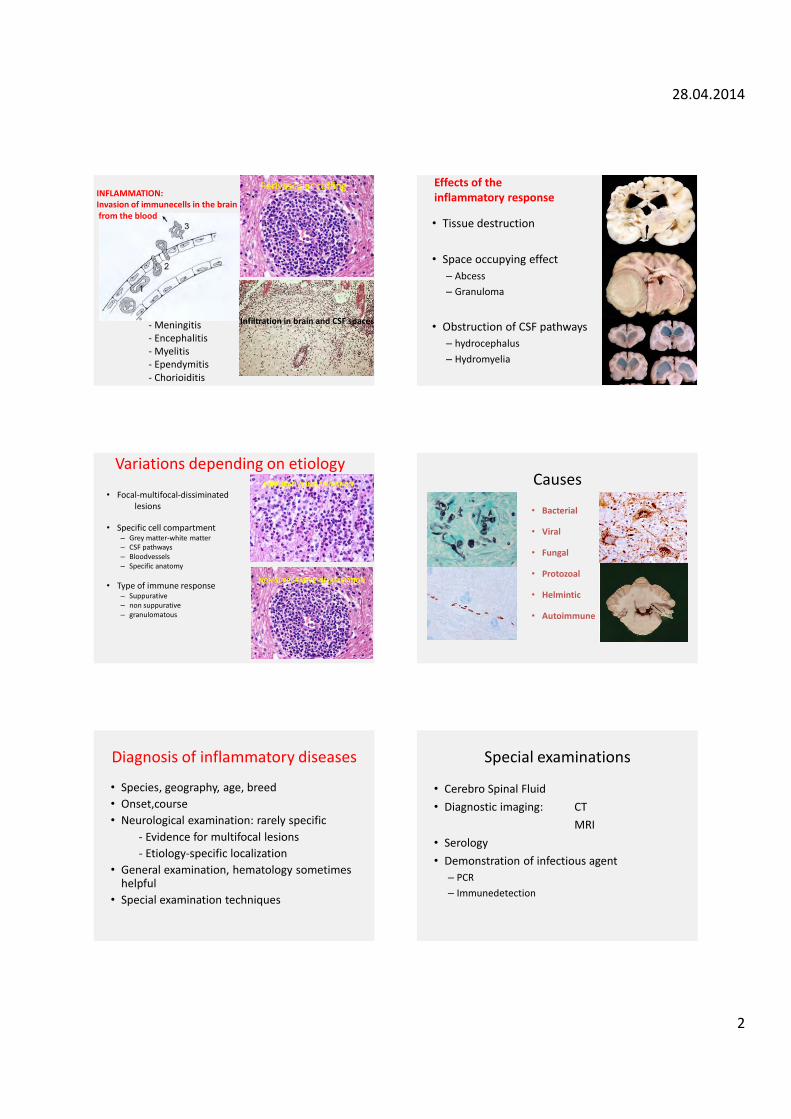
28.04.2014
2
INFLAMMATION: Invasion of immunecells in the brain from the blood
Perivascular cuffing
Infiltration in brain and CSF spaces - Meningitis - Encephalitis - Myelitis - Ependymitis - Chorioiditis
Effects of the inflammatory response
• Tissue destruction • Space occupying effect
– Abcess – Granuloma
• Obstruction of CSF pathways
– hydrocephalus – Hydromyelia
Variations depending on etiology
• Focal-multifocal-dissiminated lesions • Specific cell compartment
– Grey matter-white matter – CSF pathways – Bloodvessels – Specific anatomy
• Type of immune response
– Suppurative – non suppurative – granulomatous
SUPPURATIVE INFLAMMATION
NON-SUPPURATIVE INFLAMMATION
Causes
• Bacterial
• Viral
• Fungal
• Protozoal
• Helmintic
• Autoimmune
Diagnosis of inflammatory diseases
• Species, geography, age, breed • Onset,course • Neurological examination: rarely specific - Evidence for multifocal lesions - Etiology-specific localization • General examination, hematology sometimes
helpful • Special examination techniques
Special examinations
• Cerebro Spinal Fluid • Diagnostic imaging: CT MRI • Serology • Demonstration of infectious agent
– PCR – Immunedetection
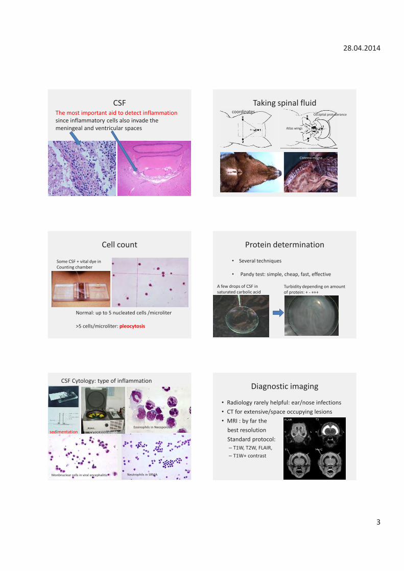
28.04.2014
3
CSF The most important aid to detect inflammation since inflammatory cells also invade the meningeal and ventricular spaces
Taking spinal fluid
Cisterna magna
coordinates
Atlas wings
Occipital protuberance
Cell count
Normal: up to 5 nucleated cells /microliter >5 cells/microliter: pleocytosis
Some CSF + vital dye in Counting chamber
Protein determination
• Several techniques • Pandy test: simple, cheap, fast, effective
A few drops of CSF in saturated carbolic acid
Turbidity depending on amount of protein: + - +++
sedimentation cytocentrifuge
CSF Cytology: type of inflammation
Eosinophils in Neosporosis
Mononuclear cells in viral encephalitis Neutrophils in SRMA
Diagnostic imaging
• Radiology rarely helpful: ear/nose infections • CT for extensive/space occupying lesions • MRI : by far the best resolution Standard protocol:
– T1W, T2W, FLAIR, – T1W+ contrast
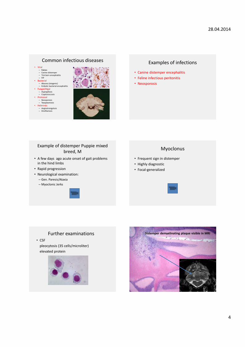
28.04.2014
4
Common infectious diseases • Viral
– Rabies – Canine distemper – Tick born encephalitis – FIP
• Bacterial – Abscess (otogenic) – Embolic bacterial encephalitis
• Fungal/Algal – Aspergillosis – Cryptococcosis
• Protozoal – Neosporosis – Toxoplasmosis
• Helmintic – Angiostrongylosis – Dirofilariosis
Examples of infections
• Canine distemper encephalitis • Feline infectious peritonitis • Neosporosis
Example of distemper Puppie mixed breed, M
• A few days ago acute onset of gait problems in the hind limbs
• Rapid progression • Neurological examination:
– Gen. Paresis/Ataxia – Myoclonic Jerks
Myoclonus
• Frequent sign in distemper • Highly diagnostic • Focal-generalized
Further examinations • CSF pleocytosis (35 cells/microliter) elevated protein
Distemper demyelinating plaque visible in MRI
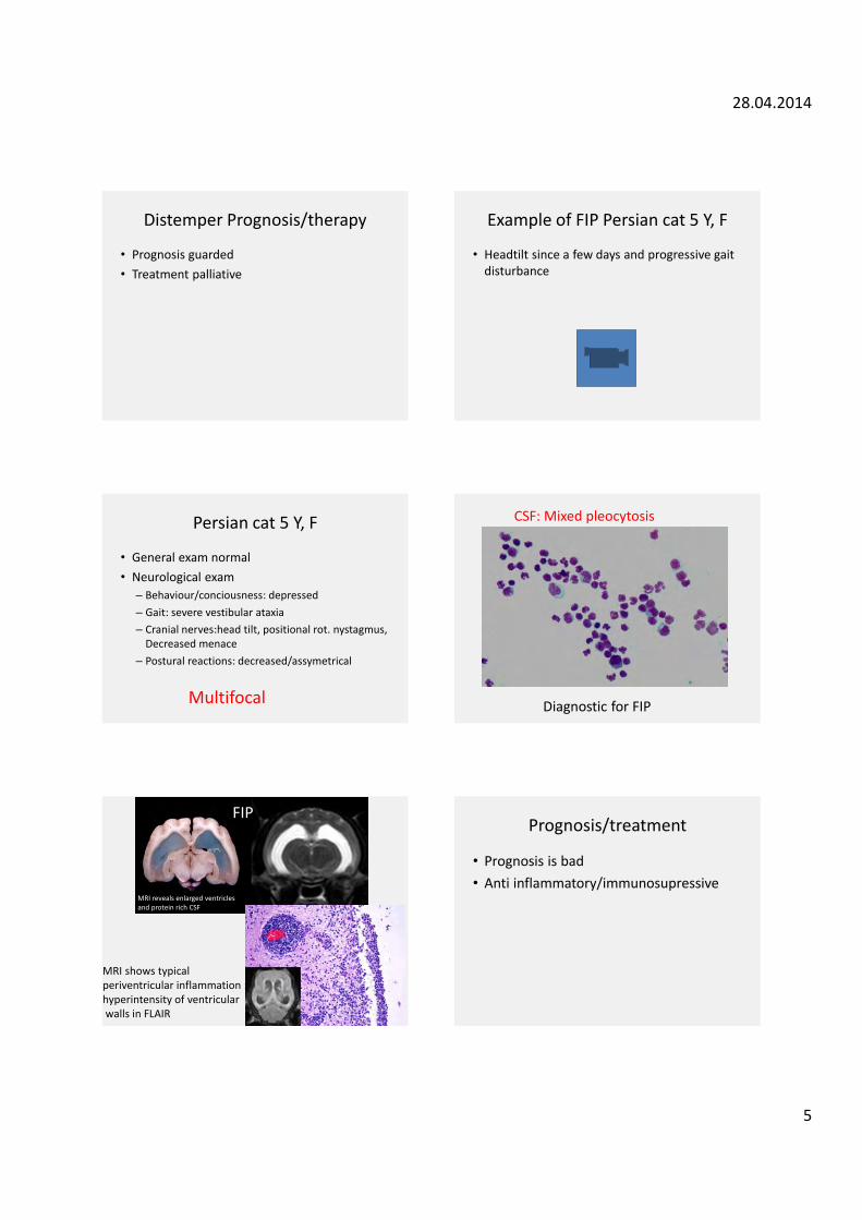
28.04.2014
5
Distemper Prognosis/therapy
• Prognosis guarded • Treatment palliative
Example of FIP Persian cat 5 Y, F
• Headtilt since a few days and progressive gait disturbance
Persian cat 5 Y, F
• General exam normal • Neurological exam
– Behaviour/conciousness: depressed – Gait: severe vestibular ataxia – Cranial nerves:head tilt, positional rot. nystagmus,
Decreased menace – Postural reactions: decreased/assymetrical
Multifocal
CSF: Mixed pleocytosis
Diagnostic for FIP
MRI reveals enlarged ventricles and protein rich CSF
MRI shows typical periventricular inflammation : hyperintensity of ventricular walls in FLAIR
FIP Prognosis/treatment
• Prognosis is bad • Anti inflammatory/immunosupressive

28.04.2014
6
Other infectious diseases
• Bacterial – Metastatic from other organs – Traumatic – Extension from ear or nose
• Fungal • Protozoal • Parasitic
T1+ C
Example of bacterial infection Dog with brainstem abscess resulting from Middle/Inner ear infection (Bulla filled with material)
Xray: Detection of Otitis media as underlying cause of brain abscess
Example of fungal infection: Mutliple granulomas in Aspergillosis
Protozoal infection
• Neospora caninum • In juvenile dogs CNS + PNS and muscle
Example of young boxer with paresis and painfull atrophic muscles
(rare) example of toxoplasmosis segmental myelitis in a cat the lesion was nicely visible in MRI
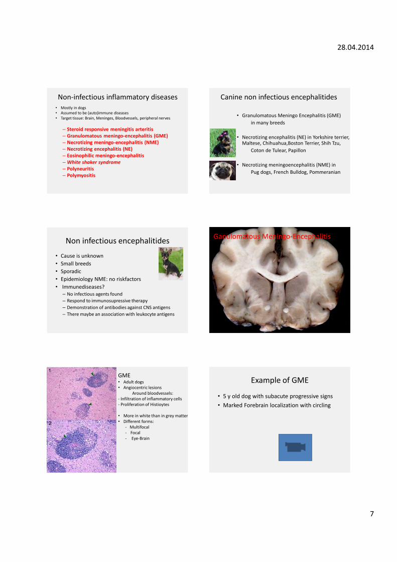
28.04.2014
7
Non-infectious inflammatory diseases • Mostly in dogs • Assumed to be (auto)immune diseases • Target tissue: Brain, Meninges, Bloodvessels, peripheral nerves
– Steroid responsive meningitis arteritis – Granulomatous meningo-encephalitis (GME) – Necrotizing meningo-encephalitis (NME) – Necrotizing encephalitis (NE) – Eosinophilic meningo-encephalitis – White shaker syndrome – Polyneuritis – Polymyositis
Canine non infectious encephalitides
• Granulomatous Meningo Encephalitis (GME) in many breeds • Necrotizing encephalitis (NE) in Yorkshire terrier,
Maltese, Chihuahua,Boston Terrier, Shih Tzu, Coton de Tulear, Papillon • Necrotizing meningoencephalitis (NME) in Pug dogs, French Bulldog, Pommeranian
Non infectious encephalitides
• Cause is unknown • Small breeds • Sporadic • Epidemiology NME: no riskfactors • Immunediseases?
– No infectious agents found – Respond to immunosupressive therapy – Demonstration of antibodies against CNS antigens – There maybe an association with leukocyte antigens
Ganulomatous Meningo-Encephalitis Adults (
GME • Adult dogs • Angiocentric lesions Around bloodvessels: - Infiltration of inflammatory cells - Proliferation of Histioytes • More in white than in grey matter • Different forms:
- Multifocal - Focal - Eye-Brain
Example of GME
• 5 y old dog with subacute progressive signs • Marked Forebrain localization with circling
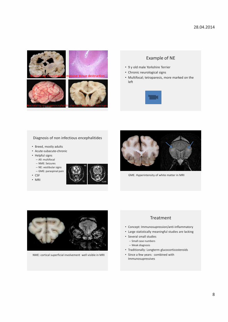
28.04.2014
8
Necrotising Encephalitis: massive tissue destruction
Necrotizing meningoencephalitis:also involvement of meninges
Example of NE
• 9 y old male Yorkshire Terrier • Chronic neurological signs • Multifocal; tetraparesis, more marked on the
left
Diagnosis of non infectious encephalitides
• Breed, mostly adults • Acute-subacute-chronic • Helpful signs
– All: multifocal – NME: Seizures – NE: vestibular signs – GME: paraspinal pain
• CSF • MRI T2 FLAIR
NE
GME. Hyperintensity of white matter in MRI
NME: cortical superficial involvement well visible in MRI
Treatment
• Concept: Immunosupression/anti-inflammatory • Large statistically meaningful studies are lacking • Several small studies
– Small case numbers – Weak diagnosis
• Traditionally: Longterm glucocorticosteroids • Since a few years: combined with
Immunosupressives
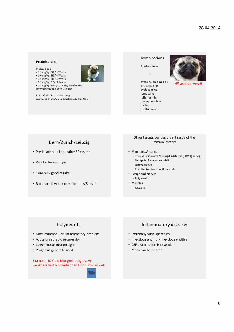
28.04.2014
9
Prednisolone Prednisolone • 1·5 mg/kg BID/ 3 Weeks • 1·0 mg/kg BID/ 6 Weeks • 0·5 mg/kg BID/ 3 Weeks • 0·5 mg/kg QD/ 3 Weeks • 0·5 mg/kg every other day indefinitely (eventuelly reducing to 0.25 mg) L. R. Talarico & S J. Schatzberg Journal of Small Animal Practice. 51, 146,2010
Kombinations Prednisolone + cytosine arabinoside procarbazine cyclosporine, lomustine leflunomide mycophenolate mofetil azathioprine
All seem to work!!
Bern/Zürich/Leipzig
• Prednisolone + Lomustine 50mg/m2
• Regular hematology
• Generelly good results
• But also a few bad complications(Sepsis)
Other targets besides brain tisssue of the immune system
• Meninges/Arteries: – Steroid Responsive Meningitis Arteritis (SRMA) in dogs – Neckpain, fever, neutrophilia – Diagnosis: CSF – Effective treatment with steroids
• Peripheral Nerves – Polyneuritis
• Muscles – Myositis
Polyneuritis
• Most common PNS inflammatory problem • Acute onset rapid progression • Lower motor neuron signs • Prognosis generally good Example: 10 Y old Mongrel, progressive weakness first hindlimbs then frontlimbs as well
Inflammatory diseases
• Extremely wide spectrum • Infectious and non-infectious entities • CSF examination is essential • Many can be treated
Holger Volk DVM PGCAP DipECVN PhD FHEA MRCVS
Holger graduated from the Veterinary School Hanover in 2001 followed by a PhD in Neuropharmacology. He then completed his clinical training at the Royal Veterinary College (RVC) and was then awarded the Diploma of the European College of Veterinary Neurology. He is currently Clinical Director of the RVC’s small animal referral hospital, Professor and Head of Veterinary Neurology & Neurosurgery at the RVC and Vicepresident of the European College of Veterinary Neurology.

Canine and feline epilepsyHolger Volk DVM PGCAP DipECVN PhD FHEA MRCVS

09/05/2014
1
Sh
aring passions, shaping fu
tures
Canine and feline epilepsy
Holger A. Volk DVM, DipECVN, PhD, FHEA, MRCVS Clinical Director & Professor of Veterinary Neurology & Neurosurgery Royal Veterinary College
“Angst”
Complex simplicity The tools
Your Brain
Neurological examina;on History
1. Define the problem 2. Define the system and loca;on 3. Define the lesion
The path
Lesion
Loca*on
System
Problem
Diagnostic Tests
Treatment Plan
DIAGNOSIS
Presenting Complaint
History & Physical Exam & NEURO EXAM
DeCine & ReCine PROBLEM
Differential Diagnoses
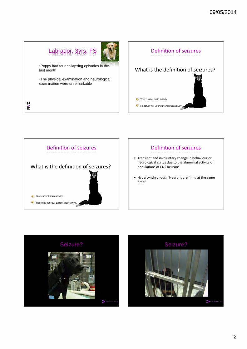
09/05/2014
2
Labrador, 3yrs, FS
¥ Poppy had four collapsing episodes in the last month
¥ The physical examination and neurological examination were unremarkable
Defini;on of seizures
What is the defini;on of seizures?
Your current brain activity
Hopefully not your current brain activity
Defini;on of seizures
What is the defini;on of seizures?
Your current brain activity
Hopefully not your current brain activity
Defini;on of seizures
• Transient and involuntary change in behaviour or neurological status due to the abnormal ac;vity of popula;ons of CNS neurons
• Hypersynchronous: “Neurons are firing at the same ;me”
Seizure? Seizure?

09/05/2014
3
Seizure?
¥ Mutation in BCAN -> – brevican (Member of hyaluronan- binding chondroitin
sulfate proteoglycan family) – important in organisation of nodes of Ranvier
¥ 17/56 homozygous but not affected
Parallel Mapping and Simultaneous Sequencing RevealsDeletions in BCAN and FAM83H Associated with DiscreteInherited Disorders in a Domestic Dog BreedOliver P. Forman1*, Jacques Penderis2., Claudia Hartley1., Louisa J. Hayward1, Sally L. Ricketts1,
Cathryn S. Mellersh1
1 Kennel Club Genetics Centre, Animal Health Trust, Kentford, United Kingdom, 2 The School of Veterinary Medicine, University of Glasgow, Glasgow, United Kingdom
Abstract
The domestic dog (Canis familiaris) segregates more naturally-occurring diseases and phenotypic variation than any otherspecies and has become established as an unparalled model with which to study the genetics of inherited traits. We used agenome-wide association study (GWAS) and targeted resequencing of DNA from just five dogs to simultaneously map andidentify mutations for two distinct inherited disorders that both affect a single breed, the Cavalier King Charles Spaniel. Weinvestigated episodic falling (EF), a paroxysmal exertion-induced dyskinesia, alongside the phenotypically distinct conditioncongenital keratoconjunctivitis sicca and ichthyosiform dermatosis (CKCSID), commonly known as dry eye curly coatsyndrome. EF is characterised by episodes of exercise-induced muscular hypertonicity and abnormal posturing, usuallyoccurring after exercise or periods of excitement. CKCSID is a congenital disorder that manifests as a rough coat present atbirth, with keratoconjunctivitis sicca apparent on eyelid opening at 10–14 days, followed by hyperkeratinisation of footpadsand distortion of nails that develops over the next few months. We undertook a GWAS with 31 EF cases, 23 CKCSID cases,and a common set of 38 controls and identified statistically associated signals for EF and CKCSID on chromosome 7 (Praw1.9610214; Pgenome = 1.061025) and chromosome 13 (Praw 1.2610217; Pgenome = 1.061025), respectively. We resequencedboth the EF and CKCSID disease-associated regions in just five dogs and identified a 15,724 bp deletion spanning threeexons of BCAN associated with EF and a single base-pair exonic deletion in FAM83H associated with CKCSID. Neither BCAN orFAM83H have been associated with equivalent disease phenotypes in any other species, thus demonstrating the ability touse the domestic dog to study the genetic basis of more than one disease simultaneously in a single breed and to identifymultiple novel candidate genes in parallel.
Citation: Forman OP, Penderis J, Hartley C, Hayward LJ, Ricketts SL, et al. (2012) Parallel Mapping and Simultaneous Sequencing Reveals Deletions in BCAN andFAM83H Associated with Discrete Inherited Disorders in a Domestic Dog Breed. PLoS Genet 8(1): e1002462. doi:10.1371/journal.pgen.1002462
Editor: Tosso Leeb, University of Bern, Switzerland
Received July 12, 2011; Accepted November 21, 2011; Published January 12, 2012
Copyright: � 2012 Forman et al. This is an open-access article distributed under the terms of the Creative Commons Attribution License, which permitsunrestricted use, distribution, and reproduction in any medium, provided the original author and source are credited.
Funding: The work was funded by the Waltham Foundation, Grant Number: 2010-004 (http://www.waltham.com/foundation.htm); the Kennel Club CharitableTrust (http://www.thekennelclub.org.uk/charitabletrust); and the Tezmae Charitable Trust. The funders had no role in study design, data collection and analysis,decision to publish, or preparation of the manuscript.
Competing Interests: The authors have declared that no competing interests exist.
* E-mail: [email protected]
. These authors contributed equally to this work.
Introduction
The domestic dog is well documented as being an excellent
model for studying the genetics of both simple and complex traits
[1–3]. The purebred dog population comprises several hundred
distinct breeds that usually originate from small numbers of
founding individuals, with each breed representing an isolated and
relatively homogenous closed breeding population. Within breeds
there is strong selection for desired phenotypic and behavioural
characteristics, and dogs that possess these desired characteristics
are often bred with extensively. The resulting population structure
of individual breeds can result in the propagation of many
spontaneously occurring deleterious mutations within a breed and
these can exist at high frequencies, with the possibility of multiple
inherited disorders arising within a single breed. A large
proportion of these disorders show a Mendelian autosomal
recessive mode of inheritance. The Staffordshire Bull Terrier
and Labrador Retriever are two of the many examples of breeds
that are affected by at least two autosomal recessive disorders for
which the causal mutations have been identified; hereditary
cataract and the metabolic disorder L-2-hydroxyglutaric aciduria
in the Staffordshire Bull Terrier [4–5] and exercise induced
collapse and centronuclear myopathy in the Labrador Retriever
[6–7]. The high levels of homogeneity and long linkage
disequilibrium that are characteristic of the genomes of most
purebred dog breeds means that canine disease traits can be
mapped using much smaller numbers of cases and controls and far
more modest numbers of genetic markers than would be required
to map an equivalent disease in outbred human populations [8–9].
Episodic falling (EF) in the Cavalier King Charles Spaniel
(CKCS) was first reported in 1983, although the condition has
been recognised in the breed since at least the early 1960s [10]. EF
(also known within the breed as ‘‘sudden collapse’’, muscle
hypertonicity and hyperekplexia) is a paroxysmal exertion-induced
dyskinesia that is usually exercise, excitement or stress-induced and
is characterised by muscular hypertonicity and abnormal postur-
PLoS Genetics | www.plosgenetics.org 1 January 2012 | Volume 8 | Issue 1 | e1002462
UN
CO
RR
EC
TE
D P
RO
OF
1A canine
BCANmicrode
letionassoci
ated with episodic falling
syndrome
2Jennife
r L.
Q1
Gilla , Kate
L. Tsaib , Chris
ta Kreyc , Rook
sana E. Noorai
b , Jean-Franço
is Vanbellinghe
nd,
3Lauren
t S. Garosi
e , G. Diane Shelto
nf , Leigh
AnneClark
b,⁎, Robert J. H
arveya,⁎⁎
4a Depar
tment ofPharm
acology, The
Schoolof Pha
rmacy, 29-39 Brunsw
ick Square, Lond
on WC1N 1AX, UK
5b Depar
tment ofGeneti
cs andBioche
mistry, College
of Agricultur
e, Forestry, a
nd Life Sciences, 100
JordanHall, C
lemson University, C
lemson, South Carolin
a 29634-0318,
USA
6c 55 Bruce
Road,Levin
5510,New Zealan
d
7d Biolog
ie Moléculaire, Ins
titut de Pathol
ogie et de Génétique ASBL,
25 Avenue George
s Lemaître, B-6041
Gosselies, Be
lgium
8e Davies
Veterinary Specia
lists, Manor F
arm Business Park
, Higham Gobion
, Hertfordshi
re, UK
9f Depar
tment ofPathol
ogy, Univers
ity of California,
San Diego,La Jolla, C
A 92093-0709,
USA
10
11
a b s t r a c t
a r t i c l e i n f o
12Article
history:
13Receiv
ed 7 May 2011
14Revise
d 28 June 2011
15Accep
ted 20 July 2011
16Availa
ble onlinexxxx
171819
20Keywords:
21Brevic
an
22BCAN
23Episod
ic fallingsyndro
me
24Cavali
er King Charle
s spaniels
25Microde
letion
26
Episodic falli
ng syndrome (EFS)
is a canine paroxy
smal hypertoni
city disorder foun
d in Cavalier Kin
g Charles
27
spaniels. Episod
es are triggered by exerci
se, stressor excite
ment and characterized
by progressive
28
hypertonicity
throughout t
he thoracic and pelvic
limbs, resulting
in a characteristic
'deer-stalking
' position
29
and/orcollap
se. We used a genome-wide associ
ationstrateg
y to map the EFS locusto a 3.48 Mb critica
l30
interval on canine
chromosome 7. By priorit
izing candidate genes
on the basis of biolo
gical plausibi
lity, we
31
foundthat a
15.7 kb deletion in BCAN,
encoding the bra
in-specific ex
tracellular m
atrix proteoglycan
brevican,
32
is associated
with EFS. This repr
esentsa compelling
causalmutation
for EFS, since
brevican has an
essential
33
role in the formationof perine
uronalnets govern
ing synapse stabili
ty and nervecondu
ctionvelocit
y.34
Mappingof the deletio
n breakpoint enable
d the development of Multiple
x PCR and Multiplex Ligatio
n-35
dependent P
robe Amplification (MLPA) g
enotyping tests t
hat can accura
tely distinguish normal, car
rier and
36
affected animals. W
ider testing of a larger
population of CKC
S dogs withouta history
of EFSfrom the USA
37
revealed that ca
rriersare ex
tremely common (12.9%). The
development of
molecular gen
etic tests for
the EFS
38
microdeletion
will allow the im
plementation of dire
cted breeding pro
grams aimed at minimizing the num
ber39
of animals with EFS and enable
confirmatory diagno
sis andpharm
acotherapy of affe
cted dogs.40
© 2011 Published by Elsevie
r Inc.4142
43
44
45Introd
uction
46Episod
ic fallingsyndro
me (EFS)is a well-rec
ognized paroxy
smal
47disord
er foundin Cavali
er King Charles spanie
ls (CKCS). Epis
odes
48begin
between fourteen weeks a
nd four years of a
ge andare tri
ggered
49by exe
rcise, stress, a
pprehension
or excitement (H
errtage and Palmer,
501983)
. Episodes are of variab
le frequency and severi
ty but are
51charac
terizedby progre
ssivehypert
onicityinvolv
ing thoracic and
52pelvic
limbs (Fig. 1a) until t
he dogs are ultimately immobilized in a
53charac
teristic'deer-s
talking' or 'pr
aying'positio
n (Fig. 1b). Sti
ffening
54of all f
our limbs dur
ing exercise can cause
falling(Fig. 1
c), although
55there is no loss of
consciousne
ss or cyanosi
s. Other clinic
al signs may
56includ
e facialmuscle
stiffness, stu
mbling,a 'bunny
-hopping' g
ait,
57archin
g of theback or voc
alization. Cu
riously, betw
een episodes, dog
s
58appea
r to be completelynormal neu
rologically. Sp
ontaneous ac
tivity
59
was not observed in muscle
electrodiagno
stic testing, ruling
out60
myotonia congen
ita (Wright et al., 1
986, 1987).
Muscle biopsies are
61
typically normal at the light
microscopic level,
excluding many
62
congenital m
yopathies. Ho
wever, EFS has been linked
to ultrastruc-
63
tural defects
in skeletal muscle includ
ing dilatation and prolife
ration64
of thesarcop
lasmic reticulum, mitocho
ndrialswelling
and degener-
65
ation(Wright
et al., 1986,1987)
. EFS has also beencompared
66
(Rusbridge,
2005)with startle
disease/hyp
erekplexia,
typically
67
characterized
by noise-or tou
ch-evoked neona
tal hypertoni
city due68
to defects in inhibit
ory glycine recept
or (GLRA1, G
LRB; Shiang
et al.,69
1993;Rees e
t al., 2002) o
r glycine transp
orter GlyT2 (SLC6A
5) genes
70
(Reeset al.,
2006;Harve
y et al.,2008)
. However, a
microdeletion
in71
the GlyT2gene in Irish Wolfhou
nds results in
severeneona
tal muscle72
stiffness and
tremor in response to handli
ng (Gill et al., 2
011), which
73
is inconsistent
with the observed clinica
l signsof EFS
. Comparisons
74
with startlediseas
e may have been made because affecte
d dogs often
75
respond well to the benzo
diazepine clonaz
epam(Garos
i et al., 2002
),76
an effective antico
nvulsant, anx
iolyticand muscle relaxa
nt thatis the
77
most effective
known treatment for
human hyperekplexi
a (Thomas et
78
al., 2010). H
owever, the carbon
ic anhydrase
inhibitor ace
tazolamide,
79
used to treat certain
types of human episod
ic ataxia (Tom
linsonet al.,
80
2009)and hyperk
alemic periodic paraly
sis (Matthews and Hanna
,
Neurobiolog
y of Disease xxx (2011
) xxx–xxx
⁎ Corresponde
nce to: L.A. C
lark, Departm
ent ofGenet
ics andBioche
mistry, College
of
Agriculture,
Forestry, and
Life Sciences, 100
JordanHall, C
lemson University, C
lemson,
SouthCarolin
a 29634-0318,
USA.
E-mail addresses:
u (L.A. Clark),
robert.harve
.uk
(R.J. Harvey)
.
Available online
on ScienceDirec
t (www.sciencedirec
t.com).
YNBDI-02479
; No. of page
s: 7; 4C:
0969-9961/$
– see front matter ©
2011 Published by Elsevie
r Inc.
doi:10.1016/
j.nbd.2011.07
.014
Contents lists availab
le at ScienceDir
ect
Neurobiolog
y of Disease
j ourna l ho
mepage: www.e lse
v ie r.com/ loca
te /ynbd i
Pleasecite this article
as: Gill, J.L.,
et al.,A canine
BCANmicrode
letionassoci
ated with episodic falling
syndrome, Neu
robiol.Dis. (2
011),
doi:10.1016/
j.nbd.2011.07
.014
Startle disease in Irish wolfhounds associated with a microdeletion in the glycinetransporter GlyT2 gene
Jennifer L. Gill a, Deborah Capper b, Jean-François Vanbellinghen c, Seo-Kyung Chung d, Robert J. Higgins e,Mark I. Rees d, G. Diane Shelton f,⁎, Robert J. Harvey a,⁎⁎a Department of Pharmacology, The School of Pharmacy, 29–39 Brunswick Square, London WC1N 1AX, UKb Halsey East Animal Clinic, 16057 NE Halsey Street, Portland, OR 97230, USAc Secteur Commun de Biologie Moléculaire–Unilab-Lg, Centre Hospitalier Universitaire de Liège, 4000 Liège, Belgiumd Institute of Life Science, College of Medicine, Swansea University, Swansea SA2 8PP, UKe Department of Pathology, Microbiology and Immunology, University of California, Davis, CA 95616, USAf Department of Pathology, University of California, San Diego, La Jolla, CA 92093–0709, USA
a b s t r a c ta r t i c l e i n f o
Article history:Received 23 December 2010Revised 1 March 2011Accepted 11 March 2011Available online 17 March 2011
Keywords:Glycine transporterGlyT2HyperekplexiaIrish wolfhoundSLC6A5Startle disease
Defects in glycinergic synaptic transmission in humans, cattle, and rodents result in an exaggerated startlereflex and hypertonia in response to either acoustic or tactile stimuli. Molecular genetic studies havedetermined that mutations in the genes encoding the postsynaptic glycine receptor (GlyR) α1 and β subunits(GLRA1 and GLRB) and the presynaptic glycine transporter GlyT2 (SLC6A5) are the major cause of thesedisorders. Here, we report the first genetically confirmed canine cases of startle disease. A litter of seven Irishwolfhounds was identified in which two puppies developed muscle stiffness and tremor in response tohandling. Although sequencing of GLRA1 and GLRB did not reveal any pathogenic mutations, analysis of SLC6A5revealed a homozygous 4.2 kbmicrodeletion encompassing exons 2 and 3 in both affected animals. This resultsin the loss of part of the large cytoplasmic N-terminus and all subsequent transmembrane domains due to aframeshift. This genetic lesion was confirmed by defining the deletion breakpoint, Southern blotting, andmultiplex ligation-dependent probe amplification (MLPA). This analysis enabled the development of a rapidgenotyping test that revealed heterozygosity for the deletion in the dam and sire and three other siblings,confirming recessive inheritance. Wider testing of related animals has identified a total of 13 carriers of theSLC6A5 deletion as well as non-carrier animals. These findings will inform future breeding strategies andenable a rational pharmacotherapy of this new canine disorder.
© 2011 Elsevier Inc. All rights reserved.
Introduction
Hyperekplexia or startle disease affects newborn children andis characterized by noise- or touch-induced non-epileptic seizuresthat result in muscle stiffness and apnea (suspension of breathing).Although a rare orphan disease (defined as b200,000 affectedindividuals worldwide), this disorder can have serious consequences,including brain damage and/or sudden infant death (Harvey et al.,2008). The primary cause of startle disease is defective inhibitoryglycinergic transmission due to missense, nonsense, or frameshiftmutations in the glycine receptor (GlyR) α1 gene (GLRA1) (Shianget al., 1993, 1995; Chung et al., 2010), although large deletions of
exons 1–7 have also been reported (Brune et al., 1996; Becker et al.,2006). Mutations are also found in the genes encoding the GlyR βsubunit gene (GLRB; Rees et al., 2002) and the presynaptic glycinetransporter GlyT2 (SLC6A5) (Rees et al., 2006; Eulenburg et al., 2006).Animal disorders similar to startle disease/hyperekplexia have beenreported in cattle (Harper et al., 1986; Gundlach, 1990; Gundlachet al., 1988; Lummis et al., 1990; Pierce et al., 2001; Charlier et al.,2008; Gill et al., 2011), horses (Gundlach et al., 1993), and dogs(Fox et al., 1984), but only in certain cases has the underlying geneticlesion been definitively identified (Table 1). For example, a nonsensemutation (c.C156A, p.Y24X) in exon 2 of GLRA1 causes inheritedcongenital myoclonus (ICM) in Poll Hereford calves (Pierce et al.,2001), while a missense mutation (c.T809C, p.L270P) in exon 4 ofSLC6A5 causes congenital muscular dystonia 2 (CMD2) in Belgian Bluecattle (Charlier et al., 2008;Gill et al., 2011). The identificationof commoncausal genes links these animal disorders to human startle disease/hyperekplexia. Two other related disorders—inherited myoclonus inPeruvian Paso horses (Gundlach et al., 1993) and familial reflexmyoclonus in Labrador retrievers (Fox et al., 1984)—are reminiscent ofdeficits in glycinergic transmission, although investigations into the
Neurobiology of Disease 43 (2011) 184–189
⁎ Correspondence to: G.D. Shelton, Department of Pathology, University of California,San Diego, La Jolla, CA 92093–0709, USA. Fax: +1 858 534 0391.⁎⁎ Correspondence to: R.J. Harvey, Department of Pharmacology, The School ofPharmacy, 29–39 Brunswick Square, London WC1N 1AX, UK. Fax: +44 207 753 5902.
E-mail addresses: [email protected] (G.D. Shelton),[email protected] (R.J. Harvey).
Available online on ScienceDirect (www.sciencedirect.com).
0969-9961/$ – see front matter © 2011 Elsevier Inc. All rights reserved.doi:10.1016/j.nbd.2011.03.010
Contents lists available at ScienceDirect
Neurobiology of Disease
j ou rna l homepage: www.e lsev ie r.com/ locate /ynbd i
partum that was evoked by handling and ceased when the animalswere relaxed or sleeping. The puppies were unable to stand andshowed a typical rigid extended posture in all four limbs. Cyanosiswas notedwhile the dogswere suckling or during tube feeding. Due tothe progression of clinical signs, feeding difficulty, and developmentof lung congestion, the dogs were humanely euthanized at approx-imately 11 weeks of age and full necropsies were performed.
Histopathology and histochemistry
Both puppies were small for their age with weights of 1446 and1559 g, while the normal littermates (n=5) averaged 2642 g, rangingfrom 2495 to 2778 g. The primary abnormality on gross examinationof both animals was consolidated (neither floated in formalin) andhemorrhagic lungs, with the male puppy more severely affected thanthe female. The esophagus in eachof the puppieswasnoticeably dilated,more so in the female than the male. No macroscopic abnormalitiesof the brain and spinal cord were noted. At the light microscopiclevel, moderate interstitial pneumonia was noted in both puppies. Nosignificant pathological abnormalities were noted in the brain orspinal cord of either animal. All muscles appeared to have developednormally with myofiber size appropriate for the age of the puppiesand a normal mosaic pattern of muscle fiber types. Intramuscularnerve branches were normal in appearance. There was no evidence ofa primary muscle or nerve disease in any of the muscle specimens.
Candidate gene analysis—GLRA1, GLRB, and SLC6A5
Given the similarity of clinical signs to startle disease, primerswere designed to amplify exons and flanking splice donor, acceptor,and branch-point sites for the major causative genes, GLRA1, GLRB,and SLC6A5, deriving gene structures in silico using the UCSC GenomeBrowser (http://genome.ucsc.edu/). PCR amplification (for primers,see Table S1) using proofreading Pfx DNA polymerase and direct
sequencing revealed surprisingly few exonic variants in GLRA1 andGLRB. No SNPs were detected in GLRA1, and only two homozygousSNPs were detected in GLRB: T1380A (p.P438P, dbSNP: rs22406580)and c.G1226A causing a p.C385Y substitution in the mature GlyR βsubunit M3–M4 intracellular loop. However, both these variants werealso found in unaffected littermates, ruling out a pathogenic role.Variation was also absent from exons 1 and 4–16 of the SLC6A5 gene.However, we consistently failed to amplify SLC6A5 exons 2 and 3 withseveral different primer sets from genomic DNA isolated from the twoaffected dogs. Since control experiments revealed that these primersets were functional in PCRs using template DNAs from the unaffectedlittermates and dam (Fig. 1), we suspected that a microdeletionencompassing exons 2 and 3 could explain this anomaly. Toinvestigate this possibility, we used long-distance PCR to amplifythe genomic region encompassing SLC6A5 exons 1–4 generating afragment predicted to be 8.2 kb in size. Although we failed to amplifythis fragment from control DNAs (presumably due to the highly GC-rich exon 2) in the two affected dogs, the unaffected dam, and threeof the unaffected littermates, we observed robust PCR productsmigrating at 4 kb, while the two remaining unaffected littermates hadno PCR products in this size range. These results suggested that thetwo affected dogs were homozygous for a microdeletion of 4.2 kb,while the dam and littermates 1, 4, and 5 were heterozygous for thisdeletion. Littermates 2 and 3 would be predicted to be homozygouswild-type due to the absence of the 4 kb deletion fragment.
Mapping the SLC6A5 deletion breakpoint
In order to confirm this deletion and determine the exactbreakpoint in the deleted allele, we cloned and sequenced the 4 kbdeletion fragment. This analysis revealed that the deletion spannedboth exons 2 and 3, starting 32 bp upstream of exon 2 and ending504 bp downstream of the end of exon 3 (Figs. 1 and 2). The presenceof a dinucleotide (gt) repeat at the breakpoint is relevant since
Fig. 1. PCR assays reveal that a SLC6A5 microdeletion is associated with startle disease in Irish wolfhounds. (A) Photograph of one of two affected puppies demonstrating lateralrecumbency and an extended posture (see Video S1 online). (B) PCR panels for SLC6A5 exon 2 (754 bp), exon 3 (513 bp), and a large genomic fragment encompassing exons 1–4(target size, 8.2 kb). Note that exon 2 and exon 3 fragments fail to amplify from the affected dogs (samples and 1 and 2,−/−) but can be robustly amplified using genomic DNA fromthe dam and five other siblings. Although the 8.2 kb exon 1–4 fragment was not amplified from wild-type animals or carriers (due to the large size and GC-rich nature of theamplicon), a 4 kb PCR product was observed in both affected dogs (−/−), the dam, and three of the littermates (sibs 1, 4, and 5—all likely to have a+/− genotype). (C) Since exons 1and 4–16 were amplified from affected animals, these results strongly suggest that a microdeletion encompassing SLC6A5 exons 2 and 3 is the genetic cause of this startle disorder,with the two remaining animals (sibs 2 and 3) being homozygous for the wild-type allele (+/+).
186 J.L. Gill et al. / Neurobiology of Disease 43 (2011) 184–189
Seizure? Seizure?
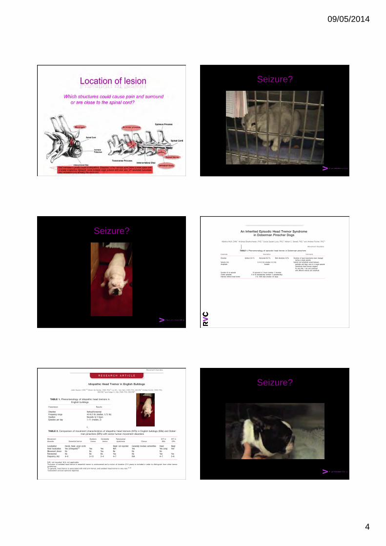
09/05/2014
4
Seizure?
Seizure? An Inherited Episodic Head Tremor Syndromein Doberman Pinscher Dogs
Martina Wolf, DVM,1 Andreas Bruehschwein, PhD,2 Carola Sauter-Louis, PhD,3 Adrian C. Sewell, PhD,4 and Andrea Fischer, PhD1*
1Clinic of Small Animal Medicine, Center of Veterinary Clinical Sciences, Ludwig-Maximilians-University of Munich, Munich, Germany2Clinic of Small Animal Surgery and Reproduction, Center of Veterinary Clinical Sciences,
Ludwig-Maximilians-University of Munich, Munich, Germany3Center of Veterinary Clinical Sciences, Ludwig-Maximilians-University of Munich, Munich, Germany
4Department of Pediatrics, University Children’s Hospital, Frankfurt, Germany
ABSTRACT: Episodic head tremor anecdotallyoccurs in the Doberman pinscher dog breed, but it is notdescribed in sufficient detail in the literature. We eval-uated 87 Doberman pinschers affected with episodichead tremor and appropriate controls. The data analyzedwere collected through detailed questionnaires, elaboratetelephone interviews, and video recordings. Affecteddogs underwent clinical, neurological, and laboratory ex-amination, and a detailed diagnostic workup was con-ducted in 5 affected dogs. Pedigrees of affected dogswere collected and reviewed. The affected dogsexpressed individual phenotypes of either horizontal orvertical head movements, but rarely did a dog exhibithead movements in both directions. There was consider-able variation in duration (10 seconds to 3 hours; median:3 minutes), frequency of occurrence (1–20 episodes/day;median: 2/day) of head tremor and length of the periodwithout head tremor (1–1,800 days; median: 60 days).
Subtle dystonic posturing of the head and neck duringhead tremor was evident on video recordings of 5 dogs.Certain exceptional conditions such as illness, surgery,some medications, heat, pseudopregnancy, or pregnancytriggered episodes. Two main important forms of episodichead tremor were identified: a familial early-onset form(age < 1 year) that affected littermates and a sporadicform. Affected dogs were traced back to 1 common sire,also including sporadic cases. Episodic head tremor isan inherited, paroxysmal movement disorder that affectsthe Doberman pinscher breed. Identification of the caus-ative genes in the future will allow us to obtain a moredetailed description of the syndrome. VC 2011 Movement
Disorder Society
Key Words: movement disorder; head tremor; dog;genetic
Episodic head tremor (HT) is anecdotally observedin the Doberman pinscher dog (DP) breed, as well asin Boxers and bulldogs.1 A disturbance in the stretch-
reflex mechanism has been suggested and compared toessential tremor.2 A detailed description of this syn-drome is missing. Therefore, this study aimed todescribe the phenomenology, the clinical course andfamily history of episodic HT in DPs to gain insightsinto the possible mode of inheritance of this syndromeand determine whether dogs can be classified asaffected or unaffected for genetic studies.
Subjects and Methods
Dog owners were invited to participate throughannouncements placed on the clinic’s homepage andon internet forums (2009–2010). Doberman pinscherswith at least 2 episodes of HT (cases) and DPs >5years of age without any observed episode of HT(controls) were selected for this study. Dogs with ahistory of head trauma, or lack of owner compliancewere excluded. Each owner filled out a detailed
------------------------------------------------------------Additional Supporting Information may be found in the online version ofthis article.
*Correspondence to: Dr. Andrea Fischer, Section of Neurology, Clinic ofSmall Animal Medicine, Ludwig-Maximilians University of Munich,Veterinarstr. 13, 80539 Munich, Germany; [email protected], [email protected]
Funding agencies: Clinics of Small Animal Medicine (blood analysis,electrodiagnostics, anesthesia) and Clinic of Small Animal Surgery andReproduction (MR imaging), Ludwig-Maximilians University of Munich,Munich, Germany.Relevant conflicts of interest/financial disclosures: M. Wolf was for 6months in a position funded by a grant provided by Intervet DeutschlandGmbH, Unterschleißheim, Germany.Full financial disclosures and author roles may be found in the onlineversion of this article.
Received: 25 February 2011; Revised: 29 July 2011; Accepted: 2 Au-gust 2011Published online in Wiley Online Library (wileyonlinelibrary.com).DOI: 10.1002/mds.23936
R E S E A R C H A R T I C L E
Movement Disorders, Vol. 000, No. 000, 0000 1
questionnaire. The information collected was verifiedthrough telephone interviews and a review of 29 vid-eos depicting the HT episodes. The velocity of HTwas measured by running 17 videos in slow motion.Affected dogs underwent neurological examination(71/87) with special focus on the vestibular system,and a laboratory evaluation (71/87), which includedhematology, biochemistry, urinalysis, and liver func-tion tests (pre- and postprandial bile acids; 40/87; fast-ing venous ammonia concentration; 8/87), venousblood gases (pH, base excess, lactate; 34/87), and thy-roid hormone status (T4, fT4, TSH; 28/87). A detaileddiagnostic evaluation, approved by the institutionalreview board, was carried out for 5 DPs with episodicHT: complete electrodiagnostic evaluation, radio-graphs of the cervical spine, magnetic resonance imag-ing (MRI) of the neurocranium and cervical spine,analysis of cerebrospinal fluid, and neurometabolicscreening of urine and blood were conducted (seeSupporting Appendix 1). In addition, polymerasechain reaction (PCR) to detect canine distemper virus(Institute of Virology, University of Gießen, Gießen,Germany) and genetic testing for narcolepsy weredone (Laboklin, Bad Kissingen, Germany). Thepedigrees of the dogs affected by episodic HT wereexamined, if available (60/87). A chi-square test wasused to analyze the differences in proportions of theclustered data. P values < .05 were consideredstatistically significant. The confidence interval was setat 95%.
Results
Study Population
Eighty-seven affected Doberman pinscher dogs(including 3 litters, each with 2–4 affected dogs) wereincluded in the study. Other dogs were excluded dueto lack of owner compliance (n ¼ 34). The majorityof the affected dogs were registered with Germanbreeding clubs (54/87, 62.1%). Slightly more males(49/87, 56.3%; 95%CI: 45.9–66.3%) than females(38/87, 43.7%; 95%CI: 33.7–54.2%) were found tobe affected by the syndrome (P ¼ .014 in comparisonwith control group).
Phenomenology of Episodic HT
Data are summarized in Table 1.
Interruptibility and Behavior
In 66.7% of the dogs (58/87), the owners reportedthat they could consistently interrupt each HT epi-sode. In some cases, stroking the dogs, talking tothem, or asking them to get up was sufficient to inter-rupt the episode (12/87; 13.8%). In other cases, stron-ger stimuli (favorite toys or snacks, cheering them on,taking them for a walk) were needed (71/87; 81.6%).Only 4 dogs (4.6%) did not respond to any of theaforementioned stimuli. The ability to interrupt epi-sodes appeared to be limited by an increase in the ve-locity and amplitude of the HT, the long duration ofan episode, and the occurrence of HT in conjunctionwith some exceptional conditions (described below).Dogs were awake (58/87; 66.7%), or dozing (29/87;33.3% when HT started. Many owners described theemotional state of their dogs at the beginning of anepisode as relaxed (39/87; 44.8%). During episodes,dogs could move in a normal manner and did not ex-hibit ataxia, even when climbing stairs or running. Allof the dogs were fully responsive during HT (Videos1, 2, and 3) and no autonomic signs were observed.Some owners (23/87; 26.4%) reported that during epi-sodes, the dogs felt uneasy, showed signs of irritation,were anxious, tried to hide, were in need of affection,stared, barked, yawned, and/or tried to stop the headmovements with their paws or by pressing their headagainst an object. After the episode, some dogs (18/87; 20.7%) were exhausted, tired, restless, ashamed,calm, or in need of affection. None of the dogsshowed nystagmus, limb tremor, or truncal sway dur-ing or between HT episodes. In 5 videos subtle dys-tonic posturing (head tilt) was observed.
Posture
When HT started, 28 dogs (32.2%) were lying, 3were sitting, 3 were standing, and 1 dog was walking.For the remaining dogs, the owners could not refer toa specific posture. Before the episodes started, 8 own-ers (9.2%) reported that the head and neck of their
TABLE 1. Phenomenology of episodic head tremor in Doberman pinschers
Parameter Description Comments
Direction Vertical 24.1% Horizontal 66.7% Both directions 9.2% Direction of head movements never changedduring a single episode.
Velocity (Hz) 5.4–8.3 Hz (median: 6.5 Hz) Velocity and amplitude varied betweenepisodes and dogs, even in a single episode.Sometimes head tremors stoppedfor less than 1 sec and continuedwith different velocity and amplitude
Amplitude Variable
Duration of an episode 10 seconds to 3 hours (median: 3 minutes)Cluster episodes 0 to 20 episodes/day (median: 2 episodes/day)Intervals without head tremor 0 to 1800 days (median: 60 days)
W O L F E T A L .
2 Movement Disorders, Vol. 000, No. 000, 0000
dogs were in an abnormal position. Furthermore, HTdid not stop when the dogs laid down their heads; itonly made the movements less prominent.
Age of Onset
Onset of episodic HT is summarized in Figure 1.Episodic head tremor onset could not be reliablyassessed in 20 dogs.
Clinical Course
The variations observed in the clinical courses of ep-isodic HT are summarized in Table 2. Some owners(32/87; 36.8%) mentioned conditions such as stress,fatigue, overstimulation, and begging that seemed totrigger HT. Specifically, HT occurred more frequentlyand with higher velocity and amplitude, or evenappeared only when the dogs were either sick, recov-ering from surgery, received certain medications (car-profen, meloxicam, or amitraz), were in heat,pseudopregnant, or pregnant. This was observed in46.7% of the dogs (21/45) that experienced 1 or moreof these exceptional conditions. Only 2 dog ownerscould provoke HT in their dogs consistently by exces-sive exercise or excitement.
Examinations
Neurologic examinations were unremarkable besidesataxia of the hind limbs without forelimb ataxia in 4dogs. Lesions were localized to the cervical (3/4) orlumbar spinal cord/cauda equina (1/4) based on cervi-cal MRI findings in 1 dog and appearance of spinalreflexes and the presence of lumbar or cervical pain inthe others. Low T4 hormone concentrations (<1.0 lg/dL; canine reference range 1.0–4.7 lg/dL) were docu-mented in similar proportions in cases (7/28; 25.0%)and controls (P > .05).3 All the other laboratoryexaminations were unremarkable.
Medical History
A high proportion of dogs with episodic HT (59/87;67.8%) exhibited compulsive stereotypic behavior,which is similar to the proportion found in the controlgroup (33/56; 58.9%; P > .05). Most commonlynamed were flank and paw sucking (35/59; controls:8/33; P ¼ 0.001), tail hunting and running in circles(35/59; controls: 18/33; P > .05), and licking objectssuch as blankets (17/59; controls: 8/33; P > .05).Moreover some dogs exhibited pica (6/59; controls: 7/33; P > .05) or excessive abnormal behavior, such asbarking (19/59; controls: 8/33; P > .05). Other
FIG. 1. Age of onset of episodic head tremor in Doberman pinschers with complete medical history (n 5 67).
TABLE 2. Clinical course of episodic head tremor in 87 Doberman pinchers
Subtype Age of onset Affected littermates n %
Early onsetEarly onset, familial form <1 y Yes 11 12.6Early onset, unknown family history <1 y Unknown 29 33.3
Adult onset 1–5 y Unknown 17 19.6Sporadic formLate onset >5 y Unknown 4 4.6Only in association with exceptional conditions Variable Unknown 6 6.9
Not classified Unknown Unknown 20 23.0Total 87 100.0
H E A D T R E M O R I N D O B E R M A N P I N S C H E R S
Movement Disorders, Vol. 000, No. 000, 0000 3
Idiopathic Head Tremor in English Bulldogs
Julien Guevar, DVM,1,2 Steven De Decker, DVM, PhD,2 Luc M.L. Van Ham, DVM, PhD, DECVN,3 Andrea Fischer, DVM, PhD,DECVN,4 and Holger A. Volk, DVM, PhD, DECVN2*
1School of Veterinary Medicine, College of Medical, Veterinary and Life Sciences, University of Glasgow, Glasgow, UK2Department of Clinical Science and Services, Royal Veterinary College, University of London, Hatfield, England
3Department of Small Animal Medicine and Clinical Biology, Faculty of Veterinary Medicine, Ghent University, Merelbeke, Belgium4Clinic of Small Animal Medicine, Center of Veterinary Sciences, Ludwig-Maximillians-University of Munich, Munich, Germany
ABSTRACT: Idiopathic head tremor (IHT) syn-drome is a recognized but poorly characterized move-ment disorder in English bulldogs (EBs). The dataanalyzed were collected via a detailed online question-naire and video recordings. Thirty-eight percent of thepopulation demonstrated IHT. The first presentation wasearly in life. There was no sex or neutered status predis-position. The condition disappeared with time in 50% ofthe cases. The direction of the head movement was ver-tical or horizontal. The number of episodes per day andthe duration of the episodes were greatly variable. The
majority of episodes occurred at rest. Most of the epi-
sodes were unpredictable. And there was no alteration
of the mental status for most dogs during the episodes.
Stress has been reported as a suspected trigger factor.
IHT in EBs can be considered an idiopathic paroxysmal
movement disorder. VC 2013 International Parkinson and
Movement Disorder Society
Key Words: movement disorder; dog; bulldog; headtremor
Canine idiopathic head tremor (IHT) is a poorlyunderstood breed-specific tremor condition of thehead affecting Doberman pinschers1 and anecdotallyEnglish bulldogs (EBs) and boxers.2 A recent study inDoberman pinschers characterized IHT as a suddenonset of vertical and/or horizontal rhythmic headmovements lasting from a few seconds to severalhours.1 However, there remains a lack of data aboutthis condition in other dog breeds. The aim of the cur-rent study was to phenotypically characterize IHT inthe EB to identify potential risk factors and to com-pare this breed-specific canine disorder with similarcanine and human movement disorders.
Materials and Methods
This study was approved by the Ethics and WelfareCommittee of the Royal Veterinary College, Universityof London, England (approval number URN201100039H). EB owners and breeders were contactedand invited to complete a detailed questionnaire avail-able online and to provide video recordings of the epi-sodes and pedigree information of their dogs. Someowners were contacted by e-mail or telephone for dataverification. This questionnaire was designed by theprimary author and a board-certified veterinary neurol-ogist (H.A.V.). Video recordings of the episodes werealso requested and analyzed by reviewing the events inslow motion. It was decided that each responder couldenter only 1 affected dog. The Mann-Whitney U testor Fisher’s exact test was used to compare groups. AP< 0.05 was considered statistically significant.
Results
Study Population
Two hundred thirty-four EB owners participated inthe study. Eighty-nine EBs had reported episodes of
------------------------------------------------------------Additional Supporting Information may be found in the online version ofthis article.
*Correspondence to: Dr. Holger A. Volk, Department of Clinical Scienceand Services, Royal Veterinary College, University of London, Hatfield,England; Email: [email protected]
Relevant conflicts of interest/financial disclosures: Nothing to report.Full financial disclosures and author roles may be found in the onlineversion of this article.
Received: 11 September 2013; Revised: 27 October 2013; Accepted:6 November 2013
Published online 00 Month 2013 in Wiley Online Library(wileyonlinelibrary.com). DOI: 10.1002/mds.25767
R E S E A R C H A R T I C L E
Movement Disorders, Vol. 00, No. 00, 2013 1
IHT and were considered the affected patient popula-tion. This group consisted of 41 male (54%; 95% CI,44%-64%) and 48 female (46%; 95% CI, 36%-56%)dogs between 5 months and 12 years of age (median, 4years) and weighing between 12 and 40 kg (median, 26kg). Forty-one of the 89 dogs (46%; 95% CI, 36%-56%) were neutered, whereas 48 (54%; 95% CI,44%-64%) were sexually intact animals. The remain-ing 145 dogs had not demonstrated signs suggestive ofIHT at the time of the questionnaire and served as thecontrol group for the affected animal population. Thislast group consisted of 62 females (43%; 95% CI,35%-51%) and 83 males (57%; 95% CI, 49%-65%).Eighty-four dogs (58%; 95% CI, 55%-66%) were sex-ually intact, and 61 (42%; 95% CI, 34%-50%) wereneutered. They were aged between 5 months and 12years (median, 3 years) and weighed between 8 and 55kg (median, 26 kg). There was no significant differencein sex (P5 0.10) or neuter status (P5 0.58) betweenclinically affected and control dogs.
Age of Onset
The reported age of onset ranged from 6 months to9 years (median, 2 years; 0.5–9 years), which wasyounger than the age of the control group (median, 3years; 0.5–12 years; P50.01). Fifty-three dogs (60%;95% CI, 50%-70%) had their first reported IHT epi-sode before they were 3 years old, with 2 years themost common reported age of onset (27% of dogs;95% CI, 18%-36%).
Occurrence of Events
Episodes of IHT occurred predominantly at rest(n5 68 dogs; 76%; 95% CI, 67%-85%) and duringsleep (n537; 42%; 95% CI, 32%-52%). Fewer than10% of owners noticed the condition during exercise(n5 8; 9.0%; 95% CI, 3%-15%). Most owners didnot think there was a link between a specific period inthe day and the occurrence of IHT episodes (n555,62%; 95% CI, 52%-72%) or a seasonal influence(n5 79; 89%; 95% CI, 83%-96%). For most dogs,no specific stimuli were identified before an episode ofIHT occurred (n577; 87%; 95% CI, 81%-95%), butsome owners mentioned stressful events (n5 6; 7%;95% CI, 2%-12%), exercise (n53, 3%; 95% CI,0%-7%), and whelping (n53; 3%; 95% CI, 0%-7%)as a possible trigger. Intact females did not seem todemonstrate IHT episodes more often around theirestrus cycle, but could demonstrate more IHT episodeswhen they had a litter (12 of 29 intact female dogswith a litter, 41%; 95% CI, 23%-59%).
Characterization of Episode
Almost none of the owners were able to predict theoccurrence of IHT episodes (n5 85; 96%; 95% CI,
78%-92%). Most affected dogs were considered nor-mal before (n559; 66%; 95% CI, 56%-76%) andimmediately after (n5 62; 70%; 95% CI, 61%-80%)the IHT episode. In some affected dogs, behavioralchanges were observed before an IHT episode occurred(alone or combined: sleepiness [n5 18; 20%], staring[n5 13; 15%], disorientation [n57; 8%], agitated[n5 1; 1%], fearful [n5 1; 1%], sniffing [n5 3; 3%]).None of the owners reported any behavioral changeafter the episode. During the episodes of IHT, bodyposture stayed normal in 86% of dogs (n576; 95%CI, 82%-96%), but a more floppy (n5 3, 3%; 95%CI, 0%-7%) or stiff (n510, 11%; 95% CI, 5%-18%)posture was also reported. None of the dogs showedautonomic clinical signs prior, during, or after theevent. Affected dogs were reported to be conscious andaware of their surroundings during IHT episodes.Although not included in the questionnaire, severalowners also commented that distractions or treatswere generally sufficient to alter or stop the episodes.Video recordings of 7 affected dogs in their home
environment were assessed in slow motion. The fre-quency of the tremors was between 4.88 and 6.17 Hz(median, 5.75 Hz) with variable amplitudes. The lattervaried between episodes. Occasionally, the IHT epi-sodes stopped for a few seconds before continuingwith a similar frequency. The general direction waseither horizontal (n5 2) or vertical (n55). Durationranged from a few seconds to 3 hours, and the fre-quency of observed IHT episodes varied from 1 to 11episodes a day (median, 2.0 episodes/day; Table 1).
Clinical Progression
The frequency of occurrence of the episodes wasreported to be extremely variable and appeared random(ie, “daily,” “weekly,” “several times per week,” “every2 months,” “clusters every 6 months,” “once a year”).The tremors disappeared spontaneously in 44 affecteddogs (49%; 95% CI, 39%-59%). None of the dogs wereeuthanized by the owners because of IHT. Ninety-ninepercent (n588) of the affected population was not onany treatment. One dog received calcium supplements.
Discussion
This is the first study evaluating the phenomenologyand the clinical course of IHT in EBs The latter
TABLE 1. Phenomenology of idiopathic head tremors inEnglish bulldogs
Parameters Results
Direction Vertical/horizontalFrequency range 4.8-6.2 Hz (median, 5.75 Hz)Duration Seconds to 3 hoursEpisodes per day 1–11 (median, 2)
G U E V A R E T A L
2 Movement Disorders, Vol. 00, No. 00, 2013
appears to be an idiopathic, paroxysmal, benignmovement disorder characterized by episodic headtremors with an early onset in life, with spontaneousresolution in nearly half of the population. No risk ortrigger factors were identified in this study.The affected dogs remained responsive and conscious
during the episodes. None of the dogs had autonomicclinical signs or displayed altered behavior after the epi-sodes. None of the included patients received antiepi-leptic medication. Although no drug studies have beenpublished, it has been suggested that these treatmentsdo not alter the clinical course of EBs with IHT.1-4 Itseems unlikely that IHT in EBs represents a seizure dis-order. Most episodes occurred at rest during a standingposition and continued during walking, suggesting thatthose tremors are not only postural but also rest trem-ors when referring to their phenomenology.5 The fre-quency of the head tremors was of medium range (4–7Hz) when compared with human tremor frequencyreferences (Table 2).5 The tremors were not reportedduring target-directed movements or to change inamplitude during the pursuit of a goal (intentiontremor). The latter and the presence of rest tremor sug-gest that the condition is unlikely to represent a cere-bellar disturbance.5 Although no specific triggers fortremor episodes were identified in this study, stressfulenvironmental situations were occasionally mentionedto be present at the time of head tremor.Although episodic disorders with head movements
have previously been reported in dogs,6-14 a similar headtremor syndrome has only been anecdotally reported inboxers,2 and a recent study provided a detailed charac-terization of the clinical presentation and pedigree anal-ysis of IHT in Doberman pinschers.1 IHT in Dobermanpinschers and EBs appears to be similar (Table 2). We,however, did not find a bimodal age of onset distribu-tion, as reported in Doberman pinschers.In human movement disorders, tremors localized
only to the head are rare, but head tremors are notuncommonly part of certain tremor types.15 IHTs in
EBs share some similarities with essential tremors,focal dystonic tremors, clonus, and paroxysmal dyski-nesia in people (Table 2). It is clear that although IHTin EBs shares similarities with several human disor-ders, the exact nature of this spontaneously occurringmovement disorder in a specific dog breed is currentlyunknown. Some authors have proposed that IHT indogs is a postural repetitive myoclonus involvingstretch reflex mechanism.4 IHT occurred with varioushead positions and also when the head was rested(Video 2). Recent genetic research in Cavalier KingCharles spaniels suffering from episodic falling syn-drome has highlighted the importance of identifyingthe genetic basis of movement disorder in animals toimprove the characterization of these syndromes.16,17
In summary, although no identical presentationcould be identified in people, the description and fur-ther study of IHT in EBs may bring some valuableinformation toward the understanding of some aspectsof human tremor syndromes. Therefore, a clear phe-notypic characterization of IHT as presented in thisstudy is an essential first step for future genetic studiesand to be potentially considered a translational model.
Legend to the Video
Video 1. A 3-year-old female English Bulldog dis-playing small-amplitude, high-velocity head tremor ina vertical direction. The dog appears to be fullyresponsive and can be called by the owners.Video 2. A 3-year-old female English Bulldog lying
in right lateral recumbency showing the same type ofhead tremors as in Video 1.
Acknowledgments: We thank many owners and breeders for theircontributions to this study.
References1. Wolf M, Bruehschwein A, Sauter-Louis C, Sewell A, Fischer A. An
inherited episodic head tremor syndrome in Doberman pinscherdogs. Mov Disord 2011;26:2381-2386.
TABLE 2. Comparison of movement characteristics of idiopathic head tremors (IHTs) in English bulldogs (EBs) and Dober-man pinschers (DPs) with some human movement disorders
Movement
disorder Essential tremor
Dystonic
tremor
Cerebellar
tremor
Paroxysmal
dyskinesia Clonus
IHT in
EBs
IHT in
DPs
Localization Hands, head, vocal cords Head: not reported Generally involves extremities Head HeadHead localization Yes (infrequent)a,b Yes Yes N/R Yes Yes (only) Yesc
Movement driven No No Yes No No NoParoxysmal No No No Yes No Yes YesFrequency (Hz) 4–8 3–12 2–4 4–7 N/A 4–7 5–9
N/R, not reported; N/A, not applicable.aInclusion of isolated head tremor in essential tremor is controversial and a notion of duration (3–5 years) is included in order to distinguish from other tremorsyndromes.bIn general, head tremor is associated with mild arm tremor, and isolated head tremor is very rare.18-20cCoexistent cervical dystonia reported.
I D I O P A T H I C H E A D T R E M O R I N E N G L I S H B U L L D O G S
Movement Disorders, Vol. 00, No. 00, 2013 3
Seizure?

09/05/2014
5
Courtesy of Dr. Katia Marioni
Seizure?
Courtesy of Dr. Katia Marioni
Seizure?
PAPER
© 2013 The Authors. Journal of Small Animal Practice published by John Wiley & Sons Ltd on behalf of British Small Animal Veterinary Association. 1This is an open access article under the terms of the Creative Commons Attribution-NonCommercial-NoDerivs License, which permits use and distribution in any medium, provided the original work is properly cited, the use is non-commercial and no modifications or adaptations are made.
Journal of Small Animal Practice (2013)DOI: 10.1111/jsap.12170
Accepted: 9 November 2013
Phenotypic characterisation of canine epileptoid cramping syndrome in the Border terrierV. Black*, L. Garosi*, M. Lowrie*, R. J. Harvey† and J. Gale‡
*Davies Veterinary Specialists, Higham Gobion
†Department of Pharmacology, UCL School of Pharmacy, London
‡Hythe, Southampton
V. Black’s current address is Department of Clinical Veterinary Science, University of Bristol, Langford House, Langford, Bristol BS40 5DU
OBJECTIVES: To characterise the phenotype of Border terriers suspected to be affected by canine epilep-
toid cramping syndrome and to identify possible contributing factors.
METHODS: Owners of Border terriers with suspected canine epileptoid cramping syndrome were invited
to complete an online questionnaire. The results of these responses were collated and analysed.
RESULTS: Twenty-nine Border terriers were included. Most affected dogs had their first episode before
3 years of age (range: 0·2 to 7·0 years). The majority of episodes lasted between 2 and 30 minutes
(range: 0·5 to 150 minutes). The most frequent observations during the episodes were difficulty in
walking (27 of 29), mild tremor (21 of 29) and dystonia (22 of 29). Episodes most frequently affected
all four limbs (25 of 29) and the head and neck (21 of 29). Borborygmi were reported during episodes
in 11 of 29 dogs. Episodes of vomiting and diarrhoea occurred in 14 of 29, with 50% of these being
immediately before or after episodes of canine epileptoid cramping syndrome (7 of 14). Most owners
(26 of 29) had changed their dog’s diet, with approximately 50% (14 of 26) reporting a subsequent
reduction in the frequency of episodes.
CLINICAL SIGNIFICANCE: This study demonstrates similarities in the phenotype of canine epileptoid cramp-
ing syndrome to paroxysmal dystonic choreoathetosis, a paroxysmal dyskinesia reported in humans.
This disorder appears to be associated with gastrointestinal signs in some dogs and appears at least
partially responsive to dietary adjustments.
INTRODUCTION
Paroxysmal dyskinesias are a group of movement disorders that are well described in human medicine. They are becoming increasingly recognised in animals (Meyers et al. 1969, Woods 1977, Nakahata et al. 1992, Ramsey et al. 1999, Penderis & Franklin 2001, Kube et al. 2006, Harcourt-Brown 2008, Packer et al. 2010) although they are often poorly characterised in the veterinary literature. A movement disorder strictly speaking
encompasses any abnormal involuntary movement, but is often a term applied to those disorders that do not impair consciousness. Dyskinesia is a general term to describe hyperkinetic movement disorders. These involuntary movements can include ballismus (abrupt contraction of limb muscles causing a flailing movement of the limb, this is often unilateral, as in hemiballismus), dys-tonia (sustained involuntary contraction of a group of muscles producing abnormal postures), chorea (abrupt, non-sustained contraction of different groups of muscles in the same patient)
http
://w
ww
.bsa
va
.co
m/
Seizure?
Courtesy of Dr. Clare Rusbridge
H Lohi et al. Science 2005;307:81-81
Published by AAAS
Expansion mutation
(A) The canine Epm2b dodecamer repeat sequence and orthologs. (B) The expansion mutation. W, wild-type; M, affected MWHD; C, carrier; B, affected basset hound; arrowheads, normal alleles; other bands, mutant alleles. (C) Muscle Epm2b mRNA amounts normalized against Gapdh (1).
LGI2 Truncation Causes a Remitting Focal Epilepsy inDogsEija H. Seppala1,2, Tarja S. Jokinen3, Masaki Fukata4, Yuko Fukata4,5, Matthew T. Webster6, Elinor K.
Karlsson7,8, Sami K. Kilpinen9, Frank Steffen10, Elisabeth Dietschi11,12, Tosso Leeb11, Ranja Eklund1,2,
Xiaochu Zhao13, Jennifer J. Rilstone13, Kerstin Lindblad-Toh6,7, Berge A. Minassian13,14, Hannes Lohi1,2*
1Department of Veterinary Biosciences, Department of Medical Genetics, Research Programs Unit, Molecular Medicine, University of Helsinki, Helsinki, Finland,
2Department of Molecular Genetics, Folkhalsan Research Center, Helsinki, Finland, 3Department of Clinical Veterinary Sciences, University of Helsinki, Helsinki, Finland,
4Division of Membrane Physiology, Department of Cell Physiology, National Institute for Physiological Sciences and National Institutes of Natural Sciences, Okazaki,
Japan, 5 PRESTO, Japan Science and Technology Agency, Tokyo, Japan, 6 Science for Life Laboratory, Department of Medical Biochemistry and Microbiology, Uppsala
University, Uppsala, Sweden, 7 Broad Institute of Harvard and MIT, Cambridge, Massachusetts, United States of America, 8 FAS Center for Systems Biology, Harvard
University, Cambridge, Massachusetts, United States of America, 9 Institute for Molecular Medicine Finland (FIMM), University of Helsinki, Helsinki, Finland, 10Department
for Small Animals, Neurology Services, University of Zurich, Zurich, Switzerland, 11 Institute of Genetics, Vetsuisse Faculty, University of Bern, Bern, Switzerland,
12Department of Clinical Veterinary Medicine, Division of Small Animal Orthopedics and Surgery, Vetsuisse Faculty, University of Bern, Bern, Switzerland, 13 Program in
Genetics and Genome Biology, The Hospital for Sick Children, Toronto, Canada, 14Division of Neurology, Department of Paediatrics, The Hospital for Sick Children,
Toronto, Canada
Abstract
One quadrillion synapses are laid in the first two years of postnatal construction of the human brain, which are then pruneduntil age 10 to 500 trillion synapses composing the final network. Genetic epilepsies are the most common neurologicaldiseases with onset during pruning, affecting 0.5% of 2–10-year-old children, and these epilepsies are often characterized byspontaneous remission. We previously described a remitting epilepsy in the Lagotto romagnolo canine breed. Here, weidentify the gene defect and affected neurochemical pathway. We reconstructed a large Lagotto pedigree of around 34affected animals. Using genome-wide association in 11 discordant sib-pairs from this pedigree, we mapped the diseaselocus to a 1.7 Mb region of homozygosity in chromosome 3 where we identified a protein-truncating mutation in the Lgi2gene, a homologue of the human epilepsy gene LGI1. We show that LGI2, like LGI1, is neuronally secreted and acts onmetalloproteinase-lacking members of the ADAM family of neuronal receptors, which function in synapse remodeling, andthat LGI2 truncation, like LGI1 truncations, prevents secretion and ADAM interaction. The resulting epilepsy onsets ataround seven weeks (equivalent to human two years), and remits by four months (human eight years), versus onset afterage eight in the majority of human patients with LGI1 mutations. Finally, we show that Lgi2 is expressed highly in theimmediate post-natal period until halfway through pruning, unlike Lgi1, which is expressed in the latter part of pruning andbeyond. LGI2 acts at least in part through the same ADAM receptors as LGI1, but earlier, ensuring electrical stability(absence of epilepsy) during pruning years, preceding this same function performed by LGI1 in later years. LGI2 should beconsidered a candidate gene for common remitting childhood epilepsies, and LGI2-to-LGI1 transition for mechanisms ofchildhood epilepsy remission.
Citation: Seppala EH, Jokinen TS, Fukata M, Fukata Y, Webster MT, et al. (2011) LGI2 Truncation Causes a Remitting Focal Epilepsy in Dogs. PLoS Genet 7(7):e1002194. doi:10.1371/journal.pgen.1002194
Editor: Marshall S. Horwitz, University of Washington, United States of America
Received December 7, 2010; Accepted June 5, 2011; Published July 28, 2011
Copyright: � 2011 Seppala et al. This is an open-access article distributed under the terms of the Creative Commons Attribution License, which permitsunrestricted use, distribution, and reproduction in any medium, provided the original author and source are credited.
Funding: This study was supported in part by the Academy of Finland, the Sigrid Juselius Foundation, Biocentrum Helsinki, the Jane and Aatos Erkko Foundation,the University of Helsinki Research Funds, the Albert-Heim-Foundation, a Vanier Canada Graduate Scholarship from Natural Sciences and Engineering ResearchCouncil of Canada, the Canadian Institutes of Health Research, and the European Commission (FP7-LUPA, GA-201370). KL-T is the recipient of a EURYI award fromthe European Science Foundation. BAM holds the Canada Research Chair in Paediatric Neurogenetics. HL is a member of Biocentrum Helsinki and the NordicCenter of Excellence in Disease Genetics, and is the recipient of a ERCStG award. The funders had no role in study design, data collection and analysis, decision topublish, or preparation of the manuscript.
Competing Interests: A DNA test for the Lagotto breed is commercially available through Genoscoper Oy (Ltd), which is partly owned by HL.
* E-mail: [email protected]
Introduction
Postnatal mammalian brain development proceeds in three
phases the first of which is construction of the primary neural
network (ages zero to two years in humans, zero to one week in
mice, and estimated zero to one to two months in dogs). In
humans, this phase generates a network of approximately one
quadrillion synapses. The second phase, pruning (ages two to 10
years in humans, seven to 17 days in mice, and estimated two to
four months in dogs), is chiefly characterized by massive removal
of unneeded or otherwise inappropriate synapses, almost half the
original synapses. The third and final phase is the remainder of
life, during which synapse numbers remain stable [1–3].
Epilepsies are by far the most common neurological diseases in
children two to 10 years of age, the three most common of which
are Rolandic Epilepsy, Panayiotopoulos syndrome, and Childhood
Absence Epilepsy (CAE). The first two of these three syndromes
are focal-onset epilepsies where seizures start from defined brain
PLoS Genetics | www.plosgenetics.org 1 July 2011 | Volume 7 | Issue 7 | e1002194
Genetic Profiling
Identification of a Novel Idiopathic Epilepsy Locus inBelgian Shepherd DogsEija H. Seppala1,2,3, Lotta L. E. Koskinen1,2,3, Christina H. Gulløv4, Paivi Jokinen1,2,3, Peter Karlskov-
Mortensen5, Luciana Bergamasco6, Izabella Baranowska Korberg7, Sigitas Cizinauskas8,
Anita M. Oberbauer9, Mette Berendt4, Merete Fredholm5, Hannes Lohi1,2,3*
1 Research Programs Unit, Molecular Medicine, University of Helsinki, Helsinki, Finland, 2Department of Veterinary Biosciences and Department of Medical Genetics,
University of Helsinki, Helsinki, Finland, 3 Folkhalsan Institute of Genetics, Helsinki, Finland, 4Department of Small Animal Clinical Sciences, University of Copenhagen,
Copenhagen, Denmark, 5Department of Basic Animal and Veterinary Sciences, University of Copenhagen, Copenhagen, Denmark, 6College of Veterinary Medicine,
Kansas State University, Manhattan, Kansas, United States of America, 7Department of Animal Breeding and Genetics, Swedish University of Agricultural Sciences,
Uppsala, Sweden, 8 Referral Animal Neurology Hospital Aisti, Vantaa, Finland, 9Department of Animal Science, University of California Davis, Davis, California, United
States of America
Abstract
Epilepsy is the most common neurological disorder in dogs, with an incidence ranging from 0.5% to up to 20% in particularbreeds. Canine epilepsy can be etiologically defined as idiopathic or symptomatic. Epileptic seizures may be classified as focalwith or without secondary generalization, or as primary generalized. Nine genes have been identified for symptomatic (storagediseases) and one for idiopathic epilepsy in different breeds. However, the genetic background of common canine epilepsiesremains unknown. We have studied the clinical and genetic background of epilepsy in Belgian Shepherds. We collected 159cases and 148 controls and confirmed the presence of epilepsy through epilepsy questionnaires and clinical examinations. TheMRI was normal while interictal EEG revealed abnormalities and variable foci in the clinically examined affected dogs. Agenome-wide association study using Affymetrix 50K SNP arrays in 40 cases and 44 controls mapped the epilepsy locus onCFA37, which was replicated in an independent cohort (81 cases and 88 controls; combined p=9.70610210, OR=3.3). Finemapping study defined a,1 Mb region including 12 genes of which none are known epilepsy genes or encode ion channels.Exonic sequencing was performed for two candidate genes, KLF7 and ADAM23. No variation was found in KLF7 but a highly-associated non-synonymous variant, G1203A (R387H) was present in the ADAM23 gene (p= 3.761028, OR= 3.9 forhomozygosity). Homozygosity for a two-SNP haplotype within the ADAM23 gene conferred the highest risk for epilepsy(p= 6.28610211, OR=7.4). ADAM23 interacts with known epilepsy proteins LGI1 and LGI2. However, our data suggests thatthe ADAM23 variant is a polymorphism and we have initiated a targeted re-sequencing study across the locus to identify thecausative mutation. It would establish the affected breed as a novel therapeutic model, help to develop a DNA test forbreeding purposes and introduce a novel candidate gene for human idiopathic epilepsies.
Citation: Seppala EH, Koskinen LLE, Gulløv CH, Jokinen P, Karlskov-Mortensen P, et al. (2012) Identification of a Novel Idiopathic Epilepsy Locus in BelgianShepherd Dogs. PLoS ONE 7(3): e33549. doi:10.1371/journal.pone.0033549
Editor: Shree Ram Singh, National Cancer Institute, United States of America
Received November 3, 2011; Accepted February 11, 2012; Published March 23, 2012
Copyright: � 2012 Seppala et al. This is an open-access article distributed under the terms of the Creative Commons Attribution License, which permitsunrestricted use, distribution, and reproduction in any medium, provided the original author and source are credited.
Funding: This study was supported by the European Commission (FP7-LUPA, GA-201370), the Academy of Finland, the Sigrid Juselius Foundation, the MorrisAnimal Foundation, the Canine Health Foundation, Biocentrum Helsinki, the Jane and Aatos Erkko Foundation, the University of Helsinki Research Funds, TheFinnish Belgian Shepherd Breed Club, Folkhalsan Research Foundation, and the Danish Kennel Club. The funders had no role in study design, data collection andanalysis, decision to publish, or preparation of the manuscript.
Competing Interests: The authors have declared that no competing interests exist.
* E-mail: [email protected]
Introduction
Epilepsy is one of the most common neurological diseases
affecting 1–3% of the human population [1]. Epilepsy refers to
a group of chronic neurological symptoms characterized by
recurrent unprovoked seizures. Seizures are transient symptoms
of abnormal, excessive or synchronous neuronal activity in the
brain and can be classified into two major types: focal-onset and
primarily generalized. In focal-onset seizures, the synchronized
activity is restricted to a single part of the cortex, and may or may
not subsequently spread to recruit the thalamocortical pathways
and result in secondary generalization. Focal motor seizures may
be characterized by elementary motor events, which consist of a
single type of stereotyped contraction of a muscle or group of
muscles or by autonomic features or paroxysms of behavioral signs
probably corresponding to disturbance of higher cerebral activity
in humans known as psychic seizures [2]. In generalized seizures,
the thalamocortical circuitry is involved in the attack and results in
synchronized firing of neurons brain-wide, unconsciousness and
often tonic-clonic seizures. In humans, epileptic syndromes are
defined by such phenotypic criteria as age of onset, survival, type
of electroencephalographic (EEG) abnormalities, seizure charac-
teristics, and the type of stimulus that induces seizures [3–7]. A
majority of epilepsies have a suspected polygenic background.
However, only a few risk genes are known to date, and a large
number of genes contributing to human epilepsy still remain to be
identified [8].
Epilepsy is also the most common chronic neurological disorder
in dogs, and has been identified by breeders as one of the top three
diseases of concern. Canine epilepsy can be classified either as
PLoS ONE | www.plosone.org 1 March 2012 | Volume 7 | Issue 3 | e33549
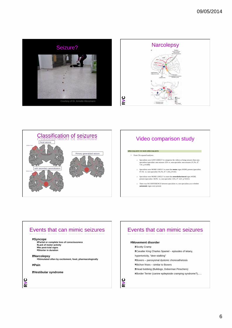
09/05/2014
6
Seizure?
Courtesy of Dr. Annette Wessmann
Narcolepsy
Epileptic focus
Epileptic focus
Focal seizure
with secondary generalization
Primary generalized seizure
Classification of seizures Video comparison study
For the following variables% respondence to each category was considered to see: ! Whether a majority vote could be reached (>50% agreement for one category) ! The strength of agreement of responses
! Motor signs (binary) o In 100% of videos a majority vote was reached as to whether motor signs were
present. There was >50% agreement that they were present in all videos. o When the strength of agreement was considered, the majority of videos exhibited a
very high strength of agreement (96%) (90-100% in one category)
! Neurobehavioural signs (binary) o In 95% of videos a majority vote was reached as to whether neurobehavioral signs
were present. o In 5 videos no majority was reached as to the presence of neurobehavioral signs
(50/50 split) o When the strength of agreement was considered, the majority of videos exhibited a
medium strength of agreement
! Autonomic signs (binary) o In 95% of videos a majority vote was reached as to whether autonomic signs were
present o In 5 videos no majority vote was reached as to the presence of autonomic signs
(50/50 split). o When the strength of agreement was considered, the majority of videos exhibited a
medium strength of agreement
% Agreement level % response to binary category
Very High (5) 90-100 High (4) 80-90
Medium (3) 70-80 Low (2) 60- 70
Very low (1) 50-60
Agreement category – motor % 1 0 2 0 3 2.0 4 2.0 5 96.0
Agreement category - neurobehavioural % 1 21.0 2 27.0 3 29.0 4 12.0 5 11.0
Agreement category - autonomic % 1 25.0 2 11.0 3 28.0 4 14.0 5 22.0
! Duration o In 84% of videos a majority vote was reached as to the duration of the seizure o In 15 videos there was no majority vote on the duration of the seizure
! Consciousness status (categorical)
o In 89% of videos (89/100) a majority vote was reached as to whether the dog was conscious
o In 11 videos there was no majority vote on whether the dog was conscious
! Type of generalised seizure (categorical) o In only 63.8% of videos (60/94) was a majority vote reached as to what type of
generalised seizure the dog was experiencing o In 34 videos no majority vote was reached on the type of generalised seizure o In 31 videos, the majority of respondents classed the seizure type as tonic-clonic
SPECIALISTS VS NON SPECIALISTS
¥ From Chi-squared analyses:
o Specialists were LESS LIKELY to categorise the videos as being seizures than non-specialists (specialist: non seizures 32% vs. non-specialist: non seizures 25.3%; X2: 7.01, p=0.008)
o Specialists were MORE LIKELY to state that motor signs WERE present (specialist: 97.5% vs. non-specialist: 95.2%; X2: 5.38, p=0.02)
o Specialists were MORE LIKELY to state that neurobehavioural signs WERE
present (specialist: 58.9% vs. non-specialist: 53%; X2: 4.67, p=0.031)
o There was NO DIFFERENCE between specialists vs. non specialists as to whether autonomic signs were present
Events that can mimic seizures " Syncope
" Partial or complete loss of consciousness " Lack of motor activity " No post-ictal signs " Shorter in duration
" Narcolepsy " Stimulated often by excitement, food, pharmacologically
" Pain
" Vestibular syndrome
Events that can mimic seizures
" Movement disorder " Scotty Cramp
" Cavalier King Charles Spaniel – episodes of tetany,
hypertonicity, “deer-stalking”
" Boxers – paroxysmal dystonic choreoathetosis
" Bichon frises – similar to Boxers
" Head bobbing (Bulldogs, Doberman Pinschers)
" Border Terrier (canine epileptoide cramping syndrome?),….
09/05/2014
7
History
¥ Seizures typically last around 1 minute.
¥ Seizures exhibit several stages
¥ Seizures often, but not always, occur at rest or out of sleep.
¥ Clonic movements (rhythmic muscle contractions) are common in
both partial and generalized seizures
¥ Most recurrent seizures respond at least in part to antiepileptic
drugs.
¥ EEG
1. Define the problem 2. Define the system and loca;on 3. Define the lesion
The path
Labrador, 3yrs, FS
¥ Poppy had four seizures in the last month
¥ The physical examination and interictal neurological examination were unremarkable
Have you ever cooked spaghetti?
Classification by aetiology
" Symptomatic or secondary seizure Structural brain lesion
" Reactive seizure Metabolic or toxic cause
" Idiopathic or primary epilepsy Suspected genetic cause
" Possible symptomatic or cryptogenic seizure Rule out only via PME…
09/05/2014
8
Classification by aetiology
Seizures
Intra-cranial
Functional Idiopathic epilepsy
Structural
Symptomatic epilepsy
Possible symptomatic
epilepsy
Extra-cranial
Intrinsic Metabolic Reactive seizures
Extrinsic Toxic Reactive seizures
Extracranial
Epilepsy (extracranial)
Extrinsic Exogenous Toxin
Intrinsic
Electrolyte imbalances
Hypernatraemia
Hyponatraemia
Hypocalcaemia
Energy deprivation
Thiamine deficiency
Hypoglycaemia
Organ dysfunction
Uraemic encephalopathy
Hepatic encephalopathy
Vascular perfusion
Polycythaemia
Hyperviscosity syndrome
Hypertension (chronic)
Intracranial
1. Define the problem 2. Define the system and loca;on 3. Define the lesion
The path
1. Menta*on
2. Posture
3. Gait
4. Postural Reac*ons
5. Cranial Nerves
6. Spinal Reflexes
7. Palpa*on
8. Nocicep*on
Neurological?
No Yes
Peripheral?
Muscle? Neuromuscular junc;on? Nerve?
Central?
Brain?
Forebrain? Cerebellum? Brainstem?
Central Ves;bular?
Spinal cord?
C1-‐C5 SCS? C6-‐T2 SCS? T3-‐L3 SCS? L4-‐S3 SCS?
Peripheral Ves;bular
09/05/2014
9
1. Define the problem 2. Define the system and loca;on 3. Define the lesion
The path Neurological examina;on and history
Define & refine the problem
Lateralising?
Asymmetrical Symmetrical
Pain?
No Yes
Generalised? Localised?
On manipula;on? On palpa;on?
History
Define & refine the problem
Onset of disease?
Per-‐acute? Acute? Chronic?
Course of the disease?
Progression?
Deteriora;ng? Improving? Sta;c? Waxing and waning?
Episodic?
Differen;al diagnoses D Degenerative
A Anomalous
M Metabolic
N Neoplastic Nutritional
I Infectious Inflammatory Iatrogenic Idiopathic
T Toxic Traumatic
V Vascular
How can we define the lesion now by the neuro-‐hand rule? 1.Onset, 2.clinical course, 3.pain, 4.lateralising, 5.neurological localisa;on Example: ¥ Acute, onset, non-‐deteriora;ng, lateralising, painful forebrain disease
Define & refine the problem Do not forget signalment
¥ Breed ¥ Age ¥ Sex
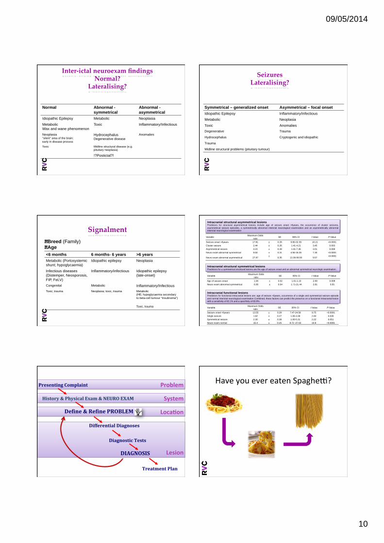
09/05/2014
10
Inter-‐‑ictal neuroexam findings Normal?
Lateralising?
Normal Abnormal - symmetrical
Abnormal - asymmetrical
Idiopathic Epilepsy Metabolic Neoplasia Metabolic Wax and wane phenomenon
Toxic Inflammatory/Infectious
Neoplasia “silent” area of the brain; early in disease process
Hydrocephalus Degenerative disease
Anomalies
Toxic Midline structural disease (e.g. pituitary neoplasia)
!?Postictal?!
Seizures Lateralising?
Symmetrical – generalized onset Asymmetrical – focal onset Idiopathic Epilepsy Inflammatory/infectious Metabolic Neoplasia Toxic Anomalies Degenerative Trauma
Hydrocephalus Cryptogenic and idiopathic
Trauma
Midline structural problems (pituitary tumour)
Signalment " Breed (Family) " Age <6 months 6 months- 6 years >6 years Metabolic (Portosystemic shunt; hypoglycaemia)
Idiopathic epilepsy Neoplasia
Infectious diseases (Distemper, Neosporosis, FIP, FeLV)
Inflammatory/infectious Idiopathic epilepsy (late-onset)
Congenital Metabolic Inflammatory/Infectious Toxic; trauma Neoplasia; toxic, trauma Metabolic
(HE; hypoglycaemia secondary to beta-cell tumour “Insulinoma”)
Toxic, trauma
VBL
Intracranial structural symmetrical lesions Predictors for a symmetrical structural lesions are the age of seizure onset and an abnormal symmetrical neurologic examination.
Intracranial structural asymmetrical lesions Predictors for structural asymmetrical lesions include age of seizure onset >6years, the occurrence of cluster seizures, asymmetrical seizure episodes, a symmetrically abnormal interictal neurological examination and an asymmetrically abnormal interictal neurological examination
Variable Maximum Odds
ratio SE 95% CI t Value P-Value
Seizure onset >6years 17.81 ± 0.28 9.80-32.39 10.21 <0.0001 Cluster seizure 2.44 ± 0.26 1.41-4.21 3.45 0.003 Asymmetrical seizure 3.23 ± 0.39 1.41-7.36 3.01 0.008 Neuro exam abnormal symmetrical 9.50 ± 0.31 4.94-18.28 7.30 <0.0001
Neuro exam abnormal asymmetrical 27.97 ±
0.35 13.38-58.50 9.57 <0.0001
Variable Maximum Odds
ratio SE 95% CI t Value P-Value
Age of seizure onset 1.06 ± 0.02 1.01-1.10 2.69 0.008 Neuro exam abnormal symmetrical 6.06 ± 0.64 1.71-21.44 2.81 0.05
Intracranial functional lesions Predictors for functional intracranial lesions are: age of seizure <6years, occurrence of a single and symmetrical seizure episode and normal interictal neurological examination Combined, these factors can predict the presence on a functional intracranial lesion with a sensitivity of 82.1% and a specificity of 83.9%.
Variable Maximum Odds
ratio SE 95% CI t Value P-Value
Seizure onset <6years 13.55 ± 0.28 7.47-24.58 9.75 <0.0001 Single seizure 1.92 ± 0.27 1.06-3.39 2.44 0.035 Symmetrical seizure 2.30 ± 0.38 1.00-5.31 2.22 0.051 Neuro exam normal 15.4 ± 0.25 8.72 -27.02 10.8 <0.0001
Lesion
Loca*on
System
Problem
Diagnostic Tests
Treatment Plan
DIAGNOSIS
Presenting Complaint
History & Physical Exam & NEURO EXAM
DeCine & ReCine PROBLEM
Differential Diagnoses
Have you ever eaten Spaghe_?

09/05/2014
11
Ancillary tests ¥ Clinical pathology
– Haematology/biochemistry – Endocrine tests – Cerebrospinal fluid analysis – Serology / PCR for detec;on of infec;ous agents – Blood pressure – Metabolic tests for storage diseases ...
¥ Diagnos;c imaging – MRI or CT
¥ Electrophysiology ¥ EEG
¥ …….
1. Define the problem 2. Define the system and loca;on 3. Define the lesion
The path
How high do you think is the percentage of dogs with epilepsy?
1. 0.1%
2. 0.6%
3. 2%
4. 5%
How high do you think is the percentage of pharmacoresistance in dogs with epilepsy?
1. 10-‐‑15%
2. 20-‐‑30%
3. 40-‐‑50%
4. 50-‐‑60%
How high do you think is the spontaneous and drug-‐‑induced epilepsy remission rate ?
1. 10-‐‑15%
2. 20-‐‑30%
3. 40-‐‑50%
4. 50-‐‑60%
~0.6% - estimated canine epilepsy prevalence in UK‘s first opinion clinic1
UK companion dog population ~9.4 million ~55.000 dogs with epilepsy ~44.000 will continue to seizure (AED) ~10.000 remain poorly controlled (2xAED)
1Fleet L, O'Neill DG, Volk HA, et al. VetRec, 2013
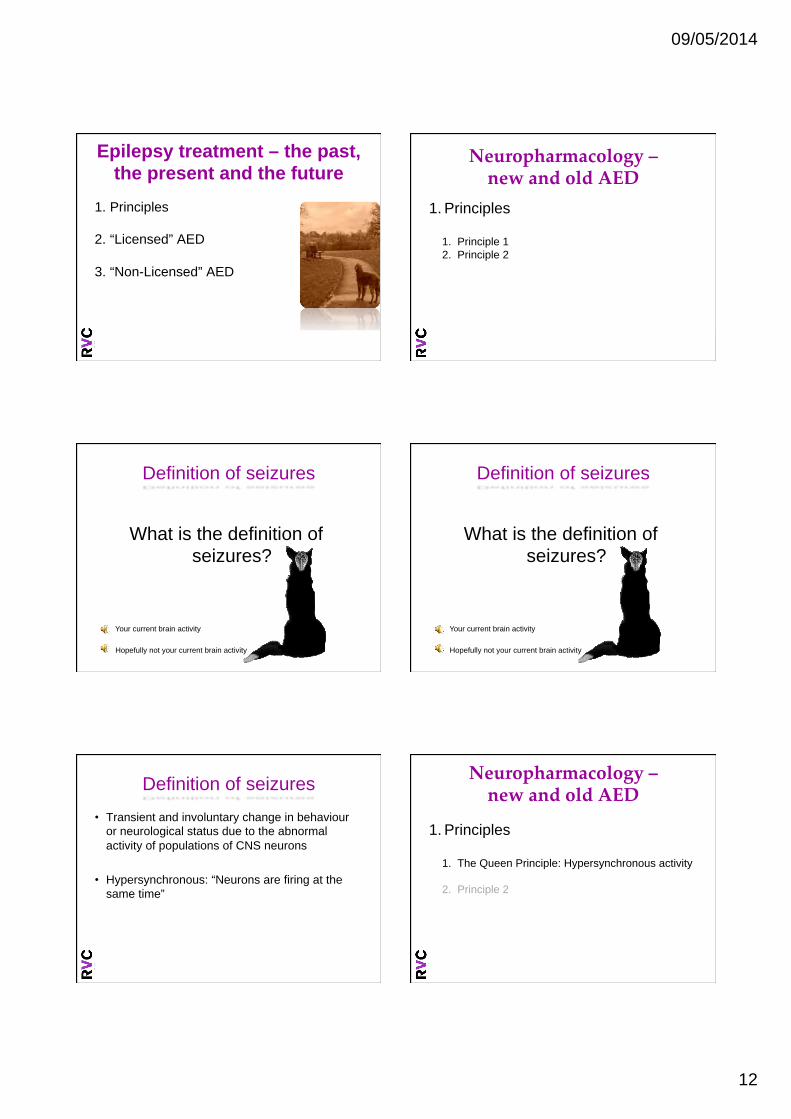
09/05/2014
12
Epilepsy treatment – the past, the present and the future
1. Principles
2. “Licensed” AED
3. “Non-Licensed” AED
Neuropharmacology – new and old AED
1. Principles
1. Principle 1 2. Principle 2
Definition of seizures
What is the definition of seizures?
Your current brain activity
Hopefully not your current brain activity
Definition of seizures
What is the definition of seizures?
Your current brain activity
Hopefully not your current brain activity
Definition of seizures
¥ Transient and involuntary change in behaviour or neurological status due to the abnormal activity of populations of CNS neurons
¥ Hypersynchronous: “Neurons are firing at the same time”
Neuropharmacology – new and old AED
1. Principles
1. The Queen Principle: Hypersynchronous activity 2. Principle 2
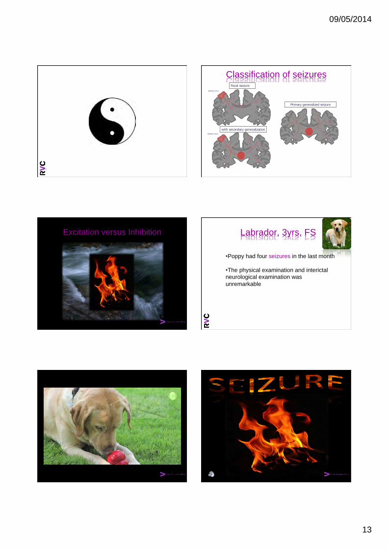
09/05/2014
13
Epileptic focus
Epileptic focus
Focal seizure
with secondary generalization
Primary generalized seizure
Classification of seizures
Excitation versus Inhibition
Dr. Holger A. Volk, CPD, Oct. 2008
Labrador, 3yrs, FS
¥ Poppy had four seizures in the last month
¥ The physical examination and interictal neurological examination was unremarkable
Dr. Holger A. Volk, 2010 Dr. Holger A. Volk, CPD, Oct. 2008

09/05/2014
14
Neuropharmacology – new and old AED
1. Principles
1. The Queen Principle: Hypersynchronous activity 2. The Yin Yan principle: Inhibition vs. Excitation
‘Idiopathic’ epilepsy ¥ Dog: 6 months to 6 years
Breeds Beagles, German Shepherd Dogs, Labrador Retrievers, Golden Retrievers, Bernese Mountain Dog, Belgian Tervuerens, Keeshonds, Irish Wolfhounds, Viszlas, English Springer Spaniels, Danish Labradors, Lagotto Romagnolo, Standard Poodles, Finnish Spitz,…
Epilepsy treatment – the past, the present and the future
1. Principles
2. “Licensed” AED
3. “Non-Licensed” AED
Have you ever cooked spaghetti?
2.
Therapy – Mode of action
2.
Therapy – Mode of action
Barbiturate increases the duration of chloride ion channel opening at the GABAA receptor. This increases the efficacy of GABA. Benzodiazepines increase the frequency of the chloride ion channel opening at the GABAA receptor. This increases the potency of GABA.
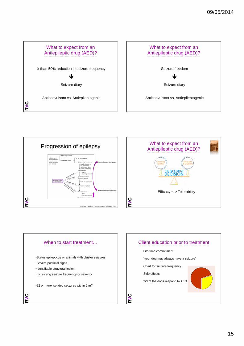
09/05/2014
15
What to expect from an Antiepileptic drug (AED)?
≥ than 50% reduction in seizure frequency
ê Seizure diary
Anticonvulsant vs. Antiepileptogenic
What to expect from an Antiepileptic drug (AED)?
Seizure freedom
ê Seizure diary
Anticonvulsant vs. Antiepileptogenic
Progression of epilepsy mechanisms that link genotype with phenotype arelargely unknown [33]. Genetic factors might alsoexplain why some individuals develop epilepsy afteracquired insults whereas others do not [33].
Mapping mutations responsible for rare forms offamilial epilepsies has identified several genes,mutation of which results in epilepsy in humans[33–35]. Each encodes either a voltage- or ligand-gated ion channel, which underlines the importantrole of channelopathies in some types of idiopathicepilepsy. However, most idiopathic epilepsies arecaused by inheritance of two or more mutant genes[2]. Genetic animal models, including bothspontaneous mutants and genetically engineeredtransgenic mice, have contributed significantly to our understanding of the mechanisms of epilepsyby allowing dissection of the genetic, biochemical and pathophysiological factors that predispose to
TRENDS in Pharmacological Sciences Vol.23 No.3 March 2002
http://tips.trends.com
117Opinion
generation of pharmacoresistance in epileptic patients. If this isthe case, either system ic or local adm inistration of inhibitors ofthese drug transporters or novel AEDs that are not substrates fortransporters could prove useful in pharmacoresistant epilepsy.Inhibitors of PGP and, more recently, MRPs are being evaluatedclinically for either the reversal or prevention of intrinsic andacquired multidrug resistance in human cancer [q] and m ight soonbe available for clinical trials in epilepsy.
Referencesa Löscher, W. and Potschka, H. Role of multidrug transporters in
pharmacoresistance to antiepileptic drugs. J. Pharmacol. Exp. Ther. (in press)b Tan, B. et al. (2000) Multidrug resistance transporters and modulation.
Curr. Opin. Oncol. 12, 450–458
c Tishler, D.M. et al. (1995) MDR1 gene expression in brain of patients withmedically intractable epilepsy. Epilepsia 36, 1–6
d Abbott, N.J. et al. Drug resistance in epilepsy: role of the blood–brain barrier.In Mechanisms of Drug Resistance in Epilepsy. Lessons from Oncology(Ling, V., ed.), Wiley (in press)
e Sisodiya, S.M. et al. (1999) Over-expression of P-glycoprotein inmalformations of cortical development. NeuroReport 10, 3437–3441
f Sisodiya, S.M. et al. (2001) Multidrug-resistance protein 1 in focal corticaldysplasia. Lancet 357, 42–43
g Sisodiya, S.M. et al. Drug resistance in epilepsy: expression of drug resistanceproteins in common causes for refractory epilepsy. Brain (in press)
h Frey, H-H. and Löscher, W. (1978) Distribution of valproate across the interfacebetween blood and cerebrospinal fluid. Neuropharmacology17, 637–642
i Shen, D.D. (1999) Valproate: absorption, distribution, and excretion. InValproate (Löscher, W., ed.), pp. 77–90, Birkhäuser
j Huai-Yun, H. et al. (1998) Expression of multidrug resistance-associatedprotein (MRP) in brain microvessel endothelial cells. Biochem. Biophys. Res.Commun. 243, 816–820
k Potschka, H. and Löscher, W. (2001) Multidrug resistance-associated proteinis involved in the regulation of extracellular levels of phenytoin in the brain.NeuroReport 12, 2387–2389
l Potschka, H. and Löscher, W. (2001) In vivoevidence for P-glycoprotein-mediatedtransport of phenytoin at the blood–brain barrier of rats. Epilepsia42, 1231–1240
m Potschka, H. et al. (2001) P-Glycoprotein and multidrug resistance–associatedprotein are involved in the regulation of extracellular levels of the majorantiepileptic drug carbamazepine in the brain. NeuroReport 12, 3557–3560
n Schinkel, A.H. et al. (1996) P-glycoprotein in the blood–brain barrier of miceinfluences the brain penetration and pharmacological activity of many drugs.J. Clin. Invest. 97, 2517–2524
o Owen, A. et al. (2001) Carbamazepine is not a substrate for P-glycoprotein.Br. J. Clin. Pharmacol. 51, 345–349
p Schinkel, A.H. (1999) P-Glycoprotein, a gatekeeper in the blood-brain barrier.Adv. Drug Deliv. Rev. 36, 179–194
q Litman, T. et al. (2001) From MDR to MXR: new understanding of multidrugresistance systems, their properties and clinical significance. Cell Mol. LifeSci. 58, 931–959
Fig. I. Multidrug transporters (MDTs) lim it drug penetration through the blood–brainbarrier (BBB) into the extracellular space, neurons and other target cells in the brain.Unlike endothelial cells in most tissues, endothelial cells of capillaries in the brain arejoined by tight junctions and lack intercellular pores and pinocytotic vesicles.Processes from pericapillary astrocytes (‘glial endfeet’) term inate on capillaries andcontribute to the function of the BBB. (a) The BBB passively excludes strongly ionized(polar), hydrophilic drugs, but nonpolar, highly lipid-soluble drugs [which includesmost antiepileptic or anticonvulsant drugs (AEDs)] penetrate easily into the brain bysimple diffusion (dashed arrows). The penetration of many lipophilic drugs into brainparenchyma is lim ited by the action of ATP-dependent MDTs located in the apical(lum inar) cell membrane of capillary endothelial cells of the BBB. These form anactive defence mechanism by acting as active efflux pumps that transfer drugs backinto blood after they have entered endothelial cells. (b) In epileptogenic brain tissue,overexperession of MDTs such as P-glycoprotein and members of the multidrug-resistance-associated protein fam ily is likely to result in reduced drug penetrationinto brain parenchyma. The concentration of MDTs also increases in processes fromglial endfeet that extend onto capillaries (not illustrated), which suggests thatoverexpression of MDTs in glia represents a ‘second barrier’ that lim its drugpenetration and results in simultaneous resistance to a variety of unrelated lipophilic AEDs.
Initiating event(e.g. head trauma,febrile seizures,stroke, infections,gene defects)
Repair (or control)
Failure to repairNo consequence
Onset of epileptic cascade(e.g. by ‘second hit’, polymorphisms, susceptibility genes,crucial modulators)
Epileptogenesis
Spontaneous seizure(onset of epilepsy)
No progression
Progression of epilepsy
Chronic epilepsyOften pharmacoresistant
Antiepileptogenic
Anticonvulsant
Disease-modifyingReversal and/or prevention
of multidrug resistance
Role of neurodegeneration?
Pharmacologicalintervention
TRENDS in Pharmacological Sciences
Fig. 1. Steps in the development and progression of epilepsy andpharmacological interventions. The term epileptogenesis includesprocesses that take place before the first seizure occurs to render theepileptic brain susceptible to spontaneous recurrent seizures andprocesses that intensify seizures and make them more refractory totherapy (progression). In addition to the pharmacological interventionsillustrated in the figure, pharmacological potentiation of endogenousanti-ictal processes (which serve to term inate a seizure) could form astrategy for the treatment of seizures.
Löscher, Trends in Pharmacological Sciences, 2002
Neurobehavioural changes
Neurobehavioural changes
What to expect from an Antiepileptic drug (AED)?
Efficacy <-> Tolerability
!
When to start treatment…
¥ Status epilepticus or animals with cluster seizures
¥ Severe postictal signs
¥ Identifiable structural lesion
¥ Increasing seizure frequency or severity
¥ ?2 or more isolated seizures within 6 m?
Client education prior to treatment
Life-time commitment
“your dog may always have a seizure”
Chart for seizure frequency
Side effects
2/3 of the dogs respond to AED
09/05/2014
16
Treatment
" Monotherapy
" Seizure frequency may influence choice of AED?
" Monitor plasma levels?
" Owner compliance
Limitation of AEDs from people
" Toxicity
" Tolerance
" Inappropriate pharmacokinetics
" Expense
Labrador, 3yrs, FS
¥ Poppy had four seizures in the last month
¥ The physical examination and interictal neurological examination was unremarkable
¥ Bloods and urine were unremarkable
¥ What would you do?
Labrador, 3yrs, FS
Do you think it is right to start treatment? 1. Yes 2. No
Labrador, 3yrs, FS
Which drug would you use first line? 1. Phenobarbitone 2. Imepitoin 3. Potassium bromide 4. Levetiracetam 5. Gabapentin 6. Zonisamide
Labrador, 3yrs, FS
Which drug would you use first line? 1. Phenobarbitone 2. Imepitoin 3. Potassium bromide 4. Levetiracetam 5. Gabapentin 6. Zonisamide

09/05/2014
17
Labrador, 3yrs, FS
When would you check Phenobarbitone serum levels for the first time? 1. 2 days 2. 12 days 3. 30 days 4. 90 days
Phenobarbitone (dog)– First line treatment
¥ Phenobarbitone (PB) – Dose ~2.5 mg/kg BID – Peak serum concentration 4-8 hours (oral) – Half-life 24-40 hrs – Time to steady state 10-14 days – Therapeutic range 15.0 – 35 µg/ml – Potential side effects Sedation, PU/PD, polyphagia, hepatotoxicity – Metabolism liver – Drug interaction Can alter serum levels of liver metabolised drugs – Obtain plasma level 14, 45, 90, 180, 360 d, then q 6m
¥ Loading dosage if indicated – 12 to 24 mg/kg total dose within 24 hours
(equal dose q 30 min to 4 hrs to effect, i.e. no seizures) SPC Epiphen®; Phenoleptil®
The peak and trough debate
Monteiro et al., 2009, Vet Record
November 7, 2009 | the VETERINARY RECORD | 557
PAPERS
the dose of phenobarbitone increased there was a trend for the trough group to have lower serum concentrations than the non-trough group, and the difference was significant (P=0·0228) at doses of more than 10 mg/kg/day.
DiscussionPhenobarbitone is considered to be an effective anticonvulsant med-ication in 60 to 80 per cent of dogs with epilepsy (Farnbach 1984, Schwartz-Porsche and others 1985). When it fails, the failure is more often due to the inappropriate use of the drug rather than to pharma-coresistance. In this study, two large populations of dogs were com-pared: one in which the blood sample for the determination of the serum concentration was collected during the trough period, and one in which the sample was collected outside this period (non-trough). By correcting for the dose of phenobarbitone, it was possible to com-pare the serum concentrations in the two groups at different oral doses. Levitski and Trepanier (2000) reported that in 33 epileptic dogs on a range of doses of phenobarbitone there was no therapeutically relevant change in the serum concentration in most of them during the 12 hours between doses. They compared trough and non-trough blood samples in the same dog, whereas in this study two different populations of dogs have been compared, making it possible to compare large numbers of dogs giving sufficient statistical power to investigate the effect of a wide range of dose levels.
There was no significant difference between the serum pheno-barbitone concentrations of the trough and non-trough animals that were treated with the lower doses, in agreement with the study by Levitski and Trepanier (2000). However, in the dogs on the highest doses of phenobarbitone the concentration in the non-trough animals was significantly higher than in the trough animals.
One reason why some dogs require higher doses of phenobarbitone to maintain their serum concentrations is that there may have been substantial phenobarbitone-induced induction of hepatic microsomal enzyme systems and a reduction in the half-life of phenobarbitone. This reduction could result in wider variations in the serum concentrations of phenobarbitone between doses and explain the difference between the trough and non-trough animals. However, the effects of dose on absorption and protein binding, genetic differences and other factors, cannot be excluded.
The nature of the Vétoquinol phenobarbitone monitoring voucher scheme introduces several limitations into the study. The data included the length of treatment, concurrent anti-epileptic agents and the treat-ment regimen, but details of the underlying diagnosis, concurrent dis-ease, and information about the doses of other anti-epileptic agents
higher doses of phenobarbitone to maintain its serum concentration influences this relationship.
Materials and methodsThe laboratory records of 1436 client-owned dogs on chronic pheno-barbitone treatment for epileptic seizures were analysed. All of them had their serum phenobarbitone concentrations monitored as part of the Vétoquinol UK phenobarbitone monitoring voucher scheme, which provides a voucher for one free determination of a dog’s serum phenobarbitone concentration for every batch of the drug purchased from the manufacturer. Only the 1427 dogs that had been on continu-ous treatment for at least four weeks were included in the study. Data recorded from each dog included the time when a blood sample had been collected in relation to the administration of the drug, the ani-mal’s weight, its total daily oral dose of phenobarbitone and its serum concentration as determined by solid-phase competitive chemilumi-nescent enzyme immunoassay (Immulite Phenobarbital Immunoassay; Siemens Medical Solutions Diagnostics). The dogs were divided into two groups: the 918 dogs from which the blood sample was collected at the trough, and the 509 dogs from which the blood sample was col-lected during the non-trough period. This classification was limited by the data recorded on the phenobarbitone monitoring vouchers, which did not record specific times of drug administration and blood sample collection, but rather whether the relationship between the two was known, and if so, whether the sample was collected during the two hours before the next dose (trough sample) or not during that period (non-trough sample). Dogs for which the time relationship between the two was not known were not included in the study. In order to assess whether the requirement for higher doses of phenobarbitone influences whether the timing of the blood sample in relation to the administra-tion of the drug is clinically relevant, the dogs were further subdivided into four groups on the basis of their total daily dose of phenobarbitone: group 1, 2 to 5 mg/kg/day (the manufacturer’s recommended dose; 478 trough samples and 271 non-trough samples); group 2, more than 5 to 8 mg/kg/day (an arbitrary interval the same as the previous group; 277 trough samples and 154 non-trough samples); group 3, more than 8 to 10 mg/kg/day (77 trough samples and 44 non-trough samples); and group 4, more than 10 mg/kg/day (86 trough samples and 40 non-trough samples); the distinction between the last two groups was made on the basis of the median dose for the dogs on a dose of more than 8 mg/kg/day.
The data were analysed using the unpaired t test with Welch’s cor-rection to account for lack of homoscedasticity. An analysis of variance was used to determine whether the four dosage groups had significantly different serum phenobarbitone concentrations. P values of less than 0·05 were considered statistically significant.
ResultsThe serum phenobarbitone concentrations in the dogs in group 1 ranged between 17·9 and 181 µmol/l, in group 2 between 24·2 and 185 µmol/l, in group 3 between 30 and 176 µmol/l, and in group 4 between 24·5 and 190 µmol/l. In group 1, the mean dose of phenobarbitone for the dogs in which the sample was collected during the trough period was 3·61 mg/kg/day and for the dogs in which the sample was collected dur-ing the non-trough period it was 3·60 mg/kg/day; in group 2, the mean concentrations were 6·34 mg/kg/day and 6·38 mg/kg/day; in group 3, they were 9·02 mg/kg/day and 9·04 mg/kg/day; and in group 4, they were 13·9 mg/kg/day and 13·4 mg/kg/day. In group 1, the mean serum phenobarbitone concentrations for the trough and non-trough groups were 65·0 and 65·6 µmol/l, respectively; in group 2, they were 85·5 and 87·2 µmol/l; in group 3, they were 98·2 and 101·9 µmol/l; and in group 4, they were 114·8 and 128·7 µmol/l. The increased oral doses of phenobarbitone were positively correlated with increased serum con-centrations (P<0·0001). Fig 1 shows the mean serum concentrations (relative to dose) for the dogs in which the sample was collected dur-ing the trough period and for the dogs in which the sample was col-lected during the non-trough period. In the dogs receiving the lower doses of pheno barbitone there was no significant difference between the serum concentrations of the trough and non-trough groups. However, as
FIG 1: Relationship between the serum concentrations of phenobarbitone (relative to dose) in ‘trough’ and ‘non-trough’ samples and the total daily doses of phenobarbitone received by groups of dogs
Ser
um p
heno
barb
itone
con
cent
ratio
n re
lativ
e to
dos
e ([
µmol
/l]/[m
g/kg
])
20
15
10
5
0 2 to 5 mg/kg >5 to 8 mg/kg >8 to 10 mg/kg >10 mg/kg
Trough samples
Non-trough samples
P=0·0228
Total daily dose of phenobarbitone (number of dogs in trough/non-trough samples)
(478/271) (277/154) (77/44) (86/40)
556-571 Papers & articles.indd 557 4/11/09 12:56:42
Phenobarbital – Side effects
¥ Rare but severe (idiosyncratic reactions): – Behavioural alterations – Immune-mediated neutropaenia, thrombocytopaenia, anaemia – Superficial, necrlolytic dermatitis – Idiosyncratic hepatotoxic reactions
(rapid elevation of ALT and abnormal bile acids) Action: stop drug immediately – load with another AED
¥ Withdrawal seizures (drug dependence) – How to stop?
JAVMA, Vol 240, No. 9, May 1, 2012 Scientific Reports 1073
SM
ALL A
NIM
ALS
Since the early 1990s, bromide (most commonly ad-ministered as a potassium salt) has been used as
an AED for the long-term management of epilepsy in dogs.1–4 Increasingly, it is considered as an alternative to phenobarbital as the first-choice sole AED used for long-term control of epilepsy in dogs.5–8 Successful use of both phenobarbital and bromide is facilitated by a long elimination half-life, which minimizes fluctuation in drug concentrations during a 12-hour dosing interval.7–11 Therapeutic drug monitoring is widely available and is supported by canine (rather than human) therapeutic ranges as follows: serum phenobarbital concentrations of 15 to 45 µg/mL7,12–14 and serum bromide concentrations of 1 to 3 mg/mL (when used as a sole treatment).7,12–17
Comparison of phenobarbital with bromide as a first-choice antiepileptic drug for treatment of epilepsy in dogs
Dawn Merton Boothe, DVM, PhD, DACVIM, DACVCP; Curtis Dewey, DVM, MS, DACVS, DACVIM; David Mark Carpenter, PhD
Objective—To compare efficacy and safety of treatment with phenobarbital or bromide as the first-choice antiepileptic drug (AED) in dogs.Design—Double-blinded, randomized, parallel, clinical trial.Animals—46 AED-naïve dogs with naturally occurring epilepsy.Procedures—Study inclusion was based on age, history, findings on physical and neuro-logic examinations, and clinicopathologic test results. For either phenobarbital treatment (21 dogs) or bromide treatment (25), a 7-day loading dose period was initiated along with a maintenance dose, which was adjusted on the basis of monthly monitoring. Efficacy and safety outcomes were compared between times (baseline and study end [generally 6 months]) and between drugs.Results—Phenobarbital treatment resulted in eradication of seizures (17/20 [85%]) signifi-cantly more often than did bromide (12/23 [52%]); phenobarbital treatment also resulted in a greater percentage decrease in seizure duration (88 ± 34%), compared with bromide (49 ± 75%). Seizure activity worsened in 3 bromide-treated dogs only. In dogs with seizure eradication, mean ± SD serum phenobarbital concentration was 25 ± 6 µg/mL (phenobar-bital dosage, 4.1 ± 1.1 mg/kg [1.9 ± 0.5 mg/lb], PO, q 12 h) and mean serum bromide con-centration was 1.8 ± 0.6 mg/mL (bromide dosage, 31 ± 11 mg/kg [14 ± 5 mg/lb], PO, q 12 h). Ataxia, lethargy, and polydipsia were greater at 1 month for phenobarbital-treated dogs; vomiting was greater for bromide-treated dogs at 1 month and study end.Conclusions and Clinical Relevance—Both phenobarbital and bromide were reasonable first-choice AEDs for dogs, but phenobarbital was more effective and better tolerated during the first 6 months of treatment. (J Am Vet Med Assoc 2012;240:1073–1083)
Each drug has its disadvantages. Both are associated with polyuria, polydipsia, and polyphagia and the sequelae as-sociated with general sedation (eg, lethargy and ataxia), adverse effects to which an animal may develop toler-ance.1,2,5–7,17 In addition, each has unique characteristics that complicate successful treatment. Long-term pheno-barbital treatment has been associated with hepatotox-icity, although a cause-and-effect relationship has not been proven.18–20 Phenobarbital also has been associated with unpredictable, idiosyncratic adverse drug reactions, including pancytopenia,21,22 and drug interactions that reflect induction of selected hepatic drug-metabolizing enzymes that target xenobiotics23–30 as well as endoge-nous compounds.31,32 Induction of its own metabolism might result in a decrease in plasma drug concentrations and therapeutic failure. In contrast, although renal ex-cretion of bromide limits hepatotoxicity or induction of hepatic enzymes, its elimination half-life in dogs is so long that several months of treatment must occur with each change in dosage before steady-state con-centrations (and thus maximum effect) are reached.33,34 Although this delay can be overcome by administration
From the Department of Anatomy, Physiology and Pharmacology, College of Veterinary Medicine (Boothe) and the Department of Mathematics and Statistics, College of Sciences and Mathematics (Carpenter), Auburn University, Auburn, AL 36849; and the De-partment of Clinical Sciences, College of Veterinary Medicine, Cor-nell University, Ithaca, NY 14850 (Dewey).
This study was implemented at Texas A&M University’s Texas Vet-erinary Medical Center but included the participation of referral practitioners throughout the United States.
Supported by the Canine Health Foundation and the American Ken-nel Club.
Presented as a poster at the 20th American College of Veterinary In-ternal Medicine Annual Forum, Dallas, May 2002.
Address correspondence to Dr. Boothe ([email protected]).
ABBREVIATIONS
AED Antiepileptic drugCI Confidence interval
Phenobarbital

09/05/2014
18
Following the introduction of VPA in the 1960s, no newAEDs were entering the market for almost two decades,except some additional benzodiazepines (Fig. 1). Not sur-prisingly, patients and physicians had great expectations fortherapeutic gains over older drugs when the new, third-gen-eration AEDs entered the market, which arbitrarily includeall drugs for epilepsy marketed after the introduction ofCBZ or VPA in the 1960s (Fig. 1). Despite the shortcom-ings of second-generation AEDs outlined above, the wide-spread use and the unsurpassed clinical efficacy of CBZ andVPA made them benchmarks for comparison with third-generation AEDs. The era of the third-generation AEDsstarted in the 1980s with ‘‘rational’’ developments such asprogabide, vigabatrin (VGB), and tiagabine (TGB), that is,drugs that were designed to selectively target a mechanismthat was thought to be critical for the occurrence of epilepticseizures (Lçscher & Schmidt, 1994).
The new, third-generation AEDs have undoubtedlyexpanded the therapeutic options, in particular for those inneed of a change in medical regimen. However, the efficacyof many, but unfortunately not all, new AEDs for treatmentof new-onset epilepsy is similar to that of older AEDs (Kwan& Brodie, 2000; Brodie et al., 2007; Marson et al., 2007;
Glauser et al., 2010). For patients with previously drug-resistant seizures, adjunctive new AEDs have been shown tobe more efficacious than adding placebo (Beyenburg et al.,2010). In addition, new AEDs have other benefits over someof the older drugs for epilepsy. Treatment with some ofthe new AEDs avoids adverse drug interactions and hyper-sensitivity reactions (Elger & Schmidt, 2008) and some newAEDs have clinically important utility for disorders otherthan epilepsy (Rogawski & Lçscher, 2004b).
Despite these encouraging and welcome advantages thatnew AEDs have brought for the management of epilepsy,there is growing concern that the efficacy of drug treatmentof epilepsy has not substantially improved with the intro-duction of new AEDs (Shorvon, 2009b). Although we haveno population-based studies if the prognosis of epilepsy haschanged since and because of the introduction of newAEDs, this dilemma is highlighted by the following discon-certing observations. Currently, 38% of adult patients withnew-onset epilepsy have AED-resistant seizures (Mohanraj& Brodie, 2005). Forty years ago, Coatsworth (1971)reported in his summary of AED trials that 33–38% of thosewith focal seizures were unchanged or worse. This has pos-sibly not changed since the 1880s when Gowers reported
Figure 1.
Introduction of AEDs to the market from 1853 to 2009 (adapted from data by Shorvon, 2009a,b). Licensing varied from country to
country. We give here the year of first licensing or the first mention of clinical use in a country of Europe, the United States, or Japan
(see Shorvon, 2009a,b, for further details). We have not included all derivatives of listed AEDs or AEDs used solely for treatment of
status epilepticus.
Epilepsia ILAE
658
W. Loscher and D. Schmidt
Epilepsia, 52(4):657–678, 2011doi: 10.1111/j.1528-1167.2011.03024.x
Loscher and Schmidt 2011
Phenobarbitone (Mode of Action)
Imepitoin Novel mode of action
Epilepsia, 45(10):1228–1239, 2004Blackwell Publishing, Inc.C⃝ 2004 International League Against Epilepsy
Anticonvulsant Efficacy of the Low-affinity PartialBenzodiazepine Receptor Agonist ELB 138 in a Dog
Seizure Model and in Epileptic Dogs with SpontaneouslyRecurrent Seizures
∗Wolfgang Loscher, ∗Heidrun Potschka, †Susanne Rieck, †Andrea Tipold, and ‡Chris Rundfeldt
∗Department of Pharmacology, Toxicology, and Pharmacy, and †Department of Small Animal Medicine and Surgery, University ofVeterinary Medicine Hannover; and ‡Elbion AG, Radebeul, Germany
Summary: Purpose:Ataxia, sedation, amnesia, ethanol and bar-biturate potentiation, loss of efficacy (tolerance), developmentof dependence, and the potential for drug abuse limit the clin-ical use of benzodiazepines (BZDs) for long-term treatment ofepilepsy or anxiety. BZD ligands that are in current use act asfull allosteric modulators of γ -aminobutyric acid (GABA)-gatedchloride channels and, on long-term administration, trigger afunctional uncoupling between the GABAA and BZD recogni-tion sites. Partial allosteric modulators, which have a low intrin-sic activity at the BZD recognition site of the GABAA receptor,might eventually overcome the limitations of full agonists suchas diazepam (DZP).
Methods: In the present study, the new low-affinity partialBZD-receptor agonist ELB 138 [former name AWD 131-138;1-(4-chlorophenyl)-4-morpholino-imidazolin-2-one] was evalu-ated in a dog seizure model and in epileptic dogs with sponta-neously recurrent seizures.
Results: ELB 138 was shown to increase potently thepentylenetetrazole (PTZ) seizure threshold in dogs. Prolongedoral administration with twice-daily dosing of ELB 138 witheither 5 or 40 mg/kg over a 5-week period was not associated
with loss of anticonvulsant efficacy in the PTZ dog model. Tostudy whether physical dependence developed during long-termtreatment, the BZD antagonist flumazenil was injected after 5weeks of treatment with ELB 138. Compared with prolongedtreatment with DZP, only relatively mild abstinence symptomswere precipitated in dogs treated with ELB 138, particularlyat the lower dosage (5 mg/kg, b.i.d.). In a prospective trial indogs with newly diagnosed epilepsy, ELB 138 markedly reducedseizure frequency and severity without significant difference tostandard treatments (phenobarbital or primidone) but was muchbetter tolerated than the standard drugs. In dogs with chronicepilepsy, most dogs exhibited a reduction in seizure frequencyand severity during add-on treatment with ELB 138.
Conclusions: The data demonstrate that the partial BZD re-ceptor agonist ELB 138 exerts significant anticonvulsant efficacywithout tolerance in a dog seizure model as well as in epilepticdogs with spontaneously recurrent seizures. These data thus sub-stantiate that partial agonism at the BZD site of GABAA receptorsoffers advantages versus full agonism and constitutes a valuableapproach for treatment of seizures. Key Words: Epilepsy—Antiepileptic drugs—GABA receptors—Benzodiazepines.
ELB 138 [former name, AWD 131-138; 1-(4-chloro-phenyl)-4-morpholino-imidazolin-2-one] was testedwithin the National Institute of Neurological Disordersand Stroke (NINDS) Antiepileptic Drug DevelopmentProgram, and it was selected for further developmentbecause of its broad-spectrum anticonvulsant and anx-iolytic properties and high therapeutic index in rodentmodels (1–3). With respect to mechanism of action, ELB138 potentiates the amplitude of γ -aminobutyric acid(GABA)-evoked currents by acting at the benzodiazepine
Accepted June 27, 2004.Address correspondence and reprint requests to Dr. W. Loscher at
Department of Pharmacology, Toxicology, and Pharmacy, Universityof Veterinary Medicine Hannover, Foundation, Bunteweg 17, D-30559Hannover, Germany. E-mail: [email protected]
(BZD) recognition site of the GABAA receptor. Incontrast to clinically used BZD ligands, ELB 138 acts asa low-affinity partial agonist with low intrinsic activity (1,4). Furthermore, ELB 138 antagonizes the increased firingof action potentials induced by corticotropin-releasingfactor (CRF) in locus coeruleus neurons and blocksvoltage-activated Ca2+ channels in a dose-dependentmanner (3). The GABA-potentiating, anxiolytic, andanticonvulsant effects of ELB 138 could be counteractedby the BZD antagonist flumazenil, indicating that theinteraction of ELB 138 with the BZD site may be themain mechanism of action of this novel drug.
One major disadvantage of drugs acting via the BZDsite of GABA receptors is their tolerance, dependence,and abuse liability (5–7). Partial agonists at this site have
1228
Anticonvulsant activity and tolerance of ELB138 in dogswith epilepsy: A clinical pilot study
Susanne Rieck a, Chris Rundfeldt b, Andrea Tipold a,*
a Department of Small Animal Medicine and Surgery, University of Veterinary Medicine, and Center for Systems Neuroscience, Hannover, Germanyb Elbion AG, Department of Pharmacology, Radebeul, Germany
Abstract
A new antiepileptic and anxiolytic drug, ELB138, was evaluated in a clinical pilot study in dogs with newly diagnosed or chronic
idiopathic epilepsy. The purpose was to verify clinically the anticonvulsant effectiveness of this substance, which had already been
demonstrated experimentally. Data from 29 dogs treated with ELB138 were compared with results obtained retrospectively from 82
dogs treated with conventional antiepileptic medication. The reduction in seizure frequency using ELB138 in dogs with newly diag-
nosed idiopathic epilepsy was comparable to the reduction in dogs treated either with phenobarbital or primidone. In dogs with
chronic epilepsy and add-on therapy with either ELB138 or potassium bromide, such supplementation reduced the seizure frequency
and the duration and severity of seizures. The most obvious difference between ELB138 treatment and conventional medications
became clear in the evaluation of side effects, which in those dogs treated with ELB138 were rare, and consisted mostly of transient
polyphagia. This pilot study confirmed that ELB138 has a potent anticonvulsant effect in dogs with idiopathic epilepsy. These results
will form the basis for a multicentre, blinded study.
� 2005 Elsevier Ltd. All rights reserved.
Keywords: Epilepsy; Dogs; New anticonvulsant medication; ELB138; Pilot study
1. Introduction
Seizure disorders are the most common intracranial
diseases in humans and animals, particularly in dogs
and cats (Frey, 1986; Loscher et al., 1985). In the dog,
seizure disorders have an estimated morbidity of between
0.5% and 1% (Oliver and Lorenz, 1993). Seizures can be
defined as sudden, excessive transient paroxysmal neuro-
nal discharges in the cerebral cortex (Jaggy and Steffen,1995a; Loscher, 1993). In dogs, idiopathic epilepsy is
diagnosed in approximately 45% of cases with seizure
disorders (Jaggy and Steffen, 1995a,b), and in 5.3–8.0%
of all dogs with diseases of the nervous system (Sch-
wartz-Porsche, 1994; Bernardini and Jaggy, 1998). Sei-
zure activity in dogs with idiopathic epilepsy commonlystarts at 1–3 years old (Cunnigham, 1971; Oliver and
Lorenz, 1993) and life-long treatment may be necessary.
Because of marked differences in pharmacokinetics
between dogs and humans, only a few antiepileptic
drugs such as phenobarbital, primidone and potassium
bromide are efficacious in dogs (Frey and Schwartz-Por-
sche, 1985; Loscher, 1993, 1994, 2003; Schwartz-Porsche
and Jurgens, 1991). Treatment outcome is not alwayssatisfactory, and pharmacoresistance is observed in
about one third of cases (Frey and Schwartz-Porsche,
1985; Loscher et al., 1985; Loscher, 2003; Loscher and
Schwartz-Porsche, 1986; Schwartz-Porsche et al.,
1982). Furthermore, phenobarbital and primidone may
cause transient side effects such as sedation, ataxia, com-
pulsive pacing, weakness, polyphagia, polydipsia and
polyuria (Schwartz-Porsche et al., 1982). An elevationof hepatic enzymes is frequently observed. Treatment
1090-0233/$ - see front matter � 2005 Elsevier Ltd. All rights reserved.
doi:10.1016/j.tvjl.2005.04.003
* Corresponding author. Tel.: +49 511 856 7666; fax: +49 511 856
7686.
E-mail address: [email protected] (A. Tipold).
www.elsevier.com/locate/tvjl
The Veterinary Journal 172 (2006) 86–95
TheVeterinary Journal Side-effect Profile (Imepitoin)
¥ Newly diagnosed dogs: Imepitoin very well tolerated, no sedation or other central side effects, mostly transient polyphagia in 7/12 cases (58%)
¥ Add on: no additional side effects to the well known effects of phenobarbital
¥ Regular follow up examinations: clinical and neurological examinations normal, some dogs increased activity
¥ Anxiolytic effects supported (in earlier studies examined in 4 anxious Beagle dogs)

09/05/2014
19
Treatment of epileptic dogs: Summary
¥ Imepitoin appears effective in newly diagnosed and in drug-resistent dogs with epilepsy
¥ Imepitoin appears well tolerated compared to standard medication used
Multiple randomized multicentre studies (Germany, Switzerland, France; ECVN/ESVN)
Data used for power analyses for
Results ¥ Primary efficacy variable: monthly seizure frequency ¥ Reduction confirmed the non-inferiority of imepitoin in comparison to
phenobarbital ¥ No correlation of plasma concentration and seizure control
Results ¥ Primary efficacy variable: monthly seizure frequency ¥ Reduction confirmed the non-inferiority of imepitoin in comparison to
phenobarbital
Side effect profile ¥ Imepitoin had a more favourable safety profile than
phenobarbital in clinical use
Adverse events: ¥ Somnolence/sedation ¥ Polydipsia/polyuria ¥ Increased appetite
¥ Hyperactivity
more cases in phenobarbital group
more cases in Imepitoin group
Liver enzyme activity induction
Phenobarbital
Imepitoin
Please cite this article in press as: Löscher W, et al. The novel antiepileptic drug imepitoin compares favourably to other GABA-mimetic drugs ina seizure threshold model in mice and dogs. Pharmacol Res (2013), http://dx.doi.org/10.1016/j.phrs.2013.09.003
ARTICLE IN PRESSG Model
YPHRS 2625 1–8
Pharmacological Research xxx (2013) xxx–xxx
Contents lists available at ScienceDirect
Pharmacological Research
journa l homepage: www.e lsev ier .com/ locate /yphrs
The novel antiepileptic drug imepitoin compares favourably to otherGABA-mimetic drugs in a seizure threshold model in mice and dogs
Wolfgang Löschera,b,∗, Katrin Hoffmanna,1, Friederike Twelea,b, Heidrun Potschkac,Q1
Kathrin Töllnera,b
a Department of Pharmacology, Toxicology, and Pharmacy, University of Veterinary Medicine, Hannover, Germanyb Center for Systems Neuroscience, Hannover, Germanyc Institute of Pharmacology, Toxicology, and Pharmacy, Ludwig-Maximilians-University, Munich, Germany
a r t i c l e i n f o
Article history:Received 11 July 2013Received in revised form 9 September 2013Accepted 9 September 2013
Keywords:ImepitoinEpilepsyAntiepileptic drugsGABA receptorPentylenetetrazoleBenzodiazepine binding site
a b s t r a c t
Recently, the imidazolinone derivative imepitoin has been approved for treatment of canine epilepsy.Imepitoin acts as a low-affinity partial agonist at the benzodiazepine (BZD) site of the GABAA receptorand is the first compound with such mechanism that has been developed as an antiepileptic drug (AED).This mechanism offers several advantages compared to full agonists, including less severe adverse effectsand a lack of tolerance and dependence liability, which has been demonstrated in rodents, dogs, and non-human primates. In clinical trials in epileptic dogs, imepitoin was shown to be an effective and safe AED.Recently, seizures in dogs have been proposed as a translational platform for human therapeutic trialson new epilepsy treatments. In the present study, we compared the anticonvulsant efficacy of imepitoin,phenobarbital and the high-affinity partial BZD agonist abecarnil in the timed i.v. pentylenetetrazole (PTZ)seizure threshold test in dogs and, for comparison, in mice. Furthermore, adverse effects of treatmentswere compared in both species. All drugs dose-dependently increased the PTZ threshold in both species,but anticonvulsant efficacy was higher in dogs than mice. At the doses selected for this study, imepitoinwas slightly less potent than phenobarbital in increasing seizure threshold, but markedly more tolerablein both species. Effective doses of imepitoin in the PTZ seizure model were in the same range as thosesuppressing spontaneous recurrent seizures in epileptic dogs. The study demonstrates that low-affinitypartial agonists at the benzodiazepine site of the GABAA receptor, such as imepitoin, offer advantages asa new category of AEDs.
© 2013 Published by Elsevier Ltd.
1. Introduction
The dog is an important large animal model in various fieldsof biomedical research and fills a crucial step in the translation ofbasic research to new treatment regimens [1–5]. Recently, natu-rally occurring canine epilepsy has been proposed as a translationalplatform for human therapeutic trials on new epilepsy treatments[6–9]. This idea is not new; it has been proposed by us almost
Abbreviations: ADD, Antiepileptic Drug Development; AED, antiepileptic drug;ASP, anticonvulsant screening programme; BZD, benzodiazepine; CLZ, clonazepam;DZP, diazepam; NIH, National Institutes of Health; NINDS, National Institute of Neu-rological Disorders and Stroke; PB, phenobarbital; PTZ, pentylenetetrazole; TD, toxicdose; TIC, threshold increasing concentration; TID, threshold increasing dose; VPA,valproate.
∗ Corresponding author at: Department of Pharmacology, Toxicology, and Phar-macy, University of Veterinary Medicine, Bünteweg 17, D-30559 Hannover,Germany. Tel.: +49 511 856 8721; fax: +49 511 953 8581.
E-mail address: [email protected] (W. Löscher).1 Present address: Abbott Laboratories, Hannover, Germany.
30 years ago [10,11]. The prevalence and phenomenology of natu-rally occurring canine epilepsy are very similar to human epilepsy[11–16]. Epilepsy is the most common chronic neurological diseasein dogs, affecting about 0.6–1% of the dog population [11,13,16].However, clinical trials on new antiepileptic drugs (AEDs) in epilep-tic dogs are as laborious and time-consuming as clinical trials inhuman patients, necessitating randomized trial designs in whichthe new drug is compared with either placebo or a standard com-parator [17,18].
One critical decision for all clinical trials is selection of appro-priate doses, which has a large impact on the success of drugdevelopment programmes [19]. This is also true for clinical tri-als in dogs with epilepsy, so that a seizure model in dogs wouldbe a valuable tool for pharmaceutical dose selection [20]. Severalcanine seizure models have been described and used for drug test-ing, including induction of seizures in healthy dogs by maximalelectroshock [20–23], microinjection of kainate into the amygdala[24], or, most commonly, systemic administration of pentylenete-trazole (PTZ) [21,25–29]. We started to use timed intravenous (i.v.)PTZ infusion for determining seizure threshold in dogs in 1982 [30].
1043-6618/$ – see front matter © 2013 Published by Elsevier Ltd.http://dx.doi.org/10.1016/j.phrs.2013.09.003
1
2
3
4
5
6
7
8
9
10
11
12
13
14
15
16
17
18
19
20
21
22
23
24
25
26
27
28
29
30
31
32
33
34
35
36
37
38
39
40
41
42
43
44
45
46
47
48
49
09/05/2014
20
Summary (Imepitoin) Personal recommendation
¥ Consider as first line treatment for dogs with newly diagnosed idiopathic epilepsy
¥ Due to its relatively good side effect profile treatment can be considered in `less severe‘ epilepsy cases earlier
¥ Should currently not be considered for dogs with acute seizures (cluster seizures/status epilepticus) or in cats
¥ Dogs with severe side effects on Phenobarbitone or other anticonvulsant drugs
¥ Alternative for dogs with unsatisfying seizure control on Phenobarbitone or other anticonvulsant drugs
Potential anxiolytic activity (Imepitoin)
Behavioral changes in dogs associated with the development of idiopathic epilepsy
Nadia Shihab a,⁎, Jon Bowen a,b, Holger A. Volk a
a Department of Veterinary Clinical Sciences, Royal Veterinary College, Hertfordshire, UKb Department of Biomolecular Medicine, Imperial College London, South Kensington Campus, London, UK
a b s t r a c ta r t i c l e i n f o
Article history:Received 21 January 2011Revised 4 March 2011Accepted 14 March 2011Available online 4 May 2011
Keywords:CanineSeizureComorbidityEpilepsyBehavior
Objectives: The aim of the study was to demonstrate behavioral changes with the development of epilepsy indogs, a species proposed as a naturally occurring animal model for human epilepsy.Methods: Owners of dogs diagnosed with idiopathic epilepsy (n=80) completed a modified, previously-validated behavioral and seizure questionnaire. Principal axis factor analysis identified behavioral factors, thescores for which were compared before and after the development of epilepsy.Results: Drug-naïve dogs showed an increase in the behavior factors Fear/Anxiety, Defensive Aggression, andAbnormal Perception. In dogs receiving antiepileptic medication, there were still increases in Fear/Anxietyand Abnormal Perception, but no longer in Defensive Aggression. Additional increases were observed inAbnormal Reactivity, Attachment Disorder, Demented Behavior, and Apathetic Behavior. Pharmacoresistantdogs had larger increases in Controlling Aggression, Abnormal Perception, and Demented Behavior than drugresponders.Conclusion: Our data suggest that dogs, like humans and rodents, exhibit neurobehavioral comorbidities withthe development of epilepsy.
© 2011 Elsevier Inc. All rights reserved.
1. Introduction
Epilepsy is the most common chronic human and canine neuro-logical disorder [1–4]. In human medicine, an increasing number ofstudies have identified somatic and neurobehavioral or psychiatriccomorbidities associated with recurrent seizure disorders [5–10].Interestingly, recent evidence suggests not only that the presence ofepilepsy increases the risk of neurobehavioral disorders, but also thata bidirectional relationship exists, with a history of major depressionor anxiety increasing the risk of unprovoked seizures and epilepsy[8,11]. Neurobehavioral comorbidities have also been reportedand studied in a variety of rodent models of epilepsy [12]. In arecent study, neurobehavioral changes were found to be relatednot only to epilepsy but also to pharmacological response, withpharmacoresistant rats having greater behavior changes [13]. There is,however, a lack of data on the comorbidities of epilepsy in dogs andthe possible occurrence of behavioral changes with the developmentof epilepsy in this species.
Most experimental rodent models of epilepsy are not naturallyoccurring models; seizures or epilepsy have to be induced and,therefore, these models have their limitations [12–14]. Canine epilepsyis similar in semiology to its human counterpart and has thus beenproposed as a good model for human epilepsy [2,3,15]. Idiopathic
epilepsy in dogs is a spontaneous, naturally occurring animal model,with seizure types and pharmacological behavior similar to those inthe human condition [16]. Around one-third of humans and dogswith epilepsy show pharmacoresistance to current antiepileptic drugs[17–19]. Dogs live in the same environment as humans and are exposedto similar external factors. Furthermore, epilepsy in humans has socialimplications and consequences, and thus, it is difficult to ascertainwhether neurobehavioral changes are associated with psychosocialfactors or with the epilepsy itself.
In the current study we investigated whether behavior changeswere associated with the development of epilepsy in dogs. If suchchanges could be identified, our subsequent aims were to identifyseizure and patient phenotypes that might be more strongly associatedwith behavior changes.
2. Methods
Hospital records at a veterinary referral hospital were searched forcanine patients with a recurrent seizure disorder from January 2001 toJanuary 2009. Dogswere included in the study only if they had a normalinterictal neurological examination and no underlying cause for theseizure disorder had been identified. All patients had undergone acomprehensive investigation, including complete blood count, serumbiochemical profile, dynamic bile acid testing, MRI of the brain, andcisterna magna cerebrospinal fluid analysis. One hundred twenty dogswere randomly selected from the 339 that met the inclusion criteria.
Previous studies investigating the neurobehavioral comorbiditiesof epilepsy in children have used previously-validated questionnaire
Epilepsy & Behavior 21 (2011) 160–167
⁎ Corresponding author at: Department of Veterinary Clinical Sciences, RoyalVeterinary College, Hawkshead Lane, North Mymms, Hertfordshire, AL9 7TA, UK.Fax: +44 1707 649 384.
E-mail address: [email protected] (N. Shihab).
1525-5050/$ – see front matter © 2011 Elsevier Inc. All rights reserved.doi:10.1016/j.yebeh.2011.03.018
Contents lists available at ScienceDirect
Epilepsy & Behavior
j ourna l homepage: www.e lsev ie r.com/ locate /yebeh
*In case of severe seizures, efficacy is seen within 48h
Labrador, 3yrs, FS
When would you check Imepitoin serum levels for the first time? 1. 2 days 2. 10 days 3. 30 days 4. 90 days
Labrador, 3yrs, FS
When would you check Imepitoin serum levels for the first time? 1. 2 days 2. 10 days 3. 30 days 4. 90 days 5. Never

09/05/2014
21
Labrador, 3yrs, FS
One month later the dog had 4 seizures. What do you want to do now? 1. Check serum levels (PB)? 2. Add potassium bromide or another
antiepileptic drug? 3. Refer to a neurologist? 4. Repeat the neurological exam? 5. Ask for a video of the seizures?
Videos
Labrador, 3yrs, FS
Does this change your plan? 1. Yes 2. No
Labrador, 3yrs, FS
Does this change your plan? 1. Yes 2. No
Intracranial structural lesions
Variable Maximum Odds ratio SE 95% CI t Value P-Value
Seizure onset >6years 17.81 ± 0.28 9.80-32.39 10.21 <0.0001
Cluster seizure 2.44 ± 0.26 1.41-4.21 3.45 0.003
Asymmetrical seizure 3.23 ± 0.39 1.41-7.36 3.01 0.008
Neuro exam abnormal symmetrical 9.50 ± 0.31 4.94-18.28 7.30 <0.0001
Neuro exam abnormal asymmetrical 27.97
±
0.35 13.38-58.5 9.57
<0.0001
Intracranial functional lesions
Variable Maximum Odds ratio SE 95% CI t Value P-Value
Seizure onset <6years 13.55 ± 0.28 7.47-24.58 9.75 <0.0001
Single seizure 1.92 ± 0.27 1.06-3.39 2.44 0.035
Symmetrical seizure 2.30 ± 0.38 1.00-5.31 2.22 0.051
Neuro exam normal 15.4 ± 0.25 8.72 -27.02 10.8 <0.0001
09/05/2014
22
Reasons for treatment failure
¥ Incorrect diagnosis – MRI performed?
¥ Incorrect choice of AED
¥ Incorrect dosage
¥ Low AED levels
¥ Newly developed disease – (liver/kidney/pancreatic
diseases)
p Change in patient weight
p Patient tolerance to drug n PB / benzodiazepine
p Monotherapy is insufficient
p Refractory seizures
p Difficult owner (poor compliance)
What to do if treatment fails?
¥ Monitor drug levels – Adjust dose
¥ If still failure – Monitor drug levels
¥ If still failure – Add anticonvulsant
¥ If still failure – Monitor drug levels – Adjust dose
¥ If still failure – Consider newer drug
Potassium bromide – add on/first line (not in cats)
¥ Potassium bromide (KBr) – Dose 30-40 mg/kg SID – Half-life 15-20 days – Time to steady state 100-200 days – Therapeutic range 0.7 – 1.9 mg/ml/2.3 mg/ml – Side effects sedation, weakness, PU, PD, GI irritation,
(pancreatitis) – Excretion renal – Obtain plasma level 4 wks, 8-12 wks, then q 6m
¥ Loading dosage if indicated – 600 mg/kg (equal doses over 6 days )+ maintenance dose
What’s new in the treatment of canine epilepsy
1. Principles
2. “Licensed” AED
3. “Non-Licensed” AED
Gabapentin (Neurontin®)
¥ Pharmacodynamic – GABA analogue – binding site: α2δ subunit of voltage-activated calcium channels – it may enhance GABA synthesis
¥ Pharmacokinetic – ~30% metabolised to N-methylgabapentin in the liver – oral bioavailability is 80% – plasma protein binding is less than 3% – renally excreted – Elimination half-life 2-4 hrs
09/05/2014
23
Gabapentin (Neurontin®)
¥ Veterinary evidence – 2 open label trials – ≥50% of PB and/or KBr resistant dogs respond to gabapentin
(Govendir et al. 2005; Platt et al. 2006)
¥ Side effects – Sedation (mild) – Ataxia (mild)
¥ Dosage – Dog 10 mg/kg TID – Cats? 5-10 mg/kg BID
Pregabalin (Lyrica®)
¥ Pharmacodynamic – Binding site: α2δ subunit of voltage-activated calcium channels – it may increase GABA synthesis – Potentiates benzodiazepines and barbiturates
¥ Pharmacokinetic – renally excreted – Elimination half-life 7 hrs
Pregabalin (Lyrica®)
¥ Veterinary evidence – 1 open label trials – ≥77% of PB and/or KBr resistant dogs respond to pregabalin
(Dewey et al. 2009)
¥ Side effects – Sedation (moderate) – Ataxia (mild)
¥ Dosage – Dog 2-4 mg/kg BID or TID
Zonisamide (Zonegran®)
¥ Pharmacodynamic – Carbonic anhydrase inhibitor – Blocking of Voltage-gated sodium channel and T-type-calcium channels – Allosteric GABA receptor activation
¥ Pharmacokinetic – Good oral bioavailability – High plasma protein binding – Metabolized in liver – Elimination half-life 15-20 hrs
Zonisamide (Zonegran®)
¥ Veterinary evidence – 2 open label trials – ~50% of PB and/or KBr resistant dogs respond to zonisamide
(Dewey et al. 2004; Kloppmann et al. 2007) – “Honeymoon period”
¥ Side effects – Renal calculi – Sedation – Gastrointestinal disorders – Hepatotoxicity (Schwartz et al., 2011)
¥ Dosage – Dog 5-10 mg/kg BID / Cat SID
Levetiracetam (Keppra®)
¥ Pharmacodynamic – inhibit high voltage activated neuronal calcium currents – act by interfering with negative allosteric modulators of inhibitory GABA
and glycine receptors – presynaptic vesicle SV2A – impedes nerve conduction across synapses
¥ Pharmacokinetic – oral bioavailability is ~100% – plasma protein binding minimal – unchanged renally excreted – Elimination half-life 3-4 hrs – Phenobarbitone increases clearance of levetiracetam
09/05/2014
24
Levetiracetam (Keppra®)
¥ Veterinary evidence – 2 open label trials – ≥30% of PB and/or KBr resistant dogs respond to levetiracetam
(Steinberg & Faissler 2004; Volk et al. 2008) – Effect on acute seizures - 56% response rate (Hardy et al., 2012) – Decreased risk of seizures post ligation of portosystemic shunt (Fryer et al.,
2011) – “Honeymoon period”
¥ Side effects – Sedation (very mild)
¥ Dosage – Dog 20 mg/kg TID
the difference was not significant (P = .326). Becausedifferences in antiepileptic drug dosages could bias theoutcome measures, the phenobarbital and potassiumbromide doses were used as covariates to adjust forthese differences. Both unadjusted and adjusted analy-ses were performed.
Twenty-two dogs (65%) completed the study. Sixdogs died during the course of the study, including 4that died or were euthanized as a result of uncon-trolled seizures. One dog died because of acute respira-tory distress while being administered placebo andanother dog was euthanized because of acute non-ambulatory tetraparesis during LEV administration.Necropsy was not performed in either of these dogs todetermine a cause for the clinical signs. There was nodifference in dropout rates between the dogs random-ized to receive LEV and those randomized to receiveplacebo during TX1. Dropouts were such that only 16(73%) of the dogs randomized to LEV for TX1 and10 (83%) of the dogs randomized to placebo for TX1crossed over to TX2.
Because the power of the analysis to detect differ-ences among dogs that crossed over to TX2 wasreduced by the high dropout rate, subsequent statisti-cal analyses were performed between the group ofdogs receiving LEV during TX1 and the group of dogsreceiving placebo during TX1, rather than comparingdogs within each group that crossed over to TX2. Theweekly seizure frequency during TX1 for LEV and pla-cebo compared to baseline is depicted in Figure 2.There was no significant difference in baseline weeklyseizure frequency between the 2 treatment groups ineither the unadjusted or adjusted analysis (P = .774and P = .640, respectively). Dogs receiving LEV dur-ing TX1 has a significant decrease in weekly seizurefrequency compared to baseline (1.9 ± 1.9 to 1.1 ± 1.3(mean, SD), P = .015). Dogs receiving placebo duringTX1 had a weekly seizure frequency of 1.5 ± 1.7 com-pared to 2.2 ± 3.3 at baseline (P = .098). When theweekly seizure frequency during TX1 was comparedbetween dogs receiving LEV and dogs receiving pla-cebo, no significant difference was identified in eitherthe unadjusted or adjusted analysis (P = .497 andP = .310, respectively).
Secondary efficacy variables were evaluated for the28 dogs completing TX1. No significant difference wasidentified between dogs receiving LEV and dogs receiv-ing placebo with respect to percent reduction inseizures compared to baseline (median, 61 versus 39%,P = .116), proportion of dogs classified as responderswith a � 50% decrease in seizure frequency from base-line (56 versus 30%, P = .184), or proportion of dogsbecoming seizure free (17 versus 0%, P = .249).
For the group of dogs as a whole, there was a statis-tically significant decrease in weekly seizure frequencyover the course of the study (P = .015) regardless oftreatment order. Weekly seizure frequency duringbaseline, TX1, and TX2 was 2.1 ± 2.5, 1.2 ± 1.5, and1.0 ± 1.0, respectively.
Table 1. Patient demographics and history of epilepsy based on 1st treatment period.
Variable Placebo (n = 12) Levetiracetam (n = 22) P-value
Age, years median (range) 5.0 (3.0, 10.0) 4.5 (1.5, 10) .592
Weight, kg median (range) 27.1 (7.7, 75.5) 24.4 (5.8, 58.0) .493
Sex .364
Female spayed 6 (50%) 7 (32%)
Male 2 (17%) 2 (9%)
Male neutered 4 (33%) 13 (59%)
Age at onset of seizures, years median (range) 2.0 (1.0, 4.5) 1.0 (0.5, 5.0) .027
Seizure duration, years median (range) 2.5 (1.5, 7.0) 3.25 (0.75, 7.0) .864
Seizure type 1.00
Generalized 9 (75%) 15 (68%)
Partial and generalized 3 (25%) 7 (32%)
Phenobarbital dose, mg/kg/d median (range) 9.7 (2.9, 13.8) 7.2 (3.8, 17.2) .326
Bromide dose, mg/kg/d median (range) 72.2 (22.2, 133.3) 34.0 (13.6, 84.2) .008
Fig 2. Mean weekly seizure frequency relative to baseline for
dogs receiving placebo (n = 12) and levetiracetam (n = 22) during
the 1st treatment period. Central lines of the box represent the
median, upper and lower limits of the box represent the 75th and
25th percentiles, and whiskers represent the 90th and 10th per-
centiles. Mean values are denoted by . * denotes a statistically
significant difference compared to baseline.
4 Munana et al
Clinical audit
Prior tr
eatm
ent
Prior L
EVLE
V0
5
10
15
20
25 **
Seizu
re fr
eque
ncy /
mon
th
Prior tr
eatm
ent.
Prior L
EV.LE
V.0
5
10
15
20
25 *
Seizu
re d
ays /
mon
th
Prior tr
eatm
ent
Prior L
EVLE
V0
5
10
15
20
25 **
Seizu
re fr
eque
ncy /
mon
th
Prior tr
eatm
ent
Prior L
EVLE
V0
5
10
15
20
25 *
Seizu
re d
ays /
mon
th
Prior tr
eatm
ent
Prior L
EVLE
V0
5
10
15
20
25 *
Seizu
re fr
eque
ncy /
mon
th
Prior tr
eatm
ent
Prior L
EVLE
V0
5
10
15
20
25
Seizu
re d
ays /
mon
th
All dogs Maintenance PulseA
B
C E
FD
¥ ~75-‐85% of dogs with epilepsy con;nue to have seizures1-‐4
¥ ~20–30% remain poorly controlled (<50%
reduc;on of seizure frequency) despite adequate treatment with phenobarbitone (PB) and/or potassium bromide (KBr)6-‐7
Pharmacoresistance (Dogs)
1. Heynold Y, Faissler D, Steffen F, et al., J Small Anim Pract 1997;38:7-‐14. 2. Arrol L, Penderis J, Garosi L, et al., The Veterinary record 2012;170:335. 3. Berendt M, Gredal H, Ersbøll AK, et al., J Vet Intern Med 2007;21:754-‐759. 4. Berendt M, Gredal H, Pedersen LG, et al. J Vet Intern Med 2002;16:262-‐268. 5. Trepanier LA, Van Schoick A, Schwark WS, et al., J Am Vet Med Assoc 1998;213:1449-‐1453. 6. Schwartz-‐Porsche D, Loscher W, Frey HH. J Vet Pharm and Therap 1985;8:113-‐119. 7. Podell M, Fenner WR. J Vet Intern Med 1993;7:318-‐327.
Remission rate?
Dogs with epilepsy (poorly controlled, high seizure frequency) -> increased risk of1-4 ¥ premature death ¥ behaviour changes ¥ reduced quality of life (QoL). Seizures not only affect QoL for affected dogs, but also for owner3-4.
1. Berendt M, Gredal H, Ersbøll AK, et al. JVIM 2007. 2. Shihab N, Bowen J, Volk HA. Epilepsy Behav 2011. 3. Chang Y, Mellor DJ, Anderson TJ. JSAP 2006. 4. Wessmann A, Volk HA, Parkin T, et al. JVIM.
Impact
09/05/2014
25
Risk Factors
¥ However (dog, human, rodent) drug responsiveness influenced by1-5
¥ high seizure frequency ¥ cluster seizures ¥ status epilepticus
¥ Seizure severity -> guide clinical reasoning -> severe seizuring patient receive treatment earlier
¥ Intact male & female dogs have a higher likelihood of having cluster seizures6.
1. Heynold Y, Faissler D, Steffen F, et al. J Small Anim Pract 1997 2. Hulsmeyer V, Zimmermann R, Brauer C, et al. J Vet Intern Med 2010 3. Weissl J, Hulsmeyer V, Brauer C, et al. J Vet Intern Med 2012 4. Löscher W, Brandt C. Epilepsia 2010 5. Kwan P, Brodie MJ. N Engl J Med 2000 6. Monteiro R, Adams V, Keys D, et al. J Small Anim Pract 2012
35%, on average (range 16–50%). As previously reported(Brandt et al., 2004, 2006; Bethmann et al., 2007), theseverity or duration of the initial brain insult (the SE) didnot differ between responders and nonresponders. Further-more, responders and nonresponders did not differ signifi-cantly in the efficacy of diazepam to interrupt SE. Bodyweight of PB-nonresponders (316 € 5.8 g) was slightly(6.4%) but significantly higher than that of PB-responders(297 € 5.1 g; p = 0.0227) at the end of the postdrugvehicle phase.
In the nonresponder group, there was a tendency forincreased seizure frequency in the postdrug versus predrugcontrol period (Fig. 1C), but the difference was not statisti-cally significant. At least in part, this trend for increasedseizure frequency in the postdrug control was due to somerats, in which seizure frequency increased shortly after ter-mination of treatment with PB, indicating withdrawalhyperexcitability, as previously described for prolongedtreatment with PB in rats (Lçscher & Hçnack, 1989).
Plasma levels of PB did not differ significantly betweenresponders and nonresponders at any of the 2–3 determina-tions (i.e., on days 1, 7, and 14) during the treatment period;plasma concentrations averaged from the 2–3 determina-tions in all 33 rats and in the responder and nonrespondersubgroups are shown in Fig. 1D. No significant differencewas found between these values. Furthermore, the severityof adverse effects of PBdid not differ significantly betweenthe responder andnonresponder subgroups (not illustrated),thus excluding that resistance was due to too low doses (orplasmaconcentrations) ofPB in thenonresponders.
Differences in seizure frequency betweenPB responders and nonresponders beforeonset of treatment
As shown in Fig. 2, PB responders exhibited a relativelylow variation in seizure frequency before onset of treat-ment with PB. Average seizure frequency was 0.54 (range0.07–1.29) seizures per day. In contrast, a large variationin frequency of seizures was determined in PB-non-responders (Fig. 2). Average seizure frequency was 4.31(range 0.14–16.4) seizures per day. Average seizure fre-quency in PB-nonresponders differed significantly fromseizure frequency in PB-responders (p = 0.0050). How-ever, as shown in Fig. 2, the significantly higher seizurefrequency of nonresponders was due to six rats that exhib-ited > 3 seizures per day. Such high seizure frequencywas observed only in the nonresponder group (6 of 13)and not in the responder group (0 of 20), which was signif-icantly different by Fisher’s exact test (p = 0.0015).Therefore, high seizure frequency (> 3 seizures per day)predicted resistance to PB. The data in Fig. 2 indicated thatthere may be two subgroups of PB-resistant rats, a sub-group with high frequency of SRS and a subgroup withlow SRS frequency. Therefore, these subgroups werefurther compared.
Differences between PB-resistant rats with low andhigh seizure frequency
Average predrug seizure frequency in the seven PB-nonresponders with low seizure frequency was 0.34(range 0.14–0.86) seizures per day (Fig. 3A), which wasnot significantly different from predrug seizure frequencyin PB responders (p = 0.2403). In contrast, seizurefrequency in the six PB-nonresponders with high seizurefrequency (> 3 seizures/day) was 8.94 (4.1–16.4) seizuresper day (Fig. 3B), which was significantly higher than sei-zure frequency in the other subgroup of nonresponders(p = 0.0012) and seizure frequency in responders(p < 0.0001), respectively.
When the response to PB was separately analyzed forPB-nonresponders with low and high seizure frequency,
Figure 2.
Frequency of spontaneous recurrent seizures (SRS) in
33 rats developing epilepsy after status epilepticus
induced by sustained stimulation of the basolateral
amygdala. Thirteen (39%) of the 33 rats were resistant
to prolonged treatment with phenobarbital (PB) (non-
responders), whereas the other 20 rats responded to
treatment with a reduction of seizure frequency of at
least 75% (see Fig. 1). The seizure frequency shown
was determined by continuous video-EEG (electroen-
cephalography) recording over 2 weeks prior to onset
of treatment with PB. The average frequency of non-
responders (indicated by horizontal line) was signifi-
cantly higher than average seizure frequency of
responders (p = 0.0050; indicated by asterisk).
Epilepsia ILAE
5
High Seizure Frequency Predicts Pharmacoresistance
Epilepsia, **(*):1–9, 2009doi: 10.1111/j.1528-1167.2009.02183.x
Löscher W, Brandt C. Epilepsia 2010
Seizure begs another seizure?
Löscher et al., 2004
ANTIEPILEPTIC DRUG TESTING IN DOGS 1235
A. gression of epilepsy in previously untreated dogs:Number of baseline seizures prior to treatment
Baseline month
-9 -8 -7 -6 -5 -4 -3 -2 -1 0
Nu
mb
er o
f se
izu
res
per
mo
nth
0,0
0,5
1,0
1,5
2,0
2,5
3,0
r2 = 0,64
B. er of baseline seizures prior to treatment,averaged over 2 month periods
Baseline months
-8 and -7 -6 and -5 -4 and -3 -2 and -1
Mo
nth
ly s
eizu
re r
ate
aver
aged
ove
r 2
mo
nth
s
-0,5
0,0
0,5
1,0
1,5
2,0
2,5
3,0
3,5
r2 = 0,91
Pro
Numb
FIG. 5. Baseline seizure frequency in 82 dogs with newly diag-nosed epilepsy in the 9 months before initiation of treatment. A:Seizure frequency is averaged for every month. B: Seizure fre-quency is averaged over a 2-month period. The linear regressionshown is supplemented with confidence intervals for 95% proba-bility. The correlation coefficients (r2) calculated from regressionanalysis are indicated in the figures.
to this baseline, ELB 138 significantly decreased seizurefrequency in the group of 12 dogs, independent of whetherall seizures (Table 2C) or only generalized tonic–clonicseizures (Table 2D) were used for calculation. Again, theefficacy of ELB 138 was not significantly different fromthat obtained with either PB or PRM, when groups werecompared with ANOVA.
After completion of the ELB 138 trial illustrated in Ta-ble 2, several dogs stayed on treatment for ≤51 monthswithout any indication of loss of anticonvulsant efficacy(not illustrated). Furthermore, abrupt termination or ta-pering of treatment with ELB 138 in epileptic dogs wasnot associated with any severe withdrawal symptoms, al-though most dogs had been treated for several months.
Anticonvulsant efficacy of ELB 138 versus potassiumbromide during add-on treatment of dogs withchronic epilepsy
To evaluate the anticonvulsant effect of ELB 138 in dogsresistant to conventional therapy, the drug was added to on-going treatment with PB or PRM. For comparison, dogs
were treated with potassium bromide plus a basal treat-ment with PB or PRM. ELB 138 was capable of reducingthe seizure frequency in 10 of 17 dogs, whereas sevendogs did not respond with reduction in seizure frequency(Table 3A). In six dogs, the frequency of all seizures wasreduced by ≥50%; in one dog, a complete seizure sup-pression was achieved. However, because in some ani-mals, an increase in seizure frequency could be observed,the median seizure frequency of the whole group of 17dogs was not reduced. If only generalized seizures wereevaluated, ELB 138 was capable of significantly reduc-ing the frequency of the seizures in this group of 17 dogs(Table 3B). The average dose of ELB 138 in the 17 dogsduring long-term add-on treatment was 17 mg/kg twiceper day. Average plasma concentrations of ELB 138 deter-mined during add-on treatment were comparable to thosedetermined during monotherapy, with average concentra-tions of ∼4,000 ng/ml 2 h after dosing and 650 ng/ml 12h after dosing (trough levels).
Addition of potassium bromide resulted in a reductionof the seizure rate in seven of 12 animals, whereas five dogsdid not respond with any seizure reduction. In five animals,the reduction in seizure rates reached >50%; however, noanimal became seizure free (Table 3A). Considering onlygeneralized seizures, the number of responders was notincreased, and the treatment effect in these 12 dogs didnot reach statistical significance.
Adverse effects of ELB 138 during treatment of dogswith epilepsy
During long-term treatment of dogs with newly diag-nosed epilepsy, seven (58%) of 12 dogs treated with ELB138 exhibited polyphagia at onset of the treatment pe-riod, but this adverse effect disappeared during subsequenttreatment. Other adverse effects were not observed by thepet owners. During add-on treatment of dogs with chronicepilepsy, polyphagia was observed in 10 (58%) of 17 dogstreated with ELB 138; in seven of these dogs, only at thebeginning of treatment. Two dogs with combined treat-ment with PB and high PB plasma levels (57–59 µg/ml)showed ataxia in hind limbs over a period of 2 to 4 h afterELB 138 administration. No other adverse effects wereobserved by the pet owners. In general, the complianceof the owners was extremely good, especially because thedogs treated with ELB 138 never showed any sedation,which was an important difference from the treatment withPB or PRM. Furthermore, although treatment with PB orPRM may be associated with polydipsia, excessive CNSdepression, and gait abnormalities that may lead to ter-mination of treatment, this was not observed with ELB138. In addition, prolonged application of PRM and PBmay lead to considerable elevation of liver enzymes [ala-nine aminotransferase (ALT), glutamate dehydrogenase(GLDH), alkaline phosphatase (AP)], which was not de-termined during monotherapy with ELB 138.
Epilepsia, Vol. 45, No. 10, 2004
Epilepsy / all types Temporal lobe epilepsy
Kwan and Brodie, Seizure 2002
70 % 30 %
Risk factor – seizure type
¥ The complex-focal seizure is the most often seen seizure type
¥ 70 – 80 % of the complex-focal seizures have their origin in the temporal lobe (especially in the hippocampus and amygdala) ¥ Temporal lobe epilepsy (TLE) is often refractory to pharmacological treatment.
Temporal lobe epilepsy (human)
Pharmacoresistant ê
Surgery ê
Pharmacoresponder (Rarely cured)
Temporal lobectomy T1w T2w FLAIR
10 days later

09/05/2014
26
10 days later
FLAIR
Post-ictal 2 years later - refractory
¥ Cats with bilateral hippocampal necrosis è refractory to treatment (Fatzer et al. 2000; Brini et al. 2004; Schmied et al. 2008)
¥ Dogs: ¥ Reversible MRI changes post prolonged seizure activity (Mellema et al. 1999)
TLE (dog, cat)?
¥ TLE model in dogs similar changes as human with TLE (Hasegawa et al. 2002, 2003)
¥ Absence of TLE in dogs with pharmacoresistant epilepsy (Buckmaster et al. 2002)
TLE (Cat)?
Suspected Limbic Encephalitis and Seizure in Cats Associatedwith Voltage-Gated Potassium Channel (VGKC) Complex Antibody
A. Pakozdy, P. Halasz, A. Klang, J. Bauer, M. Leschnik, A. Tichy, J.G. Thalhammer,B. Lang, and A. Vincent
Background: Treatment-resistant complex partial seizures (CPS) with orofacial involvement recently were reported in
cats in association with hippocampal pathology. The features had some similarity to those described in humans with limbic
encephalitis and voltage-gated potassium channel (VGKC) complex antibody.
Hypothesis/Objectives: The purpose of this pilot study was to evaluate cats with CPS and orofacial involvement for the
presence of VGKC-complex antibody.
Animals: Client-owned cats with acute orofacial CPS and control cats were investigated.
Methods: Prospective study. Serum was collected from 14 cats in the acute stage of the disease and compared with 19
controls. VGKC-complex antibodies were determined by routine immunoprecipitation and by binding to leucine-rich
glioma inactivated 1 (LGI1) and contactin-associated protein-like 2 (CASPR2), the 2 main targets of VGKC-complex anti-
bodies in humans.
Results: Five of the 14 affected cats, but none of the 19 controls, had VGKC-complex antibody concentrations above
the cut-off concentration (>100 pmol/L) based on control samples and similar to those found in humans. Antibodies in 4
cats were directed against LGI1, and none were directed against CASPR2. Follow-up sera were available for 5 cats in
remission and all antibody concentrations were within the reference range.
Conclusion and Clinical Importance: Our study suggests that an autoimmune limbic encephalitis exists in cats and that
VGKC-complex/LGI1 antibodies may play a role in this disorder, as they are thought to in humans.
Key words: Autoimmune limbic epilepsy; Feline.
L imbic encephalitis (LE) associated with serumantibodies against voltage-gated potassium
channel complexes (VGKC-complexes) is a relativenewly recognized acute epileptic condition in humansthat usually occurs in the absence of a neoplastic dis-order.1–3 The antibodies are directed against differentproteins of VGKC-complexes, principally leucine-richglioma inactivated 1 (LGI1) and contactin-associatedprotein-like 2 (CASPR2).4,5 The clinical presentationfrequently is acute and includes confusion and clusterseizures involving the facial muscles. Magnetic reso-nance imaging (MRI) frequently shows high signalintensity in the hippocampal region. Seizures oftenrespond poorly to antiepileptic drugs, but well toimmunotherapy.1,4–6
We recently reported an acute seizure disorder withorofacial involvement in cats (feline CPS with orofacialautomatism, FEPSO).7 The clinical presentation includeddistinctive complex partial cluster seizures, consisting offacial twitching, salivation, motionless staring (motorarrest), lip smacking, chewing, licking, swallowing, mydri-asis, and vocalization. Such episodes lasted usually for afew seconds to 1 minute (see supplemental video). Fre-quent postictal signs included behavioral changes andaggression. Brain MRIs showed bilateral hippocampal T1hypo- and isointensity and T2 hyperintensity. Initial resis-tance to antiepileptic treatment was observed. The histo-pathologic changes were found mainly in thehippocampus and included degeneration and inflamma-tion. We suggested that a primary immune-mediated pro-cess may cause limbic encephalitis and that hippocampalneuronal loss may be secondary to inflammation, as alsodescribed in humans with LE.6 The main aim of this studywas to determine whether affected cats had increasedserum VGKC-complex antibody concentration during theacute stage of the disease, whether the antibodies boundLGI1/CASPR2, and to compare results with follow-upand control samples.
From the Clinical Department for Companion Animals andHorses, Clinic for Small Animals Internal Medicine, University ofVeterinaryMedicine,Vienna,Austria(Pakozdy,Leschnik,Thalhammer);the Institute ofExperimentalMedicine,Budapest,Hungary (Halasz);the Institute of Pathology and Forensic Veterinary Medicine,University of Veterinary Medicine, Vienna, Austria (Klang); theDepartment of Neuroimmunology, Center for Brain Research,Medical University of Vienna, Vienna, Austria (Bauer); and thePlattform Bioinformatic and Biostatistics, University of VeterinaryMedicine, Vienna, Austria (Tichy); and Department of ClinicalNeurology, Weatherall Institute of Molecular Medicine, JohnRadcliffeHospital,Oxford,England (Lang,Vincent).
Corresponding author: Akos Pakozdy, Clinical Department forCompanion Animals and Horses, Clinic for Small Animals InternalMedicine, University of Veterinary Medicine, Veterinaerplatz 1,Vienna 1210, Austria; e-mail: [email protected].
Submitted June 5, 2012; Revised September 26, 2012;Accepted October 24, 2012.
Copyright © 2012 by the American College of Veterinary InternalMedicine
10.1111/jvim.12026
Abbreviations:
CASPR2 contactin-associated protein-like 2
CPS complex partial seizures
EEG electroencephalography
FEPSO feline CPS with orofacial involvement
GAD glutamate decarboxylase
LE limbic encephalitis
LGI1 leucine-rich glioma inactivated 1
MRI magnetic resonance imaging
VGKC voltage-gated potassium channel
Brief CommunicationJ Vet Intern Med 2013;27:212–214
Clinical Investigation Centre Neurology & Neurosurgery team
Epilepsy clinic
Thank you! Canine Epilepsy Advisory Group: Sigitas Cizinauskas, Robyn Farquhar, Gualtiero Gandini, Jacques Penderis, Kai Rentmeister, Jean-‐‑Laurent Thibaud, Sandra Tyler A special thank you to Andrea Tipold Wolfgang Löscher and Chris Rundfeldt An even more special thank you to all the former/current RVC students, to all referral vets and to the QMHA team!
Chiari like malformationHolger Volk DVM PGCAP DipECVN PhD FHEA MRCVS
09/05/2014
1
Holger A. Volk Royal Veterinary College
An update on the pathogenesis of syringomyelia secondary to Chiari-‐‑like malformations in dogs
Holger A. Volk Royal Veterinary College
An update on the pathogenesis of syringomyelia secondary to Chiari-‐‑like malformations in dogs CKCS
Holger A. Volk Royal Veterinary College
An update on the pathogenesis of syringomyelia secondary to Chiari-‐‑like malformations in dogs CKCS
epiphenomenon |ˌɛpɪfəәˈnɒmɪnəәn - noun (pl.epiphenomena |-nəә| )
a secondary effect or by-product, in particular:
Medicine a secondary symptom, occurring simultaneously with a disease or
condition but not directly related to it.
CKCS with CM/SM
! Neuropathic pain
! Allodynia and dysaesthesia ! Facial Rubbing ! Phantom scratching ! Cervical hyperaesthesia ! Spontaneous vocalisation
Similarities?
09/05/2014
2
Similarities?
Clinical Model
Therapy Model Pathophysiology Model
Chiari malformation (CM)
Human Cavalier King Charles Spaniel (CKCS)
1:1280 >95%?
Human
CKCS *
Syringomyelia (SM)
Syringomyelia Distribution
Loderstedt, Benigni, Chandler, Cardwell, Rusbridge, Lamb & Volk 2011
CM <-‐‑> CM/SM
09/05/2014
3
CM/SM Human
¥ CM patient (Human) small skull volume around cerebellum (Fossa posteria cranii)
¥ Children with CM and SM smaller Fossa posteria cranii (Sgorous et al. 2006)
Skullçè Brain ¥ Smaller caudal cranial fossa in the dog?
Skull çè Brain
Synchondrosis 2 SCHMIDT ET AL. 2013
FIG. 1. Magnetic resonance images illustrating variations in morphology and signal intensity of the cranial base synchondroses during postnatal developmentin the dog. After birth (A) the synchondrosal cartilage appear hyperintense to the surrounding bone signal on sagittal T2-weighted magnetic resonance imagesof the skull base. The cartilage is flanked on each side by a thin hypointense rim, which represents the beginning of ossification. The bone of the adjacentcranial base bones increase in signal intensity over time (A–D) on T2-weighted magnetic resonance images. This is in contrast to the ossification zone of thesynchondrosis, which becomes hypointense. The cartilage is progressively thinning and the synchondrosis obliterates until both zones of ossification are incontact with each other (B, C). Bony bridges start to develop at the dorsal and ventral aspect of the synchondroses (D).
been specified.7–9 Thus, insights into the temporal courseof developmental changes of the synchondroses are neededin order to evaluate their role in pathological developmentof canine skull shapes.
Based on previous theories,5 the first hypothesis of thecurrent study was that closure of the skull base synchon-droses occurs earlier in brachycephalic dogs compared toclosure of the skull base in mesaticephalic dogs. The Cav-alier King Charles Spaniel has been recognized to be morebrachycephalic than other brachycephalic dog breeds.10
Therefore, the second hypothesis of the current study wasthat synchondroses in the Cavalier King Charles Spanielfuse earlier than other brachycephalic dogs.
Materials and Methods
The archive of magnetic resonance imaging (MRI) scansof the Justus Liebig University (JLU) in Germany wassearched for cranial studies of dogs ≤18 months of agethat included high-resolution images of cranial base growthplate morphology. Dogs at JLU were included if they hadbeen scanned between 2006 and 2010. In addition, MRIstudies of Cavalier King Charles Spaniels ≤18 months ofage were retrieved from the Department of Veterinary Clin-ical Sciences, Royal Veterinary College (RVC), London,United Kingdom.
Magnetic resonance images of the head were obtainedusing high field scanners at both institutions (JLU: Gy-roscan Intera, 1.0 T, Phillips, Hamburg, Germany) (RVC:Gyroscan NT Intera 1.5T. Phillips, Hamburg, Germany).The T2-weighted sagittal images of the head (T2-Turbospinecho, TE 110–120 ms, TR: 2900–3300 ms, slice thickness2.5–3.5 mm) were used for all image interpretations. An“open” synchondrosis was defined based on the presenceof a broad hyperintense signal zone (cartilage) with well-defined hypointense borders (endplates) running perpen-dicularly through the entire cranial base and with an irreg-ularly hyperintense adjacent medullary cavity (Fig. 1 A). A“closed” synchondrosis was defined based on the presenceof obliteration of the dorsal and ventral edges of the syn-chondrosis and replacement of cartilage by bone (from thedorsal and ventral aspect toward the center.15 The spheno-occipital synchondrosis was identified ventral to the rostralborder of the pons in mesaticephalic dogs and ventral tothe center of the pons in brachycephalic dogs and CavalierKing Charles Spaniels. The intersphenoid synchondrosiswas identified at the level of the caudal aspect of the opticchiasm. The ethmo-sphenoid synchondrosis was identifiedat the caudal end of the olfactory bulb. (Fig. 1 A–C). Twoobservers (MS and MK) independently reviewed all in-cluded scans and recorded their interpretation of open or
Synchondrosis 4 SCHMIDT ET AL. 2013
FIG. 2. Comparison of midsagittal T2-weighted magnetic resonance images of a mesaticaphalic dog (A, B) and a Cavalier King Charles Spaniel (C, D)showing the open and closed states of the sphenooccipital synchondrosis (arrows) as defined in this study. As long as the hypointense zones of ossificationcrossed the cranial base from the dorsal to the ventral margin, the synchondrosis was defined as open. Partial or total absence of hypointense signal comparedto the surrounding bone was defined as a closed synchondrosis.
P = 0.036). The effect of the variable age was positivelydirected (i.e., with increasing age the probability of closurealso increased). However the low P-value for the variablegroup (P = 0.013) indicated that there were differencesbetween the compared groups of dogs in closure when cor-rected for the effect of the age (global differences).
Results of the pairwise comparisons for spheno-occipitalclosure are summarized in Fig. 3. All pairwise groupcomparisons identified statistically significant differences(brachycephalic dogs vs. mesaticephalic dogs: P = 0.016;Cavalier King Charles Spaniels vs. mesaticephalic dogs:P < 0.0001; Cavalier King Charles Spaniels vs. brachy-cephalic dogs: P = 0.014. The probability of finding a closedspheno-occipital synchondrosis at a given time was higherin brachycephalic dogs and Cavalier King Charles Spanielscompared to mesaticephalic dogs and higher in CavalierKing Charles Spaniels compared to brachycephalic dogs.Due to the bidirectionally censored data, a 100% proba-bility for closure of the synchondroses could not be deter-mined. The time shift where 80% of the dogs in the groupshad a closed synchondrosis was approximately 4 months.Cavalier King Charles Spaniel dogs had an 80% probabil-ity for a closed spheno-occipital synchondrosis at 8 months,brachycephalic dogs at 12 months and mesaticephalic dogsat approximately 16 months.
FIG. 3. Plot of logistic regression curves for the probability of spheno-occipital synchondrosis closure and effect of age for the different dog groups,assuming a logistic model without interaction between age and group. Thegray lines highlight the time shift points where different groups have a prob-ability of synchondrosis closure of 80%.
Discussion
The spheno-occipital synchondrosis in brachycephalicdogs and Cavalier King Charles Spaniels had a higherprobability to be closed at an earlier time point than in
Labrador
CKCS
09/05/2014
4
4 SCHMIDT ET AL. 2013
FIG. 2. Comparison of midsagittal T2-weighted magnetic resonance images of a mesaticaphalic dog (A, B) and a Cavalier King Charles Spaniel (C, D)showing the open and closed states of the sphenooccipital synchondrosis (arrows) as defined in this study. As long as the hypointense zones of ossificationcrossed the cranial base from the dorsal to the ventral margin, the synchondrosis was defined as open. Partial or total absence of hypointense signal comparedto the surrounding bone was defined as a closed synchondrosis.
P = 0.036). The effect of the variable age was positivelydirected (i.e., with increasing age the probability of closurealso increased). However the low P-value for the variablegroup (P = 0.013) indicated that there were differencesbetween the compared groups of dogs in closure when cor-rected for the effect of the age (global differences).
Results of the pairwise comparisons for spheno-occipitalclosure are summarized in Fig. 3. All pairwise groupcomparisons identified statistically significant differences(brachycephalic dogs vs. mesaticephalic dogs: P = 0.016;Cavalier King Charles Spaniels vs. mesaticephalic dogs:P < 0.0001; Cavalier King Charles Spaniels vs. brachy-cephalic dogs: P = 0.014. The probability of finding a closedspheno-occipital synchondrosis at a given time was higherin brachycephalic dogs and Cavalier King Charles Spanielscompared to mesaticephalic dogs and higher in CavalierKing Charles Spaniels compared to brachycephalic dogs.Due to the bidirectionally censored data, a 100% proba-bility for closure of the synchondroses could not be deter-mined. The time shift where 80% of the dogs in the groupshad a closed synchondrosis was approximately 4 months.Cavalier King Charles Spaniel dogs had an 80% probabil-ity for a closed spheno-occipital synchondrosis at 8 months,brachycephalic dogs at 12 months and mesaticephalic dogsat approximately 16 months.
FIG. 3. Plot of logistic regression curves for the probability of spheno-occipital synchondrosis closure and effect of age for the different dog groups,assuming a logistic model without interaction between age and group. Thegray lines highlight the time shift points where different groups have a prob-ability of synchondrosis closure of 80%.
Discussion
The spheno-occipital synchondrosis in brachycephalicdogs and Cavalier King Charles Spaniels had a higherprobability to be closed at an earlier time point than in
COMPARISON OF CLOSURE TIMES FOR CRANIAL BASESYNCHONDROSES IN MESATICEPHALIC, BRACHYCEPHALIC, AND
CAVALIER KING CHARLES SPANIEL DOGS
MARTIN J. SCHMIDT, HOLGER VOLK, MELANIE KLINGLER, KLAUS FAILING, MARTIN KRAMER, NELE ONDREKA
Premature closure of cranial base synchondroses has been proposed as the mechanism for brachycephaly indogs and caudal occipital malformation syndrome (COMS) in Cavalier King Charles Spaniels. The purposeof this retrospective study was to compare times of closure for cranial base synchondroses in mesaticephalic,brachycephalic, and Cavalier King Charles Spaniel dogs. Cranial magnetic resonance imaging studies wereretrieved for client-owned dogs less than 18 months of age. Breed, age, skull conformation, and the open orclosed state of cranial base synchondroses were independently recorded by two observers. For dogs with aunanimous observer agreement, regression analysis was used to test effects of age and gender on the openor closed status of synchondroses and differences between groups. A total of 174 dogs were included in MRIinterpretations and 165 dogs were included in the regression analysis. Statistically significant differences inclosure time of the spheno-occipital synchondrosis were identified between brachycephalic and mesaticephalicdogs (P = 0.016), Cavalier King Charles Spaniels and mesaticephalic dogs (P < 0.0001), and Cavalier KingCharles Spaniels and brachycephalic dogs (P = 0.014). Findings from the current study supported the theorythat morphological changes leading to the skull phenotype of the Cavalier King Charles Spaniels could be dueto an earlier closure of the spheno-occipital synchondrosis. C⃝ 2013 Veterinary Radiology & Ultrasound.
Key words: Chiari, craniosynostosis, dog, malformation.
Introduction
IT HAS BEEN proposed that the premature fusion of oneor more cranial sutures alters normal skull growth pat-
terns and lead to compensatory changes in the cranial vaultof brachycephalic dogs.1 This growth disturbance mightalso be involved in the pathogenesis of the caudal occip-ital malformation syndrome that is frequently diagnosedin brachycephalic dogs, especially in the Cavalier KingCharles Spaniel.1 The neurocranium, which includes boththe cranial vault and the cranial base, expands rapidly dur-ing the postnatal period of dogs and other mammals,.2
The main part of the longitudinal growth is dependent onthe extension of the cranial base. The basicranial bonesdevelop from a cartilaginous template (chondrocranium)that is replaced by bone through enchondral ossification.Whereas most of the cartilage is transformed into bone dur-ing prepartal life,3 some is preserved as boundaries whereendochondral ossification has ceased and growth of carti-
From the Department of Veterinary Clinical Science, Small AnimalClinic, Justus Liebig University, Gießen, Germany (Schmidt, Klinger,Kramer, and Ondreka), Unit for Biomathematics and Data ProcessingFaculty of Veterinary Medicine, (Failing), and Department of VeterinaryClinical Sciences, Royal Veterinary College, Hawkshead Lane, Hatfield,United Kingdom (Volk).
Address correspondence and reprint requests to Martin J. Schmidtat the above address. E-mail: [email protected]
Received August 6, 2012; accepted for publication March 6, 2013.doi: 10.1111/vru.12072
lage is ongoing (synchondrosis). The cranial base synchon-droses are analogous to the growth plates of long bones.They facilitate rapid growth in which the involved cartilagebecomes progressively thinner during skeletal maturationand ultimately converted into bone.2
Along the midline of the canine cranial base three syn-chondroses exist. They are the spheno-ethmoidal synchon-droses between the presphenoid and ethmoidal bones, theintersphenoid synchondrosis between pre- and basisphe-noid bone and the spheno-occipital synchondrosis betweenthe sphenoid and basioccipital bone.2, 4 The fusion of thesesynchondroses results in their transformation into a synos-tosis with a continuity of the bony trabeculae and marrowspaces of the adjacent cranial base bones.4
In dogs, information is currently lacking about the tem-poral relationship of skull base growth and closure ofthe synchondroses. A pathologically premature closure hasbeen suggested as the underlying cause for the morpholog-ical changes associated with brachycephaly in dogs.5 How-ever, to the authors’ knowledge, this assumption has not yetbeen tested. Whereas the epiphyseal closure of long bonesis well described,6 information about the cranial synchon-droses and sutures is scarce. The exact time of closure of thecranial base synchondroses has been reported to occur at 12months of age in the dog, however, no breed differences have
Vet Radiol Ultrasound, Vol. 00, No. 0, 2013, pp 1–7.
1
Veterinary Radiology Ultrasound, 2013
4 SCHMIDT ET AL. 2013
FIG. 2. Comparison of midsagittal T2-weighted magnetic resonance images of a mesaticaphalic dog (A, B) and a Cavalier King Charles Spaniel (C, D)showing the open and closed states of the sphenooccipital synchondrosis (arrows) as defined in this study. As long as the hypointense zones of ossificationcrossed the cranial base from the dorsal to the ventral margin, the synchondrosis was defined as open. Partial or total absence of hypointense signal comparedto the surrounding bone was defined as a closed synchondrosis.
P = 0.036). The effect of the variable age was positivelydirected (i.e., with increasing age the probability of closurealso increased). However the low P-value for the variablegroup (P = 0.013) indicated that there were differencesbetween the compared groups of dogs in closure when cor-rected for the effect of the age (global differences).
Results of the pairwise comparisons for spheno-occipitalclosure are summarized in Fig. 3. All pairwise groupcomparisons identified statistically significant differences(brachycephalic dogs vs. mesaticephalic dogs: P = 0.016;Cavalier King Charles Spaniels vs. mesaticephalic dogs:P < 0.0001; Cavalier King Charles Spaniels vs. brachy-cephalic dogs: P = 0.014. The probability of finding a closedspheno-occipital synchondrosis at a given time was higherin brachycephalic dogs and Cavalier King Charles Spanielscompared to mesaticephalic dogs and higher in CavalierKing Charles Spaniels compared to brachycephalic dogs.Due to the bidirectionally censored data, a 100% proba-bility for closure of the synchondroses could not be deter-mined. The time shift where 80% of the dogs in the groupshad a closed synchondrosis was approximately 4 months.Cavalier King Charles Spaniel dogs had an 80% probabil-ity for a closed spheno-occipital synchondrosis at 8 months,brachycephalic dogs at 12 months and mesaticephalic dogsat approximately 16 months.
FIG. 3. Plot of logistic regression curves for the probability of spheno-occipital synchondrosis closure and effect of age for the different dog groups,assuming a logistic model without interaction between age and group. Thegray lines highlight the time shift points where different groups have a prob-ability of synchondrosis closure of 80%.
Discussion
The spheno-occipital synchondrosis in brachycephalicdogs and Cavalier King Charles Spaniels had a higherprobability to be closed at an earlier time point than in
Skull çè Brain
A) Caudal cranial fossa èCKCS ~ small breed dogs
B) Brain volume in caudal cranial fossa è CKCS ~ Labrador
Cross et al. 2009
A
Small CKCS Lab
B
Small CKCS Lab
Cau
dal f
ossa
Par
ench
yma
perc
enta
ge
Cerebellar herniation
¥ Degree of cerebellar herniation correlates with syringomyelia (Human medicine)
¥ CKCS?
Lu et al. 2003
Cerda-Gonzalez et al. 2009 Lu et al. 2003
Extended position! Flexed position!
09/05/2014
5
Cerebellar herniation increases with flexion "!
(line = median, *P<0.05)
0.0
0.2
0.4
0.6
0.8
CM CM/SM
*Ce
rebe
llar h
erni
atio
n (c
m)
Upchurch, J.J., Driver, C.J., Butler, L., McGonnell, I.M., Volk, H.A. (2011)
¥ éIncrease in brain parenchyma (CCFP) in caudal cranial fossa (CCF)
¥ éVentricle size in CKCS with CM/SM ¥ Both factors correlate with syrinx size
Driver, C.J., Rusbridge, C., Cross, H., McGonnell, I.M., Volk, H.A. (2010a)
Chiari-like malformation (CM) / Syringomyelia (SM)
CCF CCFP Ventricle
Comparison of ≤2 years old CKCS with CM/SM versus ≥5 years old CKCS with CM
Additional finding decrease in caudal cranial fossa (CCF) volume
Driver, C.J., Rusbridge, C., McGonnell, I.M., Volk, H.A. (2010b)
CCF CCFP Ventricle
CM/SM CM CM/SM CM CM/SM CM
e
*t-test; p<0.05
Increase of cerebellar volume in comparison to caudal cranial fossa
Cerebellar enlargement associated with SM
*one-way ANOVA followed by the Bonferroni multiple comparison test; p<0.05
Cerebellum increased in size
Shaw, T, McGonnell, I.M.,Driver, C.J., Rusbridge, C., Volk, H.A. (2011)
Partitioning of caudal cranial fossa Caudal Compartment affected
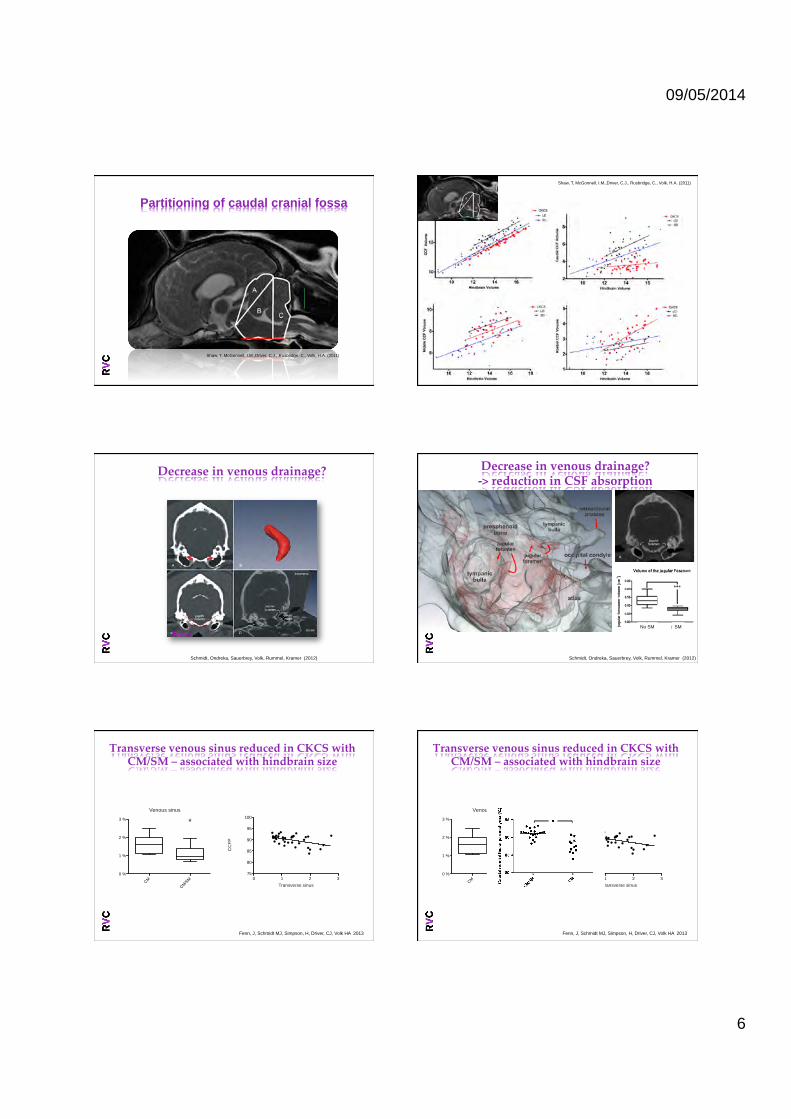
09/05/2014
6
Partitioning of caudal cranial fossa
Shaw, T, McGonnell, I.M.,Driver, C.J., Rusbridge, C., Volk, H.A. (2011)
Shaw, T, McGonnell, I.M.,Driver, C.J., Rusbridge, C., Volk, H.A. (2011)
Decrease in venous drainage?
Schmidt, Ondreka, Sauerbrey, Volk, Rummel, Kramer (2012)
Decrease in venous drainage? -‐‑> reduction in CSF absorption
SM No SM
Schmidt, Ondreka, Sauerbrey, Volk, Rummel, Kramer (2012)
Transverse venous sinus reduced in CKCS with CM/SM – associated with hindbrain size
Venous sinus
CM
CM/SM0 %
1 %
2 %
3 % *
0 1 2 375
80
85
90
95
100
Transverse sinus
CC
FP
Fenn, J, Schmidt MJ, Simpson, H, Driver, CJ, Volk HA 2013
Transverse venous sinus reduced in CKCS with CM/SM – associated with hindbrain size
Venous sinus
CM
CM/SM0 %
1 %
2 %
3 % *
0 1 2 375
80
85
90
95
100
Transverse sinus
CC
FP
Fenn, J, Schmidt MJ, Simpson, H, Driver, CJ, Volk HA 2013
Thirty-four dogs fulfilled the inclusion criteria (17 male, 17 fe-male), with a mean age of 4.2 ± 1.8 years and mean body weightof 9.7 ± 2.2 kg. SM was present in 22 dogs (10 male, 12 female)and absent in the remaining 12 (7 male, 5 female). There was nosignificant difference in either age (P = 0.145) or body weight(P = 0.341) between the CKCS with CM/SM and CM only. Commonclinical signs were phantom scratching, facial rubbing and vocali-sation, as well as scoliosis and ataxia. At least one of these clinicalsigns was seen in 16/22 (72.7%) dogs with SM and in 4/12 (33.3%)dogs without.
There was a significant difference in CCFV% (P = 0.001), with asmaller percentage of the CCF taken up by venous sinus in CM/SM group (median 1.0%, range 0.6–1.8) in comparison to the CMgroup (median 1.6%, range 1.3–2.3) (Fig. 1). CCFP% was signifi-cantly higher in the CM/SM group (P < 0.001), with a mean of91.1 ± 1.3% compared to 87.3 ± 1.9% in the CM group (Fig. 2). Pear-son’s correlation coefficients revealed a significant negative corre-lation between CCFP% and CCFV% (r = �0.464, P = 0.006).Anomalous vessels in the CCF were seen in 1/22 (4.5%) dogs inthe CM/SM group and 2/12 (16.7%) dogs in the CM only group; thisdifference was not statistically significant (P = 0.279).
The findings of this study suggest that CCF venous sinus volumeis reduced in CKCS with SM compared to those without SM, as wellas providing further support for the theory of parenchymal over-crowding of the CCF in these dogs (Driver et al., 2010a,b; Shawet al., 2012). The reduction of the CCF venous sinus volume in dogswith SM supports a possible role for reduced venous drainage inthe pathogenesis of SM (Williams, 2008; Schmidt et al., 2012). SinceSM is a progressive disease in CKCS (Driver et al., 2012), it is possiblethat someyoungdogs in theCMgroupmaydevelop SM in the future,which is a potential limitation of the current study.
Narrowing of the jugular foramen in CKCS with SM (Schmidtet al., 2012) may potentiate the reduction in venous outflow fromthe CCF. Blood vessels have been shown to have an important rolein the formation and maintenance of cranial foramina (Akbareianet al., 2009). The narrowing could be part of the CM complex orsecondary to a reduction of blood vessel size in these dogs. In CKCSwith CM, the combination of an overcrowded CCF and reduced ve-nous sinus volume may result in a reduction in CSF absorption andsubsequently a change in CSF flow dynamics, tissue complianceand venous drainage. This may contribute to the eventual develop-ment of SM, as proposed by Williams (2008). The ventricular sys-tem is enlarged in CKCS with CM/SM compared to CKCS with CMalone (Driver et al., 2010a,b). The ventricular system volume is alsopositively associated with the size of the syrinx in these studies,suggesting that a dynamic relationship exists between SM, ventri-culomegaly and venous drainage in dogs.
Conflict of interest statement
None of the authors of this paper has a financial or personalrelationship with other people or organisations that could inappro-priately influence or bias the content of the paper.
Acknowledgements
This manuscript was assessed by the Research Office accordingto the Royal Veterinary College’s code of good research practice(Authorisation Number VCS_00520).
References
Akbareian, S., Pitsillides, A., McGonnell, I., 2009. Cranial foramina development inthe chick embryo. Journal of Anatomy 214, 778–802.
Cerda-Gonzalez, S., Olby, N.J., Broadstone, R., McCullough, S., Osborne, J.A., 2009.Characteristics of cerebrospinal fluid flow in Cavalier King Charles spanielsanalyzed using phase velocity CINE magnetic resonance imaging. VeterinaryRadiology and Ultrasound 50, 467–476.
Cross, H.R., Cappello, R., Rusbridge, C., 2009. Comparison of cerebral craniumvolumes between Cavalier King Charles spaniels with Chiari-like malformation,small breed dogs and Labradors. Journal of Small Animal Practice 50, 399–405.
Driver, C.J., Rusbridge, C., Cross, H.R., McGonnell, I., Volk, H.A., 2010a. Relationship ofbrain parenchyma within the caudal cranial fossa and ventricle size tosyringomyelia in Cavalier King Charles spaniels. Journal of Small AnimalPractice 51, 382–386.
Driver, C.J., Rusbridge, C., McGonnell, I.M., Volk, H.A., 2010b. Morphometricassessment of cranial volumes in age-matched Cavalier King Charles spanielswith and without syringomyelia. Veterinary Record 167, 978–979.
Driver, C.J., De Risio, L., Hamilton, S., Rusbridge, C., Dennis, R., McGonnell, I.M., Volk,H.A., 2012. Changes over time in craniocerebral morphology and syringomyeliain Cavalier King Charles spaniels with Chiari-like malformation. BMC VeterinaryResearch 8, 215.
Moritani, T., Aihara, T., Oguma, E., Makiyama, Y., Nishimoto, H., Smoker, W.R., Sato,Y., 2006. Magnetic resonance venography of achondroplasia: Correlation ofvenous narrowing at the jugular foramen with hydrocephalus. Clinical Imaging30, 195–200.
Schmidt, M.J., Ondreka, N., Rummel, C., Volk, H., Sauerbrey, M., Kramer, M., 2012.Volume reduction of the jugular foramina in Cavalier King Charles spaniels withsyringomyelia. BMC Veterinary Research 8, 158.
Shaw, T.A., McGonnell, I.M., Driver, C.J., Rusbridge, C., Volk, H.A., 2012. Increase incerebellar volume in Cavalier King Charles spaniels with Chiari-likemalformation and its role in the development of syringomyelia. PLoS One 7,e33660.
Williams, H., 2008. A unifying hypothesis for hydrocephalus, Chiari malformation,syringomyelia, anencephaly and spina bifida. Cerebrospinal Fluid Research 5, 7.
Fig. 1. Box and whisker plots showing the distribution of venous sinus volume as apercentage of the caudal cranial fossa (CCFV%) between Cavalier King Charlesspaniels with Chiari-like malformation and syringomyelia (CM/SM), and those withChiari-like malformation alone (CM). Bars represent medians, boxes representinterquartile ranges and whiskers represent minimum and maximum values(Mann–Whitney U test; �P < 0.05).
Fig. 2. Scatter plots showing the distribution of parenchymal volume as apercentage of the caudal cranial fossa (CCFP%) between Cavalier King Charlesspaniels with Chiari-like malformation and syringomyelia (CM/SM), and those withChiari-like malformation alone (CM) (Bars represent means; Student’s t test;�P < 0.05).
2 J. Fenn et al. / The Veterinary Journal xxx (2013) xxx–xxx
Please cite this article in press as: Fenn, J., et al. Venous sinus volume in the caudal cranial fossa in Cavalier King Charles spaniels with syringomyelia. TheVeterinary Journal (2013), http://dx.doi.org/10.1016/j.tvjl.2013.05.007
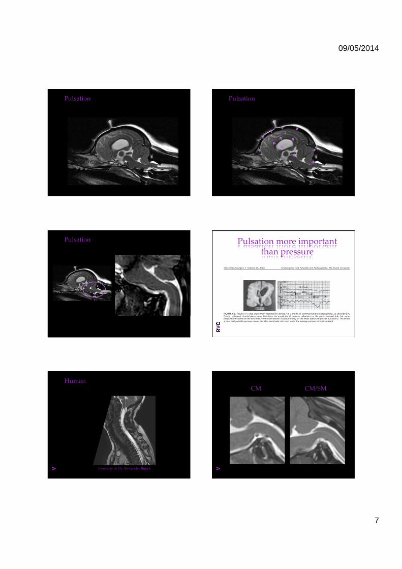
09/05/2014
7
Pulsation Pulsation
é
é é é
éé
é
é
é
é
éé
Pulsation Pulsation more important than pressure
A second engineering solution, though, would say let’sput a mass that has to be moved between the artery and thevein and that mass could be something like cerebral spinalfluid and the amount of that fluid could be a variable andcould be changed as could the elasticity in the wall. This endsup giving us a model where we can have isolation of a veryparticular band of the frequency spectrum that can be takenout. This can be shown mathematically. Interestingly, HughDavson, in 1956, really had the anatomy relevant to thisfigured out and here we are showing some of these notchfilters, or band-stop filters with different models of how themass must relate to the resistance and the capacitance of thissystem.
Experimental Demonstration of a “NotchFilter”
We have been able to look at this filtering mechanism,i.e., the transfer between the arterial blood pressure waveform and the intracranial pressure wave form using a tech-nique called time-varying transfer function, which has beenadapted in my lab by Dr. Rui Zou. We will not highlight themathematical rigors here. The important thing is that we willbe thinking of the brain as a black box system capable oftranslating an input signal to an output signal, and it can havea different gain for a particular frequency component withinthis input signal in terms of how it ends up in the outputsignal. Figure 6.6 shows data taken from dogs in the labora-tory of Michael Egnor at SUNY Stony Brook. In this exper-iment, the intracranial pressure is being monitored and tran-siently forced to go up rapidly with an injection of CSF intothe lumbar subarachnoid space. The intracranial pressure(ICP) and arterial blood pressure were recorded with highfidelity recording.
The upper graph in Figure 6.6 plots the gain of thetransfer function at different values of the frequency of theenergy in the blood pressure (BP) and ICP tracings, and howfar the experiment has progressed (therefore it is a time-
FIGURE 6.6. Time-varying transfer function analysis of caninearterial blood pressure and ICP, during an increase in ICPcaused by infusion of excess CSF. The so-called gain of thetransfer function relates how much of the amplitude of thesignal in various frequency bands. Normally (as at the begin-ning and end of the experiment), there is a sharp “notch” atthe frequency corresponding to the animals heart rate. Bymechanisms suggested in the text, movement of CSF withinthe intracranial system could produce a “notch filter” whichwould decrease the relative amplitude swings of the ICP com-pared with the blood pressure. The experimental addition ofexcess CSF, like many pathological occurrences, may perturbthe notch at the correct frequency.
FIGURE 6.5. Results of a dog experiment reported by Bering.9 In a model of communicating hydrocephalus, as described byDandy, unilateral choroid plexectomy diminishes the amplitude of pressure pulsations on the plexectomized side, but meanpressure is the same on the two sides. Ventricular dilation occurs primarily on the intact side (with greater pulsations). This favorsa view that pulsatile pressure waves can alter ventricular size even when the average pressure is kept constant.
Clinical Neurosurgery • Volume 53, 2006 Cerebrospinal Fluid Pulsatility and Hydrocephalus: The Fourth Circulation
© 2006 Lippincott Williams & Wilkins 51
Human
Courtesy of Dr. Alexander Bunck
CM CM/SM
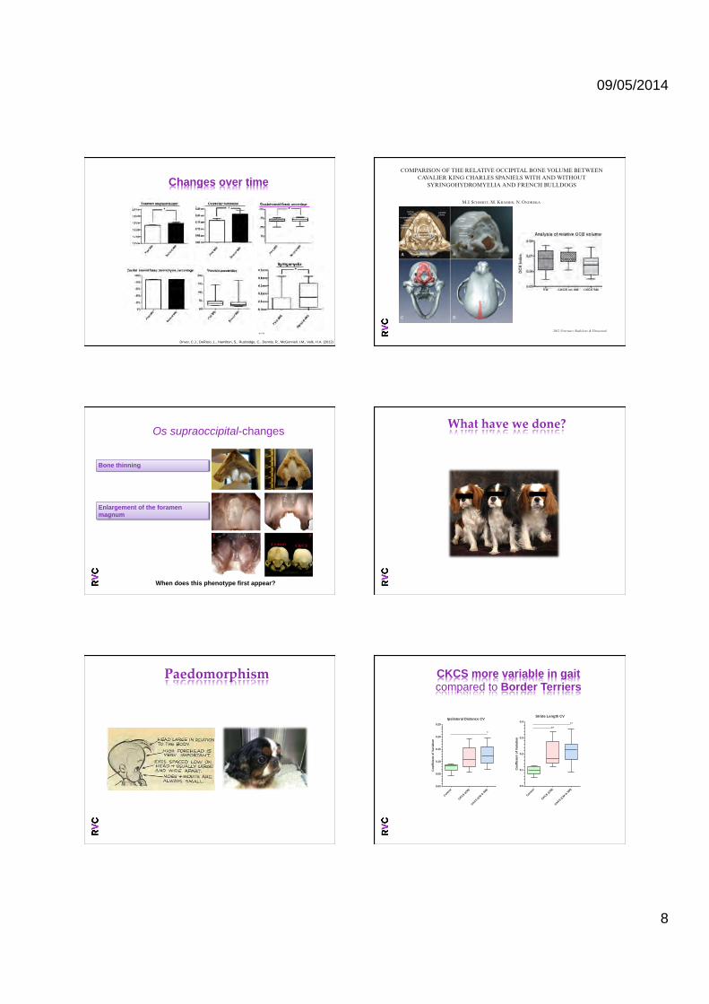
09/05/2014
8
e
*p<0.05
*p<0.05
Changes over time
Driver, C.J., DeRisio, L., Hamilton, S., Rusbridge, C., Dennis, R., McGonnell, I.M., Volk, H.A. (2012)
COMPARISON OF THE RELATIVE OCCIPITAL BONE VOLUME BETWEENCAVALIER KING CHARLES SPANIELS WITH AND WITHOUT
SYRINGOHYDROMYELIA AND FRENCH BULLDOGS
M.J. SCHMIDT, M. KRAMER, N. ONDREKA
Our aim was to determine the relative volume of the occipital bone of Cavalier King Charles spaniels withand without syringohydromyelia and normal French bulldogs to reappraise the role of a possible insufficiencyof the paraxial mesoderm in the pathogenesis of the caudal occipital malformation syndrome. Analysis ofthe occipital bone volume of 43 dogs based on computed tomography datasets was performed. Volume wasdetermined by means of three-dimensional models. Using manual segmentation of the occipital bone in sagittal,transverse, and dorsal images, the volume of the occipital bone and the rest of the skull was calculated. Theabsolute occipital bone volume was put in relation to the total skull volume, the occipital bone index. Forthe Cavalier King Charles spaniels without syringomyelia, the median occipital bone index was 0.0681; forCavalier King Charles spaniels with syringomyelia, it was 0.0646 and for French bulldogs, it was 0.0676.There was no global difference of the occipital bone index between examined groups (P = 0.4331). A reducedvolume of the occipital bone was not found in Cavalier King Charles spaniels in general in comparison toFrench bulldogs, or in Cavalier King Charles spaniels with syringomyelia compared to Cavalier King Charlesspaniels without syringomyelia. These results do not support occipital hypoplasia as a cause for syringomyeliadevelopment, challenging the paraxial mesoderm insufficiency theory. This also suggests that the term Chiari-like malformation, a term derived from human studies, is not appropriate in the Cavalier King Charles spaniel.C⃝ 2012 Veterinary Radiology & Ultrasound.
Key words: dog, paraxial mesoderm, skull morphology.
Introduction
THE MAMMALIAN SKULL is a composite structure ofbony elements that derives from embryonic mesoder-
mal and neural crest cells.1, 2 The parts of the neurocraniumcan be distinguished by their tissue origin and also by themechanism of bone formation. The frontal, parietal, andtemporal bones develop by intramembranous ossificationwhereas the bones of the skull base develop from enchon-dral ossification.2 The skull and vertebral column begin toform along the notochord as condensations of the mes-enchyme, the paraxial mesoderm. The cranial base bonesdevelop from the cranial paraxial mesoderm that lies be-tween the ectoderm of the developing neural tube and theendoderm of the pharynx.3 In mammals, the cells of the cra-nial paraxial mesoderm eventually turn into the primordiaof the basioccipital, exooccipital, and supraoccipital bone
From the Department of Veterinary Clinical Science, Small Ani-mal Clinic, Justus-Liebig-University, 35392, Gießen, Germany (Schmidt,Kramer, Ondreka)
Address correspondence and reprint requests to M.J. Schmidt, atthe above address. E-mail: [email protected].
Received November 08, 2011; accepted for publication April 17, 2012.doi: 10.1111/j.1740-8261.2012.01955.x
that grow around the spinal cord/medulla and form theforamen magnum.4–6
Chiari type 1 malformation in people is characterizedby a smaller caudal fossa volume with descent of cere-bellar tonsils through the foramen magnum.7 In contrast,the Chiari-like malformation in the Cavalier King Charlesspaniel is characterized by indentation of the occipital(bone) with cerebellar herniation and is more correctlytermed caudal occipital malformation syndrome.8 As theoccipital bone is supposed to be a key structure in thechanges leading to the Chiari malformation type 1 in hu-mans and occipital malformation in the Cavalier KingCharles spaniel,8–10 a genetically transmitted developmen-tal disorder of the occipital bone primordium is suspected.8
It was suggested that this blastema underdevelopment leadsto a decreased volume of the caudal cranial fossa later indevelopment. This in turn was thought to cause a displace-ment of the caudal cerebellar vermis into or through theforamen magnum.9, 10 This was proposed for the diseasein humans and also adopted as a pathogenetic model forthe dog. The study of the morphology of the brain andskull of the Cavalier King Charles spaniel of the last yearshas been strongly influenced by this hypothesis. Since then
Vet Radiol Ultrasound, Vol. 00, No. 00, 2012, pp 1–10.
1
VOL. 00, NO. 00 VOLUME OF THE OCCIPITAL BONES IN CAVALIER KING CHARLES SPANIEL 3
FIG. 1. Three-dimensional rendering of the skull of a Cavalier King Charles spaniel in a caudal view. (A) The asterisks mark the cut surface of the condylesafter removal of the vertebrae. The caudal view of the occipital bone is presented compared to a corresponding skull preparation of a French bulldog. (B)Manual segmentation (red) of the occipital bone from the rest of the skull in 2D CT images produced the volume of the bone mass in relation to the skullvolume (white, C, D)
chosen to test the global group differences using a Kruskal–Wallis test for nonparametric data followed by a post-hocDunn’s Multiple Comparison Test. P-values less than 0.05were considered statistically significant.
Results
Group 1 included six male and nine female Cavalier KingCharles spaniels; the age ranged from 4 to 8 years andweight from 8 to 12 kg (median 10.5 kg). Group 2 included10 female and five male Cavalier King Charles spaniels.The age in this group ranged from 4 to 7 years and weightranged from 8 to 12 kg (median 10 kg). Eight of the FrenchBulldogs were female and seven were male. Their age rangedfrom 1.5 to 9 years, and weight from 8 to 12 kg (median11 kg). The median weight was not different between groups(P = 0.21).
The median occipital bone index was 0.0646 in Cav-alier King Charles spaniels with syringomyelia, 0.0676in Cavalier King Charles spaniels without syringomyelia,
and 0.0681 in the French bulldogs (group 3). There wasno global difference (P = 0.4331) and Dunns multiplecomparison test revealed no significant difference betweenFrench bulldogs and Cavalier King Charles spaniels withsyringomyelia (P > 0.05), between French bulldgos andCavalier King Charles spaniels without syringomyelia (P> 0.05), or between Cavalier King Charles spaniels with orwithout syringomyelia (P > 0.05) (Fig. 2).
Discussion
Several morphometric analyses have been conducted onthe caudal cranial fossa volume of Cavalier King Charlesspaniels.11–16 Results of studies proposing a mismatch be-tween cerebellar and caudal cranial fossa volume in thisbreed and in comparison to other breeds were controver-sial. In some studies, there was a mismatch between caudalfossa parenchyma and caudal fossa volume in dogs withsyringomyelia and overcrowding was proposed as a causeof syringomyelia development. In most studies, however,no difference was found between caudal fossa volume in
4 SCHMIDT ET AL 2012
FIG. 2. Results of the one-way analysis of variance (ANOVA) presentedas median, range, 25th, and 75th precentiles in a box and whisker plot. ForCavalier King Charles spaniels without syringomyelia, the median occipitalbone index was 0.0681; Cavalier King Charles spaniels with syringomyeliahad a median occipital bone index of 0.0646. The median occipital boneindex of French bulldogs was 0.0676. There was no difference of the occipitalbone index between examined groups (P = 0.4331). Groupwise comparisonwas also not significantly different (P > 0.05). CKCS, Cavalier King Charlesspaniel; FB, French Bulldog; OCB, occipital bone; SM, syringomyelia.
Cavalier King Charles spaniels with and without sy-ringomyelia, although this was not universal.13, 14
Although the caudal fossa volume may be unchanged,the shape of the occipital bone is a characteristic feature ofthe Cavalier King Charles spaniel skull.13, 18 This raised thequestion whether this skull formation could be the resultof underdevelopment of the occipital bone that is compen-sated for by the parietal bone and does therefore not affectthe volume of the caudal cranial fossa but leads to an alteredconfiguration and positioning of the bone surrounding thecerebellum affecting the flow of cerebrospinal fluid.19
There is support for this mesodermal insufficiency the-ory as an underlying cause of Chiari malformation inpeople.10 Administration of Vitamin A to pregnant ham-sters causes a reduction in the number of cells of theparaxial mesoderm and induces changes consistent withChiari malformation types 1 and 2, as well as various typesof axial skeletal-dysraphic disorders.10 In this model, thebasichondrocranium, especially the basioccipital bone ofhamster fetuses, is shorter than normal. This defect re-sults in a short and small posterior cranial fossa, whichis inadequate to contain the developing nervous structuresof that region. The developing cerebellum becomes dis-placed downward but does not herniate through the fora-men magnum. The posterior fossa is the analog of the cau-dal cranial fossa in dogs and the same pathogenesis hasbeen proposed for the Cavalier King Charles spaniel.20
However, the extensive malformations seen in the ham-sters exceed the morphologic findings in dogs and humans.The hamsters had severe defects of the cerebrum (micro-cephaly, encephalocele), and eyes (micropthalmia, absenceof eyelids), and also had neural tube defects (rachishisis,
spina bifida, meningomyelocele) and severe reduction ofthe maxilla with protrusion of the tongue, vertebral seg-mentation failures, and aberrant formation of the verte-brae. These may be seen in Chiari malformation types2–4 and have been described in calves with Chiari mal-formation type 1,21 but are not part of Chiari malforma-tion 1 in humans and have never been described in as-sociation with canine caudal occipital malformation syn-drome. Therefore, the explanation of the pathogenesis ofcaudal occipital malformation in the Cavalier King Charlesspaniel from this model must be assessed with caution. An-other shortcoming of this theory is that the cranial parax-ial mesoderm consists of a multiprogenitor mesodermalcell population that participates extensively in craniofacialmorphogenesis. It contributes to the development of thebranchial arches, from which the mandible, the hyoid ap-paratus, and the cranial musculature develop. Growth dis-turbances of the paraxial mesoderm should therefore notaffect the occipital bone.22 Mandibular dysplasia has onlybeen described anecdotally in the Cavalier King Charlesspaniel.23
Based on our results, we cannot exclude an underdevel-opment of the embryological primordium of the occipitalbone that could lead to a smaller bone in the adult Cava-lier King Charles spaniel, but if the occipital bone shouldbe reduced in the Cavalier King Charles spaniel, then itshould be reduced in French bulldogs too, which are notpredisposed to caudal occipital malformation syndrome.In this context, the similar caudal fossa volume of Cava-lier King Charles spaniels with and without syringomyeliamakes a causal relationship between the occipital bonevolume and caudal occipital malformation syndromeunlikely.
The examination of more brachycephalic breeds wouldbe ideal, however, the growth of the brain and skull un-derlies individual allometric patterns that influence therelative proportions of the brain and skull.24, 25 The con-trol of postnatal skeletal and craniofacial growth involvescomplex interactions of genes, nutrients, and hormones.4
Mild imbalances involving any of these factors, sepa-rately or combined, can lead to a variety of pheno-typic appearances in the dog. Chihuahuas and pom-meranians have been classified as ateliotic dogs due toabsent or decreased growth hormone showing propor-tioned dwarfism. Other dogs have been identified as achon-droplastic and their reduced growth is due to a defectin cartilage growth and/or development.26 Brachycephalyis a feature in both groups as well as in the Cavalier KingCharles spaniel.27, 28 Given the extreme heterogeneity of thecanine breeds sharing brachycephalic features, the randomcomparison of dogs of this group seems to be unsound.As no data about the allometric growth of different dogbreeds exist, we decided on the French Bulldog as a con-trol breed because they share the same weight range and a
Os supraoccipital-changes
Bone thinning
Enlargement of the foramen magnum
When does this phenotype first appear?
What have we done?
Paedomorphism CKCS more variable in gait compared to Border Terriers
Ipsilateral Distance CV
Control
CKCS (CM)
CKCS (CM &
SM)0.00
0.05
0.10
0.15
0.20
0.25
*
Coe
ffici
ent o
f Var
iatio
n
Stride Length CV
Control
CKCS (CM)
CKCS (CM &
SM)0.0
0.1
0.2
0.3
0.4
Coe
ffici
ent o
f Var
iatio
n
****
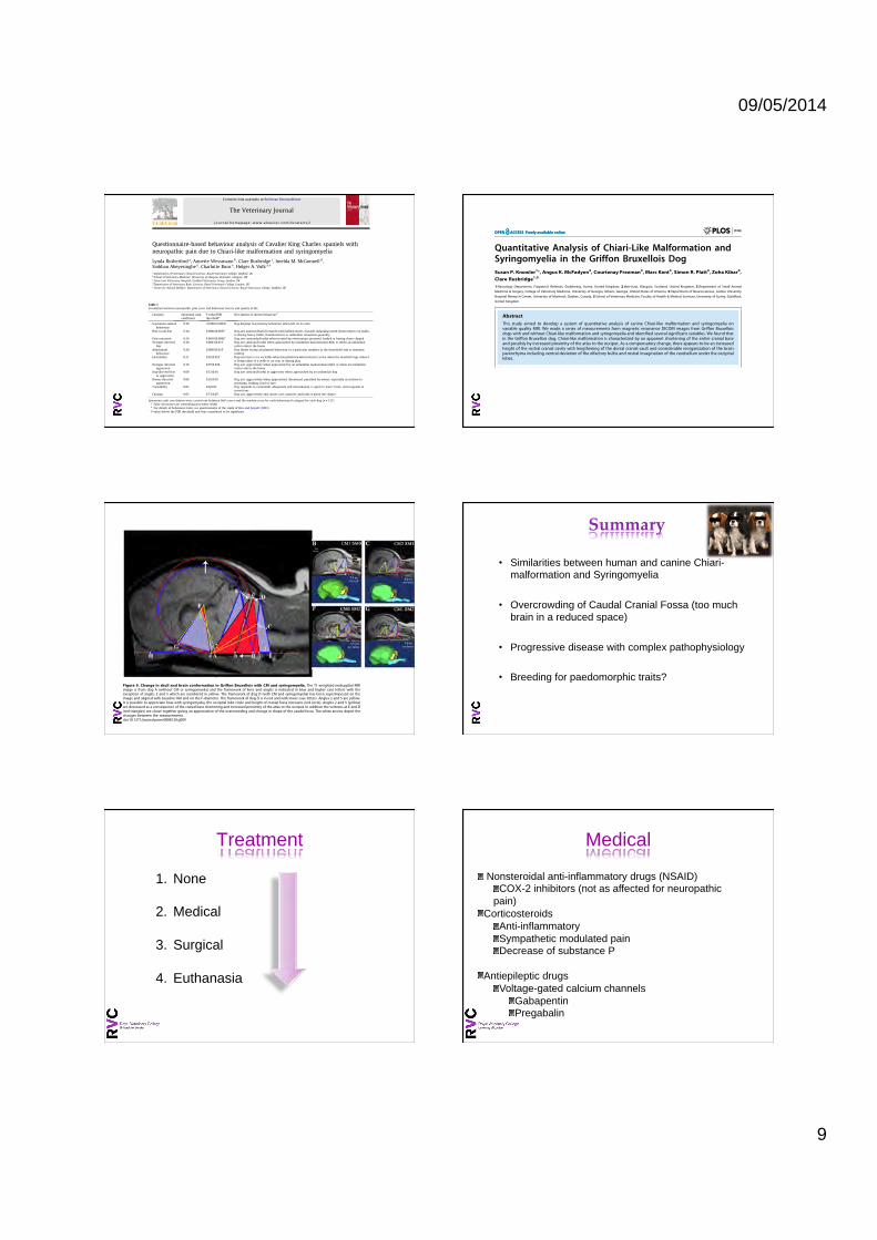
09/05/2014
9
Questionnaire-based behaviour analysis of Cavalier King Charles spaniels withneuropathic pain due to Chiari-like malformation and syringomyelia
Lynda Rutherford a, Annette Wessmann b, Clare Rusbridge c, Imelda M. McGonnell d,Siobhan Abeyesinghe e, Charlotte Burn e, Holger A. Volk a,⇑aDepartment of Veterinary Clinical Sciences, Royal Veterinary College, Hatfield, UKb School of Veterinary Medicine, University of Glasgow, Bearsden, Glasgow, UKc Stone Lion Veterinary Hospital, Goddard Veterinary Group, London, UKdDepartment of Veterinary Basic Sciences, Royal Veterinary College, London, UKeCentre for Animal Welfare, Department of Veterinary Clinical Sciences, Royal Veterinary College, Hatfield, UK
a r t i c l e i n f o
Article history:Accepted 20 May 2012Available online xxxx
Keywords:SyringomyeliaChiari-like malformationCavalier King Charles spanielPainQuality of life
a b s t r a c t
Chiari-like malformation (CM)/syringomyelia (SM) is a disease complex recognised in Cavalier KingCharles spaniels (CKCSs) that can lead to neuropathic pain (NeP). In humans, NeP is associated with anx-iety, depression and reduced quality of life (QoL). In this study, databases of three specialist veterinarycentres were searched and CKCS breed societies and health forums were contacted to identify CKCS withan imaging diagnosis of CM/SM. Owners completed questionnaires on behaviour, signalment, generalhealth status, NeP and QoL. Data were analysed from 122 dogs out of 564 questionnaires completed, afterincomplete questionnaires and data from dogs that had other potentially debilitating disease processeswere excluded. NeP severity score was significantly and positively correlated with ‘stranger-directed’ fear(rS = 0.28), non-social fear (rS = 0.34), ‘separation-related’ behaviour (rS = 0.38), attachment behaviour(rS = 0.24), excitability (rS = 0.21) and proxy for pain sensation (rS = 0.29). Increased NeP was also signif-icantly associated with decreased QoL (rS = 0.47), ability to settle (rS = 0.26) and willingness to exercise(rS = 0.50). Severity of NeP was positively associated with certain fear-associated behaviour and withdecreased owner-perceived QoL. Thus, neurobehavioural changes should be considered in the manage-ment of NeP in CKCS with CM/SM.
� 2012 Elsevier Ltd. All rights reserved.
Introduction
An estimated 95% of Cavalier King Charles spaniels (CKCSs) areaffected by Chiari-like malformation (CM) and around 70% developsyringomyelia (SM) (Dewey and Rusbridge, 2008; Parker et al.,2011). In humans, Chiari-type I malformation (the human counter-part to CM) and SM are associated with neuropathic pain (NeP) andpresent with a variety of symptoms, including headaches, pain inthe trigeminal territory, back pain, temporomandibular joint painand spontaneous pain (Todor et al., 2000; Thimineur et al., 2002).NeP is defined as pain arising as a direct consequence of a lesionor disease process which affects the somatosensory system (Grubb,2010). The resulting abnormal somatosensory processing can man-ifest as (1) dysaesthesia, a spontaneous or evoked unpleasantabnormal sensation (people with this condition describe burning-type pain, pins and needles and other strange sensations); (2) allo-dynia, pain from a stimulus which is not normally painful, such aslight touch or motion; (3) hyperpathia, increased pain from stimuli
which are normally painful; or (4) causalgia, a constant, burning-type pain (Gilron et al., 2006; Rusbridge and Jeffery, 2008).
CM/SM in CKCS is commonly associated with pain, especially inthe cervical region, and with neurological dysfunction, e.g. scolio-sis, limb paresis and ataxia (Rusbridge et al., 2000, 2006). Affecteddogs might be hypersensitive to touch and often scratch an area onthe shoulder, ear, neck or sternum, commonly only on one side ofthe body without making skin contact, so called phantom scratch-ing (Rusbridge et al., 2006). Some dogs perform facial or head rub-bing or spontaneous vocalisations. Such clinical signs are thoughtto indicate the presence of the animal’s perception of NeP, possiblydue to damage of the spinal dorsal horn grey matter secondary toSM, as well as overcrowding of the foramen magnum and subse-quent compression of the trigeminal nuclei in the caudal brainstem(Todor et al., 2000; Rusbridge et al., 2006; Rusbridge and Jeffery,2008).
Maximal syrinx width, length and asymmetry strongly predictsigns of pain, whilst the degree of cerebellar herniation does not(Lu et al., 2003; Rusbridge et al., 2007; Cerda-Gonzalez et al.,2009). The dorsal horn of the spinal grey matter is central forprocessing and transmission of sensory information to the brain.
1090-0233/$ - see front matter � 2012 Elsevier Ltd. All rights reserved.http://dx.doi.org/10.1016/j.tvjl.2012.05.018
⇑ Corresponding author. Tel.: +44 170 7666365.E-mail address: [email protected] (H.A. Volk).
The Veterinary Journal xxx (2012) xxx–xxx
Contents lists available at SciVerse ScienceDirect
The Veterinary Journal
journal homepage: www.elsevier .com/ locate/ tv j l
Please cite this article in press as: Rutherford, L., et al. Questionnaire-based behaviour analysis of Cavalier King Charles spaniels with neuropathic pain dueto Chiari-like malformation and syringomyelia. The Veterinary Journal (2012), http://dx.doi.org/10.1016/j.tvjl.2012.05.018
overall QoL (score 5) and 4/120 (3%) thought that it was neithergood nor bad (score 4). All other owners perceived their dog tohave a fairly good to excellent QoL. QoL tended to be worse withincreasing weight (rS = 0.25; P = 0.0086; FDR 0.017), but not withincreasing age. Treated dogs had a significantly worse QoL (medianscore 2; range 1–5) than untreated dogs (median score 2; range 1–3; P < 0.0001; FDR 0.0045). There was no significant difference be-tween female and male dogs.
NeP score was not correlated with age or weight. Male and fe-male dogs did not differ significantly in NeP score or in behaviourscores. Treated dogs had a higher NeP score than untreated dogs
(median score 1.3, range 0.1–3.4 vs. median 0.4, range 0–2.7;P < 0.0001, FDR 0.0045).
Discussion
The aim of this study was to determine if NeP associated withCM/SM has an impact on behaviour and on owner-perceived QoLof CKCS. Dogs with higher owner-perceived NeP severity scoreswere more likely to demonstrate stranger-directed fear, non-socialfear, separation-related and attachment behaviour, excitability and
Table 1Correlation between neuropathic pain score and behaviour factors and quality of life.
Category Spearman rankcoefficient
P value/FDRthresholda
Description of shown behaviourb
Separation-relatedbehaviour
0.38 <0.0001/0.0045* Dog displays fear/anxiety behaviour when left on its own
Non-social fear 0.34 0.0002/0.0055* Dog acts anxious/fearful towards loud/sudden noises, towards unfamiliar/wind-blown objects on walks,or during heavy traffic, thunderstorms or unfamiliar situations generally
Pain sensation 0.29 0.0018/0.0068* Dog acts anxiously/fearful when treated by veterinarian, groomed, bathed or having claws clippedStranger-directed
fear0.28 0.0023/0.011* Dog acts anxious/fearful when approached by unfamiliar man/woman/child, or when an unfamiliar
visitor enters the homeAttachment
behaviour0.24 0.0095/0.015* Dog shows strong attachment behaviour to a particular member in the household and is attention
seekingExcitability 0.21 0.02/0.022* Dog overreacts or is excitable when household members/visitors arrive, when the doorbell rings, when it
is being taken to a walk or car trip, or during playStranger-directed
aggression0.16 0.07/0.034 Dog acts aggressively when approached by an unfamiliar man/woman/child, or when an unfamiliar
visitor enters the homeDog-directed fear
or aggression0.09 0.32/0.05 Dog acts anxious/fearful or aggressive when approached by an unfamiliar dog
Owner-directedaggression
0.08 0.39/0.05 Dog acts aggressively when approached, threatened, punished by owner, especially in relation togrooming, bathing, food or toys
Trainability 0.05 0.6/0.05 Dog responds to commands adequately and immediately, is quick to learn ‘tricks’ and responds tocorrections
Chasing �0.03 0.73/0.05 Dog acts aggressively and chases cats, squirrels and birds if given the chance
Spearman rank correlations were carried out between NeP scores and the median score for each behavioural category for each dog (n = 122).a False discovery rate controlling procedure (FDR).b For details of behaviour traits, see questionnaire of the study of Hsu and Serpell (2003).
* P value below the FDR threshold and thus considered to be significant.
Qua
lity
of li
fe s
core
Mean neuropathic pain score
Fig. 1. Association of owner-perceived quality of life (QoL) with mean neuropathic pain score. QoL score was positively associated with the neuropathic pain score (rS = 0.47;P < 0.0001; FDR 0.0045). A larger QoL score indicates a poorer quality of life (1 = could not be better; 7 = could not be better), whereas a larger neuropathic pain score indicatesa higher frequency of seven pain-related behaviours (1 = seldom; 4 = always). The size of each data point shows the number of dogs represented by that point and, for pointsrepresenting more than one dog, the precise number is shown in white within the point. NeP score median was 1 (range 0–3.43, IQR 0.43–1.61) and QoL had a median of 2(range 1–5, IQR 2–3).
L. Rutherford et al. / The Veterinary Journal xxx (2012) xxx–xxx 3
Please cite this article in press as: Rutherford, L., et al. Questionnaire-based behaviour analysis of Cavalier King Charles spaniels with neuropathic pain dueto Chiari-like malformation and syringomyelia. The Veterinary Journal (2012), http://dx.doi.org/10.1016/j.tvjl.2012.05.018
Quantitative Analysis of Chiari-Like Malformation andSyringomyelia in the Griffon Bruxellois DogSusan P. Knowler1*, Angus K. McFadyen2, Courtenay Freeman3, Marc Kent3, Simon R. Platt3, Zoha Kibar4,
Clare Rusbridge1,5
1Neurology Department, Fitzpatrick Referrals, Godalming, Surrey, United Kingdom, 2 akm-stats, Glasgow, Scotland, United Kingdom, 3Department of Small Animal
Medicine & Surgery, College of Veterinary Medicine, University of Georgia, Athens, Georgia, United States of America, 4Department of Neurosciences, Justine University
Hospital Research Center, University of Montreal, Quebec, Canada, 5 School of Veterinary Medicine, Faculty of Health & Medical Sciences, University of Surrey, Guildford,
United Kingdom
Abstract
This study aimed to develop a system of quantitative analysis of canine Chiari-like malformation and syringomyelia onvariable quality MRI. We made a series of measurements from magnetic resonance DICOM images from Griffon Bruxelloisdogs with and without Chiari-like malformation and syringomyelia and identified several significant variables. We found thatin the Griffon Bruxellois dog, Chiari-like malformation is characterized by an apparent shortening of the entire cranial baseand possibly by increased proximity of the atlas to the occiput. As a compensatory change, there appears to be an increasedheight of the rostral cranial cavity with lengthening of the dorsal cranial vault and considerable reorganization of the brainparenchyma including ventral deviation of the olfactory bulbs and rostral invagination of the cerebellum under the occipitallobes.
Citation: Knowler SP, McFadyen AK, Freeman C, Kent M, Platt SR, et al. (2014) Quantitative Analysis of Chiari-Like Malformation and Syringomyelia in the GriffonBruxellois Dog. PLoS ONE 9(2): e88120. doi:10.1371/journal.pone.0088120
Editor: Claire Wade, University of Sydney, Australia
Received June 16, 2013; Accepted January 4, 2014; Published February 12, 2014
Copyright: � 2014 Knowler et al. This is an open-access article distributed under the terms of the Creative Commons Attribution License, which permitsunrestricted use, distribution, and reproduction in any medium, provided the original author and source are credited.
Funding: Contributions towards the cost of magnetic resonance imaging for most of the dogs in this study were made by the following organizations orindividuals: Syringomyelia DNA Research (http://www.veterinary-neurologist.co.uk/Syringomyelia/DNA_Research/); American Kennel Club Canine HealthFoundation Grant #1004; The Griffon Bruxellois Club 1897; For the Love of Ollie Fund; Friends of Lola Fund and Lee and Frank Pieterse. The CanadianInstitutes of Health Research and Cavalier Matters Charity Reg No 1141674 contributed towards the cost of leasing the DICOM viewing software package MIMICS14.12 Materialise (Technologielaan 15 3001 Leuven Belgium). The funders had no role in study design, data collection and analysis, decision to publish, orpreparation of the manuscript.
Competing Interests: We have the following interests: Clare Rusbridge is employed by Fitzpatrick Referrals and the University of Surrey and Susan P Knowlerworks voluntarily and is affiliated with Fitzpatrick Referrals. Angus K McFadyen is an independent statistical consultant operating professionally as a sole traderunder the company name of akm-stats. There are no patents, products in development or marketed products to declare. This does not alter our adherence to allthe PLOS ONE policies on sharing data and materials, as detailed online in the guide for authors.
* E-mail: [email protected]
Introduction
Canine Chiari-like malformation (CM) is a complex abnormal-
ity characterized by overcrowding of the craniocervical junction
and by disparity in size between the brain parenchyma (too big)
and the skull (too small) [1]. CM is common in several
brachycephalic toy breeds including Cavalier King Charles spaniel
and crosses, King Charles spaniel, Griffon Bruxellois, Affenpin-
schers and Chihuahuas [1]. It has been proposed that CM is due
to occipital bone insufficiency and an inadequate caudal cranial
fossa volume [2–5]. Moreover, Cavalier King Charles spaniels
have relatively increased cerebellar volume compared to other
breeds and overcrowding of the cerebellum in the caudal part of
the caudal cranial fossa is correlated with the development of
syringomyelia [6–9]. CM and the consequent neural parenchymal
overcrowding with obstruction of the cerebrospinal fluid channels
predisposes affected dogs to the painful spinal cord disease
syringomyelia [10]. Syringomyelia has a variable age of onset
and dogs with a more severe phenotype develop syringomyelia as
young dogs [2]. By 3 years of age the majority of dogs predisposed
to syringomyelia will have MRI evidence of it and only a small
percentage of predisposed dogs that are MRI clear of syringomy-
elia at 3 years of age develop MRI signs later. In these older dogs
the MRI changes are typically mild for example a central canal
dilatation [11].
CM is assumed to be a developmental disorder. The rostral
portion of the cranial base is of neural crest origin whereas the
caudal portion is of mesodermal origin [12,13]. The boundary
between these regions ultimately becomes the junction of the
basisphenoid and basioccipital bones [12]. The cranial base is the
first part of the skull to develop and begins as mesenchyme
condensations beneath the brain and around the developing
sensory organs [12]. This forms a longitudinal cartilage trough
which remodels and adapts to the developing neural structures
[12,14]. Expansion of the cranial base occurs by primary growth
of the cartilage and by expansion at the synchondroses i.e. at the
suture lines. Growth is uneven, reflecting and accommodating the
developing brain. The rostral and caudal sections (divided by the
hypophyseal fossa or sella turcica) increase their length at different
rates and at different times with the sphenoid and basioccipital
bones developing more slowly. The spheno-occipital synchondro-
sis makes a significant contribution to growth post-natally [12]. A
recent study found that the spheno-occipital synchondrosis was
closed in 80% of dogs at 4 months old and that this suture seemed
to ossify earlier in Cavalier King Charles spaniels compared to
other brachycephalic dogs, which in turn ossified earlier than
PLOS ONE | www.plosone.org 1 February 2014 | Volume 9 | Issue 2 | e88120
Figure 8. Diagrammatic illustrations to show the hypothetical effects of compensatory changes in skull dimensions and brainshape when there is craniosynostosis or shortening of certain skull bones. These figures were created using Adobe Photoshop (http://www.adobe.com/products/photoshopfamily.html) which enables manipulation of shapes with fixed points facilitating a simple representation of thedistortion. a) Normal; the skull is represented by a black box and brain by a grey circle with reference points w, x, y and z. Asterisk – fixed referencepoint outside brain. b) Uniform rostrocaudal shortening (black arrows). There is a compensatory increase in height of the brain (white arrow). Lines xy,yz (yellow) and wy (blue) increase and angles wyz and xyz decrease c) Cranial base shortening (black arrow); asymmetrical shortening results in anaxial tilting of the brain. There is also a compensatory increase in height (white arrow). Line xy and yz increases (yellow), wy shortens (blue) and anglewyx and xyz decrease. d) Caudal dorsoventral shortening (black arrow). There is a compensatory lengthening of the brain and rostral increase inheight (white arrow). Line wy increase (blue), xy and yz shorten (yellow), angle xyz decreases e) cranial base and caudal dorsoventral shortening.When the cranial base and the caudal part of the skull are shortened the circular model brain becomes increasing ellipsoid and there is greater axialtilt with an increase in height rostrally (white arrow). The ventorostral part (olfactory bulbs) is displaced ventrally, the dorsocaudal part (occipitallobes) is displaced caudally and the ventrocaudal part (hindbrain) is displaced rostrally. Line yz shortens and angle xyz decreases. However distance tothe external reference point (asterisk) increases (red dotted line). This illustration is offered as an explanation for the increase in line AE and BC in thisstudy and also for the change in brain shape with increasing CM affectedness.doi:10.1371/journal.pone.0088120.g008
Figure 9. Change in skull and brain conformation in Griffon Bruxellois with CM and syringomyelia. The T1-weighted midsagittal MRIimage is from dog A (without CM or syringomyelia) and the framework of lines and angles is indicated in blue and higher case letters with theexception of angles 2 and 5 which are numbered in yellow. The framework of dog D (with CM and syringomyelia) has been superimposed on theimage and aligned with baseline HAI and on the F-diameter. The framework of dog D is in red and with lower case letters. Angles 2 and 5 are yellow.It is possible to appreciate how with syringomyelia, the occipital lobe circle and height of cranial fossa increases (red circle). Angles 2 and 5 (yellow)are decreased as a consequence of the cranial base shortening and increased proximity of the atlas to the occiput. In addition the vertexes at E and D(red triangles) are closer together giving an appreciation of the overcrowding and change in shape of the caudal fossa. The white arrows depict thechanges between the measurements.doi:10.1371/journal.pone.0088120.g009
Canine Chiari-Like Malformation and Syringomyelia
PLOS ONE | www.plosone.org 11 February 2014 | Volume 9 | Issue 2 | e88120
11 dogs, the quality of the scan prevented the olfactory bulbs from
being identified with certainty so that line AH and Angle 5 were
not recorded for these dogs.
The distribution of CM and syringomyelia affectedness is
depicted in Table 2. Since syringomyelia may have a late onset,
20/53 dogs under three years with no central canal dilatation or
syringomyelia were considered too young for a reliable syringo-
myelia status leaving a total of 135 dogs with confirmed
syringomyelia status possible for this group.
Qualitative ObservationsFigure 2 and Table 3 illustrate eight representative dogs A-H
and their CM and syringomyelia status with increased affectedness
from left to right. The MRI images with significant lines and
angles and three-dimensional reconstructions of the brain are
depicted and dog’s age at time of MRI scan noted. Dogs with CM
and syringomyelia tend to have more acute Angles 2 and 5
(colored yellow) and longer lines AE, BC and BD (colored blue).
Interestingly this also applied for Dog F which had syringomyelia
but was not CM affected. An explanation for this change could be
cranial base shortening and increased proximity of the atlas to the
occiput. In addition F-diameter was increased suggesting increased
height of the rostral cranial fossa. A trend for a decrease in Angle 3
was not a consistent finding and in fact the two dogs with largest
Angle 3 (colored red) was Dog E (CM0 SM1) with 1 mm central
canal dilation and Dog H (CM2 SM2) with 9 mm syrinx. The
three dimensional images illustrate that with increased syringo-
myelia affectedness the cerebellum is displaced ventral to the
occipital lobes giving it a ‘‘pushed in’’ appearance (Dogs D, F, G,
and H).
In Figure 3 and Table 3, a comparison is made between
mesaticephalic breeds (Beagle and Australian terriers) and a
second brachycephalic toy breed (Affenpinscher) which has
common ancestors with the Griffon Bruxellois. None of the dogs
had syringomyelia thus facilitating a comparison of CM only.
Angle 2 becomes more acute with CM which can be appreciated
when the normal Beagle (Dog I) and Australian terrier (Dog J) are
compared to the Australian terrier with mild CM (Dog K). When
the mesaticephalic breeds are compared with the brachycephalic
Affenpinchers, Angle 5 becomes smaller and Angle 3 becomes
larger. The three dimensional brain images of the CM affected
Affenpinschers are similar to the Griffon Bruxellois dogs as the
cerebellum becomes ‘‘tucked under’’ and ventral to the occipital
lobes.
Figure 2. Example Dogs A–H. Top and third rows – T1-weighted midsagittal MRI images with superimposed framework of lines andangles. The yellow lines represent the significant variables in the study: F-diameter and Angle 2 and 5. Angle 3, and Lines AE and BC also found to besignificant for CM and syringomyelia and are represented in the red (angle) and blue (lines). From left to right (i.e. Dogs A to D and E to H) there is anincrease in CM and syringomyelia affectedness. The second and bottom rows detail the corresponding three dimensional images for each dogconstructed from the MRI DICOM. For each dog, the age at time of MRI, maximum transverse width of the central canal dilation or syringomyelia isdetailed. CCD – central canal dilatation; Yrs – years; CM – Chiari like malformation; SM – syringomyelia.doi:10.1371/journal.pone.0088120.g002
Canine Chiari-Like Malformation and Syringomyelia
PLOS ONE | www.plosone.org 5 February 2014 | Volume 9 | Issue 2 | e88120
¥ Similarities between human and canine Chiari-malformation and Syringomyelia
¥ Overcrowding of Caudal Cranial Fossa (too much brain in a reduced space)
¥ Progressive disease with complex pathophysiology
¥ Breeding for paedomorphic traits?
Summary
Treatment
1. None
2. Medical
3. Surgical
4. Euthanasia
Medical
! Nonsteroidal anti-inflammatory drugs (NSAID) ! COX-2 inhibitors (not as affected for neuropathic pain)
! Corticosteroids ! Anti-inflammatory ! Sympathetic modulated pain ! Decrease of substance P
! Antiepileptic drugs ! Voltage-gated calcium channels
! Gabapentin ! Pregabalin
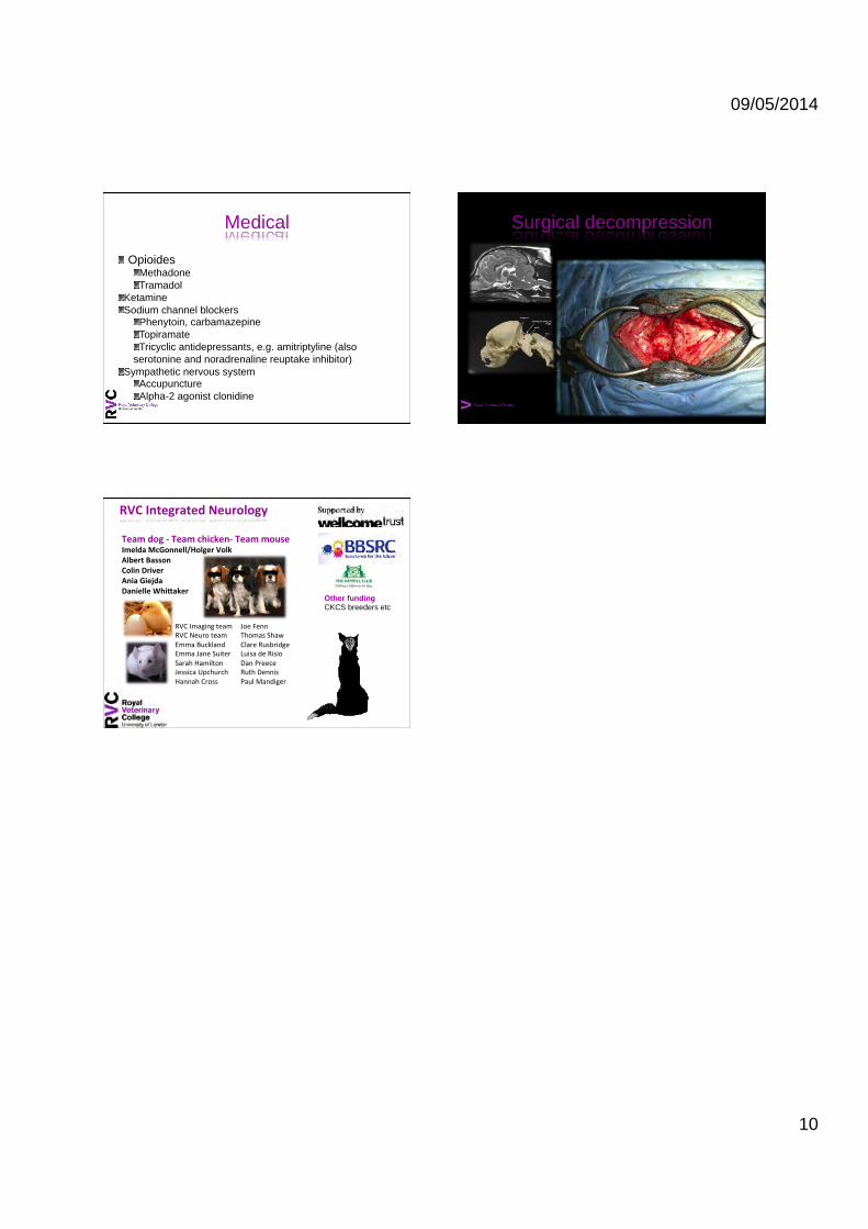
09/05/2014
10
Medical
! Opioides ! Methadone ! Tramadol
! Ketamine ! Sodium channel blockers
! Phenytoin, carbamazepine ! Topiramate ! Tricyclic antidepressants, e.g. amitriptyline (also serotonine and noradrenaline reuptake inhibitor)
! Sympathetic nervous system ! Accupuncture ! Alpha-2 agonist clonidine
Surgical decompression
RVC Integrated Neurology
Team dog -‐ Team chicken-‐ Team mouse Imelda McGonnell/Holger Volk Albert Basson Colin Driver Ania Giejda Danielle WhiEaker Other funding
CKCS breeders etc
RVC Imaging team RVC Neuro team Emma Buckland Emma Jane Suiter Sarah Hamilton Jessica Upchurch Hannah Cross
Joe Fenn Thomas Shaw Clare Rusbridge Luisa de Risio Dan Preece Ruth Dennis Paul Mandiger
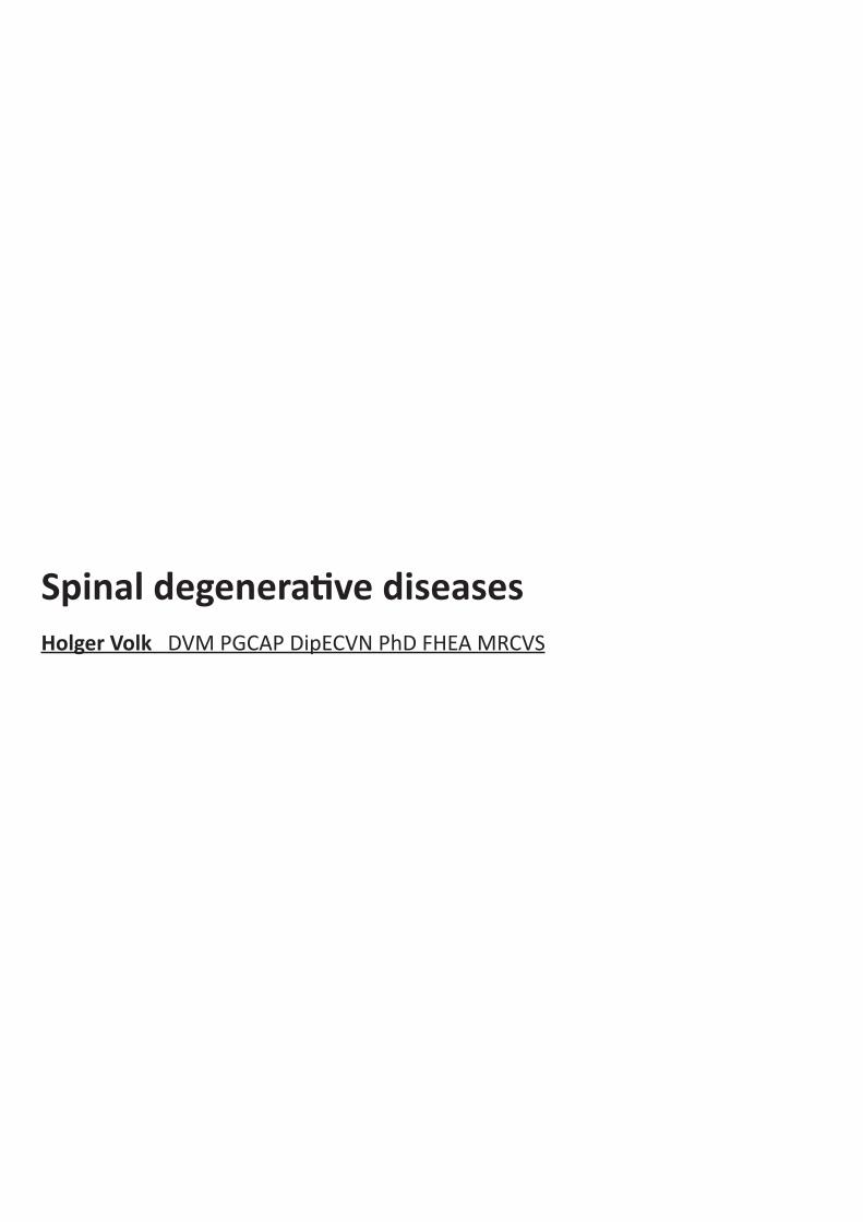
Spinal degenerative diseases Holger Volk DVM PGCAP DipECVN PhD FHEA MRCVS
09/05/2014
1
Holger A. Volk DVM, DipECVN, PhD, MRCVS Neurology & Neurosurgery The Royal Veterinary College
Spinal degenera,ve diseases
Neurological examina6on and history
Define & refine the problem
Lateralising?
Asymmetrical Symmetrical
Pain?
No Yes
Generalised? Localised?
On manipula6on? On palpa6on?
History
Define & refine the problem
Onset of disease?
Per-‐acute? Acute? Chronic?
Course of the disease?
Progression?
Deteriora6ng? Improving? Sta6c? Waxing and waning?
Episodic?
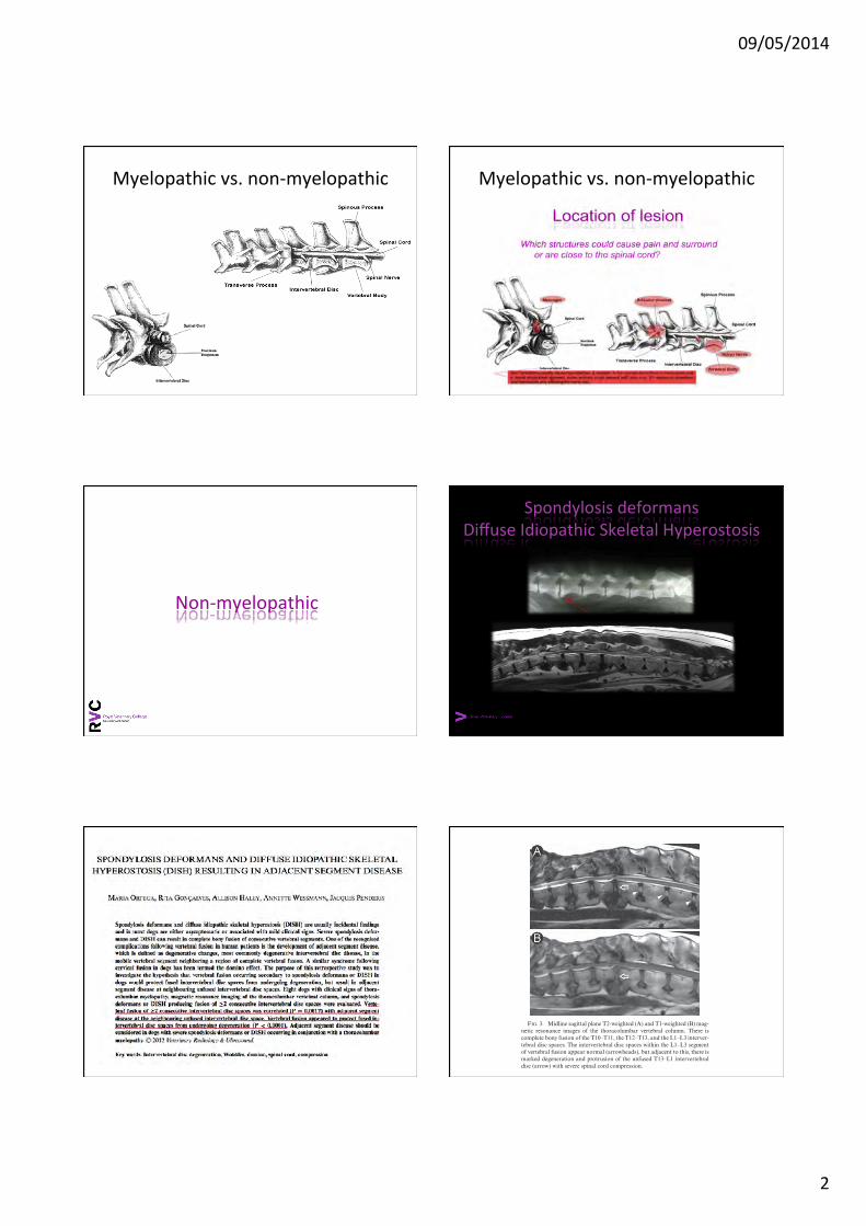
09/05/2014
2
Myelopathic vs. non-‐myelopathic Myelopathic vs. non-‐myelopathic
Non-‐myelopathic
Spondylosis deformans Diffuse Idiopathic Skeletal Hyperostosis
VOL. 53, NO. 2 SPONDYLOSIS OR DISH AND ADJACENT SEGMENT DISEASE 131
FIG. 1. Schematic demonstrating the osteophyte development score (shaded box) and intervertebral disc degeneration score (superimposed black line) foreach dog at each intervertebral disc level included in the magnetic resonance imaging study. Contiguous regions of complete bony vertebral fusion (shadedblack) are associated with a lower intervertebral disc degeneration score (P < 0.0001). Unfused intervertebral disc spaces immediately adjacent to regions ofcontiguous complete bony vertebral fusion of ≥2 intervertebral disc spaces are associated with a higher intervertebral disc degeneration score (P = 0.0017)compared to other nonfused intervertebral disc spaces, and many of these intervertebral discs were characterized by protrusion or extrusion with spinal cordcompression (denoted by a star). (T = thoracic; L = lumbar; S = sacral)
FIG. 2. Lateral radiograph (A) and midline sagittal T2-weighted magneticresonance image (B) of the thoracic vertebral column from a dog with con-tiguous new bone formation ventral to the vertebral bodies secondary todiffuse idiopathic skeletal hyperostosis (DISH). There is complete fusion ofthe vertebral column, with only the T3–T4, T4–T5, and T6–T7 intervertebraldisc spaces retaining mobility (arrows). The completely fused intervertebraldisc spaces are either normal or have mild degenerative changes. The inter-vertebral disc spaces retaining mobility are characterized by marked degen-erative changes, with extrusion of the T6–T7 intervertebral disc resulting inmoderate spinal cord compression (arrowheads).
at presentation. Neurologic deficits at presentation werescored as grade 1 in 3/8 dogs, grade 2 in 4/8 dogs, andgrade 3 in 1/8 dogs.
Discussion
The term adjacent segment disease has been used in hu-man patients to describe a broad category of disease pro-
FIG. 3. Midline sagittal plane T2-weighted (A) and T1-weighted (B) mag-netic resonance images of the thoracolumbar vertebral column. There iscomplete bony fusion of the T10–T11, the T12–T13, and the L1–L3 interver-tebral disc spaces. The intervertebral disc spaces within the L1–L3 segmentof vertebral fusion appear normal (arrowheads), but adjacent to this, there ismarked degeneration and protrusion of the unfused T13–L1 intervertebraldisc (arrow) with severe spinal cord compression.
cesses affecting the vertebral column, but all sharing thefollowing characteristics: (1) fusion of a normally mobilesegment of the vertebral column, and (2) degeneration ofthe remaining mobile vertebral segment immediately ad-jacent to the region of fusion. In some studies, however,the term adjacent segment disease has been used to refer
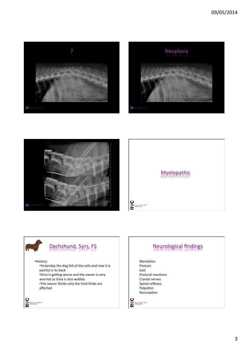
09/05/2014
3
? Neoplasia
Neoplasia
Myelopathic
Dachshund, 5yrs, FS
¥ History: ¥ Yesterday the dog fell of the sofa and now it is painful in its back ¥ Gina is geZng worse and the owner is very worried as Gina is also wobbly ¥ The owner thinks only the hind limbs are affected
Menta6on Posture Gait Postural reac6ons Cranial nerves Spinal reflexes Palpa6on Nocicep6on
Neurological findings

09/05/2014
4
Dachshund, 5yrs, FS Neurological?
No Yes
Peripheral?
Muscle? Neuromuscular junc6on? Nerve?
Central?
Brain?
Forebrain? Cerebellum? Brainstem?
Central Ves6bular?
Spinal cord?
C1-‐C5 SCS? C6-‐T2 SCS? T3-‐L3 SCS? L4-‐S3 SCS?
Neurological examina6on and history
Define & refine the problem
Lateralising?
Asymmetrical Symmetrical
Pain?
No Yes
Generalised? Localised?
On manipula6on? On palpa6on?
History
Define & refine the problem
Onset of disease?
Per-‐acute? Acute? Chronic?
Course of the disease?
Progression?
Deteriora6ng? Improving? Sta6c? Waxing and waning?
Episodic?
How can we define the lesion now by the neuro-‐hand rule? 1.Onset, 2.clinical course, 3.pain, 4.lateralising, 5.neurological localisa6on
Can the signalment help us in this case?
Define & refine the problem D Degenerative
A Anomalous
M Metabolic
N Neoplastic Nutritional
I Infectious Inflammatory Iatrogenic Idiopathic
T Toxic Traumatic
V Vascular
Please write down now your differen6al list:
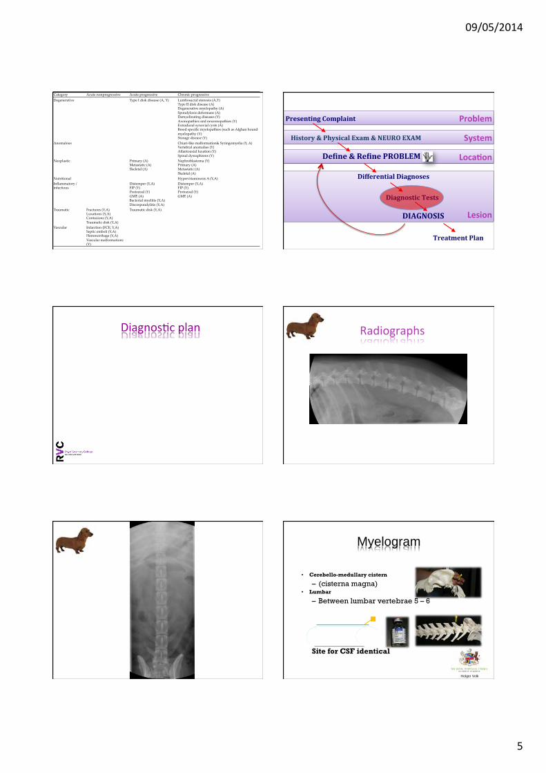
09/05/2014
5
Category Acute nonprogressive Acute progressive Chronic progressive Degenerative Type I disk disease (A, Y) Lumbosacral stenosis (A,Y)
Type II disk disease (A) Degenerative myelopathy (A) Spondylosis deformans (A) Demyelinating diseases (Y) Axonopathies and neuronopathies (Y) Extradural synovial cysts (A) Breed specific myelopathies (such as Afghan hound myelopathy (Y) Storage disease (Y)
Anomalous Chiari-‐‑like malformation& Syringomyelia (Y, A) Vertebral anomalies (Y) Atlantoaxial luxation (Y) Spinal dysraphisms (Y)
Neoplastic Primary (A) Metastatic (A) Skeletal (A)
Nephroblastoma (Y) Primary (A) Metastatic (A) Skeletal (A)
Nutritional Hypervitaminosis A (Y,A) Inflammatory / infectious
Distemper (Y,A) FIP (Y) Protozoal (Y) GME (A) Bacterial myelitis (Y,A) Discospondylitis (Y,A)
Distemper (Y,A) FIP (Y) Protozoal (Y) GME (A)
Traumatic Fractures (Y,A) Luxations (Y,A) Contusions (Y,A) Traumatic disk (Y,A)
Traumatic disk (Y,A)
Vascular Infarction (FCE; Y,A) Septic emboli (Y,A) Hemmorrhage (Y,A) Vascular malformations (Y)
Lesion
Loca,on
System
Problem
Diagnostic Tests
Treatment Plan
DIAGNOSIS
Presenting Complaint
History & Physical Exam & NEURO EXAM
DeCine & ReCine PROBLEM
Differential Diagnoses
Diagnos6c plan Radiographs
Radiographs Myelogram
¥ Cerebello-medullary cistern
– (cisterna magna) ¥ Lumbar
– Between lumbar vertebrae 5 – 6
Site for CSF identical
Holger Volk
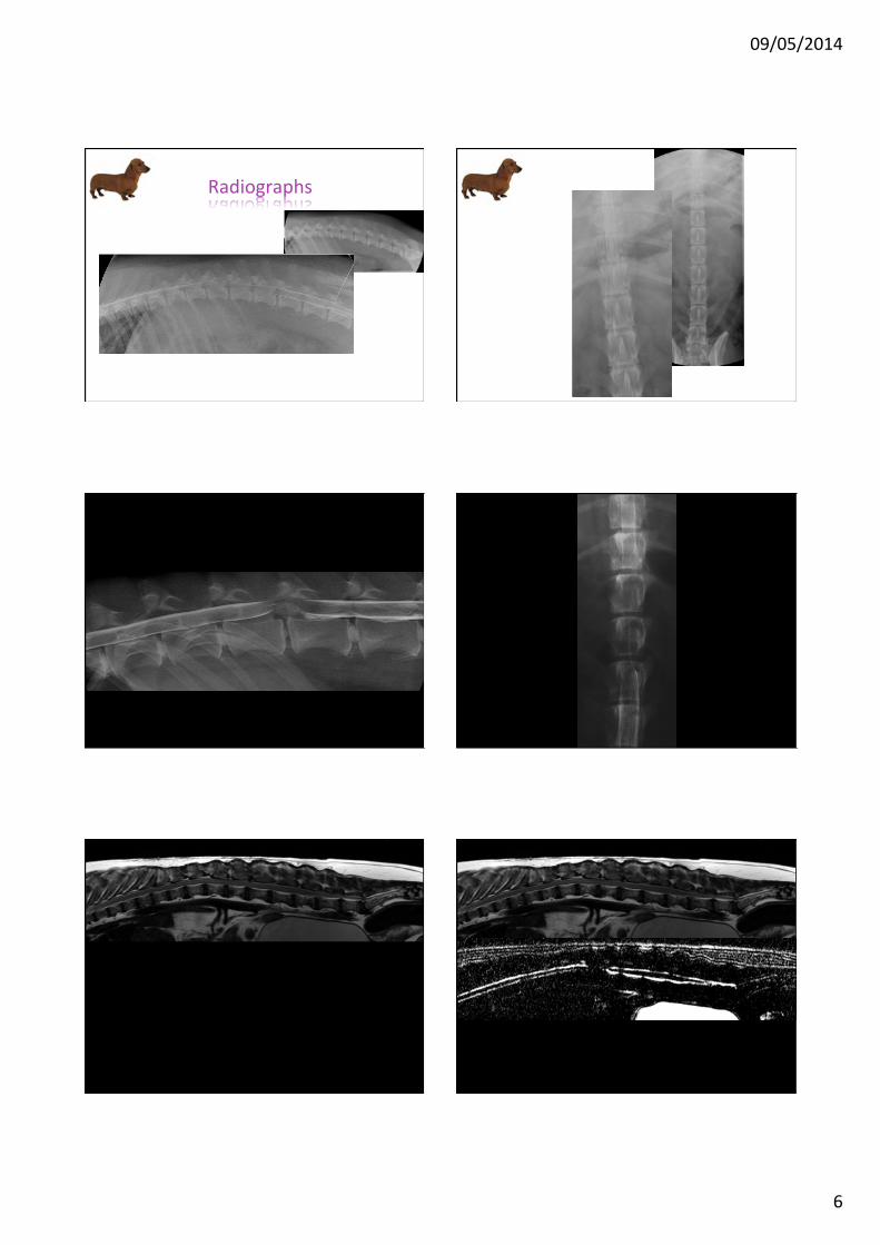
09/05/2014
6
Radiographs
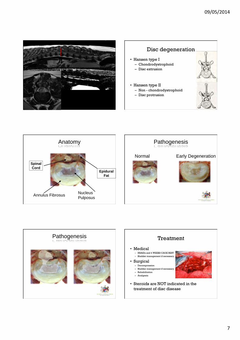
09/05/2014
7
Disc degeneration
¥ Hansen type I – Chondrodystrophoid – Disc extrusion
¥ Hansen type II – Non - chondrodystrophoid – Disc protrusion
Anatomy of the Intervertebral Disc
Annulus Fibrosus Nucleus Pulposus
Anatomy
Spinal Cord
Epidural Fat
Normal Early Degeneration
Pathogenesis
Anatomy of the Intervertebral Disc
Pathogenesis Treatment
¥ Medical – NSAIDs and 4 WEEKS CAGE REST – Bladder management if necessary
¥ Surgical – Decompression – Bladder management if necessary – Rehabilitation – Analgesia
¥ Steroids are NOT indicated in the treatment of disc disease
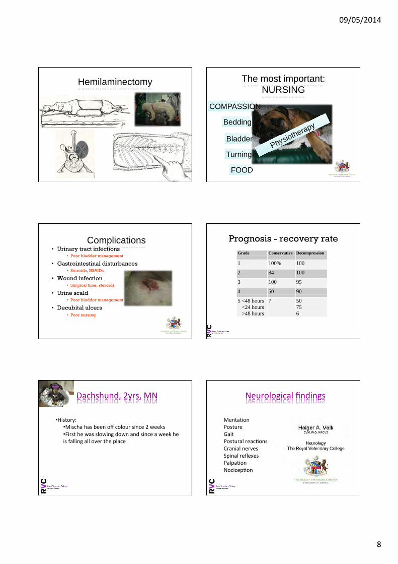
09/05/2014
8
Hemilaminectomy The most important: NURSING
COMPASSION
Bedding
Bladder
Turning
FOOD
Physiotherapy
Complications ¥ Urinary tract infections
¥ Poor bladder management
¥ Gastrointestinal disturbances ¥ Steroids, NSAIDs
¥ Wound infection ¥ Surgical time, steroids
¥ Urine scald ¥ Poor bladder management
¥ Decubital ulcers ¥ Poor nursing
Prognosis - recovery rate Grade Conservative Decompression
1 100% 100
2 84 100
3 100 95
4 50 90
5 <48 hours <24 hours >48 hours
7 50756
Dachshund, 2yrs, MN
¥ History: ¥ Mischa has been off colour since 2 weeks ¥ First he was slowing down and since a week he is falling all over the place
Menta6on Posture Gait Postural reac6ons Cranial nerves Spinal reflexes Palpa6on Nocicep6on
Neurological findings

09/05/2014
9
Neurological?
No Yes
Peripheral?
Muscle? Neuromuscular junc6on? Nerve?
Central?
Brain?
Forebrain? Cerebellum? Brainstem?
Central Ves6bular?
Spinal cord?
C1-‐C5 SCS? C6-‐T2 SCS? T3-‐L3 SCS? L4-‐S3 SCS?
Neurological examina6on and history
Define & refine the problem
Lateralising?
Asymmetrical Symmetrical
Pain?
No Yes
Generalised? Localised?
On manipula6on? On palpa6on?
History
Define & refine the problem
Onset of disease?
Per-‐acute? Acute? Chronic?
Course of the disease?
Progression?
Deteriora6ng? Improving? Sta6c? Waxing and waning?
Episodic?
How can we define the lesion now by the neuro-‐hand rule? 1.Onset, 2.clinical course, 3.pain, 4.lateralising, 5.neurological localisa6on
Can the signalment help us in this case?
Define & refine the problem
D Degenerative
A Anomalous
M Metabolic
N Neoplastic Nutritional
I Infectious Inflammatory Iatrogenic Idiopathic
T Toxic Traumatic
V Vascular
Please write down now your differen6al list:
Category Acute nonprogressive Acute progressive Chronic progressive Degenerative Type I disk disease (A, Y) Lumbosacral stenosis (A,Y)
Type II disk disease (A) Degenerative myelopathy (A) Spondylosis deformans (A) Demyelinating diseases (Y) Axonopathies and neuronopathies (Y) Extradural synovial cysts (A) Breed specific myelopathies (such as Afghan hound myelopathy (Y) Storage disease (Y)
Anomalous Chiari-‐‑like malformation& Syringomyelia (Y, A) Vertebral anomalies (Y) Atlantoaxial luxation (Y) Spinal dysraphisms (Y)
Neoplastic Primary (A) Metastatic (A) Skeletal (A)
Nephroblastoma (Y) Primary (A) Metastatic (A) Skeletal (A)
Nutritional Hypervitaminosis A (Y,A) Inflammatory / infectious
Distemper (Y,A) FIP (Y) Protozoal (Y) GME (A) Bacterial myelitis (Y,A) Discospondylitis (Y,A)
Distemper (Y,A) FIP (Y) Protozoal (Y) GME (A)
Traumatic Fractures (Y,A) Luxations (Y,A) Contusions (Y,A) Traumatic disk (Y,A)
Traumatic disk (Y,A)
Vascular Infarction (FCE; Y,A) Septic emboli (Y,A) Hemmorrhage (Y,A) Vascular malformations (Y)
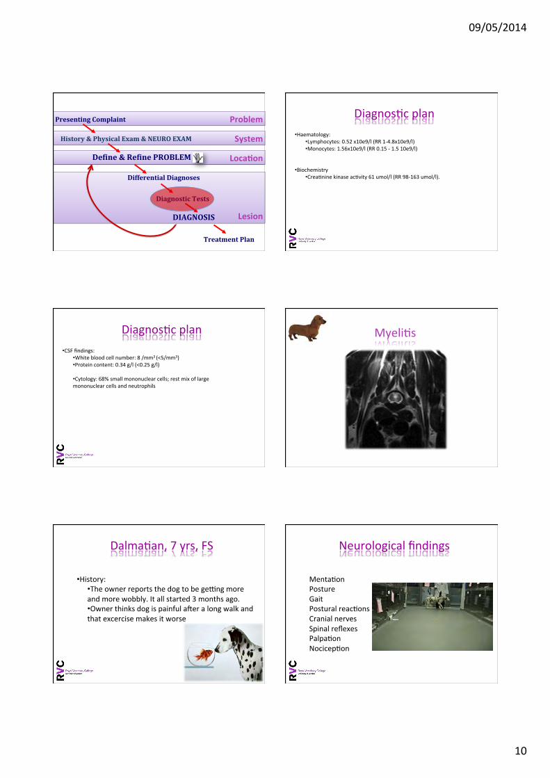
09/05/2014
10
Lesion
Loca,on
System
Problem
Diagnostic Tests
Treatment Plan
DIAGNOSIS
Presenting Complaint
History & Physical Exam & NEURO EXAM
DeCine & ReCine PROBLEM
Differential Diagnoses
Diagnos6c plan ¥ Haematology:
¥ Lymphocytes: 0.52 x10e9/l (RR 1-‐4.8x10e9/l) ¥ Monocytes: 1.56x10e9/l (RR 0.15 -‐ 1.5 10e9/l)
¥ Biochemistry ¥ Crea6nine kinase ac6vity 61 umol/l (RR 98-‐163 umol/l).
Diagnos6c plan ¥ CSF findings:
¥ White blood cell number: 8 /mm3 (<5/mm3) ¥ Protein content: 0.34 g/l (<0.25 g/l)
¥ Cytology: 68% small mononuclear cells; rest mix of large mononuclear cells and neutrophils
Myeli6s
Dalma6an, 7 yrs, FS
¥ History: ¥ The owner reports the dog to be geZng more and more wobbly. It all started 3 months ago. ¥ Owner thinks dog is painful aier a long walk and that excercise makes it worse
Menta6on Posture Gait Postural reac6ons Cranial nerves Spinal reflexes Palpa6on Nocicep6on
Neurological findings
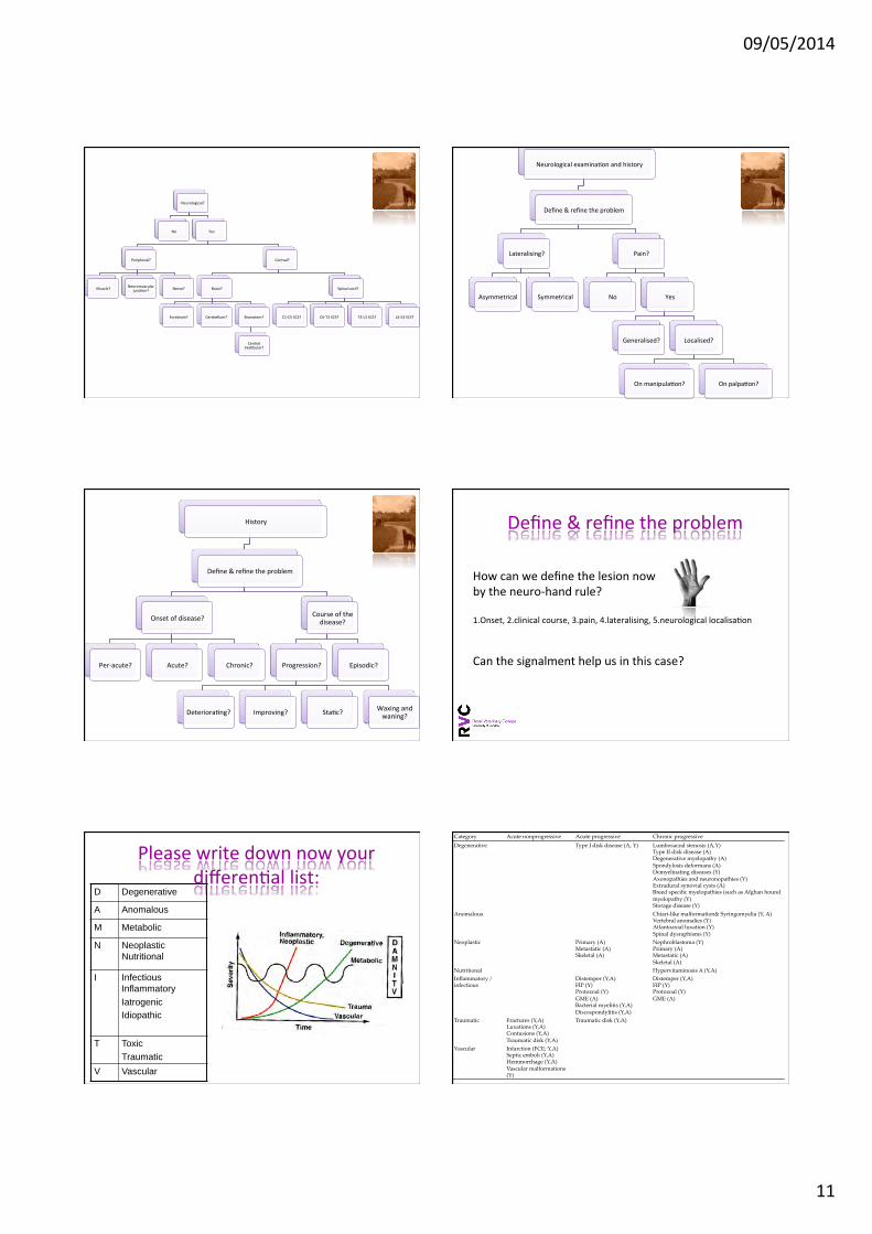
09/05/2014
11
Neurological?
No Yes
Peripheral?
Muscle? Neuromuscular junc6on? Nerve?
Central?
Brain?
Forebrain? Cerebellum? Brainstem?
Central Ves6bular?
Spinal cord?
C1-‐C5 SCS? C6-‐T2 SCS? T3-‐L3 SCS? L4-‐S3 SCS?
Neurological examina6on and history
Define & refine the problem
Lateralising?
Asymmetrical Symmetrical
Pain?
No Yes
Generalised? Localised?
On manipula6on? On palpa6on?
History
Define & refine the problem
Onset of disease?
Per-‐acute? Acute? Chronic?
Course of the disease?
Progression?
Deteriora6ng? Improving? Sta6c? Waxing and waning?
Episodic?
How can we define the lesion now by the neuro-‐hand rule? 1.Onset, 2.clinical course, 3.pain, 4.lateralising, 5.neurological localisa6on
Can the signalment help us in this case?
Define & refine the problem
D Degenerative
A Anomalous
M Metabolic
N Neoplastic Nutritional
I Infectious Inflammatory Iatrogenic Idiopathic
T Toxic Traumatic
V Vascular
Please write down now your differen6al list:
Category Acute nonprogressive Acute progressive Chronic progressive Degenerative Type I disk disease (A, Y) Lumbosacral stenosis (A,Y)
Type II disk disease (A) Degenerative myelopathy (A) Spondylosis deformans (A) Demyelinating diseases (Y) Axonopathies and neuronopathies (Y) Extradural synovial cysts (A) Breed specific myelopathies (such as Afghan hound myelopathy (Y) Storage disease (Y)
Anomalous Chiari-‐‑like malformation& Syringomyelia (Y, A) Vertebral anomalies (Y) Atlantoaxial luxation (Y) Spinal dysraphisms (Y)
Neoplastic Primary (A) Metastatic (A) Skeletal (A)
Nephroblastoma (Y) Primary (A) Metastatic (A) Skeletal (A)
Nutritional Hypervitaminosis A (Y,A) Inflammatory / infectious
Distemper (Y,A) FIP (Y) Protozoal (Y) GME (A) Bacterial myelitis (Y,A) Discospondylitis (Y,A)
Distemper (Y,A) FIP (Y) Protozoal (Y) GME (A)
Traumatic Fractures (Y,A) Luxations (Y,A) Contusions (Y,A) Traumatic disk (Y,A)
Traumatic disk (Y,A)
Vascular Infarction (FCE; Y,A) Septic emboli (Y,A) Hemmorrhage (Y,A) Vascular malformations (Y)
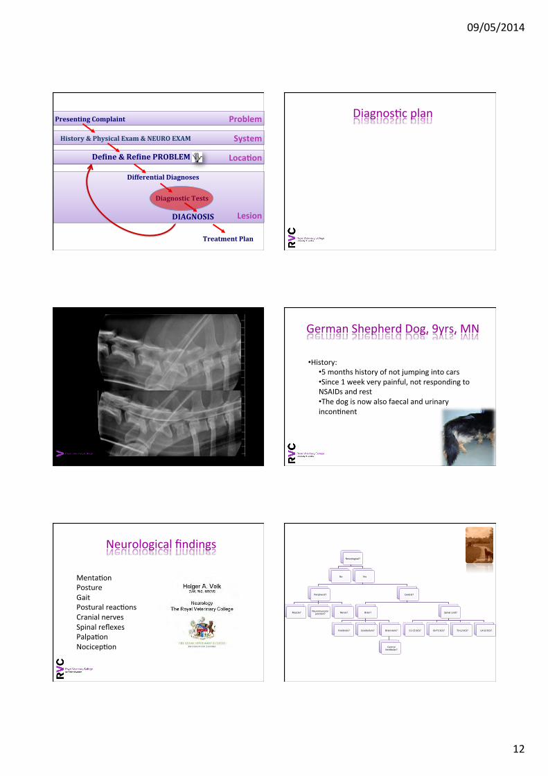
09/05/2014
12
Lesion
Loca,on
System
Problem
Diagnostic Tests
Treatment Plan
DIAGNOSIS
Presenting Complaint
History & Physical Exam & NEURO EXAM
DeCine & ReCine PROBLEM
Differential Diagnoses
Diagnos6c plan
Neoplasia German Shepherd Dog, 9yrs, MN
¥ History: ¥ 5 months history of not jumping into cars ¥ Since 1 week very painful, not responding to NSAIDs and rest ¥ The dog is now also faecal and urinary incon6nent
Menta6on Posture Gait Postural reac6ons Cranial nerves Spinal reflexes Palpa6on Nocicep6on
Neurological findings Neurological?
No Yes
Peripheral?
Muscle? Neuromuscular junc6on? Nerve?
Central?
Brain?
Forebrain? Cerebellum? Brainstem?
Central Ves6bular?
Spinal cord?
C1-‐C5 SCS? C6-‐T2 SCS? T3-‐L3 SCS? L4-‐S3 SCS?
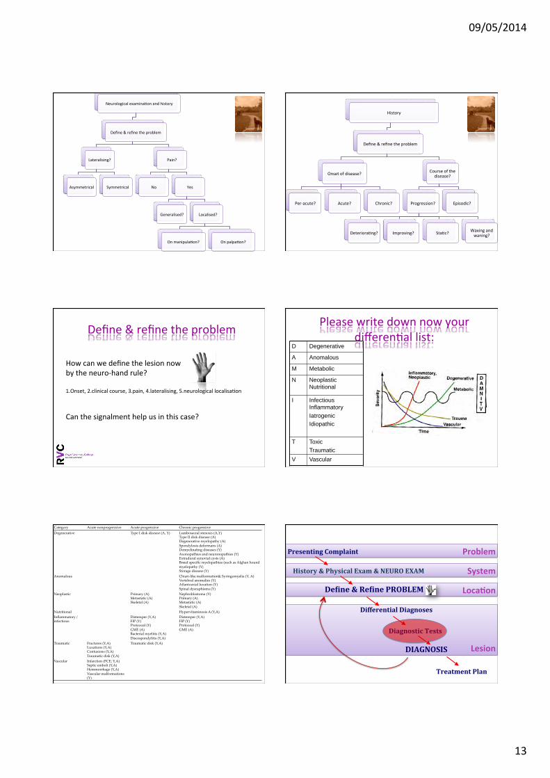
09/05/2014
13
Neurological examina6on and history
Define & refine the problem
Lateralising?
Asymmetrical Symmetrical
Pain?
No Yes
Generalised? Localised?
On manipula6on? On palpa6on?
History
Define & refine the problem
Onset of disease?
Per-‐acute? Acute? Chronic?
Course of the disease?
Progression?
Deteriora6ng? Improving? Sta6c? Waxing and waning?
Episodic?
How can we define the lesion now by the neuro-‐hand rule? 1.Onset, 2.clinical course, 3.pain, 4.lateralising, 5.neurological localisa6on
Can the signalment help us in this case?
Define & refine the problem D Degenerative
A Anomalous
M Metabolic
N Neoplastic Nutritional
I Infectious Inflammatory Iatrogenic Idiopathic
T Toxic Traumatic
V Vascular
Please write down now your differen6al list:
Category Acute nonprogressive Acute progressive Chronic progressive Degenerative Type I disk disease (A, Y) Lumbosacral stenosis (A,Y)
Type II disk disease (A) Degenerative myelopathy (A) Spondylosis deformans (A) Demyelinating diseases (Y) Axonopathies and neuronopathies (Y) Extradural synovial cysts (A) Breed specific myelopathies (such as Afghan hound myelopathy (Y) Storage disease (Y)
Anomalous Chiari-‐‑like malformation& Syringomyelia (Y, A) Vertebral anomalies (Y) Atlantoaxial luxation (Y) Spinal dysraphisms (Y)
Neoplastic Primary (A) Metastatic (A) Skeletal (A)
Nephroblastoma (Y) Primary (A) Metastatic (A) Skeletal (A)
Nutritional Hypervitaminosis A (Y,A) Inflammatory / infectious
Distemper (Y,A) FIP (Y) Protozoal (Y) GME (A) Bacterial myelitis (Y,A) Discospondylitis (Y,A)
Distemper (Y,A) FIP (Y) Protozoal (Y) GME (A)
Traumatic Fractures (Y,A) Luxations (Y,A) Contusions (Y,A) Traumatic disk (Y,A)
Traumatic disk (Y,A)
Vascular Infarction (FCE; Y,A) Septic emboli (Y,A) Hemmorrhage (Y,A) Vascular malformations (Y)
Lesion
Loca,on
System
Problem
Diagnostic Tests
Treatment Plan
DIAGNOSIS
Presenting Complaint
History & Physical Exam & NEURO EXAM
DeCine & ReCine PROBLEM
Differential Diagnoses
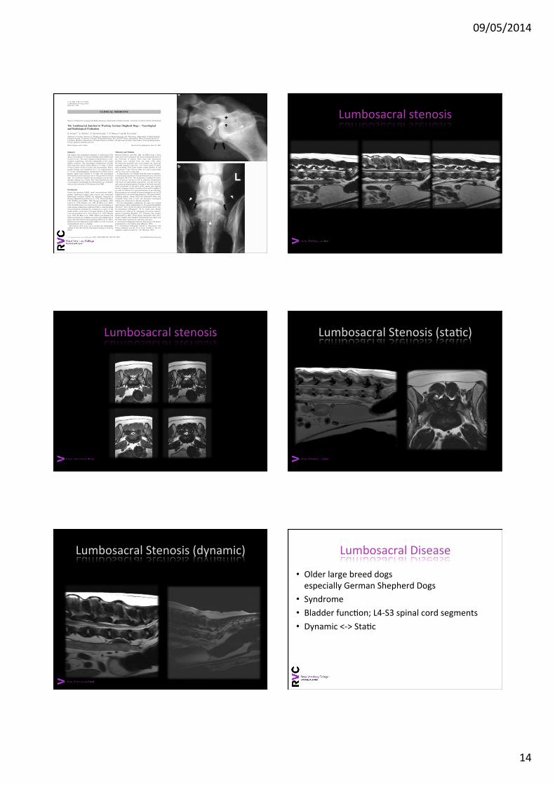
09/05/2014
14
CLINICAL MEDICINE
Section of Diagnostic Imaging and Radio-Oncology, Department of Small Animals, University of Zurich, Zurich, Switzerland
The Lumbosacral Junction in Working German Shepherd Dogs – Neurologicaland Radiological Evaluation
G. Scharf1,4, F. Steffen2, F. Grunenfelder2, J. P. Morgan3 and M. Fluckiger1
Addresses of authors: Sections of 1Diagnostic Imaging and Radio-Oncology and 2Neurology, Department of Small Animals,Vetsuisse Faculty, University of Zurich, Winterthurerstrasse 260, CH 8057 Zurich, Switzerland; 3Ross University School ofVeterinary Medicine, Department of Clinical Sciences, PO Box 334, Basseterre St Kitts, West Indies; 4Corresponding author:E-mail: [email protected]
With 4 figures and 3 tables Received for publication: June 10, 2003
Summary
The clinical and radiological incidence of lumbosacral (LS)disease was studied on 57 German Shepherd dogs (GSDs) usedin active service. The study included a clinical history, a neu-
rological examination, and plain radiographs of the caudallumbar vertebrae. The neurological examinations revealedlower back pain and/or neural deficits in 21 dogs, of which14 had a history of pain or pelvic gait abnormalities. Radio-
graphic findings were spondylosis at L7–S1, degeneration ofL7–S1 disc, LS malalignment, transitional LS vertebrae and/orprimary spinal canal stenosis in 15 dogs with neurological
abnormalities and/or back pain and in 18 dogs with no clinicalsigns. No correlation between the neurological and the radi-ographic findings were found. This study demonstrates that
even prominent radiographic LS abnormalities are of minimalvalue in the evaluation of LS disease in the GSD.
Introduction
Canine hip dysplasia (CHD), sacral osteochondrosis (SOC),primary lumbosacral spinal canal stenosis and transitionallumbosacral vertebrae (TLSV) are frequently diagnosed in
German Shepherd dogs (Oliver et al., 1978; Tarvin and Prata,1980; Winkler and Loeffler, 1986; Morgan and Bailey, 1990;Lang et al., 1992; Morgan et al., 1993; De Risio et al., 2000).
These heritable diseases are risk factors for the development ofcauda equina compression syndrome (CES), a term describingclinical symptoms produced by compression or injury of the
caudal lumbar, sacral and/or coccygeal segments of the spinalcord and associated nerve roots (Lang et al., 1992; Morganet al., 1993; De Risio et al., 2000). Spinal cord diseases and
CES in particular are common causes for early elimination ofdogs in the USA military dog programme (Moore et al., 2001).Despite this knowledge Swiss police GSDs are rarely examinedradiographically prior to training.
The purpose of the study was to correlate the radiographicfindings in the LSV and the neurological findings in workingGSDs.
Materials and Methods
Between February and May 2001, 60 GSDs from a Swisspolice unit were evaluated at the Animal Teaching Hospital ofthe University of Zurich. Three dogs with behavioural
problems were excluded from the study. Fifty males(49 intact/one neutered) and seven females (six intact/oneneutered) ranging in age from 1 to 9 years (mean 4.5 years)were evaluated. Their body weights ranged between 28 and
45 kg (mean 37 kg). Fifty-two dogs were used as guard dogsand five were used as drug dogs.A clinical history was obtained from the owners or handlers.
A neurological examination was performed by one of the twoneurologists (FS, FG). This examination included the evalu-ation of posture and gait as well as palpatory manipulation
with focus on hypotrophied musculature, tail and anal toneand conscious proprioception. Swaying of the back, incoordi-nated movements of the pelvic limbs, spastic gait (reducedflexion), dragging of limbs, knuckling of feet and/or scuffing of
toe nails are referred to as gait abnormalities in the following.Pain perception was assessed by hyperextending the tail, by LShyperextension and spinal deep palpation. The spinal reflexes
including patellar, cranial tibial and withdrawal reflex wereevaluated. Dogs with at least one abnormal neurologicalfinding were considered as clinically abnormal.
For the radiographic examination, the dogs were sedatedintravenously with a combination of 5–10 lg/kg medetomidine(Domitor�; Dr E. Graub AG, Berne, Switzerland) and 0.2 mg/
kg butorphanol (Morphasol�; Dr E. Graub AG). Musclerelaxation was achieved by subsequent intravenous adminis-tration of propofol (Propofol 1%�; Fresenius AG, Sarnen,Switzerland) to effect. Three digital radiographs (Fuji AC3-
System; Fujifilm AG, Dielsdorf, Switzerland) were taken withthe limbs held in position by sand bags and ropes:1 A left lateral radiograph centred at LS-junction with femora
perpendicular to the lumbar spine (Morgan, 1993).2 A ventrodorsal radiograph centred at LS-junction withfemora abducted and the X-ray beam parallel to the LS-
endplates, angled cranially by 7–10� (Morgan, 1993).
U.S. Copyright Clearance Center Code Statement: 0931–184X/2004/5101–0027 $15.00/0 www.blackwell-synergy.com
J. Vet. Med. A 51, 27–32 (2004)
� 2004 Blackwell Verlag, Berlin
ISSN 0931–184X
Lumbosacral stenosis
Lumbosacral stenosis Lumbosacral Stenosis (sta6c)
Lumbosacral Stenosis (dynamic) Lumbosacral Disease
¥ Older large breed dogs especially German Shepherd Dogs
¥ Syndrome ¥ Bladder func6on; L4-‐S3 spinal cord segments ¥ Dynamic <-‐> Sta6c

09/05/2014
15
Lumbosacral Disease Stenosis of the vertebral canal and/or intervertebral foramina and/or the related vasculature
1. Hansen type II disc degenera6on and protrusion at the
lumbosacral junc6on 2. Thickening and in-‐folding of the intearcuate ligament 3. Subluxa6on of the ar6cular facets 4. Epidural fibrosis 5. Thickened lamina and pedicles 6. Spondylosis, prolifera6ve degenera6ve changes of the ar6cular
facets 7. Instability and misalignment between the last lumbar vertebra and
the sacrum
Pathogenesis The importance of 1. Mo6on 2. Anatomic conforma6on a. Ar6cular joint tropism b. Straighter facet joints c. Level of change of plane d. Facet joint angle
3. Congenital & developmental abnormali6es a. Transi6onal vertebrae b. Osteochondrosis
Diagnos6cs
1. The neuro-‐exam 1. Pseudo-‐hyperreflexia 2. Urinary + faecal incon6nence
2. Imaging 1. Radiographs 2. Epidurography 3. Discography 4. CT 5. MRI
3. EMG
Lumbosacral Stenosis
¥ Treatment – Conserva6ve ¥ Restricted excercise 6-‐8 weeks ¥ An6-‐inflammatories ¥ Pain relief
– Surgery ¥ Dorsal Laminectomy
¥ Dynamic vs non-‐dynamic
Lumbosacral Stenosis
¥ Post surgical complica6ons – Immediate ¥ Seroma – haemorrhage
– > oien due to too much movement aier surgery
– Delayed ¥ Discospondyli6s ¥ Lamina forma6on/Fibrosis
– > secondary compression
German Shepherd X, 6yrs, MN
¥ History: ¥ 6 months history of slowing down ¥ Since 3 weeks Baoen is more wobbly in the hind limbs ¥ not responding to NSAIDs and rest
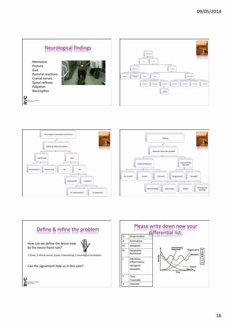
09/05/2014
16
Menta6on Posture Gait Postural reac6ons Cranial nerves Spinal reflexes Palpa6on Nocicep6on
Neurological findings Neurological?
No Yes
Peripheral?
Muscle? Neuromuscular junc6on? Nerve?
Central?
Brain?
Forebrain? Cerebellum? Brainstem?
Central Ves6bular?
Spinal cord?
C1-‐C5 SCS? C6-‐T2 SCS? T3-‐L3 SCS? L4-‐S3 SCS?
Neurological examina6on and history
Define & refine the problem
Lateralising?
Asymmetrical Symmetrical
Pain?
No Yes
Generalised? Localised?
On manipula6on? On palpa6on?
History
Define & refine the problem
Onset of disease?
Per-‐acute? Acute? Chronic?
Course of the disease?
Progression?
Deteriora6ng? Improving? Sta6c? Waxing and waning?
Episodic?
How can we define the lesion now by the neuro-‐hand rule? 1.Onset, 2.clinical course, 3.pain, 4.lateralising, 5.neurological localisa6on
Can the signalment help us in this case?
Define & refine the problem D Degenerative
A Anomalous
M Metabolic
N Neoplastic Nutritional
I Infectious Inflammatory Iatrogenic Idiopathic
T Toxic Traumatic
V Vascular
Please write down now your differen6al list:
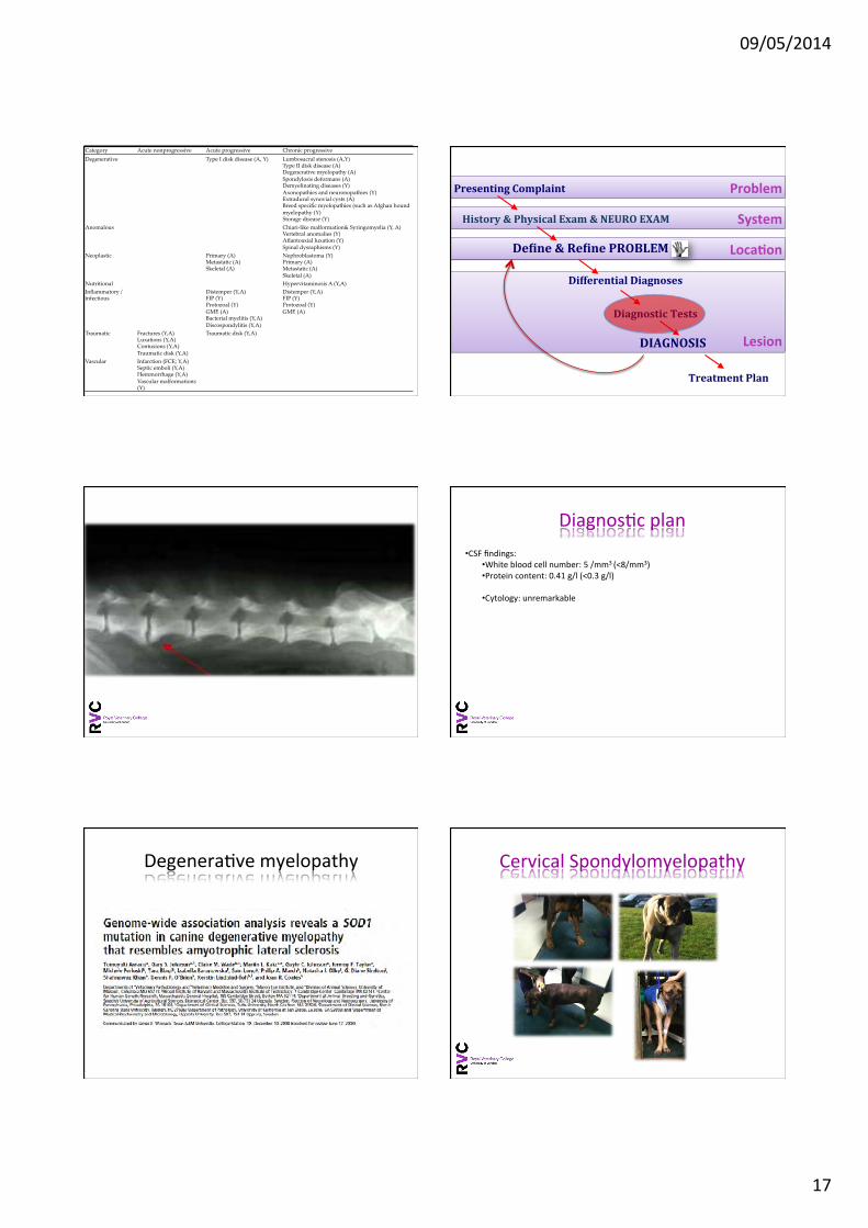
09/05/2014
17
Category Acute nonprogressive Acute progressive Chronic progressive Degenerative Type I disk disease (A, Y) Lumbosacral stenosis (A,Y)
Type II disk disease (A) Degenerative myelopathy (A) Spondylosis deformans (A) Demyelinating diseases (Y) Axonopathies and neuronopathies (Y) Extradural synovial cysts (A) Breed specific myelopathies (such as Afghan hound myelopathy (Y) Storage disease (Y)
Anomalous Chiari-‐‑like malformation& Syringomyelia (Y, A) Vertebral anomalies (Y) Atlantoaxial luxation (Y) Spinal dysraphisms (Y)
Neoplastic Primary (A) Metastatic (A) Skeletal (A)
Nephroblastoma (Y) Primary (A) Metastatic (A) Skeletal (A)
Nutritional Hypervitaminosis A (Y,A) Inflammatory / infectious
Distemper (Y,A) FIP (Y) Protozoal (Y) GME (A) Bacterial myelitis (Y,A) Discospondylitis (Y,A)
Distemper (Y,A) FIP (Y) Protozoal (Y) GME (A)
Traumatic Fractures (Y,A) Luxations (Y,A) Contusions (Y,A) Traumatic disk (Y,A)
Traumatic disk (Y,A)
Vascular Infarction (FCE; Y,A) Septic emboli (Y,A) Hemmorrhage (Y,A) Vascular malformations (Y)
Lesion
Loca,on
System
Problem
Diagnostic Tests
Treatment Plan
DIAGNOSIS
Presenting Complaint
History & Physical Exam & NEURO EXAM
DeCine & ReCine PROBLEM
Differential Diagnoses
Diagnos6c plan ¥ CSF findings:
¥ White blood cell number: 5 /mm3 (<8/mm3) ¥ Protein content: 0.41 g/l (<0.3 g/l)
¥ Cytology: unremarkable
Degenera6ve myelopathy Cervical Spondylomyelopathy
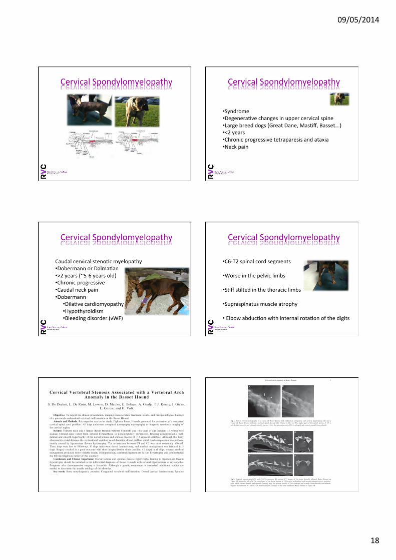
09/05/2014
18
Cervical Spondylomyelopathy Cervical Spondylomyelopathy
¥ Syndrome ¥ Degenera6ve changes in upper cervical spine ¥ Large breed dogs (Great Dane, Mas6ff, Basset...) ¥ <2 years ¥ Chronic progressive tetraparesis and ataxia ¥ Neck pain
Caudal cervical steno6c myelopathy ¥ Dobermann or Dalma6an ¥ >2 years (~5-‐6 years old) ¥ Chronic progressive ¥ Caudal neck pain ¥ Dobermann ¥ Dila6ve cardiomyopathy ¥ Hypothyroidism ¥ Bleeding disorder (vWF)
Cervical Spondylomyelopathy
¥ C6-‐T2 spinal cord segments
¥ Worse in the pelvic limbs
¥ S6ff s6lted in the thoracic limbs
¥ Supraspinatus muscle atrophy
¥ Elbow abduc6on with internal rota6on of the digits
Cervical Spondylomyelopathy
Cervical Vertebral Stenosis Associated with a Vertebral ArchAnomaly in the Basset Hound
S. De Decker, L. De Risio, M. Lowrie, D. Mauler, E. Beltran, A. Giedja, P.J. Kenny, I. Gielen,L. Garosi, and H. Volk
Objectives: To report the clinical presentation, imaging characteristics, treatment results, and histopathological findings
of a previously undescribed vertebral malformation in the Basset Hound.
Animals and Methods: Retrospective case series study. Eighteen Basset Hounds presented for evaluation of a suspected
cervical spinal cord problem. All dogs underwent computed tomography myelography or magnetic resonance imaging of
the cervical region.
Results: Thirteen male and 5 female Basset Hounds between 6 months and 10.8 years of age (median: 1.4 years) were
studied. Clinical signs varied from cervical hyperesthesia to nonambulatory tetraparesis. Imaging demonstrated a well-
defined and smooth hypertrophy of the dorsal lamina and spinous process of � 2 adjacent vertebrae. Although this bony
abnormality could decrease the ventrodorsal vertebral canal diameter, dorsal midline spinal cord compression was predom-
inantly caused by ligamentum flavum hypertrophy. The articulation between C4 and C5 was most commonly affected.
Three dogs were lost to follow-up, 10 dogs underwent dorsal laminectomy, and medical management was initiated in 5
dogs. Surgery resulted in a good outcome with short hospitalization times (median: 4.5 days) in all dogs, whereas medical
management produced more variable results. Histopathology confirmed ligamentum flavum hypertrophy and demonstrated
the fibrocartilaginous nature of this anomaly.
Conclusions and Clinical Importance: Dorsal lamina and spinous process hypertrophy leading to ligamentum flavum
hypertrophy should be included in the differential diagnosis of Basset Hounds with cervical hyperesthesia or myelopathy.
Prognosis after decompressive surgery is favorable. Although a genetic component is suspected, additional studies are
needed to determine the specific etiology of this disorder.
Key words: Bone morphogenetic proteins; Congenital vertebral malformation; Dorsal cervical laminectomy; Spinous
process.
C linical signs of cervical hyperesthesia, general pro-prioceptive ataxia, and tetraparesis in dogs can be
caused by congenital, degenerative, inflammatory, neo-plastic, traumatic, and vascular disorders affecting thecervical spinal cord.1 Several congenital malformationsaffecting the vertebral column and spinal cord havebeen described in small animals. Classification of thesedisorders is difficult and has not yet been clearlydefined.2 Some spinal malformations or anomalies areassociated with specific dog breeds. Examples are der-mal sinus tracts in the Rhodesian Ridgeback,3 spinal
dysraphism in the Weimaraner,4 hemivertebra in theFrench Bulldog5 and arachnoid diverticula,6,7 and cer-vical fibrotic stenosis in the Rottweiler.8
The Basset Hound also has been associated with aspecific vertebral malformation.9 In 1968, a deformityof the 3rd cervical vertebral body was described in 6related, young Basset Hounds. This deformity typicallyresulted in ventral extradural spinal cord compressionbetween the 2nd and 3rd cervical vertebrae.9 Later, thesame authors considered this condition similar to aform of vertebral abnormality seen in ataxic GreatDanes, Doberman Pinschers, Ridgebacks, and “wob-bler” horses.10 In 1977, 3 young Basset Hounds wereincluded in a larger retrospective study about cervicalspondylomyelopathy in dogs.11 Since that time, thiscondition in Basset Hounds has commonly beenreferred to as cervical spondylomyelopathy.12
This study describes a previously unreported cervicalvertebral anomaly in Basset Hounds. This disorder,apparently distinct from previously reported vertebraldeformities, is characterized by dorsal lamina and spi-nous process hypertrophy leading to ligamentous hyper-trophy. The aims of this study were to characterize the
From the Department of Veterinary Clinical Sciences, RoyalVeterinary College, University of London, Hawkshed Lane, NorthMymms, Hatfield, Hertfordshire, England (De Decker, Kenny,Volk); the Centre for Small Animal Studies, Animal Health Trust,Lanwades Park, Kentford, Newmarket, Suffolk, England (DeRisio, Beltran); the Davies Veterinary Specialists, Manor FarmBusiness Park, Higham Gobion, Higham Gobion, HertfordshireEngland (Lowrie, Garosi); the Department of Small AnimalMedicine and Clinical Biology, Faculty of Veterinary Medicine,Ghent University, Merelbeke, Belgium (Mauler); the Departmentof Veterinary Basic Sciences, Royal Veterinary College, Universityof London, London, England (Giedja); and the Department ofMedical Imaging of Domestic Animals and Orthopedics of SmallAnimals, Faculty of Veterinary Medicine, Ghent University,Merelbeke, Belgium (Gielen).
Corresponding author: Steven De Decker, Department of Veter-inary Clinical Sciences, Royal Veterinary College, University ofLondon, Hawkshead Lane, North Mymms, Hatfield, HertfordshireAL9 7TA, England; e-mail: [email protected]
Submitted May 8, 2012; Revised June 17, 2012; AcceptedJuly 11, 2012.
Copyright © 2012 by the American College of Veterinary InternalMedicine
10.1111/j.1939-1676.2012.00990.x
Abbreviations:
BMP bone morphogenetic protein
CSM cervical spondylomyelopathy
CT computed tomography
CT-m computed tomography myelography
MRI magnetic resonance imaging
NSAID nonsteroidal anti-inflammatory drug
J Vet Intern Med 2012
The dorsally enlarged caudal part of the lamina ofthe cranially affected vertebra formed a convex-shaped protuberance, which communicated with theconcave-shaped, enlarged spinous process of the cau-dally affected vertebra. This protuberance extendeddorsally (n = 19 sites of spinal cord compression) orboth dorsally and ventrally (n = 9). The enlarged spi-nous process extended cranially (n = 21) or cranioven-trally (n = 7). This deformation had the characteristicsof cortical bone on all imaging techniques. Theabnormally formed dorsal lamina and spinous processwere separated by a cleft, which communicated withthe vertebral canal. This area had imaging character-istics consistent with soft tissue on all imaging tech-niques. Although the dorsal lamina and spinous
process abnormality could result in a variablydecreased dorsoventral vertebral canal diameter, dor-sal midline spinal cord compression predominantlywas caused by hypertrophied ligamentous tissue. Withthe exception of 1 dog, the articular processes werenot involved in the pathological process. In this 1dog, the left caudal articular process of C4 had anabnormal, irregular, and enlarged shape. This doghad left-sided dorsolateral spinal cord compression.An intramedullary spinal cord hyperintense signal wasobserved on T2-weighted images for 11 of 14 dogs(79%) that underwent MRI. In 2 dogs, MRI dis-closed the above-described vertebral abnormality at 2sites (C3–C4 and C4–C5), with spinal cord compres-sion only at C4–C5.
A B
Fig 1. Survey cervical radiographs of a 4-year-old Basset Hound with ambulatory tetraparesis and cervical hyperesthesia (A) and a
4-year-old Basset Hound without a cervical spinal disorder (B). Cranial is left. (A) The caudal part of the dorsal lamina of C5 is
well-defined, smooth, and enlarged dorsally (arrow). Also, the spinous process of C6 is enlarged and extends cranially (arrowhead).
A B
C D
Fig 2. Sagittal reconstructed (A) and C5–C6 transverse (B) cervical CT images of the same clinically affected Basset Hound as
Figure 1A. Cranial is left. (A) The caudal part of the dorsal lamina of C5 forms a well-defined and smooth enlarged convex protuber-
ance, which extends dorsally and ventrally (arrow). Also, the spinous process of C6 is enlarged and extends cranioventrally (arrowhead).
Sagittal reconstructed (C) and C5–C6 transverse (D) CT images of the same unaffected Basset Hound as Figure 1B.
Vertebral Arch Anomaly in Basset Hounds 3
09/05/2014
19
One dog underwent 2 MRI studies within a periodof 6 months (Fig 4). The 1st MRI study demonstratedan abnormally shaped dorsal lamina and spinous pro-cess at the levels of C4–C5 and C5–C6 with intrame-dullary spinal cord hyperintensity and minimal dorsalspinal cord compression at the level of C4–C5. The2nd MRI study identified severe dorsal spinal cordcompression at the level of C4–C5 and moderatespinal cord compression at the level of C5–C6. The2nd MRI scan disclosed increased soft tissue prolifera-tion in comparison with the initial MRI scan.
Treatment and Outcome
Information related to treatment was available for15 of the 18 Basset Hounds. The 3 remaining dogswere exclusively referred to one of the participatinginstitutions for diagnostic investigations. For thesecases, treatment was carried out by the referring veteri-nary surgeon.
Ten of the remaining 15 dogs underwent surgery.One of these dogs initially was treated medically for aperiod of 6 months. The owners of this dog elected forsurgery because of neurological deterioration. Surgeryconsisted in each case of a dorsal cervical laminecto-my. The operated sites were C4–C5 in 4 dogs, a com-bination of C4–C5 and C5–C6 in 4 dogs, C3–C4 andC4–C5 in 1 dog, and 2 separate laminectomies at
C3–C4 and C5–C6 in 1 dog. Surgery confirmed thedorsal lamina and spinous process abnormalities ineach case with hypertrophied ligamentum flavum asthe cause of dorsal spinal cord compression (Fig 5).Intraoperative hemorrhage of the internal vertebralvenous plexus occurred in 1 dog. No intraoperativecomplications were recorded for the other dogs. Onedog was neurologically worse the 1st day after surgery.This dog was already nonambulatory before surgeryand experienced hemorrhage of the internal vertebralvenous plexus during surgery. No postoperative neuro-logical deterioration was noted for the other dogs.Postoperative care included analgesia, consisting of acombination of opioids and NSAIDs, cage rest, andphysical therapy. Duration of postoperative hospital-ization ranged from 3 to 7 days (median: 4.5 days;mean: 4.7 days). At discharge, all dogs were able toambulate independently. Owners were advised to use abody harness, and provide anti-inflammatory medica-tions orally for 5 to 10 days and restricted exercise for4 weeks. One dog was lost to follow-up. The 9 remain-ing dogs were still alive at the time of this study andhad improved markedly after surgery. In 3 dogs, theneurological examination was unremarkable at4 months, 3.1 years, and 3.6 years postoperatively,respectively. According to the referring veterinarysurgeon (n = 4 dogs) or owner (n = 2), 5 dogs hadrecovered completely, whereas 1 dog experienced very
A B
C D
Fig 3. T1-weighted sagittal (A) and C5–C6 transverse (B) MR images of the same clinically affected Basset Hound as Figures 1–3.Cranial is left. (A) The caudal part of the dorsal lamina of C5 forms a well-defined and smooth enlarged convex protuberance, which
extends dorsally and ventrally (arrow). Also, the spinous process of C6 is enlarged and extends cranioventrally (arrowhead). Obvious
dorsal spinal cord compression is present at this level. Similar abnormalities, but to a lesser extent, are present between C4 and C5. (B)
Dorsal midline compression can be appreciated. Sagittal (C) and C5–C6 transverse (D) T1-weigthed MR images of a 7-year-old Basset
Hound without cervical spinal disease.
4 De Decker et al
Mul6factoriel / unclear 1. Nutri6onal (Calcium supplementa6on)? 2. Spinal conforma6on (Longer neck, heavier head)? 3. Gene6c?
Pathogenesis
Mul6factoriel / unclear 1. Nutri6onal? 2. Spinal conforma6on (Longer neck, heavier head)? 3. Gene6c?
Cervical stenosis Cervical instability
Pathogenesis
Mul6factoriel / unclear 1. Nutri6onal? 2. Spinal conforma6on (Longer neck, heavier head)? 3. Gene6c?
Cervical stenosis Cervical instability
Pathogenesis
Instability vs. Dynamic
¥ Instability defined as ‘‘the loss of ability of the cervical spine under physiologic loads to main-‐ tain rela6onships between vertebrae in such a way that there is neither ini6al nor subsequent damage to the spinal cord or nerve roots, and in addi6on, there is neither development of incapacita6ng deformity nor severe pain.’’ (Panjabi et a., 2005)
¥ Dynamic lesion is one that worsens or improves with different posi6ons of the cervical spine.
CSM involves bone, fibrocar,laginous and ligamentous structures: 1. Congenital vertebral malforma6on
(stenosis of vertebral canal & abnormal ar6cula6on of the ar6cular facets)
2. Chronic degenera6ve disc disease (Type II disc disease) 3. Vertebral instability or "6pping" 4. Hypertrophy of ligamentum flavum & dorsal longitudinal ligament
(“hourglass” compression)
Cervical stenosis

09/05/2014
20
1. ~25% of Dobermans affected (visible by 16 weeks) 2. Lack of sufficient growth of the cranial vertebral pedicle 3. Decreased compliance of the vertebral canal 4. Ar6cular facets orienta6on (concave)
Primary vertebral abnormali6es Cervical instability
Secondary changes lead to stability
Fig. 1. Disc-associated CSM. (Top) Ventral spinal cord compression and nerve root compression at C5-6 caused by intervertebral disc protrusion. Dorsally,hypertrophy of the ligamentum flavum causes mild spinal cord compression. (Bottom) (A) Transverse section at the level of the C4-5 disc region showingnormal spinal cord and vertebral canal. (B) Ventral compression at C5-6 region caused by intervertebral disc protrusion and hypertrophy of the dorsallongitudinal ligament (yellow) and ligamentum flavum (causing mild dorsal compression). (C) Asymmetric intervertebral disc protrusion at C6-7 causingspinal cord and nerve root compressions. (Courtesy of The Ohio State University; with permission.)
daCosta
884
Fig. 2. Osseous-associated CSM. (Top) (A) Severe dorsolateral spinal cord compression at C2-3 caused by osseous malformation and osteoarthriticchanges. (B) Normal C3-4 disc region. (C) Bilateral compression at C4-5 caused by osteoarthritic changes and medial proliferation of the facets causingabsolute vertebral canal stenosis and foraminal stenosis, leading to spinal cord and nerve root compressions, respectively. (Bottom) Dorsal spinal cordcompression at C3-4 caused by lamina malformation and hypertrophy of the ligamentum flavum. Osteoarthritic changes are also shown at C2-3.(Courtesy of The Ohio State University; with permission.)
Cervica
lSp
ondylo
mye
lopathy(W
obblerSyn
drome)
885
Assessment Proprioceptive pathways
¥ Receptors in joint, tendon, muscles
¥ Sensory tracts (blue)– CP in dorsal column tr.– unCP in spinocerebellar
tr.– Nociception in
spinoreticular & spinothalamic tr.
¥ Motor response (red)Holger Volk, September 2007
Diagnos6cs
Imaging 1. Radiographs 2. Myelogram 3. CT 4. MRI

09/05/2014
21
Diagnos6cs 1. Tipping of the craniodorsal aspect of the vertebral body into the spinal canal which may be exaggerated by neck flexion (mainly in Doberman)
2. Stenosis of the vertebral canal, especially at the cranial aspect of the vertebrae (large breed dogs)
3. Malforma,ons of the vertebral bodies (mainly in Doberman) 4. Narrowed disk spaces, oien spondylosis deformans 5. Degenera,ve changes in the ar,cular facets (mainly in Great Danes)
Veterinary Record | November 24, 2012
ResearchResearch
of the normal process of age-related spinal degeneration (De Decker and others 2010).
The role of relative vertebral canal stenosisIf caudal cervical spinal cord compression is considered part of the common process of age-related spinal degeneration, the question remains why some dogs tolerate spinal cord compression, while other dogs do not (da Costa and others 2006a, De Decker and others 2011a). Several studies have compared the morphometric dimensions of the cervical vertebral column between dobermanns and other dog breeds, and between clinically normal and clinically affected dobermanns (Breit and Kunzel 2001, da Costa and others 2006a, De Decker and others 2011b, 2012a). The results of these studies suggest a potential role of pre-existent relative vertebral canal stenosis for the development of DA-CSM. In relative vertebral canal stenosis, the vertebral canal is smaller than what would be expected normally. This results in a decreased space between the spinal cord and the vertebral canal. This carries an increased risk of becoming clinically relevant on the development of space-occupying conditions of the vertebral canal (Bailey and Morgan 1992), such as age-related intervertebral disc protrusions. There is evidence that vertebral canal stenosis in clinically affected dobermanns is not limited to the caudal cervical vertebral canal only. Their vertebral canal is narrow over the entire cervical and even the cranial thoracic region, suggesting a more generalised vertebral canal stenosis (da Costa and others 2006a). Cervical spondylotic myelopathy is considered the human counterpart of CSM in dogs (Sharp and others 1992). Also, in this human condition, relative vertebral canal stenosis is considered an important static risk factor for the development of clinical signs (Hayashi and others 1997, Baptiste and Fehlings 2006).
The role of spinal cord compressionIn agreement with human studies (Penning and others 1986, Teresi and others 1987), it seems that the cross sectional area of the spinal cord should be decreased to a critical value before clinical signs of DA-CSM will occur (De Decker and others 2012a). Although this finding indicates that the degree of spinal cord compression is important for the development of DA-CSM, it was also demonstrated that the degree of intervertebral disc protrusion and dorsoventral spinal cord flattening are
gree information of dobermann popula-tions in England (Lewis 1991) and New Zealand (Burbidge and others 1994), did not find conclusive evidence for a heredi-tary basis. However, the more recent unravelling of the canine genome has created a large opportunity to evaluate disease-related genes in dogs (Lindblad-Toh and others 2005), including candi-date gene sequencing, linkage mapping and genome-wide association studies (Karlsson and others 2007). Therefore, until well-designed studies have been per-formed, little evidence exists to accept or reject the hypothesis of an underlying or contributing genetic predisposition. The overrepresentation of the dobermann has further led to the hypothesis of possible breed-specific conformational factors. Large heads in combination with long, straight necks (Lewis 1989) and cervical hyperextension (Sharp and others 1995) have been suggested as possible risk fac-tors for the development of DA-CSM. However, no correlation was found between several conformational dimen-sions and radiographic evidence of CSM in adult dobermanns (Burbidge and oth-ers 1994). Although nutritional factors have also been suggested as a possible cause for CSM (Hazewinkel and others 1985), there is currently little evidence to support this hypothesis (Burbidge and others 1999).
Predisposition of spinal cord compression in the caudal cervical vertebral columnLarge breed dogs have a tendency to demonstrate the degeneration and protrusion of the more caudal cervical intervertebral discs (Cherrone and others 2004). The predilection for caudal cervical intervertebral disc degeneration in large breed dogs can be explained by the biomechanical properties of the cervical vertebral column (Breit and Kunzel 2002, Johnson and others 2011). More concave shaped caudal articular facets have been demonstrated in the cervical spine of large breed compared to small breed dogs. Additionally, more concave-shaped caudal articular facet joints have been demonstrated in the caudal compared to the cranial cervical vertebral column (Breit and Kunzel 2002). Concave-shaped articular facet joints allow more axial rotation (Breit and Kunzel 2002), which has been suggested as an important biomechanical force contributing to intervertebral disc degeneration (Farfan and others 1970). These findings were confirmed recently in a biomechanical study, demonstrating a higher amount of axial rotation in the caudal compared to the cranial cervical vertebral column in large breed dogs (Johnson and others 2011).
Recent imaging studies have demonstrated cervical spinal cord compression in 25 to 30 per cent of clinically normal dobermanns and English foxhounds, two breeds with a similar body conforma-tion (da Costa and others 2006a, De Decker and others 2010). Spinal cord compression together with other imaging abnormalities, com-monly seen in dogs with DA-CSM, did not occur more frequently in the group of clinically normal dobermanns compared to English fox-hounds (De Decker and others 2010). Since none of the studied dogs developed clinical signs during a variable follow-up period, it was sug-gested to consider these abnormalities in clinically normal dogs irrel-evant and not necessarily preclinical (De Decker and others 2010). In agreement with several human studies (Boden and others 1990, Lehto and others 1994, Abdulkarim and others 2003), cervical intervertebral disc degeneration and spinal cord compression in clinically normal subjects is associated with higher age and the more caudally located intervertebral discs. Therefore, spinal cord compression in the caudal cervical region of clinically normal older dogs is suggested to be part
FIG 1: Radiographic abnormalities in four different dogs with disc-associated cervical spondylomyelopathy (a) Collapsed intervertebral disk space at C6-C7 (arrow) with sclerosis of the vertebral endplates and increased opacity of the vertebral bodies. Spondylosis deformans ventral to intervertebral disk space at C6-C7. (b) Abnormal shape of the vertebral body at C7, consisting of flattening of the cranioventral border (arrow). (c) Abnormally positioned vertebral body at C6 with craniodorsal tilting into the vertebral canal. Collapsed intervertebral disk space at C6-C7. (d) Funnel-shaped vertebral canal at the level of C7 (indicated by lines) leading to a narrowed cranial orifice. Spondylosis deformans ventral to intervertebral disk space at C6-C7 (arrow). Narrowed intervertebral disk space at C6-C7
(b)
(d)
(a)
(c)
C5
C5
C5
C5
531-537 Review.indd 532 11/23/2012 1:37:09 PM
group.bmj.com on November 26, 2012 - Published by veterinaryrecord.bmj.comDownloaded from
Review
Research
November 24, 2012 | Veterinary Record
Research
Current insights and controversies in the pathogenesis and diagnosis of disc-associated cervical spondylomyelopathy in dogs S. De Decker, R. C. da Costa, H. A. Volk, L. M. L. Van Ham
Disc-associated cervical spondylomyelopathy (DA-CSM) is the most common cause of cervical spondylomyelopathy in dogs. In this condition, progressive caudal cervical spinal cord compression is typically caused by protrusion of one or more intervertebral discs. This disc-associated compression is sometimes seen in combination with mild vertebral abnormalities and dorsal compression resulting from ligamentum flavum hypertrophy. The intervertebral disc space between the sixth (C6) and seventh (C7) cervical vertebrae is most commonly affected. Although several large breed dogs can be affected, the adult to older dobermann is overrepresented. Clinical signs vary from cervical hyperaesthesia to tetraplegia. Dogs can present with a chronic progressive or an acute onset of clinical signs. Many aspects of this multifactorial neurological syndrome are not completely understood and are the subject of controversy and debate. Although several factors have been proposed, the underlying pathology and aetiology remain unknown. Recently, new insights have been gained in the pathogenesis, diagnosis and treatment of this challenging neurological syndrome. This review outlines current controversies and new developments concerning the pathogenesis and diagnosis of DA-CSM.
CERVICAL spondylomyelopathy (CSM) is a complex and incompletely understood neurological syndrome. It can be considered to be a collection of disorders causing cervical spinal cord compression, mainly affecting large and giant breed dogs. Many lesions have been attributed to this disorder (Sharp and Wheeler 2005), leading to a multitude of reported terms to refer to this neurological syndrome (Parker and others 1973, Trotter and others 1976, Read and others 1983, Lipsitz and others 2001). Based on signalment and imaging findings, more separate syndromes have been recognised over the years (Jeffery and McKee 2001, McKee and Sharp 2003). It is unclear if these reflect distinct disease entities or spectra of the same disorder. Probably the most common and typical of these syndromes is disc-associated cervical spondylomyelopathy (DA-CSM), also commonly referred to as disc-associated wobbler syndrome (Van Gundy 1988, Jeffery and McKee 2001).
S. De Decker, DVM, PhD, MRCVS,H. A. Volk, DVM, PhD, MRCVS, Dip ECVN,Department of Veterinary Clinical Sciences, Royal Veterinary College,Hawkshead Lane, North Mymms, Hatfield, Hertfordshire, AL9 7TA, UKR. C. da Costa, DMV, MSc, PhDDip ACVIM, College of Veterinary Medicine, The Ohio State University, 1900 Coffey Road, Columbus, OH 43210, USA
L. M. L. Van Ham, DVM, PhD, Dip ECVNFaculty of Veterinary Medicine, Ghent University, Salisbrylaan, 133, B-9820, Merelbeke, Belgium
E-mail for correspondence:[email protected]
Provenance: not commissioned; externally peer reviewed
Veterinary Record (2012) 171, 531-537 doi: 10.1136/vr.e7952
Unfortunately, the term DA-CSM is not strictly defined. It is gen-erally considered a multifactorial disorder in which caudal cervical spi-nal cord compression is caused by protrusion of one or more interver-tebral discs. This is sometimes seen in combination with vertebral abnormalities and ligamentum flavum hypertrophy (Van Gundy 1988). The intervertebral disc spaces between the sixth and seventh (C6-C7) and the fifth and sixth (C5-C6) cervical vertebrae are most often affected (da Costa and others 2012). It is currently unclear if a diagnosis of DA-CSM should be reserved for dogs with caudal cervical intervertebral disc protrusion(s) in combination with other imaging abnormalities or also for dogs of particular dog breeds with one or more caudal cervical intervertebral disc protrusions without further obvious imaging abnormalities. This ambiguity might complicate the comparison of studies with different inclusion criteria. A diagnosis of DA-CSM can be established by myelography, CT myelography or MRI (Sharp and Wheeler 2005). Classically, it has been consid-ered that approximately 20 to 25 per cent of dogs present with more than one site of spinal cord compression (McKee and Sharp 2003). However, more recent studies suggest that 39 to even 50 per cent of dogs have more than one site of spinal cord compression at the time of diagnosis (De Decker and others 2009, da Costa and others 2012).Although recent studies have provided new insights into the pathogenesis, diagnosis and treatment of this controversial neurological syndrome, many questions remain unanswered. This review will focus on our current understanding of and controversies in the pathogenesis and diagnosis of DA-CSM.
Aetiology and pathogenesisAlthough several factors have been proposed, the exact aetiology and pathogenesis of DA-CSM remains unknown.
Given the breed predisposition for the dobermann, an underlying genetic aetiology has been suggested (Wright and others 1973, Selcer and Oliver 1975, Mason 1977). Research groups, evaluating the pedi-
531-537 Review.indd 531 11/23/2012 1:37:07 PM
group.bmj.com on November 26, 2012 - Published by veterinaryrecord.bmj.comDownloaded from
November 24, 2012 | Veterinary Record
ResearchResearch
not key features for the development of clinical signs. Therefore, it has been suggested that as long as the cross sectional area of the spinal cord is preserved, clinical signs of DA-CSM do not necessarily occur in cases where the spinal cord is clearly deformed (De Decker and others 2012a). It seems plausible that in the presence of intervertebral disc protrusion, the cross sectional area of the spinal cord is maintained for longer in dogs with a relative wider vertebral canal.
Wider intervertebral discs have been dem-onstrated in dobermanns with CSM (da Costa and others 2006a). It was suggested that these wider intervertebral discs could potentially be at higher risk of herniation and that the vol-ume of disc protrusion into the already nar-rowed, vertebral canal would be relatively higher than that of clinically normal dogs (da Costa and others 2006a); however, this could not be confirmed in another study (De Decker and others 2012b).
Spinal cord compression is probably not the only cause of clinical signs in dogs with CSM. In agreement with human beings (Fehlings and Skaf 1998, Sohn and others 2004), it is believed that interverte-bral foraminal stenosis with associated nerve root compression can be a contributing factor or even a primary cause of clinical signs in dogs with CSM (da Costa and others 2006a). Nerve root pathology has been demonstrated in dogs with DA-CSM (De Decker and others 2012c).
Shape of the vertebral canal and vertebral bodyDobermanns have a more pronounced funnel-shaped caudal cervical vertebral canal than other dog breeds (Breit and Kunzel 2001) and dobermanns with clinical signs of DA-CSM have a more funnel-shaped vertebral canal at the level of C7 compared to clinically normal dobermanns (De Decker and others 2011b). The vertebral canal of both clinically normal and affected dobermanns becomes more funnel-shaped from the cranial to the more caudal cervical vertebrae (Lewis 1991, De Decker and others 2011b). These data suggest that a funnel-shaped vertebral canal at the level of C7, resulting in a narrowed cranial orifice, can be considered an additional risk factor for dobermanns to develop spinal cord compression at this site.
Dobermanns with DA-CSM have more square-shaped vertebral bodies compared to clinically normal dogs (De Decker and others 2011c). It is possible that differences in vertebral body shape may alter the biomechanical properties of the cervical vertebral column (Breit and Kunzel 2004). It is suspected that a decrease in the length of the cervical vertebral bodies results in an increased range of motion during flexion and extension (Graf and others 1995, Wilke and others 1997).
Role of vertebral column instability and dynamic spinal cord compressionAlthough traditionally considered a key feature (Trotter and others 1976, Read and others 1983, Shores 1984), the role of vertebral column instability in the pathogenesis of CSM has been questioned (da Costa 2010). Vertebral column instability has never been objectively evaluated. Attempts have been made to measure the variation in alignment of the ventral surface of the vertebral canal using flexed, extended and neutral lateral radiographic views (Wright 1977, Lewis 1991). However, a wide variation in angulation between flexion and extension views was also demonstrated in normal dogs.
Human studies have demonstrated that early intervertebral disc degeneration is associated with instability, while more severe disc degeneration is associated with an increased stiffness of the vertebral column (Dai 1998, Kumaresan and others 2001). Because most dogs with DA-CSM have advanced disc degeneration, it seems unlikely that cervical vertebral column instability can be demonstrated when dogs are presented with clinical signs (da Costa and others 2006a). This finding does not, however, exclude the possibility of instability being present earlier in the degenerative process, before disc degen-eration and clinical manifestation are evident. Unfortunately, biome-
chanical evaluation of the cervical spine of dogs in the preclinical stage would be very difficult to achieve (as one would never know if the dogs would, if alive, develop the disease).
The concept of dynamic lesions should be considered distinct from the concept of vertebral column instability (da Costa 2010). A dynamic lesion is one that worsens/improves with different positions of the neck. Repetitive flexion and extension in dogs with DA-CSM is believed to cause intermittent pressures and spinal cord elongation, leading to axial strain and stress within the spinal cord (McKee and oth-ers 1990, da Costa 2007). In DA-CSM, spinal cord compression is usu-ally aggravated by dorsal extension and improved by ventral flexion of the neck (McKee and Sharp 2003 ,Sharp and Wheeler 2005). Cervical extension in healthy people causes an increase in the spinal cord length combined with a reduction in vertebral canal area (Reid 1960, Waltz 1967). Ventral flexion of the neck generates the opposite effect (Reid 1960).
Clinical presentationDogs affected with DA-CSM are usually between four and eight years old. Several large breed dogs can be affected, but dobermanns are overrepresented. Although male dogs are more often affected in some studies, there is no confirmed sex predisposition for DA-CSM in dogs (Denny and others 1977, Read and others 1983, Dixon and others 1996, da Costa and others 2008, De Decker and others 2009).
Clinical signs can vary from cervical hyperaesthesia to tetraplegia (Sharp and Wheeler 2005). The most common presentation is a gait disturbance. A wide-based ataxia and/or paresis of the pelvic limbs
FIG 2: Lateral neutral myelogram in a 10-year-old Dalmatian with ambulatory tetraparesis and cervical hyperaesthesia. Ventral extradural compressive lesions associated with intervertebral disk protrusions are at C4-C5, C5-C6 and C6-C7. Dorsal extradural compressive lesions associated with ligamentum flavum hypertrophy are at C4-C5 and C5-C6 (arrows). Spondylosis deformans from caudal C4 to mid C6 is seen (arrows)
FIG 3:(a) Lateral neutral myelogram, (b) lateral myelogram after linear traction, and (c) lateral myelogram after cervical flexion in a four-year-old dobermann with ambulatory paraparesis. (a) Severe ventral extradural spinal cord compression at C5-C6 (arrow) and minor compression at C6-C7. Collapsed intervertebral disk space with moderate degree of spondylosis deformans at C6-C7. Compressions have largely resolved after linear traction (b) and cervical flexion (c)
C5
C5
C5
(a)
(b)
(c)
C5
531-537 Review.indd 533 11/23/2012 1:37:10 PM
group.bmj.com on November 26, 2012 - Published by veterinaryrecord.bmj.comDownloaded from
Veterinary Record | November 24, 2012
ResearchResearch
are usually noted. Thoracic limb involvement with a short stilted gait can occur. Affected dogs often show a characteristic ‘two-engine’ gait, with the thoracic and pelvic limbs advancing at different rates. Although a gradual onset is noticed most commonly, clinical signs can also occur or exacerbate more acutely (Van Gundy 1988, Sharp and Wheeler 2005).
The wide variety of presenting clinical signs has resulted in a high number of reported classification systems to grade severity of neurological deficits in dogs with DA-CSM (McKee and others 1990, Queen and others 1998, Rusbridge and others 1998, De Risio and oth-ers 2002, da Costa and others 2006a, Voss and others 2006, Bergman and others 2008, Shamir and others 2008, De Decker and others 2009, 2011a). The lack of an established grading system for dogs with DA-CSM limits the objective comparison of results between different clinical studies (Levine and Fosgate 2009). A recent study assessed but failed to demonstrate a significant correlation between the type of pre-senting clinical signs, degree of spinal cord compression, electrophysi-ological evaluation of the cervical spinal cord function and the out-come in dogs treated medically for DA-CSM (De Decker and others 2012c). To allow comparison of outcomes, prospective (instead of ret-rospective) scoring of affected animals should be considered and more detailed gait grading systems for dogs with DA-CSM are required. Alternatively, the use of objective, computerised systems could also be used. Studies evaluating the use of kinematic and force plate gait analysis to quantify neurological deficits in dobermanns with CSM have been recently performed and could be useful in the future (Foss and da Costa 2012, Foss and others 2012).
DiagnosisEstablishing a final diagnosis of DA-CSM requires myelography, CT myelography or MRI. There is considerable variability in interpretation by using either of these frequently employed imaging techniques for assessment of DA-CSM. Therefore, it seems difficult to consider one of these modalities as absolutely superior and they should probably be considered complementary to each other for making a diagnosis of DA-CSM in dogs (De Decker and others 2011d). Although most recent studies have focused on the use of MRI in dogs with DA-CSM, it should be emphasised that in several of these studies (ie, De Decker and others 2011d), MRI was performed with low-field instead of high-field MR units. It is currently unclear if results retrieved from low-field MR units can be reliably compared with or extrapolated to those from high-field MR units. Several randomised, blinded studies in human beings have demonstrated that the use of high-field MR units results in images with higher signal-to-noise ratios, contrast-to-noise ratios and better subjective global quality of the images (Maubon and others 1999, Merl and others 1999). However, these studies did not demonstrate a significant difference in diagnostic accuracy between low-field and high-field MR units.
Survey radiographsSome radiographic findings may be suggestive of DA-CSM, but they are not diagnostic (Sharp and Wheeler 2005). Radiographic abnormalities in dogs with DA-CSM include vertebral body malformations, consisting of varying degrees of loss of the cranioventral border to an almost triangular-shaped vertebral body, an abnormal position of the vertebral body with craniodorsal tilting into the vertebral canal, spondylosis deformans, narrowing or collapse of the intervertebral disc space (Van Gundy 1988, Lewis 1991, Sharp and others 1992) and the vertebral canal may appear stenotic, with the cranial orifice being narrower than the caudal orifice (Sharp and others 1992) (F 1). Presence or absence of changes on survey radiographs do not always correlate with evidence of spinal cord compression on more advanced imaging techniques (Read and others 1983, Lewis 1991).
Absolute measurements on survey radiographs have been used to evaluate the presence of vertebral canal stenosis (Wright 1977, 1979, Morgan and others 1986). However, these absolute measurements are influenced by radiographic magnification (Ravi and Rampersaud 2008). Radiographic magnification can be avoided by using ratios of different measurements (Pavlov and others 1987, Herzog and others 1991). Several vertebral ratios have been developed and used in human beings and horses to assess vertebral canal stenosis (Pavlov and others 1987, Herzog and others 1991, Moore and others 1994, Hahn and others 2008). Cervical vertebral ratios, representing vertebral canal size, vertebral canal shape and vertebral body shape have recently been investigated in dobermanns, with and without clinical signs of DA-CSM. These studies indicated a smaller mid-sagittal vertebral canal height, a more funnel-shaped caudal cervical vertebral canal, and more square-shaped vertebral bodies in clinically affected compared to clinically normal dobermanns (De Decker and others 2011b, 2011c). Although these results suggest different vertebral column dimensions in dobermanns with DA-CSM, these vertebral ratios proved to be of little diagnostic value to differentiate clinically affected from clinically normal dobermanns (De Decker and others 2011b). Moreover, it has been suggested that vertebral canal to vertebral body ratios do not reli-ably predict relative vertebral canal stenosis in dogs (De Decker and others 2011c). More recently intervertebral ratios (ie, vertebral canal to body ratios between two adjacent vertebrae) were also investigated. Intervertebral ratios were proposed in horses as being more sensitive in the diagnosis of cervical malformation than the standard intraver-tebral (ie, vertebral canal to body ratio within the same vertebra) ratios (Hahn and others 2008). Unfortunately, intervertebral ratios were not useful in differentiating dobermanns with and without DA-CSM (da Costa and Johnson 2012).
MyelographyPreviously, myelography has been the method of choice to diagnose DA-CSM (Sharp and Wheeler 2005). Currently, myelography is largely being replaced by more advanced imaging techniques such as MRI (da
FIG 4: (a) Transverse CT myelogram image (b) at the level of C6-C7 and sagittal reconstruction of an eight-year-old dobermann with ambulatory tetraparesis (same dog as Fig 1a). (a) Attenuation ventral subarachnoid space with dorsal displacement and deformation spinal cord is seen. The vertebral endplate demonstrates a mixed hyperattenuate signal. Hypoattenuating lesion indicating vacuum phenomenon, suggesting intervertebral disk degeneration (black arrow) is also seen along with spondylosis deformans ventral to intervertebral disk space (white arrow). (b) Collapsed intervertebral disk space and extradural spinal cord compression at C6-C7 with increased attenuation in vertebral endplates and vertebral bodies
FIG 5: Sagittal (a) T2-weighted and T1-weighted (b) MR images in a seven-year-old dobermann with ambulatory paraparesis. Left is cranial. Complete degeneration and protrusion of the intervertebral disk at C6-C7. Ventral extradural spinal cord compression (arrow), abnormal, almost triangular-shaped vertebral body at C7 and spondylosis deformans ventral to intervertebral disk space at C6-C7 (arrowheads) are seen.
(b)(a)
(b)
C5C5
(a)
C5
531-537 Review.indd 534 11/23/2012 1:37:11 PM
group.bmj.com on November 26, 2012 - Published by veterinaryrecord.bmj.comDownloaded from
Great Dane, 1yr, MN
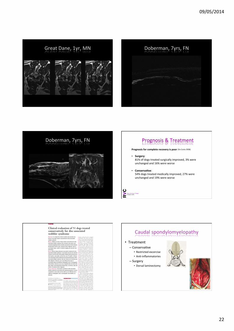
09/05/2014
22
Great Dane, 1yr, MN Doberman, 7yrs, FN
Doberman, 7yrs, FN Prognosis & Treatment Prognosis for complete recovery is poor (Da Costa 2008) ¥ Surgery: 81% of dogs treated surgically improved, 3% were unchanged and 16% were worse
¥ Conserva,ve: 54% dogs treated medically improved, 27% were unchanged and 19% were worse
PAPER
Clinical evaluation of 51 dogs treatedconservatively for disc-associatedwobbler syndromeOBJECTIVES: To evaluate the clinical evolution and potential risk
factors of 51 dogs treated conservatively for disc-associated
wobbler syndrome.
METHODS: Medical records of dogs treated conservatively for disc-
associated wobbler syndrome were reviewed, and owners were
contacted regarding clinical evolution and survival of their animals.
Relationships between age, treatment before diagnosis, type of
neurological signs, results of medical imaging and outcome were
determined.
RESULTS: Fifty-one dogs underwent conservative treatment for disc-
associated wobbler syndrome. A successful outcome was achieved
in 45 per cent (23 of 51) of the patients.Median follow-up period was
18.5 months, and median survival time was 47 months. In 85 per
cent of the dogs in which euthanasia was performed because of disc-
associated wobbler syndrome, this was carried out in the first year
after diagnosis. Outcome score was influenced by type of
neurological signs and additional radiographic and/or myelographic
abnormalities. Outcome score was not significantly associated with
age, number of protruded intervertebral discs, occurrence, type and
results of treatment before diagnosis.
CLINICAL SIGNIFICANCE: Conservative treatment of disc-associated
wobbler syndrome is associated with a guarded prognosis. It can be
considered in cases where all four limbs are not affected and no
additional radiographic and/or myelographic abnormalities are
detected.
S. DE DECKER, S. F. M. BHATTI, L.DUCHATEAU*, V. A.MARTLE, I. VAN SOENS,S. A. E. VANMEERVENNE, J. H. SAUNDERSyAND L. M. L. VAN HAM
Journal of Small Animal Practice (2009)50, 136–142DOI: 10.1111/j.1748-5827.2008.00705.x
INTRODUCTION
The ‘‘wobbler’’ syndrome refers to a collec-
tionofdisorders of the cervical vertebrae and
intervertebral discs (IVDs) of large breed
dogs resulting in spinal cord compression.
The term wobbler mainly refers to the typ-
ical pelvic limb ataxia (VanGundy 1988).
A large variety of lesions with different pro-
posed aetiologies have been attributed to
this syndrome, and over years, a few
separate entities have been recognised(VanGundy 1988, Jeffery and McKee
2001). The most typical and predominant
of these entities is disc-associated wobbler
syndrome (DAWS). This specific wobbler
syndrome is seen in middle-aged large
breed dogs, in particular the dobermann
(VanGundy 1988, McKee and Sharp
2003). In DAWS, caudal cervical spinal
cord compression results from protrusion
of IVDs and generally mild vertebral mal-
formations, frequently in combination
with dorsal spinal cord compression result-
ing from hypertrophy of the ligamentum
flavum (VanGundy 1988). Clinical signs
vary from cervical hyperaesthesia to tetra-
plegia. A slowly progressive pelvic limb
ataxia and paresis is the most common pre-
sentation (VanGundy 1988, Sharp and
Wheeler 2005). In dogs with apparently
normal thoracic limbs, it is sometimes
difficult to distinguish DAWS from a
thoracolumbar lesion (McKee and Sharp
2003). Progression to thoracic limb invol-
vement with a short stilted gait can occur
(VanGundy 1988, McKee and Sharp
2003). Affected dogs often show a charac-
teristic ‘‘disconnected’’ gait, in the sense
that the thoracic and pelvic limbs seem
to advance at different rates. Neck pain
may be seen but is usually not overtly pres-
ent (Seim2000).Tetraplegia is uncommon
(Sharp and Wheeler 2005).There is a lot of controversy about the
treatment of DAWS (Jeffery and McKee
2001). It is often defined as a progressive
disease in which early surgery is necessary
to halt progression of signs (McKee and
Sharp 2003). Several surgical procedures
have been described forDAWS (Chambers
and others 1982, Bruecker and others 1989,
McKee and others 1989, 1990, Dixon and
others 1996). Reported short-term success
rates after surgery are high (around 80 per
cent), but there exists a high rate of recur-
rence. This is most often because of the
development of adjacent segment disease
and occurs in 20 to 30 per cent of initially
successfully treated dogs, independent of
Department of Small Animal Medicine andClinical Biology, *Department of Physiology andBiometrics and yDepartment of Medical Imaging,Faculty of Veterinary Medicine, Ghent University,9820 Merelbeke, Belgium
136 Journal of Small Animal Practice � Vol 50 � March 2009 � � 2009 British Small Animal Veterinary Association
Caudal spondylomyelopathy
¥ Treatment – Conserva6ve ¥ Restricted excercise ¥ An6-‐inflammatories
– Surgery ¥ Dorsal laminectomy
09/05/2014
23
Caudal spondylomyelopathy
¥ Treatment – Conserva6ve ¥ Restricted excercise ¥ An6-‐inflammatories
– Surgery ¥ Ventral Slot
¥ Dynamic vs non-‐dynamic
development of a postlaminectomy membrane. However, a study in dogs indicateda high incidence of graft failure and neurologic complications after the use of a freefat graft in the thoracolumbar spine.126 Whether or not the same occurs in the cervicalspine, explaining the common neurologic deterioration seen postoperatively, isunknown at this time. Considering the high incidence of graft failure and the neurologiccomplications documented in dogs, the use of free fat grafts after dorsal laminectomyis not recommended at this time.The success rate for dorsal laminectomies ranges from 79% to 95%.11,58,103 The
mean time to reach optimal improvement is long (3.6 months), and in one report,30% of dogs had recurrence of signs postoperatively.58 Dorsal laminoplasty is analternative to dorsal laminectomy that has not been sufficiently explored in dogs.105
Cervical hemilaminectomy A technique of lateral approach for cervical hemilaminec-tomy was used in the treatment of CSM in dogs with either disc- or
Fig. 10. Ventral slot at C6-7. (A) Ventral view. (B) Transverse view. The slot width and lengthshould ideally be kept at about one-third of the vertebral bodies. The internal vertebralvenous plexus are represented by the blue vessels running in the ventral aspect of the verte-bral canal. (C) Inverted cone slot; this is a modification of the traditional ventral slot thatminimizes the risk of hemorrhage and subluxation. (Courtesy of The Ohio State University;with permission.)
Cervical Spondylomyelopathy (Wobbler Syndrome) 899
increases. Either a partial or complete ventral slot is created. The metal implant canbe either 2 Steinmann pins, threaded pins, or bone screws (Fig. 13). A recent studysuggested that positive profile pins provide more rigid fixation than smooth pins.127
The original recommended angle of implant insertion was 30� to 35�.107 A recentstudy indicated that these angles are relatively safe for the vertebrae C5 and C6,but that an angle of approximately 45� should be used for implants at C7, which isvery challenging at this location.128 The long-term success rate of this techniqueis 73%.107 Vertebral or transverse foramina penetration is a major risk of this proce-dure, and has been reported to occur in 25% to 57% of cases studiedexperimentally.127,128
Several modifications of this procedure have been reported using different tech-niques and materials for distraction.109,116 A recent study reported the use of screwsinserted into the transverse processes, using a U-shaped Steinmann pin that waswired to the screws and covered with PMMA.115 The advantage of this technique isthat it avoids the risk of penetration into the vertebral canal or transverse foramina.A disadvantage of some of the distraction techniques is that the instrumentationused for vertebral body fixation can cause severe imaging artifacts, predominantlywith MR imaging, making postoperative spinal cord visualization difficult orimpossible.
Distraction using the PMMA plug Distraction using the PMMA plug is a popular tech-nique and has been used for either single or multiple ventral and dorsal compressions,
Fig. 13. The technique of pins and PMMA combined with a partial slot. Note the location ofpin insertion avoiding the transverse foramina and vertebral canal. (Courtesy of The OhioState University; with permission.)
Cervical Spondylomyelopathy (Wobbler Syndrome) 901
Post surgical decompression
¥ Physiotherapy!
Veterinary Training
Humboldt‐University Berlin, Germany 1986‐1992
Working Experience Working experience in large and small animal medicine at
Free University of Berlin as well as in private practice in Berlin 1992‐1998
Department of Companion Animals and Special Species 1998‐1999 College of Veterinary Medicine, North Carolina State University, USA
Clinical Instructor in Veterinary Neurology Department of Veterinary Clinical Sciences, The Ohio State University, USA 1999‐2001
Residency in Veterinary Neurology and Neurosurgery Department of Small Animal Medicine, University of Leipzig, Germany
Head of the section of neurology and neurosurgery since November 2002
Veterinary and Academic Qualifications Doctor medicinae veterinariae (summa cum laude) 1994
Master of Veterinary Sciences (The Ohio State University, USA) 2001
Diplomate American College of Veterinary Internal Medicine (Neurology) 2003
Diplomate European College of Veterinary Neurology 2005
European Specialist in Veterinary Neurology 2008
Dr. med. vet. habilitatus (small animal surgery and small animal neurology) 2010
Secretary of the European College of Veterinary Neurology since 2012
Priv.-Doz. Dr. Thomas Flegel
Disorder of eyes and vision Priv.-Doz. Dr. Thomas Flegel
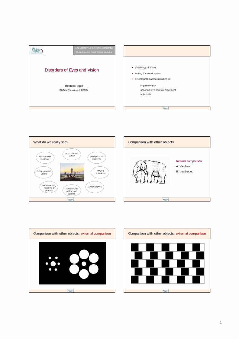
1
Disorders of Eyes and Vision
Thomas Flegel DACVIM (Neurologie), DECVN
UNIVERSITY of LEIPZIG, GERMANY
Department of Small Animal Medicine
• physiology of vision
• testing the visual system
• neurological diseases resulting in: impaired vision
abnormal eye position/movement
anisocoria
What do we really see?
perception of countours
3-dimensional vision
understanding meaning of
pictures comparision with known
objects
perception of colors
judging speed
judging distances
perception of contrasts
Comparison with other objects
Internal comparison:
A: elephant
B: quadruped
Comparison with other objects: external comparison Comparison with other objects: external comparison

2
Brodman Regions
Korbinian Brodman
1868-1918
Primary and secondary visual cortex
Visual cortex
primary
Processing of direct visual input
secondary
Processing of the meaning of things
we see
„cortical blindness“ „soul blindness“
Perception of distance
Left eye Right eye
sees a little bit more of the left side of an object
sees a little bit more of the right side of an object
The discrepancy between those 2 images is the larger the closer the object is
Some fibers carry the same information If those are the „2-meter-fibers“: the object is 2 meters away
estimating distances reliably up to 60 meters Stereopsis (depth perception)
Color vision
black/white
in addition: blue
in addition: red
Color vision
Dogs:
• differentiate red and blue
• often mix up green and red
Human Dog (probably)
Visual field
www.veterinaryvision.com
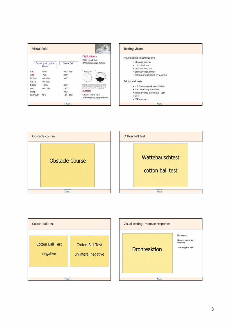
3
Visual field
cat: 65% 200°-285°
dog: 75% 240°
horse: 80-90% 305°
cattle: 80-90%
birds: 100% 300°
owl: 60-70% 160°
frog: 330°
human: 50% 180 - 200°
Crossing of optical fibers
Visual field
Flight animals: Wide visual field difficulties to judge distance
hunters: Smaller visual field Jadvantage in judging distance
Gelatt 2007
Testing vision
Neurological examination: • obstacle course • cottonball test •menace reaction • pupillary light reflex • testing physiological nystagmus
Additional tests:
• ophthalmological examination • Electroretinogram (ERG) • visual evoked potentials (VEP) • MRI • CSF analysis
Obstacle course Cotton ball test
Cotton ball test Visual testing: menace response
Not correct: Second eye is not covered Touching the hair
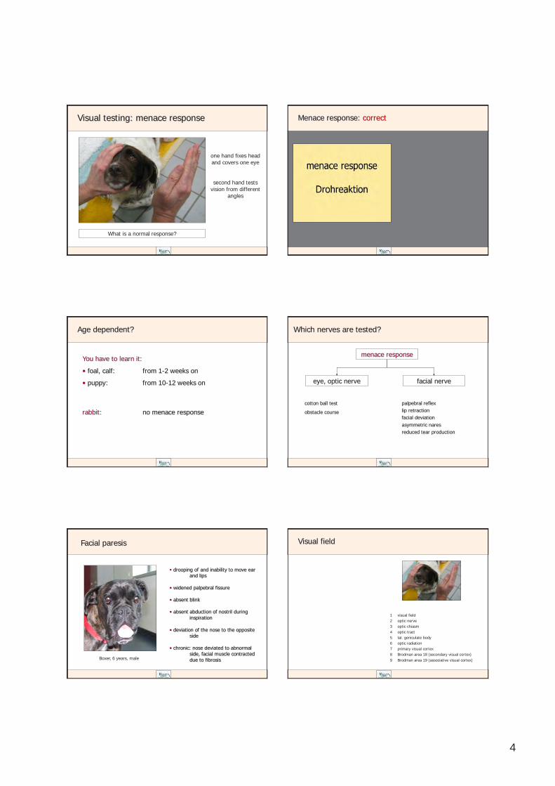
4
one hand fixes head and covers one eye
second hand tests vision from different
angles
What is a normal response?
Visual testing: menace response Menace response: correct
Age dependent?
You have to learn it:
• foal, calf: from 1-2 weeks on
• puppy: from 10-12 weeks on
rabbit: no menace response
Which nerves are tested?
cotton ball test
obstacle course
menace response
eye, optic nerve facial nerve
palpebral reflex lip retraction facial deviation asymmetric nares reduced tear production
Facial paresis
Boxer, 6 years, male
• drooping of and inability to move ear and lips • widened palpebral fissure • absent blink • absent abduction of nostril during inspiration • deviation of the nose to the opposite side • chronic: nose deviated to abnormal side, facial muscle contracted due to fibrosis
Visual field
1 visual field 2 optic nerve 3 optic chiasm 4 optic tract 5 lat. geniculate body 6 optic radiation 7 primary visual cortex 8 Brodman area 18 (secondary visual cortex) 9 Brodman area 19 (associative visual cortex)
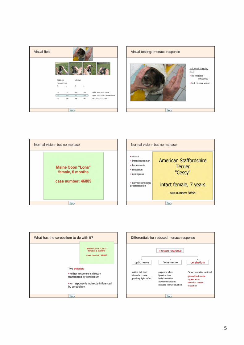
5
Visual field
Right eye Left eye
menace from
R L R L
no no yes yes right: eye, optic nerve
no yes no yes right: optic tract, visual cortex
no yes yes no central optic chiasm
but what is going on if:
• no menace response
• but normal vision
Visual testing: menace response
Normal vision- but no menace Normal vision- but no menace
• ataxia
• intention tremor
• hypermetria
• titubation
• nystagmus
• normal conscious proprioception
What has the cerebellum to do with it?
Two theories:
• either response is directly transmitted by cerebellum
• or response is indirectly influenced by cerebellum
Differentials for reduced menace response
cotton ball test obstacle course pupillary light reflex
menace response
optic nerve facial nerve cerebellum
Other cerebellar deficits?
generalized ataxia hypermetria intention tremor titubation
palpebral eflex lip retraction facial deviation asymmetric nares reduced tear production
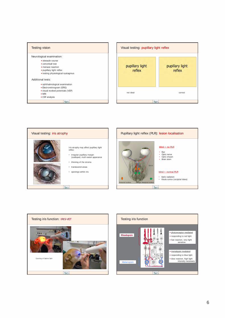
6
Testing vision
Neurological examination: • obstacle course • cottonball test •menace reaction • pupillary light reflex • testing physiological nystagmus
Additional tests:
• ophthalmological examination • Electroretinogram (ERG) • visual evoked potentials (VEP) • MRI • CSF analysis
Visual testing: pupillary light reflex
not ideal correct
Visual testing: iris atrophy
Iris atrophy may affect pupillary light reflex:
• irregular pupillary margin
(scalloped, moth-eaten apperance
• thinning of the stroma
• translucend areas
• openings within iris
Pupillary light reflex (PLR): lesion localisation
blind + no PLR: • Eye • Optic nerve • Optic chiasm • Brain stem
blind + normal PLR: • Optic radiation • Visula cortex (occipital lobes)
CN II
CN III
Pretectal nucleus Edinger Westphal nucleus
Testing iris function: IRIS-VET
Courtesy of Sabine Sahr
Testing iris function
Melanopsin
Rhodopsin • photoreceptor mediated
• responding to red light
• fast reaction, very light sensitive
• menalopsin mediated
• responding to blue light
• slow reaction, high light intensity necessary

7
Melan-100 Electrodiagnostics
Electroretinogram (ERG)
http://totaleyecare.files.wordpress.com/2012/10/vepimage1.jpg
Visual evoked potentials (VEP)
• physiology of vision
• testing the visual system
• neurological diseases resulting in: impaired vision
abnormal eye position/movement
anisocoria
Algorythm visual deficits: adopted from Gelatt: Veterinary Ophthalmology, 2007
Visual impairment
both eyes homonymous hemianopia
one eye Vision test
PLR
Lesion lacalization
Additional deficits
bilateral fixed dilated pupils
no PLR
both eyes or optic nerves
chiasma
optic radiation before PLR fibres diverge
normal
bilateral unilateral
lateral geniculate body (…ies)
optic radiation
visual cortex
forebrain signs
CP-deficits
static anisocoria PLR relatively normal
affected eye: no PLR
other eye: normal PLR
eye, optic nerve pre chiasmatic
opposite optic tract before PLR fibres diverge
contralateral CP-deficits
other cranial nerve deficits and Horner‘s
• menace response • cottonball test
• obstacle course • owner report
Ocular lesion: retinal detachment
funduscopic examination
ultrasound of the eye
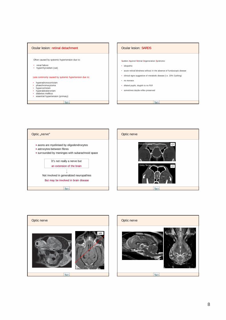
8
Ocular lesion: retinal detachment
Often caused by systemic hypertension due to: • renal failure • hyperthyroidism (cat)
Less commonly caused by systemic hypertension due to: • hyperadronocorticism • phaechromocytoma • hypercorticism • hyperaldosteronism • diabetes mellitus • essential hypertension (primary)
Ocular lesion: SARDS
Sudden Aquired Retinal Degeneration Syndrome: • idiopathic
• acute retinal blindness without in the absence of funduscopic disease
• clinical signs suggestive of metabolic disease (i.e. 15% Cushing)
• no menace
• dilated pupils, slugish to no PLR
• sometimes dazzle-reflex preserved
Optic „nerve“
• axons are myelinised by oligodendrocytes • astrocytes between fibres • surrounded by meninges with subarachnoid space
It‘s not really a nerve but
an extension of the brain
Not involved in generalized neuropathies
But may be involved in brain disease
Optic nerve
French Bulldog CT
Cesky Terrier CT
Optic nerve
MRI
Optic nerve
9
MRI
MRI is only as good as:
1. the knowledge of the examiner of all relevant diseases
2. the ability of the examiner to visualize those diseases
Optic nerve
Fibers of the Macula are localized centrally
Central lesion
axial neuritis
Reduced visual acuity No blindness
Peripheral lesion
Druck von außen
Narrowed visual field No impairment of visual acuity
Optic nerve lesion: neuritis
neuritis opposite (normal) eye
Courtesy of Andrea Steinmetz
• disc appears swollen and edematous
• blurry disc margins
Optic nerve lesion: neuritis
neuritis
• disc appears swollen and edematous
• blurry disc margins
infectious: distemper, toxoplasmosis tick-borne-encephalitis ehrlichiosis fungal FIP inflammatory: GME, NME? others: traumatic vitamin A deficiency toxic neoplastic
Lesion at the optic chiasm
Seruca et al., Vet Ophthalmology, 2010
Etiology: • neoplasia: 5/7 • optic neuritis: 1/7
Neuroanatomic localisation: • optic chiasm 5/7 • optic nerve: 1/7 • optic tract: 1/7
Ocular lesion: lesion at the optic chiasm
10
Algorythm visual deficits: adopted from Gelatt: Veterinary Ophthalmology, 2007
Visual impairment
both eyes homonymous hemianopia
one eye Vision test
PLR
Lesion lacalization
Additional deficits
bilateral fixed dilated pupils
no PLR
both eyes or optic nerves
chiasma
optic radiation before PLR fibres diverge
normal
bilateral unilateral
lateral geniculate body (…ies)
optic radiation
visual cortex
forebrain signs
CP-deficits
static anisocoria PLR relatively normal
affected eye: no PLR
other eye: normal PLR
eye, optic nerve pre chiasmatic
opposite optic tract before PLR fibres diverge
contralateral CP-deficits
other cranial nerve deficits and Horner‘s
• menace response • cottonball test
• obstacle course • owner report
Unilateral lesions with impaired PLR
CN II
CN III
Pretectal nucleus Edinger Westphal nucleus
Algorythm visual deficits: adopted from Gelatt: Veterinary Ophthalmology, 2007
Visual impairment
both eyes homonymous hemianopia
one eye Vision test
PLR
Lesion lacalization
Additional deficits
bilateral fixed dilated pupils
no PLR
both eyes or optic nerves
chiasma
optic radiation before PLR fibres diverge
normal
bilateral unilateral
lateral geniculate body (…ies)
optic radiation
visual cortex
forebrain signs
CP-deficits
static anisocoria PLR relatively normal
affected eye: no PLR
other eye: normal PLR
eye, optic nerve pre chiasmatic
opposite optic tract before PLR fibres diverge
contralateral CP-deficits
other cranial nerve deficits and Horner‘s
• menace response • cottonball test
• obstacle course • owner report
Forebrain lesion
V hemorraghe, infarct, global ischemia, mouth gag
E infection, non-infectious encephalitis
T
A hydrocephalus
M
I
N primary or metastatic neoplasia
D Ceroid lipofuscinois, Mucopolysacharidosis Typ II
Genetic diseases
http://www.offa.org/dna_alltest.html
Forebrain lesion
11
Forebrain lesion Forebrain lesion
MRI: occipital meningioma
Domestic shorthair cat „Strolchie“, 12 years, neutered male
T1 T1+contrast
mean arterial blood pressure: 230 mm Hg
Hypertension: keep your hands off the blood presssure until…
Archieves Italiennes de Biologie 1901
Harvey Cushing
Intracranial pressure
Systemic blood pressure
bradycardia
Cushing-Reflex
Occipital infarct
CKCS „Tina“, 5 Jahre Caudal cerebral artery
FLAIR T2
Global brain ischemia
18 months 7 months 6 weeks 3 hours
Pug, 2 years, female: surgery for brachycephalic syndrome
12
Hydrocephalus Hydrocephalus: „Minchen“
Hydrocephalus: „Minchen“ Necrotising meningoencephalitis: Pug
• seizures (often partial) • reduced conscious proprioception • central blindness
Necrotising meningoencephalitis: Pug
T2
Algorythm visual deficits: adopted from Gelatt: Veterinary Ophthalmology, 2007
Visual impairment
both eyes homonymous hemianopia
one eye Vision test
PLR
Lesion lacalization
Additional deficits
bilateral fixed dilated pupils
no PLR
both eyes or optic nerves
chiasma
optic radiation before PLR fibres diverge
normal
bilateral unilateral
lateral geniculate body (…ies)
optic radiation
visual cortex
forebrain signs
CP-deficits
static anisocoria PLR relatively normal
affected eye: no PLR
other eye: normal PLR
eye, optic nerve pre chiasmatic
opposite optic tract before PLR fibres diverge
contralateral CP-deficits
other cranial nerve deficits and Horner‘s
• menace response • cottonball test
• obstacle course • owner report
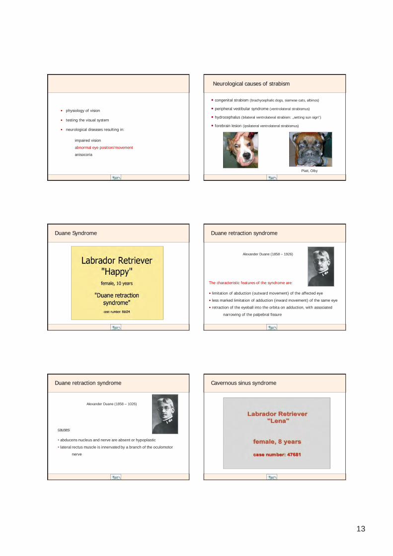
13
• physiology of vision
• testing the visual system
• neurological diseases resulting in: impaired vision
abnormal eye position/movement
anisocoria
Neurological causes of strabism
• congenital strabism (brachycephalic dogs, siamese cats, albinos)
• peripheral vestibular syndrome (ventrolateral strabismus)
• hydrocephalus (bilateral ventrolateral strabism: „setting sun sign“)
• forebrain lesion (ipsilateral ventrolateral strabismus)
Platt, Olby
Duane Syndrome Duane retraction syndrome
The characteristic features of the syndrome are:
• limitation of abduction (outward movement) of the affected eye
• less marked limitation of adduction (inward movement) of the same eye
• retraction of the eyeball into the orbita on adduction, with associated
narrowing of the palpebral fissure
Alexander Duane (1858 – 1926)
Duane retraction syndrome
causes:
• abducens nucleus and nerve are absent or hypoplastic
• lateral rectus muscle is innervated by a branch of the oculomotor
nerve
Alexander Duane (1858 – 1026)
Cavernous sinus syndrome

14
Cavernous sinus syndrome Cavernous sinus syndrome
Labrador „Lena“ , 11 years, female
T2 FLAIR
Cavernous sinus syndrome
Deficits of the following cranial nerves: CN III oculomotor CN IV trochlear CN VI abducens CN V trigeminal (first 2 branches) Deficits on the following test:
• physiological nystagmus (external ophthalmoparesis/plegia) • retractor oculi reflex • palpebral reflex • testing nasal sensation • drooping of the upper eyelid • direct and indirect pupillary light reflex (internal ophthalmoparesis/plegia)
Cavernous sinus syndrome
JAVMA 2005
• physiology of vision
• testing the visual system
• neurological diseases resulting in: impaired vision
abnormal eye position/movement
anisocoria
Horner‘s Syndrome
Loss of sympathic innervation to the eye:
• ptosis: „smaller eye“ • miosis: smaller pupil • enophthalmus: sunken in eye ball • 3rd eye lid protrusion
Andre Mischke, Kleintierzentrum Harsefeld
Sympathic M. ciliaris Innervation: M. orbitalis M. dilatator pupillae M. tarsalis superior (Müller-Muscle)
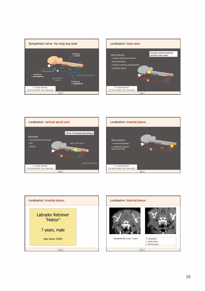
15
Sympathetic nerve: the long way back
C1-3 C4-8
T1-3
1st Neuron = zentral
2nd Neuron = präganglionär
3rd Neuron = postganglionär
Truncus vagosympathicus
Ggl. cervicale craniale
Bulla tympanica
Dr. Kaspar Matiasek The Animal Health Trust, Newmarket
Localisation: brain stem
Complex deficits pointing towards brain lesion
Dr. Kaspar Matiasek The Animal Health Trust, Newmarket
Other symptoms:
• multiple cranial nerve deficits
• paresis/paralysis
• reduced conscious proprioception
• vestibular deficits
Localisation: cervical spinal cord
tetra-/hemiparesis/plegia
Dr. Kaspar Matiasek The Animal Health Trust, Newmarket
upper motor neuron
lower motor neuron
Differentials:
• intervertebral disc disease
• FCE
• trauma
Localisation: brachial plexus
Dr. Kaspar Matiasek The Animal Health Trust, Newmarket
LMN
Other symptoms:
• monoparesis/plegia
• unilaterally reduced panniculus reflex
Localisation: brachial plexus Localisation: brachial plexus
Rottweiler Mix „Cora“ 7 years • neoplasia • stick-injury • biting injury

16
Localisation: vagosympathetic trunc
Dr. Kaspar Matiasek The Animal Health Trust, Newmarket
Other symptoms:
• laryngeal paralysis
• Megaösophagus
carotid sheath: arteria carotis communis Vagosympathicus trunc vagal nerve sympathic nerve
Differentials:
• pulling on the leash • neoplasia • injection, taking blood • biting, infection
Localisation: middle ear
Dr. Kaspar Matiasek The Animal Health Trust, Newmarket
• head tilt
• nystagmus
• ataxia
• ± CN VII defizit
Differentials:
• otitis media/interna • polyp • iatrogen
peripheral vestibular syndrome
Graefe‘s Archieve for Clinical and Experimental Ophthalmology, 1918
Localisation: media cranial fossa
Dr. Kaspar Matiasek The Animal Health Trust, Newmarket
• cranial nerve deficits
• reduced mentation
Fissura orbitalis
Fissura orbitalis
Diabetes mellitus
Horner‘s Syndrome: Diabetes mellitus
Andre Mischke, Kleintierzentrum Harsefeld
History: weakness polydipsia bilateral Horner‘s syndrome Neuroexam: delayed conscious proprioception reduced segmental spinal reflexes in rear limbs
Sympathetic nerve: the long way back
C1-3 C4-8
T1-3
1st Neuron = zentral
2nd Neuron = präganglionär
3rd Neuron = postganglionär
Truncus vagosympathicus
Ggl. cervicale craniale
Bulla tympanica
Dr. Kaspar Matiasek The Animal Health Trust, Newmarket
Pourfour-le-Petit-Syndrome
Boydell: Journal of Small Animal Practice 2000 (41): 202-203
1664-1741
Vestibular desease Priv.-Doz. Dr. Thomas Flegel
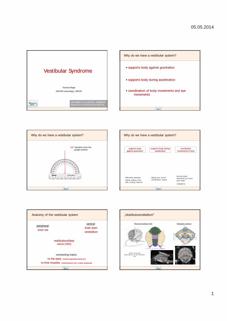
05.05.2014
1
Vestibular Syndrome
Thomas Flegel
DACVIM (neurology), DECVN
UNIVERSITY of LEIPZIG, GERMANY Department of Small Animal Medicine
Why do we have a vestibular system?
• supports body against gravitation
• supports body during acceleration
• coordination of body movements and eye movements
0.5° deviation from the upright position
Why do we have a vestibular system? Why do we have a vestibular system?
supports body against gravitation
supports body during acceleration
coordinates movements of eyes
difficulties standing rolling, falling to the side, circling, head tilt
falling over, loss of coordination, ataxia
blurred vision, whenever you move your head
nystagmus
Anatomy of the vestibular system
peripheral: inner ear
central: brain stem cerebellum
connecting tracts: to the eyes: medial longitudinal fasciculus
to limb muscles: vestibulospinal tract (mainly ipsilateral)
vestibulocochlear nerve (VIII)
„Vestibulocerebellum“
Flocculonodular lobe Fastigial nucleus
Stoffel: Funktionelle Neuroanatomie für die Tiermedizin,
2011
T2

05.05.2014
2
„Vestibulothalamus“
mixed breed „Lino“ 9 years
„Vestibulo-spinal cord“
Lesions in the upper cervical spine may cause vestibular symptoms: • transection of dorsal roots of the first 3 cervical segments
• lesions interfering with the cervicospinovestibular tracts of
those segments (S. 225)
Testing physiological nystagmus?
(LR6SO4)3
oculomotor n. (3) trochlear n. (4) abducens n. (6)
Innervation of extraocular muscles
Lateral Rectus muscle Superior Oblique muscle
http://www.csdl.tamu.edu/~sam/pictures/eye_muscles.gif
Why do we have a vestibular system?
supports body against gravitation
supports body during acceleration
coordinates movements of eyes
difficulties standing rolling, falling to the side, circling, head tilt
falling over, loss of coordination, ataxia
blurred vision, whenever you move your head
nystagmus
Ataxia
•video
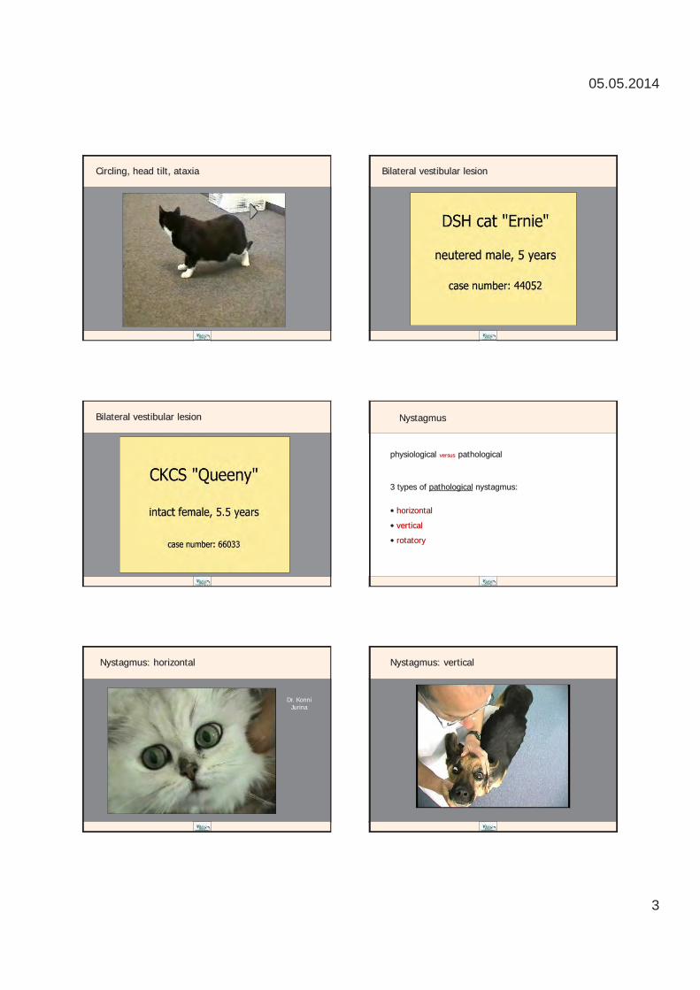
05.05.2014
3
Circling, head tilt, ataxia
•video
Bilateral vestibular lesion
Bilateral vestibular lesion Nystagmus
physiological versus pathological
3 types of pathological nystagmus:
• horizontal
• vertical
• rotatory
Nystagmus: horizontal
Dr. Konni Jurina
Nystagmus: vertical

05.05.2014
4
Nystagmus: rotatory Nystagmus: „rabbitory“
Nystagmus? Most important question in „vestibular neurology“
„Ear or brain: That is the question.“
• Diifferentials
• Diagnostic tests
• Prognois
• cost W. Shakeseare
Nystagmus: positional Nystagmus
When do we see nystagmus?
permanent: peripheral positional: central
What about the frequency? >60 beats/min peripheral <60 beats/min central
Troxel et al.. Signs of neurological dysfunction in dogs with central versus peripheral vestibular disease. J Am Vet Med Assoc 2005;227:570-574.
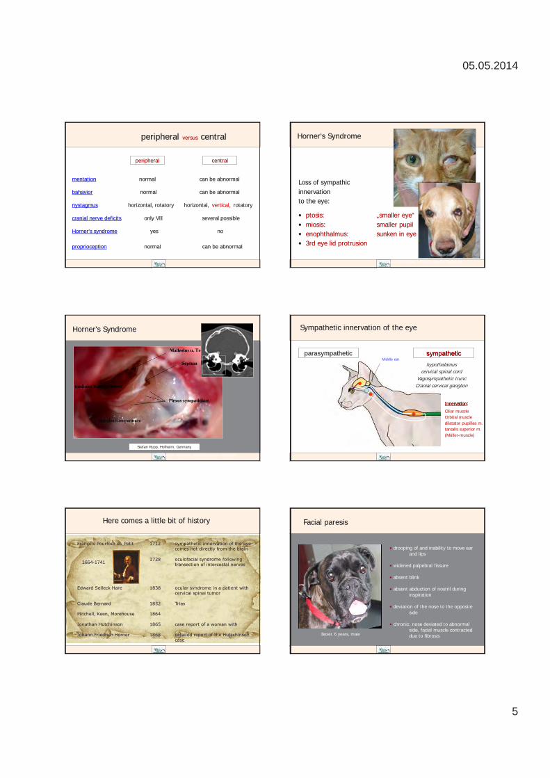
05.05.2014
5
peripheral versus central
mentation normal can be abnormal
bahavior normal can be abnormal
nystagmus horizontal, rotatory horizontal, rotatory
cranial nerve deficits only VII several possible
Horner‘s syndrome yes no
proprioception normal can be abnormal
vertical,
peripheral central
Horner‘s Syndrome
Loss of sympathic innervation to the eye:
• ptosis: „smaller eye“ • miosis: smaller pupil • enophthalmus: sunken in eye ball • 3rd eye lid protrusion
Andre Mischke, Kleintierzentrum Harsefeld
Horner‘s Syndrome
Andre Mischke, Kleintierzentrum Harsefeld
Stefan Rupp, Hofheim, Germany
Sympathetic innervation of the eye
parasympathetic sympathetic hypothalamus
cervical spinal cord Vagosympathetic trunc Cranial cervical ganglion
Innervation:
Ciliar muscle Orbital muscle dilatator pupillae m. tarsalis superior m. (Müller-muscle)
Middle ear
François Pourfour du Petit 1712 sympathetic innervation of the eye comes not directly from the brain 1728 oculofacial syndrome following transection of intercostal nerves
Edward Selleck Hare 1838 ocular syndrome in a patient with cervical spinal tumor Claude Bernard 1852 Trias Mitchell, Keen, Morehouse 1864 Jonathan Hutchinson 1865 case report of a woman with Johann Friedrich Horner 1868 detailed report of the Hutschinson case
1664-1741
Here comes a little bit of history Facial paresis
Boxer, 6 years, male
• drooping of and inability to move ear and lips • widened palpebral fissure • absent blink • absent abduction of nostril during inspiration • deviation of the nose to the opposite side • chronic: nose deviated to abnormal side, facial muscle contracted due to fibrosis
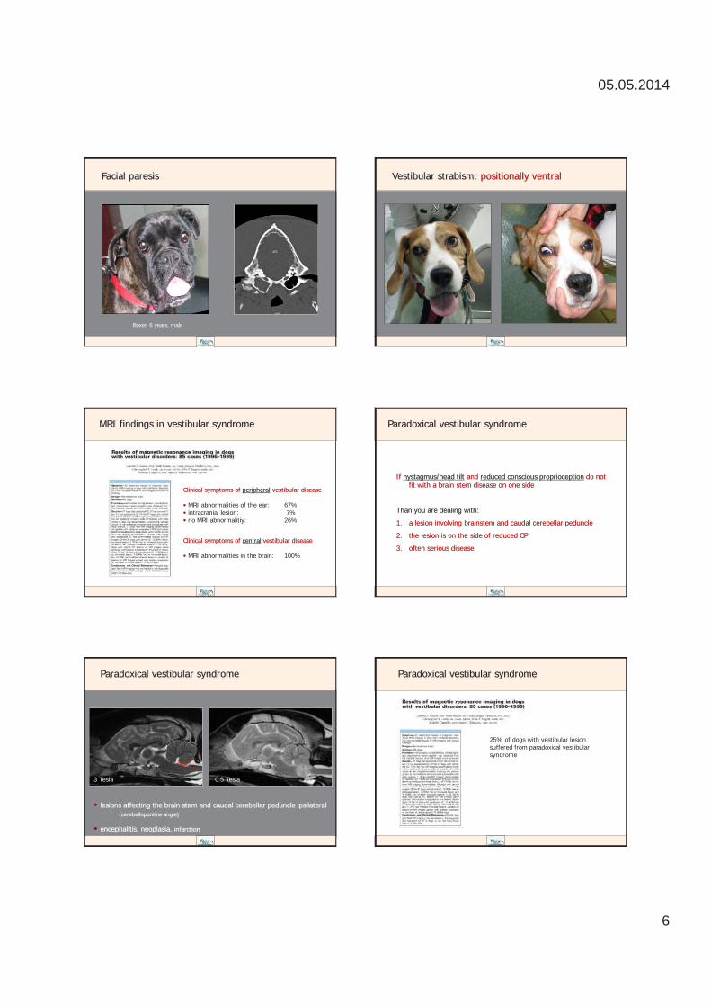
05.05.2014
6
Facial paresis
Boxer, 6 years, male
Vestibular strabism: positionally ventral
MRI findings in vestibular syndrome
Clinical symptoms of peripheral vestibular disease • MRI abnormalities of the ear: 67% • intracranial lesion: 7% • no MRI abnormalitiy: 26%
Clinical symptoms of central vestibular disease • MRI abnormalities in the brain: 100%
Paradoxical vestibular syndrome
If nystagmus/head tilt and reduced conscious proprioception do not fit with a brain stem disease on one side
Than you are dealing with:
1. a lesion involving brainstem and caudal cerebellar peduncle
2. the lesion is on the side of reduced CP
3. often serious disease
Paradoxical vestibular syndrome
• lesions affecting the brain stem and caudal cerebellar peduncle ipsilateral (cerebellopontine angle)
• encephalitis, neoplasia, infarction
3 Tesla 0.5 Tesla
Paradoxical vestibular syndrome
25% of dogs with vestibular lesion suffered from paradoxical vestibular syndrome
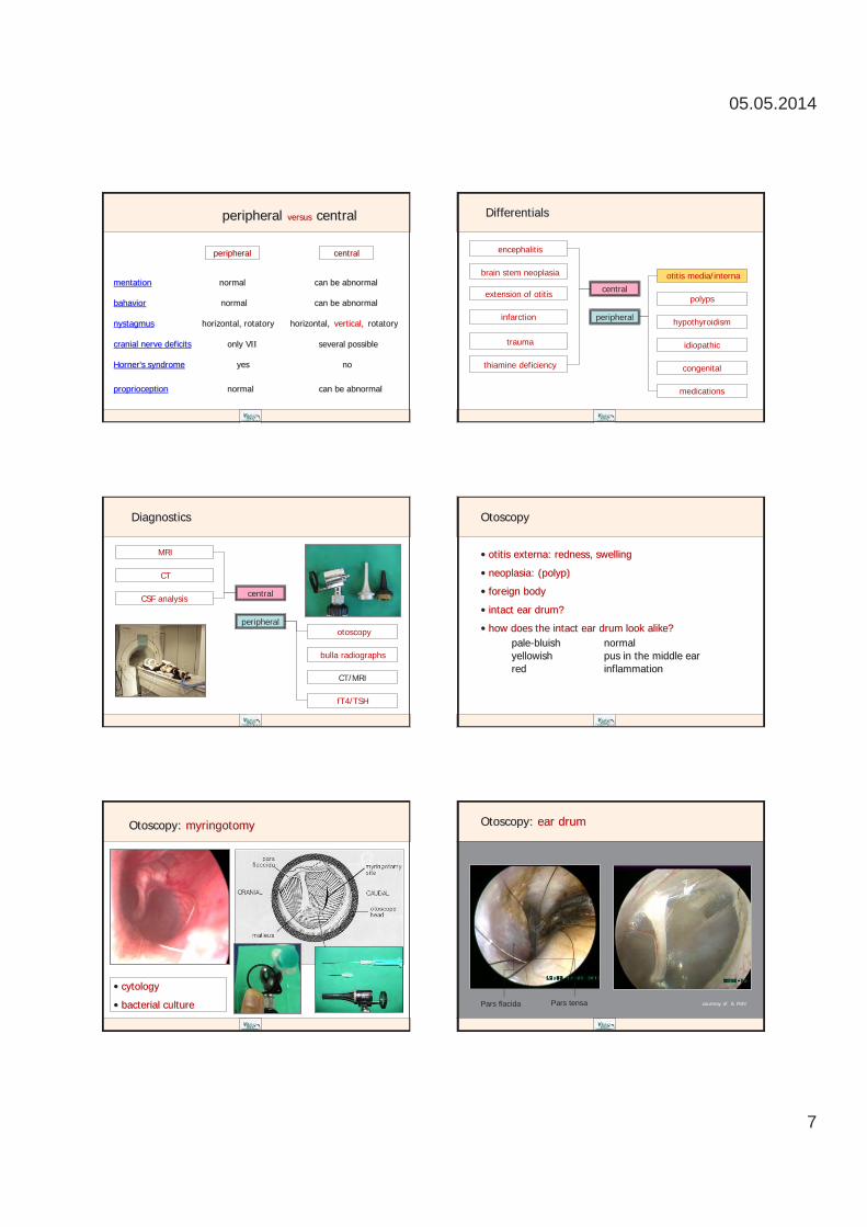
05.05.2014
7
peripheral versus central
mentation normal can be abnormal
bahavior normal can be abnormal
nystagmus horizontal, rotatory horizontal, rotatory
cranial nerve deficits only VII several possible
Horner‘s syndrome yes no
proprioception normal can be abnormal
vertical,
peripheral central
Differentials
central
peripheral
encephalitis
otitis media/interna
polyps
hypothyroidism
congenital
idiopathic
medications
extension of otitis
infarction
trauma
thiamine deficiency
brain stem neoplasia
Diagnostics
central
peripheral
MRI
otoscopy
bulla radiographs
fT4/TSH
CSF analysis
CT
CT/MRI
• otitis externa: redness, swelling
• neoplasia: (polyp)
• foreign body
• intact ear drum?
• how does the intact ear drum look alike? pale-bluish normal yellowish pus in the middle ear red inflammation
Otoscopy
Otoscopy: myringotomy
• cytology
• bacterial culture
Otoscopy: ear drum
courtesy of S. Pohl Pars flacida Pars tensa

05.05.2014
8
Otoscopy: ear drum
courtesy S. Pohl
Otoscopy: ear drum
courtesy S. Pohl
Otoscopy: ruptured ear drum
courtesy S. Pohl
Otoscopy: foreign body, polyp
Grass awn Polyp
Courtesy of S. Pohl
Otoscopy: cytology
bacterial otitis Wright-Giemsa stain
ear mites
Malessezia
Imaging
• radiography
• computed tomography
• magnetic resonance imaging
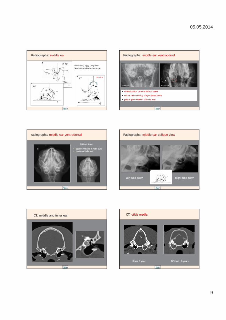
05.05.2014
9
Radiographs: middle ear
10-25°
10°
20°
Vandevelde, Jaggy, Lang 2001: Veterinärmedizinische Neurologie
30-45°!
• mineralization of external ear canal
• loss of radiolucency of tympanica bulla
• lysis or proliferation of bulla wall
normal abnormal
Radiographs: middle ear ventrodorsal
radiographs: middle ear ventrodorsal
DSH-cat, 1 year
• opaque material in right bulla • thickened bulla wall
R
Left side down Right side down
Radiographs: middle ear oblique view
CT: middle and inner ear CT: otitis media
Boxer, 6 years DSH cat , 8 years
05.05.2014
10
MRI: Otitis media (interna?)
Boxer, 8 years Doberman, 3 years
MRI: Otitis media/interna
T2 heavily T2-weighted
Fluid filled middle ear without associated clinical signs (PSOM) Katrin Truar Pug Dog (n=41) unilateral: 14.6% bilateral: 9.8% total: 24.4% French Bulldog (n=41) unilateral: 24.4% bilateral: 29.3% total: 53.7%
Primary secretory otitis media (PSOM) MRI: PSOM
Subclinical middle ear disease MRI: Otitis media/interna
T2 heavily T2-weighted

05.05.2014
11
CT „Ernie“: bilateral vestibular syndrome Therapy: Otitis media/interna
• antibiotics for 6 weeks (at least): cephalosporine fluroquinolone amoxicillin/clavulanic acid TMS
• ear cleaning and local antibiatic (if ear drum is intact)
• surgical: bulla osteotomy total ear canal ablation (TECA) endoscopic surgery through ear drum
T2
T1
T1 + contrast
CT
CSF cell count:
881/µl (normal<5)
MRI: otitis with brain stem involvement MRI: Otitis with brain stem involvement
Cholesteatoma
Airdale Terrier „Paula“
female, 12 years
Cholesteatoma
French Bulldog „Louis“ 8 years
„Epidermoid cysts containing hyperkeratotic squamous epithelium surrounding core of keratin debris“ Banco et al. 2014 Complication of otitis media
05.05.2014
12
Cholesteatoma Differentials
central
peripheral
encephalitis
otitis media/interna
polyps
hypothyroidism
congenital
idiopathic
medications
extension of otitis
infarction
trauma
thiamine deficiency
brain stem neoplasia
Acute vestibular symptoms in old dogs
History:
acute lack of coordination
German Shpeherd dog
15 years
Acute vestibular symptomes in old dogs...
In most cases neoplastic disease is supsected: Euthanasia
No, no, no, no!! – It is more likely to be
idiopathic vestibular syndrome or hypothyroidism!!
„Old dog“ vestibular disease
• old (> 10 Jahre)
• acute symptomes
• peripheral vestibular syndrome
Idiopathic vestibular syndrome
Hypothyroidism
Brain infarct
breeds: more often large breed dogs age: > 10 years of age but consider normal life expectation presenting signs: acute severe vestibular syndrome head tilt generalized ataxia nystagmus signs of peripheral vestibular lesion
Geriatric vestibular syndrome: the typical patient
05.05.2014
13
„Old dog“ vestibular disease
• old (> 10 Jahre)
• acute symptomes
• peripheral vestibular syndrome
Idiopathic vestibular syndrome
Hypothyroidism
Brain infarct
Infarct
mixed breed dog
female, 9 years
Pug
male, 4 years
„Old dog“ vestibular disease
• old (> 10 Jahre)
• acute symptomes
• peripheral vestibular syndrome
Idiopathic vestibular syndrome
Hypothyroidism
Brain infarct
Hypothyroidism (dog)
Specificity: 74% without hypothyroidism have a negative test result
Sensitivity: 98% with hypothyroidsim have a postitive test result
free T4
Geriatric vestibular syndrome: differentials
• idiopathic (geriatric) vestibular syndrome
• Hypothyroidism
• Infarction
peripheral
central
• idiopathic (geriatric) vestibular syndrome
• Hypothyroidism
• Infarction
Geriatric vestibular syndrome: diagnostics
no test
fT4, TSH
brain MRI
peripheral
central

05.05.2014
14
Therapy: Idiopathic vestibular syndrome
„The art of medicine consists in amusing the patient while nature cures the disease“
Voltaire (1694-1778)
• idiopathic (geriatric) vestibular syndrome
• Hypothyroidism
• Infarction
Geriatric vestibular syndrome: therapy
no test
fT4, TSH
fluids antiemetics Karsivan??
levothyroxin 20 µg/kg BID
brain MRI probably nothing
peripheral
central
• idiopathic (geriatric) vestibular syndrome
• Hypothyroidism
• Infarction
Geriatric vestibular syndrome: prognosis
none
fT4, TSH
fluids antiemetics (Maropitant)
propentofyllin (Karsivan)
levothyroxin 20 µg/kg BID
improvement starts within 3-4 days begins with less severe nystagmus might have residual head tilt
improvement within days-months
brain MRI probably nothing
Differentials
central
peripheral
encephalitis
otitis media/interna
polyps
hypothyroidism
congenital
idiopathic
medications
extension of otitis
infarction
trauma
thiamine deficiency
brain stem neoplasia
Vestibulotoxic substances
metronidazol in therapeutic dosage
Differentials
central
peripheral
encephalitis
otitis media/interna
polyps
hypothyroidism
congenital
idiopathic
medications
extension of otitis
infarction
trauma
thiamine deficiency
brain stem neoplasia
05.05.2014
15
Polyp (cat)
DSH cat „Pablo“
male, 8 years
T2 T2
T1 T1+Kontrast
Differentials
central
peripheral
encephalitis
otitis media/interna
polyps
hypothyroidism
congenital
idiopathic
medications
extension of otitis
infarction
trauma
thiamine deficiency
brain stem neoplasia
Thiamine deficiency (cat)
T2 weighted
• Fisch diet
• anorexia
• Diagnostics: EDTA-plasma
• Therapy: Thiamine 25-50mg daily
thiamine deficiency normal
Thiamine deficiency (cat)
Penderis et al., Vet Record 2007
Thiamine deficiency (cat) DSH cat „Samira“, female, 7 years
• generalized ataxia, head tilt and circling to the right of 2 days duration
• vertical nystagmus
T2 T1 T1 + contrast
05.05.2014
16
DSH cat „Samira“, female, 7 years
CSF analysis: nucleated cell count: 11/µl (normal <5) total protein: 0.21 g/l (normal <0.25)
Toxoplasmosis Ab-titer: IgG: 1:1024 (normal 1:32) IgM: 1:64 (normal 1:16)
Toxoplasmosis Ab-titer 5 weeks later: IgG: 1:1024 (normal 1:32) IgM: 1:256 (normal 1:16)
Anything else?


www.Farmina.com
Hap
py p
et. H
appy
you
.
Dis
trib
uter
za
Srbi
ju:
Plus
Plus
d.o
.o.
BEO
GR
AD
: Sve
tog
Save
94,
nas
elje
Rad
iofa
r, 11
271
Surč
inte
l: 01
1/31
39
800;
011
/ 31
48 6
68
NO
VI S
AD
: Bul
evar
Voj
vode
Ste
pe 5
8, 2
1000
Nov
i Sad
tel:
021/
640
33 4
7
NIŠ
: Top
lički
par
tiza
nski
odr
ed B
B, 1
8250
Nov
o Se
lote
l: 01
8/42
8 55
11
ww
w.p
lusp
lus.
co.rs
; e- m
ail:
info
@pl
uspl
us.rs
: Sve
tog
Save
94,
nas
elje
Rad
iofa
r, 11
271
Surč
in
: Bul
evar
Voj
vode
Ste
pe 5
8, 2
1000
Nov
i Sad
: Top
lički
par
tiza
nski
odr
ed B
B, 1
8250
Nov
o Se
lo
ww
w.p
lusp
lus.
co.rs
; e- m
ail:
info
@pl
uspl
us.rs
: Sve
tog
Save
94,
nas
elje
Rad
iofa
r, 11
271
Surč
in
: Bul
evar
Voj
vode
Ste
pe 5
8, 2
1000
Nov
i Sad
: Top
lički
par
tiza
nski
odr
ed B
B, 1
8250
Nov
o Se
lo
ww
w.p
lusp
lus.
co.rs
; e- m
ail:
info
@pl
uspl
us.rs
Najb
olja
podr
ška z
a dob
robi
t vaš
eg ps
a.Za
svak
i pro
blem
pos
toji
reše
nje,
pra
vo re
šenj
e.th
e Ch
air o
f Ani
mal
N
utrit
ion
and
Die
t, U
nive
rsity
of N
aple
s "F
eder
ico
II".
the
Chai
r of A
nim
al
SCIE
NTI
FICA
LLY
TEST
ED B
Y:
Gas
tro-
Inte
stin
alKo
d po
rem
ećaj
a u
vare
nju,
radu
cre
va i
insu
ficije
ncije
pa
nkre
asa.
Hra
na p
ogod
na k
od st
omač
nih
i cre
vnih
pro
blem
a sa
po
rem
ećaj
em u
aps
orpc
iji, v
isok
o sv
arlji
vi i b
io-r
aspo
loži
vi
sast
ojci
. Pog
odna
i kod
isu�
cije
ncije
pan
krea
sa.
Obe
sity
Za k
ontr
olu
prek
omer
ne te
lesn
e te
žine
i D
iabe
tes
mel
litus
a.N
amen
jen
je za
kon
trol
u go
jazn
osti
zbog
sman
jeno
g sa
drža
ja e
nerg
ijei v
isok
og sa
drža
ja li
pida
. L- k
arni
tin i v
isok
o kv
alite
tni p
rote
ini d
oprin
ose
održ
avan
ju m
išić
ne m
ase.
Ogr
anič
avan
jem
od
rizik
a go
jazn
osti
dopr
inos
ise
kva
litet
nije
m i d
užem
živo
tu p
sa. Z
ahva
ljuju
ći n
isko
m sa
drža
ju sk
roba
m
ože
se k
oris
titi u
kon
trol
i nas
tank
a i t
erap
iji D
iabe
tes m
ellit
usa.
Gro
wth
Za ra
st š
tene
ta, o
pora
vak
i kod
gas
tro-
inte
stin
alin
h po
rem
ećaj
a.Ko
mpl
etna
dije
tets
ka h
rana
, vis
oke
svar
ljivo
sti z
bog
čega
je
optim
alna
za št
ence
kod
koj
ih je
uoč
en p
orem
ećaj
u ra
stu,
za
nut
ritiv
ni o
pora
vak
i pra
vila
n ra
zvoj
imun
og si
stem
a.
Rena
lZa
pre
venc
iju i
tera
piju
hro
ničn
e bu
brež
ne i
srča
ne in
sufic
ijenc
ije.
Zahv
alju
jući
redu
kova
nom
niv
ou p
rote
ina,
fo
sfor
a i n
atrij
uma
sman
juje
se m
oguć
nost
po
jave
azo
term
ije, h
iper
fosf
atem
ijei s
ekun
darn
og h
iper
para
tireo
idiz
ma.
Tako
đe se
kon
trol
iše
i vis
ok k
rvni
prit
isak
i met
abol
ička
aci
doza
.
Oss
alat
iSm
anju
je fo
rmira
nje
osal
ata.
Osa
lati
sadr
ži sm
anje
n ni
vo k
alci
jum
a, fo
sfor
ai a
min
o ki
selin
a i r
adi k
ao a
lkal
inaj
zer u
rina,
sp
reča
vaju
ći k
rista
lizac
iju k
alci
jum
oks
alat
ai u
ratn
ih k
alku
lusa
.
Neu
tere
d 1-
10kg
Neu
tere
d >1
0kg
Za k
ontr
olu
tele
sne
teži
ne, s
preč
avan
je D
iabe
tes
mel
litus
a i u
rolit
ijaze
.Po
goda
n za
ishr
anu
ženk
i pas
a i s
teril
isan
ih o
dras
lih m
užja
ka.
Pom
aže
da se
spre
če n
eka
neže
ljena
dej
stva
zbog
st
erili
zaci
je/k
astr
acije
, kao
što
su g
ojaz
nost
, rez
iste
ncija
na
insu
lin i f
orm
iranj
e ur
olita
.
Stru
vite
Stru
vite
Man
agem
ent
Prev
enci
ja s
tvar
anja
i ot
apan
je s
truv
itni
h ka
men
čića
.St
ruvi
te i S
truv
ite M
anag
emen
t sad
rže
niza
k ni
vo p
rote
ina,
kal
ciju
ma,
fo
sfor
a, m
agne
ziju
ma
i sup
stan
cu za
zaki
selja
vanj
e ur
ina
da b
i se
spre
čilo
ta
lože
nje
kris
tala
.
Ult
raH
ypo
Kod
aler
gijs
kih
reak
cija
na
hran
u, d
ijare
jei i
diop
atsk
og d
erm
atit
isa.
Sadr
ži sa
mo
hidr
oliz
ovan
i pro
tein
ribe
i skr
ob, č
ime
se
ogra
niča
va a
lerg
ijska
reak
cija
na
hran
u.
Join
tZa
odr
asle
pse
koj
i im
aju
pore
meć
aj u
lo
kom
otor
nom
sis
tem
u i p
osle
ort
oped
skih
op
erac
ija.
Kom
plet
na, i
zbal
ansi
rana
hra
na sa
glu
koza
min
om i
hond
roiti
nom
za st
imul
aciju
rast
a zg
lobn
e hr
skav
ice,
pos
ebno
indu
kova
na za
prim
enu
kod
pasa
koj
i im
aju
zapa
ljenj
e ko
štan
og tk
iva
i zgl
obov
a i k
od m
etab
olič
kog
pore
meć
aja
zglo
bova
.
Conv
ales
cenc
eZa
odr
asle
pse
kod
opo
ravk
a od
nek
ih
nesp
ecifi
čnih
bol
esti
.Po
sebn
o po
godn
a za
nut
ritiv
ni o
pora
vak
iscr
plje
nih
pasa
zbog
vis
oko
kval
itetn
ih sa
stoj
aka
uz d
odat
ak
antio
ksid
anat
a i p
rebi
otik
a.
Hyp
oalle
rgen
ic E
gg &
Ric
eH
ypoa
llerg
enic
Fish
& P
otat
oKo
d ne
želje
nih
reak
cija
na
hran
u.H
ypoa
llerg
enic
je k
ompl
etna
, izb
alan
sira
na h
rana
koj
a sa
drži
sam
o je
dan
izvo
r pro
tein
a (r
iba
ili ja
ja) i
jeda
n iz
vor s
krob
a (k
rom
pir i
li pi
rinač
), na
men
jena
je k
ao d
ijeta
jedi
nkam
a ko
je
imaj
u al
ergi
jske
reak
cije
na
hran
u, o
sim
na
form
ulac
iju k
oja
se n
alaz
i u h
rani
.

www.sasap.org.rs