Black Veterinary Dictionary
801
-
Upload
agriculture-university-peshwar -
Category
Documents
-
view
1 -
download
0
Transcript of Black Veterinary Dictionary

B L AC K’SVETERINARYDICTIONARY


B L AC K’SVETERINARYDICTIONARY
Edited by Edward Boden
MBE, HonAssocRCVS, MRPharmS
21 ST EDITION
A & C BLACK • LONDON
21st edition 2005A & C Black Publishers Limited
38 Soho Square, London W1D 3HBwww.acblack.com
ISBN-10: 0–7136–6362–6ISBN-13: 978–0–7136–6362–4eISBN-13: 978-1-4081-0418-7
© 2005, 1964, 1967, 1970, 1972, 1975, 1976, 1979, 1982, 1985,1988, 1992, 1995, 1998, 2001 A & C Black Publishers Limited
A CIP catalogue record for this book is available from the British Library
All rights reserved. No part of this publication may be reproduced in anyform or by any means – graphic, electronic or mechanical, includingphotocopying, recording, taping or information storage and retrievalsystems – without the prior permission in writing of the publishers.
The publishers make no representation, express or implied, withregard to the accuracy of the information contained in this book
and cannot accept any legal responsibility for any errors oromissions that may take place.
A & C Black uses paper produced with elemental chlorine-free pulp,harvested from managed sustainable forests.
Typeset in Adobe Garamond by RefineCatch Limited, Bungay, SuffolkPrinted and bound by William Clowes Ltd, Beccles, Suffolk

PREFACE
Generations of veterinary practitioners, students, farmers and petowners have relied on Black’s Veterinary Dictionary as a primaryreference on animal health and husbandry matters. The 21st editionhas been comprehensively updated; it covers the widest spectrum ofveterinary data available in a single volume. The core of informationon animal health, husbandry and welfare topics, and signs ofdiseases and their treatment, is supplemented by many new andamended entries. These reflect the numerous developments thathave taken place since the 20th edition was published; they rangefrom advances in medication to descriptions of newly identifiedconditions; from the resurgence of old scourges such as TB in cattleto the emerging risk of exotic diseases being imported following therelaxation of travel arrangements for dogs and cats.
A major innovation is the inclusion of entries describing thepopular breeds of dog and cat, and the inheritable conditions towhich they might be susceptible.
Some changes will be noticed in the spelling of certain medicines,which have been amended to conform with the recommendedinternational non-proprietary names for medicinal substances, inaccordance with EEC Directive 92/97.
Dr A.H. Andrews BVetMed, PhD, MRCVS has again acted asassistant editor. Dr Andrews, D. McK. Fraser BVM&S, CertWel.MRCVS and A.D. Malley FRCVS, MVB, BA have all madeextensive suggestions and contributions. I am grateful for theirinput.
E.B. 2005
Note: The use of small capitals, for instance, ANTIBODY, in the text,refers the reader to the entry of that name for additionalinformation.

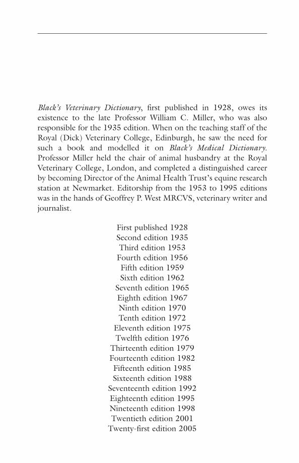
Black’s Veterinary Dictionary, first published in 1928, owes itsexistence to the late Professor William C. Miller, who was alsoresponsible for the 1935 edition. When on the teaching staff of theRoyal (Dick) Veterinary College, Edinburgh, he saw the need forsuch a book and modelled it on Black’s Medical Dictionary.Professor Miller held the chair of animal husbandry at the RoyalVeterinary College, London, and completed a distinguished careerby becoming Director of the Animal Health Trust’s equine researchstation at Newmarket. Editorship from the 1953 to 1995 editionswas in the hands of Geoffrey P. West MRCVS, veterinary writer andjournalist.
First published 1928Second edition 1935Third edition 1953
Fourth edition 1956Fifth edition 1959Sixth edition 1962
Seventh edition 1965Eighth edition 1967Ninth edition 1970Tenth edition 1972
Eleventh edition 1975Twelfth edition 1976
Thirteenth edition 1979Fourteenth edition 1982Fifteenth edition 1985Sixteenth edition 1988
Seventeenth edition 1992Eighteenth edition 1995Nineteenth edition 1998Twentieth edition 2001
Twenty-first edition 2005


Ab(see ANTIBODY)
AbamectinAn avermectin (see AVERMECTINS) used in cattleas an ectoparasiticide and endoparasiticide.
Abbizzia sppA group of rapidly growing African trees beingexploited as a forestry crop. The seed pods havecaused poisoning in goats and cattle. Clinicalsigns include tachycardia, anorexia, ruminal sta-sis, anaemia, dyspnoea and recumbency. Affectedanimals always show methaemglobinaemia.
AbdomenThe part of the body in front of the spinebetween the thorax (see CHEST) and the PELVIS.(For a description of abdominal organs, see under
appropriate headings.)
Abdomen, Diseases of(see under STOMACH, DISEASES OF; INTESTINES,DISEASES OF; DIARRHOEA; LIVER, DISEASES OF;PANCREAS, DISEASES OF; KIDNEYS, DISEASES OF;BLADDER, DISEASES OF; PERITONITIS; BLOAT;COLIC; ASCITES; HERNIA)
Abdomen, Injuries ofThese include injuries to the abdominalwalls, to the alimentary tract and to the organswithin the abdomen. Trauma may result indamage to the liver, spleen, kidneys, or urinarybladder. Apparently small external wounds ofthe abdominal wall may be far more seriousthan their appearance suggests. Radiographsand ultrasound can be useful in diagnosis.
Diagnosis An exploratory LAPAROTOMY maybe necessary to establish the internal effectsof such wounds, and also the cause of inter-nal haemorrhage, free intra-peritoneal gas,peritonitis, etc.
Obtaining a sample by PARACENTESIS maybe useful, although the hollow needle may beblocked by omentum. Use of a catheter andperitoneal lavage has been effective in detect-ing early intra-abdominal traumatic lesions,rupture of internal organs, etc. in dogs and cats.
When a stake or other pointed object hascaused a large wound in the abdominal wall, the
bowels may protrude through the opening, andif the incision be extensive, evisceration maytake place. When only the wall of the abdomenhas been damaged, there may be severe bruis-ing, and haemorrhage into the tissues (see
HAEMATOMA).If exposure of the abdominal contents has
taken place, or if the organs have been them-selves damaged, there is risk of SHOCK, haem-orrhage, infection, and PERITONITIS; the lattermay cause great pain and usually proves fatal.For this reason the injured animal shouldreceive promptly the expert services of a veteri-nary surgeon or else be humanely destroyed.Simple WOUNDS or bruises of the abdominalwalls are treated in the same way as ordinarywounds.
AbiotrophyA degenerative condition of an organ or tissueleading to dysfunction or loss of function.Usually inherited and often involving brain orother nerve tissue. (See LYSOSOMES – Lysosomalstorage disease.)
AblationRemoval of an organ, or part of an organ, bysurgery.
AblephariaThe lack of eyelids – a normal condition insnakes.
Abnormalities, Inherited(see GENETICS, HEREDITY AND BREEDING –Genetic defects)
AbomasumAbomasum is the so-called 4th stomach ofruminating animals; more correctly, the 4thcompartment of the ruminant stomach. It isalso called the ‘true’ or ‘rennet’ stomach, andthe ‘reed’. It is an elongated, pear-shaped saclying on the floor of the abdomen, on theright-hand side, and roughly between the 7thand 12th ribs.
Abomasum, Displacement of(see STOMACH, DISEASES OF; TYMPANITIC RESO-
NANCE IN CATTLE)
AbortifacientA substance causing abortion.
AbortionThe termination of pregnancy. In farm animals itrepresents one important aspect of INFERTILITY.
A

The causes of abortion in farm animals areshown in the tables below:
CowsInfections
VirusesBVD/MD (bovine virus diarrhoea/mucosal
disease); bovine herpesvirus 1 (infectiousbovine rhinotracheitis/infectious pustularvulvovaginitis)
ChlamydiaC. psittaci
RickettsiaeCoxiella burnetti (Q fever)Ehrlichia phagocytophilia (tick-borne fever)
BacteriaSalmonella dublin, S. typhimuriumBacillus lichenformisBrucella abortus; also B. melitensisActinomyces pyogenesListeria ivanovii, L. monocytogenesLeptospira hardjo and other serovarsCampylobacter fetusBesnoitia
FungiAspergillus fumigatusMortierella wolfii
ProtozoaNeospora caninumToxoplasma gondiiTrichomonas fetus
Non-infectious causesClaviceps purpurea (ergot in feed)StressRecessive lethal geneMalnutritionHaemolytic diseaseVitamin A deficiencyIodine deficiency
EwesInfections
VirusesBorder disease/Thogoto virus
ChlamydiaC. psittaci (ovis) (Enzootic abortion)
RickettsiaeEhrlichia phagocytophilia (tick-borne fever)Coxiella burnetti (Q fever)
BacteriaBacillus licheniformisSalmonella dublin, typhimurium, montivideo,
S. abortus ovis and othersListeria monocytogenesArizona sppActinomyces pyogenesBrucella abortus and (not in the UK) B. ovis
Campylobacter jejuniFungi
Aspergillus fumigatusProtozoa
Toxoplasma gondiiNon-infectious causes
Stress (e.g. chasing/savaging by dogs; transport)Near-starvationPregnancy toxaemiaClaviceps purpurea (ergot in feed)Iodine deficiency
SowsInfections
VirusesAfrican swine fever virusAujeszky’s diseaseSmediSwine fever virus
BacteriaErysipelothrix rhusiopathiae (swine erysipelas)Brucella abortus suisPasteurella multocida (occasionally)E. coliLeptospira pomona (not in UK) grippotyphosa,
canicala, icterrhaemorrhagicaProtozoa
Toxoplasma gondiiNon-infectious causes
Malnutrition, e.g. vitamin A deficiency(See also CARBON MONOXIDE.)
MaresInfections
VirusesEquine herpesvirus 1 (Equine rhinopneu-
monitis)Equine viral arteritis
BacteriaAeromonas hydrophiliaSalmonella abortus equiBrucella abortus (rarely)Haempophilus equigenitalis (contagious equine
metritis)Leptospira spp (sometimes in association with
equine herpesvirus 1)Listeriosis
Non-infectious causesTwin foalsPlant poisoning (e.g. by Locoweed)
BitchNeospora caninumBrucella canis (not UK)Streptococcus sppCanine herpesvirus
QueenFeline leukaemia virus, feline herpesvirus
2 Abortion
A
Abortion, Contagious(see BRUCELLOSIS)
Abortion, Enzootic, of EwesThis disease occurs in all parts of Britain, as wellas overseas.
Cause Chlamydia psittaci, which is ingested bymouth from infected material. It can remainlatent for long periods in non-pregnant sheep.(See CHLAMYDIA.)
Diagnosis A competitive ELISA (cELISA) test is stated to be 100 per cent effective in testingfor antibodies against abortion-causing strainsof C. psittaci.
Signs Abortion occurs during the last 6weeks, and usually during the last 2 or 3 weeks,of the normal period of gestation. Stillbirthsand the birth of weak full-term lambs alsooccur. The placenta is thickened and necrotic.Most infected ewes who do not become illhave a thick, infected vaginal discharge for aweek or more. Infertility is temporary, sinceewes usually lamb normally the followingseason.
Enzootic abortion is a zoonosis (seeZOONOSES); pregnant women must avoid allcontact with infected sheep.
Prevention Replacement sheep should beobtained from blood-tested disease-free flocks.Vaccines are available; antibiotics can reducethe level of abortions in an outbreak.
Abortion, EpizooticChlamydial abortion in cattle.
AbrasionA superficial wound of skin or mucous mem-brane caused by chaffing, rubbing, etc.
AbscessLocalised pus, surrounded by inflamed tissue.A tiny abscess is known as a PUSTULE, and adiffused area that produces pus is spoken ofas an area of CELLULITIS. Abscesses in cats areusually of this type and seldom ‘point’ (seebelow).
An acute abscess forms rapidly and asrapidly comes to a head and bursts, or elsebecomes reabsorbed and disappears.
Causes The direct cause of an acute abscess iseither infection with bacteria, or the presence ofan irritant in the tissues.
The organisms that are most often associ-ated with the formation of abscesses includestaphylococci and streptococci (see BACTERIA).
When bacteria have gained access they startto multiply, and their TOXINS may damagesurrounding tissue.
White blood cells (leukocytes) – in particu-lar, those called neutrophils – gather in the areainvaded by the bacteria and engulf them. Thearea of invasion becomes congested with deador dying bacteria, dead or dying leukocytes,dead tissue cells which formerly occcupied thesite, and debris.
Signs Inflammation, redness, warmth, swelling,and pain; and besides these, when the abscess isof large size and is well developed, fever.
‘Pointing’ of an abscess means it has reachedthat stage when the skin covering it is dead,thin, generally glazed, and bulging. If slightlydeeper, the skin over the area becomes swollen,is painful, and ‘pits’ on pressure. When theabscess bursts, or when it is evacuated by lanc-ing, the pain disappears, the swelling subsides,and the temperature falls. If all the pus hasbeen evacuated, the cavity rapidly heals; if,however, the abscess has burst into the chest orabdomen, pleurisy or peritonitis may follow.When an abscess is deeply seated so as to be outof reach of diagnosis by manipulative measures,its presence can be confirmed by blood tests.
Treatment Antibiotics may be employed asthe sole means of treating multiple or deep-seated abscesses. They may be injected into acavity following aspiration of the pus, or theymay be used in addition to the lancing of anabscess. Hot fomentations, or application of apoultice, may afford relief.
After the abscess has been opened it is usuallybest to leave it uncovered.
A chronic abscess takes a long time todevelop, seldom bursts (unless near to the sur-face of the body), and becomes surrounded bylarge amounts of fibrous tissue.
Causes Abscesses due to tuberculosis, ACTINO-
MYCOSIS, staphylococci, and caseous abscessformation in the lymph nodes of sheep, are themost common types of cold or chronic abscesses.They may arise when an acute abscess, instead ofbursting in the usual way, becomes surroundedby dense fibrous tissue.
Signs Swelling may be noticeable on the sur-face of the body (as in actinomycosis), or it mayshow no signs of its presence until the animal is
Abscess 3
A
slaughtered (as in the case of many tuberculousabscesses and in lymphadenitis of sheep). If it ispresent on the surface, it is found to be hard,cold, only very slightly painful, and does notrapidly increase in size.
Characteristics of the pus The containedfluid varies in its appearance and its consis-tency. It may be thin and watery, or it may besolid or semi-solid. To this latter type the name‘inspissated pus’ is given, and the process ofsolidification is often spoken of as ‘caseation’.
Treatment This may involve surgery, and/orthe use of antibiotics, depending upon thenature of the abscess and its location.
AbyssinianA breed of short-haired cat similar in appear-ance to those depicted in illustrations fromancient Egypt. It is favoured for its quiet vocal-isation. Familial renal amyloidosis has beenfound in this breed.
Acacia PoisoningAcacia poisoning has been recorded in cattleand goats. Signs include ataxia, excitation andprostration.
Acanthosis NigicansA chronic condition of the skin found mainly indogs, especially Daschunds. The skin becomesthickened with loss of hair and excessive pig-mentation, and is velvety to the touch. Thecondition often starts in the axillae (armpits) butthe abdomen has also been seen as the primarylocation. The cause is unknown. It may respondto corticosteroids or radiation therapy.
AcapniaAcapnia is a condition of diminished carbondioxide in the blood.
AcaricideA parasiticide effective against mites and ticks.
AcarusA forage mite only accidentally parasitic.
Accidental Self-InjectionThis has led to human infection with BRUCEL-
LOSIS, ORF, plague, Q FEVER, and TUBERCULO-
SIS (TB).Accidental self-injection with an oil-based
vaccine is painful and dangerous; it requiresimmediate medical attention.
If the accident involves IMMOBILON, theeffects can be reversed by an immediate self-
injection of Revivon (diprenorphine hydrochlo-ride). A veterinary surgeon who had no Revivonwith him died within 15 minutes of accidentalself-injection, when a colt made a sudden violentmovement. Even a scratch with a used needle cancause collapse.
AccidentsAny part of the animal may be injured in anaccident. Often the damage is obvious, such asa broken limb. Serious internal injury may notbe immediately apparent. Road traffic accidentsare the commonest cause of accidents to dogsand cats. Care must be taken in handlinginjured animals, as mishandling may make theinjury worse. (See also ELECTRIC SHOCK, ‘STRAY
VOLTAGE’ AND ELECTROCUTION; FRACTURES;BLEEDING; INTERNAL HAEMORRHAGE; BURNS
AND SCALDS; SHOCK; EYE, DISEASES AND
INJURIES OF.)
Accommodation(see EYE)
Acepromazine(Acetylpromazine)Acepromazine (Acetylpromazine) is a phenoth-iazine-derived tranquilliser. Given by injection
4 Abyssinian
A
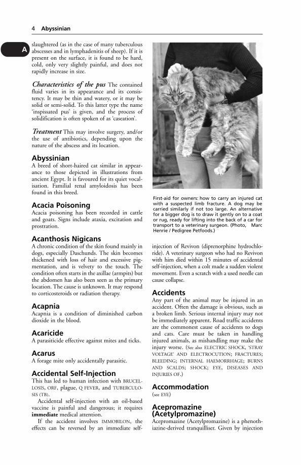
First-aid for owners: how to carry an injured catwith a suspected limb fracture. A dog may becarried similarly if not too large. An alternativefor a bigger dog is to draw it gently on to a coator rug, ready for lifting into the back of a car fortransport to a veterinary surgeon. (Photo, MarcHenrie / Pedigree Petfoods.)
before anaesthesia, it enables low doses of barbi-turates to be used. 1 to 3 mg per kg bodyweight,given by mouth a quarter of an hour or morebefore food, may be used for the prevention oftravel sickness in small animals.
Acepromazine lowers blood pressure, and so iscontra-indicated in accident cases. Noradrenalineis recommended for reversing any fall in bloodpressure.
AcetabulumAcetabulum is the cup-shaped depression on thePELVIS with which the head of the femur formsthe HIP-JOINT. DISLOCATION of the hip-jointsometimes occurs as the result of ‘run-over’ acci-dents, and FRACTURES of the pelvis involvingthe acetabulum frequently result from the samecause.
Acetaminophen(see PARACETAMOL)
Acetic AcidAcetic acid is used as a treatment for alkalosis,which may be caused by urea poisoning. Aceticacid may form naturally in pig mash feedsallowed to stand, or in silage and fermentedhay, when it can cause illness or even death.It is one of the normal breakdown products ofcellulose digesting bacteria in the rumen.
AcetonaemiaThis, and ketosis, are names given to a meta-bolic disturbance in cattle and sheep. It maybe defined as the accumulation in the bloodplasma, in significant amounts, of KETONE
BODIES. The disorder may occur at any time,but is commonest in winter in dairy cows keptindoors when receiving a full ration of concen-trates. The condition is very rare in heifers andseldom occurs before the 3rd calving. It can beseen in cows in the 1st month after calving andis most commonly apparent at 3 weeks.
Cause The disturbance is caused by the cow’sdemands for carbohydrate exceeding that avail-able from the feed. Whenever the glucose levelin the blood plasma is low, as in starvation oron a low-carbohydrate diet, or when glucose isnot utilisable, as in diabetes, the concentrationof free fatty acids in the plasma rises. This riseis roughly paralleled by an increase in the con-centration of ketone bodies, which provide a3rd source of energy. In other words, the mod-erate ketosis which occurs under a variety ofcircumstances is to be looked upon as a normalphysiological process supplying the tissues witha readily utilisable fuel when glucose is scarce.
By contrast, the severe forms of ketosis metwith in the lactating cow and the diabetic cow,and characterised by high concentrations ofketone bodies in the blood and urine, are obvi-ously harmful pathological conditions wherethe quantities of ketone bodies formed grosslyexceed possible needs.
Signs The cow shows rapid weight loss,reduced appetite and favours roughage to con-centrates. Rumen activity is reduced and faecesbecome harder. The animal is markedly dull,with a dull coat and reduced milk yield. Thebreath has a sickly sweet smell of acetone, whichmay also be detected in the milk and urine.Sometimes nervous signs are present, withthe animal licking walls, head rope and otherobjects, and overexcitement. Most animalsrecover with treatment.
Diagnosis Rothera’s test on milk; urine maybe used but can cause false positives.
First-Aid Treatment consists in giving 1⁄2 apint of glycerine or propyleneglycol, dilutedin water, or a preparation containing sodiumpropionate.
The feeding of cut grass or flaked maize, theaddition of a little molasses to feed, and exerciseall aid recovery. Injections of dextrose or corti-costeroids are used under veterinary control.Resistant cases are met with which defy alltreatment; the cow improves up to a pointbut does not feed properly and dies in 10 to 20days.
Prevention In the 2nd half of a lactation, thediet of a dairy cow should contain a greater pro-portion of home-grown foods with a lowerdigestibility than that in the diet fed duringpeak lactation.
At the beginning of the dry period, the cowsshould be fit but not fat (condition score 2.5 to3). The cows should be kept in this conditionduring the dry period by a diet of relativelypoor-quality forage or heavy stocking andshould be given a vitamin/mineral supplement.Production rations should be introduced in thelast 2 weeks of the dry period and contain boththe forage and concentrate elements to be fedafter calving. Cattle should not be ‘steamed up’but should receive up to 3 kg (61⁄2 lb) (dry) ofthe milking ration.
After calving, the quantity of productionration fed should be steadily increased as themilk production increases. For high-yieldingcows the production concentrate ration shouldcontain 16 to 18 per cent crude protein with a
Acetonaemia 5
A
high metolisable energy. The carbohydrate inthe ration should be readily digestible. Theinclusion of some ground maize may be partic-ularly helpful in ketosis-prone herds, since someof the starch escaping rumen fermentation isdigested and absorbed as sugars. Productionconcentrates should contain a balanced vitaminand mineral supplement.
Cows must not be given free access to straw.Concentrates can be fed between meals fromout-of-parlour feeders, as a constituent of acomplete diet, or layered in silage. High-yield-ing cows should not be penned for a long timein yards, but be given ample opportunity forexercise.
After the first 10 to 12 weeks of lactation,the feeding routine of the high-yielders canbe modified. The home-grown forage can beslowly increased in the ration with a corre-sponding decrease in the more expensive highlydigestible carbohydrates if the cow’s perfor-mance is not affected. This change-over mustbe a gradual process.
AcetoneA ketone with characteristic smell found insmall amounts in some samples of normalurine, and in greater quantities during thecourse of diabetes, acetonaemia, pneumonia,cancer, starvation, and diseases of disturbedmetabolism.
Acetonuria is the excretion of ketones in theurine.
AcetylcholineAcetylcholine is a neurotransmitter, an impor-tant link in the transmission of nerve impulsesbetween the nerves themselves (at the synapses)and between the nerve and the muscle. Paralysisresults if the body’s ability to produce acetyl-choline is affected by shock, injury or certaindrugs, such as curare. Pharmaceutical prepara-tions of such compounds are used in anaesthesiato produce muscle relaxation, which facilitatessurgical procedures.
In the healthy animal, acetylcholine isdestroyed by the enzyme cholinesterase as soonas the nerve impulse has passed. When thisreaction is prevented, as in poisoning byorganophosphorous insecticides, convulsionsfollow. Excessive salivation is an importantsymptom in dogs so poisoned.
Achalasia of the OesophagusAbsence of progressive peristalsis and failureof the lower oesophageal sphincter to relax. Ithas been reported as an inherited condition
in Boston terriers, English springer spaniels,smooth fox terriers, wire-haired fox terriers,German shepherd dogs and Rhodesian ridge-backs.
AchondroplasiaAchondroplasia is a form of dwarfing due todisease affecting the long bones of the limbsbefore birth. It is noticed in some calves of cer-tain breeds of cattle such as the Dexter, in somebreeds of dogs, and in lambs. (See GENETICS,HEREDITY AND BREEDING – Genetic defects.)
Achorion(see RINGWORM)
Acid-Fast OrganismsAcid-fast organisms are those which, when oncestained with carbol-fuchsin dye, possess thepower to retain their colour after immersion instrong acid solutions, which decolorise the non-acid-fast group. The important acid-fastbacteria are Mycobacterium tuberculosis, whichcauses tuberculosis in humans and other pri-mates; M. bovis, which causes tuberculosis incattle and some other mammals; M. piscium,which causes tuberculosis in fish; and M. aviumvar. paratuberculosis (johnei), which causesJohne’s disease in ruminants.
AcidosisA condition of reduced alkaline reserve of theblood and tissues, with or without an actualfall in pH. Sudden death may occur in cattlefrom acidosis after gorging on grain, or follow-ing a sudden introduction of cereal-basedconcentrates. It is a common complication of diarrhoea, particularly in young animals. (See
also BARLEY POISONING.) Sheep may similarly beaffected.
Acids, Poisoning byStrong acids are intensely destructive of animaltissue. If accidentally consumed, the effects areimmediate and drastic.
Signs Excessive salivation, great pain, anddestruction of the mucous membrane lining themouth (which causes the unfortunate animal tokeep its mouth open and protrude its tongue)are seen. After a short time convulsive seizuresand vomiting occur, and general collapse fol-lows; while if a large amount of acid has beentaken, death from shock rapidly supervenes.
Treatment Alkaline demulcents should begiven at once and in large quantities; bicarbon-ate of soda given in gruels or barley-water or
6 Acetone
A
milk is quite useful. These neutralise the acidsinto harmless salts, and soothe the corroded andburnt tissues. (See ACETIC ACID; HYDROCYANIC
ACID (HCN).)
AcinusAcinus is the name applied to each of the minutesacs of which secreting glands are composed.
AciduriaAciduria is the excretion of acid urine. It mayoccur as a result of feeding a specialised diet toreduce the fomation of urinary calculi (stones)in the dog and cat.
AcneAn inflammation of sebaceous glands or hairfollicles, with the formation of pustules. In thehorse, a contagious form of acne is sometimesdue to infection with Corynebacterium ovis.Acne often accompanies canine distemper, andis seen on the chin of the cat.
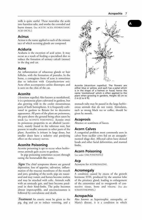
Aconite(Aconitum napellus) Also known as monkshood,it is a poisonous plant cultivated in gardens, butalso growing wild in the cooler mountainousparts of both hemispheres. It is frequently culti-vated in gardens in Britain for its decorativeappearance. All parts of the plant are poisonous,the parts above the ground being often eaten bystock (see ACONITE POISONING). Aconite owesits poisonous properties to an alkaloid (aconi-tine), mainly found in the tuberous root, butpresent in smaller amounts in other parts of theplant. Aconitine is irritant in large doses, butsmaller doses have a sedative and paralysingeffect on the sensory nerves.
Aconite PoisoningAconite poisoning is apt to occur when herbiv-orous animals gain access to gardens.
In pigs poisoning sometimes occurs througheating the horseradish-like roots.
Signs The chief symptoms shown are generaldepression, loss of appetite, salivation, inflam-mation of the mucous membrane of the mouthand jaws, grinding of the teeth; pigs are nause-ated and may vomit; and horses become restlessand may be attacked with colic. Animals walkwith an unsteady gait, and later become paral-ysed in their hind-limbs. The pulse becomesalmost imperceptible, and unconsciousness isfollowed by convulsions and death.
Treatment An emetic must be given to thepig, dog and cat to induce vomiting, and a
stomach-tube may be passed in the large herbiv-orous animals that do not vomit. Stimulants,such as strong black tea or coffee, should begiven by mouth.
AcoprosisAbsence or scantiness of faeces.
Acorn CalvesA congenital problem most commonly seen incalves from suckler cows fed on an unsupple-mented silage diet. Affected calves have domedheads and other facial deformities, and stuntedlimbs.
Acorn Poisoning(see under OAK POISONING)
AcpAcronym for ACEPROMAZINE.
AcromegalyA condition caused by excess of the growthhormone STH, produced by the anterior lobeof the pituitary gland, leading to enlargementof the extremities and to overgrowth of con-nective tissue, bone and viscera. (See also
SOMATOTROPHIN.)
AcropachiaAlso known as hypertrophic osteopathy, orMarie’s disease, it is a condition in which
Acropachia 7
A
Aconite (Aconitum napellus). The flowers areeither blue or yellow, and each has a petal whichis in the shape of a helmet or hood; hence thename ‘monkshood’ which is often applied to theplant when growing in gardens. Height: 65 cm to2 m (2 to 6 ft).
superfluous new bone is laid down – first in thelimbs and later in other parts of the skeleton. Itmay accompany tumours and tuberculosis inthe dog.
AcrosomeA cap over the anterior part of the head ofspermatozoa; it contains enzymes which aidpenetration of the ovum.
ACTHActh is the abbreviated form of ADRENOCORTI-
COTROPHIN. (See also CORTICOTROPHIN.)
ActinobacillosisActinobacillosis is a disease of cattle similar insome respects to ACTINOMYCOSIS, and some-times mistaken for it.
Generally only 1 or 2 animals in a herd areaffected at one time.
Swellings may be seen on lips, cheeks, jaw, and at the base of the horn. Pneumonia,infection of the liver or alimentary canal maylead to death in untreated cases. The diseaseoccurs also in sheep and occasionally in pigsand foals.
Cause Actinobacillosis is due to infectionwith Actinobacillus lignièresi. Infection occursthrough injuries, abrasions, etc. of soft tissues,and when lymph nodes are affected throughinvasion along the lymph vessels. Abscessesform.
Lesions may also involve the lungs, rumen,omasum, abomasum, and reticulum.
Actinobacillus seminis was discovered in asheep in Australia. The infection, sometimessubclinical, has since been recognised in severalcountries including the UK, and causes polyarthritis.
Signs With Actinobacillus lignièresi the tonguemay become infected and painful, hence itscommon name ‘wooden tongue’. When lymphnodes in the throat are affected, the swellingand pressure caused may make swallowingand breathing difficult; if the lesion is in theskin and superficial tissues only, it may attain toa great size without causing much trouble;when the tongue is affected the animal hasdifficulty in mastication and swallowing andthere is usually a constant dribbling of salivafrom the mouth. If this is examined there maybe found in it small greyish or greyish-yellow‘pus spots’, in which the organism can bedemonstrated by microscopic methods. Later,the saliva may become thick, purulent, and foulsmelling.
Treatment Antibiotics are often effective. Inintransigent cases, intravenous sodium iodide isused.
Pigs The disease has been recorded both inthe UK (very rarely) and overseas, caused byActinobacillus equuli (Bacterium viscosum equi).Actinobacillus suis has been recorded occasion-ally; it causes septicaemia in piglets and lesionsin various organs. Actinobacillus pleuropneumo-niae (formerly Haemophilus pleuropneumoniae)causes pleuropneumonia in pigs.
Horses Actinobacillus equuli causes septi-caemia and internal lesions in foals (see under
FOALS, DISEASES OF).
Precautions The disease can be transmitted toman. Accordingly, care must be taken over wash-ing the hands, etc., after handling an animalwith actinobacillosis.
ActinomycosisThis has been recorded in very many species ofanimals, including man, dogs, pigs, birds andreptiles.
The lesions produced bear a considerableresemblance to those of actinobacillosis (seeabove), and are often indistinguishable fromthem, but typically actinomycosis affects thecheeks, pharynx and especially the bone ofthe jaws (it is known as ‘lumpy jaw’ in cattle),while actinobacillosis is more likely to attacksoft tissues only.
Cause Actinomyces bovis. This anaerobic bac-terium is present in the digestive system ofcattle, and it is probable that it can only becomepathogenic by invading the tissues through awound. It is common during the ages when thepermanent cheek teeth are cutting the gumsand pushing out the milk teeth.
The liver is sometimes affected, while actin-omycosis and actinobacillosis have both beenfound in lungs and bronchi.
Yellow sulphur granules are found in thelesions.
Actinomyces (Corynebacterium) pyogenes is amajor cause of abscesses and suppurative condi-tions.
Signs The swelling in bone and other tissue,mainly composed of dense fibrous tissue, mayreach a considerable size causing interferencewith mastication, swallowing, or breathing,depending on the situation of the lesion. Inmost cases when the mouth or throat is affected,there is a constant dribbling of saliva in varying
8 Acrosome
A
amounts from the mouth. In the earlier stagesthis saliva is normal in its appearance, but laterbecomes offensive.
Actinomycosis of the bone of the upper andlower jaws produces an increase in the size ofthe part and a rarefication of its bony structure,the spaces becoming filled with the prolifera-tion of fibrous tissue which is characteristic ofthe disease.
When the udder is affected, hard fibrousnodules may be felt below the skin, varying insize from that of a pea to a walnut or larger,and firmly embedded in the structure of thegland itself. These swellings enclose softcentres of suppuration which, on occasions,may burst either through the covering skin, orinto an adjacent milk sinus or duct. The milkfrom such a cow should not be used for humanconsumption because of the danger of theconsumer contracting the disease.
Treatment Antibiotics may be effective. Inintransigent cases, intravenous sodium iodidemay be used.
Precautions The disease can be transmittedto man; hygienic precautions are necessary afterhandling infected animals.
Acuaria UncinataThis roundworm has caused outbreaks of diseasein geese, ducks, and poultry. The life-cycle of thisparasite involves an intermediate host, Daphniapulex, the water flea. On post-mortem examina-tion of affected birds, worms may be found innodules scattered over the mucous membrane ofthe oesophagus and proventriculus. Mortalitymay be high.
AcupunctureThe centuries-old Chinese technique of needle insertion at certain specified points onthe surface of the body has become a part ofWestern veterinary medicine for treatment,analgesia, and resuscitation. Acupuncture canproduce the morphine-like natural substancescalled ENDORPHINS which are, in effect, anal-gesics.
Adaptations have been made, such as the useof lasers instead of needles. Ultrasonics and heathave also been applied to the points.
Acupuncture is commonly used to relievepainful conditions; also in treating poor circu-lation, tissue damage, and smooth muscle dys-function. However, it is not a panacea and mustbe applied by experts.
Success has been reported for the use of injec-tions of sterile saline at acupunture points in
treating intractable pain in horses. The injectionswere repeated at weekly intervals for upto 8weeks.
In China, acupuncture has been used for surgical analgesia in animals and man.
Acute DiseaseA disease is called acute – in contradistinctionto ‘chronic’ – when it appears rapidly, andeither causes death quickly or leads to a speedyrecovery. (See also under DEATH, CAUSES OF
SUDDEN.)
Ad Lib FeedingThis is a labour-saving system under which pigsor poultry help themselves to dry meal, etc.,and eat as much as they wish. It is also used indairy cattle and for intensive beef production.(See also DRY FEEDING.)
AdamantinomaA tumour affecting the jaw and composed ofcells that normally produce dental enamel.
AdderThe common viper (Vipera berus). About 50cm (20 in) in length, it has dark markings ona paler ground. If disturbed, this snake maybite farm or domestic animals. The bite isdangerous; an antiserum is available.
Addison’s Disease(Hypoadrenacortism)Addison’s disease (hypoadrenacortism) is causedby failure of the ADRENAL GLANDS to produceadequate amounts of corticosteroids. It may be caused by congenital defects in, injury to, or disease of the cortex of the gland, when it is known as primary hypoadrenocorticism.Secondary hypoadrenocorticism results fromexcessive or prolonged dosage of an animal withcortisone products, which depresses the naturalproduction of the hormone.
Signs In the dog or cat, where it most com-monly occurs, the animal may be lethargic,depressed and weak; diarrhoea and vomitingmay be seen. In severe cases left untreated,death may result.
In cattle, it is associated with a high incidence of aborted, weakly or still-borncalves.
Treatment The condition responds rapidly toadministration of hydrocortisone or otherappropriate corticoid product to restore levelsof cortisol in the blood; numerous formulationsare available.
Addison’s Disease (Hypoadrenacortism) 9
A
AdditivesSubstances incorporated in a premix added toanimals’ feed, often for a purpose other thannutrition. They are mainly growth promoters,enhancers of feed conversion, or, commonly,used to provide vitami ns or minerals necessaryfor a healthy diet. In addition to minerals andvitamins, permitted additives include certainANTHELMINTICS and and coccidiostats for thecontrol of parasites in farm animals. The use ofantibiotics as growth promoters, permitted to alimited extent to date, is being phased out inthe EU. Specified dyes, such as the xanthinsused to achieve desired coloration of farmedrainbow trout, are also permitted.
Very strict controls apply to the preparationand use of medicated feeds with the principal aimof ensuring that consumers are not put at riskfrom medicinal residues in food animals. The leg-islation is contained in the Medicines (MedicatedAnimal Feeding Stuffs) No. 2 Regulations 1992,the Feeding Stuffs Regulations 2000, the FeedingStuffs (Establishments and Intermediaries)Regulations 1999 and the Feeding Stuffs(Zootechnical Products) Regulations 1999. AllUK compounders, whether commercial or home mixers, must register with the RoyalPharmaceutical Society or the Department ofAgriculture for Northern Ireland.
(See also under MEDICINES ACT; ANTIBIOTIC;GROWTH PROMOTERS; HORMONES IN MEAT
PRODUCTION.)
AdenitisInflammation of a gland.
AdenofibromaAdenofibroma is a fibrous tumour enclosingneoplastic glandular tissue.
AdenomaA TUMOUR composed of epithelial tissue, oftengland-like in appearance. It may sometimes befound in positions where glandular tissue is notnormally present. A malignant form is the ade-nocarcinoma.
AdenomatosisThe formation of numerous adematousgrowths in an organ. (See PORCINE INTESTINAL
ADENOMATOSIS; PULMONARY ADENOMATOSIS.)
AdenopathySwelling of the glands, particularly the lymphglands.
AdenosineAdenosine is a purine which is part of the
structure of certain genes controlling the for-mation of amino acids. Adenosine triphos-phate and diphosphate are important in thecontraction of muscles.
AdenovirusThis is a contraction of the original term ‘adenoidal-pharyngeal conjunctival agents’. (See VIRUSES.)
ADH(see ANTIDIURETIC HORMONE)
Adhesion Factor, Bacterial(see BACTERIAL ADHESIVENESS)
AdhesionsAdhesions occur by the uniting or growingtogether of structures or organs which are nor-mally separate and freely movable. They aregenerally the result of acute or chronic inflam-mation, and in the earlier stages the unitingmaterial is fibrin, which later becomes resolvedinto fibrous tissue.
Treatment Surgical division of the obstruct-ing bands is often necessary in the abdominalcavity and in adhesions of the walls of the vagina following injuries received at a previousparturition. (See PLEURISY; PERITONITIS.)
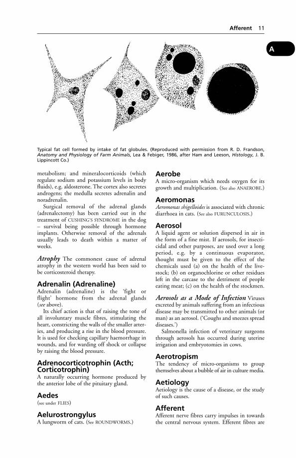
Adipose TissueHere fat is stored as an energy reserve; globulesof fat form within connective tissue cells. Whenadditional fat is stored, each cell eventuallybecomes spherical, its nucleus pushed to oneside. (See illustration on page 11.)
During demanding muscular exercise, orwhen food is insufficient, or during a debilitat-ing disease, the cells release the fat into thebloodstream and resume their normal shape.(See also LIPOMA.)
AdjuvantA substance added to a vaccine, in order to sta-bilise the product and enhance the immuneresponse.
Adrenal Glands (SuprarenalGlands)These are two small organs situated at the anterior extremities of the kidneys, and areendocrine glands.
Function The cortex secretes hormones whichare called steroids or corticosteroids. Theseinclude glucocorticoids, notably cortisol, con-cerned with the regulation of carbohydrate
10 Additives
A
metabolism; and mineralocorticoids (whichregulate sodium and potassium levels in bodyfluids), e.g. aldosterone. The cortex also secretesandrogens; the medulla secretes adrenalin andnoradrenalin.
Surgical removal of the adrenal glands(adrenalectomy) has been carried out in thetreatment of CUSHING’S SYNDROME in the dog– survival being possible through hormoneimplants. Otherwise removal of the adrenalsusually leads to death within a matter of weeks.
Atrophy The commonest cause of adrenalatrophy in the western world has been said tobe corticosteroid therapy.
Adrenalin (Adrenaline)Adrenalin (adrenaline) is the ‘fight or flight’ hormone from the adrenal glands (see above).
Its chief action is that of raising the tone ofall involuntary muscle fibres, stimulating theheart, constricting the walls of the smaller arter-ies, and producing a rise in the blood pressure.It is used for checking capillary haemorrhage inwounds, and for warding off shock or collapseby raising the blood pressure.
Adrenocorticotrophin (Acth;Corticotrophin)A naturally occurring hormone produced bythe anterior lobe of the pituitary gland.
Aedes(see under FLIES)
AelurostrongylusA lungworm of cats. (See ROUNDWORMS.)
AerobeA micro-organism which needs oxygen for itsgrowth and multiplication. (See also ANAEROBE.)
AeromonasAeromonas shigelloides is associated with chronicdiarrhoea in cats. (See also FURUNCULOSIS.)
AerosolA liquid agent or solution dispersed in air inthe form of a fine mist. If aerosols, for insecti-cidal and other purposes, are used over a longperiod, e.g. by a continuous evaporator,thought must be given to the effect of thechemicals used (a) on the health of the live-stock; (b) on organochlorine or other residuesleft in the carcase to the detriment of peopleeating meat; (c) on the health of the stockmen.
Aerosols as a Mode of Infection Virusesexcreted by animals suffering from an infectiousdisease may be transmitted to other animals (orman) as an aerosol. (‘Coughs and sneezes spreaddiseases.’)
Salmonella infection of veterinary surgeonsthrough aerosols has occurred during uterineirrigation and embryotomies in cows.
AerotropismThe tendency of micro-organisms to groupthemselves about a bubble of air in culture media.
AetiologyAetiology is the cause of a disease, or the studyof such causes.
AfferentAfferent nerve fibres carry impulses in towardsthe central nervous system. Efferent fibres are
Afferent 11
A
Typical fat cell formed by intake of fat globules. (Reproduced with permission from R. D. Frandson,Anatomy and Physiology of Farm Animals, Lea & Febiger, 1986, after Ham and Leeson, Histology, J. B.Lippincott Co.)
concerned with activities, such as movement,secretion, vascular changes, etc.
Afghan HoundA tall breed of dog with silky coat. Inheritedcataract caused by a recessive gene has beenreported in the breed.
AflatoxinsToxins produced by fungi, e.g. Aspergillusflavus: they cause poisoning in animals eatingcontaminated feed materials. The toxins havebeen found mainly in groundnut meal, butsunflower and cottonseed can also be affected.The Feeding Stuffs Regulations 2000 requirethose products, and copra, palm kernel, maizeand feeds derived from them, to be screened forthe presence of toxins.
In cattle, aflatoxins may give rise to a reducedgrowth rate and lower milk yield. Aflatoxins areexcreted in the milk. In pigs, jaundice may beseen; post mortem, the liver has a leatheryappearance. Adult pigs may show bile duct carcinoma.
Aflatoxicosis in poultry is characterised byhaemorrhages, anorexia, decreased efficiency infood utilisation, pathological changes in theliver, kidneys and bile ducts, and death. Theproblem can be prevented by storing grain with13 per cent of moisture or less. The litter mayalso be a source of toxins and consequently it isimportant to keep the moisture in the litter to aminimum by ensuring that the ventilation ofthe house is adequate and that the waterers areoperating correctly.
Fish are extremely susceptible to aflatoxins.As one of the precautions taken to keep animalfeeds free of dangerously high levels of aflatox-ins, trout have been used for testing. In youngtrout (as in pigs), aflatoxin poisoning is likely toresult in cancer of the liver. (Mature cock fishbecome fully resistant.) Equally, care has to betaken with commercial dry trout feeds, toensure that aflatoxin level is below 0.5 parts perbillion; otherwise malignant tumours are apt todevelop, and later liquid-filled cysts may growto a remarkable size.
As the long-term effect is cancer of the bileducts, animals without gall-bladders, e.g. horsesand deer, are less likely to be affected. (See also
MYCOTOXICOSIS; CIRRHOSIS.)
AFRCAFRC is the abbreviation for the Agriculturaland Food Research Council. This body wasreplaced in 1994 by the Biotechnology andBiological Sciences Research Council.
African Horse Sickness(see HORSE-SICKNESS, AFRICAN)
African Swine Fever(see SWINE FEVER, AFRICAN)
AfricanderCattle in origin about 3⁄4 Brahman and 1⁄4 Britishbeef breed. (See also under CYTOGENETICS.)
AfrikanerA synonym for Brahman or Zebu cattle.
Afterbirth(see PLACENTA)
Afterbirths, InfectedAfterbirths, Infected may be a source of infection to other animals. (See SCRAPIE;BRUCELLOSIS; ABORTION, ENZOOTIC.)
AgalactiaPartial or complete absence of milk, or milkflow, from the udder. Where this is due to a fail-ure of milk ‘let down’, oxytocin may be pre-scribed. (See SOW’S MILK, ABSENCE OF; COW’S
MILK, ABSENCE OF.)
Agalactia, ContagiousThis is a disease of goats especially, and sheepless commonly, characterised by inflammatorylesions in the udder, eyes, and joints. It ischiefly encountered in France, Switzerland, theTyrol, Italy, the Pyrenees, North Africa andIndia.
Cause Mycoplasma agalactiae. The diseaseoften occurs in the spring and the summer, anddisappears with the advent of the colder weath-er. The infection may be carried by flies or thehands of the milkers and by the litter in a shedbecoming contaminated, while the fetus maybe infected before birth.
Signs Fever, mastitis, and a greatly reducedmilk yield. The milk becomes yellowish-greenand contains clots. In addition to the udder,both joints and eyes may be involved; a painfularthritis, and conjunctivitis followed by kerati-tis (with resultant temporary blindness) wors-ening the animal’s condition.
Emaciation and death within 10 days mayoccur in very acute cases; otherwise recoveryusually follows within a few weeks, though the former milk yield will not have beenregained.
Male animals may have orchitis as well asarthritis.
12 Afghan Hound
A
Inflammation of the lymph nodes mayoccur, and lesions may be found also in abdom-inal organs and tissues, and in the chest.
Treatment Isolation of the affected animalsand strict segregation of the in-contacts shouldbe carried out.
AgarAgar is the gelatinous substance prepared fromCeylon moss and various kinds of seaweed. Itdissolves in boiling water, and, on cooling, solid-ifies into a gelatinous mass at a temperatureslightly above that of the body. It is used exten-sively in preparing culture-media for use in bacteriological laboratories, and also in the treat-ment of chronic constipation in dog and cat.
Agar-Gel Immunodiffusion TestA test used in diagnosis of, e.g., equine infectiousanaemia. (See also COGGINS TEST.)
Agene ProcessThe bleaching of flour with nitrogen trichlo-ride. The use of such flour in dog foods gaverise to HYSTERIA.
Ages of Animals
Horses By the time it has reached 17 years,which generally means about 14 years of work,a horse’s powers are on the wane. Many at this age are still in possession of their fullvigour, but these are generally of a class that isbetter looked after than the average, e.g.hunters, carriage-horses, or favourites. On anaverage, the feet of the horse are worn out first,not the arteries as in man, and consequentlyhorses with good feet and legs are likely to out-last those inferior in this respect, other thingsbeing equal. After the feet come the teeth. Invery many cases a horse’s teeth wear out beforetheir time. It often happens that the upper andlower rows of teeth do not wear in the normalway; the angle of their grinding surfacesbecomes more and more oblique, until thechewing of the food becomes less and less effective, and the horse loses condition.
Instances are on record of horses attainingthe age of 35, 45, 50, and one of a horse thatwas still working when 63 years old. These,however, are very exceptional. The average ageat which a horse dies or is euthanased lies some-where between 20 and 25 years.
Cattle The great majority of bullocks arekilled before they reach 3 years of age, and incountries where ‘prime beef ’ is grown they are
fattened and killed between 21⁄2 and 3 years. Inthe majority of herds, few cows live to be morethan 8 or 10 years of age. Pedigree bulls mayreach 12 or 14 years of age before being dis-carded. Records are in existence of cows up to39 years old, and it is claimed that one had 30calves.
Sheep Here again the requirements of thebutcher have modified the age of the animal atdeath. Wether lambs are killed at ages rangingfrom 4 to 9 months (Christmas lambs), andolder fat sheep up to 21⁄2 years. Ewes, on theaverage, breed until they are from 4 to 6 or 7 years, when they too are fattened and slaugh-tered for mutton. Exceptionally, they reachgreater ages, but unless in the case of purebreeding animals, each year over 6 reduces theirultimate value as carcases. Rams are killed afterthey have been used for 2 or 3 successive sea-sons at stud – that is, when they are 3 or 4 yearsof age, as a rule.
Pigs In different districts the age at which pigsare killed varies to some extent, according to therequirements of local trade. Pigs for pork pro-duction are killed at about 31⁄2 to 4 months;bacon pigs are killed between 6 and 71⁄2 months,and only breeding sows and boars are keptlonger. Ages of up to 12 years have been recorded for sows.
Dogs and cats These are the only domesti-cated animals which are generally allowed to diea natural death. The average age of the dog isabout 12 years, and of the cat 9 to 12, butinstances are not uncommon of dogs living to 18 or 20 years of age, and of cats similarly.(See also BREEDING OF LIVESTOCK; DENTITION.)
Elephants Their normal life-span in the wildis 65 to 70; some working elephants areemployed up to a similar age and then retired.
AgglutinationAgglutination is the clumping together of cellsin a fluid. For example, bacteria will agglutinatewhen a specific antiserum is added to the sus-pension of bacteria. Similarly, the blood serumof one animal will cause the red blood cells ofanother to become agglutinated.
Agglutination is explained by the presence inthe serum of an agglutinin which combineswith an agglutinable substance, or agglutino-gen, possessed by the organisms.
Agglutination is made use of in theAgglutination Test, which depends upon theprinciple that in the blood serum of an animal
Agglutination 13
A
harbouring in its body disease-producingorganisms (though it may show no symptoms),there is a far greater concentration of agglu-tinins than in a normal animal. Minute doses(e.g. dilutions of 1 part to 100 or even 1000) ofsuch serum will cause agglutination, whileserum from a normal animal will not causeagglutination when diluted more than 1 part in 10. Incubation of the mixture at body heatusually hastens the results and enables a rapiddiagnosis to be made.
Aggressiveness (Aggression)This may be transient, as in a nursing bitch fear-ful for her puppies. Persistent aggressiveness canbe the result of jealousy, as when the birth of ababy means a decline in status for the dog. Ill-treatment, attacks by some local pugnacious dog,being kept tied up for long periods, or being shutin an empty house are other causes. Heredity isan important factor, too, and it is unwise to breedfrom aggressive parents even if they look likeShow winners. Brain disease – for example,encephalitis, or a brain tumour – may account foraggressiveness in any animal. So may pain. (See also
ENCEPHALITIS; MENINGIOMA; RABIES; BENZOIC
ACID POISONING; EQUINE VERMINOUS ARTERI-
TIS; ‘VICES’; CHLORINATED HYDROCARBONS;MUSCLES, DISEASES OF – Muscular rheumation;OVARIES, DISEASES OF; HYPER-AESTHESIA; BOVINE
SPONGIFORM ENCEPHALOPATHY; LISTERIOSIS;ANAPLASMOSIS; ACETONAEMIA; GRASS SICKNESS;HEARTWATER.)
AgonistA type of drug which gives a positive response(e.g. contraction or relaxation of a muscle fibre,or secretion from a gland) when its moleculecombines with a receptor. The latter is a specificstructural component of a cell, on its membrane,and usually a protein.Antagonist A drug which merely blocks theattachment of any other substance at the recep-tor, so preventing any possible active response.Partial agonist A drug which produces a posi-tive response at the receptor, but only a weakone. However, since it occupies the receptor itprevents any full agonist from binding so that,in the presence of agonists, partial agonists mayact as antagonists.
Many drugs are now classified according totheir major action, e.g. β blockers, H1 and H2
receptor antagonists.β receptors are present in the heart and
smooth muscle of the bronchioles, uterus, andarterioles supplying skeletal muscle. Drugswhich are selective β1 (heart) or β2 (elsewhere)are now available. For example, CLENBUTEROL
is a specific β2 agonist; it is used as a bron-chodilator to treat respiratory conditions inhorses, dogs and cats.
The use of clenbuterol in cattle, where it actsas a growth promoter, is prohibited in the EU.
AirAtmospheric air contains by volume 20.96 percent of oxygen, 78.09 per cent of nitrogen, 0.03per cent of carbon dioxide, 0.94 per cent ofargon, and traces of a number of other elements– the most important of which are helium,hydrogen, ozone, neon, zenon, and krypton, as well as variable quantities of water vapour.(See SMOG.)
Air that has been expired from the lungs in anormal manner shows roughly a 4 per centchange in the amount of the oxygen and carbondioxide, less of the former (16.96 per cent) andmore of the latter (4.03 per cent). The nitrogenremains unaltered.
The importance of fresh air to animals isimmense. (See VENTILATION; RESPIRATION;OZONE; SLURRY; CARBON MONOXIDE.)
Air Passages(see BRONCHUS; NOSE AND NASAL PASSAGES;TRACHEA)
Air SACPart of the respiratory system, particularly inreference to birds.
Air SacculitisInflammation of the air sacs in birds.
Airedale TerrierA large, black-and-tan, wiry-coated breed.Entropion and cataract are inherited, probablyas autosomal dominant traits.
Akabane VirusFirst isolated from mosquitoes in Japan; anti-bodies detected in cattle, horses and sheep in Australia. A possible cause of abortion in cattle, and of birth of abnormal calves. Thevirus, a member of the Bunyavirus group, is teratogenic.
Some calves are born blind and walk withdifficulty; some have the cerebrum virtuallyreplaced by a water-filled cyst.
(See also Arthrogryposis under GENETICS,HEREDITY AND BREEDING – Genetic defects.)
Alanine Aminotransferase(ALT)An enzyme involved in amino acid transfer. Liverdamage results in high levels in the circulating
14 Aggressiveness (Aggression)
A
blood. It is used as a measure of liver damage indogs and cats.
Alaskan MalamuteA breed of dog developed from the husky.Dwarfism (chondrodysplasia) is inherited insome litters. Day blindness may also be inheritedand congenital haemolytic anaemia occurs.
AlbinismAlbinism is a lack of the pigment melanin inthe skin – an inherited condition.
Albumins(see PROTEINS; CONALBUMIN; ALBUMINURIA)
AlbuminuriaThe presence of albumin in the urine: one of the earliest signs of NEPHRITIS and cystitis (see URINARY BLADDER, DISEASES OF).
Alcohol PoisoningAcute alcoholism is usually the result of toolarge doses given bona fide, but occasionally thelarger herbivora and pigs eat fermenting wind-falls in apple orchards; or are given or obtain,fresh distillers’ grains, or other residue permeat-ed with spirit, in such quantities that the ani-mals become virtually drunk. In more seriouscases they may become comatose.
AldosteroneThis is a hormone secreted by the adrenalgland. Aldosterone regulates the electrolyte balance by increasing sodium retention andpotassium excretion. (See CORTICOSTEROIDS.)
AldrinA persistent insecticide; a chlorinated hydrocar-bon used in agriculture and formerly in farmanimals. Its persistence has prevented its veteri-nary use. Signs of toxicity include blindness,salivation, convulsions, rapid breathing. (See
GAME BIRDS.)
Aleutian DiseaseFirst described in 1956 in the USA, this diseaseof mink also occurs in the UK, Denmark,Sweden, New Zealand and Canada.
MinkSigns include: failure to put on weight or evenloss of weight; thirst; the presence of undigestedfood in the faeces – which may be tarry. Bleedingfrom the mouth and anaemia may also beobserved. Death usually follows within a month.
Ferrets In these animals the disease is charac-terised by a persistent viraemia.
Signs include: loss of weight; malaise; chronicrespiratory infection; and paresis or paraplegia.Bleeding from the mouth and anaemia mayalso be observed. Death usually follows withina month. The disease can be confused with thelater stages of rabies.
Diagnosis In ferrets the counter-current electrophoresis test has been used.
Alexin(see COMPLEMENT)
Alfadalone(see ALFAXALONE)
AlfaxaloneUsed in combination with alfadalone (in Saffan[Schering-Plough]) as a general anaesthetic incats; it must not be used in dogs. Given byintravenous injection, It produces sedation in 9 seconds and anaesthesia after 25 seconds. It isalso given by deep intramuscular injection as aninduction for general anasthesia for long opera-tions. It must not be given with other injectableanaesthetics.
AlgaeSimple plant life of very varied form and size,ranging from single-cell organisms upwards tolarge seaweed structures. Algae can be a nui-sance on farms when they block pipes or clognipple drinkers. This happens especially inwarm buildings, where either an antibiotic or sugar is being administered to poultry via the drinking water. Filters may also becomeblocked by algae.
The colourless Prototheca species are patho-genic for both animals (cattle, deer, dogs, pigs) and man. (See MASTITIS IN COWS – Algalmastitis.)
The non-toxic algae of the Spirulina groupare used in the feed of some ornamental fish.
Algae PoisoningToxic freshwater algae, characteristically blue-green in colour, are found in summer on lakesin numerous locations, particularly where waterhas a high phosphate and nitrate contentderived from farm land. Formed by the summerblooms of cyanobacteria, they can form an oily,paint-like layer several cm thick. Deaths haveoccurred in cattle and sheep drinking fromaffected water; photosensitivity is a commonsign among survivors. Dogs have also beenaffected.
The main toxic freshwater cyanobacteria arestrains of the unicellular Microcystis aeruginosa,
Algae Poisoning 15
A

and the filamentous forms Anabaena flos-aquae,Aphanizomenon and Oscillatoria agardhii.
Signs vary according to the dominantcyanobacterium present. Anabaena flos-aquae,for example, can form alkaloid neuromusculartoxins which can produce symptoms withinhalf an hour; these being muscular tremors, stupor, ataxia, prostration, convulsions, some-times opisthotonus, and death. Dyspnoea andsalivation may also be seen.
Mycrocystis strains produce a slower-actingpeptide toxin, which may cause vomiting anddiarrhoea, salivation, thirst, piloerection, andlachrymation. Survivors may show LIGHT
SENSITISATION, with inflamed white skin andoedema of ears and eyelids.
Poisoning by algae has been recorded in dogsthat have been in the sea off Denmark. InAmerica a colourless alga is reported to havecaused dysentery, blindness and deafness, andsometimes ataxia and head-tilting.
In Victoria, Australia, 17 sheep died andmany others showed signs of light sensitivityafter drinking from a lake affected by a thickbloom of M. aeruginosa. The deaths were spreadover 6 months after removal from access to the lake.
Poisoning in cattle was suspected in the UKafter a spell of hot weather in East Anglia causedan algal bloom in field ponds and 50 per cent ofthe cows in a herd suddenly showed nervoussigns. BSE was ruled out as the cause.
Alimentary Canal(see DIGESTION)
AlkaliA substance which neutralises an acid to form asalt, and turns red litmus blue. Alkalis are gen-erally the oxides, hydroxides, carbonates, orbicarbonates of metals.
Varieties Ammonium, lithium, potassium,and sodium salts are the principal alkalis, theircarbonates being weak and their bicarbonatesweaker.
Uses In poisoning by acids, alkalis in dilutesolution should be administered at once. (See
ACIDS, POISONING BY; STOMACH, DISEASES OF;DISINFECTION; DETERGENTS.)
AlkaloidsAlkaloids constitute a large number of theactive principles of plants and all possess a pow-erful physiological action. Like alkalis, theycombine with acids to form salts, and turn red
litmus blue. Many alkaloids are used in medi-cine, and their names almost always end in ‘ine’– e.g. atropine, morphine, quinine, etc.
Aconitine } from monkshood (Aconitum napellus).AconineArecoline, from areca nut (Areca catechu).Atropine, from belladonna, the juice of the deadly
nightshade (Atropa belladonna).Caffeine, from the coffee plant (Coffea arabica) and
from the leaves of the tea plant (Thea sinensis), alsofound in the kola nut, guarana, and species ofholly, etc.
Cocaine, from coca leaves (Coca erythroxylon).Digitoxin* } from foxglove (Digitalis purpurea).Digitalin*Ephedrine, from various species of Ephedra.Ergotoxin* } from ergot (Claviceps purpurea).ErgometrineHyoscyamine, from henbane (Hyoscyamus niger).Hyoscine or } also from henbane.
ScopolamineMorphineCodeine } from opium, the juice of the opiumThebaine poppy (Papaver comniferans).HeroinNicotine, from tobacco leaves (Nicotiana tobaccum).Physostigmine } from Calabar beans (Physostigma
or Eserine venenosum).Pilocarpine, from jaborandi (Pilocarpus jaborandi).Quinine, from cinchona or Peruvian bark (Cinchona,
and Cinchona rubra).Santonin*, from wormwood (Artemesia pauciflora).Sparteine, from lupins (Lupulinus, sp.) and from
broom (Cytisus scoparius).Strychnine, from Nux vomica seeds (Strychnos nux
vomica).Veratrine, from cevadilla seeds (Cevadilla officinale, or
Schoenocaulon officinale).
Those marked * are neutral principles.
A first-aid antidote for poisoning by an alkaloid is strong tea.
AllantoisA sac extending from the hind gut of the earlyembryo and containing urine-like fluid. Theallantois fuses with the chorion to become part of the PLACENTA. (See also PERVIOUS
URACHUS.)
Alleles (Allelomorphs)Alleles (allelomorphs) are genes which influencea particular development process, processes, orcharacter, in opposite ways, and can replace oneanother at a particular locus on a chromosome.They result from a previous mutation, and theoriginal gene and its mutated form are called an‘allelomorphic pair’. Another definition is: oneof a pair or series (multiple alleles) of genesoccupying alternatively the same locus. (See also
GENETICS, HEREDITY AND BREEDING.)
16 Alimentary Canal
A

Allergic DermatitisAllergic dermatitis is another name for eczemacaused by an allergy. For example, ‘QueenslandItch’ is seen in horses in Australia, where it is a result of hypersensitivity to e.g. the bites of a sandfly; in Japan it follows bites of the stable-fly. It is a disease of the hot weather, and is intensely itchy in character. Treatmentinvolves the use of antihistamines. In the UK ‘Sweet Itch’ is the name for a similar or identical condition in horses. (See also
ECZEMA.)
AllergyA specific sensitivity to e.g. a plant or animalproduct, usually of a protein nature. In the dogand cat, sensitivity occurs most commonlyfrom bedding, carpeting, rubber products,household cleaners, plants, and some skindressings; in pigs, soyabean protein antigens.
The three main signs are itching, self-inflicteddamage as a result, and redness; sometimes oede-ma of the face, ears, vulva or extremities, or skinweals.
Many foodstuffs have caused allergy in thedog, e.g. cow’s milk; horse, ox, pig, sheep andchicken meat; eggs. True food allergies are lesscommon in cats. They can, however, be dis-tressing. All constituents of the feline diet maybe involved, including colouring agents andpreservatives.
Tobacco smoke was reported to be the causeof an allergy in a dog. When his owner gave upsmoking, the allergy did not return.
Allergy may arise following the bites of sand-flies, stable-flies, fleas and sometimes bee orwasp stings. Pollens can produce skin changes;likewise avianised vaccines, horse serum, antibi-otics, and synthetic hormone preparations. (See
also ATOPIC DISEASE; ECZEMA; ANAPHYLACTIC
SHOCK; ANTIHISTAMINES; LIGHT SENSITISA-
TION; LAMINITIS; REAGINIC ANTIBODIES.)
AllograftA piece of tissue, or a complete organ, trans-planted from one animal to another of the samespecies. (See SKIN GRAFTING.)
Allopurinol(1) The treatment of choice for LEISHMANIASIS
in dogs. Given by mouth, it is well absorbedfrom the gastrointestinal tract and excreted bythe kidneys. (2) It is also used in dogs to treatUROLITHIASIS.
AloeCape aloes are an anthraquinone laxative with an intensely bitter taste. Aloe vera is a
popular ingredient in skin preparations and the juice is reputed to be of benefit in cases ofeczema.
AlopeciaAbsence of hair from where it is normally present; it has to be differentiated from loss ofhair due to mange, ringworm, lice infestation,and eczema.
Alopecia may be the result of a hormoneimbalance, a dietary deficiency, or seleniumpoisoning.
A temporary alopecia is occasionally seen innewborn animals, and also in the dams of new-born animals. A deficiency of iodine or of thy-roxine may produce such hair loss. In dogs,bald patches, usually symmetrical, may occuron the flanks and extend to the limbs. This typeof canine alopecia usually responds to thyroidtherapy. In male dogs of 5 years old andupwards, alopecia may be accompanied by anattraction for other males, and may respond tocastration but not to hormone therapy. ASertoli-cell tumour of the testicle also causesalopecia and feminisation. Symmetrical barepatches, accompanied by other symptoms, are afeature of Cushing’s disease. Senile alopeciaaffects some cats, and a patchy loss of fur mayoccur from time to time in some spayed cats.Tetracyclines may occasionally cause severe hairloss in cats.
Alopecia in dogs, with symmetrical bilateralhair loss from trunk, neck and end of tail, maysometimes be due to a deficiency of the growthhormone SOMATOTROPHIN. The age groupaffected is 1 to 4 years. Highly pigmented skinmay be a feature. Treatment with the growthhormone has proved successful.
AlphachloraloseA narcotic used for the destruction of rodents,pigeons, etc. It acts by lowering the body tem-perature. Accidental poisoning in dogs and cats can occur. Animals should be kept warm;emetics may be given in the early stages.
AlpacaA type of South American camel now farmed inthe UK and elsewhere for its fine wool; notreared for meat. Individuals can live for up to20 years.
AlphavirusViruses of arbovirus group A and equineencephalitis viruses bear this name.
ALT(see ALANINE AMINOTRANSFERASE)
Alt 17
A

AltitudeAnimals unaccustomed to high altitudes can beadversely affected by them. Like humans, ani-mals suffer hypoxia. Testicles of cats, rabbitsand rats atrophy with resulting fertility prob-lems. Hens and geese lay infertile eggs or ceaselaying. Ascites caused by high altitudes has beenreported in all types of poultry. Acclimatisationto high altitudes results in the formation ofmore and smaller red blood cells so that oxygen-binding capacity is increased. (See also
‘MOUNTAIN SICKNESS’.)
AltrenogestA prostaglandin analogue used for the synchro-nisation of oestrus in mature sows (RegumatePorcine) and the suppression of prolongedoestrus in mares (Regumate Equine).
Aluminium ToxicityIn the rat, research in South Africa has shownthat aluminium toxicity might be due to(experimental) porphyria. In Israel it has been shown that rats given aluminium salts,and then examined under ultra-violet light,show fluorescence of eyes, long bones, brainand peri-testicular fat. In rats at least, therefore,aluminium cannot be regarded as a harmlesselement.
AlveldA disease of lambs in Norway, associated with the eating of bog asphodel Nartheciumossifragum. Signs are photosensitisation andjaundice; it is thought to be due to poisoningby microfungi present on the plant.
AlveolusA tooth socket in the jaw. The term is alsoapplied to the minute divisions of glands and tothe air sacs of the lungs.
Alveolitis Inflammation of an alveolus. (See EXTRINSIC
ALLERGIC ALVEOLITIS.)
‘Alzheimer’s Disease’ in CatsA condition in geriatric cats that closely resem-bles the human disease. Signs include disorien-tation, compulsive behaviours, disturbed sleeppatterns and incontinence. Histologically,changes to the brain resemble those in thehuman disease.
AmaurosisImpaired vision or even loss of sight, resultingfrom disease of the optic nerve, brain, or spinalcord.
AmblyopiaDiminution of vision.
AmeliaAn information bulletin published by theVeterinary Medicines Directorate. The title isan acronym for Animal Medicines EuropeanLegislation Information and Advice.
American Box TortoisesA ban on the importation into the UK of tor-toises from Mediterranean countries led dealersand pet shops to seek an alternative, and thechoice was Terrapene carolina. These are terres-tial, but like to take an occasional dip in waterabout 3 inches deep. Poor swimmers, they dis-like water deeper than that. The recommendeddiet for them is ‘earthworms, mushrooms,beans, beansprouts, cucumber, grapes, banana,and some leafy vegetables’. In winter a vitaminand mineral supplement is advisable.
American Cocker SpanielA breed smaller than the English spaniel andwith longer hair. Cataract is an inherited trait.Other inherited conditions may include dis-tichiasis, entropion, haemophilia, patellar luxa-tion and prognathia.
American Quarter HorseA breed derived mainly from dams of Spanishorigin, for long bred by American Indians, and from Galloway sires brought by the earlysettlers. ‘It was Barb blood spiced with a Celticinfusion and refined with a dash of Easternblood that fashioned the Quarter Horse.’ (R. M. Denhardt.)
AmineAn organic compound containing ammonia(NH3).
Amino AcidsAmino acids are the ‘building blocks’ intowhich proteins can be broken down, and withwhich proteins can be constructed.
Amino acids contain carbon, hydrogen, andoxygen, together with an amine group (NH2).
The quality of a protein, in terms of its valueas an animal feed, depends upon its content of essential amino acids. These are lysine,methionine, tryptophane, leucine, isoleucine,phenylalanine, threonine, histidine, valine, andarginine.
LYSINE is a particularly important amino acidfor growth and milk production, and is one ofthose prepared synthetically and added to somelivestock feeds.
18 Altitude
A

The pig and rat require, for rapid growth:lysine, tryptophane, leucine, isoleucine,methionine, threonine, phenylalanine, valine,and histidine. The chick needs glycine in addition to these. The cat needs TAURINE.
AminoglycosidesA group of bactericidal antibiotics producedfrom Streptomycin species including strepto-mycin, neomycin, framycetin and gentamicin.
AminonitrothiazoleA drug used against Blackhead in turkeys.
AminotransferaseAn enzyme which catalyses transfer reactionsinvolving amino acids.
AmitrazAn ectoparasiticide for the treatment of lice andtick infestation and mange in farm animals anddogs. It must not be used on chihuahuas, noron cats or horses. It is sold under a variety oftrade names.
Ammonia (NH3)A few drops of ammonia on a piece of cotton-wool held a few inches from the nostrils have agood effect in reviving animals which have col-lapsed. (Inhalation of concentrated ammoniacan prove fatal.) Ammonia fumes from littermay adversely affect poultry. (See DEEP LITTER;also QUATERNARY AMMONIUM COMPOUNDS.)
An excess of ammonia in the rumen has beencited as a cause of hypomagnesaemia in springfollowing massive applications of nitrogenousfertiliser. (See also UREA.)
Ammonia poisoning Hydrolysis of urea toammonia in the rumen may occur very rapidlyin cattle receiving excessive amounts of urea. Ifmore ammonia reaches the blood and then theliver than the latter organ can detoxify, thenammonia poisoning will result. (See UREA.)
Several cows died after being fed straw whichhad been treated with ammonia for 5 days onlyand came direct from the treatment box. (It isrecommended that the treatment should be for 10 days, with a 2-day interval before theproduct is fed to livestock.) Laryngeal oedemaand emphysema of the lungs were caused. Thelevel of ammonia in the atmosphere of animalhousing must not exceed 14 ppm.
(See also LITTER, OLD.)
AmnionThe innermost of the 3 fetal envelopes. It iscontinuous with the skin at the umbilicus
(navel), and completely encloses the fetus but isseparated from actual contact with it by theamniotic fluid, or the ‘liquor of the amnion’,which in the mare measures about 5 or 6 litres(9 to 10H pints). (See PLACENTA.)
This ‘liquor amnii’ forms a kind of hydrosta-tic bed in which the fetus floats, and serves toprotect it from injury, shocks, and extremes oftemperature. It allows free though limitedmovements, and guards the uterus of the damfrom the spasmodic fetal movements which,late in pregnancy, are often vigorous and evenviolent.
At birth it helps to dilate the cervical canal of the uterus and the posterior genital pas-sages, forms part of the ‘waterbag’, and, onbursting, lubricates the maternal passages. (See PARTURITION.)
Amoebic EncephalitisAmoebic encephalitis due to Acanthamoebacastellani was found after the euthanasia of a 4-month-old puppy. Fits and hyperkeratosis ofthe foot pads suggested that the cause was thedistemper virus, but A. castellani was recoveredfrom an area of suppurative necrosis in thebrain.
(In human medicine, several species of thisamoeba are recognised as an important cause ofgranulomatous encephalitis.) (Pearce, J. R. &others, JAVMA 187, 951.)
AmoxycillinAn antibiotic resembling ampicillin, but itsaction is quicker and it is excreted more rapid-ly. Amoxycillin is often used in combinationwith clavulanase, which makes it more effectiveby blocking the effect of penicillinase, by whichampicillin is destroyed. It is used in all species.
AmphistomesSynonym for Paramphistomes (see PARAMPHIS-
TOMIASIS).
AmpicillinA semi-synthetic penicillin, active against bothGram-positive and GRAM-NEGATIVE bacteria. Itis not resistant to penicillinase, but can be givenby mouth.
AmpouleA small glass container having one end drawnout into a point capable of being sealed so as to preserve its contents sterile. It is used to con-tain solutions of drugs for hypodermic injec-tion, while many vaccines and other biologicalproducts are also distributed in ampoules. Apotential hazard of glass embolism has been
Ampoule 19
A
recognised in human medicine, and the wis-dom of allowing glass particles to settle, beforefilling a syringe, has been stressed.
AmproliumA drug used for the prevention and treat-ment of coccidiosis in turkeys, guinea fowl andchickens.
AmputationRemoval of a limb. If a long bone of dog or cathas been shattered into several pieces, or is thesite of cancer, amputation is usually the onlyhumane course to take (other than euthanasia).It is certainly kinder than leaving the animal apermanent cripple, perhaps suffering somedegree of pain for the rest of its life.
A three-legged dog or cat can be expected torevise its technique of balance and movement,and to become not merely nimble but fast aswell; and to demonstrate a capacity for enjoyinglife.
A questionnaire was submitted to the ownersof 55 dogs and 18 cats which had undergoneamputation of a limb. In 26 animals the reasonwas cancer, and in the others it was severeinjury.
All the owners stated that they were pleasedthe operation had been performed, althoughmany had found it a difficult decision to make.
Amylase (Amylopsin)A starch-splitting enzyme. (See DIGESTION.)
AmyloidosisThe deposition of an insoluble starch-like pro-tein (amyloid) which affects the functioning of the tissues in which it is deposited. It may be associated with inflammatory conditions orchronic infections.
AnabolicRelating to anabolism, which means tissuebuilding, and is the opposite of catabolism ortissue breakdown.
An anabolic steroid is one derived fromtestosterone in which the androgenic character-istics have been reduced and the protein-building (anabolic) properties increased in proportion. Examples are nandrolone and ethylestrenol. These are used in cases of malnu-trition, wasting diseases, virus diseases, andsevere parasitism.
Synthetic anabolic steroids have been used asgrowth-promoter implants in commercial beefproduction, but this is prohibited in the UK andEU. They are also prohibited in competition ani-mals. It has been found that anabolic steroids can
give rise to changes in the liver and its function-ing in both animals and man; with, in someinstances, tumour formation. Changes in the sex-ual organs may follow misuse. (See STILBENES.)
AnadromyAn anadromous fish is one that spends most ofits adult life in the sea but returns to fresh waterto spawn. Salmon are anadromous.
AnaemiaA reduction in the number and/or size of thered blood corpuscles or the haemoglobin in theblood. It is a sign rather than a disease, and it isimportant to establish the cause (obvious onlyin the case of acute external haemorrhage dueto trauma), so that a prognosis and suitabletreatment can be given.
The animal may be suffering from a chronicloss of blood due to internal bleeding, e.g. fromthe urinary or digestive tracts; and the owner of a cat, for instance, may fail to notice the presence of blood in the urine, and so not bringthe animal for treatment until other signs of illness have become obvious.
Anticoagulants, such as Warfarin, may causeinternal haemorrhage and hence anaemia.
An iron-deficient diet (and one lacking alsothe trace elements cobalt and copper, which aid the assimilation of iron) is another cause of anaemia; likewise a deficiency of folic acid,vitamin B6 and vitamin B12.
Both external and internal parasites (lice,fleas, ticks, liver flukes, roundworms and tape-worms) can cause anaemia.
Parasites of the bloodstream are an importantcause, and include trypanosomes, piroplasms,rickettsiae. (See also FELINE INFECTIOUS ANAEMIA.)
For an incompatability between the blood ofsire and dam, see haemolytic disease (under
FOALS, DISEASES OF).Aplastic anaemia means a defective, or a
cessation of, regeneration of the red blood cells; it may be drug-induced. (See also RETICU-
LOCYTES.) (In human medicine, the drugsinvolved have included chloramphenicol,phenylbutazone, and rarely penicillin andaspirin; deaths have resulted.)
Bracken poisoning, exposure to X-rays orother forms of irradiation are other causes; alsosalicylates (including aspirin).
In auto-immune haemolytic anaemia the ani-mal forms antibodies against its own red cells.
Heinz-body haemolytic anaemia (see HEINZ
BODIES) may result from kale poisoning in cattle, and from paracetamol or methylene-bluepoisoning in cats; sometimes also from leadpoisoning.
20 Amprolium
A

Signs Pallor of the mucous membranes, loss ofenergy and of appetite, and PICA. Dogs and catsmay feel the cold more than usual, and seekwarm places. In some cases fever is present, andliver enlargement. The heart rate may increase.
Treatment In the smaller animals especially,vitamin Bl2 or liver extract is often a valuablemethod of treatment. Where cobalt or copper oriron are lacking, these must be supplied. Lice orticks and fleas should be destroyed, and treatmentagainst internal parasites undertaken if they arethe cause. (See also PIGLET ANAEMIA; FELINE andEQUINE INFECTIOUS ANAEMIA; CANINE BABESIO-
SIS; HEARTWORMS; ROUNDWORMS; FLUKES.)
AnaerobeThe term applied to bacteria having the powerto live without oxygen. Such organisms arefound growing freely, deep in the soil, as, forexample, the tetanus bacillus.
Anaesthesia, GeneralThe use of general anaesthetics to produce lossof consciousness and sensation for operationson animals dates back to 1847, when severalveterinary surgeons used ether. Chloroform wasalso used in 1847. Both have now been largelysuperseded by more effective anaesthetic agents.A wide choice is now available. The selection,dosage and means of administration will beinfluenced by such considerations as thespecies, size, and habitat of the individual aswell as by the procedure to be undertaken.
Anaesthetic drugs all act by limiting the oxy-gen uptake of tissues. The effect on an individ-ual tissue is proportional to its normal oxygenrequirement. Since the oxygen requirement ofnervous tissues is disproportionately high, thesetissues are the first to be affected by anaestheticdrugs. Unconsciousness, abolition of reflexes,muscular atony, and respiratory paralysis aredue to depression of the cerebral cortices, themid-brain, the spinal cord, and the medullarespectively.
General anaesthesia is usually induced byinhalation of a volatile or gaseous anaesthetic,or by intravenous or intramuscular injection.Inhalation anaesthetics are often administeredvia an endotracheal tube; volatile anaestheticssuch as chloroform can be simply administeredby sprinkling the liquid on cotton wool andallowing the animal to inhale the vapour.
Anaesthetic agents are often used in combi-nation with a premedicant, such as hyoscine oratropine, to reduce salivation. Two agents maybe used to enhanced effect; ketamine is oftenused with xylazine, for example. In addition,
muscle relaxants such as gallamine or suxam-ethonium may also be used to facilitate certainprocedures. If the animal is a food animal, caremust be taken to observe any precautions indi-cated by the drug manufacturer to avoid drugresidues accumulating in meat or milk. In allcases, the manufacturers’ recommendations asto dosage must be followed.
Endotracheal anaesthesia This tech-nique depends upon the introduction into thetrachea of a tube which connects with the out-side. The tube is passed via the mouth under anarcotic or anaesthetic, such as pentobarbital,given intravenously, and may then be used asthe route for an inhalant anaesthetic mixture.The method ensures a clear airway throughoutthe period of anaesthesia, and thus obviates thedanger of laryngeal obstruction (e.g. by thetongue falling backwards), which sometimescauses death. The method has several otheradvantages, e.g. it permits an unobstructedoperation field during lengthy major opera-tions, achieves better oxygenation, facilitates aneven level of anaesthesia, and permits of posi-tive pressure ventilation of the lungs in theevent of respiratory failure.
Endotracheal anaesthesia is administered inone of two ways. Insufflation anaesthesiainvolves the use of air/oxygen and anaestheticvapour delivered into the tube by means of apump or, more commonly, a mixture of gasessupplied from cylinders (and sometimes bub-bled through a volatile anaesthetic liquid inaddition). Autoinhalation anaesthesia involvesthe use of a wide-bore endotracheal tubethrough which the animal inhales the anaes-thetic mixture by its own respiratory efforts. A‘rebreathing bag’ may be used.
As an alternative to general anaesthesia,EPIDURAL ANAESTHESIA (involving the injectionof a local anaesthetic into the spinal canal) isused for some surgical procedures.
Cattle Many procedures are performed underlocal anaesthetic. Where general anaesthesia isindicated, after premedication with, for exam-ple, xylazine, halothane is administered via an endotracheal tube. Cattle take chloroformwell, and recovery is rapid; however, it is littleused now. Thiopental and phenobarbital, byinjection, may also be used on occasion.
Endotracheal intubation is recommended inorder to preserve a free airway, and to preventinhalation of regurgitated rumen contents.
Horses By inhalation, halothane or isoflu-rane may be used, administered through an
Anaesthesia, General 21
A
endotracheal tube. By intravenous injection,ketamine, given after premedication withxylazine, romifidine or detomidine is effective;ketamine must not be used as sole anaestheticin the equine. The use of thiopental, via intra-venous catheter, also requires premedication.Great care must be taken that the recumbentanaesthetised horse does not suffer muscle ornerve damage, caused by the pressure of its ownweight, while unconscious.
Sheep Pentobarbital, by slow intravenousinjection at a dosage of 24 mg per kg body-weight, produces anaesthesia for up to 30 min-utes, with recovery over a similar period.Halothane may be given by inhalation.Alphaxolone/alphadone has been used, as wellas other agents, although there are few licensedanaesthetics for sheep.
Goats Pentobarbital, by slow intravenousinjection, alphaxolone/alphadolone, etorphine(Immobilon), and halothane have also beensuggested for such procedures as the disbuddingof kids.
Dogs and cats A wide choice is available.Pentobarbital by intravenous injection is rapidin action. Thiopental, also given intravenously,is a short-acting general anaesthetic.
Ketamine hydrochloride, given by intramus-cular injection, is another choice. When givento cats, it must be used with xylazine to preventexcitability on recovery.
Alfadolone/alfaxalone (Saffan), by intramus-cular or intravenous injection, is used either forthe induction of anaesthesia by other drugs, oras an anaesthetic itself. It should be used withcaution in dogs as it may cause a histaminereaction.
Propofol (Rapinovet), an intravenous anaes-thetic for dogs and cats, is useful for minor out-patient procedures and caesarian section.Recovery is generally smooth but retching,sneezing, and pawing of the face may be seen.
Monkeys Pentobarbital sodium may be givenintravenously. Ketamine hydrochloride is analternative, given by subcutaneous, intramuscu-lar, or intravenous injection. A mixture of keta-mine and xylazine has been recommended also.Halothane is suitable.
Rabbits, rats, mice, guinea pigsInhalation anaesthetics such as isoflurane aresafe and effective; rabbits should be sedatedfirst. Injectable anaesthetic combinations, suchas fentanyl-fluanisole and midazolam, or keta-
mine and xylazine, may be used; pentobarbitalis another choice.
Birds Ether has been used but its explosivenature necessitates great care; halothane orisoflurane are more suitable. For restraint, thebird may be placed in a large, clear polythenebag, into which the tube for the anaesthetic gasis introduced.
For anaesthetic injections of ketamine orpentobarbital, the bird may be immobilisedwith a cylinder of paper rolled around it andsecured with adhesive tape.
Reptiles Small reptiles may be anaesthetisedby bathing in a weak solution of phe-noxyethanol, benzocaine or tricaine mesilate.They are transferred to clean oxygenated waterfor recovery. Ketamine, by injection, is alsoused but recovery may be prolonged.
Fish Phenoxyethanol, benzocaine or tricainemesilate, dissolved in the water, are commonlyused for both exotic and farmed fish. Exotic fish species vary in their tolerance of these substances; water temperature and quality alsoaffect their efficacy. Clean oxygenated watershould be available to aid recovery.
Anaesthesia, Local andRegionalFor many minor operations and diagnostic procedures, local anaesthetics are used in pref-erence to general anaesthesia. They act byblocking conduction along the nerve fibre, producing loss of sensation and/or muscleparalysis. Drugs used include lidocaine (ligno-caine) and bupivacaine. The method and site ofadministration can be targeted according to thespecific procedure to be carried out.
Perineural anaesthesia is used when the pre-cise location of the nerves serving the area to beanaesthetised is known. For example, when dis-budding calves, the area may be anaesthetisedby injecting the agent about 2.5 cm below thebase of the horn bud.
Field block (nerve block) is produced when aseries of injections is made along a line toremove sensation from the tissue distal to thatline. Field block is typically used in the diagno-sis of laminitis in horses and temporary relief ofthe pain it causes.
Regional anaesthesia may result from per-ineural or field anaesthesia. To anaesthetise alimb, a tourniquet is applied above the part ofthe limb to be anaesthetised and the drug givenintravenously; prilocaine is the agent of choice.Loss of sensation lasts until the tourniquet is
22 Anaesthesia, Local and Regional
A
released. The precautions applying to the use of tourniquets must be observed (see under
TOURNIQUET).Surface anaesthesia is useful for facilitating
certain procedures. It may be applied to amucous surface by spray. For example, a cat’sthroat may be sprayed with local anaestheticbefore introducing a tracheal tube. To facilitateintroduction of a venous catheter, the skin ofsmaller species can be anasthetised by applyingan anaesthetic cream, after shaving the area.The cream is protected by a waterproof dress-ing; the anaesthetic may take up to an hour towork. Local anaesthetics may also be used ineye drops.
Epidural (or spinal) anaesthesia results whena local anaesthetic is injected into the space surrounding the spinal cord – the epiduralspace. This produces a loss of sensation in the tissues served by the spinal nerves. The spe-cific area affected depends on the site of injec-tion. In the caudal spinal cord, anaesthesia ofthe perineal area results; the technique is usede.g. in difficult calvings. Epidural anaesthesiaapplied to the anterior part of the spinal cordmay be used for operations on the recumbentanimal.
Intra-articular anaesthesia, by injection intoa joint, is mainly used diagnostically to identifya joint that is causing pain.
Local anaesthetics must not be used indis-criminately, since poisoning can result, andaffect the brain and heart. Symptoms of poi-soning include sudden collapse, or excitement,vomiting and convulsions.
Anaesthetics, LegalRequirementsThe Protection of Animals (Anaesthetics) Act1964 made it obligatory to use an anaestheticwhen castrating dogs, cats, horses, asses, andmules of any age.
CastrationOnly a veterinary surgeon, using an anaesthet-ic, is permitted to castrate any farm animalmore than 2 months old; with the exception oframs for which the maximum age is 3 months.
The use of rubber rings or similar devices forcastrating bulls, pigs, goats, and sheep, or fordocking lambs’ tails, is forbidden unless appliedduring the 1st week of life. The Act of 1964also requires that an anaesthetic be used whende-horning cattle; and also for disbuddingcalves unless this be done by chemical cauteryapplied during the 1st week of life.
An anaesthetic must be used for any opera-tion, performed with or without the use of
instruments, which involves interference withthe sensitive tissues or the bone structure of ananimal. (See also DOCKING.)
Anaesthetics, Residues inCarcasesDogs and cats have shown severe symptoms of poisoning after being fed on meat from ani-mals humanely slaughtered by means of anoverdose of a barbiturate anaesthetic, or chloralhydrate.
AnalRelating to the ANUS.
Anal Glands(see under ANUS)
AnalepticsDrugs that stimulate the central nervous system(see STIMULANTS).
AnalgesicsDrugs which cause a temporary loss of the senseof pain without a loss of consciousness, i.e.analgesia.
Analgesics include non-steroidal anti-inflammatory drugs (NSAIDs) such as aspirin,paracetamol and phenylbutazone. (They arecontraindicated if heart, kidney or liver diseaseis present.)
The most effective of the opiates is MOR-
PHINE. (See also BUPRENORPHINE; DETOMIDINE;ACUPUNCTURE.)
AnamnesisAnamnesis is the past history of a particularpatient.
Anamnestic ResponseThe rapid rise in antibody level in a previouslyimmunised animal in response to a ‘booster’dose of the same vaccine. The immune systemhas ‘remembered’ what to do.
AnaphrodisiaImpairment of sexual appetite.
Anaphylactic Shock(Anaphylaxis)The reaction to a foreign protein which some-times follows bee or wasp stings, injections of an antibiotic or antiserum, etc., after thepatient has become hypersensitised to the substance. There is often a rapid fall in bloodpressure; anaphylactic shock can prove fatal.(See also ANTIHISTAMINES; HYPERSENSITIVITY;WARBLES.)
Anaphylactic Shock (Anaphylaxis) 23
A
AnaplasmosisThis is an infectious disease of cattle, charac-terised by anaemia and caused by a parasite ofthe red blood cells, Anaplasma marginale.
This parasite is found in Africa, Asia,Australia, Southern Europe, South America,and the southern States of the USA. A. centrale(in cattle) and A. ovis (in sheep and goats) areother species.
Signs The disease resembles Texas fever; fre-quently anaplasmosis coexists with babesiosis,but pure infections may also occur. It is charac-terised by acute anaemia, fever, jaundice, anddegeneration of the internal organs; haemoglo-binuria does not occur as the rate of red-blood-cell destruction is not fast enough to producefree haemoglobin in the circulating blood.Young animals appear to be resistant, and casesin calves under 1 year old are rare. In older ani-mals the disease may be acute or chronic, and inthe former case they may die within 2 to 3 daysafter the appearance of the first symptoms. Thedisease starts with a high temperature of 40.5°to 41.5°C (105° to 107°F) and after a day ortwo anaemia and icterus appear. In the acute ill-ness, aggressiveness and abortion are othersymptoms.
Transmission is by ticks, e.g. Boophilus,Rhipicephalus, Hyalomma, Ixodes, Dermacentor,and Haemaphysalis. Infection is passed throughthe egg to the next generation of ticks. Tabanidflies and mosquitoes are carriers.
Animals which recover from anaplasmosisare in a state of premunition, and remain carri-ers for long periods, probably for life.
In the South African States the less serious A.centrale has been found to give protectionagainst the serious A. marginale, and both thereand in other countries successful results followits use as an immunising agent. In other areaswhere Texas fever and anaplasmosis frequentlyoccur together, cattle are often immunised byblood of a bovine infected with A. centrale,which produces a mild infection, and with amild form of Babesia bigemina.
AnasarcaAnasarca is a condition of oedema, particularlyof the tissues below the skin.
AnastomosisThe means by which the circulation is carriedon when a large vessel is severed or its streamobstructed. In anatomy the term is applied to ajunction between 2 or more arteries or veinswhich communicate with each other.
AnatoxinA toxin rendered harmless by heat or chemicalmeans but capable of stimulating the formationof antibodies.
Anchor Worm(Lernaea cyprinacea) An exotic parasite of gold-fish now to be found in some indoor ornamen-tal pools in the UK. The worms can penetratethe fish’s skin. Their removal needs to be doneunder anaesthesia.
Anconeal ProcessPart of the elbow joint, being a projection ofthe ULNA. In several breeds of dog it may notdevelop properly.
Ancylostoma(see HOOKWORMS)
Androgen(see HORMONES)
AnergyFailure or suppression of the cellular immunemechanism. This may occur in e.g. human bru-cellosis, and in other chronic diseases. Anti-anergic treatment with levamisole has beenfound successful in some patients. (R. D.Thorne, Veterinary Record, 101, 27.) (See also
IMMUNOSUPPRESSION.)
AneuplodyThe presence of an irregular number of chro-mosomes (not an exact multiple of the haploidnumber). It may arise through faulty cell division.
AneurinA synonym for THIAMIN.
AneurysmA dilatation of an artery (or sometimes of a vein)following a weakening of its walls. The result isa pulsating sac which is liable to rupture.
Aneurysms occur in the abdomen, chest, andbrain, and may result from a congenital weak-ness of the blood vessel, from disease of its lining cells, from injury, etc.
Causes Sudden and violent muscular effortsare regarded as the chief factors in the produc-tion of aneurysms, and as would be expected,the horse is more subject to this trouble thanany of the other domesticated animals.
‘Verminous aneurysm’ is a misnomer for verminous arteritis of horses caused by
24 Anaplasmosis
A
immature strongyle worms. (See EQUINE VER-
MINOUS ARTERITIS.)
AngiogenesisA method of treating a tumour by depriving itof its blood supply.
AngiographyA radiographic technique which enables theblood-flow to and from an organ to be visu-alised after injection of a contrast medium.
AngiomaA TUMOUR composed of a large number ofblood vessels. They are common in the livers ofcattle. (See also HAEMANGIOMA.)
Angiostrongylus(see HEARTWORMS)
AngitisInflammation of a blood vessel, lymph vessel,or bile duct.
AngleberryAn old name for WARTS.
AnhidrosisA failure of the sweat mechanism. This occursin horses especially, but also in cattle, importedinto tropical countries with humid climates.
At first, affected horses sweat excessively andtheir breathing is distressed after exercise. Later,sweating occurs only at the mane; the skinbecomes scurfy; and breathing becomes morelaboured. Heart failure may occur.
AnhydrideAn oxide which can combine with water toform an acid.
AnhydrousContaining no water.
Animal BehaviourAs a guide to animal welfare, see AGGRESSIVE-
NESS; ANAESTHESIA; ANALGESICS; ETHOLOGY;ELECTRIC SHOCK; HOUSING OF ANIMALS;TRANSPORT STRESS.
Animal BoardingEstablishments Act 1963This requires that the owner of a boardingestablishment shall obtain a licence from theLocal Authority, and that this licence must berenewed annually. The applicant has to satisfythe licensing authority on certain personalpoints, and that the ‘animals will at all times be
kept in accommodation suitable as respects con-struction, size of quarters, number of occupants,exercising facilities, temperature, lighting, venti-lation, and cleanliness’. The Act also requiresthat animals boarded ‘will be adequately sup-plied with suitable food, drink, and beddingmaterial, adequately exercised, and (so far asnecessary) visited at suitable intervals’. Isolationfacilities and fire precautions are covered by theAct, which empowers the Local Authority toinspect both the boarding establishment and theregister which must be kept there.
Animal Data CentreThis is located at the National Centre forAnimal Statistics, Westside, Newton,Stocksfield, Northumberland NE43 7TW.
Animal Food(see CONCENTRATES; DIET; RATIONS; PROTEINS;POISONING; VITAMINS; ADDITIVES; PET FOODS;also DOGS’ DIET; CAT FOODS, etc.)
Animal Health Act 1981This consolidated the Diseases of Animals Acts1950, 1953 and 1975.
Animal Health Schemes(see under HEALTH SCHEMES)
Animal Health Trust (AHT)A charity that is one of the world’s leading cen-tres for research into animal health. Its EquineResearch Station is renowned for its studies ofthe physiological and anatomical factors affect-ing performance, and the small animal centrehas particular expertise in eye problems of thedog and cat. AHT research has led to break-throughs in anaesthesia and in the developmentof vaccines against equine flu and canine dis-temper. The address is: Animal Health Trust,PO Box 5, Newmarket, Suffolk CB8 7DW.
Animal Husbandry(see GRAZING; PASTURE; HOUSING; WATER; DIET;DAIRY HERD; also COWS, SHEEP, PIGS, etc.)
Animal Nursing(see VETERINARY NURSES – Lay assistants whohave passed the requisite examinations underthe auspices of the Royal College of VeterinarySurgeons).
Animal Transport(see TRANSPORT)
Animal Welfare Codes(see WELFARE CODES FOR ANIMALS)
Animal Welfare Codes 25
A

Animals, Housing Of(see HOUSING OF ANIMALS)
Animals (Scientific Procedures)Act 1986This replaced the Cruelty to Animals Act 1876.The 1986 Act makes it illegal to supply ani-mals, other than those purpose-bred in Home-Office-designated breeding establishments, foruse in experimental procedures involving dogs,cats, and other animals. The Act requires alllaboratories in the UK where animals are usedin research to appoint a veterinary surgeon tobe responsible for the care and welfare of theirexperimental animals.
On 1 January 1990 it became illegal to sell orsupply pet or stray animals for use in scientificexperiments.
The Act also represents the culmination ofthe efforts of three organisations – the BritishVeterinary Association (BVA), the Committeefor the Reform of Animal Experimentation(CRAE), and the Fund for the Replacement ofAnimals in Medical Experiments (FRAME) –to reform animal experimentation legislation.The new Act is firmly rooted in BVA/ CRAE/FRAME proposals sent to the Home Secretaryin 1983, and represents an effective compro-mise between the welfare needs of animals, thelegitimate demands of the public for account-ability, and the equally legitimate requirementsof medicine, science and commerce.
The legislation gives the Home Secretary thepower and the responsibility to judge the scien-tific merit of the work s/he authorises and forwhich s/he will be answerable to Parliament.
AnkylosisThe condition of a joint in which the move-ment is restricted by union of the bones oradhesions. (See JOINTS, DISEASES OF.)
AnodynesAnodynes are pain-relieving drugs.
AnoestrusAnoestrus is the state in the female when nooestrus or ‘season’ is exhibited. It is a state ofsexual inactivity. In most mares, for example,anoestrus occurs during the winter months,when daylight is reduced, ambient tempera-tures are low and, in the wild state, food isscarce. In these circumstances the pituitarygland does not release the gonadotrophins FSHand LF (see HORMONES) so that neither folliclesnor corpora lutea develop in the ovaries.
Similar circumstances apply with cattle. Fear, hunger, cold, and pain may all result inanoestrus. (See also OESTRUS.)
Anorexia(see APPETITE – Diminished appetite)
AnoxiaOxygen deficiency. Cerebral anoxia, or a failure in the oxygen supply to the brain, occursduring nitrite and prussic-acid poisoning; incopper deficiency in cattle; and in the thor-oughbred ‘barker foal’. Anoxia is a method ofslaughter allowed under the Welfare of Animals(Slaughter or Killing) Regulations 1995. (See also
ANAESTHESIA.)
Ante-MortemBefore death. An ante-mortem inspection is thename for an examination of the live animalwhich is used in conjunction with the findingsof a post-mortem inspection, or autopsy. Underthe Welfare of Animals (Slaughter or Killing)Regulations 1995, ante-mortem inspections arecarried out on animals after their arrival at anabattoir and before they are stunned.
Ante-Natal InfectionInfection of the fetus before birth. Examples of this may occur with the larvae of the doghookworm, Ancylostoma caninum, and with the larvae of other roundworms. (See TOXO-
CARA.) Toxoplasmosis is another example of aninfection which may occur before birth.
Ante-Partum ParalysisAnte-partum paralysis is a condition in whichthe hindquarters of the pregnant animal sud-denly become paralysed. It is fairly common inthe cow, has been seen in the sheep and goat,but is rare in the mare. It appears from 6 to 25 days before parturition, and is liable to affectanimals in almost any condition – those thatare well kept as well as others.
Signs The condition suddenly appears withoutany warning. The pregnant animal is found inthe lying position, and is quite unable to regainher feet.
Treatment As a rule, the nearer to the day of parturition that the paralysis appears, themore favourable will be the result. Those casesthat lie for 2 or more weeks are very unsatisfac-tory. The condition usually disappears after par-turition has taken place, either almost at onceor in 2 or 3 days. As a consequence, treatmentshould be mainly directed to ensuring that theanimal is comfortable, provided with plenty ofbedding, is turned over on to the opposite side3 or 4 times a day if she does not turn herself,and receives a laxative diet so that constipation
26 Animals, Housing Of
A
may not occur. Mashes, green food, and a vari-ety in the food stuffs offered, are indicated.When the paralysis has occurred a considerabletime before parturition is due, it is often necessary to produce artificial abortion of thefetus and so relieve the uterus of its heavyencumbrance.
Anthelmintic ResistanceRoutine use of an anthelmintic tends to estab-lish resistance to its effects. Resistance toanthelmintics in sheep has become a seriousproblem in Australia and is increasing in othercountries where livestock are regularly dosed.The development of resistance can be discour-aged by changing the class of anthelmintic usedfor each year’s dosing programme. Wormingproducts are labelled with the following codesthat identify their chemical type.1-BZ Benzimidazoles, probenzimodazoles2-LM Imidazothiazoles, tetrahydropyrimidines3-AV Avermectins, milbemycins
Products with the same codes should not beused on the same animals in successive seasons.
AnthelminticsAnthelmintics are medicines which are given toexpel parasitic worms. There is a large range ofsubstances and formulations from which tochoose. Anthelmintic drugs include abamectin,albendazole, dichlorvos, doramectin, haloxon,levamisole, moxidectin, nitroxinil, tetramisole,morantel tartrate, thiophanate. Niclosamide,dichlorophen and praziquantel preparations areused against tapeworms in the dog. (See also
DRONCIT.) Fenbendazole and albendazole arebroad-spectrum anthelmintics usually effectiveagainst inhibited fourth-stage ostertagia larvaein cattle.
Certain criteria apply in the selection ofanthelmintics. For example, will the drug inquestion kill worm eggs? Is it effective againstimmature worms? Is it effective against adultworms of the economically important species?Does the drug discolour or taint milk? Can it begiven to pregnant, or emaciated, animals? Incows, for how long must the milk be discardedafter administration?
Methods of dosing include drenching; injec-tion (e.g. in the case of tetramisole); in the feed.(See also WORMS, FARM TREATMENT AGAINST.)
AnthisanAn antihistamine.
AnthraxAn acute, usually fatal, infection found in mam-mals; it is commonest among the herbivora.
Cause The Bacillus anthracis. Under certainadverse circumstances, each rod-shaped bacillusis able to form itself into a spore. The spores ofanthrax are hard to destroy. They resist dryingfor a period of at least 2 years. They are able tolive in the soil for 10 years or more and still becapable of infecting animals. Consequently pas-tures that have been infected by spilled bloodfrom a case that has died are extremely difficultto render safe for stock.
Earth-worms may carry the spores fromdeeper layers of the soil up to the surface.Spores have been found in bone-meal, in bloodfertilisers, in wool and hides and in feeds. (See
also STREAMS.)The bacillus itself is a comparatively delicate
organism and easily killed by the ordinary dis-infectants.
Method of Infection In cattle, infectionnearly always occurs by way of the mouth andalimentary system. Either the living organismsor else the spores are taken in on the food orwith the drinking water. Flies can spread thedisease. Anthrax has been caused through inoc-ulation of vaccine contaminated by spores;sheep should not be inoculated, therefore, in a dusty shed. Unsterilised bone-meal is animportant source of infection.
Signs Three forms of the disease are recog-nised: the peracute, the acute, and the subacute.
Cattle In most peracute cases the animal isfound dead without having shown any notice-able symptoms beforehand. Acute: a tempera-ture of 41° to 41.6° C (106° or 107°F), a thin,rapid pulse, coldness of the ears, feet, andhorns, and ‘blood-shot’ eyes and nostrils. Aftera few hours this picture is followed by one ofprostration, unconsciousness, and death. Ineither of the above types there may have beendiarrhoea or dysentery.
In the subacute form the affected animal maylinger for as long as 48 hours, showing nothingmore than a very high temperature andlaboured respirations. Occasionally cattle maybe infected through the skin, when a ‘carbuncle’follows, similar to that seen in man. Diffuse,painless, doughy swellings are seen in othercases, especially about the neck and the lowerpart of the chest.
As sudden death of an animal is oftenwrongly attributed to lightning strike, afarmer should consult a veterinary surgeon(who will carry out a rapid blood test) tomake sure that the cause of death is notanthrax – before handling the carcase, cutting
Anthrax 27
A
into it, moving it, or letting farm dogs,hounds, cats, etc., feed upon it.
Sheep and goats Anthrax in these animalsis almost always of the peracute type.
Horses There are two notable forms ofanthrax in the horse. In one there is a markedswelling of the throat, neck, and chest. In the2nd form of equine anthrax, a fit of shiveringushers in the fever. The pulse-rate becomesincreased, the horse lies and rises again withgreat frequency; it shows signs of slowly increas-ing abdominal pain by kicking at its belly, bygazing at its flanks, or by rolling on the ground.
Pigs The disease may follow the feeding ofslaughter-house refuse or the flesh of an animalthat has died from an unknown disease (whichhas really been anthrax), or raw bone-mealintended as a fertiliser. There is sometimesswelling of the throat; the intestine may beinvolved. In this abdominal form the symptomsmay be very vague. Otherwise the pigs are dull,lie a good deal, show a gradually increasing dif-ficulty in respiration, and present in the earlystages a swelling of the throat and head whichlater invades the lower parts of the neck.Recovery is not unknown.
Dogs and cats A localised form, with oede-ma of the head and neck (similar to that in thepig), is characteristic.
Prevention and Treatment In GreatBritain, as in most developed countries, anthraxis a NOTIFIABLE DISEASE. Vaccines are not avail-able commercially. In Brtitain, DEFRA may becontacted for information about emergencysupplies of vaccine. Antibiotics, if given earlyenough, may be effective.
In so far as its prevention is concerned, theimportant points to remember are (1) disposalof the carcase by efficient and safe means (see
DISPOSAL OF CARCASES) and (2) frequent obser-vation of other animals which have been incontact with the dead one; also their isolation ifshowing a rise in body temperature.
Sodium hypochlorite (or bleaching powderin a hot 10 per cent solution) kills both bacilliand the spores almost instantaneously.
The milk from in-contact animals must beregarded as dangerous until such time as theseare considered to be out of danger. The law for-bids anyone who is not authorised to cut ananthrax carcase for any purpose whatsoever.Cases of death from this procedure are by nomeans unknown, and illness following the
dressing of a carcase must always be consideredsuspicious of anthrax until the contrary hasbeen established. The need for reporting illnessto the medical authorities by all persons whosework brings them into contact with carcases ofanimals cannot be too strongly stressed.
Anthrax in human patients Anthrax isnow very rare in humans, only a handful ofcases having been notified in recent years. Itmay take the form of an inflamed pustuleaccompanied by fever and prostration, if infec-tion is via the skin – e.g. through a cut. In casesof internal infection, by inhaling or swallowingthe spores, pneumonia or intestinal ulcerationusually cause death within 2 days if not treatedpromptly. The infection is more often contract-ed by workers handling infected meat or meatand bone-meal than by farmers.
Anthrax Order 1991This order requires the person in charge of ananimal or carcase suspected of being infectedwith anthrax to notify the divisional veterinarymanager (DVM). Investigation by a veterinaryinspector will follow and the premises may bedeclared an infected place. The local authorityhas the responsibility of disposing of the carcaseby incineration or other suitable method. TheDVM supervises cleansing, disinfection, vacci-nation, etc. If the owner refuses to carry outthese procedures, the DVM can have them car-ried out and recover the cost from the owner.The Anthrax (Amendment) Order 1996enables the veterinary inspector to require theincineration of things that have been in contactwith or used by an infected animal.
AnthroponosesDiseases transmissible from man to loweranimals. Such diseases include: tuberculosis;mumps (to dogs); scarlet fever (giving rise tomastitis in dairy cows); tonsillitis (giving rise tocalf pneumonia, etc.); infestation with the beeftapeworm; influenza in pigs and birds. (Compare
ZOONOSES.)
AntibioticA chemical compound derived from living (orsynthesised) organisms which is capable, insmall concentration, of inhibiting the lifeprocess of micro-organisms. To be useful inmedicine, an antibiotic must (1) have powerfulaction in the body against 1 or more types ofbacteria; (2) have specific action; (3) have lowtoxicity for tissues; (4) be active in the presenceof body fluids; (5) not be destroyed by tissueenzymes such as trypsin; (6) be stable; (7) be
28 Anthrax Order 1991
A

not too rapidly excreted; (8) preferably not giverise to resistant strains of organisms. (ProfessorF. Alexander.)
Antibiotics are much used in veterinary med-icine to overcome certain infections, and theyhave been of notable service, for instance, in thecontrol of certain forms of mastitis in dairy cat-tle, in the avoidance of septicaemia followingbadly infected wounds, deep-seated abscesses,peritonitis, etc. Abdominal and other surgeryhas been rendered safer by the use of antibi-otics. The prophylactic use of antibiotics hasbeen an important factor in the intensive pro-duction of livestock and poultry. They mustnot, however, be used indiscriminately, beregarded as a panacea, or be given in too low adosage. It is unwise to use antibiotics of thetetracycline group in either pregnant or veryyoung animals owing to the adverse effectsupon bone and teeth which may result.
Certain antibiotics are effective GROWTH
PROMOTERS.
Selection of Antibiotic It is often necessaryto begin antibiotic therapy before the results ofbacteriological examinations are available, andtherapy must depend on the clinical features.However, the taking of material for culture andcarrying out sensitivity tests are most importantprocedures. Another factor in veterinary practiceis the cost of the drug.
Only in a very few instances are mixtures ofantibiotics superior to a single drug. In thosecases in which more than 1 antibiotic isrequired, the full dose of each of the individualantibiotics should be given so as to exceed the MINIMUM INHIBITORY CONCENTRATION.Combined antibiotic therapy does not improvethe outlook in chronic urinary infections or,indeed, many chronic infections. Mixtures of antibiotics have been most successful whenused in local applications or in infections of the alimentary canal. (See ADDITIVES, and
under MILK.) Ten of the most widely used antibi-otics in veterinary medicine are: BENZYLPENI-
CILLIN, procaine penicillin (under PROCAINE
HYDROCHLORIDE), AMPICILLIN, AMOXYCILLIN,STREPTOMYCIN, NEOMYCIN, TETRACYCLINES,CHLORAMPHENICOL, ERYTHROMYCIN, GRISE-
OFULVIN. (See also CEPHALOSPORIN ANTIBI-
OTICS, TIAMULIN, SALINOMYCIN; and below.)For advice on selection of antibiotics for
treatment, see The Veterinary Formulary(RPSGB/BVA).
Antibiotic ResistanceThe widespread use of antibiotics has becomeassociated with the development of resistance to
their effects. Bacteria can become drug-resistantin 1 of 2 ways. Chromosomal resistance devel-ops through mutation and is probably rare.Bacteria which achieve this kind of resistanceare unable to transfer it to other bacteria, butpass it on to their own future generationsthrough the ordinary process of cell division.
The 2nd method is transmissible drug resis-tance (TDR). This is achieved by means ofPLASMIDS.
Many bacteria carry, in their cytoplasm,resistance or R factors. These are pieces of DNAwhich include genes coding for resistance toantibiotics and other genes which facilitate thetransit of the R factor to other bacteria. Bothgroups of genes are carried on plasmids.
A GRAM-NEGATIVE bacterium which possess-es an R factor is able to conjugate with otherGram-negative bacteria. This involves intimatecontact through a protoplasmic bridge called a sex pilus. When this occurs a duplicate of theR factor is transmitted to the 2nd, recipient,cell, which thereby acquires both the drug resistance and the ability to transmit it to otherbacteria.
Inside the gut of an animal being dosed withan antibiotic, these resistant bacteria surviveand multiply at the expense of the antibiotic-sensitive bacteria. Cross-infection can thenbring about a similar situation in other animals.
The persistence of TDR in the animal guthas been related to the pattern of antibioticusage. Continuous low-level administration ofantibiotics has been shown to increase the inci-dence of resistant organisms. The emergence ofresistant strains of salmonella in calves receivingin-feed antibiotics has been of concern. As longago as 1972, a MAFF study of 2166 strains ofsalmonella isolated from farm animals foundthat 90 per cent were resistant to streptomycin.
There has been concern that the use of antibi-otics as growth promoters could encourage devel-opment of resistant organisms. Consequently,antibiotics used in this way should be selectedfrom those not used therapeutically in animals orhumans.
On the other hand, specific full-dose treat-ments for acute conditions are less likely to create persistent resistance problems. For exam-ple, administration in dairy herds of an antibi-otic via the teat, over short periods of time, oras a preventive during the dry period, seems tohave had little effect on drug-resistance in theherd.
Antibiotic SupplementsThe use of antibiotic feed supplements is strictlyregulated in the EU. (See under ADDITIVES.)
Antibiotic Supplements 29
A
Antibiotics, Adverse Reactions to(see PENICILLIN, SENSITIVITY TO; NEOMYCIN;CHLORAMPHENICOL; TETRACYCLINES; TYLOSIN)
Antibody (Ab)A substance in the blood serum or other bodyfluids formed to exert a specific restrictive ordestructive action on bacteria, their toxins,viruses, or any foreign protein.
Antibodies are not produced, like hormones,by a single organ, the blood then distributingthem throughout the body. Antibody produc-tion has been shown to occur in lymph nodesclose to the site of introduction of an antigen,in the skin, fat, and voluntary muscle, andlocally in infected tissues.
Chemically, antibodies (belonging to a groupof proteins called immunoglobulins) are pro-tein molecules of complex structure. In theIMMUNE RESPONSE, antibody and antigen mol-ecules combine together in what is called acomplex. These complexes are removed fromthe body by the RETICULO-ENDOTHELIAL
SYSTEM. Agglutination of bacteria and precipi-tation of soluble protein antigens both occurfollowing combination of antibody and antigenmolecules, and are made use of in laboratorytests.
Antibodies are not always protective; somejoin mast cells and eosinophils after exposure tothe specific antigen resulting in the release ofhistamine, as happens in ALLERGY. (See also
REAGINIC ANTIBODIES.)
AnticoagulantsAgents which inhibit clotting of the blood.They include WARFARIN, dicoumarol andheparin. They are used in the treatment of coronary thrombosis in humans.
AnticoagulinsSubstances secreted by leeches and hookwormsin order to prevent clotting of the blood, whichthey suck.
AnticonvulsantsDrugs used in the treatment of epilepsy to con-trol seizures. (See also ANTISPASMODICS; PHENY-
TOIN SODIUM.)
Antidiuretic Hormone (ADH)Also called vasopressin, ADH is secreted by theposterior lobe of the PITUITARY GLAND. It stim-ulates absorption of water by the renal tubules,thus concentrating the urine. A deficiency ofADH leads to DIABETES INSIPIDUS.
AntidotesAntidotes neutralise the effects of poisons either(a) by changing the poisons into relativelyharmless substances through some chemicalaction, or (b) by setting up an action in thebody opposite to that of the poison.
First-aid and other antidotes are given underthe various poisons – see POISONING.
AntifreezeGarages contain a poison which claims animalsas victims every year, namely ethylene glycol, orantifreeze. Cats and dogs are attracted by itssweet taste. The symptoms are depression, ataxia and coma, sometimes with vomiting andconvulsions. Ethylene glycol is oxidised in thebody to oxalic acid, the actual toxic agent, andcrystals of calcium oxalate may be found onpost-mortem examination in the kidneys andblood vessels of the brain. Treatment attemptsto swamp the enzyme systems which bringabout this oxidation by offering ethanol as analternative substrate. This is achieved by theintravenous administration of 20 per centethanol and 5 per cent sodium bicarbonate tocorrect acidosis; vodka (40 per cent alcohol) is areadily available source of alcohol. An alcoholdehydrogenase inhibitor, 4-methylpyrazole, hasbeen reported effective in cases where azotaemia(nitrogen in the blood caused by toxic kidneyfailure) has not occurred.
AntigenA substance which causes the formation of antibodies. (See IMMUNE RESPONSE; VACCINE;H-Y ANTIGEN.)
Antigenetic DriftAn antigenetic change caused by mutations of genes which may change the infective andantibody characteristics of a virus.
AntiglobulinAn antiserum against the globulin part of the serum, and used in the indirect fluorescentantibody test and Coombs test.
AntihistaminesDrugs which neutralise the effects of histaminein excess in the tissues. They are used in treat-ing allergic disorders, e.g. some cases of: lamini-tis, urticaria, light sensitisation, anaphylaxis,rhinitis in cats, etc. Antihistamines are oftenused to prevent travel sickness in dogs and cats.They include diphenhydramine hydrochloride(Benylin), mepyramine maleate (Anthisan),chlorpheniramine maleate (Piriton), andpromezathine hydrochloride (Phenergan). They
30 Antibiotics, Adverse Reactions to
A

should not be used except under professionaladvice.
AntihormonesTrue antibodies formed consequent upon theinjection of hormones.
AntiketogenicAntiketogenic is the term applied to foods and remedies which prevent or decrease the formation of ketones.
Antimony (Hb)Antimony (Hb) is a metallic element belongingto the class of heavy metals. Antimony salts are less used now in veterinary medicinethan formerly, less toxic substitutes beingpreferable.
Uses Tartar emetic, the double tartrate of anti-mony and potassium, was used for intravenousinjection against certain trypanosomes andother protozoon parasites. (See ANTIDOTES.)
Antioxidants(see VITAMINS – Vitamin E)
Antiphlogistics(see POULTICES AND FOMENTATIONS)
AntipyreticsAntipyretics are drugs used to reduce tempera-ture during fevers.
AntisepticsAgents which inhibit the growth of micro-organisms, and are suitable for application towounds or the unbroken skin. Preparationsdesigned to kill organisms are properly called‘disinfectants’ or ‘germicides’. Many substancesmay be either antiseptic or disinfectant accord-ing to the strength used.
Very strong antiseptic or disinfectant solu-tions should not be used for wounds because ofthe destruction of cells they cause. The deadcells may then retard healing, and in some casesare later cast off as a slough.
The following are among substances used,suitably diluted or in formulation as creams orointments, as animal antiseptics.
Chlorine compounds in several differentforms are used for cleansing wounds from thepresence of organisms. Among the class may bementioned eusol, eupad, ‘TCP’*, etc. Theyinclude sodium hypochlorite and chloramines,both also used as disinfectants.
Quaternary ammonium compounds (see
under this heading) are widely used in dairy
hygiene. They include cetrimide and benzalko-nium chloride.
Dettol*, Solution of Chloroxylenol, BPPowerful bactericide of relatively low toxicity.Useful for skin cleansing, obstetrical work, anddisinfecting premises. The bactericidal action isreduced in the presence of blood or serum.
Crystal violet in 1 per cent solution forms auseful antiseptic for infected wounds, burns,fungal skin diseases, and chronic ulcers.Similarly, gentian violet.
Common salt (a teaspoonful to a pint ofboiled water) is useful as a wound lotion and isusually easily obtainable when other antisepticsmay be lacking.
Sulphonamides have proved of great use inwounds infected with streptococci and certainother organisms (see SULFONAMIDES).
Iodoform* is a powerful, poisonous butsoothing antiseptic formerly often used fordusting on to wounds as a powder with boricacid.
Iodine* in an alcoholic solution is more penetrating and irritant, especially to delicateskins. For use on the unbroken skin only.
Alcohol is a very powerful antiseptic chieflyused for removing grease and septic matterfrom the hands of the surgeon and the skin ofthe patient.
Hydrogen peroxide (see under this heading).
* Their injudicious use could lead to toxicity in cats, so forthem other antiseptics are preferable.
AntiserumA serum for use against a specific condition isproduced by inoculating a susceptible animalwith a sub-lethal dose of the causal agent orantigen and gradually increasing the dosageuntil very large amounts are administered. Theanimal develops in its blood serum an antibodywhich can be made use of to confer a temporaryprotection in other animals against the bacteri-um or toxin.
The use of antiserum alone confers a tempo-rary immunity, and in most cases this probablydoes not protect for longer than from 10 daysto a maximum of about 21 days. Antisera areused in the treatment of existing disease, andalso as a means of protecting animals exposed toinfection. (See BLACK-QUARTER; TETANUS; JAUN-
DICE (Leptospiral) for examples of diseaseswhere serum therapy may be useful.) (See also
ANAPHYLACTIC SHOCK; IMMUNITY.)
AntisialicsSubstances which reduce salivation, e.g.atropine.
Antisialics 31
A

AntispasmodicsAntispasmodics are drugs which diminishspasm causing colic or ‘cramp’. They mostly actupon the muscular tissues, causing them torelax, or soothing nerves which control themuscles involved. Antispasmodic drugs includeATROPINE, propantheline, cisapride andhyoscine (in Buscopan Compositum).
Antitetanic Serum (TetanusAntitoxin, TAT)A serum used against TETANUS. Nowadays theantitoxin is preferred.
Antitoxins (Antitoxic Sera)Antitoxins (antitoxic sera) are substances whichneutralise the harmful effects of a toxin.
AntiveninAntivenin is a substance produced by the injection of snake venom into animals in smallbut increasing doses. In course of time the ani-mal becomes immune to the particular venominjected, and the antivenin prepared from itsserum is highly effective in neutralising venominjected by the bite of a snake of the samespecies. To be of any use it must be administeredwithin about 1 hour of the snake bite.
Antivenom (Snake VenomAntiserum)Antidote to the venom of adders; used for thetreatment of domestic animals bitten by adders.
AntiviralA substance used against viruses. (See also
INTERFERON.)
AntizymoticAn agent which inhibits fermentation.
Antlers Antlers are grown by stags, complete with bloodsupply to the velvet (the soft hairy outer layer)each year. A prime red deer stag will grow 4 kgin 3 months (May to July in the UK). Its diet must provide 600 g calcium, 300 gphosphorus and 12 g magnesium to achievethis – almost twice as much as a hind in full lactation needs.
AntrycideA synthetic drug used in the control of trypanosomiasis.
AntsAnts are of veterinary interest as intermediatehosts of the liver fluke Dicroelium dendriticum.
This fluke, which is smaller than Fasciola hepatica, the common fluke, is found in sheep,goats, cattle, deer, hares, rabbits, pigs, dogs,donkeys, and occasionally man. In the BritishIsles, the fluke occurs only (it is believed) in theislands off the Scottish mainland.
The fluke’s eggs are swallowed by a land-snailof the genus Helicella. From the snail, cercariaeperiodically escape and slimy clumps of themare eaten by ants (Formica fusca in the USA).Grazing animals, swallowing ants with thegrass, then become infested.
Ants also act as the intermediate host of atapeworm of the fowl, guinea-fowl and pigeon,Raillietina tetragona.
Pharaoh’s ants have been shown to be of considerable medical importance. They aremuch smaller than the common black ant; theworker, brownish-orange in colour, measuresonly 2 mm in length. They are a tropical speciesand in a temperate climate survive where thereis central heating or its equivalent.
Their nests have been found behind tiles, inlight fittings, fuse-boxes, and even in hospitaloperating theatres! Small nests are sometimesfound between the folds of sheets and towelscoming from laundries.
These ants eat meat, and also sweet foods. Intheir quest for water they visit sinks, drains,lavatories, etc. and can therefore contaminatefood. They also, apparently, feed on the dis-charges from infected wounds.
Pharaoh’s ants constitute a public health danger since they can carry disease-producingbacteria. In the isolation unit of a school of vet-erinary medicine they ruined one experimentby carrying infection from known infected animals to the uninfected ‘controls’.
Fire ants (Soleropsis invicta) have becomeestablished in the southeastern states of theUSA. They are very aggressive and masses ofthem will attack and eat quail fledglings, for example, and unweaned rabbits. Peoplecamping out near fire-ant colonies have alsobeen attacked; the ant ‘venom’ causes blurredvision, loss of consciousness and sometimesconvulsions.
AntuAlphanaphthylthiourea, used to kill rodents.One gram may prove fatal to a 9 to 11 kg (20 to 25 lb) dog. The poison gives rise to oedema of the lungs. (See also THIOUREA.) Antuis banned in the UK.
AnuranAmphibians having no tails in the adult stage – frogs and toads. Also known as Salientia.
32 Antispasmodics
A

AnuriaAnuria is a condition in which little or noURINE is excreted or voided for some time. (See also KIDNEYS.)
AnusThe opening which terminates the alimentarycanal. In health it is kept closed by the sphinc-ter ani, a ring of muscle fibres about 2.5 cm (1 inch) thick in the horse, which is kept in a state of constant contraction by certain specialnerve fibres situated in the spinal cord. If thisring fails to relax, constipation may result, whilein some forms of paralysis the muscle becomesunable to retain the faeces. Imperforate anus is a defect in which an animal is born withoutany such opening – in effect, the absence of an anus.
Anal glands (sacs) There are two of thesein the dog, situated below and to each side of the anus. They produce a malodorous fluid which possibly acts as a lubricant to aiddefaecation or as a means of territorial mark-ing. Each gland has a duct opening just insidethe anus. These ducts may become blocked by a grass seed or other foreign body, so that the secretions cannot escape and the glandsswell; but more commonly there is infec-tion. Irritation or pain then results. It may be necessary to manually express the glands to relieve the blockage, or to remove themsurgically.
Signs include yelping on sitting down, andtail-chasing; more commonly the dog dragsitself along the ground (‘scoots’) or licks itshindquarters in a effort to obtain relief.
Perianal fistulae may be due to a numberof causes including rupture of the anal sac,inflammation or ulceration. Except in mildcases, the condition may be difficult to treat,surgically or otherwise.
Signs include pain on defaecation and a badsmell. German shepherd dogs are said to be susceptible to the condition.
Perianal furunculosis is sometimes arecurring problem in dogs. Surgical removal ofthe anal sacs has been recommended to preventrecurrence.
AortaThe principal artery of the body. It leaves thebase of the left ventricle and curves upwards and
backwards, giving off branches to the head andneck and forelimbs. About the level of the 8thor 9th thoracic vertebra it reaches the lower sur-face of the spinal column, and from there itruns back into the abdominal cavity betweenthe lungs, piercing the diaphragm. It ends aboutthe 5th lumbar vertebra by dividing into thetwo internal iliacs and the middle sacral arteries.The internal iliacs supply the 2 hind-limbs and the muscles of the pelvis. At its commence-ment the aorta is about 1H inches in diameterin the horse, and from there it gradually tapersas large branches leave it. It is customary todivide the aorta into thoracic aorta and abdom-inal aorta. (See ARTERIES; ANEURYSM.)
Aortic RuptureThis follows degenerative changes in the aorta, and is a not uncommon cause of death of male turkeys aged between 5 and 22 weeks.It was first reported in the USA and Canada. In Britain most cases occur between July andOctober, the birds being found dead. Thecondition has also been reported in ostriches.
Aortic StenosisA condition in which the flow of blood fromthe heart into the aorta is obstructed. It mayresult from a congenital malformation of theheart valves or an obstruction in the aorta itself.It may be an inherited condition in Boxers,German shepherd dogs and Newfoundlands. Ithas also been seen in cats.
Signs may include dyspnoea or congestiveheart failure. (See STENOSIS.)
AphagiaInability, or refusal, to eat.
AplasticRelating to aplasia, the congenital absence of anorgan. In aplastic anaemia, there is defectivedevelopment or a cessation of regeneration ofthe red cells, etc. (See ANAEMIA.)
ApnoeaApnoea means not breathing. Aquatic animalssuch as ducks and penguins display ‘divingapnoea’ – they hold their breath while underthe water.
ApomorphineA derivative of morphine which has a markedemetic action in the dog, and is used in thatanimal to induce vomiting when some poiso-nous or otherwise objectionable material hasbeen taken into the stomach.
Apomorphine 33
A
AponeurosisAponeurosis is a sheet of tendinous tissue pro-viding an insertion or attachment for muscles,which is sometimes itself attached to a bone,and sometimes is merely a method of attaching1 muscle to another.
ApoproteinsApoproteins are involved in the transport ofLIPIDS throughout the body. Apoproteins are produced by cells in the liver or intestine.(See also LIPOPROTEIN.)
AppaloosaThe Appaloosa Horse Society of America andthe British Spotted Horse Society are concernedwith the breeding of this horse, which has someArab blood and is characterised by a silky whitecoat with black (or chocolate-coloured) spotswhich can be felt with the finger.
Appetite
Pica (depraved appetite) A mineral orvitamin deficiency may account for some casesof animals eating rubbish such as coal, cinders,soil, plaster, stones, faeces, etc. Pica is oftenassociated also with pregnancy, and is an impor-tant sign of rabies in dogs. It may result fromworm infections.
In cats pica is a sign of anaemia. They willlick concrete or eat cat litter.
Excessive appetite may be a sign of dyspepsia or diabetes, of internal parasites, oftuberculosis, of listeriosis, or of the early stagesof cancer.
Diminished appetite Anorexia, or a dimin-ished appetite, is a sign usually present in mostforms of dyspepsia, in gastritis and enteritis, inmany fevers, and in abnormal conditions of thethroat and the mouth, when the act of swallow-ing is difficult or painful. In other cases theappetite is in abeyance for no apparent reason. It may be merely an indication that a dog or cator other animal has overeaten, and a rest fromeating may be all that is needed. (See NURSING
OF SICK ANIMALS; MINERALS; VITAMINS.)
Aquaculture(see FISH FARMING)
Aqueduct of Sylvius (CerebralAqueduct)The aqueduct of Sylvius (cerebral aqueduct)connects the 3rd and 4th ventricles of the brain,and conveys cerebrospinal fluid.
Aqueous Humour(see EYE)
ArachnidaArachnida is the name of the class ofArthropoda to which belong the mange mites,ticks, and spiders.
Arachidonic Acid(see EICOSANOIDS)
Arachnoid MembraneArachnoid membrane is one of the membranescovering the brain and spinal cord. (See BRAIN.)Arachnoiditis is inflammation of this membrane.
ArbovirusesThis is an abbreviation for arthropod-bornviruses. They are responsible for diseases (suchas louping-ill, equine encephalitis and yellowfever) transmitted by ticks, insects, etc. Theyare known as Togaviruses. (See VIRUSES table.)
ARCThe former Agricultural Research Council,under whose control a number of UK veteri-nary research institutes functioned, wasrenamed the Agricultural and Food ResearchCouncil (AFRC) and ultimately superseded by the Biotechnology and Biological SciencesResearch Council (BBSRC).
Areolar Connective TissueAreolar connective tissue is loose in characterand occurs in the body wherever a cushioningeffect, with flexibility, is needed, e.g. betweenskin and muscle, and surrounding bloodvessels.
ArgulusA crustacean parasite of freshwater fish whichcan cause ulceration, poor growth and transmitspring viraemia of carp. These fish lice can beremoved by bathing affected fish briefly insaline.
Arizona InfectionIn turkeys it was reported for the first time inthe UK in 1968. The infection, mainly of birds,is caused by the Arizona group of the entero-bacteriaceae – closely related to the salmonellaeand the coliform group. Young birds can beinfected by contact or through the egg. Nervoussymptoms and eye lesions are characteristic inbirds surviving the initial acute illness.
Over 300 antigenically distinct serotypes ofArizona have been identified. One at leastappears to be host- adapted to sheep, and has
34 Aponeurosis
A
been recovered from scouring sheep, from eweswhich died in pregnancy and from abortedfetuses. Food-poisoning in man and diarrhoeain monkeys have been attributed to Arizonainfection.
ArrhythmiaArrhythmia means that the heartbeat is notoccurring regularly, or that a beat is being peri-odically missed. It may be only temporary andof little importance; on the other hand it maybe a symptom of some form of cardiac disease.
Arsanilic AcidOne of the organic compounds of arsenicwhich has been used as a growth supplementfor pigs and poultry; now no longer used in the EU.
It should not be given within 10 days ofslaughter, nor should the recommended dosagerate be exceeded, as residues – especially in theliver – may prove harmful if consumed. Thepermitted maximum of arsenic in liver is 1 partper million. In a random survey (1969), 4 of 93pig livers contained from 1.2 to 3.5 ppm ofarsenic.
Blindness, a staggering gait, twisting of theneck, progressive weakness and paralysis aresymptoms of chronic poisoning with arsanilicacid in the pig.
Arsenic (As)Arsenic (As) is a metal, but the term is com-monly used to refer to arsenious acid. It has 2 forms: the trivalent, which is toxic; and thepentavalent (found in most organic compoundsof arsenic), which is not. Arsenic is found inScheele’s green and emerald green – the twoarsenites of copper; Orpiment or King’s yellow,and Realgar – sulphides of arsenic; Fowler’ssolution (liquor arsenicalis, BP), which containsarsenic trioxide. It used to be used in older vari-eties of sheep-dip, rat-poisons, fly-papers, andeven wall-papers.
Uses Arsenic has been used in some compoundanimal feeds in order to improve growth rateand to prevent histomoniasis (blackhead inturkeys). The disposal of dung containingarsenic residues causes problems: small dosesover a long period may give rise to cancer. (See
also ARSANILIC ACID.)
Arsenic, Poisoning ByArsenic is an irritant poison producing in allanimals gastroenteritis. The rapidity of itsaction depends on the amount that is taken, on the solubility of the compound, on the
presence or otherwise of food in the digestivesystem, and on the susceptibility of the animal.
Signs include violent purging, severe colic,straining, a staggering gait, coldness of theextremities of the body, unconsciousness, andconvulsions. When the poisoning is the resultof the taking of small doses for a considerableperiod, cumulative symptoms are observed.These include an unthrifty condition of thebody generally, swelling of the joints, indiges-tion, constant or intermittent diarrhoea, oftenwith a fetid odour, thirst, emaciation, and distressed breathing and heart action on moderate exercise.
Causes
Cattle have died after straying into a field ofpotatoes sprayed with arsenites to destroy thehaulm. Others have died following the applica-tion to their backs of an arsenical dressing, andof the use of arsenic-contaminated, old bins forfeeding purposes.
Sheep Probably most cases of arsenic poison-ing in sheep occurred from the use of arsenicaldips before other compounds were introduced.The source of this poisoning is in many casesthe herbage of the pastures which becomescontaminated either from the drippings fromthe wool of the sheep, or from the washing of the dip out of the fleece by a shower of rainon the 2nd or 3rd day after the dipping.Absorption through wounds or laceration ofthe skin may result in arsenic poisoning, andwhen dips are made up too strong, absorptioninto the system may also occur. The obviousprecautions, apart from care of the actual dip-ping, are to ensure that the sheep are kept in the draining pens long enough to ensure thattheir fleeces are reasonably dry (some 15 to 20 minutes) and subsequently are not allowedto remain for long thickly concentrated in smallfields or paddocks. Where double dipping iscarried out, the second immersion in an arsenicdip must be at half-strength.
Dogs and cats are particularly susceptible to poisoning by arsenic. The symptoms are nausea, vomiting, abdominal pain, darkfluid evacuations, and death preceded by convulsions.
Antidotes Sodium thiosulphate is a betterantidote than ferric hydroxide, and a solutioncan be given intravenously. (See also DIPS.)
Arsenic, Poisoning By 35
A
ArteriesWith the exception of the pulmonary artery,which carries venous blood to the lungs, thearteries carry oxygenated blood; that is, bloodwhich has recently been circulating in thelungs, has absorbed oxygen from the inspiredair, and has become scarlet in colour. The pul-monary artery carries blood of a purple colourwhich has been circulating in the body and hasbeen returned to the heart, to be sent to thelungs for oxygenation.
The arterial system begins at the left ventri-cle of the heart with the AORTA. This is thelargest artery of the body. It divides and subdi-vides until the final branches end in the capil-laries which ramify throughout all the body tis-sues except cornea, hair, horn, and teeth. Thelarger of these branches are called arteries, thesmaller ones are arterioles, and these end in thecapillaries. The capillaries pervade the tissueslike the pores of a sponge, and bathe the cells ofthe body in arterial blood. The blood is collect-ed by the venous system and carried back to theheart.
Structure The arteries are highly elastic tubeswhich are capable of great dilatation with eachpulsation of the heart – a dilatation which is of considerable importance in the circulation of the blood. (See CIRCULATION OF BLOOD.)Their walls are composed of 3 coats: (1) adven-titious coat, consisting of ordinary strongfibrous tissue on the outside; (2) middle coat,composed of muscle fibres and elastic fibres, inseparate layers in the great arteries; (3) innercoat or intima, consisting of a layer of yellowelastic tissue on whose innermost surface rests asingle continuous layer of smooth, plate-likeendothelial cells, within which flows the blood-stream. The walls of the larger arteries have themuscles of their middle coat replaced to a greatextent by elastic fibres so that they are capableof much distension. When an artery is cutacross, its muscular coat instantly shrinks,drawing the cut end within the fibrous sheathwhich surrounds all arteries, and bunching itup so that only a comparatively small hole is left for the escape of blood. This in a normalcase soon becomes filled up with the blood clot which is Nature’s method of checkinghaemorrhage (see BLEEDING).
Arteries, Diseases ofThese include:
(1) Arteritis during specific viral diseases suchas African swine fever, equine viral arteritis,canine viral hepatitis, etc.
(2) Chronic inflammation, or arte-riosclerosis, is a process of thickening of thearterial wall and subsequent degenerativechanges, resulting in an abnormal rigidity ofthe tube and hindrance to the circulation.
(3) Degenerative changes includeatheroma – thickening and degeneration of thelining of the artery. Degeneration occurs in thearteries of pigs, especially, during the course ofseveral diseases. Examples are haemorrhagicgastritis and Herztod disease.
(4) Thrombosis This includes aortic-iliacTHROMBOSIS, and femoral thrombosis in dogsand cats. (See also PARAPLEGIA.)
(5) Embolism (see main entry)
(6) Aneurysm (see main entry)
(7) Equine verminous arteritis (see mainentry)
(8) Heartworms (see main entry)
(9) Aortic rupture in turkeys (see AORTIC
RUPTURE)
ArthritisInflammation of a joint. A common disease of all farm and pet animals. (See also JOINTS,DISEASES OF.)
Causes include trauma, rheumatism, amineral deficiency, and FLUOROSIS. Infectionswhich cause arthritis include BRUCELLOSIS,TUBERCULOSIS, and SWINE ERYSIPELAS. (See also
SYNOVITIS; BURSITIS; JOINT-ILL.)
Rheumatoid arthritis A chronic form ofinflammatory arthritis, often accompanied byfever and usually with symmetrical involvementof several joints. There may be a genetic predis-position to the condition.
Open-joint injuries may lead to an acuteseptic arthritis following infection. Prompttreatment often leads to a full recovery.
ArthrodesisAn operation to fix a joint in a given position.By this means a pain-free, stable and strongjoint can be achieved in cases of osteoarthrosisof the carpus.
Arthrogryposis(see GENETICS – Genetic defects)
36 Arteries
A
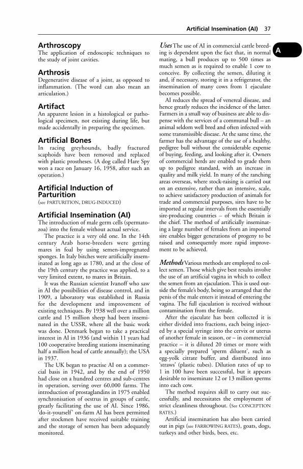
ArthroscopyThe application of endoscopic techniques tothe study of joint cavities.
ArthrosisDegenerative disease of a joint, as opposed toinflammation. (The word can also mean anarticulation.)
ArtifactAn apparent lesion in a histological or patho-logical specimen, not existing during life, butmade accidentally in preparing the specimen.
Artificial BonesIn racing greyhounds, badly fracturedscaphoids have been removed and replaced with plastic prostheses. (A dog called Hare Spy won a race on January 16, 1958, after such anoperation.)
Artificial Induction ofParturition(see PARTURITION, DRUG-INDUCED)
Artificial Insemination (AI)The introduction of male germ cells (spermato-zoa) into the female without actual service.
The practice is a very old one. In the 14thcentury Arab horse-breeders were getting mares in foal by using semen-impregnatedsponges. In Italy bitches were artificially insem-inated as long ago as 1780, and at the close ofthe 19th century the practice was applied, to avery limited extent, to mares in Britain.
It was the Russian scientist Ivanoff who sawin AI the possibilities of disease control, and in1909, a laboratory was established in Russia for the development and improvement of existing techniques. By 1938 well over a millioncattle and 15 million sheep had been insemi-nated in the USSR, where all the basic workwas done. Denmark began to take a practicalinterest in AI in 1936 (and within 11 years had100 cooperative breeding stations inseminatinghalf a million head of cattle annually); the USAin 1937.
The UK began to practise AI on a commer-cial basis in 1942, and by the end of 1950 had close on a hundred centres and sub-centresin operation, serving over 60,000 farms. Theintroduction of prostaglandins in 1975 enabledsynchronisation of oestrus in groups of cattle,greatly facilitating the use of AI. Since 1986,‘do-it-yourself ’ on-farm AI has been permittedafter stockmen have received suitable trainingand the storage of semen has been adequatelymonitored.
Uses The use of AI in commercial cattle breed-ing is dependent upon the fact that, in normalmating, a bull produces up to 500 times asmuch semen as is required to enable 1 cow toconceive. By collecting the semen, diluting itand, if necessary, storing it in a refrigerator, theinsemination of many cows from 1 ejaculatebecomes possible.
AI reduces the spread of venereal disease, andhence greatly reduces the incidence of the latter.Farmers in a small way of business are able to dis-pense with the services of a communal bull – ananimal seldom well bred and often infected withsome transmissible disease. At the same time, thefarmer has the advantage of the use of a healthy,pedigree bull without the considerable expenseof buying, feeding, and looking after it. Ownersof commercial herds are enabled to grade themup to pedigree standard, with an increase inquality and milk yield. In many of the ranchingareas overseas, where stock-raising is carried outon an extensive, rather than an intensive, scale,to achieve satisfactory production of animals fortrade and commercial purposes, sires have to beimported at regular intervals from the essentiallysire-producing countries – of which Britain isthe chief. The method of artificially inseminat-ing a large number of females from an importedsire enables bigger generations of progeny to beraised and consequently more rapid improve-ment to be achieved.
Methods Various methods are employed to col-lect semen. Those which give best results involvethe use of an artificial vagina in which to collectthe semen from an ejaculation. This is used out-side the female’s body, being so arranged that thepenis of the male enters it instead of entering thevagina. The full ejaculation is received withoutcontamination from the female.
After the ejaculate has been collected it iseither divided into fractions, each being inject-ed by a special syringe into the cervix or uterusof another female in season, or – in commercialpractice – it is diluted 20 times or more with a specially prepared ‘sperm diluent’, such asegg-yolk citrate buffer, and distributed into‘straws’ (plastic tubes). Dilution rates of up to 1 in 100 have been successful, but it appearsdesirable to inseminate 12 or 13 million spermsinto each cow.
The method requires skill to carry out suc-cessfully, and necessitates the employment ofstrict cleanliness throughout. (See CONCEPTION
RATES.)Artificial insemination has also been carried
out in pigs (see FARROWING RATES), goats, dogs,turkeys and other birds, bees, etc.
Artificial Insemination (AI) 37
A
Canine AI is now practised in many parts of the world. In the UK the Kennel Clubreserves the right to decide whether to acceptfor registration puppies obtained by means ofAI rather than by normal mating. Applicationsare usually made by the owner of the bitch.Those concerned with a newly imported breed, and who wish to widen the genetic pool, may not be able to find a suitable male for purchase and import. However, if semenfrom a satisfactory dog can be obtained, andDEFRA agrees to its import under licence, AImay be a good way of increasing the availablepool.
Registrations will not be accepted where AI isrequested because either the prospective sire ordam is unable to mate owing to disease.
Turkey AI Farmed turkeys are now bred asmale and female lines. Female lines are compar-atively slender, with high egg production. Malelines are bred for meat and are much heavier.The resulting disparity between the sizes of themale and female is such that natural matingwould result in injury to the female. Mostturkey breeding is therefore by artificial insem-ination. Disposable straws, discarded after use,are used to prevent transmission of infection(notably Mycoplasma meleagridis and E. coli)but as semen is pooled from several stags, aninfected stag can result in many infected hens.The technique of insemination and collectionof semen requires skill.
Storage of semen Diluted semen may be stored at AI centres for a few days if kept at a temperature of 5°C. In practice, a gooddeal would be wasted because its fertilisingpower has diminished before it is all requiredfor use. However, semen may be stored for long periods when glycerol is added to thesperm diluent. This enables the semen to bestored and transported at –196°C, using liquidnitrogen to maintain the low temperature. The advantages of this method are many. Thereis less wastage of semen, more can be stored,and the semen of any particular bull can bemade available on any day. It is possible for several thousand cows to be got in calf by a given bull. The disadvantages of using a givenbull or bulls too widely must be borne in mind,but that is a matter of policy and not of technique.
Infected semen Viruses (including that offoot-and-mouth) and mycoplasmas have, onoccasion, been found in stored semen. (See also
RABIES; CONTROLLED BREEDING.)
Artificial Rearing of PigletsCows’ colostrum makes a satisfactory substitutefor sows’ colostrum, and may be frozen andlater thawed when required. Pigs’ serum as anaddition enhances the value of cows’ colostrum.
Artificial RespirationThis is resorted to in: (1) cessation of respira-tion while under general anaesthesia; (2) casesof drowning when the animal has been rescuedfrom the water – chiefly applicable to the smallanimals; (3) poisoning by narcotics orparalysants; (4) cases of asphyxia from fumes,smoke, gases, etc.
Horses and cattle Release from all restraintexcept a loose halter or head-collar, extend thehead and neck to allow a straight passage of theair into the lungs, open the mouth, and pull the tongue well out. Should the ground slope,the horse must be placed with its head downhill.While such adjustments are being carried out 1 or 2 assistants should compress the elastic pos-terior ribs by alternately leaning the wholeweight of the body on the hands pressed on theribs, and then releasing the pressure about onceevery 4 or 5 seconds, in an endeavour to stimu-late the normal movements of breathing. As analternative in a larger animal, a heavy person maysit with some vigour astride the ribs for about thesame time, rise for a similar period, and then sitback again. If no response occurs, these measuresshould be carried out more rapidly.
The inhalation of strong solution of ammo-nia upon a piece of cotton-wool and held abouta foot from the upper nostril often assists ininducing a gasp which is the first sign of thereturn to respiration, but care is needed not toallow the ammonia to come into contact withthe skin or burning will occur. After 2 or 3 min-utes’ work the animal should be turned on tothe opposite side to prevent stasis of the blood.Sometimes the mere act of turning will inducethe premonitory gasp. So long as the heart con-tinues to beat, no matter how feebly, theattempts at resuscitation should be pursued.Proprietary calf resuscitators are available togive the ‘kiss of life’.
Pigs and sheep The outlines of proceduregiven for the larger animals are equally applica-ble. An ordinary domestic funnel can be usedfor giving pigs the ‘kiss of life’.
The method of giving the ‘kiss of life’ to apiglet is to use a flexible polyethylene funnel,and fit this over the animal’s mouth and nos-trils. Air is blown into the stem of the funnel,and passes down into the piglet’s lungs.
38 Artificial Rearing of Piglets
A
For the method to be effective, the procedureis as follows: (1) hold the piglet by its hind legswith head down in order to drain any fluid fromits air passages; (2) turn the piglet with its headupwards and apply the funnel; (3) blow force-fully into the funnel; (4) remove the funnel andallow the piglet to breathe out; (5) repeat theoperation. After several repetitions, the pigletshould kick or show other signs of life. Lay theanimal on its side or stomach and massage itschest and mouth. Piglets apparently stillbornmay sometimes be revived by this method.
Piglets have been revived up to half an hourafter treatment began. Of course, the heart mustbe beating and resuscitation started promptly toachieve success.
Dogs and cats A modification of theSchafer system is to lay the dog on its side withthe head at a lower level than the rest of thebody, place a hand flat over the upper side ofthe abdomen and the other on the rib-cage,lean heavily on the hands, and in a second ortwo release the pressure.
The motions of artificial respiration shouldin all cases be a little faster than those of normalrespiration, but a slight pause should always beobserved before each rhythmic movement. Useless pressure for cats.
A respiratory stimulant may be given byinjection. A carbon dioxide ‘Resuscitator’ maybe used.
AscaridaeA class of worms belonging to the round varietyor Nemathelminthes, which are found parasiticin the intestines of horses, pigs, dogs, and catsparticularly, although they may affect other ani-mals. They attain a size of 38 or 45 cm (15 or18 inches) in the horse, but are small in otheranimals. (See ROUNDWORMS.)
AscitesOEDEMA involving the abdomen; a very com-mon complication of abdominal tuberculosis,of liver, kidney, or heart disease, as well as ofsome parasitic infestations. In poultry, ascites issometimes associated with hypoxia (‘high alti-tude disease’) although there are other causesincluding toxins or, in individuals, heart defectsor abdominal tumours. It is also seen in duckswith furazolidone poisoning.
Ascorbic AcidSynthetic vitamin C.
AsepsisThe absence of pathogenic organisms. Asepticsurgery is the ideal, but among animals it may
be difficult to attain if carried out under farmconditions – despite care in sterilising instru-ments and the use of sterilised dressings, rubbergloves, etc. Moreover, it is an exceptionally difficult matter to prevent accidental infectionin a surgical wound after the operation, for the animal cannot be put to bed, and it may object to the dressings and do all in itspower to remove them. (See ANTISEPTICS;SULFONAMIDES; PENICILLIN.)
Ash PoisoningPoisoning by Fraxinus species has been reportedin cattle after eating the green leaves and fruitsfrom a broken branch of a tree. Symptomsinclude: drowsiness, oedema involving ribs andflanks, purple discoloration of perineum.
AspergillosisA disease of mammals and birds produced by the growth of the fungus Aspergillus in thetissues of the body.
Infection probably occurs chiefly throughinhalation of the fungal spores, which may beabundant in hay or straw under conditions ofdampness. Entry of the spores into the bodymay also be by way of the mouth; in herbivo-rous animals from contaminated fodder or bedding; and in cat and dog from the eating ofinfected birds or rodents.
Once in the animal’s tissues, hyphae growout from the spores, as happens also in ring-worm; and from the branching filaments morespores are produced. Local necrosis and abscessformation are caused.
Numerous organs and tissues can becomeinfected, including the nose and nasal sinuses,the lungs, brain, uterus, and mammary glands.
Cattle and horses Aspergillus may causeabortion and lung sensitisation or pneumonia.
Dogs and cats Aspergillosis is a commoncause of chronic nasal disease, and should be sus-pected when there is a discharge from one nostril.
Poultry Respiratory disease or enteritis mayoccur. In young turkey poults brain involve-ment has led to an unsteady gait, walking back-wards, and turning the head to one side.
Pet parrots may die from aspergillosis, as wellas wild birds.
Brain infection may occur in all species, and give rise to symptoms described underENCEPHALITIS. Paresis and ataxia may, rarely, becaused by fungal infections of the spine.
Ketonazole, given by mouth, and irrigationof the sinuses by enilconazole in sodium
Aspergillosis 39
A
chloride solution have been used in cases ofcanine nasal aspergillosis.
Asphodel(see BOG ASPHODEL)
AsphyxiaSuffocation may occur during the administra-tion of anaesthetics by inhalation, during theoutbreak of fires in animal houses, where thefumes and the smoke present are responsible for oedema, and in cases of poisoning. (See also
‘KITCHEN DEATHS’.)
Signs The direct cause of death from asphyxiais an insufficiency of oxygen supplied to the tis-sues by the blood. The first signs are a rapid andfull pulse, and a quickening of the respirations.The breathing soon changes to a series of gasps,and the blood pressure rises, causing the visiblemembranes to become intensely injected andlater blue in colour. Convulsions supervene.The convulsions are followed by quietness,when the heartbeat may be almost impercepti-ble and respiratory movements practicallycease. The actual time of death is unnoticed asa rule, since death takes place very quietly.
During the stage of convulsions, when theamount of carbon dioxide circulating in theblood is increased, the smaller arteries vigorouslycontract and cause an increase in the blood pressure. This high blood pressure produces an engorgement of the right side of the heart,which cannot totally expel its contents with eachbeat, and becomes more and more dilated untilsuch time as the pressure in the ventricles over-comes the strength of the muscle fibres of the heart and the organ ceases to beat. Duringthis stage immediate relief follows bleeding froma large vein.
Treatment (see ARTIFICIAL RESPIRATION). Ifthe breathing is shallow and the membraneslivid, administration of OXYGEN is indicated.
Prevention Ensure adequate ventilation inrooms where there is a gas or solid-fuel heatingsystem. (Many dogs and cats have been founddead in the kitchen in the morning as a result ofCARBON MONOXIDE poisoning.)
Aspiration(see PARACENTESIS)
Aspirin (Acetylsalicylic Acid)An analgesic; also used in prevention of throm-bosis. Must be used with extreme caution and under professional supervision in cats;
the dose not exceeding 10 mg/kg on alternatedays.
In both cats and dogs, overdosing withaspirin may cause inflammation of the stom-ach, haemorrhage, some pain, and vomiting.The antidote is sodium bicarbonate which can be given in water by stomach tube; or, for first-aid purposes, by the cat-owner, in milk or water. (See SALICYLIC ACID – Salicylatepoisoning.)
Aspirin has been used to lessen the effects ofporcine respiratory and reproductive syndrome(PRRS/blue-eared pig disease).
AstheniaAsthenia is another name for debility. Asthenicis applied to the exhausted state that precedesdeath during some fevers.
AsthmaAsthma is a term somewhat loosely applied to a number of conditions in which the main sign is breathlessness. Strictly speaking, theterm should be reserved for those conditionswhere a true spasmodic expulsion of breathoccurs without the effort of a cough. The socalled ‘asthma’ of birds is due in nearly everycase to ASPERGILLOSIS. Asthma in horses may bedifficult to differentiate from ‘BROKEN WIND’,and in all animals from simple BRONCHITIS.
Causes These are obscure, but it is generallyheld that true spasmodic asthma is of nervousorigin, and due to a sudden distressful contrac-tion of the muscle fibres which lie around thesmaller bronchioles. In some cases asthma maybe an allergic phenomenon. In other cases achronic inflammation of the lining mucousmembrane of the small tubes is the cause.
The spores of fungi are potent allergens, andcan account for many cases of asthma, especial-ly recurrent summer asthma, in man. There are,however, a number of patients with seasonal(summer or autumn) asthma who are not sensi-tive to spores of any of the above nor to pollen.(See ALLERGY.)
Dog Many cases that are really chronic bron-chitis are spoken of as ‘bronchial asthma’ owingto their similarity to asthma in man, withwhich many owners of animals are familiar. In true asthma the attacks of dyspnoea (i.e. distressed respiration) occur at irregularintervals, and there are periods between themwhen the dog is to all appearances quite nor-mal. The attacks occur suddenly, are very dis-tressing to witness, last for from 10 minutes tohalf an hour, and then suddenly cease. The dog
40 Asphodel
A
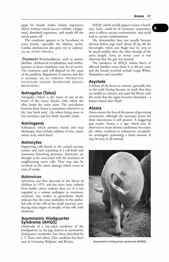
gasps for breath, makes violent inspiratoryefforts without much success, exhibits a fright-ened, disturbed expression, and stands till theattack passes off.
The condition appears to be hereditary insome breeds, especially the Maltese terrier.Cardiac dysfunction also gives rise to ‘asthma’.(See also ATOPIC DISEASE.)
Treatment Bronchodilators, such as amino-phylline, clenbuterol or ephedrine, and antihis-tamines or heart stimulants may be of service.The treatment used will depend on the cause of the problem. Regulation of exercise and dietis necessary. (See also CHRONIC OBSTRUCTIVE
PULMONARY DISEASE; RESPIRATORY DISEASE;BRONCHITIS.)
Astragalus (Talus)Astragalus (Talus) is the name of one of thebones of the tarsus (hock), with which the tibia forms the main joint. The articulationbetween these bones is sometimes referred to asthe ‘true hock joint’, the others being more orless secondary and less freely movable joints.
AstringentsSubstances which contract tissues and stop discharges; they include sulphate of zinc, alum,tannic acid, witch-hazel.
AstrocytesSupporting cells found in the central nervoussystem, and each consisting of a cell body andnumerous branching processes. Astrocytes arethought to be concerned with the nutrition ofneighbouring nerve cells. They may also beinvolved in the tissue damage which ocurs incases of stroke.
AstrovirusAstrovirus was first detected in the faeces ofchildren in 1975, and has since been isolatedfrom lambs, calves, turkeys, deer, etc. It is notregarded as a serious pathogen in veterinarymedicine, but studies in gnotobiotic lambsindicate that the virus multiplies in the epithe-lial cells of the villi of the small intestine, pro-ducing some degree of atrophy of the villi, withdiarrhoea.
Asymmetric HindquarterSyndrome (AHQS)Outbreaks of a lop-sided condition of thehindquarters in the pig, known as asymmetrichindquarter syndrome, have been described byJ. T. Done and others. This condition has beenseen in Germany, Belgium, and Britain.
AHQS, which would appear to have a hered-itary basis, could be of economic importancesince it affects carcase conformation, and couldlead to carcase condemnation.
The abnormality does not usually becomeobvious before pigs reach about 30 kg (66 lb)live-weight, when one thigh may be seen to be much smaller than the other though of thesame length. Even in severe cases it wasobserved that the gait was normal.
The incidence of AHQS within litters ofaffected families varies from 0 to 80 per cent,and the breeds involved include Large White,Hampshire and Lacombe.
AsystoleA failure of the heart to contract, generally dueto the walls having become so weak that theyare unable to contract and expel the blood, withthe result that the organ becomes distended – afeature found after death.
AtaxiaAtaxia means the loss of the power of governingmovements, although the necessary power forthese movements is still present. A staggeringgait results. Ataxia is a sign which may beobserved in many diverse conditions; for exam-ple, rabies, weakness or exhaustion; encephali-tis; meningitis; poisoning; a brain tumour. Itmay be seen in all animals.
Ataxia 41
A
Asymmetric hindquarter syndrome (AHQS).
Cattle A progressive form of ataxia ofunknown origin has been found in French-bredCharolais heifers, with symptoms first appear-ing in the 1st year: slight intermittent ataxiaprogresses to recumbency over 1 to 2 years.Urine is passed in a continuous but unevensquirting flow. When excited, affected heifersmay show nodding of their heads.
Cats Ataxia is seen in feline infectious peritoni-tis poisoning by ethylene glycol (anti-freeze)and streptomycin, for example, and beforeeclampsia (lactation tetany). Congenital cere-bellar ataxia may be seen in kittens, usuallywhen born to mothers infected with parvovirus.There is incoordinated movement of the head,especially when feeding, and they stand withtheir legs apart to aid balance. The conditiondoes not worsen and, unless very serious, kittensusually adapt well.
AtheromaA degenerative change in the inner and middlecoats of the arteries in which a deposit of lipid material is formed. (See ARTERIES, DISEASES
OF.)
AtherosclerosisA condition in which deposits of cholesteroland other material in the inner lining (intima)of arteries restricts the blood flow.
AtlasAtlas is the name given to the 1st of the cervicalvertebrae, which forms a double pivot jointwith the occipital bone of the base of the skullon the one hand, and forms a single glidingpivot joint with the epistropheus – the 2nd cervical vertebra – on the other hand. Thefreedom of movement of the head is due almostsolely to these 2 joints.
AtonyAtony means want of tone or vigour in musclesor other organs. (See also TONICS.)
Atopic DiseaseA hypersensitivity to pollens and other inhaledprotein particles. (See ALLERGY.) Hay-fever-like symptoms may be produced in the dog and horse; also intense itching affecting thefeet, abdomen, and face. As well as sneezing,conjunctivitis, rhinitis and asthma, there maybe some discoloration of the coat. In allergytests on 208 dogs, about 40 per cent were foundto be hypersensitive to human dandruff.
Atopic disease also occurs in cats and cattle(see BOVINE ATOPIC RHINITIS).
AtresiaAtresia means the absence of a natural opening,or its obliteration by membrane. Atresia of therectum is found in newly-born pigs, lambs,calves, and foals. Atresia is sometimes met within the vaginae of heifers, when it constituteswhat is known as ‘WHITE HEIFER DISEASE’.
AtrialRelating to the atrium or AURUCLE of theheart.
Atrophic Myositis(see under MUSCLES, DISEASES OF)
Atrophic RhinitisA disease of pigs affecting the nasal passages.(See under RHINITIS, ATROPHIC.)
AtrophyAtrophy is a wasting of the tissues. Followingparalysis of a motor nerve, when the musclessupplied by it are no longer able to contract,atrophy of the area takes place. This is seen in paralysis of the radial nerve. (CompareHYPERTROPHY.)
AtropineAn alkaloid contained in the leaves and root ofthe deadly nightshade (Atropa belladonna).Preparations of belladonna owe their anti-cholinergic actions to the presence of atropine,which blocks transmission at sensory nerve-endings and thus relieves pain and spasm in parts to which it is applied. It checks secre-tion in all the glands of the body when giveninternally; and whether given by the mouth or rubbed on the skin it causes a dilatation of the pupil of the eye and paralysis of accom-modation. In large doses it induces a generalstimulation of the nervous system, but thisaction is rapidly followed by depression, andthe primary effect is not noticed in the admin-istration of ordinary doses. The action on the heart is one of stimulation, since the inhibitionfibres are paralysed, while the accelerator nervesare not interfered with, except when large dosesare given and paralysis of all motor fibresoccurs.
Uses Atropine is used as a premedicant toanaesthesia as it reduces secretions. It is alsoused to dilate the pupil in order to facilitate eyeexaminations. As an antidote to morphine poi-soning and also to some of the organophospho-rus compounds used as farm sprays, it is givenas the sulphate of atropine by hypodermicinjection.
42 Atheroma
A
Atropine PoisoningAtropine poisoning may occur as the result ofthe unintentional administration of too largeamounts of the alkaloid ATROPINE or of thedrug BELLADONNA in one form or another, or it may be induced by feeding on the plantgrowing wild.
The signs of poisoning shown are restless-ness, delirium, dryness of the mouth, a rapidand weak pulse, quick, short respirations, anincrease in temperature, and dilatation of thepupil. In addition there is sometimes seen a lossof power in the hind-limbs.
Antidotes To those animals that vomit, anemetic should be given at once if the poison hasbeen taken by the mouth. Horses and cattleshould have their stomachs emptied by the passage of the stomach-tube, in so far as that is possible. Stimulants should be given, andpilocarpine, by injection, is the antidote.
AttenuatedA term used to describe a reduction in the virulence of a micro-organism, particularlyapplied to those incorporated in vaccines (see
under VACCINE).
Auditory Nerve (AcousticNerve)The auditory nerve (acoustic nerve) is the 8thof the cranial nerves, and is concerned with thespecial sense of hearing. It arises from the baseof the hind-brain just behind and at the side ofthe pons. It is distributed to the middle andinternal ears, and in addition to its acousticfunction it is also concerned with the balance ofthe body. (See EAR.)
Aujeszky’s DiseaseA viral disease, primarily of pigs. It is alsoknown as pseudorabies and infectious bulbarparalysis. It can occur in other species; theinfection usually being contracted from contactwith pigs or consumption of pig carcases. Thedisease has a very short incubation period, andis characterised by intense itching. It was firstdescribed in Hungary by Aujeszky in 1902, andhas been eradicated from the UK, Denmark,Sweden and parts of other countries in the EU.It has also been found in several parts of theUSA, South America, Australia, the continentof Europe, etc. In the UK the disease is NOTIFI-
ABLE and an eradication campaign began in1983. Monitoring continues by sampling cullanimals at slaughterhouses. The infection maybe windborne. Vaccines are available, but theiruse is prohibited in the UK except Northern
Ireland. However, gene-deleted vaccinces canbe used in eradication programmes as it is possible to differentiate serologically a pigwhich has been vaccinated from one which hasbeen exposed to infection.
Signs
Pigs Signs include abortion, sneezing, anorexiaand dullness besides some evidence of pruritus,vomiting, diarrhoea, convulsions, drooling ofsaliva, paralysis of the throat. Mummification ofthe fetuses may occur in pregnant sows affectedwith Aujeszky’s disease. Such sows may showloss of appetite and constipation, or stiffnessand muscular incoordination without itching atall. For the screening of pig serum samples, theELISA test has been found the most sensitive,speediest and cheapest of four methods fordetecting antibodies to Aujeszky’s disease virus.(Central Veterinary Laboratory.)
Prevention: Intranasal vaccination with atten-uated virus is more effective than parenteral vaccination with inactivated virus, as maternallyderived antibodies interfere with the latter.
Dogs and cats Restlessness, loss of appetite,vomiting, salivation, signs of intense irritation(leading to biting or scratching) about the faceor some other part, and occasionally moaning,groaning, or high-pitched screams are amongthe symptoms observed.
In one outbreak, 11 out of a pack of 51 har-rier hounds died of the disease (apparently as aresult of being fed raw carcase meat from a largepig unit). Infected rats may be another vector.
Cattle The first symptom to be observed isusually a persistent licking, rubbing or scratch-ing of part of the hindquarters (or sometimes ofthe face) in an attempt to relieve the intenseitching. The affected part soon becomes denud-ed of hair, and may be bitten and rubbed untilit bleeds. Bellowing, salivation, and stampingwith the hind-feet may be observed. Within 24 hours the animal is usually recumbent andunable to rise on account of paralysis. Death,preceded by convulsions, usually occurs within36 to 48 hours of the onset of symptoms.
Goats Deaths have occurred in goats keptwith infected pigs. Signs include restlessness,sweating, distressed bleating and convulsions;some animals may be found dead without signsbeing noticed.
Poultry One-day-old chicks have died afterbeing inoculated with a Marek’s disease vaccine.
Aujeszky’s Disease 43
A
Aujeszky’s disease vaccine virus adapted tochicken cells was likely to have been the cause.
Horses The virus was isolated from the brainof a horse showing the following signs: excessivesweating, muscle tremors, and ‘periods ofmania’.
Public health Aujeszky’s disease virus caninfect people, but it seems that only laboratoryworkers are likely to find this a health hazard.
AuralRelating to the ear.
Aural Cartilages(see AURICULAR CARTILAGES)
Auricle (Atrium)The auricles, right and left, are the chambers at the base of the heart which receive the bloodfrom the body generally, and from the lungsrespectively. Opening into the right auricle are the cranial and caudal vena cavae, whichcarry the venous blood that has been circulatingin the head and neck and the abdomen and tho-rax. This blood is pumped into the right ventri-cle through the tricuspid valve. Opening into theleft auricle are the pulmonary veins which bringthe arterial blood that has been purified in thelungs; when this auricle contracts the blood isdriven into the left ventricle through the mitralvalve. (See HEART; CIRCULATION OF BLOOD.)
Auricular CartilagesAuricular cartilages are the supporting struc-tures of the ears. There are three chief cartilagesin most animals, viz. the conchal, which gath-ers the sound waves and transmits them down-wards into the cavity of the ear and gives the earits characteristic shape; the annular, a cartilagi-nous ring below the former which is continuousinternally with the bony acoustic canal; the scutiform, a small quadrilateral plate which lies in front of the others and serves for theattachment of muscles which move the ear.
Accidents and diseases of the cartilages ofthe ear are not common in animals, with theexception of dog/cat fights. Ulceration of the car-tilages, chiefly the annular, occurs as a complica-tion of ear inflammation in the dog. Lacerationof the conchal cartilage is seen as the result of theapplication of a twitch to the ear in the horse.
AuscultationAuscultation is a method of diagnosis by whichthe condition of some of the internal organs
is determined by listening to the sounds theyproduce. Auscultation is practised by means ofthe stethoscope.
AutogenousAutogenous means self-generated, and is theterm applied especially to bacterial and viralvaccines manufactured from the organismsfound in discharges from the body and used forthe treatment of the particular individual fromwhich the bacteria were derived.
Auto-Immune DiseaseAuto-immune disease is due to a failure of the bodily defence mechanisms in which antibodies become active against some of thehost’s own cells. An example is spontaneousauto-immune thyroiditis which occurs in dogs,poultry, monkeys and rats, and resemblesHashimoto’s thyroiditis of man. Other exam-ples are auto-immune haemolytic disease, inwhich the blood’s red cells are affected; andglomerulonephritis in small animals.
Immune-mediate diseases are of two kinds:(1) primary, an auto-immune reaction onlyagainst self; and (2) secondary, a similar reac-tion occurring when viruses, tumours, parasites,or drugs are involved.
Primary diseases are either organ-specific, e.g. auto-immune haemolytic anaemia (see under
ANAEMIA), or systemic, e.g. LUPUS ERYTHE-
MATOSUS. (See also THROMBOCYTOPENIA; POL-
YARTHRITIS; PEMPHIGUS; BOVINE and CANINE
AUTOIMMUNE HAEMOLYTIC ANAEMIA; DIABETES
MELLITUS.)
Auto-InfectionInfection of one part of the body, hithertohealthy, from another part that already is suf-fering from the disease. Thus, sheep sufferingfrom ‘orf ’ on their feet may bite the painfulareas and convey the organisms to their mouth,where the disease becomes established.
AutovaccineA vaccine prepared from an organism isolatedfrom from an animal and injected back into the same animal. The most common auto (or autologous) vaccine is that prepared fortreatment of warts (angleberries) in cattle.
AutolysisSelf-digestion of an organism by its ownenzymes. See also NECROSIS.
Autonomic Nervous SystemThe autonomic nervous system is that part of thenervous system which governs the automatic or
44 Aural
A
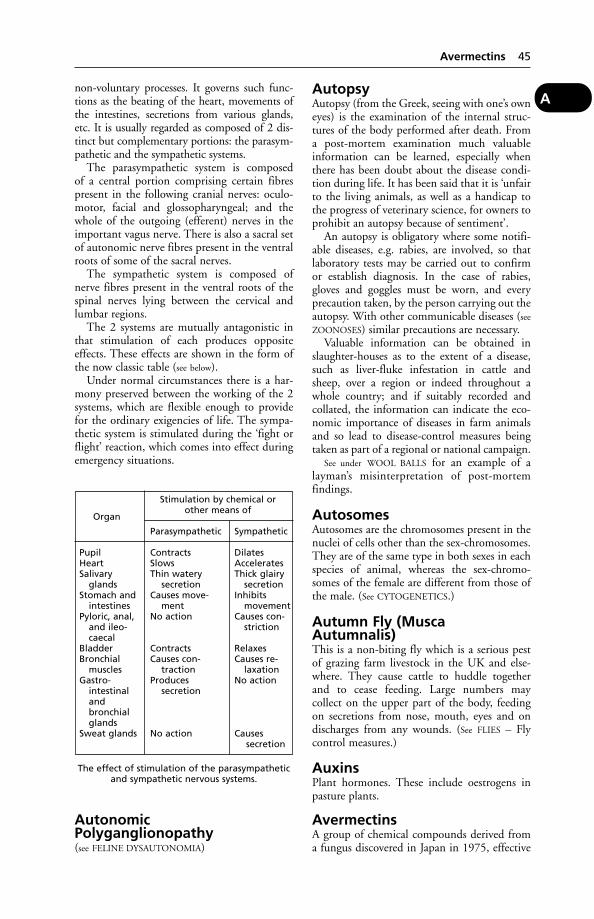
non-voluntary processes. It governs such func-tions as the beating of the heart, movements ofthe intestines, secretions from various glands,etc. It is usually regarded as composed of 2 dis-tinct but complementary portions: the parasym-pathetic and the sympathetic systems.
The parasympathetic system is composed of a central portion comprising certain fibrespresent in the following cranial nerves: oculo-motor, facial and glossopharyngeal; and thewhole of the outgoing (efferent) nerves in theimportant vagus nerve. There is also a sacral setof autonomic nerve fibres present in the ventralroots of some of the sacral nerves.
The sympathetic system is composed ofnerve fibres present in the ventral roots of thespinal nerves lying between the cervical andlumbar regions.
The 2 systems are mutually antagonistic inthat stimulation of each produces oppositeeffects. These effects are shown in the form ofthe now classic table (see below).
Under normal circumstances there is a har-mony preserved between the working of the 2systems, which are flexible enough to providefor the ordinary exigencies of life. The sympa-thetic system is stimulated during the ‘fight orflight’ reaction, which comes into effect duringemergency situations.
AutonomicPolyganglionopathy(see FELINE DYSAUTONOMIA)
AutopsyAutopsy (from the Greek, seeing with one’s owneyes) is the examination of the internal struc-tures of the body performed after death. Froma post-mortem examination much valuableinformation can be learned, especially whenthere has been doubt about the disease condi-tion during life. It has been said that it is ‘unfairto the living animals, as well as a handicap tothe progress of veterinary science, for owners toprohibit an autopsy because of sentiment’.
An autopsy is obligatory where some notifi-able diseases, e.g. rabies, are involved, so thatlaboratory tests may be carried out to confirmor establish diagnosis. In the case of rabies,gloves and goggles must be worn, and every precaution taken, by the person carrying out theautopsy. With other communicable diseases (see
ZOONOSES) similar precautions are necessary.Valuable information can be obtained in
slaughter-houses as to the extent of a disease,such as liver-fluke infestation in cattle andsheep, over a region or indeed throughout awhole country; and if suitably recorded and collated, the information can indicate the eco-nomic importance of diseases in farm animalsand so lead to disease-control measures beingtaken as part of a regional or national campaign.
See under WOOL BALLS for an example of a layman’s misinterpretation of post-mortemfindings.
AutosomesAutosomes are the chromosomes present in thenuclei of cells other than the sex-chromosomes.They are of the same type in both sexes in eachspecies of animal, whereas the sex-chromo-somes of the female are different from those ofthe male. (See CYTOGENETICS.)
Autumn Fly (MuscaAutumnalis)This is a non-biting fly which is a serious pestof grazing farm livestock in the UK and else-where. They cause cattle to huddle together and to cease feeding. Large numbers may collect on the upper part of the body, feedingon secretions from nose, mouth, eyes and ondischarges from any wounds. (See FLIES – Flycontrol measures.)
AuxinsPlant hormones. These include oestrogens inpasture plants.
AvermectinsA group of chemical compounds derived froma fungus discovered in Japan in 1975, effective
Avermectins 45
A
Stimulation by chemical orother means of
Organ
Parasympathetic Sympathetic
Pupil Contracts DilatesHeart Slows AcceleratesSalivary Thin watery Thick glairy
glands secretion secretionStomach and Causes move- Inhibits
intestines ment movementPyloric, anal, No action Causes con-
and ileo- strictioncaecal
Bladder Contracts RelaxesBronchial Causes con- Causes re-
muscles traction laxationGastro- Produces No action
intestinal secretionandbronchialglands
Sweat glands No action Causessecretion
The effect of stimulation of the parasympatheticand sympathetic nervous systems.
in very low dosage against nematode parasitesand also against external parasites. (See IVER-
MECTIN, which is the most useful of the group.)The discovery of the fungus in a soil sample waspart of Merck Sharp & Dohme’s internationalscreening programme.
Technically, the avermectins are a series ofmacrocyclic lactone derivatives produced byfermentation of the actinomycete Streptomycesavermitilis.
Avian Contagious Epithelioma(see under FOWL-POX)
Avian InfectiousEncephalomyelitisA disease of chicks and turkey poults; alsoknown as epidemic tremor.
Cause A picornavirus. (Infection via the egg,as well as bird to bird.)
Signs If infection is egg-borne, signs are seenin the first 10 days after hatching; if infectedafter hatching, at 2 to 5 weeks old. There is legweakness, followed by partial or completeparalysis of the legs. The chicks struggle to balance with the help of their wings. Tremblingof the head and neck occurs in some cases.
Diagnosis An ELISA test.
Mortality A 40 per cent rate is not unusual.
Prevention Vaccination has proved very successful.
Avian InfectiousLaryngotracheitisAvian infectious laryngotracheitis of poultry iscaused by a herpesvirus, prevalent in NWEngland. Loss of appetite, sneezing and cough-ing, a discharge from the eyes, difficulty inbreathing are the main symptoms. Birds of allages are susceptible. Mortality averages about15 per cent. No treatment is of value. Controlis best achieved by depopulation and fumiga-tion. A vaccine has been used.
Avian Influenza (Fowl Plague)Avian influenza (fowl plague) attacks domesticat-ed fowl chiefly, but turkeys, geese, ducks, andmost of the common wild birds are sometimesaffected. It is not known to affect the pigeon.The disease is found in Asia, Africa, the Americasand to a lesser extent in parts of the continent ofEurope, and is always liable to be introduced tocountries hitherto free from it through themigrations of wild birds. An outbreak occurred
among turkeys in Norfolk in 1963; this was thefirst recorded outbreak in Britain since 1929. Anoutbreak occurred in the Republic of Ireland in1983; a slaughter policy followed. Infection mayhave come from Pennsylvania, where a similarpolicy was adopted.
Cause Myxovirus influenzae.
Signs In some cases the number attacked issmall, while on the same premises the next year80 or 90 per cent of the total inhabitants of theruns may die. The affected birds often die quitesuddenly. In other instances the sick birds isolate themselves from the rest of the flock,preferring some dark out-of-the-way cornerwhere they will be undisturbed. They are dull,disinclined to move, the tail and wings droop,the eyes are kept closed; the bird may squat on its breast with its head tucked under a wingor in amongst the shoulder feathers; food isrefused, but thirst is often shown; the respira-tions are fast and laboured but not impeded by mucus; the temperature is very high at thecommencement (43° to 44°C; 110° to 112°F),but falls shortly before death to below normal.(The normal temperature of birds is 41°C;106.5°F.) The comb and wattles become purpleor blue, and oedema of the head and neck is common. The illness seldom lasts more than24 to 36 hours, and often not more than 6.
Control Vaccines are available but their use isincompatible with an eradication policy. Theyare used in parts of the USA and in Italy.
Avian ListeriosisAn infectious disease of poultry, occurring as an epidemic among young stock (often as anaccompaniment of other diseases) or sporadicallyamong adults.
Cause Listeria monocytogenes, a Gram-positivemotile rod-shaped organism.
Signs In the epidemic type, wasting occursover a period of days or even weeks. For 48 hours before death birds refuse all food.
The sporadic type is characterised by suddendeath without much loss of condition.
Diagnosis Depends upon bacteriologicalmethods. (See also LISTERIOSIS.)
Avian Lymphoid LeukosisAvian lymphoid leukosis virus (LLV) infectionis widespread among chickens in the UK, andcauses mortality from tumours.
46 Avian Contagious Epithelioma
A
This disease, which has to be differentiatedfrom Marek’s disease, affects birds of 4 monthsupwards and is egg-transmitted, shows variablesigns but, typically, the liver is enlarged.
It may be identified by the presence of neutralising antibodies in the serum or by virusdetection by ELISA.
Control High standards of hygiene and flockmanagement; no vaccines are available.
Avian Malaria(see PLASMODIUM)
Avian Monocytosis(see ‘PULLET DISEASE’)
Avian NephritisA viral infection first detected in the UK in1988. In chick embryos it causes stunting,haemorrhage and oedema as well as nephritis.
Avian Sex DeterminationAvian sex determination by laparoscopy hasbeen widely used since 1976.
Avian TuberculosisThe increase in the number of farmed poultrykept in free-range systems or with access to out-doors has led to an increase in the incidence ofthis disease. It is usually seen in birds over 2 yearsold but can occur in young birds. Ostriches areusually kept outdoors and are particularly at riskif near woodland, as wood pigeons (Columbapalamuis) and feral pigeons are often heavilyinfected – as are wild birds such as starlings.
Cause Mycobacterium avium.
Signs Dullness, loss of appetite, lethargy and atendency to squat in a sleeping position withthe head tucked under one wing.
Body temperature may reach 44°C (112°F).The comb and wattles may become almost
purple, and swollen because of oedema. Inyoung birds, there is muscle wastage and thecomb may become pale in colour. The diseaseprogresses slowly. It used to be referred to bypigeon fanciers as ‘going light’.
Infection occurs following ingestion of foodand water contaminated by the droppings ofinfected birds. Infection has been found ineagles at post-mortem examination, presum-ably from consumption of infected prey.
Cattle Avian tuberculosis rarely causes pro-gressive disease, but the presence of avian TB bacteria will affect the interpretation of the
tuberculin test. This infection must be differen-tiated from Mycobacterium bovis infection byusing avian tuberculin as well as mammalian inthe test. (See TUBERCULIN TEST.)
Sheep Avian tuberculosis can cause miliarytuberculosis in sheep.
Pigs A non-progressive infection is oftenfound in lymph nodes at slaughter. The sourceof infection in housed pigs can be the use ofpeat as litter. M. avium survives in peat for aconsiderable period.
Post-Mortem Emaciation is usually wellmarked, and whitish-yellow nodules are presentin the liver and spleen; also the intestines. Thelungs are rarely affected in avian tuberculosis.In birds which have died suddenly, death isoften found to be due to rupture of the liver,which when affected with tuberculosis is oftenenlarged and friable.
With valuable pedigree birds the intradermaltuberculin test may be employed, but beforeapplying this test all birds should be examinedand all thin birds destroyed, since those in theadvanced stages of the disease may fail to react.Birds which pass the test should be put in cleanhouses on fresh ground.
(See also DISPOSAL OF CARCASES.)
AvilamycinAn antibiotic feed additive used as a growthpromoter in pigs and poultry. Its use in the EUwas to be phased out by 2006.
AvitaminosisAvitaminosis is a term used to describe condi-tions produced by a deficiency or lack of a vita-min in the food. Thus ‘avitaminosis A’ means adeficiency of vitamin A. (See VITAMINS.)
Avocado LeavesPersea americana fed to goats and sheep, duringa drought in South Africa, caused death withina few days from heart disease.
Awns/Grass SeedsA review by Kathleen E. Brennan and Peter J. Ihrke, School of Veterinary Medicine,University of California, of 182 cases in dogsand cats over a 1-year period showed that grassawns comprised 61 per cent of all foreign bodycases. The most common site is the ear canal(51 per cent), and rupture of the tympanumhas been an occasional sequel. Other sites arethe interdigital skin conjunctiva, nose, lumbarregion. Lumbar osteomyelitis has been caused.Perforation of a bronchus led to necrosis of a
Awns/Grass Seeds 47
A

lung lobe. In a cat with chronic cystitis, 2 awnswere found in the bladder; and in another catseveral awns were found at autopsy to havecaused peritonitis. (JAVMA, 182, 1201.)
AxillaAxilla is the anatomical name for the regionbetween the humerus and the chest wall, whichcorresponds to the armpit in the human being.
Axon(see NERVES)
AzoperoneA neuroleptic drug used in pigs for reducingaggression and preventing fighting. It is used as
a sedative when pigs are being transported andmay be given as premedication before adminis-tering an anaesthetic or to reduce excitementwhen assistance at farrowing is required. Itseffect may be less reliable in Vietnamese pot-bellied pigs.
It is also used for sedation in ostriches.
AzotaemiaThe presence of urea and other nitrogenousproducts in greater concentration than normalin the blood, particularly in paralytic myoglo-binuria in horses.
Azoturia(see EQUINE MYOGLOBINURIA)
48 Axilla
A
B CellsOne of the 2 types of lymphocytes. They areimportant in the provision of immunity, andthey respond to antigens by dividing andbecoming plasma cells that can produce anti-body that will bind with the antigen. Theirsource is the bone marrow in mammals andthe Bursa of Fabricius in birds. It is believedthat the function of B cells is assisted by asubstance provided by T CELLS. With haptens(see HAPTEN) it is apparently the B cellswhich recognise the protein carrier, and theT cells which recognise the hapten. (See also
LYMPHOCYTE; IMMUNE RESPONSE.)
B VirusThis is a herpes virus found in monkeys whichgives rise in man to an encephalitis with analmost 100 per cent mortality. It may be trans-mitted to man from monkeys – especiallynewly imported rhesus and cynomolgus mon-keys. Lesions on the face and lips of monkeysshould arouse suspicion of this condition.
It is believed that B virus, herpes simplexvirus, and Aujeszky’s disease virus have acommon origin.
BabesiaBabesia is another name for piroplasm, one ofthe protozoan parasites belonging to the orderHaemosporidia. These are generally relativelylarge parasites within the red blood cells and arepear-shaped, round or oval. Multiplication is bydivision into 2 or by budding. Infected cellsfrequently have 2 pyriform parasites joined attheir pointed ends. Sexual multiplication takesplace in the tick.
Babesiosis (Piroplasmosis) Nearly all thedomestic mammals suffer from infection withsome species of Babesia; sometimes more than 1species may be present. The general symptomsare the appearance of fever in 8 to 10 days afterinfection, accompanied by haemoglobinuria,icterus; unless treated, 25 to 100 per cent of thecases are fatal. Red blood cells may be reducedin number by two-thirds. Convalescence is slowand animals may remain ‘salted’ for 3 to 8 years.
Transmission Development occurs in certainticks which transmit the agent to their offspring.
The various species are similar, but are specificto their various hosts. The ticks should probablybe regarded as the true or definite hosts, whilethe mammal is the intermediate host.
Cats Babesia felis is a (rare) cause of lethargy,inappetence and anaemia, and occasionallyjaundice and death.
Sheep Ovine babesiosis may be due to at least3 species of Babesia. There is a relatively largeform, Babesia motasi, which is comparable toB. bigemina of cattle, and which produces adisease, often severe, with high temperatures,much blood-cell destruction, icterus, andhaemoglobinuria. This is the ‘carceag’ ofEastern and Southern Europe. The 2nd para-site, of intermediate size and corresponding toB. bovis of cattle, is Babesia ovis. It produces amuch milder disease with fever, jaundice, andanaemia, but recoveries generally occur. Thesmall species is Theileria ovis, which appearsto be similar to T. mutans of cattle and isrelatively harmless to its host.
B. motasi, B. ovis, and T. ovis are all transmittedby Rhipicephalus bursa.
Animals recovered from T. ovis infectionapparently develop a permanent immunity toit. The disease occurs in Europe, Africa, Asia,and North America.
Signs In acute cases the temperature may riseto 41.5°C (107°F), rumination ceases, thereis paralysis of the hindquarters, the urine isbrown, and death occurs in about a week. Inbenign cases there may only be a slight fever fora few days with anaemia.
A theileriosis, caused by T. hirci, has beendescribed from sheep in Africa and Europe. Itcauses an emaciation and small haemorrhagesin the conjunctiva.
Bacillary HaemoglobinureaA disease of cattle caused by Clostridiumhaemolyticum (Cl. oedematiens) type D.
Bacillary White Diarrhoea(see PULLORUM DISEASE)
BacillusThis genus of Gram-positive rod-shaped organ-ism contains many species which are notregarded as pathogenic, as well as some thatare. They are found in soil, water, and onplants. Spores formed by bacilli are resistant toheat and disinfectants, and this fact is impor-tant in connection with B. anthracis, the causeof ANTHRAX. Another pathogenic bacillus is
B

B. cereus, a cause of food poisoning and also ofbovine mastitis. (See BACTERIA.)
BacitracinAn antibacterial formerly used as a feed addi-tive; its use for this purpose has been banned inthe EU.
Back-crossBack-cross is the progeny resulting from matinga heterozygote offspring with either of itsparental homozygotes. Characters in the back-crosses generally show a 1:1 ratio. Thus if a pureblack bull is mated with pure red cows (allhomozygous), black calves (heterozygotes) areproduced. If the heifer calves are ‘back-crossed’to their black father, their progeny will give1 pure black to every 1 impure black. If a blackheterozygous son of the original mating ismated to his red mother, the progeny will be1 red to 1 black.
Back-crossing can be employed as a meansof test-mating, or test-crossing to determinewhether a stock of animals is homozygous,when it will never throw individuals of differenttype, or whether it is heterozygous, when it willgive the 2 allelomorphic types. (See GENETICS,HEREDITY AND BREEDING.)
Back-Fence(see STRIP-GRAZING)
Back Muscle Necrosis (BMN)A disease of pigs first described in Belgium in1960, and recognised 8 years later in WestGermany (where it is colloquially known as‘banana disease’). It has been recorded in theUK, with 20 cases occurring in a single herd.
Signs A sudden and sporadic condition affect-ing pigs weighing over 50 kg. In the acute stage,the animal shows signs of pain, has difficulty inmoving, becomes feverish, loses appetite andappears lethargic, and shows a characteristicswelling on 1 or both sides of the back. Whenonly 1 side is affected, spinal curvature occurswith the convexity of the curve towards theswollen side.
The colloquial name ‘banana disease’ appar-ently arose from arching (as compared withlateral curvature) of the back, which is oftenseen in affected animals.
Some pigs die from acidosis and heart fail-ure; some recover, apparently completely; whileothers are left with atrophy of the affectedmuscles resulting in a depression in the skinparallel to the spine. Some examples of BMNare discovered only in the slaughterhouse.
Post-Mortem examination reveals necrosisand bleeding, especially in the longissimusdorsi muscle, as well as the widely recognisedcondition known as PSE or pale soft exudativemuscle.
Causes The disease is thought to be associatedwith stress; it is probable that heredity alsocomes into the picture.
BacteriaMicroscopic single-cell plants with importantfunctions in nutrition and in disease processes.According to peculiarities in shape and in groupformation, certain names are applied: thus asingle spherical bacterium is known as ‘coccus’;organisms in pairs and of the same shape (i.e.spherical) are called ‘diplococci’; when in theform of a chain they are known as ‘strepto-cocci’; when they are bunched together likea bunch of grapes the name ‘staphylococcus’is applied. Bacteria in the form of long slenderrods are known as ‘bacilli’; wavy or curvedforms have other names.
Reproduction The mode of multiplication ofmost bacteria is exceedingly simple, consistingof a splitting into 2 of a single bacterium. Sincethe new forms may similarly divide within halfan hour, multiplication is rapid. (See illustration;see also PLASMIDS.)
Spore-Formation Some bacteria have thepower to protect themselves from unfavourableconditions by changing their form to that of a‘spore’.
Size Bacteria vary in size from less than 1MICRON (one-thousandth of a millimetre)diameter, in the case of streptococci and staphy-lococci, up to a length of 8 microns, in the caseof the anthrax bacillus.
Mobility Not all bacteria possess the powerof movement, but if a drop of fluid contain-ing certain forms of organism which are called‘motile’ be examined microscopically, it will beobserved that they move actively in a definitedirection. This is accomplished, in the motileorganisms, by means of delicate whip-likeprocesses which thrash backwards and forwardsin the fluid and propel the body onwards.These processes are called ‘flagellae’.
Methods of diagnosis
(1) Microscopical In order satisfactorily toexamine bacteria microscopically, a drop of the
50 Bacitracin
B
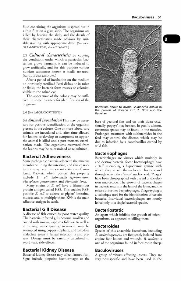
fluid containing the organisms is spread out ina thin film on a glass slide. The organisms arekilled by heating the slide, and the details oftheir characteristics made obvious by suit-able staining with appropriate dyes. (See under
GRAM-NEGATIVE; also ACID-FAST.)
(2) Cultural characteristics By copyingthe conditions under which a particular bac-terium grows naturally, it can be induced togrow artificially, and for this purpose variousnutrient substances known as media are used.(See CULTURE MEDIUM.)
After a period of incubation on the mediumon previously sterilised Petri dishes or in tubesor flasks, the bacteria form masses or colonies,visible to the naked eye.
The appearance of the colony may be suffi-cient in some instances for identification of theorganism.
(3) (See LABORATORY TESTS)
(4) Animal inoculation This may be neces-sary for positive identification of the organismpresent in the culture. One or more labora-toryanimals are inoculated and, after time allowedfor lesions to develop or symptoms to appear,the animal is killed and a post-mortem exami-nation made. The organisms recovered fromthe lesions may be re-examined or re-cultured.
Bacterial AdhesivenessSome pathogenic bacteria adhere to the mucousmembrane lining the intestine, and this charac-teristic may be an important criterion of viru-lence. Bacteria which possess this propertyinclude E. coli, Salmonella typhimurium,Mycoplasma pneumoniae, and Moraxella bovis.
Many strains of E. coli have a filamentousprotein antigen called K88. This enables K88-positive E. coli to adhere to piglets’ intestinalmucosa and to multiply there. K99 is the mainadhesive antigen in cattle.
Bacterial Gill DiseaseA disease of fish caused by poor water quality.The bacteria-infected gills become swollen andcoated with mucus; asphyxia follows. As well asimproving water quality, treatment may beattempted using copper sulphate, and zinc-freemalachite green if fungal infection is also pre-sent. Dosage must be carefully calculated toavoid toxic side-effects.
Bacterial Kidney DiseaseBacterial kidney disease may affect farmed fish.Signs include pinpoint haemorrhages at the
base of pectoral fins and on their sides; occa-sionally ‘popeye’ may be seen. In pacific salmon,cavernous spaces may be found in the muscles.Prolonged treatment with sulfonamides in thefeed may control the disease, which may be due to infection by a coccobacillus carried bywild fish.
BacteriophagesBacteriophages are viruses which multiply inand destroy bacteria. Some bacteriophages havea ‘tail’ resembling a hypodermic syringe withwhich they attach themselves to bacteria andthrough which they ‘inject’ nucleic acid. ‘Phages’have been photographed with the aid of the elec-tron microscope. The growth of bacteriophagesin bacteria results in the lysis of the latter, and therelease of further bacteriophages. Phage-typing isa technique used for the identification of certainbacteria. Individual bacteriophages are mostlylethal only to a single bacterial species.
BacteriostaticAn agent which inhibits the growth of micro-organisms, as opposed to killing them.
BacteroidesSpecies of this anaerobic bacterium, includingB. melaninogenicus, are frequently isolated fromequine foot lesions and wounds. B. nodosus isone of the organisms found in foot-rot in sheep.
BaculovirusesA group of viruses affecting insects. They arevery host-specific and have been used in the
Baculoviruses 51
B
Bacterium about to divide. Salmonella dublin inthe process of division into 2. Note also theflagellae.

control of specific insect pests while leaving ben-eficial species unharmed. Interest has also been
shown in the possibility of using them as carriersof antigens in genetically engineered vaccines.
52 Bacteria
B
Bacteria. Photomicrographs of (1) Bacillus anthracis (× 4200); (2) Clostridium tetani (× 3250) (showing thecharacteristic drum-stick appearance); (3) Streptococcus pyogenes (× 3000).
(1)
(2)
(3)
Bacteria 53
B
Some bacteria of veterinary importanceName Associated or specific diseased conditions caused
Actinobacillus lignièresi Actinobacillosis.
A. pleuropneumoniae Pleuropneumonia in pigs.
Actinomyces pyogenes Abscesses in liver, kidneys, lungs or skin in sheep, cattle and pigsespecially; present as a secondary organism in many suppurativeconditions; causes summer mastitis in cattle.
Actinomyces bovis Actinomycosis.
Aeromonas shigelloides Chronic diarrhoea in cats.
Bacillus anthracis Anthrax in all susceptible animals.
Bacillus cereus Bovine mastitis; food poisoning.
Bacillus lichenformis Abortion in ewes.
Baccilus piliformis Tyzzer’s disease.
Bacteroides species Foot infections in horses.
Bacteroides nodosus Foot-rot in sheep. Necrosis of skin or mucous membrane in rabbits after their resistance has been lowered by some other pathogen.
Bordetella bronchiseptica Complicates distemper in the dog. Kennel cough. Atrophic rhinitis.
Brucella abortus Brucellosis.
Brucella melitensis Brucellosis in goats; undulant fever in man (in part).
Campylobacter fetus Infertility, abortion.
Clostridium botulinum Botulism in man and animals.
(five types – A to E)
Cl. chauvoei ‘Black-quarter’ (and also pericarditis and meningitis in cattle) in cattle and partly in sheep.
Cl. difficile Chronic diarrhoea in dogs and piglets.
Cl. novyi (oedematiens) ‘Black-quarter’ in cattle and pigs in part; ‘black disease’ in sheep;septicaemia in horses and pigs (wound infection).
Cl. septicum Gas gangrene in man; black-quarter; braxy in sheep.
Cl. tetani Tetanus in man and animals.
Cl. welchii (perfringens) Lamb dysentery; present in many cases of gas gangrene.
Corynebacterium Caseous lymphadenitis in sheep; some cases of ulcerative lymphan-pseudotuberculosis gitis and acne in horses.
C. equi A cause of pneumonia in the horse and of tuberculosis-like lesions in the pig.
Dermatophilus congolensis Chronic dermatitis.
Group EF-4 bacteria Pneumonia in dogs and cats, and isolated from human dog-bite wound.
Erysipelothrix rhusiopathiae Swine erysipelas.
Eschicheria coli (sub. types Always present in alimentary canal as commonest organism; becomesare many) pathogenic at times, partly causing enteritis, dysentery (lambs),
scour (calves and pigs), cystitis, abortion, mastitis, joint-ill, etc.
Fusiformis necrophorus Associated with foot-rot; calf diphtheria; quittor, poll evil, and fistulouswithers in horses; necrosis of the skin in dogs, pigs, and rabbits;navel-ill in calves and lambs; various other conditions in bowel and skin.
F. nodosus Foot-rot in sheep.
Haemophilus somnus ‘Sleeper syndrome’ in cattle.
H. parainfluenzae } Chronic respiratory disease in pigs.H. parasuis
Klebsiella pneumoniae Metritis in mares; pneumonia in dogs, etc.
Leptospira ictero-haemorr- Leptospiral jaundice, or enzootic jaundice of dogs; Weil’s disease inhagiae man.
Lept. canicola Canicola fever in man, and nephritis in dogs.
Lept. hardjo Bovine mastitis.
Listeria monocytongens Listeriosis.Mycobacterium johnei Johne’s disease of cattle.
(continued)
BadgersSeveral species of badger inhabit different partsof the world. The so-called true badger, Melesmeles, can grow up to 80 cm long, excludingtail. It is an omniverous animal with greyishcoat and black-and-white stripes on the face.Badgers live in extensive underground burrowscalled setts.Tuberculosis in badgers caused byMyobacterium bovis was first described inSwitzerland in 1957, and in England in 1971.Transmission of the infection to cattle led totheir reinfection in the south-west of England
mainly. Badgers are now regarded as a significantreservoir of M. bovis infecion. However, a policyof culling badgers in TB-affected areas has beencontroversial.
The 2003 Krebs report on bovine tuberculo-sis in cattle and badgers recommended thatbadger culling should end in most of the UK. Itwould be replaced by a trial in areas repeatedlyaffected by TB. The trial would compare theeffectiveness of culling all badgers in limitedareas with the results of culling only those bad-gers assumed to be linked with bovine TB inother areas, and with no culling in a 3rd area.
54 Badgers
B
Some bacteria of veterinary importance (continued from previous page)Name Associated or specific diseased conditions caused
Myc. tuberculosis (bovine, Tuberculosis in man and animals.human, and avian types)
Pasteurella multocida Fowl cholera. Haemorrhagic septicaemia in cattle.
P. haemolytica Pneumonia.P. tularensis Tularaemia in rodents.Pseudomonas mallei Glanders in equines and man.P. pseudomallei Melioidosis in rats and man; occasionally in dogs and cats.
P. aeruginosa Mastitis in cattle.
P. pyocyanea Suppuration in wounds, otitis in the dog.
Salmonella abortus equi Contagious abortion of mares naturally, but capable of causing abortion in pregnant ewes, cows, and sows experimentally.
S. abortus ovis Contagious abortion of ewes occurring naturally.
S. dublin Causes enteritis, sometimes abortion.
S. gallinarum Klein’s disease or fowl typhoid.
S. pullorum Pullorum disease.
S. cholerae suis Salmonellosis septicaemia in pigs.
S. typhimurium Salmonellosis.
Serpulina (Treponema) Swine dysentery.hyodysenteriae
Staphylococcus albus Suppurative conditions in animals.
Staph. aureus Suppurative conditions in animals and man, especially wound infections where other pus-producing organisms are also present. Present in various types of abscess, and in pyaemic and septi-caemic conditions. Cause of mastitis in cows.
Staph. hyicus A primary or secondary skin pathogen causing lesions in horses, cattle, and pigs. It may also cause bone and joint lesions.
Staph. pyogenes Often associated with the other staphylococci in above conditions; causes mastitis incows.
Streptococcus dysgalactiae Mastitis in cattle.
Str. equi Strangles in horses, partly responsible for joint-ill in foals, and sterility in mares.
Str. agalactiae Mastitis in cows.
Str. pyogenes Many suppurative conditions, wound infections, abscesses, etc.; joint-ill in foals. (In the above conditions various other streptococci are alsofrequently present.)
Str. suis Infects not only pigs but also horses and cats.
Str. uberis Mastitis in cattle.
Str. zooepidemicus Wounds in horses; mastitis in cattle and goats.
Vibrio (see under CAMPYLOBACTER)
Yersinia enterocolitica (see under YERSINIOSIS)
Y. pestis Plague in man and rats. In an often subclinical form this may also occur in cats and dogs.
Y. pseudotuberculosis (see under YERSINIOSIS)
For other, non-bacterial infective agents, see VIRUSES; RICKETTSIA; MYCOPLASMA; CHLAMYDIA.
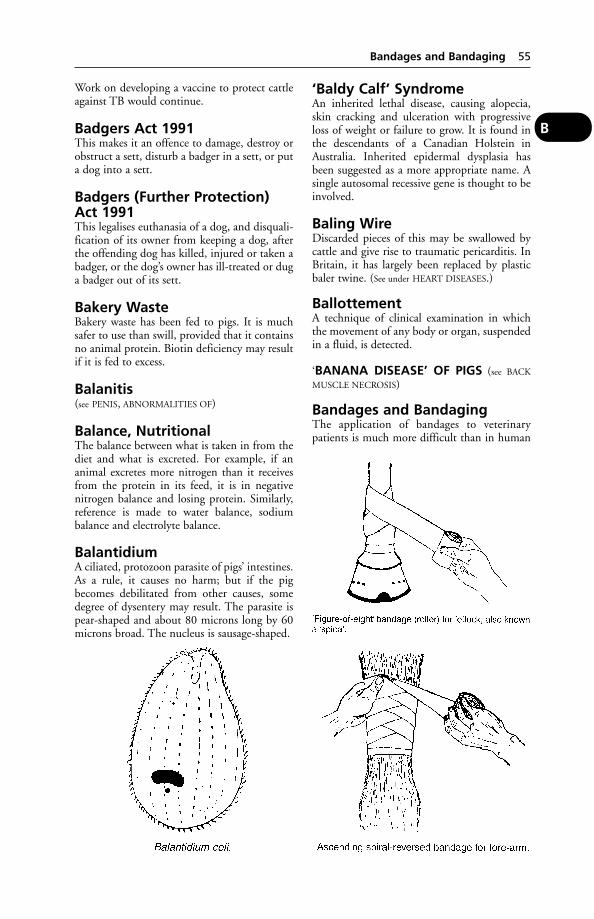
Work on developing a vaccine to protect cattleagainst TB would continue.
Badgers Act 1991This makes it an offence to damage, destroy orobstruct a sett, disturb a badger in a sett, or puta dog into a sett.
Badgers (Further Protection)Act 1991This legalises euthanasia of a dog, and disquali-fication of its owner from keeping a dog, afterthe offending dog has killed, injured or taken abadger, or the dog’s owner has ill-treated or duga badger out of its sett.
Bakery WasteBakery waste has been fed to pigs. It is muchsafer to use than swill, provided that it containsno animal protein. Biotin deficiency may resultif it is fed to excess.
Balanitis(see PENIS, ABNORMALITIES OF)
Balance, NutritionalThe balance between what is taken in from thediet and what is excreted. For example, if ananimal excretes more nitrogen than it receivesfrom the protein in its feed, it is in negativenitrogen balance and losing protein. Similarly,reference is made to water balance, sodium balance and electrolyte balance.
BalantidiumA ciliated, protozoon parasite of pigs’ intestines.As a rule, it causes no harm; but if the pigbecomes debilitated from other causes, somedegree of dysentery may result. The parasite ispear-shaped and about 80 microns long by 60microns broad. The nucleus is sausage-shaped.
‘Baldy Calf’ SyndromeAn inherited lethal disease, causing alopecia,skin cracking and ulceration with progressiveloss of weight or failure to grow. It is found inthe descendants of a Canadian Holstein inAustralia. Inherited epidermal dysplasia hasbeen suggested as a more appropriate name. Asingle autosomal recessive gene is thought to beinvolved.
Baling WireDiscarded pieces of this may be swallowed bycattle and give rise to traumatic pericarditis. InBritain, it has largely been replaced by plasticbaler twine. (See under HEART DISEASES.)
BallottementA technique of clinical examination in whichthe movement of any body or organ, suspendedin a fluid, is detected.
‘BANANA DISEASE’ OF PIGS (see BACK
MUSCLE NECROSIS)
Bandages and BandagingThe application of bandages to veterinarypatients is much more difficult than in human
Bandages and Bandaging 55
B
practice, because not only must the bandageremain in position during the movement ofthe patient, but it must also be comfortable, orit will be removed by the teeth or feet; and itmust be so adjusted that it will not becomecontaminated by either urine or the faeces.
Wounds often heal more readily if leftuncovered, but bandaging may be necessary togive protection against flies and the infectiveagents which these carry. Much will dependupon the site of the wound, its nature, and theenvironment of the animal.
Bandages may be needed for support, and toreduce tension on the skin. (See also illustration.)
BarbituratesBarbiturates are derivatives of barbituric acid(malonyl-urea). They include a wide range ofvery valuable sedative, hypnotic or anaestheticagents. Several are used in veterinary practice,including pentobarbitone, phenobarbitone andthiopentone. An overdose is often used toeuthanase dogs and cats; and farm animalswhere the brain is required for examination, asin suspected BSE cases.
In case of inadvertent barbiturate poisoning,use a stomach tube and keep the animal warm.Treatment includes CNS stimulants, e.g.bemegride, doxapram, caffeine or strong coffee.
(See also under EUTHANASIA; HORSE-MEAT.)
Barium-Meal Techniques inDogs and Cats(see under X-RAYS)
Barium PoisoningBarium chloride is used in rat poison; the baitmay be eaten by domestic pets.
The symptoms are excessive salivation,sweating (except in the dog), muscular convul-sions, violent straining, palpitation of the heart,and finally general paralysis.
Treatment Induce vomiting or use a stomachpump to remove the poison. Epsom salts dis-solved in water act as an antidote by convert-ing the chloride into the insoluble sulphate ofbarium.
Barium SulphateBarium sulphate, being opaque to X-rays, isgiven by the mouth prior to a radiographicexamination of the gastrointestinal tract fordiagnostic purposes. (See X-RAYS.)
Barium SulphideBarium sulphide is sometimes used as a depila-tory for the site of surgical operations.
BarkA change in the tone of a dog’s bark occurs inmany cases of rabies.
Bark EatingBark eating by cattle should be regarded as asymptom of a mineral deficiency, e.g. man-ganese and phosphorus. The remedy is use ofan appropriate mineral supplement.
Barker FoalA maladjustment syndrome in which a violentbreathing action results often in a noise like adog barking.
Barley PoisoningAs with wheat (and to a much lesser extent,oats) an excess of barley can kill cattle and sheepnot gradually accustomed to it. The main signsare severe acidosis and death. Treatment is sodi-um bicarbonate, by injection; gastric lavage; orrumenotomy.
It is important that barley should not be fedin a fine, powdery form. To do so is to invitesevere digestive upsets, which may lead todeath. Especially if ventilation is poor, dustyfood also contributes to coughing and mayincrease the risk of pneumonia.
‘Barn Itch’The American name for sarcoptic mange incattle.
Barrier CreamA protective dressing for the hands and armsof veterinarians engaged in obstetrical work orrectal examinations.
Barrier, Bood-BrainA filtering system to prevent harmful chemicalsin the bloodstream from reaching the brain.The system also prevents certain medicines,such as penicillin, from treating brain infectionssuch as bacterial meningitis. A similar barrier inthe placenta protects the fetus.
BarrowA castrated male pig.
Bars of FootAt each of the heels of the horse’s foot the wallturns inwards and forwards instead of endingabruptly. These ‘reflected’ portions are calledthe bars of the foot. They serve to strengthenthe heels; they provide a gradual rather than anabrupt finish to the important wall; and theytake a share in the formation of the bearingsurface, on which rests the shoe.
56 Barbiturates
B

The bars are sometimes cut away by farriersor others, who hold the erroneous idea thatby so doing they allow the heels of the footto expand; what actually happens in suchinstances is that the union between the compo-nent parts of the foot is destroyed, and the resis-tance to contraction which they afford is lost.They should therefore be allowed to grow andmaintain their natural prominence. (See also
illustration.)
BartonellosisInfection with Bartonella organisms, whichoccasionally occurs in dogs and cattle but is ofimportance in laboratory rats. Symptoms aremainly those of anaemia.
Treatment Neoarsphenamine has been used.
Basic SlagBasic slag is a by-product of the smelting indus-try often used as a fertiliser. It has causedpoisoning in lambs, which should not beallowed access to treated fields until the slaghas been well washed into the soil. Adult sheephave also been poisoned in this way, scouringbadly, and so have cattle. In these animals thesymptoms include: dullness, reluctance tomove, inappetence, grinding of the teeth, andprofuse watery black faeces.
BasenjiA small brown and white dog, originating inAfrica, which is unable to bark. Inheritablecongenital defects include haemolytic anaemia,inguinal hernia and persistent pupillary mem-brane. They may also inherit the conditionintestinal lymphangiectasia, which causes lossof protein from the gut. Basenji bitchesnormally have only one reproductive cycle a year.
Basset HoundA long-bodied, long-eared, short-legged breed.Ectropion, inguinal hernia and glaucoma maybe inherited conditions. Back problems caused
by cervical spondylosis may occur, and failure ofthe anconal process (elbow) to develop properlymay be seen.
BasophilA type of white blood cell. (See under BLOOD.)
BasophilicBlue-staining.
BathsBathing of animals may be undertaken forthe sake of cleanliness, for the cure of a para-sitic skin disease, or for the reduction of thetemperature.
Cattle and sheep (see DIPS AND DIPPING)
Dogs For ordinary purposes the dog is bathedin warm water, in which it is thoroughlysoaked. It is then lathered with a suitable sham-poo (many proprietary brands are available) orhard soap, rinsed off and dried. A wide range ofspecially formulated shampoos is available forspecific skin conditions.
Dish-washing detergent liquid should not beused for shampooing puppies or even adultdogs.
Cats Because cats are fastidious creatureswhich wash themselves nearly all over (theycannot reach the back of their necks or betweentheir shoulder blades), the question of bathingthem does not arise except in cases of a severeinfestation with external parasites; very old catswhich have ceased to wash themselves; entiretom cats which as a result of stress or illnesshave also ceased to look after themselves; as afirst-aid treatment for heat stroke/stress; and insome cases where a cat has fallen into a noxiousliquid.
Shampoos/flea-killers, etc. sold for use ondogs are not all safe for cats. Owners shouldread the small print on packets and look for‘Safe for cats’ where a preparation has not beenprescribed by a veterinary surgeon.
Baths are used to help the treatment of cer-tain muscle and joint problems. Sand bathsare essential for chinchillas to keep their coatsin good condition. (Poultry perform dustbathing – given the opportunity.)
Bats(see also RABIES; VAMPIRE-BATS; HISTOPLASMOSIS).Bats are mammals, and usually produce 1 off-spring in late spring or early summer. Fifteenspecies have been identified in Britain, wherethey are classified as protected creatures under
Bats 57
B
the Wildlife and Countryside Act 1981. Theycan live for up to 30 years.
Battery SystemA method of intensive egg production involv-ing keeping hens in cages with a sloping floor;1, 2, or up to 5 birds per cage. Feeding andwatering may be on the ‘cafeteria’ system, withfood containers moving on an endless belt, elec-trically driven. The eggs are usually collectedfrom racks at the front of the cages.
There have long been objections on welfaregrounds to current battery systems. Benefitsachieved in good examples of battery cage sys-tems (e.g. a smaller risk from parasites, goodaccess to food and water) may be out-weighed by their deficiencies (e.g. prevention ofnesting behaviour, perching, dust-bathing;bone weakness caused by lack of freedom tomove about).
In the EU, battery cages are to be phased outby 2011. From January 1, 2003 the permittedcage size was increased to allow a minimum of550 cm2 per hen and since that date no newcages could be installed. ‘Enriched cages’, oralternative non-cage systems, were specifiedfor new or replacement systems by January 1,2002. The ‘enriched cages’ have 750 cm2 spaceper hen and provide a nest, litter to allow peck-ing and scratching, and perches. The plansfor alternative non-cage systems are due to beintroduced by January 1, 2007.
‘Cage layer fatigue’, a form of leg paralysis, issometimes encountered in battery birds. Birdslet out of their cages on to a solid floor usuallyrecover. A bone-meal supplement may help.(See also INTENSIVE LIVESTOCK PRODUCTION;EGG YIELD.)
A battery rearing system has, in a somewhatdifferent form, been applied to pig rearing.
BCG VaccineBCG vaccine may be used for dogs and cats inBritain in households where a member of thehousehold has tuberculosis. The vaccine doesnot cover every species of Mycobacteriumtuberculosis, however. It cannot be used in cattleas it interfere with the tuberculin test, and hasproved unsuccessful in immunising badgers.It has been used in the treatment of equinesarcoid.
BeagleA breed of dog traditionally kept in packs.Behavioural problems may develop in solitaryanimals kept as pets. Inheritable conditionsinclude cleft palate, haemolytic anaemia,glaucoma and epilepsy.
Beak(see DE-BEAKING; SHOVEL BEAK)
BecquerelThe standard unit for measuring RADIATION.
Bedding and Bedding MaterialsWhenever animals are housed in buildings, itis both necessary and economical to providethem with some form of bedding material. Thereasons are as follows:
(1) All animals are able to rest more ade-quately in the recumbent position, and thetemptation to lie is materially increased by theprovision of some soft bedding upon whichthey may more comfortably repose than onthe uncovered floor. Indeed there are somewhich, in the event of the bedding being inad-equate, or when it becomes scraped away, willnot lie down at all.
(2) The provision of a sufficiency of somenon-conductor of heat (which is one of theessentials of a good bedding) minimises the riskof chills.
(3) The protection afforded to prominentbony surfaces – such as the point of the hip, thepoints of the elbow and hock, the stifles andknees, etc. – is important, and if neglected leadsto bruises and injuries of these parts.
(4) From the point of view of cleanliness,both of the shed or loose-box and of the ani-mal’s skin, the advantages of a plentiful supplyof bedding are obvious.
(5) In the case of sick animals, the supply andmanagement of the bedding can aid recovery.(See also SLATTED FLOORS.)
HorsesWheat straw Wheat straw undoubtedly
makes the best litter for either stall or loose-box.Its main disadvantage is its inflammability.
Wheat straw should be supplied loose or inhand-tied bundles for preference. Trussed orbaled straw has been pressed and has lost someof its resilience or elasticity in the process. Theindividual straws should be long and unbroken,and the natural resistive varnish-like coatingshould be still preserved in a sample. Thecolour should be yellowish or a golden white;it should be clean-looking and free from dusti-ness. Straw should be free from thistles andother weeds.
Wheat straw has a particular advantage inthat horses will not eat it unless kept very shortof hay.
Oat straw This straw is also very good forbedding purposes, but it possesses one or twodisadvantages when compared with wheat
58 Battery System
B

straw. The straw is considerably softer, moreeasily broken and compressible than wheat, andbeing sweet to the taste, horses eat it.
Barley straw is inferior to either of the preceding for these reasons: it is only about halfthe length; it is very soft and easily compressedand therefore does not last as long as oat orwheat; more of it is required to bed the same-sized stall; and it possesses numbers of awns.The awns of barley are sharp and brittle. They irritate the softer parts of the skin, causescratches, and sometimes penetrate the soft tissues of the udder, lips, nose, or the regionabout the tail.
Rye straw has the same advantages as wheat straw, but it is a little harder and rougher.
Peat-moss is quite a useful litter for horses. Itis recommended for town stables and for use onboard ship, or other forms of transport. A goodsample should not be powdery, but should con-sist of a matrix of fibres in which are entangledsmall lumps of pressed dry moss. It is veryabsorbent – taking up 6 or 8 times its ownweight of water. When it is used, the drainsshould be of the open or ‘surface’ variety orcovered drains should be covered with oldsacks, etc.
It should never be used in a loose-boxin which there is an animal suffering from anyrespiratory disease, on account of its dustynature.
Sand makes a fairly good bed when the sam-ple does not contain any stones, shells, or otherlarge particles. It is clean-looking, has a certainamount of scouring action on the coat, is coolin the summer, and comparatively easily man-aged. Sand should be obtained from a sand pitor the bed of a running stream; not fromthe sea-shore, because the latter is impregnatedwith salt, and likely to be licked by horses whenthey discover the salty taste of which they arevery fond. If this habit is acquired the particlesof sand that are eaten collect in the colon orcaecum of the horse and may set up a conditionknown as ‘sand colic’, which is often difficult toalleviate.
Ferns and bracken make a soft bed and areeasily managed, but they always look dirty anduntidy, do not last as long as straws, and arerather absorbent when stamped down. Withhorses that eat their bedding there is a risk ofbracken poisoning.
Cattle Wheat straw is the most satisfactory.Oat straw is used in parts where little or nowheat is grown. Barley straw is open to objec-tion as a litter for cows on account of its awns,which may irritate the soft skin of the perineal
region and of the udder. Sawdust has beenfound very convenient in cow cubicles, alsoshavings. Sand has been used on slippery floorsbelow straw bedding, when it affords a goodfoothold for the cows and prevents accidents.(See also DEEP LITTER.) Special rubber mats havebeen found practicable and economic for use incow cubicles. Shredded paper has been used forcattle (and also horses).
A disadvantage of sawdust is that its use has ledto coliform mastitis (sometimes fatal) in cattle.Sand may then be preferable.
In milk-fed calves, the ingestion ofpeat, sawdust or wood shavings may inducehypomagnesaemia.
Pigs Many materials are used for the pig, but probably none possesses advantages overwheat straw, unless in the case of farrowing or suckling sows. These should be littered with some very short bedding which will not become entangled round the feet of the little pigs, and will not irritate the udder of the mother. For this purpose chaff, shavings,and even hay may be used according tocircumstances.
Straw can make up for deficiencies in man-agement and buildings as nothing else can. Itserves the pig as a comfortable bed, as a blanketto burrow under, a plaything to avert boredom,and a source of roughage in meal-fed pigswhich can help obviate digestive upsets and atleast some of the scouring which reduces farm-ers’ profits. Straw can mitigate the effects ofpoor floor insulation, of draughts, and of cold;and in buildings without straw, ventilation (toquote David Sainsbury) becomes a much morecritical factor.
As a newborn piglet spends so much of itstime lying in direct contact with the floor ofits pen, much body-heat can be lost throughconduction. Depending on the type of floor,this effect can be large enough to affect thepiglet’s growth rate and be a potential threat toits survival.’ Providing straw can be equivalentto raising the ambient temperature from 10°to 18°C (50° to 64°F). Wooden and rubberfloors are not as effective as straw in reducingconductive heat loss.
Dogs and cats Dogs (and pigs) have died asa result of the use for bedding of shavings of thered African hardwood (Mansonia altissima),which affects nose, mouth, and the feet, as wellas the heart.
Fatal poisoning of cats has followed theuse of sawdust, from timber treated withpentachlorophenol, used as bedding.
Bedding and Bedding Materials 59
B

Hamsters Synthetic bedding materialsshould be avoided as they can cause injury.
Poultry (see LITTER, OLD)
Rabbits Peat-moss is recommended as it neu-tralises ammonia formed from urine; rabbitsare particularly susceptible to ammonia in theatmosphere.
Bedlington TerrierA small, soft-coated terrier with distinctivearched-back appearance. Together with someWest Highland white terriers, they are prone toinherited copper toxicosis. The breed is relative-ly intolerant of high copper levels in the dietand may develop cirrhosis of the liver as aresult. Zinc acetate has been used for treatment.Other inheritable conditions include brittlebones (osteogenesis imperfecta) and retinaldyspasia.
Bedsonia(see CHLAMYDIA)
Beef Breeds and CrossesThe native British beef breeds are the AberdeenAngus, Shorthorn Hereford, Devon, SouthDevon, Sussex, Galloway, Highland andLincoln Red. Continental breeds including theCharolais, Chianinas, Simmental, Limousin,Blonde d’Aquitaine, Gebvieh, Belgian Blueand Piedmontese have been imported for use inthe United Kingdom. The continental breedsare more muscular, have higher mature weightsand better performance than native beef breeds,the Meat and Livestock Commission hascommented.
The beef breeds are generally used as termi-nal sires on cows not required for breedingdairy herd replacements, and some beef crossheifers are used for suckler herd replacements.The cross-bred calves exhibit hybrid vigour andfetch a premium in the market over pure-breddairy calves.
(See also CATTLE, BREEDS OF)
Beef Cattle Husbandry in BritainAround 58 per cent of home-produced beef isderived from the dairy herd, partly from dairy-bred calves reared for beef and partly fromculled dairy cows. A further 34 per cent comesfrom the beef suckler herd.
Store systems Cattle are usually on 1 farmfor less than a year, typically a winter (yardfinished) or summer period (grass finished),
but sometimes as short a period as 3 months.Because only part of the production cycle takesplace on a single farm, the possibility for usinga wide range of technical inputs is limited. Theprofitability is dominated by the relationshipbetween buying and selling prices, and thesesystems are characterised by large year-to-yearfluctuations in margins. As a generalisation, thelonger the cattle are on the farm, the higherthe margin.
BeesHoney bees (Apis spp) represent one of theoldest forms of animal husbandry. Modernbeehives are designed so that the honey-filledcombs can be removed and replaced withoutdisturbing the main chamber. This also min-imises swarming. Bees are subject to several dis-eases of which VARROASIS is the most prevalent.The National Bee Unit, run by MAAF, providesadvice on bee health issues (National Bee Unit,Sand Hutton, York YO4 1LZ). (See also under
BITES, STINGS.)
Beet Tops(see POISONING – Fodder poisoning)
Beevbilde CattleBreeding is based on 54 per cent polled LincolnRed Blood, 40 per cent polled Beef Shorthorn,and 6 per cent Aberdeen Angus.
Behaviour ProblemsAntisocial, or inappropriate, behaviour in dogsand cats is an increasingly common problem.There are a number of possible causes, includ-ing genetic traits in particular breeds, hormon-ally triggered behaviour and intentional orunintentional mistreatment. The fact thatmany animals are left alone for long periodswhile their owners are at work can encouragemisbehaviour. The animal becomes distressedduring the periods of absence and may resortto urinating or defecating; or in the case ofdogs, chewing furniture. Then over-excitement,with uncontrolled barking and jumping, resultson the owner’s return. Aggressive behaviour topeople or other animals is another commonproblem. Conversely, a pet may become obses-sively attached to a single person, resenting anyshow of affection to that individual by another.While veterinary surgeons and ‘pet counsellors’can can offer advice on correcting unacceptablebehaviour, it is greatly to be preferred that theproblem is avoided in the first place.
When choosing a dog or cat, it is alwaysadvisable to see the puppy or kitten in its homeenvironment. A pup from a litter born to a
60 Bedlington Terrier
B
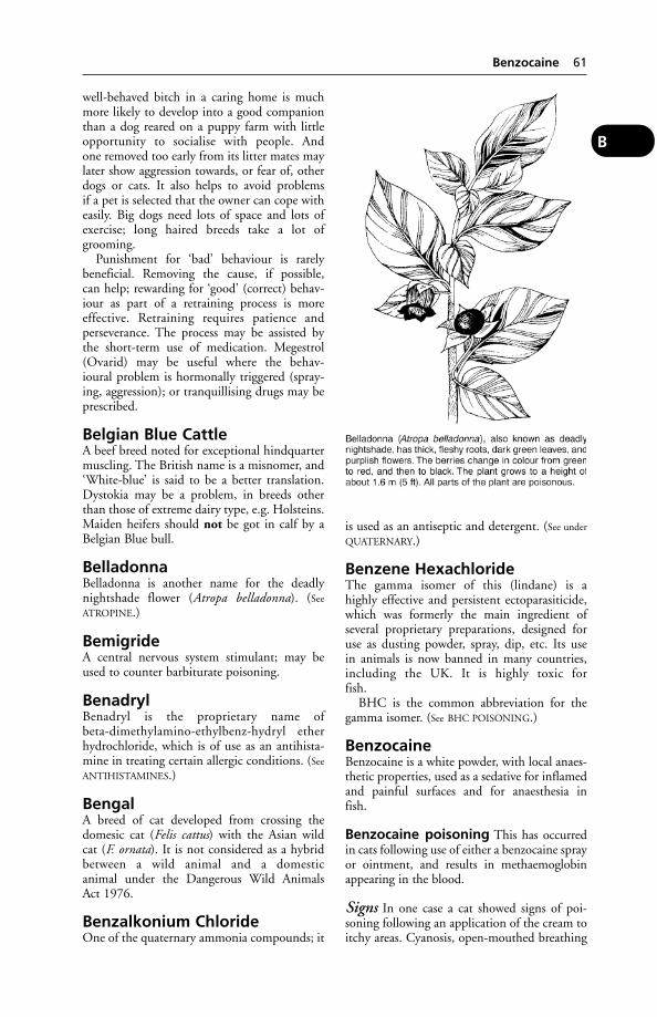
well-behaved bitch in a caring home is muchmore likely to develop into a good companionthan a dog reared on a puppy farm with littleopportunity to socialise with people. Andone removed too early from its litter mates maylater show aggression towards, or fear of, otherdogs or cats. It also helps to avoid problemsif a pet is selected that the owner can cope witheasily. Big dogs need lots of space and lots ofexercise; long haired breeds take a lot ofgrooming.
Punishment for ‘bad’ behaviour is rarelybeneficial. Removing the cause, if possible,can help; rewarding for ‘good’ (correct) behav-iour as part of a retraining process is moreeffective. Retraining requires patience andperseverance. The process may be assisted bythe short-term use of medication. Megestrol(Ovarid) may be useful where the behav-ioural problem is hormonally triggered (spray-ing, aggression); or tranquillising drugs may beprescribed.
Belgian Blue CattleA beef breed noted for exceptional hindquartermuscling. The British name is a misnomer, and‘White-blue’ is said to be a better translation.Dystokia may be a problem, in breeds otherthan those of extreme dairy type, e.g. Holsteins.Maiden heifers should not be got in calf by aBelgian Blue bull.
BelladonnaBelladonna is another name for the deadlynightshade flower (Atropa belladonna). (See
ATROPINE.)
BemigrideA central nervous system stimulant; may beused to counter barbiturate poisoning.
BenadrylBenadryl is the proprietary name ofbeta-dimethylamino-ethylbenz-hydryl etherhydrochloride, which is of use as an antihista-mine in treating certain allergic conditions. (See
ANTIHISTAMINES.)
BengalA breed of cat developed from crossing thedomesic cat (Felis cattus) with the Asian wildcat (F. ornata). It is not considered as a hybridbetween a wild animal and a domesticanimal under the Dangerous Wild AnimalsAct 1976.
Benzalkonium ChlorideOne of the quaternary ammonia compounds; it
is used as an antiseptic and detergent. (See under
QUATERNARY.)
Benzene HexachlorideThe gamma isomer of this (lindane) is a highly effective and persistent ectoparasiticide,which was formerly the main ingredient of several proprietary preparations, designed foruse as dusting powder, spray, dip, etc. Its use in animals is now banned in many countries,including the UK. It is highly toxic for fish.
BHC is the common abbreviation for thegamma isomer. (See BHC POISONING.)
BenzocaineBenzocaine is a white powder, with local anaes-thetic properties, used as a sedative for inflamedand painful surfaces and for anaesthesia infish.
Benzocaine poisoning This has occurredin cats following use of either a benzocaine sprayor ointment, and results in methaemoglobinappearing in the blood.
Signs In one case a cat showed signs of poi-soning following an application of the cream toitchy areas. Cyanosis, open-mouthed breathing
Benzocaine 61
B
and vomiting occurred. Collapse followedwithin 15 minutes.
Improvement was noticed within 10 minutesof giving methylene blue intravenously; andwithin 2 hours breathing had become normalagain. The cat recovered.
Benzoic AcidBenzoic acid is an antiseptic substance formerlyused for inflammatory conditions of the urinarysystem. It is excreted as hippuric acid, and ren-ders the urine acid. It is used in the treatment ofringworm, and as a food preservative.
Benzoic acid poisoning Cases of this havebeen reported in the cat, giving rise to extremeaggressiveness, salivation, convulsions, anddeath. A curious symptom sometimes observedis jumping backwards and striking out with thefore-limbs ‘as though catching imaginary mice’.
Benzyl BenzoateBenzyl benzoate is a drug formerly used fortreating mange in dogs and sweet itch in horses.A 25 per cent preparation may be applied tomite, etc., bites in pigeons.Benzyl benzoate is usually employed as anemulsion. It should not be used over the wholebody surface at once.
BenzylpenicillinThis antibiotic is a bactericide, active againstGram-positive bacteria, and given by parenteralor intramammary infusion. It is inactivated bypenicillinase.
Bephenium EmbonateA drug which is used in sheep to kill nematodirusworms.
Bernese Mountain DogA large, long-haired breed, mainly black withwhite and brown markings. It has few inheriteddefects, although cleft palate may occur. Alsoknown as Swiss mountain dog.
Berrichon Du CherA French breed of heavy milking sheep. Thebreed contains some merino blood.
BesnoitiosisA protozoan disease usually affecting theskin and mucous membranes; other effectsmay include sterility. Not normally found intemperate countries.
Beta-Blocker(see AGONIST)
Betahydroxybutyrate (BOHB)A ketone body which can be measured in bloodto determine the energy status. The higher thelevel, the poorer the energy intake.
BetamethasoneA corticosteroid.
BHCBHC is an abbreviation for BENZENE HEXA-
CHLORIDE.
BHC PoisoningThis may arise, especially in kittens and pup-pies, from a single dose (e.g. licking ofBHC-containing dusting powder). Symptomsinclude: twitching, muscular incoordination,anxiety, convulsions.
A farmer’s wife became ill (she had a convul-sion) after helping to dip calves, but recoveredafter treatment. Two of the calves died.
BHC is highly poisonous for fish; it must beused with great care on cats, for which otherinsecticides such as selenium preparations are tobe preferred.
The use of BHC sheep dips is no longerpermitted in the UK.
BHSBeta haemolytic streptococcus.
BicarbonateA salt containing HCO3; the amount in blooddetermines the acid/base balance. Sodiumbicarbonate is used as an antacid in ruminalacidosis.
‘Big Head’A condition associated with Clostridium novyi(type A) infection in rams which have slightlyinjured their heads as a result of fighting. Itoccurs in Australia and South Africa. (See also
HYDROCEPHALUS.)
BigheadTerm used to describe osteodystrophia fibrosain horses and goats.
BileBile is a thick, bitter, golden-brown or greenish-yellow fluid secreted by the liver, and stored inthe gall-bladder. It has digestive functions,assisting the emulsification of the fat contents ofthe food. It has in addition some laxative action,stimulating peristalsis, and it aids absorptionnot only of fats but also of fat-soluble vitamins.(See CHOLECYSTOKININ.)
62 Benzoic Acid
B
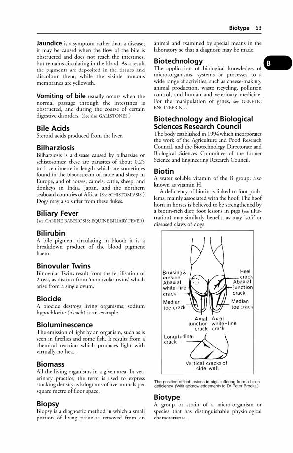
Jaundice is a symptom rather than a disease;it may be caused when the flow of the bile isobstructed and does not reach the intestines,but remains circulating in the blood. As a resultthe pigments are deposited in the tissues anddiscolour them, while the visible mucousmembranes are yellowish.
Vomiting of bile usually occurs when thenormal passage through the intestines isobstructed, and during the course of certaindigestive disorders. (See also GALLSTONES.)
Bile AcidsSteroid acids produced from the liver.
BilharziosisBilharziosis is a disease caused by bilharziae orschistosomes; these are parasites of about 0.25to 1 centimetre in length which are sometimesfound in the bloodstream of cattle and sheep inEurope, and of horses, camels, cattle, sheep, anddonkeys in India, Japan, and the northernseaboard countries of Africa. (See SCHISTOMIASIS.)Dogs may also suffer from these flukes.
Biliary Fever(see CANINE BABESIOSIS; EQUINE BILIARY FEVER)
BilirubinA bile pigment circulating in blood; it is abreakdown product of the blood pigmenthaem.
Binovular TwinsBinovular Twins result from the fertilisation of2 ova, as distinct from ‘monovular twins’ whicharise from a single ovum.
BiocideA biocide destroys living organisms; sodiumhypochlorite (bleach) is an example.
BioluminescenceThe emission of light by an organism, such as isseen in fireflies and some fish. It results from achemical reaction which produces light withvirtually no heat.
BiomassAll the living organisms in a given area. In vet-erinary practice, the term is used to expressstocking density as kilograms of live animals persquare metre of floor space.
BiopsyBiopsy is a diagnostic method in which a smallportion of living tissue is removed from an
animal and examined by special means in thelaboratory so that a diagnosis may be made.
BiotechnologyThe application of biological knowledge, ofmicro-organisms, systems or processes to a wide range of activities, such as cheese-making,animal production, waste recycling, pollutioncontrol, and human and veterinary medicine.For the manipulation of genes, see GENETIC
ENGINEERING.
Biotechnology and BiologicalSciences Research CouncilThe body established in 1994 which incorporatesthe work of the Agriculture and Food ResearchCouncil, and the Biotechnology Directorate andBiological Sciences Committee of the formerScience and Engineering Research Council.
BiotinA water soluble vitamin of the B group; alsoknown as vitamin H.
A deficiency of biotin is linked to foot prob-lems, mainly associated with the hoof. The hoofhorn in horses is believed to be strengthened bya biotin-rich diet; foot lesions in pigs (see illus-tration) may similarly benefit, as may ‘soft’ ordiseased claws of dogs.
BiotypeA group or strain of a micro-organism orspecies that has distinguishable physiologicalcharacteristics.
Biotype 63
B

Bird-Fancier’s LungPatients may be regarded as having bird-fancier’s lung if they satisfy all the followingcriteria: recent history of avian exposure; serumavian precipitins; diffuse shadowing on chestradiograph; a significant reduction (less than 70per cent predicted value) of carbon-monoxidetransfer factor (single breath); and improve-ment or no deterioration when exposure tobirds and their excreta is ceased.
In some cases there have been changes in theintestine (villous atrophy).
In the acute form, most often seen in pigeon-fanciers after cleaning the loft, influenza-likesymptoms, a shortness of breath and a coughoccur after 4 to 6 hours. The disease in elderlypatients has to be differentiated from bronchitisand emphysema.
Bird Import ControlsBird import controls were imposed in GreatBritain in 1976, and a licence is required forall imports of captive birds and hatching eggs.All birds except those from Belgium are subjectto a quarantine of 35 days. Birds importedinto the EU are subject to quarantine. (See also
PIGEONS.)
Bird LouseBird louse is a parasitic insect belonging to theorder Mallophaga, which attacks most domesti-cated and many wild birds. The lice eat feathersand the cells shed from the surface of theskin, but they do not suck blood. Dustingwith parasiticide powder is an efficient remedy.(See LICE.)
Bird MalariaA tropical disease of fowls and turkeys causedby Plasmodium gallinaceum, P. durae and otherspecies, transmitted by mosquitoes.
It may run a rapidly fatal course, or a chronicone with anaemia and greenish diarrhoea.
Birds(see under AVIAN; also CAGE BIRDS; GAME
BIRDS; TURKEYS; POULTRY; ORNITHOSIS; BOTU-
LISM; DUCK; FALCONS; PETS; RABIES; OSTRICH;RHEA.)
Birds, Blood SamplingThe toenail-clip method enables blood to becollected into a micro-haematocrit tube orpipette. The bird can be held with its backagainst the palm of the hand, head betweenthumb and forefinger.
Larger cage birds have easily accessible jugu-lar veins. In raptors, fowl and pigeons, the
brachial vein is favoured; the tarsal vein ispreferred for blood sampling in water fowl.
Birds, Humane Destruction ofFor poultry and other birds, a lidless woodenbox or chamber (of a size to take a polypropy-lene poultry crate) and a cylinder of carbondioxide with regulating valve are useful. Thebox has a 1.3-cm (1⁄2-in) copper pipe drilledwith 0.35-cm (9⁄64-in) holes at 10-cm (4-in) cen-tres fitted at levels 5 cm (2 in) and 66 cm(2 ft 2 in) from the bottom and connectedby plastic tubing to the regulator valve of thecylinder.
Birdsville DiseaseBirdsville disease occurs in parts of Australia, isdue to a poisonous plant Indigofera enneaphylla,and has to be differentiated from Kimberleyhorse disease.
Sings Sleepiness and abnormal gait with frontlegs lifted high. Chronic cases drag the hindlimbs.
Birth(see PARTURITION)
Bismuth (Bi)Bismuth (Bi) is one of the heavy metals.
Uses The carbonate, subnitrate, and thesalicylate may be used in irritable and painfulconditions of the stomach and intestines; alsoto relieve diarrhoea and vomiting.
The oxychloride and the subnitrate are usedlike barium, in bismuth meals prior to takingX-ray photographs of the abdominal organs forpurposes of diagnosis.
BistouryA surgical knife used to open up stenosed(closed up) teats, fistulae, sinuses; and abscesses.
Bites, Stings and PoisonedWoundsThe bites of animals, whether domesticated orotherwise, should always be looked upon asinfected WOUNDS. In countries where RABIES ispresent, the spread of this disease is generally bymeans of a bite.
TETANUS is always a hazard from bites.
Bees, wasps and hornets cause great irri-tation by the stings with which the females areprovided. Death has been reported in pigs eat-ing windfall apples in which wasps were feed-ing. The wasps stung the mucous membrane
64 Bird-Fancier’s Lung
B

of the throat, causing great swelling and deathfrom suffocation some hours later.
Antihistamine preparations may be usedin treatment if numerous stings make thisnecessary.
Cat-bites are usually followed by some degreeof suppuration. Pasteurella multocida infectionof the bite wound is common. (See also RABIES;CAT-SCRATCH FEVER.)
Dog-bites are usually inflicted upon otherdogs, defenceless sheep or goats, and sometimespigs; cattle may be bitten by the herd’s dog andserious wounds result. The bite is generally apunctured wound, or large tear, dependingupon the part that is bitten. Where an animal isbitten in numerous places, even though noindividual bite is large, there is always a consid-erable degree of danger. Antibiotics should begiven by injection. The wounds should bedressed with some suitable antiseptic, the hairor wool being first clipped from the area; andleft open. (See WOUNDS; RABIES.)
In the USA about a million dog-bites a yearrequire medical treatment of people; and in theUK the figure has been estimated as about99,000. Dog-bite wounds are often infected byPseudomonas species, Staphylococcus aureus,Streptococcus viridans, Pasteurella multocida, andGroup EF-4 bacteria.
Horse-bites Actinobacillosis has been trans-mitted to a bitten person.
Monkey-bites can transmit encephalitiscaused by Herpes simiae; human infectioushepatitis; also TB. (Lancet, 2, 553.)
Snake-bites (see SNAKES)
Spider-bites (see SPIDERS)
Bittersweet PoisoningThe common ‘bittersweet’ – Solanum dulcama-ra – is a frequent denizen of hedgerows andwaste lands, and, although not likely to be eatento a great extent by domesticated animals, casesof poisoning due to its ingestion have beenrecorded. All parts of the plant – stem, leaves,and berries – contain the toxic principle, whichis an alkaloid similar to Solanine found in thepotato.
Signs In cattle and sheep the symptoms aregiddiness, quickening of the respiration, stag-gering gait, dilated pupil, greenish diarrhoea,and raised temperature.
Black DiseaseBlack disease is the name given to infectiousnecrotic hepatitis of sheep and occasionally ofcattle in Australia, New Zealand, Scotland,Wales, and NW England. It is typically causedby a combined attack of immature liver flukesand bacteria, e.g. Clostridium oedematiens,which is one of the so-called ‘gas gangrene’group, and is capable of forming resistantspores.
On post-mortem examination the moststriking feature is the rapidity with which sheepdead from this disease have undergone decom-position. In carcases of sheep recently deador killed in the later stages, the skin is a darkbluish-black colour, and the underlying tissuesare congested and oedematous. In the liver,where the most constant lesions are found,there are one or more necrotic areas about2.5 cm (1 in) in diameter.
In cattle, black disease caused by Clostridiumnoyvi (Cl. oedematiens type B) may not beassociated with liver fluke.
Prevention An antiserum and a vaccine areavailable.
Black Disease 65
B

Black FaecesBlack faeces are passed when either iron or bis-muth salts are given to dogs and pigs. The mostserious cause of black motions is haemorrhageinto the early part of the digestive system. Adark-coloured diarrhoea may be seen in the dogsuffering from deficiency of the B vitamin.
Black-Leg(see BLACK-QUARTER)
Black-QuarterBlack-quarter, also called black-leg, quarter-ill,etc., is an acute specific infectious disease ofcattle, sometimes of sheep, and likewise of pigs,characterised by the presence of rapidly increas-ing swellings containing gas, and occurring inthe region of the shoulder, neck, thigh, quarter,and sometimes in the diaphragm. Young cattlebetween the ages of 3 months and 2 years aremost susceptible.
The disease has been seen in the reindeer,camel, and the buffalo.
Causes Clostridium chauvoei, which lives inthe soil until such time as it gains entry into theanimal body either along with the food or elseby abrasions of the skin (see TATTOOING).
On exposure to the air, the organisms formspores which are resistant to extreme cold, orheat.
Signs The finding of a dead animal may bethe first indication of the disease; though some-times lameness is observed, and part of theudder swollen and very painful. If seen in theearly stages, the swelling is hot and pits on pres-sure, but, increasing rapidly, it becomes puffedup with gas (emphysematous), and if pressedit crackles as if filled with screwed-up tissue-paper. Death usually occurs within 24 hours.Sheep show somewhat similar symptoms, butthey may be attacked at almost any age. Thereare often blood-stained discharges from boththe nostrils and the rectum.
Prevention Marshy ground that has beenresponsible for the loss of numerous animals inthe past has often been rendered safe by thedraining of the land and heavy liming.
Vaccine A vaccine gives very good results.Curative There is generally no opportunity to
treat cases, since death occurs after only afew hours’ illness; otherwise penicillin andantiserum may be tried.
‘Black Tongue’The counterpart of human pellagra. It is shownin the dog fed a diet deficient in nicotinic acid.
(See also SHEEPDOGS and ‘BROWN MOUTH’.)Symptoms include discoloration of the tongue,a foul odour from the mouth, ulceration, lossof appetite, and sometimes blood-stained salivaand faeces. Death will occur in the absence oftreatment.
Black VomitBlack vomit is due to the presence of blood inthe stomach. Either the appearance of thevomit may be that of black masses of clottedblood, or it may resemble coffee-grounds.
Black-Water FeverA form of babesiosis (see under BABESIA); alsoknown as TEXAS FEVER.
Blackhead of Turkeys(Histomoniasis)Blackhead of turkeys (histomoniasis) is a verycommon and fatal disease of young turkeys(from 3 weeks to 4 months old), which is causedby a small protozoon parasite, Histomonasmeleagridis, which passes part of its life in aworm (Heterakis gallinae); this acts as an inter-mediate host. The histomonas is found in adultworms and eggs; ingestion of the latter is thechief means of spread.
Though turkeys are chiefly affected, thedisease may be seen in chickens, partridges,pheasants, grouse, quail and pea-fowl.
Signs Loss of appetite and of condition. Thedroppings may be semi-liquid and bright yellow.Death, in 5 to 8 days, may occur in 70 to 90 percent of turkeys, in which the disease is very acuteand prevalent in summer and autumn.
Treatment Dinitridazole or nifursol, adminis-tered in the feed, may be used for preventionand treatment.
BladAbbreviation for BOVINE LEUKOCYTE ADHESION
DEFICIENCY.
Bladder, Diseases of(see under URINARY BLADDER, DISEASES OF; also
GALL-BLADDER)
BlastocystBlastocyst is the name given to a very early stagein the development of the fetus.
Blastomycosis of DogsInfection with Blastomyces dermatitidis.
The disease is fairly common in both manand dogs in North America. Diagnosis depends
66 Black Faeces
B
upon a laboratory demonstration of the fungus,which typically causes chronic debility oftenwith a fatal outcome.
Infection is usually through inhalation. Bonelesions, resulting in lameness, often occur;sometimes the brain, nose, eyes, and prostategland show lesions.
‘Bleeder Horses’Those which show blood at their nostrils afterhard exercise. (See also RACEHORSES – Pulmonaryhaemorrhage.)
Bleeding (Haemorrhage)Bleeding (haemorrhage) may be classifiedaccording to the vessel or vessels from which itescapes: e.g. (a) arterial, in which the blood is ofa bright scarlet colour and issues in jets orspurts corresponding in rate and rhythm to theheart-beats; (b) venous, when it comes fromveins, is of a dark colour, and wells up from thedepth of a wound in a steady stream; and (c)capillary, when it gradually oozes from a slightinjury to the network of capillaries of an area.(See also under CANINE HAEMOPHILIA; HAEMOR-
RHAGIC DIATHESIS; INTERNAL HAEMORRHAGE.)
Natural arrest When an artery with a smallcalibre is cut, the muscular fibres in its middlecoat shrink, and the cut end is slightly retractedwithin the stiffer fibrous covering. This resultsin a diminution in the size of the cut end andin a lessened capacity for output of blood. Inthe space between the end of the muscular coatand at the end of the fibrous coat a tiny clotcommences to form, which, later, is continuedinto the lumen of the vessel. This is added to byfurther coagulation of blood, until the wholeof the open end of the vessel and of the cavityof the wound is sealed by a clot. A fall in blood-pressure, due to shock and loss of blood,contributes to the natural arrest of bleeding.(See CLOTTING.)
Bleeding, external: first aid for Whena vein is cut, crimson blood will flow. From acut artery, scarlet blood will spurt, issuing injets corresponding with the heartbeats.
When a large vessel is cut, pressure should beapplied above the wound if the bleeding is froman artery, below it if bleeding is from a vein; butthe first-aider should take precautions (see
RESTRAINT).Pressure with the fingers is a helpful prelimi-
nary while someone else is finding material touse as a pressure pad. For large animals a cleanpillowslip, small towel, or piece of sheet willserve; for small animals a clean handkerchief
may suffice. The pad is then placed over thewound, and held there; pressure being appliedand maintained for a quarter of an hour.
Tourniquet Only if these measures fail tostop serious haemorrhage should a tourniquetbe used. A tourniquet can be improvised from arolled handkerchief, its two ends knotted,slipped around the limb, and tightened with apencil. Tightening must be just sufficient tostop the bleeding, no more. For large animals apiece of rubber tubing or a soft rope may beused. A tourniquet must never be left on formore than 20 minutes, or permanent dam-age to the limb will result. When releasing thetourniquet, do so gradually. A tourniquetshould not be used on cats, in which a pressurepad will suffice to control bleeding.
Professional help should be obtained assoon as possible.
Sometimes the actual point or points ofbleeding cannot be located, especially when thewound is deep or ragged, and the blood issuesin a more or less continuous stream showing notendency to clot. In such cases it is necessary toresort to packing the wound with GELATIN
SPONGE.Professional help will also be needed to
counter SHOCK. (See also BLOOD TRANSFUSIONS;DEXTRAN.)
Bleeding from special parts
(1) The Horns The horns of cattle are sometimes broken by falls or blows, and severebleeding follows. If the horn is broken com-pletely off, the haemorrhage is to the outside
Bleeding (Haemorrhage) 67
B
from the stump, but it often happens thatwhile the bony horn-core is fractured the hornitself holds the broken end in position, andthe escaping blood finds its way down intothe frontal sinus and out by the nostril.Haemorrhage from a stump may be controlledby the application of a pad and a bandage.Thermocautery, using a disbudding iron, mayassist.
(2) Legs and Feet The tourniquet describedabove may be applied, to the lower side of theinjury if the bleeding is venous, and above if itbe arterial. When the upper parts of the limbsare injured and the haemorrhage is consider-able, one of the methods of pressure is adopteduntil professional veterinary aid can beobtained.
(3) Stomach The vomiting of blood by dogs,cats, and pigs in considerable amounts is a veryserious symptom of severe injury or disease inthe stomach.
A dog may be offered ice cubes to lick. Theanimal should be kept as still as possible, andveterinary assistance obtained. Alcohol is notadvisable, as it causes a dilatation of the vesselsof the stomach wall and tends to promote thebleeding.
(4) Uterus and Vagina After parturition inall animals there is a certain risk of haemor-rhage, especially in those which have a diffuseplacenta, such as the mare and ass, and whenthe fetal membranes have been forciblyremoved. If it is copious, it may prove fatal.Prompt veterinary attention is necessary. (See also
under WOUNDS; INTERNAL HAEMORRHAGE.)
(5) Navel in piglets. See under VITAMINS –
Vitamin deficiencies for prevention.
Bleeding, internal (see INTERNAL HAEMOR-
RHAGE).
BlepharitisInflammation of the eyelids. It is usually associ-ated with conjunctivitis.
BlepharospasmBlepharospasm is a spasm of the eyelids.
Blindness(see under EYE, DISEASES OF; also VISION)
BloatAlso known as ruminal tympany, it occurs incattle, sheep, and goats. With the increased useof lucerne and clovers, bloat has become ofmore common occurrence among cattle and is
now a matter of serious economic importance.It may be of two types: free gas bloat or frothybloat.
Free gas bloat The rumen becomes dis-tended with gas, and pressure is exerted uponthe diaphragm.
The medium-sized cow’s rumen has a capac-ity of some 160 litres (35 gallons), and fermen-tation within it gives rise to bubbles of gas. Thiscomprises carbon dioxide (CO2) and methane(CH4) in surprisingly large quantities; cattleproducing as much as 800 litres of CO2 in 24hours, and as much as 500 litres of CH4. Someof this gas, perhaps a quarter, escapes via thebloodstream to the lungs and is breathed out,but that still leaves a great deal which can beexpelled only by belching. If something makesthat impossible, then gas pressure builds up andis exerted on the diaphragm, heart and lungs, sothat the cow is soon in considerable distress.
The cow’s ability to belch may be affectedby physical obstruction of the oesophagus;paralysis of the muscular wall of the rumen; andfoaming of the rumen contents.
The first diagram shows a healthy state ofaffairs in the rumen, with the cardia – a muscu-lar valve at the junction of oesophagus andrumen – temporarily open so that gas canescape up the oesophagus. But when this tube isobstructed by a piece of turnip or a tumour oran abscess, the gas cannot get away (or not insufficient quantity), and ‘gassy bloat’ results.Paralysis of the muscular wall of the rumen hasa similar effect, since expulsion of gas is aidedby contraction of these muscles.
The most common cause of gassy bloat isruminal acidosis following a barley diet, or incases of obstruction or dysfunction of theoesophageal or cardiac sphincter.
In such cases an antacid drench may be effec-tive, but passing a stomach tube, where this ispracticable, can provide immediate relief by therelease of trapped gas. Veterinary advice shouldbe sought.
In an emergency a RUMENOTOMY may beperformed or a trochar and cannula used.
Frothy bloat With the frothy type ofbloat, puncturing the rumen with a trocar andcannula in an emergency may do more harmthan good – not releasing gas and perhaps caus-ing leakage of some solids into the abdominalcavity.
This frothy type of bloat is the more impor-tant from an economic point of view, as itcan occur simultaneously in a number ofanimals, with a fatal outcome. The second
68 Blepharitis
B
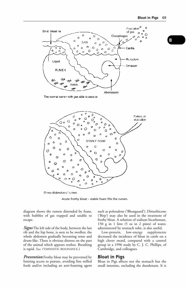
diagram shows the rumen distended by foam,with bubbles of gas trapped and unable toescape.
Signs The left side of the body, between the lastrib and the hip bone, is seen to be swollen; thewhole abdomen gradually becoming tense anddrum-like. There is obvious distress on the partof the animal which appears restless. Breathingis rapid. (See TYMPANITIC RESONANCE.)
Prevention Frothy bloat may be prevented bylimiting access to pasture, avoiding fine milledfeeds and/or including an anti-foaming agent
such as poloxalene (‘Bloatguard’). Dimethicone(‘Birp’) may also be used in the treatment offrothy bloat. A solution of sodium bicarbonate,150 g in 1 litre (5 oz in 2 pints) of water,administered by stomach tube, is also useful.
Low-protein, low-energy supplementsdecreased the incidence of bloat in cattle on ahigh clover sward, compared with a controlgroup in a 1996 study by C. J. C. Phillips, ofCambridge, and colleagues.
Bloat in PigsBloat in Pigs affects not the stomach but thesmall intestine, excluding the duodenum. It is
Bloat in Pigs 69
B
Acute frothy bloat – stable foam fills the rumen.
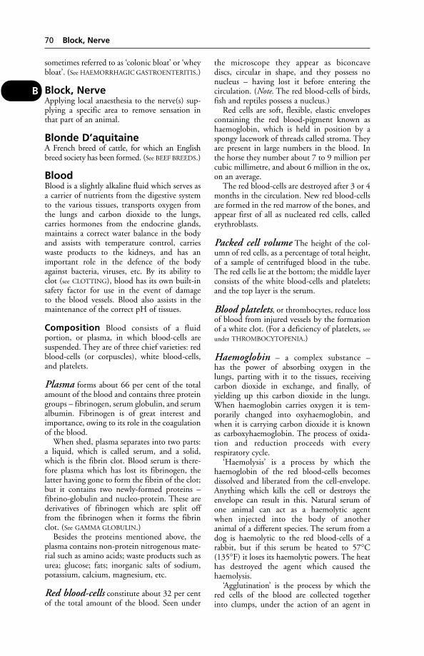
sometimes referred to as ‘colonic bloat’ or ‘wheybloat’. (See HAEMORRHAGIC GASTROENTERITIS.)
Block, NerveApplying local anaesthesia to the nerve(s) sup-plying a specific area to remove sensation inthat part of an animal.
Blonde D’aquitaineA French breed of cattle, for which an Englishbreed society has been formed. (See BEEF BREEDS.)
BloodBlood is a slightly alkaline fluid which serves asa carrier of nutrients from the digestive systemto the various tissues, transports oxygen fromthe lungs and carbon dioxide to the lungs,carries hormones from the endocrine glands,maintains a correct water balance in the bodyand assists with temperature control, carrieswaste products to the kidneys, and has animportant role in the defence of the bodyagainst bacteria, viruses, etc. By its ability toclot (see CLOTTING), blood has its own built-insafety factor for use in the event of damageto the blood vessels. Blood also assists in themaintenance of the correct pH of tissues.
Composition Blood consists of a fluidportion, or plasma, in which blood-cells aresuspended. They are of three chief varieties: redblood-cells (or corpuscles), white blood-cells,and platelets.
Plasma forms about 66 per cent of the totalamount of the blood and contains three proteingroups – fibrinogen, serum globulin, and serumalbumin. Fibrinogen is of great interest andimportance, owing to its role in the coagulationof the blood.
When shed, plasma separates into two parts:a liquid, which is called serum, and a solid,which is the fibrin clot. Blood serum is there-fore plasma which has lost its fibrinogen, thelatter having gone to form the fibrin of the clot;but it contains two newly-formed proteins –fibrino-globulin and nucleo-protein. These arederivatives of fibrinogen which are split offfrom the fibrinogen when it forms the fibrinclot. (See GAMMA GLOBULIN.)
Besides the proteins mentioned above, theplasma contains non-protein nitrogenous mate-rial such as amino acids; waste products such asurea; glucose; fats; inorganic salts of sodium,potassium, calcium, magnesium, etc.
Red blood-cells constitute about 32 per centof the total amount of the blood. Seen under
the microscope they appear as biconcavediscs, circular in shape, and they possess nonucleus – having lost it before entering thecirculation. (Note. The red blood-cells of birds,fish and reptiles possess a nucleus.)
Red cells are soft, flexible, elastic envelopescontaining the red blood-pigment known ashaemoglobin, which is held in position by aspongy lacework of threads called stroma. Theyare present in large numbers in the blood. Inthe horse they number about 7 to 9 million percubic millimetre, and about 6 million in the ox,on an average.
The red blood-cells are destroyed after 3 or 4months in the circulation. New red blood-cellsare formed in the red marrow of the bones, andappear first of all as nucleated red cells, callederythroblasts.
Packed cell volume The height of the col-umn of red cells, as a percentage of total height,of a sample of centrifuged blood in the tube.The red cells lie at the bottom; the middle layerconsists of the white blood-cells and platelets;and the top layer is the serum.
Blood platelets, or thrombocytes, reduce lossof blood from injured vessels by the formationof a white clot. (For a deficiency of platelets, see
under THROMBOCYTOPENIA.)
Haemoglobin – a complex substance – has the power of absorbing oxygen in the lungs, parting with it to the tissues, receivingcarbon dioxide in exchange, and finally, ofyielding up this carbon dioxide in the lungs.When haemoglobin carries oxygen it is tem-porarily changed into oxyhaemoglobin, andwhen it is carrying carbon dioxide it is knownas carboxyhaemoglobin. The process of oxida-tion and reduction proceeds with every respiratory cycle.
‘Haemolysis’ is a process by which thehaemoglobin of the red blood-cells becomesdissolved and liberated from the cell-envelope.Anything which kills the cell or destroys theenvelope can result in this. Natural serum ofone animal can act as a haemolytic agent when injected into the body of another animal of a different species. The serum from adog is haemolytic to the red blood-cells of a rabbit, but if this serum be heated to 57°C(135°F) it loses its haemolytic powers. The heathas destroyed the agent which caused thehaemolysis.
‘Agglutination’ is the process by which thered cells of the blood are collected together into clumps, under the action of an agent in
70 Block, Nerve
B
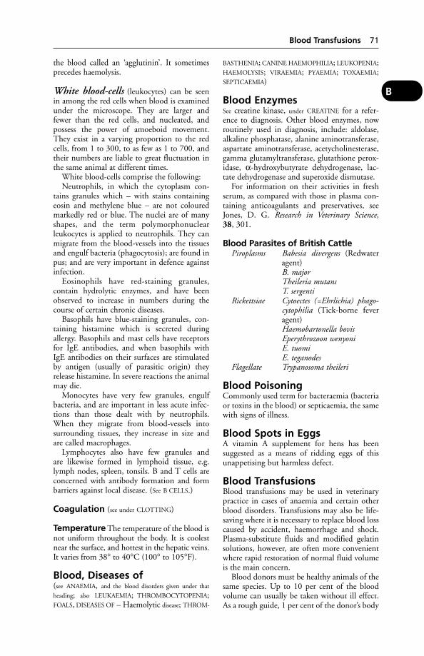
the blood called an ‘agglutinin’. It sometimesprecedes haemolysis.
White blood-cells (leukocytes) can be seenin among the red cells when blood is examinedunder the microscope. They are larger andfewer than the red cells, and nucleated, andpossess the power of amoeboid movement.They exist in a varying proportion to the redcells, from 1 to 300, to as few as 1 to 700, andtheir numbers are liable to great fluctuation inthe same animal at different times.
White blood-cells comprise the following:Neutrophils, in which the cytoplasm con-
tains granules which – with stains containingeosin and methylene blue – are not colouredmarkedly red or blue. The nuclei are of manyshapes, and the term polymorphonuclearleukocytes is applied to neutrophils. They canmigrate from the blood-vessels into the tissuesand engulf bacteria (phagocytosis); are found inpus; and are very important in defence againstinfection.
Eosinophils have red-staining granules,contain hydrolytic enzymes, and have beenobserved to increase in numbers during thecourse of certain chronic diseases.
Basophils have blue-staining granules, con-taining histamine which is secreted during allergy. Basophils and mast cells have receptorsfor IgE antibodies, and when basophils withIgE antibodies on their surfaces are stimulatedby antigen (usually of parasitic origin) theyrelease histamine. In severe reactions the animalmay die.
Monocytes have very few granules, engulfbacteria, and are important in less acute infec-tions than those dealt with by neutrophils.When they migrate from blood-vessels intosurrounding tissues, they increase in size andare called macrophages.
Lymphocytes also have few granules and are likewise formed in lymphoid tissue, e.g.lymph nodes, spleen, tonsils. B and T cells areconcerned with antibody formation and formbarriers against local disease. (See B CELLS.)
Coagulation (see under CLOTTING)
Temperature The temperature of the blood isnot uniform throughout the body. It is coolestnear the surface, and hottest in the hepatic veins.It varies from 38° to 40°C (100° to 105°F).
Blood, Diseases of(see ANAEMIA, and the blood disorders given under that
heading; also LEUKAEMIA; THROMBOCYTOPENIA;FOALS, DISEASES OF – Haemolytic disease; THROM-
BASTHENIA; CANINE HAEMOPHILIA; LEUKOPENIA;HAEMOLYSIS; VIRAEMIA; PYAEMIA; TOXAEMIA;SEPTICAEMIA)
Blood EnzymesSee creatine kinase, under CREATINE for a refer-ence to diagnosis. Other blood enzymes, nowroutinely used in diagnosis, include: aldolase,alkaline phosphatase, alanine aminotransferase,aspartate aminotransferase, acetycholinesterase,gamma glutamyltransferase, glutathione perox-idase, α-hydroxybutyrate dehydrogenase, lac-tate dehydrogenase and superoxide dismutase.
For information on their activities in freshserum, as compared with those in plasma con-taining anticoagulants and preservatives, seeJones, D. G. Research in Veterinary Science,38, 301.
Blood Parasites of British CattlePiroplasms Babesia divergens (Redwater
agent)B. majorTheileria mutansT. sergenti
Rickettsiae Cytoectes (=Ehrlichia) phago-cytophilia (Tick-borne feveragent)Haemobartonella bovisEperythrozoon wenyoniE. tuomiE. teganodes
Flagellate Trypanosoma theileri
Blood PoisoningCommonly used term for bacteraemia (bacteriaor toxins in the blood) or septicaemia, the samewith signs of illness.
Blood Spots in EggsA vitamin A supplement for hens has beensuggested as a means of ridding eggs of thisunappetising but harmless defect.
Blood TransfusionsBlood transfusions may be used in veterinarypractice in cases of anaemia and certain otherblood disorders. Transfusions may also be life-saving where it is necessary to replace blood losscaused by accident, haemorrhage and shock.Plasma-substitute fluids and modified gelatinsolutions, however, are often more convenientwhere rapid restoration of normal fluid volumeis the main concern.
Blood donors must be healthy animals of thesame species. Up to 10 per cent of the bloodvolume can usually be taken without ill effect.As a rough guide, 1 per cent of the donor’s body
Blood Transfusions 71
B

weight (300 ml for a 30 kg dog) may be taken.In dogs, which have 8 blood groups, adversereactions due to incompatible blood types arerarely seen. Cats have 3 blood types: A and B,in the ratio 3:1, and AB (less than 1 per cent).Matching of donor and recipient blood shouldbe done before transfusion, if possible.
Blood is conveniently collected from thejugular vein. Donors should be sedated and theskin in the area shaved and cleaned. A hypoder-mic needle or catheter is inserted and bloodcollected into a blood bag (dog) or 50 mlsyringe (cat) containing an anticoagulant suchas sodium citrate or acid citrate dextrose.
72 Blood Transfusions
B

Collected blood may be stored for up to 4weeks if refrigerated; it should be warmed tobody heat before administration. This shouldbe via a jugular or cephalic vein.
In cattle, donor and recipient are usually in the same herd, a fact which lessens the risk ofintroducing infection. Blood is collected fromthe jugular or other vein (after the skin has been cleaned and precautions taken to ensureasepsis) by means of a suitable needle (e.g.13swg) and allowed to flow into a blood bag or sterilised bottle containing anticoagulant.This may be made by dissolving 60 mg of sodi-um citrate in a little water, for every 100 mlblood collected. The bottle should be shakengently during collection. The donor’s blood is then transferred to the recipient’s vein.Transfusion reactions rarely occur during a firsttransfusion.
In the new-born foal suffering fromhaemolytic disease, exchange transfusion hasbeen the means of saving life. Up to 5500 ml ofthe foal’s blood is removed and replaced by upto 7000 ml of compatible donor’s blood.The process takes up to 3 hours and requiresspecial apparatus. See also DEXTRAN; GELATIN,SUCCINYLATED; also under FELINE INFECTIOUS
ENTERITIS; DEHYDRATION.
Blood Typing, CattleIn Canada extensive use is made of blood typ-ing in respect of cattle, and results of a bloodtest have been accepted as evidence in court ina case where a man was convicted of falsifying apedigree. The basis of this evidence was that toprove parentage of an animal, all the factorsfound in the blood of a calf must be presentin the blood of either the sire or the dam. Ifcertain factors found in the blood of the calfcould not be found in the blood of either thesire or the dam, then that calf could not havebeen of that particular mating – as was provedin this case.
Blood typing is also used in the diagnosis offreemartins. In one series 228 freemartins werefound out of 242 sets of twins.
Blood typing has been used to decide thepaternity issue in a heifer calf born to a cowinseminated twice in the same heat period withsemen from two different bulls; to reveal dis-crepancies in pedigrees; and to allay or confirmsuspicion on the part of a Breed Society askedto register a calf born following a very short ora very long gestation period.
The work falls into two categories: commer-cial and research. In the former category thereare routine pedigree parentage cases involvingone bull, one cow, and one calf. In a series of
403 such cases, 26 (or 6.5 per cent) were foundto be incorrect. Checking the parentage of bullsto be used in AI (see ARTIFICIAL INSEMINATION)as well as typing bulls being used in AI is car-ried out. Other applications include the diag-nosis of freemartins, and the control of eggtransplantation – i.e. checking that the off-spring is from the egg put in and not from thehost cow’s own egg.
Blood typing is of service in the policing ofscreening tests, e.g. for brucellosis. It is notunknown for lazy or unscrupulous people to fillseveral sample tubes with blood from the sameanimal and label them as coming from severalanimals. If several tubes are found to have iden-tical types, fraud is virtually certain to haveoccurred, since the likelihood of two samples,other than from identical twins, having thesame blood type is negligible.
Thoroughbred horses must all be blood-typed as an aid to identification. (See also EQUINE
BLOOD TYPING.)
The Preparation of Test Sera containingantibodies, or blood-group reagents, is based on the injection of blood corpuscles from oneanimal into another of the same species, or into one of a different species. The first proce-dure is called iso-immunisation, the second hetero-immunisation. As a result of both proce-dures, the recipient animal produces antibod-ies to the antigenic factors associated with thedonor blood corpuscles, provided that thesefactors are not already present in the recipientanimal. (No animal can produce both an antigen and its antibody.) The diagram demon-strates the principle of iso-immunisation in cattle.
It shows that the donor possesses blood-group factors A, B, and C while the recipienthas only blood-group factor A. On immunisa-tion, the recipient will therefore form anti-bodies to blood group factors B and C. Theantibodies thus formed are called anti-B andanti-C. A serum containing several blood groupantibodies is known as a crude serum. Thisserum will react with red corpuscles not onlyfrom the donor, but also from all cattle withthe blood group factor B or C.
To obtain a blood group reagent which reactswith only one blood group factor – for exampleB – the anti-C antibody must be removed. Todo this, the prepared crude serum is mixed with blood corpuscles which are C-positive but B-negative. The anti-C is then bound to the blood corpuscles and can be removed bycentrifuging, as illustrated. This procedure is called antibody absorption. As the figure
Blood Typing, Cattle 73
B
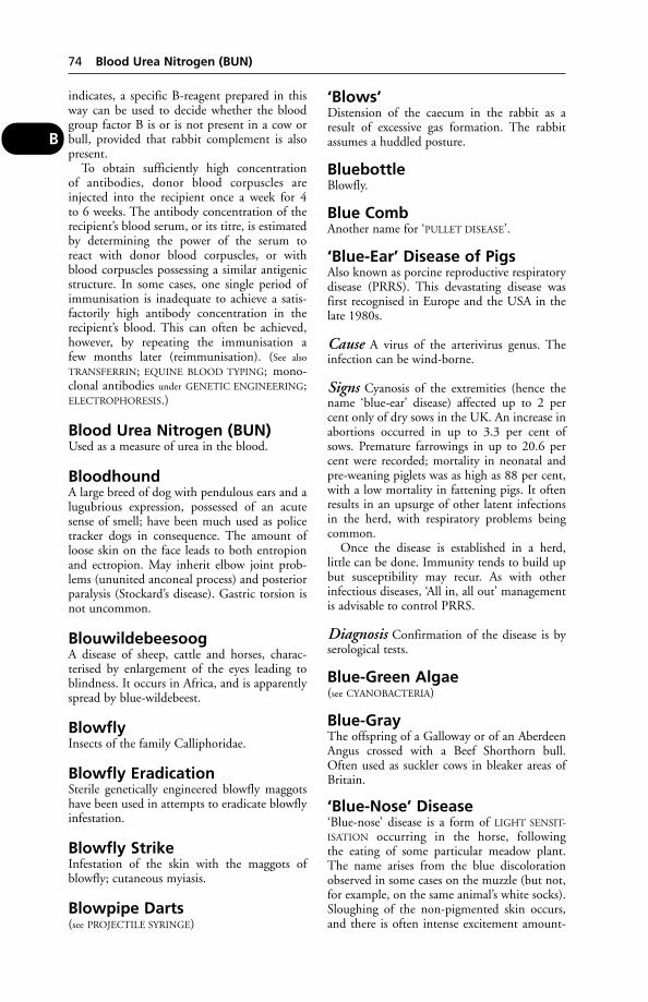
indicates, a specific B-reagent prepared in thisway can be used to decide whether the bloodgroup factor B is or is not present in a cow orbull, provided that rabbit complement is alsopresent.
To obtain sufficiently high concentration of antibodies, donor blood corpuscles areinjected into the recipient once a week for 4 to 6 weeks. The antibody concentration of therecipient’s blood serum, or its titre, is estimatedby determining the power of the serum to react with donor blood corpuscles, or withblood corpuscles possessing a similar antigenicstructure. In some cases, one single period ofimmunisation is inadequate to achieve a satis-factorily high antibody concentration in therecipient’s blood. This can often be achieved,however, by repeating the immunisation a few months later (reimmunisation). (See also
TRANSFERRIN; EQUINE BLOOD TYPING; mono-clonal antibodies under GENETIC ENGINEERING;ELECTROPHORESIS.)
Blood Urea Nitrogen (BUN)Used as a measure of urea in the blood.
BloodhoundA large breed of dog with pendulous ears and alugubrious expression, possessed of an acutesense of smell; have been much used as policetracker dogs in consequence. The amount ofloose skin on the face leads to both entropionand ectropion. May inherit elbow joint prob-lems (ununited anconeal process) and posteriorparalysis (Stockard’s disease). Gastric torsion isnot uncommon.
BlouwildebeesoogA disease of sheep, cattle and horses, charac-terised by enlargement of the eyes leading toblindness. It occurs in Africa, and is apparentlyspread by blue-wildebeest.
BlowflyInsects of the family Calliphoridae.
Blowfly EradicationSterile genetically engineered blowfly maggotshave been used in attempts to eradicate blowflyinfestation.
Blowfly StrikeInfestation of the skin with the maggots ofblowfly; cutaneous myiasis.
Blowpipe Darts(see PROJECTILE SYRINGE)
‘Blows’Distension of the caecum in the rabbit as aresult of excessive gas formation. The rabbitassumes a huddled posture.
BluebottleBlowfly.
Blue CombAnother name for ‘PULLET DISEASE’.
‘Blue-Ear’ Disease of PigsAlso known as porcine reproductive respiratorydisease (PRRS). This devastating disease wasfirst recognised in Europe and the USA in thelate 1980s.
Cause A virus of the arterivirus genus. Theinfection can be wind-borne.
Signs Cyanosis of the extremities (hence thename ‘blue-ear’ disease) affected up to 2 percent only of dry sows in the UK. An increase inabortions occurred in up to 3.3 per cent ofsows. Premature farrowings in up to 20.6 percent were recorded; mortality in neonatal andpre-weaning piglets was as high as 88 per cent,with a low mortality in fattening pigs. It oftenresults in an upsurge of other latent infectionsin the herd, with respiratory problems beingcommon.
Once the disease is established in a herd, little can be done. Immunity tends to build upbut susceptibility may recur. As with otherinfectious diseases, ‘All in, all out’ managementis advisable to control PRRS.
Diagnosis Confirmation of the disease is byserological tests.
Blue-Green Algae(see CYANOBACTERIA)
Blue-GrayThe offspring of a Galloway or of an AberdeenAngus crossed with a Beef Shorthorn bull.Often used as suckler cows in bleaker areas ofBritain.
‘Blue-Nose’ Disease‘Blue-nose’ disease is a form of LIGHT SENSIT-
ISATION occurring in the horse, followingthe eating of some particular meadow plant.The name arises from the blue discolorationobserved in some cases on the muzzle (but not,for example, on the same animal’s white socks).Sloughing of the non-pigmented skin occurs,and there is often intense excitement amount-
74 Blood Urea Nitrogen (BUN)
B
ing to frenzy – during which the horse mayinjure itself. (See also ANTIHISTAMINES.)
BluetongueA viral disease of ruminants confined mainly toAfrica but which has spread to North Americaand Australia, Portugal, Spain, and Cyprus and,more recently, Italy and France. Bluetongue is aNOTIFIABLE DISEASE throughout the EU.
Infection is carried by biting midges andprobably the mosquito, and consequently out-breaks are commonest near the breeding hauntsof such insects – damp, marshy regions.
Cattle may be symptomless carriers. A surveyof 6250 sera from cattle, sheep and goats inseven Caribbean and two South Americancountries showed that antibody to bluetonguevirus was widely distributed. Overall preva-lences of antibody were 70 per cent in cattle,67 per cent in sheep, and 76 per cent in goats.Yet, no clinical cases had been confirmed inthe area; no virus isolates were available to indi-cate which serotype(s) was/were causing theinfection.
To prevent entry of bluetongue to the EU from Canada, cattle must have a negativeblood test in January; they can then be export-ed to the EU if they leave Canada betweenFebruary 1 and April 15. This procedureensures that they were not infected the previ-ous summer and move out of the countrybefore the midges carrying the infectionbecome active.
Cause An orbivirus.
Signs In sheep, a rise in temperature up to41.5° C (107°F), and after a week or 10 days,eruptions on the tongue, lips, and dentalpads – with a swelling and blueness of theseparts – mark the typical appearance of anattack. Both the mouth and nose show a dis-charge, and there is an accompanying smackingof the lips. In spite of the soreness of the mouththe sheep are inclined to feed, but loss of fleshis very rapid, particularly when diarrhoea setsin. In 3 to 5 days, the mouth lesions begin toheal, and the disease is seen in the feet. Thesebecome sore; sheep are stiff, and feed from thekneeling or recumbent positions. Diagnosismay be confirmed by viral isolation.
In both cattle and sheep the disease may besubclinical.
Treatment Isolation of the affected animalsinto shady paddocks, sheds, or orchards, where they are immune from disturbance, anti-septic mouth washes, good feeding of a soft,
succulent quality, the provision of a cleanwater-supply and salt-licks. Dipping has givengood results.
Prevention A stockpile of quadrivalent vaccineis stored at various sites in the EU.
Boarding Kennels(see ANIMAL BOARDING ESTABLISHMENTS ACT)
Body-Scanner(see under X-RAYS)
Bog Asphodel(Narthecium ossifragum) A cause of light sensiti-sation in sheep. Ears, face, and legs of whitelambs may all be affected. Skin necrosismay follow the inflammation. In severe cases,jaundice may be a complication.
Cows have been fatally poisoned by the plantas a result of necrosis of kidney tissue.
In one case, cattle forced by drought to grazeswampy ground where bog asphodel grew suf-fered 137 deaths out of 232 cattle affected. Theclinical signs included depression, anorexiaand diarrhoea. Extensive kidney damage wascaused.
Bog SpavinAn old name for chronic synovitis of the hock(tarsus) of horses. It often shows as a swellingof the front of the hock joint caused by fluid.It seldom causes lameness. (See also BONE
SPAVIN.)
BOHB(see BETAHYDROXYBUTYRATE)
Bollinger, Bodies(see FOWL POX)
BolusA roughly spherical mass of food, which hasbeen chewed and mixed with saliva, ready forswallowing. Bolus also means a cylindricalmass, 3.8 to 7.5 cm (11⁄2 to 3 in) long, and up to 1.3 cm (1⁄2 in) in diameter, of a medicine inpaste or solid form for administration to horsesand cattle. It is also known as a ‘ball’ – hence‘balling gun’. Slow-release boluses which areretained in the rumen for the administration ofanthelmintics or trace elements over a periodare available in a variety of forms.
A bolus of slow dissolving soluble glass con-taining copper, selenium and cobalt for traceelement supplementation in cattle and sheep isalso available.
Bolus 75
B
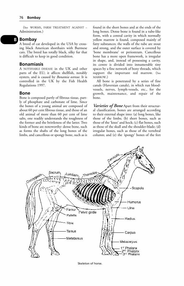
(See WORMS, FARM TREATMENT AGAINST –
Administration.)
BombayA breed of cat developed in the USA by cross-ing black American shorthairs with Burmesecats. The breed has totally black, silky fur thatis difficult to keep in good condition.
BonamiasisA NOTIFIABLE DISEASE in the UK and otherparts of the EU; it affects shellfish, notablyoysters, and is caused by Bonamia ostreae. It iscontrolled in the UK by the Fish HealthRegulations 1997.
BoneBone is composed partly of fibrous tissue, part-ly of phosphate and carbonate of lime. Sincethe bones of a young animal are composed ofabout 60 per cent fibrous tissue, and those of anold animal of more than 60 per cent of limesalts, one readily understands the toughness ofthe former and the brittleness of the latter. Twokinds of bone are noteworthy: dense bone, suchas forms the shafts of the long bones of thelimbs, and cancellous or spongy bone, such as is
found in the short bones and at the ends of thelong bones. Dense bone is found in a tube-likeform, with a central cavity in which normallyyellow marrow is found, composed mainly offatty substances; the walls of the tube are stoutand strong, and the outer surface is covered by‘bone membrane’ or periosteum. Cancellousbone has a more open framework, is irregularin shape, and, instead of possessing a cavity,its centre is divided into innumerable tinyspaces by a fine network of bony threads, whichsupport the important red marrow. (See
MARROW.)All bone is penetrated by a series of fine
canals (Haversian canals), in which run blood-vessels, nerves, lymph-vessels, etc., for thegrowth, maintenance, and repair of the bone.
Varieties of Bone Apart from their structur-al classification, bones are arranged accordingto their external shape into: (a) long bones, likethose of the limbs; (b) short bones, such asthose of the ‘knee’ and hock; (c) flat bones, suchas those of the skull and the shoulder-blade; (d)irregular bones, such as those of the vertebralcolumn; and (e) the ‘spongy’ bones of the feet
76 Bombay
B
Skeleton of horse.
of horses and cattle and the claws of otheranimals.
The Skeleton has: (1) an axial part, consistingof the skull, the vertebrae, the ribs with theircartilages, and the sternum or breast-bone; and(2) an appendicular portion, consisting of thefour limbs. In addition to these divisions, cer-tain parts of the skeleton are embedded inthe substance of organs, and are described asthe visceral skeleton, e.g. the bones in the heartof the ox, the snout of the pig, the penis of thedog, etc.
Bone, Diseases of
Growth-plate disorders The growth-plateis a layer of cartilage between the diaphysis andthe epiphysis of a long bone. Failure of chon-drogenesis leading to cessation of growth iscommonly the result of trauma to the plate,occasioned by a fracture or crush injury, or ofthe interruption of the vascular supply to thegerminal cells. Diseases such as scurvy, rickets,osteomyelitis and endocrine disorders make theplate more vulnerable to injury and predisposeto epiphyseal separation.
Epiphyseal injury in the foal, for example,may be of two types:Type 1 Separation without fracture. Afterrealignment healing is rapid and the prog-nosis is good. Femoral head detachment is theexception because if the epiphyseal vessels aredamaged, avascular necrosis of the head follows.Type 2 The most common, involving fractureof a triangular piece of metaphysis. With accu-rate reduction the prognosis is good.
Such injuries usually involve the epiphyses inthe distal radius, distal metacarpus and proxi-mal first phalanx. These cases often have theappearance of acute joint sprains but epiphysealdamage should always be suspected becauseat this age growth-plates are weaker thancollateral ligaments. Radiography is essential toidentify the type of defect present.
Prompt and accurate replacement of the epi-physis is required followed by external supportwith a cast or splint to maintain alignment. Incertain instances fixation of the fragmentswith a compression screw may offer greatersecurity. (Professor L. C. Vaughan.) (See VALGUS
for picture.)
Acute inflammation of bone is dividedinto acute periostitis, or inflammation of thesurface of the bone and its covering membrane,the periosteum; acute osteitis or ostitis, inflam-mation of the bone substance itself; and acute
osteomyelitis, inflammation in the bone andthe central marrow cavity.
Acute inflammation of the bone surfacealmost always results from external violence.Osteomyelitis is usually due to bacteria gainingaccess either through the blood- or lymph-streams, or through the broken tissues resultingfrom a deep wound. The mildest types areoften due to an inflammation in a ligament ortendon spreading to the periosteum in the nearvicinity and causing it to become inflamed as aconsequence. (See also RHEUMATISM.)
Complete rest is essential: in fact work, oreven walking, is often impossible. Hot fomen-tations, poultices, soothing and cooling lini-ments or applications are usually sufficienttreatment for mild cases. The severer cases, inwhich infection has reached the bone, call inthe first place for antibiotics, or immediateopening up of the area and the eliminationof any pus that has collected. After that, anypieces of dead bone that are present areremoved, and the wound treated as an infectedopen wound.
Chronic inflammation may result fromseveral conditions, e.g. tuberculosis, actinomy-cosis, etc. Generally speaking, when a chronicsuppurative inflammation affects a bone,sooner or later the pus and debris of a liquidnature will burrow through the surroundingtissues and burst on to the surface of theskin.A discharging sinus results which provesintractable to treatment. At the bottom of thissinus lies the dead piece of bone, and untilit has been removed or absorbed the leakageof purulent material will continue in spiteof antiseptic injections and other surfacetreatment.
The offending dead portion of bone must beremoved in the first instance, and the wholesinus tract must be laid open. This is not alwaysan easy matter, and much depends upon the sit-uation of the pieces of dead bone (sequestrum),as well as of the mouth of the sinus. The areaafterwards is treated as an open wound, andif all the necrotic parts have been removed,recovery generally takes place.
Exostosis is an outgrowth of rarefied bonetissue upon the surface of a bone. Amongthe commonest forms of exostoses are thefollowing: certain forms of splints, ring-bones,bone spavins, some side-bones. (See also
ACROPACHIA.)
Tumours of bone are sometimes met with.The commonest of these is the osteosarcoma of
Bone, Diseases of 77
B

the limb-bones of dogs. The bony tissue isinvaded by the cells that are characteristic of thetumour, and there is swelling and pain.
Rickets is a disease of young animals in whichthe bones of the limbs are affected, and oftensmall pealike swellings are found at the junctionof each of the ribs with its cartilage. (See also
main entry for RICKETS.)
Osteomalacia is the equivalent of ricketsoccurring in the adult animal, especially during pregnancy. (See also main entry forOSTEOMALACIA.)
Osteodystrophic diseases are due to anincorrect calcium: phosphorus ratio in the diet,or to lack of one or more of the minerals – cal-cium, phosphorus, magnesium, and sometimesmanganese; also, if vitamin D has been inade-quate in amount in the food eaten for someconsiderable time (e.g. see FELINE JUVENILE
OSTEODYSTROPHY). (See also OSTEODYSTROPHIC
DISEASES.)
Porphyria is a rare disease, hereditary in ori-gin, occurring in man, cattle, and pigs. It ischaracterised by brown or pinkish discolorationof the bones and teeth, and by changes in theurine. In cattle a hairless, scabby conditionof the skin is also a symptom. (See also under
HEXACHLOROBENZENE.)
Bone Grafts(see – FRACTURES)
Bone Marrow(see MARROW; ANAEMIA; BLOOD – Red blood-cells)
Bone-PinningA method of treating FRACTURES. In medullarypinning, a pointed stainless steel ‘pin’ is drivendown the marrow cavity of the bone concerned;in ordinary pinning, transverse ‘pins’ are used,driven through the bone at right-angles to itslength, and the pins held in position by a spe-cial adjustable metal splint. These methodsobviate the use of cumbersome plaster casts, andthey also enable cases of serious and multiplefracture (e.g. as caused in a dog or cat knockeddown by a car) to be successfully treated – aresult often impossible of achievement by oldermethods. These techniques require a highdegree of specialised skill and strict asepsis and, of course, the use of a general anaesthetic.(See also bone-plating in the illustration; andEXTERNAL FIXATORS.)
Bone SpavinOsteitis or arthritis of the lower hock- joints ofthe horse. The animal is often lame; there mayor may not be a hard, bony swelling.
BonhamAn Irish piglet.
BoranAn East African type of Zebu cattle.
Border CollieA medium-sized, longish-haired dog originallybred for herding sheep. It is said that one well-trained sheepdog can do the work of 7 men.The breed is susceptible to COLLIE EYE ANOM-
ALY (choroidal hypoplasia). Inherited deafnessmay be linked to coat colour. Osteochondritisdissecans may also be inherited.
‘Border Disease’ of SheepA disease occurring on the English–Welshborder, and first described in 1959. Affectedlambs are known as ‘hairy shakers’.
Cause The virus which causes the disease inlambs is classified as belonging to the familyTogaviridae, genus Pestivirus.
Signs The birth-coat is altered; the amount ofhair in the fleece being increased. Lambs are
78 Bone Grafts
B
The insertion of a metal pin down the marrowcavity of the bone.
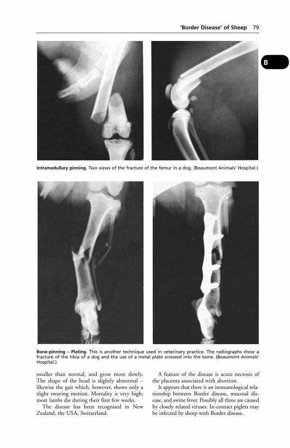
smaller than normal, and grow more slowly.The shape of the head is slightly abnormal –likewise the gait which, however, shows only aslight swaying motion. Mortality is very high;most lambs die during their first few weeks.
The disease has been recognised in NewZealand, the USA, Switzerland.
A feature of the disease is acute necrosis ofthe placenta associated with abortion.
It appears that there is an immunological rela-tionship between Border disease, mucosal dis-ease, and swine fever. Possibly all three are causedby closely related viruses. In-contact piglets maybe infected by sheep with Border disease.
‘Border Disease’ of Sheep 79
B
Bone-pinning – Plating. This is another technique used in veterinary practice. The radiographs show afracture of the tibia of a dog and the use of a metal plate screwed into the bone. (Beaumont Animals’Hospital.)
Intramedullary pinning. Two views of the fracture of the femur in a dog. (Beaumont Animals’ Hospital.)

Border Inspection PostsThe only authorised entry points for animalsentering the UK from outside the EU. Animalsimported into any EU member state throughone of its Border Inspection Posts and foundto be free from infectious or contagious diseasemay move on to the UK, or from the UK toanother EU member state, without hindrance.Quarantine may be imposed in some cases;for example, alpacas from South America. Not every Border Inspection Post can handle all types of animal. British Border InspectionPosts, and what they can import, are as follows:
East Midlands Tropical fishAirportGatwick Airport All animals except ungulatesGlasgow Airport All animals except ungulatesHeathrow Airport All animals Immingham Port Registered equidaeLuton Airport Ungulates including
registered equidaeManchester Airport Cats, dogs, rodents,
lagomorphs, live fish, reptiles, and birds otherthan ratites
Bristol Port Ungulates other than registered equidae
Stansted Airport Ungulates includingregistered equidae
Tilbury Port Ungulates and zoo animals but not equipped to handleany species of mammal specified in the Rabies (Importation of Dogs, Cats and Other Mammals) Order 1974
Bordetella(see under BACTERIA). B. bronchiseptica is a secondary invader complicating cases of caninedistemper, and may also produce respiratorydisease (see KENNEL COUGH) in the dog independently of viruses. This bacterium is also the cause of chronic respiratory disease in many other animals. It is an important factor in atrophic rhinitis. (See RHINITIS,ATROPHIC.)
Borna DiseaseMainly a disease of horses, but occurs also insheep and llamas, cats and humans.
Cause A virus which is closely related to thosecausing EQUINE ENCEPHALITIS occurring invarious tropical, subtropical and temperateregions; the diseases bearing such names asNear Eastern encephalitis, Venezuelan, Eastern,Western, and Japanese B. encephalitis.
Transmission Mosquitoes, midges, and tickscan transmit the virus, of which birds are alsohosts.
Signs Depression and fever. Recovery mayfollow without involvement of the centralnervous system, but probably in most casessuch involvement does occur. Signs theninclude walking in circles, or pressing the headagainst a fixed object, a facial twitch, hanging ofthe head, ataxia, and paralysis.
Diagnosis depends upon a fluorescent anti-body test or the detection of Joest-Degenantibodies. A differential diagnosis of Bornadisease in sheep must take into account loup-ing-ill, maedi-visna, rabies, listeriosis, scrapie,cerebro-cortical necrosis, poisons, etc.
BorogluconateThe salt of calcium used in solution for intra-venous or subcutaneous injection in cases of hypocalcaemia (milk fever). It should not be given to small animals as a treatment for calcium deficiency as boron poisoning mayresult.
BorreliaA species of SPIROCHAETE, causing disease infowls in the tropics, and also human tickbornerelapsing fever. The distribution of the latteris, with the exception of Australia, almostworldwide. The signs include fever, erythema,sometimes jaundice. B. burgdorfei causes LYME
DISEASE, a disease of dog and humans charac-terised by arthritis, headache, lethargy, andsometimes meningitis or encephalitis. It isendemic in certain parts of the USA.
BorreliosisA disease of dogs characterised by lameness,swollen joints and fever.
Cause Borrelia burgdorferi.
Boss Cows(see BUNT ORDER)
Boston TerrierOriginating in the USA, this small short-haireddog has erect ears and prominent eyes. It isprone to a number of inherited conditionsincludng achalasia of the oesophagus, cleftpalate, cataract, incomplete development ofthe vertebrae (hemivertebrae) and patellar luxation. The shape of the pelvis makes whelping difficult and assistance may berequired.
80 Border Inspection Posts
B
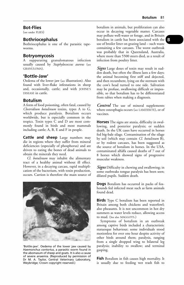
Bot-Flies(see under FLIES)
BothriocephalusBothriocephalus is one of the parasitic tape-worms.
BotryomycosisA suppurating granulomatous infectionusually caused by Staphylococcus aureus (see
GRANULOMA).
‘Bottle-Jaw’Oedema of the lower jaw (see illustration). Alsofound with liver-fluke infestations in sheepand, occasionally, cattle; and with JOHNE’S
DISEASE in cattle.
BotulismA form of food poisoning, often fatal, caused byClostridium botulinum toxins, types A to G,which produce paralysis. Botulism occursworldwide, but is especially common in thetropics. Toxin types C and D are most com-monly found in birds and most mammalsincluding cattle; A, B, E and F in people.
Cattle and sheep Large numbers maydie in regions where they suffer from mineraldeficiencies (especially of phosphorus) and aredriven to eating the bones of dead animals toobtain the minerals they need.
Cl. botulinum may inhabit the alimentarytract of a healthy animal without ill effect.However, in a decaying carcass, rapid multipli-cation of the bacterium, with toxin production,occurs. Carrion is therefore the main source of
botulism in animals, but proliferation can alsooccur in decaying vegetable matter. Carcasesmay pollute well-water or forage, and in Britainbotulism in cattle has been associated with theuse of broiler litter on grazing land – such wastecontaining a few carcases. The worst outbreakwas probably that in Queensland, Australia,where more than 5500 steers died, as a result ofinfection from poultry litter.
Signs Large doses of toxin may result in sud-den death, but often the illness lasts a few days;the animal becoming first stiff and dejected,and then recumbent, lying on the sternum withthe cow’s head turned to one side. Salivationmay be profuse, swallowing difficult or impos-sible, so that botulism has to be differentiatedfrom rabies when making a diagnosis.
Control The use of mineral supplementswhere osteophagia occurs (see LAMZIEKTE), or ofvaccines.
Horses The signs are ataxia, difficulty in swal-lowing, and posterior paralysis; or suddendeath. In the UK cases have occurred in horsesfed big-bale silage. Contamination of the silageby soil (which may contain Cl. botulinum B),or by rodent carcases, has been suggested asthe source of botulism in horses. In the USA,contaminated alfalfa caused deaths of 7 out of8 horses which showed signs of progressivemuscular weakness.
Signs Difficulty in chewing and swallowing; insome outbreaks tongue paralysis has been seen;dilated pupils. Sudden death.
Dogs Botulism has occurred in packs of fox-hounds fed infected meat such as farm animalsfound dead.
Birds Type C botulism has been reported inBritain among both chickens and waterfowl;also pheasants. It is not uncommon in hot drysummers as water levels reduce, allowing accessto mud. (See also MAGGOTS.)
Symptoms of botulism in an outbreakamong captive birds included a characteristicstatuesque behaviour; some individuals stoodmotionless for over one hour despite activity ofother birds around them; paralysis, rangingfrom a single dropped wing to bilateral legparalysis; inability to swallow; and terminalgasping.
Fish Botulism in fish causes high mortality. Itis usually due to feeding wet trash fish to
Botulism 81
B
‘Bottle-jaw’. Oedema of the lower jaw caused byHaemonchus contortus, a parasitic worm found inthe abomasum of sheep and goats. It is also a causeof severe anaemia. (Reproduced by permission ofDr M. A. Taylor, Central Veterinary Laboratory,Weybridge; Crown copyright reserved.)
farmed fish without cleaning out the machinerybetween batches. The Danish name for thedisease translates as ‘bankruptcy disease’.
For botulism in mink, see MINK, DISEASES
OF. For botulism in South African cattle, see
LAMZIEKTE.
Public health Human (and also animal) bot-ulism may occur as the result of imperfectlypreserved food or when cooked food is allowedto stand and later re-heated. Although therehave been very few cases of human botulism inBritain, a high proportion of trout in fish farmsmay be contaminated with Cl. botulinum typeE, which can multiply at temperatures as low as5°C, whereas the more common types A and Bwill not normally multiply at temperaturesbelow 10°C.
Boutonneuse FeverA zoonosis (see ZOONOSES) which is transmissi-ble from dogs to people. The cause is Rickettsiaconori. There is a rash. Wrists, ankles, andthen other parts of the body may be affected.The dog tick Rhipicephalus sanguineus is thevector.
Bouvier Des FlandersA medium-sized, short-tailed dog with grey orfawn coat. It has few defects; laryngeal paralysismay be inherited.
Bovine Atopic RhinitisA discharge from eyes and nose, with someulceration of nasal mucosa (and formation ofgranuloma), are symptoms in common withthose of bovine infectious rhinitis. It is oftenthe result of an acute hypersensitivity reaction,particularly in Channel Island breeds. Recoveryusually follows housing.
Bovine Atypical InterstitialPneumonia(see FOG FEVER)
Bovine AutoimmuneHaemolytic AnaemiaA heifer died within 2 days of showing anaemiaand dyspnoea, and the diagnosis was bovineauto-immune haemolytic anaemia, based onauto-agglutination (which increased onCoombs’ testing) and the presence of antibovineIgG on red blood cell surfaces.
Acute haemolytic anaemia may be due tomany other causes, including water intoxica-tion, delayed copper toxicity, brassica poison-ing, babesiosis, leptospirosis, and bacillaryhaemoglobinuria.
Bovine Embryo Collection AndTransfer Regulations 1993These apply within the UK and other memberstates of the EU.
Bovine Encephalomyelitis (BussDisease)Bovine encephalomyelitis (Buss disease) occursin the USA, Australia, and Japan. In the USA itis a disease mainly of the summer and autumnmonths, and cattle under 2 years old are main-ly susceptible.
Cause Chlamydia psittaci.
Signs A fever, which lasts a week or more.With loss of appetite, the animal loses condi-tion and becomes weak. A nasal discharge ordiarrhoea may be seen. Pushing the headagainst a wall, walking in a circle, hyperaesthe-sia, and convulsions are symptoms of whichone or two may be seen. Economically thedisease has a low incidence and generally alow mortality, but in some herds losses may beserious.
Autopsy findings include pleurisy, pericardi-tis and peritonitis, apart from any brain lesions.
Public health Man is susceptible.
Bovine Enzootic LeukosisA virus-produced form of cancer, characterisedby multiple malignant growths as well as, insome cases, leukaemia. The disease was firstrecorded in Britain in 1978, is fairly commonon the European mainland, and is a NOTIFIABLE
DISEASE. Occasionally cattle show symptomsbefore they are 2 years old, but 4 to 8 years is amore common age. Digestive disturbance,anaemia and loss of condition result.
The virus is a type C oncornavirus of theretrovirus family.
In Britain the enzootic bovine leukosis (EBL)attested herds scheme was introduced by MAFFin January 1982 to encourage the establishmentof EBL-free herds, as a first step towardseradication of the disease.
Great Britain was recognised as being free ofthe disease in July 1999; the last case wasdetected in December 1996.
Testing for enzootic bovine leukosisBreeding cattle may only be moved from onemember state of the EU to another if they orig-inate from herds which are recognised as freefrom EBL in accordance with the terms of theEU directive.
82 Boutonneuse Fever
B

Bovine Ephemeral Fever(see EPHEMERAL FEVER)
Bovine Herpes MammillitisAn ulcerative disease of the cow’s teats andudder, caused by a herpes virus. (See also VIRUS
INFECTIONS OF COW’S TEATS.)
Bovine Immunodeficiency-LikeVirus (BIV)This causes a progressive wasting condition ofcattle, with intercurrent diseases, poor milkyield and enlarged lymph nodes. Infection isstated to be present in North America andEurope. There is limited evidence of the degreeto which BIV is present in Britain, but antibodyhas been found in the majority of at least oneherd.
Bovine Infectious PetechialFeverAlso known as Ondiri disease, this affects cattlein Kenya, and is characterised by haemorrhagesof the visible mucous membranes, fever, anddiarrhoea. There may be severe conjunctivitisand protrusion of the eyeball. Death within 1to 3 days is not uncommon, though some ani-mals survive for longer, a few recovering. Thecause is a rickettsia, believed to be spread by abiting insect, or a tick, and known as Ehrlichiaondiri. The bushbuck provides a reservoir ofinfection.
Bovine Leukocyte AdhesionDeficiency (BLAD)An inherited problem of cattle resulting ininability to resist disease. Affected cattle usuallydie by 1 year old. It was discovered in the USAin 1989, and has since been found in manyEuropean countries, including the UK.
Bovine Malignant CatarrhalFeverThis infection, also known as malignantcatarrh, may occur not only in cattle but alsoin sheep, farmed deer, and antelopes. It ismost common in Africa, but cases have beenrecorded in the UK, other EU countries,Australasia, and North America.
Cause A herpes virus.
Signs Enlarged lymph glands, inflammationof the mucous membrane of the mouth,drooling of saliva, gastro-enteritis, keratitis(followed in some cases by blindness, andsometimes ENCEPHALITIS). Most affectedanimals die.
Bovine Papular StomatitisThis pox was first described in Germanyand during recent years has been reportedin the UK, Australia, East Africa, etc. Thedisease is not accompanied by fever or systemicupset.
Characteristically, papules form in themouth on the mucous membrane lining thecheek. Early lesions are rounded areas ofintensive congestion up to 1.5 cm in diameter,which in pigmented mucous membrane arevisible as roughened areas with greyish discol-oration. The centre of such areas becomesnecrotic and in a later stage shows a depressedcentre. Removal of the caseous material leaves araw granulating ulcer but normally epithelialregeneration occurs in 3 to 4 days. A featureof the disease is the occurrence of concentricrings of necrosis and congestion. Secondarylesions of mouth, muzzle or nostril mayprolong the disease over a period of months.However, it is unusual for the animals toshow signs of illness.
Cause The virus involved is the same as, or isrelated to, that causing pseudo-cowpox. (See also
VIRUS INFECTIONS OF COW’S TEATS.)
Bovine ParvovirusA cause of diarrhoea in calves.
Bovine Pleuropneumonia,ContagiousA NOTIFIABLE DISEASE in the UK. Affected ani-mals have a high temperature (41.5°C/107°F),difficulty in breathing and are anorexic.
Cause Mycoplasma pleuropneumoniae.
Bovine Pulmonary Emphysema(see FOG FEVER)
Bovine QuintupletsBovine quintuplets, all dead, were producedby a Charolais cow, which went on to con-ceive again within a normal period of time atthe Galemire Veterinary Hospital, CleatorMoor, UK.
Bovine Respiratory DiseaseComplex(see ‘SHIPPING FEVER’)
Bovine Rhinotracheitis,Infectious(see RHINOTRACHEITIS)
Bovine Rhinotracheitis, Infectious 83
B
Bovine SpongiformEncephalopathy (BSE)‘Mad cow disease’. A NOTIFIABLE DISEASE inwhich spaces (vacuoles) develop in the braintissue, in the manner of a sponge.
Signs Cattle become nervous and hypersensi-tive to noise, and when approached or touched.They appear frightened and may be aggressive.Dairy cattle show a reluctance to enter milkingparlours and may resent attempts to apply milk-ing clusters. The gait becomes abnormal, withhind limb swaying. The head is lowered. Whenthe animal is at rest, regular or spasmodicmuscle twitching may be seen.
BSE was first recognised in 1986 althoughindividual cattle were probably infected in the1970s. A policy of slaughter and incinerationof carcases was introduced. The heads wereremoved to confirm diagnosis.
Its origin is unknown but it is possible thatthe disease developed in a single cow followinggenetic mutation.
Fortuitously, a link between BSE and the useof meat and bone meal in bovine diets wasestablished in 1987. It was at first thought thatthe meal responsible contained material fromscrapie-infected sheep. However, the Phillipsinquiry into BSE concluded that this was notthe case. Current opinion favours the probabil-ity that a BSE-infected cow entered the render-ing process that produces meat and bone meal,infecting cattle which spread the disease whentheir carcases were in turn processed into meatand bone meal. Meat and bone meal wasbanned from bovine diets in 1988 but couldstill be used in diets for other species. However,this created the potential for meal intended forcattle to be infected by contamination with thatprepared for other species in the same mill.Meat and bone meal is now banned from use inall animal feeds; its possession in an animal feedplant is a criminal offence.
Specified bovine offals were prohibited fromhuman consumption; they include brain, spinalcord, spleen, thymus, small intestine lining. AllBritish beef exports were banned, with cata-strophic effects for the farming economy. Theban could not be lifted until the UK adoptedmeasures to lessen any risk to humans.
A new form of Creutzfeldt-Jakob disease in(mainly) young persons (new variant CJD) hasbeen confirmed as being linked to BSE. TheUK government has introduced a scheme to aidvictims and their families.
Incidence At its peak in 1992, 36,681 caseshad been confirmed representing about 0.3 per
cent of the national herd; the disease has beendeclining since then. By 1996, the number hadfallen to about 8000, about 0.07 per cent ofthe national herd. In 1997, a cull of cattle, insti-gated by the EU, removed those cows whichcould have eaten the same contaminated feed asthe proved cases in the same herd. A secondcull, in 1998, removed female calves born tocows around the time they showed clinical signsof the disease.
Eradication As a further precaution againstthe possibility of endangering public health,the carcases of animals over 30 months ofage were incinerated and not used for humanconsumption.
Cause It is generally accepted that the cause ofBSE is a prion (see PRIONS), a self-replicatinginfectious protein (PrP). However, it is notknown how this produces the disease.Theories that BSE resulted from a change inthe rendering process used to produce meatand bone meal, that it is a bovine form ofscrapie or that it is an autoimmune diseasehave been discounted. The Phillips inquiryalso discounted claims that BSE was causedby exposure to organophosphorus pesticidesalthough it acknowledged the possibilitythat this might increase susceptibility to thedisease.
Differential diagnosis BSE has to bedifferentiated from other disorders such asacetonaemia (in which short periods ofdelirium may occur); from listeriosis (in whichcattle may become violent in the terminalstages); and from hypomagnesaemia andhypocalcaemia.
Prevention The feeding of protein or othermaterial derived from the same species as willconsume the feed should be prohibited.
Other species BSE has been recorded inseveral species of antelope in zoos and thereis some evidence of infection in sheep. Pigshave developed BSE only after infectivematerial was injected into the brain; theydid not develop the disease when fed contami-nated feed. There may be a link with FELINE
SPONGIFORM ENCEPHALOPATHY as some bigcats in zoos had developed a spongiformencephalopathy after having eaten bovineheads, before BSE was recognised as adisease. BSE is experimentally transmissibleto mice and monkeys. (See also SCRAPIE.)
84 Bovine Spongiform Encephalopathy (BSE)
B
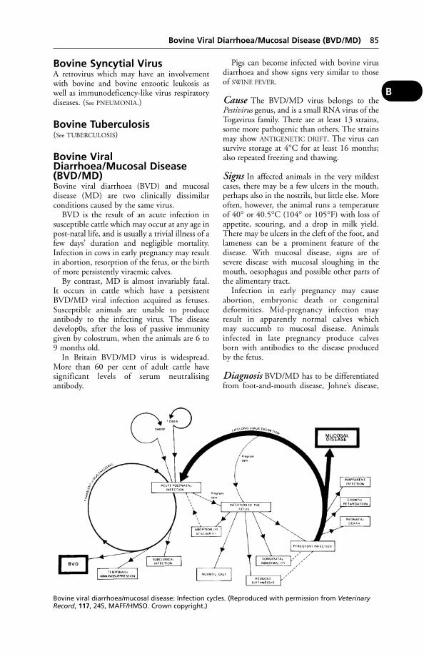
Bovine Syncytial VirusA retrovirus which may have an involvementwith bovine and bovine enzootic leukosis aswell as immunodeficency-like virus respiratorydiseases. (See PNEUMONIA.)
Bovine Tuberculosis(See TUBERCULOSIS)
Bovine ViralDiarrhoea/Mucosal Disease(BVD/MD)Bovine viral diarrhoea (BVD) and mucosaldisease (MD) are two clinically dissimilarconditions caused by the same virus.
BVD is the result of an acute infection insusceptible cattle which may occur at any age inpost-natal life, and is usually a trivial illness of afew days’ duration and negligible mortality.Infection in cows in early pregnancy may resultin abortion, resorption of the fetus, or the birthof more persistently viraemic calves.
By contrast, MD is almost invariably fatal.It occurs in cattle which have a persistentBVD/MD viral infection acquired as fetuses.Susceptible animals are unable to produceantibody to the infecting virus. The diseasedevelop0s, after the loss of passive immunitygiven by colostrum, when the animals are 6 to9 months old.
In Britain BVD/MD virus is widespread.More than 60 per cent of adult cattle havesignificant levels of serum neutralisingantibody.
Pigs can become infected with bovine virusdiarrhoea and show signs very similar to thoseof SWINE FEVER.
Cause The BVD/MD virus belongs to thePestivirus genus, and is a small RNA virus of theTogavirus family. There are at least 13 strains,some more pathogenic than others. The strainsmay show ANTIGENETIC DRIFT. The virus cansurvive storage at 4°C for at least 16 months;also repeated freezing and thawing.
Signs In affected animals in the very mildestcases, there may be a few ulcers in the mouth,perhaps also in the nostrils, but little else. Moreoften, however, the animal runs a temperatureof 40° or 40.5°C (104° or 105°F) with loss ofappetite, scouring, and a drop in milk yield.There may be ulcers in the cleft of the foot, andlameness can be a prominent feature of thedisease. With mucosal disease, signs are ofsevere disease with mucosal sloughing in themouth, oesophagus and possible other parts ofthe alimentary tract.
Infection in early pregnancy may causeabortion, embryonic death or congenitaldeformities. Mid-pregnancy infection mayresult in apparently normal calves whichmay succumb to mucosal disease. Animalsinfected in late pregnancy produce calvesborn with antibodies to the disease producedby the fetus.
Diagnosis BVD/MD has to be differentiatedfrom foot-and-mouth disease, Johne’s disease,
Bovine Viral Diarrhoea/Mucosal Disease (BVD/MD) 85
B
Bovine viral diarrhoea/mucosal disease: Infection cycles. (Reproduced with permission from VeterinaryRecord, 117, 245, MAFF/HMSO. Crown copyright.)
cattle plague, and other conditions. An ELISAtest is available.
Prevention Various vaccines, live and inacti-vated, have been used in North America,with mixed results. In 1995, an inactivatedvaccine was developed from a prototypecreated at the Institute for Animal Health,Compton, which was demonstrated as beingeffective by Professor Joe Brownlie and hiscolleagues.
Bow-LegsLegs curved outwards from the knee occurnormally in some breeds of dogs, such as thepug and bulldog, but in other breeds they areusually the sign of RICKETS. The shafts of thelong bones become softened and bend out-wards under the weight of the body, so that thefore-limbs especially become curved outwards.The condition is also seen in poultry and farmanimals.
Bowel, Oedema of theThis disease affects mainly piglets of 8 to 14 weeks old, though occasionally it is seen inthe new-born and in pigs of up to 5 months.It is usually associated with a change in man-agement such as moving or mixing with others,but especially with a change in diet. Inmost cases a gelatinous fluid is found in thethickness of the stomach wall and other parts.The disease can be experimentally transmittedby inoculation of this fluid.
Cause E. COLI, mainly seroptypes 0138, 0139or 0141.
Signs The finding of a dead pig – often thebest in the litter – is usually the first indication.Puffy eyelids, from which there may be a dis-charge, and puffiness of snout and throat maybe observed; together with leg weakness andconvulsions.
Treatment Administer antibiotics by injec-tion to affected pigs, and by injection or in feedor water to others in the group. Feed should bereduced and, if provided from hoppers,roughage should be added. Return to normalfeed should be gradual.
Prevention After weaning, keep on same feedfor at least 2 weeks. Change of diet should begradual over 3 to 5 days.
Bowels(see INTESTINES)
‘Bowie’A disease of unweaned lambs, resemblingrickets, in New Zealand. A supplement ofphosphates appears to be effective.
Bowman’s CapsuleA part of the nephron – the unit of structure ofthe kidney. Fluid passes from the glomerulusinto the capsule as the first stage of filtrationand urine formation.
BoxerA medium-sized, short-coated, short-nosed(brachycephalic) dog; usually brown in colourwith white and black markings; a few are allwhite. The skull structure leads occasionally tobreathing difficulties. Achalasia, cleft palate,corneal erosion, hyperplastic gingivitis andspondylitis may be inherited. The breed showsa higher than usual incidence of skin cancer.White boxers tend to be deaf.
Boxwood PoisoningBoxwood poisoning may sometimes occurthrough farm animals gaining access to gardenswhere the plant grows, or by eating the trim-mings from box hedges along with other greenfood taken from the garden. The plant, knownbotanically as Buxus sempervirens, containsseveral toxic alkaloids, the chief of which isbuxine. When large quantities have been taken,or if the beast is not able to vomit, nervoussymptoms, lameness, muscular twitching, dizzi-ness, diarrhoea, and acute abdominal painsare seen. In very severe cases there is the passageof blood-stained motions, great straining,convulsions, delirium, unconsciousness, anddeath. Pigs are the most susceptible of the farmanimals.
BrachialBrachial is a word describing an associationwith the forelimb. The brachial plexus is animportant group of nerves supplying the shoul-der and forelimb. Tumours arising here areusually malignant. They are seen most often indogs and cause a progressive lameness of oneforelimb together with atrophy of the muscles;also signs of pain which cannot be localised.Injury to the brachial plexus involving damageto NERVES results in paralysis of the affectedforelimb.
BrachycephalicThe word is applied to the short skulls of suchdogs as the bulldog, toy spaniel, or pug. In suchthe forehead is high, the skull broad, and theface foreshortened.
86 Bow-Legs
B
Bracken PoisoningThe eating of bracken (Pteris aquilina) byhorses, cattle or sheep may lead to serious illnessand death; symptoms appearing a month ortwo after the first meal of the plant.
Cause Bracken contains an enzyme, thiami-nase, which in the horse and pig causes athiamine deficiency. In cattle and sheep thisvitamin is produced in abundance in therumen, and bracken poisoning is due not tothiaminase but to the ‘radiomimetic-factor’also present in bracken which depresses bone-marrow function. There are complex changes inthe blood and bone marrow. Poisoning is moreprevalent in dry seasons than in wet weather,and young store stock are more often affectedthan adult cattle. The rhizomes are said to be 5 times as poisonous as the fronds – a fact ofimportance where reclaimed land has beenploughed.
Signs In the horse these take the form of ageneral loss of condition and an unsteadygait; later on, loss of appetite (but no rise intemperature), nervous spasms, and death.
Affected cattle, on the other hand, run a hightemperature. They segregate themselves fromthe rest of the herd and cease grazing. Thevisible mucous membranes are pale in colour,and numerous petechial haemorrhages arefound scattered over the lining of the nose, eyes,and vagina. Bracken is also associated withenzootic haematuria and upper alimentarysquamous cell carcinoma.
Faeces, passed without straining, are usuallyblood-stained. Respirations are accelerated, andon the slightest exertion the animals fall andhave some difficulty in rising. In many casesa knuckling of the fetlocks, especially of thehind-limbs, is noticeable. In some cases thethroat becomes swollen, so that there isdifficulty in breathing. The illness lasts from1 to 6 days. In other cases, death may occurmuch sooner, and be accompanied by bleed-ing from nose and anus, when the carcaseshave some similarity to deaths from anthrax.Onset of symptoms may be delayed for up to2 months.
In sheep, ingestion of bracken can alsocause bright blindness and mandibularneoplasia.
Treatment DL-Butyl alcohol injections havebeen recommended for cattle in the early stagesof bracken poisoning. For the horse, injectionsof thiamine are usually successful if the illness istackled in time.
Prevention Bracken is usually eaten onlywhen no other food is available; avoid the situ-ation in which animals turn to bracken out ofsheer hunger or thirst – semi-starvation of live-stock is ever a false economy. Especially wherethe grazing is poor, it is essential to move ani-mals to bracken-free land every 3 weeks. Avoidthe use of green bracken as bedding.
Bracken and cancer During the investiga-tion of acute bracken poisoning in cattle, it wasfound that certain constituents of the plantwere carcinogenic in rats and mice. In 1975 oneof at least two bracken carcinogens was identi-fied as shikimic acid, a constituent of manyother plants also, and it has been shown tocause lethal mutations and to be a very potentcancer-producer in mice.
Both in the UK and in Japan young brackenshoots have been eaten by people as a vegetable.A link has been established in Japan betweenlong-term bracken fern ingestion and stomachcancer.
In some parts of the world, cancer of thebladder is an endemic condition in cattle, andin most places – states the World HealthOrganisation – it is associated with bracken.(See also BRIGHT BLINDNESS)
BradshotAnother name for BRAXY.
BradycardiaBradycardia means slowness in the beating ofthe heart, with corresponding slowness of thepulse-rate. (See also HEART DISEASES – Functionaldisorders; PULSE.)
BradykininDamaged tissue releases the polypeptidebradykinin, a powerful vasodilator and stimula-tor of smooth muscle and of pain receptors,possibly as part of the inflammatory response toinjury.
BrafordA breed of cattle formed by crossing theBrahman and the Hereford.
BrahmanCattle of this name in the south of the USAwere developed from a mixture of several zebubreeds (Bos indicus) plus some Shorthorn orHereford content.
BrailingA means of temporarily preventing flight inpheasant poults, etc., by means of leather straps.
Brailing 87
B

BrainThe brain and the spinal cord together formwhat is called the CENTRAL NERVOUS SYSTEM.
Parts of the brain In the domestic animals,as in man, the principal parts of the brain (frontto back) are as follows:
(1) The cerebrum. This is by far the largestpart, and consists of two hemispheres separatedby a deep cleft. The surface of the cerebrum isincreased by numerous ridges or gyri and byfurrows called sulci. The hemispheres are joinedby the fibres of the corpus callosum.
Each hemisphere is divided into sections orlobes, and its surface has a layer of grey matter– the cortex. At the front of each hemisphere isthe olfactory bulb, which relays impulses fromthe olfactory nerves of the nose to the brain,and is concerned with the sense of smell.
Within the cerebral hemispheres lie the basalganglia. At the base of the cerebrum is thethalamus. Below is the hypothalamus, contain-ing nerve centres for the control of bodytemperature, and connected by a stalk orpedicle with the pituitary gland.
The lateral ventricles are located within thecorresponding hemispheres and are spaces filledwith cerebro-spinal fluid, and communicatingwith the third and fourth ventricles.
(2) The brain-stem consists of nerve tissue atthe base of the brain and includes the mid-brain(of which the largest structures are the 2 cere-bral peduncles and 4 quadrigeminal bodies),the pons, and the medulla oblongata.
(3) The cerebellum, which has 2 hemispheresand a middle ridge – the vermis. The cerebel-lum, with the pons, and the medulla oblongataare often spoken of as the hind-brain. The ponsis a bridge of nerve fibres from one hemisphere
of the cerebellum to the other. The medullacontinues backwards as the spinal cord.
Structure The brain is composed of whiteand grey matter. In the cerebrum and cerebel-lum the grey matter is arranged mainly as alayer on the surface, though both have greyareas imbedded in the white matter. In otherparts the grey matter is found in definitemasses called ‘nuclei’.
The cells vary in size and shape in differentparts of the brain, but all of them give off anumber of processes, some of which formnerve-fibres. The cells on the surface of thecerebral hemispheres, for instance, are roughlypyramidal in shape, and each one gives offnumbers of nerve-cell projections, called‘dendrites’, from one end, and a single longprocess, called an ‘axon’, from the other. Thewhite matter is made up of a large number ofnerve-fibres, each of which is connected to acell in the grey matter.
In both the grey and the white matter thereis a framework of fibrous tissue cells, extremelyfine and delicate, which acts as a supportivestructure for the fibres and nerve cells, to whichthe name ‘neuroglia’ is applied. Permeatingthe grey matter is a complex system of blood-vessels, and in the white matter there are alsovessels but to a lesser extent.
Meninges The brain proper is covered overby a thin membrane called the ‘pia mater’, thebones of the cranium are lined by a thick mem-brane called the ‘dura mater’, and between theseis an irregular network called ‘the arachnoid’.Between the arachnoid and the pia mater is asmall amount of fluid, which serves as a kind ofwater-bed in which the brain floats.
Size The brain varies very much in differentanimals and in different breeds, but the follow-ing table gives the average relation of the weightof the brain to the weight of the body:
Cat 1 to 99Dog 1 to 235Sheep 1 to 317Pig 1 to 369Horse 1 to 593Ox 1 to 682
From this it will be seen that the cat has pro-portionately to the size of its body the largestbrain.
Nerves There are 12 pairs of nerves comingfrom the surface of the brain. They are knownas cranial nerves:
88 Brain
B

1. Olfactory, to the nose (smell).2. Optic, to the eye (sight).3. Oculomotor4. Trochlear } to the muscles of the eyes.
5. Trigeminal, to the skin of the face, etc.6. Abducens, to the muscles of the eye.7. Facial, to the muscles of the face.8. Auditory, to the ear (hearing).9. Glossopharyngeal, to the tongue (taste).
10. Vagus, to heart, larynx, lungs, and stomach.11. Spinal accessory, to muscles in the neck.12. Hypoglossal, to the muscles of the tongue.
Blood-vessels The brain obtains its blood-supply from four main sources: two internalcarotids and two occipital arteries. Thesebranch and unite to form an irregular circleunder the brain within the skull, called the‘circle of Willis’. From this, numerous smallerbranches leave to supply the whole of the brainsubstance. By such an arrangement any possi-bility of deficiency of blood is obviated, forshould one of the main branches become cutor occluded, the others enlarge and the sameamount of blood is still supplied. The bloodleaves the organ by means of large venoussinuses situated in the membranes covering thebrain, and finally finds its way into the jugularveins of the neck.
Functions The cerebrum is concerned withmemory, initiative, volition, intelligence, and,as well as these, it is the receiving station of theimpulses that originate from the organs of sight,smell, taste, hearing, and touch. Fear, anger, andother emotions originate in the grey nerve cellsof the cerebrum, which is also concerned withvoluntary control of the skeletal muscles.
Sensations on the right side of the body, andmuscular control in the right side, are dealtwith by the left cerebral hemisphere; the righthemisphere being concerned with the left sideof the body.
The cerebellum is concerned with uncon-scious control, with balance, and with co-ordi-nation of complex muscular movements. Eachhalf of the cerebellum controls the muscularsystem of its own side of the body, and isin communication with the opposite side ofthe cerebrum. It closely communicates with thenerves, internal ear, and with certain nerves ofmuscle-sense, monitoring the state of musclecontraction.
The hypothalamus controls many bodyfunctions including hunger, thirst, bodytemperature and sleep.
The thalamus acts as a vital relay stationbetween the sensory nerves (except the olfactorynerves) and the cerebral cortex.
The basal ganglia help to control muchmuscular activity.
The medulla contains nerve centres for thecontrol of involuntary, or reflex, actions such asrespiration and heart-beat rates, coughing,vomiting, and the reflex part of swallowing.
Brain DiseasesBrain diseases include the following:
Abscess Infective micro-organisms may enterthrough an injury to the bone, through themedium of the ear (especially in the pig anddog), or may arrive by the bloodstream.Sometimes a foreign body, such as a needlethat has become lodged in the throat, may passupwards into the brain and set up an abscess.The condition may be produced during thecourse of pneumonia, metritis, endocarditis,etc., when the bacteria invade the bloodstreamand get carried to the brain among othertissues.
For symptoms and first-aid, see under MENIN-
GITIS; ENCEPHALITIS.
Cerebral haemorrhage Bleeding into thecranial cavity, usually as a result of trauma or a vascular problem. It may result in loss ofconsciousness and death. Residual signs onrecovery may include hind limb instability or convulsions.
Concussion The temporary loss of con-sciousness resulting from a head injury.Temporary blindness may occur after theanimal has regained consciousness.
In domestic animals long-term effectsinclude changes in behaviour, such as aggres-siveness and excessive libido. Fits (epileptiformconvulsions) may be a sequel to a head injury indogs. (See EPILEPSY.)
Encephalopathy Any disease of the brain,particularly one involving structural changes.
Inflammation (see ENCEPHALITIS; MENIN-
GITIS)
Oedema of the brain is seen in salt poison-ing in pigs, and in polioencephalomalacia (see CEREBROCORTICAL NECROSIS). Blindnessand convulsions are produced.
(See also ‘DAFT LAMBS’; HYDROCEPHALUS;CHOREA; RABIES; EQUINE ENCEPHALITIS;‘SLEEPER’ SYNDROME of cattle.)
Parasites of the human brain, of animalorigin, include nematodes, such as larval
Brain Diseases 89
B

hookworms, Strongyloides, ascarid worms(Toxocara), filarial worms, rat lungworm(Angiostrongylus cantonensis), and Gnathostomaspp.; trematodes, such as Fasciola hepatica(liver fluke), Schistosoma japonicum, andParagonimus spp. (lung fluke); the cestodes ofhydatid disease, cysticercosis, and sparganosis;and fly maggots such as Tumbu fly of Eastand Central Africa (Cordylobia anthropophaga),tropical warble fly of South America(Dermatobia hominis), sheep botfly of parts ofRussia and the region and the Mediterranean(Rhino-oestrus purpureus), and cattle bots andwarble fly in Europe (Hypoderma bovis andlineata of cattle and H. diana of deer) (Lancet).Such parasites may similarly occur in thebrains of farm and domestic animals. (See
COENURIASIS and HEARTWORMS; the latterbeing especially important in cats.)
Transmissible encephalopathies suchas scrapie and BSE.
Tuberculosis of the brain (see TUBERCU-
LOSIS)
Tumours of the brain may cause a varietyof signs, according to the part affected. Forexample, changes in character, loss of housetraining, seizures (cerebrum), circling move-ments, nystagmus (vestibular system), loss ofcoordination (cerebellum), a staggering gait(medulla), metabolic problems (hypothala-mus). Decreased activity, drowsiness, andblindness in cats are seen as a result of a menin-gioma. Of the 11 per cent suspected of havingbovine spongiform encephalitis (BSE) inwhich the disease was not confirmed, braintumour was the commonest explanation forthe signs.
Brain SurgeryIn veterinary practice this is performed to treatCOENURIASIS. (See under TAPEWORMS for thetechnique of the operation in sheep, and forthe signs suggestive of this disease.) It may benecessary in small animals following a roadtraffic accident or to remove brain tumours.
For example, a 12-year-old cat referred to theOhio State University’s teaching hospital, hadbeen walking in circles, aimlessly pacing, and,purring almost continuously; however, the catwas only intermittently responsive to humanattention, and kept his tongue protruded fromhis mouth.
Under a general anaesthetic, after comput-erised tomography of the skull to indicatethe exact site of the lesion, a hole was drilled
and through it the tumour (a meningioma)gradually removed. The cat made a perfectrecovery.
BranA bran diet is deficient in calcium and highin phosphorus. Osteodystrophia fibrosa is seen inhorses fed predominantly on bran (see HORSES,FEEDING OF; OSTEODYSTROPHIC DISEASES;BRAN DISEASE).
Bran DiseaseOsteodystrophia fibrosa; it is seen in horses pre-dominantly fed bran. Such a diet is deficient incalcium and contains excessive phosphorus.Bone deformities are seen, particularly swellingof the upper and lower jaws, with loosen-ing of the teeth, and lameness. It may alsooccur in other animals fed a diet too high inphosphorus.
Bran MashMade by adding boiling water to a tablespoon-ful of salt and two double handfuls of bran andstirring to a porridge-like consistency; cool for15 to 30 minutes, then give feed. Traditionallygiven to sick horses.
BrandingThe application of an identifying mark to, usu-ally, horses and cattle. Traditionally, a hot ironwas used to sear the hide, leaving a permanentmark. A less painful method is freeze-branding.In this technique, a copper branding ‘iron’,cooled to –70°C with dry ice and alcohol,is applied to a clipped or shaved area for about27 seconds.
When the branded area thaws, the hair fallsout. The new hair which grows in 2 or 3 weeks iswhite, and therefore shows up well on a darkishanimal. For a white animal, the brand has to beleft on longer to kill the hair roots. The brand-mark then resembles a hot-iron brand, but thehide damage may be less. (Early claims that ‘thereis no damage to the hide’ have been disproved.)
Laser beams have been used for brandingcattle in the USA. It is claimed that ‘with the5000°C temperature of the branding beam, thespeed of branding is faster than the pain reflexof an animal’.
Where a permanent brand is not necessary,it is a simple matter to apply black hair dye orhair bleach, according to whether the animalis light or dark in colour.
Brassicae SpeciesPlants of the Cruciferae family – kale, cabbage,Brussels sprouts and rape. Excessive consumption
90 Brain Surgery
B
can lead to haemolytic anaemia, with haemoglo-binuria, especially if other foods are not availableor if the kale is frosted. Certain species ofbrassica contain thiocyanates and produce iodinedeficiencies and goitre. (See under KALE.)
BraxyAlso known as bradshot – a disease of sheepcharacterised by a very short period of illness,by a seasonal and regional incidence, and, in thenatural state, by a high mortality. It occurs invarious parts of Scotland, Ireland, the northof England, Scandinavia, etc., chiefly on hillyland. It attacks young sheep under the age of2 years, weaned lambs being very susceptible;the best members of the flock are more liable tobecome attacked than poorly nourished sheep,and it is most frequently seen during a spell ofcold, severe weather with hoar frosts at night.
Causes Clostridium septicum. It affects themucous membrane of the 4th stomach of sheepand from there invades the tissues. It gainsentrance to the alimentary canal by way ofthe mouth along with the grass from a ‘braxypasture’.
Infection with Cl. septicum is characterisedby gas gangrene, and may occur in animalsother than sheep – including man.
Signs These – loss of appetite, abdominal pain,diarrhoea, with a high temperature andlaboured breathing – are seldom in evidencefor more than 5 or 6 hours; death being sud-den. A characteristic odour is perceptible fromthe breath and body fluids. Decomposition isvery rapid. The lesions are those of a gastritisin the 4th stomach (abomasum).
Prevention Vaccination at the beginning ofSeptember, so that the animals have time toestablish an immunity before the frosts begin,has given good results. On farms where thelosses have been very heavy a second vaccina-tion 14 days later may be needed. (See also under
VACCINATION.)
Breathing(see RESPIRATION)
BreathlessnessBreathlessness may be due to any condition thathinders the thorough oxygenation of the blood.
Hyperpnoea is increased depth of breathing.
Tachypnoea is the name for an increase inthe rate of respiration. This may arise from such
diverse causes as anaemia, heat stress, heartdisease, pneumonia, bronchitis, and paraquatpoisoning.
Dyspnoea means laboured breathing, orbreathing accompanied by pain or distress, suchas may occur with oedema of the lungs, pneu-monia, bronchitis, pleurisy, emphysema, andparaquat poisoning.
Breda VirusA cause of diarrhoea in calves in the USA, andof respiratory disease in 2-day-old calves whichvery soon died.
Breech Presentation(see PARTURITION)
Breeding, Controlled(see CONTROLLED BREEDING)
Breeding of Dogs Act 1973The Breeding of Dogs Act 1973 makes itunlawful for anyone to keep a dog-breedingestablishment unless it has been licensed by thelocal authority. A breeding establishment isdefined as ‘any premises (including a privatedwelling) where more than two bitches are keptfor the purpose of breeding for sale’.
Breeding of Dogs Act 1991This extended powers under the 1973 legisla-tion, which permitted local authorities toinspect only those premises already licensed, orthose for which a licence application had beenmade. Under the 1991 Act the local authorityor a veterinary surgeon could apply to a magis-trate for a warrant to enter and inspect thepremises. Obstruction is a criminal offence.
Breeding and Sale of Dogs(Welfare) Act 1999This amended the above two Acts. Bitches mustnot be mated if they are less than 1 year old;and a bitch can have not more than 6 littersin her lifetime. Records of breeding have tobe kept and dogs must be identified by a tagbearing a code identifying the premises of birth.
Breeding of LivestockInformation about animals coming ‘on heat’ orbeing ‘in season’ is given under OESTRUS. Otherinformation is given under PREGNANCY andPARTURITION.
Number of females per male varies.The stallion when he is 4 years old and upwardsand in good condition will serve from 80 to
Breeding of Livestock 91
B

120 mares during a season. A 3 year old cantake up to about 50 or 60, and from 15 to 20are enough for a 2 year old. From 60 to 80 cowsare sufficient for an average adult bull, buthe should not serve more than 35 or 40between 1 and 2 years of age. Twenty to 30 ewesare as many as the ram lamb will successfullyserve, but shearlings may have as many as 40to 50. Adult rams may successfully impregnate80 ewes or more. The year-old boar should notbe allowed more than 20 sows during a season,but when he is older he may have up to 30 or35. In this connection it must be rememberedthat when a large number of females are servedby a male, those served at the later stages are notso likely to prove fruitful as those served earlier.
When synchronisation of oestrus (heat) ispractised, more males are required; for instance,1 ram for 10 ewes.
In old age There is little reliable data, butmares have bred foals when over 30 years, cattleand sheep up to 20 years and cats till 14 yearsold. These, however, were all animals thathad bred regularly in their younger days. Itis difficult to breed from an aged female thathas not previously been used for stud purposes.(See also under REPRODUCTION; ARTIFICIAL
INSEMINATION.)
Brewer’s GrainsBrewer’s grains are a by-product of brewingused as animal feed. They consist of the maltedbarley after it has been exhausted. In both wetand dry forms they are used for feeding cattle,while dry grains are sometimes fed to foldedsheep. If fed wet they must be fresh or theybecome fermented; acidosis may then resultwhen fed. Mould can occur if stored. In thedry they can be kept for a considerable lengthof time without harm. They are rich in proteinsand carbohydrates, but must not be fed toexcess.
Some samples become infected with Bacilluscereus. (See BACILLUS.)
BriardA large, long-haired dog, black, fawn or greyin colour. Many heritable defects have beeneliminated by selective breeding but progressiveretinal atrophy is a trait. Hip dysplasia may bedue to a variety of factors.
Bridle InjuriesThey take the form of: (1) injuries to the poll;(2) injuries to the chin, caused by the curb-chain; and (3) injuries of the mouth from thebit. Damage is generally only superficial but in
a few cases infection sets in, and pus forms.This may burrow down into the ligamentoustissue of the poll and produce ‘POLL EVIL’. Inordinary cases it suffices to protect the damagedskin by winding a piece of sheep-skin roundthe strap that is causing the injury, and dressingthe abraded areas with an antiseptic powdereach night. Those injuries to the chin that arecaused by the curb-chain are usually only slight,and mainly affect young horses when they arebeing broken in. When they learn to answer thereins and acquire what is called a ‘soft mouth’,the chafed skin is allowed to heal and thecondition passes off. In older horses that have‘hard mouths’ and that constantly require theuse of the curb-chain, the skin becomes thick-ened and calloused, and the surface of the bonemay become irritated with a resulting deposi-tion of new bone in the groove of the chin.Injury may be obviated by using a leather curbfor young horses that have very tender skins,and by changing the bit for older animals. Carein driving of the horse, avoiding all sudden orsevere pulls on the reins, will often do more to‘soften’ a horse’s mouth than the use of moredrastic measures. Bit injuries consist of theabrasion of the mucous membrane of the lowerjaw, just opposite the corners of the lips, wherethe bit crosses. Sometimes the membranebecomes actually ulcerated and a foul-smellingdischarge escapes, but in the majority of casesthe injuries are slight and heal in a few days.
Bright BlindnessThis, a prevalent condition in Yorkshire hillsheep, was first described in 1965, and is char-acterised by progressive degeneration of theretina. The disease is of considerable economicimportance in some flocks.
Cause Consumption of bracken over a period.Bright blindness has been found in severalbreeds of sheep, in Scotland and Wales as wellas in Northern England. In some flocks theincidence may be 5 to 8 per cent among theewes, with a peak incidence in those 2 to 4 yearsold. The blindness is permanent.
In ewes moved to bracken-free grazing beforethe disease is well advanced, the condition willnot progress further.
Brisket DiseaseAnother name for MOUNTAIN SICKNESS.
BritishThe term applied to any breed of cat indicatesthat the animals are stockily built with roundheads. For example, the British blue is stocky
92 Brewer’s Grains
B
and round faced; the Russian blue has a bonystructure more similar to the Siamese.
British AlpineA black and white breed of goat.
British Cattle MovementService (BCMS)Curwen Road, Workington, Cumbria CA142DD. The organisation set up by the UK gov-ernment to maintain a register of births, deathsand imports of cattle, issue cattle passports andprocess cattle movement information fromfarmers.
British DaneA breed established by the Red Poll CattleSociety in the UK following the import ofDanish Red cattle.
British Veterinary Association(BVA)7 Mansfield Street, London W1G 9NQ. Theveterinary surgeons’ professional representativebody. Its principal objects are the advancementof veterinary science in all its branches, thepublication of scientific and clinical material,and the promotion of the welfare of theprofession. It is intimately concerned with allmatters of professional policy, and maintainscontact with many outside bodies and govern-ment departments. It publishes the weeklyjournal The Veterinary Record, which has aninternational circulation.
The BVA Animal Welfare Foundation isa charity devoted to the promotion andprotection of animal welfare; address as above.
British Veterinary Profession(see VETERINARY PROFESSION)
BroilersGood quality table chickens of either sex, about5 to 8 weeks old, and weighing about 1.5 kg(31⁄2 lbs) (liveweight).
Mortality If the chicks and their managementare good, the total mortality for a broiler cropshould be less than 5 per cent, frequently only3 per cent. Most of these deaths will take placeduring the first fortnight. In fact, a 1.5 per centmortality is normal and to be expected duringthe early period.
For commercial reasons there is often thetemptation to overcrowd broilers in theirhouses, and this practice will inevitably increasestress and hence the liability of disease – theeffects of which may be the more severe. (See also
under BATTERY SYSTEM; NEWCASTLE DISEASE;POULTRY AND POULTRY KEEPING.)
Broiler ascites, and colisepticaemia lesions inthe pericardium and liver, are causes of carcaserejection at processing plants; as is ‘swollenhead syndrome’ (subcutaneous oedema). Bothare caused by E. coli.
‘Broken Mouth’‘Broken mouth’ is the name given to themouths of old sheep that have lost some of theirteeth. Loss of incisor teeth is not uncommonin hill sheep and is of economic importancebecause a ewe needs her incisors if she is tosupport herself and a lamb on the hill.
The condition involves resorption of bonefrom the jaw following premature loss of theincisor teeth. It is already known that, in therat, demineralisation of the skeleton can resultfrom protein or mineral deficiency.
Broken incisors were seen in 6- to 8-month-old sheep wintered for 6 to 12 weeks on swedesor turnips. Towards the end of this period upto two-thirds of the hoggets were in poor con-dition. The crowns of several incisors hadfractured leaving short irregular brown stumps.The enamel was normal but there was softeningand loss of dentine between the apical endof the enamel and the gum margin. It wassuggested that this resulted from the effects ofacids produced by bacterial action on thecarbohydrates in the turnips and swedes.
Signs Difficulty in feeding, dropping some ofthe food back into the trough, and ‘quidding’.
‘Broken Wind’ (‘Heaves’)Both are outdated expressions applied to horseswith long-standing respiratory diseases, usuallyreferred to as chronic obstructive pulmonarydisease (COPD). ‘Heaves’ is the colloquialname for a condition in which double expirato-ry effort is a feature. This particular symptommay arise from several different pathologicalprocesses in the lungs, not all of which arechronic or irreversible; e.g. allergic reactions,such as immediate-type hypersensitivity (as inbronchial asthma) and extrinsic allergic alveoli-tis (as in ‘farmer’s lung’), chronic bronchiolitisfollowing bacterial or viral infections and, veryrarely, lung tumours. In every case there iswidespread bronchiolitis which initially givesrise to generalised over-inflation of alveoli(so-called ‘functional emphysema’). This lesionis reversible but eventually there is destructiveemphysema in which there is an increasebeyond normal in the size of the air-spaces with
‘Broken Wind’ (‘Heaves’) 93
B
destructive changes in the alveolar walls. Thesechanges are irreversible and lead to progressiverespiratory disability and eventual failure.
Signs The clinical sign of double expiratoryeffort consists of an initial passive normalexpiratory movement followed by an activecontraction of the chest and abdominal musclesto expel the remaining air. In advanced casesthis leads to hypertrophy of the rectus abdo-minis muscles, and the formation of a ‘heavesline’ beneath the posterior aspect of the rib cage– a feature characteristic of long-standingobstructive pulmonary disease in the horse.
A cough – typically dry, short, hollow andlow-pitched – sometimes becomes paroxysmalafter stabling or exercise; also faster breath-ing, audible wheezing, nasal discharge, andintolerance of exercise.
Differential diagnosis of these chronicrespiratory disorders with a double expiratoryeffort depends upon detailed clinical evaluation,responses to corticosteroids and other drugs, theresults of serological tests with appropriate anti-gens and, ultimately, autopsy. Infestation withthe equine lungworm Dictyocaulus arnfieldiituberculosis, and hydatid cysts should also beconsidered.
Control Vaccination against equine influenza,since many cases appear to originate from anepisode of acute respiratory disease.
(See also CHRONIC OBSTRUCTIVE PULMONARY
DISEASE; EMPHYSEMA.)
Bromhexine HydrochlorideA mucolytic and expectorant; used in mostanimal species.
BromocriptinAn ergot alkaloid. (See PSEUEDOPREGNANCY.)
BronchiectasisA condition in which there is dilatation of thewalls of the bronchioles due to weakeningthrough excessive coughing. The condition isoften met with in chronic bronchitis, and thecavities produced are often filled with pus.
BronchiolitisInflammation of very small bronchial tubes(bronchioles).
BronchitisBronchitis is inflammation of the mucousmembrane lining of the bronchi. It is a verycommon disease of all animals in temperate or
cold climates. It may occur as an extension ofinflammation of the trachea (tracheitis), and itmay be followed by pneumonia or pleurisy, orboth.
(a) Acute bronchitis This may follow expo-sure to smoke from a burning building, or bethe result of careless administration of liquidmedicines which then ‘go the wrong way’. Morecommonly acute bronchitis may occur duringthe course of some virus infections, followingcolds and chills, and may affect farm animalshoused in badly ventilated buildings. In thedog, bronchitis often occurs during the courseof distemper, and in the horse it may beassociated with influenza or strangles. Acutebronchitis in cattle and sheep may be parasitic.(See PARASITIC BRONCHITIS; WORMS, FARM
TREATMENT AGAINST.) In pigs, too, parasiticworms may cause bronchitis. (See also under
COUGHING.)
Signs A rise in temperature, accompanied byfaster respiration, loss of appetite, a cough, andnasal discharge, are seen. The cough is at firsthard and dry, but becomes softer and easier inthe later stages. The breathing may often beheard to be wheezing and bubbling in the laterstages.
Treatment Attention to hygienic conditionsis of first importance. The bronchitic horseshould be removed to a loose-box, providedwith a plentiful supply of bedding, rugged if theweather demands, given plenty of clean waterto drink, and fed on soft foods. It must on noaccount be drenched, for there is nearly alwaysdifficulty in swallowing, and a great risk ofsome of the medicine entering the trachea andcomplicating an already serious case. In animalssuffering from bronchitis due to parasiticworms, suitable anthelmintics must be used.Where the cause is bacterial – secondary, veryoften, to a virus infection – the use of appro-priate antibiotics is indicated. Liquid medicinesshould not be given. In housed livestock, atten-tion must be paid to the ventilation. For thedog, a jacket of flannel or similar materialmay be made. (See NURSING OF SICK ANIMALS;KENNEL COUGH.)
(b) Chronic bronchitis This may follow theacute form, or it may arise as a primary condi-tion. The smaller capillary bronchial tubes areaffected and not the larger passages.
Chronic bronchitis is often seen in the olddog, very occasionally in association with tuber-culosis. The latter may also cause chronic
94 Bromhexine Hydrochloride
B

bronchitis in cattle and other animals. In thehorse, chronic bronchitis may lead to EMPHYSE-
MA. (See also ‘BROKEN WIND’.) Parasitic wormsmay be associated with some long-standingcases of bronchitis in animals.
Signs A loud, hard cough, often appearingin spasms, respiratory distress on the leastexertion, an intermittent, white, clotted, orpus-containing nasal discharge, which is mostin evidence after coughing or exercise, and agradual loss of condition, characterises thisform of bronchitis.
(c) Bronchitis in chickens (see under
INFECTIOUS BRONCHITIS)
BronchopneumoniaInflammation of the bronchi and lungs.
BronchoscopyExamination of the bronchi by means of abronchoscope, a tubular optical instrumentwith a small lamp attached which is passedthrough the trachea. The technique is usedin cases in which clinical and radiologicalexaminations fail to provide a diagnosis.
Bronchus, or Bronchial TubeBronchus, or bronchial tube, is the nameapplied to tubes into which the windpipe(trachea) divides, one going to either lung. Thename is also applied to the later divisions ofthese tubes distributed throughout the lungs.Bronchioles are very small bronchial tubes.
‘Brown Mouth’A syndrome characterised mainly by gum necro-sis and dysentery, occurring as a complicationof virus diseases in the dog.
‘Brown Nose’A form of LIGHT SENSITISATION in cattle.
Brown SwissA breed of dairy cattle producing milk with ahigh protein level.
BrucellosisA NOTIFIABLE DISEASE, this is an infection withBrucella. Five species of this genus of bacteria areimportant, namely: B. abortus (the main cause ofabortion in cattle but now eradicated in Britain);B. melitensis; B. suis; B. ovis and B. canis.
Public health Human brucellosis may becaused by any of the five species of Brucella, asmentioned above. Infections with Brucella
species are reportable diseases under theZoonosis Order 1989. It often takes the form of‘undulant fever’, with characteristic undulatingfluctuations of the temperature. Human infec-tion with B. abortus may follow the drinking ofraw milk or the handling of infected fetal mem-branes. Infected uterine discharge drying on thecow’s skin may be inhaled. It was formerly notuncommon in farm and abattoir workers, andveterinary surgeons.
For symptoms, see UNDULANT FEVER.What was formerly known as Malta Fever in
man is due to B. melitensis, an infection of goatsand sheep, occasionally cattle. Its occurrence inthe UK was limited to one outbreak resultingfrom imported infected cheese.
The American strain of B. suis (found in pigsand hares) is also pathogenic for man, causingundulant fever and arthritis.
B. canis, which infects dogs, can also causeillness in people.
B. ovis, which infects sheep, rarely causeshuman illness.
Horses B. abortus may cause fistulouswithers and lameness due to infection of otherligaments. In the mare, abortion may (rarely)occur.
Cattle (see BRUCELLOSIS IN CATTLE)
Dogs In the UK, B. abortus was isolated fromthe urine of a dog which had shown symptomsof stiffness and orchitis. At autopsy, cystitisand an abscess of the prostate were found. Sucha dog would be a public health risk, and adanger to cattle. Abortion is another symptom.The infection has been found in kennels,following the feeding of meat from stillborncalves. Brucellosis in dogs is probably morecommon than generally realised. In Chile asurvey showed that 40 per cent of dogs, onfarms where the dairy herds were infected withB. abortus, were infected.
B. canis was first isolated in 1966. In theUSA it has caused outbreaks of severe illness inlaboratory beagles; it causes also illness in man.
A unique feature of B. canis infection is lackof fever. Another feature is the duration of bac-teraemia, which usually lasts for several months,but can last 3 or 4 years.
In males, epididymitis, scrotal dermatitis,and testicular degeneration may occur,although it is not uncommon for male dogs tobe ‘silent’ carriers.
Sheep Formerly, brucellosis was an importantdisease of sheep in the UK.
Brucellosis 95
B

B. ovis gives rise (in Australasia, the USA,and Europe) to infertility and scrotal oedema inrams. Abortion may occur in infected ewes. (See
also RAM EPIDIDYMITIS.)
Goats In Britain, brucellosis is not a seriousproblem in goats.
A survey of sheep flocks and goat herds iscarried out yearly to determine whether Britainremains free from brucellosis in those species.The results are sent to the EU VeterinaryDirectorate.
Pigs In Britain, brucellosis is not found.Overseas, abortion in pigs is caused by B.abortus suis.
Deer There is no evidence that deer, infectedwith B. abortus, have infected cattle grazing thesame pasture.
Poultry Chickens are susceptible to B. abortusinfection, which they have transmitted to cattle.
Wild animals The harbour porpoise aroundBritain may carry Brucella maris, which reactswith B. abortus. Cattle by the shore coming intocontact with porpoise material may then showa positive reaction to the brucella test. B. suishas caused orchitis in hares abroad and, inAfrica, B. abortus has been isolated from awaterbuck, and from rodents.
In Argentina foxes are commonly infectedwith B. abortus.
(See also FISTULOUS WITHERS; ‘POLL EVIL’;BUMBLE-FOOT; RAM EPIDIDIMYTIS.)
Brucellosis in Cattle(Brucella melitensis causes disease in some coun-tries.) ‘Contagious bovine abortion’, alsoknown as Bang’s disease, is a specific contagiousdisease due to B. abortus. Since the infectionmay exist and persist in the genital system ofthe bull, Brucellosis is to be preferred as a namefor the disease. In females it is characterisedby a chronic inflammation of the uterus (espe-cially of the mucous membrane); usually, butnot invariably, followed by abortion betweenthe 5th and 8th months of pregnancy.
It is important to note that not all infectedanimals abort. Indeed, in over half of thempregnancy runs to full term. However, anyanimal that has aborted once may be almostas infectious at its next and subsequent calvingsas on the occasion it aborted.
Infection may occur by the mouth orthrough the vagina during service, when a bullwhich has served an infected cow is called upon
to serve a clean one afterwards, or when the bullis a ‘carrier’. Contamination of litter with dis-charges from a previous case is an importantfactor in the spread of the disease in a herd. Thehand and arm of the man who handles anaborted fetus may also transmit infection.
In the pregnant cow a low-grade chronicinflammatory reaction is set up in the uteruswith the result that an exudate accumulatesbetween the fetal membranes and the uterinemucous membranes, especially around thecotyledons. The cotyledons may appearnecrotic, owing to the presence of fibrinousadherent masses upon their surfaces, and thefetal membranes may show similar areasafter they have been expelled. Quite commonlyin cattle the membranes are thickened andtough. The fetus may be normal or may showa dropsical condition of the muscles and thesubcutaneous tissues, and there may be fluidpresent in the cavities of chest, abdomen, andcranium. In some cases the fetus undergoesa process of mummification, and when it isdischarged it is almost unrecognisable as afetus.
Cows at pasture may become infected byolder ‘carrier’ cows (which are liable to harbourthe organisms in their udders) or by wild ani-mals (e.g. foxes), dogs or birds, which haveeaten or been in contact with infected mem-branes or discharges upon other farms near bywhere the disease already exists.
Signs Abortion may occur without any pre-liminary symptoms, and except that the calfis not a full-term one, may be practically thesame as normal calving. Most cows which haveaborted once will carry their next calf to fullterm, or practically to full term; while only veryfew cows will abort a calf three times. Somecalves born to infected cows will be persistentlyinfected.
As a rule, if abortion occurs early in preg-nancy the fetal membranes are expelled alongwith the fetus, but if towards the end of theperiod there is almost always retention of these.A continuous reddish-brown or brownish-greydischarge follows, and persists for about 10 to20 days (often for about 2 weeks). In someinstances it slowly collects in the cavity of theuterus, little or nothing being seen at the vulva,and then it is discharged periodically, often inlarge amounts at a time. In the bull symptomsof infection may be very slight or absent, andlaboratory methods are usually necessary toestablish a diagnosis.
Brucellosis is not the only cause of abortion incattle due to an infective agent, and in arriving
96 Brucellosis in Cattle
B

at a diagnosis it must be differentiated frominfections listed under ABORTION.
Immunity Infected animals gradually pro-duce an immunity in themselves against furtherabortions. The organisms may persist in thesystem for long periods, and a cow which doesnot herself subsequently abort may spreadinfection to other cows in the herd. This natur-al immunity, however, is wasteful, both in thematter of calves and milk supply, so thatmethods have been adopted in which an effortis made to provide animals with an artificialimmunity.
Testing Bulked milk from herds is routinelytested by the MILK RING TEST or an ELISA.Periodic biennial blood tests are made of suck-ler herds; formerly the ROSE BENGAL TEST wasused but this has been replaced by an ELISA.All cows calving at 270 days’ gestation or lessmust be reported and are investigated by bloodand milk samples from the dam, examinationof placenta, abomasal contents and sera of thefetus. (See also COOMBS TEST.)
Eradication In October 1985 Britain wasdeclared officially brucellosis-free. Occasionalcases have occurred following the importationof cattle. Brucellosis has been successfullyeradicated from many overseas countries,including Denmark, Sweden, Norway, Finland,the Czech Republic, Slovakia and Eire. Farmers’cooperation and discipline played an importantpart.
Precautions All calvings under 270 days’gestation must be reported to DEFRA andinvestigated. The greatest care must be taken inhandling and disposing of an aborted fetus,fetal membranes, discharges, etc., both in theinterests of human health and in order toprevent the spread of the disease among cattle.It is worth having a veterinary surgeon examinethe cause of any abortion. There can be dangerfrom the infected cow that has carried a calf tofull term. Avoid buying in replacements fromnon-Accredited herds. Infected farm dogs canspread infection.
Brucellosis in Sheep(see RAM EPIDIDYMITIS)
Bruised SoleBruised sole is a condition of bruising of thesensitive sole of the foot, due to a badly fittingshoe, or the result of the horse having stoodupon a projection, such as a stone, etc. Its
character and its treatment do not differ fromwhat is given under CORNS, except that whilethe corn has a more or less definite positionin the foot, bruising of the sole may occuranywhere.
BruisesThe discoloration caused by bleeding underunbroken skin following a blow or othertrauma (see also HAEMATOMA).
Bruit and MurmurBruit and murmur are words used to describeseveral abnormal sounds heard in connectionwith the heart and arteries on auscultation.
Brush BorderOn the free surface of some cells, the wall maybe modified to provide finger-like projections:the brush border. This is seen, for example,in the convoluted tubule of the kidney and inthe alimentary canal.
Brushing and CuttingBrushing and cutting are injuries to the horsecaused by the inside of the fetlock joint orcoronet being struck by the hoof or shoe ofthe opposite limb; although bad shoeing maybe responsible in a few instances, the cause isusually faulty conformation.
A brushing boot should be fitted, and anattempt made to avoid the future occurrence ofbrushing by skilful shoeing. (See also SPEEDY-CUT.)
Brussels SproutsCattle strip-grazing these for 6 weeks, withoutother food, became ill with anaemia andhaemoglobinuria. The illness caused by mem-bers of the Brassicae species is said to be moreserious near to the time of calving.
BruxismGrinding, gnawing or clenching of the teeth;seen mainly in cattle.
BSE(see BOVINE SPONGIFORM ENCEPHALOPATHY)
Bubonic PlagueBubonic plague is an infectious disease of man,rats and mice and rabbits caused by Yersiniapestis. Foci of infection exist in several parts ofthe world, including the western United States.Rats, rabbits and cats and dogs may be involvedin transmitting the infection to man, usually bymeans of fleas.
In man bubonic plague takes one of twoforms: (l) After an incubation period of 2 to
Bubonic Plague 97
B
7 days, the usual symptoms include the suddenonset of fever, rigors, muscular pain, headacheand prostration. Within a few days the charac-teristic buboes (swelling of the lymph nodes inthe groin and armpits) usually appear. These areaccompanied by oedema, erythema, and greatpain. (2) Pulmonary plague has an incubationperiod of 2 or 3 days. Besides the sudden onsetof fever there is a cough (usually with bloodysputum), headache, rigors, and prostration.When untreated this form of plague usuallyresults in death within 2 to 5 days.
Various antibiotics are effective in treatmentif given early enough.
The intermediate link between the infectedrat and man is the rat flea.
BuccalRelated to the cheek.
Buccal CavityThe mouth.
BuccostomyAn operation for the creation of buccal fistulaeto prevent wind-sucking.
BuckTerm for the male of many species, e.g. deer,ferret, goat, hare, kangaroo.
Budgerigars(see CAGE (AVIARY) BIRDS, DISEASES OF)
BuffaloThe Asiatic water buffalo Bubalus bubalis isfarmed in Britain for the production of moz-zarella cheese. The American ‘buffalo’ is thebison (Bison bison) and is farmed in the UK formeat. (See WATER BUFFALOES.)
Buffalo FlyThis is Lyperosia exigua, a parasite of importancein Australia and in India and Malaysia. It causesgreat irritation and even anaemia. (See FLIES.)
Buffalo GnatSwarms of these, which breed in running water,attack cattle, often causing them to stampede,and producing serious bites which may lead todeath. Man is also attacked by these black flies(Simulium species).
Buffalo-PoxA contagious disease of buffaloes which is ofconsiderable economic importance. The infec-tive agent is distinct from cowpox virus. (See also
under POX.)
BufferA substance which, when added to a solution,causes resistance to any change of hydrogen ionconcentration when either acid or alkali isadded.
BuffingBuffing is a term applied to the striking of theinside of one hoof at the quarter with some part of the opposite one. It is due to the samecauses as BRUSHING, but it occurs in horses thatdo not lift their feet very high. Less damage isdone than in brushing, and it is not so likely tocause stumbling or lameness.
BufotalinThe principal poisonous substance present inthe skin and saliva of the common Europeantoad, Bufo vulgaris. Very small quantities willcause vomiting in dogs and cats, and 0.00917mg per kg bodyweight has caused death fromheart-failure in the cat. (See TOADS.)
BuiatricsThe study of cattle and their diseases.
Buildings(see HOUSING OF ANIMALS)
Bulbar Paralysis, Infectious(see AUJESZKY’S DISEASE). The term ‘bulbar’relates to the medulla oblongata or theprolongation of the spinal cord into the brain.
Bull BeefThis is beef from the entire animal as opposedto the castrate. (See under ASTRATION.)
There is no question that bull beef is a moreeconomic proposition. Feed conversion effi-ciency is improved, daily weight gain increasedand fat deposit reduced compared with steers orheifers. Bull beef is more popular than thatfrom steers in many parts of Europe.
BulldogA breed of medium-sized, smooth haired, shortnosed (brachycephalic) dog which has breath-ing difficulties. A show of the breed that tookplace in very hot weather resulted in the deathsof several dogs because the abnormally largesoft palate interfered with their breathingin such conditions. Bulldogs are subject to anumber of inheritable conditions includingcleft palate, underdevelolped (hypoplastic)trachea, and narrowing (stenosis) of the aortaand pulmonary system. Ingrowing or doubleeyelashes (entropion and distichiasis) are alsofound.The breed has changed considerably in
98 Buccal
B
appearance since the beginning of the 20th cen-tury; those changes have largely contributed tothe problems the bulldog suffers from today.
Bull-Dog CalvesIn Dexter cattle commonly, and in other breedsoccasionally, a hereditary condition, which isscientifically known as achondroplasia, occurs.Calves are born in a deformed condition inwhich the short limbs, dropsical swollenabdominal and thoracic cavities, and a markedforeshortening of the upper and lower jaws givethe calf an appearance resembling a bull-dog.Such calves are usually dead when born.
‘Bull-Dogs’A small metal appliance used temporarilyfor the restraint of cattle. They are applied tothe inside of the nose for holding an animalsteady.
Bull HousingAny bull housing must be secure and designedto prevent injury to the animal or stockman.The pen should be sited so that the bull can seewhat is happening around him; ideally, heshould be able to see other cattle at times. Allaccommodation should have sufficient escapepoints to ensure the safety of those attendingthe bull. There should be a means of capturingand restraining the animal without having toenter the pen. Adequate space for exerciseshould be provided as well as sleeping accom-modation, which could be a loose box. If ser-vice is to be carried out in the pen there mustbe a means for allowing the cow to be intro-duced to a service area without risk to eithercow or stockmen.
The codes for the welfare of farm livestockrecommend that for an adult bull of averagesize, the sleeping area should be not less than16 m2 (180 ft2). For very large bulls the sleep-ing area should not be less than 1 m2 for each60 kg liveweight (9 ft2 per cwt). The exerciseand service area should be at least twice the sizeof the sleeping accommodation.
The walls of the pen should be built up to aheight of 1 m (3 ft 3 in) and extended to 2 m(6 ft 6 in) high with stout tubular steel rails.There should be a fodder rack and feedingtrough at the end away from the shelter, pro-vided with sufficient cover to protect the fodderand concentrates, and the animal while feeding,during bad weather. This arrangement encour-ages the bull to stay out in the open ratherthan in the box or shelter and is consideredbeneficial. The entrance to the pen should beconvenient to the feeding area.
The feeding trough should be about 60 cm(2 ft) above ground level and should be fittedwith a tubular tying arrangement which canbe closed on the bull’s neck when he puts hishead through to the trough, if it is required tocatch him. This equipment is very desirableas an added safety measure, as it permits thebull to be securely held before the attendantenters the pen.
An arrangement which is very useful fordealing with vicious bulls is the provision of astrong overhead wire cable running from insidethe house or shelter to the opposite end of thepen. This cable is threaded through a strongring, about 3 cm (11⁄4 in) in diameter. This ring,which slides along the cable, is attached to achain which passes up through the bull’s nosering, then around the back of the horns and ishooked to the upright chain in front of theforehead. In this way, the weight of the chainis carried by the head instead of by the nosering and considerable discomfort to the animalthereby avoided. The chain should be justsufficiently long to allow the animal to lie downcomfortably. The advantage of this arrange-ment is that a cow can be brought into the penfor service without the necessity of having torelease the bull from his tying.
Another safety device which should be pro-vided, where possible, in the walls or railingssurrounding the pen, is escape slits. Theseare upright openings about 38 to 45 cm (15 to18 in) wide, sufficient to allow the attendant topass through in case of emergency, but throughwhich a bull could not pass. If, due to the loca-tion of the pen, it is not possible to providethese escape slits, the blind corners of the penshould be fenced off by means of sturdy uprightsteel rails set 38 to 45 cm (15 to 18 in) apart,behind which an attendant could seek refuge.
Bull ManagementAll bulls should be handled from an early ageand become accustomed to being restrained bymeans of the bull ring. The animals should beroutinely groomed and have their feet regularlyhandled and trimmed. There should always betwo people present when the bull is handledor the pen is entered. A bull can be used forservice from about a year old, but only sparing-ly; once a week, or 3 services in 2 weeks, untilat least about 16 months old. When first usinga young bull, he should be used to serve older,experienced cows.
While a bull is often turned out with cows toact as a ‘sweeper’ after artificial insemination, orwhere oestrus detection is poor, this makes itimpossible to keep accurate records of service.
Bull Management 99
B

Because of the work involved and the lack of suitable accommodation, there has been atendency not to keep bulls on dairy farms. Bullsshould be selected to provide genetic improve-ment to a herd and their choice requires con-siderable care. (See BULL HOUSING; also PROGENY
TESTING.)
Bull MastiffA short-coated muscular dog, somewhat resem-bling the original bulldog in appearance butmuch larger. Has fewer inherited defects thanthe bulldog. Cleft palate may occur, andununited anconal process (elbow dysplasia)may be found.
Bull TerrierA medium-sized dog, smooth coated, com-monly white, with a distinctive flat profile.Originally bred for bull-fighting. Renal diseasemay be congenital and deafness is linked to thewhite colour. Cleft palate and umbilical herniaare also heritable conditions.
BullaA blister; plural, bullae.
‘Bullets’A form of BOLUS. They are administered tocattle and sheep by means of a special dosing‘gun’, and are used as a means of supplyingthe animal with a long-lasting supply of mag-nesium, cobalt, or selenium. Bullets can besomewhat costly and not always retained, butthey are widely used and have proved successfulin preventing deficiency disease in sheep.(See also under COBALT.)
BullingA cow mounting another when in heat is saidto be ‘bulling’. (See also OESTRUS, DETECTION
OF, IN COWS.)
Bulls, Diseases of(see CATTLE, DISEASES OF; diseases listed underthe word BOVINE; PENIS AND PREPUCE)
Bumble-FootBumble-foot is a condition of the feet of poul-try, waterfowl, wading birds, birds of prey andsometimes cage birds in which an abscess formsin the softer parts of the foot between the toes.It may be caused by the penetration of somesharp object, such as a piece of glass, thorn,stone, etc., or even by penetration of theskin by the bird’s own talons. An abscess slowlyforms, accompanied by distinct lameness.Usually a Staphylococcus species is involved
but many other micro-organisms may beimplicated. Brucella abortus has been isolatedfrom a case of bumble-foot in Germany.
Treatment It is necessary to open the pus-containing cavity and evacuate the cheese-likecontents.
BunBlood urea nitrogen. (See KIDNEYS, DISEASES OF.)
BunostomiasisInfestation with hookworms of the genusBunostomum.
Bunt OrderEquivalent to the ‘peck order’ among poultry,this is the order of precedence established bycattle and pigs. With a newly mixed group ofthese animals there will be aggressiveness oractual fighting, until the dominant ones (usuallythe largest) establish their position in the socialorder. Once this is established, fighting will ceaseand the group will settle down, with the top ani-mal being accorded precedence without havingto fight for it. The second animal will be submis-sive to the first, but will take precedence over therest; and so on down through the herd, with thebottom animal submissive to all. Occasionallytwo animals will be of equal rank, or there maybe a somewhat complicated relationship betweena small group as in the ‘dominance circle’.
The bunt order can be important from ahealth point of view, and it can affect thefarmer’s profits. If, in large units, the batchingof animals to ease management means frequentmixing or addition to established groups, stresswill arise, and productive performance willdecline. Stress will be reduced in the systemwhereby pigs occupy the same pen from birthto slaughter time. The health factor – as well asdaily liveweight gains and feed conversionratios – will be involved when there is, forexample, insufficient trough space, and thoseanimals at the bottom of the social scale maygo hungry or thirsty. Similarly, the dominantanimals will be able to choose more sheltered,less draughty places, while their inferiors maybe cold and wet. (See also STRESS.)
BunyaviridaeThis group of viruses includes the HANTAVIRUS.Individual species may be zoonotic.
Buprenorphine HydrochlorideAn analgesic used for dogs, cats, birds, rodentsand rabbits, and as a premedicant for surgery,radiography, etc.
100 Bull Mastiff
B
Burial of Pet AnimalsIn 1992 the Environment Minister ruled thatdead pets could be returned to their owners forburial, despite the ‘Duty of Care’ Regulations1991.
Burns and ScaldsThough the former are caused by dry heat andthe latter by moist heat, their lesions and thetreatment of these are similar.
In animals a burn is usually easily recognisedby singeing of the hair, or its destruction, butwith a scald there may be little to be seen forseveral hours or even days. Moreover, a scaldedarea may remain concealed by a scab.
Burns and scalds are extremely painful andwill give rise to shock unless they are slight.After a few hours the absorption of poisonousbreakdown products from the damaged tissuesmay give rise to toxaemia; while destruction ofskin affords means of entry for pathogenicbacteria, against which the burned tissues canoffer little or no resistance. Death is a frequentsequel to extensive burns – the result of shock,toxaemia, or secondary infection.
First aid Scalds are mainly suffered by dogs,cats, and other domestic pets as a result ofmishaps in the home. Placing dogs, cats and rep-tiles immediately under cold, running waterwill reduce the temperature of the affected area,and is likely to reduce also the pain and subse-quent skin damage. This applies to burns also.Scalds from hot oil are best treated with other(cool) oily substances or emulsions such as milk.Fur-covered mammals such as chinchillas andrabbits, and birds, should not be treated withoils. Emulsions are best for such species.
Treatment Where the burn or scald is at allextensive, no time should be lost in calling inthe veterinary surgeon, who will have to admin-ister an analgesic or anaesthetic and perhapsfluid therapy before local treatment can beattempted (and in order to relieve pain, andlessen shock). This also facilitates clipping thehair to expose the affected area.
In an emergency occurring where no first-aidkit is available, a clean handkerchief (or piece oflinen) either dry or soaked in strong tea may beapplied as a first-aid dressing to a burn. Thepart should be covered, the animal kept warmand offered water to drink.
The object of treatment, besides reducingpain, is to form rapidly a coagulum of proteinon the surface of the burned area and diminishabsorption of those altered proteins, from thedamaged tissue, which give rise to toxaemia;
and also to prevent infection – to which thedamaged tissue is very susceptible. Tannic acid(the useful constituent of the strong tea men-tioned above) helps to form the desired coagu-lum. A tube or two of tannic acid jelly or bottleof Proflavine emulsion should be included inevery first-aid kit for dealing with small burns.It should not be applied over large areas.
Where the animal-owner cannot obtain pro-fessional assistance, subsequent treatment mustaim at avoiding sepsis, the damaged tissuesbeing very prone to infection. Sulfathiazole orsulfanilamide powder may be dusted lightlyon to the area before a first or second applica-tion of tannic acid jelly. Subsequent irrigationof the part may be carried out with a weakhypochlorite or bicarbonate solution.
For burns caused by caustic alkalis use vine-gar or dilute acids; for phenol and cresol burns,swab with cotton-wool soaked in alcohol andthen smear with Vaseline, oil, or fat.
Burnt SoleBurnt sole is a condition which results fromthe fitting of a hot shoe to the horse’s foot whenthe horn has been reduced to too great anextent, or when the hot shoe has been held tothe foot for too long a time. It is most likelyto occur when the horn is naturally thin, andwhen the sole is flat or convex. The heat pene-trates through the thickness of the horn, andburns or blisters the sensitive structures below.It causes great pain and lameness. Professionaladvice should be sought.
Bursa of FabriciusA lymphoid organ in birds, located dorsal tothe cloaca, and having a similar role in immu-nity to that of the thymus of mammals. (See
T-LYMPHOCYTES.)
BursaeBursae are natural small cavities interposedbetween soft parts of the structure of the bodywhere unusual pressure is likely to occur. Theyare found between a tendon or muscle, andsome underlying harder structure, often a bonyprominence, between fascia and harder tissue,and some are interposed between the skinand the underlying fascia. They are lined bysmooth cells which secrete a small quantity oflubricating fluid. (See BURSITIS.)
Bursal Disease(see INFECTIOUS BURSAL DISEASE of poultry)
‘Bursati’(see ROUNDWORMS – Horses)
‘Bursati’ 101
B
BursitisInflammation within a bursa.
Acute bursitis is generally due to externalviolence. In horses, it commonly occurs afterrunaway accidents, falls, continued slippingwhen driven at fast paces, and after kicks in theshoulder, where the bursa of the biceps tendonis involved.
Chronic bursitis The blemishes resulting arevery commonly seen in all the domestic andmany wild animals. The walls of the bursaincrease in thickness, more fluid than usual ispoured out, leading to a soft, almost painlessswelling. Later this becomes hard, and fibroustissue invades the clotted material. ‘Cappedelbow’ and ‘capped hock’ in the horse areinstances of the condition due to lying on hardfloors for a long period, or in the case of theelbow to the calkins of the shoe; ‘lumpy with-ers’ are of the same nature, due to the pressureof a badly fitting saddle, and often lead to fistu-lous withers; hygromata or ‘big knees’ in cattleresult either from a shortage of bedding at thefront of the stall, or from the animals continu-ally striking their knees on a too high feedingtrough when rising; in dogs the same condi-tions are often seen on the knees, hocks, ster-num, and stifles, particularly in old and verylean individuals which lie a lot; monkeys, bothin captivity and in a free state, develop similarlumps on the points of their buttocks.
‘Bush Foot’‘Bush foot’ is a severe lesion associated withfoot-rot in pigs in New Zealand, Australia, theUK, etc. The infection involves Fusiformisnecrophorus and spirochaetes in the UK. (See
FOOT-ROT OF PIGS.)
Bush SicknessA cobalt deficiency disease occurring in certainsheep-rearing districts of North Island, NewZealand. It is characterised by inability tothrive, emaciation, anaemia, and ultimate pros-tration, and affects probably all herbivorousanimals, although sheep and cattle suffer most.One of the greatest sources of loss is the diffi-culty experienced in getting females to breed ina bush sick area.
The type of soil is usually blown coarse sand,coarse-textured gravelly sand, or ‘sandy silt’,and the disease is always worst on land that hasbeen recently cleared and burnt.
The cause is a deficiency in the soil, and con-sequently in the herbage, of the small amountsof cobalt, which is the trace element needed to
enable the body to utilise iron needed forthe formation of the haemoglobin of the redblood cells. In this respect, bush sickness isvery similar to conditions which are called byother names in various parts of the worldsuch as ‘Pining’, ‘Vanquish’ or ‘Vinquish’ inScotland; ‘Nakuruitis’ in Kenya; ‘Coastdisease’ in Tasmania; and ‘Salt sickness’ inFlorida.
Earlier it was shown in New Zealand thatthe oxide of iron deposit known as ‘limonite’may – with advantage – be used on bush sickholdings as a lick. It contains very smallamounts of copper and cobalt as impurities.Cobalt pellets which disintegrate slowly in the(usually 4th) stomach, giving protection for9 months or so. (See ‘BULLETS’.)
Buss Disease(see BOVINE ENCEPHALOMYELITIS)
‘Butcher’s Jelly’(see ‘LICKED BEEF’)
ButenolideA fungal toxin which can cause gangrene of thefeet in cattle. (See FESCUE.)
ButorphanolA sedative and analgesic given by injection indogs, cats and horses. It may be combined withdetomidine in the horse and with medetomi-dine in dogs and cats. Given orally in dogs, it isused to relieve cough.
Buttercup PoisoningThe common buttercups seldom cause poison-ing, although all contain a poisonous oil, pro-toanemonin, to a greater or lesser degree.Species most likely to cause poisoning includeRanunculus scleratus and R. acris.
Signs Stomatitis, gastroenteritis, abdominalpain; faeces are blackish. Eyelids, lips and earsmay show tremors; with convulsions (and rarelydeath) following. (See also WEEDKILLERS.)
Butterfat(see DIET – Fibre; also MILK)
Butyric AcidThis is a fatty acid and a product of digestion inthe rumen by micro-organisms. Butyric acid isalso a fermentation product in silage making.(See SILAGE.)
BVA(see BRITISH VETERINARY ASSOCIATION)
102 Bursitis
B

CabbageExcessive quantities of cabbage (Brassica oler-acea capitata) should not be fed to livestock. Itcontains a goitrogenic factor and may causegoitre if it forms too large a proportion of thediet over a period. In cattle, it may lead toanaemia, haemoglobinuria and death.
Caderas, Mal De(see MAL DE CADERAS)
Cadmium (Cd)A metallic element whose salts are poisonous.Aerial pollution or accidental contamination offeed with fungicides, etc., containing cadmiumleads to signs including hair loss, bone weaken-ing and kidney damage. As little as 3 parts permillion of cadmium in the diet of young lambscauses an 80 per cent reduction in the copperstored in the liver within 2 months.
Cadmium AnthranilateCadmium anthranilate has been used as a treat-ment for ascarid worms in the pig. It has beenreplaced by less toxic preparations.
CaecumCaecum is the pouch-like blind end of the largeintestine. (See INTESTINES.) Its relative sizevaries greatly between the species. Dilatation ofthe caecum is usually an acute illness.Dilatation or displacement of the caecum mayoften be identified by rectal examination.
Caesarean SectionAn operation in which the fetus is delivered bymeans of an incision through the wall of theabdomen and uterus. It is chiefly performed inbitches, sows, cows, and ewes; occasionally inthe mare, when the pelvic passage is for somereason unable to accommodate and dischargethe fetus; when the fetus has become jammed insuch a position that it cannot pass through thepelvis, and its delivery cannot be effected; whenthe value of the progeny is greater than the valueof the dam; and when the dam is in extremis andit is believed that the young is or are still alive.(In this latter case the dam is usually killed andthe abdomen and uterus are opened at once.There is a possibility of saving the fetus in themare and the cow by this method, provided that
not more than 2 minutes elapse between thetime when the dam ceases to breathe and whenthe young animal commences. The foal or calfwill die from lack of oxygen if this period beexceeded.)
Other indications for Caesarean section are:cases of physical immaturity of the dam, failureof the cervix to dilate, torsion of the uterus, thepresence of a teratoma and, perhaps, pregnancytoxaemia.
Caesium(see RADIOACTIVE CAESIUM)
CaffeineCaffeine is a white crystalline alkaloid obtainedfrom the coffee plant. It is almost identical withtheine, the alkaloid of tea. Caffeine has beenused as a central nervous system stimulant anda diuretic. It can be given either hypodermical-ly or by mouth.The use of caffeine as a stimu-lant in greyhound or equine competitions is anoffence.
Cage and Aviary Birds,Diseases ofThe most common diseases of budgerigars,canaries, parrots and other birds kept in cagesand aviaries are very often a consequence ofnutritional deficiencies. Lack of vitamin Amakes the bird more susceptible to infectionssuch as PSITTACOSIS, BUMBLE-FOOT, respiratoryand sinus infections and impaction. Calciumdeficiency can lead to bone diseases such asrickets or osteomalachia in intensively bredspecies, especially cockatiels and African greyparrots.
Congenital and inherited conditions are alsoquite common. They include feather cysts(hard yellow swellings under the skin of theback). Fatty tumours and malignant growthsmay also occur, especially in budgerigars.
The difference in life-style between the wild,gregarious parrot, and the singly caged pet par-rot accounts for behavioural problems includingfeather-picking.
Other causes of feather-picking includeinfestation with mites or lice. These are rare incaged birds but are seen in aviary birds.
Conditions affecting the crop includeimpaction (which may require surgical treat-ment) and regurgitation. Injuries to the cropmay be sustained during over-enthusiasticcourting rituals. In the budgerigar, regurgita-tion is common. There are many causes; theyinclude inflammation of the crop caused bybacterial or fungal infection (often candidiasis),or trichomoniasis. Lack of vitamin A may cause
C

the formation of crop crystals. A budgerigarshowing the so-called randy budgie syndromewill regurgitate (chronic sexual regurgitation).Laboratory examination of the crop contents,obtained by a saline wash, is often needed toestablish a diagnosis.
Prolapse of the cloaca is fairly common, espe-cially in egg-laying hens, and can also occur inother species, especially cockatoos.
Laboured breathing, associated with rhyth-mical dipping of the tail, and closing of the eyeswhile on the perch, suggests systemic infections(e.g. chlamydiosis), heart disease, internalabscesses or enlarged liver. Gape-worms,mucus, or aspirated food material may blockthe upper air passages. Air-sacs may be punctured by the claws of cats, or other traumatic injury and if infected, can fill withpus or exudate. Birds with ruptured air sacsdevelop balloon-like swellings under the skin,especially of the base of the neck. Deflationwith a needle, or more sophisticated surgery,may be needed.
So-called ‘going light’ in show budgerigarsis a chronic and eventually fatal disease; the pre-cise cause, which may be multifactorial, has yetto be determined. (See also TRICHOMONAS – Aviantrichomoniasis.) The birds lose weight, thougheating well, over a period of weeks or months.Diarrhoea is seen in a few birds; vomiting mayalso occur. At autopsy, enteritis is found.
Ascarids are frequently encountered nema-todes in birds of the parrot family. They areseen most commonly in South Australian para-keets, especially if kept in a aviary with gallina-ceous birds such as quail. Generally, nematodesare uncommon in cage birds, unless they haverecently been kept in an aviary. Treatment con-sists of the application of a topical ivermectinpreparation to the skin.
Capillaria worms may cause anaemia anddiarrhoea.
Worms in the gizzard and proventriculusmay cause peritonitis, air sacculitis and suddendeath from visceral perforation.
Tapeworms are sometimes seen in aviaryfinches and in recently imported largepsittacines.
Fluke may be found in ornamental waterfowl and occasionally in imported psittacines.
‘Scaly face’ of budgerigars and cockatiels and‘tassle foot’ in canaries are both caused by infes-tation with Knemidocuptes mites. Topicalivermectin is an appropriate treatment.
Eyeworms can be manually removed.Fancy pigeons (Columbiforms) are affected
by the same conditions as racing or feral pigeons:ascaridiasis, capillariasis, and trichomoniasis.
Some treatments for those conditions are sold by specialist suppliers to the racing pigeon fraternity.
Faulty diet, infestation by mites, and injuryare among the causes of beak abnormalities,which need correcting at an early stage withscissors. In the female budgerigar especially, thenostrils may become blocked by sebaceous orother material. Horn-like excrescences near theeyes may be associated with mite infestation.Congenital beak malformations include ‘scis-sors beak’ which, in large psittacines, requiresexpert attention.
The feet are subject to conditions includingbumblefoot, dry gangrene of the feet whichmay follow a fracture of the limb, unsuitablesynthetic bedding material forming a tourni-quet round the leg, or poisoning by ergot in the seed. Fractures of the legs result from their being caught in the wires of the cage.Dislocation of the hip is not rare. Overgrownand twisted claws are common and may be associated with mite infestation. (See also
PSITTACOSIS; TUBERCULOSIS.) A perch madefrom abrasive material helps to keep the clawstrim.
Coccidiosis, giardiasis and trichomoniasis areprotozoan diseases frequently seen in smallpsittacines. Giardiasis may be associated withfeather-plucking in cockatiels.
Viral diseases of cage birds include pox (incanaries, lovebirds, Amazon parrots); papilloma(warts) (dermal in African grey parrots, mucos-al in Amazons); Pacheco’s disease in Amazons;psittacine beak and feather disease (largepsittacines, lovebirds, budgerigars). New viraldiseases are discovered regularly.
Poisoning in budgerigars, canaries and otherpsittacine birds often results from their inquisi-tive nature. Zinc poisoning from galvanisedwire used in cages and lead poisoning frompaint or certain plastics are not uncommon.Washing galvanised wire with strong vinegar isa useful preventive. Waterfowl, especially ducksand swans, are liable to suffer lead poisoningfrom consuming lead weights discarded byanglers.
The over-heating of ‘non-stick’ frying pansin kitchens gives rise to vapour which can killbudgerigars and other small birds within half anhour. The substance involved is polytetrafluo-rethylene.
Over-heated fat in an ordinary frying panmay also prove lethal (see ‘FRYING PAN’ DEATHS).Birds have died after being taken into a newlypainted room.
(See also under ORNITHOSIS; BIRD-FANCIER’S
LUNG; and PETS.)
104 Cage and Aviary Birds, Diseases of
C
Bacterial diseases of cage birds are rare.Contact with other birds may lead to infectionwith staphylococci (surprisingly lethal in smallbirds), salmonella, mycobacteria, chlamydiaand pseudotuberculosis. This latter (caused byYersinia pseudotuberculosis) causes sporadicdeaths of birds in aviaries – sometimes an acuteoutbreak, especially in overcrowded conditions.Death may occur from a bacteraemia, or followchronic caseous lesions in lungs, air sacs, spleen,and pectoral muscles.
In exhibition budgerigars, megabacteriosiswas the most common disease in 1525 birdsexamined at Liverpool veterinary school.Trichomoniasis, enteritis, pneumonia, hepatitisand a degenerative disease of the gizzard werealso common.
‘Cage Layer Fatigue’A form of leg paralysis in poultry attributedto insufficient exercise during the rearing peri-od. (See BATTERY SYSTEM.) Most birds recoverwithin a week if removed from the cage or if apiece of cardboard is placed over the floor of thecage.
The long bones are found to be very fragile.The precise cause is obscure. A bone-mealsupplement may prove helpful.
Cage Rearing of PigletsThis system of pig management is brieflydescribed under WEANING.
Cairn TerrierA small, shaggy-coated dog with erect ears;originating in Scotland. The breed is liable toinherit craniomandibular osteopathy, whichcauses enlargement of bones of the face and cra-nium, and inguinal hernia. Globoid cellleukodystrophy, causing weakness and eventualparalysis, and haemophilia are other heritablediseases.
Cake Poisoning(see ACIDOSIS; also BARLEY, LINSEED, GOSSYPOL
and CASTOR SEED POISONING)
Calamine, or Carbonate of ZincCalamine, or carbonate of zinc, is a mild astrin-gent used to protect and soothe the irritatedskin in cases of wet or weeping eczema, and isused in the form of calamine lotion. It has beenused in cases of sunburn in pigs.
CalciferolCalciferol is one of the vitamin D group of steroidal vitamins. (See VITAMIN D andRODENTS – Rodenticides.)
CalcificationCalcification of a tissue is said to occur whenthere is a deposit of calcium carbonate laiddown. It is a natural process in bones and teeth.Calcification may also occur as a sequel to aninflammatory reaction (e.g. following caseationin chronic tuberculosis). Calcification in thelungs of puppies has led to death at 10 to 20days old.
Calcined MagnesiteCalcined magnesite contains 87 to 90 per centmagnesium oxide, and being cheaper than puremagnesium oxide is used for top-dressing pas-tures (1250 kg per hectare; 10 cwt per acre),and for supplementary feeding of cattle in theprevention of hypomagnesaemia. In the powderform, much is apt to get wasted, but if the gran-ular kind is well mixed with damp sugar-beetpulp or cake, the manger is usually licked clean.
Calcinosis(see under GOUT)
Calcinosis CircumscriptaLocalised deposits of calcium in nodules in sub-cutaneous tissues, etc. An inherited conditionin dog breeds including German shepherds,Irish wolfhounds and pointers.
CalcitoninA hormone produced by the thyroid gland. (See
also CALCIUM; BLOOD.)
Calcium, BloodLevels of calcium (Ca) in the blood are con-trolled by the parathyroid hormone and by thehormone calcitonin (see table under PARATHYROID
GLANDS). Low blood calcium, resulting in milkfever, is frequent in cows at calving; it is alsoseen in horses and dogs. About half the bloodcalcium is bound to protein and another half isin ionised form. For an insufficiency of bloodcalcium, see HYPOCALCAEMIA. The calcium/phosphorus ratio is extremely important forhealth (e.g. see CANINE and FELINE JUVENILE
OSTEODYSTROPHY). Resistance to infection isreduced if calcium levels are inadequate.
Calcium BorogluconateA solution of this, given by subcutaneous orintravenous injection, is the most frequentmethod of treating milk fever and other acutecalcium deficiencies in cattle.
Calcium SupplementsThese may consist of bone meal, bone flour,ground limestone, or chalk. Under BSE
Calcium Supplements 105
C

controls the feeding of bone meal or boneflour to ruminants is banned (see BOVINE
SPONGIFORM ENCEPHALOPATHY).Such supplements must be used with care,
for an excess of calcium in the diet may interfere with the body’s absorption or employ-ment of other elements. A high calcium tophosphorous ratio will depress the growth ratein heifers.
In pigs, there is an inter-relationship of zincand calcium in the development of PARAKER-
ATOSIS and a calcium carbonate supplement inexcess can increase the risk of PIGLET ANAEMIA.
Calcium supplements are important in thenutrition of birds and reptiles.
Calcium without phosphorus will not pre-vent rickets; both minerals being required forhealthy bone.
The calcium:phosphorus ratio is also of greatimportance in dogs and cats. (See CANINE andFELINE JUVENILE OSTEODYSTROPHY.)
Calcium alginate, derived from seaweed, hasbeen used as a wound dressing.
CalculiCalculi are stones or concretions containingsalts found in various parts of the body, such asthe bowels, kidneys, bladder, gall-bladder, ure-thra, bile and pancreatic ducts. Either theyare the result of the ingestion of a piece offoreign material, such as a small piece of metalor a stone (in the case of the bowels), or theyoriginate through one or other of the bodysecretions being too rich in salts of potassium,calcium, sodium, or magnesium.
Urinary calculi, found in the pelvis of thekidney, in the ureters, urinary bladder, andoften in the male urethra, are collections ofurates, oxalates, carbonates, or phosphates, ofcalcium and magnesium. (See under FELINE
UROLOGICAL SYNDROME.)Urinary calculi associated with high grain
rations, and the use of oestrogen implants, pro-duce heavy losses among fattening cattle andsheep in the feed-lots of the United States andCanada. However, this condition does not seemto present the same problem in the barley beefunits in this country, although outbreaks dooccur in sheep fed high grain rations. Theinclusion of 4 per cent salt (sodium chloride) inthe ration may decrease the incidence of calculi.(See also UROLITHIASIS.)
In horses, one study found that calcium car-bonate in the form of calcite plus substitutedvaterite was the major component of 18 urinarycalculi examined by X-ray diffraction crystal-lography from 14 geldings, 2 stallions, and
1 mare. In 14 of the cases the calculi were inthe bladder. Calcium carbonate crystals werealso demonstrated in the urine of 2 normalhorses.
Intestinal calculi (enteroliths) are found inthe large intestines of horses particularly. Theyare usually formed of phosphates and may reachenormous sizes, weighing as much as 10 kg(22 lb) in some instances. In many cases theyare formed around a nucleus of metal or stonewhich has been accidentally taken in with thefood, and in other instances they are depositedupon the surfaces of already existing coat-hairballs. (See WOOL BALLS.)
Salivary calculi are found in the duct of theparotid gland (Stenson’s duct), along the side ofthe face of the horse. A hard swelling can usual-ly be both seen and felt, and the horse resentshandling of this part. They are rarely seen incattle and dogs.
Biliary calculi are found either in the bile-ducts of the liver or in the gall-bladder. (Note.There is no gall-bladder in equines.) They mayform around a minute foreign body such as adead parasite or they may be made up of saltsdeposited from the bile. They are combinationsof carbonates, calcium, and phosphates, alongwith the bile pigments, and have, accordingly,many colours; they may be yellow, brown, red,green, or chalk-white.
Pancreatic calculi in the ducts of the pan-creas have been observed, but are rare.
Lacteal calculi, either in the milk sinus ofthe cow’s udder or in the teat canal, are formedfrom calcium phosphate from the milk deposit-ed around a piece of shed epithelial tissue. Theymay give rise to obstruction in milking.
Calf Diphtheria
Cause Fusiformis (Bacteroides) necrophorus.
Signs These may vary in severity and maymerely involve a swelling of the cheek. Affectedcalves cease to suck or feed, salivate profusely,have difficulty in swallowing, become feverish,and may be affected with diarrhoea. The mouthis painful, the tongue swollen, and yellowishor greyish patches are seen on the surface ofthe mucous membrane of the cheeks, gums,tongue, and throat. On removal of one ofthese thickish, easily detached, membranousdeposits, the underlying tissues are seen
106 Calculi
C
reddened and inflamed, and are very painful tothe touch. In the course of 3 or 4 days theweaker or more seriously affected calves die,and others may die after 2 or 3 weeks. Somerecover.
Control Isolate affected calves. Antibiotics arehelpful if used early in an outbreak.
Calf HousingHousing for calves must be warm but not stuffy(well ventilated), dry, well lit by windows, andeasy to clean and disinfect. Individual pensprevent navel-sucking. Bought-in calves, in par-ticular, are at risk of infection when placed inclose contact with each other in crampedaccommodation; this is exacerbated by thestress of separation from the cow, and often bytransportation. (See also under COLOSTRUM.)
In the UK, standards for calf housing mustmeet the minimum set by the Welfare ofFarmed Animals Regulations (England) 2000(and similar legislation for Scotland and Wales).This requires that in new accommodation, acalf less than 150 kg is given 1.5 sq m of unob-structed floor space; for a calf 150 to 200 kg thespace is 2 sq m and for calves more than 200 kgthe space is 3 sq m. A calf must be able to standup, turn around, lie down, rest and groom itselfwithout hindrance and must be able to see atleast one other calf unless in isolation for vet-erinary reasons. The width of any stall must beat least equal to the height of the calf at thewithers and the length must be at least 1.1times the length of the calf measured from thetip of the nose to the caudal edge of the pinbones (tuber ischia). The pen must be built ofmaterials that will not harm the calves and mustbe able to be cleaned and disinfected. Air circu-lation, dust level, temperature, humidity andgas concentrations must be within limits thatare not harmful to the calves. Ventilation sys-tems must be alarmed, with a back-up systemin case of failure; all automatic equipment mustbe serviced regularly. Calves must not be keptpermanently in the dark and the light must bestrong enough for them to be inspected and fedat least twice daily. All calves must be suppliedwith bedding and floors must be smooth butnot slippery.
Calf HutchesIndividual portable pens are widely marketed.Among their advantages are the control oftransmissible infections such as enteritis by pre-venting contact between calves. Hutches mustbe moved to another location and cleaned thoroughly after each occupation.
Calf Joint Laxity and DeformitySyndrome (CJLD)A condition, apparently nutritional in origin,very similar to acorn disease (see ACORN CALVES)seen in dairy or suckler calves in herds fed predominantly silage.
Calf PneumoniaFormerly called virus pneumonia, enzooticpneumonia of calves occurs in Britain, the restof Europe, and North America. It is multifacto-rial in origin, with the environment and man-agement often being precipitating causes. Goodhygiene and the avoidance of damp, dark, coldsurroundings will go a long way towards pre-venting it. Scours are often associated, probablythe result of secondary bacterial infections.Usually, one or more bacteria, mycoplasmas orviruses are involved.
Viral infections include the following:
Parainfluenza 3 – myxovirusBovine adenovirus 1Bovine adenovirus 2Bovine adenovirus 3Infectious bovine rhinotracheitis – a herpes-virusMucosal disease virus – a pestivirusBovine reovirus(es)Bovine respiratory syncytial virusHerpesvirus
Mycoplasma, including M. bovis, M. dispar,and ureaplasma sp. and bacteria, includingPasteurella haemolytica, P. multocida, Haemophilussomnus, and chlamydia, are other infective agentswhich may cause calf pneumonia. There is a syn-ergism between M. bovis and P. haemolytica (animportant bacterial cause of calf pneumonia). Incalves housed in groups, an almost subclinicalpneumonia may persist; a harsh cough being theonly obvious symptom, and although growthrate is reduced there may be little or no loss ofappetite, or dullness.
Often problems result from a chronic orCUFFING PNEUMONIA which is usuallymycoplasmal in origin. This may be exacerbat-ed into an acute pneumonia by other bacteriaor viruses. The change for the worse oftenoccurs following stress resulting from sale,transport, and mixing with other calves.Mortality varies; it may reach 10 per cent.
In very young calves, abscesses may form inthe lungs during the course of a septicaemiaarising from infection at the navel (‘navel-ill’).Also in individual calves, an acute exudative,lobular pneumonia may affect calves under amonth old; with, in the worst cases, areas ofconsolidation. (See also PNEUMONIA.)
Calf Pneumonia 107
C
Treatment A wide range of antibiotics may be effective, depending on the causative organism. Anti-inflammatory agents are alsouseful, and occasionally expectorants anddiuretics. Affected calves should be moved toprevent spread of infection; good ventilation isessential.
Prevention Allow calves adequate airspace,ensure good ventilation and never house morethan 30 together; do not mix age groups.Vaccines, live and inactivated, are availableagainst specific infections.
Calf-RearingCalves from dairy herds are usually removedfrom their dams at a few hours or a fewdays old. They are then reared in single orgroup pens, being fed from buckets or feeders.Colostrum may be all or part of their diet,particularly in the calves removed early. Aftercolostrum, they are given milk (from healthycows) or a proprietary milk substitute, atabout 2 litres twice daily when bucket fed.Proprietary milk substitutes must be given inaccordance with the manufacturer’s instruc-tions. Clean water should be freely availableand some form of roughage, which may bestraw bedding and concentrates. Weaningusually occurs when a calf is taking 0.7 kg con-centrate daily, if single penned, or 1 kg dailyif in groups; this is usually at about 6 weeksof age.
The use of skim milk or whey may, whereconvenient, be introduced as variants of the sys-tems given above. Under the Welfare ofLivestock Regulations 1994 a minimum of 100 g of roughage should be given daily at 2 weeks of age working up to a minimum of250 g at 20 weeks old. Concentrates providingan adequate intake of iron should also be given.
Beef calves from the suckler herd are keptwith their dams for a period that depends onwhether they are to be sold on or reared further.Spring-born calves are usually weaned at 5 to 8months, the autumn-born at 8 to 10 months.Single suckling is the rule in typical beef herdsbut multiple suckling on nurse cows is alsocommon practice. Under this system a cowfrom a dairy herd suckles 2 or more calves at atime for at least 9 to 10 weeks. Thus, a cow,according to her milk-yielding capacity, maysuckle from 3 to 10 calves provided she is fedadequately and is prepared to accept differentcalves.
Bought-in calves may come from knownfarms or, more likely, from dealers via markets.Calves under a week old must not be sold at
markets unless with the cow; their navels mustalso have healed and dried. It should be remem-bered that antibodies received from the dam inthe colostrum protect only against infectionscurrent in the original environment – not nec-essarily against infections present on anotherfarm. An early-weaning concentrate should beon offer ad lib.
Calf Scours(see under DIARRHOEA)
CalicivirusesCaliciviruses are members of the picorna virusgroup, and have been isolated from cats, dogs,pigs, and man. (See also FELINE CALCIVIRUS.)
California Mastitis Test (CMT)Using Teepol as a reagent, this test may becarried out in the cowshed for the detectionof cows with subclinical mastitis. The test canalso be used as a rough screening test ofbulk milk; slime is produced if many cells arepresent.
CalkinsCalkins are the portions of the heels of horses’shoes which are turned down to form projec-tions on the ground surface of the shoe, whichwill obtain a grip upon the surface of paved orcobbled streets. Upon modern roads and on theland, they serve no useful purpose and may doharm. If they are too high they lead to atrophyof the frog and induce contracted heels unlessthe shoe possesses a bar.
CallosityCallosity means thickening of the skin, usuallyaccompanied by loss of hair and a dulling ofsensation. Callosities are generally found onthose parts of the bodies of old animals that areexposed to continued contact with the ground,such as the elbows, hocks, stifles, and the kneesof cattle and dogs. (See HYGROMA.)
CallusCallus is the lump of new bone that is laiddown during the first 2 or 3 weeks afterfracture, around the broken ends of thebone, and which holds these in position. (See
FRACTURES.)
Calomel, or Mercurous ChlorideCalomel, or mercurous chloride, should not beconfused with the much more active and poiso-nous mercuric chloride. Calomel is a laxativehaving a special action on the bile-mechanismof the liver. (See also MERCURY.)
108 Calf-Rearing
C
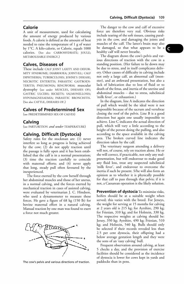
CalorieA unit of measurement, used for calculating the amount of energy produced by variousfoods. A calorie is defined as the amount of heatneeded to raise the temperature of 1 g of waterby 1°C. A kilo-calorie, or Calorie, equals 1000calories. (See also CARBOHYDRATE; JOULE;METABOLISABLE ENERGY.)
Calves, Diseases ofThese include CALF JOINT LAXITY AND DEFOR-
MITY SYNDROME; DIARRHOEA; JOINT-ILL; CALF
DIPHTHERIA; TUBERCULOSIS; JOHNE’S DISEASE;NECROTIC ENTERITIS; PARASITIC GASTROEN-
TERITIS; PNEUMONIA; RINGWORM; musculardystrophy (see under MUSCLES, DISEASES OF);GASTRIC ULCERS; RICKETS; SALMONELLOSIS;HYPOMAGNESAEMIA; PARASITIC BRONCHITIS.(See also CATTLE, DISEASES OF.)
Calves of Predetermined Sex(see PREDETERMINED SEX OF CALVES)
Calving(see PARTURITION and under TEMPERATURE)
Calving, Difficult (Dystocia)Safety rules for the stockman are: (1) neverinterfere so long as progress is being achieved by the cow; (2) do not apply traction until the passage is fully open and it has been estab-lished that the calf is in a normal presentation;(3) time the traction carefully to coincide with maternal efforts; and (4) never apply that long, steady pull often favoured by theinexperienced.
The force exerted by the cow herself throughher abdominal muscles and those of her uterus,in a normal calving, and the forces exerted bymechanical traction in cases of assisted calving,were evaluated by veterinarian J. C. Hindson,who used a dynamometer to measure theseforces. He gave a figure of 68 kg (150 lb) forbovine maternal effort in a natural calving.Manual traction by one man was found to exerta force not much greater.
The danger to the cow and calf of excessiveforce are therefore very real. Obvious risksinclude tearing of the soft tissues, causing paral-ysis in the cow, and damaging the joints andmuscles of the calf. The latter’s brain may alsobe damaged, so that what appears to be ahealthy calf will never breathe.
The diagram shows the cow’s pelvis and var-ious directions of traction with the cow in astanding position. (Her failure to lie down maybe due to stress, and in itself complicates deliv-ery. Other causes of difficulty in calving includenot only a large calf, an abnormal calf (mon-ster), and an awkward presentation, but also alack of lubrication due to loss of fluid or todeath of the fetus, and inertia of the uterine andabdominal muscles – due to stress, subclinical‘milk fever’, or exhaustion.)
In the diagram, line A indicates the directionof pull which would be the ideal were it notimpossible because of the sacrum and vertebraeclosing the roof of the pelvis. Line B is a gooddirection but again one usually impossible toachieve. Line C indicates the actual direction ofpull, which will vary a little according to theheight of the person doing the pulling, and alsoaccording to the space available in the calvingarea. The broken curved line indicates thedirection taken by the calf.
The veterinary surgeon attending a deliverywill not, of course, rely on traction alone. He orshe will correct, if practicable, not only any mal-presentation, but will endeavour to make goodany fluid loss, treat any suspected subclinical‘milk fever’, and endeavour to overcome theinertia if such be present. S/he will also form anopinion as to whether it is physically possiblefor that calf to pass through that pelvis; if it isnot, a Caesarean operation is the likely solution.
Prevention of dystocia To minimise risks,heifers should be at a suitable weight whenserved; this varies with the breed. For Jerseys,the weight for serving at 15 months for calvingat 2 years old is 215 kg; for Ayrshire, 290 kg;for Friesian, 310 kg; and for Holstein, 330 kg.The respective weights at calving should be:Jersey, 350 kg; Ayrshire, 490 kg; Friesian, 510kg; and Holstein, 540 kg. Bulls should only be selected if their records revealed less than 2.5 per cent dystocia, their offspring had abelow average gestation length and they werethe sons of an ‘easy calving’ bull.
Frequent observation around calving, at least5 checks a day, and the provision of exercisefacilities should be considered as the incidenceof dystocia is lower for cows kept in yards andpaddocks than in pens.
Calving, Difficult (Dystocia) 109
C
The cow’s pelvis and various directions of traction.
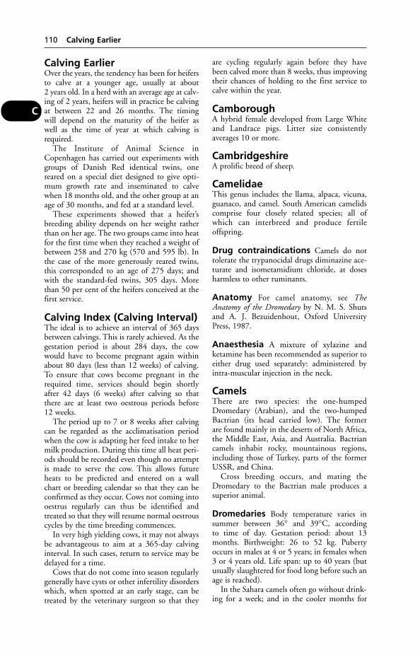
Calving EarlierOver the years, the tendency has been for heifersto calve at a younger age, usually at about2 years old. In a herd with an average age at calv-ing of 2 years, heifers will in practice be calvingat between 22 and 26 months. The timingwill depend on the maturity of the heifer as well as the time of year at which calving isrequired.
The Institute of Animal Science inCopenhagen has carried out experiments withgroups of Danish Red identical twins, onereared on a special diet designed to give opti-mum growth rate and inseminated to calvewhen 18 months old, and the other group at anage of 30 months, and fed at a standard level.
These experiments showed that a heifer’sbreeding ability depends on her weight ratherthan on her age. The two groups came into heatfor the first time when they reached a weight ofbetween 258 and 270 kg (570 and 595 lb). Inthe case of the more generously reared twins,this corresponded to an age of 275 days; andwith the standard-fed twins, 305 days. Morethan 50 per cent of the heifers conceived at thefirst service.
Calving Index (Calving Interval)The ideal is to achieve an interval of 365 daysbetween calvings. This is rarely achieved. As thegestation period is about 284 days, the cowwould have to become pregnant again withinabout 80 days (less than 12 weeks) of calving.To ensure that cows become pregnant in therequired time, services should begin shortlyafter 42 days (6 weeks) after calving so thatthere are at least two oestrous periods before12 weeks.
The period up to 7 or 8 weeks after calvingcan be regarded as the acclimatisation periodwhen the cow is adapting her feed intake to hermilk production. During this time all heat peri-ods should be recorded even though no attemptis made to serve the cow. This allows futureheats to be predicted and entered on a wallchart or breeding calendar so that they can beconfirmed as they occur. Cows not coming intooestrus regularly can thus be identified andtreated so that they will resume normal oestrouscycles by the time breeding commences.
In very high yielding cows, it may not alwaysbe advantageous to aim at a 365-day calvinginterval. In such cases, return to service may bedelayed for a time.
Cows that do not come into season regularlygenerally have cysts or other infertility disorderswhich, when spotted at an early stage, can betreated by the veterinary surgeon so that they
are cycling regularly again before they havebeen calved more than 8 weeks, thus improvingtheir chances of holding to the first service tocalve within the year.
CamboroughA hybrid female developed from Large Whiteand Landrace pigs. Litter size consistently averages 10 or more.
CambridgeshireA prolific breed of sheep.
CamelidaeThis genus includes the llama, alpaca, vicuna,guanaco, and camel. South American camelidscomprise four closely related species; all ofwhich can interbreed and produce fertileoffspring.
Drug contraindications Camels do nottolerate the trypanocidal drugs diminazine ace-turate and isometamidium chloride, at dosesharmless to other ruminants.
Anatomy For camel anatomy, see TheAnatomy of the Dromedary by N. M. S. Shutsand A. J. Bezuidenhout, Oxford UniversityPress, 1987.
Anaesthesia A mixture of xylazine and ketamine has been recommended as superior toeither drug used separately: administered byintra-muscular injection in the neck.
CamelsThere are two species: the one-humpedDromedary (Arabian), and the two-humpedBactrian (its head carried low). The former are found mainly in the deserts of North Africa,the Middle East, Asia, and Australia. Bactriancamels inhabit rocky, mountainous regions,including those of Turkey, parts of the formerUSSR, and China.
Cross breeding occurs, and mating theDromedary to the Bactrian male produces asuperior animal.
Dromedaries Body temperature varies insummer between 36° and 39°C, according to time of day. Gestation period: about 13months. Birthweight: 26 to 52 kg. Pubertyoccurs in males at 4 or 5 years; in females when3 or 4 years old. Life span: up to 40 years (butusually slaughtered for food long before such anage is reached).
In the Sahara camels often go without drink-ing for a week; and in the cooler months for
110 Calving Earlier
C
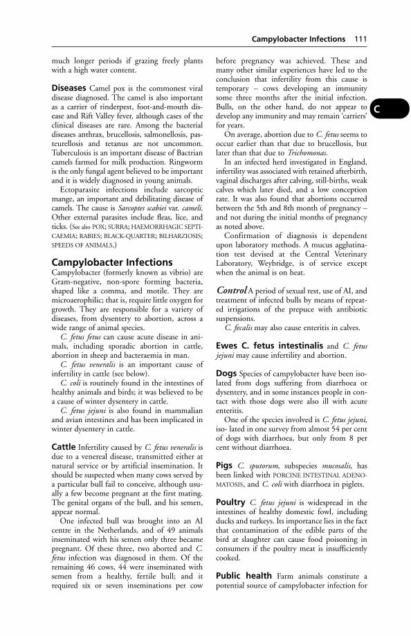
much longer periods if grazing freely plantswith a high water content.
Diseases Camel pox is the commonest viraldisease diagnosed. The camel is also importantas a carrier of rinderpest, foot-and-mouth dis-ease and Rift Valley fever, although cases of theclinical diseases are rare. Among the bacterialdiseases anthrax, brucellosis, salmonellosis, pas-teurellosis and tetanus are not uncommon.Tuberculosis is an important disease of Bactriancamels farmed for milk production. Ringwormis the only fungal agent believed to be importantand it is widely diagnosed in young animals.
Ectoparasite infections include sarcopticmange, an important and debilitating disease ofcamels. The cause is Sarcoptes scabiei var. cameli.Other external parasites include fleas, lice, andticks. (See also POX; SURRA; HAEMORRHAGIC SEPTI-
CAEMIA; RABIES; BLACK-QUARTER; BILHARZIOSIS;SPEEDS OF ANIMALS.)
Campylobacter InfectionsCampylobacter (formerly known as vibrio) areGram-negative, non-spore forming bacteria,shaped like a comma, and motile. They aremicroaerophilic; that is, require little oxygen forgrowth. They are responsible for a variety ofdiseases, from dysentery to abortion, across awide range of animal species.
C. fetus fetus can cause acute disease in ani-mals, including sporadic abortion in cattle,abortion in sheep and bacteraemia in man.
C. fetus veneralis is an important cause ofinfertility in cattle (see below).
C. coli is routinely found in the intestines ofhealthy animals and birds; it was believed to bea cause of winter dysentery in cattle.
C. fetus jejuni is also found in mammalianand avian intestines and has been implicated inwinter dysentery in cattle.
Cattle Infertility caused by C. fetus veneralis isdue to a venereal disease, transmitted either atnatural service or by artificial insemination. Itshould be suspected when many cows served bya particular bull fail to conceive, although usu-ally a few become pregnant at the first mating.The genital organs of the bull, and his semen,appear normal.
One infected bull was brought into an AIcentre in the Netherlands, and of 49 animalsinseminated with his semen only three becamepregnant. Of these three, two aborted and C.fetus infection was diagnosed in them. Of theremaining 46 cows, 44 were inseminated withsemen from a healthy, fertile bull; and itrequired six or seven inseminations per cow
before pregnancy was achieved. These and many other similar experiences have led to theconclusion that infertility from this cause istemporary – cows developing an immunitysome three months after the initial infection.Bulls, on the other hand, do not appear todevelop any immunity and may remain ‘carriers’for years.
On average, abortion due to C. fetus seems tooccur earlier than that due to brucellosis, butlater than that due to Trichomonas.
In an infected herd investigated in England,infertility was associated with retained afterbirth,vaginal discharges after calving, still-births, weakcalves which later died, and a low conceptionrate. It was also found that abortions occurredbetween the 5th and 8th month of pregnancy –and not during the initial months of pregnancyas noted above.
Confirmation of diagnosis is dependentupon laboratory methods. A mucus agglutina-tion test devised at the Central VeterinaryLaboratory, Weybridge, is of service exceptwhen the animal is on heat.
Control A period of sexual rest, use of AI, andtreatment of infected bulls by means of repeat-ed irrigations of the prepuce with antibiotic suspensions.
C. fecalis may also cause enteritis in calves.
Ewes C. fetus intestinalis and C. fetusjejuni may cause infertility and abortion.
Dogs Species of campylobacter have been iso-lated from dogs suffering from diarrhoea ordysentery, and in some instances people in con-tact with those dogs were also ill with acuteenteritis.
One of the species involved is C. fetus jejuni,iso- lated in one survey from almost 54 per centof dogs with diarrhoea, but only from 8 percent without diarrhoea.
Pigs C. sputorum, subspecies mucosalis, hasbeen linked with PORCINE INTESTINAL ADENO-
MATOSIS, and C. coli with diarrhoea in piglets.
Poultry C. fetus jejuni is widespread in theintestines of healthy domestic fowl, includingducks and turkeys. Its importance lies in the factthat contamination of the edible parts of thebird at slaughter can cause food poisoning inconsumers if the poultry meat is insufficientlycooked.
Public health Farm animals constitute apotential source of campylobacter infection for
Campylobacter Infections 111
C

man. Campylobacters were isolated from 259(31 per cent) of 846 faecal specimens collectedfrom domestic animals. The highest isolationrate was found in pigs (66 per cent); lower rateswere recorded for cattle (24 per cent) and sheep(22 per cent). All porcine isolates were C. coliwhile about 75 per cent of isolates from rumi-nants were C. jejuni. Cases of enteritis in peoplehave been linked to the consumption of milk from bottle-tops that had been pecked by birds. Campylobacters were isolated from 29 out of 37 magpies which had been shot, trapped, or killed on the roads in ruralareas around Truro, between June 1990 andFebruary 1991. Campylobacter jejuni biotypewas isolated from 25 of the birds, C. coli fromthree, C. jejuni biotype 2 from two and C. larifrom one.
CanaliculusA small channel, e.g. the minute passage lead-ing from the lacrimal pore on each eyelid to thelacrimal sac in the nostril.
CanaryThe canary, Serinus canaria, is a small seed-eat-ing bird usually yellow in colour. (See under CAGE
(AVIARY) BIRDS, DISEASES OF.)
Cancellous(see BONE)
Cancer (Neoplasia)Cancer (neoplasia) is perhaps best thoughtof as a group of diseases rather than as a singledisease entity. All types are characterised byuncontrolled multiplication of abnormal cells.Cancer can be malignant (progressive and inva-sive) and will often regrow after removal; ornon-malignant (benign) and will not return ifremoved. Malignant cancer cells usually have aprimary location. If untreated, secondarygrowths, called metastases, may develop inother parts of the body by a process calledmetastasis. Two important types of malignantgrowth are sarcomas and carcinomas. There are several subtypes of each, classified accordingto the nature of their cells or the tissues affected.
Sarcomas are, as primary growths, oftenfound in bones, cartilage, and in the connectivetissue supporting various organs. Common sar-comas include osteosarcoma, fibrosarcoma, andlymphosarcoma.
Carcinomas are composed of modifiedepithelial tissue, and are often associated withadvancing age. Primary carcinomas affect theskin and mucous membranes, for example, and
the junction between the two, such as lips,conjunctiva, etc.
Cancer can take many forms and the namesapplied relate to the type, e.g. tumour; the dis-ease caused, e.g. enzootic bovine leukosis, felineleukaemia; the tissue or organ affected, e.g.melanoma is cancer of the pigmented skin cells,osteosarcoma is cancer of the bones.
Cancer is far from rare in domestic animalsand farm livestock. In the latter, however, theincidence of cancer tends to be less, becausecattle, sheep, and pigs are mostly slaughteredwhen comparatively young. Nevertheless,sporadic bovine enzootic leukosis may appearin a clinical form in cattle under 2 years old andcancer of the liver is seen in piglets – to give buttwo examples.
In the old grey horse a melanoma is a com-mon tumour. In dogs the incidence of tumoursgenerally (including non-malignant ones) issaid to be higher than in any other animalspecies, including the human. (See CANINE
TUMOURS.) An osteosarcoma is a not uncom-mon form of cancer affecting a limb bone inyoung dogs. LEUKAEMIA provides anotherexample of cancer. In cats, a survey of 132 withmammary gland tumours showed the ratio ofmalignant to benign growths to be 9:1. (See
FELINE CANCER.) The relative risk in spayed catsis said to be significantly less than in intactfemales.
112 Canaliculus
C
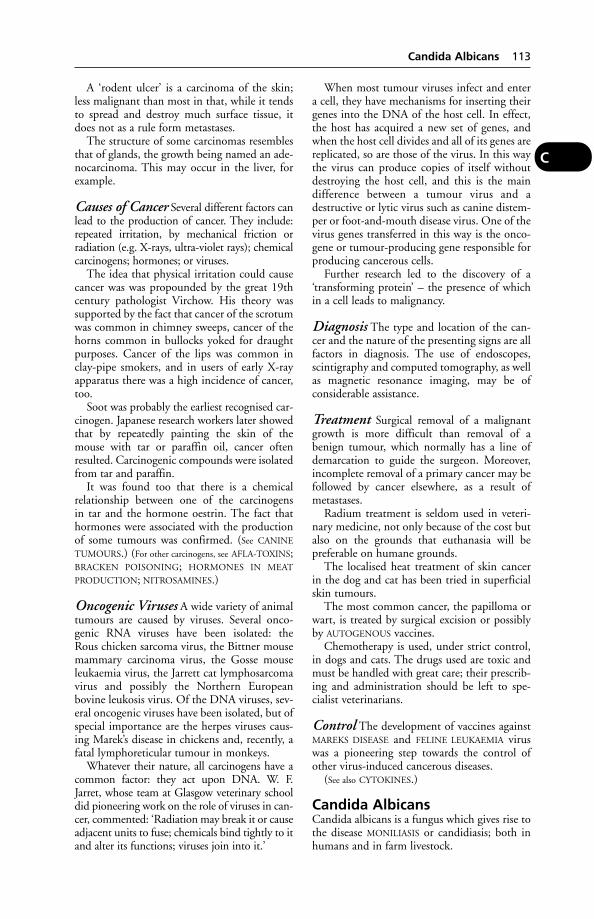
A ‘rodent ulcer’ is a carcinoma of the skin;less malignant than most in that, while it tendsto spread and destroy much surface tissue, itdoes not as a rule form metastases.
The structure of some carcinomas resemblesthat of glands, the growth being named an ade-nocarcinoma. This may occur in the liver, forexample.
Causes of Cancer Several different factors canlead to the production of cancer. They include:repeated irritation, by mechanical friction orradiation (e.g. X-rays, ultra-violet rays); chemicalcarcinogens; hormones; or viruses.
The idea that physical irritation could causecancer was was propounded by the great 19thcentury pathologist Virchow. His theory wassupported by the fact that cancer of the scrotumwas common in chimney sweeps, cancer of thehorns common in bullocks yoked for draughtpurposes. Cancer of the lips was common inclay-pipe smokers, and in users of early X-rayapparatus there was a high incidence of cancer,too.
Soot was probably the earliest recognised car-cinogen. Japanese research workers later showedthat by repeatedly painting the skin of themouse with tar or paraffin oil, cancer oftenresulted. Carcinogenic compounds were isolatedfrom tar and paraffin.
It was found too that there is a chemical relationship between one of the carcinogens in tar and the hormone oestrin. The fact thathormones were associated with the productionof some tumours was confirmed. (See CANINE
TUMOURS.) (For other carcinogens, see AFLA-TOXINS;BRACKEN POISONING; HORMONES IN MEAT
PRODUCTION; NITROSAMINES.)
Oncogenic Viruses A wide variety of animaltumours are caused by viruses. Several onco-genic RNA viruses have been isolated: theRous chicken sarcoma virus, the Bittner mousemammary carcinoma virus, the Gosse mouseleukaemia virus, the Jarrett cat lymphosarcomavirus and possibly the Northern Europeanbovine leukosis virus. Of the DNA viruses, sev-eral oncogenic viruses have been isolated, but ofspecial importance are the herpes viruses caus-ing Marek’s disease in chickens and, recently, afatal lymphoreticular tumour in monkeys.
Whatever their nature, all carcinogens have acommon factor: they act upon DNA. W. F.Jarret, whose team at Glasgow veterinary schooldid pioneering work on the role of viruses in can-cer, commented: ‘Radiation may break it or causeadjacent units to fuse; chemicals bind tightly to itand alter its functions; viruses join into it.’
When most tumour viruses infect and entera cell, they have mechanisms for inserting theirgenes into the DNA of the host cell. In effect,the host has acquired a new set of genes, andwhen the host cell divides and all of its genes arereplicated, so are those of the virus. In this waythe virus can produce copies of itself withoutdestroying the host cell, and this is the maindifference between a tumour virus and adestructive or lytic virus such as canine distem-per or foot-and-mouth disease virus. One of thevirus genes transferred in this way is the onco-gene or tumour-producing gene responsible forproducing cancerous cells.
Further research led to the discovery of a‘transforming protein’ – the presence of whichin a cell leads to malignancy.
Diagnosis The type and location of the can-cer and the nature of the presenting signs are allfactors in diagnosis. The use of endoscopes,scintigraphy and computed tomography, as wellas magnetic resonance imaging, may be ofconsiderable assistance.
Treatment Surgical removal of a malignantgrowth is more difficult than removal of abenign tumour, which normally has a line ofdemarcation to guide the surgeon. Moreover,incomplete removal of a primary cancer may befollowed by cancer elsewhere, as a result ofmetastases.
Radium treatment is seldom used in veteri-nary medicine, not only because of the cost butalso on the grounds that euthanasia will bepreferable on humane grounds.
The localised heat treatment of skin cancerin the dog and cat has been tried in superficialskin tumours.
The most common cancer, the papilloma orwart, is treated by surgical excision or possiblyby AUTOGENOUS vaccines.
Chemotherapy is used, under strict control,in dogs and cats. The drugs used are toxic andmust be handled with great care; their prescrib-ing and administration should be left to spe-cialist veterinarians.
Control The development of vaccines againstMAREKS DISEASE and FELINE LEUKAEMIA viruswas a pioneering step towards the control ofother virus-induced cancerous diseases.
(See also CYTOKINES.)
Candida AlbicansCandida albicans is a fungus which gives rise tothe disease MONILIASIS or candidiasis; both inhumans and in farm livestock.
Candida Albicans 113
C

Canicola FeverThe disease in man caused by the parasiteLeptospira canicola, which is excreted in theurine of infected dogs. Paresis may occur andsome few cases of this disease may resemblepoliomyelitis. Mild conjunctivitis and nephritisaccompanying symptoms of meningitis are sug-gestive of canicola fever. The parasite may beharboured by pigs and the disease has beenrecorded among workers on pig farms andmilkers in dairy units. (See LEPTOSPIROSIS.)
Canine Adenovirus Infection(see CANINE VIRAL HEPATITIS)
Canine AutoimmuneHaemolytic AnaemiaA progressive disease caused by a dog formingantibodies which destroy its own red bloodcells. A deficiency of platelets may occur simul-taneously. This disease is a clotting disordercaused by a deficiency of blood factor VIII, andis usually fatal in males at an early age.
Signs Pale mucous membranes, lethargy,weakness, and collapse.
Diagnosis A Coombs’ antiglobulin test.
Canine Babesiosis(Piroplasmosis)Canine babesiosis (piroplasmosis), which is alsocalled tick fever, malignant jaundice, and biliaryfever, is a tick-transmitted protozoan parasiticinfection increasingly common in the UK sincethe advent of the Pet Travel Scheme. Up to 30per cent of dogs returning with their owners fromEurope may be infected. Signs of infection arefever, weakness and malaise. Haemolytic anaemiais followed by haemoglobinurea and thrombocy-topaenia. Chronic infection must be confirmedby laboratory tests. Imidocarb dipropionate iseffective but must be continued after symptomsare relieved (in 24 to 48 hours) to ensure that theparasite is all destroyed. Babesia canis is the mostcommon cause but B. gibsoni is also a possibility;this is more resistant to treatment. Tick-repellentpreparations help prevent infection.
Canine Brucellosis(see BRUCELLOSIS)
Canine Distemper(see DISTEMPER)
Canine DysautonomiaA syndrome resembling the Key-Gaskell syn-drome in cats has been reported in dogs, and
has been tentatively linked with canine par-vovirus. (See FELINE DYSAUTONOMIA.)
Canine EhrlichiosisA rickettsial infection, formerly confined to thetropics but increasingly seen in Britain since theintroduction of the Pet Travel Scheme. Infecteddogs show fever, lethargy, anorexia, lym-phadenopathy and thrombocytopenia; urinemay be dark in colour. In the chronic form, theremay be uveitis and retinal haemorrhage, withgammaglobulinaemia. Diagnosis is confirmed byserological tests. Prompt treatment with doxycy-cline or tetracycline is usually effective, except inGerman shepherd dogs, in which pancytopeniais usually irreversible. The disease is transmittedby the ticks Rhipicephalus and Dermacentor spp.Tick-control preparations help prevent infection.
Canine FertilityIt has been suggeseed that a total output of 200 million sperms per ejaculate is necessary ifa dog is to be regarded as sound for breeding.Individual progressive motility of less than 70 per cent of sperms, and sperm head andmidpiece abnormalities in more than 40 percent of sperms, are associated with infertility.
Canine Filariasis(see HEARTWORMS and TRACHEAL WORMS)
Canine HaemophiliaThis is an uncommon disease of male dogs ofvirtually all breeds, characterised by an inherit-ed defect causing abnormally slow clotting ofthe blood, so that bleeding may occur andcontinue following only a minor injury.
Cause A sex-linked recessive gene (see GENET-
ICS). Should the dam carry this, then 50 percent of her dog pups are likely to be affectedand show symptoms. Bitches, though carriers ofthe gene, seldom show symptoms themselves.
Signs These may sometimes be vague and mis-leading, in that a temporary swelling on the fore-head, for example, or transient lameness, may beattributed solely to violence of some kind. Thefirst time that a haematoma is found in the ani-mal, violence may again be thought to be theonly cause of the bleeding, and even after repeat-ed episodes it may be thought that the animal issuffering from warfarin poisoning. In some casesthe abnormally slow clotting of the blood givesrise to excessive bleeding at teething, or if thetoe-nails are inadvertently trimmed too close.
Diagnosis Confirmation depends uponlaboratory tests.
114 Canicola Fever
C
Precautions Affected dogs cannot lead arough-and-tumble life without bleeding occur-ring, so the owner must try to prevent knocksand bumps occurring; or agree to euthanasia. Abitch which is known to be a carrier should not,of course, be bred from.
Canine HerpesvirusA virus isolated from vesicles affecting the gen-ital system of the bitch and associated withinfertility, abortion, and stillbirths. Infectedpups usually die soon after birth. Those thatrecover may remain carriers of the virus.
Canine Juvenile OsteodystrophyThis is known also by other names, e.g. nutri-tional secondary parathyroidism. It is also foundin cats, when it is referred to as FELINE JUVENILE
OSTEODYSTROPHY. It arises from a calcium defi-ciency which, in conjunction with excess vitaminD, stimulates the release of parathyroid hormone(see the table under PARATHYROID GLANDS).Resorption of bone follows. An excess of phos-phorus in the diet will also cause the condition.
Cause The main cause of this disease is feed-ing the dog a (muscle) meat-rich diet contain-ing little calcium but much phosphorus. (See
DOGS’ DIET.)
Signs Affected animals are often in good bod-ily condition but are usually reluctant to moveand may cry out in anticipation of being forcedto do so. The usual cause of pain is fractures ofthe thinned bone after a minor injury or evenno apparent injury. Short, hesitant steps may betaken. Splaying of the toes is sometimes seen;also swelling at the elbow or carpi.
On radiography, the skeleton appears lessdense than normal, indicating demineralisationof the bones.
The bones return to normal when a balanceddiet is fed but deformities left by fractures mayremain.
Canine Leishmaniasis(see LEISHMANIA; LEISHMANIASIS)
Canine Myasthenia Gravis(see MYASTHENIA GRAVIS)
Canine Nasal MitesA white mite, Pneumonyssoides caninum, is anuncommon inhabitant of the nose and nasalsinuses of dogs; and has also been found in thebronchi, and in the fat near the pelvis of thekidney.
Rubbing the nose on the ground and shakingthe head are symptoms of this infestation,
which has been reported from Scandinavia,America, Australia, and South Africa.
Breathing dichlorvos vapour from a poly-thene bag has been stated to be effective inkilling the mites (but dichlorvos is also toxic todogs).
Canine Parvovirus (CPV)This infection appeared as a new disease entityin 1978–9 in Europe, Australia, and America.Dogs proved highly susceptible, and seriousoutbreaks of the illness occurred with numer-ous deaths. By 1981 many dogs had acquireda useful degree of immunity against the virus,following either recovery from a naturallyoccurring attack or vaccination; with puppiesprotected for up to 16 weeks by the antibodiesreceived in the colostrum of their dams, assum-ing that the latter were themselves resistant.
Cause A parvovirus, possibly a mutation of thefeline enteritis or the mink enteritis virus.
Canine parvovirus (CPV-2), feline panleu-copenia virus (FPV), and mink enteritis virusshare common antigens; however, CPV-2 has atleast one specific antigen which is not presentin the other viruses.
Signs The illness takes the form of a severegastroenteritis, and diarrhoea is the main symp-tom. In the early outbreaks many dogs diedwithin 48 hours. Puppies may die suddenly,within minutes of eating or playing, as a resultof the virus having infected the heart muscleand caused myocarditis.
Treatment A combined antiserum prepara-tion is available. Symptomatic treatment mustinclude measures to overcome the severe DEHY-
DRATION resulting from the diarrhoea.Treatment of the myocarditis is seldom effective.
Prevention Vaccination is widely practisedand has greatly reduced the incidence of thedisease. Live vaccines, often combined withvaccines against distemper and other viral dis-eases, are available. It is essential to follow themanufacturers’ directions if protection is to beeffective. Annual booster doses are recommend-ed to maintain immunity. It should be notedthat apart from the effect of persisting MATER-
NAL ANTIBODIES, vaccination may fail in someindividuals which have a defective immune sys-tem and cannot produce adequate antibodies.This occurs with all vaccines.
Canine Pasteurellosis(see under BITES)
Canine Pasteurellosis 115
C

Canine Respiratory Disease(see DISTEMPER; KENNEL COUGH; KLEBSIELLA)
Canine Rickettsiosis(see ROCKY MOUNTAIN FEVER)
Canine StaphylococcalDermatitisThis may be seen in Irish setters, collies and shel-ties. The lesions appear on the fine skin with fewhairs on the abdomen or between the thighs.The condition is itchy, and causes the dog toscratch or lick the part. The lesions consist ofroughly circular areas of reddened skin, somewith a ring of blackish or greyish crust, havingpapules or pustules at the edge. The appearancemay suggest ringworm at first glance.
The Staphylococcus aureus involved is resis-tant to penicillin, so other antibiotics must beused. An autogenous vaccine may be needed ifantibiotics are not effective.
Canine TeethCanine teeth are the so-called ‘eye-teeth’, whichare such prominent features of the mouths ofcarnivorous animals. In different animals theyare known by different names, e.g.’tusks’ in thepig, and ‘tushes’ in the horse and ass. (See
DENTITION; TEETH.)
Canine Transmissible VenerealTumoursCanine transmissible venereal tumours affectmainly the mucous membrane of the vagina orthat of the prepuce; occasionally the lips of bothsexes. The lesions resemble warts, and can resultin infertility.
Canine TumoursThese are common. It has been suggested thatthe incidence of neoplasia in the dog is higherthan in any other animal species includingman. In fact, the age-adjusted incidence rate formammary neoplasia is three times larger in thebitch than in women. Tumours arising in themammary glands of the bitch and the perianalglands of the dog together may account foralmost 30 per cent of all canine neoplasms. Thepredilection of these tumours for one sex or theother and their responsiveness, in some cases, toendocrine gland ablation or hormone therapyhas promoted their designation as hormone-dependent. (See also TUMOUR; CANCER.)
Canine Viral Hepatitis (CVH)Canine viral hepatitis (CVH) is also known asRubarth’s disease, Hepatitis contagiosa canis, orinfectious canine hepatitis (ICH).
Dogs of all ages may be affected – even pup-pies a few days old – but perhaps the diseaseoccurs most frequently in young dogs of 3 to 9months. CVH may occur simultaneously withDISTEMPER.
Cause A canine adenovirus (CAV). CAV-1 isassociated with liver, eye, kidney, and respiratorydisease. (CAV-2 is implicated only in respiratorydisease.)
Signs Infection may exist without symptoms,and in such cases it can be recognised only bylaboratory tests. In the very acute form of thedisease a dog, apparently well the night before,may be found dead in the morning. In lessacute cases the dog may behave strangely andhave convulsions. A high temperature, wasting,anaemia, lethargy, and coma are other symp-toms observed in some cases. A thin, threadypulse is characteristic.
Vomiting, diarrhoea, and dullness may per-sist for 5 or 6 days, and be followed by jaundice.Such cases may be thought to be leptospiraljaundice.
Puppies may show symptoms of severe inter-nal haemorrhage, and have blood or blood-stained fluid in the peritoneal cavity, withpetechial haemorrhages from several organs.Haemorrhages, including subcutaneous ones,may also occur in older dogs. More commonly,there is fever, dullness, some vomiting, tender-ness of the abdomen. Of those that survive 5days or so, many recover. Keratitis (‘blue-eye’)occurs a week or two after the beginning of theillness in some cases. In older dogs, restlessness,convulsions, and coma are common.
Antiserum is useful in treatment. Glucoseand vitamin K are also recommended.
Dogs which have recovered may continue toharbour the virus and act as carriers, spreadingthe disease to other dogs via the urine.
Diagnosis A gel diffusion test is useful atpostmortem examination, especially wheredecomposition of the animal’s body hasinvolved cell disintegration.
Prevention Vaccines are available, both liveand inactivated. Hepatitis vaccine is usuallypresented as a multiple vaccine in combinationwith distemper and parvovirus; some prepara-tions also include protection against leptospiro-sis and parainfluenza. Dosage instructions varywith different brands of vaccine; normally, pup-pies are given two doses at an interval of 2 to 6weeks followed by annual booster inoculations.(See under DISTEMPER.)
116 Canine Respiratory Disease
C

Cannabis Poisoning(see MARIJUANA)
Cannibalism
Poultry Cannibalism may follow feather-picking – especially if blood is drawn – or a caseof prolapse. The crowding together of housedbirds is a common cause; and boredom (noscratching for insects as out-of-doors) is a factor,too. Occasionally a nutritional deficiency maybe involved. In broiler plants, beak-trimming orsubdued red lighting, making everything appearpink, has been resorted to. (See also SPECTACLES.)
In free range hens, cannibalism can be stim-ulated by the appearance of the pink of theinside of the cloaca at egg-laying. The wall ofthe cloaca may be penetrated, the intestinegrasped and ripped out.
Pigs TAIL-BITING is a complex problem, andtail sores can lead to death. In some cases, therunt of the litter starts the vice, possibly becauseit is prevented by litter mates from access to theteats or trough and has nothing but tails pre-sented to it. Cannibalism, where sows eat pigletsmainly at birth or shortly afterwards, has beenseen increasingly among farrowing sows kept onfree range, chiefly on arable farms. The cannibalsow does not eat her own litter but guards itfiercely against other predatory sows. Thus thisvice is entirely different from the occasional sav-aging of a litter by a hysterical sow or (morecommonly) gilt in intensively kept pig herds.
Wild boar Wild boar sows must be allowedto leave the herd to give birth, returning to itlater. If piglets are born near other sows they areat risk of being eaten while still in the mem-brane. The risk lessens when the piglets arerunning about.
Cannon Bone(see METACARPAL)
CantharidesCantharides is a powder made from the driedbodies and wings of the Spanish fly Cantharisvesicatoria, or Lytta vesicatoria. It contains can-tharidin, an irritant poison, which has beenused in rubefacient and blistering applications.It can be fatal if taken internally: a youngwoman died after being given a drink spikedwith cantharides by a would-be suitor.
Cantharidin poisoning has been reported ina horse and a mule, which died after eating haycontaminated by beetles (Epicanta vittata)which contain cantharidin.
Actions Cantharadin has an irritant action onthe genital and urinary organs by which it iseliminated from the body. This action isresponsible for its reputation as an aphrodisiac.
CanthusCanthus is the angle at either end of the aper-ture between the eyelids.
CapillariasisInfestation with Capillaria worms; it causes lossof condition and gastroenteritis in birds. Inmammals, diarhorrea, cystitis, hepatitis orbronchial disease may be seen. C. obsignata hasbeen recognised as of economic importance inintensely reared poultry in Britain.
Treatment is with flubendazole in poultryand game birds and with cambendazole and lev-amisole in pigeons. (See also URINARY BLADDER,DISEASES OF.)
CapillariesCapillaries are the very minute vessels that jointhe ultimate arteries (or arterioles) to the com-mencement of the veins. Their walls consist ofa single layer of fine, flat, transparent cells,joined together at their edges, and the vesselsform an intricate mesh-work throughout thetissues of the body, bathing them in blood, withonly the thin walls interposed, and allowingfree exchange of gases and fluids. These vesselsare less than 0.25 mm (1/1000th of an inch) indiameter.
Capillary Refill TimeA means of obtaining a rough assessment of thestate of the peripheral circulation. It is the timetaken for mucosa (e.g. in the mouth) to returnto its normal colour after application of pres-sure. The time should normally be less than2 seconds.
CaponisationThe castration of cockerels, carried out in orderto provide a more tender carcase, and also toobviate crowing and fighting. The castratedbird is called a capon. Stilboestrol or hexoestrol,used as pellets implanted under the skin highup the neck, were used to achieve a similareffect but such hormonal treatments are nowbanned.
Capped Elbow(see under BURSITIS)
Capped HockCapped hock is a term loosely applied to anyswelling over the point of the hock. At this point
Capped Hock 117
C

there are two bursae: the first – a false bursa, dis-tension of which constitutes true ‘capped hock’– lies between the skin and the tendon whichplays over the bone; and the second, the truebursa, separates the tendon from the bone.
The lesion is virtually identical with that of capped elbow (see under BURSITIS), and treat-ment is practically the same.
Since the condition may be brought about inthe mare by continual kicking at the heel postsof the stall (e.g. in cases of nymphomania), it isnecessary to pad the heel posts or to house thehorse in a loose-box.
‘Cappie’‘Cappie’ is a disease of sheep. (See also ‘DOUBLE
SCALP’.)
Caprine Arthritis-EncephalitisA disease of goats caused by a lentivirus. It ispresent in Britain, Switzerland, France,Norway, the USA and Canada. It was followingimport of goats from Switzerland and the USAinto Kenya that the disease reached Africa in1983. In Australia a retrovirus was isolatedfrom goats which caused a clinical diseasesimilar to caprine arthritis-encephalitis, andproduced antibodies in goats similar to thosecaused by maedi-visna virus, which has neverbeen recorded in that continent.
Signs A lowered milk yield, due to mastitis, issometimes the first sign noticed; and transmis-sion of the virus is thought to be mainly viacolostrum and milk.
The main sign, however, is arthritis.Lameness does not always accompany swellingof the joints.
Encephalitis, caused by the virus affecting thebrain, affects mainly kids 2 to 4 months old.Lesions may occur in the spinal cord also. Head-tilting and trembling may be seen, together withan unsteady gait. Opisthotonus may occur.Partial paralysis may lead to recumbency andoften death. A chronic interstitial pneumoniaoccurs in some goats and subclinical infectionsmay occur.
Capripox Viruses(see ‘LUMPY SKIN DISEASE’; POX)
CapsuleCapsule is a term used in several senses. Theterm is applied to a soluble case, either of gela-tine which dissolves in the stomach, or of keratinwhich only dissolves in the small intestine, forenclosing small doses of medicine. The term isalso applied to the fibrous or membranous
envelope of various organs, as of the spleen, liver,or kidney. It is also applied to a ‘joint capsule’.
Car Exhaust FumesCar exhaust fumes from a specially adapted carengine may be used for the humane destructionof mink. The Welfare of Animals (Slaughter orKilling) Regulations 1995 state that the exhaustgas must be cooled and filtered free of any irri-tant material. The carbon monoxide level mustreach at least 1 per cent of the volume of thechamber used before mink are placed in it andthe animals must remain there until dead. Carexhaust is no longer recognised as a legal meansof killing birds. (See under BIRDS, HUMANE
DESTRUCTION OF.)
Car, Parked in the SunThe temperature inside a car parked in the sun,even with two windows opened to the extent of2.5 cm (1 in), can within 3 hours reach 33°C(92°F), when the shade temperature outside thecar is only 18°C (65°F). With only one windowopened 2.5 cm (1 in), or all windows closed, a dangerously high temperature would obvious-ly be reached much sooner. A dog left in a carparked not in the shade is in danger of HEAT-
STROKE; a cat similarly. (See also HYPERTHER-
MIA.) Owners causing suffering to their pets byleaving them in cars may face prosecutionunder the Protection of Animals Act 1911.
Car Sickness(see TRAVEL SICKNESS)
CarapaceThe shell of tortoises, other chelonians, andcrustaceans. When assessing the health of a che-lonian, it is important to relate the length of thecarapace in relation to the body weight, espe-cially as to ability to withstand a period ofhibernation. The landing of crabs and lobstersin Britain is subject to the carapace being of aspecified minimum length.
CarbacholCarbachol is a potent parasympathomimeticagent which is used in the treatment of glaucomain dogs.
CarbamatesThese compounds are used as agriculturalinsecticides and sometimes cause accidentalpoisoning in animals. Carbamates inhibitcholinesterase. Symptoms of poisoning includeprofuse salivation, muscular tremors. Atropineis used in treatment. (See ORGANOPHOSPHORUS
POISONING.)
118 ‘Cappie’
C
CarbohydrateCarbohydrate is a term used to include organiccompounds containing carbon, hydrogen, and oxygen, the two latter being in the sameproportions as they are present in water, viz.two parts of hydrogen to every one part of oxygen. The simplest carbohydrates are themonosaccharide sugars (e.g. glucose), thencome disaccharides (e.g. cane sugar, lactose)and polysaccharides. These are complex carbo-hydrates, such as the starches, celluloses, andlignified compounds in hay, which must bebroken down into simpler sugars by both bac-terial and protozoal action and by the processesof digestion before they can be absorbed andused in the body.
Carbolic Acid(see PHENOL)
Carbolic Acid PoisoningCarbolic acid poisoning may occur from theapplication to the skin of dressings medicatedwith PHENOL; from the internal administrationof the drug by mistake; and cases have beenrecorded from the use of strong carbolic disin-fecting powders sprinkled on to the floors ofanimal buildings.
Carbon Dioxide (CO2)Carbon dioxide (CO2) is a colourless gas. It isformed in the tissues during the metabolicprocess, taken up by the blood, exchanged foroxygen in the lungs, and expired from themwith each breath. In a building, the VENTILA-
TION must be such as will get rid of it rapidly sothat it does not accumulate in the atmosphere.In the air it is present to the extent of about0.03 per cent by volume, although this amountvaries. CO2 is used as a respiratory stimulant byanaesthetists.
Carbon Dioxide AnaesthesiaCO2 has been widely used for anaesthetisingpigs and poultry prior to slaughter. For pigs, itis necessary to have a concentration of 70 percent CO2 by volume. An alarm must be fittedwhich goes off if the level in the gassing tunneldrops below this. The pigs are driven in singlefile through a tunnel and inhale the CO2 forless than a minute, after which a very brief peri-od of unconsciousness follows – long enough,however, for hackling and ‘sticking’ to beaccomplished without causing pain. There is noadverse effect upon the carcase. CO2 has alsobeen used, instead of chloroform, in lethalchambers or cabinets for the euthanasia of cats,but if it is to be humane the technique must be
correct. A mixture of argon with carbon dioxidehas been shown to be preferable on humanegrounds to CO2 alone.
Carbon Dioxide SnowCarbon dioxide snow is formed when CO2 isfirst compressed in a cylinder to a liquid andthen released through a small nozzle. The tem-perature falls to about –70°C and the CO2
solidifies as a snow. This is then compressedinto solid blocks, which are used for a variety ofpurposes where a low temperature is requiredfor a considerable time, such as to cool meat,milk, or fish in transit by rail, to preserve tis-sues, bacteria, or foods, so that normal enzymeaction is arrested, and sometimes to producelocal anaesthesia by freezing or to cauterise asurface growth on the skin.
A piece of ‘dry ice’ or carbon dioxide ‘snow’placed on the floor of an infested building willact as a bait for ticks which will gather round itand can then be collected and destroyed.
Carbon Fibre ImplantsThese have been used in the surgical repair oftendons in racehorses, and dogs, and havegenerally given good results.
Carbon MonoxideCarbon monoxide poisoning may result fromgas and solid-fuel heating systems in the homewhen there is an inadequate supply of air. Manydogs and cats have been found dead in thekitchen in the morning.
In Britain, until the late 1960s, town gas(derived from coal) contained 10 to 20 per centof carbon monoxide. Natural and oil-based gascontain less than 1 per cent. However, wherethere is inadequate VENTILATION, incompletecombustion may occur leaving not carbondioxide and water but carbon monoxide.
Stillbirths in sows have been ‘associated withincomplete combustion in propane gas heatersand inadequate ventilation. In one herd whenpoor ventilation and faulty heaters were correct-ed, the stillbirth rate dropped from 28 per centto 6.7 per cent. The pig fetus is very susceptibleto carbon monoxide poisoning, and may die inthe uterus or at farrowing, without clinical signsof ill health being shown by the sow.
Exhaust fumes from an ordinary motor carhave been used as a source of carbon monoxidefor the destruction of mink and turkeys, butthis is no longer legal. (see CAR EXHAUST
FUMES.)
Diagnosis Cherry-red tissues and body fluidsare suggestive of poisoning. Analysis of blood
Carbon Monoxide 119
C
samples for carboxy-haemaglobin can be usedfor confirmation.
Abortion may be caused by carbon monoxideeven at levels too low to cause signs in adult pigs.
Carcases, Disposal of(see under DISPOSAL)
CarcinogensCarcinogens are oncogenic viruses or substanceswhich give rise to CANCER. (See NITROSAMINES;BRACKEN; AFLATOXINS; HORMONES IN MEAT;and substances mentioned under CANCER.)
Carcinoma(see CANCER)
CardiaCardia is the upper opening of the stomach atwhich the oesophagus terminates. It lies closebehind the heart.
Cardiac Disease(see HEART DISEASES)
Cardiac Pacemakers(see PACEMAKER)
CardiographyCardiography is the process by which graphicrecords can be made of the heart’s action.Auricular and ventricular pressures can berecorded, the sounds of the heartbeat can beconverted into waves of movement and record-ed on paper, and the changes in electric poten-tial that occur can be similarly recorded. (See also
under ELECTROCARDIOGRAM.)
CardiologyStudy of the heart and heart diseases.
Caries(see TEETH, DISEASES OF)
CarminativesCarminatives are substances which help torelieve TYMPANY or flatulence. Almost all thearomatic oils are carminatives.
Carnassial Tooth(see under SKULL)
CaroteneA yellow pigment found in many feeds, carrots,egg yolks, etc. which can be converted into vitamin A (see VITAMINS).
CarpitisArthritis affecting the carpus.
CarprofenA non-steroidal anti-inflammatory drug(NSAID) used in companion animals, farmanimals and horses.
CarpusCarpus is the wrist in man, or the ‘knee’ of thefore-limbs of animals.
‘Carrier’‘Carrier’ is an animal recovered from an infec-tious disease, or not showing symptoms, butcapable of passing on the infection to anotheranimal. For example, cattle may carry infectiousbovine rhinotracheitis; dogs may be carriers ofleptospirae.
Carrying Injured Dogs and Cats(see illustration under ACCIDENTS)
CartilageCartilage is a hard but pliant tissue formingparts of the skeleton, e.g. the rib cartilages, thecartilages of the larynx and ears, and the lateralcartilages of the foot, as well as the cartilages ofthe trachea. Microscopically it consists of cellsarranged in pairs or in rows, embedded in a clearhomogeneous tissue devoid of blood-vessels andnerves. The surfaces of the bones that form ajoint are covered with articular cartilages, whichprovide smooth surfaces of contact and min-imise shock and friction. In some parts of thebody there are discs of cartilage interposedbetween bones forming a joint, e.g. between thefemur and tibia and fibula there are the carti-lages of the stifle joint, and between most of theadjacent vertebrae there are similar discs. Whena bone is still growing, there are layers of carti-lage interposed between the shaft and itsextremities; these are called epiphyseal cartilages.
Diseases of cartilage Two chief diseasesaffect cartilages in animals. Necrosis, or death ofthe cells of the cartilage, results from accident,injury, or in some cases from pressure. The treat-ment is wholly surgical, and consists in theremoval of the dead piece or pieces and the pro-vision of drainage for discharges. Ossification:many of the cartilaginous structures of the bodybecome ossified into bone in the normal course,especially in old age; but as the result of a singlemild or many slight injuries to a cartilage, theformation of bone may take place prematurely,and interference with function results.
CaruncleA small fleshy protuberance, which may be a nor-mal anatomical part. In the uterus of ruminants,
120 Carcases, Disposal of
C
for example, mushroom-shaped caruncles projectfrom the inner surface to give attachment to thecotyledons of the fetal membranes.
CascaraA purgative occasionally used for the relief of constipation in dogs and cats, and for thetreatment of furballs in cats.
CaseationCaseation is the drying up and necrosis of a tis-sue. For example, a tuberculous abscess changesinto a firm, cheese-like mass, which may latercalcify. (See CALCIFICATION.)
CaseinA protein of milk and an important constituentof ‘solids-not-fat’.
Caseous LymphadentitisCaseous lymphadentitis is a chronic disease ofthe sheep and goat, characterised by the forma-tion of nodules containing a cheesy pus occur-ring in the lymph nodes, lungs, skin, or otherorgans; exhibiting a tendency to produce achronic pneumonia or pleurisy.
The disease is believed to have been introducedto the UK in a consignment of 20 goats import-ed from Germany in 1987. It leads to productionlosses and condemnation of carcases at slaughter.
Cause Corynebacterium pseudotuberculosis.Introduction of infected animals to a herd is themost important means of spreading infection.Wound infection is a common source. Theorganism can survive outside the animal onstraw, etc. for months and in sheep dips for24 hours. Contaminated shearing or ear-tagging tools have also been implicated.
Treatment This is difficult as the lesionsbecome encapsulated and so inaccessible toantibiotics. Vaccines are available overseas.
Diagnosis Culture of C. pseudotuberculosisfrom pus from lesions confirms the diagnosis.ELISA tests are being developed.
Cassava(Manihot esculenta) A widely grown crop forhuman and animal food in the tropics, and thesource of tapioca. The potato-like tubers, howev-er, if eaten raw can cause cyanide poisoning.Livestock in the tropics have died from cyanidepoisoning caused by this crop. It must not beused in turkey feeds as it is not digested in theupper digestive tract but ferments in the caecumcausing inflammation (typhilitis). The liquid fae-ces make wet litter and leg problems may follow.
Castor Seed PoisoningCastor seed poisoning has occurred overseasthrough animals being accidentally fed eitherwith the seeds themselves or with some residuefrom them. The seeds of the castor plant(Ricinus communis) contain an oil which is used not only as a medicinal agent, but also forlubricating. Processing leaves behind in thepress-cakes the toxin ricine, and renders these‘castor-cakes’ unsuitable as a food-stuff for alllive-stock. Overseas, however, unscrupulouscattle-cake merchants sometimes sell them forfeeding cattle after treating the residual press-cakes with steam, but with the result that thericine is not all destroyed and poisoning mayoccur.
Signs These consist of dullness, loss ofappetite, elevation of the temperature, severeabdominal pain, and usually constipation butsometimes diarrhoea. The heart’s action istumultuous, the surface of the body is cold;there may be a watery cold sweat, and therespiration is distressed. Where large amountshave been eaten the faeces are usually hard, dry,and brown in colour. Upon post-mortem exam-ination there is an intense inflammation of thestomach and intestines, with ‘false membrane’formation in the small bowel particularly.
First-Aid Give milk or oatmeal gruel pendingveterinary advice.
CastrationIn Britain, it is illegal to castrate horse, ass,mule, dog, or cat without the use of ananaesthetic. For other animals, an age limitis in force. (See ANAESTHETICS, LEGAL REQUIRE-
MENTS.)
Reasons for castration To the humanitar-ian who has not an extensive acquaintance withanimals the necessity for this operation may notbe obvious, and it is advisable at the outset thatthe reasons for castration should be given.
Bullocks are able to be housed along withheifers without the disturbance which wouldotherwise occur during the oestral periods ofthe female, and they live together without fight-ing, and without becoming a risk to man. Theuncertainty of the temper of an entire male ani-mal, especially of the larger species, and the riskof injury to attendants, are well known. Thesame remarks apply to horses, asses and mules.
Another reason for castration of domesticat-ed animals living under artificial conditions isthat breeds and strains can be more easily kept‘pure’, desirable types can be encouraged andretained, and undesirable types eliminated.
Castration 121
C
It used to be held that meat from uncastratedanimals was greatly inferior to that fromcastrated ones. In fact, apart from such consid-erations as obtaining docility and avoidingpromiscuous breeding, meat-quality was themain reason advanced for doing the operation.Nowadays that phrase ‘greatly inferior’ has tend-ed to become ‘slightly inferior’; feed conversionefficiency is better in the entire animal.
Some disadvantages of castrationThe growing practice of early slaughter of meat-producing animals, so that the majority neverfully mature, has posed the question: is castra-tion still necessary or, for efficient meatproduction, even advisable?
In all species, the entire male grows morequickly and produces a leaner carcase than thatof the castrate. Since rapid and economicproduction of lean flesh is essential in modernmeat production, the principle of male castrationmay seem to be becoming out of date.
The problem differs from one species of farmanimal to another. Veal calves are not castrated.They have a better food conversion ratio thancastrated calves.
With pigs, boars are not castrated if going forpork and, often, for bacon. In trials, the averageboar took only 151 days to reach bacon weight(90 kg; 200 lb), and had a food conversion ratioof 2.87 between 32 and 90 kg (70 and 200 lb)liveweight. If the animals in the test had beencastrated they would each have required about50 kg (1 cwt) more food to reach 90 kg (200 lb)liveweight. (See also under STRESS; BULL BEEF.)
Methods The operation consists of openingthe scrotum and coverings of the testicle by alinear incision, separating the organ itself fromthese structures, and dividing the spermaticcord well above the epididymis which lies onthe testicle, in such a way that haemorrhagefrom the spermatic artery does not occur.
In the interests of animal welfare, variousmethods of immunocastration have been tried.The aim is to ‘immunise’ the animal against thehormones involved in testosterone production.A series of injections is needed but the durationof effect is limited and they need repeating atever shorter intervals.
Horses Entire colts are usually castrated when 1year old, i.e. in early spring of the year followingtheir birth, but they may preferably be castratedas foals, at an age of 5 months or younger. Thecolt may be caught with a long neck rope, andusually sedated and/or anaesthetised using deto-midine, xylazine or romifidine in combination
with ketamine. When the foal can no longerstand as a result of the anaesthetic, a hind-leg ispulled forwards to expose the operation site, andcastration performed with the foal lying on itsside. This method has been recommended asquick, requiring less assistance, less likely to traumatise the gelding, and more humane.
After castration the colt is either turned outinto a well-strawed yard or put into a roomyloose-box and given a feed; or, if climatic con-ditions are favourable, it may be turned out tograss again. It is always advisable to see the coltat intervals during the 24 hours after castration,to ensure that there is no bleeding, that herniahas not developed, or that no other untowardaccident has happened. Cryptorchid castrationis briefly mentioned under RIG.
Cattle Various methods are used, including sur-gical castration by removal of the testes. In theUnited Kingdom, the law requires that calvesover 2 months old must be anaesthetised and theoperation performed by a veterinary surgeon. Invery young calves – i.e. those between a monthand 6 weeks old – castration may be carried outby merely opening the scrotum and scraping thespermatic cord through with the edge of theknife. However, complete removal of the testicleis preferable. In larger animals the spermaticartery should be ligated to prevent haemorrhage.Alternatively, a type of emasculator may be usedwhich has two parts to the cutting arm so thatthe spermatic artery is cut and crushed at thesame time to prevent haemorrhage.
Another method which does not involveremoval of the testes is the Burdizzo or blood-less castration method. The instrument isplaced with the jaws over the neck of the scro-tum in such a way that when closed they willcrush the spermatic cord through the skin ofthe scrotum, thus preventing maturation of thetestes. Ideally, an assistant presses the handlestogether while the operator holds the cord toprevent it moving away from the closing jaws.The method has attracted objections on welfaregrounds.
Sheep The most convenient age at whichlambs are castrated is when they are between aweek and a month old, the operation usuallybeing carried out at the same time as docking.The point of the scrotum is cut off transverselyand each testicle exposed by the one incision.They are then held alternately by a pair of rub-ber-jawed forceps, turned round and round soas to twist the cord, and then pulled off, or thecord may be scraped through with a knife.Special small emasculators are also used.
122 Castration
C
The rubber-ring method (see ELASTRATOR) isalso used, and the Department of Agriculture,New Zealand, has stated that there was nosignificant difference in the fat quality of lambscastrated at 3 weeks of age by (a) rubber ring,(b) knife, and (c) emasculator. Lambs castratedat birth by the rubber-ring method were, however, lighter and smaller.
This method is not ideal. Pain immediatelyfollowing application may be severe, and subse-quent ulceration of the skin may also be painfuland conducive to tetanus infection.
For the castration of adult rams the Burdizzoemasculator has been used (see above). Anymethod of castration of adult rams whichinvolves opening the scrotum is usually attend-ed by a percentage of deaths, no matter withhow much care and asepsis the operation isperformed.
Pigs Young male pigs are usually castrated atthe time they are weaned, usually 3 to 4 weeks,and in any case before they are 2 months old.Castration before weaning entails placing thenewly castrated pigs back with the sow; with afractious gilt, or with an irritable old sow, thesmall amount of bleeding which may occur isapt to induce the mother to attack and perhapskill her unfortunate offspring. Some ownersprefer to have the pigs castrated before they areweaned, so that the check to their growth whichalways follows weaning does not coincide withthe check they receive from the operation. Inthe United States it is often the practice forpiglets to be castrated when they are between4 and 7 days old. Instead of the conventionalincising of the scrotum, small incisions aremade at different sites and, by means of asurgical hook, the spermatic cords are with-drawn and severed. The testicles may be left inposition. It is claimed that this method reducesthe danger of subsequent wound infection.
Dogs and cats A study of male cats follow-ing castration showed that there was ‘a post-operative decline in fighting, roaming andurine-spraying in 88 per cent, 94 per cent,and 88 per cent, respectively’. Improvement –especially as regards urine-spraying – wasobtained in most cases within a fortnight.
Castration of dogs seems to produce no reli-able effect on either aggressive or scent-markingbehaviour.
There are significant species differencesbetween cats and dogs as regards the effects ofcastration, but ‘the major effect of castration ineither species is reflected by an overall reductionin the frequency of intromissions sometimes
followed by a decrease in mounting behaviour.Nevertheless, some individuals retain the abili-ty to copulate for a substantial period of time.Castration is likely to have a more pronouncedeffect on the mating behaviour of male catsthan on that of male dogs.’ (See also SPAYING andVASECTOMISED.)
Castration accidents or complications fol-lowing the operation. Haemorrhage may occureither immediately following the operation orat any time afterwards up to the 6th or 7th day(usually within the first 24 hours). As a rule thesmall amount of haemorrhage which nearlyalways occurs immediately after the operationcan be disregarded, since it comes from the ves-sels in the skin of the scrotum. When bleedingis alarming it is necessary to pack the scrotumwith sterilised cotton wool or gauze or to searchfor the cut end of the cord, and apply a ligature.This is a task for a veterinary surgeon. (See under
BLEEDING.)Hernia of bowel or of omentum may occur
where there is a very wide inguinal ring. Thereplacement or amputation of any tissue thathas been protruded from the abdomen requiresthe services of a veterinary surgeon. All that theowner should do until s/he arrives is to securethe animal, pass underneath its abdomen a cleansheet that has been soaked in a weak solution ofan antiseptic, and fix this sheet over the loins insuch a way that it will support the protrudedportions and prevent further prolapse.
Peritonitis, which is almost always fatal inthe horse, may follow the use of unclean instru-ments, or may be contracted through contami-nation from the bedding, or by attack by fliessubsequent to the operation.
TETANUS may arise as a complication follow-ing castration in horses and lambs particularly.Sometimes there is a considerable loss amonglambs from this cause. In districts where tetanusis common, colts should be given a dose oftetanus anti-toxin before castration, which willprotect them until the wounds have healed.
Severance of a calf ’s urethra by a farm work-er using a Burdizzo castrator has been reportedrarely.
Casualty AnimalsSlaughter of an animal which is injured or sick. On a farm, slaughter is permissible withappropriate veterinary certification (see under
TRANSPORT STRESS).
‘Cat, Angry’ PostureThis is assumed by a cat partially crippled as aresult of exostoses of neck bones due to an excess
‘Cat, Angry’ Posture 123
C
of vitamin A. The symptom may appear within1 to 5 years of being on a virtually all-liver diet.
Cat Bites/ScratchesThese may sometimes give rise in man to CAT-
SCRATCH FEVER and also yersiniosis, rabies, etc.,should the cat be infected with organismscausing these diseases.
‘Cat Flu’An inaccurate but convenient term widely usedby owners for illness caused by FELINE VIRAL
RHINOTRACHEITIS and FELINE CALCIVIRUS
infection.
Cat FoodsCats are by nature carnivorous and need a high-er proportion of protein in their diet than dodogs. They have specific requirements for vita-min A, and for certain other substances, such astaurine and arachidonic acid, that they cannotmake for themselves. Thus a diet based tooheavily on a particular meat deficient in thosesubstances, such as heart or liver, can causehealth problems. They are also fussy eaters,which means that they may acquire a taste for adiet that is not suitable.
Reputable pet food manufacturers have stud-ied the cat’s dietary needs in great detail; theyproduce a range of prepared prepacked foodsthat are formulated to provide a palatable andnutritious diet. Such prepared foods, fedaccording to the manufacturer’s directions, pro-vide the necessary elements for a complete diet.However, it is often thought wise to alternatethem with fresh food.
Cats with certain medical conditions, orwhich are obese, may require special diets; awide range is available, which are prescribedon veterinary advice. (See also DIET; FELINE JUVE-
NILE OSTEODYSTROPHY; ‘CHASTEK PARALYSIS’;STEATITIS; TAURINE.)
Cat LeprosyA skin disease in which granuloma formationoccurs and ulcers may appear on the head andlegs. The condition is a non-tuberculosis granu-lomatous skin disease associated with acid-fastbacilli. The main differences between the humanand feline condition, on histological grounds,are the areas of caseous necrosis and the consis-tent lack of nerve involvement observed in cats.
Cause Mycobacterium lepraemurium, which isbelieved to be transmitted by mice and rats.
Differential Diagnosis Cat leprosy needs tobe distinguished from tuberculosis, neoplasia,
foreign body granuloma, mycotic infection,nodular panniculitis, pansteatitis, and chronicabscesses secondary to feline leukaemia virusinfection.
Cat LungwormAleurostrongylus abstrusus can give rise to symp-toms such as coughing, sneezing, and a dis-charge from the nostrils. Research has discloseda relationship between infestation with thislungworm and abnormality of the pulmonaryarteries. Often it is only when the cat is sub-jected to stress or to some other infection thatlungworms cause serious illness.
Cat-Scratch FeverCat-scratch fever is a disease of man. The mainsymptom is a swelling of the lymph nodes near-est the scratch, sometimes fever, and a rash;occasionally encephalitis. The cause is a bacil-lus, for the identification of which the Warthin-Starry stain is used.
CatadromyA catadromous fish is one that spends most ofits adult life in fresh water but returns to the seato spawn. Eels are catadromous.
CataphoresisCataphoresis is a method of treatment by intro-duction of medicine through the unbroken skinby means of electric current. (See also IONIC
MEDICATION, IONTOPHORESIS.)
CataplasmCataplasm is another name for a poultice.
CataplexySudden onset of paralysis or collapse of shortduration. Human patients suffering from NAR-
COLEPSY may also have attacks of cataleplexy;this is true also of the dog. A case in a bull wasreported in which the animal would periodical-ly, for no apparent reason, collapse on to itsknees; getting to its feet again very soon after-wards. Apart from a ‘sleepy demeanour’, thebull seemed otherwise normal. There was a sud-den snatch of a foreleg before attacks, whichcould be provoked by loud noise.
CataractCataract is an opacity of the crystalline lens ofthe eye. (See under EYE, DISEASES AND INJURIES
OF.)
CaterpillarsSeveral species of caterpillar have setae (hairs)which can cause an urticarial rash. Caterpillars
124 Cat Bites/Scratches
C
of the brown-tailed moth (Euproctis chrysor-rhoea) were extremely numerous in thePortsmouth area in 2 successive years, and30 cats and a dog had lesions attributed to thecaterpillars’ setae which are barbed and alsocontain an enzyme. Loss of appetite, excessivesalivation, wet patches on their flanks (probablythe result of persistent licking) and redness ofthe underlying skin were observed. The dogdeveloped a red rash under one eye, and later anexcoriated area there which took 3 weeks toheal.
CatharticsAnother name for LAXATIVES.
CathetersLong, slender, flexible tubes for insertion intoveins, the heart, the bladder and other bodycavities. They are used to remove fluids from, orintroduce them into, those cavities.
The range of catheters includes cardiac, endo-tracheal, eustachian, and urethral instruments.
Catheter embolus During the catheterisa-tion of a dog’s vein, part of the 18-gaugecatheter was accidentally severed. Radiographsshowed this unusual foreign body embolismlodged in the right atrium and ventricle of theheart.
The operating veterinary surgeons had readya cobra-shaped polyethylene end-hole catheter,which they turned into a loop snare by passingthrough it wire folded in half – forming a loopextending from the hole at the end of thecatheter. With the guidance of a fluoroscope,they introduced the catheter with its loop snareinto the right ventricle.
‘The loop was enlarged by feeding one end ofthe doubled guide wire through the catheterloop, and the loop then passed over the foreignbody, and tightened. It was safely removed, andthe dog showed no ill-effects.’
Of 42 human patients in whom catheteremboli were not removed, 14 had potentiallylife-threatening complications; 16 died.
Cationic Proteins(see ORIFICES, IMMUNITY AT)
Cats, Breeding Difficulties ofFor the novice breeder and others, the followingfacts and figures may be of interest.
Dystocia In a survey of 4007 cats, dystociaoccurred in only 134, i.e. 3.3 per cent. An over-size kitten is seldom a cause, unless the queenhas had a fracture of the pelvis. Occasionally a
malpresentation such as a turning of the fetalhead may render normal birth impossible andnecessitate a Caesarean operation.
Prolapse of the uterus is rare.
Ectopic pregnancy This occurs when a fer-tilised egg, instead of passing down one of theFallopian tubes towards the uterus, is releasedfrom the hind end of the tube, and developsoutside the uterus. Another cause is violence ofsome sort leading to rupture of the uterus.Mummified fetuses have been found alongsidethe stomach, for example.
Uterine inertia is rare. So is torsion of theuterus. In a case of the former, veterinary advicewas sought concerning a 9-month-old queen inher 70th day of gestation. Following veterinaryintervention, a dead kitten was born. Ninetyminutes later, 3 live ones followed.
Pyometra In 183 queens the signs were dis-tension of the abdomen, feverishness, and – insome cases – a vaginal discharge. A completerecovery followed surgery in 168 cats. Anypost-operative complications in 20 per cent ofthe patients cleared up within a fortnight afterbeing returned home. Euthanasia or naturaldeath accounted for 15.
Cats, Diseases of(see diseases beginning with the words CAT and FELINE.For other diseases, see ALOPECIA; ASPERGILLOSIS;AUJESZKY’S DISEASE; BUBONIC PLAGUE;CANCER; CHLAMYDIA infection; POX; CRYPTO-
COCCOSIS; DIABETES; DIARRHOEA; ECLAMPSIA;EOSINOPHILIC GRANULOMA; GINGIVITIS;NOCARDIOSIS; PYOTHORAX; RABIES; SALMONEL-
LOSIS; STEATITIS; toxocariasis under TEXOCARA;TUBERCULOSIS; TYZZER’S DISEASE; YERSINIOSIS;SPOROTRICHOSIS; POTOMAC HORSE FEVER;THROMBOSIS of femoral arteries. See also FOREIGN
BODY in the trachea; NEOSPORA; PEMPHIGUS.)
Cats, Worms inIn a survey of 110 cats autopsied in theUniversity of Sheffield, Toxocara cati werefound in 35.4 per cent, the tapewormDipylidium caninum in 44.5 per cent, Taeniataeniaeformis in 4.5 per cent. In another surveymade in the London area, and based on themicroscopic examination of faecal samples over an 18-month period, it was found that of the 947 cats, 11.5 per cent were infectedwith Toxocara cati, 1.9 per cent with Isosporafelis, 1.2 per cent with D. caninum, 1.2 per centwith Taenia taeniaeformis, 0.8 per cent with
Cats, Worms in 125
C
I. rivolta, and 0.2 per cent with Toxascaris leonina. (See also ‘LIZARD POISONING’; WORMS.)
Cattle, Breeds ofThere are now in the world nearly 1000 breeds ofcattle, including 250 major breeds. In addition,there are very many crossbreeds.
European breeds stem from Bos taurus,thought to have originated in temperate orwestern Asia. B. indicus (literally, Indian cattle),or zebus, have spread to SE Asia, China, Africa,the USA, and Australia. In Africa there havebeen many crosses between B. indicus and B.taurus groups, e.g. Africander.
(See also COWS; BULL MANAGEMENT; BEEF
BREEDS AND CROSSES; CALF-REARING; HOUSING
OF ANIMALS; MILK YIELDS; CATTLE HUS-
BANDRY.)
Cattle Crush(see CRUSH)
Cattle, Dairy HerdManagement(see under DAIRY HERD)
Cattle, Diseases ofMany cattle diseases are multifactorial in origin.Although they may be triggered by infectionwith a particular bacterium or virus, an animal’ssusceptibility to disease is affected by its envi-ronment, management, feeding, immune statusor genetic predisposition.
Surgical conditions include left or right dis-placement of the abomasum, abomasal torsion,abomasal ulceration, caecal dilatation and tor-sion, intussusception, mesenteric torsion, trau-matic reticulitis, traumatic pericarditis, bloat,lameness, including sole ulceration, white linedisease, foot abscesses and septic arthritis.
Other diseases include: ACTINOBACILLOSIS;ACTINOMYCOSIS; ANTHRAX; BLACK-QUARTER;BLUETONGUE; BOVINE ENCEPHALOMYELITIS;BOVINE SPONGIFORM ENCEPHALITIS; BRUCEL-
LOSIS; CAMPYLOBACTER (VIBRIO) INFECTIONS;CATTLE PLAGUE; CEREBROCORTICAL NECROSIS;CLOSTRIDIAL ENTERITIS; COCCIDIOSIS; CONTA-
GIOUS BOVINE DIGITAL DERMATITIS; CONTA-
GIOUS BOVINE PLEURO-PNEUMONIA; ENTEQUE
SECO; FOOT-AND-MOUTH DISEASE; HUSK;HYPOCUPRAEMIA; HYPOMAGNESAEMIA; JOHNE’S
DISEASE; LEPTOSPIROSIS; BOVINE MALIGNANT
CATARRHAL FEVER; MASTITIS; MILK FEVER;BOVINE VIRAL DIARRHOEA; MUCORMY-
COSIS; PARASITIC GASTROENTERITIS; PASTEUREL-
LOSIS; POST-PARTURIENT HAEMOGLOBINURIA;PYELONEPHRITIS; RABIES; RED-WATER FEVER;RHINOSPORIDIOSIS; RHINOTRACHEITIS; RINDER-
PEST; SALMONELLOSIS; ‘SKIN TUBERCULOSIS’;TICK-BORNE FEVER; trichomoniasis under TRI-
CHOMONAS; TUBERCULOSIS; SOOG; VIRUS INFEC-
TIONS OF COW’S TEATS; VULVOVAGINITIS. (See also
CALVES, DISEASES OF; BOVINE ENZOOTIC LEUKO-
SIS; ‘SLEEPER SYNDROME’; EYE, DISEASES OF.)
Cattle Handling(see COWS; CRUSH; VETERINARY FACILITIES ON
THE FARM)
Cattle HusbandryThe management of cattle. It has a fundamen-tal impact on the profitability of a dairy orbeef farm and on the welfare and health of theanimals.
For information on this and related healthand disease problems which can cause econom-ic loss to farmers, and for preventive measures,see under the following headings: ABORTION; ARTIFI-
CIAL INSEMINATION; BARLEY POISONING; BED-
DING; BEEF CATTLE HUSBANDRY; BEEF BREEDS
AND CROSSES; BRACKEN POISONING; BULL BEEF;BULL HOUSING; BULL MANAGEMENT; BUNT
ORDER; CALF HOUSING; CALF-REARING; CALV-
ING, DIFFICULT (DYSTOCIA); CASTRATION;CLOTHING; COBALT; COLOSTRUM; COW KEN-
NELS; COWS – Gentle treatment of; ‘CON-
TROLLED BREEDING’; CREEP FEEDING; DAIRY
HERD MANAGEMENT; DIARRHOEA; DIET; DISIN-
FECTANTS; DRIED GRASS; ELECTRIC SHOCK;EXPOSURE; FLIES – Fly control; FOOT-BATHS;GENETICS; GRAZING BEHAVIOUR; HORMONES
IN MEAT PRODUCTION; HOUSING OF ANIMALS;INFECTION; INFERTILITY; INTENSIVE LIVESTOCK
PRODUCTION; ISOLATION; LAMENESS; ‘LICKING
SYNDROME’; LIGHTING; MILK YIELD; MILKING;MILKING MACHINES; NOTIFIABLE DISEASES;OESTRUS; OESTRUS DETECTION; PARASITES;PREGNANCY; PARTURITION; PARTURITION,DRUG-INDUCED; PASTURE, CONTAMINATION
OF; PASTURE MANAGEMENT; POISONING; PROG-
ENY TESTING; RATIONS; SEAWEED; SILAGE; SLAT-
TED FLOORS; SLURRY; ‘STEAMING UP’; STOCK-
ING RATES; STRAW; STRIP-GRAZING; TRACE ELE-
MENTS; TROPICS; UREA; VENTILATION; VETERI-
NARY FACILITIES ON THE FARM; VITAMINS;WATER; WEANING; WORMS, FARM TREATMENT
AGAINST; YARDED CATTLE.
Cattle, Import ControlsCattle may be imported into the UK throughone of the following Border Inspection Posts:Bristol Port, Luton Airport, Heathrow Airportor Tilbury Docks. All animals must be accom-panied by a health certificate which satisfies the16 points laid down by the EU. Once cattle are examined and found clinically free from
126 Cattle, Breeds of
C

infectious or contagious disease at the port ofentry, they may be moved around the 15 mem-ber states of the EU. Special requirements applyto cattle imported from British Columbia.
Cattle, Names Given Accordingto Age, Sex, Etc.Different localities have their own names forparticular cattle at particular ages, periods oflife, etc., and these names vary somewhat. Thefollowing is a list of the most usual names:
Bobby or slink calves Immature orunborn calves used for human food, and oftenremoved from the uteri of cows when the latterare killed. The flesh of slink calves is oftencalled slink veal.
Freemartin (See this heading)
Calf A young ox from birth to 6 or 9 monthsold; if a male, a bull calf; if a female, a cow orheifer calf.
Stag A male castrated late in life.
Steer or stot A young male ox, usuallycastrated, and between the ages of 6 and24 months.
Stirk A young female of 6 to 12 months old,sometimes a male of the same age, especially inScotland.
Bullock A 2-year-old (or more) castrated ox.
Heifer or quey A year-old female up to the1st calving.
Malden heifer An adult female that has notbeen allowed to breed.
Cow-heifer A female that has calved onceonly.
Bull An uncastrated male.
Cow A female having had more than one calf.
Cattle Plague(see RINDERPEST)
Cattle, Reasons for EmergencySlaughterA Swiss survey covered 44,704 cattle slaugh-tered. Major causes were dystocia (8.84 percent, 3950 cattle), BLOAT (8.44 per cent; 62 percent of this group were aged 2 months to
3 years), respiratory diseases (6.49 per cent; 72 per cent were 2 months to 3 years old), jointdisease (5.78 per cent), reticular foreign bodies(5.16 per cent), circulatory disease (5.14 percent), enteritis (4.65 per cent), fractures unre-lated to parturition (4.43 per cent; 60 per centwere 2 months to 3 years old), recumbency(4.10 per cent), claw disease (3.46 per cent; 35 per cent were aged 6 to 9 years, 27 per cent9 years old or more) and abortion (3.39 percent); poisoning (1.07 per cent) and spasticparesis (1.02 per cent).
Cattle Tracing SchemeA scheme operated by the BRITISH CATTLE
MOVEMENT SERVICE by which cattle are identi-fied and all their movements recorded on a‘passport’.
Cauda EquinaCauda equina, meaning ‘tail of a horse’, is thetermination of the spinal cord in the sacral andcoccygeal regions where it splits up into a largenumber of nerve fibres giving the appearance ofa ‘horse’s tail’, whence the name.
CaudalRelating to the tail. The caudal end of any partof the body means the posterior end.
Cavalier King Charles SpanielThe smallest of the spaniels, the breed is said tohave originated in the reign of Charles II. It isprone to heart conditions and shows 2 inherit-ed conditions: cataract and ‘fly catching phe-nomenon’. In the latter, a form of epilepsy, thedog behaves as if it were trying to catch flieswhen none is present.
Cell Count ServiceA routine monitoring of the number of somaticcells in the milk (see under MASTITIS).
Cell-Mediated Immunity(see under IMMUNE RESPONSE)
CellsCells are the microscopic units of which all the tissues of the animal and plant kingdomsare composed. Every cell consists essentially of a nucleus, a cell wall or membrane, and thejelly-like cytoplasm (protoplasm) containedwithin the cell membrane. The cytoplasm consists of water, protein, lipids, inorganic salts, etc.
(The circulating red blood corpuscles have inmammals no nucleus, and although commonlyreferred to as red cells are not typical cells, theirnucleus having been lost.)
Cells 127
C
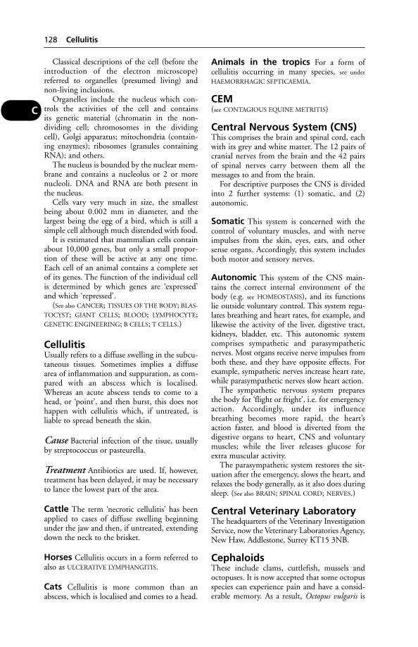
Classical descriptions of the cell (before theintroduction of the electron microscope)referred to organelles (presumed living) andnon-living inclusions.
Organelles include the nucleus which con-trols the activities of the cell and containsits genetic material (chromatin in the non-dividing cell; chromosomes in the dividingcell), Golgi apparatus; mitochondria (contain-ing enzymes); ribosomes (granules containingRNA); and others.
The nucleus is bounded by the nuclear mem-brane and contains a nucleolus or 2 or morenucleoli. DNA and RNA are both present inthe nucleus.
Cells vary very much in size, the smallestbeing about 0.002 mm in diameter, and thelargest being the egg of a bird, which is still asimple cell although much distended with food.
It is estimated that mammalian cells containabout 10,000 genes, but only a small propor-tion of these will be active at any one time.Each cell of an animal contains a complete setof its genes. The function of the individual cellis determined by which genes are ‘expressed’and which ‘repressed’.
(See also CANCER; TISSUES OF THE BODY; BLAS-
TOCYST; GIANT CELLS; BLOOD; LYMPHOCYTE;GENETIC ENGINEERING; B CELLS; T CELLS.)
CellulitisUsually refers to a diffuse swelling in the subcu-taneous tissues. Sometimes implies a diffusearea of inflammation and suppuration, as com-pared with an abscess which is localised.Whereas an acute abscess tends to come to ahead, or ‘point’, and then burst, this does nothappen with cellulitis which, if untreated, isliable to spread beneath the skin.
Cause Bacterial infection of the tisue, usuallyby streptococcus or pasteurella.
Treatment Antibiotics are used. If, however,treatment has been delayed, it may be necessaryto lance the lowest part of the area.
Cattle The term ‘necrotic cellulitis’ has beenapplied to cases of diffuse swelling beginningunder the jaw and then, if untreated, extendingdown the neck to the brisket.
Horses Cellulitis occurs in a form referred toalso as ULCERATIVE LYMPHANGITIS.
Cats Cellulitis is more common than anabscess, which is localised and comes to a head.
Animals in the tropics For a form ofcellulitis occurring in many species, see under
HAEMORRHAGIC SEPTICAEMIA.
CEM(see CONTAGIOUS EQUINE METRITIS)
Central Nervous System (CNS)This comprises the brain and spinal cord, eachwith its grey and white matter. The 12 pairs ofcranial nerves from the brain and the 42 pairsof spinal nerves carry between them all themessages to and from the brain.
For descriptive purposes the CNS is dividedinto 2 further systems: (1) somatic, and (2)autonomic.
Somatic This system is concerned with thecontrol of voluntary muscles, and with nerveimpulses from the skin, eyes, ears, and othersense organs. Accordingly, this system includesboth motor and sensory nerves.
Autonomic This system of the CNS main-tains the correct internal environment of thebody (e.g. see HOMEOSTASIS), and its functionslie outside voluntary control. This system regu-lates breathing and heart rates, for example, andlikewise the activity of the liver, digestive tract,kidneys, bladder, etc. This autonomic systemcomprises sympathetic and parasympatheticnerves. Most organs receive nerve impulses fromboth these, and they have opposite effects. Forexample, sympathetic nerves increase heart rate,while parasympathetic nerves slow heart action.
The sympathetic nervous system preparesthe body for ‘flight or fright’, i.e. for emergencyaction. Accordingly, under its influencebreathing becomes more rapid, the heart’saction faster, and blood is diverted from thedigestive organs to heart, CNS and voluntarymuscles; while the liver releases glucose forextra muscular activity.
The parasympathetic system restores the sit-uation after the emergency, slows the heart, andrelaxes the body generally, as it also does duringsleep. (See also BRAIN; SPINAL CORD; NERVES.)
Central Veterinary LaboratoryThe headquarters of the Veterinary InvestigationService, now the Veterinary Laboratories Agency,New Haw, Addlestone, Surrey KT15 3NB.
CephaloidsThese include clams, cuttlefish, mussels andoctopuses. It is now accepted that some octopusspecies can experience pain and have a consid-erable memory. As a result, Octopus vulgaris is
128 Cellulitis
C
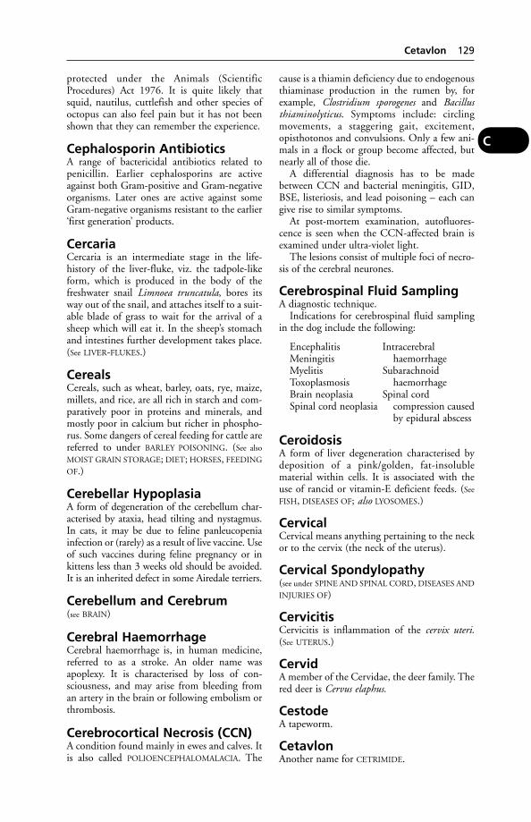
protected under the Animals (ScientificProcedures) Act 1976. It is quite likely thatsquid, nautilus, cuttlefish and other species ofoctopus can also feel pain but it has not beenshown that they can remember the experience.
Cephalosporin AntibioticsA range of bactericidal antibiotics related topenicillin. Earlier cephalosporins are activeagainst both Gram-positive and Gram-negativeorganisms. Later ones are active against someGram-negative organisms resistant to the earlier‘first generation’ products.
CercariaCercaria is an intermediate stage in the life-history of the liver-fluke, viz. the tadpole-likeform, which is produced in the body of thefreshwater snail Limnoea truncatula, bores itsway out of the snail, and attaches itself to a suit-able blade of grass to wait for the arrival of asheep which will eat it. In the sheep’s stomachand intestines further development takes place.(See LIVER-FLUKES.)
CerealsCereals, such as wheat, barley, oats, rye, maize,millets, and rice, are all rich in starch and com-paratively poor in proteins and minerals, andmostly poor in calcium but richer in phospho-rus. Some dangers of cereal feeding for cattle arereferred to under BARLEY POISONING. (See also
MOIST GRAIN STORAGE; DIET; HORSES, FEEDING
OF.)
Cerebellar HypoplasiaA form of degeneration of the cerebellum char-acterised by ataxia, head tilting and nystagmus.In cats, it may be due to feline panleucopeniainfection or (rarely) as a result of live vaccine. Useof such vaccines during feline pregnancy or inkittens less than 3 weeks old should be avoided.It is an inherited defect in some Airedale terriers.
Cerebellum and Cerebrum(see BRAIN)
Cerebral HaemorrhageCerebral haemorrhage is, in human medicine,referred to as a stroke. An older name wasapoplexy. It is characterised by loss of con-sciousness, and may arise from bleeding froman artery in the brain or following embolism orthrombosis.
Cerebrocortical Necrosis (CCN)A condition found mainly in ewes and calves. Itis also called POLIOENCEPHALOMALACIA. The
cause is a thiamin deficiency due to endogenousthiaminase production in the rumen by, forexample, Clostridium sporogenes and Bacillusthiaminolyticus. Symptoms include: circlingmovements, a staggering gait, excitement,opisthotonos and convulsions. Only a few ani-mals in a flock or group become affected, butnearly all of those die.
A differential diagnosis has to be madebetween CCN and bacterial meningitis, GID,BSE, listeriosis, and lead poisoning – each cangive rise to similar symptoms.
At post-mortem examination, autofluores-cence is seen when the CCN-affected brain isexamined under ultra-violet light.
The lesions consist of multiple foci of necro-sis of the cerebral neurones.
Cerebrospinal Fluid SamplingA diagnostic technique.
Indications for cerebrospinal fluid samplingin the dog include the following:
Encephalitis IntracerebralMeningitis haemorrhageMyelitis SubarachnoidToxoplasmosis haemorrhageBrain neoplasia Spinal cordSpinal cord neoplasia compression caused
by epidural abscess
CeroidosisA form of liver degeneration characterised bydeposition of a pink/golden, fat-insolublematerial within cells. It is associated with theuse of rancid or vitamin-E deficient feeds. (See
FISH, DISEASES OF; also LYOSOMES.)
CervicalCervical means anything pertaining to the neckor to the cervix (the neck of the uterus).
Cervical Spondylopathy(see under SPINE AND SPINAL CORD, DISEASES AND
INJURIES OF)
CervicitisCervicitis is inflammation of the cervix uteri.(See UTERUS.)
CervidA member of the Cervidae, the deer family. Thered deer is Cervus elaphus.
CestodeA tapeworm.
CetavlonAnother name for CETRIMIDE.
Cetavlon 129
C

CetrimideAn antiseptic of value in wound treatmentand for cleaning cows’ udders and teats; a0.1 per cent solution being effective againstStreptococcus agalactiae, a cause of mastitis. A1 per cent solution acts as a detergent.
ChabertiasisInfection of the colon with chabertia worms(Chabertia ovina); found in sheep and occa-sionally in goats and cattle.
Signs Usually mild: soft faeces, with mucusand sometimes blood-flecked. In severe cases,anaemia may occur.
Chagas DiseaseAn infection with Trypanosoma cruzi, mainlyoccurring in wild mammals (such as opossums,armadillos, and wood rats) of Central andSouth America, but also infecting man, dogs,cats, and pigs. (See TRYPANOSOMES – Americantrypanosomiasis.)
ChalazionChalazion is a small swelling of the eyelidcaused by a distended Meibomian gland. It iscommonly seen in dogs.
ChancreIn human medicine this term is reserved for theulcer or hard ‘sore’ which is the primary lesionof syphilis. In a veterinary context it means thelocal skin reaction at the sites of bites by tsetseflies carrying trypanosomes. The chancre – thefirst sign of trypanosome infection – begins as asmall nodule, developing into a hard, hot,painful swelling measuring up to 3 or 4 inchesacross.
Charlock PoisoningThe common charlock Brassica sinapis (wildmustard) is dangerous to livestock after its seedshave formed in the pods, although only wheneaten in large amounts. The seeds contain thevolatile oil of mustard and also a glycoside.
Signs are those of abdominal pain, loss ofappetite, a yellowish frothy liquid at mouth andnostrils, diarrhoea. There is nephritis, and theurine may be blood-stained.
First-Aid Give milk and strong tea.
Charolais CattleThis is numerically the second-largest breed ofcattle in France, and they have been exportedthroughout Europe and the USA. The
Charolais, white, is an excellent beef animal, amost efficient grazer, with a rapid growth-rateand a quiet disposition. The loin and thighmuscles are exceptionally well developed. Thebulls are colour-marking and highly prized forcrossing purposes. UK trials of this breed forcrossing purposes were approved in 1961, andthe British Charolais is now the third mostimportant beef breed.
Charolais SheepThis breed was developed in the 19th centuryby crossing Dishley Leicester with the localsheep of Central France, and has been recog-nised as a breed since 1974. Mature ewes weighup to 79 kg and rams up to l09 kg. Both sexesare polled.
‘Chastek Paralysis’A condition of secondary vitamin Bl deficiency,seen in foxes and mink on fur farms as a result offeeding raw fish. An enzyme in the latter has theproperty of destroying the vitamin, also knownas THIAMIN. The condition is seen also in cats.
Check LigamentThis is joined to the Perforans tendon, and actsas a check on the movement of the pasternjoint. The check ligaments are often strained inthe racehorse.
CheeseWhen cheese is made from raw milk, Brucella,Listeria and other organisms may infect thecheese. In the UK, pasteurised milk is used,although it is argued that the flavour may beless good. The unpasteurised form is widelyproduced in Europe; sheep and goat cheeseis popular as well as cow’s milk cheeses. (See
BRUCELLOSIS.)
CheilitisInflammation of the lips.
CheilosisCracked and scaly lips, often also affecting thecorners of the mouth. Characteristic of vitaminB deficiency.
Chelating AgentsChelating agents are substances which have theproperty of binding divalent metal ions to formstable, soluble complexes which are non-ionisedand so virtually lacking in the toxicity of themetal concerned. Derivatives of ethylene-diamine-tetra-acetic acid (EDTA) afford exam-ples. EDTA itself is poisonous, as it removescalcium; but the calcium-EDTA complex has
130 Cetrimide
C
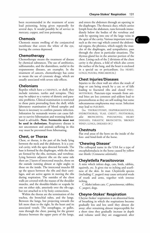
been recommended in the treatment of acutelead poisoning, being given repeatedly forseveral days. It would possibly be of service inmercury, copper, and iron poisoning.
ChemosisChemosis means swelling of the conjunctivalmembrane that covers the white of the eye,leaving the cornea depressed.
ChemotherapyChemotherapy means the treatment of diseaseby chemical substances. The use of antibiotics,sulfonamides, and the diamidines, useful in thetrypanosome diseases, are examples. In thetreatment of cancers, chemotherapy has cometo mean the use of cytotoxic drugs, which areusually associated with severe side-effects.
CheloniansReptiles which have a CARAPACE, or shell; theyinclude tortoises, turtles and terrapins. Theymay be subject to a variety of dietetic and para-sitic problems. Clinical examination is restrictedto those parts protruding from the shell, whilelaboratory examination of blood samples andfaeces is necessary to confirm parastic infection.Infestation by nematode worms can cause fail-ure to survive hibernation and worming before-hand is advisable. Note. Ivermectin must notbe used in chelonians. Respiratory disease isnot uncommon and animals suffering in thisway must be prevented from hibernating.
Chest, or ThoraxChest, or thorax, is the part of the body lyingbetween the neck and the abdomen. It is a con-ical cavity, with the apex directed forwards. Thebase is formed by the diaphragm, while the sidesare formed by the ribs, sternum, and vertebrae.Lying between adjacent ribs on the same sidethere are 2 layers of intercostal muscles, those onthe outside running almost at right angles tothose on the inside. The intercostal muscles fillup the spaces between the ribs and their carti-lages, and are active agents in moving the ribsduring respiration. The outsides of the chestwalls are covered with the masses of the shouldermuscles, and the shoulder-blades or scapula lieone on either side, anteriorly over the rib-cage,but not attached to it by bony connections.
Within the thorax are the termination of thetrachea, the bronchial tubes, and the lungs.Between the lungs, but projecting towards theleft more than to the right, lie the heart and itsassociated vessels. The oesophagus, or gullet,runs through the chest, passing for the greaterdistance between the upper parts of the lungs,
and enters the abdomen through an opening inthe diaphragm. The thoracic duct, which carrieslymph from the abdomen, runs forwards imme-diately below the bodies of the vertebrae andends by opening into one of the large veins inthe apex of the cavity. Various important nerves,such as the two vagi which control the abdomi-nal organs, the phrenics, which supply the mus-cles of the diaphragm, and sympathetics, passthrough the chest in particular situations. Thethymus gland lies in the anterior portion of thechest. Lining each of the 2 divisions of the chestcavity is the pleura, a fold of which also coversthe surface of the lung, and the heart is enclosedin a special sac or pericardium. (See HEART;LUNGS; PLEURA; PERICARDIUM.)
Chest Injuries/DiseasesInjuries to the chest wall are often the result ofdogs or cats being struck by a car; or of fallsleading to fractured ribs and closed PNEU-
MOTHORAX. Puncture-type wounds from ani-mal bites are less common and seldom lead topneumothorax as they are self-sealing; but somesubcutaneous emphysema may occur. Infectionmay lead to PLEURISY.
(See THORACOTOMY; DIAPHRAGMATOCELE;HYDROTHORAX; ‘FLAIL CHEST’; PYOTHORAX;also BRONCHITIS; PNEUMONIA; HEART
DISEASES; PARASITIC BRONCHITIS; ‘BROKEN
WIND’; LUNGS, DISEASES OF.)
ChestnutsFlat oval areas of the horn on the inside of thefore- and hind-limb of the horse.
‘Chewing Disease’The colloquial name in the USA for a type ofencephalomalacia in the horse caused by yellowstar thistle (Centaurea solstitialis).
Cheyletlella ParasitovoraxA mite which infests dogs, cats, birds, rabbits,squirrels, etc. It gives rise to itching and scurfi-ness of the skin. In man Cheyletiella species(including C. yusguri) may cause urticarial wealsof trunk and arms, together with intenseitching.
C. blakei infests cats; C. parasitovorax, rabbits;C. yasguri, dogs.
Cheyne-Stokes’ RespirationCheyne-Stokes’ respiration is an abnormal formof breathing in which the respirations becomegradually less and less until they almost dieaway; after remaining almost imperceptible fora short time they gradually increase in depthand volume until they are exaggerated; after
Cheyne-Stokes’ Respiration 131
C
attaining a maximum they again decrease untilnearly imperceptible. This alternation proceedswith considerable regularity.
Cheyne-Stokes’ breathing is always a veryserious condition, which is generally associatedwith severe nervous disturbance, shock, andcollapse, or with heart or kidney disease. It ismost obvious in the dog and horse after theyhave sustained very severe injury but withoutinternal haemorrhage (which induces what isgenerally known as ‘sobbing respiration’).
ChianinaThese Italian cattle are named after their placeof origin, the Chiana valley. Probably the largestcattle in the world, a mature bull can weighover 1.75 tonnes and be 1.8 m tall at the with-ers. Formerly used as draught animals, they arean excellent beef breed, now present in the UK.
Chick Oedema(see ‘TOXIC FAT SYNDROME’)
ChihuahuaOne of the smallest breeds of toy dog, originat-ing from Mexico. Frontal foramina are present,i.e., the frontal bones of the domed skullremain ununited. The breed is liable to sufferproblems in parturition. Possible inherited con-ditions include pulmonary stenosis, dislocationof the patella, and hydrocephalus. Mange inthis breed must never be treated with amitraz.
Chicken Anaemia VirusTransmitted by breeder flocks to their progeny,chicken anaemia virus causes increased mortal-ity with anaemia, lymphoid depletion, liverchanges and haemorrhages throughout thebody. Signs develop at 2 to 3 weeks old.
ChicksThe ambient temperature for rearing chicksmust be kept above 18°C (60°F) during thefirst 5 weeks or so of life. Ambient temperaturefor rearing chicks should be 32°C (90°F).Chilling is one of the commonest causes of pul-let chick mortality. Chicks require artificial heatfor 3 to 8 weeks, depending upon the type ofhouse, weather, etc. (See also POULTRY – Chickfeeding).
Chilblain Syndrome in DogsThis was first described as affecting Servicedogs in Northern Ireland. These dogs had pre-viously thrived in unheated, outdoor kennels,but were affected during a very cold winter.The first sign was biting of the tip of the tail –found to be red, swollen, warm and intensely
itchy. Ulceration, infection, and necrosis of thetail tip occurred in a few cases, necessitatingamputation of the tip. It is not unknown fora dog to eat the affected part of its tail.Elizabethan collars, protective tail covering,and anti-inflammatory drugs were used intreatment.
Chilling(see under CHICKS and HYPOTHERMIA)
ChimeraAn animal having in its body, cell populationsarising from different species; that is, cells withdifferent KARYOTYPES which have originatedfrom 2 or more zygotes with different kary-otypes. A freemartin is, technically, an example ofXX:XY chimerism. This is secondary chimerism.Primary chimerism occurs if 2 sperms fertilise thesame ovum. (See CYTOGENETICS.)
Other examples which have been reportedinclude a fertile female mule that had apparent-ly inherited a mixture of both horse and donkeychromosomes, and was phenotypically achimera rather than a hybrid. And a sheep-goatchimera found at the School of VeterinaryMedicine, University of California, USA, wascapable of oestrus cycles, producing fertile ova,and carrying pregnancy to full term.
Chimera is also a term used to describe anorganism that has had foreign DNA insertedinto its genome.
ChinchillaChinchilla laniger is a small rabbit-like rodent,prized for its fur. It originates from the SouthAmerican Andes; those living at higher altitudeshave better coats. Originally brought to the UKfor fur farming, the project had to be aban-doned because of the poor quality of theimported animals. Adults weigh about 400 to500 g; the female is larger than the male.Pregnancy lasts 111 days; there are usually 2 in a litter but up to 5 may be born. Weaning is at 6 to 8 weeks. They are sexually mature at 8 months and can live for 10 years although therecord is 18. Body temperature is 38 to 39°C.They can be active during the day but aremainly nocturnal in habit.
Chinchillas require ample room (about 4 m3) with a nest box about 30 x 25 x 20 cmwithin that. An ambient temperature of 10 to20°C is adequate; as low as 0°C can be tolerat-ed provided there are no draughts. Fine sandmust be provided for sand-bathing to keep theircoat in good condition.
If treated with sulfonamides, the coat colourmay fade; it will eventually return to normal.
132 Chianina
C
Chinchilla, Diseases ofEnteritis, pneumonia and impaction of theintestine are the most common diseases. Out ofa series of 1000 post-mortem examinationsmade in the USA, ‘epidemic gastroenteritis’ wasfound in 23 per cent of the chinchillas, as against25 per cent with pneumonia, and 12 per centwith impaction (blockage of the intestine). In afurther series of 1000 examinations, the figureswere: impaction, 20 per cent; pneumonia, 22 per cent; and enteritis, 24 per cent. Yersineaparatuberculosis, Listeria monocytogenes, proteus,pseudomonas, staphylococcus and salmonellainfections have been recorded, as hasClostridium perfringens associated with diar-rhoea, flatulence and prolapsed rectum. Acuteand fatal gastroenteritis caused by Yersinia ente-rocolitica has caused severe losses among chin-chillas on farms in California, and also inEurope. (See YERSINIOSIS.)
An important cause of pneumonia isKlebsiella pneumoniae. This may also produceloss of appetite, diarrhoea, and death withinabout 5 days.
Lying on one side and stretching the legs aresaid to be signs of impaction. A diet with toolittle roughage is believed to be a cause.
Intussusception is not uncommon and some-times follows enteritis. Inability to retract thepenis (paraphimosis) has also been noted.
Fur-chewing and associated skin problems –that bane of the North American chinchillaindustry – has been attributed to ‘environmen-tal stress’ associated with captivity. Of course,the wrong diet may enter into it too. High fibrepellets, timothy hay combined with some freshgreenstuff, with a little apple and a raisin or twonow and then will also help prevent digestivedisorders.
Chinchillas’ teeth grow constantly; unless theanimals are provided with materials to gnaw on,the teeth become excessively long, preventing aproper bite and causing injuries inside the mouth.Often the condition requires veterinary attention.
ChipmunksA rodent (Tamias striatus), not dissimilar to thesquirrel, with longitudinal stripes across itsback. Adults weigh 72 to 120 g. When kept aspets they should be in pairs or a trio of 1 maleand 2 females. A large, escape-proof enclosureshould be provided. They become hyperactiveif stressed and must never be kept near a televi-sion set. If outside, protection from adverseweather conditions must be given.
Body temperature is 38° C when awake,falling to a few degrees above ambient temper-ature when hibernating. Chipmunks are
comparatively healthy animals provided theyare well kept. Ectoparasites may be present, asmay mange, fleas and harvest mites. Cataractsoccur in older animals; emphysema has beenrecorded and is difficult to treat. If hypogly-caemia (milk fever) is seen after parturition, itcan be treated with 0.5 ml calcium boroglucon-te given subcutaneously. Swellings due to col-lection of fluid (lymphoedema) may be seen.These usually regress during hibernation butcan be treated with a daily dose of 0.5 mgfrusemide.
ChiropracticA technique which aims to relieve diseaseproblems by manipulation of body structures,particularly the spinal vertebrae.
ChitinThe horn-like substance forming the main con-stituent of the body-covering of insects, ticks,mites, spiders, etc. A polysaccharide, it is alsofound in some fungi.
Chlamydia and ChlamydophilaWidespread Gram-negative bacteria containingspecies of veterinary and medical significance.They include Chlamydophila psittaci, responsi-ble for psittacosis (see below) in birds (and man);C. abortus, a major cause of abortion in sheepand some other ruminants; and C. felis, whichcauses pneumonia, conjunctivitis and respira-tory problems in cats. Chlamydia suis causesconjunctivitis, pneumonia and enteritis in pigs.
Psittacosis Sometimes called parrot disease,psittacosis affects virtually all avian species.There are several strains (serovars) of C. psittaci,some of which affect other species. Affectedbirds may show no signs initially but activeinfection is often triggered by stress. Parrots andother cage birds become listless, have diarrhoea,coryza, conjunctivitis and sinusitis. Pigeonsdevelop respiratory signs and bronchitis. Inpoultry, egg production falls off; up to a third ofa flock may die.
There is a considerable risk that people incontact with infected birds pick up the disease,sometimes with serious results.
In one Edinburgh outbreak, 100 out ofabout 300 budgerigars in an aviary died.Human cases followed and a dog was found tobe excreting Chlamydia organisms and to havea lung infection.
Treatment Tetracycline or doxycycline, givento birds over a period of 7 weeks in the feed oron medicated seed.
Chlamydia and Chlamydophila 133
C

Prevention Quarantine of imported birds;disinfection of infected premises.
Ornithosis is the name formerly given to thesame infection in birds other than those of theparrot family.
Post-Mortem findings include enlargementof liver and spleen, together with pneumonia.Confirmation of diagnosis is by ELISA,immunofluorescent test or bacterial culture.
Measures to protect the UK’s poultry againstpsittacosis infection from abroad are specifiedunder the Importation of Birds, Poultry andHatching Eggs Order 1979. All diagnoses ofthe disease in imported birds are notified by theState Veterinary Service to medical officers ofenvironmental health.
Public health Human psittacosis in itsmilder forms resembles influenza. In childrenthe symptoms are slight or absent altogether,but in older people the illness is more likely tobe severe. Symptoms include shivering,headache, backache. Death from pneumoniamay follow. Acute kidney failure has beenrecorded; also heart disease. Human infectioncomes through handling infected birds.
Abortion C. abortus (and some strains ofC. psittaci) are responsible for abortion inruminants.
Sheep usually show no signs of disease untilthey become depressed shortly before abortionoccurs. Afterwards, most ewes recover unevent-fully. When disease is established in a flock, 5 to10 per cent of ewes will abort; immunity devel-ops following infection. The infection is usual-ly transmitted at lambing, through placenta,uterine discharges and faeces.
Vaccines are available against certain strainsof chlamydial infection.
Cattle Animals show no sign of disease butsporadic abortions occur, usually in the 7th to9th month of gestation. Sometimes, dead orshort-lived weakly calves are born at full term.
Public health The infection is transmissibleto humans. Cases in pregnant women who haveassisted at lambing have been recorded.Difficulties during pregnancy, and in one casedeath, followed.
A farmer’s wife who aborted in the 28th weekof pregnancy, had helped with difficult lambings.Five of 200 ewes had aborted and a serum sam-ple had shown high antibody titres to chlamydia.
Chlamydia spp. were detected in smears of liver,lung and placenta from the human fetus.
Chloral HydrateChloral hydrate is a clear, crystalline substancewith a sweetish taste; it dissolves rapidly inwater. It was formerly used widely as a hypnotic,and occasionally for euthanasia.
Chloral Hydrate PoisoningIn the dog, poisoning has occurred after eatingmeat from horses humanely euthanased bymeans of chloral hydrate.
ChloraminesChloramines are widely used as a disinfectant.Their activity depends upon the amount ofavailable chlorine.
ChloramphenicolAn antibiotic which has a similar range of activ-ity to the tetracyclines. It can be given orally(except to ruminants), by intravenous injection,and by local application, especially as an eyeointment. Because of its importance in thetreatment of human typhoid and the avoidanceof resistant strains, its use in veterinary medi-cine has been severely restricted, particularly infood-producing animals.
In human medicine, poisoning by chloram-phenicol has led to aplastic anaemia, skin erup-tions, and moniliasis. There are three mainside-effects: allergy or hypersensitivity to thedrug; damage to the blood or bone-marrow;and gastrointestinal upsets.
Intramuscular injections of chloramphenicolare painful.
Chlorate PoisoningIn acute cases cattle may die after showingsymptoms suggestive of anthrax. In subacutecases, a staggering gait, purgation, signs ofabdominal pain, and red-coloured urine may beseen. Cyanosis and respiratory distress are alsosymptoms.
Treatment Gastric lavage. If cyanosis ispresent, methylene blue should be givenintravenously.
ChlordaneA highly toxic insecticide of the chlorinatedhydrocarbon group. It is volatile and poisoningthrough inhalation may occur.
ChlorfenvinphosAn organophosphorus acaricide and insecticide.It has been used in sheep dips, etc. and against
134 Chloral Hydrate
C
fly strike, keds, lice and ticks. Less toxiccompounds are now preferred.
ChlorhexideneChlorhexidene gluconate is widely used as anantiseptic and surgical scrub; and in teat dips,sprays and udder washes.
Chlorinated HydrocarbonsThese insecticides include: chlordane, DDT,DDD, methoxychlor, benzene hexachloride,toxaphene, aldrin, dieldrin, isodrin, and endrinplus a range of others less well known. Ingestedat toxic levels, or absorbed through the skin, theyact primarily on the central nervous system caus-ing excitement/frenzy at the outset followed bymuscular tremors leading to convulsions in acutecases. Species capable of vomiting do so. Loss ofappetite with marked loss of body weight is usualin subacute poisoning. Cats are especially sus-ceptible. Wash off any residues from the skin andkeep the animal warm, comfortable and sedated.
Most compounds – methoxychlor is anexception – can be stored in the body fat andexcreted in the milk and so may constitute apublic health problem. Their use in animals isnow minimal because of the residue levelscaused by this persistence.
ChloroformChloroform is a colourless, mobile, non-inflammable liquid, half as heavy again aswater. It is much less used now than formerly asa general anaesthetic. (See ANAESTHETICS;EUTHANASIA.)
Four stages of chloroform anaesthesia arerecognised:
(1) The stage of excitement begins immedi-ately the drug is administered. Vigorous animalsstruggle violently, and when in the standingposition may rear or strike out with theirforefeet and shake their heads in an endeavourto dislodge the mask. Deep breaths are takenoften in a gasping manner, and in from 3 to 6or 7 minutes the second stage follows.
(2) The stage of depression follows the stim-ulation stage, and is marked by a quieting of themovements of the voluntary muscles, by a less-ening of the force and volume of the pulse, andby slower and deeper breathing. Pain is still felt,and if inflicted induces reflex movement.
(3) The stage of anaesthesia produces com-plete muscular relaxation and unconsciousness.This is the safe or operating stage; all the cen-tres of the brain are subdued except those thatgovern respiration and heart action.
(4) The stage of paralysis occurs when theanaesthetic is pushed beyond the safe stage. The
centres of respiration and heart action, incommon with all the other nervous centres,become paralysed. The heart stops beatingabout 2 minutes after respiration ceases, andany attempts at ARTIFICIAL RESPIRATION mustbe prompt.
Chocolate PoisoningThe feeding of waste chocolate bars to cattle hasled to fatal poisoning in calves in the UK. Theanimals showed excitement, stared about in alldirections, walked with exaggerated strides, andhad convulsions.
It was suggested that the caffeine contentwould account for the excitement; the theo-bromine content may have caused heart failure.
In dogs, the signs include panting, vomiting,thirst, diarrhoea, excitement, fits, coma.
Treatment Use of an emetic or gastric lavage.(Activated charcoal is used in human medi-cine.) For control of the convulsions, diazepamis among the suitable drugs.
Autopsy findings include cyanotic mucousmembranes, swollen and reddened gastricmucosa.
(See also COCOA POISONING.)
‘Choking’ (Obstruction ofPharynx or Oesophagus)’Choking’ is, by dictionary definition, anobstruction to respiration, but in a farmingcontext the word has been misused to denote anobstruction to the passage of food throughthe pharynx and oesophagus, either partial orcomplete.
The domesticated animals, especially cattleand dogs, are very prone to attempt to swalloweither foreign bodies or masses of food materialtoo large to pass down the oesophagus (gullet),with the result that they often become jammed.Such substances hinder the free passage of solidor fluid food, give rise to pain and discomfort,and are very often attended by serious and evenfatal consequences. Choking in cattle, dogs,and cats is usually due to a hard, large, sharp-pointed, or irregularly shaped object; while inthe horse it is most often due to a mass of dryimpacted food material, or to a portion of amangold or turnip.
Cattle Choking is of comparatively commonoccurrence, particularly in districts where rootsare fed whole to the animals, and where there is aquantity of rubbish scattered about the pastures.
Signs The animal immediately stops feeding,and becomes uneasy. A feature of nearly all
‘Choking’ (Obstruction of Pharynx or Oesophagus) 135
C
cases of choking in cattle is the rapidity withwhich gas formation occurs in the rumen. (See
BLOAT.)In a number of cases of choking, relief occurs
quite spontaneously after the lapse of from30 minutes to 2 or 3 hours from the origin ofthe symptoms. This is because the muscles ofthe gullet, which have been tightly gripping theobstruction, gradually become fatigued andrelax, thereby allowing the object to pass downinto the stomach. Naturally, such a satisfactorytermination cannot occur wherever there is asharp projecting point on the object causingthe obstruction, but it frequently happenswith eggs, apples, potatoes, and other smoothbodies.
First-Aid In all cases of choking, no matterhow simple they appear to be, the owner shouldseek veterinary assistance as soon as possible.
Professional treatment depends on the siteof the obstruction. This may be palpable in theoesophagus and manipulation up to the mouthmay be possible after giving hyoscine hydrobro-mide and metamizole dypirone (Buscopan;Boehringer) as a muscle relaxant. If in the intes-tine, passage of a probang or stomach-tubedown the oesophagus may dislodge the obstruc-tion; if not, it can be removed by rumenotomyor a trochar can be inserted into the rumen;in many cases, the blockage will reduce bymaceration over 3 to 4 days.
Horses The horse is less often choked than thecow, but owing to the long and narrow equineoesophagus, the accident is more serious.
Treatment Avoid raising the head or givingdrenches, lubricating or otherwise. It is impera-tive to secure professional assistance at once.
Dogs and cats
Signs At first there is usually a sudden pain,which causes the animal to cry out. If it hasbeen feeding it immediately ceases, andbecomes very restless. It may paw at its mouth.Salivation is often profuse.
When a threaded needle has become fixed inthe throat or below it, the end of the threadmay often be seen.
Treatment Swallowed objects which becomejammed in a dog’s oesophagus can be treated inone of two ways: surgically or conservatively. Thelatter includes the use of an endoscope, passedinto the oesophagus and enabling the foreign
body to be grasped with forceps and drawn outor, alternatively, pushed down into the stomach,whence it can, if necessary, be removed.
In a series of 90 cases treated by J. E. F.Houlton and others at the University ofCambridge, 85 of the foreign bodies were piecesof bone, and two were composed mainly of gris-tle. A potato, a fish-hook, and a ball were alsofound. The success rate of treatment by surgicaland conservative means was 82 per cent.
A young African elephant died from obstruc-tion of its oesosphagus by an apple.
CholagoguesCholagogues are substances reputed to act onthe liver, increasing the secretion of the bile.
CholangiocarcinomaCancer of the bile ducts; it is associated withliver-fluke infestation of animals in Thailand.
CholangiohepatitisInflammation of bile ducts and associated liverparenchyma.
CholangiomaA benign tumour originating from the bileducts, and occurring in cats, dogs, sheep, andpoultry.
CholangitisInflammation of the intra-hepatic bile ducts.
CholecystitisInflammation of the gall-bladder.
CholecystographyCholecystography is the term used for X-rayexamination of the gall-bladder after its con-tents have been rendered opaque by adminis-tration of lipiodol or pheniodol compounds.
CholecystokininA hormone produced in the small intestine andcausing emptying of the gall-bladder.
CholecystomyA surgical incision into the gall-bladder.
Cholera, Fowl(see FOWL CHOLERA)
CholesteatomaAn epidermoid cyst within the middle ear cavity of dogs, complicating simple otitis.
CholesterolA sterol present in blood, brain and other tis-sues, bile, and many foods. It is produced in the
136 Cholagogues
C

liver and adrenal glands; it decreases in cowswith fatty liver.
A high cholesterol level can be a precursor tohigh blood pressure, ATHEROMA, and THROM-
BOSIS.
Cholesthiasis(see GALLSTONES)
CholineCholine is an amine compound with importantfunctions in the metabolic process. It is foundin egg-yolk, liver, and muscle, and is associatedwith the vitamin B complex. Acetyl choline isessential for the transmission of an impulsefrom nerve to muscle.
CholinesteraseCholinesterase is an enzyme which inactivatesacetylcholine. Some poisons, such as carbamatesand organophosphates, cause cholinesteraseinhibition, and it is inactivated by a substanceisolated from occuring in white clover S.100.
ChondritisInflammation of cartilage.
ChondrocytesCartilage-forming cells.
Chondrogenesis(see Growth-plate disorders under BONE, DISEASES OF)
ChondromaA rare tumour, composed of cartilage-like cells,which has been seen in dogs, rats, and mink.
ChoreaTwitching or trembling caused by a successionof involuntary spasmodic contractions (clonicspasms) affecting one or more of the voluntarymuscles. The spasm is of a rhythmic nature,occurring at fairly regular intervals, andbetween the individual contractions relaxationof the affected muscle takes place.
The condition affects dogs almost exclusive-ly, although muscular spasms of a similar naturehave been seen in horses, cattle, and pigs. Inlambs, congenital chorea is described under‘BORDER DISEASE’. (See also SHIVERING.)
Causes In dogs, chorea generally followsa mild attack of distemper. It may appear with-in a few days after apparent recovery, or itsappearance may be delayed. All dog ownerswould be well advised to regard cases ofdistemper as not cured until the lapse of at least10 days after apparent recovery, and during
this period to continue to treat the animal asthough it were still sick, so far as exercise isconcerned.
Signs Twitchings usually begin about the lipsand face, or in the extremities of one or morelimbs. Later, perhaps the whole head is seencontinually nodding or jerking backwards andforwards, quite irrespective of the pose or posi-tion of the animal. As the condition progresses,there comes a time when it is unable to rest, lossof condition and weakness result, and thedog becomes exhausted. Ulceration of theaffected limb, as the result of continual frictionwith surrounding objects, the ground, etc., isnot uncommon. Chorea is always a seriouscondition.
Treatment is with ANTISPASMODICS.
ChorionChorion is the outermost of the three fetalmembranes, the others being the amnion andthe allantois. The chorion is a strong fibrousmembrane, whose outer surface is closelymoulded to the inner surface of the uterus.Chorionic villi are the vascular projections fromthe chorion which are inserted into the cryptsof the uterine mucous membrane. (See also
PARTURITION.)
Chorionic Gonadotrophin(see HORMONE THERAPY)
Choroid, or ChorioidChoroid, or chorioid, is the middle of the 3coats of the EYE, and consists chiefly of theblood vessels which effect nourishment of theorgan.
ChoroiditisInflammation of the choroid.
Chow ChowA stocky, medium-sized dog with a thick coat;the tongue is blue-black in colour. Originallybred in China for meat, the breed specificationstill reads as if that were its main purpose. Theytend to be a ‘one person’ dog. Entropion andmuscle spasm (myotonia) may be inherited.
Christmas Rose(see HELLEBORES)
Chromobacter ViolaceumAn organism, often regarded as non-pathogenic,which has caused a fatal pneumonia in pigs inthe USA.
Chromobacter Violaceum 137
C
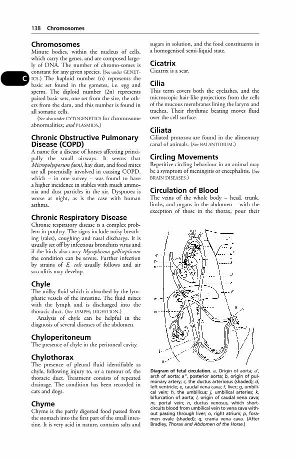
ChromosomesMinute bodies, within the nucleus of cells,which carry the genes, and are composed large-ly of DNA. The number of chromo-somes isconstant for any given species. (See under GENET-
ICS.) The haploid number (n) represents thebasic set found in the gametes, i.e. egg andsperm. The diploid number (2n) representspaired basic sets, one set from the sire, the oth-ers from the dam, and this number is found inall somatic cells.
(See also under CYTOGENETICS for chromosomeabnormalities; and PLASMIDS.)
Chronic Obstructive PulmonaryDisease (COPD)A name for a disease of horses affecting princi-pally the small airways. It seems thatMicropolysporum faeni, hay dust, and food mitesare all potentially involved in causing COPD,which – in one survey – was found to havea higher incidence in stables with much ammo-nia and dust particles in the air. Dyspnoea isworse at night, as is the case with humanasthma.
Chronic Respiratory DiseaseChronic respiratory disease is a complex prob-lem in poultry. The signs include noisy breath-ing (rales), coughing and nasal discharge. It isusually set off by infectious bronchitis virus andif the birds also carry Mycoplasma gallisepticumthe condition can be severe. Further infectionby strains of E. coli usually follows and airsacculitis may develop.
ChyleThe milky fluid which is absorbed by the lym-phatic vessels of the intestine. The fluid mixeswith the lymph and is discharged into thethoracic duct. (See LYMPH; DIGESTION.)
Analysis of chyle can be helpful in thediagnosis of several diseases of the abdomen.
ChyloperitoneumThe presence of chyle in the peritoneal cavity.
ChylothoraxThe presence of pleural fluid identifiable aschyle, following injury to, or a tumour of, thethoracic duct. Treatment consists of repeateddrainage. The condition has been recorded incats and dogs.
ChymeChyme is the partly digested food passed fromthe stomach into the first part of the small intes-tine. It is very acid in nature, contains salts and
sugars in solution, and the food constituents ina homogenised semi-liquid state.
CicatrixCicatrix is a scar.
CiliaThis term covers both the eyelashes, and themicroscopic hair-like projections from the cellsof the mucous membranes lining the larynx andtrachea. Their rhythmic beating moves fluidover the cell surface.
CiliataCiliated protozoa are found in the alimentarycanal of animals. (See BALANTIDIUM.)
Circling MovementsRepetitive circling behaviour in an animal maybe a symptom of meningitis or encephalitis. (See
BRAIN DISEASES.)
Circulation of BloodThe veins of the whole body – head, trunk,limbs, and organs in the abdomen – with theexception of those in the thorax, pour their
138 Chromosomes
C
Diagram of fetal circulation. a, Origin of aorta; a’,arch of aorta; a”, posterior aorta; b, origin of pul-monary artery; c, the ductus arteriosus (shaded); d,left ventricle; e, caudal vena cava; f, liver; g, umbili-cal vein; h, the umbilicus; j, umbilical arteries; k,bifurcation of aorta; l, origin of caudal vena cava;m, portal vein; n, ductus venosus, which short-circuits blood from umbilical vein to vena cava with-out passing through liver; o, right atrium; p, fora-men ovale (shaded); q, crania vena cava. (AfterBradley, Thorax and Abdomen of the Horse.)

blood into one of the three great terminal radi-cles which open into the right atrium of theheart. This contracts and drives the blood intothe right ventricle, which then forces the bloodinto the lungs by way of the pulmonary artery.In the lungs it is contained in very thin-walledcapillaries, over which the inspired air playsfreely, and through which the exchange of gasescan easily take place. The blood is consequentlyoxygenated (see RESPIRATION), and passes on bythe pulmonary veins to the left atrium of theheart. This left atrium expels it into the left ven-tricle, which forces it on into the aorta, bywhich it is distributed all over the body. Passingthrough the capillaries in the various organs andtissues it eventually again enters the lesser veins,and is collected into the cranial and caudal venacava and the azygos vein (see VEINS), from whereit passes to the right atrium once more.
In one part of the body there is, however, afurther complication. The veins coming fromthe stomach, intestines, spleen, and pancreas,charged with food materials and other prod-ucts, unite into the large ‘portal vein’ whichenters the porta of the liver and splits up into asecond capillary system in the liver tissue. Hereit is relieved of some of its food content, andpasses to the caudal vena cava by a second seriesof veins, joining with the rest of the blood com-ing from the hind parts of the body, and so goeson to the right atrium. This is known as the‘portal circulation’.
The circuit is maintained always in onedirection by four valves, situated one at the out-let from each cavity of the HEART, and by thepresence of valves situated along the course ofthe larger veins.
The blood in the arteries going to the bodygenerally (i.e. to the systemic circulation) isa bright red in colour while that in the veins isa dull red; this is owing to the oxygen contentof arterial blood being much greater than thatof venous blood, which latter is charged withcarbon dioxide. For the same reason the bloodin the pulmonary artery going to the lungs isdark, while in the pulmonary veins it is brightred.
There is normally no connection betweenthe blood in the right side of the heart and thatin the left; the blood from the right ventriclemust pass through the lungs before it can reachthe left atrium. In the fetus, two large arteriespass out from the umbilicus (navel), and conveyblood which is to circulate in close proximity tothe maternal blood in the placenta, and toreceive from it both the oxygen and the nour-ishment necessary for the needs of the fetus,while one large vein brings back this blood into
the fetal body through the umbilicus again.There are also communications between theright and the left atria (the foramen ovale) andbetween the aorta and the pulmonary artery(the ductus arteriosus), which serve to ‘short-circuit’ the blood from passing through thelungs in any quantity. At birth these extracommunications rapidly close and shrivel up,leaving mere vestiges of their presence inadult life. There are rare instances, however, inwhich one or more of the passages may persistthroughout life.
Circulation of Lymph(see LYMPH)
Cirrhosis, or FibrosisCirrhosis, or fibrosis, is a condition of variousinternal organs, in which some of the non-parenchymatous cells of the organ are replacedby fibrous tissue. The name ‘cirrhosis’ was firstused for the disease as it occurs in the liver,because of the yellow colour, but it has beenapplied to fibrosis in the lung, kidney, etc.Classic instances of cirrhosis are seen in the liverin chronic ragwort poisoning in cattle, inchronic alcoholism in man, and in old dogs.
CitrullinaemiaThis disease occurs in some Australian Friesiancattle; also in dogs. It is hereditary in origin,and due to a deficiency of the amino acidCitrulline. In calves depression, recumbency,and convulsions result.
CJD(see CREUTZFELDT-JAKOB DISEASE)
ClavicepsA fungus (see ERGOT, FUNGAL).
ClavicleClavicle is another name for the ‘collar-bone’ inman. This bone is not present in the domesti-cated mammals (except sometimes in a veryrudimentary form in the cat), but is present inthe fowl.
Claws(see NAILS)
Clay PigeonsRuminants and outdoor pigs may eat theseif found lying on pasture. The outcome canbe fatal in pigs but in ruminants, chronicunthriftiness results as the material is digestedonly slowly. (See PITCH POISONING; LEAD
POISONING.)
Clay Pigeons 139
C
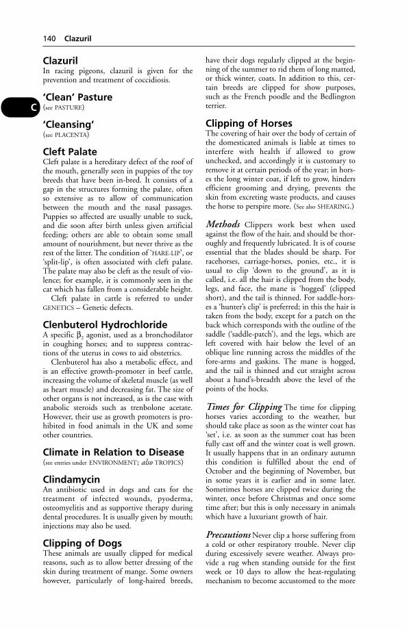
ClazurilIn racing pigeons, clazuril is given for theprevention and treatment of coccidiosis.
‘Clean’ Pasture(see PASTURE)
‘Cleansing’(see PLACENTA)
Cleft PalateCleft palate is a hereditary defect of the roof ofthe mouth, generally seen in puppies of the toybreeds that have been in-bred. It consists of agap in the structures forming the palate, oftenso extensive as to allow of communicationbetween the mouth and the nasal passages.Puppies so affected are usually unable to suck,and die soon after birth unless given artificialfeeding; others are able to obtain some smallamount of nourishment, but never thrive as therest of the litter. The condition of ‘HARE-LIP’, or‘split-lip’, is often associated with cleft palate.The palate may also be cleft as the result of vio-lence; for example, it is commonly seen in thecat which has fallen from a considerable height.
Cleft palate in cattle is referred to underGENETICS – Genetic defects.
Clenbuterol HydrochlorideA specific β2 agonist, used as a bronchodilatorin coughing horses; and to suppress contrac-tions of the uterus in cows to aid obstetrics.
Clenbuterol has also a metabolic effect, andis an effective growth-promoter in beef cattle,increasing the volume of skeletal muscle (as wellas heart muscle) and decreasing fat. The size ofother organs is not increased, as is the case withanabolic steroids such as trenbolone acetate.However, their use as growth promoters is pro-hibited in food animals in the UK and someother countries.
Climate in Relation to Disease(see entries under ENVIRONMENT; also TROPICS)
ClindamycinAn antibiotic used in dogs and cats for thetreatment of infected wounds, pyoderma,osteomyelitis and as supportive therapy duringdental procedures. It is usually given by mouth;injections may also be used.
Clipping of DogsThese animals are usually clipped for medicalreasons, such as to allow better dressing of theskin during treatment of mange. Some ownershowever, particularly of long-haired breeds,
have their dogs regularly clipped at the begin-ning of the summer to rid them of long matted,or thick winter, coats. In addition to this, cer-tain breeds are clipped for show purposes, such as the French poodle and the Bedlingtonterrier.
Clipping of HorsesThe covering of hair over the body of certain ofthe domesticated animals is liable at times tointerfere with health if allowed to growunchecked, and accordingly it is customary toremove it at certain periods of the year; in hors-es the long winter coat, if left to grow, hindersefficient grooming and drying, prevents theskin from excreting waste products, and causesthe horse to perspire more. (See also SHEARING.)
Methods Clippers work best when usedagainst the flow of the hair, and should be thor-oughly and frequently lubricated. It is of courseessential that the blades should be sharp. Forracehorses, carriage-horses, ponies, etc., it isusual to clip ‘down to the ground’, as it iscalled, i.e. all the hair is clipped from the body,legs, and face, the mane is ‘hogged’ (clippedshort), and the tail is thinned. For saddle-hors-es a ‘hunter’s clip’ is preferred; in this the hair istaken from the body, except for a patch on theback which corresponds with the outline of thesaddle (‘saddle-patch’), and the legs, which areleft covered with hair below the level of anoblique line running across the middles of thefore-arms and gaskins. The mane is hogged,and the tail is thinned and cut straight acrossabout a hand’s-breadth above the level of thepoints of the hocks.
Times for Clipping The time for clippinghorses varies according to the weather, butshould take place as soon as the winter coat has‘set’, i.e. as soon as the summer coat has beenfully cast off and the winter coat is well grown.It usually happens that in an ordinary autumnthis condition is fulfilled about the end ofOctober and the beginning of November, butin some years it is earlier and in some later.Sometimes horses are clipped twice during thewinter, once before Christmas and once sometime after; but this is only necessary in animalswhich have a luxuriant growth of hair.
Precautions Never clip a horse suffering froma cold or other respiratory trouble. Never clipduring excessively severe weather. Always pro-vide a rug when standing outside for the firstweek or 10 days to allow the heat-regulatingmechanism to become accustomed to the more
140 Clazuril
C

rapid radiation of heat from the body surface.Thoroughly dry a newly clipped horse aftercoming into the stable in wet or snowy weather,by means of straw or hay wisps. Do not allownewly clipped horses to stand in draughty placesin a stable without protection. Give extra bed-ding for a few days after clipping. Give an extraration of hay and oats to recently clippedhorses to make good the loss of heat occasioned.
Clipping of Sheep(see SHEARING; CLOTHING OF ANIMALS)
ClitorisClitoris is the small organ composed of erectiletissue, situated just within the lower commis-sure of the vulva. It is the homologue of thepenis.
Clitoral sinusectomy The RCVS has ruledthat veterinary surgeons who are asked to carryout clitoral sinusectomies on mares which aredestined for export to the United States can beclear that they will not be held to be actingunethically, even if, at the time of the perfor-mance of the operation, there is no evidence thatthe mare is infected with the CEM organism(see CONTAGIOUS EQUINE ARTERITIS.)
It is contended by the United StatesDepartment of Agriculture that clitoral sinusec-tomy:
(a) could show up, on the subsequent cultur-ing of the excised material, that CEM waspresent when swabbing proved negative;
(b) when accompanied by the prescribedfollow-up treatments would eliminate theCEM organism if it was present; and
(c) was the method by which CEM out-breaks in Kentucky had been eliminated.
Enlargement of the clitoris may occur inbitches treated with androgens for the suppres-sion of oestrus.
CloacaIn birds and lower vertebrates the alimentary,urinary and genital systems have a commonoutlet; this is the cloaca. When an egg is laid,however, the alimentary outlet is closed, reduc-ing the risk of shell contamination with entericbacteria. (See VENT GLEET.)
ClonesA group of cells derived from a single cell bymitosis. (See CLONING.)
ClonicClonic is a word applied to spasmodic move-ments of muscles lasting for a short time only.
CloningA technique whereby genetically identical ani-mals can be produced. The first sheep producedby transfer of the nucleus from cells from themammary tissue of adult sheep was produced atRoslin Institute in 1997. The cell nucleus wastransferred to an ovum from which the nucleushad been removed and which was thenimplanted into a surrogate mother. After a nor-mal pregnancy the ewe produced a lamb genet-ically identical to the sheep from which themammary cells were taken. The ‘age’ of acloned sheep has been questioned. The originalcloned sheep had aged centromeres in her cellsfrom birth and developed arthritis at a youngerage than normal.
A similar technique has been developed bythe same workers to produce transgenic lambscontaining therapeutic proteins which can becollected from the sheep’s milk, a techniquewhich it is believed will have important impli-cations for the production of human medicines.
Rats, calves, cats and rabbits have all beensuccessfully cloned.
ClopidalA drug used to prevent or control coccidiosis inchickens, guinea fowl and rabbits.
CloprostenolA prostaglandin analogue, used for induction ofoestrus or pregnancy termination in horses andin cattle. In the latter it is also used forendometritis, pyometra and removal of a mum-mified fetus. In pigs, it is used for induction ofparturition. (See CONTROLLED BREEDING.)
ClosantelAn antiparasitic drug. Among its applicationsare the treatment and control of fascioliasis,nasal bots (Oestrus ovis) and the barber poleworm (Haemonchus contortus) in sheep.
Clostridial Enteritis, orEnterotoxaemiaClostridial enteritis, or enterotoxaemia, is acause of sudden death in cattle. The deaths usu-ally, though not invariably, occur shortly aftercalving. The animal, usually one, is found dead.Where death is not immediate, ‘milk fever’ maybe suspected, but the elevated temperature atonce rules this out. The cow may be in consid-erable pain before succumbing. On post-mortem examination, acute inflammation ofthe intestine is found – such as might be expect-ed with some types of poisoning. This enteritisis associated with the presence of a toxin, diffi-cult to demonstrate in the laboratory, produced
Clostridial Enteritis, or Enterotoxaemia 141
C
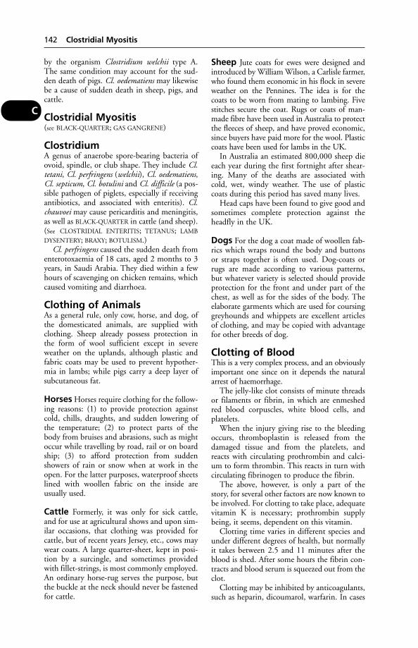
by the organism Clostridium welchii type A.The same condition may account for the sud-den death of pigs. Cl. oedematiens may likewisebe a cause of sudden death in sheep, pigs, andcattle.
Clostridial Myositis(see BLACK-QUARTER; GAS GANGRENE)
ClostridiumA genus of anaerobe spore-bearing bacteria ofovoid, spindle, or club shape. They include Cl.tetani, Cl. perfringens (welchii), Cl. oedematiens,Cl. septicum, Cl. botulini and Cl. difficile (a pos-sible pathogen of piglets, especially if receivingantibiotics, and associated with enteritis). Cl.chauvoei may cause pericarditis and meningitis,as well as BLACK-QUARTER in cattle (and sheep).(See CLOSTRIDIAL ENTERITIS; TETANUS; LAMB
DYSENTERY; BRAXY; BOTULISM.)Cl. perfringens caused the sudden death from
enterotoxaemia of 18 cats, aged 2 months to 3years, in Saudi Arabia. They died within a fewhours of scavenging on chicken remains, whichcaused vomiting and diarrhoea.
Clothing of AnimalsAs a general rule, only cow, horse, and dog, ofthe domesticated animals, are supplied withclothing. Sheep already possess protection inthe form of wool sufficient except in severeweather on the uplands, although plastic andfabric coats may be used to prevent hypother-mia in lambs; while pigs carry a deep layer ofsubcutaneous fat.
Horses Horses require clothing for the follow-ing reasons: (1) to provide protection againstcold, chills, draughts, and sudden lowering ofthe temperature; (2) to protect parts of thebody from bruises and abrasions, such as mightoccur while travelling by road, rail or on boardship; (3) to afford protection from suddenshowers of rain or snow when at work in theopen. For the latter purposes, waterproof sheetslined with woollen fabric on the inside areusually used.
Cattle Formerly, it was only for sick cattle,and for use at agricultural shows and upon sim-ilar occasions, that clothing was provided forcattle, but of recent years Jersey, etc., cows maywear coats. A large quarter-sheet, kept in posi-tion by a surcingle, and sometimes providedwith fillet-strings, is most commonly employed.An ordinary horse-rug serves the purpose, butthe buckle at the neck should never be fastenedfor cattle.
Sheep Jute coats for ewes were designed andintroduced by William Wilson, a Carlisle farmer,who found them economic in his flock in severeweather on the Pennines. The idea is for thecoats to be worn from mating to lambing. Fivestitches secure the coat. Rugs or coats of man-made fibre have been used in Australia to protectthe fleeces of sheep, and have proved economic,since buyers have paid more for the wool. Plasticcoats have been used for lambs in the UK.
In Australia an estimated 800,000 sheep dieeach year during the first fortnight after shear-ing. Many of the deaths are associated withcold, wet, windy weather. The use of plasticcoats during this period has saved many lives.
Head caps have been found to give good andsometimes complete protection against theheadfly in the UK.
Dogs For the dog a coat made of woollen fab-rics which wraps round the body and buttonsor straps together is often used. Dog-coats orrugs are made according to various patterns,but whatever variety is selected should provideprotection for the front and under part of thechest, as well as for the sides of the body. Theelaborate garments which are used for coursinggreyhounds and whippets are excellent articlesof clothing, and may be copied with advantagefor other breeds of dog.
Clotting of BloodThis is a very complex process, and an obviouslyimportant one since on it depends the naturalarrest of haemorrhage.
The jelly-like clot consists of minute threadsor filaments or fibrin, in which are enmeshedred blood corpuscles, white blood cells, andplatelets.
When the injury giving rise to the bleedingoccurs, thromboplastin is released from thedamaged tissue and from the platelets, andreacts with circulating prothrombin and calci-um to form thrombin. This reacts in turn withcirculating fibrinogen to produce the fibrin.
The above, however, is only a part of thestory, for several other factors are now known tobe involved. For clotting to take place, adequatevitamin K is necessary; prothrombin supplybeing, it seems, dependent on this vitamin.
Clotting time varies in different species andunder different degrees of health, but normallyit takes between 2.5 and 11 minutes after theblood is shed. After some hours the fibrin con-tracts and blood serum is squeezed out from theclot.
Clotting may be inhibited by anticoagulants,such as heparin, dicoumarol, warfarin. In cases
142 Clostridial Myositis
C
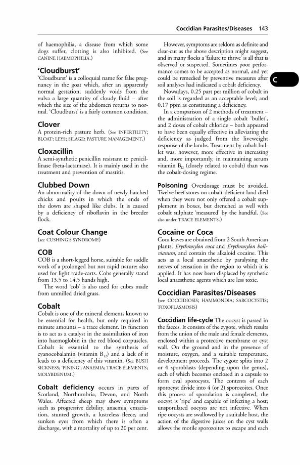
of haemophilia, a disease from which somedogs suffer, clotting is also inhibited. (See
CANINE HAEMOPHILIA.)
‘Cloudburst’‘Cloudburst’ is a colloquial name for false preg-nancy in the goat which, after an apparentlynormal gestation, suddenly voids from thevulva a large quantity of cloudy fluid – afterwhich the size of the abdomen returns to nor-mal. ‘Cloudburst’ is a fairly common condition.
CloverA protein-rich pasture herb. (See INFERTILITY;BLOAT; LEYS; SILAGE; PASTURE MANAGEMENT.)
CloxacillinA semi-synthetic penicillin resistant to penicil-linase (beta-lactamase). It is mainly used in thetreatment and prevention of mastitis.
Clubbed DownAn abnormality of the down of newly hatchedchicks and poults in which the ends ofthe down are shaped like clubs. It is causedby a deficiency of riboflavin in the breederflock.
Coat Colour Change(see CUSHING’S SYNDROME)
COBCOB is a short-legged horse, suitable for saddlework of a prolonged but not rapid nature; alsoused for light trade-carts. Cobs generally standfrom 13.5 to 14.5 hands high.
The word ‘cob’ is also used for cubes madefrom unmilled dried grass.
CobaltCobalt is one of the mineral elements known tobe essential for health, but only required inminute amounts – a trace element. Its functionis to act as a catalyst in the assimilation of ironinto haemoglobin in the red blood corpuscles.Cobalt is essential to the synthesis ofcyanocobalamin (vitamin B12) and a lack of itleads to a deficiency of this vitamin. (See BUSH
SICKNESS; ‘PINING’; ANAEMIA; TRACE ELEMENTS;MOLYBDENUM.)
Cobalt deficiency occurs in parts ofScotland, Northumbria, Devon, and NorthWales. Affected sheep may show symptomssuch as progressive debility, anaemia, emacia-tion, stunted growth, a lustreless fleece, andsunken eyes from which there is often adischarge, with a mortality of up to 20 per cent.
However, symptoms are seldom as definite andclear-cut as the above description might suggest,and in many flocks a ‘failure to thrive’ is all that isobserved or suspected. Sometimes poor perfor-mance comes to be accepted as normal, and yetcould be remedied by preventive measures aftersoil analyses had indicated a cobalt deficiency.
Nowadays, 0.25 part per million of cobalt inthe soil is regarded as an acceptable level; and0.17 ppm as constituting a deficiency.
In a comparison of 2 methods of treatment –the administration of a single cobalt ‘bullet’,and 2 doses of cobalt chloride – both appearedto have been equally effective in alleviating thedeficiency as judged from the liveweightresponse of the lambs. Treatment by cobalt bul-let was, however, more effective in increasingand, more importantly, in maintaining serumvitamin Bl2 (closely related to cobalt) than wasthe cobalt-dosing regime.
Poisoning Overdosage must be avoided.Twelve beef stores on cobalt-deficient land diedwhen they were not only offered a cobalt sup-plement in boxes, but drenched as well withcobalt sulphate ‘measured’ by the handful. (See
also under TRACE ELEMENTS.)
Cocaine or CocaCoca leaves are obtained from 2 South Americanplants, Erythroxylon coca and Erythroxylon boli-vianum, and contain the alkaloid cocaine. Thisacts as a local anaesthetic by paralysing thenerves of sensation in the region to which it isapplied. It has now been displaced by syntheticlocal anaesthetic agents which are less toxic.
Coccidian Parasites/Diseases(see COCCIDIOSIS; HAMMONDIA; SARCOCYSTIS;TOXOPLASMOSIS)
Coccidian life-cycle The oocyst is passed inthe faeces. It consists of the zygote, which resultsfrom the union of the male and female elements,enclosed within a protective membrane or cystwall. On the ground and in the presence ofmoisture, oxygen, and a suitable temperature,development proceeds. The zygote splits into 2or 4 sporoblasts (depending upon the genus),each of which becomes enclosed in a capsule toform oval sporocysts. The contents of eachsporocyst divide into 4 (or 2) sporozoites. Oncethis process of sporulation is completed, theoocyst is ‘ripe’ and capable of infecting a host;unsporulated oocysts are not infective. Whenripe oocysts are swallowed by a suitable host, theaction of the digestive juices on the cyst wallsallows the motile sporozoites to escape and each
Coccidian Parasites/Diseases 143
C
penetrates an epithelial cell. Here each parasiteincreases in size and finally becomes a largerounded schizont. This divides into a number ofsmall elongated merozoites which, escapingfrom the epithelial cell into the gut, attack newcells, and the process is repeated. The massivefeeding stage in the cell before it starts dividingis called a trophozoite, and is usually a youngschizont. Under certain conditions, however,some trophozoites develop into large femaleforms or macrogametocytes which, whenmature, become macrogametes. Meanwhile cer-tain other trophozoites develop into male cells ormicrogametocytes, which divide into a numberof small microgametes. One of these unites witheach macrogamete, and the resulting cell iscalled the zygote. The fertilised macrogamete, orzygote, then secretes a thick capsule arounditself, forming an oocyst which is discharged intothe lumen of the organ intestine or bile-duct andthus escapes from the host in the faeces.
(a) Levincia (formerly Isospora) – the matureoocyst contains 2 sporocysts, each with 4sporozoites.
(b) Eimeria – the mature oocyst contains4 sporocysts, each with 2 sporozoites.
CoccidiomycosisCoccidiomycosis is a fungal disease, involvingchiefly the lymph nodes, and giving rise totumour-like (granulomatous) lesions. It occursin cattle, sheep, dogs, cats, and certain wildrodents, caused by infection with a fungus,called Coccidioides immitis. It has been recog-nised in many parts of the USA and Canada. Itis seen in animals with immuno-suppression;especially young dogs.
Signs Loss of appetite, fever, weight loss,cough, enlarged lymph nodes.
Chiefly recognised in abattoirs during theinspection of meat for human consumption, orin other animals at post-mortem examination.The lesions are sometimes confused with thoseof actinomycosis or actinobacillosis. In the dog,the disease may involve several internal organsand also bone.
An imported baboon diagnosed with thecondition had skin lesions on muzzle and tailconsisting of raised, plaque-like ulcers. Thelesions may resemble those of Mycobacteriumtuberculosis and Yersinia pseudotuberculosis.Coccidiomycosis is communicable to man.
CoccidiosisA disease of major economic importance affect-ing many species of farm and domesticmammals, poultry; also people and birds.
Cause Eimeria, a group of protozoan parasites.For the life-history of the parasites causingthis disease, see under COCCIDIAN PARASITES/DIS-
EASES.
Cattle (in which coccidiosis is called reddysentery).
Causal agent Eimeria zürnii. This is believedto be the most important species affecting cat-tle. Developmental forms occur wholly in thelarge intestine and caecum where considerabledenudation of epithelium occurs, resulting inextensive haemorrhage. The oocysts are nearlyspherical, and sporulation, under favourableconditions, takes place in from 48 to 72 hours.It is found in Europe, Africa, and N. America.It is prevalent during the warm season, andattacks especially animals of 2 months to2 years.
Signs are first seen 1 to 8 weeks after infection.There is a persistent diarrhoea which becomeshaemorrhagic. After about a week, emaciationis evident; the temperature rises, and there aredigestive disturbances. Milk is diminished orstopped. Passage of faeces is attended by strain-ing or even eversion of the rectum.Convalescence is slow. The lesions are mainly inthe large intestine. Mortality varies between2 and 10 per cent of affected animals, and,generally speaking, the younger the animal, themore likely it is to succumb.
Treatment consists of isolation of all sickanimals and careful nursing, with the use ofsulfadimidine or decoquinate.
Sheep and goats
Causal Agents At least 7 species of Eimeriaoccur in these animals, and mixed infectionswith 2 or more species are the rule rather thanthe exception. The various species are widelydistributed and as a rule the clinical disease is seen in lambs and kids, but seldom in the old animals which, however, may harbour coc-cidia. However, new-born lambs are relative-ly resistant. Decoquinate, sulfadimidine or sulfamethoxypyridazine are used to treat or prevent coccidiosis.
Signs are those of a pernicious anaemia accom-panied by diarrhoea and emaciation.
Pigs Coccidiosis is seldom reported as a seriousdisease in the UK, and its importance is debat-able. However, reports of increasing losses from
144 Coccidiomycosis
C
it in the USA led to a re-appraisal. Occasionaloutbreaks have been treated with amprolium,monensin or toltazuril.
Horses Diarrhoea, emaciation and deathhave occurred following infection. (See also
GLOBIDIOSIS.)
Rabbits There are 2 forms of the disease: 1attacking the intestines, and the other the liver.Young rabbits may have acute enteritis, leadingto death. The hepatic form often takes a chron-ic course, with diarrhoea developing later.Affected livers show whitish spots at autopsy.Robenidine or clopidol may be used fortreatment.
Dogs and cats The following species havebeen found in cases of coccidial infection:Isospora felis; I. rivolta; I. bigemina; E. canis; andE. felina.
Most of these parasites have been isolatedfrom healthy animals. The majority of coccidi-al infections of dogs and cats are light, and thereis little evidence of serious damage to the hosts.In a few cases, however, there is diarrhoea andoccasionally fatal dysentery.
Coccidiosis in carnivores is commoner thanwas once believed, especially in young cats,where the parasite is I. felis. The disease causesno symptoms except diarrhoea when a heavyinfestation has occurred. Death is rare. The rabbit parasite may be found in faeces whendiseased rabbits have been eaten. I. canis wasisolated from 4 per cent of 481 faecal samplesfrom dogs in North Island, New Zealand; I. ohioensis from 9 per cent.
Fowls At least seven species of Eimeria havebeen implicated. The disease commonly affectschicks 5 to 7 weeks old, as well as older grow-ing birds. In the former the mortality may behigh. Diarrhoea, often with blood in the faeces,is seen.
Control A vaccine derived from the species ofEimeria that affect chickens (Paracox; Schering-Plough) is administered in the drinking water.A single dose gives effective control of coccidio-sis and, unlike earlier live vaccines, does notcarry the risk of causing the disease in non-vac-cinated birds.
Before the introduction of this vaccine,control was dependent upon antibiotics suchas monensin and SALINOMYCIN, or uponamprolium.
Turkeys Six species of Eimeria cause disease.
Ducks Coccidiosis occurs, but is of littleeconomic importance.
Geese Three species of Eimeria occur in theintestine. Rather severe outbreaks have beenascribed to E. anseris. A 4th, important speciesis E. truncata, which causes a severe form ofrenal coccidiosis. The disease affects goslingsfrom 3 weeks to 3 months of age, and in heavyinfections goslings may die within 2 or 3 daysafter symptoms are first seen. The mortality isoften very high.
CoccygealCoccygeal vertebrae are the tail bones. One ormore may fracture if a dog, cat, etc., becomescaught by a closing door or gate. The coccygealvein is often used to obtain venous blood incattle.
CockroachesThese insects may be responsible for the spreadof salmonella, which they carry in their gut. Aprotein found in the faeces of European andNorth American cockroaches could induce anattack of ASTHMA.
Cocker SpanielThe commonest and most popular of thespaniels. Originally used to retrieve game, thebreed has blunt teeth which avoid damagewhen carrying birds, etc. They are liable toinherit distichiasis, entropion, glaucoma andretinal atrophy, among other conditions. A‘rage syndrome’ is also associated with the breed(see ‘JECKYLL AND HYDE’ SYNDROME).
Cocoa PoisoningPoisoning of pigs and poultry, as a result offeeding cocoa residues or waste, was recorded inthe UK during the 1939–45 war. (See also
CHOCOLATE POISONING.)
Cocoa ShellsGround cocoa shells are sometimes used inanimal feeds. The material contains traces ofcaffeine and theobromine and was blamed asthe source of those drugs found in the blood ofa winning racehorse, which was subsequentlydisqualified.
Cod-Liver OilA valuable source of vitamin A and D supple-ments for animal feeding. The best varietiescontain about 1000 to 1200 InternationalUnits of vitamin A, and 80 to 100 Units of D,per gram. It should be stored in a dark-colouredcontainer, preferably in a cool place, and if air
Cod-Liver Oil 145
C

can be excluded until it is to be used, thiswill enable it to be kept longer. Both strongsunlight and oxygen cause a destruction ofvitamin A. Overdosage can be harmful. (See also
VITAMINS.)
Uses It has a particularly beneficial action inwarding off rickets in young animals, and ifthis trouble has already started it may bechecked, or cured, by the administration ofcod-liver oil. Synthetic vitamins have largelyreplaced cod-liver oil.
Swabs of cod-liver oil are also useful in eyeinjuries and in simple burns.
Cod-Liver Oil PoisoningThis may occur through the use of oil whichhas been allowed to oxidise or become rancid.One result may be muscular dystrophy in cattle(see under MUSCLES, DISEASES OF).
CodeineCodeine is one of the active principles ofOPIUM, and is used as codeine phosphate, tocheck severe coughing in bronchitis, commoncold, and in some cases of laryngitis. It is alsoused as an analgesic.
CoenuriasisInfestation of the sheep’s brain with cysts ofthe dog (and fox) tapeworm Taenia multiceps.(See under TAPEWORMS.)
Coffin BoneThe bone enclosed in the hoof of the horse.Also known as the pedal bone.
Coggins TestThe agar-gel immunodiffusion test. Useful inthe diagnosis of, e.g., equine infectiousanaemia.
Coit, Mal DuAnother name for DOURINE.
Coital Exanthema(see VULVOVAGINITIS; also RHINOTRACHEITIS)
Coitus(see REPRODUCTION)
ColbredA cross between the East Friesland and 3 Britishbreeds of sheep (Border Leicester, Clun Forest,and Dorset Horn). The aim of Mr OscarColburn, their breeder, was to produce eweswith a consistent 200 per cent lambing averageand a sufficiency of milk for this.
ColchicineThe alkaloid obtained from meadow saffron(Colchicum autumnale). It is used in plant andexperimental animal breeding as ‘a multiplier ofchromosomes’. It has been possible to produceTRIPLOID rabbits, pigs, etc., by exposing semento a solution of colchicine prior to artificialinsemination.
Colchicum Poisoning(see MEADOW SAFFRON POISONING)
Cold(see HYPOTHERMIA; EXPOSURE; FROSTBITE;SHEARING)
ColicColic is a vague term applied to symptoms ofabdominal pain, especially in horses. In order toemphasise the large number of different condi-tions which may produce abdominal pain inthe horse, the following list is included:
1. Acute indigestion, resulting from thefeeding of unsuitable food, the presence of gas(flatulent colic).
2. Severe organic disorders, such asimpaction of the colon, intussusception, volvu-lus, or strangulation of the bowel, rupture ofthe stomach, enteritis, and peritonitis, areamong the serious causes.
3. The presence of large numbers of parasiticworms, horse bots, etc. (See under EQUINE
VERMINOUS ARTERITIS.)4. Calculi present in the kidney, urinary blad-
der, or urethra in the male, causing irritation ofthese organs.
5. Anthrax, where one of the commonsymptoms is abdominal pain.
6. Approaching parturition in the pregnantmare.
7. Grass disease.8. See HYMEN, IMPERFORATE.9. Uterine rupture.10. Nephritis.11. Various poisons (see POISONING).12. In addition, in countries where RABIES is
endemic, this disease should be borne in mindwhen presented with a horse which appears tohave colic.
The horse has a peculiarity in the arrange-ment of its alimentary canal, in that while thestomach is comparatively small, the intestines,and especially the large intestines, are of greatbulk and capacity. In addition to this, the stom-ach itself has the peculiarity that its entranceand exit are small; the former only allows escapeof gas into the gullet under exceptional circum-stances, and the latter, owing to the S-shaped
146 Cod-Liver Oil Poisoning
C
bend of the pylorus and first part of the smallintestine, is very liable to become occludedwhen there is any considerable pressure of gaswithin the stomach. These facts combine tomake it difficult or impossible for gas collectedin the stomach, as the result of fermentation,either to escape by the mouth or to pass on intothe intestines. Fermenting or otherwise?unsuit-able food may cause tympany of the stomach;while an excess of any food may lead toimpaction of the stomach; and occasionally toits rupture.
Inflammation or volvulus may affect thesmall intestine, but most cases of colic involvethe large intestine. Impaction of the caecum orcolon may occur; likewise tympany.
The ileum, supplied only by a single artery,appears to be particularly vulnerable toischaemia, following thrombosis often causedby Strongylus vulgaris worms.
Anaerobic bacteria and their toxins mayexacerbate the situation after circulation defectshave occurred.
(See also INTUSSUSCEPTION, another cause ofcolic.)
Signs1. Spasmodic colic is typified by sudden and
severe attacks of pain, usually of an intermittentcharacter. Breathing is blowing and faster thanusual; there is an anxious expression about theface; and the pulse is accelerated and hard. In afew minutes the attack may pass off and thehorse becomes easier, or the pain may continue.In the latter case the horse lies down and rolls,after having first walked round about the box.In some cases rolling appears to afford somemeasures of relief, but in others the horse risesagain almost at once. During an attack thehorse may kick at its belly, or may turn and gazeat its flank.
In another form, ileus – often called flatulentcolic – the pain begins suddenly, but there arenot such distinct periods of ease. The horsewalks round and round the box, kicks at theabdomen, gazes at its sides, breaks out intopatchy sweating, and breathes heavily. Thehorse frequently crouches as if to lie down, butonly actually lies in the less severe cases, and sel-dom or never remains lying for any length oftime. Attempts at passing urine are noticed,but, as in the truly spasmodic colic, they areseldom successful. Faeces may be passed insmall quantities, and are usually accompaniedby flatus.
2. Obstructive colic may arise throughimpaction of the bowel with dry, fibrous, part-ly digested food material. Symptoms develop
slowly, commencing with dullness and depres-sion, irregularity in feeding, and abdominal dis-comfort. In 12 hours or so signs of abdominalpain appear. In some cases acute pain is shown,the horse rolling on the ground in agony. Smallamounts of faeces are passed with considerablefrequency at first, but when an attack is wellestablished the passage of both urine and dungceases. An attitude to which some importancemay be attached, since it is very strongly sug-gestive of impaction of the colon, is one inwhich the horse backs against the manger orother projection, and appears to sit upon it,sometimes with the hind-feet off the ground. Inother cases a horse with obstruction in thecolon or caecum may sit with the hindquarterson the ground, but retains an upright positionwith the forelegs – somewhat similar to theposition assumed by a dog. (See CALCULI foranother cause of obstruction.)
3. Colic due to a twist (volvulus). There isgreat pain, during which the horse may becomerestless and violent. Sometimes the pain passesoff, and sweating occurs, before a further peri-od of pain. The temperature may be 41°C(105 or 106°F), becoming subnormal in thelast stages. Pulse-rate may rise to 120. Death isusually preceded by convulsions.
Many colic cases end fatally, and it is certainthat many horses might have been saved if aveterinary surgeon had been summoned at theoutset.
A survey of 134 cases of colic, seen at theveterinary clinic, University of Zurich, included34 which were symptomless on arrival, requiredno treatment, and were regarded as cases of spas-modic colic. Thirty-three horses had impactionof the pelvic flexure of the colon and were treat-ed conservatively; as were 14 with impaction ofthe ampulla, coli (4), and caecum (1). Therewere 7 cases of tympany of the stomach and 2 ofimpaction. Of 53 cases of ileus, the prognosiswas hopeless in 7 which were destroyed, andowners refused surgery in another 6 cases. Fortyunderwent laparotomy, and 24 were discharged.Surgical success rate was 60 per cent; overall suc-cess of treatment was 68 per cent. Suggestionsincluded maintenance of a nasal stomach tubeto eliminate possibly lethal consequences of sec-ondary gastric distension by fluid and gas dur-ing the journey to the clinic; and 1 litre of 5 percent sodium bicarbonate solution intravenouslyto help control the start of acidosis. (See also
HORSES, COMMON CAUSES OF DEATH IN.)
Cattle Bovine colic occurs relatively infre-quently. It is mainly caused by torsion of the
Colic 147
C
caecum or abomasum, mesenteric torsion,intussusception, strangulation or phytobezoar.
ColiformA convenient term used to describe severalspecies of lactose fermenting bacilli whichinhabit the gut. The most commonly encoun-tered is Escherichia coli and approximately80 per cent of coliform isolates tested at theNational Institute for Research in Dairyingare E. coli. Other coliform species implicatedin bovine mastitis include Klebsiella pneumoni-ae, K. oxytoca, Enterobacter cloacae, E. aerogenesand Citrobacter freundii. All are ‘gut associated’but some, notably K. pneumoniae andEnterobacters, may be free-living in forest envi-ronments or soil and be introduced into a dairyherd with sawdust or wood shavings used forcattle litter.
Coliform InfectionsColiform infections include mastitis, enteritisand septicaemia. Coliform organisms are fre-quently found in apparently healthy animals.An examination of cattle carcases at slaughter-houses showed that coliform organisms wereisolated from surface swabbings from 208 outof 400 head of cattle (52 per cent); 81 of thesebeing resistant to 1 or more antibiotics. Of 400pig carcase swabs, 331 (83 per cent) were posi-tive for coliforms; 246 being resistant to 1 ormore antibiotics. Chloramphenicol resistancewas present in 19 pig isolates and 1 cattle iso-late. (See BEDDING – Sawdust; E. COLI; MASTITIS
IN COWS.)
ColitisInflammation of the colon, or first part of thelarge intestine. (See INTESTINES.)
Collagen(see FIBROUS TISSUE; also CUTANEOUS ASTHENIA)
Collateral Circulation(see ANASTOMOSIS)
Colliculus SeminalisThis protrudes into the lumen of the urethra,and at its centre is a minute opening into a tinytube (the uterus masculinus) which runs intothe prostate gland.
Collie Eye AnomalyA congenital disease occurring in some roughcollies, smooth collies, and Shetland sheep-dogs. In the worst cases, blindness may followdetachment of the retina or haemorrhagewithin the eye.
CollodionFlexible collodion, a mixture of pyroxylin, alco-hol and ether, is fairly elastic and does not crackwith movement. It was used for application tolesions around joints. Medicated collodion contains substances such as salicylic acid andiodoform. A collodion preparation containing acaustic was used for destroying the horn-budsof calves. (See DE-HORNING.)
ColloidColloid is matter in which either the individualparticles are single large molecules, such asproteins, or aggregates of smaller molecules aremore or less uniformly distributed in a disper-sion medium, e.g. water, oil. Examples: col-loidal silver (used for eye infections), andcolloidal manganese.
ColobomaA congenital eye defect caused by an absence orfault in the tissue. (See under EYE, DISEASES OF.)
ColonThe part of the large intestine extending fromthe caecum to the rectum. (See COLITIS;INTESTINES.)
ColostrumColostrum is the milk secreted by the udderimmediately after parturition and for the fol-lowing 3 to 4 days. It contains 20 per cent ormore protein, a little more fat than normal milk,and may be tinged pink due to blood corpus-cles. It coagulates at about 80° to 85°C, andcannot therefore be boiled. This is sometimesused as a test. It is normally rich in vitamins Aand D provided the dam has not been deprivedof these in her food. It acts as a natural purgativefor the young animal, clearing from its intestinesthe accumulated faecal matter known as ‘meco-nium’, which is often of a dry, putty-like nature.Of much greater importance, it is through themedium of the colostrum that the young animalobtains its first supply of antibodies whichprotect it against various bacteria and viruses.
Before the cow calves, her udder selectivelywithdraws these immunoglobulins from herblood into the colostrum. In the suckling calf,the immunoglobulins become active in theblood serum after absorption, and they alsohave a local protective action within the smallintestine. If the calf is to survive, both theserum and intestinal immunoglobulins must bepresent in adequate quantities; for the serumimmunoglobulin will protect against septi-caemia, but not against the enteritis which leadsto scouring and dehydration.
148 Coliform
C
The importance of the calf receivingcolostrum early has long been emphasised. Thisis recognised in the Welfare of LivestockRegulations 1994, which specify that colostrummust be fed within 6 hours of the calf ’s birth.
Research at Glasgow veterinary school foundthat beef suckler cows suckled their calf within1.5 hours of birth whereas, on average, dairycows suckled their calves after 4 hours.
Colour-Marking BullsColour-marking bulls, e.g. Hereford, AberdeenAngus, Charolais, and Galloway, for matingwith cows in dairy herds which are of dual-pur-pose type and moderate to poor milkers, inorder to increase the number of store cattle suit-able for fattening for beef production. (See also
BEEF-BREEDS AND CROSSES.)
Columnaris DiseaseA disease in fish caused by Chondrococcuscolumnaris, an opportunist myxobacterium pre-sent in the water. Affected fish have greyish-white lesions on the skin. It is prevented bycontrolling water temperature to below 25°Cand reducing stress on the fish.
ColtA young male horse.
ComaComa is a state of profound unconsciousness inwhich the patient not only cannot be roused,but there are no reflex movements when theskin is pinched or pricked, or when the eyeballsare touched, etc. The cause is generally anexcessively high temperature, brain injury, cere-bral haemorrhage, some poisons, or too muchor too little insulin in cases of diabetes, or theterminal stage of a fatal illness.
CombA projection of the skin, serrated at the top,running from front to back of the skull. Inhealthy poultry, it should be bright red and welldeveloped. When birds go out of lay or arecaponised, the comb becomes smaller and paler.Anaemia may also cause this. A pale comb ofnormal size suggests internal haemorrhage.Scurfiness is suggestive of favus; yellow scabs offowl-pox.
Comeny’s Infectious Paralysisof HorsesThis condition, of unknown aetiology, was firstdescribed in French army horses by Comény.The reported signs are a sudden rise in temper-ature to 40°C (104° or 105°F), persisting for
5 days, and followed in some cases by paralysisafter a period of hind-limb incoordination anddifficulty in turning.
CommensalismCommensalism is the association of 2 species inwhich 1 alone benefits, but the other does notsuffer. The term is used to refer to a benign par-asitism. Commensal micro-organsisms arefound on the skin surface, for example, and donot produce disease.
CommissureCommissure means a joining, and is a termapplied to strands of nerve fibres that join 1 sideof the brain to the other, to the band joining 1optic nerve to the other, to the junction of thelips at the corners of the mouth, etc.
Communicable DiseasesFor diseases communicable to man, see under
ZOONOSES. For diseases communicable fromman to farm livestock, etc., see ANTHRO-
PONOSES.
Companion Animal WelfareCouncilAn independent body established in 1999 toconduct and publish studies into the welfare,treatment and care of companion animals. It isfunded by a charitable trust.
Comparative Test(see TUBERCULIN TEST)
CompensationCompensation is the term applied to themethod by which the body makes good a defectof form or function in an organ which is abnor-mal in these respects.
ComplementA complex protein that is a constituent ofserum and plays an essential part in the pro-duction of immunity. Bacteria are killed by thespecific antibody developed in an animal’sserum only in the presence of complement.Complement is also necessary for haemolysis.
An immune serum may contain antibodieswhich, together with the antigen, absorb or fixcomplement and are hence called complement-fixing antibodies. These form the basis for theComplement Fixation Test, which is used in thediagnosis of certain diseases, e.g. Johne’s. As anindicator for the test, red blood corpusclesplus their specific antibody are used, i.e. thecorpuscles plus the antiserum heated at 55°C to inactivate or destroy the complement. In
Complement 149
C

the test, on adding the indicator, haemolysiswill not occur if the complement has beenfixed.
Compound FeedsA number of different ingredients (includingmajor minerals, trace elements, vitamins andother additives) mixed and blended in appropri-ate proportions, to provide properly balanceddiets for all types of stock at every stage ofgrowth and development. (See DIET; FLUOROSIS.)
Compulsive PolydipsiaThe urge to drink excessive quantities of water,due to some psychological disturbance, is arecognised syndrome in human medicine,and it probably occurs in dogs as a result ofstress; leading to urinary incontinence. (See also
DIABETES INSIPIDUS.)
ConalbuminAn important constituent of egg-white. Itmakes iron unavailable to certain bacteria(Salmonella and Arizona spp) thus inhibitingtheir multiplication within the egg duringincubation. (See also IRON-BINDING PROTEINS.)
ConcentratesThe bulk of these in Britain today come fromhighly reputable compound feeding-stuffsmanufacturers, and are expert formulationsrelated not only to the current price of variousingredients but also to the proper balancing ofthese ingredients. Computers are often used inthe formulations. The inclusion of trace ele-ments, minerals, and vitamins makes thesecompound feeding-stuffs foods complete inthemselves. Suitable mixes are obtainable forevery class of farm livestock.
Farm-mixed concentrates are commonlyused on large arable farms, using home-grownbarley, oats, beans, etc. The expertise requiredfor formulation may also be lacking, so that onthe smaller farm, proprietary concentrates areoften to be preferred.
(See DIET; CUBES; also ADDITIVES, COMPOUND
FEEDS; SUPPLEMENTS.)
Conception RatesConception Rates following artificial insemina-tion of cattle are stated to be in the region of 65 per cent in dairy breeds, and over 70 percent in beef breeds. In the UK, the conceptionrate is usually based upon the number of ani-mals which, in a 3-month period, do not returnto the first insemination. In Denmark, the con-ception rate is based on the evidence of a phys-ical pregnancy diagnosis carried out 3 monthsafter insemination.
Conception rates are influenced by many fac-tors. The best time for insemination is between2 and 20 hours after ‘heat’ is observed; after that,delay will mean a lower conception rate. Healthof male and female, and inseminator’s skill alsoinfluence the rate. (See also FARROWING RATES.)
ConceptusThe embryo or fetus together with the tissues,such as the placenta, that nourish it.
ConcreteThe precise composition of concrete may proveimportant where floor feeding is practised.Suspected iron poisoning from the licking ofconcrete made with sand rich in iron has beendescribed in fattening pigs. Concrete floors ofpiggeries, etc., should be made with integral airspaces in order to have some insulating effect,and should not be abrasive. If they are, they canlead to injuries, followed by staphylococcal orother infection which may cause severe illnessor death even in a new pig pen. Pigs shouldnever be allowed to lie on freshly set concrete;skin burns may result from chemicals in themix. (See under FOOT-ROT OF PIGS; HOUSING OF
ANIMALS; BEDDING.)
Concussion(see under BRAIN DISEASES)
Condensation in Buildings(see NITRITE POISONING; CALF HOUSING; PNEU-
MONIA; YORKSHIRE BOARDING; VENTILATION)
Condition(see under MUSCLE)
Condition ScoreA method used to evaluate the thinness or fat-ness of cattle, sheep, goats, pigs and horses. It isused to monitor feeding and check that animalsare fit to breed and maintain pregnancy.
CondyleCondyle is the rounded prominence at the endof a bone; for instance, the condyles of thehumerus are the two prominences on eitherside of the elbow-joint in animals, while thecondyles of the femur enter into the formationof the stifle joint.
Conformation Assessment inthe Cow(see under PROGENY TESTING)
Congestive Heart Failure(see under HEART, DISEASES OF)
150 Compound Feeds
C
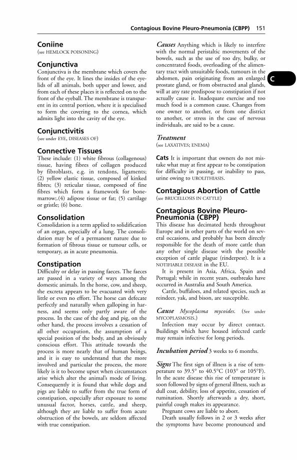
Coniine(see HEMLOCK POISONING)
ConjunctivaConjunctiva is the membrane which covers thefront of the eye. It lines the insides of the eye-lids of all animals, both upper and lower, andfrom each of these places it is reflected on to thefront of the eyeball. The membrane is transpar-ent in its central portion, where it is specialisedto form the covering to the cornea, whichadmits light into the cavity of the eye.
Conjunctivitis(see under EYE, DISEASES OF)
Connective TissuesThese include: (1) white fibrous (collagenous)tissue, having fibres of collagen producedby fibroblasts, e.g. in tendons, ligaments;(2) yellow elastic tissue, composed of kinkedfibres; (3) reticular tissue, composed of finefibres which form a framework for bone-marrow;.(4) adipose tissue or fat; (5) cartilageor gristle; (6) bone.
ConsolidationConsolidation is a term applied to solidificationof an organ, especially of a lung. The consoli-dation may be of a permanent nature due toformation of fibrous tissue or tumour cells, ortemporary, as in acute pneumonia.
ConstipationDifficulty or delay in passing faeces. The faecesare passed in a variety of ways among thedomestic animals. In the horse, cow, and sheep,the excreta appears to be evacuated with verylittle or even no effort. The horse can defecateperfectly and naturally when galloping in har-ness, and seems only partly aware of theprocess. In the case of the dog and pig, on theother hand, the process involves a cessation ofall other occupation, the assumption of aspecial position of the body, and an obviouslyconscious effort. This attitude towards theprocess is more nearly that of human beings,and it is easy to understand that the moreinvolved and particular the process, the morelikely is it to become upset when circumstancesarise which alter the animal’s mode of living.Consequently it is found that while dogs andpigs are liable to suffer from the true form ofconstipation, especially after exposure to someunusual factor, horses, cattle, and sheep,although they are liable to suffer from acuteobstruction of the bowels, are seldom affectedwith true constipation.
Causes Anything which is likely to interferewith the normal peristaltic movements of thebowels, such as the use of too dry, bulky, orconcentrated foods, overloading of the alimen-tary tract with unsuitable foods, tumours in theabdomen, pain originating from an enlargedprostate gland, or from obstructed anal glands,will at any rate predispose to constipation if notactually cause it. Inadequate exercise and toomuch food is a common cause. Changes fromone owner to another, or from one district to another, or stress in the case of nervous individuals, are said to be a cause.
Treatment(see LAXATIVES; ENEMA)
Cats It is important that owners do not mis-take what may at first appear to be constipationfor difficulty in passing, or inability to pass,urine owing to UROLITHIASIS.
Contagious Abortion of Cattle(see BRUCELLOSIS IN CATTLE)
Contagious Bovine Pleuro-Pneumonia (CBPP)This disease has decimated herds throughoutEurope and in other parts of the world on sev-eral occasions, and probably has been directlyresponsible for the death of more cattle thanany other single disease with the possibleexception of cattle plague (rinderpest). It is aNOTIFIABLE DISEASE in the EU.
It is present in Asia, Africa, Spain andPortugal; while in recent years, outbreaks haveoccurred in Australia and South America.
Cattle, buffaloes, and related species, such asreindeer, yak, and bison, are susceptible.
Cause Mycoplasma mycoides. (See under
MYCOPLASMOSIS.)Infection may occur by direct contact.
Buildings which have housed infected cattlemay remain infective for long periods.
Incubation period 3 weeks to 6 months.
Signs The first sign of illness is a rise of tem-perature to 39.5° to 40.5°C (103° or 105°F).In the acute disease this rise of temperature issoon followed by signs of general illness, such asdull coat, debility, loss of appetite, cessation ofrumination. Shortly afterwards a dry, short,painful cough makes its appearance.
Pregnant cows are liable to abort.Death usually follows in 2 or 3 weeks after
the symptoms have become pronounced and
Contagious Bovine Pleuro-Pneumonia (CBPP) 151
C
acute. Recovery is frequently more apparentthan real, for a chronic cough remains, and thedisease may again become acute and even endfatally.
Post-Mortem appearances Large or smallareas of pneumonia in the lungs, which areoften of a marbled appearance. The lesion isprimarily one of interstitial pneumonia, withthickened septa dividing the lung up into lob-ules; some lobules show acute congestion, someare in a stage of red or grey hepatisation, whileothers consist of dead encapsulated tissue,known as ‘sequestra’. Evidence of pleurisy withoften much fibrinous deposit around the lungsis usual.
Diagnosis The slaughter of suspected animalsmay be essential for this. Corroboration may beobtained by laboratory methods.
Treatment is not allowed in most countries,but neoarsphenamine and tylosin have proveduseful elsewhere.
Immunisation Live vaccines may be used ineradication and control programmes.
Contagious Caprine Pleuro-PneumoniaA disease of goats, caused by a mycoplasma andoccurring in Europe, Asia, and Africa. Acute,peracute, and chronic forms occur. Mortalitymay be 60 to 100 per cent. Antibiotics are use-ful for treatment where a slaughter policy is notin force.
Contagious DiseasesCertain of these are notifiable. (See under NOTI-
FIABLE DISEASES.) The responsibilities of animal
owners are discussed under DISEASES OF
ANIMALS ACTS.
Contagious Ecthyma of SheepContagious ecthyma of sheep is another namefor ORF.
Contagious Epithelioma ofBirds(see FOWL POX)
Contagious Equine Metritis(CEM)Contagious equine metritis (CEM) is a conta-gious venereal disease found in mares and trans-mitted by stallions. This is a NOTIFIABLE DISEASE
in the UK, under the Infectious Diseases ofHorses Order 1987.
Cause A Gram-negative coccobacillus,Taylorella equigenitalis (formerly Haemophilusequigenitalium). This has been isolated fromthe cervix, urethra and clitoris. The organismis apt to persist in the clitoral fossa afterclearance from other parts of the mare’s uro-genital tract, and routine sampling at this site istherefore necessary or diagnosis may fail to beconfirmed.
Control A code of practice for control of theinfection was formulated by the HorseraceBetting Levy Board in 1977, and supported bythe Ministry of Agriculture and theThoroughbred Breeders’ Association. A list oflaboratories authorised to test for the CEMorganism is published annually in theVeterinary Record.
Cervical swab tests are a routine diagnosticand preventive measure on stud farms. Among
152 Contagious Caprine Pleuro-Pneumonia
C
Diagram showing the technique of collecting a cervical swab via a tubular speculum. The swab is passedas far into the open oestrous cervix, and beyond, as possible.

techniques used for taking swabs is that shownin the diagram.
Measures to control the spread of CEM innon-thoroughbred mares have been in opera-tion since 1978.
(See also CLITORIS – Clitoral sinusectomy.)
Contagious Ovine DigitalDermatitis (CODD)A disease resembling, but distinct from, foot-rot which causes severe ulcerative lesions of thecoronet that may lead to complete separation ofthe hoof case. It was first identified in the UKin 1997. The cause is a spirochaete similar tothat responsible for bovine digital dermatitis. Itis differentiated from foot-rot by the absence ofinterdigital sores (in most cases) and by failureto isolate Dichelobacter nodosus, the cause offoot-rot, from the lesions.
For treatment, see DIGITAL DERMATITIS.
Contagious Pustular Dermatitisin Sheep(see ORF)
Contagious Stomatitis(see FOOT-AND-MOUTH DISEASE; also VESICULAR
STOMATITIS)
ContraceptivesMegestrol is one of the drugs used to stop dogsand cats coming on heat. An injection of oestra-diol benzoate has been used in cases of misal-liance in bitches; prostaglandins have also beenused. Prostaglandins have been used to controlthe timing of pregnancy in cattle and to termi-nate an unwanted pregnancy. Attempts todevelop a contraceptive for wild rabbits havebeen hampered by the problem of drug residuesin wild rabbits caught for food. (See also
STILBENES.)
Contracted Foot or ContractedHoofA condition of the horse in which some part ofthe foot, very often a quarter or heel, becomescontracted and shrunken to less than its usualsize. It is brought about by anything whichfavours rapid evaporation of the moisture in thehorn, such as rasping away the outer surface ofthe wall; or by conditions which prevent expan-sion of the hoof, such as paring away the frog sothat it does not come into contact with theground, cutting the bars, allowing the wall atthe heels to fall inwards, shoeing with highcalkins, etc.
Contracted Foot or Contracted Hoof 153
C
About to withdraw the progestogen-impregnated sponge at the end of the 2-weektreatment period. At this time, the ewe receives a single intramuscular injection of PMSG – tocomplete the controlled breeding treatment.
Prevention consists in leaving the frogs aslarge and well developed as possible; reducingthe overgrowth at the heels and bars to the sameextent as at the toe and other parts of the foot;shoeing with shoes which allow the frog tocome into contact with the ground.
Treatment In severe cases a run at grass withtips on the affected feet, and leaving the heelsbare, is advisable. (See also HOOF REPAIR.)
‘Contracted Tendons’(1) A congenital condition, in which the foot isnot fully extended, seen mainly in calves, lambsand foals. The causes are various and the condi-tion may clear up without treatment in a fewdays. If it does not, splints may be applied tostraighten the foot. Surgical correction by par-tially severing the tendon is sometimes carriedout. (2) Chronic tendonitis in adult horses. Thelimb is not fully extended and the animalappears to be standing on its toes (sometimescalled ‘ballerina syndrome’).
Control, Controlled ExperimentIn any scientifically conducted experiment orfield trial, the results of treatment of 1 group ofanimals are compared with results in another,untreated, group. Animals in the untreatedgroup are known as ‘the controls’.
Control of Dogs Order 1992(see under LAW)
Controlled BreedingThe manipulation of ovarian activity to enablesuccessful insemination at a predetermined timeis widely practised in cattle, sheep and pigs.Synchronisation of oestrus enables groups ofanimals to be inseminated at a chosen time, andparturition planned to take place when conve-nient. A progestogen preparation is adminis-tered for 10 to 14 days according to a specificdosage schedule; the animal comes into heatwhen the progestogen is removed. Insemination,by natural mating or artificially, then takes place.The progestogen is administered orally, by injec-tion, implant, or intravaginally, according tospecies and particular product.
An implant of MELATONIN will stimulateearly onset of natural reproductive activity andimprove fertility early in the season.
Cattle Two main systems are used. In one, usedin beef animals or maiden heifers, an implantcontaining norgestomet is inserted under theskin of the ear and then an injection ofnorgestomet plus oestradiol given immediately.
The implant is removed after 9 or 10 days andthe animal inseminated twice, 48 and 72 hours,or once, 56 hours later. In the other system, aspiral device incorporating progesterone, with agelatine capsule containing oestradiol attached,is inserted into the cow’s vagina and left for 12 days. The cow is inseminated twice, after 48and 72 hours, or once at 56 hours.
Alternatively, a progestogen may be adminis-tered by injection.
Sheep A sponge impregnated with a progesto-gen (flugestone or medroxprogesterone) isintroduced to the vagina and left for 12 to 14days. The ewes are introduced to the ram 48hours after removal. The sponge may be used inconjunction with an injection of pregnant mareserum gonadotrophin, given on removal of thesponge, to advance the breeding season by up to6 weeks
Pigs A suspension of altrenogest is added tothe feed once daily; for sows for 3 days, for giltsfor 18 consecutive days.
Farrowing may be induced, within 3 days ofthe expected normal time, by an injection of aluteolitic agent such as cloprostenol or dino-prost. Farrowing then occurs between 24 and30 hours later.
Mares Induction of ovulation in mares to syn-chronise ovulation more closely with matingcan be achieved by an injection of buserelin,a synthetic releasing hormone analogue forboth gonadotrophin and follicle stimulatinghormone.
Warning: Progestogen products should behandled with great care, particularly by preg-nant women. They must not come in contactwith the skin.
Controlled EnvironmentHousingTemperature, ventilation, and humidity arecontrolled within narrow limits by means ofelectric fans, heaters, etc., and good insulation.Poultry, for example, are protected in this wayfrom sudden changes in temperature; rearingcan be carried out with the minimum lossthroughout the year; and increased egg yieldsand decreased food intake can effect a consider-able saving in costs of production. Some ofthese houses are windowless; artificial lightingbeing provided: respiratory disease may occurthrough overcrowding or ventilation defects.
Failure of automatic control Ventilationsystems must have fail-safe alarms and back-up
154 ‘Contracted Tendons’
C

systems under UK law. If those are not effec-tive, there may be fatal consequences, as the following examples show.
A thunderstorm blew the fuse in the fan cir-cuit of a controlled environment house, andunfortunately ‘fail-safe’ ventilation flaps did notwork. As a result 520 fattening pigs died ofheat-stroke.
In another incident the heating system con-tinued to function in a house containing 82 pigs. The fans failed, and minimal naturalventilation resulted in the temperature reaching46°C (104°F), and the death of 65 pigs.
Convex Sole or Dropped SoleThe sole of the horse’s foot, instead of beingarched (concave) when viewed from the groundsurface, is convex and projects to a lower levelthan does the outer rim of the wall in manycases. (See LAMINITIS.)
Convolvulus PoisoningAnother name for MORNING GLORY.
ConvulsionsConvulsions are powerful involuntary contrac-tions (alternating with relaxation) of muscles,producing aimless movement and contortion of the body, and accompanied by loss of consciousness. (See SPASM; FITS.)
Coombs (Antiglobulin) TestCoombs (antiglobulin) test is a laboratory testused in the differential diagnosis of variousblood disorders.
CoopworthA breed of New Zealand sheep derived from the‘Border-Romney’ cross.
COPD(see CHRONIC OBSTRUCTIVE PULMONARY DIS-
EASE)
Copper (Cu)Copper (Cu) is one of the TRACE ELEMENTS
which is essential in the nutrition of animals. Itacts as a catalyst in the assimilation of iron,which is needed in the production of haemoglo-bin in the liver. Its absence from the foodstuffseaten in some areas leads to a form of anaemia.
In several parts of the world a deficiency ofcopper in the herbage has been a major obstacleto livestock production, and appropriate dress-ings of the land have permitted dramaticincreases in production.
In several parts of Britain, copper deficiencyis a serious condition. (See HYPOCUPRAEMIA.)
Two types of copper deficiency are recog-nised: primary and secondary. The former aris-es from an inadequate intake of copper and,while herbage levels of copper below 5 ppm areuncommon in Britain, a survey showed thatover 50 per cent of 1078 beef herds in mid-Wales had low blood copper levels, probablyassociated with low intake. Secondary copperdeficiency is the more common form in the UKand occurs where absorption or storage withinthe animal body of copper is adversely affectedby a high sulphate or molyb denum intake,even though there is adequate copper in thediet.
An excess of molybdenum in the ‘teart’ soilsand pastures of central Somerset, and of areasin Gloucestershire, Warwickshire, Derbyshireand East Anglia, has long been recognised,giving rise to scouring (especially from Mayto October), a greyness of the hair aroundthe eyes, staring coats and a marked loss ofcondition.
However, analysis of sediments from streambeds in many counties shows that herbage maycontain excessively high concentrations ofmolybdenum.
Copper deficiency may be prevented byadministering copper sulphate powder contain-ing 254 g/kg mixed with feed at a dose of 2 gper head; or by a ruminal bolus containingsmall blunt rods (‘needles’) of copper oxide,once a season.
Treatment of copper deficiency is by par-enteral injection of copper, usually in the formof copper edetate or heptonate.
Sheep Caution: Indiscriminate dressing ofpasture with copper salts is likely to causepoisoning in sheep if the quantities used aretoo large, or if sheep are re-admitted to dressedpasture before there has been sufficient rain towash the copper salts off the herbage.
Copper sulphate for pigs Copper sul-phate, added to the fattening ration at the rateof 150–180 ppm, has produced an improve-ment in the growth rate in pigs. (See SWAYBACK;also MOLYBDENUM.)
Copper, Poisoning byWith the exception of sheep, which may begiven an overdose to expel worms, animals arenot likely to be poisoned through internaladministration of copper sulphate as a medi-cine. Poisoning has occurred, however, in sheepgiven a copper-rich supplement, intended forpigs, over a 3.5-month period; in a heifer simi-larly, as well as in pigs given too strong a copper
Copper, Poisoning by 155
C

supplement. Poisoning also occurs when ani-mals are grazed in the vicinity of copper-smelt-ing works, where the herbage graduallybecomes contaminated with copper, inorchards where fruit-trees have been sprayedwith copper salts and also in sheep grazing landtreated with copper sulphate (either crystalsmixed with sand, or as a sprayed solution) asa snail-killer in the control of liver-fluke or as apreventative of swayback.
Signs are those of an irritant poison – pain,diarrhoea (or perhaps constipation), and weak-ness; staggering and muscular twitchings areseen in chronic cases. A fatal chronic copperpoisoning may occur in pigs fed a coppersupplement of 250 parts per million.
Failure to achieve accurate mixing of smallquantities of copper sulphate into farm-mixedrations has led to fatal poisoning of pigs.
It has been pointed out that copper poisoningis almost specific to the housing of sheep. Itoccurs even in diets ostensibly containing nocopper supplement. The capacity of the sheepfor storing copper from the normal constituentsof the diet is higher than that of other animals,and markedly higher in housed sheep. Andlambs reared indoors have died because their haywas made from grass contaminated by slurryfrom pigs on a copper-supplemented diet.
It is dangerous to exceed 10 ppm of copperin dry feeds for sheep over a long period.
AFRC research has shown that the sheep’sphysiological response to copper is influencedby heredity, and that there are significant breeddifferences as regards swayback and copperpoisoning.
Treatment Following some Australianresearch, it was shown at the Rowett ResearchInstitute that 3 subcutaneous injections oftetrathiomolybdate (on alternate days) canremove copper from the livers of both sheepand goats without causing any apparentill-effects.
‘Copper Nose’A form of LIGHT SENSITISATION occurring incattle.
CopperbottleLucilia cuprina, the strike fly which attackssheep in Australia and South Africa.
CoprophagyThe eating of faeces by an animal. In rabbits,this is a normal practice. The rabbit produces 2types of faecal pellet: the normal black pellet,
which is not eaten, and a soft brown pellet, produced in the caecum, which is eaten imme-diately on being expelled from the anus. Thelatter pellets are rich in B vitamins and aminoacids, but can also serve to recycle parasites.Female parents of several species ingest the fae-ces of their offspring in order to keep the nurs-ing area clean. Within 3 weeks of birth, foalswill eat their dams’ faeces and thereby acquirethe various bacteria needed for digestive pur-poses in their own intestines. Overnightcoprophagy has also been reported in adulthorses in adjusting to ‘complete-diet’ cubeswhen no hay is on offer.
It has been suggested that foals may obtainnutrients, and that coprophagy may be aresponse to a maternal pheromone signallingthe presence of deoxycholic acid which may berequired for gut ‘immuno-competence’ andmyelination of the nervous system.
Coprophagy also occurs in piglets, dogs, andnon-human primates.
Copulation(see REPRODUCTION)
CorgiA long-backed, short-legged dog of mediumsize with erect ears. There are 2 forms: thePembrokeshire, which is orange-brown incolour; and the Cardiganshire, which is black,white and tan. The long back can give rise tointervertebral disc problems and the breed maybe susceptible to recurrent corneal ulceration.
CoriumThe main layer of the skin, also known as thedermis. It lies below the epidermis and abovethe subcutaneous tissue (see SKIN).
‘Corkscrew Penis’(see under PENIS AND PREDUCE, ABNORMALITIES
AND LESIONS)
Corn Cockle PoisoningThe plant Lychnis (or Agrostemma) githago, aweed of corn fields, is usually avoided by live-stock; but they may be poisoned through eatingwheat or barley meal contaminated with theseeds. The latter contain SAPONINS.
Dogs and young animals are most suscepti-ble to poisoning; the signs of which are restless-ness, frothing at the mouth, colic, paralysis andloss of consciousness.
First aid Large amounts of white of egg,starch paste, and milk may be given to calvesand dog as a drench.
156 ‘Copper Nose’
C
CorneaCornea is the clear part of the front of theeye through which the rays of light pass to theretina. (See EYE.)
CornsA bruise of the sensitive part of the horse’s footoccurring in the angle formed between the wallof the hoof at the heel and the bar of the foot.
Signs In the majority of cases the horse goesvery lame either gradually or suddenly. Whenmade to walk he does so by using the toe of theaffected foot, keeping the heels raised.Sometimes the pain is so great that he refuses toplace the affected foot on the ground at all, buthops on the sound foot of the other side.
Treatment The shoe should be removed as inall cases of lameness, and the hard dry outerhorn pared away. Particular attention shouldalways be paid to the region of the heels, forstones often become lodged there. If a corn ispresent the horse will show pain wheneverthe knife is applied to the affected part, andefficient paring will necessitate an analgesic.
Mild cases take about 5 days to a week torecover, while horses with severe suppuratingcorns may be as long as 6 or 7 weeks before theyare fit to work. (See also FOOT OF THE HORSE.)
CoronaryCoronary is a term applied to several structuresin the body encircling an organ in the mannerof a crown. The coronary arteries are the arter-ies of supply to the heart which arise from theaorta, just beyond the aortic valve; throughthem blood is delivered to the heart muscle.
Coronary Band, or CoronaryCushionCoronary band, or coronary cushion, is the partof the sensitive matrix of the hoof from whichgrows the wall. It runs round the foot at thecoronet, lying in a groove in the upper edge ofthe wall. Its more correct name is the coronarymatrix. (See FOOT OF THE HORSE.)
Coronary ThrombosisCoronary thrombosis, associated withStrongylus vulgaris, is a cause of sudden death inyearling and 2-year-old horses. (See EQUINE VER-
MINOUS ARTERITIS.)
CoronavirusesCoronaviruses cause diarrhoea in calves, foals,dogs, cats, turkeys, sheep, and pigs (see TRANS-
MISSIBLE GASTROENTERITIS OF PIGS); infectious
bronchitis in chickens; hepatitis in mice, respi-ratory disease in mice; feline infectious peritonitis; and encephalomyelitis in pigs.
Coronet(see FOOT OF THE HORSE)
Coronoid ProcessesOne of these is present on the mandible (lowerjaw) where the temporal muscle is attached toit. On the ULNA they form protuberances whicharticulate with the radius and humerus.
Corpora QuadrigeminaCorpora quadrigemina form a division of theBRAIN.)
Corpus LuteumAlso known as the yellow body, this is formedby the cells lining the empty follicular cavity,under the influence of the luteinising hormone,as explained under OVARIES.
Corridor DiseaseThis affects the African buffalo and also cattle,and is caused by the protozoan parasiteTheileria lawrencei, transmitted by ticks. Itresembles East Coast fever, and has a 60 to80 per cent mortality in cattle.
CorticosteroidsThese comprise the natural glucocorticoids,cortisone, and hydrocortisone – hormones fromthe adrenal gland; and, in ascending order ofpotency, the more potent synthetic equivalents– prednisolone, methylprednisolone, triamci-nolone, betamethasone and dexamethasone.
In veterinary medicine, corticosteroids areused in the treatment of a wide variety ofinflammatory conditions. They have been usedfor the relief of lameness and navicular diseasein the horse, and arthritic joints. They findapplication in a wide variety of conditions:shock, stress, ketosis, acetonaemia, respiratorydiseases, colitis.
A corticosteroid given intravenously in latepregnancy is likely to induce abortion.
Corticosteroids are immunosuppressive andproduce relief of symptoms without treatingtheir cause. Their benefits in suppressing symp-toms and allowing increased mobility must beweighed against the risks of increasing jointdamage. Overdosage may bring out latent diabetes.
CorticotrophinThe hormone from the anterior lobe of the pitu-itary gland which controls the secretion by the
Corticotrophin 157
C

adrenal gland of corticoid hormones. These cor-ticosteroids, or steroid hormones, are of 3 kinds:(1) those concerned with carbohydrate metabo-lism and which also allay inflammation; (2)those concerned with maintaining the correctproportion of electrolytes; (3) the sex corticoids.
Cortisol(see CORTISONE)
CortisoneA hormone from the cortex of the adrenal gland.In medicine, one of its synthetic analogues isnormally used.
Actions Cortisone raises the sugar content ofthe blood and the glycogen content of the liver,among many other actions.
Uses Cortisone has been used effectively inthe treatment of rheumatoid arthritis, but whenthe drug is discontinued, symptoms return.However, because of potential side-effectsits long-term use is not recommended (see
CORTICOSTEROIDS).
CorynebacteriumA genus of slender, Gram-positive bacteriawhich includes the cause of diphtheria in man.In veterinary medicine C. pyogenes (nowrenamed Actinomyces pyogenes) is of importance,causing ‘summer mastitis’ and ‘foul-in-the-foot’in cattle. A generalised infection has beenreported, giving rise in cattle to lameness, slightfever, leg-swellings, lachrymation, and lateremaciation and death.
C. suis (or Eubacterium suis) is responsible forinfectious cystitis and pyelonephritis in pigs.
C. ovis (C. pseudotuberculosis) causes caseouslymphadenitis in sheep and some cases of ulcer-ative lymphangitis and acne in horses.
C. equi causes pneumonia in the horse andtuberculosis-like lesions in the pig.
C. renale is the cause of pyelonephritis in cattle.
Corynebacteria are also associated with dis-ease in fish causing scattered white lesionsthroughout the spleen, liver and kidney. It canbe severe in Atlantic salmon. Sometimes calledDee disease, after the river Dee at Aberdeen.
CostiaCostia necatrix is a serious parasite of freshwaterfish. (See also FISH, DISEASES OF.)
Cotton-Seed Cake or MealCotton-seed cake or meal may, if undecorticat-ed, contain up to 25 per cent of indigestible
fibre and lead to intestinal impaction if fed tocalves or pigs. Gossypol poisoning may alsoresult. (See GOSSYPOL.)
Cotyledons(see PLACENTA and PREGNANCY)
Coughing
Horses Common causes of coughing in horsesinclude equine influenza; other virus infections;laryngitis and bronchitis from other causes; anallergic or asthmatic cough often heard in theautumn; strangles; and ‘broken wind’.
(For a list of viruses which cause coughing(and also other symptoms) in the horse, see
EQUINE RESPIRATORY VIRUSES.) Where cough-ing occurs with a normal temperature, horsesmay prove to be infested with the lung-wormDictyocaulus arnfieldi.
Clenbuterol is widely used for treatment.
Pigs Coughing may be due to dusty meal or toenzootic pneumonia. It also occurs duringmigration of the larvae in infection with Ascarisworms.
Dogs A cough is often a symptom of acute orchronic bronchitis. In the dog – often fat andmiddle-aged – chronic bronchitis may result ina cough persisting for weeks or months at a timeand recurring in subsequent years, and is due toexcessive secretion of mucus in the trachea andbronchi. It may follow an attack of pneumonia.A cough is also a symptom of valvular disease ofthe heart. (See also KENNEL COUGH.)
A sporadic yet persistent cough, noticedespecially after exercise or excitement, may be asymptom of infestation with the common tra-cheal worm Oslerus osleri. Mortality amongpuppies of 4 to 8 months has been as high as75 per cent in some litters, following emacia-tion. Less serious is infestation with Capillariaaerophilia, which may give rise to a mild cough.
Cats Coughing is (in addition to sneezing) onesymptom of viral diseases such as feline viralrhinotracheitis and feline calicivirus infection;tonsillitis; as the result of grass seeds lodged inthe pharynx; infestation by the cat lungworm;pleurisy; bronchitis; pneumonia; tuberculosis;and some cases of feline leukaemia. (See under
separate headings.)
Cattle(see CALF PNEUMONIA; IBR under RHINOTRA-
CHEITIS; PARASITIC BRONCHITIS; ‘SHIPPING
FEVER’; TUBERCULOSIS)
158 Cortisol
C
CoumarinA chemical compound present in sweet vernalgrass, in sweet clovers, and in other plants.Although harmless in itself, coumarin may beconverted to DICOUMAROL if hay containingsuch plants becomes mouldy or overheated.
CowherdsOccupational hazards include ANTHRAX; BRU-
CELLOSIS; LEPTOSPIROSIS; RINGWORM; Q FEVER;TUBERCULOSIS; cowpox (see under POX); MILK-
ER’S NODULE; salmonella (see SALMONELLOSIS);SPOROTRICHOSIS; BUBONIC PLAGUE (not in theUK).
Cowbane Poisoning(see WATER HEMLOCK)
Cow KennelsThese have become popular as a cheaper (firstcost) alternative to cubicle houses, thoughsome have been developed to the point wherethey are almost cubicle houses, with the woodor metal partitions forming an integral partof the structure. Slurry can be a problem,and sometimes exposure to draughts andrain requires protection with straw bales orhardboard at the ends. (See also CUBICLES FOR
COWS.)
Cowpox(see under POX)
Cowpox, Pseudo-(see MILKER’S NODULE)
Cow’s Milk, Absence ofIn a newly calved cow giving virtually no milk,the cause may be a second calf in the uterus,and a rectal examination is accordingly advised.A normal milk yield can be expected, in suchcases, to follow the birth of the second calfwhich may occur a few months later. (See SUPER-
FETATION; also AGALACTIA.)
Cows
Gentle treatment Cows should at all timesbe quietly and gently treated. Hurried drivingin and out of gates and doors, chasing by dogs,beating with sticks should not be tolerated. Acow in milk must have time to eat, chew, anddigest her food in comfort, and rough treat-ment will not only interfere with digestion butwill also disturb the nervous system which moreor less controls the action of the milk-makingglands, thus lessening the milk yield. (See
STRESS.)
Gentle treatment should begin with the calf,and be continued with the yearling, 2-year-old,and in-calf heifer; where it is customary toapproach and handle young stock at all agesthere will be no difficulty in the managementand milking of the newly calved heifer; hermilk yield will be increased, and much timewill be saved. (See MILKING; also VETERINARY
FACILITIES ON THE FARM.)
Comfort and fresh air The housingprovided should ensure comfort. In winter,sufficient bedding should be provided to keepthe cows warm and clean. (See HOUSING OF
ANIMALS; RATIONS.)
Cowper’s GlandThe bulbourethral glands, which are situatedone each side of the urethra (see SEMEN).
CoxalgiaCoxalgia means pain in the hip-joint.
CoxiellaMicro-organisms in the order Rickettsiae (see
under Q FEVER).
CoxitisInflammation of the hip-joint.
Coxsackie B VirusA group of enteroviruses which mainly infectshorses. Infection in dogs may be a cause ofdiarrhoea; swine vesicular disease is antigenical-ly related to coxsackie B7 virus. Infection inlaboratory workers has caused influenza-likesymptoms, and sometimes heart disease andmeningitis.
CoyotesCoyotes are rabies-vectors in the USA.
Crab Lice (Phthirus Pubis)Crab lice (phthirus pubis) occasionally infestdogs, but this happens only in a householdwhere people are infested.
Crabs(see FOOD POISONING)
CrampPainful involuntary contraction of a muscle.Cramp is of importance in the racing grey-hound, which is observed to slow down anddrag both hind-legs, or – in severe cases – maycollapse and struggle on the ground. The ani-mal’s gait and appearance are ‘wooden’. Themuscles of the hindquarters are hard to the
Cramp 159
C

touch. Cyanosis may be present. Recovery usu-ally takes place within a quarter of an hour,aided by rest and massage. Possible causesinclude: fatigue, defective heart action, bacteri-al or chemical toxins, sexual repression, adietary deficiency, poor exercise, and cold. (See
also ‘SCOTTIE CRAMP’; MUSCLE – Action.)
Cranial NervesCranial nerves are those large and importantnerves that originate from the BRAIN.
Craniomandibular OsteopathyAn inherited proliferative condition of the skulland jaws in breeds of dog including Boston ter-riers and Cairn terriers. Symmetrical bonyenlargements of the temporal bone, mandible,and occasionally the long bones, may be seenand pain experienced. There may be difficultyin breathing and swallowing. Steroid therapymay be effective although the condition canrecur in dogs less than a year old. It is rarelyseen in older animals.
160 Cranial Nerves
C
1 abomasum2 aorta3 bladder4 caecum5 colon6 duodenum7 Fallopian tube8 gall bladder9 heart
10 ileum11 kidney, left12 liver13 lung, left14 lung, right15 oesophagus (gullet)16 omasum17 ovary, left18 rectum
19 reticulum20 rumen21 small intestine22 spleen23 trachea (wind-pipe)24 uterus25 vagina
INTERNAL ORGANS OF THE COW
CraniopagusA double-headed monster. In a double-headedcalf delivered by Caesarean section in Trinidad,major abnormalities involved skeletal structuresand included: fusion of both crania at the pari-eto-occipital-temporal regions; presence of cleftpalate involving both the palatine process of themaxilla and the horizontal plate of the palatinebone; and malalignment of the mandibles.
The cranial fusion resulted in the existenceof a single complete occipital bone which artic-ulated with both crania at their ventrolateralsurfaces, and bounded a single foramen mag-num articulated with the occipital bone inter-posed between them.
‘Crazy Chick’ Disease, orNutritional Encephalomacia‘Crazy chick’ disease, or nutritional encephalo-macia, is caused by vitamin E deficiency associ-ated with a diet too rich in fats, or containingfood which has gone rancid, and vitamin E hasbeen used in its prevention. It is seen at 2 to 4 weeks of age. Signs include falling over, inco-ordination, paralysis and death. Similar signsare seen in avian encephalomyelitis, a virus disease of chicks under 6 weeks of age.
Creatine(see MUSCLE – Action of muscles)Creatine kinase is an enzyme found mainly inmuscle. The activity of this enzyme in serum orplasma is used as an aid to the diagnosis ofskeletal or heart muscle lesions.
Creep-FeedingThe feeding of unweaned piglets in the creep – aportion of the farrowing house or ark inaccessi-ble to the sow and usually provided with artificialwarmth. Creep-feeding often begins with a littleflaked maize being put under a turf, and is fol-lowed by a proprietary or home-mixed mealfrom 3 to 8 weeks. Creep-feeding of in-winteredlambs and calves is also good practice.
Housed calves usually creep-feed hay orsilage plus concentrates from a few weeks ofage. Excessive creep-feeding with concentratesbefore turnout of autumn-born calves depressesgains at grass.
Creep-feeding at grass from the late summercan improve calf performance. With autumn-born calves, creep-feeding a total of up to 100 kgbarley will improve weaning weights by up to 23 kg. But as the calves grow larger it is difficultto allow them access to a creep while excludingsmaller cows. Some producers wean early, grazethe calves on high-quality aftermaths and use thecows to eat down rougher areas. Because milkcontributes more to the growth of spring-borncalves, creep-feeding can be delayed until later inthe season. But in the last few weeks beforeweaning, a total of 40 kg barley can be expectedto increase weaning weights by up to 15 kg.
Creep-feeding of calves prior to weaning alsohas the advantage of conditioning them forfuture diets and guarding against any checkin growth rate that may occur as a result ofweaning. (See table below.)
Creep-GrazingCreep-grazing is a method of pasture manage-ment, enabling lambs to gain access to certainareas of pasture in advance of their dams.
Cremation and Burial of PetAnimalsThis service is offered by a number of compa-nies, or a veterinary surgeon can advise.
CrenosomaA genus of lungworms. Crenosoma vulpis infectsdogs and some wild carnivores.
Creosoted TimberCreosoted timber may give rise to poisoning inanimals, particularly young ones, where woodenhousing has been freshly treated. For disease incattle from this cause, see under HYPERKERATOSIS.
Cats are prone to creosote poisoning.Contaminated paws may be cleaned by coatingthem with cooking oil, and then washing thisoff with a mild detergent.
CrepitusCrepitus means the grating sound of fracturedbones when handled.
Cresol Solutions(see DISINFECTANTS)
CretinismDwarfism caused by an insufficiency of the hor-mone THYROXIN(E). (See also THYROID GLAND.)
Creutzfeldt-Jakob Disease (CJD)Creutzfeldt-Jakob disease (CJD), which has aworldwide distribution, is characterised by
Creutzfeldt-Jakob Disease (CJD) 161
C
Effects of creep feeding on calf weaning weight
Supplementary Extra calffeed weaning wt.(kg) (kg)
Autumn-borncalves 76 19
Spring-borncalves 30 10
spongiform degeneration of the brain. Oncesymptoms appear, it is invariably fatal.Transmission has occurred through a cornealtransplant.
In 1996 a ‘new variant’ of CJD appeared inyoung people; it has been linked to the con-sumption of BSE-infected meat products.
Chronic Respiratory Disease(CRD)Infection of poultry by Mycoplasma gallisep-ticum. Infected birds suffer a variety of respira-tory diseases, coughing and nasal discharge.There is a reduction in egg yields; morbidity islow but carcases are rejected at slaughter.
Treatment is by administering an antibioticby injection, in severe cases, or by addition tothe drinking water.
Crib-Biting and Wind-SuckingCrib-biting and wind-sucking are different vari-eties of the same vice, which are learned chieflyby young horses. In each case the horse swallows air. A ‘crib-biter’ effects this by grasp-ing the edge of the manger or some otherconvenient fixture with the incisor teeth; it thenraises the floor of the mouth; the soft palate isforced open; a swallowing movement occurs;and a gulp of air is passed down the gullet intothe stomach. A ‘wind-sucker’ achieves the sameend, but it does not require a resting-place forthe teeth. Air is swallowed by firmly closing themouth, arching the neck, and gulping down airin much the same way.
In crib-biters the incisor teeth of both jawsshow signs of excessive wear.
Remedial measures are not always satisfacto-ry. Crib-biters may cease the habit if housed ina bare loose-box, being fed from a trough whichis removed as soon as the feed is finished.
Proprietary preparations, with an unpleasanttaste, are available for treating woodwork.
Crocodiles, FarmedSudden loss of the righting reflex was the out-standing feature of a thiamine-responsive dis-ease in softwater crocodiles (Crocodylus porosus).‘Affected hatchlings were found floating orlying on their sides, unable to right themselves.’
Treatment Two intramuscular injections of30 mg thiamine hydrochloride 24 hours apart.
(Dr T. F. Jubb, Department of Agriculture,Kununurra, Western Australia.)
Crooked Toe DeformityA condition seen in chicks brooded underinfra-red lamps. The birds have difficulty in
walking; the toes are turned out because of mal-formation of the lower metatarsal and footbones. The cause is unknown.
CropCrop, of birds, is a dilatation of the gullet atthe base of the neck, just at the entrance to the thorax. In it the food is stored for a timeand softened with fluids. It acts as a reservoirfrom which the food can be passed downwardsinto the stomach, gizzard, etc., in smallamounts.
Crop, Diseases ofBy far the commonest trouble affecting thecrop of the bird is that known as ‘crop-bound’,in which food material collects in the cropthrough the swallowing of bodies which cannotpass on to the stomach and gizzard. This mayinclude feathers, wool, straw, small pieces ofstick, etc. Other cases are due to a lack of vital-ity in the walls of the crop, which become tooweak to force the contents onwards.
The dilated crop can often be noticed pen-dulous and distended. Death occurs fromexhaustion unless relief is obtained. Massage ofthe impacted food material from the outside,along with the introduction of warm liquid insmall amounts through a rubber tube, may besufficient to dispel mild impactions, but usual-ly surgical opening is required. (See under CAGE
(AVIARY) BIRDS, DISEASES OF.)
Cross-Eye(see STRABISMUS)
Cross-ImmunityImmunity resulting from infection with onedisease-producing organism against another.For example, rinderpest virus infection in dogsgives rise to a degree of immunity againstcanine distemper virus.
Cross PregnancyDevelopment of a fetus in the opposite horn ofthe uterus to that side on which ovulationoccurred. Migration from one horn to the othermay occur.
CroupCroup of the horse is that part of the hind-quarters lying immediately behind the loins.The ‘point of the croup’ is the highest part ofthe croup, and corresponds to the internalangles of the ilia. The crupper of the harnesspasses over the croup, and derives its name from it.
162 Chronic Respiratory Disease (CRD)
C

CrowsCarrion crows often cause injury to ewes andlambs, sometimes death, and in addition theymay transmit CAMPYLOBACTER infection.
In India, house crows (Corvus splendens),which live in close contact with people anddomestic animals, can be important in thetransmission of Newcastle disease to domesticpoultry. The crows themselves may show nosymptoms, but can excrete highly virulent virusover a short period.
Cruciate LigamentsCruciate ligaments are two strong ligaments inthe stifle-joint which prevent any possibility ofover-extension of the joint. They are arrangedin the form of the limbs of the letter X.Degenerative change leading to rupture of oneor both ligaments in dogs engaged in strenuousexercise (e.g. police dogs, gun dogs, sheep-dogs)is common among all breeds and gives rise tolameness. If both ligaments are ruptured, insta-bility of the joint follows, and surgery may be necessary if lameness is severe. However,strict rest for 8 weeks is often successful in itself, especially when only one ligament isinvolved.
A technique for repair of ruptured cruciateligaments involves their replacement with mul-tifilament polyester (Terylene) prostheses. Thepolyester is anchored distally through a hole inthe tibial tuberosity and passed ‘over the top’ ofthe lateral femoral condyle.
Cruelty, Avoidance of(see LAW; ANAESTHETICS, LEGAL REQUIRE-
MENTS; CASTRATION; TRANSPORT STRESS;WATER; EUTHANASIA; DOCKING; NICKING; WEL-
FARE CODES; NUTRITION, FAULTY; STRESS;TETHERING; OVERSTOCKING)
CruralRelating to the leg.
CrushA pen constructed of wood or tubular steel, andused for holding cattle, etc., in order to facili-tate tuberculin testing, inoculations, the takingof blood samples, etc.
A wooden crush is less noisy than a metal one;clanging metalwork can be alarming to cattle.Nevertheless, metal is more often used, particu-larly for making mobile crushes. Collecting cattle in darkened pens or boxes an hour beforetesting is due to begin makes for better behaviourin the crushes.
An efficient type of crush is one constructedin a building through which the cows always
come on leaving the parlour. The two ends aresolid and fixed in concrete. The sides consist ofiron gates hinged one on the front and theother on the back of the crush. Before an ani-mal enters, the gate hinged on the front isopened back against the wall. This provides awide space and she is not asked to enter a nar-row confine. When she is in, the gate is shutand the neck secured with a rope. The othergate may now be opened and testing done with-out reaching through the side of the crush.
A funnel-shaped pen for filling the crush isuseful, and if the crush is big enough to hold 2 animals, the second will enter more readily.Fast working can be achieved with a race tohold 7 or 8 cows; there being 2 men each witha rope on the side opposite to the veterinarysurgeon. The whole batch is tested beforerelease.
The traditional neck-yoking feature of cattlecrushes is often abandoned for a design inwhich the animal is restrained by pressure fromthe sides of the crush moving together. Cattleare said to enter it more readily and to standmore quietly in it.
It is generally agreed that behaviour in crush-es is partly dependent upon breed. For example,Dairy Shorthorns are generally docile, Ayrshireseasily alarmed, and Friesians often more angry than frightened. Angus and Gallowaysseem to resent the crush rather than be alarmedby it.
Much also depends, of course, upon gentletreatment and avoiding the indiscriminate useof sticks. Some farm workers never learn tohold cattle properly by their noses, but push athumb into one nostril and try and cram alltheir fingers into the other – naturally the ani-mal struggles for breath! Even when it is doneproperly, Angus and Galloways seem to dislikethis form of restraint intensely.
It may save a lot of time in the end if animalsare accustomed to being put into a crush.An experiment at the Central VeterinaryLaboratory involved weekly weighings of 60adult heifers, which were obstreperous in theextreme. Each was led (with a head-collar, not ahalter) from its standing in a cowshed to a cratemounted on the low platform of a large weigh-ing machine in a yard. The first weighing occu-pied 2 strenuous periods totalling 135 minutes.The 37th weighing was accomplished in 38minutes. The heifers not only learnt what wasexpected of them but seemed to relish thisbreak in their routine; trotting into the crate,coming to a dead stop, and standing stock stillwhile the weighing machine beam was adjust-ed. (See also VETERINARY FACILITIES ON FARMS.)
Crush 163
C
Crushed Tail Head SyndromeA condition in dairy cows in which there is tailparalysis, hind-limb weakness and knuckling ofthe fetlock joint. It occurs suddenly, usually inhealthy cows showing recent oestrus activity.The clinical signs follow damage to the sacralvertebrae which affects the sacral and coccygealnerves, Vigorous mounting by a bull may bethe cause of the trauma. Recovery is morelikely in those cases less badly affected.
‘Crutching’‘Crutching’ means shearing of wool from asheep’s breech, tail, and back of hind-legs. It isdone before May and in autumn as an aid tocontrolling ‘STRIKE’.
CryosprayThe use of liquid nitrogen in cryosurgery.
CryosurgeryDestruction of unwanted tissue (e.g. of atumour) by the use of very low temperatures.For example, a metal rod, cooled in liquidnitrogen to –196°C, may be applied to thetumour.
Dogs Cryotherapy has been found useful in sev-eral conditions, including intractable interdigitalcysts and ‘lick granuloma’.
Cats It has been used for the relief of highlyirritant eczema, and also eosinophilic granulo-matous lesions; especially those involving thelips and hard palate, and in cats suffering fromchronic gingivitis/stomatitis.
Horses Cryosurgery may be used in the treat-ment of sarcoids, squamous cell carcinoma andother neoplastic conditions of the skin, and forthe removal of excessive granulation tissue. Inophthalmology it can be used for the treatmentof retinal detachment, iris prolapse, glaucomaand the extraction of cataracts.
CryptocaryonA parasite inhabiting the skin of salmonid fishkept at high density in salt water.
CryptococcosisInfection with the yeast Cryptococcus neofor-mans occurs occasionally in all species. Lungs,udder, brain, etc. may be involved. It has beendescribed as the least rare of fungal infections inthe cat – in which it may give rise to sneezing,a discharge from the eyes, and sometimes to anasal granuloma. Other signs include coughand dyspnoea.
Bone and eye lesions may be produced. (See
also EPIZOOTIC LYMPHANGITIS.)
CryptorchidAn animal in which 1 or both testicles have notdescended into the scrotum from the abdomi-nal cavity at the usual time. The condition maycause some irritability in the animal. Theretained testicle(s) may be defective. (See also
under GELDING.)In several breeds of pigs it has been shown
that some individual males start with 2 appar-ently normal testicles in the scrotum at birth,but that within a few weeks or months 1 testi-cle may decrease in size and then may disappearfrom the scrotum, ascending back into theinguinal canal inside the abdomen. Absorptionof this testicle may occur, so that by the timethe animal is 6 months old there may be noremains, or virtually none, of the missing testi-cle to be found.
The name ‘late cryptorchids’ has been givento such animals which have 2 testicles in thescrotum at birth, but subsequently only 1. Aresearch worker at the Central VeterinaryLaboratory, Weybridge, has referred to the find-ing of 44 such late cryptorchids out of 110cryptorchid Lacombe boars. (See also under
MONORCHID; CASTRATION.)
CryptosporidiosisDisease caused by protozoan parasites of thegenus Cryptosporidium and of the order Coccidia.Cryptosporidia are not host-specific like other coc-cidia. The oocyst is the infective stage. It causesdiarrhoea in mammals and may also cause respi-ratory disease in poultry. Both farm and compan-ion animals may be affected. The disease is moresevere in young animals; some older ones maybecome carriers. Infected animals grazing nearrivers or on the banks of reservoirs may contami-nate water supplies; the parasite is not usuallyremoved in the normal filtering process.Diagnosis is by identifying the parasite in faecalsmears; special staining techniques are required.
Public health In humans, cryptosporidiosiscauses a severe and malodorous diarrhoeawhich may last up to 2 weeks. Cases usuallyarise from drinking contaminated water,although animal to human transmission hasoccurred. In Doncaster, an outbreak involving220 persons was traced to a swimming pool, theparasite not being killed by the concentrationof chlorine in the water.
Treatment Halofuginone is used for treatmentand prophylaxis in calves.
164 Crushed Tail Head Syndrome
C

Cubes and PelletsAnimal feed compressed into small cubes or pel-lets. A cow takes about 10 minutes to eat 3.5 kg(8 lb) of cubes: a fact of some importance if theanimals are fed in the milking parlour wheretime may not permit of a high-yielder receivingher entire concentrate ration. (The figure formeal is about 2.75 kg (6 lb) in 10 minutes.)(Compare also with LIQUID FEEDING.)
It is sometimes suggested that cubes canreplace hay for horses on pasture in winter, orfor rabbits, chinchilla, etc. which are not out atgrass. However, roughage is needed in additionfor peristalsis and health of the digestive system.(See also HORSES, FEEDING OF; DRIED GRASS.)
The type of lubricant used in cubing and pel-leting machines is important; hyperkeratosiscan arise in cattle if an unsuitable one is used.(See LUBRICANTS.)
Cubicles for CowsCubicles were introduced over 35 years ago andhave varied in size and design. One of the earli-est was the Newton Rigg design, a type whichallows the cattle to be loose housed but, oncebuilt, is difficult to alter to accommodate dif-ferent sizes of cattle. Cubicles designed forFriesian cows have had to accommodate thelarger Holstein animal, with resulting problemsincluding lameness and mastitis.
The ideal cubicle will allow the cow to takeup her normal resting positions and give roomto get up and down easily. Cubicles are usuallybuilt from metal tubing or wood, althoughdivision rails are sometimes of wire or tensionedrope.
Dimensions are, typically, length 2.4 m(8 ft), width 1.2 m (4 ft), rear step not morethan 150 mm (6 in), fall from front to rear100–125 mm (4–5 in), division height 1.125 m(3 ft 9 in), lower division rail 400 mm (1 ft 4 in), brisket board 105 mm (4 in) deep, brisketboard from rear 1.7 m (5 ft 8 in), brisket boardfrom front 0.75 m (2 ft 6 in), head rail150–250 mm (6–10 in) below average witherheight. The passageway between the cubiclesshould be greater than 2.4 m (8 ft) wide to minimise build up of slurry. Comfortable, cleanbedding should be provided. It is essential thatboth passageway and cubicle are kept clean toavoid transmission of faeces from the cows’ feetto the udders when they lie down.
There are several types of cubicle. It appearsthat the type of heelstones, floor, and the widthare important factors in determining whethercows take to cubicles or not. (See also COW
KENNELS.) Bad design can lead to injury andlameness.
Cuboni TestCuboni test for pregnancy involves a singleurine sample. It is an alternative to rectal palpa-tion in the mare.
Cud and Cudding(see RUMINATION)
Cuffing PneumoniaA pneumonia of calves caused by a virus ormycoplasma. A chronic cough is the usual symp-tom. It is so called because a ‘cuff ’ or sheath oflymphocytes forms around the bronchioles.
CulardMuscular hyperplasia, or so-called ‘DOUBLE
MUSCLING’.
Culture MediumThat substance in or upon which bacteria andother pathogenic organisms are grown in thelaboratory. Such media include nutrient agar,broths, nutrient gelatin, sugar media, and manyspecial ones adapted to the requirements of par-ticular organisms. Viruses cannot be grown insuch media but require living cells, e.g. of chickembryos.
CurareCurare is a dark-coloured extract from trees ofthe Strychnos family, which causes muscularparalysis. It is used by South American Indiansas an arrow poison. Curare-treated arrow-headswere used by a veterinary surgeon in 1835 intreating tetanus in a horse and a donkey.
Curare, when injected, is one of the mostpowerful and deadly poisons known, but by themouth it is harmless, since the kidneys are ableto excrete it as rapidly as it is absorbed, and itdoes not collect in the system. Its action dependson the presence of an alkaloid, curarine, whichparalyses the motor nerve-endings in muscle,and so throws the muscular system out of actionyet leaving the sensory nervous system unaffect-ed. A standardised preparation of tubocurarineis used to obtain muscular relaxation duringanaesthesia. (See also under MUSCLE RELAXANTS.)
CurbCurb is a swelling which occurs about a hand’sbreadth below the point of the hock, due tosprain, or local thickening of the calca-neocuboid ligament, or to similar conditionsaffecting the superficial flexor tendon.Lameness is usually present at first.
‘Curled Tongue’A deformity occurring in turkey poults, due tofeeding an all-mash diet composed of very small
‘Curled Tongue’ 165
C

particles in a dry state, during the first fewweeks of life. If a change is made to wet feedingmany of the poults will become normal.
Cushing’s Syndrome(Hyperadrenocortism)This has been recognised and treated in the dog,occurring usually after the age of 5 years, and thecat. It is also seen in old horses and ponies withhairy coats, lethargy, polydipsia or laminitis.
Cause Excessive production of corticosteroidsby the adrenal cortex. In some cases there isa tumour affecting the adrenal gland or thepituitary; in others merely excessive growthof the adrenal cortex. It may result fromover-administration of glucocortisoids.
Signs These include lethargy, premature age-ing, baldness, skin eruptions, excessive thirst,‘pot belly’, and a ravenous appetite. Wasting ofthe temporal muscles may be seen. Skinchanges may not occur until up to a year afterthirst becomes noticeable.
There may also be a change of coat colourand texture. A 6-year-old male poodle, clipped8 months previously, developed a sparse andfluffy coat; instead of being an apricot colour itwas now pure white. The dog was drinking over800 ml of water per day, and scavenging forfood. A diagnosis of Cushing’s disease was con-firmed by means of the adrenocorticotrophichormone (ACTH) test.
Treatment Mitotane given orally is used indogs. A daily dose calculated by weight is given until thirst becomes normal, followed by a weekly or fortnightly maintenance dose.(Mitotane is obtainable by a veterinary surgeononly on completion of a special TreatmentAuthorisation.) The drug trilostane has beenused successfully in treating dogs with pituitary-dependent hyperadrenocortism.
This treatment is a preferable alternative tosurgery, but success has followed surgicalremoval of both adrenal glands where intensivecare has been provided both before and after theoperation. Salt supplementation and implantsof desoxycorticosterone acetate (DOCA) arenecessary following the adrenalectomy.
Cutaneous(see SKIN)
Cutaneous AstheniaCutaneous asthenia is associated with defects inthe formation and maturing of collagen fibres.The skin becomes fragile and more elastic than
normal, and a dog’s skin may appear ‘too big forits body’. (The human equivalent is the Ehlers-Danlos syndrome.)
CutterA pork pig weighing 64 to 86 kg (140 to 190lb) liveweight or 45 to 64 kg (100 to 140 lb)deadweight.
Cutting(see BRUSHING)
CuttlefishThe internal bone of the cuttlefish is used as adietary supplement and exercise toy for cagedbirds.
CVH(see CANINE VIRAL HEPATITIS)
CyanidesCyanides are salts of hydrocyanic or prussicacid. They are all highly poisonous. (See HYDRO-
CYANIC ACID.)
Cyanobacteria (Blue-GreenAlgae)Cyanobacteria (blue-green algae) are micro-organisms able to convert nitrogen from the airto ammonia, using the enzyme nitrogenase andsunlight as the energy source. The presence ofcyanobacteria in plankton and scum accumu-lating along leeward shores of the UK, etc. is acause of death among fish and birds. Dogsswimming in lakes affected by blue-green algaehave died. In Spain, 579 out of 943 greaterflamingo chicks died in a marsh lagoon inDonana National Park when a dense bloom ofcyanobacteria occurred in 2001.
CyanocobalaminCyanocobalamin is the water-soluble vitaminBl2. At the nucleus of molecules of cyanocobal-amin is cobalt, a deficiency of which leads to adeficiency of the vitamin. Some intestinal para-sites have a very large requirement forcyanocobalamin, to the extent that infectedanimals may show a vitamin deficiency. In suchcases, cobalt should be given as well asanthelminitics.
Hydroxocobalamin is an antidote in cyanidepoisoning.
CyanosisA blue or purple discoloration of the tongue,lips and gums when there is a shortage of oxy-gen in the blood. It sometimes results whenexcessive strain is put upon the heart, in
166 Cushing’s Syndrome (Hyperadrenocortism)
C

animals that have been hunted or chased. It is asymptom of nitrite poisoning, and also occursin a few cases of feline pyothorax, and inASPHYXIA.
CyathostomiasisInfestation by one, or several, species concur-rently of Cyathostome worms. They are a causeof chronic diarrhoea in horses.
Cyclonite PoisoningA plastic explosive, known as PE4, has as itsactive ingredient cyclonite, and this has causedpoisoning in a police dog trained to detectexplosives. In both dogs and man the poisoncauses epileptiform convulsions, best controlledby diazepam given intravenously, plus barbitu-rates if necessary. In the above case, the dog bitinto some of the PE4 which had been concealedfor a training exercise.
CyclophosphamideA drug used in the treatment of lymphoma, andcertain other cancers, in cats and dogs. It cancause severe side-effects and must be used onlyunder strict veterinary supervision.
Cyclopropane-OxygenAnaesthesiaA costly but otherwise useful form of anaesthesiafor dogs and cats. It has also been used for horsesand goats. Cyclopropane is an inflammable gas.
CyclopsThis genus of minute crustaceans acts as theintermediate host of the broad tapeworm ofman, dog, and cat.
CymricThe name, meaning Welsh, given to a breed ofcat established from long-haired kittens born toManx parents in Canada. The breed is knownas Manx longhairs in the USA. It suffers fromthe same defects and problems as the MANX.
CypermethrinA pyrethroid ectoparasiticide. It is used in sheepdips and in insecticidal ear tags for cattle.
Cypress PoisoningLeaves of the cypress are toxic, although poison-ing is rare. Two yearling heifers died in a fieldwhere several cypress trees (Cupressus semper-virens) were felled one morning. One heifer wasdead by the afternoon; the other 2 nights later.
Cystadenoma(see CHOLANGIOMA)
Cystic Calculi(see CALCULI)
Cystic Ovaries(see OVARIES, DISEASES OF)
CysticercosisInfestation with TAPEWORM.
CystineAn amino acid (and a constituent of some urinary calculi).
Cystitis(see URINARY BLADDER, DISEASES OF)
CystopexiaSurgical fixture of the urinary bladder to thewall of the abdomen.
CystsThis term is applied to swellings containingfluid or soft material, other than pus, and tohollow tumours – usually non-malignant.
Varieties(a) Retention cyst This may be no more than
a swollen sebaceous gland, filled with its normalsecretion which has been unable to reach theskin surface owing to blockage of its duct.Retention cysts of other glands arise similarly.
(b) Ovarian cysts (see OVARIES, DISEASES OF).(c) Developmental cysts The most important
of these is the DERMOID CYST.(d) Hydatid cysts are produced in internal
organs through the ingestion of the eggs oftapeworms from other animals. They occur inthe peritoneal cavity, liver, spleen, brain, etc.
(e) Hard tumour cysts sometimes occur intumours growing in connection with glands,such as the adenocarcinomata, which mayoccur in the mammary gland.
(f ) ‘Interdigital cysts’ in between the toes ofdogs are in reality often granulomas or abscess-es. (See INTERDIGITAL CYST.)
Cytoectes(see EHRLICHIOSIS)
CytogeneticsThe study of chromosomes and the genetics ofcellular constituents involved in heredity.
Chromosome analysis Usually, white bloodcells are used, and these are inoculated into a liq-uid tissue culture medium, supplemented withserum and antibiotics. Phytohaemaglutinin, aplant extract which stimulates the white cells to
Cytogenetics 167
C

divide, is added. The cultures are incubated for2 days at 38°C. Then colchicine is added toarrest the dividing cells at the metaphase stage.Hypotonic solutions are used to swell the cellsand spread out the chromosomes. The cells arethen fixed, dropped on to slides, and stained.
Suitable cells containing well-spread chro-mosomes are selected on the slides after exam-ining them under the microscope at 1000xmagnifications. Some of the cells are then pho-tographed, and the individual chromosomescut out from the prints, paired and stuck on toa card. This is called the karyotype.
An example of chromosome abnormalities incattle is the freemartin. Whereas the normalheifer calf has a karyotype 60, XX, thefreemartin has a proportion of XY cells. Thecondition is technically known as XX:XYChimerism.
Centric fusions (Robertsonian transloca-tions) are the result of 2 chromosomes fusing toform 1, so that the total number of chromo-somes in the cells is reduced. The 1/29 translo-cation was discovered by Gustavsson in about1 in 7 of the Swedish Red and White breed ofcattle, and has since been found in many otherbreeds. This autosomal abnormality, involvinga member of each of pairs 1 and 29, has beenfound to be inherited through both the maleand the female in Red Poll and Charolais cattlein Britain, and appears to be associated withlowered fertility in the female.
Another common centric fusion is the 13/21translocation, first found in 1973 in a NewZealand bull of the Swiss Simmental breed, andin 1974 in that bull’s sire in Scotland.
Many other chromosomal abnormalitieshave been found. (See MOSAIC; TRISOMY;TRANSLOCATION; POLYPLOIDY.)
Cytogenetics has also proved useful in con-firming or detecting the origins of some breedsof cattle. For example, in Australia Dr C. R. E.Halnan and Professor J. Francis have stated:‘The Africander has anatomical and other
characteristics of an animal of approximately3/4 Bos indicus heredity. The fact that these cat-tle carry the Bos taurus Y chromosome supportsthis view and indicates that the local cattle inSouth Africa would have been crossed with 1 ormore Bos taurus bulls. Droughtmaster andBraford cattle retain the Bos indicus Y chromo-some because Bos indicus instead of Bos taurusbulls were used to establish these taurindicusbreeds.’
Chromosome abnormalities have also beendetected in infertile mares. One, which hadnever shown oestrus, was found to have thekaryotype 63, X, i.e. lacking an X chromosome.Another mare, which had shown irregularoestrus, had the karyotype 63, X/64, X, i.e.containing both the abnormal cell line and normal cells.
CytokinesNaturally occurring compounds which causetumours either to grow more slowly, or todestroy the malignant cells. (See INTERFERON,the first to be discovered.) Genetic engineeringhas made possible large-scale production ofcytokines.
CytologyThe study of cell function, origin, structure,formation and pathology.
Cytotoxic DrugsDrugs which act on cell growth and division;they are used in the treatment of certain typesof cancer. Their use in chemotherapy is limitedby their toxicity, and dosage must be very carefully controlled.
Hazards Use of these drugs presents seriousrisks to health from residue disposal, spillage,etc. Miscarriage in nurses was twice as fre-quent in those who had been exposed to anti-cancer drugs, according to a study in Finland at 17 hospitals, as compared with unexposednurses.
168 Cytokines
C
Cytogenetics. Left: the karyotype. Right: a bull’s lymphocyte in metaphase of mitosis.(With acknowledgements to Dr C. R. E. Halnan and the Veterinary Record.)
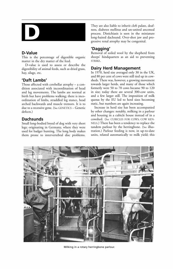
D-ValueThis is the percentage of digestible organicmatter in the dry matter of the feed.
D-value is used to assess or describe thedigestibility of animal feeds, such as dried grass,hay, silage, etc.
‘Daft Lambs’Those affected with cerebellar atrophy – a con-dition associated with incoordination of headand leg movements. The lambs are normal atbirth but have problems walking; there is inco-ordination of limbs, straddled leg stance, headarched backwards and muscle tremors. It is todue to a recessive gene. (See GENETICS – Geneticdefects.)
DachsundsSmall long-bodied breed of dog with very shortlegs; originating in Germany, where they wereused for badger hunting. The long body makesthem prone to intervertebral disc problems.
They are also liable to inherit cleft palate, deaf-ness, diabetes mellitus and un-united anconealprocess. Distichiasis is seen in the miniaturelong-haired dachsund. Over-shot jaw and pro-gressive renal atrophy may be congenital.
‘Dagging’Removal of soiled wool by the shepherd fromsheeps’ hindquarters as an aid to preventingSTRIKE.
Dairy Herd ManagementIn 1970, herd size averaged only 30 in the UK,and 80 per cent of cows were still tied up in cow-sheds. There was, however, a growing movementtowards larger herds, and many of those whichformerly were 50 to 70 cows became 90 to 120in size; today there are several 300-cow units,and a few larger still. The imposition of milkquotas by the EU led to herd sizes becomingstatic, but numbers are again increasing.
Increase in herd size has been accompaniedby other changes: notably, milking in a parlourand housing in a cubicle house instead of in acowshed. (See CUBICLES FOR COWS; COW KEN-
NELS.) There has been a tendency to replace thetandem parlour by the herringbone. (See illus-tration.) Parlour feeding is now, in up-to-dateunits, related automatically to milk yield; this
D
Milking in a rotary herringbone parlour.
both makes for economy and avoids the prob-lem of cow identification in the big herd, so faras the milker is concerned. Identification is stillnecessary, however, for use in conjunction withherd records and in the parlour where the milk-er or relief milker (who will rarely know all thecows) must feed according to yield in theabsence of automated equipment. Plastic num-bered collars, anklets, discs on chain or nylon,freeze branding and even udder tattooing areamong methods used.
In the UK, measures to deal with BSE haveled to the introduction of a comprehensive sys-tem of herd and individual cow identification,with a national database; initially, this was tobe based on a ‘passport’ that would follow theanimal throughout its life.
Feeding outside the parlour has been mecha-nised in many large units. Feeding from silos isless popular than formerly. Many farms havemixer wagons to produce a complete diet madefrom forage, grass or maize silage, straw, concen-trates or straights feeds. Fed ad lib, this systemallows better utilisation of feed and increaseddry-matter intakes. In others, side-deliverytrucks are drawn by tractor down the feedingpassages and deliver into the long mangers. Self-feed silage, with the clamp face in or near thecubicle house, is another labour-saver. Groupfeeding (e.g. of dry cows, high yielders, and lowyielders) is convenient management practice butmay give rise to stress (see BUNT ORDER). (See also
under ‘STEAMING UP’ and the advice on feedinggiven under ACETONAEMIA – Prevention.)
ADAS advice stresses the need for adequatefeeding in early lactation. ‘Since appetite isoften limited at this stage, only the highestquality food should be fed: whether it is goodhay, early cut silage, or 1.5 kg (31/2 lb) per gal-lon cake. This will allow optimum intake ofnutrients at the responsive stage of the lactation– weeks 1–12 after calving.’
Zero-grazing, where cattle are kept in pad-docks, and grass is cut and brought to them, ispractised on some farms where poaching isa serious problem in wet weather, or wherethe movement of a large number of cows isinvolved. With a very large herd on a very smallacreage (such as an American 550-cow herd onunder 5 acres), zero-grazing obviously becomesessential. It is little used in the UK.
Paddock grazing now forms an importantpart of dairy herd management, and includesthe two-sward system in which separate areasare used for grazing and for conservation.
Dung disposal presents difficulties with largeherds. There are two options: it may be treatedas a solid or as a liquid. Straw bedding lends
itself to solid-muck handling, with the liquid(urine, washing-down water, rainwater) beingtaken separately to a lagoon or to an under-ground tank. Slatted floors can be used in acubicle house, either over a dung cellar whichis cleared out once a year, or over a channelleading to an underground tank. With thesemi-solid method, dung may be spread on theland by tanker, or the slurry may pass to alagoon or be pumped through an organic irri-gation pipeline system. Where this is used, cowsmust not be expected to graze pasture untilthere has been time for rain to wash the slurryoff the herbage. The use of organic irrigation isnot entirely free from the risk of spreadinginfectious diseases.
Poaching must be avoided by the use of con-crete aprons at gateways, by mobile drinkingtroughs, by wide corridors between paddockswith an electric fence dividing the ‘corridor’so that one half can be kept in reserve, or bymoveable ramps as are used in New Zealand.
In the large herd, one of the biggest problemsis spotting the bulling heifer or the cow on heat.Properly kept herd records can be a help inalerting farm staff to the approximate dates. (See
CALVING INTERVAL; OESTRUS, DETECTION OF;
CONTROLLED BREEDING.)On large units, regular weekly visits by vet-
erinary surgeons help in the detection andtreatment of infertility and the application ofveterinary preventive medicine. (See HEALTH
SCHEMES; VETERINARY FACILITIES ON FARMS,
CALF HOUSING; also CONTROLLED BREEDING and
CATTLE HUSBANDRY.)
DalmationA medium-sized dog, white with regular blackor brown spots, that originated as a carriage dogin the Balkans. Unlike most dogs, it excretesuric acid in the urine and could be affectedby gout. It may inherit deafness and atrophicdermatitis.
DamalinaA genus of biting lice.
Dangerous Dogs Act 1991This requires that certain breed types (pit bullterrier, Japanese tosa, fila brasileiro, dogo argenti-no) must not be taken out unless on a lead,muzzled, and by someone at least 16 years old.
Owners of these dogs must register themwith the police, and either comply with theexemption scheme or arrange for euthanasia tobe carried out by a veterinary surgeon.
To comply with the exemption scheme, own-ers must take out 3rd-party insurance, arrange
170 Dalmation
D

for the animal to be neutered, and to be identi-fiable by a tattoo and a microchip. The dogmust also be kept under escape-proof condi-tions.
The Act has proved controversial; in a num-ber of cases there has been confusion over iden-tification of dogs as pit bull terriers. Severalattempts have been made to have the Actamended.
Dangerous Wild AnimalsAct 1976This requires people keeping lions, tigers,poisonous snakes, certain monkeys and otherunusual pets, such as crocodiles and bears, toobtain a licence – authorised by a veterinarysurgeon. Bison, wild boar, ostriches and emusare classed as dangerous wild animals under thisAct. They are farmed in the UK and the farmerneeds to register with the local authority. Thepremises must be inspected by a veterinary sur-geon nominated by the local authority before alicence is granted.
Local authorities have power to refuse licences,on the advice of an authorised veterinary sur-geon, on such grounds as safety, nuisance orinadequate or unsuitable accommodation.
Before a licence is granted, local authoritiesmust be satisfied about arrangements for theanimal’s food, exercise and general comfort, fireprecautions, and precautions against infectiousdiseases.
People with such animals must take outinsurance.
Conviction for the keeping of an animalwithout a licence or contravening a conditionof one could result in a fine of up to £400 anda ban from holding a licence.
Zoos, circuses, pet shops and researchworkers are exempted under the LicensingAct 1981.
Danish Red CattleMore than half the cattle in Jutland, and 97 percent of those in the Islands, belong to thisbreed, which is a very old one, though its offi-cial name (meaning Red Danish Milk breed)dates from 1878.
Danish Reds are strong, dual-purpose ani-mals with a good ‘barrel’, teats and udders,and weigh between 500 and 770 kg (1100 and1700 lb). (See also BRITISH DANE.)
Darnel PoisoningThe grass known as ‘darnel’ (Lolium temulen-tum) is a common weed in cereal crops and inpastures in some parts, but it does no harmwhen eaten before the seeds are ripe (or almost
so). Many instances are on record where harm-ful results to man and animals have followedthe use of meal or flour which containedground-up darnel seeds, and there are numer-ous references in classic literature to the harm-ful effect produced upon the eyes as the resultof eating bread made from flour containingdarnel.
Toxic Principle is a narcotic alkaloid, calledtemuline, which is said to be present to theextent of about 0.66 per cent; some authoritiesassert that a substance called loliine, and othersthat picrotoxin, should be considered responsi-ble. A fungus called Endoconidium temulentumis very often found present in the seeds ofdarnel, living a life that is to a great extentone of symbiosis, and the poisonous alkaloidtemuline is found in the fungus.
Signs Darnel produces giddiness and a stagger-ing gait, drowsiness and stupefaction, dilatationof the pupils in the horse, and interference withvision in almost all animals. Vomiting, loss ofsensation, convulsive seizures, and death followwhen it is eaten by animals in large amounts. Insome cases tremblings of the surface muscles areseen, and the extremities of the body becomecold. Death usually occurs within 30 hours ofeating the seeds.
First-Aid Strong black tea or coffee at once.
Darrow’s SolutionDarrow’s solution is used for fluid replacementtherapy in cases of a potassium deficiency, andcontains potassium chloride, sodium chlorideand sodium lactate. It is rarely used in veterinarymedicine; it is unsuitable for cases of neonataldiarrhoea. (See under DEHYDRATION.)
Dart Guns or Syringes(see under PROJECTILE SYRINGE)
DaturineAn alkaloid. (See under STRAMOMIUM.)
Day-Old Chicks(see CHICKS)
DDTThe common abbreviation for dichlorodiphenyl-trichlorethane, a potent parasiticide, lethal tofleas, lice, flies, etc. DDT was once used incor-porated in dusting powders, for applying toanimals; and dissolved in solvents for use as afly-spray. DDT-resistant insects are now found innearly all countries, unfortunately, and dangers
DDT 171
D

of DDT residues in human and animal tissueshave led to its abandonment in the UK andelsewhere.
DDT preparations should not be applied toanimals, owing to the risk of poisoning. The useof DDT with oils or fats enhances its toxiceffects, and should be avoided. Symptoms ofpoisoning include coldness, diarrhoea, andhyperaesthesia. Minute doses over a periodresult in complete loss of appetite. DDT spraysmay contaminate milk if used in the dairy; andmay lead to poisonous residues in food animalswhen applied in livestock buildings, with conse-quent danger to human beings eating the cont-aminated meat. DDT can also contaminatestreams and rivers, and prove harmful to fish.
However, in the control of human try-panosomiasis in Africa, both DDT and dieldrinhave been extensively used for ground spraying,often by aircraft.
Dead Animals, Disposal of(see DISPOSAL OF CARCASES)
Deadly NightshadeDeadly nightshade is the popular name ofAtropa belladonna, from which the alkaloidATROPINE is obtained. It is a deadly poison, andparts of the plant are sometimes eaten by stock.(See also BELLADONNA.)
Deafness
Congenital deafness is common in whitebull terriers and also in blue-eyed white cats. Inthe USA the Dalmatian breed is reported tohave the highest prevalence of deafness of allbreeds of dogs, with a risk factor of 40 to 50 percent. One or both ears may be affected.
Conductive deafness is that caused byinterference with the transmission of soundwaves from the external ear to the organ ofCorti in the inner ear. Such interference may bedue to: (1) excess of wax in the ear canal; (2)perforation of, or infection involving, theeardrum. (In human medicine otosclerosis isanother cause, being a loss of flexibility betweenthe bones of the middle ear and the membraneconnecting them with the inner ear, possiblydue to hardening or ossification.)
Nerve deafness results from pressure upon,or damage to, the auditory nerve; it can also bea side-effect of antibiotics such as streptomycinand neomycin, and possibly chloramphenicol.
Deafness is or may be also a symptom ofsantonin poisoning, coal-gas poisoning, of a
vitamin deficiency, and, in human medicine, aside-effect of streptomycin and aspirin. Othercauses include damage to the internal ear, to theEustachian tube, nervous system, etc.
Death, Causes of SuddenIn the majority of cases either failure of the heartor damage to a blood vessel (e.g. in cattle causedby a nail or a piece of wire from the reticulum)is the direct cause, but nervous shock followingan accident or injury, cerebral haemorrhage,anthrax, black-quarter, lightning strike, braxy,hypocalcaemia in cattle, hypomagnesaemia (alsoin sheep), and over-eating of green succulentfodder in young cattle, are all capable of pro-ducing sudden death. In the case of pigs, suddendeath has sometimes resulted from heat stroke.(See also BOWEL, OEDEMA OF THE.) In both cattleand pigs sudden death due to Clostridiumwelchii type A has been reported. In countriesbordering the Red Sea, horses that have notbeen bred locally are sometimes attacked bya form of heat stroke with fatal results. (See
also POISONING and (with reference to dogs)CANINE PARVOVIRUS; CANINE VIRAL HEPATITIS.)Sudden death, without obvious preliminarysymptoms, may occasionally occur in cases ofrabies, botulism, and foot-and-mouth disease.(See also ELECTRIC SHOCK).
Death, Signs ofThe physical signs of death are well known, butthere are occasions when it is difficult to statewhether an animal is dead or not. In deep comaan animal may have all the superficial appear-ances of being dead, and yet recovery is possibleif effective measures are taken. In the later stageof milk fever a cow has been mistaken for dead,has been dragged out of the byre preparatory toremoval to the slaughterer’s, has been examinedby a practitioner, has been found to be living,has been suitably treated, and within 2 hourshas been up on her feet again looking well.Foals have been discarded soon after being bornand considered dead, have been removed to theoutside of the loose-box while attention waspaid to the dam, and later have been found liv-ing, the fresh cold air having revived respirationand stimulated the circulation, etc.
When an animal dies, the essential sign ofthe cessation of life is said to be the stopping ofthe heart. This, however, is not strictly correct,for it is possible by massage to resuscitate analready stopped heart, and to recover an appar-ently dead creature. Strictly speaking, it isalmost impossible to say exactly when deathtakes place, but it is considered that when heartand respiration have ceased, when the eyelids
172 Dead Animals, Disposal of
D
do not flicker if a finger be applied to theeyeballs, when a cut artery no longer bleeds,and when the tissues lose their natural elastici-ty, life is extinct. A few of the common teststhat are applied in uncertain cases are as fol-lows. The animal is dead when (1) a piece ofcold glass held to the nostrils for 3 minutescomes away without any condensed moistureupon it; (2) a superficial incision in the skindoes not gape open; and (3) the natural elastictension of the tissues disappears. Changes thatfollow death in a variable period dependingupon the species of animal, and upon theweather at the time, are: (1) the clotting of theblood in the vessels; (2) the onset of rigor mor-tis (the stiffness of death); and (3) the com-mencement of decomposition of the carcase,usually first evident along the lower surface ofthe abdomen.
De-BeakingDe-beaking is done by poultry-keepers whenbirds are kept in groups and there is a potentialproblem of feather-picking or cannibalism.No more than one-third of the upper beak isremoved; more than this can expose the sinusesand lead to infection. If performed when veryyoung there are few after-effects. Older birdswill develop neuromas at the cut tip, resultingin hypersensitivity of that region. Managementpractices should be improved to try to eliminatethe need for de-beaking but the problem can bedifficult to resolve.
There are moves to phase out the practice, onwelfare grounds.
DebridementThe removal of dead tissue and infected materi-al from a wound surface. This can be achievedby enzymes or combinations of organic acids.The use of maggots free from pathogenic organ-isms is an old method of wound treatmentcurrently being revived in human medicine.
DecoquinateA coccidiostat originally developed for use inpoultry but mainly administered to controlcoccidiosis in lambs and calves.
DecubitusDecubitus is the recumbent position assumedby animals suffering from certain diseases.
DecussationDecussation is a term applied to any place inthe nervous system at which nerve fibres crossfrom one side to the other; e.g. the decussationof the pyramids in the medulla, where the
motor fibres from one side of the brain cross tothe other side of the spinal cord.
Deep-Freeze(see ARTIFICIAL INSEMINATION; LIFE AFTER
FREEZING)
Deep Litter for CattleThis is a very satisfactory system if well man-aged. It is mainly practised in straw yards.Straw, shavings and sawdust can be used, inadequate quantity. The bedding must be keptdry and no contact must occur between theudder and dung in the litter. Warmth given offas a result of the fermentation taking place in the litter makes for cow-comfort; and thereis, of course, the added advantage of a thick layer of insulation between the cows and theconcrete of a covered yard.
Deep Litter for PoultryChopped straw, shavings, and sawdust are com-monly used. Musty straw could cause an out-break of aspergillosis. Peat-moss is apt to be toodusty. Oak sawdust should not be used as itmay discolour the egg-yolks. The depth shouldbe at least 10 cm (4 in). The litter should beforked over, and added to from time to time. Ifit gets damp, the ventilation should be attend-ed to. Many coccidia larvae get buried in thelitter, and this is an advantage. After each cropof birds, the litter should be removed andheaped, so that enough heat will be generatedto kill parasites. If deep litter is returned to ahouse, the succeeding batch of birds sometimessuffer from ammonia fumes, which may causeserious eye troubles. Compaction of the littermust be prevented by allowing the poultry to‘work’ it; otherwise, the litter does not meet thedefinition specified by EC Directives.
Deep-Rooting PlantsDeep-rooting plants are valuable in a pasturefor the sake of the minerals they provide.Examples of such plants are chicory, yarrow,and tall fescue.
Deer, Diseases ofDeer are susceptible to the following infections:BRUCELLOSIS; BOVINE VIRAL DIARRHOEA;ELAPHASTRONGYLUS; EPIZOOTIC HAEMORRHAG-
IC DISEASE; FOOT-AND-MOUTH DISEASE; JOHNE’S
DISEASE; LISTERIOSIS; LOUPING-ILL; malignantcatarrhal fever – see BOVINE MALIGNANT
CATARRHAL FEVER; MENINGOENCEPHALITIS; PAR-
ASITIC BRONCHITIS; TICK-BORNE FEVER; TUBER-
CULOSIS; WARBLES; YERSINIOSIS; and also anenzootic ataxia resembling SWAYBACK in lambs.
Deer, Diseases of 173
D

Tuberculosis in deer Tuberculosis of deeris NOTIFIABLE in the UK. Tuberculin testing ofdeer and the establishment of tuberculosis-freeherds was the basis of the Deer Health Schemeoperated by MAFF. Tuberculin testing ofdeer and interpretation of the results are moredifficult than in cattle; special training isrequired.
The Tuberculosis (Deer) Order 1989 pro-vides for the individual marking of farmed ortransported deer, and can be used for enforcingmovement restrictions on affected or suspectanimals.
Farmed deer Red deer (Cervus elaphus)and fallow deer (Dama dama) are the mostcommonly farmed.
Around 300 farms, mainly in Scotland, raisedeer in the UK. Some 36,000 animals are farmedin total. Most (75 per cent) are red; the rest,fallow. All farmed deer must be identified bytagging; the British Deer Farming Associationsupervises a tagging scheme.
In Britain the harvesting of antler velvet fromlive stags is illegal.
In New Zealand, yersiniosis has become aserious disease of farmed red deer. It appears tobe triggered off by stress, and most cases occurduring the winter. The incidence of malignantcatarrhal fever (MCF) in red deer herds inCanterbury, New Zealand, ranges from 0.2 to10 per cent a year.
Meningoencephalitis, caused by Streptococcuszooepidemicus, has resulted in the death offarmed red deer exported from the UK andDenmark to New Zealand. Autopsy findingsare typically congestion of lungs and liver, thepresence of frothy fluid in trachea and bronchi,and acute meningoencephalitis.
Dictyocaulus viviparus is the most importantparasite of red deer in New Zealand and fre-quent drenching with anthelmintics is used tocontrol it. Development of resistant species ishindered by dosing strictly according to themanufacturers’ directions and alternating theproduct used.
Another parasitic worm of importance indeer is Elaphostrongylus cervi. It is pale andthread-like, 4 to 6 cm long, and found in theintramuscular fascia and also in the meninges ofthe brain. This parasite occurs in Scotland, themainland of Europe, and Australasia.
Eggs reach the lungs via the bloodstream andhatch in the alveolar capillaries, causing slightpneumonia. Nervous signs appear when thebrain is involved.
In the UK, one of the most important infec-tions of deer is Johne’s disease, caused by
Mycobacterium paratuberculosis. It may be seenin animals as young as a year old and results inwasting, with or without diarrhoea.
DefecationDefecation is very differently performed in thevarious animals, and some diagnostic importanceis attached to the manner of its performance. (See
CONSTIPATION; DIARRHOEA.)
Deficiency DiseasesThese form a group of diseases bearing no clin-ical resemblance to each other, but having thecommon feature that they result from omissionfrom the diet of some substance or elementessential for normal health and nutrition. Theessential element may be one of the inorganicmineral substances, such as calcium, phospho-rus, magnesium, manganese, iron, copper,cobalt, iodine, selenium or more than one ofthese; it may be a protein or an amino acid; orit may be a vitamin. In the last case the condi-tion is often referred to as an ‘avitaminosis’, andthe particular vitamin is specified, e.g. A, B, Dor E. Starvation through inadequacy of generalnutritive food intake is not classed as a defi-ciency disease. Some deficiency diseases aresimple, such as iron deficiency in young pigs;while others are more complex, such as phos-phate deficiency in South Africa, which is asso-ciated with botulism through the gnawing ofbones of dead animals contaminated with C.botulinus. (See VITAMINS; TRACE ELEMENTS;NUTRITION, FAULTY.)
Definitive HostThis is the host in which an adult parasite withan indirect life-history lives and produces itseggs. A definitive host is the final host, as com-pared with the intermediate host or hosts. Forexample, an ant is one of the intermediate hostsof a species of liver fluke; the definitive host is asheep or other grazing animal.
DeformitiesDeformities of cattle and sheep, etc. are men-tioned under GENETICS – Genetic defects. (See
also HARE-LIP; MOUTH, DISEASES OF; MONSTER.)
DEFRAAcronym for Department of the Environment,Food and Rural Affairs – the UK Governmentdepartment that replaced the Ministry ofAgriculture, Fisheries and Food (MAFF). Itsresponsibilities include notifiable diseases, foodsafety, and welfare of animals in transport, onfarms and at slaughter. The Home Office isresponsible for experimental animals.
174 Defecation
D

DeglutitionDeglutition means the act of swallowing.During swallowing, breathing temporarily ceas-es (apnoea); otherwise food might enter therespiratory tract. (See CHOKING.)
DehiscenceA breakdown in the union of a suture of adjoin-ing bones of the skull. The condition can betreated successfully by surgery.
An example of this is a breakdown of thesuture line in mandibular fractures. The term isalso applied to the re-opening of wounds.
De-Horning of CattleDairy cows are routinely dehorned to facilitatehandling and to avoid injury to those handlingthem and to other cattle. The use of mecha-nised milking systems makes dehorning virtual-ly essential. Fattening beef cattle in yards orpens are also often dehorned because there isusually 1 animal that obtains dominance; if itpossesses horns it is liable to inflict woundsupon others or upon the attendants.
The most satisfactory method in calves isthat known as ‘disbudding’. This is best donewhen the horn bud is fully detectable, whichtakes a variable time to occur. The buds arethen removed, under local anaesthetic, by cau-terising with an electric or gas-heated dehorner.
An alternative method consists of painting theyoung buds of the horns, when they first appearin calves, with caustic compound. A little petro-leum jelly or thick grease may be rubbed onthe hair around the base of the bud and care isneeded to ensure that no caustic gets into theeyes. The bud of the horn is first cleaned withspirit to remove grease – an essential preliminary– and a second coating of the caustic is givenafter the first has dried. A scab will form over thebud and drop off, carrying with it the cellswhich would have produced horn. Little or nopain is occasioned to the calf by caustic collodi-on (whereas caustic potash sticks, now largelysuperseded, do cause much pain) and the horn iseffectively prevented from growing.
In Britain the operation of de-horning cattlerequires the administration of an anaesthetic. (See
ANAESTHETICS, LEGAL REQUIREMENTS.) A saw,an electric saw, cutting wire or special horn shearsmay be used when the horns are more developed.
Bleeding from the matrix and horn core canusually be controlled by using a figure-of-eighttourniquet around the roots of the horns.
DehydrationLoss of water from the tissues, such as occurs dur-ing various illnesses, especially those producing
vomiting or diarrhoea; in impaction of therumen; and as a result of injury or serious burns.
Diarrhoea is one of the most common caus-es of dehydration. A scouring calf may lose 100ml of water per kg bodyweight in 12 hours. Asthe metabolism attempts to conserve extracellu-lar body fluid (ECF) volume, urine productiondecreases and blood urea levels rise while pHlevels are lowered. Electrolytes are lost, particu-larly sodium, potassium and bicarbonate, andketone bodies accumulate.
Treatment Restoration of fluid volume is theimmediate priority, and replacement of lostelectrolytes and blood nutrients.
Parenteral fluids In the severely dehydrat-ed animal, the restoration of ECF by parenteral(usually intravenous) administration of plasma,if available, or infusion of a sterile istonic(0.9 per cent) solution of sodium chloride,compound sodium lactate infusion (lactatedRinger’s solution) is indicated. In the case ofblood or plasma loss through injury or burns, aplasma expander based on dextran or gelatin isadded to the electrolyte solution; proprietarysolutions are widely available.
The rate of administration of intravenoussolutions should be carefully supervised. Insevere cases of dehydration or profound shock,up to 50 ml per kg bodyweight per hour may begiven initially, reducing to 5 to 10 ml/kg/hour.These high rates should not be continued formore than 20 to 30 minutes. A close watchmust be kept for signs of too rapid administra-tion: restlessness, lung sounds, tachycardia,tachypnoea.
A formula to convert ml/kg/hour to dropsper minute is given in The Veterinary Formulary:
Drops/ml × FR × BWDrops/minute = __________________
60Drops/ml = number of drops delivered by the infu-sion set per mlFR = Flow rate in ml/kg/hourBW = bodyweight of patient in kg
The total amount given will depend on theamount of fluid lost and the condition of theanimal.
Oral rehydration is usually satisfactoryin most cases of diarrhoea. Solutions for thispurpose usually contain sodium and glucose,which help the water uptake of the dehydratedanimal. To help correct any acidosis, citrateshould be included, and/or bicarbonate. Suchsolutions are suitable for calves and mostmammals. Many proprietary formulations areavailable.
Dehydration 175
D
For first-aid purposes, glucose-saline may begiven by mouth to all animals. UNICEF’s ‘OralRehydration Salts’, intended for infants andchildren, may be used; the sachet contentsbeing dissolved in 1 litre of (sterile or boiled)water (which must not be boiled thereafter).The formula is:
Sodium chloride 3.5 gPotassium chloride 1.5 gSodium bicarbonate 2.5 gGlucose 20.0 g
The effectiveness of the above glucose-salinesolution can be enhanced by the addition ofcitrate and/or citric acid.
Glucose-saline can also be administered perrectum, or subcutaneously.
Delivery(see PARTURITION)
DemephionAn organophosphorous preparation used asan insecticide and acaricide. Livestock shouldbe kept out of treated areas for at least afortnight.
DemodecosisAnother name for DEMODECTIC MANGE.
Demodectic Mange (FollicularMange)Demodectic mange (follicular mange) is causedby the demodectic mite Demodex folliculorum.This parasite, microscopic and cigar-shaped inappearance, with very short stumpy legs, livesdeep down in the hair follicles, and is accord-ingly difficult to eradicate by dressings. It is acommon cause of mange in dogs.
In cattle, D. bovis is in the UK responsiblefor mild and infrequently reported cases ofdemodectic mange, but in some parts of theworld the disease may be severe. Fatal, gener-alised cases have been reported from Africa. D.caprae infestation of goats may also be severe inthe tropics.
The parasites have been recovered from theeyelids of cattle, sheep, horses, dogs, and man(see MANGE).
DemulcentsDemulcents are substances which exert a sooth-ing influence upon the skin or the mucousmembranes of the alimentary canal, and inaddition afford some protection when these areinflamed. Examples of demulcents for internaluse are arrowroot, glycerin, bismuth subnitrate,and bismuth carbonate.
DemyelinationDestruction of the myelin, a lipid which sur-rounds the axis-cylinder of a medullated nervefibre.
Dendrites(see NERVES)
Dengue(see EPHEMERAL FEVER)
Dental Plaque(see TARTAR)
DentineDentine is the dense yellow or yellowish-whitematerial of which the greater part of the teeth iscomposed, and which in elephants, etc., consti-tutes ivory. The dentine is pierced by numerousfine tubules which communicate with thesensitive pulp in the hollow of the tooth-root,along each of which run tiny vessels and nerveswhich nourish its structure. In the young,newly erupted tooth the dentine is covered overwith a layer of hard, dense, brittle enamel,which prevents too rapid wear of the softerdentine. (See TEETH.)
DentitionDentition refers to the configuration and con-formation of the teeth, with special reference totheir periods of eruption through the gums.
PAGE
Horses 176Cattle 177Sheep 178Pigs 178Dogs 179Cats 179
Horses The dentition of the horse consists ofthe following teeth:
Incisor teeth are 6 in number in the upperand lower jaws. The temporary incisors differfrom the permanents in that while each of theformer possesses a definite crown, neck, androot, the latter do not. Moreover, the tempo-raries are smoother, whiter, and smaller. Whenthere are both temporaries and permanentspresent in the mouth it is not usually difficultto differentiate between them, but inexperi-enced persons sometimes confuse temporaries
176 Delivery
D
Incisors Canines Molars
Upper jaws 6 2 12, 13 or 14Lower jaws 6 2 12, 13 or 14
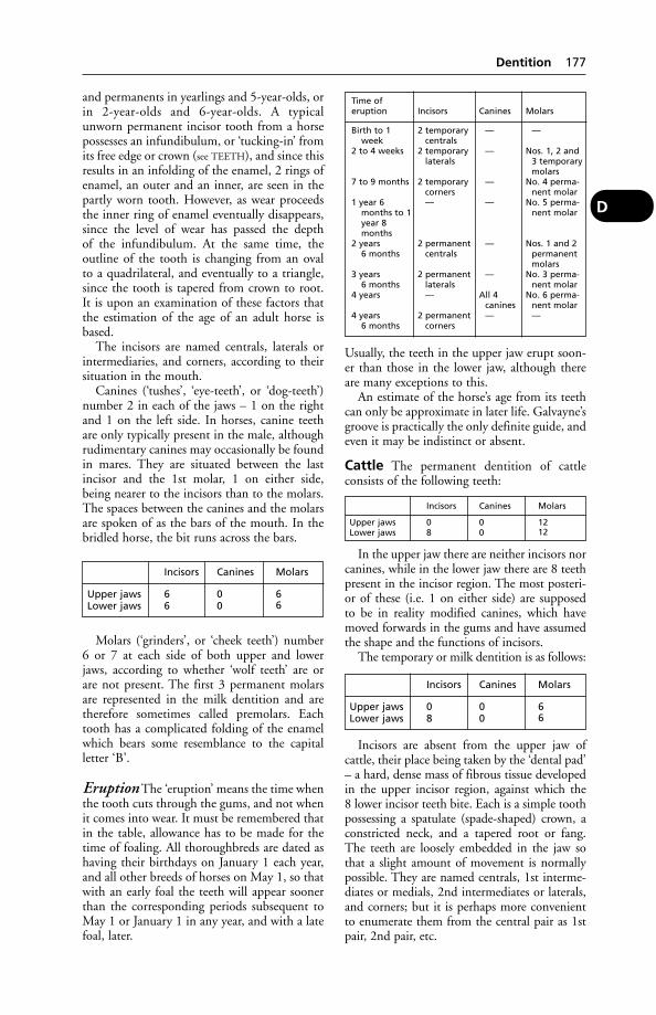
and permanents in yearlings and 5-year-olds, orin 2-year-olds and 6-year-olds. A typicalunworn permanent incisor tooth from a horsepossesses an infundibulum, or ‘tucking-in’ fromits free edge or crown (see TEETH), and since thisresults in an infolding of the enamel, 2 rings ofenamel, an outer and an inner, are seen in thepartly worn tooth. However, as wear proceedsthe inner ring of enamel eventually disappears,since the level of wear has passed the depthof the infundibulum. At the same time, theoutline of the tooth is changing from an ovalto a quadrilateral, and eventually to a triangle,since the tooth is tapered from crown to root.It is upon an examination of these factors thatthe estimation of the age of an adult horse isbased.
The incisors are named centrals, laterals orintermediaries, and corners, according to theirsituation in the mouth.
Canines (‘tushes’, ‘eye-teeth’, or ‘dog-teeth’)number 2 in each of the jaws – 1 on the rightand 1 on the left side. In horses, canine teethare only typically present in the male, althoughrudimentary canines may occasionally be foundin mares. They are situated between the lastincisor and the 1st molar, 1 on either side,being nearer to the incisors than to the molars.The spaces between the canines and the molarsare spoken of as the bars of the mouth. In thebridled horse, the bit runs across the bars.
Molars (‘grinders’, or ‘cheek teeth’) number6 or 7 at each side of both upper and lowerjaws, according to whether ‘wolf teeth’ are orare not present. The first 3 permanent molarsare represented in the milk dentition and aretherefore sometimes called premolars. Eachtooth has a complicated folding of the enamelwhich bears some resemblance to the capitalletter ‘B’.
Eruption The ‘eruption’ means the time whenthe tooth cuts through the gums, and not whenit comes into wear. It must be remembered thatin the table, allowance has to be made for thetime of foaling. All thoroughbreds are dated ashaving their birthdays on January 1 each year,and all other breeds of horses on May 1, so thatwith an early foal the teeth will appear soonerthan the corresponding periods subsequent toMay 1 or January 1 in any year, and with a latefoal, later.
Usually, the teeth in the upper jaw erupt soon-er than those in the lower jaw, although thereare many exceptions to this.
An estimate of the horse’s age from its teethcan only be approximate in later life. Galvayne’sgroove is practically the only definite guide, andeven it may be indistinct or absent.
Cattle The permanent dentition of cattleconsists of the following teeth:
In the upper jaw there are neither incisors norcanines, while in the lower jaw there are 8 teethpresent in the incisor region. The most posteri-or of these (i.e. 1 on either side) are supposedto be in reality modified canines, which havemoved forwards in the gums and have assumedthe shape and the functions of incisors.
The temporary or milk dentition is as follows:
Incisors are absent from the upper jaw ofcattle, their place being taken by the ‘dental pad’– a hard, dense mass of fibrous tissue developedin the upper incisor region, against which the8 lower incisor teeth bite. Each is a simple toothpossessing a spatulate (spade-shaped) crown, aconstricted neck, and a tapered root or fang.The teeth are loosely embedded in the jaw sothat a slight amount of movement is normallypossible. They are named centrals, 1st interme-diates or medials, 2nd intermediates or laterals,and corners; but it is perhaps more convenientto enumerate them from the central pair as 1stpair, 2nd pair, etc.
Dentition 177
D
Incisors Canines Molars
Upper jaws 6 0 6Lower jaws 6 0 6
Incisors Canines Molars
Upper jaws 0 0 12Lower jaws 8 0 12
Incisors Canines Molars
Upper jaws 0 0 6Lower jaws 8 0 6
Time oferuption Incisors Canines Molars
Birth to 1 2 temporary — —week centrals
2 to 4 weeks 2 temporary — Nos. 1, 2 andlaterals 3 temporary
molars7 to 9 months 2 temporary — No. 4 perma-
corners nent molar1 year 6 — — No. 5 perma-
months to 1 nent molaryear 8months
2 years 2 permanent — Nos. 1 and 26 months centrals permanent
molars3 years 2 permanent — No. 3 perma-
6 months laterals nent molar4 years — All 4 No. 6 perma-
canines nent molar4 years 2 permanent — —
6 months corners

Canines are absent unless the corner incisorsare considered as modified canines.
Molars are like those of the horse in numberand arrangement, except that they are smallerand progressively increase in size from first tolast, so that the 1st is quite small, and the lengthof gum which accommodates the first 3 is onlyabout half that occupied by the last 3. One ormore ‘wolf teeth’ may be present in rare cases.
Eruption In ruminants – whether domesti-cated or not – the eruption of the permanentteeth is subject to considerable variations.
Sheep The terms which were used as appliedto cattle, and the description of the variousteeth, may be taken to hold good for sheep aswell. The sheep has 8 lower incisor teeth butnone in the upper jaw. There are 24 molarteeth, 12 in each jaw, of which half these num-bers are represented in the temporary dentition.
Eruption The following is given as an averageeruption table for improved breeds of sheep inGreat Britain:
Pigs There is probably no farm animal whichshows such variation in the eruption of its teeth asthe pig, but because of the demand for young pigsfor killing by weight and size rather than by age,and because of the intractability of older breedinganimals – sows and boars – the actual age of thepig is not of such very great importance, exceptperhaps for fat stock show purposes.
When the permanent teeth have all eruptedthey are distributed as follows:
In the molar region there is a little tooth ineach of the four jaws, erupting at about 5 to 6months, which is permanent from the verybeginning. It is sometimes called the premolar,and in some cases is never developed. The next3 teeth behind it are represented in the tempo-rary dentition, the permanents replacing themin the usual way. The last 3 teeth are truemolars, i.e. permanents only.
The temporary dentition is as follows:
Incisors: the upper incisors are small, and areseparated from each other by spaces. The 1st pair(centrals) are the largest, and converge together.The 2nd pair are narrower and smaller; while thecorner pair are very small and laterally flattened.The lower incisors are arranged in a convergentmanner, and point forwards horizontally in thejaw. The 1st two pairs are large prismatic teethdeeply implanted in the jaw-bones and are usedfor ‘rooting’ purposes. The corner pair aresmaller, and possess a distinct neck.
Canines, or tusks, are greatly developed inthe entire male, and both upper and lower tusksproject out of the mouth. The upper canines ofa boar may be 3 to 4 inches long, while thelower ones may reach as much as 8 inches in anaged animal. Each has a large permanent pulpcavity from which the tooth continues to growthroughout the animal’s life.
At 3 months the lateral temporary incisorsare well up, and the temporary molars are wellin wear.
At 5 months there are signs of the cutting ofthe premolars (i.e. the No. 1 molars), and the5th molar (a permanent) is seen behind thetemporaries. It is, however, not yet in wear.
178 Dentition
D
Time of eruption Incisors Molars
Birth to 1 month All 8 tempo- All 12 tempo-raries raries
3 months — 4th permanent9 months — 5th permanent1 year to 1 year 1st pair —
3 months permanent1 year 6 months — 6th permanent1 year 9 months 2nd pair 1st and 2nd
permanent permanents2 years — 3rd permanent2 years 3 months 3rd pair —
permanent2 years 9 months 4th pair —
to 3 years permanent
Time oferuption Incisors Canines Molars
At birth Corner All 4 tem- —temporaries poraries
1 month Central — Nos. 2, 3temporaries and 4 tem-
poraries2 months Lateral — —5–6 months — — No. 1, which
remainsthroughlife, andNo. 5permanent
8 months Corner — —permanents
9 months — All 4 per- —manents
10–12 months — — No. 6permanent
12–13 months Central — Nos. 2, 3permanents and 4 per-
manents17–18 months Lateral — No. 7
permanents permanent
Incisors Canines Molars
Upper jaws 6 2 14 (i.e. 8and 6)
Lower jaws 6 2 14 (i.e. 8and 6)
Incisors Canines Molars
Upper jaws 6 2 6Lower jaws 6 2 6
At 6 months the premolars are cut and the5th permanent molar is in wear.
At 7 to 8 months there are signs of thecutting of the corner permanent incisors, orthey may already be through the gums. Thepermanent tusks are also often cutting throughthe gums at this age in forward animals.
At 9 months the corner permanent incisorsare well up and the permanent tusks arethrough the gums, although in many casesthere may be still one or two of the smalltemporary tusks in position. Where they are cutthey are not far through the gums.
At 1 year it is generally held that the centralpermanent incisors cut through the gums, butthere are a large number of animals which donot cut these teeth till about 13 months old.The 6th permanent molar cuts at this time, andis more reliable than the incisors for reference.
Shortly after 1 year the 3 temporary molarsfall out and their places are taken by thepermanents. They are into line with the othermolar teeth 3 months later.
At 17 to 18 months, when the final changesoccur, the 7th molar, the last permanent molartooth, and the lateral permanent incisors are cutthrough the gums. By this time the pig hasobtained its full permanent dentition, and thesucceeding changes are not sufficiently reliableto warrant estimations of age being based uponthem.
Dogs The average adult dog has 42 teeth.The upper jaw contains 6 incisors, 2 canines,8 premolars, and 6 molars. The lower jaw has6 incisors, 2 canines, 8 premolars, and 6 molars.(There is some breed and individual variationin the number of permanent teeth, short-skulledbreeds, e.g. Pekingese, Boxer, and Bulldog,having fewer teeth.)
Cats The number of teeth in the adult cat aver-ages 30. In the upper jaw there are 6 incisors, 2 canines, 6 premolars, and 2 molars; while thelower jaw has 6 incisors, 2 canines, 4 premolars,and 2 molars. Some cats have only 28 perma-nent teeth; lacking 2 premolars.
Rabbits are unique in that they are born withpermanent teeth. Milk teeth are shed beforebirth and may be found in the placenta.
Deoxyribonucleic Acid(see under DNA)
DepilationDepilation is the process of the destruction ofhair that takes place during certain skin or other
diseases, or after the application of chemical orthermal substances to the surface of the body. (See
MANGE; RINGWORM; ‘BALDY CALF’ SYNDROME;BURNS; CYCLOPHOSPHAMIDE, ALOPECIA.)
Depluming ScabiesDepluming scabies is a form of parasitic mangeaffecting the fowl, in which the feathers areeaten through close to the skin surface andfall or break off. It is caused by Cnemidocoptesgallinae. (See MITES.)
Depraved Appetite (PICA)(see under APPETITE)
DermatitisDermatitis means any inflammation of theskin. (See SKIN; ECZEMA; ALLERGY.)
DermatophilusDermatophilus infection results in a chronicdermatitis, in which the hairs stand erect andmatted in tufts, like a wet paintbrush. Manyspecies of animals are susceptible, e.g. horses,cattle, sheep (also dog and cat).
Cause D. congolensis, which is a Gram-positivebacterium having some fungus-like characteris-tics, e.g. the production of branching filaments.
The disease, also known as cutaneous strep-tothricosis or mycotic dermatitis, follows the pro-longed wetting of an animal and is widespread inthe tropics, but occurs also in temperate climatessuch as Ireland, Britain, etc. (For examples inhorses, see GREASY HEEL; ‘RAIN SCALD’.)
In sheep, where it is also called ‘lumpy wool’,it can cause ‘strawberry foot rot’.
Predisposing causes, other than wetting,include tick and insect bites, wounds fromthorns, etc. Fly transmission is recognised. Thebacterium can resist drying, but under wetconditions it invades the epidermis, with effectsmentioned under ‘greasy heel’, where first-aidand precautionary measures are given. Antibioticsare helpful in treatment.
In the tropics, dipping to control ticksis regarded as important, and acaricide prepa-rations used in sheep dips are effectiveagainst Dermatophilus. (See also SENKOBO;
STREPTOTHRICOSIS.)
Dermatosis VegetansA hereditary disease of young pigs characterisedby raised skin lesions, abnormalities of thehooves, and pneumonia. The semi-lethal reces-sive gene probably originated in the DanishLandrace. UK outbreaks occurred in 1958 and1964.
Dermatosis Vegetans 179
D
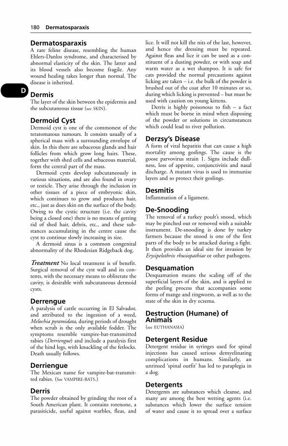
DermatosparaxisA rare feline disease, resembling the humanEhlers-Danlos syndrome, and characterised byabnormal elasticity of the skin. The latter andits blood vessels also become fragile. Anywound healing takes longer than normal. Thedisease is inherited.
DermisThe layer of the skin between the epidermis andthe subcutaneous tissue (see SKIN).
Dermoid CystDermoid cyst is one of the commonest of theteratomatous tumours. It consists usually of aspherical mass with a surrounding envelope ofskin. In this there are sebaceous glands and hairfollicles from which grow long hairs. These,together with shed cells and sebaceous material,form the central part of the mass.
Dermoid cysts develop subcutaneously invarious situations, and are also found in ovaryor testicle. They arise through the inclusion inother tissues of a piece of embryonic skin,which continues to grow and produces hair,etc., just as does skin on the surface of the body.Owing to the cystic structure (i.e. the cavitybeing a closed one) there is no means of gettingrid of shed hair, debris, etc., and these sub-stances accumulating in the centre cause thecyst to continue slowly increasing in size.
A dermoid sinus is a common congenitalabnormality of the Rhodesian Ridgeback dog.
Treatment No local treatment is of benefit.Surgical removal of the cyst wall and its con-tents, with the necessary means to obliterate thecavity, is desirable with subcutaneous dermoidcysts.
DerrengueA paralysis of cattle occurring in El Salvador,and attributed to the ingestion of a weed,Melochia pyramidata, during periods of droughtwhen scrub is the only available fodder. Thesymptoms resemble vampire-bat-transmittedrabies (Derriengue) and include a paralysis firstof the hind legs, with knuckling of the fetlocks.Death usually follows.
DerriengueThe Mexican name for vampire-bat-transmit-ted rabies. (See VAMPIRE-BATS.)
DerrisThe powder obtained by grinding the root of aSouth American plant. It contains rotenone, aparasiticide, useful against warbles, fleas, and
lice. It will not kill the nits of the last, however,and hence the dressing must be repeated.Against fleas and lice it can be used as a con-stituent of a dusting powder, or with soap andwarm water as a wet shampoo. It is safe forcats provided the normal precautions againstlicking are taken – i.e. the bulk of the powder isbrushed out of the coat after 10 minutes or so,during which licking is prevented – but must beused with caution on young kittens.
Derris is highly poisonous to fish – a factwhich must be borne in mind when disposingof the powder or solutions in circumstanceswhich could lead to river pollution.
Derzsy’s DiseaseA form of viral hepatitis that can cause a highmortality among goslings. The cause is thegoose parvovirus strain 1. Signs include dull-ness, loss of appetite, conjunctivitis and nasaldischarge. A mutant virus is used to immuniselayers and so protect their goslings.
DesmitisInflammation of a ligament.
De-SnoodingThe removal of a turkey poult’s snood, whichmay be pinched out or removed with a suitableinstrument. De-snooding is done by turkeyfarmers because the snood is one of the firstparts of the body to be attacked during a fight.It then provides an ideal site for invasion byErysipelothrix rhusiopathiae or other pathogens.
DesquamationDesquamation means the scaling off of thesuperficial layers of the skin, and is applied tothe peeling process that accompanies someforms of mange and ringworm, as well as to thestate of the skin in dry eczema.
Destruction (Humane) ofAnimals(see EUTHANASIA)
Detergent ResidueDetergent residue in syringes used for spinalinjections has caused serious demyelinatingcomplications in humans. Similarly, anunrinsed ‘spinal outfit’ has led to paraplegia ina dog.
DetergentsDetergents are substances which cleanse, andmany are among the best wetting agents (i.e.substances which lower the surface tensionof water and cause it to spread over a surface
180 Dermatosparaxis
D

rather than remain in droplet form). Detergentsare inactivated by soaps, and the 2 must not beused together. Detergents are widely used in thecleansing of milking equipment, etc., and for-mulated in skin lotions and shampoos. Theywill remove gross contamination but are notthemselves disinfectants, which, if necessary,must be applied after detergents. Examples ofdetergents are cetrimide and sodium laurylsulphate.
Detomidine (Domosedan)Given by intravenous injection, this drug hasbeen found useful for the sedation of horsesduring radiography, endoscopy, etc. Sedationlasts for 20 to 30 minutes. An analgesic is need-ed in addition. It is also used as an equineanaesthetic, administered in conjunction withketamine.
Dew ClawsDew claws in cattle are sometimes torn offor injured by slatted floors. (For dew claws indogs, see NAILS.)
De-WattlingThe removal of a fowl’s wattles. (See also
DUBBING.)
DewlapA loose fold of skin under the jaw or neck. It isfound in some cattle and dogs, e.g, bloodhounds.
DexamethasoneOne of the synthetic CORTICOSTEROIDS. It isused as an anti-inflammatory agent in cases ofshock, allergies, ketosis, etc.
DextranA water-soluble polysaccharide used as a plasmasubstitute. It may be infused intravenouslyinstead of whole blood in cases of severe haem-orrhage, etc.
Dextran SulphateAn alternative anticoagulant to Heparin. Itseffects last longer.
DextrinDextrin is a soluble carbohydrate substanceinto which starch is converted by diastaticenzymes or by dilute acids. It is a white oryellowish powder which, dissolved in water,forms mucilage. Animal dextrin, or glycogen, isa carbohydrate stored in the liver.
DextroseDextrose is another name for glucose.
DhrekAn Asiatic tree of which the leaves and fruits arepoisonous to farm animals. (See MELIA.)
Diabetes Insipidus (Polyuria)Diabetes insipidus (polyuria) is a condition inwhich there is secreted an excessively largequantity of urine of low specific gravity. Itresults from a deficiency in the bloodstream ofthe antidiuretic hormone (ADH). It is treatedwith vasopressin or desmopressin. (See PITU-
ITARY GLAND.) Diabetes has been reported tooccur in dogs as a result of fright; symptomsinclude poor appetite, dull coat, and frequenturinating in the house. (See also POLYURIA.)
Diabetes MellitusDiabetes mellitus is a condition in which thereis excessive glucose in the blood (hypergly-caemia). This produces various symptoms:thirst, polyuria, weight loss, recurrent infection;in more severe cases, diabetic coma (ketoacido-sis), and progressive disease of the kidneys andretina, which may lead to blindness, may occur.
Cause Pancreatic disease in which the insulin-producing cells (islets of Langerhans) are defi-cient. A 2nd, less common, type of diabetesis caused not by a deficiency of insulin but byan excess of insulin antagonist in the bood-stream. Insulin-dependent diabetes is seen incats and dogs; the 2nd type is more usually seenin equines.
Treatment with certain medicines – forexample, glucocorticoids and megestrol acetate– may predispose to diabetes.
A study in cats found that breed had nodetectable effect on the risk of the animal devel-oping diabetes, but bodyweight, age, sex andneutering had significant effects. Overweightcats were twice as liable to develop diabetes asthose of normal weight. Male cats, those over10 years old, and neutered cats are also morelikely to become diabetic.
Signs These are vague at first. The diabetic ani-mal develops an excessive thirst, and passes moreurine than formerly. Appetite remains good, andsometimes becomes almost ravenous. Loss ofweight occurs over a period of weeks or months.A previously active animal tends to become slug-gish. The urine contains an abnormal amount ofsugar. Sometimes the liver becomes enlarged.
These signs may progress to sudden depres-sion and vomiting, which alert the cat- or dog-owner to the illness. Great weakness, a fall inblood pressure, prostration, and diabetic comamay ensue as the result of ketoacidosis.
Diabetes Mellitus 181
D

(The temporary presence of sugar in theurine, due to a metabolic disorder, involvingliver and other tissues, is encountered from timeto time in the course of fever, some forms ofpoisoning or overdosage with chloroform, chlo-ral or morphine, and when excessive amounts ofsugars or starchy foods have been eaten. Thesecases return to normal with recovery from thecause.)
Treatment The only effective method oftreatment is injection of insulin (which is inef-fective if given by mouth), at regular intervalsfor the rest of the animal’s life, together withattention to the diet. This is a matter whichmust be undertaken under expert supervision,and with dedication on the part of the owner.
There are 3 types of insulin injection: shortacting (soluble insulin); intermediate (insulinzinc suspension and isophane insulin); and longacting (protamine zinc insulin). The durationof activity ranges from about 8 hours for solu-ble insulin to about 36 hours for protaminezinc insulin. Treatment is begun by establishingthe correct dosage regime – individual animalsdiffer in their response to a given dose. Smalldoses of 0.5 or 1.0 ml per kg bodyweight aregiven and the dose increased gradually until theoptimum glucose level is reached. In manycases, a single daily injection of a longer-actinginsulin will suffice. A regular, fixed routine ofinsulin, feeding and exercise must be observed,with meals being given when insulin activity isat a peak. It will take a few days to achievestabilisation of dosage and routine. Animalsvary considerably in their response to treatmentbut most dogs and cats tolerate the injectionprocedure quite well once they are accustomedto it.
Oral antidiabetic drugs such as chlor-propamide and tolbutamide, or biguanides maybe effective in some cases where some insulinactivity remains; control by diet alone is rarelyeffective.
Hypoglycaemia if mild, as after too strenu-ous exercise, is corrected by feeding the animalor giving glucose or sugar dissolved in water.
Severe hypoglycaemia must be treated assoon as possible in order to avoid irreversiblebrain damage.
In the emergency situation, when ketoacido-sis is approaching the coma stage, dehydrationmust be countered by intravenous infusion of50 per cent glucose solution at a rate of 1 ml perkg bodyweight, or by glucagon injection, 20mcg/kg subcutaneously, intramuscularly orintravenously.
Diagnostic Imaging(see X-RAYS; RADIOISOTOPES)
Diagnostic Tests(see LABORATORY TESTS)
DiaphoresisDiaphoresis is another name for perspiration(see SWEAT).
DiaphoreticsDiaphoretics are remedies which promoteperspiration.
DiaphragmDiaphragm is the muscular and tendinous struc-ture which separates the chest from the abdom-inal cavity in mammals. It is an important organin respiration. (See MUSCLES.)
DiaphragmatoceleA rupture in the diaphragm through whichsome of the abdominal organs, often the smallintestine, stomach, and perhaps spleen and liver,have obtruded themselves, so that they becomesituated actually within the chest cavity. It occursduring falls, when jumping from a great height,and sometimes in cats and dogs hit by a car. Thebreathing becomes very much disturbed and theanimal usually shows an inclination to assumean upright position, whereby the organs areencouraged to return to the abdominal cavityand pressure on the lungs is relieved. Treatmentby surgical means has occasionally been effectedin the dog and cat. (See THORACOTOMY.)
DiarrhoeaDiarrhoea is not, of course, a disease in itself,but merely a symptom, which may indicatenothing more than the result of an ‘error of diet’,or a ‘chill’. A sudden change of diet, or the feed-ing of unsuitable, mouldy, rancid, or fermentingmaterial will give rise to diarrhoea – a symptomof enteritis, and also of specific diseases in whichenteritis is one symptom. Some drug treatmentscan also cause diarrhoea as a side-effect.
Continuing diarrhoea is always seriousbecause not only are the digestive processes andthe absorption of nutrients impaired, but theloss of fluid gives rise to DEHYDRATION – a fre-quent cause of death unless treatment is under-taken in time. If diarrhoea persists for 48 hoursor more, veterinary advice should be sought bylivestock-owners.
Other causes include poisons such as lead,arsenic and mercury; infection with tuberculosisin some part of the bowel wall; the presence ofparasites such as worms, flukes, or coccidiae;
182 Diagnostic Imaging
D

infection with specific diseases, such as Johne’sdisease, salmonellosis, lamb dysentery, whitescour, etc.; or the excessive action of purgativesgiven in too large doses. In all of these instancesthere are other symptoms which help in the diag-nosis of the condition, and examination of thediarrhoeic material will often show the presenceof the agent responsible (see SALMONELLOSIS).
Treatment The treatment of diarrhoea fromspecific causes is dealt with under the appropri-ate headings. (See also WORMS, FARM TREATMENT
AGAINST.)If diarrhoea persists, the mere loss of large
amounts of fluid from the body may itselfbecome serious, and it becomes essential toreplace this fluid. (See under DEHYDRATION.)
Irrigation of the bowel with warm saline isuseful in some cases of severe diarrhoea inpuppies.
Adult cattle The best first-aid measure isto feed hay only. If ‘scouring’ persists beyond48 hours, obtain veterinary advice. Specific dis-eases in which diarrhoea is a symptom includeAMYLOIDIOSIS; FASCIOLIASIS; JOHNE’S DISEASE;
SALMONELLOSIS; PARASITIC GASTROENTERITIS;
TUBERCULOSIS; CRYPTOSPORIDIOSIS; BOVINE
VIRAL DIARRHOEA.
Calves Neonatal diarrhoea is still regarded asthe most important disease of young calves inboth dairy and beef herds. Mortality varieswidely from 0 to 80 per cent, and in non-fatalcases the resultant poor growth-rate and thecost of life-saving treatment can be a source ofconsiderable loss to the farmer.
The causes are various. Although pathogenicstrains of E. coli are important in the septicaemicand enterotoxaemic forms of the disease, thereis doubt concerning the role of E. coli in alloutbreaks of typical calf scours.
Of the many other bacteria which have beenassociated with the disease, few – with theexception of salmonella – can be shown to bethe cause.
For viruses associated with diarrhoea in calves,see ROTAVIRUS; CORONAVIRUSES; REOVIRUS.
The coronavirus was originally isolated fromscouring calves in Nebraska, USA, and shownto be present also in the UK. This virus resem-bles that causing transmissible gastroenteritis ofpigs (TGE). (See also WHITE SCOUR; SALMONEL-
LOSIS; COLOSTRUM.)
Sheep Lamb dysentery, E. coli infection, coc-cidiosis, parasitic gastroenteritis, salmonellosis,poisoning, and a sudden change to grain
feeding are among the causes of diarrhoea.(See also JOHNE’S DISEASE; WORMS, FARM
TREATMENT AGAINST; SOIL-CONTAMINATED
HERBAGE; CAMPYLOBACTER; COCCIDIOSIS;
COPPER, POISONING BY; ROTAVIRUS.)
Pigs The causes are numerous and include: irondeficiency; high fat content of sow’s milk atabout the 3rd week; stress, caused by e.g. longjourneys; cold, damp surroundings; change ofdiet; vitamin deficiencies; poisons; transmissiblegastroenteritis (TGE), swine dysentery, porcineintestinal adenomatosis and other disease.Viruses responsible include coronavirus androtavirus. Bacteria include E. coli (some strains),Campylobacter, Salmonella cholerae suis, S. dublin,Clostridium welchii, Erysipelothrix rhusiopathiae(the cause of erysipelas); also protozoa, e.g.Balantidium coli, coccidia; fungi; yeasts; worms.
E. coli is regarded as being associated with ahigh proportion of outbreaks of scouring,though it can be obtained from the gut ofvirtually any healthy pig. Its precise importanceand roles are explained under E. COLI. E. colivaccines have been administered to sowsbefore farrowing on farms where scouring is aproblem. (See also K88 ANTIGEN.)
Scouring piglets need plenty of drinkingwater, for there is always danger of DEHYDRA-
TION. (See also SWINE DYSENTERY; SOW’S MILK;
SWINE FEVER; ILEUM; NECROTIC ENTERITIS.)
Dogs Diarrhoea may be associated witha number of infections, distemper, toxoplasmo-sis, tuberculosis, nocardiosis; occasionallywith pyometra; with allergies; tumours; andpoisoning.
Diarrhoea may also result from an infestationof dog biscuits or meal, stored in large bins,by flour/forage mites (see FLOUR MITE INFESTA-
TION). (See also SALMONELLOSIS; E. COLI; STRESS;
PANCREAS; WORMS; CANINE PARVOVIRUS;
CAMPYLOBACTER; ROTAVIRUS; YERSINIOSIS;
GIARDIASIS.)Chronic diarrhoea is sometimes caused by
Clostridium difficile. Metronidazole has proveduseful in treatment, though relapses may occur.
Cats Similar causes (except distemper) apply. (See
also FELINE INFECTIOUS ENTERITIS; FELINE INFEC-
TIOUS PERITONITIS; COCCIDIOSIS; AEROMONAS;
CORONAVIRUSES.)
Horses Clinical evidence has suggested a possi-ble association between diarrhoea, stress, andantibiotic therapy. For example, a horse which isundergoing stress and happens to be a salmonellacarrier may develop diarrhoea, and this may
Diarrhoea 183
D
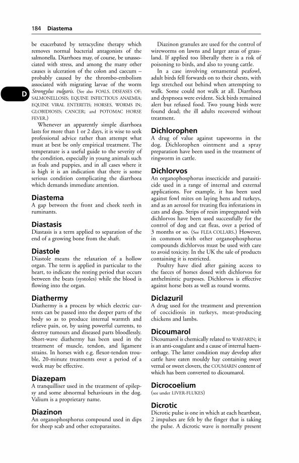
be exacerbated by tetracycline therapy whichremoves normal bacterial antagonists of thesalmonella. Diarrhoea may, of course, be unasso-ciated with stress, and among the many othercauses is ulceration of the colon and caecum –probably caused by the thrombo-embolismassociated with migrating larvae of the wormStrongylus vulgaris. (See also FOALS, DISEASES OF;
SALMONELLOSIS; EQUINE INFECTIOUS ANAEMIA;
EQUINE VIRAL ENTERITIS; HORSES, WORMS IN;
GLOBIDIOSIS; CANCER; and POTOMAC HORSE
FEVER.)Whenever an apparently simple diarrhoea
lasts for more than 1 or 2 days, it is wise to seekprofessional advice rather than attempt whatmust at best be only empirical treatment. Thetemperature is a useful guide to the severity ofthe condition, especially in young animals suchas foals and puppies, and in all cases where itis high it is an indication that there is someserious condition complicating the diarrhoeawhich demands immediate attention.
DiastemaA gap between the front and cheek teeth inruminants.
DiastasisDiastasis is a term applied to separation of theend of a growing bone from the shaft.
DiastoleDiastole means the relaxation of a holloworgan. The term is applied in particular to theheart, to indicate the resting period that occursbetween the beats (systoles) while the blood isflowing into the organ.
DiathermyDiathermy is a process by which electric cur-rents can be passed into the deeper parts of thebody so as to produce internal warmth andrelieve pain, or, by using powerful currents, todestroy tumours and diseased parts bloodlessly.Short-wave diathermy has been used in thetreatment of muscle, tendon, and ligamentstrains. In horses with e.g. flexor-tendon trou-ble, 20-minute treatments over a period of aweek may be effective.
DiazepamA tranquilliser used in the treatment of epilep-sy and some abnormal behaviours in the dog.Valium is a proprietary name.
DiazinonAn organophosphorus compound used in dipsfor sheep scab and other ectoparasites.
Diazinon granules are used for the control ofwireworms on lawns and larger areas of grass-land. If applied too liberally there is a risk ofpoisoning to birds, and also to young cattle.
In a case involving ornamental peafowl,adult birds fell forwards on to their chests, withlegs stretched out behind when attempting towalk. Some could not walk at all. Diarrhoeaand dyspnoea were evident. Sick birds remainedalert but refused food. Two young birds werefound dead; the ill adults recovered withouttreatment.
DichlorophenA drug of value against tapeworms in thedog. Dichlorophen ointment and a spraypreparation have been used in the treatment ofringworm in cattle.
DichlorvosAn organophosphorus insecticide and parasiti-cide used in a range of internal and externalapplications. For example, it has been usedagainst fowl mites on laying hens and turkeys,and as an aerosol for treating flea infestations incats and dogs. Strips of resin impregnated withdichlorvos have been used successfully for thecontrol of dog and cat fleas, over a period of3 months or so. (See FLEA COLLARS.) However,in common with other organophosphoruscompounds dichlorvos must be used with careto avoid toxicity. In the UK the sale of productscontaining it is restricted.
Poultry have died after gaining access tothe faeces of horses dosed with dichlorvos foranthelmintic purposes. Dichlorvos is effectiveagainst horse bots as well as round worms.
DiclazurilA drug used for the treatment and preventionof coccidiosis in turkeys, meat-producingchickens and lambs.
DicoumarolDicoumarol is chemically related to WARFARIN; itis an anti-coagulant and a cause of internal haem-orrhage. The latter condition may develop aftercattle have eaten mouldy hay containing sweetvernal or sweet clovers, the COUMARIN content ofwhich has been converted to dicoumarol.
Dicrocoelium(see under LIVER-FLUKES)
DicroticDicrotic pulse is one in which at each heartbeat,2 impulses are felt by the finger that is takingthe pulse. A dicrotic wave is normally present
184 Diastema
D

in a tracing of a pulse as recorded by specialinstruments for the purpose, but in health it isimperceptible to the finger.
Dictyocaulus Viviparus(see PARASITIC BRONCHITIS)
DicyclanilDicyclanil is used as a pour-on for the long-term prevention of blowfly strike in sheep andlambs. It stops the development of fly larvae byinterfering with moulting and pupation.
DieldrinA persistent organochloride insecticide former-ly used against the maggot-fly of sheep.Dieldrin is highly poisonous to birds and fish.The symptoms of dieldrin poisoning in foxes(which have eaten poisoned birds) are stated toresemble closely those of fox encephalitis. Dogsand cats have been poisoned similarly. (See also
DOG, KENNELS.) Dieldrin has been suspected asa cause of infertility in sheep, and residues inthe fat may be a danger to people eating themutton or lamb. The use of dieldrin sheep-dipswas banned in the UK in 1965, followingsimilar bans in Australia and New Zealand.Dieldrin was also banned as a dressing for win-ter wheat early in 1975, but cases of dieldrinpoisoning continued to occur among wild anddomestic pigeons, and in kestrels, etc., fed onpigeons, during that year. Dieldrin is still usedfor ground spraying in Africa (see under DDT; and
CHLORINATED HYDROCARBONS).
Diesel Oil PoisoningThirsty cattle have drunk diesel oil with fatalresults. Cattle with access to canals may drinkwater contaminated with diesel oil from boats.The results are less severe and recovery is possible.
Symptoms include loss of appetite, depres-sion, vomiting, tympany of the rumen, andemaciation. Death (sometimes from lungdamage) may occur after several weeks.
Diesel poisoning occurred in a ewe aftereating grass contaminated by oil from a fueltank sited in a field. Breath, urine and faeces allsmelt strongly of the oil.
Diet and DieteticsPAGE
Composition of foods 185Function of food constituents 186Antibiotic supplements 187General principles of feeding 187Digestibility of foods 187Preparation of foods 187Deterioration with storage 187
Palatability 188Variety and mixtures 188Maintenance and production rations 188Substitutional dieting 188Dogs’ diet 201Cat foods 124
The most important part of animal husbandryis sound feeding of the animals. This is notby any means, as might be supposed, a simplematter.
In order fully to understand rational feeding,owners of livestock (and of companion animals)must be conversant with the various food con-stituents and what part they play in the body;they must have an idea of the composition ofthe many foods that are available; and theymust know how to make the best use of them.The importance of palatability should never beunderrated.
Composition of foods By ordinarychemical analysis, foods can be split up andseparated into water, proteins, fats or oils, solu-ble carbohydrates, crude fibre or insolublecarbohydrates, minerals, and trace elements. Inaddition to these there are vitamins.
Water Water, as an essential need for livestock,is discussed under the appropriate heading, andis found in greatest amount in roots, succulentssuch as cabbages and kale, wet brewer’s grains,silage, and pasture grasses, which contain from7 to 90 per cent. Cereal grains, such as wheat,oats, barley, etc. average 11 per cent. Meadowgrass yields from 70 to 80 per cent of water, butwhen it is air-dried and made into hay underfavourable circumstances this is reduced to 12to 14 per cent.
Carbohydrates The carbohydrates in foodsare divisible into 2 groups: the crude fibre, andthe soluble carbohydrates.
Oats contain 10 per cent of fibre and hayand wheat-straw 25 per cent and 40 per centrespectively.
Crude fibre is a mixture of celluloses, lignin,cutin, and some pentosans (polysaccharides),etc. Cellulose forms the cell-wall of plants. Inits simplest form it is easily digested, but withthe growth of the plant, cellulose becomes asso-ciated with lignin, which gives stiffness to theparts of the plant requiring support, and alsocutin, which is a waterproofing material.
The carbohydrates are made up of carbon,hydrogen, and oxygen. Foods containing muchcarbohydrate are called carbonaceous foods,e.g. the cereal grains, potatoes, molasses, etc.
Diet and Dietetics 185
D

The cereals contain from 60 to 70 per cent ofcarbohydrate. The simplest of the carbohy-drates, such as the simple sugars, are absorbeddirectly from the gut, while the more complexsugars, and still more complex starches, have tobe reduced by processes of digestion to moresimple forms before they can be absorbed andbe of use to the body.
Fats or Oils Fat is present in all foods, but thequantity varies greatly; thus in hay there is 3 percent, in turnips there is 0.2 per cent, in cerealsfrom 2 to 6 per cent, and in linseed as much as40 per cent, while linseed cake, from whichmost of the fat has been expressed,contains on an average rather less than 10 percent. In meals produced from fat-rich foods,such as cotton seed or linseed, by extractionwith a solvent, all the oil except some 1 or 2 percent is removed.
Cattle cakes and other foods in which the fathas gone rancid are dangerous for animals, andoften cause diarrhoea. (See LIPIDS; COD-LIVER
OIL POISONING.)
Proteins The proteins or albuminoids in afood differ from the other constituents, in thatin addition to having carbon, hydrogen, andoxygen in their composition, they also containnitrogen and usually sulphur and sometimesphosphorus. They are very complex substances,and are made up of AMINO ACIDS.
Mineral matter or ash Plants have their ownmineral peculiarities; for example, the legumi-nous plants are rich in calcium which is sonecessary for animals; other foods, such as maize,are deficient in calcium, but contain phospho-rus; while others again, such as the wheat offals,have an unbalanced mineral content.
Vitamins (see under this heading)
Function of food constituents
Carbohydrates The carbohydrates are chieflyutilised for the production of energy and heat,and what is not required for immediate use isstored as fat which is to be regarded as a reservestory of energy.
Fibre A certain amount of crude fibre is nec-essary in the diet of all animals except thoseunder 31/2 weeks of age, when all young domes-ticated animals are on a fluid diet and most aresupported solely by suckling. If animals, espe-cially herbivorous animals, are given insuffi-cient fibre they fail to thrive, are restless and
uncomfortable, and every cattle-feeder knowsthat without ‘bulk’ to the ration the animals donot do well. Breeding gilts and sows need extrafibre to stretch their stomachs so that they canaccommodate enough concentrated feed duringlactation to provide for their litters.
Adequate fibre is necessary to cattle and rab-bits for proper muscular activity of the wholedigestive system. Secondly, the proportion offibre in the diet has an important bearing uponthe actual digestion done by living organismswithin the rumen. Thirdly, a high-protein andlow-fibre intake may lead to bloat. Fourthly,adequate fibre is necessary in the cow’s rations ifshe is to give a high yield of butterfat andsolids-not-fat.
On the other hand, if too much fibre is givenin the ration, the animals cannot digest enoughfood to get sufficient nutriment. Ruminantsmake the most use of fibre, then horses, pigs,and dogs, in that order. Fattening pigs, thoughrequiring a certain amount of fibre, must havethe allowance strictly limited, though sows andboars can do with more.
Fat The fat that is digested and absorbed maybe oxidised to form energy direct, or it may bebuilt up to form body fat. Speaking generally,fat has 21/2 times the value of carbohydrates orprotein as an energy producer. While a certainamount of fat is necessary in the daily diet ofanimals, an excessive amount does harm.
Protein It is not only the amount of protein inthe ration which is important, but also thequality of that protein.
Cereal protein is of poor quality, being defi-cient in lysine and methionine; and wheat isworse in this respect than barley. Accordingly,herring, (other) fish, and soya-bean meals arerelatively good sources of the desirable AMINO
ACIDS.For substitution of some of the protein in a
ration or diet, see under UREA.For health in all animals, adequate protein of
good quality is essential in the diet. Failure toprovide it can result in economic loss to farm-ers; losses often being far higher than the cost ofthe ‘extra’ necessary protein. Excess protein, onthe other hand, can bring its own problems. (See
under ACETONAEMIA, for example.)
Minerals, trace elements These are essen-tial for bone formation and maintenance, milkproduction, fertility, and the metabolism as awhole. The essential minerals and trace ele-ments are phosphorus, calcium, sodium, potas-sium, magnesium, iron, manganese, copper,
186 Diet and Dietetics
D

zinc, sulphur, iodine and cobalt. Not only arethey essential, but the balance is important, too:the ratio of one to another. For example, asmentioned under CALCIUM SUPPLEMENTS, theratio of this mineral to phosphorus can meanthe difference between health and ill health.
Proprietary concentrates from reputablemanufacturers ensure a feed for farm animalswith well-balanced minerals and trace elementsas a rule, and this is something which cannotalways be achieved in a farm mix unless aproprietary minerals premix is used.
On some soils, deficiencies of certain traceelements may occur so that special supplementsmay be needed.
For further information, see under METABOLIC
PROFILE TESTS; TRACE ELEMENTS; CONCEN-
TRATES; FELINE JUVENILE OSTEODYSTROPHY;
PIGLET ANAEMIA; IODINE DEFICIENCY; COBALT;
SALT.
Vitamins (see under this heading)
Antibiotic supplements (see under
ADDITIVES)
General principles of feeding There isno such thing as a well-balanced ration suitablefor all animals and all needs.
Sudden changes, involving a major proportionof the ration, are to be avoided in all stock.Changes should be made gradually or involveonly 1 or 2 out of several ingredients. In rumi-nants a sudden change to a predominantly cerealdiet can prove fatal. (See BARLEY POISONING.)
Regularity in the times of feeding is essentialfor success. Only good-quality food should beused; there is no economy in feeding with infe-rior or damaged fodder. On the contrary, theuse of such food has been the cause of muchillness. There should not be long intervalsbetween meals; with horses this is one of thecommon causes of colic. When compounding aration it should be remembered that a mixtureof foods gives a better result than the use of 1 or 2 foods. The ration should contain asufficiency of energy-producing constituents,sufficient protein, fibre, and mineral matter.(See CONCENTRATES.)
Digestibility of foods Only that part of afood which is digested is of value to an animal.The digestibility of foods varies greatly, somebeing easily and completely digested, while oth-ers, especially those containing much fibre, aredigested imperfectly and with difficulty; and, ofcourse, some animals will digest a particularfood better than others. (See D-VALUE.)
Preparation of foods Some foods are fedto animals in the natural state, while others areprepared in some such way as by grinding,bruising, cutting, chaffing, boiling, steaming,or soaking in water. Oats may be bruised forhard-working horses, for colts changing theirteeth, and for calves; there is undoubtedly aslight increase in the digestibility of bruisedover whole grain, but for an economic advan-tage the total cost of bruising should be lessthan 10 per cent of the whole grain. Beansshould be split or ‘kibbled’ for horses, as thetough seed-coat makes them difficult to masti-cate. Maize also is more easily eaten if it iscracked.
Grinding grains to a meal is advisable forpigs, but it is important that the particle size benot too small. Absence of milk in the recentlyfarrowed sow and bowel oedema may be associ-ated with meal particles that are too fine.
Deterioration with storage Bruised orkibbled seeds do not keep well, especially ifexposed to a damp atmosphere, and are liable toturn musty owing to fermentation. So long asthe grain is whole and intact it is essentially stilla living entity. When crushed, etc., it is killed,and the normal processes of deterioration anddecomposition commence.
All feeds tend to deteriorate, and to becomeless palatable, on storage. With whole cerealsthis deterioration will be very slight, but withmaize meal it can be rapid. It is recommendedthat the following storage periods should not beexceeded:
Flour mite infestation This can veryadversely affect the value of animal feed. At theNational Institute for Research in Dairying itwas suspected that infestation with flour mitesof an experimental feed, during prolongedstorage, was the cause of reduced performanceof growing pigs in a diet trial. A comparisonwas made between deliberately infested feedand control samples. It was demonstrated that,as the mite-infestation increased, there was aconsiderable loss of dry matter, carbohydrate,and amino acids. Subsequent growth trialsshowed that the daily liveweight gain andfeed:gain ratio were significantly reduced in thepigs on the mite-infested diet. Under the test
Diet and Dietetics 187
D
Maximum safe storage periods
Vegetable proteins 3 monthsAnimal proteins 1 monthMolassine meal 2 weeksGround cereals 1 weekMixed feed 1 week

conditions about one-fifth of the nutritivevalue of the diet was lost to the pig throughprogressive infestation with flour mites.
Palatability It is important that foodsoffered to animals should be palatable andappetising. Some foods are not very palatable,such as palm kernel cake or meal, but may bemade more palatable by mixing with somemolasses or locust bean meal. On the otherhand, foods which are naturally palatable maybecome very unappetising if they have beenallowed to get damp and musty. The inclusionof even a small quantity of musty food – suchas foxy oats and mouldy hay – in a ration spoilsthe whole food. The greatest care should betaken to see that the food is fresh and whole-some and that food-troughs and water-troughsare kept clean.
For dangers of poisoning by mouldy food, see
AFLATOXINS and MYCOTOXICOSIS.
Variety and mixtures Animals benefitfrom variety in their rations. It is often foundthat while a given ration may give excellentresults for a time, there is a tendency for animalsto eat the food without zest. This applies less topigs and horses than to cattle, sheep, poultry,dogs and cats. A change, which may be quitesimple, results in a return of the normal zest.
Also, as a rule, mixtures of several differentfoods are more palatable and are better digestedthan single food-stuffs. This is partly becauseduring digestion, foods of different originsactually assist to digest each other, and partlybecause if there is any deficiency in a particularfood substance in one food, it may be madegood to the animal by being present in anotherone of the mixture.
Maintenance and production rationsRations given to animals can be divided into2 parts, a maintenance and a production part. Amaintenance ration may be described as thatwhich will maintain an animal that is in a rest-ing and non-producing condition and in goodhealth, in the same condition and at the sameweight for an indefinite period.
A production ration is that part of the dailydiet which is given in excess of maintenancerequirements, and which is available for beingconverted into energy, as in working horses, orinto milk, or into fat or wool, or is used forgrowth.
It will be clear that a maintenance ration byitself is uneconomical, since it gives no return.
In devising a maintenance ration it should beclearly understood that any food will not do;
wheat straw does not contain sufficient proteinfor the maintenance of health in yearling bul-locks, but wheat straw in combination withgood quality hay will do so. (See RATIONS –Winter rationing.)
The most practical application of mainte-nance and production rations is in use wherethe cows are fed according to their milk yield.
Substitutional dieting A farmer who hasfixed a daily ration for, e.g., his dairy cows, anddesires to change some of the constituents inthe diet by substituting other foods, shouldnote that if foods are merely changed haphaz-ardly weight for weight it is almost certain thatthe diet will be altered appreciably. For exam-ple, if 3.5 kg (5 lb) of maize is substituted for3.5 kg of oats in a horse’s ration, the animal willbe getting more nutriment than formerly, as36 kg (80 lb) of oats are equal to 27 kg (60lb)of maize. Again, oat straw, pound for pound,has rather less than half the nutriment found inmeadow hay, and so on. (See STARCH EQUIVA-
LENT; PROTEIN EQUIVALENT; RATIONS; DRIED
GRASS; SILAGE; UREA.)When substituting one food for another it is
important that the change be made gradually.Disastrous results have followed the suddenchange of a diet. (See also NUTRITION, FAULTY;
VITAMINS; HORSES, FEEDING OF; DOGS’ DIET;
CAT FOODS.)
Diet During Illness orConvalescence(see NURSING)
Digestibility(see DIET)
Digestion, Absorption andAssimilationDigestion, absorption and assimilation are the 3processes by which food is incorporated intothe body.
Salivary digestion begins as soon as thefood enters the mouth and becomes mixed withsaliva secreted by the salivary glands. It is notvery thorough in animals, such as the dog,which bolt their food without careful chewing,but in the horse during feeding, and in the oxand sheep while rumination is proceeding, it ismore effective, especially when starchy foodsare eaten. Raw starches, which are very oftenenclosed in a matrix of cellulose or woodymaterial, are not acted upon to any great extentuntil the cellulose covering has been dissolved,through the action of bacteria, in other parts of
188 Diet During Illness or Convalescence
D
the system. Saliva has no digestive action uponproteins. In the domesticated dog, however,there seems little doubt that when given drybiscuits, which necessitate a certain amount ofchewing, some salivary digestion does occur.
Saliva contains the enzyme ptyalin, an a-amylase, which actively changes the insolublestarch of carbohydrate foods into partly solublesugars, but the process requires consummationby the enzymes of the small intestines. Ptyalinis only able to act in an alkaline medium, andits action therefore ceases as soon as the foodhas become permeated with acid gastric juice inthe stomach.
Stomach digestion begins shortly after thefood enters the true stomach and continues tillit leaves this organ. There are great differencesin the domesticated animals, due to the factthat some, e.g. ruminants, have a compoundstomach.
In animals with a simple stomach, such as thehorse, pig and dog, when food enters the stom-ach, ‘gastric juice’ is secreted from the digestiveglands situated in its walls. This juice containsthe enzyme pepsin, which, in the presence ofdilute hydrochloric acid, also produced by theseglands, acts upon the protein constituents.
Gastric lipase is another enzyme, present bothin ruminants and in simple-stomached animals,which is concerned with preliminary digestionof fats.
In the horse, food stays in the stomach till it isabout two-thirds full, and is then hurried throughto the small intestine to make room for furtheramounts entering from the mouth. In spite ofthis the stomach is practically never found emptyafter death – unless the horse has been starved.
In the pig and dog, food is retained in thestomach for a variable time according to thestate in which it was swallowed, and is thor-oughly churned and mixed with gastric juice.During this time the softer portions along withfluids and semi-fluids are squeezed through thepylorus into the intestine.
In the ruminating farm animals – cattle andsheep – stomach digestion is complicated by thepresence of 3 compartments before the truestomach, or abomasum, is reached. These areconcerned with the preparation of the foodbefore it enters the abomasum for true diges-tion. Although the rumen possesses no truedigestive glands, a considerable part of the diges-tive process takes place in it through the activityof cellulose-splitting and other organisms. (See
also RUMINAL DIGESTION.)In the unweaned calf, the act of sucking
apparently stimulates reflex closure of the
oesophageal groove, so that the dam’s milkby-passes the rumen (where it could not beeffectively digested).
After the food has been subjected to theaction of the organisms in the rumen, and hasbeen chewed for a second time as ‘cud’, it is senton into the 3rd stomach or omasum for furtherbreaking up by trituration, and then into thetrue stomach or abomasum where digestiveglands are present, and where a form of diges-tion similar to that which occurs in the stomachof other animals takes place.
Intestinal digestion The softened semi-fluid material which leaves the stomach iscommonly known as ‘chyme’; it has an acidreaction, since it has been well mixed with thehydrochloric or lactic acid in the stomach.Shortly after entering the small intestine itmeets with alkaline fluids and its acidity is neu-tralised. This occurs through the action of thebile from the liver and of the pancreatic juicefrom the pancreas. These fluids are similar inthat they are both alkaline, but differ greatly intheir functions. The bile is partly composed ofcomplex salts and pigments (see BILE). Its func-tion is fourfold: it aids the emulsification offats, dividing large droplets into tiny globuleswhich are more easily split into their compo-nent parts by other enzymes prior to absorp-tion; it assists in keeping the intestinal contentsfluid and preventing undue fermentation andputrefaction through its slight antiseptic actionagainst putrefactive organisms; it stimulatesperistalsis to some extent; and it gives the faecestheir characteristic colour. The pancreatic juicecontains at least 3 enzymes which are probablysufficient in themselves to ensure completedigestion of a food. Trypsin is active in thefurther splitting up of protein substances whichhave been partly acted upon by the pepsinof the stomach. The next pancreatic enzymeis amylopsin. It acts on carbohydrate con-stituents, splitting them up into sugars andother substances, but not carrying the processfar enough to allow of complete absorption.Amylopsin has an action similar to that of theptyalin of saliva, but can act upon raw starch.Lipase, or steapsin, is the fat-splitting enzymeof the pancreatic fluid. It acts upon the tinyglobules of fat which have been emulsified bythe bile, etc., and splits them into their com-pounds – glycerol and a fatty acid, the latterdepending upon the origin of the fat.
Secretions from the intestines contain anumber of enzymes of which the most impor-tant are erepsin, enterokinase, maltase, lactase,and invertase. The 1st of these completes the
Digestion, Absorption and Assimilation 189
D
breaking up of any protein which may haveescaped the action of the pepsin and trypsin.Enterokinase is concerned with the formationof trypsin from its fore-runner trypsinogen, andthe last 3 complete the splitting up of carbohy-drates into soluble sugars. Bacteria also have amost important digestive function in theintestines. In the large intestines of herbivorousanimals they have a cellulose-splitting action,which is somewhat allied to fermentation, andis similar to the activity of the organisms pre-sent in the 1st stomachs of ruminants. They actupon fats in a similar manner to the pancreaticjuice; they form certain volatile obnoxioussubstances (indole and skatole) from proteins,which give the faeces their characteristic odour;they produce lactic acid in certain cases;and they may even destroy alkaloidal poisonswhich have been formed during other stages ofdigestion.
Absorption Water passes through the stom-ach into the intestines almost immediately. Butit is only after subjection to digestion in theintestines for some hours that the bulk of thefood is taken up into the system. The chymewhich leaves the stomach is converted by theaction of the bile and pancreatic fluids into ayellowish-grey or a brownish-green fluid ofcreamy consistency called ‘chyle’, containing inthe herbivorous animals particles of hay, oats,grass, etc. From this the fats are absorbed (afteremulsification and breakdown) by the lymphvessels or ‘lacteals’ which occupy the centre ofeach of the ‘villi’ of the small intestines. (See
VILLUS.) From the lacteals the fat globules arecollected by the lymph vessels of the intestinesand are ultimately passed into the bloodstream.Sugar, salts, and soluble proteins pass directlyinto the small blood vessels in the walls of theintestines, and are thence carried to the liverand so enter the general circulation.
The food is passed onwards through thevarious folds and coils of the intestines, eachparticular part of the bowel wall removing someportions of the food, and the residual, unab-sorbable, useless constituents are eventuallydischarged from the rectum and anus duringthe process of defecation.
Assimilation takes place slowly. After theproducts of digestion have been absorbed intothe blood- and lymph-streams they are carriedround the body, ultimately reaching everyorgan and tissue, and the body cells extractfrom the blood in the capillaries whatevernutritive products they may require for growthor repair. For instance, cells in bony tissues
extract lime salts, muscles take proteins andsugars, etc. When the supply of food is much inexcess of immediate requirements the surplus isstored up, e.g. as glycogen in liver or musclefibres.
Digital DermatitisA condition in cattle usually affecting the skinabove the bulb of the heel. The animal is verylame. The cause is probably a spirochete,Borrelia burgorferi.
Treatment Clean the affected area and usean oxytetracycline spray. For herd treatment,lyncomycin/spectomycin footbaths may beused. Antiseptic foot baths may help preventthe infection.
DigitalisDigitalis is a preparation from the leaf of thewild foxglove, Digitalis purpurea, gatheredwhen the flowers are at a certain stage.
The leaves contain glycosides, includingdigoxin, digitoxin, gitoxin, and gitalin; theseeds contain another glycoside, digitalin. Thepurified glycosides digoxin and digitoxin areused medicinally.
Digitalis is used in the treatment of chronicheart disease, in dogs mainly. The action of theheart is slowed down, the drug increasing thelength of diastole, and at the same time it isstrengthened.
Digitalis must be used with care, as the digi-toxin is excreted only slowly and there is acumulative effect which can readily lead to poi-soning. Its use in cats is inadvisable, and liableto cause vomiting. This, together with loss ofappetite, depression, and bradycardia, mayoccur in some dogs even with normal dosage.
The drug is usually used in the purified formas digoxin or digitoxin.
Digitalis poisoning may occur from a sin-gle, large dose or from prolonged administra-tion. The heart’s action may become irregular.Diarrhoea may occur.
In grazing animals poisoning may resultfrom the eating of the plant rosettes. Foxglovesincluded in hay have also caused poisoning.
DihydrotachysterolAn oil-soluble steroid used to raise the calciumlevel of the blood, and so treat or preventhypocalcaemia.
DihydroxyanthraquinoneA non-toxic laxative, acting chiefly on the largeintestine, effective in all the domestic animals,
190 Digital Dermatitis
D

including horses. It may be given in the food,when it acts in about 24 hours.
DimethiconeAn anti-frothing agent used in the treatment of‘frothy bloat’ in cattle (see under BLOAT).
DimetridazoleDimetridazole is a drug used for the treatmentand prevention of histomoniasis in turkeys,pheasants and chickens and trichomoniasis andhistomoniosis in pheasants and partridges. It isthe treatment of choice for trichomoniasis inpigeons as no staining of the tail feathersresults. It is still used in the UK and elsewherebut is banned in many EU countries.
Dimidium BromideA trypanocide effective against Trypanosomacongolense.
DinoprostA synthetic preparation of prostaglandin F2α(see PROSTAGLANDINS).
DioctophymosisInfestation with the kidney worm, Dioctophymarenale, a parasite of dogs encountered inEurope, America, and Asia. A survey of 500dogs in Iran revealed an incidence of 1.3 percent. Stray dogs and jackals have been foundinfested. Man may become infested through theeating of fish. (See also under ROUNDWORMS.)
DiodoneA contrast medium used in radiography of thekidneys.
DioestrusThe resting, or inactive, phase of the sexualcycle in the female, during which progesteroneis secreted by the corpus luteum. This causesthe mare, for example, to reject the stallion andinduces changes in the reproductive tractdesigned to provide a suitable environment fordevelopment of the embryo. In the mare,dioestrus normally lasts 15 to 16 days and isterminated by the release of 1 or more luteolyt-ic factors from the endometrium which inducesregression of the corpus luteum. (See OESTRUS.)
DioxinDioxin is a toxic chemical formed as an impu-rity during the synthesis of trichlorophenol andits derivatives. Accidental exposure may leadto cancer; skin, eye, blood and liver damage;and also to abortion, fetal malformation andchromosomal aberrations.
Dioxin contaminated milk on a farm near atoxic waste disposal plant in the Netherlands.The dioxin was emitted during the destructionof polyvinyl plastic (PVC); it may be present inthe exhaust from incinerators or other industri-al processes. Dioxin contamination of animalfeed in Belgium led to the condemning of meatand other food products during 1999.
Diphtheria, Calf(see CALF DIPHTHERIA)
Diphtheria, Guttural Pouch, ofHorses(see GUTTURAL POUCH DISEASE)
DiplegiaParalysis on both sides of the body.
DiplostomumDiplostomum is a fluke which lives in the eyeof salmonids. The intermediate hosts are snailsand water birds, especially gulls.
DiprosopusDuplication of the face. This is a type ofconjoined twinning.
Dips and DippingIn Britain mostly sheep are dipped, but beefcattle may also be dipped with advantage.Dipping is an important means of tick controlin cattle, and is widely practised in the tropics.
Sheep are dipped in order: (1) to eradicatethe commoner parasitic agents, such as keds,lice, ticks, etc.; (2) to act as a check upon thespread of mange in the sheep, commonly called‘sheep scab’, and where that disease has brokenout, to cure it; and (3) to prevent attack by thesheep-blowflies and consequent infestationwith maggots. Dipping is no longer compulso-ry in Britain and a serious increase in outbreaksof sheep scab has resulted.
In Britain, dips contain the amidine,organophosphorus compounds, mainly diazinonor protemphos; or the pyrethroids, cypermethrinor flumethrin.
Precautions Purchasers and users oforganophosphorus sheep dips must hold a cer-tificate of competence in the safe use of sheepdips issued by the National Proficiency TestsCouncil. Protective clothing must be worn, caretaken to avoid inhaling dust or spray, andsplashes on the skin washed off immediately.All owners should ensure that any dips theypurchase carry on their labels the statement thatthe dip has been approved by the Ministry of
Dips and Dipping 191
D
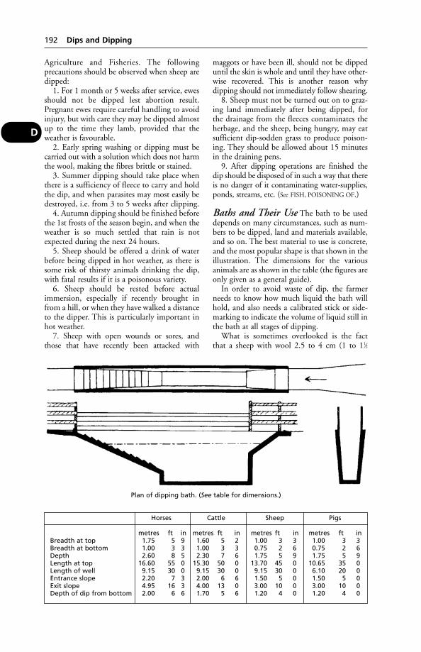
Agriculture and Fisheries. The followingprecautions should be observed when sheep aredipped:
1. For 1 month or 5 weeks after service, ewesshould not be dipped lest abortion result.Pregnant ewes require careful handling to avoidinjury, but with care they may be dipped almostup to the time they lamb, provided that theweather is favourable.
2. Early spring washing or dipping must becarried out with a solution which does not harmthe wool, making the fibres brittle or stained.
3. Summer dipping should take place whenthere is a sufficiency of fleece to carry and holdthe dip, and when parasites may most easily bedestroyed, i.e. from 3 to 5 weeks after clipping.
4. Autumn dipping should be finished beforethe 1st frosts of the season begin, and when theweather is so much settled that rain is notexpected during the next 24 hours.
5. Sheep should be offered a drink of waterbefore being dipped in hot weather, as there issome risk of thirsty animals drinking the dip,with fatal results if it is a poisonous variety.
6. Sheep should be rested before actualimmersion, especially if recently brought infrom a hill, or when they have walked a distanceto the dipper. This is particularly important inhot weather.
7. Sheep with open wounds or sores, andthose that have recently been attacked with
maggots or have been ill, should not be dippeduntil the skin is whole and until they have other-wise recovered. This is another reason whydipping should not immediately follow shearing.
8. Sheep must not be turned out on to graz-ing land immediately after being dipped, forthe drainage from the fleeces contaminates theherbage, and the sheep, being hungry, may eatsufficient dip-sodden grass to produce poison-ing. They should be allowed about 15 minutesin the draining pens.
9. After dipping operations are finished thedip should be disposed of in such a way that thereis no danger of it contaminating water-supplies,ponds, streams, etc. (See FISH, POISONING OF.)
Baths and Their Use The bath to be useddepends on many circumstances, such as num-bers to be dipped, land and materials available,and so on. The best material to use is concrete,and the most popular shape is that shown in theillustration. The dimensions for the variousanimals are as shown in the table (the figures areonly given as a general guide).
In order to avoid waste of dip, the farmerneeds to know how much liquid the bath willhold, and also needs a calibrated stick or side-marking to indicate the volume of liquid still inthe bath at all stages of dipping.
What is sometimes overlooked is the factthat a sheep with wool 2.5 to 4 cm (1 to 11⁄2
192 Dips and Dipping
D
Plan of dipping bath. (See table for dimensions.)
Horses Cattle Sheep Pigs
metres ft in metres ft in metres ft in metres ft inBreadth at top 1.75 5 9 1.60 5 2 1.00 3 3 1.00 3 3Breadth at bottom 1.00 3 3 1.00 3 3 0.75 2 6 0.75 2 6Depth 2.60 8 5 2.30 7 6 1.75 5 9 1.75 5 9Length at top 16.60 55 0 15.30 50 0 13.70 45 0 10.65 35 0Length of well 9.15 30 0 9.15 30 0 9.15 30 0 6.10 20 0Entrance slope 2.20 7 3 2.00 6 6 1.50 5 0 1.50 5 0Exit slope 4.95 16 3 4.00 13 0 3.00 10 0 3.00 10 0Depth of dip from bottom 2.00 6 6 1.70 5 6 1.20 4 0 1.20 4 0
inches) long will not merely remove perma-nently at least 2.25 litres (1/2 gallon) of liquid,but will strain off additional insecticide. Thisnecessitates ‘topping up’ of the dip wash atdouble strength as compared with the liquidused for the first filling of the bath.
It is a false economy not to top up before thelast 20 or 30 sheep are put through the dip,since any saving of money thereby could laterbe more than offset by those animals becomingvictims of strike. Disappointing results of anydip can also follow if sheep are immersed for farshort of 30 seconds; or if they are soaking wetwhen they enter the bath, for then their fleecescan carry much less than the normal quantity ofwash.
Arsenic-dipped animals should never beallowed on to pasturage until there is no risk ofcontamination of grass.
In all cases the animal should be totallyimmersed at least once (hence the abrupt com-mencement of the bath), and special attentionshould be paid to the ears and tail. Dippingmust be thorough.
One dipping will seldom (if ever) be effectivein ridding an animal of parasites, as the dip maynot affect the eggs. The dip must accordingly berepeated at suitable intervals. Against keds, dipsshould be repeated in 3 to 4 weeks, and againstmange in about 7 to 10 days.
Lameness Especially in warm climates, wherethe dip has been allowed to remain in the tankand has become dirty, there is a danger of sheepbecoming lame after dipping. This results frominfection with Erysipelothrix rhusiospathiae (see
under SWINE ERYSIPELAS) through any cuts orabrasions. Such lameness does not follow the useof a freshly prepared dip. It has been obviated bythe addition to the dip of tetramethyl thiuramdisulphide; this controls any bacteria whichcontaminate the dip liquid. Non-phenolic sheepdips have little or no action against bacteria.
Spraying Dipping of all animals involves con-siderable trouble, expensive equipment, and inmost cases is static so that animals must cometo the dipper. The use of modern sprays andjets, whereby the chemical agent is directed onto the animal’s skin with considerable force, hassome advantages over dipping and is partlyreplacing dipping in some countries (see SPRAY
RACE; also JETTING). In Britain, those who prac-tise spraying, as opposed to dipping, would beunwise to rely on more than 3 weeks’ protectionagainst strike. This is partly because less insecti-cide remains in the fleece after spraying; also,the organophosphorus insecticides move down
the wool but, apparently, not sideways, so thatif a patch is left unsprayed it remains vulnerableto strike.
Protective clothing Operators engaged indipping or spraying must use protectiveclothing. The latest guidance issued by themanufacturers or government agencies should befollowed. Basically, protection such as coveralls,waterproof apron or leggings, wellington bootsand elbow-length rubber gloves, a face shield andhat should be worn; details vary with the type ofproduct used. There is a National ProficiencyTest Council certificate in personal protectionequipment for those regularly involved in the useof dips and sprays.
Farmers should familiarise themselveswith the guidelines on the storage, use anddisposal of dips issued by the manufacturersand the Veterinary Medicines Directorate.
DiquatThis herbicide has caused fatal poisoning incattle, 4 years after the discarding of a container.
Dirofilariasis(see HEARTWORMS)
DisbuddingDisbudding is the removal of, or the preventionof growth in, the horn buds in calves, kids,and sometimes in lambs. (See DE-HORNING OF
CATTLE; GOATS, DISBUDDING OF KIDS.)
Disc, Intervertebral(see under SPINE)
DiscospondylosisInflammation of the intervertebral discs of thespinal column.
‘Disease-Free’ Animals (InResearch)The availability of animals born and reared freefrom infection is an important tool in the studyof disease. The technique is used in laboratoryanimals and has had particular application inpigs, as described below.
Piglet mortality is one of the main sources ofeconomic loss to the pig industry, and it is inthe study of important piglet diseases that spe-cial laboratory pigs are necessary. Without suchanimals, research work may not only be ham-pered or even brought to a standstill by naturalinfections, but complications may also arise.
From the moment the piglet leaves the secu-rity of the uterus and enters the birth-canal itbecomes exposed to an infected environment.
‘Disease-Free’ Animals (In Research) 193
D
Under natural conditions it is protected, againstthis environment, to a greater or lesser degree,by the wide range of antibodies received fromits dam in the first milk, the colostrum. Whendeprived of colostrum piglets almost alwaysdie. But the research worker wishes to avoidthe feeding of colostrum, since it may wellcontain antibodies against the disease underinvestigation.
The problem is, then, to rear piglets whichare both disease-free and devoid of antibodies.In principle, the solution to the problem is asimple one. All that needs to be done is toobtain the piglets before they reach the infectedenvironment and to rear them away from pos-sible infection, so that colostrum is unnecessary.In practice, these requirements are not easilymet. However, by using a technique developedin the USA at the University of Nebraska, ‘dis-ease-free’, antibody-devoid pigs have beenproduced.
The piglets are taken direct from the sow’suterus a day or 2 before the estimated farrowingdate. The sow is anaesthetised, the whole uteruscarefully but rapidly removed and passedthrough a bath of disinfectant, into a sterilisedhood. The hood is supplied with warm, filteredair under slight pressure, and 2 operators, work-ing through long-sleeved rubber gloves, takethe piglets from the uterus. Their navel cordsare tied off, and they are dried with steriletowels. The piglets are then transferred, bymeans of a sealed carrying case, to sterile incu-bator units kept in a heated isolation room. Theincubators, each of which holds 1 pig, areequipped with filter pads so that both the airentering the unit and that passing out into theexhaust system is filtered.
During the first few days of their indepen-dent existence, great care is necessary to protectthe young animals from bacteria in general. Theattendant wears mask and cap in addition torubber gloves and overalls. Subsequently, masksand caps are unnecessary. The diet, which con-sists of pasteurised milk, eggs and minerals, issterilised by heat for the first 3 days of life,but not thereafter. The piglets are fed fromflat-bottomed trays 3 times daily – morning,midday and late afternoon. There are no nightfeeds. After some 10 days in the incubator unitsthe young pigs are transferred to individualopen cages in another isolation pen. There theyare rapidly weaned to solid food. Later, the pigsare mixed together and treated as ordinary onesexcept that, of course, precautions are taken toprevent accidental infection.
Pigs reared by this technique are in a stateof minimal disease: they are not germ-free. In
fact, non-pathogenic bacteria are deliberatelyintroduced by feeding pasteurised, instead ofsterilised, milk from the 4th day of life onwards.These pigs are not, therefore, in the samecategory as germ-free GNOTOBIOTIC animals.Production of ‘disease-free’ pigs was begun atCambridge primarily to permit the criticalinvestigation of pig diseases, particularly diseasesof sucking pigs, but such pigs have obviousadvantages for nutritional and genetic studiesbecause the technique does eliminate thatunpredictable variable, disease. (See also SPF.)
Diseases(see NOSOCOMIAL; IATROGENIC; STOCKMEN/
WOMEN)
Diseases of Animals ActsLegislation relating to animal health, includingthe Diseases of Animals Act 1950, was consoli-dated under the Animal Health Act 1981,which also includes the relevant Orders madeunder the various Acts. It is administered by theAnimal Health Division of the Ministry ofAgriculture.
It covers the diseases listed under NOTIFIABLE
DISEASES.The Act and Orders provide for the compul-
sory notification of the existence or suspectedexistence of these diseases; for the immediateisolation or segregation of diseased or suspectedanimals; for the diagnosis of suspected diseaseby specially trained persons; for the slaughter,treatment or vaccination of diseased or suspect-ed animals where appropriate and for thedisposal of carcases and other waste where nec-essary; for the payment of compensation toowners in certain cases; for the apprehensionand punishment of offenders; for the systemat-ic inspection of markets, fairs, sales, exhibitions,etc., and for the seizure of diseased or suspectedanimals therein; for regulating the transit andtransport of animals by land or water, bothwithin the country and in the home waters; forcontrolling the importation of animals andthings which may introduce one or other ofthese diseases from abroad; and for inspectionat the ports and quarantine or slaughter wherenecessary.
The following regulations have a generalapplication to all scheduled diseases, but inpractically every case there is at least one Orderapplicable to the particular disease, in whichthere is set out more fully regulations dealingwith that disease. These Orders can be obtainedthrough the Stationery Office, and must beconsulted individually if complete informationis required.
194 Diseases
D

Notification of diseases or suspected diseasemust always be made by the owner of an ani-mal, or by the occupier or person in charge, andby the veterinary surgeon in attendance, to aninspector of the local authority or to a policeconstable, without undue delay.
Presumption of knowledge of disease. A personrequired to give notice if charged with failureto carry out his or her obligation shall be pre-sumed to have known of the existence of thedisease, unless and until s/he shows, to thesatisfaction of the Court, that s/he had notknowledge thereof and could not with reasonablecare have obtained that knowledge.
Separation of diseased animals. Every personhaving a diseased animal shall, as far as is prac-ticable, keep it separate from animals not sodiseased.
Facilities and assistance to be given for inspec-tion, cleansing, and disinfection. Persons in chargeof diseased animals are required to give everyfacility for the execution of the above, and mustnot obstruct or in any way hinder inspectors orother officers in doing their duty.
Prohibition of exposure of diseased animals. Itis unlawful to expose a diseased or suspectedanimal in a market, sale-yard, fair, or otherpublic or private place where such animals arecommonly exposed for sale; to place an affectedanimal in a lair or other place adjacent toor connected with a market, sale-yard, etc., orwhere such animals are commonly exposed forsale; to send a diseased animal on a railway, oron any canal, inland navigation or coasting ves-sels; to allow one on a highway or thoroughfare,or on any common or unenclosed land or inany insufficiently fenced field; to graze one onthe sides of a highway or to allow one to strayon a highway or thoroughfare or on the sidesthereof, etc.
Digging up carcases. No person may dig upthe carcase of an animal that has been buried,without official permission. (See also under eachmain heading of the scheduled diseases, e.g.ANTHRAX.)
Diseases of Animals (WasteFood) Order 1973This Order, amended in 1987, makes it anoffence for producers to feed, intentionally orinadvertently, untreated meat or meat productsto livestock on their premises. Any litter spreadon fields must be examined for the presence ofcarcases, which must be removed.
Diseases of Fish ActThis lists the notifiable diseases. (See FISH,DISEASES OF.)
Dishorning(see DISBUDDING)
Dishorning of Cattle(see DE-HORNING OF CATTLE)
DisinfectantsDisinfectants may be either physical or chemi-cal. Among the former are heat, sunlight andelectricity; while among the latter are solids,liquids, and gases. Steam may be used.
Chemical disinfectants At the presenttime these are numerous and diverse. TheDeparment of the Environment, Food, andRural Affairs tests them from time to time andissues its approval only to those that are main-tained up to standard. Consequently, ownersshould examine the labels on containers and useonly those that carry the official approval sincethis is a guarantee of potency.
The Diseases of Animals (ApprovedDisinfectants) Order 1970 governs the uses ofdisinfectants in the UK, and specifies thoseapproved for use in connection with foot-and-mouth disease, tuberculosis, fowl pest, andgeneral orders relating to disease control.Dilution rates are also specified.
A full list of disinfectants approved for use inoutbreaks of foot-and-mouth disease is givenunder that entry.
Disinfectants act in 1 of 3 ways: (1) as oxidis-ing agents or as reducing agents; (2) as corrosivesor coagulants acting upon the protoplasm ofbacterial life; or (3) as bacterial poisons.
Most chemical disinfectants are supplied in aconcentrated form and must be diluted withwater before use. The water should be clean,preferably soft, and if it can be used warm theefficiency of the disinfectant is increased. Afterthe active agent has been added, the wholeshould be well stirred for a few moments toensure thorough mixing. The solution mustbe applied so that it remains in contact withthe offending material for a sufficiently longtime to kill the bacterial life therein; generally10 minutes to half an hour should elapse beforedisinfecting solutions are rinsed away.
When 2 or more disinfectants are mixedtogether, instead of an increased disinfectingpower in the mixture they often enter intochemical combination with each other and auseless compound results. (See also ANTISEPTICS.)
For quaternary ammonium compounds, see
under this heading.Cresol solutions: there are many of these,
e.g. the cresol and soap solution of the BP, thecompound cresol solution of the USAP, lysol,
Disinfectants 195
D

isal, cyllin, creolin, cresylin, Jeyes’ fluid, or oneof the proprietary preparations. These are usedas 3 to 5 per cent solutions for practically allpurposes of disinfection about a farm premises,and very often as antiseptics also. Their actionis enhanced by the use of hot water instead ofcold. None is suitable for use in connectionwith food, for all are to a greater or lesser degreepoisonous. Cresols are not very effective againstmany viruses or bacterial spores. The cresols arerelated to PHENOL.
Formalin is sometimes used as a solution fordisinfecting floors, about 5 per cent strengthbeing necessary. Formaldehyde gas may be usedfor fumigation of livestock buildings whereviral or other diseases have occurred. (See under
DISINFECTION.)Sodium hypochlorite (bleach) is widely used
and effective. Depending on the dilution andformulation it can be used as a general disinfec-tant and in, for example, sterilising milkingmachines. It has the advantage of leaving notaint, as it breaks down into salt and water.
DisinfectionDisinfection of buildings cannot be achieved byapplying a disinfectant solution to walls andfloors which are heavily contaminated with dirt.There are 2 reasons for this: (1) the disinfectantcannot reach most of the micro-organisms,which will be protected by layers of dirt; and (2)the latter may alter the nature of the disinfectantsolution chemically, rendering it ineffective.
Preliminary cleaning is therefore essential.The building must first be thoroughly scraped,brushed, and cleansed. Concrete floors may bepower-hosed, and scraped free from all dirtand debris. A hot detergent solution such as 2.5to 4 per cent washing-soda is then thoroughlyscrubbed into floors, walls, stall partitions,mangers, troughs, or other fittings.
Disinfectants After an outbreak of infectiousdisease, buildings and equipment must be treat-ed to remove traces of infection before animalsare rehoused. To be effective, the application ofdisinfectants is the 2nd stage of the process ofdisinfection – cleaning being the 1st stage.
In certain cases it may be desirable to fumi-gate the building. All air entrances and exits aresecurely closed, the inside of the walls and roofsoaked with water, and formaldehyde gasgenerated (e.g. by pouring on to 250 g of potas-sium permanganate 500 ml of formalin per1000 cu ft of air space.) All doors and windowsare left shut for a day, and the building is thenflushed out with clean water under pressurefrom a hose-pipe.
Steam cleaning may be carried out as part ofa disinfection process.
Movable objects All pails, grooming tools,wheelbarrows, shovels, forks, etc. which havebeen used for the infected animals must also bedisinfected before they can be considered safefor further use.
DislocationDislocation is a displacement of a bone from itsnormal position in relation to a joint. Deformityis produced, and there may be intense pain if thepart is interfered with. As well as displacementthere is also bruising of the soft tissues aroundthe joints, and tearing of the ligaments whichbind the bones together.
Probably the most common dislocation isthat of the patella, which becomes lodged onthe uppermost part of the outer ridge of thepatellar surface of the femur and is unable toextricate itself from this position. In the dog,dislocation of the shoulder joint is by no meansrare.
The causes of dislocations are similar to thosewhich produce fracture, e.g. violence applied insuch a manner that the structures around thejoint are unable to withstand the stress. (Forinherited abnormality in dogs, see under PATELLA.)
Signs The injured limb is useless, and as a ruleis held off the ground in an unnatural attitude.There is generally little or no pain so long as theparts are not forcibly moved; but if a nervetrunk is pressed upon, the animal may perspirewith the pain. When the limb is compared withthat of the opposite side there is seen a markeddifference in its contours or outline – the jointaffected shows hollows or prominences wherenone is seen in the normal limb. There is a lossof the power of movement, but there is nograting sound heard when the joint or thewhole limb is passively moved, such as occurswhen a fracture exists.
Treatment The reduction of dislocationsnecessitates the use of anaesthesia.
Displaced AbomasumA condition encountered in cattle some weeksafter calving and leading to a lack of appetite.Displacement can be left or right; left is morecommon. (See under STOMACH, DISEASES OF.)
Disposal of CarcasesCarcases must be disposed of under the AnimalBy-products Order 1999. It is an offence toleave the carcase of an agricultural mammal
196 Disinfection
D

unburied for 48 hours. Carcases may be sent toa knacker’s yard or a destructor. Burning on thefarm is possible, but may only be done bypermission of the Environmental ProtectionAgency. Moreover, where the cause of death hasbeen a contagious disease there is always therisk of healthy animals becoming directly orindirectly affected, and of the disease spreadingaccordingly. In most progressive countries thereare government regulations which provide forthe safe disposal of the carcases of animals thathave died from any of the notifiable contagiousdiseases, such as anthrax, foot-and-mouthdisease, cattle plague, etc., but it is importantthat all carcases should be safely and efficientlydisposed of, no matter what has been the causeof death.
The safest and most expeditious manner ofdisposal is for the carcase to be digested ina special destructor, either by heat (burning,or by live steam) or by chemical agents. Incountry districts, however, such plants as theseare seldom available, and it is necessary to buryor burn the carcases.
Burial of carcases A suitable site shouldbe selected where there will be no danger ofpollution of streams, rivers, canals, or otherwater-supplies, and where there is a sufficiencyof subsoil to allow a depth of 2 m (6 ft) of soilabove the carcase. A pit is dug, about 2.5 to3 m (8 or 9 ft) deep, in such a manner that thesurface soil and the subsoil are not mixed, anda clear approach is left to its edge. Roughly,about 2.5 to 3 m2 (21⁄2 to 3 sq yd) of surface arerequired for a horse, 1.25 to 2.5 m2 (11⁄2 to 21⁄2sq yd) for an ox, and about 1 m2 (1 sq yd) foreach pig or sheep. The dead animal should bearranged upon its back with the feet upwards.The carcase is next covered with quicklime or apowerful disinfectant, and the pit filled in withthe soil – subsoil first and surface soil last. If theweather is very wet, or if the soil is naturallyloose and soft, the surface of the ground shouldbe fenced off to prevent horses and cattle frompassing over it and perhaps sinking into theloose soil. It is not safe to plough over a largeburial pit for 6 months after it has been closed,nor should heavy implements or vehicles beallowed to pass over it.
Cremation of carcases Where a large coalboiler or furnace is used for heating supplies ofwater, there is no reason why, occasionally, thecarcases of small animals that have died shouldnot be burned in it. However, special incinera-tors are used where small animal carcases areroutinely disposed of; they must be sited to
conform with local environmental health rules.Dead horses and cattle, and large sheep and
pigs, should not be dismembered and destroyedin such a manner; they must be burned in aspecially constructed cremation pit.
There are 3 methods of cremation: (1) thecrossed trench; (2) the Bostock pit; and (3) thesurface burning method.
In the crossed trench, 2 trenches 1.2 m (7 ft)long are dug so that they form a cross. Each isabout 40 cm (15 in) wide and 45 cm (18 in)deep in the centre, becoming shallower towardsthe extremities of the limbs. The soil is thrownon to the surface in the angles of the cross, andupon the mounds so made, 2 or 3 stout piecesof iron, beams of wood, or branches from a treeare placed. Straw and faggots are piled in thetrenches to the level of the surface of theground, the carcase is placed across the centre ofthe trenches, and more wood or coal is piledaround and above it. Two gallons of paraffin oilare poured over the whole, and the straw is lit.
In the Bostock pit, an oval pit 2.25 m (7 ft)long and 1.25 m (4 ft) wide is dug to a depth of1 to 1.25 m (3 to 4 ft), and a crossed trench 20× 20 cm (9 × 9 in) is dug in its floor. Upon thewindward side of the pit a ventilation trench1.25 m (4 ft) long and 50 cm (1 ft 6 in) wide,and a 30 cm (1 ft) deeper than the main pit,and at right angles to it, is dug. A field drain-pipe is placed in a tunnel connecting the trenchwith the pit, and this pipe is stuffed with straw.Straw is laid in the bottom of the main pit,wood or coal is piled above it so that aboutthree-quarters of the pit is filled, and the carcaseis next rolled into the pit. More wood or coal ispiled around and above it, and paraffin oilpoured over the whole. The straw is finally lit inthe bottom of the ventilation trench. A carcasecremated by this method takes about 8 to 10hours to burn away, and requires little or noattention. When burning is complete the soil isreplaced and the ground levelled.
The surface burning method is mainly usedwhere there are numbers of animals to beburned. One long trench is dug about 50 cm(1 ft 6 in) deep and 30 cm (1 ft) wide, andabout 1 m (3 ft) length is allowed for eachcattle carcase. At intervals along each side thereare placed side flues to coincide with each car-case. Fuel (straw, wood, and coal) is placedaround the central trench and the carcases aredrawn across it. More fuel is heaped around andbetween them, and paraffin oil or petrol issprayed over the whole. The straw is lit. Morefuel needs to be added at intervals.
Instead of the trench and side flues, battensof stout wood are sometimes laid upon the
Disposal of Carcases 197
D
ground, and the carcases pulled over them. Fuelis piled around them and lit, and more is addedas required. This latter method is speciallyapplicable where the ground is very wet, orwhere there is rock immediately below the soiland digging is impossible.
Precautions Where the carcase of an animalthat has died from a contagious disease is beingdisposed of in one of the above ways, it is essen-tial to ensure that blood or discharges are notspilled upon the ground in the process ofremoval. An efficient method of preventing thisis to stuff tow saturated with some strong disin-fectant into all the natural orifices – nostrils,mouth, anus, etc. – and to cover the surface ofthe improvised sleigh (door or gate) with piecesof old sacking which have been soaked withdisinfectant, so that parts of the carcase do notbecome chafed through friction with theground and so leave behind bloodstains.Everything that has come into contact with thecarcase must be carefully disinfected before it isremoved. Old ropes, sacking, and other objectsused for handling the dead animal may beburned. The surface of the soil around the edgeof the pit, upon which the carcase rests, shouldbe scraped off and thrown into the fire or pit sothat any blood or discharges may be renderedharmless. Finally, all attendants should beimpressed with the risks they run in handlingdiseased carcases, and with the risks there are ofcontaminating other healthy cattle. Appropriatebiosecurity measures, including the use ofprotective clothing, disinfected or discardedbefore leaving the premises, must be observed.
Disposal in the tropics (see TROPICS –Carcase disposal)
Disposal of Veterinary ClinicalWaste (UK)Such waste is defined by the Health and SafetyCommission as including animal tissue andexcretions, drugs or medicinal products, sharpinstruments, or similar materials or substances.
Clinical waste must be separated from otherwaste in accordance with the system agreedby the local authority, e.g. yellow sacks andreinforced containers. (The Collection andDisposal of Waste Regulations 1989.)
DistemperDistemper is a name applied to a specific viraldisease. As a rule, all members of the Canidaeand Mustelidae are susceptible to canine distem-per. These classes include dog, fox, wolf, ferret,mink, weasel, ermine, marten, otter and badger.
Felidae (cats) are not susceptible except for lions.An outbreak in the Serengeti National Park inTanzania appears to have been brought undercontrol by vaccination of the dogs belonging tothe local population. In the terminal stages ofdistemper in the fox, the animal becomes paral-ysed and froths at the mouth, giving rise to thesuspicion of rabies.
Injection of dogs with measles or rinderpestvirus confers immunity against distemper.
Canine distemper is an infectious diseasemainly of young dogs, characterised usually by arise in temperature, dullness, and loss of appetite,and in the later stages by a catarrhal dischargefrom the eyes and nostrils. The disease is oftencomplicated by broncho-pneumonia, and insome cases nervous symptoms develop, eitherwhen the febrile conditions subside, or before thishappens. The incubation period of the disease isfrom 4 to 21 days, though it may be longer.
Cause Canine distemper virus, a morbillivirus.There is only one antigenic type, thoughvarious syndromes (including ‘hard pad’) maybe associated with various strains, some ofwhich can suppress or impair the body’s natur-al defence systems, and this has a bearingupon possible complications due to secondarybacterial infections.
Certain bacteria are responsible for sec-ondary lesions; for example, Bordetella is oftenresponsible for bronchitis.
Cases of distemper may be complicated bythe coexistence of other infections such asCANINE VIRAL HEPATITIS, LEPTOSPIROSIS andTOXOPLASMOSIS.
Although it is chiefly in young dogs that thedisease is encountered, older dogs are oftenaffected; as a general rule, however, young ani-mals between the ages of 3 and 12 months arethe most susceptible.
KLEBSIELLA infection gives rise to symptomssimilar to some of those of distemper.
Signs and complications In typical cases thedog becomes feverish, has a discharge from eyesand nose, and a cough. In some cases the eyeinflammation become severe. (See KERATITIS.)
Complications include broncho-pneumoniawith a hacking cough. (See BORDETELLA.)
Gastroenteritis, and mouth ulcers, complicateother cases.
Sometimes (apart from the fact that the doghas seemed unwell) the first sign of the disease toalarm the dog-owner is a fit. (See ENCEPHALITIS.)A change in temperament, with a tendency toviciousness, may occasionally be noticed.
198 Disposal of Veterinary Clinical Waste (UK)
D
Paralysis of face muscles, or of a limb, mayoccur, and sometimes hindquarter paralysis (see
PARAPLEGIA) accompanied by incontinenceindicate that the dog is unlikely to recover,
‘Hard pad disease’ may cause a dog to makea tapping sound as it walks on a hard surface,and this manifestation of distemper may beaccompanied by pneumonia and/or diarrhoea.
Diagnosis and treatment An early diagnosisis important. A veterinary surgeon should beconsulted as soon as any of the above symptomsappear, and will advise on the use of serum,sulphonamides, antibiotics, vitamin preparations,etc., as the situation demands. (See also NURSING.)
After recovery from distemper it is importantto remember that, unless the dog is looked afterwith great care, relapses are liable to occur. Fora week or 10 days after all symptoms haveapparently subsided, the dog should be givenonly a limited amount of exercise. A vitaminpreparation may be prescribed.
After-effects CHOREA may occur when thedog appears to be making a good recovery, andoften after an otherwise mild illness. A syn-drome has been described (‘old dog encephali-tis’) in which, several months after being illwith distemper, even a young dog may becomesenile and forget its house training.
Prevention Various vaccines have long beenavailable and have included:
1. Live, egg-adapted distemper virus(a) obtained from embryonated hens’
eggs(b) obtained from cultures of avian
fibroblastic tissues.2. Live distemper virus adapted to homologous
tissue culture obtained from cultures ofdog kidney cells.
Combined vaccines against distemper,infectious canine hepatitis, canine parvovirus,leptospirosis and parainfluenza are on themarket.
The timing of vaccination is crucial.Assuming an adequate intake of colostrum,puppies born to bitches immunised againstdistemper should have sufficient antibody toprotect them during the initial weeks of life. Theimmunity provided by the antibody wanes: bythe time the puppy is 12 weeks old, the level ofmaternal antibody is negligible. It will no longerprotect against naturally occurring virus; equallyit will not interfere with distemper vaccination.
Puppies inoculated when between 7 and9 weeks old should therefore receive a 2nd doseof vaccine at 12 weeks of age.
A booster dose is often advisable when thedog is 2 years old.
These are general guidelines. Individualmanufacturers’ dosage instructions for specificvaccines may vary, and must be followed.
(See also COLOSTRUM; GAMMA GLOBULIN;
ANTISERUM; MEASLES VACCINE; MATERNAL
ANTIBODIES.)
DistichiasisDistichiasis is the presence of a double row ofeyelashes, of which one or both rows are turnedin against the eyeball, causing inflammation. Itmay lead in dogs to EPIPHORA.
Distiller’s GrainsA feed, relatively high in protein and energy, fordairy cattle. For hazards of storage, see BREWER’S
GRAINS.
DistomiasisInfestation with liver flukes.
DiureticsDrugs which increase the amount of urineexcreted. They are used mainly in the treatmentof oedema (dropsy) in cases of heart failure.They act by inhibiting the reabsorption ofsodium and chloride from the loop of Henle(loop diuretics) or the kidney tubule. (See KID-
NEYS – Structure.) Furosemide is a powerfulloop diuretic. Thiazides, which act in the distalpart of the tubule, are less potent. The risk ofexcessive excretion of potassium presented byloop diuretics may be avoided by the use ofpotassium-sparing diuretics, which are oftengiven in combination with loop diuretics toenhance their effect. Spironolactone is an exam-ple. A 2nd type of diuretic acts by osmoticaction, which causes water retention in thenephron. Osmotic diuretics such as mannitolare used, for example, to promote urine flow inkidney failure.
While diuretics can help, they will not curethe condition which has given rise to theoedema.
DiverticulumA small pouch formed in connection with ahollow organ. There are certain diverticulawhich are normally present in the body, e.g. thediverticulum of the duodenum, which is foundat the point of entrance of the bile and pancre-atic ducts, or the posturethral diverticulum, alittle pouch behind the opening of the femaleurethra into the posterior genital tract in thesow and cow; while there are others which arefound as the result of injury or disease, e.g. in
Diverticulum 199
D

the oesophagus, in the rectum, and sometimesin the intestines.
DNADeoxyribonucleic acid. This is found in thenucleus of every cell and carries coded informa-tion/instructions for reproducing other cells.‘DNA can be visualised as a long coded tape,divided into segments. These segments areindividual genes, and each carries informationfor the assembly of a specific protein. The genesissue the instructions for the cell; the proteinsexecute the orders. Some genes code for struc-tural proteins such as hair, horn, etc., but mostcode for enzymes which perform tasks inthe cell, such as motility, metabolism, andsecretion.’ (Professor W. F. H. Jarrett FRS.)
A chromosome is composed of a giant mole-cule of DNA, plus supporting protein, and itis the DNA which is the very basis of heredity.(See CELLS; GENES; CHROMOSOMES; GENETIC
ENGINEERING.) Bacteria, viruses, and plasmidscontain DNA. (See also CANCER.)
DNA ‘Finger-Printing’DNA ‘finger-printing’ of human beings wasfirst described by Dr Alec Jeffreys of LeicesterUniversity in 1985; and has since been usedto prove the identification of sires of manydifferent animal species.
The 1st case concerned a pack of Siberianhuskies, and proving the true identity of pup-pies born to one of them, prior to registrationwith the Kennel Club.
Other applications of the technique are pos-itive identification of thoroughbred horses, andof laboratory animals. The technique has beenused in the prosecution of robbers of raptornests by identifying the parents of youngraptors found by the police.
Genetic fingerprinting can also provide aneffective means of tracing the source of microbialcontamination as it differentiates between close-ly related micro-organisms, making possibleprecise identification of individual strains.
DNOCDinitro-ortho-cresol, a yellow crystalline sub-stance employed in agriculture as a weed-killerspray solution, acts as a powerful cumulativepoison. In man the symptoms are excessivesweating, thirst, and loss of weight. Poisoning indomestic animals might well be encounteredfollowing contamination by the spray or residue.
DNPDinitrophenol, a product somewhat similar toDNOC.
Dobermann PinscherA medium-sized, muscular dog with smoothhair, most often black. The ears are naturallypendulous. The breed originates in Germanyand is often used as a police or guard dog. It canbe affected by Von Willebrand’s disease, cervicalspondolithesis (‘wobbler syndrome’) andpolyostic fibrous dysplasia (bone cyst).
DockingDocking is removal of the tail or a part of it.In Britain, docking of the horse (excludingamputation of the tail by a veterinary surgeonfor therapeutic reasons) is illegal. (See also
NICKING.)
Dogs Since 1 July 1993 it has been illegalfor anyone other than a veterinary surgeon todock puppies’ tails in the UK. 47 of the 185breeds registered with the Kennel Club havetraditionally been docked. The RCVS has said itis unethical for a dog’s tail to be docked exceptfor therapeutic reasons. There is evidence thatthe docked end is more sensitive to pain than therest of the tail. Tails are widely used in commu-nication between dogs and between dogs andpeople. Dogs with docked tails cannot commu-nicate adequately; their attitude and intentionsmight be misunderstood by other dogs andfighting may result. ‘It is not mandatory for dogsto have their tails docked in order to be enteredfor Shows.’ (BVA Animal Welfare Foundation.)
Sheep It is customary for sheep of lowlandbreeds to be docked, for if the tail is left long itaccumulates dirt and faeces, and these predis-pose to the attacks of blow-flies. Enough tailmust be left to cover the vulva, or anus in thecase of the male. The use of rubber rings fordocking without anaesthetic is allowed onlywithin 48 hours of birth. (See ANAESTHETICS,
LEGAL REQUIREMENTS.)Many mountain breeds of sheep are left
undocked; the long woolly tail helps to keep thehind part protected from frost and wind.
Docks, Poisoning byLosses of sheep have been occasionally ascribedto eating either the common sorrel dock (Rumexacetosa) or sheep’s sorrel (R. acetosella), both ofwhich contain oxalates. A condition of stagger-ing with dilated pupils, muscular tremors, andlater, convulsions and prostration, has beennoticed in horses which have eaten large quanti-ties of sheep’s sorrel. In sheep, there is a loss ofappetite, rapid breathing, exhaustion, some-times constipation and at other times diarrhoea,with an unsteady gait and occasionally death.
200 DNA
D

Milk of cows that have eaten docks is made intobutter only with difficulty.
Dog BitesAnti-tetanus injections should always be givenin cases of dog bites. Various infections includ-ing Pasteurella septica infection in man can resultfrom these. (See also RABIES, BITES.)
Dog, Feminisation of(see SERTOLI CELL TUMOUR; also INTERSEX)
Dog, KennelsFormer kennels should not, unless they havebeen thoroughly cleaned and disinfected, beused for the temporary housing of lambs orgoatlings; in both, deaths have followed fromcysticercosis of the liver. (See TAPEWORMS; also
BEDDING, HOOKWORMS)Two sheep dogs died from dieldrin poison-
ing, their kennel having been washed weeklywith old sheep dip.
Dog-Sitting PositionIn pigs this may be a symptom of pantothenicacid (vitamin B) deficiency, or lameness due toMycoplasma hyosynoviae. In the horse this positionmay be adopted during severe COLIC. With refer-ence to the newborn Galloway calf, see GENETICS
– Genetic defects. Re lambs, see SWAYBACK.
Dog TicksIn Britain these include Ixodes hexagonus(common on suburban dogs and cats); I. ricinus(the sheep tick, commonly found on countrydogs); I. canisuga (‘the British dog tick’); andDermacentor reticulatus (which may infest alsocattle and horses). I. canisuga may establish itselfin buildings, as may Rhipicephalus sanguineus,which has infested houses in Denmark as well asquarantine stations. Modern central heating mayfacilitate the survival of this tick in northernlatitudes. In a house in England, a sitting-roomsofa, and a bedroom chair used by a dog, wereinfested. This tick may arrive in travellers’luggage. Hedgehogs are a source of I. hexagonus.
Dogs, Breeds ofThe reader is advised to consult textbooks onthis subject. (See also WILD DOGS.)
Dogs’ DietMost owners wisely feed their animals on amixed diet, offering some variety and at thesame time providing the essential nutrients. It isa misconception that dogs should be fed onlyon meat. However, some owners appear tobelieve that red (muscle) meat, cooked or raw,
is a complete food for dogs and cats. It is not,since it does not provide, for example, enoughcalcium. Cooked meat should be mixed withbiscuit meal or pasta, potatoes or vegetables,and fed at room temperature. Most dogs likeone or other of the proprietary biscuits.
Dogs should be fed at regular times, once ortwice a day; any food left uneaten should beremoved. Fresh water should be availablealways. A bone, or one of the proprietarysubstitutes, is useful to exercise the jaws andhelp keep the teeth healthy.
Proprietary dog foods are very widely usednowadays; they may be moist (canned), semi-moist (packeted) or dry. Major manufacturershave carried out extensive research on the dog’snutritional needs and when fed according totheir recommendations such prepared diets areperfectly adequate for the normal dog.
Any marked change in an animal’s feeding ordrinking habits may be an indication of disease;a veterinary surgeon should be consulted.Specially formulated diets are available for awide range of disorders (for example, diabetesor kidney disease); they are prescribed by aveterinarian as necessary. (See also PET FOODS.)
Dogs, Diseases ofSeveral are listed under the prefix CANINE.Others include bacterial diseases such as brucel-losis, ‘kennel cough’, salmonellosis, leptospirosis,tetanus, and tuberculosis. For skin diseases, see
ECZEMA; MANGE; RINGWORM; HOOKWORMS;ATOPIC DISEASE. Other canine diseases arereferred to under the following headings: RABIES;PARALYSIS; PYOMETRA; FUNGAL DISEASES; BLACK
TONGUE; CANCER; LEUKAEMIA; CAMPYLOBAC-
TER; ANAEMIA; ANTHRAX; AUJESZKY’S DISEASE;BOTULISM; ORF; CHLAMYDIA; CHOREA; CRAMP;CUSHING’S DISEASE; DIABETES; DIARRHOEA; HIP
DYSPLASIA; HYDATID DISEASE; HYSTERIA; MYAS-
THENIA GRAVIS; PARASITES; TGE; TOXOPLASMO-
SIS; YERSINIOSIS; SPOROTRICHOSIS; COCCIDIOSIS;(See also under the various organs and tissues, e.g.HEART, EYE, PANCREAS, PROSTATE, KIDNEY.)
Dogs’ pharyngeal injuries are oftencaused during retrieving, or playing with, sticksthrown by the dog’s owner. These injuries canbe avoided if a rubber ‘bone’ or ring is substi-tuted for the sticks. (A rubber ball can also beused, provided that it is too big for the dog toswallow.)
Dogs, MortalityLarger breeds of dog tend to have a higher mor-tality rate than smaller, according to insurancestatistics. A Swedish survey based on more than
Dogs, Mortality 201
D
220,000 animals found that mortalities in Irishwolfhounds were 9 times greater than in thesoft-coated wheaten terrier.
Dogs (Protection of Livestock)Act 1953Dogs (Protection of Livestock) Act 1953 pro-vides that the owner and also the person at thetime in charge of a dog, which is worryinglivestock on agricultural land, are guilty ofan offence. The owner will not, however, beconvicted if s/he proves that the dog was, at thetime, in the charge of a fit and proper personother than him or herself.
Amendments to the 1953 Act made by theWildlife and Countryside Act 1981 made it anoffence for a dog to be at large in a field or enclo-sure where there are sheep unless it is on a lead orotherwise under close control. There are excep-tions for a dog owned by, or in the charge of, theoccupier of the field or the owner of the sheep ora person authorised by either of these; or a policedog, guide dog, trained sheep dog, working gundog or a pack of hounds. This requirementapplies only to fields or enclosures where thereare sheep and not, therefore, to open hill areas.
Dogs, Transport by AirThis is governed by the Live Animal BoardRegulations of the International Air TransportAssociation (IATA) 1989. In addition, anyrequirements of the various regulations governingthe import and export of dogs must be observed.
Greyhounds are usually transported by airbetween Ireland and England in wooden kennelssimilar in size to greyhound racing starting traps.
A study of 12 greyhounds showed that stressvaried greatly as between individuals. Theywere transported either in the wooden kennelsor in wider Perspex kennels. These were stowedeither in the belly hold or in the main cargohold of jet freighter aircraft. Stress was greaterin the belly hold.
Dogs, Working(see also SHEEPDOGS). Working dogs include alsoguide dogs for the blind, hearing dogs for thedeaf, avalanche rescue dogs, and dogs as predic-tors of human epilepsy. (The way in whichsome dogs can detect the imminence of fits inpeople is as yet unknown. Further investigationis being undertaken in Canada and the USA.The service is a valuable one, because it allowsthe epileptic time to get to a safe place, and totake appropriate medication; or for the dog towarn the person’s family.)
Huskies are used in the Arctic for transportpurposes (and bred back to wild wolf stock
every few generations). Refuse collection is yetanother service performed by dogs, and wasintroduced in Milan, Italy. In a demonstration,dogs were shown picking up plastic and softdrink cans; and 1 bitch learned to alert her han-dler by barking when she found a hypodermicsyringe on the ground.
Dogs are widely used by customs authoritiesand police in the detection of cannabis andother substances. Trained ‘sniffer dogs’ candetect drugs concealed in packing cases, etc.
Dogs, Worms in(see WORMS)
Dolichocephalic SkullDolichocephalic skull is one which is longand narrow, as distinct from one which isshort and broad. Examples of the former areskulls of the greyhound and collie, and of thelatter (brachycephalic), those of the pug andbulldog.
DominantThat member of an allelic pair of genes whichasserts its effects over the other dissimilarmember (recessive) of a gene pair.
DonkeysDescendants of the wild ass, donkeys are greyor sable in colour; they are widely used asbeasts of burden in some countries. Theirlife-span in Turkey, Egypt, Tunisia, Ecuadorand Peru is only 11 years. In the UK the figureis 37 years. (The Donkey Trust, Sidmouth,Devon.)
They are spared many of the leg and jointtroubles common in the horse, but are veryprone to lungworm infestation. This may notgive rise to symptoms such as coughing, but thelungworms may lower the donkey’s resistanceto strangles and equine influenza, from whichmore young donkeys die than young horses.Donkeys often constitute a source from whichhorses become infested with lungworms. (See
PARASITES.)For gestation period, see under PREGNANCY.
(See also JENNY; HINNY; MULE.)
DopamineDopamine is involved in the transmission of‘messages’ in the central nervous system. Early-weaned piglets which develop the ‘vice’ of nose-rubbing show evidence of decreased dopamineproduction in the brain.
Dopamine hydrochloride can be useful inovercoming the effects of anaesthesia with
202 Dogs (Protection of Livestock) Act 1953
D
halothane, which depresses the cardiopulmonarysystem of horses.
Doppler(see ULTRASOUND)
Dosing Injuries(see DRENCHING; also X-RAYS – Ordinary radiog-raphy)
Double MusclingAlso called muscular hyperplasia or myofibrehypoplasia. An inherited conformation in whichthere is an increase in muscle fibres with a corre-sponding decrease in fat, as seen in Charolais andBelgian Blue cattle. It can give rise to dystokia;double-muscled cattle are more likely to developrespiratory problems or muscular dystrophy.
Double PregnancyA term applied to the existence of 2 sets offetuses, of different ages and born with a corre-sponding interval between litters, in the sow,cow, etc. (See SUPERFETATION.)
‘Double Scalp’A condition seen in older lambs and youngsheep, mainly on hill grazings, in autumn andwinter. There is unthriftiness associated with athinning of the bones of the skull. The cause isbelieved to be related to phosphorus-deficientpastures.
DourineDourine is a venereal disease of horses, donkeysand mules caused by Trypanosoma equiperdumwhich is NOTIFIABLE throughout the EuropeanUnion. Imported horses have to be declaredfree from the infection. It occurs in Africa, Asia,parts of Europe, and in areas in both North andSouth America. (See TRYPANOSOMES.)
Transmission appears to be by coitus only,and is spread by ‘carriers’ which themselvesshow no symptoms.
A discharge from vulva or penis may be the1st symptom, followed by oedema of the geni-tal organs, with the swellings extending forwardalong the abdomen. Fever, loss of condition,and painful micturition may be observed. A fewweeks later chancres may be seen on the flanksand elsewhere, lasting for a few hours or some-times days. Later the animal becomes weak,loses weight, may be lame or have paraplegia,and dies.
Identification of ‘carrier’ animals is of greatimportance in controlling and eradicatingthe disease, and depends on the complementfixation test (though this presents difficulties in
areas where other trypanosomiasis occur). Inmost countries slaughter is obligatory.
Treatment Quinapyramine or suramin aremedications used against trypanosomes.
Control (see TSETSE FLIES)
‘Downer Cow’ SyndromeSometimes in cases of ‘milk fever’ (parturientparesis, hypocalcaemia) a cow goes down andnever gets up again, even though the ‘milkfever’ itself is treated successfully. The criticalfactor may be the length of time the cow isrecumbent with one hind leg (usually the right)underneath her body. If that time extends to 6hours or more, there may be permanent muscleor nerve damage to that leg. Nerve damage maybe the factor determining whether a recumbentcow becomes a downer. It has been suggestedthat slight differences in body position canaccount for the fact that some animals suffernerve damage but not others.
If a cow is found recumbent and showingsigns of milk fever, the animal’s position shouldbe changed so that tissue damage can be min-imised while veterinary aid is awaited. If thecow is in close, cramped quarters, with a floornot providing a good grip, she should be movedto a better place. This can be achieved by slid-ing her on to a large piece of plywood, whichcan be used as a sledge.
An inflatable bag, attached to a rigid base,and inflated by an air compressor, is marketedfor lifting a cow on to its feet. The device isplaced under the body of the cow (or the cow isdragged onto it); the animal is helped to rise asthe bag inflates.
The syndrome may arise from a wide rangeof conditions; all the following have beenimplicated: metabolic disorders, such as hypocal-caemia, hypomagnesaemia, hypophosphataemia,hypokalaemia, and bloat; toxaemia, associatedwith mastitis, metritis, peritonitis, and aspirationpneumonia; rupture of uterus, reticulum,abomasum, and traumatic pericarditis; otherinjuries, such as a fractured pelvis, displacementof the sacrum, obturator or sciatic nerve paraly-sis, dislocation of the hip, and rupture of muscles(e.g. adductor, gastrocnemius).
About half of all downer cows get up within4 days. After 10 days the prognosis is poor, butthere have been cases of cows rising to their feetafter 2 or 3 weeks, or even a month.
DoxapramA respiratory stimulant used to aid recoveryfrom general anaesthesia or in neonates after acaesarian section or a difficult birth.
Doxapram 203
D
DoxorubicinAn anthracycline anti-tumour antibiotic whichis effective in treating certain types of cancer incats and dogs. It can cause severe side-effects andmust only be used under specialist supervision.
Dracunculiasis(see GUINEA WORM)
DrenchingThe giving of liquid medicine to animals by abottle or a drenching gun. It must be done slow-ly, and with care, in all animals if the medicineis to be effective. The fluid must be directed overthe back of the tongue to avoid some of the dosegoing straight into the abomasum. Pneumoniais a common sequel to liquid medicines ‘goingthe wrong way’.
Another danger is associated with the use onpigs of a drenching gun intended for sheep.Unless these appliances are used with care,severe injury may result. In a series of casesreported in Australia, 24 pigs suffered ruptureof the pharyngeal diverticulum – part of thethroat – and 12 died. In sheep, rupture of theoesophagus has been caused.
Dressed Seed CornAny surplus should not be fed to farm livestockowing to the danger of poisoning. Pigs havebeen accidentally killed in this way after beinggiven corn treated with mercury dressing.Dieldrin dressings kill birds.
Dried GrassDried grass has for long been incorporated bycompounders into feeding-stuffs for poultryand pigs, but is also fed to dairy cows as part ofa ration together with some roughage (straw,hay, silage) and some other concentrate feed,such as barley. Dried green crops are also fed ona small scale to sheep and beef cattle.
The dried grass can be either milled and madeinto pellets or cubes; or left unmilled andpressed into cobs or wafers, which saves the highcost of hammer-milling. Unmilled material mayhave other advantages, too, for it has been shownthat hammer-milling and pelleting decrease thedigestibility of the product and, while increasingthe efficiency with which digested nutrients areused by non-lactating animals, depress butterfatproduction of those in milk.
The hardness of the pellets and cobs is animportant factor; if too hard, they can givedisappointing results. Particle size is alsoimportant.
Minimum protein content of dried grass foruse without supplementary protein is considered
to be 18 per cent; minimum digestibility figureabout 60 per cent. Crude protein analysis is oflittle help in indicating digestibility. This (andhence energy equivalent) mainly determinesmilk production, not protein.
Work at the Grassland Research Institute andin Northern Ireland suggests that dried grass isas good as, or slightly better than, barley as asupplement for silage. Fed with cereals andminerals, dried grass has successfully provided astandard feed for MLC Bull Performance Tests,giving an average daily liveweight gain of 1.5 kg(3.3 lb) over the 200-day test, with individualgains well over 1.8 kg (4 lb).
Drinking Water(see WATER)
DroncitThe trade name of a Bayer preparation used indogs and cats against tapeworms, and forEchinococcus eradication schemes.The activeingredient is praziquantel. Preparations are avail-able for oral dosing and also for subcutaneousand intramuscular injections.
Dropped Elbow(see RADIAL PARALYSIS)
Dropped Sole(see LAMINITIS)
Dropsy(see OEDEMA)
Dropwort Poisoning(see WATER DROPWORT)
DroughtmasterA breed of cattle developed in Australia fromBrahman and British (mainly Shorthorn)ancestors. It is claimed to be 10 times moretick-resistant than British breeds, and a moreefficient beef producer under the relativelyharsh grazing conditions of North Australia.
DrowningSubmersion in water for a period of about 4minutes is sufficient to cause asphyxia and death,but shorter periods, while they may cause appar-ent death, usually only produce a collapse fromwhich recovery is possible. Practically all animals,even the very young, are able to swim naturally,so that immersion in water for this period doesnot necessarily result in drowning. Animalsfalling into water are drowned from one of sev-eral causes: they may be exhausted by strugglingin mud; they may be carried away by a swiftcurrent, e.g. during floods; they may be hindered
204 Doxorubicin
D
by harness or other tackle from keeping theirnostrils above the level of the water; or they maybecome panic-stricken and swim away fromshore. Remarkable instances of the powers ofswimming that are naturally possessed by ani-mals are on record; one example being that of aheifer, which, becoming excited and frightenedon the southern banks of the Solway Firth,entered the water and swam across to theScottish side, a distance of over 7 miles, and wasbrought back the next day none the worse.
Recovery from drowning As soon as theanimal has been rescued from the water, itshould be placed in a position which will allowwater that has been taken into the lungs to runout by the mouth and nostrils. Small animalsmay be held up by the hind-legs and swungfrom side to side. Larger ones should be laid ontheir sides with the hindquarters elevated at ahigher level than their heads. If they can beplaced with their heads downhill, so muchthe better. Pressure should be brought to bearon the chest, by one person placing all theirweight on to the upper part of the chest wall, orkneeling on this part. When no more fluid runsfrom the mouth, the animal should be turnedover on to the opposite side and the processrepeated. No time should be lost in so doing,especially if the animal has been in the water forsome time. (See ARTIFICIAL RESPIRATION.)
After-treatment As soon as possible the ani-mal should be removed to warm surroundingsand dried by wiping or by vigorous rubbingwith a rough towel. Clothing should be applied,and the smaller animals may be provided with1 or more hot-water bottles. The danger that hasto be kept in mind is that of pneumonia, eitherfrom the water in the lungs or from the generalchilling of the body, and the chest should beespecially well covered. Sometimes the ingestionof salt water leads to salt poisoning in dogs, or toa disturbance of the digestive functions, andappropriate treatment is necessary.
Drug InteractionsFor those in which one drug enhances theaction of another, see SYNERGISM.
Adverse drug interactions or reactions areindicated by manufacturers in the product datasheet. Unexpected adverse reactions should bereported to the manufacturer or the VeterinaryMedicines Directorate.
Drug Residues in FoodDrug residues in food are regarded as veryimportant from the point of view of public
health. The permitted maximum level of drugsremaining in meat, milk or eggs after medicineshave been administered (maximum residue limit[MRL]) is specified by regulation for all EUcountries. The manufacturer’s recommendedwithdrawal period between the last dose ofdrug administered and the animal going forslaughter, or the milk or eggs being sold forhuman consumption, must be observed.Carcases in abattoirs are monitored to ensurethat the residues are within allowable limits.
(See also HORMONES IN MEAT PRODUCTION;
MILK – Antibiotics in; SLAUGHTER.)
Drug Resistance(see under ANTIBIOTIC RESISTANCE; DIPPING; FLY
CONTROL)
Drugs, Disease Caused by(see IATROGENIC DISEASE)
Dry Eye(see EYE, DISEASES OF)
Dry FeedingDry feeding of meal may give rise to PARAKER-
ATOSIS in pigs; to ‘CURLED TONGUE’ in turkeypoults; and to ‘SHOVEL BEAK’ in chicks.
Dry, Firm and Dark (DFD)Dry, firm and dark (DFD) describes the meatof animals that have undergone stress in trans-port before slaughter. The condition is a resultof glycogen depletion in the body. The meat’sacidity is reduced but it is safe for consumption.
Dry PeriodIn cattle it is considered advisable on healthgrounds that after a period of lactation, cowsshould not be milked for about 8 weeks – the dryperiod. Cows are dry in the weeks before calving.
Drying-off CowsAfter milking out completely, the teats shouldbe washed and a dry-cow intramammary prepa-ration inserted in each teat. The cows should beinspected daily.
If possible, keep the cows on dry food or veryshort pasture for 3 days after drying off.
DrysdaleA sheep with a very good fleece bred in NewZealand. A natural mutation of the Romney, itwas identified and developed by Dr F. W. Dryof Massey University.
DubbingTrimming of the comb imay be performed,with scissors, by poultry keepers, and involves
Dubbing 205
D

removal of a crescent of comb about 1.5 mm(1/16 inch) deep – in day-old chicks. It is credit-ed with increasing egg production by 3 to 4 percent per year. It is also advocated in intensiverearing, where a floppy comb may be a disad-vantage if pecking and cannibalism are rife; andin order to reduce the risk of frost-bite.Dubbing can not be recommended from awelfare point of view; it is a cause of stress andan unnecessary mutilation.
Duck Virus Enteritis (DuckPlague)The disease is caused by a herpesvirus. Itappeared for the first time (so far as is known)in the UK in 1972 among birds on ornamentalwaters, not on commercial duck farms. Oneentire group of 72 Muscovy ducks died within16 days.
Symptoms, which may not be observed beforedeath occurs, include listlessness and very severediarrhoea, drooping of wings, and a disinclina-tion to take to water. Adult mortality may behigh.
Prevention is by vaccination of healthy birdsat 4 weeks of age.
Duck Virus HepatitisA virus infection which causes up to 90 per centmortality among ducklings under 3 weeks ofage, but in ducklings a month or more old loss-es are less. Duck strains resistant to the viruscan be bred. A vaccine, administered at 1 dayold to susceptible ducklings, has proved effec-tive in most cases although mutant strains canarise in which the vaccine is ineffective (as inNorfolk in the 1960s). It is a NOTIFIABLE
DISEASE. It should be suspected in cases of sud-den death if the ducklings’ heads are stretchedupwards and backwards.
Research at the Animal Health Trust hasshown that the fatty kidney syndrome can bereproduced in ducklings following infectionwith virulent duck hepatitis virus alone. Onlybirds which are dying or dead show the accu-mulation of lipid in the convoluted tubules ofthe kidneys.
Ducks, Septicaemia inTwo forms occur, one due to E. coli and one toPasteurella anatipestifer.
The former may occur in ducklings 4 to8 weeks of age. The latter infection causeslosses in ducklings under 4 weeks old. Vaccinesmay prove the most effective method ofcontrol.
Ductless Glands(see ENDOCRINE GLANDS)
Ductus ArteriosusThis connects the left pulmonary artery to thearch of the aorta. (See diagram of fetal circula-tion under CIRCULATION OF BLOOD.) If theduct remains open after birth, it is regarded as acongenital abnormality. (See HEART DISEASES;also LIGAMENTUM ARTERIOSUM for the remainsof the duct in the normal animal.)
DulaaA reddish, balloon-like organ arising from thesoft palate of male camels, it fills with air fromthe trachea when the nostrils are closed. Thedulaa is blown out of the mouth during rutting.
Dung-Fouled Pasture(see PASTURE MANAGEMENT)
Dung HeapsTo minimise the possibility of active infectionpersisting in dung, new dung should be buriedunder the older. Under natural fermentationconditions a temperature of 70° C can bereached, which will pasteurise the dung. Dungheaps should be fenced off as they are a sourceof parasites.
DuodenumDuodenum is the 1st part of the small intestineimmediately following the stomach. Into it openthe bile and pancreatic ducts. (See INTESTINE.)
Dura MaterDura mater is the outermost and the strongestof the three membranes or meninges whichenvelop the brain and spinal cord. In it also arefound the blood vessels that nourish the innersurface of the skull. (See BRAIN.)
Duraznillo BlancoA poisonous plant of South America. (See
ENTEQUE SECO.)
DurocA breed of pig, varying in colour from a lightgolden-yellow to a very dark red, originating inthe eastern states of the USA.
Dusting PowdersDusting powders form a convenient applicationfor wounds in animals. They may be used foran antiseptic effect, to control infection, or forastringent and protective effects to dry up super-ficial lesions and encourage scab formation.Various active ingredients, in an inert base, areincorporated according to the intended use.
206 Duck Virus Enteritis (Duck Plague)
D
Dusting powders containing parasiticides areused to destroy fleas and lice on animals.
Dusty AtmosphereIn piggeries, this can be a cause of coughing,etc., simulating pneumonia. (See MEAL FEED-
ING.) Inoculations should not be carried out ina dusty shed. (See ANTHRAX.) Material in dustmay give rise to an allergy (see BROKEN WIND)and to abortion if fungi are present (see UTERINE
INFECTIONS).
DuvenhageA rhabdovirus causing a disease similar to rabies.It is carried by fruit bats, which are widelydistributed in Africa.
‘Dwarf Tapeworm’(Hymenolepis nana)This parasite sometimes completes its life-cycle ina single host (e.g. man or rodent), and sometimesthe eggs are ingested by fleas or flour-beetles.Human infestation may follow the eating ofcontaminated food or, accidentally, a flea.
DynamitePoisoning from this has occurred in cattle andsheep in the USA, after they have found mislaidor discarded sticks of the explosive. They appar-ently relish its taste. Poisoning is due to itsnitrate content. (Gelignite, a type of dynamite,could be expected to be similarly toxic.)
Dys-Dys- is a prefix meaning painful or difficult.
DysautonomiaA malfunction of the autonomic nervous sys-tem, such as occurs in ‘grass sickness’ in horses,and which is virtually always fatal. Signs includeslowness of the heartbeat in dogs and cats.A similar condition has been described inwild hares in Great Britain and it has even beensuggested that they could be the carriers of ‘grasssickness’. (See also CANINE DYSAUTONOMIA;FELINE DYSAUTONOMIA.)
DyscrasiaAny disease condition; it usually relates to animbalance of component elements as in blooddyscrasia, which is a term for any pathologicalcondition of the blood.
DysenteryDysentery is a condition in which blood isdischarged from the bowels with or withoutdiarrhoea. Dysentery is most commonly encoun-tered in certain specific diseases such as anthrax,cattle plague, haemorrhagic septicaemia, purpu-ra haemorrhagica, lamb dysentery, swine fever,and swine dysentery. It may occur when there arelarge numbers of strongyle worms or coccidiaepresent in the bowels. Dysentery in young pigsmay be due to Clostridium welchii infection,which causes death within 36 hours of birth.(See also SWINE DYSENTERY; HAEMORRHAGIC
GASTROENTERITIS OF PIGS.)
DysphagiaA difficulty in swallowing. (See ‘CHOKING’;BOTULISM; RABIES; MYASTHENIA GRAVIS; also, forone cause in horses, see under GUTTURAL POUCH
DISEASE.) (See also ‘GRASS SICKNESS’; DOGS’ PHA-
RYNGEAL INJURIES; ABSCESS; FOREIGN BODY;ACHALASIA.)
DysplasiaAbsence of some part of the body (but see HIP
DYSPLASIA IN DOGS).
DyspnoeaAbnormal, difficult or laboured breathing (see
BREATHLESSNESS, RESPIRATORY DIFFICULTY).
Dystokia, or DystociaThis means difficulty during parturition. (See
PARTURITION; CALVING, DIFFICULT.)
Dystrophy(see MUSCULAR DYSTROPHY)
DysuriaAn absence of or difficulty in excreting urine.
Dysuria 207
D

E. ColiEscherichia coli, formerly known as Bacillus coli,is a normal inhabitant of the alimentary canalin most mammals. This bacterial family is alarge one, comprising many differing serotypeswhich can be differentiated in the laboratory by means of the agglutination test. Only a few serotypes cause disease. However, E. coliinfections can be severe and have become suffi-ciently prevalent for a range of vaccines to be developed for protection against the mostcommon pathological strains in farm animals.(See also DIARRHOEA; JOINT-ILL; COLIFORM
INFECTIONS.)
Sheep E. coli scours and septicaemia are common in newborn lambs and often fatal.Vaccines are available for protection and antis-era may be used for treatment.
Pigs One serotype gives rise to oedema of thebowel; another to the death of piglets within afew days of birth.
Those strains of E. coli which cause diarrhoeain piglets only a few days old are able to do sobecause they are covered with an adhesive coatknown as the K88 antigen. This enables themto adhere to the wall of the intestine where theyinduce disease by means of toxins, causing diar-rhoea, dehydration, and death.
E. coli toxins are classified as (a) heat labile(LT), which may cause severe diarrhoea, dehy-dration and death of piglets; and (b) heat stable(ST) associated with only a mild enteritis.
Scouring in older pigs may often be causedby strains of E. coli having no K88 antigen.
The K88 antigen and related antigens can be prepared in the form of a vaccine, formulat-ed with E. coli toxoids. This is injected intopregnant sows and gilts to provide protection(passive immunity) to the piglets when they are suckled, via the colostrum, by preventingthe K88-coated E. coli from adhering to theintestinal wall. Oral and parenteral vaccines areavailable.
Cattle E. coli is an important cause of calfenteritis, enterotoxaemia and septicaemia, andof mastitis. Combined antiserum preparations,vaccines, and antisera-vaccine combinations areavailable.
Poultry Coliform septicaemia is a frequentcause of loss, and one difficult to control sinceinfected birds are disinclined to eat or drink,which hinders drug administration.
Dogs E. coli is perhaps the most importantpathogen of the bladder and urethra; it alsocauses enteritis.
Horse (see FOALS, DISEASES OF)
Public health A strain of E. coli, 0157, hasbeen associated with outbreaks of disease inhumans. Animals that carry this toxic strain donot usually show any signs of clinical disease andshedding of the organism by animals is erratic,making detection difficult. Young children andthe elderly are most susceptible to the disease. In the mild form there is blood-tinged diar-rhoea. Some of those cases will go on to develophaemorrhagic diarrhoea and a number developneurological disease that is fatal. Following anoutbreak involving more than 50 persons whohad eaten contaminated meat, an investigationled by Professor Hugh Pennington of AberdeenUniversity resulted in a series of recommenda-tions for good hygiene practices.
EarSound is appreciated through the mechanism ofthe outer, middle, and internal ears. Soundwaves are collected by the funnel-like externalear (pinna) and transmitted down into an exter-nal canal, across the bottom of which isstretched the ear-drum or tympanum againstwhich these waves strike. Their impact causes avibration of the tympanum, and the soundwave becomes transformed into a wave ofmovement. This movement is transmittedthrough a chain of tiny bones, called auditoryossicles, in the middle ear, and then to fluidcontained in canals excavated in the bone of theinternal ear. The vibration of this fluid stimu-lates the delicate hair-like nerve-endings whichare found in the membranous walls of thecanals, and impulses pass to the brain, wherebyan animal is able to appreciate external soundsliterally by feeling them.
Structure The middle and inner ears areessentially the same in all animals, but theexternal ears present certain differences in dif-ferent species. (See also AURAL CARTILAGE.)
External ear
Horses The ears serve to some extent as anindication of the state of the horse’s emotions –
E
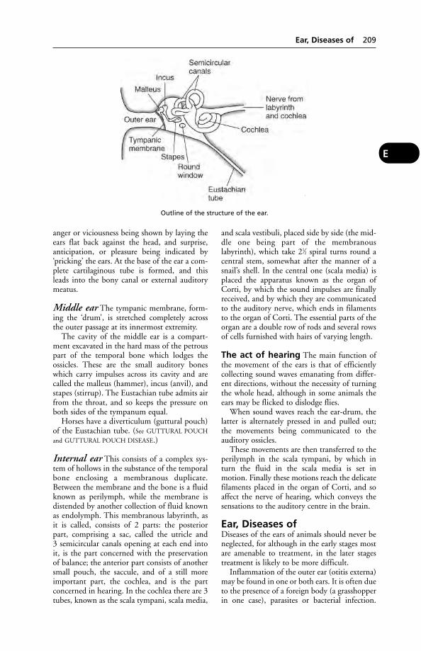
anger or viciousness being shown by laying theears flat back against the head, and surprise,anticipation, or pleasure being indicated by‘pricking’ the ears. At the base of the ear a com-plete cartilaginous tube is formed, and thisleads into the bony canal or external auditorymeatus.
Middle ear The tympanic membrane, form-ing the ‘drum’, is stretched completely acrossthe outer passage at its innermost extremity.
The cavity of the middle ear is a compart-ment excavated in the hard mass of the petrouspart of the temporal bone which lodges theossicles. These are the small auditory boneswhich carry impulses across its cavity and arecalled the malleus (hammer), incus (anvil), andstapes (stirrup). The Eustachian tube admits airfrom the throat, and so keeps the pressure onboth sides of the tympanum equal.
Horses have a diverticulum (guttural pouch)of the Eustachian tube. (See GUTTURAL POUCH
and GUTTURAL POUCH DISEASE.)
Internal ear This consists of a complex sys-tem of hollows in the substance of the temporalbone enclosing a membranous duplicate.Between the membrane and the bone is a fluidknown as perilymph, while the membrane isdistended by another collection of fluid knownas endolymph. This membranous labyrinth, asit is called, consists of 2 parts: the posteriorpart, comprising a sac, called the utricle and 3 semicircular canals opening at each end intoit, is the part concerned with the preservation of balance; the anterior part consists of anothersmall pouch, the saccule, and of a still moreimportant part, the cochlea, and is the part concerned in hearing. In the cochlea there are 3tubes, known as the scala tympani, scala media,
and scala vestibuli, placed side by side (the mid-dle one being part of the membranouslabyrinth), which take 21⁄2 spiral turns round acentral stem, somewhat after the manner of asnail’s shell. In the central one (scala media) isplaced the apparatus known as the organ ofCorti, by which the sound impulses are finallyreceived, and by which they are communicatedto the auditory nerve, which ends in filamentsto the organ of Corti. The essential parts of theorgan are a double row of rods and several rowsof cells furnished with hairs of varying length.
The act of hearing The main function ofthe movement of the ears is that of efficientlycollecting sound waves emanating from differ-ent directions, without the necessity of turningthe whole head, although in some animals theears may be flicked to dislodge flies.
When sound waves reach the ear-drum, thelatter is alternately pressed in and pulled out;the movements being communicated to theauditory ossicles.
These movements are then transferred to theperilymph in the scala tympani, by which inturn the fluid in the scala media is set inmotion. Finally these motions reach the delicatefilaments placed in the organ of Corti, and soaffect the nerve of hearing, which conveys thesensations to the auditory centre in the brain.
Ear, Diseases ofDiseases of the ears of animals should never beneglected, for although in the early stages mostare amenable to treatment, in the later stagestreatment is likely to be more difficult.
Inflammation of the outer ear (otitis externa)may be found in one or both ears. It is often dueto the presence of a foreign body (a grasshopperin one case), parasites or bacterial infection.
Ear, Diseases of 209
E
Outline of the structure of the ear.

The signs include shaking the head, often per-sistently for a few moments at a time, scratch-ing, rubbing head rotation with the affected earlowered, and a foul-smelling discharge.
Scratching the ears is also a symptom of ear-mange mite (Otodectes) infection of the externalear canal. (See MITES – Mange in dogs and cats,for first-aid and treatment of Otodectes.)
Other forms of mange may start at the earsand involve the pinna, e.g. psoroptic mange,notoedric mange.
Discharge from the ear, or the presence ofpus within, is a sequel to a neglected case ofparasitic otitis in the dog and cat and due tosecondary infection by bacteria and/or moulds.
Excessive wax in the ear often leads to disease later. It is especially common in dogswhich have large pendulous ear flaps, whenventilation is poor.
In some cases, dressing the inner parts of theear is difficult or impossible because of thethickening and perhaps distortion. For these anoperation, in which the cartilages at the lowerparts are opened or resected, has been devised.Operation may also be needed where deep-seat-ed ulceration of one or other of the aural carti-lages has occurred, and even the mere initialcleaning of a very inflamed and painful earmust be done under an anaesthetic.
Foreign bodies, such as hay seeds, sand,pieces of glass, wood, peas, or parasites, maybecome lodged in the ears of animals and giverise to irritation occurring very suddenly.
Haematoma is common in dogs and in catswhich are affected with ear mange, but it mayoccur in almost any animal. A large fluctuatingswelling appears upon the flap of the ear andcauses the animal to hang its head towards thesame side. In many cases little or no pain isexperienced once the swelling has appeared,and, in fact, a small swelling becomes larger inmany cases through the continued shaking ofthe head even after its original formation. Theswelling is caused by bruising of the skin and theblood vessels which lie between it and the carti-lage, with a consequent extravasation of bloodor serum under the skin. The condition is treat-ed by opening the haematoma under conditionsof surgical cleanliness, evacuating the fluid contents, and suturing the skin in such a waythat the collection of more fluid is prevented.
Wounds of the flaps of the ears are usuallycaused by bites, or from barbed wire, etc., in thelarger animals. The comparatively poor blood
supply to the AURAL CARTILAGES means that, if torn or lacerated, necrosis may occur. In dogsit may be necessary to secure the ear-flaps bymeans of surgical adhesive tape, or a head-cap improvised to give several ‘tails’ which canbe tied.
Deafness (see under this heading)
Middle-ear infection (otitis media) isalways serious as it may lead to MENINGITIS. Itis often found on one side but both ears may beaffected. Signs are similar to those of externalear inflammation but the balance can be affect-ed; there is swaying of the hind quarters and the head is carried abnormally. It may be causedby spread of external ear infection or by aninfection carried in the blood.
Inner-ear infection (otitis interna): signsare – the head is often rotated; the animal maywalk in circles and show a stumbling, swayinggait.
Tumours are occasionally found. Warts arenot uncommon in horses and cattle. In cats apolyp is occasionally found, and in white cats asquamous-celled carcinoma may affect the tipof the pinna.
Mange Psoroptic and notoedric mange oftenbegin on the pinna of the ear; auricular orotodectic mange involves the presence of mites(Otodectes) within the ear canal. (See MITES.)
Fly strike A dog brought to a veterinary surgeon in Cornwall was found to have a badlyinfected left ear, from which came a profusepurulent discharge. On auroscopic examina-tion, Mr D. S. Penny BVetMed was surprised tosee three faces staring back at him. Underanaesthesia 18 large maggots were removed.
Ear TagsA permanent method of identifying animals. Itis the main method of legal identification ofcattle. Tags are also used in sheep, goats andpigs as well as other animals. They can incorpo-rate an insecticide (see FLIES).
Ear TippingEar tipping of feral cats has been advocated byanimal welfare organisations and practised inAmerica and Denmark, for example. The idea is to identify those cats which have been spayed,and prevent any ‘rescued’ cat from being subjected to unnecessary anaesthesia andlaparotomy.
210 Ear Tags
E
In Australia, ear tattooing is practised for the same purpose, but has the disadvantage thatthe spayed feral cat cannot be identified from adistance.
Early Weaning(see under WEANING)
Ears as FoodEars from beef cattle which had been receivingsex hormones as implants in the ear have beenfed in breeding kennels with disastrous results.
EarthingEarthing of electrical apparatus on farms, andespecially in the dairy, is occasionally faultily car-ried out in such a way that in the event of ashort-circuit, the water-pipes supplying the cows’drinking-bowls become ‘live’ – leading to theelectrocution of the cows. (See ELECTRIC SHOCK.)
EarthwormsEarthworms are of veterinary interest in thatthey act as intermediate hosts to stages in thelife-history of the gape-worm of poultry (see
GAPES) and of lung-worm in pigs. They mayalso harbour viruses which cause disease in pigs.Earthworms can live for as long as 10 years.They can often be found at night in drains out-side piggeries, and in crevices and cracks in thecement inside piggeries. (See also INFLUENZA.)An ARC research team at the RothamstedExperimental Station found that earthworms,bred in animal manure, can provide a high-quality protein supplement for pigs, poultry,and especially fish. Several species of earthwormwere used, of which Lumbricus rubellus was theone of choice. Eisenia foetida (the brandlingworm) is easier to farm but contains a heat-labile toxin in its skin making it unsuitable as araw food. Ideally, earthworms should be heat-processed into meal for use in animal diets.
East Coast Fever (Theileriosis)An acute specific disease of cattle enzootic incertain parts of Africa, especially in the easternprovinces of South Africa, in Kenya and inZimbabwe. In these areas the native cattleattain a certain amount of natural immunity,and only imported animals are affected.Animals which recover are commonly known as‘salted’, but the mortality is very high (e.g. 90per cent) in new outbreaks of the disease.Buffaloes are also susceptible.
Cause Theileria parva, which spends part of its life-history in cattle and part in ticks(Rhipicephalus appendiculatus).
Signs After an incubation period of a fortnightor so, the animal becomes dull, listless, losesappetite, and runs a high fever. Lymph nodesbecome enlarged. There may be a dischargefrom eyes and nose; laboured breathing anddiarrhoea may be seen.
Prevention and treatment East Coastfever may be to a great extent prevented by systematic dipping of all newly purchased cattle, and quarantining them for at least 5 weeks before they are mixed with the rest ofthe stock.
Where the disease has broken out on a farm,the ‘short-interval’ dipping system first devisedby Watkins-Pitchford has proved of immensebenefit in eradicating it. (See under CONTROL OF
TICKS.)Since ticks responsible for the spread of East
Coast fever can live for some time on otherdomesticated animals, it is advisable to dipsheep, goats, and horses at suitable intervals.
Clinically ill cattle may be treated by anyantibiotics that can penetrate cell membranes,such as tetracyclines.
East Friesland Milk SheepThis breed comes from NW Germany, and in England has been used to produce the COL-
BRED. East Friesland ewes average 545 litres(120 gallons) at 6 per cent butterfat in a lacta-tion, rearing their lambs, and a yield of 1000litres (220 gallons) is not unknown. The lambshave a high growth rate and early maturity.
‘Eastrip Special Blend’A cross between Bluefaced Leicester and PollDorset sheep. A high lambing percentage isclaimed.
Ebola VirusThis, together with the Marburg virus, is amember of the Filoviridae.
It is, in appearance, indistinguishable fromthe Marburg virus, but antigenically distinct.Infection of humans is very often fatal; caseshave occurred in laboratory workers handlinginfected monkeys. It was found in 1976 inZaire and Sudan; 500 people became ill and350 died. (See under MONKEYS, DISEASES OF.)
An outbreak of disease caused by an Ebola-related filovirus, and by simian haemorrhagicfever, occurred in 1990 at an American quaran-tine station among cynomolgus monkeysimported from the Philippines. It was reportedthat this was the first case in which a filovirushad been isolated from non-human primateswithout deliberate infection.
Ebola Virus 211
E

EC(see EUROPEAN UNION)
ECGElectrocardiogram.
EcbolicsEcbolics are drugs which cause contraction ofthe muscle fibres of the uterus, such as ergot,pituitrin, etc. They are used to induce labour.
EcdysisEcdysis is the shedding of an exoskeleton inarthropods, and of the old skin in crustacea andreptiles. Failure to shed completely can lead toproblems.
Echinococcosis(see HYDATID DISEASE; TAPEWORMS)
Echium PlantagineumA poisonous plant, also known as Paterson’sCurse, or Salvation Jane, which has caused thedeath of many sheep from copper poisoning inSouth Australia. In one outbreak, 1259 sheepdied out of a total of 29,715 at risk. On onefarm, 500 of 3000 ewes died. Merino × BorderLeicester crosses appear to be especially suscep-tible. At autopsy, jaundice is evident; livers arefriable and enlarged or, less frequently, shrunk-en and fibrotic. Kidneys are swollen, soft andblackish.
The plant contains up to 10 alkaloids, and isthe first to show growth after a prolongeddrought.
EclampsiaEclampsia is a disease occurring during the laterstages of pregnancy or after parturition, andcharacterised by loss of consciousness or con-vulsions, or both. It occurs in the bitch and cat. A preferable name is lactation tetany. It isassociated with HYPOCALCAEMIA. (See also MILK
FEVER; FITS.)
EcraseurA surgical instrument used for castration of thelarger domestic animals. Haemorrhage is large-ly prevented by crushing of the blood vessels ofthe spermatic cord.
EcthymaEcthyma is a localised inflammation of the skin characterised by the formation of pustules.(See ACNE; IMPETIGO.)
Ecto-Ecto- is a prefix meaning on the outside.
EctoparasitesEctoparasites live on the skin or the hair. (See
FLEAS; FLIES; LICE; MANGE; MITES; TICKS.)
EctopicEctopic means out of the usual place. Anectopic pregnancy is one in which a fetus is pre-sent outside the uterus. (See PREGNANCY,ECTOPIC.)
Ectopia cordis thoracoabdominalis Avery rare congenital abnormality characterisedby protrusion of the heart to the outside of thebody through a ventral body-wall fissure.
EctromeliaEctromelia means literally absence of a limb orlimbs. The word is also used to describe a con-tagious disease caused by a pox virus, whichaffects laboratory mice, and in the sub-acuteform causes necrosis of a whole limb, toe, tail orear. Outbreaks are usually very severe at theoutset, killing many of the affected mice, butlater on the mortality becomes less, and theoutbreak gradually fades and disappears;though a latent infection may persist.
EctropionEctropion is a condition of the eyelids, in whichthe skin is so contracted as to turn the mucousmembrane lining of the lid to the outside.
EczemaAn inflammation of the skin (dermatitis),occurring in both farm and domestic animals.Intense irritation or itchiness may accompanythe acute form, and frantic licking of the affect-ed area may exacerbate the condition. In chron-ic eczema there may be very little irritation.
Cats Eczema is often referred to as feline miliarydermatitis. Symptoms include reddening of theskin, with the appearance of papules (small blister-like spots) and, later, scabs. These may be easier to feel than to see. The area of skininvolved may be small or large. Neck, shoulders,and back are common eczema sites. Occasionallya bacterial infection is a complication.
The most common cause is considered to be hypersensitivity to flea bites. Once a cat (seldom a young one) has become sensitised toflea saliva, the presence of only a single flea onthe cat’s body is sufficient to cause the allergicreaction.
Other allergies may produce eczema; forexample, a ‘hay-fever’ type (see ATOPIC DISEASE),or a food allergy of some kind. Cat foods containing colouring agents or preservatives are
212 EC
E
sometimes involved. Skin contact with somechemicals should also be considered. It is likelythat among some breeds or strains there is afamily predisposition to eczema.
Treatment involves flea removal; the veterinar-ian may prescribe a change of diet, a vitamin sup-plement, megestrol acetate, an antihistamine, etc.
Dogs The causes, symptoms and treatment ofeczema are similar to those described above. Thedisease is more common in dogs, however, andan acute form often involves the skin betweenthe toes, resulting in constant licking. Othersites are around the eyes, and the scrotum.
First-Aid Calamine lotion may be applied ifprecautions can be taken to prevent its beingimmediately licked off.
In a few cases what a dog-owner assumes tobe eczema may prove to be mange; a profes-sional diagnosis should always be obtained.
Horses A common cause of eczema is sensiti-sation to midge-bites. (See ‘SWEET ITCH’ for preventive measures.)
Cattle and sheep Some cases of eczemaaffecting white-haired areas of skin are theresult of LIGHT SENSITISATION. Overseas thiscondition is often referred to as ‘facial eczema’and follows sensitisation to sunlight followingthe eating of certain plants.
EdemaEdema is another spelling of oedema.
EdtaEthylenediamine tetra-acetic acid. Its salts (ede-tates) are chelating agents which are used totreat poisoning by heavy metals. For example,calcium edetate is used in cases of lead poison-ing. Non-clinically, sodium edetate is used as ananticoagulant for blood samples.
EfferentEfferent is the term applied to vessels whichconvey away blood or a secretion from a part, orof nerves which carry nerve impulses outwardsfrom the nerve-centres.
EffluentLiquid waste from an abattoir or slurry (see
SLURRY; DAIRY HERD MANAGEMENT).
Egg-BoundEgg-bound is the condition in laying poultry inwhich an egg (or eggs) may be formed in the
oviduct, but the hen is unable to discharge it.The bird shows obvious discomfort, standsstraining and pressing. A dose of liquid paraffin(2 ml) may be tried.
Egg EatingAmong intensively housed poultry, this may bea vice or sign either of boredom or of pain.
Egg YieldIn Britain, the average is approximately 130eggs per bird per year. An annual yield of 200 isobtained in well-managed batteries; about 190on deep litter; 170 in fold units. A Honeggerhas laid 305 in 350 days.
Ehlers-Danlos Syndrome(see CUTANEOUS ASTHENIA)
Ehrlichia CanisEhrlichia canis, ‘or a closely related species’, has been identified in human patients who had recently been bitten by ticks. They weresuffering from fever, rigors, myalgia, and gas-troenteritis. Tests showed leukopenia andthrombocytopenia.
EhrlichiosisInfection with species of Ehrlichia, a rickettsia.E. risticii is one cause of abortion in mares. E. chaffeensis is a cause of human ehrlichiosis.
Canine ehrlichiosis has as its vector the browndog tick. (See TICK-BORNE FEVER OF CATTLE.)
EicosanoidsArachidonic acid, a polyunsaturated fatty acid present in most body cells of domestic ani-mals, can be oxidised to the prostaglandins,prostacyclin, thromboxanes and leukotrienes.These compounds, collectively known as theeicosanoids, are involved in inflammatory andallergic conditions; in reproductive and perina-tal processes; with platelet aggregation and vascular homeostasis, kidney function, fever,and certain tumours; and with other normaland disease conditions.
Eimeria(see COCCIDIOSIS)
ElaphastrongylusA genus of nematode found in deer. E. cervilocates in the central nervous system; E. panti-cole in the brain; and E. rangiferi in the musclesand central nervous system.
Elastic Bands(see RUBBER BANDS)
Elastic Bands 213
E
ElastratorAn instrument used to stretch a strong rubberring so that it may be placed over the neck ofthe scrotum for the purpose of castration.
ElbowElbow is the joint formed between the lowerend of the humerus and the upper ends of theradius and ulna.
Electric Fences(see under PASTURE MANAGEMENT)
Electric Shock, ‘Stray Voltage’and ElectrocutionFaulty electrical wiring and earthing have led todrinking-bowls, water pipes, mangers, etc.becoming live. In some instances this has led tothe death of cows from electrocution followinga short circuit.
‘Stray voltage’ In one incident this led tocows refusing concentrates in the parlour – notbecause they were unpalatable, as at firstthought, but because cows wanting to eat weredeterred by a mild electric shock.
This ‘stray voltage’ has been associated withintermittent or unexplained periods of poorperformance, increased milking time, and ‘anincreased prevalence of mastitis’. ‘Stray voltage’was detected in 32 out of 59 dairy farms inMichigan, following investigations requested bydairymen or veterinarians.
Electrocution Deaths from electrocutionmay occur outside buildings. In one case 30cows and heifers were found dead beneath anelectric pylon. It seems that the cattle had useda metal stay as a rubbing ‘post’, which hadbecome loose and then come in contact with the high-voltage lines that the pylon wascarrying.
Pigs Metal troughs becoming electrically liveled to 20 pigs becoming paralysed after a severethunderstorm in England. Injuries apparentlyresulted from panic and crushing. In anothercase 22 out of 32 pigs in one pen were foundpiled up around the trough, close to which wasa burnt-out live wire. The carcases were bloatedand the skin bluish. Additional post-mortemfindings may include external burns, numeroushaemorrhages affecting many internal organs,black unclotted blood, congestion/oedema ofthe nervous system, and fracture of lumbar ver-tebrae or of the pelvis. In pigs, rupture of theurinary bladder may occur.
Horses A New York insurance agency has stat-ed that 0.96 per cent of its claims in respect ofthe death of horses were for lightning strike,and 0.27 per cent for electrocution.
In Canada a veterinarian was asked to call tosee a horse which appeared to be suffering fromcolic. On arrival at the farm he was told that theanimal had died minutes after he had been telephoned. Earlier the same day, the ownerexplained, a mare in foal had died instantly inthe same spot in front of a small barn; andanother had died there too. Suspecting electro-cution, she had switched off the barn’s powersupply.
Subsequently an inspector found that thebuilder of the barn had made a serious mistakewhen carrying out the electrical work, so thatwhat was supposed to be the earth line was any-thing but safe. The situation had become moredangerous after recent excavation in front of the barn, where the earthing plate had beenaccidentally dug up and replaced horizontallyacross the path to the barn. The horses had diedon the first wet day after the work was com-pleted; but their owner recalled that previous tothat they had shied or tended to bolt whenpassing the spot.
Dogs and cats Electrocution is not uncom-mon, and almost invariably results from pup-pies or kittens chewing through the insulationof electric wiring (e.g. of vacuum-cleaner, table-lamp, etc.). Burns to the mouth and lips areseen; a tan to grey discoloration is noticeable.Oedema of the lungs may be caused, with dys-pnoea. In a survey of 26 dogs treated for elec-trocution, 16 survived and were dischargedfrom hospital within 2 or 3 days. Mortality ratefor all the dogs in the survey was 38 per cent.(See EUTHANASIA.)
Lightning strike Cattle, sheep, and horsesare most often affected. Usually death occursinstantly, and the animal is often found with a bunch of grass between its teeth. Usually, but not invariably, there are external scorchmarks, with subcutaneous lesions beneath. Theother signs are as those given above under‘Electrocution’.
Static electricity can build up on equip-ment made from materials such as plastics. Inone incident, turkeys refused to eat because of a build-up of static electricity on the chain feeders which gave the birds a shock when they attempted to feed. The situation wasdetected when it was noticed that wood shav-ings were attracted to the feeders where the
214 Elastrator
E
chains were operated. Earthing the feedersremoved the problem.
Electrocardiogram (ECG)Electrocardiogram (ECG) is a record of thevariations in electric potential which occur inthe heart as it contracts and relaxes. This recordis obtained by placing electrodes on either side of the chest wall or on the two forelegs, the skin being first wetted with salt solution.These are then connected to an electrocardio-graph, which records the pattern of the heart’sactivity. The normal electrocardiogram of eachheartbeat shows 1 wave corresponding to theactivity of the auricle, and 4 waves correspond-ing to the phases of each ventricular beat.Various readily recognisable changes are seen in cases in which the heart is acting in an abnor-mal manner, or in which one or other side ofthe heart is hypertrophied. This record, there-fore, forms a useful aid in many cases of cardiacdisease.
Electrocardiography has been described as a useful aid to pregnancy diagnosis in themare – ‘where thoroughbred mares more than 5 months pregnant are presented for sale’ (see
under TWINS); and also for monitoring heart rateduring anaesthesia.
ElectrocauteryElectrocautery is useful for operations wherespace is restricted, such as removing smalltumours, etc. in mouth, nose, or throat, and to check haemorrhage in the deeper parts ofwounds. Also sometimes for disbudding. (See
also CRYOSURGERY.)
Electrocution(see under ELECTRIC SHOCK)
ElectrolyteAny compound which, in solution, conducts anelectric current and is decomposed by it. (See
under FLUID REPLACEMENT THERAPY; NORMAL
SALINE; DEHYDRATION.)
Electrolyte SolutionsElectrolyte solutions contain sodium, potassi-um and other electrolytes in an ISOTONIC for-mulation. They are used, often with plasmasubstitutes or other additives, in restoring thebody fluid volume in cases of shock, diarrhoea,injury and other conditions.
Electron MicroscopeThese instruments have made it possible tostudy and photograph viruses, bacter-iophages,
and the structure of bacteria. Instead of light,the electron microscope uses a beam of electronsto scan the specimen. This is prepared as anextremely thin film and subjected to a highdegree of vacuum. The electron image is focusedon a video screen and may be recorded photo-graphically or electronically. Magnification may be up to ×300,000, and by means of photographic enlargement and the use of pro-jection slides a total magnification approaching×1,000,000 can be achieved.
ElectrophoresisThe movement of particles in a fluid under theinfluence of an elecric current. It is used, e.g., inthe analysis of blood or serum constituentswhich form visibly identifiable patterns in astarch gel to which current is applied.
Electrotherapy
High-frequency currents are mainly usedto produce muscular contractions as an aid tomuscular re-education following injury or during transient paralysis. It can also be usedfor passive exercise when an animal is sufferingfrom certain neurological conditions. The tech-nique is painless and no control measures are necessary. Animals must not, however, beexcited during periods of therapy, which varybetween 5 and 20 minutes per day. Somechronic skin conditions may be controlled bythis method. Where movement of painful jointsor tendons is required, or where there is neuri-tis, administration of painkillers or use of nerveblocks by local anaesthetic may be considered.Diathermy is a modified form of high-frequency current therapy in which warmth isinduced deep in the tissues.Repetitive stimulation is produced by cardiacpacemakers; these have been used in small animals and in horses.Faradism is the use of electric currents to treatcertain muscle, tendon and joint conditions,mainly in horses. It has also been used in workingelephants.
(See also X-RAYS; IONIC MEDICATION; ION-
TOPHORESIS; CANCER.)
ElectuaryElectuary is a soft paste made by compoundingdrugs with treacle, sugar, or honey. It is used asa convenient method of applying medication tothe throat and pharynx of animals. To relievesore throat in the horse, an electuary of extractof belladonna, potassium chlorate, and aniseed,made up into a paste with treacle, was formerlymuch used. The electuary is applied by means
Electuary 215
E
of a flat stick, and is smeared upon the back ofthe tongue and upon the teeth.
Elephants(see MUSTH; SPEED OF ANIMALS). The height tothe shoulder of the Asian elephant (Elephasmaximus) is about 3 metres (10 feet); that of theAfrican elephant (Loxodonta africana) 3.5metres (131⁄2 feet).
Elephants, Diseases ofThese include anthrax (sometimes brought onby the breaking of a tusk where the disease haslain dormant in the dental pulp), multipleabscesses, blackleg, botulism, elephant pox,enzootic pneumonia, foot-and-mouth disease,influenza, myiasis, parasitic gastroenteritis, pas-teurellosis, rabies, salmonellosis, steanofilarialdermatitis, schistosomiasis, surra, tetanus, try-panosomiasis, tuberculosis. Elephants importedinto Great Britain may serve out their quaran-tine period for foot-and-mouth disease onboard the ship transporting them.
ELISAELISA is the abbreviation for the system ofenzyme-linked immunosorbent assay, devel-oped by the Swedish scientists Engvall andPerlmann. ELISA tests are widely used in labo-ratories for the rapid detection of pathogens.
Elizabethan CollarOften improvised from cardboard, the shape of a lampshade, and designed to fit over thedog’s head and to be attached to its collar, with the object of preventing the animal from
interfering with wounds, skin lesions or dress-ings. The illustration shows a proprietary version.
ElkA species of large deer also called the wapiti(Cervis elaphus); the European elk is Alces alces;the American moose A. americana. This farmedanimal was found to be a source of tuberculosisin people in Alberta, Canada, in 1990.Mycobacterium bovis was isolated.
ElkhoundA medium-sized dog with thick grey-black coat, pointed ears and a bushy tail curled over the back; it originated in Scandinavia.Inherited traits include progressive retinal atrophy, renal cortical hypoplasia and hip dysplasia.
EmasculatorAn instrument to remove the testicles in horses,cattle and sheep. Most crush the spermatic cordto prevent haemorrhage as well as severing thetesticle from it.
EmbolismThe plugging of a small blood vessel by blood-clot fragments originating from elsewhere inthe body, and carried along in the bloodstream.Bacteria, worm larvae, air-bubbles, and fat areother causes of embolism. The importance ofthe embolism depends upon the situation. Inthe brain it may cause apoplexy; in otherorgans, the area that was supplied by the littlevessel before it became blocked by theembolism ceases to function, and if the bloodsupply is totally cut off it dies, or degenerates,becoming an ‘infarct’. (See also GLASS EMBOLISM;THROMBOSIS; CATHETER EMBOLUS.)
Embrocations(see LINIMENTS)
Embryo(see EMBRYO TRANSFER; EMBRYOLOGY; FETUS)
Embryo TransferThe technique of transferring an embryo fromone animal and implanting it in the uterus of another has become a widely used method,particularly in cattle, of improving breed quali-ty and herd reproductivity. Basically, the tech-nique involves the collection of embryos fromone cow (the donor), 7 to 8 days after insemi-nation, by flushing out the uterus with a specialmedium such as phosphate-buffered saline. The donor cow is prepared by administering
216 Elephants
E
This Elizabethan collar is a transparent version,easy to adjust to the dog’s neck diameter. Anexcellent way of preventing the canine patientfrom interfering with wounds, skin lesions, ordressings.

gonadotrophins to cause superovulation, theproduction of multiple eggs and therefore mul-tiple embryos. The embryos are collected andtransferred surgically, or non-surgically by amethod similar to artificial insemination, to therecipient cow which must be at exactly the samestage of oestrus as the donor. Embryos may bepreserved by deep-freezing and thawed for usewhen required.
A number of firms operate commercialembryo transfer services; success rates of up to70 per cent are achieved.
Embryo transfer is also used in sheep andgoats; it is possible, but little used in practice, inmares and pigs. (See illustration on page 162.)
The advantages of embryo transfer have beensummarised as follows: (1) increased number of offspring from valuable females; (2) rapidprogeny testing of females; (3) induction oftwinning; (4) the investigation of causes ofinfertility; (5) transport of cattle ova from onestate or country to another; and (6) anincreased rate of genetic improvement.
Development The technique had already inthe 1950s been successfully carried out in sheep– ewes having produced young of which theywere not, in the full sense, the mothers; and itwas extended to cattle later, by L. E. Rowsonand colleagues at Cambridge, who were respon-sible for much of the research.
Development of a method of freezing theembryos greatly widened the scope of embryotransfer. Fertilised 10- to-13-day eggs (blasto-cysts) are treated with a protective agent(dimethylsulphoxide) to prevent damage byfreezing and cooled to –196°C; they are thenstored in liquid nitrogen until required forimplantation.
The technique Five days before oestrus is due,the donor animal is treated with pregnant mare’sserum gonadotrophin (PMSG) or a similargonadotrophin to produce superovulation. Whenoestrus occurs, insemination is carried out 2 or 3times, using fresh rather than frozen semen.
Surgical transfer On day 6, when the eggsare at the morula stage, consisting of 8 to 32cells each, and looking under the microscopelike blackberries, they are flushed out of theFallopian tube. This may be done surgically ornonsurgically. In the former, a fine catheter isinserted through a blunt needle after surgicalexposure of the uterus by means of a flank inci-sion, the cow being under local anaesthesia. Itmay be possible to recover 8 to 12 ova, and anattempt is made to select the normal ones. (Forexample, by culturing them for 1 or 2 days afterrecovery. In that time further development willhave occurred; eggs which do not show this arediscarded.) The transfer is made by puncturing
Transplantation. As a 7-day embryo, this calf was stored for a month at a temperature of –196°C before being transferred, non-surgically, to the recipient cow seen in the photograph. (ARC.)
Embryo Transfer 217
E

one horn of the anaesthetised recipient’s uteruswith a small pipette containing one ovum in asynthetic medium. This liquid is forced out ofthe pipette, carrying the ovum with it.
Non-surgical transfer The eggs are collectedby means of a 2- or 3-way catheter having aninflatable cuff. The catheter is passed through thecervix and into one of the horns of the uteruswhich is then sealed by inflating the cuff. Fluid isflushed into the horn and withdrawn throughthe catheter, with the eggs. The technique fortransfer into the recipient cow is similar to artifi-cial insemination. An egg is placed into a ‘straw’and the embryo introduced into the uterine hornby means of an insemination ‘gun’. With skilledoperators, the results by this method are compa-rable with surgical collection and without thetrauma of a surgical operation. This method iscommonly used in the dairy industry.
The first inter-species transfer of embryos wascarried out in 1979 at the ThoroughbredBreeders’ Association’s equine fertility unit atCambridge. This resulted in a pony mare foalinga donkey (gestation period about 346 days orabout 15 days longer than for a foal), and 2 don-keys giving birth to pony foals (born after 346and 361 days’ gestation period, respectively).(See PLACENTA.)
EmbryologyEmbryology is the study of the development ofthe embryo within the body of the female.
EmbryotomyThe section and removal of a fetus in the uterusto facilitate parturition; most commonly under-taken in cattle and sheep.
EmesisEmesis means VOMITING.
EmetineEmetine is one of the alkaloids of ipecacuanha.
EmphysemaAn abnormal presence of air in some part of thebody. The term is applied to the presence of airin the subcutaneous tissues following a woundbut, more commonly, to 2 abnormal conditionsof the lungs: destructive (vesicular) emphysemaand interstitial emphysema.
Destructive (vesicular) emphysema isa condition of the lung characterised by anabnormal enlargement of the air spaces, accom-panied by destructive changes in the alveolarwall. This condition occurs in dogs with chron-ic bronchitis and in horses with chronic obstruc-tive pulmonary disease (see ‘BROKEN WIND’).Emphysema is irreversible and may progress torespiratory failure and death. The main symp-tom is respiratory distress on exertion, with amarked expiratory effort.
Interstitial emphysema Air is present inthe connective tissue of the lung – a state of
218 Embryology
E
Twins from different mothers. One of 2 eggs was removed from the Border Leicester ewe and transplanted into the Welsh ewe.
inflation of the interstitial (interlobular) tissue.The air is found in the lymphatics, under thepleura in the interlobular septa, and aroundblood vessels, sometimes in the form of largebullae 10 cm or more in diameter. Air may trackas far as the hilum of the lung and gain access to the mediastinum from where, in exceptionalcircumstances, it may even spread to subcuta-neous connective tissue – usually in the shoulderregion or over the upper part of the chest.
Interstitial emphysema is a common condi-tion in cattle, especially in association with par-asitic bronchitis (HUSK) or with ‘FOG FEVER’.Increased effort, in response to obstructed airways, over-exertion and violent struggling,causes a marked increase in pressure within thealveoli. Rupture then occurs, allowing air toescape into the interstitial tissue on inspiration,but impeding its leaving on expiration. Whenthe lung lobules become surrounded by inter-stitial emphysema their ability to inflate duringinspiration is restricted, and this may lead torespiratory distress.
EmpyemaA collection of purulent fluid within a cavity.(See PYOTHORAX; PLEURISY.)
EmuA large flightless bird, Dromaius novae-hollandiae, native to Australia. It is farmed inBritain but to a lesser extent than the ostrich.Severe enteritis resulting in death of up to 65per cent of a flock has occurred in the USAfrom infection by eastern equine encephalitisvirus. This is a NOTIFIABLE DISEASE throughoutthe EU.
EnamelThe very hard substance found on the externalsurface of the crowns of teeth (see TEETH).
Enarthrodial JointsEnarthrodial joints are those of the ball-and-socket type which allow movement in nearlyany direction. Examples include the shoulderjoint between the scapula and the humerus; andthe hip joint in which the nearly spherical headof the femur fits into the cup-shaped cavitycalled the acetabulum on the pelvis.
EncephalitisEncephalitis is inflammation of the brain. Itmay be brought about through the activity ofbacteria, such as those of strangles and listerio-sis, but especially during infection with viruses,such as those of rabies, canine distemper, etc.(See BOVINE SPONGIFORM ENCEPHALITIS.)
Signs Symptoms of encephalitis include fever,excitement, delirium, convulsions, paralysis,and loss of consciousness. Several symptoms are common to MENINGITIS. (See also SLEEPER
SYNDROME.)
First-Aid Keep the animal quiet – in a dark-ened room if showing excitement – and avoidnoise or handling the patient.
Encephalitozoon CuniculiAn intracellular protozoal parasite. It developsin macrophages, brain, kidney and other tissuesof rabbits, dogs, rodents and primates.
In carnivores, severe nephritis, encephalitisand a high mortality are associated withtransplacental infection.
In a Norwegian outbreak, 1500 blue fox cubsdied (33 per cent of the litters), although theparents showed no signs of infection.
In the UK, foxhound puppies have died, andin Tanzania 2 spaniel puppies died which hadshown rabies-like signs.
Diagnosis An ELISA test. In one study, 51positive samples were identified out of 248 serafrom stray dogs.
EncephalomalaciaA group name for the degenerative diseases ofthe brain. Causes include the copper deficiencyof swayback, horse-tail and bracken poisoning,metallic poisoning, and mulberry heart diseaseof pigs. Another example of encephalomalaciais ‘crazy chick’ disease.
EncephalomyelitisInflammation of both the brain and the spinalcord.
Encephalomyelitis, Viral, ofPigsThis term covers the group of diseases known asTeschen disease, Talfan disease, and Poliomyelitissuum.
Believed to have originated in the formerCzechoslovakia, viral encephalomyelitis of pigsis now encountered throughout most of Europe.In Britain and Denmark, only a small percent-age of pigs become infected, and illness is farmilder than in some other countries.
Symptoms include fever, stiffness, staggeringgait, paralysis, and those of encephalitis.
Encephalomyocarditis VirusEncephalomyocarditis virus is a picornavirus.Antibodies have been found in the serum ofmore than 28 per cent of normal pigs in the
Encephalomyocarditis Virus 219
E
UK. It is also a pathogen of rodents and humanbeings, and has caused outbreaks of illness inpigs in Australia, the USA, and Panama.
EnchondromaA tumour formed of cartilage. (See TUMOURS.)
EncystedEnclosed in a cyst.
Endangered Species Act 1982Endangered Species Act 1982 lists measures forthe protection of named animals.
EndarteritisInflammation of the inner coat of an artery. (See
ARTERIES, DISEASES OF.)
EndemicAn endemic disease is one present in an animalpopulation at all times.
Endo-Endo- is a prefix meaning situated inside.
EndocarditisInflammation of the smooth membrane thatlines the inside of the heart. It occurs especiallyover the heart valves. (See HEART DISEASES.)
Endocrine GlandsEndocrine glands are those which secrete hor-mones. (See HORMONES; also under the name ofindividual endocrine glands, e.g. ADRENAL,THYROID, PARATHYROID, PITUITARY, THYMUS,PANCREAS.)
EndometritisInflammation of the mucosal lining of the uterus(endometrium) (see UTERUS, DISEASES OF).
EndorphinsMorphine-like, natural analgesics produced inthe body. Acupuncture is said to stimulate theirrelease into the bloodstream. (See also TWITCH.)
EndoscopeAn instrument used for viewing the interior ofan organ, and for facilitating the extraction of aforeign body, e.g. from the oesophagus; and forassistance with other surgery, including embryotransfer. (See also LAPAROSCOPY.)
EndotheliumEndothelium is the membrane lining variousvessels and cavities of the body, such as thepleura, pericardium, peritoneum, lymphaticvessels, blood vessels, and joints. It consists of a
fibrous layer covered with thin flat cells, whichrender the surface perfectly smooth and secretethe fluid for its lubrication.
EndotoxinsEndotoxins are those toxins which are retainedwithin the bodies of bacteria until the latter dieand disintegrate.
Endotracheal Anaesthesia(see ANAESTHESIA)
Endotracheal TubeA tube introduced into the trachea to preventits collapse; used in endotracheal anaesthesia.
EndrinA highly toxic insecticide of the chlorinatedhydrocarbon group. It has caused fatal poisoningin cattle, dogs, fish, and birds.
EnemaThe introduction of fluid into the rectum toassist evacuation of faeces.
Energy(see CALORIE; CARBOHYDRATES; METABOLISABLE
ENERGY; JOULES)
English Springer SpanielLong-eared, medium-sized dog with silky coat,brown and white or black and white; originallybred as a gun dog. Retinal dysplasia, entropion,cutaneous asthenia and haemophilia may beinherited conditions.
EnrofloxacinEnrofloxacin is a quinolone antibacterial activeagainst a wide range of Gram-positive andGram-negative organisms. As it may affect thedevelopment of load-bearing articular cartilage,it should not be administered to growing animals.
Ensilage(see SILAGE)
Enteque SecoA wasting disease of cattle, sheep and horses. Itoccurs mainly in Argentina, but also inUruguay and possibly Brazil. It may be identi-cal with Manchester wasting disease (Jamaica)and Naalehu disease (Hawaii).
Cause A plant, common on wet land, knownas duraznillo blanco (Solanum melacoxylon orglaucum). Poisoning may arise from deliberateeating of the leaves or from the accidental
220 Enchondroma
E
consumption of dead, fallen leaves during grazing of the underlying pasture plants. It isparticularly dangerous when growing in associ-ation with white clover.
It produces an arteriosclerosis, with calcifica-tion in heart, aorta, lungs, etc. Blood levels ofcalcium and phosphorus tend to be high as S. malacoxylon contains a potent metabolite ofvitamin D.
Signs Emaciation occurring over weeks ormonths, and an abnormal gait.
EnteralgiaEnteralgia is another name for colic.
EnteritisInflammation of the intestines (see DIARRHOEA
and INTESTINES, DISEASES OF).
Enterocele(see HERNIA)
EnterolithsEnteroliths are stones that develop in theintestines, being formed by deposition of salts round a hard metallic or other nucleus. (See
CALCULI.)
EnterostomyEnterostomy means an operation by which anartificial opening is formed into the intestine.
EnterotoxaemiaAn acute disease of calves, lambs, goats, andoccasionally of piglets and foals.
Cause Toxins emanating from the intestinesand present in the bloodstream. The toxinsinvolved are from 4 strains of Clostridiumwelchii and from some strains of E. coli.
Signs Severe enteritis, with dysentery in somecases, and sudden death in others.
Prevention A vaccine is available.
Calves seldom survive for more than a fewhours.
Goats show a sudden drop in milk yield,dysentery, and death within 36 hours. There isalso a subacute type of the disease lasting 7 to10 days, and followed by recovery.
Sheep The disease affects both unweanedlambs and sheep 1 to 2 years old.
EnterovirusesA group of smaller viruses pathogenic to ani-mals and causing disease in cattle, pigs andducks (duck hepatitis).
EntropionThe turning in, or inversion, of an eyelid. It can be congenital or acquired and is inheritedin some breeds of lambs and dogs. It is verycommon in ‘mini-pigs’ (see EYE, DISEASES OF).
Enuresis(see INCONTINENCE)
Environment(see HOUSING OF ANIMALS; PASTURE MANAGE-
MENT; EXPOSURE; RAINFALL; ALTITUDE; HEAT-
STROKE; ANHIDROSIS; TROPICAL DISEASES;VENTILATION; CALF HOUSING)
EnzooticEnzootic refers to a disease present (endemic)among animals in a particular region, country,or locality. For example, braxy and louping-illare enzootic in the south and west of Scotlandand the north of England. Compare EPIZOOTIC
(epidemic), in which a disease spreads rapidlythrough large numbers of animals over a widearea.
For enzootic abortion of sheep, see under
ABORTION, ENZOOTIC.
Enzootic Bovine Leukosis(see BOVINE LEUKOSIS)
Enzootic HaematuriaA disease typically found in old suckler cows fol-lowing the long-term ingestion of bracken. Itresults in various cancer problems of the bladder.
Enzootic Muscular DystrophyA disease in calves, lambs and foals caused byvitamin E and selenium deficiency. Acute casesmay drop dead after exercise; mild cases areweak and breathe with difficulty.
Enzootic Ovine AbortionEnzootic ovine abortion is caused by Chlamydiapsittaci. (See under ABORTION, ENZOOTIC.)
Enzootic Nasal GranulomaObstruction of the nasal cavities of cattle by aneosinophilic granuloma. More common inChannel Island breeds.
Enzootic Pneumonia of CalvesAcute pneumonia usually seen in calvesbetween 2 and 4 months old; it is caused
Enzootic Pneumonia of Calves 221
E

by environmental and management changes as well as viral, mycoplasmal and bacterialinfections.
Enzootic Pneumonia of PigsThis was formerly described as virus pneumoniaof pigs (VPP), but the cause is now generallyregarded as being Mycoplasma hyopneumoniae.However, other organisms may be involved to avarying degree. (See RESPIRATORY INFECTIONS
OF THE PIG; SYNERGISM.)Many pigs reaching the bacon factories are
affected with some degree of pneumonia, sothat the matter is of the very greatest economicimportance.
Signs When the disease is first introduced intoa herd, pigs of all ages (from 10 days upwards) godown with it, and many die. Where the diseaseis already present, deaths are few. Symptoms,which may easily be overlooked or ignored, thenconsist merely of a cough. There is, in addition,a certain degree of unthriftiness which inextreme cases may amount to stunted growth. Inall cases one may expect the liveweight gain to bereduced. Sometimes pigs which contract the dis-ease earlier in life quite suddenly develop acutepneumonia at 19 to 26 weeks of age, known as‘secondary breakdown’. Affected animals losetheir appetite and often become prostrate,breathing rapidly with a temperature over40.5°C (105°F). A number die if left untreated,but the majority have a fluctuating fever for afew days and then recover.
Prevention Vaccination at 1 to 10 weeks ofage and management measures such as avoidingbuying-in infected stock. Litters are best kept inarks on pasture, and any sows showing a cougheliminated. Weaned pigs should not be broughtinto a fattening house where pigs with pneu-monia are present. (See DUSTY ATMOSPHERE;SWINE INFLUENZA.)
Diagnosis Confirmed by a complement fixation test.
Treatment Macrolide antibiotics, adminis-tered on a herd or individual basis, help to control the severity of outbreaks.
Enzootic Pneumonia of Sheep(see PASTEURELLOSIS; PNEUMONIA OF SHEEP)
EnzymesEnzymes are complex organic chemical com-pounds which facilitate or speed biochemicalprocesses in the animal body, including those of
digestion. Some enzymes are also produced bythe normal bacterial inhabitants of the intesti-nal canal. Each has a specific use in splitting upproteins, carbohydrates, fats, or crude fibre.The best known are the ptyalin of saliva anddiastase of the pancreatic juice, which breakdown starches into soluble sugars; pepsin fromthe gastric juice and trypsin from the pancreas,which break complex proteins into simpleamino acids; and lipase in the intestines, whichattacks fats. (See DIGESTION.) Enzymes are usedin the cleaning of badly infected wounds. (See
STREPTODORNASE.)Some enzymes detoxify poisons, breaking
them down into relatively harmless compounds.The differing susceptibility of cat and dog, forexample, to phenol is due to the former animallacking a particular enzyme which the dog has.(See TAURINE.)
Some enzymes are injurious (see under
THIAMIN).(See also BLOOD ENZYMES, and CREATINE
KINASE for enzymes used in diagnosis.)
EosinophilEosinophil is the name given to white cells inthe bloodstream containing granules whichreadily stain with eosin, a histological dye. The nucleus of this leukocyte is lobular.Eosinophils, basophils, and neutrophils are collectively known as polymorphonuclearleukocytes. As well as these circulating cells,eosinophils are found in the pituitary andpineal glands.
In a normal horse, 2.5 ml3 (1 cu in) of bloodcontains between 5 and 8 million eosinophilwhite cells – compared with about 160 millionother white cells, and 128,000 million red cells.
Eosinophils increase in numbers during certain chronic infections and infestations withparasites. They contain hydrolytic enzymes.‘Unlike neutrophils, eosinophils have lowphagocytic capacity and are not good at killingmicroorganisms.’ (Lancet.) (See BLOOD.)
EosinophiliaEosinophilia means that an abnormally largenumber of eosinophils are present in the blood-stream. This may occur during severe parasiticinfestation in horses and dogs, in certain wastingconditions, and in disease of the lymph system.
Eosinophilic GranulomaA complex in cats. The name covers at least 3different lesions, of a chronic nature.
Eosinophilic ulcers usually occur on theupper lip, or commissure of the lips, gums,
222 Enzootic Pneumonia of Pigs
E
palate, pharynx and tongue. Reddish-brown incolour, they have raised edges. They are notmalignant (compare ‘rodent ulcer’ in manwhich is a basal cell carcinoma).
Eosinophilic plaques may occur anywhereon the body but are most common on theabdomen and inside of the thigh. The plaquesare red, with raised edges, and ulcerate. Theyare extremely itchy.
Linear granulomas are seen mainly on thehind legs and in the mouth, and are yellowish-pink in colour. Itching is not usually present. Aswith the ulcers mentioned above, females seemmore prone to this granuloma than are males.In the mouth, lesions are ‘more nodular’ andhave to be differentiated from bacterial ormycotic infections and also carcinoma.
Eosinophilic Myositis(see under MUSCLES, DISEASES OF)
Eperythrozoon FelisA blood parasite found in cats in Britain, andfirst reported in 1959. (See FELINE INFECTIOUS
ANAEMIA.)
Eperythrozoon ParvumA blood parasite of the pig, which gives rise tofever, anaemia, and sometimes jaundice. It canbe transmitted from pig to pig by lice. It occursin Britain and the USA. Other species of thisparasite affect sheep, and cattle in Africa. In theUK E. wenyoni has been isolated from anaemiccattle. (See also HAEMOBARTONELLA.)
EphedrineEphedrine is an alkaloid derived from theChinese plant Ma Huang, or prepared synthet-ically. It stimulates the heart and central nervous system, and relaxes the bronchioles. Ithas been used for asthma in dogs.
Ephemeral Fever (Three-DaySickness)An acute, infectious, and transient fever accom-panied by muscular pains, and lameness whichhas a tendency to shift from limb to limb. Thedisease was first described in South Africa in1867 and has been seen in Africa, Asia andAustralia. Considerable economic loss has beencaused among beef and dairy cattle in northernand eastern Australia.
Cause A rhabdovirus. The disease is sudden in onset and attacks a large percentage of thecattle in affected districts, taking the form of an
acute epizootic; then, in a few weeks, it diesdown again as quickly as it arose. The disease istransmitted by insects, including Culicoidesmidges. The incubation period is 2–10 days.
Signs The disease is ushered in by a suddenlyoccurring rise of temperature which may reach41.6°C (107°F). This is accompanied by loss of appetite, cessation of rumination, rapidrespirations, a quick and full pulse (which,however, may become very weak later), and a staring coat. The affected subject stands with head down. The attitude of the patient israther characteristic, the 4 legs being placed far under the body and the back arched, sug-gestive of the position of a horse suffering fromlaminitis. There may be a discharge from eyesand nose.
In milking cows, the milk yield is muchdiminished. Many animals prefer to lie downrather than remain on their feet, and oncedown are most reluctant to get up again. Thesymptoms along with the elevated temperaturecontinue like this for about 3 days – hence thename. There is usually a considerable loss ofcondition.
In Australia the mortality is seldom morethan 0.5 per cent.
Prevention Vaccines may be available in someareas.
Epi-Epi- is a prefix meaning situated on, or situatedoutside of.
EpidemicA disease affecting a large number of individu-als at the same time in the same area. The termis strictly applied to man, not animals.
‘Epidemic Tremors’‘Epidemic tremors’ is the colloquial name fora virus disease of poultry characterised by anunsteady gait. (See AVIAN ENCEPHALOMYELITIS.)
EpidemiologyThe study of disease as it affects groups of animals. It can be used in predicting the patternof an outbreak and in making plans to controlthe spread. International reporting services, ascarried out by WHO and OIE, play their part;and the use of computers has greatly assistedthe statistical analysis on which epidemiologyrelies.
EpidermisThe outer layer of the SKIN.
Epidermis 223
E
EpididymisEpididymis is a structure situated within thescrotum and in which the sperms mature afterleaving the testicle. It has 3 parts: the head(capus epidiolysis), the body (corpus) and thetail (cauda).The epididymis has as its outlet thevas deferens. (See TESTIS.)
EpididymitisInflammation of the epididymis. (See also under
RAM.)
Epididymitis and ‘Epivag’(Vaginitis, Contagious)A venereal disease of cattle and sheep in Kenyaand Southern Africa, and an important causethere of infertility and sterility.
Cause Possibly a double infection with a virusand a mycoplasma; possibly a campylobacter. Insheep, Brucella ovis is responsible (not in theUK).
Signs There may be a yellowish discharge fromthe vagina, or merely a redness of the mucousmembrane. In the bull, enlargement of the epididymis occurs over a period of months.
Control Slaughter of infected bulls, and use of AI.
Epidural AnaesthesiaEpidural anaesthesia is a form of spinal anaes-thesia induced by the injection of a local anaes-thetic solution into the epidural space of thespinal canal. The technique is used, for example,in bovine obstetrics – the injection being madebetween the 1st and 2nd coccygeal vertebrae.(See also ANAESTHESIA; ANALGESICS.)
EpigastriumEpigastrium is the region lying in the middle ofthe abdomen, immediately over the stomach.(See ABDOMEN.)
EpiglottisEpiglottis is a leaf-like piece of elastic cartilagecovered with mucous membrane, which standsupright between the back of the tongue and the entrance to the glottis, or larynx. It plays animportant part in the act of swallowing, pre-venting solids and fluids from passing directlyoff the back of the tongue into the larynx.
Epiglottic entrapment in the horse is diag-nosed more and more frequently due probablyto the wider use of endoscopy and greaterexpertise in its use. Affected horses have a history of decreased exercise tolerance and
they make abnormal inspiratory and expiratorynoises.
EpilepsyEpilepsy is a chronic nervous disorder charac-terised by a sudden and complete loss of consciousness, associated with muscular con-vulsions. This is particularly a disease of thedog, although other domesticated animals maybe affected. Epileptic fits in the horse were formerly called megrims.
Cause The cause of primary or idiopathicepilepsy is a genetic one, whereas secondaryepilepsy may be caused by trauma, neoplasia,infections, cardiovascular disease, or metabolicconditions.
The disease can be controlled completely in about one-third of affected dogs, and consid-erably improved in another third.
Secondary epilepsy may be the result of ahead injury, and can occur whenever scar tissueis formed in the brain.
(See also FITS; HYSTERIA; ENCEPHALITIS;POISONING; HEART DISEASE.)
Signs Attacks usually commence without any warning. The limbs are sometimes held out rigidly, and sometimes moved as if the ani-mal were running or galloping. The animalchamps its jaws; the eyes are fixed and staring,or the eyeballs may roll, and the pupils are dilat-ed. There is usually a good deal of salivationfrom the mouth. The rectum and the bladderare usually evacuated involuntarily. The dogregains consciousness in 1 to 2 minutes; in afew cases consciousness may not be completelylost. The 1st fit often occurs between the age of1 and 3.
Treatment Barbiturates, phenytoin, diazepamand primidone are among drugs used for treat-ment. After consciousness returns, the dogshould be placed in a quiet room away fromother dogs or human beings. Treatment shouldbe left to a veterinary surgeon.
Epinephrine(see ADRENALIN)
EpiphoraEpiphora is a condition in which the tears,instead of passing down the tear-duct to theinside of the nose, run over on to the cheek. Itmay be due to a blocking of the tear-duct, gen-erally from inflammation of its lining mem-brane following conjunctivitis, etc. or (in thesmaller animals) by a grass seed. (See EYE.)
224 Epididymis
E
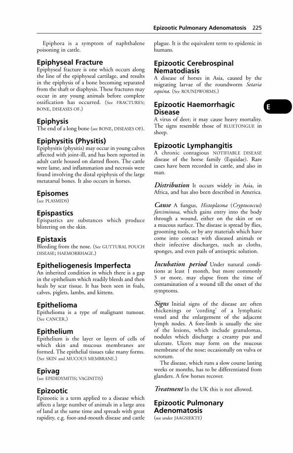
Epiphora is a symptom of naphthalene poisoning in cattle.
Epiphyseal FractureEpiphyseal fracture is one which occurs alongthe line of the epiphyseal cartilage, and resultsin the epiphysis of a bone becoming separatedfrom the shaft or diaphysis. These fractures mayoccur in any young animals before completeossification has occurred. (See FRACTURES;BONE, DISEASES OF.)
EpiphysisThe end of a long bone (see BONE, DISEASES OF).
Epiphysitis (Physitis)Epiphysitis (physitis) may occur in young calvesaffected with joint-ill, and has been reported inadult cattle housed on slatted floors. The cattlewere lame, and inflammation and necrosis werefound involving the distal epiphysis of the largemetatarsal bones. It also occurs in horses.
Episomes(see PLASMIDS)
EpispasticsEpispastics are substances which produce blistering on the skin.
EpistaxisBleeding from the nose. (See GUTTURAL POUCH
DISEASE; HAEMORRHAGE.)
Epitheliogenesis ImperfectaAn inherited condition in which there is a gapin the epithelium which readily bleeds and thenheals by scar tissue. It has been seen in foals,calves, piglets, lambs, and kittens.
EpitheliomaEpithelioma is a type of malignant tumour. (See CANCER.)
EpitheliumEpithelium is the layer or layers of cells ofwhich skin and mucous membranes areformed. The epithelial tissues take many forms.(See SKIN and MUCOUS MEMBRANE.)
Epivag(see EPIDIDYMITIS; VAGINITIS)
EpizooticEpizootic is a term applied to a disease whichaffects a large number of animals in a large areaof land at the same time and spreads with greatrapidity, e.g. foot-and-mouth disease and cattle
plague. It is the equivalent term to epidemic inhumans.
Epizootic CerebrospinalNematodiasisA disease of horses in Asia, caused by themigrating larvae of the roundworm Setariaequina. (See ROUNDWORMS.)
Epizootic HaemorrhagicDiseaseA virus of deer; it may cause heavy mortality.The signs resemble those of BLUETONGUE insheep.
Epizootic LymphangitisA chronic contagious NOTIFIABLE DISEASE
disease of the horse family (Equidae). Rare cases have been recorded in cattle, and also inman.
Distribution It occurs widely in Asia, inAfrica, and has also been described in America.
Cause A fungus, Histoplasma (Cryptococcus)farciminosa, which gains entry into the bodythrough a wound, either on the skin or on a mucous surface. The disease is spread by flies,grooming tools, or by any materials which havecome into contact with diseased animals ortheir infective discharges, such as cloths,sponges, and even pails of antiseptic solution.
Incubation period Under natural condi-tions at least 1 month, but more commonly 3 or more, may elapse from the time of contamination of a wound till the onset of thesymptoms.
Signs Initial signs of the disease are oftenthickenings or ‘cording’ of a lymphatic vessel and the enlargement of the adjacentlymph nodes. A fore-limb is usually the site of the lesions, which include granulomas, nodules which discharge a creamy pus andulcerate. Ulcers may form on the mucous membrane of the nose; occasionally on vulva orscrotum.
The disease, which runs a slow course lastingweeks or months, has to be differentiated fromglanders. A few horses recover.
Treatment In the UK this is not allowed.
Epizootic PulmonaryAdenomatosis(see under JAAGSIEKTE)
Epizootic Pulmonary Adenomatosis 225
E
Epsom Salts(see LAXATIVES). Epsom salts are also useful as a first-aid treatment of lead and carbolic acidpoisoning.
EpulisEpulis is a tumour of the gum (or involving thejaw bones).
Equid(see under EQUINE)
EquinePertaining to the horse.
Equine Back Lesions(see HORSES, BACK TROUBLE IN)
Equine Biliary FeverThis disease is caused by 2 distinct parasites:Babesia caballi and B. equi. The former speciesresembles B. bigemina in size and morphology,and causes a disease similar to Texas fever butwhich is milder and more amenable to treat-ment than that caused by B. equi. This is asmaller species than B. caballi and causes a dis-ease which is highly virulent for adult horsesand other species of the horse family, but ismild in young animals. Recovered animals arein a state of premunition, and inoculation ofcolts as a means of protection later in life iscommonly practised.
Distribution The disease occurs in Russiaand various parts of Europe, India, Africa,South America, and South Africa.
Signs At the beginning of the disease there is asharp rise in temperature to about 41.5°C(107°F). During this period the parasites aremultiplying in the blood. In a few days the temperature falls and anaemia sets in. In thehorse this is usually masked by an intenseicterus, though not in the donkey and mule.Haemoglobinuria, and constipation followedby diarrhoea, are frequent symptoms, and aresucceeded by rapid emaciation. The animalmay die during the initial fever (2 to 5 days) orfrom anaemia and emaciation about the 11thday or later. Complications are frequent.
Treatment Complete rest; an injection of abroad-spectrum antibiotic.
Transmission In Southern Europe, B. caballiis transmitted by Dermacentor reticulatus andD. silvarum; in South Africa B. equi is trans-mitted by Rhipicephalus evertsi and R. bursa.
Other species and genera of ticks probably actas vectors of B. equi in other countries.
Equine Blood TypingSeven main blood groups are recognised in hors-es: A, C, D, K, P, Q and U. A further group, T,is listed by some authorities. There are numer-ous subgroups, or subtypes, some of which areimportant. For example, a mare which is Aa-must not be given blood that is Aa+ as this canlead to fetal haemocytolysis in subsequent preg-nancies. For a first transfusion, blood need notbe typed in an emergency as the horse rarely hasnatural isoantibodies. However, blood will haveto be typed for any subsequent infusions; typingis also recommended if blood from a differentbreed is to be used.
Blood typing can be used to identify a horse’sparentage but DNA ‘fingerprinting’ is moreaccurate.
Equine Coital ExanthemaA venereal disease of horses caused by a her-pesvirus. (See table under HERPESVIRUS.)
Equine Contagious Metritis(see CONTAGIOUS EQUINE METRITIS)
Equine Dysautonomia(see GRASS SICKNESS)
Equine Ehrlichiosis(see POTOMAC HORSE FEVER). The tentativename given to a transmissible disease of horsesfirst recognised in California. The causal agentresembles that of tickborne fever of cattle.Oedema of the extremities is a symptom.
Equine EncephalitisA virus disease occurring in North, Central andSouth America, Russia, the Far and MiddleEast. It affects horses, but chickens, pheasants,etc. act as a reservoir of infection. Man can beinfected. Paralysis of the head and neck musclesis a feature. Mosquitoes transmit this disease, orgroup of diseases; the horse is an ‘accidental’host. (see also BORNA DISEASE; NEAR
EAST ENCEPHALITIS; VENEZUELAN EQUINE
ENCEPHALOMYELITIS.) The viruses mainlyresponsible are the alphaviruses St Louisencephalitis (SLE), western equine encephalitis(WEE), and eastern equine encephalitis (EEE).In the Far East, the flavivirus Japaneseencephalitis (JE) is the most important cause.Outbreaks usually follow a bird-mosquito cycle,with an occasional spill-over to mammalianhosts. SLE appears in humans, and WEE/EEEappear in both humans and horses. Rodents
226 Epsom Salts
E
may be affected, too. The infections cannot bedifferentiated on clinical grounds; laboratorytests are essential. (For signs, see under
ENCEPHALITIS.)
Control Mosquito control measures reducetransmission of the disease; stabling horses dur-ing outbreaks and applying insecticides canhelp prevent mosquito attacks. Vaccines areavailable for use in areas where the disease isprevalent.
Public health In man, the disease takes theform of an aseptic meningitis; outbreaks can bevery serious, and mortality can be high. In oneoutbreak in Canada, 509 human cases werereported, with 78 deaths; 12 of them amongchildren. Of 27 infants, many suffered braindamage, resulting in convulsions, spasticity, andhemiplegia.
Equine FilariasisInfestation of horses with the filarid worm,Seturia equina, the larvae being carried by mos-quitoes and biting flies. It occurs in South andCentral Europe, and Asia.
Signs Malaise and anaemia, or fever, conjunc-tivitis, and dropsical swellings.
Equine Gait AnalysisA combination of photographic recording andcomputer analysis is used to study the motionof the horse’s limbs as it trots or gallops on atreadmill. The system was originally devised bya Swiss, Bruno Kaegi. It helps to provide anobjective measurement of the degree of lame-ness affecting a horse, and also a comparisonbetween the limbs.
Equine Genital Infections in theMareA wide range of organisms may be found ontaking cervical swabs. Some may be harmless,but others may cause abortion or disease in themare or transmit infection to the stallion.
CONTAGIOUS EQUINE METRITIS (CEM), aNOTIFIABLE DISEASE, is an important uterineinfection described in a separate entry. It iscaused by Taylorella equigenitalis.
Other infections include beta haemolyticstreptoccoci, Klebsiella aerogenes, Pseudomonasspecies (see also LISTERIOSIS; LEPTOSPIROSIS; BRU-
CELLOSIS). Fungal infections have rarely beenreported, and include Aspergillus fumigatus andCandida albicans.
Abortion caused by the virus of equinerhinopneumonitis has also occurred in the UK
for several years, most outbreaks being associatedwith imported or visiting mares.
Equine HerpesvirusesThese include EHV 1, the equine rhinopneu-monitis or ‘equine abortion’ virus which hasalso caused ataxia and paresis. Primarily affect-ing the respiratory system, EHV1 is the cause of much illness in young horses. EHV 3 causesequine coital exanthema. (EHV 2 may be non-pathogenic.)
Equine Hydatid Disease(see HYDATID DISEASE)
Equine HyperlipaemiaA disease of ponies, with an average age of 9 years, affecting the liver, kidneys, and pan-creas. Mortality may reach 67 per cent.
Equine Infectious AnaemiaA NOTIFIABLE DISEASE. Synonyms include: pernicious equine anaemia, swamp fever, horsemalaria.
A contagious disease of horses and mulesduring the course of which changes occur in theblood, and rapid emaciation with debility andprostration are evident. It occurs chiefly in theWestern States of America and the North-Western Provinces of Canada, as well as in mostcountries of Europe, and in Asia, and Africa.The first case in the UK was reported fromNewmarket in 1975.
Cause A virus. The horse is commonly infect-ed by biting insects, e.g. horse flies, stable flies,mosquitoes. Infected grooming tools if theycause an abrasion, syringes, hypodermic needles(or even contaminated vaccines) are othermeans of transmission. The virus may be pre-sent in urine, faeces, saliva, nasal secretions,semen, and milk.
The disease is prevalent in low-lying,swampy areas, especially during spring andsummer months.
The virus may cause illness in man (who mayinfect a horse); also in pigs.
Signs After an incubation period of 2 to 4 weeks, equine infectious anaemia gives rise to intermittent fever (with a temperature of up to41°C [106°F]), depression and weakness. Oftenthere are tiny haemorrhages on the lining of theeyelids and under the tongue. Jaundice, swellingof the legs and lower part of the abdomen, andanaemia may follow. In acute cases, death iscommon. In chronic cases there may be a recur-rence of fever, loss of appetite, and emaciation.
Equine Infectious Anaemia 227
E
About 50 per cent of horses in a stud or stable may become ill with this disease, and the mortality rate can vary between 30 and 70 per cent.
Some horses do not show symptoms butbecome latent carriers of the infection, passingit on to others.
No treatment has so far been proved to beefficacious, and recovered animals become carriers. Vaccines are ineffective.
Diagnosis may be confirmed by the agar gelimmunodiffusion precipitation (Coggins’) test.Horses imported into the UK from the USAmust have passed this test with a negative result.Equine infectious anaemia may be confusedwith other infections including trypanosomia-sis, anthrax, equine rhinopneumonitis abor-tion, African horse sickness, the equineencephalitides, leptospirosis and piroplasmosis.
Control Where possible, test the animals and slaughter reactors in order to eradicate the disease.
Equine InfluenzaA common and highly infectious disease ofhorses. Provided that the animals have not beenworked while ill, mortality from influenza isusually nil, except in foals infected during thefirst few days of life. There is a danger in refer-ring to equine influenza as ‘The Cough’ or‘Newmarket Cough’ if those colloquialisms giverise to the idea that it is only a cough and notan illness. Owners should appreciate thatinfluenza viruses need to be treated withrespect; also that there are many other causes ofcoughing in horses. (See COUGH.)
Cause Viruses of the family Orthomyxoviridiaetype A. A virus was first isolated in Prague in1957; one of a similar type was isolated in the1963 outbreak in Britain, and is now known asA/Equi/l. Also referred to as the ‘Cambridgestrain’, it was found as well in the USA. In the1963 outbreak in the USA another virus,believed to have come from South America, wasisolated. This is called A/Equi/2 or the ‘Miamistrain’. This virus appeared in Britain for thefirst time in the 1965 outbreak.
A strain of equine influenza virus – influen-za/ A/equine/Jilin (China) 1/89 – identified inthe USA caused up to 20 per cent mortality insome herds. (See EQUINE RESPIRATORY VIRUSES.)
Signs The temperature rises to a degree or twoabove normal, or even as high as 41°C (106°F).Often the first symptom observed by the owner
is the cough, initially of a dry type but laterbecoming moist. Coughing may last for 1 week,or persist for 3 weeks. In mild cases there maybe virtually no other symptoms and – if rested– the horse makes an uneventful recovery.
In less mild cases the animal has a dejectedappearance and very little appetite. Some-times there is probably pain in the muscles, for the horse may show difficulty or clumsinessin lying down and getting up, or may appearstiff.
A foal born to a mare during an attack ofinfluenza, or as the first symptoms are begin-ning to appear, will appear normal for 4 or 5days; but then the temperature rises to 40.5°C(105°F) or more, the foal ceases to feed, andwithin a couple of days its breathing becomesvery laboured. Death can be expected when thefoal is about 9 or 10 days old.
Treatment First-aid measures call for rest,warmth and, if appetite fails, several small feeds a day. Professional advice should always be obtained. Antibiotics may be used in orderto prevent any complications caused by bacteria.
When the disease has already appeared in astable, it is wise to rely upon the thermometerrather than the cough as the first sign of infec-tion in a horse. The temperature may occur upto 12 hours before coughing starts, and if thefever is detected early the animal can be restedwith all the greater chances of the influenzaremaining mild.
Prevention Vaccines prepared from a mixtureof virus strains are available. It is recommendedthat foals born to vaccinated mares are vacci-nated after 5 months of age. In-foal maresshould be vaccinated at least 3 weeks beforethey foal. Horses should be vaccinated 3 weeksbefore they go to sales, etc., where they are likely to be exposed to infection. Horses enter-ing a property or competing under the rules of the Jockey Club or Fédération EquineInternational must be vaccinated according tothe manufacturer’s instructions, certificated bya veterinarian and identified by a ‘passport’.Newcomers to a stable, especially 2- or 3-year-olds, should also be vaccinated. Immunity isdeveloped in 98 per cent of vaccinated animalswithin 2 to 3 weeks, and should last for about ayear. Regular booster doses are essential.Equine respiratory viruses
Some apparent ‘breakdowns’ in horses vacci-nated against equine influenza may be due tothe fact that some outbreaks of coughing aredue to other infections, e.g. rhinopneumonitis.
228 Equine Influenza
E

Equine LymphosarcomaIn cases involving the thoracic cavity, clinicalsigns may include dysphagia, inappetence,weight loss, pectoral oedema, dyspnoea, pleuraleffusion and distension of the jugular veins.Post-mortem examination may disclose lesionsin the abdomen as well as in the chest.
Equine Motor Neuron Disease(EMND)This was first recognised in 1990. A suspectedcase was seen in the UK in 1993 by researchersC. N. Hahn and I. G. Mayhew of the AnimalHealth Trust. Of 45 confirmed cases of the dis-ease, none had previously been reported outsideNorth America.
Signs Weight loss over a 6-week period, trem-bling, muscle atrophy, generalised weakness,head carried downwards, a short-strided gait.
Equine Myoglobinuria(Azoturia)Equine myoglobinuria (azoturia) is seldom seenin horses under 4 years old.
Cause When horses that have been in contin-uous work are suddenly rested for a few days,fed very well meanwhile, and then returned towork or exercise, there is a risk of azoturia.
It has been suggested that the cause is anaccumulation of glycogen in muscle, liberatingexcessive amounts of lactic acid during exercise.
Signs The hind-limbs suddenly become stiffand weak or staggering, and there is a tendencyto ‘knuckle-over’ at the fetlocks. The muscles ofthe hindquarters become tense, hard, and oftenpainful. They feel like wood to the hand.
Colicky symptoms are observed in some cases,but these pass off after a short time. The urineis a wine-red or coffee colour. In some cases theurine is retained, and it is necessary to relievethe bladder by the passage of a catheter. Thetemperature is generally elevated in severe cases,but seldom reaches more than 40°C (104°F).
The horse should be taken from work atonce when the stiffness is noticed. It should beplaced in a loose-box for preference with plentyof bedding, and if the weather is at all cold, 1 or2 rugs should be applied.
If the horse has to be taken home, this shouldbe done by horse box. If the horse is walkedfor any distance, a fatal outcome is likely.
Treatment An antihistamine or cortisonemay be used in treatment; the application ofhot packs, etc., to the loins and over the hardmuscles, gives relief.
Prevention When horses are out of work theyshould be given some amount of exercise, andhave their concentrated diet restricted.
Atypical equine myoglobinuria Thissyndrome affects mostly horses at grass.(Compare above.) There is a sudden onset ofstiffness unrelated to exercise. Affected horsesor ponies are reluctant to move, and manybecome recumbent; some die. Appetite is notlost and water is drunk. Pulse and respirationrates also remain normal as a rule. Dark choco-late-coloured or red urine is passed.
Equine PiroplasmosisThe virus is transmissible by both the respirato-ry and the venereal routes. (See EQUINE BILIARY
FEVER.)
Equine respiratory viruses
Virusclassification Virus Disease produced
Myxovirus Myxovirus influenzae A/equi 1 Equine influenzaMyxovirus influenzae A/equi 2
Picornavirus Rhinovirus 1 Rhinovirus 2 Rhinitis; pharyngitisHerpesvirus Equine herpesvirus 1 and 4 Rhinopneumonitis; viral abortion
Equine herpesviruses 2, 3, etc. Pathogenicity uncertain; often present in(‘Slow growing herpesviruses’; the respiratory tract‘Cytomegaloviruses’)
Adenovirus Adenovirus Pneumonia and acute respiratory illness;enteritis
Paramyxovirus Parainfluenza virus; Morbillivirus; Acute upper respiratory infection; caninePneumovirus distemper; rinderpest; respiratory diseases
Coronavirus Infectious bronchitis-like agent Acute upper respiratory infection, enteritis, etc.Orbivirus African horse sickness virus African horse sickness; bluetonguePestivirus Equine arteritis virus; BVD virus; Equine viral arteritis; bovine viral diarrhoea
Border disease virus /mucosal disease; border disease
Equine Piroplasmosis 229
E
Equine Purpura Haemorrhagica(EPH)(see PURPURA HAEMORRHAGICA)
Equine Respiratory VirusesThe table on page 229 shows the viruses knownto cause disease of the horse’s respiratory system.
Equine Sarcoid(see SARCOID)
Equine Verminous ArteritisThis is a swelling of the cranial mesentericartery, commonly encountered in horses, and resulting from thickening and fibrosis ofthe arterial wall due to the effects of migratingstrongyle worm larvae. Thrombosis andembolism may follow the stenosis, or reducedlumen, of the artery. Infarction and ischaemiaof the bowel may result. Rupture of the arteryat this site is very rare indeed. (The term ‘verminous aneurysm’, which persisted in theveterinary literature until the late 1970s, orbeyond, is a misnomer.)
Signs often occur during or shortly after workand include the sudden onset of abdominalpain, fever, flaring of the nostrils, a pulse rate of70–80, and turning the head towards the rightflank. Following recovery from one attack,abdominal pain may return at frequent intervalsover weeks or months. The horse may becomebad-tempered, be unwilling to back or turn in asmall circle, may remain recumbent for longperiods, and may hesitate before jumping.
Prevention Dosing with a suitable anth-elmintic, such as ivermectin paste, kills the larvae responsible for the condition.
Equine Viral ArteritisThis is a highly contagious NOTIFIABLE DISEASE
in which damage is caused to the arteries, espe-cially the smaller ones. The disease may betransmitted from acutely infected animalsthrough the breath, or venereally in the semenof chronically infected stallions.
Signs include fever, conjunctivitis, oedema ofthe lungs and also affecting the legs and otherparts of the body. Haemorrhagic enteritis, withabdominal pain and diarrhoea, may occur.Over 50 per cent of pregnant mares abort.Horses which recover are likely to become car-riers. A vaccine is available. (See HORSES,IMPORT CONTROLS.)
Equine Viral RhinopneumonitisA disease caused by the equine herpesviruses 1and 4. (See EQUINE RESPIRATORY VIRUSES.)
Signs include slight fever, cough, and nasal dis-charge. These are seen in weaned foals and year-lings, though some infections are subclinical. Inthe mare, abortion, often after the gestationperiod has passed the 8th month, may some-times result – hence its alternative name of‘equine virus abortion’. Indeed the term ‘abor-tion storms’ has been used, since 40 to 60 percent (or even more) of the mares in a stud mayabort. Usually such an occurrence is a sequel toan outbreak of severe and extensive nasalcatarrh when the in-foal mares were between0.5 and 7 months pregnant. It must be empha-sised, however, that these ‘abortion storms’ areexceptional, and have become more so, inrecent years.
When abortion has occurred, subsequentfoaling is nearly always normal.
The virus is present in the aborted fetus, flu-ids, and membranes. It cannot survive morethan a fortnight in the absence of horse tissue.On straw, concrete floors, etc. it dies within aweek, but when dried on to horse hairs it hasbeen shown to be infective for up to 6 weeks.The stallion is not, it is believed, involved in thespread of the disease, which was first reportedin the UK in 1961.
Cases of acute paresis and paralysis in horseshave been attributed to this virus.
Equisetum Poisoning(see HORSE-TAILS, POISONING BY)
ErgometrineErgometrine is the most powerful of the activeconstituents of ergot in producing muscularcontractions of the uterus. It is used to stimu-late a sluggish uterus during parturition and to control uterine haemorrhage following parturition.
ErgotErgot is the small mass of horn which is foundamongst the tuft of hair which grows from theback of the fetlocks of horses. It is produced bycells which are similar to those which form thehorn of the hoof.
Ergot, FungalThere are several species of ergot, includingClaviceps fusiformis, which infests the bulrushmillet, and C. purpurea, which is a parasite ofrye and other cereals such as maize.
230 Equine Purpura Haemorrhagica (EPH)
E
Ergot of MungaErgot of munga, the bulrush millet, is in south-ern Zimbabwe an important cause of loss to thepig industry. The sow’s udder fails to enlarge anddoes not become functional; piglet mortality isheavy as a result of the absence of milk (agalac-tia). Sows show no other signs of ill health. Thealkaloidal composition of this ergot is believedto differ from that of Claviceps purpurea.
Ergot of RyeErgot of rye is a fungus which attacks the seedof rye or other cereal, subsists upon it, andfinally replaces it. The fungus is called Clavicepspurpurea, and is artificially cultivated onaccount of its medicinal properties. Its medici-nal preparations are used to stimulate the wall of the uterus during parturition whenthere is inertia (in both humans and animals),and are also useful for checking haemorrhage bycausing constriction of the arterioles. The crudeergot is unsafe to use.
Ergot PoisoningErgot poisoning occurs through eating cerealsupon which the fungus is parasitic, such as ryeand various kinds of maize, etc., and throughtaking foods made from affected plants (e.g.maize meals). Extensive outbreaks haveoccurred in various parts of the USA, inGermany, Austria, and other parts of Europe.Abortion and gangrene of the extremities in cattle have been seen in Britain.
Signs The characteristic feature of poisoningdue to Claviceps purpurea is that there is irrita-tion and pain in the extremities of the body.Later, areas of the skin of these parts becomegangrenous, and may slough off.
Two forms are recognised: in the first, convulsive symptoms due to stimulation of thenervous system are seen; and in the second,gangrene occurs.
Horses that have eaten large amounts of ergo-tised hay develop symptoms during the first 24hours after feeding. The animal becomes dulland listless, a cold sweat breaks out on the neckand flanks, the breathing is slow and deep, thetemperature is below normal, the pulse is weakand finally imperceptible, and death occurs dur-ing deep coma. When lesser amounts have beentaken over a longer period there may be diar-rhoea, colic, vomiting, and signs of abdominalpain. Pregnant animals may abort, and lose condition.
Trembling, general muscular spasms, loss ofsensation of the extremities, convulsions anddelirium may be seen.
In the gangrenous form there is coldness ofthe feet, ears, lips, tail, combs and wattles ofbirds, and other extremities, a loss of sensationin these parts, and eventually dry gangrene setsin. After a day or two the hair falls out, teethdrop out, the tips of the ears and tail mayslough off, and the skin of the limbs, or eventhe whole of the feet, may be cast off. Deathoccurs from exhaustion, or from septicaemia.
Ergot-contaminated feed may result inreduced fertility and agalactia in the sow. (See also under ERGOT OF MUNGA.)
Erysipelas, Swine(see SWINE ERYSIPELAS)
ErysipeloidHuman infection with Erysipelothrix rhu-siopathiae, the cause of swine erysipelas.
ErythemaErythema is a redness of the skin, the surfaceblood vessels of which become gorged withblood.
ErythrocyteErythrocyte is another name for a red bloodcell.
Erythrocyte MosaicismThe mixture of 2 blood types in each of non-identical twins.
ErythroleucosisThis is a transmissible virus-associated type ofcancer occurring in poultry. It is associated withthe fowl paralysis group of diseases. It wasdescribed and named in 1908, 3 years beforethe Rous sarcoma made history. (See under
LEUKOSIS.)
ErythromycinAn antibiotic which has a bacteriostatic actionagainst Gram-positive organisms. It is usedwhen penicillin-susceptible strains have devel-oped resistance. It is administered by mouth, in drinking water or feed to poultry and farm animals, and by tablets to dogs, cats andfoals.
ErythropoiesisThe formation of red blood cells in the bonemarrow, stimulated by the hormone erythro-poietin secreted by the kidneys.
EscharEschar is an area of body tissue that has beenkilled by heat or by caustics.
Eschar 231
E

Escherichia ColiThis is the modern name for Bacillus coli. (See
E. COLI.)
EscutcheonThe anal region of an ox, with special referenceto the direction of growth of hair.
EsterA compound formed from an alcohol and anacid by elimination of water, e.g. ethyl acetate.
Estradiol and Estrone (oestradi-ol and oestrone)Estradiol and estrone (oestradiol and oestrone)are hormones secreted by the ovary (interstitialcells and Graafian follicles) which bring aboutoestrus and, in late pregnancy, stimulate devel-opment of the mammary gland.
EstrumateA proprietary name for the prostaglandin analogue cloprostenol. (See CLOPROSTENOL;CONTROLLED BREEDING.)
Estrus(see OESTRUS)
Etamiphylline CamsylateEtamiphylline camsylate is a smooth-musclerelaxant and cardiac and respiratory stimulant. Itis used in the treatment of neonatal weakness incalves and lambs when this is associated with car-diac and respiratory distress after dystocia or cae-sarian section. In dogs, cats and horses it is alsoused as an aid in the management of coughing.
EtherA volatile liquid formerly widely used as ananaesthetic. It forms an explosive mixture withoxygen, and precautions to avoid electrical orother sparks must be strictly adhered to.
Ethidium BromideA trypanocide given by intra-muscular injec-tion. This drug is also used in the treatment of‘heather blindness’ (contagious ophthalmia) insheep, and of bovine keratitis.
EthmoidEthmoid is a bone which separates the nasalcavity from that of the brain. It is spongy innature and contains numerous cavities, some ofwhich communicate with the nose and serve tocarry the nerves of the sense of smell.
EthologyThe study of the behaviour of animals in theirnormal environment. Applied ethology is an
important aspect of animal welfare, andincludes experiments to determine animals’preferences and also their reactions to farmingpractices.
Ethyl ChlorideEthyl chloride is a clear, colourless liquid, pro-duced by the action of hydrochloric acid uponalcohol. Extremely volatile, it rapidly producesfreezing of the surface of the skin when sprayedupon it. It is used to produce insensibility forshort surface operations, such as the removal ofwarts or small tumours, the lancing of painfulabscesses, the removal of thorns or foreign bod-ies, etc. It is packaged in a glass or metal tubeprovided with a fine nozzle.
EthyleneEthylene is a colourless inflammable gas whichis sometimes used as an anaesthetic in small animals. Ethylene glycol, the antifreeze used for cars, is highly poisonous for dogs and cats.(See ANTIFREEZE.)
EtiologyEtiology is the study of the cause(s) of disease.
Etorphine(see ‘IMMOBILON’)
Eubacterium SuisAlso known as Corynebacterium suis, it is a causeof cystitis and pyelonephritis in pigs.
European Brown HareSyndromeThis has been reported from several EU coun-tries. including the UK.
Cause Picorna-like virus particles have beenisolated in the UK.
Signs Dullness, loss of fear of people, and ner-vous disorders such as ataxia. The death rate hasbeen high.
European Union (EU)Originally the European EconomicCommunity (EEC), created by the Treaty ofRome in 1957, with 6 member states, it subse-quently became known as the EuropeanCommunity (EC). The UK became a memberin 1973. In 1992, in Maastricht, the memberstates signed the Treaty on European Union. By2007 the number of member states was sched-uled to be 27.
The EU has been defined as a group ofnations which have abandoned a significant
232 Escherichia Coli
E

part of their national sovereignty in return for ashare in a much larger trading block.
A large number of directives concerning all aspects of animal health, meat inspection,abattoirs and food hygiene have been issued.
EU legislation on animal medicines, intend-ed primarily to minimise drug residues in foodanimals, has created some problems for pre-scribing medicines in small animal and equinepractice because of the all-embracing nature ofthe regulations.
EurytremaA fluke. (See PANCREAS, DISEASES OF.)
Eustachian TubesEustachian tubes are the passages, one on eachside, which lead from the throat to the middleear, and serve to maintain an even atmosphericpressure upon the inner surface of the ‘ear-drum’ or tympanum. They open widely in theact of swallowing, and during a yawn. Each has a sac or diverticulum connected with it inthe horse, and in certain conditions thesebecome filled with pus from a strangles abscessor from some other suppurating source near,when an operation becomes necessary to evacu-ate the pus and prevent it doing damage by burrowing into the middle ear or surroundingparts. (See EAR.)
EuthanasiaAs applied to animals, this is a means of pro-ducing death free from ante-mortem fear orsuffering. The term mainly applies to dogs andcats and other pets which must be put downbecause of an incurable or painful condition, orbecause of severe persistent behavioural prob-lems. The decision to euthanase an animal isnot to be taken lightly and the informed con-sent of the owner should be obtained in writingwherever possible.
Strictly speaking, the humane slaughtering ofanimals for food purposes, and the humanedestruction of horses or other animals kept forworking purposes, should also fall within themeaning of the word.
Small animals An injection of an overdoseof a barbiturate, usually pentobarbitone givenby rapid intravenous injection, is the methodpreferred by most veterinarians. The method ispainless; the animal quickly loses consciousnessand death occurs by depression of the respirato-ry and vasomotor centres of the brain.
Minimising stress Veterinarian E. H.Shillabeer, writing in the Veterinary Record,
offered the following advice: ‘It has been mycustom to show a surviving companion animalits euthanased former companion wheneverpossible.
‘Acceptance of the situation by the survivingdog (or cat) certainly appears to shorten theirperiod of “grief ” or unsettled behaviour.
‘I also press strongly for the owner’s presenceat euthanasia because I believe that the animal’sstress is thus minimised. If a house call is feasi-ble, that is preferable too as I am always helpedby a veterinary nurse to make the procedure asstress-free as possible for all concerned.’
Horses and cattle are sometimes killed bybarbiturate injection where other means areunavailable or inappropriate. A combination ofquinalbarbitone and cinchocaine (Somulose;Arnolds) is said to produce rapid heart arrestthus avoiding the gasping which barbituratesalone may produce. The carcases should not be used for food. Hunting dogs fed on meatfrom a horse killed by barbiturate have beenpoisoned as a result.
Captive-bolt pistol Correctly used, thistype of ‘humane killer’ can be a valuable meansof euthanasia for the larger animals, and also for the dog – though the method has obviousdisadvantages from the point of view of a dog-owner wishing to be present.
The following advice may be useful for ani-mal-owners or others in remote places where noveterinarian is available and who have to shootan animal.
For horses and cattle, the point aimed at isnot in the middle of the forehead, between theeyes; a shot so placed passes into the nasalchambers or air sinuses, down into the mouthand throat, and misses the important vital cen-tres. The correct spot is higher up than this.Two imaginary lines should be drawn, eachrunning from one eye to the opposite ear acrossthe front of the forehead, and the point of theirintersection is the most vital spot. A shot aimedabout parallel with the ground and directed atthis spot enters the brain cavity, destroys thebrain and the beginning of the spinal cord, andpasses on into the neck, where its energy isexpended. Otherwise, if for some reason thispart is not accessible, the next best place to aimat is the base of one ear, the direction beingagain parallel with the ground. In the case ofhorned cattle, the presence of the horn maydeflect the shot, and it is better to shoot intothe base of the brain from behind, directing thecharge downwards and forwards. When pigshave to be shot, the middle line of the head is
Euthanasia 233
E
not altogether the best place, because there is astrong crest of bone running downwards in thisposition; the shot should be placed a little aboveand a little nearer the centre of the skull thanthe eye. For dogs and cats the centre of the fore-head should be aimed at, for in these animalsthe brain is of relatively larger size, and moreeasily accessible.
Shotgun May be used to kill animalshumanely if the gun is held as close to the headas possible and pointed as for a captive-bolt pis-tol. The shot will emerge from the gun in atight cluster, penetrate the skull and dispersewithin the cranial cavity, destroying the brain inthe process.
Electrocution was formerly used to kill dogsand is still used to stun or kill pigs and sheep inabattoirs.
Cervical dislocation by a sharp blow to theback of the neck is, in expert hands, the quick-est way of stopping brain function in small animals that are easily handled, such as rabbitsand poultry.
A number of other methods, includinggassing by carbon dioxide, are used on occa-sion. For further information about euthanasia,particularly of fish and of exotic species, theHumane Killing of Animals, published by theUniversities Federation for Animal Welfare, 8Hamilton Close, Potters Bar, Herts. EN6 3QD,is very useful.
Evening PrimroseThe oil derived from this plant is a source ofgamolenic acid, an essential fatty acid.Administered orally, it is used, alone or in com-bination with fish oils or sunflower oil, in treat-ing allergic skin conditions in dogs and cats andcan lead to improvements in coat condition.Tablets, capsules, powder or liquid preparationsare available
Exanthemata(see under VESICULAR EXANTHEMA; EQUINE
COITAL EXANTHEMA)
Exchange Transfusion(see BLOOD TRANSFUSION)
ExcipientExcipient means any more or less inert sub-stance added to a prescription in order to makethe remedy more suitable in bulk, consistency,or form for administration.
ExerciseExercise is a matter of great importance in thepreservation of health. It is obvious that themethods of domestication, which have madesuch enormous modifications in the character-istics of horses, cattle, sheep, pigs, and dogs,have also so altered their modes of life that exer-cise is a matter over which they themselvesoften have no control. Lack of sufficient exer-cise is most serious in young animals, especiallycalves, pigs, and puppies. They do not grow anddevelop as they should.
Females of all species must have regular exer-cise during pregnancy, for otherwise the tone ofthe uterine wall and other muscles of the bodyis lost, and there is a risk of trouble occurring atparturition.
Over-exercise, especially if an animal is notin a fit condition, is, on the other hand, equal-ly bad. Efforts beyond the animal’s strength areapt to bring about dilatation of the heart, orlead to exhaustion; even, rarely, to death if ahorse is taken out hunting when unfit.
Heavy draught horses should get a short walkfor 10 to 15 minutes twice daily when standingidle, or they may be turned out into a paddockor yard for the greater part of the day. Cattletied up in stalls should receive a minimum of10 to 20 minutes’ exercise out of doors twicedaily. Breeding sows and boars kept in pig-houses where space is limited always thrive bet-ter when allowed into a yard for some part ofthe day, or when allowed into a paddock tograze. House-dogs need different amounts ofexercise according to their breeds and ages.Young dogs of the sporting breeds never do wellunless they receive at least 1 hour’s sharp walkmorning and night when on the leash, or abouthalf this period when allowed to range at liber-ty. Older dogs and those of pet breeds need less,but generally speaking, the more exercise thedog gets the better health it will enjoy. (See also
SHEEP-DOGS; MUSCLE – Condition.)
Exercising HorsesHorses must be gradually introduced to exerciseor work, because over-exertion of an unfit or ofa partly fit horse may have serious and perma-nent consequences. To get a riding horse fit it isusual to begin with daily walking exercise, withonly an occasional trot for the first month or so.As the horse becomes fitter the duration of theexercise is lengthened, and the animal is made towalk, then given a sharp trot or a short gallop,and finally another walk home each day for afurther 2 to 4 weeks. From this stage it proceedsto one when the gallop is of longer duration onalternate days, and then, later, the horse gets a
234 Evening Primrose
E
stiff gallop every day for perhaps half an hour orso. In some stables there is a system of morningand afternoon exercise for each horse, but muchmust be left to the individual requirement ofeach animal, and to the judgement of the train-er. After a time, varying up to 4 months or morein some cases, the horse arrives at its maximumpitch of perfection, and then begins to ‘go stale’.The art of the race-horse trainer enables him tojudge the length of time it takes for each indi-vidual horse to arrive at his best at such a timeas will allow him to enter for the race for whichhe is being trained. Every horse-trainer has hisown individual methods, and as these are by nomeans hard-and-fast rules, nothing more thanthe merest outline can be given here.
The ‘condition’ of a horse, by which is meantits capacity for doing work, cannot be retainedindefinitely; there comes a time when it beginsto perform less and less well, and is said to have‘gone stale’. This is an indication that a rest isrequired.
Overtraining in the racehorse This,and stress, are a common cause of poor perfor-mance, and could be regarded as a clinical enti-ty. Affected horses appear to ‘fade’ at the end ofa race. They also show signs of stress before racing. Once identified, such a horse should beexercised more slowly and gradually worked upto previous levels. (See also RACEHORSES.)
ExfoliationThe separation in layers or scales of dead boneor of skin.
Exocrine(see GLANDS)
ExophthalmosBulging of the eyeballs. In America it has beenobserved as a hereditary defect in certain Jerseycattle; and in Britain in certain Shorthorn herds– the condition being preceded by a squint. It isalso seen in certain breeds of toy dogs. (See EYE,DISEASES OF.)
ExostosisAn outgrowth from a bone. (See BONE, DISEASES
OF.)
ExotoxinsToxins which diffuse readily from the bodies ofbacteria during their lifetime.
Explosive, Plastic (PE4)Poisoning(see CYCLONITE POISONING; also DYNAMITE)
Exporting Animals(see IMPORTING/EXPORTING)
ExposureExposure to intense cold can usually be well tolerated by the animal which is well fed. Morefood is required during very cold weather inorder to maintain the body temperature.Windbreaks are important, but the tendency isfor their number to decline in the interests oflarger fields and units more suited to mechani-sation. Animals denied shelter from very coldwinds, and at the same time inadequately fed, are most liable to disease of one kind oranother. (See also SHEARING; FROSTBITE; FEED
BLOCKS; SHEEP.)
External FixatorsExternal fixators are a system of metal rods,clamps, screws, etc., used to create a frame tokeep fractured bones rigid while they heal. It isclaimed that there are several advantages overconventional splinting materials; namely, theyare ‘very adaptable to bone shape, fragment size,and owner-acceptance’. They also ‘maintainlimb length, and allow access to open wounds’.Practice is required to master their use, however.
ExtravasationAn escape of blood or lymph from the vesselswhich ought to contain it.
Extrinsic Allergic Alveolitis(see FARMER’S LUNG; ‘BROKEN WIND’; ALLERGY)
ExudateA fluid which seeps into a body cavity or the tissues, often as a result of disease.
EyeThe eyes are set in deep cavities known as ‘bonyorbits’, whose edges are prominent and form aprotection to the eyeball. In the pig, dog, andcat the edge of the bony orbit is not completeposteriorly, but in the other domesticated ani-mals it forms a complete circle. The two orbitsare separated from each other in the middle lineof the skull by only a very small space, and pos-teriorly the nerves leaving each eye (opticnerves) converge and meet each other on thefloor of the brain cavity. Around the eyeballthere is ‘periorbital fat’ upon which the eyerests. It is protected by 2 main eyelids and inmany cases by a small rudimentary ‘3rd eyelid’,‘haw’, or nictitating membrane, which is foundat the inner corner. The eyelids meet at theouter and inner ‘canthi’. Within the inner can-thus and attached to the nictitating membrane
Eye 235
E
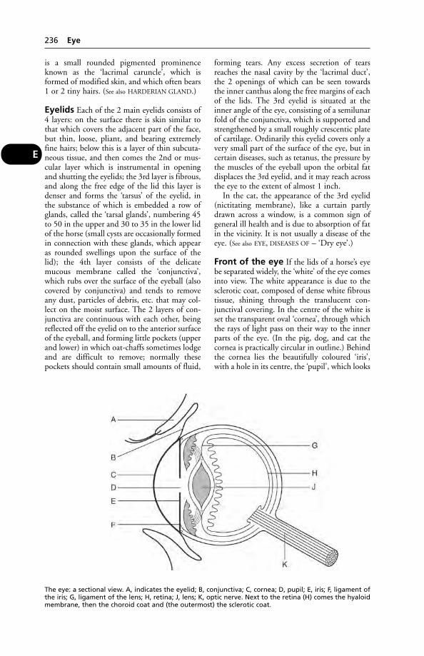
is a small rounded pigmented prominenceknown as the ‘lacrimal caruncle’, which isformed of modified skin, and which often bears1 or 2 tiny hairs. (See also HARDERIAN GLAND.)
Eyelids Each of the 2 main eyelids consists of4 layers: on the surface there is skin similar tothat which covers the adjacent part of the face,but thin, loose, pliant, and bearing extremelyfine hairs; below this is a layer of thin subcuta-neous tissue, and then comes the 2nd or mus-cular layer which is instrumental in openingand shutting the eyelids; the 3rd layer is fibrous,and along the free edge of the lid this layer isdenser and forms the ‘tarsus’ of the eyelid, inthe substance of which is embedded a row ofglands, called the ‘tarsal glands’, numbering 45to 50 in the upper and 30 to 35 in the lower lidof the horse (small cysts are occasionally formedin connection with these glands, which appearas rounded swellings upon the surface of thelid); the 4th layer consists of the delicatemucous membrane called the ‘conjunctiva’,which rubs over the surface of the eyeball (alsocovered by conjunctiva) and tends to removeany dust, particles of debris, etc. that may col-lect on the moist surface. The 2 layers of con-junctiva are continuous with each other, beingreflected off the eyelid on to the anterior surfaceof the eyeball, and forming little pockets (upperand lower) in which oat-chaffs sometimes lodgeand are difficult to remove; normally thesepockets should contain small amounts of fluid,
forming tears. Any excess secretion of tearsreaches the nasal cavity by the ‘lacrimal duct’,the 2 openings of which can be seen towardsthe inner canthus along the free margins of eachof the lids. The 3rd eyelid is situated at theinner angle of the eye, consisting of a semilunarfold of the conjunctiva, which is supported andstrengthened by a small roughly crescentic plateof cartilage. Ordinarily this eyelid covers only avery small part of the surface of the eye, but incertain diseases, such as tetanus, the pressure bythe muscles of the eyeball upon the orbital fatdisplaces the 3rd eyelid, and it may reach acrossthe eye to the extent of almost 1 inch.
In the cat, the appearance of the 3rd eyelid(nictitating membrane), like a curtain partlydrawn across a window, is a common sign ofgeneral ill health and is due to absorption of fatin the vicinity. It is not usually a disease of theeye. (See also EYE, DISEASES OF – ‘Dry eye’.)
Front of the eye If the lids of a horse’s eyebe separated widely, the ‘white’ of the eye comesinto view. The white appearance is due to thesclerotic coat, composed of dense white fibroustissue, shining through the translucent con-junctival covering. In the centre of the white isset the transparent oval ‘cornea’, through whichthe rays of light pass on their way to the innerparts of the eye. (In the pig, dog, and cat thecornea is practically circular in outline.) Behindthe cornea lies the beautifully coloured ‘iris’,with a hole in its centre, the ‘pupil’, which looks
236 Eye
E
The eye: a sectional view. A, indicates the eyelid; B, conjunctiva; C, cornea; D, pupil; E, iris; F, ligament ofthe iris; G, ligament of the lens; H, retina; J, lens; K, optic nerve. Next to the retina (H) comes the hyaloidmembrane, then the choroid coat and (the outermost) the sclerotic coat.
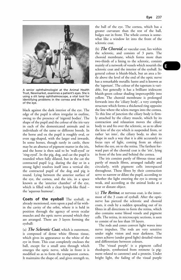
black against the dark interior of the eye. Theedge of the pupil is often irregular in outline,owing to the presence of ‘nigroid bodies’. Theshape of the pupil and the colour of the iris varyin each of the domesticated animals and inindividuals of the same or different breeds. Inthe horse and ox the pupil is roughly oval, oreven egg-shaped, with the larger end inwards.In some horses, though rarely in cattle, theremay be an absence of pigment matter in the iris,and the horse is then said to be ‘wall-eyed’ or‘ring-eyed’. In the pig, dog, and cat the pupil isrounded when fully dilated, but in the cat thecontracted pupil (e.g. during the day or in astrong light) resolves itself into a vertical slit;the contracted pupil of the dog and pig isround. Lying between the anterior surface ofthe eye, the cornea, and the iris, in a spaceknown as the ‘anterior chamber’ of the eye,which is filled with a clear lymph-like fluid –the ‘aqueous humour’.
Coats of the eyeball The eyeball, asalready mentioned, rests upon a pad of fat with-in the cavity of the orbit, where it is held inposition through the agency of seven ocularmuscles and the optic nerve around which theyare arranged. There are 3 layers forming theeyeball:
(a) The Sclerotic Coat, which is outermost,is composed of dense white fibrous tissue,which gives its appearance to the white of theeye in front. This coat completely encloses theball, except for a small area through whichemerges the optic nerve, while in front it ismodified so as to form the transparent cornea.It maintains the shape of, and gives strength to,
the ball of the eye. The cornea, which has agreater curvature than the rest of the ball,bulges out in front. The whole cornea is some-what like a window let into the front of the sclerotic coat.
(b) The Choroid, or vascular coat, lies withinthe sclerotic, and consists of 3 parts. Thechoroid membrane, which forms more thantwo-thirds of a lining to the sclerotic, consistsmainly of a network of vessels which nourish thesclerotic coat and the interior of the eyeball. Itsgeneral colour is bluish-black, but an area a lit-tle above the level of the end of the optic nervehas a remarkable metallic lustre and is known asthe ‘tapetum’. The colour of the tapetum is vari-able, but generally it has a brilliant iridescentbluish-green colour shading imperceptibly intoyellow. The choroid membrane is prolongedforwards into the ‘ciliary body’, a very complexstructure which forms a thickened ring oppositethe line where the sclera merges into the cornea.To this line of junction the ciliary body is firm-ly attached by the ciliary muscle, which by itscontraction and relaxation moves the ciliarybody to and fro over the sclerotic, so as to allowthe lens of the eye which is suspended from, orrather ‘set into’, the ciliary body, to alter itsshape in such a way that it is able accurately tofocus rays of light, coming from an objectbefore the eye, on to the retina. The farthest for-ward part of the choroid coat is the ‘iris’, lyingin front of the lens and behind the cornea.
The iris consists partly of fibrous tissue andpartly of muscle fibres, arranged radially andcircularly, with pigment cells interspacedthroughout. These fibres by their contractionserve to narrow or dilate the pupil, according towhether the light entering the eye is strong orweak, and according as the animal looks at anear or distant object.
(c) The Retina, or nervous coat, is the inner-most of the 3 coats of eyeball. After the opticnerve has pierced the sclerotic and choroidcoats, it ends by a sudden spreading out of itsfibres in all directions to form the retina, whichalso contains some blood vessels and pigmentcells. The retina, in microscopic sections, is seento consist of no less than 10 layers.
The rods and cones convert light waves intonerve impulses. The rods are very sensitiveunder night vision and near darkness. Thecones achieve (under good light) detailed visionand differentiate between colours.
The ‘visual purple’ is a pigment calledrhodopsin, synthesised from retinene (a pig-ment related to carotene) and a protein. Underbright light, the fading of the visual purple
A senior ophthalmologist at the Animal HealthTrust, Newmarket, examines a patient’s eyes. She isusing a slit lamp ophthalmoscope, a vital tool foridentifying problems in the cornea and the frontof the eye.
Eye 237
E
involves a conversion of rhodopsin into vitaminA plus protein by means of an enzyme.
Contents of the eyeball, viz. aqueoushumour, vitreous humour, and crystalline lens.Occupying the space between the iris and thecornea, i.e. the anterior chamber of the eye,there is a clear watery, lymph-like fluid. It isbeing constantly secreted and drained away,and eventually reaches the veins of the eye.Behind the iris lies the ‘crystalline lens’, whichacts as does the lens of a camera, with the excep-tion that it can alter the curves of its surfacesand therefore is able to change its refractivepowers. It is composed of layers arranged likethe leaves of an onion. The lens is held sus-pended by its capsule, which is attached to theciliary body already mentioned. Behind the lensthe cavity of the ball of the eye is filled with aviscid, jelly-like, tenacious fluid called the ‘vit-reous humour’. It maintains the intra-ocularpressure by which the eyeball retains its shape.
The lacrimal system provides a meanswhereby the eye surface is maintained free fromdust and other foreign material. It consists ofthe lacrimal gland which secretes the clear fluidpopularly known as ‘tears’; excretory ducts,from 12 to 16 in number; and the 2 lacrimalducts which open into a lacrimal sac fromwhich begins the naso-lacrimal duct which car-ries the secretion down into the nose. The glandlies towards the upper outer aspect of the orbit;secretes the clear salty, watery fluid which flowsout through the excretory ducts to reach theconjunctival sac and bathe the surface of theeye. The secretion is finally received by the 2lacrimal ducts, the openings of which lie one ineach eyelid about a third of an inch from theinner canthus. These open into the lacrimal sac,from which takes origin the long naso-lacrimalduct which conveys the secretion down into thelower part of the corresponding nasal passage,just within the nostril.
Accommodation All the rays of light pro-ceeding from a distant object may be lookedupon as being practically parallel, while thosecoming from a near object are divergent. Thedifference between distant and near in this con-nection can be taken as about 5 metres (20 feet)from the animal. A ‘near’ object can be seenanywhere between 5 metres (20 feet) and 10 to12 cm (4 to 5 inches) from the eye, but nearerthan this it loses its distinction. Parallel rays oflight do not require any focusing on the retinaother than is provided by the surface of thecornea; when an animal looks at a distant
object, the lens capsule (which is attached tothe ciliary process) retains the lens in a tem-porarily flattened condition and the ciliarymuscle is relaxed, so that no great strain is putupon the eye. Rays of light from an object nearat hand, however, which are divergent, requireto be brought to a point of focus upon the reti-na, and as they pass through the lens theirdirection is changed on account of the convex-ity of the lens. The amount of this convexity isdetermined by the divergency of the rays, and isautomatically provided for through the pull ofthe ciliary muscle upon the ciliary body. As thefunction of the muscle is to pull the ciliary bodyforwards, the tension upon the ligament of thelens is lessened and the capsule of the lens slack-ens, so that the lens, by its inherent elasticity, isallowed to bulge with a greater convexity uponits anterior surface. The greater the convexity,the more are the rays of light refracted, and themore convergent do rays which pass through itbecome. (See also VISION.)
Lens (see illustration)
Eye, Diseases and Injuries ofAll such diseases and injuries can be of eco-nomic importance to farmers, since the pro-ductive efficiency of affected animals is likely tobe reduced, owing to stress, pain, or infection –or all of these. Milk yield may decline in thedairy cow. If the animal’s sight is seriouslyimpaired, feeding may become difficult, withconsequent loss of bodily condition.
Blepharitis Inflammation of the edges of theeyelids; it usually accompanies conjunctivitis.Its causes, symptoms, and treatment are similar(see below).
Blindness There are many causes of this,including disease of the retina, of the opticnerve, and of the brain. Blindness may be con-genital or acquired, temporary or permanent.Vitamin A deficiency may be responsible, andalso poisoning by rape and other plants, and by substances such as lead. Blindness in the dogand cat may result from carbon monoxide poisoning, and persist for some time; it may also result from metaldehyde poisoning.(See also under QUININE; MALE FERN; BRIGHT
BLINDNESS.)‘Day blindness’ (Hemeralopia) is stated to be
due to an autosomal recessive gene. This eyedisease is common in the Alaskan malamutedog, and has been reported also in miniaturepoodles. The blindness occurs during brightlight, although in dim light the animal can see.
238 Eye, Diseases and Injuries of
E
Horses. The sudden onset of blindness in oneor both eyes has been reported as a result ofoptic nerve atrophy, following trauma. Signs aredilated, fixed pupils, and a lack of the menacereflex. Within 3 to 4 weeks the optic discbecomes paler, and the retina’s blood vesselsmarkedly decreased. There is a rupture of thenerve axons.
‘Night blindness’ (nyctalopia) is a conditionthat sometimes affects horses and mules incountries where the glare of the sunlight is veryintense during the day. At night such animalsare quite unable to see, and will stumble intoobjects that are easily discernible to humanbeings.
Camels are seldom affected, owing to theeffective protection afforded to the retina by theoverhanging eyelids and deeply placed eyeballs.
Opacity of the cornea will, of course, preventlight rays from reaching the retina, as happensin keratitis, so that partial or complete blind-ness results. Similarly, partial or completeblindness may result from a cataract. Othercauses are mentioned below, e.g. dislocation ofthe lens, glaucoma, etc.
Cattle and sheep Cerebrocortical necrosis(polioencephalomalacia), resulting from a thi-amine deficiency, is a cause of blindness inruminants. In sheep, other causes of blindnessinclude: infectious keratitis or contagious oph-thalmia (‘heather blindness’); pregnancy tox-aemia; and the effects of eating bracken, asdescribed under BRIGHT BLINDNESS of sheep.
Poultry Blindness may be the result of exces-sive ammonia fumes from deep litter, or it maybe associated with fowl paralysis, salmonellosis,aspergillosis, etc. Cataract, followed in somecases by liquefaction of the lens, occurs duringoutbreaks of avian infectious encephalomyelitis.(See also under LIGHTING OF ANIMAL BUILDINGS.)
‘Blue eye’ (see CANINE VIRAL HEPATITIS)
Cancer, either of sarcomatous or carcinoma-tous nature, is sometimes found in connectionwith the conjunctiva. Tumours appear as redhard swellings, painless when small, but notwhen large. These neoplasms often grow at arapid rate, and may infiltrate the surroundingtissues, sometimes affecting the bones of theorbit. Cancer of the eye is a common conditionin Hereford cattle. It has been suggested thatseveral factors may contribute to the develop-ment of eye cancer in cattle. These include age,irritation of the eyes by dust, sand, insects orchemicals, sunlight, lack of eyelid pigmentationand viral infection. Some authorities believethat cattle may be genetically prone to the condition, while others feel that poor nutritionis another factor as the condition appears to occur more frequently following a drought.(See TUMOURS.)
The beginning of ‘cancer eye’, as it is some-times colloquially known, may be a raised areaof skin or a wart. Either may become malig-nant, developing into a typical carcinoma – thetype of cancer occurring in this eye disease ofHerefords. However, in the USA a survey was
PAGE
Blepharitis 238Blindness 238‘Blue eye’ 239Cancer 239Cataract 240‘Collie eye anomaly’ 240Coloboma 240Conjunctivitis 240Dislocation of the lens 240‘Dry eye’ 241Ectropion 241Entropion 241Epiphora 241Foreign bodies 241Glaucoma 241Harderian gland, displaced 241‘Heather blindness’ 241Horner’s syndrome 241Infectious bovine keratoconjunctivitis 242
PAGE
Iritis 242Keratitis 242Keratomycosis 242Microphthalmos 242Myiasis 243New Forest disease 243Opacity of the cornea 243Ovine infectious keratoconjunctivitis 243Pannus 243Partial displacement 243Periodic ophthalmia 243Progressive retinal atrophy 243Ptosis 243Retention cysts 243Sclerotitis 243Stye 243Trachoma 244Trichiasis 244Warts 244
EYE, DISEASES and INJURIES OF
Eye, Diseases and Injuries of 239
E

carried out; the eyes of 48 Hereford cows wereexamined at 6-monthly intervals for 2 years.Over half the cows showed preliminary signs of‘cancer eye’, but – without any treatment – one-third of the growths had disappeared by thetime of the last examination.
Cryosurgery has been used to treat cancer ofthe eye. The technique is a highly skilled oneand requires special thermocouples to monitorthe very low temperatures.
In a series of 718 cases of eye cancer treatedby cryosurgery, 609 a single freeze caused totalregression of 66 per cent of the growths. In 109,treated by a rapid freeze to –25°C, a naturalthaw, and then a re-freeze, the cure rate was 97 per cent.
Cataract The condition is by far the mostcommon in the horse and dog in old age,although it is also encountered in other ani-mals, and it may occur at almost any time oflife. It consists of a coagulation of the plasma ofthe cells in the lens with loss of transparency. Abluish, cloudy appearance of the eye results andvision becomes blurred.
Causes Cataract is primarily a change charac-teristic of old age. Other causes include diabetes.Cataracts have also resulted from naphtha-lene and other forms of poisoning, and fromexposure to X-rays.
Treatment As in human patients, cataractsmay be removed successfully in animals. In onestudy, cataracts were removed by phacofrag-mentation and aspiration from one or both eyesof 56 dogs. Vision improved immediately in 53of the dogs: after 2 years, 25 of 29 dogs still hadvision, and after 4 years, 5 of 7 dogs. Thesurgery was unsuccessful in dogs with severeanterior uveitis with secondary glaucoma, reti-nal detachments, and fibropupillary membraneformation.
‘Collie eye anomaly’ is an inherited con-dition in which there is underdevelopment ofthe choroid membrane of the eye.
Coloboma is a congenital and hereditarydefect – a notch, gap, hole, or fissure in any ofthe structures of the eye. In other words, atbirth a part of the eye is missing. Bilateralcoloboma is common in Charolais cattle, ofteninvolving the optic disc. The condition can berecognised only with an ophthalmoscope, anddoes not deteriorate with the passage of time.Effect on vision varies from very slight to(rarely) blindness.
Conjunctivitis, or inflammation of the con-junctival membranes, is an extremely commoncondition among animals, and probably consti-tutes the commonest trouble to which the eyesare subject. In cattle, conjunctivitis is often thefirst symptom of cattle plague, ephemeral fever,and Ondiri disease (bovine infectious petechialfever).
Conjunctivitis is one symptom associatedwith many specific infections, such as distem-per in the dog (and see EYEWORMS).
Causes The presence of dust, sand, pollen,seeds, lime, and pieces of chaff, in the atmos-phere of a stable or field, is probably one of thecommonest causes in the larger farm animals,but such agents as flies, worms, and ticks mustalso be noted in addition to the above. In thecat, 2 infections which cause conjunctivitis –Haemophilus parainfluenzae and Moraxella lacunata – are transmissible to man, in which illness may also be caused. (See FELINE EYE
INFECTIONS.)Chlamydia psittaci was isolated from 30 per
cent of swabs from 753 cats suffering from conjunctivitis.
Signs The first signs of conjunctivitis are red-ness and swelling of the lining membranes ofthe eyelids, excessive discharge of tears, and atendency for the animal to keep its eyelids shut.
First-Aid Clean away the discharges bybathing with a warmed eye lotion. (If only 1 eyeis affected, the cause may be a foreign body thathas lodged there.) The best way to applylotions, whether to the horse, dog, or other ani-mal, is to use a perfectly clean piece of cotton-wool soaked in the solution and squeezed abovethe eye so that the drops trickle into it. Cases ofconjunctivitis should never be neglected, forthe inflammation may spread to the cornea,resulting in keratitis (see below).
Dislocation of the lens is a condition inwhich the crystalline lens becomes displacedforwards into the anterior chamber. It occurs indogs, especially Sealyhams and rough-hairedterriers, and at first is very hard to recognise.The dog runs into stationary objects withoutany obvious reason. Casual examination of theeyes reveals no change and the condition maynot be suspected for many months. Later, theowner becomes aware that sight is failing in thedog and careful examination reveals a ‘wobblylens’ in the eye. Operation may do much torestore some degree of vision and save the eye,but neglect almost invariably results in the
240 Eye, Diseases and Injuries of
E

development of glaucoma (see below) and theaffected eye may have to be surgically removed.
Technically, lens dislocation may be classifiedas congenital, primary, secondary, or traumatic.Secondary cases not uncommonly followcataract or glaucoma, but most cases occurspontaneously in adult life.
‘Dry eye’ (keratoconjunctivitis sicca) is a con-dition in the dog arising from a partial failure of tear production and leading to roughening ofthe corneal surface with a consequent lack oflustre. ‘Artificial tears’ may have to be provided,or a surgical operation performed involvingparotid duct transplantation. The condition hasbeen linked to the use of sulphasalazine for thetreatment of idiopathic colitis in dogs.
Ectropion means a turning-out of one orboth eyelids, so that the conjunctiva is exposed.It is a common condition in bloodhounds andSt Bernards, and is in them regarded as practi-cally normal. It is also treated by operation, buta part of the conjunctiva from within the edgeof the lid is removed instead of part of the skinfrom the outside, as in entropion.
Entropion Turning-in of the eyelid, often thelower one, so that it rubs upon the cornea, caus-ing inflammation. The condition is common indogs – often an inherited defect – occurring inmany breeds. It is also seen in ‘mini-pigs’.
In newborn lambs entropion is occasionallyseen and, if bilateral, can lead to eventual blind-ness and starvation. It can be corrected byMichel clips (metal sutures). It is treated by a plastic operation such as is performed fortrichiasis (see below).
Epiphora is another name for what is com-monly called ‘watery eye’ or ‘overflow of tears’.It is generally due to some obstruction to thedrainage of tears through the lacrimal duct tothe nose, but it is also an accompanying symp-tom of most forms of mild inflammation of theconjunctiva or cornea, of naphthalene poison-ing in cattle, and of atopic disease in the dog.
Foreign bodies in the eye have already beenreferred to under ‘Conjunctivitis’ above. Severeirritation may be caused by a piece of grit or agrass seed or husk. Pain and irritation may beshown by the dog pawing its face.
Treatment If a hair, bristle or tip of an awn,for instance, can be seen on folding back theeyelid, or if a white spot (sometimes indicatingthe site of a thorn’s penetration) or what
appears to be ‘a white film’ is visible on the sur-face of the eye, the best first-aid treatment is adrop of olive oil. (Cod-liver oil will do, but notany oil!) Boracic acid lotion is worse than use-less (except for the mechanical washing-out ofgrit); what is needed is a lubricant to reduce the harmful friction, and this is where the oilhelps. Removal of a foreign body is best accom-plished with the aid of a local anaesthetic, andprofessional help should be obtained.
Occasionally the object may be removed bytaking the corner of a clean handkerchief, wind-ing it into a point, and lifting the offendingbody out with it. The use of a suitable eyelotion will be helpful afterwards.
Glaucoma is a condition in which the ten-sion of the fluid contents of the eyeball is great-ly in excess of the normal. It is associated withobstruction to the drainage system of the eye, inwhich fluid continues to be secreted but theexcess is not removed. It may follow cases ofprogressive retinal atrophy. It eventually resultsin swelling and bulging of one or both of theeyes, and blindness results. Secondary glaucomais more common and caused by an eye disease,of which the most frequent is lens dislocation.(See also under EXOPHTHALMOS.)
Harderian gland, displaced In the dogthis gland sometimes becomes enlarged and dis-placed, owing to blockage of its ducts or to anearby swelling, when it becomes visible at thecorner of the eye as a reddish lump. It may thenrequire surgical removal.
‘Heather blindness’ is a colloquial name forthe equivalent of infectious bovine keratocon-junctivitis (IBK) in sheep. Rickettsia conjunctivaeis a common cause.
Treatment Shade and fly-control aid recovery,but veterinary treatment of IBK is necessary.
Boracic and similar eye lotions are useless in treating IBK or ‘heather blindness’.Chloramphenicol eye drops or cloxacillin maybe effective in treatment.
Horner’s syndrome The pupil of one eyeappears smaller than normal, the upper eyelidmay droop, the lower lid may be raised, and thenictitating membrane (‘third eyelid’) protrudedacross part of the eye.
The cause is some lesion affecting the sym-pathetic nerves of the eye, e.g. a tumour of thespinal cord, chronic otitis, bite wounds, beestings. Some cases are transient, as with woundsand bee stings.
Eye, Diseases and Injuries of 241
E

Infectious bovine keratoconjunctivi-tis (IBK) is a convenient name for a group ofeye diseases with a worldwide distribution, and includes New Forest disease (see below).What they have in common is conjunctivitisand keratitis.
Causes Bacteria, viruses, mycoplasmas, rick-ettsiae, fungi, and Thelazia worms – any ofthese alone or in combination may produceIBK. In addition, the sun’s rays, dust particles,and chemical irritants may all predispose to, orexacerbate, the condition. IBK is commonlytransmitted by flies and, in Africa, by twospecies of moth which feed on secretions andexudate from the eye. Some infective agents arepresent on the healthy eye, and become activeonly when the eye is damaged or irritated insome way.
Moraxella (Haemophilus) bovis is a commonbacterial cause of IBK. Some American researchhas suggested that Moraxella may not cause ker-atitis unless the virus of bovine rhinotracheitisis present also. Cefalonium applied as an eyeointment is an antibiotic treatment.
Iritis means inflammation of the iris, a condi-tion which is very often associated with inflam-mation of the ciliary body, when the term ‘iridocyclitis’ is used. The chief symptoms aredullness of the iris, congestion of the blood ves-sels around its margin, a lessened response tovarying intensities of light, and usually a firmlycontracted pupil. Occasionally, especially dur-ing inflammation of the cornea, the iris adheresto this structure – a condition known as ‘ante-rior synechia’; while more frequently the irisadheres to the lens, which lies behind it, andthe condition is spoken of as ‘posteriorsynechia’. The aqueous humour is often cloudyand may appear purulent, little flocculi oflymph being seen floating in the anteriorchamber or sticking to the posterior surface ofthe cornea. There is always great pain, fear oflight (photophobia), and the animal hangs itshead and is dull and listless.
Iritis is a common condition in cattle, usuallycaused by eating poor-quality big-bale silage.
Keratitis Inflammation of the cornea mayfollow conjunctivitis, or it may arise from aninjury to, or infection of, the cornea itself. Athorn, for example, may pierce the surface lay-ers of the cornea and remain invisible until afaint whitish ring appears around its protrudingpart. Should a larger area of the cornea beinvolved, opacity becomes obvious. (Animal-owners often refer to it as ‘a film over the eye’;
but in fact the opacity stems from inflamma-tion below the surface.) Keratitis may be causedby trauma of various kinds, e.g. a whip lash, akick or blow; or by irritant skin dressings whichare not prevented from running into the eyes,or by lime, sparks, or by continuous irritationby a foreign body such as a grass awn, piece ofglass or grit. It may arise during the course ofcertain diseases, such as distemper in dogs andinfluenza in horses; it can be produced by thepresence of Thelazea worms, or by fly-borneinfections; frost-bite is said to be the cause of itin ewes on hills during severe weather, when itis called ‘snow blindness’; turning-in of the eye-lids (entropion) may give rise to it in the dog.
Keratoconjunctivitis sicca is a conditionseen mostly in small animals, and is caused bythe inadequate production of tears. There is atacky mucoid discharge round the eyes; thecornea appears dry and may be ulcerated. Ifuntreated, corneal opacity vascularisation andpigmentation may result. Topical application ofantibiotics is indicated; ‘artificial tears’, formu-lated for use in human medicine, are useful.
Keratomycosis is keratitis due to a fungus,and is uncommon. If, however, tissue resistance isreduced by treatment with corticosteroids (whichare immunosuppressive), any fungi present onthe cornea may become pathogenic. It may beonly when corneal ulcers fail to respond to con-ventional treatment that keratomycosis is sus-pected. Natamycin may prove helpful. Fusariumsolani is implicated in most equine cases, some-times Candida species; but several other fungimay be involved. (See also HYPERKERATOSIS.)
In the early stages, inflammation of thecornea results in symptoms very similar to thoseseen in conjunctivitis; the production of tears,closing of the eyelids, pain and swelling beingnoticed. When the eye is examined, however,the surface of the cornea is found to have lost itslustre. There may be a bluish haze, and an opac-ity, varying from pin-head size to the whole ofthe cornea – when the animal becomes com-pletely blind in that eye, for the time being,anyway. The appearance of blood vessels wherenone is normally seen is another feature of ker-atitis and occurs before opacity becomes com-plete. There may be ulceration of the cornea,and even penetration. If the latter should occur,a keratocoele (hernia) may form endangeringthe whole eye, since infection, or escape of theaqueous humour, may sometimes occur.
Microphthalmos is an abnormally smalleye; it is seen in vitamin A deficiency.
242 Eye, Diseases and Injuries of
E

Myiasis (see UITPEULOOG)
New Forest disease (infectious bovinekeratitis). Success has been claimed for treat-ment involving the injection of 2 to 5 ml of anantibiotic preparation into the subconjunctivaltissues of the upper eyelid. Antibiotics, par-enterally, by subcutaneous injection or by long-acting antibiotic ophthalmic ointment are usedin treatment; cortisone is contraindicated.Penicillin, oxytetracycline and chloramphenicolhave been reported to give equally good results.A single treatment is usually sufficient.
Opacity of the cornea may result fromoedema of the cornea following infection withCANINE VIRAL HEPATITIS; see ‘Keratitis’ above.
Ovine infectious keratoconjunctivitisThis occurs worldwide. In a field survey carriedout by the University of Liverpool’s veterinarystaff, the microflora of 240 clinically unaffectedeyes from sheep in 10 flocks were comparedwith those of 240 clinically affected eyes from12 natural outbreaks. Totals of 16 and 17 gen-era of bacteria were recovered, includingBranhamella ovis, E. coli, and Staphylococcusaureus. Mycoplasma and acholeplasma were isolated from both groups. Chlamydia psittacican also be a cause.
Pannus is a complication of keratitis in whichblood vessels bud out from the margins of thecornea and run in towards the centre of the eye,stopping at the edges of an ulcer if such exists.Pannus is a condition which always takes a longtime to clear up, and even months after theremay be seen a dullness of the cornea, due to the tiny vessels that still exist but are invisible tothe naked eye.
Partial displacement Pekingese and otherdogs with prominent eyes sometimes suffer atraumatic partial displacement of the eye fromthe orbit, as a result of being struck by a car orof some other accident. The globe may becometrapped by the eyelids which become locatedbehind it.
First-Aid The owner should bandage the eyewith bandage moistened in saline solution (a teaspoonful of ordinary salt to a pint ofwater). Professional aid is urgently required.
Treatment This requires a general anaestheticand re-positioning of the eye where possible. Ifthe cornea, etc., has been badly damaged, theonly course is enucleation of the eye. After
suturing of the eyelids over the vacant socket,the result will not appear unsightly to theowner.
Periodic ophthalmia (see under this head-ing). (See also OPHTHALMIA.)
Progressive retinal atrophy, or so-called‘night blindness’, is a hereditary condition com-mon in some strains of Irish red setter. Theblood vessels of the retina undergo progressiveatrophy and the animal suffers from impairedvision in consequence. To endeavour to correctthis the pupil dilates widely, even in daylight,and the dog’s expression become staring. Atnight or at dusk, the dog is unable to avoidobjects and blunders into them, but during fulldaylight it appears to see quite well.
No treatment can arrest the progressivedegeneration and the dog gradually becomesblind. In severe cases puppies may show firstsymptoms soon after weaning.
Neither dogs nor bitches which show thecondition should be used for breeding. Breedsaffected include collies, griffons, poodles,retrievers, Sealyhams, cocker and Englishspringer spaniels.
The disease also occurs in cats, e.g. Abyssinianand Siamese; 25 per cent of Abyssinian cats were found to be affected in a recent study. Theearliest signs may not be seen until the cat is 18months old or more; and the advanced formtakes another 18 months to develop. (See also
TAURINE.)
Ptosis is an inability to raise the upper eyelid,usually associated with some general disease,such as distemper in dogs or ‘grass sickness’ inhorses. It may also arise after injuries when thenerve supplying the muscles of the upper lid(3rd cranial nerve) is paralysed.
Retention cysts are produced in the thick-ness of the eyelid owing to blockage of a tarsalgland.
Sclerotitis (scleritis), or ‘blood-shot eye’, isinflammation of the sclerotic coat of the eye-ball. It often accompanies conjunctivitis whenthe latter is at all severe. It is treated as for con-junctivitis.
Stye, or hordeolum, is a condition in which asmall amount of pus collects in the folliclearound the root of one of the eyelashes. Oneafter another may form in succession, owing tothe spread of infective material from follicle tofollicle.
Eye, Diseases and Injuries of 243
E

E
244 Eye Diseases, Hereditary
Trachoma A term used in human medicinefor a granular conjunctivitis, often followed bykeratitis and pannus.
Trichiasis Turning-in of the eyelashes so thatthey irritate and inflame the conditions. Thecondition is common in dogs and is sometimesa hereditary defect. It is treated surgically, bymeans of an operation in which an ellipticalpiece of skin is removed from the outer surfaceof the eyelid, and the edges sutured together.This causes the lashes to turn outwards, wherethey will not irritate or inflame the cornea.
Warts occur in connection with the eyelidscomparatively frequently in horses, cattle, anddogs, and sometimes become malignant,spreading at a rapid rate and causing interfer-ence with sight or the movement of the eyelids.Owing to the malformation which they maycause when numerous, warts should always beremoved before they attain a large size or beforethey have time to spread.
Eye Diseases, HereditaryThe British Veterinary Association, the KennelClub and the International Sheep Dog Societyoperate a joint scheme to identify the presence
or absence of inherited eye disease in a numberof breeds of dog to help ensure that only disease-free animals are used for breeding. The mainconditions covered are central progressive retinalatrophy, collie eye anomaly, generalised progres-sive retinal atrophy, goniodysgenesis/primaryglaucoma, hereditary cataract, persistenthypoplastic primary vitreous, and persistentpupillary membrane.
Eye Fluke(see DIPLOSTOMUM)
Eyelids(see under EYE)
EyewormsIn cattle Thelazia worms are one cause of infec-tious bovine keratoconjunctivitis. Speciesinclude T. skrjabini and T. gulosa, found behindthe 3rd eyelid and in the ducts of associatedglands. From 1 to 67 worms were found in eyesexamined at a UK abattoir in 36.9 per cent of287 cattle heads examined. Other species ofThelazia infest dogs, cats, and man. T. lacry-malis was found in 28 per cent of horses whoseeyes were examined at an abattoir.
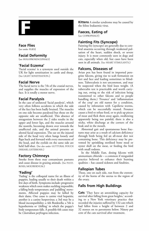
Face Flies(see under FLIES)
Facial Deformity(see HOLOPROSENCEPHALY)
‘Facial Eczema’‘Facial eczema’ is a synonym used outside theUK for light sensitisation in cattle and sheep.(See LIGHT SENSITISATION.)
Facial NerveThe facial nerve is the 7th of the cranial nerves,and supplies the muscles of expression of theface. It is totally a motor nerve.
Facial ParalysisIn the case of unilateral ‘facial paralysis’, whichvery often follows accidents in which the side of the face has been badly bruised. The muscleson one side become paralysed but those on theopposite side are unaffected. This absence ofantagonism between the 2 sides results in theupper and lower lips, and the muscles aroundthe nostrils, becoming drawn over towards theunaffected side, and the animal presents analtered facial expression. The ear on the injuredside of the head very often hangs loosely andflaps back and forward with every movement ofthe head, and the eyelids on the same side areheld half-shut. (See also under GUTTURAL POUCH
DISEASE; LISTERIOSIS.)
Factory ChimneysSmoke from these may contaminate pasturesand cause disease in grazing animals. (See FLUO-
ROSIS; MOLYBDENUM.)
‘Fading’‘Fading’ is the colloquial name for an illness ofpuppies, leading usually to their death within afew days of birth. Symptoms include: progressiveweakness which soon makes suckling impossible;a falling body temperature; and ‘paddling’ move-ments. Affected puppies may be killed by their dams. One cause is canine viral hepatitis;another is a canine herpesvirus; a 3rd may be ablood incompatibility; a 4th Bordetella; a 5th ishypothermia or ‘chilling’ in which the puppy’sbody temperature falls. A possible 6th cause maybe Clostridium perfringens infection.
Kittens A similar syndrome may be caused bythe feline leukaemia virus.
Faeces, Eating of(see COPROPHAGY)
Fainting Fits (Syncope)Fainting fits (syncope) are generally due to cere-bral anaemia occurring through weakened pul-sation of the heart, sudden shock, or severeinjury. It is most commonly seen in dogs andcats, especially when old, but cases have beenseen in all animals. (See HEART STIMULANTS.)
Falcons, Diseases ofAvian pox has been found in imported pere-grine falcons, giving rise to scab formation onfeet and face and leading sometimes to blind-ness. Tuberculosis is not uncommon, and maybe suspected when the bird loses weight. (Atuberculin test is practicable and worth carry-ing out, owing to the risk of infection beingtransmitted to other falcons and to people handling them.) ‘Frounce’ and ‘inflammationof the crop’ are old names for a condition,caused by infestation with Capillaria worms,which can be successfully treated. Frounce causes a bird to refuse food, or to pick up piecesof meat and flick them away again, swallowingapparently being too painful; there is also asticky, white discharge at the corners of thebeak and in the mouth.
Abnormal gait and spontaneous bone frac-tures may arise as a result of calcium deficiencythrough birds being fed an all-meat diet notcontaining bone. This deficiency may be pre-vented by sprinkling sterilised bone meal oroyster shell on the meat, or feeding the birdwith small rodents.
In the Middle East, dosing falcons withammonium chloride – a common if misguidedpractice believed to enhance their hunting qualities – has caused sickness and fatalities.
Fallopian TubesThese, one on each side, run from the extrem-ity of the horns of the uterus to the region ofthe ovary.
Falls from High Buildings
Cats ‘They have an astonishing capacity forsurvival after falling from great heights,’ accord-ing to a New York veterinary practice thatrecorded the injuries suffered by 132 cats whichhad fallen from a height of between 2 and 32 storeys on to pavements below. Ninety percent of the cats survived after treatment.
F
Injuries increased, as would be expected, inproportion to the distance fallen – up to about7 storeys. However, the number of fracturesdecreased with falls from a greater height than that. It is suggested that this was becausethe cats then extended their legs to an almosthorizontal position, like flying squirrels, mak-ing the impact more evenly distributed. Thisresulted in more chest injuries than fracturedribs, however.
Emergency treatment was required in 37 percent of the cats, non-emergency treatment in30 per cent.
What causes them to fall? In a few instances,it seems, they lose their balance while turningon a narrow window-ledge. More often it hap-pens while trying to catch a bird or insect. It hasalso been known for a cat to panic, and leap offthe ledge, when threatened by a strange dog letinto the room behind.
Dogs Of 81 dogs which had fallen from 1 to 6 storeys, all but 1 dog survived. ‘The falls of 52 of the dogs were witnessed, and of them, 39 had jumped.’ Injuries to face, chest, andextremities resulted in dogs falling 1 or 2 storeys.Spinal injuries were caused more often in fallsfrom a greater height.
False Pregnancy(see under PSEUDO-PREGNANCY)
Fan FailureIn buildings that are ventilated artificially, it ismandatory under the Welfare of FarmedAnimals Regulations 2000 (2001 in Wales) tohave an alarm and standby system in order toprevent heat-stroke or anoxia (see CONTROLLED
ENVIRONMENT HOUSING).
FaradismLocal application of an electric current as a pas-sive exercise which stimulates muscles and nerves.
FarcyChronic form of glanders (see GLANDERS).
Farm Animal Welfare Council (FAWC)An independent body set up by the governmentin 1979 to keep under review the welfare offarmed animals. Farms, markets, abattoirs andvehicles are inspected and, where appropriate,recommendations made to government. Reportsare issued from time to time on the welfare ofparticular species or aspects (transport, slaughter,etc.) of the use of farm animals. The address is:1a Page Street, London SW1P 4PQ.
Farm Chemicals(see SPRAYS USED ON CROPS; FERTILISERS;METALDEHYDE)
Farm, Operations on theIn the UK it is illegal for castration of horse,donkey, mule, dog or cat to be carried out with-out an anaesthetic. (See ANAESTHESIA, LEGAL
REQUIREMENTS; CASTRATION.) Only a veteri-nary surgeon is permitted to castrate any farmanimal more than 2 months old, with theexception of rams, for which the maximum ageis 3 months.
Only veterinary surgeons are permitted tocarry out a vasectomy or electro-ejaculation ofany farm animal; likewise the de-snooding ofturkeys over 21 days old, de-combing of domes-tic fowls over 72 hours old, and de-toeing offowls and turkeys over 72 hours old. Nor cananyone but a veterinary surgeon remove super-numerary teats of calves over 3 months old, ordisbud or dishorn sheep or goats.
Certain overseas procedures are prohibited in the UK, namely freeze-dagging of sheep,penis amputation and other operations on thepenis, tongue amputation in calves, hot brand-ing of cattle, and the de-voicing of cockerels.Very short docking of sheep is also prohibited(see DOCKING).
Farm Treatment AgainstWorms(see WORMS)
‘Farmer’s Lung’A disease caused by the inhalation of dust, frommouldy hay, etc., containing spores of e.g.Thermopolyspora polyspora or Micropolysporafaeni. Localised histamine release in the lungproduces oedema, resulting in poor oxygenuptake. The condition has been recognised inhumans, cattle, horses and turkeys. In chickens,a similar condition has been caused by inhala-tion of dust from dead mites in sugar canebagasse. It is classed as an acute extrinsic aller-gic alveolitis. Repeated exposure causes respira-tory distress, even when the interval betweenexposures is several years.
Farm, Veterinary Facilities on the(see VETERINARY FACILITIES ON THE FARM)
FarrierA person who shoes horses. Farriery is a craft of great antiquity and the farrier has beendescribed as the ancestor of the veterinarian. Inthe UK, farriery training is strictly controlled.
246 False Pregnancy
F
Intending farriers must undergo a 5-yearapprenticeship, including a period at an autho-rised college, then take an examination for thediploma of the Worshipful Company of Farriersbefore they can practise independently. Thetraining is controlled by the Farriers TrainingCouncil and a register of farriers kept by theFarriers Registration Council, Sefton House,Adam Court, Newark Road, Peterborough PE15PP. Its website is at www.farrier-reg.gov.uk.
FarrowingThe act of parturition in the sow.
Farrowing CratesA rectangular box in which the sow gives birth.Their use is helpful in preventing overlying ofpiglets by the sow, and so in obviating one causeof piglet mortality; however, they are far fromideal. Farrowing rails serve the same purposebut perhaps the best arrangement is the circularone which originated in New Zealand. (See
ROUNDHOUSE.)Work at the University of Nebraska suggests
that a round stall is better, because the conven-tional rectangular one does not allow the sow toobey her natural nesting instincts, and may giverise to stress, more stillbirths and agalactia.
Farrowing RatesIn the sow, the farrowing rate after 1 naturalservice appears to be in the region of 86 percent. Following a 1st artificial insemination, the farrowing rate appears to be appreciablylower, but at the Lyndhurst, Hants AI Centre, a farrowing rate of about 83 per cent wasobtained when only females which stood firm-ly to be mounted at insemination time wereused. The national (British) average farrowingrate has been estimated at 65 per cent for a 1st insemination.
FasciaSheets or bands of fibrous tissue which encloseand connect the muscles.
FascioliasisInfestation with liver flukes.
FatNormal body fat is, chemically, an ester of 3molecules of 1, 2, or 3 fatty acids, with 1 mol-ecule of glycerol. Such fats are known as glyc-erides, to distinguish them from other fats andwaxes in which an alcohol other than glycerolhas formed the ester. (See also LIPIDS [whichinclude fat]; FATTY ACIDS. For fat as a tissue, see ADI-
POSE TISSUE. A LIPOMA is a benign fatty tumour.
For other diseases associated with fat, see STEATI-
TIS; FATTY LIVER SYNDROME; OBESITY, DIET.)
Fat SupplementsIn poultry rations these can lead to TOXIC FAT
DISEASE. (See LIPIDS for cattle supplement; also
ECZEMA in cats.)
Fatigue(see EXERCISE; MUSCLE; NERVES)
Fatty AcidsThese, with an alcohol, form FAT. Saturatedfatty acids have twice as many hydrogen atomsas carbon atoms, and each molecule of fattyacid contains 2 atoms of oxygen. Unsaturatedfatty acids contain less than twice as manyhydrogen atoms as carbon items, and 1 or morepairs of adjacent atoms are connected by doublebonds. Polyunsaturated fatty acids are those inwhich several pairs of adjacent carbon atomscontain double bonds.
Fatty DegenerationA condition in which there is an excess of fat inthe parenchyma cells of organs such as the liver,heart, and kidneys.
Fatty Liver HaemorrhagicSyndrome (FLHS)This is a condition in laying hens which has to be differentiated from FLKS (see next entry) of high-carbohydrate broiler-chicks. Factorsinvolved include high carbohydrate diets, highenvironmental temperatures, high oestrogenlevels, and the particular strain of bird. FLHSin hens is improved by diets based on wheat ascompared with maize; whereas FLKS is aggra-vated by diets based on wheat. Death is due tohaemorrhage from the enlarged liver.
Fatty Liver/Kidney Syndromeof Chickens (FLKS)A condition in which excessive amounts of fat arepresent in the liver, kidneys, and myocardium.The liver is pale and swollen, with haemorrhagessometimes present, and the kidneys vary frombeing slightly swollen and pale pink to beingexcessively enlarged and white. Morbidity is usually between 5 and 30 per cent.
Cause FLKS has been shown to respond tobiotin (see VITAMINS), and accordingly can beprevented by suitable modification of the diet.
Signs A number of the more forward birds (usu-ally 2 to 3 weeks old) suddenly show symptomsof paralysis. They lie down on their breasts with
Fatty Liver/Kidney Syndrome of Chickens (FLKS) 247
F
their heads stretched forward; others lie on theirsides with their heads bent over their backs.Death may occur within a few hours. Mortalityseldom exceeds 1 per cent.
Fatty Liver Syndrome of CattleA ‘production disease’ which may occur in high-yielding dairy cows immediately after calving. Itis then that they are subjected to ‘energy deficit’and mobilise body reserves for milk production.This mobilisation results in the accumulation offat in the liver, and also in muscle and kidney. Insome cases the liver cells become so engorgedwith fat that they actually rupture.
An important consequence of this syndromemay be an adverse effect on fertility. Cows witha severe fatty liver syndrome were reported tohave had a calving interval of 443 days, as com-pared with 376 days for those with a mild fattyliver syndrome.
Complications such as chronic ketosis, par-turient paresis (recumbency after calving), anda greater susceptibility to infection have beenalso been reported.
Fatty Liver Syndrome ofTurkeysThe only sign may be wattles paler than nor-mal; the birds remain apparently in good con-dition. The cause may be varied – genetic,nutritional, management, environmental, andpresence of toxic substances. Adding choline,vitamins E and B12, and inositol to the diet canremedy the condition. Reducing the metabolis-able energy level in the diet by about 14 percent usually prevents it.
FaucesFauces is the narrow opening which connectsthe mouth with the throat. It is bounded aboveby the soft palate, below by the base of thetongue, and the openings of the tonsils lies ateither side.
Faulty Nutrition(see ACETONAEMIA; ACIDOSIS; KETOSIS; NUTRI-
TION; FEED BLOCKS; DIET; LAMENESS in cattle;BLINDNESS)
Faulty Wiring of FarmEquipmentFaulty wiring of farm equipment has led tocows refusing concentrates in the parlour, not because they were unpalatable (as at firstthought), but because the container was live so that cows wanting to feed were deterred by a mild electric shock. (See also EARTHING;ELECTRIC SHOCK.)
FavusFavus is another name for ‘honeycomb ring-worm’. (See RINGWORM.)
FAWC(see FARM ANIMAL WELFARE COUNCIL)
Feather Picking (FeatherPulling)Feather picking (feather pulling) in poultry andin cage birds, particularly parrots, may be dueto boredom or insecurity. It is in many casesdue to the irritation caused by lice or to the rav-ages of the depluming mite. In such cases thenecessary anti-parasitic measures must betaken. Insufficient animal protein in the diet ofyoung growing chicks, especially when keptunder intensive conditions, may cause the vice.Once the birds start pulling the feather theysooner or later draw blood, and an outbreak ofcannibalism results. Treatment consists of iso-lating the culprit, if it can be found at thebeginning, and of feeding the birds a balanceddiet containing green food. The addition ofblood meal in the mash is effective in manycases. The use of blue glass in intensive houseshas stopped the habit in some cases.
FebantelAn anthelmintic used for the treatment of par-asitic gastroenteritis and parasitic bronchitis incattle, sheep, pigs and horses. Chemically, it is aprobezimidazole which is converted in the bodyto benzimidazole.
FedesaThe European Federation of Animal Health, anassociation of veterinary medicine manufacturers.
Feed Additives(see ADDITIVES)
Feed BlocksThese ‘self-help’ lick blocks, placed out on pasture, are useful especially on hill farms forpreventing loss of condition and even semi-starvation in the ewe.
Most feed blocks contain cereals as a source of carbohydrate, protein from natural sourcessupplemented by urea, minerals, trace elements,and vitamins. In some blocks glucose ormolasses is substituted for the cereals as the chiefsource of carbohydrate. A 3rd type contains noprotein or urea but provides glucose, minerals,trace elements, and vitamins; being especiallyuseful in the context of hypomagnesaemia (andother metabolic ills) in ewes shortly before andafter lambing.
248 Fatty Liver Syndrome of Cattle
F
Their effectiveness for providing specificingredients is variable as animals differ in theextent to which they use feed blocks.
Feed Conversion Efficiency (FCE)The gain in weight, in kg or lb, produced by 1 kg or 1 lb of feed; it is the reciprocal of thefeed conversion ratio.
If FCRs are to be used as a basis of compari-son as between one litter and another, or onefarm’s pigs and another’s, it is essential that thesame meal or other foods be used; otherwise the figures become meaningless.
Feed Conversion Ratio (FCR)The amount of feed in kg or lb necessary toproduce 1 kg or 1 lb of weight gain.
Feeding(see DIET; FAULTY NUTRITION)
Feeding-Stuffs, Storage ofFeed must be stored separately from fertilisers,or contamination and subsequent poisoningmay occur.
The safe storage period on the farm of certainfeeds is given under DIET.
Poultry and rats and mice must not beallowed to contaminate feeding-stuffs, or SALMONELLOSIS may result. If warfarin hasbeen used, this may be contained in rodents’urine and lead to poisoning of stock throughcontamination of feeding-stuffs. (See also
TOXOPLASMOSIS.)Unsterilised bone-meal is a potential source
of salmonellosis and anthrax infections.(See also ADDITIVES; CONCENTRATES; DIET;
MOULDY FOOD; MYCOTOXICOSIS; CUBES; SACKS;
LUBRICANTS.)
Feeding-Stuffs Regulations 2000Feeding-Stuffs Regulations 2000 control theconstituents of animal feed including pet food. They specify, among other items, permit-ted additives, colourants, emulsifiers, stabilisers,maximum amounts of vitamins and trace elements, and permitted preservatives.
FeedlotsFeedlots involve the zero-grazing of beef cattleon a very large scale. In the USA there are some feedlots of 100,000 head each, and manymore containing tens of thousands of cattle.Veterinary problems arise when these cattle arebrought to the feedlot from range or pasture,and fed on grain. Shipping fever is a commonailment; likewise liver abscesses.
Feline Anaemia(see ANAEMIA; TOXOPLASMOSIS; HAEMO-
BARTONELLA; FELINE LEUKAEMIA; FELINE
BABESIOSIS)
Feline BabesiosisYoung cats may develop immunity to Babesiafelis; older cats often have recurrent illness. Sub-clinical infections occur. When symptoms arepresent they include lethargy, loss of appetite,anaemia, and occasionally jaundice. The diseasecan prove fatal. (See also BABESIOSIS.)
Feline BordetellosisA disease of the upper respiratory tract of catsinvolving Bordetella bronchiseptica. Clinical signscan be mild, or fatal pneumonia can develop.Some animals may become symptomless carriersof the organism (which is also responsible for kennel cough in dogs). Treatment is byantibiotics.
Feline CalicivirusOne of the causes of FELINE INFLUENZA.Infection by calicivirus (of which there are several strains) may occur in combination withFELINE HERPESVIRUS. Signs include fever, dis-charge from the eyes and nose, and ulcers of themouth and tongue. The virus is disseminatedby sneezing cats, and on the hands and clothingof attendants, etc.
Feline CancerCancer is an important disease of cats, and anAmerican estimate suggests a rate of 264 per100,000 cats per year. Cancer of the lymphnodes was most common (31 per cent), followedby 16 per cent involving the bone marrow. Skincancer accounted for 7 per cent, mammary glandcancer for 5 per cent. (See also under CANCER for
figures relating to mammary gland tumours, both benign and
malignant.)
Feline CardiomyopathyClinical signs of this heart condition includedyspnoea, weight loss and lethargy. Diagnosis is by radiography. Beta blockers, digitalis anddiltiazem have been used in treatment. Thecause is unknown.
Feline Chlamydial InfectionAn acute upper respiratory disease caused byChlamydiophila felis; also known as feline pneu-monitis. Signs include conjunctivitis withsevere swelling and redness, nasal discharge,sneezing and coughing. It commonly affectsgroups of animals, rarely single cats. Treatmentincludes topical and/or systemic antibiotics.
Feline Chlamydial Infection 249
F

Chlamydiosis vaccine (available as a combina-tion product) protects against clinical diseasebut not infection.
Feline CoronavirusThis is a common infection in cats. It may be linked to FELINE INFECTIOUS PERITONITIS
(FIP).
Feline Diabetes(see under DIABETES)
Feline Dysautonomia (Key-Gaskell Syndrome)A condition in cats first recognised at BristolUniversity’s department of veterinary medicinein 1981–2. It is also called feline autonomicpolygangliopathy.
Signs include depression, loss of appetite,prominent nictitating membranes, dry andencrusted nostrils – suggesting a respiratory dis-ease. Constipation and a transient diarrhoeahave both been reported; also incontinence insome cases. The pupils are dilated and unre-sponsive to light. There may be difficulty inswallowing and food may be regurgitated; a keyfinding is enlargement of the oesophagus. Theprognosis seems to depend on the degree of this‘megalo-oesophagus’; the greater the enlarge-ment, the poorer the prognosis. Lesions includeloss of nerve cells, and their replacement byfibrous tissue, in certain ganglia.
Cause The syndrome has some similaritieswith ‘GRASS SICKNESS’ in horses and, like thelatter, appears to be prevalent only in the UKwith a few cases reported from Scandinavia.
Treatment involves countering dehydrationby means of glucose-saline, offering temptingfood or feeding liquid foods by syringe, and useof eyedrops containing pilocarpine to obtainpupil constriction.
Prognosis The recovery rate is stated to beabout 25 per cent, but recuperation may takeweeks or months. Cats with a greatly enlargedoesophagus, persistent loss of appetite, or blad-der paralysis are the least likely to survive.
(See also CANINE DYSAUTONOMIA.)
Feline EhrlichiosisA disease in which affected cats show anorexia,weakness, lameness (due to bleeding in thejoints) and thrombocytopenia. The cause isinfection by Ehrlichia canis in France and E.phygocytophila in the UK. Tick repellents help
prevent infection; treatment is with doxycyclineor tetracycline.
Feline EncephalomyelitisThis has been reported in Sydney, Australia, andis characterised by non-fatal cases of hind-legataxia, and sometimes by side-to-side move-ments of head and neck. On post-mortem exam-ination, demyelinating lesions and perivascularcuffing involving the brain and spinal cord werefound. The cause is thought to be a virus, butefforts to transmit the disease have failed.
Feline Eye InfectionsConjunctival swabs obtained from 39 cats with conjunctivitis and from 50 clinically normal cats were examined microbiologically.Non-haemolytic streptococci and Staphylococcusepidermis were isolated from both groups while beta-haemolytic streptococci, rhinotracheitis(feline herpes 1) virus, Mycoplasma felis andChlamydia psittaci were isolated from cases withconjunctivitis. Organisms were isolated from 14 of the diseased cats and from 2 of the normalanimals.
Feline GingivitisThis can be mild and transient. Sometimes theterm is applied not to an inflammation of thegums but merely to a hyperaemia – an increasedblood flow – which ‘may alarm the owner butdoes not hurt the (young) cat’.
Gingivitis can also be acute or chronic, easily treatable, or highly intractable.
One of the commonest causes of gingivitis in middle-aged or elderly cats is the accumula-tion of tartar on the surface of the teeth. Ifneglected, the tartar will gradually encroach onto the gums, causing these to become inflamed.Unless the tartar is removed, a shrinkage of thegums is likely to follow. As the gum recedesfrom the teeth it leaves pockets or spaces intowhich food particles and bacteria can lodge,exacerbating the inflammation, causing halito-sis and leading to the roots of some teethbecoming infected.
The yellowish tartar deposits can become so thick and extensive that eventually they com-pletely mask the teeth. A cat in this conditionundoubtedly suffers much discomfort, findseating a little difficult, and may have toothache.Health is further impaired by the persistentinfection. The cat becomes dejected.
Even in such advanced cases, removal of thetartar (and of any loose teeth) can bring aboutalmost a rejuvenation of the animal.
This form of chronic gingivitis can be suc-cessfully overcome by treatment and, indeed,
250 Feline Coronavirus
F

prevented if an annual check of the teeth is carried out by a veterinary surgeon.
Intractable gingivitis Some cases of this are associated with a generalised illness ratherthan merely disease of the mouth. For example,chronic kidney disease, and possibly diabetes,may cause ulcers on the gums (as well as elsewhere in the mouth).
Some strains of the feline calicivirus may also cause gum and tongue ulceration. Bacterialsecondary invaders are likely to worsen this,especially if the cat’s bodily defence systemshave been impaired by, say, the feline leukaemiavirus, some other infection, or even stress.
Antibiotics or sulphonamides are used tocontrol the bacteria; vitamins prescribed to assistthe repair of damaged tissue and to help restoreappetite, and other supportive measures taken.However, some cases of feline gingivitis do notrespond.
It is likely that all the causes of feline gin-givitis have not yet been established. Furtherresearch will no doubt bridge the gaps in exist-ing knowledge, and bring new methods of treat-ment and a better prognosis. (See also FELINE
STOMATITIS.)
Feline HerpesvirusOne of the causes of feline influenza. Infectionmay occur in combination with feline cali-civirus. Clinical signs may be severe and includeepiphora, coughing, dyspnoea and cornealulcers. Secondary bacterial infection can lead tofatal pneumonia. Cats recovering from acuteinfection may develop chronic nasal disorders;they will also become carriers of the virus.Infection is spread by sneezing, and may be car-ried on equipment, clothing, hands of atten-dants, etc. (see FELINE VIRAL RHINOTRACHEITIS;FELINE INFLUENZA)
Feline Immunodeficiency Virus (FIV)Formerly known as the feline T-lymphotropiclentivirus (FTLV). It was discovered in Californiaby N. C. Pedersen and colleagues. Spread by thesaliva of infected cats, or less often via the milk orplacenta, it has a prolonged incubation periodleading to permanent infection.
The virus is said to establish a permanentinfection; the prognosis is poor. Clinical signscan be transient and mild – fever, depression,enlarged lymph glands. As the virus causesimmunodeficiency, secondary infections accountfor many of the clinical signs.
Diagnosis is confirmed by laboratory demon-stration of antibodies. Treatment is aimed at
control of secondary infection by antibiotics;many cases, however, end fatally.
Feline Infectious AnaemiaThis disease is caused by the bacteriumMycoplasma haemofelis (formerly classified asHaemobartonella felis). It is treated with anti-biotics. Blood transfusions or fluid therapy maybe required in severe, acute cases.
Adult cats may carry the parasite, the diseaselying dormant until some debilitating condi-tion (e.g. stress or immunosuppression) lowersthe cat’s resistance.
Signs are those associated with anaemia: loss ofappetite, lethargy, weakness, and loss of weight.Anaemia may be severe enough to cause panting.
Diagnosis may be confirmed by identifyingthe causal agent in blood smears.
Feline Infectious Enteritis(Panleucopenia)Formerly often known as feline distemper. Catsof all ages are susceptible; survivors appear toacquire lifelong immunity. The disease is lesscommon than it was, as a result of successfulvaccination programmes.
Cause A parvovirus, indistinguishable frommink enteritis virus. Resistant to heat and dis-infectants, the virus can survive outside its hostfor a year.
Signs Loss of appetite, vomiting, intensedepression, and prostration; the animal prefersto lie in cold places, cries out, and rapidly loses weight. The temperature, at first 40.5°C(105°F) or more, becomes subnormal in 12 to18 hours, and death commonly occurs within24 hours. Usually there is diarrhoea in the later stages. Dehydration is rapid. In newbornkittens, the brain may be affected giving rise toa staggering gait. In a few cases (which oftenrecover) the tongue becomes ulcerated.
It seems that a mild form is common as manyolder cats have immunity without previous severeillness.
Diagnosis may be confirmed by laboratorytests – examination of bone marrow and bloodsmears. Poisoning, toxoplasmosis, intestinalforeign bodies, septicaemia and must be differ-entiated.
Prevention Live and inactivated vaccines areavailable; live vaccines, however, are not suitablefor use in pregnant queens.
Feline Infectious Enteritis (Panleucopenia) 251
F

Treatment Whole blood given intravenouslyat 20 ml per kg or hyperimmune serum at 6 to 10 ml per kg, and lactated Ringer’s solution,with anti-emetics every few hours, plus broad-spectrum antibiotics, vitamins, and an easilydigested diet, such as baby food. In a cattery,isolation of in-contact animals and rigid disin-fection must be practised. (See also NURSING.)
Feline Infectious Peritonitis (FIP)A slowly progressive and fatal disease of youngcats, and sometimes of older ones also, causedby a coronavirus. Although the coronavirus iscommonly found in cats, most do not developthe disease. Where FIP develops, it usually doesso in a ‘wet’ form in which fluid accumulates inthe body cavities.
Clinical signs in the early stages are non-specific. Fever, depression, loss of appetite, grad-ual loss of weight, distension of the abdomen dueto fluid. Occasionally, diarrhoea and vomitingoccur. There may be distressed breathing.
There is also a much rarer ‘dry’ form, whichmay involve inflammation, and ultimately fail-ure of the liver, kidneys, eyes, and brain. Bothforms are fatal. Confirmation of a diagnosis ofFIP depends on tissue biopsy or post-mortemexamination.
Prevention is by avoiding overcrowding,culling of cats known to be infected (infectedqueens passing the disease to their kittens are amain source), and maintaining good hygiene ina clean environment. A vaccine is available insome countries.
Feline InfluenzaThe name is loosely applied to respiratoryinfections involving more than one virus,known as the feline viral respiratory diseasecomplex. It commonly occurs in cat-breedingand boarding establishments, the infection(s)being highly contagious. Feline calicivirus andfeline viral rhinotracheitis are commonlyinvolved. Secondary bacterial invaders accountfor many of the more serious signs.
Signs Sneezing and coughing. The tempera-ture is usually high at first; the appetite isdepressed; the animal is dull; the eyes are kept half-shut, or the eyelids may be closedaltogether; there is discharge from the nose;condition is rapidly lost. If pneumonia super-venes the breathing becomes very rapid andgreat distress is apparent; exhaustion and prostration follow. Diagnosis is confirmed byisolation of the virus from nasal swabs by a specialist laboratory.
Treatment Isolation of the sick cat under thebest possible hygienic conditions is imme-diately necessary. There should be plenty oflight and fresh air, and domesticated cats needto be kept fairly warm.
Antibiotics help to control secondary bac-terial infection. Food should be light and easilydigested. (See NURSING; PROTEIN, HYDROLYSED.)
Owing to the very highly contagious natureof the viruses causing feline influenza, disinfec-tion after recovery must be very thorough beforeother cats are admitted to the premises.
Prevention Live and inactivated combined vaccines against feline viral rhinotracheitis andfeline calicivirus are available; inactivated prepa-rations are given parenterally and live prepara-tions formulated for parenteral and intranasaluse. Vaccines are generally effective, but as thereare several strains of feline calicivirus, they maynot protect against them all. Other controlsinclude strict hygiene (of premises and atten-dants) and the segregation of carrier (infected)cats.
Feline Juvenile OsteodystrophyFeline juvenile osteodystrophy is a disease, ofnutritional origin, in the growing kitten.
Cause A diet deficient in calcium and rich inphosphorus; kittens fed exclusively on mincedbeef or sheep heart have developed the diseasewithin 8 weeks.
Signs The kitten becomes less playful andreluctant to jump down even from modestheights; it may become stranded when climbingcurtains owing to being unable to disengage itsclaws. There may be lameness, sometimes dueto a green-stick fracture; pain in the back maymake the kitten bad-tempered and sometimesunable to stand. In kittens which survive, defor-mity of the skeleton may be shown in later life, with bowing of long bones, fractures,prominence of the spine of the shoulder blade,and abnormalities which together suggest ashortening of the back.
Feline LeishmaniasisThis is a cause of ulcers, and small, palpableswellings under the skin. The disease is trans-missible to human beings. (See LEISHMANIASIS.)
Feline LeukaemiaA disease of cats caused by a virus (FeLV) dis-covered by Professor W. F. H. Jarrett in 1964.The virus gives rise to cancer, especially lym-phosarcoma involving the alimentary canal and
252 Feline Infectious Peritonitis (FIP)
F
thymus, and lymphatic leukaemia. Anaemia,glomerulonephritis, and an immunosuppressivesyndrome may also result from this infection,which can be readily transmitted from cat tocat. Many cats are able to overcome the infec-tion. The virus may infect not only the bonemarrow, lymph nodes, etc., but also epithelialcells of mouth, nose, salivary glands, intestine,and urinary bladder.
Kittens of up to 4 months of age are morelikely to become permanently infected withFeLV than older cats, but many cases do occurin cats over 5 years old.
Many cats which have apparently recoveredfrom natural exposure to the virus remainlatently infected, but keep free from FeLV-associated diseases. Such cats may infect theirkittens via the milk.
Most deaths of FeLV-positive cats are notdirectly attributable to this virus, but to otherviral or bacterial infections which, in the ordi-nary way, would not prove fatal to the cat; butwhich are rendered far more serious owing tothe immunosuppression caused by the virus.
Significance of FeLV There is an associa-tion between FeLV infection and anaemia,tumours of the leukaemia/lymphoma complex,feline infectious peritonitis, bacterial infec-tions, emaciation, FeLV-associated enteritis,lymphatic hyperplasia and haemorrhage. Linkshave also been established with icterus, severaltypes of hepatitis, and liver degeneration.
Signs These vary with the age of the cat at infec-tion; they include a gradual loss of condition,poor appetite, depression, anaemia. Breathingmay become laboured due to the accumulationof fluid within the chest. A persistent cough, andvomiting, are other signs.
Diagnosis FeLV infection can be detected bya fluorescent antibody test, an ELISA test, elec-tron microscopic examination of tissues, and byisolation of virus.
Control It is possible to prevent the spread ofthe disease to susceptible cats by a ‘test-and-removal’ system. Infected cats are removed fromthe household for euthanasia, and other cats inthe same household are then tested. If FeLV-positive, they too are removed, even if clinicallyhealthy. Retesting of the FeLV-negative cats isnecessary after 3 and 6 months. If still FeLV-negative, they can be considered clear, and newcats introduced on to the premises, if desired.
The virus may persist in the bone marrow ofcats which have ostensibly recovered. Such a
latent infection can be reactivated by large dosesof corticosteroid; the virus can be recovered bycultivation of bone marrow cells. FeLV is nottransmitted from cats with a latent infection.
Vaccines will not protect cats that are alreadyinfected. Inactivated vaccines produced from thewhole virus suitably processed, or by biotech-nology from the ‘envelope’ of the virus whichproduces antigen but not infection, are available.
Feline Miliary Dermatitis(see ECZEMA)
Feline Panleukopenia(see FELINE INFECTIOUS ENTERITIS)
Feline Pneumonitis(see FELINE CHLAMYDIAL INFECTION)
Feline Pyothorax(see PYOTHORAX)
Feline SpongiformEncephalopathy (FSE)This is similar clinically to BOVINE SPONGI-
FORM ENCEPHALOPATHY (BSE). The first signsare hypersensitivity to noise and visual stimuli.Ataxia follows and eventually the cat will not beable to get up. The cause is believed to be theeating of material from cattle affected by BSE.In a zoo, 2 pumas and a stray cat which sharedtheir food were fed on split bovine heads. Bothpumas and the cat died from FSE. At the heightof the BSE outbreak in the 1990s, one case ofFSE was being reported every 6 weeks.
Feline StomatitisInflammation of the cat’s mouth.
Causes Various. Viruses associated with stom-atitis in the cat include the feline calicivirus inaddition the rhinotracheitis virus; in addition, achronic ulcerative stomatitis might be due toimmunosuppression by the feline leukaemiavirus, for example.
Signs These include difficulty in swallowing,halitosis, excessive salivation, loss of appetite,and sometimes bleeding.
Treatment The aim is to limit secondary bac-terial infection by means of antibiotics. A sup-plement of vitamins A, B, and C may help. Ifthe cat will not eat, subcutaneous fluid therapywill be required.
Chronic stomatitis in elderly cats may be due to EOSINOPHILIC GRANULOMA, or malignant
Feline Stomatitis 253
F
growths such as squamous-cell CARCINOMA orFIBROSARCOMA. (See also FELINE GINGIVITIS.)
Feline T-LymphotropicLentivirus(see FELINE IMMUNODEFICIENCY VIRUS)
Feline Urological Syndrome(FUS)The name given to the several conditions caus-ing painful and difficult urination as well asdebility which, if untreated, can lead to death.Both cystitis and obstruction of the urethramay have a feature in common: the formationof sand-like material, composed of varying proportions of crystalline and organic matter.The crystals are usually struvite (ammoniummagnesium phosphate hexahydrate). Calculi or‘stones’ also sometimes occur in the cat, but lesscommonly than the sand-like deposits.
Cause Various theories have been advanced toaccount for FUS, which is much more commonin male cats. It has been suggested that a virusor viruses may be involved, and that a high levelof magnesium in the diet could cause FUS. Theeffects of heredity and castration have also beenmentioned.
FUS is said to be more likely to occur whena cat is fed an ordinary commercial dry, ratherthan canned, food because these dry foods are lower in calories and digestibility than many canned foods. ‘This increases the amountof dry food that the cat must eat to meet calo-rie requirements and, therefore, increases theamount of magnesium consumed and excretedin the urine.’
Excess magnesium can favour the forma-tion of sand-like struvite crystals in the blad-der. If the cat’s urine is not sufficiently acidic(pH5 to 6), as it would be on a normal carniv-orous diet, the formation of crystals is alsoencouraged.
Feline dry diets are now formulated to main-tain urine at the correct degree of acidity toavoid the problem.
Signs The owners may notice the cat strainingto pass urine, with only very little to be seen in the litter tray. The urine may be blood-stained. Cat-owners sometimes mistake FUSfor constipation.
Other signs include loss of appetite, dejec-tion, and restlessness. Signs of pain will beshown if the abdomen is touched, owing to distension of the bladder. Urethral blockage isan emergency requiring immediate veterinaryattention, in default of which there is a great
risk of collapse, leading to unconsciousness. Thebladder may rupture, causing additional shock,and leading to peritonitis.
Treatment Skilled manipulation can some-times free a plug (often a mixture of organicmaterial and the struvite crystals) blocking theend of the penis. If this fails, or if the obstruc-tion is further back, a catheter will have to bepassed. If catheterisation fails, it will be neces-sary to empty the bladder by means of aspirationor incision.
Prognosis There are cases in which, afterremoval of the urethral obstruction, the latterdoes not recur. Unfortunately, in between 20and 50 per cent of cases, recurrence does takeplace. After 2 or 3 such recurrences, the ownerhas to decide whether euthanasia would be best for the cat, rather than have it subjected to even more catherisations; or whether to optfor a URETHROSTOMY operation. (The poten-tial benefits and risks are referred to under thatheading.)
Post-operative treatment includes antibi-otics and urine acid-alkali balance control in anattempt to dissolve the remaining crystals.
A low-magnesium, urine-acidifying diet,including taurine, is also recommended andproprietary preparations are on sale to meet thisrequirement. (See PRESCRIPTION DIETS.)
Feline Vestibular Syndrome,IdiopathicThe name given to a condition in which head-tilt, ataxia, nystagmus, and occasionally vomit-ing were seen. Duration of signs was only up to24 hours; 1 hour in 2 cats.
Feline Viral RhinotracheitisFeline viral rhinotracheitis is involved in the feline viral respiratory disease complex(FELINE INFLUENZA). The disease was discov-ered in the USA, and first recorded in Britain in1966. Severe symptoms are usually confined tokittens of up to 6 months old. Sneezing, con-junctivitis with discharge, coughing and ulcer-ated tongue may be seen. Bronchopneumoniaand chronic sinusitis are possible complications.
Cause A herpesvirus. Infection may occur in alatent form, and a possible link has been sug-gested between this virus and feline syncytia-forming virus.
Live and inactivated vaccines are availableagainst feline calicivirus and feline herpesviruswhich may be implicated in the infection.
254 Feline T-Lymphotropic Lentivirus
F

Treatment May include the use of a steamvaporiser, lactated Ringer’s solution to over-come dehydration, and antibiotics. Vitaminsand baby foods may help.
FeminisationIn the male dog this may occur as the result ofa SERTOLI-CELL TUMOUR of a testicle.
FemurFemur is the bone of the thigh, reaching fromthe hip-joint above to the stifle-joint below. It isthe largest, strongest, and longest individualbone of the body. The bone lies at a slope ofabout 45 degrees to the horizontal in most ani-mals when they are at rest, articulating at itsupper end with the acetabulum of the pelvis,and at its lower end with the tibia. Just above thejoint surface for the tibia is the patellar surface,upon which slides the patella, or ‘knee cap’.
Fractures of the head of the femur are com-mon. Repair by means of divergent K wires, orlag screws, has been described.
FenbendazoleA benzimidazole anthelmintic used in cattle,horses, pigs, dogs and cats. (See WORMS, FARM
TREATMENT AGAINST.)
FentanylAn analgesic for use in small mammals (rabbits,ferrets, guinea pigs, rats and mice). It is usuallycombined with FLUANISONE for use as a neu-roleptoanalgesic.
FernsFerns other than bracken occasionally cause poi-soning in cattle. For example, Dryopteris filixmas(male fern) and D. borreri (rusty male fern) giverise to blindness, drowsiness and a desire tostand or lie in water. Poisoning is occasionallyfatal. (See also BRACKEN POISONING.)
Ferret(Mustela putorius furo) These attractive crea-tures are increasingly popular as pets. Theyneed careful and expert handling – a bite to thefinger can penetrate to the bone. In the UK thebreeding season begins in March and continuesuntil the end of August. It is preferable thatfemales (‘jills’ – males are ‘hobs’) not used forbreeding are spayed. Unmated jills may be inoestrus for the whole of the breeding season,with the occurrence of persistently high levelsof oestrogen. This can cause severe health prob-lems, including a possibly fatal pancytopenia.The alternatives to spaying are injections ofproligestone, given via the scruff of the neck, or
mating with a vasectomised male. The latterwill result in a pseudo-pregnancy lasting about42 days; the jill may need to be mated again ifshe returns to oestrus.
Other diseases of ferrets include hypocal-caemia, 3 to 4 weeks after giving birth; mastitis;ALEUTIAN DISEASE; CANINE DISTEMPER; BOTU-
LISM (type C); abscesses; enteritis due to E. colior campylobacter. Skin tumours are not uncom-mon. Periodontal disease is often caused by theaccumulation of dental calculus. Urolithiasis canoccur; the ferret can be fed food formulated forthis condition in the cat. Ferrets are susceptibleto zinc poisoning and any galvanised materialcan be a risk.
FerritinFerritin is a form in which iron is stored in the body. Ferritin concentrations in serum areclosely related to total body iron stores, and ferritin immunoassays can be used to assess the clinical iron status of human beings, horses,cattle, dogs, and pigs.
Fertilisation(see REPRODUCTION)
FertilisersFertilisers should not be stored near feeding-stuffs, as contamination of the latter, leading to poisoning, may occur. In Australia, 17 out of 50 Herefords died after gaining access to the remains of a fertiliser dump. A crust ofsuperphosphate and ammonium sulphate hadremained on the ground.
For the risk associated with unsterilised bone-meal, see under ANTHRAX and SALMONELLOSIS.
Hypomagnesaemia is frequently encounteredin animals grazing pasture which has received arecent dressing with potash. (See also BASIC SLAG;
FOG FEVER.)
Fertility(see CONCEPTION RATES; FARROWING RATES;INFERTILITY; CALVING INTERVAL)
FescueIn New Zealand and the USA, a severe hind-foot lameness of cattle has been attributed tothe grazing of Festuca arundinacea, a coarsegrass which grows on poorly drained land or onthe banks of ditches, and being tall stands outabove the snow. In typical cases, the left hind-foot is affected first, and becomes cold, the skinbeing dry and necrotic. Symptoms appear 10 to14 days after the cattle go on to the tall-fescue-dominated pasture. Ergot may be present, butis not invariably so.
Fescue 255
F

It has been suggested that ‘fescue foot’ maybe associated with a potent toxin, butenolide,produced by the fungus Fusarium tricinctum.
Fetal InfectionsExamples of these are TOXOCARIASIS in bitches;and TOXOPLASMOSIS in utero of cows, ewes,sows, bitches and cats.
Fetal Membranes(see CHORION; AMNION; ALLANTOIS; also UTERUS,DISEASES OF and EMBRYOLOGY)
Fetal Resorption(see MUMMIFICATION)
Fetlock-JointThe joint in the horse’s limb between themetacarpus or metatarsus (cannon bones) andthe 1st phalanx (long pastern bone). At theback of this joint are situated the sesamoids ofthe 1st phalanx. (See BONES.)
FetusFor an outline description of the developmentof the fetus, see under EMBRYOLOGY. For fetal circulation, see the diagram under CIRCULATION OF
BLOOD. (See also FREEMARTIN.)
FeverFever is one of the commonest symptoms of infectious disease, and serves to make the distinction between febrile and non-febrile ailments.
Examples of specific fevers are equineinfluenza, distemper, braxy, blackquarter, orswine fever.
When fever reaches an excessively high stage,e.g. 41.5°C (107°F), in the horse or dog, theterm ‘hyperpyrexia’ (excessive fever) is applied,and it is regarded as indicating a condition ofdanger; while if it exceeds 42° or 42.5°C (108°or 109°F) for any length of time, death almostalways results. Occasionally, in certain fevers orfebrile conditions, such as severe heat-stroke,the temperature may reach 44.5°C (112°F).(See also under TEMPERATURE.)
There is usually a certain amount of shiver-ing, to which the term ‘rigor’ is applied, but thisis very often not noticed by the owner. Thestage of rigor is followed by dullness, the animalstanding about with a distressed expression ormoving sluggishly. Later, perspiration, rapidbreathing, a fast, full, bounding pulse, and agreater elevation of temperature are exhibited.Thirst is usually marked; the appetite disap-pears; the urine is scanty and of a high specificgravity; the bowels are generally constipated,
although diarrhoea may follow later; oedema of all the visible mucous membranes, i.e. thoseof the eyes, nostrils, mouth, occurs. (See also
HYPERTHERMIA.)Fever may perhaps have a beneficial effect.
It was noticed in the 19th century that patientsin a Russian mental hospital, suffering fromneurosyphilis, improved as regards their paresisduring a fever outbreak; and ‘malaria therapy’was introduced at a later date. Experimentswith newborn mice show that fatal infectionwith Coxsackie B1 virus can be modified to asubclinical infection if the animals are kept inan incubator at 34°C (93°F) and thus attain thesame body temperature as mice of 8 to 9 daysold. Similarly, puppies infected with canineherpesvirus survive longer and have diminishedreplication of virus in their organs if their bodytemperature is artificially raised to that of adultdogs.
Fibre, Importance of(see under DIET)
FibrillationAn involuntary contraction of individual bundlesof muscle fibres.
FibrinFibrin is a substance upon which depends the for-mation of blood clots. (See CLOTTING OF BLOOD;PLASMA.)
Fibrin is found not only in coagulated blood,but also in many inflammatory conditions.Later it is either dissolved again by, and takenup into, the blood, or is ‘organised’ into fibroustissue.
Fibrinogen, PlasmaConcentration of this is increased in inflamma-tory conditions, especially lesions of serous sur-faces and in endocarditis. (See also CLOTTING OF
BLOOD.)
FibroblastA flat, irregularly shaped connective-tissue cell.
Fibroma(see TUMOURS)
FibrosarcomaA tumour composed mainly of fibrous or con-nective tissue; often malignant.
FibrosisThe formation of fibrous tissue, which mayreplace other tissue. (See also CIRRHOSIS.)
256 Fetal Infections
F
Fibrous TissueFibrous tissue is one of the most abundant tissues of the body, being found in quantitybelow the skin, around muscles and to a lesserextent between them, and forming tendons to agreat extent; quantities are associated with bonewhen it is being calcified and afterwards, andfibrous tissue is always laid down where healingor inflammatory processes are at work. Thereare 2 varieties: white fibrous tissue and yellowelastic fibrous tissue.
White fibrous tissue consists of a substancecalled ‘collagen’ which yields gelatin on boiling,and is arranged in bundles of fibres betweenwhich lie flattened, star-shaped cells. It is veryunyielding and forms tendons and ligaments; itbinds the bundles of muscle fibres together, islaid down during the repair of wounds, andforms the scars which result; it may form thebasis of cartilage; and it has the property of con-tracting as time goes on so may cause puckeringof the tissues around.
Yellow fibrous tissue is not so plentiful as theformer. It consists of bundles of long yellowfibres, formed from a substance called ‘elastin’,and is very elastic. It is found in the walls ofarteries, in certain ligaments which are elastic,and the bundles are present in some varieties ofelastic cartilage. (See ADHESIONS; WOUNDS.)
FibulaOne of the bones of the hind-limb, runningfrom the stifle to the hock. It appears to becomeless and less important in direct proportion asthe number of the digits of the limb decreases.In the horse and ox it is a very small and slimbone which does not take any part in the bear-ing of weight; while in the dog it is quite large,and with the tibia, takes its share in supportingthe weight of the body.
Filarial Worms(see FILARIASIS)
FilariasisFilariasis is a group of diseases caused by thepresence in the body of certain small thread-likenematode worms, called filariae, which areoften found in the bloodstream. Biting insectsact as vectors. (See HEARTWORM and TRACHEAL
WORMS for canine filariasis; also EQUINE FILARIASIS;BRAIN, DISEASES OF.)
Parafilaria bovicola causes bovine filariasis in Africa, the Far East, and parts of Europe. Thefemale worm penetrates the skin, causing sub-cutaneous haemorrhagic lesions that resemblebruising. Eggs are laid in the blood there.Downgrading of carcases at meat inspection is a
cause of significant loss. Ivermectin is useful forcontrol.
Filovirus(see MONKEYS, DISEASES OF)
FimbriaeThese are minute filaments with specific antigenic properties attached to the surface ofbacteria. They can be used in vaccines againstE. coli, for example. (See also under GENETIC
ENGINEERING.)
Finnish Landrace SheepFinnish Landrace sheep are remarkable for high prolificacy, triplets being common, and 4 or 5 lambs not rare.
FipronilA drug applied topically for the treatment andprevention of flea and tick infestation in catsand dogs. In cats, one application is active forup to 5 weeks against fleas and for 1 monthagainst ticks. In dogs, it is active for 2 monthsagainst fleas and for 1 month against ticks. It is not recommended for use on cats under 12 weeks or dogs under 10 weeks old, nor foranimals suckling young. In view of the risk ofanimals becoming infected with tick-borne dis-eases abroad (see CANINE BABESIOSIS), it may bebeneficial to treat them with such long-actingproducts before travelling.
Fire-ExtinguishersThese are required in commercial kennelsunder the terms of the Animal BoardingEstablishment Act 1963.
Fish, Diseases ofThese are covered by the Diseases of Fish Act, and all are notifiable in Britain: furunculo-sis and columnaris (bacterial); infectious pan-creatic necrosis, viral haemorrhagic septicaemia,infectious haematopoietic necrosis and springviraemia (viral); whirling disease (protozoan);ulcerative dermal necrosis and erythrodermati-tis (of unknown cause).
Yersinia ruckeri infection caused the death of yearling trout reared in an ‘earth’ pond inScotland.
On a fish-farm in England, 4900 rainbowtrout died from CEROIDOSIS over a 4-monthperiod. Affected fish swam on their sides orupside down, and often rapidly in circles. A few were seen with their heads out of water,swimming like porpoises.
Aquarium fish may be affected with fishtuberculosis, caused by Mycobacterium piscium,
Fish, Diseases of 257
F

M. platpoecilus, or M. fortuitum. These cause agranulomatous condition which can prove fatal.Skin infection may develop in people handlingdiseased fish. (See also PETS; WHIRLING DISEASE;SPRING VIRAEMIA OF CARP.)
Fish-FarmingFish-farming is a rapidly expanding industry,especially (in the UK) in Western Scotland.Rainbow trout and Atlantic salmon are the mainspecies farmed. As the salmon cages are floatedin sea lochs, the fishes come into close contactwith wild fish attracted by the feed which maypass out of the cage. Thus disease may be spreadfrom the wild fish to the farmed, with resultsthat can be devastating. Fish lice are the greatestproblem; they literally eat the fish alive.
In mainland Europe, carp and eels are farmed.Tilapia is an African fish which is farmed in vari-ous countries; it can be farmed in the warm watereffluent from power stations. Sea bream and tur-bot are also farmed. In the USA, channel catfishare farmed in the southern states. The world’slargest producer of farmed fish, however, isChina, where more than 20 species are produced.
The Farm Animal Welfare Council hasissued a report on the welfare of farmed fish.
Fish-KeepingThis very popular hobby mainly concentrateson tropical fish. Many of these are importedand may have travelled a considerable distancebefore arriving in the UK. The methods usedfor their capture in some countries may causeinjury. The result of this and of subsequent mis-handling may not be apparent until the fish arein the possession of the hobbyist. Deaths eventhen can still be due to the method of capture.
Fish Louse(see ARGULUS)
Fish-MealFish-meal is largely used for feeding to pigs andpoultry, although it is also added to the rationsfor dairy cows, calves and other farm livestock.It is composed of the dried and ground residuefrom fish, the edible portions of which are usedfor human consumption. The best variety is thatmade from ‘white’ fish – known in the trade aswhite fish-meal. When prepared with a largeadmixture of herring or mackerel offal it is liableto have a strong odour, which may taint theflesh of pigs and the eggs of hens receiving it.
Fish-meal is rich in digestible, undegradableprotein, calcium, and phosphorus; it has small-er amounts of iodine and other elements usefulto animals. It contains a variable amount of oil.
It forms a useful means of maintaining theamount of protein in the ration for all breedingfemales and for young animals during theirperiod of active growth. From 3 to 10 per centof the weight of food may consist of white fish-meal. When pigs are being fattened for baconand ‘fattening-off ’ rations are fed, the amountof fish-meal is reduced; during the last 4 to 6weeks it is customary to discontinue it entirely.
Many investigations have emphasised the verygreat economic value of fish-meal for animals fedlargely upon cereal by-products. It serves to cor-rect the protein and mineral deficiencies of theseand thus enable a balanced ration to be fed. Itserves a very useful purpose by enabling morehome-grown cereals to be fed and largely replacesprotein-rich imported vegetable products. (See
also AMINO ACIDS; DIET.)
Fish OilsLivestock owners should beware of feeding infe-rior fish oils, which often cause illness owing to their quickly becoming rancid, in place ofgood-quality cod-liver oil. (See RANCIDITY.)
Fish, Poisoning ofThis may occur through liquor from silageclamps seeping into streams, etc. The following,in very small concentrations, are lethal to fish:DDT, Derris, BHC (Gammexane), Aldrin.Many agricultural sprays may kill fish, as willsnail-killers used in fluke control. In one case, vir-tually all the 450,000 trout in a pond died. Theowner of the trout farm reported that they hadbeen leaping out of the water on to the banks.The Devon VI Centre’s findings suggested thatthe inadvertent contamination by excessivelychlorinated water, into the stream supplying the trout farm, was to blame. In Hampshire theflushing of drains with a chlorine preparation ledto similar trouble in river trout. The autopsyfindings were ‘scalding of the flanks, fins, andgills’. (See also AFLATOXINS.)
Mortality among young salmon in cages wasfound to be caused by heavy colonisation ofgills by Trichophyra species protozoa.
Alkaloids from a brightly coloured and lumi-nescent plankton can cause a high fish mortali-ty; though clams, oysters, scallops, and musselscan absorb the alkaloids without harm.
In people, paralytic shellfish poisoning canoccur within 30 minutes; deaths from respiratoryparalysis within 24 hours have been recorded.
Fish SolublesConcentrated and purified stickwater; the liquidwhich is pressed out of fish during oil-extractionand meal-making processes.
258 Fish-Farming
F

FistulaAn unnatural narrow channel leading fromsome natural cavity, such as a duct of the mam-mary gland, or the interior of the rectum oranal gland, to the surface. A fistula may resultfrom a congenital abnormality or, occasionally,may be created artificially by a surgeon. Incows, treads by neighbours, tears by barbedwire, bites or other injury to the teats some-times result in a fistula through which milkescapes from the side of the teat. In a dental fistula, which occurs in cats and dogs mostcommonly but is also seen in the horse, anabscess develops at the root of a molar tooth,and the pus burrows upwards and burststhrough the skin on to the surface of the face.
Occasionally a fistula heals, but often it isextremely hard to close, especially if it has per-sisted for some time. Surgery may be necessary.
Fistulous WithersFistulous withers is a condition in which a sinusdevelops in connection with the withers of thehorse. It may follow an external injury andinfection with bacteria, when, on account ofthe poor blood supply, local necrosis (death) ofthe ligaments above the vertebrae, or of thesummits of the spinous processes, with suppu-ration, sets in. Brucella and Actinomyces organ-isms are often found. In other cases, filarialworms have been found embedded in the liga-ment, and are responsible for those cases whicharise without any previous history of injury tothis part of the body.
Signs There is pain and swelling over the with-ers, perhaps more obvious on one side than the other, and working horses resent the appli-cation of the collar, or may be reluctant towork. Later on the swelling usually bursts, butit may appear to subside in a few cases. Theopenings which are left when the purulentmaterial is discharged may heal over in time,but other swellings form and burst as before. In many cases 1 or 2 openings remain perma-nently and a thin stream of pus is constantlydischarged.
Treatment Fistulous withers is always a seri-ous condition which should be treated beforegreat and perhaps irreparable damage has beendone to the tissues involved. Old-standing casesare notoriously difficult to treat, and many animals have to be destroyed.
The application of poultices and blisters tothe outside is absolutely useless. Antibioticsmay be effective; otherwise, extensive surgerymay be necessary.
The treatment generally takes from 2 weeks(in very slight cases) to as long as 3 months ormore, where the sinuses are deep and bone isinvolved.
FitsFits is another name for convulsive seizuresaccompanied usually by at least a few seconds ofunconsciousness. Epilepsy is the commonestcause of fits in the adult dog. The animal maybe relaxed or even asleep at the time when thefit occurs, and the 1st phase consists of a tonicspasm of voluntary muscle with arrest of respi-ration; this lasts 30 to 40 seconds and is suc-ceeded by clonic contractions of limb muscles(‘galloping’). After this the dog usually appearsto be exhausted for a period varying from a few seconds to a few minutes, with a gaspingform of respiration. Some dogs then get up and appear normal almost immediately, whileothers wander restlessly for half an hour ormore, bump into furniture and eat greedily ifthey find any food. The pattern of the fit is rea-sonably consistent in any individual dog, butvaries considerably from one dog to another. In between these fits, the dog appears to beentirely normal. (P. Croft)
Fits may also: occur during the course of ageneralised illness such as canine distemper orrabies; follow a head injury; be associated witha brain tumour; or follow some types of poi-soning. In puppies, hydrocephalus is a cause offits, but more commonly cutting of the teeth orinfestation with parasitic worms.
Deprivation of drinking-water may cause con-vulsions in dogs as in pigs. (See SALT POISONING.)
Treatment Anti-convulsant drugs, such asprimidone or phenytoin, may be successful; the dose being the lowest found to control fitsover a period. In dogs in which these drugs pro-duce side-effects, phenobarbitone may be tried,though it may cause whining in some dogs.Diazepam is useful, given intra-muscularlyalone, or with barbiturates. (See CONVULSIONS;
EPILEPSY; HYSTERIA.)
FlagellaWhip-like processes possessed by certain bacte-ria and protozoan parasites and used for pur-poses of movement.
‘Flail Chest‘A condition which may result when one ormore ribs are fractured in 2 places; the damagedarea moves slightly inwards on inspiration, andoutwards on expiration.
‘Flail Chest‘ 259
F
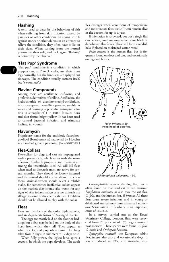
FlashingA term used to describe the behaviour of fishwhen suffering from skin irritation caused byparasites or other conditions. In trying to rubagainst stones or other objects in an attempt torelieve the condition, they often have to lie ontheir sides. When turning from the normalposition to their side, and back again, ‘flashing’is noticed by the observer.
‘Flat Pup’ Syndrome‘Flat pup’ syndrome is a condition in whichpuppies can, at 2 to 3 weeks, use their front legs normally, but the hind-legs are splayed outsideways. The condition usually corrects itself.(See ‘SWIMMERS’.)
Flavine CompoundsAmong these are acriflavine, euflavine, andproflavine, derivatives of aniline. Acriflavine, thehydrochloride of diamino-methyl-acridinium, is an orange-red crystalline powder, soluble inwater and forming a powerful antiseptic solu-tion in strengths of 1 in 1000. It stains horn and skin tissues bright yellow. It has been usedto control bacterial infection, and stimulatehealing, in wounds.
FlavomycinProprietary name for the antibiotic flavophos-pholipol (bambermycin) marketed by Hoechstas an in-feed growth promoter. (See ADDITIVES.)
Flea-CollarsFlea-collars for dogs and cats are impregnatedwith a parasiticide, which varies with the man-ufacturer. Carbaril, propoxur and diazinon areamong the insecticides used. All will kill fleaswhen used as directed; most are active for sev-eral months. They should be loosely fastenedand the animal should not be allowed to chewthem. Animal-owners should select a reliablemake, for sometimes ineffective collars appearon the market; they should also watch for anysigns of skin inflammation as a few animals areallergic to some of the chemicals used. Childrenshould not be allowed to play with the collar.
FleasFleas are members of the order Siphonaptera,and are degenerate forms of 2-winged insects.
The eggs are mostly laid on the floor or bed-ding; but a few may be laid on the body of thehost, from which they fall. They appear aswhite specks, and pop when burst. Hatchingtakes from 2 days (in summer) to 12 days or so.
When fully grown, the legless larva spins acocoon, in which the pupa develops. The adult
flea emerges when conditions of temperatureand moisture are favourable. It can remain alivein the cocoon for up to a year.
If infestation is suspected, but not a single fleacan be seen, combing may gather some black ordark-brown flea faeces. These will form a reddishhalo if placed on moistened cotton wool.
Pulex irritans is the human flea, but is fre-quently found on dogs and cats, and occasionallyon pigs and horses.
Ctenocephalides canis is the dog flea, but isoften found on man and cat. It can transmitDipylidium caninum, as also may the cat flea, C. felis, and the human flea, P. irritans. All thesefleas cause severe irritation, and in young ordebilitated animals may cause anaemia if numer-ous. Sensitisation to flea-bites is an importantcause of ECZEMA.
In a survey, carried out at the RoyalVeterinary College, London, fleas were recov-ered from 20 per cent of 193 dogs examinedpost-mortem. Three species were found: C. felis,C. canis, and Orchopeas howardi.
Spilopsyllus cuniculi, the European rabbitflea, infests also cats and occasionally dogs. Itwas introduced in 1966 into Australia, as a
260 Flashing
F
Pulex irritans. × 20.Inset: head of dog flea.
Echidnophaga gallinacea. × 30.
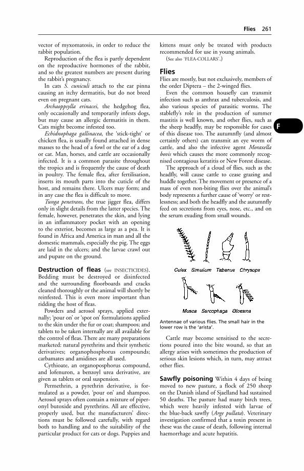
vector of myxomatosis, in order to reduce therabbit population.
Reproduction of the flea is partly dependenton the reproductive hormones of the rabbit,and so the greatest numbers are present duringthe rabbit’s pregnancy.
In cats S. cuniculi attach to the ear pinnacausing an itchy dermatitis, but do not breedeven on pregnant cats.
Archaoppsylla erinacei, the hedgehog flea,only occasionally and temporarily infests dogs,but may cause an allergic dermatitis in them.Cats might become infested too.
Echidnophaga gallinacea, the ‘stick-tight’ orchicken flea, is usually found attached in densemasses to the head of a fowl or the ear of a dogor cat. Man, horses, and cattle are occasionallyinfected. It is a common parasite throughoutthe tropics and is frequently the cause of deathin poultry. The female flea, after fertilisation,inserts its mouth parts into the cuticle of thehost, and remains there. Ulcers may form; andin any case the flea is difficult to move.
Tunga penetrans, the true jigger flea, differsonly in slight details from the latter species. Thefemale, however, penetrates the skin, and lyingin an inflammatory pocket with an opening to the exterior, becomes as large as a pea. It isfound in Africa and America in man and all thedomestic mammals, especially the pig. The eggsare laid in the ulcers; and the larvae crawl outand pupate on the ground.
Destruction of fleas (see INSECTICIDES).Bedding must be destroyed or disinfected and the surrounding floorboards and crackscleaned thoroughly or the animal will shortly bereinfested. This is even more important thanridding the host of fleas.
Powders and aerosol sprays, applied exter-nally; ‘pour on’ or ‘spot on’ formulations appliedto the skin under the fur or coat; shampoos; andtablets to be taken internally are all available forthe control of fleas. There are many preparationsmarketed: natural pyrethrins and their syntheticderivatives; organophosphorus compounds; carbamates and amidines are all used.
Cythioate, an organoposphorus compound,and lofenuron, a benzoyl urea derivative, aregiven as tablets or oral suspension.
Permethrin, a pyrethrin derivative, is for-mulated as a powder, ‘pour on’ and shampoo.Aerosol sprays often contain a mixture of piper-onyl butoxide and pyrethrins. All are effective,properly used, but the manufacturers’ direc-tions must be followed carefully, with regardboth to handling and to the suitability of theparticular product for cats or dogs. Puppies and
kittens must only be treated with products recommended for use in young animals.
(See also ‘FLEA-COLLARS’.)
FliesFlies are mostly, but not exclusively, members ofthe order Diptera – the 2-winged flies.
Even the common housefly can transmitinfection such as anthrax and tuberculosis, andalso various species of parasitic worms. The stablefly’s role in the production of summermastitis is well known, and other flies, such asthe sheep headfly, may be responsible for casesof this disease too. The autumnfly (and almostcertainly others) can transmit an eye worm ofcattle, and also the infective agent Moraxellabovis which causes the more commonly recog-nised contagious keratitis or New Forest disease.
The approach of a cloud of flies, such as theheadfly, will cause cattle to cease grazing andhuddle together. The movement or presence of amass of even non-biting flies over the animal’sbody represents a further cause of ‘worry’ or rest-lessness; and both the headfly and the autumnflyfeed on secretions from eyes, nose, etc., and onthe serum exuding from small wounds.
Cattle may become sensitised to the secre-tions poured into the bite wound, so that anallergy arises with sometimes the production ofserious skin lesions which, in turn, may attractother flies.
Sawfly poisoning Within 4 days of beingmoved to new pasture, a flock of 250 sheep on the Danish island of Sjaelland had sustained50 deaths. The pasture had many birch trees,which were heavily infested with larvae of the blue-back sawfly (Arge pullata). Veterinaryinvestigation confirmed that a toxin present inthese was the cause of death, following internalhaemorrhage and acute hepatitis.
Flies 261
F
Antennae of various flies. The small hair in thelower row is the ‘arista’.
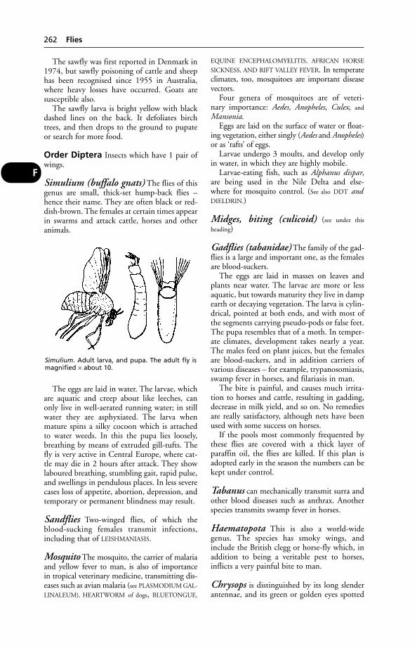
The sawfly was first reported in Denmark in1974, but sawfly poisoning of cattle and sheephas been recognised since 1955 in Australia,where heavy losses have occurred. Goats are susceptible also.
The sawfly larva is bright yellow with blackdashed lines on the back. It defoliates birchtrees, and then drops to the ground to pupateor search for more food.
Order Diptera Insects which have 1 pair ofwings.
Simulium (buffalo gnats) The flies of thisgenus are small, thick-set hump-back flies –hence their name. They are often black or red-dish-brown. The females at certain times appearin swarms and attack cattle, horses and otheranimals.
The eggs are laid in water. The larvae, whichare aquatic and creep about like leeches, canonly live in well-aerated running water; in stillwater they are asphyxiated. The larva whenmature spins a silky cocoon which is attached to water weeds. In this the pupa lies loosely,breathing by means of extruded gill-tufts. Thefly is very active in Central Europe, where cat-tle may die in 2 hours after attack. They showlaboured breathing, stumbling gait, rapid pulse,and swellings in pendulous places. In less severecases loss of appetite, abortion, depression, andtemporary or permanent blindness may result.
Sandflies Two-winged flies, of which theblood-sucking females transmit infections,including that of LEISHMANIASIS.
Mosquito The mosquito, the carrier of malariaand yellow fever to man, is also of importance in tropical veterinary medicine, transmitting dis-eases such as avian malaria (see PLASMODIUM GAL-
LINALEUM), HEARTWORM of dogs, BLUETONGUE,
EQUINE ENCEPHALOMYELITIS, AFRICAN HORSE
SICKNESS, AND RIFT VALLEY FEVER. In temperateclimates, too, mosquitoes are important diseasevectors.
Four genera of mosquitoes are of veteri-nary importance: Aedes, Anopheles, Culex, and
Mansonia.Eggs are laid on the surface of water or float-
ing vegetation, either singly (Aedes and Anopheles)or as ‘rafts’ of eggs.
Larvae undergo 3 moults, and develop onlyin water, in which they are highly mobile.
Larvae-eating fish, such as Alphanus dispar,are being used in the Nile Delta and else-where for mosquito control. (See also DDT andDIELDRIN.)
Midges, biting (culicoid) (see under this
heading)
Gadflies (tabanidae) The family of the gad-flies is a large and important one, as the femalesare blood-suckers.
The eggs are laid in masses on leaves andplants near water. The larvae are more or lessaquatic, but towards maturity they live in dampearth or decaying vegetation. The larva is cylin-drical, pointed at both ends, and with most ofthe segments carrying pseudo-pods or false feet.The pupa resembles that of a moth. In temper-ate climates, development takes nearly a year.The males feed on plant juices, but the femalesare blood-suckers, and in addition carriers ofvarious diseases – for example, trypanosomiasis,swamp fever in horses, and filariasis in man.
The bite is painful, and causes much irrita-tion to horses and cattle, resulting in gadding,decrease in milk yield, and so on. No remediesare really satisfactory, although nets have beenused with some success on horses.
If the pools most commonly frequented bythese flies are covered with a thick layer ofparaffin oil, the flies are killed. If this plan isadopted early in the season the numbers can bekept under control.
Tabanus can mechanically transmit surra andother blood diseases such as anthrax. Anotherspecies transmits swamp fever in horses.
Haematopota This is also a world-widegenus. The species has smoky wings, andinclude the British clegg or horse-fly which, inaddition to being a veritable pest to horses,inflicts a very painful bite to man.
Chrysops is distinguished by its long slenderantennae, and its green or golden eyes spotted
262 Flies
F
Simulium. Adult larva, and pupa. The adult fly ismagnified × about 10.
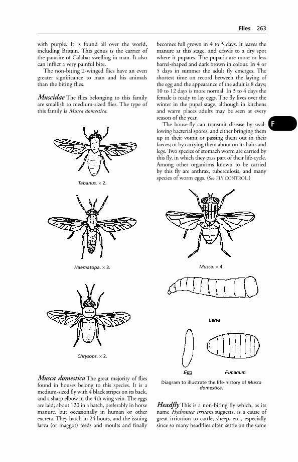
with purple. It is found all over the world,including Britain. This genus is the carrier ofthe parasite of Calabar swelling in man. It alsocan inflict a very painful bite.
The non-biting 2-winged flies have an evengreater significance to man and his animalsthan the biting flies.
Muscidae The flies belonging to this familyare smallish to medium-sized flies. The type ofthis family is Musca domestica.
Musca domestica The great majority of fliesfound in houses belong to this species. It is amedium-sized fly with 4 black stripes on its back,and a sharp elbow in the 4th wing vein. The eggsare laid; about 120 in a batch, preferably in horsemanure, but occasionally in human or other excreta. They hatch in 24 hours, and the issuinglarva (or maggot) feeds and moults and finally
becomes full grown in 4 to 5 days. It leaves themanure at this stage, and crawls to a dry spotwhere it pupates. The puparia are more or lessbarrel-shaped and dark brown in colour. In 4 or5 days in summer the adult fly emerges. Theshortest time on record between the laying of the egg and the appearance of the adult is 8 days;10 to 12 days is more normal. In 3 to 4 days thefemale is ready to lay eggs. The fly lives over thewinter in the pupal stage, although in kitchensand warm places adults may be seen at every season of the year.
The house-fly can transmit disease by swal-lowing bacterial spores, and either bringing themup in their vomit or passing them out in theirfaeces; or by carrying them about on its hairs andlegs. Two species of stomach worm are carried bythis fly, in which they pass part of their life-cycle.Among other organisms known to be carried by this fly are anthrax, tuberculosis, and manyspecies of worm eggs. (See FLY CONTROL.)
Headfly This is a non-biting fly which, as itsname Hydrotaea irritans suggests, is a cause ofgreat irritation to cattle, sheep, etc., especiallysince so many headflies often settle on the same
Flies 263
F
Tabanus. × 2.
Haematopa. × 3.
Chrysops. × 2.
Musca. × 4.
Diagram to illustrate the life-history of Muscadomestica.
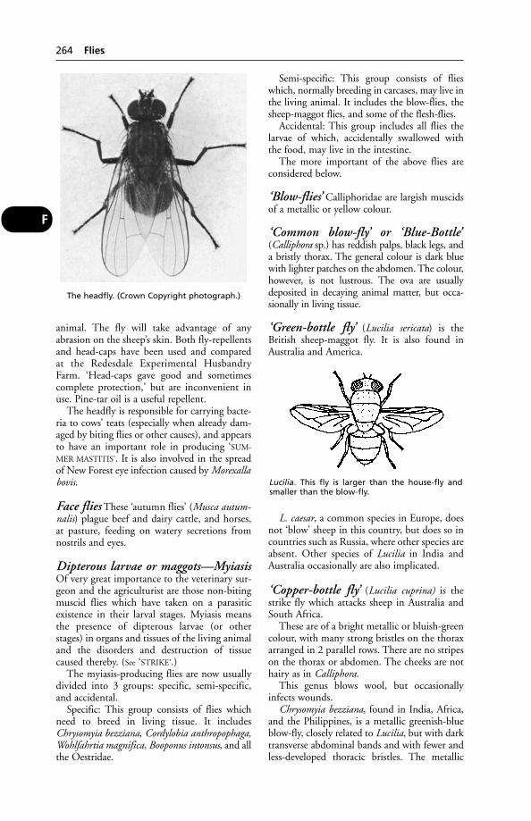
animal. The fly will take advantage of any abrasion on the sheep’s skin. Both fly-repellentsand head-caps have been used and compared at the Redesdale Experimental HusbandryFarm. ‘Head-caps gave good and sometimescomplete protection,’ but are inconvenient inuse. Pine-tar oil is a useful repellent.
The headfly is responsible for carrying bacte-ria to cows’ teats (especially when already dam-aged by biting flies or other causes), and appearsto have an important role in producing ‘SUM-
MER MASTITIS’. It is also involved in the spreadof New Forest eye infection caused by Morexallabovis.
Face flies These ‘autumn flies’ (Musca autum-nalis) plague beef and dairy cattle, and horses,at pasture, feeding on watery secretions fromnostrils and eyes.
Dipterous larvae or maggots—MyiasisOf very great importance to the veterinary sur-geon and the agriculturist are those non-bitingmuscid flies which have taken on a parasiticexistence in their larval stages. Myiasis meansthe presence of dipterous larvae (or otherstages) in organs and tissues of the living animaland the disorders and destruction of tissuecaused thereby. (See ‘STRIKE’.)
The myiasis-producing flies are now usuallydivided into 3 groups: specific, semi-specific,and accidental.
Specific: This group consists of flies whichneed to breed in living tissue. It includesChrysomyia bezziana, Cordylobia anthropophaga,Wohlfahrtia magnifica, Booponus intonsus, and allthe Oestridae.
Semi-specific: This group consists of flieswhich, normally breeding in carcases, may live inthe living animal. It includes the blow-flies, thesheep-maggot flies, and some of the flesh-flies.
Accidental: This group includes all flies thelarvae of which, accidentally swallowed withthe food, may live in the intestine.
The more important of the above flies areconsidered below.
‘Blow-flies’ Calliphoridae are largish muscidsof a metallic or yellow colour.
‘Common blow-fly’ or ‘Blue-Bottle’(Calliphora sp.) has reddish palps, black legs, anda bristly thorax. The general colour is dark bluewith lighter patches on the abdomen. The colour,however, is not lustrous. The ova are usuallydeposited in decaying animal matter, but occa-sionally in living tissue.
‘Green-bottle fly’ (Lucilia sericata) is theBritish sheep-maggot fly. It is also found inAustralia and America.
L. caesar, a common species in Europe, doesnot ‘blow’ sheep in this country, but does so incountries such as Russia, where other species areabsent. Other species of Lucilia in India andAustralia occasionally are also implicated.
‘Copper-bottle fly’ (Lucilia cuprina) is thestrike fly which attacks sheep in Australia andSouth Africa.
These are of a bright metallic or bluish-greencolour, with many strong bristles on the thoraxarranged in 2 parallel rows. There are no stripeson the thorax or abdomen. The cheeks are nothairy as in Calliphora.
This genus blows wool, but occasionallyinfects wounds.
Chrysomyia bezziana, found in India, Africa,and the Philippines, is a metallic greenish-blueblow-fly, closely related to Lucilia, but with darktransverse abdominal bands and with fewer andless-developed thoracic bristles. The metallic
264 Flies
F
The headfly. (Crown Copyright photograph.)
Lucilia. This fly is larger than the house-fly and smaller than the blow-fly.

sheen is more brassy than in Lucilia. This flybreeds only in living tissue – it discharges fromnatural orifices, or in sores and cuts. Up to 500eggs may be laid at one time. They hatch inabout 30 hours, and the larvae rapidly reachmaturity, crawl out and pupate on the ground.Several other species of this genus are semi-spe-cific myiasis flies, normally breeding in decayingmatter. These include C. albiceps, a notorioussheep-maggot fly in Australia.
‘Screw-worm fly’ (Callitroga americana) inAmerica can be distinguished from the old-world species by the 3 well-marked blue dorsalstripes on the thorax and dark hairs on theabdomen. It is of a dark bluish-green colour,with a well-marked yellowish-red face. (See also
FLY CONTROL.)This species will lay eggs in decaying animal
or vegetable matter, but will also oviposit in anydiseased tissue, in wounds in the vulvae of fresh-ly calved cows, the umbilical cord of calves, andso on. The ova hatch in 24 hours, and the mag-got matures in 4 to 6 days. The pupal stage onthe ground lasts 3 to 10 days. The maggot resem-bles a blue-bottle maggot, but the deeply cutconstrictions between segments and the promi-nent rings of spines give it its popular name.
As soon as the egg hatches, the larva startsburrowing into the flesh. It can penetrate thesound tissue of living animals, and may even laybare the bones.
‘Tumbu fly’ (Cordylobia anthropophaga) is aspecific myiasis fly in Africa, attacking manyhosts. It is a dirty brownish-yellow blow-flywith blackish markings. Eggs are laid in dustand rubbish on which the host, usually a dog, isaccustomed to lie. The small larva may liveapart from the host for 10 days, but it mayeventually burrow into the epidermis or die. Itmoults in this position, and forms a ‘tumbu’below the skin with an opening to the exteriorthrough which it breathes. The ‘tumbu’ doesnot suppurate unless the larva dies. The larvaemerges in about 7 or 8 days, and 2 or 3 dayslater it pupates. The adults emerge in about 20 days. This fly does not burrow into thedeeper tissues. The scrotum is a common site ofthe maggot. Putting a drop of oil or Vaselineover the breathing hole will force the larva toprotrude, when it can be removed.
Booponus intonsus is a light yellow specificmyiasis fly found in the Philippines, which issomewhat allied to Cordylobia. It infectsbovines and goats.
The eggs are laid on the hairs on the lowerparts of the legs; and the larvae make their way
to the coronet and bury themselves in the flesh.The larvae resemble the screw-worm. The larvalperiod seems to last 2 or 3 weeks, when it leavesthe host and pupates in the ground. The pupallife is 10 days.
The larvae cause a considerable lameness withnumerous superficial wounds and distortion ofthe horn. The larva is called the ‘foot maggot’.
‘Flesh flies’ (Sarcophagidae) are closely relatedto the Muscidae. The body is more elongatedthan that of the blow-flies, and they are usuallygrey in colour, with a mottled abdomen and astriped thorax. They generally bring forth livinglarvae instead of laying eggs. Two genera areimportant.
Sarcophaga spp. These are large grey flies withred eyes and square chequered markings on theabdomen. The 3rd segment of the antenna islong. All the species normally breed in decayinganimal matter, but may be found in old festeringwounds. They are found througout the world.
Wohlfahrtia magnifica resembles the preced-ing genus, but has well-defined round spots onthe abdomen. The 3rd segment of the antennais short and the arista is without bristles. It iswidely distributed in Russia, Asia Minor, andEgypt. The larvae never attack carcases, but arealways found in wounds and natural cavities ofliving animals. The fly deposits living larvae onsores and discharges.
Flies 265
FCordylobia. × 2½.
Wohlfahrtia. × 3.
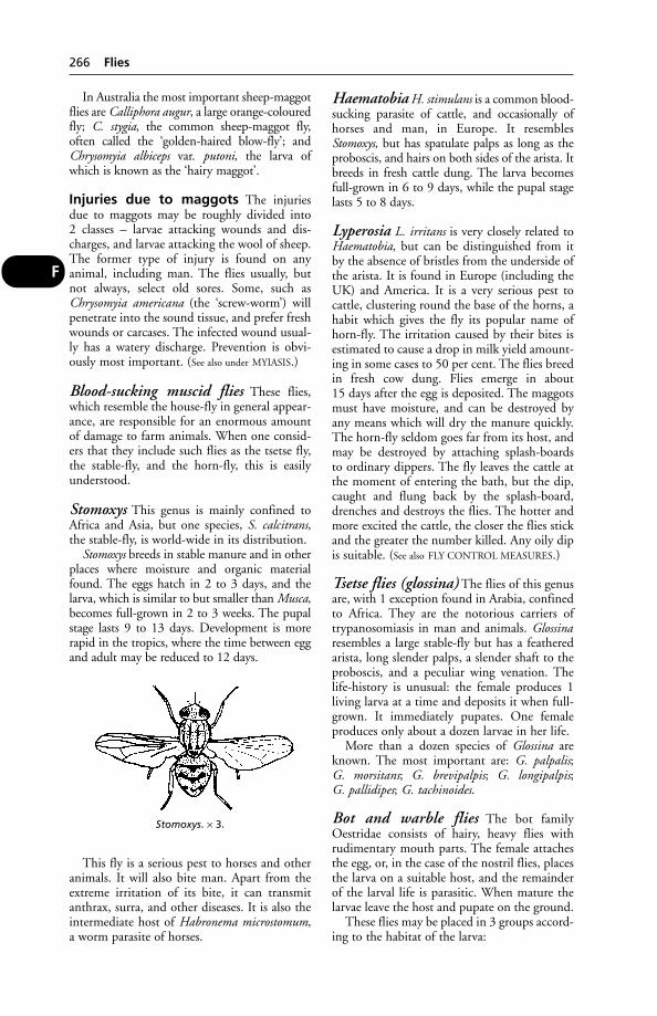
In Australia the most important sheep-maggotflies are Calliphora augur, a large orange-colouredfly; C. stygia, the common sheep-maggot fly,often called the ‘golden-haired blow-fly’; andChrysomyia albiceps var. putoni, the larva ofwhich is known as the ‘hairy maggot’.
Injuries due to maggots The injuries due to maggots may be roughly divided into 2 classes – larvae attacking wounds and dis-charges, and larvae attacking the wool of sheep.The former type of injury is found on any animal, including man. The flies usually, butnot always, select old sores. Some, such asChrysomyia americana (the ‘screw-worm’) willpenetrate into the sound tissue, and prefer freshwounds or carcases. The infected wound usual-ly has a watery discharge. Prevention is obvi-ously most important. (See also under MYIASIS.)
Blood-sucking muscid flies These flies,which resemble the house-fly in general appear-ance, are responsible for an enormous amountof damage to farm animals. When one consid-ers that they include such flies as the tsetse fly,the stable-fly, and the horn-fly, this is easilyunderstood.
Stomoxys This genus is mainly confined toAfrica and Asia, but one species, S. calcitrans,the stable-fly, is world-wide in its distribution.
Stomoxys breeds in stable manure and in otherplaces where moisture and organic materialfound. The eggs hatch in 2 to 3 days, and thelarva, which is similar to but smaller than Musca,becomes full-grown in 2 to 3 weeks. The pupalstage lasts 9 to 13 days. Development is morerapid in the tropics, where the time between eggand adult may be reduced to 12 days.
This fly is a serious pest to horses and otheranimals. It will also bite man. Apart from theextreme irritation of its bite, it can transmitanthrax, surra, and other diseases. It is also theintermediate host of Habronema microstomum,a worm parasite of horses.
Haematobia H. stimulans is a common blood-sucking parasite of cattle, and occasionally ofhorses and man, in Europe. It resemblesStomoxys, but has spatulate palps as long as theproboscis, and hairs on both sides of the arista. Itbreeds in fresh cattle dung. The larva becomesfull-grown in 6 to 9 days, while the pupal stagelasts 5 to 8 days.
Lyperosia L. irritans is very closely related toHaematobia, but can be distinguished from itby the absence of bristles from the underside ofthe arista. It is found in Europe (including theUK) and America. It is a very serious pest tocattle, clustering round the base of the horns, ahabit which gives the fly its popular name ofhorn-fly. The irritation caused by their bites isestimated to cause a drop in milk yield amount-ing in some cases to 50 per cent. The flies breedin fresh cow dung. Flies emerge in about 15 days after the egg is deposited. The maggotsmust have moisture, and can be destroyed byany means which will dry the manure quickly.The horn-fly seldom goes far from its host, andmay be destroyed by attaching splash-boards to ordinary dippers. The fly leaves the cattle atthe moment of entering the bath, but the dip,caught and flung back by the splash-board,drenches and destroys the flies. The hotter andmore excited the cattle, the closer the flies stickand the greater the number killed. Any oily dipis suitable. (See also FLY CONTROL MEASURES.)
Tsetse flies (glossina) The flies of this genusare, with 1 exception found in Arabia, confinedto Africa. They are the notorious carriers of trypanosomiasis in man and animals. Glossinaresembles a large stable-fly but has a featheredarista, long slender palps, a slender shaft to theproboscis, and a peculiar wing venation. Thelife-history is unusual: the female produces 1living larva at a time and deposits it when full-grown. It immediately pupates. One femaleproduces only about a dozen larvae in her life.
More than a dozen species of Glossina areknown. The most important are: G. palpalis; G. morsitans; G. brevipalpis; G. longipalpis; G. pallidipes; G. tachinoides.
Bot and warble flies The bot familyOestridae consists of hairy, heavy flies withrudimentary mouth parts. The female attachesthe egg, or, in the case of the nostril flies, placesthe larva on a suitable host, and the remainderof the larval life is parasitic. When mature thelarvae leave the host and pupate on the ground.
These flies may be placed in 3 groups accord-ing to the habitat of the larva:
266 Flies
F
Stomoxys. × 3.
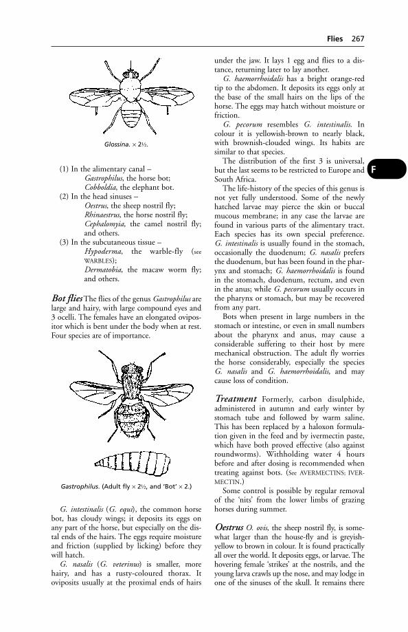
(1) In the alimentary canal –Gastrophilus, the horse bot;Cobboldia, the elephant bot.
(2) In the head sinuses –Oestrus, the sheep nostril fly;Rhinaestrus, the horse nostril fly;Cephalomyia, the camel nostril fly;and others.
(3) In the subcutaneous tissue –Hypoderma, the warble-fly (see
WARBLES);Dermatobia, the macaw worm fly;and others.
Bot flies The flies of the genus Gastrophilus arelarge and hairy, with large compound eyes and3 ocelli. The females have an elongated ovipos-itor which is bent under the body when at rest.Four species are of importance.
G. intestinalis (G. equi), the common horsebot, has cloudy wings; it deposits its eggs onany part of the horse, but especially on the dis-tal ends of the hairs. The eggs require moistureand friction (supplied by licking) before theywill hatch.
G. nasalis (G. veterinus) is smaller, morehairy, and has a rusty-coloured thorax. Itoviposits usually at the proximal ends of hairs
under the jaw. It lays 1 egg and flies to a dis-tance, returning later to lay another.
G. haemorrhoidalis has a bright orange-redtip to the abdomen. It deposits its eggs only atthe base of the small hairs on the lips of thehorse. The eggs may hatch without moisture orfriction.
G. pecorum resembles G. intestinalis. Incolour it is yellowish-brown to nearly black,with brownish-clouded wings. Its habits aresimilar to that species.
The distribution of the first 3 is universal,but the last seems to be restricted to Europe andSouth Africa.
The life-history of the species of this genus isnot yet fully understood. Some of the newlyhatched larvae may pierce the skin or buccalmucous membrane; in any case the larvae arefound in various parts of the alimentary tract.Each species has its own special preference. G. intestinalis is usually found in the stomach,occasionally the duodenum; G. nasalis prefersthe duodenum, but has been found in the phar-ynx and stomach; G. haemorrhoidalis is foundin the stomach, duodenum, rectum, and evenin the anus; while G. pecorum usually occurs inthe pharynx or stomach, but may be recoveredfrom any part.
Bots when present in large numbers in thestomach or intestine, or even in small numbersabout the pharynx and anus, may cause a considerable suffering to their host by meremechanical obstruction. The adult fly worriesthe horse considerably, especially the species G. nasalis and G. haemorrhoidalis, and maycause loss of condition.
Treatment Formerly, carbon disulphide,administered in autumn and early winter bystomach tube and followed by warm saline.This has been replaced by a haloxon formula-tion given in the feed and by ivermectin paste,which have both proved effective (also againstroundworms). Withholding water 4 hoursbefore and after dosing is recommended whentreating against bots. (See AVERMECTINS; IVER-
MECTIN.)Some control is possible by regular removal
of the ‘nits’ from the lower limbs of grazinghorses during summer.
Oestrus O. ovis, the sheep nostril fly, is some-what larger than the house-fly and is greyish-yellow to brown in colour. It is found practicallyall over the world. It deposits eggs, or larvae. Thehovering female ‘strikes’ at the nostrils, and theyoung larva crawls up the nose, and may lodge inone of the sinuses of the skull. It remains there
Flies 267
F
Glossina. × 2½.
Gastrophilus. (Adult fly × 2½, and ‘Bot’ × 2.)

until fully grown, when it is sneezed out andpupates in the ground.
Prevention is carried out by means of anapplication of tar to the nostrils. This may beapplied by means of a salt lick, access to whichmay only be obtained by smallish holes (5 cmor 2 inches) smeared with tar. Ploughing a single furrow across a sheep pasture allows thesheep to protect their nostrils from the flies,‘strike’, and gives some measure of protection.Some anthelmintics are effective.
Rhinoestrus R. purpureus (R. nasalis), thehorse nostril fly, is common in Central Europeand North Africa. It is a smallish fly with the body covered with small tubercles, and isclosely related to Oestrus. The female deposits anumber of living larvae at one time in the eyesor the nose of the horse (and occasionally man).The larvae may be found about the cranial cav-ities or even in the pharynx or larynx. Russiangadfly is a synonym.
Hypoderma Two species of warble-fly, H. bovisand H. lineatum, are found in cattle (and occa-sionally in the horse). Both are very extensivelyfound in Europe and America.
H. bovis is a largish fly with yellow hair justbehind the head. The underpart of theabdomen is nearly black, while the tail end isorange-yellow. The legs have few hairs.
H. lineatum is rather smaller with a reddish-orange tail and rough hairy legs.
H. bovis lays its eggs one on the base of each hair at a time. The fly has a most terrify-ing effect on cattle, and causes them to gallop madly in all directions. H. lineatumirritates animals less than does H. bovis. The ova are generally deposited while the animal is lying in the shade. A number of eggs – up to14 – are laid on the same hair, and are often infull view.
In both cases larvae emerge in several daysand pierce the skin. They travel up through theconnective tissue and finally reach the back.Under the skin the larvae form a small swelling(about the middle of winter), which movesabout at first, but gradually becomes still andenlarges. A small opening appears in the centrethrough which the larva breathes. In spring thelarva falls to the ground and pupates. Severalweeks later the adult fly emerges.
The presence of the larvae may decrease themilk yield by 10 to 20 per cent, cause a consid-erable depreciation in flesh near the pointswhere the larvae are, and enormously reducethe value of the hide. The adult fly also causesloss through the mad chasing about of cattle.(See also under WARBLES.)
H. diana is a warble-fly affecting deer.
Dermatobia D. hominis, the macaw worm-fly, is a parasite of cattle and other domesticat-ed animals (and occasionally man) in tropicalAmerica. It is a medium-sized fly, grey or steel-blue in colour, with pale brown wings. Thefemale lays its eggs on the body of some blood-sucking arthropod, usually a mosquito. Thiscarrier attacks an animal 5 or 6 days later, andthe larvae, rapidly escaping from their shells,pierce the skin of the host, and form a localtumour near where they were deposited. In amonth or so they emerge and pupate.
Pupipara This family, which includes thesheep ked and the horse ked (New Forest fly),was so called because live larvae are producedwhich pupate at once. The adults in this caseare blood-sucking parasites with a hard integu-ment with a broad neckless head and very stout
268 Flies
F
Oestrus. (Fly × 2; maggot × 1.)
Hypoderma. (Fly × 2; and ‘Warble’ × 1½.)

legs ending in grasping claws. Wings are presentor absent.
Hippobosca equina, the New Forest fly orhorse ked, has wings which, however, are sel-dom used, the fly preferring to run swiftlybetween the hairs of the host.
Paragle fly Paragle redicum, an anthomyiidfly, lays its eggs in canine faeces, and the whitespecks have been mistaken for tapeworm seg-ments. Larvae may also be passed alive throughthe canine (and also the human) gut.
Hymenoptera Insects which have 2 pairs ofwings.
Sawflies These have 4 wings and a saw-likeovipositor. The larvae are said to be poisonousif swallowed.
Fly control measures Perceptive farmershave for many years realised the harmful effectsof fly infestation on livestock. Controlled fieldtrials, comparing the productivity of treatedand untreated cattle, have convincingly demon-strated the advantages that can be gained bymodern fly-control methods.
Flies interfere with normal rest and feeding.The approach of a cloud of flies often causescattle to cease grazing and huddle together. Allcountrymen are familiar with the sight of cattle‘gadding’; and swarms of black fly (Simuliumornatum) may not merely cause store cattle tobreak out of fields, or rush round them, but canactually kill the animals.
The horse fly (Tabanus), like the stable fly(Stomoxys), has a painful bite, and the woundsinflicted attract other flies; this exacerbates the‘worry’ situation and often transmits even moreinfection. Some animals become sensitised tothe secretions of biting flies, so that an allergyresults. One example of this is the ‘sweet itch’ ofhorses caused by biting (Culicoid) midges.
Flies are notorious for spreading livestock dis-eases. Even the common housefly (Musca domes-tica) can transmit anthrax, tuberculosis, and the larvae of some parasitic worms. The headfly(Hydrotaea irritans) is among several species thatmay transmit the bacteria that cause ‘summermastitis’. This can lead to gangrene of part of theudder, usually with the permanent loss of use ofone-quarter – and to great pain and occasionallydeath of the cow.
Besides interfering with grazing, the headflycauses infested sheep to scratch, rub and knocktheir heads, often breaking the skin. Openwounds then attract other flies, increasing thetendency to self-mutilation. Sometimes thewhole poll region becomes raw. Pine-tar oil has been used as a repellent, and head caps forprotection, but they are inconvenient to use.
Plastic tags impregnated with syntheticpyrethroids have been effective in the reductionof ‘fly-worry’ in cattle. For sheep too, tags con-taining cypermethrin and permethrin can beeffective in controlling the severity of damagecaused by headflies. Cyromazine, in a ‘pour-on’formulation, is useful against blowfly larvae onsheep and lambs.
The activities of the autumn fly (Muscaautumnalis) are very similar to those of theheadfly. Both flies feed on secretions from nose,mouth, eyes and wounds and are among sever-al species that transmit the various pathogenscausing New Forest disease. This involves anacute and painful conjunctivitis, with inflam-mation also of the cornea, which becomesopaque, so that cattle are often rendered tem-porarily blind. Complications may result inpermanent eye defects and impaired sight; buteven without them the disease causes stress and,since it interferes with feeding, loss of conditioncan be appreciable.
Other bacterial diseases spread by fliesinclude salmonellosis and brucellosis; while
Flies 269
F
Dermatobia. (Fly and maggots × 1½.)
Hippobosca. × 2.
viral diseases such as swine fever and foot-and-mouth have in the past been similarlytransmitted in the UK. The larvae of some fliesalso parasitise animals; for example, the ‘green-bottle’ fly causing ‘strike’ in sheep, the sheepnostril fly, and the horse bot flies. The mereapproach of warble flies causes cattle to stam-pede, and the larvae undoubtedly cause painduring their migration through the cow’s body,and probably irritation while present in the skinswellings, or warbles. In a few instances, cowshave died following the accidental crushing oflarvae in the warbles, and larvae occasionallyseriously damage the spinal cord. In the UK,control measures have virtually eliminated warble infestation in cattle.
Deterring and killing flies The number offlies entering a milking parlour can be reducedby a spray-boom erected over the doorways, anda plain water mist, produced by ordinary sprin-klers or misters, has been recommended for use in collecting yards where a spray-boom overthe parlour entrance is impracticable. Such mea-sures, however, do not reduce the total fly pop-ulation of the farm – they do not kill. Electric flytraps do, and they can be useful when installedin piggeries and dairy cattle buildings. Whichflies are electrocuted will obviously depend onthe feeding and resting habits of the variousspecies.
Of far wider application, and the most effec-tive weapon against farm flies, is the insecticidalspray. This can be used to convert a livestockbuilding into one big fly trap. A wide variety ofeffective insecticides is available.
For housed stock, spraying walls may suffice;but beef and dairy cattle at grass will be the target of flies coming from their resting placesamong trees. Fly control, if it is to benefit graz-ing animals, therefore requires application of an officially approved insecticide direct to theirbacks. For this purpose, the synthetic pyrethroidsare effective.
These insecticides are chemically allied to the active ingredients of pyrethrum, but aremore potent as fly-killers and are also light-stable so that they stay effective for longer in the sun.
Fly control for horses and livestockNumerous formulations based on cypermethrinand other synthetic pyrethroids are available, asboth a spray and as pour-on applications, whichare convenient to use.
A PVC fly band impregnated with cyperme-thrin, for threading on to either the browbandor the crownpiece of a headcollar, and an ear tag
impregnated with 8 per cent cypermethrin, arealso available for cattle at pasture.
Whether measured in terms of reduced ani-mal suffering, or farmers’ incomes, or a loweredincidence of diseases – some of which are of public-health as well as economic importance –fly control is very worthwhile. If further evidenceof its effectiveness were needed, any doubtershould note the success of the UK government’s5-year plan to eradicate warble flies from theUK. Now, only sporadic outbreaks of warbleinfestation occur. Since horses, as well as cattle,suffer from warble-fly larvae (although to a muchlesser extent), eradication has also benefitedthem and their owners.
Overseas Similar methods to those describedabove, suitably adapted for tropical climates,are in use. The spraying of ground with DDTand dieldrin has been very effective for the con-trol of tsetse flies and human trypanosomiasis,but the practice was discontinued because ofthe development of resistance and the toxicityof those peparations to other species (see also
under TROPICS).The release of sterile male flies from aircraft
has been used on a large scale in Puerto Rico tocontrol the screw-worm fly.
Genetically engineered blowfly maggots have been used in attempts to eradicate sheepblowflies. The maggots were altered so thatfemales which mate produce blind or sterile off-spring – a hereditary characteristic which will‘confer genetic death on future generations’.
(See also DIPS AND DIPPING.)
Floods(see PASTURE CONTAMINATION; SALMONELLOSIS;WATER-DROPWORT)
Floor(see BEDDING AND BEDDING MATERIALS – Pigs;HOUSING OF ANIMALS)
Floor-Feeding of PigsThis practice is attractive to the pig farmer sinceit eliminates the cost of troughs and also savesspace – the normal feeding passage becoming acatwalk over the pigs’ sleeping quarters.
From a health point of view, the precise composition of the concrete floor may proveimportant. In an outbreak of illness among pigs in Eire, with anaemia, gastric ulceration,and haemorrhage, the cause was thought to bethe ‘pit sand’ (with a high iron content) withwhich the concrete was made, giving rise toiron poisoning once the surface layer had beenlicked off.
270 Floods
F

More important is the fact that loss of appetitein pigs – a common symptom of many diseases– may not be noticed. With trough-feeding, it iseasy to see which pigs are uninterested in food.
Feeding pellets instead of meal may also causetrouble – digestive upsets. The method mayinvolve more stress than conventional systems.
Floor SpaceAs a rough guide, the following minimum figures may be given: bacon pig, 2 m2 (6 squarefeet); veal calf, 3.5 m2 (12 square feet); layinghen on deep litter, 1 m2 (21⁄2 square feet).
Floor SweepingsFloor sweepings in mill or barn have been addedto feed and caused fatal poisoning. For example,pigs have died in this way from nitrate poison-ing, and cattle from mustard seed poisoning.
‘Floppy’ LabradorsThe colloquial name for an inherited muscledisease of both Black and Golden Labradorretrievers. The condition has been seen in boththe UK and the USA. Inheritance is associatedwith an autosomal recessive gene, leading to adeficiency of type II muscle fibres.
Signs Poor exercise tolerance, especially in coldweather, a stiff hopping gait, with sometimescollapse. Signs have been shown as early as 8 weeks of age, but in other cases after severalmonths.
FlorfenicolA chloramphenicol-like antibiotic which is usedin cattle to treat infections caused by Pasteurella,Mannheimia and Haemophilus spp. It is also usedin the treatment of fish, particularly in the treat-ment of furunculosis (Aeromonas salmonicidainfection) in farmed Atlantic salmon. It is notrecommended for use in breeding stock. Theantibiotic is administered by injection in cattleand in the feed of fish.
Flour(see AGENE PROCESS)
Flour-Mite InfestationInfestation of animal feeds by flour mites (Acarusfarinae; A. siro) can cause a significant loss ofnutrient value, as explained under DIET. Likeforage mites of various species, flour mites canalso cause an irritating parasitic skin disease ofanimals. In one incident, 36 police horses werestabled in a building which was cleaned andwhitewashed before their arrival. Unfortunately afeed barrow was overlooked and still contained
oats left over from the previous year. New oats ofhigh quality were delivered in sacks, and thedelivery man opened one sack and topped up thebarrow. After he had gone, new oats continuedto be put on top of what was left in the large feedbarrow, which was never completely emptied. Afortnight after the horses’ arrival, the last 4 in theline showed signs of head and neck irritation.One horse had rubbed the side of its neck bare;2 others had dermatitis on the poll and alongsidethe mane.
Examination of the bottom layer of the bar-row’s contents revealed an enormous number offlour mites, and these were also isolated fromthe skin lesions.
An unsuspected cause of diarrhoea in dogsmay be dog biscuits or meal, stored in large bins,and heavily infested with forage mites. As fliesmay carry nymphal forage mites, fly control isimportant in reducing such infestations.
Forage mites and/or their eggs may be foundin dog faeces, where they may have been mis-taken for the eggs of strongyle worms. However,the mite’s egg is nearly twice the size of theworm’s.
Flour mites (A. siro and A. farinae), thehouse/furniture mite (Glycyphagus domesticus),and the mould mites (Tyrophagus putrescentiaeand T. longior) may also be involved.
FluanisoneA neuroleptic drug used in small animals (ferrets.rabbits, guinea pigs, rats and mice). It is usuallymixed with FENTANYL)
FlugestoneA hormone preparation supplied in impreg-nated sponges for the synchronisation of oestrusin ewes. It may also be used to induce oestrusand ovulation in the non-breeding season (see
CONTROLLED BREEDING).
Fluid Replacement Therapy(see under DEHYDRATION)
Flukes and Fluke Disease(see LIVER FLUKES; LUNG FLUKES; SCHISTOSOMI-
ASIS for blood flukes; PANCREAS, DISEASES OF;and RUMEN FLUKES)
FlumethrinA synthetic pyrethroid compound used for thetreatment of sheep scab and tick infestation.Impregnated in plastic strips, it is hung in beehives to treat varroasis in honey bees.
Flunixine MeglumineA non-steroidal anti-inflammatory drug used forrelief of pain and inflammation in horses, dogs
Flunixine Meglumine 271
F
and cattle; formulated as granules, tablets orinjection. Proprietary preparations are Finadyne(Schering-Plough), Cronyxin (Bimeda), andBinixin (Bayer).
Fluorescent(see under TETRACYCLINES which make bone flu-oresce, and under WOOD’S LAMP which showsringworm-affected hairs fluorescing. For thefluorescent antibody test, see under RABIES and
IMMUNOFLUORESCENT MICROSCOPY.)
Fluorescin (Fluorescein)Fluorescin (Fluorescein) is a useful diagnosticagent in injuries and ulcers on the cornea of theeye. A weak solution is dropped into the eye andthe injured area can be seen clearly demarcatedfrom the surrounding healthy cornea.
FluorineThis element occurs in body tissues and insome natural water supplies. Excess of fluorinecauses mottling of the teeth. (For fluorine poisoning, see FLUOROSIS.)
Fluoroacetate PoisoningSodium mono-fluoroacetate is used to kill ratsand mice, and it is in this connection that poi-soning in domestic animals and man may arise.The drug causes distress, yelping, sometimesvomiting, and convulsions in the dog. Treatmentconsists in the administration of nembutal. Adose as small as 0.66 mg per kg body weight is fatal.
In 1963, 2 outbreaks of fatal poisoninginvolving numerous dogs, cats, cattle and a ponywere attributed to the agricultural insecticide fluoroacetamide, a closely related compound.
FluorosisFluorosis, or chronic fluorine poisoning, is of economic importance in cattle, sheep, etc.,grazing pastures contaminated by fluorine com-pounds emanating from iron and steel worksand other industrial plant. It has also beenreported in dairy cattle receiving mineral sup-plements with a high fluorine content, theresult of incorporation of rock phosphate. Thisis something which animal feed manufacturersshould guard against, and they should offerguarantees concerning maximum fluorine content in their products.
Signs There is severe lameness, and a resultingloss of condition; milk yield is greatly reduced.The teeth may become mottled, and the bonesparticularly liable to fracture. Cows may standwith their legs crossed in cases of fracture of the
pedal bones. Hip lameness is probably morecommon.
Antidote Calcium aluminate is of some limitedvalue as an antidote to fluorine poisoning.
FluothaneFluothane is a trade name for HALOTHANE.
FlurbiprofenFluriprofen, Flurbiprofen and Ibuprofen arenon-steroidal anti-inflammatory drugs used inhuman medicine, and sometimes given to dogsby their owners, or eaten by dogs with access to the tablets, with resultant poisoning (some-times fatal). Stomach ulceration and kidneyfailure have been caused.
Eye drops containing flurbiprofen are used aspre-operative treatment for cataract extraction.
Flushing of EwesFlushing of ewes aims for rising metabolism inbreeding ewes some 6 to 3 weeks before service,by putting them on to protein-rich feed. Thepurpose is to intensify subsequent oestrus andthereby ensure that each ewe is in fit conditionto breed. Some trials, however, have failed todemonstrate the effectiveness of the practice.
Foaling(see PARTURITION)
FoalsFoals are young horses of either sex until thetime they are 1 year old. Male foals are knownas ‘colt foals’, and female foals are called ‘fillyfoals’. Most foals are born between March andJune in Britain, although quite a number (espe-cially thoroughbreds) are dropped earlier thanthis. Thoroughbreds are conventionally aged as from January 1 of the year in which they are born, and all other horses from May 1, irre-spective of whether they were actually foaledbefore or after these dates.
Generally speaking, foals run with their damsat grass during the summer, and are weaned at 4 to 6 months of age. With weakly foals, how-ever, and in the case of highly bred pedigree animals, it is not uncommon to allow them torun with their dams until nearly Christmas-time, so that they may get an exceptionally goodstart in life.
As a rule, foals will begin to eat grass whenthey are between 3 weeks and a month old,although some start earlier and some later thanthis. At about 6 weeks to 21⁄2 months they willbegin to eat dry corn from mangers along withtheir mothers.
272 Fluorescent
F

Foals, Diseases of
Diarrhoea may occur as a result of changes in the mare’s milk, or as a result of the damgrazing avidly upon rich spring grass, etc.
Salmonella typhimurium may cause a subclin-ical infection; alternatively acute and severe diar-rhoea or septicaemia may occur, the latter oftenfollowing the former. (See SALMONELLOSIS.)
E. coli is another cause of acute diarrhoea inthe foal. (See E. COLI.)
Corynebacterium equi is probably a morecommon cause of pneumonia than of diarrhoea;nevertheless the latter can be severe. Clostridialenterotoxaemia occurs; likewise campylobacterinfections.
Viruses causing diarrhoea in foals include a CORONAVIRUS and a ROTAVIRUS. The lattermay be associated with a profuse, watery diar-rhoea and lymph-node enlargement, sometimesfollowed by death. (See also GLOBIDIOSIS.)
Navel-ill and joint-ill These are colloquialterms for dangerous infections which mayattack the foal within the first fortnight of itslife. Pain and swelling at the navel, with some-times abscesses along the umbilical vein, mayoccur; but joint-ill (polyarthritis) symptomsmay be noticed first. Often the hock and stiflejoints are painful and swollen, there is fever, andthe foal is obviously ill.
Treatment includes the use of appropriateantibiotics (but not corticosteroids, which havean immunosuppressive effect).
Septicaemia is always a danger likely to arisewith or after the last 2 conditions. Septicaemiadue to Actinobacillus equuli infection (‘sleepyfoal disease’) may occur within the first 3 daysof life. The foal becomes dull, disinclined tosuck, has stupor and diarrhoea, and prostrationand death quickly follow. Polyarthritis mayoccur in foals which survive a little longer.
Organisms causing septicaemia and joint-ill include E. coli, Actinobacillus equi, Strep.zooepidemicus and Klebsiella pneumonia.
Haemolytic disease This results from anincompatibility between the blood of sire anddam, and the consequent production of anti-bodies which reach the foal in the colostrumand break down the foal’s red blood cells. Thesebecome so reduced in number that not only isthere jaundice, but often also a fatal anaemia. Iftrouble from this cause were anticipated, theuse of a foster-mother might save the situationbut, obviously, this is seldom practicable.Moreover, unless the mare has previously had a
‘jaundiced foal’, there will be no indication ofthe problem. If she has previously had such afoal, however, it is possible to test her bloodagainst the sire’s during pregnancy and obtain a fairly good idea as to their incompatibility or otherwise. Treatment must be undertakenquickly and consists in exchange transfusion –the removal of up to 2.8 litres (5 pints) of thefoal’s blood and the simultaneous injection ofup to 3.5 litres (6 pints) of a compatible donor’sblood, previously collected. The transfusionrequires special apparatus and takes about 3 hours to complete. Recoveries following thistreatment have been spectacular.
Worms Both strongyles (red worms) andascarids may cause trouble in foals. The formergive rise to malaise, cough, unthriftiness, tired-ness, and sometimes abdominal pain; the latterto diarrhoea and intermittent colic, amongother symptoms. The Animal Health Trust rec-ommends that foals be dosed every 4 weeks,alternately for ascarids and strongyles, until theage of 1 year.
Bone diseases in foals include valgus (see
under BONE, DISEASES OF); and RICKETS whichcan render the growth-plate more vulnerable toinjury.
Skin diseases Congenital or inherited con-ditions include Epitheliogenesis imperfecta andICTHIOSIS. ALOPECIA is sometimes a complica-tion of STRANGLES; to which URTICARIA is anoccasional sequel.
Pneumonia, due to Corynebacterium equi,occurs sporadically in the UK. A suppurativebroncho-pneumonia, with abscesses in thelungs and pulmonary lymph nodes, it may beassociated with ‘joint-ill’ and osteomyelitis.
Rhinopneumonitis A congenital infectionwith EHV 1 virus is a case of early death infoals. (See EQUINE RHINOPNEUMONITIS.)
Muscular dystrophy (see under MUSCLES,DISEASES OF)
Pervious urachus, or ‘leaking at the navel’,is a condition in which the communicationbetween the urinary bladder and the umbilicusor navel outside, which should close at the time of birth, remains patent and allows urineto dribble from it. The urine blisters the skinaround, and ultimately results in considerableswelling and suppuration around the navel. Thecondition should be corrected by surgery.
Foals, Diseases of 273
F

Combined immunodeficiency (CID)occurs in some Arab foals. (See IMMUNODEFI-
CIENCY; also under HERNIA; VALGUS; TYZZER’S
DISEASE.)
Fodder Beet(see POISONING)
Fodder Mites(see FLOUR MITES)
Foetal, Foetus(see FETAL; FETUS)
Fog(see under SMOG and ‘FOG FEVER’)
Fog FeverThe colloquial name derived from the word‘fog’, meaning the second crop of grass takenfrom pasture already cut once that season for hay or silage, has caused much confusion, sinceit has been applied to several different syndromesin cattle.
True fog fever has been defined as acute pulmonary emphysema occurring in adult cattle (typically single suckler beef cows) whichhave been moved from poor to lush grazing in the autumn. It should be differentiated fromparasitic bronchitis, and also from extrinsicallergic alveolitis caused by micro-organisms inmouldy hay.
It may be identical with the acute bovinepulmonary emphysema encountered in NorthAmerican adult cattle moved from range to lushpasture.
Cause The probable cause is the conversion inthe cow’s lumen of the amino acid L-tryptophanin grass to 3-methylindole, a toxic substance.
Two clinical forms of fog fever may be seen.The mild form affects up to 50 or 60 per centwithin affected groups of cattle, the animalremaining bright though breathing ratherrapidly. In the severe form, the degree of respi-ratory distress varies greatly, is often severe, andresults in a 30 per cent mortality, with deathcommonly occurring within the first 2 days ofthe illness.
Post-mortem Lesions include pulmonarycongestion, oedema, and hyaline membranes,interstitial emphysema and diffuse alveolarepithelial hyperplasia.
A fog-fever-like condition in sheep, 6 monthsold, was seen in Sweden. All became ill within 3 days of being moved from poor to lush pasture.
FoggageAftermath, ‘fog’. Grass grown for winter grazing.
Folic AcidOne of the vitamins of the B-complex. (See
VITAMINS.)
Follicle(see SKIN; OVARY)
Follicle Stimulating Hormone(FSH)This stimulates the development of theGraafian follicles in the ovary, and controls thesecretion of oestrogens from the ovary. It issecreted by the anterior lobe of the pituitarygland. In the male animal, FSH stimulatessperm production in the testicle.
Follicular MangeFollicular mange is another name for demodec-tic mange due to the parasite Demodex canis,which lives in the hair follicles of the skin. (See
MITES, PARASITIC.)
Fomentation(see POULTICES)
FomitesFomites is a term used to include all articles thathave been in actual contact with a sick animal,so as to retain some of the infective material andbe capable of spreading the disease. Beddingmaterial, fodder, mangers, stable or byre uten-sils, clothing, grooming tools, the clothes of anattendant, or even the attendant him or herself,may all be fomites.
Food Allergies(see ALLERGIES; FATTY ACIDS)
Food Conversion Ratio(see FEED CONVERSION RATIO)
Food InspectionIn countries such as Denmark and the USA, theinspection of meat and meat-derived productshas long been carried out entirely by membersof the veterinary profession. In the UK this has been only partly so, but to conform withEU regulations meat inspection, including pre-slaughter inspection of food animals, and theexamination of organs and tissues as well asinspection of the dressed carcase are carried outby Official Veterinary Surgeons under the cen-tralised control of the Meat Hygiene Service, a government agency. For conditions which
274 Fodder Beet
F

render meat dangerous as food, see TUBERCULO-
SIS; SALMONELLOSIS; ANTHRAX; TRICHINOSIS;HYDATID DISEASE, etc.
Food Poisoning in Man(see E. COLI; SALMONELLOSIS; CAMPYLOBACTER;ROTAVIRUS; also BOTULISM). In the UK, salmo-nellosis is the most frequent cause of food poi-soning but E. coli is becoming more common.Clostridium prefringens, Staphylococcus aureus andBacillus cereus are also found. Yersiniosis and lis-teriosis, from infected milk or cheese, also occur.
Where meals are prepared for a number ofpeople, as in homes for elderly people whoseresistance is lowered, the risk of food poisoningis increased. The very young and immunologi-cally comprised people are also more at risk.Following serious outbreaks of poisoning by E. coli 1057 in Scotland in 1997, a governmentreport by Professor Hugh Pennington recom-mended a series of measures to raise hygienestandards in food shops, etc.
Food Safety Act 1990This sets out regulations covering the whole ofthe food chain from retailers back to primaryproducers.
Food Standards AgencyAn independent body established by the UKgovernment with a brief to ‘protect publichealth from risks which may arise with the con-sumption of food, and otherwise to protect theinterests of consumers in relation to food’. Theagency reports to government but can publishits advice independently. The Meat HygieneService, the agency responsible for meat inspec-tion in all licensed abattoirs, reports to the FSA.
Foods and Feeding(see DIET AND DIETETICS; NURSING OF SICK
ANIMALS; RATIONS; also CAT FOODS)
Fool’s Parsley,Although a member of the natural orderUmbellifera – very many of the members ofwhich are poisonous (e.g. water hemlock, waterdropwort, and hemlock) – the extremely com-mon fool’s parsley (Aethusa cynapium) is not afrequent cause of poisoning in animals. It isdangerous when fed to rabbits, if it is pulled in the early green succulent stage before theflowering tops are formed.
Under ordinary circumstances herbivorousanimals do not readily eat fool’s parsley, for atthe time when its growth is most luxuriant (i.e.in spring) there is generally an abundance ofgrass, which they prefer.
Signs In cows, there have been seen a loss of appetite, salivation, fever, uncertain gait, andparalysis of the hindquarters. In horses, aninstance has been recorded in which a numberof animals ate the plant in quantity; those whichhad white muzzles and feet became attackedwith diarrhoea and all white areas of the bodybecame severely inflamed, but other horses of awhole-colour remained unaffected. (See LIGHT
SENSITISATION.) In other cases stupor, paralysis,and convulsions have been noticed.
First-aid Drenches of strong black tea or coffee should be given so that the tannic acid inthem may combine with the alkaloids of theplant and form inert substances.
Foot-and-Mouth Disease (FMD)A very highly contagious NOTIFIABLE DISEASE
that can affect all cloven-hooved species; othernames are aphthous fever or epizootic aphtha. Itis characterised by the formation of small vesi-cles (fluid-filled blisters) in the mouth and onthe feet or, in the female, on the skin of theudder or teat. Economically, it can be devastat-ing, particularly in cattle, pigs and sheep. Thecosts of eradication or control, involving diag-nostic services, slaughter of infected or at-riskanimals, compensation to farmers, disposal ofcarcases, or adoption of a vaccination policy,and loss of trade can be enormous. Where thedisease is not dealt with and becomes endemic,failure to thrive and consequent loss of produc-tion and a total ban on export of live animalsand, to a very large extent, animal products canbe very serious. FMD has been considered thesingle most important constraint to trade inanimals and animal products.
Foot-and-mouth disease has occurred in virtually every country in the world in whichcattle, sheep or pigs are kept. It has beenendemic in parts of South America, continentalEurope, Asia and Africa. Except in young animals, where the death rate may be up to 50 per cent, the disease is not characterised byhigh mortality. Usually fewer than 5 per cent ofinfected adult animals die.
The disease is transmissible to humans butthe infection is usually mild and transient. It isnot the same as (human) HAND, FOOT AND
MOUTH DISEASE.
Cause Foot-and-mouth disease is caused by anaphthovirus of which 7 types are recognised –including the 3 known as O (now named thePanAsia strain), A and C, which have causedoutbreaks of the disease in Britain, and 4 moretypes which so far have been confined to Asia
Foot-and-Mouth Disease (FMD) 275
F
and Africa – Asia and Sat 1, Sat 2, and Sat 3. Itis the PanAsia strain of type O, now dominantacross much of Asia and the Middle East, thatwas responsible for the major outbreak in 2001.No cross-immunity is exhibited between types,and only partial cross-immunity between subtypes within a type. The virus has a highgenetic variability.
The virus is present in the vesicles and in thefluid which comes from them when they burst;and since there is nearly always an excessivesecretion of saliva from an animal affected withlesions in the mouth, it is through the mediumof contamination with saliva that the disease is perhaps most readily spread. As well as this,however, the urine, faeces, and small amountsof serum from lesions in the feet, are factors inthe spread to other animals; livestock marketsand transport vehicles are particularly impor-tant in causing widening of an outbreak. But as the virus can survive for very considerablelengths of time, it may be picked up and spreadby almost any object or animal that has been incontact with affected animals. Migratory birdsin their flights from one country to anothermay act as carriers. Spread can occur by wind,watercourses, people and vehicles.
Bulk collection of milk has also been impli-cated. FMD virus may be excreted in milkbefore symptoms in the cow have appeared orbecome obvious.
The virus can survive in frozen liver or kid-ney for 4 months or more, and in bull semenstored at low temperatures.
The use of swill containing scraps of meat,bones, or other animal tissue for feeding to pigsis a very important factor in the spread of foot-and-mouth disease, and because of the numberof outbreaks traced to swill, the (then) Ministryof Agriculture specified that swill shall havebeen boiled for at least 1 hour before being fedto pigs. It may have been neglect of this proce-dure that led to the 2001 UK outbreak. Thefeeding of swill is now completely banned.
The incubation period before signs of thedisease appear after infection varies from 1 to15 days, but the majority of cases show signsbetween the 2nd and the 6th day after havingbeen exposed to infection. An important fea-ture of the disease in relation to its infectivity isthat virus may be excreted before symptomsbecome evident; thus the infection may spreadbefore the farmer is aware that his stock areinfected.
With pigs, 10 days may elapse betweenexcretion of virus and the development oflesions. With cattle and sheep, the figure maybe 5 days; or an average of 21⁄2 days.
Signs
Cattle At first, animals become dull, refusetheir food, lie about in a sluggish manner, andcows suddenly give a lessened flow of milk.Their temperatures rapidly rise to 40° or 40.5°C(104° or 105°F), and fever is maintained untilthe crop of vesicles form, after which it subsides.A few hours after the initial dullness has beennoticed, affected animals usually commence tosalivate profusely – long ropes of stringy salivahanging from the mouth. Lameness may be thefirst sign of the disease. Foot lesions generallyappear 4 or 5 days after the vesicles form in the mouth. In these, blisters form around thecoronets, between the claws.
The animal frequently smacks its lips in acharacteristic manner, yawns, and protrudes itstongue. Blisters are found in all stages of devel-opment: the most common locations are on thedental pad and in the upper incisor region; onthe tongue, especially around its tip; and on theinsides of the cheeks and gums. The blisters each run a similar course; for a few hours theygradually rise, then they burst, liberating a smallamount of yellowish, straw-coloured serum(which should be regarded as highly infectiveand as containing the virus). There remainsbehind a shallow, eroded, red, raw, ulcer-likearea, to the edges of which little pieces ofmucous membrane adhere for a short time until they are removed by the movements of the mouth. Adjacent affected areas merge, andin bad cases large irregular, ragged, red patchesform, from the surfaces of which the mucousmembrane has disappeared. The lesions arealways extremely painful, and in consequencethe animal is prevented from feeding. Generally,it can still drink, and it will often take liquid orvery soft food, but it refuses dry food entirely. Infrom 6 days to a fortnight or so, healing begins,the lesions disappear and the animal appears to have recovered. It is, however, still carryingthe virus.
Sheep Foot-and-mouth disease may be diffi-cult to detect. Onset of lameness is variable –between 2 days and a week – and may pass offquickly. Vesicles in the mouth rupture and healquickly, leaving no sign of lesions. The sheepmay not appear obviously sick and feet andmouth must be examined closely to detect signsof infection.
Pigs Foot lesions usually begin either at thecoronet or at the heels instead of between the claws as in cattle. The muzzle and end ofthe snout may show lesions.
276 Foot-and-Mouth Disease (FMD)
F

In all affected species, the illness may causeanimals to lose much weight, or to cease togrow. Abortion, infertility and diabetes areoccasional complications. Foot-and-mouth dis-ease may be the cause of sudden death in pigs,cattle and sheep.
Animals in milk – cows, ewes and sows –may develop characteristic lesions upon theirteats or upon the skin of the udder. The lesionsare similar to those forming in the mouth, butthey take longer to mature. In some cases the whole of the tip of the teat shows a singlelarge blister, which is soon burst by milking or sucking. Subsequently an eroded appear-ance remains, until healing is established. Milksecretion rapidly diminishes. Permanent udderdamage may result from the disease. The pain is usually acute, and the milk – contami-nated with the exudate and with dischargesfrom the lesions – is highly infective to younganimals.
Differential diagnosis It is necessary to distinguish between foot-and-mouth disease,swine vesicular disease and vesicular stomatitisby laboratory tests. In the UK, tissue and bloodsamples from suspect animals are sent to theInstitute of Animal Health (IAH) Pirbright lab-oratory for testing. If large quantities of virusare present, the test can give a positive result in4 hours. In some cases, it may be necessary tomultiply the virus by culture in cells and it maytake up to 4 days to confirm that virus is notpresent. The cell culture technique is also usedto provide material for identifying the precisetype and strain of the virus.
Control A policy of slaughtering affected andin-contact animals is operated in the UK,Canada, the USA, Norway and countriesthroughout the European Union where the disease is not endemic. Such a policy, involvingcompensation to owners of compulsorily slaugh-tered animals, is normally far less costly than along-term vaccination policy. Slaughtered ani-mals are disposed of by burning or burial; affect-ed premises are thoroughly disinfected and the holding and surrounding area subject to aperiod of quarantine. Vaccination is practised incountries where a slaughter policy is unworkablebecause the disease is endemic and its incidencehigh; not vice versa, as might be thought bythose who condemn the slaughter policy withouthaving studied the reasons for it. ‘Overall’ vacci-nation is seldom practicable in such areas for reasons of cost, so ‘frontier’, or ‘ring’, vaccination(of all susceptible animals within a given radius
of an outbreak) is usually practised. Israel is anexception; the high-yielding diary herds are vaccinated annually.
As a temporary measure, animals in areas surrounding outbreaks may be vaccinated toprovide a ‘ring fence’ against infection. Contraryto its general policy, to protect Greece, the EUoperates a vaccination policy in EuropeanTurkey and part of Turkey-in-Asia. The use ofvaccination was seriously considered in the UK2001 outbreak when the disease threatened torun out of control and the enormous numbersof animals slaughtered, and disposal of their carcases, created serious difficulties.
Vaccine bank The United Kingdom,Australia, New Zealand, Finland, Ireland,Norway and Sweden in 1985 formally estab-lished a foot-and-mouth disease vaccine bank.The participating countries are all free from thedisease and do not normally vaccinate againstit, but if an outbreak occurs, supplies of vaccineare held at the international vaccine bank, IAHPirbright: 0.5 million doses of each of the 7 main strains of FMD are kept in store.
Foot-and-mouth disease in Britain Aserious epidemic in 1967–8 involved 2397 out-breaks; the slaughter of more than 211,000head of cattle, 108,000 sheep, 113,000 pigs,and 50 goats; and payments in compensation toowners of about £27 million. The policy andarrangements for dealing with the disease weresubsequently reviewed by the NorthumberlandCommittee, which recommended continuationof the slaughter policy with the option of ringvaccination.
Britain was free of FMD except for a minor,easily eradicated, outbreak on the Isle of Wightin 1981, until February 2001, when the worstoutbreak ever broke out. Believed to have originated from a pig farm in Durham, whereinfected swill was fed, the disease spread tosheep which were mixed with others at marketsin Hexham and Longtown. Transport of ani-mals by dealers to Devon and other parts of the country caused rapid and widespread out-breaks; export to France and Holland causedfurther outbreaks which were rapidly con-tained. By the middle of the year, when the disease was subsiding, more than 3 million ani-mals had been slaughtered. Cumbria, southwest Scotland, Durham, Northumberland andDevon were the most seriously affected areas.Large parts of the countryside were out ofbounds and the effects for the whole of the UKlivestock farming industry, and for tourism,were devastating.
Foot-and-Mouth Disease (FMD) 277
F
Foot-Baths for CattleA foot-bath with 3.75 cm (11⁄2 inch) pipes laidhorizontally 5 cm (2 inches) apart, even if filledwith plain water, will help to detach mud; thepipes forcing the claws apart.
Caution A 5 per cent formalin foot-bath isoften recommended for the control or preventionof foul-in-the-foot, but it is important not toexceed that strength or to put the cows through it too often. One veterinary investigation centrereported that on one farm, 90 out of 100 cowsdeveloped severe inflammation at their heelsbecause they were walked through a 4.7 per centformalin foot-bath twice daily for 2 weeks. Fiftyof those cows developed further lesions, a few ofwhich had not healed a month later.
Provision must be made in the planningstage for ease of filling, cleaning, and disposal of the formalin solution. It is convenient tohave the foot-bath installed at the parlour exit,so that cows become completely familiar with it and readily walk through it, whether filled orempty. (It would not be desirable to have thefoot-bath at the entry to the parlour, owing tofumes from the formalin.)
Suggested dimensions for the foot-bath are as follows: length, about 3 metres (10 ft); width1 metre (3 ft 6 inches); depth 23 cm (9 inches).The ideal is to have 2 successive foot-baths, thefirst containing plain water, and the second asolution of 5 per cent formalin.
A more recent recommendation is a 1 per centsolution for routine use as an aid to reducingherd lameness.
Foot-Baths for SheepFoot-baths for sheep are used for the purpose oftreating or preventing foot-rot and the footlesions of orf.
The solutions most often used for foot-bathsare 3 per cent formalin solution; or copper sul-phate, 4 to 8 per cent. As a preventative of contagious foot-rot, a 3-weekly run through afoot-bath gives excellent results. (See FOOT-ROT.)
Caution A striking example of overdoingfoot-bath use was the disastrous use of formalinin a foot-bath to treat lameness in a flock of 150 ewes. ‘As the lameness increased,’ MAFFstated, ‘so did the frequency and strength of the formalin liquid until the entire flock wascrippled and had to be slaughtered.’
Foot of the Horse(see also CORNS; QUITTOR; LAMINITIS; SAND-
CRACK; SEEDY TOE; BRUISED SOLE; INJURIES
FROM SHOEING)
Skeleton of the foot consists of the lowerpart of the 2nd phalanx, the whole of the 3rdphalanx, and the sesamoid of the 3rd phalanxor navicular bone. (See under BONES.) From theposterior angles of the 3rd phalanx (coffin-bone) project 2 roughly quadrilateral plates ofcartilage, one on either side, which are knownas the ‘lateral cartilages’. These are importantstructures in the absorption of shock and inpreserving the elasticity of the foot as a whole.Under certain conditions they become ossified,when the name ‘side-bones’ is applied. The 3 bones mentioned above are bound togetherby a series of ligaments which, while they allowfree mobility in normal directions, preventunnatural movements which might rupture the capsules of the coffin-joint. Lying betweenthe 2 lateral cartilages and behind the 3 pha-lanx there is a fibro-elastic structure known asthe ‘plantar cushion’ or digital torus, which,although strictly speaking it is not part of theskeleton of the foot, will be considered here for convenience. This plantar cushion is com-posed of extremely elastic, dense, fibrous tissue,poorly supplied with blood vessels and notgreatly sensitive, and is one of the chief shock-absorbing structures of the foot. From above itis pressed upon by the descending deep flexortendon, when the foot comes to the ground;from below it is pressed upwards by the hornyfrog. It cannot expand forwards to any greatextent, because of the presence of the coffin-bone; and since it is practically a rubber-likebuffer, it expands backwards and sideways. Oneither side of it, however, are the lateral carti-lages, and these are pressed outwards in theprocess and carry with them the horny wall atthe heels.
Sensitive structures Covering the partsdescribed above and accurately moulded tothem are the sensitive parts which nourish thehorny hoof. These are: around the hoof-headabove the coronary band, a perioplic matrix,the periople, which prevents undue evaporationfrom the wall; around the coronet, from oneheel to the other, a structure about four-fifths ofan inch wide, the coronary band, or coronarycushion, which nourishes and from whichgrows the horn of the wall; running down theinside of the wall all the way round and turninginwards and forwards at the heels, a laminarmatrix, which is provided with laminae or‘leaves’ which interdigitate with correspondinglaminae on the inside of the wall; covering the lower surface of the coffin-bone, and nour-ishing the sole of the hoof, a solar matrix, orsensitive sole; and covering the lower surface
278 Foot-Baths for Cattle
F

of the plantar cushion and nourishing the frog, a furcal matrix, or sensitive frog. The term‘pododerm’ is applied collectively to these sensi-tive structures. The pododermic tissues are inreality modified skin, and produce numerousminute tubular horn fibres which are firmlyunited to each other.
The hoof is composed of the wall, the sole,and the frog.
The wall is all that portion which can be seenwhen the foot rests upon the ground. It givesthe foot its form. Its horn is hard, solid, onlyslightly elastic, and affords protection to thesensitive laminar matrix below it.
The inner surface of the wall has about 600 horny leaves or laminae, which dovetailwith the sensitive laminae forming a firm unionbetween wall and matrix. The upper edge of thewall is thin, flexible, and grooved for the lodge-ment of the coronary cushion. The lower edgeis called the ‘bearing surface’, and is the part towhich the shoe is fitted.
The sole is that part of the hoof which isnourished by the sensitive tissue covering thesolar surface of the coffin-bone. It is dividedinto a body and 2 branches, and is roughly cres-cent-shaped. The sole is markedly vaulted innormal feet, especially in hind-feet, but in verymany old horses it becomes flat or even convex;when excessively convex it is called a ‘droppedsole’. The white line of soft horn acts as a kindof cementing substance between the wall and
the sole. This line is of great importance inshoeing, as it indicates the thickness of the wall,and is used as a guiding line through which thenails can be driven with safety. In the posteriorpart of the sole there is a V-shaped notch,between the branches of which lie the bars andthe frog.
The frog is an exact mould of the lower sur-face of the plantar cushion which it protects. It is a roughly triangular wedge-shaped massfilling up the space between the bars and the V-shaped notch of the sole. It projects down-wards more than the sole, and receives thegreatest amount of the concussion in the nor-mal foot; it is only seldom injured, however, for its horn is of very elastic consistency. Theground surface presents a well-marked mediancleft, which corresponds to an elevation in itsupper (inner) surface.
Foot-Rot of CattleThis name is used in the USA for what inBritain is called FOUL-IN-THE-FOOT. Bacteroidesnodosus has been isolated from some footlesions of cattle in Britain, but its role has notbeen established.
Foot-Rot of PigsAbscesses on the sensitive parts of the foot,often seen in pigs housed on rough concrete;this causes abrasions which become infected.
Thirty per cent of casualty pigs at one UKslaughter-house had abscesses (a common reason
Foot-Rot of Pigs 279
F
Diagram of the underside of a horse’s foot. (With acknowledgements to The UFAW Handbook on the Careand Management of Farm Animals, Churchill Livingstone.)
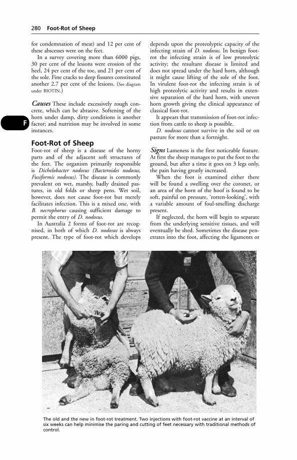
for condemnation of meat) and 12 per cent ofthese abscesses were on the feet.
In a survey covering more than 6000 pigs, 30 per cent of the lesions were erosion of theheel, 24 per cent of the toe, and 21 per cent ofthe sole. Fine cracks to deep fissures constitutedanother 2.7 per cent of the lesions. (See diagram
under BIOTIN.)
Causes These include excessively rough con-crete, which can be abrasive. Softening of thehorn under damp, dirty conditions is anotherfactor; and nutrition may be involved in someinstances.
Foot-Rot of SheepFoot-rot of sheep is a disease of the horny parts and of the adjacent soft structures of the feet. The organism primarily responsible is Dichelobacter nodosus (Bacteroides nodosus,Fusiformis nodosus). The disease is commonlyprevalent on wet, marshy, badly drained pas-tures, in old folds or sheep pens. Wet soil, however, does not cause foot-rot but merelyfacilitates infection. This is a mixed one, withB. necrophorus causing sufficient damage to permit the entry of D. nodosus.
In Australia 2 forms of foot-rot are recog-nised, in both of which D. nodosus is alwayspresent. The type of foot-rot which develops
depends upon the proteolyptic capacity of theinfecting strain of D. nodosus. In benign foot-rot the infecting strain is of low proteolyticactivity; the resultant disease is limited and does not spread under the hard horn, althoughit might cause lifting of the sole of the foot. In virulent foot-rot the infecting strain is ofhigh proteolytic activity and results in exten-sive separation of the hard horn, with unevenhorn growth giving the clinical appearance ofclassical foot-rot.
It appears that transmission of foot-rot infec-tion from cattle to sheep is possible.
D. nodosus cannot survive in the soil or onpasture for more than a fortnight.
Signs Lameness is the first noticeable feature.At first the sheep manages to put the foot to theground, but after a time it goes on 3 legs only,the pain having greatly increased.
When the foot is examined either there will be found a swelling over the coronet, or an area of the horn of the hoof is found to besoft, painful on pressure, ‘rotten-looking’, witha variable amount of foul-smelling dischargepresent.
If neglected, the horn will begin to separatefrom the underlying sensitive tissues, and willeventually be shed. Sometimes the disease pen-etrates into the foot, affecting the ligaments or
280 Foot-Rot of Sheep
F
The old and the new in foot-rot treatment. Two injections with foot-rot vaccine at an interval ofsix weeks can help minimise the paring and cutting of feet necessary with traditional methods ofcontrol.
even the bone. One, 2, 3, or all 4 feet may beaffected. If the 2 fore-feet are attacked, thesheep very often assumes the kneeling positionfor feeding. If the 2 hind-feet, any 3 feet, or all 4 feet are affected, standing becomes animpossibility, and the sheep, still retaining itsappetite, will feed from the sitting position,crawling forward a few inches at a time to a newpiece of grazing.
Prevention Foot-rot can be eradicated. Leavecontaminated pasture free of sheep for 3 weeks.Isolate and treat all infected or suspected sheep.The feet of heavy sheep should not be allowedto get overgrown during wet weather; turningon to a bare fallow or stubble field, or walk-ing along a hard road, is advocated by some towear away the feet, but is not a very practicableproposition. The better way is to round up thesheep and pare each foot individually onceevery 6 weeks or 2 months.
Where the disease has not yet taken hold, the use of foot-rot vaccine may obviate muchtime-consuming work treating diseased feet;the manufacturer’s directions must be followedif the vaccine is to be effective. Vaccines containinactivated strains of D. nodosus.
Treatment It is advisable to separate theinfected animals from the healthy, passing the latter through a foot-bath and changing thepasture to as high a ground as possible. If thelame sheep can be shut up in a dry, strawedyard, in pig-courts or in pens, and given hand-feeding and individual attention daily, theyrecover much better than if they are left out in the open and only attended to occasionally.The feet should be carefully trimmed, and allnecrotic horny material removed. When all the‘rotten’ substance has been removed, the sheepshould be passed through a foot-bath. In severecases, zinc sulphate solution is preferable to formalin, which can cause severe pain; propri-etary formulations based on zinc sulphate heptahydrate are available.
The shepherd should take care not to spreadthe disease to other sheep through the mediumof hands or knife; both should be washed afterdealing with each case, and all parings, diseasedtissue, and infected swabs collected in a pail and burned. Neglect of these precautions oftenresults in a continuance of new cases in a flock.
Aerosol sprays containing the antiboticoxytetracycline, and a purple dye as marker, arepopular. Injection of a long-acting antibioticcan be highly effective.
The economic and welfare consequences offoot-rot can be severe. Losses of up to 15 per cent
in weight can be shown in affected ewes withreduced growth rate in lambs.
(See also FOOT-BATHS; CONTAGIOUS OVINE
DIGITAL DERMATITIS.)
Forage Mites(see MITES)
ForamenA hole or opening. The word is applied partic-ularly to holes in bones through which passnerves or blood vessels. The foramen magnumis the large opening in the posterior aspect ofthe skull through which passes the spinal cordto enter the foramina in each of the vertebrae ofthe spine. The nutrient foramina are the holesin the shafts, etc. of the bones which penetrateto the marrow cavity, by which blood andlymph vessels and nerves pass to and from themarrow cavity.
Foreign BodyAny object which becomes lodged in a bodyorgan or tissue. The term includes a grass seedin the ear or nose, beneath the skin between the toes or beneath the eyelid, in the prepuce or penis of the cat; a needle embedded in the tongue or a chop bone wedged in a dog’smouth; a piece of bone lodged in the gullet; a piece of wire in the reticulum; pebbles in adog’s stomach; lead shot and airgun pellets. (See AWNS; under CHOKING; STOMACH, DISEASES
OF, etc.)Foreign bodies also include a broken-off
portion of an intravenous needle within a vein,or of a catheter. Miniature ‘button batteries’,swallowed by small children, have caused anobstruction of the oesophagus, and also mer-cury poisoning; and a similar risk could beexpected in dogs and cats.
FormalinFormalin is a gas prepared by the oxidation ofmethyl alcohol. For commercial purposes it isprepared as a solution of 40 per cent strength in water. Formalin is a powerful antiseptic, and has the quality of hardening or fixing the tissues. The solution in water gives off gasslowly, and this has an irritant action on theeyes and nose.
Formalin is used for preserving pathologicalspecimens, occasionally as a disinfectant, andfor the production of formaldehyde gas forfumigation of buildings. A 3 per cent solutionof formalin has been used in a foot-bath in the treatment of foot-rot in sheep. Its applica-tion, however, may cause considerable pain if itreaches sensitive tissues. Formalin gas has been
Formalin 281
F

used to fumigate eggs on farms and in the setters in the hatchery. The process carries some risk and must be done in special cham-bers; approved alternatives are available.Formalin gas must never be used to fumigateduck or goose eggs. (See also under FOOT-BATHS;
DISINFECTION.)
FossaFossa is an anatomical term applied to a depres-sion in a bone which lodges some other struc-ture, such as part of the brain in the skull. It isalso used to describe grooves or pockets in softtissues, such as the renal fossa of the liver inwhich is lodged the right kidney.
Foul-in-the-FootCalled FOOT-ROT in the USA. A disease thataffects cattle. Technically known as interdigitalnecrobacillosis, the lesion takes the form of aswelling which tends to force the claws apart.The whole length of the space between theclaws may be involved, with 1 or 2 fissures inthe skin evident, and a slough of dead tissue.
Cause Fusiformis necrophorus (Fusabacteriumnecrophorum) is the usual cause, entering tissuesthrough a wound or through devitalisation ofthe skin from frost, mud, decomposing urine orfaeces, or other irritants.
Signs There is nearly always well-markedlameness, with swelling of the interdigital tis-sues and a typical foul smell. Hind-feet aremore often affected than fore-feet, probablyowing to their greater liability to soiling fromurine and faeces, in which the necrosis bacilluscan generally be easily found. In many cases acow will suddenly stop walking, and shake theaffected foot as though she desires to dislodge astone or other hard object which has becomewedged between the claws.
A ‘super foul’ has been seen recently, whichspreads very rapidly in the foot; it causes severepain and deep erosion at the heel unless treatedpromptly. Tissue damage may be so extensivethat the animal has to be culled.
Treatment This calls for prompt professionalaid. The foot is dried and an oxytetracyclinespray applied. In severe cases, parenteral antibi-otics may be necessary and are essential in ‘superfoul’. Affected animals should be isolated. (See
FOOT-BATHS.)
Fowl CholeraSynonyms: cholera gallinarium, avian pasteurel-losis, pasteurellosis of the fowl, haemorrhagic
septicaemia of the fowl. This is a contagious disease of fowls, usually epizootic in type andcharacterised by sudden onset, high fever, exten-sive blood extravasations into the differentorgans, and severe diarrhoea. The disease occursall over Europe, in North and South America, inmost parts of Africa, and in Asia. All commonfowls, including domestic poultry (chickens,ducks, geese, guinea-fowl, turkeys, pigeons,pheasants, and fancy birds), are susceptible.Most common wild birds are also liable to infec-tion and serve to spread the disease. Rabbits and mice may also contract it under special circumstances.
Cause Pasteurella multocida.
Signs After a brief incubation period (usually 2 to 4 days) the birds may be seen to staggerand fall down, or more commonly are justfound dead. In the less acute type, which per-haps is the more common, the birds are seen to look ill, to stand apart from the rest, drooptheir wings, and refuse both food and water.The combs, wattles, and ear lobes become dis-coloured, and there is great nervous prostration.A discharge comes away from the eyes andnose, a frothy saliva from the mouth, and thereis usually severe diarrhoea. The respirationsbecome rapid; the temperature may reach 43.3°or 43.9°C (110° or 111°F). The feathers areruffled and draggled, and those of the hinderparts of the body are soiled with faecal dis-charges. Vomiting may take place, and in from1 to 3 days the affected birds usually die. Inother cases the symptoms are more subacute,and the disease may run on for from 7 to 9 or 10 days, but as a rule ends fatally. In themore chronic type, arthritis may be seen and itmay take several weeks before death ensues. In acute outbreaks 90–95 per cent may die,although in others the death-rate may be only20 per cent.
Treatment Tetracycline antibiotics are moreuseful than sulfonamides as these adverselyaffect egg production.
Prevention is by vaccination and avoidingcontact with wild birds.
Fowl Paralysis(Neuro-lymphomatosis.) (See MAREK’S DISEASE.)
Fowl PestThis term usually refers to NEWCASTLE DISEASE,but also includes fowl plague (see AVIAN
INFLUENZA)
282 Fossa
F
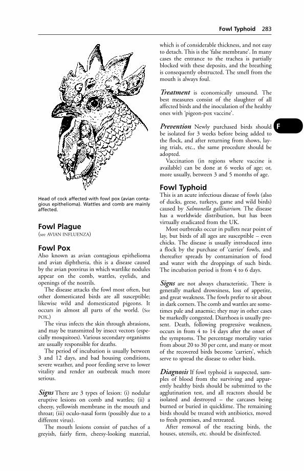
Fowl Plague(see AVIAN INFLUENZA)
Fowl PoxAlso known as avian contagious epitheliomaand avian diphtheria, this is a disease caused by the avian poxvirus in which wartlike nodulesappear on the comb, wattles, eyelids, and openings of the nostrils.
The disease attacks the fowl most often, butother domesticated birds are all susceptible;likewise wild and domesticated pigeons. Itoccurs in almost all parts of the world. (See
POX.)The virus infects the skin through abrasions,
and may be transmitted by insect vectors (espe-cially mosquitoes). Various secondary organismsare usually responsible for deaths.
The period of incubation is usually between3 and 12 days, and bad housing conditions,severe weather, and poor feeding serve to lowervitality and render an outbreak much more serious.
Signs There are 3 types of lesion: (i) nodulareruptive lesions on comb and wattles; (ii) acheesy, yellowish membrane in the mouth andthroat; (iii) oculo-nasal form (possibly due to adifferent virus).
The mouth lesions consist of patches of agreyish, fairly firm, cheesy-looking material,
which is of considerable thickness, and not easyto detach. This is the ‘false membrane’. In manycases the entrance to the trachea is partiallyblocked with these deposits, and the breathingis consequently obstructed. The smell from themouth is always foul.
Treatment is economically unsound. Thebest measures consist of the slaughter of allaffected birds and the inoculation of the healthyones with ‘pigeon-pox vaccine’.
Prevention Newly purchased birds should be isolated for 3 weeks before being added tothe flock, and after returning from shows, lay-ing trials, etc., the same procedure should beadopted.
Vaccination (in regions where vaccine isavailable) can be done at 6 weeks of age; or,more usually, between 3 and 5 months of age.
Fowl TyphoidThis is an acute infectious disease of fowls (alsoof ducks, geese, turkeys, game and wild birds)caused by Salmonella gallinarium. The diseasehas a worldwide distribution, but has been virtually eradicated from the UK.
Most outbreaks occur in pullets near point oflay, but birds of all ages are susceptible – evenchicks. The disease is usually introduced into a flock by the purchase of ‘carrier’ fowls, andthereafter spreads by contamination of foodand water with the droppings of such birds.The incubation period is from 4 to 6 days.
Signs are not always characteristic. There isgenerally marked drowsiness, loss of appetite,and great weakness. The fowls prefer to sit aboutin dark corners. The comb and wattles are some-times pale and anaemic; they may in other casesbe markedly congested. Diarrhoea is usually pre-sent. Death, following progressive weakness,occurs in from 4 to 14 days after the onset of the symptoms. The percentage mortality variesfrom about 20 to 30 per cent, and many or mostof the recovered birds become ‘carriers’, whichserve to spread the disease to other birds.
Diagnosis If fowl typhoid is suspected, sam-ples of blood from the surviving and appar-ently healthy birds should be submitted to theagglutination test, and all reactors should beisolated and destroyed – the carcases beingburned or buried in quicklime. The remainingbirds should be treated with antibiotics, movedto fresh premises, and retreated.
After removal of the reacting birds, the houses, utensils, etc. should be disinfected.
Fowl Typhoid 283
F
Head of cock affected with fowl pox (avian conta-gious epithelioma). Wattles and comb are mainlyaffected.
Fox, Diseases ofIn Europe, North America and other parts ofthe world, wild foxes often become victims ofrabies, and spread this disease to farm livestockwhich they may attack. A history of aggressive-ness and atypical behaviour does not, how-ever, point conclusively to rabies; distempermay be the reason. (See also FOX ENCEPHALITIS;‘CHASTEK PARALYSIS’.)
The fox acts as host of the roundwormToxocara canis and of the Toxascaris leonina, andif silver fox cubs are reared by a cat, they maybecome infected with Toxocara mystax of the cat.The fox harbours the dog tapeworms Taeniaserialis and T. multiceps, and Echinococcus granu-losus. Leptospirosis occurs in foxes in the UKand may be spread to farm livestock (5 strainshave been isolated). Flukes may infest foxes.
Fox EncephalitisFox encephalitis is of commercial importance on the fox ranches of North America, wherethese animals are bred for their fur. The disease is considered identical to Rubarth’s disease of dogs.
Signs Young foxes in good condition are mostfrequently affected. A violent convulsion is followed by a lethargic or ‘sleep-walking’ state.This may be followed by excitability and moreconvulsions – during which the slamming of adoor or any loud noise may prove fatal. The illness runs a very rapid course, from 1 hour to3 days, 24 hours being the average duration.
Control By means of serum and preventiveinoculation.
This disease, or one caused by a similar virus,may have accounted for the deaths of (wild)foxes in Britain, but the deaths may haveoccurred as the result of eating birds poisonedby dieldrin. Signs are similar.
Foxglove Poisoning(see DIGITALIS POISONING)
Fox TerriersSmall, lively dogs classed as smooth-haired orwiry, according to the coat; tail usually short. Inboth breeds deafness and lens luxation may beinherited. Pulmonary stenosis and achalasiamay be seen. Atopic dermatitis occurs only inthe wire-haired terrier; cerebellar ataxia in thesmooth.
FracturesSimple fractures are the commonest variety, and consist of those in which the bone is broken clean across, with or without tearingand laceration of the soft parts surrounding it,but without any wound leading from the frac-ture through to the skin. They are spoken of as being transverse, longitudinal, or oblique,according to the direction of the break.
Compound fractures are those in which the skin is injured, so that a direct or indirectcommunication between the fracture and theoutside air exists. The broken end of the bone
284 Fox, Diseases of
F
Classification of fractures. A. Transverse fracture with excellent stability after reduction.B. Oblique fracture with no stability after reduction. C. Slightly oblique fracture which,by virtue of the irregularity of the fracture lines, provides a useable degree of stabilityafter reduction. D. A typical distracted fracture. (With acknowledgements to the BritishVeterinary Association.)
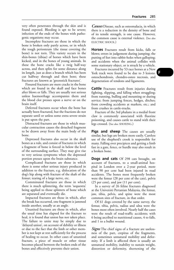
very often penetrates through the skin and isfound exposed. Bleeding is apt to be severe;infection of the ends of the bones with patho-genic organisms may occur.
Incomplete fractures are those in which thebone is broken only partly across, or in whichthe tough periosteum (the tissue covering thebone) is not torn. This variety occurs in theshin-bones (tibiae) of horses which have beenkicked, and in the bones of young animals. Inthese the bone cracks like a twig half-wayacross, and then splits for some distance alongits length, just as does a branch which has beencut halfway through and then bent; these fractures are known as ‘greenstick fractures’.
Fissured fractures are mere cracks in the bonewhich are found in the skull and face bonesafter blows or falls. They are usually not seriousunless haemorrhage accompanies them and the blood clot presses upon a nerve or on thebrain itself.
Deferred fractures occur when the bone hasactually been fractured, but the fractions do notseparate until or unless some extra severe strainis put upon the part.
Distracted fractures are those in which mus-cular contraction causes the detached fragmentto be drawn away from the main body of thebone.
Depressed fractures also occur in the skullbones as a rule, and consist of fractures in whicha fragment of bone is forced in below the levelof the surrounding surface. They may give riseto very serious symptoms when the depressedportion presses upon the brain substance.
Complicated fractures are those in whichthere is some other serious injury produced inaddition to the fracture, e.g. dislocation of thedog’s hip along with fracture of the shaft of thefemur; tearing of a large nerve, etc.
Comminuted fractures are those in whichthere is much splintering, the term ‘sequestra’being applied to those splinters of bone whichare separated and eventually die.
Impacted fractures are those in which, afterthe break has occurred, one fragment is jammedinside another, usually at an angle.
Ununited fractures are those in which, afterthe usual time has elapsed for the fracture toheal, it is found that union has not taken place.The failure to unite may be simply due to‘delayed union’, on account of debility or illnessor due to the fact that the limb or other mem-ber is not kept at rest sufficiently for the processof healing to occur. In other cases of ununitedfracture, a piece of muscle or other tissuebecomes placed between the broken ends of thebones and effectively prevents their union.
Causes Disease, such as osteomalacia, in whichthere is a reduction in the density of bone and of its tensile strength, is one cause. However, the common cause is external violence. (See also
ELECTRIC SHOCK.)
Horses Fractures result from kicks, falls orblows; errors in judgement during jumping; theputting of feet into rabbit-holes when galloping;and accidents when the animal collides withsome stationary object, or is struck by a vehicle.
Fractures incurred by 53 race horses at a NewYork track were found to be due to 3 lesions:osteochondrosis, chondro-osteo necrosis, anddegeneration of tendons and ligaments.
Cattle Fractures result from injuries duringfighting, slipping, and falling when struggling;from running, bulling and mounting or duringservice; from jumping fences, hedges, ditches;from crowding accidents at markets, etc.; andfrom crushes in cattle-trucks.
Fracture of the 3rd phalanx in a medial frontclaw is commonly associated with fluorine poisoning, and causes cattle to stand with theirlegs crossed. (See also SHOEING.)
Pigs and sheep The causes are usually similar, but legs are broken more easily. Carelessuse of the shepherd’s crook is responsible formany. Falling over precipices and getting a limbfast in a gate, fence, or hurdle may also result ina broken bone.
Dogs and cats Of 298 cats brought, onaccount of fractures, to a small-animal hos-pital in London over a 2-year period, more than 90 per cent had been injured in road accidents. The bones most frequently brokenwere the femur (28 per cent of the cats), pelvis(25 per cent), and jaw (11 per cent).
In a survey of 26 feline fractures diagnosed at the Universiti Pertanian Malaysia, the femur,jaw, tibia, pelvis, and spine were the most common sites of fracture, in that order.
Of 61 dogs covered by the same survey, thefemur, tibia, pelvis, radius and ulna were thebones most often involved. Nearly half the caseswere the result of road-traffic accidents; with 6 being ascribed to nutritional causes, 4 to falls,and 1 to a bullet wound.
Signs The chief signs of a fracture are useless-ness of the part, crepitus of the fragments, and sometimes unnatural mobility and defor-mity. If a limb is affected there is usually anunnatural mobility, inability to sustain weight,distortion or deformity, shortening of the
Fractures 285
F

length, a thickness or swelling at the seat of the fracture (due to overlapping of the frag-ments), and a variable amount of pain. (See also
‘Fractures of special parts’, below.)
Healing of fractures When the bone breaks,many blood vessels are torn, and accordingly alarge clot of blood forms around the ends,between them, and for some distance up theinside of the bone. Later, great numbers ofwhite blood cells find their way into this clot,which becomes ‘organised’ – blood vessels and,later, fibrous tissue being formed in it (soft cal-lus). Next, lime salts are gradually deposited inthis fibrous tissue, which thus develops intobone (hard callus). In this process a thick ringof new bone forms round the broken ends, fill-ing up all crevices; and when union is complete,this thickening is again gradually absorbed,leaving the bone as it was before the injury.
In racing greyhounds, badly fracturedscaphoids have been removed and replaced byplastic or metal prostheses. In one case, the useof a titanium-alloy prosthesis enabled a grey-hound to race again 43 times before retirementfrom the track.
Treatment Reduction and apposition arebrought about by manipulation of the fracturedbones under anaesthesia. Immobilisation is theneffected by means of plaster of Paris, and variousproprietary mixtures impregnated into bandages.Splints of metal, leather, wood, or cardboard,padded with cotton-wool, are useful, especiallywith dogs and cats. (See SPLINTING MATERIALS.)
Special types of extension splints, havingtransverse pins which transfix the bone, havebeen used with success in appropriate cases.Medullary pins, driven down the marrow cavi-ty of long bones; wiring; and plating have allbeen used with success. (See BONE PINNING.)
Whenever splints, plaster, or other bandagesare being applied to fractured limbs it is essen-tial to ensure that the surface of the skin is well padded with cotton-wool, and that thepressure is evenly distributed. Failure in thisrespect may result in parts of the skin becom-ing gangrenous through obstruction to theblood-flow.
Bone Grafts These are used to a limited extent in veterinary surgery to repair fractures ofthe femur, humerus, tibia, radius and ulna; or to replace comminuted fragments, to lengthenbones, to correct delayed or faulty union.
The allografts are harvested aseptically fromhealthy dogs, autoclaved the same day, and maybe stored at between –10° and –20°C (14° and–4°F) in a domestic freezer for use up to 1 yearlater. Ordinary bone-plating techniques areused to secure the implant.
Fractures of special parts1. The cranium Cases of traumatic fractureof the skull result in concussion; if not severethey may recover with conservative treatmentand nursing. Depressed fractures involving thecranial cavity or the cranial nerves are difficultto treat and usually carry a poor prognosis. (See
also CONCUSSION.) Surgical treatment may besuccessful in cases in which fractures involvebones of the jaws and face.
2. The face bones Fractures may be simple orserious according to bones involved. Nasalbones, often fractured from accidents, may beaccompanied by swelling, pain, haemorrhage,difficulty in breathing, and much watering ofthe eyes. Jaw-bones broken from falls, kicks,etc. usually interfere with feeding. Lower-jawfractures usually result in an open hangingmouth, escape of saliva, and altered expression;frequently, loose teeth, torn lips, and haemor-rhage are seen. Bones of orbit fractured by fallson to side of head, collisions, etc. interferencewith vision and with movements of the lowerjaw, in most cases serious. Treatment usuallynecessitates operation – removal of brokenpieces, elevation of depressed portions, removalof loose teeth, wiring or plating broken partstogether. Feeding must be carefully undertakenwhen jaws are injured – sloppy food, mashes,etc., for horse; hand-feeding for dog.
286 Fractures of special parts
F
Metal splints which have transverse pins to pene-trate and fix the bone are used in treating frac-tures in small animals, and have succeeded in caseswhere other methods would be ineffective.
3. The vertebrae Commonest in horse anddog through accidents (e.g. in horses gettingcast in stall, casting for operation, road-trafficaccidents in dogs, falls from heights, blowsfrom sticks). If a vertebra is fractured, paralysisresults. There is often a fatal termination, or aneed for euthanasia. Tail-bones are often bro-ken in dogs and cattle through getting caughtin doors, gates, fences, etc.
4. The ribs Due to external violence usually,but the 1st rib is sometimes broken throughmuscular action in a side-slip and violent recov-ery, when it often results in RADIAL PARALYSIS.Otherwise broken ribs show little or nothingcharacteristic except local pain and deformity,unless many are involved, when breathing maybe short and/or difficult. (See ‘FLAIL-CHEST’;
PNEUMOTHORAX.)
5. The pelvis In 123 cases of fracture of thepelvis in dogs in 1 practice, all were the result ofroad accidents. Twenty-eight of the dogs weretreated surgically, and 66 conservatively. Theconclusion drawn was that, although the major-ity of patients would recover without surgery,the latter could reduce the time taken for recov-ery, especially with multiple fractures on bothsides of the pelvis. In bulls and stallions, pelvicfractures occur sometimes during service whentheir hind-feet slip from under them and theyfall backwards on to buttocks. These are leastserious when only the external angle of ilium(‘point of hip’) is involved.
6. The scapula Fractures are uncommon.Mostly, they occur through the neck of thebone, or on the projecting spine. The muscula-ture covering the bone may impede diagnosisbut assists recovery, acting as natural bandage.
7. The humerus Lameness, intense in all ani-mals, follows fracture; the limb is usually quiteuseless. Horses and cattle do not make goodrecoveries except when young, but healing insmall animals is more satisfactory. (See BONE-
PINNING.) Absolute rest is essential; horses maybe slung.
In a series of 130 cases in dogs and cats, mostanimals with proximal, shaft and supracondylarfractures had excellent results. The poor prog-nosis associated with distal articular fractureswas most often because of failure of the fixationdevice in the supracondylar area. The bestresults were achieved with a plate on the caudaland medial surface of the distal humerus.
8. The radius and ulna One or both bonesmay be broken; fracture of the ulna is less serious unless the elbow-joint is involved. In
dogs, if one is broken the other acts as a nat-ural splint. Lameness is always marked, andthere is pain on pressure. Local swelling is usually noticed, and deformity. Bandaging isadvisable. Young horses should be placed inslings. Bone-pinning has been carried out successfully in the dog and the horse.
9. Coronoid process In 130 cases of frag-mented coronoid process of the ulna in 109dogs, 68 were treated surgically by medialelbow arthro-tomy and 62 with rest and anti-inflammatory drugs. Surgical treatment did not decrease the incidence of lameness aftertreatment, but the dogs treated surgically weremore active and less lame than those treatedwithout surgery. Young dogs with mild lame-ness due to fragmented medial coronoidprocesses probably do not benefit from surgery,but dogs with chronic, moderate or severelameness do.
10. Bones of knee These are seldom frac-tured, but if they are it is usually impossible tobring about recovery without stiffening of thejoint (ankylosis).
11. The metacarpals In the horse, goodrecoveries are made in cases of clean transversefractures without complications or splinters.Prognosis is best in fractures occurring in themiddle of the cannon. The limb is bandagedwith a plaster or proprietary resin-impregnatedbandage and the horse slung; the plaster is leftin position for at least 6 weeks. In the dog, suchfractures usually respond well after setting andsupporting of the affected bone.
12. The pastern bone Fractures may be trans-verse, oblique, or longitudinal (‘split pastern’),often comminuted. Severe lameness alwaysresults. Simple transverse fractures can be treatedsatisfactorily if the temperament of horse willallow rest and slinging; oblique, longitudinal,and all comminuted cases are unsatisfactory andif recovery occurs, usually some deformity orblemish is left.
13. The second phalanx, coffin andnavicular bones Fractures in these bones arerare; they may the caused by direct violence,and sometimes follow an operation of neurec-tomy (un-nerving); may be seen in cattle as aresult of weakening of bone through FLUORO-
SIS. Fracture of the coffin-bone, if simple andjoint surfaces are not involved, makes goodrecovery as a rule, since hoof acts as splint andbandage. Fracture of the 2nd phalanx (shortpastern bone) is usually difficult to resolve.
Most fractures of the navicular bone are
Fractures of special parts 287
F

sagittal and minimally displaced, but the prog-nosis is usually poor because the fibrous calluscauses permanent lameness. Such fractures have been repaired by inserting a 50 mm screwwhich exerts compression between the 2 frag-ments. The pilot hole is drilled and the screw isinserted precisely along the transverse axis ofthe navicular bone by means of a mechanicalguide, the process being monitored by image-intensifying fluoroscopy. The fractures are saidto heal without superfluous callus formation.
14. The femur Very commonly fracturedin dogs after street accidents. Shaft, neck, orone of the trochanters may be involved.Frequently in dogs, dislocation of the hip-jointaccompanies fracture. Extreme lameness, short-ening of the limb, local swelling, and great painon movement are usually seen. There may ormay not be crepitus. In horses, fracture of pelvisvery often accompanies fractured femur andmakes diagnosis difficult. A fractured femurusually necessitates euthanasia in large animals,but in small animals recovery may be either partial or complete. (See BONE-PINNING.)
15. The patella Fracture is a very seriouscondition, resulting in a lowering of the affect-ed stifle and inability to advance the limb.There is great pain. Treatment is union of thefragments by wire sutures; this may be difficult
to perform satisfactorily, and complete recoverymay not occur.
16. The tibia Many fractures of tibia becomecompound from sharp points of broken bonespenetrating through the skin. (See BONE-PINNING,
which has been used successfully in dog, cat, andhorse.)
17. Bones of hocks Fracture of os calcis(point of hock) – the epiphyseal summitbecomes torn away from the rest of the bone byan undue pull of the Achilles’ tendon (ham-string). Fractures of other bones of hock are lesscommon (with the exception of the SCAPHOID
in the racing greyhound).
Francisella(see TULARAEMIA)
Free RadicalsHighly reactive molecules, formed in the pres-ence of oxygen and capable of damaging livingtissue. They have been implicated in humanheart disease and arthritis. They may also be acause of sudden death of pigs – those beingtransported for long distances or subject toother forms of stress. However, it has been sug-gested that protection may be given by feedingvitamin E, which ‘scavenges’ radicals.
FreemartinUsually defined as a sterile heifer born twin to anormal bull calf; the most widely acceptedexplanation being that sex hormones from theearlier developing male twin pass across to thefemale twin, with the result that sexual differ-entiation of both male and female proceedsunder control of male hormones.
However, as long ago as 1917 it had beensuggested that hermaphrodites might occur infemale single births, as a consequence of earlyfetal death and resorption of the male twin inthe uterus. During the 1970s chromosomeanalysis had revealed the presence of both maleand female cells in single-born bull calves. Dr W. V. S. Wijeratne and colleagues were thefirst to demonstrate this condition – technicallyknown as secondary chimerism – in single-bornfreemartins. (Primary chimerism can occurwhere 2 sperms fertilise the same ovum.)
Not every female fetus having a male twinsharing the uterus will become a freemartin,because in some instances death of the maletwin fetus occurs before about day 39 of preg-nancy – when a common blood supply maybecome established. Moreover, in between 5 and10 per cent of heterosexual twin pregnancies acommon blood supply is not established.
288 Francisella
F
Two Rush-type intramedullary pins used to repaira supracondyloid fracture of the femur.

Blood samples were taken from 36 heifersnot in calf after 3 or more inseminations or nat-ural service; chromosome analysis of these ani-mals’ white cells showed that 12 of the heifershad both male and female chromosomes. Fiveof the heifers were single-born. In 3 which wereslaughtered, abnormalities of the reproductivetract were formed.
Two of the slaughtered heifers had shown nor-mal oestrous cycles, and had reproductive organsapparently normal on clinical examination; but 1 of them, with 5 per cent male cells, had hercervix closed by a fibrous band or hymen; theother, which had 12 per cent male cells, was sterile on account of fibrous bands blocking thehorns of the uterus. Both heifers possessed func-tional ovaries. The 3rd heifer, with 45 per centmale cells, had a normal vagina, enlarged clitoris,seminal vesicles and sex organs having bothovaries and testes in primitive form.
‘Presuming that 5 per cent of all heifers rearedfor breeding are infertile, the probable preva-lence of single-born freemartins in this heiferpopulation is about 0.9 per cent’, (concluded Dr Wijeratne.)
About 90 per cent of heifer calves born twinto a bull calf are freemartins. Many freemartinscan be detected on clinical examination, sincethe vagina is often only one-third of the normallength and, in addition, there is often anenlarged clitoris and a vulval tuft of hair.
The condition is associated with anastomosisof the placental blood vessels (see diagram).
Pig freemartins may also occur. (See also H-Y
ANTIGEN.)
Freeze-Branding(see BRANDING)
FremitusFremitus is a sensation which is communicatedto the hand of an observer when it is laid acrossthe chest in certain diseases of the lungs and heart. Friction fremitus is a grating feelingcommunicated to the hand by the pleura orpericardium when it is roughened as in pleurisyor pericarditis.
French BulldogA small dog resembling the English bulldog of the 19th century. Haemophilia may befound as a sex-linked recessive trait. Ununitedanconal process (elbow) is a dominant trait.Hemivertebrae, intervertebral disc disease,patellar luxation may occur.
Frog(see FOOT OF HORSE)
Frontal Bone of the SkullA roughly quadrilateral plate-like bone whichforms part of the roof of the cranium and pass-es forward between the eyes to meet the nasalbones. In the horned breeds of cattle and sheepit is extended laterally to form the horn cores.(See BONES; also SINUSES OF SKULL.)
Frontal Sinus(see SINUSES OF SKULL)
Frost-BiteFrost-bite may affect any animal exposed forlong periods to severe cold. As a result of thiscold, the body reacts by a constriction of surfaceblood vessels, in order to minimise heat loss andmaintain body temperature. This leaves exposedparts of the skin susceptible to freezing. The
Frost-Bite 289
F
The bovine freemartin.

part, such as the tips of ears, or the tail, becomesnumb, and may be completely frozen. Pain isnot felt at this stage (but occurs during thethawing process). In some instances naturalrecovery takes place, but in others gangrene fol-lows and ear-tips, tails, and the wattles andcombs of poultry may slough off.
An animal-owner may have no reason to sus-pect frost-bite until the appearance of gangreneand sloughing. (See GANGRENE.) Nowadaysmassage and rubbing the part with snow havebeen abandoned as likely to do more harm thangood in human medicine; and immersing thepart in warm or hot water is equally to beavoided. (See CHILBLAIN.)
Frounce(see FALCONS)
Frozen Embryos(see LIFE AFTER FREEZING)
FrusemideA diuretic suitable for the treatment of somecases of OEDEMA.
‘Frying Pan’ DeathsOverheated fat gives off acrolein, which can behighly poisonous and was, indeed, used inchemical warfare in 1914–18. A dog died afterbeing shut up in a kitchen for half an hour witha smoking chip pan. Ante-mortem symptomswere distressed breathing and cyanosis.
Five cockatiels died within half an hour following exposure to fumes from a non-stickfrying pan coated with plastic polytetrafluo-roethylene. Within an hour the birds’ ownerbecame ill with ‘polymer fume fever’, but recovered. (See KITCHEN DEATHS.)
FucosidosisA lysosomal storage disease caused by theabsence of an enzyme – alpha-L-fucosidase.
It is an inherited disease in the Englishspringer spaniel, affecting mainly those between18 months and 4 years old. The signs of thisultimately fatal disease include ataxia, change in temperament, depression, apparent deafnessand impaired sight. Swallowing may be difficult.Loss of weight occurs.
Fumes(see CARBON MONOXIDE; ‘FRYING PAN’ DEATHS;SLURRY; ANAESTHETICS; AEROSOLS)
Fumigation(see DISINFECTION)
FundusFundus is the base or innermost part of a hollow organ distant from its opening.
Fungal DiseasesBroadly speaking, these include both the invasion of tissues by fungi, and the effects onorgans of fungal poisons (see MYCOTOXICOSIS).
Ringworm offers a good example of the inva-sion of tissues by pathogenic fungi; one should,perhaps, say potentially pathogenic fungi, formany are present in the alimentary canal ofhealthy animals, and cause lesions only whencircumstances favour invasion or multiplication.(See MASTITIS IN COWS, Mycotic, for an exampleof the latter; also ASPERGILLOSIS; BLASTOMYCO-
SIS; HISTOPLASMOSIS; MUCORMYCOSIS; MONIL-
IASIS; STREPTOTHRICOSIS; CRYPTOCOCCOSIS;
FUSARIUM; MORTIERELLA; COCCIDIOMYCOSIS;
RHINOSPORIDIOSIS; SPOROTRICHOSIS.)
Fungal Toxins(see MYCOTOXICOSIS)
Fur Mites(see MITES)
Fur, Swallowed(see HAIR BALLS)
FurazolidoneA nitrofuran compound used against antibacte-rial and antiprotozoal infections. Its use in foodanimals is no longer permitted in the EU.
FurfuraceousFurfuraceous is a term applied to skin diseaseswhich produce a bran-like scaliness.
FurunculosisThe presence of boils (abscesses). In the dog, theterm is applied sometimes to abscesses/ cystsbetween the toes. (See INTERDIGITAL CYSTS.)Perianal furunculosis also occurs in dogs.
Furunculosis in fish is caused by Aeromonassalmonicida. Raised furuncles can be seen allover the body and they may be complicated bysecondary fungal infection. A sudden increasein water temperature can trigger the appearanceof the disease.
Furunculosis in salmon is caused byAeromonas salmonicida but may be triggered bya sudden rise in water temperature. Young fishstop feeding and may die soon afterwards. Olderfish are more resistant; they develop large, boil-like swellings on the shoulder and back. If theseburst, a reddish fluid rich in bacteria is released.The bacterium persists in some fish between
290 Frounce
F

outbreaks and is present in wild fish. Treatmentis by medicating the feed with sulfonamides ortetracyclines but the fish may be reluctant totake medicated food. It is a NOTIFIABLE DISEASE
under the Diseases of Fish Act 1937.
Furze (Gorse)(Ulex europaeus.) A very common and plentifulshrub in waste lands in Britain, it was formerlyoften cut and used as fodder after chaffing orbruising. The plant contains a very small pro-portion of a poisonous alkaloid which is calledulexine, and is practically identical with cystinefrom broom. It is a nerve and muscle poison,but it is seldom present in dangerous amounts.
FUS(see FELINE UROLOGICAL SYNDROME)
FusariumMouldy shelled maize containing F. monili-forme has caused diarrhoea and ataxia in cattle;and in broilers the same species, contaminatingmaize and wheat, has with F. culmorum, F.Tricinctum and F. nivale been implicated inpoor growth rate, poor feathering, and abnor-mal behaviour. Fusarium species may also causekeratoconjunctivitis. (See under EYE, DISEASES;also MYCOTOXICOSIS; ZEARALENONE.)
Fusiformis NecrophorusAlso known as F. necrophorum. An anaerobicbacterium causing foul-in-the-foot of cattle,calf diphtheria, abscesses in the liver and otherorgans. Also involved in foot-rot in sheep. (See
table under BACTERIA, and FOOT-ROT.)
Fusiformis Necrophorus 291
F

Gad-Fly(see FLIES; WARBLES). In Britain, warble flies areon the wing from late May onwards.
GaddingExcitement, restlessness, uncontrolled rushingaround in horses or cattle due to the presenceof biting flies; also, in the case of cattle, warbleflies.
GagA device to facilitate oral examination ortreatment by holding the mouth open.
Gait, Abnormal(see ATAXIA; ‘GOOSE-STEPPING’; LAMENESS)
Gall-BladderThe little pouch-like sac in which bile producedby the liver is stored until it is required duringthe process of digestion. It is a hollow, pear-shaped organ lying in a depression on theposterior surface of the liver. The gall-bladderis not present in the horse and in animals ofthe horse tribe, but is found in the otherdomesticated animals.
Blockages of the bile-duct by liver flukes orby gallstones may result in jaundice as well assevere local inflammation. Acute inflammationof the gall-bladder is painful, and there isdanger of rupture or gangrene.
‘Gall Sickness’(see ANAPLASMOSIS)
GallstonesGallstones, which are also known as biliarycalculi (see under CALCULI), are concretionswhich are formed in the gall-bladder or in thebile-ducts of the liver. As a rule they are hard,brownish in colour, coated with mucus, and ofa more or less rounded shape. They may becomposed of cholesterol; of cholesterol and bilepigments; or of pigment and lime salts. Oneor several may be present, causing pain andjaundice.
Gallstones are more prevalent in sheep thanin cows, dogs, cats, and horses.
In human medicine, ursodeoxycholic acidhas been used to dissolve gallstones.
Galvanised BinsGalvanised bins, used to store swill, have led toZINC POISONING in pigs.
Galvayne’s GrooveA vertical groove in the front surface of thehorse’s upper corner incisor teeth. It firstappears at the gum margin at about 10 years oldand gradually moves down the surface of thetooth as the horse ages until it grows out atabout 30 years old. (see DENTITION – Horses)
Game Birds, MortalityThis may be considered under 2 headings:
From farm chemicals Many farm chemicalscan cause poisoning in game and other birds.Deaths have resulted from the use, as seed dress-ings, of compounds such as dieldrin, aldrin andheptachlor, now banned in the UK. Some of theorganophosphorus insecticides; dimethoate; andthe ‘nitro-type’ of weedkillers such as DNC,which stains the carcase yellow; are among otherchemicals hazardous to birds.
Pheasant poults have died as a result of beingtreated for lice with a 5 per cent gamma benzenehexachloride (BHC) dusting powder.
An organophosphorus insecticide does notnecessarily act quickly. Death may occur 8 weeksafter eating the poisoned food. The symptomsshown by poisoned birds include ruffled feathers,saliva around the beak, high-stepping gait orunsteadiness on the legs, distressed breathing,and paralysis. However, as their use is nowreduced, problems caused by organophosphoruscompounds are less frequent.
Spraying an orchard with either DDD orDDT (now banned) has caused heavy game-bird losses. A partridge was found dead in afield where blackcurrants had been sprayedwith the insecticide endrin. It was reportedfrom the farm that 8 or 9 partridges died with-in a few hours of eating earthworms whichcame to the surface of the soil soon after spray-ing. Rat poisons may perhaps be included inthe term ‘farm chemicals’. Owls die after eatingpoisoned rodents.
From natural causes Impacted gizzard,tuberculosis, aspergillosis, swine erysipelas,fowl-pox, fowl cholera, fowl typhoid, infectioussinusitis. Gapes is another cause of death; also inthe USA, encephalomyelitis. Deaths from fowlpest (Newcastle disease) have been reported inthe UK; blackhead in pheasants and partridges.
‘Grouse disease’ is the colloquial name forinfestation with Trichostrongylus tenuis. Mortality
G
occurs when food is in short supply as a resultof poor growth or overpopulation of birds ona moor. In some circumstances, it has beenconcluded that grouse have died because notenough were shot the previous year. Grouse tendto remain in a locality and not move to othermoors.
Louping ill, transmitted by sheep ticks, isgenerally fatal to red grouse (Lagopus lagopusscoticus), the commonest game bird on Britishheather moorland, and can reduce stocks tovery low densities.
Inclusion-body hepatitis A 9-day outbreakresulted in an 18 per cent mortality among1000 intensively reared pheasant poults (19 daysold when the outbreak began).
Salmonellosis An outbreak killed 50 per centof 2800 pheasant poults, deaths beginning in 3-day-olds. The infection was one of S. typhimurium. An antibiotic achieved controllater.
Coccidiosis is an important disease of pheas-ants and other game birds, in chicks 2 to 4weeks old. Milky-white droppings are the mostobvious sign (but these are also seen with excessurate excretion due to kidney disease).
Yersiniosis is another important disease ofpheasants.
Moniliasis causes lethargy, stunted growthand a heavy mortality in partridges. Treatmentwith formic acid, sprayed on food, has provedsuccessful. (See also BOTULISM.)
GametesThese are the ova and spermatozoa, and containhalf the number (haploid) of chromosomespresent in all other body cells (diploid).
GametocideGametocide for bird control. (See TEM.)
GametocyteAn oocyte or spermatocyte, the cells whichproduce an ovum or spermatozoon.
Gamma GlobulinGamma globulin is a protein fraction of theblood serum which contains the antibodiesagainst certain bacteria or viruses. (See
COLOSTRUM; IMMUNOGLOBULINS.) It can beprepared in a concentrated form and can beused to give protection against infection.
Gamma Glutamyl Transferase(GGT)An enzyme that tends to increase in liverdisease. Higher than normal concentrationsare found in liver-fluke infection of sheep andcattle.
GammexaneGammexane products contain the gamma iso-mer of benzene hexachloride, a highly effective,persistent insecticide. Not now used in treatingfarm animals. (See BENZENE HEXACHLORIDE;also BHC POISONING.)
GanglionGanglion is a group of nerve cell bodies.
GangliosideA glycolipid found in central nervous system(CNS) tissue.
GangliosidosisAn inherited disease causing poor growth andprogressive neuromuscular dysfunction. Itresults from an accumulation of gangliosidesin CNS tissue and may be seen in cattle, pigs,dogs and cats.
GangreneThe presence of dead tissue in a live animal. Inprimary gangrene, bacteria which cause thenecrosis also bring about the putrefactivechanges. In secondary gangrene the putrefac-tion is caused by organisms which have invadeddead tissue (e.g. following a burn). There are2 varieties of gangrene, dry and moist; dry gan-grene is a condition of mummification in whichthe circulation stops and the part withers up,while in moist gangrene there is inflammationaccompanied by putrefactive changes.
Infection following necrosis may lead togangrene after burns, scalds, frostbite, crushwounds, puncture wounds, etc.
Poisoning by ergot results in the same condi-tion in the most distant parts of the body, e.g.the feet, tip of tail, ears, and the combs andwattles of poultry.
Signs There is at first a degree of pain when theaffected part is handled, and in a short time itbecomes reddened and swollen. Later it turnsblue or black, the hair falls from it, and there isa distinct line of demarcation between the gan-grenous and the healthy surface. Around thedividing line there is usually some degree ofinflammation, and pus production.
Moist gangrene is considerably more serious,since it is accompanied by putrefaction and the
Gangrene 293
G

absorption of toxins. The whole area turnsblack or greenish, the hair falls out, an offensivesmell is evident, and much fluid exudes fromthe decomposing tissues. A high temperature,disturbed heart’s action, and rapid breathing,are shown. (See also GAS GANGRENE.)
Treatment is mainly surgical, backed up bythe use of appropriate antibiotics or sulfon-amides. In advanced cases, euthanasia becomesnecessary. (See also FROST-BITE.)
Gangrenous DermatitisA disease of poultry caused by Clostridium sep-ticum; often associated with infectious bursaldisease and inclusion-body hepatitis, it usuallyaffects birds between 25 and 100 days of age.Mortality can be very high.
Ganjam UlrusThe Indian name for a bunyavirus infectiontransmitted by ticks.
GapesGapes is a disease of young chickens, turkeys,pheasants and other game birds particularly,although all the domesticated and manywild birds may also be affected. It is caused byinfection with the gapeworm, Syngamus trachea.
The presence of worms in the bronchialtubes and trachea of the bird causes it to gaspfor breath or ‘gape’, from which the name ofthe disease originated. Part of the life-history ofthe worm is passed in the body of the earth-worm, and young chickens eating earthwormsmay become affected. Earthworms can live for16 years. (See also under CAPILLARIASIS.)
Nitroxynil, given in the drinking water, is aneffective treatment.
Garden ChemicalsBirds, dogs, and cats may be poisoned as aresult of the use of pesticides. For the poisoningof birds, see preparations listed under GAME
BIRDS, MORTALITY. Dieldrin is highly toxic forcats, and like DDT, should not be used onthem or in their vicinity. In fact, all the CHLO-
RINATED HYDROCARBONS are best avoided inplaces where small domestic animals or theirfood may become contaminated.
For the dangers of slug-baits, see METALDE-
HYDE POISONING. (See ORCHARDS for the dangersof fruit-tree sprays. For seed dressings, see under
SEED CORN. See also PARAQUAT; HERBICIDES.)
Garden Nightshade PoisoningGarden nightshade poisoning results fromanimals eating Solanum nigrum, which is found
in many parts of the world. Its toxicity appearsto vary in different localities. The berriescontain an active alkaloidal glycoside calledsolanine, which is readily converted into sugarand the poisonous solanidine by the action ofthe gastric juices in the stomach.
Signs Staggering, loss of sensation and con-sciousness, and sometimes convulsions. First-aid:strong black tea or coffee.
GarronA useful type of horse for hill-farm work andcarrying deer. Garrons do not constitute a sep-arate breed, but were a cross between WesternIsland ponies and the Percheron. Nowadays,the Garron is regarded as a larger version of theHighland pony.
Gas(see AIR; BLOAT; CARBON MONOXIDE; OZONE;ANAESTHETICS; SLURRY; NITROGEN DIOXIDE.)
Gas Bubble DiseaseA condition in which fish swim ‘belly up’ verti-cally; the cause is supersaturation of gases inthe water in very intensive farming. It can beprevented by proper maintenance of pumpsand normal (not pressurised) aeration of thewater.
294 Gangrenous Dermatitis
G
Garden nightshade (Solanum nigrum), also knownas black nightshade, has small purple flowers,and large black shiny berries, several of whichare attached to a single stalk. Height: 1.3 to 2 m(4 to 6 ft).
Gas GangreneGas gangrene is an acute bacterial disease due tothe inoculation of wounds with organismsbelonging to the ‘gas gangrene’ group.
Gas gangrene may attack any of the domes-tic animals and man. The horse is least resistantand the cow least susceptible.
Causes Gas gangrene is produced byClostridium oedematiens, Cl. welchii, Cl. septicumand Cl. chauvei gaining access to the tissues of ananimal through a small wound; after castrationor docking, or parturition, etc.
Signs A few hours after the organisms gainentrance, the area of invasion is found swollen,hot, painful on pressure, and may crackle whenhandled. This latter effect is due to gas forma-tion below the skin. The skin and underlyingtissues rapidly become discoloured.
In a series of 9 cases in horses, the signs werefever, depression, painful muscular swellings,and toxaemia. All were dehydrated. Colic hadbeen evident in 6 of the horses; laminitis in2. Infection had followed intra-muscular injec-tions in 8 of the horses, and a puncture woundin 1. The Clostridia isolated were: chauvei (1);septicum (6); and perfringens (6).
Prevention Vaccination is effective (See also
BRAXY; BLACKQUARTER; GANGRENE.)
GastralgiaPain in the stomach.
GastrectasisDilatation of the stomach.
GastrectomyGastrectomy is an operation for the removal ofthe whole or part of the stomach.
GastricGastric means anything connected with thestomach, e.g. gastric ulcer, gastric juice.
Gastric UlcersThese are seen in pigs in some cases (but notall) of SWINE FEVER. They have also been foundin piglets under a fortnight old, due to Rhizopusmicrosporus, isolated from both stomachs andbedding. (See MUCORMYCOSIS.) Associatedwith this infection may be another fungal one –MONILIASIS – caused by the yeast-like organismCandida albicans.
Gastric ulcers may also be produced by thetoxin of Aspergillus flavus (see AFLATOXIN), andby COPPER POISONING.
In mini-pigs, gastric ulcers are quite commonwhen the diet lacks roughage.
For gastric ulcers in cattle, see under STOMACH,DISEASES OF.
In the USA, gastric ulcers have been animportant cause of foal mortality.
GastritisInflammation of the stomach.
GastrocnemiusGastrocnemius is the large muscle which liesbehind the stifle-joint and the tibia and fibula,and ends in the Achilles tendon or ‘hamstring’which is attached to the ‘point of the hock’.
GastrodiscusAmphistome flukes, e.g. G. aegyptiacus, arecommon parasites of horses and pigs in thetropics and subtropics. A heavy infestation hascaused collapse in the horse.
GastroenteritisInflammation of the stomach and intestines,causing vomiting and diarrhoea. It is an acutecondition commonest in young animals. It maybe specific or due to irritant organic or inorgan-ic poisons. (See also HAEMORRHAGIC, PARASITIC,and TRANSMISSIBLE GASTROENTERITIS; also
DIARRHOEA.)
GastropexyA surgical operation in which the stomach isfixed, usually, to the abdominal wall to preventa recurrence of torsion. In dogs it has been car-ried out after spot coagulation of the surface ofthe fundus by diathermy. The stomach is fixedin its normal position against the diaphragm by7 to 10 rows of silk sutures (7 or 10 to a row).
The incision into the abdominal wall isclosed by absorbable synthetic sutures.
This operation is also known as fundupexy.
GastrotomyAn operation to open the stomach, usually toremove a foreign body.
GavageFeeding an animal by means of a stomach tube.
GelGel is a colloid substance which is firm in con-sistency, although containing much water, e.g.ordinary gelatin.
Gelatin SpongeGelatin sponge is prepared as a haemostatic, andcan be left in a wound; complete absorption
Gelatin Sponge 295
G
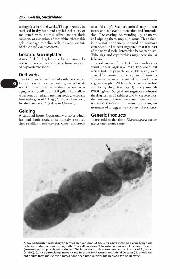
G
296 Gelatin, Succinylated
taking place in 4 to 6 weeks. The sponge may besterilised in dry heat, and applied either dry ormoistened with normal saline, an antibioticsolution, or a solution of thrombin. Absorbablegelatin sponge complies with the requirementsof the British Pharmacopoeia.
Gelatin, SuccinylatedA modified, fluid, gelatin used as a plasma sub-stitute to restore body fluid volume in casesof hypovolemic shock.
GelbviehsThis German yellow breed of cattle, as it is alsoknown, was evolved by crossing Swiss breedswith German breeds, and is dual-purpose, aver-aging nearly 3640 litres (800 gallons) of milk at4 per cent butterfat. Fattening stock give a dailyliveweight gain of 1.1 kg (2.5 lb) and are readyfor the butcher at 405 days in Germany.
GeldingA castrated horse. Occasionally a horse whichhas had both testicles completely removedshows stallion-like behaviour, when it is known
as a ‘false rig’. Such an animal may mountmares and achieve both erection and intromis-sion. The chasing, or rounding up, of mares,and nipping them, may also occur. This behav-iour is not hormonally induced or hormonedependent; it has been suggested that it is partof the normal social interaction between horses.‘False rigs’ and cryptorchids may show similarbehaviour.
Blood samples from 104 horses with eithersexual and/or aggressive male behaviour, butwhich had no palpable or visible testes, wereassayed for testosterone levels 30 to 100 minutesafter an intravenous injection of human chorion-ic gonadotrophin. All but 8 horses were classifiedas either geldings (<40 pg/ml) or cryptorchids(τ100 pg/ml). Surgical investigation confirmedthe diagnosis in 23 geldings and 47 cryptorchids;the remaining horses were not operated on.(See also CASTRATION – Immuno-castration, fortreatment of an aggressive cryptorchid stallion.)
Generic ProductsThose sold under their Pharmacopoeia namesrather than brand names.
A bovine/hamster heterokaryon formed by the fusion of Theileria parva-infected bovine lymphoidcells and baby hamster kidney cells. The cell contains 3 hamster nuclei and 1 bovine nucleus(arrowed) with a prominent nucleolus. The intracytoplasmic masses are macroschizonts of T. parva.(× 1600). (With acknowledgements to the Institute for Research on Animal Diseases.) Monoclonalantibodies from mouse hybridomas have been produced for use in blood typing in cattle.

GenesThe biological units of heredity, arranged alongthe length of the CHROMOSOMES. (See also CELLS.)
Genetic EngineeringThis may be defined as the recombination ofgenes from different organisms into 1 organismin a way that would never occur naturally;e.g. from a plant into an animal. However, inpractice, the terminology also includes geneticmanipulation, although some changes brought
about by manipulation may also occur quitenaturally.
Advances in knowledge of nucleic acids led tothe creation in the laboratory of new combina-tions of genetic material. This was achieved bysplicing together DNA from entirely differentsources to form hybrid molecules that are lesslikely to occur during evolutionary processes.
Micro-organisms virulent for cattle (forexample) will, if they can be adapted to growin laboratory animals, become less virulent for
Genetic Engineering 297
GHYBRIDOMA TECHNIQUE FOR PRODUCING ANTIBODIES TO PARASITE ANTIGENS (DIAGRAMMATIC)
Spleen cells from mice previously injected with parasite, and secreting antibody to the parasite, can befused with other cells to form hybrid cells that live and multiply. From these a cell line is selected by‘cloning’ (i.e. a colony that represents the progeny of a single cell is isolated) which secretes appropriateantiparasite antibody. (With acknowledgements to WHO Chronicle, 36.)
the original host, and may have usefulness forlater vaccine purposes. One method of adaptingthe micro-organisms to grow in laboratoryanimals is to fuse, artificially, cells to form aHETEROKARYON. (See illustration.)
Uses Genetic engineering has provided infor-mation on the molecular basis of gene action,on bacterial virulence and bacterial resistance.ln agriculture it offers the hope of being ableto transfer from bacteria to plants the geneswhich confer the ability to fix nitrogen – andso reduce farmers’ dependence on scarce andcostly nitrogen fertilisers.
In the UK, at the AFRC’s Unit of NitrogenFixation, genes for nitrogen fixation weretransferred with the aid of a PLASMID from anaturally occurring nitrogen-fixer Klebsiellapneumoniae to E. coli, which had never fixednitrogen before. The plasmid, of the exchange-able class, was able, when transferring, to takealong fragments of its host’s chromosomes,including pieces bearing the nitrogen-fixationgenes.
Some of the new nitrogen-fixing E. colistrains converted these fragments of Klebsiellachromosome into new, separate plasmids.Geneticists in the unit therefore constructed,by ordinary genetic manipulations, plasmidscarrying nitrogen-fixation genes which wouldtransfer themselves alone, without the aid ofanother plasmid.
In veterinary medicine the greatest potentiallies in the preparation of completely safe viralvaccines. One of the first successes was with foot-and-mouth virus. The specific viral protein, freeof infectivity, was produced by a geneticallymanipulated E. coli. This protein from a bacteri-al culture is capable of stimulating antibody pro-duction in animals. The technique is used also inthe production of a number of other vaccines.
Recombinant DNA techniques Develop-ment of such techniques involved 3 lines ofresearch: (1) recognition and isolation of extra-chromosomal DNA, or plasmids; (2) the manip-ulation of DNA with ‘restriction’ enzymes whichselectively split DNA into fragments whichcould then be rejoined; (3) reinserting the frag-mented DNA into living cells so that it becamepart of the genetic material of the cells.
In this way, genetic instructions for produc-ing mammalian enzymes could be transferredinto E. coli, the cell most used for propagatingsuch plasmid vectors to produce insulin, forexample.
The next step was to develop syntheticnucleotides, actually to construct genes. This
has been done successfully and nucleotides ofup to 500 characters have been constructed.
Another technique in genetic engineeringinvolves monoclonal antibodies. These are pro-duced by fusing antibody-producing cells froman immunised donor with another type ofwhite blood cell, thereby producing hybrid cellswhich in tissue culture could provide thedesired antibodies. Among potential uses are,again, vaccines, but also diagnostic reagents; forblood-typing, in race-horses, for example.
Genetics, Heredity andBreeding
PAGE
Phenotype and genotype 299Homozygous and heterozygous 299Inheritance through multiple genes 300Inheritance and high milk yields 300Selection 300Inbreeding 301Fusion of 2 inbred lines 301Hybrid vigour 301‘Nicking’ 302‘Pedigree and purity’ 302Chromosomes 302Genetic aspects of infertility 302Inheritance of twinning 302Heritability of certain traits 303Lethal and semi-lethal factors 303Heredity and disease 303Genetic defects 303
Introduction The science of genetics dealswith the physiology of heredity – the mecha-nism by which resemblance between parent andoffspring is conserved and transmitted; andwith the origin and significance of variation –the mechanism by which such resemblance ismodified and transformed. It seeks to definethe manner in which the hereditary charactersof the individual are represented in the fertilisedegg in which the individual has its beginning.
Stock-breeding is a craft concerned with themaintenance of the desirable qualities of astock, the improvement of these qualities gen-eration by generation, and the eliminationthrough breeding of qualities which are held tobe undesirable. The problems of the geneticistand of the stock-breeder are identical, thoughtheir interests are dissimilar.
The geneticist has made much progress bystudying, quickly maturing, very highly fertileanimals such as the mouse, rat, guinea-pig,rabbit, and above all the fruit fly Drosophila.
A better understanding of heredity was ren-dered possible by the concept that the individ-ual as a whole was not the unit in inheritance,
298 Genetics, Heredity and Breeding
G
but could be regarded as a definite orderlycombination of independently heritable units.
Breed was now interpreted as signifyingdifferent combinations of independently herita-ble characters, all drawn from the commonsource of the stock in which modern domesti-cated cattle had their origin – just as differentarrangements and combinations of letters makedifferent words, though all words are made upof letters derived from a common source: thealphabet.
The breeder has employed the methods ofhybridisation and inbreeding associated withselection in the creation of the modern breeds.He has practised inbreeding with selection inorder that the desired type of his stock maybe fixed, and he has sought hybrid vigour inoutcrossing. The geneticist has employed thesevery same methods in his studies. The methodof genetics is character-analysis. The object ofthe breeder is character-synthesis.
Instead of the hereditary mechanism being asimple affair as was first thought, it is one of themost complex.
Phenotype and genotype Allan FraserMD, DSc, when senior lecturer in animal hus-bandry at Aberdeen University, once drew ahelpful analogy between heredity and a gameof cards, with each card representing a gene,and Honours cards representing genes mostdesirable to a breeder.
A game of cards is preceded by the shufflingof packs, and so also is the conception of an ani-mal preceded by a shuffling of genes. Each packis then halved – just as before sperm and ovummeet, and the number of genes in both ishalved by what is called the reduction division.Fertilisation then reunites the 2 half-packs toform 1 new pack. The cards in this representthe genes in the new individual animal.
‘The cards in any one hand or the genesample in any individual animal are the result ofpure chance – no one can predict how the runof the cards or of the genes will go.’
Of 2 animals, sharing the same sire and dam,1 may have a much better genotype, be a morevaluable breeding animal than its full brother orsister.
If the cards dealt at conception be called theunalterable genotype of the animal, the playingof that hand may be called the environment,which includes climate, nutrition, exposure toinfections, stocking rates, and every aspect ofhusbandry and animal management.
Some stockmen can make a surprisinglygood job with poor genetic material; others asorry job with the best stock; but of course the
most skilful stockman cannot improve uponthe hand of genes once dealt.
Genotype can be defined as the entire arrayof genes carried by an individual (or, in anoth-er sense, the genetic constitution of an individ-ual with respect to any limited number of genesunder examination).
Phenotype is the appearance and/or the per-formance of an individual animal. Phenotypicvariation of a population results from the com-bined effects of inheritance and environment.Genetic variation is that part of the phenotypicvariation which is due to genes.
Homozygous and heterozygous Inorder to illustrate one of the simpler aspects ofheredity in relation to stock-breeding, theappearance of red calf in a herd in which the fash-ionable coat colour is black, and in which all redanimals are eliminated, will serve as an example.
Black-and-red coat colours in cattle consti-tute a typical pair of Mendelian characters,black being the dominant and red being therecessive member of the pair. A red calf can onlybe produced by black parents when both ofthese are heterozygous in respect of their coat-colour character. For the character black-coatcolour, there is a determiner or factor: thisfactor may be present in the zygote in theduplex state, having been conveyed thereintoby both egg and sperm. When the factor forblack is present in the duplex state, the individ-ual that arises for that fertilised egg or zygote isspoken of as being ‘homozygous’ for the char-acter black-coat colour. On the other hand,into the zygote there may have been brought afactor for black from 1 parent and a factor forred, the alternative character, from the otherparent. Under these circumstances, of these2 factors (that for black and that for red) it isthe former alone that determines what the coatcolour shall be. Black is said to be dominant inits relation to red. Homozygous and heterozy-gous blacks will be indistinguishable on inspec-tion. If 2 heterozygous blacks are mated therewill occur on the average in every 4, 3 blackcalves to 1 red. To explain this 3:1 ratio it isassumed that half of both male and femalegametes of such heterozygous individuals (i.e.sperm and ovum) carry the factor for the dom-inant character black, and the other half thefactor for the alternative recessive red, and the2 sorts of egg and of sperm occur in equal num-bers. If it is assumed that for every pair offactors that correspond to a pair of characters,only 1 can pass into the ripe gamete, it followsthat a 3:1 ratio in the next generation will beobtained, and of the individuals exhibiting the
Genetics, Heredity and Breeding 299
G
dominant character, 1 will be homozygous forthat character and 2 heterozygous, whilst theindividual exhibiting the recessive charactermust of necessity carry the factor for thatcharacter in the duplex state, since if in itshereditary constitution it carries a factor for thedominant, it will exhibit the dominant charac-ter. It is possible by examining the records ofthe coat colours of the offspring to define thehereditary constitution of the parent in respectof the coat colours black and red. The followingmatings are possible:
Homozygous black to homozygous blackwill give none but blacks, all homozygous.
Homozygous black to heterozygous blackwill give all blacks, of which 50 per cent will behomozygous and 50 per cent heterozygous.
Homozygous black to homozygous red willgive all heterozygous blacks.
Heterozygous black to heterozygous blackwill give 25 per cent homozygous blacks, 50 percent heterozygous blacks, and 25 per cent reds.
Heterozygous black to red will give 50 percent heterozygous blacks and 50 per cent red.
Red to red will give all reds, of necessityhomozygous. The only mating of blacks thatcan yield a red calf is that of two individualsheterozygous in respect of this coat-colourcharacter.
The coat-colour character has to be consid-ered quite apart from all the rest of the charac-ters that in their association make the animalwhat it is. An individual is a pure black when itis in respect of this character homozygous,when in its hereditary constitution the deter-miner or factor for this character has beenreceived from both its parents.
Inheritance through multiple genesThe above example shows how a character –coat colour – may be inherited through singlegenes. This is the mechanism of heredity at itssimplest. Most characters, however, including
many of economic importance to the farmer,are inherited in a far more complex mannerthrough multiple genes.
Multiple genes may have an additive effect asregards the expression of some character; orthey may interact, one with another, in theproduction of a character, inheritance of whichis even more complex.
Inheritance and high milk yields Itseems that it is easier to increase the butter-fatcontent and solids-not-fat content than it is toincrease the milk yield through breeding. Theheritability of milk yield is not as high as that ofsome other characters.
There is a correlation between high yieldsand body size, but conformation is by no meansalways associated with high yields. A few ofthe highest-yielding cows have had, to put itmildly, an unfashionable conformation.
The diagram below shows how 2 bulls, fullbrothers, may influence milk production intheir daughters in opposite ways. It also showshow the ‘gene lottery’ can make nonsense out ofthe expectations of a breeder.
For this reason, progeny testing has provedof the greatest importance in the selection ofbulls, each of which – through AI – may havenot 100 offspring but tens of thousands.
It is possible for a farmer to use (by means ofAI) a bull with the proved ability to producedaughters with a high milk yield, as comparedwith the yield resulting from use of animproved or average bull. Proven bulls are list-ed in terms of ‘a bonus of 50 or of anything upto 100 gallons’ and also in terms of a butterfatbonus. (See also PROGENY TESTING.)
Researchers have reported an associationbetween blood groups and production charac-ters in cattle and other animals. The work ontransferrin and milk yield is an example; that ofblood groups and milk yield another. However,the results were not sufficiently significant to beof practical value in selecting for productivity.
Selection is the systematic choice of animalsin a population (defined in the genetic sense asa group of interbreeding animals sharing a com-mon gene pool, e.g. a closed herd, an AI district,a breed) as parents for the next generation.
Family selection means the selection of indi-viduals on the performance of their relatives(sibs, half-sibs, or progeny), i.e. selection betweenfamilies instead of between individuals.
Genotypic selection is that based on progenytesting with a very large number of progeny, sothat the breeding value of the parent is exactlyknown.
300 Genetics, Heredity and Breeding
G
Black × Red. (Red is necessarily white in thediagram.)

Inbreeding may be defined as the matingof individuals more closely related than theaverage relationship of the population.
Both crossbreeding and inbreeding aremethods of bringing about genetic change.
Inbreeding was practised by the early devel-opers and improvers of livestock breeds in orderto fix the type of their animals. Inbreeding canbe expected to increase the proportion of ani-mals homozygous for a given desired character.As the process proceeds, however, individualswith undesirable characters are likely to appear– animals which are abnormal in some respect,sterile, or weak. Inbreeding could prove disas-trously expensive if the proportion of suchanimals were high. (Test mating of a bull torelated or carrier females may be carried outin order to detect specific genes such as lethalfactors; the carriers then being culled from theherd.)
Prolonged inbreeding will lead to disappoint-ing regression, diminution of vigour, decreasedfertility, and a reduction in body size.
The fusion of 2 inbred lines By inbreed-ing for a specific character, and by practisingrigid selection for 2, 3, or more generations,a strain of relatively homozygous individualsfor the selected character can be created. If 2such strains are developed separately but simul-taneously for 2 different but highly desirable
characters, and if these 2 strains are thencrossed, the resulting progeny can be expectedto possess both desirable characters to a usefuldegree. The mechanism has been notablysuccessful in producing strains of poultry withlarge egg size and high annual yields, and hasbeen exploited commercially.
When the generation in which the 2 desirablecharacters are expressed is bred from again, animmediate reassortment of characters occurs,and only in a small percentage of the individu-als will the desirable characters be expressed.The others are most likely to be useless.
Hybrid vigour (Heterosis), usually demon-strated by increase in size, better liveweightgains and greater resistance to disease and in theearlier attainment of sexual maturity, occurs inthe first cross-bred generation out of the matingof 2 widely dissimilar pure-bred parental stocks.Hybrid vigour is an indication of heterozygosis.The 2 parental breeds must be within reason asdissimilar in their characterisation as possible;then in the pooling of those hereditary consti-tutions there will be a very considerable degreeof heterozygosis in the 1st cross-bred offspring;the desirable characters are pooled, and inrespect of those characters exhibited by the 2parents the offspring will be heterozygous.Generally, a characteristic with poor heritabilityis enhanced more than one of high heritability.
The effect of inbreeding and crossing. These 2 sheep were sired by the same ram, but the smallerone was highly inbred (59 per cent) and when 10 months old had attained little more than 40 percent of the weight of its half-sib (left), which was a 3-way cross of inbred lines, its dam being a 2-way line cross. These hoggs are of Chevoit × Welsh extraction. (Animal Breeding ResearchOrganisation photograph.)
Genetics, Heredity and Breeding 301
G
If the 1st crosses which exhibit this hybridvigour so markedly are interbred, their off-spring will not exhibit this vigour to the sameextent.
‘Nicking’ Two individuals not remarkable inthemselves may produce superior offspring.This fact can be explained on the assumptionthat the mating brings together the chance asso-ciation of factors which are complementary orsupplementary, and that these in conjunctiondetermine characters that are greatly esteemed.
‘Pedigree and purity’ Pedigrees and regis-tration in the appropriate herd book have beenan essential part of breeders’ work in all but‘commercial’ herds. The system has been ofservice, though not free from abuse. Pedigreeshave from time to time been falsified orgenuine mistakes made (including use of thewrong semen in AI work), and until the adventof blood typing there was no means of checkingsuch errors.
It is easy to exaggerate the importance of‘family name’ or of remote ancestors; and interms of production traits or characters it hasbeen stated that ‘the concept of “purity” of purebreeds is not only something of a myth, butmight actually be detrimental to the possibilitiesfor improvement’.
Chromosomes A male can be distinguishedfrom a female not only by external appearancesand by differences in the architecture of thereproductive system, but also by differencesin the organisation of the cells of which theirbodies are composed.
The nucleus of the resting cell appears instained microscopic preparations as a vesiclecontaining a network of delicate threads uponwhich are borne, like beads upon a tangledskein, minute masses of a deeply staining mate-rial known as chromatin. As the cell proceedsto divide into 2, this tangled mass of finethreads resolves itself into a constant numberof filaments of definite shape and these becomeprogressively shorter to assume the form ofstout rods known as chromosomes. The num-ber of chromosomes is usually some multipleof 2, and is constant and characteristic ofthe species to which the individual belongs,e.g. dog 78; horse 64; cow 60; sheep 54; pig 38;cat 38.
(The study of chromosomes is known asCYTOGENETICS.)
The gametes, egg and sperm respectively,differ remarkably in size and form, but they arealike in that each contains half the number of
chromosomes that is characteristic of thesomatic cells of that species.
At fertilisation, the characteristic number isrestored, and in the case of each pair 1 memberis derived from one parent, the other from theother. In this distribution of the chromosomesone finds a mechanism by which offspringmay inherit from both parents by means of theconstituent genes.
It is possible to distinguish male from femaleby differences in the chromosome content.While all other pairs of chromosomes consist of2 chromosomes exactly alike in size and shape,1 pair differs in the 2 sexes. As they influencesex determination, these chromosomes arereferred to as the sex chromosomes.
The ovum (or egg) contains the X chromo-some.
The sperm may contain either the Y chromo-some, which will produce male offspring (XY), orthe X chromosome, which will produce femaleoffspring (XX).
In most female mammals the paired sex-chromosomes are identical, and called the Xchromosomes on account of their shape.
In the male, the sex-chromosomes aredissimilar, one being the X chromosome andthe other the Y.
The autosomes are all the other chromosomesexcept the sex-chromosomes.
Genetic aspects of infertility These arebriefly discussed in the following paragraphs.Chromosome screening (see CYTOGENETICS)and, to a lesser extent, blood-typing are likely tobe of increasing value in eliminating a propor-tion of bovine infertility due to hereditary causes. (See also INFERTILITY.)
Inheritance of twinning It is mainly cowsover 3 years old which have twins, and although
302 Genetics, Heredity and Breeding
G
A bull’s lymphocyte showingchromosomes in the 2nd stage(metaphase) of cell division(mitosis). (With acknowledge-ments to Dr C. R. E. Halnanand the Veterinary Record.)
twinning may be a desirable character for thefarmer it is not one which is easy to obtainthrough breeding. It seems that there is a ratherlow level of genetic variation in frequency oftwinning, and that prospects for rapidlyincreasing litter size in dairy cattle do not atpresent seem good. In some breeds of sheep,and perhaps also in cattle, it may not be theovulation rate which is the limiting factor but,as indicated above, the survival rate of fertilisedeggs.
Heritability of certain traits The follow-ing table, compiled at the Animal BreedingResearch Organisation, shows the degree ofheritability of certain traits sought by the pigbreeder.
Trait Heritability%
Litter size at weaning 7Average weaning weight 8Daily gain, after weaning 0Food conversion 5Average back fat 50Percentage of lean meat 45
It will be seen that litter performance has a lowheritability, and must be achieved by suitablecrossbreeding. The crossbred sow has a markedsuperiority in this respect.
Lethal and semi-lethal factors Lethalfactors have been defined as genes which, whenpresent in the homozygous condition, causethe death of the embryo and when present inthe heterozygous conditions cause a seriousimpairment in the individual often leading tonon-survival.
Lethals may be dominant or incompletelydominant, but many are certainly recessive.They are not always recognised since they maycause death of the embryo early in development,and the mating may be regarded as having beeninfertile – another illustration of the difficulty ofdefining the genetics of infertility.
Several lethals are met with in cattle, such asparrot-mouthed, in which calves die at a fewhours of age, or amputated, in which calves areborn dead with legs and lower jaws absent.
Semi-lethals include over-shot jaws in calves.
Heredity and disease Diseases exhibit aspectrum according to the genetic influence.Canine haemophilia is entirely genetic. Swedishgonad hypoplasia is mainly genetic, mastitis ismainly environmental and injuries are entirelyenvironmental. Simply dividing diseases intogenetic and non-genetic is, therefore, inaccurate.
Recessive inheritance: many diseases areinherited as autosomal recessives. Neither parentis usually affected, but the disease comes fromboth; the sexes are affected equally, inbreeding isoften being practised, the incidence is generallylow and exact genetic ratios are obtainable. Fewdiseases, however, fulfil all the criteria of simplerecessives.
Diseases due to sex-linked recessives, e.g.canine haemophilia, are uncommon in domes-tic mammals and, not being transmitted byunaffected males – the carrier female transmitsthe disease to half her male offspring – areunlikely to be a major problem. In poultry,however, sex-linked abnormalities such asfamiliar cerebellar degeneration are transmittedby the male to half his female progeny and arerelatively common.
Diseases such as cryptorchidism and inter-sexes are sex-limited and sometimes regarded asdue to recessives, the homozygote only express-ing itself in 1 sex. While both parents are proba-bly involved, their exact inheritance is unknownand cryptorchidism is certainly subject toenvironmental modification.
Irregular inheritance: many defects have acomplex inheritance. Sporadic abnormalities,which increase on inbreeding, such as chicken‘crooked toes’ or pig ‘kinky tails’, are calledpheno-deviants, and are probably caused byrecessives exhibiting a threshold of manifestation.
Dominant inheritance: the disease usuallycomes from 1 affected parent, and half its off-spring are affected. Few dominant diseases areknown in livestock, except in poultry. Irregulardominants, exemplified by ‘curved limbs’,where an unaffected male transmits a geneproducing defective offspring out of unrelatedfemales, and less than half are affected, do,however, occur. Environment or modifyinggenes may cause the defect inherent in the geneto develop.
Semi-dominant inheritance: several diseasesare due to semi-dominants, the heterozygotebeing distinguishable from both homozygotes.A single dose of a semi-dominant gene pro-duces a Dexter, a double dose a bulldogcalf, and the homozygous normal allele pro-duces a long-legged Dexter. Some Americandwarf cattle result from semi-dominants (withacknowledgements to Dr G. B. Young, AnimalBreeding Research Organisation).
Genetic defects All breeds of livestock mayharbour some genetic defects, but their inci-dence is usually low. However, specific defectssometimes become more frequent in certainbreeds, and give cause for concern.
Genetics, Heredity and Breeding 303
G

Cattle Examples of genetic defects include thefollowing:
Arthrogryposis in Charolais cattle. This defectis characterised by twisted limbs, cleft palate, andtwisted spine. In France about 1 per cent ofCharolais cattle are affected. AI records show thatwhile many bulls transmit an occasional defect ofthis nature, a few sire about 5 per cent of off-spring having this abnormality. Another defectencountered in some Charolais cattle involvesthe eyes. (See EYE, DISEASES OF – Coloboma.)
Decapitated sperms. This defect causes therejection for AI in Britain of many Herefordbulls ‘with superior test performance’ becausetheir semen contains a high percentage ofsperms with the head separated from the tail.
Tibial hemimelia. A dog-sitting position in thenewborn Galloway calf is suggestive of this defect,which involves bones missing from the hind legs(but has to be differentiated from another defectinvolving the pelvis, which also prevents the calffrom standing on its hind legs). It is estimatedthat about 16 per cent of Galloways now carrythe gene which transmits this defect. (TheGalloway Breed Society has an excellent scheme,requiring compulsory insurance and compulsoryslaughter of any bulls leaving offspring with thisdefect.) (See also MANNOSIDOSIS.)
Sheep Genetic defects include achondroplasiain some South Country Cheviot flocks. A‘squashed-in’ face, shortened forelimbs anddefective hooves are characteristic of these‘dwarf lambs’.
Cerebellar ataxia is seen in some BorderLeicester flocks, and these ‘daft lambs’ have astaggering gait and incoordination of the head.This is due to a recessive gene.
Genital Organs(see diagrams for PENIS and UTERUS)
GenomeA complete set of chromosomes derived from 1parent; or the total gene complement of a set ofchromosomes.
GenotypeThis can mean the entire array of genes carriedby an individual; or the genetic constitution ofan individual with respect to any limited num-ber of genes under examination; or (more loose-ly) the individual within a given genotype. (See
also under GENUS.)
Genotypic SelectionGenotypic selection is that based on progeny-testing with a very large number of progeny
so that the breeding value of the parent is exact-ly known. The expression is also loosely usedas a synonym for progeny testing without thisproviso.
GentianThe dried and powdered root of the yellowgentian plant (Gentiana lutea). It is a bittertonic used as an appetiser.
Gentian VioletA stain used in microscopical work and a valu-able antiseptic, of use against fungal and bacter-ial skin infections. (See also ANTISEPTICS – Crystalviolet.)
GenusGenus is a group of species. One of the speciesis chosen as being typical, and referred to as thegenotype.
GerbilA small burrowing rodent, originating in deserts,popular as pets.They live for 3 to 5 years; adultsweigh 50 to 90 g, the females being larger thanmales. Sexual maturity occurs at 10 weeks. Thegestation period is 24 to 26 days and the youngare weaned at 21 to 24 days. Rectal temperatueis 37.4 to 39°C. They are naturally healthy ani-mals, remarkably free from infectious diseases:Tyzzer’s disease is usually the only finding. Inthe wild they ‘play dead’ when attacked; this is aself-induced epileptic seizure and may occurwhen they are handled. (See also PETS.)
GeriatricsThe study of the problems and diseases of theolder animal.
German Shepherd DogPreviously known as the Alsatian, this is a medi-um to large-sized dog with a tan and black coat.The breed is extremely popular and, to fulfildemand, many unsuitable animals were used forbreeding. In consequence a number of inheriteddefects are associated with the breed. Theseinclude achalasia, calcinosis circumscripta, cleftpalate and epilepsy. A campaign to eliminate hipdysplasia, which is very common, requires allpotential breeding dogs to be X-rayed for evi-dence of the defect. Dogs in which it is foundare not used for breeding.
Germ CellsThe gametes, i.e. ovum or sperm.
Gestation(see PREGNANCY)
304 Genital Organs
G
Getah VirusA mild infectious disease of racehorses, charac-terised by fever, dermatitis, and oedema of thelimbs, appeared in Japan in 1978, and wasfound to be due to Getah virus. Antibodies to ithad previously been detected in man, horses,pigs, and birds; none showed symptoms.
Giant CellsSeen in various infections, e.g. the poxes, tuber-culosis and Johne’s disease, these multinucleatecells are formed by cell fusion, stimulated byviruses. Giant cells can be obtained in vitroin cell cultures, as well as being found in thebody.
Giant Hogweed (HeracleumMantegazzianum)This plant grows to a height of up to 3.65metres (12 feet), and has white flowers. Thestem is hollow, and has reddish/purple blotches.Human contact with the plant, and subsequentexposure to sunlight, leads to dermatitis; thepoisoning is a result of LIGHT SENSITISATION.A suspected case of this was seen in a goat,which was subdued, refusing food or water,and salivating profusely. Severe ulceration wasfound in its mouth. Two sheep were sensitiveto the cut stems of the plant and developedmouth ulcers.
In another case, circumstantial evidence sug-gested involvement of this plant in week-oldducklings which had foot and beak lesions. Twoducklings with white beaks could manage only‘open-beak’ breathing; whereas the beaks whichwere pigmented, in the other five ducklings,were not affected. The foot lesions consisted oflarge blisters.
GiardiaA genus of flagellate protozoa.
GiardiasisIn dogs a low-grade infection with Giardia canismay interfere with the absorption of fat andvitamin A; while if large numbers of G. canisare present there is likely to be chronic diar-rhoea (sometimes dysentery), with resultingdehydration.
The flagellate parasite occurs in 2 forms: thetrophozoite, which divides by binary fission,and is seldom seen in the faeces; and the cystform which is found in the faeces but is not easyto identify.
Metronizadole is used in treatment.In the USA G. lamblia is a common cause of
human gastroenteritis. The infection is usuallyvia drinking-water.
Gid (Sturdy)Gid (sturdy) is a condition in sheep, and occa-sionally in cattle, caused by tapeworm cystslodged in the brain. The sheep become infectedthrough swallowing unhatched eggs of the dogtapeworm Taenia multiceps. The eggs are passedout in the dog’s faeces. As the cysts (coenuri)develop, they press upon brain cells and giverise to nervous signs, such as staggering, cir-cling, blindness. The name ‘sturdy’ derives fromthe ‘sturdy’ manner in which an affected sheepmay struggle when caught. (See COENURIASIS.)
Gilchrist’s DiseaseInfection in man with Blastomyces dermatitidis.(See BLASTOMYCOSIS.)
GillsThe external breeding apparatus of fish andsome other water dwelling creatures. They aredelicate structures, prone to damage, infectionand parasites. Chemical pollutants in the watercause fusion of the lamellae of the gills, reducingthe absorptive surface.
Gill Maggot(see SALMINCOLA)
GiltA female pig intended for breeding purposesbefore she has her 1st litter.
GimmerA ewe lamb up to its 1st shearing, or ewe whichhas not yet produced offspring (see under SHEEP).
GingivitisInflammation of the gums. (See MOUTH,DISEASES OF; also FELINE GINGIVITIS.)
Gizzard (Ventriculus)The thick-walled, muscular stomach in a bird;it has a tough keratin lining. The gizzard’smain function is to grind food, and in this it isassisted by swallowed grit and small stones. (See
under GRIT.)
GlandersGlanders is a NOTIFIABLE DISEASE throughoutthe European Union. It is a specific, contagiousdisease of the horse family (Equidae), but alsoliable to be contracted by other mammals,including man. It is characterised by the forma-tion of nodules in the lungs, liver, spleen, orother organs; ulcerations occur of the mucousmembranes, especially those of the upperair passages, accompanied by changes in the lym-phatics and also by skin lesions. The condition
Glanders 305
G
is due to the entrance and growth in the bodyof the glanders bacillus Pseudomonas mallei(formerly known as Pfeifferella mallei).
History Glanders has been known as a seriousdisease since about 450 BC, when it was men-tioned by Hippocrates, and its contagiousnature was pointed out by Vegetius (a veterinarywriter) in the 4th century.
Distribution Glanders has been distributedto practically every country in the world atsome time or other, and was once a usual con-comitant of wars. It became prevalent, both inthe United Kingdom and in South Africa, afterthe South African war (1899–1902). In the year1892 there were more than 3000 cases of glan-ders recorded in Britain; in 1904 between 2000and 3000; while only 9 outbreaks were record-ed in the year 1923, and none since 1926.
Glanders is still endemic in Mongolia,other parts of Asia, East Africa and SouthAmerica and, recently, it has been spreadingfrom enzootic zones.
The donkey is the most susceptible to thedisease and nearly always suffers from the acuteform, from which it dies in from 2 to 3 weeks.
In the horse, glanders occurs in an acute or achronic form, the latter existing for months oreven years before it finally kills its victim; how-ever, under modern conditions it is rare to allowthe disease to run its natural course. The muleis intermediate in susceptibility between thedonkey and the horse, but usually shows theacute type. Dogs and cats may become infectedif fed upon meat from a horse which had glan-ders. The camel is susceptible, though naturalcases are very rare.
Horses can be infected naturally by 3 differentchannels:
1. By the digestive tract, through the mediumof infected food and water. This is by far thecommonest method of spread.
2. By inhalation (rarely) when some abrasionof the respiratory passage is present.
3. By skin infection.
Incubation period This may last severalmonths in the horse.
Signs The signs of glanders in the horse arevery varied. The disease may run an acutecourse of only 2 to 3 weeks, but by far the great-est number of cases met with are of a subacuteor chronic nature. A horse may be affected andshow no outward sign of disease, and yet it mayhave nodules in 1 or both lungs.
Glanders may be of the nasal, pulmonary, orglandular form, of the type producing skinlesions (farcy), or an admixture of these, and itmay also become generalised. One of the dan-gers of the disease is that a horse may work forweeks or even months with ‘open’ lesions – notlosing a great deal of flesh nor appearing very ill– and so spread the disease to healthy horseswith which it comes into contact.
When farcy is present the glands inside theaxilla or inside the groin may be somewhatenlarged and even painful to the touch. In entirehorses the testicles often become enlarged andpainful, or even the seat of glanderous abscesses.
There may be a nasal discharge, thin andwatery in the early stages, but later becomingthick, greyish, or yellow and oily. Examinationof the nose in these cases is dangerous; stricthygenic precautions should be taken, as manmay easily contract the disease by this means.
Farcy: in this form of the disease, the skin isinvolved. It is usually chronic in nature, butfarcy buds and subcutaneous swellings maycomplicate the most acute form of the diseaseshortly before death. This complication isespecially common in the mule, which oftensuccumbs to the disease before the farcy budshave time to burst. In chronic farcy there isusually swelling of one or more limbs, morefrequently a hind one. The lymphatic glands ofthe affected limb become enlarged, the lymphvessels corded, and usually a chain of farcy budsdevelops along their course.
In acute glanders there may be all the signs ofan acute broncho-pneumonia, a high tempera-ture, a rapid loss of flesh, rapid and sometimesnoisy breathing, followed by death in a fewweeks; in fact, this is the common form seenin the donkey and often in the mule, with orwithout the complication of farcy.
Diagnosis This is confirmed by means of themallein test.
Treatment Sulfathiazole has been used, butin most western countries treatment is notpermitted; the policy being one of slaughterand eradication.
Glanders in man is a distressing and nearlyalways fatal disease, and may be contracted bygrooms and others working with infected horses.Laboratory workers handling infected material orpure cultures of the organism are especially liableto infection, so that every precaution against thiscontingency has to be taken; in fact, the glandersbacillus is amongst the most dangerous of all disease-producing bacteria cultivated.
306 Glanders
G

The causal organism is present in all thelesions, though it does not – except the rarecases – circulate in the blood-stream.
GlandsA term loosely applied to a number of differentorgans. In each there are epithelial cells whichhave a secretory function (e.g. tear production).Glands are often classified as either endocrine,which are ductless, or exocrine, which usuallyhave ducts to carry their secretion to an epithelialsurface. (See ENDOCRINE GLANDS, HORMONES.)
A description of the various glands will befound under headings such as THYROID, LIVER,MAMMARY GLANDS, OVARIES, PANCREAS, SALI-
VARY GLANDS, TESTIS. Sweat and sebaceousglands are referred to under SKIN.
Lymphatic glands are nowadays more oftenreferred to as lymph nodes.
Glass EmbolismFluids for injection may contain glass particles.Glass gets in not during manufacture of thevial, but when the neck of the viual is snappedoff to insert the hypoermic needle . In humanmedicine the risk of glass embolism has beenstressed, and it has been suggested that, as awise precaution, time should be allowed forglass particles to settle before filling the syringe.
Glass, SolubleSoluble glasses containing cobalt, copper andselenium have been used in BOLUSES.
Glasser’s DiseaseAn infection which causes swelling of the hockor knee joints, or both, in the pig. There is fever,lameness and a disinclination to move; it canalso cause pericarditis, pleurisy and peritonitis.Death is usually a sequel, unless early treatmentwith, e.g., penicillin is undertaken. Glasser’sdisease is differentiated from joint-ill. Pigs of 5to 14 weeks old are chiefly affected. The cause isHaemophilus parasuis, or a mycoplasma, or both.
Glauber’s SaltsGlauber’s salts is the popular name for sodiumsulphate, a saline purgative.
GlaucomaAn increase in the pressure within the eye(intraocular pressure). (See under EYE, DISEASES
OF.)
Glenoid CavityGlenoid cavity is the shallow socket on theshoulder-blade into which the humerus fits,forming the shoulder joint.
GliomaGlioma is a tumour which forms in the brainor spinal cord. It is composed of neuroglia,which is the special connective tissue foundsupporting the nerve cells and the nerve fibres.(See TUMOURS.)
GliosisA proliferation of ASTROCYTES. It may follow abrain injury.
GlobidiosisA disease characterised by enteritis and closelyresembling coccidiosis. It occurs in Africa, SWEurope, the USA, and Australia. The cause is aspecies of Globidium. Cysts may be formed inthe skin or underlying tissue. Horses, cattle,and sheep are affected; in horses, severe diar-rhoea may be caused. Fatal cases of Globidium(Eimeria) leuckarti have occasionally beenrecorded in horses in the UK and Ireland.
GlobulinGlobulin is a protein fraction of the blood plas-ma, associated with immunity. (See also GAMMA
GLOBULIN.)
GlomerulonephritisAlso called glomerula disease. (See KIDNEYS,DISEASES OF.)
GlomerulusGlomerulus is a small knot of blood vessels, andfrom which the excretion of fluid out of theblood into the tubules of the kidney takes place.(See KIDNEY.) Glomerulonephritis is referred tounder KIDNEYS, DISEASES OF.
Glossectomy, PartialA surgical operation to remove part of thetongue. It has been found necessary in a horsefollowing severe laceration by a sharp bit.
GlossitisInflammation of the tongue.
Glossopharyngeal NerveThis is the 9th cranial nerve, which in the main is sensory. It is the nerve of taste for the back of the tongue, of sensation in a general way for the upper part of the throat, as well as for the middle ear, and it supplies the parotidgland.
GlottisGlottis is the narrow opening at the upper endof the larynx. (See AIR PASSAGES; CHOKING;LARYNX.)
Glottis 307
G
Gloves, SurgicalSterile gloves of fine rubber or polyethyleneworn during surgical procedures. A sleeve-length glove is worn by veterinary surgeons forcarrying out e.g. rectal examinations in cattle.
Surgical glove powder (sterilised maize starch)can cause an iatrogenic starch peritonitis. In acomparative study of various methods of wash-ing, brushing and rinsing, a routine involving a1-minute povidine-iodine surgical scrub followedby a rinse under sterile running water (500 ml)for 30 seconds removed 99.8 to 100 per cent ofthe original starch grain count; this routine alsoprovided the surgeon with a reassuring autotac-tile stick signal (separation of opposing forefingerand thumb as total removal of starch is about tobe achieved). Glove stickiness disappears whentissue fluid of patient coats gloves. Rinsing glovesin water alone leaves about 10 per cent of theoriginal starch on the gloves. Hypersensitivity tolatex has been recorded in people who have usedsuch gloves frequently.
GlucagonThis is a polypeptide hormone secreted byalpha-cells in the islets of Langerhans of thepancreas, and increases the amount of glucosein the blood.
GlucocorticoidsHormones, from the cortex of the adrenalgland, concerned with the formation of glu-cose. Cortisol (hydrocortisone) is one of themost important of these. Excessive secretion ofglucocorticoids is a feature of Cushing’s disease.
GlucoseGlucose is the form of sugar found in honey,grapes, fruits, etc., and in diabetes mellitus it ispassed in the urine. It is the form in whichsugar circulates in the bloodstream, and is veryuseful as an injection or drench when there is adeficiency of circulating sugar in the blood oran excess of ketones. (See ACETONAEMIA.)Glucose is a most valuable food to give duringthe course of acute illnesses, since it puts nostrain upon the digestive system yet providesfuel for the muscles, etc. Glucose saline is givenas a drink or administered per rectum or bysubcutaneous injection during the course ofjaundice, gastroenteritis, etc. (See SUGARS.)
GlucosinolatesSubstances found in Brassica spp that are bro-ken down in the rumen to form thiocyanates,which can interfere with iodine absorption. Ifbrassica are eaten to excess during pregnancy,goitre may result in the offspring.
GlucosureaThe presence of glucose in the urine. It isseen in diabetes mellitus (see DIABETES), and insome other conditions, and in all animals aftersevere shock. (See also under URINE – Abnormalconstituents of.)
GlutamineAn amino acid important in brain and musclemetabolism. High levels of exercise, as in racingor endurance riding, can seriously deplete levelsof glutamine in the muscle. If extreme,anabolism (growth and repair of muscle) isprevented and muscular exhaustion occurs.
GlutaraldehydeA disinfectant. Used in dilute solution inpreparations for cleaning udders.
GlutealGluteal is the scientific name applied to theregion of the buttocks, and to associated struc-tures, such as gluteal arteries, muscles, nerves.
GlyceridesOrganic esters of glycerin. (See FAT.)
Glycerin (Glycerol)Glycerin (glycerol) is a clear, colourless, odour-less, thick liquid of a sweet taste, obtained bydecomposition and distillation of fats. It dis-solves many substances and has a great power ofabsorbing water.
Uses Given by the mouth, diluted, it has beenused with success in the treatment of pregnan-cy toxaemia in ewes and of acetonaemia incattle. Internally, glycerin acts as a laxative tothe dog in moderate doses. It is soothing andantiseptic to inflamed mucous membranes inthe mouth and throat. In amounts of 15 to30 ml (1⁄2 to 1 ounce) it is useful as a rectal injec-tion to induce passage of impacted faeces inobstinate constipation in the dog, and it may beused for the same purpose in foals and calves.For these purposes it may be given diluted witha little water, or it may be used in the pure state.It is also used as a basis for the compounding ofvarious electuaries for the horse and dog, and itis sometimes incorporated into cough mixturesfor the smaller animals. It is used in certain skindressings when it is desired to soften the skinsurface and encourage the absorption of otherdrugs. It is also used as a diluent for semen. (See
under ARTIFICIAL INSEMINATION.)
GlycogenGlycogen is an animal starch found particularlyin the liver as well as in other tissues. It is the
308 Gloves, Surgical
G

form in which carbohydrates taken in the foodare stored in the liver and muscles before theyare converted into glucose when needed.Glycogen can become depleted when animalsare stressed, as by travelling over long periods. Ifnot replaced, animals slaughtered after a longjourney may have dry, firm, dark (DFD) meat.
Glycol, Ethylene(see under ANTIFREEZE for poisoning in dogsand cats)
Glycol, Propylene(see PROPYLENE GLYCOL)
GlycosidesGlycosides are potent naturally occurring sub-stances, combining a sugar with a hydroxy com-pound. Many are found in plants and are of greatvalue in medicine. The cardiac glycoside digital-is, found in the foxglove (Digitalis purpurea), is aprime example. Cyanogenetic glycosides arefound in linseed and saponins in ‘lords and ladies’(Aurum maculatum).
Aminoglycosides are powerful antibacterials;streptomycin was the first widely used amino-glycoside antibiotic.
Glycosuria(see GLUCOSUREA)
Gnathostoma SpinigerumA roundworm infecting dogs, cats, wild carni-vores, and man. It forms a nodule about 1.5 cm(1⁄2 inch) in diameter in the gastric mucosa. Ina cat suspected of having rabies it causedanorexia, nausea, and convulsions, coma anddeath.
GnotobioticsGnotobiotics is the name given to germ-freelaboratory animals reared according to tech-niques developed by Professor J. A. Reyniers ofNotre Dame University, Indiana. Such animalshave been used in the investigation of certaindiseases. (See also SPF; ‘DISEASE-FREE’ ANIMALS.)
Goads, ElectricBatons with electrodes at one end which give amild electric shock when they touch an animal.They are preferable to the use of carelessly orsadistically wielded sticks. The points should beblunt and spring-loaded, as otherwise they canbe jabbed into the animal – when the purposeand object of an electric goad are defeated.
It is illegal to use electric goads on calves andpiglets. They may only be applied to thehindquarters of cattle over 6 months of age, and
to adult pigs. There must be a way clear for theanimals to move forward. The Welfare ofAnimals (Slaughter or Killing) Regulations1995 state that electric goads should be appliedfor no more than 2 seconds at a time andthat there should be adequate time betweenapplications.
Goat Warble Fly(Przhevalskiana Silenus)Goat warble fly (Przhevalskiana Silenus) para-sitises goats and horses in many European andeastern countries. Hypoderma aeratum and H.crossi are other species.
GoatlingA female goat between 1 and 2 years old.
Goats as Grazers with Sheep(see PASTURE MANAGEMENT)
Goats, Disbudding of KidsThis is carried out under general anaesthesia(see ANAESTHESIA, GENERAL; GOATS); and theiron used for disbudding is heated by gas (orelectricity) to a ‘cherry-red’ heat. Great care isneeded in this operation as the skull is very thinin the area concerned.
Goats, Diseases of(see under ACETONAEMIA; AGALACTIA; BRUCEL-
LOSIS; CAPRINE ARTHRITIS-ENCEPHALITIS;CONTAGIOUS CAPRINE PLEURO-PNEUMONIA;CASEOUS LYMPHADENITIS; CHLAMYDIA (forabortion); ‘CLOUDBURST’; COCCIDIOSIS; CRYP-
TOSPORIDIOSIS; CYSTICERCOSIS; ENTEROTOX-
AEMIA; JOHNE’S DISEASE; MASTITIS; ORF;PARASITIC BRONCHITIS; PARASITIC GASTROEN-
TERITIS; PREGNANCY TOXAEMIA; Q FEVER; RICK-
ETS; RINDERPEST; SALMONELLOSIS; SWAYBACK;TUBERCULOSIS; YERSINIOSIS; LOUPING ILL;SCRAPIE; OSTEODYSTROPHIA FIBROSA; MAN-
NOSIDOSIS; LISTERIOSIS; MYCOPLASMOSIS;RINDERPEST; LIVER FLUKES; GOITRE; PULPY
KIDNEY DISEASE)
Goats’ Milk, CheeseGoat’s milk is often substituted for cow’s milkfor children suffering from a suspected allergyto cow’s milk. However, unless the milk is pas-teurised, there is a risk of human illness arising;especially from infection with Brucella meliten-sis, but also from Yersinia pseudotuberculosis –one cause of mastitis in goats. Goat’s milk maycontain louping-ill virus, staphylococci, E. coli;and, since goats are not immune to tuberculo-sis, even tubercle bacilli. Goat’s milk cheese isalso a source of B. melitensis infection.
Goats’ Milk, Cheese 309
G
GoitreGoitre is an enlargement of the thyroid gland,associated with an iodine deficiency; rarely, it canbe the result of a persistently high intake of iodineby the dam affecting the fetal thyroid. It is seen inpuppies, foals, and lambs, and also in calves, andit appears to be commoner in some districts thanin others. Swellings may appear below the larynx,usually one on either side, and beyond the localenlargements there may be no definite symptomsshown. Some cases respond to the administrationof thyroid extracts, and iodine internally, whilesome clear up spontaneously without treatment.(See IODINE; CALCIUM SUPPLEMENTS; THYROID
GLAND.)Lethargy is a notable symptom of goitre in the
dog, in which the disease often occurs in the 3 to5 year age group, especially in the bigger breeds.
Some pastures and foodstuffs may give riseto goitre. (See GOITROGEN.) Goitre seemsparticularly common in Dorset horn sheep.
A number of cases of supposed ‘goitre’ proveupon careful examination to be tumour growthin some part of the throat, not necessarily in con-nection with the thyroid gland – though cancerof the thyroid is not uncommon in the dog.
Goitre also occurs when, instead of a deficien-cy of thyroxin, there is a state of hyperthyroidism,or too much thyroxin. (See THYROID GLAND,DISEASES OF.)
In goat kids, some supposed cases of goitreprove to be hyperplasia of the THYMUS gland.
In other animals ‘goitre’ may be a misdiag-nosis for neoplasms in the throat region.
Goitrogen (Goitrogenic Factor)Goitrogen (goitrogenic factor) is one whichgives rise to goitre. Both kale and cabbage con-tain goitrogens (glucosinolates) and must there-fore not constitute too large a proportion of ananimal’s ration over a period. The same appliesto turnips. Iodine licks may be advisable. (See
IODINE DEFICIENCY.)
Golden RetrieverPopular dog of medium or larger size with aflat, wavy coat, blonde or gold in colour.Cataract, progressive retinal atrophy and entro-pion are inheritable. Retrievers may also besusceptible to hip dysplasia and myopathy.
Goldfish (Carassus Auratus)Ornamental fish of the carp family, originallydomesticated by the Chinese in the Sungdynasty (960–1279). More than 120 breeds arerecognised, as well as the common goldfish.They vary in colour and markings, and inconformation of the fin, tail and head. Their
lifespan can range from 2 years to 25 years.Size may vary from 5 cm (2 inches) to 30 cm(12 inches) or more. If the diet is too rich inoil, deficiency of vitamin E may result. Thiscan cause anaemia, poor growth, musculardystrophy, exophthalmia and other conditons.
GonadGonad is a sex gland which produces a gamete(the ovary or testis).
GonadotrophicGonadotrophic indicates something whichstimulates the gonads – testes and ovaries. (See
GONADOTROPHINS; HORMONES.)
GonadotrophinsGonadotrophins are hormones which havea gonadotrophic effect in the body. (See
HORMONES.)Chorionic gonadotrophin effects luteinisa-
tion and is used in the treatment of functionaluterine haemorrhage, cases of habitual abor-tion, and to induce descent of the testes in casesof cryptorchidism.
Serum gonadotrophin contains a follicle-stimulating hormone which affects the gonads ofboth sexes. It is used in the treatment of sterility,anoestrus, and hypoplasia of the gonads.
Goose InfluenzaA disease of goslings (and ducklings) caused byPasteurella anatipestifer. Signs include dischargefrom the nose and eye, and air sacculitis. Theliver, heart and central nrevous system may alsobe affected.
‘Goose-Stepping’In the pig this can be a symptom of a deficien-cy of pantothenic acid, one of the B group ofvitamins. In cattle it may be a symptom offamilial ataxia. (See GENETICS – Defects.)
GossypolGossypol is a toxic substance present in cotton-seed. Cake or meal made from cottonseed ispoisonous unless gossypol is removed efficientlybefore manufacture.
Gossypol poisoning gives rise to loss ofappetite, gastroenteritis, ascites, pulmonaryoedema, convulsions and death. These problemsare probably due to lack of vitamin A in animalsfed gossypol.
Contraceptive effect Already in the 1950sit was known that gossypol could impairspermatogenesis, but not ovulation, in rats.
310 Goitre
G
In the 1980s research workers in China, in asearch for possible male human contraceptives,included gossypol in their list, and sought toenhance the depressive effect on spermatogene-sis while at the same time reducing gossypol’stoxicity. The World Health Organisation wasencouraging this research, which led to a high-ly purified acetic acid preparation of gossypolbeing offered to scientists in other countries.
Previously, gossypol had been considered as acontraceptive for possible use in dogs and cats.
GoutGout, the metabolic disorder hyperuricaemia,of man, is associated with an excess of urates inblood and tissue fluids which in some, but byno means all, cases leads to the deposition ofcrystals of sodium urate in joints. Subsequentlyan acute gouty arthritis may follow. A geneticfactor, associated with an enzyme deficiency,may be involved. Gout has to be distinguishedfrom lead poisoning.
In most animals (except primates), uricaseconverts urates into allantoin, which is highlysoluble and is excreted in the urine. It is thereforeextremely rare to find articular gout in domesticmammals, though some breeds of dog can beaffected. In Dalmations the cause is different,being a defect in liver metabolism. Visceral goutin birds and reptiles is usually due to renalfailure. (See also ‘VISCERAL GOUT’ in poultry.)
Calcium gout (Calcinosis circumscripta.) Thishas been recorded in dogs and monkeys as wellas in man, and involves the deposition of calci-um salts and the appearance of fibrous tissuearound the deposits. Firm, painless nodulesoccur under the skin of the limbs and feet andat the elbow, and may ulcerate. Diagnosis maybe assisted by radiography. Calcium gout occursmainly in large breeds of dog, e.g. Alsatians.
An outbreak occurred in 32 piglets of 17herds in Switzerland at the age of several days to4 weeks. Signs were cough and dyspnoea in allcases; some showed weight loss and hunchedbacks. The respiratory and circulatory systemevinced severe changes, with calcinosis in elasticfibres of lungs, atrial walls, and arterioles. Olderpiglets also showed inflammatory changes.Milder changes were seen in stomach, kidneys,and muscles.
Graafian FollicleThe mature ovarian follicle. (See OVARIES.)
Grafts(see SKIN GRAFTING; IMMUNITY; H-Y ANTIGEN;MAJOR HISTOCOMPATIBILITY SYSTEM)
Grains, Brewers’Brewers’ grains are a by-product of brewing,consisting of the exhausted malt, and are usedwet in some cases, where they can be easilyobtained from a nearby brewery, or as driedgrains. They are useful as a feeding-stuff forcattle, pigs, and sheep, providing protein as wellas carbohydrate, but must be introduced grad-ually into the ration. Wet grains must be usedfresh as they deteriorate on keeping. Driedgrains keep well, and are suitable for horses,about 2.25 to 4.5 kg (5 to 10 lb) daily, andsheep, up to 225 g (5 lb).
Grains, Distillers’Distillers’ grains are produced as a by-productduring the manufacture of whisky in a mannersomewhat similar to brewers’ grains in themanufacture of beer. They are sold either wetor dry, but are much to be preferred dry, sincethe wet grains are liable to contain considerableamounts of raw alcohol, which may leadto intoxication of animals eating them.The amounts and uses are similar to those ofbrewers’ grains.
Gram-NegativeGram-negative bacteria are those which do notretain the violet of Gram’s stain (haematoxylin,eosin, and aniline methylene violet). Bacteriawhich do retain the violet are called Gram-positive. This staining differentiation providesan important means of classifying bacteriainto 2 groups. (Nuclei are stained blue, cyto-plasm red, and Gram-positive bacteria purpleby this staining method, devised by a Danishphysician.)
Gram-PositiveGram-positive bacteria are stained purple byGram’s method.
GramoxoneThe proprietary name of a herbicide containingPARAQUAT.
Granular Vulvovaginitis(see under VULVOVAGINITIS)
GranulationsGranulations are small masses of cells of aconstructive nature containing loops of newlyformed blood vessels which spring up over thesurfaces of healing wounds (what is commonlycalled ‘proud flesh’). Granulation tissue alsooccurs internally, forming GRANULOMAS at thesite of lesions. (See also WOUND TREATMENT.)
Granulations 311
G

GranulomaA tumour composed of granulation tissue.(See EOSINOPHILIC GRANULOMA, and ‘LICK
GRANULOMA’ for important conditions in catsand dogs; also ‘SWAMP CANCER’).
Occasionally a granuloma is seen as the resultof a Staphylococcus aureus infection involvingthe skin of cats and dogs, or the mammaryglands of cattle and goats, or the equine sper-matic cord. One case involved a gastric ulcerin a cat.
Grass(see under PASTURE MANAGEMENT; LEYS; DRIED
GRASS; HAY; ‘HAYFLAKES’; HAYLAGE; SILAGE)
Grass seeds as foreign bodies (see AWNS).
Grassland Management(see under PASTURE MANAGEMENT)
Grass SicknessA DYSAUTONOMIA which occurs in horses afterthey are put on to the grass between the monthsof April and September. Practically all breedsof horses and ponies may be affected. A verysimilar condition occurs in hares, but whetherit is the identical disease is not known.
Cause Various theories advanced include: (1)degeneration of certain ganglia of the sympa-thetic nervous system due to a virus; (2) afungal toxin; (3) a drastic reduction in peptide-containing autonomic nerves, though whatproduces this is not known.
Pathognomic lesions in the autonomic ner-vous system can be experimentally produced byintra-peritoneal injection of 500 ml serum fromacute cases of grass sickness into normal horses.The serum injections do not, however, causethe stasis of the gastrointestinal tract whichis typical of the disease. Examination of serafrom acute cases of grass sickness revealed acompound of small molecular weight whichdoes not occur in the serum of normal horsesor in cases of colic. This substance may be aneurotoxin but this was not confirmed.
A 4th cause has been suggested, namely over-activity of the sympatho-adrenal system andstress, e.g. sweating, muscle fasciculation, tachy-cardia and increased plasma catecholamines. It isknown that many types of stimuli caused byenvironmental, emotional or physical (trauma,disease) factors act as stressors and that generalarousal will cause a common response in bothparts of the sympatho-adrenal system.
Two sets of circumstances are thought to beinvolved in its aetiology: stress caused by travel to
new surroundings, particularly in excitable youngthoroughbred mares about to foal; and changesin the external environment such as cold and wetweather following warm and sunny weather.Similar changes are known to cause stress andincreases in both plasma catecholamines andcorticosteroids in laboratory animals.
Signs The disease may be:1. Peracute. Death occurs in 8 to 16 hours,
and periods of great violence are shown, atwhich times the animal may be a danger topeople looking after it.
2. Subacute. The horse becomes dull andlistless and off its food. It may have an anxiousexpression, and roll from pain. Later theremay be a discharge from the nostrils, and anexcessive amount of salivation. Swallowing isdifficult. There may be localised twitching andsweating. No faeces are passed. Attempts atvomiting are made. Food material becomesimpacted in the large intestine. The illness lastsfrom 2 to 5 days, when death or the chronicstage supervenes. The mortality is high.
3. Chronic. The signs are similar, except thatconstipation is not complete, and also somefood may be eaten. The horse becomes progres-sively thinner, until in the later stages it has atucked-up ‘greyhound’ appearance. The horsemay live for a variable period in this state; somelast only a few weeks, but others linger on for6 months or more.
Diagnosis Administration of a barium prepa-ration, followed by radiography, demonstratedserious malfunction of the oesophagus. ‘Takenin conjunction with other clinical findings, thisevidence was specific enough to confirm thediagnosis, without the need for an exploratorylaparotomy, enabling euthanasia to be effectedwithout delay.’ (Animal Health Trust.)
Treatment Recovery is uncommon, and sel-dom complete. Euthanasia is usually indicated.
Grass, Turning Out to(see under YARDED CATTLE)
Grave’s DiseaseGrave’s disease is another name for exoph-thalmic goitre.
Grazing BehaviourA study, made at Cornell University, of AberdeenAngus and Hereford cows at pasture (receivingno supplementary feed) showed that:
1. The average grazing time was 7 hours and32 minutes, of which 4 hours and 52 minutes
312 Granuloma
G
were spent in actual eating, and the balance inwalking and selecting herbage during theprocess of grazing. During the hours of darknessthe cattle grazed for 2 hours and 28 minutes.
2. In a pasture of 6 acres the cow travelled2.45 miles, of which 1.96 were during daylighthours and 0.49 during darkness.
3. The cows lay down for 11 hours and39 minutes, but this was divided into 9 periodsranging from less than 1 hour to more than6 hours. The time spent in chewing the cudaveraged 6 hours and 51 minutes.
4. The calves, which were about 3 monthsold, were suckled 3 times a day at intervals of8 hours, and for 15 minutes at a time.
5. Droppings were deposited on an average12 times a day and urine, 9 times.
6. Under the conditions prevailing, the cowsdrank water once a day only. This may beaccounted for by the luxuriant pasture herbageconsisting of Kentucky bluegrass and wildwhite clover with an average water content of72 per cent.
7. The cows showed no inclination to extendthe grazing period beyond 8 hours even whenthe amount of herbage consumed fell to 20 kg(45 lb) a day. ‘It is evident that a mechanicalfactor is involved in grazing management, andthat one of the basic principles of good pasturemanagement is to provide the livestock withpastures in a condition which will permit themto gather the optimum amount of food withina normal period of 8 hours’ grazing.
‘We may speculate upon the potential pro-ductivity of British pastures if we ever achieve adegree of efficiency in grazing managementwhich will permit mature cattle to consumedaily the normal maximum of about 68 kg(150 lb) of green herbage. This should be suffi-cient for maintenance and the production ofabout 22 kg (50 lb) of milk or possibly for theproduction of 2.25 kg (5 lb) liveweight increasedaily.’ (Professor D. R. Johnstone-Wallace.)
More recently, however, it has been shownthat the time dairy cattle graze and ruminate isvery flexible, and that the feed intake variesmuch less from day to day than the time spentin grazing. The cow has, in fact, a capacityto change her grazing habits to suit both herenvironment and her own bodily needs.
The quartering of a field by horses into partsfor grazing and parts for defecation has beendescribed by E. L. Taylor, who adds that cattleavoid the grass in the proximity of faecal pats.‘The fineness of this perception of contamina-tion is shown by a helminthologist’s observa-tion that cattle were able to detect minute tracesof faeces such as he was not able to see.’
Grazing Management(see PASTURE MANAGEMENT)
Greasy HeelThis is a chronic fungal skin infection seen inhorses mainly during the winter; with lesionsoccurring below the fetlock.
Cause Dermatophilus congolensis, which ‘invadesthe epidermis but does not destroy the germinat-ing layer’, so that regeneration occurs, to befollowed by renewed invasion and desquamation.Lesions extend and there is pus formation; thehair becoming matted and tufted.
Greasy heel occurs in horses with markedfeather, such as Shires, stabled in unhygienic,damp stables, or grazing pasture liable to flood-ing. In ungroomed horses at grass, the tuftingmay be mistaken for normal coat condition.
First-aid Pending veterinary advice: Clipall the hair away from the affected areas, andthoroughly wash the leg with soap and watercontaining washing soda. Burn the hair. Washthe hands, as the infection is transmissible toman. Isolate horse under dry conditions. (See
also POX; DERMATOPHILUS.)
‘Greasy Pig Disease’‘Greasy pig disease’ is now regarded as a staphy-lococcal infection. Bites, abrasions, tattooing,and lice infestation may facilitate entry of theorganism through the skin.
Often only some piglets in a single litter onthe farm are affected. Symptoms include dull-ness, loss of bloom, and soft, greasy spots on thereddened skin of the snout, ears, around theeyes, and sometimes on the abdomen. The spotsjoin up and spread, and after a few days thepiglet may have a largely greasy and brown body,with thickened and cracking skin. Severelyaffected cases die within a few days; survivors areseldom an economic proposition as recoverytakes several weeks and may be incomplete.
Prevention Clipping the teeth of piglets, boil-ing tattooing instruments, and providing cov-ered, non-abrasive flooring. To be successful,veterinary treatment with a suitable antibiotichas to be undertaken very promptly. Long-standing cases are best slaughtered.
Great DaneA large dog of German orgin with broad head,short, folded-over ears, a long neck and tail.The lifespan is comparatively short. Ununitedanconeal process (defective elbow develop-ment), hip dysplasia and ‘wobbler syndrome’
Great Dane 313
G
(cervical spondolithesis) may be inherited.Deafness, distortion of the nictitating mem-brane and osteochondritis dissecans andcalcinosis circumscripta may also develop.
‘Green-Bottle’ Fly(see FLIES)
Greenstick FractureA greenstick fracture is one in which the bonefractures incompletely somewhat similar to thebreak of a green stick. They mostly occur inyoung animals. (See FRACTURES.)
GreyfaceThe term often applied to a Border Leicester ×Scottish Blackface cross.
GreyhoundMedium to large dog, bred for racing, withsmooth coat, small head and abdomen, longlegs and deep chest. It has acute vision but a lessgood sense of smell than some other breeds.The racing life is short and dogs not selected forbreeding may be abandoned or destoyed whencomparatively young. Rescued greyhoundsmake good pets as they do not require moreexercise than the average dog; they tire easilyafter a short burst of speed. Haemophilia andcalcinosis circumscripta may be inherited.
Griffon BruxelloisA breed of toy dog with wiry coat, prominentchin, short nose and erect folded-over ears.Patellar luxation may be inherited.
GriseofulvinAn antibiotic, which can be given by mouth,effective against ringworm and other fungaldiseases. Dosing over a 3-week period may benecessary in the treatment of ringworm incalves. It is advisable not to use griseofulvinin pregnant animals, as there is some risk ofmalformed offspring resulting.
Grit for PoultryInsoluble grit – sand, flint grit, tiny pebbles – isnecessary for the grinding of the food in the giz-zard (poultry possessing no teeth). Flint gritshould be provided at the rate of 500 g (1 lb)per 100 birds, and is best broadcast with thegrain every 2 or 3 months; except for batterybirds, which require a monthly ration.
Soluble limestone grit is given in order tosupply calcium for bone formation and eggshellproduction, and it dissolves in the gizzardwithin 48 hours. It is not necessary for chicks,growers or birds in early lay if they are receiving
commercial mash or pellets without corn. Toomuch limestone grit can be harmful.
GroomingThe purpose of grooming horses, cattle, anddogs, especially when kept shut up in buildings,is fourfold: it is undertaken for the purpose ofcleanliness, for the prevention of disease of theskin; to stimulate the skin circulation; and toremove waste products of metabolism.
Horses
Quartering This consists of going over thehorse’s body with a dandy-brush and removingthe coarse adherent particles of bedding, drieddung, etc., as a preliminary before the horseleaves the stable for morning exercise. At thesame time a cloth and a pail of water are used towipe away discharges from the eyes, nose, anddock, in this order, and to remove any urinestains from other parts of the body. Quarteringis usually only carried out in high-class stables,where the horses go out for a short walk beforethe stablehands have had their breakfasts, orwhen a horse is not going to work but is to beturned out to grass for the day. (See SPONGES.)
Dandy-brushing The dandy-brush is madeof stiff, coarse, whisk fibre, generally of theyellow variety, with the bristles not close togeth-er. It removes the coarser particles of matterfrom the coat, and stirs up the finer debris, aswell as disentangling matted hairs. Owing to itsstiffness it is not used over the head, but eachside of the neck, the whole of the body, and the4 quarters are well brushed. It should be used inthe left hand for the near side of the animal andin the right hand for the off-side. It is advisableto make short, vigorous sweeps, turning thewrist at the end of each sweep, so that the mate-rial collected in the bristles is thrown out of thecoat. Care is necessary when the undersides ofthe body and the insides of the legs of thin-skinned or ticklish horses are being groomedwith the dandy, for they may kick if this roughbrush is used carelessly.
Body-brushing and curry-combingThe body-brush is made from finer whisk fibrethan the dandy, the bristles are set much closertogether, and they are softer and more flexible.There is usually a strap across the back of thebrush into which the hand is thrust, so that abetter grip can be obtained. The curry-comb ismade of metal, either in the form of a squareplate with a series of alternately toothed andsmooth ridges set across it, or it may be oval
314 ‘Green-Bottle’ Fly
G

with crenated ridges running round it. Theformer variety is provided with a handle, andthe latter has a strap across its back like thebody-brush. The body-brush is used all overthe horse’s body, head, and neck. It picks up thefiner particles of matter left behind by thedandy-brush and holds them between the finebristles. To clean the brush it is necessary every3 or 4 sweeps to draw it across the face of thecurry-comb and transfer the dirt to the latter.The body-brush should be used in long firmsweeps, without any turn of the wrist. Whilegrooming the near side, the body-brush is heldin the left hand and the curry-comb in theright, and for the off-side the positions arereversed.
Wisping A wisp is a small mat of plaited strawor hay, which is used to remove fine dust fromthe coat, scour and polish the surface hair, andto promote a better skin and superficial circula-tion. When properly applied it acts as massageto the surface of the body, and gives the coat afine shine.
Combing the mane and tail For thispurpose a bone or metal comb is used, fash-ioned after the familiar manner of a toiletcomb, with stouter teeth. The mane is combeda few strands at a time, both from the outsideand also from the inside (with the teeththrough the whole thickness of the mane), sothat the hair may be laid straight and all tanglesremoved. Afterwards the tail is treated similarly.When a few unruly strands will not lie inposition it is usual to damp the fibres of thewater-brush (which is not unlike a small, finedandy-brush pointed at each end) and lay thestrands with the damp brush. Neither the manenor the tail, however, should be soaked.
Rubbing or shining is carried out eitherwith a stable-rubber, which is a piece of tow-elling about 45 cm (18 inches) square, or witha chamois leather. During this process the hairsof the coat are laid straight all over the body,any loose pieces of hay or straw from the wispare removed, and the final gloss is put on tothe coat.
Cleaning-out of the feet The last opera-tion of grooming consists of picking up each ofthe feet and removing any adherent dung, etc.,by means of a hoof-pick, and brushing out thesole of each foot with the water-brush. Ifdesired, the walls of the hoofs may also beblackened or oiled at this time. This operationshould be left till last, just before the horse
leaves the stable, for otherwise he may collectfresh dung in his feet. It is an important matternot to neglect this cleaning-out of the feet, forif there is a cake of dung in the soles of eachfoot, not only is it extremely untidy, but smallstones are liable to be picked up and may causeinjury to the soles.
Parts often neglected When examining ahorse to discover the thoroughness or otherwiseof the grooming, it is usual to take a whitehandkerchief and to rub it along the coat; thesize of the particles of grey debris which adhereto its surface are in inverse ratio to the efficien-cy of the grooming – i.e. the larger the particles,the less efficiently has the horse been groomed.The following parts should be carefully exam-ined: under the forelock, the poll, jowl, underthe mane, between the fore-legs, behind theelbows, along the belly, inside the thighs, in thehollows of the heels, and around the dock andbetween the buttocks.
To dry a wet horse When a horse returnsto a stable soaked with rain, snow, or sweat, it isadvisable that it be dried to avoid the risk ofchill through too-rapid evaporation of themoisture in the coat.
First of all it should be given a warm drink.The harness is next removed and the surface ofthe body scraped down with a sweat-scraper.This is a flexible ribbon of copper providedwith a handle at either end. The scraperremoves the excess water from the coat, andmay be used to scrape away adherent mud fromthe legs and belly, but it should not be used overbony prominences owing to the danger ofabrading the skin. Two or 3 hay wisps are madeready, and the horse is vigorously wisped downall over. As 1 wisp becomes wet it is discardedand another taken. Sometimes a coarse, roughtowel is used instead of a wisp. In about 10minutes all the moisture that can be removedby this means will have been eliminated, andthe rest must be allowed to evaporate. An arm-ful of straw is arranged across the horse’s back;a rug is thrown over all, and girthed up. Thestraw allows a certain amount of ventilationunder the rug, and prevents too-rapid coolingand chilling. In about 2 hours’ time the rugshould be removed, a second wisping should begiven, and a new dry rug should be applied. Ifthe feet and legs are very wet, especially if thereis much feather, they should be bandaged withwoollen stable bandages, and a little bran orsawdust may be sprinkled on to the wet hairbelow the bandage. Sometimes a horse’s feet arewashed immediately after coming in from
Grooming 315
G
work, especially if they are coated with mud;when this is carried out, care should be taken tosee that they are well dried again afterwards, forfrequent washing predisposes to grease, eczema,and other skin conditions, through macerationof the surface epithelium.
Dogs When grooming it is always advisable tobegin by combing and brushing the coat in thewrong direction (against the lie of the hair), soas to remove pieces of dirt, debris, etc. whichhave become lodged under a lock of the coat.Finish by brushing and combing in the direc-tion in which it is desired that the hair shalleventually lie.
In the spring, and again in the autumn,when the coat is changing, both dogs and catsrequire more careful grooming than they do atother times of the year.
Cats benefit from regular grooming. Withlong-haired breeds, this is essential.
GrootlamsiekteA disease of sheep in SW Africa, associated witha prolonged gestation period, and caused by apoisonous shrub (Salsola ruberculata).
Groundnut MealGroundnut meal may be infected by a mould,Aspergillus flavus, which produces a toxin(aflatoxin). In an outbreak of fatalities amongturkeys fed in Britain on proprietary feeding-stuffs, the cause was traced to Brazilian ground-nut meal (not all samples of which, however,proved harmful). Calves and pigs also died.In calves, groundnut poisoning resembles thatof ragwort.
The mould can grow on decorticated ground-nuts when their moisture content exceeds about9 per cent, or on meal at about 16 per cent.It usually develops on the nuts after they areharvested, particularly if drying is delayed andthe shells damaged. However, if harvesting isdelayed the nuts may become toxic in theground, and if the nuts are stored at a moisturecontent in excess of 9 per cent they can alsobecome toxic.
Pigs of from 3 to 12 weeks are particularlysusceptible, and pregnant sows to a lesser extent.
It was found that cows fed on hay and aconcentrate ration containing 20 per cent toxicgroundnut meal excreted a toxin in the milkwhich produced the same biological effect inducklings as aflatoxin. There is evidence strong-ly suggesting that aflatoxin may be a carcino-gen, giving rise to cancer of the liver or bileducts. A 100 per cent incidence of carcinoma
of the liver was found in pigs which survivedillness following the feeding of a mixture ofoil-cakes. The same effect has been observedin rats. Toxicity trials in ducklings producedcarcinoma of the bile ducts.
Groundnut meal contains an alkaloid, ara-chine, which can cause a fatal hepatitis in dogs,and temporary paralysis in frogs and rabbits.(See also AFLATOXINS.)
Grouse DiseaseA wasting disease caused by heavy infestationwith Trichostrongylus tenuis, a nematode para-site. Grouse tend to remain on the moor wherethey were hatched and a build-up of the parasiteresults. If the grouse population increases toomuch, food may become short. Coupled withthe infestation, survival of chicks may becomevery difficult. Further, an ageing population ofbirds becomes less fertile. Paradoxically, in theproper management of a moor efficiently regu-lated shooting maintains the numbers of grouse.(See TRICHOSTRONGYLOSIS.)
Growth Hormone(see SOMATOTROPHIN)
Growth PlatesGrowth plates (for growth-plate defects, see
BONE, DISEASES OF)
Growth PromotersSubstances which, given in animal feeds,increase feed conversion efficiency or result inbetter daily liveweight gains, or both. Althoughvery useful when given properly, there havebeen problems in the past caused by the misuseof some hormonal growth promoters, particu-larly in Italy, which led to the banning inEurope of some potentially useful products.(For types and examples, see under ADDITIVES; see
also SOMATOSTATIN; STILBENES; SURFACTANTS;HORMONES IN MEAT PRODUCTION; WORMS,FARM TREATMENT AGAINST.)
Growths(see TUMOURS; CANCER; GRANULOMA)
Guarnieri Bodies(see INCLUSION BODIES)
GuernseyCattle originating from the Channel Islandsyielding milk of a high butterfat content. Thecoat is pale brown and white in colour.
Guide Dogs(see DOGS: WORKING)
316 Grootlamsiekte
G
Guinea FowlLarge bird (50 cm; 20 inches long) of theNumidiae family with featherless head andneck. Widely distributed in savannahs ofAfrica; introduced elsewhere. Used for food,and as an ‘intruder alarm’ because of the loudgobblng noise it makes if disturbed.
Guinea-PigAlso known as a cavy, the guinea-pig is techni-cally a rodent, Cavia porcella, originating fromSouth America; it is better known as a children’spet, a laboratory animal, and as an animal bredfor show purposes.
Breeds include English, Abyssinian andPeruvian.
Diseases include tuberculosis, pseudotuber-culosis (yersiniosis), salmonellosis, leptospirosis,streptococcal pneumonia, toxoplasmosis, a viralinfection of the salivary glands, and fascioliasis.Mange due to Trixacarus and lice infestationsalso occur. (See PETS.)
Guinea-pigs cannot synthesise vitamin C andso, like people, are liable to have scurvy if thediet is deficient in this vitamin. They practisecoprophagy, swallowing the soft brown pelletsproduced in the caecum. these are rich in Bvitamins and amino acids.
Guinea WormA nematode parasite (Dracunculus medinensis)which infects man, subhuman primates, dogs,cattle and horses. This parasite is found inAfrica, the Middle East, India, Pakistan andIran. The intermediate hosts are several speciesof the crustacea Cyclops, which live in pondsand wells, etc.
D. insignis infects carnivores in the USA andsouthern Canada.
The pads of a dog’s feet may be severelyaffected by female guinea worms and mayulcerate.
Gullet(see OESOPHAGUS)
Gumboro DiseaseProperly called avian infectious necrosis orinfectious bursal disease, Gumboro disease takesits name from a town in Delaware, USA. Itaffects broiler chickens of 1 to 5 weeks of age.The disease has been recorded in Britain since1962. Because it destroys the cells of the bursaof Fabricius, the immune system of the birds isseverely impaired, leaving the birds susceptibleto other diseases after recovery from Gumborodisease. For this reson it has been called ‘chickenaids’ but this is incorrect as the mechanisms of
the 2 are quite different. Vaccines are availablefor its control; administration must be plannedstrictly in accordance with the manufacturers’schedules. (See INFECTIOUS BURSAL DISEASE.)
GumsThe colour of these is dependent on the bloodcirculating within them, and provides usefulclues to the veterinarian engaged on the detec-tive work which goes into each diagnosis. Verypale gums suggest anaemia or internal haemor-rhage. Gum pallor may be seen too in cases ofleukaemia, shock after an accident, in warfarinpoisoning, or failure of the left side of the heart.A yellowish tinge suggests jaundice. A blueor purple discoloration (CYANOSIS) indicates ashortage of oxygen in the circulating blood.(For inflammation of the gums, see MOUTH,DISEASES OF – Gingivitis. For a commontumour of the gum, see EPULIS.)
Gunshot InjuriesIn the small-animal practice teaching unit ofthe University of Edinburgh, records showedthat over a 5-year-period there were 23 cases inwhich the animal had been shot, as detected bysurgical and radiological means. Of 11 caninecases, 7 involved shotgun pellets and 4 airgunpellets. One poodle died following lacerationsto abdominal organs, and a lurcher developed afatal fungal disease of the chest after shotguninjuries.
Of 10 feline cases, 7 were injured by airguns.One cat had a femur fractured by a singlepellet. Of 3 cats wounded by shotguns, 1 wasput down because of spinal injury; another hada urethral obstruction.
Many animals are wounded without theirowners knowing, and the pellets are oftendetected only when X-rayed for some entirelydifferent reason.
A cat found lying in the street was thought tohave been hit by a car; however, a veterinaryexamination revealed only a slight puncturewound on the right side of the chest. The catwas in pain, and there was no femoral pulse. Ametallic object was suggested on radiography,and found to be a BB shot inside the aorta,which was incised and sutured. The cat was ableto walk with difficulty after 2 weeks, andappeared completely recovered after 5 months.
In Canada the owner of a Labrador took it toa veterinary clinic on account of a swollen legwith skin wounds explaining that the dog hadbeen struck by a car 2 or 3 days previously.However, radiographs showed that, while therewere multiple fractures, their cause was not acar but a shotgun.
Gunshot Injuries 317
G

G
318 ‘Gut-Tie’
Accidental or malicious shooting of dogs andcats often leads to serious eye injury, usuallywith some permanent impairment of vision.To give some examples, an 11-month-old dog,shot with a 12-bore, suffered injury to the irisof 1 eye; the iris became partly adherent tothe lens (posterior synechia). Six weeks after theaccident the rest of the iris was mobile, andthe pupil able to respond to light. An opaquepigmented spot remained on the cornea whereit had been penetrated.
Another dog received a lead shot in the lensand another in the vitreous body of 1 eye. Sevenmonths later a cataract had developed.
A 3rd dog needed amputation of a part of theiris protruding through the shot wound in thecornea, which needed sutures.
‘Gut-Tie’‘Gut-tie’ is the colloquial name for a type ofhernia in which a piece of bowel becomesentangled in the spermatic cord followingcastration of cattle.
Guttural PouchA diverticulum of the Eustachian tube developedfrom the pharynx.
Diseases of the guttural pouch include fungalinfections, which may be followed by paralysisof the cranial nerves; and also a haemangioma,which may have a similar result. With both,the signs include difficulty in swallowing andthe return of food and water through thenostrils. (See also GUTTURAL POUCH MYCOSIS;EAR – Middle.)
Guttural Pouch Disease (GPD)Sometimes known as guttural pouch diphthe-ria, GPD is a general term for a number ofdiseases in horses locating in the gutturalpouch. Empyema, mycosis or tympany have allbeen involved. GPD of one kind or another hasbeen encountered in horses from 2 months to18 years old – in ponies, cobs, hunters, andthoroughbreds. It may prove fatal within aweek, or may be chronic, with symptomsshown over a period of 7 months or more.
Signs Clinical signs vary. Epistaxis (nosebleed-ing) is often seen. Haemorrhage occurs sponta-neously while the horse was at rest in the stable.It is generally recurrent and may be mild,severe, or fatal. Other signs may include:
Dysphagia (difficulty in swallowing). Attemptsto eat solid food result in coughing and thedischarge of food material from mouth and
nostrils. Drinking may be difficult. Water isconveyed back into the bucket via the nostrils.
Laryngeal hemiplegia. Paralysis of one side ofthe larynx, resulting in abnormal inspiratorynoise when exercised.
Soft palate paresis. In racehorses this is shownby the sudden onset of respiratory obstructionduring a race.
Diagnosis This involves an examination ofthe guttural pouches by endoscope.
Guttural Pouch EmpyemaCaused by bacterial infection of the gutturalpouch, empyema produces a painful swellingnear the ear. Raised temperature, nasal dischargeand anorexia may also be seen. Treatment withantibiotics and lavage of the guttural pouchesis often effective; surgical drainage and removalof purulent material may be required in somecases.
Guttural Pouch MycosisGuttural pouch mycosis is usually caused byinfection with Aspergillosis spp. Nosebleeding,which may be very severe, is common but dys-phasia and other signs may be seen. Mycosis canusually be successfully treated with benzimida-zole drugs given by mouth. A specially designedcatheter has been used for local treatment withantifungal agents.
Guttural Pouch TympanyFound mainly in young foals. Usually only1 guttural pouch is affected, which becomesdistended with air, causing a painless swellingnear the ear. Breathing may be noisy and insevere cases empyema may develop. Surgicaltreatment involves an incision in the membranebetween the two pouches to release trapped air.Prognosis is usually good.
GyrodactylusA fluke living on the skin of freshwater fish. Itis viviparous and a developing embryo may beseen within an adult. It can invade the gills andthe eyes, causing behavioural changes such as‘flashing’ followed by skin erosin and secondaryfungal infections. Although apparently harmlessto fish stocks in its native habitat, the riversof the Baltic, G. salaris has had severe effectson salmon in Norway. Gryrodactylosis is aNOTIFIABLE DISEASE.
GyrusA convolution of the brain.

H-Y AntigenThis histocompatability antigen is present inthe gonads of the bovine freemartin. It causesXX cells in the female gonad to assume testicu-lar organisation. It is responsible for the rejec-tion of male grafts by females of the samespecies.
HabronemiasisInfection of horses with worms of the genusHabronema, the cause of ‘summer sores’ and ausually mild chronic gastritis. (See ROUNDWORMS
– Horses.)
HaemangiomaHaemangioma is a tumour composed of bloodvessels. In the liver of adult cattle small haeman-giomata are not uncommonly found, but theyare seldom of any practical importance. (See also
under GUTTURAL POUCH for haemangioma inhorses.)
Haemangiosarcoma, CardiacA malignant tumour which may give rise tofatal internal haemorrhage, and has been foundin the lung, spleen, liver, kidney, brain, etc. of dogs. Thirty-eight cases of this were seen at 1 veterinary hospital. In 16 dogs it was foundon exploratory thoracotomy; in 22 the diagno-sis was made only at autopsy. In 9 dogs inwhich the tumour could be resected, survivaltime averaged 4 months. Metastases were foundin 16 of the dogs.
HaematemesisVomiting blood. When the blood is from alesion of the stomach or oesophagus it is brightred; but when it has lain in the stomach for sometime, and been partly digested, it resembles coffee-grounds.
HaematidrosisThe presence of blood in the sweat.
HaematoceleA haematocele results when blood collects in abody cavity. It often refers to the testicle follow-ing an injury which has ruptured the smallerblood vessels. Blood from them then collects inthe cavity of the scrotum, in the loose fascia, orin the outer coat of the testicle itself.
Haematocrit ValueThe percentage by volume of whole blood thatis composed of erythrocytes. It is determined byfilling a graduated haematocrit tube with blood– treated so that it will not clot – and then cen-trifuging the tube until the red cells are packedin the lower end. As a rough guide, values rangeas follows: sheep, 32; cow, 40; horse and pig,42; dog, 45.
HaematomaA swelling containing clotted blood under theskin, or deeper in the musculature, followingserious bruising; for example, after an animalhas been struck by a car. Haematomas alsooccur in cases of warfarin poisoning and caninehaemophilia, and may result from shaking the head or scratching the ear. They are alsoseen in pigs and sheep. (See also under EAR,DISEASES OF for haematomas in the ear in catsand dogs.)
Haematopedesis(see HAEMATIDROSIS)
HaematopinusA genus of sucking lice. (See LICE.)
HaematophagousThis adjective applies to parasites which feed onblood, such as ticks, fleas, and vampire bats.
HaematopoesisThe formation and development of blood cells;usually takes place in the bone marrow.
HaematothoraxAn effusion of blood into the pleural cavity.
HaematozoaHaematozoa is a general name applied to thevarious parasites of the blood.
Haematozoon CanisA coccidia-like parasite found in countrieswhere the tick Rhipicephalus sanguineus is present.
Signs Anaemia, fever, hindleg weakness, dyspnoea; sometimes epistaxis.
HaematuriaHaematuria is any condition in which blood is found in the urine. When urine is allowed to stand, the red cells gravitate to the bottom of the container. (See URINE, ABNORMAL
CONDITIONS OF.)
H
HaemobartonellaAlso known as eperythrozoon, it is a single-celled parasite of the blood. H. felis (also knownas Mycoplasma felis)is the cause of FELINE
INFECTIOUS ANAEMIA; H canis of the corre-sponding disease of dogs, in which the parasitecomplicates many cases of canine parvovirusinfections. (See also EPERYTHROZOON for theinfections in farm animals.) Diagnosis is noteasy as the parasites may not be present in thefirst blood samples examined. Antibiotic treat-ment is usually successful; a vitamin Bl2 prepara-tion is often given simultaneously.
HaemocyteA blood cell. Red blood cells are called erythro-cytes; white blood cells, leukocytes.
HaemoglobinHaemoglobin is a complex organic compoundcontaining iron, and gives the red colour to the red blood cells. (See METHAEMOGLOBIN.)Haemoglobin has the function of absorb-ing oxygen from the air in the lungs and oftransporting oxygen to the tissues.
It exists in 2 forms: carboxyhaemoglobin,found in venous blood, and oxyhaemoglobin,found in arterial blood that has been in contactwith oxygen. This oxyhaemoglobin, a weak com-pound of haemoglobin and oxygen, is brokendown in the tissues, yielding to the cells its oxygen, and becoming once more haemoglobin.In some forms of anaemia there is a great defi-ciency in haemoglobin. (See BLOOD; ANAEMIA;RESPIRATION.)
HaemoglobinuriaThe presence of haemoglobin in the urine, suchas occurs in azoturia, red-water fever, leptospiro-sis of calves and poisoning by an excess of kale orcabbage. When the urine is allowed to stand, thered pigment remains in solution (differentiatesfrom haematuria).
HaemolymphIn invertebrates, haemolymph is the blood-likefluid that functions as does blood and lymph invertebrates.
HaemolysisThe breakdown of red blood cells and the con-sequent release from them of haemoglobin. Itoccurs gradually in some forms of anaemia andrapidly in poisoning by snake venom. Somechemical and bacterial toxins cause haemolysis.
HaemolyticRelating to haemolysis. For haemolytic diseaseof foals, see FOALS, DISEASES OF. Haemolytic
disease in pigs and dogs is similar in its effects.In cattle, it may account for some cases of abortion.
HaemonchosisInfection of the abomasal wall with Haemonchuscontortus or H. placei. It causes acute anaemia,anasarca and sometimes death in sheep andgoats but is often less severe in cattle. Usuallyseen in the summer.
HaemophiliaA condition in which clotting of the blood isimpaired (see CANINE HAEMOPHILIA; FELINE
HAEMOPHILIA).
Haemophilus InfectionsHaemophilus infections include H. somnus caus-ing the ‘ SLEEPER’ SYNDROME in feedlot cattle inthe USA. The organism has also been isolatedfrom cases of pneumonia, metritis, and abortionin cattle; in Canada it is commonly found in thegenital tract of bulls. H. somnus has been foundin semen samples from Danish bulls. In pigs in the UK, H. parasuis, H. parainfluenzae and H. parahaemolyticus are often associated withchronic respiratory disease, including a painfulpleurisy. H. parahaemolyticus may also cause anacute illness and sudden death.
Infection with H. pleuropneumoniae has been increasingly detected in Britain, as havethe reported number of outbreaks of acutepleuropneumonia due to this organism.
Haemopoiesis, HaemopoieticRelating to the formation of red blood cells.
HaemoptysisThe expulsion of blood from the lower air pas-sages, generally by coughing. The blood soexpelled is bright red in colour and is frothy,thus differing from that which has been expelledfrom the stomach. It is seen in tuberculosis.
Haemorrhage(see BLEEDING; PROTHROMBIN; INTERNAL HAEM-
ORRHAGE; and HAEMORRHAGIC DISEASE)
Haemorrhagic DiathesisAn inherited tendency, transmissible to eithersex, to bleeding from the nasal and othermucosa. It has been reported in the dog (as wellas in man).
‘Haemorrhagic Disease’ of Dogs(see DIARRHOEA; HAEMANGIOSARCOMA; CANINE
HAEMOPHILIA; HAEMORRHAGE)
320 Haemobartonella
H

Haemorrhagic Enteritis of TurkeysThis acute, often fatal, disease is seen in birdsover 4 weeks old. The droppings are bloody and the disease spreads rapidly through a flock.It has appeared in the UK, the USA, Australiaand Southern Africa. There is an increased inci-dence during hot weather. An adenovirus isusually the cause.
Haemorrhagic Fever with RenalSyndrome (HFRS)An important human disease caused by Hantaanor related viruses, and occurring in Europe, theUSA, and the Far East. Human mortality variesfrom 0.5 to 185 per cent. In Belgium, staff at aresearch institute were infected by laboratoryrats; but voles are the main source. In the USA,urban rats have been implicated. (WHO.)
Signs These can be like the effects of a mildinfluenza attack; but in many cases they arethose of a serious illness characterised by dizzi-ness, vomiting, back pain, haematuria, acutekidney failure, and shock.
Haemorrhagic Gastroenteritisof PigsHaemorrhagic gastroenteritis of pigs can becaused by bacterial infections or parasitic infes-tations. One syndrome with a non-infectiousartiology involves the sudden death of grow-ing pigs, with autopsy findings of haemor-rhage into the small intestine, and sometimesvolvulus.
Whey-feeding is especially associated with thissyndrome, but it can occur also in meal-fed pigs.It has been suggested that rapid gas productionby fermenting whey in the colon leads to dis-tension, displacement and sometimes volvulus.Haemorrhage may result from the twisting andocclusion of the mesenteric veins.
Haemorrhage from the intestine is an impor-tant feature of another syndrome seen inAustralia. An outbreak involved 372 adult pigsin the breeding units of a minimal-disease piggery; 186 pigs died. Some had been seen tobe passing blood; others died without anysymptoms being observed.
This syndrome has the somewhat cumber-some name of proliferative haemorrhagicenteropathy (PHE), and has been described alsoby several research workers in the UK. PHE isassociated with adenoma-like changes in thesmall intestine similar to those seen in necroticenteritis and inflammation of the ileum, the lastpart of the small intestine. (See also PORCINE
INTESTINAL ADENOMATOSIS.)
Haemorrhagic Septicaemia(Pasteurellosis)This is present in most tropical countries, andis especially important in Asia. Outbreaks tendto occur at the beginning of the monsoon rains.Buffaloes and cattle are the animals mainlyaffected, but the disease occurs also in camels,goats, sheep, pigs and horses.
Cause Pasteurella multocida type 1, and possi-bly other serotypes. Stress due to exhaustion,underfeeding, and transport may predisposeanimals to infection.
Signs After a very short incubation period (2days or less), buffaloes and cattle become dull,lose their appetite, salivate profusely, and have ahigh fever. Visible mucous membranes becomedark red. The tongue may swell and protrudefrom the mouth. Oedema results in hot, painfulswellings in the regions of the throat, brisket,and dewlap. Death, in this most acute form,usually follows dyspnoea, and occurs in from afew hours to 3 or 4 days. Mortality is very high.In less acute cases there may be dysentery orbroncho-pneumonia.
Treatment can seldom be carried out in timeto save life, but sulfonamide drugs and antibi-otics may help if given early.
Control Specific and combined vaccines areavailable. (See also PASTEURELLOSIS; ‘SHIPPING
FEVER‘.)
HaemosiderinAn iron-protein compound. It appears to be theform in which iron is stored until needed forhaemoglobin.
HaemostaticsHaemostatics are means taken to check bleed-ing, and may be drugs applied to the area,mechanical devices, etc.
Hair-BallsHair-balls (also called trichobesoar) are massesof impacted hair or fur caused by animals lick-ing their own or other animals’ coats. Theysometimes cause indigestion in calves, especiallythose aged about 6 weeks to 4 months. The hairmay be in the form of a ball or in loose masses,sometimes mixed with milk curds, sand, bindertwine, etc. Bad management encourages calvesto lick their own or other animals’ hair. The condition rarely proves fatal either in calves or in pigs. (However, the owner of an animal, onfinding a hair-ball, may erroneously decide that
Hair-Balls 321
H

this is the cause of death, which may in fact havebeen caused by some infection.)
Signs are usually vague, but may includegrinding of the teeth, an unnatural gait, and in chronic cases a general loss of condition,although the appetite remains fairly good.Convulsions may also occur.
Prevention Ensure a well-balanced diet, ade-quate minerals and roughage, and attend to anyskin disease. (See SALT – Salt licks.)
Treatment is surgical and often successful ifcarried out early.
Cats Particularly in long-haired varieties,hair/fur-balls sometimes result in impaction ofthe intestine. Less commonly this occurs also inthe dog.
Hair, Clipping of The(see CLIPPING OF ANIMALS)
Hair, Diseases of(see ALOPECIA; RINGWORM; DERMATOPHILUS;SKIN DISEASES)
Hair DryersHot air from these has been used for removal ofmaggots from wounds following the desireddebridement.
‘Hairy Shaker’ Disease‘Hairy shaker’ disease, or border disease, is a transmissible disease of lambs (see ‘ BORDER
DISEASE’ OF SHEEP).
Half-BredIn sheep, this term usually means the cross of aCheviot ewe × Border Leicester.
Half-LifeThe time taken for the concentration of a drug in the animal’s body to be reduced by 50 per cent.
HalitosisBad breath may be indicative of a number of dis-ease conditions. Checking for offensive odour inthe breath is routine in the clinical examinationof many animals, particularly dogs and cats.
HalofuginoneA coccidiostat for the prevention of coccidiosisin turkeys and chickens reared for meat. It isalso used for the treatment of Cryptosporidiumparvum infection in calves.
Halothane (Fluothane)A widely used, potent, non-irritant inhalationanaesthetic used for horses, dogs, cats, laboratoryanimals and, to a lesser extent, in cattle. It offerssmooth induction, and gives moderate to goodanalgesia and muscle relaxation. Side-effects are usually dose-dependent and can includevasodilation, hypotension, cardiac arrythmia andhypothermia. High inspired concentrations canlead to cardio-respiratory depression. Halothaneis usually administered in a mixture of oxygenand nitrous oxide.
Halothane TestThe ability of halothane to detect a single geneaffecting stress susceptibility and productiontraits can be used to identify animals susceptibleto PORCINE STRESS SYNDROME.
Pigs of around 8 weeks of age are made tobreathe the anaesthetic through a face mask for a total of 3 minutes. If they remain relaxedthroughout this period, they are scored as neg-ative, or stress-resistant. If the muscles of thehind leg become rigid during the 3 minutes, thepigs are scored as positive, or stress susceptible.In this case the halothane must be turned offimmediately, or the reaction may reach an irre-versible stage which can kill the pig. Positiveand negative reactors normally recover fullywithin 5 minutes of the test.
Ham(see GLUTEAL, MUSCLES and, for abscesses,INJECTIONS)
HamartomaA tumour-like malformation composed of anabnormal mixture of the normal tissue compo-nents of the organ from which the hamartomaarises. Pulmonary hamartomas have been foundin animals, with either vascular or cartilaginoustissue predominating. It is a rare congenitaldefect.
Hammondia HammondiA coccidian parasite, antigenically related toToxoplasma gondii, of cats. The parasite has a 2-host life-cycle. Hosts also include rodents and dogs.
HampshireA black pig with a white belt, from Kentucky,USA. The origins of the breed were probably19th-century Old English.
Hampshire DownA short-wooled breed of sheep with brown/black face and legs used for meat production.
322 Hair, Clipping of The
H
HamstersSmall brown rodents popular as domestic pets:the dwarf Russian (Phodopus sungorus) and thegolden (Mesocricetus auratus) hamsters. The for-mer, also known as the striped, hairy-footedhamster, comes from Siberia, central Asia, andnorthern China.
Diseases include tumour formation affectingmouth, skin, and mammary glands, and leadingto rapid loss of weight; indeed, to emaciation inmany cases.
Weight loss as a result of broncho-pneumoniaor of tooth-trimming also occurs.
Cystic ovaries, in hamsters prevented frombreeding, result in an enlarged abdomen and ahaemorrhagic discharge from the vulva.
Synthetic-fibre bedding material sold forhamsters has caused severe injury, sometimesnecessitating euthanasia.
In the M. auratus species, the main healthproblem is ‘wet tail’, a fairly common and oftenfatal disease so-named because of diarrhoea andconsequent staining of the tail.
Anaesthesia Halothane and isoflurane appearto be well tolerated, with rapid recovery.
Hamsters and human health Theyoccasionally carry the virus of cymphocyticchoriomeningitis (LCM).
Sixty people, aged from 3 to 70, became ill fol-lowing the despatch by an Alabama breeder ofLCM-infected hamsters (via wholesalers) to shopsin 7 states of the USA. Of 60 patients, 55 kepthamsters as pets, and 4 worked for wholesalers or retail shops. An outbreak, involving 48 people,was also reported from Germany, the cause being medical laboratory hamsters. (See also under
LYMPHOCYTIC CHORIOMENINGITIS, and PETS.)
HandA unit of measurement for the height of a horse,as measured at the withers. A hand is 4 inches.Under 1981 UK legislation, metrication wasintroduced, resulting in rounded equivalents,e.g. 12 hands = 122 cm, 101⁄2 hands = 107 cm.(See HORSES, MEASUREMENT OF.)
Hand, Foot and Mouth DiseaseA disease of man, first described in 1957, whichhas to be differentiated from rare human infec-tion with foot-and-mouth disease. The cause isCoxsackie A9 virus (or A5, A10 or A16).
HantavirusA genus containing the Hantaan and relatedviruses. (See HAEMORRHAGIC FEVER WITH RENAL
SYNDROME.)
Hantavirus infection in animals A sin-gle feline case in the UK was recorded in 1983,but since then the veterinary faculty ofLiverpool University has carried out a survey ofserum samples taken from 41 pet cats broughtfor treatment, and from 12 young cats for neutering. Six were shown to have antibody tothe virus in their bloodstream.
One of 7 stray cats from Leeds, and 7 of 85feral cats in various parts of England and Wales,were likewise Hantavirus antibody-positive.
The virus can cause chronic illness in cats,especially in those infected also with the felineleukaemia virus or the feline immunodeficiencyvirus.
Sources of infection Voles and rats.
Human hantavirus infection In manyparts of Europe a mild form, Nephropathia epi-demica, has been recorded; but a severe formappeared in Greece and Bulgaria. There may beinternal haemorrhage and kidney disease insome cases.
Sources ‘probably include’ aerosols of the virusfrom saliva, urine, faeces, and lung secretions;also bites by rodents.
Laboratory infections from rats kept there,and from Hantaan tissue culture, are a recognisedhazard.
Farm workers, water sports enthusiasts,sewage farm workers, and laboratory personnelhave seropositivity rates of up to 21 per cent.
Signs, appearing 2 or 3 weeks after exposure,comprise conjunctivitis, with erythema of face,neck and upper chest. In the severe form, fever,headache, nausea and vomiting are typical; withmoderate or severe kidney disease.
It has been suggested that people with sus-pected leptospirosis should have their bloodtested also for Hantavirus.
HaploidHaploid refers to the reduced number of chro-mosomes in the ovum and sperm – half the(diploid) number in the somatic cells. (See
MEIOSIS.)
HaptenA small molecule that cannot by itself initiatean immune response, but which can do sowhen linked to a ‘carrier’, e.g. a protein such asalbumin. (See IMMUNE RESPONSE; B-CELLS.)
Hard Palate(see PALATE)
Hard Palate 323
H
Harderian GlandA sebaceous gland associated with the 3rd eyelidwhich, in some animals, acts as an accessory tothe lacrimal gland. Normally the Harderiangland is completely covered by the 3rd eyelid,but in dogs, obstruction to the flow of mate-rial from the gland not uncommonly causes itsenlargement and projection beyond the 3rd eye-lid, when it appears as a red, roundish mass. Insome cases it may be necessary for the gland tobe removed under local or general anaesthesia.(See also EYE, DISEASES OF.)
‘Hardware Disease’The colloquial American name for traumaticpericarditis of cattle caused by metal objects,such as nails or pieces of baling wire. (See under
HEART DISEASES.)
Hare-LipThis deformity is seen in puppies of the toybreeds, and in sheep. When the cleft in the lipis wide, sucking is impossible and the youngpuppies often die from starvation. In less severecases they obtain some nourishment, but neverthrive as well as the others in a litter. The mal-formation is generally associated with CLEFT
PALATE.
Hares(Lepus spp.) may harbour the liver fluke ofsheep, Fasciola hepatica, and the cystic stage of the tapeworm Taenia multiceps packi of thedog, and of T. pisiformis. In some countries (e.g.Denmark), hares are a source of Brucella abor-tus suis infection to pigs. Some European haresalso harbour B. melitensis.
In the UK, orf-like lesions have been seen (andconfused with myxomatosis). Other diseasesinclude aspergillosis, streptococcal endocarditis,toxoplasmosis, and coccidiosis. Louping-ill virusand/or antibody has been found in English hares,and also Q fever antibody. Avian tuberculosis is another occasional finding. European brownhare syndrome (leporine dysautonomia), a dis-ease similar to grass sickness, has been diagnosedin East Anglia. Its cause is not yet known.
In order to prevent the introduction of B. suis and also of Pasteurella tularensis infec-tions, the Hares (Control of Importation)Order 1965 was enacted in the UK. (See
TULARAEMIA.)
Harvest Mites(see under MITES)
Hassall’s Corpuscles(see THYMUS GLAND)
Haverhill FeverThe name given in human medicine to spo-radic cases of rat-bite fever resulting from contamination of food. The causal bacteria are Streptobacillus moniliformis and Spirillumminus. Rats are usually subclinical carriers.
HawA number of eye conditions in dogs and cats maybe called by this name. They include drooping ofthe lower eyelid, and protrusion of the 3rd eyelid(nictitating membrane).
Hawks(see FALCONS)
HayThere are 2 general classes of hay: that fromgrasses only; and that containing leguminousplants such as clover and lucerne. (See LEYS;PASTURE MANAGEMENT.) Hay is a very impor-tant, but nowadays perhaps a somewhat under-rated, article of diet for cattle. (See under
DIET – Fibre.) Hay is sometimes put down onvery lush pasture where bloat is anticipated. Aswell as assisting in bloat prevention, it will helpto obviate hypomagnesaemia and acetonaemia.The feeding of hay together with green foddercrops is said to reduce the risk of scouring, espe-cially when large quantities, of the fodder arebeing eaten and during wet weather. When kaleor rape are being fed in quantity, hay is mostnecessary in the diet. Hay made from leys is evi-dently not very palatable, for it is refused by thesick cow which will often relish even not verygood hay made from old pasture.
‘Tripoded hay has 4 or 5 times as muchcarotene as good hay made in the swathe, andbarn-dried hay is even better. On the otherhand, swathe hay has more vitamin D thanother types if made in good weather. Badly dam-aged swathe hay is deficient in both caroteneand vitamin D, and there may well be a case foradding vitamins A and D as well as minerals toany cereals used to make good the losses in poorhay.’ (T. H. Davies.)
There would certainly seem to be more scopenow for barn hay-drying, though the relativelyhigh costs of this and also of hay-towers arelikely to limit wide application of these 2 meth-ods. The first essential, in any event, is of coursehigh-quality grass to make into hay.
The nutritional quality of hay can vary widely.An ADAS study found that three-quarters of the2,800,000 acres of hay made in England was ofsub-maintenance quality. ‘Average quality is inad-equate for the bare maintenance of an averageFriesian cow, which will require 0.9 kg (2 lb) of
324 Harderian Gland
H
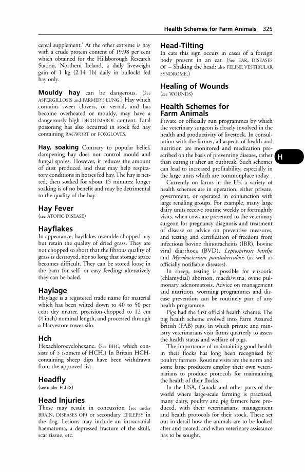
cereal supplement.’ At the other extreme is haywith a crude protein content of 19.98 per centwhich obtained for the Hillsborough ResearchStation, Northern Ireland, a daily liveweight gain of 1 kg (2.14 1b) daily in bullocks fed hay only.
Mouldy hay can be dangerous. (See
ASPERGILLOSIS and FARMER’S LUNG.) Hay whichcontains sweet clovers, or vernal, and hasbecome overheated or mouldy, may have a dangerously high DICOUMAROL content. Fatalpoisoning has also occurred in stock fed haycontaining RAGWORT or FOXGLOVES.
Hay, soaking Contrary to popular belief,dampening hay does not control mould andfungal spores. However, it reduces the amountof dust produced and thus may help respira-tory conditions in horses fed hay. The hay is net-ted, then soaked for about 15 minutes; longersoaking is of no benefit and may be detrimentalto the quality of the hay.
Hay Fever(see ATOPIC DISEASE)
HayflakesIn appearance, hayflakes resemble chopped haybut retain the quality of dried grass. They arenot chopped so short that the fibrous quality ofgrass is destroyed, nor so long that storage spacebecomes difficult. They can be stored loose inthe barn for self- or easy feeding; alterativelythey can be baled.
HaylageHaylage is a registered trade name for materialwhich has been wilted down to 40 to 50 percent dry matter, precision-chopped to 12 cm (1⁄2 inch) nominal length, and processed througha Harvestore tower silo.
HchHexachlorocyclohexane. (See BHC, which con-sists of 5 isomers of HCH.) In Britain HCH-containing sheep dips have been withdrawnfrom the approved list.
Headfly(see under FLIES)
Head InjuriesThese may result in concussion (see under
BRAIN, DISEASES OF) or secondary EPILEPSY inthe dog. Lesions may include an intracranialhaematoma, a depressed fracture of the skull,scar tissue, etc.
Head-TiltingIn cats this sign occurs in cases of a foreignbody present in an ear. (See EAR, DISEASES
OF – Shaking the head; also FELINE VESTIBULAR
SYNDROME.)
Healing of Wounds(see WOUNDS)
Health Schemes for Farm AnimalsPrivate or officially run programmes by whichthe veterinary surgeon is closely involved in thehealth and productivity of livestock. In consul-tation with the farmer, all aspects of health andnutrition are monitored and medication pre-scribed on the basis of preventing disease, ratherthan curing it after an outbreak. Such schemescan lead to increased profitability, especially inthe large units which are commonplace today.
Currently on farms in the UK a variety ofhealth schemes are in operation, either private,government, or operated in conjunction withlarge retailing groups. For example, many largedairy units receive routine weekly or fortnightlyvisits, when cows are presented to the veterinarysurgeon for pregnancy diagnosis and treatmentof disease or advice on preventive measures, and testing and certification of freedom frominfectious bovine rhinotracheitis (IBR), bovineviral diarrhoea (BVD), Leptospirosis hardjoand Mycobacterium paratuberculosis (as well asofficially notifiable diseases).
In sheep, testing is possible for enzootic(chlamydial) abortion, maedi/visna, ovine pul-monary adenomatosis. Advice on managementand nutrition, worming programmes and dis-ease prevention can be routinely part of anyhealth programme.
Pigs had the first official health scheme. Thepig health scheme evolved into Farm AssuredBritish (FAB) pigs, in which private and min-istry veterinarians visit farms quarterly to assessthe health status and welfare of pigs.
The importance of maintaining good healthin their flocks has long been recognised by poultry farmers. Routine visits are the norm andsome large producers employ their own veteri-narians to produce protocols for maintainingthe health of their flocks.
In the USA, Canada and other parts of theworld where large-scale farming is practised,many dairy, poultry and pig farmers have pro-duced, with their veterinarians, managementand health protocols for their stock. These setout in detail how the animals are to be lookedafter and treated, and when veterinary assistancehas to be sought.
Health Schemes for Farm Animals 325
H
Hearing(see EAR – The act of hearing; also ULTRASOUND
and TELEVISION SETS)
Heartbeat(see PULSE RATE)
Heart DiseasesAs in man, heart troubles are very much morecommon in old age. However, even young animals may suffer from faulty heart action dueto congenital defects.
Signs Irregularity in the heartbeat, some diffi-culty in breathing without obvious changes inthe lungs or pleura, breathlessness when the animals are compelled to exert themselves, a ten-dency to swelling of the dependent parts of thebody (e.g. along the lower line of the chest andabdomen and ‘filling’ of the limbs), are amongthe signs. A cough is sometimes a symptom ofvalvular disease.
Congestive heart failure Disease of theright side of the heart often gives rise to ascites,sometimes to swelling of one or more limbs dueto oedema. Engorgement of the veins oftenoccurs, with enlargement of the liver. The ani-mal becomes easily tired and may lose weight.Ultimately congestive heart failure is likely tooccur. This may also result from left-sided fail-ure due to myocarditis or mitral valve incompe-tence. In small animals, treatment consists inreducing exercise and giving diuretics.
A common cause of heart failure in dogs isdegeneration of a MITRAL VALVE.
Pericarditis is an inflammation of the mem-brane covering the exterior of the heart. It maybe ‘idiopathic’, when its cause is not known; itmay be ‘traumatic’, when it is due to a wound; orit may follow a general infection (e.g. ‘heart-water’) or a local infection (e.g. pleurisy) or anabscess in a remote part of the body. Pericarditismay be ‘dry’, in which case the 2 opposing sur-faces of the membrane are covered by a layer offibrin; or oedema may accompany this condi-tion, in which case fluid fills up the pericardialsac and, when no more distension of the sac canoccur, presses upon the outside of the heart itself.
Pericarditis has been reported in very youngpigs at grass. The piglet, often in good condi-tion and not anaemic, dies suddenly at about 2 to 3 weeks of age. (See also ‘MULBERRY HEART‘.)
Tamponade A rapid accumulation of bloodin the pericardium, suddenly arresting heartfunction.
Acute or chronic tamponade was the pre-senting sign in 42 cases of pericardial effusionin a series of large dogs with an average age of 9 years. Twenty-four of the cases were associ-ated with neoplasia, 8 with benign idiopathiceffusions, 6 with primary heart disease, and 2with trauma.
Echocardiography was found to be the best way of detecting pericardial effusion; and the idiopathic effusions responded well topericardiectomy.
Congenital heart disease in dogs andcats is usually indicated by a cardiac murmur,the site and nature of which shows whether avalve or a shunt is involved.
Shunts include ‘holes’ in the heart, andpatent ductus arteriosus.
Radiography and Doppler ultrasound arehelpful in diagnosis.
Surveys of a total of 580 dogs with congeni-tal heart disease showed that 28 per cent hadpatent ductus arteriosus; 16 per cent had pul-monary stenosis; 9 per cent had persistent rightaortic arch; over 7 per cent had a ventricularseptal defect; and over 7 per cent had stenosis ofthe aorta. (See also HEARTWORMS.)
Deficiency of vitamin E is one cause ofsudden cardiac arrest in cattle.
Signs These are not always characteristic, butthey include breathlessness, pain on pressure ofthe left side of the chest, a jugular pulse (seenalong the jugular furrow with each heartbeat),and oedema. On listening to the heart a varia-tion in the normal sounds may be heard, orthey may be altogether masked by the presenceof the fluid. A tinkle is sometimes audible overthe region of the heart; friction sounds indicatethe presence of dry pericarditis; and irregularityor even palpitation may be noticed.
Traumatic pericarditis of cattle Some-times when the animal is thought to be sufferingfrom simple digestive disturbance, it is foundthat a nail or piece of wire has been swallowedand arrives in the reticulum.
A distance of about only 5 cm separates theheart from the reticulum, so that the foreignbody is liable to penetrate the pericardium.
Attacks of pain may occur, the appetite isirregular, but after a time the animal regains itsnormal health, since an adhesion has occurredaround the hole in the reticulum wall, and theinflammation subsides. A cow may die sud-denly before symptoms of pericarditis appear,or soon afterwards.
326 Hearing
H

Treatment is sometimes feasible by surgicallyopening the rumen and removing the piece ofmetal.
Prevention In Switzerland the percentage ofcows slaughtered on account of traumatic peri-carditis was reduced following the use of magnetsfor the treatment of traumatic reticulitis. Magnetsweighing 114 g, 90 mm long and 15 mm indiameter were used orally 10 minutes after a subcutaneous injection of atropine sulphate.Without this it was found that only 53 per centof the magnets dropped at once into the reticu-lum. The correct siting of the magnets waschecked with a compass.
Myocarditis is inflammation of the heartmuscle. In the pig it is seen in HERZTOD disease,for example; in cattle, in MUSCULAR DYSTROPHY.(See also CANINE PARVOVIRUS; MYOCARDIUM.)
Endocarditis is an inflammation of themembrane lining the heart. It frequently leadsto the development of nodules on the valves.
The nodules result in an incomplete closing ofthe valves, and since the fibrin deposited uponthem tends to become converted into fibrous tissue (‘organised’), the growths slowly increasein size. They are seen in chronic erysipelas ofpigs. (See SWINE ERYSIPELAS.)
The valvular insufficiency can be diagnosedby auscultation. Congestive heart failure may bethe outcome (sometimes embolism); but com-pensation takes place, and the animal may live along time with faulty valves.
Bacterial endocarditis is a cause of death in cat-tle, especially in South Wales. (See HEARTWORMS
for another cause of endocarditis in the dog.)
Valvular diseases form a most importantand common group of heart disorders, andalthough the power of compensation alreadyreferred to may so neutralise the ill-effects of anarrowed valve, or one which leaks, severestrains or exertion – or even trying conditionssuch as parturition – may precipitate ill-effects.Very often when an animal ‘drops dead’, per-haps after running a race or while undergoingsome departure from its normal mode of life,the actual cause is afterwards found to be a dis-eased heart valve. Fainting fits are not by anymeans rare in incompetence of the tricuspidvalves. Congestion of the lungs may be broughtabout by incompetence of the auriculo-ventric-ular valve on the left side of the heart (mitralinsufficiency); this same condition may lead toa chronic asthmatical cough in old dogs, whichis occasionally mistaken for bronchitis.
Canine heart repair Skeletal muscle trans-plants were used to replace or repair defects inthe left ventricle of dogs, some of which werekept alive for over a year. At autopsy the trans-plants were found to be in good condition,according to a report in Circulation.
Hypertrophy, or enlargement of the heart,takes place as the result of some constant simplestrain, such as occurs in racehorses, hunters,and sporting dogs; or as the result of backwardpressure from a diseased valve, and whichentails the heart muscle ‘compensating’ for the effects of valvular disease. Alternatively itmay be due to resistance to the flow of blood insome diseased organ or tissue which results inhigh blood pressure. (See COMPENSATION.)
Hypertrophy of the left ventricle, leading toheart failure, may in the dog follow Leptospiracanicola infection.
Dilatation of the heart may precede hypertro-phy, i.e. when it occurs before the heart musclehas had an opportunity to increase to meet theextra demands upon it; and it very frequentlyfollows hypertrophy, especially when there issome disease process at work which hinders theproper nutrition of the heart muscle.
Hypertrophy may be a beneficial conditionin any animal, and, except when it is due tovalvular trouble, need not cause any worry tothe owner. It is sometimes excessive in horses;in some instances the heart may weigh as muchas 11 kg (25 lb) instead of the 3 or 3.5 kg (7 or8 lb) of the normal. Degenerative changes mayfollow hypertrophy when the animal becomesless active during later life.
Congenital defects These include a patentductus arteriosus. (See diagram of fetal circulationunder CIRCULATION OF BLOOD; also LIGAMENTUM
ARTERIOSUM; ECTOPIA CORDIS.) Tetralogy ofFallot consists of: (1) stenosis of the pulmonaryvalve; (2) a defect in the septum which separatesthe 2 ventricles; (3) the aorta over-riding bothventricles; (4) marked hypertrophy of the rightventricle.
The signs are often vague: in kittens, forexample, these may be a failure to thrive, andinability to cope with exercise. More seriousdefects result in the death of newborn kittens.
Functional disorders Palpitation is a condi-tion in which the heart beats fast and strongly,due to fright, for example(see tachycardia, below).
Bradycardia is a condition of unusually slowaction of the heart. Intermittency or irregularityis an exceedingly common condition amonganimals, and as a rule appears to cause them no
Heart Diseases 327
H
inconvenience whatever. In some horses at restin the stable the heart constantly misses every3rd, 4th, or 5th beat, a long pause taking theplace of the pulsation, but when at exercise orwork the normal rhythm is restored.
Heart-block is a condition in which the conducting mechanism between atrium andventricle (atrio-ventricular bundle of His) isdamaged in whole or part, so that the two beatindependently of each other.
Rapid heart action (tachycardia) may have anumber of causes including exertion or excite-ment. It is normally harmless in such cases. It isalso seen in diseases which affect the transmissionof the heartbeat stimulus.
Cardiac flutter and fibrillation are conditionsof great irregularity in the pulse, due to the atriaemptying themselves, not by a series of regularwaves, but by an irregular series of flutters ortwitches instead, which fail to stimulate theventricles properly.
Five cases of atrial fibrillation were describedin horses after racing. In 4 of them, which had performed poorly during their races, thearrhythmias disappeared spontaneously within24 hours; these cases were regarded as paroxys-mal. In the 5th horse, which won its race, thearrhythmia persisted for at least 45 hours afterthe race and it was regarded as an example of persistent atrial fibrillation. Treatment withquinidine sulphate restored the sinus rhythm.Paroxysmal atrial fibrillation may cause a sudden decrease in racing performance.
Diagnosis of heart disease is basedlargely on the character of the pulse and heartsounds. Murmurs, for example, indicate valvu-lar incompetence, cardiac dilatation, or congen-ital lesions. Muffled sounds may indicate fluidin the pericardium (or pleurisy).
Additionally radiography and cardiographyare used in diagnosis. (See PACEMAKERS as a possible treatment of some canine patients.)
Heart StimulantsDrugs used as cardiac stimulants include theophylline, adrenaline, isoprenaline anddobutamine.
HeartwaterHeartwater, also known as BUSH SICKNESS
(Boschziekte), VELD SICKNESS, and INAPUNGA,is a specific disease of cattle, sheep, and goatstransmitted by the bont-tick (Amblyommahebraeum) in South Africa, and A. variegatumin Kenya. The disease is characterised by theaccumulation of a large amount of fluid in thepericardial sac and nervous symptoms.
In 1980 the existence of heartwater in manyislands of the Caribbean was discovered; previ-ously the disease had been known only in Africa.The tick involved is A. variegatum, introducedinto Guadeloupe with cattle from Senegal.
Cause Infection of the nymphal or larvalstages of the bont-tick with Rickettsia ruminan-tium (Cowdria ruminantium) which is trans-mitted to other animals upon which the tickfeeds at a later state of its life-history.
Incubation After sheep and goats have beenbitten by infected ticks, a period of between 11and 18 days elapses before any symptoms areshown; in cattle the disease appears between 20 and 25 days after infestation with ticks.These periods are influenced by the stage of thedisease in the animal supplying the infectedblood to the ticks, and also by individual sus-ceptibility, which is less in native-bred cattlethan in those imported from other countries,and especially those brought from Britain.
Signs
Sheep and goats Sheep and goats at firstshow nothing more than a rise in temperature(which gradually increases to 41.7°C (107°F),falling each evening a few degrees lower), a gen-eral dullness, prostration, and lack of appetite.As these conditions are common to many otherdiseases, the difficulty of diagnosis is great. Theaffected animals isolate themselves from the restof the flock, lie about in secluded spots, cease toruminate, and when handled or driven are veryeasily tired and lie down.
Many animals show peculiar nervous symp-toms, which vary in different individuals; somemay bleat almost continuously; others champ thejaws as if feeding, moving the tongue backwardand forward between the lips; others lick theground; some turn in circles until they finally fall to the ground and lie prostrate or performgalloping movements with their limbs; whileothers show profuse salivation. Convulsions arenot uncommon, especially when the animals are handled. Death usually follows soon afterconvulsions make their appearance. magesty
Cattle The symptoms in cattle are very similarto those seen in sheep. The nervous form inwhich peculiar masticatory movements aremade by the mouth is common. Animals showa tendency to bite at their feet or legs, espe-cially when lying on the ground, and biting theground is also seen. A number of animals in theearly stages may show a dangerous tendency to
328 Heart Stimulants
H
charge any human being approaching them. Incattle the disease is usually at its height aboutthe 4th day after the first rise in temperature,and death usually occurs about the 6th day.Hyperacute cases occur in cattle, and the animal is found dead on the veld.
Autopsy Fluid in the pericardial sac surround-ing the heart (hence the name ‘heartwater’); butwhile this is usually found in sheep and goats, it may be absent in the case of cattle. In typicalinstances there is also a collection of similarfluid in the thoracic and abdominal cavities.Both the pericardium and the endocardiumwhich lines the heart may show several small ora few large ‘petechiae’, i.e. areas where a slightamount of haemorrhage has taken place.
Prevention Entirely successful results havefollowed measures taken against the ticks whichtransmit the disease. These consist in ‘5-daydippings’.
Antibiotics and sulfonamides are used intreatment.
HeartwormsDirofilaria immitis is a common parasite of dogsin Central Europe, Russia, Australia, America,and Asia. The disease has been introduced intothe UK by dogs returning from travel in main-land Europe via the Pet Travel Scheme. Theworm larvae are transmitted by various mosqui-toes and gnats. They are present in the blood-stream of infected animals as microfilariae. Theadult worms reach a length of up to 30.5 cm (12 inches) (females) and inhabit the right side of the heart, causing some degree of endocarditis and a variety of symptoms, e.g.cough, hind-leg weakness, collapse on exercise,laboured breathing, anaemia, emaciation.
This infestation is known as canine filariasisor dirofilariasis. The kidneys and urinary tractmay be affected. (See also EYE, DISEASES OF.)
In a survey in Canada, 560 dogs (1.79 per centof those tested) were found to have heartworms.
About 20 per cent of dogs may be infectedwith adult worms without having microfilariae.
Heartworms can cause devastating cardio-pulmonary effects in cats. The disease may bepresent without microfilariae, not only duringthe prepatent period, for adult worms may bemales, ‘geriatric females’, or of 1 sex only.
There have been reports of dirofilaria wormsbeing recovered from the brains of cats. Onesuch report referred to a cat with ataxia whichdied 48 hours later. At autopsy, 3 heartwormswere found in the heart, 3 in the brain, and 4 ina kidney.
Diagnosis An ELISA test, based on the detec-tion of antibodies to heartworms, is useful whenno microfilariae are present. Radiography hasalso been recommended as a diagnostic aid.
Treatment and control of 5 dogs dosed withivermectin 1 day after artificial infection with 50infective larvae of D. immitis, none harbouredany heartworms when killed 201 days later. The5 control dogs had an average of 11 worms eachat post-mortem examination. It is suggested thattreatment with ivermectin at monthly intervalswould prevent heartworm disease.
In the UK selamectin, a derivative of iver-mectin, is licensed for use in dogs and thiac-etamide has been used on imported dogs.
Another canine heartworm is Angiostrongylusvasorum which inhabits the pulmonary arteryand the right ventricle of the heart. Symptomsinclude malaise and large subcutaneous swellings.Slugs and snails may act as intermediate hosts.
In a case seen at the Liverpool School ofTropical Medicine, a 3-year-old dog, whichdied suddenly after an acute attack of dyspnoea,was found to have an A. vasorum.
HeatA female animal is said to be on heat when itwill accept the service of a male (see OESTRUS;for the suppression of ‘heat’ in the bitch, see
OESTRUS, SUPPRESSION OF).
Heat Detection in Cows(see under OESTRUS, DETECTION OF)
Heat ExhaustionA syndrome in which there is a depletion of electrolytes and water in the body. (See
HEAT-STROKE.)
Heat LossHeat loss from the body occurs by radiation, byconduction and convection from the skin, andby evaporation from the skin and lungs. Thenormal body temperature is controlled partly byalteration of the rate of metabolism, and partlyby constriction of the surface blood vessels whenthe animal is exposed to cold, as well as by shivering which generates heat. There comes apoint, however, as body temperature falls stillfurther, at which shivering ceases. Then the danger of hypothermia may not be recognised.(See BEDDING for pigs; also HYPOTHERMIA.)
Sensible loss of heat This is the heat whichanimals lose by convection, conduction, and radi-ation. It does not include heat lost by vaporisingwater from the skin and respiratory passages.
Heat Loss 329
H
Heat-StrokeHeat-stroke is a condition associated withexcessively hot weather, and especially underconditions of stress. It occurs in domestic ani-mals when taken to tropical countries fromtemperate countries, especially when recentlyunloaded from transport ships and subjected togreat excitement in unfamiliar surroundings; itis seen in cattle, sheep, and swine travelling byroad or rail, and it frequently occurs at agricul-tural shows; dogs may be affected when theyhave been left in a car parked in the sun, andwith windows closed or almost closed. There isa failure to lower body temperature. (See CAR,PARKED; also HYPERTHERMIA; TROPICS.)
Signs The animal is usually suddenly overcomeby a great lethargy and inability to work or move.The gait is staggering, and if the animal is madeto move it falls to the ground. Convulsions mayoccur, and if the temperature is taken it is foundto be very high, perhaps as much as 42.2°C(108°F) in the horse. Death often takes place ina few hours, but some cases last as long as 3 days.If recovery occurs, great dullness for a number ofweeks is liable to follow.
Treatment Removal to a cool place; douchingthe head and neck with cold water from ahosepipe. Ice cubes may be used for the smalleranimals.
An animal may die as a result of combinedheat-stroke and heat exhaustion, or either separately. (See also HEAT EXHAUSTION.)
Hebdomadis Serogroup(see LEPTOSPIROSIS)
HedgehogsHedgehogs are of veterinary interest in thatthey are susceptible to natural infection withfoot-and-mouth disease, which they transmit toother animals.
Hedgehogs, like horses, are the natural hostsfor Leptospira bratislava. A possible case of thisinfection occurred in a dog, previously vacci-nated against leptospirosis, but known to haveaccess to hedgehogs.
A UK survey of mortality in hedgehogs(Erinaceus europaeus) showed that 47 per centwere road casualties; 39 per cent had salmo-nellosis. Other zoonoses were ringworm(Trichophyton erinacei) and Yersinia pseudotu-berculosis in a very small proportion of thehedgehogs. Lungworms, flukes (Brachylaemuserinacei), tapeworms (Rodentolepsis erinacei),ticks, fleas, and mange mites (Caparinia tripilis)were other parasites found. Deaths have
been recorded after hedgehogs ate slug bait(metaldehyde).
A safe, simple method of dealing with‘rolled-up’ hedgehogs, for the purpose of exam-ination or treatment against external parasites,was described by Dr Nancy Kock, InternationalWildlife Veterinary Services, California. Hermethod is to place the animal in an aquariumtank (containing a parasiticide dip solution ifneeded), when it will immediately unroll andbegin swimming. Using protective gloves, thehedgehog can then be grasped by the scruff of its neck like a kitten. Once held firmly like that, it is unable to roll up again, makingexamination easy.
Anaesthesia Fentanyl citrate + fluanisone(Hypnorm; Janssen) by subcutaneous injectionis suitable for anaesthetising hedgehogs.
HeiferA year-old female up to her 1st calving.
Heinz BodiesHeinz bodies in red cells are seen in cases ofhaemolytic anaemia caused by, e.g., an excess ofkale in dairy cattle. Heinz-body anaemia hasalso been seen in cats as a result of poisoning bymethylene blue, formerly used in America as aurinary antiseptic. This form of anaemia hasbeen linked with onions, and a case was report-ed in a puppy which preferred raw onions andother vegetables to conventional dog foods.After a change of diet the puppy became well,and no longer tended to collapse after exercise.Heinz bodies are present in cats poisoned byparacetamol.
HelleboresThere are 4 hellebores of importance to theowners of animals because of their toxicity.Black hellebore is the dried rhizome and rootletsof the Christmas rose, or bear’s-foot, Helleborusniger. It may be eaten by livestock when gardentrimmings are thrown out on to fields to whichlivestock have access. It contains 2 very irritantglycosides – helleborin and helleborein. Stinkinghellebore (H. fetidus) and green hellebore (H.viridis or Veratrum viride) are sometimes thecause of livestock poisoning. The latter, alongwith white hellebore (V. album), contain severalalkaloids. They are depressants of the motornervous centres.
Poisoning by hellebores Symptoms arestupor, convulsions, and death when largeamounts have been taken, and purgation, sali-vation, excessive urination, attempts to vomit,
330 Heat-Stroke
H
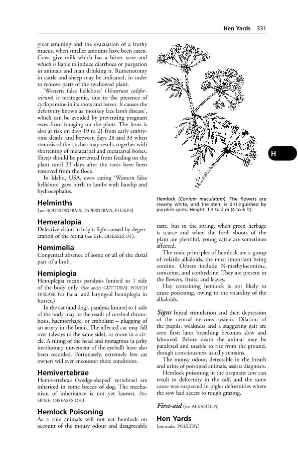
great straining and the evacuation of a frothymucus, when smaller amounts have been eaten.Cows give milk which has a bitter taste andwhich is liable to induce diarrhoea or purgationin animals and man drinking it. Rumenotomyin cattle and sheep may be indicated, in orderto remove parts of the swallowed plant.
‘Western false hellebore’ (Veratrum califor-nicum) is teratogenic, due to the presence ofcyclopamine in its roots and leaves. It causes thedeformity known as ‘monkey face lamb disease’,which can be avoided by preventing pregnantewes from foraging on the plant. The fetus isalso at risk on days 19 to 21 from early embry-onic death, and between days 28 and 33 whenstenosis of the trachea may result, together withshortening of metacarpal and metatarsal bones.Sheep should be prevented from feeding on theplant until 33 days after the rams have beenremoved from the flock.
In Idaho, USA, ewes eating ‘Western falsehellebore’ gave birth to lambs with harelip andhydrocephalus.
Helminths(see ROUNDWORMS; TAPEWORMS; FLUKES)
HemeralopiaDefective vision in bright light caused by degen-eration of the retina (see EYE, DISEASES OF).
HemimeliaCongenital absence of some or all of the distalpart of a limb.
HemiplegiaHemiplegia means paralysis limited to 1 side of the body only. (See under GUTTURAL POUCH
DISEASE for facial and laryngeal hemiplegia inhorses.)
In the cat (and dog), paralysis limited to 1 sideof the body may be the result of cerebral throm-bosis, haemorrhage, or embolism – plugging ofan artery in the brain. The affected cat may fallover (always to the same side), or move in a cir-cle. A tilting of the head and nystagmus (a jerkyinvoluntary movement of the eyeball) have alsobeen recorded. Fortunately, extremely few catowners will ever encounter these conditions.
HemivertebraeHemivertebrae (‘wedge-shaped’ vertebrae) areinherited in some breeds of dog. The mecha-nism of inheritance is not yet known. (See
SPINE, DISEASES OF.)
Hemlock PoisoningAs a rule animals will not eat hemlock onaccount of the mousy odour and disagreeable
taste, but in the spring, when green herbage is scarce and when the fresh shoots of the plant are plentiful, young cattle are sometimesaffected.
The toxic principles of hemlock are a groupof volatile alkaloids, the most important beingconiine. Others include N-methyloconiine,coniceine, and conhydrine. They are present inthe flowers, fruits, and leaves.
Hay containing hemlock is not likely tocause poisoning, owing to the volatility of thealkaloids.
Signs Initial stimulation and then depressionof the central nervous system. Dilation of the pupils, weakness and a staggering gait areseen first; later breathing becomes slow andlaboured. Before death the animal may beparalysed and unable to rise from the ground,though consciousness usually remains.
The mousy odour, detectable in the breathand urine of poisoned animals, assists diagnosis.
Hemlock poisoning in the pregnant cow canresult in deformity in the calf, and the samecause was suspected in piglet deformities wherethe sow had access to rough grazing.
First-aid (see ALKALOIDS)
Hen Yards(see under POULTRY)
Hen Yards 331
H
Hemlock (Conium maculatum). The flowers arecreamy white, and the stem is distinguished by purplish spots. Height: 1.3 to 2 m (4 to 6 ft).

Henle, Loop ofThe U-shaped loop connecting the ascendingand descending tubules in the kidneys.
HenneguyaA group of parasites found in the skin and mus-cles of fish, notably sea trout and salmon. Theparasites are seen as tadpole-shaped cysts con-taining two ‘eye-spots’. They cause ‘milky fleshdisease’. This is seen in fish, apparently healthy,which on being cut into are found to have areasof muscle replaced by a milky fluid.
HeparinA naturally occurring anticoagulant.
Hepatic EncephalopathyA disease of the brain caused by cirrhosis of theliver; or it may possibly result from a congeni-tal condition, portosystemic shunt. It is usuallyseen in dogs and cats but can occur in other animals. Affected animals are lethargic, becomeblind, have convulsions, ataxia and behaviouralchanges. The disease is clinically indistinguish-able from FELINE SPONGIFORM ENCEPHALOPA-
THY but blood samples show high ammonialevels. Neomycin may improve the liver condi-tion and a low-protein diet is recommended.
HepatisationHepatisation means the solidified state of thelung that is seen in pneumonia, which gives itthe appearance and consistence of the liver.
HepatitisInflammation of the LIVER.
Hepatitis in the horse occurs after infectiousequine encephalomyelitis, especially where vac-cines or sera have been used. In cattle and sheep,it can occur after liver fluke, ragwort poisoningand aflatoxicosis.
For hepatitis in dogs, see CANINE VIRAL
HEPATITIS and also under DUCK HEPATITIS.
HepatozoonA single-celled parasite transmitted by the tickRhipicephalus sanguineus. Heptazoon canis infectsboth dogs and cats, often causing anaemia, fever,and occasionally paraplegia. Other species infectrodents.
HeptachlorA constituent of chlordane, a chlorinatedhydrocarbon, and used also as an insecticide onits own. It is not used on animals. It is stored inthe body fat, and in the tissues is converted intoheptachlorepoxide, 4 times as toxic to birds asheptachlor itself.
Herbicides(see PARAQUAT; DIQUAT; MONOCHLOROACETATE;WEEDKILLERS; POISONING)
HerdsmenOccupational hazards of those looking after cattle include BRUCELLOSIS; Q FEVER; TUBERCU-
LOSIS; COWPOX; MILKER’S NODULE; SALMONEL-
LOSIS; SPOROTRICHOSIS; BUBONIC PLAGUE (notin the UK).
HeredityThe transfer of genetic traits from parent to offspring (see GENETICS).
HermaphroditeAn animal in which reproductive tissue of bothsexes is present. A lateral hermaphrodite has anovary on one side, and a testicle on the other;whereas a bilateral hermaphrodite has an ovaryand testicle (or a combined ovary-testis) oneach side. (See also INTER-SEX.)
In one case, a hermaphrodite rabbit servedseveral females and sired more than 250 youngof both sexes. In the next breeding season therabbit (housed in isolation) became pregnantand produced 7 healthy young of both sexes.
HerniaThe protrusion of part of an organ through the membrane which contains it. In a typicalabdominal hernia there are always the followingparts: a ‘ring’, or opening in the muscular wallof the abdomen, which may have been broughtabout as the result of an accident or may havebeen present at birth; and a swelling appearingbelow the skin, composed of the ‘hernial sac’and its contents.
The contents vary according to the situation,size, and nature of the hernia, but the followingorgans or parts of them are most commonlyherniated: a loop of bowel with its attachedmesentery omentum, either the whole or a part(very common in dogs); the stomach; the uri-nary bladder; the spleen or liver (through thediaphragm); the uterus, either when non-pregnant or with its contained fetus or fetuses;and sometimes a kidney in the cat.
(For strangulated hernia, see under ‘Signs’,below.)
Umbilical hernia The opening in theabdominal wall is a natural one which should,however, have closed at birth. If given time, itmay still do so. In the puppy, for example, onlya persistent or irreducible umbilical hernia willneed surgical intervention owing to the risk ofa piece of omentum having its blood circulation
332 Henle, Loop of
H

interfered with, or bowel becoming obstructedor strangulated – both serious conditionsrequiring immediate surgery.
Inguinal hernia, which is practically thesame as scrotal hernia, but at a less advancedstage, is almost wholly confined to the male sexin all animals, except the bitch, where a horn of the uterus may, upon occasion, come downthrough the inguinal canal. Inguinal and scrotalforms of hernia may be either congenital oracquired; congenital forms (most common inyoung animals) result through some failure of the inguinal canal, through which the testi-cle descends, to close properly; while acquiredforms (commoner in adults) result from suchaccidents as slipping sideways with the hind-feet, injuries to the abdomen from falls, blows,and kicks.
Femoral hernia is very rare, but sometimesoccurs in performing dogs which have beentrained to walk upon their hind legs for consid-erable periods of time. The vertical position ofthe body imposes an unusual strain upon themuscles at the fold of the thigh, and they giveway. It is always acquired.
Perineal hernia is almost exclusively con-fined to the dog. It may occur in either sex, usu-ally as the result of much straining occasionedby constipation or diarrhoea, chronic coughingor asthma, bronchitis, etc., and in old maledogs suffering from enlarged prostate glands.
Ventral hernia is almost invariably theresult of a serious injury to the muscular por-tion of the abdominal wall. It is commonest inmares, especially those used for breeding pur-poses. Very often there is little or nothing to be noticed if the mare is injured when non-pregnant, but when pregnancy follows and the tension upon the abdominal wall increases,the muscular part gives way and a large massappears along the lower line of the abdomen. Incows it very often results from horn-gores fromneighbours; in such, the skin remains intact butthe muscle is torn and a swelling appears at theseat of the injury. Hernia due to a gore is prob-ably commonest in the region of the flank,where the muscle is naturally thin.
Mesenteric hernia is rare in cattle (‘proba-bly because of the thickness of the mesentery’)but not in horses. In a case involving a cow,intestine was herniated through a tear or defectin the mesentery, resulting in incarceration. A laparotomy was performed, and the defect
enlarged to permit extrication of the intestine.The cow recovered.
Diaphragmatic hernia may occur in anyanimal, but is commonest in the dog and thecat. It usually results from jumping downwardsfrom a great height – an act which throws thefull weight of the abdominal contents forwardagainst the diaphragm when the animal landson its feet; it may also occur in road accidents.
The rent may be in the muscular or tendi-nous portion of the diaphragm, but it very frequently involves one or other of the naturalopenings (hiati), giving passage to the oesopha-gus, the vena cava, or the aorta, (although a hernia through an enlarged aortic hiatus is veryrare on account of the powerful nature of thediaphragm in its upper parts).
Signs The symptoms vary greatly, dependingupon the particular organ which is protruded,upon the size of the opening, which may ormay not compress the hernia, and upon thecondition of the latter. In very many casesamong animals, herniae contain either omen-tum or a loop of bowel, or both. The swellingmay be present at birth, or it may appear sud-denly or gradually at almost any time duringlife. To the touch it may present one of severalsensations: (1) in the simple form it feels soft,fluctuating (as if it contained fluid), painless,neither hot nor cold, and causes no discomfortto the animal when being handled. If it bepressed upon it can usually be returned to theabdominal cavity, though it will reappear assoon as the pressure is released. In small animalsit will disappear when they are laid upon theirbacks, and remain out of sight until they regaintheir feet; (2) when the structures are adherentto the skin which covers them, return to theabdomen is impossible, no reduction can beachieved by manipulation, no definite ring canbe determined as a rule, and there is no increasein size with exertion, but otherwise an adherenthernia presents the same appearances as a sim-ple one; (3) in the strangulated form, whichmay supervene upon a hitherto simple hernia,there are very definite and serious symptoms of general disturbance: breathing is fast and distressed, an anxious expression is visible onthe face, and the swelling shows a markedtenseness and pain when being handled. It maybe red and inflamed-looking at first, but later it frequently becomes bluish. After about 12 to 24 hours gangrene sets in; the swelling becomescold and painless to the touch; the temperaturefalls subnormal, and the animal becomes alarm-ingly weak. Death usually follows shortly after,
Hernia 333
H
unless the strangulation is relieved by operationand perhaps amputation of the strangulatedportion of bowel. An obstructed hernia is usually merely the preliminary of strangulation.
Treatment Palliative treatment, such as iscommon in human beings consisting in theapplication of trusses, bandages, etc., is of no usewhatever where animals are concerned. Withyoung animals of any species it is usual to leaveherniae alone provided that they are not acute,for it often happens that during the growth anddevelopment of the young creature the herniadisappears of its own accord, and the hole in theabdominal wall heals over. There is, however,always a danger that, as the result of some extraexertion, heavy feeding, boisterous playfulness,fighting, etc., strangulation may occur.
The most rational method is one in which the animal is anaesthetised, skin incised, the
contents returned to the abdomen, the peri-toneal sac obliterated if it is present, the edges ofthe ring carefully sutured so that they will form astrong union, and finally the skin wound closed.
The operation for a strangulated hernia differsfrom that for a simple one in that it is necessaryto enlarge the tight ring, to allow restoration ofthe circulation.
Fifty-two perineal hernias in dogs have beensuccessfully repaired by transposing both theinternal obturator muscle and the superficialgluteal muscle together. The technique results ina strong pelvic diaphragm and good long-termresults. Fewer post-operative complications areclaimed to occur than with other techniques.
HerpesvirusesHerpesviruses cause, for example, Aujeszky’sdisease, jaagsiekte, feline rhinotracheitis. (Seethe table above; also under MONKEYS and FADING.)
334 Herpesviruses
H
Some of the herpesviruses of man, domestic animals and poultry*
Recommended label Traditional name Associated disease
Human herpesvirus 1 Herpes simplex type 1 Herpetic sores, etc.Human herpesvirus 2 Herpes simplex type 2 Genital herpes and
cervical cancerHuman herpesvirus 3 Varicella-zoster Chicken pox and shinglesHuman herpesvirus 4 Epstein-Barr virus Burkitt’s lymphoma and
infectious mononucleosis(glandular fever)
Canine herpesvirus 1 Canine herpesvirus Herpes of dogs (neonataldeaths, respiratory
infection, genitallesions)
Feline herpesvirus 1 Feline rhinotracheitis virus Respiratory diseaseEquid herpesvirus 1 Equine abortion virus AbortionEquid herpesvirus 2 Cytomegalovirus Nothing or respiratory
diseaseEquid herpesvirus 3 Coital exanthema virus Coital exanthemaEquid herpesvirus 4 Respiratory diseaseBovid herpesvirus 1 Infectious bovine rhino- Upper respiratory tract
tracheitis/infectious infection; vaginitis,pustular vulvo-vaginitis abortion, etc.
Bovid herpesvirus 2 Bovine mamillitis virus Mamillitis and pseudo-lumpyskin disease
Bovid herpesvirus 3 Malignant catarrhal fever Malignant catarrhal fevervirus (wildebeeste herpes in cattle (Africa)virus)
Bovid herpesvirus 4 Jaagsiekte virus Metritis, abortion,pulmonary adenomatosis respiratory disease
Pig herpesvirus 1 Pseudorabies virus Aujeszky’s diseasePig herpesvirus 2 Inclusion body rhinitis Rhinitis
(cytomegalo) virusPhasianid herpesvirus 1 Infectious laryngo- Laryngotracheitis in
tracheitis virus poultryPhasianid herpesvirus 2 Marek’s disease virus Marek’s disease (fowl
paralysis)
*Based on the recommendations of the Herpesvirus Study Group, International Committee for the Nomenclature of Viruses, andupdated.
Herztod DiseaseA heart condition in pigs, it has similarities to MULBERRY HEART. (See PORCINE STRESS
SYNDROME.)
HeterokaryonA cell containing nuclei of 2 different species (anexample of genetic engineering). (See GENETICS.)
Heteroplastic TissueHeteroplastic tissue is that which is abnormal,different in structure, or different from anotherindividual in the case of a graft (heteroplastid).Heteroplastic bones are those which are notparts of the skeleton, e.g. the Os penis in thedog, and the Os cordis (one of 2 small bones inthe cow’s heart). Heteroplasm is normal tissuefound in an abnormal situation.
HeterosisHybrid vigour.
HeterotopicWrongly positioned.
HeterozygousRelating to a heterozygote, which is producedfrom unlike GAMETES and has 1 gene (see
ALLELES) dominant and the other recessive for aparticular characteristic.
HetpAn organophosphorus insecticide used in agriculture and horticulture. Similar to TEPP.
HexachlorobenzeneA fungistat used as a seed-dressing, it has givenrise to a form of PORPHYRIA in children inTurkey, and might similarly affect livestock.
HexachlorocyclohexaneThe group name for several isomers each having the formula C6H6Cl6. The most impor-tant of them is BENZENE HEXACHLORIDE. (See
HCH; BHC.)
HexachlorophaneAn antiseptic used as an ingredient of medicat-ed soap to kill bacteria on the skin.
HexamineAlso called methenamine. It is excreted by thekidneys, and as it sets free formalin in an acidmedium it has antiseptic qualities when theurine is acid. It may be combined in a tabletwith sodium acid phosphate for this purpose intreatment of cystitis in dogs.
HexamitiasisAn infectious enteritis of turkeys occurring inthe USA and Britain.
Cause Hexamita meleagridis.
Signs Day-old poults may be affected, butmore commonly the disease attacks turkeys afew weeks old. The feathers become ruffled, thebirds are listless with drooping wings. The drop-pings become liquid and frothy. Birds standsilent and motionless with eyes closed. Loss ofcondition is rapid, with marked dehydration. Inyoung birds mortality may reach 100 per cent.Recovered birds may act as carriers.
Treatment Antibiotics, furazolidone.
HexoestrolA synthetic oestrogen said to be more active thanstilboestrol. It is banned from use in animals inthe EU. (See STILBOESTROL; HORMONES IN MEAT
PRODUCTION; STILBENES; CAPONIEATION.)
HexosesHexoses are monosaccharide carbohydrates andinclude GLUCOSE, fructose, galactose, and man-nose. Monosaccharides also include the pentoses,e.g. arabinose, ribose. (See SUGAR.)
HexylresorcinolFormerly used as an anthelmintic for round-worms and for fluke.
Hiatus HerniaProtrusion of (usually) part of the stomachthrough the diaphragm at the oesophageal hiatus (see under HERNIA).
HibitaneChlorhexidine, a valuable disinfectant effectiveagainst some bacteria which cause mastitis incattle.
HidrosisSweat secretion, either normal or abnormallyprofuse.
High-Rise Syndrome(see FALLS FROM HIGH BUILDINGS)
Hilum (Incorrectly, Hilus)Hilum (incorrectly, hilus) is a term applied tothe depression on organs such as the lung, kid-ney, and spleen, at which the vessels and nervesenter or leave, and round which the lymphnodes cluster.
Hilum (Incorrectly, Hilus) 335
H

HinnyThe offspring of a stallion and a female ass.
Hip Dysplasia in DogsThis term covers a number of abnormal condi-tions of the acetabulum and head of the femur.
Some of these conditions are hereditary.They include:
(1) Subluxation, in which the head of thefemur is no longer firmly seated within theacetabulum. Deformity of the head of the femurgradually develops. The symptoms include areluctance to rise from the sitting position, anda sawing gait, observed when the puppy (mostoften an Alsatian, sometimes a golden retrieveror boxer) is 4 or 5 months old.
(2) Osteochondritis dissecans is seen in terri-ers with short legs, poodles, and Pekingese. It is possibly identical with Perthe’s disease.Muscular wasting and lameness are observed,usually in 1 limb.
(3) Slipped epiphysis. This also causes painand lameness at 4 to 6 months, but is difficultto distinguish from (2).
(4) Congenital dislocation, in which theacetabula are too shallow to retain the heads ofthe femurs in position. Reported in the BlackLabrador. A false joint forms in time. (See also
PERTHE’S DISEASE.)The BVA and the Kennel Club jointly run a
scheme whereby X-rays of a dog’s hip-joint areexamined by a panel of experts and given ascore according to the condition of the joint.The intention is that dogs showing a tendencyto dysplasia will not be used for breeding.
Hip-JointThe joint formed between the head of thefemur, or thigh-bone, and the depression on theside of the pelvis called the acetabulum.
HistaminaseAn enzyme obtained from extracts of kidneyand intestinal mucosa, capable of inactivatinghistamine and other diamines. It has been usedin treating anaphylactic shock and other aller-gic conditions due to, or accompanied by, theliberation of histamine in the body.
HistamineAn amine occurring as a decomposition prod-uct of histidine (see AMINO ACIDS) and preparedsynthetically from it. Histamine is widely dis-tributed in an inactive compound form in thebody, particularly in the lungs, liver, and to alesser extent in blood and muscle. As a result of trauma, burns, or infection, it may be liber-ated from the skin, lungs, and other tissues.
Histamine dilates capillaries, reduces bloodpressure, increases any tendency to oedema,stimulates visceral muscles and gastric and pan-creatic secretions. Histamine toxicity is shownby engorgement of the liver, shock, and a ten-dency to urticaria-like skin lesions. (See also
ANTIHISTAMINES; ALLERGY; MAST CELLS.)
HistidineAn amino acid from which histamine is derivedby bacterial decomposition.
HistiocytesAnother name for macrophages. (See under
BLOOD – Leukocytes.)
Histiocytosis A condition resulting from an excess of histiocytes in the bloodstream. Itaffects some breeds of dog, e.g. Bernese moun-tain dogs. Clinical signs vary from anaemia andrespiratory disease in the malignant form toitchy skin patches.
HistocompatibilityThe ability of a cell or tissue transplant to beaccepted by a different animal. Histocompatibleantigens are present in most tissue cells. Theyare the cause of the rejection of transplants. (See
MAJOR HISTOCOMPATIBILITY SYSTEM.)
Histomoniosis(see BLACKHEAD)
HistoplasmosisA fungal disease, caused by Histoplasma capsu-lata, which gives rise to loss of appetite, diar-rhoea, emaciation, and liver enlargement. Itoccurs chiefly in dogs and man. In man, ofteninfected by venturing into bat-infested caves inCentral and South America, and in Africa,lesions first occur in the lungs, but – in seriouscases – other organs may be affected.
The mycelial phase, found in soil, produces 2 kinds of spore: microconidia and macronidia.The latter enter the body by inhalation.
HockHock is the tarsus, a joint composed of 6 or 7 bones, between the tibia and the cannon boneof the hindlimb. (See under BONE.)
Hodgkin’s DiseaseHodgkin’s disease is a form of cancer involvingthe lymph nodes, bone marrow and sometimesother tissues.
HogA male pig after being castrated.
336 Hinny
H
Hog Cholera(see SWINE FEVER)
HoggSheep up to the 1st shearing. (See also under
SHEEP.)
Hogget(see under SHEEP)
Hogweed(see under GIANT)
Holly (Ilex)Holly leaves eaten by lambs have caused deathsby obstructing the pharynx and larynx. Onefarmer lost 5 good lambs in 3 weeks while theyhad been grazing under holly trees.
HoloprosencephalyA rare congenital brain malformation, accom-panied by various facial deformities. The condi-tion appears to be inherited in an autosomalrecessive manner.
Holstein-FriesianThis breed of cattle in the USA and Canada has its origin in animals imported from theNetherlands mostly between 1857 and 1887.They are also known as American or CanadianHolsteins or Friesians.
HomatropineHomatropine is an artificial alkaloid preparedfrom atropine. It is used to dilate the pupil ofthe eye for careful examination of the deeperparts of that structure. It does not interfere withvision for such a length of time as does atropine.
HomeostasisMaintenance of the body fluids (as opposed tofluid within cells) at the correct pH and chemicalcomposition.
Homograft ReactionThe process by which an animal rejects grafts of another’s tissue. (See IMMUNE RESPONSE and
KIDNEYS – Function.) The term ‘allograft’ isnow regarded as preferable to ‘homograft’.
Homozygous(see GENETICS)
HoneyThis appears to have an antibiotic effect and tobe a successful dressing for bed sores in humanpatients. Some honeys contain PYRROLIZIDINE
ALKALOIDS.
Hoof(see FOOT OF THE HORSE)
Hoof-PrintsHoof-prints, and other places where the soil isexposed below the turf, are on wet pastures acommon habitat of the snails which act as inter-mediate hosts of the liver-fluke. Dressing with12.5 kg (28 1b) of finely powdered bluestone(copper sulphate), mixed with 50 kg (1 cwt) ofdry sand, to the acre (0.4 hectare), will reducethe snail population if done each year in Juneand repeated in August.
Hoof Repair with PlasticsPlastic material, consisting of acrylic resin witha filler, can be bonded with the horn, so thatthis can be built up. Cracks, deformities, andcavities can be repaired, using one or other ofthe proprietary preparations marketed. Withone type, the acrylic assumes in about 5 min-utes the hardness of wall horn; with the other,that of the frog tissue. The former can be raspedand nailed; the latter rasped or trimmed with aknife. Large defects should be repaired with aseries of layers in order to avoid damage fromheat generated by the process.
HookwormsThese include Uncinaria stenocephala, present intemperate regions (including the UK), and themore pathogenic Ancylostoma caninum in warmerclimates. Infestation occurs either through skinpenetration or by ingestion of larvae in bitch’smilk, etc. (See also ROUNDWORMS.)
Hoose(see PARASITIC BRONCHITIS)
HordeolumA stye. (See EYE, DISEASES OF.)
Hormone TherapyHormone therapy is of value in cases where atrue endocrine failure or imbalance is at fault,but it is obviously not a panacea. Moreover, theindiscriminate use of hormones is fraught withdanger, and if persisted with may give rise to theproduction of ANTIHORMONES. Therapy shouldbe carried out by a veterinary surgeon only.
The uses of insulin, thyroxine, adrenaline,and pituitrin are described under these head-ings, and extracts of thyroid and parathyroidgland are similarly dealt with. Apart from these,considerable use is made in veterinary practiceof the sex hormones. (See HORMONES.)
Chorionic gonadotrophin is used in thetreatment of nymphomania due to cystic
Hormone Therapy 337
H

ovaries, of cryptorchidism, and also of pyome-tra and of some cases of infertility due to a deficiency of luteinising hormone. In the mareand cow a single dose given intramuscularly willusually correct nymphomania.
Serum gonadotrophin (PMS) is used in casesof anoestrus and infertility, and to obtain anextra crop of lambs. (PMS = pregnant mare’sserum.)
Progesterone is used to prevent abortion orresorption of the fetus occurring as a result ofluteal deficiency. It is also used to treat cases ofcystic ovaries, and may be tried to relieve uter-ine haemorrhage. Luteal hormone preparationsare given either intramuscularly (if in oil) or byimplantation (if in tablet or pellet form).
Synthetic oestrogens were formerly used incases of retention of the afterbirth, in some casesof pyometra, uterine inertia and dystokia, and in order to cut short lactation. Some syntheticoestrogens can be given by the mouth. In thedog, stilboestrol was used in treating enlargedprostate; in the bitch stilboestrol diproprionatemay be used by intramuscular injection aftermating to prevent conception.
Testosterone propionate is of use in sexuallyunderdeveloped young males, and in adultmales it may be given to improve fertility or toovercome impotence. In castrated or androgen-deficient males it may be of service in obesity,alopecia, and possibly eczema. In the female it may be used to cut short oestrus in racingbitches and mares, to suppress lactation, and inthe treatment of pyometra. It has been used withsuccess in the treatment of alopecia (baldness) inspayed cats and also in the bitch (non-spayed).(See CORTICOSTEROIDS; SYNCHRONISATION OF
OESTRUS.)
HormonesHormones are substances which upon absorp-tion into the bloodstream influence the actionof tissues or organs other than those in whichthey were produced. The internal secretions of the ovary, testicles, thyroid, parathyroid,adrenal, pituitary body, and the pancreas areexamples of hormones. (See ENDOCRINE
GLANDS.) The placenta is also a source of one ormore hormones.
Most animal hormones are either polypeptides(small proteins) or steroids, and the 2 groupshave different modes of action.
The interaction of the hormones is far-reach-ing and complex. In health, a delicate balance –the ‘endocrine balance’ – is maintained. In ill-health this balance may be disturbed by aninsufficiency of a particular hormone or by excessof another. Some hormones are antagonistic to
each other, so that an excess of one amounts tomuch the same thing as too little of another. Insome conditions, such as ‘milk fever’ in the cow,a number of endocrine glands are believed to beinvolved – the imbalance being far from a simpleone. The thyroid might be regarded as the ‘mas-ter gland’; its secretion profoundly influenciesgrowth, sexual development, immunity, and therate of metabolism. Yet the thyroid is itself stim-ulated by a hormone secreted by the anteriorpituitary gland – an example which illustrates theinterdependence of the whole endocrine system.
An animal’s disposition and its hormonesecretions are closely linked. Fear or anger, forexample, will cause an outpouring of adrenaline– the ‘fight or flee’ hormone. And, probably, the animal’s ‘endocrine make-up’ determines to some extent its capacity for, or tendency to,anger, fear, etc., as it does for sexual appetite.
Insulin (see PANCREAS; DIABETES; HORMONE
THERAPY).Glucagon (see PANCREAS).Thyroxine (see under this heading and THYROID
GLAND).Adrenaline (see under this heading and ADRENAL
GLANDS).Aldosterone (see under this heading and ADRENAL
GLANDS). (See also GLUCOCORTICOIDS.)Hormones of the anterior pituitary lobe
stimulate the gonads (gonadotrophin), thyroid,adrenals, the skeleton, milk secretion, etc.
Pituitary gonadotrophin influences both theovary and the testis. In the latter it stimulatesdevelopment of the sperm-secreting tissue and ofactual sperm production, and of the interstitialtissue and the secretion of male sex hormones. Inthe ovary it stimulates growth of the ovarian fol-licles and development of corpora lutea. Pituitarygonadotrophin is thus considered as having 2 parts or principles: FSH (follicle stimulatinghormone) and LH (luteinising hormone).
Chorionic gonadotrophin. This is a hormoneresembling that of the anterior pituitary, butformed in the placenta and excreted in theurine of pregnant women. The action of thishormone is predominantly luteinising.
Serum gonadotrophin (PMS) is a hormonesimilar to the above but predominantly follicle-stimulating, obtained from the serum of pregnantmares.
Pituitrin is the hormone from the posteriorlobe of the pituitary, and comprises a pressorprinciple (vasopressin), which acts upon the heartand circulation, causing a rise in blood pressure,and an oxytocic principle (oxytocin) which stim-ulates involuntary muscles such as those of theintestines and of the uterus (when pregnant). (See
also under ANTI-DIURETIC HORMONE.)
338 Hormones
H

Natural oestrogens are hormones obtainedfrom the follicles of the ovary and include oestrinand its chemical variants oestrone, oestriol,oestradiol, etc. At puberty oestrin brings aboutdevelopment of the teats, udder, vagina, etc.Oestrin is, to some extent, antagonistic to lutealhormone and the parathyroid secretion.
Synthetic oestrogens have a similar effect tothe above. They include stilboestrol, hexoestrol,and dienoestrol.
Progestin, progesterone, or the luteal hor-mone is produced by the corpus luteum. Thishormone, stimulates preparation of the liningof the uterus for pregnancy, and by counteract-ing other hormones ensures the undisturbedmaintenance of the gravid uterus; meanwhilesuppressing oestrus, and – with the oestrogens– stimulates development of the udder andonset of lactation.
Androgens are sex hormones, e.g. testos-terone secreted by the testes, and hormone(s)secreted by the adrenal supplementing, itseems, the action of testosterone. The latter isresponsible for the development of secondarysexual characters, is capable of counteractingthe female sex hormones, and apparentlyinhibits the deposition of fat.
Hormones in Meat ProductionHormonal preparations, such as stilboestrol,were formerly used to improve meat produc-tion. Often administered in unauthorised doses,serious health problems resulted in the animalsand in the humans consuming the meat produced. The use of stilboestrol and similarhormones was banned.
ANABOLIC STEROIDS such as trenbolonebecame popular in the 1970s. They wereclaimed to have only slight side-effects, andimproved food-conversion efficiency. However,their use as growth promoters is banned in live-stock in the EU. They are used clinically indebilitating diseases, anaemia, renal failure andto promote tissue repair.
(See GROWTH PROMOTERS.)
Horn FlyLyperosia irritans is a parasite of cattle inAmerica, Hawaii, and Europe. Heavy infesta-tions of cattle have been reported in the UK.(See FLIES.)
Horner’s Syndrome(see EYE, DISEASES OF)
Horns, Injuries toIn the horned breeds of cattle, sheep, and goats,injuries to the horns are not uncommon. In
spite of the great strength of the horns of cattle,fracture of the horn cores, from fighting, colli-sion, etc., may arise with comparative easewhen the force has been applied in a lateral ortransverse manner. Frequently the horn itselfremains apparently intact, but the bony core isfractured, and the injury is not suspected untilit is noticed that the animal is bleeding pro-fusely from 1 nostril, i.e. that on the same sideas the injured horn. Sometimes the tip of ahorn may be broken clean off, and the externalhaemorrhage is liable to be alarming.
Horse BotsMaggots of the common horse bot fly. Horsebots have been known to infect the liver, causing hepatitis and jaundice.
As bot flies have only 1 generation per year,it has been suggested that a single annual treat-ment of horses, preferably during early winter,would remove most, if not all, the bots. (See
under FLIES.)Haloxon in the feed or paste preparations of
IVERMECTIN are used for the control of bots.A survey carried out in Ireland showed that
during the months of October to May (inclu-sive), 90 per cent of horses slaughtered at anabattoir near Dublin, and just under 67 percent of those at an abattoir near Belfast, wereinfected with Gastrophilus intestinalis. Over 28 per cent of horses at the former abattoir harboured G. nasalis; but none of those in theUlster abattoir.
Horse-MeatUncooked liver, lungs, etc. may be a source ofthe hydatid cysts of the tapeworm Echinococcusgranulosus of the dog. The diaphragm may harbour Trichinella spiralis and, though thisparasite is unknown in the UK, horse-meat mayhave to be examined for it.
Dogs and cats have occasionally been poi-soned, some fatally, after being fed horse-meat containing barbiturates or chloral hydrate(administered to the horse for purposes ofeuthanasia). Signs include drowsiness and muscular incoordination.
Human cases of TRICHINOSIS have followedthe eating of horse-meat served rare.
Horse-Pox(see POX)
Horse-Sickness, AfricanThis is a NOTIFIABLE DISEASE throughout theEU. It is a viral disease transmitted by midges.
Horse-Sickness, African 339
H
In Africa, the species is Culiodes imicola; withclimate changes, this midge has expandednorthwards and the disease is present along thenorth African coast. Culicoides nebeculosus canalso transmit the disease, but not so success-fully; however, it is able to spread into moretemperate areas. Europe is at risk via the Straitsof Gibraltar, the Middle East or the Balkans.
Signs The acute form of the disease is a severerespiratory condition with the horses literally‘drowning in their own juices’. A chronic form isseen as a cardiovascular problem. Subacute formsshow both cardiac and respiratory symptoms.
Treatment All that can be done is to treat thesymptoms.
Prevention Horses must be protected againstnight-flying insects. Vaccines can be used butthey must be prepared from local strains of virus.NB The statutory surveillance zone around aninfected horse is 150 km: it follows that dis-ease does not have to reach the UK before asurveillance zone has to be established there.
Horse-Tails, Poisoning byIn different localities and under different condi-tions there may be considerable variation in thechemical composition of species of Equisetum,with results accordingly. It would appear that onthe continent of Europe and in Britain, E. palus-tre and E. sylvaticum are the most dangerous,and that in America E. arvense is most to befeared, particularly when they are fed amonghay. (See also BRACKEN POISONING; THIAMIN.)
Horses, Back Troubles inA deterioration in a horse’s performance or abil-ity to jump may be the result of chronic backpain or discomfort. This may alter the animal’sbehaviour or temperament. Some may becomefractious when handled or worked; some mayresent any weight on their backs at all. Wheninvestigating back problems, it is essential for a
complete history of the animal to be taken, sinceproblems in schooling and equitation may bethe real trouble, and to rule these out details ofmanagement, tack, performance, and previoustemperament need to be studied.
The thoracic and lumbar regions of the spineare commonly involved. Lesions may begrouped as shown in the table.
In one study, 3 types of ACUPUNCTURE werefound to be equally useful in the treatment ofhorses with chronic back pain. Three groups of15 horses suffering from this condition forbetween 2 and 108 months were treated by: (1)needle acupuncture (once a week for 8 weeks); or(2) laser acupuncture (once a week for 11 weeks);or (3) injection acupuncture (once a week for 9weeks). Pain was reduced in 13 horses in group l;in 11 in group 2; and in 13 in group 3; they wereable to resume training and competition work.
Horses, Common Causes ofDeath inRecords of consecutive post-mortem examina-tions, carried out at the University of LiverpoolVeterinary Field Station between 1958 and1980, showed that in 480 horses the followingconditions accounted for 10 or more deaths:
Alimentary system. Perforations, 21; specificand non-specific enteritis, 21; volvuli, 18; stran-gulated hernias, 15; malabsorption due toatrophic enteropathy, 14; intestinal obstructions,13; parasitic enteritis, 12; (e.g. cyathostome larvae).
Nervous system. Grass sickness, 51.Cardiovascular system. Verminous arteritis,
14; haemorrhage, 13.Haemopoetic system. Lymphosarcoma, 12.Miscellaneous. Pyaemia or septicaemia, 14.The following conditions were not consid-
ered to have caused death in the 480 horses, butwere found 30 times or more:
Alimentary system. Parasitic peritonitis, 93;gastrophilus larval infestation, 82; parasiticenteritis, 54; hepatic hydatidosis, 44; gastriculceration with no gastrophilus present, 37.
340 Horse-Tails, Poisoning by
H
Major causes of back troubles in horses. (With acknowledgements to Professor L. Jeffcott.)
DEFORMITY OF VERTEBRAL COLUMN Scoliosis, lordosis, kyphosis, synostosis (congential vertebralfusion).
SOFT-TISSUE INJURIES Strain/damage to supraspinous ligament of the back; myositis; or cramp; sacroiliac strain.
FRACTURES Dorsal spinous processes – single or multiple; bodies of vertebrae and neural arch.
OTHER BONE DAMAGE Ossifying spondylosis; crowding or overriding of the dorsal spinous processes; osteoarthritis and fusing of the dorsal spines,transverse and articular processes.
MISCELLANEOUS Skin lesions – sitfasts; warbles beneath saddle area.
Cardiovascular system. Verminous arteritis,146.
Respiratory system. Pneumonia, 31.
Horses, Diseases ofThese include: acne, contagious; aneurysm;anhidrosis; anthrax; asthma; azoturia; blouwilde-beesoog; blue nose disease; borna disease; ‘brokenwind’; brucellosis; chronic catarrhal enteritis;colic; Comeny’s infectious paralysis; coronarythrombosis; cyathostomiasis; dourine; entéquéseco; epizootic lymphangitis; equine biliary fever;equine contagious metritis; equine contagiouspleuropneumonia; equine ehrlichiosis; equineencephalomyelitis; equine filariasis; equine geni-tal infections; equine infectious anaemia; equinepiroplasmosis; equine rhinopneumonitis; equineverminous arteritis; equine viral arteritis; fistu-lous withers; foals, diseases of; glanders; grasssickness; grease; guttural pouch diphtheria; horses, back diseases in; horses, loss of conditionin; horses, spinal cord disease in; horses, wormsin; horse sickness, African; hyperlipaemia;
Japanese B encephalitis; Kimberley horse disease;laminitis; louping-ill; mal de caderas; mal ducoit; periodic ophthalmia; ‘poll evil’; potomachorse fever; pox; purpura; rabies; rhinosporidio-sis; senkobo; strangles; stringhalt; summer sores;tetanus; tuberculosis; Tyzzer’s disease; ulcerativelymphangitis; urticaria. (See under these headings and
also under RACEHORSES; ‘ROARING’; EPIGLOTTIS;TRANSIT TETANY.)
There have been cases of Q FEVER in Iran.
Diseases of the equine liver (see RAG-
WORT POISONING; LIVER-FLUKES; AFLATOXINS;HYDATID DISEASE)
Horses, Feeding ofHorses at grass are likely to be contented horses, for they can feed at intervals duringboth day and night (as they do in the wildstate), with exercise as an appetizer. A stabledhorse is denied these opportunities. (However,horses do need shelter in winter – or at least tobe rugged.)
Horses, Feeding of 341
H
PAGE
Anaesthesia 21
Back troubles in horses 340Bacteroides (Fusiformis) 51Biliary fever, Equine 226‘Bleeder’ horses 67Blood typing, Equine 226Bots, Horse 339
Colic 146Common causes of death in horses 340Contagious equine metritis 152Cyathostomiasis 166
Diseases of horses (list) 341
Encephalitis, Equine 226Epizootic cerebrospinal nematodiasis 225Exercising 234
Feeding of horses 341Foals, diseases of 273
Gait analysis, Equine 227Genital infections, Equine (in mare) 227Getah virus 305
Herpesviruses, Equine 227Main entry on herpesviruses 334
Horse-sickness, African 339Hydatidosis, Equine 349Hyperlipaemia 352
PAGE
Identification 358Import controls 344Infectious anaemia, Equine 227Influenza, Equine 228Ionised calcium 379
Lameness 398Laminitis 399Laryngeal paralysis 402Loss of condition 344Lung haemorrhage 344Lymphosarcoma, Equine 229
Measurement of horses 344Myoglobinuria, Equine 229
Piroplasmosis, Equine 229Pneumonia in horses 550Potomac horse fever 559
Racehorses 585Respiration 230Respiratory viruses, Equine 598
Salmonella 625Spinal cord diseases in horses 344
Verminous arteritis, Equine 230Viral arteritis, Equine 230Viral rhinopneumonitis, Equine 230
Worms in horses 347
HORSES: Index of entries that contain information relating to horses

Horses are fussy feeders, and can be affectedby the age, composition and type of pasture –all of which influence dry matter intake. (Forgrasses most suitable for horses, see PASTURE
MANAGEMENT – Grass varieties.)With concentrate feed, the aroma, freshness,
and physical characteristics influence both initial acceptance and continued consumption.
(See DIET for preparation of feeds, palatability,and deterioration in storage, etc.; also LUCERNE;LINSEED; HAY; HYDROPONIC ‘GRASS’.)
The horse can only eat relatively small quan-tities of feed at a time. The number of feedingtimes per day should therefore be increased withincreasing workload because otherwise the horsecannot get enough feed to cover requirements.In addition, the horse chews its feed thoroughlyand therefore requires relatively long feedingtimes (about 1 hour). A horse under an averageworkload requires per day about 2 kg feed (air-dry weight) per 100 kg (4 lb 6 oz per 220 lb)bodyweight.
Horses in all phases of life can largely covertheir nutrient requirements by sufficiently longdaily grazing on a good pasture. If the pasture isof poor quality then the nutrition of horses willbe deficient unless supplemented.
Oats are the most widely used cereal for feed-ing horses; they do not need processing foradults, but should be crimped or rolled for foals.Barley, wheat and maize are used to a lesserextent. Barley should be crimped or rolled, wheatshould be rolled, and maize cracked. If includedin horse feeds, beans should be split or kibbled.
Cereals are rich in starch, comparatively poor inprotein, and mostly provide too little calciumbut too much phosphorus. This mineral imbal-ance is also found in bran, which should notform a significant proportion of the ration.
Hay and oats feed rations are sufficient tocover the requirements of adult horses both formaintenance and for work, gestation and lacta-tion, only if the feed rations are of good quality.If of poor quality, mares in the late phase of ges-tation may suffer from a deficiency in minerals,whereas lactating mares and young horses maysuffer from a deficiency not only in minerals butalso in energy and in high-grade digestible crudeprotein.
For safety reasons (as a safeguard against unde-tected poor quality of feed rations) it is there-fore advisable to supplement both grazing and hay and oats feeding of horses in all phases of life with minerals and trace elements (mineralsupplement feed).
Mares at the peak of lactation and younghorses up to 6 months after weaning, if they arefed on hay and oats, require feed supplementa-tion with high-energy low-fibre concentratefeed containing high-grade protein, e.g. driedskimmed milk.
Regardless of the stage of life and of perfor-mance requirements, all horses should be givenall necessary vitamins as a supplement to thefeed. This is the only way to avoid uncertaintiesor actual deficiencies in vitamin supply whichmay arise owing to the variability of vitamincontents of feedstuffs. In addition, over andabove a sufficient supply of minerals, all horsesshould have free access to common salt in theform of mineral licks.
A way ‘to avoid deficiency situations whenfeeding horses on hay and oats rations is toreplace the oats partly or entirely by a com-pound feed for horses. With such hay/oats/compound feed rations or hay/compound feed rations, no further supplementation isrequired provided the compound feed containsthe necessary ingredients’. (Roche InformationService.)
Maintenance rations Crude proteinrequirements are relatively low, and can be metby cereal grains. More than half the diet can behay. Horse hays in the UK average between 4 and7 per cent crude protein. Energy requirementscan be met by good-quality hay.
Growth, lactation and work each have differ-ent nutrient requirements. For a horse in work,or lactation, gut capacity is insufficient for ener-gy requirements to be met from bulky, butgood-quality, hay.
342 Horses, Feeding of
H
Average weekly composition of traditional feedgiven to thoroughbreds in training – percentage byweight. (With acknowledgements to David Frape inIn Practice.)

For growth The protein requirements of ayoung, growing horse are much greater thanthose referred to under ‘Maintenance rations’above. Both digestibility and amino-acid con-tent are important. Diets containing only poor-quality protein should be supplemented withLYSINE, or some soya could be substituted forlinseed.
Pregnant/lactating mares In Americaunder poor range conditions, where grazingprovides inadequate protein, feed blocks sup-plying 50 g urea daily improve a pregnantmare’s condition.
During the last 3rd of pregnancy, energyrequirements increase above those of mainte-nance. The mare should still be able to con-sume daily 1 kg of hay and 0.25 to 0.5 kg cubesper 100 kg of bodyweight. (Levels of feed forthoroughbreds need to be 30 per cent higherthan those for pleasure horses.)
During peak lactation a 500 kg mare mayproduce over 13.5 litres (3 gallons) of milk dailyand, if she is also undertaking some work, herenergy demands are considerable. Requirementsfor concentrate cubes during the 3rd month oflactation may reach 250 g to 500 g per 100 kgbodyweight.
Proprietary concentrates are widely used. Fornovices, these concentrates are a boon, sincethey are likely to be well balanced. Some con-centrates contain soya-bean meal, which is agood source of lysine in which home-mixedrations are often deficient.
Horses do need some long hay in addition toconcentrates to provide bulk, assist peristalsis,and mitigate the boredom which can lead tohabits such as crib-biting.
In recent years silage has, to a very limitedextent, become an item of horses’ diet. Caremust be taken to avoid any mouldy samples,and it may take a week for a horse to acceptsilage.
Hydroponics have been used by a few horse-owners, who lay down 8 trays to grow mats of barley seedlings. These are harvested at the 8-day stage, when the flag is 8 or 9 inches high,and growing from a 2-inch accumulation ofroots and barley husks.This food is relished,and parasite-free.
Food preferences of ponies Studies ofthe feed preferences of ponies should help to pre-dict the acceptability and intake of rations con-taining sucrose, grains or by-product feedstuffs.Given a choice between oats, maize, barley, ryeand wheat, 6 mature pony mares preferred oats,with maize ranking 2nd and barley 3rd. Wheat
and barley were liked least, but when the choicewas restricted to these 2 grains the ponies’ feedintake was not greatly depressed. Given oats oroats plus 2 per cent or 10 per cent sucrose, 4 of6 pony geldings selected the sweetened oats but1 disliked sucrose and the other selected from 1feed bucket regardless of its content. The 6 ponymares preferred a basal diet containing 54 percent maize, 20 per cent whole oats, 10 per centwheat bran, 8 per cent soyabean meal, 7 per centmolasses and 1 per cent limestone when it wassupplemented with 20 per cent of distillers’grain, but not when it was supplemented with20 per cent beet pulp, 20 per cent blood meal or20 per cent meat and bone meal. They did notprefer the same basal diet containing 20 per centalfalfa meal, although horses are reported to prefer alfalfa pasture to other legumes.
The following rules should be adhered to as far as the feeding of horses in Britain is concerned:
(1) Water before feeding (see WATERING).(2) Feed in small amounts and as often as the
nature of the work or other circumstances willallow.
(3) Do not work immediately after the horsefinishes feeding. An hour should be given for afull feed.
(4) Give the 1st feed of the day early, andgive the majority of the bulky food at the lastfeed of the day, so that the horse can eat it at itsleisure.
(5) Always buy the best quality of foodobtainable; it is false economy to use inferiorfood-stuffs.
(6) Inspect the teeth periodically, and haveany errors corrected at once.
Horses, Identification ofUnder the Horse Passports Order 1997, thekeeper of any horse born in the UK afterJanuary 1, 1998 must have the horse registeredwith an authorised organisation and receive apassport for it. This has to accompany the ani-mal when it is moved in or out of Great Britain,when it goes to competitions, when it is movedfor veterinary treatment, when it is moved to new premises, or for any other purpose. The passport contains an outline silhouette ofthe animal properly filled in and details of allvaccinations it has been given.
From 1999 it became a requirement of entryinto the General Stud Book and Weatherbys Non-Thoroughbred Register that foals had to beidentifiable by means of a microchip implantedin the neck at the same time that the bloodsample (for typing) was taken and markingrecorded for the animal’s passport.
Horses, Identification of 343
H
Freeze branding or hoof branding are also usedfor identification. Semi-feral equines, such asDartmoor ponies, must be registered on capture.
Horses, Import ControlsThere is free movement of horses throughout theEU. Importation of horses into Great Britain isallowed only through 1 of 4 Border InspectionPosts: Heathrow Airport, Immingham Port,Luton Airport and Tilbury Port. Unregisteredequidae can be imported through Bristol.Importing a horse elsewhere is an offence, butfactors such as the designated airport being fog-bound and the aircraft diverted will be taken intoaccount. Each animal is examined, and if foundclinically free from evidence of infectious disease,is free to travel anywhere in the EU. Each horsehas to be accompanied by a health certificate.The health certificate accompanying competi-tion horses is valid for 90 days’ stay in the EUand does not require proof of freedom fromvenereal disease, whereas the certificate for per-manent residency does.
Horses from the USA must have been testedfor evidence of NOTIFIABLE DISEASE. Restrictionsmay be applied in the case of a new equine disease being identified. For example, in 1996 arespiratory disease was linked to certain racemeetings in New England. No horse which hadbeen in contact with any horse involved in thosemeetings was allowed into the UK until the matter had been cleared up.
The Animals and Animal Products (Importand Export) Regulations 1998 specify the termsof importation.
Horses, Infectious Diseases of(see HORSES, IMPORT CONTROLS)
Horses, Infertility in(see CONTAGIOUS EQUINE METRITIS; UTERINE
INFECTIONS)
Horses, Loss of Condition inWhen ponies and other riding horses lose condi-tion, a veterinary surgeon should be consulted,for the possible causes are many and a profes-sional diagnosis is important. Some pony-owners,inexperienced or otherwise, may be underfeedingtheir animals, not supplying enough drinkingwater, or overworking them. Appetite may bedepressed because of pain – perhaps in the jointsor feet, perhaps associated with brucellosis. Theteeth may need attention. Chronic grass diseasewill result in loss of condition. Migrating redworm larvae may be causing circulatory distur-bance, or the animal may have a severe infestationof worms in the intestine. Bots may be present in
the stomach. Chronic disease of liver or kidneysmay be present; or cancer or tuberculosis. Theseand many other conditions may be causing thepony to be unthrifty.
A scheme of regular visits by a veterinary sur-geon (often on a contract basis) can help tokeep horses and ponies in good condition. (See
HORSES, DISEASES OF.)
Horses, Lung HaemorrhageA study carried out at the Animal Health Trust’sequine research station confirmed the high inci-dence of blood pigment present in trachealwashes from ‘normal’ racehorses, and indicatedthat exercise-induced subclinical bleeding fromthe lungs occurs in British as in other racehorses.(See RACEHORSES, EXERCISE.)
Horses, Measurement ofAs equine veterinarian D. F. Oliver has pointedout, the precise height of a horse may deter-mine whether it is worth thousands of poundsor only hundreds. ‘The value of a horse which“measures in” may well be in the order of£35,000; if “measured out” only £500,’ he said.Consequently, there is great pressure on themeasurer. The use of a spirit level, to check the level of the ground, is now required in theUK. The horse must be measured from bothsides, and the mean taken. Some horses resentthe slightest pressure on their withers; others are taught to crouch at such pressure – bothmaking accurate measurement extremely diffi-cult. Horses should be familiarised with themeasuring standard. (See HAND.)
Horses should be examined for ‘over-prepa-ration of the foot’ and measuring postponed ifthey are found in this condition.
In one study, the heights of 89 horses weremeasured at the withers before and after half afurlong of trotting exercise. The average height-increase after the exercise was 1.75cm; the horses returning to their ‘resting height’ within7 minutes.
Horses, Motor Neuron DiseaseThis is characterised by a considerable loss ofweight, trembling, sweating, and a stiff gait.
Horses, ShoeingIn the UK, horses may only be shod by a farrierregistered with the Farriers Registration Councilafter completing an approved apprenticeshipand passing the examination for the Diploma ofthe Worshipful Company of Farriers.
Horses, Spinal Cord Diseases inA survey based on 81 horses examined at the NewYork State College of Veterinary Medicine,
344 Horses, Import Controls
H
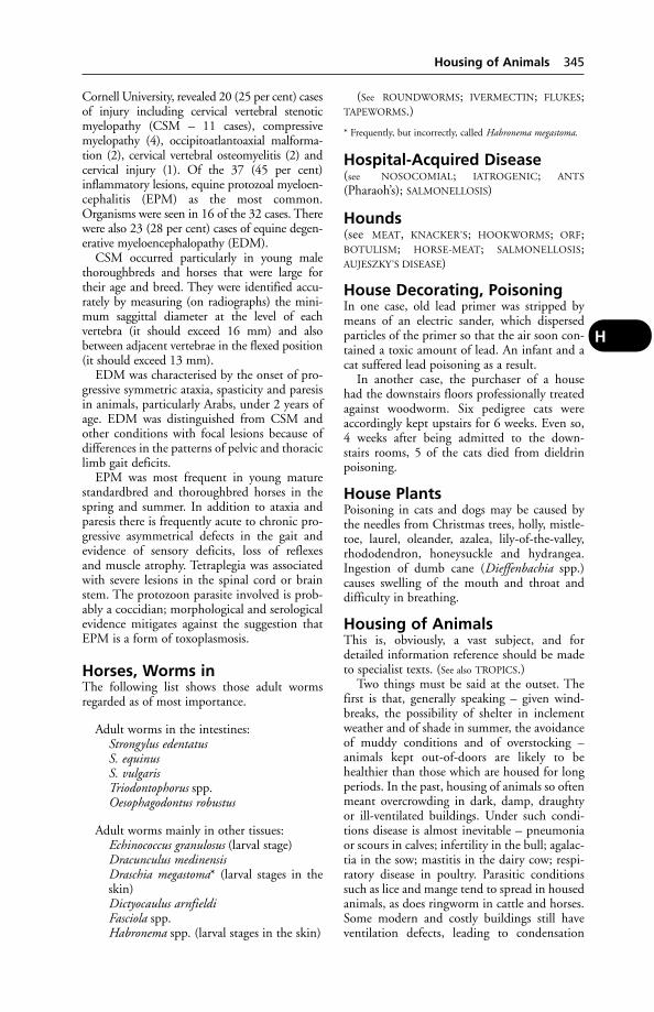
Cornell University, revealed 20 (25 per cent) casesof injury including cervical vertebral stenoticmyelopathy (CSM – 11 cases), compressivemyelopathy (4), occipitoatlantoaxial malforma-tion (2), cervical vertebral osteomyelitis (2) andcervical injury (1). Of the 37 (45 per cent)inflammatory lesions, equine protozoal myeloen-cephalitis (EPM) as the most common.Organisms were seen in 16 of the 32 cases. Therewere also 23 (28 per cent) cases of equine degen-erative myeloencephalopathy (EDM).
CSM occurred particularly in young malethoroughbreds and horses that were large fortheir age and breed. They were identified accu-rately by measuring (on radiographs) the mini-mum saggittal diameter at the level of each vertebra (it should exceed 16 mm) and alsobetween adjacent vertebrae in the flexed position(it should exceed 13 mm).
EDM was characterised by the onset of pro-gressive symmetric ataxia, spasticity and paresisin animals, particularly Arabs, under 2 years ofage. EDM was distinguished from CSM andother conditions with focal lesions because ofdifferences in the patterns of pelvic and thoraciclimb gait deficits.
EPM was most frequent in young maturestandardbred and thoroughbred horses in thespring and summer. In addition to ataxia andparesis there is frequently acute to chronic pro-gressive asymmetrical defects in the gait andevidence of sensory deficits, loss of reflexes and muscle atrophy. Tetraplegia was associatedwith severe lesions in the spinal cord or brainstem. The protozoon parasite involved is prob-ably a coccidian; morphological and serologicalevidence mitigates against the suggestion thatEPM is a form of toxoplasmosis.
Horses, Worms inThe following list shows those adult wormsregarded as of most importance.
Adult worms in the intestines:Strongylus edentatusS. equinusS. vulgarisTriodontophorus spp.Oesophagodontus robustus
Adult worms mainly in other tissues:Echinococcus granulosus (larval stage)Dracunculus medinensisDraschia megastoma* (larval stages in theskin)Dictyocaulus arnfieldiFasciola spp.Habronema spp. (larval stages in the skin)
(See ROUNDWORMS; IVERMECTIN; FLUKES;TAPEWORMS.)
* Frequently, but incorrectly, called Habronema megastoma.
Hospital-Acquired Disease(see NOSOCOMIAL; IATROGENIC; ANTS
(Pharaoh’s); SALMONELLOSIS)
Hounds(see MEAT, KNACKER’S; HOOKWORMS; ORF;BOTULISM; HORSE-MEAT; SALMONELLOSIS;AUJESZKY’S DISEASE)
House Decorating, PoisoningIn one case, old lead primer was stripped bymeans of an electric sander, which dispersedparticles of the primer so that the air soon con-tained a toxic amount of lead. An infant and acat suffered lead poisoning as a result.
In another case, the purchaser of a house had the downstairs floors professionally treatedagainst woodworm. Six pedigree cats wereaccordingly kept upstairs for 6 weeks. Even so,4 weeks after being admitted to the down-stairs rooms, 5 of the cats died from dieldrinpoisoning.
House PlantsPoisoning in cats and dogs may be caused bythe needles from Christmas trees, holly, mistle-toe, laurel, oleander, azalea, lily-of-the-valley,rhododendron, honeysuckle and hydrangea.Ingestion of dumb cane (Dieffenbachia spp.)causes swelling of the mouth and throat anddifficulty in breathing.
Housing of AnimalsThis is, obviously, a vast subject, and fordetailed information reference should be madeto specialist texts. (See also TROPICS.)
Two things must be said at the outset. Thefirst is that, generally speaking – given wind-breaks, the possibility of shelter in inclementweather and of shade in summer, the avoidanceof muddy conditions and of overstocking – animals kept out-of-doors are likely to behealthier than those which are housed for longperiods. In the past, housing of animals so oftenmeant overcrowding in dark, damp, draughtyor ill-ventilated buildings. Under such condi-tions disease is almost inevitable – pneumoniaor scours in calves; infertility in the bull; agalac-tia in the sow; mastitis in the dairy cow; respi-ratory disease in poultry. Parasitic conditionssuch as lice and mange tend to spread in housedanimals, as does ringworm in cattle and horses.Some modern and costly buildings still haveventilation defects, leading to condensation
Housing of Animals 345
H

inside and resulting in ill-health of the housedstock. The use of Yorkshire boarding can obvi-ate both the condensation problem and muchof the pneumonia.
The second thing is that, from a health pointof view, not every ‘development’ is an advance.Commercial competition may dictate the over-crowding of chickens to the point where feath-er-picking has to be counteracted by red light-ing or de-beaking; this may lead to short-termeconomic gains, but it is the antithesis of goodanimal husbandry, and the solving of the veteri-nary problems raised must be viewed accord-ingly. Intensivism can surely be pushed to astage where only a return to good husbandrywill succeed in reducing the incidence of disease– and also, incidentally, the size of the drug bill.
On the other hand, the dairy cow hasundoubtedly benefited from another dictate ofeconomy – the change from cowshed to theyard-and-parlour system – for instead of beingyoked or closely chained for long periods, she isfree to move around; and such exercise is initself important. (See CUBICLES FOR COWS.)
Cattle were housed on slatted floors inEngland in 1860 – with straw. Their use without straw may lead to welfare problemssuch as hygromata, damaged teats or injuredlegs, and housing on wholly slatted floors is notrecommended.
Intensivism has led to development inforced-draught ventilation, and to the efficientinsulation of walls and roof of animal houses bymeans of polystyrene, fibreglass, and other sub-stances. Insulated roofs are not usually used forcattle or calves. Housing for poultry and pigs,however, should have roof insulation as well aswall insulation as these animals have a highercritical temperature than ruminants. (See under
CONTROLLED ENVIRONMENT.)It costs over 4 times as much to keep an
animal warm by feeding concentrates – ‘aninternal fuel’ – as by warming the livestockhouse. Minimum economic temperatures aregiven below.
Housing has an important bearing upon thefeeding of animals. Pigs, for instance, confinedon concrete have no opportunity for the normalscavenging which can obviate mineral or vita-min deficiencies, and special rations accordinglybecome necessary for such housed animals.Vitamin A and E deficiencies are particularlylikely to occur.
Residual infection is obviously important,and advice is given on this under SALMONEL-
LOSIS and DISINFECTION. In a building used,successively, for calves and pigs, or pigs andturkeys, for example, a cross-infection betweenthe species may arise with a particular strain ofE. coli. Buildings in which pigs and sheep are
346 Housing of Animals
H
A dairy unit, with lying area, parlour and dairy under one roof. Note the Yorkshire boarding to theleft of the picture – a means of ensuring good ventilation and an absence of condensation.

housed may carry-over erysipelas; ringwormcan pass from cattle to sheep, pigs or horses viaan infected building. On land surroundingbuildings it is worth remembering that theworm Trichostrongylus axei is common to cattle,sheep, horses, and goats.
Cattle An open-ridge method of ventilation isstill recommended as the best for cowsheds. Inwinter, the optimum temperature insideappears to be within 6 and 13°C (44–55°F).Milk yields are said to be depressed when thetemperature falls below freezing point. In sum-mer, there is an upper limit of about 25°C(77°F), at which point cattle begin showing distress. High humidity at a temperature above15.5°C (60°F) appears to diminish milk yield.
For covered yards, ventilators should be pro-vided at the highest point, with a gap of 60 cm(2 ft) between the top of the walls and theeaves. Open-fronted covered yards should nothave a gap. About 2 tonnes of straw per cow isrequired for straw yards in winter.
Pigs Given adequate straw, the most primitivearks on range will yield better results than acold, damp house. A warm environment willreduce the risk of overlying by the sow. Whiledifferent optimum temperatures have beengiven by different research workers, it seemsthat 21°C (70°F) is about the figure to aim atin the farrowing house. For artificial rearing, atemperature of 30°C (86°F) has been recom-
mended for the first 4 days. Cold, damp floorsresult in liver disorders which do not appear in buildings where the pigs have a warm, drybed. Pregnant sows are better not housed. (See
CONCRETE; HYPOTHERMIA.)For fattening pigs, an optimum temperature
would appear to be about 18°C (65°F); 15.5°C(60°F) should be the minimum. Humidity doesnot appear to have an adverse effect, though fewauthorities recommend it. Good ventilation isadvocated.
Sheep In general, the disease problems associ-ated with the housing of sheep have been lessserious than might have been expected, andthere is a credit side as well as a debit side. For example, if lambs are born and reared tomarket weight indoors, there is far less risk ofworm infestation causing trouble. It is recom-mended that pens should not contain morethan 15 to 25 ewes, grouped according to lambing dates.
Ewes and hoggs housed for the winter aftergrazing should be wormed during the 1st week.If it is a liver-fluke area, dosing against flukes isadvisable 6 weeks after housing.
Lambs must be protected against lamb dysen-tery, and any from unvaccinated ewes should begiven antiserum.Infestation with lice may be aggravated byhousing and spread more rapidly. Since it cancause serious loss of condition, dipping orspraying before housing is recommended.
Housing of Animals 347
H
Cattle on deep litter in a covered yard.

E. coli infections are as much a threat to thehoused lamb as to the housed calf. Overcrowd-ing and dirty conditions at lambing predisposeto coli septicaemia, which is usually a sequel tonavel infection. In early weaned lambs, thequality of the milk substitute is important ifscouring is to be avoided; measures should betaken to minimise contact between housedsheep and their dung. Slatted floors, regularcleaning, copious use of bedding material andperiodical disinfection all help in this direction.
Good ventilation can go a long way towardsreducing the risk of acute pneumonia. In lambsand older sheep this is often associated withPasteurella infection, sometimes aggravated bylungworm infestation. Pasteurella pneumoniavaccine may be effective in prevention, but isuseless against other forms of pneumonia –which can be caused by other bacteria, moulds,and viruses.
Infections which give rise to abortion mayprove more troublesome indoors than out, andvaccination against enzootic (chlamydial) abor-tion seems worthwhile. (See also COPPER POISON-
ING and under SHEEP BREEDING and INTENSIVE.)
Poultry Chickens probably do best at temper-atures between 13 and 18°C (55° and 65°F).Egg-production declines at temperatures below5°C (40°F) or above 23°C (75°F). A relativehumidity of 50 per cent is considered the optimum for grown birds. A cold, dry house isbetter than a warm, wet one. Ventilation require-ments vary; for example, a bird may need asmuch as 300 cm3 (1 cubic foot) per minute per450 g (1 lb) bodyweight in the hottest weather,but only one-sixth of this in the coldest weather.(See also under CHICKS; NIGHT LIGHTING.)
For other aspects of housing, see under CON-
CRETE; LEAD POISONING; WOOD PRESERVATIVES;CUBICLES; BULL HOUSING; LOOSE-BOXES; DEEP
LITTER; INTENSIVE LIVESTOCK PRODUCTION;YORKSHIRE BOARDING; WATER.
Huckleberry Poisoning(see GARDEN NIGHTSHADE POISONING)
Humane Destruction of Animals(see EUTHANASIA)
HumerusHumerus is the bone of the foreleg between theshoulder-joint and the elbow-joint. It has arounded head which, with the correspondingdepression of the scapula, forms the ‘ball-and-socket’ shoulder-joint. At the opposite extremi-ty it forms with the radius and ulna the hingedelbow-joint.
Humoral ImmunityHumoral immunity is that conferred by theimmunoglobulins derived from the B-cells ofthe reticulo-endothelial system and is differen-tiated from cell-mediated immunity associatedwith T-CELLS. (See also IMMUNE RESPONSE;COLOSTRUM; IMMUNOGLOBULINS.)
HumourHumour is a term applied to any fluid or semi-fluid tissue of the body, e.g. the aqueous andvitreous humours in the eye.
HuskHusk is a disease of cattle, sheep, and goatscharacterised by bronchitis, which is caused bylungworms. (See PARASITIC BRONCHITIS.)
HuskyA muscular, medium-sized dog with a thickdouble coat. The breed is used to pull sleds inPolar regions and carts in warmer climes. Cart-racing is quite popular; it must not take placeon public roads as this would contravene theProtection of Animals Act 1911. Huskies areprone to corneal dystrophy, glaucoma and ven-tricular heart defects. Haemophilia has beenrecorded.
Hyaline MembranesA fibrinous exudate from the epithelium whichlines the alveoli of the bronchioles, found instillborn animals and those dying soon afterbirth. It is also referred to as hyaline membranedisease.
Hungarian VislaA medium-sized dog with short, reddish-browncoat and pendulous ears. Few genetic defectsare known other than haemophilia A.
HyaluronidaseAn enzyme which breaks down the hyaluronicacid forming part of the material in the inter-stices of tissue, and so facilitates the absorptionof injected fluids. It assists the rapid distri-bution of drugs injected either subcutaneouslyor intramuscularly. It has been used in the treatment of urinary calculi.
HyaluronateA mucopolysaccharide used as an injection intothe joint to treat arthritis.
HybridAt one time this word meant a cross between twoinbred lines; now it is used to describe a simplecross between 2 different strains or breeds.
348 Huckleberry Poisoning
H
For a comparison between a hybrid and achimera (with reference to fertile mules), see
CHIMERA.
Hybrid VigourThe improved performance produced in theoffspring by mating 2 breeds (see GENETICS;BLOOD-TYPING; T-CELLS).
Hybridoma(see GENETIC ENGINEERING; also under RABIES –Diagnosis)
Hydatid DiseaseHydatid disease is caused by the cystic larvalstage of the tapeworm Echinococcus granulosus,of which the dog and fox are the usual hosts.Eggs released from tapeworm segments passedin the faeces by these animals are later swallowedby grazing cattle, sheep and horses, which maybecome infested also through drinking watercontaminated by wind-blown eggs.
In Australia an anti-hydatid disease cam-paign has proved successful; though in NewSouth Wales there is a sylvatic strain which cir-culates predominantly between wild dogs andwallabies.
Swallowed eggs hatch in the intestines andare carried via the portal vein to the liver. Someremain there, developing into hydatid cysts;others may form cysts in the lungs or occasion-ally elsewhere, e.g. spleen, kidney, bone marrowcavity, or brain. Inside the cysts, brood capsules,containing the infective stage of the tapeworm,develop, and after 5 or 6 months these caninfest dog or fox.
People become infested through swallowingeggs attached to inadequately washed vegeta-bles; eggs may possibly be inhaled in dust orcarried by flies to uncovered food. The han-dling of infested dogs is an important source. In Beirut the risk is put at 21 times greater for dog-owners than others, by the WorldHealth Organisation, which states also that in California, nomadic sheep-rearers are 1000times more likely to have hydatid disease thanother inhabitants of the state.
There have been successful campaigns tocontrol human hydatid disease in both Cyprusand Iceland, by compulsory treatment and/orbanning of dogs.
In Wales, where the incidence of hydatid dis-ease is relatively high, farm dogs and foxhoundsare important in its spread.
Only some 7 people are known to die fromthis disease in England and Wales each year – afigure which would probably be higher werediagnosis less difficult. Condemnation of sheep
and cattle offal from this cause runs into hun-dreds of thousands of pounds annually. Routineworming of dogs is essential for control. E.granulosus is far from being a typical tapeworm,as it has only 3 or 4 segments and a total lengthof a mere 3 to 9 mm, so that the dog-owner willnot notice the voided segments.
A problem of diagnosis also arises, in that thisworm’s eggs are indistinguishable from those ofTaenia tapeworms. Previously, one could dosedogs with arecoline hydrochloride and examinethe faeces for the presence of the intact tape-worm, but in Britain this anthelmintic is nolonger obtainable, having been replaced by moremodern drugs which destroy the tapeworm butleave it unrecognisable.
Equine hydatidosis in Britain is caused bya strain of E. granulosus which has becomespecifically adapted to the horse as its interme-diate host, and is often referred to now as E.granulosus equinus. This apparently is of lowpathenogenicity for man.
In a survey covering 1388 horses and poniesexamined at 2 abattoirs in the north ofEngland, 8.7 per cent were infected. Prevalenceof infection was closely related to age – risingfrom zero in animals up to 2 years old to over20 per cent of those over 8 years old.
Sixty-six per cent of the infected animals hadviable cysts. Prevalence appeared to be greatestin central and north-west England.
Treatment of human patients Hydatiddisease has been said to be one of the rare para-sitic conditions that can be treated only bysurgery. However, the result is often incomplete,with frequent local recurrences or accidents ofsecondary dissemination. Repeated interven-tions are often mutilating and do not guaranteea definite cure. Mebendazole has been used successfully in some patients, but is not alwayseffective.
HydralazineAn arterial dilator, useful in treating dogs with failing heart due to mitral regurgitation(usually caused by fibrosis of the valve) and left-sided congestive heart failure.
Hydrargyrum(see MERCURY)
Hydraulic FluidIntense generalised pruritis was suffered by ani-mals grazing in a field beneath the flight path toan airfield after they ate grass contaminated byhydraulic fluid leaked from an aircraft.
Hydraulic Fluid 349
H
HydroceleHydrocele means a collection of fluid presentwithin the outer proper coat of the testicle(tunica vaginalis) or within the spermatic cord.
HydrocephalusHydrocephalus is a condition in which a largeamount of fluid collects within the brain cavityof the skull. It may be present before birth (con-genital hydrocephalus), in which case the largesize of the head may present an obstruction toparturition. In the congenital form which ismet with in foals, calves, and puppies, there is alarge prominent swelling over the forehead, anda rounded dome-like cranium. Animals born in this condition are usually dead, or if they are living they die soon after birth. It maybecome necessary to puncture the swollen skulland evacuate the fluid before delivery can beeffected.
In the acquired form, which is chiefly metwith in the horse and dog, the fluid collects in the ventricles of the brain, or under themeninges, as the result of meningitis, or thepresence of a tumour which has interfered withthe free circulation of the cerebrospinal fluid, orhas produced an exudate from the engorgedblood vessels.
When due to meningitis, hydrocephalus isusually an acute condition, and its symptomsare masked by those of the meningitis; whendue to other causes in which there is obstruc-tion to the flow of cerebrospinal fluid it is usually chronic, and the symptoms are those of pressure on the brain. The animal becomesgradually dull, sleepy, insensitive to its sur-roundings. Convulsions may occur, and duringone of these death is liable to take place. (See
under HELLEBORES.)
Hydrochloric Acid (HCI)Hydrochloric acid (HCI) is normally present inthe gastric juice, to the extent of about 2 partsper 1000. (See DIGESTION.) In the concentratedform it is a corrosive poison.
Hydrocyanic Acid (HCN)Hydrocyanic acid (HCN) and its salts – sodiumand potassium cyanide – are among the mostdeadly poisons, and very rapid in their effects.
Signs If taken by mouth, or given by injection,there is a rapid acceleration of the breathing (andoccasionally coughing). A poisoned dog or catwill utter a cry and collapse, the limbs extendedfully. There is an odour of bitter almonds. Theremay be convulsions. Respiratory failure ensues,and death may occur within seconds.
Hydrocyanic poisoning may occur from theingestion by grazing animals of plants contain-ing a cyanogenetic glycoside. (See GLYCOSIDES.)Poisoning is then less acute, and signs are notalways indicative of the cause.
Treatment In acute cases, death occurs indogs and cats before treatment can begin. If asmaller quantity of the poison has entered thebody, or if poisoning is the result of the cyano-genetic glycosides (and this is known in time),an intravenous injection of a 1 per cent solutionof sodium nitrite, followed by 25 per cent sodi-um thiosulphate, has been recommended forthe dog and large animals. Repeat doses at halfthat rate.
Hydrogen PeroxideAn antiseptic, with some effect against viruses,due to the release of oxygen. Available as a 3 percent (10 volume) or 6 per cent (20 volume)solution, it is used, usually diluted, for cleans-ing the skin and disinfecting wounds. It isunsuitable for the irrigation of cavities or deepwounds. (See OXYGEN EMBOLISM.)
HydrometraAnother name for pseudopregnancy, it is theaccumulation of a watery fluid within the uterus,sometimes sufficient to push other organs asideand to cause swelling of the abdomen of rabbits.This idiopathic condition has also been seen in cats.
HydronephrosisHydronephrosis is a condition in which thecapsule of the kidney, or even the kidney itself,becomes greatly distended with urine which isunable to pass along the ureter into the urinarybladder owing to some obstruction in thatchannel, such as calculus, a twist, or owing tothe pressure of some organ nearby. The kidneyswells in size, and causes pressure upon the surrounding organs with pain over the lum-bar region, and in severe cases a bulging of the muscles just behind the last rib. It is treatedby either the removal of the whole kidney (provided the other one is healthy), or else bythe removal of the obstruction.
HydropericardiumThe accumulation of clear, straw-coloured fluidin the pericardial sac, a swollen and discolouredliver and enlarged kidneys with distendedtubules. This syndrome (Angaria disease) hashad a devastating effect on the broiler poultryindustry of Pakistan. The disease has typi-cally been seen in 3- to 6-week-old growing
350 Hydrocele
H
broiler chicks and results in up to 60 per centmortality.
Cause An unidentified infectious agent whichappears to require the presence of an adenovirusto produce the lesions.
HydrophobiaRabies.
Hydroponic GrassHydroponic grass, consisting of a mat of barleyseedlings harvested at the 8-day stage, has beenused for horse feeding, and is usually eaten withrelish. It is highly nutritious, very digestible andparasite-free. (See under HORSES, FEEDING OF.)
Hydrops Amnii and HydropsUteri(see UTERUS, DISEASES OF)
HydrosalpinxAn accumulation of serous fluid in the Fallopiantube. It has been stated to be a common cause ofsterility in gilts in America.
HydrothoraxHydrothorax means a collection of exudate inthe chest, i.e. in the pleural cavity. This is oneof the results of certain forms of pleurisy.
‘Hyena Disease’A condition in cattle in which the hind part of the animal grows more slowly than the forepart, producing a silhouette said to resemblethat of the hyena. The cause is unknown butbovine virus diarrhoea has been suggested. Ithas not been reported in the UK.
Hygiene(see INFECTION; VENTILATION; HOUSING; WATER-
SUPPLY; DIET AND DIETETICS; DISINFECTION;SLURRY)
HygromaHygroma is a swelling occurring in connec-tion with a joint, usually the knee or hock, andthe result of repeated bruising against a hardsurface. (See CAPPED HOCK.)
Hygroma in cattle may arise through aninsufficiency of bedding, or through faultybuilding design. (See also CALLOSITY.)
Hygroma of the elbow in large dogs has beensuccessfully treated by means of the followingtechnique. A 6 mm diameter Penrose drain waspassed through incisions made dorsally andventrally into the hygroma, and secured firmlyto the skin. Dressings were changed every 4 to5 days, and the drain taken out after 2 or
3 weeks. The wounds healed satisfactorily, andthe hygroma was obliterated in all 18 cases.
Hygromycin BAn antibiotic used in the USA as ananthelmintic, and claimed to be effective againstlarge roundworms and whipworms.
Hymen, ImperforateImperforate hymen in thoroughbred fillies,with consequent accumulation of fluid in theuterus, has led to symptoms varying from acuteabdominal pain, sweating, and attempts to liedown and roll, to discomfort when urinating.Immediate relief followed necessary surgery inthe more serious cases. Pulse and respirationrates returned to normal within 10 minutes,with feeding resumed. (See also ‘WHITE HEIFER
DISEASE’.)
HyoidHyoid is the name of the bone which gives sup-port to the root of the tongue and to the larynx.It has some similarity to the letter ‘U’.
Hyostrongylus RubidusA parasitic worm of pigs.
Hyper-Hyper- is a prefix indicating excess.
Hyperadrenocorticism(see CUSHING’S SYNDROME)
HyperaemiaCongestion. An excessive amount of blood in apart of the body.
HyperaesthesiaOversensitivity to bright light, sudden noise or touch. It occurs in diseases such as rabies,tetanus and hypomagnesaemia. It is the mainconsistent clinical sign in bovine spongiformencephalopathy (BSE).
Feline hyperaesthesia may result also frompoisoning by, for example, benzoic acid.
Signs Aggressiveness, excitement.
Hyperbaric(see OXYGEN)
HypercalcaemiaAn excess of calcium in the blood.
Causes In dogs these include cancer, an excessof vitamin D, osteolytic lesions, kidney failure,
Hypercalcaemia 351
H
excess parathyroid hormone, Addison’s disease,severe hypothermia, and, rarely, blastomycosis.
In man, additional causes of hypercalcaemiainclude acromegaly, increased thyroid glandactivity, long-term immobilisation, too muchvitamin A, treatment with thiazide diuretics,tuberculosis, sarcoidosis, histoplasmosis, coccid-iomycosis, and silicone-induced granuloma.
HypercapniaThe presence in the blood of a raised level ofcarbon dioxide.
HyperchlorhydriaHyperchlorhydria is a form of indigestion asso-ciated with excessive secretion of hydrochloricacid.
HyperglycaemiaAn excess of sugar in the blood. (See DIABETES
MELLITUS.)
Hyper-Immune SerumThe serum of an animal which has been hyper-immunised by repeated injections of a toxin orvaccine. It is rich in antibodies, and is used forcurative treatment of, e.g., tetanus.
HyperkalaemiaHigh concentration of potassium in the blood.
HyperkeratosisHyperkeratosis means an excess of horn or KER-
ATIN. The specific disease is also characterisedby hardening of the skin.
Cause In cattle, the disease has been caused bypoisoning by minute quantities of chlorinatednaphthalene compounds (and possibly otherchemical substances also). These are found inmany wood-preserving compounds in insecti-cides, lubricants, and electrical insulation mate-rial. These substances bring about a secondaryvitamin A deficiency. In America the disease hasfollowed the feeding of pellets prepared bymachinery lubricated with grease or oil con-taining naphthalene compounds – an indica-tion of the minute quantities sufficient to causetrouble. Usually, however, the disease is a sequelto housing stock in recently creosoted build-ings. (For the disease in pigs, see also ZINC and
CALCIUM SUPPLEMENTS.)
Signs A thickening of the skin, sometimeswith loss of hair, on neck and shoulders. Incalves, stunted growth, a discharge from the eye (often with a corneal opacity), frothing atthe mouth, weakness and emaciation occur,
and death may precede any obvious skinchanges.
Treatment Vitamin A will assist recovery.
HyperkinesisOveractivity that may be caused by dietary orenvironmental factors. It can be accompaniedby aggression, especially in the reaction toattempted restraint (e.g. putting on a lead). Theheart and respiratory rates may increase; seda-tion may be a temporary measure. Expert eval-uation of the diet may be necessary and the animal may have to be referred to an animalbehaviour specialist.
HyperlipaemiaAn excess of lipids in the blood, which can be fatal in ponies and donkeys. It was firstreported in Europe, then in Australia. Mares areaffected in late pregnancy or early lactation.
Signs Depression, weakness, loss of appetite,diarrhoea, and terminal convulsions.
Autopsy findings: liver much enlarged, yellowand friable.
HypermetriaA high-stepping gait. (See COENURIASIS.)
HyperoxaluriaAn excess of oxalates in the urine. This accom-panies L-glyceric aciduria in kidney disease ofkittens 5 to 9 months old.
Acute kidney failure develops together withatrophy of nerves supplying muscles.
Signs Extreme weakness, affecting standing andwalking.
Cause A recessive gene.
HyperparathyroidismOf 21 dogs suffering from this, 20 had an ade-noma, and 1 a carcinoma. (See PARATHYROID
GLANDS.)
Signs Thirst, listlessness, weakness, loss ofappetite.
HyperplasiaHyperplasia is the term applied to abnormallygreat development of some organ or tissue.
HyperpotassaemiaToo high a level of potassium in the blood-stream. This may be brought about artificially,with fatal results, by the mistaken use of potas-
352 Hypercapnia
H
sium iodide intravenously instead of sodiumiodide.
HyperpyrexiaHyperpyrexia means a high degree of fever. (See
FEVER; TEMPERATURE.)
HypersensitivityOnce an animal has been ‘primed’ or sensitisedby an antigen, further contact with this willboost the immune response – but may also pro-voke tissue-damaging reactions. (See IMMUNE
RESPONSE; ALLERGY; PENICILLIN, SENSITIVITY
TO; ANAPHYLAXIS; SERUM SICKNESS.)
HypersexualityHypersexuality is usually found in males withexcessive testosterone production; it may also bethe result of a malfunction of the cerebral cortex.Administration of short-acting progesteronesmay help diagnosis but, long term, castration isoften the answer.
HypertensionHigh arterial blood pressure. In dogs, kidneydisease is the most common cause of hyperten-sion.
Signs Detachment of the retina, or bleedingfrom it, may be the first indication. The dogmay suddenly go blind. Long-term effects mayinclude enlargement of the left ventricle of theheart, and kidney failure.
HyperthermiaA body temperature greatly in excess of the normal, as occurs in fevers.
Hyperthermia, MalignantWhen some dogs of the Great Dane breed,some pigs of the Piétrain breed, and somehuman beings (about 1 in every 10,000 people)are anaesthetised with halothane, their bodytemperature rises to a point at which, unless theanaesthesia is discontinued, the hyperthermia islikely to prove fatal.
Hyperthermia may occur in animals poi-soned by chlorinated hydrocarbon insecticides.(See also HEAT-STROKE; TROPICS; FEVER.)
Malignant hyperthermia may also develop asa result of stress.
HyperthyroidismExcessive activity of the THYROID GLAND.
Hypertonic(see under ISOTONIC)
Hypertrophic Osteopathy(Marie’s Disease)This was first described in man in 1890. It hasbeen reported in the dog (see also ACROPACHIA)and in the horse. In Africa, the roundwormSpirocera lupi has been reported as associatedwith the condition in the dog.
In the dog the disease takes the form of anon-oedematous swelling of all 4 legs. It is asso-ciated with tumours of the lung. Severing of thevagus nerve has been recommended in cases(the majority) where surgical removal of thelung lesions is not possible, and has led to a reduction of the bone enlargement in thelimbs, and of the swelling, pain, and lameness.Euthanasia may, of course, be preferable.
HypertrophyHypertrophy means extra size or developmentof an organ or tissue.
In certain valvular diseases of the heart whenobstruction to the free flow of blood occurs, themuscle wall of the heart becomes increased inthickness and strength, and a compensationresults. In the training of horses the trainer aims at getting the maximum efficiency fromthe skeletal muscles, which under the influenceof judicious training and feeding becomehypertrophied.
After 1 organ of a pair has been removed –as, for instance, the kidney or the ovary – theremaining organ becomes increased in size so as to be able to perform practically the sameamount of work as was previously done by the pair.
HypervitaminosisDisease associated with an excess of a particularvitamin. For example, chronic hypervita-minosis A occurs in cats fed exclusively, or virtually so, on an all-liver diet. (See under CAT
FOODS.)
HyphaemiaAn infusion of blood into the anterior chamberof the eye.
Hypo-Hypo- is a prefix indicating a deficiency.
Hypoadrenocorticism(see ADDISON’S DISEASE)
HypoalbuminaemiaA low level of albumin in the blood. It may be indicative of kidney or liver disease, or malnutrition.
Hypoalbuminaemia 353
H

Hypocalcaemia(see MILK FEVER; TRANSIT TETANY; LAMBING
SICKNESS IN EWES; ECLAMPSIA; METABOLIC
PROFILES). An insufficiency of blood calcium.This occurs also in mares. The signs are a stiffgait, with the hind legs placed forward whenstanding still, trismus, and dyspnoea.
HypochloritesEffective disinfectants widely used in milkingmachines, dairy equipment and food premises;preparations are marketed under a number oftrade names. They leave no persistent odour ortaint. Their efficacy depends upon the amount ofavailable chlorine, which is more active againstviruses than most disinfectants. Hypochloritesare unable to penetrate grease and are often com-bined with detergents. Sodium hypochlorite isuseful for disinfecting premises after an outbreakof a virus disease. (See also TEAT-DIPPING.)
HypocupraemiaA condition in which there is too little copper inthe bloodstream. This occurs in SWAYBACK inlambs, and is also associated with serious ill-health in cattle. On the Shropshire–Cheshireborder, for example, hypocupraemia is accompa-nied by scouring and stunted growth. Two-year-old heifers have been mistaken for 8-month-oldcalves. In Caithness, hypocupraemia is liable tooccur on 75 per cent of the farms unless precau-tions are taken. Scouring is not a common symp-tom there but calves of the beef breeds show astilted gait and progressive unthriftiness. (See also
COPPER.)
HypocuprosisA disease caused by a copper deficiency. (See
HYPOCUPRAEMIA and COPPER.)
Hypodermic(see INJECTIONS)
Hypoglossal NerveThe hypoglossal nerve is the 12th cranial nerveand supplies the muscles of the tongue, togeth-er with others nearby.
HypoglycaemiaHypoglycaemia is a deficiency of sugar in theblood. It causes acetonaemia in ruminants andpregnancy toxaemia in sheep. It may occur in states of starvation, but is of special impor-tance in connection with the administration of insulin, which is injected to lower the bloodsugar from an abnormal amount, and which, if given in too large doses, may produce toogreat reduction with symptoms of nervousness,
breathlessness, and excitement. In human med-icine hypoglycaemia may be a sequel to the use of sulfonamides, e.g. sulphadiazine. Thesesymptoms are relieved by taking some food con-taining sugar and by an injection of adrenaline,which checks the action of insulin.
HypokalaemiaA deficiency of potassium in the blood. (See ‘DOWNER COW’ SYNDROME.)
In cats, hypokalaemia results in weakness,the neck bending downwards.
HypomagnesaemiaAlso known as ‘grass staggers’ or Hereford dis-ease, it is caused by too little magnesium in thebloodstream.
Hypomagnesaemia is of particular importancein cattle. It occurs when a herd is turned on tolush spring grass after being stall-fed during thewinter, and often follows a frosty or wet spell; aninterval of a few days may elapse before symp-toms appear. The problem is particularly com-mon where potash (potassium) and nitrogen fertilisers have been used, but the causes are com-plex. The low blood magnesium often resultsfrom a reduced intake of magnesium, while theabsorption of that mineral is inhibited by thepresence of potash and the rapid movement offeed through the gut. Hypomagnesaemia canalso occur in the autumn in dry cows or sucklercows at grass and not receiving supplementaryfeed.
Hypomagnesaemia has apparently beenmore common in the Ayrshire than in otherBritish breeds of cattle. Cows which have hadseveral calves are more prone to it than heifers.
It is more common in ewes in the first 4 weeks after lambing than before lambing.
In calves, hypomagnesaemia can occur wherethe diet consists mainly of milk, which is not byitself an adequate source of magnesium for arapidly growing young animal. The condition isthus seen mainly in suckler calves and thosebeing reared for veal. (See OMASUM.)
Signs Animals are often recumbent; if on theirsides, they paddle with their legs when stimu-lated, the head extends backwards and theyfroth at the mouth and defecate. The heart rate,which is rapid, may be heard several paces fromthe animal. Unless treated early they often die.If not recumbent, shivering, a staggering gait,excitement, convulsions, and paralysis may precede death. In less acute cases, the animalsappear ‘nervy’ – responding violently to sensa-tions of touch or sound – and there may bemuscular tremors.
354 Hypocalcaemia
H
Treatment This must be prompt. A 25 percent solution of magnesium sulphate is givensubcutaneously. Intravenous injection may killthe animal; if it is used intravenously, magne-sium should be given combined with calciumborogluconate. Great care is necessary, however,in giving the injection and even approachingthe animal – which may otherwise die at theprick of the needle.
An enema of up to 5 tablespoonfuls of mag-nesium chloride in 250 ml of warm water is rec-ommended by the Tennessee State University.
Magnesium can also be given in the drinkingwater, using a proprietary product.
Prevention The feeding of magnesium-richsupplements 3 weeks before early spring grazingand for up to 6 weeks after turnout or, in sheep,the use of a magnesium lick, from a month after service till a month after lambing. (Foradult cattle a daily dose of 60 g (2 oz) per headof calcined magnesite, mixed with damp sugar-beet pulp, is recommended.) A mixture of mag-nesium acetate solution and molasses may beoffered ad lib from ball feeders on pasture, as an alternative. Magnesium ‘bullets’ are alsoused. Top-dressing pasture with calcined mag-nesite is helpful. (See MAGNESIUM; MILK FEVER.)Magnesium can also be given in the drinkingwater; proprietary preparations are available.
Hypomyelinogenesis Congenitain SheepA congenital disease of lambs, characterised by trembling or twitching, staggering, andsometimes shaking of the head.
HyponatraemiaA deficiency of sodium in the blood.
Hypoparathyroidism,Nutritional Secondary(see CANINE and FELINE JUVENILE OSTEODYS-
TROPHY)
HypophosphataemiaA condition in which the level of blood phos-phorus is too low. (See MILK FEVER; ‘DOWNER
COW’ SYNDROME.)
HypophysisThe pituitary gland. Hypophysectomy is removalof the pituitary.
HypoplasiaUnder-development. Hypoplasia of the genitalorgans is a cause of sterility.
HypotensionLow arterial blood pressure. It is not commonin animals except following shock, for exampleafter an accident.
Hypotensive DrugsHypotensive drugs are those which reduce highblood pressure.
HypothalamusA part of the brain below the thalamus which acts as a thermostat, maintaining bodytemperature. It also influences blood circulation,urinary secretion, and appetite. (See BRAIN.)
HypothermiaAn abnormally low body temperature; it is acommon cause of lamb mortality. It can becaused by exposure in the first few hours afterbirth or, after about 12 hours, because of star-vation. Affected lambs should be warmed;starved lambs must receive food or 20 per centsolution of glucose, injected intraperitoneally,before warming.
Piglets are also susceptible to hypothermia andwill often die if creep areas are not adequatelyheated.
In human surgery, hypothermia is delib-erately induced, by various means, for opera-tions on heart or brain. A technique is used inhuman surgery for operations within the dryheart. The venous blood is cooled in a circuitoutside the body (a method now preferred tothe use of ice packs or refrigerated blankets)until a body temperature of 20° to 25°C (68°to 77°F) is obtained, when the flow of blood tothe heart can be stopped for several minutes toallow the operation to proceed.
Hypothermia, AccidentalThe chilling of newborn animals, or of thoseunder a general anaesthetic, is a life-threateningcondition. Warmth is essential. (See also under
SHEEP BREEDING – Lamb survival; HOUSING OF
ANIMALS.)
HypothyroidismA condition caused by underactivity of the thy-roid gland. It is not uncommon in dogs andcauses lethargy, weight gain and also alopecia. Itis treated with thyroid preparations such as thy-roxine. It is associated in cattle with a high inci-dence of aborted, still-born or weakly calves.(See also GOITRE.)
HypovolaemiaA diminished volume of blood. (See SHOCK.)
Hypovolaemia 355
H

HypoxiaA reduced level of oxygen-supply to the bodytissues. It can occur in newborn animalsdeprived of oxygen during birth. The animal isdull and weak, often unable to suck.
HysterectomyA surgical operation for removal of the uterus.Usually the ovaries are removed at the sametime. (See OVARIO-HYSTERECTOMY.)
Hysteria (Canine)A decline of this condition in the UK has been
attributed to the abandonment of the use ofagenised flour in the manufacture of dog bis-cuits. (The agene process involved the bleachingof flour with nitrogen chloride.)
It has been suggested that some cases mayhave been due to the use of flour, containingthe spores of Tilletia tritici, in dog-biscuit manufacture.
Signs The dog would suddenly ‘appear to gomad’, racing round with a fixed stare, barkingor howling.
(For distemper-like signs, see MENINGITIS.)
356 Hypoxia
H
Iatrogenic DiseaseAny illness resulting from treatment, such asthe side-effects of some drugs. Adverse drugreactions were suspected in 130 of 39,541 casestreated at the Veterinary Hospital, Universityof California, Davis. In 66 cases there wasreasonable evidence to link the reactionobserved to the drug. Antibiotics and antipara-siticides were incriminated 21 times, with ana-phylaxis being the most commonly observedreaction. There were 3 deaths following theadministration of procaine penicillin (inadver-tently intravenously) to a lamb; potassiumpenicillin (10,000 units/kg) to a cat; and oxyte-tracycline (25 mg/kg) to a cow. Anaesthetic andrelated agents were involved 20 times. Severeclonic convulsions developed in 5 cats receivingmore than 80 mg ketamine hydrochloride;cardiac arrest, hypotension, dyspnoea andmuscular rigidity in 2 horses given xylazine(1 mg/kg intravenously); and severe bradycar-dia and respiratory arrest in 2 dogs givenfentanyl-droperidol. Anti-cancer drugs wereimplicated in 10 cases with the most dramaticreactions being observed in 5 dogs treated with5-fluorouracil. One of these died as a result ofneural toxicosis. (See also SIDE-EFFECTS; DRUG
INTERACTIONS.)
IBKInfectious bovine keratitis (infectious ophthalmiaof cattle). (See EYE, DISEASES OF.)
IBRInfectious bovine rhinotracheitis. (See RHINO-
TRACHEITIS.)
IbuprofenA non-steroidal anti-inflammatory drug, muchused in human medicine. It has a narrowmargin of safety in dogs, in which it can causea sometimes fatal gastric ulceration. The same istrue of flurbiprofen.
Ice, Ice CubesOf use in cases of haemorrhage from the stom-ach, as an aid to control bleeding from wounds,and as an application in cases of meningitis andparaphimosis; also in cases of hyperthermia andsunstroke.
Icelandic Pneumonia(see PULMONARY ADENOMATOSIS; also MAEDI/
VISNA)
IchthyophonusA fungus that usually infects marine fish but ispathogenic to salmon in both sea and fresh water.It is caused by feeding salmon infected dead fishwhich have not been processed properly.
IchthyosisIchthyosis is a condition of the skin in the dog,especially over the elbow and hocks in whichlarge and irregular cracks appear. These becomefilled with dirt, and infection results.
Icterus(see JAUNDICE)
IdentichipAn electronically coded microchip, the size of agrain of rice, encased in implant-grade glass. Itis inserted in the loose skin of the neck of theanimal (under local anaesthesia).
The microchip is encoded with the animal-owner’s address, etc., kept on a central computerregister. Electronic scanners (‘readers’) are usedto read the data in the chips.
A database of animals so identified is kept,under the name Anibase, by Animalcare Ltd.,of Common Road, Dunnington, York YO1SRU (see also MICROCHIP).
Identification of CattleThis is controlled by law in the UK. TheBovine Animals (Records, Identification andMovement) Order 1995, as amended by theCattle Identification (Amendment) Regulations1999 and the Cattle (Identification of OlderAnimals) Regulations 2000, requires that allcattle be identified by an ear tag in each ear.Each animal must have a ‘passport’ that mustaccompany it wherever it goes and in whichthe following details must be entered: date ofmovement on to a particular holding, or dateof birth on the holding; eartag number; breed;sex; dam’s identification number (replacementear tag where applicable). When the animal ismoved off a holding, its age or date of birth, theholding from which it is moved, and that towhich it is moved are recorded. Ear tags must beapplied to dairy cattle within 36 hours of birthand, in other cattle, within 30 days of birth.
Identification of GoatsIdentification of Goats is controlled by theSheep and Goats (Records, Identification
I

and Movement) Order 1996, as amended.Requirements are essentially the same as forsheep (see below) except that plastic ear tags arenot recommended as goats will chew them.
Identification of HorsesUnder the Horse Passports Order 1997, asamended, each horse must have a passportprepared as specified. The document must be inFrench and English. Details of the individualhorse are given and a silhouette of the animalis filled in with the markings. Information ofvaccinations and dates of administration mustalso be stated in the passport. There is also pro-vision for the results of veterinary examinationsto be included and signed by the veterinarysurgeon in charge of an event in which thehorse is entered, or when the animal is about toundertake a journey.
Identification of PigsAll pigs must be identifiable to the pemises oforigin, under regulations set out in the Pigs(Records, Identification and Movement) Order1995. Breeding stock are usually ear-taggedbefore leaving the breeding farm. Pigs forslaughter are usually identified by a slap mark;those travelling across an EU frontier must beear-tagged. Records must be kept of pigs bornon a holding. Very strict measures have beentaken to control disease transmission, becausemany viruses multiply rapidly in an infectedpig and the quantity of virus shed can be muchmore than is the case with cattle, sheep orgoats with the same infection. All movementsof pigs must be recorded in a specified manner.Regulations set out the format for the declara-tion of pigs moved off a holding, moved froma farm for breeding, exhibition, artificial insem-ination or veterinary treatment, and returnedto the farm after having left it for breeding pur-poses. The form of declaration is also specifiedfor the movement off a farm of pigs that havebeen fed waste food (NB feeding of cateringwaste, including swill, is banned), and for thosemoved from a collection centre to a farm. Pigsmust not be moved to an abattoir within 72hours of their arrival on a holding; normally,pigs can only be moved after at least 20 days.
Identification of SheepIdentification of Sheep is controlled by theSheep and Goats (Records, Identification andMovement) Order 1996 (as amended). Sheepmoving into the EU and sheep leaving Britainmust be identifiable by ear tag. Animals mustbe marked before they are moved off theholding where they were born.
IdiopathicIdiopathic is a term applied to diseases to indicatethat their cause is unknown.
Idiopathic feline vestibular syndrome(IFVS) (see under FELINE VESTIBULAR SYNDROME)
IdiosyncrasyAn atypical reaction to a drug or to a food; in abehavioural sense, a quirk.
IgA, IgE, IgG and IgMIgA, IgE, IgG and IgM are antibody/immunoglobulins found in the blood serumand also in secretions from mucous membranes.(See IMMUNOGLOBULINS; SECRETORY IgA.)
Ile De FranceA French breed of sheep.
IleitisInflammation of the ileum.
IleocaecalIleocaecal refers to the junction between ileumand caecum, between the end of the small intes-tine and the commencement of the large. Theso-called ileocaecal valve is formed by the cae-cum in such a manner that while food materialmay readily travel from ileum into caecum, it isdifficult for it to pass in the opposite direction.
IleumIleum is the last arbitrary division of the smallintestine. (See INTESTINES.)
Inflammation of the ileum – which becomesthickened and stiff, almost like a piece of rub-ber hose – is a cause of death in piglets 2 to4 months old. It has been suggested that thereis a hereditary predisposition to this condition,which often affects the whole litter. In manyinstances, the trouble is recognised only atthe bacon factory, having caused no apparentillness in the pigs. Those that die, on the otherhand, do so from perforating ulcers and peri-tonitis, after showing evidence of thirst, a bluishcolour of the skin, and collapse. (See PORCINE
INTESTINAL ADENOMATOSIS.)
IleusThe intestinal obstruction which can followfailure of PERISTALSIS.
IliacRelating to the flank. (See ARTERIES.)
I/M or ImShort for intramuscular; usually refers to routeof injection.
358 Identification of Horses
I

IliumIlium is another name for the haunch-bone, theouter angle of which forms the ‘point of the hip’.The ilium is the largest and most anteriorlysituated bone of the pelvis. (See BONE.)
ImbalanceA term used to describe, for example, a faultycalcium to phosphorus ratio in the food of ananimal, or an excess of one hormone in thebloodstream, or a deficiency of another – withresulting disease. (See RICKETS; INFERTILITY;METABOLIC PROFILES; CALCIUM SUPPLEMENTS;DOGS’ DIET.)
Imidocarb DipropionateA drug used for the treatment and preventionof bovine babesiosis (Redwater fever, Babesiadivergens infection). It is given, by subcutaneousinjection in the neck, at the first clinical appear-ance of the disease; all animals in the samegroup should be treated as a precaution. Someanimals may have an anaphylactic reaction thatcould be fatal. Imidocarb has also been used fortreatment of canine babesiosis, although it isnot registered in the UK for this purpose. In thedog it is given by slow intravenous injection orintramuscularly at a dose of 5 to 6 mg per kgbodyweight, repeated after 2 to 3 weeks.
Immobilon/Revivon (LargeAnimal)(Novartis) is a neuroleptanalgesic, i.e. combinessedation with analgesia. It combines etorphinehydrochloride with acepromazine. Given byintravenous injection in the horse, it providesimmobilisation and analgesia useful for restraintand minor surgical procedures. Immobilon does not, like morphine, cause excitement,vomiting or defecation. It does, however, act asa respiratory depressant, and slows heart action.
Immobilon is reversible in its effects by meansof Revivon (diprenorphine hydrochloride). Itis absolutely essential that the manufacturer’sdirections are followed.
Precautions Immobilon is rapidly fatal forman and must be used with great care. Aveterinary surgeon died within 15 minutes afteraccidental self-inoculation when a colt made asudden violent movement. Unfortunately theantidote, Narcan (naloxone), was not available.
Donkeys are more sensitive to Immobilonthan are horses and it is not licensed forthat species. If used, about half the doserecommended in the horse is adequate.
Recommended precautions include thewearing of gloves to avoid skin contamination
(which has required hospital treatment), andprotection of the eyes. Once the dose has beenwithdrawn from the container into the syringe,the needle should be discarded and a freshone inserted into the injection site; the syringecontaining Immobilon is then attached to theneedle and the dose injected. An assistant capable of administering the antidote mustalways be on hand.
Immune-Mediated DiseaseImmune-Mediated Disease include PEMPHI-
GUS; FELINE INFECTIOUS PERITONITIS; MYAS-
THENIA GRAVIS. THROMBOCYTOPENIA andPOLYARTHRITIS may also, in some instances, beimmune-mediated diseases. (See AUTO-IMMUNE
DISEASE.)
Immune ResponseThis is a term used in immunology, which isthe study of the body’s reaction to the presenceof foreign substances.
Such substances (usually polysaccharide orprotein) are present in bacteria, viruses andother parasites, but are dissimilar to any sub-stances occurring naturally in the host’s body.The foreign substances act as antigens and giverise to antibodies. This is the immune response.
When antigen enters the body, the immuneresponse may take 2 forms: (1) humoral immu-nity, which involves the synthesis and release ofantibody into the blood and other body fluids;and (2) cell-mediated immunity, involving theproduction of ‘sensitised’ lymphocytes whichhave the antibody on their cell surfaces. (See
IMMUNOGLOBUUNS; INTERFERON; INFECTION.)Antibodies combine with (and for all practi-
cal purposes neutralise) the antigens. In thisway an animal may overcome infection.
Lymphocytes play an important part in theimmune response, attacking cells containingthe antigens. This happens in graft rejectionand organ transplants, in reaction to malignanttumours, and in infections where bacteria,viruses or other parasites are present insidehost cells.
B lymphocytes are the precursors of the plas-ma cells which secrete antibodies. B cells haveantibody-like receptors on their surfaces whichaid in the recognition of specific antigens. (See
under BLOOD; B CELLS; T CELLS; RETICULO-
ENDOTHELIAL; ANTIBODIES; IMMUNOGLOBU-
LINS; SECRETORY IgA.)
ImmunisationImmunisation is the process of artificially pro-ducing resistance to a given infection – generallyby means of a vaccine, sometimes by means of
Immunisation 359
I

360 Immunisation
I

an antiserum or antitoxin. (See IMMUNITY;VACCINES; ANTISERUM.)
Side-effects Immunisation is not alwaysattained without side-effects. (See SERUM SICK-
NESS; ANAPHYLACTIC SHOCK.) In human medi-cine both serum shock and serum neuritis mayoccasionally follow the use of equine antitetanusserum or of antitoxin made from this.
ImmunityImmunity is the power to resist infection or theaction of certain poisons. This immunity is(1) inherited; (2) acquired naturally; or (3)acquired artificially.
Natural immunity There are some speciesof animals that are not affected by diseases or bypoisons that are dangerous to others. Thesnake-killing mongoose of India possesses animmunity against cobra venom; the pigeon canwithstand large doses of morphine withoutharm; fowls are resistant to tetanus; the horsedoes not become affected with foot-and-mouthdisease; rats are not attacked by tuberculosis;the ox is immune from glanders; man is notaffected by swine fever and many other diseasesthat are fatal to the lower animals, while, withthe exception of the monkey, animals are notsusceptible to syphilis. It is probable that speciesimmunity cannot be broken down even bymassive inoculation of the causal agent.
A degree of immunity to locally occurringinfections is transmitted to it by the mediumof the colostrum in its mother’s milk. (See
COLOSTRUM; IMMUNOGLOBULINS.)
Acquired immunity results from an attackof some disease from which the animal hasrecovered. It is probable that most diseasesconfer a certain amount of immunity, but thisvaries greatly. It may be life-long, or virtuallyso, as in sheep pox, swine fever or erysipelas;in most instances, however, its duration is less,and in some only temporary. For example, cat-tle may be attacked by foot-and-mouth diseaseseveral times during their lives, and horsesafter recovery from one attack of tetanus mayhave a second natural attack. The immunityconferred by recovery is liable in many of theviral diseases (e.g. blue-tongue), and in someprotozoal diseases to break down in the pres-ence of massive infection subsequently.Recovery from a disease involves a process ofnatural immunisation against that disease, thetoxins or other antigens present in the bodybeing destroyed by antibodies elaborated bythe body tissues.
Artificially acquired immunity is of 2varieties, either active or passive.
(a) Active immunity may be artificially pro-duced by inoculating an animal with a vaccine(i.e. dead or attenuated bacteria or virus) orwith a toxoid.
(b) Passive immunity is that form of artificialimmunity obtained by injecting into the bodyof 1 animal, blood serum drawn from the bodyof another animal which has previously beenrendered actively immune by injecting particu-lar antigens. The serum contains antibodiesor ‘antitoxins’, which enable an in-contactanimal to resist an infection, or enable analready infected animal to overcome the infec-tion, so that an attack of illness – if it occursat all – is milder than it would otherwisehave been. (See ANTISERUM.) A young animalmay acquire passive immunity through thecolostrum of its dam which had beenimmunised with this purpose in mind. (For anexample, see LAMB DYSENTERY.)
The immune system normally ‘learns’ to dis-criminate between self and non-self antigensearly in development, leading to the normalstate known as self-tolerance. A newbornmouse or rat injected with large numbers ofcells from a genetically foreign individual willgrow up tolerant of the foreign allo-antigensof the donor, so that, for example, it will accepta skin graft from the donor which would normally be rejected. It has been shown thatthis induced state of ‘neonatal tolerance’ ismaintained by suppressor T-cells.
There are many complexities involved inimmunity, which is far from being the simplesubject it may here appear. (See IMMUNE
RESPONSE; ORIFICES.)
ImmunodeficiencyThis may involve a specific factor, such as anti-body or lymphocytes; or a non-specific factorsuch as a complement component. In eithercase the deficiency results in some failure ofthe IMMUNE RESPONSE, so that viral, bacterialor fungal disease may ensue.
Deficiencies of immunity can be either pri-mary, due to congenital dysfunction of theimmune mechanism, or secondary.
Primary immunodeficiency has beenstudied more fully in humans than in animals,although a condition of foals called ‘inheritedcombined immunodeficiency in foals ofArabian breeding’ has been documented inAmerica.
Theoretically, if the deficiency is mainly ofB-lymphocytes, the animal is likely to have
Immunodeficiency 361
I
measurably low levels of immunoglobulins anda deficiency of lymphoid follicles in lymphnodes. Such an animal would be susceptibleto pyogenic bacterial infection, but would beable to cope with most viral infections.
Conversely, if the deficiency is mainly ofT-lymphocytes, the animal will have reduced‘delayed skin hypersensitivity’ and will be moresusceptible to viruses.
Foals affected by the inherited combinedimmunodeficiency frequently suffer fromadenoviral pneumonia due to their inabilityto resist infection.
Secondary partial immunodeficiencyis much more common, and is being increas-ingly recognised as an important cause of failureto recover completely from certain diseases.
Severe malnutrition, certain viral infections,exposure to X-rays, and corticosteroid therapycan all lead to a reduction in the immuneresponse. (See also IMMUNOSUPPRESSION.)
Immunofluorescent MicroscopyThis is a useful laboratory method of diagnosis,described as specific and very sensitive. Itenables a virus to be identified during thecourse of an unknown infection. It can demon-strate the presence of swine fever virus, forexample, even before the appearance of symp-toms. Results can be obtained within a matterof hours.
The principle involved is that antigens intissues are identified by using their ability torespond to, and fix, the homologous antibodypreviously labelled with a fluorescent tracerwhich does not affect its properties.
The method has demonstrated swine fevervirus using impressions from lymph nodestaken from pigs killed during the first 60 hoursafter experimental infection. The virus isrevealed first in the cytoplasm as a diffusegranular fluorescence; later bright, fluorescentparticles become visible within the nucleus.
The term ‘fluorescent antibody test’ isapplied to this technique. (See also under RABIES.)
ImmunoglobulinsImmunoglobulins – found in blood, colostrum,and most secretions – are proteins producedby PLASMA CELLS in response to stimulation byantigens, and play an important part in theIMMUNE RESPONSE. Immunoglobulins inacti-vate or destroy antigens. In cattle, 4 mainclasses of immunoglobulin are recognised: IgA,IgE, IgG, and IgM.
IgA is mainly secreted locally in mammals. Itsfunction is aimed at combating micro-organisms
entering the body at a specific site, e.g. upperrespiratory tract, lungs, intestines.
IgE is present in increased amounts inanimals with allergies. It is attached to themast cells and, on exposure to the antigen,anaphylactic and allergic mediators are released.
IgG is the main circulating immunoglobulinand the one responsible for transferring passiveimmunity from parent to offspring.
IgM is found in the serum and is the 1st anti-body produced in an immune response. It isthe only antibody produced by fish.
All domestic animals have IgA, IgG andIgM; a 5th immunoglobulin, IgD of uncertainfunction, is found in some other species,including man.
Immunoperoxidase TestA method of staining tissue to show thepresence of specific antigens.
Immunostimulation(see LEVAMISOLE; BCG)
ImmunosuppressionSuppression of the immune response, leading togreater susceptibility of an animal to pathogens,such as may occur in trypanosomiasis, influenza,distemper, and brucellosis. (See under CORTISONE;ANERGY; LEVAMISOLE; SPLEEN.)
The occurrence of anergy following certainviral infections is worth emphasising; affectedanimals show a reduced cell-mediated response,especially following infections by viruseshaving a cytotoxic effect on lymphoid cells, e.g.Newcastle disease virus.
Immunosuppressants include CORTICOS-
TEROIDS and cytotoxic drugs such asCYCLOPHOSPHAMIDE.
ImpactionImpaction is a condition in which 2 things arefirmly lodged together. For example, when aftera fracture 1 piece of bone is driven withinthe other, this is known as an impacted fracture;when a temporary tooth is so firmly lodgedin its socket that the eruption of the perma-nent tooth below is prevented, this is known asdental impaction. Impaction of rumen or ofcolon means that food materials have becometightly packed into these organs, causing ablockage. (See STOMACH, DISEASES OF;INTESTINES, DISEASES OF; and COLIC in horses.)
ImpetigoA staphylococcal infection of the skin seen onthe teats and udders of cows, facial skin in pugs
362 Immunofluorescent Microscopy
I

and abdomen in puppies. It is characterised bythe formation of painless pustules, shallow,thin-walled, and usually projecting upwardsabove the level of the surface of the skin. Itis seen in puppies affected with worms, distem-per, and teething troubles, in bitches andcows after parturition, and in other animals.(See also ACNE.)
ImplantationThis term is used in connection with the appli-cation beneath the skin of pellets containingmedication released gradually to provide along-lasting effect. Microchips coded withidentity data are also implanted. (See HOR-
MONES IN MEAT PRODUCTION; CAPONISATION;also IDENTICHIP.)
ImplantAny material, tissue, or object inserted intothe body on a more or less permanentbasis. Implants may be prosthetic, such asreplacement hip-joints; biodegradable, such aslong-acting medicinal preparations; or for thepurposes of identification, such as electronicmicrochips.
Importing/Exporting AnimalsMany animal-owners – including sophisticatedtravellers completely familiar with passports,visas, and vaccination certificates – overlookthe fact that they cannot legally take theirpet animals with them across any and everynational frontier. Some governments exercisea total ban on the import of certain speciesof animal; others require prior vaccination and production of a certificate; others insistupon an animal going straight into quarantineon arrival. Australia and New Zealand, forexample, will admit dogs only from each other’sterritories or from the UK.
Pet animals Dogs and cats may travel to andfrom certain countries and the UK, without theneed for quarantine, provided that strict condi-tions for vaccination against rabies and healthchecks are observed. For details, see under PET
TRAVEL SCHEME.The Export of Animals (Protection) Order
1981 laid down certain welfare requirementsfor the export of cattle, sheep, goats, and pigsfrom Britain.
There are restrictions on the import of cattleand semen on account of BLUE-TONGUE andother diseases.
Sheep Animals imported to Britain fromoutside the EU require a period of month-long
on-farm isolation following release from thereception/quarantine station. During the isola-tion period, testing for maedi/visna, Brucellaovis, and Mycoplasma agalactiae is carried out;with slaughter or re-export required for positivereactors.
(See also HORSES, IMPORT CONTROL; BIRDS,IMPORT CONTROL; RABIES; QUARANTINE.)
ImpotenceInability of the male to mate successfully.Causes include malformation of the genitalorgans, weakness, starvation, and constric-tions resulting from injuries or operations.Impotence may be only a temporary phase inthe life of the animal from which it recov-ers with rest and good food. (See also PENIS;INFERTILITY.)
ImprintingThis is a mental process in which an inborntendency in the animal causes it to attachitself to a set group of objects or a singleobject within a few hours after birth. It isa very important process if the younglamb or calf is to be properly suckled andcared for.
In VitroIn the test-tube.
In VivoIn the living body.
InactivatedMade inactive. The term may be used todescribe bacteria or viruses whose virulencehas been removed, without destroying theantigenic properties. This may be achieved byheat, ultra-violet light or chemicals. Manyvaccines are manufactured using inactivatedmicro-organisms.
InbreedingMating of closely related animals. It may bepractised deliberately to preserve desirable char-acteristics, but tends to encourage undesirableand enfeebling ones.
IncidenceIn relation to disease outbreaks, incidencedescribes the number of new cases in aparticular area in a specific time period (see also
PREVALENCE).
IncisionA surgical incision is a cut made by a surgeonwith a sharp instrument such as a scalpel.
Incision 363
I
IncisorThere are no upper incisor teeth in domesticatedruminants. (See TEETH.)
Inclusion BodiesRound, oval, or irregular-shaped structures of ahomogeneous or granular nature, found in cellsduring the course of viral infections, e.g. Negribodies in nerve cells in rabies; Bollinger bodiesin epithelial cells in fowl pox.
Inclusion Body HepatitisA viral disease of chickens, and also of inten-sively reared pheasant poults. In broilers thedisease may appear at about 5 to 7 weeks ofage, giving rise to an increased mortality butwith some birds remaining healthy.
IncompetenceInability to function effectively. The term is,applied e.g. to the valves of the heart when, asa result of disease in the valves or alterations inthe size of the chambers of the heart, the valvesare unable to close the orifices which theyshould protect. (See HEART DISEASES.)
IncontinenceInability to control faecal and urinary function.Incontinence may follow injury to the spinalcord. (See PARALYSIS.) Faecal incontinence alonein the dog and cat may result from DIARRHOEA,STRESS, or possibly weakness of the sphincterani in old animals.
Urinary incontinence may be associated witha dog with an enlarged prostate gland relievingbladder pressure indoors. (See also under DIABETES
INSIPIDUS.) Old dogs may be unable to avoidincontinence at night, owing to kidney lesions.A rare cause is an ectopic URETER.
Occasionally urinary incontinence is a sequelto spaying of the bitch, and is attributed eitherto a hormonal effect or to adhesion between thevaginal stump and the bladder or urethra.
In the cat, as in the dog, chronic nephritisin the elderly animal is a common cause. Theanimal is obliged to drink more, and to passurine during the night-time. Stress may be afactor too; for example, the appearance of anaggressive entire tom cat in the neighbourhood,being left alone for long periods, or the addi-tion of a baby or another cat to the household.(See also POLYDIPSIA.)
IncoordinationIncoordination is a term meaning irregularity inmovement. Various muscles or, in someinstances, portions of 1 muscle contract or fail tocontract without relation to each other or to the
whole. Deliberate purposive movements are nolonger possible or are carried out imperfectly.
Incubation PeriodThe time that elapses between infection andappearance of symptoms of a disease.
The average incubation periods for thecommoner infectious diseases are:Anthrax 12 to 24 hours or moreBlack-quarter 1 to 5 daysBraxy 12 to 48 hoursDistemper 3 days to 3 weeksDourine 15 to 40 daysEast Coast fever 10 to 20 daysErysipelas (swine) 2 to 3 daysFoot-and-mouth disease 2 to 12 daysHeart-water 11 to 18 daysInfluenza 3 to 10 daysLymphangitis, epizootic 8 days to 9 monthsPiroplasmosis, British
bovine 14 days at earliestPiroplasmosis, other forms Up to 3 weeksPleuro-pneumonia,
contagious bovine 3 weeks to 3 monthsPleuro-pneumonia,
contagious equine 3 to 10 daysRabies 10 days to 5* monthsRinderpest 4 to 5 daysAfrican horse-sickness 6 to 8 daysStrangles 3 to 8 daysSurra 5 to 30 daysSwine fever 5 to 15 daysTetanus, horse 4 days to 3 weeksTetanus, ox 5 to 8 daysTexas fever 6 weeksTuberculosis 2 weeks to 6 months
(*but see under RABIES)
Caution It is always wise to allow at least aweek more than the longest incubation periodgiven before allowing an animal that hasbeen in contact with an infection and has notdeveloped the disease to resume its placewith other healthy animals. (See also INFECTION;ISOLATION; QUARANTINE.)
IndicatorA substance used in chemistry, etc., to show bya colour change that a reaction has taken place.Litmus is an example. (See also COMPLEMENT.)
InductothermAn electrical apparatus used in the treatment ofsprained tendons, etc. (See DIATHERMY.)
InfarctA localised area of necrosis resulting from theblockage of a blood vessel. (See EMBOLISM.)
InfectionThe presence in the body of micro-organismscapable of causing disease; the passing on of
364 Incisor
I

disease from one animal to another. Exposureto infection may or may not be followed by dis-ease, depending upon whether the potentialhost animal has or has not a useful degree ofimmunity against that particular infectiveagent, whether the animal is well nourished,not under stress, and has not any other majorinfection, disease, or defect which might lowerits power to resist the new infection. (See
IMMUNOSUPPRESSION; IMMUNODEFICIENCY.)The virulence or otherwise of the infective
agent, and the quantity of it, will also have abearing upon whether disease will follow. Forexample, a heifer vaccinated against Brucellaabortus will normally be able to resist exposureto these organisms; but her immunity mightbreak down if challenged by a massive dose ofB. abortus.
With rabies, for example, there is a ‘thresh-old’ dose of virus, and below this the infectedanimal will not become rabid (at any rate in theabsence of stress).
Susceptibility to infection is also influencedby genetics. For example, see K88 and MAREK’S
DISEASE.
Concurrent infections The average farmanimal is host to several different parasites atone and the same time – including viruses,mycoplasmas, bacteria, fungi, and worms.Accordingly, when one speaks of a calf havingpneumonia, it is unrealistic to imagine that,say, the parainfluenza 3 virus (causing theinflammation of the lungs) is the calf ’s soleresident parasite.
Some parasites may be present in relativelysmall numbers and not be causing activedisease. Some, owing to the host’s powers ofresistance (the immune response), may be onthe decline. Others may have a sudden oppor-tunity for multiplication and increased activityas the host’s resistance becomes lowered bysome additional infection or by stress arisingfrom cold, insufficiency of good food, poorventilation, or the rigours of transport, etc.
Again, infections should be thought of asnot merely mixed but changing all the time,developing, and with complex interactionsbetween a number of factors, including man-agement ones. (See under RESPIRATORY DISEASE
IN PIGS.)In respiratory diseases there is often a syner-
gism between viruses and certain bacteria. Incanine distemper, for instance, Bordetella bron-chiseptica is quick to invade in the wake of thecanine distemper virus and produce bronchitis.Foot-rot in sheep is often a mixed bacterialinfection, with Fusiformis necrophorus causing
sufficient damage to permit the entry ofBacteroides nodosus. Liver-flukes and bacteriamay both be involved in production of BLACK
DISEASE.Experimental work at the Institute for
Research on Animal Diseases, Compton, hasshown that fluke-free cattle can withstand anintravenous dose of 108 Salmonella dublin,whereas those infested with live-fluke are killedby this same dose.
Clinical and subclinical infectionsExposure to infection may lead to overt orclinical disease in which symptoms are inevidence; or there may be a subclinical infectionin which few if any symptoms – detectablewithout laboratory aids – are shown. A goodexample is subclinical mastitis. (See MASTITIS
IN THE COW.)Infection may persist in an animal which has
recovered from a disease and is no longer show-ing symptoms but is excreting the infectiveagent. Such an animal is known as a carrier.For example, a bull may be a carrier of brucel-losis; a dog of leptospirosis; a horse of equineinfectious anaemia; a cat of feline leukaemia.
Routes/modes of infection An animalmay breathe in air containing droplets in whichthe infective agent is present, e.g. influenzavirus or tubercle bacillus. This is sometimescalled an aerosol infection.
The oral route provides a common mode ofinfection. Infective material may be licked, aninfected carcase eaten, or a cow may eat feedcontaminated with salmonella organisms oranthrax spores. (In some instances, an infectiveagent, such as salmonella, is already in theintestine but becomes pathogenic when itsbacterial competitors are mostly destroyed byan antibiotic. See DIARRHOEA – Horses.)
Spirochaetes and hookworm larvae areexamples of parasites which can enter the hostthrough unbroken skin. Small, even insignifi-cant, wounds can be followed by tetanus. Bitingflies can transmit diseases (see under FLIES), andticks are notorious vectors. Dog bites and catscratches can lead to rabies, the virus of whichcan penetrate intact mucous membrane.
Infection may be transmitted at mating, e.g.brucellosis by the carrier bull. Dourine in thehorse, and venereal tumours in the dog, are 2other examples of infections transmitted atcoitus. Congenital infections also occur.
Inter-species infections Many micro-organisms have a wide range of possible hosts,e.g. the rabies virus, the influenza viruses, the
Infection 365
I
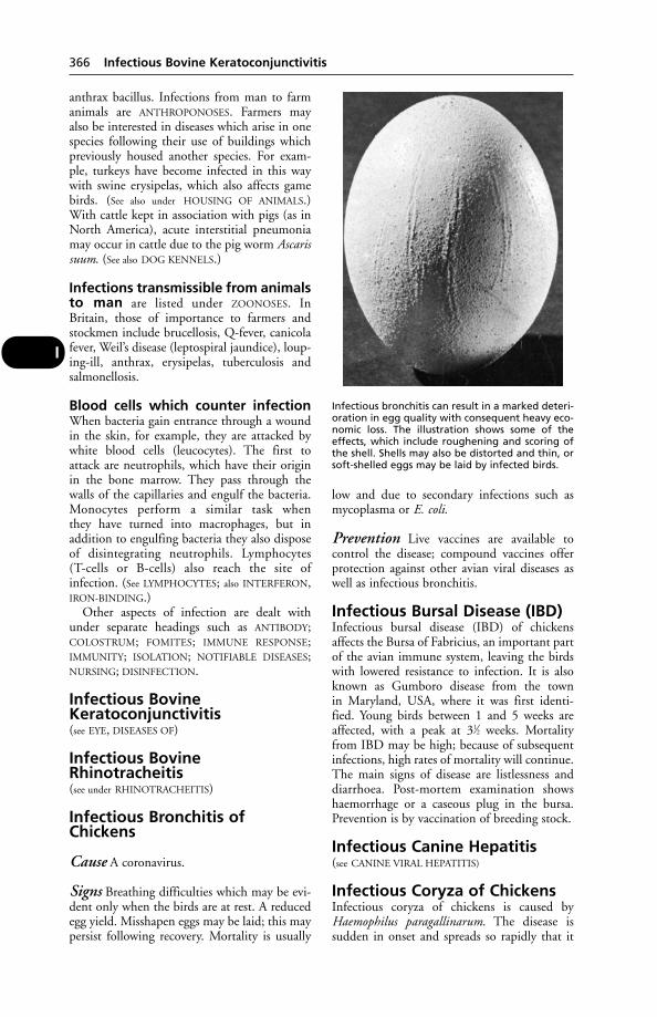
anthrax bacillus. Infections from man to farmanimals are ANTHROPONOSES. Farmers mayalso be interested in diseases which arise in onespecies following their use of buildings whichpreviously housed another species. For exam-ple, turkeys have become infected in this waywith swine erysipelas, which also affects gamebirds. (See also under HOUSING OF ANIMALS.)With cattle kept in association with pigs (as inNorth America), acute interstitial pneumoniamay occur in cattle due to the pig worm Ascarissuum. (See also DOG KENNELS.)
Infections transmissible from animalsto man are listed under ZOONOSES. InBritain, those of importance to farmers andstockmen include brucellosis, Q-fever, canicolafever, Weil’s disease (leptospiral jaundice), loup-ing-ill, anthrax, erysipelas, tuberculosis andsalmonellosis.
Blood cells which counter infectionWhen bacteria gain entrance through a woundin the skin, for example, they are attacked bywhite blood cells (leucocytes). The first toattack are neutrophils, which have their originin the bone marrow. They pass through thewalls of the capillaries and engulf the bacteria.Monocytes perform a similar task whenthey have turned into macrophages, but inaddition to engulfing bacteria they also disposeof disintegrating neutrophils. Lymphocytes(T-cells or B-cells) also reach the site ofinfection. (See LYMPHOCYTES; also INTERFERON,IRON-BINDING.)
Other aspects of infection are dealt withunder separate headings such as ANTIBODY;COLOSTRUM; FOMITES; IMMUNE RESPONSE;IMMUNITY; ISOLATION; NOTIFIABLE DISEASES;NURSING; DISINFECTION.
Infectious BovineKeratoconjunctivitis(see EYE, DISEASES OF)
Infectious BovineRhinotracheitis(see under RHINOTRACHEITIS)
Infectious Bronchitis ofChickens
Cause A coronavirus.
Signs Breathing difficulties which may be evi-dent only when the birds are at rest. A reducedegg yield. Misshapen eggs may be laid; this maypersist following recovery. Mortality is usually
low and due to secondary infections such asmycoplasma or E. coli.
Prevention Live vaccines are available tocontrol the disease; compound vaccines offerprotection against other avian viral diseases aswell as infectious bronchitis.
Infectious Bursal Disease (IBD)Infectious bursal disease (IBD) of chickensaffects the Bursa of Fabricius, an important partof the avian immune system, leaving the birdswith lowered resistance to infection. It is alsoknown as Gumboro disease from the townin Maryland, USA, where it was first identi-fied. Young birds between 1 and 5 weeks areaffected, with a peak at 31⁄2 weeks. Mortalityfrom IBD may be high; because of subsequentinfections, high rates of mortality will continue.The main signs of disease are listlessness anddiarrhoea. Post-mortem examination showshaemorrhage or a caseous plug in the bursa.Prevention is by vaccination of breeding stock.
Infectious Canine Hepatitis(see CANINE VIRAL HEPATITIS)
Infectious Coryza of ChickensInfectious coryza of chickens is caused byHaemophilus paragallinarum. The disease issudden in onset and spreads so rapidly that it
366 Infectious Bovine Keratoconjunctivitis
I
Infectious bronchitis can result in a marked deteri-oration in egg quality with consequent heavy eco-nomic loss. The illustration shows some of theeffects, which include roughening and scoring ofthe shell. Shells may also be distorted and thin, orsoft-shelled eggs may be laid by infected birds.
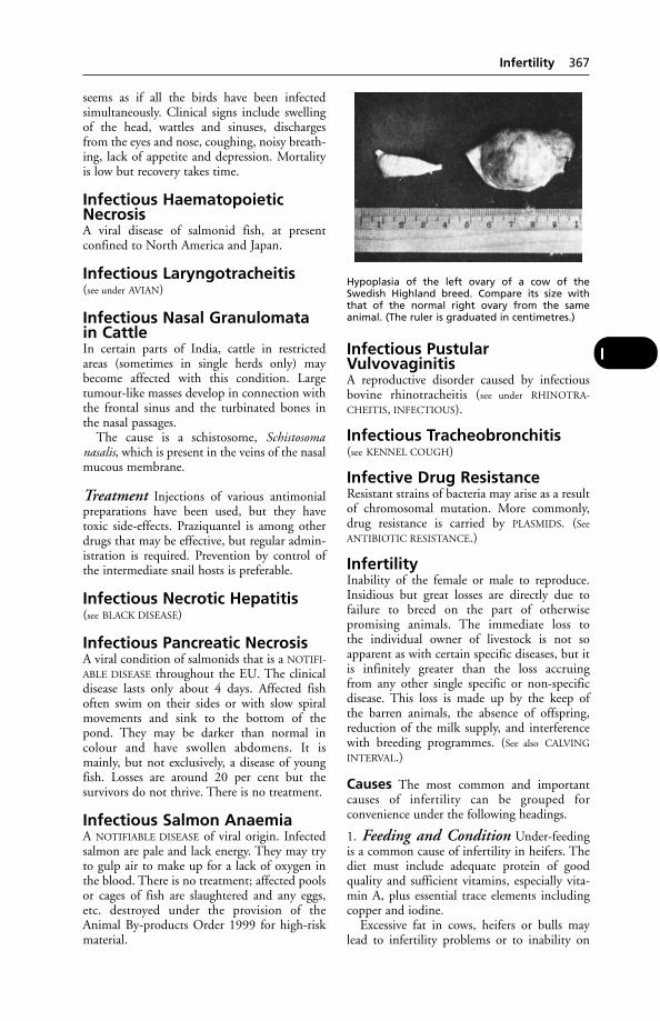
seems as if all the birds have been infectedsimultaneously. Clinical signs include swellingof the head, wattles and sinuses, dischargesfrom the eyes and nose, coughing, noisy breath-ing, lack of appetite and depression. Mortalityis low but recovery takes time.
Infectious HaematopoieticNecrosisA viral disease of salmonid fish, at presentconfined to North America and Japan.
Infectious Laryngotracheitis(see under AVIAN)
Infectious Nasal Granulomatain CattleIn certain parts of India, cattle in restrictedareas (sometimes in single herds only) maybecome affected with this condition. Largetumour-like masses develop in connection withthe frontal sinus and the turbinated bones inthe nasal passages.
The cause is a schistosome, Schistosomanasalis, which is present in the veins of the nasalmucous membrane.
Treatment Injections of various antimonialpreparations have been used, but they havetoxic side-effects. Praziquantel is among otherdrugs that may be effective, but regular admin-istration is required. Prevention by control ofthe intermediate snail hosts is preferable.
Infectious Necrotic Hepatitis(see BLACK DISEASE)
Infectious Pancreatic NecrosisA viral condition of salmonids that is a NOTIFI-
ABLE DISEASE throughout the EU. The clinicaldisease lasts only about 4 days. Affected fishoften swim on their sides or with slow spiralmovements and sink to the bottom of thepond. They may be darker than normal incolour and have swollen abdomens. It ismainly, but not exclusively, a disease of youngfish. Losses are around 20 per cent but thesurvivors do not thrive. There is no treatment.
Infectious Salmon AnaemiaA NOTIFIABLE DISEASE of viral origin. Infectedsalmon are pale and lack energy. They may tryto gulp air to make up for a lack of oxygen inthe blood. There is no treatment; affected poolsor cages of fish are slaughtered and any eggs,etc. destroyed under the provision of theAnimal By-products Order 1999 for high-riskmaterial.
Infectious PustularVulvovaginitisA reproductive disorder caused by infectiousbovine rhinotracheitis (see under RHINOTRA-
CHEITIS, INFECTIOUS).
Infectious Tracheobronchitis(see KENNEL COUGH)
Infective Drug ResistanceResistant strains of bacteria may arise as a resultof chromosomal mutation. More commonly,drug resistance is carried by PLASMIDS. (See
ANTIBIOTIC RESISTANCE.)
InfertilityInability of the female or male to reproduce.Insidious but great losses are directly due tofailure to breed on the part of otherwisepromising animals. The immediate loss tothe individual owner of livestock is not soapparent as with certain specific diseases, but itis infinitely greater than the loss accruingfrom any other single specific or non-specificdisease. This loss is made up by the keep ofthe barren animals, the absence of offspring,reduction of the milk supply, and interferencewith breeding programmes. (See also CALVING
INTERVAL.)
Causes The most common and importantcauses of infertility can be grouped forconvenience under the following headings.
1. Feeding and Condition Under-feedingis a common cause of infertility in heifers. Thediet must include adequate protein of goodquality and sufficient vitamins, especially vita-min A, plus essential trace elements includingcopper and iodine.
Excessive fat in cows, heifers or bulls maylead to infertility problems or to inability on
Infertility 367
I
Hypoplasia of the left ovary of a cow of theSwedish Highland breed. Compare its size withthat of the normal right ovary from the sameanimal. (The ruler is graduated in centimetres.)
the part of the male to accomplish coitus. (See
also FATTY LIVER SYNDROME.)In cows, temporary infertility may appar-
ently be closely associated with the feeding atabout the time of service. Cows losing weightare likely to be affected, especially if fed onpoor-quality hay or silage. With ad lib feedingsystems, heifers and more timid cows maynot be receiving enough roughage. Kale issometimes responsible.
In ewes, infertility and fetal death are alwaysserious in many hill areas, the result – to quoteDr John Stamp – ‘of keeping pregnant sheepunder conditions of near-starvation duringthe winter months when weather conditionsare atrocious’. (See DIET; FLUSHING OF EWES;STILLBORN PIGS; REPRODUCTION; VITAMINS;KALE; SELENIUM.)
2. Environment and Management Asudden change of environment, close confine-ment in dark quarters (formerly thelot of many a bull), and lack of exercise may allpredispose to, or produce, infertility. Abnormalsegregation of the sexes and the use of vasec-tomised males (for purposes of detectingoestrus) are other factors. A low level of nutri-tion may cause a quiescent or dormant state on the part of the ovaries. At the same timethere are seasonal cycles of sexual activity, and a ‘failure to breed’ during the winter monthsmay be natural enough, even if the farmerregards it as infertility. This ‘winter infertility’,as it is often called, may be influenced by tem-perature, length of daylight, lack of pastureoestrogens, underfeeding, etc. At this season,heifers often have inactive ovaries, while incows irregular and ‘silent’ heats give low con-ception rates.
Infertility may result from the oestrogeniceffects of red clover in the UK, as well as fromsubterranean clovers in Australia.
In outdoor pig herds, ‘summer infertility’ iscommon, partly due to seasonal loss of fertilityin the boars.
The most frequent reason for poor fertility ispoor management. In cows, poor oestrusdetection or timing, or bad technique ifartificial insemination is used, are common.Similar problems occur in pigs. In cattle and,particularly, pigs, when natural service isused, all matings must be seen and at least 2 undertaken within the heat period. In sheep,fertility problems often follow when too fewrams are used, or those which are too youngor unproven.
3. Diseases of the Genital Organs in theFemale While there is a very long list of such
diseases, their overall importance in causinginfertility is much less than that of managementproblems such as poor oestrus detection.
Inflammation or other disease of the ovaries:ovaritis; the non-maturation of Graafianfollicles, from any cause, and the presence inthe ovary of cysts (which often form froma corpus luteum), are causes of infertility;another is blocked Fallopian tubes.
Persistent corpora lutea: as a true clinicalcondition, these are not very common. Wherethey do exist, the animals may have a uterineinfection. The persistence of the corpus luteumprevents the ripening of the Graafian follicle,so the animal does not display oestrus and isnot mated. (See under OVARIES, DISEASES OF;HORMONES; HORMONE THERAPY.)
Inflammation of the uterine mucous mem-brane: a large number of cases of infertilitycan be ascribed to infection of the uterus(metritis) or the oviduct by organisms. (For alist of the infections which cause infertility,see under ABORTION. For infections causinginfertility in the mare, see under EQUINE
GENITAL INFECTIONS.)When the condition is mild, following a pre-
vious calving, it may disappear spontaneously,but in many instances it persists and becomeschronic. Associated with inflammation of themucous membrane of the uterus or oviduct isoften a persistent corpus luteum in the ovary.Carelessness during parturition, the use ofunclean instruments or appliances, decomposi-tion of retained membranes, and other similarfactors, also bring about infection of the uterus.Brucellosis though not necessarily itself a causeof sterility, by lowering the vital resistance of theuterus, favours infection by a multitude of otherorganisms which normally may be non-patho-genic. The details of uterine infection, includ-ing salpingitis (inflammation of the oviduct), inthe causation of sterility, are highly technical,but, generally speaking, it may be said that thepresence of organisms in the uterus, or the pres-ence of the products of their activity, either killsthe spermatozoa, or renders the locality unsuit-able for anchorage of thefertilised ovum (orova), with the result thatit perishes.
Abnormalities of the cervix may preventconception – mechanically when the lumen isoccluded or plugged by mucus of a thick tena-cious nature; and pathologically when there isacute inflammation of the mucous membraneof the cervix, or even of the whole uterus.Scirrhous cervix – where much fibrous tissue islaid down in the cervix – when very advancedmay cause sterility, but by itself is not usuallyof great importance. It is much more serious
368 Infertility
I
as a hindrance to parturition (see, for example,‘RINGWOMB’ of the ewe).
Cysts and fibrous bands in the os are seldomsufficiently extensive to occlude the passagethrough the cervical canal. Occlusion may,however, occur as the result of swelling andcongestion of the mucous membrane, due toinfection and inflammation. In such cases thesperms are unable to penetrate into the uterus,and fertilisation does not occur. This may alsobe the result of acidity (and thickened mucus)following a mild infection, and sometimessyringing the vagina a short time before servicewith a weak alkaline solution (e.g. 5 per centpotassium bicarbonate ) proves successful. (See
‘WHITES’; ‘EPIVAG’.)Tumours – either malignant or benign.Specific disease, such as tuberculosis in cattle,
or in mares. Contagious equine uretritis. (See
VULVOVAGINITIS.)
4. Hereditary Abnormalities in theFemale (see FREEMARTIN).
Hypoplasia of the ovaries of cows may occuras an inherited condition in the female. It mayinvolve 1 or both ovaries, causing either infer-tility or complete sterility. The uterus, also, maybe hypoplastic. (See also under GENETICS.)
Endocrine failure: heredity may be involved.Hermaphroditism.‘White heifer disease’ (see under this heading).It has been estimated that up to 10 per cent
of female pigs are sterile. Group studies haveshown that 25 to 50 per cent of infertile giltshad abnormalities of the genital tract sufficientto cause sterility, and two-thirds of these wereregarded as hereditary.
5. Disease of the Genital Organs in theMale Orchitis, or inflammation of the testicle,and epididymitis, inflammation of the epi-didymis, due to injury from kicks, orto infection from external wounds, or fromspecific infection, such as brucellosis or tri-chomoniasis in the bull. (See TESTICLE, DISEASES
OF; VENEREAL DISEASES.)Tumours of the testicles may destroy the
tubules or prevent spermatogenesis, and onthe penis, or in connection with the prepuce,may act as purely mechanical agents, whichprevent coitus by the male.
Adhesions between penis and prepuce, theresult of acute or chronic balanitis, though rare,may cause mechanical inability to protrude thepenis and fertilise the female. (See also under PENIS.)
Inflammation in the secondary sexualglands – i.e. in prostate, seminal vesicles, orother glands – may occlude the vasa deferentiaor ejaculatory ducts, and cause inability to
pass semen, while in other cases the semen maybe so altered as to cause death of the sperms inthe female passages.
Affection of the prepuce, such as balanitis,and injuries accompanied by laceration orsevere bruising, may cause temporary sterility,but when recovery occurs fertility returns. (See
also under PENIS.)
6. Hereditary Abnormalities in theMale Cryptorchidism, in which 1 or bothtestes do not descend into the scrotum, is a well-known cause of infertility in the male. When 1testis properly descends, and is fully developed,conception may follow service, and a sire suffer-ing from this disability has upon some occa-sions been regularly used in a flock or herd; butwhen the rig animal has both organs retained,although sexual desire may be emphatic, serviceis usually unsuccessful. The condition unfits a male animal for use as a breeding sire, since there is evidence that it is a hereditaryunsoundness. (See HORMONE THERAPY.)
Hypoplasia or under-development of themale sex organs, particularly of the testis, is animportant cause of sterility. It may involveboth testicles or only 1.
Endocrine failure may arise as a result of aninherited predisposition. In bulls this mayoccur in later life, rendering them sterile afterthey have produced a number of progenywhich, in their turn, may perpetuate this formof infertility.
Hermaphrodism, or hermaphroditism, inwhich an animal possesses both male andfemale organs, but is without a full complementof either, is usually, but not always, associatedwith sterility. (See also GENETICS; INTERSEX.)
7. Physical or Psychical Inability orDisturbance Under this heading are groupeda number of conditions which are difficult toclassify elsewhere. Some occur in the male,some in the female, and some are common toboth sexes.
Incompatibility between the blood of sireand dam may be responsible for some casesof abortion in cattle, etc. (See HAEMOLYTIC
DISEASE.)Old age: when an animal reaches a certain
age, reproduction becomes impossible. Theperiods of oestrus cease. Breeding ceases earlierin the female than in the male.
Discrepancies in size between male andfemale may result in failure to breed. The penismay be too short or too large; the vagina may betoo long or too small; the female may not havethe strength to carry a heavy male; or the malemay not be tall enough to reach the female.
Infertility 369
I
Injuries to the back, hips, hind legs, or feetof the male, and sometimes to the same regionsof the female, may be severe enough to preventsuccessful coitus. Progressive spinal arthritis is acommon condition in bulls. (See also BREEDING
OF ANIMALS; REPRODUCTION; EMBRYOLOGY;UTERUS, DISEASES OF; HORMONE THERAPY;GENETICS; VENEREAL DISEASES; ANOESTRUS;ABORTION; MUMMIFICATION.)
InflammationInflammation may be briefly defined as thereaction of the tissues to any injury short of onesufficiently severe to cause death. There are 4cardinal symptoms of inflammation – heat,pain, redness, and swelling – to which may beadded interference with function. (See ABSCESS;WOUNDS; ALLERGY.)
For the inflammations of special organs, see
under PNEUMONIA; PLEURISY; PERITONITIS;MAMMARY GLAND.
For anti-inflammatory drugs, see CORTI-
COSTEROIDS; CORTICOTROPHIN; CORTISONE;NSAIDs; IBUPROFEN; FLURBIPROFEN; ANTIHISTA-
MINES; CALAMINE.
InfluenzaScientifically, this term is now applied only todiseases caused by a myxovirus.
The World Health Organisation (WHO)was much exercised as to what happens to thevirus of human influenza between epidemics.It has long been known that there is a relation-ship between this disease and swine influenza.The human influenza virus (type A) wasisolated from the parasitic pig lung-worm.Larvae of these lung-worms are harboured byearthworms – the only known intermediatehosts – which live for as long as 10 years.
Between epidemics, the virus is not found inthe tissues of the pigs. However, earthwormstaken from infected pig farms seem to carryinapparent viruses, and these can develop, inpigs eating the worms, into normal virusescapable of being isolated from the respiratorysystem. The question, therefore, arises whetherthe pigs are, in fact, the virus reservoirs, ratherthan being secondarily infected by the humanvirus. (See also SWINE INFLUENZA.)
There is evidence that influenza viruses ofmammals and birds play an important partin the emergence of new viruses whichcause out-breaks of illness in man in severalcontinents.
The recovery from pigs in Taiwan in 1970 ofinfluenza virus indistinguishable from that caus-ing type A Hong Kong influenza epidemics inman in 1968 provided the first direct evidence
of the inter-species transfer of influenza viruses.Pigs experimentally inoculated with that virustransmitted it to pen-mates. Moreover, theTaiwan virus taken from pigs readily infectedhuman volunteers, who developed antibodieseffective against virus from both pigs and people.
It is now suggested that the Hong Konghuman influenza virus did not arise by muta-tion from a pre-existing human strain, but thatit probably arose from the mixed infection in amammal or bird with an animal influenza virusand a human type A Asian strain. The animalvirus may have provided certain subunits orcomponents; the other subunits having comefrom a human strain.
For influenza in the horse, see under EQUINE
INFLUENZA.Pneumonia in calves may be caused by a
virus of influenza-type.In the dog, parainfluenza virus SV5 has been
isolated in the USA and the UK from dogswith upper respiratory disease. (See ‘KENNEL
COUGH’.)Avian strains of type A influenza virus cause
a number of diseases in hens, ducks, turkeys,etc. During 1980 and 1981, 9 subtypes ofinfluenza A virus were isolated from birds inBritain, usually as a result of investigations ofdisease or death. However, these viruses wereshown to be of low virulence for chickens.Highly pathogenic avian influenza virus hascaused serious outbreaks among turkeys.
Avian influenza is a NOTIFIABLE DISEASE
throughout the EU. Waterfowl are the mainreservoir host for both avian and mammalianstrains but they are not themselves muchaffected by the disease. The slaughter of allchickens in Hong Kong in 1997 was justifiedas the strain of virus present transmitted fairlyeasily to the human population. (See AVIAN
INFLUENZA.)
‘Influenza’, Cat(see FELINE INFLUENZA)
Infra-Red LampsInfra-red lamps are used as a source of heat inthe creeps of piggeries and in poultry brooders;also for sickly lambs and calves. Either ‘bright’or ‘dull’ emitters are available, the latter beingpreferred for chick-rearing. They have manyadvantages, but a power-cut can cause severelosses. (See also under TOES, TWISTED.)
Inguinal CanalInguinal canal is the passage from the abdomi-nal cavity to the outside, down which pass the
370 Inflammation
I
spermatic cords and their associated structuresin the male, and in the female, the roundligament of the uterus. It is a slit-like opening,about 12.5 cm (5 inches) long in the horse, andis directed downwards, inwards, and forwards.It is bounded behind by a strong band calledthe inguinal or Poupart’s ligament. The canal isimportant, because if it is dilated from anycause, some part of the small intestines maypass through it, resulting in inguinal hernia. (See
HERNIAS.) It serves as the opening throughwhich retained testicles are removed in the ‘rig’or cryptorchid animal.
Inguinal RegionInguinal region is the region of the inguinalcanal, i.e. that part of the posterior and upper-most division of the abdominal wall which liesbelow the brim of the pelvis. The scrotum,penis, and their vessels, etc., are situated in theinguinal region in the male horse, and in thefemale the mammary glands with the vesselsthat supply them. In some animals such as thedog and boar, the scrotum is farther back, i.e. inthe perineal region, while in the bull and ramthe penis is farther forwards.
Inherited Defects/DiseasesInherited defects/diseases are referred to underGENETICS. (See also DEFORMITIES and HORSES.)
InjectionsParenteral administration of medicines maybe hypodermic or subcutaneous (sc), intra-articular (into a joint), intradermal, intramus-cular (im), intravenous (iv – into a vein)intraperitoneal (into the abdominal cavity),epidural, or subconjunctival (beneath theeyelid). Precautions must be taken againstthe introduction of bacteria, dirt, etc. Thehair should be clipped away at the site of injec-tion, and the skin cleaned with spirit or an anti-septic. Needles and syringes should be sterilisedbefore use, unless of the disposal type intendedfor once-only use and already sterilised andin a sealed wrapper.
Where the material to be injected is alreadyfluid, this is generally guaranteed sterile bythe manufacturers, and is put up in sealedvials. In cases where the drug has to be dissolvedin water first, sterile water must be used.Manufacturers usually supply ampoules ofsterile water with drugs that have to bedissolved; their instructions for use must befollowed carefully. Neglect of these precautionsis likely to be followed by the formation ofan abscess at the point of injection or even bysepticaemia.
For subcutaneous injections, a fold of theskin is picked up between the thumb andforefinger of the left hand, and the needle isinserted into the middle of this fold. Thenozzle of the syringe, preloaded with the injec-tion fluid, is slipped into the head of the needleand the piston is slowly but firmly pressedhome so as to expel the contents into the loosetissues under the skin. Care should be takenthat all air-bubbles are excluded from the barrelof the syringe, as it is unwise to introduce them.
A number of proprietary multi-injectiondevices are manufactured for herd inoculations.
Precautions Restless animals should alwaysbe secured so that they will not make a suddenplunge when the needle is introduced, andbreak the stem of the needle. Abscesses in hamsare common in pigs, and doubtless result fromanti-anaemia intramuscular injections madewithout due precautions as to cleanliness andto broken-off needles.
The sciatic nerve may be damaged as theresult of an intramuscular injection into a pig’sham, with paralysis of the limb possibly follow-ing. This site should be avoided, and it has beenrecommended that the injection be given intothe muscles of the neck, just behind the ear,and not into fatty tissue.
Care must be taken not to make what shouldbe a subcutaneous injection into the chest. Thisdanger was illustrated when a farmer injected500 lambs, using a multidose syringe intendedfor cattle, and with a 6 mm needle. Within aweek 17 of the lambs had died; autopsy showedpyothorax and a pure growth of Actinomycespyogenes. It has been demonstrated that it is pos-sible to reach the pleural cavity with a cattlesyringe, especially in thin lambs; many other‘vaccine failures’ may have been due to inadver-tent injections into the chest. Alternative sites,such as the side of the neck, would appear tooffer a much reduced chance of complications.
With intravenous injections of certain prepa-rations, severe tissue damage may follow if someof the drug enters the vein wall or surroundingtissue; the manufacturers’ instructions mustbe followed.
Inoculations should not be carried out in adusty shed.
(See ANTHRAX; also AMPOULE; DETERGENT
RESIDUE; ENEMA.)
Large-bore needles Use of these is notwithout risk, especially when liver biopsies arecarried out by means of suction through aneedle, inserted intraperitoneally and attachedto a syringe.
Injections 371
I
Accidental self-inoculation may occurowing to the sudden violent movement of alarge animal. People have been infected withBRUCELLOSIS in this way; and veterinarysurgeons have died from IMMOBILON.Accidental self-injection with oil-basedvaccines requires prompt hospital attention.
Injuries(see ACCIDENTS; WOUNDS; FRACTURES; BLEED-
ING; SHOCK)
Injuries from ShoeingThese are not always the fault of the farrier.There are some horses with such bad feet that itmay be quite impossible to shoe them withoutrunning the risk of injuring the sensitive struc-tures in the process. The nails, toe-clips, or eventhe shoe, may inflict damage.
1. The Nails either may produce lameness byactually penetrating the sensitive laminae – acondition called ‘pricking’; or, by being driventoo close to the laminae, by pressing upon thesensitive structures – a condition known as‘binding’.
Pricking may be only slight when the farrierknows that the nail has stabbed the quickand immediately withdraws it. All nail injuriesshould receive prompt attention, for theyare usually amenable to treatment in theearly stages; but if neglected they rapidly sup-purate, causing the horse great pain and oftenpermanent damage.
Binding is not so serious. Generally it sufficesto remove the shoe, to allow the horse to remainbarefooted for a day or so, and then to replacethe shoe, taking special care that the nails arenot driven too coarsely upon the 2nd occasion.The lameness in this case very often onlyappears 2 or 3 days after the horse has beenshod, and is attributed to some other cause.
2. The Clip may produce lameness by beingdriven too coarsely, and either burning the sen-sitive structures when being fitted, or pressingupon them unduly when hammered intoposition afterwards. When side-clips are used,i.e. 1 on each side of the foot, if they are forcedhome too far the foot is jammed in between2 rigid structures which will not allow it toexpand and contract with each movement ofthe foot, and lameness results. The shoe shouldbe removed, the horse given a day or two’srest, the clips altered, and the shoe reapplied,when he will usually go sound. If burning issuspected, the same procedure may be adopted.If the shoe loosens until it is only holding byone nail, or if the shoe is partially torn off, the
horse may tread on the clip which penetratesthe sole of the foot and inflict a very severewound. This is treated as for pricking, the areabeing pared out.
3. The Shoe may cause injury if it has anuneven surface and presses upon a part toomuch. This is particularly liable to happenwhen the horn of the foot is weak and thin.Horses with flat feet, or those with droppedsole, may develop bruises of the sole if theweb of the shoe presses upon the outer circum-ference of the sole, where it joins the white line.In such cases the shoe should be removed andthe unevenness corrected, or the bearing surfaceof the foot should be eased. Some horses mayrequire to be shod with a bar shoe, so that the frog may take some of the weight off theaffected part, and others need a run at grass.
Burning of the sensitive parts of the foot mayoccur through the carelessness of the farrier, notby making the shoe too hot, but by holdingit in position on the foot for too long a time,so that it may ‘bed itself in’. This is a most rep-rehensible practice and should not be tolerated.The injury usually results in a separation of thehorn from the sensitive tissues below, and someweeks pass before the horse can resume hiswork again. (See CORNS; BRUISED SOLE.)
InnominateInnominate is the bone of the pelvis and thestructures associated with it. The pelvis iscomposed of 6 separate bones, 3 on either side:ilium, pubis, and ischium.
Inoculation(1) Introduction into an animal or culturemedium of micro-organisms. (2) To induceimmunity by introducing a vaccine or serum.(See INJECTIONS; VACCINATION; INFECTION;IMMUNITY.)
InsecticidesA wide range of effective insecticides is availablefor both external application and systemicuse. Some, such as the organophosphoruscompounds, may be toxic if not used properly.They have been implicated in causing illnessin humans. General notes are given below;more detail will be found in the cross-referenceslisted.
Large animals Insecticides for the protec-tion of large animals are discussed under FLIES
and FLY CONTROL. They may be administeredas sprays, by immersion (dipping), as ‘pour-ons’or, in some cases, by mouth. IVERMECTIN isan example.
372 Injuries
I
Small animals Insecticides are available innumerous formulations for application as wetshampoos, aerosol sprays, dry dusting powders,‘spot-on’ formulations and ‘flea collars’. Activeingredients include permethrin, dichlorvos,fenthion and carbaril. There are manyothers. Dichlorvos and diazinon (both organo-phosphorus compounds) are used in ‘fleacollars’.
Manufacturers’ instructions should bestrictly followed, and only preparationsstated to be safe for cats should be used onthose animals.
Over-exposure of animals to insecticides,either through too frequent use or use of excessivequantities, can lead to poisoning. (See CHLORI-
NATED HYDROCARBONS; ORGANOPHOSPHORUS
POISONING; PERMETHRIN.)
Accidental poisoning DDT fell intodisrepute in the UK, the USA, Australia andNew Zealand, but is still used for ground spray-ing in parts of Africa (see DDT). The use ofunsuitable insecticides can lead to fatal poison-ing in cattle, etc. (See TEPP.) Poisoning mayoccur following absorption of an unsuitableinsecticide spray through the skin. This, orinhalation of spray droplets, may lead to dan-gerously contaminated milk. The followinginsecticides are not recommended for dairyand cowshed use on account of this risk:DDT, aldrin, dieldrin, chlordane, lindane,methoxychlor, toxaphene, and heptachlor.(See CHLORINATED HYDROCARBONS.)
Some insecticides may be safe for one speciesof animal but fatal to another. For example, ona farm in New York State, an insecticide spraycontaining thiophosphate had no effect on 50chickens, but killed more than 7000 ducklings.
Dieldrin, used as a seed dressing, has causedfatal poisoning in wood-pigeons and otherwild birds. Lambs have been killed by ALDRIN.(See BHC; DDT; DIELDRIN; DERRIS; TEPP;PARATHION; PYRETHROIDS; FLY CONTROL;CARBAMATES; CHLORINATED HYDROCARBONS;GAME-BIRDS.)
Insecticide resistance Extensive use of aninsecticide can encourage resistance amongthe target species. Only a few years after itsintroduction in the 1940s, house flies showedresistance to DDT, and by 1970 some 250species of fly affecting man, his animals orcrops, had developed resistance to one ormore of the organochlorines, e.g. dieldrin;organophosphates; or carbamates.
Most cases of resistance apparently dependon a single gene, and are developed mostly
following large-scale use of insecticides in controlprogrammes. (See also under FLY CONTROL.)
InsectsFor a general description of these, see FLIES.
InseminationThe introduction of semen into the vagina orcervix. (See ARTIFICIAL INSEMINATION.)
Insulation of Buildings, Floors(see under HOUSING OF ANIMALS)
InsulinInsulin is a hormone secreted by part of thepancreas, where it is produced by the isletsof Langerhans. It is used in the treatment ofdiabetes in dogs and cats. (See DIABETES.)
InsulinomaA tumour affecting cells of the islets ofLangerhans in the pancreas, which may lead tocollapse, convulsions, coma and death in thedog as a result of hypoglycaemia.
InsuranceIn the UK there is now a wide choice ofcomprehensive insurance policies available toanimal-owners. Farmers can insure against therisks of foot-and-mouth disease or brucellosis,for example. There is insurance for horses, anddog- and cat-owners can avail themselves of policies covering veterinary fees, third-party liability, theft or death of an animal from illnessor accident. Policies can be issued throughveterinary surgeons. With the possibility ofhaving to pay for a major operation or prolongedtreatment, such policies can minimise the owner’sfinancial outlay, and are a safeguard againstunexpected and sometimes large expenses.
Intensive Livestock ProductionThis means, generally speaking, having farmanimals indoors to a greater extent, and alsohaving them within a smaller space insidea building. All intensive systems requireskilled management and veterinary input toprevent the problems intrinsic among largeconcentrations of livestock.
The economic advantages claimed for inten-sive livestock production are the economies ofscale through reducing costs of labour andequipment per animal housed; lower feed coststhrough bulk buying and home mixing; theability to afford skilled management andlabour; also a saving in acres of valuable land.
The disadvantages are the effect on stock oflarge concentration, disease, cannibalism and
Intensive Livestock Production 373
I
all the problems of stress, and intensive feedingmethods.
The following describes potential hazardsand health problems, and should not beregarded as condemnation of all currentfarming methods.
Poultry De-beaking, to prevent feather pick-ing and cannibalism, if badly done can causeinjury and reduce resistance to infection. Birdsde-beaked and unable to take a dust bath areprone to severe infestation with lice and mites,which may be resistant to the commonly usedparasiticides; and infestation can be a problemin battery houses. Lack of exercise is conduciveto fatty degeneration of the liver in batterybirds. Among birds crowded together on deeplitter, coccidiosis and worm infestations areapt to be serious. Faulty ventilation often givesrise to a harmful concentration of ammoniain houses where there is litter, and also predis-poses to infectious bronchitis and other respira-tory infections. The greater the concentrationof birds, the greater the stress, it seems; andthe more chance of an increasing proportionboth of susceptible birds and of ‘carriers’ ofvarious infections.
Beef cattle In calf-rearing units, salmonellainfections cause a high proportion of thedeaths of bought-in calves. Bronchitis is alsoan important cause of losses, which oftenamount to 7 per cent.
In units taking in 12-week-old calves, respi-ratory disease, principally viral infections, isimportant. Other conditions encounteredinclude foul-in-the-foot, infectious bovinekeratitis, and bloat.
If trough space is too limited, inflammationof the eyes may be caused by cattle flicking theirears into their neighbours’ eyes – simulating theeffects of infectious bovine kerato-conjunctivitis.
Among veal calves, pneumonia, bronchitisand a peracute coliform septicaemia are majorcauses of losses. Anaemia, parasites, and aform of anaphylactic shock are also among thehazards of rearing.
Pigs These animals are particularly proneto the effects of STRESS, and of confinementin poorly ventilated buildings which favoursrespiratory infections such as enzooticpneumonia.
The use of farrow-to-finish pens whichaccommodate pigs from birth to slaughter dayhas been advocated. These obviate 4 or 5 movesto strange surroundings with its accompanyingstress.
Sheep Respiratory troubles, including variousforms of pneumonia, are a danger in buildingswhere ventilation is poor. There are somevery successful flock houses, with one end vir-tually open, where disease problems have beenminimal – foot-rot being controlled by regularuse of a foot-bath. In such buildings, the eweslamb indoors, to the great advantage of theshepherd. Straw is used for bedding. Yorkshireboarding assists ventilation.
Lameness Intensive systems of farming tendto ignore the social behaviour of animals to thedetriment of their health. Two examples involv-ing lameness in cattle may be given. In the firstcase 12 heifers accustomed to being in a smallsocial group outside in a straw yard wereabruptly transferred at calving and put in withcows in modern concrete-based cow cubicles.Five became acutely lame with septic andaseptic laminitis and solar ulceration. In 2, thecondition was so severe that they had to beslaughtered, but the other heifers improvedwhen they were transferred to straw yards.
The outbreak was attributed to the suddenintroduction to concrete surfaces and uncom-fortable cubicles which reduced the time thatthe animals lay down. Increased activity causedby behavioural interactions with the establishedcows was probably also a factor.
The 2nd case involved an outbreak of solarulcerations in 90 per cent of a small herd ofdairy cows. It coincided with the occupation ofa new cubicle house with concrete-based liplesscubicles. When given an opportunity the cows‘voted with their feet’ and returned to their oldearth-floored cubicles.
(See also HOUSING OF ANIMALS.)
IntercostalBetween the ribs.
IntercurrentIntercurrent is a term applied to a disease whichoccurs during the course of another diseasealready present, and modifies its course orincreases its severity.
InterdigitalBetween 2 toes or digits.
Interdigital Cyst (InterdigitalAbscess)Interdigital cyst (interdigital abscess) is acondition commonly affecting the feet of dogs,in which abscesses about the size of a pea orlarger appear in the spaces between the digitsof the paws. It most often affects spaniels,
374 Intercostal
I
Airedales, Scots terriers, Sealyhams, and DandieDinmonts.
Causes are generally held to be an infection ofthe hair follicles between the toes, or to gritpenetrating the skin there. In some instancesthe lesion may be a true cyst.
Signs The dog licks its foot, and upon exami-nation a swelling (which is painful) is noticedin the interdigital space. Within a couple ofdays or so, the swelling may discharge a littleblood-stained pus. If the lesions have beenrepeatedly forming, they may suddenly cease,and the dog remains free from them for perhapsmonths at a time. Unfortunately, recurrencesare likely at varying intervals.
Treatment The foot is bandaged to keep thewound clean, and dressed daily until there is nomore discharge and the wound has healed.Some encouraging results have been obtainedby the professional use of CRYOSURGERY.
Interdigital Necrobacillosis(see FOUL-IN-THE-FOOT)
InterferonA glycoprotein which inhibits the multiplicationof viruses within living cells.
Many types of cells can produce interferonas a means of defence against further viralinfection.
Recombinant DNA techniques have madepossible the production of interferons fortherapeutic use.
Interferon is being used in several countriesas an adjunct to post-exposure prophylaxis ofhuman rabies.
Internal HaemorrhageInternal haemorrhage may result from ruptureof some large blood vessel; or it may be theresult of an injury to some organ that is richlysupplied with blood, such as the liver orspleen. In either case the bleeding occursinto one of the body cavities and the bloodis lost to the tissues of the animal. (See also
HAEMANGIOSARCOMA; diseases with namesbeginning with the word HAEMORRHAGIC;WARFARIN.)
Signs of severe internal haemorrhage includeextreme pallor of mouth and mucous mem-brane lining the eyelids, coldness of the skin,rapid breathing, or a series of gasps, collapse,and a pulse becoming weak, slow, and thenimperceptible. (See SHOCK.)
Treatment of severe internal haemorrhagecan seldom be undertaken in time to save life.When the internal bleeding is less profuse,success may be achieved with ADRENALINE,BLOOD TRANSFUSION or DEXTRAN, VITAMIN K.
IntersexAn individual with characteristics intermediatebetween those of a male and a female. Incattle, examples include the FREEMARTIN; XYgonadal dysgenesis (in which there are nogonads); and testicular feminisation. A case ofthe latter, described by Dr S. E. Long, was asingle-born cow showing signs of virilismand found to have abdominal testes, someundeveloped Mullerian duct derivatives, anormal vagina, and a 60XY genotype in alltissues examined.
In a canine example of intersex, the os peniswas absent, the penis could not be extrudedfrom the prepuce, and no testicles were presentin the scrotum. A laparotomy revealed a uterusand ovaries.
(See under TRISOMY for the case of anintersex Spanish-bred horse, ‘considered to bea mare’, which had the characteristics of apseudo-hermaphrodite male.)
InterstitialInterstitial is a term applied to cells of differenttissue set amongst the active tissue cells of anorgan. It is generally of a supporting characterand formed of fibrous tissue. The term is alsoapplied to diseases which specially affectthis tissue, as interstitial nephritis. (See under
KIDNEYS, DISEASES OF – Chronic nephritis.)
Intervertebral Disc Protrusion(see under SPINE)
Intestinal Adenomatosis(see under PORCINE)
Intestine, ObstructionThis may result from an impacted mass of foodmaterial. (See IMPACTION.) In the dog, forexample, a hard mass containing spicules ofbone may render defecation impossible, andan enema will be necessary if a dose ofmedicinal liquid paraffin does not achieve thedesired result. In the cat a mass of fur (fur ball)may similarly cause obstruction. FOREIGN
BODY of many kinds, including string, areanother cause.
Wood chewing A bad habit of some horses,whether stabled or at grass, which can lead toobstruction of the small intestine.
Intestine, Obstruction 375
I
Signs Colic, passage of stomach contentsdown both nostrils.
Treatment Enterotomy under a generalanaesthetic; and removal of the obstruction.
Duodenal obstruction in cattle is notcommon but can occur. In most cases theobstruction is due to decreased motility ofthe duodenum, caused by inflammation of theduodenal wall and preperforative peritonitis,resulting from duodenal ulcers or penetratingforeign bodies respectively.
Other causes A strangulated HERNIA is aserious cause of obstruction, compression ofblood vessels and nerves making matters worse.INTUSSUSCEPTION, in which a part of the intes-tine becomes turned in on itself (like the fingerof a rubber glove may do), is fraught withsimilar dangers. VOLVULUS, or the twisting of aloop of intestine, is another cause. All theseconditions may be followed by GANGRENE
and PERITONITIS. Prompt surgical treatment isnecessary to save life.
A growth affecting either the lumen of theintestine so blocking it, or the exterior and soconstricting it, is another possibility. (See
TUMOURS; CANCER.)
Signs Intestinal obstruction can be expectedto cause depression, loss of appetite, dehydra-tion, fever, some degree of toxaemia, vomiting,and pain.
TYMPANY (distension of the intestine withgas) may occur in some cases of intestinalobstruction. In brood mares, tympany of thelarge intestine may predispose to rupture of thecaecum (or other part) from pressure exerted bythe fetal hind feet at the onset of parturition.(See also COLIC for diseases in horses.)
Intestines
Horse (1) Small intestine. This measuresabout 20 metres, (70 feet), and is divided into afixed portion – duodenum and a more or lessfree portion – jejunum and ileum. Its diametervaries from 4 to 8 cm (11⁄2 to 3 inches) whenmoderately distended, and its capacity isabout 55 litres (12 gallons). (2) Large intestine.This extends from the end of the ileum tothe anus, and measures about 8 metres (25 feet)in length. Its diameter varies in differentparts from about 8 cm (3 inches) in the smallcolon to nearly 50 cm (20 inches) across thewidest part of the caecum. It is divided intocaecum, large colon, small colon, and rectum.
The caecum is a large blind sac lying on theright side of the abdomen and extending down-wards and forwards to within a hand’s-breadthfrom the sternum. It is shaped somewhat like areversed comma, having both its entrance andits exit near the base, and has a capacity ofabout 36 litres (8 gallons). Foodstuff entersit by the ileo-caecal valve, and leaves by thecaeco-colic valve, which opens into the largecolon.
Cows The intestines lie entirely to the right ofthe middle line of the abdomen. (1) Small intes-tine, measuring 40 metres (130 feet) in length,lies in the lower part of the right side of theabdomen, filling in the spaces left between morefixed organs. (2) Large intestine is much smallerthan in the horse, and not so complicated. Thecaecum lies in the upper posterior part of theabdomen, with its blind sac posteriorly in or near the pelvic inlet. The caecum is about 75 cm(21⁄2 feet) long and is followed by the colon (thereis no small colon in the ox), which has a length ofabout 11 metres (35 feet). The colon is arrangedlike the coils of a watch-spring, with each coildouble, consisting of one part running towardsthe centre (centripetal), and a corresponding partrunning from the centre (centrifugal).
Sheep The intestines of the sheep are similarto those of the cow.
Pigs (1) Small intestine. This varies from 15 to20 metres (50 to 65 feet) in length, and mainlylies on the left side and floor of the abdomen,with some coils pushed across on to the rightside of the body. (2) Large intestine is about4.5 metres (15 feet) long and considerablywider than the small bowel.
Dogs The intestines are short in this animal,only reaching a length of about 4.5 or 5 metres(15 or 16 feet), of which the small intestinemeasures 3.75 to 4.25 metres (12 to 14 feet).The small intestine occupies the right sideof the abdomen and part of the floor. Fromhere the colon has a short course upwardstowards the head, turns across to the left side ofthe body, and then runs backwards to end inthe rectum.
Structure In all animals the intestines, bothsmall and large, are constructed of 4 main coats.They all consist of an inner mucous membranelining, a submucous coat, a middle muscularcoat, and an outer peritoneal one.
Mucous membrane coat: this is the soft,moist, velvety lining which is found in all parts
376 Intestines
I
of the intestine. (See BRUSH BORDER; PEYER’S
PATCHES; VILLUS.)Muscular coat: there are 2 definite layers of
muscle fibres in the wall of the bowel. Theinnermost of these has its fibres all running in acircular manner round the submucous coat,and the outer layer has fibres running length-wise. In the large intestine some of these longi-tudinal fibres are collected into distinct bandscalled ‘taenia’, which, being somewhat shorterthan the other fibres, cause a certain amount ofpuckering of the bowels. The muscular arrange-ment of the intestines is very important, as itis responsible for all the movement of thebowels. In health it is continually contractingand expanding, shortening and lengthening,and moving the food either onwards or back-wards. During the process the food is squeezedand churned and most thoroughly mixed withthe digestive juices. The movement is called‘peristalsis’ when it tends to move the foodtowards the anus, and ‘antiperistalsis’ when it isin the opposite direction.
Peritoneal coat: this forms the outermost cov-ering of the bowel. It is continuous for thewhole length of the canal from the pylorus tothe anus, except for certain comparatively smallregions where, for example, the duodenum andthe caecum are bound directly to the roof of theabdomen or to other organs by fibrous tissue. Itis a tough membrane with a layer of smoothglistening cells on its outer surface which rubagainst similar cells on the surfaces of adjacentorgans and reduce friction to a minimum. (See
PERITONEUM.)
Attachments The intestines are hung or heldin position by folds of peritoneum which bindthem, directly or indirectly, to some part of theabdominal wall. The fold in which the free partof the small intestine hangs is called the ‘mesen-tery of the small intestine’, and it is through thisthat the blood and lymph vessels and the nervesenter and leave the bowel. It is composed of 2layers, in the middle of which pass the vessels.
Functions (see DIGESTION)
Intestines, Diseases ofIntestinal inflammation, or ENTERITIS, is acommon disease in all animals, and may takean acute or chronic form. In either case thechief symptom is diarrhoea. In acute enteritis,diarrhoea leads to DEHYDRATION; while in thechronic form, the animal ceases to thrive andthe abdomen becomes permanently ‘tuckedup’. The causes and treatment of enteritis aregiven under DIARRHOEA.
PERFORATION of part of the intestine mayfollow ulceration, itself a complication of somecases of enteritis. Perforation injuries receivedin battle used to be common in cavalry horses;they may follow stabbing injuries such as goringby bulls, farm and road accidents, or the swal-lowing of sharp-pointed objects. (See FOREIGN
BODY.) Perforation of the wall of the intestineis obviously a very serious condition and animmediate threat to the animal’s life, sincebacteria which accompany partly digestedfood escaping from the intestine will causePERITONITIS.
Necrosis and infarction may be detected byassessing the serum levels of CREATINE kinase.(See also VOLVULUS; INTUSSUSCEPTION; COLIC.)
IntracranialIntracranial is the term applied to structures,diseases, or operations associated with thecontents of the cranium.
IntradermalInto the thickness of the skin as in intradermalinjections.
IntramedullaryWithin the marrow cavity of long bones. Thus,intramedullary pins – used in the treatment offractures.
IntramammaryWithin, or into, the mammary gland (udder).
IntramuscularWithin a muscle, e.g. intramuscular injection.
Intraperitoneal InjectionsIntraperitoneal injections are those made directinto the abdominal cavity.
IntrathecalInto a sheath; intraspinal.
IntratrachealInto the ‘windpipe’. (See also ENDOTRACHEAL
ANAESTHESIA.)
Intravenous InjectionAn injection direct into a vein, a techniqueemployed in anaesthesia and where much fluidhas to be injected. (See also INJECTIONS.)
IntussusceptionIntussusception is a form of obstruction of thebowels in which a part of the intestine turnsin on itself like the finger of a rubber glove. Itusually follows increased gut motility and often
Intussusception 377
I

results from diarrhoea. It occurs mostly inhorses, puppies and kittens, causes obstructionof the intestine and great pain.
If the condition is not relieved it leads to stop-ping of the blood supply in that part of the bowelwhich is enclosed, and death. Signs include lossof appetite, uneasiness due to abdominal pain,straining, and blood in the faeces. In the doga sausage-like swelling may be palpated in theabdomen, or there may be protrusion fromthe anus of a turgid, cylindrical mass havingfour thicknesses of bowel wall. Treatmentinvolves manipulation under anaesthesia (afterlaparatomy in most cases), and sometimes thesurgical removal of the innermost portion ofthe bowel and an end-to-end anastomosis.
Caecal intussusception In 2 ponies, intus-susception led, respectively, to pain followed bysudden death; and to pain lasting 3 weeks fromthe time of a veterinary examination. The 1stcase was found at autopsy to have intussuscep-tion of the base of the caecum; the 2nd had theentire caecum invaginated into the colon. Inboth animals the lesions had been present for along time.
InvertebratesAnimals without backbones. Some of theseare kept as ‘pets’ (tarantula spiders, scorpions,millipedes, praying mantis, etc.). It should benoted that where they are kept as captiveanimals they fall under the Protection ofAnimals Act 1911 and action has been taken incases of neglect. In New Zealand, 2 classes ofinvertebrate (molluscs and crustaceans) are cov-ered by the Animal Welfare Act 1999. In theUK, the common octopus (Octopus vulgaris)is protected under the Animals (ScientificProcedures) Act 1986.
InvolutionA change back to its normal condition whichan organ undergoes after fulfilling its normalfunction, e.g. involution of the uterus followingpregnancy.
IodidesIodides are salts of iodine. Sodium iodide isused in the treatment of actinobacillosis, andformerly it was used with other drugs in thetreatment of oedema, and of ringworm. Takenin excess, iodides cause a condition known as‘iodism’ or iodine poisoning. The symptoms ofthis are diarrhoea, loss of appetite, emaciation,total refusal of water, a dry, scurfy condition ofthe skin with a loss of hair, and in some casescatarrh of the nasal mucous membranes.
IodineIodine is a non-metallic element which occursnaturally in seaweed, brine, etc. It is prepared inthe form of dark violet-brown scales, which aresoluble in alcohol and ether.
Uses Pure iodine in the form of scales is neverused. The ordinary tincture of iodine that is acommon household remedy contains 2.5 percent of iodine. Solubilised formulations ofiodine known as IODOPHORS, e.g. Iosan CCT,are used for teat-dipping in dairy hygiene forthe prevention of mastitits. (See also MASTITIS IN
COWS – Teat-dipping.) Iodophors are also usedin treating minor skin wounds and abrasions.
Internally, iodine is a violent irritant poison.(See also under RADIOACTIVE IODINE.)
Iodine Deficiency on the FarmIodine is required by the body for the formationof thyroxine, the hormone produced by thethyroid gland, and the common sign of iodinedeficiency is goitre. Acute iodine deficiencyoccurs in 14 states of the USA. In Britain,typical iodine deficiency is not common infarm stock, although in some areas the questionof iodine intake below the optimum for healthand fertility is of economic importance. Theremedy is to provide salt licks or mineralmixtures containing traces of iodine. This isparticularly important when large quantitiesof kale, cabbage, or turnips are fed. (See TRACE
ELEMENTS.)
Iodophor(see under IODINE)
Ionic Medication, IontophoresisA form of treatment involving the use ofan electric current to cause ions of solublesubstances to pass through the skin and subcu-taneous tissues. Electrolytes which have beenused include sodium chloride, magnesiumsulphate, copper sulphate, methylene blue,quinine sulphate, and adrenaline. These aremade into solutions in which pads of felt or lintare soaked, and the pads are applied to the areato be treated. One electrode is laid over thepad, and another applied to some suitable partof the body and the current is applied. Thiscauses a disintegration of the electrolyte intoits constituent ions which are driven throughthe skin.
Uses Ionisation has been used to stimulatethe healing of ulcers, in the treatment ofdemodectic mange, to soften scars, to exertlocal antiseptic or germicidal actions, to allay
378 Invertebrates
I
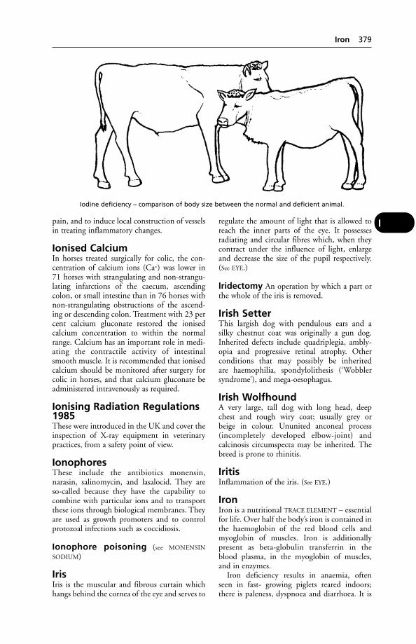
pain, and to induce local construction of vesselsin treating inflammatory changes.
Ionised CalciumIn horses treated surgically for colic, the con-centration of calcium ions (Ca+) was lower in71 horses with strangulating and non-strangu-lating infarctions of the caecum, ascendingcolon, or small intestine than in 76 horses withnon-strangulating obstructions of the ascend-ing or descending colon. Treatment with 23 percent calcium gluconate restored the ionisedcalcium concentration to within the normalrange. Calcium has an important role in medi-ating the contractile activity of intestinalsmooth muscle. It is recommended that ionisedcalcium should be monitored after surgery forcolic in horses, and that calcium gluconate beadministered intravenously as required.
Ionising Radiation Regulations1985These were introduced in the UK and cover theinspection of X-ray equipment in veterinarypractices, from a safety point of view.
IonophoresThese include the antibiotics monensin,narasin, salinomycin, and lasalocid. They areso-called because they have the capability tocombine with particular ions and to transportthese ions through biological membranes. Theyare used as growth promoters and to controlprotozoal infections such as coccidiosis.
Ionophore poisoning (see MONENSIN
SODIUM)
IrisIris is the muscular and fibrous curtain whichhangs behind the cornea of the eye and serves to
regulate the amount of light that is allowed toreach the inner parts of the eye. It possessesradiating and circular fibres which, when theycontract under the influence of light, enlargeand decrease the size of the pupil respectively.(See EYE.)
Iridectomy An operation by which a part orthe whole of the iris is removed.
Irish SetterThis largish dog with pendulous ears and asilky chestnut coat was originally a gun dog.Inherited defects include quadriplegia, ambly-opia and progressive retinal atrophy. Otherconditions that may possibly be inheritedare haemophilia, spondylolithesis (‘Wobblersyndrome’), and mega-oesophagus.
Irish WolfhoundA very large, tall dog with long head, deepchest and rough wiry coat; usually grey orbeige in colour. Ununited anconeal process(incompletely developed elbow-joint) andcalcinosis circumspecta may be inherited. Thebreed is prone to rhinitis.
IritisInflammation of the iris. (See EYE.)
IronIron is a nutritional TRACE ELEMENT – essentialfor life. Over half the body’s iron is contained inthe haemoglobin of the red blood cells andmyoglobin of muscles. Iron is additionallypresent as beta-globulin transferrin in theblood plasma, in the myoglobin of muscles,and in enzymes.
Iron deficiency results in anaemia, oftenseen in fast- growing piglets reared indoors;there is paleness, dyspnoea and diarrhoea. It is
Iron 379
I
Iodine deficiency – comparison of body size between the normal and deficient animal.
also found in calves and lambs fed milk or milksubstitute without iron supplementation andlittle or no roughage.
Iron also has a role in bodily resistance toinfection. IRON-BINDING PROTEINS can beshown to inhibit the growth of bacteria in vitro.The ability of some micro-organisms to bindiron to themselves, depriving the host animal,is a feature of pathogenicity, and ability ofthe host to limit availability to the pathogen isassociated with resistance to infection.
Bacteria (e.g. salmonellas and tuberclebacilli) and some fungi produce iron-bindingsubstances.
Iron-Binding ProteinsIron-binding proteins include conalbumin, aconstituent of egg-white; transferrin, in bloodplasma; lactoferrin, in milk, tears, saliva, bile,seminal secretions, cervical mucus, and inthe granules of neutrophils. All these proteinsinhibit the growth of bacteria, includingsalmonella.
Iron-DextranA compound of iron used in injectable form toprevent iron-deficiency anaemia in piglets.
Iron PoisoningThis may occur from overdosage, or from theeating by a pet animal of an iron preparationleft within reach. (See also under FLOOR FEEDING
OF PIGS, for the danger of concrete made withsand rich in iron.)
Horses have died very soon after receivingan intramuscular injection of an organic ironpreparation.
Piglets have been poisoned by iron-dextranpreparations given to prevent anaemia andASYMMETRIC HINDQUARTER SYNDROME maydevelop following organic iron injections.
In the dog, ferrous sulphate or ferrousgluconate in doses as low as 0.3 or 0.75 grespectively of ferrous iron per kg body-weight,have caused severe illness with diarrhoea,vomiting and ulceration of stomach andintestine.
IrradiationExposure to X-rays, radio-active material, ultra-violet, or infra-red rays. Irradiation with mea-sured doses of X-rays is used therapeutically intreating tumours; and to attenuate lungwormlarvae for use as an oral vaccine against husk.Irradiation with ultra-violet light is used tosterilise, e.g. milk to extend its usable life.(See PARASITIC BRONCHITIS; also ‘RADIATION
SICKNESS’; IONISING RADIATION.)
IrrigationIrrigation is the washing-out of wounds orcavities of the body by means of large amountsof warm water containing some antiseptic insolution.
IschaemiaLocal anaemia.
Ischaemic Contracture(see under MUSCLES, DISEASES OF)
IschiumIschium is the bone which forms the mostposterior part of the pelvis, and forms the pointof the buttock.
IschuriaIschuria means insufficiency in the amountof urine passed, due either to suppression ofexcretion in the kidneys or to retention in thebladder.
Islets of Langerhans(see PANCREAS)
IsofluraneA colourless liquid with an ether-like smell,non-explosive and non-inflammable inclinical concentrations, used as an inhalationanaesthetic for most species. Induction ofanaesthesia and recovery are more rapid thanwith halothane. It is useful in high-risk cardiaccases.
IsolationIsolation is an important procedure in thecontrol of the spread of infectious disease. Ona farm it is advisable, where practicable, to keepnewly bought stock separate from previouslyexisting stock for 2 or 3 weeks, so that ifinfectious/contagious disease occurs it may bepossible to prevent its spread to the old stock.(See INCUBATION; INFECTION; QUARANTINE;NOTIFIABLE DISEASES.)
IsoquinoliniumIsoquinolinium chloride lotion has been usedin the treatment of ringworm in cattle.
IsosporaA genus of protozoal parasites which causecoccidiosis. I. suis is a cause of coccidiosisin pigs.
IsotonicIsotonic is a term applied to solutions whichhave the same power of diffusion as one another.
380 Iron-Binding Proteins
I
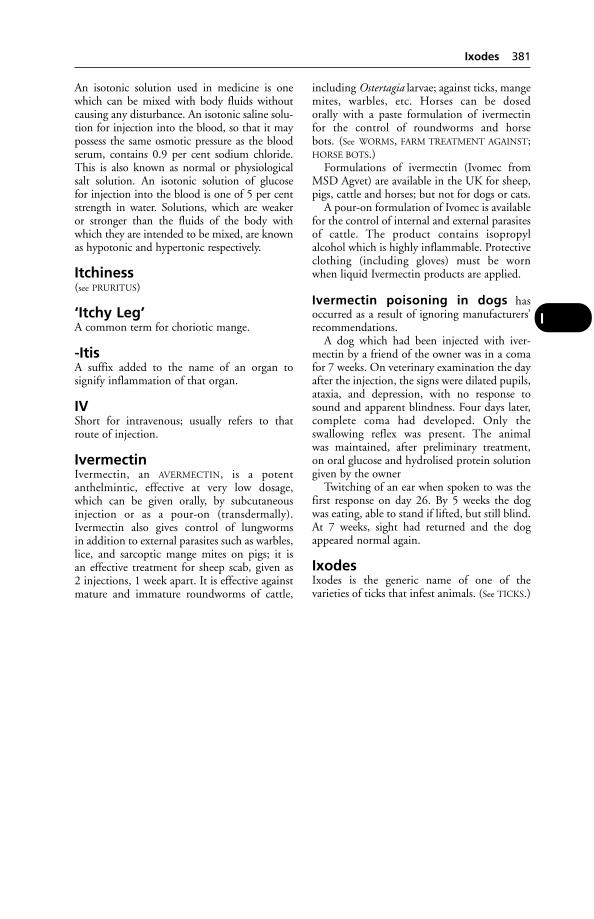
An isotonic solution used in medicine is onewhich can be mixed with body fluids withoutcausing any disturbance. An isotonic saline solu-tion for injection into the blood, so that it maypossess the same osmotic pressure as the bloodserum, contains 0.9 per cent sodium chloride.This is also known as normal or physiologicalsalt solution. An isotonic solution of glucosefor injection into the blood is one of 5 per centstrength in water. Solutions, which are weakeror stronger than the fluids of the body withwhich they are intended to be mixed, are knownas hypotonic and hypertonic respectively.
Itchiness(see PRURITUS)
‘Itchy Leg’A common term for choriotic mange.
-ItisA suffix added to the name of an organ tosignify inflammation of that organ.
IVShort for intravenous; usually refers to thatroute of injection.
IvermectinIvermectin, an AVERMECTIN, is a potentanthelmintic, effective at very low dosage,which can be given orally, by subcutaneousinjection or as a pour-on (transdermally).Ivermectin also gives control of lungwormsin addition to external parasites such as warbles,lice, and sarcoptic mange mites on pigs; it isan effective treatment for sheep scab, given as2 injections, 1 week apart. It is effective againstmature and immature roundworms of cattle,
including Ostertagia larvae; against ticks, mangemites, warbles, etc. Horses can be dosedorally with a paste formulation of ivermectinfor the control of roundworms and horsebots. (See WORMS, FARM TREATMENT AGAINST;HORSE BOTS.)
Formulations of ivermectin (Ivomec fromMSD Agvet) are available in the UK for sheep,pigs, cattle and horses; but not for dogs or cats.
A pour-on formulation of Ivomec is availablefor the control of internal and external parasitesof cattle. The product contains isopropylalcohol which is highly inflammable. Protectiveclothing (including gloves) must be wornwhen liquid Ivermectin products are applied.
Ivermectin poisoning in dogs hasoccurred as a result of ignoring manufacturers’recommendations.
A dog which had been injected with iver-mectin by a friend of the owner was in a comafor 7 weeks. On veterinary examination the dayafter the injection, the signs were dilated pupils,ataxia, and depression, with no response tosound and apparent blindness. Four days later,complete coma had developed. Only theswallowing reflex was present. The animalwas maintained, after preliminary treatment,on oral glucose and hydrolised protein solutiongiven by the owner
Twitching of an ear when spoken to was thefirst response on day 26. By 5 weeks the dogwas eating, able to stand if lifted, but still blind.At 7 weeks, sight had returned and the dogappeared normal again.
IxodesIxodes is the generic name of one of thevarieties of ticks that infest animals. (See TICKS.)
Ixodes 381
I
JaagsiekteA disease of adult sheep, first recognised in SouthAfrica. (See PULMONARY ADENOMATOSIS.)
JackA male donkey.
Jack BeansJack Beans may cause poisoning if fed raw. (See LEGUME POISONING.)
Jack Russell TerrierA small, lively dog, having a white coat withbrown or black patches. Originally a cross-breed, it now breeds true. Prone to patellar luxation, it may inherit ataxia, lens luxation andPerthe’s disease.
Jacobson’s OrganAlso known as the vomeronasal organ, this isassociated with the detection of flavours such asthose of food, but is thought also to be able todetect pheromones. The organ has 2 small tubeswhich extend from the floor of the nasal cavityto the level of the 2nd/4th cheek tooth. It isactive in most mammals, but even more highlydeveloped in certain reptiles, especially snakes.
JanetA female mule.
Japanese B EncephalitisThis disease is present in Nepal and otherregions of Asia.
Cause A flavivirus. The disease is transmittedby mosquitoes from avian species which act asreservoirs of infection but are themselvesasymptomatic.
Signs In horses, the sight is affected first. Laterthey become drowsy. Many die, and the recov-ery of others is seldom complete. Pigs are alsosusceptible; abortion and stillbirths result frominfection.
The disease is a zoonosis, and for its preven-tion in people a vaccine has been used.
Japanese BobtailA breed of cat of ‘foreign’ conformation with arudimentary tail. The hind-legs are longer than
the fore-legs but are kept angled so that theback is level. There are similarities with theManx, but there does not appear to be the samefrequency of defects as occurs in that breed.
Japanese TosaA breed of dog raised in Japan for fighting.Importation into the UK is banned under theDangerous Dogs Act.
JaundiceJaundice is a yellowish discoloration of the visi-ble mucous membranes of the body (eye, nose,mouth, and genital organs). The discolorationis caused by bilirubin, an orange-yellow pig-ment produced following the breakdown oferythrocytes due to liver disease or obstructionof the bile flow from the liver and gall-bladder.
The symptom of jaundice (icterus) may also follow the destruction of red blood cells by parasites, such as may occur in cases of biliary fever and surra in the horse; red-water in cattle; malignant jaundice (canine babesio-sis); it is seen also in leptospiral jaundice (see
LEPTOSPIROSIS), and canine viral hepatitis.In cats, jaundice is seen in the dry form of
feline infectious peritonitis, toxoplasmosis.Jaundice may indicate an incompatibility
between the blood of sire and dam causinghaemolytic jaundice of the newborn foal orpiglet.
When bile cannot enter into the small intes-tine by the bile-duct from the liver in the usualway, it becomes dammed back, is absorbed bythe lymphatics and the blood vessels, carriedinto the general circulation, and some of itsconstituents are deposited in the tissues. (See
GALL-STONES, also under GALL-BLADDER; CIR-
RHOSIS; LIVER, DISEASES OF; EQUINE BILIARY
FEVER.)It may be seen during poisoning with copper,
mercury, phosphorus, chloroform, or lead, andafter some snakebites. Aflatoxins may causejaundice.
(See also LEPTOSPIROSIS; JAUNDICE; FOALS,DISEASES OF; and BILIARY FEVER.)
Jaundice, Leptospiral(see under LEPTOSPIROSIS for the appropriate animal)
Java Bean PoisoningThe Java beans, Phaseolus lunatus, were onceimported in large amounts. The beans are ofvarying origin, and differ in colour, thus: Javabeans are as a rule reddish-brown, but they maybe almost black; Rangoon or Burmah beans are
J
smaller, plumper, and lighter in colour (so-called ‘red-Rangoons’ are pinkish with smallpurple splashes).
The active poisonous agent in the beans is asubstance called phaseolunatin, which is amember of a group of cyanogenetic glucosides.
Signs These are exactly the same as those givenunder HYDROCYANIC ACID.
JawThe upper jawbones are 2 in number and arefirmly united to the other bones of the face.The lower jaw – mandible or coronoid process– is composed of a single bone in horse, pig,dog, and cat, but in the ruminants the fusionbetween right and left sides does not occur until old age. Each of the jaws presents a num-ber of deep sockets or ‘alveoli’ which containthe teeth. (See DISLOCATIONS; FRACTURES;MOUTH; TEETH; also MUSCLES, DISEASES OF;ACTINOMYCOSIS (LUMPY JAW).
Jaw, Diseases ofFor overshot and undershot jaws, see under
TEETH, DISEASES OF.‘Lion jaw’ (craniomandibular osteopathy): a
disease seen mostly in West Highland terriers.Eating becomes difficult; mouth-opening,painful.
(See ‘BOTTLE-JAW’.)
JejunumJejunum is the central portion of the smallINTESTINE.
‘Jeckyll and Hyde’ SyndromeAlso known as ‘rage syndrome’, this is a condi-tion seen in cocker spaniels, especially those of a golden or dun colour. For no apparent rea-son a quiet dog will suddenly become veryaggressive. The dog then returns to its normalbehaviour.
JennyA female ass.
Jequirity PoisoningThis is caused by the red and black seeds of theclimbing plant Abrus precatorius, which growsin Australia, Asia, and South America. It givesrise to cyanosis and pinpoint-sized haemor-rhages from the skin, as well as diarrhoea.
JersianAlso known as a F–J hybrid, this is a beef crossobtained from a Jersey bull on a Friesian cow.(In New Zealand, the reverse cross is used.)
JettingJetting is a technique developed in Australia,involving the application of insecticide underpressure by means of a jetting gun – a handpiecewith 4 needle jets for combing through the wool.The pressure used is 10 to 14 kg/cm2 (60 to 80lb per sq in), which can be achieved by an ordi-nary medium/high-volume agricultural sprayer.
Jetting has not displaced dipping to anyextent in the UK, where spraying has beenfound inefficient in the control of sheep scab.
Jigger Flea(see under FLEAS – Tunga penetrans)
JillA female donkey or ferret.
Johne’s Disease(Paratuberculosis)Johne’s Disease (Paratuberculosis) is a chronicinfection, involving the small and largeintestines. It affects cattle particularly, butsometimes sheep, goats, and deer, and is char-acterised by the appearance of a persistent diar-rhoea, gradual emaciation, and great weakness.The infection has been set up experimentally inthe rabbit. It may occur naturally in the pig,and post-mortem findings may at first suggesttuberculosis.
Cause Mycobacterium johnei (M. paratubercu-losis).
Experimentally, sheep can be infected with asfew as 1000 M. johnei bacilli. These then mul-tiply in the intestinal mucosa for the first 2 or 3months after infection. Some animals are ableto overcome the infection completely; othersbecome carriers, with the bacilli remaining inthe intestinal mucosa and lymph nodes. Someof the carriers eventually become clinically illwith Johne’s disease.
Signs The disease is very slow in onset. Cattlethat have become infected may not show symp-toms for as long as 2 years after the last caseoccurred on that farm.
Pointers to the disease are an unexplained dropin milk yield (often months before other symp-toms appear); and diarrhoea in an individualadult animal.
Loss of condition, general unthriftiness, aharsh, staring coat are then seen, with diar-rhoea. The temperature fluctuates a degree ortwo above normal. Appetite is variable. In thelast stages emaciation becomes very marked,and the animal becomes progressively weaker.
In sheep, diarrhoea is not a major symptom.
Johne’s Disease (Paratuberculosis) 383
J

Treatment When well established, Johne’sdisease is invariably fatal, and no treatment iseffective or worthwhile.
Prevention Attention should be paid to theprevention of infection in other animals, espe-cially calves. Pastures that are suspected of beingheavily infected should be left without stock for4 or 5 months. All infected litter should bestored in a dung-pit which is not accessible toother animals, and should be used for cultivat-ed land. Loose-boxes, sheds, etc., that havehoused a case should be carefully disinfectedand diseased animals should be fed after healthyones. Ponds and water-courses should be fencedto prevent fouling by faeces, water for drinkingbeing pumped out.
Calfhood vaccination may prevent clinicaldisease but interferes with subsequent tuberculintests.
Vaccination is being practised in Iceland, theNetherlands, Belgium, and France.
In Norway a vaccination campaign to con-trol the disease in goats reduced the infectionrate from 53 to 1 per cent. Kids are vaccinatedat the age of 2 to 4 weeks.
Diagnosis The disease can usually be diag-nosed on clinical evidence, with some confir-mation afforded by microscopic examination ofthe faeces. Typical clumps of acid-fast bacillimay be found, and the complement fixation testis positive in about 90 per cent of cattle withadvanced disease. The fluorescent antibody testis equally useful.
Unfortunately, diagnosis of the carrier state is not possible with any certainty. There is nosingle test which can conclusively detect thepresence or absence of M. johnei, although laboratory tests can identify the presence ofMycobacteria spp. The complement fixation testis positive in only a small proportion of carriersand can give false positive results.
The difficulty in identifying ‘carriers’ makesJohne’s disease a difficult one to control.
JohninA diagnostic agent derived from M. paratuber-culosis used for JOHNE’S DISEASE. Cutaneousinjection results in thickening of the skin in positive cases. While insufficiently sensitivefor individual diagnosis, the test is useful foridentifying infected herds.
Joint-IllAlso called NAVEL-ILL or POLYARTHRITIS, this isa disease of foals, lambs, and calves, in whichabscesses form at the umbilicus and in some of
the joints of the limbs, due, in the majority ofcases, to the entrance of organisms into thebody by way of the unclosed navel. There arenumerous organisms associated with the dis-ease, the commonest of which are streptococci,staphylococci, Pasteurella, E. coli, the necrosisbacillus, and see under FOALS, DISEASES OF.
Signs Usually the young animal becomes dull,takes no interest in its dam, refuses to suckle;the breathing is hurried; the temperature risesfrom 0.6° to 1.2°C (2° to 4°F) above normal;the foal prefers to lie stretched out on its side,and may have attacks of either diarrhoea or con-stipation. If the navel is examined it is found tobe wet and oozing with bloodstained serousmaterial, or it may be dry, swollen, painful tothe touch, and hard, owing to abscess formationwithin. In cases that appear later in life theremay be no umbilical symptoms. In the courseof a day or so, one or more of the joints swellsup. The joints most commonly attacked are thestifle, hip, knee, hock, shoulder and elbow, butit may be seen in any of the others. The swellingis tense, painful, hot, and oedematous. There isthe danger of a fatal septicaemia.
A chronic form of infection resulting ininternal umbilical abscesses is sometimes seen.The primary infection occurs at, or soon after,birth; but once the umbilicus has sealed over,external signs are not evident, and the umbilicalremnant appears normal.
The calves are usually presented as unthrifty,depressed and slow in their movements. Theirtemperature invariably normal.
Prevention Attention must always be paid tothe cleanliness of the foaling-box, the calving-box and the lambing-pen. Where climatic andother conditions are favourable, the pregnantfemales should be allowed to give birth to theiryoung out of doors. Lambing-pens should with-out fail be changed to a fresh site every year.
Investigations undertaken by the AnimalHealth Trust suggest that thoroughbred foals inthe UK suffer severe illness as a result of beingdeprived of a not inconsiderable volume ofblood when the navel cord is severed prema-turely by attendants. Severance of the cord, itseems, is always best left to the mare. The use ofstrong disinfectants applied to the stump of thenavel cord is likewise deprecated.
An application of a sulphanilamide or otherantibiotic dry dressing may be safer than iodinesolution.
When cutting the cord, it is necessary tomaintain the strictest cleanliness. Scissors shouldbe sterilised, and tape scrupulously clean.
384 Johnin
J
Treatment Antibiotics and, if available, anti-serum for the causative micro-organisms.Surgically, the umbilicus is opened up, evacuat-ed, and disinfected. Isolation and other hygienemeasures are needed.
All pails, and other feeding utensils that areliable to get infected, should be thoroughlycleaned using boiling water or steamed beforefuture use, and the pen or box that houses acase should be occasionally washed out withdisinfectant. (See also FOALS, DISEASES OF.)
JointsJoints fall into 2 great divisions, namely mov-able joints and fixed joints. In a movable jointthere are 4 main structures. Firstly, there are the2 bones whose junction forms the joint; sec-ondly, there is a layer of smooth cartilage cover-ing the ends of these bones where they meet,which is called ‘articular’ cartilage; thirdly, thereis a sheath of fibrous tissue known as the ‘jointcapsule’, which is thickened into bands of ‘liga-ments’ which hold the bones together at variouspoints; and finally, there is a closed bladder ofmembrane, known as the ‘synovial membrane’,which lines the capsule and produces a synovialfluid to lubricate the movements of the joint.Further, the bones are kept in position at thejoints by the various muscles passing over them.This type is known as a diarthrodial joint.
Some joints possess subsidiary structures suchas discs of fibro-cartilage, which adapt the bonesmore perfectly to one another where they do notquite correspond, and allow of slightly freermovement, e.g. the stifle-joint. In others, mov-able pads of fat under the synovial membrane fillup larger cavities and afford additional protec-tion to the joint, e.g. the hock-joint. In some theedge of one bone is amplified by a margin of car-tilage which makes dislocation less of a risk thanotherwise, e.g. the hip and the shoulder-joints.
In the fixed joints a layer of cartilage or offibrous tissue intervenes between the bones andbinds them firmly together (synarthrodialjoint). This type of joint is exemplified by the ‘sutures’ between the bones that make upthe skull. Classified among these fixed joints arethe amphi-arthrodial joints, in which there is athick disc of fibro-cartilage between the bones,so that, although the individual joint is capableof only limited movement, a series of these, likethe joints between the bodies of the vertebrae,gives the column, as a whole, a very flexiblecharacter. In this connection it is noticeablethat the movement in the region of the neckmay be much more free than in some of thetrue movable joints, such as between the smallbones of the hock or carpus.
Varieties Apart from the division into fixedand movable joints, those that are movable arefurther classified. Gliding joints are those inwhich the bones have flat surfaces capable onlyof a limited amount of movement, such as thebones of the carpus and tarsus. In hinge-jointslike the elbow, fetlock, and pastern, movementcan take place around one axis only, and is calledflexion and extension. In the ball-and-socketjoints, such as the shoulder and hip-joints, freemovement can occur in any direction. There areother subsidiary varieties, named according tothe shape of the bones which enter into thejoint.
Joints, Diseases of
Arthritis means inflammation which involvesall the structures of the joint – viz. synovialmembrane, capsular ligaments, cartilages, andthe ends of the bones that take part in the for-mation of the joint. Arthritis is a general termwhich includes osteoarthritis and rheumatoidarthritis. Arthritis often begins as a synovitis (see
below), but the degree of inflammation is severeenough to extend to the structures around thesynovial membrane. Its causes, symptoms, andtreatment are similar to those given for synovi-tis, but it sometimes leads to ankylosis and fix-ation of the joint. (See CORTISONE.) The jointsthat are most often affected are the stifle, hock,knee, and fetlocks, but the shoulder, hip, elbow,and the lower joints of the digit are not infre-quently the seat of disease as well. Among dis-eases that are associated with joints, and whichare treated separately, are NAVICULAR DISEASE;SLIPPED SHOULDER; SLIPPED STIFLE; HYGROMA;CAPPED ELBOW; CAPPED HOCK; KNUCKLING OF
THE FETLOCK; JOINT-ILL; see also below and BUR-
SITIS; ANKYLOSIS; FRACTURES; DISLOCATIONS;GLASSER’S DISEASE; HIP DYSPLASIA IN DOGS;SWINE ERYSIPELAS.
Rheumatoid arthritis This can be impor-tant in the dog, and may occur at any age from2 years. Symptoms may be vague at first; theanimal appears depressed, with a poor appetiteand often some degree of fever, but with nolameness. Eventually the latter symptomappears, sometimes involving several joints,sometimes affecting only one limb and thenshifting to another. There may be crepitus – agrating sound – when the limb is moved.
Diagnosis depends upon radiography and –as in human medicine – there are certain laboratory tests, the results of which provideadditional criteria for deciding whether thecondition really is rheumatoid arthritis or not.
Joints, Diseases of 385
J

J
386 Joule
Intractable arthritis of the hip-joint in dogs,as in human beings, may be overcome by majorsurgery involving removal of the top of thefemur and replacement of the ball part of theball-and-socket joint with a plastic prosthesis.
Synovitis is the name given to any inflamma-tion of the membrane lining a joint cavity. Itmay be acute, sub-acute, or chronic.
Generally this is not a separate condition but occurs during the course of rheumatism,rickets, gout (in poultry), severe sprains andbruises, and in a variety of specific infectionssuch as brucellosis, swine erysipelas, tuberculo-sis. Tubercular joint disease often produces achronic synovitis in the neck bones of the horse,which leads to an arthritis later.
Conditions such as wind-galls, curb, bogspavin, etc., are really only synovitis that havebecome chronic or are complicated with otherpathological conditions.
The synovial membrane becomes inflamed,thickened, and secretes an excessive amount of fluid into the joint. As a result the jointbecomes hot, swollen, and painful. The animalgoes lame in greater or lesser degree accordingto the extent of the inflammation. When atrest, the joint is usually kept flexed with the toeof the affected leg just resting on the ground. Ifit is a simple condition, such as a mild sprain,these symptoms last for a few days and thengradually pass off. In more severe cases, such asin joint-ill, there may be pus formation, septi-caemia, and death. In the chronic type theswelling persists. The animal is able to use itslimb as usual, but the accumulated fluid in thecavity does not disappear (e.g. bog spavin,wind-galls, etc.).
Open joint is a condition in which, by acci-dent or other trauma, the inside of the joint isexposed to infection.
The seriousness of an open joint is not somuch due to the initial injury as to the danger ofinfection. This may cause tissue destruction with-in the joint, and even lead to a fatal SEPTICAEMIA.
The most striking signs of open joint are,first, the excessive degree of pain that seems outof all proportion to the visible amount of dam-age that has been inflicted; secondly, the greatamount of swelling that is usually seen; andthirdly, the discharge of a thin, straw-colouredor blood-stained sticky synovia which has a tendency to coagulate around the skin opening.
Veterinary advice should be sought at once.Prompt treatment with antibiotics, and surgery
if required, is necessary to prevent or limitinfection.
Dislocations (see main dictionary entry)
Bursitis, an inflammation of a bursa, com-monly occurs in the region of a joint. Theprominences of the hock, elbow, knee, stifle,etc., are protected by bursae – lined on theirinsides by synovial membrane. These sometimesbecome inflamed and lead to the formation of fluctuating swellings which have a tendencyto become chronic. Capped elbow, cappedhock, and hygroma of the knee, are of thisnature. (See also OSTEOARTHRITIS; MAST CELLS;OSTEOCHONDROSIS; RHEUMATISM.)
JouleA derived SI unit of metabolisable energy. (See
CALORIES and STARCH EQUIVALENT, which itreplaced; also SI UNITS.)
Jugular VeinsJugular veins carry the blood back to the chestfrom the head and anterior parts of the neck.The jugular vein is often used for taking bloodsamples and for intravenous injection. Thejugular furrow is the groove between the tra-chea and the muscles of the neck, in the depthsof which lies the jugular vein.
Jungle Fowl(Gallus gallus) A native of the rain forests ofSouth-East Asia, it is the species from which thedomestic fowl originated. In its normal environ-ment it prefers hot, humid, shady conditionswith frequent rain showers. A broiler-producingcompany has replicated the climate found in therain forest in its broiler houses. Mortality wasvery low and lameness almost non-existent,although the birds took a little longer to reachmarket weight.
Juvenile CellulitisAlso known as ‘puppy strangles’, this conditionaffects pups between 3 weeks and 4 months ofage. The cause is unknown but a hypersensitiv-ity reaction may be involved. Clinical signs are cellulitis on the face and head, prepuce and anus, accompanied by lethargy, anorexiaand, possibly, raised temperature. There may belymphadenopathy. Steroid and antibiotic thera-py is indicated; affected parts may be bathed in aluminium acetate solution. Permanent scarring may result.

K88 AntigenThis is possessed by certain strains of E. coliwhich cause diarrhoea in piglets during theirfirst few days of life. (See E. COLI; BACTERIAL
ADHESIVENESS.)
K99 AntigenK99 Antigen is found in strains of E. coli whichcause diarrhoea in calves.
K ValueThis is used as a measure of the insulating value of building materials such as glass fibre,wood.
Kala-Azar (Dumdum Fever)A human disease caused by LEISHMANIA.
KaleKale contains a factor which gives rise to goitreif fed in large amounts, without other foods,over a long period. Haemoglobinuria some-times follows the grazing of frosted kale by cattle, which may suffer anaemia without show-ing this symptom. The illness can be serious,resembling POST-PARTURIENT HAEMOGLOBIN-
URIA, and may result in sudden death. Thefrothy type of bloat may also occur in cattle eating excessive quantities of kale – especially, it seems, during wet weather and when no hayis fed. There is some evidence to suggest thatthe feeding of large quantities of kale may leadto low conception rates, and to mastitis. (See also
BLOAT.)It should be added that kale anaemia and
haemoglobinuria are by no means always asso-ciated with frosted kale, but merely with anexcessive (probably over 18 kg (40 lb) per cowper day) intake of kale. The symptoms of kaleanaemia include lassitude and rapid breathingand pulse-rate.
‘Kangaroo Gait’‘Kangaroo gait’ in ewes, both in New Zealandand in the UK, appears to be associated withdisease of the radial nerves, which causes diffi-culty in advancing the front feet. When made tomove rapidly, they do so with a bounding gait.The condition is seen in ewes during lactation;it normally resolves after weaning.
Kaolin (China Clay)Kaolin (China Clay) is a native aluminium silicate, which is used as a protective and astrin-gent dry dusting powder. Kaolin is sometimesgiven internally as an adsorbent in intestinaldisorders. Mixed into a paste with glycerine andsome antiseptic, it is applied as a poultice toacute sprains of tendons, etc.
KaryotypeThis is, roughly speaking, a plan showing ananimal’s chromosomes. In technical terms, akaryotype is a presentation of the metaphasechromosomes characteristic of an individualanimal or species. (See CYTOGENETICS.)
‘Kebbing’(see ABORTION, ENZOOTIC, OF EWES)
KedMelophagus ovinus, the sheep ked, is wingless,and lives on the wool and skin of the sheep. It is much larger than any of the lice, being 0.6 cm (1⁄4 inch) long. It can easily be distin-guished from the ticks by its tripartite body. Itis a dark brown colour with a sharp biting pro-boscis. The nearly mature larvae are laid on thewool and they at once pupate. The pupa mayremain in the wool or fall to the ground. Theyoung hatch in 19 to 24 days, and the femalesstart to deposit larvae in 12 to 23 days afteremergence, and lay a larva every 9 days. The flycan live for about 12 days away from the sheep,while the pupa can live for 6 weeks on theground. The whole life-cycle may be completedon the sheep within 1 month.
The sheep ked can cause severe anaemia ifpresent in large numbers, and also leads to adamaged fleece. Shearing aids control, which isachieved by means of a sheep dip.
The ked may attack men while shearing andinflict a very painful bite.
K
Melophagus × 4.

KeeshondA medium-sized breed of dog, originally fromFlanders, with a very thick coat, fox-like faceand a tail that curls over the back. It is prone tohip dysplasia. The incidence of tetralogy ofFallot (a potentially fatal heart defect) in thebreed is 1 in 10,000 births, the highest knownin any animal.
KempsCoarse hairs, the presence of which reduces thevalue of a fleece.
Kennel CoughKennel cough is a convenient term for thoseoutbreaks of respiratory disease, distinct fromcanine distemper, which are troublesome inboarding kennels and dog pounds. Othernames are canine infectious tracheobronchitisand bordetellosis.
Usually only the upper air passages areinvolved in kennel cough, the chief symptombeing a fit of coughing which is aggravated byexercise or excitement. The cough is a harsh, dryone. It has to be differentiated from infestationwith TRACHEAL WORMS.
Causes Bordetella bronchiseptica is the princi-pal cause (hence the name bordetellosis). Otherorganisms involved are the canine parain-fluenza virus (CPI), a canine herpesvirus, two adenoviruses, a reovirus, and a mycoplasma.Bacterial secondary invaders may complicatethe syndrome.
Prevention Vaccination is advisable a fort-night before a dog is taken to a show or left in boarding kennels. An intranasal live B. bron-chiseptica vaccine is available.
Kennel LamenessA colloquial term for lameness arising from anutritional deficiency, such as may occur in adog fed entirely on dog-biscuits. (See RICKETS.)
Kennel SicknessA colloquial name used in the USA for out-breaks of salmonellosis, the symptoms of whichmay include pneumonia and convulsions. (See
also under SALMONELLOSIS.)
KeratinKeratin is the substance of which horn and thesurface layers of the skin are composed. It is amodified form of skin which has undergonecompression and toughening. It is present inthe hoof of the feet of animals, in claws, horns,and nails.
KeratitisInflammation of the cornea. (See EYE, DISEASES
OF.)
KeratocoeleKeratocoele is a hernia through the cornea. (See EYE, DISEASES OF.)
KeratomaKeratoma is a horn tumour affecting the inneraspect of the wall of the hoof.
Kerosene Poisoning(see PARAFFIN)
Kerry Blue TerrierA medium-sized dog, born with a black coatthat gradually lightens to grey, and an uprighttail. It is predisposed to cerebellar abiotrophy, adegeneration in the cerebellum, that manifestsas ataxia.
KetamineKetamine is a non-barbiturate, non-narcoticanaesthetic. It can be administered by intra-venous, intramusuclar or subcutaneous injec-tion and is used in various species including cat,dog and horse. Ketamine is often used in con-junction with other agents, such as xylazine,medetomidine and detomidine, to improvemuscle relaxation during surgical procedures.
KetoconazoleAn antifungal compound used to treat systemiccandidiasis and ringworm in dogs and cats. It isadministered orally.
Ketone BodiesKetone bodies arise from acetyl coenzyme A.The 3 main ketone bodies are acetone, acetoac-etate and beta hydroxybutyrate. While acetoneis not part of the metabolic process in thehealthy animal, the others are involved in theenergy metabolism of ruminants. If the dietprovides insufficient energy sources in the dia-betic, excess ketone bodies arise, producingketosis (acetonaemia). Professor Sir Hans Krebsdescribes the process thus: ‘The severe forms ofketosis of the diabetic coma or of the lactatingcow are connected with the high rates of gluco-neogenesis which occur under these conditions.Oxaloacetate, which is an intermediate in glu-coneogenesis, is diverted from the tricarboxylicacid cycle to gluconeogenesis, owing to the highactivity of the enzyme converting it to phos-phopyruvate. The liver compensates the loss ofenergy from a reduced rate of the tricarboxylicacid cycle by an increased rate of oxidations
388 Keeshond
K

outside the cycle. The main reaction of this type is the oxidation of fatty acids to ketonebodies. These arise grossly in excess of needs, asa by-product of reactions which satisfy therequirements for energy.’
Ketonuria is the term applied to the presenceof ketone bodies in the urine. (See also ACETON-
AEMIA.)
Ketoacidosis. A condition leading to diabetic coma. (See DIABETES.)
Ketosis(see ACETONAEMIA)
Key-Gaskell Syndrome(see FELINE DYSAUTONOMIA)
KhatThis plant (Catha edulis) contains 2 compounds– cathine and cathinone – which are both structurally related to amphetamine.
Chewing of khat leaves, popular in Arabiaand East Africa, appears to be on the increase inthe UK. Addicts esteem khat for the euphoriaand extra energy which it provides, but over-usecan lead to mental illness.
Veterinary surgeons in small-animal practicewill need to be on the lookout for cases of khatpoisoning in dogs and cats – as they already arefor the effects of CANNABIS.
Kicking(see ‘VICES’)
Kidney Worm (StephanurusDentatus)Kidney worm (stephanurus dentatus) is a para-site of pigs. Affected animals fail to thrive.Occasionally migration of the larvae in thespinal canal causes some degree of paralysis. Theintermediate host is the earthworm. In the USAthe advice is to breed from gilts only, as a meansof eradicating the parasite – anthelmintics so farnot having proved effective. (For the kidneyworm of dogs, see DIOCTOPHYMOSIS.)
KidneysKidneys are paired organs situated high upagainst the roof of the abdomen, and in mostanimals lying one on either side of the spinalcolumn.
Horse The kidneys of the horse differ fromeach other in both shape and position. Theright has the outline of a playing-card heart,and lies under the last 2 or 3 ribs and the
transverse process of the first lumbar vertebra,while the left is roughly bean-shaped and liesunder the last rib and the first 2 or 3 lumbartransverse processes. They are held in place by the surrounding organs and by fibrous tissue, called the renal fascia. Each of themmoves slightly backwards and forwards duringthe respiratory movements of the animal.
Cattle The kidneys are lobulated, each pos-sessing from 20 to 25 lobes separated by fissuresfilled with fat in the living animal. The rightkidney lies below the last rib and the first 2 or3 lumbar transverse processes, and is somewhatelliptical in outline. The left occupies a variableposition. When the rumen is full, it pushes the left kidney over to the right side of the body into a position slightly below and behindthe right organ, but when it is empty the leftkidney lies underneath the vertebral columnabout the level of the third to the fifth lumbarvertebra. It may lie partly on the left side of thebody in this position in some cases.
Sheep In the sheep the kidneys are bean-shaped and smooth. In position they resemblethose of the ox, except that the right is usuallya little farther back.
Pig In this animal the kidneys are shaped likeelongated beans, and they are placed almostsymmetrically on either side of the bodies of thefirst 4 lumbar vertebrae. They sometimes varyin position.
Dogs and cats In these animals the kidneysare again bean-shaped, but they are thicker than in other animals, and relatively larger. As in most animals, the right kidney is placedfarther forward than the left, the latter varyingin position according to the degree of fullness of the digestive organs. In the cat the left kidney is very loosely attached and can usuallybe felt as a rounded mass which is quite movable in the anterior part of the abdominalcavity.
Birds have paired kidneys, seen as elongatedbrown organs closely attached on each side ofthe vertebrae.
Fish have a single kidney which is seen insalmonids as a long black strucure in the dorsalpart of the abdomen extending from the backof the head to the vent. The vena cava runsthrough the centre of the organ. The kidneyalso has a role in the development of blood cellsand in combating infection.
Kidneys 389
K

Structure The organ is enveloped in a fibrouscoat continuous with the rest of the peritonealmembrane, and attached to the kidney capsule.This capsule does not permit of much swellingor enlargement of the organ, and consequentlyany inflammation of the kidney is attendedwith much pain. On the inner border there isan indentation called the hilus, which acts as aplace of entrance and exit for vessels, nerves,etc. Entering each kidney at its hilus are a renalartery and renal nerves; leaving the kidney arerenal vein or veins, lymphatics, and the ureter.If the kidney is cut across, there are 2 distinctareas seen in its substance. Lying outermost is the reddish-brown granular cortex, whichcontains small dark spots known as Malpighiancorpuscles.
Within the cortex is the medulla, an area pre-senting a radiated appearance, whose peripheryis of a deep red colour.
The kidney tissue contains many thousandsof filtration units called nephrons. Each of thesecomprises the glomerulus (almost a sphericalarrangement of capillaries on an arteriole);Bowman’s capsule, the blind end of a proximaltubule which expands so as almost to surroundthe glomerulus; the convoluted tubule itself(with its loop of Henle); and the distal convo-luted tubule which leads on to an arched collecting tubule. The latter continues with astraight tubule in the cortex of the kidney, andon into the medulla, where papillary ducts areformed to take the urine to the pelvis of thekidney.
The Malpighian corpuscle, comprising theglomerulus and inner and outer layers ofBowman’s capsule, is where most of the filtra-tion of fluid from the blood occurs; but only asmall percentage of this fluid is finally excretedas urine.
In birds, the glomeruli are of 2 differentkinds; 1 type is similar to mammalianglomeruli; the other is more akin to the typefound in reptiles.
Function The kidney’s 2 main functionsare:first, the excretion of waste (and excess)materials from the bloodstream; and, second,the maintenance of the correct proportions of water in the blood, the correct levels of its chemical constituents, and the correct pH.(See HOMEOSTASIS.)
Blood pressure in the arteries determinespressure in each glomerulus and has an impor-tant bearing on the quantity of fluid filteredfrom the blood.
For its controlling effect on the kidney, see
ANTIDIURETIC HORMONE.
The proximal tubules reabsorb a high per-centage of the water, sodium chloride and bicar-bonate. The distal tubules reabsorb sodium, orexchange sodium ions for hydrogen, potassiumor ammonium ions; determining thereby thepH of the urine.
The kidney also secretes the hormone erythropoietin (see under ERYTHROPOIESIS) andproduces RENIN. Additionally, the kidney converts vitamin D1 into its active form.
Kidneys, Diseases ofThese are particularly common in the dog, andmust account for a high proportion of deaths indogs and cats.
Exact diagnosis is based almost entirely uponmacroscopic, microscopic, and chemical exam-ination of the urine in the laboratory. Bloodurea nitrogen (BUN) and serum creatinine con-centrations are used to evaluate renal functionin several species.
Nephrosis/nephrotic syndrome Thismay be a stage in nephritis and involves damageto the tubules of the kidneys, resulting in defec-tive filtering, so that albumin is excreted in theurine to the detriment of albumin levels in theblood. Oedema occurs.
Nephrosis may be caused by poisoning withthe salts of heavy metals, and with various tox-ins; or it may follow certain other diseases. Inlambs, clostridial infections have been suggest-ed as a cause of the disease, while nematodirusinfestation may be responsible in older animals.(See also MEMBRANOUS NEPHROPATHY.)
Acute nephritis is a rapid inflammation ofthe kidney tissues as a whole, or of the glomeruliand the secreting tubules only. The latter is muchthe more common among all animals. Since thediagnosis and symptoms of each are clinically thesame, and as their differentiation is only possibleby microscopic examination after death it willsuffice to describe the commoner type only.
Dogs Acute and subacute nephritis is oftenassociated with LEPTOSPIROSIS, especially withLeptospira canicola infection; it may follow thenephrotic syndrome, and may co-exist with dis-temper or canine viral hepatitis. A predisposingcause is often, it seems, exposure to cold, wetconditions, which lower the animal’s resistanceand so exacerbates any existing infection.
Signs may include depression, loss of appetite,thirst, vomiting. The back may be arched, andthere may be stiffness. There is fever, and some-times ulcers are present in the mouth.
390 Kidneys, Diseases of
K

Lambs Acute kidney failure was diagnosed byclinical examination and autopsy in 39 flocksserved by 6 veterinary investigation centres.
Forty-eight lambs of 12 different breeds orcrosses were investigated. The mean age ofaffected lambs was 38 days; 21 lambs were aged7 to 28 days, while only 8 were older than 2 months. Mortality in clinically affected lambswas almost 100 per cent, with no response tovarious treatments.
First-aid The animal needs rest, warmth, andlight food. Reliable proprietary foods can beobtained for kidney disease cases. Barley waterinstead of plain water is often advisable. (See
under NURSING.)
Treatment includes the use of antibiotics. Ifthere is much vomiting, normal saline may benecessary.
Chronic nephritis may follow the acuteform, or it may arise insidiously. One attack ofnephritis is always likely to render the dog moresusceptible to subsequent attacks, and chronicnephritis is common in middle-aged and olddogs. In some cases of this disease RUBBER JAW
may be present. Sometimes, despite treatment,kidney failure occurs.
Kidney failure may follow either chronicinterstitial nephritis (involving some degree offibrosis), which often results from leptospiralnephritis; or from glomerular disease (glomeru-lonephritis). Clinically, the 2 conditions are virtually indistinguishable.
Cattle Kidney disease may also be associatedwith LEPTOSPIROSIS, and may be a sequel tovarious other infections. Corynebacterium renaleattacks the kidneys, and abscesses of theseorgans are not uncommonly found in cattle.(See also pyelitis and pyelonephritis below.)Some poisons may damage the kidneys.
Symptoms in cattle include stiffness, anarched back, often the passing of small amountsof blood-stained urine, a poor appetite.Rumination may cease.
However, in non-acute cases symptoms maynot be noticed, and the existence of nephritisdiscovered only after death. A survey carriedout at a Dublin abattoir showed that of 4166cattle, 4.2 per cent had kidneys rejected underEU export regulations. The rejection rate was7.7, 1.7, 2.2, and 28 per cent for cows, bul-locks, heifers and bulls, respec-tively; the mostcommon reason being focal interstitial nephri-tis (60 per cent). Other lesions included cysts(26 per cent), pyelonephritis, pigmentation,amyloidosis, and glomerulonephritis.
Horses Nephritis may be a complication ofinfluenza and other infections; follow contusions(arising from blows, falls) in the lumbar region;or follow feeding with mouldy or otherwise con-taminated fodder. (See also pyelonephritis below.)
In the horse, symptoms of kidney diseasemay be somewhat vague, but in severe casesthere is usually evidence of pain, stiffness in thegait, a poor appetite, often fever, and urine ispassed as described above for cattle. Oedemamay involve abdomen, chest, and legs.
Cats Kidney disease is, generally speaking,likely to result in a poor appetite, loss of weight,dullness, thirst. Intermittent vomiting mayoccur. The cat may become pot-bellied, due toASCITES.
A cat with chronic nephritis may live to oldage, seemingly still able to enjoy life. There is likely to come a time, however, when the kidneys fail, and uraemia occurs.
If a cat is losing protein in its urine, the needis for a high-protein diet; but with chronicnephritis, a low-protein diet is usually indicated.A number of specially formulated proprietarydiets are available. (See PRESCRIPTION DIETS.)
B vitamins and diuretics are used in treatingthe nephrotic syndrome.
Other animals Causes, symptoms, andtreatment (antibiotics, sometimes diuretics) arein general similar. Vomiting may occur in thepig. (See also AVIAN NEPHRITIS.)
Polycystic kidneys A congenital renalproblem in which the kidney is enlarged andcontains multiple fluid filled cysts. The condi-tion, which has been recorded in pigs, is sporadic and does not usually cause illness.
Purulent nephritis, or ‘suppurative nephri-tis’, is a condition in which one or both kidneysshow abscess formation. All species may beaffected. It is caused by pus-producing (pyo-genic) organisms, which may gain access to the kidneys either by the bloodstream – when the term ‘pyaemic nephritis’ is used – or by theureters from the bladder – when the condition ispyelonephritis. Pyelitis, meaning pus in the pelvisof the kidney, is used to indicate abscess forma-tion in the pelvis only, and generally precedes themore severe form of pyelonephritis. It may beassociated with stone formation (renal calculus).
Pyelonephritis is generally preceded by anattack of inflammation of the bladder, vagina,or uterus. It is commonest in cows and maresafter parturition when the genital tract hasbecome septic, but it is seen in all females under
Kidneys, Diseases of 391
K
similar circumstances. It is not so common inmale animals. Generally only 1 kidney is affect-ed, and the animal exhibits pain when turnedsharply to the affected side, and tendernesswhen that side is handled.
Pyelitis shows symptoms that are practicallythe same as those of pyelonephritis, exceptwhen due to renal calculus. In such cases itcauses an obscure form of colic, and smallamounts of blood-stained urine are passed atfrequent intervals.
Stone in the kidney A calculus or stonemay sometimes form in the pelvis of the kidneyas the result of the gradual deposition of salts from the urine around some particle ofmatter that acts as a nucleus. (See UROLITHIASIS;CALCULI.)
Parasites of the kidney includeDioctophyma in the dog, and occasionallyEustrongylus gigas in horses, dogs, and cattle; the larvae of Strongylus vulgaris in colts,Stephanurus dentatus in pigs, and the cysticstages of certain tapeworms in the ruminants.(See also DIOCTOPHYMOSIS; LEPTOSPIROSIS.)
Tumours of the kidney include carcino-ma (mainly in dogs and cattle) and the usuallybenign nephroblastoma in pigs, puppies andcalves. In cats lymphosarcoma of the kidney iscommon.
Hydronephrosis In this condition the kidney may enlarge, owing to an obstruction.(See HYDRONEPHROSIS.)
Injuries of the kidney are not common,owing to the great protection that the lumbarmuscles provide. They may be lacerated orbruised as the result of traffic accidents in thedog. Slips or falls in the hunting field may causesimilar injuries in horses. The kidney may beshattered and death from internal haemorrhageoccurs, or in less severe cases the haemorrhagetakes place below the capsule and the blood ispassed in the urine. If only 1 kidney is affected,and provided the bleeding is not great, theother hypertrophies and acts for both.
Kilopascal (KpA)The unit used to quantify vacuum pressure inmilking machines.
Kimberley Horse Disease(Walkabout Disease)Kimberley horse disease (walkabout disease)occurs in the Kimberley district of Western
Australia, and has a seasonal incidence –January to April (i.e. ‘wet season’). Horses of allages are susceptible.
Cause Whitewood (Atalaya hemiglauca) takenvoluntarily or fed when food is scarce.
Signs Anorexia, dullness, wasting, irritability,biting other horses, and gnawing at posts.Yawning is a marked and almost constant sign.Then muscular spasms lead to a phase of madgalloping in which the horse has no sense ofdirection and is uncontrollable. Gallopsbecome more frequent but less violent, andgradually merge into the walking stage – slow,staggering gait, with low, stiff carriage of thehead. The horse may walk about for hours, witha mouthful of unchewed grass protruding fromits lips. (See also BIRDSVILLE DISEASE.)
‘Kinky-Back‘The colloquial name for a condition in broilerchickens involving distortion of the 6th tho-racic vertebra. It is the cause of lameness andsometimes paraplegia. It appears to be of hered-itary origin, perhaps influenced by growth-rate.
Kirschner-Ehmer SplintUsed in treating fractures in the dog and cat. Ithas transverse pins which are driven into partsof a long bone on either side of the fracture, andwhich are then held in position by an externalclamp.
‘Kitchen Deaths’Kitchen deaths in small caged birds can resultfrom overheated utensils, particularly fryingpans, which have non-stick coatings of polyte-trafluoroethylene (PTFE), or from acrolein orother vapours associated with cooking oils, orfrom carbon monoxide poisoning fromimproperly ventilated heaters. PTFE fumescause acute pneumonitis with haemorrhagesand death in small caged birds (see CARBON
MONOXIDE; ‘FRYING PAN’ DEATHS).In 9 incidents of bird deaths involving 1 to
18 birds investigated by Penrith VeterinaryInvestigation Centre, 3 cases were due to PTFEpoisoning, 3 to cooking oil vapours, 1 to car-bon monoxide and in 2 cases there were noobvious causes found.
KittensThe young of cats (and rabbits).
Causes of death in kittens A Glasgowveterinary-school study of the cause of death in274 kittens showed that 55 per cent died from
392 Kilopascal (KpA)
K

infectious diseases, 33 per cent from unknowncauses and 5 per cent from congenital defects.Feline parvovirus caused 25 per cent of alldeaths.
Klebsiella.A genus of gram-negative bacteria. It has beensuggested that K. pneumoniae may be an impor-tant cause of infertility in the thoroughbredmare, but see EQUINE GENITAL INFECTIONS.
Cattle The infection is occasionally the causeof mastitis and osteomyelitis; also pulmonarylesions.
Dog The infection may cause illness clinicallyindistinguishable from distemper, and maytherefore account for some of the suspected‘breakdowns’ following the use of distempervaccines.
Sows The infection may result in acute masti-tis. Both piglets and sow may die.
Klein’s Disease(see FOWL TYPHOID)
KnackersA place for the disposal of animals unfit forhuman consumption, and ill or recently deadanimals. Many of the tissues can be recycled,for a variety of purposes.
KneeKnee is the name, wrongly applied, to the car-pus of the horse, ox, sheep, and pig. This jointreally corresponds to the human wrist andshould not be called ‘knee’, but custom hasordained otherwise. (See JOINTS.)
Knocked-Up ShoeKnocked-up shoe is one in which the innerbranch is hammered laterally so as to increaseits height but decrease its width. There is 1 nail-hole at the inside toe, and 4 or 5 along the outside branch. The shoe generally has a clip atthe toe and the outside quarter, and may have asmall calkin on the outside heel.It is used for horses given to brushing, cutting,or interfering with their hind feet.
Knocked-Up ToeA term used in racing greyhound circles todescribe a type of lameness associated with thedigits. It sometimes yields to rest but mayrequire surgical treatment (even amputation ofthe 3rd phalanx).
KnucklingKnuckling of fetlock simply means that the fet-lock joints are kept slightly flexed forwardsabove the hoof, instead of remaining extended.It may result from a number of causes: genetics,positioning of the fetus in the uterus, etc.
Knuckling of the fetlocks in calves of theJersey, Ayrshire, and Friesian breeds is an inher-ited defect which can sometimes be correctedby a minor surgical operation.
Occasionally foals are born with their fet-locks knuckled, but, like many other deformi-ties of a similar nature, the condition graduallydisappears as the muscles of the young animalobtain their proper control of the joints whichthey actuate. In older horses, the 2 chief condi-tions that are responsible for knuckling are: (l)thickening and contraction of the tendons orligaments behind the cannon; and (2) chronicfoot lameness, such as is produced by ring-bones, navicular disease, chronic corns, etc. Thehorse assumes the position of partial flexion ofthe fetlock, apparently in order to ease the painhe feels; as the result of the relaxation of thetendons, shortening occurs, and it finallybecomes impossible to straighten out the joint.(NB. For descriptive purposes the word ‘flexion’here means a bending backwards of the lowersection of the limb from the fetlock joint – the cannon remaining stationary. Otherwiseconfusion between ‘flexion’ and ‘extension’ ofthe fetlock might occur.)
Koala(Phascolarctos cinereus) The koala bear is anarboreal, marsupial creature, a native ofAustralia. It is prone to chlamydial infectionswhich can cause blindness and infertility.
Kudu, Greater (TragelaphusStrepsiceros)An antelope with long spiral horns. One ofthese creatures died in the London Zoo in 1992from a scrapie-like spongiform encephalopathy.
Kuppfer’s CellsPhagocytic cells lining the walls of sinusoids inthe liver.
KuruA spongiform encephalopathy of humans,described in Papua New Guinea. It was trasmit-ted by ritual cannibalism. Men ate the victims’muscles and heart while women and childrenate the brain and other organs. 154 clinicalcases occurred as a result of eating a singleinfected body. Although affected women didnot produce affected infants, and the practice
Kuru 393
K

was outlawed in the 1950s, occasional cases stilloccur in the tribe that used to practise this ceremonial. ‘Kuru’ translates as ‘trembling withfear’.
Kyasanur Forest FeverKyasanur forest fever is a disease of man andmonkeys, occurring in Mysore, and resemblingOmsk fever. The causal virus is transmitted by
the tick Haemaphysalis spinigera, and believedto have been brought by birds from the formerSoviet Union.
KyphosisKyphosis is a curvature of the spine when theconcavity of the curve is directed downwards. Itis sometimes seen in tetanus, rabies, etc., and isa sign of abdominal pain in the dog.
394 Kyasanur Forest Fever
K
L-CarnitineA vitamin of the B complex present in meatextracts and needed for fat oxidation. In humanmedicine it is claimed to improve exercise tolerance, and so might have a potential use inracehorses.
L-Forms of BacteriaThose which can survive without a true cellwall. L-forms of staphylococci and streptococcihave been recovered from cases of mastitis.They are completely resistant to antibioticssuch as penicillin which interfere with bacterialcell-wall formation.
LabialRelating to the lips.
LabileUnstable. Thermo-labile – unstable in the pres-ence of heat.
LabiumLabium is the Latin word for lip or lip-shapedorgan.
Laboratory AnimalsAnimals bred specifically for scientific purpos-es; it is illegal to use non-purpose-bred animalsfor scientific research. Their welfare and theconditions in which they are kept are strictlycontrolled by the Animals (ScientificProcedures) Act 1986 (as amended 1998); byfar the largest number of such animals are miceand rats.
Laboratory TestsLaboratory tests are widely used as an aid todiagnosis but should always be interpreted inthe light of the signs presented by the animal.Many tests involve examination of samples ofthe blood or its cells (haematology), or plasmaor serum. Other tests are based on urine, pus,peritoneal or pleural fluid. Occasionally, sam-ples of tissues are taken for examination (biop-sy). Tests may be used to determine the variousbiochemical constituents of the sample or todetect the presence of bacteria, viruses, fungi,mycoplasma or parasites. Samples (usuallyserum) may be used to detect the presence ofantibodies to various infective agents.
In cattle, milk is increasingly used both todetermine biological levels and to determinethe herd exposure levels to infections such asbovine viral diarrhoea (BVD) and enzooticbovine leukosis (EBL). Milk samples may alsobe used to determine the levels of bacteria present in the herd.
Labour(see PARTURITION)
Labrador RetrieverA popular medium-sized breed of dog with black, beige or brown coat. Progressiveretinal atrophy, entropion and cataract areinherited as dominant traits; haemophilia,osteochondrotis and laryngeal paralysis mayalso be found.
Laburnum PoisoningAll parts of the plant, whose botanical name isCytisus – root, wood, bark, leaves, flowers, andparticularly the seeds in their pods – are poiso-nous, and all the domestic animals and birds aresusceptible.
Signs The toxic agent is an alkaloid called cystine, which produces firstly excitement, then unconsciousness with incoordination ofmovement, and finally convulsions and death.
In the horse, when small amounts have beentaken, there is little to be seen beyond a stag-gering gait, yawning, and a general abnormali-ty in the behaviour of the animal. With largerdoses there may be sweating, excitement, collapse, convulsions, coma and death.
In cattle and sheep, which are more resistantthan the horse, the rumen becomes filled withgas, the limbs become paralysed, the pupils are dilated, the animal becomes sleepy, andlater, salivation, coma, and convulsive move-ments follow each other. Fatal cases in theseanimals are not common; the symptoms may last for several days and then gradually pass off.
In the dog and pig, which vomit easily, theirritant and acrid nature of the plant causes freevomiting, and usually the animal is enabled toget rid of what has been eaten before the symp-toms become acute. However, this is not alwaysso. One dog, after 24 hours’ mild diarrhoea fol-lowing repeated chewing of a low-lying branch,suddenly collapsed and died. In another case, astick, which had been cut from a laburnum tree3 months previously, was thrown for a dog toretrieve, and caused fatal poisoning after beingchewed.
L
First-aid Very strong black tea or coffee thathas been boiled instead of infused may be givenas a drench.
Labyrinth(see EAR)
LacombeA lop-eared pig from Alberta, Canada.Breeding: Danish Landrace 51 per cent,Chester White 25 per cent, Berkshire 24 percent. (The Chester White comes fromPennsylvania, and originates from 18th centuryimports.)
Lacrimal (Lachrymal)Lacrimal (Lachrymal) relates to tears, to thegland which secretes these, and to the ducts ofthe gland.
Lachrymation This term is often used todescribe an excess of tears, as a result of ablocked duct or conjunctivitis, etc.
β-LactamaseEnzymes produced by bacteria which causeresistance to certain antibiotics (e.g. penicillins,cephalosporins) by breaking down the β-lactamring.
LactationLactation depends directly upon the fact that ifthe milk is not regularly removed, the secretionwill cease. It reaches its maximum duration inthe cow and goat which are milked by humanagency for the production of milk for con-sumption. By this artificial method the dura-tion of lactation and the quantities of milk havebeen enormously increased.
The duration of a lactation in the cow istaken to be 305 days, commencing from calv-ing and ending when the cow ceases to bemilked at least twice a day. This is in line withother European records. The period for butter-fat sampling continues to be from the 4th dayafter calving.
To produce 9090 litres (2000 gallons) ofmilk, the cow must secrete over 10,700 kg (91⁄2tons) of milk from the mammary gland, e.g.roughly about 12 or 14 times the weight of herwhole body. A remarkable British Friesian cow,Manningford Faith Jan Graceful, which died atthe age of 171⁄2 , gave a lifetime yield of 142 tonnes, 750 kg (145 tons, 14 cwt, 85 lb);and her highest 365-day yield – with her 3rd calf– was 17,409 litres (3829.5 gallons). A Jerseyhas, in 361 days, given over 12,120 litres (2666gallons) (525 kg (1157.46 lb) butterfat). (See
MILK YIELD; ‘LICKING SYNDROME’; MAMMARY
GLAND; MILK; WEANING.)
Lactation, ArtificialThe artificial induction of lactation may bebrought about by means of hormones. Forexample, barren, anoestrus ewes have been ren-dered good foster-mothers to lambs by a singledose of 40 mg stilboestrol. Persistence of lacta-tion in cows has been obtained experimentallyby using bovine somatotrophin. (See also under
SPAYING.)
Lactation Tetany(see HYPOMAGNESAEMIA; ECLAMPSIA; HYPOCAL-
CAEMIA; LAMBING SICKNESS. See also MILK
FEVER; MILK TETANY)
Lactescent Serum (Plasma)Lactescent serum (Plasma) is milky in appear-ance because of high levels of triglyceride.Especially if fasted, patients are at risk of devel-oping acute pancreatitis and gastroenteritis(dogs) and skin eruptions (cats).
Lactic Acid(See also MILK.) Excessive production of lacticacid in the rumen – such as occurs after cattlehave gorged themselves with grain – is a seriouscondition, and is followed by absorption offluid from the general circulation (with conse-quent dehydration), ruminal stasis, and oftendeath. (See BARLEY POISONING.)
Lactic acid is produced in muscle by thebreakdown of glycogen. (Oxidation of lacticacid provides energy for the recovery phase aftera muscle has contracted.)
After strenuous exercise, excess of lactic acidcan lead to CRAMP (see MUSCLE – Action).
LactoseSugar of milk. Lactose in cow’s milk has a com-mercial value. Cows with low lactose produc-tion often have higher mastitis cell counts, afactor in deciding culling policy. (See SUGAR.)
LagomorphsA group of mammals that includes rabbits andhares.
Lagos Bat VirusA rhabdovirus, carried by bats in Nigeria; it hassimilarities to rabies virus.
Lakeland TerrierA small active dog whose coat resembles anAiredale’s. Ununited anconeal process may beinherited.
396 Labyrinth
L
Lakes(see ALGAE POISONING; LEECHES)
Lamb Carcase RejectionLamb carcase rejection on inspection at abattoirs: causes include ‘MILKSPOT LIVER’;CYSTICERCOSIS; LIVER-FLUKES.
Lamb DysenteryLamb dysentery is an infectious ulcerativeinflammation of the small and large intestine ofyoung lambs, usually under 10 days old, andcharacterised by a high mortality.
Cause Clostridium welchei (C. perfringens) typeB. This organism is one of the gas gangrenegroup. After birth the lamb runs every risk ofgetting infection from its mother’s udder, fromthe soiled wool of the hind- quarters, or fromthe soil itself.
Signs In the acute type nothing seems to bewrong with the lambs at night, but in themorning 2 or 3 are found dead. If symptomsappear during the day, lambs are seen tobecome suddenly dull and listless; they stopsucking and if forced to move, they do so stiffly.Later, the faeces become brownish-red in colour(sometimes yellow), semi-liquid, and are oftentinged with bright red blood. After a few hoursin this state, the lamb becomes unconsciousand dies. In less acute forms, the lamb may livefor 2 or 3 days.
Prevention Two methods: the newly bornlamb is injected as soon after birth as possible,and not later than 12 hours, with lamb dysen-tery antiserum. This gives it a passive immuni-ty enduring long enough to protect throughoutthe dangerous period – generally about 2 weeks.More usually, though, ewes are vaccinated usingmulticomponent vaccines protecting against up to 8 clostridial infections, so that the lambwill be protected by antibodies in thecolostrum. The type of vaccine used dependson the infections prevailing in the area. (See also
under VACCINATION.)
Lamb Survival Research(see SHEEP BREEDING)
Lambing DifficultiesAbnormality of the fetus, or its malpresenta-tion, accounts for a high proportion of ‘difficultlambings’. The failure of the cervix to dilate isanother frequent cause of difficulty, which canusually be overcome by a veterinary surgeon.(See ‘RINGWOMB’; also VAGINA – Rupture.)
Lambing, Lambs(see under SHEEP BREEDING)
Lambing Sickness in EwesLambing sickness in ewes, which is also calledparturient hypocalcaemia, or milk fever in ewes,is a condition similar to MILK FEVER in cows.The symptoms and treatment are the same. Itmay be mistaken for pregnancy toxaemia orlouping-ill. (See ‘MOSS ILL’.)
Lamella(1) Concentric circles surrounding theHaversian canal in bone. (2) A small disc ofglycerin jelly containing an active drug such asatropine, cocaine, homatropine, and physostig-mine, for application to the eye. It is applied byinserting within the lower lid. This type of for-mulation has been largely replaced by eyedrops.
LamenessLameness consists of a departure from the normal gait, occasioned by disease or injury situated in some part of the limbs or trunk, andis usually accompanied by pain. In simple caseslameness is not difficult to diagnose; in obscurecases, however, and in those instances wheremore than 1 limb is affected, it may be extreme-ly difficult for anyone, professional or other-wise, to determine where the lameness is, and towhat it is due.
It is important to remember that lameness incattle, sheep, and pigs may be the first symptomof FOOT-AND- MOUTH DISEASE.
Causes The main causes are given below,according to animal.
Cattle Foul-in-the-foot, fluorosis, laminitis,mucosal disease, and ‘milk lameness’.
Lameness in cattle is of great economicimportance to the dairy farmer. The pain aris-ing from several forms of lameness can reduce acow’s milk yield to a significant extent.Economic loss can go beyond this, however,since premature culling and cost of replacementoften have to be taken into account also. A survey of 1823 herds showed that the annualincidence of lameness was about 5.5 per cent.
About 88 per cent of this lameness was dueto foot lesions, with foul-in-the-foot predomi-nating – closely followed by abscess formationat the white line, and by ulceration of the sole.
A foreign body, such as a stone or piece of broken glass, lodged between the claws of the hind feet, was a very common cause oflameness. In winter, mud at near freezing tem-peratures is apt to lodge there too, predisposing
Lameness 397
L
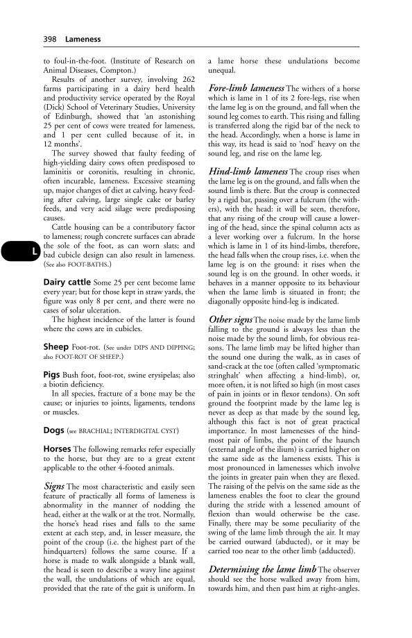
to foul-in-the-foot. (Institute of Research onAnimal Diseases, Compton.)
Results of another survey, involving 262farms participating in a dairy herd health and productivity service operated by the Royal(Dick) School of Veterinary Studies, Universityof Edinburgh, showed that ‘an astonishing 25 per cent of cows were treated for lameness,and 1 per cent culled because of it, in 12 months’.
The survey showed that faulty feeding ofhigh-yielding dairy cows often predisposed tolaminitis or coronitis, resulting in chronic,often incurable, lameness. Excessive steamingup, major changes of diet at calving, heavy feed-ing after calving, large single cake or barleyfeeds, and very acid silage were predisposingcauses.
Cattle housing can be a contributory factorto lameness; rough concrete surfaces can abradethe sole of the foot, as can worn slats; and bad cubicle design can also result in lameness.(See also FOOT-BATHS.)
Dairy cattle Some 25 per cent become lameevery year; but for those kept in straw yards, thefigure was only 8 per cent, and there were nocases of solar ulceration.
The highest incidence of the latter is foundwhere the cows are in cubicles.
Sheep Foot-rot. (See under DIPS AND DIPPING;also FOOT-ROT OF SHEEP.)
Pigs Bush foot, foot-rot, swine erysipelas; alsoa biotin deficiency.
In all species, fracture of a bone may be thecause; or injuries to joints, ligaments, tendonsor muscles.
Dogs (see BRACHIAL; INTERDIGITAL CYST)
Horses The following remarks refer especiallyto the horse, but they are to a great extentapplicable to the other 4-footed animals.
Signs The most characteristic and easily seenfeature of practically all forms of lameness isabnormality in the manner of nodding thehead, either at the walk or at the trot. Normally,the horse’s head rises and falls to the sameextent at each step, and, in lesser measure, thepoint of the croup (i.e. the highest part of thehindquarters) follows the same course. If ahorse is made to walk alongside a blank wall,the head is seen to describe a wavy line againstthe wall, the undulations of which are equal,provided that the rate of the gait is uniform. In
a lame horse these undulations becomeunequal.
Fore-limb lameness The withers of a horsewhich is lame in 1 of its 2 fore-legs, rise whenthe lame leg is on the ground, and fall when thesound leg comes to earth. This rising and fallingis transferred along the rigid bar of the neck tothe head. Accordingly, when a horse is lame inthis way, its head is said to ‘nod’ heavy on thesound leg, and rise on the lame leg.
Hind-limb lameness The croup rises whenthe lame leg is on the ground, and falls when thesound limb is there. But the croup is connectedby a rigid bar, passing over a fulcrum (the with-ers), with the head: it will be seen, therefore,that any rising of the croup will cause a lower-ing of the head, since the spinal column acts asa lever working over a fulcrum. In the horsewhich is lame in 1 of its hind-limbs, therefore,the head falls when the croup rises, i.e. when thelame leg is on the ground: it rises when thesound leg is on the ground. In other words, itbehaves in a manner opposite to its behaviourwhen the lame limb is situated in front; thediagonally opposite hind-leg is indicated.
Other signs The noise made by the lame limbfalling to the ground is always less than thenoise made by the sound limb, for obvious rea-sons. The lame limb may be lifted higher thanthe sound one during the walk, as in cases ofsand-crack at the toe (often called ‘symptomaticstringhalt’ when affecting a hind-limb), or,more often, it is not lifted so high (in most casesof pain in joints or in flexor tendons). On softground the footprint made by the lame leg isnever as deep as that made by the sound leg,although this fact is not of great practicalimportance. In most lamenesses of the hind-most pair of limbs, the point of the haunch(external angle of the ilium) is carried higher onthe same side as the lameness exists. This ismost pronounced in lamenesses which involvethe joints in greater pain when they are flexed.The raising of the pelvis on the same side as thelameness enables the foot to clear the groundduring the stride with a lessened amount offlexion than would otherwise be the case.Finally, there may be some peculiarity of theswing of the lame limb through the air. It maybe carried outward (abducted), or it may be carried too near to the other limb (adducted).
Determining the lame limb The observershould see the horse walked away from him,towards him, and then past him at right-angles.
398 Lameness
L
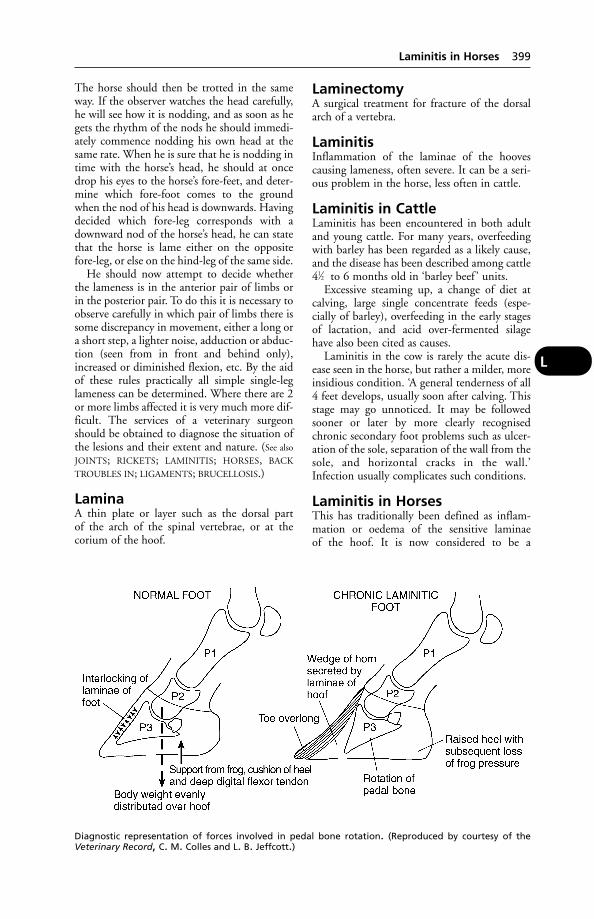
The horse should then be trotted in the sameway. If the observer watches the head carefully,he will see how it is nodding, and as soon as hegets the rhythm of the nods he should immedi-ately commence nodding his own head at thesame rate. When he is sure that he is nodding intime with the horse’s head, he should at oncedrop his eyes to the horse’s fore-feet, and deter-mine which fore-foot comes to the groundwhen the nod of his head is downwards. Havingdecided which fore-leg corresponds with adownward nod of the horse’s head, he can statethat the horse is lame either on the oppositefore-leg, or else on the hind-leg of the same side.
He should now attempt to decide whetherthe lameness is in the anterior pair of limbs orin the posterior pair. To do this it is necessary toobserve carefully in which pair of limbs there issome discrepancy in movement, either a long ora short step, a lighter noise, adduction or abduc-tion (seen from in front and behind only),increased or diminished flexion, etc. By the aidof these rules practically all simple single-leglameness can be determined. Where there are 2or more limbs affected it is very much more dif-ficult. The services of a veterinary surgeonshould be obtained to diagnose the situation ofthe lesions and their extent and nature. (See also
JOINTS; RICKETS; LAMINITIS; HORSES, BACK
TROUBLES IN; LIGAMENTS; BRUCELLOSIS.)
LaminaA thin plate or layer such as the dorsal part of the arch of the spinal vertebrae, or at thecorium of the hoof.
LaminectomyA surgical treatment for fracture of the dorsalarch of a vertebra.
LaminitisInflammation of the laminae of the hoovescausing lameness, often severe. It can be a seri-ous problem in the horse, less often in cattle.
Laminitis in CattleLaminitis has been encountered in both adultand young cattle. For many years, overfeedingwith barley has been regarded as a likely cause,and the disease has been described among cattle41⁄2 to 6 months old in ‘barley beef ’ units.
Excessive steaming up, a change of diet atcalving, large single concentrate feeds (espe-cially of barley), overfeeding in the early stagesof lactation, and acid over-fermented silagehave also been cited as causes.
Laminitis in the cow is rarely the acute dis-ease seen in the horse, but rather a milder, moreinsidious condition. ‘A general tenderness of all4 feet develops, usually soon after calving. Thisstage may go unnoticed. It may be followedsooner or later by more clearly recognisedchronic secondary foot problems such as ulcer-ation of the sole, separation of the wall from thesole, and horizontal cracks in the wall.’Infection usually complicates such conditions.
Laminitis in HorsesThis has traditionally been defined as inflam-mation or oedema of the sensitive laminae of the hoof. It is now considered to be a
Laminitis in Horses 399
L
Diagnostic representation of forces involved in pedal bone rotation. (Reproduced by courtesy of theVeterinary Record, C. M. Colles and L. B. Jeffcott.)
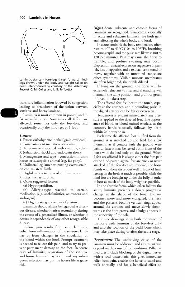
transitory inflammation followed by congestionleading to breakdown of the union betweensensitive and horny laminae.
Laminitis is most common in ponies, and infat or unfit horses. Sometimes all 4 feet areaffected; sometimes only the fore-feet; andoccasionally only the hind-feet or 1 foot.
Causes1. Excess carbohydrate intake (‘grain overload’).2. Post-parturient metritis septicaemia.3. Toxaemia – associated with enteritis, colitis X (exhaustion shock) and endotoxin shock.4. Management and type – concussion in unfithorses or susceptible animal (e.g. fat pony).5. Unilateral leg lameness putting excess strainon contra-lateral limb.6. High-level corticosteroid administration.7. Fatty liver syndrome.8. Other suggested factors:
(a) Hypothyroidism.(b) Allergic-type reaction to certain
medication (e.g. anthelmintics, oestrogens and androgens).
(c) High oestrogen content of pasture.Laminitis should always be regarded as a seri-
ous disease, whether it arises secondarily duringthe course of a generalised illness, or whether itoccurs independently of any other recognisabledisease.
Intense pain results from acute laminitis,either from inflammation of the sensitive lami-nae or from changes in the circulation of the blood within the hoof. Prompt treatment is needed to relieve this pain, and to try to pre-vent permanent damage to the foot. In severecases of laminitis, separation of the sensitiveand horny laminae may occur, and any subse-quent infection may put the horse’s life at graverisk.
Signs Acute, subacute and chronic forms oflaminitis are recognised. Symptoms, especiallyin acute and subacute laminitis, are both gen-eral, affecting the whole body, and local.
In acute laminitis the body temperature oftenrises to 40° to 41°C (104 to 106°F), breathingbecomes rapid, and the pulse rate likewise (80 to120 per minute). Pain may cause the horse totremble, and profuse sweating may occur.Depression, a facial expression suggestive of painfelt, loss of appetite, and a reluctance to stand ormove, together with an unnatural stance areother symptoms. Visible mucous membranesare often bright red, the pupils dilated.
If lying on the ground, the horse will beextremely reluctant to rise; and if standing willmaintain the same position, and grunt or groanif forced to take a step.
The affected feet feel hot to the touch, espe-cially at the coronet, and a bounding pulse inthe digital arteries can be felt or even seen.
Tenderness is evident immediately any pres-sure is applied to the affected feet. The appear-ance of blood, or blood-stained exudate, at thecoronary bands is usually followed by deathwithin 24 hours or so.
Each time the affected foot is lifted from theground, it is snatched up and held for a fewmoments as if contact with the ground werepainful; later it may be rested out in front of thehorse with the heel only on the ground. When 2 feet are affected it is always either the fore-pairor the hind-pair; diagonal feet are rarely or neverattacked. If the fore-feet are involved, the horsestands with these thrust out well in front of him,resting on the heels as much as possible, while thehind-feet are brought up under the belly in orderto bear as much of the body-weight as possible.
In the chronic form, which often follows theacute, laminitis presents a slowly progressivechange in the shape of the foot. The toebecomes more and more elongated, the heelsand the pasterns become vertical, rings appeararound the coronet and move slowly down-wards as the horn grows, and a bulge appears inthe concavity of the sole.
The line drawings show both the stance ofthe horse with laminitis of the fore-feet only,and also the rotation of the pedal bone whichmay take place during or after the acute stage.
Treatment The underlying cause of thelaminitis must be addressed and treatment willdepend on the cause of the condition. Palliativemeasures include blocking of the digital nerveswith a local anaesthetic: this gives immediaterelief from pain, enables the horse to stand andwalk normally, and has a beneficial effect on
400 Laminitis in Horses
L
Laminitic stance – fore-legs thrust forward, hind-legs drawn under the body and weight taken onheels. (Reproduced by courtesy of the VeterinaryRecord, C. M. Colles and L. B. Jeffcott.)
the blood circulation of the foot; however, caremust be taken that further damage is not causedto the laminae by exercise. For the relief of painacetylpromazine is also used, and this drugtends to reduce blood pressure. Phenylbutazoneis another drug which has been used, and simi-larly corticosteroids. Warm or hot water appli-cations to the feet are regarded as preferable tohosing with cold water.
Green food in small amounts is good, and alittle hay should be supplied.
In chronic cases the shoeing is of greatimportance and special surgical shoes may beneeded. (See also HOOF REPAIR.)
LampasA swelling of the mucous membrane of thehard palate of the horse immediately behind thearch of the incisor teeth in the upper jaw. It isoften seen about the time when the permanentteeth are cutting through the gums, i.e. at 21⁄2 ,31⁄2, and 41⁄2 years, and for a short time after-wards. It is erroneously thought that it is thecause of a falling-off in condition which natu-rally occurs when the teeth are cutting; it is really rather an effect. It was the custom tolance ‘lampas’ in many parts of the country; thisoccasions unnecessary pain and discomfort to the horse, and if the incision is made towards1 side instead of in the middle-line there is aserious risk of wounding the palatine artery onthat side.
LampreysPrimitive fish that are parasites on other fish.There are saltwater and freshwater species, thefreshwater species being larger (up to 50 cmlong). They can be a problem for freshwater fishfarms, particularly in the USA.
LamziekteLamziekte is botulism of cattle in South Africawhich occurs as an enzootic in animals onphosphorus-deficient areas of the veldt. Duringwinter, lack of phosphorus leads grazing cattleto chew the bones of animals (often cattle) thathave died, in an endeavour to take phosphorusinto the body to make good the deficiency. Thiscondition of bone-eating (osteophagia) is actu-ally only the result of a craving for minerals.Where the animals whose skeletons are left onthe veldt harboured in their alimentary canalsClostridium botulinum, this organism invadesthe carcase, and both it and its toxin are presentin the decomposing remains.
Prevention The researches of Sir ArnoldTheiler and the workers at Onderstepoort
showed that the best means of preventinglamziekte is to feed sterilised bone- meal to cattle during the winter months in areas whichare naturally deficient in phosphorus. (See
BOTULISM.)
LandraceA large white, lop-eared pig used to producecommercial hybrid breeds.
LanolinA type of fat found in sheep wool. It is widelyused in ointments and creams.
Lantana PoisoningLantana poisoning of cattle and sheep hasoccurred in Australia and New Zealand. L.camara is the species commonly involved; espe-cially the red-flowered variety. It causes lightsensitisation, with exudative dermatitis of teatsand vulva. Deaths have occurred.
LaparoscopyThe use of optical instruments for viewing theinterior of organs such as the bladder, the inte-rior of joints for signs of arthritis, etc., and foravian sex determination.
LaparotomyLaparotomy means surgical opening of theabdominal cavity. The incision is either made inthe middle line of the abdomen, or through oneor other of the flanks.
LapinisedThis term is applied to a virus which has beenattenuated by passage through rabbits. An exam-ple is afforded by lapinised swine fever vaccine.
Larkspur PoisoningOf the several varieties of larkspur, most ofwhich occur in America in the ranges of theWest, where they cause great loss to cattle own-ers, only 1 species is commonly found in Britain– Delphinium ajacis. The seeds are the mostdangerous parts of the plant, although the leaveshave proved fatal when fed experimentally.Horses and sheep are not as susceptible as cattle.The active principles are 4 in number: delphine,delphisine, delphinoidine and staphisagrine,and of these the first 3 are highly poisonous.
Signs Salivation, vomiting, colicky pains, convulsions, and general paralysis.
Laryngeal ObstructionChronic obstruction of the larynx in cattle ischaracterised by difficult or painful inspiration,
Laryngeal Obstruction 401
L

giving rise to ‘ROARING’. The most commoncauses are calf diphtheria (necrobacillosis); butthere is uncertainty as to the primary infectiveagent causing chronic laryngitis – Fusebacteriumnecroforum, for instance, is unable to penetrateintact mucous membrane.
LaryngitisInflammation of the larynx (see LARYNX,DISEASES OF).
LarynxLarynx is the organ of voice, and also forms oneof the parts of the air passage. It is placed just between, and slightly behind, the angles ofthe lower jaw. Externally it is covered by theskin, by a small amount of fibrous tissue, andsterno-thyro-hyoid muscles.
Structure The cricoid cartilage is shapedsomewhat like a signet ring and connects therest of the larynx with the 1st ring of the trachea.To its upper part are attached the arytenoids andthe posterior horns of the thyroids. A crico-tra-cheal membrane unites it to the trachea, and acrico-thyroid membrane unites it to the thyroidcartilage. The thyroid cartilage possesses a bodywhich in man forms the protuberance known asAdam’s apple. The epiglottis lies in front of thebody of the thyroid and curves forwards towardsthe root of the tongue; it is shaped somewhatlike a pointed ovate leaf. The arytenoids are situated one on either side of the upper part ofthe cricoid to which they are attached. (Forfunctions, see under VOICE.)
Larynx, Diseases of(see also ‘ROARING’; WHISTLING; COUGHING)
Laryngitis is an inflammation of the larynx,but particularly of the mucous membranewhich lines its interior. It is often associatedwith pharyngitis or with bronchitis and tra-cheitis, when it is usually due to the spreadingof inflammation from one of these neighbour-ing structures.
In the horse it may occur during influenza.(See also LATHYRISM.)
Signs In ordinary cases there is a cough, diffi-culty in swallowing, pain on pressure over thelarynx, extension of the head to relieve pressureon the throat (a condition that is aptly describedin popular terms as ‘star gazing’). A wheezing orroaring sound accompanies breathing if mem-branes become so swollen as to interfere withrespiration. A slight rise in temperature andpulse-rate accompanies the milder forms, but
when influenza is present, or if other specificdiseases arise, the signs of fever are more dis-tinct. Uncomplicated laryngitis usually lastsfrom a week to about a fortnight. Occasionallycomplications, such as roaring or whistling, follow recovery from the initial disease.
First-aid It is advisable to isolate all cases oflaryngitis in a loose-box or other building, espe-cially those arising in newly purchased animals,on account of the risk of contagious diseasedeveloping. (See NURSING.)
Wounds of the larynx are not common,owing to its comparatively sheltered position inthe body, but see under DRENCHING for a dangerassociated with the use of a drenching gun inpigs and sheep.
Foreign Bodies(see CHOKING)
Laryngeal Paralysis in HorsesLaryngeal paralysis in horses causes the abnormal inspiratory sound called ‘roaring’.
The usual cause was for long regarded asvibration of the slackened vocal folds on one orboth sides of the larynx, due to paralysis of themuscles which move the arytenoid cartilagesoutwards. Laryngeal paralysis is probably ahereditary condition transmitted by a simplerecessive factor.
A large number of respiratory diseases maygive rise to a temporary roaring due to inflam-mation and thickening of the mucous mem-branes lining the larynx. GUTTURAL POUCH
DISEASE may have a permanent effect.
Treatment The traditional Hobday operationentailed encouraging the vocal fold to adhere tothe wall of the larynx, out of the path of theentering stream of air, by stripping the liningmembrane from a little pouch which liesbetween the vocal cord and the laryngeal wall.Tracheotomy is an alternative: in this, a metaltube is inserted into the trachea at a lower levelthan the larynx, so that air is able to enter andleave through the tube instead of through thelarynx. Tracheotomy is of most use in racehors-es and hunters affected with roaring, whichconstitutes an unsoundness.
Abnormal inspiratory noises during exercise,particularly in young horses which may havepharyngitis and laryngitis, should not be takento indicate one-sided paralysis of the larynx.Similarly, normal respiratory sounds at exerciseshould not be regarded as implying soundnessof the upper respiratory tract.
402 Laryngitis
L
Laryngoplasty is sometimes used for thetreatment of roaring, especially in those horsesnot required to perform at high speeds. Theoperation involves securing the arytenoid carti-lage in a lateral position, using prostheses toprevent intrusion of the arytenoid cartilage andvocal cord into the lumen of the larynx.
Poisoning Four 2-year-old thoroughbredssuffered an acute gastrointestinal illness shortlyafter being dosed with contaminated mineraloil. Three weeks later they had developed bilat-eral laryngeal paralysis. Two of the horses diedduring severe bouts of dyspnoea 6 and 8 weekslater, and a 3rd was put down. In these horsesthere was a severe loss of myelinated fibres fromboth recurrent laryngeal nerves. The 4th horsehad bilateral pharyngeal paralysis 2 years later.The acute clinical signs and delayed neurologi-cal effects were typical of ORGANOPHOSPHORUS
POISONING.
LasalocidA coccidiostat used as a feed additive in the pre-vention of coccidiosis in chickens, turkeys andgame birds. It must not be used in breeding orlaying birds; birds may be slaughtered only after5 days from the last administration of the drug.
LaserAn acronym for light amplification by stimu-lated emission of radiation.
Lasers emit beams of intense, monochromat-ic, non-dispersing light, and can be used aspowerful sources of localised energy. They areused in ophthalmic and other surgery and maybe used instead of needles in acupuncture.Operators must wear protective glasses toShield their eyes.
Lassa FeverThis disease occurs in West Africa, and iscaused by an arenavirus first isolated in 1969.In man the infection is likely to prove fatal. Thevirus has been isolated from the rat Mastomysnatalensis, which (possibly with other rodents)acts as a reservoir of infection.
Lateral CartilagesLateral cartilages are rhomboid plates of carti-lage which are attached, one on either side, toangles of the 3rd phalanx (os pedis) of the foot of the horse. They extend above the coro-net sufficiently to be felt distinctly at the heelsand for a certain distance in front of this. In oldage they often become ossified in their lowerparts. When they ossify in their upper palpablemargins, the name ‘sidebones’ is applied to the
condition. In certain cases be cartilages maybecome injured from treads or tramps by neigh-bouring horses, or from the other foot; the cartilage, being poorly supplied with blood,undergoes necrosis. (See SIDEBONES; QUITTOR;FOOT OF THE HORSE.)
Lateral LineA structure along the sides of fish that is sensi-tive to movement in the water, enabling the fishto detect the presence of other fish, currents, etc.
Latex (Natural Rubber)Hypersensitivity to this can result in contacturticaria, respiratory symptoms, and shock.The main source of the allergens is the wearingof rubber gloves during surgery. Even a vaginalexamination can result in an anaphylactic reaction in atopic people.
In the rubber-growing areas of Malaysia, the ingestion of latex by cattle, e.g. from buckets left by rubber-tappers, is a ‘frequentoccurrence’, and can be fatal.
In 1 reported case, 2 bulls consumed 9 and14 litres, respectively, of latex from the treeHevea brasiliensis. Rumenotomy brought a temporary improvement in both bulls, but theydied, despite supportive treatment, 11 daysafter ingesting the latex.
Latex agglutination test This can beused for measuring the concentration of IgG1 inthe plasma of newborn calves. The commercialtest reagent (Ab-Ag Laboratories, Ely) is pre-pared by coating polystyrene latex beads withantibodies against bovine IgG1.
Lathyrism (Lathyrus Poisoning)Lathyrism (Lathyrus Poisoning) is caused byfeeding upon one of the various ‘Mutter peas’ –Lathyrus sativus principally, and L. cicera and L. clymenum, less frequently. The latter 2 sam-ples of field peas grown in Southern Europe and North Africa, while L. sativus is importedfrom India mainly. They are poisonous to allthe domesticated animals, but seem especiallydangerous for horses. Many outbreaks havebeen recorded, and in most the percentage ofdeaths has been high, sometimes as much as 50 per cent of the affected.
Symptoms of poisoning may not appearuntil the lapse of as much as 50 days after thepeas cease to be used as a food-stuff. The causeof lathyrism is the high selenium content of theplants. (See SELENIUM.)
Signs usually become visible when the animalis put to work or exercised. Typically, the chief
Lathyrism (Lathyrus Poisoning) 403
L

symptoms are those of paralysis of some part ofthe body – usually the hind-limbs and therecurrent laryngeal nerve. This latter gives riseto the condition known as ‘ROARING’, andunless quickly relieved, the horse will die fromasphyxia. In some instances the symptoms areso sudden in their onset that the horse dropswhile in harness and is unable to rise. In lesssevere cases there is staggering and swaying ofthe hindquarters, great difficulty in breathing, afast, weak pulse, and convulsive seizures. Theparoxysms may pass off in a few minutes, or the horse may collapse and die.
Treatment (See under LARYNX, DISEASES OF.)The antidote is ascorbic acid, added to the diet.
Laudanum(see OPIUM)
Laurel (Laurus) PoisoningThe leaves of laurel shrubs and trees (family:Lauraceae) contain cyanogentic GLYCOSIDES
which cause poisoning by HYDROCYANIC ACID.
LavageThe process of washing out the stomach or theintestines. In gastric lavage, a double-way tubeis passed down into the stomach either throughthe mouth or by way of the nose, and water orsome medicinal solution poured or pumpedthrough one channel in the tube. After a timethis escapes by the other, carrying with it thecontents of the stomach in small amounts. (See
also ENEMA.)
LawLaw, relating to the veterinary profession andveterinary practice, scientific research, domesticpets, farm animals, wild animals, and zoos, isextensive and subject to frequent amendment.The Scottish Parliament can bring in its ownActs, and both it and the Welsh Assembleybring in their own Orders and Regulations.Parallel legislation for the different parts of the UK exists for entries identified with anasterisk (*). Later legislation may partiallyrevoke that made previously.
Existing legislation includes the follow-ing (where appropriate, information on thetopic, animal or disease covered by the legis-lation listed will be found under individualentries):
Abandonment of Animals Act 1960African Swine Fever Order 1980African Swine fever (Compensation) Order
1980
Agriculture (Miscellaneous Provisions) Act 1969; 1972.
Agriculture (Poisonous Substances) Act 1952Animal Boarding Establishments Act 1963Animal By-Products Order 1999; Amendment
2002*Animal By-Products (Identification)
(Amendment) Regulations 2002Animal Gatherings (Interim Measures)
(Amendment) Order 2002*; No 2 Order2002*
Animal Health Act 1981Animal Health Act 2002Animal Health (Amendment) Act 1998Animal Health and Welfare Act 1984Animal Health Orders (Divisional Veterinary
Manager Amendment) Order 1995Animals Act 1971Animals Act (Amendment) Regulations 1991Animals and Animal Products (Import and
Export) Regulations 2002* (Amendment2002*)
Animals (Cruel Poisons) Act 1962Animals, Meat and Meat Products
(Examination for Residues and Maximum Residue Limits) Act 1991(amended 1993)
Animals (Post-Import Control) Order 1995Animals (Scientific Procedures) Act 1986, as
amended 1998Animals (Scotland) Act 1987Animals (Third Country Imports) (Charges)
Regulations 1997Antarctic Treaty Act 1994Antarctic (Amendment) Regulations 2002Anthrax Order 1991 (amended 1996)Artificial Breeding of Sheep and Goats
Regulations 1993Artificial Insemination of Cattle (Animal
Health) (England & Wales) Regulations1985 (amended 1992,1995, 2002*)
Aujeszky’s Disease Order 1983Aujeszky’s Disease (Compensation for Swine)
Order 1983Authorised Officers (Meat Inspection)
Regulations 1987
Badgers Act 1992Badgers (Further Protection) Act 1991Bovine Animals (Records, Identification and
Movement) Order 1995Bovine Embryo Collection and Transfer
Regulations 1995Bovine Spongiform Encephalopathy (No 2)
Order 1996Bovine Spongiform Encephalopathy
Compensation (Amendment) Order 1997
404 Laudanum
L
Bovines and Bovine Products (Despatch,Prohibition and Production Restriction)Regulations 1997
Bovines and Bovine Products (Trade)Regulations 1999
Breeding of Dogs Act 1973 (amended 1991)Breeding of Dogs (Licensing Records)
Regulations 1999*Breeding and Sale of Dogs (Welfare) Act 1999Brucellosis (England) Order 2000*Brucellosis and Tuberculosis (England &
Wales) Order 1978*Brucellosis and Tuberculosis (England & Wales)
Compensation (Amendment) Order 1981BSE Monitoring Order 2001
Cattle Identification (Enforcement)Regulations 1997
Cattle Identification Regulations 1998(amended 1999)
Cattle (Identification of Older Animals)Regulations 2000*
Cattle Passports Order 1996Cattle Plague Order 1928 (amended 1938)Chemicals (Hazard Information and Packing
for Supply) Regulations 1994Cinematograph Films (Animals) Act 1937Cock Fighting Act 1954Collar and Tags (Control of Dogs) Order 1991Collection and Disposal of Waste Regulations
1989Conservation (Natural Habitats, etc.)
Regulations 1994Conservation of Seals Act 1970Conservation of Seals (England) Order 1999Control of Dogs Order 1930Control of Dogs Order 1992Control of Endangered Species (Designated
Ports of Entry) Regulations 1985Control of Substances Hazardous to Health
Regulations 1994, 1999Countryside and Rights of Way Act 2000Coypus (Keeping) Order 1987Criminal Damage Act 1971Cruel Tethering Act 1988
Dangerous Dogs Act 1989, 1991, 1997Dangerous Dogs (Designated Types) Order
1991Dangerous Wild Animals Act 1976;
Modification Order 1984Dangerous Wild Animals Licensing Act 1984Deer Act 1991Deer (Scotland) Act 1995; amended 1996Destructive Imported Animals Act 1932Disease Control (Interim Measures)
(Amendment) Order; No 2 Order; No 3Order 2000*
Diseases of Animals (Approved Disinfectants)Order 1978; amended 1997, 2000*, 2001*
Diseases of Animals (Ascertainment ofCompensation) Order 1959
Diseases of Animals Ascertainment of Disease)Order 1985
Diseases of Animals (Export HealthCertificates) Order 1985
Diseases of Animals (Milk Treatment) Order1967 (amended 1968)
Diseases of Animals (Seizure) Order 1993Diseases of Animals (Therapeutic Substances)
Order 1952; amended 1953, 1981Diseases of Animals (Waste Food) Order 1973
(amended 1987)Diseases of Fish Act 1983Diseases of Fish (Control) Regulations 1994,
2000Diseases of Poultry Order 1994 (amended
1997)Docking and Nicking of Horses Act 1949Dogs Act 1871Dogs Act 1902Dogs Act 1906Dogs (Amendment) Act 1928Dogs (Fouling of Land) Act 1906Dogs (Protection of Livestock) Act 1953
Employment ActEndangered Species (Import and Export)
Act 1976Endangered Species (Enforcement)
Regulations 1985Environmental Protection ActEnvironmental Protection (Restriction on Use
of Lead Shot) (Amendment) Regulations2002
Enzootic Bovine Leukosis Order 1980, 2000*Equine Viral Arteritis Order 1995Export of Animals (Protection) Order 1981Export of Horses (Protection) Order 1969Export of Horses (Veterinary Examination)
Order 1966Export of Sheep (Prohibition) Order) 1991Export Quarantine Stations (Regulation)
Order 1973Export of Pigeons Order 1983
Farriers Registration Act 1975; amended 1997Feeding Stuffs Regulations 1995; amended
2002Feeding Stuffs (Establishments and
Intermediaries) Regulations (1998)Feedingstuffs (Zootechnical Products)
Regulations 1998Firearms Act 1997Fisheries Act 1981Fish Health Regulations 1997
Law 405
L

Food Act 1984Food and Environmental Protection Act 1985Food Safety Act 1990Foot-and-Mouth Disease Order 1983;
amended 1993; amendment orders Nos 2, 3, 7, 8 9, 10, 11, 12, 13, 14, all made in 2001
Foot-and-Mouth (Infected Areas)(Vaccination) Order 1972
Fresh Meat (Beef Controls) (No 2)Regulations 1996
Fresh Meat (Hygiene and Inspection)Regulations 1995
Fur Farming (Prohibition) Act 2000
Game Act 1831Game Laws (Amendment) Act 1960Gelatin (Intra-community Trade) Regulations
2001*Grey Squirrels (Prohibition of Importation
and Keeping) Order 1937Grey Squirrels (Warfarin) Order 1973Ground Game Act 1888, 1975Guard Dogs Act 1975
Hares Act 1848Hares (Control of Importation) Order 1965Hares Preservation Act 1880Health and Safety at Work Act 1974Highways ActHorse Passports Order 1997 (amended 1998)
Importation of Animal Pathogens Order 1980
Importation of Animal Products and PoultryProducts Order 1980 (amended, 1982,1994)
Importation of Animals and Animal Productsand Poultry Products (Amendment) Order1980, 1981, 1994
Importation of Animals Order 1977 (amended1996)
Importation of Birds, Poultry and HatchingEggs Order 1979
Importation of Bovine Semen (Amendment)Regulations 1984, 1993
Importation of Embryos, Ova and SemenOrder 1980 (amended 1984, 1993)
Importation of Equine Animals Order 1979Importation of Hay and Straw Order 1979Importation of Processed Animal Protein
Order 1981 (amended 1982)Importation of Salmonid Viscera Order 1986Infectious Diseases of Horses Order 1987Ionising Radiation Regulations 1985
Live Poultry (Movement Records) Order 1958Local Government Act 1988
Marketing Authorisation for VeterinaryMedicinal Products Regulations 1994
Markets, Sales and Lairs Order 1925 (amended 1927)
Meat and Meat Products (HormonalSubstances) Regulations 1989
Meat (Disease Control) Regulations 2000*Meat Inspection Regulations 1987 (amended
1990)Meat Sterilisation and Staining Regulations
1982 (amended 1984)Medicated Feedingstuffs Regulations 1998Medicines Act 1968Medicines (Exemptions for Merchants in
Veterinary Drugs) Order 1998Medicines (Labelling of Medicated Animal
Feedingstuffs) Regulations 1973Medicines (Medicated Animal Feedingstuffs)
Regulations 1989Medicines (Restriction on the Administration
of Veterinary Medicinal Products)Regulations 1994
Medicines (Veterinary Medicinal Products)(Veterinary Surgeons from Other EEAStates) Regulations 1994
Milk and Dairy Regulations 1959Minced Meat and Meat Preparations
(Hygiene) Regulations 1995Mink Keeping Order 1990Movement of Animals (Restriction) Order
1990Movement of Livestock Order 1990
Night Poaching Act 1828, 1844Non-Indigenous Rabbits (Prohibition of
Importation and Keeping) Order 1954
Occupier’s Liability Act 1986
Performing Animals (Regulations) Act 1925
Performing Animals (Protection) Act 1952Pests Act 1954Pet Animals Act 1951Pet Travel Scheme (Pilot Arrangements) Order
2000* (amended 2000, 2001, 2002)Pigs (Record, Identification and Movement)
Order 1995Poaching Prevention Act 1862Poultry Breeding Flocks and Hatcheries Order
1993Poultry Meat, Farmed Game Meat and
Rabbit Meat (Hygiene and Inspection)Regulations 1995
Pleuro-Pneumonia Order 1928Police and Criminal Evidence Act 1984Poultry Breeding Flocks and Hatcheries
Order 1993
406 Law
L
Poultry Breeding Flocks and Hatcheries and Animal By-Products (Fees) Order 2002
Poultry Laying Flocks (Collection andHandling of Eggs and Control of Vermin)Order 1989
Poultry Meat, Farmed Game Meat and RabbitMeat (Hygiene and Inspection) Regulations1995
Poultry (Seizure of Hatching Eggs) Order1990
Prevention of Damage by Rabbits Act 1932Processed Animal Protein Order 1989Products of Animal Origin (Import and
Export) Regulations 1992Products of Animal Origin (Third Country
Imports) (Charges) Regulations 1992(amended 2002)
Protection Against Cruel Tethering Act 1988Protection of Animals Act 1911*Protection of Animals Act 1934 (amended
1988, 2000)Protection of Animals (Anaesthetic) Act 1954
(amended 1964, 1982)Protection of Animals (Penalties) Act 1987Protection of Animals (Scotland) Act 1993Protection of Badgers Act 1902Psittacosis or Ornithosis Order 1953
Rabies (Compensation) Order 1976Rabies (Control) Order 1974Rabies (Importation of Dogs, Cats and Other
Mammals) Order 1974 (amended 1977,1986, 1994)
Racing Pigeons (Vaccination) Order 1994Removal of Antlers in Velvet (Anaesthetics)
Order 1980Restriction on Pithing Regulations 2001*Riding Establishments Act 1964 (amended
1970)Road Traffic Act 1988
Salmon Act 1984Salmon and Freshwater Fisheries Act 1975Selective Cull (Enforcement of Community
Compensation Conditions) Regulations1996
Sheep and Goats (Records, Identification andMovement) Order 1996
Sheep and Goats Identification Order 2000*Sheep and Goats Identification and Movement
(Interim Measures) (Amendment) Order2002* (amended 2002*)
Sheep and Goats Spongiform EncephalopathyRegulations 1998
Sheep Scab Order 1997Shellfish and Specified Fish (Third Country
Imports) Order 1992
Slaughterhouses (Hygiene) and MeatInspection (Amendment) Regulations 1991(amended 1997)
Slaughterhouses (Hygiene) Regulations 1997Specified Animal Pathogens Order 1993Specified Diseases (Notification and Slaughter)
Order 1992Specified Risk Material Order 1997 (amended
2000*)Specified Risk Material Regulations 1997
(amended 2000*, 2001*)Swine Fever Order 1963 (amended 1991)Swine Fever (Movement Restrictons) Order
2000Swine Vesicular Disease Order 1972 (amended
1973)
Testing of Poultry Flocks Order 1989 TheftAct 1968
Transit of Animals Order 1927Transit of Animals (Road and Rail) Order 1975Transport of Animals (Cleansing and
Disinfection) No 2 Order 2000*TSE Regulations 2002 (amended 2002*)Tuberculosis (Deer) Order 1989 (amended
1993)Tuberculosis (Deer) Notice of Intended
Slaughter and Compensation Order 1989Tuberculosis (England & Wales) Order 1984
(amended 1990)Tuberculosis (Scotland) Order 1984
Veterinary Surgeons Act 1966Veterinary Surgeons Act 1966 (Schedule 3
Amendment) Order 1988, 2002Veterinary Surgery (Blood Sampling)
(Amendment) Order 1988Veterinary Surgery (Epidural Anaesthesia)
Order 1992Veterinary Surgery (Exemption) Order 1973,
1990Veterinary Surgeons (Rectal Ultrasound
Scanning of Bovines) Order 2002Video Recordings Act 1984
Warble Fly (Ascertainment of Infestation)(Engand & Wales) Order 1985
Warble Fly (England & Wales) (InfectedAreas) Order 1985
Warble Fly (England & Wales) Order 1982(amended 1978, 1985, 1987, 1989)
Warble Fly (Scotland) Order 1982Water Regulations Act 1991Welfare of Animals at Markets Order 1990
(amended 1993)Welfare of Animals at Slaughter Act 1991Welfare of Animals (Slaughter or Killing)
Regulations 1995
Law 407
L
Welfare of Animals (Staging Posts) Order1998
Welfare of Animals (Transport) Order 1997Welfare of Horses at Markets (and Other
Places of Sale) Order 1990Welfare of Livestock Regulations 1994Wild Mammals Protection Act 1996Wildlife and Countryside Act 1981 (variations
to schedules orders 1989, 1991, 1992,1994, 1998)
Wildlife and Countryside (Registration andRinging of Certain Captive Birds)Regulations 1982 (amended 1982, 1994)
Wildlife and Countryside (Registration to Sell,etc. Certain Dead Wild Birds) Regulations1982 (amended 1991)
Wild Mammals (Protection) Act 1996
Zoo Licensing Act 1981 (amended 2002)Zoonoses Order 1988, 1989
(See also EUROPEAN UNION.)
LaxativesSENNA, which has been recommended for preg-nant sows; DIHYDROXYANTHRAQUINONE, use-ful in all domestic animals, including horses;EPSOM SALTS (magnesium sulphate), but ofdoubtful efficacy in ruminants; GLAUBER’S
SALTS, but they may have ill-effects in pigs. (See
also PARAFFIN – Uses.)
LD50Ld50value is a statistical estimate of the numberof mg of a given substance per kg of body-weight required to kill 50 per cent of a largepopulation of test animals. The LD value of a compound may refer to oral or parenteraladministration or to application to the skin.
Lead PoisoningAlso called plumbism.
Acute form of lead poisoning
Cattle This is very common in cattle whichhave eaten paint, licked out discarded painttins, licked newly painted railings, etc., orwhich have eaten tarpaulins. It is frequentlyfatal and many cattle are unnecessarily lost eachyear from this cause. Cows have also been fatal-ly poisoned after licking lead-rich ash from aburnt-down shed; and after eating silage conta-minated by an old battery broken up by a for-age harvester; also by eating roofing materialfrom an old railway carriage.
In another instance cows were poisoned aftereating haylage made from grass in a field whichhad been used for clay-pigeon shooting. The
haylage contained small particles of claypigeons and lead shot. The cows in the high-yielding herd of 115 Holsteins began to losetheir appetite, became dull, and had diarrhoea.A few developed stiff and swollen joints. Manybecame uncoordinated in their movements;also there were 25 stillbirths or abortions.Appropriate treatment brought some improve-ment, but 21 cows either died or had to beslaughtered.
A 24-volt lead battery was discarded butunfortunately scooped up with straw beingadded to a ‘complete diet’ in a feeder box. Theresult was that 55 heifers died – some rapidly,some after ataxia, head pressing, teeth grindingand convulsions.
Dogs They are sometimes poisoned througheating paint scrapings where a room is being re-decorated, or after licking out a paint tin.
Cats In one case, old lead paint was stripped bymeans of an electric sander, which dispersedparticles of the primer so that the air soon con-tained a toxic amount of lead. One cat and aninfant suffered lead poisoning as a result.
Pigs have been shown to be less sensitive thanother farm animals.They can consume, withoutshowing symptoms, a daily dose of lead whichwould rapidly kill a cow.
Geese Ten lead pellets can kill a goose.
Swans Many cases have been reported ofswans dying after swallowing lead weights usedby anglers.
Signs Nervous signs are an important featureof lead poisoning, and may include excitement,ataxia, blindness, paresis, and convulsions;affected animals may also show depression.
Cattle may bellow, charge around, and atintervals press their heads against a wall or otherfixed object.
Abdominal pain, sometimes with constipa-tion followed by diarrhoea, are other signs; alsoanaemia in chronic or subacute cases.
In horses, ‘roaring’ (laryngeal paralysis) maybe a sign, together with carpal swelling and posterior paralysis.
Treatment The treatment of lead poisoningwas revolutionised by the introduction of thechelating agent, calcium di-sodium adetate,which converts inorganic lead in the tissues intoa harmless lead chelate which is excreted by thekidneys. The drug must be given intravenously.
408 Laxatives
L
In chronic cases, potassium iodide is given 3 or4 times daily to hasten the elimination of thelead salt from the system. (See also CHELATING
AGENTS.)
Diagnosis of lead poisoning may be made by estimating the lead content of the blood,kidneys or liver.
A differential diagnosis must take intoaccount other possibilities such as hypomagne-saemia, encephalitis, acetonaemia, listeriosis,and poisoning by other substances.
Chronic lead poisoning has occurred as aresult of flaking paintwork in dog kennels, andalso in the proximity of former lead-miningsites. Four out of 5 sheepdogs, in an Australianincident, became agitated after working sheepsatisfactorily for some 20 minutes. They left thework area and retreated to the underside of avehicle or to a kennel.
The behavioural effects of lead poisoning indogs may also include hysteria or aggressiveness.
Falcons kept in painted cages developed leadpoisoning after gnawing at the bars.
Signs Chronic lead poisoning results in recur-rent laryngeal nerve paralysis and paralysis ofthe pharynx.
LeechesBlood-sucking aquatic annelids of the classHirudinea, within phylum Annelida (segment-ed worms).
Leeches live in ponds, streams, and on dampvegetation. They have strong muscular suckers;the anterior one surrounds the mouth which, inseveral species, contains saw-like teeth used topierce the skin of the host. Leeches secretehirudin in their buccal cavity; this prevents clot-ting of the host’s blood, on which they feed, andcan cause a severe and sometimes fatal anaemia.
Leeches live in ponds, streams, and on dampvegetation.
Limnatis nilotica is found in North Africaand Southern Europe. It reaches a length of 10cm. The ventral surface is dark; on the dorsalsurface are 6 longitudinal stripes on a brownish-green background. It cannot penetrate skin, buton being taken in with water by men and ani-mals, it attaches itself to the buccal mucousmembrane. This produces, constant smallhaemorrhages, which sometimes cause a seriousanaemia.
L. africana and species of Haemadipsa areactive in West Africa and in the tropical forestsof Asia and South America, respectively.
H. zeylanica occurs in Asia and lives on land.It is a clear brown colour with a yellow lateralstripe on each side and a greenish dorsal stripe.It has 5 pairs of eyes and 3 teeth. It lives indamp weather on the lower vegetation. Theseleeches are small forms, about 2.5 cm long, butare very serious pests. The bite is painless but, asthey occur in such enormous numbers, verydeadly. They attack all vertebrates and manydifferent species of mammals have been killedby them through sheer loss of blood.
Two cases of infestation of dogs withDiestecostoma mexicanum have been reportedfrom Honduras. In the non-fatal case, acatheter was passed through the inferior nasalmeatus and a 50-ml capacity syringe containingchloroform water attached. The solution wasinjected slowly while the catheter was revolved.Over 70 leeches emerged after treatment.
Legionella PneumophilaThis organism, first discovered in 1976, cantolerate hot water, and is spread by aerosols ofit. People, cattle, sheep, horses, pigs, goats, dogsand cats are all at risk of pneumonia caused bythis infection (Legionaires disease).
Legislation(see LAW)
Legume PoisoningThis may occur when certain legumes are fedraw, and can result in death. Both navy beans(Phaseolus vulgaris) and jack beans (Canavalisensiformis) contain a heat-sensitive toxin whichcan weaken the animal’s resistance to coliformand other bacteria. Heat treatment of the beansrenders them safe. (See also LATHYRISM; LUPINS;POISONING.)
LeishmaniaA genus of protozoon parasites. Each appears asa round or oval body with a micro- and amacro-nucleus.
LeishmaniasisLeishmaniasis is of considerable importance inman, but not in animals other than the dog.Cutaneous leishmaniasis, or ‘oriental sore’, isseen in Iran, India, parts of Africa, and SouthAmerica, and is caused by Leishmania tropica.Visceral leishmaniasis, called also kala-azar or‘dum-dum fever’, occurs in the coastal countriesof the Mediterranean, and is caused by L. dono-vani. (At least 5 other species, and several subspecies, are recognised.) Both forms arediagnosed by laboratory examination. A 3rdform, affecting mucous membrane of mouth
Leishmaniasis 409
L

and nose, is caused by L. brasiliensis, and has apoor prognosis. The disease has beenrecordedin dogs returning from mainland Europe(mainly Spain) under the Pet Travel Scheme.
Signs in the dog include wasting, enlargedlymph nodes, keratitis and/or conjunctivitis,and alopecia or dermatitis. The ears are exces-sively waxy and the wax has a distinct andunusual smell. Dermatitis may develop.
Leishmaniasis is rarer in cats, causing skinulcers, fistulae and enlarged lymph nodes.
Treatment The incubation period in dogsmay be up to 2 years; the prognosis is not good.The treatment of choice is allopurinol at 10 to15 mg/kg bodyweight for 6 months at least; the treatment may actually take longer, depend-ing on the response. A cure is rare. (See FLIES –
‘sandflies’.)
Prevention Housing animals between 19.00and 07.00, when the vector (the sandfly) ismost active. Fitting dogs (not cats) with a ‘fleacollar’ impregnated with a synthetic pyrethrinmay help repel the flies.
Dogs are an important source of humaninfection in many regions.
Lens of the Eye(see EYE)
Lenses, ContactLenses, contact made of a softish hydrophilicmaterial have been used in horses, dogs and catswith keratitis and/or penetration of the cornea.Such lenses can act more or less as a bandage forthe cornea, and promote healing by reducingtrauma from inflamed eyelids and so reducingpain. Ointment and eye-drops can be still used.The lens can be removed at the end of a weekor so.
The effect of bright light on the retina of a racing greyhound’s eye caused a lack of speed.Improvement was reported after a tinted contact lens had been fitted.
LentivirusesMembers of this group include EQUINE INFEC-
TIOUS ANAEMIA; MAEDI/VISNA of sheep; andCAPRINE ARTHRITIS-ENCEPHALITIS, as well asthe human AIDS virus.
LepeophtheirusLepeophtheirus is the sea-louse of salmonids. It is the most important disease problem for fish farming in sea lochs; the parasite liter-ally eats the fish alive. Treatment is possible by feed medicated with azamethiphos ordichlorvos but uneaten feed attracts wildsalmon to the cages. Wrasse (cleaner fish) havebeen suggested as a biological alternative tomedication but there are doubts about the prac-ticality. The Farm Animal Welfare Councilpublished a report, The Welfare of Farmed Fish,in 1996.
LeptomeningitisLeptomeningitis means inflammation of theinner and more delicate membranes of thebrain and spinal cord.
LeptospiraThis genus comprises 2 species: a pathogenicone, L. interrogans; and L. biflexa, which isfound in surface water and is regarded as asaprophyte. There are numerous serotypes andsubgroups. The bacteria can survive for longperiods in the kidneys and, outside the body, inmoist, warm conditions. Leptospires areSPIROCHAETES.
LeptospirosisInfection with Leptospira.
410 Lens of the Eye
L
Leishmania as seen in spleen cells.
A female phlebotomine sandfly, the vector of leish-maniasis. (Reproduced with permission from TheLeishmaniasis: Report of WHO Expert Committee,WHO Technical Report Series 701, WHO, Geneva.)

Leptospirosis in CattleThis is an important infection which can giverise to a generalised illness, to mastitis, or toabortion. Jaundice may be one of the symptomsand, in the case of mastitis, discoloured milk.(See MASTITIS.) The leptospires tend to localisein the kidneys. Abnormal milk is a dominantsymptom. Abortion due to leptospirosis is notuncommon.
In one survey, a total of 406 cattle sera werecollected at the Edinburgh abattoir from ani-mals of 63 different herds in various parts ofScotland, the north of England and NorthernIreland, and tested against the followingLeptospira serotypes: icterohaemorrhagiae, cani-cola, pomona, bratislava, ballum, sejroe, grippoty-phosa, and bataviae; saxaebing and hardjo wereincluded when testing the last 80 cattle sera. Ofthe total, 260 (64 per cent) sera had agglutininsto one or more of these 10 serotypes.
Cattle are the maintenance hosts of L. hard-jo, which is a cause of abortion, milk drop syn-drome and leptospiral MASTITIS, and alsoimportant from the public health aspect.
Leptospirosis of calves has been seen both inthe UK (due to L. icterohaemorrhagiae and L.canicola) and overseas (due to other leptospires).
In Queensland an acute fever with jaundiceand haemoglobinuria has been known in calves for many years. It is rapid in onset anddeath occurs within a few hours to 4 days afterthe appearance of symptoms; dullness, temper-ature of 40° to 41.5°C (104° to 107°F), darkred urine, pale and yellow visible mucous membranes. L. pomona was demonstrated inkidney sections on post-mortem examinations.Recovered calves continued to excrete lep-tospires for up to 3 months. Infection mayoccur through inhalation of droplets of infectedurine splashing on concrete, or as a result ofinsect bites.
In the USA, where the important species are L. pomona, L. grippotyphosa, and L. sejroe,abortion is reported to be the main symptom ofleptospirosis in cows. In Illinois, a survey cover-ing over 23,000 animals showed 14 per cent tobe affected.
In Kenya, outbreaks of acute illness due toinfection with L. grippotyphosa have beenreported in cattle, sheep, and goats. Jaundice isa symptom in some 30 per cent of cases, anddeath has followed within 12 hours of symp-toms being observed. Snuffling, coughing, andholding down of the head are other symptoms.In cows, milk yield is reduced and is red incolour or otherwise abnormal. Urine variesfrom red to black. Temperature may rise to40.5°C (105°F).
In Europe, L. grippotyphosa, L. pomona andL. canicola have been isolated.
Diagnosis The bacteria can be found in the aborted fetus but diagnosis is usually bydetection of raised antibody levels.
Treatment Antibiotics, especially strepto-mycin, used at a high dosage can reduce levelsof infection. Milk withdrawal requirementsmust be observed.
Control Vaccines are available for immunisingcattle against infection by L. interrogans serovarhardjo. Two vaccinations are given 4 to 6 weeksapart with a single annual booster injection.
Wild animal hosts After an outbreak ofabortion associated with leptospires in Scottishcattle, wild mammals were examined.Leptospires were isolated from 22 out of 108rats, 3 out of 49 mice and 1 out of 3 hedgehogs;voles, mice and shrews were found to be infect-ed on the farm where the leptospiral abortionhad been diagnosed. Contamination of pasturesby the urine of wild mammals may play a partin the spread of leptospirosis in cattle.
Public health aspects People working inmilking parlours have become infected with lep-tospirosis as a result of the splashing of infectedcows’ urine on concrete. Inhalation of a resultingaerosol is one means of transmission. Leptospirescan penetrate abraded skin and intact mucousmembrane – another mode of infection.
Infection with members of the Hebdomadisserogroup has been identified as the most com-monly diagnosed leptospiral infection of manin Britain. This serogroup includes L. hardjoand L. sejroe, which also cause mastitis in cows.
Two genotypes have been recognised: hardjobovis and hardjo prajitno; the latter being lesscommon but more pathogenic.
In one 12-month period, 72 cases of humanleptospirosis, of which 7 were fatal, were con-firmed in the British Isles. In 30 cases thepatient’s occupation was associated with farm-ing. Nine of the patients became infectedthrough immersion in polluted water. Illnessdue to Hebdomadis serogroup infection wasgenerally less severe than that due to L. ictero-haemorrhagiae.
L. hardjo causes an influenza-like illnesswhich can be severe and last several weeks. Inrare instances there may be meningitis, kidneyfailure, and death.
In New Zealand, high titres of antibodieswere found in workers on farms where there
Leptospirosis in Cattle 411
L
was active L. hardjo infection of 2- and 3-year-old cattle. Conventional measures for protect-ing milkers from contact with infected urineappeared to be ineffective, and it was conclud-ed that herd vaccination of cattle was the onlymeans of protecting dairy farm workers.
Leptospirosis in DogsJaundice in dogs may be caused by Leptospiraicterohaemorrhagiae. This organism also causesjaundice (Weil’s disease) in man, and illness(with or without jaundice) in a number ofdomestic animals, including pigs and calves. Ina Glasgow survey it was found that 40 per centof dogs had at some time been infected with L. canicola (the cause of Canicola fever in man),which is 2 or 3 times more common as a para-site in dogs than L. icterohaemorrhagiae. Theparasite is the cause of much of the acute andsubacute nephritis in younger dogs, especiallybetween November and April
It was shown in a survey in the USA that of100 rats, 55 had Leptospira in the kidneys, andthat 23 per cent of the farm rats and 49, or 66per cent, of the urban rats harboured Leptospirain those organs. (The incidence of theLeptospira in the rat varied with the location ofthe rubbish dump on which they were found.Nearly all the rats obtained in 1 area were posi-tive.) Similar surveys in the UK have shown37.6 per cent of rats infected.
The parasite is the cause of much of the acute and subacute nephritis in younger dogs,especially between November and April.
Signs of infection with L. canicola are veryvariable. There may be loss of appetite, depres-sion, and fever alone, or together with markedthirst and vomiting, loss of weight, and some-times a foul odour from the mouth. In a fewcases there is jaundice. Ulceration of the tonguemay occur. Collapse, coma, and death maysupervene.
The symptoms first described above are relat-ed to leptospiral invasion of the bloodstream.This may be followed by invasion of, and dam-age to, the kidneys. This primary nephritis may be followed later by chronic interstitialnephritis, kidney failure, uraemia, and death.
Treatment Antibiotics have been used withconsiderable success in the early stage of L. cani-cola infection. Once the kidneys have been dam-aged, however, treatment is as for nephritis. In severe cases – where the ‘Stuttgart’ syndromeor symptoms of uraemia are evident – the ani-mal dies, as a rule, despite all treatment. (See KID-
NEYS, DISEASES OF; URAEMIA; NURSING; HEART.)
Prevention Single and multiple vaccines areavailable.
Most of the dogs which recover from lep-tospirosis excrete the organisms in the urine for long periods (sometimes 4 to 18 months).This obviously makes control of the disease difficult.
Leptospirosis in HorsesLeptospirosis in horses is usually a mild disease,though sometimes fatal in foals; but see PERIOD-
IC OPHTHALMIA.
Leptospirosis in PigsCases of leptospiral jaundice in piglets due to Leptospira icterohaemorrhagiae and also to L. canicola have been reported in the UK.
Symptoms in pigs include loss of appetite,fever, jaundice, and – in some cases – death.Pigs which have recovered excrete leptospiresfor some time afterwards. Indeed, infection in aherd may persist for years, with risk to humanhealth. Sows may abort.
L. canicola can survive for 12 days in natu-rally infected pig kidneys kept in a refrigerator.(See CANICOLA FEVER, which farm workers maycontract from pigs.)
L. pomona and L. interrogans hardjo maycause leptospiral abortion. L. australia, which inthe UK has many free-living carnivore hosts,also infects pigs.
Leptospirosis in SheepIn Britain, leptospirosis is rare in sheep, thoughserological surveys have shown evidence ofinfection. In Northern Ireland the infection wasdemonstrated in aborted, stillborn and weaklambs, by culture, immunofluorescence andfetal serology, from 9 out of 42 flocks investi-gated during the 1980 and 1981 lambing seasons. Three serogroups were implicated:Hebdomadis, Australis, and Pomona.
Clinical leptospirosis in sheep and goats inother countries has been characterised either by abortion, or by an acute, often fatal disease,with symptoms of jaundice, fever, and haemo-globinuria.
LerneaThe anchor worm. The female (only) is a para-site on fish. It attaches itself to the muscles bypenetrating the skin. They may be seen roundthe vent. Affected fish are sluggish and growpoorly. There may be heavy mortaliy.
Lhasa ApsoA small dog with long straight hair almost covering the face. The breed originates in Tibet.
412 Leptospirosis in Dogs
L
Renal cortical hypoplasia may be inherited andintervetebral disc disease may develop.
LesionLesion meant originally an injury, but is nowapplied to all changes produced by diseases inorgans or tissues.
Let-Down of Milk(see MILKING)
Lethal Factors(see GENETICS, HEREDITY and BREEDING)
LeucineOne of the essential amino acids.
Leuco - (Leuko-)Leuco - (leuko-) is a prefix meaning white.‘Leuko-’ is the spelling recommended by the EU.
LeucogenThe proprietary name of Britain’s first geneti-cally engineered vaccine for use against FELINE
LEUKAEMIA virus (a retrovirus).
Leukaemia (Lymphosarcoma)Leukaemia (lymphosarcoma) is a malignantdisease – a form of cancer – involving lymphoidtissue especially. It occurs in all the domesticanimals, in which (as opposed to man) there is commonly but not invariably no increase in the number of lymphocytes in the bloodstream(an ‘aleukaemic leukaemia’). Accorddingly,lymphosarcoma is the better name.
In 1 form there may be a large tumour massat the site of the thymus. Usually, many lymphnodes are involved, with enlargement of thespleen and infiltration of the liver. Tumoursmay occur in almost any organ.
Signs Enlargement of superficial or of mesen-teric lymph nodes, depression, emaciation,anaemia, often diarrhoea.
In the dog, death commonly follows after 3 weeks, but the duration of illness varies from1 to more than 60 weeks.
Leukaemia is the commonest malignant disease in the cat in Britain, and is caused by avirus. (See FELINE LEUKAEMIA.)
(For disease in cattle, see BOVINE ENZOOTIC
LEUKOSIS.)
Treatment Cytotoxic drugs such as cyclophos-phamide and vincristine may be used in dogsand cats. Treatment is highly specialised.
Vaccines are available for protection againstfeline leukaemia.
LeukocytesLeukocytes are white cells found in the bloodand lymphoid tissue. (See BLOOD; LYMPHO-
CYTES; INFLAMMATION; PHAGOCYTOSIS;WOUNDS; IMMUNE RESPONSE.)
LeukocytosisLeukocytosis is a temporary increase in thenumber of white cells in the blood. It occursafter a feed, during pregnancy, after exertion,and when the temperature is elevated. It is seen during infections, when neutrophils will be numerous; though in some infectionsmonocytes will be more numerous than normalfor a time. In parasitic infestations and someallergic reactions, eosinophils increase in number.
Leukocytosis is seen in some cases of poison-ing, e.g. by potassium chlorate, phenacetin.
The usual proportion of red cells to leuko-cytes in the blood of the healthy mammal isabout 1000:1.
In true leukaemia there is an abnormalincrease of leukocytes in the blood – somebeing of abnormal shape. Myeloblasts may predominate in the final stages of leukaemia.(See LEUKAEMIA.)
LeukodermaLeukoderma means a condition of the skin and hair when areas become white as a result of injury or disease. It is seen on the backs ofhorses that have worn badly fitting saddles and collars, when it is called ‘saddle-mark’ and‘collar-mark’, and after ringworm.
LeukomaThe presence of an opaque patch or spots onthe surface of the cornea. (See EYE.)
LeukopeniaA condition in which the white blood cells areless numerous than normal. It occurs duringthe course of several diseases, e.g. swine fever,leptospirosis of cattle. (See FELINE INFECTIOUS
ENTERITIS.)
LeukorrhoeaA chronic vaginal discharge, generally of awhitish or greyish colour. It is a symptom ofVAGINITIS or of metritis. (See UTERUS, DISEASES
OF; ‘WHITES’.)
LeukosisMultiplication of leukocyte-forming tissues; it results in leukaemia (see BOVINE ENZOOTIC
LEUKOSIS; LEUKAEMIA; LEUKOSIS IN TURKEYS).
Leukosis 413
L
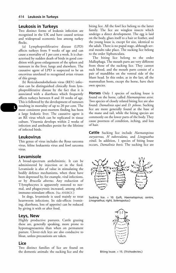
Leukosis in TurkeysTwo distinct forms of leukosis infection arerecognised in the UK and have caused seriousand widespread economic loss among turkeyflocks.
(a) Lymphoproliferative disease (LPD)affects turkeys from 9 weeks of age and cancause a mortality of 1 per cent a week. It is char-acterised by sudden death of birds in good con-dition with gross enlargement of the spleen andtumours in the liver, lungs and elsewhere. Thecausative agent of LPD is suspected to be anoncovirus unrelated to recognised avian virusesof this group.
(b) Reticuloendotheliosis virus (REV) infec-tion can be distinguished clinically from lym-phoproliferative disease by the fact that it isassociated with a diarrhoea which frequentlyaffects turkeys between 8 and 10 weeks of age.This is followed by the development of tumoursresulting in mortality of up to 20 per cent. Themost consistent post-mortem finding has beena large leukotic liver. The causative agent is an RE virus which can be replicated in tissueculture. Viraemia develops within 2 weeks ofinfection and antibodies persist for the lifetimeof infected birds.
LeukovirusThis genus of virus includes the Rous sarcomavirus, feline leukaemia virus and fowl sarcomavirus.
LevamisoleA broad-spectrum anthelmintic. It can beadministered by injection or in the feed.Levamisole is also of value in stimulating thebodily defence mechanisms, when these havebeen depressed by, for example, viral infections,or by Brucella abortus. Any reduction of T-lymphocytes is apparently restored to nor-mal, and phagocytosis increased, among otherimmuno-stimulant effects. (See ANERGY.)
In dogs, levamisole is used mainly to treatheartworm infections. Its side-effects (vomit-ing, diarrhoea, loss of appetite) can be reducedby giving it with or after food.
Leys, NewHighly productive pastures. Cattle grazingthese are, generally speaking, more prone tohypomagnesaemia than when on permanentpasture. Clover-rich leys are also conducive tobloat, unless precautions are taken.
LiceTwo distinct families of lice are found on the domestic animals: the sucking lice and the
biting lice. All the fowl lice belong to the latterfamily. The lice are wingless insects whichundergo a direct development. The egg is laidon the body, glues itself to a hair or feather, andthe young louse is, except for size, identical tothe adult. There is no pupal stage, although sev-eral moults take place. The sucking lice belongto the order Siphunculata.
The biting lice belong to the orderMallophaga. The mouth parts are very differentfrom those of the sucking lice. They cannotsuck blood, and the mouth parts consist of apair of mandibles on the ventral side of theblunt head. In this order, as in the last, all themammalian hosts, except the horse, have theirown species.
Horses Only 1 species of sucking louse isfound on the horse, called Haematopinus asini.Two species of closely related biting lice are alsofound: Damalinia equi and D. pilosus. Suckinglice are more generally found at the base of the mane and tail, while the biting species arecommonly on the lower parts of the body. Theycause poorness of condition, itching, and loss of hair.
Cattle Sucking lice include Haematopinuseurysternus, H tuberculatus, and Linognathusvituli. In addition, 1 species of biting louseoccurs, Damalinia bovis. The sucking lice are
414 Leukosis in Turkeys
L
Sucking lice. × 10. (Left, Haematopinus; centre, Linognathus; right, Solenopotus.)
Biting louse. × 15. (Trichodectes.)
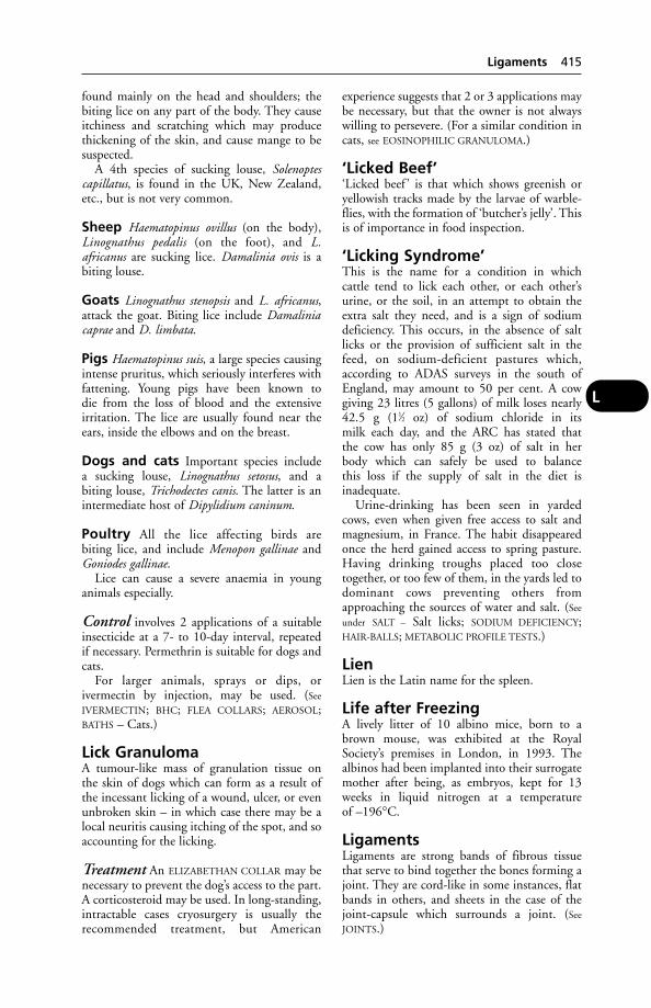
found mainly on the head and shoulders; thebiting lice on any part of the body. They causeitchiness and scratching which may producethickening of the skin, and cause mange to besuspected.
A 4th species of sucking louse, Solenoptescapillatus, is found in the UK, New Zealand,etc., but is not very common.
Sheep Haematopinus ovillus (on the body),Linognathus pedalis (on the foot), and L.africanus are sucking lice. Damalinia ovis is abiting louse.
Goats Linognathus stenopsis and L. africanus,attack the goat. Biting lice include Damaliniacaprae and D. limbata.
Pigs Haematopinus suis, a large species causingintense pruritus, which seriously interferes withfattening. Young pigs have been known todie from the loss of blood and the extensiveirritation. The lice are usually found near theears, inside the elbows and on the breast.
Dogs and cats Important species includea sucking louse, Linognathus setosus, and abiting louse, Trichodectes canis. The latter is anintermediate host of Dipylidium caninum.
Poultry All the lice affecting birds arebiting lice, and include Menopon gallinae andGoniodes gallinae.
Lice can cause a severe anaemia in young animals especially.
Control involves 2 applications of a suitableinsecticide at a 7- to 10-day interval, repeated if necessary. Permethrin is suitable for dogs andcats.
For larger animals, sprays or dips, orivermectin by injection, may be used. (See
IVERMECTIN; BHC; FLEA COLLARS; AEROSOL;BATHS – Cats.)
Lick GranulomaA tumour-like mass of granulation tissue on the skin of dogs which can form as a result ofthe incessant licking of a wound, ulcer, or evenunbroken skin – in which case there may be alocal neuritis causing itching of the spot, and soaccounting for the licking.
Treatment An ELIZABETHAN COLLAR may benecessary to prevent the dog’s access to the part.A corticosteroid may be used. In long-standing,intractable cases cryosurgery is usually therecommended treatment, but American
experience suggests that 2 or 3 applications maybe necessary, but that the owner is not alwayswilling to persevere. (For a similar condition incats, see EOSINOPHILIC GRANULOMA.)
‘Licked Beef’‘Licked beef ’ is that which shows greenish oryellowish tracks made by the larvae of warble-flies, with the formation of ‘butcher’s jelly’. Thisis of importance in food inspection.
‘Licking Syndrome’This is the name for a condition in whichcattle tend to lick each other, or each other’surine, or the soil, in an attempt to obtain theextra salt they need, and is a sign of sodiumdeficiency. This occurs, in the absence of saltlicks or the provision of sufficient salt in thefeed, on sodium-deficient pastures which,according to ADAS surveys in the south ofEngland, may amount to 50 per cent. A cowgiving 23 litres (5 gallons) of milk loses nearly42.5 g (11⁄2 oz) of sodium chloride in itsmilk each day, and the ARC has stated thatthe cow has only 85 g (3 oz) of salt in herbody which can safely be used to balancethis loss if the supply of salt in the diet isinadequate.
Urine-drinking has been seen in yardedcows, even when given free access to salt andmagnesium, in France. The habit disappearedonce the herd gained access to spring pasture.Having drinking troughs placed too closetogether, or too few of them, in the yards led todominant cows preventing others fromapproaching the sources of water and salt. (See
under SALT – Salt licks; SODIUM DEFICIENCY;HAIR-BALLS; METABOLIC PROFILE TESTS.)
LienLien is the Latin name for the spleen.
Life after FreezingA lively litter of 10 albino mice, born to abrown mouse, was exhibited at the RoyalSociety’s premises in London, in 1993. Thealbinos had been implanted into their surrogatemother after being, as embryos, kept for 13weeks in liquid nitrogen at a temperatureof –196°C.
LigamentsLigaments are strong bands of fibrous tissuethat serve to bind together the bones forming ajoint. They are cord-like in some instances, flatbands in others, and sheets in the case of thejoint-capsule which surrounds a joint. (See
JOINTS.)
Ligaments 415
L

Desmitis (inflammation) of the fetlock annu-lar ligament was diagnosed in 30 horses whichhad been lame for a long time and which hadchronically distended digital flexor tendonsheaths, or plantar annular ligaments. The liga-ment was cut longitudinally in 25 of the horses;16 returned to full work without difficulty and1 became sound after a 2nd operation. None ofthe 5 untreated horses returned to work.
Ligamentum ArteriosumThe fibrous remains of the ductus arteriosus ofthe fetus. It connects the left pulmonary arteryto the arch of the aorta.
Light, Influence ofAdequate light is necessary for maximumfertility. This applies to poultry (see under NIGHT-
LIGHTING), to bulls – too often kept in darkplaces – and to sheep, etc. (See also LIGHTING;RICKETS; VITAMINS – Vitamin D; TROPICS.)
Light SensitisationLight sensitisation implies a predisposing fac-tor, such as the eating of a particular plant,which has the effect of making certain cells in the animal’s body abnormally sensitive – for the time being – to light. Strong sunlight isthen capable of causing serious and extensivedamage, with a good deal of distress.
In Australia this trouble is frequently caused,in cattle, sheep, and pigs, through eating StJohn’s wort. Elsewhere overseas, clover andbuckwheat are often responsible. Occasionalcases of light sensitisation occur even in theUK. To give an example, a British Friesianheifer was discovered in obvious distress. Overnearly all the white parts the skin was dead andhad partly sloughed off. Appropriate treatment,which included temporary confinement in adarkened loose-box, was followed by a rapidrecovery. Bog asphodel and rape cause light sensitisation in sheep in Britain, where pigshave also been affected (probably by St John’swort). In New Zealand, where the condition is called facial eczema, moulds have beenincriminated. In Britain another plant involvedis the GIANT HOGWEED.
It is the white, pigment-free skin which suf-fers. Thus, some breeds of livestock are nevertroubled with light sensitisation, while white or partly white cattle are susceptible. Similarly,grey and pie-bald horses in the USA and elsewhere are sometimes affected.
Light sensitisation is associated with disfunc-tion of the liver, and the presence of porphyrinsin the bloodstream. It also occurs in some casesof PORPHYRIA.
Lighting of Animal BuildingsVarious kinds of glass substitutes have been puton the market, which are reputed to allow theultraviolet rays of natural sunlight to passthrough without appreciable absorption.
Adequate light is necessary to prevent rickets,and to ensure maximum fertility in poultry andother animals. Continuous light, however, mayhave harmful effects.
Artificial lighting of poultry houses is now acommon practice. Red light is used in manybroiler houses and in some laying houses inorder to reduce cannibalism.
(See also NIGHT LIGHTING.)
Lightning StrikeCattle, sheep and horses are most often affected. (See under ELECTRIC SHOCK.)
LignocaineA local anaesthetic agent used to produce nerveblocks, it has also been used to treat cardiacarrhythmias.
‘Limberneck’An old, colloquial name for some of the symp-toms seen in cases of botulism in poultry: a lossof power of the muscles of the neck, wings, andlegs, affected birds first being dull and inactive.(See BOTULISM.)
Liming of PasturesIf this is carried out to excess it can lead to a defi-ciency in copper in the grazing animal and sobring about INFERTILITY. Manganese deficiencyis likewise a sequel when the soil becomes tooalkaline. Cattle should be kept away from down-wind of liming operations, or eye inflammation(conjunctivitis and keratitis) may result.
LimousinA pure beef breed noted for high liveweightgains, high killing-out percentages, and freedomfrom calving difficulties.
LincomycinAn antibiotic effective against Gram-positivebacteria, anaerobes and mycoplasma.
Linear Assessment of DairyCows(see PROGENY TESTING – Conformation)
Liner, Teat-CupIn selecting milking machinery equipment, oneshould avoid any liner with a hard mouthpiece.Liners must be changed after a maximum of2500 milkings.
416 Ligamentum Arteriosum
L
Linguatula Serrata(see MITES, PARASITIC)
Liniments (Embrocations)Liniments (embrocations) are liquid prepara-tions for external application (to unbrokenskin), generally rubbed in, and having counter-irritant or analgesic properties. They are usedfor painful muscular conditions, strains andsprains.
LinognathusA genus of sucking lice. (See LICE.)
LinseedThe flax plant (Linum usitatissimum). Afterextraction of linseed oil from the seeds, theresidue is made into linseed cake for feeding tohorses, cattle, and sheep.
Linseed poisoning The flax plant containsa cyanogenetic glycoside in small amounts. Anenzyme in the flax can act on the glycoside,with production of hydrocyanic acid. Theenzyme is not always destroyed in the process ofmaking linseed cake. Boiling for 10 minutesdestroys the enzyme and renders linseed safe.However, linseed cake should be fed dry andnot made into a mash with warm water – adangerous practice owing to the formation ofhydrocyanic acid. Linseed poisoning is notcommon.
LipaseA fat-splitting enzyme found in the pancreaticjuice.
LipidsFatty substances. Simple lipids are esters of fattyacids and alcohol, and include fats (esters offatty acids and glycerol). Compound lipids contain, in addition to fatty acid and alcohol,carbohydrate or nitrogen or phosphoric acid,for example.
‘Protected lipids’ are those encapsulated ina protein envelope, which is then treated withformaldehyde. Because of their high energyvalue, fats and oils and their fatty acids seemedworth including in cattle feed supplements; and ‘protected lipids’ offered the possibility ofavoiding the disturbance of normal rumenmetabolism likely to occur with free fats beingpresent in the rumen.
LipomaLipoma is a tumour mainly composed of fat.They are liable to arise almost anywhere in the
body where there is fibrous connective tissue,but are especially common below loose skin.They are occasionally seen in the abdominalcavity, where they develop in connection withthe peritoneum, and sometimes encase thebowel and obstruct its function or attain a largesize. (See TUMOUR.)
LipoproteinA complex of cholesterol, triglycerides, phos-pholipids, and apoproteins. An excess oflipoprotein in the blood – hyperlipopro-teinaemia – occurs in some cases of diabetes,hypothyroidism metabolic disorders and inherited disease.
LipsLips are musculo-membranous folds which inthe horse are covered on the outside with finehairs, among which are longer, stouter tactilehairs, while some heavy draught horses have a‘moustache’ on their upper lips. On the inside,the lips are covered by mucous membranewhich is continuous with that of the mouthgenerally.
In the horse the lips are extremely mobile,and the upper lip especially contains a verydense plexus of sensory nerves which serve tactile purposes. In the ox the lips are thick andcomparatively immobile. The middle part ofthe upper lip between the nostrils is bare of hairand is termed the muzzle. It is provided with a large number of tiny glands which secrete aclear fluid, which keeps the part cool and moist.Within the lower lip are numbers of hornypapillae; its free margin is bare, but the underpart of it is covered with ordinary and tactilehairs. The sheep possesses no hairless muzzle,but has a distinct ‘philtrum’ instead. The lipsare thin and mobile. In the pig the upper lip is thick and short and is blended with the snout or nasal disc, while the lower is thin andpointed. (See also HARE-LIP.)
Liquid FeedingLiquid feeding of dairy cows in the parlourenables them to eat up to 6.8 kg (15 lb) of concentrates in 7 minutes.
Liquid Paraffin, Medicinal(see under PARAFFIN for its use as a laxative).
Listerellosis(see LISTERIOSIS)
ListeriosisA disease caused by Listeria monocytogeneswhich attacks rodents, poultry, ruminants, pigs,
Listeriosis 417
L

horses, dogs, and man. It causes encephalitisand abortion in cattle and sheep, and has to bedifferentiated from RABIES.
In cattle, the infection may be confused withrabies or poisoning. The affected animal is seento keep aloof from the rest of the herd, and islater unable to stand without support. Ifwalked, it usually moves in a circle. The headmay be held back to one side, with salivationand a nasal discharge. Paralysis of one side ofthe face may occur. Some cows become violentin the terminal stages and bellow.
In an English outbreak, 12 out of 15 calvesdied between April and August, at 3 to 7 daysold, from septicaemia. There was severe keratitis and conjunctivitis (L. monocytogenesis recognised as one cause of IBK), extremedejection, and distressed breathing.
Listeria is a cause of IRITIS in cattle feeding atsilage clamps. (See SILAGE.)
There is also a septicaemic form in adult cattle and sheep, which shows itself by depression, fever, weakness, and emaciation.
Pigs may have swelling of the eyelids,encephalitis, paralysis, or occasionally septi-caemia.
Listeriosis is a rare cause of abortion inmares, and a common cause in other animals. Itis an important cause of abortion in goats.
Infection may be spread by urine, milk, faeces, an aborted fetus, and vaginal discharges.
Listeriosis in the fowl is seen as sudden deathdue to myocarditis. It may occur in free-rangechickens.
Good hygiene helps to prevent the disease;antibiotics may be effective in treating earlycases.
(See also AVIAN LISTERIOSIS.)
LithiasisLithiasis is the formation of calculi and concre-tions in tissues or organs. For example,cholelithiasis means the formation of calculi in the gall-bladder. (See also under CALCULI;UROLITHIASIS.)
LithiumLithium antimony thiomalate has been used byinjection to remove multiple warts.
Lithium PoisoningLithium poisoning occurred in 2 dogs whosesole source of drinking water for several monthswas a swimming pool chlorinated with lithiumhypochlorite. One dog had fits. Both had diar-rhoea, became weak, and dehydrated. Theyrecovered after being provided with fresh,uncontaminated water.
LithontripticsLithontriptics are substances which are reputedto have the power of dissolving stones in theurinary system. (See HYALURONIDASE.)
LithotomyLithotomy is the operation of opening the bladder for the removal of a stone.
LithotrityLithotrity is an operation in which a stone inthe bladder is broken into small fragments andremoved by washing out the bladder with acatheter.
Litter(see DEEP LITTER; BEDDING)
Litter, OldBroiler chicks reared on previously used littermay, as a result of the ammonia fumes, developa severe inflammation of eye-surfaces and eyelids. In one house, 3000 broilers were affected. The birds cannot bear to open theireyes, and appear obviously dejected. Mortalityis generally low, but the trouble is a serious one for all that.
Litter Size (Pigs)In Britain the average is between 10 and 11born alive; mortality 0.84. Earlier figures(PIDA) showed that an average of 2.2 pigs per litter died between birth and 8 weeks old. Alitter of 34 has been recorded.
LiverA solid glandular organ lying in the anterior-most part of the abdomen close up against the diaphragm. Its colour varies from a darkred-brown in the horse to a bluish-purple in theox and pig; it is soft to the touch though it israther friable in consistency, and it constitutesthe largest gland in the body.
Functions include the excretion of bile,the storage of glycogen and of iron, the breaking-down of old and worn-out red bloodcells, and the breaking-down of toxic sub-stances and of waste substances from the tissues of the body. From the liver, urea and uric acid find their way into the bloodstreamand are excreted from the body in the urine by the kidneys. In animals except those of the horse tribe, the bile is collected in the gall-bladder and the bile-duct before passing to the small intestine, where it assists the pancreatic juice in the digestion of food after a meal.
418 Lithiasis
L

Shape There are probably few organs whichvary so much in shape as the liver, not only in different animals; but also in different individuals of the same species.
Horse It lies obliquely across the abdominalsurface of the diaphragm, its highest and mostposterior part being at the level of the right kidney. It possesses a strongly convex diaphrag-matic surface which is moulded into the con-cavity of the diaphragm, and a posterior orabdominal surface which lies in contact withthe stomach, duodenum, and right kidney, each of which organs forms a depression in theliver substance. It is only incompletely dividedinto 3 lobes in the horse. Lying mainly in the right lobe on its abdominal surface is the‘porta’ of the liver, where the portal vein andhepatic artery enter and from whence thehepatic duct (bile-duct) emerges. Part of the posterior vena cava passes through the liversubstance, whose blood it eventually drains.The liver is held in position by the pressure of other organs and by 6 ligaments. These are: the coronary, which attaches it to thediaphragm; the falciform, from the middle lobe to the diaphragm and abdominal floor; the round, to the umbilicus; the right lateral, to the costal part of the diaphragm; the left lateral, to the tendinous part of the diaphragm;and the hepatorenal or caudate, to the rightkidney.
Cattle The liver lies mainly to the right of the middle-line through the body, and its longaxis is directed downward and forward. Itsdiaphragmatic surface fits into the concavity of the right part of the diaphragm, and its posterior surface is very irregular. It presentsimpressions of the 2 main organs with which it comes into contact – the omasum and reticulum. There is only 1 distinct lobe – the caudate. There is no left lateral ligament, andthe round ligament is only found in the calf. Agall-bladder is present; it is situated partly in aslight depression on the posterior surface of theliver, and partly on the abdominal wall.
Sheep The bile-duct joins the pancreatic toform a common duct instead of opening separately as in other animals.
Pigs The liver is large, very thick, and verymuch curved. It lies in the anterior part of the abdominal cavity, occupying the whole ofthe anterior hollow of the diaphragm and moreto the right than to the left side of the body. lthas 4 main lobes.
Dogs The liver is very large, being about 5 percent of the whole bodyweight, and possesses 6 or 7 lobes. The gall-bladder is buried almostcompletely in the space between the 2 parts ofthe right central lobe, only a very small portionof it being visible from the outside.
Minute structure The liver is enveloped in an outer capsule of fibrous tissue with whichis blended the hepatic peritoneum. The hepaticartery, portal vein, and bile-duct divide andsubdivide. Between the rows of liver cells alsolie fine bile capillaries which collect the bile discharged by the cells and pass it into the bile-ducts lying around the margins of the lobules. The liver cells are amongst the largestcells of the body, and each contains 1 largenucleus. With careful special staining methodsthere can also be seen tiny passages or canals,passing into the cells themselves; some of thesecommunicate with the bile-duct, and otherswith the ultimate branches of the portal vein.After a mixed meal many of the liver cells can be seen to contain droplets of fat, and granules of glycogen (animal starch) can also bedetermined. In addition to the cells abovedescribed, there occur at intervals along thewalls of the sinusoids in a lobule stellate cellswhich represent the remains of the endothe-lium from which the capillary-likesinusoids aredeveloped. They are known as ‘Kupfer’s cells’.
Liver, Diseases ofOne of the commonly known signs of liver dis-turbance is JAUNDICE – a yellow coloration ofthe visible membranes. Gallstones, which are acomplication of some liver diseases, are treatedunder GALL-BLADDER and GALLSTONES.
Hepatitis, or inflammation of the liver, maybe acute, suppurative (in which abscesses areformed), or chronic. Acute inflammation pro-duced by viruses, bacteria or poisons (of bacterial, vegetable, animal, or mineral origin),from the intestines, and it is sometimes causedby the migration of parasites through the liver.The symptoms are pain on pressure over theabdomen, an elevation of temperature, suppres-sion of the appetite, a disinclination to move,and often diarrhoea or constipation in the laterstages. (See also CANINE VIRAL HEPATITIS.)
Chronic inflammation accompanies manydiseases among animals, the commonest prob-ably being infestation with liver-flukes, but itmay also be present as a result of tuberculosis.Poisoning may be responsible. (See, for example,RAGWORT POISONING which gives rise to CIR-
RHOSIS.)
Liver, Diseases of 419
L

Signs include a gradual loss of condition, irreg-ular appetite, a staring coat, and a generalunthriftiness; often oedema.
Abscess formation is due to the entranceinto the liver of pus-forming organisms, and isusually secondary to some disease, e.g. tubercu-losis. In the USA liver abscesses are found inabout 8 per cent of all cattle slaughtered, andthey are common in ‘barley beef ’ animals in the UK. The lesions consist of abscesses in thesubstance of the liver. Symptoms are vague anddiagnosis is often impossible.
Fatty liver (see FATTY LIVER SYNDROME ofcattle and poultry; also CEROIDOSIS)
Liver/kidney syndrome (see under thisheading)
Tumours include adenoma, carcinoma, andhaemangioma (see CANCER). Benign tumoursmay give rise to passive congestion, biliaryobstruction with jaundice, or they may causedegeneration. (See under TUMOUR.)
Parasites (see LIVER-FLUKES; HYDATID
DISEASE; HORSE BOTS; ‘LIZARD-POISONING’ IN
CATS)
Rupture of the liver is by no means rareamong old animals, especially dogs and cats. Itmay result from a blow, a kick, a traffic acci-dent, a fall, or from violent struggling, whenthe liver is diseased. Even a small wound insuch a vascular organ as the liver is likely toprove fatal. (See also ‘MILKSPOT LIVER’ of pigs andsheep.)
Rupture of the liver in lambs, aged 1 to 3months, may be the cause of sudden death. Ina survey covering a period of 12 years, theThurso (Scotland) Veterinary InvestigationLaboratory found that liver rupture in neonatallambs from 16 farms exceeded 8 per cent. Theliver surface was covered with haemorrhagictracts made by migrating metacestodes ofTaenia hydatigena (see TAPEWORMS).
Liver, Displacement ofA 1-year-old cat with a history of anaemia,jaundice and ascites was found to have its liverin the pericardium.
Liver-FlukesLiver-flukes are parasitic flat worms whichinfest the livers of various animals, especiallysheep and cattle. They may cause severe illnessand even death.
Liver-flukes may increase the susceptibilityof the host animal to Salmonella infection. (See
SALMONELLOSIS.)
Life-history of a typical fluke The eggis usually passed to the exterior in the faeces ofthe host and under suitable conditions, chieflyof moisture and warmth, a small ciliated larva,called a ‘miracidium’, hatches from it. Thislarva, which is unable to feed and will die within some hours unless it finds a suitable host,gains access to the liver or some other specialorgan by actively penetrating the skin of anappropriate snail – usually a specific snail forany one parasite. In the snail’s tissue it developsinto a sac-like sporocyst which, by a process ofbudding from the internal lining of cells, givesrise to a number of elongated ‘rediae’. Each rediais a simple, cylindrical sac-like organism whichgives rise, by budding of cells, either to anothergeneration of ‘daughter rediae’ or to ‘cercariae’.The cercaria, which resembles a miniature tad-pole in general form, leaves the snail and, afterleading a free existence in water or on wet vege-tation for a short time, comes to rest on grass orother objects, loses its tail and becomes encystedwithin a protective covering and remains in thisstate until it is swallowed by the final host, inwhich it becomes a sexually mature fluke.
Fasciola hepatica This is the commonliver-fluke of sheep. (Other hosts are cattle,
420 Liver, Displacement of
L
An adult liver-fluke implanted in a subcutaneouspocket in a rat. This technique showed that the rat gained thereby immunity to adult flukes without the complication of liver damage, andwas part of the research carried out at the ARC’sInstitute for Research on Animal Diseases,Compton, into possible means of immunising cattle against liver flukes.(S = skin; C = subcutaneous tissue; F = liver fluke)
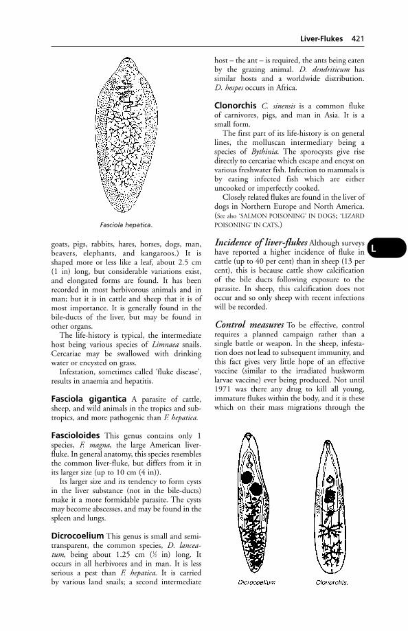
goats, pigs, rabbits, hares, horses, dogs, man,beavers, elephants, and kangaroos.) It isshaped more or less like a leaf, about 2.5 cm(1 in) long, but considerable variations exist,and elongated forms are found. It has beenrecorded in most herbivorous animals and inman; but it is in cattle and sheep that it is ofmost importance. It is generally found in thebile-ducts of the liver, but may be found inother organs.
The life-history is typical, the intermediatehost being various species of Limnaea snails.Cercariae may be swallowed with drinkingwater or encysted on grass.
Infestation, sometimes called ‘fluke disease’,results in anaemia and hepatitis.
Fasciola gigantica A parasite of cattle,sheep, and wild animals in the tropics and sub-tropics, and more pathogenic than F. hepatica.
Fascioloides This genus contains only 1species, F. magna, the large American liver-fluke. In general anatomy, this species resemblesthe common liver-fluke, but differs from it inits larger size (up to 10 cm (4 in)).
Its larger size and its tendency to form cystsin the liver substance (not in the bile-ducts)make it a more formidable parasite. The cystsmay become abscesses, and may be found in thespleen and lungs.
Dicrocoelium This genus is small and semi-transparent, the common species, D. lancea-tum, being about 1.25 cm (1⁄2 in) long. It occurs in all herbivores and in man. It is lessserious a pest than F. hepatica. It is carried by various land snails; a second intermediate
host – the ant – is required, the ants being eatenby the grazing animal. D. dendriticum has similar hosts and a worldwide distribution. D. hospes occurs in Africa.
Clonorchis C. sinensis is a common fluke of carnivores, pigs, and man in Asia. It is a small form.
The first part of its life-history is on generallines, the molluscan intermediary being aspecies of Bythinia. The sporocysts give risedirectly to cercariae which escape and encyst onvarious freshwater fish. Infection to mammals isby eating infected fish which are eitheruncooked or imperfectly cooked.
Closely related flukes are found in the liver ofdogs in Northern Europe and North America.(See also ‘SALMON POISONING’ IN DOGS; ‘LIZARD
POISONING’ IN CATS.)
Incidence of liver-flukes Although surveyshave reported a higher incidence of fluke in cattle (up to 40 per cent) than in sheep (13 percent), this is because cattle show calcification of the bile ducts following exposure to the parasite. In sheep, this calcification does notoccur and so only sheep with recent infectionswill be recorded.
Control measures To be effective, controlrequires a planned campaign rather than a single battle or weapon. In the sheep, infesta-tion does not lead to subsequent immunity, andthis fact gives very little hope of an effective vaccine (similar to the irradiated huskworm larvae vaccine) ever being produced. Not until1971 was there any drug to kill all young,immature flukes within the body, and it is thesewhich on their mass migrations through the
Liver-Flukes 421
L
Fasciola hepatica.
liver can damage it so severely that suddendeath inevitably follows.
Triclabendazole (Fasinex) is very effectiveagainst flukes as it removes both immature andadult stages.
Other drugs used against liver-flukes include aldendazole, closantel, nitroxynil andoxycyclozanide.
In the UK, over 60 per cent of cattle and 80 per cent of sheep are kept in the main fluke areas.
While sheep farmers are mostly fully alive tothe fluke problem, it is suggested that most cattle farmers are not. It has been pointed outthat on farms where mixed grazing is practised,it is a waste of time and money to dose only thesheep and not the cattle.
Profit margins in beef production have beenimproved by a combined anti-fluke attack,using routine dosing of the cattle together witha chemical spray (Frescon) on pasture to kill thehost snails. In trials, 18-month-old beef animalsfinished 25 to 30 days earlier than controls, giving higher profitability through savings infeed. Returns were further increased by bettercarcase grading.
An 8 per cent drop in milk yield has resultedfrom low-grade infestations in dairy cattle, andit is claimed that autumn and winter dosing of dairy cattle helps to improve, or at leastmaintain, milk quality levels.
A vaccine against black disease – in whichspores of one of the gas-gangrene group oforganisms are stimulated into activity by youngflukes in the liver – can prevent deaths from the resulting toxaemia. Against the liver-flukesthemselves, routine dosing is essential on allfarms where they are likely to occur.
Land drainage is still high on the list of control measures.
The use of snail-killers is a recommendedpart of the campaign against fluke disease, butis not a snag-free method. It is easy to misssmall areas inhabited by snails, and this applieseven when using a knapsack sprayer – the only possible method of spraying if the land is too wet, is to take a tractor. Snail-killers canbe unpleasant to work with: the cheapest issodium pentachlorophenate. N-trityemorpho-line is expensive per acre but has the advantage of being relatively harmless to stock, so thatgrazing need not be delayed for a fort-night as after copper sulphate dressings or pentachlorophenate. All are poisonous to fish.
Running ducks over snaily land is not amongthe official recommendations but it mightprove of some value. A few farmers have tried itin the past. In Zambia years ago, a large-scale
duck-rearing scheme was introduced in areasflooded by the River Zambesi as a method of fluke-disease and bilharzia control. Hoof-prints, where the soil is exposed, are favouritehabitats of the snails. (See also under ANTS.)
Public health Watercress is the chief sourceof infestation. Illness is most marked duringmigration of immature flukes. Eosinophilia is a pointer to aid diagnosis; eggs may not appearin the faeces for 12 weeks.
Symptoms in the human patient includeurticaria, jaundice, enlarged and tender liver,and eosinophilia.
Cats (see under ‘LIZARD POISONING’; PANCREAS,DISEASES of IN CATS)
Liver/Kidney Syndrome ofPoultry (FLKS)Liver/kidney syndrome of poultry (FLKS)affects birds usually 2 to 3 weeks old.Symptoms may not be observed – or there maybe depression for a day or two; occasionallytrembling or paralysis of legs. Mortality: 1 to 5 per cent. The whole carcase may have a pinktinge. It is the result of failure of the liver tosynthesise glucose. The liver is pale, swollen,and fatty. The kidneys may be very swollen.Biotin supplementation of the feed has beenshown to alleviate the condition. (See also underFATTY LIVER.)
The syndrome has to be differentiated from toxic fat disease, Gumboro disease, andinfectious avian nephrosis.
Livestock Production(see BEEF CATTLE HUSBANDRY; DAIRY HERD
MANAGEMENT; TROPICS; and to under PIGS and
SHEEP)
‘Lizard Poisoning’ in CatsThis term is applied to infestation with the liver-fluke Plarynosomum concinnum, whichhas been reported from South America, theCaribbean Islands, Malaysia, the USA and,more recently, Nigeria. The life-cycle of the parasite involves a large land snail, a crustacean,and lizards, frogs, and probably other amphib-ians and reptiles. Symptoms in the cat includelistlessness, fever, jaundice, diarrhoea, vomiting,and emaciation; but subclinical infestations also occur.
LizardsThe largest group of reptiles. These cold-blooded vertebrates, some of which are aquatic,are often kept as pets. The environment must
422 Liver/Kidney Syndrome of Poultry (FLKS)
L
be kept at the temperature appropriate for theparticular type of lizard. It is essential to providethe diet recommended for the particularspecies. Most lizards require meat; crickets mayprovide a suitable source. Iguanas, however, aremainly, but not entirely, vegetarian. Adequateultraviolet (UV) light must be available if normal bone development is to take place. Anylizard wih a swollen limb may have osteodys-trophy, and the source of UV light should bechecked and a calcium supplement provided.Chameleons and some other lizards have pigment cells (chromatophores) in their skinand can change colour to blend in with theirsurroundings. However, a colour change can bea sign of illness, particularly when the animalcannot blend in with its surroundings.
The water in which aquatic lizards live must be kept fresh, otherwise a build-up ofaeromonas or pseudomonas organisms mayoccur. Conjunctivitis is not uncommon; itshows as caseous (cheesy) discharge which mustbe gently removed before treatment can begiven. When diarrhoea occurs it is usually as aresult of infestation with protozoan parasites.Skin parasites can be removed by (for example)treatment with ivermectin. The reptiles must be removed to a clean cage or tank while theoriginal one is cleaned and treated with a suit-able inseticide. Egg-binding is usually associ-ated with a calcium deficiency but may happenif no suitable site for laying has been provided.Any heating device must be located so that thelizard does not come into contact with it.
LlamasLong-legged, long-necked domesticated ani-mals widely used as pack animals and for wooland hide in the South American Andes. Theybelong to the order Camelidae, which includesalso Alpaca, Guanaco and Vicuna. They areincreasingly farmed in the UK.
Importation of llamas and alpacas directfrom South America into a Border InspectionPost is permitted. On arrival, each animal isexamined for infectious or contagious disease.If no disease is apparent, the animals are quarantined for a month (to eliminate the possibility of foot-and-mouth disease) beforegoing on to their destination.
LobeLobe is the term applied to the larger divisionsof various organs, such as the lungs, liver, andbrain. The term ‘lobar’ is applied to structureswhich are connected with lobes of organs, or to diseases which have a tendency to be limitedto one lobe only, such as ‘lobar pneumonia’.
Lobules are divisions of a lobe. The term ‘lobular’ is applied to disease which occurs in ascattered irregular manner affecting lobuleshere and there, such as ‘lobular pneumonia’.
Local Anaesthetics(see under ANAESTHESIA; ANALGESICS)
Local Immunity(see under IMMUNE RESPONSE; ORIFICES; SECRETORY IgA)
Lockjaw(see TETANUS)
‘Loco Weed’The legumes oxytropis and astragalus in theUSA contain toxins that cause incoordination,and extreme excitability in animals grazingthem. They are also teratogenic and if consumed in sufficient quantity, abortifacient.
Loose-BoxesIndividual enclosures for accommodating ananimal; also called box-stalls. The best type haswell-built brick walls lined on the inside to theroof with cement-plaster finished off smooth.The floor is of cement-concrete, grooved tofacilitate the draining away of fluids and to pro-vide a foothold, and the corners are rounded offwith fillets of cement. The only fittings insideare hay-rack, water-bowl, and manger – of iron,and rather larger than in the stall of a stable, sothat cattle as well as horses may use them; insome cases 1 or 2 rings, to which animals maybe tied, are provided. One or more windows,high up out of reach of the animals’ heads,should be included, and the door should alwaysbe made in 2 halves, so that horses with respi-ratory diseases may stand with their heads outof the box, and so obtain a plentiful supply offresh air. Wherever possible, loose-boxes shouldbe built with a southerly aspect, so that the dis-infectant action of sunlight may be taken fulladvantage of, whenever sick animals are housedin the box.
LordosisLordosis is an unnatural curvature of the spine,so that the concavity of the spine is directedupwards. It is seen in tetanus, and sometimes inrabies.
Louping-IllLouping-ill is a paralytic disease of sheep, alsocalled ovine encephalomyelitis; it is transmittedby Ixodes ricinus, the tick commonly present onhill pastures. It occurs in western Scotland, the
Louping-Ill 423
L

North of England, and the Northwest ofIreland. There is a definite seasonal incidence,most cases occurring between March and June,and between September and October; only afew sporadic cases are met with at other times of the year. All breeds of sheep are susceptible. It occurs in cattle and has beenrecorded as affecting pigs, horses, deer and alsodogs, in which the signs are fever, nystagmus,hyperaesthesia, and sometimes a tetanus-likerigidity. In 2 cases, bitches had whelped 5 weekspreviously, and eclampsia was at first suspected.
Cattle On upland grazings where ticksabound, louping-ill has become of economicimportance in cattle. The animals become dulland uninterested in food, walk in an unnaturalway, sometimes with their heads down, andoccasionally become excited.
Pigs The first naturally occurring outbreak inpigs was reported in 1980 by the West ofScotland Agricultural College’s veterinary inves-tigation centre. Ten out of 16 piglets becameseverely affected with the disease when about 6weeks old. They showed nervous symptoms,were either reluctant to move or wandered aim-lessly and pressed their heads into corners. Ofthe 3 worst cases, 2 failed to survive transport tothe VI centre, and the 3rd – being in a state ofconvulsions – was killed on arrival there. Of theremainder, 5 more died and 2 recovered.
Those piglets had been housed in a coveredpen with a concrete run considered to be tick-proof; and the louping-ill virus was prob-ably transmitted through the feeding ofuncooked carcases of lambs which had died on the farm after showing symptoms suggestiveof louping-ill.
In another outbreak, pigs 6 to 8 months olddied of louping-ill after being allowed freerange on tick-infested pasture.
Cause A flavivirus. This is transmitted by thebites of infected ticks (adult or nymphal). Thevirus primarily multiplies in the blood, and incertain cases invades the central nervous systemat a later stage in the infection.
It would appear that accessory conditionsfavour such invasion, e.g. tick-borne fever, adisease also transmitted by I. ricinus.
The ticks can survive, in the absence of sheepand cattle, on deer, rabbits, hares, voles, fieldmice, grouse, etc., and these animals may act ashost of the virus.
Signs Two forms of the disease are recognised:an acute and a subacute form. In the acute form
the symptoms may appear in from 4 to 6 daysafter the sheep is infested with the carrier ticks.The sheep becomes uneasy, lies down and risesfrequently during the day. Its temperatureranges between 40° and 41.6°C (104° and107°F) during the next week or 10 days, and it develops nervous symptoms. At first, it ismerely more timid and more easily frightenedthan usual; later, the muscles of the jaws andneck begin to twitch and quiver, and there maybe frothing at the mouth. It staggers whenmade to move rapidly or turn suddenly, and asthe disease becomes firmly established it may beseen taking short spasmodic jumps, risingapparently from all 4 feet at the same time, andlanding upon all 4 feet again. In this way anaffected sheep can usually be easily noticedamong a flock when the sheep are being drivenor collected by a dog. In more advanced stagesthe animal becomes paralysed, unable to stand,and often has its head drawn round over its fore flank. Unconsciousness quickly appears,and the animal dies a short time afterwards.
In the subacute type the sheep is seen takingvery high steps with its fore-legs; it holds itshead very high, and sometimes carries it to oneside (often the left); the pupils are dilated, andthe expression of the sheep is one of extremefear when caught. It may attempt to feed, butactually eats very little. Tremblings of the muscles, staggering and falling, and sometimesparalysis of one or more groups of muscles, are seen. As times goes on the sheep loses condition. If not fed by hand it dies from starvation.
Recovery from an attack confers a degree of immunity, which may last for life. (See also
TICK-BORNE FEVER).
Prevention Control measures should aim atthe eradication of the infecting ticks from graz-ing lands. This is not easy, as the tick can liveunder rough herbage without access to the living sheep for as long as one year. A vaccine isavailable and affords good protection. Cattleshould be vaccinated annually; sheep and goatsevery 2 years.
Sheep Vaccination of ewes confers protectionin their lambs. Inoculations are carried out inspring prior to the season when ticks becomeactive.
Cattle Investigations on hill farms wherelouping-ill is a problem have shown that cattleplay an important part in the maintenance ofvirus. Hill cattle as well as sheep thereforeshould be vaccinated, not only for their own
424 Louping-Ill
L
protection, but to reduce the transfer of virus tothe ticks which are the only agents passing onthe infection each year.
Public health Shepherds, farmers, andslaughter-house workers – as well as veteri-narians – may become infected. The mainsymptom is fever. Meningoencephalitis hasbeen recorded, and has sometimes proved fatal.
LubricantsThe type of lubricant used in pellet mills andother forms of machinery for processing animalfeeding-stuffs may be of the greatest impor-tance. Lubricants containing chlorinated naph-thalene compounds and used on such machinesmay give rise to hyperkeratosis in cattle eatingthe food so contaminated by the minutestquantity of lubricant.
LucerneA valuable leguminous plant (Medicago sativa)for fodder and forage. Lucerne-hay is highlyvalued for the feeding of horses if of good qual-ity. (It is of little value when most of the leaf has been lost, or it is dusty or mouldy.) Lucerneis also a valuable crop for cattle, but for precau-tions and dangers, see BLOAT; also MUSCLES,DISEASES OF – Nutritional muscular dystrophy.
Lugol’s SolutionA solution of 50 g iodine and 100 g potassiumiodide in distilled water to 1000 ml. A 2 to 4per cent dilution of the solution has been usedfor irrigation of the uterus in cases of bovinemetritis.
LuingA beef breed evolved by Messrs Cadzow fromBeef Shorthorn and Highland cattle, andnamed after the island. Colour: red with atouch of gold; or roan; or white. There are abreed society and herd book.
LumbarLumbar is a term used to denote either thestructures in or disease affecting the loins, i.e.the region lying between the last rib and thepoint of the hip, from one side of the body tothe other.There are lumbar vertebrae, lumbarmuscles, etc.
LumenThe space inside a tubular structure, such as anartery or intestine.
Luminal or Phenobarbitone(see BARBITURATES)
Lumpy Jaw(see ACTINOMYCOSIS)
‘Lumpy Skin Disease’‘Lumpy skin disease’ is a NOTIFIABLE DISEASE
throughout the EU. It is characterised by a discharge from the eyes and nose, lameness, andsalivation may be observed – depending uponthe site of nodules which sometimes involvemucous membrane as well as skin.
Oedema may occur, and involve the genitalorgans, udder, dewlap, and limbs. Sloughing ofskin may occur. Exotic cattle may die.
The disease is caused by the Neethling poxvirus; and a modified sheep-pox vaccine is usedfor protection.
‘Lumpy Skin Disease, Pseudo-’This is characterised by the formation of raisedplaques on the skin, which exude a dischargeand then ulcerate; and by fever. The cause is the bovid herpesvirus 2, which also causesmamillitis of cattle.
Lumpy Wool (Wool Rot)Lumpy wool (wool rot) is caused by a bacteri-um which attacks the sheep’s skin during wetweather, causing irritation and the formation ofa hard yellowish-white scab about 3 mm (1⁄8 in)thick. Healing soon occurs and the wool con-tinues to grow carrying the hard material awayfrom the skin as a buff or brownish zone in thewool. Severe infection may lead to loss of wool.
The bacterium causing this dermatitis is Dermatophilus dermatonomus. (See also
DERMATOPHILUS; STREPTOTHRICOSIS.)In America, this disease has been treated by
defleecing with cyclophosphamide and the useof streptomycin and penicillin.
Lung-Flukes (ParagonimusGenus)These flukes are plump oval forms infectingcarnivores, pig and man. Generally, 2 flukes arefound together in a cyst in the lungs. The pres-ence of the flukes cause bronchitis and pleurisy.Lesions resembling tuberculosis may be devel-oped. The flukes are found in America and Asia.Eggs are coughed up, swallowed, and passed outwith the droppings. The cercariae develop insnails, and afterwards escape and encyst onfreshwater crabs or crayfish. These are eaten, andthe adult flukes develop in the body. Treatmentwith niclosamide and albendazole appears to beeffective against P. kellicotti, which causes cough-ing and sneezing in cats. For the pancreatic flukeof cats, see PANCREAS, DISEASE OF.
Lung-Flukes (Paragonimus Genus) 425
L

Lungworms(see PARASITIC BRONCHITIS; ROUNDWORMS;DONKEYS)
LungsThese 2 organs are, of course, concerned withrespiration, in which carbon dioxide isexchanged for oxygen. The air breathed in iswarmed before reaching the lungs via the AIR
PASSAGES.Blood is carried to the lungs by the pul-
monary artery, which divides and subdividesinto tiny capillaries which lie around the wallsof the air cells.
Functions Apart from their main function ofgaseous exchange (see AIR), the lungs can releasehistamine, metabolise noradrenaline, and inactivate prostaglandins. Local immune mechanisms also operate in the lungs.
Lung is composed of very highly elastic tissuewhich consists of multitudes of tiny sacsarranged at the terminal parts of the smallest ofthe bronchioles, and which collapse when thebalance of pressure between the air in the sacs and on the outside of the lung surface is disturbed. Thus a lung shrinks to about one-third of its normal size when removed from the chest cavity.
Horses The lungs occupy the greater part ofthe thoracic cavity, and are accurately mouldedto the walls of the chest and to the other organscontained within it. The right is considerablylarger than the left, owing to the presence of theheart, which lies mostly to the left side of the middle plane of the cavity. In the Equidaethe lung is not divided into lobes as it is in someof the other animals. The apex is that portionwhich occupies the most anterior part of thechest cavity, and just immediately behind it isthe deep impression for the heart. Behind thisagain, and a little above it, is the ‘root’ of thelung, which consists of the blood vessels enter-ing and leaving the lung, lymph vessels, nerves,the bronchus, and here also are situated thebronchial lymph nodes. In cross-section each
lung is somewhat triangular in shape, with oneof the angles rounded. The rounded angle liesin the uppermost part of the chest, alongsidethe bodies of the thoracic vertebrae, and themore acute of the remaining angles lies alongthe floor of the chest.
Cattle The lungs are thicker and shorter thanin the horse, and there is a greater dispropor-tion in size – the right weighing about half asmuch again as the left. They are divided intolobes by deep fissures. The left has 3 lobes, andthe right 4 or 5. The foot in each case is almostimmediately above the impression for the heart.The apical lobe (i.e. the most anterior of theright lung) receives a special small bronchusfrom the trachea direct.
Sheep The lungs show little lobulation.
Pigs The left lung is like that of cattle, but the right lung has its apical lobe very oftendivided into 2 parts. Otherwise there are nogreat differences. Three bronchi are present, asin cattle.
Dogs The lungs are thicker than in either thehorse or the ox in conformity with the morebarrel-like shape of the chest. There is no cardiac impression in the left lung. Each has 3 large lobes, but the right has a small extramediastinal lobe, and there may be 1 or moreaccessory lobes in either lung.
Colour In the perfectly fresh lung from ayoung unbled animal the colour of the lung is a bright rose-pink with a glistening surface, thepleural membrane; but in the lungs of olderanimals there is usually a certain amount ofdeposit of soot, dust, etc., which has beeninhaled with the air and collected in the lymphspaces between the air cells.
Connections The lungs are firmly anchoredin position by their roots to the heart and trachea, and by the pleura to a longitudinal septum running vertically from front to back,(the mediastinum) (see PLEURA). The pul-monary artery, carrying unoxygenated blood to the lungs, divides into 2 large branches after only a very short course. Each of thesebranches enters into the formation of the rootof the lung, and there begins to divide up intoa very large number of smaller vessels. Thesesubdivide many times until the final capillariesare given off around the walls of the air-sacs.From these the blood, after oxygenation, is car-ried by larger and larger veins, till it eventually
426 Lungworms
L
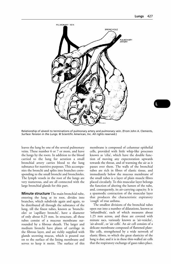
leaves the lung by one of the several pulmonaryveins. These number 6 or 7 or more, and leavethe lungs by the roots. In addition to the bloodcarried to the lung for aeration a smallbronchial artery carries blood to the lung substance for nutritive purposes. This accompa-nies the bronchi and splits into branches corre-sponding to the small bronchi and bronchioles.The lymph vessels in the root of the lungs arevery numerous, and are all connected with thelarge bronchial glands for this part.
Minute structure The main bronchial tube,entering the lung at its root, divides intobranches, which subdivide again and again, tobe distributed all through the substance of thelung, till the finest tubes, known as ‘bronchi-oles’ or ‘capillary bronchi’, have a diameter of only about 0.25 mm. In structure, all thesetubes consist of a mucous membrane sur-rounded by a fibrous sheath. The larger andmedium bronchi have plates of cartilage in the fibrous layer, and are richly supplied withglands secreting mucus, which is poured out on to the surface of the lining membrane andserves to keep it moist. The surface of this
membrane is composed of columnar epithelialcells, provided with little whip-like processesknown as ‘cilia’, which have the double func-tion of moving any expectoration upwardstowards the throat, and of warming the air as itpasses over them. The walls of the bronchialtubes are rich in fibres of elastic tissue, andimmediately below the mucous membrane ofthe small tubes is a layer of plain muscle fibresplaced circularly. To this muscular layer belongsthe function of altering the lumen of the tube,and, consequently, its air-carrying capacity. It isa spasmodic contraction of the muscular layerthat produces the characteristic expiratory‘cough’ of true asthma.
The smallest divisions of the bronchial tubesopen out into a number of dilatations, known as‘infundibula’, each of which measures about1.25 mm across, and these are covered withminute sacs, variously known as ‘air-vesicles’,‘air-alveoli’, or ‘air cells’. An air cell consists of adelicate membrane composed of flattened plate-like cells, strengthened by a wide network ofelastic fibres, to which the great elasticity of thelung is due; and it is in these thin-walled air cellsthat the respiratory exchange of gases takes place.
Lungs 427
L
Relationship of alveoli to terminations of pulmonary artery and pulmonary vein. (From John A. Clements,Surface Tension in the Lungs. © Scientific American, Inc. All rights reserved.)

The branches of the pulmonary arteriesaccompany the bronchial tubes to the farthestrecesses of the lung, dividing like the latter intofiner and finer branches, and ending in a densenetwork of capillaries, which lies everywherebetween the air vesicles, the capillaries being soclosely placed that they occupy a much greaterarea than the spaces between them. The air inthe air vesicles is separated from the blood onlyby 2 most delicate membranes, the wall of theair cell and the wall of the capillary, and it isthrough these walls that the respiratoryexchange takes place.
Lungs, Diseases ofThe chief of these is PNEUMONIA. (See also
under PLEURISY; EMPHYSEMA; TUBERCULOSIS;MAEDI/VISNA; CALF PNEUMONIA; EQUINE RESPI-
RATORY VIRUSES; ENZOOTIC PNEUMONIA
OF PIGS; CONTAGIOUS BOVINE PLEURO-PNEU-
MONIA; PARASITIC BRONCHITIS; PULMONARY
ADENOMATOSIS.)
Congestion of the lungs Accumulationof fluid, or ‘congestion’, is the preliminary stageof several types of acute pneumonia. It alsooccurs in disease of the left side of the heart.FOG FEVER of cattle is another condition inwhich congestion of the lungs is seen.
‘Hydrostatic congestion’ of a lung is apt tooccur if an animal which cannot stand, lies for too long on one side. Regular turning of the animal on to its other side is a necessarynursing procedure.
Pulmonary oedema This may occur during pneumonia (some forms), in disease ofthe left side of the heart, and (in cattle) in FOG
FEVER, and in PARASITIC BRONCHITIS of cattleand sheep.
An acute and usually rapidly fatal oedema of the lungs occurs in animals exposed to smokein a burning building; the animal almost liter-ally ‘drowns’ in its own blood serum.(Administration of oxygen can be tried if ananimal has been rescued before severe lungdamage has been caused.)
Poisoning by PARAQUAT and ANTU results inoedema and consolidation of the lungs. (See also
DIPS AND DIPPING; ELECTROCUTION.)
Pulmonary emphysema (see FOG FEVER)
Pulmonary haemorrhage (see RACE-
HORSES)
Allergic alveolitis Inflammation of thealveoli of the lungs of cattle exposed to mouldy
hay or straw contaminated with micro-organ-isms such as Thermopolyspora polyspora, andresembling ‘FARMER’S LUNG’.
Tumours of the lung are usually ofmetastatic origin, i.e. they are secondary growthswhich have started from another centre in thebody, being carried to the lung tissue either bythe blood- or lymph-stream. (See CANCER.)
Gangrene of the lung may be a complica-tion of, or a sequel to, pneumonia, and is usu-ally fatal. It is characterised by the presence of afoul-smelling, usually rusty-red, and almostalways very copious discharge from both nos-trils, in addition to the other symptoms ofpneumonia. It is commonest in the horse as asequel to ordinary pneumonia, and in otheranimals it may occur when the pneumonia hasbeen produced through faulty administrationof drenches. (See PNEUMONIA.)
Collapse of the lung The lungs are soresilient, in consequence of the elastic fibresthroughout their substance, that if air be admitted within the pleural cavities the lungsimmediately collapse to about a third of theirnatural size. Accordingly, if the chest wall iswounded and air gains entrance through thewound (pneumothorax), the lung collapses.After the wound has healed, and provided thatno complications occur, the elasticity is restoredas the air is absorbed. (See PNEUMOTHORAX.)
Torsion of a lung lobe, usually the rightcardiac lobe, is seen rarely in dogs and cats; it causes dyspnoea, pulmonary oedema, anddeath. The lobe may become twice its normalsize and blackish.
Wounds of the lung are serious onaccount of the air admitted through the chestwall, which leads to collapse; also the haemor-rhage, and the difficulty of checking it. Thelung may be wounded by the end of a fracturedrib pointing inwards. (See ‘FLAIL-CHEST’.)
Parasites of the lungs. Liver-flukes are sometimes found in the lungs of cattle and sheep; lung-flukes attack cats, dogs, pigs,and man in the Far East and the USA. Other parasites include LUNGWORMS. (See also
HEARTWORMS, for pulmonary dirofilariasis.)
Lupins, Poisoning byLupins of different species have often beenfound to cause poisoning of sheep; sometimesalso of horses, cattle, and goats.
428 Lungs, Diseases of
L
Poisoning by lupins is of 2 kinds: (1) due toalkaloids within the plant producing a nervousdisease; and (2) due to infestation of the plantwith a fungus which produces a toxin affectingthe liver. This 2nd type of poisoning is knownas lupinosis, is usually chronic, and producesloss of appetite and weight, jaundice, cirrhosisof the liver, oedema of the head, ascites anddeath. A few animals do recover but seldomthrive well afterwards.
In the USA great loss among sheep flocks has been occasioned by feeding on lupins byanimals not accustomed to them. The alkaloidsare present chiefly in the seeds.
Poisoning by the alkaloids gives rise to symp-toms which include loss of appetite, labouredbreathing, excitement, convulsions and deathfrom respiratory paralysis. There is no jaundiceor cirrhosis of the liver, and animals whichrecover are likely to do so completely.
Lupus ErythematosusAn autoimmune disease of dogs and cats whichoccurs in 2 forms: (1) the cutaneous or discoidform, and (2) the systemic form.
The discoid form is characterised by sym-metrical lesions on face, nose, and ears.Alopecia, loss of pigment, erythema, and a scaliness may be seen. Exposure to sunlightworsens the condition.
The systemic form affects many tissues andorgans. Autoantibodies against platelets, redand white blood cells may be present; with antibodies also in joints, kidney, skin, and other organs. Symptoms include bilateral pol-yarthritis, fever, muscle pain, enlarged lymphnodes, and sometimes nervous symptoms.
Prednisolone is used in treatment.
Luteinising Hormone (LH)A secretion of the anterior lobe of the pituitarygland. LH controls the development of theCORPUS LUTEUM and its production of proges-terone. In the male animal, LH stimulatessecretion of testosterone by the testicle.
LuteolysisRegression of a CORPUS LUTEUM. Two factorsappear to be involved in luteolysis in mostdomestic animals – one being prostaglandin F2αand the other being follicular oestrogen synthe-sis. It has been suggested that PGF2α is the nor-mal luteolytic compound, and that it is trans-ferred from the non-gravid uterus to the ovaryby some form of counter-current distributionbetween the uterine vein and ovarian artery.While the actual route for PGF2α transfer is insome doubt, its physiological role is certain.
A number of procedures for inducing luteol-ysis in domestic animals have been used. Theserange from the squeezing out of an establishedcorpus luteum by rectal palpation in cattle, tothe use of oestrogens. Synthetic prostaglandinsare now used.
Luxation(see DISLOCATION)
Lyme DiseaseThis was first recognised in Connecticut, USA,in 1975; the vector of infection is Ixodes tickson deer.
The disease occurs both in the UK and in other EU countries; in adults as well as inchildren.
Cause Borrelia burgdorfei. (See BORRELIA.)
Signs Blurred vision, lethargy, headaches, arthri-tis. In a few cases meningitis or encephalitis ormyocarditis result.
Lyme disease in dogs has been reportedin the UK, other EU countries, the USA andAustralia.
LymphLymph is a clear fluid collected from the tissueswhich enters the lymph vessels and thence the blood. It contains less protein than, but isotherwise similar to, the blood plasma. It alsocontains lymphocytes.
Lymph nourishes the tissues and returnswaste products from them back into the blood-stream. There are certain tissues which are notprovided with a blood supply at all, (e.g. thecornea of the eye, cartilage, horn, etc.) and inthem the lymph is the only nourishing medium.
The lymph is derived in the first place fromthe bloodstream, of which the watery con-stituents exude through the fine walls of thecapillaries into the tissue spaces. After meals,lymph from the small intestine may be milky inappearance due to contained fat. (See also LYMPH
NODES; LYMPHOCYTE.)The term ‘lymph’ was also applied to the
material which collects in the vesicles of cow-pox and was used for vaccination.
Lymph NodesFormerly called lymph glands, these are situat-ed on the lymphatic vessels, act as filters, andhave an important role in body defence by producing lymphocytes. (See also RETICULO-
ENDOTHELIAL SYSTEM; IMMUNE RESPONSE;LYMPHOCYTES; PLASMA CELLS.)
Lymph Nodes 429
L
LymphadenitisInflammation of lymph nodes.
Lymphadenoma(see HODGKIN’S DISEASE)
LymphangitisInflammation of the lymphatic vessels, oftenresulting from a streptococcal infection.
In horses 3 infective forms occur: (1) EPIZOOTIC LYMPHANGITIS (caused by a yeast);(2) ULCERATIVE LYMPHANGITIS (bacterial); and (3) GLANDERS (bacterial).
The non-infective lymphangitis used to becalled Monday morning disease, often beingseen in horses after a weekend of no work and aprotein-rich diet.
Signs Fever, lameness in one or more legs, withenlarged and tender lymph nodes. Later,doughy swellings, which pit on pressure, mayaffect the whole limb.
The appetite is lost for a day or two, but the horse is usually very thirsty. Under appro-priate treatment, the severity of the symp-toms abates in 2 or 3 days’ time, or sooner; and although lameness still persists, perhaps for as long as a week, the general appearance of the horse rapidly improves. The horse is usu-ally able to resume work in from 10 days to afortnight.
Recurrences are likely, resulting in some permanent thickening of the limb.
Treatment Antihistamines may be tried.Antibiotics may be necessary; also diuretics tohelp reduce the swelling, and phenylbutazone,or some other analgesic, to reduce the pain.
LymphaticsLymphatics are the vessels which convey thelymph through the body. (See LYMPH.)
LymphocystisA viral disease of fishes, which may give rise towhitish nodules on the cornea of the eye.
LymphocyteLymphocytes in mammals are of 2 main classes:thymus-derived, called T cells; and B cellsderived from bone marrow. Unlike polymor-phonuclear leukocytes and monocytes, thewhite cells in this group have cell surface receptors for antigen, and they are not involvedin phagocytosis.
T cells do not secrete antibodies and actdirectly on foreign cells. B cells divide rapidly toform plasma cells which secrete antibodies. (See
under BLOOD; B CELLS; IMMUNE RESPONSE;RECEPTORS.)
A much-simplified scheme of the relation-ship between antigens and some of the lym-phoid cells of the body is shown in the diagram.Natural immunity is conferred by the naturalsecretions of the body surfaces. If these surfacesare penetrated, scavenger cells (macrophages)attempt to engulf and destroy the antigens.Macrophages have a central role in immunityand if they are successful no further effects of antigen may be detectable. The activity ofmacrophages is increased if the antigen is coated with specific antibodies. Macrophagesare also attracted to areas where antigen is concentrated by soluble factors secreted by certain sensitised lymphocytes.
Large granular lymphocytes are a type ofT-lymphocyte, stated to have a prominent rolein modulating normal immune responses, andin eliminating virus-infected and transformedcells. It has been suggested that infection ofthese cells by the malignant catarrhal feveragent is the essential initial step in precipitatingthe disease.
(See also LYMPHOKINE.)
Lymphocytic Choriomeningitis(LCM)A viral disease of mice transmissible to humanbeings, in whom it may give rise to fever,headache, pain in muscles and, occasionally,death from meningoencephalitis. Dogs may actas symptomless carriers.
Lymphoid LeukosisLymphoid leukosis of chickens is a form of can-cer caused by a retrovirus, and initially affectsB-lymphocytes in the bursa of Fabricius. Itmetastises to the liver and spleen, whichbecomes swollen. The tumours are nodular andyellowish-white. The disease is spread from hento egg and hen to hen; the incubation period isabout 4 months. Lymphoid leukosis is rare inmammals.
LymphokineSecreted by T-lymphocytes, and formed whensensitised cells react with antigen, lymphokinescan attract other lymphocytes and monocytes,modify vascular permeability and activatemacrophages. In man they are believed to playan important role in the development ofrheumatoid arthritis.
Lymphosarcoma(see LEUKAEMIA; also FELINE LEUKAEMIA; BOVINE
ENZOOTIC LEUKOSIS; CANCER)
430 Lymphadenitis
L
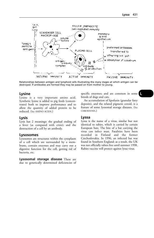
LysineLysine is a very important amino acid.Synthetic lysine is added to pig feeds (concen-trates) both to improve performance and toallow the quantity of added protein to bereduced. (See AMINO ACIDS.)
LysisLysis has 2 meanings: the gradual ending of a fever (as compared with crisis); and thedestruction of a cell by an antibody.
LysosomesLysosomes are structures within the cytoplasmof a cell which are surrounded by a mem-brane, contain enzymes and may carry out adigestive function for the cell, getting rid ofbacteria, etc.
Lysosomal storage disease These aredue to genetically determined deficiencies of
specific enzymes; and are common in somebreeds of dogs and cats.
An accumulation of lipofusin (granular fattydeposits), and the related pigment ceroid, is afeature of some lyosomal storage diseases. (See
CEROIDOSIS.)
LyssaLyssa is the name of a virus, similar but notidentical to rabies, which is carried by certainEuropean bats. The bite of a bat carrying thevirus can infect man. Fatalities have beenrecorded in Finland and the formerCzechoslovakia. In 1996, an infected bat wasfound in Southern England; as a result, the UKwas not officially rabies free until summer 1998.Rabies vaccine will protect against lyssa virus.
Lyssa 431
L
Relationships between antigen and lymphoid cells illustrating the many stages at which antigen can bedestroyed. If antibodies are formed they may be passed on from mother to young.
‘M & B 693’The code name given by the manufacturer May& Baker (now part of Merial) to sulphapyri-dine, one of the SULFONAMIDE DRUGS. It wasthe first sulfonamide to be introduced into theUK, in 1938.
Macaw-Worm FlyThis is a parasite of cattle and other animals inCentral America. It is another name for thewarble-fly Dermarobia homini. (See under FLIES.)
MacrocyteMacrocyte is the term applied to an abnor-mally large erythrocyte especially characteristicof the blood in some forms of anaemia.
MacrophageA former monocyte (type of white blood cell)which has migrated into the tissues and becomelarger. (See under BLOOD; INFECTION.)
MaculesMacules are spots or stained areas of skin ormucous membrane, usually brownish, red, orpurple in colour.
‘Mad Cow Disease’(see BOVINE SPONGIFORM ENCEPHALOPATHY)
Madness in Dogs(see RABIES; ENCEPHALITIS; MENINGITIS)
Maedi/VisnaA slowly progressive disease of sheep and goats,first recognised in 1939 in Iceland and believedto have been introduced into that country bykarakul sheep imported from Germany. Icelandis now free from the disease, following 2 eradi-cation programmes, but maedi occurs in theUK, continental Europe, North America, Africa,and Asia.
Although maedi/visna has never been record-ed in Australia, a retrovirus was isolated fromgoats there and shown to produce antibodiesindistinguishable from those produced bymaedi/visna virus in goats.
Cause A lentivirus. It is usually seen in animalsover 2 years old. Transmission to the lamb isoften via the ewe's milk.
‘Maedi’ is the Icelandic word for dyspnoea,and the disease is a type of pneumonia with avery long incubation period – 1 to 3 years oreven more. An early sign is dyspnoea; afterphysical exertion the breathing becomes veryrapid and shallow. Later, breathing becomesdifficult even when the animal is at rest, anddeath often follows.
‘Visna’ is a name applied to the same viralinfection when the brain or spinal cord ratherthan the lungs are involved. Demyelinationoccurs.
Diagnosis can be confirmed by microscopicalexamination of the tissues and by isolation ofthe virus. There is also an ELISA test.
MAFFMAFF is the acronym for the Ministry ofAgriculture, Fisheries and Food which prior to2001 was responsible for the control of notifi-able diseases, imports of animals and welfare ofanimals on farms. It was replaced in 2001 bythe Department of the Environment, Food andRural Affairs (DEFRA).
MaggotsMaggots which have fed on carcases contami-nated by Clostridium botulinum may contain adose of toxin lethal to birds. This has causeddeaths among domestic poultry, wild birds, andon game farms in the UK.
In one incident, the London Zoo lost 37birds from botulism arising from a batch ofcommercially bred maggots. Maggots reared ina sterile environment are used to debridewounds.
Maggots in SheepIn many parts of the world, certain dipterousflies may lay their eggs on the wool of sheepduring summer, and the eggs hatch into mag-gots which either live on the surface of the skinor burrow down into the subcutaneous tissues.They cause great loss from wasting of flesh anddestruction to fleeces, and sometimes result in the death of the affected sheep. The green-bottle flies (Lucilla caesar and L. sericata, inBritain, and L. macellaria, in both North andSouth America) are those responsible for thiscondition. (See under MYIASIS and FLIES.)
‘Magic Mushroom’ PoisoningThis is caused by psilocybin, a hallucinogenpresent in Psilocybe semi-lanceate and Panaeolusfoenisecii. Consumption of the fungus by domes-tic animals can give rise to bizarre behaviour.
M
Signs In the dog, aggressiveness, ataxia, nys-tagmus and salivation plus a body temperaturein excess of 42°C have been noted.
A normally docile pony, which had beengrazing in a field where ‘magic mushrooms’were growing in profusion, became aggressive.
Magnesium (Mg)Magnesium (Mg) is a light white metal whichburns in air with the production of a brilliantwhite flame, leaving a white powder as aresidue. The salts of magnesium used as drugsare the oxide, carbonate, and sulphate. There isa heavy oxide, known as ‘heavy magnesia’, alight oxide called ‘light magnesia’, a heavy carbonate, and a light carbonate. Both theoxides and the carbonates are antacids andslightly laxative. The sulphate of magnesium iscommonly called ‘Epsom salts’. (For bloodmagnesium, see under HYPOMAGNESAEMIA.)
Uses Magnesia, whether light or heavy, is usu-ally prescribed for foals, calves, and dogs whenthese require a mild antacid and laxative. Thesulphate of magnesium is a saline purgative. (See
under LAXATIVES.)Calcined magnesite is used as a top-dressing
for pastures in an attempt to prevent hypomag-nesaemia (about 500 kg (10 cwt) per acre). Forcattle, a daily dose of 50 g (2 oz) calcined mag-nesite is considered to be of great value in theprevention of hypomagnesaemia, but it shouldbe fed only during the ‘danger period’. This isbecause prolonged feeding of magnesium saltsis apt to accentuate any latent phosphorus deficiency and may lead to ‘milk lameness’ orsimilar conditions.
A mixture of magnesium acetate solutionand molasses has been used, being available ona free-choice basis to cattle from ball feedersplaced in the field.
Magnesium oxide Too high a level in con-centrate feeds for lambs and calves has led tourolithiasis. (See under URETHRAL OBSTRUCTION.)
MagnetsMagnets have been used to treat traumatic reticulitis and prevent traumatic pericarditis incattle. (See under HEART DISEASES.)
MagnetotherapyThe use of magnetic fields to supporttherapy. There are claims that magnetotherapyhas beneficial effects on conditions affecting the nervous and locomotor sysems and on post-surgery healing.
Maine-AnjouA French dual-purpose breed of cattle. Colours:red and white, and roan.
Maize(see PHYTIN; SILAGE)
Major HistocompatibilitySystemOne of the chromosomal regions controllingimmune responses.
Histocompatibility antigens are inheritedthrough a set of genes known as the major histocompatibility complex (MHC). Every animal possesses its own unique set of histocompatibility antigens.
Class I genes code for antigens that provokethe rejection of foreign grafts. Antigens of this class are of the cell-surface type, located on all nucleated cells, and concerned with cellrecognition.
Class II cell-surface antigens are locatedmainly on B-cells, and are concerned with regulation of the immune response.
Class III antigens are located in serum protein, and regulate complement activity. (See
COMPLEMENT.)Following the demonstration at the Animal
Breeding Research Organisation, Edinburgh, ofan association between MHC type in cattle andtheir resistance to mastitis, it was suggested thatit might be possible to select for AI bulls whichpass on this resistance.
Malachite GreenA dye used in the treatment of external fungaland protozoal infections of fish, and for the con-trol of proliferative kidney disease. Only the zinc-free preparation can be used for therapy in fish.
MalaciaSoftening of a part or tissue in disease, e.g.osteomalacia or softening of the bones.
Malaria(see under MONKEYS)
‘Malaria of Birds’(see under PLASMODIUM GALLINACEUM)
Malassezia (Pityosporon)A yeast-like fungus which sometimes producesa brownish-black deposit in dogs' ears.
MalathionAn organophosphorus insecticide which hasbeen used for the control of external parasites incattle and as a crop spray.
Malathion 433
M
Mal De Caderas (Maladie De Caderas)Mal de caderas (maladie de caderas) is a try-panosome disease of the horse, occurring inBrazil, Argentina, Bolivia, and Paraguay, beingmost serious in the latter country. It is causedby the Trypanosoma equinum. Suramin orquinapyramine have been used in treatment.
Mal De PlayaA form of poisoning in cattle by a plant,Lantana camara.
Mal Du Coit (Maladie Du Coit)(see DOURINE)
Male FernThe growing point of male fern (Dryopteris felix-mas) may attract cattle on bare pasture, and leadto poisoning. In Scotland, 61 out of 68 head ofbeef cattle were affected, with 45 becomingwholly blind, 10 partly blind, and 21 recum-bent. All recovered within a week except for 4cows and 4 calves, which remained completelyblind. One cow was additionally recumbent andwas destroyed. (See also under FERNS.)
Maleic HydrazideA growth-retardant used on grass verges whichhas caused non-fatal gastritis in small animals.
Malformation(see DEFORMITIES)
MalignantA progressively worsening condition.
Malignant Aphtha of Sheep(see ORF)
Malignant Catarrhal Fever(see BOVINE MALIGNANT CATARRHAL FEVER)
Malignant Jaundice of Dogs(see CANINE BABESIOSIS)
Malignant Oedema(see GAS GANGRENE)
Malignant Stomatitis(see CALF DIPHTHERIA)
Malignant Theileriasis of Sheepand GoatsA tick-borne disease caused by the protozoanparasite Theileria hirci, and occurring inEastern Europe, the Middle East, Egypt, andSudan.
Signs include high fever, constipation, gland-ular enlargement, pale anaemic mucous mem-branes with later jaundice and death; however,the disease may be very mild in animals withsome locally acquired immunity.
Mallein TestMallein test is a method of testing for the presence of GLANDERS in a horse.
MamillaMamilla is the Latin term for the nipple.
Mamillitis Inflammation of the nipple.Bovine herpes mamillitis is a recognised diseasewhich can also affect the skin of the udder.
Mammary Gland (Udder)
Structure
Cow The udder has 4 glands or ‘quarters’. Astrong septum divides the 2 right-hand glandsfrom the 2 on the left side, but there is no suchdemarcation between fore and hindquarters onthe same side of the body.
The structure of the gland is similar to thatseen in the mare, being composed of lobes andlobules, held in position by fibrous tissue, andsending ducts down into an irregular milksinus. This latter is large, and partly dividedinto compartments by folds of mucous mem-brane. From it leads 1 large lactiferous ductdown the teat to its apex, which possesses asphincter muscle of almost 10 mm in width.
Mare The mammary glands are 2 in number,situated in the inguinal region.
Ewe There are 2 mammary glands, each ofwhich has a single teat. They are situated in theinguinal region, as in the mare and cow.
Sow The mammary glands number 12 inmost sows (although a few have more), and arearranged in 2 rows reaching from just behindthe level of the elbows along the abdomen tothe inguinal region. As a rule, the glands whichare situated towards the middle of the series arethe best developed and secrete the most milk.Each teat has 2 ducts as a rule.
Bitch As in the sow, there are 2 rows of glandsalong the lower line of the abdomen. They areusually 10 in number, but in the smaller breedsthere may only be 8, and in the larger breedsthere are sometimes 12. The teats each possessfrom 10 to 12 tiny lactiferous ducts.
434 Mal De Caderas (Maladie De Caderas)
M
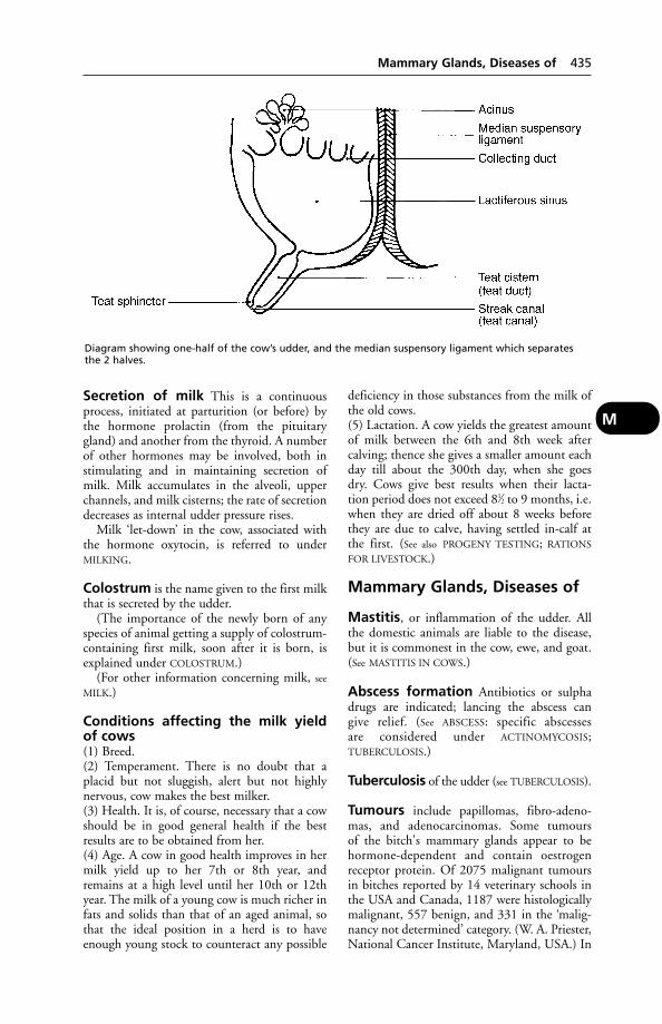
Secretion of milk This is a continuousprocess, initiated at parturition (or before) bythe hormone prolactin (from the pituitarygland) and another from the thyroid. A numberof other hormones may be involved, both instimulating and in maintaining secretion ofmilk. Milk accumulates in the alveoli, upperchannels, and milk cisterns; the rate of secretiondecreases as internal udder pressure rises.
Milk ‘let-down’ in the cow, associated withthe hormone oxytocin, is referred to underMILKING.
Colostrum is the name given to the first milkthat is secreted by the udder.
(The importance of the newly born of anyspecies of animal getting a supply of colostrum-containing first milk, soon after it is born, isexplained under COLOSTRUM.)
(For other information concerning milk, see
MILK.)
Conditions affecting the milk yield of cows(1) Breed.(2) Temperament. There is no doubt that aplacid but not sluggish, alert but not highlynervous, cow makes the best milker.(3) Health. It is, of course, necessary that a cowshould be in good general health if the bestresults are to be obtained from her.(4) Age. A cow in good health improves in hermilk yield up to her 7th or 8th year, andremains at a high level until her 10th or 12thyear. The milk of a young cow is much richer infats and solids than that of an aged animal, sothat the ideal position in a herd is to haveenough young stock to counteract any possible
deficiency in those substances from the milk ofthe old cows.(5) Lactation. A cow yields the greatest amountof milk between the 6th and 8th week aftercalving; thence she gives a smaller amount eachday till about the 300th day, when she goes dry. Cows give best results when their lacta-tion period does not exceed 81⁄2 to 9 months, i.e.when they are dried off about 8 weeks beforethey are due to calve, having settled in-calf atthe first. (See also PROGENY TESTING; RATIONS
FOR LIVESTOCK.)
Mammary Glands, Diseases of
Mastitis, or inflammation of the udder. Allthe domestic animals are liable to the disease,but it is commonest in the cow, ewe, and goat.(See MASTITIS IN COWS.)
Abscess formation Antibiotics or sulphadrugs are indicated; lancing the abscess can give relief. (See ABSCESS: specific abscesses are considered under ACTINOMYCOSIS;TUBERCULOSIS.)
Tuberculosis of the udder (see TUBERCULOSIS).
Tumours include papillomas, fibro-adeno-mas, and adenocarcinomas. Some tumours of the bitch's mammary glands appear to behormone-dependent and contain oestrogen receptor protein. Of 2075 malignant tumoursin bitches reported by 14 veterinary schools inthe USA and Canada, 1187 were histologicallymalignant, 557 benign, and 331 in the ‘malig-nancy not determined’ category. (W. A. Priester,National Cancer Institute, Maryland, USA.) In
Diagram showing one-half of the cow’s udder, and the median suspensory ligament which separatesthe 2 halves.
Mammary Glands, Diseases of 435
M
cats, mammary carcinoma is twice as commonin the Siamese breed as in all other breeds combined. (See TUMOURS; CANCER.)
Hypertrophy of the mammary glands in thecat has been recorded both in pregnant queensand in neutered females treated with megestrolacetate for 14 months to 5 years. The conditionhas to be differentiated from neoplasia.
Wounds and injuries of the udder andteats are commonest in the cow and sow, owingto greater pendulousness than in other animals.
All wounds of the udder and teats are seriouson account of the danger of infection and thedevelopment of mastitis.
Treatment As a first-aid treatment, woundsof the teats and udder should be washed with warm water and an antiseptic, dry sulphanilamide powder subsequently applied.
When a teat has been torn or injured so thatmilk escapes from the canal, it is usually diffi-cult to get the fistula, so formed, to heal untilthe cow goes dry. An operation is usually neces-sary to obtain healing. This procedure necessi-tates the cow remaining dry for at least 2months. In some cases a cow with a fistula isbetter turned out to grass at once, and made torear calves until her milk-flow ceases, when shecan be taken in and undergo the operation.
Eruptions on teats may be specific, such asare seen during outbreaks of foot-and-mouthdisease, cowpox, malignant catarrhal fever,rinderpest, etc. (See VIRAL INFECTIONS OF
COWS' TEATS.)Many teat sores, however, are caused through
‘chapping’, or ‘cracking’, of the delicate skin ofthe teat. (See under MASTITIS.)
Warts on teats (see WARTS)
Teat obstructions Difficulty in milkingmay be caused by stricture of the sphincter,milk clots or (rarely) calculi in the teat canal; alternatively by the presence of wartygrowths inside the canal. The latter condition is considered under WARTS.
Manchester TerrierA medium-sized dog with pointed face, smoothblack coat and brown markings on the face andlegs. The breed may develop cutaneous astheniaand Perthe's disease.
Mandelic AcidA urinary antiseptic, effective in acid urine.
MandibleMandible is the bone of the lower jaw. (See
under JAW.)
Mandibular Disease(see SHOVEL BEAK)
Manganese(Mn), a trace element, is necessary in minutequantities for a healthy diet. Insufficiency in thepasture herbage, e.g. in Devon and Cornwall,UK, may cause infertility in cattle, anddeformed offspring.
In New Zealand, on one farm, 32 calves wereborn with very shortened limbs and enlargedjoints. They had been sired by 4 bulls. Owingto a dry season, the cows had been fed largequantities of apple pulp and corn silage, both ofwhich contained very low levels of manganese.(See TRACE ELEMENTS; ‘SLIPPED TENDON’.)
MangeA contagious skin disease caused by mites,which lay their eggs in the skin. The movementof the larvae results in intense irritation. Damageto skin, hide or fleece is caused by efforts torelieve the discomfort. There are different typesof mange, each caused by a different species ofmite. Sarcoptic mange occurs in man (when it is known as scabies), dogs, cats, cattle, pigs,sheep, horses, etc. Psoroptic mange in the sheep is known as SHEEP SCAB. Choriopticmange usually affects the tail and legs.Demodectic mange (follicular or ‘black’ mange)is most common in dogs. Details are given below. (For causal mites, see under MITES,PARASITIC.)
Mange in cattle
Demodectic mange Fairly common butoften not noticed as clinical signs are few; nodular formation in hair follicles is the main one.
Sarcoptic mange is common in Britain andNorth America and is the cause of ‘dairyman'sitch’. It is usually found on the head and neck,but may occur on any part of the body. Bullsare particularly liable to this form of mange.
Chorioptic mange is usually confined to thebase of the tail, but may spread.
Psoroptic mange causes debility, failure tothrive, and reduced liveweight gain.
Psoroptic mange is an important disease offeedlot cattle in the USA, and was once the
436 Manchester Terrier
M

most prevalent form of cattle mange in Britain,where it is now seldom seen. In an outbreak inBritain, in a beef herd comprising 306 animals,the infected areas of skin were thickened andscabby, with blood and serum oozing from thelesions. These extended along the back anddown the flanks.Treatment Ivermectin or similar products byinjection; ‘pour-on’ products such as fenthionand phosmet.
Mange in the horse In this host, 4 varieties of mange occur.
Sarcoptic mange In this type the parasitesburrow into the epidermis and make treatmentdifficult. The disease commences by the hairdropping out in patches with the formation ofpapules and an intense continuous itching. Thehair becomes thin and broken, and abrasionsare present. The skin is hard and folded.Emaciation is progressive, and death may occurfrom exhaustion. It reaches its height in spring,and is at an ebb in late summer and autumn.Treatment Gamma BHC is an efficient mite-killer, but its use in the UK is banned. Theorganophosphorus compounds, diazinon andfenchlorphos, are also effective. For the bestresults they should be applied by dipping or as saturating sprays, and for the sarcopticmanges particularly, 2 or more treatments may be necessary at intervals of 10 to 14 days.
Psoroptic mange Two varieties of Psoroptesoccur on the horse, 1 in the ear and 1 on theskin. The lesions on the skin are localised atfirst, and usually start near the dorsal line wherethe hair is long. The patches are generally barerthan in sarcoptic mange. The parasites bite the epidermis, but do not penetrate the skin. The serum which exudes forms a scab in which the parasites live.Treatment is on similar lines to that for sarcop-tic mange. It is important to remember thepresence of parasites in the ear, and to treat this part of the body also.
Chorioptic or symbiotic mange is usuallyconfined to the legs or root of the tail. It is notnotifiable. It causes great itching, stamping, andrubbing of one leg against the other. Papules,scabs, and even ulcers may be found. Treatmentis as above. Ivermectin paste, 0.2 mg/kg, in 2doses, 2 weeks apart, has been suggested as acontrol measure.
Demodectic mange (See description under‘Mange in dogs and cats’, below.)
Mange in sheep
Sarcoptic mange is usually confined to thehead, and is seldom found on the woolly parts of the body. It tends to become more generalised in the goat.
Chorioptic mange, caused by Chorioptesbovis, occurs in horses as well as cattle andsheep, and can be serious, especially in housedsheep overseas. It is not uncommon in the UK.Of 130 sheep received from South Wales at theCentral Veterinary Laboratory, 33 per cent werefound infested. Lesions occur on the pasternsand in the interdigital spaces.
Psoroptic mange or ‘sheep scab’ is causedby Psorotes ovis. It occurs on all parts of thebody covered with wool and in the ears. Thelife-cycle is typical, and can be completed in 13to 16 days: the progress of the disease is in consequence very rapid. It is one of the worst of sheep diseases; it was formerly notifiable in Britain.
Itching is usually the first symptom of the disease, and should be investigated at once.The skin becomes thickened and even ulcer-ated; the wool becomes detached and the sheepemaciated (see illustration below). The itchingcauses the animal to rub itself against fences,and detaches the scab. This further spreads thedisease, and permits secondary infections of the wound by bacteria.Treatment is usually by means of double-dipping at an interval of about 8 to 12 days, butwhich depends on local circumstances. InBritain a dip sanctioned by DEFRA must be used. It should be purchased only from areputable manufacturer and used exactly asdirected. Organophosphorus dips can only bepurchased by persons who have attended acourse on their usage and have received anappropriate certificate to confirm this.Prevention (see IVERMECTIN)
The ‘itch-mite’ of sheep Psorergates ovis,which occurs in Australasia, Africa, and Northand South America, has not, so far, been foundin Britain. This mite causes thickening of theskin and scurf formation. The growth of thewool fibre is affected and the fleece is furtherdamaged by rubbing.
Mange in pigs
Sarcoptic mange starts on the head andgradually spreads all over the body, especiallyattacking the thinner skin. There is intense
Mange 437
M
itching, the hair falls out, and the skin becomescovered with scab or with wart-like projections.It is found in the UK and North America.Treatment Improved liveweight gains have followed treatment of sarcoptic mange.Amitraz, doramectin or IVERMECTIN may beeffective.
Mange in dogs and cats
Sarcoptic mange in the dog generally starts on the muzzle and spreads backwards.The animal should be clipped and bathed with green soap. It may then be treated withphosmet. If the infection is generalised, treatone-half of the body, and the other half after 2 or 3 days.
Notoedric mange in cats is similar to the latter form in dogs. It is intensely irritating,affecting face, ears, and occasionally legs and external genitalia. It is now very rare in the UK.
Benzyl benzoate may prove toxic to cats andso one of the sulphur preparations or piperonylbutoxide is recommended.
Otodectic or auricular mange occurs indogs and cats. Otodectes is the most frequentcause of irritation in the ears; it causes scratchingand shaking of the ear.
The eggs and larvae are very resistant, andsurvive under treatments which kill the adults.
First-aid Cat-owners may be able to provide a little temporary relief by means of a few drops of olive or vegetable oil, which will helpto soften waxy deposits and kill some mites. A few drops of warm, soapy water (not dish-washing detergent liquid) may achieve the same result. However, professional adviceshould be obtained without delay.
Owners should not poke around with cotton-wool wound round an orange stick ortweezers, as the wool will slip off, and the skinof the external ear canal is then likely to beabraded, or even the ear-drum punctured.
Professional treatment consists in the useof eardrops containing an effective mite-killer,plus an analgesic to reduce the irritation causedby the mites.
In neglected cases, or those complicated bybacterial or fungal infections, where a painful,suppurating condition is present, antibacterialor anti-fungal drugs must be used.
(See DEAFNESS and EARS, DISEASES OF; also
HAEMATOMA.)
Demodectic mange Cigar-shaped mitesinvade the hair follicles, causing the hair to fallout in patches.
Demodectic mange (also known as follicularor ‘black mange’) is most common in dogs.
Signs Two types of demodectic mange in dogshave been described: (1) the squamous type, inwhich the skin becomes scaly, wrinkled andringworm-like in appearance (and sometimesmistaken for ringworm); and (2) the pustulartype in which secondary bacterial infectionoccurs. This is always very serious, and consti-tutes an illness as well as a mere skin disease,since the dog suffers from toxaemia. Indeed,sometimes euthanasia becomes the onlyhumane course, especially when extensive areas of skin are involved.
Cause Demodex canis.
Treatment is made difficult by the fact that the mites are sometimes living at a depthdifficult to reach. However, amitraz and IVERMECTIN are effective.
Diagnosis is made or confirmed by the examination of skin scrapings under a microscope.
The disease often appears in the dog when 8 to 12 months old, usually first on the head,around the eyes and nose, and on or near the feet.
Mange in goats may be caused by D. caprae, and characterised by palpable nodules or pustules without loss of hair. The disease usually starts on face, neck andshoulders. Ivermectin and related drugs orphosmet have proved successful in treatment.
Mange in fowls
‘Depluming scabies’ in fowls is caused byCnemidocoptes laevis, which lives at the base ofthe feathers, and so irritates the fowl that itpulls them out. The stumps left may be seen tobe surrounded with crusts. The affected spotsand surrounding areas may be treated withIVERMECTIN.
‘Scaly leg’ is caused by C. mutans. The feetand legs become enlarged and crusted. Thebirds may become very lame and even lose atoe. Destruction of infected birds combinedwith rigorous disinfection is the most commonmethod of eradication. If this procedure is not convenient, the scab should be removedwith soap and water, the leg dried, and one of
438 Mange
M
the preparations mentioned above used. Thisshould be repeated in 3 or 4 days.
Cage birds may suffer from this infestation.C. pilae causes ‘scaly face’ and ‘tassel foot’.
Dermanyssus gallinae is the chicken miteof Europe and North America. It is whitish tored in colour. The complete life-cycle takesabout 7 to 10 days.
The mite lives exclusively on blood. It is nocturnal in its habits, living in crevices during the day.
Eradication of the mite must be thorough.All wooden structures must be disinfected. Apainter's blow lamp is very useful for cracks.
Affected flocks may be treated by cypermethrin, as a dilute spray.
Although primarily a parasite of fowls, thismite will attack horses and other mammals,causing much irritation, with the eruption ofpapules and the formation of scabs. The mite,as it feeds only at night, may be overlooked asthe cause of the disease. The proximity of fowlssuffering from the mite may give a clue.
Ornithonyssus sylviarum, the northernfowl mite which is also common in Britain,causes scab formation, soiling of the feathers,and thickening of the skin around the vent. Incontrast to the chicken mite, this parasiteremains on its host.
In Israel allergic rhinitis and bronchial asthma have been caused by this mite amongpoultry farmers.
Liponyssus bursa, the tropical ‘fowl mite’,replaces the last species in the warmer parts of the world. Unlike it, however, this species is found on the fowls and in the nest. It mayfeed during the day. It also lays eggs and moultson its host. The symptoms are similar.
ManiocAn ingredient of some compound animal feedswhich has been found unsafe for turkeys. (See
CASSAVA.)
MannosidosisThe most widely recognised lyosomal storagedisease of cattle, especially of Aberdeen Angus.It is due to a genetic deficiency of the enzymemannosidase. Affected calves develop ataxia andbecome aggressive; finally, paralysis sets in.
Beta-mannosidosis, an inherited disorderof glycoprotein metabolism, has been identified in goats, and is rapidly fatal. Signsinclude inability to rise from a recumbent
position, carpal contractures, pastern jointhyperextension, a dome-shaped skull, and deafness.
Manure HeapsManure heaps are potentially a source of infec-tion and should be fenced off. Grass growingnear manure heaps may also contain pathogensand parasites. New manure should be buriedunder older manure; the new manure will then heat up to about 70°C, destroying most pathogens, if left for a few days. (See also
SLURRY.)
ManxA breed of cat that is without a tail, or has only a very short one. The breed originated onthe Isle of Man and the Manx governmentmaintains a breeding colony. The lack of tail isdue to a dominant mutation and is seen as adepression at the end of the spinal column.There may be associated defects in vertebrae,and malfunction of the sphincter muscle. Ifboth parents carry the mutant gene, kittens diebefore birth. Early deaths in kittens may be dueto malformations such as fused vertebrae orspina bifida. Manx cats with a short tail arecalled ‘stumpies’; some are born with tails andare known as ‘longies’. Manx cats have longerback legs than forelegs, causing an unusual gait.
‘Marble Bone Disease’(see OSTEOPETROSIS)
Marburg Disease(see MONKEYS)
Marek's DiseaseThis contagious disease of domestic poultry was first described in Austria-Hungary in 1907.It was first recorded in America in 1914, and in Britain in 1929, and spread widely. Theavailability of vaccines, with good hygienicpractice, has greatly reduced the losses fromMarek's disease, which is a neoplasm caused bya herpesvirus. Turkeys are rarely affected as theynormally harbour a different herpesvirus – onethat affords protection against Marek's disease.The turkey virus has been used in a vaccine forchickens.
Formerly called fowl paralysis, Marek's disease had, before the advent of vaccinationagainst it, become the most economicallyimportant disease of poultry in many countries, in terms of fowl mortality, carcasecondemnation, and lost egg production.
At least 2 forms of Marek's disease are recog-nised: the classical form, in which paralysis – to
Marek's Disease 439
M
a varying degree – is the outstanding feature;and an acute leukosis form, in which lymphoidtumour formation is the main feature, with nervous symptoms less in evidence.
Cause A herpesvirus, which may persist forlong periods in litter dust.
Signs The classical form affects birds com-monly between the ages of 3 and 4 months, butcases have been recorded in broilers a little over3 weeks old, and also in birds over a year old. Itis frequently noticed that certain strains of birdsare affected – in-contact birds of a differentparentage remaining healthy. Affected birdsmay show lameness of 1 or both legs: this lame-ness becomes progressively worse, and generalparalysis results. A common attitude for anaffected bird to adopt is to lie about with 1 limbextended in front and the other extendedbehind. In spite of this the bird appears alertand will feed if placed beside a supply.Drooping of wings may be noted. In some casesthe tip of the wing may touch the ground. Eyelesions may be seen.
In the acute form of Marek's disease, birds asyoung as 6 to 8 weeks may be affected. Loss ofappetite and depression are noticeable; tumourscan often be palpated – these involving abdom-inal organs, muscles, skin, and sometimes thecomb. Paralysis is not the predominant charac-teristic of this form of the disease.
Mortality The mortality varies, but in birds 6 to 8 weeks old may exceed 20 per cent. It is difficult actually to arrive at a satisfactory figure as this disease may co-exist with others, e.g. coccidiosis, tapeworm infestations, tuberculosis, and vitamin deficiency, etc.
Control measures Vaccination at 1 day oldmay be repeated at 2 to 4 weeks; it provides 80 to 90 per cent protection in the case of anoutbreak.
On the disease being diagnosed, all affectedbirds should be destroyed as soon as the firstsymptoms are observed. The disease is alwaysintroduced to a farm by the purchase of freshstock, in the form of eggs, day-old chicks, oradult birds.
Careful selection of the source of fresh stockis vital in maintaining disease-fee flocks.
Both the fowl tick, Argas persicus, and thedarkling beetle can harbour the virus of Marek'sdisease.
Vaccines. Freeze-dried and ‘wet’ cell asso-ciated vaccines are available. The latter are
stored in liquid nitrogen; care is needed in handling as the ampoules may shatter or causefreeze burns. The manufacturers' instructionsmust be followed.
Mares, Infertility in(see under EQUINE GENITAL INFECTIONS)
Marie's Disease(see HYPERTROPHIC OSTEOPATHY)
Marijuana (Cannabis) PoisoningMarijuana (cannabis) poisoning has occurred indogs in the USA as a result of being givenhome-made sweet biscuits containing the drug.Symptoms include acute depression, retchingor vomiting, and a staggering gait. The dogsmay be ill for 36 to 48 hours, and vomitingmay be frequent. Besides vomiting, musculartremors, and weakness, 1 dog showed inconti-nence, ataxia, leant against objects and thensank to the floor.
MarketsMarkets are covered accommodation for the saleof animals. Shelter must be provided for dairycows in milk, calves, lambs and pigs. Animalsshowing any sign of disease, or which are unfit,must be put in special pens and not offered forsale. Because of the contact between animalsfrom different sources, disease can spread rapidly. Strict regulations apply to the runningof a market and the transport of animals to andfrom markets. The legislation involved is theWelfare of Animals at Markets Order 1990 andthe Welfare of Horses and Ponies at Markets(and Other Places of Sale) Order 1990.
‘Marmite Disease’A form of dermatitis encountered in piglets 3 days old and upwards. (See ‘GREASY PIG
DISEASE’.)
MarrowThe soft substance that is enclosed within thecavities of the bones. Yellow marrow owes itscolour to the large amount of fat contained in it, while red marrow is of a highly cellularstructure. Formation of the red blood cells (erythrocytes) takes place in the marrow, as alsothat of the blood platelets (thrombocytes). Themarrow is also the source of lymphocytes (B-cells), monocytes, and other leukocytes. (See
BLOOD; BONE; also MYELOCYTE.)
Marsh Marigold PoisoningThe marsh marigold, or kingcup (Caltha palustris), has occasionally been the cause of
440 Mares, Infertility in
M

poisoning, and is similar in its effects to BUTTERCUP POISONING.
MarsupialA mammal of an order in which the young areborn in an immature state and continue theirdevelopment in a maternal pouch. Examplesare kangaroos and opossums in Australia.
MarteiliosisA disease caused by Marteillia refringens thataffects molluscs, especially oysters. It is a NOTIFIABLE DISEASE in the UK and parts of the EU.
MashamThe cross resulting from a Blackface ewe and aWensleydale ram.
Mast CellMast cell is a type of connective tissue cell. It releases histamine and heparin, which causeanaphyllaxis and atopy in cases of allergic reaction. (See under BLOOD – Basophils; also
REAGINIC ANTIBODIES.)
Mast-Cell TumoursNodular tumours of the skin which are usuallybenign but may become malignant. (See also
MASTOCYTOMA.)
MastiffA very large, light-coloured dog with short coat, folded-over ears and a broad muzzle. Hipdysplasia may be present and the breed is proneto extropion.
MastitisInflammation of the udder. (See MAMMARY
GLAND, DISEASES OF, for mastitis in animalsother than the cow.)
Mastitis in CowsInflammation of the udder, involving the secret-ing cells of the mammary glands, or its connectivetissue, or both. (See diagram, page 442.)
Subclinical mastitis Mastitis may be unac-companied by obvious symptoms. This formcommonly reduces milk yields by 10 per cent or so, and is consequently of great economicimportance. It has been stated that approxi-mately 1 cow in 3 in Britain is affected by subclinical mastitis.
Simple tests have been used to detect thepresence of an abnormally high content ofwhite cells in an ordinary-looking sample ofmilk, and so indicate the presence of mastitis.
Once it is known that it exists, bacteriologicaltests can be used to identify the organismsresponsible and to determine the best treat-ments. Sometimes an excess of white cells(more than 500,000 per ml) in the milk is theresult of inflammation due to trauma and notto infection. Thus the California or WhitesideTest may draw attention to a faulty milkingmachine or bad milking technique.
The mastitis situation in a herd can be monitored on a monthly basis by laboratoriesoperating electronic cell counters. The tableshows the ranges of white cell counts.
The graph shows the spread of mastitis in anautumn calving herd.
Mastitis tends to rise as the winter progressesand fall when the cows first go out to grass. Thecell counts rise again in July and August chieflybecause of the high proportion of the cowsnearing the end of their lactation. Cell countsand mastitis levels fall again in September whensome of the older cows are being culled and firstcalf heifers are coming into the herd. Mastitislevels rise again through the winter period.
Clinical mastitis should be regarded as aherd problem.
Acute Shivering may usher in the attack. Later,there is a rise in temperature; fast, full pulse;short, quick respirations; and an uneasy appear-ance. The animal paddles with her feet, but isusually afraid to lie on account of the pain occa-sioned to the udder. She refuses food, andrumination is in abeyance. When the udder isexamined it is found that one (or more) quarteris swollen, tense, reddened, and very painful to the touch; the cow may stand with her hind-legs straddled apart.
‘Summer mastitis’ (often involving gan-grene of the udder) usually occurs either inheifers or in dry cows; however, it is seen occa-sionally in cows just after calving. It is caused byActinomyces pyogenes, often in association withother pathogenic bacteria (e.g. Peptococcusindolicus).
‘Summer mastitis’ is something of a misnomer, in that, while it is most common in July and August, it is also seen in January and February.
If the gangrene affects a large part of thequarter, or when more than one-quarter isattacked, the condition of the cow is serious in the extreme.
The animal may stand aloof from the rest ofthe herd, sometimes paddles with her hind feet,and is obviously in pain. On examination the
Mastitis in Cows 441
M

442 Mastitis in Cows
M
Diagram showing the relationship between the amount of mammary gland tissue involved and the formof mastitis which results.
Estimate ofEstimate of milk production
Cell count ranges mastitis loss per cow(cells/cc) problem per year
Below 250,000 Negligible —250,000–499,000 Slight 190 litres (42 gallons)500,000–749,000 Average 330 litres (74 gallons)750,000–999,000 Bad 760 litres (169 gallons)1,000,000 and over Very bad 885 litres (197 gallons)
The spread of mastitis in an autumn calving herd.

trouble is soon located to the udder, wherehardness – but not necessarily swelling – of aquarter is detected. Foul-smelling pus (grey,greenish-yellow, or blood-stained) is present.
Treatment Antibiotics, by injection or intramammary administration, may save the animal's life, though use of the quarter is usually lost.
Prevention The injection into the teat-canalof long-acting intramammary antibiotic priorto turning out, repeated every 3 to 6 weeksdepending on the product used, during summer. (In maiden heifers and in-calf heifers,this procedure may be difficult and not alwayspracticable. Care is needed to avoid both damage to the teat and the introduction ofpathogenic bacteria.)
Give such protection against flies as is practicable.
Subacute mastitis The disease runs a coursenot unlike that of the acute form, but the symp-toms appear much more slowly. There is agreater difficulty in milking, the first drawnmilk often containing little clots and alwayslarge numbers of shed epithelial cells; later,there is a gradually increasing pain and swellingin the affected quarter, accompanied by an alteration in the colour of the milk to yellowish or yellowish-grey. The amount ofmilk decreases. As a rule, appetite remains normal, pulse and breathing are unaltered, andif there is any rise in the temperature it is slight.
Chronic mastitis shows little general consti-tutional disturbance, and an almost completeabsence of pain, a slowly progressing increase in the density of the gland, a diminution in the secretion of milk, and a gradual increase inthe size of the affected quarter or quarters.
Pathological changes in the udder may render any antibiotic ineffectual. One suchproblem is the survival within phagocytes ofstaphylococci, where they are protected fromthe lethal action of most antibiotics. (See
PHAGOCYTOSIS.)Antibiotics can more economically be used
when the cow is in the dry period. Long-actingantibiotics can then be given without aggravat-ing the problem of antibiotic residues in milk.(See under MILK – Antibiotics in milk.)
Bacterial mastitis Staphylococcus aureus,Streptococcus agalactiae, S. uberis and S. dys-galactiae are among the main organisms respon-sible for mastitis. However, S. dysgalactiae is
seldom a serious problem; S. uberis is moreresistant.
In a survey of 5 herds, S. uberis was found tobe the major pathogen associated with dry cowmastitis.
Coliform or ‘Environmental’ mastitishas become increasingly prevalent in recentyears, and is common during the winter. Thisinfection of the udder is often long-lasting, andthe cow is ill with it, so that its economic effectsmay be greater than with streptococcal orstaphylococcal mastitis. Many outbreaks havebeen linked with cold, wet weather; they are aggravated by damp bedding, sawdust, and muddy conditions underfoot when strip-grazing kale, etc. The above conditions wouldappear to favour the entry of E. coli through the teat-canal, but the organism may also reach the udder via the bloodstream in cattlewhich are scouring – often after a suddenchange of diet – as a result of an active E. coligut infection.
‘Experimentally, severe cases of coliformmastitis can be produced only in early lactation following the stress of calving – a situation commonly prevailing in naturallyoccurring field cases.’ (IRAD, Compton.) (See
COLIFORM.)Klebsiella spp. and Enterobacter aerogenes are
other organisms involved in mastitic infections.
Leptospiral mastitis Leptospiral infectionoften causes agalactia rather than mastitis.However, in 1 outbreak in Northern Ireland,involving half a herd of 140 cows over a 2-month period, symptoms included a suddendrop in milk yield, flaccid udders with all 4 quarters affected, thickish and sometimesbloodstained milk, fever (a temperature of upto 41°C (106°F)), and quickened breathingand pulse rates. The illness in individual cowslasted from 1 to 4 days. Leptospira hardjo wasisolated from the milk and blood of cows withclinical mastitis. (See LEPTOSPIROSIS IN CATTLE.)
Among other bacteria which may cause mastitis are Bacillus subtilis, which has been isolated from washing water, header tanks, andteat-cup liners; Pseudomonas; and Chlamydia.Campylobacter jejuni has also caused mastitis.
Mycoplasmal mastitis occurs in Britain andmany other countries, and may prove resistantto antibiotics. In an outbreak in North Walesover a 5-week period, half a herd of 115 cowsbecame infected, and 14 had to be sold forslaughter. The milk was at first brownish in colour. The mastitis was rapid in onset,
Mastitis in Cows 443
M
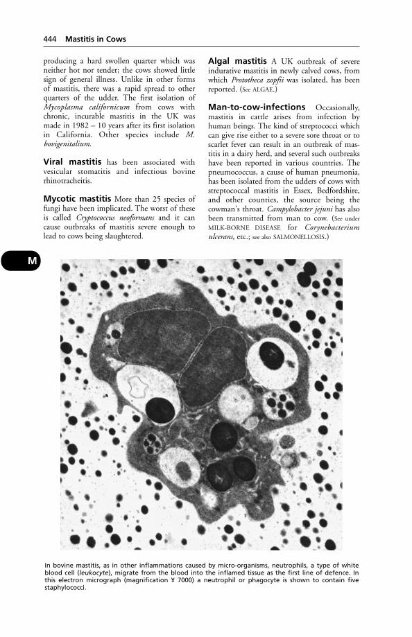
producing a hard swollen quarter which wasneither hot nor tender; the cows showed littlesign of general illness. Unlike in other forms of mastitis, there was a rapid spread to otherquarters of the udder. The first isolation ofMycoplasma californicum from cows withchronic, incurable mastitis in the UK was made in 1982 – 10 years after its first isolationin California. Other species include M. bovigenitalium.
Viral mastitis has been associated with vesicular stomatitis and infectious bovinerhinotracheitis.
Mycotic mastitis More than 25 species offungi have been implicated. The worst of theseis called Cryptococcus neoformans and it cancause outbreaks of mastitis severe enough tolead to cows being slaughtered.
Algal mastitis A UK outbreak of severeindurative mastitis in newly calved cows, fromwhich Prototheca zopfii was isolated, has beenreported. (See ALGAE.)
Man-to-cow-infections Occasionally,mastitis in cattle arises from infection byhuman beings. The kind of streptococci whichcan give rise either to a severe sore throat or toscarlet fever can result in an outbreak of mas-titis in a dairy herd, and several such outbreakshave been reported in various countries. Thepneumococcus, a cause of human pneumonia,has been isolated from the udders of cows withstreptococcal mastitis in Essex, Bedfordshire,and other counties, the source being the cowman's throat. Campylobacter jejuni has alsobeen transmitted from man to cow. (See under
MILK-BORNE DISEASE for Corynebacteriumulcerans, etc.; see also SALMONELLOSIS.)
444 Mastitis in Cows
M
In bovine mastitis, as in other inflammations caused by micro-organisms, neutrophils, a type of whiteblood cell (leukocyte), migrate from the blood into the inflamed tissue as the first line of defence. Inthis electron micrograph (magnification ¥ 7000) a neutrophil or phagocyte is shown to contain fivestaphylococci.
The spread of infection Infection entersby way of the teat and can easily be spread fromcow to cow by milkers' hands or the cups of themachine, but apparently less easily by the lattermethod. Udder cloths and towels are also commonly infected. It has also been shown thatin an infected herd, a large proportion of soresor chaps harbour organisms, and these may bea source of infection of the udder itself in thesame cow or in another. The skin of the teatsand the milkers' hands may remain infectedfrom one milking to another, and in a heavily infected herd the skin of the cows' bodies, milkers' clothes, floor, partitions, etc.become contaminated and may remain so for considerable periods.
Treatment Proper treatment depends upon acorrect diagnosis and the use of suitable antibi-otics in adequate dosage, introduced into theudder with aseptic precautions so as not tointroduce further (and perhaps more virulent)infection. Adequate dosage is important, as otherwise strains of resistant organisms mayarise. In some cases sulfanilamide may be used.(See ANTIBIOTIC RESISTANCE.)
The control of mastitis An important aidis teat-dipping. The liquid mainly used for teat-dipping is an iodophor – a type of disinfectantcontaining iodine but extremely mild in itseffect upon the tissues. Good results can also be obtained with hypochlorite teat-dips containing 1 per cent available chlorine.
In rotary parlours with automatic clusterremoval equipment and only 1 operator, teat-dipping is often impracticable and teat-spraying (probably less effective) is the only alternative.
A 2nd recommendation is the wearing by thedairyman of smooth rubber gloves, which canbe dipped in disinfectant before the udder iswashed. They represent a partial solution to theproblem created by the fact that hands cannotbe sterilised.
Warm-water sprays may be used for udder-washing, and disposable paper towels for drying. The latter obviate cross-infections fromudder cloths. (See also SPONGES.)
If warm-water sprays are not available, washthe udder (if very dirty) with plain warm waterfirst, then with an udder wash; numerous proprietary formulations are available, based onbenzalkonium, cetrimide, chorhexidine, solu-bilised iodine, etc. If none of these is available, a1 per cent solution of cetrimide may be used.
It is significant that in herds with a low inci-dence of mastitis, udder-washing is avoided in
40 per cent and practised only in 11 per cent(but see MILK – Sediment in milk).
Questions which the farmer must ask are asfollows:
(1) Is the person doing the milking capableof handling the cows properly, and keen to doso?
(2) Are the vacuum gauges and cup-linerskept correctly adjusted?
(3) Is hand-stripping avoided?(4) If a disinfectant is used, is it used at the
correct strength?(5) Are there disposable paper towels?(6) Are there any old, chronically infected
cows in the herd which do not respond to treatment and would be better disposed of?
(7) Is fly-control being practised in the milking parlour?
(8) Is attention being given to the 96-hourrule regarding the withholding of milk from a cow after calving, whether treated with antibiotics or not?
Rough inexpert milking and stripping predispose to mastitis. With machine milking,the use of a badly designed teat-cup liner, forinstance, or leaving the cups on an empty quarter, may lead to trouble. (See under MILKING
MACHINES for faulty use of these, leading tomastitis.) Bruising is an important predisposingcause, and for this reason cows should never behurried, especially before milking, as the uddermay be injured. This applies particularly toolder cows in which the udder is large and pendulous. Chilling must be avoided, and alsochapped teats (the latter should be left dry aftermilking). Even the smallest injuries and soreson the teats should be carefully attended to,since the germs which gain entry to these sooften gain entry to the udder later.
Routine use of the strip-cup is helpful. Ifflecks or clots are seen in the milk, segregate thecow(s) if practicable, and – in any case – milkafter the others. When a strip-cup is used, careshould be taken to see that neither the handlenor the fingers become a source of infection toclean cows. Use the cup before the udder iswashed.
Dry-cow therapy It has been shown in large-scale field experiments that the best time to treatcows to eliminate infection from the udders isduring the dry period. Particularly with staphy-lococcal infection, there is a better chance ofremoving infections at this time than duringlactation, and better results are achieved whencows are treated in the subclinical phase of thedisease rather than during a clinical attack.Treatment during the dry period not only
Mastitis in Cows 445
M
eliminates most of the existing infection; it alsoprevents most of the new infections from occur-ring during the dry period, including ‘summermastitis’. Another advantage is that there is noproblem of milk being contaminated withantibiotic(s), provided that a cow is dry for 6weeks or longer. It is advisable to treat all cows.Preparations containing cloxacillin have provedvery effective. Teat-dipping of dry cows is alsouseful in preventing summer mastitis.
Control measures summarised.
(1) Records Keep all details of cell-count figures on a monthly basis and use these tomonitor the incidence of mastitis in the herd.Record also details of milking-machine testingand maintenance.
(2) Milking machines Have machinerytested regularly and thoroughly at least once a year. At each milking check the vacuum pressures, pulsation rates, air bleeds and liners.Remember that machines are used 730 times ayear and faulty machines can lead to a mastitisbuild-up.
(3) Teat-dipping and hygiene Use aneffective iodophor-plus-lanolin teat-dip on eachquarter as the cluster is removed. Wear smoothrubber gloves for preference; use the fore-milkcup before washing the udders with clean, running water. Use clean paper towels – not adirty cloth – to dry the udders.
(4) Dry-cow therapy The farmer's veterinarysurgeon will not only advise generally on mastitiscontrol, but will also recommend the appropriatetreatment at the end of lactation. This willinclude the infusion of a specially formulated,long-lasting antibiotic into each quarter, todestroy residues of infection and to counter newinfections in the dry period.
(5) Treatment Clinical mastitis can occur atany time and will need prompt attention imme-diately by the veterinary surgeon, who canadvise on the correct treatment during lactation.
(6) Culling Any cow which has several attacksof clinical mastitis in a lactation endangers therest of the herd. Records of treatments andresponses will identify those cows with recur-ring cases in 1 lactation and show which shouldbe culled from the herd.
The cow's own protection ‘Pathogensinvading the mammary gland of the cow are
subjected to non-specific resistance factors at 2 levels, either in the teat-canal or in the mam-mary gland itself. The teat-canal acts as amechanical barrier, but in addition invadingpathogens within the canal are subjected to the activity of antimicrobial fatty acids andcationic proteins. Pathogens breaching thesebarriers are then subjected to the defences ofthe mammary gland itself. In the early stages of infection there is a considerable increase insomatic cells in the milk, which is associatedwith an increased resistance to infection.During the early stages of the inflammatoryreaction the invading pathogens are exposed tothe action of neutrophils, locally producedhumoral factors and proteins from the systemiccirculation which pass into the mammarygland. These serum factors include theimmunoglobulins, complement units and otherantimicrobial proteins.’ (Dr K. G. Hibbitt andDr A. W. Hill, IRAD, Compton.)
Breeding for resistance to bovine mastitis may be possible in the future. (See
MAJOR HISTOCOMPATIBILITY SYSTEM.)
Mastitis in EwesCommon causes are Pasteurella haemolytica,which can produce a peracute mastitis withgangrene, and Staphylococcus aureus. Once theewes are separated from their lambs, dry-cowintramammary preparations may be used as apreventive measure. It is essential to use 1 tubeper teat.
Mastitis in GoatsMastitis in goats can be a problem in humans as the milk is often unpasteurised.Potential infectious organisms includeStreptococcus zooepidemicus, S. pneumoniae,Actinobacillus equuli, Pasteurella haemolyticaand Staphylococcus aureus.
Mastitis Metritis AgalactiaSyndrome (MMA)Mastitis metritis agalactia syndrome (MMA),also known as farrowing fever, is a commonproblem in sows in the first 2 days after farrow-ing. The sow is off its food and the temperaturerises; the udder may be partially or whollyswollen and painful. The piglets are restless.
Treatment involves antibiotics, possibly withanti-inflammatory agents. The piglets must befed while the problem persists.
MastocytomaA type of tumour which is common in the dogand involves skin and subcutaneous tissue;
446 Mastitis in Ewes
M
occasionally muscle. A mastocytoma may bemalignant. It contains numerous MAST CELLS.In cattle this tumour is also regarded as potentially malignant.
Maternal AntibodiesTheir function in protecting the offspring frominfections encountered by the dam is referred to under COLOSTRUM. The immunity so produced is a temporary one, and wanes. The timing of vaccinations to induce lasting immu-nity is crucial (see DISTEMPER PREVENTION); for if carried out while the level of maternallyderived antibody is significant, vaccination will fail. (See also MEASLES VACCINE; CANINE
PARVOVIRUS.)
MavisMedicines Act Veterinary Information Servicenewsletter. Issued by the Veterinary MedicinesDirectorate, Woodham Lane, New Haw,Addlestone KT15 3NB.
Maxilla(see SKULL – General arrangement of the skull)
MCG (mcg)Microgram: 1 millionth of a gram.
Meadow-Saffron PoisoningThe meadow saffron (the autumn crocus,Colchicum autumnale), a common inhabitant ofmeadows, hedge bottoms, and woodland areasin England and Wales, is a cause of poisoningamong horses and cattle. Pigs may sometimeseat the bulbous root (corm) and suffer, butsheep and goats are resistant. All parts of theplant are poisonous, both when green andwhen dried in hay, but the toxicity varies at different times of the year. Cases of poisoningare usually seen in the spring, when the leavesand seed-vessels are produced, and then againin summer and autumn (from August toOctober), when the flowers are formed.
The poison, (colchicine) is present in largest amounts in the seeds and corms; it iscumulative in its action.
Signs When only small quantities have beentaken there is loss of appetite, suppression ofrumination, profuse dribbling of saliva, anddiarrhoea. The excretion of colchicine by thekidneys causes irritation in the urinary bladder,and induces the animal to pass urine in smallamounts almost as soon as it is formed. Bloodmay be present in both the urine and the milkof dairy cows. Abortion is common in pregnantcows and heifers.
When large amounts have been eaten, thesymptoms include ataxia and abdominal pain;death may occur in from 16 hours to 4 days.
The plant should be eradicated from pasturesin the autumn when its striking pale purple crocus-like flowers can be easily seen. The bulbsshould be dug out or cut with a hoe.
Meal-Feeding in PiggeriesThis can result in a very dusty atmosphereunder some circumstances, causing coughingand a feeling of tightness in the chest in peopleworking there, and sometimes to a falseassumption that the pigs are coughing becauseof enzootic pneumonia.
Measles(see under MONKEYS; MEASLES VACCINE)
‘Measles’ in Beef(see TAPEWORMS)
‘Measles’ in Pork(see TAPEWORMS)
Measles VaccineAn attenuated measles virus vaccine may be usedin the dog to give protection against distemper.(See also under DISTEMPER.) Measles vaccine canovercome low levels of maternally derived antibodies and may be used from 5 weeks old.
Meat(see PORCINE STRESS SYNDROME; HORSE-MEAT;DOGS' DIET, and below)
Meat, DarkMeat with limited fat cover and intramuscularfat appears dark. This is seen particularly in bullcarcases, and horse meat is always dark incolour. Dark cutting beef is possibly the singlebiggest cause of loss to beef processors. It iscaused by a deficiency of glycogen in the mus-cles of an animal at slaughter which prevents thenormal decrease in pH post-mortem. As a resultthere is an increase in enzyme activity; this usesup the oxygen which would normally convertthe dark myoglobin into pink oxymyoglobinand the meat appears dark. It also tends to bedry because of the higher water-binding capac-ity of muscle protein at a higher pH. The storesof glycogen are depleted principally by muscularexhaustion and stress, and these 2 factors mustbe avoided in the 48 hours before slaughter inorder to minimise the risk of dark cutting beef.(See also PORCINE STRESS SYNDROME.)
Long fasting times are associated with areduction in carcase yield in pigs and an
Meat, Dark 447
M

increase in the incidence of dark, firm, dry(DFD) meat.
Meat-Handlers' OccupationalHazardsMany of the infectious diseases that affect animals can be transmitted to abattoir workersand may cause illness. They include infectionby beta-haemolytic streptococci, which caninfect cattle, sheep, pigs and chickens. Peopleinvolved in the slaughter of these animals maybe exposed to ringworm and impetigo, and any cuts on their hands may become infected. (See also under ZOONOSES; also underspecific examples such as ORF; LOUPING-ILL;TUBERCULOSIS; ANTHRAX; RABIES.)
Meat-Hygiene RegulationsRequirements for the production, inspection,cutting, storage and transport of fresh meat fordomestic use and export are set out in the following legislation: The Fresh Meat (Hygieneand Inspection) Regulations 1995; The PoultryMeat, Farmed Game Meat and Rabbit Meat(Hygiene and Inspection) Regulations 1995;The Minced Meat and Meat Preparations(Hygiene) Regulations 1995; The MeatInspection Regulations 1987 (amended 1990);The Slaughterhouses (Hygiene) Regulations1977; The Slaughterhouses (Hygiene) andMeat Inspection (Amendment) Regulations1991 (see also LAW).
Meat inspection in all UK licensed abattoirsis controlled by the Meat Hygiene Service, agovernment agency.
(See also FOOD INSPECTION.)
Meat, Knacker'sThis is to be avoided for the feeding of pet animals unless sterilised. (See MEAT STERILISA-
TION; EARS AS FOOD.) Unsterilised meat may beinfested with viable hydatid cysts, or be infect-ed with anthrax or tuberculosis. Even if it iscooked by the pet-owner before use, it maycontaminate hands, cooking utensils, etc., andthereby be a danger to public health. (See also
SALMONELLOSIS; E. COLI; BOTULISM; AUJESZKY'S
DISEASE; HORSE-MEAT.)
Meat Scraps, BonesThese can be a source of foot-and-mouth diseaseor swine fever infections. (See SWILL; also under
TUBERCULOSIS.)
Meat (Sterilisation) Regulations1969These require all knacker's meat to be sterilisedbefore being supplied to owners of pets, kennels,
etc. In 1982, new controls on the trade in meatunfit for human consumption came into force.If not sterilised, meat must be stained; likewiseoffal. Poultry meat is exempt. The colouringagent used is black PN or brilliant black BN.
MeatusMeatus is a term applied to any passage or opening; e.g. the external auditory meatus is the passage from the surface to the drum of the ear.
MebendazoleA broad-spectrum anthelmintic used in mostspecies. Proprietary preparations includeTelmin (Janssen) and Chanazole (Chanelle).
Meckel's DiverticulumMeckel's diverticulum, of human pathology,apparently has a veterinary equivalent – a finger-like projection from the small intestine,recorded as a congenital abnormality in the dogand found in poultry.
MeconiumFaeces present in the rectum of a newborn animal. They should in all cases be dischargedsoon after birth. In the first milk of the damthere is a natural purgative for this purpose. (See
also ILEUS.)
MediastinumMediastinum is the space in the chest which lies between the 2 lungs. It contains the heart,the aorta and vena cava, the gullet, the extrem-ity of the trachea, the thoracic duct, the phrenic nerves, and other structures of lesser importance.
Pneumomediastinum The presence of airin the mediastinum, following damage to lungalveolar tissue near its root. In other cases thetrauma may be of a more serious nature – e.g.escape of air from a damaged trachea, or rup-ture of the oesophagus. A swelling of a dog'swhole face and neck due to subcutaneousemphysema may follow pneumomediastinum,the air tracking upwards. It may take weeksbefore the swelling totally disappears, but thecondition is seldom serious unless trachea oroesophagus are damaged.
Medicines Act 1968The Medicines Act 1968 was designed to con-trol many aspects of the manufacture, testing and marketing of medicines for human andanimal use. In particular, its aim was to bringsafety standards up to those already enforced
448 Meat-Handlers' Occupational Hazards
M
by the leading companies. The Act requiredwholesalers, importers and manufacturers toobtain licences. (See also VETERINARY PRODUCTS
COMMITTEE.)The Act classifies those medicines which may
be sold to the public only on a veterinary prescription (PM), and those which may bepurchased from pharmacies (P) and from otheroutlets (GSL).
Medicines (Labelling ofMedicated Animal Feeding-Stuffs) Regulations 1973These set out the detailed particulars requiredon labels of containers or packages of medicated animal feeds. The Medicines(Labelling) Regulations 1976 covered thelabelling of containers and packages for medicinal products.
Mediterranean FeverA tick-borne disease of cattle and the water-buf-falo, occurring in Southeast Europe, Africa, andAsia, and caused by Theileria annulata.
Signs Fever, loss of appetite, a discharge fromeyes and nose, anaemic pallor of mucous mem-branes, constipation followed by diarrhoea.Survivors recover very slowly.
Mediterranean Spotted FeverA human disease.
Cause Rickettsia conori, transmitted by a dog tick.
Symptoms Fever, nausea, vomiting, headache,muscle pain, and a rash.
Medulla OblongataThat part of the brain which connects to thespinal cord (see BRAIN).
Medullary CavityMarrow cavity of bones.
Mega- and Megalo-Mega- and megalo- are prefixes denoting large-ness. Strictly speaking, mega indicates a multi-ple of 1 million times, as in megabyte.
MegabacteriaThese very large bacteria cause illness and death in ostrich chicks, canaries, and budgeri-gars. In the latter the signs include loss ofweight, difficulty in swallowing, vomiting, anddiarrhoea. Sudden death may occur due tohaemorrhage.
-MegalyAn abnormal enlargement, e.g. of the spleen(which may attain 4 or 5 times its normal sizein, e.g., babesiosis (redwater) in sheep).
MegaoesophagusMegaoesophagus implies usually a pathologicalenlargement of the oesophagus, such as may beseen in FELINE DYSAUTONOMIA and ‘FLOPPY’
LABRADORS.
Megestrol AcetateThe active ingredient of Ovarid (Schering-Plough); it is used in bitches as an oestrus suppressant, in cats for the treatment of miliarydermatitis (eczema), and in male cats and dogs to modify aggressive behaviour. Contra-indications are cats with diabetes or genital disease. Prolonged dosage or overdosage mayadversely affect the uterus or result in hypertro-phy of the cat's mammary glands. (See also
OESTRUS SUPPRESSION; DIABETES.)
Meibomian GlandsThese are minute sebaceous follicles situated inthe eyelids: also called tarsal glands.Inflammation may develop around an eyelash,and later there may be suppuration with theformation of a stye.
Meiosis (Reduction Division)Meiosis (reduction division) occurs during the formation of ova in the female and of spermatozoa in the male, and reduces the number of chromosomes by one-half, to thehaploid number.
MelaenaThe passing of dark tarry faeces, usually due to bleeding from the stomach or smallintestines. The blood undergoes chemicalchanges as the result of the action of the digestive process, which produces largeamounts of sulphide of iron.
MelaninA dark pigment that occurs naturally in theretina, hair, skin, feathers, etc. In the skin, itprotects against harmful ultraviolet radiationand is responsible for the darkening effect of suntan. It is also believed to have a role ingoverning the natural circadian rhythms.
MelanomaA tumour containing the pigment MELANIN.Melanomas are potentially malignant, and notuncommon in old horses that have been greyand are turning whiter. Cimetidine has been
Melanoma 449
M

used with some success in the treatment of suchtumours.
MelanoticMelanotic is the adjective deriving fromMELANOMA.
MelatoninMelatonin is a hormone secreted by the PINEAL
BODY. It is a ‘messenger’ of day length by whichanimals recognise the seasons. Experiments inhousing sheep using artificial light in wintershowed that melatonin levels increased.
An implant of melatonin can be used tostimulate onset of reproductive activity andimprove fertility early in the season.
Melia (Dhrek)The fruits and leaves of this Asiatic tree, Meliaazedarach, are poisonous to farm livestock.Abnormal gait, trembling of hind-limbs, paresis, and abdominal pain have been reportedin the pig.
MelioidosisA disease resembling glanders, caused byPseudomonas pseudomallei, and occurring inrodents – occasionally in human beings andfarm animals – in the tropics. Diagnosis is bycomplement fixation test and/or identificationof the organism. Outbreaks have also occurredin Europe, in zoos. Antibiotics may be useful intreatment.
Membrana Nictitans(see NICTATING MEMBRANE)
Membranes(see PLACENTA; BRAIN; MENINGES; MUCOUS
MEMBRANE; SEROUS MEMBRANES)
Membranous NephropathyA progressive disease of the kidneys of dogs andcats, affecting the glomeruli, and leading eventu-ally (sometimes after several years) to kidney failure. Most cases in the cat are first seen whenshowing the nephrotic syndrome. There is per-sistent excretion of protein in the urine, too littleprotein in the blood, and subcutaneous oedemaand/or ascites. (See KIDNEYS, DISEASES OF.)
MeningesThe three coverings of the brain and spinalcord: dura mater, arachnoid membrane and piamater (see BRAIN – Structure).
MeningiomaA tumour affecting the meninges, and perhapsthe commonest brain tumour in the cat.
MeningitisInflammation affecting the membranes cover-ing the brain (cerebral meningitis), and spinalcord (spinal meningitis), or both (cerebrospinalmeningitis). When the outer membrane isaffected, the condition is called ‘pachymenin-gitis’; when the inner membrane is it is knownas ‘leptomeningitis’ – although clinically it is not often that these distinctions can be determined, for inflammation readily spreadsfrom one to the other.
Causes Meningitis frequently develops in association with viral or bacterial diseases ofanimals, such as rabies, tuberculosis, swineerysipelas and distemper.
In lambs it may be caused by Pasteurellahaemolytica; in pigs by Streptococcus suisor encephalomyocarditis virus. (See GID;TAPEWORMS.) It may be produced through anexternal injury which fractures the skull andallows entrance to organisms, or it may appearduring the course of other head injuries inwhich there is no fracture. It accompanies most cases of encephalitis caused by viruses.
Signs As a rule the first signs are those of restlessness and excitement. The animal movesabout in a semi-dazed fashion, and stumblesinto or against fixed objects. Neighing, bellow-ing, squealing, and barking, apparently at noth-ing, may be noticed, and at times the animalexhibits a wild frenzy. After an attack of delirium or frenzy the animal becomes dull and quiet; the head hangs, the eyes stare, theexpression is vacant. Other symptoms, such asturning in circles, falling over, rolling along the ground, turning forward and backwardsomersaults, resting the head upon any conve-nient fixed object, such as a loose-box door,lying curled up in an unusual attitude, etc., maybe seen in some cases. Paralysis of 1 side of the body (hemiplegia), of both hind-limbs(paraplegia), or of a group of muscles is notinfrequent in the smaller animals.
Treatment Absolute quiet in a dark place isadvisable pending professional advice.
Dogs A form of spinal meningitis occurs inwhich bony tissue is laid down in the spinalcanal towards the posterior part of the vertebralcolumn. It is called ‘chronic ossifying pachy-meningitis’ as a consequence. It mainly affectsold dogs, and only causes inconvenience whensevere. It may lead to complete paralysis of thehind-limbs, accompanied by incontinence ofurine and faeces.
450 Melanotic
M

In all animals in which meningitis followsinjury, the skull should be examined for fractures.
MeningoceleA congenital defect: the protrusion ofmeningeal membrane through an abnormalopening in the skull or spinal column.
The defect occurs in calves, foals, puppies,kittens, piglets, etc.; it is also a human abnormality.
Treatment is surgical.In 1 case the owner of a calf stated that it was
born with a ‘tumour’ (i.e. a meningocele) whichit repeatedly damaged and caused to bleed.
On admission to the department of large-animal surgery, University of Utrecht,Netherlands, a red soft mass of tissue, whichpulsated in rhythm with the heart, was seen.
The meningocele was surgically removed,under general anaesthesia, and the skin sutured.
Three months later the owner reported thatthe calf was doing well and showing no sign ofany abnormal behaviour.
MeningoencephalitisInflammation of the brain and meninges. Thereis fever, pain and rigidity, as seen in meningitis;and muscle tremors, hyperexcitability, convul-sions and paralysis, as seen in encephalitis. Mostcases result from bacterial infection, includingListeria monocytogenes, especially in cattle andpigs, Pasteurella multocida in newborn calves,and streptococci in newborn piglets.
Symptoms include walking in circles, press-ing the head against a wall, champing of thejaws, convulsions.
MeniscusMeniscus is a crescentic fibro-cartilage in ajoint.
Mepacrine HydrochlorideAn antimalarial drug which has been used inthe treatment of coccidiosis in cattle.
Mepyramine MaleateAn antihistamine which is given by mouth, by intramuscular injection, or applied to theskin as a cream. Used in the treatment of laminitis, azoturia, urticaria, etc. (See
ANTIHISTAMINES.)
MercurochromeAn antiseptic, and a stain for spermatozoa. It isa proprietary name of a preparation of mer-bromin, and an organic compound of mercury.
Mercury (Hg)Also known as quicksilver and hydrargyrum, it is a heavy silver-coloured liquid metal. In this form it was once used as an ingredient ofointments and even purgative powders.
The salts of mercury are of 2 varieties: mer-curic salts, which are very soluble and powerfulin action; and mercurous salts, which are lesssoluble and act more slowly and mildly.Mercuric salts are all highly poisonous; asorganic compounds less so. In strong solutionthey may be caustic, and in weaker solutions are irritant.
Biniodide of mercury, or red iodide of mercury, made up into an ointment, formedthe base of the common ‘red blister’. With thisand other mercury dressings, it is essential thatcare be taken, for the drug may enter the systemby absorption from the skin, or by the animallicking itself.
Mercury poisoning With the possibleexception of calomel as a laxative, prepara-tions of mercury have given way to safer and more effective drugs. Consequently, mercury poisoning is now far less commonthan it was.
However, feeding seed corn dressed withmercurial compounds has led to the death ofpigs and cattle. Three out of 17 bullocks and heifers died after being given seed barley,treated with phenyl mercuric acetate, as part of their feed. The deaths were sudden; theautopsy findings multiple with extensive haemorrhages. Gastroenteritis, ataxia, and renal failure (often associated with mercury poisoning) did not occur.
Deaths of heifers from mercury poisoningoccurred after the roots of cauliflower plantswere dipped in mercurous chloride solutionbefore planting. Drainage of plant trays, andperhaps also spillage of the concentrate, hadcontaminated the yard. (See also under SEALS formercury poisoning in those creatures.)
In Japan, the eating of fish with a high mer-cury content led to an outbreak of illness, withnervous symptoms, in cats.
Acute poisoning results in vomiting, diar-rhoea, and abdominal pain, with death from shock. (Cattle may show only the 1st symptom.) Stomatitis and salivation may alsooccur. Severe purgation occurs in the smalleranimals, together with signs of acute abdominalpain. Lips and mouth may become white.
Chronic mercury poisoning (mercurialism):salivation; swelling of tongue, which bleedsreadily; loosening of the teeth. Nervous signsmay develop, e.g. ataxia, blindness.
Mercury (Hg) 451
M

First-aid Give white of egg.
Antidote A CHELATING AGENT such as dimercaprol. Absorption of mercury can bereduced by adding a binding agent such as zeolite to the diet.
A human case The mercury from a brokenthermometer, spilt on a carpet, led to severe illness in a 33-month-old girl. Symptomsincluded loss of appetite, sensitivity to light,eczema, sweating and scaling palms.Improvement followed 2 weeks' treatment withthe chelating agent, Dimaval.
Dental amalgam is another source of mercury vapour.
Mercury, Dog'sBoth dog's mercury (Mercurialis perennis) andannual mercury (M. annua) are poisonousplants, especially when seed-bearing. Cows aremost often affected. Animals may not showsymptoms until from 7 to 10 days after theplants are first eaten.
Signs Diarrhoea. Urine is passed frequently,accompanied by painful straining, and is of ablackish or blood-red colour, as is the diarrhoea.Other signs are severe anaemia and semi-coma.Deaths have occurred.
First-aid The animal should be given strongblack tea or coffee.
MesencephalonMesencephalon is the mid-brain connecting the cerebral hemispheres with the pons andcerebellum.
Mesenteric Hernia(see HERNIA)
MesenteryMesentery is the double layer of peritoneumwhich supports the small intestine.
Mesh Grafts(see SKIN GRAFTING TRANSPLANTATION)
MesocolonMesocolon is the name of the fold of peritoneum by which the large intestine is suspended from the roof of the abdomen.
MesogenicOf medium virulence. Often used to indicatethe degree of virulence of, e.g., Newcastle disease strains.
MesometriumMesometrium is the fold of peritoneum run-ning from the roof of the abdomen to theuterus. It consists of 2 layers, between whichrun the blood and lymph vessels, and the nerves to the uterus, and it acts as an elastic suspensory ligament supporting the uterus in position. During pregnancy it graduallystretches under the weight of the fetal contents,but retracts again after parturition under normal conditions.
MesosalpinxMesosalpinx is the suspensory ligament of theoviduct.
MesotheliomaA tumour developing from the mesotheliumcovering membrane surfaces.
MesovariumMesovarium is the suspensory ligament of theovary.
MesulphenA parasiticide which allays itching, used in casesof sarcoptic mange.
MetabolicRelating to metabolism.
Metabolic Profile TestsTests devised at IRAD, Compton, to assistmanagement of the feeding of high-yieldingdairy cows in order to achieve optimum production. They are based on the principlethat imbalances between feed input and production output are reflected in abnormalconcentrations of key metabolites in the blood.To be effective, metabolic profiles must takeinto account the age, stage of lactation, condi-tion score and milk yield of the cow, and alsothe milk constituents. The diet being used, andits composition, must also be considered.
As a 1st step, ‘normal values’ for the mainmetabolites were established by analysing 2400blood samples from 13 herds, a base continuallyextended over time. Then samples from herdswith known production disease problems wereassessed so that abnormalities in the blood couldbe linked to identifiable nutritional shortcom-ings; where appropriate, a change in diet couldthen be instituted to correct the problem.
Low haemoglobin levels in cows sampled atthe end of winter indoor period warn of lack ofiron in the diet and the need for turnout tospring pasture, or supplementation, to avoidclinical anaemia.
452 Mercury, Dog's
M
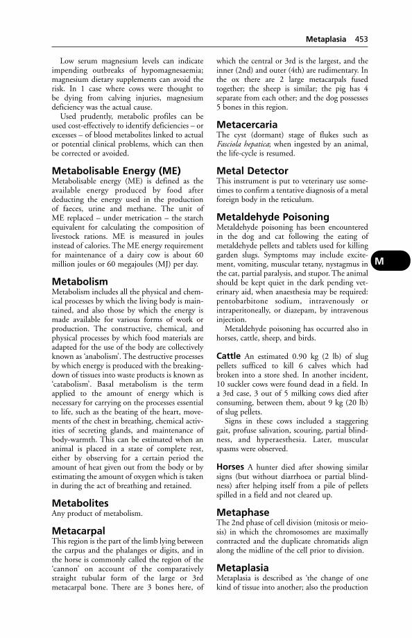
Low serum magnesium levels can indicateimpending outbreaks of hypomagnesaemia;magnesium dietary supplements can avoid therisk. In 1 case where cows were thought to be dying from calving injuries, magnesium deficiency was the actual cause.
Used prudently, metabolic profiles can beused cost-effectively to identify deficiencies – orexcesses – of blood metabolites linked to actualor potential clinical problems, which can thenbe corrected or avoided.
Metabolisable Energy (ME)Metabolisable energy (ME) is defined as theavailable energy produced by food after deducting the energy used in the production of faeces, urine and methane. The unit of ME replaced – under metrication – the starchequivalent for calculating the composition oflivestock rations. ME is measured in joulesinstead of calories. The ME energy requirementfor maintenance of a dairy cow is about 60 million joules or 60 megajoules (MJ) per day.
MetabolismMetabolism includes all the physical and chem-ical processes by which the living body is main-tained, and also those by which the energy ismade available for various forms of work orproduction. The constructive, chemical, andphysical processes by which food materials areadapted for the use of the body are collectivelyknown as ‘anabolism’. The destructive processesby which energy is produced with the breaking-down of tissues into waste products is known as‘catabolism’. Basal metabolism is the termapplied to the amount of energy which is necessary for carrying on the processes essentialto life, such as the beating of the heart, move-ments of the chest in breathing, chemical activ-ities of secreting glands, and maintenance ofbody-warmth. This can be estimated when ananimal is placed in a state of complete rest,either by observing for a certain period theamount of heat given out from the body or byestimating the amount of oxygen which is takenin during the act of breathing and retained.
MetabolitesAny product of metabolism.
MetacarpalThis region is the part of the limb lying betweenthe carpus and the phalanges or digits, and inthe horse is commonly called the region of the‘cannon’ on account of the comparativelystraight tubular form of the large or 3rdmetacarpal bone. There are 3 bones here, of
which the central or 3rd is the largest, and theinner (2nd) and outer (4th) are rudimentary. Inthe ox there are 2 large metacarpals fusedtogether; the sheep is similar; the pig has 4 separate from each other; and the dog possesses5 bones in this region.
MetacercariaThe cyst (dormant) stage of flukes such asFasciola hepatica; when ingested by an animal,the life-cycle is resumed.
Metal DetectorThis instrument is put to veterinary use some-times to confirm a tentative diagnosis of a metalforeign body in the reticulum.
Metaldehyde PoisoningMetaldehyde poisoning has been encounteredin the dog and cat following the eating of metaldehyde pellets and tablets used for killinggarden slugs. Symptoms may include excite-ment, vomiting, muscular tetany, nystagmus inthe cat, partial paralysis, and stupor. The animalshould be kept quiet in the dark pending vet-erinary aid, when anaesthesia may be required:pentobarbitone sodium, intravenously orintraperitoneally, or diazepam, by intravenousinjection.
Metaldehyde poisoning has occurred also inhorses, cattle, sheep, and birds.
Cattle An estimated 0.90 kg (2 lb) of slug pellets sufficed to kill 6 calves which had broken into a store shed. In another incident,10 suckler cows were found dead in a field. In a 3rd case, 3 out of 5 milking cows died afterconsuming, between them, about 9 kg (20 lb)of slug pellets.
Signs in these cows included a staggeringgait, profuse salivation, scouring, partial blind-ness, and hyperaesthesia. Later, muscularspasms were observed.
Horses A hunter died after showing similarsigns (but without diarrhoea or partial blind-ness) after helping itself from a pile of pelletsspilled in a field and not cleared up.
MetaphaseThe 2nd phase of cell division (mitosis or meio-sis) in which the chromosomes are maximallycontracted and the duplicate chromatids alignalong the midline of the cell prior to division.
MetaplasiaMetaplasia is described as ‘the change of onekind of tissue into another; also the production
Metaplasia 453
M
of tissue by cells which normally produce tissueof another sort’.
Metastasis and MetastaticMetastasis and metastatic are terms applied to the process by which a disease transfers from one organ to another. Often used indescribing neoplasia, where a malignanttumour spreads to distant parts of the body, andgives rise to secondary tumours similar to theprimary. Thus a sarcoma in some part of theabdomen may spread to the thorax by pieces of tumour or clusters of cells breaking awayfrom the parent growth, and being carried by the bloodstream to the lungs, etc., and setting up new sarcomatous growths there. (See
CANCER.)
MetastrongylusA genus of nematodes which infect the lungs.
MetatarsalMetatarsal is the name given to the bones andstructures lying between the tarsus or hock andthe digit of the hind-limb. It corresponds to themetacarpal region in the fore-limb, and has asomewhat similar arrangement of bones.
MethaemoglobinMethaemoglobin is a modification of haemo-globin, the red pigment of blood – the ironbeing in the form of ferric rather than ferroussulphate.
Some methaemoglobin is normally presentin the blood; but various poisons can increasethe amount found in blood, and sometimes in urine. Administration of large doses of certain drugs, such as acetanilide, can producemethaemoglobin and it is found also in somediseases. Chemically, methaemoglobin is thesame as oxyhaemoglobin, except that it cannotpart with its oxygen as readily as can the latter.
Methane (Marsh Gas)Methane (marsh gas) has the chemical formula CH4. Large quantities (up to 250 litresper day) may be formed in the rumen of the healthy cow. The gas is inflammable. (See
SLURRY.)
MethicillinA semi-synthetic penicillin resistant to penicillinase.
MethiocarbA snail-killer used in agriculture. Poultry andother animals must be kept away from treatedareas for at least a week.
MethionineAn amino acid containing sulphur; it is anessential part of the diet.
MethohexitalA short-acting barbiturate anaesthetic for use incats and dogs. It is administered intravenously.
MethopreneAn insecticide used as an ingredient of flea collars for dogs and cats. It acts by preventingthe young flea from developing to the adultstage.
MethoxyfluraneA volatile anaesthetic administered by inhala-tion; it is relatively slow in action and providesgood analgesia and muscle relaxation.
MethylMethyl is the name of an organic radicle whosechemical formula is CH3, and which forms thecentre of a wide group of substances known asthe methyl group. For example, methyl alcoholis obtained as a by-product in the manufactureof beet-sugar, or by the distillation of wood;methyl salicylate is the active constituent of oilof winter-green; methyl hydride is betterknown as marsh gas.
Methylated SpiritMethylated spirit is a mixture of rectified spiritwith 10 per cent by volume of wood naphtha,which renders the spirit dangerous for internaladministration. (See ALCOHOL POISONING.)
Methylene BlueMethylene blue, given intravenously at a doseof 10 mg/kg of a 4 per cent solution, is an antidote to nitrate poisoning, and also to chlorate poisoning. In cats it was formerly used as a urinary antiseptic but gave rise toHeinz-body anaemia (see under HEINZ BODIES).
MetoestrusMetoestrus is the period in the oestrous cyclefollowing ovulation and during which the corpus luteum develops.
Metritis(see UTERUS, DISEASES OF)
MetroceleA uterine hernia.
MetronidazoleA nitroimidazole drug useful against anaerobicbacterial infections, and also GIARDIASIS.
454 Metastasis and Metastatic
M
Meuse-Rhine-Ijssel (MRI)A dual-purpose breed of cattle from theNetherlands, with good milk yields and highbutterfat.
Mice(see LYMPHOCYTIC CHORIOMENINGITIS;RODENTS). Polyoma viruses of mice and mousehepatitis virus are other infections important inlaboratory mice; also ECTROMELIA (mousepox). (See also PETS.)
Micro-Micro- is a prefix meaning small.
MicrocephalyMicrocephaly is abnormal smallness of thehead.
MicrochipsIn the animal field, the name is given to elec-tronic chips about the size of rice grains thatcan be encoded with an identification numberand implanted permanently under the skin by aveterinary surgeon. The number can be readwith a special electronic scanner and lost orstolen animals thus identified. More than150,000 pets were microchipped in the 5 yearsup to 1996.
The scheme has been used for identifying theanimals re-homed from RSPCA centres andother animal charities.
Under the scheme, a register of the numbersof all animals ‘identichipped’ is held by Pet Log,a national computer database launched by theRSPCA and the Kennel Club.
MicronMicron 0.001 mm, the unit of measurement in microscopical and bacteriological work. Itssymbol is µ.
Micro-Organisms(see BACTERIA; VIRUSES; MYCOPLASMA; RICK-
ETTSIA; CHLAMYDIA, FUNGAL DISEASES)
MicrophthalmiaAn abnormal smallness of the eyes, accompaniedby blindness. In piglets and calves it is believedto be associated with a vitamin A deficiency.
MicroscopeThe ordinary microscope with oil-immersionlens gives magnification up to 1500 diameters.(See also ELECTRON MICROSCOPE.)
MicrosporumA group of fungi responsible for ringworm.
MicturitionThe act of passing urine.
MiddlingsA protein supplement for cattle and pigs. (See
WEATINGS.)
Midges, BitingSpecies of Culicoides are of veterinary impor-tance in connection with ‘sweet itch’ in horses,and also with the transmission of viruses tofarm livestock, e.g. the virus of bluetongue, andthat of epizootic haemorrhagic disease of deer.
MigramA disease of sheep on the Romney Marsh.Symptoms include trembling and muscularincoordination. In an incident in 1983, MAFFreported that 150 out of 260 lambs collapsedwhile being driven to new pasture. The cause is unknown, but one line of investigation is into a possible association with blue-green algaein the dykes.
MiliaryMiliary is a term, expressive of size, applied tovarious disease lesions which are about the sizeof a millet seed – e.g. miliary tuberculosis,feline miliary dermatitis.
Milk
Composition Cow's milk is a very valuablefood substance as it contains all the essentialfood constituents – proteins, carbohydrates,fats, and vitamins – in addition to a consider-able percentage of mineral matter. The mostimportant protein in milk is casein; this is present in a state of partial solution.Carbohydrates are represented by the milk-sugar (lactose) which is dissolved in the liquidportion of the milk. They, along with the fatwhich occurs as spherical globules, are heat-andenergy-producing substances. The mineral matter consists, to a very large extent, of compounds of calcium and phosphorus. These substances are the essential constituentsof bone.
The percentages of the main constituents of milk vary considerably. Fat and protein levelsare affected not only by the diet of the animal,but also by its genetic make-up. However, the level of lactose is relatively constant (see table
below).In the young growing animal, muscle and
bone are being formed rapidly. Hence the foodof the young must be adequately provided withprotein and mineral matter in particular. Since
Milk 455
M

it contains considerable quantities of both ofthese constituents, as well as vitamins, milk isan excellent food for growing animals; it is nothowever a complete one – it will not provideadequate iron in the piglet (see SOW'S MILK) oradequate magnesium in the calf.
Legal standards In Britain, under the Saleof Food and Drugs Act, milk containing lessthan 3 per cent of butterfat, or less than 8.5 percent of non-fatty solids (i.e. proteins, sugar, andash), is deemed to be not genuine (until orunless the contrary is proved) by reason ofeither the addition of water or the abstractionof some of the fatty or non-fatty solids. (See
SOLIDS-NOT-FAT.)
The specific gravity of cow's milk variesbetween 1.028 and 1.032. The greater the fat content, the lower the specific gravitybecause fat is lighter than water and solids, bulk for bulk.
The reaction of the milk of the herbivorousanimals is generally approximately neutral,while that of the carnivorous animals is acid.
Bacteria Since 1982 the total bacterial count(TBC) has been applied once weekly to samplesof milk collected from each supplier. Averageresults over 1 month, using the Plate CountTest, determined the payment for the milk. The‘cleanest’ grade, with an average number ofmicro-organisms of 20,000 or fewer per mlattracted a price premium; milk with more than100,000 organisms per ml was subject to adeduction from the agreed price per ml.
Since 1996, the milk marketing organisationMilk Marque has used the more sensitive
Bactoscan Test to determine milk hygienicquality. Milk is divided into 3 bands: band A,fewer than 100,000 cells per ml; band B, 101 to 500,000 cells per ml; band C, more than500,000 cells per ml. (See also MILK-BORNE
DISEASE; PASTEURISATION.)
Sediment in milk Milk containing sedi-ment has been the subject of prosecutionsunder the Food and Drugs Act 1955. Milk andDairy Regulations 1959 require that beforemilking is begun, all dirt on or around theflanks, tail, udder and teats of each cow shall be removed, and that the udder and teats shallbe kept thoroughly clean during milking.Additionally, milking must be carried out in agood light (daylight or electric light); no dustymaterial may be moved during or within half an hour before milking.
In order to minimise contamination of milkduring milking, the cow's udder should besprayed or wiped with a disposable towel wrungout of water containing a disinfectant, and the hands of the milker should be thoroughlywashed before the milking of each cow (prefer-ably in water containing a disinfectant). (See MASTITIS IN COWS for recommended procedure.)
Lactic acid is produced by the action of bacteria on lactose – the result being sour milk – and is also present in sour cream andyoghurt. (See also LACTIC ACID.)
White blood cells in milk In the EU,milk may not be sold if it contains more than400,000 cells per ml. Most milk marketingcompanies impose a penalty if bulk milk has acell count of 250,000 cells per ml or more;
456 Milk
M
APPROXIMATE COMPOSITION OF MILK PRODUCTS
Water Proteins Fats Sugar Ash
Separated milk 90.0 3.7 0.2 4.9 0.8Skimmed milk 90.0 3.6 0.8 4.6 0.8Butter milk 91.0 3.3 0.5 3.4 0.6Cream (thin) 64.0 2.8 30.0 3.5 0.5Cream (thick) 39.0 1.6 56.0 2.3 0.4Whey 93.0 0.9 0.2 4.8 0.5
APPROXIMATE COMPOSITION OF MILK OF DIFFERENT ANIMALS
Water Proteins Fats Sugar Ash
Mare 90.5 2.0 1.2 5.8 0.4Cow 97.4 3.4 3.8 4.8 0.8Ewe 81.9 5.8 6.5 4.8 0.9Goat 84.1 4.0 6.0 5.0 0.8Sow 84.6 6.3 4.8 3.4 0.9(Human) (87.4) (2.1) (3.8) (6.3) (0.3)

some have more stringent limits. High levels ofwhite blood cells in milk are the result of sub-clinical mastitis due to (a) trauma, defectivemilking machine or technique; (b) infection;(c) both. (See MASTITIS IN COWS.)
Antibiotics in milk It is important thatmilk should not contain traces of antibiotics,which are frequently used in treating mastitis.Some people are allergic to antibiotics, and ifthey drink milk containing them they may suffer severe effects, e.g. a troublesome rash anda period off work. It has also been feared thatthe continual consumption of small quantitiesof antibiotic may result in people becomingsensitised, later undergoing a severe reactionwhen given that antibiotic by their doctor. A3rd danger is the development of organismsresistant to antibiotics, which could possiblygive rise to illness not responding to antibiotictreatment. Antibiotic residues can also affectthe manufacturing processes of cheese andyoghurt.
All medicines containing antibiotics arelabelled with a ‘withdrawal period’ which mustelapse after treatment is ended before the milkcan be used for human food. Unless otherwisestated, the minimum standard withdrawal period for milk is 7 days.
Chlorophenol taint Most strong-smellingdisinfectants, such as those based on phenol orcresol, can cause a taint in milk, even in verylow concentrations.
The disinfectants do not have to come intodirect physical contact with the milk, but canbe absorbed from the atmosphere by anyexposed milk surface, particularly in the bulktank. They should be stored well away from themilking parlour or dairy.
Similarly, neither creosote nor products containing phenols should be used where theymay come into contact with teats and udders byindirect means, e.g. on woodwork of buildings,or in the disinfection of cubicle beds, cowsheds,loose-boxes, and collecting yards.
Plants affecting the milk A large numberof plants affect milk or milk secretion in animals eating them, and very often the realcause of unusual tastes or odours in the milk issome common wild plant. Some plants givemilk a characteristic taint or odour (such asgarlics), and others alter its colour; somedecrease the total secretion and others lessen thefat content; a few alter the colour and characterof butter made from the milk; and one or two,whose poisonous principles are excreted by the
mammary gland, render the milk actually poisonous. (See BRACKEN POISONING.)
Dioxins in milk Unacceptably high levels ofdioxin are occasionally found in milk.
The World Health Organisation has recom-mended a ‘tolerable daily intake’ for dioxins of0.01 nanogrammes per kilogram of milk.
Milk, Absence ofAbsence of milk in the mammary glands following parturition, is discussed underAGALACTIA, and SOW'S MILK, ABSENCE OF.
Milk Allergy in CowsThis may develop especially in the ChannelIslands breeds, in cows which have become sensitised to the alpha-casein in their own milk.If milking is delayed, they may develop clinicalsigns of a type 1 hypersensitivity, e.g. dyspnoea,drooling of saliva, urticaria, and swollen eyelids,in an otherwise bright animal. The with-drawal of milk results in an almost immediateremission of these symptoms.
Milk-Borne DiseaseVarious infections may be transmitted to peoplethrough unpasteurised or defectively pasteurisedmilk. (See BRUCELLOSIS; SALMONELLOSIS;Q FEVER; TUBERCULOSIS.)
Over a period of 30 years, 77 per cent of 233 reported outbreaks of communicable disease attributed to milk and dairy products in England and Wales were associated withunpasteurised milk.
After compulsory pasteurisation was intro-duced in Scotland, outbreaks of milk-borne salmonellosis fell to 8, affecting 46 people ascompared with 14 outbreaks affecting 1090people in the previous 3 years.
Campylobacter jejuni, present in unpas-teurised or incompletely unpasteurised milk,has caused outbreaks of human enteritis.
Corynebacterium ulcerans was diagnosed asthe cause of sore throat in a patient from a community that drank raw milk. The source ofthis was a herd in which 8 cows were infectedwith this organism; while a 9th cow was foundto be an intermittent excretor of it.
Goat's milk, if unpasteurised, may be asource of various infections transmissible to people. (See BRUCELLOSIS; Q FEVER;TOXOPLASMOSIS; TUBERCULOSIS.)
Milk Development CouncilA government-backed body set up in 1995 tofund, by means of a levy on milk, research anddevelopment into milk and milk products.
Milk Development Council 457
M

Milk-Drop SyndromeA sudden drop in milk yield in individual cowswith a flaccid udder; the usual cause isLeptospira hardjo.
Milk FeverMilk fever is a metabolic condition – mainlyhypocalcaemia of milk cows, milk goats, andsometimes of ewes, bitches, and cats in whichthere is a partial or complete loss of conscious-ness, paralysis of the hindquarters, and some-times paralysis of other parts.
In the hill ewe, the condition is colloquiallyknown as ‘MOSS-ILL’. Hypocalcaemia alsooccurs in lowland ewes.
Milk fever would appear to be one of the dis-eases that is to some extent traceable to artificialmethods of management. It is most frequently,though not exclusively, encountered in heavy-milking cows, of the essentially dairy breeds. Afew cases occur some hours before calving, butthe majority take place within 3 days subse-quent to parturition.
Some cases occur up to 4 weeks after calving,but, as a rule, delayed cases are mild – thoughthey take longer to recover.
Research has suggested that the calcium-con-trolling ‘mechanism’ is a very complex one,involving all the endocrine glands and both thesympathetic and the parasympathetic nervoussystems.
Blood samples have shown that as well as ashortage of calcium in the blood, there may be too little phosphorus and either too much or too little magnesium. This accounts for thediffering symptoms in what is collectively calledmilk fever.
The level of CORTISOL in the plasma of cowsduring milk fever is significantly higher than innormal cows.
Signs The animal at first shows a certainamount of excitement. She paddles with herhind-feet, stares around in a somewhat fearfulmanner, may bellow, and if tied attempts tobreak loose. The pupils are dilated. After a timeshe staggers on her feet, loses balance, and fallsto the ground. When down she may makeefforts to rise, but after struggling for a time shegives up and remains quiet. In many cases acharacteristic position is assumed: the cow lieson her brisket, head turned round over oneshoulder (often the left), and the muzzle point-ing to the stifle.
The breathing becomes deep and slow, pulse is fast but weak, the extremities of thebody grow cold, the temperature falls to 4 or 5degrees below normal, and death may followcoma.
Whereas formerly the mortality was 90 percent or so, it has been reduced to less than 5 percent in cases that are treated.
458 Milk-Drop Syndrome
M
Milk fever: the characteristic posture.

Differential diagnosis In countries whererabies is present, this disease may be mistakenfor milk fever – especially as the position inrecumbency, with the head turned to one side,resembles the milk-fever posture.
In Britain, cases treated as milk fever butwhich were unresponsive to treatment with calcium borogluconate were found to be nutri-tional myopathy associated with low vitamin E and selenium intakes. Reluctance to move,stiffness, and recumbency were the symptoms,and some deaths occurred.
Treatment The intravenous or subcutaneousinjection of calcium borogluconate solutionwith or without magnesium.
When a deficiency of blood phosphoruscomplicates milk fever, and this does not completely respond to calcium treatment,phosphorus in the form of 85 g (3 oz) of sodium acid phosphate may be given by mouthtwice daily.
Prevention Milk fever has proved difficult toprevent. Reducing calcium intake in the laterstages of pregnancy, followed by a boost in in-feed calcium 2 to 3 days before calving, hasbeen suggested.
‘Milk Lameness’This is a translation of the Swedish name for a condition encountered in high-yielding dairycattle, and characterised by hip lameness.During 1 stage they assume a characteristic posture.
Some unthriftiness and sluggishness ofmovement may be observed in the herd.Animals stop frequently to rest.
The cause of ‘milk lameness’ is a deficiency ofphosphorus in the bloodstream, and – since hiplameness may have several causes – blood testsare necessary in order to confirm a diagnosis.
In a Scottish outbreak, recovery soon followedthe feeding of sterilised bone-flour in smallamounts. It seemed that the cows had beenunable to acquire sufficient phosphorus fromunsupplemented grazing.
Lameness associated with a blood-phosphorusdeficiency is, of course, well known in manyparts of the world – subjected either to droughtor to high rainfall – where the soil or herbage isdeficient in phosphorus.
Milk Ring TestA test that has been used to determine the pres-ence of brucellosis in dairy herds. It has largelybeen replaced by an ELISA test for antibodies tothe disease.
‘Milk Scald’An alopecia around the muzzles of calves fedpoorly mixed, or cool, milk substitute.
Milk Sinus(see MAMMARY GLAND)
Milk Teeth(see DENTITION)
Milk TetanyAnother name for hypomagnesaemia in thecalf. It occurs at about 2 months old in suckleror veal calves and is usually caused by a pre-dominantly milk diet.
Milk, Unpasteurised(see MILK-BORNE DISEASE)
Milk YieldMilk yield varies according to the breed of cow.Average annual yields are: British Holstein,7000 kg; Friesian, 6150 kg; Guernsey, 4510 kg;and Jersey, 4325 kg. (For other figures see under
LACTATION; see also STRESS.)
Milker's NoduleAnother name for PSEUDO-COWPOX (see VIRAL
INFECTIONS OF COWS' TEATS).
Milking(see also under MASTITIS; MILKING MACHINES). Atmilking time, the ‘milk letdown’ mechanism
‘Milk lameness’: the characteristic posture.
Milking 459
M

begins to operate; it is actuated by the hormoneoxytocin which is secreted in the posterior pitu-itary gland and which is released into thebloodstream following a nervous stimulus. Thisstimulus may be caused by the rattling of milkpails, the placing of food in the manger, thewashing of the udder, etc.
Milking MachinesTheir action simulates that of the sucking calf.The teat-orifice is opened and milk withdrawnby means of a partial vacuum applied to the out-side of the teat. As continuous vacuum wouldrestrict circulation of the blood in the teat, causepain, and inhibit milk ejection, the vacuum isapplied intermittently by means of a pulsator.
The basic principles of machine milking are,in fact, vacuum and pulsation, and the way inwhich these are applied to the teat in the teat-cup assembly.
For maintenance of a healthy udder, what isfirst required is a strong stimulus to ‘let-down’,followed by rapid milking. As soon as themachine ceases to milk, the udder should bestripped and the machine removed. In practice,attention to this involves the herdsman not hav-ing too many units to cope with, not other tasksto perform. Automatic cluster-removal is usefulhere.
Milking machines can be made to milk fasterby increasing the degree of vacuum, increasingthe pulsator rate, or by widening the pulsatorratio. If, however, the herdsman already hasmore to do than he can manage, a faster milk-ing can result only in prolonged attachment.The milking routine must be reorganised toavoid this, or mastitis will follow.
A liner with a hard mouthpiece is likely tocause trouble.
In 1 herd badly affected with mastitis, achange from slack, wide-bore liners to the narrow-bore stretched type resulted in a spec-tacular improvement.
Investigation has shown that the slow milkeris almost invariably the cow with a small teat-ori-fice. If it is not practicable to cull such an animal,the milking machine pulsation ratio may, withadvantage, be altered. At 60 pulsations perminute, and at 38 cm of mercury, a ratio of 4:1(i.e. the liner being opened for 4 times as long asit is closed) will reduce milking time – especiallywith slow milkers – without hurting the cow, oradversely affecting the stripping yield.
Common faults in milking machines areincorrect vacuum level, or vacuum fluctuations,blocked air bleeds, unsuitable pulsation rate,and faulty liners. Such faults can lead to MASTI-
TIS. Regular, skilled maintenance of milking
machines is therefore all-important. Milkingmachines should be checked at least twice a yearand any faults repaired at once.
During a survey among 71 farms participat-ing in a mastitis control scheme, 95 per cent ofthe milking machines were found to be faulty.The importance of this is shown by anothersurvey, of a small number of herds with a seri-ous mastitis problem, in which cell counts werecarried out before and after machine testing andadjustment. It was found that cell counts fell byabout 25 per cent following the 1st annual test,and by about 15 per cent following the 2ndannual test. This shows that the correction ofmilking-machine faults really can achieve some-thing worthwhile, whether measured in cowhealth or on farmers' profits.
Milking Parlours(see under DAIRY HERD MANAGEMENT)
‘Milkspot Liver’‘Milkspot liver’ is a name given to pigs' liversshowing whitish spots or streaks of fibrous tis-sue – the result of chronic inflammation causedby the larvae of the roundworm, Ascaris suum.A similar condition may occur in lambs whichhave been grazing fields or fodder crops towhich pig slurry has been applied. On aScottish lowland farm this led to the condem-nation of 70 per cent of lambs' livers at the localabattoir in 2 successive years. (See LAMB CAR-
CASE REJECTION.)Migrating larvae of Toxocara canis may also
cause ‘milkspot liver’ in pigs.
Milling MistakesThe inadvertent inclusion of a medicinal com-pound intended for one species in feed mixedfor another has caused problems. For example,lincomycin in feed for dairy cows producedsevere diarrhoea, reduced milk yield, and acetonaemia. (See also MONENSIN SODIUM.)
MillipedesNon-poisonous, many-segmented arthropodswith 2 legs per segment; they may be kept as‘pets’. Those wishing to keep them should beinformed about their care and nutrition, as millipedes will refuse to eat unsuitable food andmay die of starvation as a result. Lesions to theintegument (skin) are usually associated withproblems in, shedding (dysecdysis); the condi-tion usually clears up at the next moult.Millipedes are prone to disease caused by bacte-ria, fungi, or viruses. Loss of haemolymph(equivalent to blood in a mammal) due toinjury may be stopped by icing sugar, Plasticine,
460 Milking Machines
M

glue, etc. In the USA Narceus annularis is theintermediate host of a large, ‘thorn-headed’worm Macracanthohyncus ingens, which cancause diarrhoea (and sometimes dysentery) indogs. Hosts of the adult worm are raccoons,black bears, skunks, foxes, and moles.
Miniature Bull TerrierA small breed with similar characteristics to theBULL TERRIER. Lens luxation may be found inindividual dogs.
Miniature SchnauzerA small breed developed in Germany from theSCHNAUZER. With miniaturisation, certaindefects have appeared. Cataract is inherited, asis von Willebrand's disease. Pulmonary stenosisand Perthe's disease may also be seen,
MineralocorticoidsA group of hormones produced from the adren-al cortex that affect the metabolism of sodium,chloride and potassium (see ADRENAL GLANDS).
Minerals(see under CALCIUM; COBALT; FLUOROSIS; IRON;MANGANESE; PHOSPHORUS; TRACE ELEMENTS;METABOLIC PROFILE TESTS; SELENIUM; SODIUM
DEFICIENCY)
Minimal-Disease PigsThose reared free from certain infections. (See also SPF.)
Minimum InhibitoryConcentrationThe lowest concentration of an antibiotic thatinhibits the growth of a particular bacterialspecies.
Mink, Diseases ofThese include distemper (caused by the virus ofcanine distemper), botulism, salmonellosis,tuberculosis, paralysis due to a vitamin B defi-ciency, mastitis, metritis, paragonimiasis, andtransmissible mink encephalopathy. (See ALEUT-
IAN DISEASE.) A vaccine against botulism isavailable. (NB: The keeping of mink in the UKis longer lawful, except under special conditions– e.g. in zoos)
Mites, ParasiticParasitic mites, including mange mites (see also
MANGE).
Types of mite The following genera areimportant. All are minute, and under favourablecircumstances just visible to the naked eye.
Sarcoptes, with 1 species, S. scabiei, andnumerous varieties. These mites live in the skinof mammals.
Cnemidocoptes, found in birds. They resem-ble Sarcoptes. Cn. mutans causses scalyleg. Cn,Gallinae is the depluming mite and Cn. pilaeinfests the cere of budgerigars
Notoedres is a genus closely allied to Sarcoptes,found on carnivores.
Otodectes is found in the external ear.
Psoroptes
Chorioptes (Symbiotes). One species isknown – C. equi – with numerous varieties.
Drugs to kill them (Acaricides) A wide rangeof products is available, in a variety of formula-tions, depending on the animal to be treated andthe mite causing the problem. Many are basedon avermectins, pyrethrins or organophosphoruscompounds (see under MANGE).
Other mites
Air sac mite (Cytodites nudus) is found main-ly in free-range poultry and, in small numbers,does little harm. In large numbers these mitescan result in weakness and loss of weight. Theyare said to exacerbate any respiratory conditionoccuring concurently.
Cyst mite of poultry (Laminosioptes cysti-cola) is found mainly in the subcutaneous tissueof free-range, poultry turkeys, pheasants,pigeons and geese, but has been found in mus-cles and also in pigeon lungs. The mite does no harm to poultry but an affected carcase isrejected for human consumption on aestheticgrounds.
Harvest mites The so-called ‘harvest mite’ or‘chigger’ is the larva of a species of Trombidium.It is microscopic in size, blood-red in colour,and in shape resembles a tick. The mite burrowsunder the skin of man, farm animals, the dogand the cat, and engorging with blood appearsas a red spot in the centre of an inflamed area.In 2 or 3 days the spot becomes a blister andultimately a scab which falls off. The spot isextremely itchy. (See HYPERSENSITIVITY.) Thenymphs and adults are free-living.
Forage mites are occasionally parasites of thehorse which live normally in the forage. They
Mites, Parasitic 461
M

may cause considerable damage to the skin, butare usually easily killed.
Flour mites These can cause loss of nutrientsin stored animal feeds (see DIET and DIETETICS –Deterioration with storage), and can also be par-asitic on animals, causing dermatitis. For such acase in horses, see FLOUR MITE INFESTATION.
Cheyletiella Two members of this genus areof some veterinary importance in Britain, viz.C. parasitivorax and C. yasguri. These mitesinfest dogs, cats, foxes, rabbits and hares. In the dog they are most frequently found on thenape of the neck, and down the back. Rednessof the skin and intense itching may be caused –the latter symptom occurring in man also.Three dressings, at 5-day intervals, with derrisor pyrethrum are recommended; for cats, selenium sulphide.
Nose mites (see CANINE NASAL MITES, andbelow)
Linguatula serrata This parasite has a flatbody shaped somewhat like a tongue, butgrooved. It is without appendages, apart from 2 pairs of hooks at its anterior end. The adultlives in the nasal passages of dogs, cats, andfoxes, and is up to 2 cm in length.
The eggs are expelled from the nose by sneez-ing; they may also be swallowed and excreted inthe faeces. Sheep, cattle, and rabbits swallowthe eggs and become intermediate hosts.
462 Mites, Parasitic
M
Sarcoptes. × 70.
Legs of mange mites. (Psoroptes, Chorioptes,Sarcoptes.)
Notoedres. × 70.
Otodectes. × 70.
Chorioptes. × 70
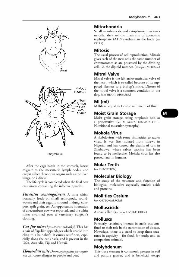
After the eggs hatch in the stomach, larvaemigrate to the mesenteric lymph nodes, andencyst either there or in organs such as the liver,lungs, or kidneys.
The life-cycle is completed when the final hosteats viscera containing the infective nymphs.
Parasitus consanguineus. A mite whichnormally feeds on small arthropods, round-worms and their eggs. It is found in dung, com-post, spilt grain, etc. An opportunist infestationof a recumbent cow was reported, and the whitemites swarmed over a veterinary surgeon'sclothing.
Cat fur mite (Lynxacarus radooskyi) This hasa pair of flap-like appendages which enable it tocling to a hair-shaft. It causes scurfiness, espe-cially along the cat's back, and is present in theUSA, Australia, Fiji and Hawaii.
House-dust mite Dermatophagoides pteronyssi-nus can cause allergies in people and pets.
MitochondriaSmall membrane-bound cytoplasmic structuresin cells; they are the main site of adenosinetriphosphate (ATP) synthesis in the body (see
CELLS).
MitosisThe usual process of cell reproduction. Mitosisgives each of the new cells the same number ofchromosomes as are possessed by the dividingcell, i.e. the diploid number. (Compare MEIOSIS.)
Mitral ValveMitral valve is the left atrioventricular valve ofthe heart, which is so-called because of its sup-posed likeness to a bishop's mitre. Disease ofthe mitral valve is a common condition in thedog. (See HEART DISEASES.)
Ml (ml)Millilitre, equal to 1 cubic millimetre of fluid.
Moist Grain StorageMoist grain storage, using propionic acid as a preservative (see MUSCLES, DISEASES OF –Nutritional muscular dystrophy).
Mokola VirusA rhabdovirus with some similatities to rabiesvirus. It was first isolated from shrews inNigeria, and has caused the deaths of cats inZimbabwe, where rabies vaccine has beenfound to be ineffective. Mokola virus has alsoproved fatal in humans.
Molar Teeth(see DENTITION)
Molecular BiologyThe study of the structure and function of biological molecules; especially nucleic acidsand proteins.
Mollities Ossium(see OSTEOMALACIA)
MolluscicideA snail killer. (See under LIVER-FLUKES.)
MolluscsFormerly, veterinary interest in snails was con-fined to their role in the transmission of disease.Nowadays, there is a trend to keep these crea-tures in captivity – for food, for study, and ‘ascompanion animals’.
MolybdenumThis trace element is commonly present in soiland pasture grasses, and is beneficial except
Cheyletiella.
Molybdenum 463
M

when it occurs in excessive amounts – such as inthe ‘teart soils’ of central Somerset, and of smallareas of Gloucestershire and Warwickshire. Here‘molybdenosis’ causes scouring in ruminants,especially cattle. The scouring is worse from May until October when the grass contains most water-soluble molybdenum. Staring coats,marked loss of condition and evil-smelling faecesare observed in affected cattle. A daily dose ofcopper sulphate (2 g for adults and half this foryoung stock) obviates or remedies the trouble.
Molybdenosis may occur also as the result ofaerial contamination of pasture in the vicinityof aluminium-alloy and other factories, and ofoil refineries. In an outbreak near the EssoRefinery at Fawley, younger cattle showed amarked stiffness of back and legs, with greatdifficulty in getting to their feet and reluctanceto move – in addition to diarrhoea.
If an animal is receiving extra molybdenumin its diet, it is likely to need extra copper.Levels of molybdenum which interfere withcopper metabolism also inhibit the synthesis ofBl2, the cobalt-containing vitamin, by therumen microflora.
Monensin SodiumMonensin sodium is licensed in the UK as agrowth promoter for cattle (see ADDITIVES) and as a coccidiostat for poultry. It is producedby fermentation of a strain of Streptomyces cinnamonensis.
Monensin resulted in the death of 9 out of84 beef cattle which had received 12 times therecommended dose. All the cattle lost theirappetite and had diarrhoea. Autopsy findingsincluded multiple haemorrhages and oedema ofthe right side of the heart.
In another incident, 9 out of 40 calves diedfollowing accidental overdosage with monensin.
Monensin toxicity has also been recorded inhorses, sheep, chickens and turkeys.
Poisoning has been reported in dogs given aproprietary dog food contaminated with mon-ensin still present in a storage bin not properlycleaned. (See also IONOPHORES.)
MongoosesMongooses are vectors of rabies in South Africa,Central America, West Indies, and India.
MoniliaA group of yeast-like organisms.
MoniliasisMoniliasis is a disease due to the yeast-like fun-gus Candida albicans. In humans it follows, insome cases, the use of certain antibiotics.
The disease occurs in turkeys and fowls, andin other domestic animals. It including dogsand cattle must be borne in mind when usingantibiotics. A high temperature, loss of weight,and oedema of the lungs may result.
Nystatin has been used – successfully, it isclaimed – in the treatment of turkeys withmoniliasis.
MonkeysMonkeys belong to the order Primata whichincludes about 200 species, ranging in size fromthe tree shrew, weighing about 100 g, to thegorilla, weighing up to 275 kg.
Two sub-orders are recognised: New Worldmonkeys (catarrhines); and Old World mon-keys (platyrrhines), apes, and man.
Monkeys, AnaesthetisingKetamine is recommended.
Monkeys, Diseases ofThese include:
(1) Infection with herpes simian B virus.This is easily transmitted to people bitten by monkeys (or perhaps to people merely han-dling monkeys with B virus lesions); it is of the greatest importance, as an encephalitis orencephalomyelitis is produced in man, withdeath as the usual outcome. This infectionshould be suspected in monkeys showing vesi-cles on the lips, tongue, inside of the cheeks, oron the body. The vesicles burst and give rise to ulcers and scab formation. Occasionally,affected monkeys have conjunctivitis and athick discharge from the nose.
(2) Tuberculosis. This is generally the miliaryform, due to the human type of tubercle bacil-lus. Symptoms include: loss of weight, ofappetite, dullness; sometimes cough and rapidbreathing. Monkeys may be tuberculin-testedby injecting mammalian tuberculin into an eyelid. The result is noted by the presence orabsence of swelling after 72 hours.
(3) Pneumonia (unconnected with tubercu-losis). A monkey that is coughing and sneezingcan be assumed to be seriously ill. Death from
464 Monensin Sodium
M
External features of the snail species Helix pomatia.
pneumonia can occur within 24 hours, andaffect a high proportion of any group of monkeys.
(4) Dysentery due to Shigella organisms.This is a common cause of death among laboratory monkeys.
(5) Phycomycosis.(6) Marburg disease, which can be fatal in
both monkeys and man, has been seen in labo-ratory workers in contact with blood and tissuesof Vervet monkeys. Symptoms: headache, fever,muscular pain, prostration, diarrhoea and vom-iting, with epistaxis and vomiting of blood.
(7) Rabies.(8) Monkey pox. This is an apparently rare
disease of monkeys. The virus was first isolatedin 1958 in a monkey colony in the StatensSeruminstitut, Copenhagen. Cases of presumedmonkey pox (resembling human smallpox) inman occurred in Africa in 1970. Since theworldwide eradication of smallpox, monkeypox has become the most important orthopoxvirus infection of man. Yet, despite a recentincrease in the number of cases reported,human monkey pox remains ‘a rare sporadicdisease’.
(9) YELLOW FEVER.(10) Measles. This was reported in 1975 in
11 colobus monkeys imported into the UK. No rash was seen; symptoms comprised a nasaldischarge, conjunctivitis, cough, facial oedema,and pneumonia. All died. Diagnosis was by laboratory tests.
(11) Malaria. Fulciparum malaria hasoccurred in owl monkeys.
(12) Yersinia enterocolitica infection. (See also
KYASANUR FOREST FEVER.)(13) Leptospirosis.(14) Simian sarcoma virus.(15) Toxoplasmosis.(16) Infectious hepatitis.(17) Jaundice and blindness (temporary)
have been caused by lead poisoning as a resultof cage bars being painted with lead-containingpaint. A chelating agent was used with success.
(18) Cases have occurred of laboratory work-ers contracting viral haemorrhagic diseasecaused by an Ebola-related filovirus and bysimian haemorrhagic fever in monkeys import-ed from the Philippines. (See also PETS.)
Monkshood Poisoning(see ACONITE)
Monochloroacetate PoisoningMonochloroacetate poisoning has occurred incattle, sheep, and other animals. Sodiummonochloroacetate is a contact herbicide.
Monoclonal AntibodiesIdentical immunoglobulin molecules producedby a single clone of plasma cells (see GENETIC
ENGINEERING).
MonocyteA type of white blood cell. (See under BLOOD.)
Monocytosis(see ‘PULLET DISEASE’)
Monogenetic FlukesParasites that infest the gills of freshwater fish;they belong to the groups Gyrodactylus andDactylogyrus. In the EU Gyrodactylus solarisinfestation is a NOTIFIABLE DISEASE and thereare restrictions on the movement of fish out ofareas where this parasite has been detected.
MonoplegiaMonoplegia means PARALYSIS of a single limb,or part.
MonorchidThis term is commonly used by dog-breeders tomean an animal in which only 1 testicle hasdescended into the scrotum. Such an animal iscorrectly called a unilateral cryptorchid, theterm ‘monorchid’ being reserved for the animalwith a single testicle (a far rarer condition).
Under Kennel Club rules, a dog which hasnot both testicles in the scrotum cannot beentered for show; but there is as yet no ban onthe registration of dogs sired by a cryptorchid.
Cryptorchidism is an inherited condition(though it has been claimed that feeding rats ona biotin-deficient diet caused their testicles toreturn to the abdomen after 2 or 3 weeks), butthe precise mechanisms of inheritance has notyet been determined.
MonosaccharideMonosaccharide, or simple sugar, is a sugar hav-ing no more than 6 carbon atoms in the mole-cule. Among monosaccharides are fructose, glucose, galactose, levulose, etc. Monosaccharideshave been termed the ‘building blocks’ of carbo-hydrates.
MonotocousNormally producing only 1 offspring at birth,like cattle and horses, as compared with a litterof, for example, puppies.
Monster (Teras)Severely malformed young are occasionallyborn to all species (teratogenesis). The causemay be genetic, or disease; or it may follow
Monster (Teras) 465
M

consumption of toxic substances. Haemolyticdisease, for example, is responsible for abnor-mal piglets. (See also under TERATOMA; BULL-DOG
CALVES; GENETICS, HEREDITY AND BREEDING –
Genetic defects).
‘Moon Blindness’(see PERIODIC OPHTHALMIA)
MoraxellaSmall rod-shaped bacteria found in pairs.Outbreaks of conjunctivitis and keratitis are oftenassociated with M. Bovis (formerly Haemophilusbovis) infection. (See under EYE, DISEASES OF.)
MorbidityMorbidity is the condition of having a disease.Proportional morbidity is the incidence of disease in a flock or herd, e.g. ‘morbidity is 60 per cent’ in a given group of animals withreference to a particular disease.
MorbillivirusesThese include the viruses of canine distemper,rinderpest, peste de petits ruminants, and humanmeasles. (See also SEAL.)
Morbillivirus in HorsesMorbillivirus in horses caused a severe diseaseoutbreak, with transmission to man, inQueensland. Deaths occurred in both species.The clinical signs were severe respiratory dis-tress, panting, high temperature, stiffness andfrothy nasal discharge. If the horse survived for2 days, there were cyanosis and oedema.
The virus is believed to have come from fruitbats; a bat miscarriage was found on the pasturegrazed by the horses and the virus isolated fromit. A survey found the virus in the reproductivetract of 3 species of fruit bat and 12 to 15 percent had antibodies to the virus.
Morel's DiseaseThis affects sheep and is caused by a Gram-pos-itive micrococcus. The disease bears someresemblance to caseous lymphadenitis, withabscesses in subcutaneous tissue and intramus-cular fascia, and has been reported in Franceand Kenya.
MorlamA strain of sheep bred at Beltsville, USA. Thebest ewes have given 6 lambs in 2 years, lambsbeing born in September, May and January –an 8-month breeding cycle.
Morning GloryPlants of the Ipomoea spp. are common in most warm climates. They are grown for their
attractive flowers and spread as a weed in someareas. Some varieties produce seeds containingthe hallucinogen, lysergic acid. The pink or red-dish flowered Ipomoea muelleri is said to havecaused losses of up to 7000 sheep on somesheep stations in Western Australia. There is aloss of condition, and after a time forced exer-cise gives rise to a swaying, uncoordinated gait,and knuckling of the hind-feet, with pantingwhen the animal is driven a few yards.
MorphineMorphine is the chief alkaloid of opium.Widely used in human medicine, it is the stan-dard against which other analgesics are mea-sured. In animals, it is used mainly for the reliefof severe pain in dogs. In the horse and cat,morphine may produce great excitement and iscontra-indicated.
Mortar EatingMortar eating by cattle may be regarded as anindication of a mineral-deficient diet. The animals are probably seeking calcium and magnesium.
MortierellaA genus of fungi. M. wolfii is the most frequentcause of mycotic abortion in cattle in NewZealand. It has been isolated in the UK fromcases of abortion; rarely, from mastitis; andfrom the diseased liver of a calf.
MorulaIn the cow, some 5 days after fertilisation of theegg, the embryo comprises a minute sphericalgroup of 30 to 60 cells, inside a transparentshell. This is known as the morula because of itsmulberry-like appearance.
MosaicAn animal having 1 or more cell populationswith different KARYOTYPES which have originat-ed from a single zygote as a result of mutationor mitotic loss, etc. (See CYTOGENETICS.)
Mosaicism(see under ERYTHROCYTE MOSAICISM)
Mosquitoes(see under FLIES)
‘Moss-Ill’A colloquial name for hypocalcaemia (see under
MILK FEVER) in hill ewes. It is seen mainly in themature ewe, and during the weeks precedingand following lambing. It often follows within12 to 48 hours of a move to fresh pasture.
466 ‘Moon Blindness’
M

Signs Stilted gait, abnormally high carriage ofthe head, muscular tremors (particularly of thelips in the early stages), recumbency, coma.
Treatment Calcium borogluconate by subcu-taneous or intravenous injection.
Moth Balls(see NAPHTHALENE POISONING)
Moths, ParasiticIn tropical Africa, Asia, and America, a smallmoth (Arcyophora longivalvis) feeds on thesecretions of the eyes of cattle; its long proboscisis able to reach under the cow's 3rd eyelid (nictitating membrane). After sunset thesemoths fly out from their daytime woodlandcover and alight on cows' faces. Several mothsmay be seen feeding from the same eye at thesame time, and they are apparently attracted toeyes which are already inflamed. During thedaytime the cattle's eyes are visited also bynumerous flies, which transmit bacteria,worms, and other infective agents causing eyedisease. As a result of their feeding habits, theproboscis of the moth becomes contaminatedand transmits infection to other cattle.
A. patricula also frequents eyes, as do someAfrican hawk moths. Their hosts include elephants, horses, pigs, cattle, and man.
In Malaya, Hans Banziger found blood-feeding moths. Some of these take blood fromwounds already inflicted, or they take surplusblood left on the skin surface by mosquitoes.Banziger also found what one might call a vampire moth – Colpe eustrigata – which can,with its proboscis, penetrate human or animalskin in order to obtain blood.
MotorMotor is a term applied to those nerves andtracts in the brain and spinal cord that have to do with the impulses which pass from thehigher nerve-centres to the muscles causingmovement. (See NERVES.)
Mouldy Food(see DIET – Palatability; also MYCOTOXICOSIS).Mouldy hay or straw can lead to FARMERS'
LUNG, and to abortion in cattle. (See ASPERGILLO-
SIS; also SWEET VERNAL GRASS.)
‘Mountain Sickness’A disease of cattle kept at high altitudes inNorth and South America. Local cattle areaffected to an extent of only 1 per cent or so;recovery is unusual. Death occurs from conges-tive heart failure, after symptoms of depression,
oedema of the brisket, and distressed breathingon light exertion. There may also be pulsationof the jugular vein.
Mouse(see MICE; RODENTS)
Mouth, Diseases ofThe mouth is one of the few internal cavitieswhich can be examined by direct vision, so thatits examination affords valuable evidence insome cases of disease (e.g. in anaemia, jaundice,cyanosis, and see TONGUE).
Inflammation of the mouth is known asstomatitis, and that of the gums as gingivitis.
Conditions of the mouth As a rule thesymptoms that lead one to suspect that themouth is diseased are as follows: salivation anddifficulty in feeding in all animals; ‘quidding’ inthe horse; smacking of the lips, in cattle partic-ularly; rubbing the mouth along the edge of thetrough, floor, etc., or pawing at it with the frontfeet, and much working of the jaws. Dogs mayoccasionally hold their mouths open, especiallywhen a piece of bone or other substancebecomes fixed between the teeth, and this symp-tom is also present in rabies. (See also FELINE
CALICIVIRUS; FELINE STOMATITIS; RANULA.)
Deformities of the mouth occasionallyoccur in all animals. (See CLEFT PALATE. Jawdeformities are referred to under TEETH,DISEASES OF and, in the case of cattle, underACTINOMYCOSIS.)
Ulceration The presence of ulcers in themouth may be associated with foot-and-mouthdisease, swine vesicular disease, mucosal diseaseof cattle, cattle plague (rinderpest), blue-tongue;and sometimes with orf in sheep, feline enteri-tis, and kidney failure/leptospirosis in dogs.
In calf diphtheria, a whitish false membranemay cover part of the inside of the mouth.‘BROWN MOUTH’ may be accompanied bynecrosis. A bluish discoloration may be seenafter asphyxia or in blue-tongue.
Gingivitis The gums may become inflamed(and often ulcerated) as a result of the diseasesmentioned above, of cutting teeth, diseasedteeth, and the deposition of tartar in the dogand cat. Actinobacillosis gives rise to abscesseson the gums and tongue, lining of the cheeks,etc. (See also FELINE GINGIVITIS.)
Tumours in the mouth are sometimes seen.Warty growths, scattered over the mucous
Mouth, Diseases of 467
M
membrane of the whole of the cavity of themouth, are not uncommon in young dogs. Inaddition to papillomas, fibromas, squamouscell carcinomas and malignant melanomas mayoccur. (See also EPULIS, a non-malignant gumtumour often difficult to remove.)
In one series of cases, oral tumours wereremoved from 100 dogs by mandibulectomy ormaxillectomy. For basal cell carcinomas andsquamous cell carcinomas, these techniquesgave 1-year survival rates of 100 per cent and 84 per cent, respectively. However, the progno-sis for sarcomas was not so good: the tumoursrecurred in 32 per cent of cases and metastasesdeveloped in 27 per cent of cases; the 1-yearsurvival rates for fibrosarcomas, osteosarcomasand malignant melanomas were 50 per cent, 42 per cent and 0 per cent, respectively.
High-energy ionizing radiation gives goodpenetration of bone, and may be used fortumours of the mouth which are not practica-ble to treat by surgical excision.
Wounds and injuries In the majority ofcases, mouth wounds do not prove serious afterany foreign bodies have been removed, for thewhole cavity is so well supplied with blood ves-sels that healing is always rapid. Haemorrhagemay be alarming at first, but, unless a largerartery has been severed, it soon ceases. Largetears in the mucous membrane, or in the skin ofthe lips or cheeks, should be sutured. Antisepticmouth washes should be applied afterwards.When the wounding has been severe, an animalwill often refuse to eat solid food, and mayrequire to be fed on liquids for a few days.Plenty of water should always be provided for drinking purposes. (See also under TONGUE;SALIVATION; TEETH.)
Movement of Cattle(see IDENTIFICATION OF CATTLE)
Movement of Goats(see MOVEMENT OF SHEEP AND GOATS)
Movement of PigsMovement of pigs is controlled by the Pig(Records, Identification and Movement) Order1995. All owners of pigs must inform theDivisional Veterinary Manager, MAFF, of theirholding. Records of pig movement, includingidentification marks, must be kept for 3 years.Pigs moved to slaughter need have only a tem-porary mark (‘slap mark’) but those movedbetween holdings or exported must have permanent markings, including the herd iden-tification number. Pigs must not be moved
between markets or collecting centres. Whenpigs are moved to another farm, none may be moved off those premises for 20 days following. If pigs are fed waste food, they canonly be moved to other premises under thesame ownership, or to a slaughterhouse. Alicence to move pigs is filled in by the ownerand a copy must be kept for inspection by alocal authority officer.
Movement of Sheep and GoatsRecords must be kept of all sheep and goatmovements to or from the premises on whichthey are kept; the destination of the animalsmust be noted. Each flock or herd is allocatedan identification number. In addition, all sheep and goats born on a holding must be individually identified with an eartag or tat-too within 1 year of birth, or before they aremoved, whichever is earlier. The Sheep andGoats (Records, Identification and Movement)Order 1996, as amended, gives details of therequirements.
MoxidectinMoxidectin is effective both as an anthelminticand against ectoparasites in sheep and cattle. Itcan be administered orally, by injection, or as a‘pour-on’.
MRD (Multifocal RetinalDetachment)An inherited condition in some breeds of dogsthat can cause serious eye problems. About 4 per cent of golden retrievers examined undera scheme run jointly by the British VeterinaryAssociation, the Kennel Club and theInternational Sheep Dog Society were found tobe affected.
MRIMagnetic resonance imaging (see NUCLEAR
MAGNETIC RESONANCE).
MRL (Maximim Residue Level)The maximum permitted level of a medicine ina food animal's body at slaughter. The MRLsare set by EU regulation. (See also WITHDRAWAL
PERIOD.)
MucilageMucilage is an aqueous solution of a gum suchas acacia or tragacanth. It is used as a demulcentand to suspend insoluble ingredients in oralmedicines.
Mucin(see MUCUS)
468 Movement of Cattle
M
Mucometra (Hydrometra)An uncommon condition in cats in which theuterus becomes filled with mucin-containingfluid. It is usually discovered at hysterectomy,but can be diagnosed in advanced cases by anultrasound scan.
Mucopolysacchariomis VIA rare genetic defect which causes facial defor-mity, skin nodules and clouding of the cornea.It is caused by lack of an enzyme which breaksdown mucopolysaccharides.
A feline case was treated at Colorado StateUniversity, using bone marrow from a healthyfemale Siamese cat, transplanted into a crippled2-year-old male cat with the same parents butfrom a different litter.
The transplant was successful, and the recipient's appearance and condition muchimproved.
MucopurulentContaining a mixture of mucus and pus.
MucormycosisInfection with Rhizopus microsporus is a cause ofdeath of piglets under a fortnight old. Theorganism has been isolated from stomach ulcersin piglets which, before death, showed symp-toms of vomiting and scouring. In many casesmoniliasis was also present. Abortion in cattlehas been attributed to mucormycosis.
Mucosal Disease(see BOVINE VIRAL DIARRHOEA)
Mucous MembraneMucous membrane lines many hollow organs;the air passages; the whole of the alimentarycanal and the ducts of the glands which openinto it; the urinary passages; and the genitalpassages. (See MUCUS; BRUSH BORDER; IMMUNE
RESPONSE.)
MucusMucus is the slimy secretion derived frommucous membranes, such as those lining thenose, air passages, stomach, intestines, etc.Mucus is composed of a substance calledmucin, water, and cells cast off from the surfaceof the membrane, white blood cells, particles ofdust, etc.
Under normal circumstances the surface of a mucous membrane is lubricated by only asmall quantity of mucus. Excessive mucussecretion is the familiar accompaniment ofnasal catarrh.
Mucus Agglutination TestThis is used in the diagnosis of Vibrio fetusinfection in cattle. (See under CAMPYLOBACTER
INFECTIONS.)
Mud, Muddy Gateways(see ‘POACHING’)
Mud FeverCommon name for an infection, particularly ofthe featherings of the legs, of horses and goats.The cause is Dermatophilus congolensis.
‘Mulberry Heart’A disease caused by vitamin E and seleniumdeficiency which is usually fatal in pigs. It is afaulty diet for the pregnant sow which can leadto mulberry heart being caused in the newbornpiglet; whereas after weaning, the cause is usu-ally lack of vitamin E and selenium supple-ments in the weaner/grower ration. The diseaseis mainly one of pigs between 3 and 4 monthsold.
The main thing to note about the disease isthat it is preventable.
Symptoms include lack of appetite, and shiv-ering – especially of shoulders and hindquar-ters. The fore-legs may be splayed in an effort tomaintain balance, and the snout may be restedon the ground. A sitting-dog posture may beassumed. Black spots on buttocks, ears, etc.,may be seen on many pigs in the herd.Temperature is subnormal. Distressed breath-ing may be observed. Death usually followswithin 12 hours of the onset of symptoms.Occasional survivors are usually blind andunsteady on their legs. (See also under HEART
DISEASES.)Post-mortem findings include oedema of the
pericardium and epicardial haemorrhages,which give rise to the characteristic ‘mulberry’appearance.
MuleThe common definition of a mule is the sterileoffspring of a jack donkey and a mare.However, scientifically authenticated reportsfrom both China and the USA have supportedancient folklore – to the effect that a femalemule is sometimes fertile.
A 4-year-old fertile female mule whichappeared to have inherited a mixture of bothhorse and donkey chromosomes was thereforetechnically a chimera rather than a hybrid. Thismule, mated to a jack donkey, produced a colt foal. This, on karyotyping, proved chromo-somally to be a mule. There are also authenti-cated (by Texas A & M College) cases of a
Mule 469
M
female mule mated to a stallion producing acolt foal with the characteristics of its sire, andwhich itself sired horse-like offspring.
In the context of sheep, ‘mule’ is a mostimprecise term; indeed, it is a colloquial expres-sion varying according to period, locality, andchanges in breeding policy.
The following crosses have all been referredto as a ‘mule’: Border Leicester ram × Blackfaceewe; Border Leicester or Hexham/Leicester ram× Swaledale or Swaledale/Blackface ewe;Blueface Leicester ram × Swaledale or Blackfaceewe (though this cross is now known as theNorth Country mule).
Mule is also the term used for a crossbetween a British finch (bullfinch, greenfinchand goldfinch) and the domestic canary: e.g., abullfinch-canary mule.
Mule's OperationThis involves the removal of a fold of skin fromthe crutch of Merino sheep and is carried outby Australian shepherds for the control ofblowfly strike. Mulesing is a synonym.
Multiple Suckling(see under NURSE COWS)
Multiple Vaccines(see under VACCINATION)
Mummification of FetusMummification of the fetus sometimes occursafter resorption of fluid from the placenta andfetus following the death of the latter. It is notuncommon in dairy cattle. In sows, it has beenreported following Aujeszky's disease and swineerysipelas. In ewes, it may be associated withtoxoplasmosis and enzootic abortion. A mum-mified fetus, remaining in the uterus for longerthan the normal gestation period, will lower acow's productivity. (Cloprestonol can be usedto abort the mummified fetus in manyinstances.) Mummification may also occur inthe bitch and cat.
MumpsMumps is another name for parotiditis orinflammation of the parotid glands at the baseof the ears and at the back of the angle of thelower jaw. (See PAROTIDITIS.)
Antibodies against the human mumps virushave been detected in the blood serum of dogs.
A survey revealed that 38 out of 209 appar-ently healthy country dogs in Pennsylvania,USA, had at some time been exposed to humanmumps infection. Mumps has also been con-firmed, rarely, in dogs in the UK. Symptoms
include loss of appetite, depression, and greatlyenlarged and painful submaxillary lymphnodes.
MunchkinA dwarf breed of cat originating from the USA.Dwarfism has been bred into munchkins,which have very short legs. They find it difficulto climb and jump, rendering them vulnerableto attack by dogs and other cats.
‘Munga’The African name for the grain of the bulrushmillet, Pennisetum typhoides. The grain, whenparasiticised with ergot, has caused agalactia insows without other symptoms. A heavy pigletmortality resulted.
Murine TyphusA disease of rodents caused by a rickettsia,which is transmissible to people, in whom it hasbeen known to cause death in some cases.
MurmurA sound on auscultation indicating heart orvascular problems (see HEART).
MurrainAn obsolete name formerly applied to a num-ber of diseases affecting domestic animals suchas anthrax, foot-and-mouth disease, etc.
Murray GreyAn Australian beef breed, originating from aroan Shorthorn cow and an Angus bull. A firstconsignment of 50 reached the UK in 1973.The breed is noted for its size, docility, and easycalving.
Murray Valley EncephalitisCaused by a mosquito-borne flavivirus, this dis-ease occurs in Australia and New Guinea. Itaffects wild birds. In children it may cause fever,vomiting and encephalitis, sometimes with ahigh mortality.
MuscleMuscular tissue is divided into 3 great classes:voluntary muscle, involuntary muscle and cardiacmuscle. Of these, the 1st only is consciously (i.e. voluntarily) controlled, the 2 latter work-ing automatically (involuntarily). Voluntarymuscle is often called ‘striped’ or ‘striated’,because under the microscope each muscle fibreshows very distinct cross-striping, while invol-untary muscle does not, and is consequentlyoften called ‘unstriped’, ‘non-striated’, or ‘plain’.Cardiac muscle is striated in an imperfect
470 Mule's Operation
M
manner, is not under the conscious control ofthe brain, and has a specialised arrangement ofits fibres.
Structure of muscle Voluntary muscleforms the chief clothing of the skeleton, and isthe red flesh forming beef, mutton, pork, etc.,of the food animals. The voluntary muscles are arranged over the body, the majority ofthem being attached to some part of the bonyor cartilaginous skeleton, and are called ‘skeletalmuscles’. The muscle is attached at each end by a tendon to part of the skeleton, which itoperates, in effect, as a system of levers.
Each muscle is enclosed in a sheath of fibroustissue, known as the ‘fascia’ or ‘epimysium’, andfrom this, partitions of fibrous tissue, known as‘perimysium’, run into the substance of themuscle, dividing it into small bundles of ‘fibres’.A muscle fibre is about 0.05 mm (1/500th inch)thick, and of varying length. Each is enclosed inan elastic sheath known as the sarcolemma. Ifthe fibre is cut across and examined by themicroscope, it is seen to be further divided into‘fibrils’. Within the sarcolemma lie numerousnuclei belonging to the muscle fibre, which wasoriginally developed from a single cell. To thesarcolemma, at either end, is attached a minutebundle of fibrous tissue fibres, which unite themuscle fibre to its neighbour or to one of theconnective tissue partitions in the muscle; bymeans of these connections the fibre producesits effect upon contracting. The sarcolemma ispierced by a nerve fibre, which breaks up uponthe surface of the muscle fibre into a complicat-ed ‘end-plate’, and by this means each musclefibre is brought under the guidance of the central nervous system, and the discharge ofenergy which produces muscular contraction iscontrolled.
Between the pillar-like muscle fibres runmany capillary blood vessels. They are so placedthat the contractions of the muscle fibres emptythem of blood, and thus the active muscle isensured of a continually changing blood supply.None of these capillaries, however, pierces the sarcolemma surrounding the fibres, so thatthe blood does not come into direct contactwith the fibrils themselves. They are nourishedby the lymph which exudes from the capillariesand bathes the outside of the sarcolemma, passing into the fibrils by a process of osmosis.The lymph circulation is also automatically varied, as required, by the muscular contrac-tions. Between the muscle fibres, and envelopedin a sheath of connective tissue, lie here and there special structures known as ‘musclespindles’.
Involuntary muscle forms the greater part ofthe walls of the hollow organs of the body, suchas stomach, intestines, bladder, etc., and thewalls of the blood vessels, ducts from glands,the uterus and Fallopian tubes, the urethra,ureters, the iris and ciliary muscle of the eye,the ‘dartos’ tunic of the scrotum, and is associ-ated with the skin and hair follicles. The fibresare smaller than those of voluntary muscle.Each is pointed at the ends, has usually 1 ovalnucleus in the centre, and a delicate sheath ofsarcolemma enveloping it. The fibres aregrouped in bundles, much as are the stripedfibres, but they adhere to one another by acementing material, not by tendon bundlesfound in voluntary muscle.
Cardiac muscle is a specialised form of invol-untary muscle in which the fibres are providedwith numbers of projections, each of which isunited to a similar projection from an adjacentcell, so that the whole forms an intricate network or mesh of fibres instead of an arrange-ment of bundles. Each fibre possesses a largenucleus which is more or less central in position.
Development of muscle All the musclesof the developing animal arise from the centrallayer (mesoblast) of the embryo, each fibre tak-ing origin from a single cell. Later on in lifemuscles have the power both of increasing insize – as the result of use, e.g. in racehorses andgreyhounds and other animals that are trainedto be fit – and of healing themselves after partsof them have been destroyed by injury orremoved surgically. This occurs by developmentof cells called myoblasts in the same way asmuscle is formed in the growing embryo.Unstriped muscle as well as striped muscle can take part in this increase in size, as witnessthe development of the muscular wall of theuterus during pregnancy. In this case not onlydo the numbers of muscle fibres increase, buteach becomes 3 or 4 times its previous size. Thefully pregnant uterus increases its weight about20 times what it is when empty, and in thecourse of a month to 6 weeks after parturitiondecreases again in weight and size.
Action of muscle A nerve impulse origi-nates in some part of the brain or spinal cord,either as the result of volition or as a reflex, and passes down the fibres of the motor nerveto the muscle, where a series of complex chem-ical reactions occur. The source of energy for muscular contraction is adenosine triphos-phate (ATP). When this is split into adenosinediphosphate (ADP) and phosphoric acid,
Muscle 471
M

energy becomes available. For subsequentresynthesis of ATP from ADP, creatine phos-phate (CP) is converted to creatine plus phos-phoric acid – oxygen from the bloodstreambeing required. (These are but 2 of many com-plex reactions, involving several enzymes.) (See
also LACTIC ACID.)During strenuous exercise, more oxygen may
be needed than is readily available, leading tothe so-called ‘oxygen debt’, which results inpanting. This ‘oxygen debt’ can be partly offsetas the muscle makes use of another chemicalreaction, involving the conversion of glycogento lactic acid.
Fatigue The accumulation of this acid in themuscles causes the stiffness of fatigue, whichhas been defined as ‘a decrease in capacity forwork caused by work itself ’. In large quantities,lactic acid in the muscles can lead to CRAMP.
After exercise, lactic acid is either eliminatedas carbon dioxide and water, or converted in theliver back to glycogen.
The importance of a sufficient period of restfor animals which have been called upon forgreat exertion, such as in hunting or racing, isobvious.
Muscle tonus is the state of partial contrac-tion of a muscle by virtue of which it is readyfor work at all times. Tonus is specially evidentin the plain muscle fibres present in the walls ofthe arteries, and it is owing to tonus that suchstriking and rapid changes in the amount of theblood in a part can occur. If the inhibitoryfibres (called ‘vasodilators’) in the arteries areactivated, an immediate increase of blood takesplace; while if the stimulating fibres (called‘vasoconstrictors’) are acted upon, the musclefibres in the walls contract, the calibre of thevessels is decreased, and the blood supply is lessened.
Condition is that remarkable state into whichhorses and other animals can be brought by carein feeding, general management, and carefullyregulated work, which is the highest pitch ofperfection to which muscles can attain. It is apotential quality not possessed by all animals,and, even when attained, does not last for longperiods. In the process of training it is possibleby excessive enthusiasm to produce a conditionof ‘staleness’, in which speed or staying-powerdiminishes, but recovery from which follows aperiod of rest. Condition consists in a gradualeducation of the muscles of the skeleton, of theheart and respiratory organs particularly, as wellas of the body generally, so that they will sustainfatigue with greater and greater facility.
All superfluous fat is removed from the body;the volume of the muscles is increased, andtheir elasticity, tone, responsiveness to stimuli,power of contraction, and blood supply areheightened; the respiratory system is made toaccommodate itself to the oxygenation of vast-ly greater amounts of blood in a shorter space oftime than normally; the heart muscle – themain pump of the circulation – hypertrophies,and the walls of the smaller arteries – the secondary pumps of the circulatory system – arekeyed up to the highest state of responsivenessto local requirements. In the production of allthis lies the art of the trainer.
Equine and canine athletes The speedand stamina of the thoroughbred and the grey-hound are due to the fact that both animalspecies can increase, during exercise, theirpacked cell volumes to between 60 and 70 percent. Together with large increases in the heart'soutput, the result is much larger increases ineffective blood flow to the muscles than occursin humans. In the fit thoroughbred, restingheart rates of 25 to 30 beats per minute can be increased to between 240 and 250; and inthe greyhound, heart rates below 100 can beincreased to 300 beats per minute. Both speciesalso have large hearts for their bodyweight.Approximately 57 per cent of the greyhound'sliveweight is due to muscle, as compared with40 per cent for most other mammals.
MusclesMuscles, which are collectively and popularlyknown as the ‘flesh’ of an animal, comprise the voluntary muscles, and amount to over one-third the weight of the whole body in anaverage animal of ordinary condition. The totalnumber of voluntary muscles is over 700 in thehorse, and more than this in some of the otherdomesticated animals, so that they cannot all bedescribed here. Each voluntary muscle isnamed, its blood and nerve supplies are men-tioned, and its shape, relations, and actions areconsidered in works on comparative anatomy,to which reference must be made for furtherdetails.
Generally speaking, muscles which cause ajoint to bend are called ‘flexors’; those whichstraighten a bent joint are ‘extensors’; onewhich carries a limb further away from the mid-dle line of the body than previously is an‘abductor’; one which has the opposite action isan ‘adductor’; and one which causes a segmentof a limb to revolve is a ‘rotator’, or ‘supinator’,or a ‘pronator’, according to its position. Asphincter is usually involuntary, but a few are
472 Muscles
M
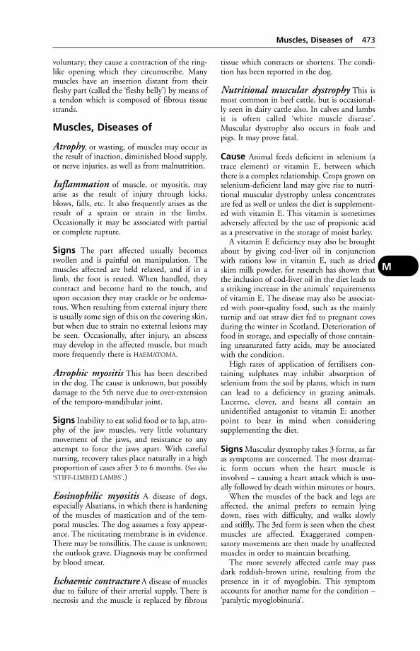
voluntary; they cause a contraction of the ring-like opening which they circumscribe. Manymuscles have an insertion distant from theirfleshy part (called the ‘fleshy belly’) by means ofa tendon which is composed of fibrous tissuestrands.
Muscles, Diseases of
Atrophy, or wasting, of muscles may occur asthe result of inaction, diminished blood supply,or nerve injuries, as well as from malnutrition.
Inflammation of muscle, or myositis, mayarise as the result of injury through kicks,blows, falls, etc. It also frequently arises as theresult of a sprain or strain in the limbs.Occasionally it may be associated with partialor complete rupture.
Signs The part affected usually becomesswollen and is painful on manipulation. Themuscles affected are held relaxed, and if in alimb, the foot is rested. When handled, theycontract and become hard to the touch, andupon occasion they may crackle or be oedema-tous. When resulting from external injury thereis usually some sign of this on the covering skin,but when due to strain no external lesions maybe seen. Occasionally, after injury, an abscessmay develop in the affected muscle, but muchmore frequently there is HAEMATOMA.
Atrophic myositis This has been describedin the dog. The cause is unknown, but possiblydamage to the 5th nerve due to over-extensionof the temporo-mandibular joint.
Signs Inability to eat solid food or to lap, atro-phy of the jaw muscles, very little voluntarymovement of the jaws, and resistance to anyattempt to force the jaws apart. With carefulnursing, recovery takes place naturally in a highproportion of cases after 3 to 6 months. (See also
‘STIFF-LIMBED LAMBS’.)
Eosinophilic myositis A disease of dogs,especially Alsatians, in which there is hardeningof the muscles of mastication and of the tem-poral muscles. The dog assumes a foxy appear-ance. The nictitating membrane is in evidence.There may be tonsillitis. The cause is unknown;the outlook grave. Diagnosis may be confirmedby blood smear.
Ischaemic contracture A disease of musclesdue to failure of their arterial supply. There isnecrosis and the muscle is replaced by fibrous
tissue which contracts or shortens. The condi-tion has been reported in the dog.
Nutritional muscular dystrophy This ismost common in beef cattle, but is occasional-ly seen in dairy cattle also. In calves and lambsit is often called ‘white muscle disease’.Muscular dystrophy also occurs in foals andpigs. It may prove fatal.
Cause Animal feeds deficient in selenium (atrace element) or vitamin E, between whichthere is a complex relationship. Crops grown onselenium-deficient land may give rise to nutri-tional muscular dystrophy unless concentratesare fed as well or unless the diet is supplement-ed with vitamin E. This vitamin is sometimesadversely affected by the use of propionic acidas a preservative in the storage of moist barley.
A vitamin E deficiency may also be broughtabout by giving cod-liver oil in conjunctionwith rations low in vitamin E, such as driedskim milk powder, for research has shown thatthe inclusion of cod-liver oil in the diet leads toa striking increase in the animals' requirementsof vitamin E. The disease may also be associat-ed with poor-quality food, such as the mainlyturnip and oat straw diet fed to pregnant cowsduring the winter in Scotland. Deterioration offood in storage, and especially of those contain-ing unsaturated fatty acids, may be associatedwith the condition.
High rates of application of fertilisers con-taining sulphates may inhibit absorption ofselenium from the soil by plants, which in turncan lead to a deficiency in grazing animals.Lucerne, clover, and beans all contain anunidentified antagonist to vitamin E: anotherpoint to bear in mind when considering supplementing the diet.
Signs Muscular dystrophy takes 3 forms, as faras symptoms are concerned. The most dramat-ic form occurs when the heart muscle isinvolved – causing a heart attack which is usu-ally followed by death within minutes or hours.
When the muscles of the back and legs areaffected, the animal prefers to remain lyingdown, rises with difficulty, and walks slowlyand stiffly. The 3rd form is seen when the chestmuscles are affected. Exaggerated compen-satory movements are then made by unaffectedmuscles in order to maintain breathing.
The more severely affected cattle may passdark reddish-brown urine, resulting from thepresence in it of myoglobin. This symptomaccounts for another name for the condition –‘paralytic myoglobinuria’.
Muscles, Diseases of 473
M
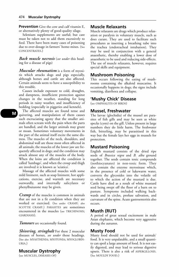
Prevention Give the cow and calf vitamin E,or alternatively plenty of good-quality silage.
Selenium supplements are useful; but caremust be taken not to add them excessively tofeed. There have been many cases of poisoningdue to over-dosage in farmers' home-mixes. (See
CONCENTRATES.)
Back muscle necrosis (see under this head-ing for a disease of pigs)
Muscular rheumatism is a form of myosi-tis which attacks dogs and pigs especially,although horses and cattle are also affected.Certain animals seem to have a susceptibility tothis trouble.
Causes include exposure to cold, draughts,and dampness, insufficient protection againstchanges in the weather, standing for long periods in rainy weather, and insufficiency ofbedding (especially in piggeries and kennels).
The affected muscles are found tense andquivering, and manipulation of them causessuch excruciating agony that the smaller ani-mals often scream with the pain when the partsare handled, and the larger animals may gruntor moan. Sometimes voluntary movements inthe part of the animal itself excite the same dis-tress. The muscles of the neck, shoulders, andabdominal wall are those most often affected inall animals; the muscles of the lower jaw are fre-quently affected in dogs; and the condition mayattack almost any of the muscles of the body.When the loins are affected the condition iscalled ‘lumbago’, and when the croup and thighare involved it is known as ‘sciatica’.
Massage of the affected muscles with somemild liniment, such as soap liniment, hot appli-cations, exercise, and warmth are necessary outwardly, and internally salicylates orphenylbutazone may be given.
Cramp of the muscles is common in animalsthat are not in a fit condition when they areworked or exercised. (See under CRAMP; also
SCOTTIE CRAMP.) PARASITES are sometimesencountered in the muscles (see TRICHINOSIS;GIARDIASIS).
Tumours are occasionally found.
Shivering, stringhalt For these 2 musculardiseases of horses, see under those headings. (See also MYASTHENIA; MYOTONIA; MYOGLOBIN-
URIA.)
Muscular Dystrophy(see MUSCLES, DISEASES OF)
Muscle RelaxantsMuscle relaxants are drugs which produce relax-ation or paralysis in voluntary muscle, such asdoes curare. They are used to facilitate suchprocedures as inserting a breathing tube intothe trachea (endotracheal intubation). Theymay be used in conjunction with a generalanaesthetic, thereby enabling a lower dose ofanaesthetic to be used and reducing side-effects.The use of muscle relaxants, however, requiresspecial skills and equipment.
Mushroom PoisoningThis occurs following the eating of mush-rooms containing the alkaloid muscarine. Itoccasionally happens in dogs; the signs includevomiting, diarrhoea and collapse.
‘Mushy Chick’ Disease(see OMPHALITIS OF BIRDS)
Mussel, FreshwaterThe larvae (glochidia) of the mussel are para-sites of fish gills and may be seen as whitespecks (cysts) on the gill. Unless present in largenumbers they do little harm. The freshwaterfish, bitterling, may be parasitised in this way but the female lays her eggs in mussels forprotection.
Mustard PoisoningEnglish mustard consists of the dried ripe seeds of Brassica nigra and B. alba groundtogether. The seeds contain toxic compounds(isothiocyanates) in non-toxic form. They also contain the enzyme myrosinase which, in the presence of cold or lukewarm water, converts the glycosides into the volatile oil to which the action of the mustard is due.Cattle have died as a result of white mustardseed being swept off the floor of a barn on topasture. Symptoms included walking back-wards and in circles, profuse salivation, and curvature of the spine. Acute gastroenteritis alsooccurs.
Musth (RUT)A period of great sexual excitement in maleAsian elephants, which become very aggressiveduring the autumn.
Musty FoodMusty food should not be used for animals'food. It is very unpalatable, and a small quanti-ty can spoil a large amount of food. It is not eas-ily digested, and may lead to serious digestiveupsets. There is also a risk of ASPERGILLOSIS.(See MOULDY FOOD.)
474 Muscular Dystrophy
M
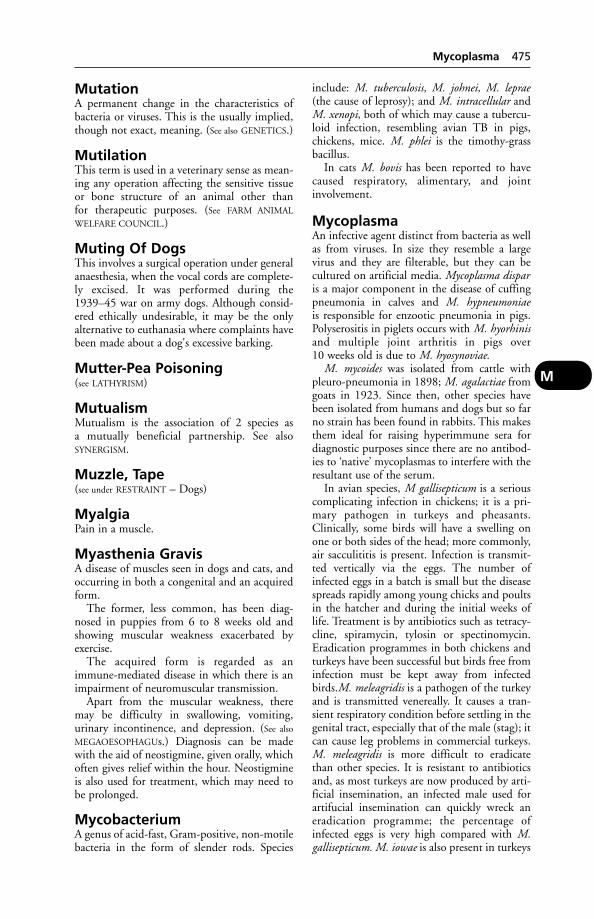
MutationA permanent change in the characteristics ofbacteria or viruses. This is the usually implied,though not exact, meaning. (See also GENETICS.)
MutilationThis term is used in a veterinary sense as mean-ing any operation affecting the sensitive tissueor bone structure of an animal other than for therapeutic purposes. (See FARM ANIMAL
WELFARE COUNCIL.)
Muting Of DogsThis involves a surgical operation under generalanaesthesia, when the vocal cords are complete-ly excised. It was performed during the1939–45 war on army dogs. Although consid-ered ethically undesirable, it may be the onlyalternative to euthanasia where complaints havebeen made about a dog's excessive barking.
Mutter-Pea Poisoning(see LATHYRISM)
MutualismMutualism is the association of 2 species as a mutually beneficial partnership. See also SYNERGISM.
Muzzle, Tape(see under RESTRAINT – Dogs)
MyalgiaPain in a muscle.
Myasthenia GravisA disease of muscles seen in dogs and cats, andoccurring in both a congenital and an acquiredform.
The former, less common, has been diag-nosed in puppies from 6 to 8 weeks old andshowing muscular weakness exacerbated byexercise.
The acquired form is regarded as animmune-mediated disease in which there is animpairment of neuromuscular transmission.
Apart from the muscular weakness, theremay be difficulty in swallowing, vomiting, urinary incontinence, and depression. (See also
MEGAOESOPHAGUs.) Diagnosis can be madewith the aid of neostigmine, given orally, whichoften gives relief within the hour. Neostigmineis also used for treatment, which may need tobe prolonged.
MycobacteriumA genus of acid-fast, Gram-positive, non-motilebacteria in the form of slender rods. Species
include: M. tuberculosis, M. johnei, M. leprae(the cause of leprosy); and M. intracellular andM. xenopi, both of which may cause a tubercu-loid infection, resembling avian TB in pigs,chickens, mice. M. phlei is the timothy-grassbacillus.
In cats M. bovis has been reported to havecaused respiratory, alimentary, and jointinvolvement.
MycoplasmaAn infective agent distinct from bacteria as wellas from viruses. In size they resemble a largevirus and they are filterable, but they can becultured on artificial media. Mycoplasma disparis a major component in the disease of cuffingpneumonia in calves and M. hypneumoniaeis responsible for enzootic pneumonia in pigs.Polyserositis in piglets occurs with M. hyorhinisand multiple joint arthritis in pigs over 10 weeks old is due to M. hyosynoviae.
M. mycoides was isolated from cattle withpleuro-pneumonia in 1898; M. agalactiae fromgoats in 1923. Since then, other species havebeen isolated from humans and dogs but so farno strain has been found in rabbits. This makesthem ideal for raising hyperimmune sera fordiagnostic purposes since there are no antibod-ies to ‘native’ mycoplasmas to interfere with theresultant use of the serum.
In avian species, M gallisepticum is a seriouscomplicating infection in chickens; it is a pri-mary pathogen in turkeys and pheasants.Clinically, some birds will have a swelling onone or both sides of the head; more commonly,air sacculititis is present. Infection is transmit-ted vertically via the eggs. The number ofinfected eggs in a batch is small but the diseasespreads rapidly among young chicks and poultsin the hatcher and during the initial weeks oflife. Treatment is by antibiotics such as tetracy-cline, spiramycin, tylosin or spectinomycin.Eradication programmes in both chickens andturkeys have been successful but birds free frominfection must be kept away from infectedbirds.M. meleagridis is a pathogen of the turkeyand is transmitted venereally. It causes a tran-sient respiratory condition before settling in thegenital tract, especially that of the male (stag); itcan cause leg problems in commercial turkeys.M. meleagridis is more difficult to eradicatethan other species. It is resistant to antibioticsand, as most turkeys are now produced by arti-ficial insemination, an infected male used forartifucial insemination can quickly wreck aneradication programme; the percentage ofinfected eggs is very high compared with M.gallisepticum. M. iowae is also present in turkeys
Mycoplasma 475
M
and causes decreased hatchability. M. iowaeinfection is usually detectable only in flocksthat have been freed from the other mycoplas-mas. Eradication is difficult but not impossible.M. synoviae affects chickens and turkeys, caus-ing tenosynovitis. It may be treated by highdoses of tetracyclines. The method of spread isunclear; even biting insects have been suggestedas possible vectors.
In goats M. mycoides causes contagiouscaprine pleuropneumonia, as well as septi-caemia and mastitis.
M. bovis, first isolated in the USA in 1962,has caused severe respiratory disease in the UK.It also causes mastitis.
M. canadense was first reported in the UK in 1978, and causes abortion in cattle; and M. californicum causes mastitis.
In many parts of the world, even where contagious bovine pleuropneumonia has beeneradicated, mycoplasmal diseases are of consid-erable economic importance. They includemastitis, arthritis, bone disease, and keratitis. Incattle, M. bovigenitalium is a cause of abortionand mastitis.
M. hyosynoviae has caused lameness in pigs. The pigs frequently adopted adopt a ‘dog-sitting’ posture; and develop areas ofhyperaemia on the hams.
Mycoplasmas are also important as contami-nants of cell cultures used for vaccine produc-tion. (See also under KENNEL COUGH.)
MycoplasmosisA mycoplasma infection. (See MYCOPLASMA;CONTAGIOUS BOVINE PLEUROPNEUMONIA;VULVO-VAGINITIS; ENZOOTIC PNEUMONIA OF
PIGS; SINUSITIS; INFECTIOUS BRONCHITIS OF
POULTRY; MASTITIS.)
Mycosis, Mycotic InfectionsMycosis, mycotic infections are diseases due tothe growth of fungi in the body. Among thecommonest are ringworm, sporotrichosis,aspergillosis. Mycotic mastitis is important indairy cattle and 26 or more species of fungi areinvolved. (See also RHINOSPORIDIOSIS; FUNGAL
DISEASES; SPORIDESMIN.)
MycotoxicosisPoisoning by toxins produced by fungi. (Forexamples of such toxins, see ERGOT OF RYE;AFLATOXINS; OCHRATOXIN A; ZEARALENONE;SPORIDESMIN; T2 TOXIN; FUSARIUM; PENI-
TREMA; and under FESCUE, RYEGRASS.)When, in farm animals, a change of feed
leads to depressed output, or to symptoms ofillness, poisoning by fungal toxins may be
suspected. However, when analysed most sus-pect rations are found to contain fungal toxinsin amounts too small for chemical detection.
Nevertheless, there is no doubt that occa-sional outbreaks of aflatoxicosis, ergotism, andzearalenone (F2) intoxication do occur in theUK.
In one outbreak, 2 cows became dull andfeverish, with bleeding from mouth and eyelids,and died within 48 hours. Other cows becameill, some with diarrhoea. About 60 members ofthis Friesian herd had bleeding eczema-likelesions of both black and white skin of udderand abdomen. A 3rd cow died later and 2 hadto be slaughtered. The final tentative diagnosiswas fungal poisoning, after examination ofmouldy barley (containing many potentiallypoisonous fungi) which formed 87 per cent ofa supplementary concentrate ration. Similarhaemorrhages and deaths from this cause havebeen reported in the USA.
MydriasisAn excessive dilation of the pupil of the eye.Drugs which are given when dilation isrequired for diagnostic purposes are called‘mydriatics’. (See also ATROPINE.)
MyelinMyelin is the white fat-like substance forming asheath round myelinated nerve fibres.
MyelitisMyelitis is a condition in which destructivechanges occur in the spinal cord. It usually follows upon viral infections. Paralysis of amuscle or of groups of muscles may occur; theremay be twitchings or spasms of muscles; thepenis may hang from the prepuce, the bladderand rectum become unable to retain their contents, and finally a form of paraplegia oftenoccurs. The paralysis may gradually pass for-wards; the sensation is lost in the skin of theloins, then of the back, and later the fore-legsbecome unable to support the weight of thebody. Occasionally the condition disappearsspontaneously, but the majority of cases endfatally. (See also OSTEOMYELITIS.)
MyelocyteA bone-marrow cell, from which white cells(basophils, neutrophils and eosinophils) of theblood are produced. They are found in theblood in certain forms of leukaemia.
MyelographyRadiography of the spinal cord, using a contrastmedium. (See SPINAL CORD, DISEASES OF.)
476 Mycoplasmosis
M
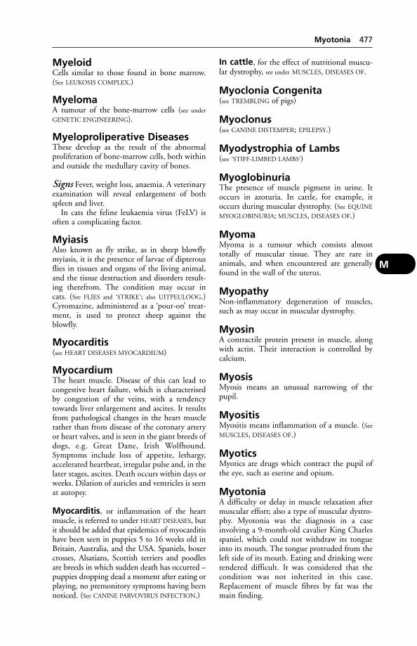
MyeloidCells similar to those found in bone marrow.(See LEUKOSIS COMPLEX.)
MyelomaA tumour of the bone-marrow cells (see under
GENETIC ENGINEERING).
Myeloproliperative DiseasesThese develop as the result of the abnormalproliferation of bone-marrow cells, both withinand outside the medullary cavity of bones.
Signs Fever, weight loss, anaemia. A veterinaryexamination will reveal enlargement of bothspleen and liver.
In cats the feline leukaemia virus (FeLV) isoften a complicating factor.
MyiasisAlso known as fly strike, as in sheep blowflymyiasis, it is the presence of larvae of dipterousflies in tissues and organs of the living animal,and the tissue destruction and disorders result-ing therefrom. The condition may occur incats. (See FLIES and ‘STRIKE’; also UITPEULOOG.)Cyromazine, administered as a ‘pour-on’ treat-ment, is used to protect sheep against theblowfly.
Myocarditis(see HEART DISEASES MYOCARDIUM)
MyocardiumThe heart muscle. Disease of this can lead tocongestive heart failure, which is characterisedby congestion of the veins, with a tendencytowards liver enlargement and ascites. It resultsfrom pathological changes in the heart musclerather than from disease of the coronary arteryor heart valves, and is seen in the giant breeds ofdogs, e.g. Great Dane, Irish Wolfhound.Symptoms include loss of appetite, lethargy,accelerated heartbeat, irregular pulse and, in thelater stages, ascites. Death occurs within days orweeks. Dilation of auricles and ventricles is seenat autopsy.
Myocarditis, or inflammation of the heartmuscle, is referred to under HEART DISEASES, butit should be added that epidemics of myocarditishave been seen in puppies 5 to 16 weeks old inBritain, Australia, and the USA. Spaniels, boxercrosses, Alsatians, Scottish terriers and poodlesare breeds in which sudden death has occurred –puppies dropping dead a moment after eating orplaying, no premonitory symptoms having beennoticed. (See CANINE PARVOVIRUS INFECTION.)
In cattle, for the effect of nutritional muscu-lar dystrophy, see under MUSCLES, DISEASES OF.
Myoclonia Congenita(see TREMBLING of pigs)
Myoclonus(see CANINE DISTEMPER; EPILEPSY.)
Myodystrophia of Lambs(see ‘STIFF-LIMBED LAMBS’)
MyoglobinuriaThe presence of muscle pigment in urine. Itoccurs in azoturia. In cattle, for example, itoccurs during muscular dystrophy. (See EQUINE
MYOGLOBINURIA; MUSCLES, DISEASES OF.)
MyomaMyoma is a tumour which consists almosttotally of muscular tissue. They are rare in animals, and when encountered are generallyfound in the wall of the uterus.
MyopathyNon-inflammatory degeneration of muscles,such as may occur in muscular dystrophy.
MyosinA contractile protein present in muscle, alongwith actin. Their interaction is controlled bycalcium.
MyosisMyosis means an unusual narrowing of thepupil.
MyositisMyositis means inflammation of a muscle. (See
MUSCLES, DISEASES OF.)
MyoticsMyotics are drugs which contract the pupil ofthe eye, such as eserine and opium.
MyotoniaA difficulty or delay in muscle relaxation aftermuscular effort; also a type of muscular dystro-phy. Myotonia was the diagnosis in a caseinvolving a 9-month-old cavalier King Charlesspaniel, which could not withdraw its tongueinto its mouth. The tongue protruded from theleft side of its mouth. Eating and drinking wererendered difficult. It was considered that thecondition was not inherited in this case.Replacement of muscle fibres by fat was themain finding.
Myotonia 477
M
M
478 Mysoline
MysolineAn anti-convulsant drug used in the treatmentof epilepsy. A side-effect of this drug may bethirst, polyuria resulting in urinary inconti-nence, in the dog.
MyxoedemaA thickening and degradation of the skin thatoccurs in hypothyroidism; seen mainly in pigsand dogs (see THYROID, DISEASES OF).
MyxomaMyxoma is a tumour consisting of imperfectconnective tissue, set in a mucoid ground substance. (See TUMOURS.)
Myxomatosis, InfectiousA disease of rabbits caused by a virus. Hares areoccasionally affected also. The disease has a very
high mortality rate when introduced into acountry, but later the virus may become lesspotent or the survivors more resistant.Myxomatosis appeared in wild rabbits in Kentand Sussex in October 1953, and spread rapid-ly throughout most of Britain. Symptomsinclude conjunctivitis, ‘gummed’ eyelids,swelling of the nose and muzzle, and of themucous membrane of the vulva and anus.Orchitis is caused in the male. Emaciation,fever, and death follow. The disease is transmit-ted by the rabbit flea and, mechanically, by thistles. A vaccine is available.
Myxovirus(see VIRUSES)

NaganaNagana is an unscientific but convenient namefor trypanosomiasis transmitted by tsetse flies(Glossina spp.) in Africa. The trypanosomesinvolved are Trypanosoma vivax, T. uniforme,T. congolense, T. brucei, T. simiae, and T. suis.(See TRYPANOSOMES.) The symptoms of naganainclude anaemia, intermittent fever, and (exceptin pigs, in which the disease may be very acute)a slow, progressive emaciation. In both horsesand dogs the eyes may be affected, as shown bycorneal opacity. Horses often have oedemaaffecting the limbs and abdomen. Cattle mayabort.
The drug quinapyramine is used (amongothers) in treatment.
The kudu, hyena, and bush-buck, as well as other wild animals, act as reservoirs of theinfection.
Nail Binding(see INJURIES FROM SHOEING)
Nails (Claws)A claw contains a matrix with blood vessels,nerves, etc., from which it grows and is nour-ished. Lying within the matrix is the bone ofthe terminal phalanx of the digit, which givesthe nail its characteristic form in the differentanimals. When not in use in the carnivora, nailsare retracted by ligaments in an upwards direc-tion; this is more marked in cats, where the nailmay almost disappear, than in dogs.
Nails, Diseases ofThe nails of cats and dogs sometimes becometorn or broken through fighting or accidents.Sometimes only the tip is injured, and thematrix higher up is undamaged; in such cases afine pellicle of horn covers the tip until suchtime as the horn has grown down from above,and the whole nail is not shed. In other casesinfection occurs, causing great tenderness of thepart.
Ingrowing nails occur upon the ‘dewclaws’, on the insides of the paws of dogs. Thesemore or less rudimentary digits do not touchthe ground, and are consequently not subjectedto wear from friction. The nails grow, andowing to their curve eventually penetrate the
soft pad behind them. Where actual penetra-tion has occurred, the nail should be cut shortand an antiseptic dressing applied. It is custom-ary for owners of sporting and other dogs tohave the dew claws removed during puppyhoodto avoid future trouble of this nature.Amputation of dew claws can be carried out inthe adult under anaesthesia.
Onychomycosis, or a fungal infection ofthe claws, is a not uncommon condition in cats,and is of public-health importance as a reservoirof ringworm transmissible to children. (See
RINGWORM.)
Nairobi Sheep DiseaseNairobi sheep disease is an acute infectiousfever of sheep and goats, caused by a bun-yavirus, and occurring in eastern and southernAfrica. The virus is transmitted by the tickRhipicephalus appendiculatus.
Signs Imported sheep usually show an acutefebrile disturbance within 5 or 6 days afterbeing infected by the ticks. This lasts for up to9 days and then a fall in temperature occurs andother clinical symptoms appear. Death maytake place a day or two later, or a further rise intemperature may be shown, death or recoveryfollowing. There is rapidity and difficulty inbreathing, a mucopurulent nasal discharge andgreen watery diarrhoea, which may containmucus or blood. The genital organs of ewes areswollen and congested, and abortion may occurin pregnant ewes.
Immunity In the great majority of cases,recovery confers a strong and lasting immunity.This is also possessed by sheep in areas wherethe infection is endemic.
Nanogram (ng)A unit of weight equivalent to 1000 microgramsµ 1000 micrograms equal 1 milligram (mg).
Nanometre (nm)A unit of linear measurement used in e.g. virolo-gy. One nm equals one millionth of a millimetre.
Naphthalene PoisoningNaphthalene poisoning might arise from theingestion of moth-balls. In the dog, it has beenshown experimentally to give rise to haemolyt-ic anaemia. (In children, poisoning from moth-balls gives rise to ‘port-wine coloured’ urine.)Another symptom is cataract. Chlorinatednaphthalenes have been identified as one cause
N
of HYPERKERATOSIS in cattle; and tear stainsmay be a symptom of this type of poisoning.
NarcolepsyNarcolepsy is a sudden collapse into deep sleep.It has been recorded in dogs, and may be part-ly genetic in origin. A case was recorded in theUK in a 3-year-old Corgi which sometimes collapsed when taken for his first walk of theday, or offered food. Often yawning and avacant expression would precede a sudden dropfrom a standing position to a sitting one or alying one. No excitement, salivation, or convul-sions were seen, and at other times the dog wasactive and mentally alert; he was easily arousedafter he had collapsed. Electro-encephalogramssupported the diagnosis. The condition has alsobeen recorded in daschunds, dobermanns,labradors and poodles.
NaresNares is the Latin word for the nostrils.
Nasal Bot Fly (Oestrus Ovis)LarvaeNasal Bot Fly (Oestrus Ovis) Larvae are seriousparasites of sheep. (See under FLIES.)
Nasal Disorders(see NOSE & NASAL PASSAGES, DISEASES OF)
Naso-Oesophageal TubesNarrow tubes inserted through the nose intothe stomach. They are tolerated by many, if notmost, cats, and can be used to provide nutri-tional support via liquid foods for a week ortwo. The use of small-diameter tubes does notprevent voluntary intake of food.
Naso-PharynxThe upper part of the throat lying posterior tothe nasal cavity.
NatamycinAn antibiotic used for the treatment of ringworm in cattle and horses. Application can be made with a knapsack sprayer. (See RINGWORM.)
National Scrapie PlanA long-term UK plan which aims to reduceand, eventually, eradicate the number of sheepnot genetically resistant to scrapie and othertransmissible spongiform encephalopathies.Under the scheme, sheep are individually iden-tified by electronic tag, and blood-tested toestablish whether they are susceptible or resis-tant to scrapie. Sheep identified as susceptible
must not be bred from. Under DEFRA propos-als in 2003, farmers with confirmed scrapiecases on their farms will have their flocks geno-typed so that the more susceptible sheep can be identified and removed, or the whole flockdisposed of.
National Office of AnimalHealth (NOAH)Founded in 1986, to represent those UK com-panies which manufacture animal-health prod-ucts licensed under the Medicines Act. Address:3 Crossfield Chambers, Gladbeck Way, Enfield,Middlesex EN2 7HF. Publications include: TheSafe Storage & Handling of Animal Medicines;Poisoning in Veterinary Practice.
National Pet RegisterThis provides a service for reuniting lost petswith their owners, and also for third-party liability. Address: Heydon, Royston, Herts.SG8 8PN.
NatureOedema of the udder (see under OEDEMA).
Navel-Ill(see JOINT-ILL)
Navicular BoneNavicular bone is the popular name for thesesamoid of the 3rd phalanx of the horse. It is alittle boat-shaped bone, developed just abovethe deep flexor tendon, and serves, as do allsesamoid bones, to minimise friction where thetendon passes round a corner of another bone.It enters into the formation of the ‘coffin-joint’,between the 2nd and 3rd phalanges of the digit.It is of great importance in deep puncturedwounds of the foot when these are situatedtowards the heels, for, when damaged, its sur-face becomes inflamed, the inflammationspreads to the coffin-joint and may produceincurable lameness.
Navicular DiseaseNavicular disease is a chronic condition ofinflammation affecting the horse’s navicularbone and its associated structures. The fore-feetare usually both attacked, though the conditionmay arise in only one of these, or in the hind-feet (rarely). Ulceration of the cartilage first,and later of the bone on the surface over whichthe deep flexor tendon plays, may sometimes beseen at autopsy.
Causes These are still a matter of hypothesisrather than certainty, and controversy persists.
480 Narcolepsy
N
Some authors have referred to increased vascu-larisation of the navicular bone; others suggestthat ischaemia may be responsible, leading topain and, if at least 2 of the distal arteries areoccluded, to chronic lameness. In horses lameas a result of navicular disease, occlusion of themain artery and progressive arterial thrombosisare frequent, with a resulting area of ischaemicnecrosis and cavitation of the navicular bone.
Another view is that the disease is not causedprimarily by ischaemia and subsequent necro-sis, but is a consequence of bone remodellingdue to altered pressure from the deep flexor ten-don and increased load on the caudal part ofthe foot – the condition not being irreversibleunless secondary lesions such as adhesions and bony spurs have developed. Special shoeingto alter the load on the navicular bone is recommended.
Signs Navicular disease usually develops soslowly that the owner has considerable difficul-ty in remembering exactly when the first symptoms were noticed. In fact, little or noimportance may be attached to the almost char-acteristic ‘pointing’ of one or both fore-feet,because ‘he has always done that’. ‘Pointing’consists of resting the affected foot (or feet) byplacing it a short distance in advance of the other when standing in harness or in thestable. When both feet are affected, each isalternately pointed. Later, the horse may golame or be tender on his feet at times, but witha rest he generally becomes sound again. As thedisease advances, he may either start off in themornings stiff and become better with exerciseas he warms to his work, or may become lameas the day goes on. Sooner or later, however,there comes a time when he will go permanent-ly ‘pottery’, or ‘groggy’. The length of the stridedecreases and there is difficulty in advancingthe feet, so it looks as if the shoulder is the seatof the lesion. When made to turn, the horsepivots round on the fore-feet instead of liftingthem, and when made to back, drags the toes.If the shoe of such a horse is examined it is usually found to be more worn at the toes thanat the heels. In fact a ‘groggy’ horse may wearhis shoes quite thin at the toes before the heelsshow much sign of wear at all. In the finalstages the horse becomes distinctly lame andunfit for work. When observed in the stable heis noticed to be continually shifting from onefoot on to the other, and the resting foot isplaced well out in front.
Treatment must aim at the relief of pain andimprovement of the local blood circulation. The
vasodilator isoxuprine or a formulation of war-farin may be added to the feed of horses; thedosage of warfarin requires great care – withoverdosage there is a danger of haemorrhage.Warfarin treatment has been reported effectivein about 75 per cent of cases of navicular disease.
Before the advent of drug therapy it was cus-tomary to perform the operation of neurecto-my, which consists of a section of the plantar ormedian nerve of the limb. In a favourable case,following operation, the horse becomes appar-ently sound, although the diseased condition isstill at work in the bone. No pain is felt, and thehorse is fit for light work at slow paces. The feetrequire constant attention to ensure that nostones, nails, etc. lodge in the hoof, for evenwhen these inflict serious damage the horse stillgoes sound, not feeling the pain.
Navy BeansNavy beans may cause death if fed raw. (See
LEGUME POISONING.)
Near East EncephalitisAn alphavirus infection of horses and donkeys;less frequently of cattle and sheep.Convulsions/paralysis may follow fever andprecede death. (See EQUINE ENCEPHALITIS;BORNA DISEASE.)
NeckIn animals, the neck is that part of the bodyconnecting the head with the trunk. It containsthe trachea, oesophagus, blood vessels, thespinal cord and cervical vertebrae. Both themouse and giraffe have 7 cervical vertebrae, asdo most mammals.
The weight of the head is supported by thepowerful ligamentum nuchae, which takes thestrain off the muscles, thereby avoiding fatigue.In the horse the ligament extends from thespines of the withers to the posterior of theoccipital bone of the SKULL.
NecrobacillosisDamage of an organ, or tissue, caused byFusobacterium necrophorum. The necrotic areahas a characteristic rotting odour.
NecropsyExamination of a dead body (see AUTOPSY).
NecrosisDeath of cells or of a limited portion of tissue.
Necrosis (Bacillary) orNecrobacillosis(see CALF DIPHTHERIA)
Necrosis (Bacillary) or Necrobacillosis 481
N
Necrotic EnteritisA subacute or chronic enteritis which follows amore severe episode caused by infection withSalmonella spp. or Campylobacter sputorumvar. mucosalis. A condition of unweaned andolder pigs, characterised by scouring and loss ofcondition.
The lesions are in the caecum and ileum. (See
also under ILEUM.)Cold, damp, dirty surroundings appear to
predispose to necrotic enteritis. (See PORCINE
INTESTINAL ADENOMATOSIS.)
Necrotic Enteritis in ChickensA disease of chickens characterised by unthrifti-ness and diarrheoa caused by Clostridium perfringens (welchii) type C. There is usually aconcurrent defect in nutrition. The disease hasbeen reported in most European and NorthAmerican countries, and in Australia.
Necrotic StomatitisA serious infection of the inside of the mouthand the tongue, seen in calves; it may also befound in reptiles. (See CALF DIPHTHERIA.)
Negri BodiesNegri bodies are comparatively large, roundedbodies in the brain cells of animals infectedwith rabies. Their presence can be demon-strated by staining with Seller’s stain, among others. The cerebral cortex, Ammon’s horn, and the Purkinje cells of the cerebellum are the main sites to examine. The diagnosis of rabies once depended upon the demonstra-tion of Negri bodies in the affected animal. (See
RABIES.)
NeisseriaSpherical, Gram-negative bacteria, some ofwhich are associated with eye infections.
NematocideA drug that destroys nematodes.
NematodeNematode is a general term applied to the para-sitic Nemathelminthes, which include the round-worms, as distinct from the Platyhelminthes, orflatworms. (See WORMS.)
‘Nematode Poisoning’In the USA, larvae of Anguina agrostis onChewing’s fescue in immature hay caused anoutbreak of poisoning in cattle. Symptomsincluded knuckling of the fetlocks, head tuckedbetween the fore-legs, recumbency, convul-sions, and death.
NematodiriasisInfestation of the intestine or abomasum ofruminants by Nematodirus species. It is endem-ic in some parts of the UK. Disease developssuddenly and leads to dullness and loss of con-dition, with black diarrhoea and dehydration;in lambs, death may follow in a few days.
Prevention As the eggs can survive over win-ter, the life-cycle can be broken by not using thesame lambing ground in successive seasons.Routine dosing of lambs in susceptible areascan be effective.
NematodirusParasitic worms of, particularly, lambs; alsosheep and calves. N. battus infection is trans-ferred from one season’s lambs to the next as large numbers of eggs are deposited on pasture. Development of the eggs occurs onlyafter exposure to cold and moisture. (See
WORMS.)
Nembutal(see PENTOBARBITONE SODIUM)
NeoarsphenamineA drug effective against BLACKHEAD OF
TURKEYS. It has been largely superseded bydimetridazole and nifursol.
NeomycinAn aminoglycoside antibiotic obtained fromStreptomyces fradiae. It must not be given byinjection, owing to resulting kidney damage. Its action closely resembles that of strepto-mycin. It is used, sometimes in combinationwith other medicaments, in a number of veteri-nary formulations. A topical spray of thisantibiotic has caused profound deafness in children. (See DEAFNESS.)
NeonatalNeonatal diseases are those of the newborn.
NeoplasmNeoplasm means literally ‘a new growth’, and isapplied to tumours in general.
Neospora CaninumThis parasite was discovered in Norway in1984, and later recognised in Sweden, the USA,Australia, and the UK in 1990.
Cause A protozoan, named as above, andresembling Toxoplasma gondii. Congenitalinfection occurs in cattle, dogs, and cats.
482 Necrotic Enteritis
N

Signs Infected animals may develop ataxia, afleeting paralysis, and nystagmus. Meningitisappears in some cases. The parasite may also befound in aborted ovine fetuses.
NeotenyThe retention of juvenile activities and appear-ance into adulthood. It is the basis of populari-ty of some breeds of dog that remain as playfulas puppies throughout their life. The extremeexample is an amphibian, the axolotl orMexican walking fish, which rarely matures tothe adult stage.
NephrectomyNephrectomy is the name given to the opera-tion by which one of the kidneys is removed.(See KIDNEY, DISEASES OF.)
NephritisInflammation of the kidneys (see KIDNEY,DISEASES OF; LEPTOSPIROSIS).
NephrolithiasisThe presence of a stone (calculus) in the pelvisof the kidney.
NephronThe structural unit of the kidney (see KIDNEYS).
Nephroptosis‘Floating kidney’ – abnormal positioning of thekidney (see KIDNEY, DISEASES OF).
NephrosisThis is a disease of the kidneys, involving dam-age to the tubules. It leads to albuminuria andoften to oedema. (See also KIDNEY, DISEASES OF.)
Nephrosis, Infectious AvianA disease of chickens. (See GUMBORO DISEASE;INFECTIOUS BURSAL DISEASE.)
Nephrotic Syndrome(see NEPHROSIS)
NephrotomySurgical incision into a kidney.
Nerve BlockAnaesthesia of a nerve or nerves supplying partof the body to assist diagnosis or treatment.Often used in diagnosing the cause of lamenessin the horse.
NervesThe nerves are fibre-like tissues that conveyimpulses (‘messages’) between the central
nervous system and other parts of the body. Thebasic unit of the nervous system is the neuron,a cell with at least 1 projection. Bipolar neuronshave 1 long projection, the axon, and 1 shortbranching projection, the dendrite. A typicalneuron (multipolar) has several dendrites butusually only 1 axon (nerve fibre).
Dendrites conduct nerve impulses towardsthe nerve cell; axons conduct away from it.
A synapse is a point or area where 1 neuronis able to make contact with another; the contact being between the axon of 1 neuronand a dendrite of another neuron, or betweenthe axon of 1 neuron and the cell of another neuron. Any neuron may connect with axons ordendrites of several other neurons.
Nerve fibres may be myelinated (enclosed ina sheath) or unmyelinated (see MYELIN). Somenerve fibres (axons) convey impulses to brain orspinal cord from skin or sense organ, and aretermed sensory or afferent. Their impulses arepassed, through connecting links or interneu-rons, to motor or efferent nerves from brain orspinal cord (but see spinal reflex under SPINAL
CORD – Functions).
Nerve impulses are dependent upon thepermeability of cell membranes. There is apotential difference of about 70 to 80 millivoltsbetween the inside and the outside of an axon –the inside being the negative. This is owing tothe fact that in a resting state the cell membraneis permeable to K(+) and Cl(–) ions, but not toNa(+) ions. Stimulation of the nerve results inthe membrane becoming permeable to thesodium ions, which flow in causing the insideof the axon to carry a positive electrical chargeinstead of a negative one. A so-called depolari-sation wave is set up, ‘self-perpetuating’, along1 neuron after another. A single nerve fibre cansend about 1000 separate impulses per second.
ACETYLCHOLINE is released by somatic (mus-cle) nerve fibres at synapses between neurons on either side of ganglia, and also at the junc-tion of motor nerve endings and voluntary(striated) muscle. Acetylcholine is released alsoat synapses by parasympathetic nerve fibres.NORADRENALIN is released at synapses of sym-pathetic nerve fibres, and at their junction withsmooth (unstriated) involuntary muscle fibres.
Nerves, Injuries toContinued or repeated severe pressure upon anerve trunk may be sufficient to damage it andresult in paralysis; severe bruising in which anerve is driven against a bone with considerableforce may produce paralysis or inflammation ofthe nerve; a nerve may be severed along with
Nerves, Injuries to 483
N
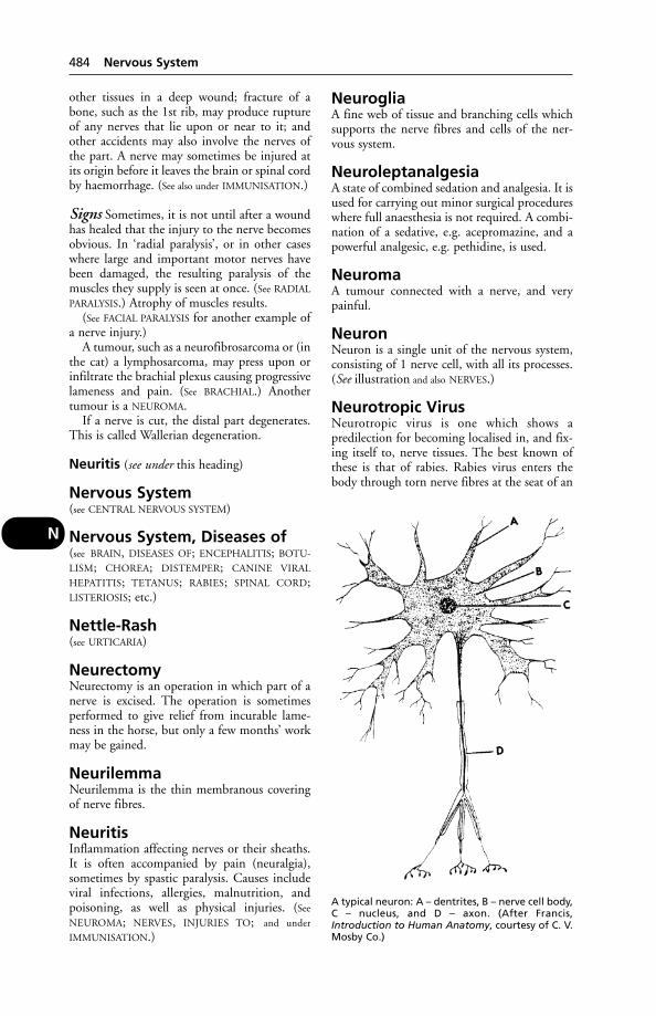
other tissues in a deep wound; fracture of abone, such as the 1st rib, may produce ruptureof any nerves that lie upon or near to it; andother accidents may also involve the nerves ofthe part. A nerve may sometimes be injured atits origin before it leaves the brain or spinal cordby haemorrhage. (See also under IMMUNISATION.)
Signs Sometimes, it is not until after a woundhas healed that the injury to the nerve becomesobvious. In ‘radial paralysis’, or in other caseswhere large and important motor nerves havebeen damaged, the resulting paralysis of themuscles they supply is seen at once. (See RADIAL
PARALYSIS.) Atrophy of muscles results.(See FACIAL PARALYSIS for another example of
a nerve injury.)A tumour, such as a neurofibrosarcoma or (in
the cat) a lymphosarcoma, may press upon orinfiltrate the brachial plexus causing progressivelameness and pain. (See BRACHIAL.) Anothertumour is a NEUROMA.
If a nerve is cut, the distal part degenerates.This is called Wallerian degeneration.
Neuritis (see under this heading)
Nervous System(see CENTRAL NERVOUS SYSTEM)
Nervous System, Diseases of(see BRAIN, DISEASES OF; ENCEPHALITIS; BOTU-
LISM; CHOREA; DISTEMPER; CANINE VIRAL
HEPATITIS; TETANUS; RABIES; SPINAL CORD;LISTERIOSIS; etc.)
Nettle-Rash(see URTICARIA)
NeurectomyNeurectomy is an operation in which part of anerve is excised. The operation is sometimesperformed to give relief from incurable lame-ness in the horse, but only a few months’ workmay be gained.
NeurilemmaNeurilemma is the thin membranous coveringof nerve fibres.
NeuritisInflammation affecting nerves or their sheaths.It is often accompanied by pain (neuralgia),sometimes by spastic paralysis. Causes includeviral infections, allergies, malnutrition, and poisoning, as well as physical injuries. (See
NEUROMA; NERVES, INJURIES TO; and under
IMMUNISATION.)
NeurogliaA fine web of tissue and branching cells whichsupports the nerve fibres and cells of the ner-vous system.
NeuroleptanalgesiaA state of combined sedation and analgesia. It isused for carrying out minor surgical procedureswhere full anaesthesia is not required. A combi-nation of a sedative, e.g. acepromazine, and apowerful analgesic, e.g. pethidine, is used.
NeuromaA tumour connected with a nerve, and verypainful.
NeuronNeuron is a single unit of the nervous system,consisting of 1 nerve cell, with all its processes.(See illustration and also NERVES.)
Neurotropic VirusNeurotropic virus is one which shows apredilection for becoming localised in, and fix-ing itself to, nerve tissues. The best known ofthese is that of rabies. Rabies virus enters thebody through torn nerve fibres at the seat of an
484 Nervous System
N
A typical neuron: A – dentrites, B – nerve cell body,C – nucleus, and D – axon. (After Francis,Introduction to Human Anatomy, courtesy of C. V.Mosby Co.)

injury, such as a bite, and, growing along them,eventually reaches the spinal cord and brain.Other neurotropic viruses are those of louping-ill in sheep, and Borna disease in horses and cattle.
Neutering(see CASTRATION and SPAYING; also VASECTOMY)
NeutronsElementary particles with approximately thesame mass as a proton. The latter has beendefined as a stable, positively charged elemen-tary particle found in atomic nuclei in numbersequal to its atomic number.
NeutropeniaA reduced number of neutrophil granularleukocytes in the blood.
(In human medicine, most cases are attrib-uted to the direct toxic effect of certain antibi-otics, e.g. penicillin and the cephalosporins, orto immune-mediated mechanisms. With thistype of blood dyscrasia patients are at seriousrisk of an overwhelming infection.)
NeutrophilA type of white blood cell which can migrateinto the tissues and engulf bacteria, etc. (See
under BLOOD; ABSCESS.)
New Forest Disease (InfectiousBovine Keratitis)A painful eye condition which can lead toblindness if neglected. (See EYE, DISEASES OF.)
New Forest FlyA blood-sucking fly, found in many parts ofBritain. Hippobosca equina attacks horses andcattle. It deposits larvae (not eggs) in the soil.When disturbed, it makes a characteristic side-ways movement. (See FLIES.)
Newcastle DiseaseAn infectious, febrile notifiable disease of chick-ens, turkeys, ducks, pigeons and wild birds.Globally, it is the most economically importantdisease of livestock. Aviary birds, particularlyfinches (canaries, etc.), may be infected by windspread. Waterfowl can be infected but clinical dis-ease is rare. In humans, conjunctivitis is the mainclinical sign, but people working with infectedbirds may develop an influenza-like illness.
Cause Paramyxovirus.
Signs The first sign noticed in laying birds maybe a drop in egg yield with the production of
pale, misshapen and/or soft-shelled eggs.According to the virus strain, infected birdsdevelop respiratory or nervous signs; it is rare tofind both together. Severe breathing difficultiesdevelop in birds affected with the respiratorystrain. In the nervous form, torticollis, paralysisof the wings or legs and impaction of the intes-tine are features. In mild cases, the main clini-cal sign may be diarrhoea, usually black.Mortality varies. Egg production may recover,but not to its former level.
Control Live and inactivated vaccines areused. Vaccination regimes can vary according to local circumstances and must be establishedon the basis of veterinary advice; the manufac-turer may need to be consulted. Live vaccinesinclude the Hitchner B1; they are administeredin the drinking water, by beak-dipping, by eye-dropper or by aerosol spray. The manufacturer’sdirections must be strictly followed in eachcase. The inactivated vaccine is used for sec-ondary vaccination after primary immunisationwith live vaccine. It is administered intramus-cularly into the thigh muscle or subcutaneouslyinto the back of the neck.
It should always be assumed that, in thevicinity of an outbreak, every flock to be vacci-nated is incubating the disease. The incubationperiod is around 21 days and it takes 10 to 14 days to build up an immunity.
NewfoundlandA large breed of dog developed in Canada torescue people from rivers and the sea; it is apowerful swimmer. It is long-haired and usuallyblack or brown. Hip dysplasia, aortic stenosis,ununited anconeal process and osteochondritismay be inherited.
‘Newmarket Cough’(see EQUINE INFLUENZA; COUGH)
Niacin (Nicotinic Acid)One of the vitamin B group, present in mostanimal feeds, and produced in the digestive system from tryptophan. With maize feeding, a niacin deficiency may occur. It has been suggested that niacin supplements benefit dairycows, as synthesis of the vitamin in the rumenmay not be sufficient, which was formerlythought to be the case. In dogs a niacin defi-ciency causes ’BLACK TONGUE‘. (See SHEEPDOGS;VITAMINS.)
NickingThis is defined in the Docking and Nicking ofHorses Act 1949 as ‘the deliberate severing of
Nicking 485
N
any tendon or muscle in the tail of a horse’. Thepractice is illegal.
Nicotine PoisoningNicotine poisoning has killed cattle dressedwith nicotine against warbles, and may alsoarise from the old practice by shepherds of dos-ing their flocks with tobacco against parasiticworms. Poisoning has also been reported inpoultry when perches have been painted withnicotine sulphate to try to control red mite(Dermanyssus galinae).
Nicotinic AcidOne of the vitamin B group present naturally inthe body and convertible to NIACIN.
Nictitating MembraneThe ‘3rd eyelid’, or haw, consists of a plate ofcartilage covered with conjunctiva, and havinglymphatic tissue and the Harderian gland.Often pigmented, the membrane is alwaysprominent in breeds of dogs such as the blood-hound and St Bernard. In other breeds of dog,and in the cat, its protrusion across part of theeye may indicate general debility if bilateral;other causes include the presence of a foreignbody, ulceration, a nerve injury, or occasionallya tumour.
‘Night Blindness’‘Night blindness’ is seen in vitamin A deficien-cy and early progressive retinal atrophy (see EYE,DISEASES OF).
NIGHT LIGHTINGNight lighting is commonly practised in poultry houses, using 40-watt lamps to give a14-hour day, or 1500-watt lamps for three 20-second exposures a night. The object isincreased egg production during the wintermonths, and the effect is due not merely to theprovision of extra feeding-time, but also to theinfluence of light indirectly on the ovaries.However, an investigation carried out in con-junction with the ADAS into eye abnormalitiesin turkey breeding flocks, leading to blindness,showed that the cause was continuous artificiallight. Seventy per cent of poults showed symp-toms after 5 weeks of this, and it was provedthat it was the continuity and not the intensityof the light which was doing the damage.
NIGHTSHADE POISONINGThe nightshades comprise garden or blacknightshade (Solanum nigrum), woody night-shade or bittersweet (S. dulcamara), and deadlynightshade or belladonna (Atropa belladonna).
(See GARDEN NIGHTSHADE POISONING; BITTER-
SWEET; and ATROPINE POISONING).
Nigroid BodiesNigroid bodies are black or brown irregularoutgrowths from the edges of the iris of thehorse’s eye. (See IRIS.)
Nipah VirusNipah virus is responsible for a disease of pigsin tropical areas. It causes fever, nervous signs,respiratory difficulties and abortions. An out-break of encephalitis among pig farmers inMalaysia, caused by the virus, resulted in theslaughter of 1.1 million pigs (out of a total pigpopulation of 2.4 million) on 956 farms in aneffort to control the outreak. Only 796 pigfarms remained. Of 256 people who sufferedfrom encephalitis, 105 died. The infection isthought to have originated in flying foxes.
Nipple-DrinkersNipple-drinkers are popular in pig and poultryenterprises as they supply water on demandwithout using troughs, and avoiding the possi-blity of drinking water being fouled. Similardrinkers are available for use by dogs in kennels.Animals may have to be taught how to usethem.
NipplesInfection and necrosis of sows’ nipples are notuncommonly caused by Fusiformis netrophorus,and may lead to the death of piglets from star-vation. (See also MAMILLA – Mamillitis.)
NitEgg of louse or other parasitic insect.
Nitrite PoisoningPoisoning as a result of eating plants with a highpotassium nitrate content is common in someof the western parts of the USA. The nitrate isreduced to nitrite by substances within theplant under certain climatic conditions, andwhen such a plant is eaten the nitrite is rapidlyabsorbed from the digestive system and con-verts haemoglobin into methaemoglobin. Thisis incapable of giving up its oxygen to the tissues and as a result the animal dies.
Sodium nitrite is used for curing meat andhas found its way into swill, causing fatal poi-soning in pigs. The main symptoms observedwere vomiting, squealing, and distressedbreathing. Nitrite poisoning has also occurred,in piggeries with poor ventilation, from con-densation dripping down. It may arise, too, ingrazing animals where nitrogenous fertilisers
486 Nicotine Poisoning
N

have been spread during dry weather, or beforerain has had time to wash it all in. This couldbe called nitrate poisoning, but the nitrate itselfhas a fairly low toxicity, being converted intothe poisonous nitrite. The nitrate content of heavily fertilised plants may increase the animal’s intake of nitrates.
Treatment consists of methylene blue intra-venously, and ascorbic acid. (See NITROSAMINES.)
Signs include abdominal pain, sometimesdiarrhoea, weakness and ataxia, dyspnoea, rapidheart action and, especially, cyanosis. Themucous membranes appear brown, due to thepresence of methaemoglobin. Convulsions,coma, and death may follow.
In a case reported by the State VeterinaryService, acute poisoning was seen in 13 cowsafter they had been brought into a shed. Anhour or two later, 2 were dead, 2 were dyingand 9 were very distressed, showing dyspnoea,salivation, cramping pains and head-pressing.Their blood was dark brown. The local hospitalwas asked to make up a 4 per cent solution of methylene blue, which they did within 15 minutes. When each cow was injected intravenously with 500 ml, the response wasdramatic and reminiscent of that seen in the successful treatment of milk fever with calcium. Sequelae of this event were abortion in2 of the cows and a change in temperament, normally quiet cows becoming wild. The source of the nitrate was believed to be strawbedding contaminated with fertiliser from broken bags.
Fatal nitrite poisoning of pigs has occurredfollowing the use, for drinking purposes, ofrainwater containing decaying organic matter.
NitritesNitrites are salts which, in excess, converthaemoglobin into methaemoglobin, and maycause death from lack of oxygen. (See NITRITE
POISONING; NITROSAMINES.)
NitrofuransA group of drugs developed in the USA duringthe 1940s, and including nitrofurazone, fura-zolidone, and nitrofurantoin (for urinary tractinfections). They are effective against a widerange of bacteria, including Gram-negative;some against protozoa and fungi. It is thoughtthat they interfere with the carbohydratemetabolism of micro-organisms. The use offurazolidone and nitrofurantoin for medicinesin food-producing animals is prohibited in theEU.
Nitrogen(see AIR). For liquid nitrogen see CRYOSURGERY;ARTIFICIAL INSEMINATION; LIFE AFTER FREEZING.
Nitrogen dioxide A reddish-brown heavygas with an offensive odour. This is formed byoxidation, on exposure to air, from the colour-less nitric oxide. The latter appears to be thechief oxide of nitrogen produced in the earlystages of silage-making.
Emissions of this gas from silage clamps havecaused human illness and the death of farm animals.
Signs Dyspnoea, cyanosis, muscular weaknessand, in piglets, vomiting.
Nitrophenide PoisoningNitrophenide poisoning, characterised byparalysis, has occurred in pigs fed medicatedmeal intended for poultry and containingnitrophenide as a treatment for coccidiosis.
NitrosaminesThey are very powerful chemical carcinogens.They cause cancer of specific organs irre-spective of the route of administration. Somenitrosamines can be formed from nitrite andsecondary amine or amide in the acid stomachcontents of animals. Nitrites used as foodpreservatives, and high levels of nitrates indrinking water, can be carcinogens.
NitroscanateA general anthelmintic for use in dogs. It actsagainst both tapeworms and roundworms.
NitrothiazoleThe drug 2-amino-5-nitrothiazole is effective incontrolling blackhead in turkeys (by preventivemedication).
Nitrous OxideThis anaesthetic is not much used in veterinarypractice but, where it is, there is a need for goodventilation, as it interferes with vitamin Bmetabolism and, in a pregnant anaesthetist,may bring about a miscarriage.
NitroxynilNitroxynil, a fasciolicide, is used by injectionfor the treatment of fluke in cattle and sheepand, given by mouth, against gapeworm inbirds. Animals must not be slaughtered formeat until 30 days after administration.
NOAH(see NATIONAL OFFICE OF ANIMAL HEALTH)
Noah 487
N
NocardiosisInfection with Nocardia asteroides in cattle,dogs, cats, and man. It is a saprophytic inhabi-tant of the soil and belongs to the genusActinomycetes. It was formerly classified as afungus but is now regarded as a bacterium. Ithas occasionally been isolated from the uddersof cows affected with mastitis, and has beenreported as the cause of ‘incurable mastitis’ inan outbreak on a Texas farm. Involvement ofthe liver and mesentery, with marked loss ofcondition, thirst, and some diarrhoea – callingfor euthanasia – has been recorded in the dog inBritain. Pleuropneumonia, occasionally also askin infection, may result from Nocardia indogs and cats.
Node(see LYMPH NODES)
Nodular PanniculitisAn inflammatory reaction involving subcuta-neous fat, and characterised by nodules whichburst. Abscesses and sloughing may occur. (See AUTO-IMMUNE DISEASE, of which the aboveis an example, occurring in dogs.)
Noise(see STRESS)
NoradrenalinA hormone secreted by the adrenal glandmedulla. It causes increased heart rate, and con-stricts the blood vessels, causing a rise in bloodpressure. (See NERVES.)
Normal Saline (PhysiologicalSaline)Normal saline (Physiological saline) is a solution of sodium chloride in sterile distilledwater, which is isotonic with the strength of this salt in the bloodstream – that is, about 0.9 per cent for mammals. (See also DEHYDRA-
TION; DEXTRAN.)
NormoblastNormoblast is a red blood cell which still con-tains the remnant of a nucleus.
Northern Fowl MiteThis can infect canaries as well as poultry, andhas caused allergic reactions in poultry-keepersin Israel. (See MITES.)
Norwegian ScabiesThis is a form of sarcoptic mange. The skinbecomes red, the hair falls out in patches, andthere is intense pruritus.
Nose and Nasal PassagesThe ‘nose’ of an animal, which is more oftentermed the ‘muzzle’, or ‘snout’, according to thespecies, serves 3 important functions. It formsthe outermost end of the respiratory passage; itis the organ of smell; and it contains some ofthe end-organs of the sense of touch.
Horses Externally, the rims of the nostrils arebuilt up on a basis of cartilages covered over bya fold of delicate skin possessing long tactilehairs. The cartilages are not complete laterally,thereby allowing the nostrils to become greatlydistended during occasions of emergency.Situated at the upper and outer part of eachnostril there is a pouch-like sac which opensinto the nostril at one end, but is blind at the other. This is often called the ‘false nostril’.Lying just within the entrance to the nasal passages about an inch or so inside each nostrilis the lowermost opening of the lacrimal ductcarrying tears secreted by the lacrimal gland ofthe eye.
Internally, each nostril, and the nasal passageto which it gives access, is completely dividedfrom the other by the septum of the nose andits associated structures. This is composed part-ly by the vomer bone, and partly by a wall ofcartilage which is continuous with the cartilagesof the nostrils. The walls of each passage arelined by mucous membrane which is reflectedon to the two turbinated scroll-like bones thatare found in the passage; this membrane, beingwell supplied with blood, and being continual-ly moist from the secretion of its mucin glands,serves to warm and moisten the incoming air before it passes to the lungs, and to extractthe larger particles of dust, soot, etc., that the air picks up, by causing them to adhere toits sticky surface. The entrance to the air sinuses of the skull leads out from the posteriorpart of each passage, the mucous membranelining the sinuses being continuous with that of the nose. (See SINUSES OF THE SKULL.) The end-organs of the sense of smell are scat-tered throughout the nasal mucous membranein the upper parts particularly. The olfactorynerves from the brain, which pass out of the cranial cavity into that of the nose by way ofthe ethmoid bone, are distributed to these end-organs. Posteriorly, the nasal passages leadinto the pharynx.
Cattle The nostrils, situated on either side ofthe broad expanse of moist hairless muzzle, aresmaller and thicker than in the horse. No falsenostril is present, and the opening of thelacrimal duct is not visible.
488 Nocardiosis
N
Nose and Nasal Passages,Diseases of
Catarrh Inflammation of the nostrils is calledRHINITIS, and may accompany ordinarycatarrhal inflammation of the nasal passagessuch as occurs in cases of distemper in the dog,of other febrile illnesses. The symptoms oftenresemble those of a human ‘cold in the head’,with a discharge from the nostrils which is atfirst clear and colourless, later becoming thickand yellowish-green. Horses and cattle oftensnort and shake their heads; dogs sneeze.Conjunctivitis may accompany the nasalcatarrh.
In horses, the presence of ulcers in themucous membrane with a punched-out appear-ance may indicate GLANDERS. For a specificcondition in the pig, see ATROPHIC RHINITIS.
Parasites, such as larvae of the sheep-nostrilfly, Linguatula, or leeches in dog or cat, maycause a discharge from one or both nostrils.
A discharge from 1 nostril only may in thedog, for example, indicate the presence of aFOREIGN BODY such as a grass awn; or theremay be a fungal infection (e.g. ASPERGILLOSIS)which may follow local injury or tumour for-mation. Another possible cause is an abscess at the root of a tooth, with pus collecting in the maxillary sinus and escaping through thenasomaxillary opening.
Treatment Nasal catarrh should be consid-ered contagious. The animal should be isolatedaccordingly, and attention paid to comfort,ventilation, and suitability of food, as discussedunder NURSING OF SICK ANIMALS. Symptoms ofother diseases must be looked for, especiallywhen the temperature is high, and a profession-al diagnosis should be obtained. The nostrilsshould be kept moist and pliable by rubbingsmall quantities of Vaseline around their rimsdaily, after sponging away discharges.
Diseased conditions of the turbinated bonesor of the molar teeth call for surgical measuresfor their correction; parasites in the nasal cavi-ties must be expelled (see MITES); and if otherforeign bodies are present they must beremoved.
Haemorrhage from the nostrils may be dueto injuries which cause tearing or laceration ofthe mucous membrane; it may occur duringviolent exertion, such as racing or hunting withhorses not in maximum condition; it may beassociated with ulceration, congestion, tumourformation, or other diseased condition of thenasal mucous membrane; it may be due to
fracture of a horn core in cattle and sheep, theblood entering the nose from the sinuses of theskull; in horses it may be seen in GUTTURAL
POUCH DISEASE; and see ‘BLEEDER HORSES‘.When the haemorrhage is only slight, little
more than keeping the animal quiet, and apply-ing douches of cold water to the bridge of the nose, will be required. A thin trickle ofblood coming from 1 nostril only can be disre-garded, as it will generally cease of its ownaccord. When the bleeding is very profuse, andthere may be danger of collapse, more drasticmeasures are needed. Where only 1 nostril isaffected it should be plugged with swabs of cotton-wool enclosed in gauze, and so arrangedthat some of the gauze is left outside the nostrilto allow of removal some hours afterwards. In horses, care must be taken not to confusenose bleeding with pulmonary bleeding. Severebleeding from both nostrils requires veteri-nary intervention; both nostrils may need to be plugged after first having performed atracheotomy.
Tumours include polyps, especially in the cat;and adenocarcinoma in dogs and other animals.
Among other conditions in which the nose orthe nasal passages are affected may be mentioned:fungal infections, TUMOURS, mucosal disease,malignant catarrh, GLANDERS, URTICARIA,PURPURA HAEMORRHAGICA, STRANGLES, andINFLUENZA. (See also INFECTIOUS NASAL GRANU-
LOMATA IN CATTLE; RHINOSPORIDIOSIS;RHINOTRACHEITIS, INFECTIOUS BOVINE.)
NosocomialHospital-acquired. Human nosocomial infec-tions, usually associated with medical or surgicalinterventions, affect about 5 to 6 per cent ofhospital patients, i.e. about 2 million people inthe USA alone, resulting in some 6 millionexcess hospital bed-days. About 1 per cent of thevictims die.
Nostril(see NOSE)
Nostril Flies (Oestridae)Nostril flies (oestridae) are members of the classof 2-winged flies, whose larvae are parasitic inthe nasal cavities, and in the air sinuses of theskull, of sheep. (See under FLIES.)
Notifiable DiseasesNotifiable diseases are those which, when theyoccur upon farm premises, must be notified tothe Divisional Veterinary Office of the StateVeterinary Service of the Department of the
Notifiable Diseases 489
N
Environment, Food and Agriculture (DEFRA).The list of notifiable diseases is amended fromtime to time and usually applies to all memberstates of the European Union. Those which arenotifiable in the UK but not in all other mem-ber states are identified by an asterisk in the listbelow.
African horse sicknessAfrican swine feverAmerican foul brood (bees)AnthraxAujeszky’s disease*Avian influenzaBluetongueBonamiasis (in shellfish)*Bovine spongiform encephalopathy (BSE)Brucella melitensisBrucellosis (bovine)Cattle plague (rinderpest)Classical swine feverContagious agalactiaContagious bovine pleuropneumoniaContagious epididymitisContagious equine metritisDourineEnzootic bovine leukosisEpizootic haemorrhagic virus disease (deer)Epizootic lymphangitisEquine infectious anaemiaEquine viral arteritisEquine viral encephalomyelitis (Eastern,
Western and Venezuelan)European foul brood (bees)Foot-and-mouth diseaseFurunculosis (fish)Glanders and farcyGoat poxGyrodactylosis caused by Gyrodactylus
solaris*Haplosporidiosis (in fish)Infectious haematopoetic necrosis
(in fish)Infectious salmon anaemia*Iridovirosis (in fish)Lumpy skin diseaseMarteiliosis (in shellfish)*Mikrocytosis (in fish)Newcastle diseaseParamyxovirus in pigeonsPerkinosisPeste des petits ruminantsRabiesRift Valley feverScrapieSheep poxSpring viraemia of carp*Swine vesicular disease (SVD)
Teschen diseaseTuberculosis (bovine)Tuberculosis (deer)Varroasis (in bees)Vesicular stomatitisViral haemorrhagic septicaemia (in fish)*Warble fly (bovine)*
The following are notifiable diseases in thewhole of Ireland (north and south):
Brucellosis, in ruminating animals andswine
Caprine viral arthritis-encephalitisCaseous lymphadenitisEnzootic abortion of ewesFowl typhoidInfectious bovine rhinotracheitisJohne’s diseaseMaedi/visnaMycoplasmal (infectious) synovitisMycoplasmosis (M. gallisepticum or M,
melea gridis)Parasitic mange of horsesPorcine epidemic diarrhoea (coronavirus)Porcine respiratory and reproductive syn-
drome (PPRS; Blue ear disease)Pullorum diseasePulmonary adenomatosisPsittacosisSalmonellosisTransmissible gastroenteritis of pigsTuberulosis in ruminating animalsTurkey rhinotracheitis
In Northern Ireland only:
Duck plagueFowl poxInfectious laryngotracheitisTrichinosisVesicular exanthemaIn the Republic of Ireland only:Avian yersiniosisCampylobacteriosis
(See under DISEASES OF ANIMALS ACTS –
Diseases of Fish Act 1937 (as amended 1983),Diseases of Fish (Control) Regulations1994and the Fish Health Regulations 1997 forduties and responsibilities of animal-owners.)
Notoedric Mange(see under MITES)
NsaidsNsaids is an acronym for Non-Steroidal,Analgesic, anti-Inflammatory Drugs. They are
490 Notoedric Mange
N

very widely used in the control of post-operative pain, arthritis, joint pain, and inflam-matory oedema; also as anti-inflammatoryagents where pain may not be an issue andsteroids are best avoided. Flunixin, paraceta-mol, phenylbutazone, ketoprofen, and aspirinare examples of NSAIDs. They may be admin-istered orally or by injection according to typeand formulation. There are restrictions on theuse of most NSAIDs in horses competingunder Jockey Club, etc., rules.
Nuclear Magnetic Resonance(NMR)Also known as magnetic resonance imaging(MRI), this is a hazard-free, non-invasive tech-nique for generating images of internal sectionsof the body. The system works by utilising thediffering absorption of radio waves by atoms inthe body when exposed to a magnetic field. Theamount of absorption is measured and the dataused to generate a computer image.
Nuclear MedicineInvolves the use of radio-isotopes for diagnosisand therapy. (See RADIO-ISOTOPES.)
Nuclear Weapons(see under RADIOACTIVE FALL-OUT)
Nucleic Acids(see DNA and RIBONUCLEIC ACID.)
NucleinNuclein is a protein substance containing phos-phorus derived from the nuclei of cells.
Nucleotides(see RADIO-ISOTOPES)
NucleusThe central body in a cell which controls itsactivities. (See CELLS.)
Nursing of Sick AnimalsThe advent of qualified professional VETERINARY
NURSES has been of great benefit to practisingveterinary surgeons, especially those engaged insmall-animal practice, and to their patients; andhas facilitated measures for intensive care.
Nursing of small animals at home Ifyour dog or cat has an infectious disease, nurs-ing will have to be undertaken at home, sinceveterinary hospitals usually cannot accept suchcases owing to the risk to other patients.
In other cases, after initial veterinary treat-ment, it is often preferable to have the animal at
home for nursing. There is likely to be less stressfor your pet when it is not sent or kept awayfrom its familiar surroundings.
A dog or cat which is ill, or recovering froman operation or accident, tends to seek solitudeand require peace. Continual fussing and inter-ference, however well-meant, are to be avoided.(This is something which has to be impressedon children.)
Fresh air, warmth, and an absence of brightlights and noise (such as those emanating from aTV set) are desirable. A patient with eye inflam-mation, tetanus, or some other nervous systemdisorder needs protection from bright light.
In many cases, it is helpful to put down oldnewspaper, which can be burnt after use. If adog cannot go outside, a box of earth or ashes,or the material sold for cat trays, may be usefultoo. An extra sanitary tray will be needed for anill cat under these circumstances.
Constipation may be a problem. A little ofthe oil from a tin of sardines may be takenvoluntarily. (Remember that a cat strainingineffectually over a litter tray may be trying topass urine and not faeces.)
Temperature-taking often forms a part ofanimal nursing. Buy a clinical thermometerwith a stout, stubby end, and lubricate thelatter before passing it into the rectum.Cooking oil will serve for this purpose.
An improvised jacket, with holes for thefront legs, is useful in cases of bronchitis orpneumonia.
Never omit to wipe away the discharges fromthe eyes and nose of an ill animal.
It is sometimes difficult to keep an ill dog orcat clean. Any hair or fur which becomes soiledshould be cut away, and the part washed.
FeedingPrescription diets are specially formulated foruse in assisting the treatment of specific canineand feline disorders. They are available incanned and dry form, as prescribed by veteri-nary surgeons; see DIET AND DIETETICS. Humaninvalid foods (e.g. Complan) are often useful.
Do not force solid foods on a sick animalwhich, if suffering from a digestive upset, isusually better off without solid food for a day or two. (See also under VOMITING.) Variety isimportant in feeding the sick.
During convalescence the animal may betempted to eat by offering small quantities ofwarmed proprietary food or meat jelly, mincedliver or rabbit, or sardine.
Nursing of horses The affected horseshould be removed from its stall in the stable
Nursing of Sick Animals 491
N
and placed in isolation. It should have plenty ofbedding, be provided with clean water, and ifthe weather is cold it should be clothed with arug. In cases where the horse is unable to stand,a specially thick straw be should be given, andone or two bags filled with straw, or bales of hay,are useful to prop it up in an upright positionon the breast. Horses that are down must beturned over on to the other side twice or thricedaily. The rectum and bladder may require evac-uation artificially, if it does not occur naturally.If bed sores appear, they should be dressed twicedaily with surgical spirit, and more beddingshould be supplied. In respiratory diseases themost important factor in nursing is the ade-quate provision of fresh air. Small feeds shouldbe offered several times daily, and when a horserefuses one type of food it should be offeredanother. Whenever the breathing is faster thannormal drenching should be avoided.
Nursing of cattle Isolate in a loose-box.Calves should be shut alone in a pen. The sameconditions as to bedding, clothing, water, ven-tilation, etc., apply to cattle as to horses. Patientkindly treatment, the avoidance of all unneces-sary fuss and haste, and a gentle firmness areessential.
A sick cow which refuses hay from a new leywill often eat hay from old pasture. Molassesmay add palatability to food otherwise rejected;so may a little salt.
Nutrition, FaultyNutrition, faulty can lead to disease and lossesof farm animals. Examples are nutritional mus-cular dystrophy (see under MUSCLES, DISEASES
OF); blindness as a result of vitamin A deficien-cy (see EYE, DISEASES OF); poisoning by excessivefluorides in the diet (see FLUOROSIS); and an all-muscle meat diet can lead to CANINE andFELINE JUVENILE OSTEODYSTROPHY. (See also
‘CAT, ANGRY’ POSTURE.)
Nutritional MyopathyA condition resulting from a deficiency ofvitamin E or selenium. (See MUSCLES, DISEASES
OF; PARALYTIC MYOGLOBINURIA; SUDDEN
DEATH.)
NuttalliaNuttallia is the name given to a genus of piro-plasms which cause biliary fever in horses inmany parts of the world. There are 2 formsinvolved – Babesia (Nuttallia) equi, which is thesmaller and more important, and B. (Nuttallia)caballi. Each is transmitted by one or moreticks. (See BABESIA – Babesiosis.)
Nux VomicaNux vomica is the seed of the Strychnosnuxvomica, an East Indian tree. It has intenselybitter taste. The medicinal properties are due to2 alkaloids – strychnine and brucine, which theplant contains. Brucine has an action similar to,though much weaker than, strychnine. (See under
STRYCHNINE.)
Nyctalopia or Night-Blindness(see EYE, DISEASES OF)
NymphomaniaA condition in which a female animal is (orbehaves as if ) in constant oestrus. It is associat-ed with pathological changes, often of a cysticnature, in the ovaries. Hormone treatment maybe tried under veterinary advice; or removingthe ovaries by surgical measures, as early as pos-sible after the erotic symptoms have made theirappearance. (See OVARIES, DISEASES OF; HOR-
MONE THERAPY.)
NystagmusNystagmus is a condition in which the eyeballsshow constant fine jerky movements of aninvoluntary nature.
492 Nutrition, Faulty
N
Oak PoisoningBoth the acorns and the leaves of the oak(Quercus spp.) may be dangerous when eaten bystock, but the leaves are usually harmless unlesseaten in large quantities. It is when there is ascarcity of food in pastures towards the end ofvery dry summers that symptoms of poisoningoccur. The animals most affected are youngstore cattle.
In a Northumberland outbreak, however, in a herd of 40 Galloways, 6 cows died and 4 aborted. A taste for oak buds was acquiredearly in the year when trees were felled and keepwas scarce. Felling went on until September,when symptoms (fever and scouring withblood-stained faeces) were first shown after onecow had aborted and died.
Horses have been poisoned through eatingeither oak leaves or acorns.
It is well known that both pigs and sheep caneat acorns in small quantities without ill-effects.
Signs Ruminants that have eaten many acornsbecome dull, cease feeding, lie groaning, andappear to be in considerable pain. At first, thereis severe constipation accompanied by strain-ing and colicky pains, cessation of rumination,weakness of the pulse, and a temperature belownormal. Later, small amounts of inky-black fae-ces are passed, and a blood-stained diarrhoea setsin. Great prostration is seen, and the animals diein from 3 to 7 days when large amounts havebeen eaten. In chronic cases there is always greatloss of flesh, and death does not take place till weeks or months after the beginning of thesymptoms.
Horses may not show signs of pain. The poisoned animal becomes weak and dull, has asubnormal temperature, may discharge foodand saliva from its nostrils, show head-pressing,have mouth ulcers, have reddish-brown urine,ataxia and convulsions.
Autopsy findings include a uraemic smellfrom the carcase, oedema and haemorrhages,and kidney lesions.
Treatment Cattle should be given long hay.The animals should be made comfortable, withplenty of bedding provided. During convales-cence, the animals require liberal feeding tomake up the loss of flesh they have sustained.
Oats(see CEREALS; DIET; and HORSES, FEEDING OF)
ObesityObesity is an important condition in the dogand cat, and may arise from overfeeding, anunsuitable diet, or from a hormone imbalance.Obesity is often associated with, and may pre-dispose to, heart disease, arthritis, and someskin and respiratory disorders, as well as intol-erance of heat. Old dogs need less carbohydrateand more protein in the diet. Overfeeding a pet can actually constitute an offence under the Protection of Animals Act 1911 by causingunnecessary suffering.
Obstetrics(see PARTURITION, DRUG-INDUCED; CALVING)
OcciputOcciput is the uppermost posterior part of thehead where it meets the neck. The occipitalbone lies in the part of the skull which formsthe occiput, and can be felt as a hard bony platein most animals. Some of the neck muscles are attached to the occipital bone, and the pow-erful ligamentum nuchae, which is the mainsupporting structure of the head and neck, isinserted into the prominence that can be feltbetween the ears.
Occupational Hazards(see SHEPHERDS; ORF; PIGS, TRANSMISSIBLE
DISEASES OF; MEAT-HANDLERS; ZOONOSES
IN UK VETERINARIANS; NITROGEN DIOXIDE;SPOROTRICHOSIS; SALMONELLOSIS; BUBONIC
PLAGUE)
Ochratoxin AOchratoxin A is a fungal toxin sometimesfound in stored feeds and originating fromPenicillium viridicatum, for example. Poisoningin pigs may result in thirst, enlarged kidneys,and polyuria. (See MYCOTOXICOSIS.)
OdontomaOdontoma is a tumour arising in tissues whichnormally produce teeth. They are encounteredin horses and cattle in association with the roots (usually) of teeth, where either they mayappear as rounded or irregular masses attachedto an otherwise normal tooth (sometimes mak-ing extraction extremely difficult), or they mayoccur as large, irregular, solid masses replacingthe greater part of a normal tooth and causing a swelling on the side of the jaw. They are usually extremely dense and difficult to cut.
O
A so-called ‘temporal odontoma’ is a tumour,not uncommon in horses, about the size of abantam’s egg occurring in connection with thetemporal bones. These tumours generally havean opening to the surface of the skin just below,or just in front of, the base of the ear. They con-tain 1 or 2 large, or many (sometimes over 100)small, imperfectly formed teeth enclosed in asingle fibrous capsule.
OedemaOedema is an accumulation of exudate in one ormore of the body cavities, or beneath the skin.
A normal, physiological form of oedemaaffecting the region of the mammary glandsoccurs in cows and mares shortly before parturition, and disappears within a day or twoafterwards.
Otherwise, oedema is a pathological condi-tion. When affecting tissue spaces immediatelybelow the skin, it is usually due to a local disturbance of circulation or it may arisethrough weak heart action, and is not uncom-mon following debilitating diseases or in oldage. Oedema of the lungs occurs in an animalexposed to smoke in a burning building, para-sitic bronchitis and as the result of an allergy(e.g. milk allergy, and POTATO POISONING).Oedema involving the brisket or under the jawmay be a sign of severe liver-fluke infestation insheep or cattle. (See also PARAQUAT.)
Oedema affecting the abdomen is alsoknown as ascites and may give rise to a visibleswelling or ‘pot-bellied’ appearance. It is seen incases of tuberculosis in the dog and cat espe-cially, and may also result from disease of heart,liver or kidneys; it sometimes accompanies dia-betes. It may be associated with parasites suchas liver-flukes.
Excessive fluid in the chest is also known ashydrothorax, which may be associated with e.g.chronic pleurisy.
Oedema is a symptom rather than a disease,and accordingly treatment must be directed at the cause. If due to parasites, the appropri-ate parasiticide must be used. A heart conditionmay be responsible and need appropriate treatment with digoxin or diuretics, or both.‘Tapping’ the chest, i.e. aspiration of the fluid, may be indicated but will not alone effecta permanent improvement. If tuberculosis isdiagnosed, immediate destruction on public-health grounds is called for. (See also BOWEL
OEDEMA.)
Oedema, Malignant(see GAS GANGRENE)
Oesophageal GrooveAlso known as the reticular groove, it is part of the gastric groove which in the ruminant has 3 parts. The other 2 are the omasal and obamasal grooves.
OesophagostomiasisInfestation with Oeso-phagostomum worms. Incalves, there is a reduced intake of food for several weeks, anaemia, and diarrhoea. In goats,peritonitis has been recorded in India. In pigs,these worms may be important in the causationof NECROTIC ENTERITIS. Third-stage larvae ofthese (and also Ostertagia) worms have beenfound clinging to psychodid flies cultured from pig faeces. Larvae have also been recoveredfrom flies caught near a field in which pigs weregrazing. It is possible that rats may also transmitlarvae from farm to farm. (See also THIN SOW
SYNDROME and under ROUNDWORMS.)
OesophagotomyA surgical operation involving incision of theoesophagus for removal of a foreign body, etc.
OesophagusPassage from throat to stomach. Food passesdown from the mouth to the stomach by theprocess of PERISTALSIS.
Oesophagus, diseases of In the tropics,stricture of the oesophagus in dogs and cats iscaused by Spirocerca lupi larvae.
Stricture has also followed anaesthesia in cats;the suggested cause is a reflux of gastric fluidcausing oesophagitis. Signs may appear somedays after anaesthesia.
A balloon oesophageal dilator has been usedto relieve some cases of stricture. (See also under
CHOKING.)
Oestradiol and Oestrone(Estradiol and Estrone)Oestradiol and oestrone (estradiol and estrone)are hormones secreted by the ovary (interstitialcells and graafian follicles) which bring aboutoestrus and, in late pregnancy, stimulate develop-ment of the mammary gland. The early concep-tus synthesises oestrogens. In dairy cattle theseare secreted in the whey fraction of the milk asoestrone sulphate. (See PREGNANCY DIAGNOSIS.)
Oestrin (Estrin)Oestrogen (see HORMONES).
Oestriol (Estriol)A hormone used to treat urinary incontinencein spayed bitches. It must not be used in intactbitches, nor if signs of polydipsia are present.
494 Oedema
O
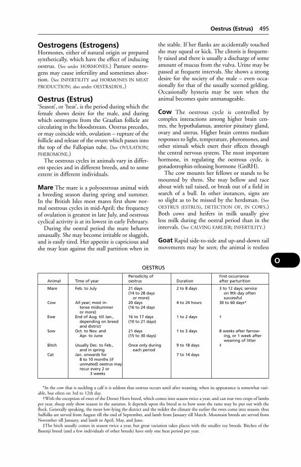
Oestrogens (Estrogens)Hormones, either of natural origin or preparedsynthetically, which have the effect of inducingoestrus. (See under HORMONES.) Pasture oestro-gens may cause infertility and sometimes abor-tion. (See INFERTILITY and HORMONES IN MEAT
PRODUCTION; also under OESTRADIOL.)
Oestrus (Estrus)‘Season’, or ‘heat’, is the period during which thefemale shows desire for the male, and duringwhich oestrogens from the Graafian follicle arecirculating in the bloodstream. Oestrus precedes,or may coincide with, ovulation – rupture of thefollicle and release of the ovum which passes intothe top of the Fallopian tube. (See OVULATION;PHEROMONE.)
The oestrous cycles in animals vary in differ-ent species and in different breeds, and to someextent in different individuals.
Mare The mare is a polyoestrous animal witha breeding season during spring and summer.In the British Isles most mares first show nor-mal oestrous cycles in mid-April; the frequencyof ovulation is greatest in late July, and oestrouscyclical activity is at its lowest in early February.
During the oestral period the mare behavesunusually. She may become irritable or sluggish,and is easily tired. Her appetite is capricious andshe may lean against the stall partition when in
the stable. If her flanks are accidentally touchedshe may squeal or kick. The clitoris is frequent-ly raised and there is usually a discharge of someamount of mucus from the vulva. Urine may bepassed at frequent intervals. She shows a strongdesire for the society of the male – even occa-sionally for that of the usually scorned gelding.Occasionally hysteria may be seen when the animal becomes quite unmanageable.
Cow The oestrous cycle is controlled by complex interactions among higher brain cen-tres, the hypothalamus, anterior pituitary gland,ovary and uterus. Higher brain centres mediateresponses to light, temperature, pheromones, andother stimuli which exert their effects throughthe central nervous system. The most importanthormone, in regulating the oestrous cycle, isgonadotrophin-releasing hormone (GnRH).
The cow mounts her fellows or stands to bemounted by them. She may bellow and raceabout with tail raised, or break out of a field insearch of a bull. In other instances, signs are so slight as to be missed by the herdsman. (See
OESTRUS (ESTRUS), DETECTION OF, IN COWS.)Both cows and heifers in milk usually give less milk during the oestral period than in theintervals. (See CALVING EARLIER; INFERTILITY.)
Goat Rapid side-to-side and up-and-down tailmovements may be seen; the animal is restless
Oestrus (Estrus) 495
OOESTRUS
Periodicity of First occurrenceAnimal Time of year oestrus Duration after parturition
Mare Feb. to July 21 days 2 to 8 days 3 to 12 days; service(14 to 28 days on 9th day often
or more) successfulCow All year; most in- 20 days 4 to 24 hours 30 to 60 days*
tense midsummer (16 to 24 daysor more)
Ewe End of Aug. till Jan., 16 to 17 days 1 to 2 days †depending on breed (10 to 21 days)and district
Sow Oct. to Nov. and 21 days 1 to 3 days 8 weeks after farrow-Apr. to June (15 to 30 days) ing, or 1 week after
weaning of litterBitch Usually Dec. to Feb., Once only during 9 to 18 days ‡
and in spring each periodCat Jan. onwards for 7 to 14 days
8 to 10 months (ifunmated) oestrus mayrecur every 2 or
3 weeks
*In the cow that is suckling a calf it is seldom that oestrus occurs until after weaning, when its appearance is somewhat vari-able, but often on 3rd to 12th day.
†With the exception of ewes of the Dorset Horn breed, which comes into season twice a year, and can rear two crops of lambsper year, sheep only show season in the autumn. It depends upon the breed as to how soon the rams may be put out with theflock. Generally speaking, the more low-lying the district and the milder the climate the earlier the ewes come into season; thusSuffolks are served from August till the end of September, and lamb from January till March. Mountain breeds are served fromNovember till January, and lamb in April, May, and June.
‡The bitch usually comes in season twice a year, but great variation takes places with the smaller toy breeds. Bitches of theBasenji breed (and a few individuals of other breeds) have only one heat period per year.
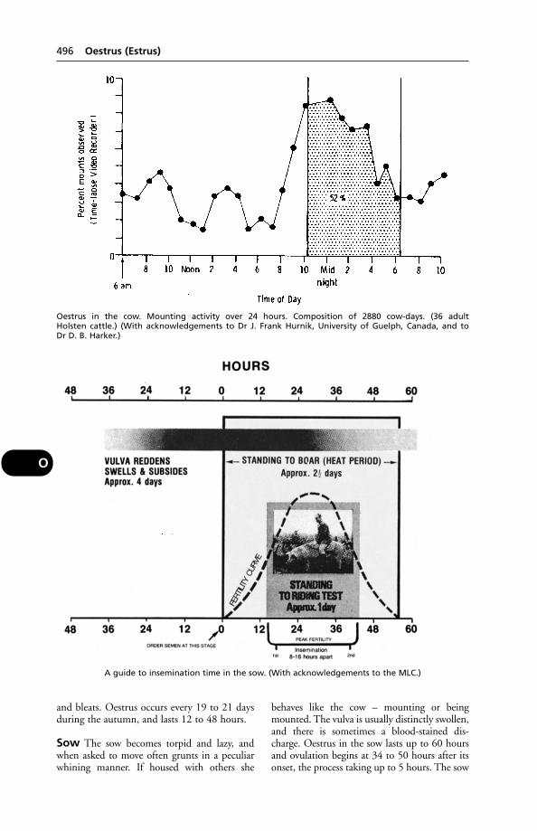
and bleats. Oestrus occurs every 19 to 21 daysduring the autumn, and lasts 12 to 48 hours.
Sow The sow becomes torpid and lazy, andwhen asked to move often grunts in a peculiarwhining manner. If housed with others she
behaves like the cow – mounting or beingmounted. The vulva is usually distinctly swollen,and there is sometimes a blood-stained dis-charge. Oestrus in the sow lasts up to 60 hoursand ovulation begins at 34 to 50 hours after itsonset, the process taking up to 5 hours. The sow
496 Oestrus (Estrus)
O
Oestrus in the cow. Mounting activity over 24 hours. Composition of 2880 cow-days. (36 adult Holsten cattle.) (With acknowledgements to Dr J. Frank Hurnik, University of Guelph, Canada, and to Dr D. B. Harker.)
A guide to insemination time in the sow. (With acknowledgements to the MLC.)

will accept service between 15 and 35 hours afterthe onset of oestrus, with the optimum at 25 to35 hours.
Bitch She wanders away from home unlessconfined, and the odour of her blood-stainedvaginal discharge attracts male followers. Asbleeding from the vulva is slight in some bitch-es, especially at their 1st oestrus, owners shouldwatch for swelling of the vulva. During the 7 to9 days of pro-oestrus, the bitch will flirt with a dog but not accept him. Usually it is only during the last week of heat that the bitch willaccept the dog, usually between the 10th and12th days.
Cat The signs may suggest pain and/or a strongdesire to have her back and flanks rubbed orscratched. She will roll over and over on carpetor floor, rub herself against furniture, etc., andutter little pleased mews.
The 1st oestrus may be expected between theages of 6 and 8 months: however, it may occur asearly as 31⁄2 months, or occasionally be delayeduntil the queen is about a year old.
Oestrus (Estrus), Detection of,in CowsEspecially in winter, detection of oestrus is not aseasy as might be thought. Studies in the USA sug-gest that where cows are watched 4-hourly roundthe clock, the efficiency of heat-detection shouldbe around 95 per cent, but in a herd where cowsare seen only twice a day, the percentage is likelyto drop to around 74 per cent.
Other studies suggest that those figures maybe over-optimistic, however; in Britain, the ratemay be as low as 55 per cent on some farms.
The main sign of oestrus in a heifer or cow isstanding still to be ridden by others (‘bulling’).
Part of the problem is that while ‘bulling’ lasts12 hours on average, it may last only 1 hour;and as to the timing, 50 per cent of the displaysoccur at night. Moreover some cows may standonly once in 20 minutes; others will stand onlyfor favourites; and some aggressive cows mountother cattle at a crowded trough in order toinduce them to move aside to create a space.
Sometimes a cow is seen to mount anotherfrom the front. This is valuable evidence ofoestrus, but it is important to remember that itis the riding cow which is bulling, not the oneunderneath.
The importance of pinpointing heat dates can-not be overemphasised. Only by record-keepingis it possible to identify animals that are not coming in heat at the normal time, in addition tothose which are cycling (coming in heat) irregu-larly. Delay in seeking veterinary advice may leadto delay in conception.
As an aid to herd management, a VASEC-
TOMISED bull may be used, or a heat-detectiondevice may be placed on a cow’s back, liberatinga dye when she is mounted. Applying paint tothe tail which is rubbed off by mounting is alsouseful.
Oestrus (Estrus), Suppression ofBitches and cats may be prevented from coming‘on heat’ by oral dosing with the syntheticequivalent of the naturally occurring hormone,progesterone; MEGESTROL ACETATE (Ovarid) isan example. Synthetic progestogens are alsoused to lessen aggressive behaviour in dogs andspraying in cats.
Some progestogens can cause pathologicalchanges in the uterus, particularly if used for pro-longed periods. They may induce abnormal levelsof growth hormone, suppress cortisol levels, andpossibly increase the risk of mammary tumours.
Oestrus (Estrus), Suppression of 497
O
(With acknowledgements to the East of Scotland College of Agriculture.)
OffalThe practice of incorporating offal – animalorgans – in cattle feeds was banned in 1988because of the risk of transmitting BOVINE
SPONGIFORM ENCEPHALOPATHY.
Offals MiddlingsA high-protein feed supplement for cattle (see
WEATINGS).
Office International DesEpizooties (OIE)Office International Des Epizooties (OIE) wasset up in 1924 following the realisation that jointaction between countries was necessary to con-trol contagious animal diseases. It determines theanimal health standards for international trade,advises the veterinary services in member coun-tries, and aims to work towards the eradicationof the most dangerous animal and zoonotic dis-eases. The membership comprises 144 countriesand international organisations such as the UN’sFood and Agriculture Organisation and theWorld Health Organisation.
Oie(see OFFICE INTERNATIONAL DES EPIZOOTIES)
Old English SheepdogIts very shaggy, grey and white coat is the distin-guishing feature of this large dog. Wobbler syn-drome (cervical spondylolithesis) and cataractmay be inherited. Hip dysplasia, deafness andosteochondritis dessicans may also be found inthe breed.
Oilfield Hazards, PoisoningIn the USA about 500 cases of suspected poi-soning by oilfield wastes are investigated eachyear at the Oklahoma animal disease diagnosticlaboratory.
Hazards arise from the ingestion by cattle ofpetroleum hydrocarbons, salt water, heavy met-als, chemicals stored on site, and rubbish such as discarded soda bags. A quantity of lead-basedpipe-jointing material is used, and also chemicalsto treat the mud which lubricates the drilling bit.
Signs of poisoning include weight loss andunthriftiness. A differential diagnosis has to takeinto account the possibility of internal parasites,faulty nutrition or other causes of debility; butstandard analytical methods make it relativelyeasy to detect the ingested poisons.
The presence of petroleum in lung tissue andin rumen contents is frequently confirmed. Liverand kidney lesions may be found.
OilsOils are divided into fixed oils, which are of the nature of liquid fats, and are derived byexpression from nuts, seeds, etc.; and volatile oressential oils, which are obtained by distillation.Examples are the oils of aniseed, cajaput, euca-lyptus, peppermint, and turpentine. (See also
PARAFFIN.)
Oilseed RapeHorses grazing in fields adjacent to this crop areat risk of developing respiratory disease.
OldenburgA breed of sheep native to the Hamburg Marshes,Germany. Fleece weights up to 6.35 kg (14 lb)and high lambing percentages are claimed.
OlfactoryRelating to the sense of smell.
Olfactory Nerve (Nerve ofSmell)Olfactory nerve (nerve of smell) is the 1st of thecranial nerves.
OliguriaA diminution in the amount of URINE excreted.
Olive PomaceA by-product of the olive-oil industry that hasbeen used to replace wheat bran in cattle dietsand barley in sheep diets. Pre-treatment withsodium hydroxide helps to delignify the productand improve palatability.
Ollulanus(see CATS, WORMS IN)
Omasum (‘Many-Plies’)Omasum (‘many-plies’) is the name given tothe 3rd stomach of ruminants. It is situated onthe right side of the abdomen at a higher levelthan the 4th stomach and between this latterand the 2nd stomach, with both of which itcommunicates. From its inner surface projectlarge numbers of leaves or folia, each of whichpossesses roughened surfaces. In the centre ofeach folium is a band of muscle-fibres whichproduces a rasping movement of the leaf whenit contracts. One leaf rubs against those oneither side of it, and large particles of foodmaterial are ground down between the roughsurfaces, preparatory to further digestion in thesucceeding parts of the alimentary canal.
Studies at the ARC’s National Institute forResearch in Dairying have shown ‘massiveexchanges of water and solutes in the omasum
498 Offal
O
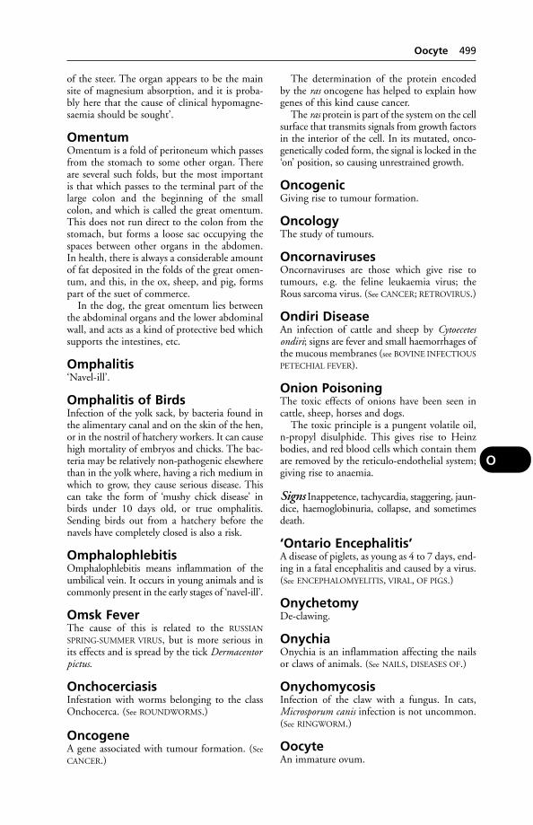
of the steer. The organ appears to be the mainsite of magnesium absorption, and it is proba-bly here that the cause of clinical hypomagne-saemia should be sought’.
OmentumOmentum is a fold of peritoneum which passesfrom the stomach to some other organ. Thereare several such folds, but the most important is that which passes to the terminal part of thelarge colon and the beginning of the smallcolon, and which is called the great omentum.This does not run direct to the colon from thestomach, but forms a loose sac occupying thespaces between other organs in the abdomen.In health, there is always a considerable amountof fat deposited in the folds of the great omen-tum, and this, in the ox, sheep, and pig, formspart of the suet of commerce.
In the dog, the great omentum lies betweenthe abdominal organs and the lower abdominalwall, and acts as a kind of protective bed whichsupports the intestines, etc.
Omphalitis‘Navel-ill’.
Omphalitis of BirdsInfection of the yolk sack, by bacteria found inthe alimentary canal and on the skin of the hen,or in the nostril of hatchery workers. It can causehigh mortality of embryos and chicks. The bac-teria may be relatively non-pathogenic elsewherethan in the yolk where, having a rich medium inwhich to grow, they cause serious disease. Thiscan take the form of ‘mushy chick disease’ inbirds under 10 days old, or true omphalitis.Sending birds out from a hatchery before thenavels have completely closed is also a risk.
OmphalophlebitisOmphalophlebitis means inflammation of theumbilical vein. It occurs in young animals and iscommonly present in the early stages of ‘navel-ill’.
Omsk FeverThe cause of this is related to the RUSSIAN
SPRING-SUMMER VIRUS, but is more serious inits effects and is spread by the tick Dermacentorpictus.
OnchocerciasisInfestation with worms belonging to the classOnchocerca. (See ROUNDWORMS.)
OncogeneA gene associated with tumour formation. (See
CANCER.)
The determination of the protein encodedby the ras oncogene has helped to explain howgenes of this kind cause cancer.
The ras protein is part of the system on the cellsurface that transmits signals from growth factorsin the interior of the cell. In its mutated, onco-genetically coded form, the signal is locked in the‘on’ position, so causing unrestrained growth.
OncogenicGiving rise to tumour formation.
OncologyThe study of tumours.
OncornavirusesOncornaviruses are those which give rise totumours, e.g. the feline leukaemia virus; theRous sarcoma virus. (See CANCER; RETROVIRUS.)
Ondiri DiseaseAn infection of cattle and sheep by Cytoecetesondiri; signs are fever and small haemorrhages ofthe mucous membranes (see BOVINE INFECTIOUS
PETECHIAL FEVER).
Onion PoisoningThe toxic effects of onions have been seen incattle, sheep, horses and dogs.
The toxic principle is a pungent volatile oil,n-propyl disulphide. This gives rise to Heinzbodies, and red blood cells which contain themare removed by the reticulo-endothelial system;giving rise to anaemia.
Signs Inappetence, tachycardia, staggering, jaun-dice, haemoglobinuria, collapse, and sometimesdeath.
‘Ontario Encephalitis’A disease of piglets, as young as 4 to 7 days, end-ing in a fatal encephalitis and caused by a virus.(See ENCEPHALOMYELITIS, VIRAL, OF PIGS.)
OnychetomyDe-clawing.
OnychiaOnychia is an inflammation affecting the nailsor claws of animals. (See NAILS, DISEASES OF.)
OnychomycosisInfection of the claw with a fungus. In cats,Microsporum canis infection is not uncommon.(See RINGWORM.)
OocyteAn immature ovum.
Oocyte 499
O

Oophorectomy(see SPAYING)
OophoritisOophoritis is another name for ovaritis orinflammation of an ovary.
Open Joints(see JOINTS, DISEASE OF)
‘Opening the Heels’‘Opening the heels’ means the cutting of thehorn at the angles of the heels of the horse’s foot,by which the continuity between the horn ofthe wall and of the bar on either side of the footis destroyed. It is performed by some farriersand owners in the hope that it will allow theheels to expand and so produce a ‘fine openfoot’. Actually, the operation results in an inter-ference with the shock-absorptive mechanism ofthe foot, and eventually produces contraction ofthe heels. It is by no means to be recommended.(See FOOT OF THE HORSE.)
OphthalmiaOphthalmia means inflammation of the wholeof the structures of the eye, but is sometimesrestricted to mean keratitis. Contagious oph-thalmia is caused by Rickettsia conjunctivaein sheep, and by Moraxella bovis in cattle.Verminous ophthalmia also occurs in cattle. (See
EYES, DISEASES OF.)
OphthalmoscopeOphthalmoscope is an instrument used for theexamination of the back of the eye.
OpioidsEndogenous opioids in the central nervous sys-tem, the encephalins and endorphins, are ableto modify the perception of pain.
OpisthotonosOpisthotonos is the position assumed by thebackbone during one of the convulsive seizuresof tetanus, and also sometimes seen duringepileptiform convulsions and strychnine poi-soning. The spinal column is markedly archedwith the concavity facing upwards away fromthe lower parts of the body, so that the head isdrawn backwards, and the tail and hind-parts ofthe body pulled forwards. The condition is dueto the spasmodic contraction of the powerfulmuscles lying above the vertebral column.
OpiumOpium is the dried milky juice of the unripeseed-capsules of the white Indian poppy, Papaver
somniferum. Good opium should contain about10 per cent of morphine, the chief alkaloid andactive principle. It also contains other alkaloids,the most important of which are codeine, narco-tine, thebaine, papaverine, apomorphine.
The preparations of opium used in veterinarymedicine are now virtually nil, but have includ-ed the following: (1) Powdered opium, which isthe dried juice powdered, contains about 9.5 to10.5 per cent morphine. (2) Tincture of opium,or laudanum, consists of the powder treatedwith distilled water and alcohol, and containsabout 1 per cent of morphine. (3) Opiumextracts, 1 dry of 20 per cent morphine, and 1 liquid of 3 per cent morphine, as well as afluid extract which contains about 5 per centmorphine. (4) Compound tincture of camphor,or paregoric. (5) Compound ipecacuanha pow-der, or Dover’s powder, contains 10 per cent of opium. (6) Gall and opium ointment, con-taining 7.5 per cent of opium, is used as anastringent ointment. (7) Compound tincture ofmorphine and chloroform which containedmorphine, chloroform, dilute prussic acid, aswell as Indian hemp and capsicum, is similar tothe proprietary mixtures which are calledchlorodyne. Morphine, codeine, apomorphine,heroin, and dionin are also preparations from orderivatives of opium. (See MORPHINE.)
OpsoninsSubstances present in blood serum which facilitate the engulfment of bacteria (and otherforeign proteins) by certain white cells. (See
PHAGOCYTOSIS.)
Optic NerveOptic nerve is the 2nd cranial nerve runningfrom the eye to the base of the brain. It conveysthe sensations of light that are received by theretina, and registers them in the optic centres ofthe brain. (See EYE, VISION.)
OrbitOrbit is the eye socket.
Orbital Gland(see HARDERIAN GLAND and EYE, DISEASES OF)
OrbivirusesThese cause African horse sickness, blue-tongue,and a haemorrhagic disease of deer.
OrchardsAnimals grazing in orchards may run the risk of poisoning if fruit-trees have recently beensprayed with insecticides or fungicides. Orchards,like paddocks, sometimes become a reservoir of
500 Oophorectomy
O

parasitic worm larvae. (See PADDOCKS; also ALCO-
HOL POISONING.)
OrchitisInflammation of the TESTICLE.
Oregon Muscle DiseaseA condition in turkeys and chickens in whichthe inner breast (deep pectoral) muscles becomenecrotic and greenish. The cause is possibly aninherited abnormality affecting the blood vessels.
OrfA disease of sheep, cattle, and goats which has avery wide distribution and many names. Amongits numerous designations are the following:‘ulcerative stomatitis’, ‘contagious pustular der-matitis’; ‘contagious ecthyma’; ‘necrobacillosis ofsheep’.
Orf is enzootic in the Border counties ofEngland and Scotland, but outbreaks may arisein any county in Britain, as well as in Germany,France, Austria, the USA, and other sheepcountries.
The disease attacks sheep of all ages, sexes, andbreeds, and kept under all conditions of manage-ment. It frequently attacks lambs just before orafter weaning, or after docking or castration, andfrom them it may spread to the teats of the ewes.In other cases it is common among gimmersuntil they are 1 year old.
Causes Essentially, a parapoxvirus; but secon-darily Fusiformis necrophorus (Fusobacterium).The virus is needed to produce pox-like lesionsfirst, which the necrosis organism then invades.
Signs In the milder form of the disease, vesicles,followed by ulcers, appear on the lips – especiallyat the corners of the mouth. Sometimes healingtakes place uneventfully; in other cases verrucosemasses form and persist. The animal loses weight.
In the severe form the inside of the mouthbecomes involved in most cases, and in additionother parts of the body such as the vulva and theskin of the face, legs, tail, etc. A greyish-blackcrust often appears which, if removed, leaves araw, angry-looking surface.
Sheep with lesions on the head frequently rubtheir muzzles on their fore-feet, or scratch attheir heads with their hind-feet. In this way thefeet and legs often become affected. Abscessesmay form in the region of the coronet. Thesheep becomes extremely lame, so much so thatit is frequently unable to put the affected leg tothe ground, and hobbles about on 3 legs. If bothfore-feet are affected – which is commonly thecase – the animal may be observed feeding from
a kneeling position. In severe cases the horn separates from the sensitive structures below,large quantities of foul-smelling thick pus areproduced, and the hoof may be shed. The spacebetween the claws, and the parts around thefront and sides of the coronets, are the com-monest situations of the lesions.
Less commonly the external genitals of bothmale and female are affected. (See also PENIS AND
PREPUCE – Balanoposthitis.)After 550 apparently healthy 5-month-old
lambs had been transported over a period of 23 hours, a severe outbreak developed and 10 per cent of the lambs died. The outbreakwas attributed to spread of the virus from anaffected animal in the confined space inside thetruck.
Treatment As soon as a case of orf appearsamong a flock of sheep, it should be isolated atonce. Isolated sheep that are already affectedusually do best when they can be shut upindoors, given hand-feeding, and provided withclean dry litter. A dressing is applied over theraw ulcerated area and around its margin.Crystal violet is very suitable as a dressing, andantibiotics are useful in treatment. Cryosurgerymay be helpful.
On farms previously heavily infected, andwhere orf was very common on the feet, pass-ing the whole of the sheep through a foot-bathat 3-weekly intervals has resulted in a completedisappearance of the disease. (See FOOT-BATHS
FOR SHEEP.)Orf is well recognised as an occupational
hazard of shepherds.
Control A modified live vaccine is available; itis applied by scarifications. The resulting scabscan be a source of infection when they detach.
Orf in the dog Outbreaks of orf in houndsand sheepdogs are not unknown. They arecharacterised by circular areas of acute inflam-mation, with a moist appearance, ulcerationand scab formation.
Public health In one 5-year period there were344 laboratory reports of patients with orflesions in Britain. Contact with live sheep orlambs was reported 142 times. In 49 cases thepeople affected were abattoir workers, butchers,or domestic meat-handlers. The possible sourcein 36 patients (including 13 milkers) was con-tact with cows or calves. Sixteen patients werefarmers; 7 were veterinary surgeons or veterinarystudents. (Communicable Disease SurveillanceCentre.)
Orf 501
O
Severe mouth lesions have been successfullytreated by DIATHERMY and CRYOSURGERY.
OrganellesSpecialised structures within a CELL.
Organic DiseasesOrganic diseases, as distinct from ‘functionaldiseases’, are those in which some actual alter-ation in structure takes place, as the direct resultof which faulty action of the organ or tissueconcerned follows.
Organochlorine Poisoning(see CHLORINATED HYDROCARBONS)
Organophosphorus PoisoningThis may arise from contamination of crops, or other food material, with organophosphorusinsecticides such as dimethoate, schradan,parathion or dimefox, or by skin contact orinhalation.
Signs are varied but include salivation, mus-cle tremors, slow heartbeat, constricted pupils,swaying gait, and recumbency.
For a case of laryngeal paralysis arising fromorganophosphorus poisoning of racehorses, see
under LARYNX, DISEASES OF.
Treatment Atropine sulphate given intra-venously or intramuscularly, and repeated in 30 minutes. Barbiturates may be needed tocontrol excitement. Oxygen for distressedbreathing, and gastric lavage are recommendedin the human subject. In the latter, PAM hasbeen recommended as an antidote to parathionand other insecticides in this group – in conjunction with atropine.
Protective clothing must be worn when dipping sheep in organophosphorus dips; onlyproperly trained operatives may use sheep dips(see DIPS AND DIPPING).
Orifices, Immunity atDefence mechanisms, directed against the inva-sion of pathogenic bacteria, exist in the naturalorifices of the body. For example, research atIRAD, Compton, led to the isolation of a num-ber of cationic proteins from the keratin of theteat-canal’s lining, and these have been shownto inhibit the growth of mastitis strains ofstaphylococci and streptococci. These proteins,which are soluble in distilled water and carry a positive electrical charge, were shown toinhibit the growth of 2 strains of Staphylococcusaureus and 1 strain of Streptococcus agalactiae.The proteins in very low concentration causeda 50 per cent mortality in test bacterial cultures.
The secretions of the uterine cervix of thecow during oestrus also contain cationic pro-teins which possess antibacterial activity againststaphylococci. In the laboratory these proteinswere shown also to inhibit growth of Brucellaabortus.
The anionic proteins from the cervicalmucus, however, showed no inhibitory actionon the bacteria. This difference ‘suggested thatthe killing of the bacteria was preceded by anelectrovalent binding of the positively chargedcationic protein on to the negatively chargedsurface of the bacteria’, and this has proved tobe the case.
Antibacterial cationic proteins have also beenisolated from cells normally present in cow’smilk, and research has shown that synthesis ofthese proteins can be stimulated. Induction of amild sterile mastitis by the injection of E. coliendotoxin through the teat-canal led to increasednumbers of neutrophils in the milk from whichwas extracted cationic proteins with a higherantibacterial activity.
Ornamental FishOrnamental fish are widely kept in the UK. Thespecies vary considerably and, therefore, so dotheir environmental requirements. A general ruleis that when changing water, not all the old watershould be removed; about a third should be left (unless advised otherwise in certain circum-stances, such as where a disease is present). Thereis evidence that fish may secrete, in the mucouscovering their skin, substances with antibioticactivity. Beneficial bacteria will also have becomeestablished in the water and enough should beleft to re-establish numbers in the tank.
Stress is a factor in most diseases of fish.Imported fish can undergo considerable stressduring catching and transport before reachingthe hobbyist. Ulceration of the skin is notuncommon in goldfish and koi; the bacteriaresponsible vary. As with farmed fish, protozoalinfestations by Ichthyophthirius (white spot),Trichonodina, Costia, Chilodonella, Scyphidia,etc, can occur. Gill flukes may also present aproblem: Sapreolegnia is usually a complicatinginfection.
When fish are kept in ponds, great care must be taken to prevent spindrift from gardenchemicals contaminating the water.
OrnithosisThe name formerly given to Chlamydophilapsittaci infection in birds other than those of theparrot family. C. psittaci infections in all birds arenow designated as psittacosis. (See CHLAMYDIA
AND CHLAMYDOPHILA.)
502 Organelles
O
Orthopox VirusesThis genus contains those pox viruses genetical-ly and antigenetically related to smallpox virus.(See table under VIRUSES.)
OSThe Latin word for a bone. Examples: Os cordis,a bone (1 of 2) present in the hearts of cattle; Ospenis in the dog.
OssificationOssification means the formation of bone tissue. In early life the bones are represented bycartilage or fibrous tissue, and in these, centresappear in which the cells undergo a change andlime salts are deposited. This process proceedsuntil the areas or centres meet each other, andthe tissue is wholly converted into bone. Whena fracture occurs, the bone unites by ossificationof the blood-clot which forms between the broken ends of the bone. (See FRACTURES.) Inold age, ossification takes place in parts wherenormally there are cartilages found, such as inthe larynx, in the rib-cartilages, in the scapularcartilages, etc., and these parts lose their normalelasticity and become easily broken. (See
SIDEBONES.)
Osteitis (Ostitis)Inflammation of bone (see BONE, DISEASES OF).
Osteoarthritis (Osteoarthrosis)Inflammation/degeneration of the bone at ajoint. In human medicine, the name ‘osteoarthro-sis’ is now preferred, emphasising the view thatthis is a degenerative rather than an inflammato-ry disease. The primary disturbance is usuallyregarded as occurring in articular cartilage, and as resulting from a combination of ageing andmechanical factors. An alternative hypothesis isthat the disease originates in the synovial liningcells (see ARTHRITIS).
OsteoblastsCells which assist in the formation of bone. (See also OSTEOCLASTS.)
OsteochondritisInflammation of bone and cartilage. (See HIP DYS-
PLASIA IN DOGS.) O. dissecans is characterised byseparation of a piece of articular cartilage which,together with a small piece of underlying bone,forms a loose body within a joint (see below).
Osteochondritis DissecansOsteochondritis dissecans is a disturbance in theendochondral ossification (conversion of cartilageinto bone) affecting the growth plates and the
articular cartilage. It is an inherited trait in somedog breeds but rapid growth in any animal maycause the condition. Clinical signs include lame-ness and abnormal gait. There is pain around theaffected joints. The stifle and hock may be moreseverely affected as they bear greater weight. Rest,and perhaps surgery, may be required.
OsteochondrosisAbnormal differentiation of cartilage. It is sim-ilar in some respects to OSTEOCHONDRITIS, andmay be a more accurate description in caseswhere there is no inflammatory response to thechanges in bone and cartilage. There may benecrosis of bone and separation of splinters orflaps of articular cartilage. A hereditary basis forthe condition has been recorded in man, horse,dog, and pig. In pigs, as many as 80 per centcan show lesions at slaughter.
In young horses, the most frequently diag-nosed conditions are O. dissecans and a sub-chondral bone cyst. These are 2 separate entities,though often bracketed together under theosteochondrosis syndrome. Severe lameness maybe caused by the former, and surgical treatmentneeded.
OsteoclastsCells which aid the breakdown or resorption of excess bony tissue, laid down following frac-tures, as part of the repair process. (See also
OSTEOBLASTS.)
Osteodystrophia FibrosaA degenerative condition of bone resulting fromover-activity of the parathyroid gland. Affectedanimals show lameness, pain, incoordination,paralysis, curvature of the spine, fractures andconstipation. Diagnosis is confirmed by X-ray.Treatment is by giving mineral supplements;steroids and sex hormones have also been used.
Osteodystrophic DiseasesOsteodystrophic diseases are conditions in whichthe metabolism of the bone is upset. This may becaused by mineral deficiencies or imbalances inthe diet or pasture.
Osteogenesis ImperfectaA failure of bone to develop properly in younganimals. Animals show incoordination, and painat the joints; they may be prone to greenstick or complete fractures. Rest is necessary; anti-inflammatory medication will relieve the pain.
OsteomalaciaOsteomalacia is the equivalent of rickets occur-ring in the adult animal. The bones become
Osteomalacia 503
O

softened as the result of the loss by absorp-tion of the salts they contain. The cause of thedisease is obscure, but it appears to be morecommon in pregnant females than in other animals, and it may be associated with a deficiency of vitamin D and/or phosphates. (See VITAMINS.)
The most serious feature is the deformitywhich occurs in the softened bones, owingeither to the weight of the body or to the pullof the muscles upon them. When the deformi-ty is located in the pelvis of the dam, great dif-ficulty is often experienced at the birth of theyoung animal, and fractures of this part are notunknown.
Treatment Vitamin D and good nourishingfood with an adequate phosphate content.
OsteomyelitisInflammation and infection of the bone mar-row. It is sometimes a complication of atrophicrhinitis of pigs and of actinomycosis of cattle.(See BONE, DISEASES OF.)
OsteopeniaA reduction in the body’s bone tissue.
Osteopetrosis (‘Marble Bone’Disease)Osteopetrosis (‘marble bone’ disease) is charac-terised clinically by thickening of the legs ofpoultry (see illustration, below). On post-mortem examination, involvement of otherbones is seen. The cause is a virus of the avianleucosis group.
OsteophagiaOsteophagia means bone-eating, and is a symp-tom shown by sheep and cattle in certain partsof South Africa where soil and herbage are defi-cient in phosphorus and sometimes in calcium.(See LAMZIEKTE.)
Deer living wild in forests where there is asimilar deficiency, as in many parts of theScottish Highlands, exhibit osteophagia bychewing and actually eating portions of shedantlers. Sheep exhibit similar tendencies in thesame areas.
504 Osteomyelitis
O
Two chicken skeletons at the Regional Poultry Research Laboratory at East Lansing, Michigan. One (left)is the skeleton of a 216-day-old normal White Leghorn cockerel. The other is the skeleton of a 202-day-old White Leghorn cockerel affected with osteopetrosis, a disease that causes an enlargement andhardening of the bones. (With acknowledgements to USDA, photograph by Madeleine Osborne.)
OsteoporosisOsteoporosis is a rarefying condition of boneswhich lose much of their mineral matter andbecome fragile. It can occur in young animalsthrough deficiency of protein in the diet, and inolder animals through lack of exercise. In oldanimals appropriate sex hormones may be given;in younger ones the diet must be checked. Extrathiamine (vitamin B1) may be useful.
OsteosarcomaA malignant cancer of the bone. Some largebreeds of dog appear to have a predispositiontowards the disease.
OstertagiasisInfestation with species of Ostertagia worms,which produce gastroenteritis. It is seen incalves and lambs. This is an important diseasein Ireland. (See WORMS, FARM TREATMENT
AGAINST.)
Ostrich (Struthio Camelus)Ostriches, once farmed extensively for their tailfeathers, are now farmed mainly for their meat,which is low in cholesterol, although – thefeathers, leather and eggs are also marketed.
An adult male may be up to 2.75 m (9 ft)tall, and weigh 135 kg (300 lb). Ostriches canbe dangerous and have been known to disem-bowel people when angry. That is why 2 areasof the drawing have been shaded – to indicatesites for the administration of IMMOBILON bymeans of a PROJECTILE SYRINGE.
Ostriches cannot fly, but they can runextremely fast.
Ostriches are included under the DangerousWild Animals Act. In the UK, the melon-sizedeggs are usually used for hatching only.
Diseases Among infectious conditions thathave been reported are the following:
Newcastle disease in the ostrich usuallyaffects the nervous system. Avian influenza cancause severe mortality, particularly amongyoung birds. Ostriches are susceptible to fowlpox; vaccination is recommended where thisdisease is prevalent. Clostridium perfingens typeC can be a problem; vaccination should alsocover other types of this organism. Clostridiumchauvoei has been implicated in a conditionresulting in partial paralysis and inability to urinate. Unusually for birds, ostriches are sus-ceptible to anthrax; they can also be infectedwith avian tuberculosis, E. coli, salmonella and klebsiella, among other micro-organisms.Aspergillosis and candidiasis have been report-ed. Houttuynia struthionis is a tapeworm specif-ic to the ostrich, imported into the UK withbirds from Namibia. Nematodes specific to theostrich include the wireworm Libyostrongylusdouglassi (not in the UK as at 2004),Codiostomum struthionis and Dicheilonema spir-ularium. Feather lice are usually of the speciesStruthioleupiris struthionis and quill mites of thePterolichidiae may be found. Occasionally ticks(Ambyloma, Hyalomma or Rhipicephalus can bepresent. Borna disease has affected ostriches,causing paresis.
OtitisOtitis means inflammation of the ear. (See EAR,DISEASES OF.)
OtodectesMites which cause ear mange in dogs and cats.(See MITES.)
OtorrhoeaOtorrhoea means a discharge from the ear. (See EAR, DISEASES OF.)
OtterhoundA large dog with pendulous ears and a long, oilycoat. It is liable to an inherited disorder of theblood platelets.
‘Oulou Fato’A form of rabies occurring among dogs in parts of Africa, and probably Asia also. Peopleare rarely bitten, epidemics are uncommon;infected dogs may show either no symptoms, or transient symptoms followed by recovery.Repeated attacks prove fatal, however.
‘Oulou Fato’ 505
O
Darting sites suggested for ostriches.
Ovarid(see MEGESTROL ACETATE; OESTRUS, SUPPRESSION
OF)
OvariesThe female reproductive organs. They are sus-pended in a fold of peritoneum from the roof ofthe abdomen, called the ‘mesovarium’. In themare they are situated in the abdomen, lying alittle below and behind the kidneys, usually incontact with the muscles of the lumbar region.Each possesses a groove which gives the organ ashape not unlike a bean, and which is called theovulation fossa. It is into this groove that the ripeova escape from the ovary, and it is the only partcovered by germinal epithelium in the mare. Inthe cow the ovaries are oval in outline and pos-sess no fossa. Each is situated about half-way upthe shaft of the ilium of the corresponding sideof the body. The ovaries of the sow are usuallysituated in a position similar to those of thecow, but their position changes somewhat afterbreeding has occurred. They are studded uponthe surface with irregular prominences, so thatthe organs present a mulberry-like appearance,and are enclosed in a ‘purse’ of peritoneum. In the bitch the ovaries are situated in closeproximity to, if not in actual contact with, thekidneys of the respective sides.
Structure Each ovary is composed of a stro-ma of dense fibrous tissue in whose spaces arenumerous blood vessels, especially towards thecentre. On the surface of the organ is a layer of germinal epithelium from which arise theGraafian follicles. These vary very much in size: when young they are microscopic, and lieimmediately under the outer surface, but asthey grow older they become more and moredeeply situated, and finally, as ripening occurs,they once more come to the surface. Growth orripening of a follicle occurs following stimula-tion of the ovary by the follicle-stimulating hor-mone (FSH) from the pituitary gland. The follicle produces, also as the result of FSH,oestrogens which prepare the uterus, Fallopiantubes and vagina for the other processes ofreproduction. In a ripening Graafian folliclethere is 1 (rarely 2) of the essential female germcells, called an ovum. This is situated at the pinnacle of a mass of cells which projectinwards from the inner surface of the follicle,and which is known as the cumulus.
Function When the follicle is ripe, a processknown as ovulation occurs, in which the outersurface wall of the follicle ruptures and liberatesthe contained ovum, which escapes from the
ovary. The ovum is caught by the oviduct, and either fertilised or passed on through thefemale system to the outside. The cavity of the Graafian follicle fills up afterwards withspindle-shaped cells, under the influence of theluteinising hormone (LH) from the pituitary.LH becomes more plentiful as FSH becomesless so, and the structure is called the CORPUS
LUTEUM or yellow body.If an ovum is fertilised, resulting in pregnancy,
the corpus luteum persists and secretes proges-terone, a hormone necessary for the maintenanceof pregnancy.
If the animal does not become pregnant, thecorpus luteum breaks down and disappears.(Occasionally, however, it fails to do so, andmay then cause infertility, especially in the dairycow .) (See also under CYSTS and below for cysticovaries – leading often to NYMPHOMANIA.) (See
also OESTRUS and diagram under UTERUS.)
Ovaries, Diseases ofIn cystic degeneration, large cavernous cystsappear in the substance of the organ, and fillwith fluid. For a time there are no definitesymptoms shown, but after the cysts attain considerable size the animal begins to exhibitsigns of fretfulness and excitability. As time goeson these symptoms increase in violence until inthe mare, in which the condition is quite com-mon, it usually becomes dangerous to work her.Upon the slightest provocation, and often withno provocation at all, the mare starts to kick.After her bout of kicking is over she resumesher normal behaviour, but another attack maycome on at any time afterwards.
Cysts are also met with in cows where theymay be associated with sterility, and in bitcheswhere they are frequently present along withtumour formation in the mammary glands.They are recognised as a cause of sterility in gilts– heat periods being irregular and the clitorisbecoming enlarged. Hypoplasia of the ovariesmay also occur.
(See NYMPHOMANIA; also under INFERTILITY.)
Ovario-HysterectomySurgical operation for removal of the uterus and ovaries. This is carried out in the dog andcat in cases of pyometra, and following dys-tokia where a recurrence is feared. It is the usualtechnique for spaying, especially of cats to prevent the birth of unwanted kittens. (See also
SPAYING.)
OvariotomySurgical operation for removal of a diseasedovary. (See also SPAYING.)
506 Ovarid
O

Overgrown FootOvergrown Foot is one in which the horn of thewall all the way round has continued to growdownwards and outwards, without any com-pensatory wear along its lower edge. A horsewith overgrown feet which may arise eitherfrom overlong periods between successive shoe-ings, or from living on marshy land where theunshod foot gets no wear, is unable to walk cor-rectly. The frog does not reach the ground, thetoe is too long, and the heels are too high, sothat the normal anti-concussion mechanism ofthe foot is thrown out of action. The conditionpredisposes to the occurrence of sprains andcontractions of tendons, upright pasterns, andsplitting of the horn, with the production ofsandcracks as a consequence. Horses’ feet thatare shod should have the shoes removed at leastonce a month, and the growth since the lastshoeing should be removed by rasping thelower edge of the wall. Young colts, running outat grass, should have their feet properly reducedat least once during every 2 months or so.Overgrown foot is of importance in cattle andsheep, and in animals confined in zoos.
OverlyingOverlying by the sow is one cause of PIGLET
MORTALITY and can be prevented by the use offarrowing crates, rails, and the roundhouse. Itshould be remembered, however, that an illpiglet is more likely to be crushed by the sowthan a healthy one; and it has been shown thatafter 1 hour in an environmental temperatureof 1.6° to 4.4°C (35° to 40°F) a piglet becomescomatose. (See under ROUNDHOUSE for an effec-tive means of preventing overlying.)
Over-ReachingA problem seen in horses when the toe of ahind leg strikes the back of the front leg on the same side. The injury caused is called an‘over-reach’ or ‘strike’. It usually occurs whenthe animal is travelling at a gallop; it is also seenin trotters and when jumping of rising ground.Special shoes, designed to hasten the breakoverof the front feet and delay that of the hind, mayprevent the problem.
OverstockingThe term refers to an excess of grazing animalson a given acreage of pasture. (See STOCKING
RATES.) It is also used to describe the practice ofleaving a cow unmilked in order to increase thesize of the udder and impress potential buyers.It causes great distress to the animal and is ille-gal under the Welfare of Animals in MarketsOrder 1990. Both the owner of the cow and theoperators of the market can be prosecuted ifsuch a cow is exposed for sale.
Oviduct(see FALLOPIAN TUBES; SALPINGITIS; EGG-BOUND;PROLAPSE OF OVIDUCT)
Ovine Encephalomyelitis(see LOUPING-ILL)
Ovine Enzootic Abortion(see ABORTION, ENZOOTIC, OF EWES)
Ovine EpididimytisOvine epididimytis caused by Brucella ovis is aNOTIFIABLE DISEASE throughout the EU. It is ofconsiderable importance in Australia and NewZealand. (See RAM; BRUCELLOSIS.)
Ovine Interdigital Dermatitis(OID)This has been described in foot-rot free flocks inAustralia, and is caused by Fusiformis necrophorus.(See also SCALD and SCAD.)
Ovine Keratoconjunctivitis(OKC)The name for a group of infectious eye diseasesof sheep. (See also EYE, DISEASES OF.)
OvulationIn the mare, cow, ewe, sow, and bitch, ovula-tion has no relation to coitus; whereas in thecat, ferret and rabbit it is coitus that determinesthe onset of ovulation. (See under OVARIES and
OESTRUS.)
Ovulation 507
O
Overgrown hoof, showing how much should becut away at the next shoeing.

OvumOvum is an egg cell. (See EMBRYOLOGY; OVARY;TRANSPLANTATION.)
OxfendazoleAn anthelmintic of the benzimidazole group.
Oxygen(see OZONE; AIR; RESPIRATION)Cylinders of oxygen are essential items of equip-ment for anaesthesia. They are fitted with a pres-sure gauge and a reducing valve. A flowmeter is incorporated in the anaesthetic circuit. (See
ANAESTHESIA.)Oxygen is used in the treatment of animals
rescued from burning buildings and sufferingfrom the effects of smoke inhalation.
Hyperbaric oxygen is that used at highpressures (e.g. 3 atmospheres) for the treatmentof carbon monoxide poisoning; and it has alsobeen used for gas gangrene in a dog.
Oxygen embolism is a potential dangerwhen hydrogen peroxide is syringed into a deepwound.
Oxygen Debt(see MUSCLE – Action of muscles)
OxytetracyclineAn antibiotic. (See TETRACYCLINES.)
OxytocinA hormone, secreted by the posterior pituitarygland, and also by the corpus luteum, whichactuates the ‘milk let-down’ mechanism; andalso stimulates contraction of the muscles of theuterus in late pregnancy.
OxyurisOxyuris is another name for the thread worm,which possesses a long finely-tapered tail. (See
ROUNDWORMS.)
OzaenaOzaena is a chronic inflammatory disease of thenasal passages. (See NOSE, DISEASES OF.)
OzoneThe chemically highly reactive allotropic formof oxygen, (O3). As a constituent of the upperatmosphere it forms a layer which protects people from excessive exposure to ultravioletradiation from the sun. Ozone may be the mainconstituent of smog.
It has been described as the most hazardousof all the gaseous air pollutants because of itslong-term association in laboratory animalswith emphysema, lung cancer, accelerated age-ing, increased neonatal deaths, decreased littersize, teratogenesis, and jaw anomalies. In ani-mals exposed to ozone the mortality from lunginfections is increased.
508 Ovum
O
PacemakerAn electronic device implanted under the skinof the chest which stimulates the heartbeat.
Pacemakers have been successfully implantedinto dogs which show an impaired conductionof the impulse that regulates the heartbeat, sothat a lack of coordination between the beatingof the atria and that of the ventricles occurs.
Following repeated episodes of loss of con-sciousness, which heart medication failed toobviate, a 2-year-old dachshund in Switzerlandhad a pacemaker electrode inserted into theright ventricle of its heart via the jugular vein.The batteries were inserted between abdominalmuscles. Local irritation from the batteryimplant was stated to be slight. Two years laterthe heart had given no further trouble.
A pacemaker was inserted in a pregnant mareat the Royal Veterinary College. The marefoaled normally and she and the foal fared well.
Pacheco’s DiseasePacheco’s disease is caused by a herpesvirus which is present in South American psittacines(parrots, parakeets). In the wild it does not appearto cause any harm. In aviaries, however, suddenonset of disease and death may occur, with up to100 per cent mortality. The birds become weakand have diarrhoea. Post-mortem examinationshows necrosis of the liver and spleen.
PachymeningitisInflammation of the dura mater of the brainand spinal cord. (See MENINGITIS.)
Pacinian Corpuscles(see under TOUCH, SKIN)
Packed Cell Volume(see under BLOOD – Composition)
PaddocksThese often become reservoirs of parasiticworm larvae – a point for animal-owners to bear in mind. Paddocks need ‘resting’ for 12 months, or grazing by a different species ofanimal, periodically.
Pain(For relief of pain, see ANALGESICS; ANAESTHE-
SIA.) Animals which are natural hunters
(predators) may cry out when suffering pain.Species which are, or were, normally huntedmay not do so because it would reveal that theyare injured or hiding. Thus, because an animaldoes not cry out, or show signs of restlessness, itshould not be assumed that it is free from pain.The clinical signs of pain can differ from speciesto species. Pain can be a cause of aggressiveness.The effect of pain can last longer than the painitself: it has been shown that as long as a monthafter foot pain has been corrected in cattle, cowsstill behave as if in pain, as the animal’s bodyhas not returned to normal. In sheep, this canlast even longer. As a result, bodily conditionand milk production may not return to normaluntil some time after the animal has made anapparent recovery.
Paint(see HOUSE DECORATING, POISONING; LEAD
POISONING, CAGE AND AVIARY BIRDS,DISEASES OF)
Palatability(see under DIET AND DIETETICS)
PalatePalate is the partition between the cavity of themouth below, and that of the nose above. Itconsists of the hard palate and the soft palate.The hard palate is formed by the bony floor ofthe nasal cavity covered with dense mucousmembrane, which is crossed by transverseridges in all the domesticated animals. Theseridges assist the tongue to carry the food backto the throat. The hard palate stretches back alittle beyond the last molar teeth in animals,and ends by becoming continuous with the softpalate. This latter is formed by muscles coveredwith mucous membrane, and in the horse actsas a sort of curtain between the cavity of themouth and that of the pharynx. Materialbrought up from the stomach must pass out byway of the nostrils. In racehorses, distressedbreathing may arise as the result of inflamma-tion or partial paralysis of the soft palate, whichmay be linked with paresis or paralysis of thevocal cords. Partial resection of the soft palatehas been carried out as treatment for this lattercondition. (See GUTTURAL POUCH DISEASE.)
Prolonged soft palate is a recognised inheritedabnormality of the short-nosed breeds of dogs,e.g. boxers, bulldogs, Pekingese, pugs, cockerspaniels. It makes breathing difficult at times,with snoring or even loss of consciousness result-ing. An operation to correct the condition isoften very successful.
P

Severe injury to the hard palate is notuncommonly seen in cats which have fallenfrom a height, and suturing may be required.
Pale Soft Exudative Muscle(PSE)(see PORCINE STRESS SYNDROME)
Palo Santo TreesThe leaves, fruit, and seeds cause poisoning in cattle in South America. Signs include tympany, depression, and convulsions.
PalpebralRelating to the eyelids.
Pan-Pan- is a prefix meaning all or completely.
PancreasPancreas is partly an endocrine gland, produc-ing hormones; and partly an exocrine gland,producing the pancreatic juice for digestivepurposes.
The pancreas is situated in the abdomen, alittle in front of the level of the kidneys and a little below them. When fresh it has a reddish-cream colour.
The pancreatic juice is secreted into the smallintestine to meet the food which has undergonepartial digestion in the stomach. The juice contains alkaline salts and at least 9 enzymes:e.g. trypsin, which carries on the digestion ofproteins already begun in the stomach; amylase,which converts starches into sugars; and lipase,which breaks up fats; as well as a substance thatcurdles milk. (See DIGESTION, ABSORPTION andASSIMILATION – Intestinal digestion.)
The pancreas also has groups of cells, theislets of Langerhans. (See INSULIN; DIABETES
MELLITUS; GLUCAGON; HORMONES.) Herealpha-cells produce glucagon, and beta-cells,insulin.
Pancreas, Diseases ofThese include DIABETES MELLITUS, inflamma-tion, suppuration, atrophy, tumour formation,etc. (See INSULINOMA.)
Acute pancreatitis occurs in obese dogs,more rarely in cats. Signs include abdominaltenderness or pain. Hyperglycaemia and shockmay follow. Treatment includes witholdingfood, and intravenous fluid therapy to maintainthe balance of fluid and electrolyte.
Exocrine pancreatic insufficiency indogs has 3 main causes: congenital hypoplasia,
degenerative pancreatic atrophy, and chronicpancreatitis. Signs include a ravenous appetite,loss of weight, fatty faeces, and a dry scurfycoat. Treatment includes supplementation ofthe diet with pancreatin as oral powder or granules. Cimetidine may be used in additionin severe cases.
Parasites which may be found in the pancre-atic ducts include Toxocara canis and, in cats in America, the pancreatic fluke Eurytrema procyonis. The latter may interfere with thegland’s exocrine function to a great extent.Fenbendazole is effective against the fluke.
PancreatinA preparation of the exocrine part of the pancreas used to treat pancreatic deficiency indogs and cats.
Pancreatitis(see PANCREAS, DISEASES OF)
PancytopeniaA reduction in the number of red cells, whitecells, and platelets in the blood; usually due toa bone-marrow dyscrasia.
PanhypopituitarismA condition caused by development failure ofthe pituitary gland, destruction of the pituitarytissue or a cyst or tumour in the gland. In puppies, the milk teeth and puppy coat areretained for longer than normal and the animals are stunted. There may be alopeciawith skin pigmentation. The gonads may beabnormally small. Polydipsia and polyuria maydevelop; affected animals may be aggressive.Treatment will depend on the cause.
PanleucopeniaFeline infectious enteritis.
Panniculitis(see under NODULAR PANNICULITIS)
Pannus(see EYE, DISEASES AND INJURIES OF)
PanosteitisA condition in which an entire bone isinflamed.
Pansteatitis(see STEATITIS)
Pantothenic Acid(see VITAMINS – Vitamin B)
510 Pale Soft Exudative Muscle (PSE)
P
PapainAn enzyme extracted from the pawpaw (custardapple) and used to tenderise meat.
PapillaA small projection.
PapillomaA wart (see WARTS; PAPILLOMA; VIRUS GROUP;also VIRAL INFECTIONS; TUMOUR). In some animal species a papilloma may, through theaction of sunlight, lead to a squamous cell carcinoma.
Papilloma Virus GroupPapilloma virus group includes viruses infecting cattle, sheep, goats, horses, dogs, rabbits, etc.
PapillomatosisThe development of multiple WARTS.
PapuleA pimple.
Para-Para- is a prefix meaning near, aside from, orbeyond.
ParacentesisThe technique of puncturing a body cavity, e.g.the abdomen, with a hollow needle or by meansof a trocar and cannula in order to extract fluid;or to obtain a sample of tisse for a biopsy.
Paracetamol (Acetaminophen)An analgesic. It should not be given to cats, inwhich it is toxic. Symptoms include cyanosisand facial oedema. Acetylcysteine, given orally,is an antidote.
ParaffinParaffin is the general term used to designate aseries of saturated hydrocarbons. The highermembers of the series are solid at ordinary temperatures, some being hard and others soft.Lower in the scale comes petroleum, which isliquid at ordinary temperatures. Naphtha,petroleum spirit, and hydramyl are members of the series lower still, which are very volatilebodies, and finally lowest comes methane ormarsh-gas.
Uses Internally, only medicinal liquid paraffinis used; it is a gentle laxative, but has the disad-vantage that it is liable to become tolerated by the system and lose its effect when givencontinually as a routine laxative. It should not
be given regularly as it prevents the absorp-tion of vitamin D and may cause rickets.Externally, the hard and soft paraffins are usedin the preparation of various ointments andlubricants.
ParafilariaA genus of filarial worms. P. bovicola causes seri-ous skin lesions in cattle in several parts of theworld. (See FILARIASIS.)
Paragle Fly(see under FLIES)
ParagonimiasisInfestation with LUNG FLUKES of Paragonimusspecies in dogs, cats, foxes, mink.
Parainfluenza 3 VirusInfection with this is widespread in sheep in the UK; the virus is also a cause of CALF
PNEUMONIA and of respiratory diseases in thehorse. (See EQUINE RESPIRATORY VIRUSES.)Parainfluenza5 infects the dog, and may beassociated with KENNEL COUGH. (See also
INFLUENZA.)
ParakeratosisThe name applied to a scaly, elephant-like skin.The condition has been seen in pigs sufferingfrom a zinc deficiency. It occurs in pigs fed drymeal ad lib, and gradually clears up when achange to wet feeding is made. It often beginswith a red pimply condition of the skin on theflanks, abdomen, etc. Thin, dry yellowish or greyish scales may be seen on the skin, which later becomes thickened. It responds to small doses of zinc sulphate. (See CALCIUM
SUPPLEMENTS.)Inherited parakeratosis has been reported
in calves of Friesian descent, and although azinc supplement proved successful in treatingthe encrusted skin of head, neck and limbs, the lesions returned after cessation of the supplement.
ParaldehydeA narcotic. It is used in some slug pellets and poisoning in domestic pets can followingestion.
ParalysisParalysis, in its widest sense, may mean loss ofnerve control over any of the bodily functions,loss of sensation, and loss of the special senses,but the term is usually restricted to mean loss of muscular action due to interference with the nervous system. When muscular power is
Paralysis 511
P

merely weakened, without being lost completely, the word ‘paresis’ is often used.
Various terms are used to indicate paralysisdistributed in different ways. (See HEMIPLEGIA;PARAPLEGIA; QUADRIPLEGIA.)
Paralysis should be regarded as a symptomrather than as a disease by itself.
VarietiesCerebral paralysis: conditions resulting frombrain lesions, such as encephalitis, tumour formation, fracture of the skull with depressionof a portion of bone, haemorrhage, etc., areaccompanied by severe general or local paralysis, either of the whole body (when deathusually follows very rapidly), or of one side(hemiplegia).
Paralysed limbs when examined are found tobe flaccid, with the muscles totally relaxed, andpassive movements are not resisted. Sensationsof pain may be felt, however, and an indicationthat sensation is not destroyed is shown by raising the head, or struggling with the sound limbs when a pinprick is made in a paralysed part.
In cases of cerebral haemorrhage, the seizureis sudden; in encephalitis there is usually some co-existing disease, such as influenza ordistemper, and the brain symptoms develop asa complication – or the encephalitis may be theresult of a primary viral infection, such asequine encephalitis or rabies. With fracture and depression there is an immediate loss ofpower, just as when an animal is stunned.
Spinal paralysis or paraplegia is most oftendue to fracture of, or severe injury to, the vertebrae. (See PARAPLEGIA.)
In complete paralysis death usually takesplace within 12 to 48 hours after the injury. (See
SPINE AND SPINAL CORD, DISEASES AND
INJURIES OF; and, for horses, under COMENY’S
INFECTIOUS PARALYSIS OF HORSES and EQUINE
VIRAL RHINO PNEUMONITIS.)Peripheral paralysis: there is usually some
injury to a nerve trunk, or lesion of the nerve-endings in the muscle fibres. (See
SUPRASCAPULAR PARALYSIS; RADIAL PARALYSIS.)Brachial paralysis results from road accidents,
collisions, or stake wounds. Gluteal paralysis isvery uncommon: wasting of the muscles of onehindquarter and a tendency to carry the limbout to one side occur. ‘Paralysis of the sciaticnerve’ causes a loss of power in all the musclesof the thigh except those situated above and tothe front of the stifle joint, i.e. the quadratusgroup. The limb hangs loosely and the animaljerks it forward when attempting to walk;although the stifle is advanced, the hock and
the fetlock remain flexed and the front of thefoot comes to the ground.
When there is severe injury to the side of thethigh from a fall, kick, or other similar cause,paralysis of the external popliteal nerve (commonperoneal) may occur, resulting in an inability toextend the foot or flex the hock. When the horseis made to walk, the limb is drawn out backwardsinto a position resembling that seen in disloca-tion of the stifle, but the fetlock is flexed insteadof being fully extended. The limb is then carrieda short distance forward and the foot comes torest upon the ground on its anterior face insteadof on the sole. In ‘crural paralysis’ (paralysis of the femoral nerve) the quadriceps muscles abovethe stifle, which normally extend that joint, areparalysed. When weight is put upon the limb thestifle sinks to the level of the hock or below it, all joints are flexed, and there is a peculiar drop of the hindquarter on the same side. (See also
PARAPLEGIA.)
Paralysis in the dog (see also DISTEMPER;BOTULISM; THROMBOSIS; SPINE, DISEASES OF;TICK PARALYSIS; LEAD POISONING; RABIES;RACOONS; ORGANOPHOSPHORUS POISONING)
Aid A wheeled trolley which supports thehindquarters can be an alternative to euthanasiafor some paraplegic dogs.
Paralytic Myoglobinuria(see MUSCLES, DISEASES OF – Nutritional muscular dystrophy)
Paraminobenzoic AcidA growth factor produced in bacteria which isblocked by certain antibacterials, such assulphonamides.
ParamphistomiasisA disease caused by RUMEN FLUKES of the genusParamphistomum.
ParamyxovirusesAn important group of disease-causing viruses.Parainfluenzavirus, morbillivirus and pneu-movirus are the 3 genera of paramyxovirus. (See
Paramyxovirus parainfluenza 3 virus in thetable under EQUINE RESPIRATORY VIRUSES.) Twoparamyxoviruses infecting dogs are the caninedistemper virus (a mobillivirus), and canineparainfluenza virus/SV5. (See also PIGEONS and
NEWCASTLE DISEASE.)
ParaphimosisA constriction preventing the penis from beingwithdrawn into the prepuce. This is not
512 Paralytic Myoglobinuria
P
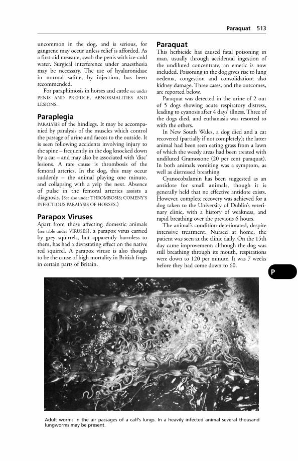
uncommon in the dog, and is serious, for gangrene may occur unless relief is afforded. Asa first-aid measure, swab the penis with ice-coldwater. Surgical interference under anaesthesiamay be necessary. The use of hyaluronidase in normal saline, by injection, has been recommended.
For paraphimosis in horses and cattle see under
PENIS AND PREPUCE, ABNORMALITIES AND
LESIONS.
ParaplegiaPARALYSIS of the hindlegs. It may be accompa-nied by paralysis of the muscles which controlthe passage of urine and faeces to the outside. Itis seen following accidents involving injury tothe spine – frequently in the dog knocked downby a car – and may also be associated with ‘disc’lesions. A rare cause is thrombosis of thefemoral arteries. In the dog, this may occur suddenly – the animal playing one minute, and collapsing with a yelp the next. Absence of pulse in the femoral arteries assists a diagnosis. (See also under THROMBOSIS; COMENY’S
INFECTIOUS PARALYSIS OF HORSES.)
Parapox VirusesApart from those affecting domestic animals(see table under VIRUSES), a parapox virus carriedby grey squirrels, but apparently harmless tothem, has had a devastating effect on the nativered squirrel. A parapox viruse is also though to be the cause of high mortality in British frogsin certain parts of Britain.
ParaquatThis herbicide has caused fatal poisoning inman, usually through accidental ingestion ofthe undiluted concentrate; an emetic is nowincluded. Poisoning in the dog gives rise to lungoedema, congestion and consolidation; alsokidney damage. Three cases, and the outcomes,are reported below.
Paraquat was detected in the urine of 2 out of 5 dogs showing acute respiratory distress,leading to cyanosis after 4 days’ illness. Three ofthe dogs died, and euthanasia was resorted towith the others.
In New South Wales, a dog died and a catrecovered (partially if not completely); the latteranimal had been seen eating grass from a lawnof which the weedy areas had been treated withundiluted Gramoxone (20 per cent paraquat).In both animals vomiting was a symptom, aswell as distressed breathing.
Cyanocobalamin has been suggested as anantidote for small animals, though it is generally held that no effective antidote exists.However, complete recovery was achieved for adog taken to the University of Dublin’s veteri-nary clinic, with a history of weakness, andrapid breathing over the previous 6 hours.
The animal’s condition deteriorated, despiteintensive treatment. Nursed at home, thepatient was seen at the clinic daily. On the 15thday came improvement: although the dog wasstill breathing through its mouth, respirationswere down to 120 per minute. It was 7 weeksbefore they had come down to 60.
Paraquat 513
P
Adult worms in the air passages of a calf’s lungs. In a heavily infected animal several thousandlungworms may be present.

The patience and perseverance of bothowner and clinic staff were rewarded, for whenseen again 6 and then 18 months later, the dogwas well and fully active again.
Parasites and ImmunityParasite antigens are a potent stimulus forantiparasite antibodies of the IgE class (see
IMMUNOGLOBULINS), and parasite infectioncan potentiate a pre-existing IgE response to anunrelated antigen.
Examples of the effect of parasitism on theimmune response are given under CANCER andALLERGY.
Parasitic BronchitisThis occurs in cattle, sheep and goats; onaccount of the husky cough produced, the disease is commonly called ‘husk’ or ‘hoose’ in the UK.
Although of greater economic importance incalves, nevertheless the cost of an outbreak in adairy herd may be very high – not so much as a result of deaths (which do occur in adult cattle) but on account of reduced milk yieldsand the need for extra feed. Marshy land andmild, wet weather both favour the parasites, asdoes overstocking.
Cause In cattle the lungworm Dictyocaulusviviparus is the important species (see ROUND-
WORMS; also illustration, page 513). Parasitic bronchitis normally affects cattle in their firstgrazing season. Affected animals experience adrop in the saturation level of oxygen in their blood to 70 per cent even before clinicalsigns become apparent. In clinical cases the percentage may be reduced to 30.
Workers at Glasgow University definedinfection with the parasite into 5 phases: pene-tration, pre-patent, patent, post-patent, andreinfection. In all but the first phase, oedemaand emphysema are found .
Spread of the worm larvae is assisted by theirrocket-like propulsion by the fungus Pilobolus,which is found in faecal deposits on pasture.The worm larvae are projected along with the fungal spores, often between 10.00 andmid-day.
Signs The characteristic husky cough is asymptom in the milder cases, but in acute casesmay be absent, with the main symptom beingdyspnoea (laboured breathing). In calves, deathmay occur from actual suffocation due to masses of worms obstructing the air passages, or it may result from general debility or pneumonia. In adult cattle pneumonia develops, with Actinomyces pyogenes acting as asecondary invader. Oedema of the lungs mayoccur, and cause death.
Prevention and treatment Live oral vaccines are available. A number ofANTHELMINTICS, including albendazole, iver-mectin and fenbendazole, may be used to treatinfected cattle. Some anthelmintics are availablein bolus form. If the animal is exposed to infection while the anthelmintic bolus is active,the animal will develop immunity withoutshowing signs of the disease (see under WORMS,FARM TREATMENT AGAINST).
Parasitic Disease, Nature ofParasitic diseases are caused by organisms thatlive within an animal (endoparasites) or on itssurface (ectoparasites). Endoparasites includeworms and flukes; ectoparasites include fleas,mites and ticks. Disease is seldom caused byone or a few parasitic organisms, but as a rule depends on mass infestations. There areexceptions to this, however, as a single Ascarismay obstruct the bile-duct with fatal results.Parasites, with few exceptions, do not spend alltheir lives in the animal body, but always needto spend a certain proportion of their life-cycleoutside the host. They may cause damage to thehost in the following ways:
(1) By abstraction of nourishment properlybelonging to the host, e.g. many of the intestinal worms;
(2) By mechanical obstruction of passages orcompression of organs, e.g. gapes (in chickens)and hydatid;
(3) By feeding on the tissues of the host, e.g. blood-sucking worms or flies;
(4) By production of toxins with varyingeffects;
(5) By actual traumatic damage, e.g. bypiercing and destroying skin (ticks, mites, flies, etc.), by depositing eggs in the tissues
514 Parasites and Immunity
P
In a case of parasitic bronchitis, the neck is heldextended and there may be continual coughingand/or distressed breathing.
(lung-worms), by migrations of larvae (Ascarisand Trichinella), by clinging to surfaces bymeans of sharp hooks (tapeworms), and inmany other ways;
(6) By facilitating the entrance of bacteria,e.g. stomach worms in pigs allow the entrance of Fusiformis necrophorus (the necrosisbacillus);
(7) By transmitting diseases for which they act as intermediate hosts, e.g. ticks andbabesiosis;
(8) By causing inflammatory or neoplasticreactions in the invaded tissues, e.g. pneumo-nia, gastritis, and fluke adenomata in the liver.
These are only some of the more obviousmethods of injuring the host. Apart from theloss due to actual deaths, the depreciation in value of hides, meat, milk, and work is enormous, and, although less spectacular than abacterial epizootic, the loss is more constant,and in the aggregate is probably even greaterthan the loss due to bacterial diseases. (See
BRAIN DISEASES – Parasites, for parasites whichmigrate to the brain.)
Parasitic Gastroenteritis ofCattleThis is an insidious and economically impor-tant disease, and the cause of death in manycalves and yearlings. It is known that the outputof worm eggs in the faeces does not bear any constant relation to the number of wormspresent. It rises to an early peak and thendeclines, and is not a reliable guide to thedegree of infestation.
Cause Infestation with various species ofROUNDWORMS.
Signs A gradual loss of condition; a harsh, staring coat; sometimes, but not always, scouring; pale mucous membranes; progressiveweakness and emaciation. In adult cattle, which acquire a high degree of resistance (onlybroken down when under-feeding, chilling,pregnancy, or massive contamination of pastureoccurs), no symptoms may normally be shown, but nevertheless the animal’s efficiencyis lowered.
Treatment Dosing with an appropriateanthelmintic should not be delayed until thestock are weak.
Prevention Calves should be dosed once withan anthelmintic in mid-July and moved to pasture which has not been grazed that seasonby other cattle. Dose again in the autumn.
(See WORMS, FARM TREATMENT AGAINST;PASTURE, ‘CLEAN’.)
Parasitic Gastroenteritis ofSheepIt is likely that outbreaks in early lambs inMarch and April are the result of over-winteredlarvae. In 1 experiment, worm-free lambs wereturned on to a pasture – ‘rested’ during the winter – in the spring and became infested with12 species of gastrointestinal worms.
Lambs may also be infected by eggs in thedung of ewes at lambing, when a periparturientrise occurs.
Treatment and prevention Routine use of, e.g., Tetramisole. (See WORMS, FARM
TREATMENT AGAINST; PASTURE, ‘CLEAN’.)
Parasitic Tracheobronchitis(see TRACHEAL WORMS)
ParasitismParasitism is the association of 2 organisms, 1 ofwhich (the parasite) benefits by nourishingitself at the expense of the other (the host) butwithout normally destroying it.
The following types of parasitic relations arerecognised: 1 (a) ectoparasites, which live onthe host; and (b) endoparasites, which livewithin the body of the host; 2 (a) accidentalparasites, which are normally free-living animals but may live for a certain period in ahost; (b) facultative parasites, which are able toexist free or as parasites, e.g. blowfly larvae; and(c) obligatory parasites, which are completelyadapted to a parasitic type of life and must livein or on a host, e.g. most parasitic worms; 3 (a)temporary or transitory parasites, which pass adefinite phase or phases in their life-history asparasites and during which time the parasitismis obligatory and continuous, e.g. botflies, ticks;(b) permanent parasites, which always live forthe greater part of their life as parasites, e.g. lice,tapeworms, coccidia, etc.; and (c) periodic,occasional, or intermittent parasites, whichonly visit the host for short periods to obtainfood, e.g. blood-sucking flies, fleas; 4 (a) erraticparasites, which occur in an organ that is nottheir normal habitat, e.g. Fasciola hepatica inthe lungs; (b) incidental parasites, which,exceptionally, occur in an animal that is nottheir normal host; they are incidental only inthis first host, e.g. Dipylidium caninum is incidental in man; and (c) specific parasites,which occur in a particular species of host orgroup of hosts, e.g. D. caninum is specific fordogs and cats.
Parasitism 515
P

Parasympathetic NervousSystemThe parasympathetic nervous system is 1 divi-sion of the autonomic nervous system; theother division being the sympathetic. (See
AUTONOMIC NERVOUS SYSTEM; CENTRAL
NERVOUS SYSTEM.)
ParathionParathion is chemically diethyl-para-nitrophenyl-thiophosphate and is used for agricultural purposes to destroy aphis and redspider. In man and domestic animals it is acumulative poison which readily enters the system through inhalation, by the mouth or by absorption through the skin. Animals
should not be allowed to graze under treessprayed with parathion for at least 3 weeks. In man, symptoms of poisoning includeheadache, vomiting, and a feeling of tightness in the chest. Later there is sweating,salivation, muscular twitching, distressedbreathing and coma. (See ORGANOPHOSPHORUS
POISONING.)In animals, copious salivation and lachryma-
tion, twitching, and increased intestinal movement are shown. Cattle are apparently tolerant of parathion, being able to break itdown chemically.
The danger of spray drift, and the risk todogs and cats wandering in sprayed areas, areobvious.
516 Parasympathetic Nervous System
P
PAGE
Anaplasmosis 24Arachnida 34Babesiosis 49Balantidium 55Blood flukes (see SCHISTOSOMIASIS) 629Blood parasites of cattle (UK) 71Blow-flies (see FLIES) 264Bots (see FLIES) 266Canine babesiosis 114Chicken mites (see MANGE) 436Coccidiosis 143Cysticercosis (see TAPEWORMS) 687Ear mange (see EAR, DISEASES OF) 210East Coast fever –
(see THEILERIOSIS) 700Fleas 260‘Flesh’ flies (see FLIES) 265Flies 261Flour mites (see MITES) 461
(see FLOUR-MITE INFESTATION) 271Flukes 420, 425, 510, 618, 629Forage mites (see MITES) 461Fungal diseases 290Gadflies (see FLIES) 262
(see WARBLES) 769Gapeworm (see GAPES) 294Giardia (see GIARDIASIS) 305Globidiosis 307Gnats (see FLIES) 262‘Green-bottle’ flies (see FLIES) 264Haemobartonella 320Harvest mites (see MITES) 461Hookworms (see ROUNDWORMS) 614House flies (see FLIES) 263Husk and hoose 348, 337Jaundice, malignant (see CANINE BABESIOSIS) 114Ked, sheep 357Leeches 409Leishmaniasis 409Lice 414Linguatula (see MITES) 462
PAGE
Liver-flukes 420Lung flukes 425Lungworms 514, 613Mange mites (see MITES) 401Midges 455Mites 461Mosquitoes (see FLIES) 262Myiasis (see FLIES) 264Nematodes (see ROUNDWORMS) 611Nematodirus 482Red-water fever 593Ringworm 606Roundworms in the horse 611Roundworms in ruminants 613Roundworms in the pig 615Roundworms in the dog and cat 616Roundworms in poultry (see GAPES) 294Rumen flukes 618Sarcocystis 627‘Scaly leg’ (see MANGE) 438Schistosomiasis 629‘Screw-worm’ fly 631Sheep nostril fly (see FLIES) 267Sheep scab 639Strongyles (see ROUNDWORMS) 612
(see STRONGYLES) 674Summer sores 677Tapeworms in the dog and cat 688Tapeworms in the horse 688Tapeworms in the pig 689Tapeworms in ruminants 688Texas fever 699Theileriosis 700Ticks 704Toxoplasmosis 714Trichomoniasis 719Trypanosomiasis 725Tumbu flies (see FLIES) 265Warble flies (see FLIES) 266
(see WARBLES) 769
PARASITES and PARASITOLOGY
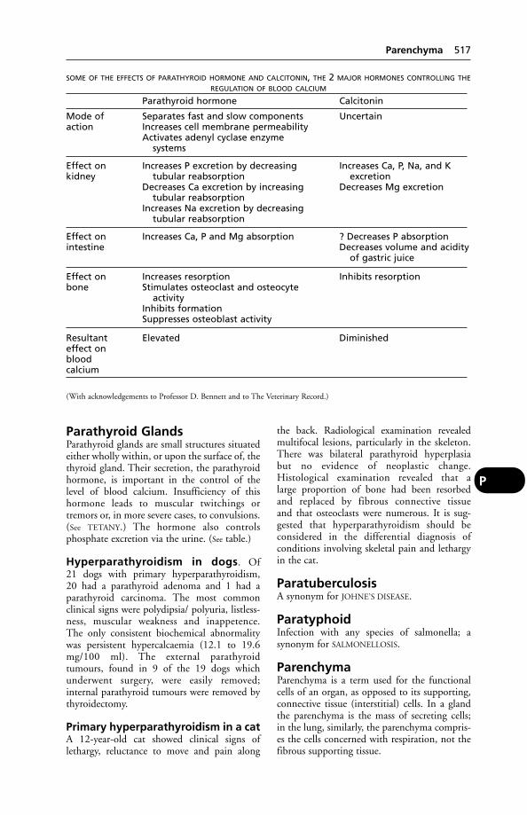
Parathyroid GlandsParathyroid glands are small structures situatedeither wholly within, or upon the surface of, thethyroid gland. Their secretion, the parathyroidhormone, is important in the control of thelevel of blood calcium. Insufficiency of this hormone leads to muscular twitchings ortremors or, in more severe cases, to convulsions.(See TETANY.) The hormone also controls phosphate excretion via the urine. (See table.)
Hyperparathyroidism in dogs. Of 21 dogs with primary hyperparathyroidism, 20 had a parathyroid adenoma and 1 had aparathyroid carcinoma. The most commonclinical signs were polydipsia/ polyuria, listless-ness, muscular weakness and inappetence. The only consistent biochemical abnormalitywas persistent hypercalcaemia (12.1 to 19.6mg/100 ml). The external parathyroidtumours, found in 9 of the 19 dogs whichunderwent surgery, were easily removed; internal parathyroid tumours were removed bythyroidectomy.
Primary hyperparathyroidism in a catA 12-year-old cat showed clinical signs oflethargy, reluctance to move and pain along
the back. Radiological examination revealedmultifocal lesions, particularly in the skeleton.There was bilateral parathyroid hyperplasia but no evidence of neoplastic change.Histological examination revealed that a large proportion of bone had been resorbed and replaced by fibrous connective tissue and that osteoclasts were numerous. It is sug-gested that hyperparathyroidism should be considered in the differential diagnosis of conditions involving skeletal pain and lethargyin the cat.
ParatuberculosisA synonym for JOHNE’S DISEASE.
ParatyphoidInfection with any species of salmonella; a synonym for SALMONELLOSIS.
ParenchymaParenchyma is a term used for the functionalcells of an organ, as opposed to its supporting,connective tissue (interstitial) cells. In a glandthe parenchyma is the mass of secreting cells; in the lung, similarly, the parenchyma compris-es the cells concerned with respiration, not thefibrous supporting tissue.
Parenchyma 517
P
SOME OF THE EFFECTS OF PARATHYROID HORMONE AND CALCITONIN, THE 2 MAJOR HORMONES CONTROLLING THE
REGULATION OF BLOOD CALCIUM
Parathyroid hormone Calcitonin
Mode of Separates fast and slow components Uncertainaction Increases cell membrane permeability
Activates adenyl cyclase enzymesystems
Effect on Increases P excretion by decreasing Increases Ca, P, Na, and Kkidney tubular reabsorption excretion
Decreases Ca excretion by increasing Decreases Mg excretiontubular reabsorption
Increases Na excretion by decreasingtubular reabsorption
Effect on Increases Ca, P and Mg absorption ? Decreases P absorptionintestine Decreases volume and acidity
of gastric juice
Effect on Increases resorption Inhibits resorptionbone Stimulates osteoclast and osteocyte
activityInhibits formationSuppresses osteoblast activity
Resultant Elevated Diminishedeffect onbloodcalcium
(With acknowledgements to Professor D. Bennett and to The Veterinary Record.)

ParenteralAdministration of a medicinal substance otherthan via the digestive system, e.g. by injection.
ParesisA state of slight or temporary PARALYSIS, alsocalled ‘fleeting paralysis’. (See MILK FEVER;MUSCLES, DISEASES OF – Nutritional musculardystrophy; LEAD POISONING; GUTTURAL
POUCH DISEASE.)
ParietalParietal is the term applied to anything pertain-ing to the wall of a cavity, e.g. parietal pleura,the part of the pleural membrane which linesthe wall of chest.
ParonychiaParonychia is inflammation near to the nail. (See RINGWORM.)
Parotid GlandParotid gland is one of the salivary glands. It issituated just below and behind the ear on eitherside, in the space between the angle of the jawand the muscles of the neck. From its base com-mences a duct, the parotid duct, or Stenson’sduct, which in the horse runs within the borderof the mandible for a distance, and then turnsround its rim to the side of the face in companywith the external maxillary artery and vein, andends by opening into the mouth opposite theanterior part of the 3rd upper cheek tooth; inother animals it runs straight across the faceinstead of along the lower jaw bone.
The SALIVARY GLANDS are composed of col-lections of secreting acini held together looselyby a certain amount of fibrous tissue, but theydo not possess a distinct capsule.
ParovariumParovarium is the name of rudimentary struc-tures situated near the ovary, which are the remnants of the Wolffian bodies. The nameParoophoron is also used. These structures areoften the seat of cysts in the young adult. (See
OVARIES, DISEASES OF.)
Parrots(see PSITTACOSIS; also BIRD IMPORT CONTROLS
and PACHECO’S DISEASE)
ParthenogenesisAsexual reproduction, in which the ovumdevelops into an embryo without fertilisationby a spermatozoon.This is a common methodof reproduction among invertebrate animals,particularly insects, including ants and bees.
Partridges(see GAME BIRDS, MORTALITY)
Parturient Paresis(see MILK FEVER and ‘DOWNER COW’ SYNDROME)
ParturitionParturition is the expulsion of the fetus (and itsmembranes) from the uterus through thematernal passages by natural forces, and in sucha state of development that, in domesticatedanimals at least, though not in the marsupials,the fetus is capable of independent life. Theprocess is called ‘foaling’ in the mare, ‘calving’in the cow, ‘lambing’ in the ewe, ‘kidding’ inthe goat, ‘farrowing’ in the sow, and ‘whelping’in the bitch. It is more likely to proceed suc-cessfully without than with human interferencein the great majority of cases. (See CALVING,DIFFICULT for information on traction.)
Stages in parturition Although the act isreally a continuous one, it is customary todivide it into 4 stages: (1) Preliminary stage; (2) Dilatation of the cervix stage; (3) Expulsionof the fetus stage; (4) Expulsion of the membranes stage.
(1) Preliminary stage may occupy somehours or even days. The udder swells, becomeshard and tender, and a clear waxy fluid material oozes from the teats or may be expelledby pressure of the hand. The external genitalsbecome swollen, enlarged, and their lining isreddened. A vaginal secretion appears. Theabdomen drops and becomes pendulous. Thequarters droop and the muscles and ligamentsof the pelvis slacken. The animal separates itselffrom its fellows if at pasture; if at liberty, it seeksa remote or an inaccessible place in which tobring forth its young, and some, such as thesow, bitch, and cat, prepare a bed or nest.
(2) Dilatation of the cervix stage mergeswith the preceding. Restlessness is evident. Themare paces around the loose-box (often withtail raised) – perhaps lying down and risingagain several times. Sweating occurs under themane and tail, and soon over most of the body.
During the 2nd stage of labour, the mare isusually lying down on her side, and in somecases will show symptoms of COLIC, i.e. kickingat the belly, turning and gazing at her flanks, orwandering round in an aimless fashion.Meanwhile the labour pains have been gettingmore and more powerful and the intervalsbetween them shorter. The pulse is quickened,and the breathing rapid. When a pain haspassed the animal calms down and remains
518 Parenteral
P
so till the next takes place. After a variable time – from about 1/2 to 3 hours – the ‘water-bag’ appears at the vulva. It is tense and hardduring a pain, but becomes slack and flaccid inthe intervals. It is found to be empty at first, butthe fore-feet of the young animal can be felt in it later. At this time the cervix is fully dilated, and the 3rd stage follows without anyappreciable break in the sequence of events.
(3) Expulsion of the fetus stage In thisstage the severity of the pains is greatest, and theauxiliary muscles of the abdomen assist in thecontractions. The animal may remain standing,may lie down in the recumbent position, or mayalternately lie and stand. The back is arched, thechest expanded, and the muscles of theabdomen become board-hard with each labourpain. The animal may groan, or squeal or evenscream with each effort. Frequently the rectumforcibly discharges its contents and the urinarybladder does likewise. At each contraction the‘water-bag’ protrudes farther and farther fromthe vulva until it finally ruptures in its mostdependant part. There is a rush of fluid from theuterus to the outside and the animal has a peri-od of ease. Then fore-feet, and the muzzle lyingbehind and over them, appear at the vulva,forming a kind of cone which dilates the softertissues of the genital canal. In the larger animalsthe feet come first, but in the carnivora, wherethe head is large, the head precedes the fore-feet,which are tucked against the young animal’schest and sides. When the head has cleared thevulva there is usually another pause, whichallows the tissues to become accustomed to thegreat distension, and prepares them for the stillgreater distension and strain that is soon to fol-low. The thorax and shoulders are now in thepelvis of the dam, and are driven slowly throughit by the most powerful and painful of the con-tractions that occur during the process. As thispart of the fetus reaches the outlet of the pelvisthere is generally a more energetic and painfuleffort than all the others – which pushes thefetal trunk to the outside. This culminatingeffort may cause the bitch or cat to cry out.
Sometimes the foal’s umbilical cord does notrupture, in which case the mare will usuallygnaw through it, and so liberate the foal. Itsometimes happens that a foal is born com-pletely enveloped in its membranes; in suchinstances, unless assistance is at hand to free thefoal, it will be rapidly suffocated.
In cows the umbilical cord is much shorterand it ruptures before the hind legs of the calfhave passed to the outside. Owing to the cotyle-donary attachment of the placenta the mem-branes are seldom born along with the calf. In
the smaller animals, especially in the sow, bitch,and cat, the young are frequently born in theirmembranes, and these are licked away andcleared from the young by the dam, the umbil-ical cord being broken or bitten through in theprocess.
(4) Expulsion of the membranes stage,or the ‘delivery of the afterbirth’, may occurwith, immediately following, or not for someconsiderable time after, the production of theyoung in an animal.
Very soon after the young animal is born the uterus contracts and becomes smaller – aprocess known as ‘involution’ – so that itscapacity is decreased. The attachment betweenthe membranes and the mucosa of the uterus isloosened and the placenta is separated from the uterus. These contractions also serve topush out the membranes through the wideopen cervix.
With the mare, owing to the diffuse and notvery intimate adherence between the uterinemucous membrane and the placental mem-brane, the separation and the discharge of the envelopes are soon accomplished. In fact, ifthese are retained for more than a very fewhours (4 or 6 or so), serious results are probable,but retention of the membranes is rare in thehealthy mare.
In the cow, the attachment is limited to thesurfaces of the cotyledons and is very close, and where the shrinkage in the uterine wall (i.e.involution) does not tend greatly to upset theintimacy of the adhesion, the calf is not born inits membranes, and retention of these is morecommon. They are generally discharged withina few hours of the birth of the calf, but the timevaries.
Animals which produce more than 1 youngat a time generally discharge the membranes ofeach at the same time as or soon after it is born,with the exception of the last of the litter, whosemembranes are occasionally retained in theextremity of 1 horn of the uterus.
In animals that are really uniparous (i.e. produce only 1 fetus at a birth but which havebeen modified by breeding so that they oftenproduce 2 or more young, such as the sheepand goat) the membranes of the 1st twin comeaway with the 2nd, and those of the 2nd areexpelled after it has been born.
Early discharge of the membranes is desir-able, because as long as they remain in positionthey are likely sources of infection to the uterus,and they prevent that organ from returning tonormal. After they have been evacuated theinvolution of the uterus becomes more andmore complete, until in a few days it has
Parturition 519
P
shrunk to less than half its former size. It neverdecreases to its original virgin size.
The mare should have been housed in the‘foaling-box’ for a month or so previously, so that she shall feel quite at home (see
PREGNANCY AND GESTATION – Care of the dam during pregnancy), and the ventilation,warmth, bedding, cleanliness, etc., should be asnear an approach to the ideal as circumstanceswill allow. If possible, the cow should calve in aseparate loose-box. Ewes lamb out in the openand do quite well, but if the weather is cold orstormy, or if the ground is very wet, it is betterto provide a ‘lambing-pen’, especially withLowland breeds which have not the same hardiness as the mountain varieties. Sowsshould on all occasions have a pen to them-selves, for if other pigs are present the little pigs will most probably be eaten as soon as they are born.
When the birth process has begun, the atten-dant may need to soothe and quieten the damif she becomes very excited, but beyond this theprospective mother should be left alone forsome time. If all is going well, the ‘water-bag’will soon appear and later burst. No hard-and-fast rule can be laid down, but if the fore-legsand nose of the fetus do not appear within 10to 20 minutes in the mare, and in double thatperiod in the cow, a simple examination shouldbe made by the attendant to ensure that thepresentation is a normal one. After washinghands and arms in mild antiseptic, such asDettol or Cetrimide solutions, and thoroughlylubricating them with a vaginal lubricant, the hand should be gently inserted into the posterior genital passage to explore by touchwhatever presents itself. The 2 fore-feet shouldbe distinguishable in that part of the passagethat lies lowermost when the dam is standing.Above them and slightly behind, the nose andmouth should be felt. These structures are oftencovered with fetal membrane, but in a normalcase can be located. In such cases as this, noth-ing further need be done in the meantime; thedam will probably produce her young quitenormally, and any attempts at assistance willonly irritate and perhaps exhaust her.
It may happen, however, that 1 or both ofthe fore-legs or the nose cannot at first befound. On introducing the arms still furtherthese parts can sometimes be discovered, and,by gentle pulling or readjustment, can bebrought into the normal position. Before theprocess of parturition has advanced very far,abnormal positions of the fetus can be compar-atively easily corrected, and serious troublefrom subsequent jamming may be avoided. If
all efforts at correction prove futile, a veterinarysurgeon should be called in.
Upon comparatively rare occasions none of the foremost positions of the body can befelt, but the 2 hind-feet or legs (distinguishable by the difference between knees and hocks),and perhaps the tail of the young animal, arediscovered. This is a posterior presentation, andas the head is the last part of the fetus that willbe born, respiration cannot begin until birth iscomplete, and the risk of suffocation is great.Accordingly, it is necessary to attempt to hurrythe whole process and a veterinary surgeonshould be called.
Attention to offspring As soon as theyoung animal is born and free from the maternal passages, it is absolutely essential toascertain that the fetal membranes are notobstructing its mouth or nostrils. It generallygives 1 or 2 spasmodic gasps or struggles, and then begins to breathe. Each respiration is shallow and weak at first, but in a very few minutes the breathing settles down to thenormal.
Suspended animation Occasionally, foalsand calves are born in a state of suspended animation, and ARTIFICIAL RESPIRATION will beneeded.
The umbilical cord This is best not inter-fered with. In the foal, early severance of thecord can lead to the loss of 1000 to 1500 ml ofplacental fetal blood, whereas under ‘natural’conditions the amount concerned is probablywell under 200 ml. The well-being of the animal is better served when it is allowed toretain the blood, as would usually be the caseunder natural conditions. The umbilical cordhas a point at which it will rupture normally,generally by movement of the mare, after a period during which mother and foal rest. Sincehaemorrhage from either end of the severedcord is then extremely rare, the cord shouldrequire no human attention after a normalbirth. The natural sealing of the umbilical cord provides an effective barrier against bothbleeding and infection.
The Animal Health Trust’s Equine ResearchCentre has commented: ‘It is difficult to imag-ine a worse procedure than leaving a substantial“meaty” mass of umbilical cord at the navel ashappens so commonly after cutting the cordwith scissors. This provides an ideal medium forthe passage of micro-organisms whose entranceto the abdominal portions of the umbilical veinand arteries are not hindered in any way by the
520 Parturition
P

Parturition 521
P
Different varieties of presentations of the foal.1. Normal anterior presentation – nose and both fore-feet in passage.2. Anterior presentation with 1 fore-limb retained completely. This should be brought forward by hand,
or by passing rope round flexure of knee.3. Anterior presentation with both fore-limbs retained at the knees, corrected as in No. 2.4. ‘Dog-sitting’ presentation – nose and all 4 limbs presenting. The 2 fore-limbs should be corded and
the hind-limbs repelled or pushed back.5. Anterior presentation with head and neck retained. Delivery may often be effected by traction on
fore-limbs in foal, the head being pressed into the soft abdomen. In calf, owing to the short neck, this isnot usually possible. Where possible, fore-limbs should be corded, pushed back, and the head broughtround by the hand or, in difficult cases, by hooks and cords.6. Posterior presentation. Successful delivery often possible if the birth is speeded up by traction to
avoid suffocation (see text).7. ‘Breech presentation’. Delivery difficult, foal nearly always dead. Cords in front of the foal’s stifles and
round buttocks may be applied if mare is large and foal is small, but usually necessitates amputation of 1or both hind-limbs.8. ‘Thigh and croup presentation’. Cord round hocks may be successful in converting this into an
ordinary posterior presentation. Quarters must be firmly pushed back after hocks are corded.
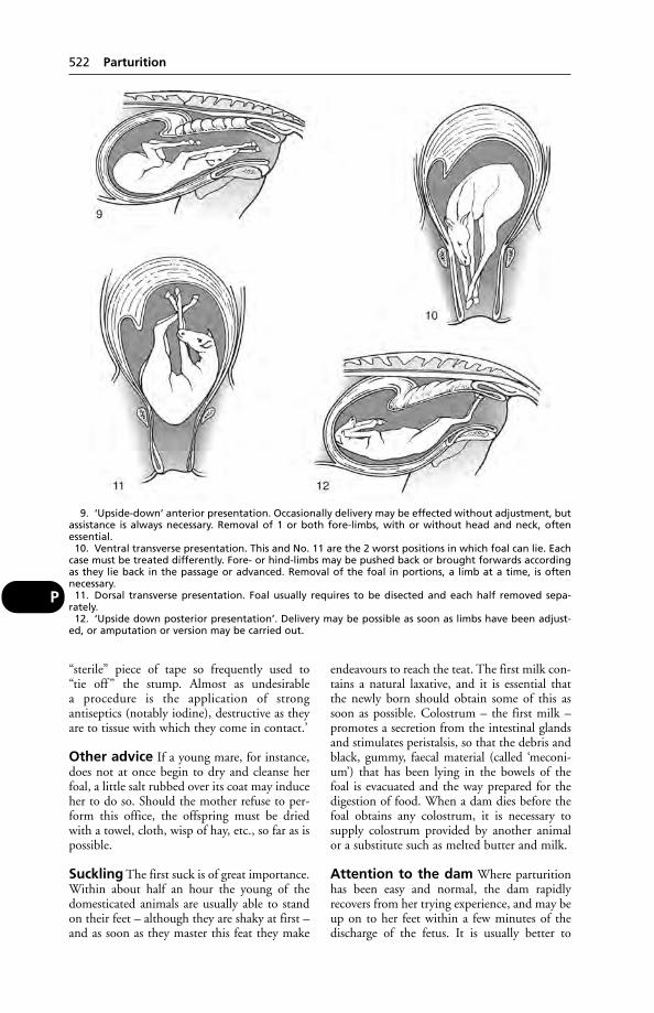
“sterile” piece of tape so frequently used to “tie off ” the stump. Almost as undesirable a procedure is the application of strong antiseptics (notably iodine), destructive as theyare to tissue with which they come in contact.’
Other advice If a young mare, for instance,does not at once begin to dry and cleanse herfoal, a little salt rubbed over its coat may induceher to do so. Should the mother refuse to per-form this office, the offspring must be driedwith a towel, cloth, wisp of hay, etc., so far as ispossible.
Suckling The first suck is of great importance.Within about half an hour the young of thedomesticated animals are usually able to standon their feet – although they are shaky at first –and as soon as they master this feat they make
endeavours to reach the teat. The first milk con-tains a natural laxative, and it is essential thatthe newly born should obtain some of this assoon as possible. Colostrum – the first milk –promotes a secretion from the intestinal glandsand stimulates peristalsis, so that the debris andblack, gummy, faecal material (called ‘meconi-um’) that has been lying in the bowels of thefoal is evacuated and the way prepared for thedigestion of food. When a dam dies before thefoal obtains any colostrum, it is necessary tosupply colostrum provided by another animalor a substitute such as melted butter and milk.
Attention to the dam Where parturitionhas been easy and normal, the dam rapidlyrecovers from her trying experience, and may beup on to her feet within a few minutes of thedischarge of the fetus. It is usually better to
522 Parturition
P
9. ‘Upside-down’ anterior presentation. Occasionally delivery may be effected without adjustment, butassistance is always necessary. Removal of 1 or both fore-limbs, with or without head and neck, oftenessential.10. Ventral transverse presentation. This and No. 11 are the 2 worst positions in which foal can lie. Each
case must be treated differently. Fore- or hind-limbs may be pushed back or brought forwards accordingas they lie back in the passage or advanced. Removal of the foal in portions, a limb at a time, is oftennecessary.11. Dorsal transverse presentation. Foal usually requires to be disected and each half removed sepa-
rately.12. ‘Upside down posterior presentation’. Delivery may be possible as soon as limbs have been adjust-
ed, or amputation or version may be carried out.
allow her to remain lying as long as she wisheswhile attention is being paid to her offspring. Itis good practice to offer a drink of warm barley-water or thin oatmeal gruel containing a table-spoonful of common table-salt, as soon after theact as convenient. Her system has undergone aconsiderable shock, and has lost quantities offluid which should be replaced. The larger ani-mals may require a rug if the weather is at allcold. After 3 to 6 hours or thereabouts, a pailfulof bran mash and a little hay should be given.
When the dam is very exhausted by herlabour it is necessary to administer stimulants.If the birth of the young was difficult, andwhen the passages have been exposed to consid-erable strain by assisted labour, hot (but notscalding) fomentations may be applied to theexternal genital organs. Afterwards the partsmust be covered with a warm and dry blanket,to prevent any chilling. The loose-box must bewarm yet well ventilated, and the dam shouldbe encouraged to rest as much as possible.
Subsequent management No oats orconcentrated food-stuff should be given to thedam for the first 2 days after parturition; herrations should consist of water, bran mashes,and hay or green food given 3 times daily. Afterthat time a gradually increasing amount ofcrushed oats and cut hay or chaff may be addedto the mash daily, until at the end of a week or10 days she is back on to her usual diet. Gentleexercise is as necessary for the foal or calf as it isfor their dams, and if the weather is suitable thedam and her progeny should be allowed out onto a sheltered meadow for an hour or so twicedaily, after the first 3 or 4 days following thebirth of the young. This period is graduallyincreased until in 2 weeks’ time the pair may beleft out from 9.00 a.m. till 5.00 p.m., or evenmay be allowed to sleep out all night if theweather permits. In this connection it should beremembered that cold dry nights are much lessharmful than those that are wet or foggy. Younganimals of all species withstand dry cold verymuch better than wet cold, and it is inadvisableto allow foals or calves less than a month old tosleep out on a wet or marshy meadow. A usefulmethod is to erect a covered-in shed in a cornerof the meadow, containing a feeding-troughand well littered with straw, into which bothdam and her offspring may retire whenever theywish. The amount of hand feeding which thedam receives must be judged according to circumstances. If the grass is rich and well forward, 1 feed of oats and hay may be suffi-cient after the first 3 or 4 weeks, but it is alwaysbetter to err on the safe side and keep the dam
in good general condition, for much of the subsequent quality of the offspring dependsupon the start in life that it receives through its mother’s milk, and if she herself is in poorcondition her milk will be inferior.
Parturition, Drug-InducedProstaglandins are frequently used to induceparturition in pigs, and sometimes in other live-stock, so that it occurs at a planned date. It is atechnique also used in some cases of debility ofthe dam in late pregnancy, where there is someabnormality associated with the pregnancy, toavoid dystokia in the cross-breeding of dairycows for beef production.
ParvovirusThis has been associated with infertility in thesow, and is a cause of mummified fetuses andsmall litter size. (See VIRUSES – Classificationtable; CANINE PARVOVIRUS (CPV); and FELINE
INFECTIOUS ENTERITIS.)
Human parvovirus infection This ismost common in children, and is associatedwith a rash and fever; and sometimes pain inthe joints.
PasPas is the abbreviation for para-aminosalicylicacid, a drug which has been used in the treatment of tuberculosis in zoo animals.
PassagePassage (pronounced as in the French) is a termmeaning the passing of a strain of organismsthrough a series of animals to decrease virulence. For example, passage of cattle plaguevirus through goats is done to reduce its virulence for cattle, and is a technique used inthe production of cattle plague vaccine.
PasternThe name given to the 1st (long) and 2nd(short) phalanges of the fore-limb, and to thejoint so formed.
PasteurellaA genus of bacteria, which are small, ovoid,Gram-negative, bipolar staining. Both non-motile and motile species occur, and they are aerobic or facultatively anaerobic. For thediseases they cause, see following entries.
Pasteurellosis in Cats(see YERSINIOSIS and BUBONIC PLAGUE; see BITES
for human infection with Pasteurella multocida,formerly known as P. septica).
Pasteurellosis in Cats 523
P
The cat harbours P. multocida as one of itsbacterial flora.
Pasteurellosis in CattleThis includes HAEMORRHAGIC SEPTICAEMIA ofcattle and buffaloes in the tropics; ‘SHIPPING
FEVER’ (‘Transit fever’) of the American feedlotsand elsewhere; and other pneumonias occurringin Europe.
In calves, Pasteurella haemolytica serotype A1may be the primary pathogen. Serotype A2 and P.multocida may also be isolated. Both bacteria arenormal inhabitants of the upper resiratory tract.Under stress, or infection by viruses, the bacteriamultiply rapidly, causing disease. Symptomsinclude a nasal discharge, a respiratory rate of 60to 100 per minute, and a temperature of up to41.6°C (107°F).
(See CALF PNEUMONIA for viruses which maybe involved, and for synergism betweenPasteurella and Mycoplasma.)
The pneumonia is fibrinous in type, and thisis seen also in older cattle – P haemolytica or P.multocida often being found in large numbers.
Pasteurellosis in DucksCaused by Pasteurella anatipestifer, this is a disease of considerable economic importance inducklings, with a mortality sometimes as highas 70 per cent. Less acutely infected birds mayshake their heads or draw their heads close totheir bodies. Sulfadimidine has been used intreatment. See also FOWL CHOLERA.
Pasteurellosis in ManPasteurella multocida is a commensal organismin the mouth and naso-pharynx of many ani-mals. In humans, superficial infections of theskin and mucous membranes, such as corneal,oral or leg ulcers and infections of compoundfractures, conjunctivitis or sinusitis andpanophthalmitis may result from animal bitesor scratches. In these infections P. multocida islikely to have been acquired when saliva fromthe animal contacted injured tissue. Pasteurellaorganisms may also invade the body via the respiratory system or, less commonly, via thealimentary tract and skin lesions. The mostcommonly seen internal infections of P. multo-cida are associated with chronic obstructivelung disease. A 3rd category of infection is suggested: septicaemia and bacteraemia inpatients with chronic disease, especially chronicliver disease. P. multocida may result from handling raw poultry carcases. Small domesticpets may be carriers of P. multocida. Internalinfections may derive from farm animals; inone study, 27 of 37 patients with internal
infection lived on farms. Other Pasteurellaspecies that affect animals rarely occur in man.
In the UK during one 10-year period therewere reports of 3699 cases of human pasteurel-losis. Eighty-six per cent of these were skininfections; two-thirds due to dog bites; a quarter due to cat bites. In a small proportionof cases meningitis and septicaemia were complicating factors.
Pasteurellosis in PigsBronchial pneumonia, sometimes with pleuritisand pericarditis, is the commonest symptom; P. multocida is the usual cause but clinical pasteurellosis may be a complication ofmycoplasmal pneumonia.
Signs There is fever, and the animals maybeseen struggling for breath, with frothing in themouth. Death may follow.
Treatment and control Antibiotics shouldbe given; antiserum is effective if the causal bacteria is known. Vaccination with a com-bined A. pyogenes, P. haemolytica, P. multocidaand staphylococcus vaccine (Pastacidin;Hoechst) is used for control where there is aknown risk of pasteurellosis.
Pasteurellosis in SheepPasteurellosis in sheep is caused occasionally byPasteurella multicocida but far more commonlyby P. haemolytica, and subclinical infection maydevelop into pneumonia if parainfluenza IIIvirus is present too.
P. haemolytica biotype A causes enzootic pneu-monia; while biotype T is mainly associated withsepticaemia. Both may be isolated from cases ofarthritis. P. haemolytica also causes mastitis inewes, and meningitis – especially in lambs.
Young sheep are liable to die from the acutesepticaemic form, while older ones show a slower type of the disease, in which the pneumonic lesions predominate.
Signs The acute cases are ushered in by hightemperature, great dullness and nervous depres-sion, difficult respirations, muscular tremors,followed by rapid collapse and death within 3 days.
In the less acute cases, similar but slightlymilder symptoms occur. These are accompa-nied by a discharge from the eyes and nose, lossof appetite and absence of rumination, withsigns of pneumonia or pleurisy.
Diagnosis The acute form may be confusedwith anthrax or braxy.
524 Pasteurellosis in Cattle
P

Immunisation Vaccines containing severalstrains of P. haemolytica are available; also a combined clostridial and pasteurella vaccine.A serum has also been prepared. (See also
PNEUMONIA IN SHEEP.)
Pasteurisation of Milk
High temperature pasteurisationconsists of heating the milk for 10 to 20 min-utes at a temperature of 75°C (167°F). This issufficient to render harmless the germs ofenteric and scarlet fever and diphtheria, andalso bacteria which give rise to summer diar-rhoea in children. It also affords a considerablemeasure of protection against tuberculosis.
Low temperature pasteurisationconsists of maintaining the milk for at least halfan hour at a temperature between 63° and65°C (145° and 150°F). This has the effect ofconsiderably reducing the number of bacteriacontained in the milk and greatly delaying souring and similar changes. This procedure issufficient for the sale of milk as ‘pasteurisedmilk’ in England. (See also ULTRA HIGH
TEMPERATURE TREATMENT OF MILK.)
Unpasteurised milk (see MILK-BORNE
DISEASE)
Pasture, ‘Clean’Pasture that has not been grazed by the samespecies for some time. The actual period varieswith the parasites involved, climate and otherfactors. Criteria for clean pasture vary with thetime of year. In spring, it is pasture not grazedby the same species in the previous grazing season – that is, a new ley, an area grazed byanother species or used for conservation. In thesummer, clean pasture is defined as an area notgrazed by that species the same year up to mid-June, for sheep, and mid-July for cattle.However, pasture rarely becomes completelyfree from parasites.
Professor James Armour and colleagues at theUniversity of Glasgow veterinary school foundclinical parasitic bronchitis (‘husk’), due to thelungworm Dictyocaulus viviparus, and gastroen-teritis due to Osertagia ostertagi, in young cattlegrazing aftermath pasture in late summer. Calveson pasture lightly infested with Ostertagia ‘wereeffectively treated with an anthelmintic andtransferred to a silage aftermath in late July. Amarked increase in 3rd-stage larvae numbers onthe aftermath occurred within the first week,and clinical signs of type I ostertagiasis wereobserved 4 weeks later.
‘In both instances the aftermath pastures hadnot been grazed since the previous autumn, andthe interval between entry to the aftermath and clinical or other evidence of infection precluded the possibility of the calves beingresponsible for cycling of the infection.’
Such findings suggested the existence of areservoir of infective larvae in the soil persistingfrom previous grazing seasons. ‘Preliminaryobservations on core samples of soil from per-manent cattle pastures in the Glasgow arearevealed that Ostertagia 3rd-stage larvae wereregularly present, and lungworm 3rd-stage larvae occasionally present,’ over a 12-monthperiod from August to July.
Research in the USA showed that ifOstertagia eggs are buried to a depth of 12.5 cmunder pasture, or beneath 15 cm of soil in thelaboratory, 3rd-stage larvae develop and migratevertically through the soil. However, the goodhusbandry rule of keeping young stock off pasture previously grazed in the same season byadult stock is, obviously, still worth applying asa means of avoiding even worse outbreaks.
Pasture, Contamination ofThis may occur in the vicinity of smeltingworks (see under FACTORY CHIMNEYS), or as theresult of droplets of chemical sprays being carried by the wind to adjoining fields. (For alist of chemical sprays, see under WEEDKILLERS
and INSECTICIDES.) Contamination may alsooccur as the result of atomic fall-out. (See
RADIOACTIVE FALL-OUT.) Bacterial contamina-tion can result from organsims that are able toexist for long periods outside the animal body.Examples are bovine tuberculosis (at least 4months), Johne’s disease (at least a year),anthrax (at least 30 years) and clostridial spores.Resting pasture for 3 weeks provides a measureof control of foot-rot, the organism responsiblebeing unable to survive for more than a fort-night. For contamination by worm larvae, see PARASITIC BRONCHITIS; GASTROENTERITIS;PASTURE, ‘CLEAN’. For contamination by organic irrigation see under SLURRY. (See also BASIC
SLAG; FERTILISERS).The average cow defecates about 12 times
daily and each pat weighs about 2.5 kg (51⁄2 lb); in a 180-day grazing season, she willput about 5 tons of faeces (containing about680 kg (1500 lb) dry matter) on to the pasture.(See DAIRY HERD MANAGEMENT.) (For contami-nation by slurry applications, see SLURRY and
‘MILKSPOT LIVER’; also SOIL-CONTAMINATED
HERBAGE.)Contamination of pasture may occur during
flooding and, in a sense, when ticks are left
Pasture, Contamination of 525
P

behind by a batch of cattle infected with piro-plasms; the ticks then infect other cattle put onto that land (see RED-WATER FEVER).
Pasture ManagementPasture management is of the greatest impor-tance in relation to diseases such as BLOAT,HYPOMAG-NESAEMIA, PARASITIC GASTROEN-
TERITIS and PARASITIC BRONCHITIS. (See also
under DEEP-ROOTING PLANTS, TOPPING, and
WILTING.) Controlled grazing is effected bymeans of an electric fence. (See STRIP-GRAZING
and PADDOCKS.)It is important that heavy application ofnitrogenous and potash fertilisers to grasslandshould be made at the right time, or animalsgrazing there will be exposed to a greatlyincreased risk of hypomagnesaemia. (See also
HOOF-PRINTS.)The sudden (and harmful) change of diet
which may occur when stock are turned out inthe spring, or brought off pasture into yards forthe winter, are discussed below.
In spring, it is a mistake to turn calvesstraight out on to grass. This means a suddenchange from protein-poor food to the rich protein of the early bite, and the resulting effectupon the rumen will set them back. It is wise toget them out before there is much grass for a few hours each day; let them have hay andshelter at night to protect them from suddenchanges of weather. Hypomagnesaemia, too, isfar less likely under these circumstances.
Before yarding cattle in the autumn, it is wiseto make a gradual change from sugar-poorautumn pasture to things like roots, and toaccustom them to concentrates. Otherwisedigestive upsets are very likely to occur.
It should be borne in mind thatTrichostrongylus axei is a parasite common tocattle, sheep, goats, and horses, and grazing onespecies of animal after another in a field couldgive rise to a very heavy contamination withthis one parasite.
Prompt removal of faeces from pasture hasbeen found effective in reducing the worm burden, and practicable where acreage is small,labour cheap, or racehorses are concerned.
Use of goats in sheep grazing systems.Goats prefer to graze plant species not readilyeaten by sheep; also, given the choice, goats willdiscriminate against plant species such as cloverwhich are important and beneficial in sheepproduction systems. The introduction of goatscan benefit the sheep stock by helping to prevent the degeneration of the improved areasand by keeping the indigenous vegetation in a
more productive and nutritious state, while atthe same time not competing with the sheep forthe more valuable plant species. (Hill FarmResearch Organisation.)
Other aspects of pasture management arereferred to under the following: PASTURE,CONTAMINATION OF; BRACKEN POISONING;RAGWORT POISONING; DIGITALIS POISONING;WORMS, FARM TREATMENT AGAINST;EXPOSURE; STRESS; ‘POACHING’; BEEF CATTLE
HUSBANDRY IN BRITAIN; SHEEP BREEDING AND
MANAGEMENT; SILAGE; FOG FEVER; HAY;STOCKING RATES; TOPPING OF PASTURES.
Grass varieties Plant breeding for improvedpastures is in its infancy as compared with thatfor the agronomically less complex arable crops.
Yield (whether annual or seasonal) is only 1 of 4 criteria useful in judging a new variety of herbage. Its persistency, palatability andnutritive value are important criteria, too; ahigh-yielding variety may be contraindicated ifanimals lose weight on it, as they do with 1variety of Phalaris. The effect which the systemof grassland management has, and how varietieswill stand up to a given system, must also beconsidered. For example, with regular defolia-tion, perennial rye grass yields more than Italianrye grass; whereas with infrequent cutting,Italian rye grass yields more.
For systems of farming at high production,consideration should be given to a species liketall fescue, whose biological potential is knownto be very high, and whose reaction to intensivesystems of defoliation is favourable.
Horses Pasture grasses and herbs recom-mended by the Animal Health Trust for horsesare classified as under:Desirable speciesPerennial Rye grasses, Sceempter, Melle, Petra,Midas S.23 and S.321, Timothy S.50 and S.48,Cocksfoot S.143, Crested Dogstail, WildWhite Clover, Dandelion, Ribgrass, Chicory,Yarrow, Burnet, Sainfoin.Probably useful (turf species which are alsopalatable)Tall Fescue Alta, Canadian Creeping RedFescue, Smooth Stalked Meadow Grass, RoughStalked Meadow Grass.Best excludedPerennial Rye grass S.24, Creeping Red Fescue,Brown Top, Meadow Foxtail, Red Clover.
PatellaPatella is the bone that lies at the front of the‘stifle joint’, and is called the ‘knee-cap’. It liesin the tendon of the large extensor muscles of
526 Pasture Management
P
the joint, just above and in front of the truefemorotibial joint. It is roughly pyramidal inthe horse, with the apex of the pyramid point-ing downwards. It is dislocation of the patellathat constitutes the condition known as ‘slippedstifle’. (See BONE.)
Dislocation of the patella (patellar luxation)may occur as an inherited abnormality in certain breeds of dogs, e.g. Boston terriers, boxers, bulldogs, cairn terriers, chihuahuas,wire fox terriers, griffons, Pekingese, Maltese,papillons, Pomeranians, poodles, Labradors,Scotch terriers, King Charles spaniels.
Indications for surgery of the canine stifle arecongenital medial luxation and rupture of theanterior cruciate ligament.
Congenital patellar luxation occurs also incats, rendering them unable to walk normallyor jump.
Patent Ductus ArteriosusAn abnormality in which the ductus arteriosus,between the aorta and the pulmonary artery,fails to close at or shortly after birth. This condition has been recognised in the puppy,and gives rise to a characteristic murmur onauscultation; and also in cattle and cats. (See
LIGAMENTUM ARTERIOSUM.)
Pathetic NerveThe trochlear nerve. It is the 4th nerve arisingfrom the brain and controlling the superioroblique muscle of the eye.
PathogenicDisease-producing.
PathognomonicPathognomonic is a term applied to those signsor symptoms of a particular disease which arecharacteristic of that disease, and on whosepresence or absence the diagnosis depends.
Peas, Mutter(see LATHYRISM)
‘Peat Scours’‘Peat scours’ is a name given in Australasia andCanada to MOLYBDENUM poisoning in grazingcattle.
Peck OrderThis is the equivalent in poultry of the order ofprecedence described under BUNT ORDER.
Pedal BonePedal bone, or coffin bone, is the bone enclosedwithin the hoof of the horse
PediculosisPediculosis is infestation with lice.
PekingeseA toy dog originating from China. It has longstraight hair, snub nose and pendulous ears. Its shape predisposes the breed to cleft palate,inguinal hernia, intravertebral disc disease,Perthe’s disease, and retrognathia (underde-veloped lower jaw). Distichiasis and patellar luxation may be inherited.
Pellagra(see ‘BLACK TONGUE’)
Pellets(see CUBES)
PelvisPelvis is the posterior girdle of bones by whichthe 2 hind-limbs are attached to the rest of theskeleton. It is composed of 2 ilia, 2 pubes, and2 ischia, united together by fusion into a basin-shaped whole (see BONE). Strictly speaking, itincludes the sacrum and the coccygeal verte-brae. The 2 ‘haunch bones’ are the externalangles of the ilia; the ‘croup’ is composed of the internal angles of these bones along with thespines of the sacrum; and the ‘points of the buttocks’ are the tuberosities of the two ischia.The pelvis is spoken of as having an ‘inlet’,formed by the brim of the pubes, and an ‘outlet’ posteriorly. In the living animal the out-let is occupied by the soft tissues forming theperineal region, except for the anus in the maleand the anus and vulva in the female. The deepnotch between the sacrum and the haunchbone is closed by the sacrosciatic ligaments,upon which lie the gluteal muscles which givethe quarters their shape. The pelvis varies in the 2 sexes: in the female it is broader from sideto side, and deeper from above downwards,than in the male; this difference being chieflynecessary to allow of the act of parturition.
The contents of the pelvis are the rectum andurinary bladder in both sexes (except in thedog, where the urinary bladder is abdominal inposition). In the male there is in addition theprostate gland and the seminal vesicles aroundthe neck of the bladder and the beginning ofthe urethra; while the female pelvis contains thevagina, uterus, their appendages, and perhapsthe ovaries.
PemphigusAn autoimmune disease of dogs also seen incats and horses. It can take various forms: inPemphigus vulgaris, lesions affect the mucous
Pemphigus 527
P
membrane lining the mouth, and the junctionwith the lips, giving rise to ulcers. Sometimesthe pads of the feet are affected. P. erythematosisis characterised by crusty lesions on and aroundthe nose, and elsewhere on the face.
In P. vegetans, alopecia and pruritus followpustules which ulcerate on the body andextremities.
Treatment is with corticosteroids; cytotoxicdrugs are also used. The prognosis in severecases is not good.
-PeniaA suffix meaning too few, less than normal. (See
LEUKOPENIA for an example.)
PenicillinThe first of the antibiotics, discovered by SirAlexander Fleming in 1929. Benzyl penicillin(penicillin G) was the original preparation to beintroduced for clinical use and remains widelyeffective against Gram-positive bacteria. It isthe sodium or potassium salt of the antimicro-bial acids produced when the mouldsPenicillium notatum or Chrysogenum (or relatedspecies) are grown under suitable conditions.
Purified penicillin salts occur as a white crystalline powder, readily soluble in water.
Following injection into the animal body,penicillin is rapidly absorbed and diffused inthe bloodstream throughout the body, beingexcreted by the kidneys. It is non-poisonouseven in large doses (although allergic reactionsare not uncommon) and is effective against:
Staphylococci, causing local pyogenic inflam-mation as primary or secondary infections.
Haemolytic streptococci, usually causinglocalised infections either primary or secondary.
Streptococcus equi, causing strangles in horses.S. agalactiae, causing mastitis in cattle.Bacillus anthracis, causing anthrax.Clostridium chauvoei, causing blackleg in
cattle.Corynebacterium renale, causing pyelonephritis
in cattle.Erysipelotrix rhusiopathiae, causing swine
erysipelas.Actinomyces bovis, causing actinomycoses.Leptospira canicola, causing leptospirosis in
dogs.Penicillin is of value in the treatment of
wounds and for the prevention of sepsis insurgery.
It is of great importance that penicillinshould be used in full doses; otherwise there isa risk of strains of bacteria resistant to penicillinbeing developed. The dosage for systemicadministration is 10 mg/kg bodyweight.
‘The long-established benzyl penicillin hasthe following shortcomings: (1) It is unstable in acids, and therefore cannot be given orally.This consideration, however, is of little impor-tance in the veterinary field. (2) Organismswhich produce penicillinase, and these are notuncommon, are resistant to benzyl penicillin.(3) It is active against only a narrow range oforganisms. Semi-synthetic penicillins weredeveloped to overcome these drawbacks. Firstly, Phenethicillin potassium was developedas a penicillin stable in acids and which is an improvement on the older acid-stable penicillin Phenoxymethylpenicillin, becauseafter oral administration it gives twice as high a level in the blood. It is slightly resistant to penicillinase and is used in the veterinaryfield mainly in the treatment of mastitis involv-ing susceptible strains of streptococci andstaphylococci. Secondly, Methicillin; the mainfeature of methicillin is that it is resistant topenicillinase; it can, however, be given only parenterally. Methicillin should never be usedin the treatment of infections caused by organ-isms susceptible to benzyl penicillin, since it is much less potent and may give rise to strains of organisms which show a penicillinresistance which is not due to the production of penicillinase. Moreover, methicillin actuallystimulates the production of penicillinase.Thirdly, Cloxacillin; this penicillin is resistantto penicillinase, is stable in acids, but induces the production of penicillinase.Fourthly, Ampicillin: this is a most importantintroduction, because it is a penicillin active against both Gram-positive and Gram-negative organisms. It is useful particularly in the treatment of tetracycline-resistant coliforms, strains of Proteus andPseudomonas, Salmonellae, Shigellae, andPasteurellae. It is not resistant to penicillinase,and is acid stable.
‘Benethamine penicillin is a long-actingpreparation, given by intramuscular injection as an insoluble suspension from which benzylpenicillin is slowly released. Benzathine peni-cillin has the same properties as benethaminepenicillin, but is acid stable and can thereforebe given by mouth to dogs and cats.
‘While these long-acting preparations ofpenicillin eliminate the necessity for frequentadministration, they do, however, present therisk of inducing resistant strains because theymust by their nature provide a lower level ofpenicillin in the tissues for a long period aftertheir administration has terminated. This feature should be borne in mind when usingthem.’ (Professor F. Alexander.)
528 -Penia
P
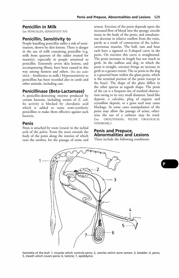
Penicillin in Milk(see PENICILLIN, SENSITIVITY TO)
Penicillin, Sensitivity toPeople handling penicillin suffer a risk of sensi-tisation, shown by skin lesions. There is dangerin the use of milk containing penicillin (e.g.milk from quarters of the udder treated formastitis), especially in people sensitised to penicillin. Extremely severe skin lesions, andaccompanying illness, have been caused in thisway among farmers and others. (See also under
MILK – Antibiotics in milk.) Hypersensitivity topenicillins has been recorded also in cattle andother animals, including cats.
Penicillinase (Beta-Lactamase)A penicillin-destroying enzyme produced bycertain bacteria, including strains of E. coli. Its activity is blocked by clavulanic acid which is added to some semi-synthetic penicillins to make them effective against suchbacteria.
PenisPenis is attached by roots (crura) to the ischialarch of the pelvis. From the roots extends thebody of the penis along the interior of whichruns the urethra, for the passage of urine and
semen. Erection of the penis depends upon theincreased flow of blood into the spongy, erectiletissue in the body of the penis, and simultane-ous decrease in relative outflow from the veins,partly as a result of contraction of the ischio-cavernosus muscles. The bull, ram and boareach have a sigmoid or S-shaped curve in thepenis. On erection this curve is straightened.The penis increases in length but not much ingirth. In the stallion and dog, in which thepenis is straight, erection brings an increase ingirth to a greater extent. The os penis in the dogis a grooved bone within the glans penis, whichis the terminal portion of the penis (except inthe boar). The shape of the glans differs in the other species as regards shape. The penis of the cat is a frequent site of urethral obstruc-tion owing to its very small diameter. Sand-likedeposit, a calculus, plug of organic and crystalline deposit, or a grass seed may causeblockage. In some cases manipulation of thepenis may allow the passage of urine; other-wise the use of a catheter may be tried. (See UROLITHIASIS; FELINE UROLOGICAL
SYNDROME.)
Penis and Prepuce,Abnormalities and LesionsThese include the following conditions:
Penis and Prepuce, Abnormalities and Lesions 529
P
Genitalia of the bull: 1, muscles which controls penis; 2, vesicles which store semen; 3, bladder; 4, penis;5, sheath which covers penis; 6, testicle; 7, epididymis.
Necrosis of the prepuce has been recorded inpigs kept in poor hygienic conditions. There isno indication that the condition causes pain ordistress.
Phimosis A narrowing of the orifice of theprepuce, preventing normal protrusion of thepenis. Congenital phimosis is occasionally seenin dogs, cats, and horses. It can be correctedsurgically.
Paraphimosis A condition in which thepenis cannot be retracted into the prepuce. Thisis not uncommon in the dog, and is potentiallyserious unless quickly relieved, owing to interference with the circulation. As a first-aidmeasure, swabbing the penis with ice-coldwater may prove helpful.
Prolapse of the penis due to paralysis is seen in bulls which have rabies, and also as achronic condition in Zebu cattle (Bos indicus)and occasionally in breeds of UK origin.
Horses Prolapse of the penis may follow use of acepromazine (to effect relaxation of the retractor muscles so that swabbing can beundertaken for contagious equine metritis). In6 cases involving protrusion, oedema, and paresis of the penis, 4 of the horses had receivedacepromazine with etorphine hydrochloride,and 2 had received the former drug along withothers.
After such drugs have been used, it is impor-tant to check that retraction of the penis is taking place as the effects of medication wearoff. If not, treatment should be started withoutdelay.
Priapism A persistent erection of the penisunassociated with sexual stimulation. (See also
PRIAPISM.)
Balanitis Inflammation of the glans penis – thecap-shaped spongy tissue at the end of the penis,normally covered by the foreskin.
Posthitis Inflammation of the prepuce.
Balanoposthitis A viral infection affectingboth the penis and the prepuce. One example isan enzootic form, called ‘pizzle rot’ or ‘sheathrot’, of sheep (especially merinos) in Australia.Corynebacterium renale is the primary cause.
A herpesvirus also causes balanoposthitis incattle and sheep.
An infectious balanoposthitis (also known as‘ulcerative dermatosis’) of sheep may affect not
only the penis, prepuce and vulva but also the face, feet and legs; this has to be differenti-ated from ORF. It occurs in Europe and South Africa. In lambs mortality may be up to30 per cent.
Tumours of the penis include WARTS (papillo-mas) and also, in dogs, infective granulomas.(See VENEREAL TUMOURS.)
Traumatic lesions include injury to thepenis from a kick by the cow or mare at service,or when a bull proves too heavy for a heifer.There is usually an accompanying haematoma.Trauma may also result in adhesions at the sig-moid flexure. In dogs a fracture of the os penismay occur as the result of being hit by a car.
Spiral deviation of the penis occurs inbulls. Service is prevented. (In the USA devia-tion of the penis is sometimes deliberately produced surgically in teaser bulls.)
Penitrem AA mycotoxin. It was isolated from mouldycream cheese from a refrigerator given to a dogwhich became very ill with ataxia, musculartremors, and opisthotonos. The mould wasidentified as Penicillium crustosum.
PentastomiasisInfection with the nymphs of the pentastomidArmillifer armillatus, the adults of which infestsnakes in Africa and Asia.
The disease has been recognised in man, dogand cat.
Signs Abdominal or thoracic oedema.Infection occurs through drinking water
contaminated by the eggs, or eating a snake.
Diagnosis In human medicine this is basedon the radiographic appearance of calcifiednymphs.
Pentobarbitone Sodium(Pentobarbital)(Sodium ethyl/methylbutyl barbiturate). Awhite crystalline powder, soluble in water, andused for its narcotic and anaesthetic effects.
First used as a general anaesthetic in veteri-nary surgery in America in 1931. A proprietaryname is Nembutal.
Pentobarbitone has been used to produceanaesthesia in all the domestic animals includ-ing the fowl, but it is not recommended forhorses, calves, or sheep. For anaesthesia in thedog and cat, however, pentobarbitone is very
530 Penitrem A
P
extensively employed, and is usually given bythe intravenous route – a method which per-mits of varying depths of anaesthesia beingobtained and the avoidance of overdosage. Thedrug may also be given by intra-peritonealinjection or by mouth; narcosis being thenslower in onset (8 to 20 minutes), and occa-sionally preceded by some degree of excitement,while the dose has to be an estimate calculatedon the basis of bodyweight.
Deep anaesthesia with pentobarbitone maylast for an hour, being followed by 2 to 7 hoursof narcosis. (See ANAESTHESIA, GENERAL;EUTHANASIA.)
PepsinPepsin is an enzyme found in the gastric juicewhich digests proteins.
PeptidesPeptides are composed of 2 or more aminoacids, and represent an intermediate stage in thedigestion of protein.
Polypeptides are proteins composed of several amino-acids linked by the peptidegrouping CH– CO–NH–CH.
Synthetic polypeptides have potentialuses as vaccines, e.g. against foot-and-mouthdisease.
Peptococcus IndolicusA Gram-positive bacterium which is sometimesa complicating factor in CASEOUS LYMPHADENI-
TIS of sheep and summer mastitis in cattle (see
MASTITIS IN COWS, ‘SUMMER MASTITIS’).
PerforationPerforation is one of the serious dangersattached to the presence of ulcerating condi-tions in the stomach and bowels. When a per-foration of one of these hollow organs takesplace in the peritoneal cavity, multitudes of bacteria, much ingesta, mucus, and otherputrescible materials escape and set up PERI-
TONITIS. The immediate signs are a collapse of the patient, with, later, collections of gas orfluids in the abdominal cavity. It is not uncom-mon to observe vomiting in the horse when thestomach ruptures and the contents escape intothe abdominal cavity; this is one of the very raretimes when the horse is seen to vomit, and it isimportant accordingly.
Performance TestingA method of comparing strains or breeds of,e.g. beef cattle, by studying liveweight gains
over a stated period with given rations. (See also
PROGENY TESTING.)
Peri-Peri- is a prefix meaning round, or about.Examples: PERICARDIUM; perianal abscess.
PericarditisInflammation of the pericardium. Traumaticpericarditis is common in cattle as a result ofswallowing pieces of wire, nails, etc. (See also
HEART DISEASES.)
PericardiumPericardium is the smooth lubricating mem-brane which surrounds the heart. (See also HEART
DISEASES.)
PerineumPerineum is the region lying between the anusand the genital organs in the male, and lyingbetween the anus and the mammary region inthe female of the horse, ox, sheep, goat, and pig.In bitches and cats the female genital organs lielower than in other animals, and in them theperineum lies between the anus and the vulva.Rupture of the perineum sometimes occurs inthe cow at calving, when the fetus over-distendsthe vulva. Suturing, under local anaesthesia, isusually required.
Periodic OphthalmiaSpecific ophthalmia, or ‘moon blindness’, is acondition of the eyes of horses, due to inflam-mation of the uveal tract (especially of the irisand ciliary body) which is characterised by atendency to recur.
Causes These are still in doubt, but it isknown that leptospirosis is one. Some 2 to 8months after acute leptospirosis, periodic ophthalmia appears in up to 45 per cent of thehorses affected. Leptospires have been isolatedfrom eye lesions over long periods.
Signs In the 1st stage a horse is found onemorning with the eyelids on one side half-closed; tears run from the eye down the face,and any effort to examine the eye is resented.Bright light is avoided, and the eyeball appearssunken in the socket. This period of inflamma-tion may last up to 10 days, after which it gradually disappears and the eye returns topractically its normal appearance. Repeatedattacks are apt to occur. Total blindness mayfollow and/or lesions of the retina. Periodicophthalmia is common in Europe, the USA,and Asia.
Periodic Ophthalmia 531
P
Periodontal(see TEETH, DISEASES OF)
Periople(see FOOT OF THE HORSE)
PeriosteumPeriosteum is the membrane surrounding abone. The growth of a bone in its thickness isdue to the action of the cells of this membraneforming fibrous tissue in which lime salts aredeposited. (See BONE.)
PeriostitisPeriostitis means inflammation on the surfaceof a bone affecting the periosteum. (See BONE,DISEASES OF.)
PeriparturientUsed to describe any condition occurring shortlybefore or shortly after birth.
PeristalsisPeristalsis is the succession of involuntary mus-cular contractions which propel ingested foodalong the alimentary canal. (See INTESTINES.)
PeritoneumPeritoneum is the membrane lining the abdom-inal cavity, and forming a covering for theorgans contained in it. That part lining thewalls of the cavity is called the ‘parietal’ peri-toneum, and that part covering the viscera isknown as the ‘visceral’ peritoneum. Betweenthe 2 parts is a film of lubricating liquid.
PeritonitisInflammation of the PERITONEUM. It may beeither localised or diffused.
Acute peritonitis
Causes The direct cause of acute peritonitis isnearly always the invasion of the membrane by micro-organisms. It occurs through a woundin the abdominal wall, in the stomach orintestines, uterus, bladder, etc., or through thespreading of inflammatory conditions from oneor other of these parts. It may follow castration,when the infection gains entrance by theinguinal canal. Peritonitis may occur during the course of anthrax, acute tuberculosis, etc.
Signs These include restlessness and signs ofdistress and pain. Horses and cattle usuallyremain standing, but the smaller animals liealmost continually. The temperature is raised by5° to 10°C (3° to 6°F), and the pulse is quick,
small, and wiry. Faeces and urine are usuallyretained and lead to further complication, andvomiting in dogs is common. Pressure over the sides of the abdomen is painful; the animalusually ‘boards’ the muscles of the abdomen,and may groan or grunt. As the disease pro-gresses, fluid may be thrown out into the cavityin great quantities, leading to ASCITES. (See
OEDEMA and PARACENTESIS.)
Treatment Operative treatment and drainagemay be undertaken.
Antibiotics and/or sulfa drugs may be givenby injection. Hot fomentations to the abdomenrelieve the acute pain. The prognosis is seldomgood.
Chronic peritonbitis
Causes Slowly-forming abscesses in the liver,tuberculous lesions in the peritoneal cavity, foreign bodies in the reticulum, etc.
Signs There may be slight attacks of pain at times, but very often it is only after deaththat the condition is discovered. Ascites and agradual loss of condition may be seen.
Treatment This will vary according to thenature of the infection or of adhesions. (See
ANTIBIOTIC.)
PermethrinA synthetic pyrethroid (derivative of pyrethrum)used as an insecticide. Excessive applications tocats can induce hyperaesthesia, with excitement,a staggering gait, muscular twitching, and occa-sional collapse (see INSECTICIDES; FLIES – Flycontrol measures).
PeronealRelating to the fibula.
Perosis(see under ‘SLIPPED TENDON’)
Perruque AntlersPerruque antlers describes a defect in antlergrowth in red deer where the antlers formclumps close to the head.. In severe cases, theanimal has the appearance of wearing a judge’swig, hence the name.
Perthe’s DiseaseThis name is given to a deformed condition ofthe head of the femur in the dog. The animal isnoticed to be lame. The condition may clear upspontaneously within 6 months, but during
532 Periodontal
P
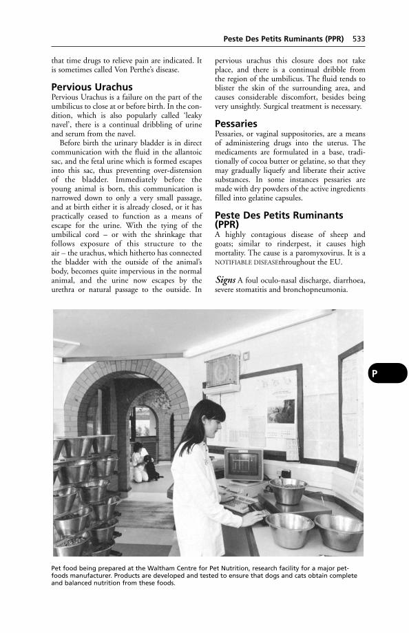
that time drugs to relieve pain are indicated. Itis sometimes called Von Perthe’s disease.
Pervious UrachusPervious Urachus is a failure on the part of theumbilicus to close at or before birth. In the con-dition, which is also popularly called ‘leakynavel’, there is a continual dribbling of urineand serum from the navel.
Before birth the urinary bladder is in directcommunication with the fluid in the allantoicsac, and the fetal urine which is formed escapesinto this sac, thus preventing over-distension of the bladder. Immediately before the young animal is born, this communication is narrowed down to only a very small passage,and at birth either it is already closed, or it haspractically ceased to function as a means ofescape for the urine. With the tying of theumbilical cord – or with the shrinkage that follows exposure of this structure to the air – the urachus, which hitherto has connectedthe bladder with the outside of the animal’sbody, becomes quite impervious in the normalanimal, and the urine now escapes by the urethra or natural passage to the outside. In
pervious urachus this closure does not takeplace, and there is a continual dribble from the region of the umbilicus. The fluid tends toblister the skin of the surrounding area, andcauses considerable discomfort, besides beingvery unsightly. Surgical treatment is necessary.
PessariesPessaries, or vaginal suppositories, are a meansof administering drugs into the uterus. Themedicaments are formulated in a base, tradi-tionally of cocoa butter or gelatine, so that theymay gradually liquefy and liberate their activesubstances. In some instances pessaries aremade with dry powders of the active ingredientsfilled into gelatine capsules.
Peste Des Petits Ruminants(PPR)A highly contagious disease of sheep and goats; similar to rinderpest, it causes high mortality. The cause is a paromyxovirus. It is aNOTIFIABLE DISEASEthroughout the EU.
Signs A foul oculo-nasal discharge, diarrhoea,severe stomatitis and bronchopneumonia.
Peste Des Petits Ruminants (PPR) 533
P
Pet food being prepared at the Waltham Centre for Pet Nutrition, research facility for a major pet-foods manufacturer. Products are developed and tested to ensure that dogs and cats obtain completeand balanced nutrition from these foods.
Pestivirus(see BOVINE VIRAL DIARRHOEA; BORDER
DISEASE OF SHEEP; SWINE FEVER; EQUINE VIRAL
ARTERITIS)
Pet Animals Act 1971Pet Animals Act 1971 covers the licensing ofpet shops, and the conditions under which animals are kept there and offered for sale.
Pet Bereavement SupportServiceA counselling service for people whose petshave died is operated jointly by the animalcharities Blue Cross and Society forCompanion Animal Studies. The telephonehelpline is open between 08.30 and 17.00; thenumber is 0800 0966606.
PetlogA UK national scheme for identifying pets bymicrochipping. The Petlog ReunificationService helps to trace lost microchipped animalsand reunite them with their owners.
Pet FoodsPet foods come under the Feeding Stuffs(Sampling and Analysis) Regulations 1982. The Feeding Stuffs Regulations 2000 list the special diets which are permitted. Together,these regulations satisfy the requirements of theappropriate EU directives. (See also DIET andDIETETICS; DOGS’ DIET; CAT FOODS, etc.)
Pet Travel Scheme (PETS)An arrangement by which cats and dogs maytravel to and from European Union countries,Norway, Australia, New Zealand, Japan, andcertain islands in the Atlantic and theCaribbean without having to go into quaran-tine. This was made possible under the PetTravel Scheme (Pilot Arrangements) Order1999 (as subsequently amended). Strict condi-tions apply: the animals must have been identity microchipped, vaccinated againstrabies at least 6 months previously and havetheir blood levels of rabies-immunity tested bya DEFRA-approved laboratory before beingallowed to travel. In the case of dogs, vaccina-tion against distemper is obligatory. Most coun-tries also require an export health certificatebefore a pet will be allowed to enter. Prior toarrival in Britain, the animals must have beenwormed, especially against the tapewormEchinococcus multilocularis, and have been treated for the presence of ticks. Failure toobserve those requirements will lead to the animal being quarantined. In the first 3 years of
the scheme, 136,500 dogs and cats enteredBritain without being quarantined. In 2003alone, there were 48,329 dogs and 5838 cats.
Companies operating the scheme have toprovide trained staff to deal with checking animals and make available adequate facilitiesfor handling them.
Details are available from PETS Helpline,telephone 0870 2411710; fax 020 7904 6834;e-mail [email protected] or from theDEFRA website at www.defra.gov.uk/animalh/quarantine.
PetechiaePetechiae are small spots on the surface of anorgan or the skin, generally red or purple incolour and resembling flea-bites. They may be minute areas of inflammation or they maybe small haemorrhages. (Petechial fever isanother name for PURPURA HAEMORRHAGICA.)
PethidineAn analgesic (pain reliever) used for dogs andcats. It may also be used in rabbits and rodentsand for the relief of the pain of colic in horses.
Petri DishA shallow circular glass dish with lid in whichbacteria are grown on a solid medium.
Pets, Children’s and ExoticFor information on the breeding, care, diseasesand treatment of mice, rats, rabbits, hamsters,guinea pigs, gerbils, reptiles, fish, birds, andmonkeys, the reader is referred to the BritishSmall Animal Veterinary Association’s Manualof the Care and Treatment of Children’s andExotic Pets. (See also under CAGE AND AVIARY
BIRDS, DISEASES OF; MONKEYS; HAMSTERS;GUINEA-PIG; AMERICAN BOX TORTOISES;TORTOISES; PIGS – Foreign breeds; CHINCHILLA;also ANAESTHESIA, GENERAL.
Peyer’s PatchesPeyer’s patches are lymph follicles on the smallintestine; they appear as raised oval areas inmucous and submucous areas. In sheep theyhave a function analogous to that of the bursaof Fabricius in birds. An investigation of jejunalPeyer’s patches (JPP) and ileocecal Peyer’spatches (IPP) showed that, in JPPs, there are big interfollicular T-cell areas, but IPPs contained mainly B-lymphocytes. Germinalcentres of JPPs had about 40 per cent IgM positive cells and, in IPPs, about 80 per cent of these cells were present. (Larsen, H. J. &Landsverk, T. Research in Veterinary Science, 40, 105.) (See also under INTESTINES.)
534 Pestivirus
P
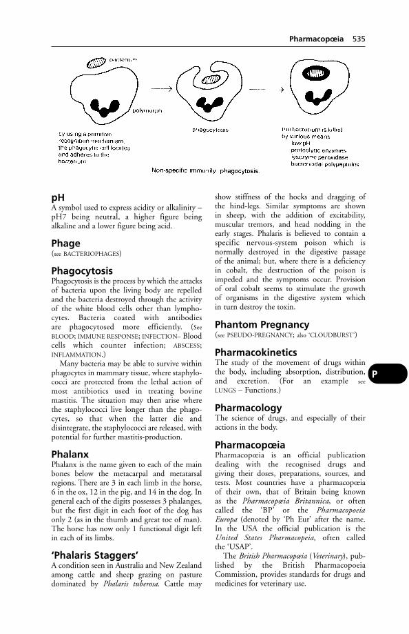
pHA symbol used to express acidity or alkalinity –pH7 being neutral, a higher figure being alkaline and a lower figure being acid.
Phage(see BACTERIOPHAGES)
PhagocytosisPhagocytosis is the process by which the attacksof bacteria upon the living body are repelledand the bacteria destroyed through the activityof the white blood cells other than lympho-cytes. Bacteria coated with antibodies are phagocytosed more efficiently. (See
BLOOD; IMMUNE RESPONSE; INFECTION– Bloodcells which counter infection; ABSCESS;INFLAMMATION.)
Many bacteria may be able to survive withinphagocytes in mammary tissue, where staphylo-cocci are protected from the lethal action ofmost antibiotics used in treating bovine mastitis. The situation may then arise where the staphylococci live longer than the phago-cytes, so that when the latter die and disintegrate, the staphylococci are released, withpotential for further mastitis-production.
PhalanxPhalanx is the name given to each of the mainbones below the metacarpal and metatarsalregions. There are 3 in each limb in the horse,6 in the ox, 12 in the pig, and 14 in the dog. Ingeneral each of the digits possesses 3 phalanges,but the first digit in each foot of the dog hasonly 2 (as in the thumb and great toe of man).The horse has now only 1 functional digit leftin each of its limbs.
‘Phalaris Staggers’A condition seen in Australia and New Zealandamong cattle and sheep grazing on pasturedominated by Phalaris tuberosa. Cattle may
show stiffness of the hocks and dragging of the hind-legs. Similar symptoms are shown in sheep, with the addition of excitability, muscular tremors, and head nodding in theearly stages. Phalaris is believed to contain aspecific nervous-system poison which is normally destroyed in the digestive passage of the animal; but, where there is a deficiency in cobalt, the destruction of the poison isimpeded and the symptoms occur. Provision of oral cobalt seems to stimulate the growth of organisms in the digestive system which in turn destroy the toxin.
Phantom Pregnancy(see PSEUDO-PREGNANCY; also ‘CLOUDBURST’)
PharmacokineticsThe study of the movement of drugs within the body, including absorption, distribution,and excretion. (For an example see
LUNGS – Functions.)
PharmacologyThe science of drugs, and especially of theiractions in the body.
PharmacopœiaPharmacopœia is an official publication dealing with the recognised drugs and giving their doses, preparations, sources, andtests. Most countries have a pharmacopœia of their own, that of Britain being known as the Pharmacopœia Britannica, or often called the ‘BP’ or the Pharmacopoeia Europa (denoted by ‘Ph Eur’ after the name. In the USA the official publication is the United States Pharmacopeia, often called the ‘USAP’.
The British Pharmacopœia (Veterinary), pub-lished by the British PharmacopoeiaCommission, provides standards for drugs andmedicines for veterinary use.
Pharmacopœia 535
P
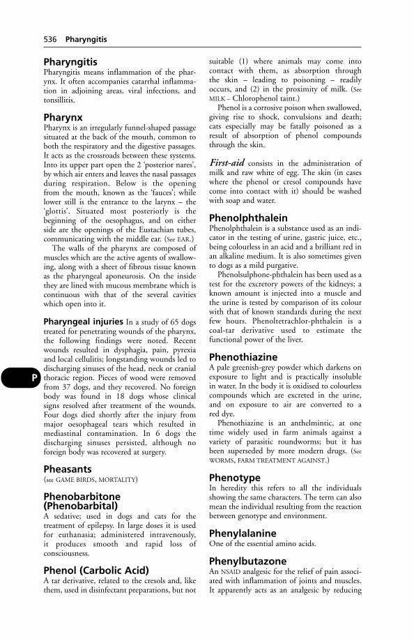
PharyngitisPharyngitis means inflammation of the phar-ynx. It often accompanies catarrhal inflamma-tion in adjoining areas, viral infections, andtonsillitis.
PharynxPharynx is an irregularly funnel-shaped passagesituated at the back of the mouth, common toboth the respiratory and the digestive passages.It acts as the crossroads between these systems.Into its upper part open the 2 ‘posterior nares’,by which air enters and leaves the nasal passagesduring respiration. Below is the opening from the mouth, known as the ‘fauces’; whilelower still is the entrance to the larynx – the‘glottis’. Situated most posteriorly is the beginning of the oesophagus, and on either side are the openings of the Eustachian tubes,communicating with the middle ear. (See EAR.)
The walls of the pharynx are composed ofmuscles which are the active agents of swallow-ing, along with a sheet of fibrous tissue knownas the pharyngeal aponeurosis. On the insidethey are lined with mucous membrane which iscontinuous with that of the several cavitieswhich open into it.
Pharyngeal injuries In a study of 65 dogstreated for penetrating wounds of the pharynx,the following findings were noted. Recentwounds resulted in dysphagia, pain, pyrexiaand local cellulitis; longstanding wounds led todischarging sinuses of the head, neck or cranialthoracic region. Pieces of wood were removedfrom 37 dogs, and they recovered. No foreignbody was found in 18 dogs whose clinical signs resolved after treatment of the wounds.Four dogs died shortly after the injury frommajor oesophageal tears which resulted inmediastinal contamination. In 6 dogs the discharging sinuses persisted, although no foreign body was recovered at surgery.
Pheasants(see GAME BIRDS, MORTALITY)
Phenobarbitone(Phenobarbital)A sedative; used in dogs and cats for the treatment of epilepsy. In large doses it is usedfor euthanasia; administered intravenously, it produces smooth and rapid loss of consciousness.
Phenol (Carbolic Acid)A tar derivative, related to the cresols and, likethem, used in disinfectant preparations, but not
suitable (1) where animals may come into contact with them, as absorption through the skin – leading to poisoning – readily occurs, and (2) in the proximity of milk. (See
MILK – Chlorophenol taint.)Phenol is a corrosive poison when swallowed,
giving rise to shock, convulsions and death; cats especially may be fatally poisoned as aresult of absorption of phenol compoundsthrough the skin.
First-aid consists in the administration ofmilk and raw white of egg. The skin (in caseswhere the phenol or cresol compounds havecome into contact with it) should be washedwith soap and water.
PhenolphthaleinPhenolphthalein is a substance used as an indi-cator in the testing of urine, gastric juice, etc.,being colourless in an acid and a brilliant red inan alkaline medium. It is also sometimes givento dogs as a mild purgative.
Phenolsulphone-phthalein has been used as atest for the excretory powers of the kidneys; aknown amount is injected into a muscle andthe urine is tested by comparison of its colourwith that of known standards during the nextfew hours. Phenoltetrachlor-phthalein is a coal-tar derivative used to estimate the functional power of the liver.
PhenothiazineA pale greenish-grey powder which darkens onexposure to light and is practically insoluble in water. In the body it is oxidised to colourlesscompounds which are excreted in the urine,and on exposure to air are converted to a red dye.
Phenothiazine is an anthelmintic, at onetime widely used in farm animals against a variety of parasitic roundworms; but it has been superseded by more modern drugs. (See
WORMS, FARM TREATMENT AGAINST.)
PhenotypeIn heredity this refers to all the individualsshowing the same characters. The term can alsomean the individual resulting from the reactionbetween genotype and environment.
PhenylalanineOne of the essential amino acids.
PhenylbutazoneAn NSAID analgesic for the relief of pain associ-ated with inflammation of joints and muscles.It apparently acts as an analgesic by reducing
536 Pharyngitis
P
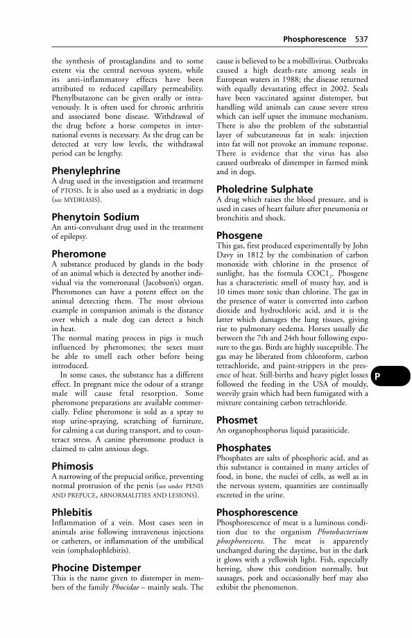
the synthesis of prostaglandins and to someextent via the central nervous system, while its anti-inflammatory effects have been attributed to reduced capillary permeability.Phenylbutazone can be given orally or intra-venously. It is often used for chronic arthritisand associated bone disease. Withdrawal of the drug before a horse competes in inter-national events is necessary. As the drug can bedetected at very low levels, the withdrawal period can be lengthy.
PhenylephrineA drug used in the investigation and treatmentof PTOSIS. It is also used as a mydriatic in dogs(see MYDRIASIS).
Phenytoin SodiumAn anti-convulsant drug used in the treatmentof epilepsy.
PheromoneA substance produced by glands in the body of an animal which is detected by another indi-vidual via the vomeronasal (Jacobson’s) organ.Pheromones can have a potent effect on theanimal detecting them. The most obviousexample in companion animals is the distanceover which a male dog can detect a bitch in heat.The normal mating process in pigs is muchinfluenced by pheromones; the sexes must be able to smell each other before being introduced.
In some cases, the substance has a differenteffect. In pregnant mice the odour of a strangemale will cause fetal resorption. Somepheromone preparations are available commer-cially. Feline pheromone is sold as a spray tostop urine-spraying, scratching of furniture, for calming a cat during transport, and to coun-teract stress. A canine pheromone product isclaimed to calm anxious dogs.
PhimosisA narrowing of the prepucial orifice, preventingnormal protrusion of the penis (see under PENIS
AND PREPUCE, ABNORMALITIES AND LESIONS).
PhlebitisInflammation of a vein. Most cases seen in animals arise following intravenous injectionsor catheters, or inflammation of the umbilicalvein (omphalophlebitis).
Phocine DistemperThis is the name given to distemper in mem-bers of the family Phocidae – mainly seals. The
cause is believed to be a mobillivirus. Outbreakscaused a high death-rate among seals inEuropean waters in 1988; the disease returnedwith equally devastating effect in 2002. Sealshave been vaccinated against distemper, buthandling wild animals can cause severe stresswhich can iself upset the immune mechanism.There is also the problem of the substantiallayer of subcutaneous fat in seals: injection into fat will not provoke an immune response.There is evidence that the virus has also caused outbreaks of distemper in farmed minkand in dogs.
Pholedrine SulphateA drug which raises the blood pressure, and isused in cases of heart failure after pneumonia orbronchitis and shock.
PhosgeneThis gas, first produced experimentally by JohnDavy in 1812 by the combination of carbonmonoxide with chlorine in the presence of sunlight, has the formula COC12. Phosgenehas a characteristic smell of musty hay, and is10 times more toxic than chlorine. The gas inthe presence of water is converted into carbondioxide and hydrochloric acid, and it is the latter which damages the lung tissues, givingrise to pulmonary oedema. Horses usually diebetween the 7th and 24th hour following expo-sure to the gas. Birds are highly susceptible. Thegas may be liberated from chloroform, carbontetrachloride, and paint-strippers in the pres-ence of heat. Still-births and heavy piglet lossesfollowed the feeding in the USA of mouldy,weevily grain which had been fumigated with amixture containing carbon tetrachloride.
PhosmetAn organophosphorus liquid parasiticide.
PhosphatesPhosphates are salts of phosphoric acid, and asthis substance is contained in many articles offood, in bone, the nuclei of cells, as well as inthe nervous system, quantities are continuallyexcreted in the urine.
PhosphorescencePhosphorescence of meat is a luminous condi-tion due to the organism Photobacterium phosphorescens. The meat is apparentlyunchanged during the daytime, but in the darkit glows with a yellowish light. Fish, especiallyherring, show this condition normally, butsausages, pork and occasionally beef may alsoexhibit the phenomenon.
Phosphorescence 537
P

PhosphorusPhosphorus itself is not used in medicine, but is usually given in the form of one or other of the glycerophosphates, or hypophosphites ofsodium, potassium, calcium, magnesium, oriron. Preparations are used with calcium anddextrose in the treatment of MILK FEVER.
Phosphorus DeficiencyThis is seen in RICKETS; ‘MILK LAMENESS’; POST-
PARTURIENT HAEMOGLOBINURIA; and maycomplicate MILK FEVER. (See also INFERTILITY.)
Phosphorus PoisoningPhosphorus poisoning may occur in the dogand cat, either through puppies eating matches,or from animals gaining access to rat poisonmade with phosphorus.
Signs When an animal has been poisoned byphosphorus there is acute abdominal pain,vomiting, intense thirst, diarrhoea, and greatdullness. The material vomited may be green incolour and is often luminous in the darkness.Collapse rapidly follows, and the animal dies ina few hours. Where less has been taken, deathmay not occur for 2 or 3 days.
First-aid An emetic should be given at once, assoon as the symptoms appear, Washing-soda crys-tals (sodium carbonate), salt (sodium chloride) ormustard placed at the back of the tongue areeffective emetics. Sulphate of copper (bluestone),in solution, may be given: this induces vomiting,gets rid of the majority of the phosphorus, andrenders inert what remains. In 15 minutes anoth-er dose dissolved in water as before should begiven, and this should be repeated every quarterof an hour till 4 doses have been given. In allcases, white of egg, milk, oils, and fatty sub-stances must be avoided; these dissolve the phos-phorus and render it able to be absorbed withgreater rapidity. (See also ORGANOPHOSPHORUS
POISONING, which arises from contaminationswith certain farm chemicals.)
Phrenic NerveThis arises from the 5th, 6th, and 7th cervicalspinal nerves, passes through the thoracic cavity, and ramifies in the muscular part of thediaphragm.
PhthiriasisInfestation with lice of the genus Phthirus.
PhycomycosisA group of fungal infections by species ofAbsidia, Mucor, Rhizopus, etc., which affects the
lymph nodes and may give rise to loss of weightand tumour-like (granulomatous) swellings. Itmay affect the lungs and intestines. A monkeydied from systemic phycomycosis in the UKafter loss of appetite, depression, and labouredbreathing.
PhylloerythrinA substance formed in the rumen from chloro-phyll by bacterial digestion. Some is absorbedand excreted in the bile, but when the liver isdamaged in any way the phylloerythrin mayreach the peripheral circulation and give rise toLIGHT SENSITISATION.
PhysiotherapyIn the UK, some veterinary practices use the services of qualified physiotherapists for the treatment of selected cases of disease in animals.
PhysisA synonym for EPIPHYSIS (see also BONE,DISEASES OF).
PhysitisInflammation or disease of the growth plate (see EPIPHYSITIS).
PhysostigminePhysostigmine is an anticholinesterase. Eyedrops containing physostigmine sulphate are used to contract the pupil and lessen intra-ocular pressure. Alternated with atropine,it has been used for adhesions of the iris, following iritis. It is prepared from the ripeseeds of Physostigma venenosum, a tree fromWest Africa.
PhytinA substance present in oatmeal, maize meal(and in other cereals) that binds to phosphorus,making it unavailable to animals. As a result,calcification of bone is disturbed and rickettsmay result. Phytase is added to many diets tobreak down phytin and remove this risk.
Pia MaterPia mater is the membrane that closely investsthe brain and spinal cord. (See BRAIN; SPINAL
CORD.)
PicaDepraved appetite. It is often the result of adeficiency in the diet such as lack of fibre or salt, or inadequate trace elements such asphosphorus or copper. (For causes, see under
APPETITE.)
538 Phosphorus
P
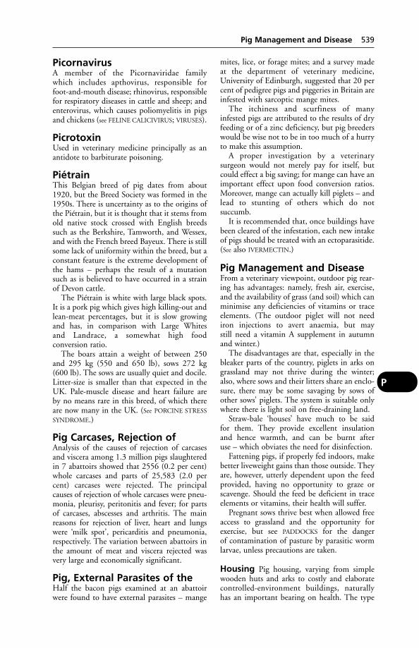
PicornavirusA member of the Picornaviridae family which includes apthovirus, responsible for foot-and-mouth disease; rhinovirus, responsiblefor respiratory diseases in cattle and sheep; andenterovirus, which causes poliomyelitis in pigsand chickens (see FELINE CALICIVIRUS; VIRUSES).
PicrotoxinUsed in veterinary medicine principally as anantidote to barbiturate poisoning.
PiétrainThis Belgian breed of pig dates from about1920, but the Breed Society was formed in the1950s. There is uncertainty as to the origins ofthe Piétrain, but it is thought that it stems fromold native stock crossed with English breedssuch as the Berkshire, Tamworth, and Wessex,and with the French breed Bayeux. There is stillsome lack of uniformity within the breed, but aconstant feature is the extreme development ofthe hams – perhaps the result of a mutationsuch as is believed to have occurred in a strainof Devon cattle.
The Piétrain is white with large black spots.It is a pork pig which gives high killing-out andlean-meat percentages, but it is slow growingand has, in comparison with Large Whites and Landrace, a somewhat high food conversion ratio.
The boars attain a weight of between 250and 295 kg (550 and 650 lb), sows 272 kg (600 lb). The sows are usually quiet and docile.Litter-size is smaller than that expected in theUK. Pale-muscle disease and heart failure are by no means rare in this breed, of which thereare now many in the UK. (See PORCINE STRESS
SYNDROME.)
Pig Carcases, Rejection ofAnalysis of the causes of rejection of carcasesand viscera among 1.3 million pigs slaughteredin 7 abattoirs showed that 2556 (0.2 per cent)whole carcases and parts of 25,583 (2.0 percent) carcases were rejected. The principal causes of rejection of whole carcases were pneu-monia, pleurisy, peritonitis and fever; for partsof carcases, abscesses and arthritis. The mainreasons for rejection of liver, heart and lungswere ‘milk spot’, pericarditis and pneumonia,respectively. The variation between abattoirs inthe amount of meat and viscera rejected wasvery large and economically significant.
Pig, External Parasites of theHalf the bacon pigs examined at an abattoirwere found to have external parasites – mange
mites, lice, or forage mites; and a survey madeat the department of veterinary medicine,University of Edinburgh, suggested that 20 percent of pedigree pigs and piggeries in Britain areinfested with sarcoptic mange mites.
The itchiness and scurfiness of many infested pigs are attributed to the results of dryfeeding or of a zinc deficiency, but pig breederswould be wise not to be in too much of a hurryto make this assumption.
A proper investigation by a veterinary surgeon would not merely pay for itself, butcould effect a big saving; for mange can have animportant effect upon food conversion ratios.Moreover, mange can actually kill piglets – andlead to stunting of others which do not succumb.
It is recommended that, once buildings havebeen cleared of the infestation, each new intakeof pigs should be treated with an ectoparasitide.(See also IVERMECTIN.)
Pig Management and DiseaseFrom a veterinary viewpoint, outdoor pig rear-ing has advantages: namely, fresh air, exercise,and the availability of grass (and soil) which canminimise any deficiencies of vitamins or traceelements. (The outdoor piglet will not neediron injections to avert anaemia, but may still need a vitamin A supplement in autumnand winter.)
The disadvantages are that, especially in thebleaker parts of the country, piglets in arks ongrassland may not thrive during the winter;also, where sows and their litters share an enclo-sure, there may be some savaging by sows ofother sows’ piglets. The system is suitable onlywhere there is light soil on free-draining land.
Straw-bale ‘houses’ have much to be said for them. They provide excellent insulation and hence warmth, and can be burnt after use – which obviates the need for disinfection.
Fattening pigs, if properly fed indoors, makebetter liveweight gains than those outside. Theyare, however, utterly dependent upon the feedprovided, having no opportunity to graze orscavenge. Should the feed be deficient in traceelements or vitamins, their health will suffer.
Pregnant sows thrive best when allowed freeaccess to grassland and the opportunity forexercise, but see PADDOCKS for the danger of contamination of pasture by parasitic wormlarvae, unless precautions are taken.
Housing Pig housing, varying from simplewooden huts and arks to costly and elaboratecontrolled-environment buildings, naturallyhas an important bearing on health. The type
Pig Management and Disease 539
P
and quality of building materials can be asimportant as the design of the building.Abrasive concrete can lead to injuries and abscesses, resulting in lameness. (See
CONCRETE.)Straw bedding is the ideal from a health
angle. It can keep the pigs warmer, improveliveweight gains, obviate boredom (a possiblecause of tail-biting), and reduce stress. (See
BEDDING AND BEDDING MATERIALS.)Recommended temperatures for pigs are
given under HOUSING OF ANIMALS. With controlled-environment buildings, precautionsmust be taken against the effects of power-cuts:it is useful to have a tractor-pto generator for such emergencies. These may arise duringboth winter and summer, and be due not to apower-cut but to fuses blowing in the buildingitself. In one case, fuses blew during a thunder-storm, and fail-safe ventilators failed to operate, with the result that 500 pigs inside that building died of heat-stroke. (DEFRA veterinary investigation service report.)
As pigs are particularly susceptible to theeffects of water deprivation, precautions mustbe taken to ensure that water pipes do notfreeze, and that the levers of automatic drinkersare not too stiff for piglets to manage. (See under
WATER AND WATERING OF ANIMALS for otherdangers).
Feeding stalls prevent greedy and aggressivesows from obtaining more than their fair shareof feed, leaving others undernourished.However, confinement of sows in stalls (otherthan for feeding only) deprives the animals ofany opportunity for the slightest exercise, andthey are unable to move away to escape any cold draughts. Stress results (see THIN SOW
SYNDROME).Stress (which reduces bodily resistance to
infection) may also occur at times when pigs aremoved from one building to another, or whenlitters are first mixed. Accordingly, the use offarrowing-to-finish pens has been advocated.
However, relatively few fatteners breed theirown pigs. Bought-in pigs are best kept awayfrom other stock on the farm for 3 or 4 weeks.Young pigs are said to do better at this period if they are kept as far away from their dung as possible, and this is one reason why slatted floors and floor feeding rarely work atthis stage.
A popular way of achieving good accommo-dation at this time is to provide a simple covered straw yard allowing about 3 m2
(10 sq ft) per pig of total area preferably withpart of the area ‘kennelled’ to give a warm sleeping area. If the latter is raised and dark it
will usually be kept clean, and the dung placed in the lighter and lower strawed area.Suitable-sized groups are of 25 to 30 weanersand a lean-to yard will be as cheap a method ofhousing as any, particularly with ad lib feedingfrom large hoppers.
For the farmer who cannot use straw or otherbedding, there are a number of designs of kennel-type pens with covered or uncoveredyards where the muck can be readily clearedaway, with tractor or squeegee, or with a hose to wash it down a drain. An essential of this system is to have pigs in small and separatedgroups perhaps no more than 20 to a pen.
After the conditioning period, the usualpractice is to finish the pigs under more inten-sive conditions. This often involves keepingpigs in litter-less pens, and there is little doubtthat this is the type of environment that can beconducive to tail-biting and cannibalism.
Ventilation – at pig level – becomes all themore important in such circumstances. Railedor Weldmesh pen fronts or sides are often pre-ferred as they allow a much better circulation ofair in a low-roofed building in particular. Theiradvantages do not stop there. Many farmersfind that it is difficult to get the younger pigdunging in the passage rather than in the pen: asure help is a gate of Weldmesh or of bars, as anew group will appear to follow the habits of itsolder companions on the other side of the gate.
It is good practice to have separate sections orunits which can be cleaned and disinfectedbetween batches.
The age for weaning piglets is usually 5 to 7weeks. Where for any reason the sow is notwanted for breeding again immediately, theperiod of suckling may be extended by an additional week or two. In countries with asevere winter climate, where only spring littersare satisfactory and where consequently only 1 litter per year is bred, the last course is that normally followed. (For early weaning, see under
WEANING.)It is not normally advisable to put pigs into
the finishing house until they are at least 32 kgin weight, and most feeders continue the weaner-pool system until this weight is reached.
Feeding Many unsatisfactory results aredirectly attributable to badly balanced rations.(See CONCENTRATES.)
The growing pig needs a diet that providesthe amino acids required to build body proteins as well as vitamins. These (particularlyA, D and E) are best added as a special preparation according to the manufacturers’
540 Pig Management and Disease
P
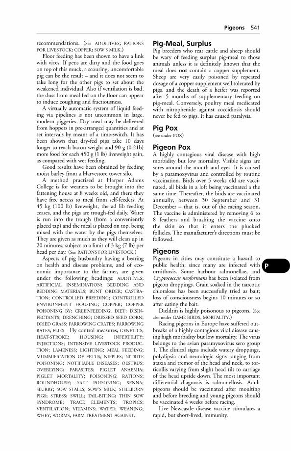
recommendations. (See ADDITIVES; RATIONS
FOR LIVESTOCK; COPPER; SOW’S MILK.)Floor feeding has been shown to have a link
with vices. If pens are dirty and the food goeson top of this muck, a scouring, uncomfortablepig can be the result – and it does not seem totake long for the other pigs to set about theweakened individual. Also if ventilation is bad,the dust from meal fed on the floor can appearto induce coughing and fractiousness.
A virtually automatic system of liquid feed-ing via pipelines is not uncommon in large,modern piggeries. Dry meal may be deliveredfrom hoppers in pre-arranged quantities and atset intervals by means of a time-switch. It hasbeen shown that dry-fed pigs take 10 dayslonger to reach bacon-weight and 90 g (0.21b)more food for each 450 g (1 lb) liveweight gain,as compared with wet feeding.
Good results have been obtained by feedingmoist barley from a Harvestore tower silo.
A method practised at Harper AdamsCollege is for weaners to be brought into thefattening house at 8 weeks old, and there theyhave free access to meal from self-feeders. At 45 kg (100 lb) liveweight, the ad lib feedingceases, and the pigs are trough-fed daily. Wateris run into the trough (from a convenientlyplaced tap) and the meal is placed on top, beingmixed with the water by the pigs themselves.They are given as much as they will clean up in20 minutes, subject to a limit of 3 kg (7 lb) perhead per day. (See RATIONS FOR LIVESTOCK.)
Aspects of pig husbandry having a bearingon health and disease problems, and of eco-nomic importance to the farmer, are givenunder the following headings: ADDITIVES;ARTIFICIAL INSEMINATION; BEDDING AND
BEDDING MATERIALS; BUNT ORDER; CASTRA-
TION; CONTROLLED BREEDING; CONTROLLED
ENVIRONMENT HOUSING; COPPER; COPPER
POISONING BY; CREEP-FEEDING; DIET; DISIN-
FECTANTS; DRENCHING; DRESSED SEED CORN;DRIED GRASS; FARROWING CRATES; FARROWING
RATES; FLIES – Fly control measures; GENETICS;HEAT-STROKE; HOUSING; INFERTILITY;INJECTIONS; INTENSIVE LIVESTOCK PRODUC-
TION; LAMENESS; LIGHTING; MEAL FEEDING;MUMMIFICATION OF FETUS; NIPPLES; NITRITE
POISONING; NOTIFIABLE DISEASES; OESTRUS;OVERLYING; PARASITES; PIGLET ANAEMIA;PIGLET MORTALITY; POISONING; RATIONS;ROUNDHOUSE; SALT POISONING; SENNA;SLURRY; SOW STALLS; SOW’S MILK; STILLBORN
PIGS; STRESS; SWILL; TAIL-BITING; THIN SOW
SYNDROME; TRACE ELEMENTS; TROPICS;VENTILATION; VITAMINS; WATER; WEANING;WHEY; WORMS, FARM TREATMENT AGAINST.
Pig-Meal, SurplusPig breeders who rear cattle and sheep shouldbe wary of feeding surplus pig-meal to thoseanimals unless it is definitely known that themeal does not contain a copper supplement.Sheep are very easily poisoned by repeateddosage of a copper supplement well tolerated bypigs, and the death of a heifer was reported after 5 months of supplementary feeding onpig-meal. Conversely, poultry meal medicatedwith nitrophenide against coccidiosis shouldnever be fed to pigs. It has caused paralysis.
Pig Pox(see under POX)
Pigeon PoxA highly contagious viral disease with highmorbidity but low mortality. Visible signs aresores around the mouth and eyes. It is caused by a paramoxyvirus and controlled by routinevaccination. Birds over 5 weeks old are vacci-nated, all birds in a loft being vaccinated a thesame time. Thereafter, the birds are vaccinatedannually, between 30 September and 31December – that is, out of the racing season.The vaccine is administered by removing 6 to 8 feathers and brushing the vaccine onto the skin so that it enters the plucked follicles. The manufacturer’s directions must befollowed.
PigeonsPigeons in cities may constitute a hazard topublic health, since many are infected withornithosis. Some harbour salmonellae, andCryptococcus neoformans has been isolated frompigeon droppings. Grain soaked in the narcoticchloralose has been successfully tried as bait;loss of consciousness begins 10 minutes or soafter eating the bait.
Dieldrin is highly poisonous to pigeons. (See
also under GAME BIRDS, MORTALITY.)Racing pigeons in Europe have suffered out-
breaks of a highly contagious viral disease caus-ing high morbidity but low mortality. The virusbelongs to the avian paramyxovirus sero group1. The clinical signs include watery droppings,polydipsia and neurologic signs ranging fromataxia and tremor of the head and neck, to tor-ticollis varying from slight head tilt to carriageof the head upside down. The most importantdifferential diagnosis is salmonellosis. Adultpigeons should be vaccinated after moultingand before breeding and young pigeons shouldbe vaccinated 4 weeks before racing.
Live Newcastle disease vaccine stimulates arapid, but short-lived, immunity.
Pigeons 541
P

Ornithosis occurs in young racing pigeons, thesymptoms including diarrhoea, conjunctivitis,and nasal discharge. (See CHLAMYDIA.)
Pigeon pox is associated with vesicles around the beak and eyes, and with hardgrowths – which, if on the feet, may causelameness.
‘Canker’ may give rise to a 70 per cent mor-tality; it is caused by Trichomonas gallinae whichaffects the liver and alimentary canal.
Salmonellosis is often fatal in recentlyhatched birds, and in adults may cause diarrhoea, distressed breathing, swollen joints,lameness, dropped wing, and loss of weight.
Parasitic worms, especially capillaria, maycause huddling, loss of weight and anaemia.
Pigeons imported into the UK must be vaccinated against paramyxoviruses within 24hours of their arrival. Four weeks later theymust receive a 2nd dose of vaccine, and remainin quarantine for a further week.
Piglet AnaemiaA common cause of pre-weaning losses amonghoused pigs.
Cause The disease is associated with a defi-ciency of iron, and is aggravated by cold anddamp. (A deficiency of copper and cobalt maysometimes also occur.)
Signs Dullness, a pale whitish skin, scouring,and sometimes exaggerated heartbeats.
Treatment Turn sow and litter out to grass.Give an iron and copper preparation sold forthe purpose.
Prevention If outdoor rearing is not desired,give a suitable iron preparation (with cobalt andcopper, preferably) at 7 days of age. (A solutionmade by dissolving 25 g (2oz) of commercialiron pyro-phosphate in 500 ml (1 pint) of wateris effective; a quarter of a teaspoonful beinggiven daily for 4 or 5 days.) Place a fresh turf inthe farrowing house. Acute iron poisoning,often leading to death within 24 hours, some-times follows the injection or oral dosing ofnormally used iron preparations. To preventthis it is advisable to wait until the piglets are a week old, when this danger is reduced; it isalso wise to ensure that gilts’ rations containadequate vitamin E.
The intramuscular injections of iron dextranto prevent piglet anaemia are sometimes followed by ham abscesses – the result of broken-off needles or failure to clean the skin adequately before making the injection.
Unweaned piglets eat a significant amount oftheir dam’s dung, and if the sow’s diet is supplemented with 2000 mg iron per kg drymatter, it is possible to prevent piglet anaemia;the dung will then contain enough iron to protect the piglets.
Supplements of calcium carbonate fed to fattening pigs from weaning onwards can causeiron deficiency, shown by reduction in bloodhaemoglobin concentrations and rates ofliveweight gain. This effect is especially markedin litters with low weaning weights, probablybecause their reserves of iron are generallylower. Iron injections or dosing at weaning willovercome these harmful effects.
A secondary anaemia, due to blood-suckinglice, must be borne in mind.
Piglet MortalityCauses include Aujeszky’s disease, ‘baby pig’disease, piglet anaemia, haemolytic disease, leptospiral jaundice, E. coli infections, strepto-coccal meningitis, swine erysipelas, swine fever,trembling, enzootic pneumonia, Glasser’s disease, Talfan disease, atrophic rhinitis, andtransmissible gastro-enteritis (TGE); also overlying by the sow. (See also under ILEUM
for another form of enteritis and BOWEL, OEDEMA OF
THE; DYSENTERY; GASTRIC ULCERS; MUCORMY-
COSIS; LISTERIOSIS; HEART DISEASES – Pericarditis;DERMATOSIS VEGETANS; SPLAYLEG, CONGENI-
TAL.) Chlamydia psittaci is another cause ofpiglet mortality. A list of diseases which affectspigs usually after weaning is given under PIGS,DISEASES OF.
Pigmentation, Loss ofThis mostly affects Siamese cats. Eyelids, foot-pads, etc., are altered, with resultant exclu-sion from cat shows. The causes are various,including hereditable factors.
PIGS, transmissible diseases ofOccupational hazards of people handling pigsinclude: erysipeloid (the human form of swineerysipelas infection); PORCINE STREPTOCOCCAL
MENINGITIS; SWINE VESICULAR DISEASE, whichhas been transmitted to laboratory workers, butthere appear to be few, if any, reports of farmworkers becoming ill; and LEPTOSPIROSIS IN PIGS.
PigsAs seen by research workers, pigs are the fastestgrowing of the domestic animals, prone to hearttroubles and disease of the arteries, and greatlyaffected in body by mental stress.
Domesticated pigs are believed to be thedescendants of the native European wild pig
542 Piglet Anaemia
P
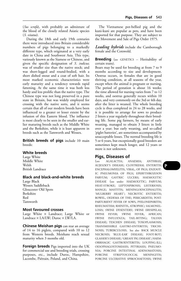
(Sus scrofa), with probably an admixture of the blood of the closely related Asiatic species(S. vitatus).
During the 18th and early 19th centuriesthere were introduced into Britain considerablenumbers of pigs belonging to a markedly different type, which originated at a very earlydate in China and Southwest Asia. This type,variously known as the Siamese or Chinese, andgiven the specific designation of S. indicus; was of smaller size than the native stock; andwas short-legged and round-bodied, with ashort dished snout and a coat of soft hair. Itsmost marked economic characteristics wereearly maturity and a tendency towards rapidfattening. At the same time it was both lesshardy and less prolific than the native type. TheChinese type was not long preserved in a purestate in Britain, but was widely employed forcrossing with the native sorts, and it seems certain that all of our modern breeds have beeninfluenced to a greater or lesser extent by theinfusion of this Eastern blood. The influence is most clearly to be seen in the smaller and ear-lier maturing breeds such as the Middle Whiteand the Berkshire, while it is least apparent inbreeds such as the Tamworth and Wessex.
British breeds of pigs include 10 mainbreeds:
White breedsLarge WhiteMiddle WhiteWelshBritish Landrace
Black and black-and-white breedsLarge BlackWessex SaddlebackGloucester Old SpotsBerkshireRedTamworth
Most favoured crossesLarge White × Landrace; Large White orLandrace × LA/LW; Duroc × LW/LA.
Chinese Meishan pigs can rear an averageof 14 to 16 piglets, compared with 10 to 12from Western breeds. Meishans reach sexualmaturity when 3 months old.
Foreign breeds Pigs imported into the UKfor commercial use and breeding trials, crossingpurposes, etc., include Duroc, Hampshire,Lacombe, Piétrain, Poland, and China.
The Vietnamese pot-bellied pig and thekuni-kuni are popular as pets, and have beenimported for that purpose. They are subject tothe Movement and Sale of Pigs Order 1975.
Leading hybrids include the Camboroughfemale and the Cotswold.
Breeding (see GENETICS – Heritability of certain traits).Boars may be used for breeding at from 7 to 9months according to size and development.Oestrus occurs, in females that are in goodthriving condition, at all seasons of the year,except when the animal is pregnant or nursing.The period of gestation is about 16 weeks; the time allowed for nursing varies from 7 to 12weeks, and oestrus generally recurs within 10days, and very commonly on the 3rd or 4th day,after the litter is weaned. The whole breedingcycle is thus completed in 24 to 28 weeks, andit is possible to arrange for sows to produce 2 litters a year regularly throughout their breed-ing life. Some pig farmers, by means of earlyweaning, managed to obtain 3 litters in littleover a year; but early weaning, and so-called‘piglet batteries’, are sometimes accompanied byunacceptable losses. The normal breeding life is5 or 6 years, but exceptionally good breeders aresometimes kept much longer, and 12 years ormore is not unknown.
Pigs, Diseases of(see AGALACTIA; ANAEMIA; ANTHRAX;AUJESZKY’S DISEASE; CLOSTRIDIAL ENTERITIS;ENCEPHALOMYELITIS, VIRAL, OF PIGS; ENZOOT-
IC PNEUMONIA OF PIGS; EPERYTHROZOON
PARVUM; GASTRIC ULCERS; HAEMOLYTIC
DISEASE (see under HAEMOLYTIC; PARVUM;HEAT-STROKE; LEPTOSPIROSIS; LISTERIOSIS;MANGE; MASTITIS; MENINGOENCEPHALITIS;‘MULBERRY HEART’; NECROTIC ENTERITIS;BOWEL, OEDEMA OF THE; PERICARDITIS; POST-
PARTURIENT FEVER OF SOWS, PYELONEPHRITIS;RHEUMATISM; RHINITIS, ATROPHIC; SALMONEL-
LOSIS; SWINE DYSENTERY; SWINE ERYSIPELAS;SWINE FEVER; SWINE FEVER, AFRICAN;SWINE INFLUENZA; TAIL-BITING; TALFAN
DISEASE; TESCHEN DISEASE; TOXOPLASMOSIS;TRANSMISSIBLE GASTRO-ENTERITIS; TRICHI-
NOSIS; TUBERCULOSIS. See also BACK MUSCLE
NECROSIS; ‘BLUE-EAR’ DISEASE; FOOT-ROT;GLASSER’S DISEASE; ‘GREASY PIG DISEASE’; HAEM-
ORRHAGIC GASTROENTERITIS; LOUPING-ILL;OESOPHAGOSTOMIASIS; PITYRIASIS; PNEUMO-
NIA; PORCINE INTESTINAL ADENOMATOSIS;PORCINE STREPTOCOCCAL MENINGITIS;PORCINE ULCERATIVE SPIROCHAETOSIS; SWINE
Pigs, Diseases of 543
P

VESICULAR DISEASE; VOMITING AND WASTING
SYNDROME; WORMS, FARM TREATMENT
AGAINST and headings under PORCINE.For causes of death among unweaned
pigs, see list of diseases, etc., given underPIGLET MORTALITY. See also PIG CARCASES,REJECTION OF.)
Pigs, names given according toage, sex, etc.The naming of pigs at various times in theirlife, and according to their age, sex, etc., variesin different areas; the following gives the mostusual names:
Store pig – a pig between the time of weaning and being fattened.
Hog (barrow in the USA) – a male pig afterbeing castrated.
Stag, steg, or seg – a male castrated late in life.
Gilt or yelt – a female intended for breedingpurposes, and up to the time that she has her1st litter.
Boar, bran, or hogg – an uncastrated male.Sow – a breeding female after the 1st litter.
Pigs, Sale ofIn the UK this is subject to the Pigs (Records,Identification and Movement) Order 1996.Pigs moved on to a premises must not be subject to further transport to other premisesfor 21 days, except for slaughter. The farmer isresponsible for the necessary certification.
Pigs, Sedation ofSedation is useful to prevent fighting after themixture of litters or re-grouping of pigs; to‘cure’ fighting after it has broken out; to makethe aggressive sow accept her litter; to facilitatecastration, nose-ringing, detusking, etc. Amongdrugs used for this purpose is azaperone, whichis administered by intramuscular injection.
Precaution There is a risk of damaging thesciatic nerve when making intramuscular injec-tions into the hind-legs of pigs; it is thereforeadvisable to inject into the neck muscles, justbehind the ear.
Case histories Within 1 to 8 weeks of aninjection of an antibiotic into the ham of 1 hind-leg of 180 4-week-old piglets, 150 haddeveloped paralysis of that leg. In anothergroup of 380 5-week-old piglets, 30 per centhad become paralysed within 10 days, and 30 per cent had died from complications suchas septicaemia and pneumonia – ‘probably dueto necrosis of the foot’.
Pigs, Transport ofThe use of containers for the transport of pigscan reduce the risk of infection being carried onto a purchaser’s farm. One crate can hold acomplete litter group, and keep the pigs fromcoming into contact with the sides and floor ofthe lorry – which are often not properly cleanedand which can seldom or never be sterilised.
PilobolusA fungus often present in bovine faeces on pasture, it acts as a disperser of lungworm larvae, by means of a ‘rocket-like’ effect causedwhen the fungal spores are dispersed.
PilocarpineA cholinergic alkaloid used in ophthalmic treat-ments to constrict the pupil and to reduceintra-ocular pressure in cases of glaucoma.
PilusAnother name for bacterial FIMBRIAE. For sexpilus, see under PLASMIDS.
Pin Bone (Tuber Ischii)The rearmost (caudal) projection on the floorof the pelvis.
Pineal BodyPineal body is a small structure situated in adeep recess of the mid-brain. It occupies a posi-tion similar to the third eye in certain reptiles.The pineal body is often regarded as anendocrine gland, concerned with growth. It is the source of melatonin, and it has aninhibitory action on the gonads.
‘Pining’ (Pine)A term formerly used to describe any progressive loss of condition in sheep, butnowadays – together with ‘vinquish’ – usuallyreserved for copper or cobalt deficiency. Thisoccurs in many parts of the world, is known as bush sickness in New Zealand, and has beenreported in areas of Scotland, Northumberland,Devon, and North Wales, where tracts of landare cobalt deficient. (See under COPPER and
COBALT.)
‘Pink-Eye’‘Pink-eye’ is the colloquial name for infectiouskeratitis of cattle caused by Moraxella(Haemophilus) bovis; and also for equine viralarteritis.
‘Pink Tooth’The colloquial name for congenital PORPHYRIA
in South Africa.
544 Pigs, names given according to age, sex, etc.
P
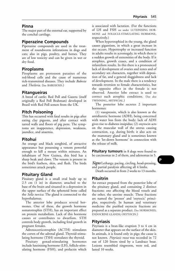
PinnaThe major part of the external ear, supported bythe conchal cartilage.
Piperazine CompoundsPiperazine compounds are used in the treat-ment of roundworm infestations in dogs andcats; also in pigs, poultry, and horses. They are of low toxicity and can be given in wet ordry food.
PiroplasmsPiroplasms are protozoon parasites of the red-blood cells and the cause of numerous tick-transmitted diseases. They include Babesiaand Theileria. (See BABESIOSIS.)
PitangueirasA breed of cattle; Red Poll and Guzera (itselforiginally a Red Poll Brahman) developed inBrazil with Red Poll semen from the UK.
Pitch PoisoningThis has occurred with fatal results in pigs aftereating clay pigeons, and after contact withtarred walls and floors of pig pens. The symp-toms are inappetence, depression, weakness,jaundice, and anaemia.
PitohuiAn orange and black songbird, of attractiveappearance but possessing a venom powerfulenough to kill a mouse within minutes. Aninhabitant of New Guinea, this bird has a sharp beak and claws. The venom is present inthe bird’s feathers, skin, and flesh. The birdssometimes attack people.
Pituitary GlandPituitary gland is a small oval body up to 2.5 cm (1 in) in diameter, attached to the base of the brain and situated in a depression inthe upper surface of the sphenoid bone calledthe Sella turcica. The gland is connected to thehypothalamus.
The anterior lobe produces several hor-mones. One of these, the growth hormonesomatotrophin (STH), has an important effecton protein metabolism. Lack of this hormonecauses or contributes to dwarfism. STH controls body growth, including fetal growth inpregnant females.
Adrenocorticotrophin (ACTH) stimulatesthe cortex of the adrenal gland. Thyroid-stimu-lating hormone (TSH) stimulates the thyroid.
Pituitary gonad-stimulating hormonesinclude luteinising hormone (LH), follicle-stim-ulating hormone (FSH), and prolactin which
is associated with lactation. (For the functionsof LH and FSH see under LUTEINISING HOR-
MONE and FOLLICLE-STIMULATING HORMONE,respectively.)
When hypertrophied in the young, the glandcauses gigantism, in which a great increase insize occurs. Hypertrophy or increased functionin adults results in acromegaly, in which there isa sudden growth of extremities of the body. If itatrophies, growth ceases, and a condition ofinfantilism results. In this there is a pronouncedlack of development of ovaries and testes and ofsecondary sex characters, together with deposi-tion of fat, and a general sluggishness and lackof development. In the male there is a tendencytowards reversion to female characteristics, butthe opposite effect in the female is notobserved. Anterior lobe extract is used to correct such atrophic conditions. (See also
TWINNING, ARTIFICIAL.)The posterior lobe secretes 2 important
hormones:(1) vasopressin, which is also known as the
antidiuretic hormone (ADH), being concernedwith water loss from the body; lack of ADHgives rise to diabetes insipidus; (2) oxytocin actson the muscular wall of the uterus causing contraction, e.g. during birth; it also acts on the mammary gland and is sometimes known as the ‘let-down hormone’ in connection withthe release of milk.
Pituitary tumours in 8 dogs were found tobe carcinomas in 2 of them, and adenomas in 5.
Signs Lethargy, pacing, circling, head-pressing,and partial paralysis affecting all 4 limbs.
Death occurred in from 2 weeks to 13 months.
PituitrinAn extract prepared from the posterior lobe ofthe pituitary gland, and containing 2 distinctfractions: one affecting the blood vessels andthe other, the uterine muscle. These fractionsare named the ‘pressor’ and ‘oxytocic’ princi-ples, respectively. In human and veterinarymedicine the purified oxytocin fractions areprepared as a separate product. (See HORMONES;ENDOCRINE GLANDS; OXYTOCIN.)
PityriasisPityriasis is a bran-like eruption 3 to 4 cm indiameter that appears on the surface of the skin.In animals, it is found only in pigs; the cause isnot known. Pityriasis rosea was recorded in 72out of 120 litters sired by a Landrace boar.Lesions resembled ringworm, were red, andlasted 10 weeks.
Pityriasis 545
P
‘Pizzle Rot’A disease mainly of merino sheep in Australia.(See under BALANITIS.)
PlacentaPLACENTA is the technical name for the afterbirth. Strictly speaking, placenta means the medium by means of which the mothernourishes the fetus.
Structure It is composed of 3 fetal membranes:
(1) The chorion, which is the outermost, is astrong fibrous membrane, the outer surface ofwhich is closely moulded to the inner surface of the uterus. The chorion has villi, which arevascular projections inserted into the crypts ofthe uterine mucous membrane.
(2) The allantois is the middle membrane. Itdevelops early in embryonic life as an out-growth from the hindgut, and insinuates itselfbetween the other 2 membranes. That part ofthe allantois remaining inside the abdominalcavity of the fetus forms the urinary bladder in after-life, and, until the time of birth, is in direct communication with the extra-fetalportion by means of the urachus – that partpassing through the umbilicus. Fluid secretedby the kidneys of the fetus and passing to theurinary bladder gains exit to the allantoic cavity, which is outside the fetus, until justbefore the time of birth, when the passage is closed.
(3) The amnion, which is continuous with the skin at the umbilicus (navel), andcompletely encloses the fetus but is separatedfrom actual contact with it by the amnioticfluid, or the ‘liquor of the amnion’, which inthe mare measures about 5 or 6 litres (9 to 101⁄2 pints).
This ‘liquor amnii’ forms a kind of hydrosta-tic bed in which the fetus floats, and serves to protect it from injury, shocks, extremes oftemperature, allows free though limited move-ments, and guards the uterus of the dam fromthe spasmodic fetal convulsions which, late inpregnancy, are often vigorous and even violent.
At birth it helps to dilate the cervical canal of the uterus and the posterior genital passages, forms part of the ‘waterbag’, and, onbursting, lubricates the maternal passages. (See
PARTURITION.)The membranes should be discharged from
the uterus with or soon after the young animal,but it is not uncommon to find their expulsiondelayed for a variable time, depending upon thespecies and the individual. Immediately after
the birth of the young animal the uterus contracts to a size smaller than when pregnant,with the result that the attachment betweenfetal envelopes and maternal uterus is severed to a greater or lesser extent. Afterpains follow,similar to those of normal labour, but lesssevere, with the object of expelling the membranes.
In the mare, with a scattered and slightattachment, the afterbirth separates rapidly andis soon expelled; indeed, not infrequently thefoal is born still enveloped in, or attached to, itsmembranes. These appear as a complicated massof pinkish-grey tissue, plentifully supplied withblood, and often possessing little bladder-likepockets of amniotic or allantoic fluid.
If in 6 hours the placenta has not been discharged naturally, measures should be takento effect its prompt removal, for the mare isvery susceptible to metritis.
In the cow, where the attachment is cotyle-donary, the fetal membranes may be expelled atany time during the first 6 hours after calving,or not for 1 or 2 days, without any serious consequences. Retention occurs frequently.
In those animals which normally producemultiple offspring at a birth – ewe, sow, bitch,and cat – as each fetus is born the correspond-ing membranous envelope either accompaniesor else immediately follows it. The exception tothis otherwise almost invariable rule is in thecase of the last fetus to be born, that fetus whichoccupied the extremity of one or other horn ofthe uterus; the envelopes of this, the youngestmember of the family, are sometimes retained,and may occasion a mild or severe metritis,until such time as they are expelled.
The bitch, cat, sow, cow, and even the mareeat the membranes.
Retained placenta is one of the undesir-able sequels of parturition. Retention of thefetal membranes is commonest in the cow.Normally, the membranes should be expelled in from half an hour to 4 or 5 hours after the birth of the calf, but owing to the intricate cotyledonary attachment in cattle,they are often retained for long periods. (See
PARTURITION.)
Signs A portion of the fetal membranes hangsfrom the lips of the vulva attached to whatremains inside the cavity of the uterus. Theexposed portion may measure only a few inches, or be a large mass reaching to the cow’s hocks. In some cases there is no mem-brane visible externally, but there is an odour ofdecomposition evident.
546 ‘Pizzle Rot’
P
During the first day after calving the mem-branes in an ordinary uncomplicated case arefresh, slimy, and pinkish in colour. There is noobjectionable smell, and the cow is not distressed. After the second day the externalportions undergo decomposition; the colourbecomes greyish; an offensive chocolate-coloured discharge makes its appearance andsoils the hindquarters and tail of the cow. She stands with her back arched, frequentlyswitches her tail and paddles with the hind-feet.
Masses of semi-dissolved membrane, lookinglike pieces of wet cobweb, are passed out atintervals, and can be found behind the cowalong with quantities of greyish foul-smellingdischarge; the mucous membrane of the vaginais inflamed, and the cow resents having herhindquarters examined.
It is certainly not advisable that a mass ofdecomposing fleshy material should be allowedto hang from a cow’s uterus for a longer timethan is absolutely necessary, but too hastyattempts at removal may be followed by infertility or even death.
In modern practice, hormone injections havelargely displaced manual removal, with itsattendant risks. Manual removal consists ofintroducing the hand and arm, protected insterile disposable plastic sleeve-length gloves; if these are not available, the hand and armmust be cleansed as far as possible by thoroughwashing in an antiseptic solution, and a subse-quent rinsing in strong salt and water. Themembranes should not be removed until theycan be displaced without difficulty, and withoutdistress to the cow. Each cotyledon is grasped asit is reached, and the adherent membrane ispeeled off from its surface with the fingers andthumb. At the same time gentle traction shouldbe exerted upon the protruded membranesfrom the outside.
After removal it may be advisable to doucheout the uterus with a suitable antiseptic solu-tion, and to give prophylactic antibiotics (see
UTERUS, DISEASES OF – The cow).In the sow and bitch the membranes that are
liable to be retained are those belonging to the fetus that is born last, which occupied theextremity of one or other of the horns of the uterus, but the condition is rare in each of these animals.
It is most advisable that owners of animalswhich have retained their afterbirth after parturition should seek veterinary advice.
Plague(see CATTLE PLAGUE; AVIAN INFLUENZA (FOWL
PLAGUE); BUBONIC PLAGUE)
Plant JuiceAt grass-drying plants, when mechanically de-watering forage before drying, a rich greenjuice is expressed. The juice represents a sourceof protein concentrate comparable with fishmeal in value.
From 6 tons of fresh material, such as lucerneor grass, 2.5 tons of this plant juice is squeezedout. However, using plant juice presents someproblems, as it is very unstable, due to enzymeaction. It must be heated to about 85°C(185°F) to stabilise the protein, and somechemical preservative is also added.
The easiest way to use the juice is to includeit in liquid pig feed.
PlantarAt the back of the hind-limb.
Plantar CushionPlantar cushion is the dense fibro-fatty rubber-like structure which lies immediately above the frog in the foot of the horse, and is one of its most important anti-concussion or shock-absorbing mechanisms. (See FOOT OF THE
HORSE.)
PlasmaPlasma is the fluid portion of the blood.
Plasma CellsLarger than lymphocytes, with dark-staininggranules in the nucleus, plasma cells are found in the lymph nodes, and are concernedwith antibody production. (See diagram under
LYMPHOCYTE.)
Plasma Substitutes(see DEXTRAN; GELATIN, SUCCINYLATED)
PlasmidsPlasmids are genetic structures which manyspecies of bacteria possess in addition to theirchromosomes, and which, like the chromo-somes, determine the inheritance of variousproperties. Since plasmids are not essential tocell growth, the cell may gain or lose themwithout lethal effect. Some plasmids can unitewith chromosomes: these are called episomes.All episomes are plasmids but not all plasmidsare episomes.
Plasmids have been defined as ‘circularlengths of DNA which behave as viruses with arestricted range of host bacteria, and which arereplicated in step with the organism’. (See also
GENETIC ENGINEERING.)Infectious (self-transmissible) plasmids are
found in Gram-negative rods. In addition to
Plasmids 547
P
genes coding for specific properties (e.g. R-determinants coding for antibiotic resis-tance), these plasmids also possess other geneswhich code for the production of sex pili bywhich conjugation with a recipient cell is madepossible. Non-transmissible plasmids, alsofound in Gram-negative rods, lack the genesnecessary for self-transfer; for these plasmids tobe transferred the cell must first be infectedwith a sex-factor from another cell.
In Gram-positive organisms, plasmids maybe transduced from one cell to another by bacteriophages.
Plasmodium GallinaceumA parasite which causes bird malaria in poultry.
P. durae causes death in turkey poults.Malaria of birds is a group of almost world-
wide infections which include Haemoproteusand Leucocytozoon as well as Plasmodium.
Plaster Casts(see SPLINTING MATERIALS)
Plastic Bags or SheetingCattle have died following ingestion of plasticbags discarded on grazing land. In someinstances the material is digested and does notcause an obstruction.
Plastic ‘Bones’A fractured scaphoid in the right hind-leg of aracing greyhound has been successfully replacedby a plastic replica of a scaphoid bone. For afurther use of plastics, see under HOOF REPAIR.
Plate CultureThe growing of bacteria in a medium containedin a Petri dish, a covered, shallow, circular glassdish which gives a large surface area.
Platelets (Thrombocytes)Platelets (Thrombocytes) are described underBLOOD.
PlatingPlating is the cultivation of bacteria on flatplates (Petri dishes) containing nutrient material. The term is also applied in surgery tothe method of securing union of fracturedbones by screwing to the sides of the fragmentsnarrow metal plates which hold them firmlytogether whilst union is taking place.
PleuraPleura is the membrane which covers the exter-nal surfaces of the lungs and lines the inside ofthe chest walls. (See LUNGS.)
Pleural CavityIn normal healthy animals this is merely apotential cavity, since between the pleura liningthe chest and the pleura covering the lungsthere is usually a thin film of fluid. Surface tension holds the one surface to the other.
Pleurisy (Pleuritis)Inflammation of the PLEURA, which may occuras a complication of pneumonia, of ‘shippingfever’ in cattle, and in cases of tuberculosis andother infections; occasionally from a chestwound.
Friction between the inflamed surfaces givesrise to pain each time the animal breathes, andthe breathing is changed in character – i.e. thereis minimal movement of the chest walls, andextra effort by the abdominal muscles, whichappear to be doing all the work. The line of the rib cartilages often stands out prominently,giving rise to what has been called the ‘pleuriticridge’. Other symptoms include fever, dullness,and what is often described as a ‘hacking’cough; sometimes a rasping sound due to friction may be heard.
After 12 to 48 hours the painful stage ofpleurisy may be followed by an effusion offluid. Removal of this fluid becomes necessaryif respiration is seriously impaired by it.
Sometimes the fluid is purulent, a conditionknown as ‘empyema’. Sometimes there is verylittle effusion, and the condition remains one of‘dry pleurisy’.
A complication of pleurisy is that adhesionssometimes occur, and persist, between the pari-etal and visceral pleura. (See also PLEURODYNIA;PLEURO-PNEUMONIA.)
Treatment Pleurisy is one of those conditionsin which professional advice is highly desirablein the early stages, and reliance should not beplaced on first-aid. It may be necessary to withdraw fluid from the chest, to administer an appropriate antibiotic; and it is important to establish the presence or absence of tuberculosis. (See also NURSING.)
PleurodyniaPleurodynia means a painful condition of thechest wall. It is a symptom of PLEURISY; it maybe due to fractures of the ribs; it is sometimesseen in tumours affecting the chest wall, and itis commonly recognised by pressing the fingersinto the spaces between the ribs.
Pleuro-PneumoniaPleuro-pneumonia is a combination ofPLEURISY with a PNEUMONIA. Acute pneumonia
548 Plasmodium Gallinaceum
P
is often accompanied by some amount ofpleurisy, which is largely responsible for thepainfulness which accompanies pneumonia. (See
also CONTAGIOUS BOVINE PLEURO-PNEUMONIA.)
PlexusPlexus is a network of nerves or vessels, e.g. the brachial and sacral plexuses of nerves andthe choroid plexus of veins within the brain.
PlumbismPlumbism is another name for chronic lead poisoning. (See LEAD POISONING.)
PMLPharmacy and merchants’ list: a category of veterinary medicines which may be sold to thepublic only through pharmacies; and to farmersor others ‘maintaining animals in the course of their business’ through pharmacies or agricultural merchants. Horse and pony owners may obtain PML horse wormers from pharmacies, agricultural merchants or registered saddlers.
PMSPregnant mare’s serum, a source ofgonadotrophin.
PMWS(see POST-WEANING MULTISYSTEMIC WASTING
SYNDROME)
Pneumocystis PneumoniaPneumocystis pneumonia, one of the most serious diseases of human AIDS patients, hasbeen found in dogs, cats, rodents, and primates.
Pneumogastric nerveAnother name for VAGUS NERVE.
PneumomycosisA fungal infection of the lungs.
PneumoniaPneumonia may be defined as inflammation oflung tissue.
Pneumonias have been classified in variousways, e.g. according to the area or tissueinvolved, or according to lesions, or causes.
Lobar pneumonia is that in which a wholelobe is involved; in lobular pneumonia theinflammation is less localised and more patchy.Broncho-pneumonia is that in which theinflammation is concentrated in and aroundthe bronchioles leading from the bronchi.Pleuro-pneumonia, as the name suggests,involves the pleural membranes as well as the
lung itself. Interstitial pneumonia affects thefibrous supporting tissue of the lung rather thanthe parenchyma though consolidation of thelatter can then occur. (See also LUNGS, DISEASES
OF for lesions.)Pneumonia, which can be acute or chronic,
may – as mentioned above – also be classifiedaccording to causes, e.g. viral, mycoplasmal,bacterial, mycotic, parasitic and non-infective. Itmust be borne in mind, however, that infectionsmay be mixed and changing. (See RESPIRATORY
DISEASE IN PIGS for an explanatory diagram.)Pneumonia may arise from a primary viral
infection, with complications caused by secondary bacterial invaders, as in canine distemper. Some viruses and bacteria dependupon each other, as explained under SYNER-
GISM. The infection may be a very mixed one, e.g. in ENZOOTIC PNEUMONIA OF PIGS.Bacterial pneumonia may be acute, e.g. KLEB-
SIELLA infection, or of a chronic suppurativetype, e.g. TUBERCULOSIS.
The main effect of pneumonia, whetherthrough the presence of exudate in the bronchi-oles and alveoli, or destruction of areas of lungby abscess formation or consolidation orhepatisation, is that the normal exchange of carbon dioxide for oxygen is impaired andimpeded. The animal has to struggle for breathto obtain sufficient oxygen.
Signs With less oxygen available to the redblood cells (and hence to the organs and tissues)at the normal respiratory rate, the animalaccordingly needs to breathe faster. Thisincreased respiratory rate (tachypnoea) is there-fore a main symptom. The breathing may alsobecome laboured and painful (dyspnoea). Feveris usually present, with accompanying dullnessand loss of appetite (but see under CALF PNEU-
MONIA for an exception to this). There is oftena cough, though this is not an invariable orattention-catching symptom.
Viral pneumonia In cattle, most cases ofviral pneumonia, uncomplicated by secondarybacterial infection, show areas of collapse in thelungs, emphysema of the apical lobes, some inthe cardiac lobes but little in the diaphragmaticlobes, with little or no exudate. An acute pneu-monia may develop in some cases of infectiousbovine rhinotracheitis. (See RHINOTRACHEITIS.)Bovine syncytial virus is another importantpathogen, causing coughing, oedema of thelungs, consolidation and emphysema. (See
also CALF PNEUMONIA.) In horses, pneumonia in young foals may be caused by equine herpesvirus 1. (See also SWINE PLAGUE.)
Pneumonia 549
P
Mycoplasmal pneumonia An example ofthis is fully described under CONTAGIOUS
BOVINE PLEURO-PNEUMONIA – a disease notnormally present in the UK. Mycoplasma bovisand M. dispar are primary pathogens in theUK. The latter may cause CALF PNEUMONIA ofa mild type except for the harsh cough. (See also
under MYCOPLASMA.)
Bacterial pneumonia Bacterial toxins mayhave an important additional effect in this. Incattle, Pasteurella haemolytica can be a primarycause of pneumonia, typically producing much exudate as well as a secondary invader. Inhorses Corynebacterium equi causes a suppura-tive bronchopneumonia in foals, and in adulthorses a suppurative pneumonia may also occurduring the course of strangles.
Streptococcus pneumoniae and Staphylococcusaureus are other important secondary invadersof damaged lungs.
Parasitic pneumonia This may be anextension of PARASITIC BRONCHITIS in calves,caused by the lungworm Dictyocaulus viviparus.Autopsy findings include dark red consolida-tion of some lung lobes. In pigs, lungworms(Metastrongylus) are also a cause of pneumonia.
Mycotic (fungal) pneumonia is causedby Aspergillus species, and also by Candida albicans. (See MONILIASIS.) The latter may beassociated with oedema of the lungs, in bothmammals and birds, and may follow the use ofcertain antibiotics. (See also PHYCOMYCOSIS.)
Allergic pneumonia (see ‘FARMER’S LUNG’
which affects cattle also)
Non-infective pneumonia can resultfrom the action of certain poisons, e.g.PARAQUAT, ANTU, and phenolic sheep dips (see DIPS), as well as from aspiration pneumonia.The latter may result from milk ‘going thewrong way’ in bucket-fed calves, and also from medicines administered to animals bystomach tube passed in error into the tracheainstead of the oesophagus (fortunately a rareoccurrence!). Aspiration of vomit is anotherexample.
Non-infective pneumonia quickly becomesinfective, as micro-organisms take advantage ofthe inflamed mucous membranes. The animalbecomes suddenly dull, uninterested in food,feverish, and may show signs of chest pain.Death can be expected within 72 hours, andautopsy findings may include areas of necrosisand abscess formation.
Treatment of pneumonia Separateaffected animals, provide good-quality feed in a well-ventilated environment, free fromdraughts, and good bedding. Appropriateantibiotics, sulfa drugs, and trimethoprim may all be of service. Heart stimulants, theadministration of oxygen, and possibly diuretics in the case of oedema, may be indicated. Anti-inflammatory drugs, e.g. flu-nixin, may help in the acute stage. The one golden rule in the treatment of pneumoniain all animals is: ‘Do not drench.’ Medicinesshould be administered by injection, in the food, or perhaps as an electuary. (See also
NURSING.)
Pneumonia in Calves(see CALF PNEUMONIA)
Pneumonia in CatsBronchopneumonia can be a complication offeline viral rhinotracheitis in young cats.
In older cats tuberculosis is still sometimes a cause of disease of the lungs, though withnearly all milk now pasteurised in EU coun-tries, feline TB is no longer at all common. It can, however, result from a cat eating TB-infected prey.
A granulomatous pneumonia is caused,rarely, by Corynebacterium equi. Theoretically, astable cat would be more prone to it.
Pneumonia in HorsesIn a USA study, anaerobic bacteria were isolated from pleural fluid or tracheobronchialaspirates obtained from 21 of 46 horses with bronchopneumonia. Bacteriodes oralis andB. melaninogenicus were the species most com-monly isolated (9 and 5 horses, respectively).Other Bacteriodes species were cultured from 12 animals and Clostridium species from 8. Aputrid odour was associated with the pleuralfluid and/ or breath of nearly two-thirds of the horses from which anaerobes were isolated.The prognosis was significantly poorer in caseswith anaerobic infections; 14 of the 21 horsesinvolved either died or were euthanased. (See also
under FOALS, DISEASES OF).Another cause is Corynebacterium equi,
which can also infect cats.
Pneumonia in PigsActinobacillus (Haemophilus) pleuropneumoniaeis a major cause of pneumonia in pigs. (See
RESPIRATORY DISEASE IN PIGS.)For the most common form of pneumonia in
pigs, see under ENZOOTIC PNEUMONIA. (See also
SWINE PLAGUE.)
550 Pneumonia in Calves
P

Pneumonia in SheepIn Britain, pneumonia caused by Pasteurellahaemolytica has become more frequent. Thisorganism commonly lives in normal sheep, andcauses disease only when the animal’s resistanceis weakened by bad weather, transport from onefarm to another, movement from a poor to aricher pasture, or perhaps by a virus. In someoutbreaks, where the disease takes an acuteform, a sheep which seemed healthy enough inthe evening may be found dead in the morning.Usually, however, the shepherd sees depressed-looking animals with drooping ears, breathingrather quickly and having a discharge from eyesand nostrils, and a cough. Death often occurswithin a day or two. The last sheep to beinvolved in the outbreak tend to linger for several weeks, looking very tucked-up in themeantime, with a cough and fast breathing.Antisera and vaccines are available for treatment and prevention.
Another cause of pneumonia in sheep isChlamydia psittaci. (See also HAEMORRHAGIC
SEPTICAEMIA, which occurs in the tropics;PARAINFLUENZA 3 VIRUS; MAEDI/VISNA.)
PneumothoraxPneumothorax is a condition in which air hasgained access to the pleural cavity through awound in the chest wall. (See LUNGS, DISEASES
AND INJURIES OF.)Pneumothorax is not uncommon in the dog
which has fallen out of a window, or been runover or hit by a car. Distressed breathing andcyanosis are, following an accident, suggestiveof pneumothorax.
Mild cases may be accompanied by mildsymptoms, and spontaneous recovery mayoccur. Severe cases may die. Treatment includesaspiration of the air from the pleural cavity. (See
also LUNGS, DISEASES OF – Collapse.)
Simple pneumothorax In such cases, the condition may be caused by sudden non-penetrating trauma to the chest wall which momentarily raises the intrathoracicpressure. If this occurs against a closed glottis, then alveolar tissue may rupture. Inmany cases the leak will be small, but in others,sufficient air will enter the pleural cavity tocause marked dyspnoea.
Open pneumothorax is caused by a penetrating wound of the chest wall, resultingin an immediate and total collapse of the lung. Air can be heard entering the chestwound at each respiratory effort. There is severedyspnoea.
Tension pneumothorax may develop following an open pneumothorax, or when asmall bronchus ruptures, and air accumulates in the pleural space during inspiration but is not expelled during expiration. The medi-astinum is apt to be displaced from the midline, partially collapsing the remainingfunctional lung.
PneumovirusA cause of rhinotracheitis in turkeys and chick-ens. An ELISA test is available for diagnosis.
‘Poaching’Land becoming muddy and broken up by thefeet of animals. Especially on heavy land, withhigh stocking rates, wet weather can bring serious poaching problems. (See DAIRY HERD
MANAGEMENT.) At gateways, deep mud (inwinter often at near-freezing temperatures) canlead to foul-in-the-foot and mastitis.
Pododermatitis(see FOUL-IN-THE-FOOT)
PoikilocytePoikilocyte is a malformed red blood cell foundin the blood in various types of anaemia.
Poikilothermic‘Cold-blooded’. The rate of metabolism of such an animal varies with its environmentaltemperature.
PointerA medium-sized, slim dog with long neck andpointed tail; used as a gun dog. Entropion and ununited anoconeal process are inheritable,as may be calcinosis circumscripta.
PoisoningMost cases result from the poison being swallowed. In a few instances poison may betaken in through a wound of the skin, or eventhrough the unbroken skin, e.g. phenol preparations. Malicious poisoning is most frequently carried out against dogs and cats,although horses and ruminants also sometimessuffer.
The use of poison to control vermin – rabbits,foxes, rats, mice, etc. – is a hazard, for when the poisoned bait is accessible to domesticatedanimals, cases of poisoning may result. It shouldbe remembered that the exposure of such poisonabove ground constitutes an offence.
The constituents of common and com-mercial rat-poisons are mentioned under RODENTS – Rodenticides.
Poisoning 551
P

Many cases of poisoning result from the care-less use of sheep-dips, paints, weed-killers andinsecticides, which, in powder, paste, or solu-tion, are left about in places to which animalshave access. Cattle are notoriously inquisitive,and will lick at anything they find, sometimeswith fatal consequences.
It is perhaps not widely enough realised thatcattle seem to like the taste of lead paint – 1heifer helped herself to a whole pint of it – andthat very small quantities spattered on theground can kill several beasts. Even the contentsof old, discarded paint tins can be lethal. In oneinstance, children found such tins and scrapedout the residue on to pasture, killing 5 year-lings. In another instance, cattle licked out oldpaint tins on a rubbish dump in a pit to whichthey found their way. A recently painted fenceis also a danger.
Thirsty cattle will drink almost anything:diesel oil and a copper-containing spray liquidhave each caused death in these circumstances.Salt poisoning is certainly no myth, and pigsshould never be kept short of drinking water.
Some insecticides, such as TEPP andParathion, are totally unsuitable for use on livestock. Fatal poisoning of a herd of cattlesprayed with TEPP has been reported fromTexas. A farmer in Ireland used aldrin as an orf-dressing, and killed 105 out of 107 lambs.Fatal poisoning of cattle has also occurredthrough the application to their backs of a carbolic-acid-arsenic preparation against flies.(See also HERBICIDES; WEEDKILLERS.)
Near factories and chemical works, grass,etc., may become impregnated with fluorinecompounds, copper, lead or other metals, andlead to chronic poisoning of any animals grazing nearby. The same thing applies inorchards after spraying of fruit-trees. Pasturemay be contaminated by spray-drift or dustingoperations, particularly from the air, and thechemicals used may cause poisoning. Thisapplies also to other treated green crops whichanimals may eat. DDT and BHC and otherinsecticides (used in home and garden) of the CHLORINATED HYDROCARBON group may poison birds, cats and dogs. (See also
ORGANOPHOSPHORUS POISONING; FARM
CHEMICALS; FLUOROSIS.)The use of pitch (the poisonous ingredient of
clay-pigeons) or coal tar on the walls and floorsof piggeries is a cause of poisoning. Some woodpreservatives cause hyperkeratosis.
Poisoning may result from indiscretion onthe part of owners or attendants in the use ofpatent or other animal medicines, or from theadministration to animals of tablets, etc.
intended for human use, e.g. paracetamol, caffeine.
Dogs and cats may also be poisoned bygaining access to unsecured medicines (pills,tablets, etc.) intended for human use. For homeand garden hazards to pet animals (includingcage birds), see under CARBON MONOXIDE;‘FRYING PAN’ DEATHS; ANTIFREEZE; CREOSOTE;LEAD POISONING; METALDEHYDE; BHC POISON-
ING; BENZOIC ACID POISONING; WARFARIN;DDT; HOUSE PLANTS; HOUSE DECORATING. Ifone considers dogs out for walks, one shouldadd DIELDRIN; PARAQUAT; FARM CHEMICALS. As regards dog and cat foods, poisoning hasresulted from biscuit meal made from corndressed with dieldrin, from stored food conta-minated by rats’ urine containing warfarin,from aflatoxins, and from horse-meat contain-ing barbiturates or choral hydrate. In the cat,food containing benzoic acid as a preservativehas caused poisoning.
Fodder poisoning Excess of fodder beetmay cause scouring in both pigs and cattle, andthe after-effects may be serious. In sows just farrowed, the milk supply may almost disap-pear. Beet tops have caused the deaths of cattlewhen given unwilted, and even when wiltedthey should be strictly rationed. Kale and rapepoisoning can occur in cattle and sheep; thesemust be used sparingly and not constitute ananimal’s sole diet, and hay in particular is nec-essary in addition. Deaths have occurred inhorses and cattle restricted to rye-grass pasture.Sheep have been fatally poisoned by feedingthem surplus pig-meal containing a copper supplement; a heifer likewise. Pigs have beenpoisoned by giving them medicated mealintended for poultry and containing nitro-phenide against coccidiosis. Which all goes toshow that medicated feeds are by no meansalways interchangeable between differentspecies of livestock, since there are genetic differences between them as regards susceptibility to poisoning, depending in partupon possession or absence of some enzyme which can readily detoxify the poison. (See
GROUNDNUT MEAL.)The use of surplus seed corn for pig-feeding
has led to fatal poisoning – the mercury dress-ing having been overlooked! (See DIELDRIN forpoisoning from seed dressings, and FLOOR
SWEEPINGS; also MONENSIN SODIUM.)Hay contaminated with foxgloves or ragwort
is a source of fatal poisoning. Silage contami-nated with ragwort has similarly caused death.Silage contaminated with hexoestrol has caused
552 Poisoning
P
abortion. (See under HORMONES IN MEAT
PRODUCTION.)
Poisonous plants growing in pastures, inswampy or marshy places, in the bottoms ofhedges, on waste land and in shrubberies andgardens, are other very common sources of poi-soning. In the early spring, when grass is scarce,and when herbivorous animals are let out for thefirst time after wintering indoors, the tender suc-culent growths attract them, often with seriousconsequences. Similar results may be seen dur-ing a very dry summer when grass is parched.Ragwort and bracken are the most commoncauses of plant poisoning in cattle; in sheep,bracken can cause night blindness and neoplasiaas well as true poisoning. (See BRACKEN.)
Clippings from shrubs, especially from yew,rhododendrons, aconite, boxwood, lupins, laurel, laburnum, etc., should never be thrown‘over the hedge’, because in some of these thetoxic substances are most active when the clip-pings have begun to wither, and animals arevery prone to eat them in this condition. It is asafe rule to regard all garden trimmings asunsafe for animals, with the exception of vegetables, such as cabbages and turnips. (See
under ACONITE; BITTERSWEET; FOXGLOVE; HEM-
LOCK; LABURNUM; POTATO; RAGWORT; WATER
DROPWORT; YEW; LOCOWEED, etc.)
Signs The symptoms of each of the more common poisonous agents are given undertheir respective headings.
It must be emphasised that the symptoms ofsome illnesses are the same as those of somepoisons, and vice versa. For example, not only vomiting and diarrhoea but also cramp,fever, rapid breathing, convulsions, hysteria,jaundice, salivation, blindness, and deafness are common to both. A professional diagnosis is therefore important.
Irritant poisons produce acute abdominalpain, vomiting (when possible), purging, rapidly developing general collapse, and oftenunconsciousness, perhaps preceded by convulsions.
Narcotics produce excitement at first,unsteady movements, interference with sight;and later, stupor and unconsciousness appear;coma, with or without spasmodic or convulsivemovements, supervenes, and death occurs inmany cases.
Narcotic irritants produce symptoms of irritation in the first place, and later, delirium,convulsions, and coma.
As a general rule poisoning should be sus-pected when an animal becomes suddenly ill,
soon after feeding; when put out to pasture forthe first time in the season after dipping; orwhen a change of food has recently taken place.Newly purchased animal feeds may be followedby an outbreak of illness, and such results pointto the inclusion of some harmful substance.Fungal poisoning may occur as a result ofmouldy barley, etc.
First-aid In suggesting simple first-aid measures, it should be emphasised that theynecessarily differ from – and are likely to be less effective than – those the veterinary surgeon will take. It should be realised, too, that against some poisons there are no effectiveantidotes.
Where it is suspected that poisoning hasarisen from use of some proprietary product,take the container (or the label from it) to yourveterinarian (or write down the name of themanufacturer and product) so that he or shemay ascertain the chemical ingredients and, ifnecessary, consult the manufacturer as to therecommended antidote.
If it is suspected that poisoning may haveresulted from a skin dressing, wash this off with warm soapy water to prevent furtherabsorption. (See CARBOLIC ACID POISONING.)
Where a poison is believed to have beentaken by mouth, give an emetic. However,emetics should be avoided if strong acids oralkalis are involved.
Emetics which may be safely used are: forpigs, a dessertspoonful of mustard in a cupful ofwater; for dogs, a strong salt solution (ordinaryhousehold salt), or a crystal of washing soda; for cats, the latter.
To hinder absorption in the horse, ox, orsheep, strong black tea or coffee which has been boiled may be given. These, all of whichcontain tannic acid or tannates, are usefulagainst vegetable poisons.
To counteract the effects of irritants, usedemulcents (olive oil, milk, milk and eggs, orliquid paraffin). Yellow phosphorus is an excep-tion to this rule; oily substances favour itsabsorption and must be avoided; copper sulphate should be given instead. Against narcotics, stimulants are needed, e.g. strongcoffee or black tea, given by the mouth as afirst-aid measure.
Advice on poisons In the UK, veterinarysurgeons may obtain information and adviceconcerning poisonous compounds, and theirantidotes, if any, from the National PoisonsInformation Service, Avonley Road, LondonSE14 5ER.
Poisoning 553
P
Confirmation of poisoning No matterhow strong circumstantial evidence seems to be, it is always essential that a post-mortemexamination be made, and that necessary samples of the stomach contents, portions ofthe liver and perhaps other organs, should besubmitted to a qualitative and quantitativechemical examination by an analyst, before asuspected case of poisoning can be consideredto be definitely proved. The necessity for thisprocedure is obvious when legal proceedings arecontemplated.
Poisoning by Salmonella(see SALMONELLOSIS)
Poland ChinaA breed of pig from Ohio, USA. Colouring isblack with 6 white points (feet, tip of noise, andtail). Rapid growth and good meat productionare characteristics of the breed.
Polioencephalomalacia(see CEREBROCORTICAL NECROSIS)
Poliomyelitis of PigsThis disease is distinct from that of humanbeings and is possibly identical with TALFAN
DISEASE.
PollThe region lying between the ears and a littlebehind them.
‘Poll Evil’‘Poll Evil’ is an old, colloquial name sometimesincorrectly applied to any swelling in the POLL
region, but which should be reserved for a sinus following infection of some of the deeper tissues and giving rise to pus-formation. It may result from an injury which displaces achip of bone from the atlas. Apart from this, and its situation, ‘poll evil’ resembles fistulous withers.
Causes Self-inflicted injuries such as strikingthe poll against the top of a doorway, or fallingbackwards with the poll striking the ground;blows, such as from a whip-handle; bridle pressure.
Fusiformis necrophorus has been associatedwith some cases and may have gained entrancethrough damaged skin. It has been suggestedthat some cases may arise through infectionwithout injury. Brucella abortus and the wormOnchocerca reticulata have each been found,and the former is now known to account forsome cases of fistulous withers.
Signs A painful swelling on one or both sides with, after a time, the appearance of one or more orifices exuding pus. The animal resents the part being touched and, ifthe ligamentum nuchae is involved, avoids downward movement of the head.
First-aid measures include the use of apoultice and the placing of food at a level whichthe animal can reach without pain.
Treatment may necessitate the removal of any dead tissue and the surgical enlargement of any openings to allow free drainage.Antibiotics may be used to overcome the infection.
PolledInherited hornlessness of an animal belongingto a normally horned breed, e.g. Hereford cattle. (See SCUR.)
PollenPollen of oilseed rape has been linked toobstructive lung disease seen in horses at pasture. Clenbuterol is recommended for treatment.
PolyarthritisInflammation of several joints occurring simultaneously, as in ‘JOINT-ILL’. In pigs a common cause is Erysipelothrix rhusiopathiae;streptococci and staphylococci may also beinvolved. The disease is seen also in lambs,calves, and foals.
PolycythaemiaA marked increase in the number of red blood cells. This disorder is seen, rarely, in dogs and cats.
PolydactylyA congenital defect in which an animal has anextra digit. Cattle, horses (rarely), dogs and catsmay be affected.
PolydipsiaPolydipsia is excessive thirst.
PolymeliaA developmental disorder resulting in an extralimb or limbs.
PolymorphPolymorph is a name applied to certain white cells of the blood which have a nucleus of varied shape. (See BLOOD.)
554 Poisoning by Salmonella
P

PolyneuritisPolyneuritis means an inflammation of nervesor their sheaths occurring in different parts ofthe body at the same time. (See NEURITIS.)
PolyoestrousPolyoestrous animals are those which have several oestrus cycles per year – the conversebeing monoestrous.
PolypPolyp is a tumour which is attached by a stalkto the surface from which it springs. The termonly applies to the shape of the growth and hasnothing to do with its structure or to its nature.Most polyps are benign, but some may becomemalignant. They are generally of fibrous tissuein the centre covered with the type of localepithelium. In animals, the common situationswhere they are found are: in the nostrils; in thevagina, where they sometimes interfere withsuccessful copulation; and in the interior of thebladder.
PolyphagiaExcessive ingestion of food. While it is a common attribute of animals, it can in companion animals lead to obesity, and maskmetabolic disease.
PolyploidyThe presence of multiples of the haploid number of chromosomes greater than thediploid number, e.g. triploidy, tetraploidy.
Polyradiculoneuritis, IdiopathicA disease of dogs, and occasionally cats, affecting nerves.
Signs Weakness of the hind-legs, followed byparalysis of them within a few days. The fore-legs then become involved. Hyperaesthesiaof all legs occurs. Body temperature remainsnormal.
Muscle wasting occurs. Recovery is gradual.In one case a puppy was able to walk again after 4 weeks; but complete recovery took over6 months.
Polysaccharides(see SUGAR)
PolytocousProducing several offspring at birth, i.e. a litter.
PolyuriaPolyuria is a condition in which a much greateramount of urine is passed than is usual.
Polyuria is also a symptom of diabetes, and itoccurs in certain forms of inflammation of thekidneys, especially in the early stages ofpyelonephritis affecting cows after calving,when infection has travelled into the bladder,up the ureters, and so into the kidney. (See
INCONTINENCE; KIDNEY, DISEASES OF; URINE;STRESS.)
Polyvalent VaccineOne prepared from cultures of several strains of the same bacterial or viral species or from different species. A single polyvalent vaccinecan now protect against as many as 8 diseases.
Pom(see PRESCRIPTION-ONLY MEDICINES)
PomeranianA small alert dog with tail carried over the back, pointed ears and prominent eyes. Patellar luxation is inheritable and patent ductus arteriosus may be present.
PondsSalmonella typhimurium was isolated from marlponds on a farm where the dairy herd had areduced milk yield, fever and dysentery.Fencing off the ponds to prevent access by thecattle stopped the outbreak immediately. (See
also LEECHES, ALGAE POISONING, COCCIDIOSIS,JOHNE’S DISEASE, BOTULISM.)
Ponies(see under HORSES, FEEDING OF)
Pons (Pons Varolii)Pons (Pons Varolii) is the so-called ‘bridge ofthe brain’. It is situated at the base of the brainin front of the medulla, and behind the cerebralpeduncles, and appears as a bulbous swellingnot unlike a small curved artichoke. It is mainly composed of strands of fibres which linkup different parts of the brain.
PoodleA breed of dog originating in Germany (in spiteof being known as a French poodle) where it was used as a gun dog for retrieving game,especially that which had fallen in water. Thecharacteristic clip of its curly black coat wasdesigned to streamline the dog as it swam. Thetopknot of the head and the tip of the tail wereto show the hunter the position of the dog in the water; the hair left round the leg jointswas to protect them against underwater objectsthey might strike. There are 3 types: standard,miniature and toy. The standard was the
Poodle 555
P

original and is less liable to defects than the others. Toy and miniature poodles are prone toPerthe’s disease, patent ductus arteriosus andprogressive retinal atrophy. Distichiasis may be inherited. Cleft palate is found in toys.Miniature poodles are prone to epilepsy, epiphyseal dysplasia, patellar luxation and glaucoma.
PoplitealPopliteal refers to a region that lies behind thestifle joint, and to the vessels, lymph glands,nerves, etc., lying in this region. It is protectedlaterally by the biceps femoris, posteriorly bythe semitendinosus and the gastrocnemius, and internally by the gracilis and semitendi-nosus tendon; consequently it is seldom that itsvessels or nerves are injured.
Porcine Coronavirus InfectionThis has become enzootic in the UK and in some other EU countries. The virus has aclose antigenic relationship with transmissiblegastroenteritis virus and is a cause of a non-severe pneumonia.
Porcine Cytomegalovirus(PCMV)Porcine cytomegalovirus (PCMV) is a name for inclusion-body rhinitis virus, which canproduce rhinitis and pneumonia in pigs.
Porcine Dermatosis andNephropathy Syndrome (PDNS)A condition of young pigs (10 to 16 weeks) inwhich red-brown circular lesions and haemor-rhages appear under the skin of the ears, face,flanks and limbs. Often only a few animals are affected but many of those die, sometimessuddenly after lesions develop. The lesions maybe caused by an antigen-antibody reaction;porcine intestinal circovirus type 2 has been isolated from cases. The condition was firstidentified in Chile in 1976 and in England in 1987.
Porcine EncephalomyelitisAlso called TALFAN DISEASE and TESCHEN
DISEASE.
Porcine Enterovirus England/72Porcine Enterovirus England/72 is the cause ofSWINE VESICULAR DISEASE.
Porcine IntestinalAdenomatosis (PIA)Porcine Intestinal Adenomatosis (PIA) is themost common form of proliferative enteropathy,
a serious economic problem for pig farmers. Thesigns include poor growth and chronic diarrhoeaor acute haemorrhagic diarrhoea, sometimeswith perforation of the intestine, and death.Growing and finishing pigs are usually affected,although it can occur in adult pigs. Many casesmay not be noticed, and failure to make satis-factory weight gains may be the only indicationof the disease. The cause is the bacteriumLawsonia intracellularis.
Salinomycin or zinc bacitracin in the feedhave been reported to improve weight gain andgeneral condition.
(See also Proliferative haemorrhagic enteropa-thy under HAEMORRHAGIC GASTROENTERITIS
OF PIGS.)
Porcine ParvovirusThis can be a cause of fetal death and mummi-fication, if infection occurs during the first halfof the gestation period. An inactivated vaccineis available.
Porcine ReproductiveRespiratory Disease (PRRS)(see ‘BLUE-EAR’ DISEASE OF PIGS)
Porcine RespiratoryCoronavirus Infection (PRCV)(see under RESPIRATORY DISEASE IN PIGS)
Porcine StreptococcalMeningitisThis was first recognised in fattening pigs in the UK in 1975 (though in unweaned pigs in 1954).
Streptococcus suis type I causes meningitis as acomplication of septicaemia in the unweanedpiglet. Symptoms include fever, loss of appetite,a tendency for the piglets to bury themselves in the litter, stiffness, and an unsteady gait. Theears may be drawn back and held close to the side of the head. An inability to rise andpaddling movements of the hind-legs precededeath in many instances. Some pigs recover;others die from septicaemia associated witharthritis or pneumonia.
Streptococcus suis type II affected pigs mainlyin the age bracket 4 to 8 weeks, but pigs up to16 weeks can be involved.
The death of a large pig in excellent condi-tion is often the first sign of the disease. Ifsymptoms are observed, they are similar tothose already described for the unweaned pig. In untreated cases the illness is usually briefand fatal.
This streptococcal meningitis is ‘primarilyassociated with the mixing and moving of
556 Popliteal
P

young weaned pigs’, and may affect from lessthan 1 per cent to over 50 per cent of pigs,depending on management factors, etc. Poorventilation favours a higher incidence, which is seen more in imperfectly managed controlled-environment buildings than in oldor converted fattening houses.
Control measures So far vaccination has notbeen very successful. Accordingly, control measures can be aimed at either eradicating theinfection on the farm or, if this is consideredimpracticable, minimising losses. If weaners arebeing bought from different suppliers, it maybe possible to discover which is the source ofinfection. On some farms, buildings could beemptied, disinfected, and restocked – avoidingbuying from anyone known or suspected ofhaving the disease on the premises. If carriersows can be identified, they should be culled.
Penicillin or broad spectrum antibiotics maybe used for treatment.
Public health People working with pigsshould be warned of the risk to them, eventhough it is a small one.
Streptococcus suis type II was isolated from acase of meningitis in an abattoir worker by thepublic health laboratory, Cambridge. Seven of10 cases of streptococcal meningitis occurringin the Netherlands were associated with infec-tion with the porcine streptococcus group R.The other 3 isolates were similar but lacked the R antigen. All streptococci fell into the bac-terial species provisionally named S. subacidus.Nine of the 10 patients had contact with pigs,while the other person liked to eat raw meat.One of the affected people died of the disease.
Porcine Stress Syndrome (PSS)A group of symptoms, due to a single gene,occurring in some breeds of pigs, notablyDutch Piétrain, Belgian Landrace, GermanLandrace, and French Piétrain. Under stress,death from heart failure may occur suddenly.The syndrome is associated with pale waterymeat (pale soft exudative muscle (PSE)). Theanaesthetic halothane can be used for on-farmtesting of pigs to discover whether they are susceptible. A selective breeding programmehas eliminated this gene from some strains of pig.
Porcine UlcerativeSpirochaetosisAn infection believed to be present in the UK,and reported also in the USA, Australia, andNew Zealand. Experimentally, injection of the
spirochaetes has led to foot-rot, schirrhouscord, and ulceration of the skin.
Porcupine QuillsIn some areas of the USA and Canada, dogsrequire treatment as a result of rash encounterswith porcupines. The North American porcu-pine uses its tail as a means of defence, leavingbehind numerous quills; while if the dogattempts to bite a porcupine, the result may bea mouthful of quills, which stick in the tongueand cheeks.
In the UK, free-living porcupines (Asiaticand African species) in both Devon andStaffordshire are, like mink and coypu, escapers, and have bred since gaining their freedom.
Removal of quills must be a very painfulprocess, for they are barbed. American veteri-nary authorities nevertheless recommend thatthe owner should, if the dog will allow it,remove quills as soon as possible as a first-aidmeasure, especially from the tongue and overthe chest and abdomen, as deep penetrationmay occur with possible fatal injury involvinginternal organs. Quill removal should be completed under a general anaesthetic by a veterinary surgeon.
If there is delay, some quills will have disap-peared from sight and, as they are not revealedby X-rays, no veterinary surgeon could guarantee 100 per cent removal. Some quillsmay work themselves out through the skin indue course. One dog died after penetration ofthe pericardium by quills.
PorkerIn Britain, porkers weigh 40 to 67 kg (90 to190 lb) (liveweight). Baconers are 101 kg (220 lb). ‘Heavy hog’ weight is 102 kg (225 lb)and above.
PorphyriaA condition in which porphyrins accumulate inthe tissues or are excreted in the urine. The clinical signs vary in different species butinclude discoloration of bone, teeth and urine,and photosensitivity. The condition may beinherited or acquired. (See BONE, DISEASES
OF, and HEXACHLOROBENZENE; also under
ALUMINIUM TOXICITY with reference to the rat.)
Portal VeinPortal vein carries to the liver the blood that hasbeen circulating in many of the abdominalorgans. It is unique among the large veins of thebody in that on entering the liver it breaks upinto a capillary network, instead of passing its
Portal Vein 557
P

blood into one of the larger veins to be carriedback to the heart. It is formed by the confluenceof the anterior and posterior mesenteric withthe splenic vein in the horse, and by the unionof the gastric and mesenteric radicles in the ox; from a point behind the pancreas and belowthe vena cava, it runs forwards, downwards, anda little to the right, to reach the porta of the liver. Here it divides and subdivides in themanner usual with an artery. (See LIVER for further course, and DIGESTION.)
The blood that is carried to the liver by theportal vein is that which has been circulating inthe stomach, nearly the whole of the intestines,the pancreas, and the spleen.
Portosystemic ShuntAn abnormality of the blood circulation system, resulting in blood from the heartbypassing the liver and entering the general circulation. The condition has been seen incats, dogs, and a foal.
PosologicalRelating to dosage.
Posthitis(see PENIS AND PREPUCE)
Post-Mortem Examination(see under AUTOPSY)
Post-PartumFollowing parturition.
Post-Parturient Fever of SowsPost-parturient fever of sows occurs, as a rule, 2 or 3 days after a normal farrowing. The animal goes off her food, is slightly feverish,and apt to resent suckling by her piglets. Theudder is hard, the hardness beginning at the rear and extending forward. A watery or white discharge from the vagina is not invariably present. The uterus may not beinvolved at all. Treatment by antibiotics is successful if begun early. (See also UTERUS,DISEASES OF.)
Post-ParturientHaemoglobinuriaThis disease is seen in high-yielding dairy cowsin North America, 2 to 4 weeks after calving. A deficiency of phosphorus in the diet and/orconsumption of rations containing cruciferousplants or beet products are among the causes.Mortality may reach 50 per cent. In New Zealand it is associated with copper deficiency, and mortality is low.
Signs These are sudden in onset and includered-coloured urine, loss of appetite, and weakness. Faeces are firm. Breathing may be laboured. Death may occur within a few days.
Treatment A suitable phosphate preparationintravenously, or bone-meal by mouth. Bloodtransfusion may be indicated in severe cases.(See also under KALE.)
Potash FertilisersPotash fertilisers are best not applied to pasture land in the spring shortly before grazing, owing to the increased risk of HYPOMAGNESAEMIA.
Potassium (K)Potassium (K) is a metal which, on account ofits great affinity for other substances, is notfound in a pure state in nature.
Potassium is a mineral element essential for the body. It helps to control the osmoticpressure of the fluid within cells. Its content inbody fluids is controlled by the kidneys. (See also
under ALDOSTERONE, and PURGATION.)Potassium salts are used in human and
animal medicine, but as their action depends in general not upon the metallic radicle, butupon the particular acid with which each iscombined, their uses vary greatly and aredescribed elsewhere. Thus, for the action anduses of potassium iodide, see IODIDES.
All salts of potassium are supposed to have adepressing action on the nervous system and on the heart, but in ordinary doses this effect isso slight as to be of no practical importance.The corresponding sodium salts can be used ifpreferred. For intravenous injections, however,potassium salts must not be used as they areliable to be rapidly fatal; sodium salts must beused instead.
Potassium chloride, given intravenously, hascaused accidental deaths (when mistakenly usedinstead of sodium chloride).
Potassium deficiency This was diagnosedin 6 cats at the College of Veterinary Medicine,Colorado. They all showed an acute onset of weakness, loss of weight, a reluctance towalk, a stiff stilted gait. Their necks were bentdownwards, and palpation was painful.
Treatment Lactated Ringer’s solution supplemented with potassium chloride byintravenous or subcutaneous injection; withfurther K supplementation by palatable elixirs.All recovered.
558 Portosystemic Shunt
P
Post-Weaning MultisystemicWasting Syndrome (PMWS)A condition in young pigs in which there is failure to thrive. It was first recorded in Canadain 1991 and appeared in Britain in 1999. Seenin pigs 6 to 14 weeks old, signs are variable andslow in onset. There is wasting and depression;affected pigs often look pale or jaundiced andhave diarrhoea; conjuctivitis may be seen as well as respiratory signs. Sometimes suddendeath is the only sign. Morbidity is 3 per centto 50 per cent and up to 80 per cent of affectedanimals die. It is seen more often in wet, mildconditions, may be triggered by stress, and maybe complicated by other infections. Attentionto such factors assists in reducing the problem.
Potato PoisoningBoth the haulms and the tops contain varyingquantities of solanine, an alkaloid, which is present to a dangerous extent in green andsprouting potatoes. The haulms are most dangerous just after flowering.When boiled, thealkaloid is dissolved out in the water, and doesno harm.
There is some evidence that solanine alkaloids can cause deformities in litters ofpiglets born to sows fed on green or sproutingpotatoes.
Rotting or mouldy potatoes have also causedpoisoning.
Signs Pigs have shown loss of appetite, dullness, exhaustion, watery diarrhoea, lowtemperature, and coma.
Cases on the continent of Europe haveexhibited peculiar skin lesions. They occurredafter the green haulms had been eaten by cattle,and consisted of eczematous ulcerated areasoccurring on the scrotum of the male and theudder of the female. In addition, there were ulcers in the mouths of some animals, and blisters about the hind-limbs which suggested foot-and-mouth disease, except that considerable quantities of pus were produced.
The most constant symptoms appear to beloss of appetite, prostration or interference with movement, a weak pulse, and a low or subnormal temperature.
In North America, sweet potato (Ipomeabatatas) poisoning may cause acute respiratorydistress in cattle fed mouldy tubers – the fungus(Fusarium solani) causing toxin production bythe potato itself.
PotentiateTo increase the effectiveness of 2 drugs byadministering them together.
Potomac Horse FeverThis occurs in the USA, and has a seasonal incidence (May to October).
Cause Ehrlichia sennetsu or E. risticii.
Signs In addition to fever there may be acutediarrhoea, sometimes abortion, leukopenia.The clinical signs are said to be similar to thoseof EQUINE INFECTIOUS ANAEMIA.
The vectors are mainly black flies ofSimulium species.
The infection has been detected in farm cats.
PoultA young turkey or pheasant.
Poult Enteritis and MortalitySyndrome (PEMS)A complex viral infection of young turkeysinvolving a coronavirus, an astrovirus, severalgroups of rotavirus and other viral agents. Themultiviral nature of the infection causes highmortality (up to 80 per cent). The litter givesoff a sickly-sweet odour. The syndrome has adetrimental effect on the immune system.
Poultices and FomentationsPoultices and fomentations are useful in allstages of inflammation to soothe pain and promote resolution, or in the late stages, whenpus is forming, to hasten the formation of anabscess.
Poultices include a mixture of kaolin andglycerin, made into a paste, incorporated withan antiseptic and applied hot upon a piece ofgauze or cotton-wool to the part.
Hot fomentations are usually made by cooling boiling water down to a temperaturethat can be easily borne by the bare elbow,wringing a piece of flannel or blanket out of the water, and applying it to the part.
Poultry and Poultry KeepingThese have, so far as large-scale production isconcerned, undergone great changes in recentdecades. Many of the older breeds and strains of poultry have given way to more efficienthybrids – a form of genetic selection, in effect.Increasingly, intensive production of layers orbroilers in controlled-environment houses hasbeen practised. Formulation of poultry foodsfor optimum production has advanced, too,and well-balanced proprietary compounds areextensively used.
Hybrids The following have done well inBritain: Double-A1, Babcock 300, CH20,
Poultry and Poultry Keeping 559
P

Honegger, Shaver 288, Sykes 3, SW20,Thornber 606, Sterling White Link.
Large-scale production Information willbe found under CONTROLLED-ENVIRONMENT
HOUSING; HOUSING OF ANIMALS; BROILERS;INTENSIVE LIVESTOCK PRODUCTION; BATTERY
SYSTEM; NIGHT LIGHTING; CANNIBALISM; DEEP
LITTER, EGG YIELD.
Hen yards are nowadays preferred to largepens for birds kept intensively, and are oftenadapted from old bullock yards. Protectionfrom cold winds and rain is necessary. The highcost of straw is sometimes a disadvantage.
Free range The keeping of poultry in openfields, where they can move freely and supple-ment their rations by foraging is once againpopular. Cabins, to which the hens have freeaccess, are provided for shelter. ‘Free range’ eggsattract a premium price.
Housing Competitive broiler and egg production has led to the use of specially constructed, controlled-environment buildingsin which temperature, ventilation, etc., aremaintained at optimum levels. The main features are described under CONTROLLED
ENVIRONMENT HOUSING and HOUSING OF
ANIMALS.What follows relates to non-intensive
housing where high capital expenditure is notpossible or desirable.
Height A very high house is apt to be cold anddraughty, while a very low one is difficult toventilate and troublesome to clean. From 200to 215 cm (6 ft 6 in to 7 ft) at the highest pointto 175 to 195 cm (4 ft 6 in or 5 ft) at the lowest should be allowed.
Ventilation There must be good top ventila-tion. The amount to be given depends a gooddeal upon the situation and exposure. Housesof the open-fronted type may prove to be toodraughty for exposed wind-swept districts. Forsuch places a pitched roof is rather to be preferred to a lean-to. Dampness in a housemay be due to faulty ventilation.
Light The maximum amount of light and sunshine should be aimed at. Fowls will notshelter during the day in a dark house.Additional windows should be placed a fewcentimetres/inches above the level of the floor,if possible at the east and west sides. This meansthat the floor will always be light, and the birds
will always be encouraged to scratch for grainburied in the litter. (See also NIGHT LIGHTING.)
Litter The floor should be covered to a depthof 30 cm (1 foot) or more with clean dry litter,such as straw. (See DEEP LITTER.)
Perches These should measure 5 cm2 (2 in by2 in) and have the edges on the upper surfacesmoothed off. They should be all on one levelabout 75 cm (2 ft 6 in) from the floor, andmade to drop into sockets. About 15 or 20 cm(6 or 8 in) below the perches should be placeda removable dropping board. This keeps thefloor clean and prevents upward draughts. Theymust be cleaned regularly, and lightly sprinkledwith sand or peat moss litter.
Nests are best placed on the same sides as the windows, so that the light does not shinedirectly into them.
Houses should be regularly cleaned andsprayed with disinfectant from time to time.
Runs Fresh clean ground is very necessary. Ifbirds remain too long in one place the groundbecomes foul, and the egg-yield and the birds’health soon suffer. Where space permits (as onfarms) the birds may be kept on free range inportable houses. When the ground round thehouse becomes dirty, the house may beremoved to another place. In this way the fowlsalways have clean land.
Hatching and rearing The time to beginhatching depends on the breed, the strain, and the poultry-keeper’s requirements. Quick-maturing breeds, such as Leghorn, Ancona, andgood laying strains of White Wyandottes,Rhode Island Reds, and Light Sussex, will, if properly fed and managed, lay at 5 or 51⁄2 months, so that if pullets are wanted to layin October, chickens should be hatched inMarch or April. It is generally considered thatbirds hatched early in the year have more natural vitality and mature more rapidly thanthose hatched later. Against this must be thefact that in very cold weather and in exposeddistricts the percentage of fertility may be lowin the first 2 months of the year, and there may be heavy mortality in the rearing of thechickens unless adequate protection can begiven. June or even July chicks may be broughton to lay if they are well fed. As soon as the daysbegin to get short, these late-hatched chicksshould be fed by lamplight, otherwise they arenot getting sufficient food to make their fullgrowth. Where only a few chickens are to be
560 Poultry and Poultry Keeping
P
raised, or where very special eggs are to be set,the hen is to be preferred to the incubator.
Silkies make excellent sitters and mothers.They are small eaters, their eggs are of fair size,they lay a small batch, and then go broodyalmost irrespective of the season. A silkie hencan cover from 6 to 8 ordinary eggs.
Brooders Bottled gas is much used nowadaysfor heating brooders, and has advantages overparaffin burners. Infra-red heating, especiallythe dull-emitter kind, is popular where reliableelectricity supplies are available, and enables thechicks to be readily observed.
Rearing houses These should be well venti-lated but free from floor draughts, well lit bywindows, and spacious enough. Allow 15 cm2
(6 sq in) per chick up to a month old.
Drinking water (see under CHICKS) must beconstantly available.
Trough space About 2 metres (6 ft) of troughspace per 100 chicks should be provided untilthe birds are 3 weeks old; 3 m (10 ft) per 100chicks at from 3 to 6 weeks old; 3.5 m (12 ft)at from 6 to 12 weeks old; 5 m (16 ft) at from12 to 16 weeks old, and 6 m (20 ft) thereafter.
Bought-in stock If buyers insist on‘Accredited’ stock, they can be almost certain of avoiding trouble from pullorum disease(bacillary white diarrhoea) and from fowltyphoid.
When chicks are bought as day-olds, mortality should not exceed 3 per cent by thethird week. Losses exceeding 5 per cent indicatethe need for an investigation; and several dead chicks should be sent to a laboratory for a post-mortem examination.
Chick feeding There is no longer support forthe old idea that chicks must not be fed for thefirst 48 hours. It is better to feed day-olds onarrival (otherwise they pick at their bedding) andto allow them ample cold water. Feeding appli-ances must be of a good design and not placed ina dark spot where chicks may fail to find them.
Proprietary crumbs, or mash or meal, may be fed. Limestone grit and oyster shell shouldnot be given with these. (See under GRIT FOR
POULTRY.) Day-olds do not need this unlessthey are to be fed on grain or to be put on grasswhen very young. Grain should not be fed adlib, but rather as a twice-daily scratch feed, untilchicks are a month old.
Dirt, dampness, and overcrowding are thechickens’ worst enemies. Coops and broodersshould be moved constantly, so that the chick-ens have fresh clean ground to run on. After thebirds have been removed from the rearingground, the land should be dressed with burntlime at the rate of 2030 kg (40 cwt) to the acre.The cockerels should be separated from the pul-lets as soon as it is possible to differentiatethem. The pullets need plenty of space both intheir houses and in their runs. It is best to getthem into their winter quarters by August orSeptember and not move them again, aschanges of all kinds are apt to check laying. Asa preventive of soft-shelled eggs, 2 per centsteamed bone-flour or bone-meal may be addedto the mash. Pullets should begin to lay inOctober or November if hatched in good time.Trap-nesting should be adopted wherever it is possible, as it is important to find out thewinter records of the pullets. A good winterrecord (for 4 months) is from 30 to 40 eggs, butbirds of good strain, properly managed and fed, will produce up to 70 or 80 eggs. A goodflock average for the year is 180, but there are,of course, instances of birds producing up to300 eggs in their first year.
Feeding Where fowls have access to goodgrass runs, and especially where these contain afair proportion of clover, they can themselvescorrect any faults in a badly balanced ration,but birds on earth runs, or kept purely on theintensive system, are entirely at the mercy of the poultry-keepers, and their diet must becarefully considered. An excess or deficiency ofany one substance in the ration may causederangement of the digestive system of the bird, and so may affect egg-production. Birds,especially those kept in confinement, often suffer from a deficiency of some sort. Moderncarefully formulated proprietary foods havebeen developed to obviate all known deficiencies in housed birds.
The amount which a fowl will eat mustdepend on the breed, the condition of the bird,whether she is laying or not, and the conditionsunder which she is kept. The bird’s appetite is the best guide, but a rough rule is to allowabout 60 g (2 oz) of grain and 60 to 70 g (2 to21⁄2 oz) of mash per bird per day. For a grainfood, a mixture of 2 parts oats and 1 partcracked maize may be recommended. The grainshould be lightly buried in the litter, so that the birds have to work for it. The mash may befed either wet once a day, or dry in hoppers, sothat the fowls can help themselves. (See under
RATIONS.)
Poultry and Poultry Keeping 561
P
Poultry, Diseases of(see under ASPERGILLOSIS; AVIAN INFECTIOUS
ENCEPHALOMYELITIS; AVIAN LISTERIOSIS; AVIAN
TUBERCULOSIS; BUMBLE-FOOT; ‘CAGE LAYER
FATIGUE’; COCCIDIOSIS; ‘CRAZY CHICK DISEASE’;E. COLI; EGG-BOUND; FAVUS; FOWL CHOLERA;FOWL PARALYSIS; FOWL TYPHOID; GAPES;MONILIASIS; NEWCASTLE DISEASE; OMPHALITIS;‘PULLET DISEASE’; PULLORUM DISEASE; SALMO-
NELLOSIS; SLIPPED TENDON; SYNOVITIS; TOXIC
FAT SYNDROME; GUMBORO; BRONCHITIS;NEPHROSIS; LIVER/KIDNEY SYNDROME; MAREK’S
DISEASE.)
Poultry-KeepersThe occupational hazards of people lookingafter poultry include: allergy to the northernchicken mite (see MITES); conjunctivitis and/oran influenza-like illness from NEWCASTLE
DISEASE virus. See also VENT GLEET; AVIAN
TUBERCULOSIS; SALMONELLOSIS.
Poultry Waste, DriedThis has been fed to beef cattle as part of theirdiet, especially in the USA. The product is veryvariable in its content – droppings being themain ingredient; litter, feathers, broken eggsmay also be present. From a veterinary point of view there may be dangers – high levels ofcopper or arsenic, for example, used in broilerdiets; also high calcium carbonate levels. Crude protein content may vary from 15 to 35 per cent, crude fibre 12 to 35 per cent.
Feeding beef cattle with large quantities ofthis waste product has, in Israel, caused suddendeaths from heart failure. It was found that the broilers had been receiving a coccidiostat,either maduramycin or salinomycin. ‘Someionophores are well recognised as having a cardiotoxic potential in certain species’.
Ensiled poultry litter, fed to cattle, proved to be a source of Clostridium botulinum, andcaused botulism.
Producers intentionally or inadvertentlyfeeding poultry carcase material to livestock ontheir premises commit an offence under theDisease of Animals (Waste Food) Order 1973.
The World Health Organisation has pointedout that the feeding of poultry manure introduces the risk that people may acquirezoonoses, such as salmonellosis from cattleproducts, and that there is a danger of drugs and other chemicals fed to poultry accumulating as residues in cattle.
PoxThe best known pox diseases, caused byorthopox viruses, are cowpox (vaccinia) and
smallpox (variola). The latter disease was eradi-cated on a world-wide basis, the World HealthOrganisation announced, in 1980. However,the other pox diseases are transmissible tohuman beings.
Some of the pox diseases are mild, whereas inothers there may be a high fever, and even ahigh mortality.
These pox diseases are all contagious, andcharacterised by skin lesions. Typically theybegin with small red spots followed by papules.Exudate causes these to become vesicles, andpus forms, so that the vesicles become pustules.These either burst or become desiccated, andthe larger ones may leave a pock mark whichcan be a deep lesion with permanent scarring.
Mucous membranes may be affected as wellas skin. (In horses lesions may occur in themouth; and in canaries lesions may be foundonly in the trachea.)
Public health Human cases of cowpox arereported only rarely, and may be severe.However, mild or subclinical infections mayoccur, and the possibility of person-to-personinfection has been suggested.
Cowpox is now a rare disease in the UK. Inthe days of hand milking it was spread fromcow to cow by that means (and also sometimesby milkers recently vaccinated against small-pox). Lesions appear on the teats and skin ofthe udder mainly, but the lips and perinealregion may be affected too. Cowpox is usually amild disease, with slight fever, reduced appetiteand milk yield. Cowpox is transmissible to people, horses, dogs, sheep and goats. lt wasdiagnosed in cats for the first time in the UK in 1978, but see ‘Cat-pox’ below, as a differentvirus may be involved.
Pseudo-cowpox (parapox) is a commondisease of cattle, and affects man also. Thepapules tend to be larger than with cowpox. A mild disease.
Cat-pox This name has come to be preferredto ‘cowpox in cats’, since evidence for the cow’s involvement is questionable, and there isa greater likelihood that the infection comesfrom some small wild animal.
The pock may appear at the site of a bite, andseveral cases have occurred in cats known to bekeen hunters. The siting of pocks on the lips orat the base of the claws further supports the ideathat the infection comes from cats’ prey.
Previously it was thought that cat-pox wasnot transmissible from cat to cat, but evidence
562 Poultry, Diseases of
P

from the Netherlands indicates that it can be,and that cat-to-human infection may alsooccur.
Cat-pox appears to be patchy in its geographical distribution, and not common.
Lesions in the cat vary from no more than ascabby condition along the back, in mild cases,to small red glistening areas of skin covered byscabs. White pus may be present. The pawsbecome ulcerated in some cases; also lips andeyelids.
Buffalo-pox is a mild disease but one of economic importance. Similar to cowpox, it iscaused by an orthopox virus distinct from vaccinia virus. (The latter can also cause pox inbuffaloes.)
Camel-pox is usually a mild disease, exceptin young camels in which a generalised formmay prove fatal. Facial oedema and lip lesionsoccur in adult camels.
Sheep-pox This is the most serious of thepoxes affecting farm or domestic animals.Infection can occur through inhalation, directcontact, and probably the bites of insects.
Symptoms include high fever, perhaps dysp-noea, salivation as a result of mouth lesions, adischarge from eyes and nose. Skin lesions follow in a day or two, and may cause intenseirritation or pain, leading to self-mutilation.Areas of skin may slough off, leaving deepulcers. In peracute cases the mortality may be as high as 80 per cent; in mild cases a figure of5 per cent is to be expected. White nodules maybe found in many organs at autopsy.
In the UK sheep-pox is a NOTIFIABLE disease,and compulsory slaughter is the policy in theevent of its introduction.
Goat-pox In the tropics goats may sufferfrom stone pox or goat dermatitis, the symptoms of which are similar to those ofsheep-pox – mortality varying from less than 10 per cent to over 50 per cent.
Ordinary goat-pox, which occurs in mostparts of the world, is relatively mild, and ifdeath occurs it is usually the result of a secondary bacterial pneumonia.
Horse-pox is usually a mild disease. Lesionsmay appear on the back of the pastern, hollowof the heels, and be confused with grease; ormay involve the lips, mouth, nostrils, vulva. A painful stomatitis, with loss of appetite andsalivation, may occur. Recovery may take 2 to 4weeks. However, in a few cases lesions may
affect much of the body; the horse becomesdebilitated and young ones may die.
Swine-pox is usually mild. Lice may possiblyspread the infection. (Cowpox may also appearin pigs.)
Monkey-pox (see under MONKEYS)
Pox in birds (see FOWL POX; PIGEON POX)
PPR(see PESTE DES PETITS RUMINANTS)
Precardial (Precordial Region)Precardial (Precordial Region) is the region ofthe chest cavity that lies in front of the heart.
PrecipitinsPrecipitating antibodies, e.g. to Micropolysporafaeni in ‘farmer’s lung’.
Predetermined Sex of CalvesResearch at the Babraham Institute in collabo-ration with the United States Department ofAgriculture and Animal Biotechnology,Cambridge (‘Mastercalf ’) led to a technique forseparating the X- and Y-carrying sperm of bulls.
The technique is based upon the fact thatsperm carrying the X chromosome, whichresults in heifer calves, have about 4 per centmore genetic material (DNA) than the male-determining Y-carrying sperm.
The sperm are stained with a fluorescent dyeand separated by passage through a laser beam,using a specially modified flow cytometer/cellsorter.
PrednisoloneA CORTICOSTEROID which is used in treatinginflammatory and allergic disorders. It cancause immunosuppression and exacerbate theeffects of worm infestations (e.g. Filaroideshirthi, feline heartworms), and viral diseasessuch as cat-pox (see under POX). (See CORTICOS-
TEROIDS for reference to the treatment ofrheumatoid arthritis and other diseases.)
Pregnancy and GestationThe uterus, the ovaries, and the whole of the tis-sues of the mother are influenced directly orindirectly during pregnancy, but the grosschanges exhibited, with certain exceptions, sub-side quickly after the birth of the young. Theminor alterations which persist throughout life,such as increased size of the mammary glands,enlargement of the uterus, and of the whole ofthe genital canal, are not generally obvious
Pregnancy and Gestation 563
P

except after repeated breeding, and in from 4 to6 weeks the dam has returned to normal to allintents and purposes, always excepting the flowof milk in the mammary glands. In most uni-parous animals – producing 1 young at a time –the horn of the uterus which becomes pregnantgreatly enlarges and becomes straightened out soas to be practically continuous with the body ofthe uterus, and the non-pregnant horn appearsas a small appendage projecting from its side; inthe multiparous animals, however, both hornsusually carry a share of the number of the young,and both are consequently nearly alike in size.
As the organ gradually increases in size toaccommodate its contents, the broad ligament,
which supports it from the roof of theabdomen, increases in length and strength toallow the uterus to move further and furtherforward and downward in each animal, so thateventually it may occupy the greater part of theabdominal cavity. At the same time there is avery great increase in the muscular coat of theuterus.
Duration of pregnancy This varies greatly in different species and to some extentin different individuals. Male fetuses are carried longer than females. Debility, weakness,or illness in the dam shortens the duration ofpregnancy. (See table.)
564 Pregnancy and Gestation
P
PERIODS OF GESTATION
Average period
Shortest period Longest periodMonths young born young born
Animal (calendar) Days alive alive
Mare 11 340 340 414Ass 121/4 374 365 385Cow 9 283 or 284 200 439Ewe and goat 5 144 to 150 135 160
(merinos) — (150)Sow — 114 110 130Bitch — 58–63 55 76Cat — 55–63 — —
PERIODS OF DEVELOPMENT DURING PREGNANCY
State ofpregnancy Mare Cow Ewe and goat Sow Bitch
Duration of period 14 days 14 days 14 days 14 days 10 daysI Length of fetus Ovum 2 mm Ovum 2 mm Ovum 1.25 Ovum 1.25 Ovum 1.25
to 1.5 mm to 1.5 mm to 1.5 mmStages in develop-
ment Fertilised ovum has reached uterus from oviduct
Duration 3 to 4 weeks 3 to 4 weeks 3 to 4 weeks 3 to 4 weeks 10 days to3 weeks
II Length of fetus 12 .5 mm 8 mm 3 mm 12.5 mm 3 mmStages Traces of fetus appear; head, body and limbs are discernible by end of this
period
Duration 5 to 8 weeks 5 to 8 weeks 5 to 7 weeks 4 to 6 weeks 3 to 4 weeksIII Length of fetus 55 mm 45 mm 32 mm 45 mm 25 mm
Stages First indications of hoofs and claws visible as little pale elevations at ends ofdigits
Duration 9 to 13 weeks 9 to 12 weeks 7 to 9 weeks 6 to 8 weeks 5th weekIV Length of fetus 150 mm 140 mm 90 mm 75 mm 64 mm
Stages Stomach well defined in foal, pig and puppy; differentiation of four stomachsin ruminants at end of this period
Duration 14 to 22 weeks 13 to 20 weeks 10 to 13 weeks 8 to 10 weeks 6th weekV Length of fetus 33 cm 30.5 cm 15.25 cm 12.5 cm 90 mm
Stages Large tactile hairs appear on lips, upper eye-lids, and above eye. Teats visible infemale fetuses
Duration 23 to 24 weeks 21 to 32 weeks 13 to 18 weeks 11 to 15 weeks 7 to 8 weeksVI Length of fetus 68.5 cm 60 cm 35.5 cm 18 cm 12.5 cm
Stages Eye-lashes well developed. A few hairs appear on tail, head and extremitiesof limbs
Duration 35 to 48 weeks 33 to 40 weeks 19 to 21 weeks 15 to 17 weeks 9th week (8thin cat)
VII Length 107 cm 91 cm 46 cm 230–300 cm 15–20 cm(kitten 13 cm)
Stages Fetus attains full size. Body becomes gradually covered with hair, hoofs andclaws complete, but soft
A prolonged gestation period in eweshas been reported on occasion. In an incidentin western Scotland, gestation periods extendedup to 8 months; unless relieved of their fetusessurgically, the ewes usually died. Long hairycoats, skeletal deformities, and extensive lique-faction of the central nervous system were char-acteristic of the fetuses. The cause is unknown,but could be a toxic plant.
A similar syndrome occurs in southwestAfrica, associated with feeding on the shrubSalsola tuberculata var. tomentosa; and in theUSA prolonged pregnancy in ewes has beenlinked to the plant Veratum californicum.
Signs of pregnancy When well advanced,the typical signs of pregnancy are sufficientlyknown to the majority of livestock owners, andrequire no mention here; but in the earlierstages they are not always so clear, and for thefirst few weeks in the larger animal it is oftendifficult to diagnose pregnancy by clinical signs.The chief changes and differences to be lookedfor are as follows:
Cessation of oestrus: in the majority of cases,but not in all animals, the female exhibits nodesire for the male after conception occurs.There are many instances, however, when ser-vice is allowed until late on in pregnancy, andthere may be all the usual signs of oestrus evident on each occasion. In such cases abor-tion of the fetus may occur, or no harm mayresult. When the bull refuses to serve a cowwhich is apparently in season it may be taken asa strong sign that she is pregnant.
Alteration in temperament: vicious, trouble-some, or easily excited mares generally becomevery much more tractable and quiet after con-ception, whereas if they are served and do notconceive they are frequently more intractablethan previously. The same signs are sometimesseen in the cow.
Fattening tendency: in the sheep and the cowparticularly, condition markedly improves dur-ing the first few weeks of pregnancy, but duringthe latter stages when the abdomen hasincreased in size the opposite effect is seen in allanimals.
Easily induced fatigue: in the later stages,pregnant animals almost always show anincreased desire to rest as much as possible.
Enlargement of the abdomen: this occurs inevery direction, and is a most important sign ofpregnancy; it occurs at about the same rate asthe rate of development of the young, which isgreatest towards the end of the period. Theabdomen descends or ‘drops’; the flanks becomehollow; the spine appears more prominent, and
its line tends to become flat or even concave inthe thoracic and lumbar region; the muscles ofthe quarters appear to fall in, making thehaunches and the root of the tail appear moreprominent; and the pelvis tilts into a more vertical position.
Enlargement of the mammary glands: thiscommences very soon in pregnancy in thoseanimals which are bearing young for the firsttime. The glands become larger, and firmer, andmore prominent.
Increase in weight: this is of course, a sinequa non of normal pregnancy in a healthy, well-nourished animal. (See PREGNANCY DIAGNOSIS.)
Care of the dam during pregnancy Inall species of animals, exercise (or work) isessential if the vigour of the dam is to beretained, and if her circulatory, digestive, mus-cular, and nervous systems are to be maintainedin a fit state for the strains they will have towithstand at parturition. Food is of greatimportance: no sudden changes in the rationshould be made. It is better to give an extra feedeach day rather then unduly to increase thequantities given at each feed. This avoids exces-sive distension of stomach and intestines whichmay lead to nausea and indigestion.
Mares should be treated as usual until the timethat the abdomen begins to increase in size.
During the last month an extra feed per dayshould be given, and if clover (or, better,lucerne) hay is available it should be given inpreference to other kinds of hay. Lucerne, beingrich in lime and magnesium salts, provides aplentiful supply of these for the mare’s milk, aswell as for the developing foal. During this lastmonth it is well to allow the mare to sleep in thefoaling-box, so that she may become accus-tomed to it, and settle better. The box shouldhave previously been thoroughly cleaned out,its walls scrubbed with boiling water containinga suitable disinfectant, especially where joint-illexists upon a farm. Where the climate is mild,mares may, with great advantage, be allowed tofoal out of doors. The foal is often born duringthe night.
Food given should be gently laxative; for thispurpose the addition of pulped roots, carrots,bran, or treacle to the food is good. (For furtherinformation see under PARTURITION.)
Cows are usually allowed to calve in a loose-box. (See under STEAMING-UP.)
Ewes may be either kept out on the hill, orbrought down to lower land, and housed in a
Pregnancy and Gestation 565
P
lambing-pen during the last week or so of preg-nancy, but otherwise little special attention isnecessary. Chasing by dogs, crowding throughgateways, and all other forms of rough treat-ment are to be avoided. Care is needed whencatching. Heavy in-lamb ewes should not beturned up to have their feet dressed.
Sows greatly benefit from having access to anold pasture or paddock, where they will not bedisturbed by other animals, and where theymay take as much exercise as they desire. But at night they should have a clean, warm, drybed to sleep on. Pregnant sows are best fed individually or in twos: otherwise some sowsget more than their fair share, while others suf-fer from under-feeding. Wet, cold floors andcold, draughty premises predispose to mastitisand agalactia.
Bitches must be given regular exercise, andafter the first month extra meals of protein-richfood, including a little liver once a week. Animprovised whelping box is useful. (See also
SUPERFETATION; BREEDING OF DOGS; PARTURI-
TION; PREGNANCY DIAGNOSIS.)
Pregnancy ComplicationsIn the mare these include twin foals (see ABOR-
TION) and PREPUBIC TENDON RUPTURE. (See
also PREGNANCY ECTOPIC; MUMMIFICATION OF
FETUS; SUPERFETATION.)
Pregnancy DiagnosisAs well as traditional techniques such as rectalor abdominal palpation, confirmatory tests forpregnancy are widely used. There are 2 types:those relying on the detection of hormones inblood, urine or milk; and those depending on visualisation of the fetus by ultrasound scanning instruments.
Farm animals In cattle, rectal palpation iswidely used. It may be carried out 5 or 6 weeksafter insemination in cows and by 5 weeks inthe heifer. Among more sophisticated tests isthat for PROGESTERONE, based on a radioim-muno-assay technique for the detection of prog-esterone in a sample of milk. The milk sample istaken 24 days after the last insemination.
A test based on the measurement of oestronesulphate in milk uses a milk sample taken 15 weeks or more after insemination.
An enzyme method of milk pregnancy testing, using do-it-youself kits, is available; thetest takes about 45 minutes.
Real-time ultrasonic scanning is widely used for the early detection of pregnancy. The
technique, which requires special equipment, isapplicable to most species.
Bitch Pregnancy cannot be diagnosed in theearly stages. From 24 to 32 days is the best timefor abdominal palpation; after 35 days preg-nancy may be difficult to recognise by thismeans, though occasionally posterior fetusescan be felt at 45 to 55 days (when the fetalskeleton can be palpated). Auscultation of fetalhearts in the final week of pregnancy will differentiate pregnancy from pyometra andshow that the fetuses are alive. Pregnancy has tobe differentiated also from pseudo-pregnancy,ascites, adiposity, and diabetes mellitus.
Eighty-two bitches were examined for preg-nancy using several different techniques.Abdominal palpation 26 to 35 days after mat-ing was 82 per cent accurate in detecting bitch-es that would whelp, and 73 per cent accuratein identifying those that would not do so. A-mode ultrasound was best used 32 to 62 daysafter mating, and was 90 per cent and 83 percent accurate in diagnosing pregnancy andnon-pregnancy respectively. The better of the 2 ultrasound instruments used was 85 and 100per cent accurate in detecting pregnancy in theperiods 36 to 42 days and 43 days to termrespectively. It was completely accurate indetecting bitches which were not pregnant.
Mares An ultrasonic scanner is often used forpregnancy diagnosis in mares. It is possible todetect the presence of a developing fetus withgreat accuracy as early as 14 days after concep-tion, and this technique is particularly useful inthe diagnosis of twin pregnancies.
Pregnancy, EctopicThe presence of a fetus (or more than one)inside the abdomen but outside the uterus.Many cases occur as the result of trauma, e.g. ina dog or cat struck by a car. The uterus is tornand the fetus becomes dislodged and undergoesmummification. The latter also occurs when afertilised egg has ‘gone the wrong way’; i.e.instead of taking the normal route down theFallopian tube to the corresponding horn of theuterus, it develops outside the uterus.
Pregnancy ExaminationPregnancy examination of cattle, when carriedout by means of rectal palpation, requiresexpert knowledge not only of anatomy but alsoof physiology and pathology. It is not always asimple matter and an accurate diagnosis is notachieved every time. The dangers of attemptsby herdsmen and other untrained people to
566 Pregnancy Complications
P
carry out such an examination include: ruptureof the heart of the embryo calf; perforation ofthe rectum; and abortion due to malhandlingof the ovaries. In the mare, rectal palpation is acommon method of pregnancy diagnosis. (See
also PREGNANCY AND GESTATION – Signs ofpregnancy; PREGNANCY DIAGNOSIS.)
Pregnancy, False(see PSEUDO-PREGNANCY and ‘CLOUDBURST’)
Pregnancy, Termination ofTermination of pregnancy following misal-liance of a bitch may be achieved with oestradi-ol benzoate, used with 4 to 7 days of mating. Incattle, termination using prostaglandins may beundertaken up to 150 days gestation. Abortionalso occurs from a variety of causes. (See
RESORPTION; MUMMIFICATION OF FETUS; PAR-
TURITION, DRUG-INDUCED; CLOPROSTENOL.)
Pregnancy Toxaemia in EwesAn acute metabolic disorder occurring duringthe last few weeks of pregnancy; perhaps moreaccurately, a number of disorders – one ofwhich may be acetonaemia.
Causes In the more typical outbreaks, ewes aregenerally in good bodily condition, are carryingtwins or triplets in utero, or have a particularlylarge single lamb. They are on good rich graz-ing, seldom getting much exercise. Bad weather,e.g. a fall of snow, has often occurred previousto the outbreak. It has been claimed that thedisease can be produced experimentally by ashort period of starvation during advancedpregnancy, and that ewes which become fatduring the first 3 months of pregnancy areespecially susceptible.
Signs The first symptoms are incoordinationof movement, the animal lagging behind otherswhen driven, stepping high, and often stagger-ing and falling. In another hour or 2 the ewelies down and can only be induced to rise withdifficulty. She stands swaying and will fall or liedown again almost immediately. In generalappearance she is dull, hangs her head, her eyesappear to be staring – owing to widely dilatedpupils – and breathing is laboured or ster-torous. Fluid may be copiously discharged fromthe nostrils. Acetonaemia may be present, giv-ing rise to the characteristic odour from breathand urine. A comatose condition develops.Death occurs within 1 to 6 days.
Prevention It has been recommended thatafter the pre-tupping flush, ewes should be kept
in store condition for the first 3 months ofpregnancy.
Treatment Ewes should be dosed at once withglycerine 150 ml (2 tablespoonfuls) in water; orglucose, 60 g in 300 ml (2 oz in 1⁄2 pint) warmwater; or, preferably, glucose solution may begiven intravenously. A number of ready made-up proprietary products, most based on the glu-cose precursor propylene glycol, are available.(See ACETONAEMIA.)
Pregnant Mare’s Serum(see PMSG under CONTROLLED BREEDING –Synchronisation in ewes; HORMONES).
Premature Birth(see ABORTION and PARTURITION, and the table
under PREGNANCY)
PremedicationUse of a drug or drugs before administration ofa general anaesthetic. An analgesic will relievepain in an animal awaiting surgery, and a tranquilliser will relieve anxiety and facilitatehandling. Both effects may be obtained by thesame drug. (See ANALGESICS; TRANQUILLISERS.)
Premilking(see under PREPARTUM MILKING)
PremunitionPremunition is a term used in relation to thetype of resistance shown by animals againstsevere illness caused by infection. Animalswhich are premunised are infected with amicro-organism but are not affected by it.
The term has often been used in veterinarymedicine in relation to trypanosomiasis. Cattlewhich are premunised will not succumb to try-panomiasis although infected by trypanosomes.There are 2 types of premunition recognised:(1) natural premunition, which occurs inside orin close proximity to a fly-belt, where trypamo-miasis is endemic; and (2) artificial premuni-tion, which results from the administration of asubsterilising dose of a trypanocidal drug.Unfortunately, it seems very probable that, atleast in the majority of cases, natural premuni-tion only gives protection against 1 local strainof trypanosomes, and cattle which are thus pre-munised against a local strain may succumbwhen exposed to infection with a differentstrain of the same species; if, for instance, theyare moved out of one fly-belt to another. Theoccurrence of intercurrent diseases of othervarieties may also lead to a breakdown in pre-munition. Similarly, artificial premunition can
Premunition 567
P
only be relied upon to protect against a singlestrain. (See also TSETSE FLY.)
Prepartum MilkingMilking a heifer or cow a few days before thebirth of her calf. Where this is practised, the calfwhen born must be provided with colostrumfrom another cow.
PrepotencyThe ability of one parent, in greater degree thanthe other, to transmit a characteristic (e.g. highmilk yield) to the offspring.
Prepubic Tendon, Rupture ofA possible complication of pregnancy, especial-ly in heavy mares. Diagnosis is difficult but thecondition should be suspected whenever ventraloedema occurs suddenly in late gestation, and isassociated with considerable pain (due to thetrauma). The condition is usually fatal, andmay be a cause of sudden death.
PrepuceThe fold of skin covering the end of the penis.Crystals on the hairs here in the calf are seen insome cases of UROLITHIASIS. (See also PENIS AND
PREPUCE, ABNORMALITIES AND LESIONS.)
PresbyopiaPresbyopia is the term used to indicate thechanges that normally affect the eye in old age,quite apart from any disease. The most impor-tant of these changes is a diminution of the natural elasticity of the lens of the eye, resultingin an impaired power of focusing objects near athand.
Prescription DietsSpecially formulated dog and cat foodsdesigned to assist the treatment of certain meta-bolic and functional disorders. Available inboth dry and moist form, from veterinary surgeons.
Prescription-only Medicines(POM)Medicines that may be supplied only on theprescription of a doctor, veterinary surgeon ordentist. Under the terms of the MEDICINES
ACT 1968, veterinary surgeons in the UK maysupply prescription-only medicines only foranimals or herds under their care, and not tothe public at large.
Presentation(see under PARTURITION)
PressorPressor is the term applied to anything thatincreases the activity of a function, e.g. a pres-sor nerve or pressor drug. Producing a rise inblood pressure is its most common meaning.
PrevalenceThis is defined as the number of cases of diseaseor infection existing at any given time in rela-tion to the unit of population in which theyoccur. It is a static measure as compared withthe dynamic measure, INCIDENCE.
Preventive Veterinary MedicineThis is the keynote of modern veterinary prac-tice, and is of increasing importance in thesedays of intensive livestock husbandry and ofvery large units. (See HEALTH SCHEMES FOR
FARM ANIMALS.)
PriapismPersistent erection of the penis. Cases of pri-apism in horses, with protrusion, oedema andparesis of the penis, have been recorded afterneuroleptanalgesia and anaesthesia using ace-promazine with etorphine chloride and otheranaesthetic agents. It is recommended that fol-lowing the use of neuroleptic drugs a checkshould always be made to ensure that penileretraction is taking place as the effects of thedrug wear off. If not, treatment should be start-ed without delay. (See also PENIS; PENIS AND
PREPUCE, ABNORMALITIES AND LESIONS.)
Primary MosaicismPrimary mosaicism is a sequel to fertilisation ofan ovum by spermatozoa derived from the samezygote but having different chromosomes. (See
ERYTHROCYTE MOSAICISM; GENETIC ENGINEER-
ING.) Secondary mosaicism occurs in theFREEMARTIN.
PrimatesThese include about 200 species, ranging in sizefrom the tree-shrew, weighing about 100 g, tothe gorilla, weighing up to 275 kg.
Two suborders are recognised: New Worldmonkeys; and Old World monkeys, apes, andman.
PrionsPrions are proteins found in the brain which are,apparently, self-replicating. In mice, experimen-tally removing the gene responsible for produc-ing prions resulted in their being totally resistantto spongiform encephalopathies. (See BOVINE
SPONGIFORM ENCEPHALOPATHY; FELINE
SPONGIFORM ENCEPHALOPATHY; SCRAPIE.)
568 Prepartum Milking
P

Privet PoisoningPrivet poisoning is very rare, and occurs onlywhen horses and cattle have free access to privethedges, or break into gardens and shrubberiescontaining this common ornamental shrub.Privet (Ligustrum vulgare) contains a glucoside(ligustrin), which causes loss of power in thehind-legs, dilated pupils, slightly injected mucousmembranes, and death in 36 to 48 hours.
ProbangA rod of flexible material designed to aidremoval of foreign bodies from the oesophagus.(See CHOKING.)
ProbioticsPreparations containing live micro-organismssuch as lactobacilli and yeasts; yoghurt is anexample. They are used in some animal-feedsupplements to act as growth-promoters.Probiotics are believed to act by preventingcolonisation of the gut by pathogenic organisms.
Procaine HydrochlorideProcaine hydrochloride is used in solution as alocal anaesthetic, and for EPIDURAL ANAESTHE-
SIA. It is, generally speaking, as effective ascocaine (except for anaesthetising the cornea,for which cocaine is preferable) but far less toxicand safer to use, besides not coming under the Controlled Drugs Regulations; it is a pre-scription-only medicine. It is often combinedwith adrenaline, in order to lessen haemorrhageduring minor surgery.
Toxicity Excessive amounts of procainehydrochloride cause stimulation of the centralnervous system. In the horse, 5 mg per lbbodyweight gives rise to nervousness (tossing of the head, twitching of the ears, stamping of the feet, snorting, or neighing), while muscularincoordination and convulsion follows largerdoses. In the dog, 20 mg per lb causes salivationand vomiting, with muscular tremors and incoordination.
Procaine penicillin The procaine salt ofpenicillin is often used, the concentration ofpenicillin in the blood remaining for a longerperiod, and the injection being less painful.
However, procaine penicillin G can some-times cause a febrile reaction in pigs. The toxicity can be potentiated by swine erysipelas.
ProctitisIrritation situated about the anus. It is a sign ofthe presence of parasitic worms in almost allanimals.
ProdromalProdromal is a term applied to symptoms of adisease which are among the first seen, butwhich are not necessarily characteristic.
‘Production Disease’A name suggested to embrace all the syndromesformerly classified as metabolic disease. It hasbeen used particularly in connection withintensive farm husbandry, because high produc-tion is frequently expected on diets which arenot always suitable for the purpose.
ProestrusThe 1st phase of the oestrous cycle, when the ovary is producing hormones which bringabout enlargement of uterus, oviducts, andvagina, and when the ovarian follicle containingthe ovum is also increasing in size. (See
OESTRUS.)
Progeny TestingA method of assessing the value of, e.g. a bull asa sire, by examining the milk yield, etc., figuresfor an unselected sample of his daughters.Dam:daughter comparisons may show whethera high-yielding cow can transmit her capabilityto her progeny, but these comparisons are validonly under identical systems of feeding andmanagement.
Conformation Selection of proven bulls for use as artificial insemination sires dependsnot only on the production figures of thedaughters, but also on an assessment of (asmany as possible) daughters.
Qualities of commercial importance takeninto account are size, temperament, ease ofmilking – plus appearance, dairy character,udder, legs, feet, etc. Gradings are Excellent,Very Good, Good Plus, Good, Fair and Poor.
Below is shown a summary for a particularbull which had 56 daughters inspected by a typeassessment panel. It will be noted that all but 2of his daughters were quiet, and over half ofthem were in the top 3 ratings. The score of 107indicates that the fore udder was the best pointwhen compared with the national average.
Progeny Testing 569
P
GENERALCHARACTERISTICS No. %
Large 21 38Size Medium 34 60
Small 1 2
Quiet 54 96TemperamentNervous 2 4
Satisfactory 55 98Ease of Milking Hard — —
Too easy 1 2

Linear assessment This is widely used in theUSA and Canada, and in the UK. It does awaywith the idea of scoring against an ideal, makesno attempt to define good or bad, but simplydescribes where, between the biologicalextremes for agreed traits, an individual animalcomes.
The linear system identifies the pointbetween the extremes at which an animal is feltto come by describing it numerically in therange 1 to 9. Since the total number of singlebiological traits is very large, the most impor-tant ones have to be selected to keep the total amanageable one.
A common set of 16 traits was agreed fol-lowing discussions with the British FriesianCattle Society and the Associated AI Centres.The traits are: stature, chest width, body depth,angularity, rump angle, rump width, rear legs(side view), rear legs (rear view), foot angle, foreudder attachment, rear udder attachment,udder support, udder depth, teat placement(rear view), teat placement (side view), teatlength.
Other traits which need recording, such asweak pasterns, teats not plumb, or high pelvis,can be dealt with by means of a list of miscella-neous characteristics.
ProgesteroneA sex hormone from the corpus luteum and (inthe pregnant animal) the placenta which pre-pares the reproductive tract for pregnancy. Itinhibits follicle-stimulating hormone (FSH)and action of oxytocin. (See under ENDOCRINE
GLANDS.)
ProgestogensThese drugs are used in CONTROLLED BREED-
ING and have a progesterone-like action. Aprogestogen is administered over a period oftime so that the established oestrous cycle isarrested at the point at which all corpora luteahave regressed. The removal of the progestogenthen allows the continuance of reproductiveactivity. Examples of progestogens are:
Their use can sometimes lead to diabetes indogs and cats.
ProglottisA segment, of an adult tapeworm, capable ofreproduction.
Progressive Retinal Atrophy (PRA)Degenerative diseases of the eye leading com-monly to night blindness and ultimately totalloss of vision. In the UK there is a joint schemeoperated by the British Vet-erinary Associationand the Kennel Club to reduce the incidence ofthis disease in any breed of dog; certificates areissued to dog-owners. (See EYE, DISEASES OF.)The disease also occurs in some breeds of cats.
The genetic defect responsible for causing thedisease in Irish setters has been identified by USand UK researchers, and could provide a meansfor eliminating the condition from the breed.
Projectile SyringeFired from a cross-bow, gun or blowpipe, thisinstrument is useful for immobilising and/oranaesthetising wild animals. The use of dartguns is, in the UK, restricted under section 5 ofthe Firearms Act 1968.
Blowpipes and dart guns are short range – upto 36 m (40 yards) only. They use a compressedair discharge system to shoot a small hypoder-mic syringe loaded with the appropriate drug.They facilitate the treatment of dangerous orunapproachable animals with safety. They areoften used to administer antibiotics, vaccines,and so are not purely for anaesthetics.
570 Progesterone
P
The progeny group illustrated in the diagramwould have very high rear udders, legs about mid-way between very straight and very sickle, andwould be rather sharper, cleaner cut animals thanaverage, though not excessively so. (MMB.)
Name Product, or other nameAltrenogest RegumateDelmadinone TardakMegestrol acetate Ovarid
Medroxyproges- MAP (Methyl acetoxy terone acetate progesterone),
Perlutex. Promone,Veramix
Fluorogestone (Flugestone) Cronogestacetate
Norgestomet in Crestar
NB. The list is not comprehensive

Projectile VomitingThis term is used when the vomitus is thrownsome distance (up to a metre) from the body –a symptom of pyloric stenosis in the dog.
ProlactinA hormone associated with lactation andsecreted by the PITUITARY GLAND. Also calledleuteotropic hormone or leuteotropin.
ProlanAn old name for chorionic gonadotrophin.
ProlapseProlapse means the slipping down of someorgan or structure. The term is applied to thedisplacements of the rectum and female gener-ative organs, which result in their appearance tothe outside.
The best plan is to seek professional assis-tance at once. (See UTERUS, DISEASES OF;RECTUM, DISEASES OF.)
Prolapse of OviductThis condition is fairly frequently encounteredin fowls, particularly in birds which have beenlaying heavily. It is nearly always associated withsome aberration from normal of the cloaca oroviduct, irritation resulting and causing the birdto strain. Occasionally it is seen after an endeav-our to pass a large or malformed egg, yolk con-cretion, etc., and in cases known as ‘egg bound’.It is also sometimes met with in cases of ventgleet. The prolapsed oviduct appears as a darkred swelling protruding from the vent. Otherbirds are attracted by the swelling and peck at it,frequently leading to evisceration and death.Treatment consists in removing the affectedbird from the flock. The prolapse should bewashed with warm water containing a mildantiseptic, and then gently pressed back into theabdominal cavity after first removing the egg orother foreign body, if the presence of such canbe detected. It greatly aids return to have thebird held head downwards by an assistant.
Proliferative Enteropathy (PE)An infectious intestinal disease of pigs associat-ed with Lawsonia intracellularis. There may bediarrhoea but no specific clinical signs.
Proliferative HaemorrhagicEnteropathy in Pigs(see HAEMORRHAGIC GASTROENTERITIS).
Proliferative Kidney DiseaseA disease mainly of fingerling salmonids; affect-ed fish show abdominal swellings and any stress
will result in death. The cause is unknown. Atpost-mortem exmination, fluid is found in theabdominal cavity and the kidneys and spleenappear grey and swollen.
Prolonged soft PalateAn inherited abnormality of dogs. (See under
PALATE.)
Promazine HydrochlorideAn effective sedative and prenarcotic, adminis-tered to the dog by intravenous or intramus-cular injection.
Pro-OestrusA period in the oestrus cycle when the Graafianfollicles are increasing in size (see OVARIES) and the female reproductive organs are beingprepared for possible pregnancy.
ProphylaxisProphylaxis means any treatment that is adopt-ed with a view to preventing disease.
Propionate, SodiumA bacteriostatic and fungicide which has beenrecommended in the treatment of obstinateinfections of the conjunctiva and cornea.
Propionic Acid(see MUSCLES, DISEASES OF – Nutritional mus-cular dystrophy).
PropofolOne of a group of alkyl phenols, propofol(Rapinovet; Schering-Plough) is useful as anintravenous anaesthetic for dogs and cats, aswell as for minor surgical procedures and cae-sarian section. Recovery from it is quiet andrapid – an advantage when the patient has to bereturned to the owner’s care with the minimumdelay.
Propylene GlycolPropylene Glycol is used in the treatment ofacetonaemia in cattle and pregnancy toxaemiain sheep. It is often formulated with minerals.
ProstaglandinsA group of hormone-like compounds whichcan cause contraction of the uterus, lower bloodpressure, have an effect on platelets, and lowerbody temperature.
Prostaglandin F2α or its analogues clo-prostenol, dinoprost, luprostiol and tiaprost,are used in veterinary practice to bring aboutregression of the corpus luteum for control ofoestrus or to induce abortion or parturition.
Prostaglandins 571
P
A code of practice relating to the use ofprostaglandins in cattle and pigs has beenagreed by the RCVS and the BVA. Care mustbe taken when handling prostaglandin productsto avoid skin contact and self-injection.Asthmatics and women of child-bearing age areparticularly at risk.
Prostaglandins can cause local ischaemia atthe intramuscular injection site, followed bydiffuse swelling and emphysema.
In one case, sloughing of skin and muscleoccurred, and Clostridium chauvoei was isolatedfrom the exudate. The mare became recum-bent, and euthanasia was decided upon.
(See CONTROLLED BREEDING; PYOMETRA;RETAINED PLACENTA. See also UTERUS, DISEASES
OF – Chronic metritis.)
Prostate GlandProstate gland is one of the accessory sexualglands that lies at the neck of the bladder in themale animal, and partly surrounds the urethra atthat point. Hyperplasia is an enlargement of theprostate and is seen in older animals. Whengreatly enlarged, not only does it interfere withurination, but it may also obstruct the passage offaeces. Affected animals will show constipation,and eventually tenesmus with the production ofribbon-like faeces. The condition can cause per-ineal hernia. Oestrogens are used in treatmentbut castration may have to be carried out.
Apart from this gradually occurring hyperpla-sia of the gland in dogs over 5 years old, enlarge-ment may be due to an acute infection, whenevidence of pain (with arched back and a stiff-legged gait) may be added to the symptoms.Cancer of the prostate is not rare in the dog;cysts sometimes occur. (See also BRUCELLOSIS.)
ProstatitisInflammation of the prostate ocurring as aresult of infection ascending the urethra.Affected animals are anorexic, show malaise andmay vomit. There is severe abdomnal pain witharching of the back. Urination will be difficultand painful. Antibiotics, oestrogens, smoothmuscle relaxants and castration have been usedin treatment, but in chronic cases the prognosisfor improvement is not good.
ProsthesisAn artificial replacement of a part of the body.
Protection of Animals Act 1911.(Protection of Animals[Scotland] Act 1912)These are the Acts under which actions for cru-elty to animals are taken in Britain. Both have
been amended subsequently. The Acts make itan offence to carry out an act, or to do some-thing, that results in a domestic or captive ani-mal suffering unecessarily. either by deliberatecruelty or neglect.
Protection of Animals(Anaesthetics) Act 1964(see under ANAESTHESIA)
Protein CaloriesA measure of the nutritional value of a food,not of a requirement by the animal.
Protein ConcentratesProducts specifically designed for further mix-ing with planned proportions of cereals andother feeding-stuffs, either on the farm or by afeed-stuff compounder.
Protein EquivalentThis provides the measure of the value of a feed-ing-stuff, taking into account the protein con-tent plus the non-protein nitrogen content,capable of being converted into protein by theanimal’s digestive system. It is expressed as a per-centage. For example, the protein equivalent oflinseed cake is 25 per cent; i.e. 100 kg of the cakeis equivalent to 25 kg of protein and potentialprotein. The protein equivalent of grass silage isabout 2 per cent; that of kale, 1.3 per cent.
Protein, HydrolisedA mixture of amino acids and simple polypep-tides prepared by enzyme digestion of wholemuscle. A valuable source of protein used incases of shock, malnutrition, convalescence,fevers, chronic nephritis, etc. It may be given bymouth or injection.
Protein ShockA reaction following parenteral administrationof a protein. (See ANAPHYLACTIC SHOCK.)
ProteinsProteins are complex chemical compounds con-taining nitrogen, carbon, hydrogen and oxygen,found in every body tissue and living cell.Proteins are formed from (and convertible to)amino acids. (See DIET.)
ProteoglycansProteins which are combined with a carbohy-drate.
ProtetamphosA compound used as a sheep dip, and also forfly strike and control of keds, ticks and lice.
572 Prostate Gland
P
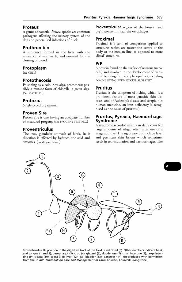
ProteusA genus of bacteria. Proteus species are commonpathogens affecting the urinary system of thedog and generalised infections of duck.
ProthrombinA substance formed in the liver with the assistance of vitamin K, and essential for theclotting of blood.
Protoplasm(see CELL)
ProtothecosisPoisoning by a colourless alga, prototheca; pos-sibly a mutant form of chlorella, a green alga.(See MASTITIS.)
ProtozoaSingle-celled organisms.
Proven SireProven Sire is one having an adequate numberof measured progeny. (See PROGENY TESTING.)
ProventriculusThe true, glandular stomach of birds. In itdigestion is effected by hydrochloric acid andenzymes. (See diagram below.)
Proventricular region of the horse’s, andpig’s, stomach is near the oesophagus.
ProximalProximal is a term of comparison applied tostructures which are nearer the centre of thebody or the median line, as opposed to more‘distal’ structures.
PrPA protein found on the surface of neurons (nervecells) and involved in the development of trans-missible spongiform encephalopathies, includingBOVINE SPONGIFORM ENCEPHALOPATHY.
PruritusPruritus is the symptom of itching which is aprominent feature of most parasitic skin dis-eases, and of Aujeszky’s disease and scrapie. (Inhuman medicine, an iron deficiency is recog-nised as one cause of pruritus.)
Pruritus, Pyrexia, HaemorrhagicSyndromeA syndrome recorded mainly in dairy cows fedlarge amounts of silage, often after use of asilage additive. The signs vary but include feverand persistent skin lesions which sometimesresult in self-mutilation and haemorrhages. The
Pruritus, Pyrexia, Haemorrhagic Syndrome 573
P
Proventriculus. Its position in the digestive tract of the fowl is indicated (5). Other numbers indicate beakand tongue (1 and 2); oesophagus (3); crop (4); gizzard (6); duodenum (7); small intestine (8); large intes-tine (9); cloaca (10); caeca (11); liver (12); gall bladder (13); pancreas (14). (Reproduced with permissionfrom the UFAW Handbook on Care and Management of Farm Animals, Churchill Livingstone.)

outcome is often fatal. It has also been reportedin cattle fed on citrus pulp which was mouldyand contained citrinin.
Prussic Acid(see HYDROCYANIC ACID)
PsammomaPsammoma is a small hard tumour of the brain.
Pseudo-CowpoxCaused by a parapox virus, this infection ischaracterised by inflammation of the teats ofcows, and of the hands of milkers.
PseudomonasA genus of bacteria. P. pyocyanea is a motile,Gram-negative rod, 1.5 to 3 µ long. It flourish-es in suppurating wounds, and has been foundin cases of otitis in the dog. It has also beenreported as causing outbreaks of disease inturkey poults and other birds as well as in sheep.
Chronic mastitis, with diarrhoea and wastingresembling Johne’s disease, has been caused incows by P. aeruginosa. This organism, oftenfound in non-mains water supplies, is thoughtlikely to be increasingly involved in mastitis incattle. It appears to have an increased incidenceduring August, September and October. (See
WOUNDS; also MELIOIDOSIS.)
Pseudo-PregnancyPseudo-pregnancy is a condition commonlyseen in the bitch, but probably occurring in allbreeding female animals to a lesser degree. In itthe physical signs of pregnancy are exhibited inthe absence of fetus or fetuses. The abdomenincreases in size, the uterus becomes swollenand turgid, its walls are thickened, and inextreme cases mammary development mayoccur and milk may be secreted. The bitch mayactually make a bed.
In time, since no fetuses are present, theorgans and tissues return to their normal statewithout the occurrence of parturition; heatreturns, and successful breeding may occur subsequently.
The condition has been described as anintensification and prolongation of metoestrus.The essential feature is persistence of the cor-pora lutea in the ovaries.
The condition can – where necessary – be treated by injection of the appropriate hormone.
In a review of 442 cases of pseudo-pregnancy in a total of 142 bitches, 19 had only1 pseudo-pregnancy, 31 had 2, 54 had 3, and39 had 4 or more pseudo-pregnancies.
In another reported series, a total of 81 percent of pseudo-pregnancies responded to treat-ment with bromocriptine, and 80 per cent ofthe behavioural or psychological problems wereresolved.
(See REPRODUCTION; BREEDING; ‘CLOUD-
BURST’.)
Pseudo-RabiesA name occasionally used for AUJESZKY’S
DISEASE.
Pseudo-Tuberculosis(see YERSINIOSIS; CASEOUS LYMPHADENITIS)
PsittacinesParrots, parrakeets, cockatiels, budgerigars andother members of the order Psittaciformes.
Psittacosis (‘Parrot Fever’)A NOTIFIABLE DISEASE in the UK. It causessevere respiratory illness in man and birds of theparrot family (psittacines), including budgeri-gars and cockatiels. It is caused by Chlamydiapsittaci. (See CHLAMYDIA; ORNITHOSIS.)
PsoasPsoas is the name of 2 muscles, psoas major andpsoas minor, which lie along the roof of theabdomen immediately beneath the last 2 or 3thoracic and the whole of the lumbar vertebrae,and stretch into the pelvis. The psoas minor isinserted in the psoas tuberele of the ilium, andthe psoas major runs to the inner or lessertrochanter of the femur in common with theiliacus muscle. The action of these muscles is tobend the pelvis on the rest of the trunk, or ifthose of one side of the body are acting alone,to bend the posterior part of the trunk towardsthat side. The act of crouching preparatory tokicking is accomplished by these muscles andothers, and they are largely concerned in themovements of galloping. Disease or injury, suchas a severe sprain, is shown by a difficulty inwalking both forwards and backwards, by acrouching appearance of the back, and byextreme difficulty in rising from the ground.
PsoriasisPsoriasis is a chronic inflammatory skin diseasewith scurf formation.
Psoroptic MangeA type of mange caused by various species ofPsoroptes mites. It can affect most animals, andcauses sheep scab, ear mange in horses andwidespread skin lesions in cattle. (See under
MANGE.)
574 Prussic Acid
P
PtosisThe drooping of the upper eyelid, due to paral-ysis of the oculomotor nerve. It is seen in thehorse after accidents involving the head; andalso in GUTTURAL POUCH DISEASE. A case ofptosis in a bird was treated by the topical appli-cation of phenylephrine, which rapidly resolvedthe condition.
PtyalinPtyalin is the name of the enzyme contained inthe saliva, by which starchy food-stuffs arechanged into sugars, and so prepared forabsorption.
PtyalismThe overproduction of saliva. It may be the firstclinical sign of eplepsy in dogs.
PubertyEwes, sows, and bitches may mate when only 6 or 7 months old; mares reach puberty at from 15 to 18 months old; heifers from 7 to 15 months old. In the cat oestrus may occur asearly as 31⁄2 months, or occasionally be delayeduntil the queen is about a year old. In the male,puberty commonly occurs at an age of 10 to 12 months, but here again there may be con-siderable variation. Some toms may reachpuberty as early as 6 months, while others donot mate until their 2nd spring.
PubisPubis is the bone that forms the lower anteriorparts of the pelvis. The pubes of right and leftsides meet each other at the ‘symphysis of thepubes’, which in old age is no longer a separa-ble union, bony fusion having taken place.
Public Health(see MILK; FOOD INSPECTION; ANTIBIOTIC RESIS-
TANCE; and information given under the mainanimal diseases communicable to people. See also
ZOONOSES)
Puffer Fish(see TOADFISH)
PugA small dog with large rounded head, promi-nent eyes, smooth coat and tail arched over theback. It is suceptible to Perthe’s disease butother inherited defects are not common.
‘Pullet Disease’A transmissible enteritis of pullets and turkeypoults, first described in the USA in 1951. (See
also VISCERAL GOUT.)
Signs Loss of appetite, diarrhoea with wateryor whitish evacuations and, sometimes, darken-ing of the comb. Birds appear drowsy. About 10per cent die. The cause is a REOVIRUS.
Pullorum Disease of Chicks(Bacillary White Diarrhoea)Pullorum Disease of Chicks (Bacillary WhiteDiarrhoea) has been virtually eradicated in theUK. It is an acute, infectious, and fatal disease ofchicks, causing much loss during the first 2 weeksof life. Adult fowls, especially laying hens, act ascarriers and transmit infection through their eggsto the chick before hatching. They may alsospread infection in their droppings.
Cause Salmonella pullorum, which is found inthe ovary and oviduct of carrier hens – birdswhich themselves contracted the disease whenyoung, but which survived.
Signs Lameness, with swelling of the hocks, ischaracteristic of chronic pullorum disease.
Prevention This can be achieved by testing all birds, eggs from which are to be used forhatching, by an agglutination test.
Pulmonary Adenomatosis(Jaagsiekte)Pulmonary adenomatosis (jaagsiekte) is causedby a retrovirus, often in association with aherpesvirus (see HERPESVIRUSES) and is a conta-gious neoplasm of the lung of adult sheep. Firstrecognised in South Africa, it occurs also in theUK, Iceland, and the USA. In Britain, one EastAnglian farmer lost 50 out of 200 half-bredewes from jaagsiekte.
Pulmonary Diseases(see LUNGS, DISEASES OF)
Pulpy Kidney DiseasePulpy kidney disease attacks lambs betweenabout 3 and 18 weeks of age, particularly thosewhich are thriving. The disease has been seen in lambs under a week old; its occurrence iswidespread.
Cause Clostridium welchii type D.
Signs As a rule the affected lambs are founddead without having previously been noticedailing. Usually the lambs in the best conditionare the first to be affected. The loss may be very heavy, especially with the larger earliermaturing breeds. The liver usually showshaemorrhagic spots on its surface.
Pulpy Kidney Disease 575
P
Prevention It is recommended that immuni-ty be maintained by autumn vaccination, witha second dose of vaccine in the spring, prefer-ably about 10 days before lambing – unless theewes are to be moved to a better pasture priorto lambing, when the second dose should begiven before the move is made. These 2 dosesshould protect the ewe through the springmonths and allow her to pass to the lamb viathe colostrum sufficient antibodies to protect itfor the first 8 to 12 weeks of life. That tempo-rary immunity in the lamb should be convertedto an active one by the use of vaccine.
PulseThe forcing of blood from the heart into thearteries of the systemic circulation causes a pul-sation (regular expansion and relaxation) inthem. The beating of the heart drives blood outfrom the left ventricle into an already full aorta,in which it is imprisoned by the closing of theaortic semilunar valves. To accommodate thisextra blood the aorta dilates, and the bloodalready in it moves onwards throughout thevessel, and through the larger branches arisingfrom it. The wave of dilatation also travelsalong the course taken by the blood, and istherefore distributed along all the larger arterialtrunks. If the fingers are placed over any ofthese latter, which lie near the surface, a period-ic thrill or ‘pulse’ can be felt, occurring at a reg-ular frequency according to the species; in thehorse, it is about 35 to 45 times per minute.
The pulse-rate varies according to the state ofthe animal’s health, being faster in fevers, andslower and weaker in debilitating non-febrile dis-eases; according to the age of the animal (fasterin the very young and very old); according to theclimate; according to bodily condition; andunder other circumstances. During and immedi-ately after exercise it is greatly increased, but inhealth it subsides rapidly subsequently. Duringsleep and unconsciousness it is slower.
The normal pulse-rates of the domesticatedanimals at rest are as follows:
Per minuteHorse 36 to 42Ox 45 to 50Sheep }Pig
70 to 80
Dog 90 to 100Cat 110 to 120
and of certain other animals as follows:Per minute
Elephant 25 to 28Camel 28 to 32Buffalo 40 to 45Reindeer 60 to 65Mouse 130 to 150
Roughly speaking, the smaller the animal, thefaster the pulse. The same principle applies toanimals of one species but of different sizes orof different breeds: e.g. the pulse of the Shirestallion is usually about 35 per minute, whilethat of the Shetland pony is 45 or more. Thesefacts must be taken into account when count-ing the pulse of any given animal. (See also under
HEART.)
Pupil(see EYE)
Puppies, Newborn, Infection in(see FADING; TOXOCARA)
PurgationEvacuation of the bowel following administra-tion of a cathartic medicine. It must be applied,if at all, with moderation. Excessive purgationinvolves dangers which include potassiumdepletion.
PurgativesThis is the age of LAXATIVES rather than purga-tives. The old drastic purgatives are obsolete;they tended to make the patient’s conditionworse.
Purpura HaemorrhagicaPurpura haemorrhagica often occurs in a horserecovering from a respiratory infection such asinfluenza or strangles; Streptococcus equi is fre-quently involved. The disease is characterisedby oedema of the head and also of the lowerparts of the body. There may be kidney lesions.
Signs appear suddenly; often overnight.Swellings, very often the same on each side ofthe body, are found on the limbs, the breast, theeyelids, and almost always about the muzzleand nostrils. These swellings may be diffusefrom the first, or they may begin as isolated cir-cumscribed flat prominences which coalesce inthe course of a day or more; when pressed withthe point of the thumb, a little pit remainsafterwards for some moments. Petechial haem-orrhages are present in the nostrils (from whicha bloodstained discharge is often seen) and onany mucous membrane.
The horse is dull, loses its appetite, movesstiffly and with difficulty, and if the swellings ofthe nostril are large, shows rapid and labouredbreathing. Swollen lips may prevent a horsefrom feeding or drinking; swollen eyelids mayhinder or prevent vision; and a swollen sheathin the male may make the act of micturitiondifficult. The temperature usually remains
576 Pulse
P
between 39° and 40°C (102° and 104°F); thepulse is soft, feeble, generally rapid, and may bevery irregular.
The percentage of recoveries is not large inwell-marked cases, and even where death doesnot occur, complete recovery takes a long timewith relapses common. It is said that casesshowing nervous complications always endfatally, and the same may be said of those withpneumonia.
Treatment The most careful nursing andfeeding are essential in all cases of purpura. (See NURSING OF SICK ANIMALS.) Good resultsoften follow the intravenous injection of anantihistamine.
After apparent recovery the horse must havea long period of convalescence.
PusThis thick, often yellowish fluid, found inabscesses and sinuses, and on the surfaces ofulcers and inflamed areas where the skin is broken, comprises blood serum, bacteria, whiteblood cells, and damaged tissue cells. (See
ABSCESS; STREPTODORNASE; PHAGOCYTOSIS.)
‘Pushing Disease’A colloquial name for poisoning of cattle by‘Staggers weed’ (Matricaria nigellaefolia) inSouth Africa.
PustulePustule means a small collection of pus occur-ring in the skin, or immediately below it. (See
ABSCESS.) ‘Malignant pustule’ is the nameapplied to the form that anthrax most com-monly takes when it affects the human being.
PuttyEating of this can result in lead poisoning. Adiscarded drum of putty thrown into a field ledto 12 bullocks dying within 24 hours, and afurther 40 requiring treatment. (Vl Servicereport).
PyaemiaThe presence of pus in the bloodstream.
PyelitisPyelitis means a condition of pus-formation inthe kidney which produces pus in the urine. Itis due to inflammation of the part called the‘pelvis of the kidney’, which is connected withthe ureter. The condition is commonest amongcows after calving, when infection has reachedthe bladder, invaded the ureters, and has arrivedat the pelvis of the kidney.
PyelonephritisThis term is used when both the pelvis andmuch of the rest of the kidney are involved, asdescribed under PYELITIS.
Contagious bovine pyelonephritis is a specif-ic infection of cattle caused by Corynebacteriumrenale, giving rise to inflammation and suppu-ration in kidneys, ureters and bladder. As a rule,only 1 cow in a herd is attacked though othersmay be carriers. The passage of bloodstainedurine and abdominal pain are symptoms.Penicillin is useful in treatment. Otherwise,death may occur (sometimes after severalweeks).
In the pig, an infectious pyelonephritis iscaused by C. suis. It is a common cause of deathor culling.
Pyloric StenosisThis occurs as a rare congenital defect in thedog. Only liquid food can pass into the stom-ach. Projectile vomiting is a symptom. Thedefect can be corrected by means of surgery. (See
PYLORUS.)
PylorospasmPylorospasm means spasm of the pyloric por-tion of the stomach. This interferes with thepassage of food in a normal, gentle fashion intothe intestine, and causes distress from half anhour to 3 hours after feeding. It is associatedwith severe disorders of digestion.
PylorusPylorus is the name of the lower opening of thetrue stomach. Exit of food from the stomach iscontrolled by a strong ring of muscular tissuecalled the ‘sphincter of the pylorus’, whichopens under nervous activity and allows escapeof small amounts of partly digested food mate-rial into the small intestine. (See STOMACH;DIGESTION.)
Pyo-Pyo- is a prefix attached to the names of variousdiseases to indicate the presence of pus or theformation of abscesses.
PyodermaA pustular condition of the skin. In dogs aller-gic skin disease is regarded as predisposing toinfection by staphylococci.
PyogenicPyogenic is a term applied to those bacteriawhich cause the formation of pus, and so leadto the production of abscesses.
Pyogenic 577
P
PyometraA collection of pus in the uterus: a conditionnot uncommon in maiden bitches, and occur-ring in all species. (See UTERUS, DISEASES OF.)
PyorrhoeaInflammation of the gums, in which suppura-tion is produced and ultimately interferencewith the integrity of the teeth. It is a commoncondition in aged dogs and cats. (See TARTAR.)
PyosalpinxDistension of a Fallopian tube with pus.
PyothoraxThe presence of pus within the chest. It may bea sequel to pneumonia, or to a penetratingwound of the chest, perhaps a bite. This is afairly common condition in the cat, which islikely to rest on its brisket, be disinclined tomove, and to have laboured breathing. Cyanosismay be present. Tenderness of the chest isanother symptom. The temperature may be37°C (98.6°F). In many cases the conditiondevelops very rapidly in the cat, death occurringbefore treatment has been obtained. Treatmentinvolves aspiration of the pus, and the introduc-tion of an antibiotic. In cats, however, the mortality despite treatment may be 50 per cent.
Pyramidal DiseaseAn exostosis affecting the pyramidal process(extensor process) of the 3rd phalanx of thehorse’s foot. It is usually found in associationwith low ringbone. (See RING-BONES.)
Pyrenean Mountain DogOne of the largest breeds of dog, powerfullybuilt with a thick, usually white, coat.
Haemophilia may be inherited and the breed isdisposed to osteochondritis dissecans and hipdysplasia.
PyrethroidsSynthetic equivalents of some of the activeprinciples of pyrethrum flowers are useful andpotent insecticides.
Commercial preparations are widely available(See FLIES – Control.)
Pyrexia(see FEVER)
PyridinePyridine is an alkaloidal substance derived fromcoal-tar, tobacco, etc. It is added to methylatedspirit in order to render this unpleasant todrink.
PyridoxineVitamin B6.
Pyrrolizidine AlkaloidsThese cause poisoning in animals which haveeaten ragwort. (See RAGWORT POISONING.)
Pyruvic AcidAn organic acid which is anintermediate product in carbohydrate and pro-tein metabolism. Excessive quantities accumu-late in the bloodstream in cases of vitamin Bldeficiency.
PyuriaPus in the urine produced by suppuration insome part of the urinary tract. (See URINE.)
578 Pyometra
P

Q FeverA disease first recognised in Australia in 1935,and now known to have a worldwide distribu-tion, Q fever is an infection of man, cattle,sheep, goats, fowls, and rodents. In Iran, serological evidence of Q fever has been foundalso in horses and camels.
Cause A rickettsia, Coxiella burnetii, which isresistant to heat and drying, and can be trans-mitted by ticks. Human infection can beacquired from these, from inhalation, and fromdrinking unpasteurised, infected milk; as well asfrom handling or coming into contact with the fetal membranes, faeces or urine of infectedanimals.
Signs In farm animals, many Q fever infec-tions may be present without obvious symp-toms. However, the rickettsia is a cause of abortion, and less often of pneumonia.
Incidence In the UK a preliminary surveyshowed that 2581 farms in England, 553 inWales, and 240 in Scotland were infected. It hasbeen found possible to isolate the parasite from13,600-litre (3000-gallon) milk tankers.
In a survey, sera from cattle and sheep in thenortheast of Scotland were tested for antibodiesto C. burnetii. Approximately 1 per cent of4880 cattle had antibodies to the organism.These potentially infected cattle were distrib-uted throughout the area. Two flocks of sheepwere tested; in one flock, 30 per cent of sheep had antibodies, while the other wasnegative. The flock with the high prevalence ofC. burnetii antibodies appeared to be associatedwith an outbreak of human Q fever on thatfarm.
Treatment Most antibiotics are rickettsiostat-ic rather than rickettsiocidal; tetracycline hasbeen used effectively. However, the organismcan remain dormant for long periods inside thehost’s body cells.
Public health Acute Q fever may involve theliver and heart (with resultant myocarditis).Mild cases may resemble food poisoning orinfluenza with headaches. Chronic Q feveroccurs.
Q fever in snakes Many snakes importedinto the USA are infested with ticks, whichtransmitted Q fever to dockside workers handling a shipment of Ball pythons.
Q fever from contaminated clothingThis was the presumed cause of 16 out of 32 employees at a truck- repair plant becomingill with the disease. Serological tests on a catwere positive for C. burnetii. The cat was fed athome by one of the workers at the plant.
QuadricepsQuadriceps means having 4 heads, and is thecollective name applied to the powerful musclessituated above the stifle-joint. These are medialand lateral vasti, and the rectus femoris; the 4th muscle (vastus intermedius) in the horse isso blended with the medial vastus that it haslost its autonomy.
QuadriplegiaParalysis of all 4 limbs. (See PARALYSIS; TICK
PARALYSIS; RACOONS; CURARE.)
QuailSmall, rapidly maturing game birds included aspoultry in British legislation. Females start to lay eggs at 5 to 6 weeks of age; the first eggslaid are usually infertile, but thereafter high fertility can be obtained. Males are sexuallyactive at 5 weeks. Among the diseases they maysuffer are: botulism, lymphoid leukosis, Marek’s disease, Newcastle disease, pasteurellosis,mycoplasmosis, salmonellosis, quail disease(ulcerative enteritis), quail bronchitis and coccidiosis.
Quail BronchitisA highly contagious viral infection. Signsappear in all susceptible birds within 3 to 7 daysof infection having been spotted in individuals.Clinical signs are very severe respiratory distresswith 100 per cent morbidity and mortalityfrom 10 per cent to 100 per cent.
Quail Disease (Ulcerativeenteritis of quail)A severe bacterial infection caused byCorynebacterium perdicum. Birds may die sud-denly without showing clinical signs; thesebirds are usually in good condition with feed intheir crop. Birds less acutely affected are listless,huddling with eyes partly closed and with ruf-fled feathers. Emaciation develops witin a week.Survivors become immune. Streptomycin,chloromycetin and bacitracin have been used
Q
in treatment. Effective control of other diseaseswill help to reduce the severity of quail disease.
QuarantineThe imposition of measures for preventing thespread of infectious disease by which an animalor animals, which have come from potentiallyinfected countries or areas, are kept separatefrom indigenous animals until their disease-freestatus is confirmed (or otherwise).
The regulations dealing with quarantine ofanimals are altered from time to time, and soinformation on the matter is best obtaineddirect from the government department thatdeals with livestock in a particular country.
The length of quarantine depends on the dis-ease whose entry is being prevented. For rabiesit is 6 months (except in the case of dogs andcats from specified countries, for which newregulations apply); for foot-and-mouth andNewcastle disease it is 30 to 35 days. It is a sen-sible precaution that new stock introduced tofarms, zoos, etc. should be kept separate until itis certain that they have not brought in new dis-eases or virulent strains, even if this is notrequired by law.(See RABIES; IMPORTING/EXPORTING ANIMALS;NOTIFIABLE DISEASES; PET TRAVEL SCHEME;PIGEONS.)
Quarter Horse(see AMERICAN QUARTER HORSE)
Quaternary AmmoniumCompoundsQuaternary ammonium compounds are used asantiseptics, and have found widespread applica-tion in dairy hygiene. Cetrimide – or cetyltrimethyl ammonium bromide – is an example.It is used in 0.1 per cent solution for washingcows’ udders, teats, and milkers’ hands, beingeffective against Streptococcus agalactiae. Inhigher concentrations it acts as a detergent.Such compounds are readily neutralised byorganic matter and should only be used on visibly clean surfaces. (See also CETRIMIDE; HIBI-
TANE.)
QueenA female cat.
Queensland ItchThis is caused by sensitisation to bites of themidge Culicoides robertsi. The lesions resemblethose of mange or eczema, and are seen usuallyalong the animal’s back. Antihistamines are useful in treatment. The condition is regarded
as an allergic dermatitis, and is similar to ‘sweetitch’. (See under FLIES.)
QueyA heifer.
Quidding (Cudding)Quidding (cudding) is the name given to thatcondition in horses, depending upon injuries tothe mouth or diseases of the teeth, in whichfood is taken into the mouth, chewed repeated-ly, and then expelled on to the floor of the stallor into the manger. It may result from the teethbeing too sharp, irregular in height, uneven inalignment, or from permanent teeth pushingthe temporaries out from the gums; it may alsoarise when the gums, cheeks, or tongue havebeen injured or are diseased. Paralysis of thethroat, or some other condition which causesinability to swallow, can cause quidding. (See
MOUTH, DISEASES OF; TEETH, DISEASES OF.)
QuinineQuinine is an alkaloid obtained from the barkof various species of cinchona trees in SouthAmerica. The bark contains 4 alkaloids, ofwhich quinine is the most active and impor-tant, the others being quinidine, cinchonine,and cinchonidine.
Quinine is usually used in the form of one ofits salts, i.e. sulphate, hydrochloride, or hydro-bromate of qui-nine.
Action. Quinine causes a lowering of temper-ature in fevers. In man, it is used for the reliefof malaria.
Uses These have dwindled. Before the adventof the sulfa drugs and antibiotics it was muchused in influenza, distemper, and similar condi-tions. It is sometimes given as an intramuscularinjection. Owing to its very bitter taste it is seldom that it will be taken in the food.
Toxicity The dog is very susceptible to quinine and may become blind at plasma con-centrations readily tolerated by man.
QuittorQuittor is a condition of the ‘lateral’ cartilagesof the horse’s foot, in which suppuration occurs,with pus escaping from an opening in the regionof the coronet. This, and the bulbs of the heels,are swollen and painful. The cause is an injuryto the cartilage or to infection, or both. There isusually some degree of lameness. Antibiotics areused in treatment.
580 Quarantine
Q
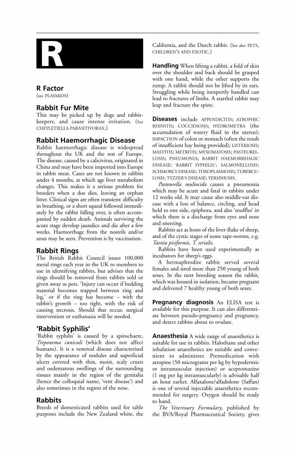
R Factor(see PLASMIDS)
Rabbit Fur MiteThis may be picked up by dogs and rabbit-keepers, and cause intense irritation. (See
CHEYLETIELLA PARASITIVORAX.)
Rabbit Haemorrhagic DiseaseRabbit haemorrhagic disease is widespreadthroughout the UK and the rest of Europe. The disease, caused by a calicivirus, originated inChina and may have been imported into Europein rabbit meat. Cases are not known in rabbitsunder 4 months, at which age liver metabolismchanges. This makes it a serious problem forbreeders when a doe dies, leaving an orphan litter. Clinical signs are often transient: difficultyin breathing, or a short squeal followed immedi-ately by the rabbit falling over, is often accom-panied by sudden death. Animals surviving theacute stage develop jaundice and die after a fewweeks. Haemorrhage from the nostrils and/oranus may be seen. Prevention is by vaccination.
Rabbit RingsThe British Rabbit Council issues 100,000metal rings each year in the UK to members touse in identifying rabbits, but advises that therings should be removed from rabbits sold orgiven away as pets. ‘Injury can occur if beddingmaterial becomes trapped between ring andleg,’ or if the ring has become – with the rabbit’s growth – too tight, with the risk ofcausing necrosis. Should that occur, surgicalintervention or euthanasia will be needed.
‘Rabbit Syphilis’‘Rabbit syphilis’ is caused by a spirochaete,Treponema cuniculi (which does not affecthumans). It is a venereal disease characterisedby the appearance of nodules and superficialulcers covered with thin, moist, scaly crusts and oedematous swellings of the surroundingtissues mainly in the region of the genitalia(hence the colloquial name, ‘vent disease’) andalso sometimes in the region of the nose.
RabbitsBreeds of domesticated rabbits used for tablepurposes include the New Zealand white, the
California, and the Dutch rabbit. (See also PETS,CHILDREN‘S AND EXOTIC.)
Handling When lifting a rabbit, a fold of skinover the shoulder and back should be graspedwith one hand, while the other supports therump. A rabbit should not be lifted by its ears.Struggling while being inexpertly handled canlead to fractures of limbs. A startled rabbit mayleap and fracture the spine.
Diseases include APPENDICITIS; ATROPHIC
RHINITIS; COCCIDIOSIS; HYDROMETRA (theaccumulation of watery fluid in the uterus);IMPACTION of colon or stomach (often the resultof insufficient hay being provided); LISTERIOSIS;MASTITIS; METRITIS; MYXOMATOSIS; PASTEUREL-
LOSIS; PNEUMONIA; RABBIT HAEMORRHAGIC
DISEASE; ‘RABBIT SYPHILIS’; SALMONELLOSIS;SCHMORL’S DISEASE; TOXOPLASMOSIS; TUBERCU-
LOSIS; TYZZER’S DISEASE; YERSINIOSIS.Pasteurella multocida causes a pneumonia
which may be acute and fatal in rabbits under12 weeks old. It may cause also middle-ear dis-ease with a loss of balance, circling, and headheld to one side, epiphora, and also ‘snuffles’ inwhich there is a discharge from eyes and noseand sneezing.
Rabbits act as hosts of the liver-fluke of sheep,and of the cystic stages of some tape-worms, e.g.Taenia pisiformis, T. serialis.
Rabbits have been used experimentally asincubators for sheep’s eggs.
A hermaphrodite rabbit served severalfemales and sired more than 250 young of bothsexes. In the next breeding season the rabbit,which was housed in isolation, became pregnantand delivered 7 healthy young of both sexes.
Pregnancy diagnosis An ELISA test isavailable for this purpose. It can also differenti-ate between pseudo-pregnancy and pregnancy,and detect rabbits about to ovulate.
Anaesthesia A wide range of anaesthetics issuitable for use in rabbits. Halothane and otherinhalation anaesthetics are suitable and conve-nient to administer. Premedication withatropine (50 micrograms per kg by hypodermicor intramuscular injection) or acepromazine (1 mg per kg intramuscularly) is advisable halfan hour earlier. Alfaxalone/alfadolone (Saffan)is one of several injectable anaesthetics recom-mended for surgery. Oxygen should be ready to hand.
The Veterinary Formulary, published by the BVA/Royal Pharmaceutical Society, gives
R

comprehensive details of anaesthetics and analgesics suitable for rabbits and small rodents.
RabiesThe Latin word for madness, it is a specific inoc-ulable contagious disease of virtually all mam-mals, including man; and occasionally it occursin birds, e.g. domestic poultry and vultures. It ischaracterised by nervous derangement, often by achange in temperament, with paralysis occurringin the final – and sometimes in the intermediate– stages.
Foxes and cattle are both highly susceptibleto infection.
Rabies occurs in all continents with theexception of Australasia and Antarctica. InTurkey, dogs remain the principal vectors; in afew countries in Europe cats attack more peoplethan do dogs. In Asia and South America dogsare still the most important vectors, but inmany countries wild animals provide a reservoirof infection, and infect dogs and cats and farmanimals – which in turn may infect man, whois an incidental host of the disease. (See table ofvectors.)
Public health Rabies is virtually always fatal inthe human being, and there is danger not onlyfrom being bitten by rabid animals, but also fromcontamination by their saliva of wounds, cut fingers, eyes, etc. Scratches may convey infectionas well as bites.
People have died from rabies following attacksby rabid dogs, cats, foxes, wolves, badgers,skunks, racoons, mongooses, bats, rodents, etc.
Pet animals, such as rabbits, may be bitten byrabid animals and themselves become rabid;and it has sometimes happened that wild orexotic animals (originating in countries whererabies is endemic) were bought as pets while inthe incubation stage of rabies, with unfortunateresults.
In the UK as in most other countries, rabiesis a NOTIFIABLE DISEASE, and must be reported
to DEFRA or to the police. Bitten personsshould seek medical advice immediately.
Cause A Lyssavirus (one of the Rhabdovirusgroup). When it is injected into the tissues,either naturally (from a bite) or artificially, thevirus passes along the nerves and reaches thecentral nervous system. The time elapsingbetween infection and onset of symptoms variesgreatly with the location of the bite, its severity,and – no doubt – the quantity of virus in thesaliva. In the most rapidly developing cases thesymptoms may be shown as early as the 9th day after being bitten, and at the other extreme,cases have appeared several months after theincident. It is owing to this fact that the 6-months period of quarantine insisted upon in Britain is something of a compromise. Theaverage incubation periods in dogs, sheep, andswine are from 15 to 60 days; in horses and cattle, from 30 to 80 days. In young animals theperiod of incubation is shorter than in adults.
Signs
Dog There are 2 distinct forms of rabies in thedog – the ‘furious’ and the ‘dumb’; but these are in reality 2 stages only. It is customary toconsider 3 stages of typical symptoms.
(1) Melancholy. The prodromal dull stage isoften not noticed, or, if it is, only scant attentionis paid to it. The habits of the dog change. It becomes morose and sulky, indifferent toauthority, disregards its usual playthings or com-panions, shows a tendency to hide in dark cor-ners, and may appear itchy or irritable as regardsits skin. Noisy, boisterous animals become quietand dull, while animals that are normally of agentle, quiet disposition may become excitable.After 2 or 3 days of such behaviour the nextstage is reached.
(2) Excitement. The symptoms describedabove become exaggerated, and there is a ten-dency towards violence. The dog pays no atten-tion to either cajoling or threatening. Itbecomes easily excited and very uncertain in itsbehaviour. Food is either disregarded complete-ly or eaten with haste. Vomiting is a notuncommon symptom. A fear of water is not asymptom to expect in the rabid dog, which willoften drink or attempt to do so even when part-ly paralysed. After a time the appetite becomesderanged. The dog refuses its ordinary food,but eats straw, stones, wood, coal, carpet, piecesof sacking, etc., with great avidity. If the animalis shut up in a kennel, it persists continually inits efforts to escape. Should it be released orshould it escape, it almost invariably runs away
582 Rabies
R
Rabies in wild animals – principal vectors in various regions
Europe Foxes, roe-deer,badgers, martens
Asia Wolves, jackals, bats, mongooses
North America Foxes, skunks, coyotes, bats
Central America BatsSouth America and
Trinidad Vampire bats

from home. It may wander for long distances.In its travels it bites and snaps at objects whichit encounters, real or imaginary, animate orinanimate. Some rabid dogs bite several people.The tone of voice is altered.
The face has a vacant stare, the eyes are fixedand expressionless, and the pupils are dilated.This stage lasts from 2 to 4 days, unless the dog’sstrength gives out sooner, and the next stageappears.
(3) Paralysis. The characteristics of the laststage in the train of symptoms of rabies arethose of paralysis, especially of the lower jawand the hindquarters. The dog begins to staggerin its gait, and finally falls. It may manage toregain its feet when stimulated, but soon fallsagain. The lower jaw drops, the tongue lolls outof the mouth, and there is great salivation. Themuscles of the throat and larynx are sooninvolved in the progressive paralysis.
The dumb form of rabies consists of this par-alytic stage – the stage of excitation having beenomitted. The dumb form is the more commonin the dog: barking ceases – hence the name.Vomiting may suggest merely a digestive upset.Protrusion of the nictitating membrane partlyacross the eye, together with a dropped jaw, i.e.partly opened mouth which can be closed bygently raising the lower jaw by means of a stick,are highly suggestive of rabies.
In parts of Africa and Asia, the classical formof rabies in dogs (described above) is replacedby a form called (in Africa) ‘OULOU FATO’.
Cat In this animal the furious form is morecommon than in the dog. The aggressive stage ismost marked, the cat attacking other animalsand man with great vigour, and attempting toinjure their faces with teeth or claws. Sometimesthe rabid cat will at first show extra affection.The course of the disease is usually shorter thanin the dog.
It is worth mentioning that occasionally dogsand cats die from rabies without any observedsymptoms. They may be found dead or dying.It is not unknown for a cat to be found lying ina field or garden unable to walk but still able tobite.
Cattle These animals are usually affectedthrough having been bitten by a rabid fox ordog. The stage of excitement is short and thedumb stage most evident. Affected cattlebehave in an unusual manner; they may stampor bellow, salivate from the mouth, break loose,and may do much damage. Rumination andmilk production cease, muscular quiverings areseen, sexual excitement is noticed, and there is
a great loss of condition. Exhaustion soon fol-lows and paralysis sets in. Death occurs within2 to 6 days or more after the commencement ofthe condition.
Rabies may be mistaken for hypomagne-saemia, milk fever, botulism, anaplasmosis, listeriosis, lead poisoning, choking, etc.
In Central and South America, cattle areinfected with rabies by vampire bats, and mayshow long streaks of blood on their shoulders,necks and backs.
Sheep, goats and swine The sheep and thegoat are affected in a manner similar to cattle, butthe stage of excitement is shorter or absent, andthe dumb paralytic stage is more often noticed.Pigs become excitable; they may squeal and showmuscular spasms before paralysis ensues.
Horse The furious form is common but theanimal may appear calm between bouts ofaggressiveness. Dumb forms also occur andmay be mistaken for colic, paresis or encephali-tis from other causes. Signs may include a facialtwitch, biting of woodwork or self-mutilation,head-tossing, frequent whinnying, abnormalposture, apparent lameness, ataxia, paralysis ofhindquarters. The horse may continue to eatand drink until shortly before death. The toneof voice may be altered.
Diagnosis The routine examination for Negribodies has now in most countries been super-seded by the fluorescent antibody test, withconfirmation by mouse inoculation if necessary.(If a dog which bit someone is still alive after 10 days, it cannot be assumed that the dog isnot rabid.)
Differentiation between laboratory andstreet rabies virus, between rabies vaccine virusand street virus, and between rabies virus andrabies-like viruses (e.g. Mokola, Lagos bat, andDuvenhage viruses) is possible by laboratorytests based on differentiation of monoclonalantibodies.
Prevention Prevention of the disease in manand animals stems from the research of LouisPasteur in the 1880s. He discovered the processof attenuation, by which the virulence of amicro-organism is reduced but not its ability to produce antibodies against disease. Pasteurachieved this by infecting rabbits with rabiesfrom a dog. Although this was fatal to the rabbits, dogs survived infection with the rabbitvirus. Tissue from the spinal cord of an infect-ed rabbit was then used to prepare a vaccine.
Rabies 583
R

His triumph came in 1885 when the vaccinesaved the lives of 2 badly bitten boys.
In the intervening years many modificationshave been made, and new techniques developed,to make rabies vaccines which would be safe andfree from dangerous side-effects, and so could beused to immunise people and animals againstrabies (‘pre-exposure’ – vaccination), as well asprovide ‘post-exposure’ treatment of those bittenby rabid animals.
The table shows examples of vaccines pre-pared from tissue culture cells. The last one, the Merieux, was developed by the MerieuxInstitute of France using a technique pioneeredat the Wistar Institute of Philadelphia. Only 1 ml doses are required, and 2 injections (apartfrom any booster doses). (See also VERO CELLS.)
In the UK, 2 vaccines approved for use indogs and cats: Rabisin rabies vaccine (Merial)containing inactivated GS-57 Wistar virusstrain; and Nobivac Rabies (Intervet) preparedfrom virus grown on cell-line tissue culture.
Mass vaccination of dogs is carried out inmany countries as a control measure; and inCentral and South America, cattle on ranchesare vaccinated against vampire-bat-transmittedrabies. In France and other countries of Europe, hundreds of thousands of cattle arevaccinated against rabies (often a combinedrabies/foot-and-mouth disease inoculation).
It must be remembered, however, that no vac-cines are 100 per cent effective, that certificatesof vaccination can be forged, and that conse-quently it is still essential to control the importof animals, whether vaccinated or not, and toenforce quarantine measures where appropriate.
Control of rabies in Britain From 1902until 1918, no cases occurred in the British Isles;but in that year infected dogs were smuggledfrom the Continent, and the disease obtained a fresh hold for a period of little more than 3 years. Britain had been free since then, but in
1969 a dog released from quarantine 10 days ear-lier showed symptoms of rabies and bit 2 peopleat Camberley, Surrey; a 2nd case occurred in1970. In 1965 there was a case in a recentlyimported leopard in quarantine at EdinburghZoo. In Britain, in 1969, the danger of allowingthe importation of rabies-susceptible exotic ani-mals, for sale as pets or for research, was official-ly recognised, and the quarantine regulationsamended to include monkeys, mongooses, etc.
Following strong pressure to replace quaran-tine for pet dogs with a vaccination/identifica-tion policy, a government committee was set upin 1997 to examine the issue. The committeerecommended that a strict scheme of medicalexamination, rabies vaccination and veterinarycertification should replace the compulsoryquarantine regulations for dogs and cats. Therecommendation was accepted and an arrange-ment introduced in 2000 under which dogs andcats may travel to and from the UK and specifiedcountries without quarantine under the PET
TRAVEL SCHEME. (See also IMPORTING/EXPORTING
ANIMALS.)
Other points to note: (1) the saliva is some-times infective before symptoms of rabies appear– a hazard for a person licked; (2) farmers havedied through mistaking rabies for ‘choking’ and,with abraded fingers, examining their cow’smouths; (3) non-typical cases of rabies are notuncommon; (4) a dog may bite a small child orhousehold pet and promptly run away – rabiesnot being suspected, though running away is initself a canine symptom; (5) the virus may be pre-sent in semen, as well as in milk, tears, faeces, andurine; and (6) subclinical rabies, and a ‘carrier’state, have long been recognised in Africa (see
‘OULOU FATO’) and in Asia.
Vaccination of foxes has been an outstand-ing success in controlling the disease in WesternEurope. Currently Britain, Andorra, Ireland,
584 Rabies
R
Examples of rabies vaccines prepared from tissue culture cells
Live virus: Cells used:ERA Pig kidney Cats, dogs, cattle
and other animalsHEP-Flury Dog kidney Cats, dogs, and cattle
Inactivated:Fixed Hamster kidney Cats, dogs, cattle
and other animals
Hamster embryo Cats, dogs, horses,cattle and sheep

Italy, Netherlands, Norway, Sweden, Denmark,Greece, Spain and Portugal are all rabies-free.There are still black spots in Germany, whilethere are enzootic areas in Poland and Turkey.The oral vaccine is genetically engineered on vaccinia virus so that the antigen to rabies isabsorbed from the intestine. The vaccine is putinto fish-flavoured capsules scattered from heli-copters. In the areas so treated, up to 93 per centof foxes, stoats, weasels, polecats and badgerscaught and bloodsampled have been found tohave taken up the vaccine.
Rabies (Control) Order 1974This gives powers to deal with an outbreak ofrabies outside quarantine premises. In a declaredinfected area, an order may be made for thedestruction of foxes and other wild mammals,and for access to land for this purpose. Fences orother types of barrier may be erected to restrictmovement of animals into or out of an areawhile such destruction is in progress.
Orders may be made for compulsory vacci-nation, confinement, and control of domesticanimals, including strays. Anyone knowing orsuspecting that an animal has rabies must noti-fy that suspicion to the police. Deaths of ani-mals in an infected area must also be notified,and the authorities can take over ownership ofcarcases and determine the means of their dis-posal. This is because it is essential to confirm a diagnosis of rabies, so that precautions can be taken concerning in-contact animals andhuman beings. The order can override a dog-owner’s reluctance or refusal to part with thebody of a dead pet or working dog.
The Rabies (Importation of Mammals)Order 1974 prohibits the landing of suscepti-ble mammals in Britain unless from Ireland, Isleof Man, or the Channel Islands. Any animalbrought in from elsewhere has to undergo a peri-od of quarantine. Imported animals are vacci-nated while in quarantine as a precaution againsta quarantined animal developing the disease.Those animals not a threat to human health(ruminants, pigs and horses) do not go intoquarantine for rabies but may be quarantined forother diseases. Control under the Order is exer-cised on the transport of imported susceptibleanimals within Britain.
If an animal is landed at a port or airport notauthorised to receive such animals, that consti-tutes an illegal landing even if the circumstancesare outside anyone’s control (e.g. if an airport isfogbound).
Under a 1984 amendment order, animalswhich have not been in contact with another
animal (e.g. have been on an oil rig) are permit-ted to be landed in Britain. A similar relaxationapplies to animals belonging to the police,Customs & Excise and H.M. Forces, if the ani-mal has been abroad but under the constant con-trol of a trained handler while outside Britain.(See also PET TRAVEL SCHEME (PETS).)
Rabies-Related VirusesThese include Duvenhage virus, the cause infruit-eating bats of a disease very similar torabies; the Mokola virus, which has been isolat-ed from shrews, and causes nervous symptomsin man; the Lagos bat virus; the Nigerian horsevirus and Lyssa virus.
RaccoonsRaccoons are, in Canada and the USA, amongthe wildlife creatures which sometimes transmitrabies.
A dog bitten by a (non-rabid) raccoon maybecome paralysed in all 4 limbs (quadriplegia).
RacehorsesEvery year between 1400 and 1600 thorough-bred mares go to stud in the UK. About 67 percent of them foal successfully, and for every 1000mares covered, 270 or so of the resulting progenyfinally appear on the racecourse. Temperament,unsoundness, or sale abroad account for the non-appearance of more in the UK.
An epidemiological study of wastage amongracehorses has been conducted among 6 stables,5 of which were in Newmarket. The basis of thesurvey was the inability of horses to take part incantering exercise as a result of injury or disease.The greatest number of days lost to training wascaused by lameness (67.5 per cent) and respira-tory problems (20.5 per cent). Conditions of thefoot (19 per cent), muscle (18 per cent), carpus(14 per cent), fetlock joints (14 per cent), tendons (10 per cent) and sore shins (9 per cent)were the major reasons for training days beinglost in 198 cases in which a positive diagnosis ofthe site of lameness was made.
Pulmonary haemorrhage In horseswhich show blood at their nostrils after exercisesuch as racing, the blood does not come fromthe nasal cavity but from the lungs. Endoscopicexamination showed an incidence of 42 percent in a group of horses with only 15 per centshowing blood at the nostrils. Affected horsesmight appear distressed, with dilated pupils.
Exercise-induced pulmonary haemorrhagewas observed in 23 of 49 endoscopic examina-tions after high-speed training, in 9 of 37 exam-inations after cantering, and in 1 of 17 after
Racehorses 585
R
walking or trotting; it was not possible to pre-dict its occurrence. Mucoid or mucopurulentexudate was observed in 60 of 118 examinationsand the amount increased after exercise.
Pulmonary haemorrhage was diagnosed byendoscopic examination in 255 2-year-old quar-terhorses after racing. Only 9 (3.5 per cent) ofthe animals had visible epistaxis.
Fatal pulmonary haemorrhage occurred in aracehorse which panicked as the aircraft in whichit was travelling landed.
(See HORSES, BREEDS OF; HORSES; EXERCISING
HORSES; etc.)
Rachitis(see RICKETS)
Radial Paralysis (‘DroppedElbow’)Radial paralysis (‘dropped elbow’) is common-est in horses and dogs, though it may be seen inany animal.
Causes Probably the majority of cases are dueto a fracture of the 1st rib on the same side ofthe body, the broken ends of the rib laceratingthe nerve-fibres as they pass the rib, or pressingagainst them. In other cases the origin of theparalysis seems to be situated in the end-platesof the nerve-fibres where they are distributed tothe muscles, and in some cases a neuritis involv-ing the radial nerve, or a tumour pressing uponit at some part of its course, is responsible forproducing the condition.
Signs In a typical case the horse stands with theelbow dropped lower than normally, and withthe knee, elbow, and fetlock joints flexed. Littleor no pain is felt, unless there is a fractured rib,or some inflammatory condition which hascaused the paralysis. The limb is held in theposition assumed at the commencement of astride, but the animal is incapable of advancingit far in front of the sound limb. No weight isborne upon the leg, the muscles are flaccid andsoft, and if the horse is made to move forwardeither it does so by hopping off and on to thesound fore-limb, or it may fall forwards. If thehand be forcibly pressed against the knee, sothat the limb is restored to its natural uprightposition, the horse is able to bear weight upon itand may lift the other limb from the ground,but as soon as the pressure is released, the jointsfall forward again. Sometimes the toe is restedupon the ground, but at other times the horsestands with the wall of the foot in contact withthe ground. In cases that are not so severe, theflat of the foot may rest on the ground, and the
limb can be advanced forwards to a considerableextent.
Treatment The majority of such cases as thesewill recover in a few weeks. Patience on the partof the owner is essential.
Radiation, Exposure toThe 1986 Chernobyl nuclear power-station dis-aster in the former USSR led to controls beingimposed on the movement and slaughter ofsheep in parts of Scotland, Cumbria, and Wales,after between 1000 and 4000 Becquerels/kg ofcaesium-137 had been detected in lambs.Similar controls were applied in other countriesaffected by the fallout. The ban temporarilyaffected about 2 million sheep and lambs insome 500 flocks.
The Atomic Energy Authority stated that10,000 Bq/kg represents a health risk.
However, the contamination figures exceed-ed, in 9 cases, the internationally recommendedaction levels for radiocaesium of 1000 Bq/kg.The highest figure was 4000.
‘Although the physical half-life of radiocae-sium is 30 years, its biological half-life is muchshorter. In an adult animal, the half-life is esti-mated at between 30 and 100 days, but for lambit would be between 25 and 50 days.’ (MAFF)
(See also RADIOACTIVE IODINE; RADIOACTIVE
STRONTIUM.)
Annual human exposure Of the averageUK citizen’s annual exposure to radioactive dis-charges, only 0.1 per cent comes from the nuclearpower industry, according to the RadiologicalProtection Board.
For radiation exposure associated with veteri-nary practice, see RADIOISOTOPES and X-RAYS.
Carbon-14 is among internal sources of nat-ural radiation, and is present in the humanbody to the extent of about 2000 Bq.
Radiation, Protection againstRegulations governing the use of X-ray equip-ment, and the precautions to be taken by thosehandling it, are very strict. Details are given in the Health and Safety at Work Act. (See under
X-RAYS.)A concise guide to the Health and Safety at
Work Act 1974 can be obtained from HSEBooks, PO Box 1999, Sudbury, Suffolk COI06FS.
Radiation SicknessDogs exposed to radiation following a nuclearexplosion will vomit as a result of gastroenteri-tis, become dull and lose their appetite. This
586 Rachitis
R
may return after a day or two, but leucopeniadevelops, and may be followed by haemorrhageor septicaemia.
Radioactive CaesiumHigh levels of caesium 137 were found in areasof Wales and Scotland following the nuclearpower-station explosion at Chernobyl in 1986.Certain flocks of sheep were affected by the fall-out and the meat declared unfit for humanconsumption for some time.
Antidote A ferric-cyano-ferrate (AFCF), in theform of a dark blue powder, can bind radiocae-sium both in vitro and in the gastrointestinal tractof animals very effectively, preventing the isotopefrom being absorbed and secreted into the milkor transferred to the meat of cows, etc. The addi-tion of only 3 g AFCF per day to the diet of lactating cows reduced the radiocaesium contentof their milk by between 80 and 90 per cent, andof their meat by 78 per cent.
The radiocaesium content of the meat fromsheep fed 1 g AFCF per day or of calves or pigsfed 2 g AFCF per day was reduced by approxi-mately 90 per cent. The compound was givenofficial clearance as a feed additive against radio-caesium in Germany.
Radioactive DischargesOf the average UK citizen’s annual exposure, only0.1 per cent comes from the nuclear power indus-try, according to the Radiological ProtectionBoard.
Around 90 per cent comes from naturalsources, principally radon gas released frombuilding materials.For radiation associated with veterinary practice,see RADIOISOTOPES and X-RAYS.
Radioactive Fall-OutRadioactive fall-out, following the explosion ofnuclear bombs, etc., or accidents at atomic plant,may be dangerous to farm livestock on accountof the radioactive iodine and strontium released.After an accident at Windscale, radioactiveiodine alone contaminated pasture in the area.(See also RADIATION, EXPOSURE TO; RADIATION
SICKNESS; RADIOACTIVE IODINE.)
Radioactive IodineCattle grazing pasture contaminated by fall-outpick up 10 times as much radioactive iodine asdo people in the same locality, according toAmerican reports. Much is excreted in the milk,and much concentrated in the thyroid glands.
Feeding-stuffs or pasture contaminated byfall-out containing radioactive iodine and
strontium may give rise to illness in cattle.Digestive organs may be damaged, changes inthe blood occur, and deaths follow within amonth or so, after a period of dullness andscouring. (See RADIOACTIVE STRONTIUM.)
Radioactive StrontiumWhereas the half-life of radioactive iodine is amatter of days, that of strontium is 30 years.Following the grazing of contaminated pastureor the eating of other contaminated feed,radioactive strontium is excreted in the milk,but much of it enters the bones and is liable toset up cancer many years afterwards.
The UK average ratio of strontium-90 to calcium in milk was 2.8 picocuries per gram ofcalcium in 1975, compared with 3.3 picocuriesper gram in the previous year; this result isabout one-tenth of the maximum reached in1964. The average concentration of caesium-137 (7 picocuries/litre) was about four-fifths ofthe value in 1974 and less than one-twentiethof the 1964 maximum. (AFRC.)
Radio-Frequency Treatment(see under CANCER – Treatment)
Radiography(see under X-RAYS)
Radioimmune AssayA method of measuring antigen or antibodyconcentration by means of radioactively labelledreagents (see RADIOISOTOPES).
RadioisotopesA radioisotope is a form of an element thatundergoes decay while emitting radiation.Artificial radioisotopes (radiopharmaceuticals)are widely used in diagnosis and in humanmedicine. Nuclear medicine involves the use of unsealed radioisotopes for diagnosis andtherapy. For example, in bone scanning, themost commonly used radiopharmaceutical ismethylene diphosphonate, labelled withTechnetium 99 mm (Tc-99). With a half-life ofonly 6 hours, high doses can be given for a lowradiation burden, permitting high resolutionpictures to be obtained.
Radio ‘Pills’ (TelemeteringCapsules)Radio ‘pills’ (telemetering capsules) have beendeveloped for research purposes. A radio trans-mitter, the size of an ordinary drug capsule, can give information concerning pressure, temperature or pH within an organ.
Radio ‘Pills’ (Telemetering Capsules) 587
R
RadiusThe inner of the 2 bones of the fore-limb. In the horse and ox particularly, the radiusforms the main bone of this part, the ulna beingmuch smaller and not taking part in weight-bearing. (See BONE.)
RadonA colourless gas produced by the disintegrationof radium. It is found naturally in low concen-trations in certain areas, e.g. parts of Cornwall,where it has given rise to public-health concerns(see under RADIOACTIVE).
RagdollA breed of cat originating in the USA, so calledbecause it tends to ‘flop’ if carried. They have ahigh pain threshold and, if involved in a fight,could continue long after a normal cat wouldhave stopped. As a result, it could sustain seriousinjuries even if it won the fight.
Ragwort PoisoningRagwort poisoning causes losses among cattleand sheep in Great Britain, Canada, and NewZealand. It is the cause of the ‘Pictou cattle disease of Canada’, and of ‘Molteno cattle dis-ease’ in South Africa. The plant (Seneciojacobaea, or sp.) is very often fed off by sheepwhen it becomes too plentiful in grass land. Inthe UK fatal poisoning has followed the givingof hay contaminated with ragwort – deathoccurring many weeks after the last mouthful.The death of 28 head of cattle was caused 2 to4 months after feeding ragwort-contaminatedsilage. Acute ragwort poisoning may also occur,causing death in 5 to 10 days with symptoms of dullness, abdominal pain, and sometimesjaundice.
Ragwort contains PYRROLIZIDENE ALKALOIDS,which produce cirrhosis of the liver, inflamma-tion of the 4th stomach, and other lesions.
In grazing horses, ragwort will be eaten onlyif other food is not available but may be ingest-ed in hay or silage. In the UK after a mild, dampwinter, when the plant grows earlier in the yearthan usual, and is sprouting among the grasses,horses may eat it.
Chronic liver damage may result, with acutesigns apparent when the cirrhosis becomesadvanced.
Milk from a cow which has eaten ragwort maybe dangerous to children, causing liver damage.
Signs include loss of appetite and of condition,constipation, sometimes jaundice. Cattle maystrain and later become excited and violent;horses may become drowsy, with a staggering
gait. Secondary gastric impaction and rupturein horses has been reported.
Treatment There is no specific antidote, butmethionine has been reported to be helpful. (See
LIVER, DISEASES OF.)
Diagnosis A liver biopsy may be helpful inthe diagnosis of chronic ragwort poisoning inhorses – ‘probably the most common cause ofchronic hepatic pathology in horses in the UK’.
‘Rain Scald’An old name for Dermatophilus infection inhorses subjected to prolonged wetting. Lesionsoccur on withers, shoulders, and rump. Forappearance of the lesions, see under GREASY HEEL,and DERMATOPHILUS.
RainfallRainfall may influence outbreaks of HYPOMAG-
NESAEMIA; BLOAT; FOOT-AND-MOUTH DISEASE.
Rales (Moist Sounds)Rales (moist sounds) are sounds heard by aus-cultation of the chest during various diseases.They are divided into 2 main classes: (1) crepi-tant or vesicular rales, which are heard in the 1st stages of pneumonia, and are sharp, fine,crackling noises noticed during inspirationonly; and (2) mucous rales, which are heardduring expiration as well as during inspirationand may be described as bubbling or gurglingsounds.
Ram EpididymitisThis is a disease of economic importance inmost of the sheep-farming areas of the world,including Australia and Mediterranean Europe,but not the UK. The cause is Brucella ovis.Diagnosis by clinical means (palpation, mainly)is not very satisfactory. Laboratory tests to con-firm the organism confirm the diagnosis.Vaccination and culling are methods of control,but vaccination is not free from problems.
Contagious epididymitis is a NOTIFIABLE
DISEASE throughout the EU.
RancidityRancidity of cod-liver oil or other fish oils, etc.,can be extremely dangerous. Rancid mash maybring about deficiencies of vitamins A, D, andE, with acute digestive disorders and death in chicks. Growing and adult birds may alsosuffer losses from this cause; with osteomalacia,and decreased egg production. (See also under
VITAMIN E.)
588 Radius
R
Rangoon Beans(see JAVA BEAN POISONING)
RanulaRanula is a swelling which sometimes appearsbelow the free portion of the dog’s tongue. It iscaused by a collection of saliva in one of the smallducts that carry saliva from the glands below thetongue, or further back, into the mouth, andwhen of some size a ranula may cause consider-able interference with feeding. It is treated byincision or excision, and is usually not serious.
Rape PoisoningRape poisoning occurs in animals which are not given hay or other food in addition to rape.Poisoning can be extremely serious, especiallyin sheep.
Signs include dullness, red-coloured urine,and blindness. In one outbreak reported by theReading VI Centre, 36 out of 360 sheep diedfrom rape poisoning.
A form of light sensitisation called ‘rape scald’occurs in sheep on rape. Swelling of the headoccurs, there is irritation leading to rubbing, theears may suffer damage. Jaundice may occur.
Rapeseed CakeA compressed ‘cake’ of rapeseed is used as a cattle feed. The oil is first removed and the cakemay be processed to remove any toxicity.
Rapeseed OilThis has been shown experimentally to be toxicto the hearts of rats. The degree of toxicityvaries according to the erucic acid content ofthe oil, and perhaps to closely related mono-ethylenic acids (e.g. cetoleic and nervonic). It isapparently the breakdown of erucic acid in themyocardium and skeletal muscles which pro-duces the damaging effects. The use of the oil inmargarine manufacture and as a substitute formore expensive olive oil has led to anxiety overthe effects on the human heart.
Rapeseed meal fed to poultry may depressgrowth and egg yield, and cause hypertrophy ofthe thyroid gland, liver haemorrhage, abnormal-ities of the skeleton, and a fishy taint in the eggs.The liver haemorrhages resemble those associat-ed with the ‘fatty liver/haemorrhagic syndrome’.Rapeseeds of low toxicity, such as the Canadianvariety canola, have now been bred.
RapheRaphe means a ridge or furrow between thehalves of an organ.
Rarefaction of BoneA decrease in the mineral content.
Rat and Mouse Poisons(see under RODENTS)
Rat-Bite FeverThis is a disease recognised in man and caused,following the bite of a rat (or, sometimes, dog,cat, mouse, weasel, or squirrel), by infection withSpirillum minus or Streptobacillus moniliformis. Inaddition to fever there may be an extensive rash.
Rations for Livestock
Dairy cattle
Winter rationing The home-grown foodsavailable naturally vary from farm to farm. Farm-mixed rations often make good use of barley.Proprietary compound feeding-stuffs are wellbalanced and formulated to contain all necessaryingredients such as vitamins, trace elements, etc.,and are nowadays extensively used. Proprietarybarley balancers and straw balancers are alsomuch used. (See also under WINTER DIET.)
Rations: theoretical basis for calcula-tion Traditionally, it is customary to regard theration as being composed of 2 parts: (1) the‘maintenance’ part, which provides the materialfor all vital activities and makes good the nor-mal wear and tear of the body without causingincrease or decrease in liveweight; and (2) the‘production’ part, which supplies the materialsused for increase in body size, fat production,growth of the fetus, and milk production.
ADAS Advisory Paper No. 11, NutrientAllowances and Composition of Feeding-Stuffs forRuminants, contains 2 valuable sets of informa-tion: firstly, what different classes and weightsof ruminant stock need for maintenance andproduction; and secondly, the analyses of a widevariety of feeds.
Maintenance and 4.5 litre (1 gallon) rationsfor cows of Friesian breed or similar:
kg (lb)(a) Hay 8 (18)
Brewer’s grains 4.5 (10)Dried sugar beet pulp 1.8 (4)
(b) Hay 8 (18)Dried sugar beet pulp 1.8 (4)Silage 23 (50)
with parlour-fed concentrates, 1.5 kg (3H lb)per 4.5 litres (1 gallon), for both (a) and (b).
Maintenance plus 9 litres (2 gallons):Ryegrass/lucerne haylage ad lib
Rations for Livestock 589
R
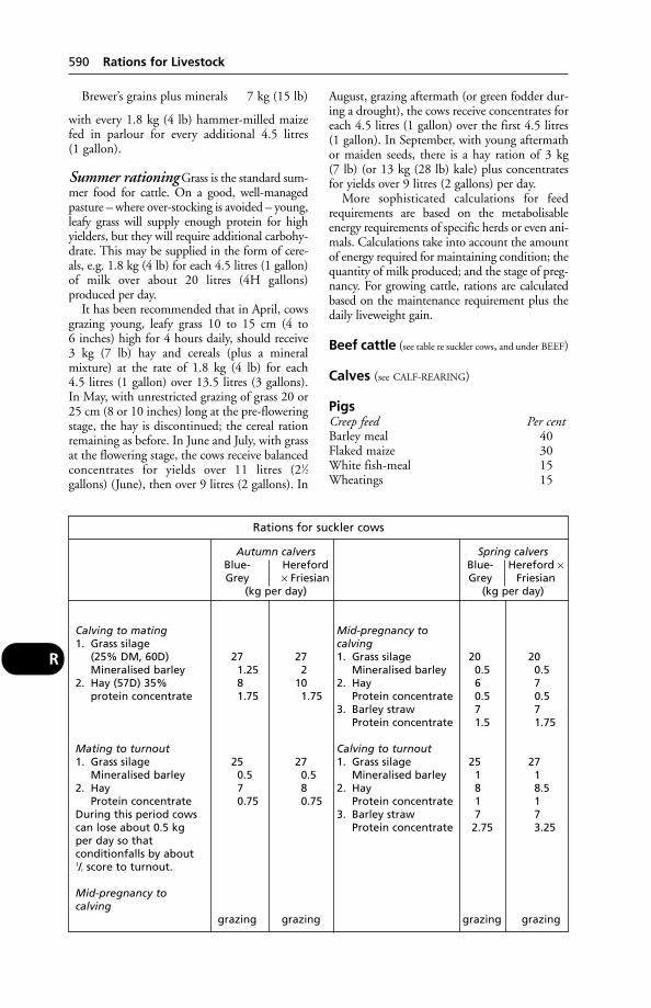
Brewer’s grains plus minerals 7 kg (15 lb)
with every 1.8 kg (4 lb) hammer-milled maizefed in parlour for every additional 4.5 litres (1 gallon).
Summer rationing Grass is the standard sum-mer food for cattle. On a good, well-managedpasture – where over-stocking is avoided – young,leafy grass will supply enough protein for highyielders, but they will require additional carbohy-drate. This may be supplied in the form of cere-als, e.g. 1.8 kg (4 lb) for each 4.5 litres (1 gallon)of milk over about 20 litres (4H gallons) produced per day.
It has been recommended that in April, cowsgrazing young, leafy grass 10 to 15 cm (4 to 6 inches) high for 4 hours daily, should receive3 kg (7 lb) hay and cereals (plus a mineral mixture) at the rate of 1.8 kg (4 lb) for each 4.5 litres (1 gallon) over 13.5 litres (3 gallons).In May, with unrestricted grazing of grass 20 or25 cm (8 or 10 inches) long at the pre-floweringstage, the hay is discontinued; the cereal rationremaining as before. In June and July, with grassat the flowering stage, the cows receive balancedconcentrates for yields over 11 litres (21⁄2gallons) (June), then over 9 litres (2 gallons). In
August, grazing aftermath (or green fodder dur-ing a drought), the cows receive concentrates foreach 4.5 litres (1 gallon) over the first 4.5 litres(1 gallon). In September, with young aftermathor maiden seeds, there is a hay ration of 3 kg (7 lb) (or 13 kg (28 lb) kale) plus concentratesfor yields over 9 litres (2 gallons) per day.
More sophisticated calculations for feedrequirements are based on the metabolisableenergy requirements of specific herds or even ani-mals. Calculations take into account the amountof energy required for maintaining condition; thequantity of milk produced; and the stage of preg-nancy. For growing cattle, rations are calculatedbased on the maintenance requirement plus thedaily liveweight gain.
Beef cattle (see table re suckler cows, and under BEEF)
Calves (see CALF-REARING)
PigsCreep feed Per centBarley meal 40Flaked maize 30White fish-meal 15Wheatings 15
590 Rations for Livestock
R
Rations for suckler cows
Autumn calvers Spring calversBlue- Hereford Blue- Hereford ×Grey × Friesian Grey Friesian
(kg per day) (kg per day)
Calving to mating Mid-pregnancy to1. Grass silage calving
(25% DM, 60D) 27 27 1. Grass silage 20 20Mineralised barley 1.25 2 Mineralised barley 0.5 0.5
2. Hay (57D) 35% 8 10 2. Hay 6 7protein concentrate 1.75 1.75 Protein concentrate 0.5 0.5
3. Barley straw 7 7Protein concentrate 1.5 1.75
Mating to turnout Calving to turnout1. Grass silage 25 27 1. Grass silage 25 27
Mineralised barley 0.5 0.5 Mineralised barley 1 12. Hay 7 8 2. Hay 8 8.5
Protein concentrate 0.75 0.75 Protein concentrate 1 1During this period cows 3. Barley straw 7 7can lose about 0.5 kg Protein concentrate 2.75 3.25per day so that conditionfalls by about 1/2 score to turnout.
Mid-pregnancy to calving
grazing grazing grazing grazing
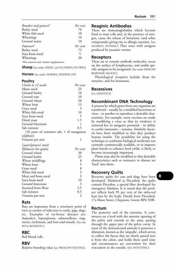
Breeders and growers* Per centBarley meal 70White fish-meal 10Wheatings 10Ground maize 10Fatteners* Per centBarley meal 75Soya bean-meal 5Wheatings 20*Plus mineral and vitamin supplements.
Sheep (see under SHEEP, and FLUSHING OF EWES)
Horses (see under HORSES, FEEDING OF)
PoultryChicks to 12 weeks Per centMaize meal 23Ground barley 10Ground oats 10Ground wheat 20Wheat bran 13Grass meal 5White fish-meal 10Soya bean-meal 5Dried yeast 1.5Ground limestone 1Salt mixture 0.5
(10 parts of common salt, 1 of manganesesulphate)Vitamin pre-mix 1
Layers’/growers’ mash(Balancer for grain) Per centGround wheat 30Ground barley 25Wheat middlings 8Wheat bran 5Grass meal 8White fish-meal 3Meat and bone-meal 3Soya bean-meal 10Ground limestone 3Steamed bone-flour 2.5Salt mixture 0.5Vitamin pre-mix 2
RatsRats are important from a veterinary point ofview as carriers of infection to cattle, pigs, dogs,etc. Examples of rat-borne diseases are:Aujeszky’s, leptospirosis, salmonellosis, ring-worm, trichinosis, and foot-and-mouth. (See also
PETS; RODENTS.)
RBCRed blood cells.
RBVRelative breeding value (see PROGENY TESTING).
Reaginic AntibodiesThese are immunoglobulins which becomefixed to mast cells and, in the presence of anti-gen, cause the release of histamine (and othercompounds) giving rise to allergic reaction. (See
ALLERGY; ASTHMA.) They react with antigensproduced by parasitic worms.
ReceptorsThese are or contain antibody molecules, occuron the surface of lymphocytes, and enable spe-cific antigens to be recognised. (See under IMMUNE
RESPONSE; BLOOD.)Physiological receptors include those for
enzymes, and for hormones.
Recessives(see GENETICS)
Recombinant DNA TechnologyA process by which genes from one organism aretransferred – usually by a modified bacterium orvirus – to another to reproduce a desirable char-acteristic. For example, some vaccines are madeby modifying a virus so that its virulence isremoved but its antigenic potential – its abilityto confer immunity – remains. Similarly, bacte-ria have been modified so that they producehuman insulin. The possibility for using thetechnique to synthesize biological medicines notcurrently commercially available, or to improveplant breeds to enhance food yields, is likely tobecome increasingly important.
Plants may also be modified so that desirablecharacteristics such as resistance to disease are‘bred’ into them.
Recovery QuiltsRecovery quilts for cats and dogs have beendeveloped. Marketed as Flectabed, the quiltscontain Flectalon, a special fibre developed foremergency blankets. It is stated that the prod-uct reflects back 95 per cent of the infra-redheat lost by the body. Details from Flectabed,17a Moor Street, Chepstow, Gwent BP6 5DB.
RectumThe posterior end of the intestine. It com-mences on a level with the anterior opening ofthe pelvis and extends to the anus, passingthrough the upper part of the pelvic cavity. Inmost of the domesticated animals it possesses adilatation, known as the ‘ampulla’, which servesto collect the faeces that are slowly passed intoit from the colon, and holds them until timeand circumstances are convenient for theirevacuation to the outside. (See INTESTINE.)
Rectum 591
R
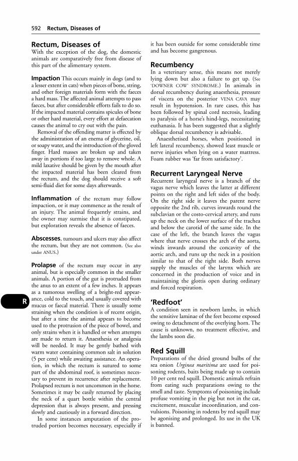
Rectum, Diseases ofWith the exception of the dog, the domesticanimals are comparatively free from disease ofthis part of the alimentary system.
Impaction This occurs mainly in dogs (and toa lesser extent in cats) when pieces of bone, string,and other foreign materials form with the faecesa hard mass. The affected animal attempts to passfaeces, but after considerable efforts fails to do so.If the impacted material contains spicules of boneor other hard material, every effort at defaecationcauses the animal to cry out with the pain.
Removal of the offending matter is effected bythe administration of an enema of glycerine, oil,or soapy water, and the introduction of the glovedfinger. Hard masses are broken up and takenaway in portions if too large to remove whole. Amild laxative should be given by the mouth afterthe impacted material has been cleared from the rectum, and the dog should receive a softsemi-fluid diet for some days afterwards.
Inflammation of the rectum may followimpaction, or it may commence as the result ofan injury. The animal frequently strains, andthe owner may surmise that it is constipated,but exploration reveals the absence of faeces.
Abscesses, tumours and ulcers may also affectthe rectum, but they are not common. (See also
under ANUS.)
Prolapse of the rectum may occur in any animal, but is especially common in the smalleranimals. A portion of the gut is protruded fromthe anus to an extent of a few inches. It appearsas a tumorous swelling of a bright-red appear-ance, cold to the touch, and usually covered withmucus or faecal material. There is usually somestraining when the condition is of recent origin,but after a time the animal appears to becomeused to the protrusion of the piece of bowel, andonly strains when it is handled or when attemptsare made to return it. Anaesthesia or analgesiawill be needed. It may be gently bathed withwarm water containing common salt in solution(5 per cent) while awaiting assistance. An opera-tion, in which the rectum is sutured to some part of the abdominal roof, is sometimes neces-sary to prevent its recurrence after replacement.Prolapsed rectum is not uncommon in the horse.Sometimes it may be easily returned by placingthe neck of a quart bottle within the centraldepression that is always present, and pressingslowly and cautiously in a forward direction.
In some instances amputation of the pro-truded portion becomes necessary, especially if
it has been outside for some considerable timeand has become gangrenous.
RecumbencyIn a veterinary sense, this means not merelylying down but also a failure to get up. (See
‘DOWNER COW’ SYNDROME.) In animals in dorsal recumbency during anaesthesia, pressureof viscera on the posterior VENA CAVA mayresult in hypotension. In rare cases, this hasbeen followed by spinal cord necrosis, leadingto paralysis of a horse’s hind-legs, necessitatingeuthanasia. It has been suggested that a slightlyoblique dorsal recumbency is advisable.
Anaesthetised horses, when positioned in left lateral recumbency, showed least muscle ornerve injuries when lying on a water mattress.Foam rubber was ‘far from satisfactory’.
Recurrent Laryngeal NerveRecurrent laryngeal nerve is a branch of thevagus nerve which leaves the latter at differentpoints on the right and left sides of the body.On the right side it leaves the parent nerveopposite the 2nd rib, curves inwards round thesubclavian or the costo-cervical artery, and runsup the neck on the lower surface of the tracheaand below the carotid of the same side. In thecase of the left, the branch leaves the vaguswhere that nerve crosses the arch of the aorta,winds inwards around the concavity of the aortic arch, and runs up the neck in a positionsimilar to that of the right side. Both nervessupply the muscles of the larynx which are concerned in the production of voice and inmaintaining the glottis open during ordinaryand forced respiration.
‘Redfoot’A condition seen in newborn lambs, in whichthe sensitive laminae of the feet become exposedowing to detachment of the overlying horn. Thecause is unknown, no treatment effective, andthe lambs soon die.
Red SquillPreparations of the dried ground bulbs of thesea onion Urginea maritima are used for poi-soning rodents, baits being made up to contain10 per cent red squill. Domestic animals refrainfrom eating such preparations owing to thesmell and taste. Symptoms of poisoning includeprofuse vomiting in the pig but not in the cat,excitement, muscular incoordination, and con-vulsions. Poisoning in rodents by red squill maybe agonising and prolonged. Its use in the UKis banned.
592 Rectum, Diseases of
R

Red UrineCauses of red discoloration of the urineinclude: haematuria – blood in the urine, whichsettles out on standing; haemoglobinuria – thebreakdown of red blood cells, in which urinedoes not change colour on standing; and pig-ments. The coloration is usually a sign of disease: pyelonephritis and cystitis causehaematuria; red-water fever, leptospirosis,infection with Clostridium haemolyticum, kaleand rape poisoning, copper poisoning, and thedrinking of very large quantities of water allresult in haemoglobinuria. Azoturia results inbreakdown of muscles to produce myoglobin-urea, a brown-red coloration. Dosing with phenothiazine produces a red pigment.
Red-WaterAlso called bacillary haemoglobinuria, orictero-haemoglobinuria, in the USA it occurs in California, Colorado, Idaho, Louisiana,Montana, Nevada, Oregon, Texas, and Utah.Clostridium haemolyticum is the cause. (See also
TEXAS FEVER, the American red-water fever.)
Red-Water FeverIn the British Isles, babe siosis or piroplasmosis(see under BABESIA), to give red-water fever itsproper names, is a disease of cattle and sheep,
due to the presence in the blood of a protozoonparasite which attacks the red blood cells,destroying their envelopes and liberatinghaemoglobin, which is excreted by the kidneysand colours the urine reddish or blackish. Itoccurs mainly in the south and west ofEngland, in the north and west of Scotland,and practically all over Ireland, but it is alsoseen at times in districts that are not included inthese areas. It is common in low-lying, rough-pastured, and moorland districts, where ticks,which harbour and transmit the parasite, canfind abundant shelter and suitable breedingplaces. Cattle are usually attacked from the ageof about 6 months upwards, but young calvesare practically immune. One attack gives adegree of immunity, and cattle that have beenbred upon infected farms, and from infectedcattle, are more resistant than those broughtfrom a clean district. It is more prevalent in thespring and autumn months, since the ticks arethen at their maximum activity.
Cause Babesia (Piroplasma) divergens. This istransmitted by the common tick Ixodes ricinus,and occasionally by Haemophysalis punctata.(See under BABESIA and TICKS; also MUSCLES,DISEASES OF; MYOGLOBINURIA.)
Signs Two varieties of the disease are recognised:an acute and a mild form.
The acute type is sudden in its onset and frequently fatal. The animal becomes very dulland depressed, separates itself from the rest of theherd, moves slowly or not at all, grunts, groans,arches its back, salivates freely, grinds its teeth,and often staggers and falls. The coat becomeshard and staring, the skin is dry and often hide-bound, and there is almost always a profuse,watery, violent ‘pipe stem’ diarrhoea, due tospasms of the anal sphincter. The temperaturerises to as high as 40.5° to 41.5° C (105° to107°F), the pulse is fast and weak (often 100 perminute), and the respirations are laboured, blow-ing, and rapid (80 to 100 per minute). The visi-ble mucous membranes are pale. After a few daysthe animal’s distress becomes less acute, and themost alarming symptoms subside. The signs offever, however, are still evident, and the cow isstill in a serious condition. The urine usuallyshows some degree of coloration, which variesfrom a clear reddish claret to a deep dark brownor black – almost like stout.
The duration of acute attacks varies, but it isseldom that the high temperature lasts for morethan a week. Death may take place in from 3 to5 days, or later on, when it is usually due toexhaustion.
Red-Water Fever 593
R
An electron microscope picture showing threeprofiles of Babesia divergens – the cause of red-water fever – inside the egg of a tick.(With acknowledgements to IRAD, Compton.)

In the mild type the urine is not usuallyhighly coloured; there is only slight dullnessand loss of appetite. The animals are ill for aweek or 10 days, and the only marked sequel isanaemia.
There are irregular forms of red-water metwith at times, in which the general symptoms aresimilar to these seen in the typical acute attack,but the urine does not become discoloured.Many of these cases end fatally.
Treatment Imidocarb (Imizol). For use underprescription only. A single dose of 1 ml per 100 kg bodyweight for treatment, or 2.5 ml per100 kg bodyweight for prevention; the dosemust be given subcutaneously. Cattle must notbe slaughtered for human consumption for 90 days after administration, and milk fromtreated animals must be withheld for 21 days.Veterinary surgeons who prescribe Imizol areadvised that:
1. Full records of product administration toidentifiable animals must be maintained, andthat it is the duty of the farmer to keep a care-ful record of all administration of the product,as required by the Animals and Fresh Meat(Examination for Residues) Regulations 1988.
2. The local DVM must be notified of theaddress of the farm where treatment is to takeplace.
3. Farmers should be informed that they mustnotify the local DVM when treated animals gofor slaughter for human consumption or whenmilk from treated animals is intended for humanconsumption.
Any suspected adverse reactions, includingevidence of lack of efficacy, should be reportedto the Veterinary Medicines Directorate, NewHaw, Weybridge, Surrey, immediately.
Ticks should be removed, either by hand-picking or by spraying with a suitable parasiticide.(See TICKS, CONTROL OF.)
The few piroplasms taken into the blood-stream, when young cattle are bitten by infectedticks, tend not to multiply but to give rise to a useful degree of immunity. This may wane ifthe piroplasms die, so that the animal becomessusceptible again. Immunity may likewise breakdown if the animal becomes ill from some othercause.
Control Measures involve tick control, andnot mixing cattle from red-water areas with susceptible cattle. Even then there are risks.
Twenty deaths occurred when local cattlewere placed on sea marsh land in Lincolnshirethat had previously been used for fatteningimported Irish steers which were carriers of
B. divergens. Cornish cattle brought to a farm inSussex set up a focus of infection because infect-ed ticks became established in the new habitat.And Simmental, Charolais and other Europeanbreeds are imported into Britain with noscreening for blood parasites.
Red WormsThe common name for strongyles. These cancause severe anaemia, unthriftiness, and debility.(See under FOALS, DISEASES OF; also under EQUINE
VERMINOUS ARTERITIS; HORSES, WORMS IN.)Benzimidazoles, ivermectins and thiabendazoleare useful drugs for the removal of red worms.(See also ROUNDWORMS.)
ReduplicationReduplication is a term applied to a duplicationof the normal heart-sounds as heard by auscul-tation. There are heard a 1st and a 2nd soundin a normal heartbeat, and in the above condi-tion one or both of these may be doubled. It isfound in certain diseases of the heart, such asobstruction of the valve between the auricle andventricle on the left side of the organ (the mitralvalve).
Reflex ActionReflex action is one of the simplest forms of activity of the nervous system. For the mechanism, see NERVES.
Superficial reflexes are well instanced in thesudden shivering movement that is seen when a fly or other insect settles upon the skin of ahorse, particularly in the region of the back ofthe shoulder.
Visceral reflexes are those connected withvarious organs, such as the narrowing of thepupil when the eye is exposed to a bright light.(See SPINAL CORD.)
Regional AnaesthesiaThis consists in the anaesthetisation of a regionof the body by means of a local anaesthetic solu-tion injected either into the connective tissue sur-rounding a sensory nerve trunk or into the spinalcanal. (See EPIDURAL ANAESTHESIA, ANALGESICS.)The most common example of perineurial injection is plantar block in the horse.
Register of VeterinarySurgeonsThe Register of Veterinary Surgeons lists veteri-nary surgeons who can practise in the UK. Itmay be consulted in some public libraries or isobtainable from the Royal College of VeterinarySurgeons, Belgravia House, 62–64 HorseferryRoad, London SW1P 2AF.
594 Red Worms
R

RehydrationThe restoration of the correct levels of waterand electrolytes in animals suffering fromDEHYDRATION.
Reindeer (Rangifer Tarandus)Both in northern Europe and North Americathese animals are of economic importance.Parasites include WARBLES. Subcutaneous injec-tions of ivermectin are recommended for prevention of infestation and treatment.
RelapseA relapse occasionally occurs when antibiotic or sulfa drug treatment of an infectious diseaseis stopped – the infection having been sup-pressed but the animal’s powers of resistancenot having been stimulated to establish a suffi-cient degree of immunity. Some forms of lame-ness are particularly liable to relapses, especiallythose associated with sprains of tendons or ligaments.
Relative Breeding Value(see PROGENY TESTING)
‘Remote Injection’ Method(see PROJECTILE SYRINGE)
RenalRelating to the kidney.
ReninAn enzyme, secreted by the kidneys, which maycontrol the secretion of the hormone aldosteroneby the adrenal glands.
ReovirusThe name derives from the words ‘respiratoryenteric orphan virus’. Reoviruses have double-stranded ribonucleic acid (RNA), and will repli-cate and produce changes in cells of cattle, pigs,dogs, cats, rabbits, monkeys, and man. Reovirusin poultry is often seen as tenosynovitis. The tendon sheaths, synovial membrane and themyocardium are all affected. Vaccination is possi-ble but attention to hygiene is also essential foreffective control. Only flocks known to be freefrom the infection should be selected for produc-ing hatching eggs. (See also CALF PNEUMONIA.)
RepairRepair of tissue after injury is described underWOUNDS; for the repair of special tissues, see
under BONE, MUSCLE, NERVE, etc. (See also HOOF
REPAIR WITH PLASTICS.)
Reproduction
Ovulation At OVULATION the Graafian folli-cle bursts, and the ovum is expelled by the rushof the escaping fluid. The cavity of the Graafianfollicle becomes filled with special cells to formthe corpus luteum, and the ovum begins itscareer as an absolute entity. In normal circum-stances the fimbriated and dilated funnel-shaped end of the Fallopian tube, or oviduct, isapplied to the point at which a follicle willburst, so that upon escape of its ovum this latter may be caught and retained. The dilatedend of the oviduct is usually known as thevestibule, and it is in this part that the spermusually meets the ovum and fertilises it. (See also
under OVARIES.)
Coitus The act of copulation. As mentionedunder OESTRUS, service by the male is onlyallowed during the period of oestrus by thefemales of the majority of species of higher animals. At other times there is little or nodesire exhibited by the male, and all attentionsare resented by the female. Artificial methods ofdomestication have to some extent modifiedthe frequency and duration of oestrus, so thatthe domestic animals sheltered under the pro-tection of man breed more frequently than dothe majority of wild animals of similar species.
During a single ejaculation of an adult vigor-ous stallion about 80,000,000 sperms arereleased. As soon as the sperms are free in theuterus or vagina, they travel towards whereverthe ovum is situated. This they accomplishpartly by a kind of wriggling movement of theirtail, which drives them onwards always in thesame direction. They are attracted to the ovumby ‘chemotaxis’.
Fertilisation Somewhere in the oviduct, gen-erally in its vestibule but not necessarily so, thespermatozoa arrive in the region of the waitingovum. More than one sperm may penetrate thewall of the ovum, but except in rare instances(giving rise to PRIMARY MOSAICISM) only onesperm fertilises the ovum.
The sperm, having penetrated the ovum,loses its tail, which is no longer required, and lieswithin the protoplasm of the ovum. The nucle-us of the ovum and that of the head of the spermnow fuse, each contributing half the number ofchromosomes that are to be found present innearly all the cells of the future young animal.The fused body is known as the segmentationnucleus, and from it, when it begins to divide,all the body cells of the embryo are formed. Theprocess of the formation of the young embryo is
Reproduction 595
R

considered under EMBRYOLOGY. (See also TESTI-
CLE; OVARIES; OESTRUS; BREEDING OF ANIMALS;PREGNANCY; PARTURITION; PARTHENOGENESIS.
Reproductive Organs(see diagrams under UTERUS and PENIS)
ReptilesA class of animal which includes tortoises,lizards and snakes. They tend to favour warmplaces when they are ill. Reptile housing shouldbe heated at all times, but a range of heatshould be available in different areas, within thenormal limits for the species, so that they canchoose which suits them best. Under no cir-cumstances must they be allowed to come intodirect contact with the heat source. Manyspecies require ultraviolet light, otherwise bonerarefaction (weakening) may occur; expertadvice must be sought before buying such a pet.(See also PETS.)
ResectionResection is an operation in which a part ofsome organ is removed – as, for example, theresection of a piece of dead bone, or resection ofa part of the intestine which is diseased; resec-tion of a rib in thoracotomy; aural resectiondone to overcome chronic disease of a dog’s ear.
Resistance Transferability(see under PLASMIDS; ANTIBIOTIC RESISTANCE)
Resistant StrainsThis phrase is commonly used of bacteria whichare not sensitive to antibiotics, or of insectswhich are not killed by an insecticide.
ResorptionMummification. Resorption of the fetus occurs,e.g. in heifers receiving a high calcium and lowphosphorus diet. In sows, mummification canbe a feature of Aujeszky’s disease, ‘blue-ear’ dis-ease (PRRS), and both African and classicalswine fever. With the banning of sow stalls andthe need to find alternatives, it has been report-ed that the level of mummification tends to be higher in dry sows kept in straw yards. Thereason is not clear, but bullying has been suggested as a cause. (See MUMMIFICATION.)
Respiration(see also NOSE AND NASAL PASSAGES; LUNGS)
Mechanism of respiration For the struc-ture of the respiratory apparatus see NOSE AND
NASAL PASSAGES; LUNGS, etc.
Inspiration is due to muscular effort whichenlarges the chest in all 3 dimensions, so thatthe lungs have to expand in order to fill up thevacuum that would otherwise be left; and since,although the lungs are not fixed to the chestwall, surface tension between the pleura liningthe chest and the pleura covering the lungs, hasmuch the same effect.
In most vertebrates, except birds, the lungsare not normally attached to the walls of thechest, but are rather suspended in them fromtheir ‘roots’, so that there is no direct pull uponthe lungs when the chest cavity increases in size.The vertical diameter of the chest is increasedduring inspiration through the downward tilt-ing of the sternum. This movement is best seenin the dog when it is out of breath; at othertimes, and in other animals, it is so slight that it escapes detection. The transverse dimensionof the chest increases when any 1 of the ribsbehind the first 2 or 3 are forcibly pulled for-ward by muscular action. Each rib only movesa small amount, but the mass effect of the seriesis considerable. The muscles which bring aboutthese changes in ordinary inspiration are thediaphragm, the intercostal muscles which aresituated in two layers between each rib and itstwo neighbours, and possibly the levators of theribs, and the serratus muscles.
When the chest expands, the lungs expandtoo; but initially the quantity of air within themremains the same. Accordingly, the pressure falls,leading to an inflow of air.
Expiration is in ordinary circumstancesmerely an elastic recoil, the diaphragm movingforward and the ribs settling back into theiroriginal positions, partly through muscularaction, and partly through the elasticity of theircartilages. It occupies a slightly longer period oftime than does inspiration.
Nervous control Respiration is usually an automatic act under the control of the respiratory centre in the medulla oblongata.
Although the respiratory centre is itself capa-ble of carrying on respiration, it is in its turnliable to be controlled by the higher consciouscentres. This is seen particularly well in humanbeings, where it is possible to ‘hold the breath’,
596 Reproductive Organs
R
Inspired air Expired air
Per cent Per centNitrogen 79.04 79.04Oxygen 20.93 16.02Carbon dioxide 0.03 4.38
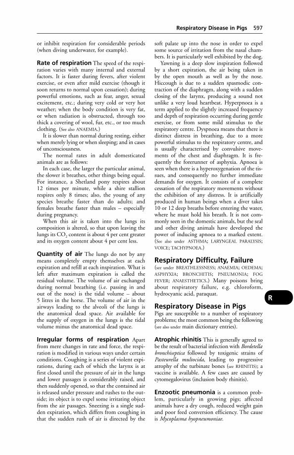
or inhibit respiration for considerable periods(when diving underwater, for example).
Rate of respiration The speed of the respi-ration varies with many internal and externalfactors. It is faster during fevers, after violentexercise, or even after mild exercise (though itsoon returns to normal upon cessation); duringpowerful emotions, such as fear, anger, sexualexcitement, etc.; during very cold or very hotweather; when the body condition is very fat, or when radiation is obstructed, through toothick a covering of wool, fur, etc., or too muchclothing. (See also ANAEMIA.)
It is slower than normal during resting, eitherwhen merely lying or when sleeping; and in casesof unconsciousness.
The normal rates in adult domesticated animals are as follows:
In each case, the larger the particular animal,the slower it breathes, other things being equal.For instance, a Shetland pony respires about 12 times per minute, while a shire stallionrespires only 8 times; also, the young of anyspecies breathe faster than do adults; andfemales breathe faster than males – especiallyduring pregnancy.
When this air is taken into the lungs its composition is altered, so that upon leaving thelungs its CO2 content is about 4 per cent greaterand its oxygen content about 4 per cent less.
Quantity of air The lungs do not by anymeans completely empty themselves at eachexpiration and refill at each inspiration. What isleft after maximum expiration is called theresidual volume. The volume of air exchangedduring normal breathing (i.e. passing in andout of the nose) is the tidal volume – about 5 litres in the horse. The volume of air in theairways leading to the alveoli of the lungs is the anatomical dead space. Air available for the supply of oxygen in the lungs is the tidalvolume minus the anatomical dead space.
Irregular forms of respiration Apartfrom mere changes in rate and force, the respi-ration is modified in various ways under certainconditions. Coughing is a series of violent expi-rations, during each of which the larynx is atfirst closed until the pressure of air in the lungsand lower passages is considerably raised, andthen suddenly opened, so that the contained airis released under pressure and rushes to the out-side; its object is to expel some irritating objectfrom the air passages. Sneezing is a single sud-den expiration, which differs from coughing inthat the sudden rush of air is directed by the
soft palate up into the nose in order to expelsome source of irritation from the nasal cham-bers. It is particularly well exhibited by the dog.
Yawning is a deep slow inspiration followedby a short expiration, the air being taken in by the open mouth as well as by the nose.Hiccough is due to a sudden spasmodic con-traction of the diaphragm, along with a suddenclosing of the larynx, producing a sound notunlike a very loud heartbeat. Hyperpnoea is aterm applied to the slightly increased frequencyand depth of respiration occurring during gentleexercise, or from some mild stimulus to the respiratory centre. Dyspnoea means that there isdistinct distress in breathing, due to a morepowerful stimulus to the respiratory centre, andis usually characterised by convulsive move-ments of the chest and diaphragm. It is fre-quently the forerunner of asphyxia. Apnoea isseen when there is a hyperoxygenation of the tis-sues, and consequently no further immediatedemands for oxygen. It consists of a completecessation of the respiratory movements withoutthe exhibition of any distress. It is artificiallyproduced in human beings when a diver takes10 or 12 deep breaths before entering the water,where he must hold his breath. It is not com-monly seen in the domestic animals, but the sealand other diving animals have developed thepower of inducing apnoea to a marked extent.(See also under ASTHMA; LARYNGEAL PARALYSIS;VOICE; TACHYPNOEA.)
Respiratory Difficulty, Failure(see under BREATHLESSNESS; ANAEMIA; OEDEMA;ASPHYXIA; BRONCHITIS; PNEUMONIA; FOG
FEVER; ANAESTHETICS.) Many poisons bringabout respiratory failure, e.g. chloroform,hydrocyanic acid, paraquat.
Respiratory Disease in PigsPigs are susceptible to a number of respiratoryproblems; the most common being the following(see also under main dictionary entries).
Atrophic rhinitis This is generally agreed tobe the result of bacterial infection with Bordetellabronchiseptica followed by toxigenic strains ofPasteurella multocida, leading to progressive atrophy of the turbinate bones (see RHINITIS); avaccine is available. A few cases are caused bycytomegalovirus (inclusion body rhinitis).
Enzootic pneumonia is a common prob-lem, particularly in growing pigs; affected animals have a dry cough, reduced weight gainand poor feed conversion efficiency. The causeis Mycoplasma hyopneumoniae.
Respiratory Disease in Pigs 597
R
Pleuro-pneumonia of pigs is a rapidonset respiratory disease resulting in dyspnoeaand, often, death in acute cases. Less severelyaffected animals have a variety of subclinicalproblems. The cause is Actinobacillus pleuro-pneumoniae. Often seen following other diseaseproblems, pleuro-pneumonia can affect onlysome of the animals in a herd, causing hightemperatures, laboured breathing with, often, abloody frothy discharge from the nose. Earlyantibiotic treatment can effect a recovery; tetra-cyclines, lincomycin and ceftiofur are amongdrugs used. Vaccines are available for control.
Swine influenza can sweep through a pigunit, causing a variety of respiratory signs andsometimes precipitating other respiratory dis-eases. Little can be done to control the spread ofinfection, but the pigs recover after 4 to 6 days;subsequent immunity lasts about 3 months.
Porcine respiratory reproductive dis-ease (PRRS) (‘blue-ear’ disease) resultsin variable signs, but affected animals may have respiratory difficulties; it often exacerbatesany underlying respiratory diseases, such aspneumonias.
Porcine respiratory-coronavirus infec-tion (PRCV) Outbreaks are often relativelymild, but other respiratory problems may result.Coughing, sneezing or dyspnoea can occur.
Ascarids Infection with Ascaris suum canresult in coughing during the migratory stage ofthe larvae.
Aujeszky’s disease sometimes causes respi-ratory signs such as sneezing, coughing and nasaldischarge, occasionally with dyspnoea. Vaccinesare available (in Ireland).
Lungworm Infection with Metastrongylusapri is usually only a problem in outdoor pigs;signs are often limited.
Not all the factors involved in respiratorydisease are infections: management factors playtheir part, too. Space allowance per pig, num-ber of pigs per group, effects of mixing andcrowding, temperature, humidity, nutrition,age and genetic status of the pigs all exert theireffects. (See also under ATROPHIC RHINITIS.)
Respiratory StimulantsRespiratory stimulants are used to promotebreathing in the newborn and to relieve respira-tory depression associated with, for example,general anaesthesia. They include doxapram
(Dopram V), cropropamide and crotethamide(Respirot), and etamiphylline (Dalophylline).
Respiratory Syncytial VirusRespiratory syncytial virus was first isolated fromchimpanzees showing ‘cold-like’ signs; since themid-1950s it has been detected in clinical cases ofrespiratory disease in man, cattle, sheep, goats,and horses. It is a cause of acute bronchiolitis andalveolitis.
RestraintIn order to examine an animal thoroughly forsigns of injury or disease, in order to carry outinoculations, or even to administer an anaes-thetic, some form of restraint is often necessary.
The introduction of effective tranquillisersand sedatives facilitated the handling of horses,cattle and small animals, and may assist orreplace the use of several means of restraintdescribed below. (See also TRANQUILLISERS;XYLAZINE; ANALGESIA.)
The following methods should not be usedindiscriminately upon any and every animal. A method that is sufficient to restrain one animal may prove aggravating to another; e.g.while the common twitch may serve for a heavydraught gelding, it is likely to cause a thor-oughbred stallion to be more restive than ever.A person who finds it necessary to employ somemeans of restraint should first of all considerthe temperament, age, breed, and, if possible,the individual characteristics of the animal, as well as the purpose of the restraint, beforedeciding upon what methods will be employed.Firm gentleness, a kindly spoken word, and ahand-pat, with a little coaxing or urging, willvery often allay an animal’s fears, but there arethose of a temperament which will not respondto gentleness; it is to those particularly that suchmethods as described here are applicable.
Horses The usual halter, head-stall, or bridleis generally sufficient to control broken horsesthat are to be handled or examined without theinfliction of pain. In some cases it may be nec-essary to tie the animal to a ring in the wall ormanger, or to the heel-posts, but it is better insuch cases to take a couple of turns round thering and have a man hold the end of the rope.For measures which involve handling of thehind-parts of the body, it is usually advisable tohave one of the fore-feet picked up and held(preferably that upon the same side of the bodyas the operator is to work).
For greater control a TWITCH may be applied.(See also TRANQUILLISERS; ANAESTHETICS.)
598 Respiratory Stimulants
R
Cattle A cattle CRUSH; either of a commercialpattern or one constructed of timber by farmlabour, is useful; a gate may be hinged to a walland closed so as to act as a crush for inocula-tions, etc. (See also VETERINARY FACILITIES ON
THE FARM.)A halter is also useful in cattle, as in horses.In the case of comparatively quiet cattle, milk
cows, etc., it will generally suffice if an assistanttakes the animal by the nose. The thumb andmiddle finger of one hand are inserted into the respective nostrils, and the nasal septum ispinched between them. It is important that thestockman’s fingers do not block up the airway.
The other hand may be placed under thejaw. In this position the majority of adult quietcattle can be easily held. For bulls and those cattle that are more difficult to control it isusual to use a pair of bull-holders (‘bull-dogs’;‘bull-tongs’); or if the animal is already rung(with a copper or aluminium ring), to attach arope or bull-leader to the ring in the nose. Fordrenching purposes it is necessary to keep thehead and neck in as straight a line as possible to obviate the risk of choking. If an assistant is needed he should stand on the opposite sideof the beast and take the horns in his hands so that he may tilt the head upwards and at the same time keep the head and neck straightout. A pair of bull-holders may be inserted into the nostrils, and have a rope attached tothem which is passed over a beam and the headpulled up.
For lifting a hind-leg, a pole, broom handle,etc., may be placed in front of that hock andbehind and above the other. Two helpers takehold of ends of the pole and pull the leg upwardsand backwards, at the same time steadying theanimal’s balance by leaning against its thighs withtheir shoulders. For the fore-feet it is usual to passa rope around the cannon or above the heels andover the back to the opposite side, where it isheld by an assistant. (See also TRANQUILLISERS.)
Sheep For most purposes the sheep may beturned up into a position in which it sits uponits rump, by placing the left hand round underthe neck from the near side, and the right handover the back to seize the wool of the abdomen,lifting the animal’s fore-end off the ground andtwisting its hind-legs from under it. In thisposition its feet may be dressed, its fleece maybe examined, etc. It is not advisable to turn in-lamb ews, due to the possibility of harmingthem or the fetus; they may be held against a wall or fence by an assistant while their feet, etc., are being dressed. Sheep stocks aresometimes used, or modern shearing tables.
Pigs The adult pig is proverbially a difficultanimal to handle and restrain, especially whenthe handling involves pain or discomfort, butpiglets are easily held by the hind-legs with thehands, while the knees grip the dependenthead. With large sows and boars it is wise toremember that they are apt to be vicious withstrangers, and to use a shield of wood or a hur-dle to prevent a rush by the angry animal.
A method of securing a large pig is to drive itinto a corner and pen it there with a door, gate,or heavy hurdle carried by 2 helpers, and heldso that the pig has no room to turn while anoose is dropped over its head and pulled tightround its jaws, and another is secured to a hind-leg above the hock. The ends of theseropes are then passed round a post or a rail inthe fence and pulled tight when the pig isreleased from its corner.
Dogs and cats These animals are usuallymore easily restrained than some of the largeranimals because of their intimate associationwith man, but there are certain animals thatpresent difficulty when angry or excited. A kindword and a caress will often be necessary to gain the animal’s confidence before attemptingto examine it, and, wherever possible, severemethods of restraint should be avoided exceptas a last resort. The human voice often exercis-es a degree of control over an excitable animal,and there are certain people who appear to pos-sess the faculty of immediately gaining almostany dog’s confidence and of being able to doanything with it.
However, it is always wise in any case ofdoubt to take no risks. The safest way of deal-ing with a dog is to muzzle it first. A tape muz-zle may be applied; this is simply a piece of tapeor a bandage about 118 cm (3 ft) long whosemiddle is wound round the dog’s nose, the endsbeing crossed under the jaw and tied round theneck or on to the collar. With bulldogs, andthose with a short face and a pug nose, it is better to tie the tape round the jaws, finishingwith the end above the nose, tying them togeth-er there, and then passing the ends back to thecollar.
Cats can be rolled in a sack or towel. Withcats it is important to prevent them from usingtheir claws, which inflict injuries more oftenthan do the teeth. (See also under TRANQUILLISERS;ANAESTHETICS.)
ResuscitationA basic method of pulmonary resuscitation withexpired air, using a device portable and simpleenough for emergency use by herdsmen and
Resuscitation 599
R

shepherds, is in use on farms. The device con-sists of a mouthpiece, non-return valve, flange,and mouth tube. (See ARTIFICIAL RESPIRATION;RESPIRATORY STIMULANTS; ACUPUNCTURE.)
Retention of Afterbirth(see PLACENTA – Retained)
ReticulocytesThe penultimate stage in the formation of redblood cells. Reticulocytes are numerous in theblood only in anaemic conditions and indicatean effort of the blood-forming tissues to restorethe red blood cell count to normal levels.
Reticulo-Endothelial SystemThis consists of macrophages, special cells presentin the liver, spleen, lymph nodes and bone marrow. The system has a number of functionsincluding the regulation of immune responses (see
under ANTIBODY). It also removes disintegratingred cells from the blood.
ReticulumThe 2nd stomach of ruminants.
RetinaThe innermost layer of the eye; it includes thelight-sensitive rods and cones which transmitimpulses to the optic nerve. Detachment of, or haemorrhage into, the retina is a cause ofsudden blindness in dogs. It is often due tohypertension, the long-term effects of whichmay be hypertrophy of the left ventricle of the heart and kidney failure. (See EYE and EYE,DISEASES AND INJURIES OF.)
RetinoblastomaA type of tumour which occurs on the RETINA.
Retro-Retro- is a prefix signifying behind or turnedbackwards.
Retropharyngeal AbscessRetropharyngeal abscess is the name given to anabscess occurring at the back of the throat inthe region behind the pharynx. Such abscessesgenerally make swallowing difficult or impossi-ble until they burst, which they frequently dointo the cavity of the pharynx, whence the pusis swallowed. (See STRANGLES.)
RetrovirusA member of the Retroviridiae, the family ofviruses which includes the lentiviruses and the oncornaviruses. Retroviruses are naturallyoccurring gene transfer organisms. When the
virus infects a cell, it is uncoated; the viral RNAis transcribed into DNA and this DNA inte-grates into one of the cell’s chromosomes. Thisproperty could be used to produce disease-resis-tant transgenic animals. Certain viral groupsappear to need the presence of a receptor on the cell membrane in order to gain access intothe cell. Retroviruses are enveloped viruses andcarry a glycoprotein on their surface; a specificinteraction with this glycoprotein and the cellular receptor is a prerequisite for infection.Immunodeficiency viruses of humans, cats, cattle and primates are retroviruses.
(See table under VIRUSES; also GENETIC ENGI-
NEERING.)
RhabdomyolosisRhabdomyolosis, also called azoturia, is a break-ing down of skeletal muscle in consequence ofwhich the urine contains myoglobin. (See EQUINE
MYOGLOBINURIA.)
RhabdovirusA group of bullet-shaped viruses which includesthe rabies virus and that of vesicular stomatitis.Several rhabdoviruses are associated with diseaseconditions in fish.
RheaOstrich-like flightless bird, native to SouthAmerica. Smaller than ostrich, about 120 cm (4 ft) tall, and has 3 toes.
RheumatismA general term indicating a painful condition of muscles, tendons, joints, bones, or nerves; itis generally less common in animals than inhumans.
Rheumatism is seen in dogs, pigs, and horsesmost commonly, but it can affect all of thedomesticated animals. Young animals are mostoften attacked by the acute type, especiallyyoung pigs and puppies, and adults by the muscular form and by chronic or particularrheumatism.
For the muscular type see under MUSCLES,DISEASES OF.
Treatment There is no absolute specific,although certain drugs have enjoyed a great repu-tation in the alleviation of this disease, especiallysalicylates. Phenylbutazone has been used withreported success. (See also CORTISONE.)
Rheumatoid ArthritisThis may occur in the dog from the age ofabout 2 years upwards, and in cats.
600 Retention of Afterbirth
R

Signs are at first vague; the dog appearsdepressed, often with a poor appetite and somedegree of fever, but with no lameness. Thisappears later, sometimes involving several jointssimultaneously, sometimes affecting one limband then shifting to another. There may becrepitus when the limb is moved.
Diagnosis depends for confirmation uponradiography and on laboratory tests.
(See also AUTO-IMMUNE DISEASE.)
RhinitisInflammation of the NOSE.
Rhinitis, AtrophicThis has been defined as the product of a severepersistent inflammatory reaction in the nasalmucosa of a growing, and therefore very young,pig, and as such is non-specific with regard toaetiology.
The generally accepted view is that in thefirst 2 or 3 months of life, the rapidly growingnasal structures are extremely liable to attack byinfectious agents – but quite often, recoveryfrom these is complete. In herds with severe disease, however, the condition may progress togive rise to the marked displacement or atrophyof the turbinate bones and also to an associatedpneumonia.
Causes Bordetella bronchiseptica, Pasteurellamultocida and Pseudomonas aeruginosa, as well asinclusion-body rhinitis virus, may all be involvedat some stage. Respiratory disease in the pig is achanging, developing process with many agentsand factors involved; rhinitis and pneumoniaoften occur together.
From other sources it is known that B. bron-chiseptica secretes a substance which inhibits thedeposition or transfer of calcium salts in theinfected tissues. Accordingly the bones may failto ossify properly or may become weak andliable to distortion.
The disease in a severe form may occur only when there is a double infection with B. bronchiseptica and P. multocida.
Signs The acute form is to be found in piglets2 or 3 weeks old, when there is no deformity ofthe snout to be seen and not always an overflowof tears. Sneezing is perhaps the most commonsymptom. The eyelids may be puffy, and some-times the piglet has a copious discharge from its nose and breathes through its mouth. Thedisease can be so mild that symptoms passunnoticed, or so severe that death occurs with-in a week. In some outbreaks the mortality is 10 per cent or more, and survivors suffer agrowth check from the disease which continuesin the subacute form.
Incidence Examination of the snouts from2701 pork, bacon and heavy pigs killed at 5 abattoirs in England and Scotland duringMarch to July 1974 revealed obvious atrophy ofthe turbinate bones in over 44 per cent andsevere atrophy in over 17 per cent of the snoutsexamined.
Prevention Inactivated vaccines preparedfrom B. bronchiseptica and P. multocida areavailable.
Rhinopneumonitis(see EQUINE VIRAL RHINOPNEUMONITIS)
RhinosporidiosisA chronic disease of the nasal mucous mem-brane, and associated with polyp formationleading to difficulty in breathing, caused by afungus Rhinosporidia seberi. The disease occursin cattle and horses, in the USA, SouthAmerica, Australia, and India.
Rhinotracheitis, InfectiousBovine (IBR)A disease of cattle recognised in the USA in1951.
Rhinotracheitis, Infectious Bovine (IBR) 601
R
Distortion of the pig’s snout as a result of atroph-ic rhinitis. (With acknowledgements to ProfessorR. H. C. Penny and the Royal Veterinary College.)

Cause The bovine herpesvirus 1, which canproduce disease of the respiratory, reproductive,nervous and digestive systems.
Signs In America the disease is usually severe infeedlot cattle, but mild in dairy/range cattle. InBritain outbreaks of severe IBR occurred in1978–9, causing heavy losses on some farms,with reduced milk yield, loss of appetite, fever(up to 42°C (108°F)), laboured breathing, a dis-charge from eyes and nose, and sometimesdrooling of saliva. Reproductive disorders suchas vulvo-vaginitis and orchitis have also occurredin Britain. In America abortion has followednatural infection or vaccination against IBR.
IBR is sometimes associated with a fatalpneumonia. The disease may closely resemblemucosal disease, with which at one time it wasthought to be possibly identical, and also malig-nant catarrh.
At least one strain of the virus is neurotropicand has caused encephalitis in calves in Australiaand elsewhere.
Control Live vaccines are available; dependingon the product, they are given by intranasalapplication or by intramuscular injection.
RhinovirusThis genus of viruses have RNA as their nucleicacid and include equine rhinovirus. They cancause upper respiratory tract infections.
Rhodesian RidgebackA very large, tan or redcoated dog with theunique characteristic of a strip of hair runningfrom tail to nose along the back opposite to the normal lie of the rest of the coat. This mayaccount for the breed being prone to sebaceouscysts in that region and to dermoid sinus.Wobbler syndrome (cervical spondylothesis) isinheritable; achalasia may also develop.
Rhodococcus(see CORYNEBACTERIUM)
Rhododendron PoisoningRhododendron poisoning is not common.There are about 20 varieties which have beenrecorded as causing poisoning in sheep, cattle,goats, and even man. The shrubs contain a gly-coside called andromedotoxin, which is the onlypoison capable of making ruminants vomit.
Poisoning may occur when sheep are broughtin to graze former amenity land on an estate. Inone outbreak, lambs were turned into a crop ofrape bordered by rhododendrons, before havinghad time to adjust to the new diet. Fifty out of
300 lambs were found weak and salivating; 2 were attempting to vomit; 6 were recumbent;and 3 dead.
Weak tea may be helpful in countering theeffects; the tannin acts as an antidote.
Rhonchi (Dry Sounds)Rhonchi (dry sounds) sometimes referred to as ‘dry rales’; continuous sounds heard duringbreathing by auscultation of the chest, whenthere is some obstruction of the bronchi. (See also
RALES.)
RhoncusAn abnormal sound detectable by means of astethoscope and indicating chronic inflammationof a bronchial tube.
Riboflavin(see under VITAMIN)
Ribonucleic AcidA substance related to DNA, RNA includes the sugar ribose combined with nucleic acid. Itappears to be concerned with protein synthesiswithin the cell. (See CELLS.) In some experimentsit has been shown that RNA from malignant cellswill cause normal cells in vitro to show character-istics of malignancy; and the converse is possible.(See VIRUSES; CANCER; GENETIC ENGINEERING.)
RibosomesRibosomes are granules containing RNA. (See
CELLS.)
RibsRibs are the long bones which together form thecage of the thorax. Their numbers vary in thedifferent animals, according to how many tho-racic or dorsal vertebrae are present, as follows:horse, 18 pairs; ox, 13 pairs; pig, 14 or 15 pairs;dog, 13 pairs. In any of these animals an extrarib (often called a ‘floating rib’ because it pos-sesses little or no cartilage to unite it to thecostal arch) may be present on one or both sidesof the body. The first 8 of these in the horse andox, the first 7 in the pig, and the first 9 in thedog, have cartilages which are united to the sternum, and are called sternal ribs, while thosefurther back in the series in each case have cartilages which do not reach the sternum, butform an arch by overlapping each other, and areknown as asternal ribs. (See STERNUM.)
Each rib possesses a ‘head’, by which it isjoined to the anterior part of the vertebra towhich it corresponds in number, and to theposterior part of that immediately in front, andthis is succeeded by a ‘neck’; a short distance
602 Rhinovirus
R

further down the shaft is a ‘tubercle’, whicharticulates with the transverse process of thevertebra to which it corresponds. The rest of therib is composed of a long, curved, flat shaft,whose curve varies according to the position ofthe rib in the chest, being greatest about themiddle of the series, and also according to theanimal to which it belongs. Posterior to eachruns the intercostal nerve and blood vesselswhich are situated in a little groove along theborders of each rib. In life the ribs are attachedto each other by the intercostal muscles to formthe continuous wall of the chest. (See BONES.)
Ricinus CommunisThe plant from which castor oil is obtained.
Rickets (Rachitis)Rickets (rachitis) is a deficiency disease of younganimals characterised by a tendency towards theformation of enlarged extremities of the longbones, and a bending of their shafts. Dogs, pigs,lambs, foals, and calves are all affected, the first 2more frequently than the other species of domes-ticated animals. It has also been encountered inintensively managed poultry establishmentswhere chicks are deprived of sunlight.
Cause Rickets may be caused by a deficiencyof either vitamin D or phosphorus, or in somecases both of these.
Absence of sunlight is a contributory causeand animals kept in dark buildings, especially ifinadequately fed, are prone to rickets.
Often a diet consisting largely of oatmeal ormaize meal, such as is commonly the lot ofsheepdogs, results in rickets. (See PHYTIN.)
Signs The typical changes consist of the devel-opment of bony swellings upon the ends of thelong bones of the limbs, where they meet otherbones to form joints, and the production ofswellings at the point where a rib joins its ribcartilage, i.e. along each side of the chest abouttwo-thirds the way down from the spine. Inpuppies, there is also a tendency for the shafts ofthe long limb to bend in an outward directionunder the influence of the weight of the body.
Treatment A vitamin D supplement is recommended or fresh COD-LIVER OIL. (See also
OSTEOMALACIA.)
RickettsiaThe generic name for a group of minute Gram-negative bacteria. They will not grow in ordinaryculture media, and their metabolic requirementsare more akin to those of the filterable viruses.
They are distinguished from chlamydia andother bacteria by being found in the cytoplasmof arthropods such as ticks, lice, mites and fleas. (For rickettsial infections in cattle, see TICK-
BORNE FEVER; Q FEVER. For tropical diseasescaused by rickettsiae, see BOVINE PETECHIAL
FEVER; HEARTWATER; see also ROCKY MOUNTAIN
FEVER.)
Rickettsial PoxA mild form of mite typhus transmitted topeople by the mouse mite Allodermanyosus sanguineus, and caused by Rickettsia akari.
RidaA disease of sheep involving the nervous system,similar to SCRAPIE, in Iceland.
Rideal-Walker CoefficientExpresses the comparative efficiency of antisep-tics, as based on the Rideal-Walker Test, takingcarbolic acid as unity. It does not take intoaccount the influence of body fluids upon theefficiency or otherwise of the antiseptic.
Riding Establishments Act 1963The Riding Establishments Act 1963 sets outthe standards which riding schools and similarestablishments must meet to obtain a licence to operate. It empowers local authorities to select aveterinary surgeon to inspect, and report upon, ariding establishment. The decision on whetherto grant a licence is based on that report. Pointstaken into consideration include the competenceof the owner, the suitability of the accommoda-tion, the supply of food, water and bedding, theavailability of veterinary treatment, and satisfac-tory provisions for evacuation of horses in case offire. Prosecution may follow if the terms of theAct, or licence, are broken.
RifampicinThis bactericidal antibiotic has proved effectiveagainst some bacteria resistant to other antibi-otics. It is sometimes used in combination witherythromycin.
Rift Valley Fever (Enzootic Hepatitis)A disease of sheep, cattle, buffaloes, goats,camels, horses, donkeys, and man, occurring inAfrica. Until 1973 it had not been recorded innorthern Africa, but in that year it reached theSudan, and then Egypt, where major epizooticsoccurred in 1977–8. In 1982 WHO stated thatthis disease might be of greater concern outsideAfrica than in many of the countries where it isendemic; and expressed anxiety over its possible
Rift Valley Fever (Enzootic Hepatitis) 603
R
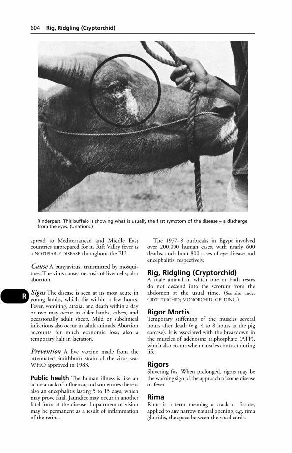
spread to Mediterranean and Middle Eastcountries unprepared for it. Rift Valley fever isa NOTIFIABLE DISEASE throughout the EU.
Cause A bunyavirus, transmitted by mosqui-toes. The virus causes necrosis of liver cells; alsoabortion.
Signs The disease is seen at its most acute inyoung lambs, which die within a few hours.Fever, vomiting, ataxia, and death within a dayor two may occur in older lambs, calves, andoccasionally adult sheep. Mild or subclinicalinfections also occur in adult animals. Abortionaccounts for much economic loss; also a temporary halt in lactation.
Prevention A live vaccine made from theattenuated Smithburn strain of the virus wasWHO approved in 1983.
Public health The human illness is like anacute attack of influenza, and sometimes there isalso an encephalitis lasting 5 to 15 days, whichmay prove fatal. Jaundice may occur in anotherfatal form of the disease. Impairment of visionmay be permanent as a result of inflammationof the retina.
The 1977–8 outbreaks in Egypt involvedover 200,000 human cases, with nearly 600deaths, and about 800 cases of eye disease andencephalitis, respectively.
Rig, Ridgling (Cryptorchid)A male animal in which one or both testes do not descend into the scrotum from theabdomen at the usual time. (See also under
CRYPTORCHID; MONORCHID; GELDING.)
Rigor MortisTemporary stiffening of the muscles severalhours after death (e.g. 4 to 8 hours in the pigcarcase). It is associated with the breakdown inthe muscles of adenosine triphosphate (ATP),which also occurs when muscles contract duringlife.
RigorsShivering fits. When prolonged, rigors may bethe warning sign of the approach of some diseaseor fever.
RimaRima is a term meaning a crack or fissure,applied to any narrow natural opening, e.g. rimaglottidis, the space between the vocal cords.
604 Rig, Ridgling (Cryptorchid)
R
Rinderpest. This buffalo is showing what is usually the first symptom of the disease – a dischargefrom the eyes. (Unations.)

Rinderpest (Cattle Plague)Rinderpest (cattle plague) is an acute, specific,inoculable, and febrile disease of cattle, charac-terised by an ulcerative inflammation of mucousmembranes, especially those of the alimentarytract. It is caused by a paramyxovirus. It is aNOTIFIABLE DISEASE throughout the EU.
This disease ravaged Europe intermittentlyfor 15 centuries. In France, a very severe out-break resulted in the government establishingthe first veterinary college at Alfort in the late18th century. Following the Great Exhibition of1851, the UK adopted a free trade policy andrinderpest entered the country with cattle fromthe Baltic states. This prompted the British gov-ernment to set up the Cattle Plague Departmentof the Privy Council, which was the forerunnerof the State Veterinary Service. The last case inBritain was in 1877.
One of the most serious threats to world food-supplies, cattle plague – like foot-and-mouth disease – is caused by a virus, but one far moredeadly. Indeed, when cattle plague strikes a herd,9 out of 10 animals may die – a catastrophewhich is not infrequently followed by famine –and the total loss of food and draught animals(cattle and buffaloes) from this cause is immense.
A global rinderpest eradication programmewas set up by the FAO in the 1990s with theaim of eradicating the disease by 2010. By2003, the only remaining focuses of infectionwere in Somalia and northern Kenya, and thegoal was in sight. However, constant vigilance isneeded to ensure that the infection does notspread to other countries.
Susceptibility Cattle are by far the most sus-ceptible animals. Eland and bush pig are knownto contract rinderpest, and ailing wild game maycarry infection to healthy cattle. Sheep and goatsoccasionally become infected; and the diseasemay exist subclinically in sheep and goats for atime, with consequent risk of an outbreak amongunvaccinated cattle.
Most pigs show only a mild fever, with somedepression and anorexia; they could, therefore,act as a means of transfer of virus from conta-minated meat to cattle. There is evidence thatAsiatic pigs are more susceptible than those ofEuropean origin to infection with rinderpestvirus, and they have long been known to beaffected naturally in Indo-China. Horses areimmune.
Incubation period 3 to 9 days.
Signs include fever, dullness, and loss ofappetite. Soon the nasal mucosa becomes red
and gives off a watery discharge which thenbecomes mucoid. The mouth is found to bepasty and inflamed.
Ulcers occur in front of the incisor teeth, onthe gums, inside the cheeks, on the borders of the tongue, and in front of the dental pad. Theepithelium comes off in bran-like scales, leavinga ragged surface. This feature of the ulcers isimportant as one of the distinguishing charactersfrom the ulcers found in foot-and-mouth disease.
Constipation gives place to diarrhoea of afetid nature, and much straining takes place.The diarrhoea is followed by dysentery. Theanus becomes dilated and the mucous mem-brane of the rectum is exposed, appearing dark,or purple. The affected animal becomes veryweak and emaciated.
In milking-cows the milk falls off. Pregnantcows usually abort at the height of the disease.The lungs are affected only in chronic cases, asa rule.
Course and duration The disease is usuallyacute, lasting 4 to 10 days. Outbreaks of a morechronic type do occur in some countries: theseproduce a greater number of recoveries. In newoutbreaks of cattle plague, death may claim up to90 per cent of the victims, while at other timesthe death-rate may be as low as 20 per cent.
Prevention Several vaccines are available. In continents other than Asia and Africa, quar-antine measures are relied upon to exclude thedisease from countries. In the event of an out-break, immediate slaughter of all infected or in-contact cattle, sheep, goats, or other ruminantsmust be carried out, and all movement of stockprohibited in a given area.
Duration of immunity Cattle were immu-nised with a single dose of a rinderpest cell cul-ture vaccine and maintained in a rinderpest-freeenvironment for 6 to 11 years. They were thenchallenged by either parenteral or intranasalinoculation of virulent virus or by contact expo-sure to reacting cattle. None of the vaccinatesreacted clinically and a rinderpest viraemia wasnever detected.
Cross-immunity Infection of dogs with therinderpest virus apparently confers immunityagainst distemper.
Ring-BonesA term used for any bony exostosis affecting the interphalangeal joints of the horse’s foot, or indeed any bony enlargement in the sameregion: (1) high ring-bone, where the pastern
Ring-Bones 605
R

joint (i.e. between the long and short pasternbones) is the seat of the disease; (2) low ring-bone, where the deposit occurs round the cof-fin-joint, between the short pastern bone andthe coffin-bone; and (3) false ring-bone, wherethe enlargement occurs upon the shaft of one of the bones and does not involve the edges ofa joint surface (though it may do so later). Fromthe point of view of etymology it would appearthat the term ‘ring-bone’ should be restricted toconditions in which a partial or complete ringof bone is formed round one or other of thejoints, and that all other bony enlargementsaffecting the surface of the shaft of the bones,but not involving the edges of the joint sur-faces, should be called exostoses. Difficulty arises, however, when examining a horse’s foot,in determining exactly whether the joint surfaces are affected, or are likely to becomeaffected, in any particular given case.
Causes Injury, inflammation of the periosteumor of the bone – sometimes following infection,possibly a vitamin D deficiency.
Signs In the early stages nothing more than afleeting lameness is seen. Eventually the horsewill go lame all day if it is worked, or becomestoo lame to take out of the stable. After a timeone or other of the joints becomes enlarged,and the cause of the lameness becomes obvious.It is only in the case of high ring-bones (aroundthe pastern joint) that the exostosis can be felt;when the lower (coffin) joint is affected there isat first no outward visible or palpable sign; butafter a time the hoof alters in shape, becomesdistinctly bulged or ‘buttressed’ at the coronet.This latter effect is due to the fact that in lowring-bone the extensor or pyramidal process ofthe coffin-bone is usually involved, and thedeposit of bone upon it pushes the coronet, andthe wall which grows from it, in an outwarddirection (‘pyramidal disease’). At times thealteration in the outline of the hoof is not byany means regular; it may be bulged at anypoint from one heel to the other, denoting adeposit of bone wherever there is a bulge.
In ‘true ring-bone’ the joint that is affectedalmost always ends by becoming stiff (ankylosed),owing to fusion between its complementarybones and obliteration of the joint havingoccurred. In this state the horse may become fairly sound, because the pain occasioned bymovement at the joint has disappeared, but thegait will always be stiff.
Treatment Prolonged rest in a loose-box or,preferably, at grass is indicated. More harm
than good results when blistering is carried out.Corticosteroids may be used.
Ring VaccinationA disease-control process by which susceptibleanimals in a prescribed area surrounding anoutbreak are vaccinated. It is used, for example,in the control of foot-and-mouth disease wherethere is no slaughter and eradication policy.Vaccination is begun at the perimeter of theareas, progressing inwards towards the centre.For success, diagnosis, typing of virus and thevaccination itself must all be speedy.
Ringer’s SolutionRinger’s solution consists of sodium chloride, 9 grams; calcium chloride, 0.25 g; potassiumchloride, 0.42 g per litre.
‘Ringwomb’This is the colloquial name for a conditionwhich sometimes complicates lambing, and isdue to failure of the cervix to dilate. Usually, theos uteri will admit 1 or 2 fingers, which can feelwhat seems like a firm ring.
The shepherd may recognise the conditionon seeing a small portion of fetal membraneprotruding from the vulva. The ewe remains ingood health (but does not lamb) until death anddecomposition of the fetus occur.
Manual dilation of the cervix is practised bysome veterinary surgeons. Should this proveimpossible, Caesarean operation is the onlyalternative. (See UTERUS and PARTURITION.)
RingwormA contagious skin disease caused by the growth ofcertain fungi, which live either upon the surfaceof the skin or in the hairs of the areas affected.Ringworm may affect any of the domesticatedanimals, but it is probably commonest in youngstore cattle when they are enclosed in buildingsduring winter, and in pet cats and kittens. Dogsand horses are also frequently affected, but thedisease is not often seen in the sheep and pig inthe UK.
Ringworm and favus in the domesticated ani-mals are caused by parasitic fungi which belongto the family Gymnoascidae.
Lesions generally Ringworm appears in theform of patches of dry, raised, crusty skin, fromthe surface of which the hairs have fallen andupon the surface of which there are scales orscabs. The patches are often more or less circular,but in bad cases large irregular areas may be produced, which result from the coalescence of adjacent areas. Favus is a type of ringworm
606 Ring Vaccination
R
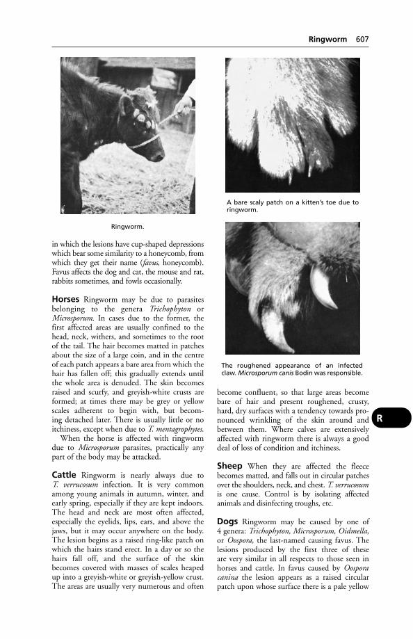
in which the lesions have cup-shaped depressionswhich bear some similarity to a honeycomb, fromwhich they get their name (favus, honeycomb).Favus affects the dog and cat, the mouse and rat,rabbits sometimes, and fowls occasionally.
Horses Ringworm may be due to parasitesbelonging to the genera Trichophyton orMicrosporum. In cases due to the former, thefirst affected areas are usually confined to thehead, neck, withers, and sometimes to the rootof the tail. The hair becomes matted in patchesabout the size of a large coin, and in the centreof each patch appears a bare area from which thehair has fallen off; this gradually extends untilthe whole area is denuded. The skin becomesraised and scurfy, and greyish-white crusts areformed; at times there may be grey or yellowscales adherent to begin with, but becom-ing detached later. There is usually little or no itchiness, except when due to T. mentagrophytes.
When the horse is affected with ringwormdue to Microsporum parasites, practically anypart of the body may be attacked.
Cattle Ringworm is nearly always due to T. verrucosum infection. It is very commonamong young animals in autumn, winter, andearly spring, especially if they are kept indoors.The head and neck are most often affected,especially the eyelids, lips, ears, and above thejaws, but it may occur anywhere on the body.The lesion begins as a raised ring-like patch onwhich the hairs stand erect. In a day or so thehairs fall off, and the surface of the skinbecomes covered with masses of scales heapedup into a greyish-white or greyish-yellow crust.The areas are usually very numerous and often
become confluent, so that large areas becomebare of hair and present roughened, crusty,hard, dry surfaces with a tendency towards pro-nounced wrinkling of the skin around andbetween them. Where calves are extensivelyaffected with ringworm there is always a gooddeal of loss of condition and itchiness.
Sheep When they are affected the fleecebecomes matted, and falls out in circular patchesover the shoulders, neck, and chest. T. verrucosumis one cause. Control is by isolating affected animals and disinfecting troughs, etc.
Dogs Ringworm may be caused by one of 4 genera: Trichophyton, Microsporum, Oidmella,or Oospora, the last-named causing favus. Thelesions produced by the first three of these are very similar in all respects to those seen inhorses and cattle. In favus caused by Oosporacanina the lesion appears as a raised circularpatch upon whose surface there is a pale yellow
Ringworm 607
R
Ringworm.
A bare scaly patch on a kitten’s toe due toringworm.
The roughened appearance of an infectedclaw. Microsporum canis Bodin was responsible.

crust with little depressions (honeycomb) scat-tered through it. The skin in such cases is oftenvery much thickened.
Hedgehogs Caused by T. erinacei, this infec-tion may cause lesions on the face of dogs wherethe skin has been damaged by the hedgehog’sspines.
Cats Ringworm is of 3 kinds: due toTrichophyton, Microsporum and Achorion, thelatter producing favus. When due to the firsttwo of these, the symptoms and lesions are similar to those seen in other animals. (See
ONYCHOMYCOSIS.)Cats become infected from mice with mouse
favus (A. quickeanum or A. arlongi), although itmay also be due to A. schoenleinii – the favus ofman. The lesions are chiefly confined to thefore-paws and the head and the neck, thoughthey may spread to other parts of the body.Itchiness is usually absent. The areas affectedvary in size from that of a pin’s head up to a 5p piece or so, and are not always regular inoutline. The skin is thickened and the edges areraised. When newly formed, the covering crustis yellow and soft to the touch, but when old it is grey and powdery. The characteristic cup-shaped depressions are seen in most cases, butwhen affecting the claws they may be absent.
Ringworm due to M. canis Bodin is of public-health importance. It is often overlooked byowners, but children are readily affected.
Cats, especially Persians and other longhairs,may be ‘carriers’ of ringworm fungus. In a surveyinvolving 200 selected cats seen at a veterinaryclinic, none of them showed any sign of ring-worm. Fur samples taken with a brush showedthat 39 per cent of the 200 were carrying sporesof ringworm fungi. (In 72 samples the sporeswere those of M. canis.) A survey in England offur samples taken at 4 cat shows revealed that,overall, 35 per cent of longhairs were carrying M. canis spores.
Decontamination of households is impor-tant for human health after ringworm has beendiagnosed. Hypochlorite, benzalkonium chlo-ride, and glutaraldehyde-based compounds arerecommended.
Favus in the fowl, due to T. gallinae, affectsthe comb, wattles, and other parts of the fowl’shead.
If the condition spreads down to featheredparts, the feathers become dry, brittle, and breakoff at the surface of the skin, leaving large bareareas. There is always a most disagreeable odourfrom fowl favus.
Treatment Oral administration of griseoful-vin is by far the simplest method. Cattle and
608 Ringworm
R
Ringworm in the cat: whitish, scaly lesion can be seen to the right of the ear, above the white fur.
horses can be given a supplemented feed. Thismakes possible group treatment, and avoidshandling of infected animals – thus reducingthe risk of infection being transferred to man.However, it is inadvisable not to use griseoful-vin in pregnant animals. In a cat which couldnot tolerate griseofulvin, a thiabendazole (see
ANTHELMINTICS) dip was successfully used.Natamycin and enilconazone which are
used as a wash or spray can be applied to infect-ed cattle and horses with a knapsack sprayer.Ketaconazole, applied as a shampoo, may beused to treat dogs.
Otherwise, treatment consists, in the firstplace, of removing the hair from around thelesions, collecting it and burning it. There aremany suitable dressings to choose from: e.g. gen-tian violet solution; undecylenate ointment; andcopper naphthenate lotion, which has given rapidand good results in the treatment of ringworm incattle. A vaccine for use in cattle is available.
Dressing should be carried out twice a weekfor a fortnight for cattle and horses, and by thenmost of the fungus will be killed. The casesshould not be considered cured until there is a level crop of new hair over each of the areas.For the smaller animals it is better to use thedressing once every 2nd day.
In all instances it is very important to remem-ber that ringworm spreads from the centre out-wards, and edges and margins of the areas shouldbe especially well dressed.
Vaccination of calves against T. verrucosuminvolves 2 intramuscular doses, 10 to 14 daysapart.
In Russia the vaccination of racehorses, andother horses taking part in competitive events,is compulsory.
Public health Ringworm is readily transmis-sible to human beings, so precautions such ashand-washing and disinfection after contactwith known infected animals should not beneglected. Dettol is useful for these purposes.
Diagnosis Microscopic examination or culturemethods. (See also WOOD’S LAMP.)
RNA(see RIBONUCLEIC ACID)
Road AccidentsDogs and cats struck by cars may suffer chest injuries in addition to limb injuries. (See
FRACTURES; ACCIDENTS; DIAPHRAGMATOCELE;HYDROTHORAX; PNEUMOTHORAX.)
RoamingRoaming is a behavioural habit in certain maledogs and cats. If the animal does not respond totraining from an animal behaviourist, it may becastrated – although there is no guaranteee thatthis will stop the habit.
‘Roaring’ in HorsesAn abnormal sound made when the horsebreathes in; the usual cause has for long beenregarded as vibration of the slackened vocal foldson one or both sides of the larynx, due to paral-ysis of the muscles which move the arytenoidcartilages outwards. (For treatment and furtherdetails, see LARYNX, DISEASES OF – Laryngealparalysis.)
Rock Salt(see SALT – Salt licks)
Rocky Mountain FeverAlso called Rocky Mountain spotted fever, thisis a disease of man caused by Rickettsia rickettsii.Wild animals provide a reservoir of infection.
Rocky Mountain fever affects human beingsusually between March and July. The onset offever is sudden, and in 2 to 5 days a rashappears over the whole body, including thepalms of the hands. The rash changes to a sortof mottling – petechiae, scattered over the skin,which gives the condition its name of ‘spottedfever’. The infection is transmitted by ticks,especially Dermacentor andersoni, the RockyMountain wood tick.
Dogs The infection causes fever, abdominalpain, depression, loss of appetite, nystagmus,with sometimes conjunctivitis and petechialhaemorrhages in the mouth. Oedema of a limb(s)is common, and the scrotum and prepuce may besimilarly affected. Over 30 cases are confirmedserologically each year, with many more beingdiagnosed, in Long Island, New York.
Diagnosis is most reliably confirmed by the immunofluorescent test. Treatment is byantibiotics, e.g. tetracycline.
Precautions There is a risk to veterinarians tak-ing a blood sample or carrying out a post-mortemexamination, as the rickettsia is present in theblood during the acute phase.
Rodent UlcerIn human medicine this term is reserved for car-cinoma of the skin, but is sometimes misappliedby animal-owners to EOSINOPHILIC GRANULOMA
or LICK GRANULOMA.
Rodent Ulcer 609
R

RodentsRats and mice are important from a veterinarypoint of view on account of the diseases whichthey may transmit to domestic animals. Forexamples, see AUJESZKY’S DISEASE; SALMONEL-
LOSIS; LEPTOSPIROSIS; RINGWORM; FOOT-AND-
MOUTH DISEASE. In countries where the diseaseis present, rodents may transmit RABIES.
Zoonoses Members of the family Muridae(Old World rats and mice) can infect man withplague, tularaemia, listeriosis, pseudotuberculo-sis, erysipelas, leptospirosis, brucellosis, melioi-dosis, murine typhus, Q fever, scrub typhus andother rickettsioses, histoplasmosis, lymphocyticchoriomeningitis, Lassa fever, rabies and otherviral infections, Asian schistosomiasis, Chagasdisease, rat-bite fever, and HANTAvirus.
Rodenticides In the UK brodifacoum wascleared by MAFF (for indoor use only) in 1984;and difenacoum had also been scrutinisedunder the Pesticides Safety Precautions Scheme.(No incidents linking barn owl deaths withthese two rodenticides had been reported in the UK.)
A calciferol preparation, Rodin C (Rentokil) –claimed to be effective against warfarin-resistantrats – had earlier received MAFF approval. (See
also WARFARIN.) ALPHACHLORALOSE is used forthe same purpose.
A rodenticide containing vitamin D1 causedthe death of 2 dogs.
Signs Weakness, anorexia, vomiting and passingblood.
Accidental poisoning of domestic animalshas occurred from some of the above, and alsofrom others banned in the UK, e.g. RED SQUILL;THALLIUM; ANTU; THIOUREA; PHOSPHORUS;FLUOROACETATE; barium salts; zinc phosphide;STRYCHNINE.
RomagnolaAn Italian breed of cattle, white in colour, andreared for beef. Romagnolas have been importedinto the UK.
RompunProprietary name for XYLAZINE.
Rose Bengal Plate TestA simple and quick screening test used in thediagnosis of brucellosis in cattle. Sera givingpositive results may then be tested by means ofthe Serum Agglutination Test and ComplementFixation Test.
RostralTowards the nose or front end of the body.
Rostral teeth are the incisors and canines.
RotavirusSo-called because of its resemblance to a wheel.Responsible for causing diarrhoea in the youngof many species – children, foals, calves andpiglets. It has been shown that, in piglets, onlythe pig and calf rotaviruses cause diarrhoea,although the human and foal rotaviruses canreplicate in the pig.
Research at the Moredun Institute led to amethod of diagnosis based on the direct detec-tion of the viral nucleic acid, which comprises11 molecules of double-stranded DNA. Thismethod is ‘rapid and as sensitive as ELISA’.
A vaccine Rotavec K99 (Schering-Plough)was introduced in 1986, following research atMoredun, to protect calves against rotavirus(and also K99 E. coli). The vaccine is used toimmunise cows in late pregnancy, producingenhanced antibody levels in the colostrum; thisis given to calves at the rate of 2.5 to 3.5 litresdaily for the first 2 weeks of life.
RotenoneThe insecticidal principle of derris root.Rotenone is highly poisonous to fish and is useddeliberately for removing coarse fish fromenclosed waters before establishing trout fisheries.
Rothera’s TestA test for ketones in milk or urine; a modifiedversion requires the following reagent: ammo-nium sulphate, 100 g; anhydrous sodium car-bonate, 50 g; sodium nitroprusside, 3 g.
If the bottom half-inch of a test-tube is filledwith this powder, and a little of the fluid to betested runs down the side of the tube, a red colourwill develop after 3 or 4 minutes if ketones arepresent, as with acetonaemia.
RoughageBy this is meant food of a bulky and fibrousnature, such as hay and straw. These have a lowwater-content, and are in a sense the oppositeof succulents, e.g. kale, silage. (See also DIET AND
DIETETICS – Fibre.)
RottweilerA large powerful breed developed as a guarddog in Germany. The breed is prone to deafnessand retinal dysplasia; hip dysplasia may also bea problem.
610 Rodents
R

Rough CollieA rough-coated medium-sized dog with an elongated face and nose, orginally developed as asheep-dog. The breed is prone to cleft palate,corneal dystrophy, deafness, epilepsy and umbil-ical hernia. Central progressive retinal atrophy isa dominant trait; collie-eye anomaly is a recessivetrait. Patent ductus arteriosus may be inherited,as may neutropenia.
RouleauxRouleaux is the term applied to the columns intowhich red blood cells collect as seen under themicroscope. The appearance somewhat resemblesa pile of stacked coins.
Round Heart DiseaseIn chickens, this occurs only on some types ofdeep litter. The cause is not known, but litterfrom affected houses can transmit the disease to healthy chickens. Sudden death occurs; onpost-mortem examination, the heart is firm,bright pink in colour, barrel shaped and with adimple at the apex. Replacement of the litterstops mortality.
In turkeys, the disease is more common inthe small white strains. It causes sudden deathin birds in apparently good condition. Thereappears to be a genetic factor.
RoundhouseA type of circular farrowing pen devised in NewZealand. It consists of a circle of hardboard,
about 2.4 m (8 feet) in diameter and 1.2 m (4 feet) high, bolted to a light iron frameworkand fitted with an internal creep rail. A smallercircle, about 1 m (3 feet) in diameter, madepartly of hardboard and partly of tubular rails,is fitted eccentrically within the larger one, andthe whole is fastened with bolts to the concretefloor of the piggery. The smaller circle, which iswarmed by an infra-red lamp, acts as a creep forthe piglets, while the sow is kept in the spacebetween the 2 circles. Because of the shape ofthis space, the sow invariably lies in the sameposition, with her udder towards the piglet’screep. This gives the piglets the maximumdegree of safety.
Roundworms (Nematoda)Most nematodes lay eggs, but some produce liv-ing larvae. The life-history may be direct or indi-rect, i.e. an intermediate host may be necessary.
Nematodes can be the cause of anaemia, wast-ing, gastroenteritis, bronchitis and pneumonia,aneurism, convulsions and blockage of the intes-tine. Some are of public health importance. (See
TRICHINOSIS; TOXOCARA.)
Horses
1. Stomach Two species of Habronema(H. muscae and H. microstoma), and Drascheiamegastoma, inhabit the stomach of Equidae invarious parts of the world.
Roundworms (Nematoda) 611
R
The rotavirus. (By courtesy of the AFRC.)
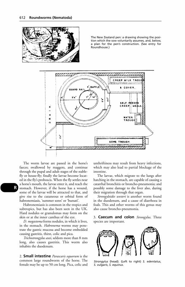
The worm larvae are passed in the horse’s faeces; swallowed by maggots, and continuethrough the pupal and adult stages of the stable-fly or house-fly; finally the larvae become locat-ed in the fly’s proboscis. When the fly settles neara horse’s mouth, the larvae enter it, and reach thestomach. However, if the horse has a wound,some of the larvae will be attracted to that, andgive rise to the cutaneous or orbital form ofhabronemiasis, ‘summer sores’ or ‘bursati’.
Habronemiasis is common in the tropics andsubtropics, but has also been seen in the UK.Hard nodules or granulomas may form on theskin or at the inner canthus of the eye.
D. megastoma forms nodules, in which it lives,in the stomach. Habronema worms may pene-trate the gastric mucosa and become embeddedcausing gastritis, thirst, colic and pica.
Trichostrongylus axei, seldom more than 8 mmlong, also causes gastritis. This worm also inhabits the duodenum.
2. Small intestine Parascaris equorum is thecommon large roundworm of the horse. Thefemale may be up to 50 cm long. Pica, colic and
unthriftiness may result from heavy infections,which may also lead to partial blockage of theintestine.
The larvae, which migrate to the lungs afterhatching in the stomach, are capable of causing acatarrhal bronchitis or broncho-pneumonia; andpossibly some damage to the liver also, duringtheir migration through that organ.
Strongyloides westeri is another worm foundin the duodenum, and a cause of diarrhoea infoals. This and other worms of this genus mayalso cause broncho-pneumonia.
3. Caecum and colon Strongylus. Threespecies are important.
612 Roundworms (Nematoda)
R
The New Zealand pen: a drawing showing the posi-tion which the sow voluntarily assumes, and, below,a plan for the pen’s construction. (See entry forRoundhouse.)
Strongylus (head). (Left to right) S. edentatus, S. vulgaris, S. equinus.
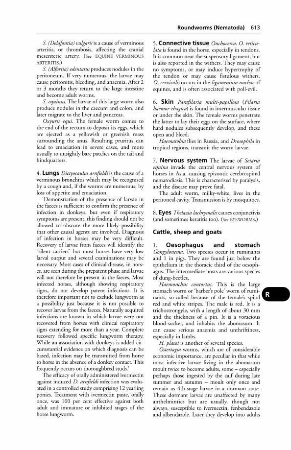
S. (Delafontia) vulgaris is a cause of verminousarteritis, or thrombosis, affecting the cranialmesenteric artery. (See EQUINE VERMINOUS
ARTERITIS.)S. (Alfortia) edentatus produces nodules in the
peritoneum. If very numerous, the larvae maycause peritonitis, bleeding, and anaemia. After 2or 3 months they return to the large intestineand become adult worms.
S. equinus. The larvae of this large worm alsoproduce nodules in the caecum and colon, andlater migrate to the liver and pancreas.
Oxyuris equi. The female worm comes to the end of the rectum to deposit its eggs, whichare ejected as a yellowish or greenish mass surrounding the anus. Resulting pruritus canlead to emaciation in severe cases, and moreusually to unsightly bare patches on the tail andhindquarters.
4. Lungs Dictyocaulus arnfieldi is the cause of averminous bronchitis which may be recognisedby a cough and, if the worms are numerous, byloss of appetite and emaciation.
‘Demonstration of the presence of larvae inthe faeces is sufficient to confirm the presence ofinfection in donkeys, but even if respiratorysymptoms are present, this finding should not beallowed to obscure the more likely possibilitythat other causal agents are involved. Diagnosisof infection in horses may be very difficult.Recovery of larvae from faeces will identify the“silent carriers” but most horses have very lowlarval output and several examinations may benecessary. Most cases of clinical disease, in hors-es, are seen during the prepatent phase and larvaewill not therefore be present in the faeces. Mostinfected horses, although showing respiratorysigns, do not develop patent infections. It istherefore important not to exclude lungworm asa possibility just because it is not possible torecover larvae from the faeces. Naturally acquiredinfections are known in which larvae were notrecovered from horses with clinical respiratorysigns extending for more than a year. Completerecovery followed specific lungworm therapy.While an association with donkeys is added cir-cumstantial evidence on which diagnosis can bebased, infection may be transmitted from horseto horse in the absence of a donkey contact. Thisfrequently occurs on thoroughbred studs.’
The efficacy of orally administered ivermectinagainst induced D. arnfieldi infection was evalu-ated in a controlled study comprising 12 yearlingponies. Treatment with ivermectin paste, orallyonce, was 100 per cent effective against bothadult and immature or inhibited stages of thehorse lungworm.
5. Connective tissue Onchocerca. O. reticu-lata is found in the horse, especially in tendons.It is common near the suspensory ligament, butis also reported in the withers. They may causeno symptoms, or may induce hypertrophy ofthe tendon or may cause fistulous withers. O. cervicalis occurs in the ligamentum nuchae ofequines, and is often associated with poll-evil.
6. Skin Parafilaria multi-papillosa (Filariahaemor-rhagica) is found in intermuscular tissueor under the skin. The female worms penetratethe latter to lay their eggs on the surface, wherehard nodules subsequently develop, and theseopen and bleed.
Haematobia flies in Russia, and Drosophila intropical regions, transmit the worm larvae.
7. Nervous system The larvae of Setariaequina invade the central nervous system ofhorses in Asia, causing epizootic cerebrospinalnematodiasis. This is characterised by paralysis,and the disease may prove fatal.
The adult worm, milky-white, lives in theperitoneal cavity. Transmission is by mosquitoes.
8. Eyes Thelazia lachrymalis causes conjunctivis(and sometimes keratitis too). (See EYEWORMS.)
Cattle, sheep and goats
1. Oesophagus and stomachGongylonema. Two species occur in ruminantsand 1 in pigs. They are found just below theepithelium in the thoracic third of the oesoph-agus. The intermediate hosts are various speciesof dung-beetles.
Haemonchus contortus. This is the large stomach worm or ‘barber’s pole’ worm of rumi-nants, so-called because of the female’s spiralred and white stripes. The male is red. It is a trichostrongyle, with a length of about 30 mmand the thickness of a pin. It is a voraciousblood-sucker, and inhabits the abomasum. Itcan cause serious anaemia and unthriftiness,especially in lambs.
H. placei is another of several species.Ostertagia worms, which are of considerable
economic importance, are peculiar in that whilemost infective larvae living in the abomasummoult twice to become adults, some – especiallyperhaps those ingested by the calf during latesummer and autumn – moult only once andremain as 4th-stage larvae in a dormant state.These dormant larvae are unaffected by manyanthelmintics but are usually, though notalways, susceptible to ivermectin, fenbendazoleand albendazole. Later they develop into adults
Roundworms (Nematoda) 613
R
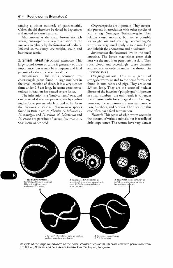
causing a winter outbreak of gastroenteritis.Calves should therefore be dosed in Septemberand moved to ‘clean’ pasture.
Also known as the small brown stomachworm, Ostertagia cause severe irritation of themucous membrane by the formation of nodules.Infested animals may lose weight, scour, andbecome anaemic.
2. Small intestine Ascaris vitulorum. Thislarge round worm of cattle is generally of littleimportance, but it may be a frequent and fatalparasite of calves in certain localities.
Nematodirus. This is a common tri-chostrongyle genus found in large numbers inthe small intestine of sheep. It is a very slenderform under 2.5 cm long. In recent years nema-todirus infestation has caused severe losses.
The infestation is a ‘lamb-to-lamb’ one, andcan be avoided – where practicable – by confin-ing lambs to pasture which carried no lambs inthe previous 2 seasons. Nematodirus speciesfound in Britain are N. filicollis, N. helvetianus,N. spathiges, and N. battus. N. helvetianus andN. battus are parasites of calves. (See PASTURE,CONTAMINATION OF.)
Cooperia species are important. They are usu-ally present in association with other species ofworms, e.g. Ostertagia, Trichostrongylus. Theyseldom cause anaemia, but are responsible for weight loss and scouring. Trichostrongylusworms are very small (only 2 to 7 mm long)and inhabit the abomasum and duodenum.
Bunestomum (hookworms) live in the smallintestine. The larvae may either enter their host via the mouth or penetrate the skin. Theysuck blood and accordingly cause anaemia and sometimes oedema under the throat. (See
HOOKWORMS.)Oesophagostomum. This is a genus of
strongyle worms related to the horse forms, andfound in ruminants and pigs. They are about2.5 cm long. They are the cause of nodular disease of the intestine (‘pimply gut’). If presentin small numbers, the only result is to renderthe intestine unfit for sausage skins. If in largenumbers, the symptoms are anaemia, emacia-tion, diarrhoea, and oedema. The disease in thiscase often has a fatal termination.
Trichuris. This genus of whip-worm occurs inthe caecum of various animals, but is usually oflittle importance. The worms have very slender
614 Roundworms (Nematoda)
R
Life-cycle of the large roundworm of the horse, Parascaris equorum. (Reproduced with permission fromH. T. B. Hall, Diseases and Parasites of Livestock in the Tropics, Longman.)
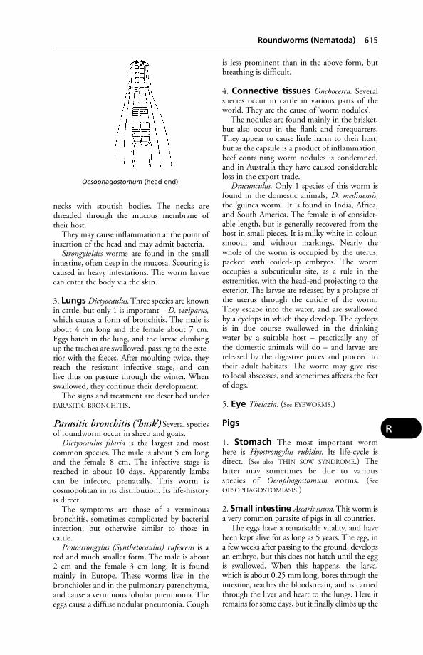
necks with stoutish bodies. The necks arethreaded through the mucous membrane oftheir host.
They may cause inflammation at the point ofinsertion of the head and may admit bacteria.
Strongyloides worms are found in the smallintestine, often deep in the mucosa. Scouring iscaused in heavy infestations. The worm larvaecan enter the body via the skin.
3. Lungs Dictyocaulus. Three species are knownin cattle, but only 1 is important – D. viviparus,which causes a form of bronchitis. The male isabout 4 cm long and the female about 7 cm.Eggs hatch in the lung, and the larvae climbingup the trachea are swallowed, passing to the exte-rior with the faeces. After moulting twice, theyreach the resistant infective stage, and can live thus on pasture through the winter. Whenswallowed, they continue their development.
The signs and treatment are described underPARASITIC BRONCHITIS.
Parasitic bronchitis (‘husk’) Several speciesof roundworm occur in sheep and goats.
Dictyocaulus filaria is the largest and mostcommon species. The male is about 5 cm longand the female 8 cm. The infective stage isreached in about 10 days. Apparently lambs can be infected prenatally. This worm is cosmopolitan in its distribution. Its life-historyis direct.
The symptoms are those of a verminous bronchitis, sometimes complicated by bacterialinfection, but otherwise similar to those in cattle.
Protostrongylus (Synthetocaulus) rufescens is ared and much smaller form. The male is about2 cm and the female 3 cm long. It is foundmainly in Europe. These worms live in thebronchioles and in the pulmonary parenchyma,and cause a verminous lobular pneumonia. Theeggs cause a diffuse nodular pneumonia. Cough
is less prominent than in the above form, butbreathing is difficult.
4. Connective tissues Onchocerca. Severalspecies occur in cattle in various parts of theworld. They are the cause of ‘worm nodules’.
The nodules are found mainly in the brisket,but also occur in the flank and forequarters.They appear to cause little harm to their host,but as the capsule is a product of inflammation,beef containing worm nodules is condemned,and in Australia they have caused considerableloss in the export trade.
Dracunculus. Only 1 species of this worm isfound in the domestic animals, D. medinensis,the ‘guinea worm’. It is found in India, Africa,and South America. The female is of consider-able length, but is generally recovered from thehost in small pieces. It is milky white in colour,smooth and without markings. Nearly thewhole of the worm is occupied by the uterus,packed with coiled-up embryos. The wormoccupies a subcuticular site, as a rule in theextremities, with the head-end projecting to theexterior. The larvae are released by a prolapse ofthe uterus through the cuticle of the worm.They escape into the water, and are swallowedby a cyclops in which they develop. The cyclopsis in due course swallowed in the drinking water by a suitable host – practically any of the domestic animals will do – and larvae arereleased by the digestive juices and proceed totheir adult habitats. The worm may give rise to local abscesses, and sometimes affects the feetof dogs.
5. Eye Thelazia. (See EYEWORMS.)
Pigs
1. Stomach The most important worm here is Hyostrongylus rubidus. Its life-cycle isdirect. (See also THIN SOW SYNDROME.) The latter may sometimes be due to various species of Oesophagostomum worms. (See
OESOPHAGOSTOMIASIS.)
2. Small intestine Ascaris suum. This worm isa very common parasite of pigs in all countries.
The eggs have a remarkable vitality, and havebeen kept alive for as long as 5 years. The egg, ina few weeks after passing to the ground, developsan embryo, but this does not hatch until the eggis swallowed. When this happens, the larva,which is about 0.25 mm long, bores through theintestine, reaches the bloodstream, and is carriedthrough the liver and heart to the lungs. Here itremains for some days, but it finally climbs up the
Roundworms (Nematoda) 615
R
Oesophagostomum (head-end).
trachea and is swallowed. The larva which leavesthe lung has grown to about 2.5 mm in length. Inthe intestine it continues its development, takingabout 21⁄2 months to do so.
In passing through the lungs a certainamount of bleeding is caused, and if the larvaeare numerous, pneumonia results. During thisperiod the animal shows the symptoms knownas ‘thumps’. If it survives the lung symptoms, itoften fails to grow properly and remains smalland stunted.
Macrocantorhynchus hirudinaceus is found inthe small intestine of pigs. It is a whitish worm,the male being 5 to 10 cm long, while thefemale is 20 to 35 cm long. The neck is thin and the posterior region stout. The intermediate stages are found in beetles.
The parasite may cause a catarrhal enteritisor even actual perforation with peritonitis.
Trichuris suis, the pig whip-worm, causesmainly subclinical disease in temperate climates, but in the tropics it may cause dysen-tery, anaemia, and even death. In the Americasup to 85 per cent of pigs may be infested; insome areas of the UK, from 75 per cent.Trichuris occurs in the caecum.
Treatment in the pig includes oxibendazole,fenbendzole and thiophanate.
3. Lungs In pigs 2 species are common, bothbelonging to the genus Metastrongylus. Themale is about 2 cm and the female about 4 cmlong. Both species are common in Europe and America, and may occur in the same pig.They cause a verminous bronchitis and sometimes pneumonia. Young animals are moresusceptible and may die from it. Both speciesare carried by earthworms.
4. Muscles Trichinella spiralis. This is a smallworm found in the intestine. The female produces living larvae (0.1 to 0.16 mm long)which migrate through the mucosa, reach thebloodstream, and are carried to various muscles.Here they pass into a cystic stage (the cyst beingformed by the host), in which they remain untilthey are swallowed by some flesh-eating host or until they calcify and degenerate. In theintestine of the new host they reach sexualmaturity and produce a new lot of larvae, whichin turn migrate to the muscles.
The normal hosts are carnivores (dogs andcats). Rodents may be infected, and rats can bea source of infection to pigs. Man may beinfected from the pig. (See under TRICHINOSIS.)
5. Kidney Stephanurus dentatus is a thickishworm of fair size, the male being nearly 3 cm
long and the female a little larger. It is found asa rule in the kidney fat of pigs, but also occursin the liver and other locations in these animalsand in ruminants. It is found in America andAustralia, and is responsible for considerabledamage. Its life-cycle is similar to that of thehookworms. Thiabendazole, fenbendazole andivermerctin have proved effective in controllingthis parasite.
Dogs and cats
1. Oesophagus Spirocerca lupi is found innodules in the oesophagus and, less frequently,the stomach of the dog, in all hot countries andin Europe.
It is a reddish worm. The male is 3 to 5 cmlong. The intermediate hosts are various beetlesand cockroaches.
The disease is often undiagnosed during life, but in countries where it is common thepresence of the worm may be suspected from afrequent cough followed by repeated vomiting.Death from exhaustion may result.
Damage to the carotid artery by S. lupiworms (3 in each of 2 nodules attached to the oesophagus) led to the death in the UK ofan Alsatian from internal haemorrhage. Thisparasite appears also to be closely associatedwith sarcoma of the oseophagus.
2. Stomach A microscopic gastric nematodeof cats, Ollulanus tricuspis, has been found in the Americas, Australasia, and Europe. Theworm causes unthriftiness and vomiting in kittens.
3. Small intestine
Ascarids include several species that occur indogs and cats. In cats the species seem to beAscaris tubaeforme and A. braziliense.
Hookworms in dogs Two species of hook-worm are found in dogs: Ancylostoma caninumand Uncinaria stenocephala. The latter is foundin Britain. These are smallish worms, about 2.5 cm long, found in the small intestine.
Eggs are passed to the exterior in the faecesand hatch in the soil or water. After severalmoults, the resulting larva becomes infective,and is able to gain access to the host either in the food or by penetrating the unbrokenskin. It enters the bloodstream and is carried to the lungs. It then passes up the trachea and is swallowed. It completes its develop-ment in the small intestine, where it becomesmature.
616 Roundworms (Nematoda)
R
4. Caecum The whip-worm Trichuris vulpisoccurs in the UK, and gives rise todiarrhoea/dysentery, loss of condition and aharsh, staring coat.
5. Heart Dirofilaria. There are 2 species occur-ring in dogs and cats. D. immitis occurs in theheart of the dog and occasionally the cat. The female may reach a length of 30 cm, butthe male is little more than half this size. It isfound in Asia and, of recent years, in Britain.The embryos are hatched in the body of thefemale, and the young larvae, passed into the bloodstream, are sucked up by a mosquitoin which they develop. After a certain periodthey escape from the fly, when it attacks another dog, and entering the blood are carried to the heart, where they complete theirdevelopment.
The worms interfere to a greater or lesserextent with the circulation. No symptoms maybe shown; or the dog may suddenly die. Othersymptoms include anaemia, respiratory troubles, ascites, etc. Various complicationsmay be due to emboli, such as cough, dyspnoea, etc. Diagnosis is by demonstration ofthe microfilaria in the blood.
Another heartworm of the dog isAngiostrongylus vasorum, which has, as interme-diate hosts, slugs and snails. This worm, whichhas caused an outbreak of infestation in kennelsin Ireland, lives in the pulmonary artery and theright ventricle of the heart. Symptoms includemalaise, stiffness on running, and subcutaneousswellings (due to suppression of normal bloodclotting by the parasites). Some lung damagemay be caused; likewise anaemia.
(See also under HEARTWORMS.)
6. Kidney Dioctophyme renale. The kidneyworm of dogs and wild carnivores is very large,reaching 1 m in length, and is a blood-red colour.It is found in Europe and the USA. It occurs inthe pelvis of the kidney, and occasionally destroysthe kidney tissue, to leave only the wall as a cystfilled with a purulent fluid. The other kidneyusually shows a compensatory hypertrophy. It isoccasionally found in the bladder. Infestation follows the eating of raw fish.
The worm’s eggs are barrel-shaped and maybe seen in the urine, under the microscope.
7. Bladder In the UK the bladder-wormCapillaria plica is rare, and seldom gives rise to obvious symptoms. A severe infestation can lead to inflammation of the bladder and a mucoid discharge from vagina or prepuce. Incats cystitis may, rarely, be caused by C. feliscati.
8. Trachea Oslerus (Filaroides) osleri occurs inthe UK and gives rise to a sporadic but persis-tent cough, especially on exercise or if the dogis excited. Retching may be caused. Severeinfestation can give rise to emaciation despite afair appetite, laboured breathing, sleepingstanding, and death in young dogs. For control,thiabendazole has given promising results.
Another tracheal worm, Capillaria aerophilia,seldom gives rise to obvious symptoms.
9. Lungs A minute worm lives in the lungs ofcats in Britain and elsewhere in Europe andAmerica. It may cause a fatal form of parasiticpneumonia. The parasite (Aelurostrongylusabstrusus) is transmitted to cats by mice. InAfrica, Bronchostrongylus subcrenatus is found.
Lung lesions found at the autopsy of 5 out ofa batch of 20 beagles were due to Filaroidesspecies, ‘probably F. milksi rather than F. hirthi’.The lungs had the appearance of being peppered with black spots. Signs of larvalmigration were seen microscopically in the liver, mesenteric lymph nodes, and gastrointestinal tract.
Poultry Roundworms more commonly occurin free-range systems where it is also difficult toensure that all birds can be treated. There maybe no obvious clinical signs but breeding flocks(chickens and turkeys) often show reducedhatchability.
Public health aspects (see under TOXOCARA)
Rous Sarcoma of ChickensThis is produced by a virus. (See under CANCER.)
Royal Army Veterinary Corps(RAVC)It has a long and honourable history. An ArmyVeterinary Service was established in 1796; thisbecame the Army Veterinary Corps in 1906,the title of ‘Royal’ being bestowed in 1918.
A History of the RAVC 1796–1919 was compiled by Major-General Sir FrederickSmith KCMG, CB, a former Director-General,Army Veterinary Services, and published by Baillière, Tindall & Cox. A 2nd volume, byBrigadier J. Clabby, was published in 1963 by J. A. Allen & Co.
Royal College of VeterinarySurgeons (RCVS)Belgravia House, 62–64 Horseferry Road,London SW1P 2 AF. The governing body ofthe veterinary profession in the UK. (See also
REGISTER.)
Royal College of Veterinary Surgeons (RCVS) 617
R

-Rrhaphy-Rrhaphy is a suffix meaning an operation in which some opening or tear is closed bystitches.
Rubarth’s Disease (HepatitisContagiosa Canis)This is named after the Swedish scientistRubarth who, in 1947, described for the firsttime a disease in dogs which he called, onaccount of its contagious nature and the damage caused to the liver, Hepatitis contagiosacanis. This is now commonly known as CANINE
VIRAL HEPATITIS. He regarded this disease, on the basis of the microscopical findings, asidentical with fox encephalitis, which had been known in America for some 17 years previously.
Rubber BandsThese sometimes get, or are put, on to the legsof cats (and possibly dogs), where they mayremain unnoticed until the continual pressurehas destroyed the skin beneath the band andcaused damage to the underlying structures.Gangrene or loss of use of the limb results.
A successful prosecution has followed theapplication of rubber bands to cows’ teats in the UK.
Rubber rings have been used for castrationof lambs and calves, and for the docking oflambs. (See ELASTRATOR, etc.)
‘Rubber Jaw’A condition seen in the dog in some cases ofchronic nephritis. It may be associated withenlargement of the parathyroid glands.Softening of the bones of the skull, particularlythe jaw, occurs, and in a severely affected part the bone can be cut with a scalpel. There is resorption of bone and its replacement byvascular fibrous tissue. ‘Rubber jaw’ is not, of course, seen in all cases of chronic nephritis,though some changes may be detected microscopically.
RumenThe 1st stomach of ruminants. It lies on the leftside of the body, occupying the whole of the leftside of the abdomen and even stretching acrossthe median plane of the body to the right side.It is a capacious sac which is subdivided into anupper or dorsal sac and a lower or ventral sac,each of which has a blind sac, at its posteriorextremity. These divisions are defined by thepresence of grooves on the outside of the organand by pillars or ridges internally. The whole
organ is lined by mucous membrane whichpossesses a papillated, stratified, squamousepithelium containing no digestive glands,but mucus-secreting glands are present inlarge numbers. Its entrance is through theoesophagus, and its exit is into the reticulumor 2nd stomach through the rumeno-reticularorifice.
Coarse, partially chewed food is stored andchurned in the rumen until such time as theanimal finds circumstances convenient forrumination. When this occurs, little balls offood are regurgitated through the oesophagusinto the mouth, and are subjected to a second,more thorough mastication. Each bolus ischewed 30 to 60 times and mixed with copiousamounts of saliva, to be swallowed and passedonwards into other parts of the compoundstomach.
In rare instances, the rumen may be situatedon the right-hand side.
Rumen FlukesBelonging to the genus Paramphistomum, these are found in both the tropics and North America.
Conical in shape, round in cross-section,they inhabit not only the rumen but also the reticulum, and – when immature – the duodenum. They are also found occasionally in the bile ducts and urinary bladder.
Little damage is caused to the rumen, but in young animals a severe enteritis is the important aspect of the disease, resulting in diarrhoea, unthriftiness, anaemia, and sometimes death.
Paramphistomum flukes have a life-historysimilar to that of the common liver-flukeFasciola hepatica; several species of snails beingthe intermediate hosts.
Rumen, Ulceration ofIn calves, ulcers in the rumen may be associ-ated with lesions of the liver caused byBacteroides (Fusiformis necrophorus), or with BVD infection. (See also STOMACH,DISEASES OF.)
RumenotomyOpening the rumen via the left upper flank forthe purpose of emptying the contents.
Ruminal DigestionIn the rumen, bacteria break down the cellulose(which forms the structural materials of plants),and starch by means of enzymes, and convertthem into fatty acids. The bacteria fall a prey tothe protozoa which, besides digesting starch,
618 -Rrhaphy
R

thus perform the useful task of converting plantprotein into animal protein. This becomesavailable to the cow when the protozoa are, intheir turn, destroyed further down the digestivetract and themselves digested.
A sample taken from the rumen, at theHannah Research Institute, contained 100 million protozoa and 5 million bacteria (givingsome idea of the proportion of the two).Examples of protozoa included Entodiniomorphspecies, which feed on plant material, bacteria,and each other; and Holotrich species, whichferment soluble sugars from plants and feed on bacteria. (See also under, DIET AND
DIETETICS – Fibre; LACTIC ACID.)
Ruminal Tympany(see BLOAT)
Rumination (‘Cudding’)Rumination (‘cudding’) is the process wherebyfood taken into the stomachs of ruminants is returned to the mouth, subjected to a second, more thorough chewing, and is againswallowed.
The act occurs at intervals of from 6 to 8 hours, and occupies a longer or shorter timeaccording to the nature of the food and the amount taken at the last meal. It usuallycommences about half an hour after feedingceases, and probably continues until all thecoarser constituents have been re-chewed, or atleast until the animal is disturbed. This fact is ofconsiderable importance practically; cattle andsheep should be allowed at least 2 hours’ restafter feeding before they are subjected to anysevere exertion. Disregard of this is a fruitfulcontributory cause of stomach disorders in bothcattle and sheep. (See also under RUMEN.)
The act of regurgitation appears to be in reality a complex one, but it may be brieflysummarised as follows:
(1) The tension of the oesophagus relaxes,partly by dilatation, and partly through aninspiratory movement of the diaphragm (theglottis being temporarily closed), which reducespressure in the thorax.
(2) The rumen and the reticulum powerfullycontract and squeeze upon their contents.
(3) The abdominal muscles contract andraise the intra-abdominal pressure.
The direct result is that ingested foodstuffsare forced from the area of high pressure (i.e.the rumen and reticulum) through the openoesophagus into an area of lower pressure (i.e.into the thoracic portion of the oesophagus).When a small quantity, sufficient to form abolus or ‘cud’, has entered the oesophagus, the
lips of the oesophageal groove and the musclesin the vicinity close the terminal part of theoesophagus, and there commences an antiperi-staltic movement which conveys the ‘cud’upwards past the closed glottis, underneath thesoft palate, and so into the mouth. Excess fluidis immediately squeezed from the mass andswallowed, and chewing movements commenceat once. Each bolus is chewed 30 to 60 timesaccording to its consistency, size, and to thenature of its constituents; coarse straw or hayfodder requiring the longest time. The chewingoccupies from 30 to 90 seconds, and then thebolus is rolled up by the dorsum of the tongueand again swallowed. In from 3 to 6 secondsanother bolus has reached the mouth, and sothe process is continued.
‘Run-back’This must be avoided by means of back fences.(See under STRIP GRAZING.)
Runch(see CHARLOCK POISONING)
‘Runners’This is an old, popular term for hounds unableto gallop properly. ‘Runners’ are usually recog-nised as such when they return to hunt kennelsat about 7 months old after being walked; and they are then often culled from the pack.Technically, the condition is known as osteo-chondrosis of the spine. Symptoms includepoor muscular development in the spinalregion, poor bodily condition, an unnaturalgait, and often inability to jump a fence successfully negotiated by the rest of the pack.Some curvature and rigidity of the spine mayalso be observed. It seems that this is, in part atleast, an inherited defect of foxhounds.
The term is also applied to young budgerigars affected by French moult.
Runt PigsRunt pigs can be reared in special nursing unitsdesigned for runts and excess piglets in a litter.Runt pigs and underweight babies have similarbiochemical and physiological abnormalities.
Runting and StuntingSyndromeA condition of economic importance in poultryproduction. Clinical signs include pallor of the skin, decreased skeletal density, lameness,late development of plumage, distortion andbending of quills or primary feathers andorange-coloured mucus in the droppings, alongwith particles of undigested food. The problem
Runting and Stunting Syndrome 619
R

occurs sporadically and tends to last for abou ayear on the farm before any improvement isseen. Retroviruses, enteroviruses and other viralagents, as well as anaerobic bacteria, have allbeen suspected as the cause. Disappearance ofthe syndrome over much of the USA coincidedwith the use of reovirus vaccines.
RuptureRupture is a popular name for HERNIA. Theterm is also applied to the tearing across of amuscle, tendon, ligament, artery, nerve, etc.Rupture of the aorta is a cause of death in maleturkeys at 5 to 22 weeks old.
Russian Gad-Fly(Rhinoestrus purpureus.) This attacks horses inEurope and North Africa.
Russian Spring-Summer VirusRussian spring-summer virus causes anencephalitis of man and goat, caused by a virus
and transmitted by the tick Ixodes ricinus inRussia, Poland, the Czech Republic andSlovakia.
Rye-GrassRye-grass poisoning has caused the death of cat-tle and horses restricted to grazing rye-grass pas-ture (Lolium perenne). In New Zealand andAustralia, a fungus present on the rye-grass maycause facial eczema. A staggering gait – andconvulsions – may occur in cattle and sheep onrye-grass pasture giving rise to the colloquialname ‘rye-grass staggers’. In a UK outbreak insheep, they had ‘a rocking-horse gait, and whenchased fell down and trembled violently’.(Veterinary Investigation Service report.)Fungal toxins are the cause of ‘rye-grass staggers’ in both the UK and New Zealand. The rye grass is infected with a seed-borneenfophytic fungus, Acremonium lolii, containing the alkaloid loitrem B. (See also
CEREBROCORTICAL NECROSIS.)
620 Rupture
R
SabulousGritty, sandy.
SacksSacks may be a means of passing infection fromone farm to another, for when empty they areput to many uses. Poisoning has occurredthrough contamination of feeding-stuffs bysacks previously used for sheep-dip. For thesereasons, non-returnable paper sacks haveadvantages over jute sacks.
SacrumThe part of the spinal column lying betweenthe lumbar region and the tail. It consists of 5 vertebrae in the horse and ox, 4 in thesheep and pig, and 3 in the dog and cat, fusedtogether in each case. It is roughly triangular in shape in all animals, and forms the roof of the pelvic cavity, lying midway between the2 ‘points of the hip’ or ‘haunch bones’.
Saddle-SoresSaddle-sores are formed through uneven pres-sure upon the back by some part of the saddle.They may be found in the middle line, imme-diately over the upper ends of the spinousprocesses; they may occur on either side of the middle line where the fore-arch of the saddle-tree presses; or they may be found justbehind the elbow, when they are caused bybadly fastened girths, and are often called‘girthgalls’.
The injuries consist of raw areas from whichthe hair has been rubbed or chafed off and,later, ulcers. Alternatively, patches of the skin,varying in size from 2.5 cm in diameter toalmost 7 cm, may become hard and leathery,pus being formed underneath. These are knownas ‘sitfasts’.
Treatment Attention must first of all be paidto the saddles. They should fit evenly all overthe back, and the stuffing or padding should beadequate to protect the skin from pressure bythe rigid framework of the saddle-tree. The hol-low of the arch of the saddle should never pressupon the middle line of the back, and the girthshould never be fastened with the skin foldedunder it. Rest from work will be necessary. (See
ULCER; WOUNDS.)
SagittalA structure or section running transverselyacross the trunk or a limb.
Sainfoin(Onobrychis sativa) A leguminous forage cropwhich fixes its own nitrogen; it contains tannins, so its rumen protein degradability islow (this means that the protein is used moreefficiently); and it does not cause bloat.Voluntary intake by animals is high – intakes of sainfoin can be 25 per cent higher than thatof ryegrass. Furthermore, it is drought-resistant.
Unfortunately, sainfoin does not grow as wellas bred strains of grasses, clovers and lucerne;30 per cent less yield than lucerne is quoted.
St John’s WortThis plant, Hypericum perforatum, which maybe present in hay, does not lose its poisonouscharacter when dried. It causes LIGHT SENSITI-
SATION in cattle, sheep, and pigs, especially inAustralia.
St Louis EncephalitisTransmitted by mosquitoes, and caused by aflavivirus, this disease occurs in North andSouth America, affecting wild birds, bats, hors-es and man (in which it may cause encephalitisand death in the elderly, although only fever inother people).
Salicylic Acid and SalicylatesOriginally derived from the willow (genusSalix), salicylic acid and its salts have long beenused in pain relief. ASPIRIN, which is acetylsali-cylic acid, largely replaced the other salicylatesas pain relievers (see ANALGESICS), and has beengiven in fevers. It must be used with extremecaution in cats, which metabolise aspirin veryslowly. A standard 250 mg tablet given daily toa cat may prove fatal in 12 days.
Salicylate poisoning has occurred inyoung animals following overdosage.Symptoms include depression, loss of appetiteand vomiting. Treatment involves the use of an emetic or gastric lavage and respiratory stimulants.
Saline(see under NORMAL SALINE)
SalinomycinAn IONOPHORE used as a coccidiostat in chick-ens, and also (outside the UK) as a growth-promoting feed additive for pigs. Its use had tobe phased out within the EU by January 2006.
S

Salinomycin poisoning Four hundredpoint-of-lay turkeys died within a week afterthe introduction of a diet containing 50 ppmsalinomycin.
In horses the signs of poisoning are eyelid-swelling, anorexia, colic, weakness, ataxia.
Salivary GlandsSalivary glands include the parotid gland, lyingin the space below the ear and behind the border of the lower jaw; the submaxillary gland,lying just within the angle of the lower jaw,under the lower part of the parotid; and thesublingual gland, which lies at the side of theroot of the tongue. Each of these glands ispaired, so that actually there are 6 glands, notall of which function at the same time.
Salivary Glands, Diseases ofCalculi and tumours may occur. In rabies, thesalivary glands must become infected beforetransmission of the virus to another host canoccur through a bite. (See also MUMPS.) A foreignbody, such as a grass seed, may cause anobstruction to one of the ducts, particularly inthe dog.
Salivary-gland tumours in dogs and cats are rare. The majority of 138 tumours indogs (81) and cats (57) involved animals of 10 years of age or more, were malignant and of epithelial origin (84 per cent). Local recur-rence after excision occurs frequently, andmetastasis to regional lymph nodes and beyondis common.
Salivation‘Foaming at the mouth’, to use a colloquial butapt expression, is seen in the dog, e.g. in anepileptic or other fit. (See FITS.) ‘Drooling ofsaliva’ is seen in the dog with a bone wedgedacross the roof of its mouth, or in a cat with aneedle embedded in its tongue – or in cases ofRABIES in all species.
Salivation is a symptom of CHOKING, ofalmost any painful condition of the mouth ortongue, and of poisoning (e.g. by benzoic acidin the cat), arsenic, lead, phosphorus andorganophosphorus compounds; see also TOAD.
Salivation is also an important symptom of FOOT-AND-MOUTH DISEASE, and of other diseases and conditions mentioned underMOUTH.
SalmincolaParasites on the gills of salmonid fish whichaffect respiration. Affected fish show reducedgrowth and delayed sexual maturity.
‘Salmon Poisoning’ in Dogs‘Salmon poisoning’ in dogs occurs on thePacific coast of the USA, and is the result of eating salmon or trout infested with the fluke Troglotrema salmincola, containing a rickettsia. The latter, Neorickettsia helminthoeca,produces a haemorrhagic gastroenteritis whichis usually fatal unless antibiotics are used intime.
SalmonellosisInfection with organisms of the salmonellagroup is of importance from 2 distinct aspects:(1) food poisoning in man; and (2) disease indomestic animals.
Salmonella poisoning – routes ofinfection (see diagram)
In cattle and calves Salmonellosis and brucellosis have 4 points in common – bothdiseases are important from the public healthpoint of view; both can lead to abortion in cattle, to a carrier state likely to perpetuateinfection on the farm, and to considerablefinancial loss to the farmer.
While the salmonella group of bacteriaincludes more than 1000 different serotypes,the 2 of most importance to the dairy farmerare Salmonella dublin and S. typhimurium.Either can produce acute or subacute illness inadult cattle and in calves.
S. typhimurium infection is of greater public-health importance, and is a notorious cause of outbreaks of food poisoning in man. S.typhimurium type 204C has been a majorsource of problems in calves bought from mar-kets and is highly resistant to antibiotics. Anoutbreak of this same infection involved morethan 200 cows on a single farm, and led to thedeath or slaughter of 29 of them.
S. typhimurium 104 also has a relatively highresistance to antibiotics. It can result in severe illness and deaths in small groups of cows orcalves; it is the second most common salmonellain food poisoning.
S. dublin infection may be associated withabortion, sometimes without any other symp-toms being observed. Animals which recovermay excrete the organisms for years. Besidesthis carrier state, which may keep infection on the farm, there is also a latent carrier state in which the organism remains dormant withinthe animal until it is subjected to some stress or superimposed disease, when excretion of the organism occurs and fellow members of the herd become infected.
622 Salivary Glands
S
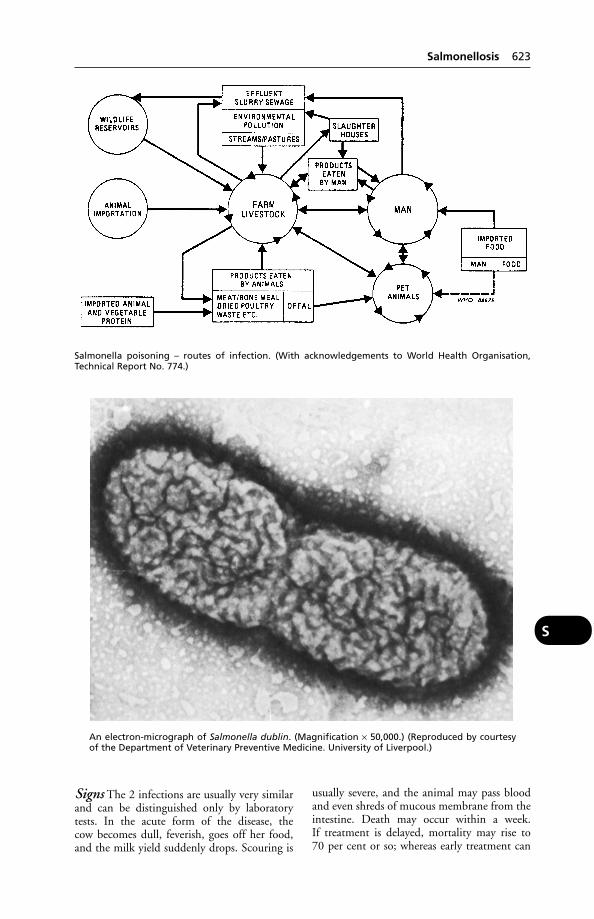
Signs The 2 infections are usually very similarand can be distinguished only by laboratorytests. In the acute form of the disease, the cow becomes dull, feverish, goes off her food,and the milk yield suddenly drops. Scouring is
usually severe, and the animal may pass bloodand even shreds of mucous membrane from theintestine. Death may occur within a week. If treatment is delayed, mortality may rise to 70 per cent or so; whereas early treatment can
Salmonellosis 623
S
Salmonella poisoning – routes of infection. (With acknowledgements to World Health Organisation,Technical Report No. 774.)
An electron-micrograph of Salmonella dublin. (Magnification × 50,000.) (Reproduced by courtesyof the Department of Veterinary Preventive Medicine. University of Liverpool.)
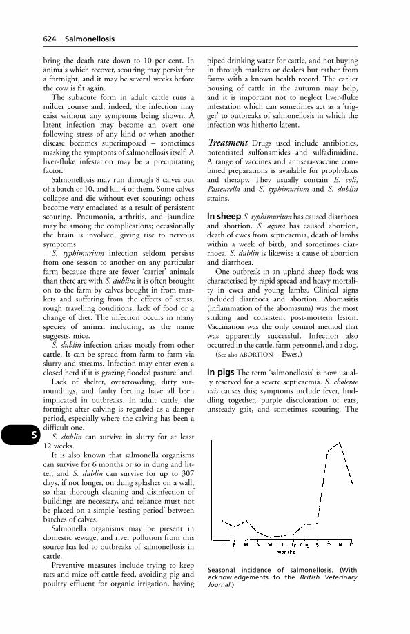
bring the death rate down to 10 per cent. Inanimals which recover, scouring may persist fora fortnight, and it may be several weeks beforethe cow is fit again.
The subacute form in adult cattle runs amilder course and, indeed, the infection mayexist without any symptoms being shown. Alatent infection may become an overt one following stress of any kind or when anotherdisease becomes superimposed – sometimesmasking the symptoms of salmonellosis itself. Aliver-fluke infestation may be a precipitatingfactor.
Salmonellosis may run through 8 calves outof a batch of 10, and kill 4 of them. Some calvescollapse and die without ever scouring; othersbecome very emaciated as a result of persistentscouring. Pneumonia, arthritis, and jaundicemay be among the complications; occasionallythe brain is involved, giving rise to nervoussymptoms.
S. typhimurium infection seldom persistsfrom one season to another on any particularfarm because there are fewer ‘carrier’ animalsthan there are with S. dublin; it is often broughton to the farm by calves bought in from mar-kets and suffering from the effects of stress,rough travelling conditions, lack of food or achange of diet. The infection occurs in manyspecies of animal including, as the name suggests, mice.
S. dublin infection arises mostly from othercattle. It can be spread from farm to farm viaslurry and streams. Infection may enter even aclosed herd if it is grazing flooded pasture land.
Lack of shelter, overcrowding, dirty sur-roundings, and faulty feeding have all beenimplicated in outbreaks. In adult cattle, thefortnight after calving is regarded as a dangerperiod, especially where the calving has been adifficult one.
S. dublin can survive in slurry for at least 12 weeks.
It is also known that salmonella organismscan survive for 6 months or so in dung and lit-ter, and S. dublin can survive for up to 307days, if not longer, on dung splashes on a wall,so that thorough cleaning and disinfection ofbuildings are necessary, and reliance must notbe placed on a simple ‘resting period’ betweenbatches of calves.
Salmonella organisms may be present indomestic sewage, and river pollution from thissource has led to outbreaks of salmonellosis incattle.
Preventive measures include trying to keeprats and mice off cattle feed, avoiding pig andpoultry effluent for organic irrigation, having
piped drinking water for cattle, and not buyingin through markets or dealers but rather fromfarms with a known health record. The earlierhousing of cattle in the autumn may help, and it is important not to neglect liver-flukeinfestation which can sometimes act as a ‘trig-ger’ to outbreaks of salmonellosis in which theinfection was hitherto latent.
Treatment Drugs used include antibiotics,potentiated sulfonamides and sulfadimidine. A range of vaccines and antisera-vaccine com-bined preparations is available for prophylaxisand therapy. They usually contain E. coli,Pasteurella and S. typhimurium and S. dublinstrains.
In sheep S. typhimurium has caused diarrhoeaand abortion. S. agona has caused abortion,death of ewes from septicaemia, death of lambswithin a week of birth, and sometimes diar-rhoea. S. dublin is likewise a cause of abortionand diarrhoea.
One outbreak in an upland sheep flock wascharacterised by rapid spread and heavy mortali-ty in ewes and young lambs. Clinical signsincluded diarrhoea and abortion. Abomasitis(inflammation of the abomasum) was the moststriking and consistent post-mortem lesion.Vaccination was the only control method thatwas apparently successful. Infection alsooccurred in the cattle, farm personnel, and a dog.
(See also ABORTION – Ewes.)
In pigs The term ‘salmonellosis’ is now usual-ly reserved for a severe septicaemia. S. choleraesuis causes this; symptoms include fever, hud-dling together, purple discoloration of ears,unsteady gait, and sometimes scouring. The
624 Salmonellosis
S
Seasonal incidence of salmonellosis. (Withacknowledgements to the British VeterinaryJournal.)
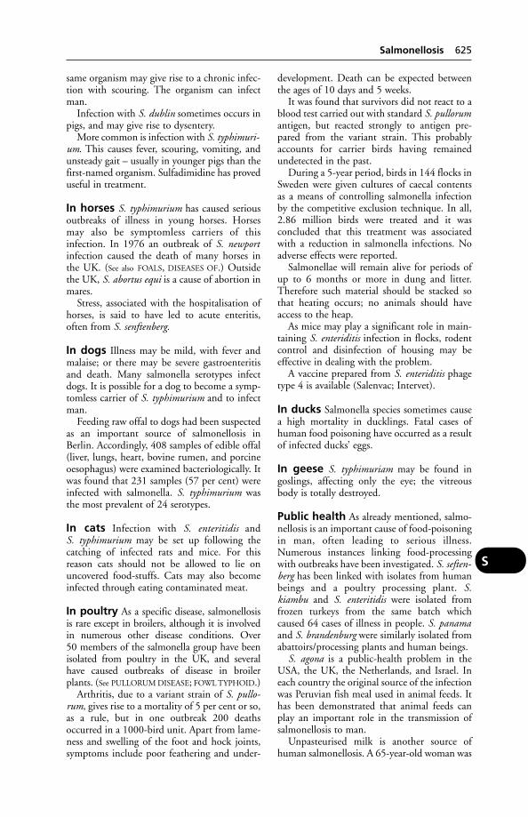
same organism may give rise to a chronic infec-tion with scouring. The organism can infectman.
Infection with S. dublin sometimes occurs inpigs, and may give rise to dysentery.
More common is infection with S. typhimuri-um. This causes fever, scouring, vomiting, andunsteady gait – usually in younger pigs than thefirst-named organism. Sulfadimidine has proveduseful in treatment.
In horses S. typhimurium has caused seriousoutbreaks of illness in young horses. Horsesmay also be symptomless carriers of this infection. In 1976 an outbreak of S. newportinfection caused the death of many horses inthe UK. (See also FOALS, DISEASES OF.) Outsidethe UK, S. abortus equi is a cause of abortion inmares.
Stress, associated with the hospitalisation ofhorses, is said to have led to acute enteritis,often from S. senftenberg.
In dogs Illness may be mild, with fever andmalaise; or there may be severe gastroenteritisand death. Many salmonella serotypes infectdogs. It is possible for a dog to become a symp-tomless carrier of S. typhimurium and to infectman.
Feeding raw offal to dogs had been suspectedas an important source of salmonellosis inBerlin. Accordingly, 408 samples of edible offal(liver, lungs, heart, bovine rumen, and porcineoesophagus) were examined bacteriologically. Itwas found that 231 samples (57 per cent) wereinfected with salmonella. S. typhimurium wasthe most prevalent of 24 serotypes.
In cats Infection with S. enteritidis and S. typhimurium may be set up following thecatching of infected rats and mice. For this reason cats should not be allowed to lie onuncovered food-stuffs. Cats may also becomeinfected through eating contaminated meat.
In poultry As a specific disease, salmonellosisis rare except in broilers, although it is involvedin numerous other disease conditions. Over 50 members of the salmonella group have beenisolated from poultry in the UK, and severalhave caused outbreaks of disease in broilerplants. (See PULLORUM DISEASE; FOWL TYPHOID.)
Arthritis, due to a variant strain of S. pullo-rum, gives rise to a mortality of 5 per cent or so,as a rule, but in one outbreak 200 deathsoccurred in a 1000-bird unit. Apart from lame-ness and swelling of the foot and hock joints,symptoms include poor feathering and under-
development. Death can be expected betweenthe ages of 10 days and 5 weeks.
It was found that survivors did not react to ablood test carried out with standard S. pullorumantigen, but reacted strongly to antigen pre-pared from the variant strain. This probablyaccounts for carrier birds having remainedundetected in the past.
During a 5-year period, birds in 144 flocks inSweden were given cultures of caecal contentsas a means of controlling salmonella infectionby the competitive exclusion technique. In all,2.86 million birds were treated and it was concluded that this treatment was associatedwith a reduction in salmonella infections. Noadverse effects were reported.
Salmonellae will remain alive for periods ofup to 6 months or more in dung and litter.Therefore such material should be stacked sothat heating occurs; no animals should haveaccess to the heap.
As mice may play a significant role in main-taining S. enteriditis infection in flocks, rodentcontrol and disinfection of housing may beeffective in dealing with the problem.
A vaccine prepared from S. enteriditis phagetype 4 is available (Salenvac; Intervet).
In ducks Salmonella species sometimes causea high mortality in ducklings. Fatal cases ofhuman food poisoning have occurred as a resultof infected ducks’ eggs.
In geese S. typhimuriam may be found ingoslings, affecting only the eye; the vitreousbody is totally destroyed.
Public health As already mentioned, salmo-nellosis is an important cause of food-poisoningin man, often leading to serious illness.Numerous instances linking food-processingwith outbreaks have been investigated. S. seften-berg has been linked with isolates from humanbeings and a poultry processing plant. S.kiambu and S. enteritidis were isolated fromfrozen turkeys from the same batch whichcaused 64 cases of illness in people. S. panamaand S. brandenburg were similarly isolated fromabattoirs/processing plants and human beings.
S. agona is a public-health problem in theUSA, the UK, the Netherlands, and Israel. Ineach country the original source of the infectionwas Peruvian fish meal used in animal feeds. Ithas been demonstrated that animal feeds canplay an important role in the transmission ofsalmonellosis to man.
Unpasteurised milk is another source ofhuman salmonellosis. A 65-year-old woman was
Salmonellosis 625
S
infected in this way, and was ill with diarrhoeaand meningitis. After her death a brain abscesswas found. Both the latter and meningitis are ‘rare complications of salmonellosis in man’. Seventeen other people were ill with salmonellosis from drinking the unpasteurisedmilk.
Viable salmonellae were found in the meatfraction of domestic refuse from 120 houses.This source could provide a reservoir of infec-tion accessible to wild animals. Tipping shouldbe carefully controlled, and refuse coveredimmediately.
The protective gloves, worn by a veterinarysurgeon while calving a cow, unfortunatelyburst. Within 48 hours numerous non-pruriticpapules had appeared over both arms, especial-ly the upper arm, where the gown cuffs hadchafed the skin. The papules developed intopustules which burst and resolved in approxi-mately 10 days without treatment. No othersymptoms were observed.
A pustule was swabbed and a pure growth ofSalmonella species was recovered.
(See also under SAUSAGE.)
SalolithsThese are CALCULI, found mainly in STENSON’S
DUCT of horses.
SalpingitisSalpingitis is inflammation in the Fallopian tubesor oviducts, sometimes the cause of sterility incattle. (See INFERTILITY.)
SaltA chemical substance in which a metal is sub-stituted for the hydrogen of an acid.
Sodium chloride (common salt)(NaCl) is an essential ingredient of body fluids.Sodium depletion results, ultimately, in circula-tory collapse.
Salt is an appetiser, and commonly incorpo-rated in animal feeds in carefully measured proportions. Ruminants will avidly consumesalt; any excess is harmlessly excreted in theurine and faeces.
Salt licks It has been suggested that a 500-kg(10-cwt) cow needs 30 g (1 oz) of salt a day for maintenance and a further 3.5 g (1⁄8 oz) for 4.5 litres (1 gallon) of milk produced.Therefore, a 3200-litre (700-gallon) cowrequires about 14 kg (30 lb) of salt yearly.
On some pastures, or under some systems ofmanagement, cattle may not obtain sufficientsalt. To obviate this danger, salt licks are
commonly provided. In some salt licks traces of iodine are incorporated, together with othertrace elements such as copper, manganese,cobalt, and magnesium. (See ‘LICKING
SYNDROME’.)
Salt PoisoningSalt poisoning has been reported in both pigs and poultry. It is essential that pigs are notkept short of water, or given food that is toosalted.
An outbreak, reported from Scotland,involved piglets aged 6 weeks brought indoorsfrom field arks at weaning. A proprietary mealwas fed dry. The water bowls in the house werenot very accessible, and some of the piglets werenot strong enough to depress the levers. Twodays after being housed, 23 out of the 32 pigletswere showing symptoms of salt poisoning, andsome died.
Signs Often a number of pigs are found deadwithout signs having been observed, theremainder being weak and very thirsty.Vomiting and diarrhoea may occur. (For othersigns, see under MENINGOENCEPHALITIS.)
In poultry, adult birds show excessive thirstand diarrhoea, with sometimes cyanosis of thewattles, somnolence, and sudden death. Inyoung birds gasping and ascites may occur.
SamoyedA medium-sized breed of dog characterised bythick straight cream or white hair. Like thechow-chow, they tend to be ‘one-person’ dogs.Haemophilia has been recorded; pulmonicstenosis may be inherited.
SandHorses on the seashore or along tidal mud flatslearn that the sand contains salt, and may lickup large quantities of it in their endeavour toget the salt. The signs set up are chiefly those ofCOLIC with impaction.
Cattle feeding on the seashore take in quan-tities of sand, which in some cases may be sogreat as to hinder the movements of the rumen(where the sand always collects), and, by upset-ting digestion, may cause unthriftiness andeven emaciation.
Sand Tampan (OrnithodorusSavignyl)(see TICKS – Family Argasidae)
SandcrackSandcrack is a pathological condition affectinghorses’ feet, in which a deep fissure or crack forms
626 Saloliths
S

at some part of the wall of the hoof, extendingdownwards from the coronet, and usually involving the whole of the thickness of the wall.
Causes Anything which interferes with theproper nutrition of the horn at the coronet predisposes to sandcrack, the actual splitting ofthe horn occurring as the result of the strainsput upon the foot. Treads on the inside of thecoronet, occasioned by hurried turning when atwork, are frequent causes in the fore-feet, andcontinual pressure on the coronary matrix bythe 2nd phalanx, especially when the toes havebeen allowed to grow too long, appears to bethe commonest cause in the hind-feet. A predisposition to sandcrack may be inherited.
With all cases it is advisable to place the ani-mal under veterinary care. (See HOOF REPAIR.)
‘Sandflies’(see under FLIES)
SanguineousSanguineous means containing blood.
Santa GertrudiThis breed of cattle are 5⁄8 Shorthorn and 3⁄8Brahman in origin.
SaponinsThese are natural detergents, present in someplants such as corncockle and soapwort.Saponins contain a sugar and a steroid-likecompound, and with water form a lather.Poisoning by them results in gastroenteritis.The central nervous system may also be affect-ed, with consequent paralysis. Saponins breakdown red blood cells. In the USA the leaves andnuts of the tung tree, grown for the sake of itsoil, can cause fatal poisoning.
SaprolegniaA fungus that can infect fish. It is sometimesfound as a secondary infection to another con-dition such as ulcerative dermal necrosis insalmonids, and autumn aeromonad disease inadult brown trout. The infection can be con-trolled by bathing the fish in zinc-free malachitegreen but the healing process is prolonged. In asalmon hatchery, it is important to remove deadand infertile eggs as these can be invaded by thefungus and passed on to healthy eggs.
Sarco-Sarco- is a prefix signifying flesh or fleshy.
Sarcolemma The membrane covering eachvoluntary (striated) muscle fibre.
SarcocystisA genus of protozoal, coccidian parasites havinga 2-host life-cycle. Carnivorous animals such asdogs, cats and foxes ingest the cysts when eatinginfected flesh of cattle, sheep, pigs and horses.Human infection also occurs, and sarcocystis isa zoonosis (see ZOONOSIS).
While the cysts in the intermediate host’smuscles may not have any serious effect uponhealth, the second-generation schizonts are certainly harmful – damaging the endotheliumof blood vessels, and causing serious illness inmany cases.
Signs Cattle showed loss of appetite, fever,anaemia, and wasting, after ingesting sporocystsfrom canine faeces, and some cattle died within33 days. Sarcocystosis has also killed sheep. In horses, signs of central nervous system damage may be seen, as well as signs of muscleinflammation, resulting in lameness.
Prevalence In Europe 61 per cent of slaugh-tered cattle have been found to be infected. InGermany a prevalence rate of 5 per cent in pigshas been recorded.
Human sarcocystosis may give rise toabdominal pain, diarrhoea, fever, tachycardia,and an increased respiratory rate.
‘Sarcoid’A tumour which resembles histologically a sarcoma (see CANCER), but which is regressive in character, disappearing within a matter ofmonths. It has the appearance of a reddish but-ton, raised about 0.30 mm (1⁄8 in) above the sur-rounding skin. It affects the dog.
A fibroma-like sarcoid is perhaps the mostcommon tumour of equines, especially olderones, occurring on limbs or head. Believed tobe caused by a virus, the equine sarcoid com-monly ulcerates and recurs following surgery.Cryosurgery may be tried, or a BCG vaccine;the latter may be more successful in donkeys. Aguarded prognosis should be given.
Bovine papillomavirus is involved in theprocess by which sarcoids develop from normalequine fibrous tissue.
Sarcoma(see CANCER)
SarcoptesSarcoptes are members of a class of parasiticacari, which cause MANGE in animals and man.
Sarcoptic mange occurs in cattle, horses,sheep, pigs, and dogs – also in man, when it is
Sarcoptes 627
S
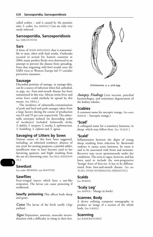
called scabies – and is caused by the parasiticmite S. scabei. (See MANGE.) Cats are only veryrarely infected.
Sarcosporidia, Sarcosporidiosis(see SARCOCYSTIS)
SarsA form of AVIAN INFLUENZA that is transmissi-ble to man, often with fatal results. Outbreaksoccured in several Far Eastern countries in2004; many poultry flocks were destroyed in anattempt to prevent the disease from spreading.Fears that migrating wild fowl would carry theSARS virus to Western Europe led ??? considerpreventive measures.
SausageDiscarded portions of sausage, or sausage-skin,can be a source of infection when fed, unboiled,to pigs, etc. Foot-and-mouth disease has beentransmitted in this way. African swine fever andswine fever could similarly be spread by thismeans. (See SWILL.)
The incidence of salmonella-contaminationof pork and beef and pork sausages taken froma large factory during the course of productionwas 65 and 55 per cent respectively. The salmo-nella serotypes isolated (in descending order of incidence) included Salmonella derby, S. dublin, S. newport, S. stanley, S. typhimurium, S. heidelberg, S. infantis and S. agona.
Savaging of Litters by SowsVarious causes of this have been suggested,including: an inherited tendency; absence ofany straw for nesting purposes; a painful udder;insufficient time to have become used to herfarrowing quarters; and fright resulting fromthe use of a farrowing crate. (See PIGS, SEDATION
OF.)
Sawdust(see under BEDDING and MASTITIS)
SawfliesFour-winged insects which have a saw-likeovipositor. The larvae can cause poisoning ifswallowed.
Sawfly poisoning This affects both sheepand goats.
Cause The larvae of the birch sawfly (Arge pullata).
Signs Depression, anorexia, muscular incoor-dination with a difficulty in rising to their feet.
Autopsy Findings Liver necrosis, petechialhaemorrhages, and sometimes degeneration ofthe kidney tubules.
ScabiesA common name for sarcoptic mange. (See under
MANGE – Sarcoptic mange.)
‘Scad’A colloquial name for a transitory lameness, insheep, which may follow frost. (See ‘SCALD’.)
‘Scald’Inflammation between the digits of youngsheep resulting from infection by Bacteroidesnodosus; it causes acute lameness. Its onset issaid to be associated with frosts and moisture.Recovery may occur spontaneously under dryconditions. The term is vague, however, and hasbeen used to include the non-progressive‘benign’ form of foot-rot. It has to be differen-tiated from foot-and-mouth disease. (See also
‘SCAD’; OVINE INTERDIGITAL DERMATITIS.)
Scalds(see BURNS)
‘Scaly Leg’(see MANGE – Mange in fowls)
Scanner, BodyA device utilising computer tomography toproduce an image of a section of the wholebody. (See X-RAYS.)
Scanning(see RADIOISOTOPES)
628 Sarcosporidia, Sarcosporidiosis
S
Schistosoma, o, µ, and egg.

ScaphoidThe name given in human anatomy to a smallbone present in the carpus and tarsus. In theracing greyhound, fracture of the right hindscaphoid is a common accident. Treatment hasincluded the removal of bone fragments and thesuccessful insertion of a plastic ‘scaphoid’.
ScapulaThe scapula is the shoulder blade – the large,triangular, flat bone that lies on the outside ofthe front of the chest, to which are attachedmany of the muscles that unite the fore-limb tothe trunk.
Scheduled Diseases(see under NOTIFIABLE DISEASES)
SchistosomiasisInfestation with Schistosoma worms or flukes,which are also known as bilharzia worms. Theyinhabit the portal and mesenteric veins mostly,one species preferring veins of the urinary blad-der, and another the veins of the nose. Cattleand sheep and virtually all domestic animals,and man, may become infested.
Several species have been reported frommammals in India, Africa, and Europe. S. bovismay cause anaemia, emaciation and death ofcattle in Africa, or the infestation may be sub-clinical. In India S. nasalis may produce a nasaldischarge and difficulty in breathing, withsometimes the formation of a granuloma. Inthe Far East S. japonicum occurs in water-buffa-lo and infests man, in which the disease is veryserious.
The life-cycle differs from the typical case, inthat the free cercaria may pierce the skin of itshost instead of being swallowed.
The sexes are separate, and are usually foundwith the female lying in a groove formed by theincurved edges of the male.
Control Provision of clean drinking water andtreatment of pasture with molluscicides such as copper sulphate to kill the intermediate host will reduce infection. However, such mea-sures are rarely practicable in affected areas.Drugs such as praziquantel may be effective intreatment.
Schistosomus ReflexusA deformity of the bovine fetus, in which thespine is bent, so that head and tail curvetowards each other, causing dystokia. Fetalintestine may be visible at the vulva, or locatedin the vagina.
Schmorl’s DiseaseSchmorl’s disease is a disease of rabbits, involv-ing areas of necrosis of skin or mucous mem-brane, and caused by Bacteroides necrophorus(often after the animal’s resistance has been lowered by some other pathogen).
SchnauzerA German breed of dog with wiry coat thatforms characteristic eyebrows, mouth and chinwhiskers. There are miniature, standard andgiant forms. The standard has fewer defectsthan the miniature, which is predisposed tocataracts and progressive retinal atrophy.
SchradanAn organophosphorus insecticide used in agri-culture and a potential danger to farm livestock.(See also PARATHION.) Symptoms of poisoningmay include vomiting, lachrymation, saliva-tion, straining, twitching, distressed breathing,and coma.
SciaticaSciatica means pain connected with the sciaticnerve which runs down the thigh.
ScintigraphyThe application of nuclear medicine to thediagnosis of bone pathology and lameness. Ithas applications for dogs and horses. (See
NUCLEAR MEDICINE.)
Scirrhous CordScirrhous cord is a condition in which there is achronic fibrous enlargement of the cut end ofthe spermatic cord following castration. In mostcases the castration wound does not completelyheal, but a small sinus discharging a thick whitepus persists. The discharge may cease later, butthe swelling of the cord goes on increasing slow-ly in size, until eventually it may be nearly aslarge as a man’s head. In extreme cases theswelling extends upwards through the inguinalcanal and into the abdomen and a mass weigh-ing as much as 45 kg (100 lb) has occasionallybeen encountered in the horse on post-mortem examination. Treatment is entirely surgical.
ScirrhusScirrhus is a term applied to a growth or toother hard fibrous conditions of various organs.
Sclera (Sclerotic Coat)Sclera (sclerotic coat) is the outermost hardfibrous coat of the EYE.
Sclera (Sclerotic Coat) 629
S
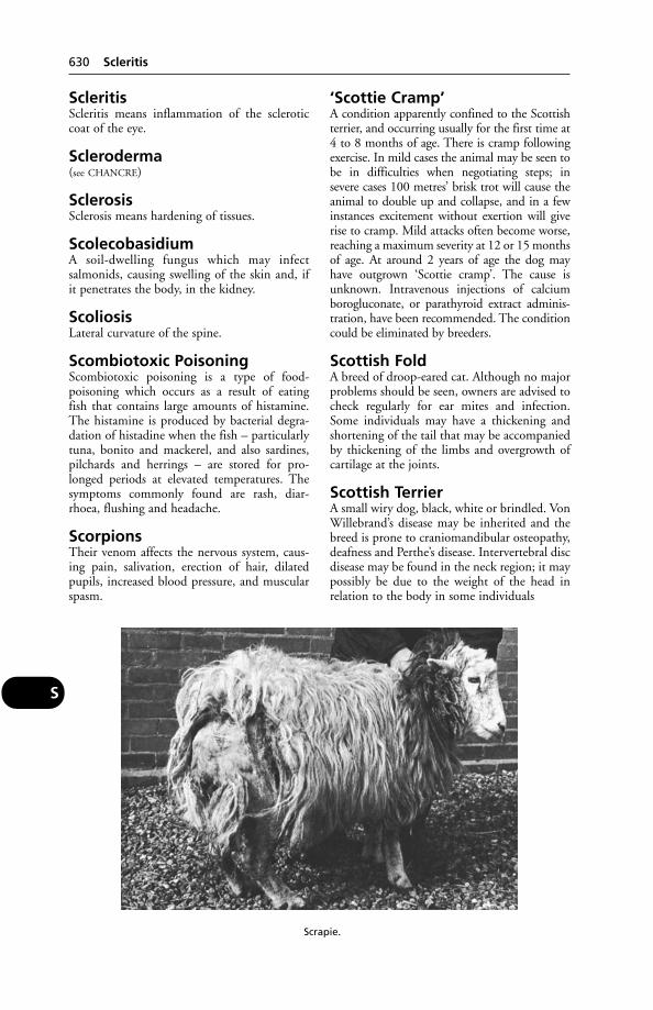
ScleritisScleritis means inflammation of the scleroticcoat of the eye.
Scleroderma(see CHANCRE)
SclerosisSclerosis means hardening of tissues.
ScolecobasidiumA soil-dwelling fungus which may infectsalmonids, causing swelling of the skin and, if it penetrates the body, in the kidney.
ScoliosisLateral curvature of the spine.
Scombiotoxic PoisoningScombiotoxic poisoning is a type of food-poisoning which occurs as a result of eating fish that contains large amounts of histamine.The histamine is produced by bacterial degra-dation of histadine when the fish – particularlytuna, bonito and mackerel, and also sardines,pilchards and herrings – are stored for pro-longed periods at elevated temperatures. Thesymptoms commonly found are rash, diar-rhoea, flushing and headache.
ScorpionsTheir venom affects the nervous system, caus-ing pain, salivation, erection of hair, dilatedpupils, increased blood pressure, and muscularspasm.
‘Scottie Cramp’A condition apparently confined to the Scottishterrier, and occurring usually for the first time at4 to 8 months of age. There is cramp followingexercise. In mild cases the animal may be seen tobe in difficulties when negotiating steps; insevere cases 100 metres’ brisk trot will cause theanimal to double up and collapse, and in a fewinstances excitement without exertion will giverise to cramp. Mild attacks often become worse,reaching a maximum severity at 12 or 15 monthsof age. At around 2 years of age the dog may have outgrown ‘Scottie cramp’. The cause isunknown. Intravenous injections of calciumborogluconate, or parathyroid extract adminis-tration, have been recommended. The conditioncould be eliminated by breeders.
Scottish FoldA breed of droop-eared cat. Although no majorproblems should be seen, owners are advised tocheck regularly for ear mites and infection.Some individuals may have a thickening andshortening of the tail that may be accompaniedby thickening of the limbs and overgrowth ofcartilage at the joints.
Scottish TerrierA small wiry dog, black, white or brindled. VonWillebrand’s disease may be inherited and thebreed is prone to craniomandibular osteopathy,deafness and Perthe’s disease. Intervertebral discdisease may be found in the neck region; it maypossibly be due to the weight of the head inrelation to the body in some individuals
630 Scleritis
S
Scrapie.
Scours, Scouring(see DIARRHOEA)
ScrapieScrapie is a disease of sheep mainly confined tothe district of the English and Scottish Borders,to Spain, France, and Germany. Sheep import-ed into Australia, New Zealand, Canada, andthe USA have brought the disease with them.Australia and New Zealand are believed to havequickly eradicated the disease. Scrapie is aNOTIFIABLE DISEASE in the UK, which operatesa compulsory slaughter policy for infectedsheep.
Scrapie, BSE and other ‘prion’ diseases It is possible that scrapie has a relationship with the human diseases kuru,Creutzfeldt-Jakob disease and Gerstmann-Straussler-Scheinker syndrome. Material fromscrapie-infected sheep, rendered and used indairy cattle concentrates, is believed to be theorigin of BSE in cattle. (See BOVINE SPONGIFORM
ENCEPHALOPATHY.)
Diagnosis One method is to detect scrapie-associated fibrillar protein (PrP) by means of arabbit-anti-sheep PrP polyclonal antibody byWestern blot analysis; but consistent resultshave not, it seems, been obtained.
Experimentally, scrapie has been transmittedto goats, mice, rats, and hamsters.
Cause An infective agent, possibly a prion.Research at the joint AFRC and MRC neu-ropathogenesis unit, Edinburgh, found thatcrude extracts of scrapie-infected brain con-tained accumulations of material known asscrapie-associated fibrils (SAF), which were alsofound in scrapie-like diseases.
Infection is spread from ewe to lamb and,possibly, by contact with fetal fluids. Signs takeabout 2 years to appear.
A long-term DEFRA research project isinvestigating the causes, disease process and epidemiology of scrapie, and the genetic factorsmaking some sheep more susceptible than others. The aim is eradication of the diseasefrom the UK. (See NATIONAL SCRAPIE PLAN.)
Signs The most striking and easily seen symp-tom of scrapie is the torn, ruffled, and untidyappearance of the fleece, and when very severe,the bruised or scratched condition of the skin.In many cases, especially those occurring dur-ing the late spring, the fleece may be almostentirely rubbed off against fences, posts andtrees or may be greatly removed by the mouth.
In addition, the condition of the sheep is note-worthy; whereas the remainder of the flock maybe in fair bodily condition, the scrapie sheep arethin, gaunt, and apt to become weak on theirlegs, lagging behind when going uphill, and losing their foothold when descending.Muscular tremors are often seen, and later thereis evidence of intense itching.
Occasionally, when startled – as, for instance,when being moved by dogs, or when a gun isfired near the unwary scrapie sheep – convul-sive seizures are seen, usually lasting from 3 to 5 minutes, and leaving the animal temporarilydazed.
Screw-Worm FliesThese include Chrysomyia bezziana in Australia,Cochliomyia hominivorax, C. Americana.
The screw-worm (C. hominivorax), a signifi-cant parasite of both humans and animals, hadnot been recorded outside the New World untilits accidental introduction into Libya, probablyin 1988–9. Hundreds of cases of wound myia-sis including many fatalities were recorded invarious species of domestic animals during1989 and 1990.
An international campaign to eradicate theAmerican screw-worm fly from North Africaappears to be succeeding, according to a bulletinfrom the organisers.
The campaign, begun in December 1990,involved the release of sterile male flies import-ed from Mexico. The flies were dispersed by airat densities of 500 to 1200 per km2 over an areaof 40,000 km2. More than 745 million flieshave been used and 40 million a week were dis-persed during the campaign. (See FLIES;MYIASIS; STRIKE.)
ScrotalRelating to the scrotum.
Scrub Typhus (Japanese RiverFever)A disease caused by Rickettsia tsutsugamushi,and transmitted by mites.
ScurA loose, horny growth, not attached to theskull, at the site normally occupied by a horn ina horned breed of cattle.
A bull calf with a scur, or with a bony protu-berance beneath the skin at the horn site, is not a pure polled animal. Without these, a bullcan be expected to breed true as regards the poll character; this can be checked by a proge-ny test of the bull mated to horned cows – theresult should be polled heifer calves or bull
Scur 631
S
calves with scurs or bony protuberances, but nocalves with horns.
Sea LiceFarmed Atlantic salmon, and sea trout, are subject to infestation by sea lice (Lepeophthirussalmonis and Caligus elongatus) with serious economic consequences. Treatment with para-siticides including hydrogen peroxide and synthetic pyrethroids such as cypermethrin iseffective, but must be repeated at intervalsdepending on circumstances.
SealA common marine mammal found around thecoasts of Britain and many parts of the world,favouring colder climates. True, or earless, seals are members of the family Phocidae. Theyare susceptible to infection by a morbillivirussimilar to that causing canine distemper. In polluted areas, they are liable to suffer mercurypoisoning.
Sealyham TerrierA short-legged breed with a wiry, often white,coat. Retinal dysplasia may be inherited and thebreed is prone to deafness and lens luxation.
Season(see OESTRUS)
Seat-Worm(see OXYURIS)
SeaweedA source of AGAR; a food grazed by sheep on theseashore, and sometimes fed to horses and cattle.A source of iodine and other trace elements and(in the case of brown seaweeds) of vitamins A,Bl, B2, C, and D. Animals do not take readily toseaweed as a rule, nor are they able to digest itwell at first, but after a few days it usually provesan acceptable supplement to the ration.
The ruminal microflora of sheep feedingalmost entirely on seaweed were devoid of cel-lulolytic bacteria and anaerobic bacteria whichare so numerous in sheep-grazing pasture.
Sebaceous GlandsSebaceous glands are found in the skin (see dia-
gram under SKIN), and secrete the oily sebumwhich prevents excessive dryness of hair andskin. The glands are liable to become invadedduring some parasitic diseases; sometimes ablocked duct leads to a retention cyst.
Seborrhoea is an excessively oily skin due toover-production by the sebaceous glands.
SecretinA hormone secreted by the mucous membranenear the beginning of the small intestine whenfood comes into contact with the latter. Onreaching the pancreas via the bloodstream, thehormone stimulates the flow of pancreaticjuice.
Secretory IgAIt has been shown that in some infections, espe-cially those of the respiratory and digestivetracts, immunity is conferred by antibodyfound in the local secretions – and not by the antibody circulating in the bloodstream.For example, the IgA found in secretions isquite different from that found in serum.Secretory IgA is relatively resistant to break-down by digestive enzymes and has an affinityfor mucus. (See IMMUNE RESPONSE, IgA.)
-SectomyA word-ending meaning ‘surgical removal of ’.
Seed Corn, dressedA number of substances with which seed cornmay be dressed may be toxic to animals. Forexample, corn with a mercury dressing has beenfed to pigs with fatal results.
Dieldrin seed dressings lead to poisoning inwild birds and, indirectly, have killed dogs, cats, and foxes which have eaten poisonedbirds. (See also under GAME BIRDS.)
Seedy ToeA condition affecting the hoof of the horse, inwhich there is a separation of the wall from thelaminar matrix below, and the formation in the space so produced of a dry, crumbly, friablevariety of horn, which bears some resemblanceto pumice-stone. It may occur at any part of thewall of the foot. The cause is uncertain.
Signs In most cases the condition is generallyfirst noticed by the farrier when paring downthe wall prior to fitting a new shoe. Lameness isonly seen when the extent of the separation islarge, or when foreign matter becomes forcedup into the space, and causes pressure upon thesensitive matrix.
When struck with a hammer the affectedpart of the foot gives out a hollow resonatingnote, and the margins of the separated area canusually be fairly well determined by this means.
Treatment All the soft friable horn should becleared away and an antibiotic applied within.A suitable shoe should be fitted to cover thebase of the cavity. (See HOOF REPAIR.)
632 Sea Lice
S

Selenium (SE)A TRACE ELEMENT essential in minute quanti-ties for nutrition, but toxic if fed in excess. Insome parts of Britain home-grown animal feedsmay not contain enough selenium, and unlessconcentrates are fed as well, nutritional muscu-lar dystrophy may result. The organic formconjugated to the amino acid methionine ismore easily utilised by animals than inorganicsalts. In other areas the soil may contain anexcess of selenium. The normal level in animalfeeds should be around 0.2 ppm; the maximumlevel of selenium allowed in pig diets without aveterinary prescription is 0.5 mg/kg, this levelbeing specified under the Feeding-StuffsRegulations 1982.
Supplements of selenium can be given notonly in the feed, but also in drinking water, bysubcutaneous injection, by BOLUS, and (forlambs) by an oral dose.
Sodium selenate is used by horticulturists asan insecticide, and accordingly there is a possi-bility of toxic effect occurring in animals.Sterility results, and also loss of hair. Thesesymptoms are also observed in parts of the USAand Eire where the soil contains an excess ofselenium. In the acute form of poisoning, ani-mals may be found wandering aimlessly or incircles. Paralysis precedes death.
A horse weighing approximately 450 kgreceived 25 mg selenium as sodium selenatedaily for 5 consecutive days. The horse becamelethargic, walked stiffly and was unwilling toundertake pace work. The main signs were lossof hair from the mane and tail, disintegration ofthe skin of the lips, anus, prepuce and scrotum,and separation of the hooves from the coronarycorium. There were strong correlations betweenthe selenium concentrations in blood, hair andhoof parings.
Externally, selenium sulphide is used in wet shampoos for dogs and cats infested withfleas, harvest mites, or cheyletiella mites. (See also
VITAMIN E; LATHYRISM; MUSCLES, DISEASES OF;IONOPHORES.)
Retention of placentas in a dairy herd in the north of England was associated with a selenium deficiency.
Sella TurcicaSella turcica is the name applied to the deephollow on the upper surface of the sphenoidbone in which the pituitary gland rests.
Semen (Seminal Fluid)Semen (seminal fluid) consists of the secretionsof the accessory sex glands, in which isfound the mature spermatozoa (or sperms)
from the epididymis. A single ejaculation by abull may produce semen containing millions ofsperms.
The secretions of the accessory sex glands actas a vehicle for the sperms, probably as a nutri-ent, and neutralise any acidity in the femalegenital passages.
The accessory glands are the prostate; the ampullae of the vasa deferens (absent in theboar); the seminal vesicles; and (except in thedog) the bulbo-urethral (Cowper’s) glands situ-ated on either side of the urethra. (See also under
SPERMATOZOA; ARTIFICIAL INSEMINATION.)
Imports of semen in Britain are subject toDEFRA regulations.
Seminal Vesicles(see under TESTICLE; ACTINOBACILLOSIS).
Sendai VirusThis causes respiratory disease in the mouse butis most noteworthy for its use in experimentalcell fusion work.
SenkoboCutaneous streptothricosis, caused byDermatophilius congolensis, occurring in tropicalAfrica in cattle, sheep, goats, and horses. Thehair stands erect and matted on small patchesalong the back. Moist, raw areas are left, thencrusts form, and eventually a ‘crocodile-skin’effect is produced. The disease occurs in associ-ation with tick infestation, and can therefore be controlled by means of an ectoparasiticide.(See DERMATOPHILUS.)
SennaA standardised preparation of this householdlaxative has been recommended in treating orpreventing constipation in pigs – especially inpregnant sows. A sublaxative dose of 3 g is recommended during the farrowing period.
Sensitisation(see ALLERGY)
Sepsis(see SUPPURATION; ANTISEPTICS)
SepticaemiaA condition in which toxic bacteria invade thebloodstream. It is very serious because the organ-isms and the toxins they produce become wide-ly distributed throughout the tissues, and practi-cally every organ is affected by them. In mostcases, septicaemia terminates in death. Examplesare ANTHRAX; HAEMORRHAGIC SEPTICAEMIA.
Septicaemia 633
S
Signs In many cases, especially when the ani-mal is in a weakened state, sudden death, pre-ceded by a very high temperature, may be theonly sign of the presence of septicaemia.
Treatment Antibiotics and/or sulfonamides,and antisera (where appropriate) are given.
SeptumA thin wall dividing 2 cavities or masses of tissue.
SequestrumA fragment of bone which, in the process ofnecrosis, has been cast off from the living boneand has died, but still remains in the tissues.
SequelaeSymptoms or effects which may follow diseaseor injury. Thus pneumonia may follow a simpleinfluenza, and chorea may follow distemper.
SeroconversionThe appearance in the blood serum of antibod-ies following vaccination (or natural exposureto some infective agent).
Serous MembranesSerous membranes are smooth, glistening,transparent membranes that line certain of thelarge cavities of the body and cover the organsthat are contained in them. The chief serousmembranes are: (1) the peritoneum, lining thecavity of the abdomen; (2) the pleurae, one ofwhich lines each side of the chest and surroundsthe corresponding lung; (3) the pericardium, inwhich the heart lies; (4) the tunica vaginalis,one on each side, enclosing a testicle; and (5)the mesentery supporting the small intestine.
SerpulinaA group of spirochaetes which includesSerpulina hyodysenteriae, the cause of swinedysentery. (See SWINE DYSENTERY; TREPONEMA.)
Sertoli-Cell TumourThis may be associated in the dog with femini-sation, urethral bleeding, and urinary obstruc-tion. (See also SPERMATIC CORD, TORSION OF.)
Sertoli CellsCells in the testicular tubules to which sper-matids become attached. Their function isbelieved to be the nourishment of spermatids.(See diagram under SPERMATOZOA.)
SerumSerum is the fluid which separates from blood when clotting takes place. It is, in effect,defibrinated plasma without the red cells,
platelets or white cells. (For a description ofplasma, see BLOOD.) (See also ANTISERUM.)
Serum Gonadotrophin(see HORMONES)
Serum SicknessIn human medicine this term is applied to thefever, glandular enlargements, oedema, and painin the joints, which may occur 8 to 12 days afterthe injection of a ‘foreign’ serum. Immediatereaction, denoting sensitisation by a previousinjection of the same kind of serum, is regardedas anaphylactic shock. (See HYPERSENSITIVITY.)
Serum Therapy(see ANTISERUM)
Service PeriodThis is usually taken to mean the intervalbetween giving birth and subsequent serviceleading to conception. In cattle, an 85-day ser-vice period would appear to be the optimumnumber of days between calving and successfulservice. If the trend of heat periods after calvingis detected at about 6 weeks, and checked againaround 9 weeks, the herdsman can, with a fairdegree of accuracy, be on the look-out for bullingat or about the 12th week (or 84 to 85 days). Inpractice, most farmers will serve at 9 weeks to tryto maintain a 365-day calving interval. Veryearly service may produce prolonged infertility.
SetaeStiff hairs. (See CATERPILLARS; SPIDERS.)
Sewage SludgeSewage sludge is often contaminated by heavymetals (cadmium, copper, lead) and should notbe used for manuring pasture unless these havebeen removed. The heavy metals are present asharmless sulphide complexes when first appliedto the soil: however, passage through earth-worms breaks down the complexes, which canthen be absorbed by animals. The process may take several years. Horses on pasture werefatally poisoned by cadmium, lead and copper5 years after sewage sludge was applied. Morerarely, the eggs of Taenia sagniata which passthrough the filters in some sewage may be present on pasture and lead to cysticercosis incattle. (See also SALMONELLOSIS; SLURRY; COPPER
POISONING in sheep.)
Sex DifferentiationSex differentiation in the fetus is brieflydescribed under EMBRYOLOGY. (See also under
GENETICS; CYTOGENETICS; FREEMARTIN.)
634 Septum
S
Sex-Hormones(see HORMONES)
Sex-InversionAnimals which at birth, and for a variable period afterwards, are of normal sexual struc-ture and function, but which later in lifeacquire properties of the opposite sex, are saidto undergo sex-inversion. This has been seen inAyrshire cows permanently kept indoors. (See
also FEMINISATION.)
Sex Pilus(see PLASMIDS)
Sex, Predetermined, of Calves(see PREDETERMINED SEX OF CALVES)
Sexual Cycle(see OESTRUS)
Shar-PeiA medium-sized breed of dog originally fromChina which is characterised by very loose,infolded skin. In the folds, dermatitis maydevelop if the care of the dog is less than good.
Shavings(see under BEDDING)
ShearingIn Britain, the usual time for shearing is May inthe southern counties, early June onupland semi-arable farms, and during July in mountain flocks.
The newly shorn sheep is very sensitive tocold. This is particularly so with machine shear-ing which leaves a fleece of about 6 mm depthcompared with about 12 mm after hand shear-ing. In Australia, late-winter and early-springshearing of ewes has led to a high mortality, sothat the practice is being abandoned or the usualshears replaced by ‘snow combs’ which leave alonger fleece. In Britain, losses of weight or poorgains in lambs shorn during the summer canlargely be attributed to an effect of cold.
Chemical ‘shearing’ Certain drugs, forexample cyclophosphamide and mimozone,cause the wool to loosen so that it can easily beplucked. It has been suggested that this couldbe an economical way of defleecing sheep.However, the sheep is left naked and unpro-tected against cold. It does not seem that thesystem has gained wide usage.
Sheep, Abortion and Infertility in(see ABORTION)
Sheep Breeding andManagementThe use of hybrids, referred to under SHEEP,BREEDS OF (British), is a relatively new trend.Another is the housing of ewes for part of the winter before lambing. The number ofsheep per flock has increased and a full-timeshepherd will be looking after more than10,000 breeding ewes.
Economic factors have dictated manychanges in traditional sheep management. Thesheep market has altered greatly; the demand ismainly for lamb, not mutton. However, sheepare still sold through markets on a liveweightbasis and are classed as light (25.5 kg), standard(32.1 to 39 kg), medium (39.1 to 45.5 kg) andheavy (45.6 to 52 kg). Wool is now of rela-tively low value, because of reduced demand.
Efficient sheep production depends on rais-ing the productivity of grassland by improvingthe quality and quantity of the grazing andimproving the growth characteristics of thesheep. Worming regimes must be established todeal with the infestations that affect all grass-land used for growing young sheep. Feed avail-able to the lactating ewe must be of sufficientquality to allow a good supply of milk for twinor triplet lambs, and limit initial grass uptake.
Direct and indirect feed costs (including fer-tilisers, fuel and labour), can account for 60 percent of the cost of sheep production. Thisemphasises the importance of the efficiency offeed conversion. In lowland flocks, most food isconsumed by the ewes, so the number of lambsweaned per ewe per year is of critical impor-tance. Ideally, the aim is for a relatively smallewe, with good milking potential, which can becrossed with a larger, meat-producing, breed toproduce several offspring that grow fast andeconomically.
While artificial rearing systems for lambs areused, they are not favoured by most breedersexcept for orphan lambs or those from large litters. Most farmers aim to produce 2 viablelambs that can be naturally reared.
Housing of sheep (In-wintering) Sheepare frequently housed for part of the year, usually before and after lambing. The durationof housing depends on the area and other management considerations. In Scotland, in-wintering of ewes lambing between Decemberand April revives a practice traditional in hillareas until a change was made to winteringthem on lowland pastures. That policy provedtoo costly and led to the flocks being kept on the farms and provided with shelter. Winterhousing is also now common on lowland
Sheep Breeding and Management 635
S

farms. The ewes are kept in groups of 50 to 70,lambing at the same time.
Housing permits greater attention to feedingand care at lambing, and leads to less culling as the sheep are not subjected to the stress ofexposure to severe weather and finding theirown food. However, the risk of disease and mismothering is higher.
Portable feeding troughs and racks in sheephouses can be used as partitions and leave anunobstructed floor so that pen sizes can bealtered according to requirements. The 2 maintypes of feeder are a hay rack, with sloping sidesso that seeds do not drop into the sheep’s eyes,with a concentrate trough below (preferred forupland sheep); and a box type with a barrier infront which prevents the ewes from wasting theforage. Troughs should allow a length per eweof 23 to 25 cm (9 to 10 in) for hill breeds and30 to 35 cm (12 to 14 in) for lowland breeds. Asupply of fresh (i.e. running) water must beprovided; sheep will not drink water that iseven slightly fouled, or warm. A raised troughfed from a slow running tap, with drainage tothe outside, is suitable. About 30 cm (12 in) oftrough per sheep should suffice for 40 sheep.
Adequate ventilation is essential. Yorkshireboarding to walls is suitable. If slatted flooringis used, care must be taken that updrafts do notchill sheep and, especially, lambs. The slatsmust be laid parallel to the door openings.
Lamb survival Lambs have the highest post-natal mortality of all the main farm species.This is partly because they are very susceptibleto hypothermia caused by exposure, or lack offood. Problems arise when the environment iscolder than the critical temperature of the lambin the first few hours after birth. This is 32°C(89.5°F) for heavy lambs and 37°C (98.5°F)for light lambs. As most lambs are born atambient temperatures well below this, theyhave to increase their metabolic rate to main-tain body temperature. Wind chill factor canreduce the effective temperature considerably. Awind speed of 20 km/h (12 mph) can have acooling effect of up to 20°C (68°F) or more ifthere is rain. Even when a lamb survives suchconditions, it will have suffered a major drainon its bodily reserves, mainly in the form of fat.In bad conditions these may be used up inbetween 5 and 17 hours.
While a lamb will normally begin to replen-ish its store of energy within an hour of birth bysucking, work in Australia has shown that theurge to do so is reduced if its body temperaturefalls below 37°C. In some breeds of sheep, suchtemperatures occur in weather not unusually
severe for March and April in Scotland. So coldnot only increases the demand for energy, butmay prevent that demand from being met.
Starvation exaggerates the effect of cold byreducing heat-production capability, so increas-ing the risk of death from hypothermia.
It may be possible to breed for greater ablityto survive under harsh conditions. Experimentssuggest that there are significant differencesbetween breeds in their tolerance of body cool-ing; and within breeds, some individual sheephave a cold-resistance several times greater thanthat of other individuals. Preliminary trials haveindicated that this character is moderately wellinherited.
Lambs require between 180 and 210 ml ofcolostrum per kg bodyweight during the first18 hours after birth, to provide sufficient fuelfor heat production; and immunoglobulins forprotection against infections.
Ewes which are well fed during late pregnan-cy produce more colostrum than their lambsneed; those with singletons have enough for a2nd lamb. By contrast, most underfed ewes donot produce enough colostrum.
Colostrum can be readily obtained by handmilking and stored for subsequent use. Yields aremarkedly increased when milking is preceded byan oxytocin injection.
Life-saving techniques on the farm. Thefollowing recommendations have been made bythe Moredun Institute, Edinburgh. Two dangerperiods should be recognised: (1) from birth to5 hours afterwards; and (2) 10 hours to 3 daysafter birth.
During the 1st period, moderate hypother-mia (a body temperature of 37° to 39°C; 98.5°to 102°F) usually responds to drying the lamb,feeding it colostrum by stomach tube, andmoving it to shelter along with the ewe. Serioushypothermia (below 37°C; 98.5°F) requires inaddition that the lamb be warmed in air at37°C (98.5°F) to 40°C (104°F) until its bodytemperature has reached 37°C (98.5°F). Whenremoved from the Moredun-type bale-warmer(heated by a domestic fan-heater), the lamb isthen given colostrum and, if strong enough to suck vigorously, can be reunited with theewe. If not strong enough, the lamb must behoused for a day or two in its own cardboardbox in an intensive care unit. There colostrumis given 3 times daily, and warmth provided byan overhead infra-red lamp.
During the 2nd danger period, when serioushypothermia is then usually due to depressedheat production as a result of starvation, andoften complicated by low glucose levels in the
636 Sheep Breeding and Management
S
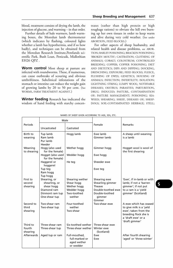
blood, treatment consists of drying the lamb, theinjection of glucose, and warming – in that order.
Further details of bale warmers, lamb warm-ing boxes, the Moredun lamb thermometer(which indicates by flashing, coloured lightswhether a lamb has hypothermia, and if so howbadly), and techniques can be obtained fromthe Moredun Research Institute,Pentlands sci-entific Park, Bush Loan, Penicuik, MidlothianEH26 QPZ .
Worm control Most sheep at pasture areinfected with roundworms. These, if numerous,can cause outbreaks of scouring and obviousunthriftiness. Subclinical infestations of thestomach or intestine can reduce the weight gainof growing lambs by 20 to 50 per cent. (See
WORMS, FARM TREATMENT AGAINST.)
Winter feeding Research has indicated thewisdom of hand feeding with starchy concen-
trates (rather than high protein or highroughage rations) to obviate the hill ewe burn-ing up her own tissues in order to keep warmand alive during very cold weather. (See under
ABORTION, FEED BLOCKS.)For other aspects of sheep husbandry, and
related health and disease problems, see ABOR-
TION; BARLEY POISONING; BRACKEN POISONING;‘BROKEN MOUTH’; CASTRATION; CLOTHING OF
ANIMALS; COBALT; COLOSTRUM; CONTROLLED
BREEDING; COPPER; COPPER POISONING; DIET
AND DIETETICS; DIPS AND DIPPING; DOCKING,DRENCHING; EXPOSURE; FEED BLOCKS; FLEECE,FLUSHING OF EWES; GENETICS; HOUSING OF
ANIMALS; INFECTION; INFERTILITY; ISOLATION;LIGHTNING STRING; LUMPY WOOL; NOTIFIABLE
DISEASES; OESTRUS; PARASITES; PARTURITION,DRUG- INDUCED; PASTURE, CONTAMINATION
OF; PASTURE MANAGEMENT; POISONING; SEA-
WEED; SHEARING; SHEEP, DISEASES OF; SHEEP-
DOGS; SOIL-CONTAMINATED HERBAGE; STELL;
Sheep Breeding and Management 637
S
NAMES OF SHEEP GIVEN ACCORDING TO AGE, SEX, ETC.
Male
Periods Female RemarksUncastrated Castrated
Birth to Tup lamb Hogg lamb Ewe lamb A sheep until weaningweaning Ram lamb Gimmer lamb is a lamb
Pur lambHeeder
Weaning Hogg (also used Wether hogg Gimmer hogg Hogget wool is wool ofto shearing for the female) the first shearing
Hogget (also used Wedder hogg Ewe hoggfor the female)
Haggerel or He teg Sheeder ewehoggerel
Tup teg Ewe tegRam hoggTup hogg
First to Shearing, or Shearing wether Shearing ewe ‘Ewe’, if in-lamb or withsecond shearling, or Shear hogg Shearling gimmer lamb; if not a ‘barrenshearing shear hogg Wether hogg Theave gimmer’; if not put
Diamond ram Wedder hogg Double-toothed ewe to a ram is a ‘yieldDinmont ram tup Two-toothed Double-toothed gimmer’ (Scotland)One-shear tup wether gimmer
GimmerSecond to Two-shear ram Four-toothed Two-shear ewe A ewe which has ceasedthird Two-shear tup wether to give milk is a ‘yeldshearing Two-shear ewe’; taken from the
breeding flock she isa ‘draft ewe’ or a‘draft gimmer’
Third to Three-shear ram Six-toothed wether Three-shear ewefourth Three-shear tup Three-shear wether Winter eweshearing (Scotland)Afterwards Aged tup or ram Full-mouthed, Ewe After fourth shearing
full-marked or Ewe ‘aged’ or ‘three-winter’aged wetheror wedder

STOCKING RATES; STRESS; TRACE ELEMENTS;TROPICS; VAGINA (for rupture of ); VITAMINS;WATER; WEANING; WOOL BALLS; WORMS, FARM
TREATMENT AGAINST.
Clipping (see SHEARING; CLOTHING FOR
ANIMALS; WOOL SLIP)
Sheep, Breeds of
Introduction Sheep are maintained, general-ly speaking, with the object of producing bothwool and meat. In some countries’ ewe’s milk isvalued for cheese-making. In the UK the impor-tance of the fleece tends to be disregarded.
Hardiness, prolificacy, milking capacity ofthe females, and activity are all important.What will constitute the most profitable typemust be carefully considered in relation to localconditions.
British breeds of sheep British breeds –some 40 are registered – offer a wide choice oftypes, adapted to almost every conceivable set ofconditions under which sheep are maintained inthe country, from the highest mountain grazingsin Scotland and Wales to the richest lowlandpastures, or the dry arable farms of the Wolds.However, crossbreeds are increasingly popular.
Cambridge This breed was developed at theUniversity of Cambridge by Professor JohnOwen in collaboration with Alun Davies. Thebreed is now regarded as one of the most pro-lific in the world with litter sizes of 1.7, 2.5 and2.9 for 1-, 2- and 3-year-old females respective-ly. Both sexes are polled, ewes weighing 70 kg(154 lb) and rams 90 kg (98 lb). (See also TEXEL;COOPWORTH.)
The British breeds are commonly classifiedas Longwools, Downs, other Shortwools, andMountain breeds.
Longwool breeds include Leicester, BorderLeicester, Lincoln, Wensleydale, Kent orRomney Marsh, Devon Longwool, SouthDevon, and Roscommon.
Downs breeds include Southdown, Suffolk,Hampshire, Dorset Down, Shropshire, andOxford.
Other Shortwool breeds include DorsetHorn, Wiltshire Horn, Ryeland, DevonClosewool, and Kerry Hill.
Mountain breeds include Scottish Blackface,Cheviot, Swaledale, Herdwick, Lonk, WelshMountain, Exmoor, and Dartmoor.
UK sheep population A rapid rise insheep numbers followed the imposition of dairymilk quotas, plus the granting of various subsidy payments. At December 1994, MAFFrecorded 29.5 million of which 20.1 millionrepresented ewes in the breeding flock. Thesummer census annually lists the population at 35 to 40 million. However, the virtual collapse of the sheep market in the later 1990sled to a drop in numbers. A further fall fol-lowed the foot-and-mourh diseases outbreaksof 2001, since when restocking has revivednumbers.
Sheep Dipping(see DIPS AND DIPPING)
Sheep, Diseases of(see under ABORTION; ACTINOBACILLOSIS;ANTHRAX; ARTHRITIS; BALANITIS; BLACK DIS-
EASE; BLACK-QUARTER; BLOUWILDEBEESOOG;BLUE TONGUE; BORDER DISEASE; BRAXY; ‘CAPPIE’;CASEOUS LYMPHADENITIS; ENTEQUE SECO; EYE
DISEASES OF; FOOT-AND-MOUTH DISEASE; FOOT
ROT; GAS GANGRENE; HYPOMAGNESAEMIA;JAAGSIEKTE; JOHNE’S DISEASE; JOINT-ILL; LAMB
DYSENTERY; LIVER-FLUKE; LOUPING-ILL; MILK
FEVER; MOREL’S DISEASE; OVINE EPIDIDIMYTIS;OVINE INTERDIGITAL DERMATITIS; NEMA-
TODIRUS; PARASITES; ‘PINING’; PNEUMONIA IN
SHEEP; PREGNANCY TOXAEMIA; PULPY KIDNEY;‘REDFOOT’; ‘RINGWOMB’; SCALD; SCRAPIE; SHEEP
SCAB; SWAYBACK; TICKS; TICK-BORNE FEVER OF
SHEEP; TOXOPLASMOSIS; UDDER, DISEASES OF;WATERY MOUTH; WESSELSBRON DISEASE; also ARI-
ZONA INFECTION; ENZOOTIC OVINE ABORTION;HAEMORRHAGIC SEPTICAEMIA; MAEDI/VISNA;‘MILKSPOT LIVER’; PULMONARY ADENOMATOSIS;RIFT VALLEY FEVER; ULCERATIVE DERMATOSIS and
under RAM.)
Sheep Health SchemeA preventive medicine and productivity moni-toring scheme for maintaining herd health; it isoperated by Scottish agricultural colleges. APremium Health scheme is operated for flocksseeking accreditation as free from chlamydialabortion. A maedi/visna accreditation scheme is also available; it involves certification by aveterinary surgeon that movement records havebeen checked, flock security rules obeyed andblood samples taken.
Sheep Ked (MelophagusOvinus)Sheep ked (Melophagus Ovinus) is a winglessblood-sucking parasite. (See KED.)
638 Sheep, Breeds of
S
Sheep, Legislation AffectingAgriculture (Miscellaneous Provisions) Act
1968Animal and Animal Products (Import &
Export) Regulations 1998Animal Health Act 1981Anthrax Order 1991Artificial Breeding of Sheep & Goats
Regulations 1993Diseases of Animals (Approved Disinfectants)
(Amendment) Order 1997Foot and Mouth Order 1983Fresh Meat (Hygiene & Inspection)
Regulations 1995Market Sales and Lairage Order 1925Products of Animal Origin (Import & Export)
Regulations 1992Protection of Animals Act 1911Protection of Animals (Anaesthetics) Act 1954
(amended 1982)Sheep and Goats Spongiform Encephalopathy
Order 1998Sheep and Goats Spongiform Encephalopathy
Regulations 1998Sheep Scab Order 1997Specified Diseases (Notification & Slaughter)
Order 1991 (amended 1992)Veterinary Surgeons Act 1966Welfare of Animals at Markets Order 1990
(amended 1993)Welfare of Animals (Transport) Order 1997Welfare of Livestock (Prohibited Operations)Regulations 1982, 1987Welfare of Livestock Regulations 1994Zoonoses Order 1989
Sheep, Names Given Accordingto Age, Sex, etc.There are probably more names for any givenclass of sheep than is the case among any of theother domesticated animals, and it is almostimpossible to give a list that will include all thevarious designations that are used, but the tablegives a list of commoner terms.
Sheep Pox(see POX)
Sheep ScabThe popular name for psoroptic mange. Sheepscab was formerly a notifiable disease in theUK, from where it was eradicated in 1952. Itreappeared in 1973 and, following the abolitionof compulsory dipping, has again become aserious problem. In the late 1990s, as many as25 per cent of sheep pelts showed evidence ofdamage associated with scab. Although sheepscab is no longer a notifiable disease, control is
exercised through local authorities. It is anoffence under the Protection of Animals Actsand the Welfare of Animals (Northern Ireland)Act 1972 to expose an infected sheep for sale;the charge would be one of causing unecessarysuffering.(see MANGE – Mange in sheep).
SheepdogsSheepdogs are popularly regarded as exception-ally healthy, but a survey in Scotland showedthat at least 11 per cent were suffering from‘BLACK TONGUE’ as a result of an inadequatediet. On average, this consisted basically of 225g (8 oz) oatmeal, 225 g (8 oz) maize, and (by nomeans always) 225 ml (8 oz) of milk; the first 2ingredients being made into a brose or mash bypouring on boiling water. The occasional rab-bit, or piece of boiled mutton from a deadsheep, or – at lambing time – the afterbirths,were not sufficient to prevent ‘black tongue’.
Sheepdogs may walk or run 90 miles per dayat lambing time and must have meat if staminaand health are to be maintained. Even fishmealis of service – also dried blood – if meat or fishare unobtainable. (See also under GID; RICKETS.)
Sheepdogs may become infected with brucel-losis as a result of eating infected cattle after-births; through eating dead sheep they maybecome infested with the tapeworm causingHYDATID disease. Regular worming is essential.(See also ORF; ANTHRAX; BOTULISM.)
Sheep, Winter Coats for(see CLOTHING OF ANIMALS)
Shelters, Need for(see under EXPOSURE, TROPICS; also STELL)
ShepherdsOccupational hazards include the following diseases: CAMPYLOBACTER INFECTIONS;CHLAMYDIA; HYDATID DISEASE; LISTERIOSIS;LOUPING-ILL; ORF; PASTEURELLOSIS; Q FEVER;SALMONELLOSIS; TOXOPLASMOSIS. (See also
ZOONOSES.)Shepherdesses, if pregnant, are at risk when
helping with lambing. (See CHLAMYDIA.)
Shetland SheepdogA small breed resembling a toy rough collie.Progressive retinal atrophy and distichiasis areinherited dominat traits and collie eye anomalyand patellas luxation are recessive traits.Haemophlia and deafness may also be found.
ShigellosisInfection by one of the Gram-negative Shigellabacteria. (See Sleepy foal under FOALS, DISEASES OF
– Septicaemia.)
Shigellosis 639
S
Shih TzuA small dog with short muzzle and long flowinghair; it originates from Tibet. The breed is proneto cleft palate and interverbral disc disease.
‘Shipping Fever’A disease of cattle caused by a virus and/orPasteurella multocida or P. haemolytica.‘Shipping fever’ is very common in Americanfeedlots, among cattle 6 months to 2 years old, and often follows the stress of transport,castration, de-horning, winter weather, changeof food, etc. In the USA the term ‘bovine respiratory disease complex’ is a synonym. (See
PASTEURELLA.)
Signs include fever, loss of appetite, weakness,followed by nasal discharge, a discharge fromthe eyes, distressed breathing, coughing, andsigns indicating bronchopneumonia. Mortalityis usually 1 to 2 per cent, but may exceed this if cases are neglected .
Treatment And Prevention Antibioticsand sulfa drugs are used. Immunisation hasbeen tried using myxovirus parainfluenza-3 andP. septica, for example.
‘Shivering’A nervous disease of horses. It runs a slowly progressive course, and constitutes anunsoundness.
Cause This is unknown, though it seems thatthere may be a hereditary predisposition to it.
Signs In a well-marked case, the muscles of thehindquarters are seen to quiver or tremble. Atthe same time, the tail is usually elevated andalso shows the quivering movements. Inadvanced cases it may be difficult or impossibleto pick up either of the hind-feet, and shoeingis only accomplished with difficulty. When thehind-limb is raised from the ground duringbacking, in many cases it also quivers, or ‘shiv-ers’, and in some instances one or both of thefore-limbs, or the muscles of the fore-quarter,exhibit the same feature.
Shivering in the dog may occur, especial-ly in fox terriers, for no apparent reason andmay be unconnected with either cold or fear. Atthe prospect of a walk the dog may suddenlycease trembling.
ShockShock is, clinically, an abrupt fall in blood pressure (acute hypotension).
Signs include weakness, pale and cold mucousmembranes, subnormal temperature but noshivering; a weak and rapid pulse; shallowbreathing at an increased rate; cold extremities.
Cause Shock may follow severe trauma, haem-orrhage, surgical operations, a sudden decreasein the heart’s pumping capacity, burns andscalds, toxaemia. (See also ELECTRIC SHOCK; ANA-
PHYLACTIC SHOCK.) Pain, fright, and any air-way obstruction may exacerbate the condition.
Treatment Although corticosteroids are oftenused, it has been stated that there is little or noevidence that they are effective. A blood trans-fusion, adrenalin, plasma substitutes based ongelatin and dextrans, and lactated Ringer’s solu-tion given intravenously, may each have a placein treatment, as appropriate.
The patient must be rested and kept warm.
Shoeing of CattleThis may be undertaken to reduce weight-bearing on an injured claw, especially wherethere is a fracture of a phalanx. Draught cattlemay also be shoed.
Shotgun Injuries(see GUNSHOT INJURIES)
ShoulderShoulder is the joint formed between the scapula and the upper end of the humerus. (See
DISLOCATION.)
Shoulder-Blade(see SCAPULA)
Shovel BeakA disease occurring in intensively reared chicksfed dry mash. It affects usually birds of 2 to 8 weeks old. The upper or lower beak (or both) may be deformed, with ulceration ornecrosis. Infection with Fusiformis necrophorus,Staphylococcus aureus, or Clostridium welchi mayfollow.
Shying(see ‘VICES’)
SI UnitsThe Système International d’Unités was adoptedby the Conférence Générale des Poids et Mesuresin 1960. Based on 7 units – metre, kilogram,second, ampere, degree kelvin, candela, andmole – it admits only 1 unit for any 1 physicalquantity. Derived units in any science or tech-nology can be made up from the 7 basic units
640 Shih Tzu
S

by division or multiplication without numericalfactors being involved. The SI unit joulesreplaces calories. The 7 basic SI units and theirsymbols are as follows:
(length) metre m(mass) kilogram kg(time) second s(electric current) ampere A(thermodynamic degree kelvin °Ktemperature)(luminous intensity) candela cd(amount of substance) mole mol
(See under METABOLISABLE ENERGY.)
SialocoelesCyst-like swellings, usually lined by granulationtissue rather than epithelium, containing saliva.
SialogoguesSialogogues are substances which produce acopious flow of saliva, e.g. pilocarpine andarecoline.
Sickness(see VOMITING)
SidebonesOssification of the LATERAL CARTILAGES of thehorse’s foot. When this occurs in a young ani-mal, it is looked upon as an unsoundness. Inold horses, all cartilages, not only in the foot,tend to become ossified as an almost naturalcourse of events, and sidebones are accordinglynot looked upon as so serious.
Causes Heredity is considered as a predispos-ing cause, but in many instances no such rela-tionship can be shown. It has been suggestedthat a vitamin D deficiency in foalhood may bepartly responsible.
Signs Ordinarily, the upper part of each carti-lage can be felt at the coronet as a flexible ridgeor edge, lying immediately below the skin, butwhen the cartilage has ossified, this ridge is nolonger flexible, and is more or less thickened aswell. In some instances the ossified cartilage canbe easily seen when the feet are viewed from thefront. The condition is more common by far inthe fore-limbs, and may occur on the outside orinside, or in both places, on one or both of thefore-feet.
When sidebones have formed, there is nolameness, pain, heat, or other signs of inflam-mation, but when forming, there may be painover the quarters involved, and lameness –
characterised by the taking of a shorter step bythe affected foot, and the tendency to do thismay result in a peculiar short and long step.
Treatment As a rule, in horses with wideopen feet and well-developed frogs, no treat-ment is required. The sidebone does not inter-fere with slow work of a regular nature. (See also
RING-BONES.)
Side-EffectsThe side-effects of a drug are those produced inaddition to that for which purpose the drug isgiven. Examples: deafness in humans followingthe administration of streptomycin; moniliasisafter the use of chlortetracycline; aplasticanaemia after the use of chloramphenicol. (See
also IATROGENIC DISEASE.)
Silage (Ensilage)Silage (ensilage) is a succulent food. It has beenclassified as follows: Grade I containing 15 percent and over crude protein, and made fromyoung grasses, none in flower, clover, lucerne,or sainfoin in bud stage; Grade II containing 12 to 14.9 per cent crude protein, and madefrom grasses in their flowering stage, lateautumn grass, clover passed full flower, marrowstem kale, pea pods, cereal-legume crops cutwhen cereal is ‘milky’; and Grade III containingless than 12 per cent crude protein, and madefrom seeding grasses, stemmy clover, maize, peahaulm and pods, sugar-beet tops, potatoes.
Grade I makes a substitute for cake, whereasGrade III is good enough only as a substitutefor roots, straw, or low-grade hay.
Ensilage involves fermentation. Lactic, aceticand butyric acids are produced: in good silage,lactic acid predominates. Silage with a highbutyric-acid content must be fed with caution,and may be recognised by its to unpleasantsmell and lighter colour – yellowish-greeninstead of dark brown.
The Dorset wedge system of silage-makinghas enabled better quality to be achieved.
ADAS comments: ‘Most farmers still makesilage at the wrong time.’ This criticism refers tonot cutting at the optimum stage of growth buttending to delay until there is more to cut.Wilting, judicious choice of harvester, type ofsilo, sealing, consolidation, and use of additivesare all being applied by the more progressivefarmers.
As with hay, there are extremes of quality in silage. At the Rowett Research Institute cows have maintained a yield of up to 23 litres(5 gallons) daily for 2 months while receivingno other feed. With the average quality silage it
Silage (Ensilage) 641
S
would, however, be unrealistic to expect to beable to dispense with supplements.
Acetonaemia is often seen in cattle receivinglarge quantities of silage of low quality. Hayshould be made available as well; also 55 g (2 oz) per head of bone flour with salt added.
When self-feeding of silage is practised, caremust be taken that conditions underfoot do notbecome dirty and slushy to an extent wheresoftening of the horn of the hoof occurs andfoot troubles develop.
Silage must be free of ragwort. (See under
RAGWORT POISONING; also RETICULITIS,TRAUMATIC.)
Listeria in silage LISTERIOSIS in ruminantshas often been associated with silage feeding. In a survey carried out in Scotland, Listeriamonocytogenes was isolated from 2.5 and 5.9 percent of samples of clamp silage obtained in twosuccessive years.
Silage EffluentSilage effluent has been described as one of thestrongest of all agricultural wastes and pollu-tants. Some 16,000 litres (3500 gallons) of cleanwater are needed to dilute 4.5 litres (1 gallon) ofsilage effluent to bring it to the recommendedlevel for treated (‘safe’) effluents.
In a wet season a 400,000-kg (400-ton)silage clamp with grass at 1 to 15 per cent drymatter at ensiling may produce 182,000 litres(40,000 gallons) of effluent, most of which discharges in the first month. It takes only a little of this effluent to kill fish and other formsof aquatic life if it reaches a stream.
‘Silent Heat’(see under OESTRUS DETECTION; INFERTILITY)
Silica ContaminationSilica contamination can be a problem withsugar-beet tops and other arable residues. Crops windrowed and then picked up may contain up to 30 per cent silica. Direct load-ing might keep the figure down to 10 per cent.(See also SOIL-CONTAMINATED HERBAGE, SAND,COLIC.) Silica is silicon dioxide, present in sand.
SiliconA non-metallic element. In the form of silicicacid or its derivatives, silicon is essential for growth, and is found mainly in connectivetissue. It has been suggested that lack of sufficient silicon may be a factor in the cause of atherosclerosis in man. (See also SILICA
CONTAMINATION.)
Silicone implant repair A year-oldArabian filly had a depression over its rightfrontal sinus as the result of an injury sustained6 months earlier when it ran into a steel pipeand the frontal bone had been broken. A heat-vulcanised silicone implant was used to repairthe deformity and the normal facial contourwas restored by suturing the sculpted implantto the periosteum over the defect.
Silicone SolutionAn anticoagulation solution used in connectionwith blood transfusion apparatus and syringesto prevent clotting.
Simian Haemorrhagic Fever(see MONKEYS, DISEASES OF; EBOLA VIRUS)
SimmentalA dual-purpose breed of Swiss cattle, now to befound throughout Europe and in the USA. InGermany the Simmental has been developedwith emphasis on beef production.
Simulium(see under FLIES). In the UK, the gnat S. ornatum,which breeds in running water and is difficultto control, sometimes causes eye lesions in cattle.
SinusSinus is a term applied to narrow hollow cavi-ties (especially in bones) occurring naturally in the body, or produced as the consequence of disease. (See SINUSES OF THE SKULL; also
FISTULA.)In pathology, sinus refers to a blind infected
tract, leading from a site of suppuration to thesurface of the skin or of a mucous membrane.
Sinuses, Diseases of theThe sinuses of the head are lined with a mem-brane which is continuous with that of thenasal cavities, and which acts as a periosteal covering for the bone.
Causes Sinusitis may arise as a result of aspread of inflammation from that affecting the nasal mucous membrane. It may followstrangles in the horse; occasionally the cause isa diseased tooth, the root of which has suppu-rated and the pus burrowed through the thinplate of bone that separates the tooth socketfrom the sinus cavity. In other cases, the causeis a penetrating injury from the outside, such asis occasioned by a blow on the forehead whichfractures the external plate of bone and allowsthe ingress of infection. Animals living near the
642 Silage Effluent
S
seashore or in sandy and windy localities aresometimes afflicted with collections of fine sandin the sinuses. In sheep, and sometimes in thehorse, sheep-nostril fly larvae of the Oestrusfamily may be found in the sinuses, and aregenerally associated with pus formation. Indogs especially, but also in other animals,tumour formation is often accompanied by thepresence of pus in the sinuses, and the condi-tion may be complicated by a FUNGAL infec-tion. In the dog, a foreign body such as a grassseed may give rise to the discharge from onenostril which is characteristic. Either cancer, ora fungal infection which may follow, can lead todistortion of the dog’s or cat’s face. (See also
MITES linguatula serrate LEECHES.)
Signs The most prominent sign of the pres-ence of any amount of pus in the sinuses is the usually slight, but continual, dribbling ofdischarge from one or both nostrils. This discharge is usually more marked when the animal lowers its head.
Treatment This consists of opening, underanaesthesia, the diseased sinus by trephining thebone over the surface, and irrigation and evacu-ation of the cavity. When a tooth has been theprimary cause of the condition it is extracted,and its cavity temporarily plugged with gauzeuntil healthy tissue fills up the space betweenthe tooth socket and the sinus. Parasitic inhab-itants are removed, either by the injection offluids that will kill them, or by picking each outseparately with forceps. Chloroforming the animal will often kill such parasites.
Sinuses of the Skull,The sinuses of the skull, also called theparanasal sinuses, are directly or indirectly con-nected with the nose. There are 4 pairs: (1)maxillary; (2) frontal; (3) spheno-palatine, orsphenoid; and (4) ethmoidal.
SinusitisInflammation of the sinuses.
Sinusitis, InfectiousA disease of turkeys, poultry and pheasantscaused by Mycoplasma gallisepticum. The obvi-ous sign is a swelling of the sinuses below theeyes, but the disease will also be present in thelower respiratory tract. While local treatment(e.g. draining the sinuses) is helpful, generallyadministration of antibiotics is necessary. Thedisease is egg-transmitted and may spread fromegg to egg in the hatcher. Infected birds alwaysremain carriers. Eradication has been carried
out in the commercial breeding stock of majorsuppliers.
Swollen sinuses may also be seen in certainparamyxovirus infections, from which infectioussinusitis must be differentiated.
Sire Identification(see DNA – ‘Fingerprinting’)
Sitfasts(see SADDLE-SORES)
Skim MilkThis is a valuable food, retaining, as it does, thesolids-not-fat after most of the fat has beenremoved. These solids include the valuablemilk-protein, the sugar lactose, valuable miner-als, and vitamins of the B group. It is poor inthe fat-soluble vitamins A and D, and also in vitamin E; and if given along with cod-liveroil to beef stores, may lead to cod-liver oil poisoning or MUSCULAR DYSTROPHY.
Skim milk is a useful food for pigs, but is notsuitable on its own. It can be fed ad lib to suck-ling pigs; weaners may receive 3 litres (5 pints)per day; fatteners from 14 weeks to slaughter,about 3.5 litres (6 pints).
Skim milk is, if from infected cattle, a sourceof tuberculosis in pigs, and pasteurisation maybe desirable in many countries.
For sows and piglets, skim milk should befresh or completely sour; 0.1 per cent formalinis sometimes added to skim milk for fatteningpigs.
SkinSkin, the protective covering of the body, iscontinuous at the natural openings with themucous membranes. It consists of 2 main lay-ers, which differ in structure and origin.
The epidermis This is a cellular layer ofnon-vascular, stratified epithelium of varyingthickness, covering the outer surface of thebody, which presents the openings of the cuta-neous glands and of the hair follicles. In ani-mals it is divisible into 2 layers, the outer, hard,dry stratum corneum, and the deeper, softer,moist stratum germinativum. The cells of thelatter are pigmented, and by their growth com-pensate for the loss by exfoliation or sheddingof the surface cells from the stratum corneum,which forms the scurfy deposit upon anungroomed horse. This inner layer consists ofthe part of the skin which is living, and isformed by several layers of cells set upon thecorium and nourished by it. The cells continu-ally multiply, and are slowly pushed upwards to
Skin 643
S
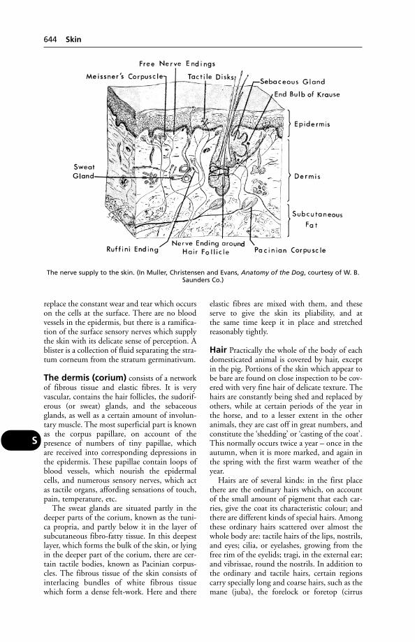
replace the constant wear and tear which occurson the cells at the surface. There are no bloodvessels in the epidermis, but there is a ramifica-tion of the surface sensory nerves which supplythe skin with its delicate sense of perception. Ablister is a collection of fluid separating the stra-tum corneum from the stratum germinativum.
The dermis (corium) consists of a networkof fibrous tissue and elastic fibres. It is very vascular, contains the hair follicles, the sudorif-erous (or sweat) glands, and the sebaceousglands, as well as a certain amount of involun-tary muscle. The most superficial part is knownas the corpus papillare, on account of the presence of numbers of tiny papillae, which are received into corresponding depressions inthe epidermis. These papillae contain loops ofblood vessels, which nourish the epidermalcells, and numerous sensory nerves, which actas tactile organs, affording sensations of touch,pain, temperature, etc.
The sweat glands are situated partly in thedeeper parts of the corium, known as the tuni-ca propria, and partly below it in the layer ofsubcutaneous fibro-fatty tissue. In this deepestlayer, which forms the bulk of the skin, or lyingin the deeper part of the corium, there are cer-tain tactile bodies, known as Pacinian corpus-cles. The fibrous tissue of the skin consists ofinterlacing bundles of white fibrous tissuewhich form a dense felt-work. Here and there
elastic fibres are mixed with them, and theseserve to give the skin its pliability, and at the same time keep it in place and stretched reasonably tightly.
Hair Practically the whole of the body of eachdomesticated animal is covered by hair, exceptin the pig. Portions of the skin which appear tobe bare are found on close inspection to be cov-ered with very fine hair of delicate texture. Thehairs are constantly being shed and replaced byothers, while at certain periods of the year inthe horse, and to a lesser extent in the other animals, they are cast off in great numbers, andconstitute the ‘shedding’ or ‘casting of the coat’.This normally occurs twice a year – once in theautumn, when it is more marked, and again inthe spring with the first warm weather of theyear.
Hairs are of several kinds: in the first placethere are the ordinary hairs which, on accountof the small amount of pigment that each car-ries, give the coat its characteristic colour; andthere are different kinds of special hairs. Amongthese ordinary hairs scattered over almost thewhole body are: tactile hairs of the lips, nostrils,and eyes; cilia, or eyelashes, growing from thefree rim of the eyelids; tragi, in the external ear;and vibrissae, round the nostrils. In addition tothe ordinary and tactile hairs, certain regionscarry specially long and coarse hairs, such as themane (juba), the forelock or foretop (cirrus
644 Skin
S
The nerve supply to the skin. (In Muller, Christensen and Evans, Anatomy of the Dog, courtesy of W. B.Saunders Co.)

capitis), the tail, where the hairs (cirrus caudae)are very large and long, and the ‘feather’ of thefetlocks and cannons (cirrus pedis), which gavethe name of this region (fetlock = feet-lock – alock of hair on the foot).
Each hair has a shaft, the part above the sur-face, and a root, embedded in the hair follicle.Below this is a little fibrous papilla possessingblood vessels, which is capped by the expandedend of the hair root, and known as the hairbulb. The follicles are set somewhat obliquelyin the corium and at varying depths; the longtactile hairs reaching down to the underlyingmuscle. Most of the follicles have little bands ofplain muscle attached to one side, known as the arrectores pilorum; these serve to erect thehairs during anger, fear, or extreme cold, andalso to express from the sebaceous gland a smallportion of sebaceous secretion.
Glands of the skin are of 2 kinds: sweatand sebaceous. The former are scattered overthe body in nearly all animals, being mostnumerous in the horse, and least in the dog(which is essentially a non-sweating animal),where the largest are found only on the pads ofthe feet. Each sweat or sudoriferous gland consists of a long tube, usually greatly coiled inits inner part, which has a duct leading up tothe surface of the skin. (See PERSPIRATION.)
The sebaceous glands, except in certainplaces, open into the follicles of the hairs a little way below the surface. Each consists of a little bunch of small sacs, within which fattyor oily material is produced. This secretion isforced from the sacs by the contractions of thearrectores pilorum muscles, and during exerciseit also escapes on to the shafts of the hairs. Itsfunction is to keep these pliable and lubricatedand prevent them from becoming brittlethrough drying. A copious secretion from the sebaceous glands results in a sleek shiningcoat, such as is associated with a well-fed andwell-groomed horse.
Appendages of the skin In addition tohair, the skin possesses certain appendages,which in reality are modified hair only. Thus,horns, hoofs, claws, nails, ergots, chestnuts, andother horny structures are closely packed epi-dermal cells which have undergone keratinisa-tion or cornification. Spurs of poultry are hornyepidermal sheaths covering a centre of bonyoutgrowth from the metatarsal in the case ofpoultry. Feathers are highly specialised scales.The down feathers of the chicken are simple,and consist of a brush of hair-like ‘barbs’springing from a basal quill or ‘calamus’. From
the whole length of each barb a series of small-er ‘barbules’ comes off not unlike the branchesof a shrub. The adult or ‘contour feathers’ areformed at the bottom of the same follicles thatlodged the down feathers, which by the growthof the adult feather become pushed out ofplace. At first they are nothing more thanenlarged down feathers, but soon one of thebarbs grows enormously, and forms a mainshaft or ‘rachis’ to which the other barbs areattached on either side. From the sides of thebarbs grow the barbules, just as in the downfeathers; and these, in the case of the large wingfeathers (‘remiges’) and the tail feathers (‘retri-ces’), are connected by minute hooks so that thefeather ‘vane’ has a more resistant surface forflight than in the case of the breast feathers, forinstance. Moulting in birds occurs periodically,when the bird casts off the old feathers and getsa complete new set.
Functions of the skin The main use of theskin is a protective one. It covers the underlyingmuscles, protects them from injury, and byvirtue of its padding of fat prevents them fromextremes of temperature. The hair, fur, wool, orfeathers assist this heat-regulating mechanismstill further, and usually the growth of the coatis determined by the temperature of the sur-roundings. For example, when horses are keptout of doors during winter they grow long thickcoats, while when kept in warm stables and covered with rugs they assume a close sleekcoat: and the same applies to other animals.
Heat regulation is one of the most importantfunctions of the skin. When cold air, water, orother cooling substances come into contactwith a large area of the skin, the numerousblood vessels of the skin immediately contract,reducing the amount of blood circulating inthem, and therefore reducing the amountwhich will be exposed to the cooling actionfrom outside. On the other hand, when the sur-rounding medium is at a higher temperaturethan the normal – i.e. when it is approachingbody heat, or rises above it – the blood vesselsof the skin dilate, more blood is brought to thesurface, and this stimulates sweating, or excre-tion; when the perspiration evaporates, espe-cially when the surrounding atmosphere is dry,considerable cooling of the skin surface occurs.(See TEMPERATURE; TROPICS; HYPOTHALAMUS.)
Skin, Diseases ofThe majority of the commoner diseases of theskin in animals are due either to parasitic invasion, or to conditions of an allergic origin,e.g. eczema. These are treated under separate
Skin, Diseases of 645
S

headings – e.g. mange, of all varieties, is dealtwith under MITES; ECZEMA; URTICARIA; RING-
WORM; ACNE; see also TUMOURS; IMPETIGO;POX; BRIDLE INJURIES; SPOROTRICHOSIS;SWINE ERYSIPELAS; LIGHT SENSITISATION; DER-
MATOPHILUS; GRANULOMA; ABSCESS; HYPERK-
ERATOSIS; LUPUS; AUTO-IMMUNE DISEASE;CUTANEOUS ASTHENIA.
Cats may suffer from cancer of the sweat glands.
Skin Disorders in CattleThese include squamous-cell carcinoma (whichmay also affect the eye), iodism, persistent BVD infection, vitamin E deficiency, vitamin A deficiency, papillomatosis, lice infestation,ringworm and the effect of a snake-bite.
Skin Grafting TransplantationThe pedicle technique, in which the transplantis attached at one end to adjacent skin, has beenapplied in cats and dogs. A broad flap of skin isformed by incision to cover the denuded area,with a narrow strip to form the pedicle orbridge to carry the blood supply to the broadflap or graft. The edges of the pedicle aresutured; the flap is sutured to adjacent skin.
In horses, skin grafting has also been carriedout using free, whole-thickness grafts of skintaken from other sites in the same animal. Suchgrafts will give rise to normal hair growth.
In a cat a badly damaged tail was used as asource of skin for a graft before tail amputation– extensive skin loss having resulted from a fan-belt accident.
Skin, Poisoning Through(see under POISONING; HYPERKERATOSIS)
‘Skin Tuberculosis’This is characterised by the appearance ofswellings, varying in size from that of a pea tothat of a tangerine, on the limbs and occasion-ally on the trunk of cattle. Lesions are often mul-tiple and in the form of a chain, often along thelines of the lymphatic vessels. They are unsightlybut appear to cause the cow no discomfort andtheir economic importance lies only in the factthat they apparently sensitise the animal to mam-malian and/or avian tuberculin, thus complicat-ing the interpretation of the tuberculin test. This,indeed, may give rise to anxiety on the part of theowners of attested herds. A re-test after an inter-val of 30 to 60 days will, however, in the absenceof tuberculosis, usually give a reaction justifyingretention of the animal within the herd.
Microscopically, the lesions of ‘skin tubercu-losis’ closely resemble those of tuberculosis, and
acid-fast bacilli resembling Mycobacteriumtuberculosis are present in them.
SkullThe bony structure of the head. Excavated in itthere are large irregular spaces known as sinuses.(See SINUSES OF SKULL.)
General arrangement of the skull Theskull is divided into 2 parts: (1) the cranium;and (2) the face. The former consists of the posterior part, which encloses the brain.
Most of the bones of the skull are flat bonesdeveloped from a structure which is partly car-tilage and partly fibrous membrane. Centres ofossification appear in these during early life,and soon after birth the greater part of eachbone has assumed its eventual outline, but isseparated from its neighbours by an intimatelydovetailed joint. These joints, none of which ismovable, allow growth until the animal is adult,when bony fusion usually occurs, and the jointsbecome obliterated. Many of these joints –‘sutures’, as they are called – can be felt in theskull of a newly born animal, particularly overthe dome of the head in a foal or puppy, and fora time constitute especially vulnerable parts ofthe skull.
The bones of the cranium – those whichenclose the brain and its membranes – are 10 innumber: 4 single and 3 paired. They are occip-ital, sphenoid, ethmoid, interparietal (single),and parietals, frontals, and temporals (paired).The occipital lies at the posterior lower aspectof the skull, and forms the hinder wall of thebrain cavity. Through it passes the spinal cord,which emerges by the foramen magnum, and toa roughened prominence above this foramen isattached the very powerful ‘ligamentumnuchae’, which supports the head. On eitherside of the foramen are the occipital condyleswhich articulate with the atlas – the first of the cervical vertebrae. The lower part of theoccipital – the basilarpart – runs forward alongthe base of the brain to meet the body of the
646 Skin Disorders in Cattle
S
The dog’s skull, and teeth of the upper jaw.
sphenoid bone. The inner surface is adapted tothe cerebellum – the most posterior upper partof the brain, while above the basilar portion liesthe medulla which is continued backwards intothe spinal cord. It has the form of a body with2 pairs of wings and 1 pair of projections. It issupposed to resemble a bird with 2 pairs ofwings in flight trailing its legs behind it. Thebody is continuous with the basilar part of the occipital, and helps to form the base of thebrain.
SkunksSkunks and foxes are now the 2 most importantwildlife hosts of the rabies virus in the USA.
Slag(see BASIC SLAG)
Slatted FloorsThese were tried in England in the 19th centu-ry and described in the RASE Journal of 1860,and had been used for many years in Norway,before being re-introduced in Britain as ameans of saving money on straw. The currentpractice is sometimes to sprinkle sawdust on theslats (of wood or concrete), but to use no straw.The use of slatted floors can hardly be regardedas anything but a retrograde step from the animal husbandry point of view, howeverattractive commercially. The animals obviouslycannot rest as comfortably as on straw, and ifstrict precautions are not taken (as in Norway)they may be subjected to severe draughts withresultant ill-health and poor food conversionratios. Teat and leg injuries, and injuries orabnormalities of the feet, may also develop inanimals on slats. (See also EPIPHYSITIS.)
The space between the slats is critical, andthere must be no sharp edges on the concrete.(See LAMENESS.)
A slatted dunging area and a bedded area are satisfactory. (See also under SOW STALLS and
SLURRY.)
Slaughter(see under EUTHANASIA; STUNNING). Specifiedintervals between cessation of treatment of foodanimals with certain drugs are required beforeslaughter. (See IVERMECTIN.)
Sleep (Horses)The rest obtained by horses sleeping in an erectposition is, actually, not sufficient for theirneeds. They require complete relaxation of theirmuscles, and this can only be furnished in therecumbent position. When from fear, ankylosisof vertebrae, or other cause a horse does not lie
down, it should be placed in slings, or givensome form of support, such as a rope betweenthe heel posts upon which the hindquartersmay bear, so that it may obtain the requisiterest.
On board ship, and for surgical or other rea-sons, horses may be kept standing withoutharm for considerable periods, but they shouldbe exercised for a short while 2 or 3 times daily,in order that the muscles may be preventedfrom becoming stiff. Horses are liable to fallwhile standing asleep, and may, in rare cases,actually come to the ground through the relax-ation of their extensor muscles; what happensmore frequently is that they knuckle over on totheir fetlocks, recovering themselves almost atonce, but not before a slight injury has beeninflicted to the skin over the joint. The fallalways occurs in front, not behind, probablybecause of the extra weight carried by the fore-legs.
‘Sleeper’ Syndrome(Haemophilosis)This takes the form of a septicaemia, is causedby Haemophilus somnus, and occurs in cattle infeedlots in the USA. The syndrome is associat-ed with an encephalomyelitis; as well as brainand spinal cord, many other tissues may beinvolved. It has also been seen in the UK.
Sleeping SicknessHuman trypanosomiasis transmitted by tsetseflies and caused by Trypanosoma gambiense andT. rhodesiensis. (See TRYPANOSOMES; TROPICS;FLIES.) Sleeping sickness caused by T. rhodesien-sis can also be transmitted from person to person.
‘Sleepy Foal Disease’Infection with one of the Gram-negative bacteria (see under FOALS, DISEASES OF).
SlingsA device whereby a large animal may be kept inthe standing position for long periods withoutbecoming completely exhausted. The apparatusconsists essentially of a broad strong sheetwhich passes under the animal’s chest andabdomen, supported by a block-and-tackle orother means to a beam overhead. Connectedwith this there are 2 strong straps, one passinground the front of the chest, and the other pass-ing round the buttocks. These latter serve tohold the sling in position, and prevent the animals from struggling free. The whole isadjustable so that it may fit animals of differentsizes. The sling is often made with a metal or
Slings 647
S
wooden bar along each end of the sheet; thesebars serve to distribute the weight of the animalalong the whole width of the sheet, and afforda rigid means of attachment to the cross-beamof the slings, to which the chain or rope of theblock-and-tackle is attached.
In addition to the above use, slings are one of the means of lifting a horse that has eitherfallen or lain down in a stable and is unable torise. The horse is placed so that the slings maybe pulled under it, or is rolled on to them, and after the chest and breeching straps are arranged, the horse is lifted by the block-and-tackle high enough to be able to use itsfeet. It sometimes happens that if the horse haslain for a considerable time it refuses to supportits weight on its feet, but hangs ‘like a herring’in the slings. In such cases it may be necessaryto startle the horse, when it will generally makea lunge and ‘find its feet’.
Slings are employed in a variety of conditions,e.g. fractures.
When slings are applied to an animal, theyshould not be fixed up so tightly that the animal is unable to walk a step or so in eachdirection. They are only required as a means ofsupport for the animal when it so desires, andnot as a suspensory apparatus which is always inuse. The animal soon learns to lean on theslings and rest its feet. The hand should be ableto be passed under the sling webbing when theanimal is standing immediately under the cen-tre of the block-and-tackle, and neither thechest strap nor the breeching should be buckledup tightly. It is generally necessary to secure thehead of the animal by a halter to restrict itsmovements, and to supply a suitable manger orother receptacle from which it may feed easily.
(See also ‘DOWNER COW’ SYNDROME for ameans of lifting a cow.)
Slink CalvesImmature or unborn calves improperly used forhuman food. The flesh of slink calves is oftencalled slink veal.
‘Slipped’A colloquial expression meaning aborted; alsodislocated (see below).
‘Slipped Shoulder’(see DISLOCATION and SUPRASCAPULAR PARAL-
YSIS).
Slipped StifleSlipped stifle is the popular term for dislocationof the patella. It may be partial, when the patel-la slides in and out of the trochlear depression
on the femur with each step; or it may be com-plete, when the patella becomes fixed above theouter lip of the pulley-like trochlear surface,causing all the joints of the affected leg tobecome straightened, and the limb to be heldpointing behind. Dislocation of the patella is acommon condition in the dog.
‘Slipped Tendon’A condition seen in chickens, turkey poults,and ducklings, in which there is displacementof tendons and an inability of the leg to supportthe bird’s weight. It is due to a manganese defi-ciency, and may arise from feeding lime toexcess. The incidence is higher in bronze andblack turkeys and in dark-coloured chickenbreeds, as extra manganese is necessary to synthesise the pigment melanin.
Slope CultureA method of growing micro-organisms on solidmedia (e.g. agar) in tubes which are usuallyarranged in racks at the correct angle for theagar to solidify on cooling.
SloughSlough means a dead part separated by naturalprocesses from the rest of the living body. Theslough may be only a small part, such as a pieceof skin that has been burnt by heat or chemicals,or it may be a whole foot. (See GANGRENE.)
Slow-Milking Cows(see under MILKING MACHINES)
Slow Reacting Substances (SRS)Also called leukotrienes, they are substancesreleased in an anaphylactic reaction whichinduce prolonged smooth-muscle contraction.The effect is seen in asthma.
SlugsThe common field slug Agriolimax meticulatusis of veterinary interest as intermediate host ofthe sheep lungworm Cystocaulus ocreatus. (Forthe danger of slug poisons, see METALDEHYDE.)
SlurrySlurry is the liquid mixture of urine and faeces,together very often with washing-down waterand rain-water, which has to be disposed offrom pig, beef and dairy units. Deaths of pigshave been reported following agitation of slurryduring the emptying of tanks or pits under thepiggery slats. It is recommended that slurryshould never be allowed to come within 45 cm(18 in) of the slats, and that especially in hotweather emptying should be carried out at least
648 Slink Calves
S
every 3 or 4 weeks. Methane, hydrogen sul-phide, ammonia, and carbon dioxide may all begiven off as the result of bacterial action on slurry, giving rise to a mixture both lethal andexplosive.
Cows, too, have been overcome by slurry gas.For methods of slurry disposal, see DAIRY
HERD MANAGEMENT. (See also under SALMONEL-
LOSIS; PASTURE CONTAMINATION OF; ‘MILKSPOT
LIVER’; SILAGE.)
Smear PreparationsA film of blood, pus, etc., smeared on to a slide, fixed – and if necessary stained – formicroscopical examination.
SmediAn acronym for stillbirth, mummification,embryonic death, infertility in pigs – a syn-drome caused by infection with subgroups ofenteroviruses A, B, or C.
SmegmaSebum with a distinctive odour found in theregion of the clitoris and penis. For a test usingsmegma, see EQUINE CONTAGIOUS METRITIS.
SmellSmell is detected by the dissolving of minuteparticles of oderiferous substances, gaseous orsolid, in the mucus lining the nose. This trig-gers a response in the hair-like processesattached to the nerve cells which is transmittedto the brain by the olfactory nerve. The sense of smell is much more highly developed in dogs and cats than in humans. They have a for-mation at the roof of the nasal passage (thesubethmoidal shelf ) that extends the range andaccuracy of smell detection. The act of ‘sniff-ing’, familiar in the case of the dog especially,simply ensures that the particles are rapidly andforcibly drawn upwards into the nose. Smellsmay be air-borne or ground smells, those left onsolid objects by an animal, person or object.The response to a smell can differ in differentanimals: thus the smell of fish, blood, and offalhas a remarkably stimulating effect upon thecarnivorous animals, while grass, grain, andvegetable substances stimulate the sense organsof herbivorous creatures particularly. The odourof flesh, blood, etc., is repulsive to the herbivo-ra, and may cause great nervousness and fright.Most of the wild grass-eating animals haveremarkably well-developed powers of smell, andare able to locate their enemies at great dis-tances – over 1 kilometre – but they also detectground smells which are important in markingout territory. It is through the sense of smell
that the male is attracted to the female duringthe season or oestrus of the latter; the odour atthis period is most persistent, and can be appre-ciated at great distances. Females recognise theiroffspring by their sense of smell, and damswhose young have died can often be deceivedand persuaded into accepting other young ani-mals by clothing these in the skins of the deadones. This fact is made use of in the case of eweswhich have lost their lambs. (See JACOBSON’S
ORGAN; PHEROMONE.)
Smells as Evidence of DiseaseIn certain cases the presence of a smell connect-ed with an animal is almost a diagnostic featureof disease. Thus in decay of the teeth or decom-position of bone there is a characteristic smellwhich, when once it is appreciated, can neverbe forgotten, although it is difficult to describe.The breath, urine, and the milk of a cow suf-fering from acetonaemia have a characteristicsweetish sickly smell. Poisoning by certaindrugs, e.g. carbolic acid, can be diagnosed tosome extent by the smell of the drug that is left in the mouth or on the skin. The urine ofthe horse has the smell of violets after theadministration of turpentine in large quantities.
It has been suggested that dogs might betrained to recognise certain smells associatedwith human diseases, and so aid diagnosis at anearly stage.
SmogThis is the popular name for fog containing adangerously high proportion of sulphur dioxideand other harmful gases derived from coal firesand factory chimneys. (See also OZONE for a further description of smog.)
Smooth CollieThis breed, originally a shepherding dog, caninherit, like other collies, collie eye anomaly.Central progressive retinal atrophy is a dominanttrait.
SnailsOne or two species are of veterinary interest inconnection with LIVER-FLUKES and tapeworms.
The giant African snail (Achatina fulica) iscommonly kept in UK schools to show to biol-ogy class pupils; however, it is a potential humanhealth risk. Third-stage larvae of Angiostrongyluscantonensis, passed out in rats’ faeces, are infec-tive for mammals, and in the Far East havecaused meningitis in people; though the major-ity of cases have occurred through eatinguncooked snails. Snails are farmed for food inBritain, as well as in France and elsewhere.
Snails 649
S

SnakesLimbless reptiles, widely distributed and differ-ing greatly in size. Many are poisonous. Broadlyspeaking, those which have 2 rows of small,solid, equal-sized teeth on either side of theupper jaw are non-venomous; while those with1 row of small teeth on either side of the upperjaw, and 2 or more large, curved, hollow orgrooved fangs on the outside of the smallerteeth, should be considered venomous.
Signs Two kinds of symptoms are produced,depending upon the kind of venomous snakeinvolved. In those of the cobra type there is aperiod of excitement immediately after the bite,lasting only for a few minutes and followed bya period of normality. Then nervous excitementappears, convulsive seizures follow, and deathtakes place from asphyxia. If death does notoccur at once, dullness and depression are seenand death or recovery takes place some hourslater. There is usually little pain at the site ofinjury, and practically no local reaction inrapidly fatal cases.
The symptoms of bite by the adder (Viperaberus) – the only poisonous snake found inBritain – are similar, except that there is localpain and considerable swelling. The skinbecomes a livid colour, tumefied, and if in alimb there may be severe lameness. The dogoften appears to be frightened.
With most snakes, the venom is, of course,introduced by their fangs, which have either agroove on the surface, as in cobras, or a canaldown its centre, as in adders.
The African Ring-hals, however, squirts itsvenom with uncanny accuracy for a distance ofabout 2 m (6 ft) into the eyes of its victim; thesnake rising and opening its mouth wide, itshead thrown back.
The rattlesnake venom contains compoundspossessing zinc, plus an enzyme which causesdestruction of muscle tissue.
In Australia, snake bite was diagnosed at the University of Melbourne in 41 cats over a 6-year period – the tiger snake having been pos-itively identified in 7 of these cases. Symptomsincluded weakness, dilated pupils and absenceof normal reaction to light by the pupils, withvomiting and laboured breathing in someinstances. Paralysis and a subnormal tempera-ture suggest a fatal outcome. A high rate ofrecovery followed the use of 3000 units of tigersnake antivenin.
Animals susceptible Dogs are the animalsmost frequently killed by snake-bites, both athome and abroad, and sporting dogs suffer
more than others. Sheep, cattle, and horsescome next in frequency, whilst cats and pigs are only very rarely killed. The reasons for this appear to be that hunting dogs most oftendisturb snakes, and that grazing herbivorousanimals, moving only slowly over a tract ofcountry, disturb snakes less; while the cat is not often attacked because of its greater cautionwhen hunting, and because of its superior agility. Pigs apparently are least often killedbecause of the protection they possess in a hardtough skin, with a padding of fat immediatelybelow it.
SneezingSneezing is a sudden expulsion of air throughthe nostrils, designed to expel irritating materi-als from the upper air passages; the vocal cordsbeing kept shut till the pressure in the lungs ishigh, and then suddenly released, so that thecontained air is driven through the throat intothe nose. Entrance to the mouth is preventedby the soft palate closing the exit from themouth.
Sneezing is induced by the presence in thenose of particles of irritating substances, such as pungent odours, smoke, dust, spores of cer-tain species of fungi, pollen from some grasses,etc. It is also the forerunner of chills, colds,influenza, etc., when it is usually accompaniedby a running at the nostrils, and it is a sign of the presence of certain parasites, such asOestrus larvae in sheep and horses, and rarelyLinguatula in dogs. In pigs, sneezing is animportant sign of atrophic rhinitis andAujeszky’s disease.
S-N-F(see SOLIDS-NOT-FAT)
SnoodThe long fleshy appendage extending from thefront of a turkey’s head over its upper beak.
‘Snow Blindness’ in Sheep(see under EYE, DISEASES OF – Keratitis)
Soapwort PoisoningSoapwort poisoning may occur when the soapwort plant (Saponaria officinalis) growsabundantly in pasture. The plant contains theglycoside saponin, which causes frothinesswhen stirred in water. When saponin is intro-duced into the body it causes dissolution of thered blood cells, stupefaction, paralysis, vomit-ing, and purging with the passage of largeamounts of frothy faeces, which are mixed withblood.
650 Snakes
S
SoayA breed of small brown sheep, named after theisle of Soay in the Outer Hebrides, where theyhave existed since before the Roman occupationof Britain. Their fleeces, which weigh 1 or 2 kgeach, are much favoured by hand spinners. Theewes produce only single lambs but motherthem very well. The breed has found favour inCornwall, where they do not graze clover in agrass/clover mixture, so that the hillsides arenever overgrazed.
Social Behaviour(see BUNT ORDER)
Sodium (Na)A metal, the salts of which are white, crystalline,and very soluble in water. Common salt, orsodium chloride, is contained in the fluids of thebody under natural circumstances, and thereforethe salts of sodium, when used as drugs, act notthrough their metallic base but according to the acid radicle with which the sodium is com-bined. Generally speaking, the salts of sodiumact in a manner very like corresponding salts ofPOTASSIUM but are better tolerated.
Sodium carbonate (washing soda) is an irri-tant internally, and is therefore never given bythe mouth except, in an emergency, as an emet-ic for the dog. A solution of sodium chloride0.9 per cent is called ‘normal saline’, as it is isotonic with body fluids.
Sodium ArsanilateSodium arsanilate is an organic preparation ofarsenic used for the treatment of coccidiosis in poultry. It has been used by intramuscularand intravenous injection for treatment of certain diseases caused by the presence of trypanosomes in the blood.
Sodium DeficiencyThis may occur in dairy cattle in the UK in July. (See also SALT – Salt licks; METABOLIC
PROFILE TESTS, ‘LICKING SYNDROME’.)
Sodium MetabolismSodium is important in maintaining osmoticpressure in the body fluid outside cells, and socontrolling body fluid volume. (See also KIDNEYS
– Function, and ALDOSTERONE.)
Sodium MonofluoroacetateA rodenticide also known as ‘1080’. If ingestedby dogs, symptoms of poisoning include yelp-ing, sometimes vomiting, and convulsions. Thiscompound was sometimes used in wild-liferabies control operations against foxes, etc.
Sodium Nitrite(see under NITRITE POISONING)
Sodium Propionate(see under PROPIONATE)
Soft PalateFor a condition of this causing distressedbreathing in the racehorse, see under PALATE;likewise for prolonged soft palate in the dog.
Soil-Contaminated HerbageExperiments in Australia and New Zealandwith intensively grazed sheep were undertakento investigate tooth wear in ewes and wethers atvarious stocking densities. It was found that‘tooth wear was low when soil content of faeceswas low and they rose to a peak simultaneous-ly’. Moreover, in the majority of cases, soil con-tent of the faeces was highest where stockingrates were high; when stocked at 9 adult sheepto 4000 sq m (1 acre), the daily intake of soilper head could be as much as 370 g (13 oz) inthe rainy season.
In Britain, it has been suggested, July thun-derstorms over first-year leys may – by a combination of splashing by rain and poachingby feet – produce a herbage that is seriouslycontaminated with soil. This could well irritatethe sensitive lining of the gut in young lambs,with consequent scouring; this is often seenamong lambs believed to be reasonably freefrom parasitic worms. (See also SAND, COLIC;SILICA CONTAMINATION.)
Solids-not-Fat (S-N-F)These include the protein casein, milk-sugar,and minerals.
Deficiencies of solids-not-fat lead to difficul-ties in processing the milk, and render it unsuit-able for manufacture into high-class products.Milk produced by a cow affected with mastitis,or by one approaching the end of a lactation, isparticularly undesirable.
Maintaining the S-N-F percentage at a satis-factory level is a more difficult problem for themilk producer than rectifying variations in thebutterfat percentage. The causes of S-N-F defi-ciency are not always apparent, and attempts atremedying them may have no rapid effect.
Factors involved include breed of cow, herinherited capacity, age, stage of lactation, theseason of the year, feeding, management, andattacks of mastitis.
The diet should contain adequate fibre aswell as protein. There is some evidence suggest-ing that an all-silage diet may lower S-N-F,unless the silage is of the highest quality.
Solids-not-Fat (S-N-F) 651
S
Hay, as well, is desirable. (See RATIONS FOR
LIVESTOCK – Winter rationing.)The percentage of solids-not-fat is relatively
high in October and November, after whichtime it begins to decline and falls to a minimumin February and March. It then starts anupward trend, reaches a high level in May, andmay drop again in July and August.
Milk from cows of the Jersey and Guernseybreeds is relatively high in solids-not-fat. BritishFriesians, as a rule, give milk low in S-N-F.Inherited capacity is important.
The percentage of solids-not-fat in milk variesaccording to the stage in the lactation. It is highat the beginning, but falls rapidly to a low levelwithin 6 to 8 weeks after calving. Thereafter, itrises gradually if the cow is pregnant, while ittends to decline further if she is not again in calf.Towards the end of the lactation, when the cowis drying off, it may fall very low.
SomaticSomatic means all the cells belonging to thebody except the germ cells in the gonads.
Somatic NervesSensory or motor nerves of the somatic divisionof the central nervous system; they deal withawareness of sensation and with voluntary control of muscles. (See CENTRAL NERVOUS
SYSTEM.)
SomatostatinA peptide hormone, produced by the hypothal-amus at the base of the brain, which acts as abrake on growth by regulating release of growthhormone directly responsible for tissue growth.Animals immunised against somatostatin arenot subject to this ‘braking’ effect, and it has been suggested that this technique might be more effective than conventional growthpromoters used in meat production.
SomatotrophinA growth hormone, produced by the pituitarygland, which stimulates growth of all body tissues, and influences mammary-gland devel-opment. Like insulin, somatotrophin helps tomaintain correct glucose levels in the blood.
In the 1930s the National Institute forResearch in Dairying found that the hormonecould increase the milk of dairy cows. In 1983research was being directed towards productionof growth hormone by genetic engineeringtechniques, with the aim of producing a com-mercial product which could increase milkyields. This research was successful and trialsshowed that in cows, fodder was metabolised
more efficiently, producing higher milk yieldsfor a given quantity of feed. However, the argu-ment that this increased efficiency was benefi-cial did not prevail against UK and EU welfareconcerns about its use. Although used in theUSA and elsewhere, there is a moratorium onthe use of somatotrophin in the EU. (See
SOMATOSTATIN; PITUITARY GLAND.)
SorbitolA sugar alcohol found in fruits and berries, it isone of the intermediate products in the conver-sion of glucose to fructose. In severe diabetesmellitus, it is deposited in the lens of the eye.
Sorbitol Dehydrogenase (SDA)A liver enzyme; raised levels indicate liver damage, particularly in horses. Also called L-iditoldehydrogenase.
Sore ThroatSore throat is a popular term for laryngitis orpharyngitis, which is often present duringcatarrh, strangles, influenza, etc. (See THROAT –
Throat diseases.) A person with an infectedthroat may pass the infection to the udders ofcows being milked, setting up mastitis.
Sores(see ULCERS)
SorghumWidely grown fodder and grain plant.Poisoning in horses has been recorded in horsesgrazing pasture containing Sorghum species.Hindquarter weakness and paralysis of thebladder may result.
Sorrel Poisoning(see DOCKS, POISONING BY; SOURSOB)
‘Sound’A blunt metal rod, either curved or straight,which is used for passing along a natural chan-nel or duct of the body. They are generally usedto discover whether there are any hard or solidforeign bodies present.
SoundsSounds are made both normally and abnormal-ly by some of the organs of the body. For exam-ple, during the normal heartbeat there can bedistinguished 2 definite sounds. The first ofthese, known as the ‘first heart sound’, is a longbooming noise, similar to the syllable lūb,which is heard when the ventricles are contract-ing and the atrio-ventricular valves are closing,and which is produced by these processes. The
652 Somatic
S
‘second heart sound’ is a short, sharp, suddensound, similar to the syllable dûp, and is heardat the end of the contracting period of the ven-tricles, when the semilunar valves at the bases ofthe pulmonary artery and the aorta are closing.
Respiratory sounds are also present normally.The sound made by the air entering the alveoliis generally called the respiratory, or vesicular,murmur. It is a soft, low, quiet blowing sound,which can be imitated by the gentle blowing ofair from a pair of bellows. In addition to thefriction of the air in the alveoli there is also asound produced by the air in its course downthe trachea and along the greater bronchi.
During disease there are unusual sounds produced, especially by the organs in the chest:for these see RALES; HEART DISEASES; LUNGS,DISEASES OF; etc.
SoursobThe Australian name for Oxalis cernua, a mem-ber of the sorrel family, found in Australia,South Africa, the continent of Europe, and now the West of England. It has caused fatalpoisoning in sheep.
SowA female pig that has had one or more litters.
Sow StallsSow stalls have been widely used for dry andpregnant sows, and ensure that each animalobtains her fair share of food. However, in part-ly slatted stalls, high culling rates have some-times resulted from defective slats causing footand leg injuries, leading in some instances topartial or complete paralysis of the hind-quar-ters. Also the sow is cramped, and cannot moveout of draughts; so the use of these stalls canlead to stress. They became illegal in the UK onJanuary 1, 1999 by a unilateral decision of theBritish government. Leading retailers agreed toobtain pig meat only from sources where stallsare not used. As a result, Denmark and theNetherlands are bringing in legislation to bansow stalls. (See THIN SOW SYNDROME.)
Sows’ MilkThe production of colostrum lasts for about 5 days, during which time the milk composi-tion changes rapidly to ‘normal’. In fact there is no such thing as ‘normal’ milk because itscomposition changes continually throughoutlactation. The protein and mineral contents risesteadily, while lactose and fat contents fall.
Sows’ milk is very rich in fat, reaching a peakat about the 3rd week of lactation; levels mayreach as much as 17 per cent. This high fat level
may be significant in the frequent occurrence ofpiglet scours at about 3 weeks of age.
Although inadequate in iron, sows’ milkappears otherwise to be an ideal feed for youngpigs, allowing an efficiency of feed conversion ona dry matter basis of some 300 g (0.8 lb) feed per450 g (1 lb) of gain. However, the sow is unableto produce sufficient milk to allow the pigletweight-gain expected. Thus, an average yield isabout 45 kg (100 lb) milk per piglet suckled, or9 kg (20 lb) dry matter in 8 weeks which, at anefficiency of conversion of 0.8, is sufficient toallow a weight gain of 11.3 kg (25 lb). With anaverage birth-weight of 1.4 kg (3 lb) this wouldallow the production of 13 kg (28 lb) weaners at8 weeks. So to produce an 18 kg (40 lb) pig atthis age it must have consumed some 11 kg (24 lb) creep-feed at an efficiency of feed conversion of about 2:1.
The major requirement of a creep-feed isenergy as the sow’s milk should provide ade-quate protein. An early-weaning diet, on theother hand, requires to have a high protein content, which will vary with the age of pig towhich it is given.
Sows’ Milk, Absence of,The absence of milk following farrowing maybe due to prior feeding with excessive quantitiesof fodder beet, or to inflammation of the uterus(metritis). Another cause is an endocrine fail-ure. Post-parturient fever is an important cause.Wet, cold floors and cold, draughty premisesappear to predispose sows to mastitis andagalactia.
In herds where agalactia is common, admin-istration of prostaglandin (PG) F2α to induceparturition reduces the number of cases of lactation failure.
In parts of Africa, heavy losses of piglets haveresulted from this failure of the sows’ milk sup-ply, and the cause was traced to the fungusergot, parasitic on bulrush millet (‘MUNGA’).(See PREDNISOLONE; POST-PARTURIENT FEVER;ERGOT OF MUNGA.)
Soya BeansSoya beans are rich in protein and fat. Soyaflour contains about 40 protein and 20 fat, andis a good source of thiamine, riboflavin, vitaminA, and lysine.
Spanish Fly(see CANTHARIDES)
SparganosisInfestation with Spirometra larvae of musclesand subcutaneous tissue. The adult worms infest
Sparganosis 653
S
dogs, cats, and wild carnivores in Australia, theFar East, and North and South America.
Sparganosis is a ZOONOSIS, as people canbecome infested through eating pork or drink-ing water containing the larvae at one stage intheir development.
SpasmAn involuntary and, in severe cases, a painfulcontraction of a muscle, or of a hollow organwith a muscular wall. Further information isgiven under ASTHMA; COLIC; CHOREA; CON-
VULSIONS; CRAMP; EPILEPSY; MUSCLES, DISEASES
OF; SPINE AND SPINAL CORD, DISEASES OF;STRYCHNINE; TETANUS; TETANY; RABIES; HYPO-
MAGNESAEMIA.
SpasticSpastic is a term applied to any condition show-ing a tendency to spasm, such as ‘spastic gait’.
In British Friesian cattle, an inherited spasticform of lameness may appear when the calf is 6 or 8 weeks old, but sometimes not until it is6 months old. Before long, the toe may nottouch the ground as the calf walks, and theaffected hind-leg is held backwards. Later, theleg becomes shortened and useless. If, however,the case is treated early enough, a simple oper-ation will correct the deformity and preventthese unfortunate sequels. But of course, thatcalf, grown to maturity, can transmit the deformity to a proportion of its offspring. Thecondition has also been seen in Shorthorns andAberdeen Angus crosses. (See TENOTOMY.)
SpavinGeneral name for diseases of the hock-joint (see
BONE SPAVIN; BOG SPAVIN).
SpayingSurgical removal of the ovaries, and usually ofthe uterus also, carried out mainly in cats and bitches. (See OVARIO-HYSTERECTOMY forreasons for the operation.)
Also, mares to be used in cavalry regiments,or polo pony mares, as well as certain thor-oughbred racing mares, where the occurrence ofoestrus and its associated phenomena wouldinterfere with the proper performance of work,and mares which are suffering from some defi-nitely hereditary disease, are subjected to theoperation.
In ‘nymphomania’, ovariotomy, when per-formed before the symptoms have been in existence for long, usually results in a completecessation of the kicking, squealing, and frac-tiousness which generally render the mare unfitfor work.
In the past it was common practice to spayboth cows and sows – the former giving a con-tinuous milk supply for 18 months or evenmuch longer.
Cats are spayed to prevent the birth of unwant-ed kittens, adding to the problems of stray andferal cats. The operation is almost invariably satisfactory, and involves few if any disadvantages.
Bitches are spayed to a less extent than cats. The operation may be requested by theowner on account of domestic difficulties orconvenience, or it may be advised as a means ofpreventing pyometra. Bitch puppies can bespayed as early as 8 to 10 weeks of age; kittensalso, though probably most are spayed between3 and 4 months of age.
Ovario-hysterectomy is performed usuallythrough a flank incision.
Spectacles,Spectacles, so-called, of plastic material aresometimes used to prevent poultry from resort-ing to cannibalism, etc. ‘Spectacles’ for horsesused in mines are really eye-shields. (See also
LENSES, CONTACT.)
SphinxA breed of cat originating from Canada, alsokown as the Canadian hairless cat. Although it looks hairless, it has a short, soft, downy coat.This provides insufficient insulation and thebreed needs a warm environment and protec-tion from sunburn. The breed has a high pain threshold and should not be given theopportunity to engage in fights.
SpeculumSpeculum is an instrument designed to aid theexamination of the various openings of thebody surface. Many are provided with smallelectric lamps which illuminate the cavityunder examination.
Speeds Of AnimalsRACING CAMEL In Australia the record is 1⁄4mile in 27 seconds.
CHEETAHS 100 km/h (62 mph).
GREYHOUNDS The record is 66 km/h (41 mph).
OSTRICHES can achieve a speed of 72 km/h(45 mph) over short distances.
PIGEONS can fly at 74 km/h (46 mph).
PORPOISES can swim at 64 km/h (40 mph).
PRONGHORN ANTELOPE 56 km/h (35mph) for 6 km (4 miles); 88.5 km/h (55mph) for 0.8 km (H mile).
654 Spasm
S

RACEHORSES have reached 69 km/h (43mph).
KILLER WHALE 30 knots.(with acknowledgements to Guinness WorldRecords)
Speedy-CutSpeedy-cut is the name given to the injury that results from a horse striking the inside ofthe carpus or metacarpus with some part of the inside of the shoe of the opposite foot. (See
BRUSHING AND CUTTING.)
SpermaticAdjective used to describe blood vessels, nerves,and other structures that are associated with thetesticle.
Spermatic Cord(see INGUINAL CANAL and the illustration under
PENIS)
Spermatic Cord, Torsion ofThis has been reported, as a rare condition, inthe dog. In a review of 13 cases, the testicleinvolved was intra-abdominal in 11 dogs, andinguinal and scrotal in others. In most cases thetorsion appeared to result from enlargement ofthe testis due to tumours. Two of the dogs died– one of uraemia due to retention of urine, theother from shock – and a 3rd after surgery.However, 10 dogs recovered completely aftercastration.
SpermatidA developing stage of the maturing spermatozoa.
Spermatozoa (Sperms)Spermatozoa (sperms) are the motile male sexcells which, having matured in the epididymis,are ejaculated at orgasm and are normally capa-
ble of fertilising the ovum or egg. The spermsare derived from non-motile cells in the semi-niferous tubules of the testicle. The first-stagecells, Spermatogonia, divide to form the prima-ry spermatocytes. When these latter in turndivide, the chromosomes become paired, onefrom each pair being found in the resulting sec-ondary spermatocytes, which accordingly havehalf the number of chromosomes found in allthe somatic cells. The spermatid is a furtherstage of development which includes acquisi-tion of the flagellum or tail which provides thesperm with its motility. For their journey to theFallopian tubes, it appears that the sperms arenot wholly dependent upon their own motivepowers; the muscles of the female genital organsapparently assist onward movements of thesemen.
Each spermatozoon has 3 main parts: thehead, containing the cell’s nucleus; a middleportion; and the tail. (See also SEMEN; REPRO-
DUCTION; and ARTIFICIAL INSEMINATION.)In the bull, it takes about 50 days from the
time a sperm begins to be formed in the testicleto the time it appears in semen.
All normal semen contains some geneticallydeleterious diploid spermatozoa, distinguishableby their large size.
Fever may result in increased numbers ofabnormal spermatozoa. For example, inAustralia many abnormalities have been foundin the middle portions of sperms from bulls suffering from bovine ephemeral fever.
SpermiophagesSpermiophages are macrophages which engulfsperms, thereby causing infertility. They havebeen found in both human and canine semen.
SPFSpecific pathogen-free. In Britain as in theUSA, SPF pigs are available for repopulatingfarms where disease has become a problem. SPFpiglets are removed from the uterus by surgery in a sterile manner, reared in elaborateisolation premises, and immunised and pre-pared for a normal farm environment. (See also
‘DISEASEFREE’ ANIMALS.)
SphenoidSphenoid is a bone lying along the base of theskull in front of the occipital bone, and immediately above and slightly behind thethroat.
SphincterA circular muscle which surrounds the openingfrom an organ, and by maintaining constantly a
Sphincter 655
S
Sperm formation in the testis. (From D. F.Horrobin, Medical Physiology and Biochemistry,courtesy of the author and Edward Arnold.)

state of moderate contraction prevents theescape of the contents of the organ. The musclefibres forming the sphincter relax when thecontents of the organ are due to be dischargedto the outside past the sphincter. Sphinctersclose the outlet from the stomach, bladder, andrectum, and regulate the escape of the contentsfrom these organs. Under certain conditions the nervous mechanism which keeps thesesphincters shut is liable to become upset, so that faeces and urine, for instance, can escapefreely. This incontinence is one of the impor-tant symptoms seen in fracture of the spinalcolumn, and in some forms of paralysis.
SphygmographSphygmograph is an instrument used forrecording the pulse.
Spica Bandage(see BANDAGES)
‘Spider Syndrome’A name given by Minnesota sheep breeders to acrippling congenital disease seen mostly inblack-faced lambs. The forelimbs may be bent,spines ‘twisted’, rump angled steeply from tubersacrale to T. ischii.
SpidersIn the USA and South America, dogs are oftenbitten by the black widow spider (Latrodectusmactans), which tends to lurk among piles oflogs or in dark outhouses. The bite is extremelypainful, and may be followed by vomiting,laboured breathing, weakness, and paralysis.Death follows within hours or days, unless theantivenin is administered.
In the USA bites by the spiders of the subor-der Labido are fairly common in horses anddogs, the bites being mostly on the head. Catsare mostly bitten on the face or forepaws. Twopuncture marks provide a clue aiding diagnosis.Within minutes or hours, signs of neurotox-icity appear and may last for several days. Myalgia, abdominal rigidity, vomiting, panting,disturbance of vision, and shock occur.
Another spider, the brown recluse (Loxocelesreclusa), causes an erythematous lesion, fromwhich a central blister emerges; the skin thereturns purple or black. Convulsions followed bydeath are not uncommon – the preliminarysigns being those caused by the Labido spider.
Several of the larger ‘bird-eating’ spiders have setae, which they brush off theirabdomens with their hind-legs. These setae(described under CATERPILLARS) can give rise todermatitis, pharyngitis, and eye inflammation.
Such spiders are becoming popular as pets,and veterinary advice is increasingly sought ontheir care.
The Sydney funnel-web spider (Atrax robus-tus) is poisonous to a degree which varies withits sex. The male is much more poisonous thanthe female. Seventy-five per cent of mice, and95 per cent of guinea pigs, died after being bitten by male spiders, but only 20 per cent orso after bites by females.
Spina BifidaA congenital abnormality of the vertebral col-umn, involving a defect in closure of the archformed by the dorsal laminae of one or morevertebrae. The worst lesions prove lethal.Symptoms of less serious lesions include paresisor paralysis, and incontinence. The conditionhas been found in dogs.
Spinal Anaesthesia(see under EPIDURAL)
Spinal Column,The spinal column, the chain of bones reachingfrom the base of the skull along the neck andback to the tip of the tail, is composed of thevertebrae, and forms the central axis of theskeleton. Through the spinal canal, formed bythe arches of adjacent vertebrae, runs the spinalcord, which gives off the spinal nerves runningto various parts of the body. (See BONES;NERVES; SPINAL CORD.)
Spinal CordThe spinal cord is the posterior part of the cen-tral nervous system and is situated within thespinal canal of the SPINAL COLUMN. It forms thedirect continuation of the medulla of the brain,being usually arbitrarily held to commence atthe foramen magnum, the large opening in theoccipital bone at the back of the skull.Posteriorly, it ends about the middle of thesacrum, although in this region the cord haslost its original form, and consists of a bundleof nerves, the actual termination being at aboutthe level of the joint between 5th and 6th lum-bar vertebrae, the continuation of bundlesbehind this being known as the cauda equina,owing to its supposed likeness to a horse’s tail.The spinal cord is thus considerably shorterthan the spinal column which houses it. Duringits course in the horse it gives off 42 pairs ofspinal nerves, each of which takes origin bymeans of a dorsal and ventral root, which joineach other, before emerging from the spinalcanal. These spinal nerves, according to theirposition, are known as cervical (8), thoracic
656 Sphygmograph
S
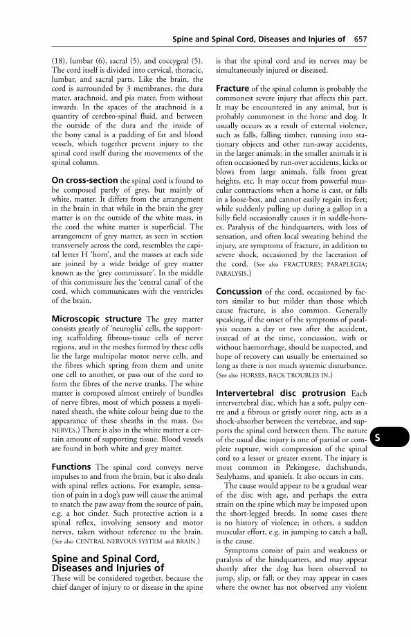
(18), lumbar (6), sacral (5), and coccygeal (5).The cord itself is divided into cervical, thoracic,lumbar, and sacral parts. Like the brain, thecord is surrounded by 3 membranes, the duramater, arachnoid, and pia mater, from withoutinwards. In the spaces of the arachnoid is aquantity of cerebro-spinal fluid, and betweenthe outside of the dura and the inside of the bony canal is a padding of fat and bloodvessels, which together prevent injury to thespinal cord itself during the movements of thespinal column.
On cross-section the spinal cord is found tobe composed partly of grey, but mainly ofwhite, matter. It differs from the arrangementin the brain in that while in the brain the greymatter is on the outside of the white mass, inthe cord the white matter is superficial. Thearrangement of grey matter, as seen in sectiontransversely across the cord, resembles the capi-tal letter H ‘horn’, and the masses at each sideare joined by a wide bridge of grey matterknown as the ‘grey commissure’. In the middleof this commissure lies the ‘central canal’ of thecord, which communicates with the ventriclesof the brain.
Microscopic structure The grey matterconsists greatly of ‘neuroglia’ cells, the support-ing scaffolding fibrous-tissue cells of nerveregions, and in the meshes formed by these cellslie the large multipolar motor nerve cells, andthe fibres which spring from them and uniteone cell to another, or pass out of the cord toform the fibres of the nerve trunks. The whitematter is composed almost entirely of bundlesof nerve fibres, most of which possess a myeli-nated sheath, the white colour being due to theappearance of these sheaths in the mass. (See
NERVES.) There is also in the white matter a cer-tain amount of supporting tissue. Blood vesselsare found in both white and grey matter.
Functions The spinal cord conveys nerveimpulses to and from the brain, but it also dealswith spinal reflex actions. For example, sensa-tion of pain in a dog’s paw will cause the animalto snatch the paw away from the source of pain,e.g. a hot cinder. Such protective action is aspinal reflex, involving sensory and motornerves, taken without reference to the brain.(See also CENTRAL NERVOUS SYSTEM and BRAIN.)
Spine and Spinal Cord,Diseases and Injuries ofThese will be considered together, because thechief danger of injury to or disease in the spine
is that the spinal cord and its nerves may besimultaneously injured or diseased.
Fracture of the spinal column is probably thecommonest severe injury that affects this part.It may be encountered in any animal, but isprobably commonest in the horse and dog. Itusually occurs as a result of external violence,such as falls, falling timber, running into sta-tionary objects and other run-away accidents,in the larger animals; in the smaller animals it isoften occasioned by run-over accidents, kicks orblows from large animals, falls from greatheights, etc. It may occur from powerful mus-cular contractions when a horse is cast, or fallsin a loose-box, and cannot easily regain its feet;while suddenly pulling up during a gallop in ahilly field occasionally causes it in saddle-hors-es. Paralysis of the hindquarters, with loss ofsensation, and often local sweating behind theinjury, are symptoms of fracture, in addition tosevere shock, occasioned by the laceration ofthe cord. (See also FRACTURES; PARAPLEGIA;PARALYSIS.)
Concussion of the cord, occasioned by fac-tors similar to but milder than those whichcause fracture, is also common. Generallyspeaking, if the onset of the symptoms of paral-ysis occurs a day or two after the accident,instead of at the time, concussion, with orwithout haemorrhage, should be suspected, andhope of recovery can usually be entertained solong as there is not much systemic disturbance.(See also HORSES, BACK TROUBLES IN.)
Intervertebral disc protrusion Eachintervertebral disc, which has a soft, pulpy cen-tre and a fibrous or gristly outer ring, acts as ashock-absorber between the vertebrae, and sup-ports the spinal cord between them. The natureof the usual disc injury is one of partial or com-plete rupture, with compression of the spinalcord to a lesser or greater extent. The injury ismost common in Pekingese, dachshunds,Sealyhams, and spaniels. It also occurs in cats.
The cause would appear to be a gradual wearof the disc with age, and perhaps the extrastrain on the spine which may be imposed uponthe short-legged breeds. In some cases there is no history of violence; in others, a suddenmuscular effort, e.g. in jumping to catch a ball,is the cause.
Symptoms consist of pain and weakness orparalysis of the hindquarters, and may appearshortly after the dog has been observed tojump, slip, or fall; or they may appear in caseswhere the owner has not observed any violent
Spine and Spinal Cord, Diseases and Injuries of 657
S

movement whatsoever: for example, a dogapparently normal at night may be found paralysed in the morning.
The muscles which control the passage ofurine and faeces may become paralysed.
In uncomplicated cases, natural recoverymay take place within a fortnight. Whereparalysis persists, the outlook becomes pro-gressively less hopeful, though a completerecovery after 12 months is not unknown. It isessential that veterinary attention is given assoon as possible.
A ‘fenestration’ operation is occasionallyperformed to reduce pain by relieving pres-
sure upon the spinal cord, but it is not ofmuch use for paralysis. Other measures are forthe treatment of paralysis generally.
Cervical spondulopathy (or ‘wobbler’syndrome) in the dog is most commonly seenin the Great Dane but also occurs in otherbreeds, especially the Dobermann. It is usual-ly first seen between 8 and 12 months, and theclinical signs include hind-limb incoordina-tion, abduction of the limbs, a prancing gait,and dragging of the feet causing wearing of thetoe nails. Diagnosis of cervical spondylopathyis confirmed by radiography. Lesions includestenosis of the vertebral canal, exostosis of thearticular facets, and subluxation.
658 Spine and Spinal Cord, Diseases and Injuries of
S
Intervertebral disc lesions.
Paralysis due to a ruptured intervertebral disc.
Hemivertebra ‘Wedge-shaped’ vertebra; inmany cases this condition in dogs gives rise tono obvious symptoms, but in others the condi-tion is characterised clinically by progressivehind-leg weakness, spinal pain, abnormalities ofthe nervous system and evidence of muscleatrophy or other abnormalities of conforma-tion. Confirmation of the clinical diagnosis isby radiography. Breed incidences are reported.The occurrence of the disorder in certain families of dogs suggests also that it may behereditary.
Ankylosis of the vertebrae is not rare in hors-es. It originates from diffuse inflammation ofthe spinal column, frequently due to rheumaticcauses, and one after another of the vertebraebecomes fused to its neighbour in front orbehind. In severe cases practically the whole ofthe thoracic and lumbar regions of the horsemay fuse into a rigid bar. Such horses usuallycan perform straightforward work for sometime, but are unable to carry any weight, toback heavy loads, or to lie down or rise withease. They may develop into ‘shiverers’, but solong as the spinal cord is not compressed, theymay live for years.
Pachymeningitis, or inflammation of themembranes of the cord, sometimes occurs inold dogs. It is called ‘ossifying pachymeningitis’in these animals, because of the tendency forbone to be deposited in the dura mater.
Abscess in the cord, or in one of the vertebrae,may be discovered only at a knackery or atautopsy; or it may lead to symptoms of paresis/paralysis. (See also ABSCESS; SPINA BIFIDA.)
SpirillumA bacterium with a wavy shape. (See RAT-BITE
FEVER.)
Spirit(see ALCOHOL POISONING; and SURGICAL SPIRIT)
SpirocercaWorms which are found in nodules on theoesophagus. In the dog they may sometimes giverise to cancer (sarcoma) of the oesophagus; andalso to fatal haemorrhage. (See ROUNDWORMS.)
SpirochaeteSpirochaete is one of the names applied to bacteria possessing a more or less spiral or wavyoutline. Another term applied to this group is ‘spirillum’. There are 3 genera which areimportant causal agents of disease: Borrelia,
Treponema, and Leptospira. The 1st has largewavy spirals and is flexible, the 2nd has regularrigid spirals, while the last has small spirals andone hook-like end.
Many spirochaetes produce disease in manand animals, the best known among which is T. pallida, the cause of syphilis in man. T. cuni-culi in rabbits causes ‘rabbit syphilis’, in Britain,on the continent of Europe and in America. B. galinarum is responsible for a form ofspirochaetosis affecting fowls in the tropics andsubtropics, and is transmitted by the fowl tick.L. canicola causes nephritis in dogs and canico-la fever in man; L. icterohaemorrhagiae causesWeil’s disease in man and jaundice in dogs.Leptospirosis also occurs in cattle and pigs, andthere are many serotypes. (See LEPTOSPIROSIS
and, for infection with Treponema in pigs,SWINE DYSENTERY.)
Spirochaetosis of FowlsSpirochaetosis of fowls is met with in Africa,Asia, the West Indies, South America, Australia,and Europe. It occurs in fowls, ducks, geese,and turkeys. Canaries and other birds are susceptible to artificial infection.
Transmission The disease is transmittedfrom diseased to healthy fowls by the fowl tickArgas persieus, one of the most important para-sites of poultry. The ticks are very active atnight, and may travel long distances to reach ahost. They remain hidden during the day increvices and underneath the bark of trees.Myriads of these ticks may attack fowls on theroost, a large quantity of blood being sucked –the affected birds becoming weak and unthriftyand having ragged plumage.
If, however, the ticks have fed on a bird affected with spirochaetosis, they become carriers of this disease and may transmit the infection through the egg to the next generation of ticks.
Signs Affected fowls show diarrhoea, loss ofappetite, loss of power of the head, and may die in convulsions. A more chronic course isalso described, birds becoming paralysed andemaciated, and dying in about a fortnight.
Treatment Antibiotics are effective.
Preventive Treatment This consists of ridding the premises of ticks.
Spirochaetosis of Pigs(see under PORCINE ULCERATIVE SPIROCHAETOSIS
and SWINE DYSENTERY).
Spirochaetosis of Pigs 659
S

SpirometraA tapeworm which infects cats, dogs, and occasionally people.
SplanchnicSplanchnic means relating to the viscera (inter-nal organs) of the body – as distinguished fromits framework.
Splayleg, CongenitalCongenital splayleg of piglets may involveeither the fore-legs or all 4 legs. Recovery from this defect can be expected if the pigletcan manage to keep out of the sow’s way. (See
VITAMIN E.)
SpleenA soft, highly vascular, plum-coloured organ,possessing a smooth surface formed by a densefibrous capsule over which the peritoneum isclosely applied.
Structure Beneath the outermost covering ofperitoneum lies the dense fibrous-tissue coat,from the inner surface of which numerousstrands or ‘trabeculae’ run into the organ. The fibrous coat and the trabeculae possesselastic fibres and a fair number of plain muscu-lar fibres. The trabeculae branch and rebranchthroughout the substance of the organ, and inthe meshes so-formed lies the spleen-pulp. Thisconsists of delicate connective-tissue fibres passing between the trabeculae, and numbers ofleucocytes and red blood corpuscles. Blood vessels run through the trabeculae and end inareas where the blood cells appear to be highlyconcentrated; these concentrations are knownas Malpighian corpuscles or bodies. The bloodescapes into the pulp of the spleen instead oftravelling through capillaries everywhere as inother organs.
Functions The spleen destroys old red bloodcells, acts as a blood store, and appears to playsome part in the formation of lymphocytes.
An animal is able to survive after removal ofthe spleen. There is a compensatory increase inthe lymph nodes all over the body, and, after aperiod of adjustment, life continues normally.
The spleen is apparently concerned also withbodily defence, and resistance to liver-flukeinfestation is reduced after removal of a sheep’sspleen. (See RETICULO-ENDOTHELIAL SYSTEM.)
Spleen, Diseases ofIt is often only at post-mortem examinationthat spleen diseases are revealed in the large animals. In anthrax it becomes greatly enlarged,
and also in babesiosis. In the dog, splenectomyis occasionally performed in cases of tumourformation or injury. Rupture of the spleen, withresultant internal haemorrhage, occurs in smallanimals which have fallen from a height or havebeen involved in a road accident.
SplenectomySurgical removal of the spleen.
Splinting MaterialsFor the immobilisation of limbs to achieveexternal fixation of fractured bone, or to pro-vide additional support for internal fixation,plaster of Paris and resin-impregnated materialsare used.
For large-animal use, plaster of Paris has several disadvantages: it does not achieve itsmaximum strength for up to 24 hours afterapplication, and is liable to break. The cast isoften heavy and cumbersome, and will soften incontact with moisture.
A number of proprietary splinting materialsare available, which offer advantages ofstrength, lightness and transparency to X-rays.They include: Baycast (Bayer), a polyester cotton bandage impregnated with a water-activated prepolymer resin; Scotchcast, whichconsists of polyurethane resin on a fibreglassbandage; Hexcelite, a thermoplastic material,stated to be easily applied and very strong.
Splints in HorsesThe splint-bones are the rudientary 3rd and 4th metacarpals; ‘splints’ is the common namegiven to exostoses (bony outgrowths) on thesplint-bones producd by inflammation.Theinflammation itself may be caused by a knockor a minute fracture. They are more common in young horses and slight or severe lamenessmay be seen. Splint formation usually starts inthe periosteum or ligament. The amount ofnew bone formed depends on the extent andduration of the inflammation.
The lameness disappears as the inflammationsubsides. The causes include an inheriteddefect.
Signs Usually, lameness appears before anybony enlargement can be seen or felt, althoughpressure over the region of splints causes pain.This lameness usually increases with exercise.The horse may walk sound, but trots lame to asurprising extent, considering the apparently‘sound’ walk. Later on, a soft putty-like swellingcan be felt, and this becomes harder with time,until it can be finally recognised as bone. Inknee-splint the leg is carried to the outside, and
660 Spirometra
S

appears stiff. As a rule splints are not serious,since with rest and treatment the bony fusionbecomes complete, and the horse goes sound.When they are placed high up, however, there isa danger that the new bone formation mayinvolve the knee-joint, and when they are situated far back, so as to interfere with the tendons, they may produce permanent lame-ness and injury to the tendon. In a horse under6 years old they should be looked upon as liableto cause future trouble, but in a horse over 6 years old they can be disregarded unless lameness is present.
Treatment Most mild cases require nothingfurther in the way of treatment than a rest fromwork, and later a run at grass for a fortnight orso. Topical applications of anti-inflammatorypreparations or hot fomentations may help torelieve pain during the acute stage.
SpondylitisInflammation of a vertebra, due to trauma oran infection.
Spondylosis A degenerative condition of thespine which can lead to ANKYLOSIS.
SpondylopathyDisease of the vertebrae such as may cause com-pression of the spinal cord, disc degeneration,and narrowing of the intervertebral space.
SpongesIn modern stables it is recognised that if a con-tagious disease breaks out, the sponge used fora number of horses is an important factor in the spread of the disease, and consequently apiece of flannel or other material which can beboiled is generally used instead for ‘quartering’(see GROOMING). A sponge used at the end of a hosepipe for udder-washing of cattle led to an outbreak of mastitis due to Pseudomonasaeruginosa.
Spongiform Encephalopathy(Human)The human spongiform encephalopathies –Creutzfeldt-Jakob disease (CJD), Gerstmann-Straussler-Scheinker syndrome (GSS) and kuru – are pathologically very similar to scrapiein sheep and to bovine spongiform encephalopa-thy (BSE). Like them, they are transmissible,although there is very little evidence of person toperson transmission, except in a very few iatro-genic cases, such as the grafting of corneal or duramater tissues from donors subsequently shown tohave had CJD, and the use of human growth
hormone prepared from the pituitary glands ofpatients dying with CJD. Kuru was transmittedby cannibalism in Papua New Guinea.
(See also BOVINE SPONGIFORM ENCEPHALOPA-
THY; FELINE SPONGIFORM ENCEPHALOPATHY.)
Sporadic DiseaseA sporadic disease is a disease occurring in sin-gle cases here and there, as distinct from diseaseoccurring as an enzootic, throughout a district,or epizootic, through a country or large tract ofland.
SporesReproductive cells of protozoa, bacteria, andfungi, etc., usually able to withstand an adverseenvironment.
SporidesminA poisonous substance, isolated from the fun-gus Pithomyces chartarum, which causes facialeczema and liver damage in sheep and cattle inNew Zealand and Australia.
SporotrichosisA fungal disease of horses, cattle, dogs, cats, andman, caused by Sporothrix schenckii. This givesrise to nodules under the skin, and thickeningof the lymphatics with ulceration. In the dog, liver, lungs and bone may show lesions. Of 19 people reported to have acquired this infec-tion from cats, 14 had no history of traumaticinjury at the site; 12 were veterinarians or assis-tants/nurses. All had a localised cutaneouslymph-node infection, lesions resolving in 1 to10 months after potassium iodide treatment.One patient had a deep ulcer on a finger.Although rare, infection through inhalation hasbeen recorded in people.
SporozoaThis is a group of Protozoa which are all para-sitic and produce spores at some stage of theirlife-cycle. It is divided into a number of orders,of which only 2 are important. These are theHaemosporidia which are parasites of the redblood cells, and the Coccidia which are parasitesof epithelia.
Spotted Fever(see ROCKY MOUNTAIN FEVER)
Spotted Horse(see under APPALOOSA)
Sprained TendonsSprained tendons is an extremely common con-dition in both the heavy and the light draught
Sprained Tendons 661
S

horse. The flexors, superficial and deep, aremostly affected.
Causes The superficial flexor tendon issprained during maximum weight-bearing bythe limb, and the deep flexor becomes sprainedat the period of thrust.
Signs There are the usual signs of inflamma-tion – heat, pain, swelling. A horse with a badlysprained deep flexor may walk almost sound,but goes pronouncedly lame when made to trot.A localised sprain of the check ligament and itsinsertion into the deep flexor tendon often pro-duces acute lameness, but the condition is notas serious as a sprain of the tendons lowerdown. From an owner’s point of view, however,differential diagnosis between the various formsand situations of sprain is not important.
Treatment A firm elastic support bandage,and adequate rest. A poultice, spread on cotton-wool and applied hot, before bandaging, is atraditional remedy.
Generally a horse with a badly sprained ten-don is not fit for work for a month to 6 weeks,although it may be apparently sound before thistime.
Chronic sprained tendons are often incur-able, but good results have sometimes beenobtained with DIATHERMY.
SprainsSprains involve the wrenching of a joint, oftenwith the simultaneous tearing of a ligament. Theterm is also applied to an inflammation of a ten-don, generally the result of an excessive stretchingof its fibres. (See SPRAINED TENDONS; SYNOVITIS.)
Spray ‘Drift’By this is meant droplets of spray liquid carriedby the wind to fields adjacent to that which isbeing intentionally sprayed with some farmchemical for purposes of weed control, pestcontrol, haulm destruction. It is a potentialcause of poisoning in grazing animals. (See
SPRAYS USED ON CROPS.)
Spray RaceA race which can be used for spraying sheep orcattle. Nozzles are arranged at intervals and fedwith suitable parasiticide liquid by means of a pump. The system has not been found as satisfactory as dipping for sheep scab.
Sprays used on CropsSprays used on crops include weedkillers suchas DNOC, insecticides such as parathion, and
potato-haulm destroyer such as arsenites. Suchsubstances constitute a hazard to livestockwhich gain entry into fields. (See POISONING;INSECTICIDES; WEEDKILLERS).
‘Spreading Factor’(see HYALURONIDASE)
Spring Viraemia of CarpA serious viral disease to which farmed or ornamental fish are particularly susceptible. It is transmitted by lice which parasitise carp.Clinical signs vary, but affected fish swim errat-ic-ally, may be swollen, and have small red spotson the skin. It is a NOTIFIABLE DISEASE.
Spur VeinsThe veins liable to damage by the horseman’sspurs.
Spurges, Poisoning byThe various species of spurges (Euphorbia spp.)are, apparently, mostly poisonous, though notto the same extent. Animals are not likely to eatthem because of the acrid milky sap. Specieswhich have been blamed for causing poisoningare as follows: caper spurge, E. lathyris; Irishspurge, E. hibernica; petty spurge, E. peplus; andthe sun spurge, E. helioscopia. Of these, the firstseems to be the most dangerous.
Signs Inflammation and swelling of themucous membranes of the mouth and tongue,pains in the abdomen, coldness of the extremi-ties of the body, dizziness, fainting, leading tounconsciousness and death in 2 or 3 days. Inone of the South African spurges, E. genistoides,the typical symptom, in addition to these men-tioned here, is an acute inflammation of theurethra, accompanied by frequent and painfulattempts at urination. Symptoms of acuteenteritis may also be seen.
Treatment Veterinary advice should besought at once and, since the milk of affectedcows may cause illness to people drinking it, itshould not be used for either human or animalconsumption.
SRM (Specified Risk Material) In thecase of bovines, for animals aged 12 months ormore this is the skull including brain, eyes, ton-sils and spinal cord. For bovines over 6 months,the spleen, thymus and intestines are alsoincluded. SRM must be stained to prevent its consumption and destroyed by incinerationor rendering. For sheep and goats older than 12 months, SRM includes the skull, including
662 Sprains
S

brain, eyes, tonsils and spinal cord; the spleenof any age of sheep or goat is included. Thetongue is exempt, provided it is removed immediately after slaughter.
St BernardA very large dog with a massive head anddrooping ears, traditionally used for mountainrescue work in the Swiss Alps; the rough-coatedvariety is commoner. The breed is prone to ‘dia-mond eye’ – a combination of entropion andectropion. Wobbler syndrome (cervical spondy-lolithesis) is inherited as a recessive trait andhaemophiolia B is a sex-linked recessive trait.Other inheritable conditons include ununitedanconeal process and a progressive posteriorparalysis (Stockards disease).
‘Stable Cough’(see EQUINE INFLUENZA)
Stable FlyStable fly is a serious pest to horses and otheranimals, and transmits diseases such as surra andanthrax in the tropics. (See FLIES – Fly controlmeasures; SUMMER SORES.)
Stable Vices And Tricks(see ‘VICES’ AND VICIOUSNESS)
Stables for RacehorsesA survey of 96 racehorse stables in the south-west of England showed that a ‘typical’ race-horse is kept in a loose-box with a floor area of12 m2 and is bedded on straw; it shares its airspace of 39 m3 with 7 other horses. In calmconditions, with the top door of the stableopen, natural convection would provide 6.6 airchanges/hour, but with the door closed, only2.2 changes. The top door should rarely, if ever,be closed. It has been suggested that present-day stables are based on designs which are worsethan the best available in the 19th century. (See also BEDDING.)
Staffordshire Bull TerrierA medium-sized muscular breed with a smoothcoat, often mainly white. Cataract may beinherited and the breed is prone to cleft palate.The American pit bull terrier was derived fromthe Staffordshire, which it resembles; it is advis-able to keep a pedigree record to avoid confu-sion and impounding under the DangerousDogs Act.
StagIn deer, it is a male of some species; the femaleof those species is always called a hind and the
young, calves. It is also the British term for amale turkey.
StaggersErratic gait caused by incoordination of thelimbs, as in ‘rye-grass staggers’. It may be seenin hypomagnesaemia.
‘Staggers Weed’A poisonous South African plant. (See ‘PUSHING
DISEASE’.)
StainingFor differentiation of bacteria, see ACID-FAST
ORGANISMS; GRAM-NEGATIVE.
StallionAn adult male horse, uncastrated, over 4 yearsold.
Standard International Units(see SI UNITS)
Staphylococcus(see BACTERIA)
Staples for Wound ClosureThese have long been used in human surgery inplace of stitches and are also used as wound closures in some types of surgery for animals.
Starch(see CARBOHYDRATE; DIET AND DIETETICS;DIGESTION)
Starch EquivalentThis term is no longer used, and the starchequivalent in the UK was replaced in 1975 byunits of METABOLISABLE ENERGY as part of theintroduction of metric and SI UNITS.
Starch Gel ElectrophoresisThis is one of the commonest techniques for studying the genetic variation in serum proteins. (See ELECTROPHORESIS.)
StargazerThe term applied to a newly hatched chick or poult where the head is permanently held back with the beak pointing directlyupwards. Affected birds may twist their headscontinuously. The cause is a deficiency of thiamine.
Staring CoatDry, dull, scurvy hair or fur. A common sign ofpoor condition of whatever cause. In the dog,one cause is lack of suitable fat in the diet. As a
Staring Coat 663
S
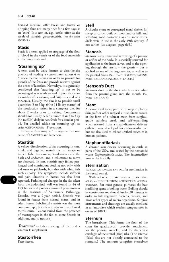
first-aid measure, offer bread and butter ordripping (but not margarine) for a few days asan ‘extra’. It is seen in, e.g., cattle, often as theresult of parasitic gastroenteritis. (See also under
WORMS.)
StasisStasis is a term applied to stoppage of the flowof blood in the vessels or of the food materialsin the intestinal canal.
‘Steaming up’A term used by dairy farmers to describe thepractice of feeding a concentrates ration 4 to 5 weeks before calving in order to provide forgrowth of the fetus and provide reserves againstthe onset of lactation. Nowadays, it is generallyconsidered that ‘steaming up’ is not to beencouraged as it tends to lead to poor dry mat-ter intakes after calving, and fatty liver and ace-tonaemia. Usually, the aim is to provide smallquantities (3 to 5 kg; 61⁄2 to 11 lb dry matter) ofthe production ration in a complete diet forabout 2 weeks prior to calving. Concentrateshould not usually be fed at more than 2 to 3 kg(41⁄2 to 61⁄2lb) daily in two feeds for a similar peri-od. For detailed advice on ‘steaming up’, see
under ACETONAEMIA – Prevention.Excessive ‘steaming up’ is regarded as one
cause of LAMINITIS and lameness.
SteatitisA yellow discoloration of fat occurring in cats,mink, and pigs fed mainly on fish scraps ortinned fish. Listlessness, tenderness over theback and abdomen, and a reluctance to moveare observed. In cats, steatitis may follow pro-longed and continuous feeding not only withred tuna or pilchards, but also with white fishsuch as coley. The symptoms include stiffnessand pain. Steatitis in horses has also beenreported. Pathological changes in the fat takenfrom the abdominal wall was found in 44 of173 horses and ponies examined post-mortemat the Institute of Veterinary Pathology,Utrecht, over a 2-year period. Steatitis wasfound in fetuses from normal mares, and inadult horses. Subclinical steatitis was the mostcommon type, but a few deaths were attributedto this cause. Lesions varied from the presenceof macrophages in the fat, to some fibrosis inaddition, and to necrosis.
Treatment includes a change of diet and a vitamin E supplement.
SteatorrheaFatty faeces.
StellA circular stone or corrugated metal shelter forsheep or cattle, built on moorland or hill, andaffording good protection against snow drifts.Stells were in use in the early 19th century, ifnot earlier. (See diagram, page 665.)
StenosisStenosis is any unnatural narrowing of a passageor orifice of the body. It is specially reserved forapplication to the heart valves, and to the open-ing through the larynx – the glottis – but isapplied to any of the large arteries, as well as tothe parotid ducts. (See HEART DISEASES; LARYNX;PAROTID GLAND; PYLORIC STENOSIS.)
Stenson’s DuctStenson’s duct is the duct which carries salivafrom the parotid gland into the mouth. (See
PAROTID GLAND.)
StentA device used to support or to keep in place askin graft or other surgical suture. Stents wovenin the form of a tubular mesh from surgical-grade stainless steel, and self-expanding when released from a small-diameter deliverycatheter, were developed for endovascular use,but are also used to relieve urethral stricture inhuman patients.
StephanofilariasisA chronic skin disease occurring in cattle inparts of the USA, and caused by the nematodeworm Stephanofilaria stilesi. The intermediatehost is the horn fly.
Sterilisation(see CASTRATION; also SPAYING for sterilisation inthe sexual sense).
With reference to sterilisation in its othersense, see DISINFECTION; ANTISEPTICS; ASEPSIS;WOUNDS. For most general purposes the beststerilising agent is boiling water. Boiling shouldbe continuous and should last for 30 minutes inorder to kill vegetative bacteria, viruses, andmost other types of micro-organisms. Surgicalinstruments and dressings are usually sterilisedin an autoclave which reaches temperatures inexcess of 100°C.
SternumThe breastbone. This forms the floor of thechest (in quadrupeds), provides attachment for the pectoral muscles, and for the costal cartilages of the sternal (true) ribs. (The asternal(false) ribs are not directly connected to thesternum.) The sternum comprises sternebrae
664 Stasis
S

– segments which fuse together with advancingage. The horse and dog each have 8 of these; thecow 7; pig and sheep, 6.
SteroidsChemical substances closely related to thesterols, e.g. the sex hormones, hormones of the adrenal cortex, bile acids. (See also
CORTICOSTEROIDS; DIABETES.)
Prescribing steroid hormone prod-ucts (where allowed) Steroid hormonegrowth promoters are substances with anandrogenic, oestrogenic or gestagenic action. Ingeneral they must not be administered to farmanimals. ‘Farm animals’ includes cattle, sheep,pigs, goats, horses, poultry, the wild animals ofthese species and any ruminants raised on aholding.
The prohibition does not apply to:(a) the administration for therapeutic
treatment by a veterinarian in the form of aninjection of oestradiol-17-beta, progesterone ortestosterone or derivatives of these substanceswhich readily yield the parent compound on hydrolysis after absorption at the site ofapplication;
(b) the administration of a steroid hormoneproduct for the termination of unwanted gestation or the improvement of fertility;
(c) the administration of a steroid hormoneproduct by, or under the direct responsibilityof, a veterinarian for the synchronisation of oestrus or the preparation of donors or recipients for the implantation of embryos.
For the purposes of the controlling regula-tion, the term ‘injection’ does not includeimplantation and the term ‘therapeutic treat-ment’ has a very restricted meaning – i.e. thetreatment of a fertility problem diagnosed by a veterinarian in an animal not intended for fattening.
A small range of products is licensed in the UK for the purposes listed above. The regulations are subject to change; the currentstatus of hormonal products should bechecked.
SterolsSolid alcohols, waxy substances derived fromanimal (and plant) tissues, e.g. cholesterol,ergosterol.
StertorNoisy breathing resembling snoring.
Stick InsectsSlow-moving green or brown insects thatresemble twigs, often kept as ‘pets’. They arevegetarian and must have fresh food that is
Stick Insects 665
S
Of metal construction, on a base of concrete blocks, this modified stell at the Rowett Research Institute,Aberdeenshire, was designed by Dr E. Cresswell. Sheep doors and a doorway for tractor access are shown.

not allowed to wilt. Hygiene is important intheir care.
Stiff Lamb DiseaseThis is a mild disease occurring in East Anglia due to infection with Erysipelothrix rhusiopathiae, the cause of swine erysipelas. Thesame name is also applied to muscular dystro-phy, a condition similar to that occurring incattle as a result of vitamin E deficiency.
‘Stiff-Limbed Lambs’This is a hereditary condition affecting newly born lambs, to which the nameMyodystrophia fetalis deformans has been given.It is commoner among Welsh Mountain sheepthan among other breeds. The condition is anarrest of the development of muscular tissueduring fetal life, and a replacement by fibroustissue. This contracts and pulls the limb into an unnatural, stiff attitude, and gives rise to difficulty in parturition.
The condition is a Mendelian recessivelethal. It has been reported from Britain andAmerica as affecting cattle, but it is not at allcommon in them.
Stiff Sickness(see STYFZIEKTE)
StifleThe joint corresponding to the human knee.(See BONES and JOINTS.) In horses, stifle lame-ness is often the result of OSTEOCHONDROSIS
dissecans or of subchondral bone cysts.In 42 cases of stifle lameness in cattle, the
diagnoses included subchondral bone cysts (18 cases), joint instability (15), degenerativejoint disease (12), cranial cruciate ligamentinjury (9), sepsis (9), collateral ligament injury (3), femorotibial luxation (2) and intra-articular fracture (2). The prognosis for animalswith bone cysts was good, irrespective of treatment (75 per cent recovered), while it was much poorer for animals with sepsis (22 per cent) or joint instability (27 per cent).
StilbenesSubstances consisting wholly or partly of stil-boestrol, hexoestrol, dienoestrol, or benzestrol.(See HORMONES IN MEAT PRODUCTION.)
In accordance with an EU directive, the saleof veterinary medicines, veterinary products oranimal feeds containing these stilbenes wasbanned in 1982 in the UK for use in food animals.
An exemption allows the limited use of stil-benes in companion animals for treating
enlarged prostate glands, adenoma, misalliancein the bitch, and urinary incontinence inspayed bitches.
StilboestrolAn oestrogen formerly used both therapeuticallyand as a growth promoter in food animals. (See
HORMONES; HORMONE THERAPY; HORMONES
IN MEAT PRODUCTION; STILBENES.)
Stillborn PigsBreeding stock should, of course, have access topasture, but if for any reason this golden rule isgoing to be broken, then rations should be supplemented in summer as well as in winter,with vitamin A. In a group of 20 gilts whichwere suddenly switched from succulent feedingto dry, fibrous grazing late in pregnancy, severeconstipation resulted, and there were deadpiglets in 19 of the litters.
A survey carried out by the VeterinaryInvestigation Service in England and Walesshowed a 4.8 per cent incidence of stillbirthsout of a total of 4394 piglets born in 371 litters.The incidence varied widely from herd to herd,as would be expected – ranging from 0.4 to12.9 per cent. Constipation appears to be acause of stillbirths, and SENNA may be used.
There are several infections which give rise toabortion and stillbirths. (See AUJESZKY’S DISEASE;ABORTION; MUMMIFICATION; INFERTILITY;CARBON MONOXIDE.)
StimulantsHeart ‘stimulants’ include caffeine, digitalis,etc. (See also RESPIRATORY STIMULANTS.)
Stings(see BITES AND STINGS)
StirkA young female bovine of 6 to 12 months old,sometimes a male of the same age, in Scotland.
Stitching(see SUTURE; WOUNDS)
Stockard’s DiseaseAn inheritable posterior paralysis found in individuals of cetain breeds of dog.
Stocking RatesDuring the peak of grass growth from earlyApril to mid-June, 8 to 10 ewes and their lambscan be carried per acre (4000 m2). But this is a maximum figure for this period only. For cattle, continuous grazing, the figure is perhaps1 to 3 acres (12,100 m2).
666 Stiff Lamb Disease
S
In Britain, ‘most grass is greatly under-stocked during the grazing season, chiefly dueto the lack of capacity to carry more stock overwinter.
‘Good farmers, using intensive grass-farmingmethods, require 11⁄2 acres (6000 m2) or more per cow, even when winter feeding is supplemented considerably with concentrates.
‘Estimates in New Zealand have suggestedthat dairy cows at 1.2 per acre may consume as little as 30 per cent of the available herbage.’(Director, Grassland Research Institute,Hurley.)
The most skilful dairy farmers were soonachieving 0.9 to 1.0 acre per cow without purchasing more than 10 to 15 per cent of their winter feed requirements other than production concentrates.
Stockmen/WomenFor health hazards see under COWHERDS;SHEPHERDS; MEAT-HANDLERS; ZOONOSES.
Stomach
Functions of the stomach (see RUMEN;RUMINAL DIGESTION; RETICULUM; OMASUM;ABOMASUM.) Broadly speaking, the function of the stomach is to store, warm, soften, andprepare food materials, and then to pass themon in regulated amounts into the intestine,where the more important digestive processesand absorption occur.
Stomach, Diseases ofIn all animals bacterial diseases, such as SALMO-
NELLOSIS, may be involved in diseases of thestomach; likewise parasitic worms.
Horse In view of the comparatively simplearrangement of the stomach, and the naturalfastidiousness of the horse in the matter offood, stomach disease is not so common as insome other animals.
Gastritis is usually brought about by theingestion of irritant, poisonous, or otherwiseharmful substances, or by the presence of bots,or spread of disease from other parts of thebody. (See also SALMONELLOSIS; ROUNDWORMS.)
Signs Attacks of violent abdominal pain,occurring shortly after feeding or even beforefeeding is completed, indicate that the stomachis affected. Dullness and depression are noticed;patchy sweating may break out; food is refused;the temperature rises slightly in mild cases, andto as high as 41°C (106°F) in severe instances.
Usually within 2 to 3 hours after taking foodthe acute pain ceases, but the horse remainsdull, and gives the impression that it is affectedwith a dull ache rather than with acute pain. Vomiting does not usually occur unlessthe stomach is ruptured or the oesophagus isdilated.
Impaction of the Stomach is a conditionin which engorgement of the stomach withfood takes place. It may be due to lack of vitality (atony) in the muscular walls, toimpaired gastric secretion.
Signs Signs of impaction may occur, suddenlyor gradually. There is depression, uneasiness,and perhaps colic, in those cases where a horseover-eats. (See COLIC.)
Sometimes a horse obtains relief by vomitingthrough the nostrils a quantity of the impactedmaterial, which reduces the amount in thestomach so that the remainder can be dealt within the usual way.
Prevention It is easier to prevent impactionof the stomach than it is to cure it.
Whole beans, peas, wheat, or barley shouldnot be used for horses. Horses should always beallowed as much water as they desire to drink,and should be watered before feeding in all
Stomach, Diseases of 667
S
Thorn apple (Datura stramonium), which is alsoknown as the Jamestown or jimson weed. Theflower may be white or purple. On the left is afruit capsule. Well-developed plants may be 1.6 m(5 ft) high. (See entry for Stramonium, page 670.)
cases. Diseased or rough and irregular teethshould be treated.
Tympany of the stomach When vegetable food ferments from any cause, gas isproduced. Certain foods, especially whenunsound, undergo fermentation in the stomachinstead of digestion, and the gas so formed is liable to collect in that organ often underpressure producing great distension. Foodswhich ferment easily are succulent green crops, clovers, lucerne, and potatoes eaten inquantity.
Signs There is no remission of pain, such as isusually seen when the intestines are tympanitic.Horses may roll, plunge, and paw the groundduring the earlier part of the attack.Respirations increase in rate, and becomelaboured. The abdomen becomes tense andoften swollen, and in many instances horsesassume a crouching attitude with theirhindquarters, not unlike the way in which adog sits. When the tympany is severe, unlessrelief is afforded by the passage of the stomach-tube, rupture of the stomach may occur, anddeath follow. (See COLIC.)
Rupture of the stomach may also occur when a horse falls violently to the groundsoon after a big feed, i.e. when the stomach is full.
Signs The distress characteristics of engorge-ment and tympany suddenly cease when thestomach ruptures, and for a short time thehorse appears so much better that the ownerimagines recovery will result. After a short time, however, the more serious symptoms ofperitonitis and shock occur. Profuse perspira-tion usually breaks out; the pulse changes towhat is called a ‘running down pulse’, i.e. thereare a few strong beats which gradually becomeweaker until they are almost imperceptible, andthen a succession of strong beats return; this isrepeated rhythmically. Ears and feet becomecold and clammy to the touch; respiration isblowing; and the expression on the face of the horse is one of anxiety. Vomiting is said tocharacterise rupture of the stomach, but it isprobable that in most cases the vomiting occurs before the rupture takes place; the foodmaterial escapes into the abdominal cavity afterrupture has occurred rather than up into thepharynx and nostrils. (See also COLIC.)
Treatment is usually regarded as useless;euthanasia is advisable.
Cattle Acute indigestion with acidosis, andsometimes impaction of the rumen, may follow overeating of grain or green foods. (See
ACIDOSIS.)
Tympany of the rumen (bloat) consists of acollection of gas in the rumen. (See under BLOAT
for symptoms, prevention and treatment.)
Inflammation of the rumen (see also
RUMEN ULCERATION) may be due to ingestionof irritant poisons, of either chemical or vegetable origin, to penetrating foreign bodies,or to the spread of inflammatory conditionsfrom other parts in specific diseases.
Foreign bodies in the reticulum are ofgreat importance in both young and adult cattle, because of the close proximity of thisorgan to the pericardium and heart. In thereticulum 2 things may happen: they may fallto the lowermost part of the sac and remainthere for an indefinite period, or they may slowly penetrate its wall and wander forwardsthrough the diaphragm. Their subse-quent course is described under HEART
DISEASES – Traumatic pericarditis of cattle. (See
also HAIR-BALLS)
Inflammation of the abomasum, abomastitis, or gastritis, is caused by a lack oflong-fibre roughage and the too-rapid intro-duction of concentrate diets after calving; itmay also be caused by parasitic roundworms.(For the causes, symptoms, and treatment of parasitic gastritis in cattle and sheep, see
PARASITIC GASTROENTERITIS.)
Displacement of the abomasum may beassociated with stenosis of the sigmoid curve ofthe duodenum in cattle. The abomasum is thenfound to be distended with fluid and gas, anddisplayed to the right.
Signs include a distended abdomen, loss ofappetite, loss of weight, and depression of milk yield, and may sometimes be successfullytreated by casting the cow and, with her lyingon her back, rotating or rocking her through anangle of 45° from the vertical each way. Surgicaltreatment may be necessary. (See TYMPANITIC
RESONANCE.)
Ulceration is a condition by no means rare in cattle. It is sometimes associated with dis-placement of the abomasum, and may give riseto symptoms a few days after calving. Deathfollows perforation. Symptoms are similar to
668 Stomach, Diseases of
S
those given above. Ulceration is not uncom-mon in calves after weaning, giving rise tocapricious appetite and sometimes evidence of abdominal pain. Fusiformis necrophorus,Actinomyces pyogenes, and Pasteurelle organismsin other parts of the body may be associated.
Sheep The diseases of the stomachs of thesheep resemble in general those of the sameorgans in cattle.
Braxy is characterised by a patch of acuteinflammation in the wall of the abomasum,usually about the size of the palm of the hand,where the mucous membrane is to a largeextent destroyed.
Another form of gastritis is due to parasiticworms in the young lambs. (See WORMS.)
Pigs
Gastritis Irritating or poisonous substances,specific disease, or parasites are among the causes. Salt poisoning is a cause of gastritis, asare also poisonings by arsenic, copper, saltpetre,sheep dips, etc. During the course of swinefever, swine erysipelas, foot-and-mouth disease,and even tuberculosis, the wall of the stomachmay become involved, and inflammation mayresult. (See under GASTRIC ULCERS.) Parasites may also cause this condition. (See WORMS,TREATMENT AGAINST; MUCORMYCOSIS.)
Signs Vomiting is the first and most importantsymptom of stomach disturbance. Thirst,depression, and sometimes skin discolorationare other symptoms. Convulsions and twitchingof the limbs may be seen in young pigs.
Treatment All solid foods must be withheld,and soft light foods given instead. Whole milkis one of the best. Where the condition isbelieved to be due to poisonous substances, the appropriate antidotes must be given. (See
ANTIDOTES.)
Dogs Gastritis may be bacterial in origin,caused by parasitic worms, irritating substanceswhich include poisons, or be associated withforeign bodies. It is probably less common thanenteritis or nephritis, both of which may giverise to vomiting.
Causes Gastritis/gastroenteritis may be acomplication of distemper, canine virus hepati-tis, or may arise during salmonellosis and other bacterial disease; or follow the eating ofinfected or decomposing food.
Ulceration of the stomach in dog and catmay be associated with gastritis – sometimeswith tuberculosis, malignant growths, and acti-nobacillosis. Ulcers similar to peptic ulcers inhuman beings, and leading to perforation,occur occasionally.
Signs As a rule, a severe attack of vomitingimmediately after a feed, and refusal to touchfood subsequently. Thirst is nearly always exces-sive, and if gratified, vomiting usually follows.
If capillary haemorrhage occurs into thestomach, perhaps as the result of retching, the blood which slowly oozes from its walls collects in the cavity of the stomach, undergoespartial digestion, and becomes changed into abrownish granular material, strongly resem-bling moist coffee grounds. This always has amost foul and objectionable odour. The dogitself becomes extremely miserable, draggingitself slowly from place to place, and showingpreference for cold places where it may liestretched out with its hind-legs straight behindit, so that the lower wall of the abdomen is inclose contact with the cold surface, e.g. a stonestep, or linoleum in a passage. Constipationusually occurs unless the intestines becomeinvolved, when diarrhoea is noticed. Pressureon the abdomen causes pain, and sometimes adog lifted by the hand under the abdomen criesout. The temperature is raised at first.
Treatment Hot packs applied to theabdomen soothe pain. Dogs affected with gastritis should be under the charge of a veterinary surgeon, who will vary the treatmentaccording to the circumstances.
At first food is better withheld. (See PYLORIC
STENOSIS; GLUCOSE; NORMAL SALINE; PROTEIN,HYDROLISED.)
Foreign bodies in the stomach mayinclude pieces of carpet or other fabric, the rubber from a golf ball, the covers of tennisballs, bones, pieces of wood, etc. A depravedappetite may be due to hunger, a mineral or vit-amin deficiency, rabies, or to bad habits – suchas picking up and swallowing pebbles (oftenmisguidedly thrown by the owner).
Signs Ineffectual attempts to vomit, accompa-nied by painful retching, an arched back, salivation from the mouth, and signs of discomfort. Sharp-pointed bodies may causeperforation of the stomach walls, peritonitis,and death. (See PERITONITIS.)
In small toy dogs it is quite usual for symptomsof acute nervous excitement to be shown.
Stomach, Diseases of 669
S

With chronic gastritis due to swallowed pebbles, symptoms are those of occasional vomiting, discomfort, and even a rattling soundas the dog walks.
First-aid An emetic, e.g. a crystal of washingsoda and water.
Treatment Apomorphine given by hypoder-mic injection will rid the stomach of the greaterpart of fibrous or soft ingesta, but where sharp-pointed foreign bodies have been swallowed, obviously it is unsafe to give emetics. To remove these and large roundedobjects which cannot be easily vomited, surgerywill be necessary.
Torsion of the stomach Except in the giantbreeds of dogs, torsion or twisting of the stomach is rare. The abdomen becomes painfulto the touch, swelling may be apparent, andvomiting likely to occur. The dog is soon in avery distressed condition, and needs emergencytreatment or death will result.
Pyloric stenosis and pylorospasm (see
under these headings for disease affecting thepylorus of the stomach)
Stomach-TubeA flexible, often rubber, tube used for introduc-ing into the stomach (either through the mouthor more often through the inferior meatus ofthe nostril on one side), with a view to relievingtympany, or introducing medicines in the treatment of disease. It is about 3 m long forhorses and cattle, and about 10 to 15 mm indiameter (proportionately smaller for otherspecies).
A tube which possesses 2 channels is sometimes used to attempt to remove from thestomach portions of poisonous plants whichmay have been eaten. Water is pumped downthrough one channel, and when the stomach isfull it runs from the other carrying with it smallpieces of the harmful material. It is not possibleto empty completely the stomach by the doublestomach-tube, but considerable amounts of theharmful material may be removed.
The stomach-tube is extremely useful inthose cases of colic which depend upon distur-bances in the stomach, and if warm water isintroduced by it in impaction of the largecolon, peristalsis can often be stimulated. (But
see DEHYDRATION.)Its use demands care and a knowledge of the
structure of the nasal passage, pharynx, gullet,and stomach.
StomatitisInflammation of the mouth and gums (gingivitis), tongue (glossitis) or lips (cheilitis).(See BOVINE PAPULAR STOMATITIS; FOOT-AND-
MOUTH DISEASE; VESICULAR STOMATITIS;SWINE VESICULAR DISEASE; MOUTH, DISEASES
OF; FELINE STOMATITIS.)
-StomyA suffix signifying formation of an opening inan organ by operation, e.g. gastrostomy andcolostomy.
Stones(see CALCULI and FOREIGN BODY)
Storing Feeds(for safe storage periods see under DIET AND
DIETETICS).
StotA steer.
StrabismusA condition in which each eye appears to be looking in a different direction. Also calledsquint.
StraightsSingle feeding-stuffs of animal or vegetable origin, which may or may not have undergonesome form of processing before purchase, e.g. flaked maize, soya bean meal, fish meal,barley.
StrainThe over-stretching of muscle fibres. Often afew of these are ruptured. A painful conditionrequiring rest. The same is true of overstressedtendons. (Compare a SPRAIN which involvesligaments of a joint.)
StramoniumStramonium is the leaf of Datura stramonium, which is popularly known as the thorn apple or the Jamestown or jimsonweed. It contains the alkaloid daturine, which isalmost identical in its actions with atropine.
The plant has caused fatal poisoning in pigs in Britain. However, the fatalities whichhave been reported appear to be the result ofingestion of large quantities of the plant in theabsence of other food.
StranglesStrangles is an acute contagious fever of horses,donkeys, and mules.
670 Stomach-Tube
S
Cause Streptococcus equi. Strangles is commonest and most serious in horses under 6 years of age. Mature horses living in a stablewhere an outbreak has occurred are frequentlyunaffected.
Signs Typical attacks begin with dullness, lackof appetite, rise in temperature to between39.5° and 40.5°C (103° and 105°F) and congestion of the visible mucous membranes,especially of the nose and eyes. Nasal dischargeis at first thin and watery, but soon becomesthicker, and profuse. There is often a cough.One or both of the submaxillary nodes, or perhaps one of the pharyngeal nodes, becomesenlarged, hot, tense, and painful to the touch;until a soft spot, usually over a most prominentpart of the swelling can be detected. This indicates the ‘pointing’ of the abscess.Following its resolution the horse improvesgreatly; temperature falls, appetite returns, andthe animal becomes much brighter.
Complication: occasionally a suppurativepneumonia occurs. There may also be abscessformation in the liver or other abdominal organs.
Treatment The owner should call in a veterinary surgeon. Immediate isolation of theaffected horse is necessary. (The box or stallwhere it stood must be disinfected as carefullyand thoroughly as possible, and should be leftvacant for 3 to 4 weeks afterwards.) The sickhorse should be clothed and made comfortable.Soft foods, such as mashes, are indicated, asswallowing may be painful. (See NURSING OF
SICK ANIMALS.) Antibiotics and/or sulfa drugsare used.
Prevention An efficient vaccine can be produced only if encapsulated S. equi is used(i.e. from very young cultures) and the capsulenot destroyed by formalin and excessive heat. In older cultures, the capsule is lost and theorganism no longer invasive.
Human Infection by S. equi has beenrecorded.
Strangulated HerniaThe term is applied to a loop of intestinebecoming trapped in a hernia, so that the bloodsupply to that section of it is cut off. (See
HERNIA; ‘GUT-TIE’; VOLVULUS; INTESTINES,DISEASES OF.)
StranguryDifficulty and pain in passing more than a few drops of urine at a time. It is a sign of
an inflammatory condition situated in the kidneys, bladder, or urethra.
Straw(see under BEDDING; DAIRY HERD MANAGEMENT;DEEP LITTER)
Straw feeding of cattle Straw is commonly included in diets, particularly whentreated with sodium or ammonium hydroxide.It has been used in a strict maintenance diet of 2 kg (4 lb) each of barley and a low protein-mineral-vitamin concentrate with some4.5 to 5.5 kg (10 to 12 lb) barley straw.
Experimentally, up to 30 per cent of groundstraw has been incorporated into beef rations,along with 10 to 20 per cent molasses, 45 percent cereals, and 5 per cent total minerals, vita-mins and urea to provide a complete ration. Fedad lib, this is claimed to have consistently givendaily liveweight gains in excess of 1.3 kg (2.8 lb)with Friesian steers in commercial trials.
The main snag with feeding ground straw isits tendency to produce frothy bloat (with lossof appetite and loss of weight in subclinicalcases). This has been overcome, it is claimed, byinclusion in feeds of an anti-bloat preparation(Poloxalene).
(See also NITRITE POISONING.)Straw is a very useful bedding material for
livestock.
‘Strawberry Foot-Rot’The colloquial name applied to a conditioncaused by the fungus Dermatophilus pedis or D. congolensis.
‘Stray Voltage’(see under ELECTRIC SHOCK)
StreamsAs a source of drinking water for cattle these should always be suspect, since they often carry infection from one farm to another, e.g. COCCIDIOSIS; JOHNE’S DISEASE;SALMONELLOSIS.
‘Street’ VirusThis term refers to the naturally occurring rabiesvirus, such as may be isolated from a rabid dog.
Streptococcal Meningitis(see under PORCINE STREPTOCOCCAL MENIN-
GITIS)
StreptococcusA micro-organism which under the microscopehas much the appearance of string beads. It is
Streptococcus 671
S

responsible for strangles, mastitis, acute abscessformation, etc. (See BACTERIA.)
Streptococcus SuisStreptococcus suis infection is a cause of menin-gitis and lymphadenitis in pigs.
Streptodornase, StreptokinaseEnzymes used to dissolve pus, fibrin, and bloodclot in infected wounds. They have also beenused in the treatment of mastitis.
StreptomycinAn aminoglycoside antibiotic obtained fromStreptomyces graces. Active almost entirelyagainst Gram-negative organisms, streptomycinhas given good results against infection with Corynebacterium (Actinomyces) pyogenes,Staphylococcus pyogenes, C. renale, E. coli, andPasteurella septica.
It has been used in cases of calf pneumoniaand calf scours, some types of bovine mastitis,and complications of viral diseases in the dog, and in septic conditions in the cat. (Inmedical practice, streptomycin is regarded asone of the most toxic of the antibiotics in com-mon use. It may cause deafness and vestibulardisturbance in dogs and cats.) Resistance to this antibiotic develops readily and is usuallymultiple. For these reasons, other anti-bioticsare to be preferred when suitable.
StreptothricosisInfection with streptothrix organisms. InBritain, the name is applied to the disease in cattle equivalent to lumpy wool or wool rot, caused by Dermatophilus dermatonomus. Ascurfy, scaly condition of the skin is produced,and scabs come away with a bunch of hairsattached if plucked. Anything which lowers theresistance of a hitherto healthy animal facilitatesinfection; and prolonged wetting, insect bites,thistle pricks, and other tiny breaks in the skinmay all predispose to infection.
In the tropics, the name is applied to infection with D. congolensis.
The onset of the rains brings an increase in theincidence and severity of the disease, which is ofgreat economic importance in Central and WestAfrica. Flies, ticks, and thorn bushes appear toplay some part in the production and spread ofthe disease. Zebu cattle, as exotic cattle, appearhighly susceptible; while N’dama and Muturuhumpless cattle are resistant. It seems that infection does not give rise to later immunity.
Treatment with antibiotics or sulfonamidesoffers most chance of success, but is impracticablein many areas. (See DERMATOPHILUS.)
StreptothrixDermatophilus congolensis (see STREPTOTHRICOSIS
and SENKOBO)
StressIn human medicine it is now recognised thatmental stress, anxiety and frustration can exerta profound effect for the worse upon bodilyhealth. Similar effects may be found in animals. Stress can adversely affect productionin food animals and behaviour in companionanimals.
On a farm in New Zealand where theoreticalconsiderations were all against high milk yields,the yields were, in fact, extremely high. After adetailed investigation it was concluded that thereason could only be sympathetic handling atmilking time by the farmers – father, son, anddaughter – who were strikingly ‘in harmony’with their cattle.
By contrast, on another New Zealand farmwhere everything – staff, milking machines, and herd management – remained the same,the strangeness of a new milking shed wasapparently the sole cause of a 15 per cent reduction in milk yield. (See also CALF
HOUSING.)Stress is recognised as a predisposing cause
of diseases in pigs, following the mixing of litters, castration, etc.; and in all species following parturition.
Subjection of animals to noise in intensivelivestock production, or in the course of transport, can be a source of stress. Reductionof noise could have considerable economic benefits. (See TRANSPORT STRESS.)
Sheep Problems can arise in paddock grazing.The grassland breeds need a greater space, if stress is to be avoided. They are the hedge-breakers and fence-testers. They ‘work away’ atweak places with a will to escape.
Where a very large number of sheep are dealtwith in one unit, it has been shown that it isespecially desirable to reduce the flock to unitsof 80 ewes during the intensive managementperiod at grass. Stress can result in a subclinicalinfection turning into overt illness. (See
also BUNT ORDER; INTENSIVE LIVESTOCK
PRODUCTION; INFECTION.)
Dogs Stress may result from being left tied upfor long periods, or alone in an otherwiseempty house; sometimes from ill-treatment by one member of a family. Dog fights areanother cause, or merely the presence of a large dog in the vicinity, known to be a fighter.Being lost or abandoned, placed in boarding
672 Streptococcus Suis
S
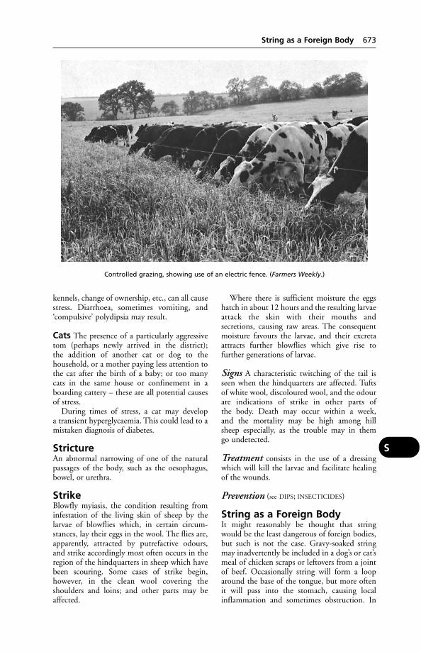
kennels, change of ownership, etc., can all causestress. Diarrhoea, sometimes vomiting, and‘compulsive’ polydipsia may result.
Cats The presence of a particularly aggressivetom (perhaps newly arrived in the district); the addition of another cat or dog to the household, or a mother paying less attention tothe cat after the birth of a baby; or too manycats in the same house or confinement in aboarding cattery – these are all potential causesof stress.
During times of stress, a cat may develop a transient hyperglycaemia. This could lead to amistaken diagnosis of diabetes.
StrictureAn abnormal narrowing of one of the naturalpassages of the body, such as the oesophagus,bowel, or urethra.
StrikeBlowfly myiasis, the condition resulting frominfestation of the living skin of sheep by the larvae of blowflies which, in certain circum-stances, lay their eggs in the wool. The flies are,apparently, attracted by putrefactive odours,and strike accordingly most often occurs in theregion of the hindquarters in sheep which havebeen scouring. Some cases of strike begin, however, in the clean wool covering the shoulders and loins; and other parts may beaffected.
Where there is sufficient moisture the eggshatch in about 12 hours and the resulting larvaeattack the skin with their mouths and secretions, causing raw areas. The consequentmoisture favours the larvae, and their excretaattracts further blowflies which give rise to further generations of larvae.
Signs A characteristic twitching of the tail isseen when the hindquarters are affected. Tuftsof white wool, discoloured wool, and the odourare indications of strike in other parts of the body. Death may occur within a week, and the mortality may be high among hill sheep especially, as the trouble may in them go undetected.
Treatment consists in the use of a dressingwhich will kill the larvae and facilitate healingof the wounds.
Prevention (see DIPS; INSECTICIDES)
String as a Foreign BodyIt might reasonably be thought that stringwould be the least dangerous of foreign bodies,but such is not the case. Gravy-soaked stringmay inadvertently be included in a dog’s or cat’smeal of chicken scraps or leftovers from a jointof beef. Occasionally string will form a looparound the base of the tongue, but more oftenit will pass into the stomach, causing localinflammation and sometimes obstruction. In
String as a Foreign Body 673
S
Controlled grazing, showing use of an electric fence. (Farmers Weekly.)
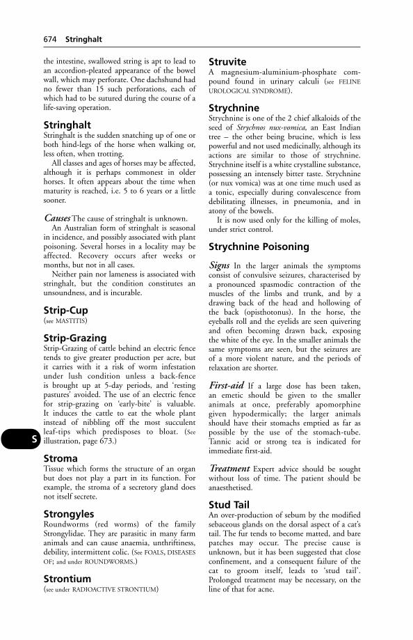
the intestine, swallowed string is apt to lead toan accordion-pleated appearance of the bowelwall, which may perforate. One dachshund hadno fewer than 15 such perforations, each ofwhich had to be sutured during the course of alife-saving operation.
StringhaltStringhalt is the sudden snatching up of one orboth hind-legs of the horse when walking or,less often, when trotting.
All classes and ages of horses may be affected,although it is perhaps commonest in older horses. It often appears about the time whenmaturity is reached, i.e. 5 to 6 years or a littlesooner.
Causes The cause of stringhalt is unknown.An Australian form of stringhalt is seasonal
in incidence, and possibly associated with plantpoisoning. Several horses in a locality may beaffected. Recovery occurs after weeks ormonths, but not in all cases.
Neither pain nor lameness is associated withstringhalt, but the condition constitutes anunsoundness, and is incurable.
Strip-Cup(see MASTITIS)
Strip-GrazingStrip-Grazing of cattle behind an electric fencetends to give greater production per acre, but it carries with it a risk of worm infestationunder lush condition unless a back-fence is brought up at 5-day periods, and ‘resting pastures’ avoided. The use of an electric fencefor strip-grazing on ‘early-bite’ is valuable. It induces the cattle to eat the whole plantinstead of nibbling off the most succulent leaf-tips which predisposes to bloat. (See
illustration, page 673.)
StromaTissue which forms the structure of an organbut does not play a part in its function. Forexample, the stroma of a secretory gland doesnot itself secrete.
StrongylesRoundworms (red worms) of the familyStrongylidae. They are parasitic in many farmanimals and can cause anaemia, unthriftiness,debility, intermittent colic. (See FOALS, DISEASES
OF; and under ROUNDWORMS.)
Strontium(see under RADIOACTIVE STRONTIUM)
StruviteA magnesium-aluminium-phosphate com-pound found in urinary calculi (see FELINE
UROLOGICAL SYNDROME).
StrychnineStrychnine is one of the 2 chief alkaloids of theseed of Strychnos nux-vomica, an East Indiantree – the other being brucine, which is lesspowerful and not used medicinally, although itsactions are similar to those of strychnine.Strychnine itself is a white crystalline substance,possessing an intensely bitter taste. Strychnine(or nux vomica) was at one time much used asa tonic, especially during convalescence fromdebilitating illnesses, in pneumonia, and inatony of the bowels.
It is now used only for the killing of moles,under strict control.
Strychnine Poisoning
Signs In the larger animals the symptoms consist of convulsive seizures, characterised by a pronounced spasmodic contraction of themuscles of the limbs and trunk, and by a drawing back of the head and hollowing of the back (opisthotonus). In the horse, the eyeballs roll and the eyelids are seen quiveringand often becoming drawn back, exposing the white of the eye. In the smaller animals thesame symptoms are seen, but the seizures are of a more violent nature, and the periods of relaxation are shorter.
First-aid If a large dose has been taken, an emetic should be given to the smaller animals at once, preferably apomorphine given hypodermically; the larger animals should have their stomachs emptied as far as possible by the use of the stomach-tube. Tannic acid or strong tea is indicated for immediate first-aid.
Treatment Expert advice should be soughtwithout loss of time. The patient should beanaesthetised.
Stud TailAn over-production of sebum by the modifiedsebaceous glands on the dorsal aspect of a cat’stail. The fur tends to become matted, and barepatches may occur. The precise cause isunknown, but it has been suggested that closeconfinement, and a consequent failure of thecat to groom itself, leads to ‘stud tail’.Prolonged treatment may be necessary, on theline of that for acne.
674 Stringhalt
S

Stunning, Electric, of CattleThis is practised in Sweden and theNetherlands by means of the Elther apparatus(prior to Jewish ritualistic slaughter or otherwise). It is used also for calves, sheep, and goats.
Stunning, Electric, of PigsThis has been practised extensively since the1930s, and involves the use of brine-soakedelectrodes, applied on each side of the pig’s face,by means of which the electric current is passed.A voltage of not less than 75 is recommendedand a current of not less than 250 milliamperes,assuming 50 cycles-per-second alternating current. An electroplectic fit is caused, withanaesthesia lasting for about 60 seconds, whenconditions are satisfactory. After 60 seconds,there may be a half-minute period of paralysisduring which sensation is present. Therefore,the pigs must be stuck during the first 60 seconds. If care is not taken and the apparatusbe faulty or unsuitable, paralysis only, and notanaesthesia, may result; the pig being consciouswhen stuck.
High-voltage stunning The trendtowards the use of 180 to 600 volts has beenimpeded by the commonly held belief that itmight adversely affect ‘bleed out’. However, the ARC’s Meat Research Institute has shownthat this need not be so.
Stunted Chick DiseaseThis syndrome in chickens was first recognisedby Kouwenhoven and others in 1978, and has since been found to occur worldwide. Thecause is believed to be a virus, possibly exacer-bated by Campylobacter spp. and spirochaetes.
SturdyA neurological disease in sheep caused byCoenurus cerebalis, it is also known as GID.
Stye(see EYE)
StyfziekteStyfziekte is a name meaning ‘stiff sickness’,which is used to describe either the symptomsassociated with chronic aphosphorosis, which isthe forerunner of lamziekte in South Africa, orthose associated with a mineral deficiency incertain parts of northern Nigeria.
SubclinicalA disease is said to be subclinical when thesymptoms are so slight as to escape the notice of
the animal-owner. Examples: subclinical mastitis, which by lowering the milk yield of aherd of cows may be of considerable economicimportance; similarly, a subclinical infestationwith parasitic worms. (See also STRESS.)
SubcutaneousSubcutaneous means anything pertaining to the loose connective tissue lying under the skin, such as a subcutaneous injection, wherethe injected fluid is introduced below the skin.(See under INJECTIONS.)
SubluxationA partial dislocation. Atlantoaxial subluxationis a cause of neck pain and muscle dysfunctionin some toy breeds of dogs.
Treatment In 13 cases the atlas and axis werestabilised with a wire suture; in 10 cases lagscrews were used for fixation of the ventralarticular facets. Nine of them recovered within2 months.
Sucking (Intersucking)This habit or ‘vice’ occurs among dairy calves.If allowed to go unchecked, the practice maybecome habitual, involving a risk to the healthof the calves, and, if persisting into adulthood,the welfare of the herd in general may be affected. In a herd of 50 Friesian cows the habitgrew so pronounced that the herd becameuneconomic and had to be dispersed. Cattle ofall ages were involved and milk loss was consid-erable. Purchased calves acquired the samehabits after a short while. Intersucking is aproblem in only a small proportion of herds,usually those of above average size, where thecalves are bucket fed, or where they are groupedat or shortly after birth.
The most effective remedy is to separate the calves after feeding, but if this is not practicable, mechanical devices or the provisionof dry food are good alternatives. It seems that a useful preventive measure is to delaygrouping calves until they are more than 4 weeks of age.
Suckling(see CALF REARING)
Sudden Death(see under DEATH)
SudorificsSudorifics are drugs and other agents whichproduce a copious flow of perspiration. (See
DIAPHORETICS.)
Sudorifics 675
S
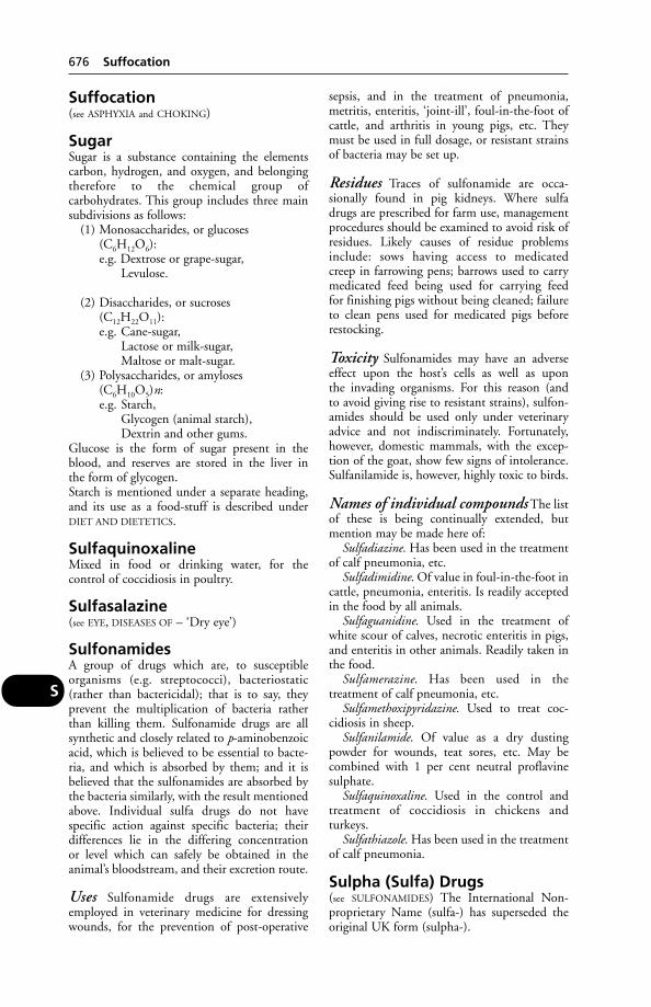
Suffocation(see ASPHYXIA and CHOKING)
SugarSugar is a substance containing the elementscarbon, hydrogen, and oxygen, and belongingtherefore to the chemical group of carbohydrates. This group includes three mainsubdivisions as follows:
(1) Monosaccharides, or glucoses(C6H12O6):e.g. Dextrose or grape-sugar,
Levulose.
(2) Disaccharides, or sucroses(C12H22O11):e.g. Cane-sugar,
Lactose or milk-sugar,Maltose or malt-sugar.
(3) Polysaccharides, or amyloses(C6H10O5)n:e.g. Starch,
Glycogen (animal starch),Dextrin and other gums.
Glucose is the form of sugar present in theblood, and reserves are stored in the liver in the form of glycogen.Starch is mentioned under a separate heading,and its use as a food-stuff is described underDIET AND DIETETICS.
SulfaquinoxalineMixed in food or drinking water, for the control of coccidiosis in poultry.
Sulfasalazine(see EYE, DISEASES OF – ‘Dry eye’)
SulfonamidesA group of drugs which are, to susceptibleorganisms (e.g. streptococci), bacteriostatic(rather than bactericidal); that is to say, theyprevent the multiplication of bacteria ratherthan killing them. Sulfonamide drugs are allsynthetic and closely related to p-aminobenzoicacid, which is believed to be essential to bacte-ria, and which is absorbed by them; and it isbelieved that the sulfonamides are absorbed bythe bacteria similarly, with the result mentionedabove. Individual sulfa drugs do not have specific action against specific bacteria; theirdifferences lie in the differing concentration or level which can safely be obtained in the animal’s bloodstream, and their excretion route.
Uses Sulfonamide drugs are extensivelyemployed in veterinary medicine for dressingwounds, for the prevention of post-operative
sepsis, and in the treatment of pneumonia,metritis, enteritis, ‘joint-ill’, foul-in-the-foot ofcattle, and arthritis in young pigs, etc. Theymust be used in full dosage, or resistant strainsof bacteria may be set up.
Residues Traces of sulfonamide are occa-sionally found in pig kidneys. Where sulfadrugs are prescribed for farm use, managementprocedures should be examined to avoid risk ofresidues. Likely causes of residue problemsinclude: sows having access to medicated creep in farrowing pens; barrows used to carrymedicated feed being used for carrying feed for finishing pigs without being cleaned; failureto clean pens used for medicated pigs beforerestocking.
Toxicity Sulfonamides may have an adverseeffect upon the host’s cells as well as upon the invading organisms. For this reason (and to avoid giving rise to resistant strains), sulfon-amides should be used only under veterinaryadvice and not indiscriminately. Fortunately,however, domestic mammals, with the excep-tion of the goat, show few signs of intolerance.Sulfanilamide is, however, highly toxic to birds.
Names of individual compounds The listof these is being continually extended, butmention may be made here of:
Sulfadiazine. Has been used in the treatmentof calf pneumonia, etc.
Sulfadimidine. Of value in foul-in-the-foot incattle, pneumonia, enteritis. Is readily acceptedin the food by all animals.
Sulfaguanidine. Used in the treatment ofwhite scour of calves, necrotic enteritis in pigs,and enteritis in other animals. Readily taken inthe food.
Sulfamerazine. Has been used in the treatment of calf pneumonia, etc.
Sulfamethoxipyridazine. Used to treat coc-cidiosis in sheep.
Sulfanilamide. Of value as a dry dusting powder for wounds, teat sores, etc. May becombined with 1 per cent neutral proflavinesulphate.
Sulfaquinoxaline. Used in the control andtreatment of coccidiosis in chickens andturkeys.
Sulfathiazole. Has been used in the treatmentof calf pneumonia.
Sulpha (Sulfa) Drugs(see SULFONAMIDES) The International Non-proprietary Name (sulfa-) has superseded theoriginal UK form (sulpha-).
676 Suffocation
S
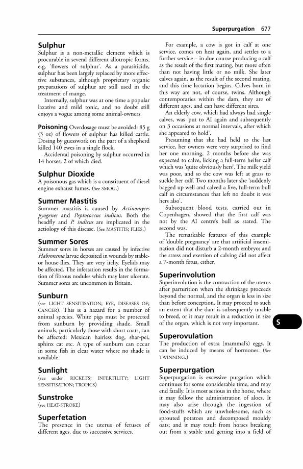
SulphurSulphur is a non-metallic element which isprocurable in several different allotropic forms,e.g. ‘flowers of sulphur’. As a parasiticide, sulphur has been largely replaced by more effec-tive substances, although proprietary organicpreparations of sulphur are still used in thetreatment of mange.
Internally, sulphur was at one time a popularlaxative and mild tonic, and no doubt stillenjoys a vogue among some animal-owners.
Poisoning Overdosage must be avoided: 85 g(3 oz) of flowers of sulphur has killed cattle.Dosing by guesswork on the part of a shepherdkilled 140 ewes in a single flock.
Accidental poisoning by sulphur occurred in14 horses, 2 of which died.
Sulphur DioxideA poisonous gas which is a constituent of dieselengine exhaust fumes. (See SMOG.)
Summer MastitisSummer mastitis is caused by Actinomyces pyogenes and Peptococcus indicus. Both the headfly and P. indicus are implicated in the aetiology of this disease. (See MASTITIS; FLIES.)
Summer SoresSummer sores in horses are caused by infectiveHabronema larvae deposited in wounds by stable-or house-flies. They are very itchy. Eyelids may be affected. The infestation results in the forma-tion of fibrous nodules which may later ulcerate.Summer sores are uncommon in Britain.
Sunburn(see LIGHT SENSITISATION; EYE, DISEASES OF;CANCER). This is a hazard for a number of animal species. White pigs must be protectedfrom sunburn by providing shade. Small animals, particularly those with short coats, canbe affected: Mexican hairless dog, shar-pei,sphinx cat etc. A type of sunburn can occur in some fish in clear water where no shade isavailable.
Sunlight(see under RICKETS; INFERTILITY; LIGHT
SENSITISATION; TROPICS)
Sunstroke(see HEAT-STROKE)
SuperfetationThe presence in the uterus of fetuses of different ages, due to successive services.
For example, a cow is got in calf at one service, comes on heat again, and settles to afurther service – in due course producing a calfas the result of the first mating, but more oftenthan not having little or no milk. She latercalves again, as the result of the second mating,and this time lactation begins. Calves born inthis way are not, of course, twins. Althoughcontemporaries within the dam, they are of different ages, and can have different sires.
An elderly cow, which had always had singlecalves, was ‘put to AI again and subsequently on 3 occasions at normal intervals, after whichshe appeared to hold’.
Presuming that she had held to the last service, her owners were very surprised to findher one morning, 2 months before she wasexpected to calve, licking a full-term heifer calfwhich was ‘quite obviously hers’. The milk yieldwas poor, and so the cow was left at grass tosuckle her calf. Two months later she ‘suddenlybagged up well and calved a live, full-term bullcalf in circumstances that left no doubt it washers also’.
Subsequent blood tests, carried out inCopenhagen, showed that the first calf was not by the AI centre’s bull as stated. The second was.
The remarkable features of this example of ‘double pregnancy’ are that artificial insemi-nation did not disturb a 2-month embryo; andthe stress and exertion of calving did not affecta 7-month fetus, either.
SuperinvolutionSuperinvolution is the contraction of the uterusafter parturition when the shrinkage proceedsbeyond the normal, and the organ is less in sizethan before conception. It may proceed to suchan extent that the dam is subsequently unableto breed, or it may result in a reduction in sizeof the organ, which is not very important.
SuperovulationThe production of extra (mammal’s) eggs. Itcan be induced by means of hormones. (See
TWINNING.)
SuperpurgationSuperpurgation is excessive purgation whichcontinues for some considerable time, and mayend fatally. It is most serious in the horse, whereit may follow the administration of aloes. Itmay also arise through the ingestion of food-stuffs which are unwholesome, such assprouted potatoes and decomposed mouldyoats; and it may result from horses breaking out from a stable and getting into a field of
Superpurgation 677
S
clover or lucerne. (See PURGATIVES; LAXATIVES;COLIC.)
Supplementary Feeding(see FLUSHING OF EWES; FEED BLOCKS; UREA;SUPPLEMENTS; CREEP-FEEDING)
Supplementary VeterinaryRegister(see under VETERINARY SURGEONS ACT 1966)
SupplementsProducts for use at less than 5 per cent of the total ration in which they are included, and designed to supply planned proportions of vitamins, trace minerals, one or more non-nutrient additives and other special ingredients.
SuppositoryA suppository is a small conical mass made ofglycerine or a similar substance, and containingdrugs intended for introduction into the rectum.
SuppurationThe formation or discharge of pus (see under
ABSCESS; CELLULITIS; FISTULA; INFLAMMATION;PHAGOCYTOSIS; WOUNDS).
Suprarenal Bodies(see ADRENAL GLANDS)
Suprascapular ParalysisSuprascapular paralysis occurs as a result ofinjury to the suprascapular nerve. The term‘slipped shoulder’ is applied to the symptomswhich are shown in a typical case. Thesupraspinous and infraspinous muscles act as ligaments of the shoulder-joint, and when theyare paralysed the shoulder slips outward eachtime the foot is placed upon the ground andwhen weight is put upon it. After the paralysishas been in existence for some few days, 2 dis-tinct hollows appear over the shoulder, due toatrophy of the muscles, and the spine of thescapula stands out prominently between thesehollows. When viewed from in front the animalappears to have lost the symmetry of the 2 shoul-der regions. In typical cases there is difficulty inbringing the limb forward, and often the legappears to swing outwards with a circular move-ment. When a horse stands quietly, the affectedlimb is usually brought well under the body, andmay even take up a position across the middleline of the body. The paralysis may disappear in6 weeks; but in more severe cases, 18 monthsmay elapse before the horse is fit for work.
First-aid Fomentations, or application of a liniment may be helpful until the acute symptoms subside; thereafter a run at grass generally results in improvement.
In the dog there may be permanent paralysis,sometimes requiring amputation of the leg orother surgery.
SuraminA drug used against trypanosomes.
SurfactantsSubstances that reduce the surface tension of aliquid; soap and detergents are examples.Surfactants are used in frothy bloat to allow therelease of gas from the bubbles which areformed.
Surgical SpiritA preparation of alcohol used, for example, as a skin cleanser before giving an injection. Itconsists of industrial methylated spirit with theaddition of castor oil (2.5 per cent), methyl salicylate (0.5 per cent) and diethyl phthalate (2 per cent).
SurraSurra is a disease of most economic importancein camels and horses, but it can affect all thedomestic animals. The disease occurs in Africa(north of the tsetse fly belt), Asia, Central andSouth America. In the latter, Trypanosomaequinum is responsible; elsewhere it is caused byT. evansi.
The infection is spread by blood-suckingflies, such as tabanids and stable flies. Vampirebats are believed to transmit the infection also.Animals which eat the meat from carcasesinfected with trypanosomes may themselvesbecome infected in the case of surra.
In the Sudan, surra affects mainly camels,which die within weeks or a few months, aftershowing symptoms of fever, anaemia, progres-sive emaciation, oedema, and paralysis. In Asia,surra in camels is often a chronic disease whichmay persist for years.
In horses, symptoms are similar, but thedropsical swellings (oedema) are especiallynoteworthy, affecting several parts of the body(as they do also in the dog). Mortality is high, and occurs in horses after a matter ofweeks or months. Loss of power in the hindlimbs, and exaggerated heart sounds may precede death.
In Central America the names ‘murrina’ and ‘derrengadera’ have been used for vampire-bat and fly-transmitted infection withT. equinum.
678 Supplementary Feeding
S
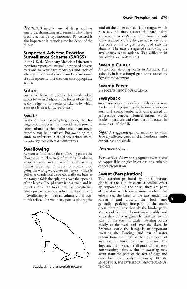
Treatment involves use of drugs such asantrycide, diminazine and suramin which havespecific action on trypanosomes. Fly control isalso important in reducing the incidence of thedisease.
Suspected Adverse ReactionSurveillance Scheme (SARSS)In the UK, the Veterinary Medicines Directoratemonitors reports of unusual unexpected adversereactions to veterinary medicines and lack ofefficacy. The manufacturers are kept informed of such reports so that they can take appropriateaction.
SutureSuture is the name given either to the closeunion between 2 adjacent flat bones of the skullat their edges, or to a series of stitches by whicha wound is closed. (See WOUNDS.)
SwabsSwabs are used for sampling mucus, etc., fordiagnostic purposes; the material subsequentlybeing cultured so that pathogenic organisms, ifpresent, may be identified. For swabbing as aguide to infertility in the thoroughbred mare,see under EQUINE GENITAL INFECTIONS.
SwallowingAs soon as food ready for swallowing enters thepharynx, it touches areas of mucous membranesupplied with nerves which automaticallyinhibit breathing, in order to prevent foodgoing the wrong way; close the larynx, which ispulled forwards and upwards, while the base ofthe tongue folds the epiglottis over the openingof the larynx. The pharynx is shortened and itsmuscles force the food into the oesophagus,where peristalsis takes the food to the stomach.
Swallowing is one-third voluntary and two-thirds reflex. The voluntary part is placing the
food on the upper surface of the tongue whichis raised, tip first, against the hard palatetowards the rear. At the same time the softpalate is raised, closing the gateway to the nose.The base of the tongue forces food into thepharynx. The next 2 stages of swallowing areinvoluntary, reflex actions. (For difficulty inswallowing, see DYSPHAGIA.)
Swamp CancerA condition affecting horses in Australia. Thelesion is, in fact, a fungal granuloma caused byHyphomyces destruens.
Swamp Fever(see EQUINE INFECTIOUS ANAEMIA)
SwaybackSwayback is a copper deficiency disease seen inthe last 3rd of pregnancy in the ewe or in new-born and young lambs. It is characterised byprogressive cerebral demyelination, whichresults in paralysis and often death. It occurs inmany parts of the UK.
Signs A staggering gait or inability to walk.Severely affected cases all die. Newborn lambscannot rise and suckle.
Treatment None.
Prevention Allow the pregnant ewes access to copper licks or give injections of a suitablecopper preparation.
Sweat (Perspiration)The excretion produced by the sudiparousglands of the skin; it exerts a cooling effect by evaporation. In the horse, there are parts of the skin which sweat more readily than others, e.g. the bases of the ears, under the fore-arm, and around the dock, and generally speaking, fore-parts of the trunk sweat more quickly than do the hinder parts.Mules and donkeys do not sweat readily, andwhen they do it is generally confined to thebases of the ears. In cattle, sweating occurschiefly at the neck and over the chest. InBrahman cattle the hump is an importantsweating site. Panting (and loss of water vapour from the lungs) is the chief means ofheat loss in sheep, but they do sweat. The dog, cat, and pig are, for all practical purposes,non-sweating animals, though sweating mayoccur from the pads of the feet of dogs and cats; dogs rely mainly on panting. (See also
ANHIDROSIS; HYPERTHERMIA; HYPOTHALAMUS;TROPICS.)
Sweat (Perspiration) 679
S
Swayback – a characteristic posture.
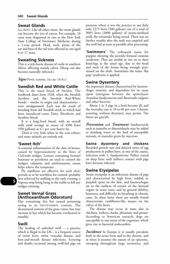
Sweat Glands(see SKIN). Like all other tissue, the sweat glandscan become the site of cancer. For example, 10cases were diagnosed in cats at the New YorkState College of Veterinary Medicine during a 2-year period. Head, neck, pinna of the ear and base of the tail were affected in cats aged6 to 17 years.
Sweating SicknessThis is a tick-borne disease of cattle in southernAfrica, affecting mainly calves. (Sheep can alsobecome naturally infected.)
Signs Fever, eczema. (See also TICKS.)
Swedish Red and White CattleThis is the main breed of Sweden. The herdbook dates from 1928, when the SwedishAyrshire and the Swedish Red-White breeds – similar in origin and characteristics –were amalgamated. Each was the result ofbreeding from old Swedish stock to which hadbeen introduced some Dairy Shorthorn andAyrshire blood.
It is a long-lived breed, with an overall milk yield average in excess of 4300 litres (950 gallons) at 4.1 per cent butter fat.
There is very little white in the coat-colour;and some animals are entirely red.
‘Sweet Itch’A seasonal inflammation of the skin of horses,caused by hypersensitivity to the bites ofCulicoides midges. Lotions containing benzoylbenzoate or pyrethrins are used to control themidges; calamine and antihistamine creamhelps relieve the symptoms
Fly repellents are effective for such shortperiods as to be worthless for control, probablybest achieved by stabling in the early evening; aVapona strip being hung in the stable to kill anymidges entering.
Sweet Vernal Grass(Anthoxanthum Odoratum)Hay containing this has caused poisoningowing to its DICOUMAROL content. Thedicoumarol content of the grass varies, but mayincrease in hay which has become overheated ormouldy.
SwillThe feeding of unboiled swill – a practicewhich is illegal in the UK – is a frequent sourceof swine fever, swine vesicular disease, and foot-and-mouth disease infections. Scouringand deaths occurred among swill-fed pigs on
premises where it was the practice to use dailyonly 2275 litres (500 gallons) out of a total of9095 litres (2000 gallons) of steam-sterilisedswill, the remainder being stored. There was nofurther trouble after the tank was emptied andthe swill fed as soon as possible after processing.
‘Swimmers’ The colloquial name for puppies showing the juvenile femoral rotationsyndrome. They are unable to rise on to theirhind-legs at the usual age, due to the head and neck of the femur being wrongly posi-tioned on the shaft. Sometimes the name ‘flatpup’ syndrome is applied.
Swine DysenteryAn important disease, characterised by haemor-rhagic enteritis, and dependent for its causeupon synergism between the spirochaeteSerpulina hyodysenteriae and Bacteroides vulgatusand other bacteria.
About 1 in 3 pigs in a herd become ill, andthe mortality rate is 10 to 60 per cent. Chronicscouring, without dysentery, may persist. Thefaeces are greyish.
Prevention and Treatment Antibacterialssuch as tiamulin or dimetridazole may be addedto drinking water or the feed of susceptible animals, or tiamulin given by injection.
Swine dysentery and chickensRetarded growth rate and delayed onset of eggproduction in pullets have, as one of the causes,infection with S. hyodysenteriae. Pullets rearedon deep litter with indirect contact with pigshave become infected.
Swine ErysipelasSwine erysipelas is an infectious disease of pigsand characterised by high fever, reddish or purplish spots on the skin, and haemorrhageson to the surfaces of certain of the internalorgans in acute cases; and by general debility,lameness, and difficulty in breathing in chroniccases. In these latter there are usually foundcharacteristic cauliflower-like masses on thevalves of the heart.
The disease may occur in man; also in chickens, turkeys, ducks, pheasants and grouse.According to American research, dogs are susceptible to one strain of the organism, whichgives rise to bacterial endocarditis.
Incidence In Europe it is usually prevalentboth in the acute form and in the chronic, andat times it assumes the nature of an epizootic,sweeping throughout large territories, and
680 Sweat Glands
S
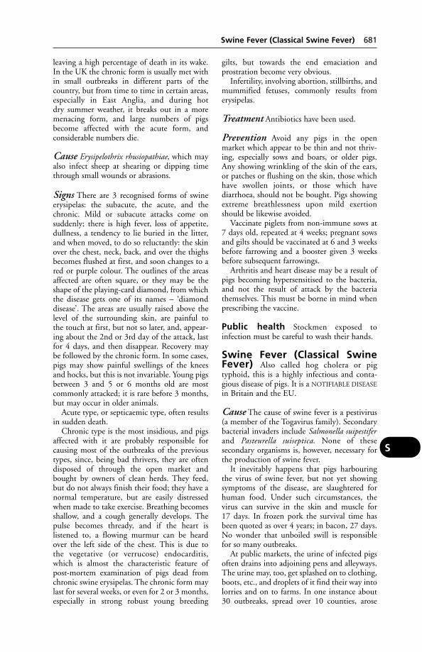
leaving a high percentage of death in its wake.In the UK the chronic form is usually met within small outbreaks in different parts of thecountry, but from time to time in certain areas,especially in East Anglia, and during hot dry summer weather, it breaks out in a moremenacing form, and large numbers of pigsbecome affected with the acute form, and considerable numbers die.
Cause Erysipelothrix rhusiopathiae, which mayalso infect sheep at shearing or dipping timethrough small wounds or abrasions.
Signs There are 3 recognised forms of swineerysipelas: the subacute, the acute, and thechronic. Mild or subacute attacks come on suddenly; there is high fever, loss of appetite,dullness, a tendency to lie buried in the litter,and when moved, to do so reluctantly: the skinover the chest, neck, back, and over the thighsbecomes flushed at first, and soon changes to ared or purple colour. The outlines of the areasaffected are often square, or they may be theshape of the playing-card diamond, from whichthe disease gets one of its names – ‘diamonddisease’. The areas are usually raised above thelevel of the surrounding skin, are painful to the touch at first, but not so later, and, appear-ing about the 2nd or 3rd day of the attack, lastfor 4 days, and then disappear. Recovery maybe followed by the chronic form. In some cases,pigs may show painful swellings of the kneesand hocks, but this is not invariable. Young pigsbetween 3 and 5 or 6 months old are most commonly attacked; it is rare before 3 months,but may occur in older animals.
Acute type, or septicaemic type, often resultsin sudden death.
Chronic type is the most insidious, and pigsaffected with it are probably responsible forcausing most of the outbreaks of the previoustypes, since, being bad thrivers, they are oftendisposed of through the open market andbought by owners of clean herds. They feed,but do not always finish their food; they have anormal temperature, but are easily distressedwhen made to take exercise. Breathing becomesshallow, and a cough generally develops. Thepulse becomes thready, and if the heart is listened to, a flowing murmur can be heard over the left side of the chest. This is due to the vegetative (or verrucose) endocarditis,which is almost the characteristic feature ofpost-mortem examination of pigs dead fromchronic swine erysipelas. The chronic form maylast for several weeks, or even for 2 or 3 months,especially in strong robust young breeding
gilts, but towards the end emaciation and prostration become very obvious.
Infertility, involving abortion, stillbirths, andmummified fetuses, commonly results fromerysipelas.
Treatment Antibiotics have been used.
Prevention Avoid any pigs in the open market which appear to be thin and not thriv-ing, especially sows and boars, or older pigs.Any showing wrinkling of the skin of the ears,or patches or flushing on the skin, those whichhave swollen joints, or those which have diarrhoea, should not be bought. Pigs showingextreme breathlessness upon mild exertionshould be likewise avoided.
Vaccinate piglets from non-immune sows at7 days old, repeated at 4 weeks; pregnant sowsand gilts should be vaccinated at 6 and 3 weeksbefore farrowing and a booster given 3 weeksbefore subsequent farrowings.
Arthritis and heart disease may be a result ofpigs becoming hypersensitised to the bacteria,and not the result of attack by the bacteriathemselves. This must be borne in mind whenprescribing the vaccine.
Public health Stockmen exposed to infection must be careful to wash their hands.
Swine Fever (Classical SwineFever) Also called hog cholera or pigtyphoid, this is a highly infectious and conta-gious disease of pigs. It is a NOTIFIABLE DISEASE
in Britain and the EU.
Cause The cause of swine fever is a pestivirus(a member of the Togavirus family). Secondarybacterial invaders include Salmonella suipestiferand Pasteurella suiseptica. None of these secondary organisms is, however, necessary forthe production of swine fever.
It inevitably happens that pigs harbouringthe virus of swine fever, but not yet showingsymptoms of the disease, are slaughtered forhuman food. Under such circumstances, thevirus can survive in the skin and muscle for 17 days. In frozen pork the survival time hasbeen quoted as over 4 years; in bacon, 27 days.No wonder that unboiled swill is responsiblefor so many outbreaks.
At public markets, the urine of infected pigsoften drains into adjoining pens and alleyways.The urine may, too, get splashed on to clothing,boots, etc., and droplets of it find their way intolorries and on to farms. In one instance about30 outbreaks, spread over 10 counties, arose
Swine Fever (Classical Swine Fever) 681
S
from the sale of a single infected pen at a largemarket.
It seems probable that the virus may be carried by rats and mice for short distances at least. Horse-flies can carry the virus, which – according to an American report – canbe harboured by larvae of the pig lungworm.These larvae are, in turn, harboured by earthworms.
The use of antibiotics contained in feeding-stuffs has had the effect of masking the classicalsymptoms of swine fever, and is sometimes saidto have extended the incubation period.
Signs In young pigs the disease is often acuteor peracute, while in older pigs it tends toassume a chronic form, although they also maybe affected with the severe rapidly fatal form.Acute type: After an incubation period of 5 to10 days, signs of the disease include thirst,sometimes vomiting, shivering, loss of appetite.There is a tendency to lie with backs arched and tails uncurled. If forced to move, pigs areseen to be unsteady on their legs. If their temperature is taken, it is found to be high.Initial constipation is usually followed by diarrhoea, with a foul odour. There is often adischarge from the eyelids. The skin becomesreddened or purplish. Some pigs may cough orshow laboured breathing. Convulsions mayprecede death. The mortality rate can be as highas 90 per cent.
Pneumonia is a common post-mortem finding and ‘button ulcers’ may be present inthe intestines.Chronic Type The pigs are dull and unthrifty,lose weight, have a variable appetite; coughingand/or diarrhoea may be other signs. The temperature may be only slightly raised or ashigh as 41°C (106°F). A partial recovery maybe followed by relapse and death.Subclinical Swine fever may exist in a herd in asubclinical form; pregnant sows showing noobvious signs (though fever may be present),and the disease remaining unsuspected until the finding of a few dead piglets, or of othersshowing muscular tremors.
Death of the fetus may occur (see MUMMIFI-
CATION) or weak or deformed piglets may beborn. If infected late in pregnancy, piglets maydie without signs of swine fever. Meanwhile,being viraemic, they may have infected others.Infection of a pregnant sow can be followed by the presence of virus in her piglets, eitherstillborn or living. The sow is not a carrier inthe usually accepted sense, since after the birthof her piglets the virus – having crossed the placental barrier – no longer remains within her
body. A period of 56 days may elapse betweenthe last deaths on a farm and a recrudescence ofthe disease.
Diagnosis The fluorescent antibody test.
Prevention Swill must be boiled for at leastan hour, and it must be prepared only in regis-tered premises for pieces of infected pig meatmay otherwise give rise to an outbreak of the disease. NB: Swill feeding is illegal in the UK since the 2001 outbreaks of foot-and-mouth disease. Pigs introduced into a herdshould be from premises shown to be free fromthe disease. Visits by pig-dealers should be discouraged.
Control A swine fever eradication pro-gramme, with compulsory slaughter, and compensation, was introduced in 1963. Thedisease was eradicated in 1966, but re-appearedbriefly in Yorkshire in 1971; and a single outbreak occurred in 1986. In 2000, a seriousoutbreak occurred in East Anglia, resulting inthe imposition of movement restrictions onanimals from affected areas and the slaughter of thousands of pigs. Sixteen areas were affected, the first case being confirmed onAugust 8; the final restricted area was cleared on December 30. The outbreak might haveoriginated from a pork pie, made from imported pork, discarded where free-range pigs could have consumed it.
Swine Fever, AfricanThis disease, formerly indigenous in the Africancontinent, appeared in Spain and Portugal during 1960.
During 1978 there was an outbreak inMalta; eradication was effected by slaughter ofthe entire pig population and restocking in aquarantine station on the island of Comino. Anoutbreak in Sardinia, also in 1978, spread into the wild boar population, in which itremains endemic. The risk of introduction toother countries is a serious one; there were 5 outbreaks in Belgium in 1985. The disease may be caried by airline meals or by passengers.
The disease is also known as wart-hog disease, as these animals besides bush-pigs areaffected. In some parts of Africa, pig-raising has had to be abandoned on account of the disease, which is highly contagious, nearlyalways fatal, and gives rise to carriers – thosefew that survive often transmitting the infection to other pigs for a year or more.
682 Swine Fever, African
S
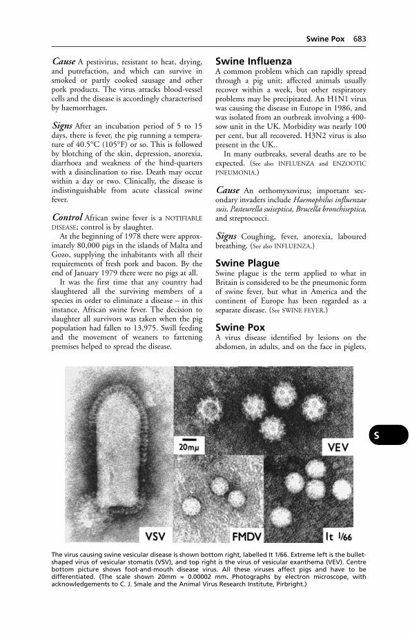
Cause A pestivirus, resistant to heat, drying,and putrefaction, and which can survive insmoked or partly cooked sausage and otherpork products. The virus attacks blood-vesselcells and the disease is accordingly characterisedby haemorrhages.
Signs After an incubation period of 5 to 15days, there is fever, the pig running a tempera-ture of 40.5°C (105°F) or so. This is followedby blotching of the skin, depression, anorexia,diarrhoea and weakness of the hind-quarterswith a disinclination to rise. Death may occurwithin a day or two. Clinically, the disease isindistinguishable from acute classical swinefever.
Control African swine fever is a NOTIFIABLE
DISEASE; control is by slaughter.At the beginning of 1978 there were approx-
imately 80,000 pigs in the islands of Malta andGozo, supplying the inhabitants with all theirrequirements of fresh pork and bacon. By theend of January 1979 there were no pigs at all.
It was the first time that any country hadslaughtered all the surviving members of aspecies in order to eliminate a disease – in thisinstance, African swine fever. The decision toslaughter all survivors was taken when the pigpopulation had fallen to 13,975. Swill feedingand the movement of weaners to fatteningpremises helped to spread the disease.
Swine InfluenzaA common problem which can rapidly spreadthrough a pig unit; affected animals usuallyrecover within a week, but other respiratoryproblems may be precipitated. An H1N1 viruswas causing the disease in Europe in 1986, andwas isolated from an outbreak involving a 400-sow unit in the UK. Morbidity was nearly 100per cent, but all recovered. H3N2 virus is alsopresent in the UK..
In many outbreaks, several deaths are to beexpected. (See also INFLUENZA and ENZOOTIC
PNEUMONIA.)
Cause An orthomyxovirus; important sec-ondary invaders include Haemophilus influenzaesuis, Pasteurella suiseptica, Brucella bronchiseptica,and streptococci.
Signs Coughing, fever, anorexia, labouredbreathing. (See also INFLUENZA.)
Swine PlagueSwine plague is the term applied to what inBritain is considered to be the pneumonic formof swine fever, but what in America and thecontinent of Europe has been regarded as a separate disease. (See SWINE FEVER.)
Swine PoxA virus disease identified by lesions on theabdomen, in adults, and on the face in piglets,
Swine Pox 683
S
The virus causing swine vesicular disease is shown bottom right, labelled It 1/66. Extreme left is the bullet-shaped virus of vesicular stomatis (VSV), and top right is the virus of vesicular exanthema (VEV). Centre bottom picture shows foot-and-mouth disease virus. All these viruses affect pigs and have to be differentiated. (The scale shown 20mm = 0.00002 mm. Photographs by electron microscope, withacknowledgements to C. J. Smale and the Animal Virus Research Institute, Pirbright.)

in which the disease is more serious; they alsohave conjunctivitis and some may die. Thecause is infection with swine poxvirus and/orvaccinia virus (see POX).
Swine Vesicular Disease (SVD)A NOTIFIABLE DISEASE. An enterovirus diseasewhose signs resemble those of foot-and-mouthdisease. It first appeared in the UK in 1972. Inthe Staffordshire outbreak of that year it was at first mistaken for foot-and-mouth disease,from which it cannot be differentiated on clinical grounds alone. However, it was shownat the Animal Virus Research Institute,Pirbright, that the virus was not that of foot-and-mouth disease but related to anenterovirus which had caused outbreaks in Italyand Hong Kong.
All cases in the UK were linked either to swillfeeding of pigs or to the movement of pigs frominfected to clean premises. The disease appearsto be spread rapidly through contact, with anincubation period of perhaps 4 to 8 days.Airborne infection appears less likely than withfoot-and-mouth disease.
A similar disease has been reported in Austriaamong pigs imported from Poland, and also inFrance.
SVD virus is very closely related to CoxsackieB5 virus, which causes not only influenza-likesymptoms in man but also sometimes heart disease and meningitis. It is thought possiblethat SVD arose as a result of pigs becominginfected by people ill because of Coxsackie B5 virus, which locally then became adapted topigs or underwent mutation.
SVD has been transmitted to laboratoryworkers, so precautions must be taken.
Control Experience has shown that the incidence of the disease has been quicklyreduced by the imposition of Controlled Areameasures, and this fact led to the Movementand Sale of Pigs Order 1975, and subsequentlegislation, designed to slow down the move-ment of pigs so that infection can show up andbe dealt with before it spreads further.
Licences are required for all movement ofpigs; entry of pigs on to a farm precludes movement of animals off that farm for 21 days,except for those going direct to slaughter. Swill-fed pigs can move only to a slaughter-house. All pigs consigned to a slaughter marketor to a slaughterhouse must be marked with ared cross of specified dimensions.
Sporadic outbreaks have occurred since the original outbreak. The infection can be subclinical.
Mode of Infection Although the SVD virusbelongs to the enterovirus group, it has beendifficult to obtain evidence for infection bymouth. Many experiments, in which precau-tions were taken to prevent entry of virus byother routes, have failed to produce the disease.In contrast, infection by rubbing or scarificationof the skin regularly produces infection, and itseems that the most likely route of infection inthe field is through damaged skin.
Swinge CoatAn abnormality in which the hair is short,sparse and curly.
Swollen Head Syndrome ofChickens (SHS)
Signs An oedematous swelling beginninground the eyes and progressing to the inter-mandibular tissue. There is coughing and anasal discharge. Opisthotonus may be seen. Ifpicked up the birds become incoordinated, rollover, and have difficulty in regaining a normalposture; diarrhoea may be seen. The diseaselasts about 2 weeks.
Cause A paramyxovirus.
SymbiosisSymbiosis means an obligatory associationbetween 2 different species for their mutualbenefit.
Sympathetic Nervous System(see CENTRAL NERVOUS SYSTEM; AUTONOMIC
NERVOUS SYSTEM)
SymphysisA joint, in which bones are united by a flattened disc of fibro-cartilage.
Syn-Syn- is a prefix signifying union.
Synapse(see NERVES)
Synchronisation Of Oestrus(see CONTROLLED BREEDING)
Syncope (Fainting)Syncope (fainting) is generally due to cerebralanaemia occurring through weakened pulsationof the heart, sudden shock, or severe injury.
It is common in dogs and cats, especiallywhen old; cases have however been seen in allanimals.
684 Swine Vesicular Disease (SVD)
S
Syncytial Viruses(see RESPIRATORY SYNCYTIAL VIRUS)
SyncytiumTissue composed of a mass of nucleated protoplasm without cell boundaries, such as the outer layer of the trophoblast of a placenta;or a mass of cells united by protoplasmicbridges.
SyndromeA group of symptoms.
SynechiaAdhesions in the eye, e.g. involving the tissuesof the iris to the cornea or lens (see EYE,DISEASES OF – Iritis).
SynergismSynergism is the opposite of antagonism.Synergism between drugs, e.g. trimethoprimand sulfadiazine, may be of practical value, for with the two it may be possible to obtain the required effect with a dosage of one which,if used alone, would be insufficient, but which cannot be increased because largeramounts would cause side-effects. Anotheradvantage of using two drugs is the possibilitythat this would tend to prevent the multiplica-tion of strains resistant to one of the compounds.
The word ‘synergism’ is also used to describean interaction between a virus and bacteria in their combined invasion of, for example, the lungs; implying that the result of the ‘combined forces’, as it were, is greater than the sum of the effects produced by the agentsindividually. Synergism occurs in calf pneumo-nia between Mycoplasma bovis and Pasteurellahaemolytica. (For another example, see SWINE
DYSENTERY.)
SynostosisSynostosis is the term applied to a union bybony material of adjacent bones usually sepa-rate. It may occur in the spinal column in oldanimals. (See also HORSES, BACK TROUBLES IN.)
SynotiaThe (virtual) absence of head in a stillborn animal.
Synovial MembraneSynovial membrane forms the lining coveringthe surfaces of the opposed articular cartilages,which enter into the formation of a joint. (See
JOINTS.)
SynovitisInflammation of the membrane lining a joint.It is usually accompanied by effusion of fluidwithin the synovial sac of the joint. It is foundin various injuries and inflammation of joints.
Synovitis, InfectiousThis is a disease of chicks, of about 2 to 10weeks old, and of turkeys; first diagnosed inBritain in 1959.
Cause Mycoplasma synoviae.
Signs Reluctance to move, lameness, swellingof joints, anorexia.
The confined conditions under which broilers are raised appear to render them partic-ularly susceptible to this disease. Mortality islow, but a third of the survivors may be down-graded, so that severe financial loss may becaused. Control depends upon hygiene, andbeing careful about the breeding stock.
Syringe, HypodermicA pump-like device used to introduce solutionsto, or withdraw them from, the body. (see INFEC-
TIONS, DETERGENT RESIDUE; also PROJECTILE
SYRINGE)
SystoleSystole means the contraction of the heart asopposed to the resting phase, which is called‘diastole’, and which alternates with the formercontracting period. In the cardiac cycle systoletakes about one-third, and diastole about two-thirds, of the whole period of the heartbeat. (See HEART DISEASES.)
Systole 685
S

T-CellsLYMPHOCYTES from the thymus gland con-cerned with cell-mediated immunity. (See
IMMUNE RESPONSE.)
T2 ToxinThis fungal toxin may poison cattle or poultryeating stored corn containing the fungusFusarium tricinctum. In cattle, the toxin maycause multiple haemorrhages and sometimesdeath; in poultry, there may be mouth lesions.
TachycardiaTachycardia is a disturbance of the heart’saction which produces great acceleration of thepulse.
TachypnoeaAn increase in the rate of breathing due to somepathological condition. (See BREATHLESSNESS;PARAQUAT POISONING.)
Taenia(see TAPEWORMS)
Tail, Amputation ofAmputation of the tail (docking) is, or hasbeen, undertaken for a variety of reasons. In the UK the Royal College of VeterinarySurgeons has ruled that docking a puppy’s tail is an unethical procedure except when it is done for prophylactic or therapeutic reasons.Docking by lay persons is illegal. In cattle,amputation of the tail is illegal except followinginjury and must, except in an emergency, beundertaken by a veterinary surgeon. Pigs’ tailsare often docked to prevent tail-biting. Lambstails are docked to prevent faecal soiling and fly strike (see under DOCKING; LAW; WELFARE
CODES).
Tail-BitingIn pigs this ‘vice’ can be of great economicimportance. There are various reasons why itoccurs: boredom, absence of bedding, and overcrowding (floor space of less than 1.5 m2 (5square feet) per pig), are regarded as conducive to tail-biting. High temperature and humidity are possible causes. Bitten tailsrequire amputation or dressing if pyaemia is tobe prevented.
Tail sores in pigs These may follow tail-biting by 1 or 2 pigs out of a large batch,and if untreated can lead to pyaemia.
In 6 months, out of 135 pig carcases con-demned in an Oslo abattoir, 56 were affectedwith pyaemia – and of these, 43 had tail sores.
Talfan Disease (TeschenDisease; Porcine ViralEncephalomyelitis)This disease of pigs was first recognised in theCzech Republic and occurs throughout Europe.In the UK, it was made a NOTIFIABLE DISEASE
in 1974. Its cause is an enterovirus.Experimentally, the incubation period is statedto be 12 days. Piglets 3 weeks old and upwardsare affected; adult pigs may be infected butshow no clinical signs. By no means all pigletsin a litter or on a farm become ill, and the mortality is usually low. The main symptom isweakness or paralysis of the hind-legs. There is little or no fever or loss of appetite. Recoveryoccurs in a proportion of animals which arehand-fed. The disease is present in Britain to asmall extent, and apparently may be associatedwith abortion.
TampanA soft tick of the family Argasidae. (See TICKS.)
Tamponade, CardiacA rapid accumulation of blood or other fluid inthe pericardial sac, compressing the heart andsometimes suddenly arresting its function.
Tannin (Tannic Acid)Tannin (tannic acid) is a non-crystallisablewhite or pale-yellowish powder, which is soluble in water and glycerine. It is preparedfrom oak-galls, and is found in strong tea orcoffee. When brought into contact with amucous surface, tannin causes constriction ofthe blood vessels. When brought into contactwith many poisonous alkaloids it renders them temporarily inert by forming the insoluble tannate, and so is a valuable antidote.
Uses Tannic acid has been used in diarrhoeaand dysentery in young animals, usually as catechu or kino – 2 vegetable drugs which contain a large amount of tannin. It is oftenadministered, in the form of strong tea, as the first step in the antidotal treatment of poisoning by ALKALOIDS.
Tannic-acid jelly is a valuable burn dressing.It lessens the absorption of breakdown productsfrom the burned area and hence diminishes the
T

secondary effects of a serious burn. It is notsuitable for extensive areas owing to the dangerof liver damage if large quantities are absorbed.
Tapetum(see EYE)
TapewormsAn intestinal parasite commonly found in vertebrates. Their life-cycle requires 2 hosts,sometimes 3. The presence of the adult wormmay give rise to few if any symptoms or, on theother hand, to anaemia, indigestion, and nervous symptoms – or even to blockage of the intestine. The cystic stage of tapewormsmay involve the brain. Tapeworms are of considerable public-health importance.
A typical tapeworm has a head or scolex, provided with suckers and, in some species,with hooks also.
Behind the scolex follows a neck, and behindthat are the segments, each being called a proglot-tis. The segments nearest to the head are thesmallest, and are immature. Next follow mature
segments, and lastly the gravid segments contain-ing eggs. These older segments fall off and arepassed out of the host’s body in the faeces.
Taenia. This is the common genus of wormsfound in dogs and cats, and includes:
T. pisiformis (T. serrata) is one of the com-monest. Its cystic stage, Cysticercus pisiformis, isfound in rabbits and hares.
T. hydatigena (T. marginata) is the largestform, with mature segments wider than long. It may reach a length of over 5 metres (16 feet).Its cystic stage, C. tenuicollis, occurs in the viscera of various animals, especially sheep, cattle and pigs. T. ovis is frequently mistaken for the last form, from which it can be distinguished only by microscopical examina-tion. Its cysticercus, C. ovis, is found in themuscles and organs of sheep and goats. It is asmall form, easily overlooked.
T. multiceps (T. coernurus) is a more delicateform than the others, semi-translucent. Theintermediate stage is a coenurus, found in thenervous system of sheep and other ruminantsand man.
T. serialis is a more robust form, its coenurusbeing found in rabbits and hares. Only 1species is common in the cat, T. taeniaeformis(T. crassicollis). The cystic stage C. fasciolaris isfound in the liver of rats and mice.
Tapeworms 687
T
A typical tapeworm. Each segment is called aproglottis. (From H. T. B. Hall, Diseases andParasites of Livestock in the Tropics, Longman.)
Taenia. Head, mature and gravid segments.
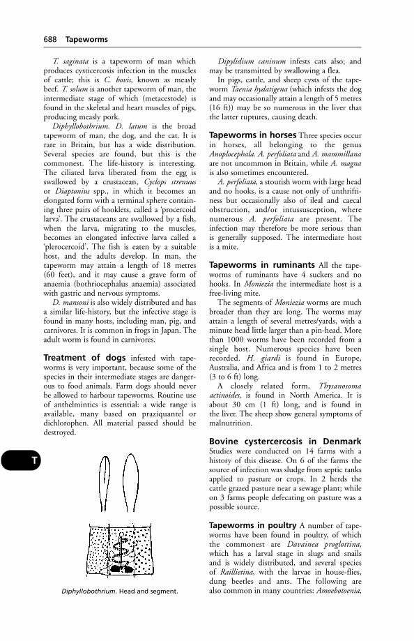
T. saginata is a tapeworm of man which produces cysticercosis infection in the musclesof cattle; this is C. bovis, known as measly beef. T. solum is another tapeworm of man, theintermediate stage of which (metacestode) isfound in the skeletal and heart muscles of pigs,producing measly pork.
Diphyllobothrium. D. latum is the broadtapeworm of man, the dog, and the cat. It israre in Britain, but has a wide distribution.Several species are found, but this is the commonest. The life-history is interesting. The ciliated larva liberated from the egg is swallowed by a crustacean, Cyclops strenuusor Diaptonius spp., in which it becomes anelongated form with a terminal sphere contain-ing three pairs of hooklets, called a ‘procercoidlarva’. The crustaceans are swallowed by a fish,when the larva, migrating to the muscles,becomes an elongated infective larva called a‘plerocercoid’. The fish is eaten by a suitablehost, and the adults develop. In man, the tapeworm may attain a length of 18 metres (60 feet), and it may cause a grave form ofanaemia (bothriocephalus anaemia) associatedwith gastric and nervous symptoms.
D. mansoni is also widely distributed and hasa similar life-history, but the infective stage isfound in many hosts, including man, pig, andcarnivores. It is common in frogs in Japan. Theadult worm is found in carnivores.
Treatment of dogs infested with tape-worms is very important, because some of thespecies in their intermediate stages are danger-ous to food animals. Farm dogs should never be allowed to harbour tapeworms. Routine useof anthelmintics is essential: a wide range isavailable, many based on praziquantel ordichlorophen. All material passed should bedestroyed.
Dipylidium caninum infests cats also; andmay be transmitted by swallowing a flea.
In pigs, cattle, and sheep cysts of the tape-worm Taenia hydatigena (which infests the dogand may occasionally attain a length of 5 metres(16 ft)) may be so numerous in the liver thatthe latter ruptures, causing death.
Tapeworms in horses Three species occurin horses, all belonging to the genusAnoplocephala. A. perfoliata and A. mammillanaare not uncommon in Britain, while A. magnais also sometimes encountered.
A. perfoliata, a stoutish worm with large headand no hooks, is a cause not only of unthrifti-ness but occasionally also of ileal and caecalobstruction, and/or intussusception, wherenumerous A. perfoliata are present. The infection may therefore be more serious than is generally supposed. The intermediate host is a mite.
Tapeworms in ruminants All the tape-worms of ruminants have 4 suckers and nohooks. In Moniezia the intermediate host is afree-living mite.
The segments of Moniezia worms are muchbroader than they are long. The worms mayattain a length of several metres/yards, with aminute head little larger than a pin-head. Morethan 1000 worms have been recorded from asingle host. Numerous species have beenrecorded. H. giardi is found in Europe,Australia, and Africa and is from 1 to 2 metres(3 to 6 ft) long.
A closely related form, Thysanosoma actinoides, is found in North America. It isabout 30 cm (1 ft) long, and is found in the liver. The sheep show general symptoms ofmalnutrition.
Bovine cystercercosis in DenmarkStudies were conducted on 14 farms with a history of this disease. On 6 of the farms thesource of infection was sludge from septic tanksapplied to pasture or crops. In 2 herds the cattle grazed pasture near a sewage plant; whileon 3 farms people defecating on pasture was apossible source.
Tapeworms in poultry A number of tape-worms have been found in poultry, of whichthe commonest are Davainea proglottina, which has a larval stage in slugs and snails and is widely distributed, and several species of Raillietina, with the larvae in house-flies,dung beetles and ants. The following are also common in many countries: Amoebotoenia,
688 Tapeworms
T
Diphyllobothrium. Head and segment.
with larvae in earthworms; and Hymenolepis ofvarious species, some of which may be verynumerous in individual birds.
‘Measles’ in beef due to the presence of thecyst stage (Cysticercus bovis) of the tapewormTaenia saginata, which is a parasite of man.Cattle swallow the eggs of the adult tapeworm,and these hatch in the intestines, liberatingyoung embryos, which burrow until they settlein muscle fibre or connective tissues. Here they appear as small oval cysts, containing fluid,and each possessing the head of a potentialtapeworm.
‘Measles’ in pork is due to the presence of the cyst stage (Cysticercus cellulosae) of thetapeworm of man, Taenia solium. It is extremely common among pigs in easternlands, which have access to garbage and humanfaeces, from whence they pick up the eggspassed through the human intestines. The eggs undergo a development similar to those ofthe beef-measles tapeworm. Man may also himself harbour the cystic stage.
Cysticercosis in man Very high sporadicinfection rates have been found in Africa withTaenia saginata and T. solium, the 2 tapewormsof major importance in man. Where T. soliumis present, serious human infections with thecysticercus stage may be observed, as well asmild infections with the adult tapeworm.When it occurs in beef cattle, the cysticercus ofT. saginata is a major economic problem and aserious obstacle to the export of meat.
A single human carrier of T. saginata led toan outbreak of cysticercosis among cattle on alarge farm in the USA.
Coenuriasis (gid or sturdy) in sheepThis disease is caused by the pressure of cysts ofthe tapeworm Taenia multicepts on cells of thebrain (or spinal cord).
Sheep become infested by swallowing theunhatched eggs, excreted in a dog’s faeces, whilegrazing. In the digestive tract the eggs hatch,and pass via the bloodstream to various parts of the body; only those reaching the central nervous system develop. Here they form smallcysts, each containing 1 tapeworm head: thislarval stage is known as Coenurus cerebralis.Over a period of months, each cyst increases in size, and more heads are budded from thelining membrane of the translucent cyst wall.Eventually a single coenurus may contain 50 or 100 or more tapeworm heads (scolices) projecting inwards.
The life-cycle is completed if a dog eats thehead of an infested sheep.
Signs These include impairment of vision, astaggering or high-stepping gait, circling, andstanding with head lowered, raised, or pressedagainst an object. Backward somersaults havebeen recorded. Recumbency and opisthotonusmay occur. A softening of the bone of the skull,due to internal pressure of the cysts, is found ina proportion of cases.
Diagnosis Where there is no softening of theskull, a guide to the location of the cyst may begiven by interpretation of the neurological signsas indicated by the sheep’s behaviour. An intra-dermal test has been used: 0.1 ml of cyst fluid is injected into a shaved area of skin.Thickening of the skin within 24 hours indicates the presence of a cyst in the animal.
Treatment Physical removal of the cyst maybe attempted. The sheep is anaesthetised and,in the absence of any skull softening, a trephineused to remove a disc of bone 1.5 cm (0.6 in) indiameter. Draining the fluid from the cystbefore its removal obviates the need to enlargethe hole. The cyst is then removed completely.(If this is not done, the remaining cyst wall isapparently capable of replacing the fluid.)
Hydatid disease is caused by the cystic larval stage of the tapeworm Echinococcus granulosus, of which the dog and fox are theusual hosts. Eggs released from tapeworm segments passed in the faeces by these animalsare later swallowed by grazing cattle, sheep andhorses, which may become infested alsothrough drinking water contaminated by wind-blown eggs.
People become infested through swallowingeggs attached to inadequately washed vegeta-bles, and possibly eggs may be inhaled in dustor carried by flies to uncovered food. The handling of infested dogs is an importantsource. In Beirut, the risk is put at 21 timesgreater for dog-owners than others, by theWorld Health Organisation, which states alsothat in California nomadic sheep-rearers are1000 times more likely to have hydatid diseasethan other inhabitants of the state. (WHOTechnical Report 637).
There have been successful campaigns tocontrol human hydatid disease in both Cyprusand Iceland, by compulsory treatment and/orbanning of dogs.
Swallowed eggs hatch in the intestines andare carried via the portal vein to the liver. Some
Tapeworms 689
T
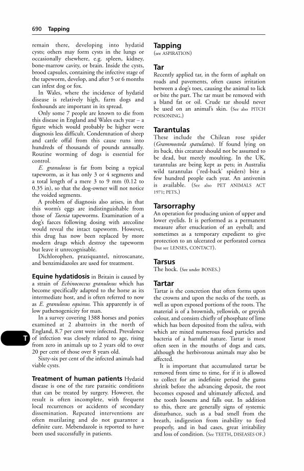
remain there, developing into hydatid cysts; others may form cysts in the lungs oroccasionally elsewhere, e.g. spleen, kidney,bone-marrow cavity, or brain. Inside the cysts,brood capsules, containing the infective stage ofthe tapeworm, develop, and after 5 or 6 monthscan infest dog or fox.
In Wales, where the incidence of hydatid disease is relatively high, farm dogs and foxhounds are important in its spread.
Only some 7 people are known to die fromthis disease in England and Wales each year – afigure which would probably be higher werediagnosis less difficult. Condemnation of sheepand cattle offal from this cause runs into hundreds of thousands of pounds annually.Routine worming of dogs is essential for control.
E. granulosus is far from being a typical tapeworm, as it has only 3 or 4 segments and a total length of a mere 3 to 9 mm (0.12 to 0.35 in), so that the dog-owner will not noticethe voided segments.
A problem of diagnosis also arises, in thatthis worm’s eggs are indistinguishable fromthose of Taenia tapeworms. Examination of adog’s faeces following dosing with arecolinewould reveal the intact tapeworm. However,this drug has now been replaced by more modern drugs which destroy the tapeworm but leave it unrecognisable.
Dichlorophen, praziquantel, nitroscanate,and benzimidazoles are used for treatment.
Equine hydatidosis in Britain is caused bya strain of Echinococcus granulosus which hasbecome specifically adapted to the horse as itsintermediate host, and is often referred to nowas E. granulosus equinus. This apparently is oflow pathenogenicity for man.
In a survey covering 1388 horses and poniesexamined at 2 abattoirs in the north ofEngland, 8.7 per cent were infected. Prevalenceof infection was closely related to age, risingfrom zero in animals up to 2 years old to over20 per cent of those over 8 years old.
Sixty-six per cent of the infected animals hadviable cysts.
Treatment of human patients Hydatiddisease is one of the rare parasitic conditionsthat can be treated by surgery. However, theresult is often incomplete, with frequent local recurrences or accidents of secondary dissemination. Repeated interventions are often mutilating and do not guarantee a definite cure. Mebendazole is reported to havebeen used successfully in patients.
Tapping(see ASPIRATION)
TarRecently applied tar, in the form of asphalt onroads and pavements, often causes irritationbetween a dog’s toes, causing the animal to lickor bite the part. The tar must be removed witha bland fat or oil. Crude tar should never be used on an animal’s skin. (See also PITCH
POISONING.)
TarantulasThese include the Chilean rose spider(Grammostola spatulatus). If found lying on its back, this creature should not be assumed tobe dead, but merely moulting. In the UK,tarantulas are being kept as pets; in Australiawild tarantulas (‘red-back’ spiders) bite a few hundred people each year. An antivenin is available. (See also PET ANIMALS ACT
1971; PETS.)
TarsorraphyAn operation for producing union of upper andlower eyelids. It is performed as a permanentmeasure after enucleation of an eyeball; andsometimes as a temporary expedient to giveprotection to an ulcerated or perforated cornea(but see LENSES, CONTACT).
TarsusThe hock. (See under BONES.)
TartarTartar is the concretion that often forms uponthe crowns and upon the necks of the teeth, aswell as upon exposed portions of the roots. Thematerial is of a brownish, yellowish, or greyishcolour, and consists chiefly of phosphate of limewhich has been deposited from the saliva, withwhich are mixed numerous food particles andbacteria of a harmful nature. Tartar is mostoften seen in the mouths of dogs and cats,although the herbivorous animals may also beaffected.
It is important that accumulated tartar beremoved from time to time, for if it is allowedto collect for an indefinite period the gumsshrink before the advancing deposit, the rootbecomes exposed and ultimately affected, andthe tooth loosens and falls out. In addition to this, there are generally signs of systemic disturbance, such as a bad smell from thebreath, indigestion from inability to feed properly, and in bad cases, great irritability and loss of condition. (See TEETH, DISEASES OF.)
690 Tapping
T
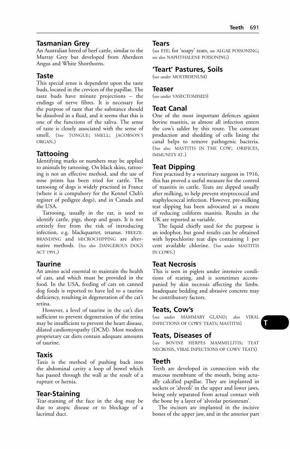
Tasmanian GreyAn Australian breed of beef cattle, similar to theMurray Grey but developed from AberdeenAngus and White Shorthorns.
TasteThis special sense is dependent upon the tastebuds, located in the crevices of the papillae. Thetaste buds have minute projections – the endings of nerve fibres. It is necessary for the purpose of taste that the substance shouldbe dissolved in a fluid, and it seems that this isone of the functions of the saliva. The sense of taste is closely associated with the sense ofsmell. (See TONGUE; SMELL; JACOBSON’S
ORGAN.)
TattooingIdentifying marks or numbers may be appliedto animals by tattooing. On black skins, tattoo-ing is not an effective method, and the use ofnose prints has been tried for cattle. The tattooing of dogs is widely practised in France(where it is compulsory for the Kennel Club’sregister of pedigree dogs), and in Canada andthe USA.
Tattooing, usually in the ear, is used to identify cattle, pigs, sheep and goats. It is notentirely free from the risk of introducing infection, e.g. blackquarter, tetanus. FREEZE-
BRANDING and MICROCHIPPING are alter-native methods. (See also DANGEROUS DOGS
ACT 1991.)
TaurineAn amino acid essential to maintain the healthof cats, and which must be provided in thefood. In the USA, feeding of cats on canneddog foods is reported to have led to a taurinedeficiency, resulting in degeneration of the cat’sretina.
However, a level of taurine in the cat’s dietsufficient to prevent degeneration of the retinamay be insufficient to prevent the heart disease,dilated cardiomyopathy (DCM). Most modernproprietary cat diets contain adequate amountsof taurine.
TaxisTaxis is the method of pushing back into the abdominal cavity a loop of bowel which has passed through the wall as the result of arupture or hernia.
Tear-StainingTear-staining of the face in the dog may be due to atopic disease or to blockage of alacrimal duct.
Tears(see EYE; for ‘soapy’ tears, see ALGAE POISONING;see also NAPHTHALENE POISONING)
‘Teart’ Pastures, Soils(see under MOLYBDENUM)
Teaser(see under VASECTOMISED)
Teat CanalOne of the most important defences againstbovine mastitis, as almost all infection entersthe cow’s udder by this route. The constant production and shedding of cells lining thecanal helps to remove pathogenic bacteria. (See also MASTITIS IN THE COW; ORIFICES,IMMUNITY AT.)
Teat DippingFirst practised by a veterinary surgeon in 1916,this has proved a useful measure for the controlof mastitis in cattle. Teats are dipped usuallyafter milking, to help prevent streptococcal andstaphylococcal infection. However, pre-milkingteat dipping has been advocated as a means of reducing coliform mastitis. Results in theUK are reported as variable.
The liquid chiefly used for the purpose is an iodophor, but good results can be obtainedwith hypochlorite teat dips containing 1 percent available chlorine. (See under MASTITIS
IN COWS.)
Teat NecrosisThis is seen in piglets under intensive condi-tions of rearing, and is sometimes accom-panied by skin necrosis affecting the limbs.Inadequate bedding and abrasive concrete maybe contributory factors.
Teats, Cow’s(see under MAMMARY GLAND; also VIRAL
INFECTIONS OF COWS’ TEATS; MASTITIS)
Teats, Diseases of(see BOVINE HERPES MAMMILLITIS; TEAT
NECROSIS, VIRAL INFECTIONS OF COWS’ TEATS)
TeethTeeth are developed in connection with themucous membrane of the mouth, being actu-ally calcified papillae. They are implanted insockets or ‘alveoli’ in the upper and lower jaws,being only separated from actual contact withthe bone by a layer of ‘alveolar periosteum’.
The incisors are implanted in the incisivebones of the upper jaw, and in the anterior part
Teeth 691
T
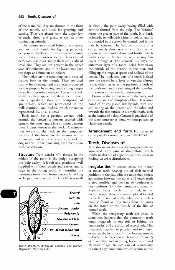
of the mandible; they are situated in the frontof the mouth, and used for grasping and cutting. They are absent from the upper jaw of cattle, sheep, and goats, as well as otherruminating animals.
The canines are situated behind the incisors,and are used mainly for fighting purposes,being most developed in carnivores and omni-vores. They are useless to the domesticated herbivorous animals, and in them are usually ofsmall size. They are not present in the upperjaws of ruminants, and in the lower jaws havethe shape and function of incisors.
The molars are the remaining teeth, situatedfurther back in the mouth. They are used mainly for chewing, and are specially adaptedfor this purpose by having broad strong irregu-lar tables or grinding surfaces. The term ‘cheekteeth’ is often applied to these teeth, since,strictly speaking, they are composed of ‘pre-molars’, which are represented in the milk dentition, and ‘molars’, which are not sorepresented. (See DENTITION.)
Each tooth has a portion covered with enamel, the ‘crown’; a portion covered withcement, the ‘root’; and a line of union betweenthese 2 parts known as the ‘neck’. A constric-tion occurs at the neck in the temporaryincisors of the horse, in the incisors of the ruminants, and in incisors and molars of thedog and cat; in the remaining teeth there is nosuch constriction.
Structure Teeth consist of 4 tissues. In themiddle of the tooth is the ‘pulp’, occupying the ‘pulp cavity’. It is soft and gelatinous, wellsupplied with blood vessels and nerves, and islarge in the young tooth. It nourishes theremaining tissues, and forms dentine for as longas the pulp cavity is open. In later life it is small
or absent, the pulp cavity having filled withdentine formed from the pulp. The ‘dentine’forms the greater part of the tooth. It is hard,yellowish, or yellowish-white in colour, and issurrounded in the crown by enamel, and in theroot by cement. The ‘enamel’ consists of a comparatively thin layer of a brilliant whitecolour and extremely dense and brittle, whichforms a cap to the dentine, or is arranged in layers through it. The ‘cement’ is always theoutermost layer of a tooth, being formed on the outside of the dentine in the root, and filling up the irregular spaces and hollows of thecrown. The implanted part of a tooth is fixedinto the socket by a layer of vascular fibrous tissue, which serves as the periosteum both ofthe tooth root and of the lining of the alveolus.It is known as the ‘alveolar periosteum’.
Enamel is the hardest tissue in the body, andconsists mainly of phosphate of lime. It is com-posed of prisms placed side by side, with oneend resting on the dentine and the other endtowards the free surface in a simple tooth, suchas the canine of a dog. Cement is practically ofthe same structure as bone, without possessingHaversian canals.
Arrangement and form For times of cutting of the various teeth, see DENTITION.
Teeth, Diseases ofMost diseases or disorders affecting the teeth areassociated with pain or discomfort, whichresults in absence of appetite, capriciousness infeeding, or other disturbances.
Irregularities In certain cases, the incisor or molar teeth develop out of their normal positions in the jaw, with the result that perfectapposition between the upper and lower teeth is not possible, and the rate of toothwear is not uniform. In other instances, extra or ‘supernumerary’ teeth are formed; in the incisor region these are usually placed behindthe arch of normal teeth, while extra molarsmay be found as projections from the gums on the inside or the outside of the line of normal teeth.
When the temporary teeth are shed, it sometimes happens that the permanent teetherupt irregularly to one side or behind the temporaries, and are distorted accordingly. Thisfrequently happens in puppies, and to a lesserextent in the herbivora. In the former, trouble is likely to be experienced between 31⁄2 and 5 or 6 months, and in young horses at 21⁄2 and 31⁄2 years of age. In such cases it is necessary to extract any temporaries which persist, so that
692 Teeth, Diseases of
T
Tooth structure. (From de Coursey, The HumanOrganism, McGraw-Hill.)

the permanent teeth can arrive in their properplaces in the mouth.
In dogs frequently, in sheep sometimes, andin other animals less commonly, there may be adiscrepancy in length between the upper andlower jaws. When the upper jaw is too long, thecondition is known as an ‘overshot jaw’, andwhen the lower jaw projects too far forward, it is popularly spoken of as an ‘undershot jaw’.In bulldogs, pugs, and other breeds of dogswith very short upper jaws the undershot condition is practically normal, while in certainbreeds with extremely long upper jaws, such asthe greyhound and show collie, overshot jawsare very common.
Abnormal wear, which is due to malforma-tions of the jaws, to excessive softness of the teeth, or to the direction of the teeth, isanother mechanical cause of tooth disorder. (See SOIL-CONTAMINATED HERBAGE with reference to sheep.)
Abnormal wear varies in different cases, andis productive of some well-known conditions,as follows: (1) shear mouth, in which the molarteeth of the upper and lower jaws wear so thatin time they appear like the blades of a pair ofsheep-shears, the upper row being worn awayon its inner border, and the lower one along itsouter border; (2) step mouth, where the cheekteeth, instead of being all at the same level, are arranged with some higher than others,somewhat like steps – a high tooth in the lowerjaw being opposite a short one in the corre-sponding upper jaw; (3) overhanging upperjaw, which is where the first upper cheek toothon either side is placed too far forward in the mouth, and does not come into accurateapposition with the tooth immediately below it, causing the formation of a hook – at thesame time the last lower cheek tooth is situatedtoo far back and also forms a hook; and (4)curved tables, where the line of cheek teeth inthe upper jaw shows a convexity in its centre,and a corresponding concavity exists in thelower row.
Signs In most of these instances the animalaffected (almost always a member of the horsetribe), instead of chewing its food and swallow-ing it in the usual way, rolls it round and roundin the mouth until it collects into a soddenmass, often about the size of a couple of fingers,and puts it out of the mouth instead of swallowing it. (See QUIDDING.) Pain may beshown when the hand is passed along the outside of the cheek, especially when pressure is put upon the line of teeth.
Treatment Rasping the teeth by means of a special tooth-rasp will reduce smaller irregularities, and bring the teeth back intotheir proper function.
Caries is not synonymous with tooth decay,although the term – borrowed from humandentistry – is often used in veterinary practiceto include all tooth decay.
Caries is the destruction of the tooth enameland invasion of the dentine by bacteria, resulting in the formation of a cavity. Truecaries has been confirmed in dogs but is comparatively rare in farm animals.
Neck lesions in cats’ teeth A painfulcondition affecting middle-aged to elderly cats,characterised by cavitation of the necks of teeth.This makes extraction difficult because of therisk of breakage of crowns.
Inflammations of the periosteumlining the root cavity of a tooth are common.They may be due to small particles of food getting forced down into the socket of thetooth, to fractures or fissures of the teeth, tocaries, tumour formation, depositions of tartar,and to certain specific diseases, such as actinomycosis, etc.
Signs These vary from a slight redness of thegum around the root of the tooth, which ispainful when pressed by the finger, to a largesuppurating tract running alongside the root ofthe tooth down into its socket, and perhapsthrough the skin to the outside or into one orother of the sinuses. Abscess formation in thetooth socket may take place, and the abscessmay burst into the mouth, to the outsidethrough the skin, or up into a sinus. In manycases there is a distinct bulge of the surfaceabove the diseased tooth, which may give to theface a one-sided appearance.
Treatment The affected tooth or teeth mustbe extracted, and the areas of suppurationcleansed and curetted if necessary. The cavityusually has to be packed with antiseptic gauzeafterwards for a few days until it begins to fill by healthy granulation tissue.
Periodontal disease is a name for chronicinfection of the periodontal membrane. It isone form of inflammation of the periosteum, oralveolar periostitis. It causes loosening andshedding of the teeth, pain, failure to masticate,and loss of weight.
Teeth, Diseases of 693
T

Odontomata are tumours formed in connection with the root of one tooth, or theymay be found in the jaw, sinuses, or eveninvolving part of the nasal passage, and be composite or compound, when multitudes ofsmall rudimentary teeth are present. They cause swelling and bulging of the surface of theface, and can only be treated surgically.
Porphyria gives rise to a pink or brown discoloration of teeth. (See under BONE,DISEASES OF.)
Toothache is most spectacular in the dog,which rubs its mouth along the ground, paws atits nose or mouth, works its jaws, salivates, andmay whine or moan.
A veterinary surgeon will offer a diagnosisand initiate the necessary treatment.
‘Broken mouth’ is important in hill sheep.(See under main dictionary heading.)
Fractures of the canine teeth in dogs are notuncommon. If the pulp is exposed, subsequentinfection can lead to a painful abscess.Extraction of the remainder of the tooth obviates this but, for show dogs or guard dogs,is undesirable. Metal crowns have been appliedto dogs’ teeth, but are liable to be dislodged.
Tooth transplantation has been used inveterinary practice but the results are seldomlasting, due to root resorption and bonereplacement. Fracture of the transplanted toothis likely after a couple of years or so.
Teeth, Ewes’, ‘Trimming’It has been estimated that between 60 and 70per cent of culling of ewes is on account of theirteeth. A small percentage will involve loss ofmolars or incisor wear, but the vast majoritywill be incisor loss.
Ewes have been treated for ‘bite correction’by means of an electric grinder, a practice thatoriginated in Australia. The procedure has beenstrongly condemned on welfare grounds.
(See also ‘BROKEN MOUTH’.)
Teeth ScalingThe use of ultrasonic dental scalers is widelyaccepted in veterinary dentistry. During thescaling, an aerosol of water droplets is formed,with a variable amount of periodontal debrisspattered from the patient’s mouth. In thedebris there are likely to be viruses and/or bacteria – a danger for operator, assistant, orsubsequent patient unless precautions are
taken. An aerosol of mouth flora can remainairborne for up to 30 minutes following scaling.
It is recommended that: (1) the working areashould be well ventilated – preferably withforced air extraction; (2) masks should be wornat all times by anyone in the working area; and(3) a 0.2 per cent chlorhexidine solution shouldbe used as the coolant supplied to the scalingequipment.
TelogenThe resting phase in the cycle of hair growth.
TEMTriethylenemelemine, a gametocide which, inAmerica, has been used in field trials for thecontrol of birds. The chemical is mixed withcorn, and has the effect of making the male birdinfertile. The birds continue to defend their territories and nest, but do not produce anyyoung.
Temperament, Change inThis may follow a brain tumour or infection, asoccurs in BOVINE SPONGIFORM ENCEPHALOPA-
THY and RABIES, for example. A horse maybecome bad-tempered as the result of EQUINE
VERMINOUS ARTERITIS. Poisoning may causefrenzy or aggressiveness, e.g. BENZOIC ACID
poisoning in the cat. (See also BRAIN DISEASES;STRESS; FUCOSIDOSIS.)
Temperature, Air(see under HOUSING OF ANIMALS; HEAT
EXHAUSTION; TROPICS)
Temperature, BodyBody temperature is controlled by the heat-regulating centre in the brain – the hypothalamus, which also influences blood circulation, secretion of urine, and appetite – all3 of which have a bearing on body temperature.
Heat is produced by the muscles and by thedigestive organs, and during very cold weatheror exercise, heat from the former increases,while that from the liver and other digestiveorgans decreases. The animal may also absorbheat from the sun’s rays.
Heat is lost by evaporation of water, and bysensible heat loss (see under HEAT LOSS). Waterloss is achieved via the lungs and the skin, e.g.by panting and sweating. (The dog is, for allpractical purposes, a non-sweating animal apartfrom the pads of its feet, and has to rely mainlyon panting.)
Diurnal variations in body temperature arenormal; in the early hours of the morning it is
694 Teeth, Ewes’, ‘Trimming’
T
usually at its lowest, and at its highest in the lateafternoon.
For ordinary practical purposes the usualaverage temperatures of animals are given as follows:
Horses 38.0°C (100.5°F)Cattle 38.9°C (102.0°F)Sheep, goats 40.0°C (104.0°F)Pigs 39.7°C (103.5°F)Dogs 38.3°C (101.0°F)Cats 38.6°C (102.0°F)Rabbits 38.2°C (100.8°F)Fowls 41.6°C (106.9°F)Small birds 42.5°C (108.6°F)Elephants 36.4°C (97.6°F)Camels 37.5°C (99.5°F)
Temperature-taking The most satisfactoryplace is within the rectum. In females the ther-mometer may also be inserted into the externalpart of the genital canal; as a rule, the vaginaltemperature is about half a degree higher thanthe rectal temperature, so that when a series oftemperatures is to be taken, one site or theother should be selected.
With dogs and cats, one person should holdthe animal, preferably on a table, while anotherinserts and holds the thermometer. In each animal, after the bulb of the thermometer hasbeen lubricated with a little soap or Vaseline,etc., the tail is raised vertically by the left hand,and the thermometer is inserted through theanal ring and into the rectum, by a screwingmovement if any resistance is encountered. It isheld in position for 30 seconds, or 1 minute,according to the make of the thermometer, andthen withdrawn. With a piece of cotton-woolany adherent faeces are wiped away, and thetemperature is read off. Subsequently, the ther-mometer should be washed in cold water, and acold solution of disinfectant used to disinfect it.
For purposes of temperature stress research,American scientists use a special ear thermome-ter in cattle. As in similar medical research, thistympanic thermometer is more reliable than therectal thermometer, and can sense changes assmall as 0.05°C (1⁄50°F).
Temperature in disease A high tempera-ture is one of the classic symptoms of fever, andin greater or less measure accompanies practi-cally all acute cases of disease. A comparativelysteady rise in temperature is as a rule succeededby a correspondingly steady fall, and is to belooked upon as a more favourable sign of thenatural course of a disease than when the tem-perature rises and falls with greater suddenness.The reduction of temperature in simple fevers is
in almost all cases much slower than the rise. Awavering temperature, which shows little tendency to come down to normal, generallyindicates that there is some active focus of disease, such as an abscess, which the body cannot overcome. Sudden rise in temperaturein an animal which has shown a steady fall previously is an indication of a relapse or recurrence of the disease. (See also FEVER;HYPERTHERMIA; HEAT-STROKE; TROPICS.)
Fall of temperature may be occasioned bygreat loss of blood, starvation, collapse, orcoma; it is characteristic of certain forms of kidney disease. Certain chronic diseases inwhich emaciation is marked are also associatedwith a subnormal temperature. (See also
HYPOTHERMIA.)
Temperature, near calving time Ahealthy cow – even though showing the famil-iar signs – is unlikely to calve during the next12 hours if her temperature is 39°C (102°F).This is a useful guide to herdsmen. (See also under
FEVER; HOUSING OF ANIMALS, etc.)
Temperature Control in AnimalHousing(see CONTROLLED-ENVIRONMENT HOUSING)
Temperature-Sensitive (TS)Viruses(see VACCINE)
TendernessTenderness is pain that is felt only when a diseased or injured part is handled.
TendonTendon is the dense, fibrous, slightly elasticcord that attaches the end of a muscle to thebone or other structure upon which the muscleacts when it contracts. Tendons are composedof bundles of fibrous tissue, white in colour,and arranged in a very dense manner, so as to becapable of withstanding great strains. Some are rounded; some are flattened into ribbons;others are arranged in the form of sheets; whilethose of a 4th variety are very short, the musclefibres being attached almost directly on to the bone or cartilage which they actuate. Mosttendons are surrounded by sheaths lined withmembrane similar to that found in joint cavities, i.e. synovial membrane. In this sheaththe tendon glides smoothly over surroundingparts. The fibres of a tendon pass into the fibresof the periosteum covering a bone, and blendwith them. One of the largest tendons in theanimal body is the Achilles tendon, which runs
Tendon 695
T
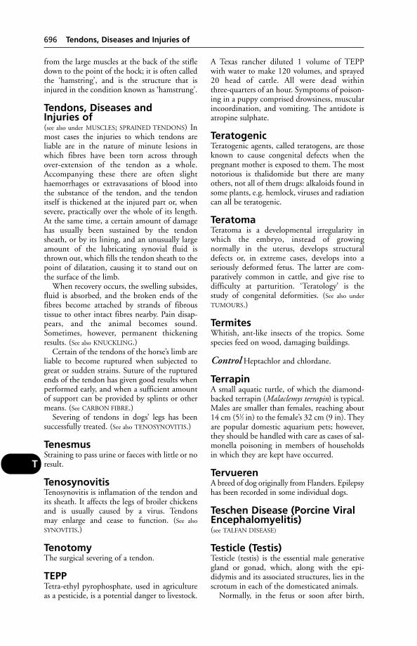
from the large muscles at the back of the stifledown to the point of the hock; it is often calledthe ‘hamstring’, and is the structure that isinjured in the condition known as ‘hamstrung’.
Tendons, Diseases and Injuries of(see also under MUSCLES; SPRAINED TENDONS) Inmost cases the injuries to which tendons areliable are in the nature of minute lesions inwhich fibres have been torn across throughover-extension of the tendon as a whole.Accompanying these there are often slighthaemorrhages or extravasations of blood intothe substance of the tendon, and the tendonitself is thickened at the injured part or, whensevere, practically over the whole of its length.At the same time, a certain amount of damagehas usually been sustained by the tendonsheath, or by its lining, and an unusually largeamount of the lubricating synovial fluid isthrown out, which fills the tendon sheath to thepoint of dilatation, causing it to stand out onthe surface of the limb.
When recovery occurs, the swelling subsides,fluid is absorbed, and the broken ends of thefibres become attached by strands of fibrous tissue to other intact fibres nearby. Pain disap-pears, and the animal becomes sound.Sometimes, however, permanent thickeningresults. (See also KNUCKLING.)
Certain of the tendons of the horse’s limb areliable to become ruptured when subjected togreat or sudden strains. Suture of the rupturedends of the tendon has given good results whenperformed early, and when a sufficient amountof support can be provided by splints or othermeans. (See CARBON FIBRE.)
Severing of tendons in dogs’ legs has beensuccessfully treated. (See also TENOSYNOVITIS.)
TenesmusStraining to pass urine or faeces with little or noresult.
TenosynovitisTenosynovitis is inflamation of the tendon andits sheath. It affects the legs of broiler chickensand is usually caused by a virus. Tendons may enlarge and cease to function. (See also
SYNOVITIS.)
TenotomyThe surgical severing of a tendon.
TEPPTetra-ethyl pyrophosphate, used in agricultureas a pesticide, is a potential danger to livestock.
A Texas rancher diluted 1 volume of TEPP with water to make 120 volumes, and sprayed20 head of cattle. All were dead within three-quarters of an hour. Symptoms of poison-ing in a puppy comprised drowsiness, muscularincoordination, and vomiting. The antidote isatropine sulphate.
TeratogenicTeratogenic agents, called teratogens, are thoseknown to cause congenital defects when thepregnant mother is exposed to them. The mostnotorious is thalidomide but there are manyothers, not all of them drugs: alkaloids found insome plants, e.g. hemlock, viruses and radiationcan all be teratogenic.
TeratomaTeratoma is a developmental irregularity inwhich the embryo, instead of growing normally in the uterus, develops structuraldefects or, in extreme cases, develops into a seriously deformed fetus. The latter are com-paratively common in cattle, and give rise todifficulty at parturition. ‘Teratology’ is thestudy of congenital deformities. (See also under
TUMOURS.)
TermitesWhitish, ant-like insects of the tropics. Somespecies feed on wood, damaging buildings.
Control Heptachlor and chlordane.
TerrapinA small aquatic turtle, of which the diamond-backed terrapin (Malaclemys terrapin) is typical.Males are smaller than females, reaching about14 cm (51⁄2 in) to the female’s 32 cm (9 in). Theyare popular domestic aquarium pets; however,they should be handled with care as cases of sal-monella poisoning in members of householdsin which they are kept have occurred.
TervuerenA breed of dog originally from Flanders. Epilepsyhas been recorded in some individual dogs.
Teschen Disease (Porcine ViralEncephalomyelitis)(see TALFAN DISEASE)
Testicle (Testis)Testicle (testis) is the essential male generativegland or gonad, which, along with the epi-didymis and its associated structures, lies in thescrotum in each of the domesticated animals.
Normally, in the fetus or soon after birth,
696 Tendons, Diseases and Injuries of
T

the testicle, guided by the fibrous cord knownas the gubernaculum, moves down from a position close to the kidney to a ‘cooler climate’in the scrotum. Into this it is pulled by thegubernaculum, which either fails to lengthen oractually shortens.
In some animals, e.g. foals, one or both testicles may go up again through the inguinalcanal. This occurs occasionally in pigs, in whicha returning testicle has been known to becomea mere vestige by the age of 6 months.
In certain of the wild animals, such as the rat,and in many tropical animals, e.g. the elephant,the testes are found in the abdominal cavity,either permanently or temporarily betweenperiods of sexual activity. In the foal the testesappear in the scrotum usually very soon afterbirth, but they are subsequently drawn up intothe abdomen, and do not reappear untilbetween 5 or 6 months and 10 to 12 months.In a certain proportion of cases the testes areretained in the abdomen until 2 years of age, and then descend into the scrotum; in anumber of cases they do not descend at all. Thename ‘rig’, or ‘cryptorchid’, is applied to such animals, and the condition is known as‘cryptorchidism’. (See CRYPTORCHID.)
The testes consist of a dense fibrous coat, the‘tunica albuginea’. Blood vessels run through-out the fibrous tissue, and nourish microscopictubules, lined by layers of specialised cellswhich form the spermatozoa. The tubules,known as ‘seminiferous tubules’, are connectedwith each other near the centre of the testes,and communicate with the coiled tubes of theepididymis, from which springs the vas deferensconnecting with the urethra at the oppositeend. In the epididymis the sperms mature. The‘spermatic cord’, which consists of the vas deferens, spermatic artery, veins, and nerves,enclosed in the layer of serous membrane (tunica vaginalis), passes upwards through theinguinal canal and enters the abdomen, whenceit runs back to the region of the neck of the urinary bladder, opening finally into the urethra. Along the course of the urethra are the openings of the ducts from the secondarysexual glands – seminal vesicles, prostate, andbulbo-urethral glands – which pour out a secre-tion which mixes with, nourishes, and protectsthe masses of spermatozoa coming from thetestes.
Externally, the testicle is covered by a layer of serous membrane, lying immediately outsidethe tunica albuginea, and known as the tunicavaginalis propria, which also covers the epi-didymis. On the outside of this tunic is thetunica vaginalis communis, or the parietal layer.
Outside this is a fairly thick layer of scrotal fascia, in which is deposited the ‘cod-fat’ of thebullock and wedder. A strong reddish, fibro-elastic tunica dartos forms the next outermostlayer, and provides the septum between theright and left pouches of the scrotum. Finally,on the outside, there is the practically hairless,thin, elastic, oily-feeling skin of the scrotum.
Functions The essential function of the testisis to produce sperms. (See SPERMATOZOA.)Between 60 and 80 million sperms are dis-charged at each copulatory act by the stallion atthe beginning of the breeding season. Since astallion may serve more than 100 mares duringthe season, many of them upon 2 separate occa-sions, it will readily be understood that thetestes are extremely active organs, and make aconsiderable demand upon the vitality of thebody generally. The necessity for a recuperativeperiod in breeding males will also be obvious.
The other function of the testis is that associated with elaboration of the male sex-hor-mones, resulting in the production of the secondary sexual characteristics, such as thearched neck and great body size of the stallion,the broad forehead, massive development ofhorns, and deep voice of the bull, the horns ofthe ram, and the tusks of the boar, etc., as wellas the instinctive desire for sexual intercourse.The chief hormone is testosterone.
(See also REPRODUCTION; ENDOCRINE
GLANDS; ARTIFICIAL INSEMINATION.)
Testicle, Diseases ofDuring service, an irritable mare may kick astallion and rupture one of the testes, or seri-ously injure it. Damage may also be occasionedto these organs by the bites of dogs when fight-ing, by gores from cattle, or by injuries from the tusks of boars, gunshots, etc. However,infection is probably most common.
Orchitis, or inflammation of the testis, may be the result of infection (e.g. byActinobacillus seminis, Brucella abortus, B. suis,Corynebacterium pseudotuberculosis, tuber-culosis) or of trauma which – if the skin is broken – may itself lead to infection. A viralinfection of bulls – infectious orchitis – wasreported in the former Czechoslovakia.Necrotic orchitis in the bull has been caused inBritain by actinobacillosis. The testis, beingenclosed in a fibrous, comparatively non-elasticcapsule, is not able to swell to a great extent,although the loose tissues of the scrotum oftendo. The scrotum becomes reddened in animalswhich have unpigmented skin in the inguinal
Testicle, Diseases of 697
T
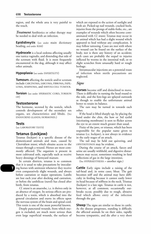
region, and the whole area is very painful to the touch.
Treatment Antibiotics or other therapy maybe needed to deal with an infection.
Epididimytis (see under main dictionary heading, and under RAM)
Hydrocele is a local oedema affecting usuallyone tunica vaginalis, and distending that side ofthe scrotum with fluid. It is most frequentlyencountered in the dog, although it may affectother animals.
Hypoplasia (see under INFERTILITY)
Tumours affecting the testicle and/or scrotuminclude CARCINOMA, SARCOMA, FIBROMA, PAPIL-
LOMA, SEMINOMA, and SERTOLI-CELL TUMOUR.
Torsion (see under SPERMATIC CORD, TORSION
OF)
TestosteroneThe hormone, secreted by the testicle, whichcontrols development of the secondary sexorgans, sex characteristics and libido. (See
ENDOCRINE GLANDS; HORMONES.)
Tests(see LABORATORY TESTS)
Tetanus (Lockjaw)Tetanus (lockjaw) is a specific disease of thedomesticated animals and man, caused byClostridium tetani, which obtains access to thetissues through a wound. Horses are most com-monly affected. The organism is present inmost cultivated soils, especially such as receiveheavy dressings of farmyard manure.
In certain districts, tetanus is so commonthat it is usual to take precautions by inoculat-ing horses with antitoxin whenever they receiveeven comparatively slight wounds, and alwaysbefore castration or major operations. Lambsare lost each year after docking and castration,or before the umbilicus (navel) has closed afterbirth, from tetanus.
Cl. tetani is an anaerobe, i.e. it thrives only inan absence of oxygen. Its serious effects are pro-duced by a toxin, which is absorbed into thegeneral circulation and exerts its effects uponthe nervous system of the brain and spinal cord.This toxin is one of the most powerful known.
Deeply punctured wounds, from which oxy-gen is excluded, are much more serious thaneven large superficial wounds, the surfaces of
which are exposed to the action of sunlight andfresh air. Picked-up nail wounds, cracked heels,injuries from the prongs of stable-forks, etc., areexamples of wounds which often become cont-aminated with Cl. tetani. Tetanus may occur inan animal which has had a slight wound whichappeared to heal without any complication. Itmay follow tattooing. Cases are met with whereno wound can be found on the surface of thebody, nor is there any history of an accident;such cases are probably the sequel to injuriesinflicted by worms in the intestinal wall, or toslight scratches from unusually hard or roughherbage.
Intramuscular injections are a potential routeof infection when sterile precautions areneglected.
Signs
Horses become stiff and disinclined to move.There is difficulty in turning the head round tothe side, and the fore-legs are splayed outwardsas though to enable the unfortunate animal better to retain its balance.
The ears may be turned in towards eachother.
If the head is lifted sharply up, by placing thehand under the chin, the haw or 3rd eyelid(nictitating membrane) is seen to flicker acrossthe eye to an extent much greater than usual.
Fixity of the jaws, or trismus, which has beenresponsible for the popular name given totetanus (i.e. lockjaw), is not always in evidencein the early stages of an attack.
The tail may be held out quivering, andOPISTHOTONOS may be evident.
During the course of an attack, faeces andurine are usually withheld, and digestive distur-bances may occur, sometimes resulting in fatalcollections of gas in the large intestines.
(See HYPERAESTHESIA – another sign.)
Cattle Early signs include a raising of the tail-head and, in some cases, bloat. The gaitbecomes stiff and the animal may have diffi-culty in feeding because it cannot easily lowerits head because of stiffness in the neck. Trismus(lockjaw) is a late sign. Tetanus in cattle is not,however, at all common; occasionally out-breaks occur, possibly due to rough, abrasivefeed which allows entry of the infectionthrough the gut.
Sheep The signs are similar to those in cattle.As the disease progresses, standing is difficult;the affected animals lie on their sides, rapidlybecome tympanitic, and die after a very short
698 Testosterone
T

illness. In lambs after castration or docking, thedisease is very rapid in its effects, and several areaffected at the same time.
Pigs Tetanus is not common.
Dogs The owner may notice something peculiar about the eyes and mouth, and eitherstiffness or recent lameness. Later, the limbs areusually stretched out as far from each other aspossible, in a sawhorse position. Squinting andgrinning are common, but closure of the jaws isnot always in evidence. When it is present it iscomplete, and death practically always follows.Hyperaesthesia is also very marked. The earsmay be bent inwards (as in the horse).
Treatment Farm animals should be placed ina darkened loose-box, away from noise, andwith food and water placed at a new level whichthey can reach despite their stiffness.
If nursed at home, a dog should be in a room where there are no bright lights, noise,television, or family activity.
Tetanus antiserum, penicillin, and musclerelaxants (such as acepromazine, which canobviate exhaustion and save life) are all needed.Treatment must also include glucose saline injec-tions, e.g. in a dog which cannot drink or eat;and large animals similarly. (See DEHYDRATION,NORMAL SALINE.)
Prevention Vaccination is effective, and onland where tetanus is rife, the most susceptibleanimals should be immunised.
Lambs are given antitoxin on the day of dock-ing or castration. Vaccine can be injected at adifferent site on the same occasion.
Horses The usual practice is to give 2 injec-tions at an interval of 4 to 6 weeks, with abooster dose 6 or 12 months later. Furtherbooster doses may be required. It is practicable
to vaccinate pregnant mares so that later their newborn foals will be protected againsttetanus infection via the navel. (See also under
IMMUNITY.)
Prognosis In the absence of first-class nursingand intensive care, not many animals (otherthan cattle) recover from tetanus.
If an animal regains the ability to drink, thatcan be regarded as a favourable sign.
TetanyTetany is a condition in which localised spas-modic contraction of muscles takes place. Theremay be twitching or convulsions. Tetany occurswhen the level of blood calcium falls below normal. (See also under PARATHYROID; HYPOMAG-
NESAEMIA; TRANSIT; MILK FEVER; RABIES.)
TetheringThe Cruel Tethering Act 1988 makes it illegalto tether a horse, ass or mule in such a way as to cause suffering. The animal must haveenough to eat and be supplied with fresh waterregularly. The tether must not be able to causeinjury, e.g. by being too tight or too short.
TetracyclinesTetracyclines are bacteriostatic antibiotics witha wide range of activity which includes Gram-positive and Gram-negative bacteria, certain protozoa, rickettsia, and mycoplasma.Tetracyclines are absorbed from the gut, butoral administration may upset the gut flora.They are irritant when injected.
Tetracyclines cause fluorescence in bone andteeth. In late pregnancy or in young growing ani-mals, high dosage can result in teeth discolorationand can interfere with the formation of enamel.
Horses treated with tetracyclines while suf-fering from stress may become affected withdiarrhoea and die. (See DIARRHOEA in horses.)
In cats, tetracyclines occasionally cause severeloss of hair.
TetraiodophenolphthaleinTetraiodophenolphthalein is used in radiogra-phy of the gall-bladder and bile-ducts for diagnostic purposes.
Texas FeverTexas fever is a tick-borne disease. (See BABESIA
– Babesiosis.)
Signs Stained urine (red-water), high tempera-ture, no appetite, and constipation followed bydiarrhoea. Cerebral symptoms may be evident.The animal dies within 3 to 10 days. On
Diagram of attitude assumed by a dog affectedwith tetanus. The hind-limbs are kept well outbehind the body, the tail is held rigidly or quiver-ing, and the muscles of the face are drawn into asardonic grin – the ‘risus sardonicus’ of ancientauthors.
Texas Fever 699
T
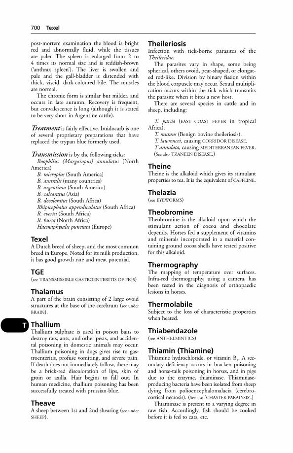
post-mortem examination the blood is brightred and abnormally fluid, while the tissues are paler. The spleen is enlarged from 2 to 4 times its normal size and is reddish-brown(‘anthrax spleen’). The liver is swollen and pale and the gall-bladder is distended withthick, viscid, dark-coloured bile. The musclesare normal.
The chronic form is similar but milder, andoccurs in late autumn. Recovery is frequent,but convalescence is long (although it is statedto be very short in Argentine cattle).
Treatment is fairly effective. Imidocarb is oneof several proprietary preparations that havereplaced the trypan blue formerly used.
Transmission is by the following ticks:Boophilus (Margaropus) annulatus (North
America)B. microplus (South America)B. australis (many countries)B. argentinus (South America)B. calcaratus (Asia)B. decoloratus (South Africa)Rhipicephalus appendiculatus (South Africa)R. evertsi (South Africa)R. bursa (North Africa)Haemaphysalis punctata (Europe)
TexelA Dutch breed of sheep, and the most commonbreed in Europe. Noted for its milk production,it has good growth rate and meat potential.
TGE(see TRANSMISSIBLE GASTROENTERITIS OF PIGS)
ThalamusA part of the brain consisting of 2 large ovoidstructures at the base of the cerebrum (see under
BRAIN).
ThalliumThallium sulphate is used in poison baits todestroy rats, ants, and other pests, and acciden-tal poisoning in domestic animals may occur.Thallium poisoning in dogs gives rise to gas-troenteritis, profuse vomiting, and severe pain.If death does not immediately follow, there maybe a brick-red discoloration of lips, skin ofgroin or axilla. Hair begins to fall out. Inhuman medicine, thallium poisoning has beensuccessfully treated with prussian-blue.
TheaveA sheep between 1st and 2nd shearing (see under
SHEEP).
TheileriosisInfection with tick-borne parasites of theTheileridae.
The parasites vary in shape, some beingspherical, others ovoid, pear-shaped, or elongat-ed rod-like. Division by binary fission withinthe blood corpuscle may occur. Sexual multipli-cation occurs within the tick which transmitsthe parasite when it bites a new host.
There are several species in cattle and insheep, including:
T. parva (EAST COAST FEVER in tropicalAfrica).
T. mutans (Benign bovine theileriosis).T. lawrencei, causing CORRIDOR DISEASE.T annulata, causing MEDITERRANEAN FEVER.(See also TZANEEN DISEASE.)
TheineTheine is the alkaloid which gives its stimulantproperties to tea. It is the equivalent of CAFFEINE.
Thelazia(see EYEWORMS)
TheobromineTheobromine is the alkaloid upon which thestimulant action of cocoa and chocolatedepends. Horses fed a supplement of vitaminsand minerals incorporated in a material con-taining ground cocoa shells have tested positivefor this alkaloid.
ThermographyThe mapping of temperature over surfaces.Infra-red thermography, using a camera, hasbeen tested in the diagnosis of orthopaediclesions in horses.
ThermolabileSubject to the loss of characteristic propertieswhen heated.
Thiabendazole(see ANTHELMINTICS)
Thiamin (Thiamine)Thiamine hydrochloride, or vitamin B1. A sec-ondary deficiency occurs in bracken poisoningand horse-tails poisoning in horses, and in pigsdue to the enzyme, thiaminase. Thiaminase-producing bacteria have been isolated from sheepdying from polioencephalomalacia (cerebro-cortical necrosis). (See also ‘CHASTEK PARALYSIS‘.)
Thiaminase is present to a varying degree inraw fish. Accordingly, fish should be cookedbefore it is fed to cats, etc.
700 Texel
T

Signs of this deficiency include loss ofappetite, a staggering gait, and muscularspasms.
Thin Sow SyndromeGroups of sows or gilts lose weight, usually inthe middle or later stages of pregnancy, andremain emaciated for perhaps 6 months ormore. Prolonged under-feeding may eventuallyresult in some sows being unable to cope withadverse conditions encountered at times ofstress, e.g. weaning. It has also been suggestedthat infestation with the stomach wormHyostrongylus or with the nodular wormOesophagostomum may be a cause. The use ofsow stalls, in which animals cannot move awayto escape draughts, is another possible cause.
ThiopentalA widely used anaesthetic for horses, dogs andcats. It is administered intravenously, as anaqueous solution of the sodium salt; otherroutes cause necrosis of the tissues.
ThiouracilAn antithyroid agent which lowers the rate ofmetabolism. It has been used as a growth promoter; such use is banned in the EU.
ThioureaThis is naphythyl antu, a rat poison which caus-es oedema of the lungs. It is dangerous todomestic animals and birds.
Thirst(see WATER; DIABETES; SALT POISONING;COMPULSIVE POLYDIPSIA)
Thogoto VirusThogoto virus is a cause of abortion in ewes in Africa. It was first isolated from a tick,Rhipicephalus appendiculatus, near Thogoto inKenya. In one flock of some 600 Dorper ewes, more than 200 aborted over a 2-monthperiod.
Thoracic DuctThe thoracic duct is the large lymph vesselwhich collects the contents of the lymphaticsproceeding from the abdomen, hind-limbs,part of the thorax, etc., and which discharges its contents into the left innominate vein. (See
aspiration under PARACENTESIS.)
ThoracocentesisDraining off from the thorax of the fluid foundin certain diseases of the chest. (See aspiration under
PARACENTESIS.)
ThoracotomyA surgical operation involving opening of thechest cavity.
Thorax(see CHEST)
Thorn Apple(see STRAMONIUM)
Thorough-PinThorough-pin is a distension of the sheath ofthe deep flexor tendon where it passes over thearch of the tarsus (hock). It is characterised byswellings, one on either side of the hock, aboutthe level of the ‘point of the hock’ (summit ofthe tuber calcis), and lying in front of the strongAchilles tendon.
Thread-WormThread-worm is a popular term for oxyurisworms. (See ROUNDWORMS.)
ThreonineOne of the essential amino acids.
Throat(see PHARYNX; also under LARYNX; NOSE AND
NASAL PASSAGES; MOUTH)
Throat diseases Most of these will be foundunder separate headings such as choking; larynx, diseases of; tonsillitis. For ‘sore throat’,see PHARYNGITIS.
ThrombastheniaThis is a rare, congenital disorder of the blood,occurring in man and dogs, in both sexes (com-pare HAEMOPHILIA). It arises from a defect ofthe platelets, and gives rise to prolonged bleed-ing resulting in anaemia. It has been describedin foxhounds, otterhounds, etc.
ThrombocytopeniaA condition of the blood in which the numberof platelets is below normal. Causes include viralinfections, poisoning, auto-immune disease.The signs may include petechial haemorrhagesand fever.
ThrombosisThe blocking of a blood vessel by a blood clot.It may follow atheroma, or some injury to thevessel. In cats, thrombosis of the femoral arter-ies is by no means rare, and causes paralysis ofthe hind-legs and often pain. There is completeabsence of pulse in the arteries. In dogs, throm-bosis of the iliac and femoral arteries occurs
Thrombosis 701
T
occasionally. Euthanasia is nearly always necessary. (See also PARAPLEGIA.)
Aortic-iliac thrombosis is seen in the horse;the worm Strongylus vulgaris may be a cause.
Thrombosis of a blood vessel in the brain isa cause of apoplexy (in human medicine astroke). (For thrombosis of the vena cava in cattle, see under VENA CAVA.) (See also under
ANTICOAGULANTS.)
ThrombusA blood clot in a blood vessel or the heart.
Thymus GlandSituated in the anterior part of the chest cavity,this gland attains its largest size during early lifeand thereafter gradually dwindles. The thymushas a role in immunity, as it removes young T-cells that happen to reccognise the body’sown components as foreign. Failure of thisfunction can result in auto-immune disease. (See
T-CELLS, which are thymus derived; also
LEUKAEMIA.)
Thyroid CartilageThe thyroid cartilage is the largest cartilage ofthe larynx, and forms a well-marked promi-nence at the upper end of the trachea. It givesattachment to one end of each of the vocalfolds, which are concerned in the production ofvoice. (See LARYNX.)
Thyroid GlandThis is a very highly vascular ENDOCRINE
GLAND, situated near the thyroid cartilage of thelarynx. The gland usually consists of 2 lobes,one on either side of the larynx, joined by anisthmus in some species and individuals.
Located within or near the thyroid gland arethe PARATHYROID GLANDS.
Minute structure Each lobe is enveloped in a thin capsule of fibrous tissue, strands from which pass into the organ, dividing it intolobules.
Function The most important hormonesecreted by the thyroid gland is an iodine-containing compound called thyroxin. Thisincreases the rate of metabolism, and is releasedwhen an animal is exposed to cold, for example.In hot weather, thyroid activity is reduced.Thyroxin is essential for growth and reproduc-tion, and influences lactation.
Secretion of the hormone is controlled wholly or in part by a hormone from the ante-rior lobe of the pituitary gland. (See also
PARATHYROID GLANDS.)
Thyroid Gland, Diseases ofEnlargement of the thyroid gland is known asGOITRE. Goitre may occur when there is eithertoo little or too much of the thyroid hormone,thyroxin, produced.
Dwarfism in young animals (cretinism) canresult from failure of the gland to produce sufficient thyroxin.
Hypothyroidism An insufficiency of thy-roxin is known as hypothyroidism, and may beassociated with insufficiency of iodine in thediet (see GOITRE). The rate of metabolism isslowed, while there is an increase in bodyweight, loss of hair, and lethargy.
One form of hypothyroidism, MYXOEDEMA,affects the skin, causing its deterioration.
Treatment includes the use of thyroid extract;and iodides if appropriate.
Hyperthyroidism, or excess thyroxin in theblood, is characterised by loss of weight, some-times an increase in appetite, polyuria, thirst,increased rate of metabolism and heartbeat.Enlargement of the gland may be detected on palpation. The animal may become rest-less or irritable. Protrusion of the eyeballs (exophthalmic goitre) may occur.
Hyperthyroidism is seen in elderly cats. Theyare mostly thin, and it is this loss of weightwhich causes the owner to seek veterinary advicein many instances. In addition to the symptomsmentioned above, diarrhoea may occur.
Treatment is surgical: removal of one gland,or ligation of the anterior arteries; alternatively,the use of drugs such as sodium fluoride ormethylthiouracil.
Tumours of the gland include ADENOMA;sarcoma and carcinoma (see under CANCER); and
EPITHELIOMA.
ThyroxineThe active principle of the THYROID GLAND. Itis used in pharmaceutical preparations to cor-rect hypothyroidism, a common endocrinedeficiency in dogs.
TiamulinA macrolide antibiotic active against Treponemahyodysenteriae (swine dysentery), various Gram-positive organisms, and Mycoplasma hyosinoviae(a cause of arthritis in pigs). It must not be usedat the same time as MONENSIN or SALINOMYCIN.
TibiaThe tibia is the larger of the 2 bones which liebetween the stifle and the hock. In animals
702 Thrombus
T

which possess fewer than 5 digits in their hind-limbs, the tibia has become modified so that itsustains the greater part of the weight borne bythe limb – the fibula, its complementary bone,having become reduced in size and importance.The tibia lies just below the skin on the insideof the limb, in such a position that it is liable tobe injured by kicks, blows, etc., and in thisconnection is of more importance than thosebones that are surrounded by massive muscleswhich afford some protection. It is not uncom-mon for the tibia to become fractured, but theparts remain held together by the very denseperiosteum that covers the bone. In the smalleranimals, the setting of the fractured bone is aroutine. (See BONES; FRACTURES.)
Tibial dyschondroplasia A cripplingdeformity occurring in certain strains of chick-ens, ducks, and turkeys selected for high growthrates. It is due to a cartilage abnormality.
Tick-Bite Fever of Man in Africa
Cause A RICKETTSIA. Local reactions, swellingof lymph nodes, occur in some individuals. Sofar as is known, tick-bite fever is not fatal.
The bont tick, bont-legged blue tick, yellowdog tick, and the brown tick – all common inEast Africa – transmit this disease. It can betransmitted to the guinea-pig by inoculation ofblood.
Tick-Borne Encephalitis (TBE)A meningioencephalitis following infection by aflavivirus transmitted by the sheep/cattle tickIxodes ricinus. The ticks become infected by fieldmice, voles, shrews, and occasionally moles. Itoccurs throughout continental Europe, beingespecially prevalent in mountainous regionswith coniferous forests. It is more common in humans than in animals, but the infection indogs has been confused with rabies.
The human illness resembles influenza in its symptoms, with a high fever. This may be followed by meningitis. Mortality is about 1 per cent.
In differential diagnosis, the flavivirus causingTBE has to be distinguished from louping-illvirus.
Tick-Borne Fever of CattleTick-borne fever of cattle is caused by Cytoectes(Erlichia) phagocytophila, transmitted by thecommon sheep tick, Ixodes ricinus. Symptomsof this infection are high but transient fever,and a considerable reduction in milk yield.Abortion may also occur. Oxytetracycline is
used in treatment. (Red-water, caused byBabesia divergens, often occurs simultaneously.)
Tick-Borne Fever of SheepTick-borne fever of sheep is a disease caused byCytoectes phagocytophilia transmitted by the tickIxodes ricinus.
Tick-borne fever is a mild febrile disease ofsheep in which the essential symptom is a risein temperature occurring after an incubationperiod of 4 to 8 days, and lasting about 10 days,when it subsides. During this period (whichmay be prolonged) there is dullness and listless-ness, and a considerable loss of weight mayoccur. Death occurs in only a small percentageof cases; most sheep recover unless some othercomplicating condition such as louping-illsupervenes. Abortion is an important result of infection in many instances, and may affect50 per cent of breeding stock introduced fromtick-free areas.
Rickettsiae can be demonstrated in the polymorphonuclear white cells of the blood.
The importance of tick-borne fever is that itis capable of rendering the vasculo-meningealbarrier of the central nervous system vulnerableto the virus of louping-ill. Without its presence,though the louping-ill virus may be introducedinto the bloodstream (by the bite of a tick), itcannot pass this barrier to attack the nerve cellsand so produce the typical nervous symptoms.It has been shown that both infective agents –that of tick-borne fever and of louping-ill – frequently exist together in ticks found on animals on farms where louping-ill is common,and it is probable that under natural conditionsthe great majority of adult sheep on such farmshave been infected with tick-borne fever infection and have recovered.
Tick-borne fever increases the susceptibilityof lambs to tick pyaemia, often caused by infec-tion with Staphylococcus aureus following tick bites. Abscesses occur in the joints and elsewhere, causing lameness, unthriftiness, anddeath.
Tick ParalysisTick paralysis affects man, cattle, sheep, horses,pigs, dogs, cats, and poultry.
It occurs in Africa, Australia, and Canada,and is caused by the presence on the animal ofvarious species of Ixodes (especially the dog tick)in South Africa and Australia, and Dermacentorin America. In East Africa, the bont-legged tick(Hyalomma spp.) and possibly the Red tick(Rhipicephalus evertsi) cause paralysis.
The paralysis is caused by toxin(s) present inthe saliva of ticks.
Tick Paralysis 703
T

In human beings, 3 or 4 days after the ticksattach themselves, paralysis of the legs occurs,then paralysis of the arms takes place, later thechest and neck become involved, and ultimate-ly the heart and respiratory centres are attacked.In the sheep, the parts are affected in the samegeneral sequence.
This form of paralysis is peculiar in thatsymptoms disappear within 2 to 6 days after theticks are removed, and recovery takes place subsequently. Individual lambs, for example,can be reinfected and recover more than once,if the ticks are removed by hand. They are usu-ally not easily seen unless a deliberate search ismade in the wool over the vertebral columnfrom the base of the skull back to the tail.
In the dog, they may cause QUADRIPLEGIA.
Tick Pyaemia(see TICKS; TICK-BORNE FEVER OF SHEEP)
TicksThese are among the most serious parasites ofdomestic animals. In the tropics they transmitbacterial, protozoal and viral diseases; in theUK, tick-borne fever, red-water fever and louping-ill. Tick pyaemia is caused in sheep by the transfer of staphylococcal infection. Lyme disease is considered to be partly tickspread.
Some cause illness by means of a toxin, whileall feed on the host’s blood – which can resultin a serious anaemia. Large numbers of ticksalso worry the host, and cause unthriftiness.Suppurating wounds may also result. In theBritish Isles, Ixodes ricinus is the main tickfound, although Haemophysalis punctatis is present in some southern coastal areas.
Life-cycles On this basis, ticks can be dividedinto 3 groups:
1-host ticks, such as Boophilus, which spendall 3 stages of their life-cycle on the same ani-mal. Larvae having attached themselves to thehost, they feed on it, moult, feed again on it asnymphs, moult, and the adult ticks also feed onit – the females subsequently dropping to theground to lay their eggs.
2-host ticks: these, such as some Hyalommaspecies, feed as both larvae and nymphs on the same host, but then moult on the ground; emerging adults find and feed on a 2nd host.
3-host ticks: larva, nymph, and adult eachfeeds on a different host, with moulting takingplace on the ground between each stage in thelife-cycle. Ixodes and Dermacentor species areincluded in this group.
Family Ixodidae (hard ticks) In this family the dorsum of the body is more or lessprotected by a hard shield of chitin, and insome species the male has ventral plates also.
The principal species attacking the domesticanimals are dealt with below. (See also DOG TICKS
for those occurring in Britain.)
Ixodes. There are over 50 species in this genus,including the following:
(a) I. ricinus attacks all the domesticated ani-mals and is found in most parts of the world. Itis known locally as the castor-bean tick, orEuropean sheep tick. A 3-host tick, it leaves itshost before each moult, and then seeks a newhost. In this way 3 animals are attacked by thesame tick: one as a larva, one as a nymph, andone as an adult. The animals attacked need notbe of the same species. This tick transmits tick-borne fever in sheep, louping-ill, and causes tickparalysis in sheep and cattle. It can also transmitBabesia, the cause of red-water.
(b) I. hexagonus attacks especially the dog,but is found on other hosts, notably sheep. It
704 Tick Pyaemia
T
Ixodes. (Ventral and dorsal views of male. × 12.)
Ixodes. (Dorsal and ventral views of a smallfemale. × 8.) In this and subsequent drawings ofticks only the fore parts of the legs are shown indiagrams of the ventral surface.

occurs in Europe, North Africa, and America; it is common on hunting dogs in France. In addition it is a transmitter of babesiosis.
(c) I. canisuga is the common species foundon the dog in Britain. It occurs also in WesternEurope and North America. Like the lastspecies, only females are found on the host. It isknown popularly as the British dog tick.
(d) I. pilosus attacks all the domestic mam-mals in South Africa. It is a reddish-brown tick,with the body larger behind than in front. It isknown locally as the russet tick, and is a causalagent of tick paralysis.
(e) I. rubicundus, another South African tick,which is found only on sheep, also causes tickparalysis.
(f ) I. holocyclus, in Australia and India, isfound on ruminants, dogs, and pigs. It is thecause of Australian tick paralysis, symptoms ofwhich may appear within an hour of attachment.It transmits Q fever.
Haemaphysalis. The following species areimportant:
(a) H. punctata (H. cinnabarina var. puncta-ta) is a common tick in Europe, North
America, and North Africa on all the domesticanimals. The life-history is identical with thatof H. leachii. It transmits Babesia bovis inBritain.
(b) H. leachii is a 3-host African specieswhich has been found in Western Asia andAustralia. It attacks carnivores, but is sometimesfound on ruminants. In East Africa it is calledthe yellow dog tick; it is also known as theSouth African dog tick. It transmits caninebabesiosis, Q fever, and tick-bite fever.
Dermacentor. The following species areimportant:
(a) D. reticulatus is common in Europe, butalso occurs in North Asia. It attacks ruminants,and also the dog and the horse. It is occasional-ly found in Western England. It transmitsequine and canine babesiosis.
(b) D. variabilis (D. electus) is found on dogsin North America. It also occurs on cattle andhorses. It is known as the American dog tick.
(c) D. occidentalis occurs in western NorthAmerica on various domestic mammals. It isconsidered by some authorities to be D. reticu-latus.
Life-cycle of a 3-host tick, Ixodes ricinus. (Reproduced with permission from H. T. B. Hall, Diseases andParasites of Livestock in the Tropics, Longman.)
Tick 705
T
(d) D. venestus is found in the RockyMountain District of North America and iscalled the Pacific Coast tick. Adults are foundon various mammals, including man. It is thetransmitter of Rocky Mountain spotted fever inman, and of canine babesiosis. It is the cause ofAmerican tick paralysis. It is a 3-host tick.
Rhipicephalus. The following species areimportant:
(a) R. sanguineus is found in all parts of theworld on dogs and ruminants. It is brown incolour. It is known as the European brown tickand also as the European dog tick – a nameshared with Ixodes hexagonus.
(b) R. appendiculatus is found in Africa,where it attacks cattle, sheep, goats. It is calledthe brown tick, and is a 3-host tick. This speciestransmits East Coast fever, corridor disease,mild gall sickness, red-water, Nairobi sheep disease.
(c) R. bursa is found in North Africa andSouthern Europe on all animals. It is a 2-hosttick. It transmits ovine babesiosis in Europe.
(d) R. capensis is found in South Africa oncattle, horses and dogs. It is called the Capebrown tick. The life-cycle is similar to the 2nd species. It can transmit Theileria parva.
(e) R. simus is found in Africa on dogs and her-bivores. It is called the dark pitted tick. Its life-cycle is similar to the 2nd species. It can transmitTheileria parva, Anaplasma marginale, T. mutans.
(f ) R. evertsi in Africa may be found on all the domestic mammals except pigs. It hasorange-red legs with round convex distinct eyes.The scutum is black and densely pitted. Theunderside of the male is red; the females arebrown or reddish brown. It is called the red tickor the red-legged tick. This 2-host species trans-mits Nuttallia equi and T. parva, causing EastCoast fever, babesiosis, spirochaetosis.
Boophilus(a) B. decoloratus is found on cattle and other
animals in Africa. It is a 1-host tick, called theblue tick. This tick, which may be a variety ofB. annulatus, transmits Babesia bigemina,Anaplasma marginale, and Spirochaeta theileri.
706 Tick
T
COMMON TICKS IN EAST AFRICA
Number Preferred site of Animal DiseaseTick species of hosts attachment affected Parasite transmitted
Brown-ear tick 3 Ears, base of horns, Cattle Theileria parva East Coast fever(Rhipicephalus around eyes, tail Cattle Theileria lawrencei Corridor diseaseappendiculatus) brush, and heels Cattle Theileria mutans Mild gall-sickness
Cattle Babesia bigemina Red-waterSheep and goats Virus Nairobi sheep
diseaseSheep, cattle, and Louping-ill
goatsMan Rickettsia Tick-bite fever
Red-legged tick 2 Larvae and Cattle Babesia bigemina Red-water(Rhipicephalus nymphae in ears Cattle Theileria parva East Coast feverevertsi) Adults perineal Cattle Theileria mutans Mild gall-sickness
region Horses Babesia nuttaliBabesia caballi } Biliary fever
Cattle, horsessheep, and goats Spirochaeta theileri Spirochaetosis
Lambs ?Tick toxin ?Paralysis
Yellow dog tick 3 Whole body Dogs Babesia canis Biliary fever(Haemaphysalis Man and animals Virus Q feverleach) Man Rickettsia Tick-bite fever
Blue tick 1 Face, neck, dewlap, Cattle Babesia bigemina Red-water(Boophilus and sides of the Cattle Anaplasma Gall-sicknessdecoloratus) body marginale
Man Rickettsia Tick-bite feverHorses, cattle, Spirochaeta theileri
Goats and sheep
Bont tick 3 Larvae and Cattle/Sheep/ Ricketsia Heartwater(Amblyomma nymphae on head Goats ruminantiumspp.) and ears Sheep Virus Nairobi sheep
Nymphae and diseaseadults on perineum, Man and animals Virus Q feverudder, scrotum, Man Rickettsia Tick-bite feverand tail
Bont-legged tick 2 or 3 Adults on perineum, Cattle, sheep, Tick toxin Sweating sickness(Hyalomma udder, scrotum, and goats, and pigsspp.) tail brush Man and animals Virus Q fever
Man Tick toxin Tick paralysisMan Rickettsia Tick-bite fever
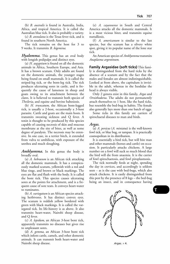
(b) B. australis is found in Australia, India,Africa, and tropical America. It is called theAustralian blue tick. It also is probably a variety.
(c) B. annulatus is the Texas fever tick, and isfound in southern North America.
The tick remains on the host for 3 to 9 weeks. It transmits B. bigemina.
Hyalomma. This genus has an oval bodywith longish pedipalps and distinct eyes.
(a) H. aegyptium is found on all the domesticanimals in Africa, Southern Europe, and Asia.It has a brown scutum. Only adults are foundon the domestic animals, the younger stagesbeing found on small mammals. It is called thestriped-leg tick, or the bont-leg tick. The tickproduces ulcerating sores in cattle, and is fre-quently the cause of lameness in sheep andgoats owing to its attachment between theclaws. It is believed to transmit both species ofTheileria, and equine and bovine babesiosis.
(b) H. truncatum, the African bont-leggedtick, is usually a 2-host, occasionally a 3-hostparasite. Cattle and goats are the main hosts. Ittransmits sweating sickness and Q fever. Atoxin is thought to be produced by this speciescapable of causing necrosis of skin and mucousmembrane at the site of bites, as well as somedegree of paralysis. The necrosis may be exten-sive. In one case, in a terrier bitch, it extendedfrom vulva to umbilicus, with exposure of theurethra and much sloughing.
Amblyomma. In this genus the body isbroadly oval.
(a) A. hebraeum is an African tick attackingall the domestic mammals. It has a conspicu-ously marked scutum, yellowish with a red andblue tinge, and brown or black markings. Theeyes are flat and flush with the body. It is calledthe bont tick. This species causes ulceratingsores at the points for attachment, and is a fre-quent cause of sore teats. It conveys heart-waterto ruminants.
(b) A. variegatum is an African species attack-ing herbivores. It has distinct convex eyes. The scutum is reddish yellow bordered withgreen with black markings. It is called the var-iegated tick. Its life-history is as above. It alsotransmits heart-water, Nairobi sheep disease,and Q fever.
(c) A. lepidum, an African 3-host bont tick,apparently transmits no diseases but gives riseto unpleasant sores.
(d) A. gemma, an African 3-host bont tickwhich infests cattle, camels, and other domesticanimals. It can transmit both heart-water andNairobi sheep disease.
(e) A. cayannense in South and CentralAmerica attacks all the domestic mammals. It is a most vicious biter, and transmits equinenuttalliosis.
(f ) A. americanum is similar to the lastspecies, but the scutum has a silvery white spot, giving it its popular name of the lone startick.
An American species of Amblyomma transmitsAnaplasma argentinum.
Family Argasidae (soft ticks) This fami-ly is distinguished from the hard ticks by theabsence of a scutum and by the fact that themales and females are almost indistinguishable.Looked at from above, the capitulum is invisi-ble in the adult, whereas in the Ixodidae thehead is always visible.
Only 2 genera exist in this family, Argas andOrnithodorus. The adults do not permanentlyattach themselves to 1 host, like the hard ticks,but resemble the bed-bug in habits. The femalealso generally lays more than one batch of eggs.
Some ticks in this family are carriers ofspirochaetal diseases to man and birds.
Argas.(a) A. persicus (A. miniatus) is the well-known
fowl tick, or blue bug, or tampan. It is practicallycosmopolitan in its distribution.
It is essentially a bird tick, but will bite manand other mammals (horses and cattle) on occa-sion. It particularly attacks chickens. A largenumber on a fowl will suck so much blood thatthe bird will die from anaemia. It is the carrierof fowl spirochaetosis, and fowl piroplasmosis.
The tick normally feeds at night, spendingthe day in crevices, and accordingly is seldomseen – as is the case with bed-bugs, which alsoattack chickens. It is easily distinguished fromthis pest by the presence of 8 legs – the bed-bugbeing an insect, and in consequence having
Argas. × 4.
Tick 707
T

only 6 legs. The larval tick (seed tick) remainsseveral days on the host, and is more frequentlyseen. The adults can live for 2 years withoutfood.
(b) A. reflexus, a closely related species, isfound mainly on pigeons, but also attacks poul-try and man. It is found in Europe, Africa, andAmerica.
Ornithodorus.(a) O. savignyi, the sand tampan, is a soft tick
of great economic importance in Africa, Asia,and the Near East. The tick lives below the sur-face of the sand, emerging to feed on the bloodof cattle, other domesticated stock, wild ani-mals, and man. The tick’s saliva contains apotent toxin and this, together with massiveblood loss, readily kills young or debilitatedanimals. Amitraz, cypermethrin and ivermectinare among effective controlling agents.
(b) O. megninii is the spinose ear tick ofAmerica and South Africa. The larvae creepinto the ear of some mammalian host, and in a few days moult. The nymphs, which are covered with minute spines, may live for 1 to 7 months in the ear, increasing in size from 3 to 17 mm (1⁄8 to 2⁄3 in). They finally drop to theground, moult, mate, lay their eggs, and die.The adult is not parasitic. The eggs hatch inabout 10 days. As many as 80 ticks have beenfound in a single ear. The irritation is consider-able and heavy losses may result. A moderntreatment is IVERMECTIN.
(c) O. coriaceus (pajaroello) is a venomousspecies (found in North America) which causesa very painful bite.
Transmission of disease When an infect-ed tick feeds upon a calf, it transmits the para-sites – or causal organisms – of the tick-bornedisease in question. The calf soon becomes ill,and either dies or recovers. As a rule, recovery isassociated with immunity. However, relapsesmay occur in animals thought to be immune tored-water, for example.
With the exception of ticks of the Boophilusspecies, larvae hatching from a tick’s eggs willnot immediately be infective because these lar-vae have not yet fed on any host; but as soon asthey start feeding they may ingest the causalorganisms of a tick-borne disease. When theymoult and become nymphs, they may then becapable of transmitting disease. Similarly, whenthe nymph, on moulting, becomes an adulttick, it will be infective if there were already parasites in its blood.
Not all tick vectors will transmit all causalorganisms; and, of course, not all species of hostare susceptible to the same causal organisms.
An infective 3-host tick feeding on a non-susceptible host ‘cleans’ itself of infection andwill not transmit disease in the next stage of itslife-cycle. This fact provides a useful controlmeasure.
The specific parasites transmitted by theticks are not passed on mechanically, but mustundergo a special development in the tick. Thisis easily understood when it is realised that anyone stage in the life-history of a hard tick bitesonly one animal. Accordingly, a tick infected inone stage must be capable of producing the dis-ease in some succeeding stage, which dependson the tick.
Control of ticks In many tropical countries,energetic measures for the regular and frequentdipping of cattle and sheep are necessary. Inorder to achieve adequate control of the tick-borne diseases, it is important that hand-dressing of certain parts of the body should becarried out in addition to the dipping or spray-ing. This applies to inside the ears, around thebase of the horns, around the eyes, anus, etc.
The acaricides, or tick-killing chemicals,have comprised: (1) arsenical compounds; (2) chlorinated hydrocarbon compounds; (3) the organo-phosphorus compounds; and(4) avermectin compounds.
Dipwashes containing arsenic are unsuitablefor spraying because of the danger of pasturecontamination. Although cheap, stable, and sol-uble, arsenic compounds are very poisonous:another disadvantage is that some species ofticks acquire a resistance to arsenic preparations.Accordingly, a change to chlorinated hydrocar-bons followed. Of these, BHC and toxaphenehave been widely used. Unfortunately, ticks canbecome resistant to these too. (See also BHC.)
Organophosphorus compounds tend to beexpensive, and are used mostly against ticksresistant to other acaricides.
Coumaphos; cypermethrin, a syntheticpyrethroid; diazinon, an organophosphorus
708 Tick
T
Ornithodorus. × 3.
compound – these are among a range of sub-stances formulated for application against ticks
For tick control in temperate regions, see also
DIPS AND DIPPING and DOG TICKS.
Systemic Acaricides are a useful alternativeto spraying or dipping. ‘Pour-on’ or ‘spot-on’formulations of acaricides such as amitraz andivermectin are applied along the dorsal ridge(pour-on) or to the base of the head, betweenthe shoulder blades (spot-on). The drug isabsorbed through the skin and carried by theblood circulation to all parts of the body.
Ticks in BuildingsTicks in buildings, such as quarantine premises,kennels in the tropics, private houses, etc., canbe eradicated by placing a block of dry ice onthe floor and closing all doors and windows.Adults, nymphs, and larval ticks will be found,after a time, clustering around this source ofCO2 and can then be easily collected anddestroyed.
Timber(see WOOD PRESERVATIVES; BEDDING – Pigs, andDogs and cats)
TinctureTincture is an alcoholic solution, e.g. tinctureof iodine.
Tinea(see RINGWORM)
Tissue Culture Vaccines(see VACCINES)
Tissues of the BodyTissues of the body include 5 groups:
(1) Epithelial tissues, including the cells covering the skin, those lining the alimen-tary canal, those forming the glands, etc. (See
EPITHELIUM.)(2) Connective tissues, including FIBROUS TIS-
SUE, FAT (adipose tissue), BONE, and CARTILAGE.(See main dictionary headings.)
(3) Muscular tissues (see MUSCLES).(4) Nervous tissues (see NERVES).(5) Fluid tissues (see BLOOD; LYMPH).
TitreThe extent to which an antibody-containingbiological substance can be diluted before los-ing its power of reacting with a specific antigen.‘High titres’ indicate, in practical terms, that apatient’s blood serum contains high levels ofantibody, e.g. to the rabies virus.
TLC‘Tender loving care’ – the indefinable qualitythat good nursing brings to recovery. Also, thinlayer chromatography.
Toadfish (Puffer Fish)Members of the Tetraodontidae which contain a poison, tetrodotoxin; if eaten, can cause paralysis.
ToadsToads have a defensive venom which is secretedby skin-glands and by the parotid salivarygland. The principal toxic substance is BUFO-
TALIN. Symptoms of poisoning in the dog areprofuse vomiting followed by the emission ofropy saliva and by loss of consciousness, whichmay persist for a couple of hours. Adrenalin hasbeen used in treatment.
The Central American toad, Eufo marinas, is very large, and has a powerful venom whichcan cause prostration, convulsions, and deathwithin 15 minutes.
ToadstoolsToadstools can cause severe poisoning if eaten.In one recorded instance, a cairn terrier diedafter eating Nolanea sericeum toadstools growingon a lawn. Death occurred within 3 hours.
Signs of poisoning by this fungus are severevomiting and abdominal pain.
Tobacco(see NICOTINE) Stalks of tobacco plants fed topigs have resulted in piglets born with limbdeformities.
TocopherolVitamin E.
Toes, CurlyAlso called curly toe disease and curled toeparalysis, it is a condition arising in chicks from a deficiency of riboflavin. The toes curlunderneath the feet. (See VITAMINS.)
Toes, TwistedAlso called crooked toes, it is a condition seenin chicks – one or more toes twisting inwards oroutwards. There is, at least, a hereditary dispo-sition to this abnormality, but it may occur intemporary and reversible form where infra-redbrooders are in use.
TogavirusesFormerly known as arboviruses, this groupincludes ALPHAVIRUSES, BUNYAVIRIDAE,
Togaviruses 709
T

ORBIVIRUSES, and PESTIVIRUSES. (See table under
VIRUSES.)
TomA male cat. In North America, male turkeys arealso called toms.
TomographyBody section radiography. (See X-RAYS
– Computed tomography.)
-Tomy-Tomy is a suffix indicating an operation bycutting.
Tongue(see also MOUTH) is a muscular and fibrousorgan, richly supplied with blood vessels andnerves, and covered with a highly specialisedmucous membrane. Its shape varies in the dif-ferent animals, but in all it consists of a free partor ‘tip’; a middle part, the ‘body’; and a hinderpart, the ‘root’. In the horse the tongue is longand spatulate, with a blunt tip, freely movable,and there is a definite narrowing just behind thetip. In the ox the tip is short, and pointed orconical; mobility and pliability are not so great,and on the upper surface is a hump-like emi-nence or ‘dorsum’, divided from the tip by adistinct, deep, transverse groove. The dorsum isof the greatest use in swallowing, and in bring-ing the small balls of cud from the back of themouth forward for chewing by the cheek teeth.
Tongue, Diseases of(see under MOUTH, DISEASES OF; SALIVATION;RANULA; ‘CURLED TONGUE’ in turkey poults)
Condition of the tongue The tongue ofany animal in health should be of a pink glis-tening appearance, soft and moist to the touchin the horse, sheep, pig and dog; rough in thecow and cat. (There are a few breeds of dogs,such as the chow, in which the tongue is nor-mally black or bluish.) When handled thetongue should possess a considerable power ofretraction; a weak flabby tongue usually indi-cates general muscular weakness. When at restthe tongue should touch the inner edges of allthe lower teeth. When it becomes greatlyswollen it presses against the teeth and theseleave indentations around its margin. In cattlethere is a raised part or ‘dorsum’ behind the freetip, which is instrumental in forming the foodinto boluses for swallowing.
Inflammation of the tongue (glossitis)is usually accompanied by SALIVATION, and
may result from injuries, irritant or corrosivepoisons, infections, and vitamin deficiencies.
Glossitis may be accompanied by the forma-tion of vesicles which burst, leaving ulceratedareas. (See FOOT-AND-MOUTH DISEASE; SWINE
VESICULAR DISEASE.) Ulcers are also a symptomof cattle plague (see RINDERPEST), MUCOSAL
DISEASE, and ORF in sheep. FELINE CALICIVIRUS
may cause tongue ulcers in the cat.Raised irregular swellings or abscesses on the
tongues of cattle suggest ACTINOBACILLOSIS.Ulcers along the free edges of the tongue may
be produced by diseased teeth.In the disease called calf diphtheria (necrosis
of the pharynx), the tongue may be the seat ofraised areas of false membrane which will alsobe seen in other parts of the mouth.
The tongue may be injured or wounded fromtoo severe a bit in the horse, or from carelessnessin breaking in a young colt. In such cases there isusually a distinct mark across the tongue’s uppersurface, behind which the organ appears normal,and in front of which it is reddened and swollen.
Foreign bodies, such as fish-hooks, needles,wire, splinters of bone, etc., may become fixedin the tongue, and lead to protrusion of theorgan, difficulty in swallowing, salivation, and adisinclination on the part of the animal to allowthe mouth to be handled or examined.
In canine LEPTOSPIROSIS/kidney failure thereare often areas of necrosis around the tip (whichmay slough off ), and a foul odour.
A brown discoloration may be present in theabove condition. (See also ‘BROWN MOUTH’.) Asoapy-white appearance of the tongue, againaccompanied by an unpleasant odour, oftenindicates some digestive disorder.
Curled tongue is an inherited defect inturkeys which has largely disappeared from themain hybrids. It may, however, still surface in themore traditional types – for example, broad-breasted bronze or Norfolk black. (See also ‘BLACK
TONGUE’; BLUETONGUE (a specific disease ofsheep and cattle); MYOTONIA; ASPHYXIA.)
‘Tongue Worm’‘Tongue worm’ is the colloquial name forLinguatula serrata, a parasite of the nose of thedog and other animals. (See MITES.)
TonicTonic means, in one sense, a continuous mus-cular spasm, as compared with CLONIC. (See also
TONICS.)
TonicsA name applied to a variety of medicines or othersubstances believed to help improve health. In
710 Tom
T
human medicine, the term is often applied tobitter herbal extracts that may stimulate theappetite.
Turning out to grass is itself a tonic to ani-mals that have been confined indoors. (See also
under PROTEIN, HYDROLISED; VITAMINS.)
TonsillitisInflammation of the tonsils, a symptom of e.g.canine viral hepatitis. The dog may retch orcough, and be slightly feverish.
TonsilsTonsils are collections of lymphoid tissue, situ-ated between the anterior and posterior pillarsof the soft palate at the back of the throat. Inthe horse there is not a compact tonsil, as inman, the dog, etc., but a diffuse collection oflymphoid tissue, mucous glands, etc., causingelevations on the surface, in which are seen thenumerous depressions or crypts which charac-terise the tonsil and differentiate it from otherlymphoid tissue. In the sheep the tonsil is bean-shaped and does not project into the throat, asin most other animals. In health, the dog’s ton-sils are not conspicuous, being situated in adepression; but when inflamed they appear as 2 bright red lumps.
Toothache(see TEETH, DISEASES OF)
Topical ApplicationsTopical applications of a drug are those madelocally to the outside of the body.
Topping of PasturesMowing the top growth on an overgrown pas-ture. This practice is beneficial from a veteri-nary point of view in that it is unfavourable tothe survival of parasitic worm larvae.
Torsion (Twisting)Torsion (twisting) occasionally involves theintestine (see VOLVULUS); the pedicle of thespleen; the stomach; uterus; and spermaticcords.
TorticollisA lateral deviation of the neck.
TortoisesIn Britain an amendment to the EndangeredSpecies Act, in 1982, required that every buyerof tortoises must sign an undertaking to pro-vide them with appropriate care, attention, andliving quarters necessary for their survival.Failure to comply can result in a fine of up to
£400 for an offending supplier, pet trader, orprivate individual. This little piece of legislationmay help to mitigate the extremely high mor-tality of tortoises imported into the UK, oftendue to their being crammed into unsuitablecontainers for their journey here and badlylooked after subsequently.
It has been suggested that outbreaks of viraldisease are present among tortoises (Testudospecies) in the Mediterranean region.
Imported tortoises should be carefully examined for the presence of exotic ticks.
Tortoises and turtles have been known toinfect dogs, cats, and people with salmonellosis.(See also AMERICAN BOX TORTOISES; PETS.)
For euthanasia, an injection into the abdomenof pentobarbitone sodium is recommended.
Tortoiseshell Cats, MaleAlthough nearly all tortoiseshell cats are female,males do occasionally occur. It appears that themost common chromosome complements are39XXY, 38XY/38XX, 38XY/39XXY and38XY/38XY. A 38XY is needed for the cat to befertile.
TouchThis sense depends upon receptors at the end ofnerves, or upon the nerve endings themselves:
Touch sense proper, by which touches orstrokes are perceived, such as the lightest sensa-tion caused by a fly settling on the skin. Thesize and shapes of bodies in contact with theskin which are not seen is also appreciated bythis sense.
Pressure sense, by which the weight of heavyobjects and their hardness can be determined.
Heat sense, by which the heat of the sur-rounding atmosphere, or of bodies in contactwith the skin, is appreciated as being above thatof body temperature. (Receptors for warmth in the human body number about 16,000 ascompared with 150,000 for detecting cold.)
Cold sense.Pain sense.Muscle sense, by which the weight of an
object can be tested, and the amount of energynecessary for an effort can be gauged.
Sense of position, by which, without usingthe powers of vision, the attitude and positionof any part of the body is known.
The distribution of the sense organs whichare concerned with the reception of these sensa-tions is very widespread. There is no part of thesurface of the body, except the horns, hoofs and claws, which can be cut without giving evi-dence of pain, and there is no part, includinghorny structures, which is insensible to touch.
Touch 711
T
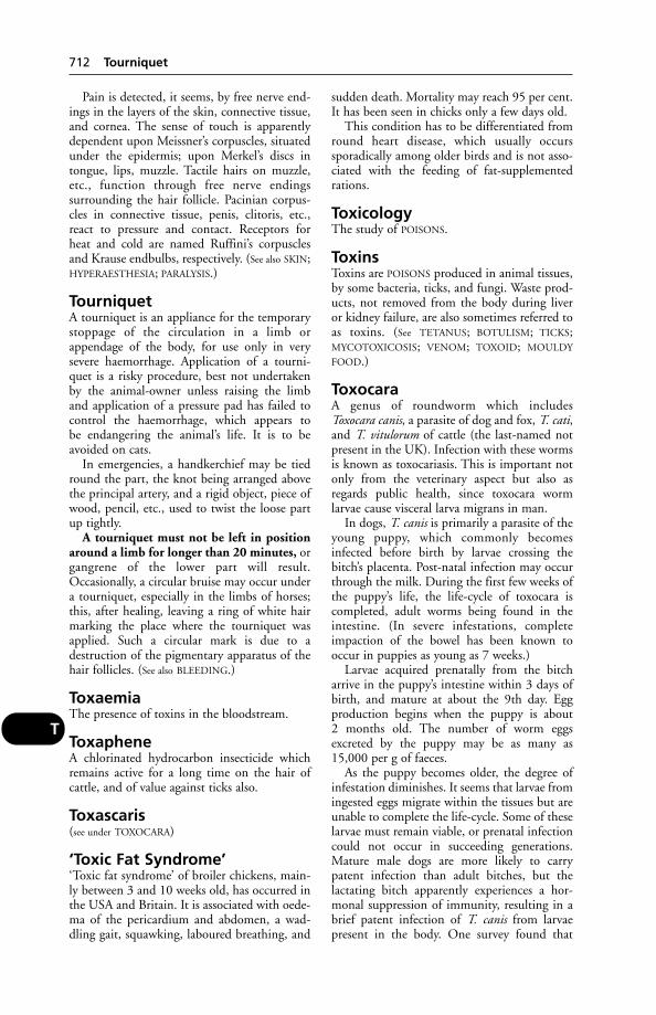
Pain is detected, it seems, by free nerve end-ings in the layers of the skin, connective tissue,and cornea. The sense of touch is apparentlydependent upon Meissner’s corpuscles, situatedunder the epidermis; upon Merkel’s discs intongue, lips, muzzle. Tactile hairs on muzzle,etc., function through free nerve endings surrounding the hair follicle. Pacinian corpus-cles in connective tissue, penis, clitoris, etc.,react to pressure and contact. Receptors for heat and cold are named Ruffini’s corpusclesand Krause endbulbs, respectively. (See also SKIN;HYPERAESTHESIA; PARALYSIS.)
TourniquetA tourniquet is an appliance for the temporarystoppage of the circulation in a limb orappendage of the body, for use only in verysevere haemorrhage. Application of a tourni-quet is a risky procedure, best not undertakenby the animal-owner unless raising the limband application of a pressure pad has failed tocontrol the haemorrhage, which appears to be endangering the animal’s life. It is to beavoided on cats.
In emergencies, a handkerchief may be tiedround the part, the knot being arranged abovethe principal artery, and a rigid object, piece ofwood, pencil, etc., used to twist the loose partup tightly.
A tourniquet must not be left in positionaround a limb for longer than 20 minutes, organgrene of the lower part will result.Occasionally, a circular bruise may occur undera tourniquet, especially in the limbs of horses;this, after healing, leaving a ring of white hairmarking the place where the tourniquet wasapplied. Such a circular mark is due to adestruction of the pigmentary apparatus of thehair follicles. (See also BLEEDING.)
ToxaemiaThe presence of toxins in the bloodstream.
ToxapheneA chlorinated hydrocarbon insecticide whichremains active for a long time on the hair of cattle, and of value against ticks also.
Toxascaris(see under TOXOCARA)
‘Toxic Fat Syndrome’‘Toxic fat syndrome’ of broiler chickens, main-ly between 3 and 10 weeks old, has occurred inthe USA and Britain. It is associated with oede-ma of the pericardium and abdomen, a wad-dling gait, squawking, laboured breathing, and
sudden death. Mortality may reach 95 per cent.It has been seen in chicks only a few days old.
This condition has to be differentiated fromround heart disease, which usually occurs sporadically among older birds and is not asso-ciated with the feeding of fat-supplementedrations.
ToxicologyThe study of POISONS.
ToxinsToxins are POISONS produced in animal tissues,by some bacteria, ticks, and fungi. Waste prod-ucts, not removed from the body during liver or kidney failure, are also sometimes referred toas toxins. (See TETANUS; BOTULISM; TICKS;MYCOTOXICOSIS; VENOM; TOXOID; MOULDY
FOOD.)
ToxocaraA genus of roundworm which includesToxocara canis, a parasite of dog and fox, T. cati,and T. vitulorum of cattle (the last-named notpresent in the UK). Infection with these wormsis known as toxocariasis. This is important notonly from the veterinary aspect but also asregards public health, since toxocara worm larvae cause visceral larva migrans in man.
In dogs, T. canis is primarily a parasite of theyoung puppy, which commonly becomesinfected before birth by larvae crossing thebitch’s placenta. Post-natal infection may occurthrough the milk. During the first few weeks ofthe puppy’s life, the life-cycle of toxocara iscompleted, adult worms being found in theintestine. (In severe infestations, completeimpaction of the bowel has been known tooccur in puppies as young as 7 weeks.)
Larvae acquired prenatally from the bitcharrive in the puppy’s intestine within 3 days ofbirth, and mature at about the 9th day. Eggproduction begins when the puppy is about 2 months old. The number of worm eggsexcreted by the puppy may be as many as15,000 per g of faeces.
As the puppy becomes older, the degree ofinfestation diminishes. It seems that larvae fromingested eggs migrate within the tissues but areunable to complete the life-cycle. Some of theselarvae must remain viable, or prenatal infectioncould not occur in succeeding generations.Mature male dogs are more likely to carrypatent infection than adult bitches, but the lactating bitch apparently experiences a hor-monal suppression of immunity, resulting in abrief patent infection of T. canis from larvaepresent in the body. One survey found that
712 Tourniquet
T
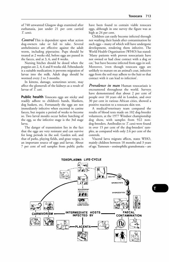
of 740 unwanted Glasgow dogs examined aftereuthanasia, just under 21 per cent carried T. canis.
Control This is dependent upon what actiondog-owners take or fail to take. Severalanthelmintics are effective against the adultworm, including piperazine. Pups should betreated at 2 weeks old, before eggs are passed inthe faeces, and at 3, 6, and 8 weeks.
Nursing bitches should be dosed when thepuppies are 2, 4, 6 and 8 weeks old. Febendazoleis a suitable medication; it prevents migration oflarvae into the milk. Adult dogs should bewormed every 2 to 3 months.
In kittens, damage, sometimes severe, mayaffect the glomeruli of the kidneys as a result oflarvae of T. cati.
Public health Toxocara eggs are sticky andreadily adhere to children’s hands, blankets, dog baskets, etc. Fortunately the eggs are notimmediately infective when excreted in caninefaeces, but require a period of weeks to becomeso. Two larval moults occur before hatching ofthe egg, so the infective stage is the 3rd stagelarva.
The danger of transmission lies in the factthat the eggs are very resistant and can survivefor long periods in the soil. Garden soil, andthat of parks, playing fields, and grass verges, isan important source of eggs and larvae. About7 per cent of soil samples from public parks
have been found to contain viable toxocaraeggs, although in one survey the figure was ashigh as 24 per cent.
Children can easily become infected throughnot washing their hands after contamination bysuch eggs – many of which will have undergonedevelopment, rendering them infective. TheWorld Health Organisation (WHO) has stated:‘Many patients with proven toxocariasis havenot owned or had close contact with a dog orcat,’ but have become infected from eggs in soil.Moreover, ‘even though toxocara eggs areunlikely to mature on an animal’s coat, infectiveeggs from the soil may adhere to the hair so thatcontact with it can lead to infection’.
Prevalence in man Human toxocariasis isencountered throughout the world. Surveyshave demonstrated that about 2 per cent ofpeople over 10 years old in London, and over30 per cent in various African cities, showed apositive reaction to a toxocara skin test.
A medical/veterinary team compared theresults of blood tests made on 102 dog-breedervolunteers, at the 1977 Windsor championshipdog show, with samples from 922 non-dog-breeders. Antibodies to T. canis were foundin over 15 per cent of the dog-breeders’ sam-ples, as compared with only 2.6 per cent of thecontrols.
Visceral larva migrans affects, states WHO,mainly children between 18 months and 3 yearsof age. Tumours – eosinophilic granulomata – are
Toxocara 713
T
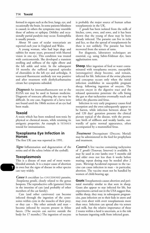
formed in organs such as the liver, lungs, eye, andoccasionally the brain. In some patients blindnessis caused; in others the symptoms may resemblethose of asthma or epilepsy. Debility and occa-sionally partial paralysis may occur. Eosinophiliais usually present.
About 50 cases of ocular toxocariasis arereported each year in England and Wales.
A young woman, who had kept dogs andrabbits for many years, presented with blurredvision in one eye. This condition was treatedwith corticosteroids. She developed a transientswelling and stiffness of the right elbow and the left ankle and wrist. In the subsequent 18 months she suffered repeated episodes of choroiditis in the left eye and arthralgia. Atoxocaral fluorescent antibody test was positiveand after treatment with diethylcarbamazinecitrate her symptoms subsided.
Diagnosis An immunofluorescent test or theELISA test may be used in human medicine.Diagnosis of toxocara affecting the eye may bedifficult. In one case, fragments of a larva werenot found until the 186th section of an eye hadbeen made.
ToxoidA toxin which has been rendered non-toxic byphysical or chemical means, while retaining itsantigenic properties. An example is TETANUS
toxoid for immunisation.
Toxoplasma Eye Infection inHorsesThe first UK case was reported in 1991.
Signs Inflammation and degeneration of theretina and of the sclera (white of the eyeball).
ToxoplasmosisThis is a disease of man and of most warm-blooded animals. It is a major cause of abortionin ewes but the signs of disease in other speciescan vary widely.
Cause A coccidian (see COCCIDIOSIS) parasite,Toxoplasma gondii, closely related to the genusIsospora. The reproductive cells (gametes) formin the intestine of cats (and probably of othermembers of the cat family).
Cats (and other carnivores) can becomeinfected through the ingestion of the cysto-zoites within cysts in the muscles of their prey;or they can – like other animals and man –become infected by oocysts present in felinefaeces. (The oocysts can survive outside thebody for 17 months.) The ingestion of oocysts
is probably the major source of human urbantoxoplasmosis in the UK.
T. gondii has been isolated from the milk ofbitches, cows, ewes, and sows, and it has beenshown that the young of these may be bornalready infected. The parasite can live in ticksand lice, so that the spread of toxoplasmosis bythese is not unlikely. The parasite has beenrecovered from the semen of rams.
For diagnosis, laboratory techniques areessential, e.g. using Sabin-Feldman dye, latexagglutination tests.
Sheep After ingestion of feed or water conta-minated with toxoplasma oocysts, susceptible(seronegative) sheep become, and remain,infected for life. Infection of the ovine placentaand conceptus occurs only when the initialinfection establishes in susceptible pregnantsheep, following ingestion of oocysts. Theoocysts encyst in the digestive tract and thereleased sporozoites penetrate the cells liningthe gut so that tachyzoites eventually reach andinfect the placenta and fetus.
Infection in very early pregnancy causes fetalresorption and the ewes subsequently appear tobe barren, while infection between about 50and 120 days’ gestation presents the clinicalpicture typical of the disease, with the prema-ture birth of stillborn and weakly lambs, out-wardly of quite normal appearance, oftenaccompanied by a mummified fetus.
Treatment Decoquinate (Deccox; Merial)may be administered in the feed for prophylaxisand treatment.
Control A live vaccine containing tachyzoitesof T. gondii (Toxovax; Intervet) is available. Itmay be used in ewe lambs over 5 months oldand older ewes not less than 6 weeks beforemating; repeat dosing may be needed after 2years. Warning: Accidental exposure to the vac-cine can cause infection in man – in particular,abortion. The vaccine must not be handled bywomen of child-bearing age.
Goats Toxoplasmosis causes abortion and peri-natal mortality similar to that seen in sheep.Goats also appear to stay infected for life, butexperiments carried out in the USA suggest that,unlike sheep, they may, in subsequent pregnan-cies, pass infection on to their kids in utero andmay even abort with overt toxoplasmosis morethan once. Infection can spread also via semenand milk, but the relative importance of these 2 routes within a herd is uncertain, as is the riskto humans ingesting milk from infected goats.
714 Toxoid
T
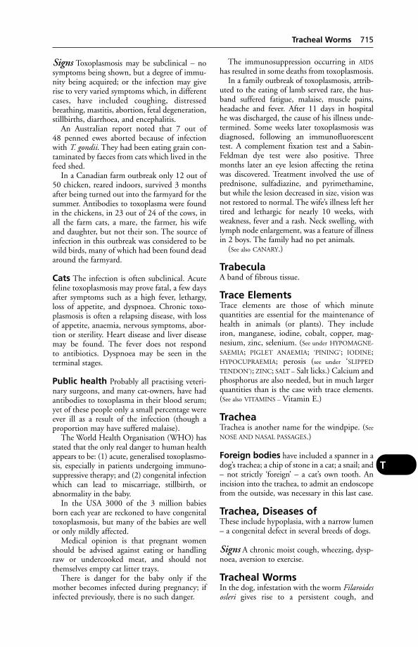
Signs Toxoplasmosis may be subclinical – nosymptoms being shown, but a degree of immu-nity being acquired; or the infection may giverise to very varied symptoms which, in differentcases, have included coughing, distressedbreathing, mastitis, abortion, fetal degeneration,stillbirths, diarrhoea, and encephalitis.
An Australian report noted that 7 out of 48 penned ewes aborted because of infectionwith T. gondii. They had been eating grain con-taminated by faeces from cats which lived in thefeed shed.
In a Canadian farm outbreak only 12 out of50 chicken, reared indoors, survived 3 monthsafter being turned out into the farmyard for thesummer. Antibodies to toxoplasma were foundin the chickens, in 23 out of 24 of the cows, inall the farm cats, a mare, the farmer, his wifeand daughter, but not their son. The source ofinfection in this outbreak was considered to bewild birds, many of which had been found deadaround the farmyard.
Cats The infection is often subclinical. Acutefeline toxoplasmosis may prove fatal, a few daysafter symptoms such as a high fever, lethargy,loss of appetite, and dyspnoea. Chronic toxo-plasmosis is often a relapsing disease, with lossof appetite, anaemia, nervous symptoms, abor-tion or sterility. Heart disease and liver diseasemay be found. The fever does not respond to antibiotics. Dyspnoea may be seen in theterminal stages.
Public health Probably all practising veteri-nary surgeons, and many cat-owners, have hadantibodies to toxoplasma in their blood serum;yet of these people only a small percentage wereever ill as a result of the infection (though aproportion may have suffered malaise).
The World Health Organisation (WHO) hasstated that the only real danger to human healthappears to be: (1) acute, generalised toxoplasmo-sis, especially in patients undergoing immuno-suppressive therapy; and (2) congenital infectionwhich can lead to miscarriage, stillbirth, orabnormality in the baby.
In the USA 3000 of the 3 million babiesborn each year are reckoned to have congenitaltoxoplasmosis, but many of the babies are wellor only mildly affected.
Medical opinion is that pregnant womenshould be advised against eating or handlingraw or undercooked meat, and should notthemselves empty cat litter trays.
There is danger for the baby only if themother becomes infected during pregnancy; ifinfected previously, there is no such danger.
The immunosuppression occurring in AIDS
has resulted in some deaths from toxoplasmosis.In a family outbreak of toxoplasmosis, attrib-
uted to the eating of lamb served rare, the hus-band suffered fatigue, malaise, muscle pains,headache and fever. After 11 days in hospital he was discharged, the cause of his illness unde-termined. Some weeks later toxoplasmosis wasdiagnosed, following an immunofluorescenttest. A complement fixation test and a Sabin-Feldman dye test were also positive. Threemonths later an eye lesion affecting the retinawas discovered. Treatment involved the use ofprednisone, sulfadiazine, and pyrimethamine,but while the lesion decreased in size, vision wasnot restored to normal. The wife’s illness left hertired and lethargic for nearly 10 weeks, withweakness, fever and a rash. Neck swelling, withlymph node enlargement, was a feature of illnessin 2 boys. The family had no pet animals.
(See also CANARY.)
TrabeculaA band of fibrous tissue.
Trace ElementsTrace elements are those of which minute quantities are essential for the maintenance ofhealth in animals (or plants). They includeiron, manganese, iodine, cobalt, copper, mag-nesium, zinc, selenium. (See under HYPOMAGNE-
SAEMIA; PIGLET ANAEMIA; ‘PINING’; IODINE;HYPOCUPRAEMIA; perosis (see under ‘SLIPPED
TENDON’); ZINC; SALT – Salt licks.) Calcium andphosphorus are also needed, but in much largerquantities than is the case with trace elements.(See also VITAMINS – Vitamin E.)
TracheaTrachea is another name for the windpipe. (See
NOSE AND NASAL PASSAGES.)
Foreign bodies have included a spanner in adog’s trachea; a chip of stone in a cat; a snail; and– not strictly ‘foreign’ – a cat’s own tooth. Anincision into the trachea, to admit an endoscopefrom the outside, was necessary in this last case.
Trachea, Diseases ofThese include hypoplasia, with a narrow lumen– a congenital defect in several breeds of dogs.
Signs A chronic moist cough, wheezing, dysp-noea, aversion to exercise.
Tracheal WormsIn the dog, infestation with the worm Filaroidesosleri gives rise to a persistent cough, and
Tracheal Worms 715
T
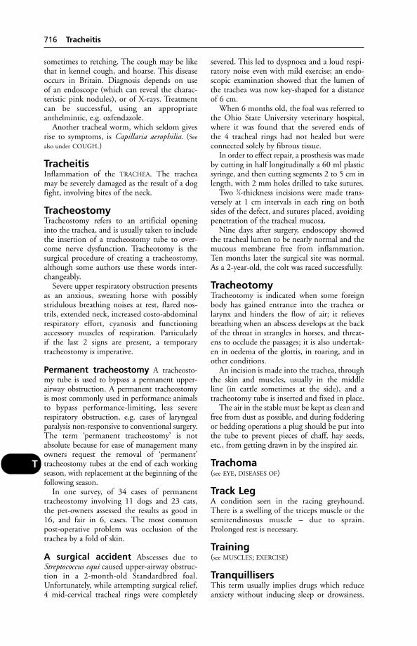
sometimes to retching. The cough may be likethat in kennel cough, and hoarse. This diseaseoccurs in Britain. Diagnosis depends on use of an endoscope (which can reveal the charac-teristic pink nodules), or of X-rays. Treatmentcan be successful, using an appropriateanthelmintic, e.g. oxfendazole.
Another tracheal worm, which seldom givesrise to symptoms, is Capillaria aerophilia. (See
also under COUGH.)
TracheitisInflammation of the TRACHEA. The tracheamay be severely damaged as the result of a dogfight, involving bites of the neck.
TracheostomyTracheostomy refers to an artificial openinginto the trachea, and is usually taken to includethe insertion of a tracheostomy tube to over-come nerve dysfunction. Tracheotomy is thesurgical procedure of creating a tracheostomy,although some authors use these words inter-changeably.
Severe upper respiratory obstruction presentsas an anxious, sweating horse with possiblystridulous breathing noises at rest, flared nos-trils, extended neck, increased costo-abdominalrespiratory effort, cyanosis and functioningaccessory muscles of respiration. Particularly if the last 2 signs are present, a temporary tracheostomy is imperative.
Permanent tracheostomy A tracheosto-my tube is used to bypass a permanent upper-airway obstruction. A permanent tracheostomyis most commonly used in performance animalsto bypass performance-limiting, less severe respiratory obstruction, e.g. cases of laryngealparalysis non-responsive to conventional surgery.The term ‘permanent tracheostomy’ is notabsolute because for ease of management manyowners request the removal of ‘permanent’ tracheostomy tubes at the end of each workingseason, with replacement at the beginning of thefollowing season.
In one survey, of 34 cases of permanent tracheostomy involving 11 dogs and 23 cats,the pet-owners assessed the results as good in16, and fair in 6, cases. The most commonpost-operative problem was occlusion of thetrachea by a fold of skin.
A surgical accident Abscesses due toStreptococcus equi caused upper-airway obstruc-tion in a 2-month-old Standardbred foal.Unfortunately, while attempting surgical relief,4 mid-cervical tracheal rings were completely
severed. This led to dyspnoea and a loud respi-ratory noise even with mild exercise; an endo-scopic examination showed that the lumen ofthe trachea was now key-shaped for a distanceof 6 cm.
When 6 months old, the foal was referred tothe Ohio State University veterinary hospital,where it was found that the severed ends of the 4 tracheal rings had not healed but wereconnected solely by fibrous tissue.
In order to effect repair, a prosthesis was madeby cutting in half longitudinally a 60 ml plasticsyringe, and then cutting segments 2 to 5 cm inlength, with 2 mm holes drilled to take sutures.
Two 3⁄4-thickness incisions were made trans-versely at 1 cm intervals in each ring on bothsides of the defect, and sutures placed, avoidingpenetration of the tracheal mucosa.
Nine days after surgery, endoscopy showedthe tracheal lumen to be nearly normal and themucous membrane free from inflammation.Ten months later the surgical site was normal.As a 2-year-old, the colt was raced successfully.
TracheotomyTracheotomy is indicated when some foreignbody has gained entrance into the trachea orlarynx and hinders the flow of air; it relievesbreathing when an abscess develops at the backof the throat in strangles in horses, and threat-ens to occlude the passages; it is also undertak-en in oedema of the glottis, in roaring, and inother conditions.
An incision is made into the trachea, throughthe skin and muscles, usually in the middle line (in cattle sometimes at the side), and a tracheotomy tube is inserted and fixed in place.
The air in the stable must be kept as clean andfree from dust as possible, and during fodderingor bedding operations a plug should be put intothe tube to prevent pieces of chaff, hay seeds,etc., from getting drawn in by the inspired air.
Trachoma(see EYE, DISEASES OF)
Track LegA condition seen in the racing greyhound.There is a swelling of the triceps muscle or thesemitendinosus muscle – due to sprain.Prolonged rest is necessary.
Training(see MUSCLES; EXERCISE)
TranquillisersThis term usually implies drugs which reduceanxiety without inducing sleep or drowsiness.
716 Tracheitis
T
They include benzodiazepines such as diazepamand azapirones such as buspirone. They areused in veterinary practice to calm or restrainvicious or nervous animals; to obviate travelsickness; and to facilitate the induction ofanaesthesia. Their use is not permitted atKennel Club shows.
(For horses, see DETOMIDINE.)Tranquillisers have been administered to
cattle, zoo and wild animals, by firing a hypodermic syringe from a cross-bow, gun, orblow-pipe. (See DART GUNS; also PIGS, SEDATION
OF; ROMPUN.)
Natural tranquillisers At the Institute ofAnimal Physiology, during investigation of the hormones contained in extracts of ovariantissue, several steroids have been found whichexert a strong sedative effect on the central nervous system. Variations in secretion rates of these steroids during the reproductive cyclemay be partly responsible for cyclic variationsin behaviour, states the AFRC. Slight tranquil-lity could occur when the blood concentrationof the steroids is relatively high; restlessness oreven aggressiveness might result from a lowconcentration.
Transferable Resistance(see under ANTIBIOTIC RESISTANCE; PLASMIDS)
TransferrinA beta-globulin present in blood plasma andacting as a carrier of iron. (See also IRON.)
Transfusion of Blood(see under BLOOD TRANSFUSIONS)
Transgenic AnimalsThose bred by genetic engineering methodsinvolving the isolation of genes from one ani-mal, modification of them in the laboratory,and introduction of them into animals of thesame or different species. (See also RETROVIRUS.)
Transit TetanyTransit tetany is the result mainly of HYPOCAL-
CAEMIA, which is precipitated by the stress oflong travel. It is seen in ruminants and, morerarely, in horses.
In lactating mares (also known as lactationtetany), it may occur about 10 days after foal-ing or a day or two after weaning. It can alsooccur, it has been said, in fillies and colts.
In ruminants, prolonged travel may inducehypocalcaemia or hypophosphataemia in cattle,with hypocalcaemia, hypomagnesaemia andhypoglycaemia in ewes.
Signs These vary according to the extent towhich the blood calcium level is reduced. Aslight reduction causes the mare to be excitable,but a further fall produces muscular incoordi-nation and staggering, and the animal appearsobviously distressed. Sweating may occur; rapidand noisy breathing and, in the mare, flarednostrils are other signs. A stiff gait, raised tail,and spasms similar to those of tetanus occur.
Another similarity is that eating and drink-ing may become impossible. Recumbency,coma, and death follow within a couple of days.
While mild cases recover, the mortality ishigh in untreated animals showing the moresevere signs.
Treatment The animal should be kept quietand the appropriate dose of a mineral replace-ment solution, e.g. calcium borogluconate 20 per cent, given subcutaneously or by slowintravenous injection.
TranslocationIn CYTOGENETICS, this means transfer of a broken-off fragment of one chromosome toanother. A cause of some congenital diseases.
Transmissible Gastroenteritis ofPigs (TGE)Transmissible gastroenteritis of pigs (TGE) is arapidly fatal disease in young piglets. The causeis a coronavirus; mortality decreases with age of the piglet. For example, mortality may be 90 per cent in the first week of life, 50 per centin the second, 25 per cent in the third, and zeroin older pigs.
Foul-smelling watery diarrhoea, vomitingand loss of appetite are the main signs inpiglets. Adult pigs are usually little affected,although fattening pigs require extra water during an outbreak.
TGE is typically but not exclusively a diseaseof the winter months. Epidemics occurred inwinter every 5 to 7 years between 1956 and1983. Apart from 2 incidents in 1996, the UKwas free from TGE until a single incident in1999. The absence of disease from Britain andcontinental Europe since the mid-1980s hasbeen attributed to the emergence in 1986 ofporcine respiratory coronavirus. This is anapparent mutant of TGE virus and is believedto have effectively immunised the pig popula-tion against TGE. The reservoir of virus may beas a subclinical infection in large herds.
There is no specific treatment, but losses maybe reduced by extra care and management:warmth and extra fluids; good-quality milkreplacer/creep pellets; early weaning into warm
Transmissible Gastroenteritis of Pigs (TGE) 717
T
accommodation; cross-suckling affected pigletsonto recovered sows; using antibiotics to control secondary infection.
Control TGE is spread by direct or indirectcontact with infected faeces; strict attention todisinfection and hygiene is essential. Sows dueto farrow, and sucking piglets under 7 days oldshould be isolated. Exposure of uninfected pigsto recently infected stock may help to developherd immunity.
Transmissible MinkEncephalopathyA spongiform encephalopathy which has beenreported in mink in the USA. The affectedmink were fed bovine offal.
Transplants(see EMBRYO TRANSFER and SKIN GRAFTING
TRANSPLANTATION)
TransponderAn electronic device which stores informationthat can be read by a suitable scanner. In miniaturised form, transponders are the basis of identity MICROCHIPS.
Transport StressStress caused by transportation can adverselyaffect the welfare and health of farm animalsand the meat quality of the carcase. A methodhas been developed for measuring the adversenature of the noise and vibration components oftransport, using operant conditioning. Theequipment is a modification of a machine orig-inally developed for testing tractors, and consistsof a pen which is tilted up and down in all direc-tions, generating a noise of 80 decibels. It wasfound that pigs soon learn to press a panel withtheir snouts in order to obtain a 30-secondsrespite from vibration and keep the machineimmobile for 70 to 80 per cent of the time. Theanimals switch the machine off more frequentlywhen the speed of vibration is increased and alsowhen they have eaten a large meal just beforethe test. During an hour-long session the fre-quency with which the machine was switchedoff tended to increase, showing that aversion tothe conditions does not diminish with time.Pigs which have experienced the machine willpress the switch when exposed to a recording ofthe noise even when there is no movement. Incontrast, naive animals do not learn to operatethe switch when exposed to the noise alone. Theadvantage of this technique is that specificationsfor improved methods of transport can be basedon the animal’s own preferences. (AFRC.)
Traumatic Pericarditis(see HEART DISEASES)
Travel SicknessTravel sickness is observed in dogs and cats –some individuals being particularly susceptible– and may be relieved by the administration ofa suitable tranquilliser prior to the journey.Fitting a chain to a car so as to act as an earthhas been recommended, but may be less effec-tive than periodic stops and adequate ventila-tion. It may also occur in horses on long seavoyages. (See also TRANSIT TETANY.)
TreadsTreads are injuries inflicted at the coronet of the horse’s foot, either by the shoe of the oppo-site foot, or, when horses are worked in pairs, by the adjacent horse. When situated in theposterior half of the foot, the upper free edge of the lateral cartilage may be damaged and aQUITTOR result.
TrefoilConsumption of the plant is a cause of LIGHT
SENSITISATION in Australia.
TrematodeAn unsegmented flat worm or fluke. (See
LIVER-FLUKES; LUNG-FLUKES; RUMEN-FLUKES;SCHISTOSOMIASIS.)
Trembling in Dogs(see under SHIVERING)
TremorsVery fine jerky contractions of a muscle or ofsome of the fibres of a muscle. They are oftenseen in nervous animals when frightened, andthey are one of the signs of viciousness in ahorse when seen on the quarters, especiallywhen the horse is watching out of the corner of its eye. Tremors are, however, encountered in certain nervous disorders, such as shiveringin horses and chorea in dogs. (See also ‘CRAZY
CHICK’ DISEASE.) HYPOMAGNESAEMIA in cattle,and RABIES in many species, also give rise to tremors. (For tremors in pigs, see SWINE
FEVER.)
TrephiningTrephining is an operation in which a small discof bone is removed from the cranium to permitthe elevation of a depressed portion, or to allowaccess into the brain cavity. In certain purulentconditions of the air sinuses of the horse’s head,trephining may be required to give drainage forthe pus.
718 Transmissible Mink Encephalopathy
T
TreponemaA genus of spiral organisms of the familyTreponemaceae, which includes also Borreliaand Leptospira. (See also SWINE DYSENTERY.)
Triatomid BugsTriatomid bugs are the most important vectorsof human trypanosomiasis in South andCentral America.
TrichiasisIngrowing eyelashes (see EYE, DISEASES OF).
TrichinosisTrichinosis is an infestation of the muscles ofthe pig, man, dog, etc., with the larvae ofTrichinella spiralis, a small roundworm. Pigsbecome infected by eating infected rats, or rawswill or garbage containing pieces of infectedpork. Trichinosis constitutes a serious problemamong sledge-dogs in the Arctic and may follow the eating of walrus, bear, seal, or fox-meat. A temperature of –15°C for 20 days isneeded to kill the larvae.
Infection in man occurs through the eatingof raw or undercooked meat. Human symp-toms include pain in muscles; myocarditis,meningitis, encephalitis, and rarely death haveoccurred.
An outbreak of trichinosis in Paris, involving300 proven cases, followed the eating of horse-meat either raw or served rare. All this meat hadcome from 2 shops, and originated from a sin-gle horse imported from the USA. The mainsymptoms in this outbreak were fever, musclepain, swollen face and eyelids, a rash, and digestive system upsets.
The disease has not been seen in the UK for some years, but outbreaks have occurred.(See ROUNDWORMS.)
Trichocephalus (Whip-Worm)Trichocephalus (whip-worm) is the name of aworm that infests the caeca of various animals.(See ROUNDWORMS.)
Trichodinella and TrichodiniaTrichodinella and trichodinia are skin parasites offish. Their sharp, rasping teeth damage the skin.
Trichoglyphs(see WHORLS)
TrichomonasThe flagellates of the genus Trichomonas areusually pear-shaped, with 3 to 5 anterior flagel-la, an undulating membrane and, in somespecies, 1 free flagellum directed backwards.
Species of this genus very commonly occur inthe intestinal canal of many different species ofmammals and birds.
Trichomoniasis Trichomonas fetus causesabortions, pyometra, and sometimes sterility incattle. The cow becomes infected by the bull atcoitus, or vice versa.
Signs A transient vaginitis, which is often over-looked. If conception has not occurred, achronic form of endometritis follows. If thecow is pregnant, the fetus dies and is eitheraborted 1 to 4 months later or retained in theuterus, where it becomes macerated and apyometra develops.
Control of the disease includes the disposal ofinfected bulls, withholding all breeding opera-tions on infected cows for at least 3 months,and the serving of non-infected cows and virginheifers by a ‘clean’ bull. Freezing bull semen to–79°C, in the presence of 10 per cent glycerol,kills T. fetus but allows the spermatozoa to sur-vive. This method of deep-freeze, commonlypractised at AI centres, is one way of getting ridof the infection from semen.
Avian trichomoniasis, caused by T. galli-nae, affects the oesophagus of budgerigars,pigeons, etc, causing necrotic lesions withretching and vomiting. Also known as canker orroup. In pigeons, which feed their young on‘crop milk’, squabs are easily infected which ledto the belief among pidgeon-fanciers that thedisease was inherited. It can be treated withdimetridazole or nifursol.
Trichophyton(see RINGWORM)
TrichostrongylosisParasitic disease caused by infection withTrychostrongylus worms. It can affect most
Trichomonas fetus.
Trichostrongylosis 719
T
mammals, causing poor growth, lack of condi-tion and diarrhoea.
TrichostrongylusA large group of parasitic worms which infestboth people and their domestic animals, andwhich cause persistent diarrhoea.
TrichothecenesTrichothecenes are fungal metabolites whichcontaminate animal feeds and human foods.Examples are deoxynivalenol and nivalenol.The most potent toxin is T2 TOXIN.
Throat irritation and digestive disorders arecaused in people. Baking does not destroy thetoxin.
Tricuspid ValveThe tricuspid valve is the valve lying in theheart between the right atrium and the rightventricle, which possesses 3 cusps or flaps. (See
HEART.)
Trigeminal NerveTrigeminal Nerve is the 5th of the cranialnerves. (See NERVES.)
TrimethoprimA drug which inhibits the growth of many bacteria and some protozoa through reducingtheir synthesis of folinic acid (necessary for syn-thesis of nucleic acids). Effective against manyGram-positive and Gram-negative bacteria.One part of trimethoprim is combined with 5 parts of sulfadiazine, with which it is syner-gistic, in co-trimazine. Several other prepara-tions of trimethoprim with one or other of thesulfa drugs also exist.
TriorchidismA condition in which 3 testicles are present.This was found in a cock – all 3 being func-tional, and structurally independent. A case inwhich there was duplication of the right testiclewas seen in a calf. (In man, triorchidism is notextremely rare, and even 5 testes in a scrotumhave been recorded.)
Triplet CalvesIn the UK in 1985 a Friesian cow had triplets.After initial breathing difficulties, all 3 survivedand were described as ‘fine, strong calves’.Quintuplets have been recorded.
TriploidAn animal having one and a half times as manychromosomes in its cells as a normal (i.e.diploid) animal. (See COLCHICINE.)
Triploidy accounts for up to 13 per cent ofembryonic loss in animals, and for 20 per centof all chromosomally-caused spontaneoushuman abortion (ARC). (See CHROMOSOMES;CYTOGENETICS.)
TrismusThe locking of the jaws, which is characteristicof TETANUS.
TrisomyThe presence in triplicate of a particular chro-mosome. In the cow, such a condition would bedenoted as 61,XXX.
X-trisomy is associated with nymphomaniaand infertility in cows.
Trixacarus CaviaeThe mange mite which is a parasite of guinea-pigs.
TrixylphosphateA substance used in the manufacture of plastics.Poisoning of cattle has occurred through contamination of molasses with this substance.Symptoms included diarrhoea, coughing,unsteady gait, partial paralysis.
TrocarA sharp-pointed, rod-like instrument used witha cannula to puncture the wall of a body cavity.It is often used to release gas in cases of bloat inruminants.
TrochanterOne of the protuberances on the femur which serve as attachment sites for hindquartermuscles.
Trochlear NerveThe 4th cranial nerve (see NERVES), also knownas the pathetic nerve. It controls the superioroblique muscle of the eye.
TrombiculosisInfestation with Trombicula autumnalis, theharvest mite.
TrophicRelating to nutrition; neurotrophic means the influence that nerves exert upon the tissuesto which they are distributed, for health andnourishment.
TrophinsGland-regulating hormones.
720 Trichostrongylus
T
TrophoblastThe outer layer of BLASTOCYSTS which makecontact with the wall of the uterus, and throughwhich nutrients and waste products areexchanged between fetal and maternal circula-tions. (See also SYNCYTIUM.)
Tropics, Livestock Production intheLivestock farming in the tropics, and even insome subtropical developing countries, is besetwith difficulties not experienced to anythinglike the same degree in developed countrieshaving a temperate climate.
In developing countries there is often theadditional problem of limited financial resources.Money may not be available for measures tocounter or ameliorate adverse conditions for animals; to provide adequate supplies of safedrinking water, good-quality feeds, vaccines, prophylactic drugs; or to support disease eradica-tion programmes on a large scale. Veterinariansare usually few in number in relation to the largeareas in which they are needed (see VETERINARY
PROFESSION), and faced with great distances tocover and an absence of local laboratory services.
Heat Animals can survive in temperatures ofup to 60°C (140°F). Temperatures above thatare lethal. Cattle in Death Valley, California,and in Queensland, Australia, for example, existat temperatures of 52° to 58°C (125° to136°F). Such heat, however, is far above thecomfort zone – estimated as air temperature atbetween 21° and 26°C (70° to 79°F); or 13° to18°C (55° to 64°) for adult cattle.
High temperatures impose stress upon theanimal’s physiological processes and productivecapacity.
Records for more than 12,000 inseminationsover a 2-year period in a Florida herd indicateda sharp decline in conception rates of cowswhen a maximum air temperature the day afterartificial insemination exceeded 30°C (86°F)(35°C (95°F) for heifers).
Besides air temperature, radiation, air move-ment, and humidity all influence the animal’simmediate environment. Body temperature iscontrolled by the heat-regulating mechanism(see HYPOTHALAMUS), and affected not only by environmental heat but also by the heat generated in the tissues (see METABOLISM). The more the animal eats, the more heat itsbody will produce. Water intake also plays apart in the physiological reactions; as doessweating, but in this respect cattle are less effi-cient than people. In great environmental heat,the point may be reached where normal body
temperature cannot be maintained, and it rises– a state of HYPERTHERMIA. Death may result.
Even at non-lethal levels, tropical heat is alimiting factor so far as fertility and yields ofmilk, meat and eggs are concerned. Poor feed,and water deprivation, can further depressgrowth rates, fertility, and yields. Humidity canincrease heat stress.
Signs Heat stress may cause the body temper-ature to rise to 42° to 43°C (110°F) in cattle.The earlier symptom of rapid breathing progresses to panting. The mouth may be keptopen, tongue lolling out, and frothy saliva maybe in evidence. Appetite is lost. Cattle mayremain standing, huddling together.
Preventive Measures If heat stress is to beavoided or minimised, livestock must haveshade (from trees or shelters) to protect themfrom the sun’s rays. Cooled drinking water isbeneficial to all livestock in tropical heat. Pigsneed to be able to wallow – their normal,instinctive method of cooling themselves. Coolwater sprays can help dairy cattle to withstandhigh temperatures. Grazing should take place atnight rather than during the day.
Zero-grazing may be practicable in someplaces, and beneficial too.
Poultry in the tropics grow larger combs andwattles than do similar birds in temperate climates, as a physiological means of body cool-ing. At very high temperatures they dip combsand wattles into water, for an extra coolingeffect. It has been suggested that the design ofdrinkers in intensive poultry units should besuch as to make possible this beneficial practice.
Antibiotics and poultry In developingcountries in the tropics, the widespread use ofantibiotics – especially the tetracyclines – inmodern battery poultry units has been blamedfor encouraging the proliferation of drug-resis-tant bacteria. Where indiscriminate use is cou-pled with poor sanitation and low personalhygiene, the situation may constitute a dangerto public health.
In a study in Nigeria of E. coli, 1248 strainsisolated from battery hens at the University ofIbadan, and 2196 strains from a commercialpoultry farm, were resistant to tetracycline,streptomycin, and also sulfonamides. By con-trast, all strains isolated from free-range townand village poultry were sensitive to these drugs.
Altitude In some regions, altitude mitigatesthe effect of heat. The uplands of Jordan are
Tropics, Livestock Production in the 721
T

regarded as suitable for intensive poultry production; and Iran’s uplands, with their dry climate, make dairy farming practicabledespite very high summer temperatures of43°C (109°F) upwards, and very cold ones in winter.
When temperature falls during night-timehours, cattle may withstand a higher day-timetemperature than they otherwise could.
Very high altitudes, e.g. in the mountains of Peru, can themselves be an obstacle to live-stock production. (See ALTITUDE; MOUNTAIN
SICKNESS.)
Stock improvement When high-yieldstock are imported into tropical regions fromcountries having a temperate climate, disap-pointment often follows. At first, yields –whether of beef, milk, pork, or eggs – are betterthan those of the indigenous stock, as expected;but before long, in many instances, the initialgains are offset by a high mortality rate. Theexotic animals may not be able to tolerate theheat, may not produce so well when fed onlocal feeds of lower quality, and will have noresistance to many local diseases and parasites,especially ticks. (For cattle resistant to heat andticks, see DROUGHTMASTER; ZEBU; SANTA
GERTRUDI; AFRICANDER.)In many situations it is often preferable
to improve indigenous stock first, before intro-ducing new blood from overseas, by selectivebreeding and better management; ensuring thatthey are better fed and not deprived of adequatequantities of drinking water. After improve-ment has been obtained by these means (butnot before), cross-breeding with exotic high-performance stock may be begun, preferably ona small-scale trial basis to start with. Use may bemade of AI.
Animal power In India it is estimated thatwork animals provide as much energy as theentire electrical system of the country. The num-ber of work animals is estimated to be 70 millionbullocks, 8 million buffaloes, 1 million horsesand 1 million camels. Throughout the Far Eastanimal power remains the major factor in agri-culture. Small farms, difficult terrain, lack ofroads and the structure of the rural economy inmany countries mean that situation is unlikelyto change in the foreseeable future. (See WATER
BUFFALOES.)
Animal feeds In poorer countries the cost ofimporting cereal grains or high-quality proteinfeeds may be prohibitive, and local stock willthen be dependent on feeds which may restrict
their yields – though obviously this is notalways the case.
Imported feeds sometimes deteriorate tosome extent during long sea voyages and subse-quent storage in a hot and often humid climate.There may, for example, be a serious loss of vit-amin E, so that a supplement is required ifENCEPHALOMALACIA is to be avoided. (See also
VITAMINS.)Local crops such as groundnuts, cotton seed,
sorghum and sunflower seed may be contami-nated by AFLATOXINS, so that precautions are needed. Groundnuts may be affected in this way through being left too long in theground before harvesting, or during subsequentstorage.
Minerals In many parts of the tropics, millingand processing facilities are lacking – at any ratein the more remote areas; this fact makes feedsupplementation more difficult. Mineral andtrace element supplements are necessary foravoidance of deficiencies. In South Africa manyyears ago, Sir Arnold Theiler showed that theneed of cattle for phosphorus drove them to eatthe bones of dead animals, and many cattlebecame infected with botulism in that way anddied. (See LAMZIEKTE.) In several parts of theworld a deficiency of copper in the herbage hasimpeded livestock production, and appropriatedressings of the land have brought great benefit.(See TRACE ELEMENTS.)
Some tropical crops Apart from the cropsmentioned above, many others – or their by-products – are used. For example, cattle mayhave the leaves of shade trees, or sugar-cane;pigs may be given dried leaf meal, bananawaste, coca pod husks, or sweet potatoes; poul-try may receive millet (if any can be sparedfrom human food requirements) or sago.
(See GOSSYPOL – Gossypol poisoning; CASTOR
SEED POISONING; COCOA POISONING.)
Tropical diseases In some tropical regionsthe presence of animal parasites and their vectors makes livestock production difficult,costly, or even impracticable. This is true of theAfrican tsetse-fly belt, extending roughly fromlatitude 15°N to 30°S. Here control of try-panosomiasis (see TRYPANOSOMES) is dependenton drugs for prevention, drugs for treatment,and use of insecticides against flies. Aerialspraying, bush clearance, and attempts to erad-icate reservoirs of infection among wild animalswill, if undertaken, obviously add to the cost,which in some territories may be beyond localresources. For many years control of tsetse flies
722 Tropics, Livestock Production in the
T
had been successfully achieved by aerial spray-ing with insecticides, but the ever-rising cost ofthese, and of aviation fuel, has led to the aban-donment of many such government schemes.Fly traps have had to be used instead. TheManitoba trap designed specially for tabanids isreported to be very successful; another widelyused trap is the Laveissire. In other territorieslong-term, government-controlled campaignshave proved successful in maintaining andextending production.
Humpless cattle, such as the N’Dama, inWest Africa had long been regarded as historicrelics, and their reduced susceptibility to try-panosomiasis as a biological oddity. It has beenshown, however, that despite their relativelysmall size, N’Dama cattle could survive and beproductive in endemic trypanosomiasis areaswhere Zebu cattle died.
Comparative studies on 2 types of large EastAfrican zebu (Bos indicus) Boran cattle, on abeef ranch in Kenya, indicated that a Borantype bred by the Orma tribe has a superiorresponse to tsetse fly challenge. The OrmaBoran when compared with an improved Boranwas found to have lower trypanosome infectionrates and, when untreated, better control ofanaemia as well as decreased mortality.
In areas where trypanosomiasis is endemic insusceptible cattle, sequential use of such drugsas diminazine and suramin has been effective incontrolling the disease without causing drugresistance to develop.
Trypanosomes cause disease also in Asia andCentral and South America.
Ticks are of great importance in the tropics,transmitting numerous protozoal parasites,viruses, and rickettsias. (See under TICKS.)
Among the major diseases caused by virusesare cattle plague (rinderpest), African swinefever, foot-and-mouth disease, and varioustypes of encephalitis. Bacterial diseases includeanthrax, botulism, haemorrhagic septicaemia(pasteurellosis), and salmonellosis. A notablemycoplasmal disease is contagious bovine pleu-ropneumonia; another is contagious agalactia ofsheep and goats.
Vaccine storage/transport One of theproblems of veterinary medicine in the tropicsis the storage of vaccines at a sufficiently lowtemperature (below 8°C (46°F)). With the highcost and scarcity of kerosene or liquid propanegas in many rural areas, and the fact that electricity supply is often unreliable or non-existent, there is scope for solar refrigerators.Under a WHO scheme these have been tried in13 countries. Photo-voltaic panels exposed to
the sun supply electricity direct to an ordinarycommercial refrigerator.
Sterilisation In the tropics the sun’s rays canbe used for sterilisation purposes. Research atthe American University of Beirut showed that oral rehydration solution, for treatingdehydration, can be sterilised in plastic bags ortransparent plastic or glass vessels by exposureto sunlight. In an experiment such a solution,contaminated with fresh sewage, proved to havea zero coliform count after 1 hour. It appearsthat the sterilising effect is not heat, since thetemperature of the solution rises by less than5°C (41°F) after 2 hours, but is rather solarradiation in the near ultra-violet range.
Another application is the Solomon solarsteriliser which uses only solar energy to boilwater and sterilise needles and syringes. Theprototype consisted of a metal-lined plywoodbox topped with a truncated pyramid of glass.The steriliser, in use in the Solomon Islands, iseasily constructed, has no moving parts, andrequires no fuel; but it does need orienting tothe sun every half-hour. (World HealthOrganisation.)
Carcase disposal, following post-mortemexaminations, may present problems in unde-veloped areas. An Australian veterinarian work-ing at an Indian sheep project found theanswer. If unsuitable for boiling as dog food,and yet not likely to spread infection, the car-case is dragged out into the open. He timedevents on one occasion. ‘At 2.46 p.m., post-mortem examination completed; no vultures tobe seen in a clear sky. At 2.50 the first arrived;at 2.53 there were approximately 40 vulturesaround the carcase; at 2.58 carcase stripped tobones and sinew – vultures leaving.’
TrypanocideA drug which will kill TRYPANOSOMES withinthe host’s body.
TrypanosomesTrypanosomes are small single-celled parasitesfound in the bloodstream in certain diseasesthat are classed together as the trypanoso-miases.
The trypanosome is of an elongated shapewith a single flagellum and an undulatingmembrane. There are 2 nuclei – a large nucleus(macronucleus or trophonucleus) near the cen-tre of the body; and a small kinetoplast(micronucleus) at the posterior end remotefrom the flagellum. In some forms there is nofree flagellum.
Trypanosomes 723
T
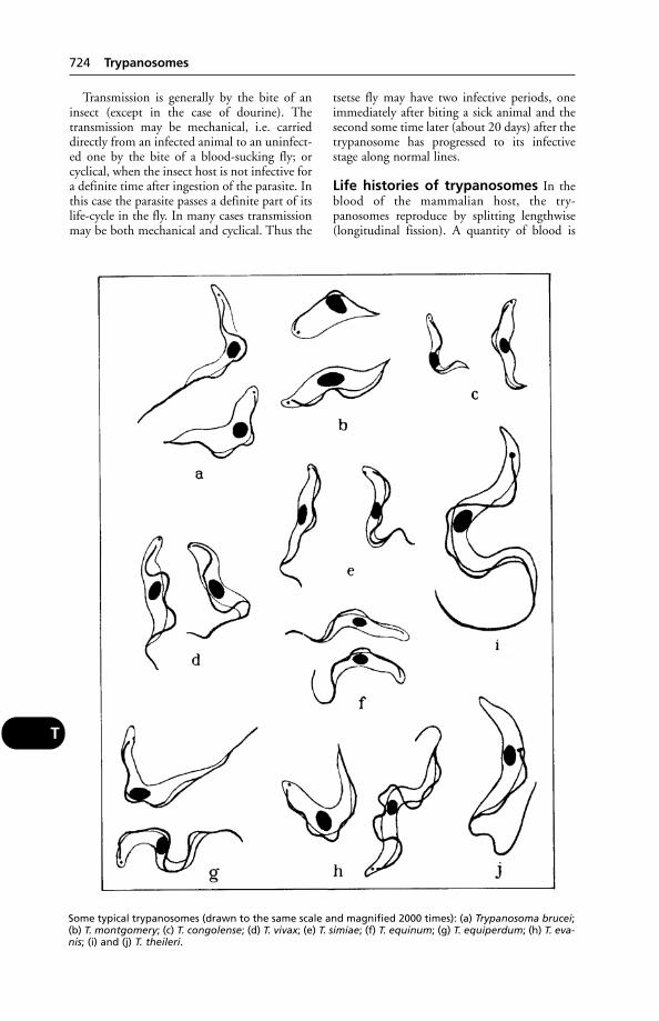
Transmission is generally by the bite of aninsect (except in the case of dourine). Thetransmission may be mechanical, i.e. carrieddirectly from an infected animal to an uninfect-ed one by the bite of a blood-sucking fly; orcyclical, when the insect host is not infective fora definite time after ingestion of the parasite. Inthis case the parasite passes a definite part of itslife-cycle in the fly. In many cases transmissionmay be both mechanical and cyclical. Thus the
tsetse fly may have two infective periods, oneimmediately after biting a sick animal and thesecond some time later (about 20 days) after thetrypanosome has progressed to its infectivestage along normal lines.
Life histories of trypanosomes In theblood of the mammalian host, the try-panosomes reproduce by splitting lengthwise(longitudinal fission). A quantity of blood is
724 Trypanosomes
T
Some typical trypanosomes (drawn to the same scale and magnified 2000 times): (a) Trypanosoma brucei;(b) T. montgomery; (c) T. congolense; (d) T. vivax; (e) T. simiae; (f) T. equinum; (g) T. equiperdum; (h) T. eva-nis; (i) and (j) T. theileri.

sucked up by the tsetse fly, a species of Glossina,and in that host the flagellates undergo a devel-opmental cycle. The location chosen by theparasite for its development varies with thespecies. Thus some will develop only in the sali-vary glands, others in the gut, and still others inthe proboscis. After some time they assume theinfective form, and are ready to be passed withthe salivary fluids into the bloodstream of asuitable vertebrate host.
African trypanosomiasis Tsetse-borne try-panosomiasis renders approximately 10 millionsquare km of prime African land unsuitable forcattle production. It has been estimated that ifthis disease could be controlled, the infested areawould increase its cattle holding capacity from20 million head to 140 million head.
The disease is of greatest importance in cattle, which are hosts of the following try-panosomes: Trypanosoma congolense, T. vivax, T. uniforme, and T. evansi.
Usually a chronic disease, acute cases alsooccur, and the mortality may be high. (See
also PREMUNITION; and, for resistant breeds,TROPICS.)
Signs These include intermittent fever,anaemia, anorexia or pica, a progressive loss ofcondition, and increasing weakness. (See under
CHANCRE for the hard swelling which is oftenthe first pointer to trypanosomiasis.) Lymphnodes are enlarged in many cases, the coatharsh, and abortion may occur.
Some cattle recover, but in others apparentrecovery is followed later by a relapse and death. In acute cases, death may occur within afortnight.
Horses Additional signs include oedema ofthe limbs and abdomen, and corneal opacity.Species of trypanosome infecting horses are T.brucei, T. vivax, and T. evansi. (See also DOURINE,caused by T. equiperdum, transmitted at coitus,and occurring also in Asia.)
Dogs The eyes may be affected, as in horses.Canine trypanosomiasis is caused by T. brucei,T. congolense, and T. evansi.
Pigs often suffer from acute and fatal try-panosomiasis caused by either T. simiae or T. evansi.
Control This is difficult, on account of trypanosomiasis existing in wild animals in the vicinity of cattle herds, and the fact thatvaccination has not been practicable.
In well-managed herds in areas where tsetse-fly numbers are relatively low, drugs are used forpreventive purposes against the trypanosomes;but as the latter develop drug resistance, it isusually necessary to change drugs.
In other areas, reliance is placed on drugs for treatment rather than prophylaxis; these can achieve survival of cattle where untreatedanimals die.
(See also under TSETSE FLY for another aid to control of the disease, and under TROPICS
for breeds resistant to trypanosomiasis, and fordrugs in current use.)
Diseases caused by trypanosomes are sepa-rately described under NAGANA, DOURINE,SURRA and (for human trypanosomiasis) SLEEP-
ING SICKNESS and CHAGAS DISEASE. The latteralso affects domestic animals and is describedimmediately below.
American trypanosomiasis (Chagasdisease) Caused by Trypanosoma cruzi, thisoccurs in South and Central America, and also in the southern states of the USA. WHOestimates that at least 7 million people areinfected with T. cruzi.
The infection can be carried from both wildand domestic animals to people by blood-sucking triatomid bugs; and the latter also causepeople-to-people infections. Blood transfu-sions, and infection of the human fetus inutero, have also to be borne in mind.
Dogs, cats, and guinea-pigs are amongdomestic animals which are hosts; pigs and rabbits also have the disease. Rats, mice, foxes,ferrets, and vampire bats are other vectors.
Signs Fever, anaemia, emaciation, ascites, withdeath from heart failure following myocarditisin children.
TrypsinTrypsin is a proteolytic enzyme of the pancreat-ic secretion. It changes proteins into peptones.It is said to be helpful in cases of non-specificdiarrhoea in dogs.
TryptophanOne of the essential AMINO ACIDS. Excessivelyhigh levels of tryptophan can result in fog fever(atypical interstitial pneumonia).
Tsetse FlyTsetse fly is the insect vector which is of suchimportance in the transmission of African TRY-
PANOSOMES. (See also FLIES – Glossina; TROPICS.)Destruction of tsetse flies in the fly-belts –
tracts of bush country in which only cattle
Tsetse Fly 725
T
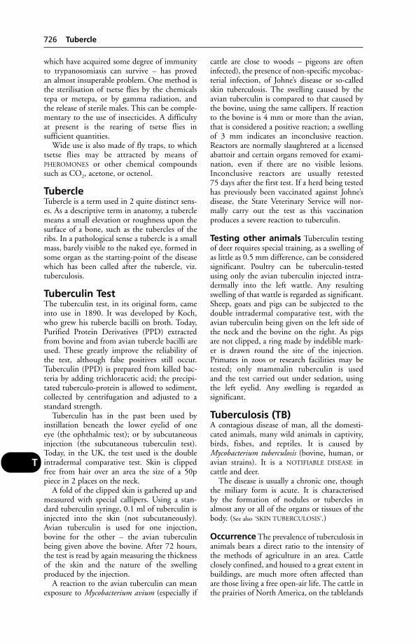
which have acquired some degree of immunityto trypanosomiasis can survive – has proved an almost insuperable problem. One method isthe sterilisation of tsetse flies by the chemicalstepa or metepa, or by gamma radiation, and the release of sterile males. This can be comple-mentary to the use of insecticides. A difficultyat present is the rearing of tsetse flies in sufficient quantities.
Wide use is also made of fly traps, to whichtsetse flies may be attracted by means ofPHEROMONES or other chemical compoundssuch as CO2, acetone, or octenol.
TubercleTubercle is a term used in 2 quite distinct sens-es. As a descriptive term in anatomy, a tuberclemeans a small elevation or roughness upon thesurface of a bone, such as the tubercles of theribs. In a pathological sense a tubercle is a smallmass, barely visible to the naked eye, formed insome organ as the starting-point of the diseasewhich has been called after the tubercle, viz.tuberculosis.
Tuberculin TestThe tuberculin test, in its original form, cameinto use in 1890. It was developed by Koch,who grew his tubercle bacilli on broth. Today,Purified Protein Derivatives (PPD) extractedfrom bovine and from avian tubercle bacilli areused. These greatly improve the reliability ofthe test, although false positives still occur.Tuberculin (PPD) is prepared from killed bac-teria by adding trichloracetic acid; the precipi-tated tuberculo-protein is allowed to sediment,collected by centrifugation and adjusted to astandard strength.
Tuberculin has in the past been used byinstillation beneath the lower eyelid of one eye (the ophthalmic test); or by subcutaneousinjection (the subcutaneous tuberculin test).Today, in the UK, the test used is the doubleintradermal comparative test. Skin is clippedfree from hair over an area the size of a 50ppiece in 2 places on the neck.
A fold of the clipped skin is gathered up andmeasured with special callipers. Using a stan-dard tuberculin syringe, 0.1 ml of tuberculin isinjected into the skin (not subcutaneously).Avian tuberculin is used for one injection,bovine for the other – the avian tuberculinbeing given above the bovine. After 72 hours,the test is read by again measuring the thicknessof the skin and the nature of the swelling produced by the injection.
A reaction to the avian tuberculin can meanexposure to Mycobacterium avium (especially if
cattle are close to woods – pigeons are ofteninfected), the presence of non-specific mycobac-terial infection, of Johne’s disease or so-calledskin tuberculosis. The swelling caused by theavian tuberculin is compared to that caused bythe bovine, using the same callipers. If reactionto the bovine is 4 mm or more than the avian,that is considered a positive reaction; a swellingof 3 mm indicates an inconclusive reaction.Reactors are normally slaughtered at a licensedabattoir and certain organs removed for exami-nation, even if there are no visible lesions.Inconclusive reactors are usually retested 75 days after the first test. If a herd being testedhas previously been vaccinated against Johne’sdisease, the State Veterinary Service will nor-mally carry out the test as this vaccination produces a severe reaction to tuberculin.
Testing other animals Tuberculin testingof deer requires special training, as a swelling ofas little as 0.5 mm difference, can be consideredsignificant. Poultry can be tuberculin-testedusing only the avian tuberculin injected intra-dermally into the left wattle. Any resultingswelling of that wattle is regarded as significant.Sheep, goats and pigs can be subjected to thedouble intradermal comparative test, with theavian tuberculin being given on the left side ofthe neck and the bovine on the right. As pigsare not clipped, a ring made by indelible mark-er is drawn round the site of the injection.Primates in zoos or research facilities may betested; only mammalin tuberculin is used and the test carried out under sedation, usingthe left eyelid. Any swelling is regarded as significant.
Tuberculosis (TB)A contagious disease of man, all the domesti-cated animals, many wild animals in captivity,birds, fishes, and reptiles. It is caused byMycobacterium tuberculosis (bovine, human, oravian strains). It is a NOTIFIABLE DISEASE in cattle and deer.
The disease is usually a chronic one, thoughthe miliary form is acute. It is characterised by the formation of nodules or tubercles inalmost any or all of the organs or tissues of thebody. (See also ‘SKIN TUBERCULOSIS’.)
Occurrence The prevalence of tuberculosis inanimals bears a direct ratio to the intensity ofthe methods of agriculture in an area. Cattleclosely confined, and housed to a great extent inbuildings, are much more often affected thanare those living a free open-air life. The cattle inthe prairies of North America, on the tablelands
726 Tubercle
T

of Central Africa, and in the steppes of EasternEurope, are almost entirely free from its ravages,while it is unknown in many islands (Iceland,Sicily, etc.).
Bovine tuberculosis eradication campaignshave succeeded in several countries.
Animals affected Among the ordinarydomesticated animals, cattle and pigs are morecommonly affected than are other species. Dogsare rarely infected but have been known to contract TB. Horses and sheep appear to bemore resistant, though cases of progressiveavian TB have been recorded in both species.Donkeys and mules are only very seldomattacked.
Methods of infection Cattle are infectedin 2 chief ways: (1) by the respiratory system;and (2) by the digestive tract. They are suscep-tible to infection from humans suffering frombovine tuberculosis, and serious breakdowns inattested herds have been traced to farm workerssuffering from the disease. Cattle are also sus-ceptible, to a lesser degree, to infection of thehuman type. (See also under TUBERCULIN TEST reavian tuberculosis.) Badgers have been impli-cated in the spread of bovine TB and long-running tests are taking place in the UK toestablish whether this is the case (see below,Tuberculosis in wildlife).
Sometimes tuberculosis may be contractedthrough a wound (e.g. after dehorning) or by direct introduction into the tissues of a penetrating instrument, and an infection of the udder may easily occur through the teatcanal. An aerosol infection commonly resultsfrom coughing cows, and infected sputum may contaminate feed or be swallowed, therebyspreading infection to the intestines.
Within the body, infection may spread viathe lymphatic system or the bloodstream.
Tuberculosis of the vagina occurs in cows,and the disease may be spread from them tohealthy cows through the medium of the bull.Infected dung can be a source of infection.
Nature of the lesions A typical lesion is atubercle – a small nodular swelling whose cen-tre contains either pus or dry yellowish cheesymaterial. The peritoneum, liver, lymph nodes,lungs, etc., may be affected. Sometimes the disease remains localised to the area of its firstinfection and does not spread. In other casesthe defensive forces of the body overcome anddestroy the focus of infection.
Tuberculosis may affect bones and one ormore joints, causing arthritis.
Superficial, as well as deep, lymph nodes maybecome enlarged.
A subclinical infection may occur, and resultin overt illness only when stress, under-feeding,exposure or some other infection lowers theanimal’s resistance.
Cattle As a rule a considerable period of timeelapses between infection and the appearance ofthe first symptoms.
Tuberculosis of the lungs – the commonesttype – gives rise to a hard, dry, short cough inthe early stages. Later, coughing becomes morefrequent and DYSPNOEA is evident.
Appetite is variable. Sometimes a difficulty in swallowing is noticed. Loss of condition follows, with pale mucous membranes, and astaring coat. There may be diarrhoea.
Superficial lymph nodes may becomeenlarged. Those at the back of the throat or atthe corner of the lower jaw, or the glands of theneck, shoulder, or stifle, may be swollen.
Tuberculosis of the udder – which is all-important from the milk standpoint – beginsinsidiously. The gland slowly becomes diffuselythickened, and more solid to the touch thannormally. After milking, it does not feel quite so elastic as it should, and in some cases distincthard nodules can be felt.
(Tuberculoid mastitis. Over 700 cases of this,due to rapidly growing acid-fast organismsother than Mycobacterium tuberculosis, occur inthe UK annually – mainly due to not cleaningthe teats before introducing antibiotics.)
Tuberculosis sometimes involves the brain orspinal cord, giving rise to symptoms describedunder MENINGITIS.
Tuberculosis of the bones and joints is notuncommon.
In the skin there occasionally develop hardtumours, about the size of a hazelnut (see also
‘SKIN TUBERCULOSIS’), which, if they are opened,are found to contain cheesy or mortar-like mass-es in their centres. Later, ulcers may developwith the formation of multiple small abscesses in any or all of the organs. The abscesses are millet-seed sized (hence ‘miliary’). This form oftuberculosis is rapidly fatal.
Sheep and goats A distressing painfulcough, always present, but most noticeableupon exertion; and a gradual, but quite definite,loss of condition, with progressing weakness,are the main symptoms observed in these ani-mals. Sheep are very rarely affected, but milkinggoats kept in the vicinity of infected cattle notuncommonly develop tuberculosis. There isnearly always a marked anaemia, pneumonia,
Tuberculosis (TB) 727
T
sometimes diarrhoea, and occasionally an infec-tion of the udder corresponding to that foundin cattle.
Horses Tuberculosis in the horse is not verycommon, but there are certain symptoms whichshould always lead one to suspect its presence: agradual emaciation in spite of good food andwithout any other established possible cause; aslight fluctuating increase in the temperature,an occasional moist weak cough; a tucked-upappearance of the abdomen, or in some cases(where ascites exists) a heavy pendulous condition, ‘pot-bellied’.
Cases in which the abdominal organs areaffected sometimes terminate by lung compli-cations – i.e. miliary tuberculosis sets in – theanimal becomes feverish, distressed in itsbreathing, refuses all food, and generally dieswithin a few days. Tuberculosis may alsobecome localised in the skin, lymph nodes,brain, or udder, but these are not common. It iscomparatively often found that sooner or latersome part of the skeleton (the bones of the neckbeing a very usual situation) becomes infected.
Occasionally, tuberculosis in the horse maybe caused by the human or avian type of thetubercle bacillus.
Pigs Tubercular poultry, or wild birds such aswood-pigeons, are a not uncommon source ofinfection. A diagnosis may be established bymeans of the tuberculin test.
Symptoms are as in the horse. Scouring andemaciation may occur. Anaemia is common. Asin horses, the bones are especially vulnerable toattack.
Lesions, which, to the naked eye, appearidentical with tuberculosis, may be caused byinfection with Corynebacterium equi. Even theuse of a microscope sometimes fails to differen-tiate between the 2 infections.
Deer Disease caused by bovine or avian tuber-cle bacilli can be found. The clinical signs,which may not be noticeable until the disease isadvanced, include respiratory signs. Caseouslesions are rarely seen as tuberculous lesions areusually filled with pus.
Dogs and cats Where tuberculosis is com-mon in the human population, these domesticanimals are liable to become infected – either as the result of receiving tubercle-containingmilk, or as the result of infection from sputumor discharges from a human case. Not only may dogs and cats contract the disease fromman, but they may occasionally be sources of
infection to healthy human beings, and espe-cially to children.
As in other animals, the symptoms are some-what vague until the disease is well established.The first signs may be no more than a capri-cious appetite, slight loss of condition, generalweakness, and exhaustion when at exercise.Pulmonary tuberculosis usually begins with a short dry cough. It is less common in theseanimals than the abdominal form (but see
PLEURISY).Tuberculosis of the abdominal organs is indi-
cated by impaired nutrition and anaemia,attacks of diarrhoea and constipation alternat-ing with each other. There may be vomiting,also ascites. Body temperature is very variable.
Joints and sinuses may be sites of infection inthe cat. Occasionally skin tuberculosis is seen indogs and cats, and may take the form of raisedplaques with a tendency to ulcerate.
Treatment The treatment of tuberculosis inthe domesticated animals is not attempted, for4 reasons: (1) because of the nature of the disease; (2) because of the ever-increasing danger to human beings who have to attendaffected animals; (3) for economic reasons; and (4) for humanitarian ones. However, inzoological gardens, animals are sometimes treated. (See PAS.)
Prevention Good hygiene, good feeding andgood ventilation all help. Animals brought infrom areas known to be infected should bequarantined on the premises and a tuberculintest carried out by the veterinarian befoe theyare allowed to mix with other stock.
History of control in Britain It was notuntil 1928 that measures to control bovinetuberculosis were introduced by the govern-ment. In that year, the Tuberculosis Order,enacted in 1915, came into force, and theattempt to control the disease by the detectionand elimination of ‘open’ cases began. In 1935the Attested Herds Scheme carried controlmeasures a stage further.
Area Eradication, which began in 1950, andmeant, at first, an extension of the AttestedHerds Scheme on a voluntary basis, and thenthe compulsory slaughter of reactors within theprescribed areas, followed.
In October 1960, the whole of the UK wasdeclared one Attested Area – bovine tuberculo-sis being virtually eradicated from all herds ofcattle.
In 1962, the incidence of bovine tuber-culosis in herds in England and Wales was
728 Tuberculosis (TB)
T
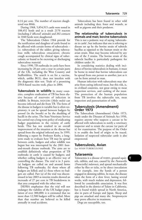
0.14 per cent. The number of reactors slaugh-tered was 8846.
During 1968, 5,854,915 cattle were tested in108,452 herds and as a result 2170 reactors(including 2 ‘affected’ animals) and 202 contactsin 1040 herds were slaughtered.
The Tuberculosis Orders 1964 provide forthe notification and slaughter of cattle found tobe affected with certain forms of tuberculosis –i.e. tuberculosis of the udder; giving tubercu-lous milk; tuberculous emaciation; chroniccough accompanied by clinical signs of tuber-culosis; or found to be excreting or dischargingtuberculous material.
Since 1990, TB outbreaks in cattle have beenincreasing by 20 per cent a year in certain partsof the UK, including the West Country andStaffordshire. The search is on for a vaccine,which, unlike BCG, does not interfere with the diagnostic skin test. Trials of a promisingDNA-based vaccine took, place in 2004.
Tuberculosis in wildlife In many coun-tries, complete eradication of TB has been elu-sive because of reservoirs of infection inwildlife. In Britain, BADGERS (Meles meles) canbecome infected and die from TB. The form ofthe disease in badgers is variable but is often res-piratory; it can be spread between badgers byaerosol (in the breath) or by the shedding ofbacilli in the urine. The State Veterinary Servicehas carried out a long-term policy of eradicatingbadger populations in the vicinity of cattleherds. This has not resulted in an overallimprovement of the situation as the disease hasspread from the original infected area. In 1999,following a report by Professor Krebs, a long-term study to evaluate how TB may be spreadbetween cattle, badgers and other wildlife wasbegun but was interrupted by the 2001 foot-and-mouth disease outbreak. The aims are toestablish definitively what proportion of TBoutbreaks in cattle is caused by badgers; andwhether culling badgers is an effective way ofcontrolling the disease. The trial is in 3 parts:(a) badgers are culled on and around farms following TB outbreaks; (b) those where allbadgers are killed; and (c) those where no bad-gers are culled. Part (a) of the trial was discon-tinued in late 2003 as interim results showed anincreasde of 27 per cent in TB breakdowns inthat area compared with the other two.
DEFRA emphasises that the trial will notendanger the viability of the UK badger popu-lation of over 300,000; it is estimated that nomore than 12,500 badgers will be culled. Morethan that number are believed to be killedannually in road accidents.
Tuberculosis has been found in other wildanimals including deer, foxes and weasels, aswell as pigeons and their predators.
The relationship of tuberculosis inanimals and man; bovine tuberculosisThis is not a pedantic way of saying ‘tuberculo-sis in cattle’, but indicates that one is referring todisease set up by the bovine strain of tuberclebacillus as opposed to the human strain or theavian strain. Man may become infected by anyone of the 3 strains. The bovine strain of thetubercle bacillus is particularly pathogenic forchildren under 16.
In considering statistics dealing with inci-dence of bovine tuberculosis in humans, it mustbe borne in mind that bovine tuberculosis canbe spread from one person to another, just as itcan be from animal to man.
Human infection with tuberculosis may alsoarise from eating infected meat, but this risk, is,in civilised countries, not great owing to meatinspection services, and cooking of the meat.The protection of the human population from TB involves the tuberculin test, meatinspection and pasteurisation of milk.
Tuberculosis (Amendment)Order 1973The Tuberculosis (Amendment) Order 1973,made under the Diseases of Animals Act 1950,requires anyone who suspects a carcase to beaffected with tuberculosis to notify a veterinaryinspector and to retain the carcase (or parts ofit) for examination. The purpose of the Orderis to enable the herd of origin to be traced.Isolation of suspected tubercular cattle is alsoempowered.
Tuberculosis, Avian(see AVIAN TUBERCULOSIS)
TularaemiaTularaemia is a disease of HARES, ground squir-rels, rabbits, and rats, caused by the Pasteurella(Francisella) tularensis, and spread mechanicallyeither by flies or ticks, or by direct inoculation– for example, into the hands of a personengaged in skinning rabbits. In man, the diseasetakes the form of a slow fever, lasting severalweeks, with much malaise and depression, fol-lowed by considerable emaciation. It was firstdescribed in the district of Tulare in California,but is found widely spread in North America,also in parts of Europe and Japan. Sheep andpigs are attacked and many die. Streptomycinmay prove effective in treatment.
Dogs are susceptible, too.
Tularaemia 729
T

Tumbu Fly(see under SCREW-WORM FLIES)
TumourMalignant tumours are those which tend togrow and spread rapidly, destroying neighbour-ing tissues and infiltrating the healthy struc-tures near by. They are liable to ulceratethrough the skin when superficial, are non-encapsulated, and may spread to distant partsof the body by the blood- or lymph-stream, giving rise to secondary tumours there. (Furtherinformation upon malignant tumours appearsunder CANCER.)
Benign tumours grow slowly at one place,press neighbouring parts aside, but neitherinvade nor destroy them, only seldom ulceratethrough the skin or mucous membrane, haveusually a capsule of fibrous tissue surroundingthem, and when once completely removed bysurgical excision or other means, do not recur.
While this classification serves in a measureto differentiate typical varieties into 2 classes, itis by no means absolutely satisfactory. There arecertain kinds of normally benign tumourswhich may remain comparatively small and cir-cumscribed for a number of years, and thensuddenly become malignant.
Benign or simple tumours include ANGIOMA,CHONDROMA, fibroma GLIOMA, LIPOMA,MYOMA, MYXOMA, NEUROMA, ODONTOMA, andPAPILLOMA. The last-named may be benign inthe beginning but become malignant later; alsoMELANOMA and ADENOMA. (See also CYSTS;WARTS; CANCER.) While normally all tumourstend to increase in size – either slowly or rapid-ly – some grow to a certain size, remain sta-tionary, then decrease in size, and a few mayeven disappear completely.
Two of the most common tumours of thedog are mammary carcinoma and anal adeno-ma (usually benign). (See also EOSINOPHILIC
GRANULOMA.)
Tumour Angiogenesis Factor(TAF)This has been isolated from human and animaltumours. It stimulates mitosis in endothelialcells and rapid formation of new capillaries fortumour nourishment. (Unlike a skin-graft,which sends out capillary shoots to join thecapillaries of the recipient tissue, a tumour hasto rely entirely on the host, and makes use ofTAF for this purpose.)
TungiasisInfestation with Tunga penetrans, the jigger flea.(See under FLEAS.)
TupA ram (see under SHEEP).
Turbinate Bones(see under NOSE and RHINITIS)
Turkey CoryzaA disease of the upper respiratory tract, withsneezing and nasal discharge, whose probablecause is Bordetella avium. It occurs in turkeypoults, usually in conditions where ventilationis poor and the birds are stressed.
Turkey Viral HepatitisThis occurs in Europe, the USA, and Canada;and in 1982 isolation of a picorna-like viruscausing hepatitis and disease of the pancreaswas isolated from an outbreak in Scotland. Theinfection, which is highly contagious, is often,if not usually, subclinical, but may take an acuteform, and prove fatal.
TurkeysDiseases include Arizona disease, blackhead,coccidiosis, erysipelas, fowl pest, hexamitiasis,haemorrhagic enteritis, moniliasis, Oregon disease, ornithosis, mycoplasmosis, pullorumdisease, reticuloendotheliosis, rupture of theaorta, sinusitis, synovitis (see also GROUNDNUT
MEAL MANIOC).Turkey meningoencephalitis occurs in Israel.
Influenza viruses cause disease in NorthAmerican domestic turkeys.
Turkey rhinotracheitis has been seen in several countries, and appeared in the UK in1985, causing severe financial losses due todeaths, carcase rejections, and lowered egg production. The first sign is sneezing.
TurnipsLike kale, these contain a goitre-producing fac-tor, and if fed in large amounts to pregnantewes are liable to cause abortion – unless iodinelicks are provided. (See VAGINA, RUPTURE OF.)
Turpentine, Medicinal Oil ofTurpentine is the oleo-resin which exudes fromvarious members of the pine family, especiallythe Pinus australis, P. taeda, and P. sylvestris. Theoil distilled from this oleo-resin is known as oilof turpentine. The natural turpentine is notused in medicine, as it is highly irritating andwhen the word ‘turpentine’ is employed, the oilof turpentine is indicated.
In collections of gas in the abdominal organs,medicinal turpentine has been used (e.g. tym-pany in horses and cattle). Large doses are liableto irritate the stomach and kidneys.
730 Tumbu Fly
T
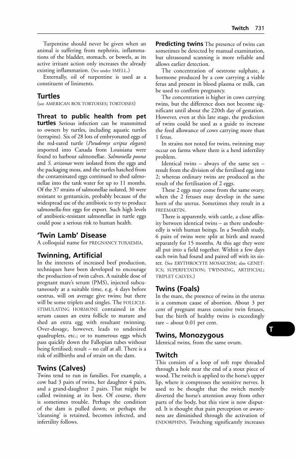
Turpentine should never be given when ananimal is suffering from nephritis, inflamma-tions of the bladder, stomach, or bowels, as itsactive irritant action only increases the alreadyexisting inflammation. (See under SMELL.)
Externally, oil of turpentine is used as a constituent of liniments.
Turtles(see AMERICAN BOX TORTOISES; TORTOISES)
Threat to public health from pet turtles Serious infection can be transmitted to owners by turtles, including aquatic turtles(terrapins). Six of 28 lots of embryonated eggs of the red-eared turtle (Pseudemys scripta elegans)imported into Canada from Louisiana werefound to harbour salmonellae. Salmonella poonaand S. arizonae were isolated from the eggs andthe packaging moss, and the turtles hatched fromthe contaminated eggs continued to shed salmo-nellae into the tank water for up to 11 months.Of the 37 strains of salmonellae isolated, 30 wereresistant to gentamicin, probably because of thewidespread use of the antibiotic to try to producesalmonella-free eggs for export. Such high levelsof antibiotic-resistant salmonellae in turtle eggscould pose a serious risk to human health.
‘Twin Lamb’ DiseaseA colloquial name for PREGNANCY TOXAEMIA.
Twinning, ArtificialIn the interests of increased beef production,techniques have been developed to encouragethe production of twin calves. A suitable dose ofpregnant mare’s serum (PMS), injected subcu-taneously at a suitable time, e.g. 4 days beforeoestrus, will on average give twins; but therewill be some triplets and singles. The FOLLICLE-
STIMULATING HORMONE contained in theserum causes an extra follicle to mature andshed an extra egg with resultant twinning.Over-dosage, however, leads to undesiredquadruplets, etc.; or to numerous eggs whichpass quickly down the Fallopian tubes withoutbeing fertilised; result – no calf at all. There is arisk of stillbirths and of strain on the dam.
Twins (Calves)Twins tend to run in families. For example, acow had 3 pairs of twins, her daughter 4 pairs,and a grand-daughter 2 pairs. That might becalled twinning at its best. Of course, there is sometimes trouble. Perhaps the condition of the dam is pulled down; or perhaps the‘cleansing’ is retained, becomes infected, andinfertility follows.
Predicting twins The presence of twins cansometimes be detected by manual examination,but ultrasound scanning is more reliable andallows earlier detection.
The concentration of oestrone sulphate, ahormone produced by a cow carrying a viablefetus and present in blood plasma or milk, canbe used to confirm pregnancy.
The concentration is higher in cows carryingtwins, but the difference does not become sig-nificant until about the 220th day of gestation.However, even at this late stage, the predictionof twins could be used as a guide to increase the feed allowance of cows carrying more than1 fetus.
In strains not noted for twins, twinning mayoccur on farms where there is a herd infertilityproblem.
Identical twins – always of the same sex –result from the division of the fertilised egg into2; whereas ordinary twins are produced as theresult of the fertilisation of 2 eggs.
These 2 eggs may come from the same ovary,when the 2 fetuses may develop in the samehorn of the uterus. Sometimes they result in aFREEMARTIN.
There is apparently, with cattle, a close affin-ity between identical twins – as there undoubt-edly is with human beings. In a Swedish study,6 pairs of twins were split at birth and rearedseparately for 15 months. At this age they wereall put into a field together. Within a few dayseach twin had found and paired off with its sis-ter. (See ERYTHROCYTE MOSAICISM; also GENET-
ICS; SUPERFETATION; TWINNING, ARTIFICIAL;TRIPLET CALVES.)
Twins (Foals)In the mare, the presence of twins in the uterusis a common cause of abortion. About 3 percent of pregnant mares conceive twin fetuses,but the birth of healthy twins is exceedinglyrare – about 0.01 per cent.
Twins, MonozygousIdentical twins, from the same ovum.
TwitchThis consists of a loop of soft rope threadedthrough a hole near the end of a stout piece ofwood. The twitch is applied to the horse’s upperlip, where it compresses the sensitive nerves. Itused to be thought that the twitch merelydiverted the horse’s attention away from otherparts of the body, but this view is now disput-ed. It is thought that pain perception or aware-ness are diminished through the activation ofENDORPHINS. Twitching significantly increases
Twitch 731
T
T
732 ‘Tying-Up Syndrome’
plasma levels of beta-endorphin, which returnsto normal 30 minutes or so after the twitch isremoved. Twitching could therefore be said tobe analogous to ACUPUNCTURE.
‘Tying-Up Syndrome’Also known as set-fast, this condition in race-horses appears to be identical with azoturia.Symptoms include stiffness, a rolling gait, blow-ing and sweating and, if exercise continues, theadoption of a crouching attitude. Pain is evi-dent. The animal may lie down and be unable torise. (See azoturia, under BOVINE MYOGLOBINURIA.)
TylanThe proprietary name of a preparation oftylosin.
TylosinA macrolide antibiotic effective against Gram-positive organisms; it concentrates in acidic con-ditions, as in the udder and the lung. It is usedas a growth promoter in pigs. (See ADDITIVES.)
TympanitesThe drum-like condition of the abdomen,which results from distension of the stomach orbowels with gas, as the result of fermentation,constipation, or of simple obstruction. (See under
STOMACH, DISEASES OF; INTESTINES, DISEASES
OF; BLOAT; TYMPANY; TYMPANITIC.)
Tympanitic Resonance in CattleRight-side tympanitic resonance (ping) causedby gas distention of intra-abdominal structureswas diagnosed in 366 adult cattle, in a USAstudy. The source of the ping was identified as the abomasum in 137 animals, various seg-ments of the intestinal tract in 157 and peri-toneal gas in two. The source of the noise wasnot identified in 70. The principal final diag-noses were: left displacement of the abomasum(116), right displacement of the abomasum(77), abomasal (and omasal) volvulus (60),other gastro-intestinal conditions (73) and non-gastrointestinal conditions (40).
TympanyTympany is distension of a hollow organ withgas. (See TYMPANITES; BLOAT.)
TyphilitisInflammation of the caecum or 1st part of thelarge intestine, into which the termination ofthe small intestine opens.
Typhus of Rats and MiceTyphus of rats and mice, caused by Rickettsiamooseri, may kill about 5 per cent of peopleinfected by it.
Tyzzer’s DiseaseThis was first described in mice in 1917, andhas since been reported in horses, cats, and lab-oratory animals including rats, rabbits, gerbils,and rhesus monkeys.
Cause The spore-forming, Gram-negative,motile Bacillus piliformis.
Signs The disease is characterised as a rule bysevere diarrhoea, debility, and death; thoughsudden death in foals without preliminarysymptoms has been reported in the USA.Jaundice, slight or marked, was a post-mortemfinding, together with some liver necrosis andenteritis.
In the cat, the infection gives rise to symp-toms of loss of appetite, depression, diarrhoea,collapse and death. Necrosis of the ileum andhepatitis are among post-mortem findings.
Tzaneen DiseaseThis is a tick-borne infection with Theileriamutans in cattle, the African buffalo, and theIndian water buffalo, and often occurs simulta-neously with other infections. There may beonly a mild fever or, less commonly, serious ill-ness, and death. Anti-malarial drugs are of use.
Udder(see MAMMARY GLAND)
Uitpeuloog‘Bulging eye disease’– an oculovascular myiasisof domestic animals in South Africa.
UlcerA break on the surface of the skin, or of anymucous membrane of a cavity of the body,which does not tend to heal. The process bywhich an ulcer spreads, which involves necrosis(death) of minute portions of the healthy tis-sues around its edges, is known as ulceration.Most ulcers are suppurative; bacteria preventhealing and often extend the lesion.
An ulcer consists of a ‘floor’ or surface which,in consequence of the loss or destruction of tis-sue, is usually depressed below the level of thesurrounding healthy structures; and an ‘edge’around it where the healthy tissues end.
Callous ulcer is a type of chronic ulcer oftenencountered in horses and dogs, when there isany pressure or irritation that interferes with theblood supply but does not necessarily causeimmediate destruction of the skin. In most casesit is covered by a hard, leathery piece of deadskin from under which escapes a purulent fluid.‘Bedsores’ in all animals may be of this nature.
‘Rodent ulcer’ is a term reserved in humanmedicine for an ulcerating carcinoma of theskin, but it is often colloquially used by dog-and cat-owners for an EOSINOPHILIC GRANULO-
MA. Skin cancer occurs in domestic animals,and such malignant tumours may ulcerate.
Tubercular ulcers may occur in dogs’ and cats’skin in the form of raised plaques which ulcerate.
Internal ulcers may occur in the mouth (see
MOUTH, DISEASES OF), in the stomach (see GAS-
TRIC ULCERS), in the bowels (see INTESTINES,DISEASES OF), and in other parts.
Glanders ulcers are typically encountered inthe mucous membrane of the nostrils, and havea ‘punched-out’ appearance.
Lip-and-leg ulcers occur in sheep with ORF.
Causes Any condition that lowers the generalvitality of the animal, such as old age, chronicdisease, malnutrition, and defective circulation,will act as a predisposing cause. Among directcauses may be bacteria gaining access to
wounds; irritation from badly fitting harness,pressure of bony prominences upon hard floorsinsufficiently provided with bedding, andapplication of too strong antiseptics to wounds.
Treatment In the smaller animals a vitaminsupplement may be indicated. An antibiotic orone of the sulfa drugs may be used.
Local treatment aims at converting the ulcerinto what virtually becomes an ordinary openwound. The surface is treated with some suit-able antiseptic, such as cetrimide, gentian violet,dilute hydrogen peroxide, etc. If one or twodays of such treatment does not result in a clean,bright-red, odourless wound, or where there areshreds of dead tissue adherent to the surface, itmay be necessary to curette the surface so thatthe dead cells may be separated from the healthyones below them.
Animal-owners should note that after thesurface of the ulcer has been rendered as healthyas possible, use of strong antiseptics or (worsestill) disinfectants should cease, as these retardhealing by the destruction of surface tissues.
Corneal ulcers are referred to under EYE,DISEASES OF – Keratitis. (See also CRYOSURGERY.)
Ulcerative Dermatosis of SheepA viral infection, which has to be differentiatedfrom ORF, and is characterised by ulcers on theface, feet, legs, and external genitalia. It also inEurope and Africa.
Ulcerative Dermal NecrosisA disease of adult salmon which occurs whenthey enter fresh water on their way to spawninggrounds. Grey lesions are seen above the eyes,on the snout and on the side of the opercula (gillcoverings). If they do not heal, the lesions spreadto the skin of the head, ulcerate, and becomeprone to infection by Saprolegnia fungus. Thefungal infection may be treated with zinc-freemalachite green. The cause is unknown.
Ulcerative Enteritis of Chickens,Pheasants and QuailsUlcerative enteritis is seen in chickens between 4 and 7 weeks, and in quail, turkeys, partridgeand grouse at any age. Birds are depressed, with watery droppings; mortality can be veryhigh, reaching 70 to 100 per cent. The cause is avirus.
Ulcerative LymphangitisUlcerative lymphangitis, also called ulcerativecellulitis, is a contagious chronic disease of horses, characterised by inflammation of the
U

lymph vessels and a tendency towards ulcerationof the skin over the parts affected.
Cause Corynebacterium bovis (pseudotuberculo-sis). It gains access through abrasions. Infectionmay be carried by grooming tools, harness,utensils, etc., from one horse to another.
Signs The commonest seat of the disease is thefetlock of a hind-leg. This part becomes swollenand slightly painful. Small abscesses appear;ulcers follow. The condition gradually spreadsup the leg.
Treatment Antibiotics.
Ulcerative Spirochaetosis ofPigsThis has been reported in the UK, Australia,New Zealand, South Africa, and the USA. Itmay give rise to foot-rot in pigs, ulceration ofthe skin, and scirrhous cord.
UlnaThis is the inner of the 2 bones of the fore-arm.The shaft has gradually reduced in size as thenumber of digits has decreased, so that whilethe ulna is a perfect bone in the dog and cat, inthe horse its shaft has almost completely disap-peared and the bone is only represented by theolecranon process which forms the ‘point of theelbow’. The shaft of the ulna is liable to becomefractured from violence to the fore-limb, butthe commonest seat of an ulnar fracture is theolecranon process. This occurs from a fall inwhich the fore-limbs slip out in front of the ani-mal, and the weight of the body comes downsuddenly on to the point of the elbow. (See
FRACTURES.)
Ultra-High-TemperatureTreatment of Milk (UHT)Ultra-high-temperature treatment of milk(UHT) involves heating it to between 135° and149°C (275° and 300°F) for a few seconds.Suggested in 1913, UHT is used to producelong-life milk, on sale in Britain from 1965onwards. This process does not affect the calci-um nor the casein, but destroys some vitaminsand probably some serum proteins (immuneglobulins). Calves grow less well on it than onraw or pasteurised milk.
UltrasoundSound at a frequency above 20,000 cycles persecond. Propagated by applying an electric cur-rent to one side of a piezoelectric crystal, whichdeforms and produces a sound wave.
Ultrasound is generally defined as an auditoryfrequency beyond that perceived by the humanear. Most humans hear and emit sound in thefrequency range 2 to 20 kHz, while in some animals ranges are much greater. Bats, dolphins,many rodents and some insects have ranges thatextend as high as 120 kHz – well beyond thelimit of human detection. Pigs and poultry candetect higher ultrasound frequencies and may bedisturbed by the noise given off by, for example,certain electronic equipment and dripping nipple-drinkers. Female rabbits communicatewith their litters in ultrasound.
Ultrasound, in the range of 1 million to 10 million hertz, is used in non-invasive diag-nostic imaging of internal body structures. It iswidely used in pregnancy diagnosis of animals.(See also PREGNANCY DIAGNOSIS.)
In human medicine, ultrasound has beenshown to be beneficial for wound healing, bothin the treatment of pressure sores and in thepreparation of trophic ulcers for skin grafting.Studies have shown that it influences the activityof fibroblasts.
Ultra-Violet RaysUltra-violet rays are used in the treatment of various skin diseases, etc., and in the diagnosis ofringworm and porphyria; also in the fluorescent-antibody test for various infections includingrabies.
Ultra-violet rays and eye cancerAnalysis of data from 14 veterinary colleges inthe USA, where 147 cases of eye cancer in hors-es were studied, led to the conclusion that ultra-violet radiation may be of primary importancein triggering cancer.
Umbilical Cord, Cutting The(see under PARTURITION)
UmbilicusUmbilicus is another name for the navel.
Umgana TreeElephants seek out and gorge themselves on thefruits of this tree, leading to in sexual excitement.Ostriches may behave similarly.
UncinariasisInfection with Uncinaria stenocephala, one ofthe hookworms of the dog.
Unconsciousness(see under COMA; FITS; SYNCOPE; EPILEPSY;NARCOLEPSY)
734 Ulcerative Spirochaetosis of Pigs
U
Undecylenate OintmentA fungicide, used in the treatment of ringworm,etc.
Undulant Fever in ManUndulant fever in Man is caused by Brucellamelitensis, B. abortus suis, or B. abortus. The lat-ter organism is responsible for ‘contagious abor-tion’ (brucellosis) of cattle, and it is probablethat most cases of undulant fever in man causedby B. abortus arise through handling infectedcows or from drinking their milk. Infection can readily occur through the skin. Numerouscases have occurred in veterinarians, followingmishaps with Strain 19 vaccine, e.g. accidentalspraying into the eyes or injection into the hand.In America, B. abortus suis is an important causeof undulant fever in man; B. canis likewise.
Signs are vague and simulate those of influen-za except that undulant fever lasts for a muchlonger time, even many months. Temperature isgenerally raised but fluctuates greatly; there aremuscle pains, headache, tiredness and inabilityto concentrate. One or more joints may swell.There may be constipation. The organisms arepresent in the bloodstream and in the spleen.
The disease is serious, not so much becauseof its mortality (1 to 2 per cent), but because ofincapacity occasioned by its long duration. (See
BRUCELLOSIS and CHEESE.)
Prevention Anyone handling aborting cows,or their fetal membranes, or even calving anapparently normal cow, should wear protectivegloves or sleeves (which nevertheless sometimestear), and wash arms and hands in a disinfec-tant solution afterwards. Avoid drinking anycold milk that has not been pasteurised.
Unsaturated Fatty Acids(see under LIPIDS; VITAMINS – Vitamin E)
Unstable Substances(see under INJECTIONS)
UraemiaUraemia results when the waste materials thatshould be excreted into the urine are retained inthe body, through some disease of the kidneys,and are circulated in the bloodstream. Bloodurea is in excess. Death may be preceded byconvulsions and unconsciousness. In the slowertypes there is usually a strong urinous odourfrom all the body secretions. In acute cases theadministration of glucose saline subcutaneouslymay help; likewise, withdrawal of a quantity of blood (provided that saline is given). (See
URINE – Abnormal constituents; LEPTOSPIRO-
SIS; KIDNEYS, DISEASES OF.)
Urates(see URIC ACID)
Urea (Carbamide)Urea (carbamide) is a crystalline substance of the chemical formula CO(NH2)2, which isvery soluble in water and alcohol. It is the chiefwaste product discharged from the body in theurine, being formed in the liver and carried bythe blood to the kidneys. The amount excretedvaries with the nature and the amount of thefood taken, being greater in the carnivora, andwhen large amounts of protein are present inthe food. It is also increased in quantity duringthe course of fevers. Urea is rapidly changedinto ammonium carbonate after excretion andwhen in contact with the air, owing to theaction of certain micro-organisms.
Determination of the blood urea level is animportant aid to the diagnosis of kidney failure.
Urea As a Ruminant FeedSome of the micro-organisms which inhabit therumen can synthesise protein from urea. It wasaccordingly suggested that urea might be substi-tuted for protein in concentrates fed to cattle.This has been called the protein-sparing effect ofurea, which is a non-protein source of nitrogen.
The emphasis has now shifted more to thevalue of urea in increasing the intake and aidingthe digestion of low-quality roughages, and ithas been widely used as a dietary supplement forcattle and sheep on poor pasture in many partsof the world. Where extra energy, in the form ofreadily digestible carbohydrate, is provided inaddition to the urea, both roughage digestibilityand feed intake improve. In these circumstancesthe urea stimulates multiplication of cellulose-digesting organisms, so that the urea-fed animalmay be able to make more effective use ofroughage than the one receiving no urea.
In the ruminant animal, any injudiciousfeeding of urea can give rise to poisoning byammonia, since it is this which is released in the rumen and then converted into microbialprotein. Excess ammonia can cause the animal’sdeath. It is essential, therefore, that urea is takenin small quantities over a period, and not fed alarge amount at a time.
Urea is often added to molasses and fed viaball feeders which prevent rapid and excessiveuptake of the liquid and ensure maximum utili-sation of urea. Under optimum farm conditionsonly 15 to 30 per cent of the dietary protein canbe replaced by urea. As a rule of thumb, urea, if
Urea As a Ruminant Feed 735
U

incorporated uniformly within the dairy ration,has been shown to be safe at a level of 1 per cent.Combined with 5 per cent barley, that mix canreplace 5 per cent ground-nut meal in theration. In the diets of finishing beef cattle, theanimals can gradually have the proportion ofurea increased.
Some guidelines for urea feeding1. Introduce urea feeding gradually, i.e. at a
slowly increasing level over a period of 3 to 4weeks, with adequate minerals and vitaminsprovided.
2. Avoid starting newly calved cows on it (butit may be included in the steaming-up ration),or giving it to calves under 3 months of age.
3. Ensure that urea is fed with adequate readi-ly digestible carbohydrate, as is contained in cere-als, molasses, sugar-beet pulp, maize silage, etc.
4. Do not exceed levels of urea recommendedby the supplier.
5. Ensure that urea is fed little and often, andnot irregularly or at long intervals.
Urea poisoning Symptoms include saliva-tion, excitement, running and staggering, jerkingof the eyeballs, and scouring.
Acute urea poisoning killed 17 beef cows in agroup of 29 in the south of Scotland. The ani-mals died over an 8-hour period as a result ofdrinking water which had been carried to atrough in a tanker previously used for transport-ing urea fertiliser. It was calculated that as littleas 10 litres of the water would have provided afatal dose of urea to a 500 kg cow.
UreaplasmasFormerly known as T-mycoplasmas, these havebeen isolated from the lungs, and also the uro-genital tract of several species of animals. Theyare a likely cause of pneumonia and infertility.
UreterThe ureter is the tube which carries the urineexcreted by a kidney down to the urinary bladder.Each ureter begins at the pelvis (main cavity) ofthe corresponding kidney, passes backwards anddownwards along the roof and walls of the pelvis,and finally ends by opening into the neck of thebladder. The wall of the ureter is composed of afibrous coat on the outside, a muscular coat in themiddle, and this is lined by a mucous membraneconsisting of cubical epithelium.
UrethraThe urethra is the tube which leads from theneck of the bladder to the outside, opening atthe extremity of the penis in the male, and into
the posterior part of the urino-genital passagein the female. It serves to conduct the urinefrom the bladder to the outside; also the semen.
Urethra, Diseases ofOwing to its extreme shortness in the female,the urethra is not subject to the same diseaseconditions as in the male, where the tube isconsiderably longer. In fact, disease of the ure-thra in the female hardly ever arises except as acomplication of either disease of the bladder, onthe one hand, or of the vagina on the other.
Urethritis Inflammation of the urethra isusually associated with cystitis, and may be theresult of an infection, or of some irritant poison(such as CANTHARIDES) present in the urine.The lining mucous membrane may also beinflamed by crystalline deposits. (See FELINE
UROLOGICAL SYNDROME; UROLITHIASIS; URE-
THRAL OBSTRUCTION.)In most cases of urethritis there are signs of
pain and distress whenever urine is passed orwhen the parts are handled. A little blood maybe seen.
Stricture is an abrupt narrowing of the calibreof the tube at one or more places. In almost allcases of true stricture there has been some injuryto the urethra or penis, resulting in the forma-tion of scar tissue, which eventually contractsand decreases the lumen of the tube. A fewcases, however, are caused by a rapidly growingtumour.
Injuries to the urethra may follow a severecrush or blow which causes fracture of the pelvisor of the os penis in the dog. They are usuallyobvious when the injury has involved the sur-face of the body, and may be suspected if thereis an inability to pass urine, or if the urine con-tains blood or pus following upon a severeinjury to the hindquarters of the body. A com-plication of urethral injuries is abscess formationaround the urethra and consequent stricture at alater period.
Urethral ObstructionIn sheep, the injudicious use of hormones toincrease liveweight gain has killed lambs, appar-ently as the result of urethral obstruction. Inone incident in the USA, 200 out of 9000lambs died after receiving 12 mg stilboestrol byinjection.
In the UK, an increased incidence of urethralobstruction in male calves and lambs followed theincorporation of too high a level of magnesium inthe concentrates fed.
736 Ureaplasmas
U
Analysis of the calculi (stones) causing theobstruction showed them to be crystals of mag-nesium ammonium phosphate. After reducingthe level of magnesium supplementation to 200mg MgO per tonne of feed, there were no casesof urolithiasis in intensively fattened malelambs offered a cereal-based diet ad lib. (See also
under URINARY BLADDER, DISEASES OF – Urinarycalculi.)
Five outbreaks in male calves of various ages investigated by the Veterinary ResearchLaboratory, Stormont, showed a magnesiumcontent of the concentrates (fed from the firstweek of life) to range from 4.9 to 9.2 g/kg drymatter. (The AFRC recommendation is notmore than 1.4 g/kg dry matter.)
Obstruction of the male urethra is a commoncondition in cats, and fairly common in thedog. (See FELINE UROLOGICAL SYNDROME.)
Unless relieved, urethral obstruction can leadto rupture of the bladder and death.
UrethrostomyPerineal urethrostomy is a surgical operation forthe treatment of urethral obstruction; it consistsof making a permanent opening in the urethra,the lining mucous membrane and the skinbeing joined by sutures. (Urethrostomy differsin this respect from urethrotomy, in which theurethra is incised – to remove a wedged calculus,for example – but immediately closed.)
Urethrostomy is performed mainly in cats suf-fering from feline urological syndrome. It is notin itself a cure for this, but rather for the often-associated urethral obstruction. The operation isan alternative to euthanasia when the cat cannotbe catheterised, or has already been subjected tothis on 2 or more occasions, when repetitioncould be regarded as inhumane.
Urethrostomy, skilfully performed, can besuccessful, in both the short and the long term.
Complications can arise, however, after bothurethrotomies and urethrostomies, and include:extravasation of urine into surrounding tissues;haemorrhage; and stricture, as the result of scarformation. Should the latter occur, it leaves the cat in the same state as it was before theoperation, so that nothing has been gained.
Urethrostomy makes the male cat anatomi-cally similar to the female, so that ascendinginfections may occur.
Uric AcidUric acid is a crystalline substance, very slightlysoluble in water, white in the pure state, andfound in the urine of flesh-eating animals in nor-mal conditions. It is also found in some kidney
stones and urinary calculi, and may be present injoints affected with GOUT.
Urinary AntisepticsUrinary antiseptics include hexylresorcinol,mandelic acid, hexamine (for acid urine; noteffective in alkaline urine), buchu.
Urinary BladderIn some animals the bladder is situated in thepelvis, but in the dog and cat it is placed furtherforward in the abdomen, while in the pig andox it may be almost entirely abdominal whendistended. The size of the organ varies with thebreed and sex of the animal, and its capacitydepends upon the individual. Two small tubes –called ureters – lead into the bladder, one fromeach kidney, and the larger, thicker urethra con-veys urine from it to the exterior. The constrict-ed portion from which the urethra takes originis called the neck of the bladder, and is guardedby a ring of muscular tissue – the sphincter.
Structure The wall of the bladder is some-what similar to that of the intestine, and con-sists of a mucous lining on the inside, possessingflat, pavement-like epithelial cells; a loose sub-mucous layer of fibrous tissue very rich in bloodvessels; a strong, complicated muscular coat inwhich the fibres are arranged in many direc-tions; and on the surface an incomplete peri-toneal coat covering the organ. In places thisperitoneal covering is folded across to parts ofthe abdominal or pelvic wall in the form of lig-aments which retain the bladder in its position.
In young animals the bladder is elongatedand narrow, and reaches much further forwardthan it does in the adult. In the unborn fetus itsforward extremity communicates with the out-side of the body until just before birth, whenthe passage becomes closed at the umbilicus, ornavel, and the bladder shrinks backwards.
Urinary Bladder, Diseases of
Cystitis Inflammation of the bladder is ofteninfective in origin, with micro-organisms com-ing either from the kidneys via the ureters, or,in the female, in the reverse direction – i.e. viathe urethra from an infected vagina.
Leptospirosis is a common cause of nephritisand cystitis in farm animals and in dogs. E. coliis another common pathogen in dogs; andCorynebacterium suis in pigs.
In dogs, cystitis is occasionally found to bedue to the bladder worm Capillaria plica; andin cats to C. feliscati. The parasites’ eggs may befound in the urinary sediment. Anthelminticsmay be used for treatment.
Urinary Bladder, Diseases of 737
U
Inflammation of the bladder may be causedby the abrasive action of a sand-like crystallinedeposit as in the FELINE UROLOGICAL SYN-
DROME or, to a lesser extent, by sizeable urinarycalculi.
Signs In acute cystitis, small quantities of urinemay be passed frequently, with signs of painand/or straining on each occasion. Blood may beseen in the urine. The larger animals may walkwith their hind legs slightly abducted, and theback is often arched in all animals.
Treatment This will naturally vary accordingto the cause. An appropriate antibiotic may beused to overcome infection, along perhaps with aurinary antiseptic. Urine acidifiers, such as ascor-bic acid or ammonium chloride, or alkalisers,such as potassium citrate or sodium bicarbonate,may also be used to adjust the pH of the urine.Pain-relievers may be needed.
Urinary calculi These, associated with highgrain rations and the use of oestrogen, produceheavy losses among fattening cattle and sheep inthe feed-lots of the USA and Canada. However,this condition does not seem to present thesame problem in the barley beef units in thiscountry, although outbreaks do occur in sheepfed high grain rations. The inclusion of 4 percent NaCl in the diet decreased the incidence ofurinary calculi.
In male calves and lambs, crystalline depositsof magnesium ammonium phosphate cause ure-thral obstruction if the animals are receiving toohigh a level of magnesium supplement in theirconcentrate feed. (See URETHRAL OBSTRUCTION.)
Urinary calculi may occur in an individualanimal irrespective of its diet, or of hormoneimplants. There may be one large calculus pre-sent in the bladder, or several small ones, or thecrystalline sand-like deposit already mentioned.In such cases, although hyaluronidase might betried, treatment usually has to be surgical, i.e.cystotomy.
Rupture of the bladder This condition isusually quickly fatal, and is brought about by apainful over-distension of the bladder due tourethral obstruction.
Tumours These may cause difficulty in pass-ing urine, and sometimes the presence of bloodin the urine.
In a study of 70 cases in the dog, no urinarysigns were found in 9. In the other 61, signsincluded haematuria, dysuria, tenesmus, inconti-nence, and polyuria. Sixty-two dogs had primary
tumours; 44 of these were carcinomas. Severalpapillomas were found during cystotomy for urinary calculi.
Urinary Calculi(see above, and under URINARY BLADDER, DISEASES
OF)
Urinary Incontinence in Dogsand Cats(see INCONTINENCE)
Urinary Organs(see KIDNEYS; URETER; BLADDER; URETHRA)
UrineA brief outline of the formation of urine isgiven under KIDNEYS – Function. (See also
HOMEOSTASIS.)Not only are waste products removed from thebloodstream by the kidneys, but most poisonstaken into the body are eliminated from the sys-tem by way of the urine; thus, quinine, mor-phine, chloroform, carbolic acid, iodides, andstrychnine can be recognised in the urine bymeans of appropriate tests, while there is abun-dant evidence to show that during bacterial dis-eases, the kidneys eliminate toxins.
Specific gravity The specific gravity of theurine of animals varies between wide limits; foraverage purposes the following figures are given:
Reaction The urine of the herbivorous animals is usually alkaline, and that of the flesh-eating animals, acid. The alkalinity in her-bivores is due to the salts of the organic acidsthat are taken in with the vegetable diet, such asmalic, citric, tartaric, and succinic; these acidsare converted into carbonates in the body, andthese latter are excreted in solution. In the caseof some foods, such as hay and oats, an acidurine may be produced when they are fed to thehorse. In the carnivorous animals the acidity isdue to sodium acid phosphate. The pig’s urinemay be acid or alkaline according to the natureof its food.
Amount The quantities of urine excreteddepend upon many factors, among which may
738 Urinary Calculi
U
Lowest Average Highest
Horse 1014 1036 1050Cow 1006 1020 1030Sheep 1006 1010 1015Pig 1003 1015 1025Dog and cat 1016 — 1060
be noted: season, diet, amount of water con-sumed, condition of the animal, secretion ofmilk, pregnancy, age, and size of the animal.(See also PREGNANCY DIAGNOSIS.)
The following are average figures of theamounts excreted during 24 hours:
Horse: 3 to 11 litres (5 to 20 pints), average5 litres (9 pints).
Cow: 5.7 to 22 litres (10 to 40 pints), average12.5 litres (22 pints).
Sheep: 285 to 855 ml (0.5 to 1.5 pints), average 570 ml (1 pint).
Pig: 1.4 to 8 litres (2.5 to 14 pints), average4.5 litres (8 pints).
Dog: 440 to 995 ml (0.75 to 1.75 pints),average 680 ml (1.25 pints).
Abnormal constituents of urineAlbumin may be excreted when there is somedisease of the kidneys. Sugar is found in dia-betes and it is also found in smaller amountsafter an animal has been fed on a diet that is too rich in sugar. In this latter case – known asglycosuria – the sugar disappears when the feed-ing is corrected. Pus and tube-casts are the signsof inflammation or ulceration in some part ofthe urinary system. Bile in the urine is a signthat there is some obstruction to the outflow ofbile into the intestines, and that the bile is beingreabsorbed into the bloodstream and excretedby the kidneys.
Urine-Drinking, or LickingUrine-drinking, or licking, by cattle may be asymptom of sodium deficiency. (See ‘LICKING
SYNDROME’.)
Urine ScaldLoss of hair and inflammation of the skin causedby persistent wetting with urine.
Urine-Spraying by CatsThis is the normal method used by the male catto mark out his territory. Under natural condi-tions this may be some 2 km2 (5 acres) or so inextent. The territory-marking serves as a warn-ing to other males to keep out, and perhaps alsoas an invitation to females in oestrus to enter.
Urine-spraying is not confined to the entiremale, but may also be indulged in by the entirefemale, and even by neuters of either sex. It mayalso be an expression of sexual excitement.
Spraying indoors is often the result of theinvasion of a cat’s territory by an intruder suchas a new person (if the owner marries, for exam-ple), the arrival of a baby or another pet. Theappearance of a cat at the window of a house or in the garden may trigger spraying. Another
cause is the installation of a cat flap, if the flapdoes not keep other cats out of the house. Amove to a new home, or even the rearrange-ment of furniture, may initiate urine-sprayingindoors. Spraying is common in householdswhere several cats are kept.
Hormonal drugs such as progestins, whichblock the effects of male hormones, can be usedin male cats. Tranquillisers may be of benefit inmore intractable cases. If a particular area is tar-geted, the cat’s food bowl can be placed there, ascats will not spray close to where they eat. Felinepheromone, in an aerosol, is said to inhibit thecat’s desire to spray.
UrinometerAn instrument designed for the estimation ofthe specific gravity of urine.
Urogenital PapillaA small projection at the urogenital opening offish. Damage or infection at this area can leadto problems in shedding eggs or semen (‘milt’).
UrolithiasisThe formation of calculi (stones), or of a crys-talline sand-like deposit, in the urinary system. Abacterial or viral infection may precede or followthe condition. (See URETHRAL OBSTRUCTION;URINARY BLADDER, DISEASES OF; FELINE URI-
NARY SYNDROME; and URETHRA, DISEASES OF.)
UrolithsThe mineral composition of 2700 of these werestudied, after their removal from dogs. Theircomposition was struvite in nearly 60 per centof those tested. In horses the most commonmineral was calcium carbonate.
Urotropine(see HEXAMINE)
Urticaria (Nettle Rash)Urticaria (nettle rash) is a disease of the skin inwhich small areas of the surface become raised inweals of varying sizes. It occurs in horses, cattle(when it is often called blaines), pigs, and dogs.
Causes The condition is not necessarily specif-ic. It may follow exposure to the leaves of thestinging nettle (hence one of its names); insectbites may produce it; it may be associated withdiet; it may occur during the course of certainspecific conditions, such as purpura, dourine,influenza, etc. Urticaria is usually, if not always,of an allergic nature.
Factitious urticaria, common in the dog butnot recorded in the cat, is a term for an abnormal
Urticaria (Nettle Rash) 739
U

tendency for the skin to weal when rubbed orscratched.
Signs As a rule there is little to be seen beyondthe local swellings of the skin. These may varyin size from a pea to a walnut, and are general-ly more or less almond-shaped. They are pain-less to the touch, show no oozing discharge, arescattered irregularly over the whole body, andsometimes involve the skin of the eyelids, nos-trils, and perineum. In cattle especially theymay attain a great size in the throat region andproduce difficulty in breathing.
Treatment Consists of the use of ANTIHISTA-
MINES, a light diet, and calamine lotion. Anantibiotic may be used to prevent infection fromoccurring.
Ustu VirusUstu virus is closely related to WEST NILE VIRUS.An outbreak in Vienna in 2000 is thought tohave been carried by swallows migrating fromAfrica. The disease is transmitted by mosqui-toes, In humans, the signs are fever and a rash,but serious illness has not been reported.Humans and primates are terminal hosts for thevirus and so are not a source of infection forother animals.
Uterine InfectionsThese are discussed under UTERUS, DISEASES OF
and INFERTILITY. A list of the principal organ-isms which infect the uterus in the variousspecies is given under ABORTION; but for themare, see EQUINE GENITAL INFECTIONS.
UterusThe uterus is a Y-shaped organ consisting of abody and two horns, or cornua; it is lined by an elaborate mucous membrane which presentsspecial features in different species of animals.The uterus lies in the abdomen below the rec-tum and at a higher level than the bladder. Itbecomes continuous with the vagina posteriorly.Its most posterior portion, known as the cervix,usually lies partly in the pelvis. From the tip ofeach horn to the ovary on the correspondingside runs the Fallopian tube or oviduct, whichconducts the ova from the ovary into the uterus.
In the human female the body is large andhorns, for practical purposes, do not exist. Inrabbits the 2 horns open into the vagina sepa-rately. The uteri of domesticated animals areintermediate between these types.
The walls consist of 3 coats:a peritoneal cov-ering on the outside continuous with the rest ofthe peritoneum; a thick muscular wall arranged
in 2 layers, the fibres on the outside being longitudinal and those on the inside circular; aninnermost coat, which is mucous membrane.This latter is very important, since it is by itsagency that the ovum and the sperms are nour-ished before they fuse; it is through the mucousmembrane that nutrients and oxygen are con-veyed from dam to fetus, and that much of thewaste products leave the fetal circulation to passinto the maternal bloodstream. It consists ofepithelial cells, amongst which lie the uterineglands which secrete the so-called ‘uterine milk’serving to nourish the newly fertilised ovum.
The most posterior extremity of the uterus iscalled the os uteri, and this forms the openinginto the cervix uteri, which is a thick-walledcanal guarding entrance into the cavity of thebody of the uterus. Normally this is almost orcompletely shut, but during oestrus it slackens,and during parturition it becomes fully openedto allow exit of the fetus. The uterus is held inposition by means of a fold of peritoneumattached to the roof of the abdomen, which carries blood vessels, nerves, etc. This is knownas the ‘broad ligament’; it is capable of a considerable amount of stretching.
The mare The shape of the uterus of the maremost nearly approaches that of the human being.It possesses a large body and comparably smallhorns. During pregnancy the fetus generally liesin horn and body. The mucous membrane iscorrugated into folds.
The cow The body is less in size than thehorns, which are long, tapering, and curveddownwards, outwards, backwards, and upwardsto end within the pelvis at about the level of thecervix. The fetus lies in the body and one horn insingle pregnancy, and when twins are presenteach usually occupies one horn and a part of thebody. The mucous membrane presents upon itsinner surface a large number (100 upwards) ofmushroom-shaped projections – cotyledons.The fetal membranes are attached to the dome-like free surface of the cotyledons, in which are alarge number of crypts, which receive projectionscalled villi from the outer surface of the chorion.
The ewe has a uterus similar to that of the cowexcept that it is smaller and that the cotyledonsare cup-shaped.
The sow has a small uterine body and a pairof long convoluted horns that resemble pieces ofintestine. The mucous membrane is ridged buthas no cotyledons. The young lie in the hornsonly.
740 Ustu Virus
U
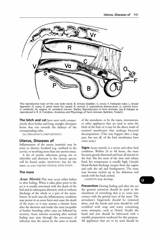
The bitch and cat have uteri with compar-atively short bodies and long, straight, divergenthorns that run towards the kidneys of the corresponding sides.
(See PREGNANCY; PARTURITION.)
Uterus, Diseases ofInflammation of the uterus (metritis) may beacute or chronic, localised (e.g. confined to thecervix), or involving more than one uterine tissue.
A list of uterine infections giving rise toinfertility and abortion in the various specieswill be found under ABORTION, but for themare, see under EQUINE GENITAL INFECTIONS.
The mare
Acute Metritis This may occur either beforeor after foaling. When it takes place prior to theact it is usually associated with the death of thefoal and its subsequent abortion, with or withoutdischarge of the whole or a part of the mem-branes. In such cases the inflammatory conditionmay persist in an acute form and cause the deathof the mare, or it may assume a chronic formafter the abortion and render the mare incapableof further breeding; other cases are followed byrecovery. Acute metritis occurring after normalfoaling may arise through the conveyance ofinfection into the uterus by the arms or hands
of the attendants, or by the ropes, instruments,or other appliances that are used to assist thebirth of the foal; or it may be the direct result ofretained membranes that undergo bacterialdecomposition. (This may happen after a largepart, but not all, of the fetal membranes havecome away.)
Signs Acute metritis is a severe and often fatalcondition. Within 24 to 48 hours, the marebecomes greatly distressed and loses all interest inthe foal. She lies most of the time and refusesfood; her temperature is usually high. Greyishblood-flecked discharge escapes from the vaginaand soils the tail and hindquarters. The maremay become tucked up in her abdomen andstands with her back arched.
LAMINITIS may develop.
Prevention During foaling and after the actthe greatest attention should be paid to thecleanliness of everything that is to come intocontact with the genital tract of the mare. Theattendant’s fingernails should be trimmedshort, and the hands and arms should be wellscrubbed with soap and water containing some antiseptic, such as Dettol. Finally thehand and arm should be lubricated with a suitable preparation marketed for this purpose.All appliances that are to be used should be
Uterus, Diseases of 741
U
The reproductive tract of the cow (side view). B, Urinary bladder; C, cervix; F, Fallopian tube; L, broad ligament; O, ovary; P, pelvic bone (os coxae); R, rectum; S, suburethral diverticulum; U, uterine horn; V, vestibule; Va, vagina; Vr, vertebral column. (Hafez, Reproduction in Farm Animals, Lea & Febiger asreproduced in R. D. Frandson, Anatomy and Physiology of Farm Animals, Baillière, Tindall.)
boiled and kept in a pail of hot water when notactually in use.
One other factor is of the greatest impor-tance: after a mare foals, the fetal membranesshould be given attention. Normally they aredischarged by means of a few comparativelymild labour pains within an hour of the birth ofthe foal. If however they are retained for longerthan this period, the person in attendanceshould suspect that something may be wrongand seek veterinary advice.
In other cases a series of violent pains maycommence, when the bulk of the membranesare passed to the outside, where they hang sus-pended. Should this happen a sack or sheetshould be placed under the dependent mass,and held so as to support the weight and relievethe tension on that portion that is still retainedin the uterus. This is necessary lest the weight ofthe external membranes causes a tearing awayfrom the non-separated part. Gentle tractionshould then be exerted upon the imprisonedportion; as a rule it will gradually detach itselfand come to the outside. If no progress is made,veterinary assistance should be sought promptly.
Injections of PITUITRIN may obviate manualremoval of the fetal membranes. A syntheticoestrogen may be preferred. (See HORMONE
THERAPY.)Regarding complete retention of the fetal
membranes – when only a very small portion isseen hanging from the vagina – professionalhelp should be obtained if there is no sign of anyattempt at expulsion within 4 to 6 hours afterfoaling.
Generally speaking, membranes that haveremained in position for 8 to 12 hours are start-ing to decompose, and decomposition meansbacterial infection of the uterus (i.e. metritis) inalmost every case.
Treatment The case must be considered mostserious. The use of antibiotics or one of thesulfa drugs is indicated. (See also under NURSING.)Any retained fetal membrane must be removedfrom the uterus by hand and as much dischargeas possible cleared out. A solution of acriflavine,proflavine, or brilliant green, 1 part in 1000 of boiled water, or some other suitable non-irritant antiseptic solution at blood heat, is douched into the cavity of the uterus by alength of rubber tubing, and, after allowing it to act for 2 to 5 minutes, is syphoned off. Aspecial 2-way tube is sometimes used for thispurpose – the solution entering by one channeland leaving by the other. When all the fluid has been removed, an antiseptic pessary may be inserted. When complications such as
LAMINITIS or PNEUMONIA co-exist, they mustreceive separate attention.
Chronic Metritis This may originate as asequel to an acute attack in some cases, butmore commonly it is directly due to an injuryor infection which is not sufficiently severe toproduce an acute attack.
Signs There may be a general unthriftiness following upon foaling. The mare’s appetite iscapricious, but her thirst is unimpaired. Thetemperature fluctuates a degree or two abovenormal. There may or may not be a dirty, sticky,grey, or pus-like discharge from the vagina,which causes irritation and frequent erectionsof the clitoris. The mare resents handling of thegenital organs, but if the lips of the vulva aregently separated the mucous membrane is seento be inflamed and swollen.
In other cases the pus collects in the cavity ofthe uterus and is retained there through closureof the os. (See PYOMETRA.) It sometimes happensthat after the pus has collected for a certain peri-od the os suddenly opens and 4.5 litres (1 gallon)or more of pus is discharged. The os then closesonce more. Intervals between these evacuationsmay vary from a few days to 3 or 4 weeks. Themare’s general condition shows an improvementimmediately following a sudden discharge ofpus, but as it re-accumulates she relapses into her former chronic state. Chronic metritis mayget gradually worse, and the mare dies. Casestaken in time usually recover with treatment, butfurther breeding is often impossible.
Treatment An early opportunity should betaken to evacuate the pus from the uterus, bydouching and siphonage, or by irrigation asalready described under ‘Acute metritis’.
Sulfa drugs or antibiotics may be used.It should be emphasised that expert advice
should be sought at the earliest opportunity.(See also CONTAGIOUS EQUINE METRITIS.)
The cow In the following brief account, muchof what has been said in relation to the maremust be understood to apply to the cow as well,and only the main differences will be stated.
Acute Metritis In some cases where birth ofthe calf has taken place easily and naturally,metritis supervenes in the course of the firstweek or 10 days after calving, but in the major-ity of cases there has been some injury or infec-tion at, or shortly after, parturition. Retentionof the fetal membranes, which is so much morecommon in the cow than in other animals, isvery often the contributory factor to an attack
742 Uterus, Diseases of
U
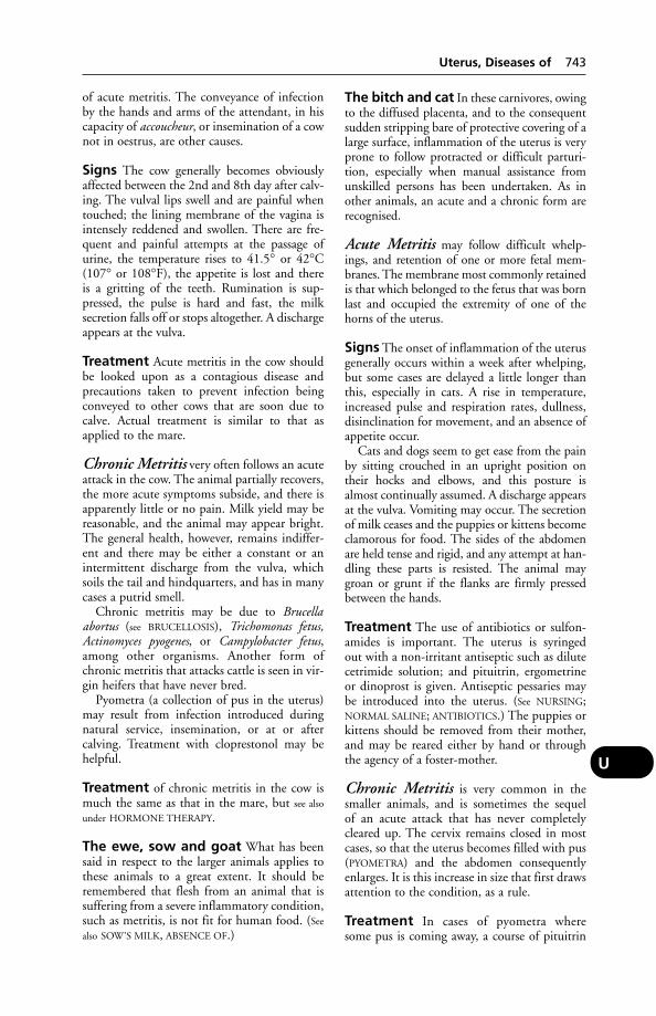
of acute metritis. The conveyance of infectionby the hands and arms of the attendant, in hiscapacity of accoucheur, or insemination of a cownot in oestrus, are other causes.
Signs The cow generally becomes obviouslyaffected between the 2nd and 8th day after calv-ing. The vulval lips swell and are painful whentouched; the lining membrane of the vagina isintensely reddened and swollen. There are fre-quent and painful attempts at the passage ofurine, the temperature rises to 41.5° or 42°C(107° or 108°F), the appetite is lost and there is a gritting of the teeth. Rumination is sup-pressed, the pulse is hard and fast, the milksecretion falls off or stops altogether. A dischargeappears at the vulva.
Treatment Acute metritis in the cow shouldbe looked upon as a contagious disease and precautions taken to prevent infection beingconveyed to other cows that are soon due tocalve. Actual treatment is similar to that asapplied to the mare.
Chronic Metritis very often follows an acuteattack in the cow. The animal partially recovers,the more acute symptoms subside, and there isapparently little or no pain. Milk yield may bereasonable, and the animal may appear bright.The general health, however, remains indiffer-ent and there may be either a constant or anintermittent discharge from the vulva, whichsoils the tail and hindquarters, and has in manycases a putrid smell.
Chronic metritis may be due to Brucellaabortus (see BRUCELLOSIS), Trichomonas fetus,Actinomyces pyogenes, or Campylobacter fetus,among other organisms. Another form ofchronic metritis that attacks cattle is seen in vir-gin heifers that have never bred.
Pyometra (a collection of pus in the uterus)may result from infection introduced duringnatural service, insemination, or at or after calving. Treatment with cloprestonol may behelpful.
Treatment of chronic metritis in the cow ismuch the same as that in the mare, but see also
under HORMONE THERAPY.
The ewe, sow and goat What has beensaid in respect to the larger animals applies tothese animals to a great extent. It should beremembered that flesh from an animal that issuffering from a severe inflammatory condition,such as metritis, is not fit for human food. (See
also SOW’S MILK, ABSENCE OF.)
The bitch and cat In these carnivores, owingto the diffused placenta, and to the consequentsudden stripping bare of protective covering of alarge surface, inflammation of the uterus is veryprone to follow protracted or difficult parturi-tion, especially when manual assistance fromunskilled persons has been undertaken. As inother animals, an acute and a chronic form arerecognised.
Acute Metritis may follow difficult whelp-ings, and retention of one or more fetal mem-branes. The membrane most commonly retainedis that which belonged to the fetus that was bornlast and occupied the extremity of one of thehorns of the uterus.
Signs The onset of inflammation of the uterusgenerally occurs within a week after whelping,but some cases are delayed a little longer thanthis, especially in cats. A rise in temperature,increased pulse and respiration rates, dullness,disinclination for movement, and an absence ofappetite occur.
Cats and dogs seem to get ease from the painby sitting crouched in an upright position ontheir hocks and elbows, and this posture isalmost continually assumed. A discharge appearsat the vulva. Vomiting may occur. The secretionof milk ceases and the puppies or kittens becomeclamorous for food. The sides of the abdomenare held tense and rigid, and any attempt at han-dling these parts is resisted. The animal maygroan or grunt if the flanks are firmly pressedbetween the hands.
Treatment The use of antibiotics or sulfon-amides is important. The uterus is syringed out with a non-irritant antiseptic such as dilutecetrimide solution; and pituitrin, ergometrineor dinoprost is given. Antiseptic pessaries maybe introduced into the uterus. (See NURSING;NORMAL SALINE; ANTIBIOTICS.) The puppies orkittens should be removed from their mother,and may be reared either by hand or throughthe agency of a foster-mother.
Chronic Metritis is very common in thesmaller animals, and is sometimes the sequel of an acute attack that has never completelycleared up. The cervix remains closed in mostcases, so that the uterus becomes filled with pus(PYOMETRA) and the abdomen consequentlyenlarges. It is this increase in size that first drawsattention to the condition, as a rule.
Treatment In cases of pyometra where some pus is coming away, a course of pituitrin
Uterus, Diseases of 743
U
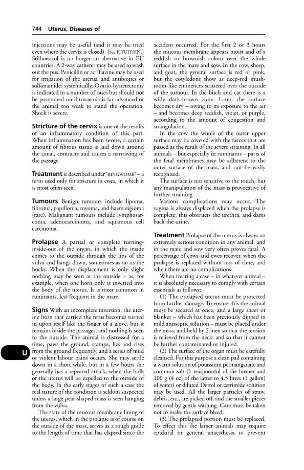
injections may be useful (and it may be triedeven where the cervix is closed). (See PITUITRIN.)Stilboestrol is no longer an alternative in EUcountries. A 2-way catheter may be used to washout the pus. Penicillin or acriflavine may be usedfor irrigation of the uterus, and antibiotics orsulfonamides systemically. Ovario-hysterectomyis indicated in a number of cases but should notbe postponed until toxaemia is far advanced orthe animal too weak to stand the operation.Shock is severe.
Stricture of the cervix is one of the resultsof an inflammatory condition of this part.When inflammation has been severe, a certainamount of fibrous tissue is laid down aroundthe canal, contracts and causes a narrowing ofthe passage.
Treatment is described under ‘RINGWOMB’ – aterm used only for stricture in ewes, in which itis most often seen.
Tumours Benign tumours include lipoma,fibroma, papilloma, myoma, and haemangioma(rare). Malignant tumours include lymphosar-coma, adenocarcinoma, and squamous cell carcinoma.
Prolapse A partial or complete turning-inside-out of the organ, in which the insidecomes to the outside through the lips of thevulva and hangs down, sometimes as far as thehocks. When the displacement is only slightnothing may be seen at the outside – as, forexample, when one horn only is inverted intothe body of the uterus. It is most common inruminants, less frequent in the mare.
Signs With an incomplete inversion, the uter-ine horn that carried the fetus becomes turnedin upon itself like the finger of a glove, but itremains inside the passages, and nothing is seento the outside. The animal is distressed for atime, paws the ground, stamps, lies and risesfrom the ground frequently, and a series of mildor violent labour pains occurs. She may settledown in a short while, but in a few hours shegenerally has a repeated attack, when the bulkof the uterus will be expelled to the outside ofthe body. In the early stages of such a case thereal nature of the condition is seldom suspectedunless a large pear-shaped mass is seen hangingfrom the vulva.
The state of the mucous membrane lining ofthe uterus, which in the prolapse is of course onthe outside of the mass, serves as a rough guideto the length of time that has elapsed since the
accident occurred. For the first 2 or 3 hours the mucous membrane appears moist and of areddish or brownish colour over the whole surface in the mare and sow. In the cow, sheep,and goat, the general surface is red or pink, but the cotyledons show as deep-red mush-room-like eminences scattered over the outsideof the tumour. In the bitch and cat there is awide dark-brown zone. Later, the surfacebecomes dry – owing to its exposure to the air– and becomes deep reddish, violet, or purple,according to the amount of congestion andstrangulation.
In the cow the whole of the outer uppersurface may be covered with the faeces that arepassed as the result of the severe straining. In allanimals – but especially in ruminants – parts ofthe fetal membranes may be adherent to theouter surface of the mass, and can be easilyrecognised.
The surface is not sensitive to the touch, butany manipulation of the mass is provocative offurther straining.
Various complications may occur. The vagina is always displaced when the prolapse iscomplete; this obstructs the urethra, and damsback the urine.
Treatment Prolapse of the uterus is always anextremely serious condition in any animal, andin the mare and sow very often proves fatal. Apercentage of cows and ewes recover, when theprolapse is replaced without loss of time, andwhen there are no complications.
When treating a case – in whatever animal –it is absolutely necessary to comply with certainessentials as follows:
(1) The prolapsed uterus must be protectedfrom further damage. To ensure this the animalmust be secured at once, and a large sheet orblanket – which has been previously dipped inmild antiseptic solution – must be placed underthe mass, and held by 2 men so that the tensionis relieved from the neck, and so that it cannotbe further contaminated or injured.
(2) The surface of the organ must be carefullycleansed. For this purpose a clean pail containinga warm solution of potassium permanganate andcommon salt (1 teaspoonful of the former and100 g (4 oz) of the latter to 4.5 litres (1 gallon)of water) or diluted Dettol or cetrimide solutionmay be used. All the larger particles of straw,debris, etc., are picked off, and the smaller piecesremoved by gentle washing. Care must be takennot to make the surface bleed.
(3) The prolapsed portion must be replaced.To effect this the larger animals may requireepidural or general anaesthesia to prevent
744 Uterus, Diseases of
U
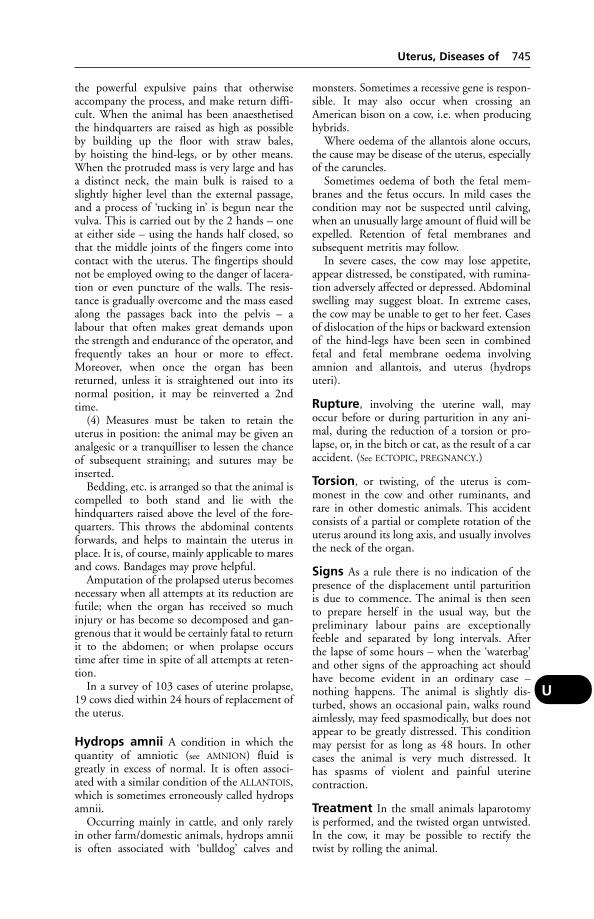
the powerful expulsive pains that otherwiseaccompany the process, and make return diffi-cult. When the animal has been anaesthetisedthe hindquarters are raised as high as possibleby building up the floor with straw bales, by hoisting the hind-legs, or by other means.When the protruded mass is very large and hasa distinct neck, the main bulk is raised to aslightly higher level than the external passage,and a process of ‘tucking in’ is begun near thevulva. This is carried out by the 2 hands – oneat either side – using the hands half closed, sothat the middle joints of the fingers come intocontact with the uterus. The fingertips shouldnot be employed owing to the danger of lacera-tion or even puncture of the walls. The resis-tance is gradually overcome and the mass easedalong the passages back into the pelvis – alabour that often makes great demands uponthe strength and endurance of the operator, andfrequently takes an hour or more to effect.Moreover, when once the organ has beenreturned, unless it is straightened out into itsnormal position, it may be reinverted a 2ndtime.
(4) Measures must be taken to retain theuterus in position: the animal may be given ananalgesic or a tranquilliser to lessen the chanceof subsequent straining; and sutures may beinserted.
Bedding, etc. is arranged so that the animal iscompelled to both stand and lie with thehindquarters raised above the level of the fore-quarters. This throws the abdominal contentsforwards, and helps to maintain the uterus inplace. It is, of course, mainly applicable to maresand cows. Bandages may prove helpful.
Amputation of the prolapsed uterus becomesnecessary when all attempts at its reduction arefutile; when the organ has received so muchinjury or has become so decomposed and gan-grenous that it would be certainly fatal to returnit to the abdomen; or when prolapse occurstime after time in spite of all attempts at reten-tion.
In a survey of 103 cases of uterine prolapse,19 cows died within 24 hours of replacement ofthe uterus.
Hydrops amnii A condition in which thequantity of amniotic (see AMNION) fluid isgreatly in excess of normal. It is often associ-ated with a similar condition of the ALLANTOIS,which is sometimes erroneously called hydropsamnii.
Occurring mainly in cattle, and only rarelyin other farm/domestic animals, hydrops amniiis often associated with ‘bulldog’ calves and
monsters. Sometimes a recessive gene is respon-sible. It may also occur when crossing anAmerican bison on a cow, i.e. when producinghybrids.
Where oedema of the allantois alone occurs,the cause may be disease of the uterus, especiallyof the caruncles.
Sometimes oedema of both the fetal mem-branes and the fetus occurs. In mild cases thecondition may not be suspected until calving,when an unusually large amount of fluid will beexpelled. Retention of fetal membranes andsubsequent metritis may follow.
In severe cases, the cow may lose appetite,appear distressed, be constipated, with rumina-tion adversely affected or depressed. Abdominalswelling may suggest bloat. In extreme cases,the cow may be unable to get to her feet. Casesof dislocation of the hips or backward extensionof the hind-legs have been seen in combinedfetal and fetal membrane oedema involvingamnion and allantois, and uterus (hydropsuteri).
Rupture, involving the uterine wall, mayoccur before or during parturition in any ani-mal, during the reduction of a torsion or pro-lapse, or, in the bitch or cat, as the result of a caraccident. (See ECTOPIC, PREGNANCY.)
Torsion, or twisting, of the uterus is com-monest in the cow and other ruminants, andrare in other domestic animals. This accidentconsists of a partial or complete rotation of theuterus around its long axis, and usually involvesthe neck of the organ.
Signs As a rule there is no indication of thepresence of the displacement until parturition is due to commence. The animal is then seen to prepare herself in the usual way, but the preliminary labour pains are exceptionally feeble and separated by long intervals. After the lapse of some hours – when the ‘waterbag’and other signs of the approaching act shouldhave become evident in an ordinary case –nothing happens. The animal is slightly dis-turbed, shows an occasional pain, walks roundaimlessly, may feed spasmodically, but does notappear to be greatly distressed. This conditionmay persist for as long as 48 hours. In othercases the animal is very much distressed. It has spasms of violent and painful uterine contraction.
Treatment In the small animals laparotomyis performed, and the twisted organ untwisted.In the cow, it may be possible to rectify thetwist by rolling the animal.
Uterus, Diseases of 745
U

Congenital defects (see the diagram under
INFERTILITY; also HYDROMETRA)
UveitisInflammation of the uvea (iris, ciliary body andchoroid coat of the eyeball).
UvulaThis is the small downward projection that isfound on the free edge of the soft palate of thepig. It is not present in the other domesticatedanimals.
746 Uveitis
U
VaccinationA method of producing active immunity againsta specific infection by means of inoculation with a vaccine, i.e. a preparation of the necessaryantigen(s). (See IMMUNITY; IMMUNISATION;IMMUNE RESPONSE; VACCINE.)
Vaccination in mammals is normally carriedout by inoculating individual animals. Themethod of administration depends on the typeof vaccine. Most inactivated vaccines are inject-ed intramuscularly or subcutaneously; tempera-ture-sensitive live vaccines may be administeredas drops into the nasal passages; vaccines againsthusk are given orally.
Mass vaccination of poultry against Newcastledisease may be achieved by dispersing aerosols ofvaccine over the heads of the birds with finespray pumps or adding vaccine to the drinkingwater. Some fish are vaccinated by dipping thefish in a solution of the vaccine. Fox populationsin Europe have been vaccinated against rabies byimpregnating chicken heads or other baits andspreading them in known fox runs. Multiple-component vaccines containing antigens againsta number of diseases are available. For example,sheep can be simultaneously immunised againstpulpy kidney disease, lamb dysentery, braxy,blackleg, black disease, struck, Clostridium oede-matiens infection and tetanus by a single 8-in-1vaccine.
(In connection with foot-and-mouth disease,see also RING VACCINATION.)
VaccineWhen an animal is inoculated with a vaccine asprotection against a specific disease, e.g. black-leg, this is carried out with the object of stimu-lating production of antibodies in its system,which will confer active immunity againstblackleg organisms.
Vaccines may be prepared from live micro-organisms; from inactivated (killed) micro-organisms; from genetically engineered subunitsof the pathogenic fraction of the organism; or from toxoids – heat- or chemically-treatedmicro-organisms that have lost their virulencebut retain their antigenicity, i.e. ability to createresistance to disease.
Live vaccines are vaccines prepared from bac-teria or viruses whose virulence is reduced byheat, chemicals or passage through an animal
other than the normal host species. For example,cattle plague vaccine may be prepared from thevirus passaged through (i.e. grown in) chickembryos. Occasionally the live viruses used arerelated but non-pathogenic strains, usefulbecause they will stimulate antibody productionbut will not produce the disease.
Viruses may be inactivated by phenol orultra-violet rays, for example; or they may bemodified in some way, such as by artificiallyinduced mutation, to produce a temperature-sensitive virus which will replicate in the nosebut not in the lungs. Such a virus vaccine can beadministered by nasal spray.
Tissue culture vaccines – live vaccines grownon cell cultures – are used in the prevention ofcanine distemper, rabies, etc., and in treatmentof benign skin papillomata (warts) of cattle.
Vaccines are sometimes used for treatment aswell as for prevention of a particular disease.
X-irradiated worm larvae vaccine is used inthe prevention of PARASITIC BRONCHITIS.
It is important that, in the commercial pro-duction of live vaccines involving the use ofchicken embryos (or of tissue cultures derivedfrom them), contaminant viruses are eliminated.For example, the avian leukosis virus has conta-minated distemper vaccine and would representa risk to vaccinated poultry if contaminating vac-cines for them. Scrapie was accidentally spreadby an early louping-ill virus contaminated by thescrapie agent.
It is essential that vaccines are stored undersuitable conditions of temperature, etc.; thatthey are not used after the expiry date shown onthe package; that where 2 doses are stated to benecessary, both are given – and at the correctinterval. Failure to observe these rules can meanthat the vaccinated animal does not become animmunised animal; it has led to dogs presumedproperly vaccinated against rabies becomingrabid after exposure to a natural infection. (See
also INJECTIONS; GENETIC ENGINEERING.)Inactivated vaccines are prepared from killed
micro-organisms that retain sufficient antigenicactivity to promote immunity. They are not aspotent as live vaccines, and 2 doses at specifiedintervals are usually necessary to produce effec-tive immunity. Inactivated vaccines often con-tain an adjuvant, usually an aluminium saltsuch as aluminium hydroxide, which enhancesthe immune reaction. Some are water-based,others formulated in an oily medium. Oil-based vaccines can cause serious reactions ifaccidentally self-injected into the operator.Leptospirosis vaccine is an example.
Subunit vaccines are genetically engineered sothat only the antigenic fraction of a pathogen is
V
utilised. The vaccine does not cause infection butdoes stimulate immunity. Feline leukaemia vac-cine is an example; another is Aujeszky’s diseasevaccine. The virus component of the subunitvaccine has difficulty in penetrating the cells ofthe vaccinated animal; it does not multiply wellwithin the cells and the animal does not shed thevirus. By testing for the fraction missing from thevaccinial strain of virus, a vaccinated animal canbe determined from one carrying the infection.
Toxoid vaccines are produced by treating tox-ins from micro-organisms so that their harmfuleffects are removed but the antigenic propertiesremain. Tetanus vaccine is an example.
Vaccinia VirusThis term may refer to the virus of naturallyoccurring cow-pox, or to a strain which hasundergone mutation and was used for vaccina-tion against smallpox. (See POX.)
VacuoleA cavity within a cell.
Vacuum-Dipping of EggsA technique used in assisting the eradication of Mycoplasma spp. in poultry. Fertile eggs aredipped in a concentrated solutions of antibiot-ic (usually tylosin) and subjected to a negativepressure. Some of the air in the egg’s air pocketis thus extracted and about 0.5 ml of antibioticdrawn through the shell into the egg andabsorbed. This process is more effective in help-ing to eliminate M. gallisepticum than othermycoplasmas.
VaginaThe vagina extends from the cervix of theuterus to the vulva. Vaginal mucus is altered incharacter during pregnancy, a fact which can bemade use of in pregnancy diagnosis. (Forinflammation of the vagina, see VAGINITIS.)
An artificial vagina is used at AI centres forthe collection of semen.
Vaginal prolapse in ewes This may pre-cede lambing by up to 55 days, but most casesoccur within the last 21 days of pregnancy.
Rupture of the vagina, with protrusion of theintestine and rapid death, occurs not uncom-monly in ewes of a large breed, of mature age,carrying a twin – a week or two before lambingis due. Bulky foods – swedes, turnips, kale – areoften involved.
Vaginoureteral fistula This has beenrecorded in dogs and cats, as a complication of
ovariohysterectomy or a caesarean operation,and leads to urinary incontinence. It has beensuggested that the fistula may occur followingaccidental ligation of the ureter during surgery,or because the ureter becomes involved in an inflammatory adhesion originating in thevaginal stump.
Intermittent haemorrhage occasionallyoccurs in mares having very prominent varicoseveins at the dorsal aspect of the vulva-vaginalarea; it does not appear to affect health or fertil-ity. Persistent vulval haemorrhage from varicoseveins of the dorsal wall of the vagina has alsobeen described. It yields to local haemostatictreatment.
VaginitisInflammation of the vagina. (See under INFERTIL-
ITY – Diseases of the genital organs in female;also ‘WHITES’; EPIDIDYMITIS – Epivag; VULVO-
VAGINITIS, GRANULAR; PROLAPSE.)
VagotomySevering of the vagus nerve. (See HYPERTROPHIC
OSTEOPATHY.)
Vagus (Pneumogastric Nerve)The vagus (pneumogastric nerve) is the 10thcranial nerve. This nerve is remarkable for itsgreat length, and for the attachments which itforms with other nerves and with the sympa-thetic trunks. It arises from the side of themedulla, passes out of the skull, and runs downto the jugular furrow of the neck, where, alongwith the sympathetic, it accompanies thecarotid artery to the entrance to the chest. Fromthis point the right and left vagi differ fromeach other in their course. They both passthrough the chest cavity, giving branches to thepharynx (which run up the neck again), to theheart, bronchi, oesophagus, etc. Each nervethen splits into 2 parts and the 2 upper branch-es fuse with each other to form the dorsal trunk,the lower branches behaving similarly to formthe ventral trunk. These 2 branches now passthrough the diaphragm, with the oesophagus,into the abdominal cavity, and end by givingbranches to the stomach, duodenum, liver, andvarious ganglia nearby. (See Parasympathetic system
under CENTRAL NERVOUS SYSTEM – Autonomic;also BRAIN.) (See GUTTURAL POUCH DISEASE.)
ValgusA bone growth-plate defect. (See under BONE,DISEASES OF.)
ValineOne of the essential amino acids.
748 Vaccinia Virus
V

ValvesValves are found in the heart, veins, lymph ves-sels, etc., and serve the purpose of ensuring thatthe fluids will only circulate in one direction.(See HEART; VEINS; ILEOCAECAL.)
Valvular Disease(see HEART DISEASES)
Vampire BatsVampire bats are important transmitters ofrabies in parts of South and Central America,the West Indies, etc. The bat laps blood fromthe wounds inflicted with its upper incisor teethon cattle, horses, etc. In Mexico infected vam-pires have made necessary the preventive inocu-lation of 800,000 cattle a year. Trypanosomiasiscan also be transmitted by vampire bats.Vampire bats imported into the UK remain inquarantine for the rest of their life.
VanadiumA trace element essential in minute quantitiesfor the growth of chicks; as little as 10 mg/kg ofdiet is an overdose that will suppress growth. Itworks with insulin to increase the amount ofglucose and aminoacids taken up by muscle.
Varicose Veins(see under VEINS)
Varied Diet, Need for(see DIET AND DIETETICS; AMINO ACIDS; CAT
FOODS; DOGS’ DIET)
VarioceleVariocele is a condition in which the veins ofone or both testicles are greatly distended.
Variola (Pox)Variola (pox) is the inclusive term for fevers ofanimals and man, in which a skin eruption takesthe form of a ‘pock’, caused by a POX virus.
VarroasisVarroasis is a parasitic disease of honey bees,Apis melifera, caused by the mite Varroa jacob-soni. The mite feeds on the developing larvaeand on the adult bees. The result is weak beesand sometimes the death of the queen. Wholehives can be wiped out, causing great economicloss not only to the apiculturalist but also tofruit and arable farmers; beekeepers often sup-ply hives to fruit growers and to fields of rapeand linseed. Varroasis is prevalent in the UKand is a NOTIFIABLE DISEASE.
As both parasite and host are arthropods,products used for control of the disease musthave a fine division between toxicity to the mite and toxicity to the bee. Suspending stripsimpregnated with flumethrin or fluvalinate in thehive can be effective. Other treatments includetobacco smoke or a vapourising block containingthymol and aromatic oils. Eradication, however,is difficult. The disease came from Asia, where itdoes less harm because Asiatic bees groom eachother, thus removing the mites. Long term, it hasbeen suggested that European and Asian beesmay be cross-bred to try to introduce the groom-ing habit into the European bee population. Aleaflet available from DEFRA gives more detailsof the disease and its control.
Vas Deferens(see under TESTICLE)
VascularConsisting of, or containing a high proportionof, blood vessels.
Vasculitis Inflammation of a blood vessel.
VasectomisedA male animal in which the vas deferens hasbeen cut. Such an animal is sterile though itretains its libido and may be used for the detec-tion of oestrus (e.g. in cattle). In breeding catter-ies one or two toms are sometimes vasectomisedfor the sake of peace, quiet and contentment ofqueens not being bred from until a later oestrus.
Sterility does not immediately follow vasec-tomy (or castration), as some sperms will be inthe seminal vesicles and can lead to conception
Vasectomised 749
V
One-month-old foal with bilateral carpal valgus.(With acknowledgements to Professor L. C.Vaughan and the Royal Veterinary College.)
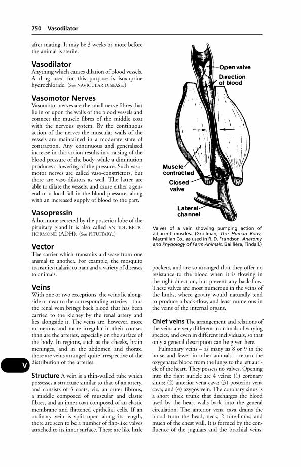
after mating. It may be 3 weeks or more beforethe animal is sterile.
VasodilatorAnything which causes dilation of blood vessels.A drug used for this purpose is isoxuprinehydrochloride. (See NAVICULAR DISEASE.)
Vasomotor NervesVasomotor nerves are the small nerve fibres thatlie in or upon the walls of the blood vessels andconnect the muscle fibres of the middle coatwith the nervous system. By the continuousaction of the nerves the muscular walls of thevessels are maintained in a moderate state ofcontraction. Any continuous and generalisedincrease in this action results in a raising of theblood pressure of the body, while a diminutionproduces a lowering of the pressure. Such vaso-motor nerves are called vaso-constrictors, butthere are vaso-dilators as well. The latter areable to dilate the vessels, and cause either a gen-eral or a local fall in the blood pressure, alongwith an increased supply of blood to the part.
VasopressinA hormone secreted by the posterior lobe of thepituitary gland.It is also called ANTIDIURETIC
HORMONE (ADH). (See PITUITARY.)
VectorThe carrier which transmits a disease from oneanimal to another. For example, the mosquitotransmits malaria to man and a variety of diseasesto animals.
VeinsWith one or two exceptions, the veins lie along-side or near to the corresponding arteries – thusthe renal vein brings back blood that has beencarried to the kidney by the renal artery and lies alongside it. The veins are, however, morenumerous and more irregular in their coursesthan are the arteries, especially on the surface ofthe body. In regions, such as the cheeks, brainmeninges, and in the abdomen and thorax,there are veins arranged quite irrespective of thedistribution of the arteries.
Structure A vein is a thin-walled tube whichpossesses a structure similar to that of an artery,and consists of 3 coats, viz. an outer fibrous, a middle composed of muscular and elasticfibres, and an inner coat composed of an elasticmembrane and flattened epithelial cells. If anordinary vein is split open along its length,there are seen to be a number of flap-like valvesattached to its inner surface. These are like little
pockets, and are so arranged that they offer noresistance to the blood when it is flowing in the right direction, but prevent any back-flow.These valves are most numerous in the veins ofthe limbs, where gravity would naturally tendto produce a back-flow, and least numerous inthe veins of the internal organs.
Chief veins The arrangement and relations ofthe veins are very different in animals of varyingspecies, and even in different individuals, so thatonly a general description can be given here.
Pulmonary veins – as many as 8 or 9 in thehorse and fewer in other animals – return theoxygenated blood from the lungs to the left auri-cle of the heart. They possess no valves. Openinginto the right auricle are 4 veins: (1) coronarysinus; (2) anterior vena cava; (3) posterior venacava; and (4) azygos vein. The coronary sinus isa short thick trunk that discharges the bloodused by the heart walls back into the general circulation. The anterior vena cava drains theblood from the head, neck, 2 fore-limbs, andmuch of the chest wall. It is formed by the con-fluence of the jugulars and the brachial veins,
750 Vasodilator
V
Valves of a vein showing pumping action of adjacent muscles. (Grollman, The Human Body,Macmillan Co., as used in R. D. Frandson, Anatomyand Physiology of Farm Animals, Baillière, Tindall.)

and receives other branches from the neck, vertebral region, and the chest wall. The posteri-or vena cava drains all the remainder of the bodyexcept the region of the diaphragm, the posteri-or intercostal areas, the oesophagus, and thebronchial tubes, the blood from these partsbeing collected into the azygos vein which joinsthe right auricle separately in most animals. Theposterior vena cava is formed under the lumbarregion by the union of the right and left com-mon iliac veins, which drain the blood from thepelvis and hind legs, and which are distributedin a more or less similar manner to the corre-sponding arteries of these parts. From here itpasses forwards below the lumbar muscles incompany with the abdominal aorta, until at thelevel of the last thoracic vertebra it passes down-wards and forwards, past the pancreas, andreaches the liver.
Its further course is partly embedded in theliver substance until it arrives at a special open-ing in the diaphragm, called the foramen venaecavae, by which it gains the thoracic cavity.From here it passes along in a groove in theright lung to reach the right auricle. Its maintributaries are as follows: (1) lumbar veins,which empty blood from the lumbar muscles,etc.; (2) internal spermatics in the male, andutero-ovarian veins in the female, from the generative organs in either sex; (3) 2 renal veins,one from each kidney, satellites of the corre-sponding arteries; (4) several large hepaticveins, which return not only blood carried tothe liver by the hepatic arteries, but also thatwhich comes from the digestive organs by theportal vein to undergo a second capillary circu-lation in the liver (see PORTAL VEIN); and (5)phrenic veins returning blood from thediaphragm.
In the venous system, even more so than in the arterial system, there is an intricatearrangement of anastomoses by which, whenone vein becomes damaged or diseased, lateralbranches from it may enlarge and carry awaythe excess blood into other veins so that nogreat hindrance to the return flow of the bloodto the heart may be occasioned. If this were notso, the circulation might be from impairedminor causes.
Veins, Diseases ofThose lying near to the surface are frequentlyinjured along with other tissues when contusionsor lacerations have been sustained, but so exten-sive is their communication with neighbouringveins that it is usually possible for these latter to enlarge and undertake the functions of thedamaged vessels, and thereby prevent serious
consequences. The deeper veins are protectedfrom all but the most severe, and usually fatal,injuries.
Inflammation of a vein, or phlebitis, mayfollow the collection of blood samples whenunclean instruments have been used, or whenthe resulting skin wound has not received atten-tion. In other cases it follows THROMBOSIS andinfection.
Varicose veins are those which have becomestretched or dilated to an extent not justified bythe blood flow. (See VARIOCELE and under VAGINA.)
Veld Sickness(see HEARTWATER)
Vena CavaEach of the 2 large veins that open direct intothe right auricle of the heart. (For further details,see under VEINS.)
Thrombosis of the posterior vena cava, whichmay follow abscess formation in the liver or else-where, is in cattle not infrequently followed bythe presence of clots in the pulmonary vessels,abscess formation and sometimes erosion of thepulmonary artery wall – giving rise to a fatalhaemorrhage. Symptoms may include dullness,rapid breathing, a cough, chest pain, the pres-ence of blood in material coughed up, anaemia,and widespread rhonchi. (See RECUMBENCY.)
Venereal DiseasesAnimals, with the exception of the monkey, arenot subject to infection by the 2 great humanvenereal diseases of syphilis and gonorrhoea, butthere are several important contagious diseasesthat can be transmitted from animal to animalby coitus. These include brucellosis, trichomoni-asis, Campylobacter fetus infection, and infectiousvaginitis of cattle, venereal granulomata or vene-real tumours of dogs, and dourine or mal du coitof horses. (See PROTOZOA; EPIDIDYMITIS; VULVO-
VAGINITIS, GRANULAR; CONTAGIOUS EQUINE
METRITIS.)
Venereal Tumours (InfectiveGranulomata)Venereal tumours (infective granulomata) characterise a contagious disease of dogs.
Signs In the female the original tumour is awarty excrescence which soon grows andbecomes cauliflower-like. In advanced stagesthere is a large mass of pinkish or greyish-red tissue, which easily bleeds when touched, occu-pying the greater part of the vaginal passage and
Venereal Tumours (Infective Granulomata) 751
V
often causing a bulging and swelling of the perineal region. A dirty sticky blood-stained dis-charge accompanies the condition, and the ani-mal’s general health suffers. In the male thewatery growths usually have a distinct stalk, andare attached to the skin or mucous membrane ofthe prepuce, or to the penis. (See also under WARTS.)
Venezuelan EquineEncephalomyelitisA strain recognised in the 1930s. A severe out-break occurred in Venezuela and Colombia in1962–4, when thousands of horses died andabout 30,000 people were infected. A later out-break spread to Mexico in 1970 where 6000 or more horses died, and then to Texas, USA.(See also EQUINE ENCEPHALITIS.)
Venom(see SNAKES; TOADS; BUFOTALIN; SPIDERS;SCORPIONS)
Vent GleetThis condition in poultry is an inflammation of the cloaca, with which is associated a thinyellowish watery discharge which has a charac-teristic and particularly unpleasant odour. Thecloaca and adjacent skin appear swollen andcongested, and the bird exhibits signs of irrita-tion. Other birds attracted by the reddening ofthe region may peck at vent; this leads on tocannibalism.
Egg production drops, and in some cases egg-binding and impaction of the oviduct result.Culling is advisable. There is a similar conditionin ducks but the material round the vent is moresolid. This must be removed and the affectedarea treated with antibiotic cream.
Cases of severe infection of the eyes of poultry-keepers treating this condition are notuncommon.
VentilationVentilation may be summed up as ‘the measuresnecessary to rectify the pollution of the air in abuilding – without the production of a draught’.Whenever animals are enclosed in a confinedbuilding they gradually use up the oxygen anddischarge into the air quantities of carbon dioxideand water vapour, until, if no fresh air is supplied,the percentage of oxygen decreases below theamount required.
One of the problems in livestock buildings is condensation, which can lead to bronchitisand pneumonia. For buildings used for cattleand sheep, provision of Yorkshire boarding isone of the best and least expensive methods ofavoiding or curing condensation.
Necessary air spacem3 ft3
Cow, horse(Byre or stable) 5.6 200(Loose box or yard) 16.8–33.6 600–1200
Bacon pig 1.7 60Poultry
(layers on slats) 0.17 6(layers on deep litter) 0.34 12
The required amount of air for each animalmust be continuously brought in from the out-side, and an exit must be provided for an equalamount. This is arranged for by the provision ofinlet and outlet ventilators.
Inlets These include windows, direct inletpipes, perforated bricks and gratings, Yorkshireboarding, and electric fans.
Windows, of which the Sheringham Valvetype is the most common and useful, serve thedual purpose of lighting and ventilation. Thoseon the lee side of a building serve as outlets whenthe wind is strong. In the Sheringham Valvewindows, the incoming air is deflected upwardsby the hopper-like flap that falls inwards, so thatit is spread over a greater area than is the casewith other openings. Inlet pipes are used, oftenin conjunction with windows, to ensure a supplyof fresh air in the region of the animals’ heads.
Ventilation rates – (maximum)Changes
of air m3 ft3
per hour per hour per hourBacon pig 20 5.7–34 200–1200Broiler chicken 40 6.8 240Laying birds 30 10 360
Outlets These include an open ridge, louvre-board ventilators, outlet shafts, open eaves,exhaust fans, and other devices. The most satis-factory outlet is undoubtedly an open ridgealong the whole length of the building. Theheated impure air rises and is drawn throughthe open space by the suction of the wind. Thedisadvantages of this system are that the openspace will allow entrance to a certain amount ofrain or snow in bad weather; the system is alsoinapplicable to buildings possessing lofts.
Extraction area(Necessary with natural ventilation)
Outlet per animal: cm2 sq. insCowhouse 930 144Farrowing house 95 15Fattening house 65 10Calf house 65 10Poultry (adult) house 13 2
752 Venezuelan Equine Encephalomyelitis
V
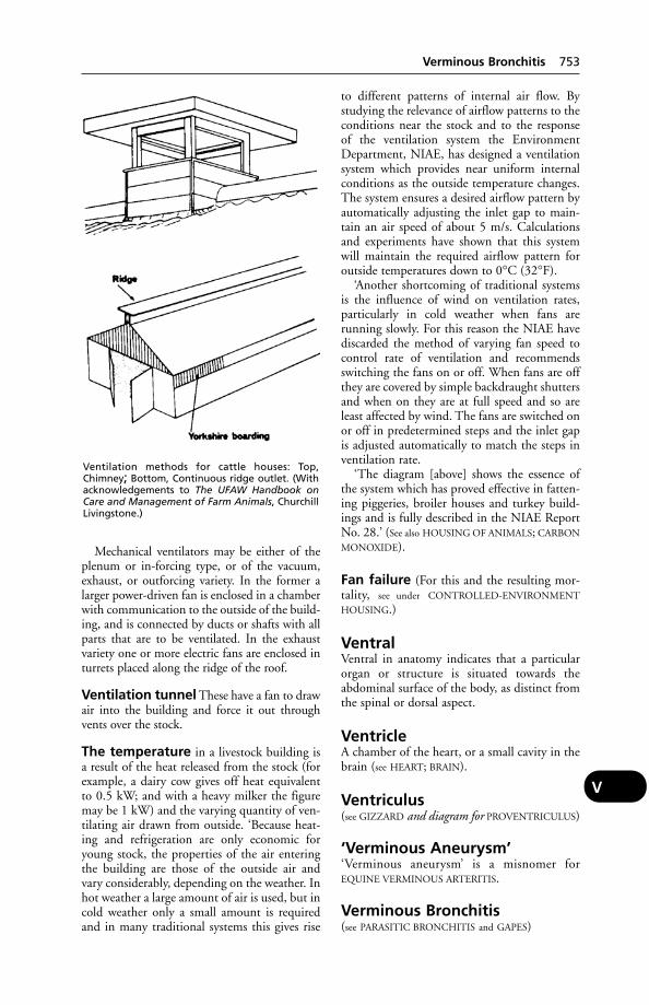
Mechanical ventilators may be either of theplenum or in-forcing type, or of the vacuum,exhaust, or outforcing variety. In the former alarger power-driven fan is enclosed in a chamberwith communication to the outside of the build-ing, and is connected by ducts or shafts with allparts that are to be ventilated. In the exhaustvariety one or more electric fans are enclosed inturrets placed along the ridge of the roof.
Ventilation tunnel These have a fan to drawair into the building and force it out throughvents over the stock.
The temperature in a livestock building isa result of the heat released from the stock (forexample, a dairy cow gives off heat equivalentto 0.5 kW; and with a heavy milker the figuremay be 1 kW) and the varying quantity of ven-tilating air drawn from outside. ‘Because heat-ing and refrigeration are only economic foryoung stock, the properties of the air enteringthe building are those of the outside air andvary considerably, depending on the weather. Inhot weather a large amount of air is used, but incold weather only a small amount is requiredand in many traditional systems this gives rise
to different patterns of internal air flow. Bystudying the relevance of airflow patterns to theconditions near the stock and to the response of the ventilation system the EnvironmentDepartment, NIAE, has designed a ventilationsystem which provides near uniform internalconditions as the outside temperature changes.The system ensures a desired airflow pattern byautomatically adjusting the inlet gap to main-tain an air speed of about 5 m/s. Calculationsand experiments have shown that this systemwill maintain the required airflow pattern foroutside temperatures down to 0°C (32°F).
‘Another shortcoming of traditional systemsis the influence of wind on ventilation rates,particularly in cold weather when fans are running slowly. For this reason the NIAE havediscarded the method of varying fan speed tocontrol rate of ventilation and recommendsswitching the fans on or off. When fans are offthey are covered by simple backdraught shuttersand when on they are at full speed and so areleast affected by wind. The fans are switched onor off in predetermined steps and the inlet gapis adjusted automatically to match the steps inventilation rate.
‘The diagram [above] shows the essence ofthe system which has proved effective in fatten-ing piggeries, broiler houses and turkey build-ings and is fully described in the NIAE ReportNo. 28.’ (See also HOUSING OF ANIMALS; CARBON
MONOXIDE).
Fan failure (For this and the resulting mor-tality, see under CONTROLLED-ENVIRONMENT
HOUSING.)
VentralVentral in anatomy indicates that a particularorgan or structure is situated towards theabdominal surface of the body, as distinct fromthe spinal or dorsal aspect.
VentricleA chamber of the heart, or a small cavity in thebrain (see HEART; BRAIN).
Ventriculus(see GIZZARD and diagram for PROVENTRICULUS)
‘Verminous Aneurysm’‘Verminous aneurysm’ is a misnomer forEQUINE VERMINOUS ARTERITIS.
Verminous Bronchitis(see PARASITIC BRONCHITIS and GAPES)
Verminous Bronchitis 753
V
Ventilation methods for cattle houses: Top,Chimney; Bottom, Continuous ridge outlet. (Withacknowledgements to The UFAW Handbook onCare and Management of Farm Animals, ChurchillLivingstone.)

Verminous Dermatitis(see STEPHANOFILARIASIS; SUMMER SORES)
Verminous Ophthalmia(see under EYE, DISEASES OF)
Vero CellsA continuous heteroploid cell line derived fromAfrican green monkey (Cercopithecus aethiops)kidney tissue. These cells are approved as a sub-strate for the production of virus vaccines,including rabies. They are much easier to growthan human diploid cells, and provide a betteryield; so that manufacturers are keen to use
them for vaccine production. Latent virus inthese cells is a potential danger.
VerotoxinA total of 1012 milk filters were collected from498 diary farms in south-west Ontario. Thesupernatants of 20 (2 per cent) of the milk filtercultures had verocytotoxic activity. Seven vero-toxin-producing E. coli strains were isolated, 2 ofwhich had been previously associated with diseasein humans.
Verruca(see PAPILLOMA)
754 Verminous Dermatitis
V
(With acknowledgements to the Scottish Farm Buildings Investigation Unit.)
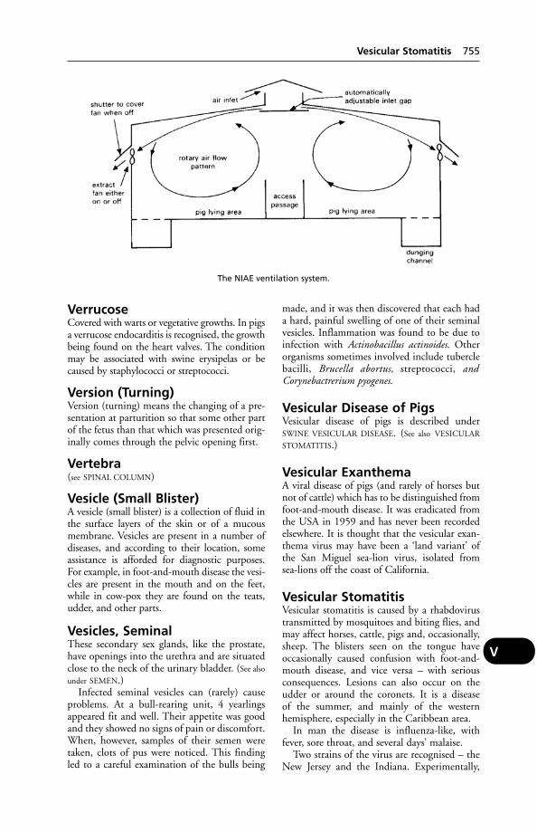
VerrucoseCovered with warts or vegetative growths. In pigsa verrucose endocarditis is recognised, the growthbeing found on the heart valves. The conditionmay be associated with swine erysipelas or becaused by staphylococci or streptococci.
Version (Turning)Version (turning) means the changing of a pre-sentation at parturition so that some other partof the fetus than that which was presented orig-inally comes through the pelvic opening first.
Vertebra(see SPINAL COLUMN)
Vesicle (Small Blister)A vesicle (small blister) is a collection of fluid inthe surface layers of the skin or of a mucousmembrane. Vesicles are present in a number ofdiseases, and according to their location, someassistance is afforded for diagnostic purposes.For example, in foot-and-mouth disease the vesi-cles are present in the mouth and on the feet,while in cow-pox they are found on the teats,udder, and other parts.
Vesicles, SeminalThese secondary sex glands, like the prostate,have openings into the urethra and are situatedclose to the neck of the urinary bladder. (See also
under SEMEN.)Infected seminal vesicles can (rarely) cause
problems. At a bull-rearing unit, 4 yearlingsappeared fit and well. Their appetite was goodand they showed no signs of pain or discomfort.When, however, samples of their semen weretaken, clots of pus were noticed. This findingled to a careful examination of the bulls being
made, and it was then discovered that each hada hard, painful swelling of one of their seminalvesicles. Inflammation was found to be due toinfection with Actinobacillus actinoides. Otherorganisms sometimes involved include tuberclebacilli, Brucella abortus, streptococci, andCorynebactrerium pyogenes.
Vesicular Disease of PigsVesicular disease of pigs is described underSWINE VESICULAR DISEASE. (See also VESICULAR
STOMATITIS.)
Vesicular ExanthemaA viral disease of pigs (and rarely of horses butnot of cattle) which has to be distinguished fromfoot-and-mouth disease. It was eradicated fromthe USA in 1959 and has never been recordedelsewhere. It is thought that the vesicular exan-thema virus may have been a ‘land variant’ ofthe San Miguel sea-lion virus, isolated from sea-lions off the coast of California.
Vesicular StomatitisVesicular stomatitis is caused by a rhabdovirustransmitted by mosquitoes and biting flies, andmay affect horses, cattle, pigs and, occasionally,sheep. The blisters seen on the tongue haveoccasionally caused confusion with foot-and-mouth disease, and vice versa – with seriousconsequences. Lesions can also occur on theudder or around the coronets. It is a disease of the summer, and mainly of the westernhemisphere, especially in the Caribbean area.
In man the disease is influenza-like, withfever, sore throat, and several days’ malaise.
Two strains of the virus are recognised – theNew Jersey and the Indiana. Experimentally,
Vesicular Stomatitis 755
V
The NIAE ventilation system.
numerous mammalian species can be infected –likewise ducks.
Vesicular Vaginitis(see VULVOVAGINITIS, GRANULAR)
Vesiculitis(see VESICLES, SEMINAL)
Veterinary DegreesVeterinary degrees are conferred on graduatesfrom the veterinary faculties of Bristol,Cambridge, Edinburgh, Glasgow, Liverpooland London universities. They lead to member-ship of the Royal College of VeterinarySurgeons (MRCVS) which allows the graduateto practise in the UK as a veterinary surgeon.Higher degrees (PhD, MSc, etc.) are availableafter postgraduate study, as well as certificatesand diplomas in specialist areas of veterinarymedicine and surgery. Graduates of veterinaryschools in other EU countries may also becomeMRCVS. University degrees in veterinary nurs-ing are also available.
Veterinary Facilities on theFarmEvery breeding cow and heifer in Britain has,during its lifetime, to be caught, ear-tag read,restrained and a blood sample taken from neck ortail vein. This will take place at least 2 or 3 times,quite apart from any herd or individual han-dlings necessary for clinical reasons or breedingmanagement. Taking a blood sample can take aslittle as 30 to 45 seconds given efficient holdingfacilities; 200 cattle could be sampled in a morn-ing’s work. On most farms there is a lack of cattle-handling facilities of the right type, so thatthe catching of a single animal can and does takeall the farm staff about 20 minutes with the veryreal possibility of broken gates and fences andvarying degrees of personal injury, even beforeblood-sampling is attempted.
Experience in the design and erection of cat-tle handling units for dairy and beef cattle hasshown the main points to be as follows:
Collecting pens should be large enough tohold all stock to be handled, or all the stock in units as they are housed, e.g. 50s or 100s. Apost-and-rail pen 9 × 18 m (30 × 60 ft) or 12 ×13.5 m (40 × 45 ft) will hold 100 cows withcalves at foot. A pen of 9 × 12 m (30 × 40 ft)will hold 60 adult cattle or 80 young cattle.
The forcing pen leads from the collecting pento the race or chute, and should be funnel-shaped. It should hold no fewer than 12 cowsplus calves, or 15 adult cattle – enough to providea group for handling without having repetitive
stops while 2s or 3s are run into the cow-race.The optimum dimensions are as shown in thediagram, and should not be made larger for largeunits. The dimensions are those within whichcattle cannot evade pressure to go into the race byadopting a whirlpool movement.
Race An 18 m (60 ft) long race, 680 mm (2 ft3 in) wide internally and 1.680 m (5 ft 6 in) tothe top rail, will hold 10 to 12 cattle. It shouldbe made up of verticals (sleepers) 2 m (6 ft 6 in)between centres sunk 900 mm (3 ft) into theground, the bottom concreted with a brushedsurface. There should be 4 horizontal rails.Height above ground of the 2nd and 3rd rails isspecific in that it accommodates the large, fat orpregnant animal.
Catwalk and working space Catwalksshould be provided on both sides of the cowrace, 760 mm (2 ft 6 in) above ground level andnot less than 300 mm (12 in) wide, in wood.Space should always be provided for 2 catwalks,even when building in close proximity to anexisting wall – i.e. the face should be stood offfrom the wall, however tempting it may be to usean existing wall for one side. Cattle can then berun in either direction for procedures on eitherflank (vaccinations, branding, testing, etc.).
Crush and veterinary gates The crushshould be stood off 1.079 m (3 ft 6 in) from theend of the cow race with the same internalwidth of 680 mm (2 ft 3 in), and suitable gatesto hold animal No. 1 firmly, stop animal No. 1from backing out of crush before being heldand prevent animal No. 2 from pushing up. Thematerials and sizes are the same as for the race.
Yoke or headstock A device for restrainingcattle by the neck, strongly made from wood ormetal and designed so that the head cannot jerkabout. While securing the animal firmly, itincorporates a quick-release frame to free it inan emergency.
Three-way cattle shedder If fat cattle areweighed, cows examined for breeding function
756 Vesicular Vaginitis
V
or large stores sexed, then this at once dictatesgrouping by weight, pregnancy or other find-ings. A 3-way shedder immediately after thecrush renders this separation an easy task.
Dispersal and recirculation Having dis-persed any large group of cattle to their appro-priate categories of in-calf, empty, etc., thereshould be a series of gates in the far end of allholding pens, allowing cattle to be recirculated,retained or individually extracted.
Suckler cows and calves Suckler cows areusually handled for clinical reasons (vaccina-tions, treatment, blood-sampling) or to findout breeding status at that time (in-calf, ovulat-ing, empty). This usually means that calves areat foot. To simplify handling, calves can go withtheir dams right into the forcing pen withoutany attempted separation. If a calf race is thensited to run from near the mouth of the cowrace, with a shedder gate, then calves of 1 to 7 months can be run off separately. This allowsvaccination, castration, dehorning or weighingto be done very quickly with no time and ener-gy dissipated on catching each calf individually.Critical dimensions for the calf race are 410 mm (1 ft 4 in) internal width and 1.040 m(3 ft 6 in) to top rail. The shedder gate shouldbe close-boarded to prevent visual contactbetween calves and cows in cow race. Calveswill run into the calf race quickly if the shedderis operated from an overhead platform, but theytend to flinch at an operator working throughthe rails at head or shoulder level.
In addition to the cattle-handling facilitiesdescribed above, it is useful to have a footbathsuitable for cattle (see FOOT-BATHS), and loose-boxes for calving or isolation purposes. With avery large herd of, say, 500 cows, 15 loose-boxeswould not be too many.
Veterinary InvestigationCentres (VICS)Together with the Central Veterinary Laboratory,Weybridge, the VICs form the VeterinaryLaboratories Agency. They provide laboratoryfacilities and a consultative service for veterinarysurgeons in private practice, assisting with thediagnosis of disease and herd problems. Theirwork includes autopsies, serological tests, bio-chemistry and parasitology. VIC staff carry outresearch into disease problems of local impor-tance, and also provide a surveillance functionfor DEFRA in warning of local disease whichmight become important nationally. In Scotlandsimilar VICs operate but are affiliated to theScottish agricultural colleges.
Veterinary MedicinesDirectorate (VMD)The government agency with responsibility for overseeing the evaluation and licensing of animal medicines, protecting the consumerfrom unacceptable or hazardous residues, andadvising the Veterinary Products Committee.Address: Veterinary Medicines Directorate,Woodham Lane, New Haw, Addlestone, SurreyKT15 3NB.
Veterinary NursesA title restricted to those who have completedthe course of instruction and passed the exami-nations authorised by the Royal College ofVeterinary Surgeons. People wishing to train asveterinary nurses must first find employment fornot less than 35 hours per week in a veterinarypractice or other veterinary centre approved bythe RCVS. The greater part of the training isgiven while working; VN training is now part ofthe NVQ scheme
The practical training is supplemented byformal tuition to provide the necessary back-ground knowledge. Residential courses areavailable.
All pre-enrolment queries about training aredealt with by the British Veterinary NursingAssociation, Level 15, Terminus House,Terminus Street, Harlow, Essex CM20 1XA.
There are more than 80 training centres,approved by the RCVS, where student nursescan study for the 2 qualifying examinations.
A Diploma in Advanced Veterinary Nursing,and a university degree in veterinary nursing,can also be obtained.
Veterinary Poisons InformationServiceAddresses are National Poisons InformationService, National Poisons Unit, Avonley Road,London SE14 5ER; and Leeds PoisonsInformation Service, The General Infirmary,Great George Street, Leeds LS1 3EX.
Veterinary PractitionerSomeone on the Supplementary VeterinaryRegister; not an MRCVS. The SVR was closedto new entrants in 1967.
Veterinary Products Committee(VPC)This, under the Medicines Act 1968, advises theMedicines Commission, and ultimately, theLicensing Authority, on the marketing of medi-cines for animals. Its approval is needed beforean animal medicine may be licensed for sale. Ithas to consider the safety, quality and efficacy of
Veterinary Products Committee (VPC) 757
V
any veterinary medicine in relation to the treat-ed animal, the safety of consumers of producederived from treated animals, and the safety offarmer, pet-owner, and the environment. (See also
SUSPECTED ADVERSE REACTION SURVEILLANCE
SCHEME.)
Veterinary ProfessionThis comprises those engaged in private prac-tice, in the Animal Health Division of theMinistry of Agriculture, the Royal ArmyVeterinary Corps, the overseas veterinary ser-vices, in research and teaching at the universi-ties, and also at AFRC research establishments,and those of the Department of Environment,Food and Rural Affairs etc., in food inspectionand other municipal services, in AI centres, inresearch and advisory appointments with, e.g.FAO, and in commercial undertakings. Thereare about 12,500 veterinary surgeons workingin the UK and about 2,800 veterinary practices.Over the next few years the traditionally male-dominated profession will become abouttwo-thirds female, as the majority of veterinarystudents are women.
Veterinary Surgeons Act 1966This relates to veterinary education, the man-agement of the profession, and the registrationand professional conduct of veterinary surgeonsand practitioners. The practice of veterinarysurgery continues to be limited to veterinarysurgeons and practitioners whose names appearon the registers maintained by the RoyalCollege of Veterinary Surgeons. Unregisteredpersons may carry out only the very limitedtreatments, tests, or operations specified in sec-tion 19 of the Act, any exemption orders madethereunder, or schedule 3.
The Amendment Order to the Act permits a veterinary nurse to carry out any medicaltreatment or any minor surgery to a companionanimal, provided that the latter is under thecare of a registered veterinary surgeon who hasauthorised the treatment.
Veterinary Surgery (EpiduralAnaesthesia) Order 1992(see under LAW)
Vial(see AMPOULE; GLASS EMBOLISM)
VibicesVibices are long tapering markings that sometimes occur on visible mucous membranesduring certain diseases, such as purpura haem-orrhagica and pernicious anaemia of horses.
VibrioVibrio is a bacterium shaped like a boomerang.In stained smears they are often seen in pairseither in the form of an ‘S’ or of a flying seagull.
Vibrio fetus (see CAMPYLOBACTER INFEC-
TIONS, FETUS)
‘Vibrionic scours’ in pigs (see SWINE
DYSENTERY)
VibriosisVibriosis caused by infection with Vibrioanguillilarum is a serious disease in marine fishfarms. Affected fish suddenly lose appetite andturn dark in colour. Post-mortem examinationreveals haemorrhagic internal organs; the kidneymay be liquefied. Surviving fish may have ulcersthat erode the back muscles and the base of thefins. Prompt treatment with antibiotics in thefeed may save those still eating.. The infectionhas also been found in eels transported live ininadequate conditions.
VibrissaeThe thick, stiff hairs or whiskers which projectfrom the faces of cats, dogs, and other animals.They are minor sense organs. (See SKIN.)
‘Vices’ and ViciousnessA definition comprehensive enough to includebad habits. (See TAIL-BITING (in pigs), SUCKING
(in calves); and FEATHER-PICKING, CANNIBALISM
(in poultry).) The following concern the horse.
Bad habits, mild vices, or whimsHorses which are shut in stables without exerciseor work frequently learn vices and tricks whichnot only may be harmful to the animals them-selves, but may be dangerous to persons whoattend them. Perhaps the most objectionable isthe habit of kicking when being approached.(See below, under ‘Kicking’.)
Eating the bedding may be merely anendeavour on the part of the horse to acquire asufficiency of coarse bulky food when the rationis too concentrated, or it may be a bad habit. Itcan be prevented by supplying sawdust insteadof straw, or peat moss litter.
Refusing to lie is often due to fear, nervous-ness, or physical inability, such as ankylosis ofthe spinal column. Horses may lie when housedin a loose-box instead of a stall; a stout ropefrom one heel post across to the other may allowthe horse to obtain some amount of rest. (See
SLEEP, etc.). Gnawing the walls is usually a signof the presence of worms, bots, a mineral orother deficiency, or indigestion, and appropriate
758 Veterinary Profession
V
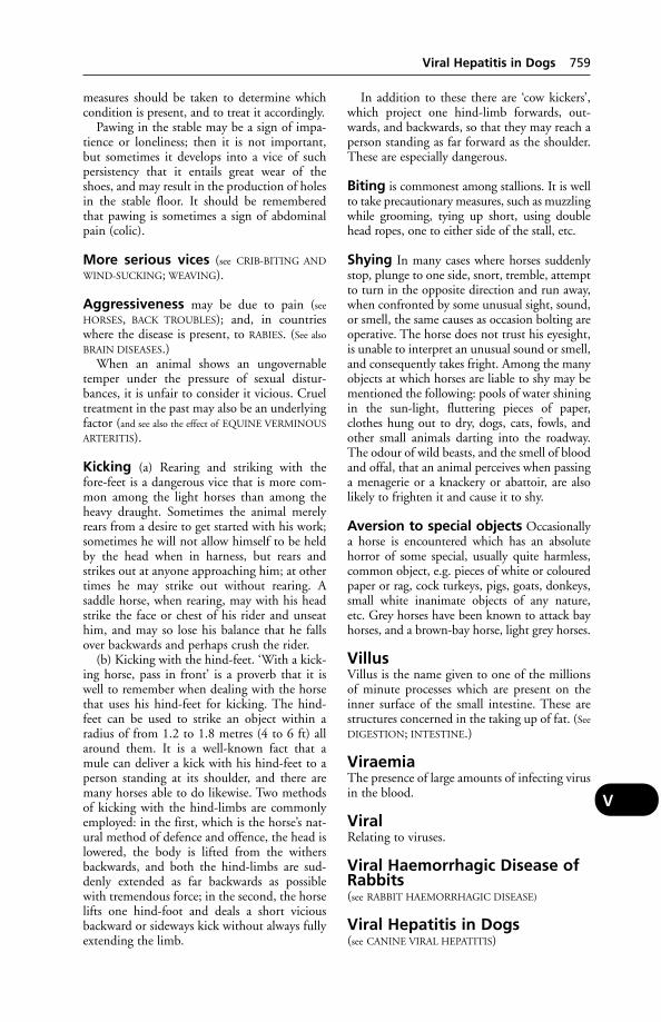
measures should be taken to determine whichcondition is present, and to treat it accordingly.
Pawing in the stable may be a sign of impa-tience or loneliness; then it is not important,but sometimes it develops into a vice of suchpersistency that it entails great wear of theshoes, and may result in the production of holesin the stable floor. It should be rememberedthat pawing is sometimes a sign of abdominalpain (colic).
More serious vices (see CRIB-BITING AND
WIND-SUCKING; WEAVING).
Aggressiveness may be due to pain (see
HORSES, BACK TROUBLES); and, in countrieswhere the disease is present, to RABIES. (See also
BRAIN DISEASES.)When an animal shows an ungovernable
temper under the pressure of sexual distur-bances, it is unfair to consider it vicious. Crueltreatment in the past may also be an underlyingfactor (and see also the effect of EQUINE VERMINOUS
ARTERITIS).
Kicking (a) Rearing and striking with thefore-feet is a dangerous vice that is more com-mon among the light horses than among theheavy draught. Sometimes the animal merelyrears from a desire to get started with his work;sometimes he will not allow himself to be heldby the head when in harness, but rears andstrikes out at anyone approaching him; at othertimes he may strike out without rearing. A saddle horse, when rearing, may with his headstrike the face or chest of his rider and unseathim, and may so lose his balance that he fallsover backwards and perhaps crush the rider.
(b) Kicking with the hind-feet. ‘With a kick-ing horse, pass in front’ is a proverb that it iswell to remember when dealing with the horsethat uses his hind-feet for kicking. The hind-feet can be used to strike an object within aradius of from 1.2 to 1.8 metres (4 to 6 ft) allaround them. It is a well-known fact that amule can deliver a kick with his hind-feet to aperson standing at its shoulder, and there aremany horses able to do likewise. Two methodsof kicking with the hind-limbs are commonlyemployed: in the first, which is the horse’s nat-ural method of defence and offence, the head islowered, the body is lifted from the withersbackwards, and both the hind-limbs are sud-denly extended as far backwards as possiblewith tremendous force; in the second, the horselifts one hind-foot and deals a short viciousbackward or sideways kick without always fullyextending the limb.
In addition to these there are ‘cow kickers’,which project one hind-limb forwards, out-wards, and backwards, so that they may reach aperson standing as far forward as the shoulder.These are especially dangerous.
Biting is commonest among stallions. It is wellto take precautionary measures, such as muzzlingwhile grooming, tying up short, using doublehead ropes, one to either side of the stall, etc.
Shying In many cases where horses suddenlystop, plunge to one side, snort, tremble, attemptto turn in the opposite direction and run away,when confronted by some unusual sight, sound,or smell, the same causes as occasion bolting areoperative. The horse does not trust his eyesight,is unable to interpret an unusual sound or smell,and consequently takes fright. Among the manyobjects at which horses are liable to shy may bementioned the following: pools of water shiningin the sun-light, fluttering pieces of paper,clothes hung out to dry, dogs, cats, fowls, andother small animals darting into the roadway.The odour of wild beasts, and the smell of bloodand offal, that an animal perceives when passinga menagerie or a knackery or abattoir, are alsolikely to frighten it and cause it to shy.
Aversion to special objects Occasionallya horse is encountered which has an absolutehorror of some special, usually quite harmless,common object, e.g. pieces of white or colouredpaper or rag, cock turkeys, pigs, goats, donkeys,small white inanimate objects of any nature, etc. Grey horses have been known to attack bayhorses, and a brown-bay horse, light grey horses.
VillusVillus is the name given to one of the millionsof minute processes which are present on theinner surface of the small intestine. These arestructures concerned in the taking up of fat. (See
DIGESTION; INTESTINE.)
ViraemiaThe presence of large amounts of infecting virusin the blood.
ViralRelating to viruses.
Viral Haemorrhagic Disease ofRabbits(see RABBIT HAEMORRHAGIC DISEASE)
Viral Hepatitis in Dogs(see CANINE VIRAL HEPATITIS)
Viral Hepatitis in Dogs 759
V
Viral Hepatitis of DucklingsViral hepatitis of ducklings is a disease whichattacks ducklings under 3 weeks of age. Thereare usually no clinical signs before death exceptgeneral malaise. On post-mortem examination,the liver shows enlargement and haemorrhaging.It can be prevented by vaccination at 1 day old.
Viral Infections of Cows’ TeatsThese include cowpox. Nowadays, true cowpoxis (in the UK) considered to be a rare disease.Another infection common to man and cattle ispseudo-cowpox or milkers’ nodules. The skindisease in herdsmen is indistinguishable fromthat in shepherds who have been handlingsheep suffering from orf. It is now thought thatthe milkers’ nodules virus very closely resemblesthe orf virus, but that they are 2 distinct entities.
Two doctors in Dorset who had 7 patientswith milkers’ nodules found, with the aid of aveterinary colleague, that the 6 dairy herds inwhich the men worked all had some cows withpseudo-cowpox (or milkers’ nodules) lesions onthe teats. There are 2 types of this infection: oneis described as benign or chronic – this lasts formonths, it is painless throughout, and startswith a mild redness of the teats, followed by theformation of many scabs which get rubbed offat milking. The second, or acute, form involvespain before scabbing begins, but not afterwards.First there is reddening, then blisters whichburst, then very large scabs form. So-calledproud flesh is formed beneath the scabs. Whenthese drop off, a characteristic horseshoe-shaped ring of minute scabs at the circumfer-ence is left. All this takes 7 to 10 days. Whatlooks like a wart remains for several months.
This pseudo-cowpox differs from true cow-pox in that the latter infection is associated withmore pain, fewer scabs, quicker development ofthem and recovery within 3 weeks.
The virus which causes pseudo-cowpox ormilkers’ nodules may be identical with, or close-ly related to, that of bovine papular stomatitis(BPS).
Raised, roughened, brownish plaques are seenon the muzzle, and lesions on the lips and insidethe mouth.
An ulcerative infection of the teats of dairycows has been described in Scotland, and giventhe name bovine ulcerative mammillitis. It iscaused by a herpesvirus.
The disease has been seen only in early win-ter, and lasts for up to 15 weeks. In severe casesit is of sudden onset, often appearing betweenmilkings; the whole teat being swollen andpainful. Blue discoloration is common. Theresultant ulcer covers most, if not all, the teat.
In a less severe form vivid red discoloration wasnoted.
On account of the impossibility of milkingcows with badly ulcerated teats, and becausemastitis often follows, several animals may haveto be slaughtered.
The same disease has been seen in south-westEngland where the onset appeared to follow aprolonged period of wet weather. If the virus isof the herpes type, it may be that it is endemicin the cattle population and produces lesionsonly under conditions which result in devitalis-ing of the tissues. Another possibility is that biting flies transmit the infection. (See also under
FOOT-AND-MOUTH DISEASE.)
Viral Pneumonia of Cattle(see infections listed under CALF PNEUMONIA)
VirginiamycinAn antibiotic used as a growth promoter whichmay be included in livestock rations.
VirinoA low-molecular-weight nucleic acid and ahost-derived protein. (See SCRAPIE for a possibleexample.)
VirionA mature virus; the ultimate phase in viraldevelopment.
VirologyThe study of viruses.
Virus Diarrhoea of Cattle(see BOVINE VIRAL DIARRHOEA)
VirusesThese are minute entities which carry theirgenetic information in one type of nucleic acid.They use the energy system of the host cell fortheir own biosynthetic needs, and can be differ-entiated from bacteria by their size and by theirinability to multiply except in living cells.
Most viruses produce disease in man, ani-mals and plants. They can be transmitted fromone animal to another and stimulate the pro-duction of antibodies in infected animals.
Viruses are mostly invisible under the lightmicroscope, although some of the larger exam-ples (e.g. the pox viruses) can be seen readilyunder the light microscope. Most viruses canonly be visualised in the electron microscope.There is considerable variation in size. Foot-and-mouth disease virus is about 25 nm indiameter, whereas African swine fever virus isabout 10 times that size. (See NANOMETRE.)
760 Viral Hepatitis of Ducklings
V
Many pathogenic viruses are capable of alter-ing their antigenic structure, or their patho-genicity, or both, in response to pressures putupon them by, for example, vaccination.
The classical example is provided by influen-za viruses, which are able to change from harm-less to extremely virulent forms very quicklywithin the same species.
The nomenclature and classification of viruseshas in recent years undergone many changes, andfurther changes are likely as new viruses are dis-covered and new information on the propertiesof viruses accrues. (See table, page 762.)
Anti-viral agents Since viral infections arenot controllable by antibiotics, there has been along search for other compounds which mightachieve anti-viral activity. Much hope was pinnedon INTERFERON, but at present its use is very limited, and research has switched to attempts tostimulate natural production of interferon, or tofind drugs effective against viruses.
Aciclovir may be used for ophthalmic infec-tions by herpesviruses. Zidurodine alleviatesclinical signs in cases of feline immunodeficien-cy virus (FIV). However, anti-viral drugs have tobe used with great care as the margin between aneffective dose and the toxic one (the therapeuticindex) is narrow.
Vaccines are available against a number ofviral infections, e.g. equine herpesvirus, caninedistemper, and feline leukaemia.
(See also ROTAVIRUS; ASTROVIRUS; ONCOGENIC;DNA; RIBONUCLEIC ACID; CANCER.)
Viruses, PlantSome of these can, by means of DNA technol-ogy, be adapted to produce ‘potentially safe and inexpensive vaccines for use against diseasein animals’. AFRC research workers have, forexample, produced a harmless plant virus carry-ing a fragment of foot-and-mouth disease virus.
VisceraViscera is the name given to the larger organslying within the chest and abdominal cavities.The term ‘viscus’ is applied to each of theseindividually.
‘Visceral Gout’A disease characterised by the deposit of uratesover the internal organs; it is found in birds andreptiles.
Visceral Larva MigransA syndrome produced in man by the larvae of Toxocara canis. Occasionally it is the cause ofdeath. (See TOXOCARA.)
Vision(see also EYE) Rays of light pass, in the first place,through the cornea, then through the aqueoushumour that fills the anterior chamber of theeye. The light then enters the hinder part of theeye, through the pupil – a round, slit-like, orelliptical hole in the iris, which can be automat-ically narrowed according to the strength of the light rays that are passing through it.Immediately behind the iris lies the crystallinelens, a clear structure arranged in layers some-what like an onion, which also by automaticalterations in its curves, brings the rays to a focusupon the retina after they pass through a secondclear jelly-like humour – the vitreous humour.The retina is the innermost of the 3 coats of the eyeball, and consists of the specialised terminations of the fibres of the optic nerve.
Monocular and binocular vision Inanimals whose eyes are laterally placed in thehead it is impossible for both eyes to look at anobject directly in front of them. One eye can befocused upon an object at any one time, whilethe other eye sees a completely different picture.This is called ‘monocular vision’. When the eyesare placed towards the front of the head so thatthey can both be concentrated upon an object,as in man, horse, and dog, each eye sees a slightly different picture, but the 2 ranges ofvision overlap. This is called ‘binocular vision’.It is partly owing to the fact that in binocularvision each eye sees slightly ‘round the corner’of the object, that a sense of depth and distanceis conveyed to the higher brain centres. The 2 pictures are not quite superimposed, and the previous experience of the animal enables it to judge distance by this difference in super-imposition. This is technically known as‘stereoscopic vision’.
A striking point in connection with the eyesight of animals is that, although many ofthem have their visual powers obviously veryhighly developed, they seldom trust their eyesin matters of emergency. The visual imagesalone do not convey to the mind the reality ofthe external world. It becomes necessary for theanimal to verify its visual impression by tactileor olfactory impressions. In practically everycase the fear of a harmless object may be imme-diately or shortly dispelled by allowing the ani-mal to smell and examine it by touching it withthe nose.
In birds, the central part of the lens has a mag-nifying power of up to 8x. As a result (stereo-scopic) vision is very accurate, enabling birds tolocate food easily. Birds, reptiles, amphibia andinsects can ‘see’ ultraviolet light.
Vision 761
V

762 Vision
V
Some viruses of veterinary importance
Group Diseases caused Animals affectedAdenoviruses Canine viral hepatitis Dogs
Fox encephalitis Foxes(See also KENNEL COUGH.)
Alphaviruses Equine encephalitis Horses, birds, manAphthoviruses Foot-and-mouth disease Cattle, sheep, goats, pigs, deer,
hedgehogs; (very rarely) manArboviruses (See Togaviruses below.)Arenaviruses Lymphocytic choriomeningitis Mice, hamsters, man
(See also LASSA FEVER.) (Man)Bunyaviruses Rift Valley fever Sheep, cattle, goats,
buffaloes, camels, manNairobi sheep disease Sheep, goatsGumboro disease Poultry
Caliciviruses Feline calicivirus disease CatsVesicular exanthema Pigs; (rarely) horses
Circoviruses Anaemia ChickensDermatitis and nephrosis Pigs
syndromePost-weaning multisystemic
wasting syndrome PigsCoronaviruses Transmissible gastroenteritis Pigs
Feline infectious peritonitis CatsInfectious bronchitis ChickensEnteritis Calves, foals, dogs, cats
Enteroviruses Swine vesicular disease Pigs; (rarely) manTeschen/Talfan disease PigsDuck virus hepatitis DucklingsAvian encephalomyelitis Chickens
Flaviviruses Louping-ill Sheep, cattle, deer, dog, manWesselbron disease Sheep, manJapanese B encephalitis Horses, man, pigs, birdsTick-borne encephalitis Rodents, goats, cattle, manKyasanur forest disease Monkeys, rodents, manOmsk haemorrhagic fever Rodents, manMurray Valley encephalitis Wild birds, childrenSt Louis encephalitis Wild birds, bats, horses, man
Herpesviruses (See table under HERPESVIRUSES.) Horses, cattle, pigs, dogs, cats, manIridoviruses African swine fever Pigs, African warthogLentiviruses Maedi/visna Sheep
Caprine arthritis-encephalitis GoatsMorbilliviruses Canine distemper Dogs, ferrets, mink
Cattle plague (rinderpest) Cattle, sheep, goatsOncoviruses Leukaemia, leukosis, cancer Mammals, birdsOrbiviruses Bluetongue Cattle, sheep
African horse sickness Horses (but not donkeys)Orthomyxoviruses Influenza Horses, pigs, birdsOrthopoxviruses Cowpox Cattle, manPapillomaviruses Warts/papillomas/cancer/sarcoids Cattle, horses, dogs, manParamyxoviruses Newcastle disease Poultry, pigeons
Parainfluenza Cattle, dogs (see KENNEL COUGH)Parapoxviruses Pseudo-cowpox Cattle, man
Orf Sheep, cattle, goats, dogsBovine papular stomatitis Calves
Pestiviruses Bovine virus diarrhoea CattleBorder disease SheepSwine fever PigsEquine viral arteritis Horses
Picornaviruses (This group includes Aphthoviruses and Enteroviruses (see above) and Rhinoviruses.)
Poxviruses (See Orthopoxviruses and Parapoxviruses, above.)Reoviruses Respiratory disease, enteritis Calves, pigs, dogs, cats, rabbits, manRetroviruses (See Lentiviruses and Oncoviruses above.)Rhabdoviruses Rabies (see LYSSA; DUVENHAGE; MOKOLA VIRUS; LAGOS BAT.)
Vesicular stomatitis HorsesTogaviruses (See Alphaviruses, Flaviviruses and Pestiviruses above.)
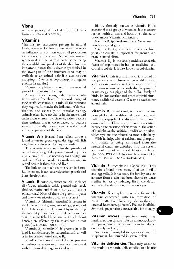
VisnaA meningoencephalitis of sheep caused by alentivirus. (See MAEDI/VISNA.)
VitaminsVitamins are substances present in naturalfoods, essential for health, and which exercisean influence in nutrition out of all proportionto the amounts consumed. Several vitamins aresynthesised in the animal body, some beingthus available independent of the diet, but it isimportant to note that a vitamin synthesised inthe lower part of the alimentary canal may beavailable to an animal only if it eats its owndroppings. (Nocturnal coprophagy is a regularpractice in rabbits.)
Vitamin supplements now form an essentialpart of farm livestock feeding.
Animals, when feeding under natural condi-tions, with a free choice from a wide range offood-stuffs, consume, as a rule, all the vitaminsthey require. But under the influence of domes-tication, and especially of intensive rearing, animals often have no choice in the matter andsuffer from vitamin deficiencies, either becausetheir artificial diet is too restricted, or becausevitamins naturally present have been destroyedin the preparation of the food.
Vitamin A is formed from yellow carotenefound in carrots, green vegetables, egg-yolk, fishroe, liver, cod-liver oil, kidney and milk.
This vitamin is necessary for the growth andgeneral well-being of the young animal in partic-ular. Vitamin A is also necessary for healthy skinand teeth. Cats are unable to synthesise vitaminA and obtain it from fish and liver.
Too little or too much vitamin A can be harm-ful. In excess, it can adversely affect growth andbone development.
Vitamin B complex, water-soluble, includesriboflavin, nicotinic acid, pantothenic acid,choline, biotin, and thiamin. (See also CHOLINE;FOLIC ACID.) Most of these are present in yeastand liver. (For nicotinic acid, see NIACIN.)
Vitamin B1 (thiamin, aneurine) is present inthe husks of cereal grains, yolk of egg, yeast, andliver. A deficiency can be caused by overheatingthe food of pet animals, or by the enzyme pre-sent in some fish. Horse and cattle which eatbracken are affected by the thiaminase in thatplant. (See BRACKEN POISONING.)
Vitamin B2 (riboflavin) is present in milk(and is not destroyed by pasteurisation), as wellas in foods mentioned under B1.
Riboflavin is a constituent of the flavoproteins– hydrogen-transporting enzymes concernedwith the animal’s energy metabolism.
Biotin, formerly known as vitamin H, isanother of the B group of vitamins. It is necessaryfor the health of skin and hoof. It is referred tobelow under ‘Vitamin deficiencies’.
Vitamin B3 (pantothenic acid). Necessary forskin health, and growth.
Vitamin B6 (pyridoxine), present in liver,yeast and cereals, is important for growth andprotein metabolism.
Vitamin Bl2 is the anti-pernicious anaemiafactor of importance in human medicine, andcontains cobalt. It is also known as cobalamin.
Vitamin C This is ascorbic acid; it is found inthe juices of most fruits and vegetables. Mostanimals can produce sufficient vitamin C fortheir own requirements, with the exception ofprimates, guinea pigs and the bulbul famly ofbirds. In hot weather and other stressful situa-tions, additional vitamin C may be needed forall animals.
Vitamin D, or calciferol, is the anti-rachiticprinciple found in cod-liver oil, meat juice, cow’smilk, and egg-yolk. The absence of this vitamincauses rickets. There is an intimate associationbetween the presence of this vitamin, the actionof sunlight or the artificial irradiation by ultra-violet rays, and the mineral balance in the body.
With its help, salts of calcium and phospho-rus, instead of being eliminated from theintestinal canal, are absorbed into the systemand made use of in the calcification of bone.(See COD-LIVER OIL.) Too much vitamin D isharmful. (See RODENTS – Rodenticides.)
Vitamin E (tocopherol) (fat-soluble). Thisvitamin is found in red meat, oil of seeds, milk,and egg-yolk. It is necessary for fertility, and itsabsence from a diet has been shown to causesterility in rats by inducing firstly the death,and later the absorption, of the embryos.
Vitamin K complex – mostly fat-soluble vitamins concerned with the formation of PROTHROMBIN, and hence regarded as ‘the anti-internal-haemorrhage factor’. Present in alfalfa.Synthetic preparations are available for therapy.
Vitamin excess (hypervitaminosis) mayresult in serious disease. (For an example, chron-ic hypervitaminosis A occurs in cats fed almostexclusively on liver.)
An excess of yeast, fed to pigs as a vitamin Bsupplement, has resulted in severe rickets.
Vitamin deficiencies These may occur asthe result of a vitamin-deficient diet, or a failure
Vitamins 763
V

– in some instances – to synthesise a particularvitamin within the body. ‘Secondary’ or ‘condi-tioned’ deficiencies may also arise from any disease which impairs absorption from the alimentary tract, injuries to the liver, infections(which increase the consumption of vitamins),metallic poisoning, and as the result of someenzyme which destroys or inactivates a vitamin.(For examples of the last-mentioned cause, see
‘CHASTEK PARALYSIS’.)Biotin-producing bacteria live in intestines
and contribute a variable amount. But biotindeficiency is not rare – except in adult rumi-nants. Signs of deficiency are: dermatitis onears, neck, shoulder and tail of the pig, togetherwith cracking of the walls and sole of the hoof;retarded growth and brittle feathering, foot der-matitis, swollen eyelids, eruptions on mouthand beak, perosis, leg weakness, poor hatchabil-ity and embryonic malformations in birds.
A report from Finland stated that bleedingfrom the navel, which was a serious problem in a breeding herd of 85 Finnish Landrace and85 Large White sows, could be successfully con-trolled by vitamin C (not vitamin K as mighthave been expected). An ascorbic acid supple-ment was given to the sows for from 8 to 2 daysbefore farrowing. Piglets from the treated sowswere also 5.5 per cent heavier than those fromuntreated controls at 3 weeks of age.
Vitamin E (tocopherol) could with advantagebe added to all compound feeds as a precau-tionary measure, particularly if the feed containspolyunsaturated fatty acids. Less vitamin E isabsorbed from the intestine if the latter are pre-sent. Some feeds contain vitamin E antagonists– present in lucerne and beans. The activity of vitamin E may be reduced by a high nitratecontent in feed or drinking water. Animalswhich do not receive an adequate supply of thetrace element selenium need extra vitamin E,because selenium has a vitamin E sparing effect.Application of fertilisers rich in sulphatesinhibits the absorption of selenium by plantsfrom the soil, and in these circumstances grazing animals will require extra vitamin E.
Vitamin E contents of feeds as well as cropsmay be reduced to a dangerous level on storage.(See also Muscular dystrophy under MUSCLES, DISEASES
OF.) Anti-vitamin E factor may be present inbarley as well as fats of animal origin.
Despite a current tendency to increase vita-min levels in animal feeds, cases of vitamin Edeficiency appear to be becoming more preva-lent in all classes of farm livestock, resulting inmulberry heart in pigs, muscular dystrophy incalves, and ‘crazy chick’ disease in poultry.Perhaps this has something to do with the faster
growth-rates expected of animals nowadays.Certainly the low vitamin E content of somecereals is important. This is especially the casewith some samples of barley grown on seleni-um-deficient soil but fed on a farm where sele-nium is no problem, and where the possibilityof such a deficiency might well be overlooked.
It is generally accepted that the daily require-ment of vitamin E is, for cows, 1 g; for calves,150 mg; for ewes, 7 mg; and for lambs, 25 mg.
Vitamin E deficiency leads to white muscledisease or muscular dystrophy; and a supple-ment of this vitamin has been shown to reducethe incidence of retained placenta, metritis, andcysts of the ovaries when given with SELENIUM.
Horses Vitamin A deficiency is unlikely tooccur except in town horses denied adequategreen food. Deficiency symptoms are stated toinclude night-blindness, hoof lesions, corneallesions, respiratory symptoms, and reproductivedifficulties. Some of the B vitamins are synthe-sised by adult horses, but backward foals maybenefit from vitamin supplements. Infertility inthe mare may sometimes be associated with avitamin C deficiency. It has been suggested thatsplints, sidebones, ringbones, and spavins maybe associated with a vitamin D deficiency.
Cattle Vitamin A deficiency in cattle deniedadequate green food leads to abortion or thebirth of weak, blind calves, or of those sufferingfrom diarrhoea which die within a few days.Corneal lesions and blindness may also result ingrowing cattle. For example, Hereford bulls ona diet of beet-pulp nuts, high-protein nuts, andbarley straw went blind owing to a lack of vitamin A. (See also HYPERKERATOSIS.) Vitaminsof the B complex are mostly synthesised in therumen, but in the newborn calf a deficiencymay occur. (See THIAMIN.) Vitamin C is appar-ently synthesised by adult cattle, but some casesof infertility may, it is believed, be due to a deficiency, and some cases of navel-ill benefit, it is said, from vitamin C treatment. In someparts of Britain pasture or fodder crops containtoo little vitamin D, while sunlight during thewinter months is insufficient to enable theshortage to be made good within the animal’sbody; the result is rickets. Vitamin E deficiencyis associated with muscular dystrophy.
Pigs Vitamin A deficiency results in failure togrow in piglets and infertility in adult pigs, aswell as paralysis of the hindquarters. A nicotinicacid deficiency gives rise to a condition simu-lating necrotic enteritis and poor growth. Yeastsupplements will correct deficiencies of the Bcomplex, but excess may result in rickets.
764 Vitamins
V
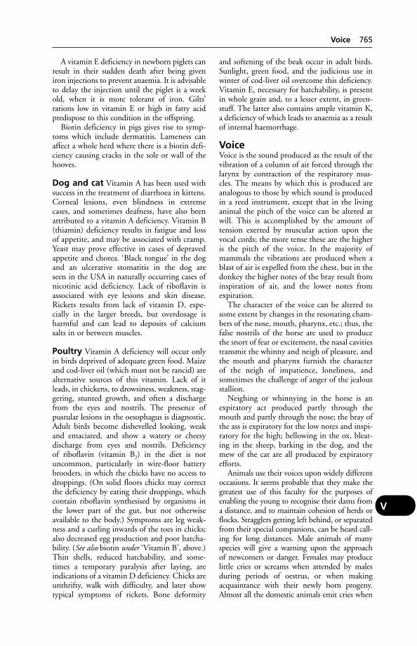
A vitamin E deficiency in newborn piglets canresult in their sudden death after being giveniron injections to prevent anaemia. It is advisableto delay the injection until the piglet is a weekold, when it is more tolerant of iron. Gilts’rations low in vitamin E or high in fatty acidpredispose to this condition in the offspring.
Biotin deficiency in pigs gives rise to symp-toms which include dermatitis. Lameness canaffect a whole herd where there is a biotin defi-ciency causing cracks in the sole or wall of thehooves.
Dog and cat Vitamin A has been used withsuccess in the treatment of diarrhoea in kittens.Corneal lesions, even blindness in extremecases, and sometimes deafness, have also beenattributed to a vitamin A deficiency. Vitamin B(thiamin) deficiency results in fatigue and lossof appetite, and may be associated with cramp.Yeast may prove effective in cases of depravedappetite and chorea. ‘Black tongue’ in the dogand an ulcerative stomatitis in the dog are seen in the USA in naturally occurring cases ofnicotinic acid deficiency. Lack of riboflavin isassociated with eye lesions and skin disease.Rickets results from lack of vitamin D, espe-cially in the larger breeds, but overdosage isharmful and can lead to deposits of calciumsalts in or between muscles.
Poultry Vitamin A deficiency will occur onlyin birds deprived of adequate green food. Maizeand cod-liver oil (which must not be rancid) arealternative sources of this vitamin. Lack of itleads, in chickens, to drowsiness, weakness, stag-gering, stunted growth, and often a dischargefrom the eyes and nostrils. The presence of pustular lesions in the oesophagus is diagnostic.Adult birds become dishevelled looking, weakand emaciated, and show a watery or cheesy discharge from eyes and nostrils. Deficiency of riboflavin (vitamin B2) in the diet is notuncommon, particularly in wire-floor batterybrooders, in which the chicks have no access todroppings. (On solid floors chicks may correctthe deficiency by eating their droppings, whichcontain riboflavin synthesised by organisms inthe lower part of the gut, but not otherwiseavailable to the body.) Symptoms are leg weak-ness and a curling inwards of the toes in chicks;also decreased egg production and poor hatcha-bility. (See also biotin under ‘Vitamin B’, above.)Thin shells, reduced hatchability, and some-times a temporary paralysis after laying, are indications of a vitamin D deficiency. Chicks areunthrifty, walk with difficulty, and later showtypical symptoms of rickets. Bone deformity
and softening of the beak occur in adult birds.Sunlight, green food, and the judicious use inwinter of cod-liver oil overcome this deficiency.Vitamin E, necessary for hatchability, is presentin whole grain and, to a lesser extent, in green-stuff. The latter also contains ample vitamin K,a deficiency of which leads to anaemia as a resultof internal haemorrhage.
VoiceVoice is the sound produced as the result of thevibration of a column of air forced through thelarynx by contraction of the respiratory mus-cles. The means by which this is produced areanalogous to those by which sound is producedin a reed instrument, except that in the livinganimal the pitch of the voice can be altered atwill. This is accomplished by the amount oftension exerted by muscular action upon thevocal cords; the more tense these are the higheris the pitch of the voice. In the majority ofmammals the vibrations are produced when ablast of air is expelled from the chest, but in thedonkey the higher notes of the bray result frominspiration of air, and the lower notes fromexpiration.
The character of the voice can be altered tosome extent by changes in the resonating cham-bers of the nose, mouth, pharynx, etc.; thus, thefalse nostrils of the horse are used to producethe snort of fear or excitement, the nasal cavitiestransmit the whinny and neigh of pleasure, andthe mouth and pharynx furnish the character of the neigh of impatience, loneliness, andsometimes the challenge of anger of the jealousstallion.
Neighing or whinnying in the horse is anexpiratory act produced partly through themouth and partly through the nose; the bray ofthe ass is expiratory for the low notes and inspi-ratory for the high; bellowing in the ox, bleat-ing in the sheep, barking in the dog, and themew of the cat are all produced by expiratoryefforts.
Animals use their voices upon widely differentoccasions. It seems probable that they make thegreatest use of this faculty for the purposes ofenabling the young to recognise their dams froma distance, and to maintain cohesion of herds orflocks. Stragglers getting left behind, or separatedfrom their special companions, can be heard call-ing for long distances. Male animals of manyspecies will give a warning upon the approach of newcomers or danger. Females may producelittle cries or screams when attended by malesduring periods of oestrus, or when makingacquaintance with their newly born progeny.Almost all the domestic animals emit cries when
Voice 765
V
suffering pain. In the horse tribe the sounds areoften merely grunts or groans, especially whenthe pain is abdominal. In other cases horses willscream when they are suddenly subjected toacute pain, or to very great fright. Cattle andsheep in agony behave similarly to horses; theyusually groan, but cows, ewes, and heifers mayissue a long drawn-out bellow or bleat during dif-ficult parturition. The pig has a range of notesfrom the satisfied grunt of suckling a sow, to thefrightened squeals and screams of those that arebeing handled by man. The dog has a note for alloccasions; he generally expresses all the emotionsof which he is capable by differences in his bark.(See also LARYNX and MUTING OF DOGS.)
In rabies the character of the voice may be changed. In the ‘dumb’ form, barking is suppressed.
VolarRefers to the back of the fore-limb.
Volcanic GasesTypically these comprise water vapour, carbondioxide, sulphur dioxide, hydrogen sulphide,hydrochloric acid, hydrogen fluoride, and carbon monoxide.
VolvulusAn intestinal obstruction is produced by thetwisting of a loop of bowel round itself. It is usu-ally due to some spasmodic contraction of themuscular coat, or to the presence of gas, and isvery dangerous owing to the great risk of stran-gulation of the blood supply and consequentnecrosis. Excessive gas formation in the caecumand colon of whey-fed pigs may lead to volvu-lus. (See HAEMORRHAGIC GASTROENTERITIS
OF PIGS and INTESTINES, DISEASES OF.)
VomicaA cavity in the lung tissue produced by disease.Vomicae are most commonly met with in cattlesuffering from either tuberculosis or contagiouspleuro-pneumonia.
VomitingVomiting involves not merely a contraction ofthe stomach walls and a dilatation of the gullet,but is a complex act in which the abdominalmuscles, the diaphragm, the muscles of thechest and larynx, and those of the lower part ofthe neck all play a part.
Before the act there is usually a profuse secre-tion of watery saliva which serves to lubricatethe passage of the stomach contents. The animal appears uneasy, and will usually seek asecluded spot. Soon rhythmic contractions of
the abdominal muscles commence and culmi-nate in the ejection of a quantity of frothy mate-rial. The diaphragm is generally fixed, and thereis a powerful closing of the glottis to prevent anyfluids from gaining access into the trachea.
The dog and cat vomit with relative ease.They are able to induce vomiting by eating por-tions of the green shoots of couch grass(Triticum repens), ingestion of which brings onvomiting within 5 to 10 minutes. In the pig,the process is more exacting than in the carni-vores. Cattle and sheep may vomit occasionally,but it is not common and may be related to aserious stomach disorder or to rhododendronpoisoning. Vomiting in the horse is rare, andoften is associated with a rupture of the stomach; when it occurs it should be considereda very grave symptom indeed. The materialalways escapes through the nostrils in the horse.
Causes Vomiting can be considered under thefollowing headings:
Travel sickness.Stress.Simple indigestion. When the stomach has
received either a quality or a quantity of food-stuff with which it is unable to deal, the processof digestion does not proceed, or only proceedsup to a point. The material brought up is recog-nisable as food, but it is mixed with quantitiesof frothy mucus, water, and perhaps may bestained brownish from bile. It has a faintly sour smell which is greater the longer theprocess of digestion has been enabled to proceed. The ejecta is generally easily broughtup, and the animal soon settles down andbecomes normal.
Indigestion from foreign bodies. (See
‘CHOKING’; IMPACTION; FOREIGN BODY.)Gastritis. The walls of the stomach are
inflamed and thickened, the mucous membraneis swollen and painful, and the nervous system isin an irritable state. Whenever food or waterenters the organ, vomiting immediately takesplace. The vomit consists of the solid materialswallowed, coated on the outside with mucus andfroth. If liquids have been taken they are returnedalmost unchanged. When the inflammatory con-dition is very severe there are quantities of bloodthat have undergone partial digestion and havean appearance not unlike coffee-grounds, seen inthe vomit. In such animals there will be a veryoffensive smell both from the ejecta and from themouth of the patient.
Pyloric stenosis, which may be congenital, issaid to give rise to projectile vomiting.
Enteritis is associated with vomiting but thereis diarrhoea as well.
766 Volar
V
Impaction of the rectum, whether from par-ticles of undigested bone, or hair, hard faeces,etc., generally induces vomiting in which notonly does the stomach expel its contents, butmasses of bowel content as well.
Acute nephritis is an extremely commoncause of thirst and vomiting in the dog.
Pyometra in the bitch, cat, and sow is frequently accompanied by vomiting.
Accidents. The shock of a severe burn or acci-dent will cause vomiting, although the injurieshave not been inflicted upon the stomach itself.In other cases, where the head has been injured,the area in the case of the brain which controlthe act of vomiting becomes disturbed and theanimal evacuates its stomach.
Poisons. Many irritant substances will pro-duce vomiting. Of the commonest may be men-tioned tartar emetic, mustard, salt, carbolic acid,areca-nut, castor oil, etc.; and of substances less common, but more drastic, the followingare examples: strychnine, arsenic, phosphorus,apomorphine, croton oil, zinc and copper sul-phates, and many of the metallic salts. Some ofthese have special characteristics; for example,phosphorus vomit is luminous in the dark.
Diseases. The symptom of vomiting is com-mon to many other diseases – meningitis, peri-tonitis, nephritis, leptospirosis, rabies, vomitingand wasting syndrome in pigs, etc.
Treatment In the dog and cat the use of normal saline or glucose saline by injection isfrequently indicated as an alternative to givingfood (liquid or otherwise) by mouth during anillness (such as nephritis, uraemia, and enteritis)in which vomiting is persistent.
Vomiting and WastingSyndromeThis occurs in piglets 5 days old and upwards,and is characterised by vomiting, depression,
loss of appetite, constipation, emaciation, and ahairy appearance. It is caused by a coronavirus,haemagglutinating encephalomyelitis virus(HEV). (See also ‘ONTARIO ENCEPHALITIS’.)
Von Willebrand’s DiseaseAn inherited bleeding disorder found in some25 breeds of dogs, and associated with an auto-somal trait causing a high morbidity but a lowmortality. Signs may include epistaxis, haema-turia, lameness, bleeding from genital mucosa,and prolonged bleeding from cut nails, etc.
Vultures(see TROPICS; CARCASES, DISPOSAL OF)
VulvaIt has in domesticated animals only simple, single labia or lips.
Diseases of the vulva In Kenya, squamouscell carcinoma of the vulva is common in cattleof the Ayrshire breed. Cryosurgery has givengood results when treatment has not beendelayed until the tumour becomes too large. In62 cases, 55 were successfully treated.
(For persistent bleeding from the vulva, see
VAGINA.)In the tropics, especially, a thick purulent
discharge from the vagina may be a sign oftuberculosis involving the uterus/vagina.
Vulvovaginitis(see under RHINOTRACHEITIS and VULVOVAGINITIS,GRANULAR)
Vulvovaginitis, GranularA venereal disease of cattle caused byMycoplasma bovigenitalium, affecting vulva andvagina or seminal vesicles and skin of the penis.The lesions are nodules.
Vulvovaginitis, Granular 767
V

‘Walkabout Disease’(see KIMBERLEY HORSE DISEASE)
Wall Eye (Leukoma)Wall eye (leukoma) is a condition in which the brown pigment of the iris is lacking, givingthe iris a steely blue appearance. In dogs, it is usually unilateral and is not a problem. In the horse, wall eyes may occur when the greaterpart of the face, or that portion around the eyes, is white. The pupil of the eye appears to be encircled by a ring of bluish or greyishwhite, and the expression of the horse’s face is
consequently unusual. It is not a serious defectexcept in tropical countries.
WallabiesSmaller than kangaroos, these native Australianmarsupials are a source of human HYDATID DIS-
EASE in the southern tablelands of New SouthWales. Lumpy jaw is a common finding. Theymay sometimes be found as feral animals inparts of England.
Warble Fly OrderThe Warble Fly Order came into force in 1989.The presence of warble fly lesions in cattle is aNOTIFIABLE DISEASE in Britain. Where a bloodtest indicates that an infestation may be pre-sent, treatment under the supervision of an offi-cer of the State Veterinary Service is required.Every herd and every bovine animal within3 km of an infestation must be treated (the fly
W
The life-cycle of the warble fly.

is able to travel only short distances). As warbleflies are present in other countries, all cattleimported into Britain must be treated forpossible infestation within 24 hours of arrival.The treatment must be supervised by a StateVeterinary Service officer.
WarblesWarbles are swellings about the size of a marbleor small walnut occurring upon the backs ofcattle in spring and early summer, caused by thepresence in them of the larvae of one of thewarble flies – Hypoderma bovis or H. lineata.These are of very great economic importance.The adults – especially H. bovis – cause greatannoyance to stock during the period wheneggs are being laid. Not only does this result ininjuries, animals rushing around (‘gadding’) toavoid the attacks, but the milk yield is reduced,sometimes by as much as 25 per cent, and condition is impaired.
H. lineata in its migration through the bodyirritates the gullet; and both species may injurethe spinal cord. The warbles on the back are really so many small abscesses which not onlyreduce condition very considerably but may,when many are present, result in the death ofyoung animals. The accidental crushing of anumber of the larvae in these cavities may causethe death of the animal from anaphylactic shock.
In the carcases there is considerable destructionof valuable meat around the warbles; ‘butcher’s
jelly’ or ‘licked beef ’ is an oedematous, straw-coloured, jelly-like substance, which infiltratesthe tissue near the larvae. The holes which the larvae produce in the hides reduce their value;heavily infected hides are often useless for leather.
Warbles are most common in young animals,in which loss of condition is most serious; butthey have been found in small numbers in ani-mals up to 15 years old. They are sometimesfound in young horses. The larvae occasionallyenter the spinal canal and produce very seriouslesions. Horses are attacked mostly by H. bovislarvae, which affect the area of the saddlechiefly; but brain involvement has been report-ed in the horse. In deer, larvae of the warble flyH. diana are often found.
Methods of control Satisfactory controldepends upon artificial interference with thelife-cycle. (See IVERMECTIN.) A systemic insecti-cide will kill a high percentage of larvae beforethey complete their migration and penetratethe back.
In Britain in 1978, 40 per cent of cattle inEngland and Wales, and 20 percent inScotland, were affected with warbles. (See FLIES
– FLY control measure.)Autumn is the more effective time for
treatment, even though infestation cannot bevisually detected then, and cattle-owners inwarble-affected areas are advised to treat theirherds accordingly.
Warbles 769
W
Dressing against warbles.

Pour-on warblecide compounds includephosmet and ivermectin. Parenteral prepara-tions of abamectin, doramectin and moxidectinare also effective.
Reindeer In Canada they are attacked by thewarble fly Edoede magena tarandi. Ivermectinhas been used for control.
Goats Ivermectin has been used also againstthe goat warble Przhevalskiana silenus.Following the introduction of the Warble FlyOrder 1985, infestation by warbles was virtuallyeradicated by 1995.
The tropical warble fly of CentralAmerica is Dermatobia hominis, which lays itseggs on an intermediary vector – fly or mosqui-to – which it catches for the purpose. (See also
under FLIES, and IVERMECTIN.)
WarfarinAn anticoagulant drug used in human medi-cine; its veterinary use is mainly as a rat poison.It causes death of rats and mice from internalhaemorrhage. In the strengths used, 0.005 percent and 0.025 per cent, it is considered thatproperly prepared baits will not prove danger-ous to livestock if used with ordinary care.Cases of accidental poisoning have occurred,however, in domestic animals; and food conta-minated by rodents’ urine may be dangerouswhere warfarin is used.
Therapeutically, warfarin is used in the treatment of navicular disease.
Treatment of warfarin poisoningVitamin K1 (phytomenadione) by intramuscularinjection. Blood transfusion may be necessary.
Once symptoms have appeared, use of glu-cose saline, or blood transfusion, is indicated.The poisoned animal must be handled verygently, or further internal bleeding may occur.(See also NAVICULAR DISEASE.)
Warts (Papillomas)Warts (papillomas) are small growths whichappear on skin or mucous membrane, andoccur in all farm and domestic animals.Papillomas are benign, but an individual wartcan become malignant. (See PAPILLOMA.)
Around the mouth they may interfere withfeeding, and when occurring about the nostrilsthey may obstruct the breathing. Soft warts inthe oesophagus sometimes make swallowingdifficult, and upon the penis or in the urethrathey may hinder the passage of urine. (See also
EYE.)
Horses The commonest situations are the skinof the udder or sheath, the lips and nostrils, the eyelids, outer and inner skin of the ears, the region of the breast, and the insides of thelimbs.
Cattle The commonest seats of warts are theteats of cows. Young cows in winter are fre-quently affected about the skin of the eyelidsand along the lower line of the abdomen, butthe growths often drop off spontaneously fromthese positions when the young animals areturned out to grass in the early spring.Otherwise warty growths are found as in thehorse.
Dogs and cats In the dog especially, less soin the cat, warts are common. Single smallwarts with a cauliflower-like extremity or with arounded top are commonly found about theeyelids, lips, ears, paws, etc., as well as upon thegeneral surface of the body. They usually growvery slowly and may be present for years with-out causing any pain or inconvenience. In othercases warts appear in connection with thegums, tongue, and insides of the cheeks; inthese positions they arise in clusters and growvery rapidly. Cases such as these are usuallyaccompanied by a great amount of salivationand a fetid discharge from the mouth.
Removal Of Warts Multiple warts in cattlehave been treated by a variety of chemicals,including injections of lithium antimony tartrate, with varying degrees of success.Autogenous and other vaccines have also been used where there is a herd problem, andsurgical removal may be resorted to.
Washing of Animals(see BATHS)
Wasp Stings(see under BITES)
Waste Food(see BAKERY WASTE; SWILL; CHOCOLATE
POISONING)
Wasting(see ATROPHY)
Water and Watering ofAnimals
Amounts required The quantity of waterneeded per day by the various domestic animalsdepends upon the nature of the food, the cli-mate, the temperature, and the size and the
770 Warfarin
W

activity of the animals themselves. When verydry food is given, such as hay, bran, oats, etc.,more water is required than when roots orgrowing grass is eaten.
Drinking water should be freely available toanimals, so that they can drink as and whenthey choose. (See below under ‘Water supply’.)Stress may occur in an animal deprived of thechance to drink sufficient water, and actualdehydration (which can lead to death) may becaused. Production of milk, etc., will obviouslybe adversely affected.
With an ad lib water supply, the amount ofwater required by various animals under variousconditions is of mainly theoretical interest,apart from practical aspects of planning ade-quate supplies of piped water, trough space, etc.Water requirement figures can be taken only asapproximate guidelines, and authorities differto some extent.
Cattle Dry cows of the larger breeds requirebetween 36.5 and 45 litres (8 to 10 gallons) per day. Those in milk need in addition about5 times as much water as the volume of milkproduced, while for the last 4 months of preg-nancy, the daily consumption may rise to about70 litres (approximately 15 gallons).
As the air temperature increases above 10°C(50°F), the water requirement rises rapidly.
Calves require much more water after theyare weaned than before. A common mistake isto ignore this fact, with the result that the calvesreceive a check to their growth from which theymay never fully recover.
Pigs are highly susceptible to water depriva-tion. (See SALT POISONING.) Approximate quan-tities required have been given as 4.5 litres (1 gallon) per day for a litter of 3-week-oldpiglets, and up to 22.5 litres (5 gallons) per dayfor a nursing sow. The benefits of creep feedingmay be lost if the piglets are denied water.
Quality of water This is obviously of primeimportance. Animals may suffer thirst andstress if the only drinking water available tothem is disagreeable in taste. Where pipedwater is not available, and rainwater has to bestored in tanks, it is important to clean out gut-ters and the tanks themselves. Galvanised irontanks should not be allowed to get rusty. Well-water may contain an excess of one or moreminerals which may make it unpalatable or beharmful to the animal, so that sampling andanalysis should be carried out.
Poisoning by water may result from the useof lead pipes or tanks. (See LEAD POISONING.)
The use of lead paints in storage tanks is also adanger. (See also ZINC POISONING.) Stored rain-water containing decaying organic matter(leaves, bird droppings, etc.) has led to thedeath of pigs from nitrite poisoning.
Diseases spread by water Apart from illness caused by some inorganic substance dissolved in the water, such as lead from leadpipes or tanks, or arsenic from contaminationwith sheep-dip, water-borne infection maycause disease.
Among diseases that can be distributed inthis manner are the following: anthrax, fromwater used in tanneries or wool-washingpremises, or when a carcase has been buriednear a stream; Johne’s disease, salmonellosis,and coccidiosis in cattle, from contamination ofstreams, ditches, and ponds. Liver fluke can bespread via infected mud snails, Lymnaea trun-catula. Leptospira hardjo infection is 8 timesmore likely where cattle have access to a watercourse.
Washing water and water-tanks have beencontaminated with, for example, Bacillus subtilis, leading to MASTITIS.
Water supply A good stockman will ensurethat the animals in his care are never short ofwater; that all automatic drinking bowls or nip-ple drinkers are in working order; that frost hasnot cut off the supply of piped water (lagging ofexposed pipes is obviously necessary in winter);and that the water has not been allowed tofreeze in troughs, tanks, etc. It is also necessaryto ensure that the levers of automatic drinking
Designed for field use, this CemFil glass fibre-reinforced cement drinking trough is obtainable insizes of up to 2000-litre capacity. The water supplyis, of course, piped.
Water and Watering of Animals 771
W

bowls are not too stiff for young animals tooperate, and that young stock are shown work-ing nipple drinkers – not left to find them forthemselves. (See also ALGAE.)
In one incident, newly weaned pigs were putinto a yard having automatic water-bowls fit-ted, but as the yard had been mucked out thebowls were out of reach of the young pigs.
Pigs deprived of water show nervous symp-toms. They may walk in circles, or backwards,press their heads against a wall, champ their jaws,collapse and have convulsions. Of course, somepigs may be found dead without symptoms having been observed.
Sheep have shown symptoms suggestive oftwin-lamb disease, and died, after beingremoved from a field where they had access to a stream and placed on pasture where the ball-valve of a drinking trough had been tied up.Sheep prefer to drink running water, and thoseof some breeds are so reluctant to drink any-thing else that, when housed, a running watersupply must be arranged indoors.
A drop in milk yield may occur in dairyherds where the cows are moved periodically toa field too far from a water-trough; or where thewater pressure is too low to ensure adequatesupply.
Dogs, cats, and poultry should always beallowed an unlimited supply of water soarranged that they are unable to foul or upsetthe drinking vessels.
(See also DEHYDRATION.)
Horses Wherever possible, water should begiven before the food, or not for 1 to 2 hoursafter feeding. The horse’s stomach is small, andcannot contain a full feed and several litres/gallons of water simultaneously. Water in excessof requirements should be offered when horsesare at rest, and they should be allowed to drinkfrequently when working.
Water intoxication This may occur infarm livestock when, as a result of bad manage-ment, they have been deprived of adequatedrinking water and then suddenly find them-selves in circumstances which enable them todrink as much as they want.
One symptom may be a red discoloration ofthe urine. Convulsions, recumbency, hyperaes-thesia, aimless wandering, and death have beenseen in calves.
Water BuffaloesWater buffaloes are regarded by many as anunder-utilised form of livestock. It is commonpractice in the tropics to immerse them in water
during the heat of the day as they have very fewsweat glands and are prone to heatstroke.However, they can be reared away from water ifshade is available. They are widely used as adraught animal in warmer countries, includingthe poorer parts of Italy. They are farmed inBritain to produce mozzarella cheese. Studies inthe USA, Papua New Guinea, Trinidad andAustralia have shown water buffalo performwell as regards growth rates, health and produc-ton of meat and milk. They are able to digestrougher material than cattle or sheep.
Water buffalo meat is similar in taste to beef;the milk is richer in butterfat and solids-not-fatthan cows’ milk. There is a general low inci-dence of mastitis, probably because on ceasingmilking, the teat canal closes very rapidly.Water buffalo are generally quite docile unlessseverely stressed or in pain.
Some of the steps needed to permit greaterexploitation of this valuable animal are:
1. Trials to compare growth rate, feeding,nutrition and other characteristics of water buffalo with those of cattle.
2. Selective breeding and protection of out-standing buffalo specimens, especially inSoutheast Asia.
3. Replacement of the 1500-year-old ineffi-cient wooden yoke (in rural Asia, where thewater buffalo is the small farmer’s ‘tractor’,improved harnesses could increase pullingpower by up to 25 per cent). (See TRANSPORT OF
ANIMALS.)The limitation of water buffaloes must be
taken into account. For instance, the animalssuffer if forced to remain, even for a few hours,in direct sunlight. They cannot be worked forlong periods during the heat of the day, andthey are also susceptible to extreme cold. (TheWater Buffalo: New Prospects for anUnderutilized Animal, US National Academy ofSciences).
An important roundworm of the water buf-falo (Bubalus bubalis) is Paracooperia nodulosa.This causes development of nodules in theintestine, and diarrhoea, anaemia, emaciationand sometimes death. (See LIVESTOCK PRODUC-
TION.)
Water-DropwortThis is Oenanthe fistulosa, and while it and pars-ley water-dropwort (O. lachenalii and also O.aquatica) are all poisonous, they are less so thanhemlock water-dropwort (O. crocata) – a weedof marshy places, ditches, and other wet loca-tions. This is considered to be one of the mostdangerous and poisonous of the commonerplants found in Britain, and many cases of
772 Water Buffaloes
W
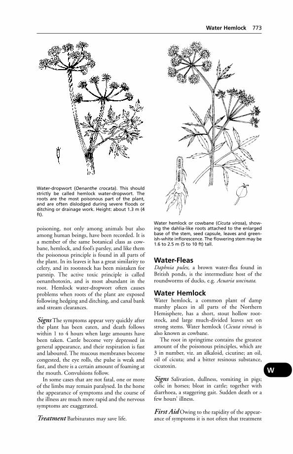
poisoning, not only among animals but alsoamong human beings, have been recorded. It isa member of the same botanical class as cow-bane, hemlock, and fool’s parsley, and like themthe poisonous principle is found in all parts ofthe plant. In its leaves it has a great similarity tocelery, and its rootstock has been mistaken forparsnip. The active toxic principle is calledoenanthotoxin, and is most abundant in theroot. Hemlock water-dropwort often causesproblems when roots of the plant are exposedfollowing hedging and ditching, and canal bankand stream clearances.
Signs The symptoms appear very quickly afterthe plant has been eaten, and death followswithin 1 to 4 hours when large amounts havebeen taken. Cattle become very depressed ingeneral appearance, and their respiration is fastand laboured. The mucous membranes becomecongested, the eye rolls, the pulse is weak andfast, and there is a certain amount of foaming atthe mouth. Convulsions follow.
In some cases that are not fatal, one or moreof the limbs may remain paralysed. In the horsethe appearance of symptoms and the course ofthe illness are much more rapid and the nervoussymptoms are exaggerated.
Treatment Barbiturates may save life.
Water-FleasDaphnia pulex, a brown water-flea found inBritish ponds, is the intermediate host of theroundworms of ducks, e.g. Acuaria uncinata.
Water HemlockWater hemlock, a common plant of dampmarshy places in all parts of the NorthernHemisphere, has a short, stout hollow root-stock, and large much-divided leaves set onstrong stems. Water hemlock (Cicuta virosa) isalso known as cowbane.
The root in springtime contains the greatestamount of the poisonous principles, which are3 in number, viz. an alkaloid, cicutine; an oil,oil of cicuta; and a bitter resinous substance,cicutoxin.
Signs Salivation, dullness, vomiting in pigs;colic in horses; bloat in cattle; together withdiarrhoea, a staggering gait. Sudden death or afew hours’ illness.
First Aid Owing to the rapidity of the appear-ance of symptoms it is not often that treatment
Water-dropwort (Oenanthe crocata). This shouldstrictly be called hemlock water-dropwort. Theroots are the most poisonous part of the plant,and are often dislodged during severe floods orditching or drainage work. Height: about 1.3 m (4ft).
Water hemlock or cowbane (Cicuta virosa), show-ing the dahlia-like roots attached to the enlargedbase of the stem, seed capsule, leaves and green-ish-white inflorescence. The flowering stem may be1.6 to 2.5 m (5 to 10 ft) tall.
Water Hemlock 773
W
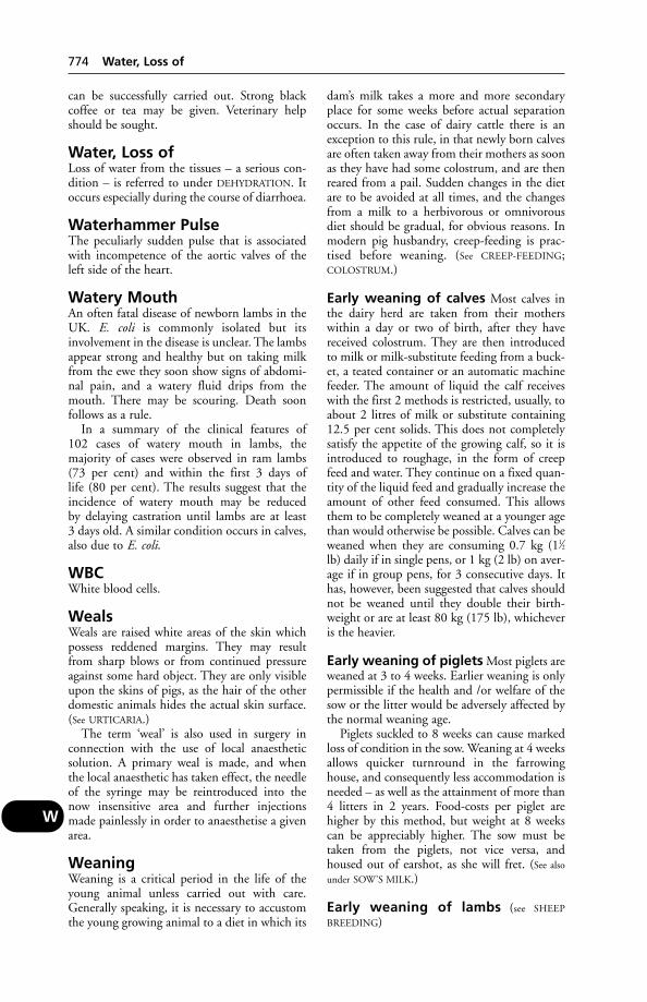
can be successfully carried out. Strong blackcoffee or tea may be given. Veterinary helpshould be sought.
Water, Loss ofLoss of water from the tissues – a serious con-dition – is referred to under DEHYDRATION. Itoccurs especially during the course of diarrhoea.
Waterhammer PulseThe peculiarly sudden pulse that is associatedwith incompetence of the aortic valves of theleft side of the heart.
Watery MouthAn often fatal disease of newborn lambs in theUK. E. coli is commonly isolated but itsinvolvement in the disease is unclear. The lambsappear strong and healthy but on taking milkfrom the ewe they soon show signs of abdomi-nal pain, and a watery fluid drips from themouth. There may be scouring. Death soon follows as a rule.
In a summary of the clinical features of 102 cases of watery mouth in lambs, the majority of cases were observed in ram lambs(73 per cent) and within the first 3 days of life (80 per cent). The results suggest that theincidence of watery mouth may be reduced by delaying castration until lambs are at least 3 days old. A similar condition occurs in calves,also due to E. coli.
WBCWhite blood cells.
WealsWeals are raised white areas of the skin whichpossess reddened margins. They may resultfrom sharp blows or from continued pressureagainst some hard object. They are only visibleupon the skins of pigs, as the hair of the otherdomestic animals hides the actual skin surface.(See URTICARIA.)
The term ‘weal’ is also used in surgery inconnection with the use of local anaestheticsolution. A primary weal is made, and when the local anaesthetic has taken effect, the needleof the syringe may be reintroduced into thenow insensitive area and further injectionsmade painlessly in order to anaesthetise a givenarea.
WeaningWeaning is a critical period in the life of theyoung animal unless carried out with care.Generally speaking, it is necessary to accustomthe young growing animal to a diet in which its
dam’s milk takes a more and more secondaryplace for some weeks before actual separationoccurs. In the case of dairy cattle there is anexception to this rule, in that newly born calvesare often taken away from their mothers as soonas they have had some colostrum, and are thenreared from a pail. Sudden changes in the dietare to be avoided at all times, and the changesfrom a milk to a herbivorous or omnivorousdiet should be gradual, for obvious reasons. Inmodern pig husbandry, creep-feeding is prac-tised before weaning. (See CREEP-FEEDING;COLOSTRUM.)
Early weaning of calves Most calves inthe dairy herd are taken from their motherswithin a day or two of birth, after they havereceived colostrum. They are then introducedto milk or milk-substitute feeding from a buck-et, a teated container or an automatic machinefeeder. The amount of liquid the calf receiveswith the first 2 methods is restricted, usually, toabout 2 litres of milk or substitute containing12.5 per cent solids. This does not completelysatisfy the appetite of the growing calf, so it isintroduced to roughage, in the form of creepfeed and water. They continue on a fixed quan-tity of the liquid feed and gradually increase theamount of other feed consumed. This allowsthem to be completely weaned at a younger agethan would otherwise be possible. Calves can beweaned when they are consuming 0.7 kg (11⁄2lb) daily if in single pens, or 1 kg (2 lb) on aver-age if in group pens, for 3 consecutive days. Ithas, however, been suggested that calves shouldnot be weaned until they double their birth-weight or are at least 80 kg (175 lb), whicheveris the heavier.
Early weaning of piglets Most piglets areweaned at 3 to 4 weeks. Earlier weaning is onlypermissible if the health and /or welfare of thesow or the litter would be adversely affected bythe normal weaning age.
Piglets suckled to 8 weeks can cause markedloss of condition in the sow. Weaning at 4 weeksallows quicker turnround in the farrowinghouse, and consequently less accommodation isneeded – as well as the attainment of more than4 litters in 2 years. Food-costs per piglet arehigher by this method, but weight at 8 weekscan be appreciably higher. The sow must betaken from the piglets, not vice versa, andhoused out of earshot, as she will fret. (See also
under SOW’S MILK.)
Early weaning of lambs (see SHEEP
BREEDING)
774 Water, Loss of
W
WeatingsThe particles finer than bran of the husk ofwheat, containing not more than about 6 percent crude fibre. They are also known as offalsand middlings, and much confusion existsbetween these various terms.
WeavingWeaving was thought to be a vice of horses anda form of stereotypical behaviour, but this isopen to doubt. Affected animals swing the headand neck and the anterior parts of the bodybackwards and forwards; sometimes the affect-ed animal appears not to be able to stand stillon all 4 feet and lifts each foot in turn. Thebehaviour shown is thought to indicate pain. Ithas been postulated that the affected horse issuffering from trigeminal neuralgia, a recog-nised condition in man that causes excruciatingpain. In some cases, a degree of relief has beenobtained by tracheotomy; it appears that themere passage of air through the nose is enoughto cause the clinical signs.
Wedder (Wether)A castrated male lamb, after weaning (see under
SHEEP).
Wedge OsteotomyAn operation for treating an angular deformityof the horse’s fetlock of 8° or more.
WeedkillersWeedkillers used in agriculture include:DNOC, DNP, PARAQUAT, DIQUAT. Hormoneweedkillers: MCPA, Agroxone 4, and 2, 4-D.MCPA, it is claimed, renders pasture morepalatable and has no ill-effects upon cattle ortheir milk. Ragwort and buttercups alsobecome more palatable, due to a temporaryincrease in their sugar content, and poisoningmay consequently arise. (See also HERBICIDES.)
Weights of CattleAt birth, calves of the larger breeds weigh 36 to54 kg (80 to 120 lb) – 77 kg (170 lb) has beenrecorded. The averages for heifer calves areabout: British Friesian, 39 kg (86 lb); DairyShorthorn, 36 kg (80 lb); Jersey, 25 kg (56 lb).Bull calves weigh about 2.25 kg (5 lb) more.
Weights of HorsesAt birth, a Shire or Clydesdale foal averages 90 or 100 kg (11⁄2 or 2 cwt).
Weights of PigsAverages in Britain are as follows: at birth, 0.9 or1.4 kg (2 or 3 lb); at 3 weeks, 5.4 or 5.9 kg (12or 13 lb); at 8 weeks, 16 or 16.6 kg (36 or 37 lb).
Weil’s Disease(see LEPTOSPIROSIS IN DOGS)
WeimaranerA German breed of dog, medium to large, withlong neck, pendulous ears and a smooth silverycoat. Originally used as a pointer and retriever.Close to extinction at the end of the 1939–45war, it was saved by British troops. Distortion ofthe nictitating membrane may be found, as wellas spinal dysraphism (an abnormal dilation ofthe central canal of the spinal cord). Cutaneousasthenia and ununited anconal process may beinherited, as may haemophilia A.
Welfare Codes for AnimalsWelfare Codes for Animals produced under the Agriculture (Miscellaneous Provisions) Act1968, make recommendations as to how ani-mals should be kept. There are codes coveringcattle, domestic fowl, ducks, farmed deer, goats,pigs, rabbits, sheep and turkeys. Anyone keep-ing any of those species must have, and haveread, a copy of the appropriate code; staff looking after the animals must also have read it. While the codes are advisory only (but seebelow), if the recommendations are ignoredand animals suffer in consequence, the code canbe used in evidence in a court of law.
Regulations The regulations relating to cat-tle, poultry, pigs and rabbits (Welfare of FarmedAnimal Regulations 2000) have convertedsome of the recommendations of the codes of practice into mandatory requirements (e.g.stocking densities for pigs). The regulations also prevent the routine tail-docking and tooth-clipping of pigs except where this is necessary (a case for performing such operations must bemade). Tail-docking of cattle, surgical castra-tion of poultry and interference with the visionof birds are also prohibited. (see also under FARM
ANIMAL WELFARE COUNCIL; LAW).
Welfare of Animals atSlaughter ActThis amends the Acts of 1974 and 1980, andcovers the formal training, examination, andlicensing of slaughtermen, codes of practicerelating to welfare, in both slaughterhouses andknackers’ yards.
Welfare of Farmed AnimalRegulations 2000Welfare of Farmed Animal Regulations 2000specify the minimum standards under whichfarmed animals are to be kept. There are specialprovisions for battery hens, calves, pigs and
Welfare of Farmed Animal Regulations 2000 775
W
rabbits. The regulations incorporate many of theguidelines in the WELFARE CODES FOR ANIMALS.
Wells (Well Water)(see WATER AND WATERING OF ANIMALS)
Welfare of Animals (Slaughteror Killing) Regulations 1995These cover the licensing of slaughtermen andknackermen, including the handling of animalsat abattoirs. They detail the (only) methods bywhich animals may be slaughtered or killed.
Wesselsbron Disease
Cause A flavivirus. Transmitted by mosqui-toes, and communicable to man, this infectionwas first reported in South Africa in 1955. Itcaused death of lambs, abortion, and somedeaths of ewes; persistent muscular pain inman. It resembles Rift Valley fever.
West Highland White TerrierA small, rough-haired breed, with pointed earsand black nose. The breed is prone to cran-iomandibular osteopathy, inguinal hernia, keratoconjunctivitis sicca and Perthe’s disease.
West Nile VirusCause of an infection, mainly in wild birds;corvids (crows, magpies, jays) are particularlysusceptible, with dead birds literally falling out ofthe sky. The disease was transported from Israelto the USA in 1998 and spread rapidly, reachingCanada. Virus is transmitted by mosquitoes;humans and other primates can be infected, buta mosquito biting an infected human is unableto transmit the disease to another person. Thereis evidence that the infection is present in Britishwild birds. The virus is related to yellow feverand Japanese encephalitis viruses.
WetherA castrated male sheep after weaning (see under
SHEEP).
Wetting AgentsSubstances which lower the surface tension ofwater, so that the latter spreads out over the surface rather than remaining in the form ofdrops. Good wetting ability is a characteristic ofdetergents, which play an essential part in thedisinfection of vessels, pipes, glassware used formilking equipment, etc.
Wharton’s DuctWharton’s duct is the name of the tube bywhich saliva secreted by the submaxillary gland
reaches the cavity of the mouth. It opens in thefloor of the mouth almost opposite to thecanine tooth in the horse.
Wharton’s JellyWharton’s jelly is the embryonic connective tissue that forms the basis of the umbilical cordin the fetus. In its substance are found theumbilical vessels and the other structures thatconstitute the umbilical cord.
Wheat GlutenFor the adverse effect of this in some instancesin calves, see under SOYA BEANS.
Wheezing(see BRONCHITIS; also ‘BROKEN WIND’)
Whelping(see under PARTURITION, in the bitch)
WheyWhey is the liquid residue left after the separa-tion of the curds in cheesemaking. Used as afood, particularly for pigs. Can be a source ofinfection if made from unpasteurised milk.
WhippetA medium-sized dog of the greyhound type.The breed is prone to alopecia.
WhipwormWhipworm is the popular name for theTrichuris found in the caecum. (See ROUND-
WORMS.)
Whirling DiseaseA parasitic disease of fish caused by the proto-zoan Myxosoma cerebralis. The parasite spendspart of its life-cycle in mud; after swallowing by the fish it migrates and penetrates the carti-lage of the skull. Affected fish swim erratically.There is no treatment but the disease can beprevented by rearing young fish in plastic orconcrete-lined ponds until the skull is ossified –the parasite cannot penetrate bone. It was formerly a notifiable disease.
WhistlingWhistling is a defect affecting the respir-atory system of the horse. In many respects it is similar to ‘ROARING’, but the note emit-ted is higher pitched. It constitutes anunsoundness.
White Cells(see under BLOOD. For white cells in milk, see under
MASTITIS)
776 Wells (Well Water)
W

White Diarrhoea, Bacillary(see under PULLORUM DISEASE)
‘White Heifer Disease’The name given to defects in the genitalia mostcommonly found in Shorthorn cattle. Thedefects can vary from the presence of fibroustissue across the posterior part of the vagina(‘persistent hymen’) that may be corrected surgically, to the absence of all or part of theuterus.
White LineWhite line is the margin of horn that runsround the outside of the sole, between it andthe wall, in the horse’s hoof. It acts as a slightlypliable cementing material between wall andsole. It is important as a guide to the shoeingsmith, since it forms a line inside which it isunsafe to drive a nail without risk of prickingthe sensitive parts of the foot.
White Muscle DiseaseWhite muscle disease is another name for theresult of vitamin E deficiency. (See MUSCLES,DISEASES OF – Nutritional myopathy.)
White Scour in CalvesWhite scour in calves is a disease affecting calveswithin the first 3 weeks of life. The disease is usually a rapid one. In the acute casethe calf may be found dead or dying; in othercases death occurs within 3 to 10 days aftersymptoms are first noticed.
Cause is usually E. coli, but other organismsmay be involved, including Proteus vulgarisandPseudomonas pyocyanea, Salmonella spp.
Predisposing causes include: exposure to coldand damp; deprivation of colostrum; sudden
changes in diet; feeding with unsound milk or mouldy calf-meals from unclean utensils;overcrowding; and housing healthy calves inpens or boxes that have previously containedcases of the disease and have not been carefullydisinfected afterwards.
White scour is very rare in beef cattle at pasture.
Calving-boxes should be disinfected and welllittered before the pregnant cattle occupy them.A protective serum has been used with encour-aging results. Where bucket-feeding is adopted,colostrum must not be withheld.
Treatment It is essential to overcome thedehydration resulting from the diarrhoea. (Fordetails, see DEHYDRATION.)
Other treatment comprises the use of E. coliantiserum, sulfamezathine or one of the othersulfa drugs, and in some cases the inclusion ofyeast in the diet. Serum from the dam has beengiven by subcutaneous injection in default ofcolostrum. (See also DIARRHOEA.)
White SpotWhite spot is a parasitic disease in which whitecysts are formed all over the surface of the fish,including the gills. It is more common in thecarp family, including goldfish. The cause isIcthyophthirius multifiliis. Part of its life-cycle is spent at the bottom of ponds, from where the infective stage is released into the water andmakes for the fish. It is only at this stage thatthe parasite can be treated. Zinc-free malachitegreen is used to create a very dilute solution in the pond (0.1 ppm), as the parasite will continue to be released until eradicated.Treatment instructions must be followed carefully if toxicity is to be avoided.
‘Whites’‘Whites’ is another name for leukorrhoea, andis a term popularly used in connection with C. pyogenes infection in cows. (See LEUKOR-
RHOEA; UTERUS, DISEASES OF; VAGINITIS.)
Whiteside TestThis has been used for the detection of subclin-ical mastitis, by indicating an abnormally highwhite-cell count of the milk. A modified ver-sion consists of placing 1 drop of 4 per centcaustic soda (NaOH) and 5 drops of the milkon a glass plate, and stirring with a glass rod for20 seconds or so. The presence of flakes indi-cates a positive result; a viscous mass at the endof the rod suggests a strong positive result. It isa laboratory version of the California MastitisTest.
An arched back is characteristic of white scour;also a dejected appearance.
Whiteside Test 777
W

WhorlsThese, as well as colour markings, assist in theidentification of horses. A whorl is a pattern ofhairs, often about 2.5 cm (1 in) across.
Wild BirdsFor unintended poisoning of these, see under
GAME BIRDS; also TEM.
Wild DogsWild dogs are an important source of humanhydatid infection in New South Wales, where asylvatic strain of Echinococcue granulosus circu-lates predominantly between them and walla-bies. The incidence of infection in domesticdogs, however, is much lower.
WiltingWilting of sugar-beet tops is highly desirablebefore feeding in order to avoid poisoning, andwith a lush crop of grass on a new ley, cuttingand allowing to wilt may obviate bloat.
Wind PuffsA popular term for the rupture of one or moreair sacs in birds, with escape of air under the skin.The birds appear very fat, until handled. Thecondition usually resolves without treatment.
Wind GallsDistensions of the joint capsules, or of tendonsheaths, in the region of the fetlock. (See
SYNOVITIS.)
Wind-Borne InfectionUnder favourable conditions, viruses, includingthe virus of foot-and-mouth disease, may becarried from country to country, even where along sea passage is involved.
Wind-Sucking(see CRIB-BITING)
Windbreaks(see under EXPOSURE)
Winter DietIt is often wise to incorporate 5 per cent of animal protein in the winter rations of dairycattle, which otherwise may be getting too littleprotein and give milk low in solids-not-fat.Succulent food such as silage or kale forms a high proportion of the winter diet for cattle, which may be receiving too little carbo-hydrate. On self-fed silage the NIRD haverecorded a 33 per cent reduction in dry-matter intake, compared with a diet of hay and concentrates.
Winter Infertility(see INFERTILITY)
Wire(see STOMACH, DISEASES OF – Foreign bodies inreticulum) Barbed wire is responsible for manysmall wounds of the cow’s udder which predis-pose to mastitis, and for accidents in the hunt-ing field.
‘Witch’s Milk’An old name for abnormal secretion, in rareinstances, of milk by the newborn of either sex.
Withdrawal PeriodThe length of time that must elapse after treat-ment with a medicinal product before an animal can be slaughtered for food, or its milkor eggs used for human consumption.
‘Wobbler’A colloquialism for a horse which evinces aslight swaying action of its hindquarters and atendency to stumble. The condition is likely toprogress to a form of ataxia in which the horsecannot trot without swaying from side to sideand falling.
Cause Pressure on the spinal cord in the neckregion blocking nerve transmission to the hind-legs. The pressure may be caused by subluxa-tion of the vertebrae of the neck, arthritis, orosteochondritis. Surgery to relieve the pressuremay be a possibility.
The wobbler syndrome in the dog is referredto under SPINE AND SPINAL CORD, DISEASES
AND INJURIES OF – Cervical spondulopathy.
Womb(see UTERUS)
Wood-Ash, Eating of, by CattleThis is suggestive of a diet deficient in salt, calcium or magnesium.
Wood PreservativesSome of these are a source of arsenical poisoning;others, containing chlorinated naphthalene com-pounds, of hyperkeratosis. Creosote and pen-tachlorophenol are liable to cause poisoning inyoung pigs; the latter has caused fatal poisoningin cats bedded on sawdust from treated timber.Cats have been killed also by DIELDRIN used fortreating floorboards, etc., against woodworm.
Wood’s LampA source of ultra-violet radiation, it is used inthe diagnosis of ringworm – diseased hairs, etc.,
778 Whorls
W
appearing fluorescent in the case ofMicrosporum canis infection, but only to theextent of 50 per cent or so. A useful screeningmethod, nonetheless. The fluorescence is of anapplegreen colour.
Wooden Tongue(see ACTINOBACILLOSIS)
Wool Balls in LambsOn opening a lamb’s stomach after death fromsome unknown disease, if a mass of wool andgreyish or greenish softer material is found inthe 1st or 4th stomach and no other readilyobvious symptoms are noticed, the shepherd orowner is prone to reach the conclusion that thecause of death was this mass of wool. In somedistricts, so-called ‘wool balls’ have in the pastbeen held to account for a high mortalityamong lambs, when the real cause was oftenlamb dysentery.
There is no doubt, however, that wool ballsdo occasionally kill in dry seasons or when eweshave for some other reason a reduced flow ofmilk. The hungry lamb withdraws all the milkavailable, but when it reaches the age of 2 to4 weeks or so, this proves insufficient to satisfy
its needs. It empties first one teat, then theother, and finally, searching for a further supplyit finds a small tag of wool on the udder or nearto it and sucks at it. The somewhat salty taste of the contained wool grease may possibly bepleasing, and in time the lock of wool comesaway and is chewed and swallowed. Anotherlock is found, sucked, and also swallowed.
The mass of wool may occasionally result inblockage of the outlet from stomach to smallintestine (PYLORUS).
Prevention The removal of shed wool fromthe pastures, and ‘udder-locking’ (clipping allwool from the udder before or at lambing).
Wool-Eating by CatsWool-eating by cats may result from boredom(e.g. in Siamese) or from persistence of thesucking reflex, and cause an obstruction ofpylorus or bowel.
Wool Rot(see under LUMPY WOOL)
Wool SlipAlopecia occurring in housed ewes shorn dur-ing the winter, reducing wool yield by up to 25 per cent.
In order to avoid this alopecia, it has beensuggested that sheep should be sheared at the
same time as they are housed, to reduce thenumber of periods of stress; and that a better-quality diet should be provided after shearing.The diet should be introduced before housing.
Works Chimneys(see FACTORY CHIMNEYS)
Worm Egg CountsThe use of faecal egg counts as a means of estimating the degree of infestation can be misleading. With Ostertagia worms in calves,for example, the pattern of faecal egg countstends to be the same whether the worm burdenis large or small, increasing or decreasing.Counts increase fairly rapidly to an early peak,from which they decrease according to a loga-rithmic curve. This means that the egg count atany one point in time bears a constant relationto the egg count a given number of days before.The limit to total egg output evidently dependson the host’s degree of immunity.
Worms(see ROUNDWORMS; TAPEWORMS; LIVER-FLUKES;RUMEN-FLUKES; SCHISTOSOMIASIS for ‘bloodflukes’; HEARTWORMS; also EARTHWORMS.)
In cattle and sheep, parasitic gastroenteri-tis and bronchitis (husk) are important diseasescaused by worms. (See also LIVER-FLUKES; NEMA-
TODIRUS, STEPHANOFILARIASIS; and WORMS,FARM TREATMENT AGAINST.)
In horses, strongyle worm larvae may cause averminous arteritis with fatal results. (See HORS-
ES, WORMS IN; EQUINE VERMINOUS ARTERITIS;DIARRHOEA; FOALS, DISEASES OF; HYDATID
DISEASE.)
In dogs in Britain, the worms usually encoun-tered comprise ascarids, hookworms, whip-worms, and tapeworms. (See also ANTHELMINTICS;TOXOCARA; TRACHEAL WORMS; HEARTWORMS;KIDNEY WORM; FLUKES.)
In pigs Ascaris worms in the intestine reducegrowth rate, while their larvae, migrating throughthe lungs, may give rise to pneumonia and thesymptom known as ‘thumps’. Metastrongyluslungworms cause bronchitis and sometimespneumonia. (See also THIN SOW SYNDROME and
WORMS, FARM TREATMENT AGAINST.)
Worms, Farm TreatmentAgainstMuch effort has been concentrated on thedevelopment of effective and safe anthelmintic
Worms, Farm Treatment Against 779
W

drugs to control infestation of farm livestock byparasitic worms (endoparasites). There are manyon the market, but often they derive from a rel-atively small group of chemical compounds.There is confusion among many users aboutwhich drugs to use in particular cases. A widerange of worms may be present in the gut (intes-tine), stomach or abomasum, lungs or liver;some endoparasites such as tapeworms andliver-flukes are found in other parts of the bodyduring migratory stages of their development.
No wormer is completely effective against allworms, and one which may eradicate adultworms may not be effective against the larvae oreggs.
Thus the choice of which product to usedepends both on knowing which worms are tobe treated and the stage in their life-cycle atwhich they are to be destroyed.
Some anthelmintics act by killing the para-site, which may then be expelled in the faeces,broken down in the body or coughed up,depending on the site of infestation. Otherswork by immobilising the worm, thus allowingit to be expelled from the body.
Whatever worming procedures are used, theymust be integrated with other animal-husbandryand grass-land-management procedures to prevent or reduce reinfection.
Wormers may be formulated to be given bymouth as a suspension, liquid, paste, in the feedor as a bolus; they may be given by injection; oras a ‘pour-on’ to be absorbed through the hide.Most have a wide margin of safety, but theeffect of the medication on sick animals mustalways be considered. The effect of accidentalcontact, or ingestion, on the person administer-ing the wormer must also be considered; themanufacturer’s precautions and dosage instruc-tions must be carefully read before use. Manyproducts have a long duration of activity, andmilk-withdrawal and meat-withholding timesmust be observed.
Administration The method of administer-ing an anthelmintic is worth consideration. No
method is perfect – each having some disad-vantage. Drenching can, if not done with care,lead to ‘drenching pneumonia’, and the neces-sary restraint may be undesirable with yardedcattle or in-lamb ewes. The smaller dosages nowrequired make drenching less hazardous, but see
under DRENCHING.Injection usually involves less restraint than
drenching, but with any injection there is theslight risk of broken-off needles and an abscessat the site. Neither of these disadvantagesapplies to anthelmintics which can be given inthe feed – a most convenient method whichnormally should not involve extra cost.
Long-acting boluses which lodge in the retic-ulorumen, and contain anthelmintics releasedover a period, are available for cattle. One con-taining morantel tartrate introduced by Pfizerprovides several months’ activity, depending onthe type of roundworm present. The bolus ispresented in the form of a cylinder made froma laminated sheet that unrolls to release thedrug. Schering-Plough use a ‘pulse’ systemwhich releases a dose of the anthelminticoxfendazole at intervals of about three weeks.Other slow-release boluses contain fenbenda-zole and ivermectin. Such boluses are usuallyfor use only in animals over a certain weightand age.
Husk – drugs or vaccine? Parasitic bron-chitis or verminous pneumonia (known collo-quially as husk or hoose) is mainly thought ofas a disease of young stock in their first seasonat grass. Recovery from an attack can be expect-ed to result in a useful degree of immunity tothe lungworm.
While the disease is a virtually permanentproblem on many farms, and a risk on mostothers except where zero-grazing is practised,some farms do escape it altogether – at any ratefor a time; but then one day it may suddenlyappear out of the blue with devastating results.When this happens it may be cows in milkwhich suffer, losses to the farmer arising main-ly as a result of a lowered milk yield but also ofthe extra feed needed for recuperation. Theremay be deaths, too, following symptoms com-mon to those of an allergic condition. Indeedone shocked farmer, in his first encounter withhusk, lost several dairy cows from oedema ofthe lungs – and his bull as well!
Especially on farms where the disease is aperennial problem, the farmer’s own veterinarysurgeon should be consulted concerning the useof vaccine as a preventative.
The vaccine consists of 3rd-stage larvae ofthe lungworm Dictyocaulus viviparus treated
780 Worms, Farm Treatment Against
W
The principal parasitic worms of the pig, and theirhabitat.
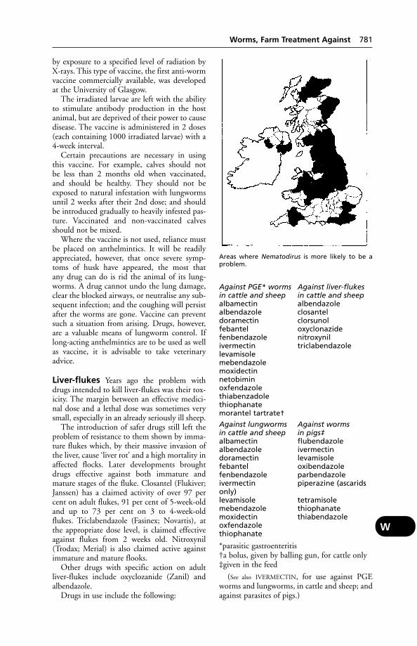
by exposure to a specified level of radiation byX-rays. This type of vaccine, the first anti-wormvaccine commercially available, was developedat the University of Glasgow.
The irradiated larvae are left with the abilityto stimulate antibody production in the hostanimal, but are deprived of their power to causedisease. The vaccine is administered in 2 doses(each containing 1000 irradiated larvae) with a4-week interval.
Certain precautions are necessary in usingthis vaccine. For example, calves should not be less than 2 months old when vaccinated, and should be healthy. They should not beexposed to natural infestation with lungwormsuntil 2 weeks after their 2nd dose; and shouldbe introduced gradually to heavily infested pas-ture. Vaccinated and non-vaccinated calvesshould not be mixed.
Where the vaccine is not used, reliance mustbe placed on anthelmintics. It will be readilyappreciated, however, that once severe symp-toms of husk have appeared, the most that any drug can do is rid the animal of its lung-worms. A drug cannot undo the lung damage,clear the blocked airways, or neutralise any sub-sequent infection; and the coughing will persistafter the worms are gone. Vaccine can preventsuch a situation from arising. Drugs, however,are a valuable means of lungworm control. Iflong-acting anthelmintics are to be used as wellas vaccine, it is advisable to take veterinaryadvice.
Liver-flukes Years ago the problem withdrugs intended to kill liver-flukes was their tox-icity. The margin between an effective medici-nal dose and a lethal dose was sometimes verysmall, especially in an already seriously ill sheep.
The introduction of safer drugs still left theproblem of resistance to them shown by imma-ture flukes which, by their massive invasion ofthe liver, cause ‘liver rot’ and a high mortality inaffected flocks. Later developments broughtdrugs effective against both immature andmature stages of the fluke. Closantel (Flukiver;Janssen) has a claimed activity of over 97 percent on adult flukes, 91 per cent of 5-week-oldand up to 73 per cent on 3 to 4-week-oldflukes. Triclabendazole (Fasinex; Novartis), atthe appropriate dose level, is claimed effectiveagainst flukes from 2 weeks old. Nitroxynil(Trodax; Merial) is also claimed active againstimmature and mature flooks.
Other drugs with specific action on adultliver-flukes include oxyclozanide (Zanil) andalbendazole.
Drugs in use include the following:
Against PGE* worms Against liver-flukesin cattle and sheep in cattle and sheepalbamectin albendazolealbendazole closantel doramectin clorsunolfebantel oxyclonazidefenbendazole nitroxynil ivermectin triclabendazolelevamisolemebendazolemoxidectinnetobiminoxfendazolethiabenzadolethiophanatemorantel tartrate†
Against lungworms Against wormsin cattle and sheep in pigs‡albamectin flubendazolealbendazole ivermectindoramectin levamisolefebantel oxibendazolefenbendazole parbendazoleivermectin piperazine (ascaridsonly)levamisole tetramisolemebendazole thiophanatemoxidectin thiabendazoleoxfendazolethiophanate
*parasitic gastroenteritis†a bolus, given by balling gun, for cattle only‡given in the feed
(See also IVERMECTIN, for use against PGEworms and lungworms, in cattle and sheep; andagainst parasites of pigs.)
Areas where Nematodirus is more likely to be aproblem.
Worms, Farm Treatment Against 781
W
Dual-purpose and multi-purpose anthel-mintics are available.
Parasitic Gastroenteritis For control of theparasites causing gastroenteritis, it is advisedthat calves should be dosed once with an effi-cient anthelmintic in mid-July and moved topasture which has not been grazed that seasonby other cattle. The 1st pasture may then begrazed by animals not susceptible to parasiticgastroenteritis, such as adults.
The 2nd essential dose is in the autumnwhen cattle come in from grass. At housing, the wormer selected should be active againstinhibited larvae, as well as maturing larvae.
Ostertagia worms, which are of considerableeconomic importance, are peculiar in that whilemost infective larvae living in the abomasummoult twice to become adults, some – especial-ly perhaps those ingested by the calf during late summer and autumn – moult only onceand remain as 4th-stage larvae in a dormantstate. The larvae are resistant to manyanthelmintics, but fenbendazole and albenda-zole are often effective. Later, they develop intoadults causing a winter outbreak of gastroen-teritis, with scouring and other digestive distur-bance. Accordingly, it is usually recommendedthat calves be dosed in September and moved to‘clean’ land.
Ivermectin (Ivomec) is effective againstimmature and even inhibited Ostertagia larvae;it can be given to both beef and dairy cattle, butnot within 21 days of slaughter, or to dairycows in milk, or within 28 days prior to calving.Three doses a year – in spring, summer, andautumn – are recommended to make the bestuse of this multi-purpose anthelmintic.
Ivermectin has no action against flukes ortapeworms, but is highly effective against all theimportant roundworms, both adult and larvalforms.
Nematodirus in Lambs Lambs 4 to 6weeks old and upwards may become severely illas a result of infestation with Nematodirusworms. A well-recognised condition, it mayshow itself with dramatic suddenness in low-land flocks in spring, but – depending on locality and weather – the main period of incidence is probably the end of May until the2nd week in July.
The worms, each about 1.70 cm (2⁄3 in) long,cause unthriftiness and poor liveweight gains;they may also cause a high mortality following4 to 5 days’ scouring resulting in a lethal loss ofbody fluids, i.e. dehydration. Where recovery
does take place, it is usually long-drawn-outand the animal may remain stunted.
These worms differ from others infesting thestomach/intestine of sheep in Britain in thattheir life-cycle takes about 12 months to com-plete, and the eggs require exposure to cold toinitiate development; as infective larvae, theyremain viable for only a few weeks. This fortu-nate fact offers an obvious method of control –the well-known rule, ‘Never put lambs on thesame pasture 2 years running.’ On farms whereit is impracticable to observe this rule, dosing 3 or 4 times with an appropriate anthelmintic isadvisable. (See also ‘CLEAN’ PASTURE.)
Worms in Pigs Anthelmintics, complement-ed by good hygiene, play an essential part inmaintaining health in the intensively managedpig unit. Pigs kept outdoors are vulnerable toworm infections.
Infestation by parasitic worms is best regard-ed as a herd problem, and the fact thatanthelmintics are available in a palatable pelletform, or as a powder to mix in a meal, is a greathelp to the pig farmer. These are usually broad-spectrum drugs which will act against most ofthe species of worm normally found in the pig.Where a particular species has led to a severehealth problem, an anthelmintic most effectiveagainst that species can be selected.
The main wormers used in pigs are febantel,fenbendazole, flubendazole, ivermectin, mox-idectin and thiophanate.
Steering a middle course The farmershould, where necessary, seek veterinary adviceon the spot, and aim to steer a middle coursebetween inconveniently frequent dosing andhigh drug bills on the one hand, and toleratingpoor liveweight gains, unthriftiness and evenseveral deaths among his stock on the otherhand. Internal parasites steal feed intended fortheir hosts, and often cause physical inury –sometimes very severe – as well.
WoundsA wound may be defined as a breach of the con-tinuity of the tissues of the body produced byviolence. (See also under BRUISES.)
Varieties Wounds may be classified accordingto the nature of the effect produced, viz.incised, punctured, lacerated, and contused,and whether they are infected or contaminated.
Incised wounds are usually inflicted by somesharp instrument which leaves a clean cut; thetissues are simply divided without extensivedamage to the surrounding parts. Bleeding from
782 Wounds
W
an incised wound is apt to be very profuse for atime, but it soon stops and is easily controlled.
Punctured wounds or stabs are inflicted witha pointed instrument or another animal’sincisor or canine teeth. (A dose of tetanus anti-toxin or toxoid is indicated in puncturedwounds, especially in the horse, cow, and dog.)
Lacerated wounds are those in which greattearing takes place. They are usually very painfulfor a few days, and suppurate before they heal. They are usually followed by disfiguringscars when extensive.
Contused wounds are those accompanied bymuch bruising of the surrounding tissues, as inthe case of blows from heavy sticks, kicks fromshod horses, and from road accidents. There isusually little bleeding from the wound itself,but blood may be extravasated into the tissues.(See HAEMATOMA.)
Any one of these forms of wounds maybecome infected with pus-forming organisms,and develop into a suppurating, septic wound.(For other information, see under ACCIDENTS;FRACTURES; etc.)
First-aid treatment With a serious woundinvolving much haemorrhage, the first consid-eration must obviously be to stop the bleeding.(See BLEEDING – Bleeding, external; first aid for.)
With all wounds it is advisable to clip awaythe hair – preferably using blunt-pointed surgi-cal scissors – first inserting a piece of cotton-wool moistened in antiseptic into the cavity ofthe wound (if large enough), so that the cut hairdoes not fall into the wound.
If the hair is not cut away, it is apt to becomematted by blood or oozing serum, and thewound may later be found to be suppuratinginstead of healing. (What may look to the ani-mal-owner like a normal healthy scab may be,in fact, a crust of blood, matted hair and dirt.)
The surface of the wound may be cleaned bygentle application of a piece of cotton-woolsoaked in warm antiseptic such as dilutedDettol or Cetrimide, etc., or KY Jelly.
The wound may be covered in order to preventcontamination and infection by flies – in the caseof farmyard animals – or to prevent excessive lick-ing by dogs and cats. Before covering, a dry anti-septic dressing of sulfanilamide may be applied.
The covering of a wound cleaned anddressed as described should be removed daily sothat the progress of healing can be observed,and cleaning repeated if necessary. An open,granulating wound should have a clean, pinkappearance.
Large, gaping wounds may require suturing,which should be done by a veterinary surgeon,
who should also always be consulted concern-ing the treatment of punctured and laceratedwounds.
Other points that should be noted are: (1)that stitches should be removed if they com-mence to suppurate, and in any case after beingin position for a week, after which they serve nouseful purpose; (2) that if pus burrows underthe skin surrounding a wound, it must be givendrainage by incision below the level of the mostdependent burrowing or by drainage tubes; (3)that if the granulation tissue (i.e. ‘proud flesh’)rises to a higher level than the skin around, itmay need professional treatment; and (4) thatin cases of injury to parts such as the eyes, nos-trils, lips, genital organs, feet, etc., it is essentialto seek skilled advice rather than to persist inrule-of-thumb methods which often lead theenthusiastic amateur astray, and cause the ani-mal unnecessary distress. (See also FRACTURES;GRANULATIONS; ULCER; ANTISEPTICS; ANTIBI-
OTICS; SULFONAMIDES; and under ACCIDENTS,INJURIES, and CORTISONE.)
Wounds, How They HealThe blood forms clots; these consist of minutethreads of fibrin, in which are enmeshed redblood cells and white blood cells. The threadsof fibrin bridge the gap between the cut surfacesof the wound, at its base, forming a matrix,hardening into a scab under which tissue repaircan take place. (See CLOTTING OF BLOOD.)
From the neighbouring blood capillariescome white cells (especially neutrophils) whichengulf dirt, bacteria, etc. (See PHAGOCY-
TOSIS.) Monocytes arrive later, especially if thewound has become infected. They becomemacrophages which remove any disintegratedneutrophils and also bacteria. Meanwhile, thecells of the epidermis begin to multiply in orderto restore the skin covering. (See LYMPHOCYTES.)
Healing of wounds may be delayed if the animal is being treated with corticosteroids.
Wry-Neck (Torticollis)Wry-neck (torticollis), which occurs in foals,sheep and poultry particularly, is a lateral deviation of the head and neck to the right orleft side of the body, usually so marked as tohinder or prevent foaling. The bones of theskull and neck are frequently distorted, and the ligaments, tendons, and muscles on theinside of the curve are shorter than those on the outside.
The condition may also be encountered incattle.
When seen in rabbits, the cause may be middle ear infection.
Wry-Neck (Torticollis) 783
W
X-Rays are high-energy radiation capable ofpassing through considerable thicknesses ofmany substances which are opaque to ordinarylight, without undergoing material absorption,but other substances, even in very small thick-nesses, are able to absorb the great majority ofthe rays: thus, flesh is very transparent; healthybone is fairly opaque. Widely used for diagnos-tic imaging of internal body structures and (in human medicine) for radiotherapy.
Precautions Guard screens of lead glass, rub-ber impregnated with lead, or sheet lead, areused to protect the operators of radiographicapparatus, and precautions are necessary toshield the testes and ovaries of young personsand animals from the sterilising effects of therays.
Detailed precautions are as follows:1. Persons under 16 years must not take part
in radiological procedures.2. Fluoroscopy (imaging) or radiotherapy
should not be carried out except under expertradiological guidance. Hand-held fluoroscopesmust not be used in any circumstances.
3. Personnel radiation monitoring devices,such as film badges, must be worn by all per-sons who take part routinely in radiologicalprocedures.
4. The animal should, if possible, be anaes-thetised or tranquillised for radiography, and allpersons should withdraw as far as practicablefrom the useful beam.
5. If it should be necessary to hold the ani-mal for radiography, lead-protective gloves andaprons must be worn. Whenever possible, hold-ing should be done by the owners, unless theyare under 16 years or pregnant.
6. Persons should not expose any part oftheir bodies to the useful beam even whenwearing protective clothing.
7. The useful beam should be restricted tothe area being examined by means of a beam-limiting device.
Notes on protection against radiation willalso be found in the British VeterinaryAssociation’s guide to the Health and Safety atWork Act.
Regulations In the UK, the IonisingRegulations 1985 require veterinary surgeons
using X-ray equipment to notify their localHealth and Safety Executive. Many veterinarypractices now employ a radiation protectionadviser to ensure compliance with the require-ments of the regulations.
Radiography The production of a radi-ograph of the internal structure of a small ani-mal is a comparatively simple matter once thedifficulty of control is overcome. The animal isarranged upon the table in such a position as toallow the rays to pass down through the partand become registered upon a sensitive plateplaced flat upon the table immediately below.The animal may lie upon its back, on one or theother side, or on its chest and abdomen withthe legs pulled out from under it. To maintainthis position it is always advisable to administeran anaesthetic. The discharge tube is bestarranged immediately above the animal in sucha position as to allow the rays to fall perpendic-ularly down through the body on to the plate.(For screening, the tube must be below thetable, and the screen held or supported abovethe animal.) The period of exposure to the pas-sage of the rays varies according to the tissues,to the type and power of the equipment, to thedistance of the tube from the plate, and towhether or not an intensifying screen is used.
There are many conditions in which theactual extent of injury or disease can be accu-rately discovered by the use of X-rays, but themost important are diseases and injuries ofbones. Fractures of the limb-bones are wellshown, and their extent is better realised than ispossible by palpation. Exostoses (overgrowthsof bone) can also be clearly indicated, whiletumour formation (usually sarcomatous) showsas a thinning and enlargement of the bone tissue. Where only one limb is affected it isadvisable to arrange the animal so as to includea picture of the normal limb for comparison.Foreign bodies – especially needles, pins, nails,and other metallic substances – which havebeen swallowed are best shown by a profile viewof the abdomen. Pieces of game bones (whichare specially dense and show up well) can alsobe seen in the stomach or intestines, and arevery often surrounded by gas, which, in thenegative, appears as a dark shadow – the boneitself appearing light. Internal tumours can veryoften be diagnosed. They appear as more or lessdiscrete pale areas in positions where a radi-ograph from a normal animal is denser underthe same conditions of exposure, etc. Certaintumours can be made to show up well by givingthe animal medicinal doses of a lead salt for afew days before taking the plate. Some of the
X

lead becomes deposited in the tumour andintensifies the contrast. Where some displace-ment, stricture, or dilatation of the stomach orof part of the intestinal canal is suspected, theanimal is given a feed or a draught containing
an emulsion of bismuth or barium carbonate,or some other harmless metallic salt, or hassome of the same material injected into the rec-tum. After waiting until the salt has becomesuitably distributed, a radiograph of the
Xanthosis 785
X
A teaspoon in the stomach of a cocker spaniel. The spoon was swallowed while the animal was beinggiven cod-liver oil. (Reproduced by courtesy of Mr S. W. Douglas, University of Cambridge School ofVeterinary Medicine.)
Radiography reveals that a painful swelling on the leg of a St Bernard is due to an osteosarcoma involvingthe radius. (Reproduced by courtesy of Mr S. W. Douglas, University of Cambridge School of VeterinaryMedicine.)
X
786 Xanthosis
abdomen is taken, and the outlines of thoseorgans to which the salt has been carried byperistalsis, can be made out as pale areas in thenegatives.
Other conditions in which X-rays are usefulare as follows: stones in the kidney, urinary orgall-bladder; dilatation of the heart; solidifica-tion of a portion of a lung; and pleurisy.
Portable X-ray apparatus is available for usein, for example, examining the lower limbs of ahorse at the stable.
Computed tomography A sophisticatedand effective method of visualising the interiorof the body, this has revolutionised radiography.A focused electronic imaging scanner is used tobuild up, by a series of consecutive exposures, apicture of an organ or specific area. The result-ing image is enhanced by computer analysisand viewed on a visual display unit (VDU)which shows a clear picture without the thesuperimposition of other body tissues whichsurround the targeted area. In the apparatus,whose British development was by GodfreyHounsfield FRS, crystal detectors are used inplace of the film in a normal X-ray system.
Radiotherapy X-ray therapy has beenapplied to a limited extent in the treatment ofcertain tumours in the dog.
XanthosisXanthosis is a yellowish-brown pigmentation ofmeat, generally affecting the heart and thetongue. It gives the meat an objectionablecolour, but is quite harmless.
XenophthalmiaInflammation of the eye caused by a foreignbody.
XerophthalmiaA disease of the eye associated with a vitamin Adeficiency. There is thickening and cloudinessof the conjunctiva and cornea; blindness mayresult.
XylazineA sedative used to render animals easier to han-dle, it is widely used in dogs, cats, horses, farmlivestock, and zoo animals. It is also used forpre-anaesthetic medication and for generalanaesthesia in combination with ketamine.
Xylazine increases blood glucose levels andurine output. Side-effects may include brady-cardia, slower breathing, and lowered bloodpressure. In cattle tolazoline has been used as axylazine antagonist.
Yarded CattleBefore yarding cattle in the autumn, it is wise tomake a gradual change from sugar-poorautumn pasture to things like roots; otherwisedigestive upsets are likely to occur.
Similarly, in spring it is a mistake to turncalves straight out on to grass. This means asudden change from protein-poor food to the rich protein of the early bite, and the resulting effect upon the rumen will set themback. It is best to get them out before there is much grass for a few hours each day; let them have hay and shelter at night to protectthem from sudden changes of weather.Hypomagnesaemia, too, is far less likely underthese circumstances. (See also HOUSING OF
ANIMALS.)Boss cows can be a nuisance in yards, but the
provision of yokes for feeding overcomes themain diffculty.
When self-feeding of silage is practised, pre-cautions are necessary in order to prevent foottroubles. (See SILAGE.)
Yarded animals fed on cereals, sugarbeetpulp, straw, and hay – but with little or nogreenstuff – may suffer from xerophthalmia andgo blind as a result of a vitamin A deficiency.
YawningYawning is an important sign of KIMBERLEY
HORSE DISEASE; it may also be seen in cases ofLABURNUM POISONING and NARCOLEPSY.
YeastYeast is a valuable source of vitamin B, butshould not be fed in excessive amounts to pigsor it may give rise to rickets unless adequate vit-amin D is simultaneously available. Yeast hasproved successful in the treatment of tropicalulcers in humans, and success has been report-ed in a limited number of cases in horses in thetropics. The human patients were mostly thosewhose diet was deficient in vitamin B, a defi-ciency further increased by sweating. The yeastwas applied directly to the ulcer, and a smallquantity given internally also.
YeastsYeasts sometimes cause enteritis, and are impor-tant in some cases of refractory otitis in the dog.(See FUNGAL DISEASES.)
Yellow Fat Disease of Cats(see STEATITIS)
Yellow FeverA viral disease affecting man and other verte-brates, principally monkeys, in large areas oftropical America and Africa. There are 2 knowncycles of transmission, the urban and junglecycles. In the urban cycle, man is the reservoirand Aedes aegypti probably the only vector. Thiscycle from man to A. aegypti to man is now virtually unknown in the Americas owing to efforts to eradicate the vector, but it is stillcommon in Africa.
The jungle cycle has a primate reservoirmaintained by various mosquitoes. Movementof virus from the monkey-mosquito-monkeycycle into man is accidental, and is the result of human penetration into jungle where thedisease is endemic.
The causative organism is classified as a flavivirus.
YeltA female pig intended for breeding, up to thetime that she has her 1st litter.
YersiniosisInfection with Yersinia pseudotuberculosis orwith Y. enterocolitica.
Up to 1960, states WHO, only the formerorganism was regularly isolated in man and animals in Europe; but since then most of theisolations have been of Y. enterocolitica.
‘Pseudotuberculosls’ in the early 1990swas still occasionally found in rodents andbirds, especially in France and the UK, and is azoonosis. People may become infected throughpets such as guinea pigs, hamsters, and cats, allof which may have a subclinical infection onlybut excrete Y. pseudotuberculosis.
An investigation in Invermay, New Zealand,resulted in Y. pseudotuberculosis being isolatedfrom 675 apparently healthy small mammalsand birds. In descending order of prevalencewere feral cats (27.8 per cent), Norway rats (8.6 per cent), mice, hares, rabbits, ducks, sparrows, seagulls and starlings.
In New Zealand, yersiniosis has also emergedas a serious disease of farmed red deer. Itappears to be triggered off by stress, and mostcases occur during the winter.
Cats (which are liable to become infected bytheir prey) may also show clinical symptoms:loss of appetite, vomiting and diarrhoea. Alsoloss of weight.
Y

Pheasants Yersiniosis is an important causeof death of these birds in the UK.
Yersinia enterocolitica infection in Europewas first found in hares, in outbreaks of diseaseon chinchilla farms, in monkeys in zoos, and inguinea pigs. There may be enteritis and otherlesions, but symptomless carriers have beenfound among all the farmyard mammals andbirds.
Occasionally Y. enterocolitica has been isolat-ed from cases of mastitis in cows, endocarditisin bulls, and septicaemia in pigs. In cattle, theantibody produced may be difficult to differen-tiate from that produced by Brucella abortus.
Camels, foxes, and fleas may also carry theorganism.
Public health Yersinia enterocolitica infectionis not regarded as a genuine zoonosis by WHO.Person-to-person infection occurs, and alsoinfection from soil-contaminated vegetables.The human illness is characterised by enteritis,and is a cause of diarrhoea, although less impor-tant than salmonella and campylobacter. Ileitismay be accompanied by acute pain, suggestive ofappendicitis. A mesenteric adenitis is also seen,and sometimes polyarthritis, deep abscesses, eyelesions, and occasionally septicaemia.
In the UK in 1984, 250 cases were reported.Outbreaks in North America have been linkedto raw milk. (For Y. pestis see BUBONIC PLAGUE,which can occur in cats and dogs in subclinicalform.)
Yew PoisoningAll varieties of the British yew trees are poiso-nous, but owing to its more frequent cultiva-tion, the common yew (Taxus baccata) is mostoften responsible for outbreaks of poisoningamong animals. The Irish yew (T. baccata var.
fastigiata) and the yellow yew appear to containless of the poisonous alkaloid, which is calledtaxine. The bark, leaves and seeds all contain it.The older dark leaves are more dangerous thanthe fresh green young shoots, which cattle havebeen known to eat in small amounts withoutharm. Cases of poisoning have been notedamong horses, donkeys, mules, cattle, sheep,goats, pigs, deer, rabbits, and even pheasants,but the majority of cases occur in young storecattle and in dairy cows which have access tothe shrubberies, graveyards, etc., where yewtrees are most common.
Signs In many cases cattle drop dead withoutshowing any preliminary symptoms at all. Theymay fall while cudding almost as suddenly as ifshot. In other cases where less has been eaten,excitement and paresis may be seen.
Treatment Antidotes are as for alkaloids. Iftime allows, rumenotomy may be carried out.
Yolk Sac Infection(see OMPHALITIS OF BIRDS)
Yorkshire TerrierA long-haired, black-and-tan coloured toy dog.The breed is prone to tracheal collapse causedby a cartilage defect that may or may not beinherited. Patellar luxation is inherited as arecessive trait.
Yorkshire BoardingVertically arranged boards with a gap betweeneach, used for partial cladding of a livestockbuilding. It is a very useful means of improvingventilation and avoiding condensation, therebyreducing the risk or incidence of bronchitis andpneumonia in housed livestock.
788 Yew Poisoning
Y
ZearalenoneAn oestrogenic toxin from the fungus Fusariumgraminearum of standing corn. The toxin hascaused abortion in sows, and possibly a splaylegcondition in piglets.
ZebuBos indicus, the cattle of India, East and WestAfrica, and Southeast Asia. The American nameis Brahman; in South Africa, the Afrikaner.
Zero-GrazingTaking cut fodder to yarded cattle, or to cattlein exercise paddocks. Zero-grazing has a placeon heavy land, with high stocking rates andlarge herds. It obviates poaching and the spoil-ing of grass, and a given acreage zero-grazed canprovide more grass than if grazed. It means,however, cutting grass every day, and mechani-cal failures can upset the system. It is not yetconsidered economic for sheep.
Zinc (Zn)Zinc (Zn) is a trace element, and a deficiencyhas occurred in pigs. (See PARAKERATOSIS.) Azinc supplement to prevent or correct this con-dition must be used with care, as 1000 parts permillion can cause poisoning. It seems that ahigh calcium intake by pigs aggravates a zincdeficiency.
A zinc deficiency may also occur in dogs,especially in those fed largely on flaked maize or‘loose cereal-based diets’. Signs include a predis-position to skin infections, a poor coat,localised alopecia, and hardening of the skin inplaces. Response to a zinc supplement is usual-ly quick. (See SHEEPDOGS.)
A zinc supplement has been used to protectsheep against facial eczema due to ingestion ofthe mycotoxin sporidesmin.
External uses Zinc oxide is an ingredient of ointments; the carbonate an ingredient ofcalamine lotion used for moist eczema, etc. Thesulphate in weak solution has been used inwound treatment and in eye lotions; the chloride – a caustic – to repress granulations.
Zinc BacitracinAn antbiotic formerly used as a feed additive toimprove growth rate in most farm animals andegg production in poultry. (See ADDITIVES.)
Zinc PoisoningChronic zinc poisoning has been reported in adairy herd as a result of contaminated drinkingwater – caused by interaction between copperpipes and newly galvanised tanks. The mainsymptom was chronic constipation throughoutthe herd, and a diminished yield from the cowsin milk.
Fatal zinc poisoning has occurred in dairycattle fed on dairy nuts to which zinc oxide hasbeen added instead of magnesium oxide. Thefirst death occurred after 3 weeks.
Zinc-responsive skin disease The mostcommon cause of this is the feeding of soya orcereal-based diets – with little or no meat,which is rich in zinc. Some dogs may have aninherent defect which limits zinc absorption.
Signs A dull, harsh coat; sometimes withwhitish crusts on the skin.
Zondek-Ascheim Test(see PREGNANCY DIAGNOSIS)
Zoo Licensing Act 1981The Zoo Licensing Act 1981 is intended to pro-mote animal welfare and public safety at zoos. Itcovers any collection of wild animals (includingmammals, birds, reptiles, fish, and insects) inBritain to which the public has access for morethan 7 days in any 12-month period; butexempts pet shops and circuses, as these are cov-ered by the Pet Animals Act 1951 and thePerforming Animals (Registration) Act 1952.
Of 150 zoos inspected following the passingof the Act, only 5 were refused a licence; and inthose cases it was public-safety considerationsrather than the quality of animal care whichbrought about the refusal.
ZoonosesDiseases communicable between animals andman. Information about them will be foundunder the following headings: ARIZONA INFEC-
TION; BABESIA – Babesiosis; ANTHRAX; B VIRUS
(from monkeys); BRUCELLOSIS; CAT-SCRATCH
FEVER; CHAGAS DISEASE; EQUINE ENCEPHALITIS;EQUINE INFECTIOUS ANAEMIA; FOOT-AND-
MOUTH DISEASE (very rare in human beings);GLANDERS; HYDATID DISEASE; LEPTOSPIROSIS;LISTERIOSIS; LIVER-FLUKES; LOUPING-ILL; LYME
DISEASE; LYMPHOCYTIC CHORIOMENINGITIS
(from mice); NEWCASTLE DISEASE; ORNITHOSIS;ORF; PASTEURELLOSIS; Q FEVER; RABIES; RATBITE
FEVER; RIFT VALLEY FEVER; RINGWORM; ROCKY
MOUNTAIN FEVER; RUSSIAN SPRING-SUMMER
VIRUS; SALMONELLOSIS; SCABIES; SCHISTOSOMI-
Z
ASIS; TAPEWORMS; TICK-BITE FEVER; TICK PARAL-
YSIS; TOXOCARA; TOXOPLASMOSIS; TRICHINOSIS;TUBERCULOSIS; TULARAEMIA; VESICULAR STOM-
ATITIS; MARBURG DISEASE; WESSELBRON DIS-
EASE; YERSINIOSIS; YELLOW FEVER; SWINE VESIC-
ULAR DISEASE; PORCINE STREPTOCOCCAL
MENINGITIS; ROTAVIRUS; LASSA FEVER; BOVINE
ENCEPHALOMYELITIS; LEISHMANIASIS; BUBONIC
PLAGUE; ENCEPHALOMYOCARDITIS.)It should be added that typhus and plague
may be transmitted, by flea-bite, from rats; and,in jungle areas, yellow fever, by mosquito-bite, from monkeys. (See also under RODENTS;MONKEYS; INFLUENZA.)
Among skin diseases, the parasite of follicu-lar mange may occasionally infest the humaneyelid. Among eye infections, INFECTIOUS
BOVINE KERATOCONJUNCTIVITIS should bementioned. Human enteritis has followed con-tact with sheep affected with campylobacterabortion.
(See also BIRD-FANCIER’S LUNG; MELIOIDOSIS;CAMPYLOBACTER INFECTIONS; CHLAMYDIA;PSITTACOSIS; BOUTONNEUSE FEVER; LEISHMA-
NIA; HANTAVIRUS; TICK-BORNE ENCEPHALITIS;EHRLICHIA CANIS; ABORTION, ENZOOTIC.)
Zoonoses in UK VeterinariansA questionnaire was distributed to 1717 mem-bers of veterinary and support staff of theMinistry of Agriculture and the Institute forResearch on Animal Diseases; 1625 (95 percent) responded, comprising 563 veterinary surgeons, 690 scientific staff and 372 technicalsupport staff. A total of 1057 (61.5 per cent)had apparently not suffered any zoonotic infec-tion. Animal ringworm was the commonestreported zoonosis. The incidences of ringworm,brucellosis and Newcastle disease were higher in the veterinary and support staff than in the
laboratory workers. In contrast, ornithosis, salmonellosis and Q fever occurred at least asoften in the laboratory staff. Fourteen peopledeveloped tuberculosis during their employ-ment, although only 1 was caused byMycobacterium bovis. The veterinarians report-ed 441 injuries that resulted from accidents atwork; 397 (71 per cent) of these involved ani-mal-handling. The comparable figures for labo-ratory workers and technical staff were 329 and103 (15 per cent) and 198 and 179 (42 percent) respectively.
Zoonoses Orders 1988 & 1989These include measures intended to reduce the risk to humans of salmonella and brucellainfections of animal origin. The 1989 Orderrecognises bovine spongiform encephalopathyas a zoonotic disease.
ZootechnyAnimal management.
ZygomaZygoma is the bridge of bone which runs fromnear the base of the ear to the lower posteriorpart of the eye-socket. It protects the side of the bony orbit, forms part of the support of theoutside of the joint of the lower jaw with therest of the head, and serves as a base of attach-ment for part of the strong masseter musclewhich closes the mouth and is important in thechewing of the food. The zygomatic arch(another name for the zygoma) is formed byprojections from the temporal, zygomatic, andmaxillary bones.
ZygoteThe body that results from the fertilisation ofan egg cell by a sperm.
790 Zoonoses in UK Veterinarians
Z

Animal Health Trust PO Box 5,Newmarket, Suffolk CB8 8JH. 01638 750659
Association of Pet BehaviourCounsellors PO Box 46, Worcester WR89YS. 01386 751151
Association of Pet Dog TrainersPeacock’s Farm, Northchapel, Petworth, WestSussex GU28 9JB
Blue Cross 1 Hugh Street, London SW1V1QQ. 020 7834 1128
British Cattle Movement ServiceCurwen Road, Workington, Cumbria CA142DD. 0845 0501234
British Cattle Veterinary AssociationThe Green, Frampton-on-Severn,Gloucestershire GL2 7ER. 01452 740816
British Equine Veterinary Association5 Finlay Street, London SW6 6HE. 020 76106080
British Horse Society British EquestrianCentre, Stoneleigh Deer Park, Kenilworth,Warwickshire CV8 2LR. 08701 220224
British Rabbit Council Purefoy House, 7Kirkgate, Newark, Nottingham NG24 1AD.01636 676042
British Veterinary Association 7Mansfield Street, London W1G 9NQ. 0207636 3541
Companion Animal Welfare Council43 Brook Field Lane, Harlow, Essex CM187AU
DARD (Department of Agricultureand Rural Development) DundonaldHouse, Upper Newtownards Road, Belfast BT43SB. 028 9052 4580
DEFRA (Department of theEnvironment, Food and Rural Affairs)1A Page Street, London SW1P 4PQ
Donkey Sanctuary Sidmouth, DevonEX10 0NU. 01395 578222
Farm Animal Welfare Council DEFRA,1A Page Street, London SW1P 4PQ
Farriers Registration Council SeftonHouse, Adam Court, Newark Road,Peterborough PE1 5PP. 01733 319911
Kennel Club 1 Clarges Street, London W1Y8AB. 0870 6066750
National Association of Farriers,Blacksmiths & Agricultural EngineersThe Forge, Avenue B, 10th Street, NAC,Stoneleigh, Warwickshire CV8 2LQ. 024 76696595
National Canine Defence League 17Wakely Street, London EC1V 7LT. 020 78370006
National Farmers Union AgricultureHouse, 164 Shaftsbury Avenue, London WC2
National Office of Animal Health 3Crossfield Chambers, Gladbeck Way, Enfield,Middlesex EN2 7HF
National Poisons Information ServiceAvonley Road, London SE14 5ER. 020 76359195
NOAH (National Association ofAnimal Health) 3 Crossfield Chambers,Gladbeck Way, Enfield, Middlesex EN2 7FH.020 8367 3131
PDSA (People’s Dispensary for SickAnimals) Whitechapel Way, Priorslee,Telford, Shropshire TF2 9PQ. 01952 204713
Pets Travel Scheme (PETS) Helpline:0870 41171; fax 020 7904 6834; [email protected]/animalh/quarantine
Royal College of VeterinarySurgeons Belgravia House, 62–64Horseferry Road, London SW1P 2AF. 0207222 2001
RSPCA (Royal Society for thePrevention of Cruelty to Animals)Causeway, Horsham, West Sussex RH12 1HG.01403 264181
UFAW (Universities Federation forAnimal Welfare) The Old School,Brewhouse Hill, Wheathampstead,Hertfordshire AL4 8AN. 01582 9303611
USEFUL ADDRESSES