VETERINARY NURSING OF EXOTIC PETS
320
VETERINARY NURSING OF EXOTIC PETS Simon Girling BVMS (Hons) DZooMed CBiol MIBiol MRCVS
-
Upload
independent -
Category
Documents
-
view
1 -
download
0
Transcript of VETERINARY NURSING OF EXOTIC PETS

VETERINARY NURSING OFEXOTIC PETS
Simon GirlingBVMS (Hons) DZooMed CBiol MIBiol MRCVS
© 2003 by Blackwell Publishing LtdEditorial Offices:9600 Garsington Road, Oxford OX4 2DQ, UK
Tel: +44 (0)1865 776868108 Cowley Road, Oxford OX4 1JF, UK
Tel: +44 (0)1865 791100Blackwell Publishing USA, 350 Main Street, Malden, MA02148-5018, USA
Tel: +1 781 388 8250Iowa State Press, a Blackwell Publishing Company, 2121State Avenue, Ames, Iowa 50014-8300, USA
Tel: +1 515 292 0140Blackwell Munksgaard, Nørre Søgade 35, PO Box 2148,Copenhagen, DK-1016, Denmark
Tel: +45 77 33 33 33Blackwell Publishing Asia, 550 Swanston Street, CarltonSouth, Victoria 3053, Australia
Tel: +61 (0)3 9347 0300Blackwell Verlag, Kurfürstendamm 57, 10707 Berlin,Germany
Tel: +49 (0)30 32 79 060Blackwell Publishing, 10 rue Casimir Delavigne, 75006Paris, France
Tel: +33 1 53 10 33 10
The right of the Author to be identified as the Author ofthis Work has been asserted in accordance with theCopyright, Designs and Patents Act 1988.
All rights reserved. No part of this publication may bereproduced, stored in a retrieval system, or transmitted, inany form or by any means, electronic, mechanical,photocopying, recording or otherwise, except as permittedby the UK Copyright, Designs and Patents Act 1988,without the prior permission of the publisher.
First published 2003
A catalogue record for this title isavailable from the British Library
ISBN 1-4051-0747-2
Library of CongressCataloging-in-Publication DataGirling, Simon.
Veterinary nursing of exotic pets/Simon Girling.p. cm.
Includes bibliographical references and index.ISBN 1-40510-747-2 (softcover)1. Exotic animals–Diseases. 2. Wildlife diseases.3. Pet medicine. 4. Veterinary nursing. I. Title.
SF997.5.E95 G57 2003636.089¢073–dc21 2002038565
Set in 9.5 on 12 pt Timesby SNP Best-set Typesetter Ltd., Hong KongPrinted and bound in Great Britain by Ashford Colour Press, Gosport
For further information onBlackwell Publishing, visit our website:www.blackwellpublishing.com

Preface v
AVIAN SPECIES 1
1 Basic avian anatomy and physiology 32 Avian housing and husbandry 253 Avian handling and chemical restraint 344 Avian nutrition 475 Common avian diseases 646 An overview of avian therapeutics 87
REPTILES AND AMPHIBIANS 103
7 Basic reptile and amphibian anatomy and physiology 1058 Reptile and amphibian housing, husbandry and rearing 1279 Reptile and amphibian handling and chemical restraint 133
10 Reptile and amphibian nutrition 14811 Common reptile and amphibian diseases 16112 An overview of reptile and amphibian therapeutics 175
SMALL MAMMALS 193
13 Basic small mammal anatomy and physiology 19514 Small mammal housing, husbandry and rearing 22315 Small mammal handling and chemical restraint 23316 Small mammal nutrition 24617 Common diseases of small mammals 25718 An overview of small mammal therapeutics 285
Appendix 1 Legislation affecting exotic pet species in the UK 305Appendix 2 Useful addresses 307Index 308
The colour plate section may be found following page 154.
Contents

Preface
Veterinary nurse and veterinary student trainingin exotic species has come a long way in the lastfour or five years. Previously often consigned tothe category of ‘alsorans’, exotic species areincreasingly seen in general veterinary practice, tothe point where the house rabbit has officiallybecome the UK’s third most commonly kept pet,after the cat and dog. Even more telling is the factthat numbers of cats and dogs in the UK are onthe decline, yet the number of small mammals,reptiles and birds kept by the public continues torise.
With this increase in these species kept ashousehold pets, improved training in their carehas thankfully started to become more impor-tant. Many veterinary schools and veterinarynurse training providers are devoting more timeto teaching the husbandry and medicine of
exotic species. Indeed, 2001 saw the start of thefirst course in Veterinary Nursing of ExoticSpecies, run through Edinburgh’s Telford Collegeand leading to a City and Guilds recognised qualification.
There is no turning the clock back. Exotic petspecies are here to stay. It is therefore our duty as veterinary surgeons and veterinary nurses toensure that we are up-to-date with the latest hus-bandry and medical details so that we may offeras good, if not better, levels of care as that pro-vided for more traditional domestic pets.
I hope that this book will help in that quest andmay be of use to veterinary nurse, technician andveterinary student alike.
Simon J. Girling

Plate 1.1 The African grey parrot is acommonly seen pet and makes one ofthe best talkers.
Plate 1.2 Many parrots such asthis umbrella cockatoo are strikingbirds.
Plate 1.3 Parrots beaks make impressive crushingtools such as this blue and gold macaw, Ara ararauna.
Plate 1.4 The normal lining of the ventriculus or gizzardis known as the koilin, and is often stained green with bilepigments. Note the cream coloured pancreas between theloop of descending and ascending duodenum.
Plate 2.1 Young Psittaciformes are altricial like mostavian species, and born with little or no feather covering.
Plate 3.1 Rhamphastids, such as this red-billedtoucan, have impressive beaks which need carefulhandling.

Plate 5.1 Viral diseases such as psittacine beakand feather disease in this African grey parrot maycause feather colouration changes (note multiplered feathers on the main body).
Plate 5.2 Crop burns may be caused by feeding of too hota rearing formula.
Plate 5.3 Ascites will show as ventral abdomendistension and may indicate liver disease. In thiscase rarely the skin has become jaundiced, a con-dition not commonly seen in avian species withliver failure.
Plate 7.1 Snakes should perform a whole body slough whenshedding their skin, such as that seen in this pine snake.
Plate 5.4 Lime green urates in raptorsmay indicate lead poisoning.
Plate 7.2 Ventral view of a bearded dragon,Pogona vitticeps, post mortem. The heart may beseen at the top of the picture, immediately belowwhich are the dark coloured lungs, and then thered coloured liver. Below this lies the stomach andintestines. The two dark masses reflected ventrallyare abdominal fat pads. Note the overall largeamounts of dark melanin pigment found in theinternal organs of many desert dwelling species.

Plate 7.3 Head of green iguana showing theparietal or third eye lying at the midline on thedorsal aspect of the head.
Plate 8.1 Large vivaria are needed for large species of snake suchas this Burmese python, which may reach 16 feet in length. Note barksubstrate of tank.
Plate 9.1 Restraint of a docile species suchas this female plumed basilisk. Note facialabscess.
Plate 9.2 While large snakes must be handled by more thanone person, even smaller species such as this corn snake arebest handled by two people. Note the control of the headbetween thumb and forefinger.
Plate 9.3 Correct position to lift a tortoise withboth hands either side of the shell between thefront and rear limbs.
Plate 9.4 Method of short-term restraint ofChelonia by placing the animal on apedestal so that it rests on the plastron.

Plate 11.1 Fungal granulomatous disease in the tail of a greeniguana.
Plate 9.5 Intubation of reptiles, such as this green iguana, allows the use of intermittentpositive pressure ventilation, which is being performed here by a mechanical ventilatorunit (Vetronic Services).
Plate 11.3 Redleg in an Argentinianhorned frog.
Plate 11.2 Dystocia is uncommon in snakes, but may cause prolapseof the oviduct as here.
Plate 11.4 Saprolegniasis in a fire-bellied toad.

Plate 12.1 Surgical placement of a pharyngostomy tube in ananorectic green iguana.
Plate 12.2 Surgical speying of a water dragon with post-ovulatorystasis.
Plate 13.1 Chinchillas have fine bonesand are prone to fractures, which areoften compound. If these become badlyinfected amputation may be necessary.
Plate 12.3 Surgical speying of a female green iguana with pre-ovulatory stasis. Note multiple yellow yolks of the ovaries.
Plate 15.1 Method of holding a rabbit for examination of theventrum.

Plate 17.1 Lop breeds of rabbit are more prone todental disease due to their foreshortened heads,which may lead to uneven molar wear.
Plate 17.2 Ocular disease may be associatedwith rabbit snuffles but often starts in the tear ductrather than the eye. Dental problems are some-times the cause.
Plate 17.3 Mite infestation inmice may be intensely pruritic.
Plate 17.4 Rodents such as the rat are proneto pyometras.
Plate 17.5 (Left) Leg amputation is not uncommon forcompound fractures of hamsters after falls from the ceil-ings of wire cages. Note Elizabethan collar to preventwound trauma.
Plate 17.6 (Above) Cystic ovarian disease is commonin older female guinea pigs. The cystic ovaries anduterus are shown here after removal under anaesthetic.

Avian Species

Classification
Birds are classified into many different familygroups, according to a number of physical, anato-mical and evolutionary factors. It is useful to knowto which group a bird belongs as this gives an indication of the other birds it is related to. Thisis of some help when faced with a species that youhave not seen before.
Table 1.1 contains some of the more commonlyencountered family groups of birds seen in gene-ral and avian orientated practices.
The Psittaciformes are among the most colour-ful of birds kept as pets.
Nervous system
The avian brain is extremely smooth, lacking themany gyri (the ridges in the brain) seen inmammals (Fig. 1.1). Sight appears to be the dominant sense in birds. Two large optic lobes liebetween the cerebral hemispheres and the cere-bellum, and it is here where the optic nerves com-municate and disseminate information.
The avian nervous system is not dissimilar tothat seen in its mammalian counterpart. Birdspossess 12 cranial nerves, the same number as thecat and dog. In the bird, the optic nerve is thelargest.
Each of the wings has a nervous supply from abrachial plexus derived from the spinal nerves inthe caudal cervical area. A lumbar plexus in thecranial kidney area supplies the body wall andupper leg muscles. Unlike dogs and cats, birdshave an ischiatic plexus which is derived fromspinal nerves in the sacral area and which is situated in the mid-kidney structure. It gives rise to the principal nervous supply for the hind
limbs – the ischiadic nerve. Finally, a pudendalplexus forms in the caudal kidney area from thespinal nerves and innervates the tail and cloacalarea.
Musculoskeletal system
Most birds have the power of flight. The dense,cumbersome bones of the earthbound mammalwould require too much effort to lift into the air.Birds have therefore adapted their skeletal struc-ture, simplifying the number of bones by fusingsome together, and generally lightening the wholestructure by creating air spaces within many of the bones.
To further lighten the skeleton several of thelarger bones, and even some of the vertebrae in the spine, are connected directly or indirectly to the airways, and are said to be pneumonised.This replaces the thick medullary cavity or bonemarrow present in the center of mammalianbones, and produces a light, trabecular struc-ture. While light, the structure is neverthelessextremely strong.
Figure 1.2 shows a generalised avian skeleton.
Skull
Beak
The beak, or bill, is the principle feature of theavian skull. It has been modified into a bewilder-ing number of shapes and sizes, depending mainlyon the diet to which the bird has become adapted.In all cases it is composed of an upper (maxillary)and lower (mandibular) beak which are coveredin a layer of keratin, a tough protein compoundsimilar to that which forms the exoskeleton of
Chapter 1
Basic Avian Anatomy and Physiology
3

4 Chapter 1
insects. This keratin layer is known as the rham-photheca. It is further classified so that the maxil-lary layer is referred to as the rhinotheca, and the mandibular layer as the gnatotheca. Therhinotheca and gnatotheca grow from a plate atthe base of the respective sides of the beak, the
rate of replacement depending upon the type offood eaten and the abrasion the beak receives.
In Psittaciformes (Table 1.1), the upper beak is powerfully developed and ends in a sharp point overhanging the broader, stouter lowerbeak (Plate 1.3). The tremendous power in aparrot’s beak is due to a synovial joint or hingemechanism, known as the kinetic joint, which joinsthe beak to the skull. The parrot’s lower beak hasa series of pressure sensors at its tip, which allowit to test the consistency and structure of objectsgrasped.
In raptors, the upper beak is extremely sharpand pointed, but lacks the kinetic joint attachmentso it cannot produce such powerful downwardforce. Instead, it is used as a ripping instrument.
In Anseriformes (the duck family), the beak isflattened and may have fine serrations at theedges that allow the bird to filter fine particlesfrom the water. Ducks such as mallards and shovellors have this type of beak. These serrations may be further developed to a jagged edge (forexample, in the aptly named sawbill family) whichallows the bird to grip slippery food, such as fish.Anseriformes also have nerve endings in a plateat the tips of their beaks (known as the ‘nail’) thatallows them to find food hidden in mud.
Table 1.1 Avian family groups commonly encountered in veterinary practice.
Psittaciformes This is the order of birds which includes those we know as ‘parrots’. This includes the budgerigar, the amazons, the macaws, cockatiels, African grey parrots (Plate 1.1), cockatoos (Plate 1.2), parakeets and others.
Passeriformes This is the largest order of birds and includes the canary, the finch family, birds of paradise, the mynah birds, ornamental starlings, sparrows and others.
Anseriformes This order includes:• the duck family, for example the mallard, shovellor and shelduck;• the goose family, for example the barnacle, and greylag;• the sea duck family, for example the eider and smew;• the swan family, for example the mute, Whooper’s and Bewick’s swans.
Rhamphastidae This order includes the toucan, toucanette and hornbill families.
Strigiformes This covers the owl families.
Falconiformes This order covers:• the Falconidae family, for example the peregrine falcon, the saker, the
llanner, the gyrfalcon;• the Accipitridae family such as the buzzards (common, rough-legged and
honey), the sparrowhawk, goshawk, golden eagle.
These are known as raptors}
Fig. 1.1 Dorsal aspect of avain cerebral hemispheresshowing lack of gyri.

Avian Anatomy and Physiology 5
In all birds there is a series of smaller bonesbehind the lower and upper beaks which allowsthem to move the beak independently of the skull.These include the palatine, quadrate and ptery-goid bones and the jugal arches. Their exact move-ments are beyond this text to describe, but manyof the references at the end of this chapter givegood accounts of their function.
Nostrils
The nostrils, or nares, lie at the base of the upperbeak in most birds and are often surrounded by an area of featherless skin known as the cere.This may be highly coloured in some species, suchas the budgerigar, where they may be used toidentify the sex of the bird. In many Anseriformesthe nares lie more towards the tip of the beak.The nares themselves are merely openings intothe sinus chambers, which in turn connect with abranching network of bony chambers throughoutthe bird’s head. These sinuses vary according tothe species, but the majority of avian patients havean infraorbital sinus. This sits below the eyes, andis often involved in sinus and ocular infections.These sinuses also communicate with head andneck air-sacs. The function of these air sacs is notclear, but they may help with voice resonance.
When a bird suffers from sinus infections, thenarrow inlets to these sinuses may become par-tially blocked and act as one-way valves, allowingair into the sacs but not out. The sacs may thenoverinflate and soft swellings are then commonlyseen over the back or nape of the bird’s head.
The sinuses and external nares communicatewith the oropharynx via the choanal slit. This is anarrow opening in the midline of the hard palate.It is often the area chosen for taking sampleswhen trying to isolate infectious agents for upperairway disease in birds.
The skull of the avian patient connects to theatlas (or first spinal vertebra) via only one occip-ital condyle at the base of the skull, unlike themammalian two. There are also a large number of highly mobile cervical vertebrae. These twofactors make the avian head extremely agile.However, the atlanto-occipital joint is also a weakpoint, making dislocation at that site very easy.
Vertebral column
Cervical vertebrae
The cervical vertebrae (Fig. 1.2) are indepen-dently mobile in the avian patient, as they are inthe mammalian patient, and vary in number
Major and minormetacarpals
AlulaRadiusand ulnacarpalbones
Cervical vertebrae
Singleoccipitalcondyle
Humerus
Scleral ossicles
Upperandlowerbeaks Quadrate bone
Clavicles
Coracoid
Keel
Thoracic rib
Sternal ribPatella
Digits 2+3
FibulaTibiotarsus
Intertarsal jointTarsometatarsus
Digits 1 + 4
Unfused pubic bonesStifle
Pygostyle
AntitrochanterSynsacrumScapula
Notarium
RadiusUlna–secondary flight feathers attach here
Primary flight feathers attach here
Minor digit
First andsecond phalanx}
Fig. 1.2 Avian skeleton.

6 Chapter 1
depending on the species between 11–25. They aregenerally box-like in form.
Thoracic vertebrae
The thoracic vertebrae (Fig. 1.2) are fused inraptors, pigeons and many other species to form asingle bone known as the notarium. In otherspecies they have some limited mobility. There are then two intervertebral joints between thenotarium and the fused lumbar and sacral verte-brae. These fused vertebrae are known as the synsacrum.
Coccygeal vertebrae
The majority of the caudal coccygeal vertebrae(Fig. 1.2) are usually fused into a single structureknown as the pygostyle – which forms the ‘parsonsnose’ part of the chicken!
Pelvis
The roof of the pelvis is formed by the synsacrum(Fig. 1.2). The two ‘sides’ of the pelvis are reducedin size compared with mammals but consist of theilial and ischial bones, with the acetabulum beingcreated where they meet. The acetabulum in birdsis not a complete bony socket as it is in mammals,but a fibrous sheet. There is a ridge on the lateralpelvis known as the antitrochanter, which articu-lates with the greater trochanter of the femur. Thefunction of this ridge is to prevent the limb frombeing abducted when perching. The pubic bonesof the pelvis do not fuse in the ventral midline asin mammals. Instead they form fine long boneswhich extend caudally towards the vent. Theyprovide support for the skin covering the caudalabdomen and enough space for the passage ofeggs in the female bird.
Ribcage
Psittaciformes have eight pairs of ribs (Fig. 1.2).Each rib has a dorsal segment known as the thoracic rib, and a ventral segment, or sternal rib.These ribs point backwards and rigidly connectthe thoracic vertebrae dorsally and the keel, orsternum, ventrally.
Sternum
The sternal vertebrae are fused in birds to formthe keel. The keel has a midline ridge whichdivides the pectoral muscles into right and leftsides. The ridge may be a deep structure, as is seenin pigeons, raptors and Psittaciformes, allowinglarge pectoral muscles to attach for strong flight.Alternatively the keel may be flattened, as withAnseriformes, to provide a boat-like structuremore suited to floating.
Wings
The shoulder joint is formed by the meeting ofthree bones, the humerus, the scapula (which ismore tubular than the flattened mammalian one)and a third bone, known as the coracoid (Fig. 1.2).This latter bone forms a strut propping the shoul-der joint against the sternum. The supracoracoidmuscle attaches to the keel, then passes throughthe foramen, or opening, formed at the meetingpoint of these bones, and so reaches the dorsalaspect of the humerus where it attaches. Contrac-tion of this muscle, along with some elastic tissueswhich are also present, helps to raise the wing. Thepectoral muscles attach from the keel onto thehumerus to pull the wing downwards. The fusedclavicles, or wishbone (often referred to as thefurcula), articulate with the coracoid bone andprovides a degree of spring to the flapping of thewings. The humerus is pneumonised, which meansthat it cannot be used for intraosseous fluidtherapy. This is also an important point to con-sider when repairing fractures.
The humerus articulates with the radius andulna at the elbow joint. The radius is the smallerof these two bones, and lies cranially. The ulnaprovides the source of attachment for the sec-ondary flight feathers, which insert directly intothe periosteum of this bone (Fig. 1.3). The ulna is often used for intraosseous fluid administrationin birds.
The radius and ulna articulate with one radialcarpal bone and one ulnar carpal bone respec-tively. These in turn articulate with threemetacarpal bones. The first metacarpal bone is theequivalent of the avian ‘thumb’. It is known as thealula, or ‘bastard wing’, and forms a feathery pro-

Avian Anatomy and Physiology 7
jection from the cranial aspect of the carpo-metacarpal joint. The remaining two metacarpalbones are known as the major and minormetacarpal bones, and articulate with the firstphalanx cranially and the minor digit caudally.The first phalanx then articulates with the secondphalanx, forming the wing tip. The primary feath-ers attach to the periosteum of the phalanges andminor metacarpal bones (Fig. 1.3).
The area of the wing is enlarged by thin sheetsof elastic tissue which span from one joint surfaceto another. The largest extends from the shoulderto the carpal joint cranially and is known as
the propatagium, or ‘wing web’ (Fig. 1.4). This canbe used in some species, such as pigeons, forvaccine administration.
Pelvic limb
The acetabulum of the pelvis holds the femoralhead (Fig. 1.2). The limb may be locked, and prevented from being abducted, by the greatertrochanter of the femur engaging with the anti-trochanteric ridge on the pelvis. The femur ispneumonised in many birds. At the stifle joint the femur articulates with the patella and the
Fig. 1.3 Ventral aspect of akestrel’s (Falco tinnunculus) wingwith covert feathers removedshowing the attachment of the pri-maries to the manus and the secon-daries to the ulna.
Fig. 1.4 Dorsal aspect of akestrel’s (Falco tinnunculus) wingwith covert feathers removedshowing the elastic sheet of thepropatagium bridging the elbow joint.

8 Chapter 1
tibiotarsal bone. The tibiotarsal bone is so calledbecause it is formed from the fusion of the tibiaand the proximal row of tarsal bones, and mayalso be used for intraosseous fluid administration.On the lateral aspect of the proximal tibiotarsusis the much reduced fibula.
Distally, the tibiotarsal bone articulates with the tarsometatarsal bone. This bone is formed by the fusion of the distal row of tarsal bones with the solitary metatarsal bone. The jointbetween the tibiotarsus and the tarsometatarsus is known as the intertarsal, or suffrago, joint.The tarsometatarsus then articulates with the phalanges.
In Psittaciformes, two digits point forwards (the second and third) and two backwards (the first and fourth), creating a zygodactyl limb.The first digit has two phalanges, the second digit has three phalanges, the third has four phalanges and the fourth has five phalanges. Inperching birds (Passeriformes) and raptors, thesecond, third and fourth digits point forwards andthe first points backwards creating an anisodactyllimb. Some species, such as the osprey (Pandionhaliaetus), may move the fourth digit to face forwards or backwards to aid capturing its prey,creating a semi-zygodactyl limb.
Special senses
Eye
The avian eye is unique in that it contains a seriesof small bones. These are known as the scleralossicles (Fig. 1.2). They form a ring-shaped struc-ture which supports the front of the eye. The avian eye also differs from the mammalian eye in that it is not a globe, but pear-shaped, with the narrower end outermost.
The avian eye is large in proportion to theoverall size of the skull, with only a paper-thinbony septum separating the right and left orbits.Birds have a mobile, translucent third eyelid, andupper and lower eyelids, the lower of which ismore mobile than the upper. Two tear-producingglands commonly exist: the third eyelid, or Hard-erian gland, which is located at the base of thethird eyelid, and the lacrimal gland situatedcaudo-laterally, as in mammals.
The colour of the iris may change with age insome parrots, for example the African grey parrothas a dark grey iris until 4–5 months of age, whenit turns yellow/grey, and then silver as it continuesto age. In others the iris may be used as an indi-cator of the sex of the bird: in large cockatoos, forexample, the female has a bright, red-brown iris,whereas the male’s is a dark, brown-black.
The avian retina is thick and possesses novisible surface blood vessels, unlike that ofmammals. To provide nutrition to the retina, birdspossess a pleated and folded vascular structurecalled the pecten oculi, which is found at the pointwhere the optic nerve enters the eye. It contractsintermittently, expelling nutrients into the vitre-ous humour.
Finally, the avian iris has skeletal-muscle fibreswithin it, unlike mammals, which possess onlysmooth-muscle fibres. This means the avian pati-ent can constrict and dilate its pupil at will, soreducing the value of the pupillary light reflex asa tool in determining ocular function. Because thetwo optic nerves are completely separated fromeach other, the consensual light reflex is also apoor indicator of cerebral function.
Ear
There is no pinna in birds, although some species,such as the long and short eared owls, have feathers in this area. There is a short, horizontalexternal canal, covered by feathers, which islocated caudo-lateral to the ocular orbit. The tym-panic membrane may be clearly seen. The middleear connects to the oropharynx via the Eustachiancanal. The mammalian aural ossicles are replacedin the bird by a lateral, extra columella cartilageand a medial columella bone which transmitsound waves to the inner ear.
The inner ear contains the cochlea and thesemicircular canals, which fulfil the same functionsas in mammals.
Respiratory anatomy
Upper respiratory system
The nares open into the nasal passages, which inturn communicate with the glottis of the larynx

Avian Anatomy and Physiology 9
via a midline aperture in the hard palate whichforms the roof of the caudal mouth. This apertureis called the choanal slit. The sinus system and cervicocephalic air sacs have been previouslymentioned.
Larynx
Birds have a reduced laryngeal structure, lack-ing an epiglottis, the thyroid cartilage, and thevocal folds seen in cats and dogs. The main struc-ture is the glottis, which protects the entrance to the trachea. External muscles pull the glottisand trachea forwards so that it communicatesdirectly with the choanal slit, allowing the bird tobreathe through its nostrils. The glottis is heldclosed when at rest, only opening on inspirationand expiration.
Trachea
The trachea of avian species differs from themammalian trachea in that its cartilage rings arecomplete, signet-ring-shaped circles, interlockingone on top of the other, rather than the C-shapedrings of the mammalian trachea. In Psittaciformesand diurnal raptors the shape of these cartilagerings is slightly flattened in a dorso-ventral direc-tion, whereas in most Passeriformes they areround.
In some species, such as the Whooper’s swanand the guinea fowl, the trachea forms a series of loops and coils at the thoracic inlet. Otherspecies, such as the emu, have a midline ventralsplit in the trachea three quarters of the distancebetween the head and the thoracic inlet. The tracheal lining mucosa projects through this slit to form a tracheal sac. This improves vocal resonance. In male ducks such as mallards, there is a swelling in the last portion of the trachea,often just inside the thorax, known as the trachealbulla.
Syrinx
Before the trachea divides into the two mainbronchi, there is a structure known as the syrinx(Fig. 1.7). This is where the bird produces most ofits voice. It is composed of a series of muscles and
two membranes which can be vibrated, indepen-dently of inspiration or expiration.
Lower respiratory system
Lungs
The lungs of avian species are rigid in structureand do not inflate or deflate significantly. They areflattened in shape and firmly attached to theventral aspect of the thoracic vertebrae and ver-tebral ribs. There is no diaphragm in birds and thecommon body cavity is referred to as the coelom.
The paired bronchi are supported by C-shapedrings of cartilage, unlike the trachea. The primarybronchi supply each of the two lungs, and rapidlydivide into secondary and tertiary bronchi, orparabronchi. There are four main groups of secondary bronchi supplying the lung but theirrole in gas exchange is minimal. The tertiarybronchi however do play a role in gas exchange,as their walls are filled with membranes capableof gaseous exchange. These areas appear as smallpits, or atria, to which are connected even finertubes known as air capillaries. These intertwinewith each other to form a three-dimensional meshinterwoven with the blood capillary beds. Theseair capillaries vary in size but average around 3–5mm in diameter. This extremely small dia-meter produces very high forces of attractionbetween their walls when fluid secretions arepresent, resulting in rapid blocking of the respira-tory surfaces. To stop this from occurring, thereare cells within the parabronchi which secrete sur-factant, to ensure the airways stay open.
The lung structure may be further classified bythe direction of airflow within it into the neopul-monic lung and the paleopulmonic lung. Thesewill be mentioned later on when discussing respiratory physiology.
Air sacs
The final part of the avian lower respiratorysystem is composed of the air sacs. These areballoon-like sacs which act as bellows, pumpingthe air into and out of the rigid avian lungs. Theair-sac walls are very thin and composed of simple

10 Chapter 1
squamous epithelium which covers a layer ofpoorly-vascularised elastic connective tissue.
In the majority of birds there are nine air sacs. One of these is the separate air sac alreadymentioned, the cervicocephalic air sac, which doesnot communicate with the lungs at all. The othereight all communicate with the lungs via a sec-ondary bronchus (except the abdominal air sacswhich connect to the primary bronchus on eachside). Figure 1.5 shows the air sac system of aduck.
In addition to the separate cervicocephalic airsac, the other standard eight air sacs are:
(1) A single cervical air sac which lies betweenthe lungs and the dorsal oesophagus, andcommunicates with air spaces within the cervical vertebrae.
(2) A single clavicular air sac which has twodiverticuli, one of which involves the heartand, cranial to this, the thorax. The otherextends around the bones and muscles of the pectoral girdle and crop. This air saccommunicates with the air spaces within themedullary cavity of the humeri, scapulae andsternum.
(3) The paired cranial thoracic air sacs lie dorso-laterally in the chest, ventral to the lung fieldand immediately caudal to the heart.
(4) The paired caudal thoracic air sacs lie imme-diately caudal to the cranial air sacs. These areagain positioned dorso-laterally within thechest and tend to be slightly smaller than the cranial ones.
(5) The paired abdominal air sacs lie caudal tothe caudal thoracic air sacs. They touch the
caudal aspect of each lung field before spread-ing caudally into and around the gut. Thiscommunicates with the air spaces in themedullary cavities of the notarium, syn-sacrum, pelvis and femurs.
Respiratory physiology
Respiratory cycle
The downward movement of the sternum and thecranial and lateral movement of the ribs inflatethe air sacs and draw air into the lungs. There are two portions to the avian lung, known as theneopulmonic and paleopulmonic sections. Theneopulmonic part of the lung is caudolateral andis absent in certain species, such as penguins. Itdiffers from the paleopulmonic lung in that airpasses through the paleopulmonic section of thelung in one direction only, whereas the neopul-monic lung receives air on inspiration and expira-tion. The avian cycle of inspiration and expirationis given in Fig. 1.6.
It can be seen that the avian respiratory systemis extremely efficient at extracting oxygen fromthe air. For one thing, the whole cycle occurs overtwo inspirations and expirations, allowing oxygento be extracted on both inspiration and expira-tion. In addition, the air flow through theparabronchial tubes is at right angles to theaccompanying blood flow. This creates a crosscurrent system, wherein oxygen in the airway isalways at a higher concentration than its accom-panying blood vessel, so encouraging the move-ment of oxygen from airway to bloodstream.
iii
iii
LU
iv
v
B
Hvi
vii
L
Fig. 1.5 Avian air-sac system in aduck. (i) nasal passages; (ii) infra-orbital sinus; (iii) cervicocephalic air-sac (single); (iv) clavicular air-sacs; (v) cranial thoracic air-sacs;(vi) Caudal thoracic air-sacs; (vii) Abdominal air-sacs; H = heart; L = liver; Lu = lungs; B = syringealbulla (male ducks).

Avian Anatomy and Physiology 11
Physiological control of respiration
This is in many ways similar to mammalian respi-ratory control. Carotid body chemoreceptors inthe carotid arteries monitor the partial pressure ofoxygen in the blood, while carbon dioxide sensi-tive receptors in the paleopulmonic parabronchiof the airways stimulate respiration once theairway partial pressure of carbon dioxide reachesa critical threshold. Some anaesthetic gases, suchas halothane, can depress the function of thesecarbon dioxide receptors, creating apnoea in thepatient.
Digestive system
Oral cavity
The functions of this part of the digestive systemare prehension, mastication and manipulation offood into the oesophagus, just as other mammalsuse their teeth, lips and tongue. The avian oralcavity differs from the mammalian, in that it possesses relatively few taste buds, and produceslittle saliva during the mastication process.
Tongue
The avian tongue may be relatively immobile andstrap-like, as in Passeriformes (perching birdssuch as the canary, finch and songbird families),or it may be highly muscular and mobile as in the Psittaciforme family. Alternatively, it may be
extensible and specialised as is found in the hummingbirds, and, to a lesser extent, in thenectar-eating parrots, the lories and lorikeets.
Oesophagus and crop (Fig. 1.7)
The oesophagus is a muscular tube connecting the oral cavity to the first stomach. As in the lowerdigestive system, a series of peristaltic waves pass along the oesophagus when food is present,pushing the bolus of food towards the stomach.The oesophagus runs to the right of the trachea in the neck, and is lined with many salivary glands. Some fish-eating species of bird have aseries of hooks and papillae, directed caudally to force slippery food items to travel in one direction only.
Along its route, usually at the thoracic inlet,some species have a diverticulum of the oesopha-gus, known as the crop. This is an expansible sacacting as a storage chamber for food. It has nodigestive-enzyme secreting properties, although insome species, such as pigeons, a form of lipid ‘milk’is produced from the lining of the crop, which actsas a source of nutrition for the young.
Some species, such as a few grain-eating finches,penguins, gulls, toucans, ducks and geese, do nothave a specific crop, but instead have a much moredistensible oesophagus, which can be used in the same way. The crop empties into the thoracicportion of the oesophagus, which passes dorsallyover the heart before entering the proventriculusor true stomach.
Inspiration Fresh air and the air in the dead space of the trachea and the primary bronchi move into the secondary and tertiary bronchi, where gas exchange can begin. In addition some of this air travels through the neopulmonic tertiary bronchi into the caudal thoracic and abdominal air sacs where it takes no part in respiration.
Expiration The caudal thoracic and abdominal air sacs contract and expel their air back through the neopulmonic part of the lung and into part of the paleopulmonic lung where gas exchange occurs.
Second Inspiration The air expelled during expiration through the neopulmonic and paleopulmonic lung continues to move cranially into the cervical, clavicular and cranial thoracic air sacs.
Second Expiration The air in the clavicular, cervical and cranial thoracic air sacs is expelled through secondary bronchi to the primary bronchi and so out of the body.
Fig. 1.6 Avian respiratory cycle.

12 Chapter 1
Avian ‘stomachs’
Birds have two stomachs (Fig. 1.7). The first ofthese is the ‘true’ stomach, known as the proven-triculus. This organ is responsible for the secretionof the digestive enzyme pepsin and hydrochloricacid. Unlike mammals, the secretion of these twosubstances occurs from the same compoundgland, rather than two separate ones. Other glandsare present which secrete a protective mucus. Thesac-like proventriculus empties in to the more cir-cular ventriculus, also known as the gizzard, orgrinding stomach. There is often movement of food backwards and forwards between pro-ventriculus and gizzard to mix and digest foodthoroughly before passing it on to the rest of thegut.
In seed-eating species, the gizzard is larger than the proventriculus and very muscular. Innectar-eating species, such as the lories and lorikeet family of parrots and other species, suchas hummingbirds and raptors, the gizzard is muchsmaller. The mucosa of the gizzard is lined withdeep tubular glands. These glands secrete aprotein substance which, in conjunction with cellsshed from the inside of the gizzard, forms a toughsheet-like layer known as the koilin, or cuticle.This layer becomes heavily stained with bilerefluxing into the gizzard from the duodenum(Plate 1.4). Its function appears protective and itis periodically replaced. Some species of raptorwill regurgitate the koilin as a neat package whenexpelling waste fur and bones from prey whichhas been consumed.
The gizzard itself is constructed of two pairs ofmuscles, one pair of smaller muscles and onelarger pair, forming an asymmetric structure. Eachmuscle bundle is separated from its neighbour by a sheet of tendinous tissue. The gizzard lies on the left side of the avian coelomic cavity,caudally, and empties into the duodenum. Foodoutflow from the gizzard is regulated by a fold-like sphincter.
Grit particles may be consumed by the bird andlodge in the gizzard, and act as an abrasive sourcefor food grinding. This is important for seedeating, or granivorous, birds which do not removethe outer husks with their beaks, for examplepigeons and gamebirds.
Small intestine
The first part of the small intestine is the duode-num. It forms a descending and then an ascend-ing limb which are adherent to each other, butseparated by the pancreas (Fig. 1.7). The duode-num has separate openings into it from the liverbiliary system and the pancreatic ducts.
The proximal duodenum contains many mucussecreting goblet cells which serve to protect itslining from the acidic food mixture leaving thegizzard. As in the mammalian duodenum, theavian duodenum is covered with a thick carpet ofvilli, which increases its absorptive area. The smallintestinal brush border cells secrete some dis-accharidase and monosaccharidase enzymes tofurther digest food. There is no evidence of theenzyme lactase in birds, so the feeding of milk-
Trachea
Syrinx
Lung
OvaryUterus
KidneysUreters
Bursa ofFabricius
RectumCloaca
VentSmall intestines
Descending andascending duodenum
PancreasKeel
Crop
Oesophagus
Proventriculus
VentriculusSpleen
Fig. 1.7 Generalised view of theinternal organs of a female bird.

Avian Anatomy and Physiology 13
sugar or lactose containing foods is not recom-mended. The jejunum and ileum are very small inavian patients, and difficult to define.
Large intestine
The large intestine is frequently referred to as the‘rectum’ in birds, because of its small size. It issmaller in diameter than the small intestine (Fig.1.7). The rectum is responsible for the absorptionof water and some electrolytes.
At the junction of the ileum and rectum are the caeca. These may be absent in most Psittaci-formes or reduced in many Passeriformes to oneor two lymphoid deposits. In some other speciessuch as the duck family or the domestic fowl thecaeca may be relatively large. Other examples ofspecies with large caeca are ratites, such as theostrich or emu, willow grouse and red grousewhich live off twigs and shoots such as heather.The caeca in these species often act as fermentingchambers for the microbial digestion of celluloseand hemicellulose present in these tougher vegetable foods. The rectum empties directly into the most cranial part of the cloaca known as thecoprodeum.
Cloaca
This is the communal chamber into which the digestive, urinary and reproductive systemsempty (Fig. 1.7). The most cranial segment is the coprodeum which receives faeces from therectum.
The coprodeum is separated by a mucosal foldfrom the next chamber of the cloaca, known as theurodeum. The urodeum receives the urinary wastefrom the ureters, and the reproductive tract opensinto it as well. The mucosal fold may be everted,or pushed out through the vent, so ensuring thatthe faeces do not contaminate the urodeum andtherefore the reproductive system. This fold canalso close off the coprodeum from the urodeumcompletely when the male ejaculates, or when thefemale is egg laying, so as to prevent faecal contamination of semen or egg.
The last chamber of the cloaca before the ventis the proctodeum. The proctodeum is connectedto the bursa of Fabricius which is the germinal
centre for the B lymphocyte line of the immunesystem (see the Lymphatic System section).
Gastrointestinal tract innervation
The intestines and avian stomachs are supplied bybranches of the tenth cranial nerve, the vagus,which also carries branches of the parasympa-thetic nervous system. The sympathetic nervoussystem also contributes to gut innervation, mainlyvia the intestinal nerve, which is a large plexus ofsympathetic nerves that is close to the cranial andcaudal mesenteric arteries and supplies the smalland large intestines.
Liver
The liver is bilobed and lies caudal to the heart,ventral to the proventriculus and cranial to thegizzard (Fig. 1.7). The liver produces bile salts andbile acids, which are excreted into the duodenumvia bile canaliculi and bile ducts, and aid in theemulsification of fats in the small intestine.
Some species have a gall bladder, but manyparrots and pigeons do not possess one. In thosespecies with a gall bladder, it tends to receive bileonly from the right lobe of the liver, the left lobedraining directly into the duodenum.
The main bile pigment in birds is biliverdin.This means that measurement of total bilirubinlevels is of no clinical use in birds for assessingliver disease. It does mean that liver inflamma-tion may often be indicated by the presence ofbiliverdin pigments in the urate portion of thedroppings. These pigments turn the uratesmustard yellow or lime green. Starved blood bileacid levels are therefore preferred as indicators ofliver function.
Pancreas
The pancreas lies between the descending andascending loops of the duodenum (Fig. 1.7). It has three lobes: dorsal, ventral and splenic.The gland is tubuloacinar in structure, similar to that of mammals, and is responsible for thesecretion of the enzymes amylase, lipase, trypsin,chymotrypsin, carboxypeptidases, ribonucleases,deoxyribonucleases and elastases. As in mammals,

14 Chapter 1
it is also responsible for the production of bicar-bonate ions which help neutralise the hydro-chloric acid from the stomachs. The pancreas alsoproduces the endocrine chemicals insulin andglucagon.
Urinary anatomy
Kidney
The kidneys are paired structures found withinthe pelvis. They are tightly adhered to the back-bone in the lumbosacral area (Fig. 1.7). Eachkidney is divided into cranial, middle and caudallobes (Fig. 1.8).
Nephron
The nephron is the functional unit of the kidney,as it is in mammals, however there are two typesin birds. One is the cortical form of nephron whichlacks a loop of Henle. It makes up 70–90% of thenephrons, depending on the species, and is foundonly in the outer cortex of the kidney. The otheris known as the medullary nephron, whichaccounts for the other 10–30%. This has a loop ofHenle, which, like its mammalian counterpart,dips into the inner medullary region of the kidney.
Irrespective of the type of nephron, they bothstart with a Bowman’s capsule and a glomerulus.Both types of nephron empty into the collectingducts, the duct tubes becoming fewer and fewerand their diameter becoming larger and largeruntil they empty into the ureter.
Ureters
Each kidney has a ureter which arises from itscranial lobe. The ureter then passes caudally, onthe ventral surface of the middle and caudal renallobes, receiving branches from the amalgamationof the collecting ducts. Each ureter continues caudally and finally empties into the urodeumsegment of the cloaca, on its dorsal surface. Birdsdo not have a urinary bladder or urethra.
Renal blood supply
Each kidney in the avian patient is supplied bythree renal arteries, the cranial, middle and caudalrenal arteries, which supply the cranial, middleand caudal renal lobes respectively.
In addition to this arrangement, the aviankidney also receives blood from the renal portalsystem. The renal portal veins form a ring ofvessels surrounding the kidneys and connect with the vertebral sinus cranially and the caudalmesenteric vein caudally. Within the common iliacvein, draining the hindlimb and anastomosingwith the renal portal veins, is a valve. This renalportal valve can divert blood in a number of directions. For instance:
• It can allow blood through from the leg and soon into the caudal vena cava, bypassing thekidneys altogether.
• It could shut and so divert blood from the hind limb into the kidney tissues. (This is ofimportance theoretically when administeringpotentially nephrotoxic drugs or drugs whichare excreted by the kidneys, into the hind-limbsof birds.)
• The blood could equally be shunted towards theliver via the caudal mesenteric vein or into theinternal vertebral venous sinus within the spinalcanal. The blood flow is therefore complex andmay alter almost at will.
Fig. 1.8 Ventral aspect of African grey parrot (Psittacuserithacus erithacus) kidneys (bottom of picture) showingvascular supply and 3 lobed appearance.

Avian Anatomy and Physiology 15
Renal physiology
The anatomy of the cortical nephrons is significantbecause they do not have a loop of Henle, and sothere is no counter-current multiplier system bywhich the urine may be made more concentratedthan the plasma. Therefore most of the urine pro-duced by birds is isosmotic, that is it is of the sameconcentration as the extracellular fluid.
The rennin–angiotensin system present inmammals is also present in birds and functionsprimarily to control sodium balance. It does thisby causing the release of aldosterone from theadrenal glands or by altering the resistance of theafferent and efferent glomerular arterioles. Thiscauses an alteration of the volume of the filtrateproduced.
In mammals, when an animal becomes dehy-drated, antidiuretic hormone (also known as argi-nine vasopressin) is released from the posteriorpituitary. This causes the opening of channels in the renal collecting ducts for further absorption ofwater. In birds the chemical released is argininevasotocin (AVT). This chemical works on severalareas of the bird’s body as, unlike mammals,birds do not rely solely on the kidneys for waterconservation. This is because of their poor abilityto produce concentrated, hypertonic urine.AVT therefore has more of an effect on the bloodflow through the glomeruli rather than on theglomeruli themselves. By reducing this, it reducesthe amount of filtrate produced and so conserveswater.
To remove the salts which build up during dehy-dration, many species of bird have salt glandswhich excrete concentrated sodium chloride fromthe body. They are present in their nostrils and arealso influenced, positively this time, by AVT.
Finally, some fluid is reabsorbed in the avianrectum, as urine enters the urodeum of the cloacaand may then be refluxed back through thecoprodeum and into the rectum.
Cardiovascular system
Heart
The avian heart has four chambers and is largerin proportion to the rest of its body than its mam-
malian counterpart, averaging 1–1.5% bodyweight as compared to 0.5% body weight onaverage in mammals (Fig. 1.9).
The sinus venosus, which forms part of the wallof the mammalian right atrium, is actually a sep-arate chamber in the bird, into which the caudalvena cava and the right cranial vena cava empty.The left vena cava empties nearby but is separatedfrom the other two vessels by a septum. There arealso sinoatrial valves separating the caudal venacava entrance and the right cranial vena cava fromthe rest of the right atrium.
The two atria are separated from the ventriclesby atrioventricular (AV) valves. The right AVvalve has only one muscular flap with no chordaetendinae. The left AV valve is much the same asin the cat and dog as it has two valves. In somebirds there may be three.
There are two coronary arteries supplyingblood to the myocardium, and four major co-ronary veins as opposed to the single mammaliancoronary vein.
Blood vessels (Fig. 1.9)
The avian patient differs from the mammalian inthat the aorta curves to the right side of the chestrather than the left.
The avian ‘abdominal’ contents are suppliedwith the same coeliac, cranial and caudal mesen-teric arteries as the mammalian. The main differ-ence is that three arteries supply the kidneys. Inthe case of the reproductive organs, the testes aresupplied by arteries arising from the cranial renalarteries, while the left ovary is supplied by anartery branching off from the left cranial renalartery. (There is often only one ovary – see theReproductive System section.)
The legs are supplied by a femoral artery arisingfrom the external iliac artery. However the leg isalso supplied by a larger vessel than the femoralartery, known as the ischiadic artery. This arisesfrom a common vessel offshoot of the aorta whichalso creates the middle and caudal renal arteries,and so passes through and over the kidney struc-ture. It continues down the leg, changing into thepopliteal and cranial tibial arteries.
The head is supplied with blood chiefly by theleft and right carotid arteries which arise from the

16 Chapter 1
left and right brachiocephalic arteries. The wingsare supplied from the subclavian arteries whichalso supply the pectoral muscles forming thebreast. The subclavian artery gives rise to thebrachial artery which supplies the humeral region.The brachial artery then gives rise to the radial
and ulnar arteries to supply the rest of the moredistal wing.
The head is drained chiefly by the right and leftexternal jugular veins. In the majority of avianspecies the right jugular vein is much larger thanthe left.
Left cranial vena cava
Right cranial vena cava
Caudal vena cava
Sinus venosus
Right atrium
Outflow of pulmonary artery
Right ventricle
Left ventricle
Brachiocephalic trunk
Outflow of aorta
Left atrium
Commonpulmonaryvein
Internalcarotidartery
Vertebral artery
Common carotid artery
Subclavian arteryRadial artery
Brachialartery
Ulnar artery
Descending aorta
Heart
Cranial mesentericartery Coeliac artery
Testicular or (left)ovarian artery
External iliac artery
Caudal mesentericartery
Internaliliacartery
Middle and caudal renal arteries
Ischiadicartery
Poplitealartery
Testicular artery
Cranial renal arteryFemoral artery
Cranial tibialartery
Fig. 1.9 Dorsal view of Avian heartand circulation.

Avian Anatomy and Physiology 17
The abdominal contents of the bird are drainedthrough a series of vessels. The main vessel return-ing to the heart is the caudal vena cava which issupplied by two large hepatic veins and manyminor ones. The liver is supplied by two hepaticportal veins from the intestines as opposed to theone vessel in the mammal system. The caudal venacava also receives blood from the common iliacvein and the two testicular or one ovarian veindepending on the sex of the bird.
The common iliac vein receives supplies fromthe caudal and cranial renal veins and from thestructure known as the renal portal circulation.
The majority of the blood from the legs isdrained by first the tibial vein, then into thepopliteal vein, then femoral vein and finally theexternal iliac vein which itself empties into the common iliac vein.
The wing is drained by the radial and ulnarveins which converge to form the brachial vein.This runs alongside the humerus on its caudoven-tral aspect and can be used for intravenous injec-tions in many species. The brachial vein runs intothe subclavian vein and so on into the left or rightcranial vena cava.
Lymphatic system
Spleen
The avian spleen is spherical in many species ofPsittaciformes, but may be more strap-like inother species, and sits adjacent to the proven-triculus. It has white and red pulp areas, as inmammals, and acts to remove old red blood cells, as well as functioning as part of the immunesystem and in cell production. It is particularlyimportant in systemic infectious diseases, such aspsittacosis, where due to antigenic stimulation itmay increase ten fold in size.
Thymus
This is a series of islands of lymphoid tissue strungout along the neck and thoracic inlet. It is respon-sible for the production of T-lymphocytes whichare necessary for cellular immunity. As withmammals the thymus decreases in size with age.
Lymph nodes
These do not occur as recognisable organs in birdsexcept in some waterfowl such as ducks and geese.These waterfowl have two main nodes, one nearto the gonads and kidneys and one near to thethoracic inlet.
In other species lymphatic tissue is present inaccumulated areas within the internal bodyorgans such as the kidneys, liver, digestive system,pancreas and lungs.
Bursa of Fabricius
This is a structure unique to birds. It is situated in the dorsal wall of the proctadeum segment ofthe cloaca. It is where the avian B-lymphocytepopulation, responsible for humoral/antibodyimmunity, is produced. The bursa, as does thethymus, decreases in size with age.
Reproductive anatomy
Male
Testes
There are two testes, both of which, unlike inmammals, sit entirely within the abdominal/coloemic cavity. They are positioned cranial to thekidneys and are tightly adherent to the dorsum ofthe body wall either side of the midline.
The testes often enlarge during the reproduc-tive season, most noticeably in species such as thepigeon and dove family (Columbiformes), wherethe testes may enlarge by up to twenty times theirout-of-season size. From each testis runs a singlevas deferens or spermatic cord which traverses the ventral surface of the kidney before enteringthe urodeum section of the cloaca.
Phallus
There is no phallus in many species of bird.Instead the semen is transferred by the apposi-tion of the male cloacal vent to female cloacalvent.
Some species do have a phallus, including theAnseriformes, or duck, goose and swan family,

18 Chapter 1
as well as the ratite (ostrich, cassowary, emu andrhea) family and the domestic chicken.
The phallus lies in its dormant state on theventral aspect of the cloaca. When aroused itengorges with blood and everts through the ventto curve in a ventral and cranial direction. Alongthe dorsal surface of the erect phallus runs theseminal groove. The semen drops from the vasdeferens openings in the cloaca into the groove,which then guides the sperm into the femalecloaca. The phallus therefore plays no part in the process of urination, unlike its mammaliancounterpart.
Female (Fig. 1.10)
Ovary
There is only one ovary, the left, in most species.One or two species have two ovaries, the kiwi andmany hawks for example. The ovary is cranial tothe left kidney, suspended from the dorsal bodywall by a mesentery containing numerous shortovarian arteries. For this reason, when speying anavian patient, only the uterus is removed, leavingthe ovary intact.
Infundibulum
From this one ovary arises a one-sided reproduc-tive system. Adjacent to the ovary is the fimbriaor funnel part of the infundibulum. This ‘catches’the oocyst when it is shed into the reproductivetract. Attached immediately to the funnel is thetubular part of the infundibulum, which is alsoknown as the chalaziferous region.
Magnum
Joining onto the infundibulum is the magnumportion of the reproductive system. This is highlycoiled and much larger in diameter than theinfundibulum, with many folds to its lining. Thereare multiple ducts leading to the lumen of themagnum, with the most caudal portion containingmucus glands as well.
Attached to the caudal portion of the magnumis the isthmus which is narrower in diameter andless coiled, but with more prominent longitudinalfolds.
Uterus
Attached to the caudal portion of the isthmus isthe shell-gland or uterus. This is a short portion ofthe tract with many leaf-like folds. It empties intothe S-shaped vagina, from which it is separated bya muscular sphincter.
Reproductive physiology
Male
The male avian testes are primarily composed ofseminiferous tubules. In between these are theinterstitial, or Leydig, cells which are the mainsource of androgens, such as testosterone, in themale bird. The seminiferous tubules are similar totheir mammalian counterparts and contain theSertoli cells in which the spermatozoa are nour-ished and develop.
In perching birds (Passeriformes) each vas def-erens forms an enlarged area just before entering
Ovary
StalkFollicle/yolk
FimbriaChalaziferous region
Infundibulum
Isthmus Shell gland
Leaf-Likefolds
Muscular sphincter
Vagina
Cloaca(Urodeum)
Longitudinalfolds
Magnum
{
{
Fig. 1.10 Avian female reproductivetract.

Avian Anatomy and Physiology 19
the urodeum, known as the seminal glomus. Thetwo glomi become so swollen during the breedingseason as to form one mass, known as the cloacalpromontory, which acts as the storage chamber forsperm. These can be used to sex many species ofPasseriformes.
In general, the testes will enlarge during thereproductive season, with sperm productionceasing during the winter months. In many birdsthe left testis is larger than the right, following apattern similar to that of the female gonad. Thecolour of the testes may also change during thebreeding season, going from a yellow, or in someinstances blackened, colour to a grey or white asthey enlarge with spermatozoa.
Female
In the ovary, folliculogenesis (production of theoocyte and its yolk) is similar to that seen inmammals. The cycle is as follows:
(1) Each follicle starts with the oocyte. Theoocyte obtains its ‘yolk’ of lipids and proteinsfrom the liver via the bloodstream in responseto follicle stimulating hormone (FSH) whichis secreted by the anterior pituitary.
(2) Over the surface of the follicle lies a whiteband known as the stigma which splits,shedding the oocyst from the follicle into theinfundibulum. This occurs as a result of arelease of luteinising hormone (LH) from theanterior pituitary.
(3) The corpus haemorrhagicum and corpusluteum that are seen in mammals are absentin birds, although some progesterone horm-one production may occur from remainingcells.
(4) The ovum is fertilised in the infundibulum(assuming a successful mating) 15–20 minutesafter ovulation. The fertilised ovum movesthrough the tubular portion of the infundibu-lum (the chalaziferous portion). Here the cha-lazion, or egg yolk supporting membrane, isdeposited around the egg yolk. This is a denselayer of albumen, or inner egg white. Theovum moves on into the magnum where thebulk of the egg white (albumen) is deposited.After this the egg moves into the isthmus
gaining the tough shell membranes. Finally itmoves into the uterus or shell gland where themineralised shell is deposited. It is here alsothat ‘plumping’ occurs. This is when the bulkof the water content is added, mainly to thealbumen portion of the egg. When the egg isready, the uterine/vaginal sphincter opens,allowing the egg to move into the vagina.
(5) The vagina acts as the main storage site forspermatozoa immediately after copulation,and is where the sperm mature. It also isresponsible for the deposition of the outer eggsurface (cuticle) providing a microporous,protective breathing membrane on the egg’ssurface. When the egg is ready, the vaginaexpels it by muscular contractions.
Sex determination and identification
In the majority of species of birds sexual identityis chromosomally (i.e. genetically) determined.
Sex identification may be performed in threemain ways:
(1) Some species show sexual dimorphism. Thatis the two sexes appear physically different.For example:— When sexually mature, the male budgeri-
gar (in 95% of cases) has a blue cere, andthe female a brown one.
— Male cockatiels have a solid-colouredunderside to their tails and a vivid orangecheek patch, whereas females have hori-zontal light and dark bars to the undersideof the tail and a paler cheek patch. (Prob-lems do arise in these two species whendilute colour variants (known as lutinos)and albino birds appear as there is oftenno pigmentation in the cere in thesespecies.)
— In some species the two sexes have totally different body feather colours.For example, the male eclectus parrot is avivid green with a yellow beak, yet thefemale is red and deep blue with a blackbeak.
— Male large species cockatoos have a darkbrown iris and the females a red/brownone.

20 Chapter 1
— Male canaries will sing during the breed-ing season.
— Many male songbirds are more highlycoloured than the female. This is also trueof many waterfowl, for example the ducks.
— In raptors there may be a wide variationin colours and size between the sexes.In most raptors, the female is larger thanthe male bird, often as much as double the size.
In sexually monomorphic species,such as African grey parrots, manymacaws and Amazons, identifying the sex of the bird has to be done by sur-gical sexing or DNA sexing, as there is no obvious reliable external differencebetween the sexes.
(2) Surgical sexing involves anaesthetising thebird and passing a fine rigid endoscopethrough the flank of the patient in order toexamine the internal gonad(s) visually. Thereare risks to such a procedure, which is why theprocess has been largely replaced by DNAsexing.
(3) DNA sexing requires either a sample of the patient’s blood or the pulp from a freshlyplucked body feather. This is submitted to the laboratory to determine if the DNA is that of a male or female bird. In birds thefemale is the heterogametic sex, having sexchromosomes known as YZ, the male ishomogametic for the sex chromosomes, beingZZ. This is the reverse of the situation inmammals where females are homogametic.In mammals, the sex chromosomes arereferred to as X and Y.
Skin and feathers
Skin
Avian skin is much thinner than that of mammals,and has little or no hypodermis. This means thatin general it is poorly or loosely attached to theunderlying structures. However in regions such as the lower legs, the skin adheres directly to thebone.
The skin has an outer epidermal layer which iscomposed of three main layers, the germinatorylayer, the maturation layer and the cornified layer.
The dermis is much reduced compared tomammals. It forms very little in the way of a sub-stantial structure but it does give the skin someelasticity, although nowhere near as much asmammalian skin.
There are no sweat glands in avian skin. A birdregulates its temperature by panting, known asgular fluttering, and by altering feather alignmentto allow heat either to escape or to be trappedagainst the body surface.
Claws
The tip of each toe is supplied with a claw, formedof a keratinous material similar to the beak. Thesemay be adapted to form the basic perching clawsof the Passeriformes, the multipurpose perchingand grasping claws of Psittaciformes or theripping and prey-capture implements of theraptor family.
The members of ratite family also have varyingnumbers of claws on their wing digits, and somefalcons, such as the kestrel and peregrine falcon,have a claw on the first digit or alula of the wing.
Preen (uropygial) gland
The uropygial gland is situated over the syn-sacrum at the base of the tail. It is a highly developed structure in most waterfowl, as it isresponsible for producing oil to waterproof thefeathers. The oil produced from the preen glandmay also act as a source of vitamin D for the bird.It has a bilobed structure with two tubular exits,one for each lobe. The preen gland is also presentin many other species, but is absent in someparrots, Amazons for example, pigeons and some of the ratites.
Feathers (Fig. 1.11)
Feathers are unique to the class Aves. They arearranged in a set pattern over the surface of thebody, with some tracts of skin being completelydevoid of feather follicles. These areas of skinwithout follicles are known as apterylae, whilst

Avian Anatomy and Physiology 21
other areas have rows of feather tracts known aspterylae.
There are six types of feather in most species.These are known as:
(1) The contour or flight feathers(2) The down feathers(3) The powder feathers(4) The semi-plume feathers(5) The filoplume feathers and(6) The bristle feathers.
Contour feathers
These form the flight feathers on wings and tail,and the main feathers outlining the body. Theflight feathers are subdivided into primaries and secondaries depending on whether they arederived from the ‘hand’ or manus (the carpus anddigits) of the wing (the primaries), or theulna/antebrachium of the wing (the secondaries).There are also contour feathers forming the tail,known as the rectrices. (The primaries and secondaries combined are referred to as theremiges.)
The remiges attach directly onto the perios-teum of the relevant wing bone and so are deeplyattached. In addition they are covered at theirbases by smaller feathers on the dorsal aspect ofthe wing known as covert feathers (Fig. 1.12).
The structure of the contour feather is the clas-sical quill shape. It is supported by the main shaftof the feather which is embedded in the follicle.The part of the shaft to which the vane of the
feather is attached is known as the rachis. Thevane is formed from parallel side branches set at45 degrees to the rachis which are known as barbs.From the barbs arise distal and proximal barbules,each of which has its own smaller hooks known ashamuli. These form interconnections with otherbarbules and this allows the feather to form a solidbut ultra-light structure.
The part of the shaft which is devoid of thefeather vane is known as the calamus. This is thepart which is inserted into the follicle and which,in the immature growing feather, is filled withnerves and blood vessels. This is known as thefeather pulp. It is this area which, when emergingthrough the skin, is at risk of being damaged bythe bird. It can then bleed profusely, giving theyoung feather its alternative name of ‘bloodfeather’.
Down feathers
The down feathers provide an insulating layerbelow the contour feathers of the adult bird. Theyare also the main feather of the chick. They aremuch shorter in length than the contour feathersand have a ‘fluffy’ look, as there are no barbulesto interlock the vane structure.
Powder feathers
As their name suggests, the powder feathersproduce a fine white powder which is shed overthe surface of the bird. This appears to act as a
Main shaft/rachis
Semi-plume feather
Distal barbuleHamuli
Barb
Proximalbarbule
Down feather
Calamus
Vane
Flight feather
Filo-plume feather
Fig. 1.11 Avian plumage.

22 Chapter 1
semi-waterproof covering. Some species producemore powder than others, African grey parrotsand cockatoos, for example. This powder maycause irritation to the airways of species which arerelatively powder-free, such as Amazon parrots.This is a good reason for not mixing these speciestogether in the same aviary/cage.
Many viral and bacterial agents may infect thepowder feathers. The infection is then spreadwhen the powder is shed. Examples of theseorganisms are Chlamydophila psittaci, the causeof psittacosis, psittacine beak and feather diseasevirus (a circovirus), and the gamma herpes viruswhich causes Mareks disease. Other feather follicles may also be affected.
Semi-plume feathers
The semi-plume feathers have long shafts, but likethe down feathers, they have no barbules. Thisgives them a ‘fluffy’ appearance. They are situatedbelow the contour feathers and are thought toprovide insulation.
Filoplume feathers
The filoplume feathers are situated close to thecontour feathers and possess long, fine, bare shafts
which end in a clump of barbs. Their roots are sur-rounded with sensory nerve endings. It is thoughtthat these feathers are responsible for sensing thepositions of the adjacent contour feathers. Thisallows the bird to make accurate alterations offlight and body feather positions.
Bristle feathers
These are similar to the filoplumes in that theyhave bare shafts with a few barbs at the tip. Theyare however shorter and found around the beakand eyes, and again seem to have a sensory, tactilefunction.
Blood feathers and pin feathers
The blood feather is the young immature featheras it emerges from the follicle. It is so namedbecause it possesses a plentiful blood supply.
At this stage, the feather is protected by anouter keratin sheath, which gives it its other nameof ‘pin’ feather. As the feather develops inside thissheath, it is surrounded by a blood supply. As thevane forms, the blood supply retracts to the baseof the feather below the skin surface. At this pointthe sheath should split and allow the feather tounfurl. If the sheath is damaged prior to retrac-
Fig. 1.12 Dorsal aspect of apigeon’s wing showing the primary,secondary and covert feathers.

Avian Anatomy and Physiology 23
tion of the blood supply, then profuse bleedingwill occur.
In some birds the sheath is retained long afterthe blood supply has regressed, and this gives theappearance of multiple, white pin-like structuresover the plumage. This can be a sign of generaldebilitation or of a nutritional deficiency.
Moulting
Moulting occurs in most birds once a year, usu-ally just after the breeding season in the latespring/early summer. Some species will moultmore frequently, having a winter and a summerplumage which allows them to blend in with theirsurroundings. Many eagles and large Psittaci-formes kept indoors though will moult every twoyears.
In general the stimulus for moulting seems tobe a combination of diurnal rhythms and temper-ature changes. The new feather forms at the baseof the old, and, like a permanent tooth pushing outa deciduous one, it dislodges the existing featherand grows in behind it.
Most species do this gradually, taking severalweeks to moult all of the feathers fully. Somethough, such as some ducks, become completelyflightless due to the loss of all of their flight feathers at once.
Immature birds will often go through three tofive rapid moults in their first year or so of life asthe initial down feathers give way to more andmore contour feathers and adult plumage.
Haematology: an overview
The cells in a bird’s bloodstream are significantlydifferent from those in mammals. There are fivemain differences:
(1) The avian erythrocyte is nucleated and oval inshape. This contrasts with the mammaliananucleate, biconcave structure.
(2) Heterophils replace the mammalian neu-trophil. Their function is similar, as the firstline of defence against viral and bacterialinfections. Heterophils however are a
rounded cell with a colourless to pale pinkcytoplasm and a multiple-lobed nucleus(averaging two to three lobes). They possessbrick red, cigar-shaped to oval granules, andduring infection these may appear to disinte-grate, with the cytoplasm becoming vacuo-lated or foamy. This effect is useful inassessing the presence of infection, and thesecells are referred to as ‘toxic’ heterophils.Excessive heterophil counts (>30 ¥ 109/l) areassociated with diseases such as psittacosis,aspergillosis, avian tuberculosis or egg yolkperitonitis.
(3) The basophil, lymphocyte and monocyte arebasically similar to those in mammals andhave broadly the same functions.
(4) The eosinophil has a clear blue cytoplasmwith a bilobed nucleus which stains moreintensely than the heterophil. It has round,bright red staining granules. It is more com-monly seen in increased numbers in parasiticconditions, such as intestinal ascarid (round-worm) infestations. It may appear morebasophilic in some species, such as Africangrey parrots.
(5) The thrombocyte (platelet) is nucleated,unlike the mammalian anucleated form.
Avian blood samples for haematological analysismay be taken into potassium EDTA tubes exceptin a few species. Examples of these exceptions aremembers of the crow, crane, flamingo and penguinfamilies in which the erythrocytes will haemolyse.In these species heparin should be used forhaematology. A fresh smear for the differentialwhite cell count should also be made. This isbecause heparin samples yield inferior stainingresults.
Blood sampling for biochemistry testing inavian patients is best performed in heparin anti-coagulant tubes.
Further reading
Braun, E.J. (1998) Comparative Renal Function in Reptiles, Birds and Mammals. Seminars in Avian and Exotic Pet Medicine 7 (2), 62–71. W.B. Saunders,Philadelphia.

24 Chapter 1
Heard, D.J. (1997) Avian respiratory anatomy and physiol-ogy. Seminars in Avian and Exotic Pet Medicine 6 (4),172–179. W.B. Saunders, Philadelphia.
King, A.S. and McLelland, J. (1975) Outlines of AvianAnatomy. Balliere and Tindall, London.
Klasing, K.C. (1999) Avian Gastrointestinal Anatomy andPhysiology. Seminars in Avian and Exotic Pet Medicine8 (2), 42–50. W.B. Saunders, Philadelphia.
Ritchie, B., Harrison, G. and Harrison, L. (1994) AvianMedicine: Principles and Applications. W.B. Saunders,Philadelphia.
Stormy-Hudelson, K. (1996) A Review of Mechanisms ofAvian Reproduction and Their Clinical Applications.Seminars in Avian and Exotic Pet Medicine 5 (4), 189–190.W.B. Saunders, Philadelphia.

Cage requirements forPsittaciformes and Passeriformes
The advice given in the Wildlife and CountrysideAct 1981 is that the cage should be sufficientlylarge enough for the bird to be able to stretch its wings in all three dimensions. This is a bareminimum requirement, and the cage sizes shouldbe as large as is feasibly and financially possible.
Cages to avoid
It is worthwhile avoiding certain cage types:
• ‘Hamster’ style cages which are wider than theyare high. Birds enjoy freedom of movement ina vertical plane and feel more at ease whencaged accordingly.
• Tall, narrow cages which prevent lateral flightand movement.
• Cages coated in plastic which may be chewedoff, as many plastics contain zinc and other compounds which may be toxic.
• Cages which have a poor metallic finish. Manycages are made of zinc alloys and if the finish ofthe wire surface is poor, the zinc may becomeavailable to the bird. As parrots in particularuse their beaks to manoevre themselves aroundthe cage, the tendency is to swallow the zincdust coating the wire. The zinc builds up in thebird’s body over a number of weeks and canlead to kidney and liver damage and, in severecases, death.
• Cages with very small doors on them, whichmakes catching the bird difficult.
The preferred construction material for cagesfor Psittaciformes and Passeriformes is stainless
steel. This is non toxic and easy to keep clean.Unfortunately, stainless steel cages are heavierand can be more expensive to buy.
Cage ‘furniture’
Various items are necessary to provide for basicneeds and to improve welfare. Considerationshould be given to the type and position ofperches, food and water bowls, floor coverings and toys.
Perches
Perches should be made of various different diam-eters, in order to provide exercise for the bird’sfeet and to prevent pressure sores from forming.The presence of single-diameter sized perches will lead to pressure being applied to the sameparts of the bird’s feet continuously. This causesreduced blood circulation and results in corns andulcers. If not corrected, it will ultimately lead todeep foot infections, referred to as bumblefoot.The perches are best made of hardwood branchessuch as beech, mahogany, witch-hazel etc., whichare relatively smooth and non-toxic. It is impor-tant to avoid using branches from trees such as ornamental cherries and laburnum which arefound in many gardens, as these are poisonous.If using branches collected from hedgerows it isimportant to clean them to prevent contaminationand disease transmission from wild birds. Theyshould be cleaned in dilute bleach or an avian-safedisinfectant and then dried before placing them in the cage. Concrete perches should be avoidedas some parrots in particular have been known to eat these, causing gastrointestinal problems ormineral over-supplementation leading to kidneyproblems.
Chapter 2
Avian Housing and Husbandry
25

26 Chapter 2
Perches should never be covered with sand-paper. This does not keep their nails short, butdoes lead to foot abrasions which become seriously infected resulting in conditions such as bumblefoot.
Perches should be positioned so as not to allowthe bird to foul the water and food bowls, andshould not be stacked on top of one another asthis allows any other bird in the cage to defaecateonto the one below!
Food and water bowls
Food and water bowls should not be metallic, asmost are galvanised with zinc or have solderededges, which contains lead. Both lead and zinc are highly toxic to any cage bird. Plastic orceramic bowls are therefore preferred. Alterna-tively, single-pressed (i.e. no soldered seams)stainless steel food bowls can be used instead.
Floor coverings
Sawdust, shavings or bark chips should be avoidedas these are difficult to keep clean and are oftenconsumed by the cage bird. Newspaper, kitchentowel paper, or the sandpaper sold in many petshops are better options. However, certainground-dwelling species, such as the quail, maybenefit from bark chippings as a floor coveringbecause it allows natural foraging activity. What-ever the floor covering used, it should be changedregularly, at least twice-weekly. Newspaper orother paper coverings have the advantage thatthey are easy to remove, so it is easier to maintainhygiene standards, and the droppings may beobserved for any changes from normal.
Toys
Passeriformes, the perching birds such as canaries,finches and mynah birds, are generally less inter-ested in toys, although providing their food innovel forms can lead to a better quality of life.Psittaciformes, on the other hand, are much moreintelligent birds. On average the larger parrotshave a mentality of the typical two year oldhuman, and so benefit from toys and mental stimulation. Toys offered should of course be safe,
attractive, and of a size and number appropriateto the space allowed.
Some toys are harmful and should be avoided.These include the following:
• Open chain links: a bird’s foot can easilybecome caught in one of these, particularly if the bird has an identification ring on its leg.
• Bells with clappers in them: birds, particularlyparrots, will remove and often swallow these.The clappers are frequently made of a leadalloy, which is potentially dangerous as birds arevery susceptible to lead and other heavy metalpoisons.
• Human mirrors: these have various lead oxidesas their backing and so present another sourceof lead poisoning. Polished, stainless steelmirrors are better.
• Plastic children’s toys: the plastics are often toosoft, and so are easily broken up and swallowed,sometimes leading to gut impaction. Some toyscontain zinc, which is toxic.
• Toxic plants: many tropical houseplants, such as spider plants and cheese plants, are poisonous.
Some toys which may be safely offered include:
• Whole vegetables, such as apples, pears,broccoli, beetroot or carrots.
• Pine cones and clean hardwood branches such as beech or mahogany (cockatoos andmacaws particularly love to strip bark fromthese). Edible fruit trees such as apple trees andgrape vines may also be used. It is essential thatthese are well cleaned and have not beensprayed with fertilisers/pesticides/fungicidesfirst!
• Rye grass growing in a shallow dish may be particularly well accepted by smaller Psittaci-formes, and many Passeriformes, such ascanaries.
• Placing favourite food items inside hollowedout pieces of wood, so the bird has to pick thefood out can keep a bird occupied for hours.
• Thick ropes and closed chains may be used to suspend toys. However, no bird should be left unattended with these as they can easilybecome entangled.

Avian Housing and Husbandry 27
Positioning of the cage
Correct positioning of the cage is vitally impor-tant. Wrong positioning can lead to a permanentlystressed bird. Severe illness or even death mayresult from an incorrectly placed cage.
Birds of the Psittaciforme and Passeriformefamilies are prey animals rather than predatorsand are therefore constantly on the look-out forpotential predators. If left to their own devicesthey will position themselves in such a way as tominimise risk. This means achieving some height(to avoid ground based predators) and gettingthemselves into a position where a predator canonly approach from one or two sides.
Therefore, some perches should be at eye levelto achieve a little height. It is important not toplace the bird too far above this as the bird maythen start to feel dominant to its owner and maybecome increasingly difficult to catch. To min-imise fear of predator attack the cage should beplaced in a corner of the room, rather than in itscentre. For greater security, three of the four sidesmay be ‘blocked off’, for example by a wall or witha towel.
The positioning should also allow direct sun-light to fall on the cage for some part of the day,although the bird should not be in direct sunlightcontinuously as this will lead to heat stress. Daylength should mimic that of the bird’s nativehabitat, which often means a 12–14 hour day followed by a 10–12 hour period of darkness,and cages may be covered to provide the correctnumber of hours of darkness. Towels are oftenused to cover the cage, for this purpose, but it isimportant to ensure that ventilation remains adequate.
Room temperature is also important. Smallercage birds have a large body surface area in rela-tion to their size, and therefore lose heat rapidly.A comfortable room temperature should be maintained day and night for most species. Thisshould be 16–20°C for most commonly keptPsittaciformes and Passeriformes. It is especiallyimportant to maintain room temperature foryoung birds (Plate 2.1).
The room chosen for the bird is also important.For example, the bird should not be placed in the kitchen. Many birds are potential carriers of
zoonotic diseases which are more easily trans-mitted by close proximity to food preparation areas. In addition, many fumes produced by cook-ing can be life threatening to birds. Some of thesetoxic fumes include:
• Fumes from over-heated Teflon®-coated pans.These are lethal to birds within minutes but arecompletely undetectable to the human sense ofsmell.
• Fumes from frying fats can cause serious lungoedema and death within minutes.
With no antidote to even mild exposure to thesehazards, they must simply be avoided. Supportivetherapy will be discussed later on in the book.
Nor should birds be placed in bedroomsbecause the owner may be exposed to dander andfaecal matter. This can cause allergic or anaphy-lactic reactions and poses a risk of zoonoticdisease which many birds carry. In addition bed-rooms are often the coldest rooms in the houseand so not suitable for a bird. If a bird is to behoused indoors, choose the living room, or assignit a special room of its own.
Outdoor enclosures – aviary flights
One problem that occurs with indoor housing ofcage birds is the lack of exposure to the ultravio-let spectrum of the sun’s radiation. Ultravioletlight is required for vitamin D synthesis (Fig. 2.1).Specific ultraviolet lamps are now being sold for birds which are kept indoors. However it isbecoming increasingly common to encourageowners to provide some form of outdoor enclo-sure for their pet birds for all or part of the year.
Exposure to the changing daylight lengths isrequired for setting the diurnal and seasonal body clock of the bird. Another advantage of anoutdoor enclosure is that it can be made suffi-ciently large to allow the bird(s) room to fly andexhibit other natural display activities. Theseoutdoor enclosures are often referred to as aviaryflights by owners and breeders and may be usedto house several members of the same species, oreven mixed species exhibits.

28 Chapter 2
Aviary flight construction
Construction
Aviary flights are generally constructed of wood and stainless steel. The flight flooring ismade of solid concrete. This is preferable to a soil covering which can allow the build-up ofpathogens and be extremely difficult to clean.The rest of the flight is usually based on four stain-less steel mesh sides supported by a hardwood orstainless steel frame. The roof is also of wire mesh,or clear corrugated plastic, and some form of nestbox or enclosed roosting area, for breeding andextra protection during bad weather, is provided.Many of these flights are attached to the sides ofhouses, providing a solid wall to one side and theadditional protection that the heat and eaves ofthe house offer.
Any wire mesh used to form the sides of theaviary flight should ideally be made from stainlesssteel, rather than a galvanised alloy to avoid therisk of zinc poisoning. The size of the meshdepends on the size of the species kept.
If wood is used it should preferably be hard-wood and must not have been treated with any
potentially harmful wood preservatives, such ascreosote.
It is sensible to provide a double door systemto gain entry to the aviary flight. Usually one doorgives access to a corridor, from which each aviaryflight has a door branching off. This helps to mini-mise the risk of a bird escaping during entering or exiting.
Another potential hazard of outdoor flights isthe risk of rodent and wild bird access to thesystem. This can cause fouling of food and waterbowl contents as well as the transmission ofdisease. Care should be taken to ensure that thewire mesh is regularly inspected for signs ofdamage and that the food and water bowls areplaced in a part of the cage with a solid roof overthem to minimise faecal contamination by wildbirds.
Positioning
Protection from the prevailing wind direction isimportant to prevent chilling. Equally importantis to ensure that the nest or roosting box is not indirect sunlight all day. Temperatures will soarduring the summer, leading to hyperthermia ofadults and chicks alike. It is also useful to providean area in the flight that protects from wind in thewinter and provides shade in the summer. Thisallows the bird to alter its own microclimate atwill, minimising environmental stresses.
Nest box
The nest or roosting box should be large enoughto accommodate all birds housed in the flight andshould be cleaned out regularly during the non-breeding season. During the breeding season, ifbreeding pairs are kept, the box should not be dis-turbed at all to avoid stress and mis-mothering ofthe eggs or young. It is also important to provideadditional nest boxes if multiple pairs of breedingadults are kept together, so as to prevent intra andinter species aggression. The nest boxes should bewaterproofed and positioned out of the prevailingwind and direct sunlight. They are generally constructed of marine plywood. This can lead tochewing of the boxes by the larger parrot species
Fig. 2.1 Multiple retained feather sheaths may suggesta nutritional disease or environmental problem. The lattermay include a lack of exposure to ultraviolet light and lowenvironmental humidity.

Avian Housing and Husbandry 29
and so some breeders coat the inside and outsideof the box with stainless steel wire. Care shouldbe taken of the material used for this and the wire should not have sharp edges which may harmchicks inside the box.
Perches
Within the flight, perches made of hardwoodshould be provided. These should be of differingdiameters, as discussed earlier, and be sufficient toprovide all flight occupants with a perch. Foodbowls may be clipped to the mesh sides of theflight or placed in an alcove recessed into one ofthe walls. Food and water bowls should not beplaced in the roost or nest boxes, as fouling of thewater and fights are much more likely to occur.Again, multiple tiering of perches, especially overfood and water bowls, is to be avoided, as it maylead to widespread contamination of food andwater.
Substrate
If mixed species aviary flights are used, ground-dwelling species, such as quail are often included.In these cases some form of flooring substrate isrequired, with bark chips, or peat being the mostpopular. Care should be taken in these cases toensure that the flooring is kept very clean and thatany Psittaciformes present do not start eatinglarge quantities of substrate.
Raptors
Cage requirements
The cage requirements for raptors are differentfrom those of Psittaciformes and Passeriformes.This is due both to the different environmentalrequirements of raptors and the reasons for whichthey are being kept. For example, many trainedraptors are kept tethered for much of their lives,although there is a growing trend to keep themloose in an outside aviary system.
It is still necessary though for working raptorsto be tethered for two to four weeks during theirtraining period. The tethering device is the jess,
a leather strap which is attached to each leg at the tarsometatarsal area just above the foot.Each jess is the same length and is attached to a metallic swivel that allows rotation. This swivelis then attached to a leash. The leash may then be used to tether the bird to a perch, which isusually positioned close to the ground. It is impor-tant to keep the leash relatively short to preventthe bird reaching sufficient flight velocity beforethe leash becomes taut, as this can seriouslydamage its legs.
Cage or shelter designs
Cages are generally all sited outdoors and vary tosuit the particular raptor and the presence orabsence of tethering.
As with Psittaciformes and Passeriformes, thecage design should provide protection from theweather as well as from rodents and other poten-tial pests and predators.
Tethered raptors
Raptors should only be kept tethered during thesummer months, as, by the very nature of tether-ing, the bird cannot move around or achieveshelter and so may die from exposure during thecolder months.
During the summer months this form ofhousing for a tethered raptor is known as a weath-ering (Fig. 2.2). This is a three-sided, solid woodenconstruction, with a solid roof and open at thefront. The perch is placed centrally on a floorcovered with sand or gravel. Recommendeddimensions for these shelters are 2.5m ¥ 2.0m forFalconidae and 3.5m ¥ 2.5m for hawks and eagles(Forbes & Parry-Jones, 1996).
This form of shelter is prone to two major prob-lems. One is that during the summer, the weath-ering may become extremely hot and so adequateshade must be provided to prevent heat stroke.Dehydration may also be a problem as mostraptors (particularly Falconidae) obtain theirwater from their food and will not drink freewater. The second problem is the risk of predationdue to the open housing and the restrained, lowposition of the bird.

30 Chapter 2
Aviary flights
These are often built along similar designs to thePsittaciformes and Passeriformes styles alreadymentioned. There is a major difference, though,in that raptors do not socialise well with otherraptors and so aviaries tend to contain individualsor a breeding pair and no others.
One of the more successful designs is a patternsimilar to the weathering, except the front iscovered with a wire mesh rather than being open.Roofing materials which have been found to minimise overheating during the summer, but arewaterproof, include the compound Eternit®which is a type of concrete matting (Forbes &Parry-Jones, 1996).
As with the aviary flights already mentioned, itis advisable to have a double door system so as to avoid escapees. Food is often provided on afeeding block mounted on one side of the aviary,off the ground. It is accessed either directly frominside the aviary or from outside the aviary via an access hatch. Feeding frequency is generallygeared to the appetite of the individual bird, butcare must be taken as overfeeding non-workingraptors with fatty prey such as laboratory rats canlead to atherosclerosis and obesity.
Water should be provided fresh each day, evenfor those species which do not routinely drink.
Many free flight aviaries also provide some formof shallow bathing area which should be keptscrupulously clean on a daily basis.
The floor should be of solid concrete, withexcellent drainage to minimise the build-up ofpotential pathogens.
Perches
These depend on whether the bird is tethered orfree flying. In tethered situations there are twotypes of perch (Fig. 2.3).
The first is the block perch, which is mainly usedfor Falconiformes. As its name suggests, it is com-prised of a block of flat wood, mounted on a short pole (usually 30–60cm long). The block isoften padded with a material such as Astroturf®to provide cushioning for the feet. This is to pre-vent bumblefoot or deep-seated foot infections.Raptors, particularly Falconiformes, will remainmotionless on block perches for hours at a time,putting continuous pressure on the same parts ofthe sole of the feet. This causes reduced bloodsupply and necrosis of small areas of skin whichmay become secondarily infected leading to infection.
The second type of perch for tethered raptorsis the bow perch. This is a curved rod, usually of
Fig. 2.2 Basic layout of a weather-ing for raptors: a three sided androofed building, usually of wood, witha gravel floor and open at the front.The bird is tethered to a perch in thecentre.

Avian Housing and Husbandry 31
tubular iron or steel with padding wound aroundthe highest point of the curve for the bird to grasp.This is the commonest perch offered to the largerraptors such as the hawks and eagles.
In both cases the length of the leash should notbe so long as to allow the bird sufficient speed offthe perch to be violently jerked backwards whenit becomes taut but neither should it be so shortthat if the bird should lose its footing, it becomessuspended in mid-air by its legs.
Columbiformes
Shelter requirements
There is a huge variation in the forms of housingoffered to doves and pigeons in the UnitedKingdom. To a large extent it depends on thereason for which the bird is being kept. Orna-mental doves and pigeons are often kept in dove-cotes or dove lofts, many of which are hundredsof years old. Conversely the more athletic racingpigeon is housed mostly in lofts specificallydesigned for the purpose and divided into breed-ing quarters, roosting areas and traps for racing.
Racing pigeons
A loft design will depend largely on the individ-ual owner’s preference and space available. It isrecommended that the minimum space require-ment in the loft area be 0.25m2 per bird with headroom of around 2m for the younger stock. This
requirement should be doubled for adult individ-uals (Harper, 1996).
The provision of wooden doweling perches isstandard. It is recommended that 20–25% moreperching space than there are birds be providedto avoid overcrowding stresses (Harper, 1996).The perches should be arranged side by siderather than stacked vertically. This is because, aswith all birds, the dominant individuals take thehighest perches leading to soiling of birds below.
Within the loft system, an owner may house hisor her pigeons in several different ways.
Racing loft – natural loft system: In the naturalloft system both male and female pigeons arehoused together during the reproductive season.Each pair has its own nest box and perch but canmix with the other pairs in a communal area. Asits name suggests, this system is the most natural,but it may be more stressful for the birds, demand-ing that they race and rear their young at the sametime.
Racing loft – widowhood loft system: The widow-hood loft houses only the males. The hen birds arehoused in an adjacent loft, and this provides anadditional incentive for the males to race back! Toreduce fighting, it is necessary to provide moreroom per bird and each bird often has a doublesized nest box as well as individual feeding sta-tions. The racing season in the United Kingdom isfrom April to July for the older birds and July toSeptember for the younger ones. The females areallowed into the widowhood loft just before the
(a)(b)
Fig. 2.3 (a) A block perch forfalcons, with tether, metal swivel andjesses. (b) A bow perch for hawkswith padding to prevent bumblefoot.

32 Chapter 2
males to allow rearing of a single chick. After this the hens are removed and the males fed tofitness for racing.
Young loft: The young loft is used to house that year’s young and so will have differing populations of pigeons throughout the year.Depending on the breeding success of the year,overcrowding can occur and the disease and stressthat go with it.
Anseriformes
Shelter requirements
The Anseriformes order is a large one but thereis a common requirement for all the species andthat is the need for open water. The form in whichit is presented, however, depends on the species.The swan family will benefit from deeper water inwhich they can swim and dabble than the duckfamily.
The type of species kept also determines thenature of the enclosure. For example, geese enjoyareas of open grassland to graze. However, theyare inclined to turn grass around a pond into amud-bath. This can lead to the rapid spread ofpathogens, such as avian tuberculosis (Mycobac-terium avium). Because of this, many waterfowlkeepers will concrete the edges of ponds or placeobstacles around them to prevent such damageoccurring.
Other factors must be taken into account whenproviding ponds. One is the high incidence ofbumblefoot or pedal dermatitis and abscessation,which is made worse if the standing surfaces surrounding the pond are rough and muddy (Fig. 2.4). A smooth concrete finish is thereforepreferred.
Any pond should also contain an island at itscentre. This is useful to encourage nesting, aswaterfowl feel more secure from predation onsuch islands (Roberts, 2001).
Maintenance of water quality
Maintaining good water quality can be one of themost difficult aspects of keeping waterfowl. Withlarge stocking densities or with muddied margins
to the water area, the water itself can be rapidlyturned into a murky breeding ground for bacteriaand parasites. It is important to prevent stagna-tion of the water which means the standing watermust move out of the pond, to be replaced byfresh water. Ideally this should be achieved byusing natural resources such as local streams, butin some cases water has to be piped in and out toachieve it.
Other methods of water purification include theplanting of certain marginal bog plants, which canalso provide cover for waterfowl and enhance thelook of a pond. A specific reed type (Phragmitesaustralis) has been used for this.
Mixing species
Many waterfowl keepers will wish to keep severaldifferent species together. There are some specieswhich cannot be kept on the same water. Exam-ples include mixing fresh water species and saltwater species. Salt-water birds can be kept onfreshwater but they do need very deep waterlevels to enable them to dive properly.
Other combinations to avoid include:
• Mixing together more than one pair of trum-peter swans
• Mixing of Hawaiian geese with Canada geese• Mixing of a pair of Bewick’s swans with a pair
of whistling swans (Forbes & Richardson,1996).
Fig. 2.4 Bumblefoot in a duck.
Avian Housing and Husbandry 33
It is generally advisable to keep any swan orshelduck as a single pair on smaller ponds as multiple pairs will always fight in the nestingseason.
Other considerations
One of the main concerns of any waterfowlkeeper is the loss of birds to wild predators suchas foxes, cats, weasels, stoats and mink. The provi-sion of an island in the centre of an expanse ofwater is one of the most useful preventive measures. Other measures include the erection of fox-proof fencing or the setting of live traps formink, weasels and stoats. It is worthwhile notingthat if non-native species of bird are allowed to escape, the owner could be in contravention ofthe Wildlife and Countryside Act 1981, whichexpressly forbids the release of non-native speciesinto the wild in the United Kingdom.
References
Forbes, N.A. and Parry-Jones, J. (1996) Management andhusbandry (Raptors). In Manual of Raptors, Pigeons andWaterfowl (Benyon, P.H., Forbes, N.A. and Harcourt-Brown, N.H., eds), pp. 116–128. BSAVA, Cheltenham.
Forbes, N.A. and Richardson, T. (1996) Husbandry andnutrition (Waterfowl). In Manual of Raptors, Pigeons andWaterfowl. (Benyon, P.H., Forbes, N.A. and Harcourt-Brown, N.H., eds), pp. 289–298. BSAVA, Cheltenham.
Harper, F.D.W. (1996) Husbandry and nutrition (Pigeons).In Manual of Raptors, Pigeons and Waterfowl (Benyon,P.H., Forbes, N.A. and Harcourt-Brown, N.H., eds), pp.233–236. BSAVA, Cheltenham.
Roberts, V. (2000) Diseases of Free Range Poultry. WhittetBooks, Stowmarket.
Further reading
Batty, J. (1985) Domesticated Ducks and Geese. FanciersSuppliers Ltd., Liss, Hants.

Handling the avian patient
Is there a need to restrain the avian patient?
This may seem a basic point, but it is sometimesnecessary to be sure that restraint is required. Adecision on whether the bird in question is safe torestrain has to be made. This is not only becauseof the danger to the handler’s health and safety(in the case of an aggressive or potentially dan-gerous bird of prey), but also because of themedical aspects of the patient’s own health.
Points which need to be considered include:
• Is the bird in respiratory distress, and is thestress of handling going to exacerbate this?
• Is the bird easily accessible, allowing quick,stress-free and safe capture?
• Does the bird require medication via the oralor injectable route, or can it be medicated vianebulisation, food or drinking water?
• Does the bird require an in-depth physicalexamination at close quarters, or is cage obser-vation enough?
If the decision is reached that restraint is nec-essary, it should be remembered that many avianpatients are highly stressed individuals, so anyrestraint should involve minimal periods of han-dling and captivity (Fig. 3.1).
Techniques useful in handling avian patients
The majority of avian patients seen in prac-tice (with the exception of the owl family,Strigiformes) are diurnal, so reduced or dimmedlighting usually has a calming effect. This can be
used to the handler’s advantage when catching aflighty or stressed bird.
In the case of Passeriformes and Psittaciformesturning down the room lights or drawing the cur-tains or blinds in order to dim the room is enough.For birds of prey, there may well be access to thepractice’s or the bird’s own hoods. These areleather caps which slot over the head and drawtight around the neck, leaving the beak free butcompletely covering the eyes. They are used tocalm birds of prey when on the wrist or duringhandling or transporting.
It is also advisable to keep noise levels downwhen handling avian patients, as their sense ofhearing is their next best sense after sight. Withthese two factors borne in mind, stress and timefor capture can be greatly reduced.
Prior to approaching the capture of the avianpatient, all items of cage furniture or other obsta-cles, such as toys, water bowls, food bowls, perchesetc., should be removed from the cage or box. Thishelps to avoid self-trauma and reduces the timeneeded to capture the patient.
Once you have made these initial arrangementsyou can then confidently approach the avianpatient.
Equipment used in avian handling
Birds of prey
There are two main categories of birds of prey –Falconiformes and Strigiformes.
Falconiformes: is the order of birds of prey whichcontains the following families – falcons, hawks,vultures and eagles. These birds are mainly
Chapter 3
Avian Handling and ChemicalRestraint
34

Avian Handling and Chemical Restraint 35
diurnal and they make up the group of birds ofprey most commonly seen in practice.
Strigiformes: is the order containing the owlspecies. They differ from falcons in two mainareas:
• They rely more heavily on silent flight andexcellent hearing to capture their prey.
• They are generally nocturnal, therefore the useof hoods and darkening the room will notquieten these birds.
Their saving grace is that they tend to be rela-tively docile and so rarely need excessive pacifi-cation. If momentary calming is required, thenbright lights have a pacifying effect upon manyStrigiformes.
Leather gauntlets are a must for restraint of allbirds of prey, as their talons, and the power of thegrasp of each foot, can be extremely strong. Thefeet of birds of prey and not the beak representthe major danger to the handler. It is important tonote that when the bird of prey is positioned onthe gauntleted hand the wrist of this hand (tradi-tionally the left hand in European falconers) iskept above the height of the elbow. If not, the bird
has a tendency to walk up the arm of the handlerwith potentially serious and painful results! Thetype of gauntlet should be either a specific fal-coner’s gauntlet or one of the heavier duty leatherpruning gauntlets available from garden centres.To handle a bird of prey, the following stepsshould be taken:
• First place the gauntleted hand into the cage orbox or beside the bird’s perch.
• Grasp the jesses (these are the leather strapswhich are attached around the tarsometatarsalarea of the raptor) with the thumb and fore-finger of the gauntleted hand and encourage the bird to step up onto the glove.
• Once the bird is on the hand, retain hold of the jesses and slip the hood over the bird’s head.
The bird of prey may then be safely examined ‘onthe hand’ and frequently is docile enough to allowmanipulation of wings and beak, small injectionsto be administered or for oral dosing.
If the bird of prey does not have jesses on butis trained to perch on the hand it may well step uponto the gauntlet of its own accord. If not, then it is advisable to have the room darkened for Falconiformes. Alternatively a blue or red lightsource could be used. This allows the handler tosee the bird but prevents the bird of prey seeingnormally, as their sight is limited in these lightspectra.
You must then ‘cast’ the bird by grasping it frombehind, ensuring that you are aware of where itshead is. This may be done with or without a towel.Grasp the raptor across the shoulder area with thethumbs and forefingers pushing forward under-neath the beak to extend the head away from thehands, then place the hood over the bird’s headand place the bird onto a gauntleted hand. Themajority of birds are happier and struggle lesswhen their feet are actually grasping something,rather than when they are held in a towel withtheir feet hanging freely.
Alternatively the hooded bird may be heldfrom behind, with the middle and fourth finger of each hand grasping the leg on the same side,directing the feet away from the handler and eachother. This method of holding the legs preventsthe raptor from grasping one foot with the other.
Fig. 3.1 Respiratory tract infections, such as this sinusi-tis in a yellow crowned Amazon (Amazona ochrocephela)may make restraint and anaesthesia hazardous.

36 Chapter 3
This can cause deep puncturing of the skin whichcan lead to secondary infections and bumblefoot.
If the raptor is loose in its aviary then it is advis-able in the majority of cases to consider catchingthem at night when it is dark. Owls on the otherhand should be caught during the day. The use ofnets and towels is often required.
Finally, it is important to remember that themajority of birds of prey are regularly flown, so it isvital to preserve the integrity of their flight and tailfeathers. A falconer may not thank you for savinghis or her bird’s life if they then cannot fly that birduntil after the feathers have been replaced at thenext moult – usually the following autumn.
Parrots and other cage birds
All of these birds will benefit from the use ofsubdued or blue or red light to calm the bird andto allow it to be restrained with minimal fuss.
A Psittaciforme’s main weapon is its beak andpowerful bite. (The hyacinth macaw, the largest inthe family, can produce 330 pounds per squareinch of pressure with its beak. This means that itcan easily crack the largest Brazil nuts, and badlydamage or even sever a finger.)
A Passeriforme’s main weapon may be its beak,although this is less damaging as a biting weapon.It may still be a sharp stabbing weapon in the caseof starlings and mynahs.
Heavy gauntlets are not recommended forrestraint of either family group, as they do notallow for accurate judgement of grip on the patient.Instead, dish or bath towels for the larger speciesand paper towels for the smaller ones are advised.These provide some protection from being bittenwithout masking the true strength of the handler’sgrasp. This is very important because birds do nothave a diaphragm and so rely solely on the outwardmovement of their ribcage for inspiration. Restric-tion of this with too tight a grip can be fatal.
Before attempting to restrain the patient, itscage should be cleared of all obstacles which mayhinder capture or result in injury of the patient.The towel and hand are then introduced into thecage and the bird is firmly but gently grasped frombehind. First grasp the head. Position the thumband forefingers beneath the lower beak, pushingit upwards and preventing the bird from biting.
Then use the rest of the towel to wrap around thebird, gently restraining its wing movements. Thiswill avoid excessive struggling and wing trauma(Fig. 3.2). The patient may then be cocooned inthe towel with the head still held extended frombehind through the towel and the rest looselywrapped around the bird’s body. Individual limbsmay then be drawn from the towelling one at atime for examination, or the body accessed formedicating.
The towel technique is also better than glovesalone because the towel presents a larger surfacearea for the bird to try to evade. The bird is thenless likely to try to bolt for freedom, whereas abare hand is a much smaller target and encour-ages escape attempts.
Fig. 3.2 Restraint of larger parrots may be easily per-formed with a towel.

Avian Handling and Chemical Restraint 37
For smaller cage birds, a piece of paper towelmay be used and the bird transferred to a latexgloved hand. The neck of the bird should be heldbetween the index and middle fingers. You canthen use your thumb and forefinger to manipulatethe legs or wings. The rest of the hand should gently cup the bird’s body to discourage struggling(Fig. 3.3). It is still necessary to be cautious with thisapproach to prevent over-constraining the patient,as this could cause physical harm.
In the case of particularly aggressive parrotsthat are very difficult to handle, leather gauntletsmay be used. If they are, extreme care should beexercised as too strong a grasp around the bird’sbody could prove fatal.
Other avian species
Toucans and hornbills
Another group of birds which is increasinglybeing kept in private collections is the group thatincludes the toucans, toucanettes and hornbills.These have an extremely impressive beak (Plate3.1), with a serrated edge to the upper bill. Pro-viding the head is initially controlled using thetowel technique previously described for parrots,an elastic band or tape may be fastened aroundthe bill to prevent biting. The handler still needsto be careful of stabbing manoeuvres, and it is agood idea to work with a second handler who is
solely responsible for containing the beak. Other-wise, restraint is the same as for the Passeriformes(Fig. 3.4).
Columbiformes
These are restrained in a manner similar to thePsittaciformes (Fig. 3.5). The following approachmay be used, with the bird’s wings and feet cuppedin one hand, while resting the sternum in theother.
Fig. 3.3 Smaller Psittaciformes such as this budgerigar(Melopsittacus undulatus) may be restrained with onehand.
Fig. 3.4 Restraint of a red billed toucan.
Fig. 3.5 Restraint of a pigeon

38 Chapter 3
Waterfowl
Ducks, geese and swans are often found in farm situations, but are also kept by smallholders andso may well be brought to practices specialising in avian medicine.
Restraint of these species is relatively straight-forward but may become hazardous with thelarger swan and goose family. The first priority is capturing the head. This can be done by grasping the waterfowl around the upper neckfrom behind. It is important to ensure that thehandler’s fingers curl around the neck and underthe bill, whilst the thumb supports the back of the neck and the potentially weak area of theatlanto-occipital joint. Failing this, a swan or shepherd’s crook, or other smooth metal, orwooden pole with a hook attached, can be used to catch the neck – again high up under the bill. Care should be exercised with these ‘swanhooks’ as overzealous handling can lead to necktrauma.
Having restrained the head and beak it is essen-tial that you control the powerful wings before thebird has a chance to damage itself or the handler.This can be most easily achieved by using a towel,thrown or draped over the patient’s back andloosely wrapped under the sternum. Many insti-tutions have access to more specialised goose orswan cradle bags which wrap around the body,containing the wings but allowing the feet, headand neck to remain free.
The waterfowl may now be safely carried orrestrained by tucking its body (contained withinthe towel or restraint bag) under one arm andholding this close to the torso. With the otherhand, the handler may hold the neck loosely frombehind, just below the beak.
Capturing escaped avian patients
Where a bird is loose in a room or in an aviaryflight cage a number of methods can be used tocapture it. Again, darkening the room and reduc-ing its area, if possible, are both helpful to calmand confine the bird.
In the case of larger parrots, the use of a heavybath towel thrown over the bird can confine itlong enough to allow the handler to restrain the
head from behind and then wrap the patient in thetowel.
For very small birds, a fine aviary or butterflynet – preferably made of dark very fine mesh – isextremely useful to catch the bird safely either inmid-flight or against the side of the cage or room.The mesh should be fine enough to ensure that nolimbs or feathers will become entangled within it,yet there should be sufficient gaps in the materialto allow air to pass through to prevent suffocation.The mesh is best made of a dark or black ma-terial to restrict light and calm the bird.
It is important to ensure that the patient israpidly transferred from the net into the handler’shands or a container, as this is the most stressfulperiod of the capture and the inability to seewhether the patient is in respiratory distress canlead to fatalities.
To recapture escaped raptors, a lure, baited withprey, can be used. Most raptors will remain withinthe area of their release for several days, andmany captive-reared birds will not be able to killprey for themselves. They will then becomehungry and will often look for their handler tooffer prey, either on the glove, if they are trainedbirds, or within the vicinity of their cage, if theyare non-trained exhibit or breeding birds.Patience and persistence are two essential virtueswhen trying to recapture escaped raptors!
Aspects of chemical restraint
Assessment of the patient’s status
Most avian patients are highly stressed. Thismeans that chemical restraint, whether full anaes-thesia or, less commonly, sedation, is used moreand more in avian practice. All methods of chemical restraint require that the patient is first restrained manually, even though it may be for a short period, while the medication isadministered. The aim is to keep the period ofmanual restraint to a minimum, in order to reducestress.
Before any form of chemical immobilisationcan be used, however, an assessment of thepatient’s status must be made. The followingpoints need to be considered:

Avian Handling and Chemical Restraint 39
• Is the procedure to be performed necessary for life-saving medication or treatment, or canit be postponed if the patient’s health is sub-optimal?
• Is the patient’s condition likely to be worsenedby the anaesthetic drugs used?
Implications of avian respiratoryanatomy and physiology
Another area which must be considered andappreciated is the avian patient’s normal respira-tory physiology. This differs from its more wellknown mammalian counterpart.
There are three main differences between avianand mammalian anatomy and physiology, whichare important when considering restraint:
(1) Absence of a complete larynx. The bird doeshave a glottis, but there are no vocal cords orepiglottis to worry about on intubation. Themajority of the vocal sounds a bird makescome from the syrinx, which lies at the bifur-cation of the trachea into the bronchi.
(2) Presence of complete cartilagenous rings sup-porting the trachea. This is important for tworeasons.— It is relatively difficult (although not
impossible) to suffocate the bird by con-stricting the neck.
— There is no ‘give’ to the trachea and soinflatable cuffs on endotracheal tubesshould not, if at all possible, be inflated, asthis will cause pressure necrosis in thelining of the trachea. In some cases, suchas when flushing out the crop or proven-triculus of the bird to remove foreignbodies, it may be necessary to inflate thecuff in order to prevent inhalation pneu-monia. This must be performed with greatcare.
(3) Avian lungs are rigid structures, attached tothe underside of the thoracic vertebrae. It isthe air sac system that is responsible for themovement of air through the lungs, in combi-nation with the lateral movement of the chestwall. Therefore, if there is any form of restric-tion to the outward movement of chest and
keel, ventilation will be reduced, leading tohypoxia.
Pre-anaesthetic preparation
Blood testing
It may be advisable to run biochemistry andhaematology tests on avian patients prior toadministering anaesthetics, particularly in olderand obviously unwell individuals. Blood may betaken from the following vessels:
• The right jugular vein in all species (it is sig-nificantly larger than the left).
• In the larger species the brachial vein, whichruns cranially on the ventral aspect of thehumerus.
• In many waterfowl and raptors the medialmetatarsal vein, which runs, as its name sug-gests, along the medial aspect of the metatarsalarea.
Fasting
Because of the high metabolic rate of avianpatients, extended fasting may be detrimental to their health and and their ability to recoverfrom anaesthesia. This is because hepatic glycogenstores, which provide the most rapid form ofstored energy, can be quickly depleted. Birdslarger than 300g bodyweight, which have largerglycogen stress, are slightly less likely to becomehypoglycaemic with fasting.
The purpose of fasting is to ensure emptying ofthe crop. This is the sac-like structure which actsas a storage chamber for food and sits at theentrance to the bird’s thorax. Fasting preventspassive reflux of fluid or food material from thecrop during the anaesthetic. Material refluxed inthis way may then be inhaled, obstructing theairway, or causing inhalation pneumonia.
Ideally, most birds are fasted between one andthree hours prior to their anaesthetic, dependingon their body size. The smallest (e.g. canaries,budgerigars) have the shortest period of fasting,often amounting to just one to two hours at most.Birds weighing over 300g may be able to toleratean overnight fast of eight to ten hours, assuming

40 Chapter 3
good health and body condition, and this may benecessary particularly if surgery on the gastroin-testinal system or crop is intended. Whatever theperiod of fasting, water should only be withheldfor one hour prior to anaesthesia. This is longenough to prevent regurgitation, but short enoughto prevent dehydration.
Pre-anaesthetic medications
Pre-anaesthetic medications are used in cats anddogs to provide cardiovascular, respiratory andcentral nervous system stabilisation, a smoothanaesthetic induction, muscle relaxation, analge-sia and a degree of sedation, however they areinfrequently used in birds. Of those used, fluidtherapy is the most important. Whether it is crys-talloid or colloid, pre-, intra- and post-operativefluids can make the difference between successfulsurgery or failure. Fluid therapy is covered inmore detail in Chapter 6.
Antimuscarinic premedicants
Atropine and glycopyrrolate have been used as pre-anaesthetic medicants to reduce vagallyinduced bradycardia, and to reduce oral secre-tions which may block endotracheal tubes. Theyboth however may have unwanted side-effects.The two main side-effects are:
• Causing unacceptably high heart rates, increas-ing myocardial oxygen demand and so increas-ing the risk of cardiac hypoxia and arrest.
• Making oral or respiratory secretions so thickand tenacious that they make endotrachealtube blockage even more likely.
Benzodiazepine premedicants
Diazepam or midazolam may be used as pre-medicants in waterfowl, as these species mayexhibit periods of apnoea during mask inductionof anaesthesia. This is a stress response (oftenreferred to slightly inaccurately as a ‘divingresponse’) mediated by the trigeminal receptorsin the beak and nares. When this response is trig-gered, the breath is held and blood flow is prefer-entially diverted to the kidneys, heart and brain.
Induction of anaesthesia
Anaesthesia may be induced with two main cat-egories of drugs, the injectable anaesthetics andthe inhalational anaesthetics.
Injectable agents
The advantages of injectable anaesthetics include:
• Ease of administration (most birds have a good pectoral muscle mass for intramuscularinjections)
• Rapid induction• Low cost• Good availability.
Disadvantages include the following:
• Recovery is often dependent on organ metabolism
• Reversal of medications in emergency situa-tions is potentially difficult
• Prolonged and sometimes traumatic recoveryperiods may ensue
• Muscle necrosis at injection sites and lack of ade-quate muscle relaxation may occur with somemedications.
The following are some of the injectable anaes-thetics more commonly used in avian practice.
Ketamine and ketamine combination anaesthesia
When used alone, ketamine produces inadequateanaesthesia and recoveries are often traumatic,with the patient flapping wildly. Doses of 20–50mg/kg are quoted (Forbes & Lawton, 1996).However, combining it with the benzodiazepines,diazepam (0.5–2mg/kg) or midazolam (0.2mg/kg)(Curro, 1998), helps with muscle relaxation andsedation, reduces flapping on recovery and allowsthe dose of ketamine to be reduced to around10–20mg/kg.
Ketamine may also be combined with xylazine(1–2.2mg/kg) (Forbes & Lawton, 1996) ormedetomidine (60–85mg/kg) (Forbes & Lawton1996). This improves recovery, sedation and anal-gesia and again allows reduction of the ketaminedosage to 10–20mg/kg depending on the species

Avian Handling and Chemical Restraint 41
and procedure. The medetomidine may be re-versed with atipamezole (the same volume asmedetomidine is given). However, the a-2 drugshave severe cardiopulmonary depressive effectsand may compromise blood flow to the kidneys,risking renal damage. Sun-conures have beennoted to be particularly intolerant of ketamine–a-2 combinations (Rosskopf et al., 1989).
The combined medications are usually givenintramuscularly. Induction will take on averagefive to ten minutes, but complete recovery maytake two to four hours or more and is dose dependent.
Propofol
Propofol is given intravenously, preferably via a jugular, medial metatarsal or brachial veincatheter. It produces profound apnoea and israrely used as an induction or anaesthetic agent inbirds.
Inhalation agents
Nitrous oxide
This is rarely used in avian anaesthesia. It hasgood analgesic properties, but accumulates inlarge, hollow organs. There is some thought that itmay therefore accumulate in the air sacs and mayconsiderably prolong anaesthetic recovery times.Recent evidence disputes this, but it cannot beused on its own for anaesthesia. It must be usedin combination with halothane or isoflurane to allow a surgical plane of anaesthesia to bereached.
Halothane
Halothane has been much used and can still beused safely in avian patients. Induction with 3–4%halothane via a face mask is recommended. Whensurgical anaesthesia is achieved, halothane con-centration may be reduced to 2–3% for mainte-nance and delivered either via a face mask or,preferably, via endotracheal tubing after trachealor air sac intubation.
Halothane, however, has several serious disad-vantages. One is that it requires metabolism by
the liver for recovery to occur. As many sick avianpatients have some degree of impaired hepaticfunction, this can place the patient at some risk,and recovery is often extended. A further disad-vantage is that cardiac arrest often occurs at the same time as respiratory arrest giving littleresponse time in an emergency. Halothane is alsomore likely to induce cardiac arrhythmia thanmany injectable anaesthetics or isoflurane (seebelow)
Halothane also depresses the responsiveness ofthe bird’s intrapulmonary chemoreceptors (IPC)to carbon dioxide. In the normal, unanaesthetisedbird, carbon dioxide levels are monitored by theIPCs. When carbon dioxide rises above an accept-able level the IPCs trigger breathing. If the IPCsensitivity is depressed, therefore, the patient’sability to adjust ventilation in response to changesin carbon dioxide levels will be compromised.
Isoflurane
Isoflurane is the anaesthetic of choice for theavian patient. It is also licensed for use in avianspecies in the United Kingdom. Induction may beachieved by face mask at 4–5% concentration,reducing to 1.25–2% for maintenance, preferablyvia endotracheal tubing inserted into the tracheaor an air sac. If using a mask for maintenance,an increase in gas concentration of 25–30% isrequired.
The advantages of isoflurane include:
• Low blood solubility and minimal metab-olism of the drug by the bird (<0.2%). Thismeans that the drug does not accumulate in the bloodstream, and is rapidly excreted by thepatient, allowing rapid changes in anaestheticdepth.
• Minimal cardiopulmonary effects at sedative orlight anaesthesia levels.
• Little or no tendency to cause cardiac arrhythmia.
• Unlike halothane, isoflurane does not requiremetabolism in the liver, making it suitable forsick avian cases.
• Cardiovascular arrest does not occur at thesame time as respiratory arrest, allowing timefor resuscitation.

42 Chapter 3
Sevoflurane
This is a more recent human anaesthetic gas. Likeisoflurane, it requires little or no organ metabo-lism and is very safe. Slightly higher percentagesare required to induce and maintain the patient,but low blood solubility does allow rapid changesin anaesthesia levels (Greenacre, 1997). The highcost of this anaesthetic, and its non-licensed status,currently make it less widely used in practice inthe United Kingdom.
Maintenance of anaesthesia
For prolonged anaesthetic procedures, gaseousmaintenance is required. Isoflurane is currentlythe agent of choice.
For avian patients larger than 100g it is advis-able to intubate the bird for maintenance, as moreeffective control of rate and depth of anaesthesiacan be achieved. Also, although isoflurane is oneof the safest anaesthetics available, its respiratorydepressive effects are dose dependent. This meansthat with prolonged procedures, the patient maybecome apnoeic and require either manual orpositive pressure ventilation. This can be more ofa risk with some species, such as the African greyparrot, than with others.
Endotracheal intubation
It is easy to intubate a bird once anaesthesia isinduced using a mask. The beak must be heldopen, either using an avian gag, such as those sold for crop tubing, or by attaching short lengthsof bandage to the upper and lower beaks. Onehandler can then use them to hold the beak open, while a second intubates the bird. Theglottis will be easily visible at the base of thetongue, in the midline. Intubation may be madeeasier if the tongue is grasped with atraumaticforceps enabling it, and the glottis, to be pulledforward.
It is better not to inflate the cuff on endotra-cheal tubes because of the risk of causing severedamage to the lining of the rigid avian trachea. Itis therefore essential to use a well fitting tube. Ifpositive pressure is required, the very smallest
inflation of the cuff may be used – enough toprevent air escaping when ventilated but notenough to traumatise the tracheal rings.
Anaesthetic circuits
Anaesthetic circuits used must be non-rebreathing and minimise dead space, as manyavian patients are much smaller than the moreroutinely seen cats and dogs. Modified Bain circuits, or the more exotic Mapleson C circuitsare useful for these reasons. Even the Ayres T-piece may be used for larger parrots and waterfowl as small as 0.5–1kg. In any instance a 0.25–0.5 litre rebreathing bag is frequently necessary.
Air sac catheterisation
In the event of an airway obstruction, or if heador oral cavity surgery is required, it may be nec-essary to deliver the inhalant gases via a tubeplaced directly into one of the air sacs, away fromthe oral cavity. This is possible because many ofthe avian air sacs are very close to the skin’ssurface. These structures take no part in gaseousexchange, simply shunting the air back and forththrough the rigid lung structure, but, as birdsextract oxygen from the air on expiration as wellas inspiration, it does not matter which directionthe oxygen and gases are introduced into the respiratory system.
Air sacs which may be catheterised include theclavicular air sacs and the abdominal or caudalthoracic air sacs. A small stab incision is made inthe skin over the air sac. The underlying muscle isbluntly dissected with a pair of haemostats andthe endotracheal tube may then be inserted to a depth of 4–5mm and sutured in place with aChinese finger snare suture, or by applying zincoxide tape around the tube and suturing it to theskin. The clavicular air sacs lie at the thoracic inlet,just dorsal to the clavicles. The caudal thoracic air sacs may be entered between the sixth andseventh ribs in Psittaciformes. The abdominal airsacs may be entered by raising the leg cranially sothat the femur crosses the last ribs, and incisingjust caudal to the stifle.

Avian Handling and Chemical Restraint 43
Monitoring of anaesthesia
Monitoring of anaesthesia is crucial. The depth ofanaesthesia should be adequate enough for theprocedure required, while cardiovascular and res-piratory parameters should remain constant. Thefollowing points should be considered:
• During the initial stages of anaesthesia (stages1 and 2), the respiratory rate, as with cats anddogs, will be shallow and erratic. The patientwill be lethargic and have drooping eyelids, alowered head and ruffled feathers.
• As the depth increases, palpebral, corneal,pedal and cere reflexes will remain, but all voluntary movement ceases.
• As the next stage of anaesthesia (stage 3) isreached, the respiration rate becomes regular,and depth of breathing is increased. The cornealand pedal reflexes are slow and the palpebralreflex disappears. This is the light plane ofanaesthesia required for minor procedures. Thedepth at which the pedal reflex is lost, but thecorneal reflex is retained will allow most surgi-cal procedures to be performed.
• As anaesthesia is allowed to deepen, respira-tory rate and tidal volume will continue todecrease, until, if allowed, respiratory arrest willoccur. Therefore monitoring of the rebreathingbag, for rate and depth of respiration, canprovide valuable information to the monitoringnurse.
Equipment used for monitoring anaesthesia
A stethoscope may be used to monitor the heart rate externally, or, during anaesthesia, anoesophageal stethoscope may be inserted.
It is also possible to attach ECG leads to allowmonitoring of cardiac electrical output. Theattachment of the ECG leads is frequently modi-fied in avian cases by using adhesive pads, or fineneedles to attach to the avian skin rather than themore cumbersome and traumatic alligator clips.These leads may be attached to the wing web(propatagium) and the folds of skin connectingthe legs to the body wall cranially. The avian ECGdiffers somewhat from its mammalian counter-
part in that the normal QRS complex appearsinverted in most species. This is actually due to a much larger RS wave than Q which creates a strong negative trace. Readers are advised toconsult standard texts for further information onthe ECG trace for avian patients (Lumeij &Richie, 1994; Oglesbee et al., 2001).
Respiratory flow monitors can be used if endo-tracheal tubes are involved, although the very lowflow rates of some smaller avian species may notregister on machines designed with cats and dogsin mind.
Pulse oximeters may also be used to measurethe relative saturation of the haemoglobin withoxygen. Reflector probe attachments are the bestand may be used cloacally or orally. Again, it isimportant, if wishing to measure heart rates usingthese machines, to find an oximeter which canread the higher heart rates of even anaesthetisedbirds, as these will be significantly higher thanthose of cats or dogs. Indeed, no pulse oximeterscurrently available commercially will read heartrates of birds less than 150g accurately.
Additional supportive therapy
Recumbency
Due to the unique anatomy of the avian patient’srespiratory system, it is vitally important that theribcage is unrestricted, otherwise hypoxia willdevelop. Common positions for surgery includelateral or dorsal recumbency.
Dorsal recumbency may be a problem in largeranimals of any species when the larger digestivetract and heavier abdominal contents may presson the lungs, so increasing respiratory effort. Thisis exacerbated in birds because of the lack of adiaphragm and the presence of thin-walled airsacs. However, the smaller species generally donot suffer as severely from this problem and maybe positioned according to need.
Intermittent positive pressure ventilation (IPPV) and resuscitation techniques
If a bird’s respiratory rate becomes depressedbelow 15–20 seconds between each breath, then it

44 Chapter 3
should be given respiratory assistance. The usualanaesthetic checklist should be run through –checking flow rates, patency of endotrachealtubing, anaesthetic gas levels etc. and the emer-gency ABC (Airway, Breathing, Cardiovascular)protocol initiated if breathing ceases. In general,the percentage of anaesthetic gas delivered shouldbe reduced or the anaesthetic ceased duringperiods of apnoea, and respiratory supportstarted.
If the avian patient is still breathing, even spo-radically, then positive pressure ventilation with100% oxygen should be used at a rate of two tothree times per minute. This is best done with amechanical ventilator unit, as very low levels ofpressure, just 100–150mm mercury (10–15cm ofwater), should be used. If using manual ‘bagging’techniques, then the gentlest of touches on therebreathing bag – enough just to allow the bird’srib cage to rise – should be used. If the bird istotally apnoeic, then a rate of 10–15 breaths perminute should be started.
Doxapram may be used orally or by injectionto stimulate the central nervous system respira-tion centres. Doses of 10mg/kg by injection or0.5mg orally are useful.
If the bird is being maintained on a mask ratherthan being intubated, it is possible to carry outmechanical ventilation by grasping the uppermostwing at the carpus, and gently but firmly movingit in and out at 90 degrees to the chest wall. Thiswill simulate muscular movement of the ribcageand so allow air to move back and forth throughthe air sacs and lungs. Alternatively the sternummay be pushed toward the spine. This can also beuseful for cardiac massage in cases of cardiacarrest.
Cardiac arrest frequently and rapidly followsapnoea and it is then extremely difficult to revivean avian patient. Adrenaline may be given at 1000units/kg body weight – preferably via endotra-cheal tubing, or, directly through the glottis intothe trachea. Alternatively, it may be administeredintravenously via the right jugular vein or thebrachial wing vein, but generally there is a poorsuccess rate.
The likelihood of apnoea occurring dependsupon the type of anaesthetic used. Xylazine is notorious as a deep respiratory depressant
(Curro, 1998). Isoflurane will also produce respi-ratory depression in African grey parrots, even at the lowest surgical levels (Forbes & Lawton,1996).
Maintenance of body temperature
Maintaining the body temperature is extremelyimportant for the successful outcome of anyanaesthetic procedure. It is a problem in mostavian patients as they are generally small in sizeand therefore have a large body surface area tovolume ratio. This results in loss of body heat at a much faster rate than in a larger species.Combine this with the fact that most avian specieshave a core body temperature between 40–44°C,as well as the fact that anaesthetic gases exert acooling effect, and the potential for hypothermiais high.
Minimising the risk of hypothermia
To minimise hypothermia a series of procedurescan be used:
• The surgical field should receive minimal pluck-ing of feathers, and minimal soaking with surgi-cal antiseptic solutions. (Avian skin is relativelypoorly populated with contaminant bacteriaand fungi in comparison to cats and dogs in anycase.)
• The patient should be covered with surgicaldrapes, with clear drapes being preferred forease of observation.
• The patient may be placed onto a circulatingwarm water blanket, the room temperatureshould be raised and an anglepoise lamp mayalso be used to increase the environmental temperature.
• Warm water filled latex gloves or other heatedwater containers may be placed close to thepatient. However, these should not be allowedto come into direct contact with the bird, aswhen too hot they may scald, and as they coolthey may actually draw heat away from thepatient.
Although it is important to prevent hypo-thermia, care should also be taken not to inducehyperthermia. During the operation, cloacal

Avian Handling and Chemical Restraint 45
temperature can be measured directly. It may be necessary to use an electronic probe, as mostmercury based thermometers do not register as high as the core body temperature of manybirds.
Fluid therapy and blood transfusions
Fluid therapy for disease will be covered in moredetail in the chapter on avian therapeutics.However, it is worthwhile mentioning here that,as with small mammals, post-operative fluidtherapy may enhance the recovery rate andimprove the patient’s return to normal function.
Healthy, anaesthetised birds should receivereplacement fluids at 10ml/kg/hour for the firsttwo hours, and then 5–8ml/kg/hour thereafter toprevent overhydration (Curro, 1998).
If blood loss occurs during surgery then thereplacement volume of crystalloid fluid should bethree times the blood loss volume. However if thevolume of blood lost is greater than 30% of thenormal total blood volume, then a blood transfu-sion should be considered. Other indicators forblood transfusions are a total plasma protein levelbelow 25g/l, and/or a packed cell volume (PCV)below 15%.
Blood donors should at least be of the sameavian family – for instance parrot to parrot, andpreferably the same species, that is for example,one budgerigar to another budgerigar. It is usefulto remember that on average one drop of bloodis roughly equivalent to 0.05ml, and that the esti-mated blood volume of an avian patient is 10% ofits body weight in grams. This means, for instance,that a healthy 500g African grey parrot wouldhave roughly 50ml of blood circulating in its body.However it is equally worrying to note that a 40gbudgerigar will only have 4ml of blood and thusthe loss of 25% of its blood volume will occur withjust 20 drops of blood (1ml). Compared to a cator dog – in surgical terms – this is a minusculeamount.
Routes of administration of fluids will dependon the surgical procedure and the health status ofthe patient. Subcutaneous routes are frequentlyused for healthy birds undergoing minor orroutine surgery. Areas that can be used for this
purpose include the interscapular area, the axillae,the propatagium (wing web between the shoulderand carpus) and the fold of inguinal skin whichlies cranial to each leg.
For more serious cases, the intraosseous andintravenous routes should be used. Intraosseoussites include the proximal tibia and distal andproximal ulna. Intravenous sites include the rightjugular vein, and the brachial veins.
The most commonly used fluid is lactatedRinger’s solution, but dextrose solutions may beused to treat hypoglycaemia in malnourishedcases. Indeed, many veterinary surgeons will usea half and half mixture of dextrose solution andlactated Ringer’s as a routine for surgery. Allfluids should be warmed to 39–40°C, before beingadministered to ensure they do not further cooland cause shock.
Post-operative recovery
Recovery should always involve ventilation with 100% oxygen. The endotracheal tube shouldbe removed once the bird starts to object to itspresence, often signified by coughing and swal-lowing. It is important to note, however, thatregardless of the anaesthetic used, practically all patients will appear disorientated and willattempt to flap their wings during recovery. Everyattempt should be made to constrain them gently(without restricting respiration) to ensure thatthey do not damage their wings or feathers. Thiscan best be achieved by lightly wrapping them ina towel.
Usually, with isoflurane anaesthesia, recovery iscomplete in five to ten minutes, and the patient is often then able to perch. However, if ketamineis used, recovery may take much longer – anythingup to three to four hours. During any recoveryperiod the environmental temperature should bekept in the 25–30°C range to prevent hypothermiadeveloping. It also helps to keep the recovery areaquiet and dimly lit to ensure minimal adversestimulation.
The patient should be encouraged to take foodas soon as it is able. This is to minimise the dele-terious effects of hypoglycaemia seen in thesehigh metabolic rate species.

46 Chapter 3
Analgesia
Analgesia is a very important aspect of post-operative recovery, although it is generally under-used. Analgesia has been shown to reduce thetime taken for an avian patient to return tonormal eating and preening behaviour. It reduceslevels of anaesthetic required, if administered pre-operatively, and results in less wound breakdowndue to self-trauma.
Opioid drugs used include butorphanol, at adose of 3–4mg/kg (Bauck, 1990). It has beenshown to be an effective analgesic in cockatoosand African grey parrots. Butorphanol doeshowever cause some respiratory depression. Italso requires metabolism by the liver for excre-tion. However, these disadvantages are muchreduced in comparison with many other opioidanalgesics.
Other analgesics which have been used andshown to have beneficial effects include the non-steroidal anti-inflammatory drug group(NSAIDs). These include: carprofen, at doses of4–5mg/kg subcutaneously once daily; meloxicamat 0.1–0.2mg/kg (1–2 drops/kg body weight) orallyonce daily; flunixin meglumine at 1–10mg/kg subcutaneously once daily (Curro, 1998).
It is important to point out however thatNSAIDs can cause unwanted gastrointestinal andrenal side-effects, just as is the case in mammalianpatients. Because of their kidney structure, avianpatients are more sensitive to some of these renalside-effects. Concurrent fluid therapy is therefore
advised when using NSAIDs, as is the use of gas-trointestinal protectants, such as sucralfate andcimetidine.
References
Bauck, L. (1990) Analgesics in avian medicine. In Pro-ceedings of the Association of Avian Veterinarians,pp. 239–244.
Curro, T.G. (1998) Anaesthesia of pet birds. Seminars inAvian and Exotic Pet Medicine 7 (1), 10–21.
Forbes, N.A. and Lawton, M.P.C. (1996) Formulary. InManual of Psittacine Birds (Beynon, P.H., ed), BSAVA,Cheltenham, Glos.
Greenacre, C.B. (1997) Comparison of sevoflurane toisoflurane in Psittaciformes. Proceedings of the Associa-tion of Avian Veterinarians, pp. 123–124.
Lumeij, J.T. and Richie, B. (1994) Cardiology. In Avian Medicine: Principles and Applications (Richie, B.,Harrison, G. and Harrison, L., eds), pp. 697–711. WingersPublishing, Lake Worth, Florida.
Oglesbee, B.L., Hamlin, R.L. and Hartman, S.P. (2001) Electrocardiographic reference values for macaws (Araspecies) and cockatoos (Cacatua species). Journal ofAvian Medicine and Surgery 15 (1), 17–22.
Rosskopf, W.J., Woerpel, R.W. and Reed, S. (1989) Aviananaesthesia administration. Proceedings of the AmericanAnimal Hospital Association, pp. 449–457.
Further reading
Taylor, M. (1988) General cautions with isoflurane.Association of Avian Veterinarians Today 2, 96–97.

Classification of birds according to diet
Dietary preferences may be used to help classifyavian species into groups, which often possesssimilar physical characteristics dictated by thesepreferences.
There are two main categories of cage andaviary birds:
• Hardbills are predominantly members of theparrot and finch families. They will eat a varietyof foods, both fruit and vegetable, and alsoseeds and nuts. To cope with these, the parrotfamily has developed a powerful crushing beak,the upper part of which has a synovial joint atits connection with the skull that allows evengreater pressures to be generated.
• Softbills comes from a variety of species, fromthe insectivorous birds, such as wild blackbirdsand starlings, to the nectivorous hummingbirdsand the omnivorous toucans.
In addition, there is the raptor family, whichcontains the main species of birds of prey, includ-ing owls, falcons, eagles and buzzards, all of whichpossess a sharp hooked beak used as a ripping toolfor tearing prey.
In all of these cases, the individual species have become highly evolved to handle certaintypes of food. We also know that many of thesecreatures in the wild have a changing food supplythroughout the year, so what may form a staplediet in the summer does not necessarily apply inthe winter.
General nutritional requirements
Water
Water is a necessity. However, just as important asproviding it, is maintaining its quality. Many birdswill dunk their food in water, and indeed, if thewater feeders are poorly situated, at low levels,may defaecate in the water bowls. This can lead tomassive bacterial population explosions and gas-troenteritis, sour crop and other health problems.
The quality of water that comes from the tap isalso important. Many older buildings may haveappreciable levels of lead in their water source.This can produce problems for humans at higherlevels, but even low levels of lead can build up ina bird’s body over a period of weeks causing leadpoisoning. This is an insidious disease and resultsin the gradual weight loss, with chronic bonemarrow and immune system suppression as wellas liver and kidney damage. Safe levels reportedfor humans of <50mg/ml of water are acceptablefor birds.
Problems can also arise when mineral andvitamin supplements are administered in thedrinking water. The enriched water will allowrapid bacterial growth over a 24-hour period,requiring owners to be rigorous about bowlhygiene if they are giving their bird any of these‘tonics’.
The amount of water consumed by individualbirds will depend on the diet being offered. Ondry biscuit or seed-based diets, water consumptionwill be higher than for birds which consume largeamounts of fruit and vegetables – even so, abudgerigar, for example, may only consume 5–10ml of water in a 24-hour period.
Chapter 4
Avian Nutrition
47

48 Chapter 4
Maintenance energy requirements (MER)
It is extremely difficult to determine the basalmetabolic rate (BMR) of avian patients, as thisterm covers the energy expenditure when at com-plete rest in a thermoneutral environment after a period of sleep. Therefore, the more usefulconcept of maintenance energy requirement(MER) is used in birds. This is the energy usage ina moderately active adult bird in a thermoneutralenvironment. This can still be difficult to calculatewith accuracy, however studies of the budgerigar,for example, have revealed that current MERlevels are 30kJ per day for an adult bird.
There are times in a bird’s life when this willvary. For instance, this requirement will be morethan doubled in active egg-laying females, duringheavy moults or during disease or growth. Aformula has been devised to calculate MER.MER is dependent on the basal metabolic rate(BMR) and may be calculated from it by the following formula:
The constant, k, varies with family groups, and hasbeen estimated at 78 for non-passerine (that isnon-perching birds such as parrots etc.) and 129for passerines (perching birds such as finches,canaries etc).
The MER of a bird gives a guide to what a bird must consume per day in order to continueto maintain good health and body weight. If the foods offered are so low in kilojoules or calories that the bird has to eat more of it than will fit into its digestive system in 24 hours,the bird will rapidly lose condition. However,many pet birds will continue eating until theirdigestive tracts are full, so if all they are offered isthe high oilseed types then they will rapidlyachieve their MER and then exceed it. This willlead to obesity.
Oilseeds, such as rapeseed or sunflower seeds,which have an energy content of 25MJ/kg weight,have a high energy density, whereas carbohydrate-based seeds, such as millet, have a lower energydensity of 17MJ/kg weight (Harper & Skinner,
MER BMR
where BMR constant k body weight
= ¥
= ( ) ¥ ( )
1 50 75
..
1998). This means that the bird has to consumemore of the carbohydrate based seeds to gain thesame energy dose. In general, though, carbohy-drates are the most important source of energy forthe body as they are the only source of energy thebrain can use (Brue, 1994).
Protein and amino acids
Proteins are assembled from groups of up to 22amino acids. In general terms, for humans and, itseems, for birds, ten amino acids are essential andtherefore must be provided in the diet. The othersmay be manufactured from these ten. The essen-tial amino acids are shown below:
• leucine • tryptophan• lysine • isoleucine• methionine • valine• phenylalanine • arginine• threonine • histidine
In addition, it is known that if a diet is low in theamino acids methionine or arginine, an extra sup-plement of the amino acid glycine is required.
Proteins are assessed on their ability to providethese essential amino acids, with poor proteinssupplying only non-essential ones. This is quanti-fied by the term biological value. A high biologi-cal value foodstuff contains more of the essentialamino acids. For companion birds, levels of10–13% protein content in the diet have beenshown to be adequate (Harper & Skinner, 1998).Most of this protein in cage birds seems to comefrom seeds. Some seeds are very high providers ofcertain amino acids. Sunflower seeds, white milletand rapeseed for example provide high levels ofthe sulphur-containing amino acids methionineand cystine. These amino acids are useful duringmoulting when new feathers are being producedrapidly.
Deficiencies in certain amino acids have beenshown to cause certain diseases (Fig. 4.1). Forexample a deficiency in lysine has been shown tocause depigmentation of wing feathers and a lackof arginine is associated with feather picking. Insome species of birds, for example the carnivorousraptors, a deficiency in essential amino acids isextremely rare.

Avian Nutrition 49
Fats and essential fatty acids (EFAs)
Fats provide high concentrations of energy as well as supplying the bird with essential fatty acids (EFAs). The latter are required for cellularintegrity and are used as the building blocks for internal chemicals. These internal chemicals,such as prostaglandins, play an important part in reproduction and inflammation. Fats alsoprovide a carrier mechanism for the absorption of fat soluble vitamins, such as vitamins A, D, Eand K.
The primary EFA for birds is linoleic acid, as itis for mammals (Brue, 1994). The absolute dietaryrequirement of this fatty acid is 1% of the diet. Ifthe diet becomes deficient in this EFA there willbe a rapid decline in cell structure. This is shownclinically by the skin becoming flaky and dry andprone to recurrent infection. Linoleic acid defi-ciency also leads to fluid loss through the skinwhich in turn leads to polydipsia. It is, however,unlikely that any seed-eating bird will be deficientin linoleic acid, as it is widely found in sunflowerseeds and safflower seeds amongst others.
Another EFA which is thought to be impor-tant for birds is alpha-linolenic acid which is necessary for some prostaglandin and eicosanoidproduction.
The problem of overconsumption of fats in cagebirds which are not exercising regularly is wellknown, and high-fat oil seeds are prime culpritsfor this (Fig. 4.2). The same applies to under-worked raptors fed overweight laboratory rats.Female raptors are particularly prone to this afterthe breeding season when large amounts of cho-lesterol are mobilised for yolk production. Hyper-cholesterolaemia is hereditary as well as dietaryin raptors, but it can be reduced by regular exer-cising and feeding of lean, low-fat prey. Saturatedanimal fats provide a supply of cholesterol inexcessive amounts for seed and fruit eaters,causing atherosclerosis in older parrots fed onmeat and fatty foods.
Fig. 4.1 Amino acid deficiencies may lead to feathergrowth abnormalities.
Fig. 4.2 Obesity may cause manyproblems, such as lipoma formationin this budgerigar.

50 Chapter 4
Carbohydrates
Carbohydrates are mainly used for rapid energyproduction. This is particularly important in birdsbecause they are constantly demanding rapid sup-plies of energy for their often hyperactive andhigh metabolism lives. Birds that are debilitatedin some way will particularly benefit from thesupply of high carbohydrate foods.
Fibre
Dietary fibre does not seem to be important forcage and aviary birds, as the presence of exces-sive volumes of digestive contents does not makegood survival sense when trying to evade preda-tors. As raptors are carnivorous, they do not havea dietary fibre requirement either. Only a few wildbirds, such as the ratites (ostriches, emus, rheasand cassowaries) and grouse, have large ferment-ing caeca. The red grouse, for example, lives predominantly on heather shoots for certain timesof the year.
Apart from these general dietary requirements,birds, like all animals, require a variety of vitaminsand minerals for a healthy diet.
Vitamins
Vitamins are obviously an essential part of thebird’s nutritional requirement.
These compounds are grouped together,although they are widely differing in nature, andall animals have a requirement for variousnumbers of these. They are categorised into:
• Fat soluble vitamins A, D, E and K• Water soluble vitamins such as the B vitamin
complex and vitamin C.
Fat-soluble vitamins
Vitamin A: in cage birds and waterfowl the dietfrequently contains only the vitamin A precursors,which are present as carotenoid plant pigments. Inbirds, the most important version of these, interms of how much vitamin A can be producedfrom it, is beta-carotene (Harper & Skinner,1998). Vitamin A is needed for a number of func-tions, the best known of which is maintenance of
the light sensitive pigment in the retina of the eye.It is also important in the process of epithelial cellturnover and keratinisation, which is why a defi-ciency in vitamin A has been linked with miteinfestations, such as ‘scaly face’.
Hypovitaminosis A is a commonly seenproblem in companion birds, particularly parrots.This is mainly due to the very low levels of beta-carotenes present in seeds, especially in the muchloved sunflower seeds, millet and peanuts. Anexample of the requirement for vitamin A isquoted as 40IU per budgerigar, up to a safemaximum of 2500IU (Harper & Skinner, 1998).The lower levels may be met by as little as 0.4g ofcarrot per day, but would require 200g of milletseed to provide the same amount – that is fivetimes a budgerigar’s average weight!
If a relative deficiency in vitamin A occurs thenmucus membranes become thickened. Oral andrespiratory secretions dry up because of blockageof salivary and mucus glands with cellular debris.This, leads to poor functioning of the ciliary mech-anisms in the airways that have a role in remov-ing foreign particles. This, combined with vitaminA’s role in immune system function, leads to res-piratory and digestive tract infections (Fig. 4.3).One of the most frequently seen infections, par-ticularly in parrots fed an all-seed diet, is the
Fig. 4.3 Hypovitaminosis A may lead to increased thick-ening of airway mucus secretions and increased risk ofrespiratory disease, as in this African grey parrot.

Avian Nutrition 51
fungal respiratory disease aspergillosis. Sterilepustules and cornified plaques inside the mouthare also commonly seen, with enlargement of thesublingual salivary glands.
Vitamin A is required for bone growth, for the normal function of secretary glands such asthe adrenals and for normal reproductive func-tion. On another level, coloured plumage, par-ticularly the reds and yellows, is derived frombeta-carotene pigments. Therefore, birds such asthe red-factor canaries will become paler in colourif they are fed a diet deficient in this vitamin precursor.
Because it is fat soluble, vitamin A can be storedin the body, primarily in the liver. The recom-mended minimum dietary levels are 8000–11000IU/kg of food offered for both Passeriformes and Psittaciformes (Scott, 1996;Kollias & Kollias, 2000).
Vitamin D: this vitamin is primarily concernedwith calcium metabolism and Vitamin D3 com-pound is the most active in calcium homeostasis.Plants are not effective as suppliers of this compound.
Cholecalciferol is manufactured in the bird’sskin in a process enhanced by ultraviolet light.Indoor-kept birds produce much less of this com-pound and this can lead to deficiencies. Exposureto just 11–45 minutes of unfiltered sunshine (glassfilters out the important ultraviolet light) has beenshown to prevent rickets in growing chickens(Heuser & Norris, 1929). Cholecalciferol mustthen be activated, first in the liver and then by thekidneys, before it becomes functional. It workswith parathyroid hormone to increase reabsorp-tion of calcium at the expense of phosphorus from the kidneys. It also increases absorption ofcalcium from the intestines and mobilises calciumfrom the bone, increasing the blood’s calciumlevels.
Hypovitaminosis D3 affects with calcium meta-bolism and causes rickets. This is exacerbated bydiets low in calcium. Typical sufferers are Psittaci-formes, indoor-kept birds fed all seed diets andraptors fed all meat diets with no calcium supple-mentation. This produces well-muscled heavybirds, which have poorly mineralised bones.Radiographical evidence shows flaring of the epi-physeal plates at the ends of the long bones. The
end result is bowing of the limbs, especially thetibiotarsal bones. The recommended minimumlevels are 500IU/kg of food offered for Psittaci-formes and 1000IU/kg for Passeriformes (Kollias& Kollias, 2000).
Hypervitaminosis D3 is caused by over supple-mentation with D3 and calcium, and leads to cal-cification of soft tissues, such as the medial wallsof the arteries, and the kidneys. This leads tohypertension and organ failure. The recom-mended maximum levels are 2000IU/kg of foodoffered for Psittaciformes and 2500IU/kg forPasseriformes (Kollias & Kollias, 2000).
Vitamin E: vitamin compound is found in severalactive forms in plants. The most active is alpha-tocopherol. It has an important role in immunesystem function, particularly lymphocyte cellactivity. Vitamin E is combined with selenium intothe metallo-enzyme glutathione peroxidase. Thismops up the free radicals that are produced in themetabolism of dietary polyunsaturated fats andwhich would otherwise cause cellular damage.
Hypovitaminosis E produces a condition knownas ‘white muscle disease’ in which damage occursto the muscle bundles and their myoglobincontent, leading to pale-coloured muscles andmuscle weakness. In addition, birds deficient invitamin E, such as cockatiels may be more sus-ceptible to the protozoal gut parasite Giardia sppand may pass undigested whole seeds in theirdroppings.
Hypovitaminosis E may occur due to a reduc-tion in fat metabolism or absorption, as can occur with small intestinal, pancreatic or biliarydiseases. It may also occur because of a lack of green plant material in the diet, as this is the mainsource of vitamin E. Vitamin E deficiency mayalso occur in birds fed on diets high in polyun-saturated fatty acids, such as fish-eating raptorsfed oily fish like tuna. These diets rapidly deplete vitamin E reserves in metabolising thepolyunsaturated fats.
Hypervitaminosis E is extremely rare. The recommended minimum level is 50ppm for both Passeriformes and Psittaciformes (Kollias &Kollias, 2000).
Vitamin K: because vitamin K is produced bybacteria normally present in the gut, it is very
52 Chapter 4
difficult to get a true deficiency, although absorp-tion will be reduced when fat digestion/absorptionis reduced as in, for example, biliary or pancreaticdisease.
The consumption of warfarin and coumarinderived compounds (such as those found in plantssuch as sweet clovers) can increase the demandfor clotting factors. It can also be a problem forraptors that have eaten prey that has been killedby these rodenticides. The deficiency producedcauses internal and external haemorrhage, butvitamin K also functions in calcium/phosphorousmetabolism in the bones and this may also beaffected. The recommended minimum level forraptors, Passeriformes and Psittaciformes is 1ppm(Wallach & Cooper, 1982).
Water-soluble vitamins
Vitamin B1 (thiamine): thiamine is found widelyin plant and animal tissues alike. It is concernedwith a number of cellular functions, one of whichinvolves the integrity of the central nervoussystem.
Hypovitaminosis B1 is uncommon. The mostlikely cause is the presence of enzymes called thiaminases in the diet, which destroy thiamine. Asource of thiaminases is raw salt-water fish whichmay be fed to some raptors, such as sea-eagles andospreys. However, there are thiamine antagonistspresent in foods such as blackberries, beetroot,coffee, chocolate and tea. When a deficiencyoccurs, neurological signs such as opisthotonus,weakness and head tremors appear. In addition,the fungal infection aspergillosis is a commonsequel to B1 deficiency. The recommendedminimum levels for raptors, Psittaciformes andPasseriformes are 4ppm (Kollias & Kollias, 2000)or 5g of usable vitamin B1 per day for raptors(Wallach & Cooper, 1982).
Vitamin B2 (riboflavin): vitamin B2 is present inparticularly small amounts in seeds, and so defi-ciency is primarily seen in seed-eating birds.Hypovitaminosis B2 causes growth retardationand curled toe paralysis in chicks. This is due to its function in cartilage, collagenous and nerve cell tissue growth. Birds can be supple-
mented using commercial powder supplements or simple brewers yeast. The recommended minimum levels for Psittaciformes and Passeri-formes are 6ppm (Kollias & Kollias, 2000), forgamebirds 3.6mg/kg and waterfowl (Austic &Cole, 1971) 4mg/kg of feed offered (Scott &Norris, 1965).
Niacin: niacin is found widely in many foods, butthe form that occurs in plants has a low availabil-ity to the bird. It is used in many cellular meta-bolic processes.
Birds fed a high proportion of one type of seed,such as waterfowl overfed on sweetcorn, canbecome deficient. This results in retarded growth,poor feather quality and scaly dermatitis on the legs and feet. Intertarsal joint deformities can occur in the larger waterfowl. The recom-mended minimum requirements are 50ppm forPasseriformes and Psittaciformes (Kollias &Kollias, 2000) or 55–70mg/kg of feed in general(Wallach & Cooper, 1982).
Vitamin B6 (pyridoxine): any deficiency of pyri-doxine will result in retarded growth, hyperex-citability, convulsions, twisted neck andpolyneuritis, although a deficiency is rarely seen.Recommended minimum levels are 6ppm inPasseriformes and Psittaciformes (Kollias &Kollias, 2000) or 2.6–3mg/kg of feed for gamebirds (Scott & Norris, 1965).
Pantothenic acid: pantothenic acid is foundwidely in plants and animals and so deficiencyrarely occurs. When it does occur in birds, signsinclude crusting of the feet, eyelids and com-misures of the beak, poor feather growth andgeneral epidermal desquamation. The recom-mended minimum levels are 20ppm for Passeri-formes and Psittaciformes (Kollias & Kollias,2000) or 35.2mg/kg of feed in ducks (Scott &Norris, 1965).
Biotin: true deficiencies are rare due to gut bacterial production. Deficiencies produce arange of signs, including exfoliative dermatitis andgangrenous toes. The recommended minimumrequirements are 0.25ppm for Passeriformes and Psittaciformes (Kollias & Kollias, 2000) or

Avian Nutrition 53
0.09–0.15mg/kg of feed in waterfowl (Wallach &Cooper, 1982).
Folic acid: folic acid deficiency leads to severelyimpaired cell division. This can lead to a numberof problems, such as failure of hen birds’ repro-ductive tract development, a macrocytic anaemiadue to failure of red blood cell maturation andimmune system cellular dysfunction.
Folic acid is needed to form uric acid, the waste product of protein metabolism in birds.Therefore, a relative deficiency of folic acid may occur in some individuals fed a very highprotein diet. In addition, some foods, such ascabbage and other brassicas, as well as orangesbeans and peas, contain folic acid inhibitors. Theuse of trimethoprim sulphonamide drugs may also reduce gut bacterial folic acid production.The recommended minimum requirements are1.5ppm for Passeriformes and Psittaciformes(Kollias & Kollias, 2000) or 1.25mg/kg of feed(Scott & Norris, 1965).
Vitamin B12: vatamin B12 is produced by intestinalbacteria so deficiency is uncommon, although itmay occur after prolonged antibiotic medication.Vitamin B12 is required for many metabolic path-ways and neurological function and a deficiencymay cause a knock-on deficiency in folic acid. Itwill cause slow growth, muscular dystrophy in thelegs, poor hatching rates and high mortality ratesin young birds, as well as hatching deformities. Therecommended minimum requirements are for0.01ppm in Passeriformes and Psittaciformes(Kollias & Kollias, 2000) or 0.009–0.25mg/kg offeed offered (Wallach & Cooper, 1982).
Choline: choline may be synthesised in the body,but not in enough quantities for the growing bird.Because of their interactions, the need for cholineis dependent on levels of folic acid and vitaminB12. Excess dietary protein increases cholinerequirements, as does a diet high in fats.Deficiencies cause retarded growth, disrupted fatmetabolism, fatty liver damage and perosis (slip-ping of the Achilles tendon off the intertarsal jointgroove). The recommended minimum require-ments are 1500ppm for Passeriformes andPsittaciformes (Kollias & Kollias, 2000) or
1300–1900mg/kg of feed offered (Wallach &Cooper, 1982).
Vitamin C: there are only a few wild birds thathave a direct need for vitamin C. These includethe red-vented bulbul and the willow ptarmiganas well as the crimson sun-conure, a form ofparrot. Birds in general do not need vitamin C intheir diets as it can be produced from glucose inthe liver. If a bird is suffering from liver disease,therefore, it may require a dietary source ofvitamin C.
Vitamin C is needed for the formation of elasticfibres and connective tissues and is an excellentanti-oxidant similar to vitamin E. Deficiency leadsto scurvy in which there is poor wound healing,increased bleeding due to capillary wall fragilityand bone weakness.
Vitamin C also increases gut absorption ofsome minerals, such as iron. This may be impor-tant for chronically anaemic patients, but can be adanger for softbills, such as the mynah and toucanfamilies, as these birds are prone to liver damagefrom excessive dietary uptake of iron.
Minerals
There are two main groups of minerals:
• Macro-minerals• Micro-minerals
Macro-minerals – such as calcium and phospho-rus – are present in large amounts in the body.Micro-minerals, or trace elements (such as man-ganese, iron and cobalt), are all necessary fornormal bodily function, but are needed in farlower quantities.
Macro-minerals
Calcium: the active form of calcium in the bodyis the ionic, double-charged molecule Ca2+.Lowered levels of this form lead to hyperex-citability, fitting and death. This can occur eventhough the overall body reserves of calcium arenormal.
Calcium levels in the body are controlled byvitamin D3, parathyroid hormone and calcitonin
54 Chapter 4
working in opposition to each other. The ratio ofcalcium to phosphorus is particularly important –as one increases, the other decreases and viceversa. A ratio of 2 :1 calcium to phosphorus isdesirable in food for growing birds and 1.5 :1 foradults. In high egg laying periods though, to keeppace with the output of calcium into the shells, aratio of 10 :1 may be needed (Brue, 1994). Exces-sive calcium in the diet (>1%) however reducesthe use of proteins, fats, phosphorus, manganese,zinc, iron and iodine and combined with a highlevel of vitamin D3 may lead to calcification of softtissue structures.
Phosphorus: phosphorus, like calcium, is used inbones. It is also used in the storage of energy asadenosine triphosphate (ATP), and as a part ofthe structure of cell membranes. Levels of phos-phorus are controlled in the body as for calcium,the two being in equal and opposite equilibriumwith each other. Nutritional secondary hyper-parathyroidism may occur when dietary phos-phorus exceeds calcium, and this can lead toprogressive bone demineralisation and renaldamage due to high circulating levels of parathy-roid hormone.
High dietary phosphorus, particularly as phy-tates, will reduce the amount of calcium which canbe absorbed from the gut, as it forms complexeswith the calcium present there. This can be a bigproblem in cage birds which are predominantlyseed eaters, as cereals are high in phosphorus andlow in calcium. It may also be a problem forraptors fed pure meat with no calcium/bone supplement.
Magnesium: most of the magnesium in the bodyis found in the bone matrix. However, it is alsoessential for phosphorus transfer in the formationof ATP, and cell membranes in soft tissues such asthe liver. Most magnesium is absorbed in thesmall intestine, and is affected by large amountsof calcium in the diet, which will reduce magne-sium absorption. The recommended minimumlevels are 600ppm (Kollias & Kollias, 2000) or475–550mg/kg of feed (Wallach & Cooper, 1982).
Sodium: sodium is the main extracellular, posi-tively charged ion and regulates the body’s
acid–base balance and osmotic potential. Alongwith potassium, it is responsible for nerve signalsand impulses.
A true dietary deficiency (hyponatraemia) israre, but may occur due to chronic diarrhoea orrenal disease. These disrupt the osmotic potentialgradient in the kidneys leading to further waterlosts and dehydration. Excessive levels of sodiumin the diet (greater than ten times recommendedlevels) lead to poor feathering, polyuria, hyper-tension, oedema and death. Minimum levels arequoted as 5–10mg/kg of feed offered (Wallach &Cooper, 1982) or around 0.12% of the diet offered(Kollias & Kollias, 2000).
Potassium: as with mammals, potassium is themajor intracellular positive ion. It is essential inmaintaining membrane potentials and it is theprincipal intracellular cation affecting acid–basereactions and osmotic pressure. Rarely is there adietary deficiency, however, severe stress maycause potassium deficiency, hypokalaemia. This iscaused by an increased kidney excretion of potas-sium due to an elevatation in plasma proteinswhich is often seen at times of stress. This can leadto cardiac dysrhythmias, muscle spasticity andneurological dysfunction.
Potassium is present in high amounts in certainfruits such as bananas. It is controlled in the bodyin equilibrium with sodium under the influence ofthe adrenal hormone aldosterone, which promotessodium retention and potassium excretion. Therecommended minimal level is 0.4–1.1mg/kg(Scott & Norris, 1965).
Chlorine: this mineral is the major extracellularnegative ion. It is responsible for maintainingacid–base balances in conjunction with sodiumand potassium. Deficiencies are rare due to itscombination with sodium in the diet as salt.
Micro-minerals (trace elements)
Iron: iron is required for the formation of theoxygen-binding centre of the haemoglobin mole-cule. Absorption from the gut is normally rela-tively poor as the body is very good at recyclingits iron levels from old red blood cells.

Avian Nutrition 55
Certain species have a greater ability to absorbiron from the small intestine. The mynah andtoucan/toucanette families are examples. Thisability may become a problem when diets rich iniron are presented to these species. For example,rodents and day-old chicks may be fed to toucans,and so occasionally are various brands of dog ormonkey biscuits. In addition, vitamin C increasesiron absorption by converting the iron into themore easily absorbed ferrous (Fe2+) state. This canlead to a condition known as haemochromatosis,where the liver becomes fatally overloaded with absorbed iron. The recommended minimumrequirement for Passeriformes and Psittaciformesis 80ppm, but for toucans and mynahs a maximumof 60ppm, or less than 160mg/kg of feed, is recommended (Worrell, 1991). Dietary deficien-cies rarely occur. However, ground foragingspecies reared on impervious surfaces such as wireor concrete have suffered iron deficiency. This isbecause soil consumption, which occurs duringground feeding, is another source of iron.
Copper: copper is used for haemaglobin synthe-sis, collagen synthesis and the maintenance of thenervous system.
Deficiencies occur, as with iron, in some groundfeeding species like pheasants and other gamebirds, reared on surfaces such as concrete wheresoil consumption during the foraging process does not occur. Signs include chronic anaemiawith general weakness, limb deformities andhyperexcitability. The recommended minimumrequirements are 8ppm for Passeriformes andPsittaciformes, and 4ppm for Galliformes(Wallach & Cooper, 1982).
Zinc: this is a vital trace element for woundhealing and tissue formation, forming part of anumber of enzymes.
Deficiencies can occur in young, rapidlygrowing birds fed on plant material high in phy-tates such as cabbage, wheat bran and beans.This is because, as with calcium, the phytates bindzinc and prevent its absorption from the gut. Inaddition, high dietary calcium itself decreases zinc uptake. Deficiencies cause retarded growth,poor feathering, enlarged intertarsal joints and slipped Achilles tendon (perosis). The
minimum recommended requirement is 50ppmfor Passeriformes and Psittaciformes (Kollias &Kollias, 2000).
Manganese: manganese is primarily found inplant materials but is often present in unavailableforms. Efficient bile salt production is required forits absorption, so birds with hepatic and biliarydysfunction are most at risk of a deficiency.
Manganese is necessary for normal bone structure, hence deficiencies are most commonlymanifested by a swelling and flattening of thelateral condyles of the intertarsal joint, allowingthe Achilles tendon to slip out of the groovecreated for it (a condition known as perosis).In addition, the tibiotarsus and tarsometarsus may exhibit lateral rotation. Young may be bornwith retracted beaks and shortened long bones.The recommended minimum requirement is 55–60mg/kg of feed (Wallach & Cooper, 1982).
Iodine: the sole function of iodine is in thyroidhormone synthesis. Deficiencies cause the condi-tion goitre, and produce effects such as reducedgrowth, stunting and neurological problems. It isa relatively common finding in budgerigars fed on an all seed diet without additional supplemen-tation. They adopt a classic, hunched posture onthe perch because the enlarged thyroid gland con-stricts the tracheal lumen. They may also be seento regurgitate seed from the crop. Goitre is one of the differential diagnoses in a vomiting bud-gerigar. Levels of 4mg per budgerigar per weekprevent goitre from developing (Blackmore,1963).
Selenium: its functions are similar to vitamin E as it is found in the enzyme glutathione per-oxidase. If there is a general deficiency in bothselenium and vitamin E, a condition known asexudative diathesis will occur. This is when thesmaller, subcutaneous blood vessels becomedamaged and more leaky. Fluid then movesrapidly out into the subcutaneous spaces andoedema forms over the neck, wings and breast.This is often followed by stunted growth, limbweakness and death.
The selenium content of plants is dependent on where they were grown and the levels of

56 Chapter 4
selenium in the soil. The recommended minimumrequirement is 0.1ppm for Passeriformes andPsittaciformes (Kollias & Kollias, 2000).
Examples of food types forPsittaciformes and Passeriformes
As a rough example of food types suitable forcommonly kept cage and aviary birds, pleaseexamine the list given below.
Seeds
Rape seed, millet, canary seed, hemp and linseedare useful for smaller species such as finches,canaries, budgerigars and cockatiels.
Safflower, sunflower and pumpkin seeds areuseful for larger parrots but beware of addictionto sunflower seeds!
Nuts
Almonds, walnuts, brazil nuts and hazel nuts areuseful for larger parrots but be careful of addic-tion to peanuts and Aspergillus spp toxin poison-ing. Peanuts and pine nuts may also be fed. Nutsare high in calories.
Fruit
A wide variety of fruits are useful foods. Exam-ples include: apple, pear, melon, mango, papaya,pomegranate, guava, apricot, peach, nectarine,oranges, bananas. Grapes and kiwi fruit should befed sparingly due to their high sugar content,which can cause diarrhoea. Oranges should alsoonly be fed in small amounts, since excess cancause gastric upset, and they should not be fed totoucans and mynah birds due to their high vitaminC content and iron absorption facilitation.
Vegetables
Broccoli, watercress and wild rocket are goodsources of vitamin A and calcium. Other goodvegetables include swiss chard, kale, sweet pepper,carrot, beetroot, boiled potato, peas, mung beans(particularly sprouted ones), cauliflower, tomatoand sweetcorn.
Do not feed avocados as these cause severefatty liver damage in birds. Lories, lorikeets and hummingbirds are all nectar feeders and sorequire a specialised artificial syrup food. Manyare commercially produced. They may also takevery ripe fruits, and enjoy eating the pollen fromflowers.
Specific nutritional requirements
Nutritional requirements for growth
Embryonic growth
The egg is a perfect capsule of nutrients providingall that is needed for the developing embryo. Thehen must be fed a balanced diet to ensure that shehas the nutrients available to instil into the egg. Ifshe is fed a poor or deficient diet then the egg maynot be fertile. It could also undergo:
• Early embryonic death (EED). This is often sig-nalled by a blood ring left in the yolk, whichsuggests a vitamin A deficiency.
• Retarded embryonic development (RED). Thisis often associated with vitamin B deficiencies.
• Embryonic deformities, which may be seen with manganese, zinc or other trace elementdeficiencies.
Post-hatch growth
Hatching occurs after the developing chick hasabsorbed the external yolk sac, and the chick mustthen be supplied within three to four days withhigh levels of energy and protein for the growthphase. The remaining internal yolk sac supply willlast that long.
When feathers are produced, a huge demandfor protein occurs, as feathers are made of keratin,a protein, and will eventually make up one tenthof the bird’s weight. In addition there will beseveral feather changes during the first two yearsof life, as juvenile down plumage is replaced byadult plumage. Young birds also have a muchlarger requirement for calcium and vitamin D3 fordeveloping bones. On average, sexual maturity forthe larger parrots, such as African grey parrots,macaws and cockatoos, is not reached until two

Avian Nutrition 57
to four years of age, so this growth phase may beprolonged.
If, during this growth phase, disease or low envi-ronmental temperatures are introduced, energy isdiverted to immune system function and heatsupply. This results in less energy for growth and in slowing of the growth rate. This may bereversed in later periods of growth, assuming nopermanent damage has been caused. The obviousside-effect of this, though, is that adult weights areachieved at an older age. If a retarded bird is supplied with excess nutritional levels after thisperiod of leanness, a compensatory growth spurtmay occur and the chick appears to grow morerapidly than other birds of that age group.
For all of this growth to occur it has been esti-mated that minimum energy requirements forsmall Psittaciformes and Passeriformes are fivetimes that of adults. Young chicks nearly doubletheir weights over 48 hours, and require a proteinlevel of 15–20%, as opposed to an adult proteinneed of 10–14% (Harper & Skinner, 1998). Com-mercial and home-prepared diets for chicks areusually preformulated mashes with this level ofprotein, eggs and dairy products which have agood broad spectrum of amino acids supplemen-tation and 20% protein levels.
Excessive protein supplementation may beequally damaging. Levels greater than 25% of diethave been shown to lead to behavioural problemsand claw, beak and skeletal deformities, particu-larly if combined with a lack of calcium. Levels of0.6–1.2% of the diet as calcium have been quotedfor chick growth (Brue, 1994) with a calcium:phosphorus ratio of 2 :1 maintained.
Nutritional requirements for breeding
These requirements include those needed for eggproduction as well as for courtship behaviour. Therequirements for egg production are high, withlarge volumes of fats needed for the yolk, calciumfor the shell and proteins for the albumen (eggwhite). As egg production commences, the henbird will increase the volume of food consumed soremoving the need to increase the energy con-centration of the diet being offered. It is, however,essential that the diet offered is a balanced one
with an increased protein content, particularlyfrom methionine and cystine (the sulphur con-taining amino acids) and lysine.
In addition, a moderate increase in the amountof calcium and vitamin D3 offered (an increase of0.35% of the adult maintenance requirement forcalcium) is needed, so that levels approach 1% ofdiet (Harper & Skinner, 1998). This not only helpsto ensure proper calcification of eggshells and thedeveloping embryo, but also to prevent egg-binding in the hen. This is when poor calciumreserves lead to low calcium blood levels, weak-ness, uterine muscular paresis, egg retention,shock and death.
Other compounds which, if supplied above the minimum daily requirements mentioned earl-ier, help with egg production, include vitamins A, B12, riboflavin and the mineral zinc. In addi-tion, it is useful for improved hatching rate, toincrease the levels of the B vitamins biotin, folicacid, pantothenic acid, riboflavin (B2) and pyri-doxine (B6), as well as vitamin E, iron, copper,zinc and manganese to above the minimum dailyrequirements.
Nutritional requirements for theolder bird
As with cats and dogs, the aim is to provide a dietof high digestibility whilst reducing slightly theprotein, sodium and phosphorus levels. This pre-serves renal function and prevents hypertension.
In addition, lowering cholesterol and unsatu-rated fat levels is important, as atherosclerosis iscommon in older birds. The levels of vitamins A,E, thiamine (B1), B12, and pyridoxine (B6), themineral zinc, amino acid lysine and fatty acidlinoleic acid should also be slightly increased toensure that any decrease in digestive function andage-related cellular damage is contained.
Special nutritional requirementsfor debilitated birds
Extra nutritional support for debilitated and dis-eased birds is vital and plays an essential role in ensuring recovery of the avian patient after

58 Chapter 4
disease or debility. Enteral nutrition is currentlythe most usual method of supporting the debili-tated patient, with parenteral (intravenous) nutri-tion still being in its infancy in avian therapeutics.
First, fluid requirements should be assessed,as any animal will succumb to dehydration longbefore starvation. The reader is referred to page90 for a more detailed discussion of this topic.
Second, energy requirements should be esti-mated. These can be calculated roughly from themaintenance energy requirements (MER) bymultiplication as follows:
From these crude estimations a rough idea of thelevels of nutrition demanded and the energy con-centration of the diet can be decided.
Third, protein requirements should be evalu-ated, as debilitation will increase amino acid and protein turnover. This may be through theincreased use of proteins in the immune systemresponse, or for repair of damaged tissue orsimply after using tissue proteins as an energysource.
Birds with liver disease
Birds affected by hepatic dysfunction should befed a diet which reduces liver use by decreasingits need to convert body tissues into bloodglucose, and decreasing its need to break downthe waste products of excess protein metabolism.Therefore birds should be fed a diet high indigestible carbohydrates, such as boiled rice, pastaor potatoes, for energy and sugars. Protein sourcesshould be of a high biological value (high in essen-tial amino acids) such as those found in wholeeggs. This is in an attempt to keep to a minimumthe overall amount of protein fed, particularlypurine-producing proteins such as are found infish, meat etc. These protein sources lead to thegreater build up of waste products of proteindigestion, which the liver then has to detoxify andeliminate via the kidneys. The feeding of frequent,small meals is also to be advocated for liver
Starvation MERTrauma MERSepsis MERBurns MER
= ¥= ¥= ¥= ¥
0 51 52 53 4
.
.
.–
disease patients, as is the addition of vitamin Bsupplements to the diet.
Birds with renal disease (Fig. 4.4)
Like the liver, the kidney is responsible for eliminating the waste products of protein metab-olism. It is therefore important in renal disease to reduce protein levels, and ensure that theprotein sources provided have a high biologicalvalue.
In addition, restricting phosphorus in the diet ishelpful, as excess may lead to demineralisation ofthe bone, increased levels of parathyroid hormoneand further renal damage.
Finally, restricting sodium is important toreduce fluid retention and decrease hypertension.Vitamin B supplementation can help as anappetite stimulant. It is advisable to supplement itin any case as, being a water-soluble vitamin, it canbe flushed out of the body where polyuria occurs,leading to deficiency.
Birds with heart disease
Heart disease is commonly seen in older par-rots and some raptors, particularly females. It is
Fig. 4.4 Gout crystals may form in joints (articular gout)as in this cockatiel (Nymphicus hollandicus) with renaldisease.

Avian Nutrition 59
also seen as a sequel to iron storage disease(haemochromatosis) in toucans and mynahs.Reduction of sodium in the diet to reduce hyper-tension due to fluid retention is helpful. So too islowering of cholesterol levels by removing satu-rated fats, such as dairy products and over-fat preyfor raptors. Avoiding meat in general for birdssuch as parrots is advised. For toucans, mynahbirds and related species, the avoidance of high-iron-containing diets is essential to preventhaemochromatosis and the cardiac damage whichcan ensue.
Birds suffering from maldigestionand malabsorption syndromes
Birds suffering from maldigestion and malab-sorption syndromes such as pancreatic diseaseshould be fed highly digestible carbohydratefoods such as rice, potatoes and pasta, whilstreducing all fats in the diet such as seeds, meat etc.The addition of pancreatic enzyme supplementa-tion is to be considered, along with B-vitamin supplementation.
Birds suffering from anaemia
Anaemic patients should have their cause ofanaemia investigated, as common causes include
heavy-metal poisoning and chronic diseaseprocesses. Other aids to increasing red blood cellproduction (erythropoiesis) are supplementationwith iron and with smaller increases in copper andcobalt levels. Vitamin B12, niacin, and folic acidlevels should be six times the normal minimalvalues to ensure that the extra demands caused byincreased red cell production are met.
Other dietary factors and conditions
Other factors which are known to aid the recov-ery of debilitated birds are given in Table 4.1.
Hypocalcaemic tetany
A specific disease of African grey parrots com-monly between the ages of two and four years ishypocalcaemic tetany. This is due to a failure tomobilise calcium into the blood stream. Theafflicted parrot will become weak, collapse andstart fitting which may progress to a coma anddeath. It appears to be a hereditary condition and birds often grow out of it if carefullymanaged. Afflicted birds appear to have plenty ofcalcium in their bones, but seem unable tomobilise it. Management, therefore, consists ofproviding sufficient dietary calcium so that bloodlevels remain normal.
Table 4.1 Examples of dietary deficiencies in avian species.
Vitamin A Seems to improve the activity of the mononuclear immune system.
Vitamin B complex Thiamine especially helps stimulate appetite, and many B vitamins are useful in protein and energy metabolism, making them important during recovery periods.
Vitamin C Also seems to aid immune system function, and may be in short supply if the avian patient is suffering from hepatic disease, since it is the liver which manufactures vitamin C.
Vitamin D Production of this vitamin may also be impaired due to renal or hepatic dysfunction. However, it is worthwhile noting that excess vitamin D3 can cause soft tissue mineralisation, including in the kidney itself.
Vitamin E Aids immune system function and helps reduce free radical oxidation of tissues.
Vitamin K This may become deficient due to prolonged antibiotic therapy, where intestinal bacteria are depleted or where a raptor has consumed a warfarin poisoned rodent.
Zinc This mineral helps the healing process, particularly in skin diseases, but can also help immunity, particularly with respect to phagocytosis.

60 Chapter 4
Obesity
In overweight or obese birds, particularly where afatty liver syndrome is suspected, a strict diet man-agement is indicated.
Correcting dietary deficiencies
Attempting to encourage new food consumption
Birds are highly intelligent, particularly membersof the Psittaciformes order. This means that theintroduction of new foods, in an attempt to correctdietary deficiencies can be a significant problem.The following techniques may be employed totempt a cage bird to accept new food items.
Overcoming unfamiliarity
Just because the bird was not interested in thefood item today is no reason that it may not trythe food tomorrow. Just like children, birds mayview a new or unfamiliar food with suspicion andreject it purely for that. The key is to keep repre-senting the food, preferably on the top of its foodbowl, so that the bird has to remove it to get tothe usual food underneath. The bird may eventu-ally become used to its sight, smell and taste, andmay therefore start to eat it.
Tempting the bird with food
Many birds want to eat what their owners areeating, as obviously it must be better than therubbish in their own food bowl! This can be usedto our advantage, as the owner can sample thefood in front of the bird and then offer it to thebird. This can trigger acceptance.
Fooling the bird with food
Covering the food item that you wish the bird toeat in the flavour or colour of a food item italready likes can work well. An example would behoney- or peanut-butter-coated broccoli or carrot.
Pelleted commercial diets
Many excellent pelleted avian diets are now avail-able. For all species ranging from flamingos to
toucans, parrots and ducks. Their main advantageis homogeneity – that is the bird cannot pick andchoose what it wishes to eat, but is rather forcedto eat a balanced pre-prepared diet.
The disadvantages include the problem ofselection. Some of these diets are multi-colouredand birds have good colour vision and may pick out one coloured biscuit to eat only. This is fine if all of the biscuits are of the same com-position no matter what the colour, but does lead to wastage. In addition, palatability can be aproblem, so getting a bird to eat some of thesediets can be difficult.
Fresh water must be available at all times, asthese diets are dry pellets and chronic dehydra-tion can occur.
Avian mineral and vitamin supplements
These are very important, particularly for speciesfed a home-prepared diet. They are produced bymany companies and cover all aspects of supple-mentation. Some are put in the food itself, somein water. The latter may be problematic as thetaste may prevent water consumption or the vita-mins may encourage bacterial growth in the waterbowl. Multivitamin and mineral preparations maybe used as a general supplement to a diet wherethe individual bird will not take a wide variety offood types, an example is the seed-obsessedparrot. They should not be used though, as anexcuse to give up on offering other food types, butmore to ensure nutritional balance whilst trying toencourage the bird onto a more stable diet. Theymay also be used, even when a balanced diet isbeing fed and eaten, to supplement a bird atcertain times of its life when requirements forminerals and vitamins increase, such as duringegg-laying, growth, moulting and old age.
Dietary requirements peculiar tospecific families
Larger members of the Psittaciformes order
Larger members of the parrot family, such ascockatoos, macaws, Amazons and African grey

Avian Nutrition 61
parrots, have slightly different requirements from the smaller members of the family, such as the conures and parakeets. Many of their larger parrots require slightly more fats in theirdiets, to increase their calorific density, than their smaller cousins. This is not so surprising considering that the normal wild diets of theseparrots involve a moderate amount of oil-basednuts and seeds. It is advisable therefore, thatlarger nuts, such as hazel nuts, Brazil nuts,cashews etc, should be included in the diet of these species.
An example of the maintenance energyrequirements of some of the larger species ofparrot include 452kJ per day for a scarlet macaw(Ara macau) as opposed to 200kJ per day (Nott& Taylor, 1993) required by the lesser sulfurcrested cockatoo (Cacatua sulphurea). Therefore,to maintain calorific intake, a scarlet macaw willhave to eat far more of a seed such as the sunflower seed, which has an energy content ofapproximately 24kJ per gram of dry matter (18gdry matter or nearly 22.5g fresh), as opposed tothe cashew nut which has an energy content of 32kJ per gram of dry matter (14g dry matter ornearly 17g fresh).
However, many of the larger parrots, especiallythe Amazons and African grey parrots, are alsoprone to atherosclerosis, or hardening of themajor arteries, due to cholesterol deposition intheir walls. This happens if their diets are persis-tently high in saturated fats, such as meat anddairy products, but may also occur with chronicover-consumption of high fat nuts and seeds.Hepatic lipidosis, or the obliteration of the livercells with fat, is also a common sequel to over-supplementation of the diet with high fat nuts and seeds in parrots. So it is important to get the balance right.
In formulating these diets it is also importantthat attention is paid to calcium and minerallevels, as seeds and nuts are extremely poor sup-pliers of these. Nuts may also be a potential sourceof fungal growths of Aspergillus spp. These cannot only produce spores which may cause respi-ratory disease, but can also produce highly poiso-nous toxins, known as aflatoxins and gliatoxins,which may cause sudden death, neurologicaldamage or chronic hepatic damage, depending onthe dose consumed.
The most commonly seen deficiencies in thelarger psittacines occur with vitamin A, vitamin D3
and the mineral calcium. This is particularly thecase with African grey parrots because of hypocal-caemic tetany – an inherited condition of two-to-four-year-old birds, where blood calcium levelscannot be maintained. This can lead to hypocal-caemic fits. In addition, African grey parrots fed an all-seed diet are very prone to respiratoryinfections, especially aspergillosis (the air sac andlung infestation with the fungus Aspergillus fumi-gatus). This is often associated with hypovita-minosis A.
However, as with so many nutrients, too muchis as bad as too little. Excessive levels of vitaminD3 commonly cause toxicosis in African greyparrots and macaws, leading to soft tissue calcifi-cation and renal damage. Excessive vitamin A canalso cause problems in the large parrots and inhand-reared chicks, causing liver damage, kidneydamage and haemorrhage.
Raptors
Raptors are carnivorous. They therefore requireregular fresh rodent or avian prey. If fed whole,these usually provide a balanced diet. To deter-mine the amounts of prey to be fed, a rough ruleof thumb is that the larger the raptor, the smallerthe food consumption is as a percentage of thatbird’s weight. A golden eagle, for example,may consume 5–7% of its body weight per day,whereas a sparrowhawk would perhaps consume25% body weight per day.
For raptors in training the main aim is to keep the birds lean and slightly hungry to make iteasier to hand-train them. If underfed, however,the consequence is starvation, while if overfed thebird will become obese – and atherosclerosis is a major problem in overfed raptors. To try andprevent under- or overfeeding a useful techniqueis to weigh the raptor on a daily basis to ensureconstant weight levels. This is not always possible,however, for example when breeding the raptorswhen they mustn’t be handled, and so at certaintimes of the year it may be difficult to assess their condition. During this period however it isuseful, as with any animal, to increase the levelsof nutrition to ensure healthy egg production.Care should be taken with egg-laying female

62 Chapter 4
raptors though, to ensure they are not fed obeseprey items, as the presence of cholesterol andlipids for yolk production in their blood streammakes them very prone to atherosclerosis at thistime.
Water consumption
Water consumption is of course vital. Dehydra-tion may occur due to the feeding of previouslyfrozen and then thawed prey only because thefreezing process causes dehydration of the car-cass. Therefore, when defrosting the rodent oravian prey item, it is advisable to soak it in water.
Food composition
Protein levels are as expected for a carnivorousfamily, relatively high, at 15–20% (Cooper, 1991)and this is desirable for full fitness. Howeverexcessive levels of protein (greater than 30%)should be avoided as this can lead to an excess of protein degradation products in the blood-stream, such as purines. These are converted into uric acid for renal excretion, but if the levelsare excessive, the uric acid concentration willexceed that which can be dissolved in the blood.This will lead to the precipitation of uric acidcausing gout.
Optimum fat levels in prey offered for raptorshave been estimated at 20–25% (Cooper, 1991).Prey such as some laboratory rats and mice, asmentioned, can cause hypercholesterolaemia andatherosclerosis in underworked raptors.
Mineral supplementation may be necessary.This is especially so in chicks which are often fedsmall pieces of meat, rather than whole prey. Thiscan result in calcium deficiencies, as muscle tissueis low in calcium and high in phosphorus. A ratioof calcium to phosphorus of 2 :1 or at least 1.5 :1should be maintained.
Prey
The feeding of certain types of prey should bedone with care. For example, pigeons fed to largerbirds of prey can be a source of Trichomonas gal-linae infestation. This is a protozoal parasite which
invades the crop and digestive system, causingregurgitation, vomiting and weight loss in theaffected raptor. Therefore, it is recommended thatif pigeon is to be fed it be deep frozen and thenthoroughly defrosted to kill any Trichomonas spporganisms present in the carcass.
Other parasites such as the worm Capillaria sppcan also be transmitted from prey bird to raptor.In other avian and mammalian prey, the presenceof lead shot is again a potential hazard and a causeof lead poisoning in raptors.
Other problems due to poisoning from preyoffered are pesticides and herbicides, particularlythe organochlorines and organophosphates. Theseconcentrate in animal fats and so pass up throughthe food chain, producing highest concentrationsin top predators such as raptors.
In addition, there are compounds such as thepolychorinated biphenyls (PCBs). These are usedfor a number of purposes, including insulation andelectrical circuitry, and may enter the food chainthrough small rodents. These also concentrate inanimal fats, so concentrating their levels in thetissues of animals as one moves up the food chain.They cause problems similar to those caused bythe pesticides: reproductive problems, chick mor-talities and neurological tremors which can leadto death.
Food management for racing pigeons
The day-to-day feeding of racing pigeons includesthe provision once or twice a day of a grain-and-pulse-based diet. For racing pigeons, one trend isthe feeding of carbohydrate-based foods, such asgrains, early on in the course of a week, and thenincreasing the protein levels with supplementsand pulses as the racing day, often a Saturday,approaches. Many companies now producehomogenous pelleted feeds for racing pigeons,containing differing levels of protein dependingon whether they are actively training or rearingyoung. Grit can be given freely. It is insoluble andaids in the grinding of grain in the gizzard. This isimportant in species such as pigeons which do not de-husk seed prior to swallowing it. Water isoffered, as with game birds, from communalfeeders, and pigeon owners will often add multi-

Avian Nutrition 63
vitamins, minerals and medicants to these foun-tain feeders.
Waterfowl
Nutrition of waterfowl receives less attention, asmany geese and ducks are kept in outdoor small-holding settings and so live predominantly ongrass and other herbage. Geese in particular needample grass for grazing. It is essential to ensurethat access to poisonous plants or trees, such aslaburnum and foxgloves, is restricted, as thesebirds will eat almost anything. In poor weather, orwith limited grass production, commercial pel-leted food should be given. There are now com-mercial duck and geese pellets available.Alternatively the use of poultry ‘layer’ pellets canbe tried.
Commercial pellets come in four main categories:
• Pellets designed for the brooding, egg-layingfemale, which are higher in calcium and protein
• Pellets for the very young waterfowl chick upto two to three weeks of age (known as ‘starterpellets’ or ‘crumbs’)
• Pellets for the juvenile growing waterfowl (upto four to six months)
• Adult non-breeding maintenance pellets.
The differences lie in the provision of proteinsand calcium, with the highest being in the starterpellets (at around 20% protein) through to thelower end as adult pellets (14% protein). Thereare now companies which also produce specialdiets for other ornamental waterfowl such as sea duck and flamingos. (Flamingos require moreprotein and a selection of salts to encourageplumage colouration.)
Duck and geese enclosures need to be routinelyexamined for evidence of foreign objects, such aslead shot, barbed wire fragments and so on. Thisis because of these birds’ desire to consume anyand every loose potential food item they can findwhen foraging.
References
Austic, R.E. and Cole, R.K. (1971) Impaired uric acid excre-tion in chickens selected for uricemia and articular gout.Proceedings of the 1971 Cornell Nutritional ConferenceNovember 2–3, Buffalo, Colarado.
Blackmore, D.K. (1963) The incidence and aetiology ofthyroid dysplasia in budgerigars (Melopsittacus undula-tus). Veterinary Record 75, 1068–1072.
Brue, R.N. (1994) Nutrition. In Avian Medicine: Principlesand Application (Ritchie, B., Harrison, G. and Harrison,L., eds), pp. 63–95. W.B. Saunders, Philadelphia.
Cooper, J.E. (1991) Nutritional diseases including poisons.In Veterinary Aspects of Captive Birds of Prey, pp.124–142. Standfast Press, Glos.
Harper, E.J. and Skinner, N.D. (1998) Clinical nutrition ofsmall psittacines and passerines. Seminars in Avian andExotic Pet Medicine 7 (3), 116–127.
Kollias, G.V. and Kollias, H.W. (2000) Feeding passerineand psittacine birds. In Small Animal Clinical Nutrition4th Edition (Hand, M.S., Thatcher, C.D., Remillard, R.L.and Roudebush, P., eds), pp. 979–991. Mark Mervis Insti-tute, Marceline, Missouri.
Nott, H.M.R. and Taylor, F.J. (1993) The energy require-ments of pet birds. Proceedings of the Association ofAvian Veterinarians, pp. 233–239.
Scott, M.L. and Norris, L.C. (1965) Vitamins and vitamindeficiencies. In Diseases of Poultry, 5th Edition (Biester,H.E. and Schwarte, L.H., eds). Iowa State UniversityPress, Ames, Iowa.
Scott, P.W. (1996) Nutrition. In Manual of Psittacine Birds(Beynon, P.H., Forbes, N.A., and Lawton, M.P.C., eds), pp.17–26. BSAVA, Cheltenham, Gloucester.
Wallach, J.D. & Cooper, J.E. (1982) Nutritional diseases ofwild birds. In Non-infectious Diseases of Wildlife (Hoff,G.L. and Davis, J.W., eds), pp. 113–126. Iowa State Uni-versity Press, Ames, Iowa.
Further reading
Heuser, G.F. and Norris, L.C. (1929) Rickets in chicks III:The effectiveness of mid-summer sunshine and irradia-tion from a quartz mercury vapour arc in preventingrickets in chickens. Poultry Science 8, 89–98.
Stahl, S. and Kronfield, D. (1998) Veterinary nutrition of large Psittacines. Seminars in Avian and Exotic PetMedicine 7 (3), 128–134.
Worrell, A. (1991) Serum iron levels in rhamphastids. InProceedings of the Association of Avian Veterinarians, pp.120–130.

Skin and feather disease
Feather plucking
Feather plucking can be one of the most compli-cated conditions in avian medicine to accuratelydiagnose and treat. The causes of feather pluckingcan vary tremendously as is seen in the list belowof some of the more commonly occurring causes(Fig. 5.1):
• Ectoparasites• Endoparasites• Skin infections (bacterial, viral, fungal)• Iatrogenic (for example a poor wing clip)• Behavioural (this can be extremely difficult to
diagnose!)• Organopathy (damage/infection of an internal
organ, e.g. liver)• Heavy metal poisoning (lead, zinc)• Psittacosis (infection with Chlamydophila
psittaci)• Salmonellosis• Hormonal (nesting behaviour)• Environmental contaminant (e.g. nicotine
tainted feathers if the bird is kept in the sameroom as a smoker).
Diagnosis requires a full, detailed history to betaken as well as a full health examination. Thisoften comprises feather analysis, skin scrapingsand biopsy, blood tests and radiographs. A diag-nosis of a behavioural condition can only be madeaccurately by extensive history taking and elimi-nation of medical conditions from the picture.
Two different forms of feather plucker havebeen identified. The first is the true featherplucker, which actually removes the wholefeather. The second is the feather chewer, whichjust mutilates the feather but leaves it embedded
in the skin. The condition is mainly seen inmembers of the Psittaciformes group, althoughsome of the hawk family, such as Harris hawks, arealso susceptible.
Ectoparasitic
Ectoparasites are not as commonly seen in cagebirds as might be expected. There are, however,many important skin and feather parasites ofraptors,game birds and waterfowl kept in captivity.
Mites
Cnemidocoptes: the mite most commonly seen inavian medicine is seen mainly in the budgerigarand canary and is a member of the genus Cnemi-docoptes. These eat cell debris, and cause the conditions known as ‘scaly beak’ and ‘tassle foot’(Fig. 5.2). The presenting signs are crusting andenlargement of the cere at the base of the beak,often causing beak deformities, and thickeningand proliferation of the skin of the legs. Anothermite of this genus, which is peculiar to pigeons, isthe depluming mite, Cnemidocoptes laevis laevis.This mite causes disintegration of the featherquill, causing it to break off close to the skin.This produces bald areas over the body of thepigeon.
Sarcoptes: scabies mites (Sarcoptes spp) areuncommonly seen, but have been reported inmacaws. The symptoms are feather loss and wide-spread self-trauma due to rubbing, scratching andfeather plucking because of intense pruritus.
Dermanyssus: the red feather mite (Dermanyssusgallinae) has been reported in raptor flights,pigeon lofts and in some cases cage and aviary
Chapter 5
Common Avian Diseases
64

Common Avian Diseases 65
birds such as parrots, particularly those in outsideflights. This mite does not live on the bird perma-nently. It hides in the cracks and crevices of thecage or shelter during the day and crawls out to
attack the bird when it is roosting at night. Thiscan make diagnosis difficult. It is a blood suckingmite and can cause anaemia and weakness inheavily parasitised birds.
Ornithonyssus: another blood sucking mite is thenorthern fowl mite (Ornithonyssus sylvarium).This mite does inhabit the host continuously, so itis easier to detect and to treat.
Lice
All avian lice belong to the order Mallophaga,and so cause their damage by chewing the feath-ers. They are generally elongated, squeezing inbetween the barbs of the feathers. They are morecommonly seen in out-door housed birds and themain route for infestation is from wild birds.
The genus of shaft louse infesting waterfowl is Holomenopon. It has been associated withdamage of feathers, leading to a lack of normalstructure and loss of waterproofing. This causesthe feathers to look bedraggled and soggy whichis why the condition is often referred to as ‘wetfeather’ disease.
In pigeons, two main types of louse are seen.These are the body louse (Menapon latum) andthe slender louse (Columbicula columbae), mainlyseen on the wings. They cause damage to thestructure of the feather, preventing the interlock-ing of the barbs which keep the feather’s shapeintact, and so give the pigeon a somewhat ragged,and ‘moth-eaten’ appearance.
Raptors are particularly prone to lice and awide variety of species-specific lice are found. Themites destroy feather integrity and this leads topoor flight. Some lice are contracted from preyspecies if the raptor is used for hunting, althoughthese may not be significant.
Lice in cage birds are less common. However,some individual birds, particularly those which areunwell for some other reason and so are notpreening themselves adequately, may have largeenough populations to cause feather damage.
Flies
There are many different families of flies whichcan cause harm to avian species. The blow fly
Fig. 5.1 Feather plucking may have many causes.
Fig. 5.2 Scaly beak in a budgerigar due to Cnemido-coptes spp.

66 Chapter 5
family, such as the blue, black and green bottles,may be drawn to birds with diarrhoea or withwounds. The maggots of these species then eattheir way into the bird with devastating results,and allow the colonisation of the wounds bysmaller species of fly maggot.
The Hippoposcidae genus contains such speciesas the sheep and horse keds, which are howevercapable of infesting aviary birds, particularlyraptors. The juvenile form of the fly is flightlessand so spends large amounts of time living on itshost. These species can cause an unpleasant biteand will suck tissue fluids, and blood, often creat-ing a serious infection and anaemia. They mayalso be able to transmit several unpleasant blood-borne parasites, such as avian malaria, and theblood parasites Haemoproteus spp and Leucocy-tozoan spp which may cause anaemia andimmunosuppression.
Mosquitoes
Mosquitoes can also be a problem. They havebeen shown to cause irritation and to act as a pos-sible vector for the transmission of avian malariain penguins in the south of England, as well ascausing the spread of other blood parasites andviruses, such as West Nile fever in the UnitedStates and Africa, and eastern and westernencephalitis viruses.
Gnats
Gnats, such as the blackfly family, apart fromcausing intense irritation, can also transmit blood-borne parasites such as Leucocytozoan spp.
Ticks
These can rapidly be fatal to birds, possibly due tothe presence of a toxin in the saliva of the tick.They are seen relatively uncommonly, but canoccur in raptors being used for hunting, as ticksare ground dwellers. They are also common inaviary-kept birds which are overhung by sur-rounding trees. The ticks may be carried on wildbirds and so drop off roosts and nests into anaviary beneath. Again, they may transmit manybacteria, such as members of the rickettsia family,as well as blood-borne parasites and viruses.
Endoparasitic
There are situations where internal parasites willcause a bird to exhibit feather plucking. One ofthese is the parasite Giardia which can infest cock-atiels. This leads to internal irritation which causesthe affected bird to pluck the feathers over itsflanks and ventrum.
Viral
Psittacine beak and feather disease (PBFD)
Psittacine beak and feather disease is caused by amember of the family of viruses known as ‘cir-coviruses’. It is transmitted by particles of featherdander. There is evidence of the presence of dif-ferent strains of the virus, or differing species sus-ceptibility. As its name suggests, it is mainly seenin Psittaciformes, although many other orders ofbirds have been found to be affected. In somebirds, such as the lovebirds, it may cause featherloss. Ultimately the individual may fully recoverand regrow all its feathers.
In many other psittacines though, the virusdamages the feather follicles causing the produc-tion of deformed feathers and eventually featherloss. It also damages the structure of the beak andclaws, leading to sloughing of the beak in severecases. More importantly it can also produceimmunosuppression, particularly in young birds,resulting in a low white blood cell count andanaemia, leading to multiple secondary infectionssuch as aspergillosis. This is a common finding inyoung African grey parrots (Fig. 5.3, Plate 5.1).
In many parrots, the virus proves fatal. There isno cure and no preventative treatment. Diagnosisis based on the clinical signs and on either demon-strating the virus particles in a feather folliclebiopsy, or by detecting the DNA of the virus in afeather or fresh blood sample by polymerasechain reaction (PCR) technology.
Polyoma virus
This has been recorded as the cause of the con-dition known as ‘French moult’ in budgerigars.This is where damage occurs, mainly to theprimary, secondary and rectrice feathers, leadingto deformity of their shape and fractures in their

Common Avian Diseases 67
shafts. Some doubt has recently been cast overwhether the condition of French moult may haveother causes as well. Polyoma viruses, though,do infest many Psittaciformes, and its symptomswill vary depending on the age at which the bird was infected. If infected at a very young age(often less than two weeks of age), then the indi-vidual bird will often die due to heart and liverdamage. Ascites and digestive disease may thusform part of this viral disease. If infected after this then the bird will develop feather abnormal-ities as described. The virus can be transmitted in faeces, urine and feather dander, and a bird that survives infection may then shed the virusintermittently.
There is no treatment for this condition,although some preliminary work in the UnitedStates has now produced a possible vaccine. Diag-nosis is based on the clinical signs, as well asdetecting virus DNA particles from a cloacal swabor faecal sample by PCR technology.
Avipox viruses
These can occur in all avian species, although nonehave so far been recorded in Strigiformes. Signsinclude pox-like lesions, occurring predominantly
on the face, eyelids, feet and cere. These becomesecondarily infected, and may lead to more exten-sive lesions. Respiratory tract disease is also seen and may cause sloughing of the lining of thetrachea and smaller airways, causing asphyxiation.Transmission may occur directly from bird to bird,and by flying insect vectors. The pox virus affect-ing canaries is particularly virulent and may causeserious mortality in canary flocks.
Diagnosis is based on demonstrating the clas-sical Bollinger bodies, seen due to virus particlesinside infected cells, as well as the clinical signsdescribed.
Bacterial
Bacterial infections are many and varied.Examples include: Salmonella typhimurium var.Copenhagen joint infection of pigeons, which maythen erupt as a boil visible on wing joints such asthe elbow; Staphylococcal spp and E. coli infec-tions of the feet of many species, which causebumblefoot.
Bumblefoot
This is a common and difficult condition to treat. In raptors it is most frequently seen infalcons, possibly because they spend more timeperching in one position than the more activehawks. This causes persistent pressure on thesame parts of the sole of the foot which restrictsthe blood flow to these areas. This will ultimatelylead to hypoxia and death of tissues in theseblood-compromised areas, and the developmentof a shiny smooth skin with corns, followed byopen sores and deep pedal infections. Many cate-gorisations for the differing stages of bumblefoothave been described, the most basic being pro-posed by Cooper (1985):
• Type I: this is when the injuries are restricted to smoothing of the normally papillated under-side of the foot. There may or may not be the presence of a corn and sometimes a mildscab.
• Type II: this is the next stage, wherein the mildscabs become deeply infected. This conditionmay also be caused by the raptor puncturing its own foot with one of its claws, for instance
Fig. 5.3 Psittacine beak and feather disease in anAfrican grey parrot – note the overlong beak, which is split-ting, and some poor feather quality.

68 Chapter 5
when it fails to grasp a prey item and the footcloses on itself. The bacteria involved includeStaphylococcus aureus, E. coli, Pseudomonasspp and occasionally yeasts.
• Type III: this is the worst lesion, with deeperstructures such as bones within the toes, liga-ments etc. all becoming infected. These birdsare often not possible to treat. The feet are veryswollen and painful and hot to the touch.
More recently bumblefoot has been furtherrefined in classification into 5 categories frommildest at I, with early devitalisation of the plantaraspect of the foot, to severe osteomyelitis of the phalangeal bones at V (Oaks, 1993; Remple,1993).
Miscellaneous
Split keel
Split keel is commonly seen in young, overweight,hand-reared birds, particularly those which havebeen carelessly wing clipped. It occurs when thebird attempts to fly off a high perch, and, eitherdue to poor wing muscling (such as often occurswhen hand reared) or lack of flight feathers (afteroverzealous wing clipping), the bird drops like astone and hits the ground with force. This oftensplits the thin skin covering the prominent keel area and this split may become secondarilyinfected. This delays healing, and may necessitatethe use of antimicrobials or even surgery toachieve wound healing.
Ulcerative skin disease
Ulcerative skin disease can be due to bacterialinfections, for example by Staphylococcus spp, ormay be due to fungal infections such as infec-tion by Aspergillus spp. Birds most commonlyaffected are species such as lovebirds, cockatoosand cockatiels.
The bird traumatises the skin underneath eachwing, leading to a weeping ulcerative dermatitis.The infections may be primary or secondary.Little is known about the true initial causes of this condition, although some suggestions recently
have included allergic skin conditions similar tothose seen in dogs.
Psittacosis/chlamydophilosis
Systemic infection with Chlamydophila psittacihas been implicated in some parrots with featherplucking and generally poor feather quality (Fig. 5.4).
Changes in feather coloration and structure
This may occur due to a lack of one nutrient, suchas the paler colour seen in some canary breeds, forexample red factors, when deprived of vitamin A.Vitamin A in particular is required for the pro-duction of the yellow and red coloration of feath-ers. Other colour changes which can be seeninclude the following:
Fig. 5.4 Infection with Chlamydophila psittaci may causemany conditions, one of which may be feather plucking asseen here.

Common Avian Diseases 69
• Vasa parrots will develop white feathers ratherthan their normal grey when they are infectedwith PBFD virus.
• Red colouration of the grey feathers in Africangrey parrots may also be seen with hepaticdisease and with PBFD virus (Plate 5.1).
• Black feathers appearing in green colouredbirds such as Amazons can suggest liver disease.
Fret marks
Fret marks are another common feather abnor-mality seen in birds with a history of illness. Theseare breaks in the integrity of the feathers due topoor production of the interlocking barbs. Thisproduces a noticeable band on all of those feath-ers which were growing at this time. The presenceof fret marks suggests some systemic disease ormalnutrition at the time the feathers were beingproduced, and can be a useful external indicatorof a bird’s recent past health, although they saynothing about its current health status.
Hormonal skin disease
Hypothyroidism has been recorded in birds, par-ticularly in chickens and African grey parrots.Clinical signs include slow growth and poorlycoloured feathers, which often lack the interlock-ing barbules and so appear ragged. Diagnosis isbased on the thyroid stimulating hormone (TSH)test where 1IU/kg of TSH is administered intra-muscularly after measurement of basal T4 levels.T4 levels are then measured 24 hours later, overwhich time a 2.5 times increase in T4 should haveoccurred. Failure to do so suggests hypothy-rodism. It has been suggested that an underlyingcause of hypothyroidism may be a dietary defi-ciency of iodine.
Tumours and feather cysts
Tumours are seen regularly in avian species. Themore common skin forms include subcutaneouslipomas in many overweight birds, and the so-called xanthoma seen particularly along the distalwings of birds such as budgerigars. This is more anaccumulation of fat deposits within the cells, pro-
ducing a thickening of the area, and a character-istic yellow colouring of the skin.
Feather cysts are seen commonly in manyspecies, with the budgerigar and the canarytopping the list (Fig. 5.5). The condition occurswhen a new growing feather fails to find its waythrough the skin surface, and so continues to growwhile trapped under the skin. The feather cystbecomes an enlarging caseous nodule, which canulcerate and become secondarily infected. Thesecysts commonly occur on the wings of affectedbirds. It is thought that the condition is hereditaryand the affected birds should be removed frombreeding stock.
Oil spills
One of the commonest reasons for a major cata-strophic event affecting wild birds is an oil spill.Sea birds and shore birds alike become coatedwith the crude oil, making them flightless andremoving their water-proofing. Many also suc-cumb to the toxic and irritant effects of the oilitself.
Treatment is based on a number of principleswhich include stabilisation of the often dehydrated and malnourished bird, followed byprevention of further irritation and absorption ofoil across mucus membranes, and finally removal
Fig. 5.5 A bleeding feather cyst on the wing of a budgerigar.

70 Chapter 5
of the oil from the pelage (feathers and skin)(Chapter 6).
Digestive disease
Crop
Ingluvitis
Ingluvitis is the medical term for inflammation ofthe crop. It can be caused by an overgrowth of avariety of pathogens, from the yeast Candida albi-cans to bacteria such as E. coli and other Gramnegative bacteria. It can also be due to parasitessuch as Trichomonas spp which are a commoncause of regurgitation of seed in budgerigars andcockatiels.
In pigeons and raptors, Trichomonas spp causea condition, known as ‘canker’ or ‘frounce’, whichresults in caseous yellow nodules at the corners ofthe oropharynx and the proximal oesophagus(Fig. 5.6). The feeding of wild-caught pigeons toraptors is a prime means of transmitting this par-ticular pathogen from one species to the other. Ifa raptor owner is to feed wild-caught pigeon, thenit should first be frozen for three to four weeksand then thoroughly defrosted before being fed,so as to kill off any Trichomonas spp present.Diagnosis of the causal agent is by crop wash.Sterile saline is flushed into the crop via a croptube and syringe, the crop manipulated from the
outside, and the saline immediately withdrawn.The volume of saline to use varies from 0.5ml inthe case of a budgerigar up to 10ml in the case ofa large macaw. The sample should then be exam-ined using both Gram’s stain and Diff-Quik®style dichrome stains. Trichomonas spp may bedifficult to pick up using this method, as it is oftenin the lining of the crop. Treatment may thereforehave to be given on the assumption that thisdisease exists, once all of the other possibilitieshave been ruled out.
Sour crop
Sour crop is associated with juvenile birds. Itoccurs when the contents of the crop ferment,producing a foul smelling acidic environment.This condition may be life threatening be-cause of the possibility of an acute toxic reactionto the chemicals thus produced. Psittaciformesand raptors in particular are prone to this condition.
Crop impaction
Crop impaction occurs in juvenile birds that may overeat. Alternatively, they may eat the floorcovering, and, if this is composed of shavings or sawdust, impaction may occur (this is par-ticularly common in pheasants and other younggame birds).
Fig. 5.6 Trichomonas spp infectionin and around the mouth of a pigeon.

Common Avian Diseases 71
Capillariasis
Capillariasis is the name given to the infestationof the crop lining with the parasitic worm Capil-laria spp. This tends to be mainly a problem forpigeons and raptors, but it is also seen in Passeri-formes, such as finches and canaries, as well asspecies of Galliformes, such as pheasants andgrouse. It is much rarer in Psittaciformes.
A raptor affected by capillariasis continuallyflicks its head from side to side when eating, andwill regurgitate food. In pigeons, acute infectionsare associated with a high mortality rate becauseof intense vomiting.
The parasite Streptocara spp has been associatedwith oesophageal damage in waterfowl similar tothat caused by Capillaria spp in other birds.
Crop burns
These are frequently seen in hand-reared Psittaci-formes. They occur when the owner has fed thejuvenile bird a rearing formula porridge that hasbeen microwaved and not thoroughly stirred andallowed to stand. This results in ‘hot spots’ withinthe mixture, which, when fed, will cause localburns. These can be full thickness, causing the skinto slough after seven to ten days (Plate 5.2).
Associated crop disorders
Goitre is a condition, seen in budgerigars fed anall seed unsupplemented diet, which mimics cropdisease. The lack of iodine in an all seed diet willcause thyroid gland enlargement (goitre). Thisenlarged gland will press on the crop and proxi-mal oesophagus, so limiting its ability to emptyand fill. The bird will then adopt the classic hori-zontal posture as it lies across the perch with thehead slightly raised. There is frequent intermittentregurgitation of seed.
Proventriculus
Proventriculitis
Proventriculitis is inflammation of the proven-triculus, the true avian acid-secreting stomach. Itcan be caused by a number of pathogens, includ-ing the yeast Candida spp and the bacteria
Megabacterium spp. Signs can include the vomit-ing of food, the passing of undigested seed in thefaeces, in the case of Psittaciformes, anorexia,diarrhoea and weight loss. In budgerigars the inci-dence of megabacteriosis (caused by Megabac-terium spp) is relatively high. Many birds arecarriers with no clinical signs (asymptomatic), buta percentage of a flock will show a ‘going light’syndrome typical of megabacteriosis. They will eatvoraciously, yet lose weight, often passing undi-gested seed in their faeces.
Psittacine proventricular dilatation syndrome (PPDS)
This syndrome is also known by the names ‘gastricdilatation syndrome’ (GDS), and ‘macaw wastingsyndrome’ (MWS). It is now known to be causedby a virus, and is invariably fatal in those affected.The condition occurs in members of the Psittaci-formes (not necessarily macaws) and presents as a bird which chronically loses weight, oftenpassing undigested seeds in its faeces and vomit-ing food more and more frequently (Fig. 5.7). The
Fig. 5.7 Severe emaciation and death is the clinicaloutcome of proventricular dilation syndrome (breast feath-ers have been manually removed to show extent of weightloss).

72 Chapter 5
virus attacks the nerves supplying the crop andproventriculus, leading to their progressive paralysis. Food and fluid sit in these two organs,ferment and are often vomited.
Diagnosis is made on the clinical appearanceand confirmed by proventriculus or crop biopsydemonstrating the typical nerve damage (Fig. 5.8).There is no vaccination available and no treat-ment for this condition. The bird will eventuallydie of malnutrition.
Endoparasitic
Helminths
Helminths comprises the worm families, includ-ing the roundworms or ascarids, as well as the‘tapeworms’ or cestodes.
Ascarids are commonly seen in cage and aviarybirds, particularly those that have a deep litter orearth floor to their aviary, as this allows matura-tion of any worm eggs that are passed in thefaeces. Infected individuals may show no externalsigns, but if parasitised heavily enough they maylose weight, have diarrhoea or, in severe cases,may experience intestinal blockage and death.Ascarids have a direct life cycle. This means thatthe eggs passed in the faeces of an infected bird,can, after a few days, be directly infectious toanother bird without having to be taken up by anintermediate host. Diagnosis is made by finding
the thick-walled eggs in the faeces of the affectedbird.
Capillaria spp
In pigeons, Capillaria obsignata burrows into thelining of the intestine where it can cause severediarrhoea and regurgitation, often with fatal consequences. Capillaria spp are less commonlyfound in cage and aviary birds, but symptoms aresimilar to those already described. Diagnosis is bydemonstrating the presence of the bi-operculateeggs in the faeces or regurgitated material pro-duced by the infected bird. Its life cycle is direct,as with the ascarid family.
Other digestive system worms seen in birds
Other species of worm found in the digestivesystem are largely nematodes and includeOrnithostrongylus quadriradiatus which is foundin the intestine of pigeons. It is a blood-suckingworm, similar to the hookworm families seen incats and dogs, and causes weight loss, anaemia anddeath. Its eggs are thin walled and more sphericalthan the Capillaria spp eggs.
Waterfowl: in waterfowl there are many differentspecies of nematode worms. Some of theseinclude Echinuria spp which cause a high mortal-ity rate in young ducks and swans. It is transmit-
Fig. 5.8 An enlarged proventricu-lus (cream structure pointed to at topof picture) is seen on examination.

Common Avian Diseases 73
ted via the intermediate host Daphnia, the barelyvisible water flea. It can produce tumour-likelumps in the lining of the proventriculus andgizzard, which may lead to partial blockage ofthese organs.
Other waterfowl worms seen commonly ingeese are the Epomidostomum and Amidosto-mum spp which infest the gizzard. They causebleeding into the lumen of the gizzard, diarrhoea,enteritis and weight loss. In young birds it causesgrowth retardation and even death.
Gamebirds: in gamebirds, turkeys and hens, theroundworm Heterakis spp has been reported.Heterakis isolonche is a significant cause of mor-tality in young pheasants, causing severe damageto the caecal lining. In many Galliformes, such asturkeys and chickens, this worm carries anotherpathogen with it as it migrates through the body.Known as Histomonas meleagriditis, this is asingle celled protozoan parasite that can cause sig-nificant focal liver necrosis, giving the disease itsname of black spot.
Non-helminth digestive system parasites
The non-helminth parasites include the singlecelled protozoan parasites such as Giardia spp,Hexamita spp, Trichomonas spp, and the coccidiasuch as Caryospora spp and Eimeria spp.
Protozoan parasitesGiardia spp are commonly found in cockatiels andbudgerigars, but may be found in other species,such as toucans, as well. Signs of the disease vary,and in the case of the cockatiel has been associ-ated with a deficiency in vitamin E, producingfeather plucking.
Diagnosis is by finding the parasites on faecalsmears. The sample is suspended in saline andexamined using the ¥ 400 microscope lens. Thesample must be fresh, as the parasite disintegratesrapidly once the faeces dry. When fresh, diagnosisis aided by the movement of the organism whichhas eight flagellae.
Hexamita spp are commonly found in pigeons.It produces weight loss and diarrhoea. It is fre-quently observed in young birds towards the endof the breeding season when environmental con-
tamination is high. Diagnosis is by finding largenumbers of the motile, eight-flagellae-bearing,elongated protozoa using the ¥ 400 microscopelens on on a slide of fresh faeces suspended insaline.
Trichomonas spp may affect the intestines aswell as the crop.
CoccidiosisEimeria spp infest many species, from pigeons andcage birds to Galliformes and waterfowl. Theypresent a major intestinal parasite problem, con-tributing to the ‘going light’ syndrome wherein thebird loses condition rapidly.
Caryospora spp are a less commonly seen blight of raptors, where they cause crop andintestinal damage. There is often high mortality in young birds, with adults exhibiting abdominaldiscomfort.
In canaries and other Passeriformes, the coc-cidial parasites Atoxoplasma spp or Isospora sppmay be found. Isospora is confined to the gutwhereas Atoxoplasma spreads into the mononu-clear white blood cells and so is carried throughthe bloodstream. The parasite is filtered out of theblood by organs such as the liver, causing organenlargement. (This condition is known as ‘blackspot’ because it is visible through the thin skin ofthe canary’s abdomen. It is not to be confusedwith the disease seen in Galliformes mentionedearlier!). Diarrhoea may be seen, or the diseasemay be a symptomatic. On the other hand, insevere cases, death from liver failure may ensue.
Diagnosis of coccidiosis is by finding the oval to round oocysts in the bird’s faeces using sugarflotation methods, or by direct smear with the¥ 400 microscope lens.
Bacterial
Bacteria such as E. coli, Campylobacter spp,Clostridia spp, Pseudomonas spp, Salmonella sppand Chlamydophila psittaci may all cause diar-rhoea and even toxaemia, septicaemia and death.Salmonella spp may be particularly difficult totreat as the bacteria becomes entrenched in thelining of the intestines. Culture and sensitivitytesting using specific Salmonella spp culturemedium should always be performed on avian

74 Chapter 5
faecal samples in cases of diarrhoea and weightloss.
In addition, the mycobacterium Mycobacteriumavium, the cause of avian tuberculosis, is oftenseen in waterfowl and raptors. It inhabits the gutand associated organs and is thus spread via thefaecal–oral route. It is often implicated in chronicwasting diseases, and produces classical caseousnodules throughout the gut. Diagnosis is made onZiehl-Neilsen stains demonstrating the acid-fastbacteria, and bacterial culture of the faeces. It isnot treatable and, as it is a zoonotic disease,affected birds should be euthanased.
Viral
Duck plague virus
The duck plague virus is a member of the herpesvirus family. It affects both ducks and geese. It isspread in the faeces and oral secretions and maybe carried latently with no clinical disease, or cancause death with no premonitory signs. Alterna-tively it may cause violent diarrhoea, anorexia andneurological tremors. Death usually occurs withinthree to twelve days of infection, and one to tendays after developing clinical signs. Diagnosis ismade on the clinical signs and isolation of thevirus from faeces, in the live bird, or on demon-stration of intranuclear inclusion bodies in the gutwall on post mortem.
Avian papillomatosis
This condition is thought to be due to a papo-vavirus. It occurs in members of the Psittaci-formes family commonly, causing papillomasthroughout the digestive system from the cropthrough the proventirculus, intestine and fre-quently the cloaca. In the latter situation it oftenleads to irritation and eversion of the cloaca, oreven prolapse due to repeated straining. Otherclinical signs include regurgitation, recurrentbouts of enteritis, passage of blood in the faecesand infertility. Papillomatosis is also linked with ahigh incidence of liver and pancreatic cancer inaffected birds.
Diagnosis is made in the live bird by visualising
the papillomas. This is aided in the cloaca byapplying 5% acetic acid to the lesion, which willcause the papillomas to turn white while normalmucosal folds will remain pink.
Liver
Haemochromatosis
Haemochromatosis is a condition in which exces-sive amounts of iron are deposited in the liverparenchyma. Birds affected are commonly mem-bers of the toucan, toucanette, mynah and starlingfamilies. In the wild these species often live in ironpoor soil areas, and their digestive systems aretherefore adapted to absorb as much iron as possible. When presented with an iron rich diet,especially with high levels of vitamin C, whichencourages iron absorption from the gut, toomuch iron is absorbed. This iron is then depositedin the liver, leading to liver damage and failure.
Clinical signs include ascites, dullness, dyspnoea(due to the ascites pressing on the air sacs),abdominal swelling and sudden death. Diagnosisis based on breed predilection and clinical signs,but frequently requires a liver biopsy to assessiron levels. This can be a dangerous procedure,particularly if the bird has ascites. There is then areal danger of rupturing air sacs and drowning thebird.
Avian tuberculosis (Mycobacterium avium infection)
Avian tuberculosis may produce liver disease,causing classical caseous nodules throughout thestructure, and is also associated with intestinaldisease.
Hepatic lipidosis
Hepatic lipidosis is usually diet related. High fat diets (such as the all-seed diets so beloved of Psittaciformes) and lack of exercise lead toobesity and fat deposition in the liver cells orhepatocytes. Liver function suffers as a conse-quence. This condition may be seen in raptors thatare not working, but are still being well fed.

Common Avian Diseases 75
Affected birds are often sleek, plump birds, whichthen become dull and lethargic, and may exhibitsigns varying from ascites, to respiratory distressand clotting defects.
Other bacterial diseases
Any bacteria, such as Salmonella spp, E. coli andPseudomonas spp, that breaches the gut wall mayaffect the liver directly via the enterohepatic cir-culation. In addition, the pansystemic infection of Chlamydophila psittaci will cause hepatitismanifested, as with so many hepatic disorders, bygreen to yellow coloured urates. Diagnosis isbased on clinical signs, biochemistry and isolationof the bacteria from the gastrointestinal tract and,if possible, from hepatic swabs.
Viral liver diseases
Pacheco’s disease (herpes virus infection ofPsittaciformes): Pacheco’s disease is spread in the faeces and oral secretions of infected birds.The condition is highly infectious, and may rapidly spread through a flock. Many birds will diewithin a matter of days of contracting the dis-ease, the incubation period varying from two totwelve days. Some birds will, however, remainlatently infected and will shed the virus into theenvironment.
Clinical signs include watery green to orange-red faeces, due to large amounts of blood beingshed from the gut, and abdominal swelling.However, a bird may die suddenly withoutwarning. Terminally there are often neurologicalsigns including leg paresis, paralysis, opistho-tonus, seizures and torticollis. Diagnosis is madeby detecting the virus using PCR techniques (asfor C. psittaci) in faeces, the clinical signs andfinding intranuclear inclusion bodies in the livercells.
Falcon herpes virus: another member of theherpes virus family affects Falconidae and occursmainly in Europe and the Middle East. It isusually fatal and causes liver and spleen swell-ing and necrosis, bone marrow necrosis and gutinflammation with haemorrhage. It is transmitted
directly from bird to bird and via prey items. Thedrug acyclovir appears not to be effective in treat-ing this herpes virus.
Owl herpes virus: this virus causes liver, spleenand bone marrow necrosis and is specific to theowl family. It is invariably fatal within three tonine days. Yellow nodules may be found in the gutand the pharyngeal mucosa. It is spread in urineand faeces, as well as respiratory secretions. Somebirds which survive will remain latently infected.There is no treatment or vaccine available.
Duck viral hepatitis: duck viral hepatitis affectschiefly young ducklings under three weeks of age and is caused by a member of the Picornoviri-dae. It is rapidly fatal and produces severe liverdamage and haemorrhage from the liver surface.
Avian leukosis/sarcoma virus: this family group of viruses is known to induce a tissue borneleukaemia in Psittaciformes, Galliformes (such asthe domestic chicken) and Passeriformes (such asthe canary). It destroys the liver and kidneys,which become infiltrated by rapidly dividing lym-phocytes. Hen birds seem more susceptible to thisvirus than cock birds, and it is shed in the faeces,oral/respiratory secretions and semen. It is alsopassed in genetic material from mother to youngin the egg. Clinical signs include hepato- andrenalomegaly, ascites (Plate 5.3), respiratory dis-tress, weight loss, green coloured urates, polydip-sia and polyuria. There is no treatment.
Toxic liver conditions
Toxins from a number of sources may causehepatic damage in the avian patient. Theseinclude the heavy metal toxicoses such as lead andzinc poisoning. It may also include other metalssuch as copper, chromium and mercury.
Lead poisoning: Lead poisoning is one of the twocommonest seen in birds. Lead poisoning is fre-quently seen in waterfowl, particularly swans,which have inadvertently eaten lead shot fromhunting or fishing. However Psittaciformes arefrequently affected by lead poisoning, as they will

76 Chapter 5
consume or destroy many household items whichcontain lead. These include:
• Metal strips used for creating a lead windoweffect on kitchen units
• Lead weights in window curtain or blind drawercords
• Lead in the foil used for wine bottles• Metallic cloths used for sanding/polishing
metals• Lead paints in older houses.
Clinical signs include:
• Weakness (the classic S-shaped neck of theswan)
• Lethargy, vomiting, passage of blood in thefaeces (very common in Psittaciformes)
• Passage of lime green faeces (very common inraptors, Plate 5.4)
• Chronic non-regenerative anaemia (due tobone marrow suppression)
• Seizures• Kidney and liver damage• Death.
Diagnosis is by finding blood lead levels inexcess of 0.2ppm (12.5mmol/l). Those in excess of 0.4–0.6ppm (25–37.5mmol/l) are diagnostic. Inaddition, radiography will often show lead parti-cles as radiodense areas in the proventriculus andventriculus.
Zinc poisoning: Zinc poisoning is the second ofthe two commonest poisonings. It is mainly seen in Psittaciformes shortly after they have beenmoved into a new cage, and has thus attracted thesynonym ‘new wire cage disease’. This is becausethe finish on many cages is a compound of zinc.Occasionally the finish of these cages is of a poorquality and a fine powder of zinc oxide forms onthe surface of the wires. The parrot manoeuvresitself around the cage with feet and beak,and takesin small volumes of this powder on a daily basis.After four to six weeks, liver and kidney damagewill occur, and the bird may present as for lead poi-soning, weak, lethargic, having seizures and withanaemia (Fig. 5.9). Other sources of zinc includesome forms of coin, some plastics and other alloys.Diagnosis is made on clinical signs, history andfinding zinc blood levels above 2ppm (2000mg/l).
Other poisons
Organic poisons, such as the aflatoxins released byAspergillus spp, are known to be hepatotoxic andcarcinogenic, as are the organic poisons found inrapeseed, ragwort and the castor bean. Treatmentis rarely possible, the aim being to avoid the toxinin the first place. Supportive therapy and the useof adsorbants, such as activated charcoal, are suggested.
Pancreas
Diabetes mellitus is a common condition inbudgerigars and cockatiels. It seems that the con-dition may be hereditary, although pancreatitiscan result in the development of this disease.
Clinical signs are similar to those of mammals,with polydipsia and polyuria being the most commonly seen, often with dramatic weight lossdespite a healthy appetite.
Diagnosis is made on the clinical signs in con-junction with a persistently raised blood glucose(often above 25mmol/l up to 44mmol/1). Someglucose may be found in the urine normally,but high levels (> 1%) are strongly suggestive ofdisease.
Fig. 5.9 Zinc poisoning may cause a number of signs,from weakness and fitting to feather plucking. The birdexhibited feather plucking and neurological signs.

Common Avian Diseases 77
Respiratory disease
Upper respiratory tract
Nostrils
Often feathers are stained just above the nares,and there may be sneezing and head flicking dueto the discharge. The causes of this are many andvaried, and may be purely confined to the upperairways, or may hint at more generalised respira-tory disease.
Rhinoliths are a common finding in Psittaci-formes. These are concretions of dried secretionsthat form a ball of solid material, blocking theentrance to the nasal passages. They may becaused by dietary insufficiencies such as hypovit-aminosis A, and/or may involve local or deeperinfections due to bacteria such as Mycoplasma sppparticularly in pigeons, or Chlamydophila psittaci,the cause of psittacosis.
Fungi, such as Aspergillus spp, or viruses,such as the avipox virus group, are also causes.In budgerigars and other cage birds, as well asraptors, the presence of scaly face/beak (the miteCnemidocoptes spp) may also contribute to theblockage of the nostrils.
Some species of parrot, such as the Amazons,are particularly susceptible to nasal irritation indry arid environments, and in the presence ofother species of bird which produce large amountsof feather dander, such as African grey parrotsand cockatoos.
A not uncommon congenital defect in somebirds is the absence of a patent internal choanalslit. This prevents the normal nasal secretionsfrom draining ventrally into the oropharynx,where they are swallowed. The secretions thenpresent as a clear nasal discharge.
Theromyzon tessulatum is the duck nasal leechand is a common parasite in Anseriformes. It maylead to secondary infections of the nasal passages.
Pigeon herpes virus (PHV) is a common diseaseof young (<6 months) pigeons. It causes a nasaldischarge, sneezing and necrotic plaques to forminside the mouth and upper airways. It may alsocause the cere to become discoloured and crusty.In severe cases death may occur. There is no spe-cific treatment or vaccine available, although sup-
portive therapy should be performed as manybirds will survive.
Sinusitis may be seen in conjunction with rhi-noliths and nasal discharges. It may be visible asa swelling on the face, often ventral to the eye inthe area of the infraorbital sinus.
Lower respiratory tract
Trachea
The following conditions may be associated withthe avian trachea.
Gape worm infection: the gape worm, Syngamustrachea, is found particularly in Galliformes, aswell as in pigeons and raptors such as buzzards.This parasite uses the earthworm as its interme-diate host before finally maturing in the windpipeof the afflicted bird. There it causes irritation andsecondary infection, and may be present in suffi-cient numbers to suffocate the bird. Diagnosis is made by finding the characteristic Y-shapedworms (the male and female worms are perma-nently joined in copulation!) in the mucus of themouth and trachea.
Avipox virus: in Amazon parrots, a severe form of avipox virus may cause the sloughing of thelining of the windpipe, resulting in dyspnoea,pneumonia and even death from secondary infec-tion. All avipox viruses cause pox lesions on theface, particularly around the eyes and mouth,the pharynx and trachea, but may also producelesions on the feet. Secondary infections arecommon. In pigeons there are vaccines available.Diagnosis is confirmed by finding the characteris-tic Bollinger bodies which are the reproductivecentres of virus replication within the host cells.There is no treatment for avipox viruses otherthan supportive therapy and antibiosis for sec-ondary infections.
Aspergillosis: aspergillosis, due to the fungusAspergillus spp, can produce a growth whichblocks the windpipe in a matter of days. This con-dition is particularly common in raptors and manyPsittaciformes such as African grey parrots. Diag-nosis is based on history, which is often of a cough

78 Chapter 5
and a change in voice in Psittaciformes. In raptorsit may suddenly present with dyspnoea. Rigidendoscopic examination of the trachea is recom-mended, with sampling for culture of any mater-ial that is identified.
Air sac mites: Air sac mites, Sternostoma trachea-colum are common in Passeriformes such ascanaries where they prefer the trachea andbronchi, despite their name, and cause dyspnoea.The mites may be seen easily if the feathers of theneck region are wetted and a bright, but cold light,is shone from behind the bird. The mites are sil-houetted as dark pin-heads crawling up and downwithin the trachea. Another form of air sac mite,known as Cytodites nudus, is seen in waterfowl,and produces dyspnoea, often with a cough.
Avian cholera: avian cholera is a disease seen inwaterfowl caused by the bacterium Pasteurellamultocida. It causes a thick mucoid dischargefrom the mouth and trachea, dyspnoea and oftendeath in young birds. A related bacteria, R merella(Pasteurella) anatipestifer, will cause tracheal andupper airway discharge in ducklings and a condi-tion known as duck septicaemia. It is a commoncause of mortality.
Lungs and air sacs
The absence of a diaphragm in avian speciesmeans that any disease causing fluid retention orproduction, such as liver damage, has the poten-tial to affect breathing. This is particularly so whena thin walled air sac ruptures and allows fluid toaccess the respiratory system. The bird may thenbecome suddenly severely dyspnoeic.
Chlamydophila psittaci infection: chlamydophi-losis, ornithosis and psittacosis are all terms forthe same infectious condition – that caused by thezoonotic bacterium Chlamydophila psittaci.
Psittacosis refers to the condition in Psittaci-formes, ornithosis to the condition in other avianspecies and chlamydophilosis may be applied toboth. The bacterium is of great significance in petcage and aviary birds first because of its wide-spread presence, particularly in the smallerspecies such as cockatiels and budgerigars, and
second because birds may act as carriers of thebacteria for months to years without necessarilyshowing any signs of disease. Indeed they mayshed the bacterium into their faeces and oralsecretions only intermittently, which can makedetection of carriers extremely difficult. This isthought to be because Chlamydophila can liveinside the host’s cells, more like a virus than a traditional bacteria.
The disease is spread via faecal, oral and respi-ratory secretions and feather dander, and differ-ent strains of the bacteria affect different speciesof bird. Not all strains are as potent, either to thebird, or to man.
Pigeons are commonly affected by chlamy-dophilosis. Often this is shown by relatively mildsigns of persistent conjunctivitis and sneezing.Conversely, in species such as the macaw family,the first sign of psittacosis may be death!
Other symptoms include a clear nasal dis-charge, sneezing, conjunctivitis, greenish diar-rhoea, dyspnoea and the classical ‘sick birdsyndrome’ of a hunched and fluffed up, dull bird.
C. psittaci can infect almost every internalorgan, including the spleen, the liver and thekidneys. It would be more accurate therefore, inspecies such as the Psittaciformes, to considerchlamydiosis as a systemic disease with respira-tory symptoms.
Diagnosis of chlamidophilosis: the methods ofdiagnosing chlamydiosis are not all guaranteed todetect the bacteria even if present. This is becauseof the intermittent shedding of the bacteria intothe environment that frequently occurs in carrierstatus individuals. Traditional methods employ-ing radiography and white blood cell counts areuseful, as infected Psittaciformes often havemarkedly enlarged spleens on radiography andwhite blood cell counts in excess of 30 ¥ 109/l ifactively infected.
The most reliable test is the PCR test used to detect the bacteria in droppings, or, on post-mortem, from internal organs. It is extremely sen-sitive, but due to intermittent shedding may notpick up all infected birds.
The Clearview® test (Unipath) has beendesigned to pick up particles of the bacteria shedin oral or faecal samples and again is relatively

Common Avian Diseases 79
sensitive, but will not detect the bacteria if the birdis not actively shedding it through these routeswhen tested.
The Immunocomb® test (Biogal Labs) detectsthe antibodies (IgG1) produced by the bird’simmune system against the bacterium, and so isbeneficial in testing birds which may be carriers ofthe disease and not shedding it into the environ-ment. The disadvantage of the test is that the birdmay not be currently infected, and yet still haveantibodies against the disease if it was infected inthe past.
Chlamydophilosis as a zoonosis: all humans aresusceptible, but those most at risk include:
• The very young• The very old• Those on immunosuppressive medication such
as high dose corticosteroids (e.g. asthma suffer-ers) and chemotherapy
• Those with immunosuppressive diseases, suchas diabetes mellitus and AIDS.
If the owner falls into one of these categories thenserious thought should be given to rehoming oreven euthanasing the bird.
In a pet shop situation, where the public are atrisk, the affected livestock must be removed frompublic areas into a specific quarantined area. Allhandlers of birds during the treatment periodshould wear face-masks and protective clothingwhen in the same air space as the birds. Faecesshould be incinerated.
The owner should also be informed of the symp-toms of chlamydophilosis in humans, and advisedto contact his or her doctor immediately to receivetesting or medication if any of these occur.
The symptoms of chlamydophilosis in humansinclude all or some of the following:
• A dry, non-productive cough• ‘Flu-like muscle pains and fever• Migraines• Kidney and liver damage• Dyspnoea• Heart damage• Occasionally death.
Despite all of these problems, chlamydophilosisis not a notifiable disease. This does not, however,
diminish the veterinary surgeon’s responsibilityunder Health and Safety guidelines to ensure that all staff, as well as owners, are aware of therisks and health hazards, and that all precautionsare taken to ensure adequate protection of staffdealing with infected birds is taken.
Veterinary nurses and technicians are equallyobliged to follow practice protocols and healthand safety procedure, when dealing with positivecases.
Aspergillosis: aspergillosis is the fungal conditioncaused by Aspergillus spp frequently Aspergillusfumigatus. This fungus is found as an environ-mental contaminant and so any bird may beaffected. It is not however infectious from onebird to another. Certain environments will con-tain more air-borne microscopic spores of thisfungus. These include areas near rotting vegeta-tion, which includes the bottom of a parrot cagewhich has not been cleaned out recently!
Certain species are very susceptible, includingmost raptors, but particularly gyrfalcons, goldeneagles, the northern goshawk and snowy owls. Inaddition, all species of penguin are extremely sus-ceptible. In Psittaciformes, all species can sufferfrom this condition, but African grey parrots par-ticularly are more likely to suffer from it thanother species.
Younger birds are more commonly affectedthan adults. Aspergillosis may also be seen in birdswhich are immunosuppressed due to concurrentillness, or are stressed or where immunosuppres-sive medication such as corticosteroids are used.Indeed it is now standard therapy to start treat-ment for aspergillosis whenever a bird from a sus-ceptible species is likely to be stressed. This mightbe when moved from one owner to another, orwhen undergoing treatment for other conditionsor surgery. This is because once the disease has gota firm hold within the airways of the bird, it isextremely difficult to treat.
The fungal spores settle out on the surface ofthe respiratory tract, particularly in the region ofthe syrinx, the abdominal air sacs and the lungstructure itself. If the number of spores is low andthe bird is in good health and unstressed, its owncellular immunity will mop up these spores. If,however, the bird has any of the problems already

80 Chapter 5
mentioned, the spores will germinate into thespreading fungal hyphae and invade the sur-rounding structures. This can lead to blockage ofthe airway in the case of the syrinx, or it can leadto the production of large balls of fungus, knownas ‘fungal granulomas’, within the air sacs.
The fungus progressively blocks airwaysleading to dyspnoea and death through asphyxia-tion, or it invades the bloodstream and is trans-ported to other organs, such as the liver, kidneysand central nervous system. This can then producea variety of signs depending on the systemaffected.
Aspergillus spp can also produce a toxin knownas aflatoxin. This is highly toxic to birds andrapidly causes liver damage, failure and death.In addition, a gliatoxin may also be releasedcausing central nervous system damage, withparesis, fitting and other neurological signs beingobserved.
There is some evidence to suggest that, due to its important role in cellular turnover of theairway linings and local immunity, hypovita-minosis A may play a part in the disease.
Diagnosis of aspergillosis: Full blood cell countsare useful as the condition will often cause amarkedly elevated white blood cell count (greaterthan 30 ¥ 109/l). Radiographs are useful to showthickening of the normally transparent air sacs,and the growth of any fungal granulomas withinthem, or in the area of the syrinx.
Rigid endoscopy is frequently essential toexamine the syrinx and the internal air sacs. Thismethod not only allows the user to examine theaffected areas visually, but also to sample them toenable culture and microscopic examination for adefinitive diagnosis.
Tracheal and lung washes may be performedwith small volumes of sterile saline (0.5–1mlkg of bird) flushed into the trachea of the anaes-thetised bird, and then immediately aspirated.This is a useful technique to collect samples forsuspected bacterial pneumonias as well. Howeverit often requires that an air-sac tube be placed(page 98).
Recently, studies have shown that an Asper-gillus spp antibody test has been useful, althoughoften affected birds do not produce antibodies
to the fungus until quite late on into the course ofthe disease (Redig et al., 1997; Cray, 1997).
Paramyxovirus/Newcastle disease: this is a condi-tion seen primarily in pigeons, and is important, asNewcastle disease and Paramyxovirus 1 (PMV-1)are notifiable in the United Kingdom. That is, thelocal government animal health office must beinformed if they are diagnosed. This is due to theirability to cause mortality and debility in commer-cial poultry.
Symptoms of paramyxovirus disease are variedand may include:
• Dyspnoea• Tachypnoea• Oedema around the eyes• Listlessness and neurological signs such as wing
drooping• Head tilt and circling (torticollis)• Sudden death.
The virus is thought to be transmitted via drop-pings. Diagnosis is based on the symptoms andconfirmation is made using viral haemagglutina-tion inhibition tests.
Bacterial pneumonias and air sacculitis: bacterialpneumonias are common in all species of bird.Some occur due to primary pathogens such asChlamydophila psittaci. Others are due to sec-ondary bacterial infections such as those occuringin raptors after infestation by the raptor air-sacmite Serratospiculum spp.
Diagnosis is made on clinical signs, full bloodcell counts and full body radiographs. Treatmentwill depend on the bacteria involved, and it wouldbe sensible to take a sample for culture and sensitivity testing. Nebulisation therapy may beused to administer antibiotics as well as antifun-gal agents to the air sacs.
Teflon® and smoke-inhalation toxicity: birds are extremely sensitive to the odourless fumesgiven off by overheated Teflon® coated pans. Theconsequence can be a reaction causing pulmonaryoedema with severe dyspnoea and often deathbefore treatment can be initiated. A similar situa-

Common Avian Diseases 81
tion is seen with poisoning due to the toxic fumesfrom smoke, or cooking fat inhalation.
Cardiovascular disease
Congenital heart defects
With the success of breeding programmes toproduce colour variants, the incidence of con-genital heart defects has increased.
Atherosclerosis
In recent years the incidence of atherosclerosishas increased both in cage birds, such as Psittaciformes, and in raptors. It is often becauseof lack of exercise and inappropriate, high fatdiets. The major arteries become thickened with cholesterol deposits, their lumen diameterdecreases and they become less elastic, so bloodpressure increases (Fig. 5.10).
Arterial calcification
Some birds, oversupplemented with calcium andvitamin D3, may suffer from calcification of thewalls of the major blood vessels. This leads to loss
of elasticity and increased blood pressure, heartfailure and organ damage.
Vegetative endocarditis
Chlamydophilosis can lead to myocardial damage,as can a number of septicaemic and bacteraemicconditions. It can also lead to vegetative endo-carditis. This is when one or more heart valvesbecome infected with bacteria and cease to func-tion efficiently. More bacteria may seed off into the bloodstream as septic emboli which canspread the condition to other organs.
Haematological disease
Anaemia is a frequent finding in any debilitatedbird. It may also be due to heavy metal, such aslead or zinc, poisoning, or due to bloodparasites.Leucocytozoan spp inhabit red and white bloodcells and is thought to cause disease not only inthe bloodstream where it causes intravascularhaemolysis leading to anaemia, but also in thespleen and liver where the parasite multiplies. Itis transmitted by blackflies and is known to causeserious problems in young Galliformes, raptorsand Anseriformes.
Fig. 5.10 An enlarged heart in anAfrican grey parrot due to athero-sclerosis. Note also the white granular gout crystals covering thepericardium due to secondary renalfailure

82 Chapter 5
Other significant blood parasites include themalarial parasites, Plasmodium spp, which cancause severe anaemia, lethargy, vomiting, seizuresand death, particularly in penguin species.Plasmodium is transmitted by mosquitos of theAnopheles spp or by direct blood to blood transfer during fighting. Treatment is difficult, buthuman antimalarial agents have been used.
A common haemoparasite, Haemoproteus spp,which inhabit the avian red blood cell, generallydoes not cause disease.
Urinary tract
Acute renal disease
Acute renal disease is often due to bacterial con-ditions, such as chlamydophilosis, or E. coli septi-caemia. It may also be caused by poisons such as zinc, antifreeze and chocolate (theobromine).Diagnosis is often difficult. It is based on clinicalsigns such as anuria or oliguria, and extremedepression, with a history of exposure to the relevant poison. Another cause of acute renaldisease in geese is the coccidial parasite Eimeriatruncata, which causes lethargy, rapid weight lossand diarrhoea with high mortality rates.
Chronic renal disease
As with acute renal disease, chronic renal diseasehas a number of causes, including poisons, chro-nic low-grade infection, excessive calcium andvitamin D3 supplementation, and excessive levelsof dietary protein in Psittaciformes.
Gout
Anything that causes renal failure will lead to abuild-up of the main kidney excretory product,uric acid, which rapidly reaches its saturationpoint. After this, crystals of uric acid form in thebloodstream and become deposited in and aroundvital organs.
The organs in which uric acid deposits formpreferentially include the kidneys themselves, theheart and pericardial sac and the liver. Ultimatelyall aspects of the avian body can be affected by
gout. The joints, particularly the distal leg joints,may develop deposits of white uric acid crystalswhich cause joint swelling and pain, known asarticular gout. The bird is often lame, and thewhite crystals may be seen through the skinsurface.
Renal urolithiasis
Renal urolithiasis is a common condition in layinghens. It can lead to renal failure due to blockageof the renal tubules. It happens because of thehigh levels of calcium that circulate during egglaying, some of which spills over into the urine.The situation may be exacerbated if the birdbecomes egg-bound, pressing on the kidneys,restricting renal blood flow and creating renalischaemia.
Renal tumours
Renal tumours are common in species such asbudgerigars. Clinical signs usually involve unilat-eral paralysis of the leg on the affected side. Thisis because the sciatic and ischiadic nerves thatsupply the leg run through the kidney, where thetumour impinges on them.
Diagnosis of chronic renal disease
Blood tests: the biochemical parameter uric acidmay be measured in a heparinised blood sample.If levels exceed 500mmol/l then there is a highpossibility that renal disease is present. Thesample should be taken from a fasting bird, as(particularly in the meat-eating raptors) a recentmeal can falsely raise the levels. Excessively highlevels of calcium (>3mmol/l) may suggest dietaryoversupplementation. An exception is an in-layhen, whose blood levels may rise this high normally due to calcium mobilisation for shell production.
Radiography: radiography may show kidneyenlargement in the case of nephritis and tumours.It will also show increased radiodensity in cases ofcalcinosis.

Common Avian Diseases 83
Urinalysis: sampling of the urine portion of the faeces is possible, but must be performed carefully to avoid contamination with the faecalportion.
The urate, or solid urinary waste product (thewhite portion of the dropping) is less useful, butthe liquid section can be used for analysis.
Presence of haematuria is an important sign,often associated with heavy metal poisoning,severe renal disease or the haemorrhagic syn-drome of Amazons and conures. Faecal contami-nation of the urine though, often confuses theissue.
Low levels of glucose are seen in the urine ofhealthy birds, so this is an unsafe test for diabetesmellitus. The presence of ketones suggests asevere catabolic disease, such as liver failure. Thespecific gravity should be around 1.005–1.020,with polyuric birds often dropping below 1.005.
Traces of protein (probably from faecal conta-mination) are common. The urine should also beclear in colour. In diseases that affect the liver,such as chlamydophilosis, the excretion of largeamounts of biliverdin through the kidneys resultsin a green yellow tinge to the urine.
Histology: renal biopsy techniques can be used toascertain definitive diagnoses of the conditionafflicting the bird.
Reproductive tract disease
Egg-binding
This is the commonest complaint of the avianreproductive system in caged birds. The hen birdoften presents with a history of compulsive egglaying and is often a budgerigar or cockatiel,although any species is susceptible. There is alsofrequently a history of poor diet, high in seeds andlow in calcium. The hen bird becomes lethargic,dull and sits on the floor of its cage. The causes ofegg-binding in these cases can vary from lowblood calcium levels due to persistent egg layingand a low calcium containing diet, to primarymuscle hypomotility, deformed eggs and uterinerupture. These can be exacerbated by failure toprovide suitable nesting facilities. Or by keeping
the hen in poor environmental conditions, such astoo cold a position within the house.
Diagnosis of egg-binding
In addition to the above clinical signs, it is oftenpossible to palpate the egg if it is situated in the lower reproductive tract, just caudal to thesternum. Radiography is useful for more craniallysituated eggs, although it may be necessary toperform a barium study to highlight the digestivesystem to differentiate it from the reproductivesystem.
Salpingitis
Salpingitis is also known by the name of vent gleetbecause of the discharge from the cloaca whichaccompanies it. It is due to infection of the uterus,or, more correctly, the portion of the uterus corresponding to the salpinx. Signs may vary from non-specific malaise, with a bird becoming intermittently dull and lethargic, to an obviouspurulent discharge.
Musculoskeletal disease
Hypocalcaemic syndrome of Africangrey parrots
This is seen in African grey parrots between theages of two and four years. It occurs where,although bone calcium quality is adequate, theparrot is unable to mobilise calcium reserves tomaintain blood calcium levels. This is often com-pounded by a low calcium, that is all seed, diet.This leads to hypocalcaemia, muscular weakness,tremors, collapse, fits and death.
Carpometacarpal luxation
This condition is often known by its colloquialname of angel wing and is seen commonly inbudgerigars, macaws and geese and other water-fowl. It is when the carpometacarpal joints ineither wing rotate and subluxate so that the distalprimary feathers point dorsally giving a fanlikeappearance. It is thought that the condition may

84 Chapter 5
have a connection with vitamin D3 deficiency(rickets) and excess dietary protein levels, as it isa growth deformity.
Nutritional osteodystrophy/metabolicbone disease
Nutritional osteodystrophy is due to a deficiencyin vitamin D3 and calcium. Several syndromes are seen. The commonest is bowing of the tibio-tarsal and tarsometatarsal bones. This is becausethe weight of the growing bird cannot be properlysupported by poorly mineralised bones. Often the diets of the birds have high levels of proteinas well as low calcium and vitamin D3, promot-ing rapid growth and further exacerbating the situation.
Pinwheel
Pinwheel is a developmental deformity of thestifle joint of young pigeons. It occurs around twoto four weeks of age. The squab cannot flex itsstifle, and since only one leg is usually affected itleads to the circling or pinwheeling which givesthe condition its name. It seems to occur in wellfed chicks reared in flat bottomed nests with littlenesting material. Prevention is the key to success.Treatment is usually ineffective.
Articular gout
Articular gout is seen commonly in older cage andaviary birds and has been discussed under chronicrenal disease.
Neurological disease
Fitting
This condition has many aetiologies, including:
• Trauma (flying into glass windows etc.)• Nutrition (hypoglycaemia, hypocalcaemia,
hypovitaminosis B1)• Heavy metal poisoning (lead and zinc
primarily)• Plant and pesticide poisons• Meningoencephalitis (bacterial, viral or
protozoal)
• Organopathy (particularly hepatic disease)• Idiopathic epilepsy (reported in red-lored
Amazons)• Vestibular disease• Cardiovascular disease (fat emboli during egg
production, atherosclerosis)• Cancer• Congenital CNS disease (cerebellar hypoplasia
seen in lutino colour birds).
The breed of bird may suggest a condition, forexample a fitting African grey parrot between theages of two and four years may well have hypocal-caemia syndrome, and waterfowl are prone tolead poisoning from lead shot and fishing tackleweights.
Other causes of neurological disease
Kidney tumours
Unilateral leg paralysis is a common peripheralneuropathy in budgerigars. It is often due totumours of the kidneys, through which the majornerves innervating the legs have to pass. Radio-graphs are useful in diagnosing this condition.
Kidney disease
Any form of kidney disease which results inswelling of the renal parenchyma can theoreticallyplace pressure on the leg nerves and so producevarying signs of paresis and paralysis.
Egg-binding
Egg-binding is a common cause of bilateralhindlimb paresis and paralysis in hen birds. Theegg becomes stuck in the pelvic inlet, placing pres-sure on the nerves in the roof of the pelvis thatinnerve the legs. Radiography will aid in diagnos-ing this condition.
Wing paralysis
Wing paralysis is common after flying injuries orfracture of the humerus. This fracture results in rotation laterally of the distal fragment, oftenlacerating the radial nerve.

Common Avian Diseases 85
Spinal abscesses and fungal infection
Spinal abscesses and fungal infection can occur inbirds. Spinal abscesses may occur from bacteriawhich have spread there via the bloodstream, par-ticularly after spinal trauma, such as bruising aftera flying injury. Fungal granulomas are common incases of aspergillosis, and may invade the spinalcord as many vertebrae are pneumonised and soconnected to the air sac system.
Paramyxoviruses/Newcastle disease
These are important to note because of their significance with respect to the poultry indu-stry. Paramyxovirus is frequently isolated frompigeons. The condition is preventable by vaccina-tion, which is compulsory in pigeons presented atraces and shows under the Disease of PoultryOrder 1994 (SI 1994/3141) (page 101). In additionto respiratory problems, the virus can also causeneurological disease. Signs of this include wingdrooping, circling, torticollis and opisthotonus.
An overview of avian biochemistry
Biochemical parameters may be measured usingheparinised blood or clotted blood samples. Bileacid and protein electrophoresis results are bestperformed on serum gel collected blood samples.As mentioned in the section on anatomy andphysiology, haematological parameters are bestmeasured in potassium edetate collected bloodsamples.
Liver biochemical parameters
The most reliable test for liver function in avianspecies is that for bile acids. Normal ranges varyfrom species to species, but commonly range from20–80mmol/l.
Other useful tests include aspartaminetransaminase (AST) rather than the mammalianAST. Alkaline phosphatase (ALKP) is too non-specific for birds to be of great use. AST activityis also found in muscle tissue, so often the twoother biochemical tests of lactate dehydrogenase
(LDH) and creatinine phosphokinase (CPK) areperformed. This allows some differentiationbetween liver and muscle damage, as CPK is onlyfound in muscles. LDH is released in the earlystages of liver disease, but also has some skeletaland cardiac muscle distribution. The enzyme glu-tamate dehydrogenase (GLDH) is released whenliver and kidney cells die or are disrupted. As therenal source of this enzyme goes into the urinedirectly, and not the bloodstream, blood levels ofGLDH reflect any hepatocyte damage. Levelsgreater than 2IU/l are considered elevated inpsittacines (Hochleithner, 1994).
Kidney biochemical parameters
The only reliable test for renal function is uricacid. Neither urea nor creatinine are useful inavian species. Uric acid is not, however, a sensi-tive test, and is only elevated once the majority(>75%) of the renal mass ceases to function.Levels may be falsely elevated in raptors imme-diately after feeding, so samples should be takenin the fasted bird. Levels greater than 420mmol/lin psittacines, 550mmol/l in fasted raptors and 750mmol/l in racing pigeons are suggestive ofrenal problems.
Calcium and phosphorus
Levels of calcium and phosphorus depend onnutritional levels and renal function as well as on the presence of vitamin D3 and functionalparathyroid glands. Normal values quoted forcalcium are 2–2.8mmol/l in most psittacines and1.9–2.6mmol/l in pigeons, and 0.9–5mmol/l inpsittacines and 0.57–1.33mmol/l in pigeons forphosphorus. Elevated phosphorus and calciummay suggest renal dysfunction. Low calcium andhigh or normal phosphorus suggest nutritionaldeficiency or the hypocalcaemic syndrome seen in African grey parrots.
Total proteins
These are very useful in birds to allow an assess-ment of nutrition and hepatic function. To gainaccurate values for albumin and globulins though,it is necessary to perform protein electrophoresis.

86 Chapter 5
The practice laboratory dry chemistry systemsused for mammals are not accurate enough forthis. Protein electrophoresis is a useful techniqueto assess whether the bird is mounting an immuneresponse, when the values of some acute phaseproteins may rise, and the pattern of this can besuggestive of some types of disease.
Blood glucose
Blood glucose measurement is useful in rap-tors, where the values should range between17–22mmol/l. If levels fall below 15mmol/l, hypo-glycaemic fitting and coma may develop in smallspecies of raptor. If levels exceed 27mmol/l,hyperglycaemic fitting can occur. Diabetes melli-tus is seen in Psittaciformes such as budgerigars.Here values greater than 30mmol/l may beobserved. It is worth noting though that small elevations in blood glucose commonly occur instressed birds and should not necessarily be takento suggest clinical disease.
References
Cray, C. (1997) Application of aspergillus antigen assay inthe diagnosis of aspergillosis. Proceedings of the AnnualConference of the Association of Avian Veterinarians, pp.219–220.
Hochleithner, M. (1994) Biochemistries. In Avian Medicine:Principles and Applications (Ritchie, B., Harrison, G.and Harrison, L., eds), pp. 223–245, W.B. Saunders,Philadelphia.
Oaks, J.L. (1993) Immune and inflammatory responses infalcon staphylococcal pododermatitis. In Raptor Bio-medicine (Redig, P.T., Cooper, J.E., Remple, J.D. andHunter, D.B., eds), pp. 72–87. University of MinnesotaPress, Minneapolis, Minnesota.
Redig, P.T., Brown, P.A. and Talbot, B. (1997) The ELISAas a management guide for aspergillosis in raptors.Proceedings of the 4th Conference of the European Com-mittee of the Association of Avian Veterinarians. Pp.223–226.
Remple, J.D. (1993) Raptor bumblefoot: A new treat-ment technique. In Raptor Biomedicine (Redig, P.T.,
Cooper, J.E., Remple, J.D. and Hunter, D.B., eds), pp.154–160. University of Minnesota Press, Minneapolis,Minnesota.
Further reading
Beynon, P.H., Forbes, N.A. and Lawton, M.P.C. (1996)Manual of Psittacine Birds, 2nd Edition. BSAVA,Cheltenham.
Beynon, P.H., Forbes, N.A. and Harcourt-Brown, N. (1996)Manual of Raptors, Pigeons and Waterfowl. BSAVA,Cheltenham.
Coles, B.H. (1997) Avian Medicine and Surgery, 2nd Edition.Blackwell Science, Osney Mead, Oxford.
Cooper, J.E. (1985) Foot conditions. In Veterinary Aspectsof Captive Birds of Prey, 2nd Edition, pp. 97–111.Standfast Press, Gloucestershire.
Forbes, N.A. (1996) Fits and incoordination. In Manual ofRaptors, Pigeons and Waterfowl (Beynon, P.H., Forbes,N.A. and Harcourt-Brown, N.H., eds), pp. 197–207.BSAVA, Cheltenham.
Forbes, N.A. and Parry-Jones, J. (1996) Management and husbandry (raptors). In Manual of Raptors,Pigeons and Waterfowl (Beynon, P.H., Forbes, N.A. andHarcourt-Brown, N.H., eds), pp. 116–128. BSAVA,Cheltenham.
Forbes, N.A. and Richardson, T. (1996) Husbandry andnutrition (waterfowl). In Manual of Raptors, Pigeons and Waterfowl (Beynon, P.H., Forbes, N.A. and Harcourt-Brown, N.H., eds), pp. 289–298. BSAVA,Cheltenham.
Harper, F.D.W. (1996) Husbandry and nutrition (pigeons).In Manual of Raptors, Pigeons and Waterfowl (Beynon,P.H., Forbes, N.A. and Harcourt-Brown, N.H., eds), pp.233–237. BSAVA, Cheltenham.
Lightfoot, T.L. (1998) Approach to avian obstetrics. In Proceedings of the North American Veterinary Congress,pp. 757–778.
Malley, A.D. (1996) Feather and skin problems. In Manualof Psittacine Birds (Beynon, P.H., Forbes, N.A. andLawton, M.P.C., eds), pp. 96–105. BSAVA, Cheltenham.
Meredith, A. and Redrobe, S. (2002) Manual of Exotic Pets,4th Edition. BSAVA, Cheltenham.
Ritchie, B., Harrison, G. and Harrison, L. (1994) AvianMedicine: Principles and Applications. W.B. Saunders,Philadelphia.
Roberts, V. (2000) Diseases of Free-Range Poultry. WhittetBooks, Stowmarket, Suffolk.

FLUID THERAPY
Maintenance requirements
To maintain fluid levels, a bird must take inenough fluid to replace everyday losses throughurine, insensible losses such as panting (gular flut-tering in birds) and tears.
In birds there is almost no water lost as sweat as they have no proper skin sweat glands.However there are greater losses compared withcats and dogs, due to their increased metabolicrates and because of their high body surface andlung area to body weight ratio. This means thatproportionately large amounts of fluids are lostthrough respiration.
To compensate for this, birds can conserve watermore efficiently than mammals. Unlike urea, theirwaste protein excretory product, uric acid, re-quires very little water to be excreted. Howeverthe losses and gains equal out, so that maintenancelevels in companion birds have been estimated as50ml/kg/day which is the same as for cats and dogs.
The effect of disease on fluid requirements
With any disease the need for fluids increases,even if no obvious fluid loss has occurred. Thismay be due to a number of factors. The diseaseprocess may affect the kidneys, causing increasesin the glomerular filtration rate or reduction inwater reabsorption by the collecting ducts, leadingto increased urine output. Or there may be a lossof absorption of water from the small or largeintestine. For example, the endotoxins producedin cases of E. coli septicaemia or enteritis cause
a reduction in the response of the renal collect-ing ducts to arginine vasotocin (AVT) (the avian equivalent of antidiuretic hormone (ADH)released from the pituitary). This will lead to lessconcentration of the urine, more fluid loss anddehydration.
Respiratory disease is common in avianpatients, especially in cases of chlamydophilosis,hypovitaminosis A and aspergillosis, all of whichmay lead to fluid loss.
Individuals suffering from diarrhoea will expe-rience fluid loss and often metabolic acidosis due to prolonged loss of bicarbonate. There mayalso be chronic losses of potassium, due to the re-duced absorption of this electrolyte by the large intestine.
Most avian patients have the potential to regur-gitate, for example in cases of sour crop and tri-chomoniasis in psittacines. Fluid loss by this routeis therefore not uncommon. The secretions of thecrop are mainly neutral to alkali, and so losses aremore likely to give straightforward neutral waterloss. However, more severe true vomiting willoccur in birds with proventricular disease, such as‘macaw wasting syndrome’ or megabacteriosis,and metabolic alkalosis due to loss of hydrogenions will ensue.
Another less obvious route of fluid and elec-trolyte loss is through skin disease. Lovebirds andcockatiels in particular are prone to ulceratingskin conditions, particularly under the wings. Thisis often due to self-trauma with secondary infec-tion by environmental bacterial organisms such as Pseudomonas spp or fungi such as Aspergillusspp. These produce lesions which resemble chemical or thermal burns, and leave large areasof weeping, exudative skin which allow fluid andelectrolyte loss.
Chapter 6
An Overview of Avian Therapeutics
87

88 Chapter 6
Post-surgical fluid requirements
Surgical procedures are being more and more fre-quently performed on birds for both routine andemergency reasons. These bring their own require-ments for fluid therapy. There is the possibility ofintrasurgical haemorrhaging, necessitating vascu-lar support with an aqueous electrolyte solutionor, in more serious blood losses (>10%), colloidalfluids or even blood transfusions (Fig. 6.1).
Even if surgery is relatively bloodless, there areinevitable losses via the respiratory route becauseof the drying nature of the gases used to deliverthe anaesthetics commonly used in avian surgery.The smaller birds have larger surface areas in rela-tion to volume, and this applies to the lung fieldsas well as the skin. To exacerbate the situation,avian patients also have an air-sac system whichincreases the surface area for fluid loss evenfurther.
Many patients are not able to drink immedi-ately after surgery. The period without food orwater intake may stretch to a few hours, enoughtime for any avian to start to dehydrate. Finally,some forms of surgery, such as prosthetic beakrepair procedures, will lead to inappetance for aperiod.
Electrolyte replacement
In cases of chronic fluid loss, electrolytes as wellas fluids will often need replacing. Chronic diar-rhoea, such as in megabacteriosis or giardia infes-tations where gut pathology leads to maldigestionand malabsorption, will cause loss of food, waterand electrolytes to waste. The main electrolytelosses involve bicarbonates and potassium.
Because they have a crop between the mouthand the true stomach, birds may well regurgitaterather than vomit. The crop contents are alkalineto neutral, and so metabolic alkalosis is unlikelyto occur with regurgitation or crop problems,indeed metabolic acidosis is more likely.However, in serious proventriculus disease, suchas the viral condition ‘macaw wasting syndrome’,stomach megabacteriosis or ulcers, loss of hydro-gen ions will occur and metabolic alkalosis canensue.
Fluids used in avian practice
Lactated Ringer’s/Hartmann’s
Lactated Ringer’s solution is useful for rehydra-tion and to supply maintenance needs. It is par-ticularly useful for avian patients suffering frommetabolic acidosis, such as those with chronic gastrointestinal disease and bicarbonate loss, butit can also be used for fluid therapy after routinesurgical procedures. The quantity of potassiumpresent in lactated Ringer’s solution is unlikely to cause a problem in birds with hyperkalaemia(such as those suffering from rapid weight loss orwith serious skin or tissue trauma). The use ofcalcium gluconate (5mg/kg) or the addition of aglucose-containing fluid will help drive the potas-sium ions into the cells and so reduce the hyper-kalaemic threat.
In hypokalaemic birds (such as those sufferingfrom chronic diarrhoea, vomiting, burns or onlongterm glucose/saline fluids) the addition ofpotassium to the fluids at rates of 0.1–0.3mEq/kgbody weight may help stimulate appetite andreduce the risk of cardiac arrhythmias.
In birds which are in metabolic acidosis anassessment of bicarbonate ion loss can be made
Fig. 6.1 Removal of tumours, such as this uropygialgland tumour from an African grey parrot, will result inblood loss and require fluid replacement therapy.

Avian Therapeutics 89
from a blood sample. However, in many cases it isnot possible in practice to measure it. Therefore,if persistent vomiting or chronic weight loss ortrauma occur and metabolic acidosis is suspected,a rough approximation may be made. Give asodium bicarbonate supplement at 1mEq/kg at15–30 minute intervals until a maximum of 4mEq/kg has been reached. (This supplement mustnot be given with the lactated Ringer’s solution asit will precipitate out.)
Glucose/saline combinations
Glucose/saline solutions are useful for small avian patients. These may well have been throughperiods of anorexia prior to treatment and there-fore may be borderline hypoglycaemic. The prin-ciples for use of these fluids is the same as for catsand dogs. The concentration to start with whendehydration is present is 5% glucose, 0.9% saline.Once dehydration has been reversed, the avianpatient may be moved on to the 4% glucose,0.18% saline concentration for maintenance purposes.
Protein amino acid/B vitaminsupplements
Protein and vitamin supplements can be veryuseful for nutritional support. Products such asDuphalyte® (Fort Dodge) may be given at the rateof 1ml/kg day. They are particularly useful toreplace nutrients in cases where the patient is mal-nourished or has been suffering from a protein-losing enteropathy or nephropathy. They are alsogood supplements for patients with hepaticdisease or severe exudative skin disease such asthermal burns.
Colloidal fluids
Colloidal fluids have been used in avian practiceonly by the intravenous route, although there issome evidence that they may be used via theintraosseous route. This may limit their use, assome birds are just too small to gain full vascularaccess. They are used in the same way as with catsand dogs:
• For when a serious loss of blood occurs• Where severe hypoproteinaemia is seen• In order to support central blood pressure.
This is due to their ability to stay in the bloodstream for several hours after administration,whereas crystalloids remain in circulation for just 15–30 minutes on average. Colloids may beused as a temporary measure whilst a blood donor is selected, or, if a donor is not available,the only means of attempting to support such a patient. Bolus treatment with gelatin colloids such as Gelofusine® (Millpledge) and Haemaccel®
(Hoechst) may be given at 10–15mls/kg intra-venously four times over a 24-hour period to aidin the treatment of hypoproteinaemia.
Blood transfusions
Blood transfusion should be considered if thePCV (packed cell volume) drops below 15%.Birds in general are far more tolerant of bloodloss than their mammalian counterparts as theoxygenation of their blood through the rigid lungstructure is more efficient. However transfusionsare sometimes required. Donors are best selectedfrom the same species, for example African greyparrot to African grey parrot, or budgerigar tobudgerigar. Blood can be collected from thedonor into a container or syringe with acid citratedextrose anticoagulant or, in an emergency, aheparinised syringe may be used. Ideally a bloodfilter should be used before blood is transfusedinto the recipient bird, but frequently this is notpossible in general practice, and so collection and administration should be done with care, toreduce haemolysis and clumping.
It is useful to remember that one drop of bloodis roughly equal to 0.05ml and that the estimatedblood volume of an avian patient is 10% of its body weight in grams. Volumes which can betransfused range from 0.25ml in a budgerigar to 5ml in an African grey parrot.
Oral fluids and electrolytes
Oral fluids may be used in avian practice for those patients experiencing mild dehydration,and for ‘home’ administration. Many products

90 Chapter 6
are available for cats and dogs and may be usedfor birds. One electrolyte in particular may beuseful and that is Avipro® by VetArk. This is aprobiotic, but used at the correct concentrationmay also be used as an oral electrolyte solution.The lyophilised bacteria are useful to aid diges-tion, which is also often upset during periods ofdehydration.
Calculation of fluid requirements
A critically ill avian patient is assumed to be atleast 5–10% dehydrated. As with cats and dogs,you should assume that 1% dehydration is equalto needing to supply 10ml/kg body weight of fluid in addition to maintenance requirements.Assumptions then have to be made on the degreeof dehydration of the bird concerned. Roughlythis works out as:
• 3–5% dehydrated – increased thirst, slightlethargy, tacky mucous membranes, increasedheart rate
• 7–10% dehydrated – increased thirst, anorexia,dullness, tenting of the skin and slower returnto normal over eyelid or foot, dry mucous mem-branes, dull corneas, red or wrinkled skin inchicks
• 12–18% dehydrated – dull to comatose, skinremains tented after pinching, desiccatingmucous membranes, sunken eyes.
These deficits may be large and the volumerequired for replacement difficult to administerrapidly. Indeed, it may be dangerous to overloadthe patient’s system with these fluid levels all inone go. To spread the deficit evenly it is advisedthat the following protocol be used:
• Day one: Maintenance fluid levels +50% of calculated dehydration factor
• Day two: Maintenance fluid levels +50% of calculated dehydration factor
• Day three: Maintenance fluid levels.
If the dehydration levels are so severe thatvolumes are still too large to be given at any onetime, it may be necessary to take 72 rather than 48hours to replace the calculated deficit.
To add to the problem, debilitated avianpatients may also be anaemic, and therefore PCVsmay appear misleadingly normal, so total proteinlevels are an additional parameter to look at whenassessing dehydration. Uric acid levels may also be measured, as these will often increase incases of moderate to severe dehydration. Otheruseful parameters include weight measurementand of course fluid intake and urine output. Table6.1 gives some normal PCV and total proteinvalues.
Equipment for fluidadministration
The equipment required to administer fluids tobirds is often very small in size. For example, theblood vessels available for intravenous medica-tion are often 30–50% smaller than their cat ordog counterparts and tend to be highly mobileand much more fragile and prone to rupture.
Crop tubes
Crop tubes are often used in avian patients inorder to provide nutritional support in as stress-free a manner as possible. They are also useful as a route for fluid administration. Crop tubes
Table 6.1 Normal packed cell volumes (PCV) and totalblood proteins for selected avian species.
Species PCV l/l Total protein g/l
Budgerigar 0.45–0.57 20–30Amazon parrot 0.41–0.53 33–53African grey parrot 0.42–0.52 26–49Macaw 0.43–0.54 25–44Cockatoo 0.42–0.54 28–43Cockatiel 0.43–0.57 31–44Mallard duck 0.42–0.56 32–45Canada goose 0.35–0.49 37–56Mute swan 0.32–0.5 36–55Chicken 0.24–0.43 33–55Pheasant 0.28–0.42 42–72Pigeon 0.36–0.48 21–35Peregrine falcon 0.37–0.53 25–40Barn owl 0.42–0.51 29–48Tawny owl 0.36–0.47 27–46

Avian Therapeutics 91
come either as straight or curved metal tubes,both with blunt ends. To insert a crop tube, extendthe bird’s head. Starting from the left side of the inside of the lower beak pass the tube downthe proximal oesophagus into the crop at the right side of the thoracic inlet. Maximal volumeswhich may be given vary from 0.5ml in a budgeri-gar to 15ml in a large macaw (Fig. 6.2).
Catheters
Because they have a length of tubing attached to the needle, butterfly catheters are extremelyuseful for the small and fragile avian vessels. If thesyringe or drip set is connected to this piece offlexible tubing, rather than directly to the catheter,there is less chance of the catheter becoming dislodged should the bird draw back after thecatheter is inserted. Also the piece of clear tubingon the catheter allows you to see when venousaccess has been achieved, as blood will flow backinto this area without having to draw back on thesyringe (which would collapse the fragile veinsanyway).
It is advised to flush any catheter withheparinised saline prior to use to prevent clotsforming. 25–27 gauge sizes are recommended and will cope with venous access for budgerigarsthrough to small conures. 23–25 gauge will sufficefor larger parrots and some of the bigger water-fowl and raptors.
Ordinary over-the-needle catheters may also beused for catheterisation of jugular veins. The latexcatheter is useful for long-term maintenance of venous access, as butterfly catheters tend torupture the vessels if left in for long periods. It isbetter to use an over-the-needle catheter whichhas plastic flanges so that it can be sutured to theskin at the site of insertion to prevent removal.
Hypodermic or spinal needles
Spinal needles have a central stylet to preventclogging of the lumen of the needle with bonefragments after insertion and are therefore useful for intraosseous catheterisation. 21–25gauge spinal needles are usually sufficient formost cage birds. Straightforward hypodermicneedles may also be used for the same purpose,although the risks of blockage are higher. Hypo-dermic needles may also be used, of course, for theadministration of subcutaneous fluids. Generally21–25 gauge hypodermic needles are sufficient forcage birds.
Syringe drivers
For continuous fluid administration, such as isrequired for intravenous and intraosseous fluidadministration during anaesthesia, syringe driversare becoming more widely used. They are lessuseful in the conscious bird due to poor toleranceof drip tubing, hence bolus fluid therapy is morecommonly used in avian practice.
Elizabethan collars
It may be necessary to place some of the psittacinefamily into an Elizabethan style collar as they are the world’s greatest chewers! There are also a selection of lightweight perspex neck braceswhich may be better tolerated. These however arenot so useful when jugular vein catheters are used.
Fig. 6.2 Method of inserting a crop tube. Approach fromleft side of beak and aim towards the lower right neckregion.

92 Chapter 6
Routes of fluid administration
There are four main routes available for adminis-tration of fluids to birds. They are:
• Oral• Subcutaneous• Intravenous• Intraosseous
The advantages and disadvantages of the fourroutes are given in Table 6.2. The intraperitonealroute used in mammals is not available for use inbirds, due to the lack of a diaphragm and the pres-ence of air sacs. This means that any injection intothe body cavity (or coelom) may inadvertentlyenter an air sac and thence on to the bird’sairways, resulting in drowning.
Oral
Lactated Ringer’s solution, probiotic or elec-trolyte solutions such as VetArk’s Avipro® andCritical Care Formula® or 5% dextrose solu-tions may be used. The maximum volumes which
may be administered via crop tube are givenbelow.
Budgerigar – 0.5–1mlCockatiel – 2.5–5mlConure – 5–7mlCockatoo – 10mlAfrican grey – 8–10mlMacaw – 10–15ml
Subcutaneous
Table 6.2 gives the advantages and disadvantagesof subcutaneous fluid therapy. The sites for subcu-taneous fluid administration are located in theinguinal web of skin which attaches the leg to thebody cranially, the axillary region immediately un-der each wing, and the dorsal interscapular area.Fluid absorption may be increased significantly bythe addition of hyaluronidase to the fluids.
Intravenous
Table 6.2 gives the advantages and disadvantagesof intravenous fluid therapy in avians.
Table 6.2 Advantages and disadvantages of various avian fluid therapy routes.
Advantages Disadvantages
Oral • Reduced stress (if competent handler) • Increased stress (if inexperienced• Physiological route handler or untamed patient)• Less trauma • Not useful in cases of digestive tract dysfunction • Home therapy possible or disease
• Risk of inhalation pneumonia or regurgitation• Slow rate of rehydration• Inaccurate method of dosing unless crop tubing
Subcutaneous • Faster uptake of fluids than oral route • Reduced uptake in severe dehydration or • Volumes given may be large, reducing peripheral vasoconstriction
dosing frequency • May be painful in smaller species• Only hypotonic or isotonic fluids may be used
Intravenous • Rapid rehydration and support of • Venous access may be difficult in some speciescentral venous pressure • Veins may be fragile
• Use of hypertonic and colloidal fluids • Some species will not tolerate permanent,possible indwelling, intravenous catheters
Intraosseous • Rapid rehydration and central venous • Potentially painful procedure requiring analgesia support and local or general anaesthetic
• Useful in smaller species where venous • Risk of bone fracture and osteomyelitisaccess is difficult • Bolus fluids take longer to administer due to rigid
• May be better tolerated for indwelling confines of bone marrow cavitycatheters than intravenous routes • Avoid use of Pneumonised bones (e.g. humerus
• Hypertonic and colloidal fluids may be and femur) as will cause drowningadministered

Avian Therapeutics 93
Blood vessels used for intravenous therapy
Veins which may be used for intravenous therapyinclude the basilic and ulnar veins, which run on the underside of the wing in larger species.The right jugular vein may be used for bolus injec-tions in all species down to the size of a canary.In raptors, a jugular vein catheter is very well tolerated for repeated bolus injections. In water-fowl, such as swans and ducks, raptors and somelarger parrots, the medial metatarsal vein, whichruns along the medial aspect of the lower leg,can be used. Avian species will tolerate catheteri-sation of this vessel extremely well for severaldays.
Volumes of fluid which may be administered intravenously
Isotonic solutions may be given at 10–15ml/kg per bolus, although volumes up to 30ml/kg rarelycause problems. Maximum intravenous bolusvolumes are given below:
Finch – 0.5mlBudgerigar – 1mlCockatiels – 2mlConure – 6mlAmazon parrot – 8mlOwl – 10mlCockatoo – 14mlBuzzard – 12–14mlMacaw – 14mlSwan – 25–30ml
Placement of intravenous catheters
Right jugular vein catheterisation: the followingsteps will allow catheterisation of the right jugularvein.
(1) Sedate or lightly anaesthetise the avianpatient, preferably with isoflurane, to ensureno trauma occurs and to minimise stress.
(2) The feathers overlying the area should bewetted and parted. An area of no feathergrowth (known as apterylae) lies over theimmediate area of the right jugular vein.
(3) Raise the vein at the base of the neck with athumb and swab the area lightly with surgicalspirit.
(4) Use a 23–25 gauge over-the-needle catheter,pre-flushed with heparinised saline. Insert itin a caudal direction, as these are often bettertolerated than cranially pointing ones, par-ticularly when administering fluids.
(5) Once in place, suture the cather securely tothe skin on either side with fine nylon andreflush to ensure it is properly in the vein.Then attach the intravenous drip tubing orcatheter bung to the end of the catheter.
(6) Sometimes a light bandage may be necessaryto protect the catheter. In severe cases anElizabethan bird collar may be used (al-though the latter may catch on the catheter).Many avian patients will tolerate a catheterunprotected at this site for 24–48 hours.
Medial metatarsal catheterisation for waterfowl:this procedure may be used for larger Psittaci-formes and raptors. It may also be performed with the bird conscious, particularly in water-fowl as the blood vessel is less mobile and likelyto rupture. You should however sedate or give alight anaesthetic to fractious or highly-stressedbirds.
(1) Wipe the inside of the lower leg with surgicalspirit or povidone-iodine just above the inter-tarsal joint.
(2) The vessel runs from the anterior aspect dis-tally to a more medial aspect proximally, andis obvious without digital pressure.
(3) Use a 23–25 gauge over-the-needle catheterinserted in a proximal direction (i.e. in thedirection of blood flow up the leg).
(4) Tape the catheter in place using zinc oxidetape and apply a catheter bung after flushingwith more heparin saline.
Intraosseous fluid therapy
Table 6.2 gives the advantages and disadvantagesof intraosseous fluid therapy in avians.
Bones used for intraosseous fluid therapy
The two bones most commonly used for intra-osseous fluid therapy are the ulna and the tibio-tarsus. The ulna may be accessed from a distal orproximal aspect, and the tibiotarsus is accessed

94 Chapter 6
from a cranial proximal aspect through the crestjust distal to the stifle joint.
Placement of intraosseous catheters
Proximal tibiotarsus: this is the procedure forplacing a tibiotarsal intraosseous catheter.
(1) Sedation or anaesthesia is needed for con-scious animals. In all cases good analgesiamust be administered.
(2) Pluck the area overlying the cranial aspect ofthe tibial crest and surgically prepare this withdilute povidone-iodine.
(3) Insert a 21–25 gauge needle through the tibialcrest (depending on the size of the patient),screwing it into the bone in the direction ofthe long axis of the tibiotarsus distally.
(4) Flush the needle with heparinised saline. Theadvantage of using a spinal needle is that ithas a central stylet which helps prevent itfrom becoming plugged with bone fragments.
(5) Tape the needle securely in place and apply an antibiotic cream around the site.Radiographing the area to ensure correct
intramedullary placement of the needle isadvised.
(6) Once correct placement has been assured,attach the needle to intravenous tubing and asyringe driver and bandage this securely in bywrapping bandage material around the limb.If the catheter has merely been placed for uselater, insert a catheter bung and bandage inplace.
(7) Finally, fit an Elizabethan collar or avian neckbrace if the patient shows signs of trying toremove the catheter.
Distal ulna in all species (Fig. 6.3): sedation orisoflurane anaesthesia is often required.
(1) Pluck the feathers over the distal aspect of thecarpal joint of the wing to be used.
(2) Surgically prepare the site with povidone-iodine or surgical spirit and flex the distal tip of the wing caudally. This flexure exposesthe distal aspect of the radius and ulna bones within the carpal joint. The ulna is thelarger of the two bones, unlike in mammals,but as with mammals it lies caudal to theradius.
Fig. 6.3 Method of inserting anintraosseous catheter into the distalulna. Note the flexed carpal joint andthat the ulna runs caudal to the radius.

Avian Therapeutics 95
(3) Palpate the end of the ulna with the carpaljoint maximally flexed, and using a 23–25gauge hypodermic or spinal needle, screw intothe medullary cavity of the bone along thelong axis of the ulna from a distal to proximaldirection.
(4) Flush the catheter with heparinised saline andplace a catheter bung over the end. Radio-graphs may be taken to ensure accurate placement, and antibiotic cream can be usedat the site of insertion.
(5) Bandage the wing to the side of the bird toimmobilise it. Encircle the thorax and passboth cranial and caudal to the opposite wing’sattachment at the chest wall, otherwise thebird may flap wildly and loosen the catheteror traumatise itself.
(6) The catheter may then be used for eitherintermittent slow bolus injections or forattachment to a syringe driver for continuousperfusion.
Treatment of avian diseases
As this text is aimed at the veterinary technicalnurse it is not intended to give exhaustive lists oftreatments or drug dosages, rather to give an ideaof the treatments possible and the techniquesuseful to aid recovery. For drug dosages the
reader is referred to one of the many excellenttexts listed in the additional reading list at the endof this chapter.
Avian dermatological disease therapy
Table 6.3 highlights some of the treatments andtherapies commonly used for the management ofavian skin diseases. In addition, the managementof the bacterial pododermatitis problem known as‘bumble-foot’ is also discussed.
Treatment and prevention of bumblefoot
This condition is not restricted to raptors but isseen in almost any species, and prevention isbetter than cure. Some methods to prevent bum-blefoot from developing include the following:
• Provide a variety of different diameter naturalwood perches for cage and aviary birds. Thesewill allow the feet of the bird to expand andcontract as they grip the differing perches. Thisallows blood to be pumped through the foot,preventing devitalisation, and also applies pres-sure to different areas, preventing corns.
• For falcons and raptors perches should becovered in padding such as Astroturf®, particu-larly if they are tethered for days at a time.
Table 6.3 Treatment of avian skin diseases.
Diagnosis Treatment
Ectoparasites Mites (e.g. Cnemidocoptes spp.): ivermectin injectable at 0.2mg/kg once, repeat after 10–14 days.
Lice: fipronil sprayed onto a cloth and wiped over feathers or pyrethrin based powders.Dermanyssus gallinae will require treatment of cage/aviary environment as they only live on the host at night
Avipoxvirus no specific treatment. Antibiosis and topical treatment with dilute povidone iodine are useful
Pigeon poxvirus Vaccine available for prevention. Columbovac PMV/POX® (Fort Dodge) given subcutaneously over dorsal neck. Nobivac Pigeon Pox® (Intervet UK Ltd) given by removing 6–8 feathers from lower leg/breast and brushing vaccine into feather follicles
Ulcerative dermatitis Appropriate anti-fungal (e.g. enilconazole washes) or antibiotics advised. Behavioural aspects, e.g. environmental enrichment or exposure to UV light, need to be considered. Possibility of allergic skin disease

96 Chapter 6
This cushions the foot and again prevents exces-sive pressure which causes restrictions of bloodflow.
• A good quality diet is vital. Vitamin A is par-ticularly important for skin integrity and localimmunity, as well as the vitamin B complex andminerals such as calcium. Preventing obesity isalso important as this will lead to increasedpressure on the feet.
Once these have been adopted and bumblefootstill develops, the following may be useful:
• For type I–II lesions (Cooper, 1985; Oaks,1993), padding the perches is a simple solu-tion. In the case of raptors, increasing the timespent flying helps, and the use of antibiotics has been recommended where inflammation ispresent.
• If a scab exists, it should be debrided undersedation with antiseptic such as povidoneiodine, followed by padding of the foot. Thepadding is formed from a non-adherent dress-ing such as Coflex®/Vetband®, which is woundin small strips around the base of each digit.This lifts the plantar aspect of the foot off theperch, thus allowing increased circulation.
• For type III–IV lesions (Oaks, 1993), it is rec-ommended that a culture of the lesion is taken
whilst the bird is anaesthetised in order tochoose the correct antibiotic. The woundsshould be repeatedly debrided with dilute povi-done iodine and the toes bandaged. In moresevere cases, the application of a ball bandage(where a wad of padding is placed in the grip ofthe foot and the foot bandaged to this ball) maybe necessary (see Fig. 6.4). Alternatively,casting materials may be used to create a largebut lightweight ‘corn plaster’ which removespressure from the affected area of the foot. It isusually necessary to do this to both feet to avoidputting pressure on the non-affected foot.
• For type V–VI lesions (Oaks, 1993), the processis the same as for type III–IV. If bone is involvedthe outlook is poor but antibiotic-impregnatedpolymethylmethacrylate (PMMA) beads havebeen shown to improve healing and recoveryrates. These beads are implanted and sewn intothe wounds where they release antibiotic slowlyat the site of the infection over a period ofweeks. Alternatively, drains may be placed inthe affected foot, exiting proximally on thecaudal aspect of the tarsometatarsus.This allowsthe wounds to be flushed with antibiotics for anumber of days. The foot should be placed intoa ball bandage dressing, which should bechanged after each flush.
Fig. 6.4 Application of a ball bandageto the feet of a raptor with bumblefoot.Note gauze packing to support the foot before using elasticated bandagematerial.

Avian Therapeutics 97
Avian digestive tract disease therapy
Table 6.4 highlights some of the treatments andtherapies commonly used for the management ofavian digestive tract diseases.
Avian respiratory tract disease therapy
Table 6.5 highlights some of the treatments andtherapies commonly used for the management ofavian respiratory tract diseases. In addition, the
procedure for flushing the avian nasal sinuses isdescribed, which is useful in the treatment ofupper respiratory tract disease. The procedure forplacing an avian air sac tube is also covered. Thismay be necessary during surgery or treatment oftracheal/syringeal disease, or when a tracheal/syringeal obstruction occurs.
Performing a sinus flush
Apply the hub of a syringe (minus its hypoder-mic needle!) to one external nostril. To make a
Table 6.4 Treatment of diseases of the digestive system of avians.
Diagnosis Treatment
Nematodes (e.g. Capillaria spp.) Fenbendazole 15–20mg/kg orally daily for 5 days (may need 20–50mg/kg for Capillaria spp.) – care during moult as will damage growing feathers. Ivermectin 0.2mg/kg once, repeat 14 days
Protozoa (e.g. Trichomonas spp., Metronidazole 10–30mg/kg orally twice daily 3–5 daysHexamita spp., Giardia spp.)
Coccidiosis Carnidazole (pigeons) 25mg/kg orally once, clazuril (Caryospora spp.) 5–10mg/kg orally every other day, 3 doses
Bacterial/fungal infections Based on culture and sensitivity results.Megabacteria spp. – amphotericin B 1ml/kg orally twice daily 3–5 days or
ketoconazole 10mg/kg twice daily for 10 daysCandida albicans – nystatin 300,000units/kg orally twice daily. Associated
with lack of vitamin A
Sour crop Food should leave crop after 3–4 hours. If not crop wash and remove manually. May need antibiosis + sodium bicarbonate (antacid) +metoclopramide
Crop impaction Crop wash under sedation with warm water and milk contents out. Advise intubation as there is a risk of inhalational pneumonia
Crop burns Leave until skin sloughs. Then surgically debride and suture + antibiosis andsupportive therapy
Duck plague No treatment – prevention vaccine is Nobilis Duck Plague® (Intervet UK Ltd)
Haemochromatosis Iron chelation agent deferoxamine 100mg/kg once daily. Phlebotomy (bleeding 1–2ml blood per kg once weekly). Diet should contain <60ppm iron levels
Cloacal papillomas Surgical removal, caustic silver nitrate under sedation/anaesthesia
Hepatic lipidosis Reduce proteins, but increase biological value. Vitamin B and K supplements and lactulose syrup 0.3ml/kg orally once daily. Spirulina diet supplement advised
Lead/zinc poisoning Heavy metal chelation agent sodium edetate 20–50mg/kg twice daily for 5 days (beware renal toxicity), or D penicillamine 50–55mg/kg orally twicedaily. Flush foreign body out of proventriculus under anaesthesia.
Diabetes mellitus Protamine zinc insulin 0.5–1 unit/kg. Dilute in saline accurate dosing. Rarelycontrols blood glucose. Aim is rather to stop weight loss. Twice daily dosing often required.

98 Chapter 6
snug fit between syringe and nostril it is oftenhelpful to remove the rubber stopper of a 2mlsyringe, pierce a hole through its centre and attachthis to the hub of the syringe containing the medication.
The bird is held inverted and the contents of thesyringe (0.5ml for a budgerigar, up to 5–7ml for alarge macaw or raptor) is expelled into the nostril.The mixture should exit through the infraorbitalsinus and out through the eye, and through theinternal choanal slit and out of the mouth.Holding the bird upside down minimises aspira-tion of this mixture.
Repeat the procedure for the other nostril.
Air sac tube placement
In many cases of syringeal aspergillosis or whenperforming tracheal washes in a dyspnoeic bird,the placement of an air sac tube may be vital tokeep a patent airway. This may be inserted asfollows:
(1) Place the bird in the right lateral recumbancyposition. The bird may first be anaesthetised,or in emergencies this may be performed inthe conscious bird.
(2) Elevate the uppermost wing (left). Anotheroperator pulls the uppermost leg caudally.
(3) Make a small skin incision just caudal to thelast rib, and just ventral to the pelvis at its con-junction with the spine.
(4) Pass either a specifically designed air sac tubeand cannula through the body wall or a shortlength of 2–3.5 gauge endotracheal tubing isgrasped with a pair of haemostats and pushedthrough the body wall. The cuff on the endo-tracheal tube or air sac tube may be inflatedand the tube sutured to the body wall.
(5) The outlet of the anaesthetic circuit may thenbe attached to the free end of the tube toadminister anaesthetic or oxygen. Alterna-tively, in the conscious bird the air sac tube iscut short and may be left in place for normalbreathing for 3–4 days whilst the bird’s con-dition is treated.
Table 6.5 Treatment of respiratory system disease.
Diagnosis Treatment
Upper respiratory tract disease Antibiosis based culture/sensitivity, swab from choanal slit. Surgical removal of rhinoliths, sinus flushes (see below), parenteral antibiosis
Respiratory parasites Syngamus trachea (air sac mites): ivermectin 0.2mg/kg once
Ornithosis/psittacosis/infection Use of doxycycline advised, treatment for 42–45 days (this is the lifespan of with Chlamydophila psittaci the alveolar macrophage). This can be administered in water (relatively
inaccurate), or by intramuscular injection of 100mg/kg doxycline(Vibravenos® Pfizer). Always a danger of Candida spp. over-growth of GI tract
Aspergillosis Difficult. Surgical debridement of granulomas, possible air-sac tube placement (see blow). Nebulisation with amphotericin B/itraconazole.Intravenous amphotericin B 3–5 days, 1.5mg/kg initially (beware of renaltoxicity – always give bolus fluids as well). Oral itraconazole 5–10mg/kg twice daily (beware toxicity in African grey parrots). Treatment for 3–12 months.
Paramyxovirus No treatment. Prevention is by vaccination of young pigeons, using, for example, Columbovac PMV® (Fort Dodge), Nobivac Paramyxo® (Intervet UK Ltd). Essential for racing pigeons in UK by law (Disease of Poultry Order 1994 (SI 1994/3141))
Smoke inhalation toxicity Supportive, furosemide 1–4mg/kg intravenously/intramuscularly (beware nephrotoxicity). Oxygen enriched environment is advised, and in severe cases give prednisolone 6mg/kg for lung oedema (beware immunosuppressive side effects)

Avian Therapeutics 99
The air sac tube works so well because of theunique physiology and anatomy of the avian air-way. Birds can extract oxygen on both inspirationand expiration so air entering caudally and exitingcranially can still oxygenate the bloodstream.
A nebulisation circuit may be used to adminis-ter oxygen or drugs in aerosol form to avians withrespiratory disease (Fig. 6.5).
Avian reproductive tract disease therapy
Table 6.6 highlights some of the treatments andtherapies commonly used for the management ofavian reproductive tract diseases.
Table 6.6 Treatment of reproductive system diseases.
Diagnosis Treatment
Egg binding Provision suitable nesting material. If hypocalcaemic treat with 100mg/kg calcium gluconate intramuscularly once. Fluid therapy advised – treat with oxytocin 0.1–1unit/kg once intramuscularly. May need to collapse egg with needle and syringe either per cloaca if visible or, if not, via midline through the abdominal wall.
Compulsive egg-laying Reduce daylight length to a maximum of 10 hours. Leave eggs in with hen, or use plastic egg-replacers to inhibit further laying. Progesterone injections may be administered – medroxyprogesterone actetate 5–50mg/kg once only (side effects include increased risk diabetes mellitus, liver damage, immunosuppression). Human chorionic gonadotrophin may be administered on days 1, 3 and 5 at 500–1000IU/kg intramuscularly (Lightfoot, 1998) with fewer side effects. Surgical spaying may be required if no success.
Salpingitis Antibiosis based on culture and sensitivity. Surgical spaying may be the only option.
Fig. 6.5 A nebulisation circuit which may be attached toan oxygen supply or preferably a nebulisation unit toadminister drugs in aerosol form to avian patients with respiratory disease.
Table 6.7 Musculo-skeletal system diseases.
Diagnosis Treatment
Fracture management Fracture repair is rapid in birds, with callus formation in 7–10 days. For small birds, canaries and budgerigars with distal pelvic limb fractures use zinc oxide tape splints (‘Altman splint’) (see Fig. 6.6)
Surgical repair may be necessary, and use of external transcortical pins advised for narrow bones with small medullary cavity. Intramedullary pins allow rotation and stack pins (multiple small diameter pins implanted) help prevent this.
For wild birds achieve a 100% successful repair, otherwise there will be welfareaspects if re-released into the wild. It may be better to euthanase if conditon is severe. Splinting is satisfactory for temporary stabilisation and wound management (see Fig. 6.7).
Carpometacarpal luxation Bandage wings to body wall for 4–6 weeks if early on in condition. Once rotation ofjoint has occurred the only option is surgical.
Nutritional osteodystrophy Once bones are deformed and mineralised the only options are surgical. Prevention geared to adequate diet, calcium supplementation, etc., and not providing excess proteins which generate too rapid growth rates.

100 Chapter 6
Avian musculoskeletal system disease therapy
Table 6.7 highlights some of the treatments andtherapies commonly used for the management of avian musculoskeletal system diseases. Theprocess for splinting a fractured wing is alsodescribed.
Temporary wing splinting
Splinting of wing fractures may be performed withbandage material. It should be noted, though, thatwhen a bird’s joint is kept immobilised for a fewdays it starts to stiffen and fibrose. This can be disastrous for raptors and wild birds. Bandagingtechniques are therefore more suitable for cageand aviary birds, and also for emergency supportdressings, or where it is possible to maintain jointmovement.
The technique for humeral/radial/ulna fracturesis described below.
• Flex the wing using coflex/vetwrap.• Starting from the point of the elbow take the
bandage over the dorsal aspect of the wing to apoint just distal to the point of the carpus.
• Place the bandage over the ventral aspect of thewing, heading back to the point of the elbow.
• Cover the dorsal aspect of the wing to a pointjust proximal to the carpus.
• Put the bandage onto the ventral aspect of thewing towards the digits.
• Roll it over the leading edge of the wing and thedorsal aspect once more and back to the elbow.
This ‘figure of eight’ technique bunches theprimary feathers together and so uses them as asplint.
Avian neurological system disease therapy
Table 6.8 highlights some of the treatments andtherapies commonly used for the management ofavian neurological system diseases.
Miscellaneous conditions
Oil spills
Stabilisation: this involves the administration offluid therapy to correct dehydration, which is com-monly seen because diarrhoea results from the
Fig. 6.6 An Altman splint applied to the tibiotarsus of thislovebird for a simple closed fracture. Two pieces of heavyzinc oxide tape are used to sandwich the proximal anddistal fragments together.
Fig. 6.7 A light padding and elasticated bandage supportfor a toe amputation.

Avian Therapeutics 101
bird consuming oil. In addition, birds are oftenhypothermic because they are unable to moveproperly. Gentle warming in a stream of warm air,such as that from a hairdryer, can be used to bringbody temperature to near normal. Heated cageswith dimmed lighting to reduce further stress are also advisable. As these birds may have beenunable to feed for days readily digestible liquidfood should be provided. This can be easily croptubed into the bird if necessary. Various prepara-tions are available, such as Critical Care Formula®
(VetArk).
Initial treatment: this involves the oral adminis-tration of adsorbents, such as activated charcoal,to prevent further absorption of oil. Coveringantibiotics and further fluid and nutritional sup-port may also be necessary. Due to the extremestress of the situation many birds succumb to sys-temic fungal diseases such as aspergillosis and thismay necessitate additional treatment.
Cleaning: The best cleaning agent for removal ofoil is washing-up liquid, which should be diluted
1 part to 50 parts water. This should be latheredinto the oil and sprayed off using a shower head attachment. The water supply should be kept at around the bird’s own body temperature(between 40–45°C) to reduce further loss of bodyheat. This is important as it may take up to an hourto clean some birds. Attention must be paid tothoroughly cleaning all of the feathers, so a clean-ing pattern or routine should be used. Washingcan stop when the water starts to form small,bead-like droplets on the feathers.
Once the oil has all been removed, the feathersshould be dried initially using a hairdryer, andthen by placing the bird in a heated cage. Aftercleaning and ensuring that the birds are feedingproperly and maintaining condition, it is oftennecessary to retain waterfowl in captivity for a fewdays to ensure that they are regularly preening towaterproof their feathers.
Lead poisoning
This is still a common problem in wild waterfowl,particularly swans. It is often caused by the birdconsuming lead shot from shotgun discharges or
Table 6.8 Treatment of nervous system diseases.
Diagnosis Treatment
Hypocalcaemic syndrome Slow intravenous/intramuscular injection of 100mg/kg calcium gluconate. of African grey parrots Diazepam/midazolam to control fitting.
Prevention – adequate dietary calcium, supplementation with calcium in water and small volumes of dairy products, e.g. bioyoghurts and cottage cheese.
Will grow out of condition by 5–6 years of age.
Seizuring Treatment is based on diagnosis of cause.For heavy metal toxicity see Digestive System Diseases.For hypocalcaemic syndrome see condition above.Also seen in young raptors.Symptomatic treatment – dimmed lighting, reduced noise levels, careful handling,
diazepam/midazolam 0.3–1mg/kg intravenously/intramuscularly.If the condition occurs in a small raptor, it may be hypoglycaemia. Administer 40%
glucose w/v intraveneously and slowly at 0.25–1ml per bird depending on size. Northern goshawks are prone to hyperglycaemia at the start of the training season. Diagnosis is blood glucose >30mmol/l. To treat use protamine zinc insulin 0.1 unit and repeat until glucose levels fall below 15mmol/l (Forbes, 1996b).
Newcastle Disease/ Treatment is not possible but vaccination for PMV-1 is compulsory in racingPMV-1 pigeons in the UK. Vaccines include Columbovac® (Fort Dodge) and Nobivac
Paramyxo® (Intervet).

102 Chapter 6
old lead weights from coarse fishing (which arenow banned but can still be found in the environ-ment). Commercial mining may also occasionallycontribute to discharges of lead and other heavymetal discharges, such as zinc and tin.
Diagnosis of lead poisoning should be made byradiographing the affected bird to demonstrateradio dense particles, often in the gizzard or ven-triculus of the patient. However, many affectedbirds no longer have lead particles in their diges-tive system but are still suffering from lead poi-soning. Therefore, a blood sample to demonstrateelevated levels of lead is required. Normal levelsare below 0.4ppm, while levels over 0.5ppm, andparticularly over 2ppm, are diagnostic for leadpoisoning.
Raptors often present with green diarrhoea,they are weak, and adopt a classical ‘holdinghands’ posture wherein they rock back on theirintertarsal joint, and one foot grasps the other.Waterfowl will often be weak, may have wingdroop and a curved S-shaped neck, particularlyswans.
References
Cooper, J.E. (1985) Foot conditions. In Veterinary Aspectsof Captive Birds of Prey, 2nd edn, pp. 97–111. StandfastPress, Gloucestershire.
Forbes, N.A. (1996a) Respiratory problems. In Manual ofRaptors, Pigeons and Waterfowl (Beynon, P.H., Forbes,N.A. and Harcourt-Brown, N.H., eds), pp. 147–157.BSAVA, Cheltenham.
Forbes, N.A. (1996b) Fits and incoordination. In Manual ofRaptors, Pigeons and Waterfowl (Beynon, P.H., Forbes,
N.A. and Harcourt-Brown, N.H., eds), pp. 197–207.BSAVA, Cheltenham.
Lightfoot, T.L. (1998) Approach to avian obstetrics. In Proceedings of the North American Veterinary Congress,pp. 757–778.
Oaks, J.L. (1993) Immune and inflammatory responses in falcon staphylococcal pododermatitis. In Raptor Biomedicine (Redig, P.T., Cooper, J.E., Remple, J.D. andHunter, D.B., eds), pp. 72–87, University of MinnesotaPress, Minneapolis, Minnesota.
Further reading
Benyon, P.H., Forbes, N.A. and Lawton, M.P.C. (1996)Manual of Psittacine Birds. BSAVA, Cheltenham.
Benyon, P.H., Forbes, N.A. and Harcourt-Brown, N.H.(1996) Manual of Raptors, Pigeons and Waterfowl.BSAVA, Cheltenham.
Coles, B.H. (1997) Avian Medicine and Surgery. BlackwellScience, Oxford.
Cooper, J.E. (1991) Veterinary Aspects of Captive Birds ofPrey. Standfast Press, Glos.
Cooper, J.E. (2002) Veterinary Aspects of Captive Birds ofPrey, 2nd edn. Standfast Press, Glos.
Forbes, N.A. and Parry-Jones, J. (1996) Management and husbandry (raptors). In Manual of Raptors,Pigeons and Waterfowl (Beynon P.H., Forbes, N.A. andHarcourt-Brown, NH., eds), pp. 116–128. BSAVA,Cheltenham.
Hochleithner, M. (1994) Biochemistries. In Avian Medicine:Principles and Application (Ritchie, B., Harrison, G. andHarrison, L., eds), pp. 223–245. Wingers Publishing, LakeWorth, Florida.
Ritchie, B., Harrison, G., and Harrison, L. (1994) AvianMedicine: Principles and Applications. Wingers Pub-lishing, Lake Worth, Florida.
Roberts, V. (2000) Diseases of Free-Range Poultry. WhittetBooks, Stowmarket.
Samour, J. (2000) Avian Medicine. Mosby, London.

Reptiles and Amphibians

SERPENTES (SNAKES)
Like the bird, the snake has no diaphragm, so noseparate thorax and abdomen. Instead it has acoelomic, or common, body cavity.
Musculoskeletal system
All true snakes have no limbs. This distinguishesthem from species such as the slow-worm, whichis actually a lizard with vestigial limbs. There aresome remnants of limbs in one or two of the olderevolutionary species of snake such as the boiidfamily (pythons and boa constrictors). These canpossess vestigial pelvic remnants, having claw-likespurs either side of the vent representing the hindlimbs.
The snake skull possesses a small cranial cavitycontaining the brain and a large nasal cavity. Themaxilla has 4 rows of teeth, two on either side.The mandible has the more normal two rows ofteeth (Fig. 7.1). The teeth vary somewhat betweenthe genera. The more commonly seen non-poisonous species, such as the colubrid family(containing the kingsnakes and ratsnakes) and the boiid family, have simple, caudally-curved,peglike teeth. Some of the more poisonousspecies have specialist adaptions. Rattlesnakes,for example, have hinged, rostrally situated fangswhich swing forward as they strike. All teeth arereplaced as they are lost, including fang teeth inpoisonous species. It is worth mentioning thatowners (other than zoos) of poisonous species ofsnake, such as pit vipers and rattlesnakes, must belicensed and registered in the United Kingdomunder the conditions of the Dangerous WildAnimals Act of 1976.
The anatomy of the snake’s head has a numberof adaptations that allow it to swallow large prey.In all snakes, the two halves of lower jaw areloosely held together rostrally and the mandibu-lar symphysis can separate. In addition, the snakehas no temperomandibular joint. Instead it pos-sesses a quadrate bone, which articulates betweena mandible and the skull and allows the mandiblesto be moved rostrally and laterally, ‘dislocating’.The maxilla also hinges only loosely with therostral aspect of the cranium so allowing the nose of the snake to be raised, increasing the oralaperture.
The skull articulates with the atlas vertebra via a simple joint containing only one occipitalcondyle, rather than the mammalian two. The coc-cygeal vertebrae (those caudal to the vent) are theonly vertebrae with no ribs attached. Instead theyhave paired, ventral, haemal processes betweenwhich the coccyeal artery and vein run. This veincan be used for venipuncture both for samplingand for intravenous injections. The site for this isone third the distance from the vent to the tail tipon the ventral aspect.
The ventral scales, known as scutes or gastro-peges, overlie the muscular casing of the snake’storso. This muscle is segmental and supplied by intervertebral nerves. It is by alternately con-tracting and relaxing these segmental muscles that the snake can propel itself across the ground,the caudal edge of each ventral scute providingfriction.
A very few species of snake, such as the glasssnake, exhibit autotomy. That is, they will shedtheir tail if roughly handled or caught by a preda-tor. They will regrow their tail later.
Chapter 7
Basic Reptile and Amphibian Anatomyand Physiology
105

106 Chapter 7
Respiratory system (Fig. 7.2)
Upper respiratory system
The nostrils are paired and open into the roof ofthe mouth. Snakes, like all reptiles other thancrocodilians, do not have a hard palate. When themouth is closed, the internal nostrils are posi-tioned directly above the entrance to the trachea.This is guarded by the glottis. An epiglottis maybe present in vestigial form, but there is oftenfusion of the cartilages here to form a glottal tube.This tube is rigid enough to withstand the pres-sures placed upon it when the snake is swallowingwhole prey. At rest the glottis is held closed, onlyopening when the snake breathes. The glottis thenopens into the trachea which, in the snake family,is supported by C-shaped cartilages similar tothose of the cat and dog.
Lower respiratory system
In the majority of colubrid species, such as rat-snakes and kingsnakes and some Viperidae, theright lung is the major lung, the left havingregressed to a vestigial structure. The vestigial left
Fig. 7.1 Intraoral view of a young Burmese pythonshowing the four upper and two lower rows of teeth, aswell as the glottal tube and tracheal entrance, rostral towhich is the tongue sheath.
Glottis
Stomach
Trachea
Heartatria
Heartventricle
Left lung
Airsac
+
Liver Right lung
Oesophagus
Pancreas
Spleen
Vasdeferens
Right lungair sac Right
kidney
CaecumColon
Small intestine
Leftkidney
Left testis
Right testis
Ureters
Cloaca
Cut away oftail showinginvertedhemipene
Gall bladder
Fig. 7.2 Diagram of a male snake (ventral aspect).
Reptiles and Amphibians: Anatomy and Physiology 107
lung is often replaced by a vascularised air sac andso can take part in gaseous exchange. In the evo-lutionarily older species such as the Boiidae thereare two lungs.
The trachea bifurcates at the level of the heart. The lungs occupy the first half of the mid-dle third of the body of the snake. As there is no diaphragm, inspiration is purely due to theoutward movement of the ribs and intercostalmuscles. This is aided by elastic tissue presentwithin the lung structure, which allows the lungsto expand and recoil. Expiration is facilitated by contraction of abdominal and intercostalmuscles, and the elastic recoil of the lungs themselves. A ‘tracheal lung’ is often present as an outpouching of the lining of the trachea fromthe open part of the C-shaped cartilages. This isthought to aid respiration when main lungs arebeing compressed during the swallowing of largeprey items.
The stimulus for respiration is a lowered partialpressure of oxygen, rather than an increase in thepartial pressure of carbon dioxide, as is the casein mammals.
Digestive system
Oral cavity
The tongue sits in a basal sheath at the rostral endof the oral cavity just in front of the glottis andcan be pushed out through the lips even when themouth is closed through the labial notch. Thetongue is bifid (split into a forked end) and is usedto catch odours on its moist surface. These arethen pushed into the roof of the mouth into thevomeronasal organ. The vomero-nasal organ isconnected to the olfactory region of the brain andis a primitive but effective pheromone and scentdetector. The oral cavity contains salivary glandswhich are stimulated to release saliva during mas-tication. The mouth is normally free of saliva atother times.
The oropharynx passes on into the oesophagus,which is an extremely distensible muscular tubetraveling ventral to the lungs and entering thestomach in the second half of the middle third ofthe snake’s body.
Stomach, associated organs and intestine
The stomach is a tubular organ, populated withcompound glands secreting both hydrochloricacid and pepsin (mammals having two separatecells) and separate mucus secreting glands. Thereis no well-defined cardiac sphincter. The majorityof the digestive process occurs in the stomach and is continued by the small intestine. The onlysubstance which cannot be digested is the hair of the prey, known as the ‘felt’, which is passed outin the stool.
The stomach empties into the duodenum, whichis poorly defined from the jejunum and ileum. Thespleen, pancreas and gall bladder are found at the point where the pylorus empties into the smallintestine. Some snakes have a fused splenopan-creas. The gall bladder is found at the most caudalpoint of the liver, which is an elongated structureextending from the mid-point of the lungs to thecaudal stomach.
The small intestine empties into the large intes-tine, which is distinguished from it by its thinnerwall and larger diameter. In the Boiidae theremay be a caecum at this junction.
The large intestine empties into the coprodeumportion of the cloaca, which, as with birds, is the common emptying chamber for the digestive,urinary and reproductive systems.
Urinary system
There are paired elongated kidneys (Fig. 7.3),situated in the distal half of the caudal third of the snake’s body, attached to the dorsal body wall.The right kidney is cranial to the left and botheach has a single ureter which travels across theirventral surface to empty into the urodeum of the cloaca, caudal to the proctodeum. There is nourinary bladder in snakes. The caudal portions of the kidneys in male snakes are the ‘sexual seg-ments’, enlarging during the breeding season asthey provide seminal fluid.
Renal physiology
As with the majority of reptiles, snakes are uricotelic, that is that like birds their primary

108 Chapter 7
nitrogenous waste product is not urea but uricacid. This compound is relatively insoluble allow-ing conservation of water. This is particularlyimportant for reptiles, as they have no loops ofHenle in their kidneys therefore they cannotcreate hypertonic urine as mammals can. Tofurther conserve water, urine in the urodeumportion of the cloaca can be refluxed back into the terminal portion of the gut where more waterreabsorption can occur. If the reptile becomesdehydrated, or renal blockage or infection occurs,then excretion of uric acid is reduced and can leadto gout as is seen in birds.
Cardiovascular system
Heart
The heart is three chambered, with two atria anda common ventricle situated within the pericardialsac, but it despite this functions as a four cham-bered organ. The heart lies in the caudal half ofthe proximal third of the snake’s body, and ismobile, to allow the passage of large food itemsthrough the oesophagus above it. There are twocranial vena cavae and one caudal vena cavaentering via the sinus venosus (a narrow tubeleading to the right atrium from which it is sepa-rated by the sinoatrial valve).
Blood vessels
The snake family has paired aortae. They exit onefrom each of the two sides of the single ventricle
of the heart and then fuse into a single abdominalaorta. The pulmonary artery that leads to thelung(s) also arises here.
As with birds, snakes have a renal portalsystem. The blood supply from the caudal portionof the snake in the coccygeal artery splits into twoand can enter the renal circulation or may bypassit via a series of valves. This is important whenadministering drugs which are nephrotoxic orwhich may be excreted by the kidneys, as it meansthey might be concentrated there. They shouldtherefore be administered in the cranial part ofthe snake.
There is also an hepatic portal system from the intestine to the liver. A ventral abdominal vein lies in the midline, just beneath the ventralabdominal musculature, and must be avoidedwhen performing surgery.
Two external jugular veins run just medial tothe ventral cervical ribs, and may be reached toplace catheters for intravenous fluid adminis-tration via a surgical cut-down procedure. Theventral tail vein has already been mentioned andis useful for venipuncture for blood collection.
Lymphatic system
There are no specific separate lymph nodes asseen in mammals, a situation similar to birds.Instead, as with birds, there are discrete accumu-lations of lymph tissue within most of the majororgans, particularly the liver and intestines. Thereis also a spleen, as mentioned above, which has
Fig. 7.3 Post mortem showing theelongated lobular right and leftkidneys of a corn snake (Elapheguttata guttata) adjacent to scalpelhandle.

Reptiles and Amphibians: Anatomy and Physiology 109
loosely arranged red and white pulp. Lymphaticvessels are found throughout the body. A lym-phatic sinus, for example, runs the length of thesnake just ventrolateral to the epaxial muscula-ture immediately below the skin surface on eitherside of the body. This may be used for smallvolumes of fluid administration. In the walls ofmany of the lymphatic vessels there are muscularswellings known as ‘lymph hearts’ which aid in thereturn of the straw coloured lymphatic fluid backto the true heart.
Reproductive system
Male (Fig. 7.2)
The paired testes lie intracoloemically (within the coelom or common body cavity, as snakes like most reptiles have no diaphragm and so noseparate thorax and abdomen only a commoncoelomic cavity). They are situated cranial to eachkidney, and caudal to the pancreatic tissue, withthe right testis slightly cranial to the left, and areoval in shape. The testes enlarge during the breed-ing season, often reaching two to three times theirquiescent state. Close to the testes lie the adrenalglands. Each testis has a solitary vas deferensleading down to the urodeum portion of thecloaca, where seminal fluids from the reproduc-tive sexual segment of the kidneys are added.
The male snake also has paired penises, knownas hemipenes in its tail. At rest they are like twoinverted sacs either side of the midline and lieventral to two other small invaginations in the tailwhich form the anal glands. When a hemipene’slining becomes engorged with blood, it everts,forming a finger-like protrusion through the vent.As with the domestic cat, the hemipenes are oftencovered in spines and barbs, and they each have adorsal groove into which the sperm drops fromthe cloaca, and so is guided into the female’scloaca. The hemipenes therefore do not play anypart in urination.
Female
The female has paired ovaries, cranial to therespective kidneys, with the right ovary cranial tothe left. There are two coiled oviducts startingwith the fimbriae opposite each ovary, and moving
through the tubular portion of the infundibulumand on into the magnum. From here the tractmerges into the isthmus and then the shell glandor uterus before opening into the muscularvagina. This organ ensures that the eggs are layedonly when the timing is correct. The vaginaempties into the urodeum section of the cloaca.The vascular supply is from the dorsally sus-pended oviduct mesentery, rather than the caudo-cranial route employed in mammals.
Most females are stimulated to reproduce in thespring when the weather warms and the daylightlength increases. The tropical boas (such as theBoa constrictor) and the Burmese python (Pythonmolurus), however, start breeding when the tem-perature drops slightly during the cooler portionof the year.
Some species of snake are oviparous (that isthey lay eggs), others are viviparous (that is theybear live young). The latter are, for example, thegarter snakes (Thamnophis sirtalis) and the boiidfamily, which have a vestigial egg structure moreclosely resembling a placenta. Other species makenests, and some species of python will incubateeggs by contracting and relaxing skeletal muscles,so creating warmth.
Sex determination and identification
Snakes are chromosomally dependent for sexdetermination, as with most mammals. This is in contrast with the Chelonia, Crocodylia andsome Sauria, in which sex may be temperaturedependent.
Sex identification is best made by surgicalprobing. A fine, sterile, blunt-ended probe isinserted through the vent and advanced just toone side of the midline in a caudal direction. If thesnake is a male, then the probe will pass into oneof the inverted hemipenes to a depth of 8–16 sub-caudal scales. In the female, there are anal glandsin this region, and so the probe may be insertedonly to a depth of two to six subcaudal scales. Insome species, such as the boiid family, the malespossess a paracloacal spur. This is the remnant of the pelvic limb and may be found on either sideof body, ventrally, at the level of the cloaca. In veryyoung snakes it may be possible carefully to evertthe hemipenes manually, a technique known as‘popping’.

110 Chapter 7
Skin
The outer epidermal layer in snakes is thrown intoa series of folds forming scales, which cover thewhole surface of the snake. There are differentsizes of scale over the body, with smaller, lessraised ones covering the head, to larger and moreraised scales over the main portion of the body.Some species have scales with ridges on theirsurface to add greater grip, other species havesmooth scales. In some snakes, such as the seasnakes, the skin is very loose fitting, and appar-ently has few elastic fibres. Other snakes haveelastic skin which relatively quickly returns to itsnormal shape. The reptile skin has little or no skin glands. Its outer layer, or stratum corneum, isheavily keratinised, and composed of three layersof dead cells filled with keratin. These cellsbecome progressively more flattened as theyapproach the surface. On the ventral surface ofthe snake there is a single row of scales which spanthe width of the snake, and are known as theventral scutes or gastropeges. The caudal edge ofeach overlaps the cranial edge of the followingscale (Fig. 7.4).
Ecdysis
Ecdysis is the regular shedding of the entire skin. Other reptiles also shed their skin, but theChelonia and Crocodylia shed individual scutes,and the Sauria shed in patches. Only the Serpentes shed all of their skin (including theclear, fused eyelids, or spectacle) in one go.The stimulus can be dependent on time of year,health status and age of the snake and the processis partly controlled by the thyroid gland.
First the new layer of skin is formed deep to theold one. Once it is complete, the snake secretes aproteinaceous lymph fluid between the new layerof skin and the old one. At this time the snake willbecome dull in colour, and often exhibits blueingof the eyes. The fluid forces the outer layer of oldskin to separate away from the new, and oftencontains enzymes to help in this process. Onceseparation has been achieved the fluid is reab-sorbed and the snake’s eyes may be seen return-ing to normal. A few days later the snake will shedthe old skin. It starts the process by rubbing thecorners of its mouth on some abrasive surface.The shedding proceeds with the head skin firstand the snake then rolls the old skin back until thetail is the last to emerge.
In a healthy snake all of the skin should comeaway at once (Plate 7.1). If the skin does not shedthe condition is known as dysecdysis. There can bemany reasons for this. Disease, dehydration(which causes too little fluid to be produced), scarson the skin surface or lack of an abrasive surfaceupon which to remove the skin can all be factors.Regular bathing and soft but abrasive damp sur-faces may be needed to aid shedding, and anyunderlying disease should be attended to.
Special cutaneous adaptations in snakes
There are one or two special structures associatedwith the skin of snakes. These include the lateralspurs of the Boiidae, which have been mentionedearlier. The male possesses larger spurs than thefemale.
Snakes do not have mobile eyelids. Instead, theeyelids have become fused together and trans-parent, forming the so-called ‘spectacle’.
Fig. 7.4 The vent of a ratsnake showing the division ofthe ventral scales from single scutes cranial to the vent topaired scales caudally.

Reptiles and Amphibians: Anatomy and Physiology 111
Many snakes also have special sense organs onthe head. The older snake families such as theBoiidae have labial pits – a series of depressionsrunning along the dorsal border of the upper jaw. These function as rudimentary heat sensors.In the more evolutionarily advanced species, suchas the pit vipers, the heat sensing organs can actually focus on their prey, and are composed ofbilateral, forward-facing pits midway between thenares and the eyes. They are supplied by branchesof the trigeminal nerves, and, in the case of pitvipers, may be sensitive enough to detect changesof heat as small as 0.002°C!
Snakes do not possess an external eardrum or middle ear. They can however hear air-borne sounds and can of course detect groundtremors.
SAURIA (LIZARDS)
Musculoskeletal system
Lizards have a musculoskeletal system morefamiliar to those used to dealing with mammalianforms. They possess in the majority four limbs, anaxial skeleton and much of the anatomical layoutof small mammals. There are some exceptions, onebeing the slow worm, a native of mainland Britainand northern Europe which resembles a snake,having no obvious external limbs. It is actually,however, a highly evolved lizard with rudimentarylimbs.
The skull is more rigid than its snake counter-part, having less mobile jaws. There are four rowsof teeth, one to each jaw. These are peg-like inshape and are continually replaced in the Sauriaexcept for the Agamidae and Chameleonidae.There are no fang teeth in Saurians, but thebeaded lizard (Heloderma horribilis) and the gila monster (Heloderma suspectum) have hollowteeth which allow the venom from sublingualvenom glands to ooze through them into the preywhen they bite them. These two species are there-fore currently classified as dangerous wild animalsunder the Dangerous Wild Animals Act 1976, inthe United Kingdom requiring a special licence to keep them in captivity outside of a zoologicalcollection.
The skull articulates with the atlantal cervicalvertebra via a single occipital condyle. The tho-racic vertebrae and lumbar vertebrae generallyhave paired ribs either side. The coccygeal ver-tebrae possess ventral haemal arches betweenwhich it is possible to access the ventral tail veinfor venipuncture.
In many saurians the tail possesses fractureplanes which allow the tail to break off duringescape from a predator. These fracture planesoccur in the mid to caudal portions of the tail,but not proximally, where vital structures such as the male reproductive organs and fat pads are stored. Only certain species exhibit this tailautotomy. These include most of the Iguanidae,but does not include the Agamidae, monitorlizards and true chameleons. When the tail isregrown in these species, the coccygeal verte-brae are not replaced, instead a cartilagenous rod of tissue forms the rigid structure. In addi-tion the rows of scales over the new tail sur-face are often haphazardly arranged and do notmatch the size and shape of the rest of the tail(Fig. 7.5).
Respiratory system (Fig. 7.6)
Upper respiratory system
Lizards have paired nostrils situated rostrally onthe maxilla. To the side, or just inside the nares,particularly in iguanids, there is often situated apair (one on each side) of salt-secreting glands.These are responsible for excreting excess sodiumas sodium chloride so helping to conserve water.The sodium chloride may be seen as a white crystalline deposit around the nostrils which isoften sneezed out by the lizard. The nostrils enterinto the rostral part of the oral cavity, there beingno hard palate.
The entrance to the trachea is guarded by arudimentary larynx which often lacks an epiglot-tis and vocal folds. Some species, such as theGeckonidae, do possess vocal folds and arecapable of producing a variety of sounds. In mostspecies the trachea is supported by incompletecartilaginous C-shaped rings similar to those ofthe cat and dog.

112 Chapter 7
Fig. 7.5 Regrowth of the tail is possible in many species of lizard,but the scales which regrow arearranged haphazardly, and the ver-tebrae lost are replaced by a rod ofcartilage.
Glottis
Trachea
OesophagusAtria of heart
Ventricle ofheart
Right lung
Liver
UreterCloaca
Right and leftkidneys
Gall bladder
Pancreas
Caecumand colon
Vent
Urinary bladder(reflected)
Inverted hemipenes
Prefemoralpores (male)
Spleen
Testes(attachedto dorsalbody wall)
Left lung
Stomach
like the mammalian sponge-like system, with finerdivisions and more structured alveolar systems.Lizards will often overinflate their lungs, in anattempt to make themselves look bigger, whenthreatened.
There is no diaphragm in any lizard species,so there is no clear distinction between the thorax and abdomen, rather there is a commonbody cavity, known as the coelom, as in snakes andbirds.
Respiration is thought to be stimulated byfalling partial pressures of oxygen in the bloodstream. The act of inspiration is due to themechanical contraction of the intercostal musclescausing an upwards and outwards movement ofthe rib cage. This is aided by the elastic tissues thatare present within the lung structures themselves.Expiration is by contraction of the abdominal andintercostal muscles, and by the elastic recoil of the lung tissue.
Digestive system
Oral cavity
The majority of lizards have a large, fleshy tonguewhich is frequently mobile. In some specieshowever, such as the chameleons, the tongue hasbecome specialised. It lies coiled in the lower jawand can be projected out at a flying insect or otherpotential prey item. The green iguana has a moretraditional fleshy tongue, which has a much darkertip. This is not to be confused with pathologicalchanges. A vomeronasal organ is present.
Fig. 7.6 Diagram of male green iguana (ventral aspect).
Lower respiratory system
The trachea bifurcates into two main bronchi inthe cranial thorax to supply two lungs. In moreprimitive lizards the lungs are sac-like structureswith large bulla-like divisions and alveoli. In themore advanced lizard species the lungs are more

Reptiles and Amphibians: Anatomy and Physiology 113
Stomach, associated organs and intestine
The stomach is a simple sac-like structure in mostspecies. The glands are combined hydrochloric-acid- and pepsin-secreting glands lining the walls.There are also separate mucus-secreting glandsfor lubrication.
The small intestine is better developed in morecarnivorous species such as the monitor lizardsand the insectivorous water dragons. In herbivo-rous species it is relatively short. It is poorlydivided into jejunum and ileal structures. In a few, mainly herbivorous, species, at the junctionbetween the small and large intestines, lies thecaecum. The liver is roughly bilobed in structureand is situated ventral to the stomach and lungs.There is usually a gall bladder, with the primarybile pigment being biliverdin, as with birds, ratherthan the bilirubin of mammals.
The large intestine is more highly developed inherbivorous species than in carnivorous or omniv-orous species. Examples include the green iguanaand the chuckwalla. These have a large intestinewhich is often sacculated and divided into manychambers by leaf-like membranes. These increasethe intestinal surface area so that microbes, uponwhich these species depend for vegetation diges-tion, may colonise it.
The large intestine then empties into thecoprodeum portion of the cloaca. The cloaca itselfis then continued, as with birds and snakes by theother two segments, the urodeum which receivesthe urogenital openings, and the proctodeumwhich is the last chamber before waste exits thecloaca through the vent.
Urinary system
The kidneys are paired and often bean-shapedorgans. Their position is variable depending onthe species. In some, such as the green iguana, theyare both situated in the pelvis, attached to thedorsal body wall (see Fig. 7.6). Other species,such as chameleons, have longer kidney structureswhich extend cranially into the coelomic cavity.As with snakes, the males of some species have aspecially developed caudal portion of the kidneys,
known as the ‘sexual segment’ which enlargesduring the breeding season and contributes to theproduction of seminal fluid. The kidneys emptyinto the ureters which empty into the urodeumportion of the cloaca.
Many lizards have a bladder. This is nothowever like the sterile bladder of mammals, as itis not connected directly to the ureters. Instead, itis joined to the cloaca, and so urine has to enterthe cloaca, before entering the bladder. There issome evidence that the bladder is able to absorbsome fluid from its contents, or it may function asa fluid storage chamber, flushing its contents backinto the caudal large intestine for further fluidabsorption.
Renal physiology
The renal physiology is similar to that alreadydescribed for snakes. The main differences lie in the variable presence of the urinary bladderwhich may have some water reabsorption capabilities.
Cardiovascular system
Heart
The saurian heart is very similar to the snakemodel, with paired atria and a single common ven-tricle which nevertheless functions as two. Themajority of the deoxygenated blood is channelledto the pulmonary arteries and the oxygenatedblood enters the paired aortae. (Plate 7.2).
Blood vessels
The two aortae fuse dorsally, after giving offpaired carotid trunks, to form the abdominalaorta. Lizards also possess a hepatoportal venoussupply and a renal portal system, hence, as withbirds and snakes, intravenous injection into thecaudal half of the lizard of medications which areexcreted through the renal tubules, could result in their failure to reach the rest of the lizard’sbody. It could also increase the toxicity of sub-stances known to be renally toxic if given by thisroute.

114 Chapter 7
Lizards, like snakes, also possess a large ventralabdominal vein which returns blood from the tailarea and passes just beneath the body wall, ven-trally and in the midline. This must be avoidedwhen performing abdominal surgery. This vesselcan be used, carefully, for venipuncture for bloodsampling in lizards, although the preferred vesselis the ventral tail vein. For intravenous use, thecephalic vein may be accessed on the cranialaspect of the antebrachium, via a cut-down pro-cedure, in the larger species.
Lymphatic system
The lymphatic system is similar to that of snakes,with no discrete lymph nodes.
Reproductive system
Male
The paired testes are situated cranial to therespective kidneys in those species which haveabdominally positioned kidneys (Fig. 7.6). Inthose where the kidneys are more pelvic in posi-tion, the testes are located just caudal to the endof the lungs and liver, in the middle part of thecoelomic cavity. They are supplied by severalarteries each and drained by several veins. Bothare very tightly adhered to their vascular supply,the left testicle being separated from the left renalvein (into which the left testicular veins drain) bythe left adrenal gland. The right testicle is tightlyattached to its right renal vein which separates it from the right adrenal gland. This positioning so close to such vital structures makes castratingaggressive lizards a difficult operation. The testesenlarge during the breeding season and regressout of it. Each testis drains into a vas deferenswhich has a tightly coiled course over the ventralsurface of the respective kidney before emptyinginto the urodeum portion of the cloaca. Somespecies, such as the Chameleonidae, have a pro-nounced epididymis extending caudally from eachtesticle.
The male lizard has paired penises, as with thesnake, known as hemipenes. These lie in the base
of the tail structure, either side of midline, andfunction as with the snake family. At rest they areinverted sacs in the tail base. During copulation,one will engorge with blood and evert itself, cre-ating a groove along its dorsal surface. Into thisgroove sperm and spermatic fluid will drop fromthe cloaca, and the hemipene will guide this intothe female lizard’s cloaca.
Female
The female lizard has much the same anatomy asthat described for the snake.
Egg-producing physiology
Reproductive physiology in the female lizard isbroadly similar to that of the avian patient. Somespecies, such as some of the Chameleonidae, areovoviviparous. That is, they produce live younginstead of laying eggs, although the eggs are produced internally. Some species are viviparous,in which a form of placenta or thin-walled eggstructure allows the foetus to develop and liveyoung are produced. Many other species areoviparous, i.e. they lay eggs. One or two speciesare parthenogenic: that is the females produceentire females with no need for a male lizard –some species of Lacerta and Hemidactylus(geckos) are capable of this.
Reptile eggs are generally soft shelled and moreleathery than those of their avian cousins. Sexualmaturity varies according to the species, greeniguanas, for example, reaching it at 2–3 years.
Sex determination and identification
Sex determination is largely dependent on chro-mosomes. However, geckos as a family are tem-perature dependent, with 99% of eggs incubatedbetween 26.7–29.4°C being female whereas if thetemperature was greater than 32.2°C, 90% of theoffspring would be male.
Sex may be identified by surgical probing asmentioned above. This is often the only methodavailable for some species such as the beadedlizard, some monitors and the gila monster.However, in most other species there are externalphysical differences. These include the prominent

Reptiles and Amphibians: Anatomy and Physiology 115
pre-femoral pores of males that are seen on thecaudoventral aspect of the thigh of iguanids (seeFig. 7.6). Some male lizards have a series of pre-anal pores just cranial to the vent. Males havewider tail bases than the females to house thelarge hemipenes (Fig. 7.7). Some males havegreater ornamentation (Fig. 7.8). Male greeniguanas and plumed basilisks have larger crests,male Jackson’s chameleons have horns, male
water dragons have larger crest spines and manymale geckos have a wider vent size and hemipenalbulge.
Skin
Lizard skin is much the same as that of snakes.The scales in most cases are much smaller than thesnake equivalent.
Special cutaneous adaptations in lizards
There are some specialised skin glands and struc-tures in lizards. The males of certain species, suchas the green iguana, have secretory glands orpores. The green iguana’s are on the ventrocaudalaspect of the femoral area. Some geckos have precloacal pores.
Many lizards have large numbers of chro-matophores in their skins. These are connected to neural networks, allowing them to alter thecolour they produce according to external stimuliand mood. This ability is seen in the chameleons,and, to a lesser extent, in green iguanas and manyother species.
Unlike snakes, lizards have a tympanum,located ventrocaudal to the eye, and a middle ear.
Many males will have large amounts of orna-mentation on their body surface for display pur-poses. Examples include the male green iguanawhich often has large coloured scales on the headand a bluish sheen to the head and neck colour-ing. Others, such as male anoles, have extendablechin flaps which are often brightly coloured andcan be ‘flashed’ in display. Some males such as themale plumed basilisks, have larger nuchal creststhan the female.
Many lizards, such as the green iguana have aparietal eye (Plate 7.3). This is a special adapta-tion on the very top of the skull midway betweenthe eyes. It is connected directly, via neural path-ways, to the pineal gland in the brain and it isresponsible for informing the lizard about lightintensity and daytime lengths. These in turn influ-ence feeding and reproductive behaviour. In thetuatara, which is found in New Zealand, a pri-maeval lizard in its own class of the reptile family,
Fig. 7.7 Eversion of a hemipene in a male leopard gecko to remove a hemipenal plug (accumulation of driedsecretion).
Fig. 7.8 Greater ornamentation: more spines and largerdewflaps as well as brighter colouration distinguish themale green iguana.

116 Chapter 7
this parietal eye actually has a vestigial lens withinit.
CHELONIA (TORTOISES, TURTLES AND TERRAPINS)
Musculoskeletal system
Chelonia have a rigid upper and lower jaw struc-ture similar to that of the lizard family, but unlikelizards they have no teeth. Instead, the maxillaeand mandibles are edged with tough keratin toform a horny beak, similar to that seen in birds.The skull articulates with the atlantal cervical ver-tebra via a single occipital condyle, similar to birdsand other reptiles. There are two strong musclesattached to the back of the chelonian skull, con-necting it to the point of fusion of the cervical ver-tebrae with the shell. These are responsible for theretraction of the head in Cryptodira, those specieswhich can pull their heads back into the shell.
There are some turtles (the side necked or Pleu-rodira turtles) which, as their name suggests, foldtheir head sideways into the shell, rather than fullyretracting it in a craniocaudal manner. The tho-racic vertebrae are fused with the dorsal shell,becoming flattened and elongated. The same istrue of the lumbar and sacral vertebrae. The coccygeal vertebrae emerge distally to form themobile tail.
Chelonia are distinguished from other animalsby the presence of their shell. This structure iscomposed of fused living dermal bone covered bykeratinised epidermis. It therefore can feel sensa-tions and pain and so should never be used totether tortoises to ropes or chains. The shell iscomposed of an upper section, known as the cara-pace (Fig. 7.9), and a lower, flatter ventral section,known as the plastron (Fig. 7.10). These two sec-tions of the shell are connected either sidebetween the fore- and hind limbs by the pillars ofthe shell. The carapace is a fusion of dermal bone,ribs, thoracic and lumbar vertebrae.
Inter gular
Gular plate/scute
Axillaryplate/scute
Pectoral
Abdominal
Inguinal
Femoral plate/scute
Anal plate/scuteMarginal scutes
Vertebral scutes
Pleuralscutes
‘Pillars’
Dorsal view Ventral view
Fig. 7.9 Dorsal view of carapace. Fig. 7.10 Ventral view of plastron.

Reptiles and Amphibians: Anatomy and Physiology 117
The scutes (the individual segments of the shell epidermis) are given specific names (Fig.7.11). They do not overlie directly the bone sections of the shell, there is some overlap. Sometortoises, such as the box turtle (Terrapene car-olina), possess a hinge to the plastron allowingthem to close themselves into their shells evenfurther. Some of the Mediterranean species of tor-toise such as the spur-thighed tortoise (Testudograeca) can have caudal plastral hinges. This canbe particularly useful in females, when they canincrease the caudal exit space of the shell for egglaying.
Another unusual feature of Chelonia is that thescapulae are to be found on the inside of the shell,
i.e. inside the ribcage, due to the shell structure.This is unique in the animal world. In addition theelbow joint is effectively rotated through nearly180 degrees to cause the twisted forelimb so char-acteristic of tortoises.
The fore- and hind limbs are supplied withextensive muscles making them extremely strongfor their size.
Respiratory system (Fig. 7.12)
Upper respiratory system
Chelonia have paired nostrils leading to therostral portion of the oral cavity. As with otherreptiles (excepting the crocodylians) there is no hard palate. The entrance to the trachea isguarded by the glottis, which, as with lizards and snakes, is closed at rest. It opens into thetrachea, which has complete cartilaginous rings,and bifurcates relatively far cranially, often in theneck area, allowing the chelonian to breatheeasily even when the neck is withdrawn deep intothe shell.
Lower respiratory system
The two bronchi supply two lungs. These struc-tures are situated in the dorsal aspect of thecoelomic cavity against the inside of the carapace,and above the liver and digestive system. Betweenthe lungs and the rest of the body organs there is a membrane but no true diaphragm. The lungsare sponge-like in structure and contain smooth-muscle and elastic fibres, forming essentially anon-collapsible structure. In some aquatic spe-cies the lungs have air sacs that act to increasebuoyancy.
Respiration is aided by movement of the limbsand neck which act to pump the air into and outof the confined lungs. In addition, there aremuscles attached to the membrane that separatesthe lungs from the rest of the viscera. In breath-ing, these contract and relax. Many chelonians cansurvive without breathing for several hours if necessary. The stimulus for respiration is, as withother reptiles, a fall in blood partial pressure ofoxygen.
Fig. 7.11 Dorsal and ventral view of female tortoise.

118 Chapter 7
Digestive system
Oral cavity
The tongue is relatively tightly attached, but isfleshy in structure with the glottis at its base. Sali-vary glands secrete mucus only when eating tolubricate food. The pharynx is wide and passesinto a distensible smooth-muscle covered oesoph-agus. Many of the aquatic turtles have caudallycurved spines present in the caudal pharynx andoesophagus, which are thought to aid in swallow-ing slippery prey such as fish.
Stomach, associated organs and intestine
The stomach sits on the left side ventrally in the mid-coelomic cavity. It has a strong cardiacsphincter, making vomiting in the healthy chelon-ian rare. The stomach leads to the duodenum anda short but highly coiled small intestine. At thejunction of the small and large intestine lies the
caecum which is often a rounded bag-like object.The large intestine itself has a large diameter and,for the herbivorous chelonia such as tortoises, isthe principle site of fermentation. It then narrowsto form the rectum. Next come the coprodeum,the urodeum and then the proctodeum of thecloaca, and finally the vent.
The liver is a bilobed structure situated trans-versely across the mid-section of the coelomiccavity, dorsal to the digestive system, and ventralto the lungs. There is a gall bladder to the right ofthe midline.
Urinary system
The paired kidneys are situated caudally withinthe shell, tightly adhered to the ventral surface of the inside of the carapace and caudal to theacetabulae of the pelvis. There is a difference inthe marine species where the kidneys are situatedcranial to the acetabulae. Two ureters empty intoa urogenital sinus, a common chamber for the
Nares
Trachea
Cervicalvertebrae
Tongue
Thymus
ThyroidAtriaof heart
Ventricleof heart
Smallintestine
Spleen
Cross sectionof plastron
Large intestine
Coprodeumof cloaca
Urodeumof cloaca
Proctodeumof cloaca
Vent
Urinarybladder (bilobed)
Kidneys(paired)
Coccygealvertebrae
Shell gland/uterus
Cross sectionof carapace
Ovary(paired)Gall bladder
Lungs
StomachLiver
Panc
reas
Fig. 7.12 Section through the midline of a female tortoise.

Reptiles and Amphibians: Anatomy and Physiology 119
opening of the urinary and reproductive systems,which also connects with the urinary bladder. Thelatter organ is a large, bilobed, thin-walled struc-ture which has some ability to reabsorb water. Theurogenital sinus empties into the urodeum portionof the cloaca.
Cardiovascular system
Heart
As with lizards and snakes, the chelonian heart isa three-chambered organ situated within the peri-cardial sac. The two cranial vena cavae and onecaudal vena cava merge to form the sinus venosus,which enters the right atrium.
Blood vessels
Paired aortae give rise to the carotid arteries thatsupply the head and neck. They then curve dor-sally to fuse into the abdominal aorta. Just beforefusing, the left aortic arch gives rise to arteriessupplying the digestive tract, and the right aorticarch produces the brachiocephalic trunk that sup-plies the head and forelimbs. The abdominal aortathen courses down the ventral aspect of the ver-tebrae, supplying the shell and dorsal structuresvia intercostal arteries. The shell itself has a bloodsupply arising from cranially placed subclavianand caudally placed iliac arteries which anasto-mose widely.
The venous return of blood follows a similarpattern to lizards and snakes. Another bypasssystem exists whereby blood from the caudalvessels may cross from one side of the body to theother via transverse pelvic veins. From these,the blood may enter the paired abdominal veinswhich run along the floor of the ceolomic cavity.It is these latter vessels which must be carefullynegotiated when performing abdominal surgeryin chelonians.
Lymphatic system
The lymphatic system is essentially the same as inthe snake and the lizard. The spleen is situatedclose to the caecum.
Reproductive system
Male
The testes are internal and often yellow creamcoloured oval organs cranial to the kidneys. Aswith the liver, there may also be some dark pig-mentation to the testes. The testes empty into the associated epididymal organs that overlietheir surface. The sperm then enter the vas defer-ens, which courses over the ventral surface of thekidneys, and enters the urogenital sinus adjacentto the urodeum portion of the cloaca.
The phallus is a large fleshy organ, and, unlikesnakes and lizards, there is only one of them. Itlies on the ventral aspect of the cloaca at rest.When engorged, the free caudal end of the phallusis projected through the vent and curves cranially.In so doing a dorsal shallow seminal groove isformed to guide sperm into the female’s vent.
Female
Paired ovaries are suspended from the mid-dorsalaspect of the coelomic cavity. They shed their ovainto the infundibulum portion of the oviduct. Thisis connected to similar structures as in the snakeand lizard families, including a magnum and auterus. This finally connects to the smooth-musclelined vagina which is responsible for keeping theoviduct closed to the outside world until egglaying occurs. The oviducts empty into the uro-genital sinus and then into the urodeum portionof the cloaca.
Reproductive physiology
Folliculogenesis is stimulated by the time of year.In those chelonians such as the Mediterraneanspecies of tortoise, hibernation is an importantfactor. This period is necessary for the prepro-gramming of the thyroid gland and the reproduc-tive cycle. Two bouts of egg production can occurper year. On average 10–30 eggs are laid each year, depending on the species. MostMediterranean tortoise species do not becomesexually mature until they are 7–10 years of age.
The female tortoise will start to form fertile eggsafter a successful mating. These are carried in her

120 Chapter 7
reproductive system for a period of time varyinganywhere from 4 weeks to 3–4 years which meansthat it is possible for Mediterranean species tocarry eggs through hibernation. The female canalso store the sperm from a successful mating forlong periods of time, eventually allowing fertilisa-tion to occur many months to years after exposureto the male, a factor which makes identification ofthe father sometimes rather difficult!
Sex determination and identification
As with many reptiles, tortoise sex determinationdepends on the temperature at which the eggswere incubated. Spur-thighed tortoises (Testudograeca) will produce males if the eggs are kept at29.5°C and females if kept at 31.5°C. It seems thatthis fact can be applied to a large number of othertortoise species, with males being predominantlyproduced at the lower temperatures, and femalesat the higher ones. If the temperature range iskept from 28–31°C, a mixture of sexes is likely tobe achieved.
Male Chelonia have longer tails, the vent beingfound on the tail caudal to the edge of the cara-pace, in order to house the single phallus. Malesof many Mediterranean species of tortoise andturtles possess a dished plastron, and a narrowerangle to the caudal plastron in front of the cloacathan the egg bearing female. Some femaleMediterranean species have a hinge to the caudalpart of the plastron to allow easier egg laying.The male of the box turtle (Terrapene carolina)has red coloured irises whereas the female hasyellow/brown ones. The female leopard (Geo-chelone pardalis) and Indian starred tortoises(Geochelone elegans) have longer hind limb clawsfor digging than the males. The male red-earedterrapin (Trachemys scripta elegans), has longerforelimb claws than the females. Some males, suchas the Horsfield’s tortoise (Testudo horsefieldii),have a large, hooked scale at the tip of their tails.In some species there is a size difference betweenthe sexes, the female of the Indian starred tortoiseand the red-eared terrapin species is larger than the male when full grown, but the reverse istrue of the red-footed tortoise (Geochelone carbonaria).
Skin
The skin covering the head, neck, limbs and tail of the chelonian is much the same as that ofsnakes and lizards. Many species of tortoise haveenlarged scales over their forelimbs, and somehave horny spurs on their hind limbs. The skin is particularly tightly adhered to the underlyingbony structures over the distal limbs and the head.
Tortoises and most other Chelonia have visibleauditory membranes covering the entrance to themiddle ear. These lie caudoventral to the eyes atthe rear of the skull.
The shell is formed from the fusion of islands of bone produced within the chelonian’s dermallayer the skin, rather than from the limbs or ribs.The overlying epidermis is highly keratinised andpigmented. The lines joining individual scutes onthe shell are not directly above the correspondingsuture lines in the bony part of the shell. There issome considerable overlap which reinforces thestructure.
CROCODYLIA (CROCODILES,ALLIGATORS, CAIMANS, ANDGHARIALS)
Musculoskeletal system
The body plan for the Crocodylia is not dissimilarto that of the Sauria. The basic structure is aquadruped reptile, with an elongated tail, butinstead of the short- to medium-length head, thecrocodylians have elongated jaws. This is particu-larly accentuated in the long thin jaws of the fisheating gharial family.
The teeth are continually replaced and are heldin crude sockets. One of the distinguishing fea-tures between the more bad-tempered crocodilesand the alligator family is that the fourth mandi-bular tooth on either side is visible in the croco-diles. In the alligator subfamily the tooth is hiddenin a maxillary pocket.
The articulation point of the upper and lowerjaws is located at the rear of the skull, giving moreroom for teeth and allowing a larger gape. Thejaws are powered by strong temporal and ptery-

Reptiles and Amphibians: Anatomy and Physiology 121
goid muscles, allowing immense crushing forces to be applied. Interestingly though, the musclesresponsible for opening the jaws are relativelyweak, hence once closed and taped shut a croco-dilian cannot easily open its mouth again.
There are eight pairs of ribs arising from thethoracic vertebrae, with additional dermal bonesembedded in the ventral body wall known as gas-tralia or floating ribs. There are also thickenedtransverse processes of the sacral vertebrae whichfloat freely alongside the respective vertebralbodies and have also been called ribs. The femuris proportionally longer than that of the saurians,leading to a raised appearance to the crocodylianhindquarters.
Respiratory system
Upper respiratory system
The nares of the Crocodylia are frequently pro-tected by lateral skin flaps which can be con-tracted medially to close them when submerged.The nasal passages have excellent neural endingsin the ethmoid/olfactory chamber area allowingan acute sense of smell. The crocodiles have a truehard palate that separates the oral and nasal pas-sages. The internal nostrils open caudally, there-fore the glottis guarding the trachea is alsosituated caudally.
The entrance to the larynx is guarded by theglottis but also by gular and a basihylar fold whichoriginate in, respectively, the floor and roof of theoropharynx and can close access to the glottisfrom the mouth when a crocodilian is submerged.This allows them to drag prey underwater at thesame time preventing any water from entering thecaudal aspect of the pharynx. Providing its nostrilsare above water, the crocodilian can still draw airin through the nasal passages.
The trachea is composed of complete cartilagerings, similar to Chelonia and birds.
Lower respiratory system
The trachea bifurcates within the ‘chest’ into twoprimary bronchi, each supplying a well-formed
lung structure. The lungs are basic in design, anddo not contain well-defined lobules, as do theirmammalian counterparts, although they are verywell vascularised. There are air sacs more caudallywhich can be inflated to provide some buoyancy.More importantly, they can function as a gasreserve, allowing them to remain submerged forup to 1–2 hours before anaerobic respiration takesover. The lungs are also different from other rep-tiles in that there is a muscular, crude diaphrag-matic structure separating the dorsally-situatedlung fields from the ventrally-situated heart anddigestive system. The diaphragm as well as theintercostal muscles are important for respirationin crocodylians.
Digestive system
Oral cavity
The crocodylian tongue is large and fleshy, butimmobile. Caudally, the floor of the mouth formsa transverse fold, as does the palate above. Thisshuts the oropharynx off completely from theglottis and nasopharynx.
Stomach, associated organs and intestines
The crocodilian stomach is large and divided into two areas, known as the body and the parspylorus. The body area is heavily populated bymucus-secreting glands and surrounded by a thickband of smooth muscle. It is in this area thatstones swallowed by the crocodilian may befound, and therefore this area seems to be respon-sible for grinding and massaging food into smallerpieces, similar to the action of the avian gizzard.The pars pylorus has acid and pepsin secretingglands and empties into the small intestinethrough the well-developed pyloric sphincter.
The small intestine meets the large intestine at the ileocaecal sphincter. The large intestineempties into the coprodeum portion of the cloaca,which opens into the urodeum, then the proc-todeum before ending at the vent to the outsideworld.

122 Chapter 7
Urinary system
There are paired kidneys located in the caudalabdomen, and extending into the pelvis. Theaquatic crocodile kidney excretes waste proteinnitrogen as ammonia rather than as the moreusual reptilian uric acid, although the latter canalso be produced, particularly when the animal isdehydrated. Osmoregulation, the maintenance ofthe electrolyte and water balance within the body,is not solely performed by the kidneys, as withmany other reptiles. Instead, there are salt-secreting glands in the mouth which aid excretionof excess sodium as sodium chloride. There are notruly marine crocodilians, although the salt watercrocodile and the American crocodile will spendtime in brackish water and will venture out intothe sea.
The oral salt-secreting glands of the alligatorsubfamily are not as highly developed, reflectingtheir more freshwater habitat. The kidneys emptythrough the ureters which travel into the urodeumportion of the cloaca.
Cardiovascular system
Heart
The crocodilian heart is different from that ofother reptiles in that it has four chambers, similarto the mammalian model. There is however asmall ‘hole in the heart’ between the two ventri-cles, known as the foramen of Panizza, whichallows some mixing of oxygenated and deoxy-genated blood.
The rate of mixing is dependent on the pres-sures within the left and right ventricles. While theanimal is breathing, the left ventricle has higherpressure and so blood moves from the oxygenatedside to the deoxygenated side. More important forthe crocodilian is what happens when he is submerged and not breathing. The increased pul-monary resistance so produced forces blood fromthe right ventricle to the left and out through theabdominal aorta, decreasing the blood flow to thenon-functional lungs and sending it back aroundthe body. The left ventricle blood, still returningfrom the lungs and so still relatively well oxy-
genated, is diverted through the brachicephalictrunk to the head and heart muscle which needmore oxygen to keep functioning. This allows thecrocodilian to function in conditions of reducedoxygen and anaerobic metabolism for up to 6hours!
Blood vessels
The basic structure of the blood vascular systemis similar to the lizard’s. There are paired aortaefrom the right and left ventricles which fuse toform a single abdominal aorta after giving off thebrachicephalic trunk which supplies the head andforelimbs.
The venous system has many parallels with theSauria. There is an hepatoportal system supplyingthe liver directly from the intestines and a renal-portal system wherein the blood returning fromthe hind limbs and tail enters a venous circlearound the kidneys.
There is a large venous sinus caudal to theocciput of the skull on either side of the midlinewhich may be used for venipuncture. Alterna-tively, the ventral tail vein may be used for bloodsampling purposes.
Lymphatic system
The lymphatic system parallels the sauriansystem.
Reproductive system
Male
The testes are long, thin organs situated medial to their respective kidneys either side of the cau-dal vena cava. The vasa deferentia travel to theurodeum portion of the cloaca. The testes enlargeduring the reproductive season.
On the ventral aspect of the cloaca lies thephallus. This is a fibrous organ which has littleerectile tissue, but once everted forms a dorsalgroove into which the semen is deposited. Thereare two accessory ducts entering this groovewhich supply seminal fluids from the caudalkidney area.

Reptiles and Amphibians: Anatomy and Physiology 123
Female
The paired ovaries are also to be found medial totheir respective kidneys. Each ovary is slightlyflattened and has a central medullary area whichis supplied with nerves and blood vessels. The rest of the reproductive system consists of thefimbria or ostium which catches the ova, thenthere is a muscular portion followed by theisthmus and the shell gland or uterus. The pairedoviducts open into the urodeum of the cloaca,adjacent to the clitoris.
Reproductive physiology
Follicular activity is triggered by increasing daylength in March–May, there being one cycle peryear. An average clutch of follicles varies from20–80 per cycle.
Sex determination and identification
This varies with species. In the case of alligatorsand caimans, the lower incubation temperatures(from 28–31°C) produce all females. An inter-mediate temperature (from 31–32°C) producesmales and females and a higher temperature(from 32–34°C) produces all males. In the case of crocodiles, temperatures at the lower end of the range (from 28–31°C) also produce allfemales. Intermediate temperatures (from 31–33°C) produce some females but a predominanceof males. For higher temperatures (from 33–34°C), predominantly female crocodiles, withsome males, are produced.
The best method of sex identification is bymanual palpation of the ventral surface of thecloaca for the presence of the phallus. This is an obvious structure if present, as otherwise thecloaca is completely smooth walled. The crocodil-ian involved must be in dorsal recumbency andadequately restrained in order to perform this!
Skin
The epidermis and dermis of Crocodylia is composed of thickened scales which are joinedtogether like a patchwork quilt by elastic tissue.
The skin is tightly attached to underlying boneover the feet and the skull. The skin covering thedorsum has layers of dermal bone present withinit, making this area extremely thick and impossi-ble to penetrate for injections. The caimans haveareas of bone in the skin covering their ventralsurface as well.
Specialised skin areas
Within the majority of the scales of the crocodilesubfamily are present integumentory sense organs(ISO). These are absent in the alligator subfamily.Their function is to determine underwater pres-sure sensations which can be used to locate preywhilst submerged.
Overview of reptilianhaematology
The cells found in the reptilian bloodstreambroadly mirror those seen in mammals. There arehowever a few important differences:
• The reptilian (and amphibian) erythrocyte isoval in shape rather than biconcave and has anucleus even when fully matured.
• The reptile, like the avian patient, has a slightlydifferent version of the mammalian neutrophil,known as the heterophil. This white blood cellhas a bilobed nucleus, like the neutrophil, andcontains cytoplasmic granules, but these stain a variety of colours with Romanowsky stains,rather than remaining neutral as seen in neu-trophils. The heterophil performs similar func-tions to the neutrophil, being a first line ofdefence for infection. However, although itsnumbers may be increased during infections,they may stay the same, in which case the onlytell-tale sign of an inflammatory process occur-ring is vacuolation and degranulation of the cell(the so-called ‘toxic’ cell). This makes cytolo-gical examination more important than cellcounts in reptiles.
• An additional mononuclear cell is theazurophil. It stains a blue red colour and is alarge, single nucleated cell with moderate cyto-plasm present. It is found normally in small

124 Chapter 7
numbers, but if elevated in lizards and chelonia,it suggests an inflammatory/infectious process.It is particularly associated with chronic granu-loma formation.
• Eosinophils and basophils are present in mostspecies, basophils being common in turtles.Basophils generally stain blue, and are smallspherical cells with a non-lobed nucleus. Theeosinophil is a larger spherical cell witheosinophilic cytoplasmic granules and a mildly-lobed to elongated nucleus. However, in thegreen iguana they often stain blue!
• Lymphocytes may vary with the season showinga decrease in the ‘winter’/colder months in tropical species. Unlike mammals, the B-lymphocyte can change into the plasma cellform within the bloodstream during chronic orsevere infections of reptiles. Hence, the eccen-trically placed nucleus and pale staining ‘clock-face’ cytoplasm of the plasma cell may be seenin blood smears of reptiles.
• The thrombocytes (platelets) of reptiles, likeavian species, are also nucleated.
Most importantly, when collecting blood samplesfrom reptiles, it is better to do so into heparinisedcontainers for haematology, rather than potas-sium EDTA tubes used in birds and mammals.This is because the blood cells, particularly ery-throcytes, of many species (particularly the Chelonia) will rupture in potassium EDTA. Anair-dried smear for staining made at the time ofsampling is also useful, as heparin interferes withthe Romanowsky stains.
AMPHIBIANS
Musculoskeletal system
The body plan of amphibia in general variesgreatly within the family from the classic lizardshape of the salamanders and newts, through to the tail-less quadruped form of the anurans(frogs and toads) through to the worm-like caecilians.
In the case of the anurans and the salamanders,the skeletal structure is very basic but similar to the saurian model described above. The maindifferences lie in the anurans which have long
femurs, tibias and fibulas, and metatarsals whichare developed into the well-muscled hind limbs.There is a basic spinal column of cuboid verte-bral bodies joining to the primative pelvis cau-dally and the single occipital condyle of the skullcranially.
The skull possesses a mandible and maxillawith, in many cases, simple peg-like, open-rootedand continually replaced teeth. The eye socketsare large in anurans, as is the well developedeardrum that leads to the middle ear. Sight ismainly based on movement rather than sharpfocus, but the sense of hearing is very good,although low frequency sounds are transmittedthrough the bones of the forelimbs, and high fre-quency ones through the actual eardrum.
The majority of anurans have five digits on theforelimbs and four on the hind limbs. Many sala-manders and newts have four digits on their fore-limbs and four or five digits on their hind limbs.Many salamanders will show autotomy or tailshedding when roughly handled as with manysaurians.
Many amphibians possess vestigial rib struc-tures, and the majority also have a sternum. Thepectoral and pelvic girdles are fused to the spine,giving increased rigidity to the body structure.
The caecilians have a much simpler body plan with few if any bones. Their body plan ismore worm-like, although they are amphibiansrather than insects, but they do possess jaws, aprimitive skull and a fibrocartilaginous spinalcolumn. They also have small eyes and nostrils inthe head.
Respiratory system
In aquatic amphibians such as the primitiveaxolotl, gills are still present. Indeed, as anamphibian metamorphoses through from egg to larval or tadpole form it will also have gills,although these may be lost as the adult form isreached.
Terrestrial amphibians, such as many of theanurans, have an internal lung structure. This isoften no more than a simple air sac structure.There are no internal alveolar areas to this lung,although it may be folded to increase its surface

Reptiles and Amphibians: Anatomy and Physiology 125
area. No diaphragm is present, giving a continu-ous body cavity or coelom, hence respiration forthose amphibians possessing lungs occurs due tointercostal muscle and limb movements pullingthe chest wall up and outwards.
Many amphibians will use the skin surface forgaseous exchange, whether they possess an inter-nal lung structure or not. Indeed the skin is oftensolely used for gas exchange during periods of lowoxygen requirement, such as during hibernation.For other skin breathers, such as the plethodontidsalamanders, other adaptations, such as increasingthe skin’s surface area by having folds of skin, orby having ‘hairs’ on their surface (for example,the African hairy frog), or reducing oxygendemands/metabolic rates, are necessary.
Digestive system
Most adult amphibians are carnivorous, and theirdigestive systems are adapted to this diet. Themajority possess a tongue which can be projectedat high speed towards their prey. It is covered infine sticky cilia, as in many anurans, enabling it to capture flying prey. The salamanders, newts andmost anurans have vestigial peg-like teeth. Thereare frequently cilia within the oral cavity andoesophagus, which aid in the propelling of foodinto the stomach. Terrestrial amphibians also havemucus-secreting salivary glands to aid in the swal-lowing of prey.
The stomach is simple in nature, possessesmucus-secreting glands and, in the majority ofcases, combined acid- and pepsinogen-secretingglands. The small intestine is short, and littledefines its finish and the start of the rectum whichempties into the cloaca.
The liver is frequently dark coloured due tomelanin pigmentation for protection from ultra-violet light, necessary due to the thin nature ofamphibian skin.
Urinary system
As with the reptile family, amphibians cannot con-centrate urine beyond plasma tonicity. The mainexcretory product of aquatic amphibians such as
caecilians and aquatic newts and the axolotl, isammonia. This is actually excreted, as with fish,through the gills if present, and the skin if not,rather than the kidneys.
In terrestrial amphibians, urea is the main wasteproduct of nitrogen metabolism and this isexcreted through a primitive paired kidney struc-ture. The cloaca of these species (chiefly theanuran toad family) may also have a pocketforming a primitive bladder. One or two amphib-ians can produce uric acid. The kidneys empty intoureters which travel to the urodeum portion of thecloaca, before either refluxing into the bladder (ifpresent) or emptying through the vent.
Cardiovascular system
Heart
The circulatory system changes dramaticallyduring the metamorphosis of the amphibian. Atthe larval stage the circulatory system is more fish-like, with a two chambered heart possessing onlyone atrium and one ventricle. When the adultform is achieved the heart has divided itself into three chambers by producing an intra-atrialseptum, and also rerouted its circulation awayfrom the gill arches of the larvae to the adult respiration organ (the skin, lungs or again gillarches).
Blood vessels
As far as the more peripheral vascular system isconcerned, the amphibians differ from the reptilesin that the caudal body drainage goes through thehepatic portal system rather than the renal portalsystem. This is important for the administration of hepatically metabolised drugs or hepatotoxicdrugs in the caudal half of an amphibian.
Lymphatic system
There is a large lymphatic drainage system, withpaired dorsal lymph sacs in anurans lying cranialto the hind limbs laterodorsally. These help propellymph fluid as well as draining it via lymph heartsback to the true heart. They may also play a role

126 Chapter 7
in electrolyte balance through the skin overlyingthem.
Reproductive system
There are paired internal testes or ovariesdepending on the sex. There is some variation inthe size of the gonads during the reproductiveseason, and basic hormones resembling the activ-ity of FSH and LH are produced.
In the female there are paired oviducts whichare responsible for producing the jellylike mater-ial that coats the ova. Most amphibians fertilisetheir eggs outside the body, but one or two speciesof anuran, and the caecilians, do have a form ofphallus for internal fertilisation. Indeed some caecilians are viviparous and actually secrete a uterine milk to nourish the foetuses within theoviduct.
When hatched, the amphibian then metamor-phoses through a series of changes, often knownas instars, one of which we all know as the‘tadpole’ of the anurans which has external gillsand a tail and no legs initially. The stimuli for themetamorphosis seem to come from various exter-nal sources, such as environmental iodine levels,as well as internal hormonal influences from thethyroid gland.
Skin
The terrestrial amphibian has a thin stratumcorneum which provides extra cutaneous protec-tion above that seen in aquatic species. It alsoreduces water loss from the skin. The skin is shedregularly, similarly to a snake, and is frequentlythen eaten by the amphibian. The skin containsmany glands which secrete oils and mucus in tofurther protect against water loss, but amphibiansalways have to have access to damp conditions orfree water to survive.
Many of the toad family have poison glandslocated in their parotid glands which are used asa form of protection. A similar ploy is used in thepoison arrow tree frogs which secrete neurotoxinsonto the surface of their skin.
Some anurans, such as the midwife toadXenopus laevis, have claws, but most have veryfragile skin, which, as mentioned above, may havegas exchange as well as water and electrolyteexchange capabilities.
Some male amphibians can be identified by skincolour or ornamentation. The male great crestednewt has a larger crest than the female, forexample, and many male frogs and toads have aswelling in the ‘thumb’ area of the hand whichcontains scent glands. Male caecilians often havea phallus in the cloaca.

There are many good books available on the husbandry of reptiles and amphibians, and someare listed at the end of this chapter. This chapterwill therefore provide only a brief overview of themain points in housing and caring for reptiles andamphibians.
Vivarium requirements
Dimensions and construction
Vivarium designs vary, as does their construc-tion material. The main aim though is to ensurethat they are durable, easily cleaned for hygienereasons and that they provide enough space forthe captive reptile to demonstrate normal behav-iour (Plate 8.1). Strict cage dimensions are there-fore difficult to quantify, as each situation shouldbe judged on its own merits. However somegeneral principles apply (Fig. 8.1).
For species which are more arboreal (treeclimbing) in nature, such as the green iguana,and many snakes, such as the boa constrictor andBurmese python, the emphasis in cage designshould be more on vertical height rather than horizontal space. For species such as the tortoisefamily however, the provision of too much verti-cal space is pointless, as they are not renowned fortheir tree-climbing tendencies! Horizontal spaceis thus more important.
Cage materials commonly used includeperspex, reinforced glass and fibreglass (Figs 8.2and 8.3). Wood should be avoided unless it issealed to prevent moisture damage and rotting.Glass and clear perspex are useful when showingoff a collection, but care should be taken as manyreptiles cannot see the tank sides, and so may continually rub their snouts along the inside of
the vivarium, causing severe abrasions which canbecome infected. Often the provision of a tapestrip on the outside of the glass at reptile levelallows them to appreciate that a barrier exists andprevents this problem.
Heating
Reptiles are ectothermic by nature – that is theyrely on the environment to provide sufficient heatto warm them to their preferred body temperature(PBT).To do this the reptile must be provided witha preferred optimum temperature zone (POTZ)within which it may position itself in order to main-tain its PBT. This necessitates the provision ofsome form of artificial heating within the tank, orvivarium, as it is more commonly known.
Two main forms of heating are advised. A back-ground, continuous heat source is important toraise the vivarium temperature above the back-ground room temperature. This is often providedin the form of a radiant heat mat. This is a matwhich emits heat continuously, and is placed onthe outside of the vivarium, usually on the backwall of the tank (Fig. 8.1). It then radiates heatthrough the tank wall. Placing it on the outside of the tank avoids the possibility of the reptilechewing, urinating or defaecating on it, so increas-ing hygiene and safety. The size of the availablemats vary, but in general a rough rule is that onethird to one half of the longest side of the tankshould be covered with the mat. Some form ofinsulation on the outside of the mat, increasingreflection of heat into the vivarium, is also useful.
In addition, the vivarium requires a focal hotspot, which may be provided in the form of aceramic or infra-red heat bulb. This should be suspended from the ceiling of the vivarium, andshould be protected from the reptile to avoid the
Chapter 8
Reptile and Amphibian Housing,Husbandry and Rearing
127

128 Chapter 8
risk of burns. The bulb should be attached to themains via a thermostat, which will allow maximumand minimum tank temperatures to be set. Theimportance of a focal heat source is that it pro-vides a temperature gradient, allowing the reptileto bask underneath the heat source or to escapeto a cooler end of the tank when overheated. Thereptile can then maintain its PBT by positioningitself at different points in the tank during thecourse of the day.
The temperatures required for different species of reptile will naturally differ dependingon the part of the world from which they origi-nate. Mediterranean tortoises may cope ade-
i
ii
iii
Fig. 8.1 Basic diagrammatic repre-sentation of a vivarium: (i) ultravioletlight source (on inside of tank asglass filters out UV light); (ii) radiantheat mat, usually placed on outsideof tank for hygiene reasons; (iii) focalheat source such as a heat lamp toprovide a temperature gradient.
Fig. 8.2 Basic layout of traditional plastic tank.
Fig. 8.3 More sophisticated tank system made ofmoulded fibreglass.

Reptiles and Amphibians: Housing, Husbandry and Rearing 129
quately outside in the warmer British summers,but leopard geckos will require more desert-liketemperatures for normal homeostasis systems towork properly. A list of some species and theirtemperature requirements is given in Table 8.1.
Humidity
Humidity can also be important. Many species ofreptile come from dry desert regions, but equallymany originate in tropical rain forests. Thereforetheir tolerance of water moisture in their envi-ronment will vary. A water dragon, basilisk orgarter snake, all used to living near or in water,will require a 75–90% humidity level. This may bedifficult to maintain in a heated environment, asthe hotter the air the more water droplets the aircan hold, and so the relative humidity levels drop.Thus, spraying the enclosures frequently, using ahand-held plant mister, with previously boiled andthen cooled water is useful. Alternatively, the pro-vision of water baths, or damp substrate within thetank can be used to increase humidity. Careshould be taken over hygiene levels though, as atoo damp and soiled substrate can lead to skininfections such as blister disease, a commonproblem in garter snakes. In the case of reptilesfrom more arid climates, such as the Mediter-ranean tortoises, leopard geckos and beardeddragons, a relative humidity of between 25–50%is often adequate. This is around the normal levelof the average centrally heated home.
Lighting
Lighting is particularly important for the growingjuveniles of many species. In the wild, many of
these reptiles live in parts of the world where theintensity of the sun’s ultraviolet rays is high. Theseultraviolet rays stimulate a number of functions in the reptile, often encouraging mating at certaintimes of year, and may act as a general appetitestimulus. This seems to be the role of the A sectionof the ultraviolet waveband.
The B waveband of the ultraviolet spectrum isimportant in all species in encouraging the pro-duction of vitamin D3 from precursors in thereptile’s skin. Vitamin D3 is intimately involved inthe metabolism of calcium and bone growthwithin the juvenile reptile. A lack of ultravioletlight therefore can be responsible for the presenceof metabolic bone disease in several species, par-ticularly the green iguana and the Mediterraneantortoises (see section on reptile and amphibiandiseases p. 171).
Artificial ultraviolet lighting is therefore impor-tant in these species, and should be provided onthe inside of the vivarium. This is because glassand perspex will filter out the UV rays if the lightis placed on the outside of the tank. In addition,the light source should be positioned close to thereptile, i.e. within 30–45cm. This is because theintensity of these artificial lights is relatively low,and the inverse square rule tells us that the inten-sity of the light diminishes with the square of thedistance from its source (e.g. the intensity at 2mfrom the source is a quarter of that 1m from itssource).
Some species are not so susceptible to ultravi-olet deprivation, including more nocturnal speciessuch as the leopard gecko and many snakes. Thetheory is that these species gain sufficient pre-formed vitamin D3 in their diets to cope. This isimportant if the owner is not feeding them correctly, as metabolic bone disease may then beseen.
Cage ‘furniture’ and environmental enrichment
Many reptiles are relatively poorly adapted tocaptivity, being wild animals in a confined space,and so it is important to ensure that their envi-ronment adequately caters for their requirements.
As previously mentioned, many arboreal spe-cies, such as the green iguana and members of
Table 8.1 Preferred optimum temperature zones forselected species of reptile.
Species Temperature range (°C)
Mediterranean tortoises 20–27Green iguana 25–32Leopard gecko 25–34Water dragon 24–30Bearded dragon 25–32Corn snake 23–30Burmese python 25–30

130 Chapter 8
the python family, enjoy exploring vertical space.They should therefore be provided with branchesand ramps up which they may climb. It is oftenuseful to provide an elevated basking spot which they can lie out on near to the focal heatsource.
In the case of ground-dwelling species, the pro-vision of some form of floor furniture is impor-tant. Tortoises are in general best kept singly,except when breeding of course, or in the case of small hatchlings which prefer to be in groups.In these cases the provision of visual barricadeswhich they can hide behind and so get out of viewof one another is useful.
All reptiles should be provided with a hide. Thisis an area to which they can escape to digest foodin peace, and to escape the view of other reptiles,predators and nosey humans! This is importantparticularly for many snakes which will oftenrefuse to eat their prey in the open, but ratherprefer to take it back into the hide area away fromview. The size of these areas does not need to bethat large, in the case of most species a space 2–2.5 ¥ the size of the reptile housed is sufficient.
Flooring
The substrate of the vivarium, or floor covering,is important. It is vital that any substrate used isnon-toxic to the reptiles housed and is easilycleaned. In many cases the provision of newspa-per, or unbleached household paper is perfectlysufficient, although possibly not so aestheticallypleasing, as more naturalistic substrates. Careshould be taken with smaller reptiles with news-paper, as the ink from the newsprint may proveirritant.
Other substrates used commonly include barkchippings, sand, coral and peat. Bark chippings area good choice for deep litter situations, particu-larly when providing enough substrate for a preg-nant female to dig a nest in which to lay eggs.However, the chips should not be of cedar as theresins from this can be irritant. It is also more dif-ficult to monitor the cleanliness of bark chippingsas faeces and urine may fall into the substrate andso avoid detection.
Sand is useful for desert species such as leopardgeckos, collared lizards, sand boas etc., but care
should be taken with any reptile on this substrateas if the diet contains mineral deficiencies, or there is intestinal parastism present, many species will consume the sand and may suffer intestinalblockages.
Peat can be useful for species requiring damperconditions such as water dragons, red-footed tor-toises etc., but care again should be taken with thehygiene of this substrate, as waste materials maybuild up unnoticed. Coral is not advised as a substrate for ecological reasons as well as the ten-dency for reptiles to eat the substrate and suffergut impactions.
Aquatic species and amphibians
Species requiring fresh water to bathe or live ininclude all amphibians, red-eared terrapins, soft-shelled turtles,water and garter snakes. Some,suchas the terrapins and turtles, require large areas offree water, some, such as frogs and toads, a smallarea but damp environmental conditions (Fig. 8.4).
In the case of freshwater species, tap water may be used, but it should be conditioned first,that is the water must first be dechlorinated. Thismay be done by standing it in an open containerfor 24 hours prior to use or by using any commercial dechlorinating tablets available fromaquarists. In addition, the tap water should beallowed to come up to room temperature beforebeing introduced to avoid cold shocking thereptile or amphibian.
Where large areas of water are provided, it isimportant to keep them clean. It is advisable forterrapins and soft-shelled turtles that their habitatprovides an area in which they can immerse them-selves completely in water, and an area into whichthey can pull themselves out to bask and drythemselves off, preferably with a focal heat sourceabove it. It is often advisable to remove themfrom the water and place them in a separate dryor water-containing feeding tank. This is becausethey are extremely messy eaters and will quicklycontaminate their water with food. The food actsas a substrate for bacteria, and this can increasethe risk of shell infections and septicaemia. Analternative would be a powerful water filtrationdevice placed in the tank to cope with the largevolumes of organic debris produced. Even if a

Reptiles and Amphibians: Housing, Husbandry and Rearing 131
feeding tank is provided, regular water changes orfiltration is required, as they will of course stillurinate and defaecate in their water.
Many anurids, such as White’s tree frogs andfire-bellied toads, will appreciate a small amountof free water, but the rest of their vivarium shouldbe well-supplied with moisture-retaining sub-strate such as mosses or peat substitute mulches.These retain moisture and increase the humidityof the tank, ensuring protection of the sensitiveamphibian’s skin. Other amphibians, such asnewts and salamanders, require more access tofree fresh water and precautions similar to thosementioned for freshwater turtles and terrapinsshould be taken. In addition, for all these species,the construction of the vivarium should ensurethat it is waterproof!
Egg incubation of reptiles
It is necessary for successful egg incubation to usean incubator. The basic components of a reptileegg incubator are as follows: the basic require-ment is for a plastic, perspex or toughened glasstank, with a plastic lid containing aeration holesthat can be covered to regulate humidity and tem-perature. Nesting substrate is placed into smallopen containers within the above tank, and theeggs are placed in slight depressions within thesubstrate. A useful substrate is the loft insulating
material vermiculite. Alternatives include dampsand, sphagnum moss or even peat. When the eggsare retrieved from the nest site particular careshould be taken to maintain the same position ofthe egg in the incubator. The eggs should not beturned or touched during the incubation processas this can cause significant foetal mortality.
The tank requires a source of humidity and heatproduction. There are two methods for providingthese. The containers containing the eggs and substrate may be placed onto a wire mesh whichdivides the tank into a top and a bottom com-partment. The bottom compartment may then be three quarters filled with filtered water, and athermostatically controlled water heater placedinto it. This technique will provide heat and moisture, and is good for the higher-moisture-requiring species, such as the water or gartersnakes which need an average 80% humidity.
An alternative set-up is to attach a thermostatically-controlled radiant heat mat tothe outside of the tank. The tank is then com-pletely filled with the substrate, which is keptmoist by regular misting with a plant sprayer, andby placing shallow containers of filtered and previously boiled water in amongst the eggs. Thisprovides a drier atmosphere, more suitable for thedesert dwelling species.
Care should be taken not to allow the humidityto drop below 50%, as reptile eggs are porous and excessively dry conditions will dehydrate the
Fig. 8.4 Example of hospital tankprovision for an amphibian, in thiscase an Argentinian horned frog.

132 Chapter 8
foetus inside and lead to high levels of mortality.Equally, excessive levels of humidity will lead toan increased risk of fungal infection of the shelland contents and again higher mortality rates. Itis therefore important to have both a thermome-ter and a humidity gauge within the incubator, andboth should be monitored regularly.
Temperatures for incubation of reptile eggsvary from 26–32°C with an average incubationperiod in snakes of 45–70 days. Incubation periodsvary from 45–70 days in smaller lizards to 90–130days for iguanas and larger lizards.
For chelonians, the colder, northerly climes ofthe United Kingdom mean the incubation of eggsin an outside environment is not possible becauseof the high incubation temperatures required.It is therefore necessary to remove the eggs from
wherever they have been laid by the female andtransfer them to a purpose-built incubator forhatching. Once laid the eggs will hatch for mosttortoise species in 8–12 weeks depending on thetemperature at which they are incubated.
Further reading
Beynon, P.H., Lawton, M.P.C. and Cooper, J.E., eds (1992)Manual of Reptiles. BSAVA, Cheltenham.
Cooper, J.E. and Jackson, O.F., eds (1981) Diseases of theReptilia, Volumes 1 and 2. Academic Press Ltd., London.
Frye, F. (1991) Biomedical and Surgical Aspects of CaptiveReptile Husbandry, Volume 1 and 2. Kreigar Publishing,Malabar, Florida.
Mader, D., ed (1996) Reptile Medicine and Surgery. W.B.Saunders, Philadelphia.

Handling the reptilian patient
Is there a need to restrain thereptilian patient?
Reptiles are less easily stressed than their aviancousins, and so restraint may be performed with-out as much risk in the case of the debilitatedanimal. However, it is still worthwhile consideringfactors that may make restraint dangerous toanimal and nurse alike.
• Is the patient in respiratory distress? For ex-ample, consider pneumonic cases, where mouthbreathing and excessive oral mucus may bepresent and where excessive manual manipu-lation can be dangerous.
• Is the species of reptile a fragile one? Daygeckos are extremely delicate and prone toshedding their tails when handled. Similarlysome species such as green iguanas are proneto conditions such as metabolic bone diseasewherein their skeleton becomes fragile andspontaneous fractures occur.
• Is the species an aggressive one? Some arenaturally so, e.g. snapping turtles, tokay geckosand rock pythons.
• Does the reptile patient require medication orphysical examination, in which case restraint isessential.
It should be noted that many species of reptilehave a normal bacterial flora in their digestivesystems which frequently includes species such asthe Salmonella family. These bacteria are found in abundance all over the body of the reptile.Personal hygiene is therefore very importantwhen handling these patients to prevent zoonoticdiseases.
The need for restraint therefore needs to be
considered carefully before physical attempts aremade.
Techniques and equipmentinvolved in restraining reptile patients
Because of the variety of reptile species and theirdiversity, this section is easier considered underspecific Orders of reptile.
Sauria
This Order includes the members of the lizardfamily such as geckos, iguanas, chameleons andagamas.
Lizards come in many different shapes andsizes, from the four-foot-long adult green iguanato the 4–5 inch-long green anole. They all have thesame structural format, with four limbs (althoughthese may be vestigial in the case of the slow-worm for example) and a tail. Their main dangerareas to the handler are their claws and teeth, andin some species, such as iguanas, their tails – whichcan lash out in a whip-like fashion.
Geckos other than tokay geckos are generallydocile as are lizards such as bearded dragons.Others, such as green iguanas, may be extremelyaggressive, particularly sexually mature males.They may also be more aggressive towards fe-male owners and handlers as they are able to de-tect pheromones secreted during the menstrualcycle.
They are best restrained by grasping around the shoulders (the pectoral girdle) with one hand,from the dorsal aspect, so controlling one fore-limb with forefinger and the thumb and the other
Chapter 9
Reptile and Amphibian Handling andChemical Restraint
133

134 Chapter 9
between middle and fourth finger. The other hand is used to grasp the pelvic girdle from thedorsal aspect, controlling one limb with the thumband forefinger, the other again between middleand fourth finger. The handler may then hold
the lizard in a vertical manner, with head upper-most, placing the tail underneath his or her arm (Figs 9.1a and 9.1b). It is then possible topresent the head and feet of the lizard away from the handler to avoid injury. The handlershould allow some flexibility as the lizard maystruggle and overly rigid restraint could damagethe spine.
Some of the more aggressive iguanas may needto be pinned down first. Here, as with avianpatients, the use of a thick towel to control the tailand claws is useful. Gauntlets may be necessaryfor particularly aggressive large lizards or forthose which may have a poisonous bite (the gilamonster and the beaded lizard). It is important toensure that you do not use too much force whenrestraining the lizard, as those with skeletal problems, such as metabolic bone disease, may beseriously injured. In addition, lizards, like otherreptiles, do not have a diaphragm and so overzealous restraint will lead to the digestive systempushing onto the lungs and increasing inspiratoryeffort.
Day geckos and other fragile species are bestexamined in a clear plastic container. Othergeckos have easily damaged skin and latex glovesand soft cloths should be used for examination.When handling small lizards, they may be cupped
Fig. 9.1(a) Approach large green iguanas from thedorsal aspect with two hands, one over the shoulder areaand one over the pelvic area.
Fig. 9.1(b) The iguana may thenbe firmly but gently controlled.Tucking the tail underneath the armprevents eye injuries.

Reptiles and Amphibians: Handling and Chemical Restraint 135
in the hand (Fig. 9.2, Plate 9.1) and their headscontrolled by holding between the index fingerand thumb to prevent biting.
It is important that lizards are never restrainedby their tails. Many will shed their tails at thistime, but not all of them will regrow. Green igua-nas for example will only regrow their tails asjuveniles (less than 2.5–3 years of age). Once theyare older than this, they will be left tail-less.
Vago-vagal reflex
The vago-vagal reflex can be used to placemembers of the lizard family into a trance-likestate. The eyelids are closed and gentle digitalpressure is applied to both eyeballs. This stimu-lates the autonomic parasympathetic nervoussystem resulting in a reduction in heart rate, bloodpressure and respiration rate. Providing there areno loud noises or environmental stimulation, after1–2 minutes the lizard may be placed on its side,front, back etc. allowing radiography to be performed without using physical or chemical
restraint. A loud noise or physical stimulation will immediately revert the lizard to its normalwakeful state.
Serpentes
The Serpentes are the snake family, which in-cludes a wide range of sizes from the enormousanacondas and Burmese pythons, which mayachieve lengths of up to 30 feet or more, down tothe thread snake family which may be a few tens of centimetres long. They are all charac-terised by their elongated form and absence oflimbs. The danger areas for the handler are theirteeth (and in the case of the more poisonousspecies such as the viper family their fang teeth),and, in the case of the constrictor and pythonfamily, their ability to asphyxiate their prey bywinding themselves around the victim’s chest andneck.
With this in mind, the following restraint techniques may be employed. Non-venomoussnakes can be restrained by controlling the headinitially. This is done by placing the thumb overthe occiput and curling the fingers under the chin(Plate 9.2). Reptiles, like birds, have only the oneoccipital condyle so it is important to stabilise theoccipitoatlantal joint. It is also important tosupport the rest of the snake’s body so that not allof the weight of the snake is suspended from thehead. Allow the smaller species to coil around thehandler’s arm, so the snake is supporting itself(Fig. 9.3).
In the larger species (longer than 10 feet) it isnecessary to support the body length at regularintervals. This often requires several handlers.Indeed, it is vital to adopt a safe operating prac-tice with the larger, constricting species of snake.A ‘buddy system’ should be operated, as withscuba diving, wherein any snake longer than 5–6feet in length should only be handled by two ormore people. This ensures that if the snake wereto enwrap one handler, the other could disentan-gle him or her by unwinding from the tail end first. Above all it is important not to grip the snaketoo hard as this will cause bruising and the releaseof myoglobin from muscle cells. This will lodge in the kidneys, causing damage to the filtrationmembranes.
Fig. 9.2 Docile species such as the bearded dragon,Pogona vitticeps, may be cupped in the hand for routineexamination.
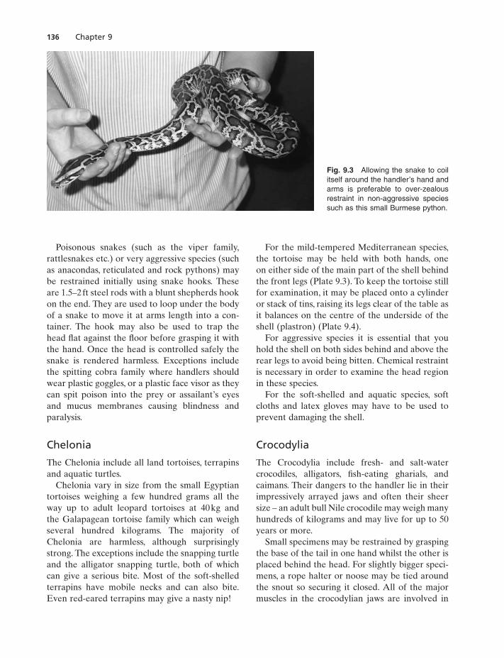
136 Chapter 9
Poisonous snakes (such as the viper family,rattlesnakes etc.) or very aggressive species (suchas anacondas, reticulated and rock pythons) maybe restrained initially using snake hooks. Theseare 1.5–2ft steel rods with a blunt shepherds hookon the end. They are used to loop under the bodyof a snake to move it at arms length into a con-tainer. The hook may also be used to trap the head flat against the floor before grasping it withthe hand. Once the head is controlled safely thesnake is rendered harmless. Exceptions includethe spitting cobra family where handlers shouldwear plastic goggles, or a plastic face visor as theycan spit poison into the prey or assailant’s eyesand mucus membranes causing blindness andparalysis.
Chelonia
The Chelonia include all land tortoises, terrapinsand aquatic turtles.
Chelonia vary in size from the small Egyptiantortoises weighing a few hundred grams all theway up to adult leopard tortoises at 40kg and the Galapagean tortoise family which can weighseveral hundred kilograms. The majority of Chelonia are harmless, although surprisinglystrong. The exceptions include the snapping turtleand the alligator snapping turtle, both of whichcan give a serious bite. Most of the soft-shelledterrapins have mobile necks and can also bite.Even red-eared terrapins may give a nasty nip!
For the mild-tempered Mediterranean species,the tortoise may be held with both hands, one on either side of the main part of the shell behindthe front legs (Plate 9.3). To keep the tortoise stillfor examination, it may be placed onto a cylinderor stack of tins, raising its legs clear of the table asit balances on the centre of the underside of theshell (plastron) (Plate 9.4).
For aggressive species it is essential that youhold the shell on both sides behind and above therear legs to avoid being bitten. Chemical restraintis necessary in order to examine the head regionin these species.
For the soft-shelled and aquatic species, softcloths and latex gloves may have to be used toprevent damaging the shell.
Crocodylia
The Crocodylia include fresh- and salt-watercrocodiles, alligators, fish-eating gharials, andcaimans. Their dangers to the handler lie in theirimpressively arrayed jaws and often their sheersize – an adult bull Nile crocodile may weigh manyhundreds of kilograms and may live for up to 50years or more.
Small specimens may be restrained by graspingthe base of the tail in one hand whilst the other isplaced behind the head. For slightly bigger speci-mens, a rope halter or noose may be tied aroundthe snout so securing it closed. All of the majormuscles in the crocodylian jaws are involved in
Fig. 9.3 Allowing the snake to coilitself around the handler’s hand andarms is preferable to over-zealousrestraint in non-aggressive speciessuch as this small Burmese python.

Reptiles and Amphibians: Handling and Chemical Restraint 137
closing not opening them, hence relatively finerope or tape can be used to keep the mouthclosed. The rest of the animal is restrained bypinning it to the ground.
Always approach crocodylians from head on,as their binocular vision is poor (although the alligator family does have some). Care should betaken when close to the crocodylian for head andtail movements both are directed at the assailantat the same time!
Much larger crocodiles require teams of people,with nets and snout snares in order to quicklyclamp the jaws closed and to restrain the danger-ous thrashing tail. Chemical immobilisation viadart guns is another option to be seriously considered.
Principles of chemical restraint
Chemical restraint is necessary for many pro-cedures in reptile medicine, ranging from minorprocedures such as extracting the head of a leo-pard tortoise or box turtle from its shell, toenabling a jugular blood sample to be taken or to carrying out coeliotomy procedures because of egg-binding. Before any anaesthetic or sedativeis administered, an assessment of the reptile
patient’s health should be made. Considerationsinclude:
• Is sedation or anaesthesia necessary for the pro-cedure required?
• Is the reptile suffering from respiratory diseaseor septicaemia?
• Is the reptile’s health likely to be made worseby sedation or anaesthesia?
Before discussing the administration of chemi-cal restraint it is important to understand the rep-tilian respiratory system.
Overview of reptilian respiratoryanatomy and physiology
The reptilian patient has a number of variationson the basic mammalian respiratory system.
The reptile patient has a glottis similar to theavian patient, which lies at the base of the tongue.This is more rostral in snakes and lizards andmore caudal in chelonia (Figs 9.4a,b). At rest theglottis is permanently closed, opening brieflyduring inspiration and expiration. In crocodilesthe glottis is obscured by the basihyal valve whichis a fold of the epiglottis. This fold has to bedeflected before they can be intubated (Bennett,1998).
(a)
(b)Fig. 9.4 Intraoral views of (a) Snake, showing 6rows of teeth, and the glottal tube with the entranceto the trachea located rostrally, just behind thetongue sheath. (b) Tortoise, showing large fleshytongue, at the base of which is the glottis.

138 Chapter 9
The trachea varies between orders. In Cheloniaand Crocodylia complete cartilagenous ringssimilar to those of the avian patient are found,with chelonians having a very short trachea. Insome species this trachea bifurcates into twobronchi in the neck. Serpentes and Sauria haveincomplete rings, such as those of the cat and dog,with Serpentes having a very long trachea.
The lungs of serpent and saurian species aresimple and elastic in nature. The left lung of most serpents is absent, or vestigial in the case of members of the boiid family. The right lung ofsnakes frequently ends in an air sac. Chelonianspecies have a more complicated lung structure,and the paired lungs sit dorsally inside the cara-pace of the shell. Crocodylian lungs are similar tomammalian lungs and are paired.
No reptile has a diaphragm, although croco-dylian species have a pseudodiaphragm whichchanges position with the movements of the liverand gut so pushing air in and out of the lungs.Most reptiles use intercostal muscles to move theribcage in and out in a manner similar to birds.The exception to this is members of the orderChelonia. These species need to move their limbs,neck and head into and out of the shell in orderto bring air into and out of the lungs.
Some species can survive in oxygen deprivedatmospheres for prolonged periods – chelonianspecies may survive for 24 hours or more, andeven green iguanas may survive for 4–5 hours.This makes induction of anaesthesia via inhal-ation of a gaseous anaesthetic agent almostimpossible in these animals.
Pre-anaesthetic preparation
Weight measurement
This is important for accuracy as some species ofreptile may be very small. Scales accurate to 1gare therefore advised for smaller reptiles toensure correct dosage.
Blood testing
It may be advisable to test biochemical andhaematological parameters prior to administeringchemical immobilising drugs. Blood samples canbe taken from:
• Jugular vein in Chelonia• Dorsal tail vein in Chelonia• Ventral tail vein in Serpentes, Crocodylia and
Sauria• Palatine vein or cardiac puncture in Serpentes
(although they frequently need to be sedated or anaesthetised to collect blood from theseroutes).
Fasting
Fasting is necessary in Serpentes to prevent regur-gitation and pressure on the lungs or heart. It isadvisable to ensure that no prey has been offeredin the two days prior to anaesthesia. Other rep-tiles require less fasting. For example, cheloniansrarely if ever regurgitate. However, it is impor-tant not to feed live prey to insectivores such asleopard geckos 24 hours prior to an anaesthetic asthe prey may still be alive when the reptile isanaesthetised.
Pre-anaesthetic medications
The premedications are the same drugs used incats and dogs to provide cardiopulmonary andcentral nervous system stabilisation, a smoothanaesthetic induction, muscle relaxation, analge-sia and a degree of sedation.
Antimuscarinic medications
Atropine (0.01–0.04mg/kg intramuscularly) orglycopyrrolate (0.01mg/kg intramuscularly) maybe used to reduce oral secretion and reducebradycardia, however these problems are notusually of concern in reptile patients. Indeed,antimuscarinics may increase the thickness ofmucus secretions, leading to more rapid blockingof the airways.
Tranquilisers
Acepromazine (0.1–0.5mg/kg intramuscularly)may be given one hour before anaesthetic induc-tion to reduce the levels of anaesthetic required,as can diazepam (0.22–0.62mg/kg intramuscularlyin alligators) and midazolam (2mg/kg intramus-cularly in turtles) (Bennett, 1998).

Reptiles and Amphibians: Handling and Chemical Restraint 139
Alpha-2 adrenoceptor stimulants
Xylazine at 1mg/kg can be used 30 minutes priorto ketamine in crocodylians to reduce the dose of ketamine needed. Medetomidine, used at dosesof 100–150mg/kg, markedly reduces the dose ofketamine required in chelonians, and has the ad-vantage of being reversible with atipamezole at500–750mg/kg.
Opioids
Butorphanol (at 0.4mg/kg intramuscularly) canbe administered 20 minutes before anaesthesia soproviding analgesia and reducing anaesthetic re-quired, and may be combined with midazolam at2mg/kg.
Fluids
Fluid therapy is, as with avian patients, veryimportant, and correction of fluid deficits shouldbe attempted prior to surgery. Maintenance levelsin reptile patients have been quoted as 25–30ml/kg/day (pages 178–9).
Induction of anaesthesia
It should be noted that reptiles should never beimmobilised by chilling or cooling them down.This does not provide analgesia and has seriouswelfare implications.
Injectable agents
Table 9.1 describes the advantages and disadvan-tages of injectable anaesthetic agents.
Dissociative anaesthetics
Ketamine: ketamine has been widely used inreptile patients. The effects produced vary de-pending on the species and dosage. Recommendedlevels range from 22–44mg/kg intramuscularly forsedation to 55–88mg/kg intramuscularly for surgi-cal anaesthesia. Lower levels are needed if com-bined with a premedicant such as midazolam ormedetomidine (Bennett, 1996).
Effects are seen in 10–30 minutes but may takeanything up to 4 days to wear off, particularly atlow environmental temperatures. Its main use istherefore at the lower dose range, to allow seda-tion, facilitate intubation and maintenance ofgaseous anaesthesia in species such as cheloniansthat hold their breath during gaseous induction.
It is however frequently painful on administra-tion. Also, because ketamine is excreted by thekidneys, it is recommended that it is administeredin the cranial half of the body. This is because, inreptiles, blood from the caudal half of the bodytravels to the kidneys before returning to the heartand the anaesthetic may thus be excreted beforeit has a chance to work.
Steroid anaesthetics
Alphaxalone/alphadolone: this combination al-lows intubation within 3–5 minutes when admin-istered intravenously. It may be administeredintramuscularly, but induction takes longer viathis route (25–40 minutes). If used alone, it willprovide anaesthesia for from 15–35 minutes at adose of 6–9mg/kg for intravenous administration,or 9–15mg/kg if given intramuscularly. Althoughrecovery time is quicker than for ketamine, it maystill take 1–4 hours for full recovery to occur. Itcan be very useful in chelonians and in general hasa wide safety margin.
Other injectable anaesthetics
Propofol: propofol produces rapid induction andrecovery, and is becoming the induction agent ofchoice in many practices specialising in reptile
Table 9.1 Advantages and disadvantages of injectableanaesthetics.
Advantages Disadvantages
Ease of administration Recovery often dependent Prevention of breath- on organ metabolism
holding on induction Difficult to reverse rapidlyReduced costs Often prolonged recovery Easy to administer timesLow risk to anaesthetist Muscle necrosis at site of
injection

140 Chapter 9
medicine and surgery. Its advantages also includea short elimination half-life and minimal organmetabolism, making it particularly safe to use indebilitated reptiles which frequently have somedegree of liver damage.
Its main disadvantage is that it requires intra-venous access, although use of the intraosseousroute has been shown to be successful in greeniguanas at a dose of 10mg/kg. As with cats anddogs, propofol also produces a transient period ofapnoea and some cardiac depression. In this situa-tion intubation and positive pressure ventilationis necessary.
Doses of 10–15mg/kg in chelonians given viathe dorsal coccygeal (tail) vein have successfullyinduced anaesthesia in under one minute. Thisallows intubation and maintenance on a gaseousanaesthetic if required. Alternatively, propofolcan be used alone providing a period of anaes-thesia of 20–30 minutes.
Depolarising muscle relaxants
Succinylcholine: this is a neuromuscular blockingagent and produces immobilisation without providing analgesia. Therefore it should only beused to aid the administration of another form ofanaesthetic or for transportation, and not as a solesource of anaesthesia. Recovery is dependent onliver metabolism and its use in animals with possible liver disease should be avoided.
It can be used in large Chelonia (such as thegiant Galapagoas species) at doses of 0.5–1mg/kgintramuscularly and will allow intubation and conversion to gaseous anaesthesia. Crocodilianscan be immobilised with 3–5mg/kg intra-muscularly, with immobilisation occurring within4 minutes and recovery in 7–9 hours. Respirationusually continues without assistance at thesedoses, but is important to have assisted ventilationfacilities to hand as paralysis of the muscles of res-piration can easily occur.
Gaseous agents
The gaseous anaesthetics used for induction willbe discussed in the next section on maintenanceof anaesthesia, however a table listing theiradvantages and disadvantages is presented below.
Maintenance of anaesthesia
Injectable agents
Dissociative anaesthetics
Ketamine: ketamine may be used on its own foranaesthesia at doses of 55–88mg/kg intramuscu-larly. It is worthwhile noting though that as the dosages get higher the recovery time alsoincreases, and in some cases it can be as long as several days. It should be noted that dosesabove 110mg/kg will cause respiratory arrest andbradycardia.
Ketamine may be combined with otherinjectable agents to provide surgical anaesthesia.Examples of these combinations include:
• Midazolam at 2mg/kg intramuscularly with 40mg/kg ketamine in turtles (Bennett, 1996)
• Xylazine at 1mg/kg intramuscularly, given 30minutes prior to 20mg/kg ketamine in largecrocodiles (Lawton, 1992)
• Medetomidine at 100mg/kg intramuscularlywith 50mg/kg ketamine in kingsnakes (Malley,1997).
Other injectable anaesthetics
Propofol: propofol may be used to give 20–30minutes of anaesthesia after administration,allowing minor procedures such as wound re-pair, intraosseous or intravenous catheter place-ment or oesophagostomy tube placement to be carried out.
It may be topped up at 1mg/kg/min intra-venously or intraosseously, but apnoea is ex-
Table 9.2 Advantages and disadvantages of gaseousanaesthetic.
Advantages Disadvantages
Ease of administration Breath-holding (Chelonia via face-mask/ particularly)induction chamber Environmental pollution
Pain free Health risk to Minimial tissue trauma anaesthetist
Risk with dangerous reptiles during handling

Reptiles and Amphibians: Handling and Chemical Restraint 141
tremely common and intubation and ventilationwith 100% oxygen is frequently required.
Alphaxalone/alphadolone: this injectable combi-nation can be used for induction and also for shortperiods of anaesthesia (average 25 minutes) at 6–9mg/kg intravenously. Intramuscular doses maybe given but onset of anaesthesia may take 20–30minutes. Its disadvantages include a prolongedrecovery time (1–4 hours), the need for relativelylarge doses and the need to intubate many reptilesdue to relaxation of the muscles which keep theglottis open to allow breathing.
Gaseous agents (see also Table 9.2)
Halothane
Halothane is the anaesthetic gas most widelyavailable in general practice and can be used in reptile anaesthesia, both for maintenance andinduction. Face-mask or induction chambers canbe used for induction at levels of 4–5%, althoughcertain species, such as Chelonia, can hold theirbreath for long periods making gaseous inductiondifficult. Maintenance can be achieved at 1–2.5%.Disadvantages of halothane are:
• It can induce myocardial hypoxia and dysrhythmias
• 15–20% of halothane is metabolised by the liverbefore recovery can occur. As many diseasedreptiles have dysfunctional or impaired livers,this makes its use limited.
• It can excite reptiles in the early stages of anaesthesia
• Cardiac arrest and apnoea frequently occursimultaneously, reducing available resuscitationresponse times.
Isoflurane
Isoflurane is the gaseous maintenance anaestheticof choice. Although more expensive than halo-thane, isoflurane is minimally metabolised in the body (0.3%) and has a very low blood–gaspartition coefficient (1.4 compared with 2.3 forhalothane in human trials). This means that it hasa very low solubility in blood, so as soon as isoflu-rane administration is stopped the reptile starts
to recover, excreting it from the lungs. In addi-tion it has lower fat solubility than halothane and so excretion from the body is quicker still.Isoflurane still has excellent muscle relaxing prop-erties and is a good analgesic. Apnoea precedescardiac arrest, unlike the case with halothaneanaesthesia.
It can be used to induce anaesthesia by face-mask application or, in those species not exhibit-ing breath-holding capabilities at levels of 4–5%,by induction chamber. It is also possible to adaptthe cases of 20ml and 60ml syringes to form longthin face-masks to induce snakes. Isoflurane canthen be used to maintain anaesthesia, preferablyvia endotracheal tube, at levels from 2–3%depending on the procedure.
A summary of the advantages of isoflurane overhalothane may been seen in Table 9.3.
Nitrous oxide
Nitrous oxide can be used in conjunction withhalothane or isoflurane, reducing the percentageof gaseous anaesthetic required for induction andmaintenance of anaesthesia. Its other advantagesinclude good muscle relaxation and excellentanalgesic properties, making it useful inorthopaedic procedures.
Disadvantages of nitrous include its tendency to accumulate in hollow organs. This may prove aproblem for herbivorous reptiles as they oftenhave capacious hind guts and nitrous oxide canaccumulate there. Nitrous oxide also requires
Table 9.3 Comparison of halothane and isofluranegaseous anaesthetics.
Halothane Isoflurane
Organ metabolism Minimal organ metabolism required for recovery required for recovery
Induces cardiac Relatively few arrhthymias arrhthymias induced
Cardiac arrest and Respiratory arrest respiratory arrest precedes cardiac arrestcoincide Good analgesia
Good analgesia Blood gas partition Blood gas partition coefficient 1.4
coefficient 2.3 Lower fat solubility than Highly fat soluble halothane

142 Chapter 9
some organ metabolism for full excretion and somay be a problem in a seriously diseased patient.
Aspects of gaseous anaesthesiamaintenance for reptiles
Inhalant gaseous anaesthesia is becoming themain method of anaesthetising reptiles for prolonged procedures. The reptile patient shouldpreferably be intubated to allow the inhalantanaesthetic to be delivered in a controlled manner.
Intubation
Intubation is straightforward in reptiles as they donot have an epiglottis and the glottis, which actsas the entrance to the trachea, is relatively cranialin the majority of species. It is useful to note thatthe glottis is kept closed at rest, so the operatormust wait for inspiration to occur to allow intu-bation. Reptiles produce little or no saliva whenat rest or not eating, so blockage of the tube isuncommon.
In Serpentes, the glottis sits rostrally on thefloor of the mouth just caudal to the tongue sheathand is easily visible when the mouth is opened(Fig. 9.4a). Intubation may be performed in theconscious patient if necessary, as reptiles do nothave a cough reflex. The mouth is opened with awooden or plastic tongue depressor and the endo-tracheal tube inserted during inspiration. Alter-natively, an induction agent may be given andthen intubation attempted.
In Chelonia, the glottis sits slightly more cau-dally at the base of the tongue (Fig. 9.4b). Thetrachea is very short and the endotracheal tubeshould only be inserted a few centimetres other-wise there is a risk that only one or the other ofthe bronchi will be intubated, leading to only onelung receiving the anaesthetic. An induction agentsuch as ketamine or propofol is advised for chelonians prior to intubation due to their abilityto breath-hold and difficulty in extracting thehead from the shell.
Sauria vary depending on the species of lizard,most having just a glottis guarding the entrance tothe trachea (Fig. 9.5). Some species possess vocal
folds, notably some species of gecko (Porter,1972). Some may be intubated consciously, butmost are better induced with an injectable prepa-ration or by face-mask using gas. Some speciesmay be too small for intubation. Plate 9.5 showsan intubate green iguana.
Crocodylia have a basihyal fold (Bennett, 1998)which acts as an epiglottis and needs to bedepressed prior to intubation. Because they arepotentially dangerous, these species require someform of injectable chemical sedation or inductionprior to intubation.
Intermittent positive pressureventilation (IPPV)
If intubation is performed on a conscious patient,anaesthesia may be induced, even in breath-holding species, by using positive pressure venti-lation. This has some advantages as it leads torapid post-operative recovery.
Many species require positive pressure ventila-tion during the course of an anaesthetic. Cheloniafor example are frequently placed in dorsalrecumbency during intracoelomic surgery. As they
Fig. 9.5 Intraoral view of a spiny-tailed iguana showingfleshy tongue. Glottis is caudal to this structure.

Reptiles and Amphibians: Handling and Chemical Restraint 143
have no diaphragm, and the lungs are situateddorsally, the weight of the digestive contentspressing on the lungs will reduce inspiration andslowly lead to hypoxia. This is in addition to thefact that most inspiratory effort is induced bymovement of the chelonian limbs, which are hope-fully immobile during anaesthesia! Also, if a neu-romuscular blocking agent such as succinylcholinehas been used, positive pressure ventilation maybe needed as respiratory muscle paralysis mayoccur.
The aim of IPPV is to inflate the lungs with anoxygen and anaesthetic mixture enough for anadequately oxygenated state to be maintained andfor the animal to remain anaesthetised. To thisend it is sufficient to ventilate most reptilestwo–six times a minute and no more, at a pressureof 10–15cm water (100–150mm mercury). As withbirds, a ventilator unit makes life much easier, but,with experience, manual ‘bagging’ of the patientwith enough pressure just to inflate the lungs andno more can be achieved (Figs 9.6 and 9.7). Arough guide is to inflate the first two fifths of thereptile’s body at each cycle (Malley, 1997).
Anaesthetic circuits
For species weighing less than 5kg, a non-rebreathing system with oxygen flow at twice theminute volume is suggested (Bennett, 1998). This
approximates to 300–500ml/kg/minute for mostspecies. Ayres T pieces, modified Bain circuits andMapleson C circuits may all be used.
Additional supportive therapy
Recumbency
Many chelonians are placed in dorsal recumbencyfor intracoelomic surgery. Other groups of reptilesmay also be placed in this position for similartechniques. The use of foam wedges, or positionalpolystyrene-filled vacuum bags, is essential tomaintain stability.
Snakes may become extremely flaccid duringsurgery. In order to provide stability they may be strapped to a long board or wedged in placewith foam wedges or vacuum bags. In any case,it is important to keep the body wall of non-chelonian species free of constraint and to useIPPV if necessary.
Fig. 9.6 Manual bagging of a garter snake. Note Dopplerprobe to monitor heart sounds, and Mapleson C circuit forsmaller species.
Fig. 9.7 Mechanical IPPV using a ventilator in a greeniguana. Note again Doppler probe and cranial position ofthe heart in iguanids.
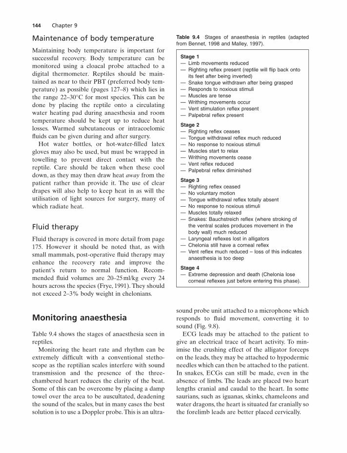
144 Chapter 9
Maintenance of body temperature
Maintaining body temperature is important forsuccessful recovery. Body temperature can bemonitored using a cloacal probe attached to adigital thermometer. Reptiles should be main-tained as near to their PBT (preferred body tem-perature) as possible (pages 127–8) which lies inthe range 22–30°C for most species. This can bedone by placing the reptile onto a circulatingwater heating pad during anaesthesia and roomtemperature should be kept up to reduce heatlosses. Warmed subcutaneous or intracoelomicfluids can be given during and after surgery.
Hot water bottles, or hot-water-filled latexgloves may also be used, but must be wrapped intowelling to prevent direct contact with thereptile. Care should be taken when these cooldown, as they may then draw heat away from thepatient rather than provide it. The use of cleardrapes will also help to keep heat in as will theutilisation of light sources for surgery, many ofwhich radiate heat.
Fluid therapy
Fluid therapy is covered in more detail from page175. However it should be noted that, as withsmall mammals, post-operative fluid therapy mayenhance the recovery rate and improve thepatient’s return to normal function. Recom-mended fluid volumes are 20–25ml/kg every 24hours across the species (Frye, 1991). They shouldnot exceed 2–3% body weight in chelonians.
Monitoring anaesthesia
Table 9.4 shows the stages of anaesthesia seen inreptiles.
Monitoring the heart rate and rhythm can beextremely difficult with a conventional stetho-scope as the reptilian scales interfere with soundtransmission and the presence of the three-chambered heart reduces the clarity of the beat.Some of this can be overcome by placing a damptowel over the area to be auscultated, deadeningthe sound of the scales, but in many cases the bestsolution is to use a Doppler probe. This is an ultra-
sound probe unit attached to a microphone whichresponds to fluid movement, converting it tosound (Fig. 9.8).
ECG leads may be attached to the patient togive an electrical trace of heart activity. To min-imise the crushing effect of the alligator forcepson the leads, they may be attached to hypodermicneedles which can then be attached to the patient.In snakes, ECGs can still be made, even in theabsence of limbs. The leads are placed two heartlengths cranial and caudal to the heart. In somesaurians, such as iguanas, skinks, chameleons andwater dragons, the heart is situated far cranially sothe forelimb leads are better placed cervically.
Table 9.4 Stages of anaesthesia in reptiles (adaptedfrom Bennet, 1998 and Malley, 1997).
Stage 1— Limb movements reduced— Righting reflex present (reptile will flip back onto
its feet after being inverted)— Snake tongue withdrawn after being grasped— Responds to noxious stimuli— Muscles are tense— Writhing movements occur— Vent stimulation reflex present— Palpebral reflex present
Stage 2— Righting reflex ceases— Tongue withdrawal reflex much reduced— No response to noxious stimuli— Muscles start to relax— Writhing movements cease— Vent reflex reduced— Palpebral reflex diminished
Stage 3— Righting reflex ceased— No voluntary motion— Tongue withdrawal reflex totally absent— No response to noxious stimuli— Muscles totally relaxed— Snakes: Bauchstreich reflex (where stroking of
the ventral scales produces movement in thebody wall) much reduced
— Laryngeal reflexes lost in alligators— Chelonia still have a corneal reflex— Vent reflex much reduced – loss of this indicates
anaesthesia is too deep
Stage 4— Extreme depression and death (Chelonia lose
corneal reflexes just before entering this phase).

Reptiles and Amphibians: Handling and Chemical Restraint 145
Respiratory flow monitors are not very usefuldue to the need for IPPV in most reptile speciesPulse oximeters are, however, useful, with reflec-tor probes being used per cloaca or per os tomeasure the percentage saturation of the haemo-globin with oxygen.
Recovery and analgesia
Recovery
Reptiles often recover rapidly from isofluraneanaesthesia. But, if other injectable drugs wereused, such as alphadalone/alphaxolone or keta-mine, recovery may be prolonged. It is essential atthis time to keep the reptile patient calm, stressfree and at its optimum preferred body tempera-ture. It may also be necessary to keep the patientintubated and on IPPV with oxygen, if high dosesof the above agents have been used, until thereptile is once again breathing for itself. The useof doxapram at a dose of 5mg/kg by intramuscu-lar or intravenous injection is useful to help stimulate respiration. It should be noted, how-ever, that the stimulus for reptiles to breathe is afalling blood partial pressure of oxygen, and not arising partial pressure of carbon dioxide, so IPPVwith 100% oxygen may actually inhibit respira-tion. IPPV with atmospheric air may therefore bepreferable once any gaseous anaesthetic has beenflushed from the airways.
Fluid therapy during this period will also helpto speed recovery, especially from agents such asketamine which are cleared through the kidneys.Once recovery is complete, the reptile should be
encouraged to eat, or, if anorectic, the patientshould be assist fed, stomach tubed etc.
Analgesia
Analgesia is an important aspect of post-operativerecovery. Reptiles which have been provided withanalgesia have been shown to have a quickerreturn to normality, eating, normal behaviour etc.than those who have not.
Opioids
Butorphanol at 0.4mg/kg intramuscularly, intra-venously or subcutaneously and buprenorphine at 0.01mg/kg intramuscularly have been recom-mended (Lawton, 1991; Bennett, 1998).
Non-steroidal anti-inflammatory drugs (NSAIDs)
NSAIDs also seem to be beneficial. The followinghave been recommended: carprofen at doses of 2–4mg/kg intramuscularly initially, and then1–2mg/kg every 24–72 hours thereafter (Malley,1997); meloxicam at 0.1–0.2mg/kg orally every 24hrs; ketoprofen at 2mg/kg intramuscularlyevery 24 hours (Bennett, 1996); flunixin meglu-mine at 0.1–0.5mg/kg intramuscularly every 24hours (Lawton, 1992). It should be noted that allof these are potentially nephrotoxic and have gastrointestinal ulcerative side-effects, hence fluidtherapy should be given and patients should beclosely monitored for side-effects.
Overview of amphibiananaesthesia
Techniques and equipment involvedin restraining amphibian patients
Examination of the amphibian patient should beperformed at that species’ optimum preferredbody temperature, as with reptile patients. Arough guide is between 21–24°C, which is lowerthan the more usual 22–32°C reptile housing conditions.
Fig. 9.8 Doppler probe monitoring of heart sounds in ananaesthetised red-eared terrapin.

146 Chapter 9
The examination table should be covered withpaper towels (unbleached) that have been soakedin dechlorinated water – preferably purifiedwater. More purified water should be on stand-byto be applied to the amphibian patient to preventdehydration during the examination.
Initially it is useful not to restrain the amphib-ian patient until the extent of any problem isassessed, as many have severe skin lesions whichare extremely fragile. Once an initial assessmenthas been made, the patient may be restrainedmanually. First, it is advisable to put on a pair ofnon-powdered hypoallergenic latex gloves inorder to minimise irritation to the amphibian’sskin caused by either the handler’s normal acidicskin environment or by the powder in manyprepacked latex gloves. The wearing of gloves isalso essential in species such as the toad family orthe arrow tree frogs whose skin can produce irri-tant or even potentially deadly toxins which canbe absorbed through unprotected human skin. Itmay also be necessary to wear goggles when han-dling some species of toad – the giant toad Bufomarinus can squirt a toxin from its parotid salivaryglands over a distance of several feet.
When handling the amphibian patient themethod of restraint will obviously depend on theanimal’s body shape. The elongated form of sala-manders and newts will require similar restraintto that of a lizard, with one hand grasping the pec-toral girdle from the dorsal aspect, index fingerand thumb encircling one forelimb, second andthird fingers the other, with the opposite handgrasping the pelvic girdle, again from the dorsalaspect in a similar manner. Some salamanders willshed their tails if roughly handled so care shouldbe taken with these species.
Large anurans (members of the frog and toadfamily) can be restrained by cupping one handaround the pectoral girdle immediately behindthe front limbs with the other hand positionedbeneath the hind limbs. Care should be taken withsome species which have poison glands in theirskin, as mentioned above, and in the case ofspecies such as the Argentinian horned frog careshould be taken with their bite. This species willeat whole small rodents when adult and can inflictunpleasant bite wounds.
Aquatic urodeles should be examined only in
water as removal causes skin damage. Some of thelarger urodeles, such as the hellbender species(Cryptobranchus spp) can also inflict unpleasantbite wounds on handlers, so firm restraint isrequired.
Smaller species and aquatic species may be bestexamined in small glass jars.
Aspects of chemical restraint in amphibians
There are three main routes of administration ofanaesthetic and sedative agents to amphibians:injections, inhalant gaseous anaesthetics and in-water methods.
In-water anaesthetic agents
There are two main medications for this route –MS-222 and benzocaine.
MS-222
This is tricaine methanesulphonate, an anaestheticused commonly in fish restraint. It is a watersoluble white powder. A range of 1–2g/l water isrequired to anaesthetise most frogs and urodeles,but a solution of 3g/l is required for most toads(Wright, 1996). A much reduced level of 0.5g/l canbe used for tadpole anaesthesia.
It is best to use the amphibian’s own water tominimise environmental changes, and to place thisinto a plastic bag, or plastic-lined box. This isuseful as many amphibians go through an exci-tated stage during anaesthesia, and the slight givein the plastic bags reduces skin damage. It is alsoimportant to ensure any anurans and other non-gilled amphibians can raise their nostrils abovethe water, otherwise they will drown.
Anaesthesia induction will take 20 minutes orso, with respiration rate reducing. Respirationmay even stop, although cardiac function persists.During the induction period, the ventrum of theamphibian will redden and anurans will becomeexcited, making leaping movements.
Initial anaesthesia is manifested by the inabilityof the amphibian to right itself, and loss of the

Reptiles and Amphibians: Handling and Chemical Restraint 147
corneal reflex, but with pain reflexes still intact. Adeep plane of anaesthesia is when all of these areabolished and only the heart-beat can be seen asa sign of movement. The level of anaesthesia canbe maintained by trickling the anaesthetic solu-tion over the amphibian’s body once the amphib-ian is removed from the solution. Reversal isachieved by trickling fresh, distilled, oxygenatedwater over the amphibian’s skin.
Benzocaine
This is related to MS-222 and can be used toanaesthetise many adult amphibians at solutionsof 0.2–0.3mg/l water. It is more soluble in ethanolthan in water and so is often dissolved in a smallvolume of this before it is added to the water.Recovery occurs some 60 minutes after rinsing theamphibian with benzocaine-free water.
It is worth noting that both anaesthetics areacidic in nature and hence will lower the pH ofthe water solution. It may be necessary to add abuffer solution to the water to correct this.
Injectable anaesthetic agents
Ketamine may be used, but is less preferable toMS-222. This is because relatively large volumesare required (75–100mg/kg (Bennett, 1996)), andthe anaesthetic takes a variable period to takeeffect – from 10 minutes to 1–2 hours. Injectionsmay be made intramuscularly, intravenously into the midline ventral abdominal vein or subcutaneously.
Inhalant gaseous anaesthetic agents
Isoflurane
This may be used, either in an induction chamberat dose of 2.5–3%, or, in the larger species oftoads, by dripping it directly onto the toad’s skin.
Some of the larger species may also be intubated,but anaesthesia is often erratic, as alternative respiratory routes are available to amphibians(cutaneous or buccopharyngeal routes – i.e. theamphibian can breathe through its skin or oralmembranes). Some of the more fragile species,such as the smaller urodeles or caecilians mayactually suffer severe skin irritation during gaschamber induction due to the direct irritant effectof the anaesthetic on the skin.
Halothane
This anaesthetic has the same problems as isoflu-rane, but in both cases the larger anuran speciesmay be anaesthetised by this method.
References
Bennet, R.A. (1996) Anaesthesia. In Reptile Medicine andSurgery (Mader, R., ed), pp. 241–247. W.B. Saunders,London.
Bennet, R.A. (1998) Reptile anaesthesia. Seminars in Avianand Exotic Pet Medicine, January, 30–40.
Frye, F. (1991) Biomedical and Surgical Aspects of CaptiveReptile Husbandry. Krieger, Malabar, Florida.
Lawton, M.P.C. (1992) Anaesthesia. In Manual of Reptiles.BSAVA, Cheltenham.
Malley, D. (1997) Reptile anaesthesia and the practisingveterinarian. In Practice, July/August, 351–368.
Porter, K.R. (1972) Herpetology. W.B. Saunders,Philadelphia.
Wright, K.M. (1996) Amphibian husbandry and medicine.In Reptile Medicine and Surgery (Mader, D., ed), pp.436–458. W.B. Saunders, Philadelphia.
Further reading
Page, C.D. (1993) Current reptilian anaesthesia procedures.In Zoo and Wildlife Medicine: Current Therapy 3 (Fowler,M., ed), pp. 140–143. W.B. Saunders, Philadelphia.

Classification
Reptiles and amphibians may be classified in anumber of different ways, one of which is accord-ing to their diet. Of the commonly seen speciesthere are four main categories as defined by diet.These are:
• Carnivores are predominantly the members ofthe snake family, which will eat whole avian,amphibian or mammalian prey. To do this theyhave powerful crushing jaws. Some have poisonfangs while others, such as the boa and pythonspecies, rely on suffocating their prey.
• Herbivores come from a variety of species, fromthe tortoise family, for example the Greek orspur-thighed tortoises, through to the lizardfamily, for example the green iguana.
• Insectivores are predominantly from the lizardfamily, for example the leopard geckos, collaredlizards etc., and from the amphibians, forexample the frogs, salamanders etc.
• Omnivores are from a variety of reptile species,and the term may be used to refer to reptileswhich change their eating habits during thecourse of their life. For example, the beardeddragon starts off as an insectivore, but becomesmore and more dependent on fruit and vege-table matter as it gets older.
In all of these cases, the individual species havebecome highly evolved to cope with certain typesof food. We also know that many of these crea-tures in the wild have a changing food supplythroughout the year, so what may form a staplediet in the summer does not necessarily applycome the winter.
General nutritional requirements
Water
As mentioned in the chapter on avian nutritionthe most important thing about the water pro-vided is its quality. Reptiles may defaecate in theirwater bowls and turtles and terrapins eat in theirwater. These habits cause pollution and leading todisease.
To prevent this, either the water must have apowerful filter system, or the terrapins/turtles fedin a separate feeding tank which may be cleanedout after feeding.
Vitamin and mineral supplements administeredin the water will allow rapid bacterial growth over24 hours, so bowl hygiene must be rigorous.
The amount of water consumed by individualreptiles and amphibians will depend on the dietsbeing offered as well as on the species. On dry,insect-based diets water consumption will bemuch higher than for reptiles and amphibianswhich consume large amounts of fruit and vegetables. Even so a leopard gecko may onlyconsume 5ml of water in a 24-hour period.
Tap water contains chlorine, which may irritatethe skin of sensitive aquatic species such asamphibians or soft-shelled turtles. It is advisedthat tap water be allowed to stand for 24 hours tolet the chlorine escape and to allow it to come upto room temperature.
Renal disease as a result of chronic dehydrationis common in captive reptile species. Many ofthese animals, for example green iguanas andwater dragons, come from parts of the worldwhich have high relative humidities – anywherefrom 60–100%. If these species are kept in vivariaat their correct temperatures (high twenties to lowthirties degrees centigrade) then the air can hold
Chapter 10
Reptile and Amphibian Nutrition
148

Reptiles and Amphibians: Nutrition 149
large amounts of water, consequently the relativehumidity often drops at these temperatures.Combine this with the fact that many reptiles willnot drink from water bowls, instead preferring tolick moisture from leaves or cage furniture, andwe can see that chronic dehydration can occur. Toprevent this, it is important not only to providedrinking bowls, but also to mist the cage, thereptile and the cage furniture several times daily.This is of course less necessary for desert-dwellingspecies, such as leopard geckos, pancake tortoisesetc., but even these species benefit from beingmisted every now and then.
Reptiles, like birds, are susceptible to renaldisease because the waste product of proteinmetabolism is predominantly the insoluble uricacid. If the reptile is not kept adequatelyhydrated, it will reduce the excretion of uric acidthrough the kidneys. This leads to deposition ofuric acid inside the body, a condition known as visceral gout. Once deposited, the uric acid formsa tough mineralised coating to the lining of bloodvessels, the kidneys, the heart and many otherorgans, leading to hypertension and multipleorgan failure.
Maintenance energy requirements (MER)
Every species has a level of energy consumptionper day which is needed to satisfy basic mainte-nance requirements. This is the energy usedpurely to maintain current status under minimalactivity and is the minimum energy requirementto support that reptile or amphibian’s life. In rep-tiles, basic maintenance requirements vary widelydepending on activity level and environmentaltemperature, so calculations are made at thatanimal’s optimum environmental temperature.Energy requirements will also vary according tothe animal’s stage of life. For example the MERwill be more than doubled in active egg layingfemales during disease or growth.
MER is dependent on the basal metabolic rate(BMR – the energy requirement when at com-plete rest) and metabolic body weight as follows:
MER BMR
where BMR constant k body weight
= ¥
= ( ) ¥ ( )
1 50 75
..
The constant, k, varies with family groups, and hasbeen estimated at 10 for reptiles in general.
If the foods offered are so low in kilojoules thatthe reptile or amphibian has to eat more of it thanwill fit into its digestive system in 24 hours, thatanimal will rapidly lose condition (Figs 10.1 and10.2).
For example, vegetables such as lettuce andcelery have an energy content of 12.6kJ/g drymatter (or in real terms 0.75kJ/g wet food),whereas meat-based foods such as rodent preyhave a much higher energy density of 19–21kJ/gdry matter (or in real terms 6–7.5kJ/g wet food)(Donoghue, 1998). From this we can see that in ‘asfed’ terms, i.e. wet food, one would need to feedeight times as much weight of vegetable matter togive the same energy dose in animal prey. Thisvolume may well exceed the gut capacity of thereptile or amphibian.
Conversely many pet reptiles and amphibianswill continue eating until their digestive tracts arefull, and if all they are offered is high energydensity food then they will rapidly achieve theirMER, exceed it and become obese.
Fig. 10.1 Malnutrition in a leopard gecko.

150 Chapter 10
Protein and amino acid requirements
Proteins are assembled from groups of aminoacids – indeed a protein can contain up to 22amino acids. In general terms, for humans, and itseems for reptiles and amphibians, ten aminoacids are essential and need to be provided in thediet. The others may be manufactured from theseten. The essential amino acids are:
• leucine • tryptophan• lysine • isoleucine• methionine • valine• phenylalanine • arginine• threonine • histidine.
In addition, it is known that for diets low in theamino acids methionine or arginine, an extra sup-plement of the amino acid glycine is required.
Proteins are therefore assessed on their abilityto provide these essential amino acids, with poorproteins supplying only non-essential ones. This isquantified by the term biological value, a high-biological-value foodstuff containing more of theessential amino acids.
For herbivorous reptiles, levels of 25% proteincontent as metabolisable energy in the diet havebeen shown to be adequate (Donoghue, 1998).Most of this protein source in herbivores seems
to come from leafy greens, but deficiencies areseen in herbivorous reptiles fed high cellulose,low protein foods such as the ubiquitous lettuce,and fruits. Chronic protein deficiencies are oftenpresented as gradual wasting conditions, withincreased susceptibility to infections.
Deficiencies in amino acids in carnivorous rep-tiles and amphibians is extremely rare becausethey eat whole prey. This gives them a 30–60%protein content as metabolisable energy.Herbivorous deficiencies specific amino acids arepossible although not well documented.
In general, waste products of protein metabo-lism in land-based reptiles are converted into therelatively insoluble uric acid. Alligators mayproduce ammonia as the waste product, as maymany amphibians, depending on their state ofhydration, but adult frogs may excrete urea.Protein excesses, such as those produced byfeeding cat or dog foods to predominantly her-bivorous species, can lead to an excess productionof uric acid and visceral gout, renal failure anddeath may result.
Fats and essential fatty acids (EFAs)
Fats provide high concentrations of energy. Theyalso supply the reptile or amphibian with essen-tial fatty acids, which are required for cellular
Fig. 10.2 Malnutrition and blisterdisease in a Burmese python.

Reptiles and Amphibians: Nutrition 151
integrity and as the building blocks for internalchemicals such as prostaglandins (which play apart in reproduction and inflammation). Fats also provide a carrier mechanism for the absorp-tion of fat-soluble vitamins such as vitamins A, D,E and K.
The primary EFA for reptiles is linoleic acid,as it is for mammals, with the absolute dietaryrequirement of this fatty acid being 1% of thediet. If the diet becomes deficient in this essentialfatty acid a rapid decline in cellular integrityoccurs. This is manifested clinically by the skinbecoming flaky and inelastic and prone to recur-rent infections, and also to fluid loss through theskin which in turn leads to polydipsia (Wallach &Hoff, 1982).
In herbivores, less than 10% of the diet on a dry matter basis is composed of fats, the chiefenergy sources being carbohydrates and proteins.However, fermentation of fibre in the lower bowelproduces short-chain fatty acids which can beused for energy. For carnivores, fat forms a majorpart of the energy source in the diet, as much as 40–70% of the calories, with protein chieflymaking up the rest.
The problem of overconsumption of fats in petreptiles which are not exercising regularly is wellknown and high fat foods such as dog and cat foodfed to herbivores, or extremely fat rodent prey fedto snakes, are prime culprits for this. Obesity canlead to a number of problems, high amongst whichis fatty degenerative change in the liver (hepaticlipidosis) which can lead to liver failure. This isparticularly common in tortoises.
Carbohydrates
Carbohydrates are primarily used for rapidenergy production. This is particularly impor-tant in herbivores which consume plant matteronly, and so gain the majority of their energysource from carbohydrates and proteins.Carnivorous reptiles do not utilise carbohydratesmuch at all.
Fibre
Dietary fibre is extremely important for herbivo-rous reptiles. Indeed, the presence of fibre acts
both as a bulking agent, encouraging gut motility,and as a source for fermentation by the intestinalmicroflora, essential for fatty acid and B vitaminproduction. Snakes and other carnivores do nothave a dietary fibre requirement, and indeed ifprovided will not be utilised. Ultimately it willdilute the energy concentration of the diet neces-sitating feeding more frequent and larger meals.
Vitamins
These compounds are grouped together althoughthey are widely differing in nature, but all animalshave a requirement for various numbers of these.They are categorised into fat soluble (vitamin A,D, E and K) and water soluble (the B vitamincomplex and vitamin C).
Fat-soluble vitamins
Vitamin A: in herbivore reptiles the most impor-tant plant precursor in terms of how much vitaminA can be produced from it is beta-carotene.Carnivorous reptile and amphibians will gain the preformed vitamin A in their prey food.
Hypovitaminosis A is a frequently seenproblem in chelonians, particularly tortoises and young red-eared terrapins. If a deficiency invitamin A occurs then mucous membranesbecome thickened and oral and respiratory secretions dry up. This is due to blockage of salivary and mucous glands with cellular debris –a condition known as squamous metaplasia. Thisleads to poor functioning of the ciliary mecha-nisms which have a role in removing foreign par-ticles from the airways. Swelling of the periorbitalmembranes – a condition known as xerophthalmia– is also seen.
Vitamin A’s role in immune system functionmeans that a deficiency makes respiratory anddigestive tract infections more common. The mostfrequently seen example of this is the increasedsusceptibility to pneumonias seen in red-earedterrapins and manifested in the lop-sided positionthey adopt when swimming. This is because oflung collapse or congestion which reduces buoy-ancy on the affected side.
Tortoises may suffer more frequently fromupper respiratory tract infections when a defi-

152 Chapter 10
ciency is present. Evidence of sterile pustules and cornified plaques inside the mouth are com-monly seen, with overgrowth of the beak due tohyperkeratosis.
Vitamin A also has a role in bone growth andstructure, the normal function of secretory glandssuch as the adrenals and also in reproductive function. Finally renal damage may occur in hypo-vitaminosis A, with evidence of oedema in theinguinal and axillary regions secondary to failureof the renal tubular filtration system.
Because it is fat soluble, vitamin A can be storedin the body, primarily in the liver. Recommendedminimum dietary levels are 200–300 IU/kg forreptiles (Wallach & Hoff, 1982).
Hypervitaminosis A rarely occurs naturally butmay be induced by overdosing with vitamin Ainjections at 1000 times or more the daily recom-mended doses. If this occurs acute toxicity devel-ops with mucus membrane and skin sloughing andfrequently death within 24–48 hours. Vitamin Asupplements are therefore often given orally as,due to slower absorption, this reduces the risk ofthis condition developing.
Vitamin D: vitamin D3 is the most active form forcalcium homeostasis, and plants are not effectiveas suppliers of this compound.
Cholecalciferol, the precursor of vitamin D3, ismanufactured in the reptile or amphibian’s skin ina process enhanced by ultraviolet light. For thisreason indoor animals produce much less of thiscompound unless supplied with an effective arti-ficial ultraviolet light source.
Hypovitaminosis D3 causes problems withcalcium metabolism, and leads to rickets. This isexacerbated by low calcium high phosphorus-containing diets. A typical sufferer would be ayoung growing reptile, kept indoors with no ultraviolet light supplementation and fed on a low calcium diet – for example lettuce/celery/cucumber for herbivores, or meat only/day oldmice/chicks for carnivorous species. The conditionso produced is referred to as ‘metabolic bonedisease’.
An animal so affected is frequently apparently‘well-muscled’, due to poorly mineralised boneswhich increase their thickness to maintain theirstrength. There is often flaring of the epiphyseal
plates at the ends of the long bones, with con-comitant bowing of the limbs. Green iguanas and other herbivorous species such as terrestrialchelonians are particularly susceptible, with thelizards showing rachitic rosettes (Frye, 1991) dueto flaring of the epiphysis in the ribs at the costo-chondral junction.
Chelonia develop ‘lumpy shell’, a deformity ofthe carapace in particular, where the edges rollupwards creating a ‘Cornish pasty’ effect, and themuscles which attach the limbs to the inside of the shell, pull the carapace downwards creatingpits either side cranially and caudally (Fig. 10.3).Recommended maximum levels are 50–100IU/kgevery other day.
Hypervitaminosis D3 occurs due to over sup-plementation with D3 and calcium and leads tocalcification of soft tissues, such as the medial wallof the arteries, and the kidneys, creating hyper-tension and organ failure. This often occurs in herbivores such as tortoises fed on tinned cat anddog food.
Vitamin E: hypovitaminosis E may occur due toa reduction in fat metabolism or absorption, suchas can occur in small intestinal, pancreatic orbiliary diseases, or due to a lack of dietary greenplant material for herbivores. A relative defi-ciency will occur in species eating large amountsof polyunsaturated fats, for example marine fishsuch as tuna and mackerel. These use up the
Fig. 10.3 Red-eared terrapin, Trachemys scriptaelegans, with hypovitaminosis D3.

Reptiles and Amphibians: Nutrition 153
body’s vitamin E reserves. A condition calledsteatitis, wherein body fat starts to necrose, hasbeen seen in gharials (a fish-eating crocodilian)(Frye, 1991) and terrapins.
Hypervitaminosis E is extremely rare.
Vitamin K: because of its production by gut bacteria, it is very difficult to get a true deficiency,although absorption will be reduced when fatdigestion or absorption is reduced as in biliary orpancreatic disease. The consumption of warfarinand coumarin derived compounds (as found insweet clovers) can increase the demand for clotting factors, and this may also be seen insnakes consuming prey which has been killed bythese rodenticides. Disease so caused is charac-terised by increased internal and external haem-orrhage, but vitamin K also has some function incalcium/phosphorous metabolism in bone so thismay also be affected. Frye (1991) has recordeddisease in crocodiles exhibiting gingival bleedingwithout petechiation. Recommended minimumlevels for reptiles are 1ppm (Wallach & Hoff,1982).
Water-soluble vitamins
Vitamin B1 (thiamine): a source of thiaminases,enzymes which destroy thiamine, is raw salt-waterfish which may be fed to some snakes, crocodiliansand turtles, such as garter snakes, red-eared ter-rapins and gharials. There are thiamine antago-nists as well in blackberries, beetroot, coffee,chocolate and tea when considering herbivores.When a relative deficiency occurs, neurologicalsigns such as opisthotonus, weakness and headtremors may be seen. In garter and water snakes,a classical inability to right itself occurs, with thesnake continually flipping onto its back. In addi-tion, fungal infections are reported as more likelyafter a B1 deficiency. The recommended minimumlevel for reptiles is 20–35 mg/kg food offered. Inaddition, if sea-fish such as smelt, which are highin thiaminases, are to be fed, cooking the fish for 5 minutes at 80°C deactivates the thiaminase.In a reptile with thiamine deficiency, doses of50–100mg/kg body weight should be given.Because of this problem it is often advised feedinggarter and water snakes on rodent prey rather
than sea-fish. They may be encouraged to eat thisby wiping the rodent prey with the fish to whichit is accustomed to cover the scent.
Biotin: deficiencies do occur commonly in gilamonsters, beaded lizards and monitor lizards all of which enjoy raw eggs in the wild. In this state the majority of eggs are fertile, and containlittle avidin (an antibiotin vitamin). However,unfertilised hen’s eggs are high in avidin and so a relative biotin deficiency may occur. Deficien-cies produce muscular weakness, occasionallywith skin lesions. It is therefore recommendedthat minimal levels of raw eggs are fed to suchreptiles.
Folic acid: a deficiency of folic acid is rare but can lead to severely impaired cellular division.This can lead to a number of obvious problemssuch as females reproductive tracts not maturing,a macrocytic anaemia due to failure of red bloodcell maturation and immune system cellular dys-function. A relative deficiency of folic acid mayoccur in some individuals fed a very high proteindiet, as folic acid is needed to produce the wasteproducts of protein metabolism in reptiles, uricacid. In addition there are inhibitors of folic acidin some foods such as cabbage and other bras-sicas, oranges, beans and peas, and the use oftrimethoprim sulphonamide drugs also reducesgut bacterial folic acid production.
Vitamin B12: vitamin B12 is produced generally byintestinal bacteria and so deficiencies are uncom-mon but may occur after prolonged antibioticmedication. Deficiency produces slow growth,muscular dystrophy in the legs, poor hatchingrates, high mortality rates and hatching deform-ities in young reptiles and amphibians.
Choline: choline may be synthesised in the body, but not in enough quantities for the growingreptile. Because of interactions, the need forcholine is dependent on levels of folic acid and vitamin B12. Excess protein therefore, as withfolic acid, increases choline requirements, as doesa diet high in fats. Deficiency causes retardedgrowth, disrupted fat metabolism and fatty liverdamage.

154 Chapter 10
Vitamin C: there is no direct need for this vitaminin reptiles and amphibians as vitamin C may beproduced from glucose in the outermost portionsof the kidneys. However, during disease processes,particularly those which affect liver function,it may be beneficial to the recovery process to provide a dietary source of vitamin C. It isrequired for the formation of elastic fibres andconnective tissues and is an excellent anti-oxidantsimilar to vitamin E. Deficiency leads to ‘scurvy’where there is poor wound healing, increasedbleeding due to capillary-wall fragility and bonealterations. Deficiency has been postulated as thecause of skin splitting in snakes fed rodent preythat had been starved for 24–48 hours prior tobeing fed (Frye, 1991). This allowed the emptyingof their gastrointestinal tract, and hence reducedlevels of vitamin C from the plant materialtherein. It has also been suggested that increasingvitamin C levels by supplementation at levels of10–20mg/kg intramuscularly or orally (Frye, 1991)may be useful in the treatment of chronic infec-tions, such as ‘mouth rot’ in snakes.
Minerals
As with mammals there are two main groups ofminerals, those classified as macro-minerals, i.e.those present in large amounts in the body, suchas calcium and phosphorus, and micro-minerals,or trace elements, such as manganese, iron andcobalt, which are all necessary for normal bodilyfunction.
Macro-minerals
Calcium: calcium has a wide range of bodily func-tions, the two most obvious being its role in theformation of the skeleton and mineralisation ofbone matrix, and its requirement for muscularcontractions. The active form of calcium in thebody is the ionic double charged molecule Ca2+.Low levels of this form, even though the overallbody reserves of calcium are normal, leads tohyperexcitability, fitting and death, conditionsseen in gravid female egg-bound green iguanas.
The ratio of calcium to phosphorus is important
– as one increases the other decreases and viceversa. This is controlled by the hormones calci-tonin, parathyroid hormone and the accessoryhormone vitamin D3. A ratio of 2 :1 calcium tophosphorus is desirable in growing reptiles, and1.5 :1 for adults. In high egg laying periods though,to keep pace with the output of calcium into theshells, a ratio of 10 :1 may be needed.
Calcium deficiency causes nutritional osteo-dystrophy, or metabolic bone disease, and is oftenaccompanied by deficiency in vitamin D3. Normallevels of vitamin D3, however, may exacerbate thisdisease as they encourage further calcium resorp-tion. Deficiency may be seen in lizards such asgreen iguanas and water dragons, and cheloniansfed on high fruit, lettuce, celery etc. diets. Dietswith excessive levels of oxalates, compoundswhich bind up calcium and prevent it beingabsorbed, such as spinach and beetroot orrhubarb leaves, can also lead to a deficiency.
Calcium deficiency may also be seen in insec-tivorous species, such as geckos, and beardeddragons, fed on insects without supplementation(Fig. 10.4). Insects have little or no calcium, theirtough outer coat is made of a protein known aschitin. To provide adequate calcium therefore,the insect must be dusted with a calcium powderimmediately before being fed (if not, by the timethe reptile has caught the insect most of thepowder has fallen off!). Alternatively, the insect ispre-fed on a calcium supplement. This is mixed inwith the insect’s food and fed for the 24–48 hoursprior to feeding the reptile, so that the insect’s gutis pre-loaded with calcium.
Extensive resorption of calcium occurs from the bones during dietary deficiencies, leaving onlyfibrous tissue. This is considerably weaker and so the ‘bones’ thicken to maintain their strength.Even so the bones are weakened, and bowing oflong bones and spontaneous fractures occur inlizards, collapsing of spinal vertebrae and deform-ities in most reptiles and deformed, lumpy shellsin chelonians. Excessive calcium in the diet (>1%)though reduces the use of proteins, fats, phospho-rus, manganese, zinc, iron and iodine, and can leadto soft tissue mineralisation if in conjunction with adequate or excessive vitamin D3 levels.Red-eared terrapins have been shown to have a

Reptiles and Amphibians: Nutrition 155
requirement for 2% calcium on a dry matter basis(Kass et al., 1982).
Phosphorus: phosphorus is widespread in plantand animal tissues, but in the former it may bebound up in unavailable form as phytates. Levelsof phosphorus are controlled in the body as forcalcium, the two being in equal and opposite equilibrium with each other. Therefore, if dietaryphosphorus levels exceed calcium levels appre-ciably the parathyroid glands become stimulatedto produce more parathyroid hormone and nutri-tional secondary hyperparathyroidism occurs.This leads to progressive bone demineralisationand renal damage due to high circulating levels ofparathyroid hormone. High dietary phosphorusalso reduces the amount of calcium which can beabsorbed from the gut, as it complexes with thecalcium present there. This can be a problem inreptiles fed pure meat with no calcium or bonesupplement, and in herbivores which are pre-dominantly fruit and lettuce consumers as theseare high phosphorus low calcium foods. Greenvegetables or supplementation with calciumpowders may therefore be necessary. In addition,low calcium/high phosphorus levels frequentlyallow bladder stones to form in those species(such as chelonians, green iguanas etc.) that havea bladder.
Magnesium: most magnesium is absorbed fromthe small intestine, and is affected by large
amounts of calcium in the diet which reduce mag-nesium absorption. Deficiencies rarely occur, butmuscular weakness can be the result.
Potassium: as with mammals, potassium is themajor intracellular positive ion. Rarely is there a dietary deficiency, but severe stress can causehypokalaemia through increased kidney excretionbecause of elevated plasma proteins. This can lead to cardiac dysrhythmias, muscle spasticityand neurological dysfunction.
Sodium: sodium is the main extracellular posi-tive ion and regulates the body’s acid base balanceand osmotic potential. In conjunction with potas-sium, it is responsible for nerve signals andimpulses. Rarely does a true dietary deficiencyoccur, but hyponatraemia may occur due tochronic diarrhoea or renal disease. Many reptileshave salt glands found outside the kidneys respon-sible solely for the excretion of excess sodiumwhilst conserving water. The green iguana’s salt glands, for example, are present inside thenostrils, and white crystalline deposits of salt maybe seen here and are frequently sneezed out.It may be necessary to supplement the diet ofmarine species with sodium chloride if kept infresh water situations, or if fed freshwater plantsor fish.
Chlorine: this is the major extracellular negativeion and is responsible for maintaining acid–base
Fig. 10.4 Bearded dragon, Pogonavitticeps, with metabolic bonedisease and resultant spinal andlimb deformity.

156 Chapter 10
balances in conjunction with sodium and potas-sium. Deficiencies are rare.
Micro-minerals (trace elements)
These elements include zinc, copper, iron, man-ganese, cobalt and sulphur all of which have an important part to play in cellular function.However, so far no actual deficiencies specificallyrelated to these elements have been reported inreptiles or amphibians. The exception is iodine.
Iodine: iodine’s sole function is in thyroidhormone synthesis, which affects metabolic rate.Deficiency causes goitre and fluid retention (myx-oedema). It has knock-on effects on growth,causing stunting and neurological problems and inamphibians may prevent metamorphosis fromintermediate tadpole stages to the adult form. Inreptiles it is most often encountered in giant ter-restrial tortoises, which will exhibit goitre swellingof the neck. It can also occur due to overfeedingwith iodine-binding plants, such as cabbage, cauli-flower, broccoli, kale and Brussels sprouts. Excessiodine added to the water may cause species such as the amphibian axolotl (a neotenic sala-mander – that is its ‘adult’ form has the externalgills more typical of a tadpole or intermediatelifestage) to shed its external gills. Levels of 0.3mg/kg body weight have been quoted(Donoghue & Langenburg, 1996).
Specific nutritional problems in reptiles
Below are some common presentations of nutri-tional problems in reptiles.
Post-hibernation anorexia (PHA)
PHA occurs in the Mediterranean species of tor-toise (Hermann’s, Greek or spur-thighed, mar-ginated and Horsefield’s), which hibernate duringthe winter months. The commonest presentationis that of an inappetant tortoise after coming outof hibernation, often with signs of systemic or res-piratory tract infections (such as the ‘runny nosesyndrome’) and often with a low body weight in
relation to length (a low Jackson’s ratio). In addi-tion the blood glucose levels are frequently below3.2mmol/l, which appears to be the minimumlevel required for appetite stimulation (Lawrence,1987), with high levels of urea.
Dehydration is apparent in these cases, andtreatment requires aggressive fluid therapy andnutritional support, using glucose-containingfluids and liquid food stomach tubing, as well aswarming the tortoise to its optimum temperature(20–27°C) (Fig. 10.5).
Causes of this condition could involve any oneof the following:
• Disease during or prior to hibernation• Poor nutrition leading to poor fat reserves prior
to hibernation• Owner failure to observe recovery from hiber-
nation for several days, so no food offered at thecritical time
• A period of cold weather immediately afterrecovery.
It is the rising plane of blood glucose post hiber-nation that acts as the stimulus for appetite inthese chelonians, and failure of this rise, due tomalnutrition or failure to eat whilst the levels arestill high, may lead to unresponsive anorexia.
To prevent PHA, therefore, it is important to attend to disease prior to hibernation, and, ifseverely affected or underweight, the tortoiseshould not be hibernated, but kept indoors at its
Fig. 10.5 Spur-thighed tortoise, Testudo graeca, withpost-hibernation anorexia.

Reptiles and Amphibians: Nutrition 157
optimum temperature range and fed throughoutthe winter. The tortoise should also be checkedregularly once in hibernation, around once ortwice a week, to ensure if the tortoise does comeout of hibernation early, it has food and wateravailable immediately. Bathing the tortoise im-mediately after waking in warm water, cleaningthe nose, eyes and mouth especially can also stimulate appetite, and no tortoise having recov-ered from hibernation should be allowed torehibernate that same winter. Finally, stomachtubing with fluids and soluble carbohydrates, suchas the basic sugar and protein containing liquidfood Critical Care Formula produced by VetArkor vegetable baby food porridges, early on in thecourse of the problem can be useful.
Visceral and articular gout
Gout is a condition caused by the unique way thatmany reptiles deal with the waste products ofprotein metabolism. Most reptiles are uricotelic,that is the main excretory product of proteinmetabolism is uric acid. This compound is rela-tively insoluble in water, which has its advantagesas reptiles are therefore able to reduce the waterlost in excreting it. Unfortunately, if the reptilebecomes dehydrated, either acutely or chronically,or consumes diets with excessive protein levels,particularly a type of protein called purines, orsuffers kidney damage, then uric acid levels buildup in the bloodstream. If allowed to do so, theywill eventually exceed the precipitation point andform crystals inside the body.
Diet is important, as purine proteins which arefound mainly in animal protein, are convertedreadily to uric acid on degradation in the body.Therefore, if herbivorous species, such as greeniguanas, which are not used to large volumes ofpurines, are fed a diet rich in animal protein, suchas cat or dog food, they will produce excessiveamounts of uric acid and develop gout.
There are two forms of gout, visceral and articu-lar. Articular gout can easily be diagnosed antemortem, as it causes gross swelling and inflamma-tion in the joints where the uric acid crystals form.Visceral gout is more difficult to diagnose antemortem, because it occurs where uric acid crystalsare deposited in the soft tissues of the body,
primary sites being the kidneys, the pericardialsac, the lungs, spleen and liver. Once deposited,it is almost impossible to move the crystals medically and permanent damage is often done.
Obesity
Obesity is a common problem in many reptiles and amphibians kept in captivity. Many species areoverfed because of owners’ ignorance of naturalfeeding intervals and of food types commonlyeaten in the wild. Examples include feeding dogfood to tortoises and green iguanas, both of whichare totally herbivorous in the wild but both ofwhich will eat meat if offered it. The resultingproblems are as mentioned above with excessprotein causing gout, excess calcium and vitaminD3 causing soft tissue mineralisation and excessanimal fat causing fatty liver syndrome (hepaticlipidosis) wherein the liver cells are filled with fatdeposits impeding their function. Snakes alsosuffer from these conditions when fed overfat lab-oratory rodents, or simply fed too often. A roughidea of feeding frequencies is given in Table 10.1.
The aim should be a reptile which does notappear emaciated, but lean.
Hypoglycaemia in crocodilians
It has been reported that crocodiles kept in highdensity conditions, or are otherwise stressed, areprone to hypoglycaemic fits (Scott, 1992). It isinteresting to note that crocodiles’ blood sugarlevels vary throughout the year, being lower in thewinter and highest in the summer. A rising bloodsugar level appears to be the stimulus to eat, asseems to be seen with Mediterranean tortoisesafter hibernation.
Environmental temperature and itseffects on nutrition
Because reptiles and amphibians are ectothermic,that is they rely on their surroundings to maintaintheir body temperature, environmental tempera-ture is important in all aspects of husbandry.
There is an optimum preferred temperaturezone that will allow their enzymes and metabolicpathways to function at their optimum levels, so

158 Chapter 10
environmental temperature will influence the rateof digestion of the food offered. It may take 2–3days for a rat consumed by a large boa constric-tor to pass through its digestive system if keptwithin its preferred temperature zone of 25–30°C,but if kept 5–10°C cooler this will often slow downto 5–7 days, and if kept much lower than thisdigestion may not occur before the prey itembecomes rancid inside the snake. Similarly, if keptat too high a temperature, the reptile may not bestimulated to eat at all and dehydration and heatstress may set in.
A general guide to feeding reptiles
Fresh food should always be fed to reptiles. Itshould also be remembered that at the increasedtemperatures of most vivaria, the food offered will spoil very quickly and will need to be replacedfrequently.
Snakes
It is important to note that it is illegal to feed livevertebrate prey to another animal in the UnitedKingdom. All rodent prey fed to snakes andlizards must therefore have been humanely killed
first. In addition, live prey may damage the reptileif the latter is not hungry and does not kill the preyquickly.
To encourage anorectic snakes to eat, a numberof tricks may be employed including:
• Warming the prey briefly before offering byheating it in a pot of hot water
• Breaking the prey item open to release thescent of blood
• Teasing the snake by moving the dead prey itemaround the cage with forceps, to mimic live prey
• Trying a variety of colours of prey – somesnakes will only take dark-furred rodents
• To get a snake (such as a garter or water snake)used to eating rodent prey after only eating fishor amphibians (hog-nosed snakes), wipe therodent to be offered with the previously takenfood item to transfer scent
• Ensure that there are plenty of areas to hide;some boids and pythons like to consume theirprey in a box or hide
• Leave the prey in overnight because somespecies prefer to hunt at night
• Choose the next smallest size of rodent, so ifadult mice were previously offered try fuzzies,if juvenile rats, try adult mice, etc.
NB The term ‘pinkies’ refers to nude neonatal ratand mice pups, ‘fuzzies’ refers to week-old rat and
Table 10.1 Feeding frequencies and food types for various reptile species.
Species Feeding frequency and food types
Herbivores (e.g. iguanas, tortoises) Daily grazing of food advised
Small insectivores (e.g. leopard geckos, Fed 2–3 times weekly on live collared lizards) insect prey ‘to appetite’
Small carnivores (e.g. garter snakes, Fed 2–3 times weekly on rat corn snakes) pups,‘fuzzies’ (furred baby
mice) or ‘pinkies’ (nude baby mice) according to size
Medium carnivores (e.g. kingsnakes, Fed 1–2 times weekly, adult ratsnakes) mice or small rats
Large carnivores (e.g. boa constrictors, Fed once weekly or fortnightly Burmese pythons) (the larger the snake the less
often) on adult rats or small rabbits

Reptiles and Amphibians: Nutrition 159
mice pups with a thin covering of fur and ‘furries’refers to juvenile rat and mice pups between 1–3weeks of age which have a soft but longer cover-ing of fur.
Refeeding syndrome
If anorexia in a snake or other reptile has per-sisted for some time it is essential to rehydrate the patient before attempting to feed. Indeed,initial feeding after this should be started off atvery low levels. This is because excess calories and proteins cause a rapid uptake of glucose fromthe bloodstream into the cells, which takes potas-sium and phosphorous with it. This can lead to alife-threatening hypokalaemia and hypophos-phataemia. The monitoring of blood phosphorusand potassium is therefore to be recommendedwhen treating chronically anorectic reptiles,whether carnivorous or herbivorous.
Herbivores
If using a commercial pelleted food for iguanas or tortoises, then be sure to soak the pellets thoroughly before feeding, otherwise they swellup inside the reptile causing colic and bloat. Com-mercial pelleted diets are a useful adjunct to thediet of herbivorous reptiles and many companiesnow produce iguanid and chelonian diets whichare well supplemented with minerals and vitaminsand also contain moderate levels of fibre for gutmotility enhancement.
The feeding of certain foods to herbivoresshould be prohibited. Animal proteins are one, asare certain fruit and vegetables. We have alreadydiscussed the problems of excessive volumes of largely water-containing vegetables such aslettuce, cucumber and celery, the goitrogenicproperties of cabbage, kale, broccoli and cauli-flower and the anti-calcium effect of oxalate-containing plants such as spinach, beetroot andrhubarb leaves. In addition, fruits such as bananacan cause a sugar ferment in herbivorous reptilescausing colic, as well as adhering to the mouth-parts and encouraging local infection. Avocadoshave an extremely high fat content and should notbe fed to herbivorous reptiles due to potential secondary fatty degeneration of the liver.
Sample diet for green iguanas
Pelleted soaked commercial food may be fed ataround 25% of daily intake. The rest can be basedon the following: Up to half of the plant materialoffered can be made of calcium-rich vegetablessuch as kale, dandelions, chicory, watercress,cabbage, flat-leaved parsley, basil, coriander etc.The other half may be made up of other vegetablematter such as peas, beans, carrots, sweet peppers,courgettes or marrows, cauliflower florets andflowers of plants such as nasturtiums, dandelions,roses etc. To this mixture can be added smallamounts of fruit such as apples, pears, tomatoes,plums, strawberries, raspberries, melon, passionfruit, papaya etc., or fibrous foods such as brancereals, brown bread and cooked brown rice.
This combination should be thoroughly mixedso as to prevent selective feeding and to it shouldbe added a supplement of calcium in the form ofcalcium lactate or gluconate, or a natural calciumsource such as cuttlefish or oyster shells. In addi-tion the use of a vitamin D3/calcium supplementonce or twice a week, particularly for growingiguanas and egg-laying females, is advised.
Sample diet for Mediterranean tortoises
The majority of the diet is to be composed of veg-etable matter of a leafy nature such as dandelions,kale, watercress, flat-leaved parsley, chicory andbok-choy. To this may be added peas, beans, hayor dried grass, fresh grass (not cut), grated carrot,grated pumpkin and sweet peppers.
To this may be added small volumes of fruitsuch as apples, pears, melon, papaya, passion fruit,strawberries, plums, flowers such as dandelions,nasturtiums, roses and sprouted seeds such asmung beans, lentils, and chick peas.
For more tropical species, such as the red andyellow footed tortoises (Geochelone carbonariaand Geochelone denticulata), the amount of fruitand flowers may be doubled.
For grassland species, such as leopard tortoises(Geochelone pardalis) and Indian starred tor-toises (Geochelone elegans), a good provision offresh uncut grass or good quality hay is advised.
For more omnivorous species, such as boxturtles (Terrapene carolina), up to 50% of the

160 Chapter 10
above fruit and vegetable diet may be replacedwith adult maintenance dry dog foods (soaked),mealworms, crickets, earthworms and even babymice (pinkies). Juveniles of this species tend to bemore carnivorous than the adults.
In all of these diets it is recommended that dailysupplementation with calcium lactate or glu-conate be included, with a calcium/vitamin D3
supplement added once or twice weekly depend-ing whether the tortoise is a juvenile (higherrequirement) or an adult (lower requirement).
Diets for snakes and insectivorous species have been mentioned. The latter need calciumsupplementation as discussed in the section con-sidering calcium as a macro-mineral which mayeither be dusted onto the insects, or pre-fed tothem to load their digestive contents with thecalcium.
References
Donoghue, S. (1998) Nutrition of pet amphibians and reptiles. Seminars in Avian and Exotic Pet Medicine7, 3.
Donoghue, S. and Langenburg, J. (1996) Nutrition. InReptile Medicine and Surgery (Mader, D., ed), pp.148–174. W.B. Saunders, Philadelphia.
Frye, F. (1991) A practical guide for feeding captive reptiles.In Biomedical and Surgical Aspects of Captive ReptileHusbandry Vol 1. Krieger Publishing Inc, Malabar,Florida.
Kass, R.E., Ullrey, D.E. and Trapp, A.L. (1982) A study ofcalcium requirements of the Red-eared slider turtle(Pseudemys scripta elegans). Journal of Zoo AnimalMedicine, 13, 62.
Lawrence, K. (1987) Post hibernational anorexia in cap-tive Mediterranean tortoises (Testudo graeca and T.hermanii). Veterinary Record 120, 87.
Scott, P.W. (1992) Nutritional diseases. In Manual of Reptiles (Beynon, P.H., Lawton, M.P.C. and Cooper,J.E., eds), pp. 138–152. BSAVA, Cheltenham.
Wallach, J.D. and Hoff, G.L. (1982) Metabolic and nutri-tional diseases of reptiles. In Non-infectious Diseases ofWildlife (Hoff, G.L. and Davis, J.W., eds), pp. 155–168.Iowa State Press, Ames.
Further reading
Barten, S.L. (1996) Lizards. In Reptile Medicine and Surgery(Mader, D., ed), pp. 47–60. W.B. Saunders, Philadelphia.
Boyer, J.H. (1996) Turtles, tortoises and terrapins. In ReptileMedicine and Surgery (Mader, D., ed), pp. 61–77. W.B.Saunders, Philadelphia.
Frye, F. (1995) Reptile Clinicians Handbook. Krieger Pub-lishing Co, Malabar, Florida.
Zimmerman, E. (1995) Reptiles and Amphibians: Care,Behaviour and Reproduction. TFM Publishing Inc,Neptune City, New Jersey.

Skin disease
Dysecdysis
Dysecdysis occurs when the normal process ofskin shedding (known as ecdysis) fails. It occurs in snakes, and to a lesser extent, lizards. Theprocess of ecdysis involves the forming of a newskin beneath the old one. Once this is complete,a series of proteolytic enzymes and lymphaticfluid is secreted between the new skin layer andthe overlying old one. This lifts the old layer andseparates the two, and makes the snake appeardull and lack-lustre with the eyes noticeablyblueing. This lasts for 5–7 days on average in thesnake. Once complete the fluid is reabsorbed, andthe snake appears to return to its normal hue.After a further 5–7 days the skin is shed, in thecase of the snake, in one piece, from the head endfirst. It is initiated in snakes by rubbing the faceon an abrasive surface to loosen the first piece ofskin. When the separation fails to occur, whetherin part or totally, the condition is known asdysecdysis.
The causes of dysecdysis are many and varied,but can include any condition which can causedehydration so reducing lymphatic fluid availableto separate the skin layers. If the reptile is malnourished dysecdysis may occur due to a lackof proteolytic enzymes. Alternatively old scars, alack of an abrasive surface in the vivarium uponwhich to start the process or severe ectoparasitismmay also cause dysecdysis (Fig. 11.1).
Scale rot
Scale rot is a colloquial term for a range of con-ditions affecting the reptile skin which result insevere infections.
Septicaemic cutaneous ulcerative disease (SCUD)
Septicaemic cutaneous ulcerative disease(SCUD) is a condition seen in aquatic chelonians.It is caused by the bacterium Citrobacter freundiiwhich, once it has gained access to the blood-stream, produces ulcers in the skin and looseningof scales with general debilitation and, in manycases, death (Fig. 11.2).
Blister disease
This is a form of scale rot seen in snakes, particu-larly semi-aquatic species such as garter or watersnakes. It can however occur in any species whichis exposed to a persistently damp substrate. Theouter layer of skin develops blisters of a clear fluidwhich become secondarily infected with environ-mental bacteria such as Aeromonas spp orPseudomonas spp. These can progress to envelopthe whole of the ventral aspect of the snake, andlead to septicaemia. Occasionally these conditionsmay involve a fungus such as Aspergillus spp.
Abscesses
Abscesses have recently been quoted as being more accurately termed fibriscesses(Huchzermeyer & Cooper, 2001) due to theunique nature of reptile and avian abscess forma-tion. Instead of the liquid pus we are more famil-iar with in mammals, reptiles and birds form asolid, caseous, dried swelling surrounded by athick shell of fibrous tissue. This is due to the lackof lysozymes (a form of destructive enzyme foundin macrophages and other immune system cells)that help liquefy foreign matter. Instead thereptile engulfs the material in immune system
Chapter 11
Common Reptile and Amphibian Diseases
161

162 Chapter 11
cells and to contain the problem further throws upa thick connective-tissue wall around the area,forming a solid swelling (Fig. 11.1). These mayform anywhere, but a common site in cheloniansis the middle ear, causing a bulging of the eardrum. The pathogens involved are frequentlyGram negative species, although the presence offungi in abscesses has been well recorded.
Erythema, petechiae and ecchymoses
These three conditions may all be present at thesame time or may appear individually.
Erythema is the reddening of skin tissues dueto vascular congestion beneath. It can be seen inreptiles as a pink hue to the areas between scutesin the case of chelonians (Fig. 11.2) and croco-dylians, or the more generalised reddening of the skin seen in snakes. Erythema is often sug-gestive of a generalised medical condition such assepticaemia.
Petechiae are the pin-point haemorrhages seen in many reptiles. They can be seen in areassuch as caused by the mouth, where they oftensuggest the incidence of an oral infection such as‘mouth rot’. They may be seen over the whole ofthe body suggesting the possibility of septicaemia.Other conditions include clotting deficienciessuch as are caused by the consumption of rodentprey which has itself consumed a warfarin-typepoison.
Ecchymoses are larger areas of haemorrhage,and may be caused by severe local infection orconditions mentioned above.
Overgrown beaks and claws
Overgrown beaks are common in chelonians, andare often due to a lack of abrasive foodstuffs orabrasive surfaces from which to eat them. Thebeak may become so overgrown that the reptile isunable to feed itself. Beaks may be trimmed usinga slow-speed dental drill, or a battery-poweredhand-held drill in much the same way as a bird’sovergrown beak can be trimmed.
Overgrown claws can also be a problem inlizards and chelonians due to the lack of abrasivesubstrates in their environment. It should benoted however that some species have perfectly
Fig. 11.1 Dysecdysis in a lizard with a subcutaneousabscess.
Fig. 11.2 SCUD in a red-eared terrapin, Trachemysscripta elegans.

Common Reptile and Amphibian Diseases 163
normal apparently ‘long’ claws. An example is themale red-eared terrapin which uses his claws as adisplay aid to attract and mate the female. Other-wise, trimming the claws with a standard pair offeline claw clippers is advised.
Pigment changes
An example of a physiologically normal colourchange is seen in the Chameleonidae which willoften blend with their surroundings and darken incolour as they become stressed. Green iguanasoften become yellow brown in colour as theybecome stressed or unwell, and many of thesespecies will exhibit darkening at the site of aninjection. Areas of previous trauma may becomewhitened.
Ectoparasitic
Mites
The most commonly seen mite in snakes isOphionyssus natricis which appears as red todark-coloured pin-head mites hiding under theoverlapping edges of scales. They may be seen inwater dishes in which the reptile has been bathing.They can cause severe irritation, pruritus and self-trauma, as well as potentially causing anaemia andconditions such as dysecdysis. In addition, theycan be responsible for transmitting Aeromonasspp bacteria, which may cause septicaemia. Thereare other mites, such as the cloacal mites ofaquatic turtles of the family Cloacarus spp. Inaddition, there is of course the non-parasiticharvest mite or Neotrombicula autumnalis whichmay be brought in on straw and hay beddingmaterial. The adult itself is not irritant but the sixlegged larval stage is and may cause the reptile totraumatise itself.
Ticks
Wild-caught species are common sufferers ofticks, but ticks are also seen in reptiles kept inowners’ gardens, such as tortoises. In the UnitedKingdom the species Ixodes ricinus (the sheep ordeer tick) and Ixodes hexagonous (the hedgehogtick) are seen. These attach to soft, thin-skinned
areas. In tortoises these are naturally the areasaround the neck inlet and in front of the hind legs.They may cause local damage and can transmit arange of viral and bacterial pathogens. Theseinclude the bacteria Staphylococcus aureus, thecause of tick pyaemia and Borrelia burghdorferi,the cause of Lyme’s disease.
Blowfly myiasis
Myiasis is a particular problem for any reptilekept in insanitory conditions or those with diar-rhoea. In particular, tortoises are at high riskduring the summer months. Members of the bluebottle, black bottle and green bottle family are allcapable of laying eggs on a tortoise. In peak con-ditions these can hatch into larvae or maggotswithin 1–2 hours. These then burrow away fromthe light, into the body of the tortoise causingsevere trauma, infections, shock and ultimatelydeath.
Leeches
Leeches tend not to be such a problem in theUnited Kingdom, but can affect aquatic species allover the world. The family Annelidae are the mostdangerous, and cause large wounds which con-tinue to bleed after the leech has detached. Thesemay then become secondarily infected.
Traumatic and spontaneous damage
Traumatic damage can be due to attacks by preda-tors or other reptiles, such as those induced bymales fighting over a female. In some species, suchas green iguanas, the male mounting the femaleduring mating bites the shoulder area vigorouslycausing open wounds. In the United Kingdom itis unlawful to feed live vertebrate prey to anyanimal, but invertebrate live prey can be offeredand these can still cause trauma to a sick oranorectic reptile. Finally, injuries from cage furni-ture, are commonplace. Unprotected heat sourcesfrequently cause severe burns.
Spontaneous skin damage wherein the skin hassplit as the snake has swallowed prey, has beenrecorded in some snakes. It has been postulatedthat this may be due to a deficiency in vitamin C

164 Chapter 11
(Frye, 1991) or even an hereditary defect in skinelasticity.
In some chelonians, scutes may be seen to liftoff, often weeping clear fluid beneath them. Thisis frequently associated with underlying renaldisease.
Iatrogenic causes of skin damage are also seen insome reptiles such as chelonians. This can occurafter overadministration of vitamin A, which cancause sloughing of the epidermal layer on the head,neck and limbs, exposing the underlying dermis.
Tumours
Fibrosarcomas of the head are commonly seen in lizards and snakes. Melanomas are also seenrelatively frequently in pigmented species.
Bacterial
Many bacteria have been mentioned already.Others which have been closely associated withskin disease include Salmonella spp. These cancause skin abscesses, septicaemia and areas of skin sloughing. Others such as Mycobacteria sppmay be seen associated with the appearance ofswellings, producing classical tuberculous lesions.The mycobacteria found are more commonlyenvironmental ones rather than the human orcattle tuberculosis organisms.
Fungal
Many of the fungi found in the local environmentare found associated with wounds. These includeAspergillus spp, Penicillium spp and dermato-phytes (the cause of ringworm in mammals).Diagnosis is by histopathological demonstrationand culture. Plate 11.1 shows fungal granuloma-tous disease on the tail of an iguana.
Viral
There are more and more cases of viral associateddisease being diagnosed in reptiles each day.Herpes viruses causing grey patch disease havebeen found in turtles, papilloma viruses in lizards,and pox viruses in caimens (a member of thefamily Crocodylia).
Hereditary
Many species exhibit scale and skin abnormalities.These vary from cleft palates, to failure of scalesto develop at all. In addition many colour varia-tions have been specifically bred for.
Digestive disease
Oral (‘mouth rot’)
‘Mouth rot’ is the colloquial term for the oralinfections, or stomatitis, commonly seen in manyreptiles. It is particularly seen in snakes and che-lonians, in the latter species often after hiberna-tion and associated with anorexia.
The causes are many and varied. In the case ofchelonians it has been associated with a herpesvirus. In many snakes and tortoises however it iscaused by secondary infections with Gram nega-tive bacteria. The initial cause of the damage canbe rubbing of the snout on vivarium glass, partic-ularly in snakes. The use of opaque tape stuck tothe outside of the glass helps the snake to ‘see it’,preventing this injury. Stomatitis may also be dueto the overzealous force-feeding of anorectic reptiles, or simply due to other disease or stressesleading to reduced immune system function.
Vomiting and regurgitation
These two conditions are seen commonly inmembers of the snake family, but rarely in chelo-nians, crocodylians or saurians (lizards).
In snakes, regurgitation may simply occur dueto rough handling, particularly soon after thesnake has eaten. Snakes will also regurgitate iffed, or force-fed, food and then kept at sub-optimal environmental temperatures.
Parasitic
In snakes, one of the more frequently encounteredforms of stomach disease is cryptosporidiosis. It iscaused by a single-celled parasite, Cryptosporidiaserpentes, which invades the outer membrane ofcells lining the stomach, most specifically the combined acid and pepsinogen secreting cellsresponsible for starting the digestive process. This

Common Reptile and Amphibian Diseases 165
results in a reduction in the ability of the snake todigest food, and the increased risk of gastritis dueto opportunistic bacterial infections. With time, italso causes a thickening of the stomach lining asimmune system cells move into the area to attemptto contain the problem, and new stomach-liningcells are produced. This narrows the lumen of thestomach and so reduces its elasticity, making it dif-ficult to keep food items down.
Diagnosis of the disease is made on clinicalsigns and recovery of the egg sachets or oocystsshed in the faeces or recovered from a stomachwash, or via biopsy of the stomach wall. Thecourse of the disease can vary from 4 days insevere cases to 2 years in chronic ones. Externallythe snake often shows signs of a mid-bodyswelling, due to the thickening of the stomachwall. The parasite is passed directly from onesnake’s faeces or regurgitated fluids to another.The condition is difficult to treat and frequentlyresults in the death of the snake (Fig. 11.3).
Other forms of parasitism may cause vomitingor regurgitation in snakes. These include thenematode worm Kalicephalus spp (the snakehookworm) which may cause extensive ulcerationof the digestive system. Large numbers of round-worm ascarids may also cause vomiting. In mem-
bers of the python family the tapeworm Bothrid-ium spp has been reported as a cause of vomiting.
Bacterial and fungal
Bacterial and fungal infections, granulomas andabscesses may also cause damage to the stomach,or pressure on it, and so lead to regurgitation orvomiting.
Tumours
Finally, stomach tumours such as gastric adenomasand adenocarcinomas have been reported. Thesecan lead to vomiting, and often physical enlarge-ment of the stomach may be seen externally.
Lizards and chelonians tend not to regurgitateor vomit. They have a better-developed cardiacsphincter in the stomach. However, it may stilloccur and is often a very grave indicator of poorhealth status.
Intestinal disease
Parasitic
Snakes: snakes suffer from a variety of nematodeand cestode problems. Species involved in-clude the hookworm Kalicephalus spp, ascarids,strongyles such as Strongyloides spp and tape-worms such as Bothridium spp in pythons andOphiotaenia spp in garter and water snakes.
In addition, snakes may suffer from the para-sitic condition entamoebiasis caused by the singlecelled parasite Entamoeba invadens. Nearly allspecies of snake may be affected, and its life cycleis direct, i.e. spread directly from one reptile toanother. After incubating in the lining of the smallintestine, each infective cyst produces eight uninucleate amoebae which invade cells lining thelarge intestine of the same snake. The amoebaemay also penetrate into the blood stream and soend up in the liver via the hepatoportal bloodstream. Here they cause a necrotising hepatitis.The symptoms of entamoebiasis include a bloodymucoid diarrhoea, distension of the snake’s largeintestine, central nervous system symptoms due toliver damage, often septicaemia due to bacterialblood poisoning from the damaged intestines andoccasionally vomiting. Diagnosis is by finding the
Fig. 11.3 Cryptosporidiosis is a common cause ofswellings in the stomach of snakes.

166 Chapter 11
small amoebae or cysts in the faeces (¥400 mag-nification is required). Many herbivorous reptiles,such as green iguanas or Mediterranean tortoises,may carry this parasite without any sign of diseaseas the ‘parasite’ feeds off plant material in the her-bivore’s gut. In carnivores this is not possible andso the parasite damages the gut lining. This is onereason why particular care should be taken toprevent transmitting this parasite when dealingwith a carnivorous reptile after handling a her-bivorous one.
Chelonians: chelonians are mainly affected byspecies of the ascarid family such as the largeroundworm Angusticaecum spp (10–20cm inlength!). Some may be affected by oxyurid pin-worms. In most cases no symptoms are apparent.Some worm burdens may be large enough todebilitate a tortoise, particularly prior to and subsequent to hibernation, and occasionallyactual intestinal obstructions due to the numberof worms present have been recorded. Round-worms are passed directly from chelonian to che-lonian in the faeces in the infectious egg form, andmay be diagnosed by the detection of these eggsin a faecal smear.
The most important group of intestinal para-sites of Chelonia, is the flagellate family. Theseinclude Trichomonas and Hexamita spp. Thesymptoms produced are rapid in onset andinclude anorexia, occasionally diarrhoea and fre-quently the passage of undigested food in thefaeces. Additionally, there is often polydipsia, duepartly to the inability of the damaged intestines toabsorb water, and partly, in the case of Hexamitaparva, due to kidney damage, as this organismmigrates up the ureters from the cloaca into thekidneys. Diagnosis is made by viewing the veryfast-moving organisms microscopically using a¥400 magnification, although a fresh faecal speci-men is required, as when it dries, the organismsdie and so do not move. Transmission is directlyfrom the faeces of one tortoise to another. Theprognosis may be extremely poor for any chelonian severely parasitised by this organism,due to its ability to cause irreparable damage to the intestinal mucosa.
Another family of single-celled, mobile para-sites afflicting Chelonia is the ciliate family.
Organisms such as Balantidium coli and Parame-cium spp may be seen normally in the healthystool of a Chelonian. But they may be present insuch large numbers as to cause weight loss andintestinal damage.
Lizards: worm burdens in lizards are particularlycommon from the Strongyloides family. Due tothe direct life cycle of these parasites (that is theeggs passed in the faeces of one lizard are thenimmediately infective to other lizards, or even thesame lizard), levels may become very high, enoughto produce intestinal damage and debilitation.Diagnosis is by finding the eggs in the faeces.
The single-celled parasites of the coccidiafamily are also important parasites in lizards, par-ticularly the gecko and chameleon families. Clin-ical signs include anorexia, weight loss, diarrhoea,dysentery and general debilitation. Diagnosis isby finding the eggs or oocysts in the faeces, andthe life-cycle is direct.
Cryptosporidiosis has caused problems in somespecies of lizard, such as the gecko family. As insnakes it is often impossible to treat. The symp-toms include weight loss, diarrhoea and anorexia.
Entamoeba invadens may also be seen in car-nivorous lizards, producing colic and progressiveweight loss with diarrhoea. It does not affect her-bivorous lizards though.
Bacterial
In all species, but particularly snakes and cheloni-ans, members of the family Salmonellae are com-monly recovered. They are of course potentiallyzoonotic bacteria. Current advice is that provid-ing the Salmonella spp is not causing clinicaldisease in the reptile, no antibiotics should begiven. This is to prevent antibiotic resistance, andon a practical basis no one has satisfactorilyproven that such treatment can clear a reptile per-manently of the bacterium.
Salmonella spp may be carried by perfectlyhealthy individuals, but during other diseaseprocesses, such as parasitism, or during periods ofstress, they can become opportunistic pathogens.Many septicaemic reptiles are being attacked bybacteria present within their own digestivesystems that have breached the gut–blood barrier.

Common Reptile and Amphibian Diseases 167
Other bacteria found in the normal digestivesystem of reptiles can act as opportunisticpathogens including many members of the E.colifamily, Pseudomonas spp, Campylobacter spp,Clostridia spp, and Aeromonas spp.
Physical
Foreign bodies are not uncommon in cheloniansand lizards, which may consume stones, sand, soilor any other substrate present. There have been a number of possible reasons put forward for this behaviour. It is true that some reptiles willconsume foreign objects at the same time as theirfood item, due to adherence or proximity to it.However, stones may be eaten if the reptile is deficient in some mineral such as calcium in itsoffered diet. Another possible cause is the pres-ence of internal parasites or indeed to help grindfood which is fibrous in nature. Surgery may berequired to remove some foreign bodies, althoughmany may be passed with the aid of oral liquidparaffin and fluid therapy.
Other physical obstructions include tumourswithin the intestines. These are not uncommon,particularly in snakes, and may present as aswelling, or produce symptoms such as vomiting,diarrhoea, constipation or anorexia.
Liver disease
Snakes
Possible causes of liver damage in snakes (Enta-moeba invadens) have already been mentionedabove.
Other parasitic causes include some forms ofthe single-celled parasites of the coccidia familywhich gain access to the liver from the small intes-tine via the bile ducts.
Both a herpes virus and an adenovirus havebeen isolated from damaged snake livers.
Chelonia
The single-celled flagellate Hexamita parva hasbeen associated with intestinal, renal and hepaticdisease in Chelonia. Clinical signs have beendescribed as lethargy, weight loss and poor growthrates.
Hepatic lipidosis, or fatty liver syndrome, iscommon in tortoises fed inappropriate diets, morespecifically regular amounts of cat or dog food.The excessive amount of fat consumed causesdeposition of fat within the liver cells themselves,enlarging the liver and, more importantly, severelyaffecting its function. This can lead to jaundice,anorexia and death.
Post-hibernation jaundice is often a temporaryfinding in tortoises immediately after hibernation.It may however persist, indicating liver damagedue to any one of the above or due to hepatitisfrom bacteria such as Salmonella spp andAeromonas hydrophila. In addition some forms ofherpes viruses and iridoviruses have been isolatedfrom damaged tortoise and other chelonian livers.
The liver may become calcified due to over-supplementation of the diet with calcium andvitamin D3.
Lizards
Bacterial hepatitis due to Salmonella spp,Aeromonas spp or Pseudomonas spp have allbeen recorded. These may be associated withheavy worm burdens, many of which may migratethrough or to the liver, often carrying bacteriawith them.
Respiratory disease
Signs of respiratory disease
Respiratory disease in reptiles is exacerbated bythe inability of many reptiles to remove fluid,mucus and debris from their airways by coughing(Fig. 11.4). This is because reptiles do not possessa true diaphragm, and have no cough reflex.Secretions pool in the dependent parts of thelungs and so make the course of the disease morechronic and difficult to treat. Signs of respiratorydisease are given in Table 11.1.
Causes of respiratory disease
Parasitic
These are mainly due to the larger species ofworm. However the single-celled parasite Enta-
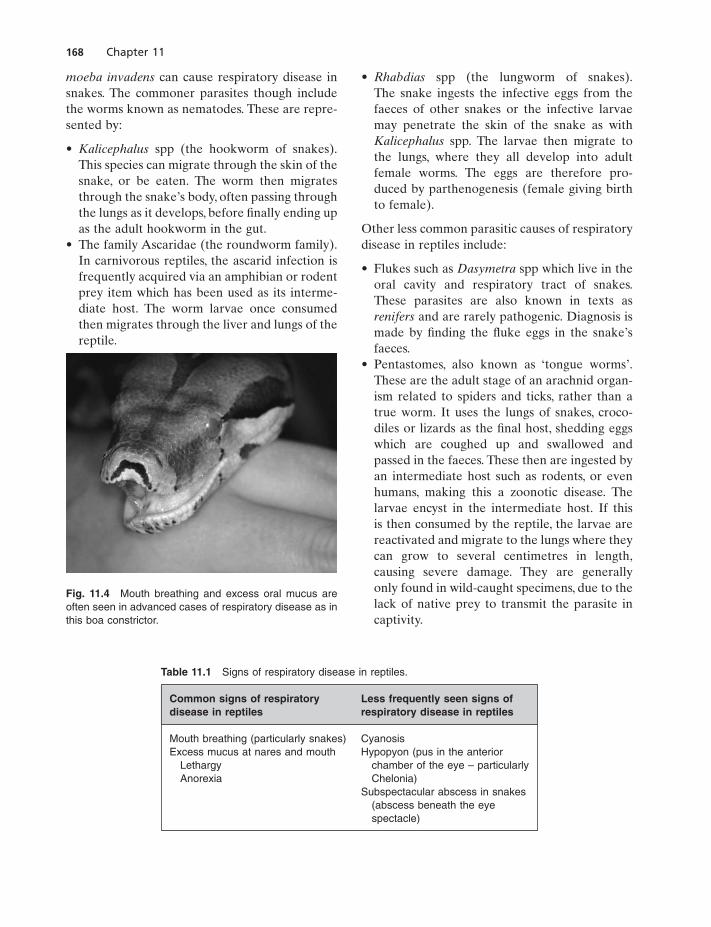
168 Chapter 11
moeba invadens can cause respiratory disease insnakes. The commoner parasites though includethe worms known as nematodes. These are repre-sented by:
• Kalicephalus spp (the hookworm of snakes).This species can migrate through the skin of thesnake, or be eaten. The worm then migratesthrough the snake’s body, often passing throughthe lungs as it develops, before finally ending upas the adult hookworm in the gut.
• The family Ascaridae (the roundworm family).In carnivorous reptiles, the ascarid infection isfrequently acquired via an amphibian or rodentprey item which has been used as its interme-diate host. The worm larvae once consumedthen migrates through the liver and lungs of thereptile.
• Rhabdias spp (the lungworm of snakes).The snake ingests the infective eggs from thefaeces of other snakes or the infective larvaemay penetrate the skin of the snake as withKalicephalus spp. The larvae then migrate tothe lungs, where they all develop into adultfemale worms. The eggs are therefore pro-duced by parthenogenesis (female giving birthto female).
Other less common parasitic causes of respiratorydisease in reptiles include:
• Flukes such as Dasymetra spp which live in theoral cavity and respiratory tract of snakes.These parasites are also known in texts asrenifers and are rarely pathogenic. Diagnosis ismade by finding the fluke eggs in the snake’sfaeces.
• Pentastomes, also known as ‘tongue worms’.These are the adult stage of an arachnid organ-ism related to spiders and ticks, rather than atrue worm. It uses the lungs of snakes, croco-diles or lizards as the final host, shedding eggswhich are coughed up and swallowed andpassed in the faeces. These then are ingested byan intermediate host such as rodents, or evenhumans, making this a zoonotic disease. Thelarvae encyst in the intermediate host. If this is then consumed by the reptile, the larvae arereactivated and migrate to the lungs where theycan grow to several centimetres in length,causing severe damage. They are generally only found in wild-caught specimens, due to thelack of native prey to transmit the parasite incaptivity.
Table 11.1 Signs of respiratory disease in reptiles.
Common signs of respiratory Less frequently seen signs of disease in reptiles respiratory disease in reptiles
Mouth breathing (particularly snakes) CyanosisExcess mucus at nares and mouth Hypopyon (pus in the anterior
Lethargy chamber of the eye – particularly Anorexia Chelonia)
Subspectacular abscess in snakes(abscess beneath the eye spectacle)
Fig. 11.4 Mouth breathing and excess oral mucus areoften seen in advanced cases of respiratory disease as inthis boa constrictor.

Common Reptile and Amphibian Diseases 169
Bacterial
Many bacteria have been isolated from the lungsof pneumonic reptiles. Some are primarypathogens and some are secondary invaders,following damage caused by organisms such as the above-listed parasites, or secondary to septi-caemic states or oral infections such as ‘mouthrot’. Examples of the bacteria seen in pneumoniasinclude Aeromonas spp, Pseudomonas spp,Klebsiella spp and Pasteurella spp.
‘Runny nose syndrome’ or rhinitis seen in Chelonia is mainly seen in members of the tortoise family, particularly the Mediterraneanspecies. No one pathogen has been determined asthe true cause, although many bacteria, and aherpes virus have all been isolated.
Bacterial sampling
Gaining access to the bacteria causing the pneu-monia may be difficult. There are two main tech-niques involved in sampling bacteria or parasitesthat cause pneumonias:
• Lung wash: This involves passing a sterilecatheter into the trachea of the reptile. Thismethod is usually reserved for snakes andlizards and may be done in the conscious animaldue to the lack of the cough reflex. Fractiousanimals may require sedation. Through thiscatheter, sterile saline may be infused, at dosesequal to 0.5–1ml per 100g body weight, andimmediately aspirated. This sample can then becultured to identify the pathogen. Sample col-lection may be enhanced by holding the reptileupside down whilst aspirating.
• Direct sampling: This involves taking a swabdirectly from the site of the problem. This maybe done via laparotomy or endoscopically. Inthe case of Chelonia, the reptile may be anaes-thetised, the shell overlying the pneumoniclesion aseptically prepared and a small holedrilled through to enable the bacteriologicalswab to be passed.
Viral
Paramyxovirus: paramyxovirus is becoming in-creasingly commonly seen in snakes in captivity.
This virus is highly infectious, being shed in respi-ratory secretions. It causes a haemorrhagic pneu-monia and viraemia which affects other organs aswell. There is no current treatment and it can befatal. Diagnosis is based on clinical signs and sero-logical tests on a blood sample.
Boiid inclusion virus: this has been associatedwith pneumonia in snakes – primarily pythons andboas. There is no treatment.
Herpes viruses: herpes viruses have beenrecorded in Mediterranean tortoises associatedwith upper respiratory tract infections which maylead to secondary pneumonias. Treatment can beattempted with the anti-viral drug acyclovir.
Cardiovascular disease
Infectious disease
There are few pathogens which specifically affect the cardiovascular system. Many of thosewhich cause systemic illness and septicaemia areresponsible for cardiac damage. In many cases the damage is due to a bacterial endocarditiswhich then causes micro-thrombi to seed off into the rest of the body as well as causing heart failure. Many of the Gram negative bacte-ria seen in reptiles, such as Aeromonas spp andPseudomonas spp, are associated with heartdisease. Diagnosis requires ultrasonography andradiology to determine heart size and internalstructure. Electrocardiograms can also be used toaid diagnosis. Final proof of the organisminvolved depends on blood culture, which may beperformed by direct cardiocentesis in a sedated oranaesthetised reptile.
Parasitic disease
There are several parasitic nematode wormswhich release microfilaria into the blood stream.These are microscopic young which may be trans-mitted from reptile to reptile by mosquitos andticks. These parasites may cause thrombi todevelop in any of the major blood vessels, causingischaemic necrosis or cardiac damage. Other para-

170 Chapter 11
sites, such as members of the fluke family, havealso been isolated. The adult fluke is not normallythe problem, but the eggs laid into the bloodstream cause thrombi to form and so block capil-lary beds.
Nutritional disease
Hypoiodinism in chelonians has been associatedwith cardiac disease and goitre. The feeding ofmineral-poor foods such as cucumber and lettuceor goitrogenic foods such as brassicas (e.g.cabbage, Brussels sprouts), may lead to this.
Oversupplementation with vitamin D3 andcalcium may lead to calcification of the tunicamedia of the major arteries, causing a decrease intheir elasticity and so increased blood pressure.This may lead to heart failure or aneurysm formation.
Dietary deficiencies in vitamin E and/or sele-nium can lead to a condition known as whitemuscle disease, and can cause a dilated cardiomy-opathy to develop. It may occur due to a dietarydeficiency, or to overfeeding with high fat foods,such as snakes fed overweight rodents or fish,such as tuna and mackerel, which are high inpolyunsaturates and increase the demand forvitamin E.
Urinary tract disease
Renal disease and gout
Damage to kidney function in terrestrial reptilescan lead to increasing blood levels of uric acid, themain excretory product of protein metabolism.Uric acid is not very water soluble, which meansit rapidly reaches its precipitation point whencrystals start to form and come out of solution.This may occur in internal organs such as theheart, kidneys and liver when it is known as vis-ceral gout. It may also occur in the joints, when itis known as ‘articular’ gout, particularly of thedistal limbs.
Many factors can contribute to renal damageand gout. Diet plays a role. Diets high in protein,in species designed for lower levels of protein,such as the herbivorous tortoises and green
iguanas, when fed commercial cat or dog foods,cause excessive production of uric acid. In addi-tion, these commercial mammalian diets oftenhave too high levels of vitamin D3 and calcium forthe average herbivorous reptile. This leads to calcification of soft tissue structures, such as thetunica media of the arterial walls, and the kidneysthemselves, leading to renal dysfunction and ele-vated blood uric acid levels.
Causes of renal disease
Parasitic
Flagellates, such as Hexamita spp, Trichomonasspp and Giardia spp have been reported in chelo-nians as well as in snakes and lizards, with involve-ment of the renal system. Hexamita spp areparticularly damaging to the chelonian kidneys.
Kidney flukes are found in several species ofkingsnake, and members of the Boiidae. They mayblock renal tubules and in high enough quantitiescause kidney failure.
Coccidial parasites have also been seen insnakes, migrating from the digestive system byway of the cloaca, back up the ureters and into thekidneys.
Bacterial
Almost any bacteria found in the digestive systemor environment of the reptile has the potential tocause renal disease if that reptile is stressed ordebilitated in any way. Stresses can include in-correct environmental temperatures, incorrectfeeding, malnutrition, rough and overhandling ofthe reptile. Bacteria commonly implicated includethe Salmonella spp, Aeromonas and Pseudomonasspp. In some chelonians, the bacteria responsiblefor septicaemic cutaneous syndrome, Citrobacterfreundii, may also cause disease. These bacteriagain access either via haematogenous spread from the gut, or via the cloaca, ascending theureters.
Iatrogenic
Iatrogenic causes include certain nephrotoxicdrugs, such as the aminoglycoside antibiotic

Common Reptile and Amphibian Diseases 171
family (e.g. gentamicin, amikacin etc.), and thediuretic frusemide. In addition, if sufficient musclebruising occurs, such as when too firm a grasp isused when handling the patient, then myoglobinis released. This causes damage to the kidney filtration membrane.
Reproductive tract disease
Egg-binding or post-ovulatory stasis
Dystocia is common in many species of reptile,the commonest being chelonians and many of the lizard family such as green iguanas. It is often due to hypocalcaemia because of an inap-propriate diet, however other causes include mal-formed eggs, fractured pelvic bones, cystic calculi,lack of nesting material or malnutrition of thefemale.
Dystocias can be difficult to diagnose in snakes,as they normally tend to be relatively quiet creatures. This difficulty is exacerbated in thosespecies (e.g. garter snakes) which are viviparous(that is give birth to live young rather than eggs)in that any abdominal swelling is much less pro-nounced. Any previous history of passing eggs,and then the presence of a persistent caudally-located mass is of course highly suggestive. Inaddition, any evidence of a prolapse of the cloaca or distal reproductive tract also indicatesdystocia (Plate 11.2).
In lizards, the patient may be obviously dis-tended with eggs, and becomes progressivelymore moribund and lethargic. Many snakes cansurvive prolonged periods of dystocia, but lizardsare not so resilient and dystocia may prove fatalwithin days.
Chelonia may show signs of discomfort andstraining or they may show no signs at all.Radiographs are often the only way to tell if a chelonian is gravid.
Pre-ovulatory stasis
A condition recorded in both Sauria and Chelo-nia is pre-ovulatory stasis. This is when the ovariesproduce follicles, which enlarge but do not shedinto the reproductive tract. This results in two
ovaries containing anywhere up to 30–40 yolks,displacing all other coelomic organs. Over timethe affected reptile becomes anorectic and lethar-gic and often succumbs to secondary diseases andmalnourishment. The cause of the condition is notfully understood.
Musculoskeletal disease
Metabolic bone disease
The disease is common in young growing lizardsand chelonians that are being fed on a calciumdeficient diet, often in conjunction with a vitaminD3 deficiency due to a lack of ultraviolet light.(See also pages 152 and 154.)
The disease presents in lizards as an individualwhich is often weak and lethargic. The limbs aremarkedly swollen due to thickening of the bones.A lack of mineralisation causes the now primarilyfibrous bone structure to weaken. The bones compensate for this by increasing their width. Inaddition, pathological fractures are common.Also softening of the jaws is common, and this cancause foreshortening of the mandible, due to thepull of the tongue and neck muscles.
In chelonians the disease is often seen as a soft-ening and pyramiding of the shell. The plastronand carapace are weakened due to the hypomin-eralisation which allows the muscles to deformtheir structure. This is particularly obvious overthe internal attachments of the fore- and hindlimbs where depressions are seen. In some chelo-nians, for example, the softened shell allows thecorners of the carapace to roll upwards.
Fractures
These may be pathological, as is seen in nutri-tional deficiencies of calcium, often combinedwith a lack of ultraviolet lighting. Alternativelythey may be traumatic in origin.
Autotomy (tail shedding)
Autotomy, or spontaneous shedding, with subse-quent regrowth of the tail is seen in some saurianspecies such as the gecko and iguanid families.

172 Chapter 11
These lizards have fracture planes in their tailsallowing a clean break, with minimal bleeding, tooccur should a predator attack or a handler beoverzealous in restraint attempts. The tail will sub-sequently regrow, but when it does so the lost coccygeal vertebrae are replaced with a rod of cartilage, and the scale pattern is frequently muchmore haphazard.
In iguanids, once the individual matures, auto-tomy is often lost. This can occur over the age of2.5–3 years in the green iguana for example.
Neurological disease
Nutritional
Hypocalcaemic collapse
This is commonly seen in gravid lizards, such asgreen iguanas, when the blood calcium levels droptoo low. The female becomes flaccidly paralysed,and unresponsive. Occasionally fine muscletremors will be seen.
Hypovitaminosis B1
This has been mentioned on page 153. It is seenin primarily fish eating reptiles such as gartersnakes. The thiaminases present in salt-water fishbreak down the vitamin B1 present in the foodleading to a functional deficit. The presentingsigns include opisthotonus and a lack of a rightingreflex.
Biotin deficiency
This is seen in species fed mainly on unfertilisedhen’s eggs that contain large amounts of theantibiotin vitamin, avidin. This produces a relativedeficiency in biotin which leads to muscle tremorand general weakness. Monitor lizards are com-monly affected.
Hypoglycaemia
This is a condition seen in crocodiles. It isunknown why it occurs, but muscle tremor andweakness are seen.
Environmental
This can occur due to freezing injuries. These areseen in chelonians overwintering outside in theUnited Kingdom. Frost damage causes blindness,vestibular disease and death. There is no treatment.
Parasitic
Acanthamoebic meningoencephalitis is a condi-tion seen primarily in snakes due to the gut parasites of the acanthamoeba family. Fits andopisthotonic seizures are seen. Treatment is generally unsuccessful.
Viral
Paramyxovirus
These have been reported in many species ofsnake. The virus causes haemorrhagic pneumo-nias but will also cause neurological signs such asthe loss of righting reflexes. There is no currenttreatment.
Inclusion body disease of boiids
This is another viral condition seen primarily inboas and pythons. In pythons the disease is severewith infectious stomatitis, pneumonia and neuro-logical signs such as loss of the righting reflex, dis-orientation and blindness often followed rapidlyby death. In boas, the disease is fatal in young indi-viduals. In older patients, the disease producesmore chronic neurological signs with anorexia,vomiting and pneumonias. Neurological signs aremilder, with a loss of ability to chew and swallowprey, and a loss of the striking reflex. There is notreatment for this disease.
Overview of reptile biochemistry
Blood for biochemical parameters is best col-lected in a heparinised container. This also appliesfor haematological parameter measurements as potassium EDTA-coated tubes often causelysis of reptilian red blood cells, particularly inchelonians.
Common Reptile and Amphibian Diseases 173
Liver parameters
There are no liver-specific enzymes in reptiles. Theuse of the enzyme aspartamine transaminase(AST) has been widely noted. Levels in excess of150–200IU/l are suggestive of problems, but asthere is much species variation and this enzyme isfound in the skeletal muscle as well it is only aguide.
Renal parameters
Uric acid is useful for uricotelic species such asmost terrestrial chelonians, lizards and snakes.Levels over 450mmol/l suggest renal problems, butcare should be taken in carnivorous species suchas snakes to ensure that they have not recentlyconsumed a meal as this will falsely elevate uricacid levels. Uric acid levels will only be patholog-ically elevated if greater than 75% of the renalmass ceases to function, so this is not a sensitivetest. Urea is much less useful, although it has beenused in Chelonia immediately after hibernation toassess dehydration. Creatinine appears to be of nouse in reptiles.
Glucose
Levels below 3mmol/l are seen in the hypogly-caemic syndrome of crocodiles. Fits, incoordina-tion and weakness are a common sign inmalnourished reptiles, which will also show lowblood glucose levels.
Calcium and phosphorus
Calcium levels vary between 2–5mmol/l in mostreptiles. Levels below 2mmol/l can be seen ingravid female lizards with egg-binding, and younggrowing reptiles with metabolic bone disease.Calcium levels will increase up to four-fold infemales producing eggs. Phosphorus levels varyfrom 0.3mmol/l to 1.8mmol/l. Calcium to phos-phorus ratios of less than 1 :1 have been used toindicate early renal disease.
Total proteins
Total protein levels are used to indicate generalnutritional status and liver function. Dry chem-
istry techniques such as those commonly used inpractice blood analysers are not accurate foralbumin and globulin levels. Total protein valuesof 30–80g/l have been quoted as normal.
Diseases of amphibians
Bacterial
Redleg
Redleg is a common disease seen in amphibianscaused by the bacteria Aeromonas spp. It causesulcerating red wounds, which give it its name(Plate 11.3). The bacteria also causes a septicaemicsyndrome with the amphibian becoming bloatedand rapidly going into renal and hepatic failure.Poor environmental conditions are often blamed.
Mycobacteria
Mycobacterial infections are caused by environ-mental species such as Mycobacterium marinum,which are extremely resistant species and causeclassical tuberculous lesions mainly on the limbsor internal organs. The affected amphibian losesweight rapidly becoming emaciated. There is noeffective treatment.
Fungal
Saprolegniasis
This is a common environmental fungal diseaseseen in all aquatic species. It is seen classically asstrands of white, cottonwool-like material adher-ing to the skin surface (Plate 11.4). The conditionis worsened in warmer waters.
Phycomycosis
This is due to common moulds such as Mucor spp.These are often darkly pigmented. These mouldswill often affect amphibian eggs.
Viral
Herpes virus
A well-known viral condition of the North American leopard frog is casued by the herpes

174 Chapter 11
virus that induces a form of renal adenocarci-noma, known as Lucke’s renal tumour. Thetumour grows during the warmer months, with the virus being shed in the spring to infect otherfrogs. Renal failure occurs with chronic weightloss and death. There is no treatment for this condition.
Parasitic
Nematodes
The commonest nematode seen in anurans is thelungworm Rhabdias spp, the same family group ofparasite as is seen in reptiles. In large numbers thisparasite may cause pnuemonia. The parasite hasa direct life cycle.
Other nematodes of the Strongyloides familymay parasitise the gut and coelomic cavity, withlarge burdens resulting in poor growth and intesti-nal blockages.
Protozoa
Entamoeba ranurum: can cause damage to thelarge intestine of anurans, as well as causing liverinfections and abscesses. These may lead to weightloss, diarrhoea with blood, anorexia and dehydra-tion. This protozoa has also been associated withkidney damage in some species of toad and ascitesand oedema of the limbs may be seen.
Oodimium pillularis: is a motile protozoan whichaffects many aquatic animals including amphib-ians. It damages the skin, and in tadpole stages
damages the gills leading to anoxia. It is able toswim through the water from amphibian toamphibian and can therefore spread rapidly.
Metabolic bone disease
This is common, particularly in frogs and toads(anurans). There is often softening of the lowerjaw which bulges laterally, weakness and paralysisof the limbs, with spontaneous fractures.
References
Frye, F. (1991) Biomedical and Surgical Aspects of CaptiveReptile Husbandry Volume 1. Kreiger Publishing,Malabar, Florida.
Huchzermeyer, F.W. and Cooper, J.E. (2001) Fibriscess, notabscess, resulting from localised inflammatory responseto infection in reptiles and birds. Veterinary Record147:515–516.
Further reading
Bone, R.D. (1992) Gastrointestinal system. In Manual ofReptiles (Beynon, P.H., Lawton, M.P.C. and Cooper, J.E.,eds), pp. 101–116. BSAVA, Cheltenham.
Cranfield, M.R. and Grazyk, T.K. (1996) Cryptosporidiosis.In Reptile Medicine and Surgery (Mader, D.R., ed), pp.359–363. W.B. Saunders, Philadelphia.
Lane, F.J. and Mader, D.R. (1996) Parasitology. In ReptileMedicine and Surgery (Mader, D.R., ed), pp. 185–203.W.B. Saunders, Philadelphia.
Wright, K.M. (1996) Amphibian husbandry and medicine.In Reptile Medicine and Surgery (Mader, D.R., ed), pp.436–459. W.B. Saunders, Philadelphia.
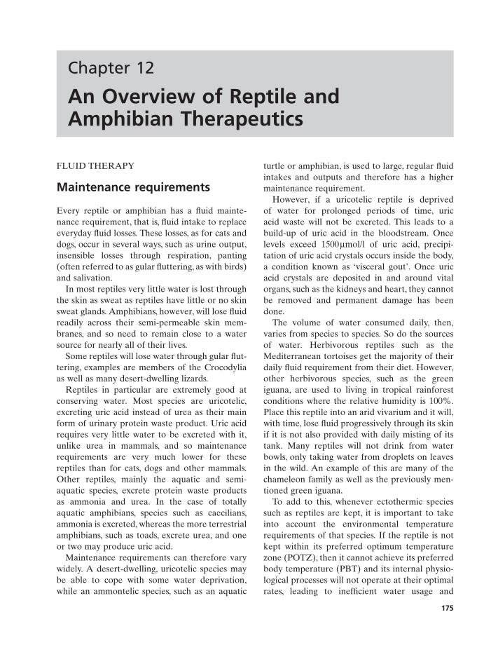
FLUID THERAPY
Maintenance requirements
Every reptile or amphibian has a fluid mainte-nance requirement, that is, fluid intake to replaceeveryday fluid losses. These losses, as for cats anddogs, occur in several ways, such as urine output,insensible losses through respiration, panting(often referred to as gular fluttering, as with birds)and salivation.
In most reptiles very little water is lost throughthe skin as sweat as reptiles have little or no skinsweat glands. Amphibians, however, will lose fluidreadily across their semi-permeable skin mem-branes, and so need to remain close to a watersource for nearly all of their lives.
Some reptiles will lose water through gular flut-tering, examples are members of the Crocodyliaas well as many desert-dwelling lizards.
Reptiles in particular are extremely good atconserving water. Most species are uricotelic,excreting uric acid instead of urea as their mainform of urinary protein waste product. Uric acidrequires very little water to be excreted with it,unlike urea in mammals, and so maintenancerequirements are very much lower for these reptiles than for cats, dogs and other mammals.Other reptiles, mainly the aquatic and semi-aquatic species, excrete protein waste products as ammonia and urea. In the case of totallyaquatic amphibians, species such as caecilians,ammonia is excreted, whereas the more terrestrialamphibians, such as toads, excrete urea, and oneor two may produce uric acid.
Maintenance requirements can therefore varywidely. A desert-dwelling, uricotelic species maybe able to cope with some water deprivation,while an ammontelic species, such as an aquatic
turtle or amphibian, is used to large, regular fluidintakes and outputs and therefore has a highermaintenance requirement.
However, if a uricotelic reptile is deprived of water for prolonged periods of time, uric acid waste will not be excreted. This leads to abuild-up of uric acid in the bloodstream. Oncelevels exceed 1500mmol/l of uric acid, precipi-tation of uric acid crystals occurs inside the body,a condition known as ‘visceral gout’. Once uricacid crystals are deposited in and around vitalorgans, such as the kidneys and heart, they cannotbe removed and permanent damage has beendone.
The volume of water consumed daily, then,varies from species to species. So do the sourcesof water. Herbivorous reptiles such as theMediterranean tortoises get the majority of theirdaily fluid requirement from their diet. However,other herbivorous species, such as the greeniguana, are used to living in tropical rainforestconditions where the relative humidity is 100%.Place this reptile into an arid vivarium and it will,with time, lose fluid progressively through its skinif it is not also provided with daily misting of itstank. Many reptiles will not drink from waterbowls, only taking water from droplets on leavesin the wild. An example of this are many of thechameleon family as well as the previously men-tioned green iguana.
To add to this, whenever ectothermic speciessuch as reptiles are kept, it is important to takeinto account the environmental temperaturerequirements of that species. If the reptile is notkept within its preferred optimum temperaturezone (POTZ), then it cannot achieve its preferredbody temperature (PBT) and its internal physio-logical processes will not operate at their optimalrates, leading to inefficient water usage and
Chapter 12
An Overview of Reptile andAmphibian Therapeutics
175

176 Chapter 12
consumption. A list of optimum temperaturerequirements can be found on page in Table 8.1.
The effect of disease on fluid requirements
Fluid loss may be rapid, due to water loss alone,such as occurs with acute diarrhoea, thermalburns or vomiting. In this case, the remainingextracellular fluid (ECF) becomes reduced, but isstill of the same composition (isotonic). Alterna-tively, fluid loss may be due to long-term anorexia,producing a reduction in electrolytes and creatinga hypotonic ECF. Finally, water deprivation ororal trauma preventing drinking will lead toincreases in the tonicity of the ECF, and create ahypertonic dehydration.
With any disease, the need for fluids increases,even if no obvious fluid loss has occurred. This isdue to a number of reasons. It may involve renalchanges, such as increases in the glomerular filtration rate or reduction in water reabsorptionby the collecting ducts so causing increased urineoutput. Or there may be reduced absorption ofwater from the small or large intestine.
Respiratory disease is a not uncommon findingin reptiles, especially tortoises and septicaemicsnakes. In these animals chronic levels of lunginfection can occur, with increased respiratorysecretions being the result. Fluid loss via this routecan be appreciable.
Another less obvious route is fluid and elec-trolyte loss through skin disease. Reptiles oftensuffer from serious burns from unprotectedbasking lamps and faulty heaters. Not only willthere be serious fluid and electrolyte loss via full-thickness skin burns but these reptiles willsuccumb to secondary skin infections from envi-ronmental bacteria such as Pseudomonas spp.These produce lesions which resemble chemicalor thermal burns, and leave large areas of weepingexudative skin for further fluid loss.
Finally we have to consider the need for fluidtherapy during other forms of medical therapy,such as antibiotic treatment. Many of the morecommonly seen bacterial infections in reptiles arecaused by Gram negative bacteria, therefore theaminoglycoside family of antibiotics (gentamicin,
tobramycin, amikacin etc.) has been widely usedfor treatment. This family of antibiotics hasseveral serious side-effects, particularly in thedehydrated patient. The most serious of these isrenal damage if for any reason the drug accumu-lates in the kidney. This can happen if there isreduced renal perfusion because of dehydration.Levels of the drug then reach toxic concentrationsvery quickly. The renal damage so caused can beeasily enough to kill even a healthy reptile.
Post-surgical fluid requirements
Surgical procedures are more and more fre-quently being performed on reptiles and amphibians in both routine and emergency situa-tions. Causes of fluid loss include the possibility ofintrasurgical haemorrhaging. This will call for vas-cular support with an aqueous electrolyte solutionor, in more serious blood losses (greater than 10%blood volume), colloidal fluids or even bloodtransfusions.
Even if surgery is relatively bloodless there are inevitable losses via the respiratory route. This is due to the drying nature of the gases used to deliver the anaesthetics commonly used inreptile and amphibian surgery. As many of thesespecies are small in size they have a large lungsurface area in relation to volume and hence agreater loss of fluid per unit time/per breath than larger animals. To exacerbate the situation,many patients are not able to drink immediatelyafter surgery, and so the period without water or food intake may stretch to a few hours –enough time for any reptile or amphibian to startto dehydrate. Finally, some forms of surgery willlead to inappetance for a period, as for someforms of head trauma – such as the repair of tortoises that have gone ten rounds with thefamily lawnmower!
Electrolyte replacement
Other diseases, such diarrhoea, will cause fluidloss and metabolic acidosis due to the prolongedloss of bicarbonate. This is not uncommon in reptiles, and is often due to parasitism, such as

Reptile and Amphibian Therapeutics 177
amoebiasis, in snakes. There may also be chroniclosses of potassium, in cases of chronic diarrhoea,due to the reduced absorption of this electrolyteby the large intestine.
Snakes will vomit after a meal if stressed, andmay suffer from diseases of the stomach such ascryptosporidiosis, causing loss of fluid and hydro-gen ions, and a resultant metabolic alkalosis.Other reptiles such as tortoises will rarely vomit,so the likelihood of fluid loss via this route is lesscommon.
Fluids used in reptilian practice
Lactated Ringer’s/Hartmann’s
As with cats and dogs, lactated Ringer’s solution isuseful as a general purpose rehydration and main-tenance fluid. It is particularly useful for reptilesand amphibians suffering from metabolic acidosis,such as those described above with chronic gas-trointestinal problems, but can also be used forfluid therapy after routine surgical procedures.
Glucose/saline combinations
Glucose/saline combinations are useful for reptiles and amphibians, as they may have beenthrough periods of anorexia prior to treat-ment, and therefore may well be borderline hypoglycaemic.
There is some evidence that in reptiles, andprobably amphibians, the isotonicity of the extra-cellular fluids is lower than that of mammals.Studies on non-marine reptiles suggest that iso-tonicity for the majority is 0.8% rather than the0.9% saline assumed for mammals. Because ofthis, a number of fluid combinations utilising theabove two types of crystalloid support have beenderived as follows:
• One third each of 5% glucose with 0.9% saline,lactated Ringer’s solution and sterile water
• Nine parts 5% glucose with 0.9% saline to 1part sterile water.
Many texts still advise that straightforward,undiluted lactated Ringer’s solution or 4%glucose with 0.18% saline may be used, and
glucose/saline solutions are particularly useful foramphibia, where the use of potassium-containingfluids should be avoided initially.
It is important that whatever fluid is adminis-tered it be warmed to the reptile or amphibian’spreferred body temperature (approximately30–35°C) before being given.
Protein amino acid/B vitamin supplements
Protein and vitamin supplements are useful fornutritional support. Products such as Duphalyte®(Fort Dodge) may be used at the rate of 1ml/kg/day. They are used to replace nutrients incases where the patient is malnourished or hasbeen suffering from protein-losing enteropathy,such as may occur with heavy parasitism, or aprotein-losing nephropathy, as in renal failure. Itis also a useful supplement for patients withhepatic disease or severe exudative skin diseases,such as heater burns, in which blood proteins willbe reduced.
Colloidal fluids
Colloidal fluids have been used in reptilian practice when intravenous administration hasbeen possible. This limits their usefulness, as somereptiles are just too small to gain full vascularaccess, although there is some evidence that theymay be used via the intraosseous route. They areused when a serious loss of blood occurs, in orderto support central blood pressure. This may be a temporary measure whilst a blood donor isselected, or, if none is available, the only means ofattempting to support such a patient.
Blood transfusions
Blood transfusions are indicated when the packedcell volume (PCV) has dropped below 0.05 l/l, andthey may be given via intravenous or intraosseousroutes. Cross-matching of blood groups does notappear to be necessary for one-off transfusions,but the same species should be used each time,i.e. green iguana to green iguana, boa constrictorto boa constrictor. In an emergency, it is possible to transfuse a member of a family group with

178 Chapter 12
another from the same group, i.e. iguanid toiguanid and boiid to boiid. Up to 2% body weight as blood may be taken from healthyspecies (Klingenberg, 1996), preferably into a pre-heparinised syringe before immediately trans-fusing into the recipient.
Oral fluids and electrolytes
Oral fluid administration may also be used inreptile and amphibian practice for those patientsexperiencing mild dehydration, and for homeadministration. Many products are available forcats and dogs, and may be used for reptiles.However, as with the crystalloid fluids, it is advisable to dilute these oral electrolytes byapproximately 10%, otherwise their concentra-tion will be greater than the reptile’s extracellularfluid (ECF) and so water will move from the bodyinto the gastrointestinal tract rather than theother way around. The inclusion of a probioticwith the electrolytes may aid recovery by nor-malising gut flora and digestion.
Calculation of fluid requirements
Fluid requirements may be calculated as for catsand dogs. It is worth noting that a lot of the fluidintake is normally consumed in food e.g. in theform of fresh vegetation for herbivorous species.This is difficult to take into consideration, andtherefore it is safer to assume that the debilitatedreptile will not be eating significant enoughamounts for this to matter in the calculation. Inany case, levels of fluid replacement rates havereceived relatively little research. Consequentlyfor the vast number of reptiles and amphibians acalculated guess has to be made!
Frye (1991) recommends that levels of 20–25ml/kg/day be used for hydration purposes inboth reptiles and amphibians, and current litera-ture suggests that rates across several species varyfrom 10–50ml/kg/day.
The factor that limits the volume of fluids thatcan be administered is that, although intravenousand intraosseous routes may also be used, mostfluids are given intracoelomically to the debili-tated reptile. Reptiles and amphibians do not
possess true diaphragms, therefore the thorax and abdomen are all interconnected in a coelom.When fluids are placed in this cavity, it is equiva-lent to giving intraperitoneal fluids to a mammal,but as there is no diaphragm these fluids can causepressure to build up on the lungs. Excessive fluidsmay severely compromise respiration.
Excessive fluids given intravenously or intra-osseously may also overload the circulation andcause pulmonary oedema. It can result in cardiacand renal overperfusion and solute wash-out, withpotassium in particular being excreted with theincreased diuresis causing a hypokalaemic crisis to develop. This may manifest itself initially as an anorectic reptile, but will progress to cardiacarrhythmias, coma and death.
As with cats and dogs, one can assume that 1%dehydration equates with a need to supply 10ml/kg fluid replacement in addition to the main-tenance requirements. It is also possible to makesome qualitative assessment of the level of dehy-dration from the elasticity of the skin. Althoughreptile skin is not as elastic as mammalian, itshould be freely mobile and recoil, albeit slowly,after tenting. Other factors to assess are thebrightness of the corneas in species with mobileeyelids. In those without mobile eyelids (e.g.snakes) the collapse of the spectacle (the clearfused eyelids) is suggestive of dehydration. Otherassessments of thirst and urate output can bemade over 24 hours.
It is possible to estimate the degree of dehy-dration of a reptile patient as follows:
• 3% dehydrated – increased thirst, slightlethargy, decreased urates
• 7% dehydrated – increased thirst, anorexia,dullness, tenting of the skin with slow return tonormal, dull corneas, loss of turgor of spectaclesin snakes
• 10% dehydrated – dull to comatose, skinremains tented after pinching, desiccatingmucous membranes, sunken eyeballs, nourate/urine output.
The alternative is to compare packed cellvolumes and total protein levels to assess dehy-dration (Table 12.1), again with 1% increase inPCV suggesting 10ml/kg fluid replacements areneeded (this assumes no anaemia in the patient).

Reptile and Amphibian Therapeutics 179
It is important not to exceed 25–30ml/kg/day asa maximum for reasons mentioned above, what-ever the level of dehydration of the patient. Sorehydration of severely debilitated reptiles maytake days to weeks. As with avian patients, there-fore, making good the fluid deficit may need to besplit over several days.
Equipment for fluidadministration
Catheters
The most useful catheters for small reptiles andamphibians are the series of butterfly cathetersavailable, often adapted from human paediatricmedicine. These are extremely useful for the smalland fragile vessels in these patients, as they havea short length of tubing attached to the needle. Ifthe syringe or drip set is connected to this pieceof flexible tubing, rather than directly to thecatheter, there is less chance of the catheterbecoming dislodged if the reptile moves. Also, thepiece of clear tubing on the catheter allows you to see when venous access has been achieved,as blood will flow back into this area without theneed to draw back thus collapsing the fragileveins.
To make effective use of a butterfly catheter, itis advisable to flush it with heparinised salineprior to use to prevent clotting. 25–27 gauge sizesare recommended. They may also be used to giveintracoelomic fluids to reptiles, as the conscious
patient may continue to move (particularly aproblem in snakes) without dislodging the needle.
For larger patients, such as adult iguanas,monitor lizards and Crocodylia, 21–25 gauge over-the-needle Teflon®-coated catheters used for catsand dogs can be used.
Hypodermic or spinal needles
Hypodermic needles are useful for the adminis-tration of intraosseous, intracoelomic or subcuta-neous fluids.
Intraosseous fluids may be the only method ofcentral venous support in very small patients orpatients in which vascular collapse is occurring.The proximal femur, tibia or humerus may beused. Entry can be gained using spinal needles.These have a central stylet to prevent clogging ofthe lumen of the needle with bone fragments afterinsertion. 23–25 gauge spinal needles are usuallysufficient.
Straightforward hypodermic needles may alsobe used for the same purpose, although the risksof blockage are higher. Hypodermic needles mayalso be used, of course, for the administration ofintracoelomic and subcutaneous fluids. Generally23–25 gauge hypodermic needles are sufficient forthe task.
Pharyngostomy or oesophageal tubes
Pharyngostomy tubes are often used in reptiles inorder to provide nutritional support in as stress-free manner as is possible. They are also useful as a route for some fluid administration, as onlyliquid formulas will pass through these narrow(3.5–6.5 french) tubes. It should be noted thoughthat in severely dehydrated individuals there is areal possibility that gut pathology may exist, sothis route may need to be supplemented by others.This route therefore has limited use in facilitatingfluid replacement, and is used mainly for nutri-tional support and rehydrating and replenishingthe gut microflora.
Syringe drivers
For continuous fluid administration, such as isrequired for intravenous and intraosseous fluid
Table 12.1 Examples of normal packed cell volumes(PCV) and total blood proteins in selected species ofreptile.
Species PCV l/l Total protein g/l
Green iguana (Iguana 0.25–0.38 28–69iguana)
Tortoise (Testudo spp.) 0.19–0.4 32–50Ratsnake (Elaphe spp.) 0.2–0.3 30–60Boa constrictor (Boa 0.2–0.32 46–60
constrictor constrictor)

180 Chapter 12
administration, syringe drivers are being usedmore and more. The units themselves vary fromthe inexpensive, semi-disposable, battery-operated units available to the veterinary profes-sion, right through to the more expensive digitalunits requiring mains electricity. As an investmentfor the practice they are well worthwhile, as theymay be used for cat and small dog fluid adminis-tration as well. Their advantage is that smallvolumes, such as a fraction of a ml may be admin-istered accurately per hour. An error of 1–2ml insome of the smaller species dealt with over anhour could be equivalent to overperfusion of50–100%! In addition it is almost impossible tokeep gravity fed drip sets running at these lowrates without blocking every few minutes.
Intravenous drip tubing
Particular fine drip tubing is available for attach-ment to syringes and syringe-driver units. It isuseful if these are luer locking as this enhancessafety and prevents disconnection when thepatient moves.
Routes of fluid administration in reptile
As with cats and dogs the same medical principlesbroadly apply with five main routes of adminis-tration available as follows:
• Oral• Subcutaneous• Intracoelomic• Intravenous• Intraosseous.
Table 12.2 gives the advantages and disadvantagesof each of the five routes.
Oral
Snakes
The oral route is not very good for seriously debilitated animals, but useful for those withpharyngostomy feeding tubes in place, or if theowner or handler is experienced in stomach
tubing. This makes it useful for mild cases ofdehydration where owners wish to home treattheir pet. A stomach tube is passed by restrain-ing the snake’s head gently but firmly and theninserting a plastic or wooden tongue depressor to open the mouth. A lubricated feeding tube isthen passed through the labial notch (the area atthe most rostral aspect of the mouth withoutteeth) and to a depth of one third of the snake’slength.
Lizards
Gavage (stomach) tubes or avian straight croptubes or straightforward feeding tubes can beused to administer fluids directly into the oesoph-agus or stomach. The reptile needs to be firmlyrestrained to keep the head and oesophagus in astraight line. The mouth is opened with a plasticor wooden tongue depressor and the tube insertedto a depth of one third to one half the torso lengthof the reptile. This method is often stressful for thereptile. The alternative is to syringe fluids into themouth, but this risks inhalation in a debilitatedreptile. A pharyngostomy tube may be placed fornutritional support, and so may be used for fluidtherapy.
Chelonians
The oral route can be used as for lizards andsnakes. A pharyngostomy tube may be implantedas described below, and levels of 10ml/kg at anyone time can be administered. Alternatively, astomach tube may be inserted each time it isneeded. The feeding tube is first measured fromthe tip of the extended nose to the line where thepectoral and abdominal ventral scutes connect. Itcan then be lubricated and passed after extendingthe head and gently prising the mouth open witha wooden or plastic speculum (Fig. 12.1).
Placement of pharyngostomy tubes
Pharyngostomy tubes may be placed in anyspecies of reptile, but are particularly useful inchelonians, such as the leopard tortoise (Geoche-lone pardalis) which can retract its head deep

Reptile and Amphibian Therapeutics 181
inside its shell, making repeated stomach tubingimpossible. It also significantly reduces the stressand trauma of repeatedly passing a stomach tubein long-term anorectic reptile patients.
The steps for placement are as follows:
(1) Sedation or anaesthesia is required, and goodanalgesia post implantation.
(2) Surgically prepare the site with 0.25–0.5%povidone-iodine, being particularly scrupu-lous as reptile skin is notoriously dirty. In che-lonians, the implantation site is the ventralaspect of the lateral neck, 3–4cm caudal to theangle of the jaw. In snakes and lizards it is theventrolateral aspect of the throat region, 5–10cm caudal to the angle of the jaw.
Table 12.2 Advantages and disadvantages of various fluid therapies for reptiles and amphibians.
Advantages Disadvantages
Oral • Minimal stress with experienced handler • Stressful with inexperienced handler• Physiological route for fluid intake • No use in cases of digestive tract disease• Less risk of tissue trauma • May damage stomach if the stomach tube is• Home therapy possible inserted too roughly• Rapid administration • Rehydration rates are slow
• Risk of inhalational pneumonia• Limited volumes may be administered at any
one time
Subcutaneous • Large volumes may be given at one time • May be uncomfortable for patient• Rapid administration possible, minimising • Risk of muscle and subcutaneous tissue
stress trauma• Uptake may be better than oral route in • Rates of rehydration poor if severely
cases of digestive tract disease dehydrated and peripheral vessels are • Minimal risk of internal organ damage collapsed
during administration • Only isotonic or hypotonic fluids may beadministered
• Darkening of the skin at injection site in many lizards such as chameleons and iguanas
Intracoelomic • Large volumes may be administered at • Large volumes may cause pressure on the one time, increasing dosing intervals lungs (no diaphragm)
• Uptake is faster than subcutaneous • Rehydration rates may still be slow in route in dehydrated patients severely hypovolaemic patients
• Generally relatively painless route of • Only isotonic or hypotonic fluids may be givenadministration • Increased risk of organ damage
Intravenous • Rapid rehydration in even severely • Size of reptile may prevent venous accessdehydrated patients is possible • Species of reptile (e.g. snakes) may make
• Colloidal fluids and blood transfusions venous access difficult without minor surgerypossible • Veins are more fragile than mammalian
• Use of intravenous catheters and vesselssyringe drivers makes accurate, • Increased skill levels and equipment continuous, reduced-stress dosing requirementspossible
Intraosseous • Rapid rehydration possible even with • Not useful in presence of infection collapsed peripheral vasculature (osteomyelitis) or skeletal nutritional disease
• Accurate dosing and continuous (metabolic bone disease)perfusion possible • Sedation, local or general anaesthesia is
• Use of colloidal fluids and blood requiredtransfusions possible • Tolerance may be poor in some species
• Useful in smaller species and specieswhere venous access is difficult

182 Chapter 12
(3) A pair of curved haemostats is placed inthrough the mouth and pushed laterally andventrally, tenting the skin above them.
(4) A sharp incision is made with a scalpel blade,over the point of the haemostats, through theskin and the underlying muscle.
(5) The tubing, preferably as large a diameter as will comfortably fit down the oesophagus(Foley catheters are useful in tortoises), isgrasped with the haemostats as they protrudeout through the incision, and then pulled intothe pharynx and pushed down into theoesophagus.
NB The tube should be measured prior toimplantation so the depth of insertion isknown. It is measured in tortoises from thesite of the tube incision to the mid-portion ofthe plastron, halfway through the abdominalscutes (page 116). A further length shouldthen be allowed, in order to attach the end of the feeding tube to the dorsal aspect of thecarapace.
(6) Once in place, two pieces of zinc oxide tapemay be attached to the tube close to the skinsurface. Through this, sutures may be placed,attaching this to the skin itself. The tube maythen be attached to the midline cranial cara-pace in tortoises, or taped to the side of theneck for snakes and reptiles, and a bunginserted (Fig. 12.2, Plate 12.1). Care of thetube is as for nasogastric tubes in cats anddogs. For example, plain water should beflushed through the tube prior to administer-ing food to ensure correct placement. Thisshould also be done after feeding to flush fooddebris out of the tube.
Subcutaneous
Snakes
The lateral aspect of the dorsum of the snake, inthe caudal third of its body, is the ideal site for subcutaneous fluid administration. This is a goodtechnique for use for routine post-operativeadministration of fluids to patients undergoingminor surgical procedures such as skin massremovals. If positioned correctly, there is a lymphsinus running lateral to the epaxial muscles oneither side, just subcutaneously, which can be usedfor moderately large volumes. It may however stillbe necessary to use several sites.
Lizards
The lateral thoracic area is easily used for smallervolumes of fluids at any one site. There is a risk of the reptile developing a darkened, pigmentedarea over the injection site, particularly in thechameleon family.
Fig. 12.1 Placement and depth of insertion of a stomachtube in a chelonian.

Reptile and Amphibian Therapeutics 183
Chelonians
The subcutaneous route is easily used for post-operative fluids and mild dehydration in thisspecies. Fluids may be given in the area justcranial to the hind limbs, or in the skin folds justlateral to the neck. Relatively large volumes maybe given via this route.
Intracoelomic
Snakes
The intracoelomic route is useful for more seri-ously dehydrated reptiles, as there is a greater vasculature at this site for absorption. The needleor butterfly catheter is inserted two rows of lateralscales dorsal to the ventral scutes in the caudalthird of the snake, but cranial to the vent. Theneedle is inserted so that it just penetrates thebody wall, the plunger of the syringe is pulledback to ensure no organ puncture has occurredand the fluids administered. If correctly insertedthere will be no resistance to the injection.
Lizards
Because of the positioning necessary for adminis-tration, the intracoelomic route may be a stressfulmethod of fluid administration. As for smallmammals, the lizard should be placed in dorsalrecumbancy with its head downwards to encour-age the gut contents to fall cranially and away
from the injection site. The needle, preferably 25gauge or smaller, is advanced slowly to just popthrough the abdominal wall in the lower rightventral quadrant. The syringe plunger should bepulled back to ensure that no organ has been penetrated, and the fluids can be administeredwithout any resistance.
Chelonians
The intracoelomic route can be used in tor-toises up to a maximum of 20–25ml/kg/day only, otherwise, due to the confines of the rigidshell, the fluids will place too much pressure onthe lung fields. The area cranial to the hind limbsis used, i.e. the same site as for subcutaneousroutes, but the chief difference is depth. Theconcern with this route is that the bladder lies inthis area, and if full may be punctured. The otherroute is the cranial access site. This is locatedlateral to the neck and medial to the front limband is more epicoelomic than truly intracoelomic.The needle is kept close and parallel to the plas-tron and a 3/4-in needle may be inserted to the levelof the hub.
Intravenous
Snakes
There are no major vessels for intravenous use in snakes which are easily accessible. If an intra-
Fig. 12.2 A pharyngostomy tube inplace in an anorectic spur-thighedtortoise (Testudo graeca).

184 Chapter 12
venous route is to be used one of the following isrequired.
Ventral tail vein: this is more of a plexus of veins, and may be accessed from the ventrum.The needle is inserted midline, one third of the taillength from the vent, and advanced until ittouches the coccygeal vertebrae at a 90-degreeangle. The needle is then retracted slightly whilstdrawing back on the syringe until blood flows intothe hub. Fluids may then be given slowly.
Palatine vein: this is present on the roof of themouth, as its name suggests, and is paired. Can-nulation may be performed with a 25–27 gaugebutterfly catheter although the snake usually hasto be sedated or anaesthetised to gain access.
Jugular vein: these also can only be accessed in ananesthetised or sedated snake. To gain access,a full-thickness skin cut-down procedure may beperformed 5–7.5cm caudal to the angle of the jaw,two rows of scales dorsal to the ventral scutes. Thejugular vein can then be seen medial to the ribs.An over-the-needle catheter is best for this, andshould then be sutured in place, therefore acatheter with plastic wings is advised.
Intracardiac: this site can be used in emergencies.The heart may be catheterised under sedation oranaesthesia only. On turning the snake onto itsback, the heart may be seen to beat against theventral scale, approximately one quarter of itslength from the snout. A 25–27 gauge over-the-needle catheter may be inserted between thescales, ventrally, in a caudocranial manner at 30degrees to the body wall into the single ventricle.A bolus may be administered, or it may be taped,glued or sutured in place for 24–48 hours.
Lizards
The intravenous route can be difficult in smalllizards, and frequently requires sedation or anaes-thesia. Several veins may be tried.
Cephalic vein: this is approached in the anaes-thetised lizard by performing a cut-down proce-dure on the cranial aspect of the middle of the
antebrachium, perpendicular to the long axis of the radius and ulna. The vessel may then becatheterised using an over-the-needle catheter,which is then sutured in place. This technique isreally only useful for lizards over 0.25kg in weight.
Jugular vein: this vessel may be accessed via a cut-down technique in the anaesthetised or sedatedlizard. An incision is made in a craniocaudal direc-tion from 2.5cm caudal to the angle of the jaw. Anover-the-needle catheter may then be sutured inplace.
Ventral tail vein: this is more of a plexus of veins.It is accessed from the ventral aspect of the tailand can be performed in the conscious lizard. It isfrequently only suitable for one-off bolus injec-tions, and special care should be taken withspecies which exhibit autotomy (spontaneous tailshedding) such as day geckos and green iguanas.The needle is inserted at 90 degrees to the angleof the tail and advanced until it touches the coccygeal vertebrae. It is then withdrawn slightlywhile drawing back on the syringe. When bloodflows into the syringe, the infusion may begin (Fig.12.3).
Chelonians
There are two main intravenous routes, the dorsaltail vein and the jugular veins.
Fig. 12.3 Green iguana in dorsal recumbancy for ventraltail vein fluid administration or blood sampling.

Reptile and Amphibian Therapeutics 185
Jugular vein: these may be accessed for catheterplacement in the sedated or anaesthetised tor-toise. The neck is extended and the head tiltedaway from the operator to push the neck towards him or her. The jugular vein runs from the dorsal aspect of the eardrum along the moredorsal aspect of the neck (Fig. 12.4). An over-the-needle catheter may be placed directly, or,in thicker-skinned animals, a cut-down techniqueemployed.
Dorsal tail vein: this is more of a plexus of veins.Therefore it is often not possible to give largevolumes of fluids, and certainly not possible toplace a catheter. Access is midline, on the dorsalaspect of the tail. The needle is inserted at a 90-degree angle until it hits the coccygeal vertebrae.The needle is then pulled back, drawing back onthe syringe at the same time, until blood flows intothe hub (Fig. 12.5).
Intraosseous
Snakes
The intraosseous route is not possible in thesnake.
Lizards
The intraosseous is a good route for the smallerspecies of lizards, where venous access is restrictedor difficult. There are a few access points to
choose from. Hypodermic or spinal needles of23–25 gauge sizes may be used.
Proximal femur: this may be accessed from thefossa created between the greater trochanter andthe hip joint. This route may be difficult due to the90-degree angle the femur often forms with thepelvis.
Distal femur: this is relatively easy to access fromthe stifle joint. It does restrict the movement ofthe stifle, but it is easier to bandage the catheterin to this site and access to the medullary cavityof the femur is certainly easier via this route.Sedation or anaesthesia is required. See below forthe placement technique.
Proximal tibia: this again is possible in the largerspecies. Anaesthesia and sedation is needed, andthe spinal needle or hypodermic needle may bescrewed into the tibial crest region in a proxi-modistal manner.
Placement of distal femoral intraosseouscatheters in lizards
The technique for placement of a distal femoralintraosseous catheter in a lizard is explained indetail below.
(1) Sedation or anaesthesia (local or general) isneeded, and in all cases it is advised that goodanalgesia is administered.
Fig. 12.4 Placement of a jugular catheter in a chelonian. Fig. 12.5 Access to dorsal tail vein in chelonian for fluidadministration or blood sampling.

186 Chapter 12
(2) Surgically scrub the area overlying the craniolateral aspect of the stifle joint usingpovidone-iodine. It is important that place-ment of the catheter or needle is performedas aseptically as possible.
(3) Take a 20–23 gauge spinal or hypodermicneedle and insert it in through the ridge justproximal to the stifle joint, screwing it into thebone in the direction of the long axis of thefemur proximally.
(4) Flush the needle with heparinised saline (theadvantage of a spinal needle is that it has a central stylet which helps prevent it frombecoming plugged with bone fragments).
(5) Tape the needle securely in place and applyan antibiotic cream around the site. It may be worthwhile radiographing the area toensure correct intramedullary placement ofthe needle.
(6) Once the needle or catheter has been cor-rectly placed, attach the intravenous tubingand bandage it securely in place by wrappingbandage material around the leg of thepatient.
(7) Finally, it may be necessary to immobilise thelimb by bandaging it to a splint to prevent dis-lodgement of the catheter, which should nowbe attached to a syringe driver.
Chelonians
Two main intraosseous sites can be used.
Plastrocarapacial junction/pillar: this is the pillarof shell which connects the plastron to the cara-pace. It is approached from the caudal aspect,just cranial to one of the hind limbs. The spinal orhypodermic needle (21–23 gauge) is screwed intothe shell attempting to keep the angle of insertionparallel with the outer wall of the shell, so enter-ing the shell bone marrow cavity (Fig. 12.6). Inlarger older species, the shell may be too tough toallow penetration.
Proximal tibia: this may be approached as forlizards. The area is thoroughly scrubbed with0.25–0.5% povidone-iodine and the hypodermicor spinal needle is screwed into the tibial crest in the direction of the long axis of the tibia distally.
Routes of fluid administration in amphibians
Cutaneous
The cutaneous route is unique to amphibians andmakes use of their semi-permeable skin. It can be used only with mildly dehydrated amphibians,and should only involve the use of dechlorinated,plain water. It should be warmed to the amphib-ian’s preferred body temperature and be well oxygenated before immersing the patient.Absorption will occur across the skin membranes.
Fig. 12.6 Intraosseous fluid admin-istration via the plastrocarapacialpillars. Note syringe driver in background.

Reptile and Amphibian Therapeutics 187
Oral
This route can be used for hypotonic fluid therapyvia a small feeding tube inserted orally. Thedanger is that trauma can easily occur duringrestraint and opening of the amphibian’s mouthduring this procedure. In addition the process ismoderately stressful.
Intracoelomic
This is accessed in the right lower ventral quad-rant of the ‘abdomen’. The amphibian should beplaced in dorsal recumbancy and with its headdown to allow coelom contents to fall away fromthe injection site.
Intravenous
In the larger anurans and some salamanders,the midline ventral abdominal vein may be usedfor bolus fluids or blood transfusions. The vessel,as its name suggests, lies midline just below theskin surface and runs from the pubis area of thepelvis to the xiphoid of the sternum. A 25–27gauge insulin needle may be used to gain access,although the vessel is very fragile and care shouldbe taken not to rupture it.
Treatment of reptilian diseases
As this text is aimed at the veterinary nurse ortechnician it is not intended to give exhaustivelists of treatments or drug dosages, rather to givean idea of the treatments possible and the tech-niques useful to aid recovery. For drug dosagesthe reader is referred to one of the many excel-lent texts listed in the additional reading list at theend of this chapter.
Metabolic scaling of drug dosages
It should be mentioned that, due to the lack ofresearch, many drugs have not been evaluated inreptiles and amphibians.
Drug doses may (with care) be extrapolatedfrom known doses in other species. However,to do this, the differences in basal metabolic
rate (BMR) or metabolism should be taken into consideration (see Chapter 10, on reptile and amphibian nutrition). Reptiles have a muchlower metabolic rate than mammals, and this is reflected by the fact that they excrete drugs atslower rates. The environmental temperature atwhich the reptile or amphibian is kept, due totheir ectothermic nature, will also greatly affecttheir metabolic rate and hence excretion of any drug administered. Therefore, dosages anddose rates are often much lower in reptiles thanin mammals.
A formula has been derived to ‘metabolicallyscale’ the dosage of a drug in a tested species, suchas a dog, to an untried one, such as a snake. Forthose keen to read more on this subject the readeris referred to the BSAVA Manual of Reptiles(1992 edition).
Reptile dermatological disease therapy
Table 12.3 highlights some of the treatments andtherapies commonly used for the management ofreptilian dermatological diseases.
Reptile digestive tract disease therapy
Table 12.4 highlights some of the treatments andtherapies commonly used for the management ofreptilian digestive tract diseases.
Reptile respiratory and cardiovasculardisease therapy
Table 12.5 highlights some of the treatments and therapies commonly used for the manage-ment of reptilian respiratory and cardiovasculardiseases.
Reptile reproductive tract disease therapy
Table 12.6 highlights some of the treatments and therapies commonly used for the manage-ment of reptilian reproductive tract diseases.

Table 12.3 Treatment of skin diseases.
Diagnosis Treatment
EctoparasitesMites Ivermectin 0.2mg/kg injection on 2–3 occasions at 10–14 day intervals.
Can spray environmental mixture of 0.5ml ivermectin + 1 litre water with 1–2ml propylene glycol to aid mixing to remove remaining mites. Fipronil has also been used topically, sprayed onto a cloth and wiped over the reptile
Ticks Manual removal, topical ivermectin/fipronil. Treat for secondary bacterial infection
Blowflies Manual removal, often under sedation, antibiosis and treatment of initiatingcause
Leeches Manual removal after applying lignocaine to leech
Bacterial, fungal and viral skin Mycobacterial infections require surgical excision. For topical fungal diseases infections use enilconazole washes, but difficult to treat if systemic.
Some herpes virus infections may be treated with acyclovir topically asointment or orally once daily at 80mg/kg (Stein, 1996)
Dysecdysis (1) Rehydrate patient(2) Treat underlying cause(3) Luke-warm water shallow bathing, allow access to abrasive surfaces for
snakes, e.g. wet towels.(4) Retained spectacles in snakes may be removed carefully with viscous
tear drops and moistened cotton buds.
Scale rot (1) Isolate bacteria/fungi involved and obtain sensitivity.(2) Blisters treated topically dilute povidone iodine, silver sulfadiazine
cream/enilconazole washes.(3) Parenteral antibiosis(4) Prevention geared to reducing substrate moisture and increase hygiene.
Abscesses Surgical therapy due to fibrous nature. Requires debridement and topicalantiseptic with systemic antibiosis based on culture and sensitivity.
Table 12.4 Treatment of digestive system diseases.
Diagnosis Treatment
Mouth rot Antibiosis based on culture and sensitivity. Necrotic tissue should be debrided under sedation/anaesthesia. Topical compounds containing silver sulfadiazine and framycetin are useful. Intralesion injections of antibiotic and vitamin C advised (Frye, 1991). For Chelonian herpesvirus, use topical iodine washes and acyclovir ointment.
Stomach diseases Tumours: surgical option.Nematodes: ivermectin 0.2mg/kg once, or fenbendazole 100mg/kg orally once, or
oxfendazole 60mg/kg once. Note: dose may require repeating.Cryptosporidiosis: unrewarding, but metronidazole may be used (may be toxic in indigo
and king snakes) or use spiramycin and paromycin. Raising environmental temperature to 80°F may help.
Intestinal diseases Nematodes: see stomach diseases. NEVER USE IVERMECTIN IN CHELONIA AS IT IS LETHALEntamoebiasis: metronidazole 160mg/kg orally once daily for 3 days (beware toxicity in
indigo and king snakes).Coccidiosis: sulfadimidine, orally, 50mg/kg once daily for 3 daysFlagellates: (Chelonia particularly) metronidazole 260mg/kg orally once.
Liver diseases Entamoebiasis: see intestinal diseases.Hepatic lipidosis: supportive nutritional and fluid therapy. Use of anabolic steroids and
thyroxine in Chelonia may be helpful.

Reptile and Amphibian Therapeutics 189
Table 12.5 Treatment of respiratory and cardiovascular system diseases.
Diagnosis Treatment
Parasitic respiratory disease Nematodes: Ivermectin may be used at 0.2mg/kg EXCEPT IN CHELONIA. Alternatively, fenbendazole and oxfendazole may be used (see intestinal disease treatment, Table 12.4).
Pentastomes: Manual removal or levamisole at 5mg/kg may help.
Bacterial respiratory disease Based on culture and sensitivity results. Lung washes may be used to collect samples for this, and to flush out infection if antibiosis is used in the lung wash.
Cardiovascular disease Heart failure: may use furosemide, but care should be taken as nephrotoxic in the long-term.
Filariasis: Ivermectin 0.2mg/kg. Raising environmental temperature may help to kill adult worms.
Goitre: Iodine supplement at 2–4mg/kg orally once weekly (Stein, 1996).
Table 12.6 Treatment for reproductive system diseases.
Diagnosis Treatment
Dystocia (post-ovulatory stasis, Provide nesting material and quiet location. Supplement with calcium Fig. 12.7) gluconate 100mg/kg intramuscularly if you believe hypocalcaemic
paralysis has occurred. Oxytocin at 5–35 IU/kg once. Fluid therapy to correct dehydration. Percutaneous/per cloacal aspiration of egg contents via needle and syringe (see Table 6.6) to collapse egg. Surgical caesarian (Fig. 12.8, Plate 12.2) may be required as a last resort.
Pre-ovulatory stasis Surgical spey advised – seen in Sauria and Chelonia, with supportive therapy initially (Plate 12.3).
Fig. 12.7 Heavily pregnant femalegreen iguana with post-ovulatorystasis.

Fig. 12.8 Surgical removal of eggsfrom same iguana.
Table 12.7 Treatment of musculo-skeletal diseases.
Diagnosis Treatment
Metabolic bone Correction of dietary deficiency and possible UV light deficiency. In cases of hypocalcaemic disease paralysis 100mg/kg calcium gluconate intramuscularly is advised.
Fractures In cases of metabolic bone disease it is better to correct diet and splint fractures than repair surgically.
Lizards: for one fore limb fracture bandage limb to body wall (see Fig. 12.9). For bilateral humeral fractures use a coaption splint. For one hindlimb fracture bandage limb to tail base. For digital fractures ball bandage as for avian patients.
Chelonia: possible to bandage limb into shell. This is useful in cases of metabolic bone disease. Spinal fractures should be splinted as neural control may be regained.
All species may require external coaption or internal surgical fixation to mend fractures.
Tail loss For young iguanids and geckos treat stump as an open wound and dress with topical silver sulfadiazine cream or iodine antiseptic (dilute).
For agamids or species not showing autonomy (tail re-growth) and older iguanids which lose this ability, suture the stump surgically.
Reptile musculoskeletal systemdisease therapy
Table 12.7 highlights some of the treatments andtherapies commonly used for the management ofreptilian musculoskeletal system diseases.
Reptile neurological system diseasetherapy
Table 12.8 highlights some of the treatments andtherapies commonly used for the management ofreptilian neurological system diseases.
Treatment of amphibian diseases
Table 12.9 highlights some of the treatments andtherapies commonly used for the management ofamphibian diseases.
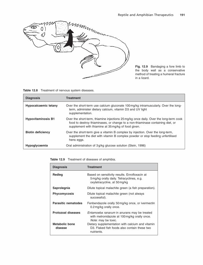
Reptile and Amphibian Therapeutics 191
Fig. 12.9 Bandaging a fore limb tothe body wall as a conservativemethod of treating a humeral fracturein a lizard.
Table 12.8 Treatment of nervous system diseases.
Diagnosis Treatment
Hypocalcaemic tetany Over the short-term use calcium gluconate 100mg/kg intramuscularly. Over the long-term, administer dietary calcium, vitamin D3 and UV light supplementation.
Hypovitaminosis B1 Over the short-term, thiamine injections 25mg/kg once daily. Over the long-term cook food to destroy thiaminases, or change to a non-thiaminase containing diet, or supplement with thiamine at 35mg/kg of food given.
Biotin deficiency Over the short-term give a vitamin B complex by injection. Over the long-term, supplement the diet with vitamin B complex powder or stop feeding unfertilised hens eggs.
Hypoglycaemia Oral administration of 3g/kg glucose solution (Stein, 1996)
Table 12.9 Treatment of diseases of amphibia.
Diagnosis Treatment
Redleg Based on sensitivity results. Enrofloxacin at 5mg/kg orally daily. Tetracyclines, e.g.oxytetracycline, at 50mg/kg
Saprolegnia Dilute topical malachite green (a fish preparation).
Phycomycosis Dilute topical malachite green (not alwayssuccessful).
Parasitic nematodes Fenbendazole orally 50mg/kg once, or ivermectin0.2mg/kg orally once.
Protozoal diseases Entamoeba ranarum in anurans may be treatedwith metronidazole at 100mg/kg orally once.Note: may be toxic.
Metabolic bone Dietary supplementation with calcium and vitamindisease D3. Flaked fish foods also contain these two
nutrients.

192 Chapter 12
References
Frye, F.L. (1991) Infectious diseases. In Biomedical and Surgical Aspects of Captive Reptile Husbandry, 2nd edn,Vol 1. Kreiger Publishing, Malabar, Florida.
Klingenberg, R.J. (1996) Therapeutics. In Reptile Medicineand Surgery (Mader, D.R., ed). W.B. Saunders,Philadelphia.
Stein, G. (1996) Reptile and amphibian formulary. InReptile Medicine and Surgery (Mader, D.R., ed), pp.465–472. W.B. Saunders, Philadelphia.
Further reading
Bone, R.D. (1992) Gastrointestinal system. In Manual ofReptiles (Beynon, P.H., Lawton, M.P.C. and Cooper, J.E.,eds), pp. 101–116. BSAVA, Cheltenham.
Cranfield, M.R. and Grazyk, T.K. (1996) Cryptosporidiosis.In Reptile Medicine and Surgery (Mader, D.R., ed), pp.359–363. W.B. Saunders, Philadelphia.
Pokra, M.A., Sedgewick, C.J. and Kaufman, G.E. (1992)Therapeutics. In Manual of Reptiles (Beynon, P.H.,Lawton, M.P.C. and Cooper, J.E., eds). BSAVA,Cheltenham.
Wright, K.M. (1996) Amphibian husbandry and medicine.In Reptile Medicine and Surgery (Mader, D.R., ed),pp. 436–459. W.B. Saunders, Philadelphia.

Small Mammals

Classification of small mammals
The commonly seen species of small mammals inveterinary practice are classified in Table 13.1.
RABBIT
Biological average values for thedomestic rabbit
Table 13.2 below gives the basic biological para-meters for domestic rabbits.
Musculoskeletal system
The skeletal system of rabbits is extremely light. As a percentage of body weight the rabbit’sskeleton is 7–8% whereas the domestic cat’sskeleton is 12–13%. This makes rabbits prone tofractures, especially of the spine and the hindlimbs. This is because of a combination of thepowerful musculature of their hind legs and theirhabit of kicking out.
Skull
The mandible is narrower than the maxilla, andthe temperomandibular joint has a wide surfacearea allowing lateral movement of the mandiblein relation to the maxilla.
Axial skeleton
The cervical vertebrae are box-like and small andgive great mobility. The thoracic vertebrae possessattachments to the twelve paired ribs which are
flattened in comparison to cats ribs. The pelvis isnarrow and positioned vertically. The ilial wingsmeet the ischium and pubis at the acetabulum,where an accessory bone unique to rabbits, calledthe os acetabuli, lies. The pubis forms the floor ofthe pelvis and borders the obturator foramenwhich is oval in rabbits as compared to the catwhere it is round.
Appendicular skeleton
The scapula is slender and distally has a markedlyhooked suprahamate process projecting caudallyfrom the hamate process. The scapula articulateswith the humerus which in turn articulates withthe radius and ulna. In rabbits, the ulna fuses tothe radius in older animals and the two bones aredeeply bowed. The radius and ulna articulate withthe carpal bones which in turn articulate with themetacarpals and the five digits.
The hind limbs are similar to a cat’s exceptingthe femur is flatter ventrodorsally and the tibiaand fibula are fused in the rabbit. The tibia articu-lates distally with the tarsal bones where there isa prominent calcaneous bone. The tarsals articu-late with the metatarsals which articulate with thefour hind limb digits.
The hind limbs are well muscled and powerful.Lagomorph muscle is a pale colour, unlike thedomestic cat’s which is a deeper red.
Respiratory anatomy
Upper respiratory tract
The paired nostrils can be closed tightly shut in lagomorphs. Rabbits, like horses are nasalbreathers, with the nasopharynx permanently
Chapter 13
Basic Small Mammal Anatomy and Physiology
195

196 Chapter 13
locked around the epiglottis, hence upper respira-tory disease or evidence of mouth breathing arepoor prognostic signs. The nasolacrimal ductsopen onto the rostral floor of the nasal passage.The epiglottis is not visible easily from the oralcavity making direct intubation difficult. It isnarrow and elongated and leads into the larynxwhich has limited vocal fold development. The
larynx leads into the trachea which has incom-plete C-shaped cartilage rings for support.
Lower respiratory tract
The trachea bifurcates into two primary bronchi.There are two lungs, which are relatively small in proportion to the overall rabbit’s body size.
Table 13.1 Classification of commonly seen small mammals.
Order Rodentia Carnivora
Suborder Lagomorpha Myomorpha Hystricomorpha Sciuromorpha Fissepedia
Family Leporidae Muridae Cricetidae Caviidae Sciuridae Mustelidae
Species Domestic Rat (Rattus Gerbil Guinea pig Siberian Domesticrabbit norvegicus) (Meriones (Cavia chipmunk ferret(Oryctolagus unguiculatus) porcellus) (Tamias (Mustelacuniculi) Mouse sibericus) putorius
(Mus Syrian hamster Chinchilla furo)musculus) (Mesocricetus (Chinchilla
auratus) laniger)
Russian Deguhamster (Octodon(Phodopus degus)sungorus)
Chinesehamster(Cricetuluscricetus)
Table 13.2 Biological parameters for the domestic rabbit.
Biological parameter Average range
Body weight (kg) 1.5 (Netherland dwarf) to 10 (NewZealand whites and Belgian hares)
Rectal body temperature (°C) 38.5–40Respiratory rate at rest (breaths/min) 30–60Heart rate at rest (beats/min) 130 (New Zealand whites) to 325
(Netherland dwarf)Gestation length (days) 29–35 (average 31)Litter size 4–10Age at sexual maturity (months) –
Male 5–8Female 4–7
Life span (years) 6–10

Small Mammals: Anatomy and Physiology 197
This means that even minor lung disease maycause serious problems. The majority of the inspi-ratory effort comes from the movement of thediaphragm and abdominal musculature ratherthan the intercostal muscles and ribcage move-ment. Each lung has three lobes, with the cranialones being the smallest.
Respiratory physiology
Much of the impetus for inspiration derives fromthe muscular contraction and flattening of thediaphragm. In rabbits the lungs are the ‘shockorgan’ (for cats and dogs it is the gut). The lungparenchyma possesses a cellular population thatis well supplied with anaphylactic mediatingchemicals. These are strong enough to cause fluidextravasation and blood pooling as well as spasmswithin the walls of the main pulmonary arterialsupply, leading to rapid right-sided heart failure.This is of particular importance when performingchest surgery and anaesthetising a rabbit.
Digestive system
Oral cavity
The dental formula is
I 2/1 C 0/0 Pm 3/2 M 3/3
The roots of all of the teeth are open, allowingcontinual growth throughout the rabbit’s life. Themolar enamel is folded providing an unevenocclusal surface with the ipsilateral jaw whichallows some interlocking of the teeth. Wear is kepteven by the lateral movement of the mandibleallowing independent left and right arcades toengage in mastication. The incisors differentiatethe order Lagomorpha from Rodentia, as therabbit and hare have two smaller incisors, or ‘pegteeth’, behind the upper two forming an angledwedge, whereas rodents have only two upperincisors. The larger incisors only have enamel onthe labial surface, whereas the smaller maxillarypeg teeth have enamel on the labial and lingualsides. This allows the wedge-shaped bite plane ofthe incisors wherein the lower incisors closeimmediately behind the upper large incisors andfit into a groove made by the peg teeth. The per-manent incisors are present at birth, although thepeg teeth are replaced by permanent peg teeth ataround the second week of life. The deciduouspremolars present at birth are replaced and joinedby permanent molars by the fourth week of life.There are no canines, instead there is a gap, ordiastema, between the incisors and premolars(Fig. 13.1).
Fig. 13.1 Lateral diagram of anormal rabbit skull showing therelation of tooth roots to the orbitand jaw bones. Note peg-teethincisors behind main incisors inmaxilla.

198 Chapter 13
Stomach
The stomach is a large, simple structure, with astrong cardiac sphincter. This makes vomiting in the rabbit virtually impossible. There is a main body, or fundus, and a pyloric section with awell-formed pyloric sphincter. The lining of thestomach wall contains acid-secreting and separatepepsinogen-secreting cells. The pH of the rabbit’sstomach contents is surprisingly higher than a cator dog’s at 1.5–1.8. In addition, a healthy rabbit’sstomach never truly empties, so an empty stomachon radiography is an abnormal finding.
Small intestine
The total length of the small intestine in theaverage rabbit may be some 2–3 feet!
Caecum and large intestinal anatomy
At the junction of the ileum and caecum lies thesacculus rotundus. This is a swelling of the gut,infiltrated with lymphoid tissue and a commonsite for foreign body impactions. The caecum is alarge, sacculated and spiral-shaped organ, finish-ing in a blind-ended, thickened, finger-like projec-tion known as the vermiform appendix which alsocontains lymphoid tissue. The bulk of the caecumis thin walled and possesses a semi-fluid digestivecontent.
The start of the large intestine is the ampullacoli which sits near to the sacculus rotundus andcaecum. It is a smooth-walled portion of the gutwith some lymphoid infiltration of its walls, unlikethe rest of the large intestine. It is also distin-guished by having bands of fibrous tissue (knownas taeniae) which create sacculations (also knownas haustra). At the end of the proximal colon thetaeniae and haustra cease, and the gut is thenknown as the fusus coli, its walls becoming thick-ened and smooth because of the presence of largemembers of nerve ganglia. These nerve cells act aspacemakers for contraction waves in the largebowel. The distal descending colon then continuesthrough the pelvis to empty through the rectumand anus. There are a couple of anal glands justinside the anus, one on either side, emptying theirsecretions onto the faecal pellets.
Large intestinal physiology
Two types of faecal pellet are produced by therabbit. One is a true faecal pellet, comprisingwaste material in a dry and light brown sphericalform. The other is a much darker, mucus-coveredpellet known as a caecotroph. The caecotroph iseaten directly from the anus, as soon as it is pro-duced, which in the wild is during the middle ofthe day when the rabbit is often underground. Incaptivity they are often produced overnight, butmay be produced at any time. The caecotrophcontains plant material from which all of thenutrients have yet to be extracted.
The large bowel can produce two types of pelletdue to the unique contraction waves in the largeintestine and caecum. Food accumulates in themultiple haustra of the proximal colon,and reverseperistaltic waves flush fine particulate matter backinto the caecum. Here bacterial and microbial fer-mentation occurs, breaking down the tough cellu-lose and hemicellulose walls of the plant material.The larger fibre particles, which would not bedigested adequately in the caecum,are selected outby the proximal colon and pass out of the gut as thecaecotroph pellet. This will be re-eaten directlyfrom the rabbit’s anus, and so undergo a seconddigestion process. The caecotrophs are mucuscovered to protect their microbial contents fromthe acidic stomach contents, allowing furthermicrobial breakdown of the fibre.The smaller fibreparticles which were flushed back into the caecum,once broken down as far as possible, will then bepassed out of the caecum, back into the colon, andexcreted as the true, dry faecal pellet.
Liver
The rabbit liver has four lobes. There is a gallbladder, which has an opening separate from the pancreatic duct into the proximal duodenum.The main bile pigment is biliverdin, rather thanthe more usual bilirubin seen in cats and dogs.
Pancreas
The pancreas is a diffuse organ, suspended in theloop of the duodenum. There is one single pan-creatic duct, separate from the bile duct, empty-ing into the proximal duodenum.

Small Mammals: Anatomy and Physiology 199
Urinary anatomy
Kidney
The kidneys are similar in shape to the cat’skidneys. The right kidney is slightly more cranialthan the left, and they are often separated fromthe ventral lumbar spine by large fat deposits. Asingle ureter arises from each kidney and tra-verses across the abdominal cavity to empty intothe urinary bladder.
Bladder
The bladder lining is composed of transitional cellepithelium. The urethra in the male rabbit exitsthrough the pelvis and out through the penis. Infemales the urethra opens onto the floor of thevagina.
Renal physiology
Like most herbivores, rabbits’ urine is alkaline innature, with a pH varying between 6.5–8, but itwill become acidic if the rabbit has been anorec-tic for 24 hours or more. The urine will containvarying amounts of calcium carbonate due to therabbit’s unique method of calcium metabolism.The rabbit has no ability to alter how muchcalcium is absorbed from the gut. This means anyexcess calcium must be excreted by the kidneysinto the urine, where, owing to its alkaline nature,crystals of calcium carbonate form. This can beseen as a tan-coloured silt. Other pigments mayalso be seen in rabbits’ urine. These are known asporphyrins. These are plant pigments, and makethe urine appear anywhere from a dark yellow toa deep wine red in colour. This may mimic haema-turia, so to diagnose blood in the urine, it is oftennecessary to examine it microscopically.
Cardiovascular system
Heart
The rabbit heart is relatively small in relation to body size. The right atrioventricular valve
has only two cusps instead of three, unlike catsand dogs. The pulmonary artery also has a largeamount of smooth muscle in its wall which cancontract vigorously during anaphylactic shock,causing immediate right-sided cardiac overloadand failure.
Blood vessels for sampling
Vascular access in rabbits include the lateral earvein which, as its name suggests, runs along the lateral margin of either ear. It may be enteredusing a 25 or 27 gauge needle or catheter and used for slow intravenous injections and bloodsampling. Application of local anaesthetic creamto the area 2–3 minutes prior to venipuncture isrecommended to avoid the rabbit jumping at thecritical moment and to encourage vasodilation.
The central ear artery runs along the midline ofthe outer aspect of each pinna. It is a large vessel,and is generally avoided for blood sampling dueto fear of causing thrombus formation, whichwould cut off the blood supply to the ear tip,leading to avascular necrosis. It may however beused with care in an emergency using a 25–27gauge needle, and applying pressure to the vesselonce sampled.
Cephalic vein
The cephalic vein runs in a similar position to thatseen in cats and dogs. It may be split into two insome individuals, but may be used for intravenousfluids and sampling (Fig. 13.2).
Saphenous vein
The saphenous vein runs across the lateral aspectof the hock, as in cats and dogs, and may also beused for venipuncture.
Jugular vein
The jugular veins are prominent in the rabbit butthey form the major part of the drainage of bloodfrom the orbit of the eye. Therefore if ahaematoma and thrombus forms and blocks thelumen of a jugular vein, severe orbital oedemamay occur, with possible damaging effects.

200 Chapter 13
Eye plexus
The eye also has a large venous plexus at themedial canthus,draining the orbit.This is an impor-tant structure to avoid when enucleating the eye.
Lymphatic system
Spleen
The spleen is a flattened structure, oblong innature and attached to the greater curvature ofthe stomach, and is thus found predominantly onthe left side.
Thymus
The thymus is a large structure in the cranial thoracic compartment even in the adult rabbit. Ithas the same functions as for any other mammal,in that it provides the body with the T-cell popu-lation of lymphocytes.
Lymph nodes
The root of the mesentery supporting the diges-tive tract is well supplied with lymph nodes, as is
the hilar area of the lungs where the two mainbronchi diverge to supply each lung. In additionthere are superficial lymph nodes in the popliteal,prescapular and submandibular areas, althoughthese are much smaller deposits than are found indogs and cats.
Reproductive anatomy
Male
The paired testes can move from an inguinal posi-tion within the thin-skinned scrotal sacs, to anintrabdominal position through the open inguinalcanal. The scrotal sacs are sparsely haired and lieeither side of the anogenital area.
The accessory sex glands which are found in the buck attached to the urethra in the caudalabdomen are a dorsal and a smaller ventralprostate, a bilobed vesicular gland, a bilobed coagulating gland and a bilobed bulbourethralgland. There is no evidence of an os penis. Theprepuce has numerous small preputial glands inthe dermis and there are a couple of inguinalglands situated either side of the penis whichsecrete a brown-coloured sebum clearly seenadjacent to the anus.
Female
The ovaries are small, elongated, bean-shapedstructures, supported by the ovarian ligament and lying caudal to each respective kidney. Theovarian artery often splits into two parts afterleaving the aorta, and it, along with the rest of thereproductive tract, is frequently encased in largeamounts of fat, as the doe’s reproductive tract isa fat deposition site.
The uterus is duplex – there is no commonuterine body. Instead there are two separate uteriwith separate cervices emptying into the vagina.The vagina is large and thin walled, with theurethra opening onto its floor cranial to the pelvis.The vulva therefore is a common opening for thereproductive and urinary systems unlike manyrodents. It lies just cranial to the anus, and isflanked on either side by the inguinal glands, aswith the buck.
Fig. 13.2 Cephalic vein access in a rabbit using a pre-heparinised butterfly catheter.

Small Mammals: Anatomy and Physiology 201
The doe has on average four pairs of mammaryglands extending from the inguinal region up tothe axillary areas.
Reproductive physiology
Male
The buck rabbit has the same sexual hormones asin cats and dogs, but they are on a seasonal timeclock triggered by the lengthening daylight of thespring. This is mediated through the pineal glandin the brain. This has neural links from the retinas,and controls the hormone melatonin. It in turncontrols the pituitary release of follicle stimulat-ing and luteinising hormones which then act uponthe testes.
Female
Does are induced ovulators. Waves of folliclesswell and regress during the course of the season,starting to increase in activity in early spring. Ifnot mated, these follicles will often dominate thecycle for 12–16 days at a time. There is no realanoestrus phase in does, instead a slight waning inactivity for 1–2 days occurs before a return toheat. During peak sexual activity the vulva isoften deeply congested and almost purple incolour and considerably enlarged.
Once mated, the male’s semen may form a copulatory plug, which is a gelatinous accumula-tion of sperm which drops out of the doe’s vagina 4–6 hours post mating. Gestation lasts from 29–35days, with the foetus forming a haemochorial placenta (where the outer chorion layer of thefoetal placental membrane burrows into the liningof the uterus so that it directly attaches to theblood in the intrauterine vessels) at about day 13.This is a common time for abortions to occur. Apregnant doe will often remove fur from herventrum to line the nest in the latter few daysprior to parturition.
Dystocia is uncommon. The doe only nurses thekittens once a day for 20 minutes or so, often inthe early morning. It is therefore not uncommonfor owners to think that the doe is neglecting heryoung, as she will often spend the rest of the timeeating and away from the litter.
Pseudopregnancy or phantom pregnancy is notuncommon and often occurs after an unsuccessfulmating or mounting activity by another buck ordoe. A corpus luteum forms and this lasts for15–17 days during which time the doe mayproduce milk, and build a nest. At this time thedoe is susceptible to mastitis.
Neonatology
The young kits or kittens are altricial in nature, i.e.they are totally dependent on the mother fornutrition and survival for the first few weeks oflife. They are born blind, deaf and furless. Furgrowth appears around days 5–6, the eyes open at8–10 days and the ears at 11–12. Weaning occursaround 6 weeks of age, with the young taking solidfood from 2–3 weeks.
Sexing
The young may be sexed from 4–5 weeks of age. Gentle pressure is placed either side of thereproductive/anal area to protrude the vulva orpenis. The vulva of the young doe is rounded andhas a central slit in midline and projects cranially.The penis of the young buck is more conical andpointed, with no central slit and tends to projectcaudally when protruded. Once the buck is older,the testes descend into the scrotum.
Skin
Lop breeds, particularly does, have extra skinfolds called ‘dewlaps’ around the ventral neckregion. In addition, such extra folds of skin maybe found around the anogenital area, leading toincreased risk of urine and faecal soiling.
Rabbits do not have the keratinised foot pads that are found in cats and dogs. Instead they have thick fur covering the areas of the toesand metatarsals which are pressed flat to theground.
In addition to the para-anal scent glands mentioned above, there are a series of discretesubmandibular chin glands. These are used to

202 Chapter 13
mark territory and also, in the case of does, tomark their young to distinguish them from others.
The rabbit has no skin sweat glands except afew along the margins of the lips. This means thatthey are very prone to heat stress at temperaturesgreater than 28°C.
The presence of many vibrissae or sensitivehairs around the lips and chin are important, asrabbits cannot see anything immediately belowtheir mouths, and so rely on touch to manipulatefood towards the mouth.
Eyes
Rabbits have prominent eyes, which allow a near360° field of vision. There is a prominent thirdeyelid which moves from the medial canthus of theeye, and possesses a large amount of reactive lym-phoid tissue within its structure and a Harderiantear gland at its base. This is often enlarged in thebuck during the breeding season and possessestwo lobes in both the sexes. There are two blindspots, just in front of the rabbit below the mouthand immediately behind the head. The formermeans that the rabbit detects its food at closerange by smell and by touch with the sensitivewhiskers known as vibrissae which fringe the lips.
Haematology
The most notable difference between rabbit andfeline or canine haematology is the staining of the
rabbit neutrophil, which makes it resemble the cator dog eosinophil, which is why it is often knownas the pseudoeosinophil. Many rabbits have morelymphocytes than pseudoeosinophils, resemblingother mammals such as cattle, rather than cats anddogs, where the neutrophil is the commonestwhite blood cell.
In other respects the red and white blood cellsof the rabbit resemble those of the domestic catand dog.
RAT AND MOUSE
Biological average values for therat and mouse
The normal biological values for the rat andmouse are given in Table 13.3 below.
Musculoskeletal system
Skull
The skull of both species is elongated. The eyesare laterally situated and there is a long snout and a shallow cranium. The maxilla is narrowerthan the mandible, the reverse of the rabbit. Thetemperomandibular joint is elongated cranio-caudally allowing the mandible to move rostrallyand caudally in relation to the maxilla. This allows the incisors to be engaged for gnawingwhilst the molars are disengaged. Alternatively,the molars may be engaged for mastication prior
Table 13.3 Biological parameters of rats and mice.
Average AverageBiological parameter range rat range mouse
Weight (g) 400–1000 25–50Rectal body temperature (°C) 37.6–38.6 37–38Respiration rate at rest (breaths/min) 60–140 100–280Heart rate at rest (beats/min) 250–450 500–600Gestation length (days) 20–22 19–21Litter size 6–16 8–12Age sexual maturity (weeks) Male 8 6
Female 10 7Oestrus interval (days) 4–5 4–5Life span (years) 3–4 2–3

Small Mammals: Anatomy and Physiology 203
to swallowing, whilst the incisors are disengaged.The two procedures cannot occur at the sametime. The rostral symphysis, joining each half ofthe mandible, is also articulated allowing move-ment of each hemimandible independently of theother.
Axial skeleton
The pelvis of the female mouse is joined at thepubis and ischial areas midline by fibrous tissue.This allows separation of the pelvis during partu-rition in the mouse. There are no fibrous areas tothe pelvis of the female rat and consequently nopelvic separation occurs.
Appendicular skeleton
Each species has shortened limbs. The scapulaarticulates at its coracoid process with the clavi-cles as well as the humerus. There are fourmetacarpal bones in the rat, with four digits. Occa-sionally the vestigial remnant of digit 1 is present.
The hind limbs have a strongly laterally-bowedfibula in both species. The tibia articulates withfive metatarsal bones at the hock joint. Conse-quently there are five digits in the hind limb ofthese species. Rats and mice are plantigrade intheir stance, i.e. they walk with the whole of themetatarsal bone area flat to the ground.
Male rats may still have open growth plates inmany of their long bones well into the second yearof their lives, whereas mice close their growthplates in the first 3–4 months of life.
Respiratory system
The nares of both species are prominent and sur-rounded by an area of hairless skin which docontain some sweat glands.
There is a vomeronasal organ in the floor of thenasal passages, accessed via two small stoma in theroof of the mouth just caudal to the maxillaryincisors. This organ is responsible for detectingpheromones secreted by other individuals.
The right lung of the rat is divided into threedistinct lobes, whereas the left lung is undivided.The chest cavity itself is smaller in proportion to
the abdominal cavity than is the case in cats anddogs, leading to little respiratory reserve capacity,making damage to the lungs more of a problem inrats and mice.
Digestive system
Oral cavity
The lips of mice and particularly rats are deeplydivided exposing the upper incisors, with largeareas of loose folds of skin forming the cheeks.
Both species have pigmented yellow orangeenamel coating the labial aspect of the incisors.The maxillary incisors are naturally one third toone quarter of the length of the mandibularincisors. There is a chisel shape to their occlusalsurfaces due to the absence of enamel on thelingual aspect of the incisors making them wearquicker on this side. The mandibular incisors arealso mobile and loosely rooted in the lower jaw.Their dental formula is:
I 1/1 C 0/0 Pm 0/0 M 3/3
There is no evidence of any deciduous or ‘milk’teeth being present in either species. The molarsgrow extremely slowly and therefore appear not to develop the problems seen in rabbits andchinchillas.
Both species have a diastema as with rabbits, i.e.a gap between incisors and molars. In the case ofrats, this gap is particularly noticeable, and largeenough to allow them to draw their cheeks intothe gap to effectively close off the back of themouth. This enables them to gnaw, without con-suming the material they are nibbling.
The tongue is relatively mobile and its surfaceis covered with small, backward-pointing papillae.
Stomach
The stomach of the mouse and rat is elongatedand narrow. In the rat in particular the stomach isdivided into two regions. The most cranial isknown as the proventricular region and is coveredby a thin, whitened lining of aglandular mucosa.The oesophagus enters the stomach half wayalong the length of its lesser curvature. The caudal

204 Chapter 13
area of the stomach is covered by a redder,thicker, glandular mucosa known as the pyloricregion.
Small intestine
The small intestine comprises the largest portionof the gastrointestinal tract. The bile duct entersthe first part of the duodenum direct from theliver in the rat, which has no gall bladder. Themouse does have a gall bladder which emptiesinto the first part of the duodenum.
Large intestine
The ileum enters the large intestine at the junc-tion of the caecum and the large intestine on theleft side of the abdomen. The caecum is a mediumsized organ in the mouse and rat, reflecting theiromnivorous nature, and forms a blind-endedpouch which is flexed back on itself.
Liver
The liver is divided in both species into four lobes.There is a gall bladder present in the mouse butnot the rat. The liver sits cranial to the stomach.Biliverdin is the prominent bile pigment in ratsand mice.
Pancreas
The pancreas lies along the proximal aspect of theduodenal loop. In both species it empties througha series of ducts into the bile duct. Its functionappears to be the same as in the cat and dog,producing both insulin and glucagon for glucosehomeostasis and the digestive enzymes amylase,lipase and trypsinogen.
Urinary system
Kidney
The kidneys are of a typical mammalian structure,with a cortex and medulla. There are left and rightkidneys, both bean shaped. The right kidney sitsin a depression in the right lobe of the liver, the
left kidney is slightly more caudal. Each emptiesthrough its ureter, which enters the bladder at thetrigone area.
Bladder
The bladder is lined with transitional epitheliumand empties through the urethra. In the femalemouse and rat, the urethra empties through a sep-arate urinary papilla rather than onto the floor ofthe vagina as it does with higher mammals. Thefemale mouse and rat therefore has three orificescaudoventrally: the anus most caudally; the re-productive tract entrance next cranially; and the urinary papilla the most cranial of the three.
Cardiovascular system
Heart
The heart of the mouse and rat is four cham-bered, as in other mammals. As with rabbits, thechest compartment is relatively small in com-parison to the abdomen, and the heart thereforeappears relatively large in relation to the rest ofthe chest. The heart occupies the fourth to sixthrib spaces.
Blood vessels for sampling
Useful vessels from which to sample blood are the lateral tail veins. These are best accessed afterfirst warming the tail, or lightly sedating themouse or rat to allow dilation of the vessels. A25–27 gauge needle or butterfly catheter isrequired. Some mild pressure at the tail baseallows further dilation.
In the rat, the femoral vein may also be used forsampling. This is found on the medial aspect of thethigh, close to its junction with the inguinal area,just caudal to the femur itself. This vessel is bestused only under sedation or anaesthetic due to thedifficulty of accessing it in the conscious rat.
For small capillary samples, a microcapillarytube may be gently pushed into the medialcanthus of the eye socket in the sedated or anaes-thetised rat or mouse. This collects blood from theorbital sinus which sits here.

Small Mammals: Anatomy and Physiology 205
Lymphatic system
Spleen
The spleen of male mice is often twice the size ofthat in females. In both species it is a strap-likeorgan sitting along the greater curvature of thestomach.
Thymus
The thymus is an obvious organ in the cranialchest, and may be split into several smaller islandsof tissue. It is frequently present in the adult rator mouse.
Lymph nodes
The lymph nodes follow similar patterns to thoseseen in the rabbit. The mesenteric lymph nodescan become very prominent in certain bacterialinfections.
Reproductive anatomy
Male
The male rat and male mouse reproductive sys-tems are nearly identical in design.
The testes are large and can move between the abdomen and the scrotal sacs although theirmovement is somewhat inhibited by a large fatbody attached to the tail of each testicle extend-ing through the open inguinal canal. Each testisdescends into the scrotum around the fifth weekof age in the rat and the third to fourth week inthe mouse.
The vasa deferentia are joined by the openingof the small ampullary glands which open into aswelling of the vas deferens known as the ampullajust before they join the urethra. Other accessorysex glands, the vesicular glands, the coagulatingglands (which are joined together) and the twoparts of the prostate (the ventral and dorsallobes), open into the urethra itself. As the urethraexits the pelvic canal, a paired bulbourethralgland empties into its lumen.
These accessory glands produce nutrients andsupporting fluids for the spermatozoa. In addition,
the coagulating glands are responsible for allow-ing a plug of sperm to form in the female’s vaginaimmediately after mating. The penis has an ospenis in both species. There is a preputial gland inthe small prepuce, which is used for territorialmarking. Male mice and rats have no nipples.
Female
The rat uterus has two separate uterine hornswhich come together at two separate cervices.From the outside these appear to merge to forma common uterine body, and so it is referred to asbicornuate in nature. The vagina itself has nolumen in the immature rat. Instead, at puberty, thesolid mass of tissue forms its own lumen, break-ing through to the surface at the time of the firstovulation.
The mouse uterus is almost exactly the sameexcept that the two separate uterine horns do fusejust before the cervix, making it truly bicornuateand so there is just the one cervical opening intothe vagina. The vagina is also non-patent in theimmature state. At the first heat, the vaginallumen is created.
Mammary tissue is extensive in both female ratsand mice. In mice there are normally five pairs of mammary glands, three in the axillary region,with mammary tissue extending dorsally nearly tomidline! The other two pairs of glands are in theinguinal region with mammary tissue extendingaround the anus and tail base. In rats there aremore commonly six pairs of mammary glands.Three are located in the axillary region, again withsome tissue moving onto the lateral chest wall.The other three glands are inguinally located.
Reproductive physiology
The female rat is non-seasonally polyoestrus. Thecommonest time for heat to occur is during thenight. The first cycling activity occurs around 8weeks of age in the female rat, with the cyclelasting 4–5 days in total. Ovulation is spontaneousand occurs towards the end of the 12-hour-longheat. There is a reduction in reproductive activityin the female rat over 18 months of age.
During mating, the semen deposited in thefemale rat’s vagina forms a copulatory plug which

206 Chapter 13
sits in the cranial vagina, blocking both cervices.This dries and falls out within a few hours ofmating, but seems to play an important role in thesuccess of mating. It is often eaten rapidly afterbeing passed.
Gestation length is around 21 days. The placen-tation of the rat and mouse is discoidal, i.e. thearea of attachment is disc-like with the chorion of the placenta in contact with the blood streamof the dam’s uterus. There may be a bloodymucous discharge from the vagina around 14 days, which is normal. This stops within 2–3 days.Mammary development occurs at around days12–14 and at that stage the foetuses may be palpated.
Parturition is rarely complicated. There is noseparation of the pelvis in the female rat, althoughthe female mouse’s pelvis does separate at theischial and pubic sutures. Parturition occurs in the afternoon, and is followed by a post-partumoestrus.
Pseudopregnancy is seen in both rats and mice.During this time the female may nest build, theremay be some mammary development and no signsof a heat for up to 2 weeks.
There are a couple of important physiologicalreproductive phenomena in mice and rats. One ofthese is the Whitten effect. This is when a group ofanoestrus females will all come into heat sponta-neously some 72 hours after being exposed to thepheromones of a male. This has beneficial effectswhen it comes to successful rapid breeding. Theother is the Bruce effect. This is when a female in the early stages of gestation will reabsorb theembryos and come back into heat when presentedwith a new male. By preferentially allowing suc-cessful mating with a new male, this is thought tohave a beneficial effect on genetic diversity.
Neonatology
Rat and mouse pups are altricial. They are bornblind, deaf and hairless. The ear canals openaround day 4–5 and the eyes at around 2 weeks ofage. The first few hairs are also seen in the firstweek of life. The pups are born without teeth, theincisors becoming visible at 1–2 weeks of age withthe molars developing later.
The female rat and mouse are prone to canni-balism if disturbed with their young in the first fewweeks after parturition. It is therefore importantto leave the female rat and mouse alone duringthis period, only disturbing them to replenish foodand clear the worst of any cage soiling.
Sexing
Sexing may be done from 4–6 weeks of age. Inmales the urinary papilla is slightly larger than thefemale and further away from the anus. It may bepossible in the sexually mature female to see thesmall reproductive tract entrance as a transverseslit in between the anus and urinary papilla. Also,in male mice and rats, no nipples are visible. Inmature males, if the rat or mouse is gently sus-pended in a vertical position with the head upper-most, the testes will often descend into the scrotalsacs, and are then obvious.
Skin
Rats and mice, like rabbits, possess no generalisedsweat glands and so are prone to heat stress attemperatures above 26–28°C. There are somesweat glands present on the soles of the feet aswell as the nares.
There is a layer of brown fat between the shoulder blades dorsally, its function is not clearlyknown, but it decreases with age and may play arole in thermoregulation.
The tails of rats and mice are relatively hairless.As rats age there is an increasing number ofcoarse skin scales present on the tail surfacemaking blood sampling difficult. Rats should notbe grasped by the tip of the tail, as the skin mayslough in this region.
White fur will often yellow in rats as they age,and most rats will show evidence of a yellow hueto the skin on the back with time.
The vibrissae around the lips and nose areimportant for detecting vibrations and deter-mining where food is due to their inability, as with rabbits, to see food immediately below theirmouths.

Small Mammals: Anatomy and Physiology 207
Eyes
Rats and mice have a prominent set of small eyeslocated laterally. The albino breeds lack pigmentin their irises or retinas and so their eyes appearpink red. Many rats under stress will produceexcess tears. These excess tears will stain the peri-ocular area and run over the face, down the naso-lacrimal ducts and so be transferred onto the fur ofthe body as they groom. They contain a porphyrinpigment which is a red-brown colour on contactwith air, and so these tear stains resemble driedblood. The condition is known as chromodacry-orrhea. These tears will also fluoresce under ultra-violet light. It is not a disease in itself, but maysuggest an underlying problem causing stress.
Haematology
The haematological parameters are similar tothose seen in the cat or dog, except that the lym-phocyte, as opposed to the neutrophil, is the mostcommon white blood cell.
GERBIL AND HAMSTER
Biological average values for thegerbil and hamster
Table 13.4 below gives the average normal valuesfor the basic biological values for gerbils and hamsters.
Musculoskeletal system
Skull
The skull of the gerbil is not dissimilar to that ofthe rat or mouse, in the hamster the skull is short-ened, particularly in the Russian and Chinesehamster subspecies.
Axial skeleton
The axial skeleton is much the same as for rats andmice, except the hamster has much fewer coc-cygeal vertebrae (only seven or so).
Table 13.4 Biological parameters of the gerbil and hamster.
Biological Russian Syrian Gerbilparameter Hamster Hamster
Weight (g) 30–60 90–150 50–60(male larger) (male larger)
Rectal body 36–38 36.2–37.5 37.5–39temperature (°C)
Respiration rate at 60–80 40–70 80–150rest (breaths/min)
Heart rate at rest 300–460 250–400 250–400(beats/min)
Gestation (days) Average 16 15–18 24–26 (up to(Chinese 42 – delayedhamster 21) implantation)
Litter size 4–8 4–12 2–6Age at sexual
maturity (weeks):Male 5–6 6–8 8–9Female 6–8 (Chinese 8–12 9–10
hamster 14)Oestrus interval 3–4 4 4–6
(days)Lifespan (months) 18–24 24–36 36–60

208 Chapter 13
Appendicular skeleton
The gerbil has a longer femur and tibial length,giving them the longer hind limbs equipped forjumping. Their normal stance is bipedal, standingerect on their hind limbs. The hamster is a muchshorter-legged creature, stockier in build andwalks predominantly on all fours.
The forelimbs have four digits and the hindlimbs have five in both species.
Respiratory system
As with rats and mice, the chest cavity is small inrelation to the abdomen, but the situation is notso pronounced as that seen in rats.
Digestive system
Oral cavity
The incisors in both species are continuouslyerupting (open rooted) and both species haveorange pigmentation of the enamel surfaces. Thedental formula is:
I 1/1 C 0/0 Pm 0/0 M 3/3.
Hamsters are born with the incisors fullyerupted, and use them to grasp the nipples of thefemale (a potentially painful situation!) enablingthem to suck effectively.
The molars are also open rooted and so growcontinually, but at a very slow rate. In addition,there appears to be no evidence of deciduousteeth, similarly to the rabbit, rat and mouse. Bothspecies possess a diastema. The mandible is generally wider than the maxilla.
The cheek pouches of the hamster are its mostdistinguishing feature. These are not present atbirth, but rather develop during the second weekof life from a solid cord of cells which disintegrate,creating the cavities. The entrances to the cheekpouches open into the diastema. Each cheekpouch extends caudal to the respective ear, and so has a huge storage capacity. They are lined by stratified squamous epithelium, and have areduced local immune system and lymphatic func-
tion. This can be a problem if the cheek pouchbecomes infected.
Stomach
The stomach of the hamster has two separateareas. The oesophagus enters the proximalportion. This portion is non-glandular and has abacterial population that allows limited microbialbreakdown of food. It is sharply divided by a deepgroove from the distal area of the stomach, whichis glandular, with a redder lining composed of theacid and pepsinogen secreting cells that start theprocess of enzymatic digestion.
The gerbil has two areas to the stomach butthey are less clearly demarcated, and the proximalportion does not support a significant microbialpopulation.
Small intestine
The hamster’s small intestine is extremely long,being three to four times its own body length! Thegerbil has a similar layout to the mouse.
Large intestine
In the hamster the caecum is a sacculated andenlarged organ sitting in the ventral left por-tion of the abdomen at the ileocaecal junction.It has fine divisions within it which may functionto increase its surface area and aid fibre fermen-tation. The gerbil has a similar layout to themouse.
Liver
The liver in both species is highly divided into fourlobes. In both hamsters and gerbils a gall bladderis present. A bile duct empties into the duodenumaccompanied by the pancreatic duct.
Pancreas
The pancreas is found adjacent to the descendingduodenum. It has a similar structure and functionto that seen in the rat and mouse.

Small Mammals: Anatomy and Physiology 209
Urinary system
Kidney
The kidneys are similar to the rat and mousekidney. The gerbil is very good at concentrating its urine, being a desert dwelling species, and to do this it has very long loops of Henle whichcontain a countercurrent multiplying system. Inthe hamster, the renal papilla is particularly longand protrudes from each kidney into its ureter.
Bladder
The bladder of gerbils and hamsters is essentiallythe same as that seen in the rat and mouse.
Cardiovascular system
Heart
The heart is similar in form to the rat and mouseheart. The hamster is prone to thrombus forma-tion around the atrioventricular valves, althoughwhy this occurs is not fully known.
Blood vessels for sampling
The hamster has very few accessible externalvessels for blood sampling. This is principally due to its much reduced tail length, which pro-vides the main vascular access in the rat andmouse. The gerbil’s tail breaks off easily, makingits use for blood sampling very restricted. Vesselsused therefore include the jugular veins and thefemoral veins both of which require the hamsteror gerbil to be sedated or anaesthetised. Capillarysamples may be taken from the orbital sinus asdescribed in the rat and mouse.
Lymphatic system
Spleen
The structure and position of the spleen is much the same for both species as that seen in the rat.
Thymus
The thymus is again a prominent organ in thecranial chest and often persists in the adult. It pro-vides the T-cell lymphocyte population as in othermammals.
Lymph nodes
The presence of lymphatic tissue is the same asthat seen in the mouse and rat.
Reproductive anatomy
Male
The male hamster has a smaller fat body attachedto the testicle than has the rat. The testes arefreely moveable between the abdominal cavityand the scrotal sacs. A small os penis is present inthe penile structure.
The male gerbil is similar to the male rat and mouse. The main difference is the slightly smaller size of the testes in relation to the overallbody size and the presence of a pigmentedscrotum.
Female
The hamster uterus is bicornuate. It has two separate cervices opening into a common vagina,although the uterine horns appear to join exter-nally to produce a common uterine body. Thevagina is, as with the rat, not patent at birth. Itopens after the tenth day of life, rather than atpuberty as in the rat.
The gerbil reproductive tract is similar to thatof the mouse. The main difference is that, whilstthere is only one cervical opening into the vagina,the division between the left and right uterinelumens persists to within a few millimetres of thissingle cervical orifice.
The female hamster has 6–7 pairs of mammaryglands stretching in a continuous band from theaxillary region to the inguinal and perianal region.
The female gerbil has four pairs of mammaryglands. Two pairs are found in the axillary regionand two pairs in the inguinal region.

210 Chapter 13
Reproductive physiology
The female hamster is seasonally polyoestrus withcycling and fertility dropping off during the winterperiod. The reproductive cycle is short, lasting 4days. The female hamster develops a creamywhite vaginal discharge around the first day following oestrus. This may be mistaken for apathological discharge as it has an odour.Ovulation is spontaneous and generally occursovernight. Phantom pregnancy does occur in thehamster, postponing oestrus for 7–13 days. Gesta-tion itself lasts for 15–18 days in the Syrianhamster, an average of 21 days in the Chinesehamster and an average of 16 days for the Russianhamster. Successful mating is followed by thepresence of a copulatory plug of coagulatedsemen 24 hours post mating. Pregnancy can beconfirmed by failure to produce the copious whitedischarge 5 days after mating, and an increase inweight at around day 10. There is no evidence ofpelvic separation at parturition as is seen in themouse. There is reduced fertility in the femalehamster after one year of life.
Gerbils form a monogamous pair, i.e. they pairfor life. The female gerbil is seasonally polyoestrusand a spontaneous ovulator. The oestrus cyclelasts for 4–6 days. Oestrus lasts for 24 hours, andmay occur within 14–20 hours of parturition. Ges-tation lasts an average of 26 days, but may take upto 42 days if mating has occurred at the postpartum heat. This is because when the female isstill feeding the young, the fertilised ova will notimplant, so prolonging the interval from mating toparturition.
Neonatology
The young hamster is altricial. The pale pinkcolour of the skin is replaced by some darker pig-mentation after the first 2–3 days, with the eyesopening at 2 weeks of age. Weaning occurs around3–4 weeks of age, with the female hamster becom-ing sexually mature at 6–8 weeks (up to 14 weeksfor the Chinese hamster) and the male at 8–9weeks.
The young gerbil is also altricial, born blind,deaf and furless. The skin is a pale pink at birthbut darkens by the end of the first week with the
appearance of the first few hairs. The teeth eruptin the first few days of life. The eyes open at 2weeks of age and the ears around days 4–5.Weaning occurs at 3–4 weeks of age. The femalegerbil becomes sexually mature at 9–10 weeks ofage when the vaginal opening becomes patent.The male gerbil becomes sexually mature at 8–9weeks of age, the testes descending into the scrotalsac at 5 weeks.
It is particularly inadvisable to disturb thefemale hamster with her young as cannibalismmay then occur. However, a common protectiveaction of the female is to place the young into hercheek pouches to move them and this may makeit look as if she is ‘eating’ the young. Gerbils areless prone to abandoning or abusing their youngif disturbed.
Sexing
This may be done from 4 weeks of age. In theimmature gerbil and hamster the differences aredetermined by anogenital distances as with the ratand mouse. In the sexually mature hamster, themale has a pointed outline to his rear, owing tothe descended testes, whereas the female has amore rounded appearance. In both gerbils andhamsters though it is relatively easy to determinethe sex once mature if the individual is supportedin a vertical position with the head uppermost.In this position the testes will descend into thescrotal sacs where they are clearly visible!
Skin
Hamster and gerbil skin has no sweat glands,like that of rats and mice. Gerbils, however,can tolerate wider temperature ranges, up to29–30°C, although if the humidity levels increaseabove 50% they will rapidly suffer from heatexhaustion.
In hamsters, there is a pair of oval, raised, flankscent glands situated either side just cranial to thethigh region on the body wall. In the mature adult,particularly the male, they may become darklypigmented. The secretions of these glands maymatt the sparsely covered fur, increasing theirprominence.

Small Mammals: Anatomy and Physiology 211
In gerbils, there is a large ventral sebaceousscent gland in the region of the umbilical scar. Thisis devoid of fur, secretes a yellow sebaceous fluidand is more prominent in males. This is a predilec-tion site for the development of adenocarcinomain adults. The tail of gerbils is fully furred, but hasa series of fracture planes allowing a deglovinginjury if a gerbil is grasped by the tail. The softtissue structure never regrows, and the denudedvertebrae will drop off leaving a stump.
Haematology
The lifespan of the gerbil red blood cell is short,lasting only 10 days. This is why so many gerbil redcells show the degenerative blue speckling colourwhen stained with Romanowsky stains. Gerbilblood is often lipaemic and this has been blamedon their high fat, predominantly sunflower seeddiet. In addition the blood parameters vary de-pending on the sex. The male gerbil has a higherpacked cell volume, white blood cell and lympho-cyte count than the female. Hamster haematologyis similar to that seen in mice.
GUINEA PIG, CHINCHILLA AND DEGU
Biological averages for theguinea pig and chinchilla
The average normal values for guinea pigs andchinchillas are given in Table 13.5 below.
Musculoskeletal system
Guinea pig
Skull
The skull is rodent shaped, with an elongatednose, low forehead and widely spaced eyes. Thereare moderately large tympanic bullae which house the middle ear, and are clearly visible onradiographs.
Axial skeleton
The vertebral structure is the same as that seen inthe rat and mouse excepting the number of coc-cygeal vertebrae is much reduced at 4–6, and theyare less mobile. There are 13 ribs, the last two aremore cartilaginous than mineralised. Guinea pigsalso have clavicles.
The pelvis of the female is joined at the pubisand ischium by a fibrocartilagenous suture lineallowing separation of the pelvis prior to andduring parturition. If the female guinea pig hasnot had a litter by the time she has reached oneyear of age, this suture line mineralises and pre-vents future separation. Female guinea pigs notmated before one year, should therefore not bemated for the rest of their life, as dystocia prob-lems are common.
Appendicular skeleton
The fore- and hind limbs are relatively long incomparison to the rat and mouse, but the same
Table 13.5 Biological parameters of guinea pigs and chinchillas.
Biological parameter Guinea pig Chinchilla
Weight (g) 600–1200 400–550Rectal body temperature (°C) 37.2–39.5 37.8–39.2Respiration rate at rest (breaths/min) 60–140 50–60Heart rate at rest (beats/min) 100–180 120–160Gestation length (days) 59–72 111
(average 63)Litter size 1–6 1–5
(average 3) (average 2)Age at sexual maturity (months Male 2–3 6–7
Female 1.5–2 8–9Oestrus interval (days) 16 30–50Lifespan (years) 3–8 6–10

212 Chapter 13
bone formulas exist. The main difference is thatthe guinea pig has four digits on each forelimb andonly three digits on each hind limb.
Chinchilla
Skull
The bones of the skull are more domed than thequinea pig, although still distinctly rodent-like(Fig. 13.3). Like guinea pigs the chinchilla has verylarge tympanic bullae which are clearly visible ascoiled, snail-shell-like features on radiographs.
Axial skeleton
The vertebral structure is similar to that seen inthe rat and mouse. Chinchillas are fine-boned andprone to fractures (Plate 13.1).
Appendicular skeleton
The fore- and hind limbs are proportionately thelongest of the mammals so far considered. Thehind limbs in particular have very long femurs andtibias. The chinchilla has the usual four digits oneach forelimb (Plate 13.1), but, unlike the guineapig, has four digits on each hind limb as well.
Respiratory system
The lung structure of the guinea pig is similar tothat seen in the rat and mouse. The left lung is
divided into three lobes, and the right into four.Chinchillas follow a similar pattern.
Digestive system
Oral cavity
The dental formula of both chinchillas and guineapigs is:
1 1/1 C 0/0 Pm 1/1 M 3/3.
In both species all of the teeth, both incisors andpremolars and molars (often collectively referredto as ‘cheek teeth’ as in rabbits) are open rootedand therefore continuously growing. This can lead to malocclusion problems, particularly inchinchillas, if an inappropriately non-fibrous dietis fed. The incisors of the chinchilla are orangeyellow pigmented on their enamel surfaces, butthose of the guinea pig are often white. In bothcases a diastema is present.
Both species also have a palatal ostium creat-ing an entrance through the soft palate allowingcommunication of the oropharynx with thepharynx. It exists because the soft palate is actu-ally connected with the base of the musculartongue.
Stomach
The whole stomach of the guinea pig is coveredwith a glandular epithelium containing acid- and
Fig. 13.3 Lateral diagram of theskull of a normal chinchilla showingthe relation of tooth roots to the orbitand jaw bones. Note how close theroots of the third and fourth cheekteeth are to the inner aspect of theeye, hence root elongation oftencauses watering of the eyes.
Small Mammals: Anatomy and Physiology 213
pepsinogen-secreting cells and is usually full offood material. It has a strong cardiac sphincter,making vomiting a rare and grave occurrence. Thestomach of the chinchilla is much the same.
Small intestine
The small intestine of both species is relativelylong and pink in colour, measuring anywhere upto 50–60cm in the chinchilla and over 120cm inthe adult guinea pig!
Large intestine
The large intestine starts at the ileocaecal junctionon the left side of the abdomen where the ileumenters the caecum. The caecum is a large, saccu-lated organ, measuring 20cm in length, and in theguinea pig it contains 60–70% of all the gut con-tents. It is attached to the dorsal abdomen, and hasa series of three smooth muscle bands runningalong its length known as taeniae coli. Theseproduce the sacculations of the caecum known ashaustrae. In the chinchilla, the caecum is smaller,containing only 20–25% of gut contents, but it ismore folded. The caecum itself forms a blind-ending sac at one end, and empties into the colonnear to the ileocaecal junction.
The colon is twice as long as the small intestinein both species and is dark brown. In the chin-chilla, the proximal section of the colon possessestaeniae and haustra, whereas in the guinea pig thewhole of the colon is smooth surfaced. The latterhalf of the colon in both species can be distin-guished by the presence of faecal pellets in itslumen. The guinea pig has a complicated series ofcoils to the large intestine, which form a spiral ofbowel on the right cranial ventral aspect of theabdomen. The chinchilla’s colon is much moresimply arranged, and not as long.
Liver
The liver of the guinea pig has six lobes. There isalso an obvious gall bladder, unlike the rat. Thegall bladder empties through one bile duct intothe small intestine. The chinchilla’s liver has asimilar format.
Pancreas
The pancreas has two limbs in the guinea pig and lies alongside the stomach and proximal duodenum. It empties via one duct into the mid-descending duodenum and performs thesame functions as in other mammals.
Urinary system
Kidney
As with the hamster, the guinea pig has a rela-tively long renal papilla. Both kidneys in theguinea pig are surrounded by large amounts of fat,making them difficult to see at laparotomy. Thechinchilla’s kidneys are not so covered in fatdeposits.
Lower urinary tract
The urine of the guinea pig is often yellow andcloudy in nature. Like all herbivore urine, it isalkaline under normal conditions, and maycontain calcium carbonate or calcium oxalatecrystals normally. In the female guinea pig theurethra empties just caudal to the vagina, butwithout a urinary papilla, giving the false impres-sion of a common urogenital opening.
In the female chinchilla, cloudy alkaline urineis common. The urethra of the bladder howeveropens through a separate orifice from the vagina.The urinary papilla is a large structure in thefemale chinchilla and may easily be confused withthe male penis.
Cardiovascular system
Heart
In both species, the thoracic cavity appears rela-tively small in comparison with the abdominalcavity, and therefore the heart appears relativelylarge in comparison with the lung field.
Blood vessels for sampling
The jugular veins are the vessels commonly usedfor blood sampling. Small doses of intravenous

214 Chapter 13
medications may be administered through the earveins that are clearly visible on the non-furredears, or via the cephalic or saphenous veins whichoccupy the same positions as in other species.
Lymphatic system
Spleen
The spleen of the guinea pig is a wide structureattached to the greater curvature of the stomachon the left side of the cranial abdomen.
The spleen of the chinchilla is a smaller strap-like organ attached again to the greater curvatureof the stomach on the left side.
Thymus
In guinea pigs, the thymus is prominent in thecranial thorax in the immature stage, but there areoften only remnants left in the adult. A similar situation exists in the chinchilla.
Lymph nodes
The guinea pig is prone to Streptococcus zooepi-demicus infections of the cervical lymph nodeswhich run in a chain along the ventral aspect ofthe neck. Both species have prominent mesentericlymphoid deposits.
Reproductive anatomy
Male
Guinea pig
The male guinea pig is often referred to as a boar.His testes are prominent, and occupy the scrotalsacs either side of the anus. Each testis has a largefat body projecting through the open inguinalcanal into the abdomen. The vas deferens opens,with the accessory sex glands (the vesicularglands, the coagulating glands and the ventral anddorsal prostate lobes), into the proximal urethra.The vesicular glands are the most prominent,curving cranially into the abdomen for 10cm ormore. The paired bulbourethral glands lie dorsalto the urethra just before it passes into the penis,
which is Z-shaped, moving cranioventrally fromthe caudal brim of the pelvis and then caudoven-trally so to point caudally at rest. The penis is alarge structure by rodent standards and possessesa glans structure distally. There is an os peniswhich sits dorsal to the urethra when the penis iserect and pointing cranially. Ventral to the distalurethra are two invaginated spurs, which, whenthe penis is erect, project from the end of the glansas two slender spurs 4–5mm in length. Their func-tion is not fully known but they may aid in lockinginto similar grooves in the female reproductivesystem. The whole penis is contained in a prepuce,which possesses sebaceous glands, and is partlyformed from a fold of perineal skin.
Chinchilla
There is no true scrotum. The tail of the epi-didymis sits lateral to the anus, whilst the testisoccupies an inguinal position. A fat body projectsfrom each testis into the abdominal cavity. The vasdeferens opens into the urethra caudal to thebladder neck along with the accessory sex glands.These include the ventral and dorsal paired lobesof the prostate as well as the paired, frond-likevesicular glands. The urethra then passes caudallythrough the pelvis, becoming ensheathed in theischiocavernosus muscles that control the move-ment of the penis and pelvic floor. The bul-bourethral glands lie dorsal to the urethra in thisarea. The urethra then passes out of the pelvis andinto the penis, which is tubular and blunt endedand points caudally when relaxed. The penisforms a Z-like flexure, similar to the guinea pig,and contains an os penis in its most caudalportion.
Male chinchillas are often prone to fur rings.This is when a band of fine fur becomes woundaround the penis inside the prepuce. This mayconstrict and so cause ischaemic damage to thepenis.
Female
Guinea pig
The female guinea pig is often referred to as a sow.Her uterus is bicornuate. It has two uterine horns,

Small Mammals: Anatomy and Physiology 215
a short uterine body and a single cervix. Theovaries are closely associated with the respectivekidneys. The periuterine tissues and cornuate lig-aments are sites for the same fat deposition thatis seen in the female rabbit. The vagina opens justcranial to the urethral opening. A small clitoris sitsjust ventral to the urethral opening, and the twoare enclosed in skin folds to create a Y-shaped slit.The entrance to the vagina is sealed by epithelialtissues at all times other than at oestrus and imme-diately prior to parturition.
The female guinea pig has two mammary glandsin the inguinal region (the male has two vestigialglands as well).
Chinchilla
The female chinchilla has a uterus like the rabbit.There are two uterine horns but no commonuterine body. Instead two separate cervices openinto the vagina. The entrance to the vagina issealed at all times except during oestrus and justprior to parturition. The urethra opens through aseparate urinary papilla.
Reproductive physiology
Guinea pig
The female guinea pig is non-seasonally poly-oestrus. The cycle lasts for around 16 days, oestruslasting for 6–12 hours, and ovulation is spon-taneous. Immediately after mating (1–2 hours) a copulatory plug may be found in the cage. It is possible that this is necessary to prevent leakage of sperm back out of the reproductivetract, but it could also prevent another male fromsuccessfully mating the female. Gestation lasts onaverage 63 days, although it may take up to 67days for small litters and 59 days for large ones.The average litter contains three young. Preg-nancy may be detected by gentle palpation from3 weeks. The entrance to the vagina is closed at all times other than immediately before par-turition, and for 2–3 days around oestrus. There is a post partum heat within 10 hours of parturi-tion at which the female may be successfullyremated.
In the last 2 days of gestation hormones, suchas relaxin and progesterone, allow the pelvic liga-ments to separate the pubis and ischium by up to2cm, allowing the passage of the relatively largeyoung. This only occurs if the female is under oneyear of age or has had her first litter under oneyear. Nulliparous females over one year of agehave a fused pelvis and dystocias are thereforecommon. Many female guinea pigs will breedthrough to two years of age.
The guinea pig placenta is haemochorial. Thatis the membranes of the placenta (chorion) are incontact with the blood of the mother. This allowsfor large amounts of immune system exchangebetween mother and foetus during gestation.
Chinchilla
The chinchilla is seasonally polyoestrus. Thereproductive season stretches from November toMay, and the cycle lasts on average 40 days. Theentrance to the vagina opens at oestrus, whichlasts for 12–24 hours, and stays patent for 3–4 days.At this stage the perineum may darken in colour,and clear mucus may be seen from the vaginalopening. It also opens 2–3 days prior to parturi-tion, and remains open for the commonly seenpost-partum oestrus.
Chinchillas are spontaneous ovulators. A copulatory plug is frequently found the day aftera successful mating. Gestation lasts on average111 days, with typically two kits being born. Preg-nancy may be diagnosed by palpation from day60. Female chinchillas may continue to breed upto ten years of age.
The chinchilla placenta is haemochorial.
Neonatology
Both guinea pigs and chinchillas are precocial,that is they are born fully furred, with eyes andears open, and often start to eat small amounts of solid food from day one. Weaning generallyoccurs at 6 weeks in guinea pigs and 6–8 weeks in chinchillas. Sexual maturity occurs from 2–3months in the guinea pig to 6–8 months in thechinchilla.

216 Chapter 13
Sexing
Guinea pig
Sexing of male and female guinea pigs is relativelysimple and may be performed from the first fewweeks of life. The female anogenital area is ovalin nature. The anus is closest to the tail base, andcranial to this is a Y-shaped slit housing the smallclitoris and the entrance to the urinary and genitaltracts. In the male, the distance between the anusand urogenital system is larger, and gentle pres-sure either side of the prepuce will allow protru-sion of an obvious penis.
Chinchilla
The female chinchilla has a large urinary papillamaking identification difficult. The identificationis made on the distance between the anus and theurinary papilla. The female’s urinary papilla isclose to the anus, and if examined closely it maybe possible to observe the transverse slit whichmarks the sealed (when not in heat) entrance tothe reproductive tract lying between the anus andurinary papilla. The male’s prepuce, which resem-bles the female’s urinary papilla, is much largerand more cranial, and the penis may be protrudedin compliant individuals.
Females are often larger than males in the chin-chilla, the reverse of many other rodents.
Skin
The guinea pig has a prominent sebaceous glandon its back, cranial to the tail base. This secretes ayellow waxy material which frequently matts thefur in this area. There are additional glands emp-tying into the anal sacs in the folds of skin whichenclose the anus and genitalia. These can producea creamy white, strong-smelling discharge in theboar. Guinea pig fur is often relatively coarse innature, varying with the breed type.
Conversely, the coat of the chinchilla isrenowned for its soft silky nature. It respondsbadly to moisture, hence the need to provide chin-chillas with dust baths for cleaning. In addition,the chinchilla may experience a feature known as‘fur slip’. This is when a section of fur will drop
out due to fright or stress. The alopecic area leftmay take several weeks to regrow its fur.
In both species the ears are prominently furless,with the chinchilla in particular having the largestpinnae.
The guinea pig has a prominent subcutaneousfat pad over the scruff region of the neck, whichmakes large injections at this site painful.
The chinchilla has very small claws on eachdigit. In comparison, the guinea pig has prominentclaws on every digit. Both have defined leatherypads to the ends of each digit.
Eyes
The eyes of the guinea pig are relatively small incomparison to the size of their heads. There is aprominent third eyelid tear gland which may prolapse. The chinchilla on the other hand haslarge prominent eyes, and a vertical, slit-like pupilwhich allows the chinchilla virtually to close off alllight reaching the retina. This is to protect it in itswild habitat high in the Andes of South America,which is exposed to bright sunshine.
Haematology
In both species, the morphology of the red andwhite cells is similar to that seen in other rodents.There are predominantly more lymphocytes thanneutrophils in the white cell count. In the guineapig, however, an intracellular inclusion known asthe Kurloff body may be seen in the circulatingmonocytes which are therefore, known as ‘Kurloffcells’. These are rare in the juvenile and maleguinea pigs, but common in the female particularlyduring gestation, and they may play a role in the physiological immunity relationship betweenmother and foetus.Their origin is not clear but theyare thought to come from the thymus or spleen.
CHIPMUNK
Biological average values for the chipmunk
The normal values for the basic biological para-meters of the chipmunk are given in Table 13.6.

Small Mammals: Anatomy and Physiology 217
Musculoskeletal system
The musculoskeletal system has many similaritiesto the rat as outlined above.
Skull
The skull is typically rodent in its long and flat-tened form.
Axial skeleton
The spinal vertebral layout is the same as the rat.
Appendicular skeleton
Each forelimb has four and each hind limb fivedigits. Their gait is a jumping sinuous movement,which makes them excellent climbers, with fore-and hind limbs a similar length. Their bodies are more elongated than those of rats or mice,and their long, prehensile tail is used for balanceand support. Their bone structure is light-weight and more bird-like than the heavier struc-ture of the rat.
Respiratory system
The lungs are divided into three lobes on the leftside and four on the right. Their thoracic cavity islarger in relation to the abdomen than is the casein many other rodents.
Digestive system
Oral cavity
The incisors are open rooted, or continuouslygrowing, and malocclusions are not uncommon.The dental formula is:
I 1/1 C 0/0 Pm 0/0 M 3/3.
They have a diastema. The mouth is narrow, thetongue fleshy and fixed firmly at the base, althoughthe rostral tip is mobile. There are small cheekpouches communicating with the diastema of theoral cavity and extending back to the ear base.These are frequently sites for abscess formation ifsharp seeds, such as unhusked oats, are fed.
Stomach
The stomach is of a simple glandular design. Thereis a strong cardiac sphincter which normally pre-vents regurgitation.
Small intestine
The small intestine is relatively long. The duode-num receives a duct from the gall bladder justafter the pyloric sphincter, and one further onfrom the pancreas.
Large intestine
The initial part of the large intestine at the ileo-caecal junction has a small, blind-ending caecumwith some sacculations, or haustrae.
Table 13.6 Biological parameters of the chipmunk.
Biological parameter Chipmunk
Weight (g) 55–150Rectal body temperature (°C) 37.8–39.6 (when not
hibernating)Respiration rate at rest (breaths/min) 60–90Heart rate at rest (beats/min) 150–280Gestation (days) 28–35Litter size 2–10 (average 4)Age at sexual maturity (months) Male 8–9
Female 9–12Oestrus interval (days) Average 14Lifespan (years) 8–12 (may be shorter in
captivity)

218 Chapter 13
Liver
The liver has four main lobes, and possesses a gallbladder and bile duct, which joins the descendingduodenum.
Pancreas
The pancreas is found along the descending duo-denum and the edge of the stomach. It emptiesthrough one duct which empties into the proximaldescending duodenum.
Urinary system
Kidney
The kidneys are a typical bean shape. Fat depositsare often found in this area during the latesummer and early autumn. Each kidney has theusual ureter passing caudally to the urinarybladder.
Bladder
The bladder is of the usual design seen inmammals. Chipmunk urine is usually alkaline innature and may contain calcium crystals. How-ever, due to their more omnivorous nature (theyeat meat such as insects, eggs etc.), they may alsoproduce acidic urine.
Cardiovascular system
Heart
The heart is similar to that seen in the rat andmouse.
Blood vessels for sampling
The jugular veins make the best vessels for bloodsampling. The ventral tail vein may be used butcare should be exercised here and the chipmunksedated, as the tail skin can deglove and sloughrelatively easily.
Lymphatic system
Spleen
The spleen is a small, strap-like organ on thegreater curvature of the stomach to the left sideof the cranial abdomen.
Thymus
The thymus is a prominent organ in the juvenile,and persists in the cranial thorax of the adult.
Reproductive anatomy
Male
The testes sit in a caudally placed scrotum, butonly during the reproductive season. During thequiescent period the testes are retracted into theabdomen. The scrotum and testes thus enlargeduring the breeding season from January to September.
Female
The female chipmunk has four pairs of mammaryglands, two inguinal and two thoracic. Otherwise,the reproductive tract follows a pattern similar tothat of the rat.
Reproductive physiology
The chipmunk is seasonally polyoestrus, cyclingbetween March and September. Chipmunks arespontaneous ovulators, with an oestrus cyclelength of around 14 days. There is no evidence ofa post-partum oestrus, and gestation length aver-ages 31–32 days. Mammary development becomesprominent 24–48 hours prior to parturition.
Reproductive success drops dramatically after6–7 years of age in the female.
Neonatology
Chipmunk young are altricial and so are bornblind, deaf and hairless. Fur starts to appeararound 7–10 days of age, and the eyes open at 4weeks. The age at weaning is 5–7 weeks. Sexual

Small Mammals: Anatomy and Physiology 219
maturity is reached at 8 months in the male and10 months in the female.
Sexing
The male chipmunk has a clearly visible peniswhich points caudally. During the breeding seasonthe scrotum is noticeably enlarged. The femalehas a urogenital papilla, but the distance fromanus to papilla is less than the distance from anusto prepuce in the male.
Skin
Chipmunks have soft fur covering the whole oftheir bodies. Sebaceous glands exist around theanus in both sexes. They have five digits on theirforepaws and four on the hind, which possess smallpads and claws. The ears are small and furred.
FERRETS
Biological averages for thedomestic ferret
Table 13.7 gives the basic normal biological valuesfor the domestic ferret.
Musculoskeletal system
Skull
The skull is more rodent-like, in that it is pointedand flattened dorsoventrally. The eyes are for-
ward facing, giving binocular vision for preydetection.
Axial skeleton
The vertebral formula for the ferret is similar tomost mammals and comprises the usual seven cervical vertebrae, with the extended 15 thoracic,5 lumbar, 3 sacral and, on average, 18 coccygeal vertebrae.
Appendicular skeleton
The form is basically similar to that seen in the cat. The forelimbs and hind limbs each have fivedigits.
Respiratory system
Ferret lungs are split into two lobes on the left side and four on the right. The entrance to the tho-racic cavity is very small and is bounded by thefirst ribs. Any space-occupying lesion, therefore,such as thymic lymphoma, will cause dyspnoeaand dysphagia.
Digestive system
Oral cavity
The ferret has a set of deciduous teeth, whichappear at 3–4 weeks of age. These are replaced bypermanent teeth at 7–11 weeks of age. The dentalformula of the adult is:
Table 13.7 Biological parameters of the domestic ferret.
Biological parameter Domestic ferret
Weight (kg) 0.5–1 females; 1–2 malesRectal body temperature (°C) 37.8–40Respiration rate at rest (breaths/min) 40–80Heart rate at rest (beats/min) 180–250Gestation length (days) 41–42Litter size 2–14 (average 8)Age at sexual maturity (months) Male 4–6
Female 4–8 (spring following birth)Lifespan (years) 5–10

220 Chapter 13
I 3/3 C 1/1 Pm 3/3 M 1/2.
The deciduous formula for the ferret is:
I 1/0 C 1/1 Pm 3/3.
The most prominent teeth are the canines,which are responsible for holding onto the prey.The molars and premolars are shearing teeth, asis typical of the carnivore family, rather than theflat grinding surface of herbivorous mammals. Thetongue is fleshy and mobile.
Stomach
The stomach is of a simple form, lined with glandular epithelium containing both acid- andpepsinogen-secreting cells. It has a weak cardiacsphincter allowing easy vomition, and a pro-nounced pyloric sphincter. The stomach can dilatemarkedly when full.
Small intestine
The descending duodenum begins at the pylorus ofthe stomach and passes across to the right side ofthe abdomen. It is entered into, after the first 5cmor so, by the common bile and pancreatic duct.
Large intestine
The large intestine is not easily differentiatedfrom the small intestine, as it is the same widthand colour, although the mesenteric lymph nodemarks the junction between the two. There is no caecum in the ferret or any member of thefamily Mustelidae. The terminal portion of therectum has emptying into it the two anal glandswhich, when emptied, can give off the veryunpleasant odour associated with a frightenedferret! The removal of these glands is consideredan unnecessary mutilation by the Royal Collegeof Veterinary Surgeons in the UK and so shouldnot be done unless there is a medical reason forso doing.
Liver
The liver is deeply divided into six lobes. The right lobe has the usual renal fossa to accommo-
date the right kidney. A gall bladder is present,and empties via a common duct with the pancreas.
Pancreas
The pancreas is a prominent organ, with two mainlobes, one along the descending duodenum andthe other along the pyloric axis. A single ductmerges with the bile duct to provide a commonentrance to the duodenum.
Urinary system
Kidney
The kidneys are the traditional kidney-beanshape. The right kidney sits more cranially, in afossa in the right lobe of the liver. The left kidneyis more caudal and freely suspended.
Bladder
The bladder is much the same structure as thatseen in the cat or dog. The urethra passes throughthe penis in the male ferret, which contains a J-shaped os penis. This makes urinary catheterisa-tion of the male difficult.
Ferret urine is naturally acidic in nature, due toits carnivorous diet. It tends to be relatively con-centrated and free of turbidity.
Cardiovascular system
Heart
The heart occupies rib spaces 6–8. Its tip is con-nected to the sternum by a ligament which frequently contains fat, making the heart appearelevated off the sternal floor on radiographs.
Blood vessels for sampling
Blood vessels for sampling include the jugularveins, the cephalic and saphenous veins. These arefound in the same places as for the cat. The ferretalso has an unusual series of arteries branchingfrom the main aortic trunk. In cats and dogs, two

Small Mammals: Anatomy and Physiology 221
separate carotid arteries arise from the aortictrunk. In the ferret, a single vessel (the brachio-cephalic or innominate artery 1) leaves the aorticarch. This then divides into the left and rightcarotid arteries, as well as into the right subclavianartery at the thoracic inlet. This prevents restric-tion of blood flow to the head which could occurdue to the narrow chest inlet.
Lymphatic system
Spleen
The spleen varies greatly in size between individ-uals, and is attached to the greater curvature ofthe stomach on the left side. Its ventral tip mayextend across the floor of the abdomen and backup to meet the right kidney.
Thymus
The thymus is a prominent organ in the cranialthorax of the young ferret. It dwindles to a fewislands of tissue in the adult.
Lymph nodes
The lymph nodes are of a similar arrangement to the cat. The most notable lymph node is themesenteric node, which can be used to differenti-ate between small and large intestine.
Reproductive anatomy
Male
The male ferret is known as a hob. The testes aresituated in a perineally located scrotum. Theyenlarge in the breeding season (March to Sep-tember). There is no movement of the testes fromscrotal sac to abdomen.
A single accessory sex gland exists in the maleferret. This is the prostate, which lies at the neckof the bladder and opens into the lumen of theurethra. The urethra passes caudally through thepelvis before bending ventrally and cranially toexit at the ventrally located prepuce. The os penisis J-shaped.
Female
The female ferret is known as a jill. The ovariesare found close to the caudal poles of the res-pective kidneys. The uterus is bicornuate andsimilar to that seen in the cat, with two longuterine horns and a short uterine body. There is asingle cervix opening into the vagina. The urethraopens into the floor of the vagina so there is acommon urogenital opening at the vulva, cranialto the anus.
Reproductive physiology
The female ferret is seasonally polyoestrus and an induced ovulator. Ovulation occurs 1–2 daysafter mating. The breeding season runs fromMarch through to September. Oestrus is demon-strated by the obviously swollen vulva, whichreturns to normal 2–3 weeks after a successfulmating.
Gestation lasts on average 42 days. The pla-centa is zonary, similar to that seen in the cat anddog.
The most important point about the femaleferret’s reproductive cycle is that if she is notmated, or brought out of heat in some way, thepersistent exposure to oestrogen can cause a fatalbone marrow suppression in one season. Femaleferrets should therefore be mated by entire orvasectomised males, treated with progesteronehormone therapy to halt the oestrus or speyed toprevent heat altogether.
Neonatology
The young ferret is known as a kit. They are bornaltricial, blind, furless and deaf. The average littersize is eight kits. The fur starts to appear aroundthe second day post partum, and is pronounced by2 weeks. The eyes open around 3 weeks and theears around 10 days. The kit is born with a promi-nent fat pad on the dorsum of the neck providingsome energy reserves during the early stages oflife. The female ferret is sexually mature from 4–8months and the male from 4–6 months, usually inthe spring following birth.

222 Chapter 13
Sexing
The male ferret has an obvious prepuce on theventral abdomen similar to that seen in thedomestic dog. In addition, the male has a caudallylocated scrotum with obvious testes. The femalehas a vulval orifice just ventral to the anus.
Skin
The claws present on all four feet are notretractable. The odour of a ferret is primarily from the normal sebaceous glands present withinthe skin, giving them the characteristic muskysmell. These glands are particularly well concen-trated around the mouth, chin and perineum. Theodour of the male ferret is particularly strong, dueto the action of testosterone on these glands andthe tendency to spray and empty the anal glandsto mark territory. Neutering reduces this latterproblem.
Haematology
One noticeable aspect of the ferret blood count isthe consistently high packed cell volume, often inthe 58–63% range in healthy adults. The white cellcount on the other hand tends routinely to belower than that seen in cats and dogs. The neu-trophil is generally the predominant white cellseen.
Further reading
Harkness, J.E. and Wagner, J.E. (2000) Biology and Medi-cine of Rabbits and Rodents, 5th edn. Lea & Febiger,Philadelphia.
Hillyer, E.V. and Quesenberry, K.E. (1997) Ferrets, Rabbitsand Rodents. W.B. Saunders, Philadelphia.
Meredith, A. and Redrobe, S. (2001) Manual of Exotic Pets,4th edn. BSAVA, Cheltenham.
Okerman, L. (1994) Diseases of Domestic Rabbits.Blackwell Science, Oxford.
Richardson, V.C.G. (1992) Diseases of Domestic GuineaPigs. Blackwell Science, Oxford.

DOMESTIC RABBIT
Breeds
There are many different breeds of rabbit, varyingfrom the miniature breeds such as the Netherlanddwarf, weighing in at 0.5–0.75kg, through to theNew Zealand whites and the Belgian hares at8–10kg. Other commonly seen breeds include thelop-eared crosses, the angoran breeds, the Rexand the traditional Dutch rabbits.
Cage requirements
Size and construction
The traditional hutch is a common feature ofrabbit husbandry. The provision of a woodenenclosure which is sufficiently large to providesleeping quarters, a feeding area and a toilet area is common. In general, a rough guide to aminimum width of a rabbit hutch is three timesthe length of the rabbit to be housed when it isstretched out at rest. The depth should be onerabbit length, and the height equal to that of therabbit standing on hind legs. Anything smaller andthe rabbit must be provided with an outside run,or allowed out of the hutch for regular exerciseperiods every day. Apart from being cruel,cooping a rabbit up in too small a hutch will leadto muscular and skeletal atrophy, and increase therisk of spontaneous spine and limb fractures whenthe rabbit overexerts itself. However many com-mercially available hutches are in fact too smallfor the adult rabbit, being the correct size only forthe small juvenile or dwarf.
Wooden hutches are the standard and are sat-isfactory in many cases. Their disadvantage is that
they will tend to rot with the absorption of urineand rain unless properly protected. Care shouldbe taken with wood preservatives to ensure ananimal friendly preservative is chosen (do not usecreosote!). The roof may be further protectedwith the felt material used to roof garden sheds,and should slope to the rear of the hutch to avoidrain dripping into the front, or pooling on the roof.The hutches should also be raised off the floor onlegs to avoid the bottom rotting from the dampground surface. A ramp should therefore be sup-plied if the rabbits are to be allowed in and out ofthe hutch of their own accord. Wooden hutchesare also much more easily destroyed by gnawing.
In commercial fur and meat producing situa-tions, rabbits are kept in wire mesh hutches sus-pended above a solid floor. The wire mesh ‘hutch’has one major advantage in that it prevents soilingof the fur by urine and faeces. However it cancause abrasions of the hocks in older overweightrabbits, and is not advised for housing pet rabbits.
Substrates
Substrates used for cage floor covering includestraw, hay, shavings and newspaper. Many rabbitswill preferentially select hay and straw forbedding over shavings and paper. One of theadvantages of the former is that they allow urineto drop through the fibre framework and awayfrom the rabbit, so reducing the likelihood ofurine scolding in older and arthritic rabbits.
Positioning
Care should be taken to avoid overheating of thehutch, as rabbits cannot sweat and temperaturesabove 26–28°C will rapidly cause hyperthermiaand death. Hutches should therefore be posi-
Chapter 14
Small Mammal Housing, Husbandryand Rearing
223

224 Chapter 14
tioned out of direct sunlight, particularly in thesummer months. Rabbits will shiver when cold,although they can tolerate colder temperaturesbetter than hot. Care should still be taken toensure the hutch is not overly drafty or exposedduring the winter months. Bringing it into a shedor garage is often advisable in the worst weather.
Food and water bowls
Feeding bowls should be a ceramic or metal. Theformer are preferred, as they are heavier andharder to knock over. Plastic feed bowls should beavoided as they are easily chewed. Water feedersare better offered with ball valve drip dispensers.They allow less contamination of the water withfood, urine and faeces than an open bowl. Careshould be taken with these feeders, though, assome rabbits reared with water bowls will notdrink from them. In addition, the ball valve oftenleaks and this will lead to excessively damp sub-strate and mould growth. Drip feeders will alsosuffer from bacterial build-up and need carefulcleaning once or twice a week, or even daily if alarge number of rabbits are housed.
Outdoor runs
It is advisable to provide outside runs attached to the hutch in the summer months. This allowsthe rabbit access to unfiltered sunshine, which isimportant for vitamin D3 synthesis as well as forstimulating normal annual rhythms of behaviour.Fresh grass is also the food item rabbits aresupremely adapted to eat. The fibre content inparticular is vital for wear of the teeth and stimu-lation of normal gut motility. Grass should not becut first and then offered though, as this rapidlyferments and can produce colic.
Care should be taken when securing outsideruns to make them both rabbit proof and preda-tor proof. For this reason it may be necessary tobury the wire sides to any run a foot or so beneaththe ground surface as does in particular willburrow regularly. To prevent foxes and catsgaining access to the run, a meshed roof should be provided. Finally, all outdoor rabbits should be vaccinated against myxomatosis, the viral con-dition spread by fleas and mosquitos from wild
rabbits, and preferably against viral haemorrhagicdisease as well.
House rabbit
Many rabbits are now kept as house rabbits, withsleeping quarters and a litter tray. Rabbits can betoilet trained relatively easily. The first steps inthis are to keep the rabbit in a small area with asleeping area, the litter tray and a feeding area.Once the litter tray has been associated with urination in particular, the rabbit may then beallowed more freedom to roam.
Hazards in the home include electrical cabling,which should be hidden beneath carpets or pro-tected inside heavy duty cable trunking, which isavailable from hardware stores. House plants areanother problem. Many of the exotic tropicalhouse plants are poisonous. Examples includeAfrican violet, Dieffenbachia, cheese plant andspider plant.
Social grouping
Rabbits are in general a social species, preferringto live in a group rather than singly. Problemsarise though with keeping a number of entiremales together, as bullying and sexual harassmentwill occur. Neutering is therefore advised wheremore than one male is to be kept, and may be per-formed in bucks from 4–5 months of age. Mixedsex groups will work well if the does are spayed.This may be safely done from 5–6 months of age, and is advised even in solitary does due to the high risk of developing a malignant uterinecancer, known as a uterine adenocarcinoma, inmiddle age.
Some owners advocate the grouping of guineapigs with rabbits. This is to be discouraged for two important reasons. One is that the rabbit hasvery powerful hind legs and the guinea pig a long and fragile spine. One well placed kick fromthe rabbit can consequently do a great deal ofdamage. The other reason is that rabbits are frequently asymptomatic carriers of the bacteriaBordetella bronchiseptica in their airways. Thisbacteria can cause a severe pneumonia in guinea

Small Mammals: Housing, Husbandry and Rearing 225
pigs. Other domestic pets are not advised to bemixed with rabbits, as both the cat and dog arepotential predators!
Behaviour
Rabbits are a prey species and therefore commu-nicate in a very different manner from the morecommonly understood cats and dogs. Indeed, itmay seem that rabbits are very poor at communi-cating their feelings to their owners, when in factthey may be communicating, but in a much moresubtle manner.
Affection is shown by mutual grooming of acompanion, or owner, with licking of the hands inthe latter case common. Other signs of relaxationinclude coming to the owner to be fed treats, fol-lowing an owner around the house and, in manyrabbits, making a buzzing noise from the larynx.This may be mistaken for a disease problem byinexperienced owners as it can be quite loud!
Aggression is shown by scratching, boxing withthe front legs and biting. Aggression may be initi-ated because of a hormonal state, such as doescoming into season or bucks fighting for territoryin the early spring, or it may be fear or pain driven.Aggression may also become a learnt behaviourif an act of aggression results in a desired effect,such as immediate backing off or replacing of arabbit that has just been picked up by an owner.
Fear is shown initially by the regular thumpingof the hind legs. This is a warning signal. As theobject of fear approaches, the rabbit will theneither freeze and remain motionless, or suddenlybolt towards an exit.
Chewing of almost everything in the rabbit’senvironment is a perfectly normal behaviour andno amount of training will alter this fact. Ownersof house rabbits should be warned of this, andtake appropriate action to prevent chewing ofelectric cables and other hazardous items ofhousehold furnishing.
Fostering
Rabbit kittens are difficult to hand rear. Manyfemales will not foster a strange doe’s kittens, but
does kept together and lactating at the same timewill often allow suckle each other’s young. This is the best scenario if another known lactating doe is available. If not, as is often the case, handrearing may be attempted.
A rearing formula has been derived (Okerman,1994): 25ml of whole cow’s milk to 75ml of con-densed milk and 6g of lyophilised skimmed milkpowder. To this may be added a vitamin supple-ment. The kitten is fed twice a day only, from 2–10ml depending on its age. This should continueuntil the kitten is 2 weeks old when more andmore good quality hay and pellets should be intro-duced, aiming to wean the kitten at 3 weeks. Theanogenital area should be stimulated with a pieceof damp cotton wool after every feed to stimulateurination and defaecation for the first 2 weeks.
RAT AND MOUSE
Varieties
Rat
The common albino laboratory rat is widelydomesticated. Other common varieties includethe hooded rat and Rex groups. These are all vari-ations on the Rattus norvegicus species, and thefancy rat numbers are ever increasing.
Mouse
As with rats, there are many different varieties of domesticated mouse. These vary from albinosthrough to the Rex, whole body colour types etc.
Cage requirements
Construction and temperature
These are similar for both rats and mice. The tra-ditional solid, plastic-bottomed and wire meshupper cages are to be advised. These allow goodair circulation at the level of the rat or mouse. Thefish-tank style of housing is much less ventilatedand allows the build-up of ammonia from urine-soaked bedding. Ammonia is a heavy gas and sitsjust above substrate level, i.e. at rat or mouse nose

226 Chapter 14
level, and is thus inhaled often in high concentra-tions in this style of housing. Ammonia is highlyirritant to the sensitive mucous membranes of theairways and will inflame and damage them, allow-ing secondary bacterial infection. This leads to theall too common problems of pneumonia seen inthese species.
Environmental temperatures should rangefrom 18–26°C. Because they lack skin sweatglands, temperatures above 28–29°C will rapidlyinduce hyperthermia and death in rats and mice.Rats can tolerate cooler temperatures better thanmice, due to their lower surface area to body massratio and deposits of brown fat beneath the skin.However, temperatures consistently below 10°Cwill lead to poor health and hypothermia.
Substrate
Wood shavings are well tolerated by rats andmice, but be aware that many pine and coniferouswoods contain resins which may cause skin and airway irritation. Alternatively, newspaper orpaper towelling may be used. Straw and hay maybe used, but again beware that parasites may beintroduced from wild rodents inadvertently withthese bedding materials.
Cage furniture
As with hamsters, wheels are enjoyed by mice inparticular. But these should be solid in construc-tion rather than open wired to avoid damage tolimbs. Rats are less keen to use wheels, althoughthey do enjoy climbing and hiding inside card-board tubes and other enclosed items.
Food and water bowls
As with rabbits, sip feeders are ideal, as they leadto minimal wastage and contamination. Ceramicor stainless steel bowls are preferred to plastic.
Social grouping
Rat
Male rats may be kept with other males, particu-larly if reared together from an early age, without
fighting. Females may also be paired with otherfemales, and seem to benefit from the company.Intersex groups also work well, although careshould be taken to neuter the males (which may be performed from 3–4 months of age) ifunwanted pregnancies are to be avoided. Thismay be done from 3–4 months of age. If breeding,male rats may be ‘paired’ with 1–6 females. Thepregnant female should be removed to a separatecage from the male rat 4–5 days prior to par-turition to avoid disturbing the female at this sensitive time.
Mouse
Females may be kept in groups, particularly ifreared together from a young age. Males howevershould always be housed singly, as severe fightsand even death may result from aggressionbetween sexually mature males. If breeding, malemice may be ‘paired’ with 1–6 females. It is thenadvised to remove the pregnant female from themale some 4–5 days prior to parturition.
Behaviour
Rat
Rats are generally docile and rarely do they bite.Female rats are prone to cannibalism of the youngif disturbed in the first few days following par-turition. Food and water should therefore be provided prior to whelping, to last for the follow-ing 7–10 days, and the female then left. The femalerat builds a relatively poor nest in comparison toother members of the rodent family.
Mouse
Mice are generally relatively docile, althoughmale mice may be more aggressive than females.The latter though will be aggressive in the defenceof her young. In addition, although cannibalismtowards her young is rare, a female mouse shouldbe left undisturbed for a minimum of 2–3 dayspost partum. It is advisable to remove the femaleto a separate tank once mated to allow her to givebirth and rear her young undisturbed.
Small Mammals: Housing, Husbandry and Rearing 227
Fostering
It is extremely difficult to rear young rats and micesuccessfully. Attempts may be made using a 1 :1dilution of evaporated milk to previously boiledwater fed every 2 hours for the first 1–2 weeks.Weaning may be performed at 3 weeks. Stimula-tion of the anogenital area should be performedto encourage urination and defaecation.
GERBIL AND HAMSTER
Varieties
Gerbil
There seems to be one main breed common in captivity, although a separate species known as the fat-tailed gerbil (Pachyuromys duprasi) has become more popular recently. There arehowever several different fur colour types, withalbinos, black variants and greys as well as thenormal tan colouration now available.
Hamster
There are four main species: Syrian, European,Chinese and Russian. Within these species thereare many colour variations, from albino to red toblack, with differing fur types such as the fluffy‘teddy bear’ version of the Syrian hamster in addi-tion to the more common short-coated varieties.
Cage requirements
Cages and substrates
Gerbil
These enjoy tunnelling through deep litter sub-strates. It is important that the environment iskept dry, as humid conditions lead to poor furquality and increased skin and respiratory infec-tions. Shavings are an ideal substrate and shouldbe at least 10–15cm deep. Placing ceramic or cardboard tubes through the substrate can helptunnel formation and provide environmentalenrichment. Peat and other soil substrates should however be avoided due because they are
more likely to encourage damp. Environmentaltemperatures are usually kept around 20–25°C ifpossible.
Hamster
Cages for hamsters are best constructed of solidwalls. The Rotastak®-style cage is ideal, with multiple tunnels for the hamster to manoeuvrefrom enclosed space to enclosed space. Wire cagesare not so good, as hamsters have a habit of climb-ing up the sides, and then hand-over-hand acrossthe roofs of these cages. They will often lose theirgrip suffer back injuries or compound fractures ofthe tibia as they land on the cage floor. It isadvised to keep housing temperatures between18–26°C. Lower temperatures than 5–6°C willresult in the hamster hibernating. In this state res-piration and heart rates slow considerably, makingit difficult in many instances to detect if thehamster is still alive. Temperatures above 29–30°Cwill result in hyperthermia and death.
Cage furniture
Hamsters much enjoy wheels, but these should beof a solid type, rather than the open wire format.This is to prevent the inadvertent damaging oreven fracturing of a hind leg if it gets pushedbetween the wire slats. Tubes are ideal to enter-tain gerbils as mentioned above.
Food and water bowls
Food bowls for both species are best made of aceramic material, as these resist gnawing, areeasily cleaned and difficult to tip over. Water isusually supplied in the traditional drip feeders,although care should be taken especially withgerbils that the valve is not leaky, as this leads toexcessively wet substrate conditions and resultantdermatitis.
Social grouping
Gerbil
Gerbils are best housed singly, as a female pair ora neutered male and female pair. In the wild they

228 Chapter 14
will often bond, male to female, for life. Maleshoused together though will fight, inflicting severewounds. Female gerbils will rarely cannibalisetheir young, unlike hamsters, although care shouldstill be taken not to disturb the female and youngtoo much in the first week post partum.
Hamster
Males of the European, Chinese and Syrianspecies will fight. To a certain extent males of theRussian species will also fight, although this is less-ened somewhat if they are reared from a youngage together. Females may also be aggressive andso it is advised that hamsters in general are housedindividually. For breeding purposes it is better to introduce a male hamster to a female’s cage,rather than the other way around, to minimisefighting. A female hamster which has recentlygiven birth should not be disturbed for aminimum of 10 days as the incidence of cannibal-ism of the young is high. To avoid this, enoughfood, water and bedding material (too littlebedding is another reason for a female hamsterkilling the young) should be placed in the female’scage a few days prior to parturition and the femaleleft undisturbed for the next 10–14 days.
Behaviour
Gerbil
Gerbils can be difficult to handle if not acclima-tised to it from an early age. They will often biteif frightened or roughly handled. They are rarelyvocal, but they will communicate their alarmthrough regular drumming of the floor of the cagewith one hind leg, in much the same way as arabbit will do. Gerbils spend the day dozing andthey are most active at night, that is they are nocturnal.
Hamster
Hamsters are frequently accused of being aggres-sive. They will certainly bite readily if roughlyhandled, disturbed or frightened. Chinese andRussian species are more aggressive than Syrian
and Europeans. Hamsters are also nocturnal andmuch of the aggression is due to being disturbedfrom their nest during the day. They thereforereally do not make good children’s pets. Femalesmay be aggressive towards their young if disturbed.
Fostering
Gerbil
See rats and mice. Young gerbils are extremelydifficult to rear artificially.
Hamster
See rats and mice. Young hamsters are extremelydifficult to rear artificially. They do not foster wellonto another female in any of the species, exceptperhaps the Russian hamster where a lactatingfemale may accept another’s young.
GUINEA PIG AND CHINCHILLA
Breeds
Guinea pig
There are several different varieties of domesti-cated guinea pig: the Abyssinian, which possesseswhorls of fur over the body and head; the Peruvian, which is particularly long furred; theEnglish or short-furred variety; the Rex varieties,with short fuzzy fur. Colour variations are manyand varied from whole body colours of tan, whiteand black, through mixtures of two or threecolours and albinos.
Chinchilla
There are just two subspecies of chinchilla recog-nised. Chinchilla laniger is the standard domesticlong-tailed chinchilla, which comes in a variety of colours from silver, to white, to champagne, toblack. Some authorities also recognise a sub-species known as Chinchilla brevicauda which is ashort-tailed, larger version of the above.

Small Mammals: Housing, Husbandry and Rearing 229
Cage requirements
Guinea pig
A hutch system similar to that outlined above forrabbits is advised, although the whole structureshould be on one level. The same substrate andbedding materials are offered. In addition, it isoften advised that lengths of tubing, such as drain-pipe, should be offered as bolt holes for the guineapigs to use when frightened. Access to grazing is useful, and guinea pigs cannot climb or dig sopen requirements are easier to provide than forrabbits. Care should be taken to ensure that anysteps or ramps are not so steep that a guinea pigcould fall, as their long and fragile backbone iseasily damaged. Guinea pigs are expert chewersso, if allowed access to the home, precautions as with rabbits should be observed. Bowls anddrinkers for food and water are as for rabbits.
Chinchilla
Space requirements for chinchillas are greaterthan for guinea pigs as they are extremely active.Recommendations include enclosures in excess of 2m3. They appreciate vertical space, unlike theground-dwelling guinea pig. Cage constructionshould be of wire mesh, with a solid or mesh floor.This is because chinchillas are particularly good at chewing wood, and rapidly destroy woodenhutches. Chinchillas prefer an actual nest boxrather than plentiful substrate. This should ideallybe 20cm3 or more in size and can be lined withhay or straw, although the rest of the cage is oftenleft bare. The floor is often a wire grid structure,to prevent any fluid accumulating, as this may leadto damage of the fur. The provision of lengths ofdrainpipe tubing is also advised, as for guinea pigs,to allow the shy chinchilla to hide from publicgaze.
Water can be provided in the traditional dripfeeders. Chinchilla fur matts very quickly whenwet, so care should be taken to prevent the cagefrom becoming damp from any leakage. Becauseof this tendency to matt easily, chinchillas shouldnot be allowed to bathe in water, but instead pro-vided with daily access to a fine, pumice sand andfullers earth mixture as a dust bath. This can be
provided in a metal box which may be clippedonto the inside of the cage or in a cat litter tray.The latter is less satisfactory as the chinchilla maychew the plastic. The sand bath should only beprovided for short periods each day as otherwisethe chinchilla tends to spend all day in the bath!Environmental temperatures should not exceed20–22°C as heat stress may occur with that thickfur coat.
Social grouping
Guinea pig
Guinea pigs are a social species, and they live amuch more contented life in the presence of otherguinea pigs. Entire males can fight, although this isless likely if they are reared together from an earlyage. Even so,males will form a hierarchical system,and subordinate males may be bullied and bittenon a regular basis. Females live happily together,and the sexes may be mixed, although castration ofthe males (which may be done from 4–5 months ofage) is advised to prevent unwanted pregnancies.In addition, females are unusual amongst thespecies so far discussed in that they will allow thenursing of other young than their own.
Chinchilla
The chinchilla will often form bonded pairs,although they will equally live happily in multi-sex and multi-chinchilla groups. If breeding is to be prevented, one or both of the sexes shouldbe neutered. Males may be castrated from 5–6months and females from 6–7 months.
Behaviour
Guinea pig
Guinea pigs make good pets for the older child.They are docile and easily handled and rarely bite.Unlike many of the other species discussed here,they are very vocal. Their normal, contented vocalsounds include a series of chirrups and chatteringnoises which are low pitched. When alarmedthough they will emit higher pitched squeaks of

230 Chapter 14
warning. They will also run around the perimeterof their enclosure at high speed when stressed, andmay flatten any younger guinea pigs in the process!
Chinchilla
Chinchillas are shy and retiring creatures. Theyare very affectionate and will make chirrupingnoises when contented. When frightened they willbite, bark and often exhibit fur-slip, where fur willdrop out leaving alopecic areas which last formany weeks. When distressed, many chinchillaswill urinate at the handler. This includes femaleswho, having a large urinary papilla, can directtheir urination as accurately as males!
Fostering
Guinea pig
Use of a foster mother for any orphaned guineapigs is advisable, even though they are precociousand may start to eat solids from day 3 post partum.Even so, rearing milk formulas are still advised to be fed. Recipes for these include commercialfeline weaner formulas, or using a 1 :2 mixture of evaporated milk to previously boiled and thencooled water, thickened with a proprietary veg-etable baby food powder (Richardson, 1992). Thismay be given through a kitten-rearing feederevery 2 hours for the first week, but the youngshould be encouraged to take solids as early aspossible as a high incidence of cataracts is noticedin young guinea pigs fed for too long on cat, dog,or cow’s milk replacers. For the first 7–10 days theanogenital area of the young guinea pig should bestimulated with damp cotton wool to encourageurination and defaecation.
Chinchilla
Even though young chinchillas are precocious at birth, they can be difficult to rear successfully.A rough guide to a rearing formula is to feed a 50 :50mix of a commercial cat or dog rearingformula added to evaporated milk. Alternatively,a 1 :2mix of evaporated milk with cooled, previ-ously boiled water, thickened with a little fruit or
vegetable baby food (Milupa or Farex) may beused. This should be fed through a kitten feederevery 2 hours for the first week, dropping to every3–4 hours once the young chinchilla starts nib-bling small volumes of solid. Chinchillas may beweaned early at 4 weeks if eating sufficient dryfoods. Their weight should be measured daily toensure regular gains. After each meal, the ano-genital area should be stimulated with a piece of damp cotton wool to stimulate urination anddefaecation, although this is really only necessaryfor the first 7–10 days.
CHIPMUNK
Species
The two main species seen in captivity are theSiberian chipmunk and, to a lesser extent, theNorth American species. The coat variations arelimited, but the basic pattern is a with a lightbrown base coat with darker longitudinal stripes.Albinos do exist.
Cage requirements
Cages and substrates
Chipmunks appreciate a combination of cageenvironments. The enclosure itself resemblesmore closely an aviary system designed for cagebirds, with a wire mesh wall. The most successfulcombine a deep litter floor, with bark chippings toallow foraging for food for environmental enrich-ment and roost boxes attached a few feet off theground. These are constructed along similar linesto bird boxes. The cage may be further enhancedby stringing ropes from side to side to create aerialwalkways.
Positioning
It is important not to house chipmunks near anysource of electrical radiation in the 50–60 hertzrange. This includes television sets and many strip lights and computer terminals. The radiationwaves given off by these electrical items causehigh degrees of stress to the chipmunks, who will

Small Mammals: Housing, Husbandry and Rearing 231
exhibit manic behaviour. This occurs even whenthey are turned off but still plugged in to themains socket.
Social grouping
Chipmunks prefer to be grouped together. Malesare territorial and will fight during the breedingseason, but females will tolerate each other well.In general, family groups are preferred, but theparents will chase away the young when weaned.
Behaviour
Chipmunks are extremely nervous and highlystrung creatures. They will bite if handled, and aredifficult to tame, even when hand reared. They willchatter excitedly to each other when stressed, andoften emit high pitched squeaks. Some will alsodrum their hind legs as a warning signal. Tail flick-ing occurs almost continually, but becomes evenmore excited when stressed.
Fostering
Young abandoned chipmunks are difficult to rear.A rearing formula has been proposed using amixture of 1 part evaporated milk to 2 parts water,adding vegetable baby foods to this as the chip-munk ages. Minerals and vitamins may then beadded, and the whole fed through a kitten rearingfeeder every four hours for the first 2 weeks of life,dropping down to every 8 hours from 3 weeks untilthey are weaned at 5–6 weeks. The young chip-munk should be stimulated to urinate and defae-cate with a piece of damp cotton wool rubbed overthe anogenital area immediately after feeding.
FERRET
Cage requirements
Cages and substrates
Many ferrets are kept in outside hutches in afashion similar to rabbits in the United Kingdom.
Increasingly though, ferrets are being kept ashouse pets in the United Kingdom, and of coursehave been kept as such in the United States of America for many years now. The problem withwooden hutches is that the often strong-smellingurine of the ferret will penetrate the wood. This isnot so bad in ferrets that are kept outdoors, butfor indoor ones this may be a considerable down-side! The use therefore of steel bottomed and wireupper cages is preferred for indoor ferret keeping,as this type of cage is easily disinfectable. The sizeof the ferret cage should be a minimum of twoferret lengths in each direction. Ferrets like tomake use of vertical space, so the provision of ashelf and raised sleeping quarters are useful. Careshould be taken to ensure that the wire mesh is nolarger than 2.5cm square to avoid smaller ferretsescaping. Substrate in the cage may be newspaper,hay, shavings or straw. The nest box is best linedwith towelling or a similar material. Ferrets enjoyhiding in plastic tubing and investigating everynook and cranny of a house. It is therefore essen-tial not to allow a ferret access to rooms wherethere are holes in the walls for pipes, such as thekitchen, as they usually end up disappearing downthem!
Social grouping
Female ferrets get on well together, and ferrets ingeneral like company, so it is often advisable tohouse them together. Male ferrets (hobs) thoughmay fight and so care should be taken whenhousing multiples. Castration of male ferrets isadvised on grounds of reducing odour in any case,and this makes them more malleable and lesslikely to fight as well. The female ferret is not sus-ceptible to cannibalism of the young if disturbed,although care should be exercised when interfer-ing with the young as the jill will defend them vigorously if she feels threatened.
Behaviour
Ferrets are extremely inquisitive creatures, andwill explore everything and anything! They aregenerally docile when reared in the company of

232 Chapter 14
humans, but they can give a ferocious bite whenfrightened, which may be difficult to disengagefrom.
Management
Ferrets are currently kept in the United Kingdommainly as working pets, for hunting, chiefly, ofrabbits. Ferrets may be ‘trained’ to flush outrabbits from their warrens, although this is merely taking advantage of their natural huntingtendency.
Ferrets may be walked on a harness, and theycan be litter trained with perseverance.
The odour of ferrets is reduced in the case ofthe hob by castration. The removal of the analglands is prohibited as an unnecessary mutilationin the United Kingdom.
Fostering
Care should be taken to ensure correct nursing of the kit after failure of the jill to nurse, producemilk or when mastitis occurs. This is best done byfostering the kits onto another lactating jill, butthis is often difficult. If the affected jill is still wellenough the kits should be left with her to gainwhat little milk they may. Supplemental feedingmay then be given using a proprietary feline milk
replacer enriched with whipping cream toincrease the fat content. A rough guide is 2–3parts feline milk replacer to one part whippingcream. This needs to be fed every 2 hours or sovia a nipple feeder, preferably warmed prior tofeeding. Weaning may be brought forward to 3weeks, and the kits trained to drink milk replacerfrom a saucer. Generally kits cannot manage tosurvive on solid foods alone until they are over 5weeks of age. As with neonatal cats and dogs, kitsrequire stimulation of their anogenital areas withdamp cotton wool after each feed to encourageurination and defaecation.
References
Okerman, L. (1994) Diseases of Domestic Rabbits, p. 117.Blackwell Science, Oxford.
Richardson, V.C.G. (1992) Diseases of Domestic GuineaPigs. Blackwell Science, Oxford.
Further reading
Harkness, J.E. and Wagner, J.E. (2000) Biology and Medi-cine of Rabbits and Rodents, 5th edn. Lea & Febiger,Philadelphia.
Hillyer, E.V. and Quesenberry, K.E. (1997) Ferrets, Rabbitsand Rodents. W.B. Saunders, Philadelphia.
Meredith, A. and Redrobe, S. (2001) Manual of Exotic Pets,4th edn. BSAVA, Cheltenham.

Before attempting to restrain a small mammalpatient, we must first be sure that it is necessary.Having decided that it is so there are some generalguidelines to follow in order to safeguard thesmall mammal patient’s welfare. Points to be con-sidered include:
• Is the patient severely debilitated and in respiratory distress? Examples include thepneumonic rabbit with obvious oculo-nasal dis-charge and dyspnoea, or the chronic lungdisease so often seen in older rats. Excessive,or rough handling of these patients is contraindicated.
• Is the species a tame one? Examples of themore unusual small mammals which may bekept include chipmunks, marmosets (a form ofsmall primate), pine martens (a wild mustelidfrom the same family as stoats and weasels) andracoons. All of these are potentially hazardousto handlers and themselves as they will oftenbolt for freedom when frightened, or turn andfight!
• Is the small mammal suffering from a metabolicbone disease? This is often seen in small pri-mates and young rabbits and, to a lesser extent,guinea pigs. The diet may have been deficient incalcium and vitamin D3 and exposure to naturalsunlight may be absent. Therefore, long-bonemineralisation during growth will be poorleading to easily fractured bones.
• Does the small mammal patient require medication or physical examination, in whichcase restraint may be essential?
Handling techniques
Because of the wide range of species groupedunder the heading small mammal, this section is
easier if considered under specific groups andorders. Always approach small mammals from the sides and low levels to avoid mimicking theswooping action of a bird of prey.
Domestic rabbit
The majority of domestic rabbits are docile, butthe odd aggressive doe or buck, usually those notused to being handled, does exist. In rabbits, themain dangers are from the claws, which can inflictdeep scratches, and the incisors, which canproduce deep bites. Aggression is frequentlyworse at the start of the breeding season in Marchand April, or when the rabbit is frightened. Inaddition to the damage they may cause thehandler, a struggling rabbit may lash out with itspowerful hind limbs and fracture or dislocate itsspine. Severe stress can even induce cardiac arrestin some individuals. Rapid and safe restraint istherefore essential.
If aggressive, the lagomorph may be grasped by the scruff with one hand whilst the other supports the hind legs. If the rabbit is not aggres-sive, then one hand may be placed under thethorax, with the thumb and first two fingers encir-cling the front limbs, whilst the other is placedunder the hind legs to support the back (Plate15.1).
When transferred from one room to another therabbit must be held close to the handler’s chest.Non-fractious individuals may also be supportedwith their heads pushed into the crook of one arm,with that forearm supporting the length of therabbit’s body. The other hand is then used to placepressure on or grasp the scruff region.
Once caught the rabbit may be calmed furtherby wrapping him or her in a towel, similar to themethod used for cats, so that just the head and
Chapter 15
Small Mammal Handling and Chemical Restraint
233

234 Chapter 15
ears protrude. There are also specific rabbitpapooses that encircle the rabbit, but leave thehead and ears free. This allows ear blood samplingand oral examinations, but controls their power-ful hind limbs. It is important not to allow themto overheat in this position, as rabbits do not havesignificant sweat glands and do not actively pant.They can therefore overheat quickly if their en-vironmental temperature exceeds 23–25°C withfatal results.
Rat and mouse
Mice will frequently bite an unfamiliar handlerespecially in strange surroundings. First grasp the tail near to the base and then position themouse on a non-slip surface. Whilst still grasp-ing the tail, the scruff may now be grasped firmly between thumb and forefinger of the otherhand.
Rats will rarely bite unless roughly handled.They are best picked up by encircling the pectoralgirdle immediately behind the front limbs with thethumb and fingers of one hand, whilst bringing theother hand underneath the rear limbs to supportthe rat’s weight (Fig. 15.1). The more fractious
rat may be temporarily restrained by grasping thebase of the tail before scruffing it with thumb andforefinger.
Under no circumstances should mice or rats be restrained by the tips of their tails as deglovinginjuries to the skin covering them will occur.
Gerbil and hamster
Hamsters can be difficult to handle as, being nocturnal, they do not like being handled duringthe day. If the hamster is relatively tame, cuppingthe hands underneath the animal is sufficient totransfer it from one cage to another.
Some breeds of hamster are more aggressivethan others, with the Russian hamster (also knownas the Djungarian or hairy-footed hamster) beingnotorious for its short temper. In these cases, thehamster should be placed onto a firm flat surface,and gentle but firm pressure placed onto the scruffregion with finger and thumb of one hand. Asmuch of the scruff should then be grasped as possible, with the pull in a cranial direction, toensure that the skin is not drawn tight around the eyes (hamsters have a tendency to proptosetheir eyes if roughly scruffed) (Fig. 15.2). If a very aggressive animal is encountered, the use of a small glass or perspex container with a lid forexamination and transport purposes is useful.
Fig. 15.1 Method of restraining a hand tame rat. Notesupport of hind quarters with free hand.
Fig. 15.2 Method for restraining a hamster. Note dottedline indicating the large amount of loose skin which mustbe grasped and the high position at which it is graspedimmediately behind the ears to avoid eye proptosis.

Small Mammals: Handling and Chemical Restraint 235
Gerbils are relatively docile, but can jump ex-tremely well when frightened and may bite. Forsimple transport they may be moved from oneplace to another by cupping the hands underneaththem. For more rigorous restraint, the gerbil maybe grasped by the scruff between thumb and fore-finger of one hand after placing it onto a flat levelsurface. It is vitally important not to grasp a gerbilby the tail. The skin will strip off, leaving denudedcoccygeal vertebrae, and the skin will never re-grow. Jerds and jerboas are related species andhandling techniques are the same.
Guinea pig and chinchilla
Guinea pigs are rarely aggressive, but they arehighly stressed when separated from their com-panions and normal surroundings. This makesthem difficult to catch as they will move at highspeed around their cages. Dimming the lighting,and reducing noise and other stress can aid incontrol. Restraint is also easier if the guinea pig isalready in a small box or cage, as there is less roomfor it to escape.
The guinea pig should be grasped behind theforelimbs from the dorsal aspect with one hand,whilst the other is placed beneath the hind limbsto support its weight (Fig. 15.3). This is particu-larly important as the guinea pig has a largeabdomen but slender bones and spine. Withoutsupporting the rear end, spinal damage is risked.
Chinchillas are equally timorous and rarely ifever bite. They too can be easily stressed andreducing noise and dimming room lighting can be useful techniques in limiting stress. Whenrestrained they must not be scruffed under anycircumstances as this will result in the loss of fur at the site held. This fur-slip will leave a barepatch which will take many weeks to regrow.Chinchillas may actually lose some fur due tostress of restraint, even if no physical gripping ofthe skin occurs. Some chinchillas, when particu-larly stressed, will rear up on their hind legs andurinate at the handler with surprising accuracy! Itis therefore essential to pick up the chinchillacalmly and quickly with minimal restraint, placingone hand around the pectoral girdle from thedorsal aspect just behind the front legs, and theother hand cupping the hind legs and supporting
the chinchilla’s weight (Fig. 15.4). Degus may behandled in a similar fashion, although they are lessprone to fur-slip.
Chipmunk
As a species, chipmunks are highly strung, and theavoidance of stress and fear aggression is essen-tial to avoid fatalities. They are difficult to handlewithout being bitten, unless hand reared, whenthey may be scruffed quickly, or cupped in bothhands. To catch them in their aviary-style enclo-sures the easiest method is to use a fine-meshedaviary or butterfly net, preferably made of a darkmaterial. The chipmunk may then be safely nettedand quickly transferred to a towel for manualrestraint, examination or injection or induction ofchemical restraint.
Ferret
Ferrets can make excellent house pets and manyare friendly and hand tame. However in theUnited Kingdom the most frequent use for whichferrets are kept is for rabbit hunting, hence many
Fig. 15.3 Method for restraining a guinea pig.

236 Chapter 15
ferrets are not regularly handled and so may beaggressive.
For excitable or aggressive animals, a firm graspof the scruff, high up at the back of the neck isadvised. The ferret may then be suspended whilststabilising the lower body with the other handaround the pelvis. In the case of tamer animals,they may be suspended with one hand behind thefront legs, cupped between thumb and fingersfrom the dorsal aspect, with the other hand supporting the hind limbs. This hold may be varied somewhat in the more lively individuals byplacing the thumb of one hand underneath thechin, so pushing the jaw upwards, and the rest ofthe fingers grasping the other side of the neck. Theother hand is then brought under the hind limbsas support.
Aspects of chemical restraint
Chemical restraint may be necessary for a numberof reasons in small mammals:
• Sample collection, such as blood testing orurine collection
• For procedures such as radiography• Oral examinations.
Anaesthetic procedures are now becomingroutine for most small mammals. Levels of safetyand success in this area have greatly improved in recent years. This has been due mainly to ourincreased awareness of certain health problemswhich frequently beset these patients, principallythe high prevalence of low-grade respiratoryinfections.
Is the patient fit enough for chemical restraint?
It is important to make an assessment as towhether or not the patient is fit enough beforechemical restraint is attempted. Factors whichshould be considered prior to the anaesthesia ofsmall mammals are outlined below.
Low-grade respiratory infections
Many of the rodents and lagomorphs suffer fromlow-grade respiratory infection all of their lives.Many will cope with this on a day-to-day basis,but when anaesthetised the respiratory rate slowsand respiratory secretions, already thickened orincreased due to chronic infection, become moretenacious. This can cause physical blockage of theairways.
Respiratory system anatomy
The majority of the species considered are nose breathers, with their soft palates perma-nently locked around the epiglottis. Therefore,if the patient has a blocked nose, whether it be due to pus, blood, tumours or abscesses, then res-piratory arrest is made much more likely underanaesthetic.
Hypothermia
Due to their small size and resultant high bodysurface area to volume ratios, these smallmammals are prone to hypothermia during anaes-
Fig. 15.4 Method for restraining a chinchilla.

Small Mammals: Handling and Chemical Restraint 237
thesia. This is because of the cooling effect of theinhaled gases and reduced muscular activity. It is dangerous, therefore, to anaesthetise a patientthat is already hypothermic without first treatingthis condition.
Dehydration
Respiratory fluid losses during drying gaseousanaesthetic procedures are much greater than incats, dogs or larger species. Hence putting aseverely dehydrated small mammalian patientthrough an anaesthetic without prior fluid therapyis not advisable.
Pre-anaesthetic management
Weight measurement
It is vitally important to weigh the patient accu-rately. A mistake of just 10g in a hamster, say, willlead to an under- or overdosage of 10%! The useof scales which will read accurately down to 1g inweight is therefore essential.
Blood testing
Blood testing is starting to become much morecommon in small mammals, and should be con-sidered in every clinically unwell or senior patientwhere a large enough sample may be obtained.Sites for venipuncture are detailed below.
Rabbit: a 25–27 gauge needle may be used in thelateral ear vein. Prior to sampling, apply a localanaesthetic cream to the site and warm the earunder a heat lamp or with a hot water bottle toallow dilation of the vessel. Alternatively, thecephalic or the jugular veins, may be used. Thelatter should be used with caution as it is the onlysource of blood drainage from the eyes, and so, ifa thrombus forms in this vessel, ocular oedemaand permanent damage or even loss of the eyemay occur.
Rat and mouse: the lateral tail veins may be used in rats and mice. These run either side of thecoccygeal vertebrae, and are best seen when thetail is warmed as for the lateral ear vein in lagomorphs. A 25–27 gauge needle is required.
Ferret: the jugular vein is probably the easiest toaccess, but may be difficult in a fractious animal.The positioning adopted for cats is used. Onehandler holds onto both front limbs with onehand, clamping the body with forearm and elbow,the other hand placed under the chin and raisingthe head. Towel restraint may also be utilised topapoose the ferret. Cephalic veins may also beused. 23–25 gauge needles suffice.
Guinea pig and chinchilla: the jugular veins arethe most accessible. One handler holds both frontlimbs with one hand and brings the patient to theedge of the table, raising the head with the otherhand. The other operator may then take a jugu-lar sample with a 23–25 gauge needle. Lateralsaphenous veins may be used in guinea pigs, butchinchillas rarely have any other peripheral vessellarge enough to sample.
Gerbil and hamster: these are the most challeng-ing, and blood sampling may not be possible. Withcare (particularly in gerbils) the lateral tail veinsmay be used. Frequently cardiac puncture underanaesthetic is often necessary to obtain a suffi-cient sample.
Chipmunk: jugular blood samples may be taken,but in nearly every case anaesthesia is requiredfirst.
Fasting
This again depends on the species considered.
Rabbit: these do not need to be fasted prior toanaesthesia, as they have a very tight cardiacsphincter preventing vomiting. Indeed, starvingmay actually be deleterious to the patient’s healthas it causes a cessation in gut contractility and sub-sequent ileus. It is important, though, to ensurethat no food is present in the mouth at the timeof induction, hence a period of 30–60 minutes starvation should be ensured.
Rat and mouse: due to their high metabolic rateand likelihood of hypoglycaemia, rats and miceneed only be starved a matter of 40–60 minutes

238 Chapter 15
(mice) to 45–90 minutes (rats) prior to anaestheticinduction.
Ferret: these may be starved for 2–4 hours. Anymore than this will lead to hypoglycaemia, asmustelids have a high metabolic rate, and shortgut transit times.
Guinea pig and chinchilla: these may be starvedfor 3–6 hours prior to surgery to ensure a rela-tively empty stomach and reduce pressure on the diaphragm. Again, prolonged starvation (>4hours) will lead to hypoglycaemia and gut stasisand increase the risks of intra- or post-operativemortality.
Gerbil and hamster: as for Muridae, a period of45–90 minutes is usually sufficient. Any longerthan 2 hours and post-operative hypoglycaemia isa real problem.
Chipmunk: periods of fasting of 2 hours havebeen reported as safe.
Pre-anaesthetic medications
Pre-anaesthetic drugs are used either becausethey provide a smooth induction and recoveryfrom anaesthesia, or because they ensure a re-duction in airway secretions, act as a respiratorystimulant or prevent serious bradycardia.
Antimuscarinics
Atropine is used in some species such as guineapigs and chinchillas where oral secretions are highand intubation difficult. Doses of 0.05mg/kg havebeen used subcutaneously 30 minutes before in-duction. Atropine also acts to prevent excessivebradycardia which often occurs during the induc-tion phase.
It is not so useful in lagomorphs, as around 60%of rabbits have a serum atropinesterase whichbreaks down atropine before it has a chance towork. Glycopyrrolate, which functions in a similarmanner may also be used at doses of 0.01mg/kgsubcutaneously.
Tranquilisers
Tranquilisers are frequently used to reduce thestress of induction, which increases the risks ofanaesthetising lagomorphs and hystricomorphs.These species will breath-hold during gaseousinduction, to the point where they may becomecyanotic. In rabbits, the ‘shock’ organ is the lungs,and during intense stress the pulmonary circula-tion can go into spasm, making the hypoxia dueto breath-holding even worse, even to the point ofcollapse and cardiac arrest!
Acepromazine (ACP): ACP can be used at dosesof 0.2mg/kg in ferrets, 0.5mg/kg in rabbits and0.5–1mg/kg in rats, mice, hamsters, chinchillas andguinea pigs. In general it is a very safe premed-icant even in debilitated animals. However, it isadvised not to use it in gerbils, as acepromazinereduces the seizure threshold, and many gerbilssuffer from hereditary epilepsy.
Diazepam: diazepam is useful as a premedicantin some species. In rodents, doses of 3mg/kg canbe used, even in gerbils. In rabbits, the benefits are somewhat outweighed by the larger volumesrequired. In addition, as the drug is oil based, theintramuscular route is employed and this may bepainful.
Neuroleptanalgesics
The fentanyl/fluanisone combination (Hyp-norm®) may be used at varying doses as either apremedicant, a sedative or as part of an injectablefull anaesthesia. As a premedicant, doses of 0.1ml/kg for rabbits, 0.08ml/kg for rats, 0.2ml/kgfor guinea pigs (one fifth the recommended seda-tion doses) can produce sufficient sedation toprevent breath-holding and allow gaseous induc-tion. These doses are given intramuscularly 15–20minutes before induction. However, Hypnorm®is an irritant and large doses in one spot may cause post-operative lameness so care must betaken to avoid this. It can be reversed after theoperation with butorphanol at 0.2mg/kg orbuprenorphine at 0.05mg/kg intravenously.Should a vein not be available, both may also begiven intramuscularly.

Small Mammals: Handling and Chemical Restraint 239
Fluid therapy
Fluid therapy is a vitally important pre-anaesthetic consideration and will be mentionedbelow and elsewhere in the book.
Induction of anaesthesia
Injectable agents
Table 15.1 outlines some of the advantages anddisadvantages of injectable anaesthetics in smallmammals.
Propofol
This has some limited use in small mammals. Itmay be used in ferrets at 10mg/kg after the use of a premedicant such as acepromazine. However,in rabbits and hystricomorphs, the apnoea can be a problem, and because it must be given intravenously it is difficult to use in the smallerrodents.
Ketamine
Ketamine is a dissociative anaesthetic commonlyused in small mammals.
Ferret: ketamine may be used alone for chemicalrestraint in the ferret at doses of 10–20mg/kg but,as with cats and dogs, the muscle relaxation ispoor and salivation can be a problem. More often,ketamine is combined with other drugs such as thealpha-2 agonists xylazine and medetomidine. Inferrets, 10–30mg/kg ketamine may be used with1–2mg/kg xylazine (Flecknall, 1998), preferably
giving the xylazine 5–10 minutes before the ketamine.
Rabbit: ketamine is used at a dose of 15mg/kg in conjunction with medetomidine at 0.25mg/kgor with xylazine at 5mg/kg (Flecknall, 2000).The advantages are a quick and stress-free anaes-thetic, but the combination will cause blueing ofthe mucous membranes due to peripheral shut-down, and this may make detection of hypoxia difficult. Respiratory depression during longerprocedures may become a problem and intuba-tion is often advised. The medetomidine may bereversed using atipamazole at 1mg/kg.
Rat, mouse, gerbil and hamster: ketamine can be used at 90mg/kg in combination with xylazineat 5mg/kg intramuscularly or intraperitoneally in rats and 100mg/kg ketamine with 5mg/kgxylazine in hamsters (Harkness & Wagner, 1989).Mice require 100mg/kg of ketamine and 10mg/kgxylazine (Orr, 2001). These combinations provide30 minutes or so of anaesthesia.
In gerbils, the dose of xylazine may be reducedto 2–3mg/kg as they appear more sensitive to thehypovolaemic effects of the alpha-2 drugs, withketamine doses at 50mg/kg (Flecknell, 1998).
Ketamine may also be used at 75mg/kg in combination with medetomidine at 0.5mg/kg ingerbils (Keeble, 2001) and rats (Orr, 2001). Micemay require as much as 1mg/kg medetomidine(Orr, 2001). The advantages of the alpha-2 ago-nists are that they produce good analgesia (whichketamine does not) and that they may be quicklyreversed with atipamazole at 1mg/kg. Their dis-advantages include their severe hypotensiveeffects, and that, in common with all injectablepreparations, once administered it is more difficultto control than a gaseous anaesthetic. Alpha-2agonists also increase diuresis and may exacerbaterenal dysfunction.
Guinea pig and chinchilla: ketamine at 40mg/kgin conjunction with xylazine at 5mg/kg can beused in guinea pigs to produce a light plane ofanaesthesia (Mason, 1997). Ketamine at 40mg/kgmay also be used with medetomidine at 0.5mg/kgfor guinea pigs (Flecknell, 2001), or ketamine at 30mg/kg with medetomidine at 0.3mg/kg for
Table 15.1 Advantages and disadvantages of injectableanaesthetics.
Advantages Disadvantages
Easily administered Delay in reversalMinimal stress Hypoxia and hypotension
commonPrevent breath-holding Tissue necrosisInexpensive Organ metabolism
required

240 Chapter 15
chinchillas (Mason, 1997). These doses may bereversed with 1mg/kg atipamazole. The responseto both of these combinations may be improvedafter an acepromazine premedication of 0.25mg/kg. Alternatively, for chinchillas, a ketamine (40mg/kg) and acepromazine (0.5mg/kg) com-bination can be used (Morgan et al., 1981). In-duction with these drugs takes 5–10 minutes and typically lasts for 45–60 minutes, but recoverymay take 2–5 hours for the non-reversible ace-promazine combination, hence reducing the doseof this drug and using the reversible alpha-2antagonists may be beneficial, but should beweighed against the greater hypotensive effects ofthe alpha-2 drugs.
Fentanyl/fluanisone (Hypnorm® Janssen)
This drug combination is a neuroleptanalgesiclicensed for use in rats, mice, rabbits and guineapigs. Fentanyl is a morphine/opioid derivative, andfluanisone is the neuroleptic. It is mentioned hereparticularly because it is specifically licensed foruse in rabbits, rats and mice and guinea pigs in theUnited Kingdom.
Rabbit: sedation is produced at doses of 0.5ml/kg intramuscularly (see data sheets). Thisproduces sedation and immobilisation for 30–60minutes, but the analgesic effect from the opioidderivative fentanyl will persist for some timelonger. It may however be reversed with 0.5mg/kgbutorphanol intravenously, or 0.05mg/kg bupren-orphine, both of which will counteract the fen-tanyl and its analgesia and substitute their ownpain relief.
Alternatively, to provide anaesthetic depth fen-tanyl/fluanisone may be combined with diazepamat a dose of 0.3ml Hypnorm® to 2mg/kg dia-zepam given intraperitoneally or intravenously(but in separate syringes as they do not mix). Itmay also be combined with midazolam (0.3mlHypnorm® to 2mg/kg midazolam) and givenintramuscularly or intraperitoneally in the samesyringe. Hypnorm® may also be given intramus-cularly first and then followed 15 minutes later bymidazolam given intravenously into the lateral earvein. These two combinations provide good anal-
gesia and muscle relaxation with a duration ofanaesthesia of 20–40 minutes.
The fentanyl part may be reversed withbuprenorphine or butorphanol given intra-venously. In emergencies, naloxone at 0.1mg/kgintramuscularly or intravenously may be given,but this provides no substitute analgesia.
Fentanyl/fluanisone combinations are well tolerated in most rabbits, but they can producerespiratory depression and hypoxia.
Rat and mouse: Hypnorm® may be used as seda-tion only on its own at a dose of 0.01ml/30g bodyweight in mice and 0.4ml/kg in rats. This producessedation and immobilisation for 30–60 minutesand may be reversed with buprenorphine orbutorphanol as above.
Alternatively, it may be combined withdiazepam (mice 0.01ml/30g Hypnorm® with 5mg/kg diazepam intraperitoneally; rats 0.3ml/kg Hypnorm® with 2.5mg/kg diazepamintraperitoneally). In this case the diazepam andHypnorm® are given in separate syringes as they do not mix. Midazolam is miscible withHypnorm® and for rodents the recommendationis that each drug is individually mixed with anequal volume of sterile water first. These solutionsare then mixed together in equal volumes. Of thisstock solution, mice receive 10ml/kg and rats 2.7ml/kg as a single intraperitoneal injection.These two combinations provide anaesthesia fora period of 20–40 minutes.
Guinea pig and chinchilla: Hypnorm® may beused for sedation only on its own at a dose of 1ml/kg intramuscularly. This may be problematicin guinea pigs as large volumes are required.Hypnorm® is an irritant and may cause lamenesswhen the whole dose is placed in one spot – multiple sites are therefore preferred.
Alternatively, it may be combined withdiazepam (1ml/kg Hypnorm® and 2.5mg/kgdiazepam) in separate syringes and givenintraperitoneally, or with midazolam by makingthe stock solution as described above for rats and mice, and then administering 8ml/kg of this solution intraperitoneally. Hypnorm® may

Small Mammals: Handling and Chemical Restraint 241
be reversed with the partial opioid agonistsbuprenorphine and butorphanol, or with the fullantagonist naloxone.
Gaseous agents
Table 15.2 gives some advantages and disadvan-tages of gaseous anaesthetics in small mammals.
Halothane
In general, induction concentrations should notexceed 3% and anaesthesia can usually be main-tained with 1.5%.
Halothane has two main disadvantages. Thefirst is that it can induce cardiac arrhythmias.This is a particular problem in lagomorphs, whichare also some of the worst breath-holders. Thesefactors may lead to apnoea and cardiac arrest. Thesecond disadvantage is that it is metabolised bythe liver. Its use in small mammals with hepaticdisease should be avoided, therefore. In addition,premedication with acepromazine rather thanHypnorm® is preferred as Hypnorm® alsorequires hepatic metabolism.
Methoxyflurane
Methoxyflurane has often been quoted as the gasof choice for rodents and rabbits. Face-maskgaseous induction is relatively straightforward,although guinea pigs and rabbits are renowned for breath-holding, making the use of a pre-anaesthetic sedative or tranquiliser advisable.Maintenance may be combined with nitrous oxide
as a 1 :1 mixture with oxygen with 1% methoxy-flurane levels being an average level.
Its advantages include excellent analgesia andrapid induction. Its disadvantages include hepaticmetabolism and greater risks for operators as wellas longer recovery times than isoflurane.
Isoflurane
Isoflurane is now becoming the most widely used gas for maintenance and indeed induction.Usually a premedication is used with analgesia asits analgesic effects and rapidity of knock-downare less than methoxyflurane. Its outweighingadvantage, though, is in its safety for the debili-tated patient. Only <0.3% of the gas is meta-bolised hepatically, the rest merely being exhaledfor recovery to occur. Recovery is therefore rapid.
Induction levels vary at 2.5–4% and mainte-nance usually is 1.5–2.5% assuming adequateanalgesia. Breath-holding can still be a problem,even with premedication, but the practice of sup-plying 100% oxygen to the patient for 2 minutesprior to anaesthetic administration helps min-imise hypoxia. Isoflurane is then gradually intro-duced, first 0.5% for 2 minutes, then, assumingregular breathing, increased to 1% for 2 minutesand so on until anaesthetic levels are reached. Thisallows a smooth induction.
Maintenance of anaesthesia
Intubation
As with all gaseous anaesthetics, the placement ofan endotracheal tube for maintenance after induc-tion is to be recommended whenever possible.This is relatively straightforward in ferrets, beingmuch the same procedure as for cats.
In rabbits, the use of a number 1 Wisconsin flat-bladed paedeatric laryngoscope and a 2–3mmtube is advised. The rabbit is first induced eitherwith an injectable anaesthetic or with an inhala-tional one. It is then placed in dorsal recumbency,allowing the larynx to fall dorsally and into view.A rabbit mouth gag aids visibility at this stage,and the laryngoscope and endotracheal tube areinserted. A guide wire may first be passed through
Table 15.2 Advantages and disadvantages of gaseousanaesthetics.
Advantages Disadvantages
Faster alteration of Increased drying effect on depth of anaesthesia respiratory membranespossible
Recovery times shorter Hypothermic effect from drying
Less organ metabolism Difficulty in some species which breath-hold

242 Chapter 15
the laryngeal opening, and, once in, the endotra-cheal tube is threaded over the top and the wirewithdrawn.
Alternatively the rabbit may be intubatedblindly. This is performed in sternal recumbency,after initial induction. The head is lifted verticallyoff the table and the endotracheal tube insertedorally in the midline until slight resistance and acough is elicited. It is then advanced slowly, andair passage through the tube checked to ensurecorrect placement.
Intubation is a specialised procedure for ro-dents such as rats and mice where rigid guidetubes and wires and smaller scopes are used toguide the tube into the larynx.
In rabbits and ferrets it is helpful to use xylo-caine spray on the larynx to reduce laryngospasmand aid intubation.
Intermittent positive pressureventilation (IPPV)
IPPV may be necessary in some individuals whobreath-hold during induction. The patient shouldreceive ventilation at their resting respiratoryrate. If intubation is not possible then threeoptions are available:
(1) Ensure a tight-fitting face-mask and use anAyres T-piece, Mapleson C or modified Baincircuit with half litre bag attached to attemptventilation.
(2) Place a nasopharygeal tube, via the medialmeatus of the nose, into the pharyngeal area.Then supply 4 litres or so of oxygen (tocombat the resistance of the small diametertubing of 1–2mm) via this route. This cannotbe done in mammals smaller than a youngguinea pig.
(3) Perform an emergency tracheostomy with a 25–27 gauge needle attached to the oxy-gen outlet, placing the needle between thesupporting C-shaped laryngeal cartilages ventrally.
Anaesthetic circuits
Most of the small mammals described here areless than 2kg in weight. For this reason an Ayres
T-piece, modified Bain or Mapleson C circuit arethe best ones to use, as they provide the minimumof dead space (Fig. 15.5). For larger rabbits, anAyres T-piece is usually sufficient.
Supportive therapy during andafter anaesthesia
Recumbency
For most surgical procedures the positioning forrestraint or recumbency will be dependent uponthe area being operated on. Frequently, this neces-sitates the patient being placed in dorsal recum-bency. Because small herbivores rely on their gutflora to aid digestion, most of them have devel-oped an enlarged hindgut. This means that whenthey are in dorsal recumbency there is a lot ofweight on the diaphragm, and so more resistanceto inspiration. During lengthy surgical proceduresthis may lead to apnoea and hypoxia. Therefore,place the patient with its cranial end elevatedabove the caudal when in dorsal recumbency. Thisallows gravity to draw the intestine away from thediaphragm.
Maintenance of body temperature
Maintenance of core body temperature is vitallyimportant in all patients to ensure successful
Fig. 15.5 A Mapleson C circuit with a 0.25 l rebreathingbag is deal for smaller species.

Small Mammals: Handling and Chemical Restraint 243
recovery from anaesthesia, but is particularlyimportant in small mammals. This is primarily dueto their increased surface area to volume ratio,allowing more heat to escape per gram of animal.To help minimise this the following actions maybe taken:
• Perform minimal surgical scrubbing of the site,and minimal clipping of fur from the area. Donot use surgical spirit as this rapidly cools theskin.
• Ensure the environmental room temperature isat the warm end of comfortable.
• Place the patient onto either a water circulatingheat pad, or use latex gloves or hot waterbottles filled with warm water grouped aroundthe patient (making sure that the containers arenot in direct contact with the patient as skinburns may ensue).
• Administer warmed isotonic fluids subcuta-neously, intravenously or intraperitoneallyprior to and during surgery.
It is also worth noting that anaesthetic gases have a rapidly cooling effect on the oral and res-piratory membranes, and so patients maintainedon gaseous anaesthetics will cool down quickerthan those on injectable ones. This effect willworsen as the length of the anaesthesia increases.
These precautions will prevent hypothermiasetting in and so improve anaesthesia successrates. It is worth noting however that hyperther-mia may be as bad as hypothermia. Smallmammals generally have few or no sweat glands,and so heat cannot be lost via this route. In ad-dition, very few actually pant to lose heat, so ifoverwarmed the core body temperature rises and irreversible hyperthermia will occur. A rectalthermometer is useful to monitor body tempera-ture. Normal body temperature ranges are givenin Table 15.3.
Fluid therapy
Intra-, pre- and post-operative fluid therapy isvery important in small mammals, even forroutine surgery. The small size and relatively largebody surface area in relation to volume of thesepatients means that they will dehydrate muchfaster, gram for gram, than a larger cat or dog.
Studies have shown that the provision of main-tenance levels of fluids to small mammals during and immediately after routine surgeryimproved anaesthetic safety levels by as much as 15% in some cases, with higher levels if thesurgery was being performed on severely debili-tated animals.
It is therefore to be strongly recommended thatall small mammal patients receive fluids duringand after an anaesthetic whether it be routine ornot. For further information see pages 285–294.
Monitoring anaesthesia
Monitoring anaesthesia becomes more and moredifficult as the size of the patient decreases. Noone factor will allow you to assess anaestheticdepth. Indeed, in small mammals many of theuseful techniques used in cats and dogs are ir-relevant. Eye position for example should not be used to assess depth of anaesthesia in smallmammals. Instead a useful method is to assess theresponse to noxious stimuli such as pain.
Initially, the first reflex lost is usually the right-ing reflex – the animal is unable to return toventral recumbancy.
The next reflex to be lost, for example in rabbitsand guinea pigs is the swallow reflex, however thismay be difficult to assess. Palpebral reflexes (theresponse of blinking when the eye surface islightly touched) are generally lost early on in thecourse of anaesthetic, but rabbits may retain thisreflex until well into the deeper planes of surgicalanaesthesia. The palpebral reflex is also affected
Table 15.3 Normal body temperatures for selected smallmammals.
Species Normal body temperature range (°C)
Rabbit 37–39.4Guinea pig 37.2–39.5Chinchilla 37.8–40Rat 38 (average)Mouse 37.5 (average)Gerbil 37.4–39Hamster 36.2–37.5Chipmunk 38 (average)

244 Chapter 15
by the anaesthetic agent used. Most inhalantgaseous anaesthetics cause loss of the reflex earlyon, but it is maintained with ketamine.
The pedal withdrawal reflex is useful in smallmammals, with the leg being extended and the toefirmly pinched. Loss of this reflex suggests surgi-cal planes of anaesthesia, but again rabbits willretain the pedal reflex in the forelimbs until muchdeeper (and often dangerously deep!) planes ofanaesthesia are reached. Other pain stimuli suchas the ear pinch in the guinea pig and rabbit areuseful. Loss of this indicates a surgical plane, asdoes the loss of the tail pinch reflex in rats andmice.
Monitoring of the heart and circulation may bedone in a conventional manner, with stethoscopeand femoral pulse evaluation, or, in the largerspecies, an oesophageal stethoscope may be used.As with cats and dogs, increases in the respiratoryand heart rates can indicate lightening of theplane of anaesthesia. More sophisticated moni-toring equipment, such as pulse oximeters, mayalso be used to monitor heart rate and haemoglo-bin saturation. As with cats and dogs, the aim is toachieve 100% saturation, and levels below 90%would indicate hypoxaemia and the initiation ofassisted ventilation. The ear artery is useful forthis in rabbits, using the clip-on probe, while thelinear probes may be used successfully on theventral aspect of the tail in most other species.Other forms of cardiac monitoring include theECG, which is frequently adapted to minimisetrauma from the alligator forcep attachments bysubstituting fine needle probes for these, or byblunting the alligator teeth.
An extremely useful monitoring device is theDoppler probe which can detect blood flow in the
smallest of vessels up to the heart itself. TheDoppler probe converts blood flow into anaudible signal, which is transmitted through aspeaker device. This can then be assessed by the anaesthetist during surgery for changes instrength of output and heart rate.
Respiratory monitors may also be used if thepatient is intubated and many pulse oximetershave outlets for these allowing assessment of res-piratory rates. Obviously they are not useful forpatients that are being maintained on face-masksor on injectable anaesthetics.
Recovery and analgesia
Recovery from anaesthesia is improved with theuse of suitable reversal agents if available. Exam-ples include the use of atipamazole after medeto-midine anaesthesia or sedation, and the use ofnaloxone, butorphanol or buprenorphine afteropioid or Hypnorm® anaesthesia or sedation.Gaseous anaesthesia, particularly with isoflurane,tends to result in more rapid recovery than theinjectable anaesthetics, but recovery from allforms of anaesthesia is improved by ensuring ade-quate maintenance of body temperature and fluidbalance during and after anaesthesia.
Most small mammals will benefit from a quiet, darkened and warm recovery area. Sub-sequent fluid administration the same day is frequently beneficial, as many of these creatures will not be eating as normal for the first 12–24hours.
Analgesia is vitally important to a quick andsmooth recovery process. The time taken to returnto normal activity such as grooming, eating, drink-
Table 15.4 Analgesics used in small mammals (information adapted from Flecknell, 1998 and Mason, 1997).
Drug Ferret Rabbit Rodent Chinchilla Guinea pig Chipmunk
Butorphanol 0.3 q4h 0.3 q4h 1–5 q4h 2 q4h 1–2 q4h 1–5 q4hBuprenorphine 0.01–0.03 0.01–0.05 0.01–0.05 0.05 0.05 q8–12h 0.01–0.05
q8–12h q8–12h q12h q8–12h — q8–12hCarprofen 4 q24h 4 q24h 5 q24h 5 q24h 5 q24h 4 q24hFlunixin 1 q24h 1.1 q24h 2.5 q12h 2.5 q24h 2.5 q24h 2.5 q24hMeloxicam 0.2 q24h 0.2 q24h 0.2–0.4 q24h 0.2 q24h 0.2 q24h 0.2 q24h
Values are in mg/kg body weight.q4h = ‘every 4 hours’, q8h = ‘every 8 hours’, and so on.

Small Mammals: Handling and Chemical Restraint 245
ing etc. has been shown to be considerably short-ened following adequate analgesia.
Dosages for analgesics frequently used in smallmammals are given in Table 15.4.
As with cats and dogs (and, indeed, humans),the administration of analgesia prior to the onsetof pain makes for the most effective control. Anal-gesics are therefore now frequently being used aspart of premedication injections.
Choice of analgesic depends on the level of pain and other factors, such as concurrent diseaseprocesses. Flunixin, for example, is not a goodanalgesic to use in dehydrated animals or thosewith renal disease and opioids depress respirationand so may be contraindicated in cases of severerespiratory disease. Buprenorphine requires twicedaily administration, while carprofen and meloxi-cam has been shown to be useful when given onlyonce daily.
References
Flecknell, P.A. (1998) Analgesia in small mammals. Semi-nars in Avian and Exotic Pet Medicine, 7 (1), 41–47. W.B.Saunders, Philadelphia.
Flecknell, P.A. (2000) Anaesthesia. In Manual of RabbitMedicine and Surgery. BSAVA, Cheltenham.
Harkness, J.E. and Wagner, J.E. (1989) The Biology andMedicine of Rabbits and Rodents, 3rd edn, pp. 61–67. Leaand Febiger, Philadelphia.
Keeble, E. (2001) Gerbils. In Manual of Exotic Pets 4th edn(Meredith, A. and Redrobe, S., eds), pp. 34–46. BSAVA,Cheltenham.
Mason, D.E. (1997) Anaesthesia, analgesia and sedation forsmall mammals. In Ferrets, Rabbits and Rodents (Hillyer,E.V. and Quesenberry, K.E., eds), pp. 378–391. W.B.Saunders, Philadelphia.
Morgan, R.J., Eddy, L.B., Solie, T.N. and Turbe, C.C. (1981)Ketamine–acepromazine as anaesthetic agent for chinchillas (Chinchilla laniger). Laboratory Animals 15,281–283.
Orr, H. (2001) Rats and mice. In Manual of Exotic Pets,4th edn (Meredith, A. and Redrobe, S., eds), pp. 13–25.BSAVA, Cheltenham.
Further reading
Beynon, P.H. and Cooper, J.E. (1991) Manual of ExoticPets, 3rd edn. BSAVA, Cheltenham.
Flecknell, P.A. (2001) Guinea pigs. In Manual of ExoticPets, 4th edn (Meredith, A. and Redrobe, S., eds), pp.52–64. BSAVA, Cheltenham.

Classification
One way of classifying small mammals is accord-ing to their diet. There are three main categoriesof the commonly seen small mammals as definedby their diet:
• CarnivoresThe main carnivore seen in small mammal prac-tice is the ferret, which, like the cat, is totallycarnivorous. To cope with this diet, the ferrethas developed sharp shearing teeth and power-ful crushing jaws. In addition, because its dietconsists of highly digestible fats and proteins, ifhas a very short gastrointestinal tract.
• HerbivoresA variety of species, from lagomorphs to hystricomorphs, are herbivores. Their teeth aregrinding in nature. Some species have con-tinually growing molars and incisors, and theirgastrointestinal tracts are long and often saccu-lated. This increases their volume and allowsthe bacterial fermentation that assists digestionof the relatively indigestible cellulose, whichforms a large part of their diet in the wild.
• OmnivoresSome rodents will eat a mixed diet. Rats andchipmunks will consume meat and eggs ifoffered, but both species are primarily herbivo-rous by preference, and true omnivores, such as humans, are uncommon amongst the smallmammals seen routinely.
Individual species have become highly evolvedto cope with certain types of food. In addition tothe above generalisations we also know that manyof these creatures, in the wild, have a changingfood supply throughout the year.
General nutritional requirements
Water
Maintaining good water quality is important.Many small mammals will dunk food in theirwater supply or, if the water feeders are poorly situated in bowls rather than in sip-feeders, someanimals may actually defaecate in their waterbowls. This can cause massive bacterial populationexplosions which may lead to gastroenteritis.Enriching the water with mineral and vitamin supplements will also allow rapid bacterialgrowth.
The amount of water an individual smallmammal consumes will obviously depend on thediet being offered and the species considered. Ondry biscuit or seed-based diets, water consumptionwill be much higher than for small mammals suchas rabbits and guinea pigs, which consume largeamounts of fruit and vegetables. A gerbil, forexample, is a desert, seed-eating rodent and mayonly drink 5–10ml of water in a 24-hour periodwhereas an average 2kg rabbit may drink 200mlor more.
Maintenance energy requirements (MER)
Every species has a level of energy consumptionper day that is needed to satisfy basic mainte-nance requirements. It is the energy used purelyto maintain current status under minimal activityand hence is frequently the lowest energy require-ment during that mammal’s life. As with otherspecies a formula has been derived as MER isdependent on the basal metabolic rate (BMR =the energy requirement when at complete rest)and metabolic body weight as follows:
Chapter 16
Small Mammal Nutrition
246

Small Mammals: Nutrition 247
MER = constant (k) ¥ (body weight)0.75
The constant, k, varies with family groups, and hasbeen estimated at 100 for adult rabbits, going up to 200 for growth and 300 for lactation (Carpenter & Kolmstetter, 2000). Other smallmammals are similar, having much higher metabolic rates than their larger dog and catcounterparts.
Energy requirements give a guide to what asmall mammal must consume per day in order tocontinue to maintain good health and bodyweight. Hence, if the foods offered are so low inenergy content that the small mammal has to eatmore of it than will fit into its digestive system in24 hours, the animal will rapidly lose condition.
An example is that of meat based foods such asrodent prey which have an energy content of19–21kJ/g dry matter (6.3–7.5kJ/g real or wetweight), whereas high water-content vegetablefoods such as lettuce have a lower energy densityof 12.5kJ/g dry matter (0.75kJ/g real or wetweight). The latter is therefore unlikely to meetthe requirements of a high-energy-demandingferret (irrespective of the fact it is a carnivore!)which has a requirement of 837–1255kJME/kg/day (Carpenter and Kolmstetter, 2000).
Conversely, many pet small mammals will con-tinue eating until their digestive tracts are full. Ifall they are offered are the energy-dense seedtypes (the all sunflower seed diet) then they willrapidly achieve their MER and then exceed it, soleading to obesity.
Protein and amino acids
The ten essential amino acids required by birdsand reptiles are also needed by the smallmammals considered here, and the concept of bio-logical value of a protein is just as applicable. Inaddition, it is known that an extra supplement ofthe amino acid glycine is required for diets low inthe amino acids methionine or arginine.
For small mammals, levels of dietary proteinrequired have been shown to vary from mainte-nance to reproductive needs (Hillyer et al., 1997),and from species to species:
• 35% for ferrets• 14–20% in rodents
• 18–20% in guinea pigs• 15–18% in rabbits as dry matter• 16–20% for chinchillas and guinea pigs.
The majority of this protein in granivorousrodent diets seems to come from seeds. Someseeds are very high providers of certain aminoacids. Sunflower seeds, white millet and rape seedfor example provide high levels of the sulphur-containing amino acids methionine and cystine,useful when moulting. In rabbits and othergrazing herbivores, the majority of the proteinsare from plant sources such as clover, alfalfa hayand good quality grass hays.
Deficiencies in amino acids have been shown tocause certain diseases. An example of this isurolithiasis seen in ferrets that are fed on plantprotein sources, which are deficient in severalessential amino acids. In addition, the lack of arginine in plant proteins will cause the younggrowing ferret to develop hyperammonaemia andneurological signs such as fitting (Bell, 1993).
Fats and essential fatty acids (EFAs)
Fats provide high concentrations of energy, butalso supply the small mammal with essential fattyacids. These are required for cellular integrity andas the building blocks of internal chemicals suchas prostaglandins, which play a part in reproduc-tion and inflammation.
Fats also provide a carrier mechanism for theabsorption of fat-soluble vitamins such as vitaminA, D, E and K.
The primary EFA for small mammals is linoleicacid, as it is for larger mammals, with the absolutedietary requirement being 1% of the diet. If thediet becomes deficient, a rapid decline in cellularintegrity occurs. This is manifested clinically bythe skin becoming flaky and dry and prone torecurrent infections. Fluid loss through the skin isalso increased, even to the extent of causing poly-dipsia. It is, however, unlikely that any seed-eatingsmall mammal will be deficient in linoleic acid asit is widely found in sunflower seeds and safflowerseeds amongst others. It is also present in theanimal protein fed to ferrets.
The latter species has a requirement for arachi-donic acid as well, similar to cats, without which

248 Chapter 16
they will develop a number of problems such asskin diseases, including intense pruritus. Ferretshave an overall fat requirement of 20–30% of dry matter fed. Rabbits require no additionaldietary fat other than the 2–5% provided in theirvegetable-based diet, similar to the rodent, chin-chilla and guinea pig families.
Another essential fatty acid which is thought tobe important for rodents is alpha-linolenic acidwhich is necessary for some prostaglandin andeicosanoid synthesis.
The problem of overconsumption of fats in smallmammals which are not exercising regularly is wellknown,and high-fat oil seeds are the prime culpritsin this. Obesity can also be a problem for overfed,underworked ferrets, fed overweight laboratoryrats for example. Saturated animal fats providecholesterol, often in excessive amounts, to ferrets,and hepatic lipidosis can be the result. Becauserodents and rabbits have a much lower require-ment for fat, excess plant fats can also cause severehepatic lipidosis and, in rabbits, atherosclerosis inthe aorta. Sources of sunflower and other high-fatoil-based seeds are often the culprits.
Carbohydrate
Carbohydrates are primarily used for rapidenergy production. This is particularly importantfor herbivorous small mammals which constantly
demand rapid supplies of energy for their oftenhyperactive and high-metabolic-rate lives. Smallmammals that are debilitated in some way willparticularly benefit from the supply of high car-bohydrate foods. Ferrets have no requirement for carbohydrates (similarly to cats) with bloodglucose being provided by hepatic gluconeogene-sis from amino acid sources.
Fibre
Dietary fibre does not seem to be important forferrets. Indeed their extremely short gastroin-testinal tract and minimal bacterial microflorashow an inability to cope with any fibre at all. Her-bivorous small mammals, particularly the guineapig, the chinchilla and the rabbit, rely moreheavily on fibre. Fibre in these species is impor-tant for a number of reasons.
First it provides a source of abrasive food, asmost of the fibre-providing foods, such as grasses,contain silicates, which wear down the teeth. Thisis important as these species have open-rooted orcontinually-growing teeth, which need to be keptin check (Figs 16.1 and 16.2).
Second, the fibre is essential to stimulate gutmotility. These species are hind-gut fermentersand rely on the microflora of the hind gut to digestfood by breaking down the cellulose. Fibre is con-verted by the intestinal microflora into volatile
Fig. 16.1 Incisor malocclusion in arabbit may be due to inappropriatelylow levels of fibre in the diet, leadingto molar over growth. This forces themouth open so that the incisors donot wear against each other and soovergrow.

Small Mammals: Nutrition 249
fatty acids, which decrease the pH of the cae-cum and large bowel, so preventing bacterialovergrowth and minimising enteritis problems.Without sufficient fibre, the hind gut fermentingspecies develop a mucoid enteropathy, with inter-mittent constipation, diarrhoea and colic.
Rabbits may also be inclined to overgroomthemselves, consume large volumes of fur as a‘fibre source’ and so develop fur balls (trichobe-zoars) which may completely block the stomach.Rabbits have a requirement for 12–16% crudefibre, which is similar to guinea pigs, but chinchilladietary fibre levels have been quoted as varyingfrom 15–35% (Hoefer, 1994). The high levels for chinchillas is explained by their natural diet,which is chiefly composed of poor-quality, highlyabrasive and fibrous grasses in the arid and highaltitude Andes mountains of South America.The lack of such fibre in pet diets may wellaccount for the chinchilla’s appalling susceptibil-ity to molar malocclusion problems. It is recom-mended that chinchillas and rabbits are fed goodquality hay, dried grass or dried grass pellets as30–50% of their diet as a minimum. The solefeeding of pelleted and dry mix foods currentlyavailable leads to a major fibre shortage whichleads to lack of dental wear, gastrointestinalupsets and obesity.
Unlike rabbits and other species, guinea pigs do not eat until their calorific requirements are
satisfied and then stop, but instead eat until theirdigestive system is full, irrespective of how dilutedthe energy of the diet is by increased fibre. Rec-ommended minimum crude fibre levels for guineapigs are 10%, with an average of 13% being rou-tinely offered (Hillyer et al., 1997).
Vitamins
Fat-soluble vitamins
Vitamin A: in small herbivores the diet frequentlycontains only the vitamin A precursors, ascarotenoid plant pigments, whereas in ferretsvitamin A itself is obtained from animal proteinsand does not need to be synthesised. In small her-bivores, the most important form of these precur-sors in terms of how much vitamin A can beproduced from it is beta-carotene. Because it is fatsoluble, vitamin A can be stored in the body,primarily in the liver.
Hypovitaminosis A is an uncommonly seenproblem in small mammals. This is because greenvegetables are good providers of the beta-carotenes. A problem can arise with rodents thatdevelop an addiction to seeds such as sunflowerseeds and peanuts. Hypovitaminosis A in rabbitscan cause infertility, foetal resorption, abortion,stillbirth and neurological defects such as hydro-cephalus. In ferrets it can cause infertility, poor
Fig. 16.2 A chinchilla showingcheek tooth elongation leading tomouth ulcers and anorexia.
250 Chapter 16
coat and fluid retention (anasarca). Recommen-dations for dietary levels include 7000IU/kg forrabbits (Carpenter & Kolmstetter, 2000). The lowlevels may be met by as little as 200g of carrot perday for a 2kg rabbit, but would require 70kg ofoats to provide the same levels!
Hypervitaminosis A rarely occurs naturally, butmay be induced by overdosing with vitamin Ainjections at 1000 times or more the daily recom-mended doses. If this occurs, acute toxicity devel-ops with mucus membrane and skin sloughing,liver damage and frequently death within 24–48hours. It may also be seen in ferrets fed predom-inantly the livers of prey items. In these casesexcess bony production may occur similar to thesyndrome seen in cats.
Vitamin D: this vitamin is primarily concernedwith calcium metabolism. Cholecalciferol is manu-factured in the small mammal’s skin, a processenhanced by ultraviolet light. Small mammalskept-indoors therefore produce much less of this compound, and this can lead to deficiency.Cholecalciferol must then be activated in the liver and kidneys before it can function in calcium metabolism. Once formed into its activemetabolite it acts in concert with parathyroidhormone to increase reabsorption of calcium atthe expense of phosphorus from the kidneys, toincrease absorption of calcium from the intestinesand to mobilise calcium from the bones, all ofthese functions increasing the blood’s calciumlevels.
Hypovitaminosis D3 causes problems withcalcium metabolism and leads to rickets. This isexacerbated by low-calcium-containing diets, atypical sufferer being an indoor kept smallmammal, fed an all seed diet in the case of arodent or an all meat and no calcium supplementdiet for a ferret. This leads to well-muscled, heavyrodents or ferrets, with poorly mineralised bones,flaring of the epiphyseal plates at the ends of thelong bones and concomitant bowing of the limbs,especially the tibiotarsal bones. Recommendedminimum levels are 3.5–9IU/g of feed for rodentsand lagomorphs (Wallach & Hoff, 1982).
Hypervitaminosis D3 occurs due to over sup-plementation with D3 and calcium and leads tocalcification of soft tissues, such as the medial
arterial walls, and the kidneys, creating hyperten-sion and causing organ failure. Recommendedmaximum levels are 2000IU/kg dry matter of foodfed for most small mammals (Wallach & Hoff,1982).
Vitamin E: this compound is found in severalactive forms in plants, the most active being alpha-tocopherol. It is used as an anti-oxidant and inimmune system function.
Hypovitaminosis E in ferrets results in a yellowdiscolouration in body fat deposits, haemolyticanaemia and a progressive paresis of the limbs. Inaddition, a series of firm swellings underneath theskin in the inguinal area may be seen. In rabbitsa similar disease is seen with hind limb paresis andwhite muscle degeneration. In hamsters, defici-encies have been reported as causing muscularweakness, ocular secretions and death, often dueto cardiac muscle damage. Hypovitaminosis Emay occur due to a reduction in fat metabolism orabsorption as can occur in small intestinal, pan-creatic or biliary diseases, or due to a lack of greenplant material, the chief source of the compound,in the diet. Recommended levels for rabbits are40mg/kg dry matter of food offered (NationalResearch Council, 1978).
In addition, feeding diets high in unsaturatedfats (such as oily fish) may use up the body’sreserves of Vitamin E and so induce signs ofhypovitaminosis E. This has occurred in ferretsand mink fed such a diet.
Hypervitaminosis E is extremely rare.
Vitamin K: because of its production by bacteria,it is very difficult to get a true deficiency ofVitamin K, although absorption will again bereduced when fat digestion or absorption isreduced, as in, for example, biliary or pancreaticdisease.
Ferrets eating prey which has been killed bywarfarin will show signs of deficiency. Rabbits and guinea pigs eating large amounts of sweetclovers can also experience a relative deficiency.The signs of disease that are seen are due toincreased internal and external haemorrhage,but vitamin K also has some function in calcium/phosphorous metabolism in the bones and thismay also be affected.

Small Mammals: Nutrition 251
Water-soluble vitamins
Vitamin B1 (thiamine): hypovitaminosis B1 isuncommon, but may be seen in ferrets fed rawsalt-water fish which contain thiaminases. When arelative deficiciency occurs, neurological signssuch as opisthotonus, weakness and head tremorsare seen. Ferrets can suffer from a conditionknown as Chastek syndrome. The symptoms may include salivation, paralysis, incoordination,pupillary dilation and easily-induced convulsions,which are characterised by strong ventral flexionof the neck. Evidence of dilated cardiomyopathiesis seen at post mortem. The recommendedminimum level for most small mammals is around6–7mg/kg of diet dry matter (Wallach & Hoff,1982).
Vitamin B2 (riboflavin): hypovitaminosis B2 isvery rare and produces growth retardation, rough-ened coat, alopecia, excess scurf and cataract formation.
Niacin: rodents fed a high proportion of one typeof seed such as sweetcorn, can become deficientin niacin. They exhibit blackening of the tongue(known as pellagra) and oral mucosa, retardedgrowth, poor coat quality and scaly dermatitis.Rats may also have anaemia and porphyrin-encrusted noses.
Biotin: deficiency may occur in animals fed largeamounts of unfertilised, raw eggs, the whites ofwhich contain the antibiotin vitamin, avidin. Thiscan be seen in ferrets and chipmunks whengreater than 10% of the diet is composed of rawegg. Deficiency produces exfoliative dermatitis,toes may become gangrenous and slough off andataxia may be observed. The recommendedminimum requirement is 0.12–0.34ppm food asdry matter for small mammals (Wallach & Hoff,1982).
Folic acid: deficiency occurs mainly because ofthe folic acid inhibitors that are present in somefoods such as cabbage and other brassicas,oranges, beans and peas. The use of trimethoprimsulphonamide drugs also reduces gut bacterialfolic acid production. Deficiency causes a number
of problems, such as failure in reproductive tractmaturation, macrocytic anaemia due to failure ofred blood cell maturation and immune system cellular dysfunction.
Choline: because of their interactions, the needfor choline is dependent on levels of folic acid andvitamin B12. That is, a deficiency may be caused ifthe latter are deficient, as may occur in an animalfed a diet high in fats. Deficiency causes retardedgrowth, disrupted fat metabolism and fatty liverdamage. Recommended minimum requirementsfor small mammals are 880–1540mg/kg food asdry matter.
Vitamin C there is no direct need for this vitaminin small mammals other than the guinea pig, asvitamin C may be synthesised from glucose in the liver. The guinea pig lacks the enzyme L-gluconolactone oxidase and therefore cannotconvert glucose to ascorbic acid, so has a dietaryrequirement for vitamin C. However, duringdisease processes, particularly those conditionsthat affect liver function, it may be beneficial tothe recovery process in all species to provide adietary source of vitamin C.
Vitamin C is required for the formation ofelastic fibres and connective tissues and is anexcellent anti-oxidant, similar to vitamin E. Defi-ciency leads to ‘scurvy’. Signs of scurvy includepoor wound healing, increased bleeding due tocapillary-wall fragility, gingivitis and bone alter-ations. These include the swelling of long bonesclose to joints (the epiphyseal plates), whichbecome very painful. In addition, crusting occursat mucocutaneous junctions such as the eyes,mouth and nose, as well as loosening of the teethdue to periodontal ligament weakening.
Vitamin C also helps in the absorption from thegut of some minerals, such as iron.
The daily recommended requirement forguinea pigs is 10mg/kg body weight for mainte-nance, increasing to 30mg/kg body weight duringgestation, although treatment of scurvy recom-mends levels of 50–100mg/kg until resolution ofclinical signs (Harkness & Wagner, 1995). Thismay be given by injection, or soluble humanvitamin C tablets may be placed in the drinkingwater each day if the guinea pig is still drinking

252 Chapter 16
adequately. In addition, fresh fruit and vegetablesas well as a specific supplemented guinea pig diet,should always be fed. Many deficiencies occur dueto guinea pigs being housed with rabbits andtherefore fed only dry rabbit food which has nosupplemental vitamin C.
Minerals
Macro-minerals
Calcium: calcium has a wide range of functions,the two most obvious being its role in the forma-tion of the skeleton and mineralisation of bonematrix, and its use in muscular contraction. Theactive form of calcium in the body is the ionicdouble-charged molecule Ca2+. Low levels of thisform, even though the overall body reserves ofcalcium may be normal, lead to hyperexcitability,fitting and death.
Calcium levels in the body are controlled byvitamin D3, parathyroid hormone and calcitonin.
The ratio of calcium to phosphorus is veryimportant. As one increases the other decreasesand vice versa. A ratio therefore of 2 :1 calcium tophosphorus is desirable in juvenile and lactatingsmall mammals, and 1.5 :1 for adults. Excessivedietary calcium (>1%) though reduces the use ofproteins, fats, phosphorus, manganese, zinc, ironand iodine. In rabbits, this is exacerbated by theirunique method of calcium control, in that theyhave no ability to reduce calcium absorption fromthe gut, as do other species. Instead all availablecalcium is absorbed from the diet, and any excessmust then be excreted through the kidneys. Thisleads to excess calcium excretion into the urine,the formation of calcium carbonate crystals andurolithiasis. In addition, as rabbits are herbivo-rous, their urine pH is alkaline, and as calcium carbonate (limestone) is less soluble in alkalineenvironments it precipitates more readily in theurine, forming crystals. Levels of calcium >4% drymatter for rabbits will lead to soft tissue mineral-isation in sites such as the aorta and kidneys.
Phosphorus: like calcium, phosphorus is used inbone formation, but it is also used in cell structureand energy storage. It is widespread in plant andanimal tissues, but in the former it may be bound
up in unavailable form as phytates. Levels ofphosphorus are controlled in the body as forcalcium, the two being in equal and opposite equi-librium with each other. Therefore, if dietaryphosphorus levels exceed calcium levels appre-ciably (a maximum of twice the calcium levels onaverage) the parathyroid glands become stimu-lated to produce more parathyroid hormone in an effort to restore the balance. This causes nu-tritional secondary hyperparathyroidism whichleads to progressive bone demineralisation andthen renal damage due to the high circulatinglevels of parathyroid hormone. High dietary phos-phorus also reduces the amount of calcium whichcan be absorbed from the gut, as it complexes with the calcium present there. This can be a bigproblem for ferrets that are fed pure meat dietswith no calcium or bone supplement, and inrodents that are predominantly seed eaters, ascereals are high phosphorus/low calcium foods.Feeding green vegetables or supplementationwith calcium powders may therefore be necessary.In the case of ferrets a standard ferret completediet, or whole rodent prey, should be fed to avoidthis.
Potassium: as with larger mammals, this is themajor intracellular positive ion. Rarely is there a dietary deficiency. Severe stress can causehypokalaemia due to increased kidney excretionof potassium due to elevated plasma proteins, ascan persistent diarrhoea. Hypokalaemia can leadto cardiac dysrhythmias, muscle spasticity andneurological dysfunction. Other symptoms arestunted growth, ascites, abnormally short hair andreduced appetite. Potassium is present in highamounts in certain fruits, such as bananas. It iscontrolled in equilibrium with sodium by theadrenal hormone aldosterone which promotessodium retention and potassium excretion.
Sodium: this is the main extracellular positive ionand regulates the body’s acid–base balance andosmotic potential. In conjunction with potassium,it is responsible for nerve signals and impulses.Rarely does a true dietary deficiency occur, buthyponatraemia may occur due to chronic diar-rhoea or renal disease. This disrupts the osmoticpotential gradient in the kidneys and water is lost

Small Mammals: Nutrition 253
leading to further dehydration. Excessive levels ofsodium in the diet (>10 times recommended) leadto poor coat, polyuria, hypertension, oedema anddeath.
Chlorine: this is the major extracellular negativeion and responsible for maintaining acid–base bal-ances in conjunction with sodium and potassium.Deficiencies are rare, but if they do occur,retarded growth and kidney disease are com-monly seen.
Micro-minerals (trace elements)
Copper: copper is used in haemaglobin synthesis,collagen synthesis and in the maintenance of the nervous system. Copper toxicosis has beenreported in ferrets in the United States as a pos-sible hereditary storage disease. The symptomsare of liver disease, as seen in Bedlington Terriers(Brown, 1997). Deficiency has been reported inhamsters as a cause of poor coat quality and gen-eralised alopecia. Minimum recommended levelsare 13–20ppm (Wallach & Hoff, 1982).
Iodine: iodine’s sole function is in thyroidhormone synthesis, which affects metabolic rate.Deficiency causes goitre, and has knock-on effectson growth causing stunting, stillbirths and neuro-logical problems. It is a relatively uncommonfinding in small mammals but may occur in speciessuch as rabbits that are fed large volumes ofgoitrogenic (iodine inhibiting) plants such ascabbage, kale, Brussels sprouts etc.
Iron: this is essential, as with larger mammals, forthe formation of the oxygen carrying part of thehaemoglobin molecule. Absorption from the gutis normally relatively poor, as the body is verygood at recycling its own iron levels.
Manganese: deficiency has been reported ascausing poor bone growth, with limb shorteningas a consequence. Recommended daily require-ments are from 40–120.7ppm with the higherdosages for guinea pigs (Wallach & Hoff, 1982).
Selenium: the main role of selenium is as part ofthe anti-oxidant enzyme glutathione peroxidase,
with which vitamin E is also involved. Its func-tions are therefore similar to those of vitamin Ein that it helps keep peroxidases from attackingpolyunsaturated fats in cell membranes. A generaldeficiency in both selenium and vitamin E willlead to liver necrosis, steatitis and muscular dys-trophy. The selenium content of plants is depen-dent on where they were grown and the levels ofselenium in the soil. Recommended minimumrequirements are still not clearly defined for smallmammals in general.
Zinc: zinc this is a vital trace element for woundhealing and tissue formation, forming part of anumber of enzymes. Deficiencies can occur inyoung rapidly growing guinea pigs and chinchillasfed on plant material high in phytates such ascabbage, wheat bran and beans etc. In addition,high dietary calcium decreases zinc uptake. Defi-ciency produces retarded growth and poor skinquality with increased scurf and hyperirritability.Zinc deficiency alopecia is particularly seen atabout day 50 of gestation in chinchillas with hairregrowth occurring 2–3 weeks after parturition(Smith et al., 1977). Minimum recommendedrequirements are 20–122ppm for small mammals(Wallach & Hoff, 1982). Zinc toxicosis has been reported in ferrets, with anaemia, lethargyand hind limb paresis, and was associated withfeeding from zinc galvanised buckets (Donnelly,1997).
Requirements for young andlactating small mammals
Rabbit
Neonatal rabbits nurse for only 3–5 minutes at atime once or twice in a 24-hour period and aretotally dependent on their mother’s milk up to day21 post partum (Okerman, 1994). At this timethey should be weighed, as solid foods offered willbe increasingly consumed, and weight losses maybe seen if they do not eat enough of this. The doemay be offered increasingly more pelleted dryfoods before weaning, as her energy demandsincrease to 3.5¥ maintenance by peak lactation.Ad-lib dry food is therefore often advocated for

254 Chapter 16
the doe at this stage. It should be noted thoughthat levels of food for the doe should not start tobe dramatically increased until 5–7 days after par-turition. Early overfeeding can cause excessivemilk production, and mastitis will result if the kitsdo not have enough appetite to empty themammary glands at each sitting. For hand-rearingformulas, see page 225.
Growing kits require higher levels of vitamin D3 and calcium than their adult counterparts. Toensure that this is received, a balanced diet shouldbe offered, combining pelleted food, good qualitygrass hay and some greens. Dry foods should be carefully chosen. Many are balanced nutri-tionally, but only if the rabbit consumes all partsequally. Rabbits are concentrate selectors (that is they will preferentially pick out those foodscontaining the highest calories in their environ-ment and eat them first). Therefore, if offered one of the ‘muesli’-type diets, it will eat all of thefatty, carbohydrate foods first, and, if provided ad lib, they will not get around to eating the high fibre, calcium-containing grass pellets. Henceit is advised either to use a homogenous pel-leted diet where all of the pellets are exactly thesame, or to feed enough in 24 hours so that thebowl is completely emptied before offering more.Access to unfiltered natural sunlight is alsoadvised, even if for only 15–20 minutes per day,to ensure sufficient vitamin D3 synthesis. Lactat-ing does will also have higher calcium require-ments, and so their consumption of pelleted dietand grass products (both high in calcium) willincrease.
Ferret
Young ferrets have a higher calorific requirementthan adults, as do lactating jills, needing 1.5–2¥maintenance adult calorie levels. The proteinrequirement is a minimum of 35% in younggrowing ferrets and lactating jills with a fat levelof a minimum of 25% as dry matter (Kupersmith,1998). For rearing formulas see page 232
Rodents, chinchilla and guinea pig
The guinea pig sow when lactating has a require-ment for vitamin C which increases from 10mg/kg
per day to 30mg/kg per day. She also has the usualincreases in calcium, energy and protein demands.The demands for increased calories are particu-larly important as the long gestation of the guineapig (average 63 days) and the frequent litter sizeof 3–4 piglets, place huge stresses on the sow. Ifthese increased requirements are not met, then acondition known as pregnancy toxaemia, orketosis ensues. This is when a lack of availablecalories leads to increased fat mobilisation. If thisis combined with a glucose deficit the fats are con-verted into chemicals known as ketones. Theseproduce metabolic acidosis in addition to thehypoglycaemic state. Death can follow within 24hours. Prevention is geared towards avoidingobesity and sudden dietary change, both of whichset up the condition.
Young guinea pigs are frequently not hungryfor the first 12–24 hours after birth because theyhave brown fat reserves. They should not be force-fed during this time. Young chinchillas and guineapigs are eating solid foods practically from dayone after parturition. It is important therefore toensure high-fibre foods are offered preferentiallyat this stage so as to avoid them developing intofussy eaters in later life.
Rodents such as rats and gerbils have beenquoted as needing a dietary protein level of20–26% during and prior to pregnancy and lacta-tion as opposed to their more usual 16–18%.
For rearing formulas see page 230.
Requirements for debilitatedsmall mammals
In general, requirements for debilitated animalswill vary from 1.5–3 times maintenance levels,with the lower levels being for mildly injured orinfected animals and the upper levels for burnsvictims and cases of serious organ damage or septicaemia.
Fluid therapy as additional support is essential,particularly for the herbivorous mammals, whichhave very high maintenance requirements whencompared with cats and dogs (average 80–100ml/kg/day). Also, herbivore gut contents arevoluminous and need to be kept fluid. For furtherdetails see pages 285–294.

Small Mammals: Nutrition 255
Rabbit
The debilitated rabbit may be supported withnasogastric or oral syringe feeding of vegetable-based baby foods (lactose free varieties), or, forpreference, with a gruel composed of ground dryrabbit pellets and water as this will supply a betterfibre level for gut stimulation. Amounts suggestedto feed at any one sitting vary from 3–15ml fourto six times daily.
A nasogastric tube (more correctly a naso-oesophageal tube as the tubing must not allowreflux of acid stomach contents into the oesopha-gus) is placed after first spraying the nose with lig-nocaine spray. A 3–4 French tube is pre-measuredfrom the extended nose to the seventh rib orcaudal end of the sternum and then inserted.Sterile water should be flushed through the tubebefore and after feeding to ensure it is correctlyplaced and does not become blocked. The tubemay then be glued, taped or sutured to the dorsalaspect of the head and a bung inserted when notin use. It may be necessary to put an Elizabethancollar on the rabbit to prevent removal.
The use of cisapride at a dose of 0.5mg/kg orallyevery 8–24hrs (Smith & Bergmann, 1997) is to beadvocated in rabbits to stimulate large bowel ac-tivity, and encourage a return of normal appetite.
Older rabbits often do better on lower protein(14%) and higher fibre (18%) diets as these re-duce the risk of obesity, kidney and liver damage.
Ferret
The debilitated ferret may be supported withnasogastric or oral syringe feeding of meat-basedbaby foods, or commercially prepared liquidmeat-based formulas designed for cats and dogssuch as Reanimyl® and Hill’s a/d. Amounts sug-gested to be fed at any one sitting vary from 2–10ml three to six times daily. It is vitally impor-tant that no debilitated ferret goes longer than 4hours without nutritional support, as they willbecome rapidly hypoglycaemic.
A feline 3 French nasogastric tube can easily beplaced via the nostril after first spraying the areawith lignocaine spray. The tube should be pre-measured to extend from the nose tip to the levelof the seventh rib (i.e. it is really a naso-
oesophageal tube, to avoid the acid contents of thestomach refluxing and causing an oesophagitis)and cut to this length. It may then be secured withtissue glue, or taped or sutured to the skin overthe forehead. It is advised to fit an Elizabethancollar to prevent the ferret removing the tube. Thetube should be flushed with sterile water beforeand after feeding to ensure the tubing is in thecorrect position and does not block.
Rodents, chinchilla and guinea pig
Syringe feeding orally or using a straight aviancrop tube to administer liquid food directly intothe oesophagus are advised. The latter may bestressful, as the rodent must be scruffed prior toadministration. Volumes suggested vary from 0.5ml for a mouse up to 2.5ml for a rat at any onesitting, the dose to be repeated 6–8 times daily toensure correct calorie administration. Diets suchas dry rodent pellets ground in a coffee grinderand then added to water to form a gruel, or theuse of vegetable-based, lactose-free baby foodsmay be used. Guinea pigs and chinchillas benefitfrom the use of oral cisapride to stimulate gutmotility, as well as the use of gruels made fromhigher-fibre chinchilla or rabbit pellets. Naso-oesophageal tubes can be attempted for the largerindividuals of these two species.
Probiotics
The use of oral probiotics is to be advocated forsmall herbivores, to encourage normal digestivefunction by providing enzymes and to encouragenormal pH conditions. Probiotics are best addedto the drinking water but may be added to thesyringed food to ensure consumption.
References
Bell, J. (1993) Ferret nutrition and diseases associated withinadequate nutrition. Proceedings of the North AmericanVeterinary Conference, pp. 719–720. Orlando, Florida.
Brown, S.A. (1997) Basic anatomy, physiology and hus-bandry. In Ferrets, Rabbits and Rodents: Clinical Medicineand Surgery (Hillyer, E.V. and Quesenberry K.E., eds),pp. 3–13. W.B. Saunders, Philadelphia.

256 Chapter 16
Carpenter, J.W. and Kolmstetter, C.M. (2000) Feeding smallexotic mammals In Hill’s Nutrition, pp. 943–960. MarkMervis Institute, Marceline, Missouri.
Donnelly, T.M. (1997) Basic anatomy, physiology and hus-bandry. In Ferrets, Rabbits and Rodents: Clinical Medicineand Surgery (Hillyer, E.V. and Quesenberry, K.E., eds),pp. 147–159. W.B. Saunders, Philadelphia.
Harkness, J.E. and Wagner, J.E. (1995) The Biology andMedicine of Rabbits and Rodents, 4th edn, p. 230. Lea &Febiger, Philadelphia.
Hillyer, E.V., Quesenberry, K.E. and Donnelly T.M. (1997)Biology, husbandry and clinical techniques In Ferrets,Rabbits and Rodents: Clinical Medicine and Surgery(Hillyer, E.V. and Quesenberry, K.E., eds), pp. 243–259.W.B. Saunders, Philadelphia.
Hoefer, H.L. (1994) Chinchillas. Veterinary Clinics NorthAmerican Small Animal Practice 24,103–111.
Kupersmith, D.S. (1998) A practical overview of smallmammal nutrition. Seminars in Avian and Exotic PetMedicine 7 (3), 141–147.
National Research Council (1978) Nutrient Requirementsof Laboratory Animals. National Academy Press,Washington, D.C.
Okerman, L. (1994) Diseases of Domestic Rabbits, 2nd edn.Blackwell Science, Oxford.
Smith, J.C., Brown, E.D. and Cassidy, W.A. (1977) Zinc andvitamin A: Interrelationships of zinc metabolism. InCurrent Aspects in Health and Disease. Alan R. Liss, Inc.,New York.
Smith, D.A. and Bergmann, P.M. (1997) Formulary. InFerrets, Rabbits and Rodents: Clinical Medicine andSurgery (Hillyer, E.V. and Quesenberry K.E., eds), pp.392–403. W.B. Saunders, Philadelphia.
Wallach, J.D. and Hoff, G.L. (1982) Nutritional diseases ofmammals In Noninfectious Diseases of Wildlife (Hoff,G.L. and Davis, J.W., eds), pp. 133–135 and 143–144. IowaState University Press, Iowa.
Further reading
Richardson, V.C.G. (1992) Diseases of Domestic GuineaPigs. Blackwell Science, Oxford.

DISEASES OF THE DOMESTIC RABBIT
This section will consider the diseases of thedomestic rabbit, Oryctolagus cuniculus.
Skin disease
Causes of mammalian skin problems may be sep-arated into the following categories: ectoparasitic,bacterial, viral, fungal and managemental.
Ectoparasitic
Mites
Cheyletiella parasitivorax: Cheyletiella parasitivo-rax is from the same family as the cat and dogCheyletiella spp and produces similar signs ofdense, white scurf, chiefly along the dorsum,usually starting around the nape of the neck andspreading outwards and caudally. The fur dropsout and new fur regrows rapidly. The conditionmay not appear pruritic, although in severe casesrabbits will self-traumatise. Cheyletiella para-sitivorax appears as a large mite, with an obviouswaist, under the microscope. The mouthparts haveclaws, and the ends of the legs possess combsrather than suckers. Owners may be bitten by thismite in the classical pattern of three bites (break-fast, lunch and dinner!).
Psoroptes cuniculi: This mite lives chiefly in theexternal ear canal of the rabbit, where it irritatesthe lining of the canal making it weep serum. Theserum dries in brown crusts within the ear canalsand in severe cases may obliterate the lumen com-pletely. Infestation can lead to serious secondarybacterial ear infections which can cause vesti-
bular disease. The mites are large enough to beseen with the naked eye. They have a classicalPsoroptes spp form with pointed mouthparts andconical suckers to the ends of the legs.
Leporarcus (Listrophorus) gibbus: This is a non-pathogenic fur mite of rabbits. It has recentlychanged its name from Listrophorus to Leporar-cus so some texts still have the former. It is an ovalmite with short legs, and so may be distinguishedfrom the above two mites on the clinical signs andthe absence of a ‘waist’. It has conical suckers andcombs on the end of its legs.
Neotrombicula autumnalis: This mite is theharvest mite. It is not a true parasite, but the juve-nile (six legged as opposed to the adult eightlegged) bright orange-red mite may irritate theskin of rabbits which have access to the hay orstraw which it infests. It causes irritation of theskin surface chiefly over the palmar/plantar aspectof the feet, the face and ears. Its typical orange-red colour is visible to the naked eye, and its six-legged form distinguishes it from other mites.
All of the above mites are surface-dwelling,rather than burrowing mites. They may thereforebe harvested for identification using a flea comb,or Sellotape® strip applied to the affected area and then stuck to a microscope slide forexamination.
Burrowing mites are uncommonly found inrabbits, the two main ones being Sarcoptes scabeiand Noetedres spp. They appear similar to theforms found in other mammals such as cats anddogs, with intensely pruritic skin lesions, and arediagnosed after skin scrapings and microscopicalexamination.
Chapter 17
Common Diseases of Small Mammals
257

258 Chapter 17
Lice
Haemodipsus ventricosis is an uncommon louse in domestic rabbits, and belongs to the sucking(Anoplura) family. It is slender and often bloodfilled, and may cause significant anaemia in young kits.
Fleas
The rabbit flea is Spillopsyllus cuniculi and is thechief vector for myxomatosis. It tends to concen-trate around the ears of the rabbit and may be distinguished from the cat and dog flea by thepresence of obliquely arranged genal ctenidium.These are the fronds which line the mouthparts ofthe flea, and which are horizontal in the cat anddog fleas.
Blow-fly
Blow flies cause a condition called myiasis (‘fly strike’).
This horrible condition is common in outdoorrabbits that have perineal soiling due to urinescalding or diarrhoea. It is caused by flies includ-ing the blue, black and green bottle families. Theseare the primary instigators of the disease, layingtheir eggs on the skin of the rabbit. In warm con-ditions these eggs hatch into larvae within 2 hours,and these immediately start to burrow into therabbit away from the light.
Other parasites
Another parasite known as Coenurus serialis,which is the intermediate host for the adult tape-worm of dogs Taenia serialis, may form large,fluid-filled cysts in subcutaneous sites containingthe scolices of the tapeworm.
Viral
Myxomatosis
In rabbits with some immunity, myxomatosis mayproduce crusting nodules on the skin of the nose,lips, feet and base of the ears (Fig. 17.1). In somerabbits, death will still occur up to 40 days afterthe initial signs, but many will recover. The acute
form of myxomatosis causes oedema of the peri-ocular region, the base of the ears and the analand genital openings. The virus attacks internalorgans as well, and in the unvaccinated domesticrabbit is almost invariably fatal, running a courseof 5–15 days.
The myxomatosis virus is a member of the poxvirus family, and is transmitted by biting insects,chiefly fleas, but mosquitos and lice may alsoprovide a source of infection.
Bacterial
Rabbit pus is thick and often inspissated, so theyform firm abscesses. These are common in thehead region, where they are invariably associatedwith dental disease. The bacteria involved areoften Pasteurella multocida and Staphylococcusaureus although, after prolonged antibiotic treat-ment, many anaerobic bacteria will flourish.
‘Blue fur disease’ is caused by the bacteriaPseudomonas aeruginosa. The bacteria producesa blue pigment, which stains the fur, and iscommon in outdoor rabbits, or those kept in damp
Fig. 17.1 Myxoma lesions on the nose of a rabbit withpartial immunity to myxomatosis.

Common Diseases of Small Mammals 259
conditions. It will infect damaged skin, such asoccurs in the dewlap area of many lop breeds,where wet skin chafes.
Rabbit syphilis, caused by the bacteria Tre-ponema cuniculi, affects the anogenital area, thenose and lips, producing brown crusting lesions.These may progress over the face. The bacteria isthought to be passed from doe to young duringparturition, and may be sexually transmitted frombuck to doe and vice versa. It is not infectious forhumans.
Fungal
The two commonly seen fungal forms are the catand dog ringworm Microsporum canis, which willfluoresce under the ultraviolet Wood’s lamp light,and the environmental Trichophyton mentagro-phytes, which does not. The lesions are dry, scaly,often grey plaques appearing over the head ini-tially, but then spreading to the feet and the restof the body. Culturing brushings of the lesions ondermatophyte medium is advised for definitivediagnosis.
Managemental
Rabbits are as prone to neglect as any otherspecies. Particular problems occur in the fine- andlong-haired breeds such as the Angoras. Thisbreed has especially fine fur which mats easily,particularly when the animal is bedded on straw,hay or shavings. Owners of these breeds will needto be advised to keep them on paper or wire mesh,and to groom their rabbit once or even twice daily.Even then, when the Angora moults in the springand autumn, the chances are the coat will becomematted. This can lead to dermatitis underneaththe matted areas.
Overgrown claws are another common featurein rabbits, particularly hutch-kept rabbits withlittle access to the outside. Claws should be regularly assessed and, if necessary, trimmed on a4–6 week basis. Overgrown claws can lead tolateral twisting of digit joints and deformity of the foot.
Perineal soiling is a common condition in petrabbits. It may be due to faecal soiling from diar-rhoea, or inability or lack of desire to consume the
caecotrophs which are softer than the faecalpellet. Urine scalding is also common. Olderrabbits may have spinal arthritis, which preventsthem from positioning correctly to urinate, andlop breeds have excessive folds of skin around the urogenital area. For mild cases, provision ofabsorbent bedding underneath a deep layer ofstraw is advised. Attention to cage and rabbithygiene on a daily basis are essential to preventblow fly strike and secondary bacterial skin infec-tions. In severe cases, in addition to treating anyunderlying condition, housing the rabbit on a sus-pended mesh floor so the urine and faeces dropthrough should be considered.
Digestive disease
Oral
Dental disease is one of the most commonly seenconditions in rabbits in the United Kingdom.Dental disease problems may present with sali-vation and anorexia, through to jaw boneswellings from root elongations of cheek teeth,to full-blown abscesses. Overgrown teeth may beobvious, as with incisor malocclusions, or may behidden from external view, as with cheek teethmalocclusions. Dental problems may affect otherparts of the head, creating abscesses behind theglobe of the eye, or affecting the lumen of the tearduct as it runs over the cheek teeth and maxillaryincisor roots, causing pus to appear at the eye ornares.
Causes of dental disease can be hereditarydefects (Netherland dwarf and lops, Plate 17.1),due to a shortened rostrocaudal length of skull andabnormal bite (Fig. 17.2), or dietary deficiencies.The two most commonly cited dietary deficienciesinclude a lack of suitable abrasive foodstuffs fordental wear, and a lack of calcium and vitamin D3
for proper jawbone mineralisation.Rabbits have adapted over thousands of years
to survive on a diet consisting of 85% grass, and15% leafy herbage. Grass, of the meadow variety,is very high in silicates. These are abrasive com-pounds of silica, the chief mineral in sand, and arenaturally very wearing. Rabbits’ dental growthhas therefore evolved to cope with this. Even if

260 Chapter 17
the rabbit is not wearing its teeth down they con-tinue to grow at several millimetres per week. Inaddition, with a reduced abrasive diet, whatchewing is done can be less vigorous. As therabbit’s mandible is narrower than its maxilla, theonly way the whole surface of the opposing setsof cheek teeth will wear evenly, is by the lateralmovement of the mandible across the maxilla. Inless abrasive, easier-chewed diets, this happensless effectively, and so the outer or buccal edgesof the mandibular cheek teeth wear, and the inneror lingual edges of the maxillary cheek teeth wear,causing sharp points to form on the tongue side ofthe mandibular teeth and the cheek side of themaxillary. These points grow, the teeth tilt, and the mandibular teeth then cut into the tongue, andthe maxillary teeth into the cheeks leading todeep ulcers, pain and anorexia.
In addition, the roots of the cheek teeth maygrow back into the jaws as they elongate, due to alack of space inside the mouth and continuedgrowth. The roots then push through the ventralaspect of the mandible, and may be felt as a series
of lumps underneath the angle of the lower jaw.In the maxilla, the roots of the last two cheek teethcan push into the orbit of the eye, causing ocularpain and watering of the eye. All of the maxillarycheek tooth roots can cause blockage or stricturesof the tear duct which runs in the bone abovethem.
Incisors may overgrow due to elongation of the molars pushing the mouth open wider. Themaxillary incisors are tightly curved, whereas themandibular incisors are more gently curving. Ifthe mouth is forced slightly open, the maxillaryincisors no longer close rostral to the mandibularones, and overgrowth ensues. The maxillaryincisors curl back and into the mouth like ram’s-horns, the mandibular incisors grow up in front ofthe nose. Root elongation may also occur withincisors, with maxillary root elongation causingconstriction of the tear duct which bends aroundthem. This leads to dacrocystitis or infection of thetear duct. The duct then dams back and a milkywhite discharge appears at the eye. This is oftenmisdiagnosed as a primary conjunctivitis (Plate17.2).
Other causes of dental disease include physicaltrauma to the head and inappropriate ‘clipping’ ofthe teeth with nail clippers. This often twists andcracks the tooth root causing further deformityand predisposing to infection of the root andabscess formation.
Hairballs
Some rabbits will overgroom and this may lead toan abnormal build-up of fur in the stomach. Theresultant trichobezoar may be palpated in thecranial abdomen, and may cause a decrease in gut motility due to poor stomach emptying.Causes of overgrooming include parasitic skinconditions, dental disease and intestinal discom-fort or disease.
Gastrointestinal
Dietary
Dietary causes, such as a sudden change in diet,as with the feeding of an excess of leafy greens
Fig. 17.2 Brachygnathism is hereditary and may be acause of malocclusion in rabbits.

Common Diseases of Small Mammals 261
to a rabbit previously fed dry formulas, will leadto a temporary diarrhoea. Other dietary factorsinclude the type of food fed. Spoiled foods andfoods such as part-fermented grass cuttings willinevitably lead to diarrhoea.
Iatrogenic
Iatrogenic causes include the administration ofcertain antibiotics, such as many penicillins and themacrolide family. These destroy the normal floraof the gut, leaving bacteria such as Clostridia spiriformae to flourish. This bacteria is found insmall quantities in the normal gastrointestinalflora of the rabbit, but once it achieves a criticalthreshold level, it can release an iota toxin which isabsorbed across the gut wall into the bloodstreamand causing toxaemia, with hepatic and renalnecrosis.
Bacterial
Bacteria such as the E. coli family, which are par-ticularly common in young weaner rabbits, are acause of sudden death, due to the release ofanother form of toxin known as an enterotoxin.Death may occur before any signs of diarrhoea.
Parasitic
Parasitic causes of diarrhoea include the coccidialparasites Eimeria spp. These are single-celled protozoal parasites which destroy the lining of the small intestine, causing diarrhoea and death in heavy infestations. In mild cases it maysimply cause poor growth and stunting in youngrabbits. The coccidia Eimeria stiediae is of particular importance in rabbits as it will alsoinfest and damage the liver. Coccidial oocysts may be detected in the faeces by direct smears orsugar solution flotation methods, as for cats anddogs.
Other parasitic causes of diarrhoea and diges-tive upsets include the stomach worm Graphid-ium strigosum and the small intestinal wormTrichostrongylus retortaeformis. These are usuallyonly found in rabbits with access to the outsidewhere they may pick up the eggs of these worms
directly from wild rabbits’ faeces or indirectly viapassive spread from wild birds etc. A large intesti-nal pinworm, Passalurus ambiguus, is commonlyseen, but rarely causes disease. All of theseworms’ eggs may be detected in the faeces byflotation methods.
Mucoid enteropathy
This is a catch-all term for a condition commonlyaffecting 4–14 week old rabbits which pass thickmucus instead of faeces. These animals are dull,lethargic and in abdominal pain. The caecal andlarge intestinal contents are often dried out andimpacted. The causes of this condition are not wellidentified, but the feeding of a highly fermentablelow fibre diet does predispose many rabbits tothis. In addition, some evidence now suggests thata primary gut dysautonomia may be responsiblefor some cases.
Respiratory disease
Pasteurellosis
The bacteria most commonly involved in rabbitrespiratory disease is Pasteurella multocida and it causes ‘rabbit snuffles’. It is commonly found in the airways of apparently healthy rabbits,but it can lead to severe upper airway disease,with a purulent oculonasal discharge, and the characteristic wet, matted fur of the fore paws. Itcan develop into pneumonia. Poor housing,with damp, ammonia-laden bedding can irritatethe airways and allow rapid infection to occur.Dental disease and myxomatosis are other fac-tors. Similarly, overcrowded hutches and concur-rent disease elsewhere in the body may lowerimmunity.
Viral haemorrhagic disease (VHD)
This is a calici virus and has been in the UnitedKingdom since 1992. It does not cause respiratorydisease exclusively, as it will attack all organswithin the body, particularly the lungs, digestivesystem and liver. It is spread via contact, and is

262 Chapter 17
100% fatal, often causing death with no obviousclinical disease. Post-mortem examination revealswidespread internal haemorrhage. Young, weanedrabbits are most at risk.
Cardiovascular disease
Arterial wall calcification is seen in rabbits fedexcess amounts of vitamin D3 and calcium in theirdiets. The calcium becomes deposited in the wallsof the major blood vessels, reducing their elastic-ity and leading to increased blood pressure.
Urinary tract disease
Urolithiasis and cystitis
Cystitis and urolithiasis are common problems inrabbits, due to their unusual method of control-ling calcium levels in the body. They absorb all ofthe calcium they can from their diet, and thenexcrete any excess into the urine via the kidneys.The commonest urolith to form is the calcium car-bonate crystal. This forms readily in the rabbit’salkaline urine, and is radio-dense, being seen to fillthe bladder outline on radiography. Secondarybladder infections are common with calcium car-bonate urolithiasis, as the crystals irritate thelining of the bladder, and secondary, often E. coli,infections result.
Haematuria
The presence of red-coloured urine in the rabbitdoes not necessarily indicate haematuria. Manyred porphyrin pigments from the diet (particularlysome leafy greens such as beetroot) will beexcreted in the urine. Causes of haematuriainclude cystitis, uterine tumours, aneurysms andkidney infections.
Encephalitozoonosis
Infection of the kidneys with Encephalitozoancuniculi, a single-celled protozoal microsporidianparasite, can cause severe scarring and damage.This parasite may also be responsible for variousneurological symptoms (see below).
Reproductive tract disease
Uterine
Uterine adenocarcinoma
This is a common condition seen in does over theage of four years. Some authors have put the inci-dence at nearly 80%. The condition is fatal if notdetected early as the tumour readily metastasizes,primarily to the lungs. Radiography of the chest istherefore advised if the condition is suspected, todetermine if spread has occurred.
Venereal spirochaetosis
This is rabbit syphilis, which is caused by the bac-teria Treponema cuniculi. The condition is self-limiting, but produces crusting of the anogenitalarea, the nose and lips.
Pyometra
Uterine infections do occur in older does, and areoften due to E. coli infections of hyperplasticendometrial tissues.
Mammary gland
Mastitis is seen following a pregnancy when thekittens are lost or in phantom pregnancy. Theunused milk can easily become infected, particu-larly if the hutch or other environmental condi-tions are poor. Many of the coliform type ofbacteria involved will release endotoxins into thebloodstream causing rapid toxaemia, fever anddeath of the doe. Others may just cause severemastitis with abscess formation.
Musculoskeletal disease
Splayleg
Splayleg is an inherited congenital diseasewherein the kit cannot position one or more hindlimbs underneath itself. Instead the affected limbsticks out awkwardly. There is no treatment forthis condition.

Common Diseases of Small Mammals 263
Fractured or dislocated spine
This is a common condition in indoor-reared,poorly-fed rabbits. The close confinement leads toweakening of the bones and muscles due to disuseatrophy, and the absence of sunlight can exacer-bate a vitamin D3 and calcium deficiency in a poordiet. Osteoporosis occurs, with spontaneous frac-tures, often located in the lumbosacral area.
Neurological disease
Encephalitozoonosis
The microsporidian intracellular parasiteEncephalitozoan cuniculi is common in manydomestic rabbits. It is passed from rabbit to rabbitin the urine gaining access via the oral or mucocu-taneous route. It may be transferred from motherto young at the time of birth. It affects the kidneysand the central nervous system, and may remainlatent for years, producing no clinical signs.Alternatively, paralysis of the hind limbs, fitting,head tilt or other vestibular symptoms and blind-ness may be seen. Diagnosis is difficult to make inthe live rabbit, as the only test so far availableinvolves detecting the antibodies to the infection,therefore a negative result is a definite negative,but a positive result in an otherwise healthy rabbitcould merely suggest that the rabbit has beenexposed to the parasite, but is not currentlyinfected. Alternatively, the rabbit may be perma-nently infected.
Vestibular disease
One cause of vestibular disease is Encephalito-zoonosis. Others include tumours or infarcts ofthe hind brain, and more commonly Pasteurellamultocida infection of the middle and inner ear.The bacteria gain access to the middle ear via theEustachian tube, or via a perforated ear drumsuch as are seen in ear mite infestations. Thebalance centres in the inner ear and/or the hindbrain are affected and the rabbit exhibits a headtilt, of varying severity which often worsens whenstressed or handled, and nystagmus. Many areunable to stand. Prognosis in these cases is poorand euthanasia should be considered.
DISEASES OF THE RAT AND MOUSE
Rat and mouse diseases are relatively similar asthey are so closely related. Therefore the follow-ing will detail the diseases of both species high-lighting any differences as they arise.
Skin disease
Ectoparasitic
Mites
The mite Myobia musculi tends to affect the headof mice causing intense pruritus in many cases,inducing self-mutilation. Some mice may beasymptomatic carriers of this mite, but equallysome may be so severely affected as to die of secondary bacterial infection (Plate 17.3).
Myocoptes musculinus causes disease over thebody of the mouse. It may produce intense pruri-tus, but does not tend to induce the large areas ofulceration which form the feature of Myobiamusculi infection.
In the rat the commonly seen fur mite is Rad-fordia ensifera. In the tropics the blood suckingmite Liponyssus bacoti can cause anaemia andgeneral debilitation.
Notoedres muris is a burrowing mite of rats andcauses crusting lesions on the ear tips and tailwhich may then spread to the rest of the body. Itappears to be extremely pruritic in the rat and secondary bacterial infections are common.
Diagnosis of these mites is based on the clinicalsigns and discovering them on skin scrapings ofaffected lesions.
Lice
The common sucking louse seen in both rats andmice is Polyplax spinulosa. This seems to be mildlypruritic in both species, but if present in sufficientnumbers (the louse may be seen with the nakedeye) it can cause anaemia.
Fleas
In most pet households which also contain cats ordogs, infestation of pet rats and mice with either

264 Chapter 17
Ctenocephalides felis or Ctenocephalides canis ispossible. These may cause significant anaemicstates to develop. The flea Xenopsylla cheopis isthe main transmissal agent of the bacteria Yersiniapestis which is the cause of bubonic plague. It isnot found in the United Kingdom, but in certainparts of the world the wild rat population isinfested with this flea and the disease (e.g. partsof Africa, China, South America and the westernstates of the USA).
Viral
Both species may be affected by their own form of pox virus. These are rarely seen in general practice, although the mouse pox virus (known as ectromelia) can cause problems in laboratorysituations.
Sialodacryoadenitis virus infection is a coron-aviral disease affecting the tear glands and peri-orbital areas of both rats and mice. It causes somelocal swelling and excess tears to be produced.The tears contain porphyrin pigments making thethem red (chromodacryorrhoea) and mimickingdried blood. The disease is not treatable, but isgenerally self-limiting, although red tears mayreappear in rats at times of stress.
Bacterial
Generalised bacterial skin problems are commonas a sequel to any self-induced trauma. The in-fections are commonly due to bacteria such asStaphylococcus aureus and Streptococcus sppalthough infections with commensal or environ-mental bacteria such as Pseudomonas spp are alsoseen.
Pododermatitis
This is not so much a primary bacterial skinproblem as a secondary one. Sores commonlydevelop on the hocks of older, overweight rats.These occur as pressure sores, with the bloodsupply to the pressure points being compromised,allowing secondary infections to occur. Causesinclude osteoarthritis, excessive weight carriageand poor bedding.
Fungal
As with rabbits, the main fungal skin infection thatis seen is that due to ringworm organisms. In ratsand mice it is relatively uncommon and usuallydue to the fungus Tricophyton mentagrophytes.This produces lesions on the head and neck, witha typical scaling and dry crust. The tail may alsobe affected, and the lesions do not appear to bepruritic. This ringworm does not fluoresce underWood’s lamp.
Miscellaneous
Barbering
Barbering of the whiskers and fur is very commonin male mice kept together. The whiskers and furis chewed short by the companion(s) and mayproceed to more serious injuries. For this reasonmale mice should not be kept together.
Ringtail
Ringtail is seen in rats when circular constrictionsof the skin covering the tail occur stopping theblood supply and causing skin sloughing. It isthought to be due to a reduction in the relativehumidity of the cage or environment of the rat.Levels of humidity below 40–50% have been asso-ciated with this problem.
Atopy
Atopy is hypothesised to exist in rats. This is anallergic skin condition similar to that seen in dogs,and can occur due to contact with any potentialallergen in their environment. Classically, the ratis pruritic and is covered in scabs. There is no evi-dence of mites on skin scrapings, and no responseto ivermectin medication.
Digestive disease
Oral
Dental disease is uncommon in rats and mice.Their cheek teeth grow very slowly and so the

Common Diseases of Small Mammals 265
classical overgrowth problems seen in rabbits andchinchillas are not seen in rats and mice.
Gastrointestinal
Endoparasitic
Worm infestations with species of pinworms suchas Syphacia obvelata (chiefly in mice), Syphaciamuris (chiefly in rats) and Aspiculuris tetraptera(chiefly in mice) are common. They may cause nodisease at all, but the characteristic asymmetricaleggs may be found in the faeces, or around theanus, where the Syphacia spp may cause irritation.Diarrhoea, if present, is generally mild, but severeinfestations may cause rectal prolapses.
Protozoal
These include infections with Entamoeba muris,Trichomonas muris and Giardia muris. All cancause a mild diarrhoea, but many may cause nodisease signs at all when present in low numbers.
Bacterial
Salmonellosis: salmonellosis is also a not uncom-mon problem and mice and rats, which mayremain subclinical carriers of the bacteria foryears. Diarrhoea is not always seen, treatment isdifficult as clearing a rat or mouse of Salmonellaspp is almost impossible, and therefore euthana-sia is often advised due to the human health risk.
Transmissible murine colonic hyperplasia(TMCH): TMCH is an infection of mice causedby the environmental bacteria Citrobacter fre-undii. It causes progressive thickening of themucosa of the large intestine, causing diarrhoea,abdominal pain, anorexia and sometimes rectalprolapse, particularly in young mice, 2–4 weeks ofage. Death occurs in a small number of cases. Themore common outcome is stunting of the mouse.It is highly infectious, and poor cage hygieneallows rapid spread through a colony.
Tyzzer’s disease: this is an infection by a bacteriaknown as Clostridium (Bacillus) piliformis. It is
common in rats, where it can cause sudden death,or watery diarrhoea, perineal staining, heart andliver disease. It is highly infectious and spread inthe faeces.
Viral
Rotavirus: rotavirus infection is seen in rats andmice before weaning. Chronic infections, yellowdiarrhoea and stunted growth are common.
Mouse hepatitis virus (MHV): MHV is a highlypathogenic corona virus mainly affecting suckl-ing mice. It causes rapid deterioration, a yellowdiarrhoea, muscle tremors, fitting and death ininfected individuals, and is spread via the respira-tory and faecal–oral routes.
Respiratory disease
Respiratory disease is extremely common in ratsand mice, particularly the former where it makesup the most frequently seen problem in rats ingeneral practice.
Mycoplasma pulmonis
The bacteria Mycoplasma pulmonis is widespreadin the rat and mouse population. It is carried, inasymptomatic individuals, in the upper airwaysand the reproductive tract. Transmission is byclose contact between male and female at mating,mother and young during nursing and by aerosolspread from individual to individual over greaterdistances. It is highly infectious.
Signs of disease include snuffles, head tilts dueto inner and middle ear infections, dyspnoea,hyperpnoea and death from advanced bron-chopulmonary disease. The course of the infectioncan be chronic, with affected individuals sufferingrepeated bouts of bronchitis and pneumonia.They will exhibit poor body condition, a dullstaring coat, secondary red tear production due tostress and anorexia and lethargy. Another form ofthe disease affects the reproductive tract andcauses infertility, reduced fertility and early abortions.

266 Chapter 17
Some factors will encourage rapid onset of theinfection and these include any concurrent illness,other respiratory tract infections, the Sendai virus(see below) and unsanitary conditions. Particu-larly implicated is urine soiling which leads to abuild up of ammonia gas which then leads to respiratory tract irritation and infection.
Sendai virus
Sendai virus is a paramyxovirus type 1 which maybe transmitted by sneezing, by direct contact, viafood bowls or fomites. It is commonest in recentlyweaned mice as younger mice are protected bymaternal antibodies. Signs of disease include dull-ness, dyspnoea, chattering of the teeth, weight loss,anorexia and a dull coat. In young mice the mor-tality rate may be high.
Other respiratory infections
Streptococcus pneumoniae (a common cause ofpneumonia and meningitis in older humans andtherefore a potential zoonosis), Pasteurella pneumotropica, Bordetella bronchiseptica, Cory-nebacterium kutscheri and the cilia-associated respiratory bacillus (CAR) may all occur in respiratory disease in rats and mice. Clinical signsproduced are variations on those seen above, withrespiratory disease and often reproductive diseaseoccurring.
Cardiovascular disease
Cardiovascular disease is uncommon in mice andrats, except when caused by association withTyzzer’s disease where myocardial microabscessesmay be seen.
Urinary tract disease
Chronic progressive nephrosis
Chronic progressive nephrosis is a common con-dition in ageing rats and mice. The kidneysbecome progressively more damaged by protein
deposits in the tubular lumens. In certain strainsof mice an autoimmune factor is seen. In rats ahigh protein, low potassium diet has been impli-cated, with males more susceptible than females.Recurrent disease processes equally may have arole to play. An increase in thirst and urinationmay be seen, with progressive weight loss anddehydration. Blood samples showing increasedlevels of urea and creatinine with low bloodalbumin are suggestive of this condition.
Urolithiasis
This is common in older rats and mice, especiallymales, where uroliths may block the narrowerurethra proximal to the os penis. The compositionof the uroliths varies, but is frequently of calciumcarbonate, ammonium phosphate or calciumoxalate. There is often a secondary cystitis.
Reproductive tract disease
Uterine
Dystocias are uncommon in rats and mice.Uterine infections do occur infrequently, withpyometras being the most common in rats (Plate17.4).
Mammary gland
The mammary glands are commonly affected bycancer in both rats and mice.
In rats, the cancer which occurs most frequentlyis the fibroadenoma. This is a benign, but rapidlygrowing tumour, and can occur in any of themammary tissue which extends from cranially atthe forelimbs to the inguinal region. The massesare well defined and easily removed surgically,although the predisposition appears to be hered-itary, and the likelihood of further tumours occur-ring is high. Fibroadenomas can occur in male ratsas well as females. Adenocarcinomas do occur in10% of cases and may spread to the rest of thebody.
In mice, the mammary cancer most commonlyseen is malignant and caused by an RNA virus, the

Common Diseases of Small Mammals 267
Bittner agent, which, in a susceptible strain ofmouse, causes a mammary adenocarcinoma.Surgery is often unsuccessful in curing this condi-tion due to the aggressive nature of the cancer.
Testicular
A form of testicular cancer, a Leydig celladenoma, is seen in old male rats. It is benign andcauses a soft swelling, and may be accompaniedby some hair loss over the body.
Musculoskeletal disease
Spondylosis is common in older rats, and may beresponsible for incontinence and reduction offunction of the hind limbs. Typically, the lum-bosacral area is affected by the osteoarthritis,reducing mobility, causing pain and irritatingspinal nerve function.
Ocular disease
The virus causing red tears has been mentionedabove, and will infect rats, mice, hamsters andgerbils. Chronic infections may cause permanentreduction in tears, leading to keratitis sicca. Con-junctivitis may occur secondary to this, or to thepresence of finely chopped or dusty bedding,creating a foreign body reaction.
Cataracts are occasionally seen, often associ-ated with a hereditary deformed eye seen asmicrophthalmia.
Many young rats and mice have a persistenthyaloid artery which may bleed into the vitreushumour and appear as haemorrhages at the frontof the eye.
Albino rats and mice must be given shelterfrom light as their retinas are very prone to thedamage this can cause. Even normal-coloured ratsand mice require a shelter that is light proof.
Neurological disease
Vestibular disease
Head tilts are common in rats. Some causesinclude infections due to Mycoplasma pulmonis
and Streptococcus pneumoniae, which can causeinner ear disease or hind brain abscesses and soaffect the balance centres. Other causes ofvestibular disease are tumours, with pituitarytumours reported as commonly occurring in olderfemale rats on high calorie/protein diets.
Lymphocytic choriomeningitis
This condition is mainly seen in wild mice. It is avirus, which is shed in the urine and saliva, andaffected mice may show no symptoms, or theymay show neurological signs such as head tilts,fitting and death. It is a zoonotic disease and cancause a meningitis which may be fatal to humansand other primates. Thankfully it is rare in animalsin captivity.
DISEASES OF THE GERBIL
Skin disease
Ectoparasitic
The only mite of any note in gerbils is Demodexmerioni, and it rarely causes disease. It has atypical cigar shape under microscopy of skinscrapings. Other mite infestations are rare,although cases of Notoedres muris and Sarcoptesscabei have been reported.
Storage mites have been reported as causing aproblem in gerbils, amongst other rodents. Themites, which live on cereal products, may contam-inate food and bedding material and cause irrita-tion to the nose and facial area which is in contactwith food.
Bacterial
Staphylococcus aureus infections of the nose andface occur secondary to wet substrate conditionssuch as peat or earth. Another cause of the con-dition is the sialodacryoadenitis virus infection ofthe tear glands, which causes excessive tear pro-duction. This may lead to a wet dermatitis in theperinasal area, which allows Staphylococcusaureus to create a pyoderma.

268 Chapter 17
Fungal
As with rats and mice, the ringworm Trichophy-ton mentagrophytes is the commonest seen,causing areas of hyperkeratosis with grey scalingof the skin, particularly in the head region. Diag-nosis is made on microscopy and culture.
Skin tumours
The commonest seen cancer of gerbils is theventral scent gland adenocarcinoma which devel-ops from 2 years of age onwards and is most com-monly seen in the male gerbil.
Other skin tumours, such as the melanoma, areseen, particularly on the extremities. Squamouscell carcinomas of the ears and nose are also seen.
Endocrine
Cystic ovarian disease (see below) has been associated with symmetrical hair loss over theflanks of female gerbils, along with a swollenabdomen.
Miscellaneous
Tail skin degloving injuries are common in gerbilsthat have been roughly handled or restrained bythe end of the tail. The denuded vertebrae will dieoff later on, and the tail never regrows.
Digestive disease
Oral
This is relatively uncommon in the gerbil,although traumatic damage to the incisors leadingto fractures and overgrowth may occur.
Gastrointestinal
Bacterial
Tyzzer’s disease – Clostridium (Bacillus) pili-formis: Tyzzer’s disease causes an enteritis, butrarely diarrhoea and often spreads to the liver andheart. The disease lasts for 1–4 days and may
produce sudden death or a more lingering diseasewith dullness, lethargy, a hunched posture, scant orsoft faeces and a dull staring coat.
Proliferative ileitis: proliferative ileitis is thoughtto be caused by the intracellular bacteria Lawso-nia intracellularis which is the pathogen causingwet-tail in hamsters. It is passed from gerbil togerbil via the faecal–oral route, and causes thick-ening of the ileum. This prevents it from absorb-ing nutrients and water, causing weight loss,debilitation and diarrhoea producing the classical‘wet tail’ of matted damp fur around the rear. Thecondition is much less common in gerbils thanhamsters, but can nonetheless be a serious andfatal condition.
Endoparasitic
Gerbils may suffer from the mouse pinwormSyphacia obvelata and the rat pinworm Syphaciamuris eggs of which may be found in the faeces.Rarely do these cause clinical disease and treat-ment is as for rats and mice.
The zoonotic cestode Hymenolepis nana can befound and is discussed in hamsters.
Respiratory disease
The gerbil is affected by more or less the same res-piratory conditions as the rat and mouse, althoughin general, gerbils are not affected as commonlyas rats and mice.
Cardiovascular disease
Tyzzer’s disease may cause myocarditis, otherwisecardiovascular disease is uncommon.
Urinary tract disease
Gerbils are moderately prone to ageing changesinvolving gradual scarring of renal tissue and thenephrotic syndrome, but the incidence of this ismuch less than in the rat or mouse.

Common Diseases of Small Mammals 269
Reproductive tract disease
Uterine infection
Uterine infections are uncommon in gerbils.
Ovarian cysts
Ovarian cysts are common. The cysts may be quitelarge and cause distension of the abdomen. Thecondition is hereditary and bilateral and causesdisruption of the oestrus cycle. The follicle cystsecretes low levels of oestrogens which may havean effect on fur growth, causing mild alopecia overthe flanks of the gerbil.
Uterine and ovarian neoplasia
After cancer of the adrenal gland, cancer of theovaries and the female reproductive tract are considered the most common tumours in thegerbil. Uterine cancers may cause bleeding fromthe reproductive tract entrance, or abdominalswelling. Diagnosis may be made on clinical signs,or ultrasound and radiographical examination.
Musculoskeletal disease
Fractures are uncommon in gerbils, as are othermusculoskeletal problems other than the deglov-ing tail injuries mentioned above.
Neurological disease
Vestibular disease
Vestibular disease is common in gerbils and isusually due to inner ear disease of a bacterialnature, often Mycoplasma pulmonis, Pasteurellapneumotropica or Streptococcus pneumoniae,which gain access from the oropharynx via theEustachian canals and the middle ear. Pus may beobserved at the external ear canal in some cases.
Epilepsy
Epilepsy in gerbils appears to be of a hereditarynature, and it is advised not to keep susceptible
individuals in rooms with strip lighting, televisionsets or computer terminals as these all emit elec-tromagnetic radiation of around 50–60Hz whichmay induce a fit.
DISEASES OF THE HAMSTER
Skin disease
Ectoparasitic
The mites Demodex criceti and Demodex auratiboth infest hamsters and can cause clinicaldisease. Under the microscope, D. criceti is shorter and rounder, whereas D. aurati is longerand more like D. canis. The clinical disease is manifested by alopecia over the dorsum caudally and intense white scurf. The lesions are mildlypruritic. Diagnosis is made on positive skin scrapings.
Fungal
The fungus Trichophyton mentagrophytes is oftenfound as a secondary cause of skin disease in hamsters, usually as a sequel to mange or mycosisfungoides (see below).
Bacterial
Bacterial skin disease is uncommonly seen inhamsters. The main bacteria isolated from woundsinflicted by other hamsters include Pasteurellapneumotropica and Staphylococcus aureus.
Skin tumours
The commonest skin tumour in the hamster is aform of T-cell leukaemia known as mycosis fun-goides. This infiltrates the epidermis producingchronic thickening of the skin, and is pruritic.Alopecia is seen due to the obliteration of the hair follicles. The cancer eventually spreads internally.
Melanomas affecting the head, ears and flankscent glands are also seen. These are usually pig-mented, relatively fast growing and occur morefrequently in males than females.

270 Chapter 17
Endocrine
Cushing’s syndrome can produce bilaterally symmetrical hair loss over the flanks, which mayspread to the rest of the body. The disease is dueto a chromophobe adenoma in the pituitary gland.This secretes excessive levels of ACTH whichcauses bilateral adrenal gland hyperplasia andhence increased body levels of cortisol. Thiscauses the classical signs of Cushing’s syndrome,i.e. polydipsia, polyuria, symmetrical alopecia,increased appetite and thinning of the skin.
Irritant
Skin irritation has been reported in hamsters kepton cedar pine chips. These are highly resinous andcan cause intense skin irritation, particularly overthe ventrum and nasal areas. Removal of thehamster from these shavings produces improve-ment within days.
Digestive disease
Oral
Cheek pouch impaction
Impaction of the cheek pouch with food may occurfor a number of reasons. The hamster may havebeen so enthusiastic in filling the cheek pouch thatit becomes too full to empty. Alternatively, thehamster may have filled the pouch and become illbefore emptying it. Because the hamster uses hishind limb to empty the pouch, losing a hind legmay also lead to impaction on that side.
Cheek pouch prolapse
Prolapse may occur after the hamster has emptiedthe cheek pouch. The sac, now everted, may notreturn to normal and protrudes as a pink massfrom the mouth.
Gastrointestinal
Proliferative ileitis (‘wet-tail’)
Wet-tail is caused by the bacteria Lawsonia intra-cellularis which produces hypertrophy of the
ileum, reducing digestion and absorption whichleads to diarrhoea. Some hamsters die within 24hours of contracting the disease without showingany obvious signs.
Salmonellosis
Salmonellosis is uncommon, but does occur inhamsters. The bacteria Salmonella enteritidis is themost frequently reported and this is a zoonoticdisease. Signs include sudden death, frequentlybefore any diarrhoea is produced. Youngeranimals seem more susceptible.
Parasitic
Hymenolepis nana is a dwarf tapeworm which ispotentially transmissible to humans. It appears tocause little or no disease in hamsters. A diagnosismay be made by finding cestode egg sachets in thefaeces, or by seeing the cream coloured, 1–2mmlong egg packet wriggling on the perianal fur!
Miscellaneous
Examples of other causes of diarrhoea include the sudden introduction of fresh fruit and vegeta-bles to a previously all-dry diet. Any dietarychanges should be made gradually, as the initialmild diarrhoea may allow secondary bacterialpathogens to cause more serious disease. Certainfoods should be avoided altogether, includingfoods containing lactose, such as many dairy products. In addition, over-sugary fruit such asgrapes, kiwi fruit and bananas should be fed inmoderation if at all, as they can induce diarrhoeaor colic.
Respiratory disease
Hamsters suffer from pulmonary thromboem-bolisms from atrial thrombotic lesions (see below)which may prove fatal, and are a cause of suddendeath.
Cardiovascular disease
Atrial thromboses form mainly in the left atriumas a result of a cardiomyopathy. If the right side

Common Diseases of Small Mammals 271
is affected, thrombosis may induce pulmonarythromboembolisms which can prove fatal. In anycase the thromboses often start a chain of coagu-lation events. Clinical signs of cardiomyopathyinclude tachypnoea, cyanosed membranes andextremities and lethargy. The incidence is higherin female hamsters and castrated males suggest-ing that the presence of testosterone has someprotective effect. Diagnosis is based on the signsand the demonstration of an enlarged heart onradiography and ultrasound examination. Thelatter are often able to show the thrombi in theatria.
Urinary tract disease
Kidney
Hamsters are prone to chronic progressivenephropathy in much the same way as mice andrats. They are also prone to amyloidosis. This is acondition wherein proteins of immunoglobulinorigin are deposited within various organs of the body, such as the kidneys, affecting their func-tion. The cause of the condition is not known.Clinical signs vary, but may include weight loss,lethargy, polydipsia and polyuria with or withoutproteinuria.
Bladder
Like mice and rats, hamsters can suffer from cystitis and urolithiasis. Calcium oxalate andcalcium carbonate are the two commonly seenuroliths.
Endocrine disease
Cushing’s syndrome
See above under the discussion of skin diseases.
Diabetes mellitus
In the Chinese hamster diabetes mellitus is ahereditary condition. Clinical signs include polydipsia (drinking often in excess of 50ml ofwater per day) and polyuria, cystitis, lethargy and
weight loss. Glucosuria of 2% is common andblood glucose is often in excess of 25–30mmol/l.
Reproductive tract disease
Uterus
Pyometra is a not uncommon condition in hamsters, but may be mistaken for the normalreproductive tract discharge seen during theoestrus cycle. Pyometras, however, persist, andsmell much worse! Closed pyometras are moredifficult to diagnose due to the absence of a dis-charge, but the hamster is often lethargic, anorec-tic, polydipsic, dehydrated and has a tender,swollen abdomen.
Ovaries
Cystic ovaries are relatively common in femalehamsters over the age of 8 months. The condi-tion is often bilateral, and may be accompa-nied by some mild symmetrical alopecia of theflanks.
Ocular disease
Chinese hamsters are prone to cataract develop-ment as a result of diabetes mellitus.
Musculoskeletal disease
Compound fractures of the tibia are the com-monest, especially from hamsters falling from theroof or sides of wire cages up which they haveclimbed, and leg amputation may be required(Plate 17.5). Other fractures commonly seen arefractures of spinal vertebrae, or more commonlyvertebral subluxations, usually in the lumbar area,again due to falls. These will present as bilateralhind limb paresis or paralysis.
Neurological disease
Lymphocytic choriomeningitis generally pro-duces little or no clinical signs in the hamster.

272 Chapter 17
It is excreted in the urine and saliva. It is relatively rare in captive hamsters, being found more commonly in wild mice, but it is aserious zoonotic disease causing meningitis inman.
DISEASES OF THE GUINEA PIG
Skin disease
Ectoparasitic
Mites
The scabies mite Trixicara caviae is a source ofintense pruritus and distress for guinea pigs. Theaffected individual may scratch itself deeply,creating multiple areas of alopecia and open sores over the dorsum, and may be so severe as tocause abortion in heavily pregnant females. Diag-nosis is made on the clinical signs and demon-stration of this sarcoptid mite on skin scrapingsunder the microscope.
Another mite, Chirodiscoides caviae, is seen inguinea pigs. This is a fur mite and seems to causelittle trouble.
Lice
Gliricola porcelli and Gyropus ovalis, both ofwhich are members of the Mallophaga and hencelive off cellular debris, are found in guinea pigs,but rarely cause disease.
Bacterial
Cervical lymphadenitis
Cervical lymphadenitis is a disease of the cervicallymph nodes caused by the bacteria Streptococcuszooepidemicus. It is found in the airways andmouth of the average healthy guinea pig, butseems to cause a problem when the mucosa of theoropharynx becomes abraded by rough food par-ticles. This leads to local lymphadenitis, with sub-cutaneous abscessation. The disease may gainaccess to the bloodstream and so cause a septi-caemia which is rapidly fatal.
Pododermatitis
Pododermatitis is commonest in overweight,elderly guinea pigs, who will spend more timeresting, and walk on the flat of the hock. Bacteriainvolved in the pressure sores include members ofthe E. coli family, Staphylococcus aureus andStreptococcus spp.
Fungal
Ringworm due to Microsporum canis and Tri-chophyton mentagrophytes is not uncommon inguinea pigs. Lesions are typical and cover thehead, paws and rear with brittle hairs, grey crustsand some scabs. Diagnosis is as for rats and mice.
Skin tumours
Guinea pigs get a skin tumour that causes a lym-phosarcoma that affects the superficial lymphnodes. It is caused by a retrovirus similar to theone that causes feline leukaemia. It also producesa blood-borne leukaemia and affects the spleenand the liver. The course of the disease lasts only 3–4 weeks, with rapid deterioration, weightloss, increased secondary infections and organfailure. Treatment with chemotherapeutic agentsis frequently unsuccessful.
The benign trichofolliculoma appears as a solid,cyst-like structure, often over the lumbosacralarea dorsally.
Hormonal
Bilateral, non-pruritic hair loss in female guineapigs is commonly seen in association with cysticovarian disease. This resolves on treatment of thecystic ovaries.
Miscellaneous
Guinea pigs may barber each other in the sameway as rats and mice, particularly boar guinea pigskept in cramped conditions. It is advised toincrease fibre in the cage and provide more spaceand tubing or other hides so that they can escapefrom sight of each other. Cheilitis is common, and

Common Diseases of Small Mammals 273
may be due to lack of vitamin C, although a pox virus and Candida spp yeasts have also beenfound.
Digestive disease
Oral
Dental disease is less common in guinea pigs than in rabbits or chinchillas, but probably morecommon than in other rodents.
Gastrointestinal
Salmonellosis
The bacteria Salmonella enteritidis and Salmo-nella typhimurium can both cause enteritis inguinea pigs. The young and debilitated are mostat risk. Diarrhoea is uncommon, instead a dullstaring coat, weight loss, and abortion are the mostcommonly seen signs.
Other bacterial disease
Yersinia pseudotuberculosis is a bacteria whichcan produce intestinal abscesses, in much sameway as the real tuberculosis organism, Mycobac-terium tuberculosis, Listeria monocytogenes andClostridium perfringens can cause intestinaldisease. As with rats, mice, hamsters and gerbils,guinea pigs are prone to Tyzzer’s disease, which iscaused by Clostridium (Bacillus) piliformis.
Endoparasitic
Balantidium coli may potentially cause disease inhumans and causes large intestinal inflammationand profuse diarrhoea in an immunocompro-mised or debilitated individual. A coccidial organ-ism, Eimeria caviae, may also cause problems.
Faecal impaction
This is a not uncommon problem in older guineapigs, particularly males. It occurs when excessivefolds loose skin around the anogenital openingtrap faecal pellets. This can create serious consti-pation problems, and localised infection.
Respiratory disease
Lung infections are common in guinea pigs thatare housed with rabbits, as the latter often carrythe bacteria Bordetella bronchiseptica asympto-matically. This bacteria causes a severe bronchop-neumonia in guinea pigs. Other pathogenicbacteria include Streptococcus pneumoniae,which is zoonotic. Guinea pigs are often severelyaffected by respiratory tract disease, becomingdyspnoeic and cyanotic, and the prognosis is fre-quently guarded. Diagnosis can be made onhistory, clinical signs, auscultation of the harsh-sounding chest and, if necessary, radiographyshowing consolidation of the lungs and bron-chioalveolar patterns.
Pulmonary adenoma, a form of lung cancer, hasbeen commonly recorded in guinea pigs. Thetumour is slow growing and does not metastasisebut causes a reduction in functional lung volume.It may be discovered on radiography or may causeclinical dyspnoea in conjunction with a respiratorypathogen.
Cardiovascular disease
Pericarditis has been recorded in conjunction withrespiratory tract infection involving Streptococcuspneumoniae and may cause heart failure anddeath.
Urinary tract disease
Kidney
Chronic progressive interstitial nephritis may be a sequel to diabetes mellitus and staphylococ-cal pododermatitis. Clinically the guinea pig ispolyuric and polydipsic and loses weight. Bloodparameters may indicate elevated urea and creatinine levels, often with low blood albumindue to urinary protein losses. Urinalysis maydetect cystitis, and commonly reveals proteinuria.
Bladder
Calcium oxalate or calcium carbonate crystals are commonly found in the bladder. Older

274 Chapter 17
boars may experience urethral obstruction due to dried accessory sex gland secretions, as well asbeing more susceptible to urolith blockage of the urethra at the os penis.
Secondary, or primary bacterial cystitis is oftenseen, the former due to irritation of the bladderby the uroliths, the latter leading to clumps of bac-teria around which uroliths form. Clinically theaffected guinea pig is often dull, lethargic, makesintermittent squeaks of discomfort and may peri-odically strain, passing small volumes of blood-stained urine. Diagnosis may be made on thesesigns, along with examination of the sediment ofa centrifuged urine sample. Radiography is alsohelpful, although calcium oxalate crystals are lessradio-dense than struvite.
Reproductive tract disease
Cystic ovarian disease
Cystic ovarian disease is common in femaleguinea pigs over the age of 15 months. It is oftenbilateral with the cysts such an enormous size, thatthey cause a swollen abdomen (Plate 17.6). Thereis frequently bilateral symmetrical alopecia overthe flanks.
Reproductive tract tumours
Ovarian granulosa cell tumours often follow onfrom cystic ovaries. Other forms of reproductivetract cancer include benign leiomyomas, anduterine adenocarcinomas, which are malignant.Diagnosis is made on palpation of an enlargedwomb, radiographical or ultrasonographic evi-dence of enlargement of the tract, or reproductivetract discharge, which may be haemorrhagic.
Dystocia
Dystocia is common in sows mated for the first time when they are over 12 months of age. Ifa sow has not given birth before this time, thefibrocartilaginous ligament which holds the twosides of the pubis and ischium together willbecome mineralised and fuse. This ligament
normally stretches just before parturition under the influence of the hormone relaxin. If itdoes not, it significantly narrows the birth canal,and this will lead to dystocias. Any maiden sowolder than 7–8 months of age should not bemated.
Pregnancy toxaemia
This is a common condition in late pregnancy andearly lactation in overweight first-time mothers.It is most likely to happen in the presence of concurrent disease, such as Trixicara caviaemange, which may reduce the sow’s appetite. Arapid mobilisation of body fats occurs, producingketones. These cause a ketoacidosis that is rapidlyfatal. Blood glucose levels are very low (below 3mmol/l) and there is an increase in the acidity of the blood. The urine develops a pH of nearly 5 when it should be 8–9. The sow becomes dull, lethargic and hyperpnoeic and then collapsesand becomes comatose. Death can occur withintwo days, the sow fitting and convulsing towardsthe end.
Mammary gland tumours
Mammary gland adenomas are common in older sows. They are slow growing but may reachappreciable sizes. In 20–30% of cases the tumourmay be a malignant adenocarcinoma.
Mastitis
Poor hygiene predisposes the guinea pig to thiscondition, with the mammary glands quicklybecoming hot and swollen. The bacteria involved(such as E. coli) may release endotoxins into thebloodstream which can rapidly cause endotoxicshock.
Musculoskeletal disease
Scurvy
The daily requirement for vitamin C is 10mg/kg.If the guinea pig is fed non-vitamin-C-containing

Common Diseases of Small Mammals 275
dry foods, such as rabbit food, and no fresh fruit and vegetables, then it will develop scurvywithin 4–5 days. Clinical signs include a staringcoat, dental malocclusions, anorexia, slobbers,diarrhoea and immobility due to painful, swollenjoints. The guinea pig is in constant pain and clinical signs and history are enough to make adiagnosis. Radiographically though, it can be seenthat the epiphyses of the long bones and the costochondral junctions of the ribs are flared laterally, hence younger, still-growing guinea pigsare more susceptible.
Fractures
Fractures of the spine are not uncommon inguinea pigs that are housed with rabbits. Therabbit has tremendously strong hind legs and onewell-placed kick can produce a lot of damage tothe long and fragile guinea pig backbone. Clini-cally there is paresis or paralysis of the rear limbs,urinary incontinence etc. depending on the severity of the lesion. This may be confirmed as afracture or subluxation on radiography. The prog-nosis is poor.
Ocular disease
Hypovitaminosis C can cause flaking of the skinof the eyelids and periocular area, as can thefungal infection ringworm.
Conjunctivitis is often seen in guinea pigs kepton deep shavings or fine-chopped straw. Trau-matic damage to the conjunctiva may be severe,and grass awns can become jammed behind the third eyelid, causing intense damage.These lesions may become secondarily infectedwith bacteria such as Pasteurella or Streptococcusspp.
A primary cause of conjunctivitis is Chlamy-dophila psittaci, which cause crusting of the lids,reddening of the conjunctiva, increased tear production and a white-green tinged mucus discharge.
Other ocular problems include hereditary anddiabetic cataracts, and a condition known as ‘peaeye’ where subconjunctival fat accumulates,
usually in the ventral fornix area, making thetissue protrude.
Neurological disease
Pregnancy toxaemia
See page 274 for details.
Spinal trauma
See pages 271–2 for details.
DISEASES OF THE CHINCHILLA
Skin disease
Fur slip
Stress and rough handling will cause clumps of fur to fall out spontaneously. This will regrow,but often not for some time.
Fur ring
In male chinchillas, a ring of fur can becomewrapped around the penis inside the prepuce.This must be removed before it constricts theblood supply to the end of the penis. Males shouldbe checked monthly, but more frequently duringthe breeding season – every 2–3 days – as there isevidence that the condition is caused by mount-ing behaviour.
Fur matting
This is common if a chinchilla’s fur is allowed tobecome damp. They should never be washed,shampooed or allowed to live in damp environ-mental conditions. Fine pumice sand and Fuller’searth should be provided once or twice daily as adust bath for fur hygiene and grooming.
Barbering
This is a common problem in chinchillas housed in pairs or groups. The fur of the tail and thewhiskers are the most commonly chewed parts,

276 Chapter 17
and this increases when environmental stressessuch as overcrowding are high, and when the diet is lacking in fibre or when dental disease ispresent.
Fungal disease
The ringworm organism Trichophyton mentagro-phytes can cause alopecia in chinchillas, particu-larly over the nose and pinnae where non-pruriticgrey crusts form.
Digestive disease
Oral
Dental disease is probably the most commonlyseen ailment of captive chinchillas. The problemis predominantly cheek tooth malocclusion.The latter occurs, as with rabbits, usually due to alack of abrasive foods in the diet, and possiblycombined with a lack of calcium and vitamin D3
during growth. There is probably a hereditarycomponent to the disease. Elongation of thecrowns of the maxillary cheek teeth occurs laterally, so that they penetrate the cheek mucosa,with the crowns of the mandibular cheek teethflaring medially, impinging on the tongue.
In addition, elongation of the roots of the third and fourth maxillary cheek-teeth can be aserious cause of pain and discomfort, as they maythen penetrate the ocular orbit causing increasedtear production and ocular pain. The first andsecond cheek teeth of the maxilla push up into thefloor of the nasal passages and sinuses causingsneezing and nasal discharge. The mandibularcheek teeth roots penetrate the ventral aspect ofthe jaw and can be felt as a series of bumps alongits lower border.
When the molars overgrow, gaps form betweenindividual molars. These allow food particles tobecome wedged between the molars, whichdecays, creating periodontal disease and eventu-ally abscesses.
Diagnosis of these problems can be made onclinical signs and radiography. Clinically, the chinchilla is often seen to be drooling saliva (theso-called ‘slobbers’). It may also be anorectic,
have lost weight and started consuming softerfresh foods rather than the harder dry pellets.
Gastrointestinal
Colic
Colic is often due to feeding sugary food itemssuch as banana. When it has colic, the chinchilla isoften dull, and droppings may be reduced orsmaller than normal. There may be teeth grindingand a hunched appearance.
Caecocolic disease
Caecocolic disease occurs rarely in chinchillas, butis more likely if the chinchilla is suffering fromsevere diarrhoea. The caecum or proximal colonmay become involved in an intussusception or atorsion. This is an acute crisis. The chinchilla ispresented in severe pain, usually extremely dulland hunched in posture, often grinding its teethand drooling saliva, and occasionally rollingaround the cage. History of previous diarrhoea,and a lack of faecal pellet production should alertone to the possibility of caecocolic disease. Loopsof bowel in the caudal and ventral portions of theabdomen, grossly swollen with gas, are seen onradiograph. The prognosis for these cases is poor,and immediate surgery is required.
Constipation
Constipation is occasionally seen, most commonlyas a sequel to: dental disease; obesity; late stagepregnancy; intestinal or abdominal surgery;sudden change in diet to a less fibrous, higherprotein (such as an all-seed diet) diet. The chin-chilla may produce scant, small faecal pellets fora number of days, and then none at all. It may con-tinue to strain to pass faeces during this time, evenprolapsing the rectum in the process. Chinchillasso affected are often uncomfortable, and may sithunched with tucked in abdomens.
Diarrhoea
Surprisingly, diarrhoea is less common in the chin-chilla than constipation.

Common Diseases of Small Mammals 277
Bacterial: any of the bacteria mentioned in thesection on guinea pigs may cause diarrhoea, withthe E. coli and Salmonella spp families appearinghigh up on the list. Clostridia spp enterotoxaemiawill occur if inappropriate antibiotics are used(see page 295).
Endoparasitic: Giardia is a single-celled proto-zoal parasite is found in healthy and sick chin-chillas alike, and therefore its role in disease is notfully understood. It is however a potential hazardto humans as giardiasis is a zoonotic disease. It iscurrently thought that poor diet, stress or concur-rent disease allows the normally present Giardiaspp parasite to multiply up to sufficient numbersin the large bowel to cause diarrhoea. There isoften the presence of soiled fur around the rear,and a generally dull chinchilla. Diagnosis is byfinding the motile, single-celled organisms onmicroscope examination of fresh faeces samplessuspended in isotonic saline. They have an ovaloutline with a characteristic ‘face’ of two darkspots for eyes and a dark line beneath for a‘mouth’. They have eight flagellae.
Hepatic lipidosis
Many chinchillas are overweight. They may thengo on to develop dental problems or other condi-tions which may cause anorexia. Fat reserves aremobilised and the liver becomes swamped in fatcompounds to the point at which it can no longerperform its job satisfactorily. The chinchilla maythen go into liver failure, with dullness andanorexia being the two most commonly seen non-specific signs. Very rarely jaundice occurs.Diagnosis is made on finding elevated bile acidlevels in conjunction with elevated AST (asparta-mine transferase) and often GGT (gamma glu-tamyl transferase) levels in the blood, and anenlarged liver shadow on radiography.
Respiratory disease
Pneumonia is frequently found in chinchillashoused in damp and drafty conditions, but it mayoccur in any individual. The pathogens chieflyinvolved are bacteria such as Bordetella bron-
chiseptica, Streptococcus pneumoniae, Pasteurellapneumotropica, and Pseudomonas spp. Diagnosisand treatment is as for guinea pigs.
Cardiovascular disease
Heart murmurs have been associated with valvu-lar defects and dilated cardiomyopathies. Thechinchilla may be asymptomatic or show signs oflethargy and weakness.
Urinary tract disease
Urinary crystalline deposits have been seen inchinchillas and resemble those found in guineapigs. Diagnosis and treatment is as for guinea pigs.
Reproductive tract disease
Dystocia occurs infrequently. There appears to beno associated significant separation of the pelvisas is seen in the guinea pig, and therefore age atfirst breeding is not so critical.
Musculoskeletal disease
Fractures are relatively common in chinchillas,and tend to occur in the longer, slender bones suchas the tibia and femur.
Ocular disease
Conjunctivitis presents as dampened fur aroundthe eyes, with tear overflow. It may be due toexcessive dust bathing with resultant foreign body irritation, or to the bacteria Chlamydophilapsittaci. Alternatively, dental disease, such as thatdescribed above, may be present, so the problemmay not be a true conjunctivitis.
Neurological disease
Several causes of fitting have been described inchinchillas. These include the virus which causes
278 Chapter 17
lymphocytic choriomeningitis (see page 267), aswell as the bacteria Listeria monocytogenes, whichmay be spread by rodents.
DISEASES OF THE CHIPMUNK
Skin disease
Ectoparasitic
Chipmunks may be susceptible to burrowingmites such as Notoedres muris as well as the morecommon Sarcoptes spp. In addition they maybecome infested with the opportunist harvest miteNeotrombicula autumnalis as well as the avian redcage mite Dermanyssus gallinae. The latter cancause anaemia and is commonly found wherebirds nest and roost.
Bacterial
Bacteria such as those described above for ratsand mice are commonly isolated from the skin of chipmunks and their associated abscesses.Abscesses are common in chipmunks housed in groups as they frequently fight amongst themselves.
Digestive disease
Oral
Overgrown incisors are common due to trauma toone or more incisors from a fall etc. Periodontaldisease may occur as a sequel to fracture of anincisor, or due to foreign body impaction whengnawing. Overgrowth or damage of the incisorsmay cause them to impinge on the floor of thenasal passage. The chipmunk often shows signs ofa runny nose with a copious discharge.
Gastrointestinal
Chipmunks suffer diarrhoea from similar bacter-ial causes as are seen in rats and mice, withYersinia pseudotuberculosis and Salmonella sppbeing commonly seen. Tyzzer’s disease (Bacilluspiliformis), which may cause liver damage, is alsoseen.
Respiratory disease
Pneumonia is frequently of a bacterial nature, andoften occurs after periods of stress such as re-homing and handling. In addition, it is thoughtthat the human influenza virus may be transmis-sible to chipmunks.
Cardiovascular disease
Chipmunks are highly-strung creatures and careshould be taken to handle them little, if at all, andto do so carefully and in dimmed lighting. Roughhandling and high levels of stress can lead tofitting and cardiac arrest.
Urinary tract disease
Because chipmunks are more omnivorous thanmost rodents, struvite crystals are seen, along withthe more common calcium oxalate. E. coli bacter-ial cystitis is frequently seen at the same time. Sincethey have a longer urethra and an os penis, uro-lithiasis causes more problems for males than forfemales. The chipmunk often strains to pass urineand may squeal at the same time. Haematuria maybe seen, and a swollen penis found on close physi-cal examination. The calculi may be demonstratedradiographically and in urine samples.
Reproductive tract disease
Hypocalcaemic paralysis
Hypocalcaemic paralysis can occur soon after par-turition, particularly if the chipmunk is on a poor-quality all-seed diet. The female appears lethargic,often is not suckling the young and may becomeunconscious.
Uterine disease
Uterine infections such as pyometras are oftenseen in chipmunks. There may be a vulval dis-charge, sometimes tinged with blood, and the

Common Diseases of Small Mammals 279
female chipmunk is often anorectic, lethargic and sometimes polydipsic. Metritis may be seenshortly after parturition, with the female present-ing as collapsed and weakened with a swollenabdomen and signs of peritonitis.
Mammary gland disease
Mastitis is uncommon, but bacteria such as E. coli,Klebsiella spp and Staphylococcus spp have beenisolated. More commonly seen are mammarygland tumours. They are usually fibroadenomas asin rats. They are benign and rapidly growing.
Musculoskeletal disease
Fractures are very common in chipmunks andoften involve the spine. Chipmunks are very acro-batic creatures and falls from ropes and the sidesor roof of the wire cage are common. If a spinalfracture is suspected, radiography may be per-formed with the chipmunk conscious and re-strained in a perspex box, or under sedation, toconfirm it. The prognosis is poor and euthanasiais advised in these cases.
Neurological disease
Fitting is a common problem in chipmunkshoused in rooms with television sets, computerterminals and other items of electronic equipmentwhich emit radiowaves on the 50–60 Hertz wave-length. In addition, there is some evidence thatsome chipmunks may be prone to hereditaryepilepsy. Finally, bacteria such as Listeria mono-cytogenes and Streptococcus pneumoniae, proto-zoa such as Toxoplasma gondii, as well as virusessuch as the cause of lymphocytic choriomeningitismay also cause central nervous system diseasewhose signs include seizures, coma and death.
DISEASES OF THE FERRET
Skin disease
Ectoparasitic
Mites
The commonly seen Otodectes cynotis, or ear mite,causes intense ear irritation, and the production ofcopious black wax. This may lead on to otitisexterna, ofitis interna and vestibular syndromewith loss of balance control. It may spill over on tothe side of the face creating a traumatised area andpyoderma. Diagnosis is made by finding thetypical mites on wax samples, the closed apodemesof the Otodectes family being diagnostic.
Less commonly seen is the burrowing mite Sar-coptes scabei which can cause intense pruritusover the head, ears, paws and tail. It may howeverbe totally confined to the feet, although this formis uncommon. Diagnosis is made on skin scrapingsof the affected areas and finding the rounded,adult mite.
Fleas
The domestic cat and dog fleas Ctenocephalidesfelis and canis may be found in a domestic situa-tion. Many ferrets are used in the UnitedKingdom for hunting, and therefore the rabbit flea Spilopsyllus cuniculi or the ‘stick tight’ fleaEchidnophaga spp may also be seen, the formercommonly found more on the ears of the ferret.
Ticks
The presence of ticks such as Ixodes ricinus arecommonly found in ferrets used for hunting.
Bacterial
Ferrets kept together will often play fight orworse, and ferrets’ mouths are not the cleanest ofplaces! Abscesses frequently develop, particularlyin the cervical area. Commonly isolated bacteriainclude Streptococcus spp, Staphylococcus spp,Actinomyces pyogenes, Pasteurella spp, and E.coli. The swellings these abscesses produce maybe massive in size but the ferret generally remains

280 Chapter 17
bright as they are well walled off. Actinomycespyogenes infections often produce a copiousgreen-coloured pus, and may be associated withimmunosuppressive conditions.
Fungal
Ringworm due to Microsporum canis or Tri-chophyton mentagrophytes is relatively uncom-mon in the ferret.
Viral
Canine distemper virus (CDV), a paramyxovirus,may affect the skin about a week after infection.A rash appears over the chin and ventrum fol-lowed by brown crusts, particularly around theeyes and chin. A nasal discharge may be present.Eventually the cornified layer of the foot padsincreases, giving canine distemper its other nameof ‘hard pad’. Unfortunately, unless vaccinated,most ferrets will die from canine distemperbetween 7–21 days after contracting the virus.
Skin tumours
Squamous cell carcinomas are the most frequentlyreported skin neoplasm in ferrets, occurring onthe head, along the nose and ear tips in particu-lar. They tend to be rapidly spreading and malig-nant tumours in ferrets. Sebaceous epitheliomasand basal cell tumours are commonly seen on thehead, neck and shoulders and are well defined,benign tumours, but may ulcerate. Fibrosarcomashave been reported in response to injection sitereactions as is seen in cats.
Hormonal
Hyperadrenocorticism is the most commonly seen cause of non-pruritic alopecia in the ferret(although some may be pruritic!). Hair loss ismainly over the dorsum and flanks initially. Sec-ondary signs of hyperadrenocortical disease arealso present. These include pendulous abdomen,thinning of the skin, weight gain etc.
In male ferrets, other hormonal causes of skin abnormalities include. Leydig/interstitial
cell tumours producing excess testosterone andSertoli cell tumours producing excess oestrogens.In females, granulosa cell tumours of the ovarieshave been associated with hair loss, as has thecommon hyperoestrogen condition of unmatedentire females.
Digestive disease
Oral
Dental disease is extremely common in ferretsover 18 months of age that have been fed ontinned diets. Severe tartar or calculus accumula-tion and periodontal disease are easily recognis-able by reluctance to eat, enlarged local lymphnodes, foul breath and bleeding gums.
Gastric
Gastric ulceration is a common problem in ferretsand is associated with Helicobacter mustelae in-fection. This is a member of a family of bacteriaknown for its ability to cause ulcers in humans.Gastric tumours and renal disease may also leadto gastric ulcer development, as will gastricforeign bodies.
The clinical signs vary from a dull ferret, withabdominal pain and salivation, to a ferret whichvomits, sometimes with melaena present in thevomitus. Alternatively melaenic faeces may bepassed. Further tests involve blood tests for renaldisease, radiographs, both plain and bariumstudies, which may highlight peptic and duodenalulcers as well as foreign bodies and ultimatelygastric biopsy to demonstrate Helicobacter muste-lae. Culturing techniques for this bacteria arecomplicated and the bacteria often does notsurvive transport to the laboratory.
Intestinal
Proliferative ileitis
Proliferative ileitis is similar to the ‘wet tail’ seenin hamsters and is due to Lawsonia intracellularis.It is passed from one ferret to another, often,during the suckling period, from dam to offspring.The bacteria causes thickening of the lining of the

Common Diseases of Small Mammals 281
ileum and proximal colon, which results in a green,mucoid, bloody diarrhoea and weight loss in theaffected ferret. Diagnosis is made on the clinicalsigns primarily which are unlike almost any othercondition. Biopsy of the ileum may be performedunder general anaesthetic to demonstrate the bac-teria and the loss of structure of the ileum.
Intestinal lymphoma
This condition is relatively uncommon, but lym-phoma of the mesenteric lymph nodes and theliver are commonly seen in older ferrets. This maypresent as vague gastrointestinal signs, such asconstipation, liver disease or even as apparentlyacute liver failure with jaundice.
Endoparasitic disease
Coccidiosis and giardiasis have both beendescribed as causes of lethargy, diarrhoea anddehydration. Nematode and cestode parasites arerarely seen, although hunting individuals are morelikely to be exposed to these parasites, with Tox-ocara spp and Toxascaris spp being the most commonly seen. Rarely do they cause clinicaldisease.
Viral disease
Ferrets suffer from a form of parvovirus known asAleutian disease. This is particularly lethal tomink, but in ferrets it produces an unpleasant diarrhoea, although it is rarely fatal. It mayproduce melaenic faeces, fever, loss of weight anda number of other immune-system mediatedsymptoms.
Canine distemper can produce gastrointestinalsigns such as diarrhoea. In addition, the influenzavirus C may also be responsible for mild diarrhoea.
A member of the rotavirus family can causediarrhoea in young, unweaned and recentlyweaned ferrets.
Other bacterial causes of diarrhoea
Salmonellosis due to Salmonella typhimuriumhas been reported. As it poses a zoonotic risk tohandlers, it is fortunate that the incidence is verylow.
Liver disease
The liver is commonly affected by lymphoma,although primary cancer of the liver is uncom-mon. Hepatic lipidosis has been recorded in persistently anorectic ferrets. In all of these casesthe ferret may simply appear to be vaguelyunwell, jaundice being a relatively uncommonfeature. Blood results may suggest a rise in ALT(alanine aminotransferase) levels above 275IU/l(Hillyer & Quesenberry, 1997), but it oftenrequires ultrasonographic and biopsy tests tomake a diagnosis.
Respiratory disease
Viral
The most serious pathogen of the ferret’s respira-tory system is canine distemper virus (CDV). Thisis a paramyxovirus, in the same family group asmeasles. The virus is spread by aerosol from oneinfected ferret or canid to another, when theysneeze or breathe. It may also be transmitted ona handler’s hands and clothing. The virus gainsaccess through the upper airways and incubatesinside the ferret for 7–10 days, spreading through-out the body via the bloodstream.
The first signs of the disease occur on the chinand ventrum of the ferret where an erythematousrash appears, followed by brown, crusting lesionsaround the lips. The ferret may be feverish andmay have a serous oculonasal discharge. Later in the course of the disease the foot pads maythicken to create the classical ‘hard pad’. It is thesecondary bacterial infections of the lungs on top of widespread immunosuppression which isoften the reason for the death of an infectedferret. Towards the end of the disease fitting, nys-tagmus and generalised incoordination are allseen. The condition is fatal in nearly all cases ofunvaccinated ferrets. Diagnosis is made on clini-cal signs, demonstration of viral antigens in thebloodstream and/or mucus secretions of theferret.
Another viral respiratory condition is thehuman influenza C virus, an orthomyxovirus. Thisis transmissible from human to ferret and backagain. It causes a mainly upper airway disease

282 Chapter 17
with a systemic phase producing pyrexia for 3–4days.
Bacterial
Bacterial lung infections are not so common aproblem, although they do often follow as sec-ondary invaders after viral infections. Diagnosis ismade on lung radiographs, lung washes and isola-tion of the bacteria, clinically harsh-soundinglungs, often pyrexia and occasionally a cough.
Cardiovascular disease
Cardiomyopathy
Dilated cardiomyopathy is the most commonlyseen, with signs of congestive cardiac failure in-cluding lethargy, fluid respiratory noises, weightloss, polydipsia, ascites and audible systolicmurmurs on auscultation of the chest. In the ferretthe heart normally occupies the space bounded bythe sixth to the seventh or eighth ribs. The area ofheart sound may also be enlarged. Diagnosis ismade on radiographic signs of lung congestion,occasionally pleural effusion and an enlargedcardiac shadow, ECG changes and ultrasono-graphic demonstration of heart-wall thinning andvalvular incompetence. Hypertrophic cardiomyo-pathy is rarely seen.
Endocardiosis
Endocardiosis is increasingly recognised in ageingferrets. The presenting signs are similar to thoseseen in dogs with the same condition, with pro-ductive coughs, lethargy and, in more seriouscases, heart failure with ascites and blue, con-gested membranes. Diagnosis is by auscultation,clinical signs and ultrasonographic demonstrationof valvular incompetence and thickening.
Urinary tract disease
Kidney
Cystic kidney disease appears to be inherited inferrets. These are often asymptomatic and gounnoticed in the majority of cases.
Bladder
Urolithiasis is a particular problem in pregnantfemale ferrets and is mainly due to the build upof struvite or magnesium ammonium phosphatecrystals. The problem is seen in ferrets that are fedprotein from a plant source as well as animal, suchas is found in dog food. This creates an alkalineurine pH and allows these salts to precipitate. Theprovision of solely animal proteins in the dietleads to acidic urine and dissolution of the calculi.Pregnant jills are susceptible as they are mobilis-ing large volumes of minerals from their bones forfoetal development and milk production.
Male ferrets may experience obstruction at theos penis if large enough calculi form. They maypresent as dull ferrets, straining to urinate andsometimes prolapsing the rectum in the process.The prepuce is often swollen, the abdomen tenseand occasionally crystals may be seen on the hairsaround the prepuce. Radiography will confirm thediagnosis.
Endocrine disease
Adrenal
Hyperadrenocorticism is one of the prime causesof non-pruritic alopecia in the ferret (see above).Other clinical signs include enlargement of theprostate and urethral blockage in male ferrets, andenlargement of the vulva in female ferrets. Un-usually, for a hormonal condition, pruritus may beseen, usually between the shoulder blades, wherean erythematous rash may appear. Diagnosis ismade based on clinical signs and ultrasonogra-phy of the adrenal glands showing enlargement.ACTH stimulation and dexamethasone suppres-sion tests do not seem to work in ferrets.
Diabetes mellitus
Diabetes mellitus is uncommon in ferrets, but pre-sents as a disease similar to that seen in cats.
Hypoglycaemia
Hypoglycaemia may occur due to prolongedfasting (ferrets should not1 be fasted for more

Common Diseases of Small Mammals 283
than 4–6 hours due to their high metabolic rates), over-dosage with insulin in a diabetic case or due to an insulinoma (an insulin secretingpancreatic tumour). These are extremely com-mon in ferrets in the United States of America,where considerable line breeding of ferrets for thepet trade has led to a narrowed gene pool, but inthe United Kingdom this condition is so faruncommon.
Hyperoestrogenism
For details see below.
Reproductive tract disease
Pregnancy toxaemia
Pregnancy toxaemia is a condition similar to thatseen in the guinea pig. It occurs in late gestation,often in a first time mother, who experiences aperiod of anorexia due to disease, food with-drawal or change etc. The result is mobilization ofbody fats and the production of ketones whichreduce the pH of the bloodstream. A toxaemiadevelops, leading to dullness, lethargy, vomiting,dehydration, alopecia, neurological signs, abortionand death of the jill.
Dystocia
Dystocia is relatively uncommon in the ferret.It is more common in jills carrying a small litter. Corticosteroid hormone production by thefoetal kits is the trigger for labour. If there is insuf-ficient hormone produced, the jill may go overdue.If she exceeds day 43 of gestation, the kits willalmost certainly die and labour may need to beinduced.
Mastitis
Mastitis may occur as an acute illness immediatelyafter parturition. The mammary gland becomesswollen and painful, and a potentially fatal toxaemia may develop if the mammary glandbecomes gangrenous. The bacteria commonlypresent in these cases are Streptococcus spp andE. coli family members.
A more chronic condition is seen due to thebacteria Staphylococcus intermedius. This form ofmastitis is highly infectious between jills anddestroys much of the mammary tissue, leavingscar tissue and pockets of infection.
Pyometra and metritis
Metritis occurs, as in many species, immediatelyafter parturition and may cause an acute toxicreaction in the jill. The uterus may enlarge and fillwith infected bloody discharge. The jill may bevery dull and pyrexic at this stage, and have nomilk production.
Pyometra occurs at any stage, and is mani-fested by a dark, foul-smelling vulval discharge,polydipsia and sometimes toxaemia. This is due to the E. coli type bacteria that is often present.Occasionally, a closed pyometra with no externaldischarge may occur. The jill may present as dull,polydipsic, lethargic and occasionally vomiting.Palpation of the abdomen reveals a swollen uterus in the caudal dorsal abdomen. The diag-nosis may be confirmed with radiography or ultrasonography.
Hyperoestrogenism
It is well known that an entire female will remainin oestrus for the whole of the breeding season(March–October), unless she is mated or chemi-cally brought out of oestrus. This chronic, long-term exposure to oestrogen results in often fatalanaemia from bone marrow suppression. The jillpresents as tachypnoeic, with pale, petechiatedmucous membranes, lethargic and collapsed.Early signs though are gradual, and the jill is seen with a prominently swollen vulva, symmetri-cal alopecia of the flanks, and often a vulval discharge.
Prostatic disease
Prostate cysts are particularly common in ferretswith an actively secreting adrenal gland tumour.The prostate is so shaped in the ferret that it willobstruct the urethra, and therefore signs of aninability to pass urine may be observed.

284 Chapter 17
Musculoskeletal disease
Fractures in ferrets are uncommon, although thespine is the most commonly affected. Posteriorparesis or paralysis is often associated with verte-bral fractures, but may also be a sign of cardio-vascular disease, hypoglycaemia or anaemia (seeabove). Diagnosis is by radiography and testslisted above.
Ocular disease
Ophthalmia neonatorum, a failure of the kits’ eyesto open, is often due to bacteria from the Staphy-lococcus and Streptococcus families.
Other ocular problems include crusting andweeping associated with canine distemper virus,photophobia associated with the influenza virus,conjunctivitis, night blindness and cataracts asso-ciated with hypovitaminosis A. Corneal ulcers dueto trauma and local infections are common inferrets.
Neurological disease
Fitting may be seen towards the end stages ofcanine distemper virus infection, and may be seenin the hypoglycaemic syndrome,as well as in severeanaemia. Posterior paresis may be associated withspinal trauma, cardiovascular disease, hypogly-caemia, anaemia and canine distemper virus.
References and further reading
Beynon, P.H. and Cooper, J.E. (1991) Manual of ExoticPets, 3rd edn. BSAVA, Cheltenham.
Harkness, J.E. and Wagner, J.E. (1995) The Biology andMedicine of Rabbits and Rodents, 4th edn. Lea andFebiger, Philadelphia.
Hillyer, V.E. and Quesenberry, K.E. (1997) Ferrets, Rabbitsand Rodents: Clinical Medicine and Surgery. W.B.Saunders, Philadelphia.
Meredith, A. and Redrobe, S. (2001) Manual of Exotic Pets,4th edn. BSAVA, Cheltenham.
Okerman, L. (1994) Diseases of Domestic Rabbits, 2nd edn.Blackwell Scientific Publications, Oxford.
Richardson, V.C.G. (1992) Diseases of Domestic GuineaPigs. Blackwell Science, Oxford.

FLUID THERAPY
Maintenance requirements
In most small mammals there is very little waterlost as sweat, as rodents, and lagomorphs havelittle or no skin sweat glands, and most do not panteither. Increased metabolic rates, and the fact thattheir size is small in most cases, leads to a largelung surface area in relation to volume. Hencelarge amounts of fluids are lost during normal res-piration. In addition, glomerular filtration ratesare higher, due to the higher metabolic rates seenin small mammals. This makes their daily mainte-nance fluid requirement per kg nearly doublethose seen in larger mammals. Some values aregiven in Table 18.1.
The effect of disease on fluid requirements
With any disease, the need for fluids increases,even if no obvious fluid loss occurs. Respiratorydisease is not an uncommon finding in smallmammals, especially rabbits, rats and mice. Inthese animals often chronic levels of lung infec-tion are present, with increased respiratory secre-tions being the result. Fluid loss can therefore beappreciable via this route.
Individuals suffering from diarrhoea will expe-rience fluid loss and often metabolic acidosis dueto the prolonged loss of bicarbonate.
Another, less obvious, route of fluid and elec-trolyte loss is through skin disease. Rabbits (inparticular those kept in wet or unsanitary condi-tions) will contract skin infections from environ-mental bacterial organisms such as Pseudomonasspp. These produce lesions which resemble
chemical or thermal burns, and leave large areasof weeping, exudative skin causing further fluidloss.
Post-surgical fluid requirements
Surgical procedures are being performed moreand more frequently on small mammals in bothroutine and emergency situations. These maycause intrasurgical haemorrhaging, necessitatingvascular support with an aqueous electrolyte solu-tion or, in more serious blood losses (>10%),colloidal fluids or even blood transfusions. Even if surgery is relatively bloodless there areinevitable losses via the respiratory route due tothe drying nature of the gases used to deliver theanaesthetics.
In addition, many patients are not able to drinkimmediately after surgery. Some forms of surgery,such as incisor extraction in rabbits suffering frommalocclusion, will lead to inappetance for a periodof time. Dehydration will result, as many rabbitsgain the majority of their fluid intake from theirvegetable diets.
Electrolyte replacement
Electrolyte replacement can be necessary in casesof chronic diarrhoea such as in coccidiosis inrabbits or wet-tail in hamsters. Here gut pathol-ogy leads to maldigestion and/or malabsorptionand loss of food, water and electrolytes to waste.The main electrolyte losses are bicarbonate andpotassium leading to metabolic acidosis.
Small herbivores, particularly rabbits, rarelyvomit and so electrolyte loss by that note isunlikely to occur. However exceptions are ferrets
Chapter 18
An Overview of Small Mammal Therapeutics
285

286 Chapter 18
which can regurgitate food, or may have moreserious problems with stomach ulceration due tobacteria such as Helicobacter mustelae. In thesecases metabolic alkalosis may result.
Fluid types used in smallmammal practice
A number of fluid types are commonly used insmall mammal practice (Fig. 18.1).
Lactated Ringer’s/Hartmann’s
As with cats and dogs, lactated Ringer’s solutionis useful as a general purpose rehydration andmaintenance fluid. It is particularly useful forsmall mammals suffering from metabolic acidosis,such as those described above with chronic gas-
trointestinal problems, but can also be used forfluid therapy after routine surgical procedures.
Glucose/saline combinations
Glucose/saline is useful for small mammals, asthey may have been through periods of anorexiaprior to treatment, and therefore may well be borderline hypoglycaemic. Glucose/saline com-binations are also useful for cases of urethralobstruction, such as ferret urolithiasis.
Protein amino acid/B vitamin supplements
Protein and vitamin supplements are useful fornutritional support. Such products as Duphalyte®
may be used at the rate of 1ml/kg/day. These supplements are particularly good in cases wherethe patient is malnourished or has been suffer-ing from a protein-losing enteropathy or nephro-pathy. It is also a useful supplement for patientswith hepatic disease or severe exudative skin diseases.
Colloidal fluids
These are of use in rabbits, guinea pigs and chin-chillas, and even rats which may be given an intra-venous bolus. They are used when a serious lossof blood occurs in order to support central blood
Table 18.1 Maintenance fluid values for selected smallmammals.
Species Fluid maintenance values
Rabbit 80–100 ml/kg/dayGuinea pig 100ml/kg/dayChinchilla 100ml/kg/dayRodent 90–100ml/kg/dayFerret 75–100ml/kg/day
Fig. 18.1 Fluid types used in smallmammal practice.

Small Mammal Therapeutics 287
pressure. This may be a temporary measure whilsta blood donor is selected, or, if none is available,the only means of attempting to support such apatient.
Blood transfusions
Blood transfusions are sometimes performed ifpacked cell volume (PCV) levels start to dropbelow 20%. They are best performed by direct,same species to species transfers (i.e. rat to rat,and guinea pig to guinea pig). The donor mayhave 1% body weight in blood removed withoutany deleterious effects, assuming it is healthy.The sample is best taken directly into a pre-heparinised syringe, or use citrate acid dextrose ifavailable at 1ml of anticoagulant to 5–6ml ofblood, and immediately transfer it in bolus fashionto the donor. The use of intravenous catheters isadvised, as administration should be slow, giving 1ml over a period of 5–6 minutes. Therefore seda-tion, or good restraint, is required. Very littleinformation is currently available about cross-matching blood groups of small mammals,although ferrets, it seems, do not have detectablegroups. Intraosseous donations may be made ifvascular access is not possible.
Oral fluids and electrolytes
Oral fluids may be used in small mammal practicefor those patients experiencing mild dehydration,and for home administration. The most usefulproducts contain probiotics which aid the returnto normal digestive function.
Calculation of fluid requirements
A lot of fluid is normally taken in as food i.e. inthe form of fresh vegetation. This is difficult totake into consideration when calculating fluidneeded, and therefore it is safer to assume that the debilitated small mammal will not be eatingenough for it to matter. Once it is appreciated thatmaintenance for most small mammals is doublethat required for the average cat or dog, thendeficits may be calculated in the same manner.Assume that 1% dehydration equates with
needing to supply 10ml/kg fluid replacement, inaddition to maintenance requirements.
Then estimate the percentage of dehydration ofthe patient as follows:
• 3–5% dehydrated – increased thirst, slightlethargy, tacky mucous membranes
• 7–10% dehydrated – increased thirst, anorexia,dullness, tenting of the skin and slow return tonormal, dry mucous membranes, dull corneas
• 10–15% dehydrated – dull to comatose, skinremains tented after pinching, desiccatingmucous membranes.
Alternatively if a blood sample may be obtained,a 1% increase in PCV, associated with an increasein total proteins, may be assumed to equate to 10ml/kg fluid deficit (see Table 18.2).
These deficits may be large and difficult toreplenish rapidly. Indeed, it may be dangerous tooverload the patient’s system with these fluidlevels all in one go. Therefore the following pro-tocol is worth following to ensure fluid overload,renal shutdown and pulmonary oedema areavoided:
• Day one: Maintenance fluid levels +50% of calculated dehydration factor
• Day two: Maintenance fluid levels +50% of calculated dehydration factor
• Day three: Maintenance fluid levels
If the dehydration levels are so severe thatvolumes are still too large to give at any one time,it may be necessary to take 72 rather than 48hours to replace the calculated deficit.
Table 18.2 Comparison of normal packed cell volume(PCV) and total proteins for selected small mammals. (NBthe range given for hamsters is an average of Syrian andRussian hamster values.)
Species PCV range l/l Total protein g/l
Ferret 0.44–0.6 51–74Rabbit 0.36–0.48 54–75Guinea pig 0.37–0.48 46–62Chinchilla 0.32–0.46 50–60Rat 0.36–0.48 56–76Mouse 0.39–0.49 35–72Gerbil 0.43–0.49 43–85Hamster 0.36–0.55 average 45–75 average

288 Chapter 18
In addition, in those species such as ferrets,which can vomit, the fluid lost in vomitus ex-pelled should be considered, assuming 2ml/kg per vomit.
In other species such as the small herbivores,where diarrhoea only is the norm, it is much moredifficult to make estimations, although fluid lossesmay approach 100–150ml/kg per day.
Equipment for fluidadministration
Because the creatures involved are small, theequipment needed is correspondingly small insize. The blood vessels available for intravenousmedication, for example, are often 30–50%smaller than their cat and dog counterparts.
Catheters
The catheters that are most useful for smallmammals are the series of butterfly cathetersavailable, often adapted from human paediatricmedicine. It is advisable to flush it with hepa-rinised saline, prior to use, to prevent clotting.25–27 gauge sizes are recommended and will copewith venous access for rabbits, guinea pigs, chin-chillas and ferrets. Occasionally a 28 or 29 gaugecatheter may be needed to catheterise a lateraltail vein in a rat or mouse, although 27 gaugecatheters often suffice (Fig. 18.2).
Hypodermic or spinal needles
Hypodermic and spinal needles are useful for the administration of intraosseous, intraperi-toneal and subcutaneous fluids. The intra-osseous route may be the only method of givingcentral venous support in very small patients, orpatients where vascular collapse is occurring. Theproximal femur, tibia or humerus may be used.Entry can be gained by using hypodermic orspinal needles. Spinal needles are preferablebecause they have a central stylet to prevent clog-ging of the needle lumen with bone fragmentsafter insertion. 23–25 gauge spinal needles areusually sufficient.
Hypodermic needles may be used for the samepurpose, although the risks of blockage are higher.Hypodermic needles may also be used of coursefor the administration of intraperitoneal and sub-cutaneous fluids. Generally 23–25 gauge hypoder-mic needles are sufficient for the task.
Nasogastric tubes
Nasogastric tubes are often used in smallmammals in order to provide nutritional supportin as stress-free a manner as possible. They are alsouseful as a route for fluid administration. It shouldbe noted though that in severely dehydrated indi-viduals, there is no way that all of the fluid deficitsmay be replaced via this route alone. This is due tothe limited fluid capacity of the stomach of thesespecies, as well as the real possibility that gutpathology may exist. This route is thereforerestricted for use in facilitating fluid replacement,and is used mainly for nutritional support andrehydrating the gut microflora.
Syringe drivers
For continuous fluid administration, such as isrequired for intravenous and intraosseous fluidadministration, syringe drivers are being usedmore and more (Fig. 18.3). Their advantage is that
Fig. 18.2 A selection of butterfly catheters for intra-venous use in small mammals.

Small Mammal Therapeutics 289
small volumes, such as a fraction of a millilitremay be administered accurately per hour. Anerror of 1–2ml in some of the species dealt withover an hour could be equivalent to an overper-fusion of 50–100%! In addition, it is almost impos-sible to keep gravity fed drip sets running at theselow rates.
Intravenous drip tubing
Particular fine drip tubing is available for attach-ment to syringes and syringe-driver units. It isuseful if these are luer locking, as this enhancessafety and prevents disconnection when thepatient moves. It may be necessary to purchase asheath, such as is available for protecting house-hold electrical cables, to cover drip tubing, as mostof the small herbivores are experts at removing orchewing through plastic drip tubing!
Elizabethan collars
It may be necessary to place some of the smallherbivores into an Elizabethan-style collar as theyare the world’s greatest chewers! These can bepurchased as cage bird collars and adapted to fiteven the smallest of rodents.
Routes of fluid administration
As with cats and dogs the same medical principlesbroadly apply, and there are five main routes ofadministration available to the clinician or veteri-
nary nurse. These routes all have their advantagesand disadvantages, given in Table 18.3.
Oral
Rabbit
The oral route is not a very good route for seri-ously debilitated rabbits, but it is useful for thosewith naso-oesophageal feeding tubes in place (seepage 255). It may also be useful for mild cases ofdehydration where owners wish to home treattheir pet. This route though is restricted to smallvolumes, with a maximum of 10ml/kg adminis-tered at any one time.
Rat, mouse, gerbil and hamster
Gavage (stomach) tubes or avian straight croptubes can be used to place fluids directly into therodent oesophagus. The rodent needs to be firmlyscruffed to adequately restrain it and to keep the head and oesophagus in a straight line. Thismethod is often stressful but the alternative is tosyringe fluids into the mouth, which often does notwork as rodents can close off the back of themouth with their cheek folds. Maximum volumeswhich can be given via the oral route in rodentsvary from 5–10ml/kg. Naso-oesophageal orgastric tubes are not a viable option in rodents dueto their small size.
Guinea pig and chinchilla
Naso-oesophageal or gastric tubes may be placedand doses of 10ml/kg may be administered at any
Fig. 18.3 A typical syringe driverused for cat and dog medicine maybe adapted for small mammal use.

290 Chapter 18
one time. Guinea pigs and chinchillas are morelikely to regurgitate than rabbits, especially whendebilitated, so care is needed.
Ferret
Naso-oesophageal tubes are not so well toleratedin ferrets, but many will accept sweet-tasting oralelectrolyte solutions from a syringe. Ferrets, espe-cially when debilitated, can regurgitate, so care isneeded.
Subcutaneous
The advantages and disadvantages of this methodare given in Table 18.3.
Rabbit
The scruff or lateral thorax make ideal sites. Thisis a good technique for use as routine post-operative administration of fluids for minor surgi-cal procedures such as speying or castration. It ispossible to give a maximum of 30–60ml split into
Table 18.3 The advantages and disadvantages of various fluid therapy routes in small mammals.
Advantages Disadvantages
Oral • Reduced stress • May increase stress in guinea pigs• Well accepted • Risk of aspiration pneumonia in ferrets• Physiological route • Not useful in gut diseases• Minimal tissue trauma • Slow rates of rehydration• Rehydrate gut flora (useful for herbivores) • Maximum volume is 10ml/kg in most species
Subcutaneous • Large volumes may be given, reducing • Guinea pigs react badly to scruff injections, dosing frequency and stress fur slip is a problem in chinchillas
• Minimal risk of organ puncture • Slow rehydration rates• May impede respiration due to pressure on
chest wall• Hypotonic or isotonic fluids only
Intraperitoneal • Large volumes may be given • Risk of organ puncture (particularly large• Uptake faster than subcutaneous intestines and caecum in herbivores)• Minimal discomfort • Stressful positioning (dorsal recumbancy)
• Pressure on diaphragm may increase respiratory effort needed
• Isotonic or hypotonic fluids only
Intravenous • Rapid central venous support • Minimal peripheral access in some species • Maybe used for continuous perfusion (e.g. hamsters, gerbils)• Can be used for colloidal fluids, • Increased vessel fragility due to small patient
hypertonic glucose (for pregnancy sizetoxaemias in ferrets and guinea pigs) • Requires increased levels of operator skilland blood transfusions
Intraosseous • Rapid support of central venous • Not useful in fragile bones or metabolic bonesystem disease
• Useful in collapsed and very small • Not useful in cases of bone fractures or patients where vascular access is difficult osteomyelitis
• May still be used for blood transfusions • Increased risk of infection• Minimal risk of organ damage • Painful procedure requiring sedation/
analgesia• Continuous perfusion required (syringe
drivers) otherwise maximum boluses are 0.25–0.5ml for rodents, 1–2 ml forhystricomorphs and 2–3mls for rabbits and ferrets

Small Mammal Therapeutics 291
two or more sites at one time depending on thesize of rabbit.
Rat, mouse, gerbil and hamster
The scruff area is easily utilised for volumes of 3–4ml of fluids for smaller rodents and up to 10mlat any one time for rats. The use of a 25 gaugeneedle is recommended.
Guinea pig and chinchilla
This is an easily used route for post-operativefluids and mild dehydration in these species. Thescruff area or lateral thorax are preferred sites(Fig. 18.4). The subcutaneous route may be pain-ful for guinea pigs but doses of 25–30ml may begiven at one time, preferably at two or more sites.Fur slip is a problem in chinchillas.
Ferret
Volumes of 15–20ml may be given in two or moresites over the scruff.
Intraperitoneal
The advantages and disadvantages of theintraperitoneal route in small mammals are givenin Table 18.3.
Rabbit
The rabbit is placed in dorsal recumbancy to allowthe gut contents to fall away from the injectionzone. The needle is inserted in the lower rightquadrant of the ventral abdomen, just through theabdominal wall and the syringe plunger drawnback to ensure that no puncture of the bladder orgut has occurred. A maximum volume of 20–30mlmay be given at one time depending on the sizeof the rabbit. Previous notes regarding concurrentrespiratory or cardiovascular disease should beconsidered. If positioned correctly there shouldbe no resistance to injection.
Rat, mouse, gerbil and hamster
The positioning and administration site forrodents is as for rabbits (Fig. 18.5). The needleshould be 25 gauge or smaller and maximalvolumes of 1–4ml in smaller rodents, up to 10mlin large rats, may be given.
Guinea pig and chinchilla
Similar principles apply for this route as forrabbits and rodents. Doses of 15–20ml may begiven. This is a good route for more serious cases,as intravenous fluids are not so well tolerated, par-ticularly in chinchillas and guinea pigs.
Ferret
The technique is as for rabbits. Restraint may bedifficult in the conscious patient, and maximumvolumes are 20–25ml.
Intravenous
The advantages and disadvantages of intravenousfluid therapy in small mammals are given in Table18.3.
Fig. 18.4 Subcutaneous fluids being administered in thescruff region in a chinchilla post-operatively.

292 Chapter 18
Rabbit
The blood vessel that is best tolerated is the lateralear vein. The technique for using it is describedbelow.
Lateral ear vein catheterisation: the followingtechnique should be used (Fig. 18.6):
(1) The area should be shaved and surgically prepared. Warm the ear under a lamp or hotwater bottle or apply local anaesthetic creamto dilate the vessel. (NB If the rabbit issedated, the ear veins will dilate anyway.)
(2) Use the lateral ear vein which runs along the curved most caudal/lateral margin of thepinna. Do not be tempted to use the appar-ently larger vessel that runs in the midline ofthe pinna as this is the central ear artery.Catheterisation of this vessel may lead tothrombosis followed by ear tip necrosis!
(3) Use a 25–27 gauge butterfly catheter, pre-flushed with heparinised saline. Once in place,tape it in securely and reflush. Attach the
intravenous drip tubing or catheter bung tothe end of the butterfly catheter.
(4) Fit the rabbit with an Elizabethan collar orapply an intravenous drip guard to the intra-venous tubing to prevent chewing, and attachthis to the syringe driver. It is possible to tape the butterfly catheter to the back of therabbit’s head if using intermittent intravenousboluses, but it is important to ensure thecatheter is regularly flushed with heparinisedsaline.
Cephalic vein: The cephalic vein may be used asfor the cat and dog, although this vein may be splitin some rabbits:A 25–27 gauge over-the-needle orbutterfly catheter may be used for access andtaped in as for cats and dogs.
Saphenous vein: for the saphenous vein it is bestto use a 25–27 gauge butterfly catheter as it is relatively fragile. It runs over the lateral aspect ofthe hock (Fig. 18.7).
All of these routes can be used for intravenousboluses of up to 10ml for larger rabbits and 5mlfor smaller dwarf breeds, but for continuoustherapy a syringe driver is required.
Fig. 18.5 Intraperitoneal fluids administered to a ratshowing positioning required for safe administration toavoid organ puncture.
Fig. 18.6 Catheterisation of the lateral ear vein in a rabbitusing a butterfly catheter.

Small Mammal Therapeutics 293
Rat, mouse, gerbil and hamster
The intravenous route in hamsters and gerbils isextremely difficult to use, as they have few periph-eral veins and the tail veins in gerbils are danger-ous to use due to the risk of tail separation. Inmice and rats, the lateral tail veins may be used.An intravenous bolus of fluids can be given usinga 25–27 gauge insulin needle or by insertion of abutterfly catheter (Fig. 18.8). Warming the tail,applying local anaesthetic cream and sedation willhelp to dilate the vessels and make venipunctureeasier. Volumes of 0.2ml in mice up to 0.5ml inrats as a bolus may be given. It is also possible toperform a cut-down jugular catheterisation butthis requires anaesthesia.
Guinea pig and chinchilla
The cephalic and saphenous veins may be used –but generally these are very small and difficult tocatheterise. A cut-down technique may be used toaccess the jugular veins in an emergency.
Jugular vein catheterisation: Sedation or anaes-thesia is necessary for this procedure, which is asfollows:
(1) The guinea pig or chinchilla is placed in dorsalrecumbancy and the ventral neck is surgicallyprepared.
(2) An incision is made lateral to the midline andparallel to the trachea, through the skin, andthe underlying tissues are bluntly dissected toexpose the jugular vein.
(3) An over-the-needle catheter is preferred,preferably a 25 gauge with wings which can besutured to the skin after insertion.
(4) The catheter is flushed with heparinisedsaline, a bung is placed over the port and thecatheter bandaged in place.
Ferret
Ferrets are difficult to catheterise when fully con-scious. The cephalic vein may be used, with 24–27gauge over-the-needle catheters, however move-ment once consciousness has been regained fre-quently dislodges these catheters, and ferrets willoften chew the dressings off. Bolus therapy whenunconscious may be preferable with 5–10ml givenover several minutes.
Intraosseous
The advantages and disadvantages of this route insmall mammals are given in Table 18.3.
Rabbit
The proximal femur is the easiest bone to use forintraosseous fluid administration. The landmark
Fig. 18.7 Catheterisation of the saphenous vein in arabbit using a butterfly catheter.
Fig. 18.8 Blood transfusion in a rat. Blood is adminis-tered to an anaesthetised patient via the tail vein, here ina pre-heparinised syringe.

294 Chapter 18
to aim for is the fossa between the hip joint andthe greater trochanter. A 20–23 gauge hypoder-mic needle or spinal needle is used, and the pro-cedure requires sedation. The area is surgicallyprepared and the needle is screwed into positionin the same direction as the long axis of the femur.It may be necessary to cut down through the skinwith a sterile scalpel blade in some rabbits. Thismethod will require a syringe driver perfusiondevice.
It is possible to use the proximal tibia but thisis less well tolerated due to interference with thestifle joint. There is frequently a need for tubingguards or Elizabethan collars for all intravenousor intraosseous techniques.
Rodents
The proximal femur may be tolerated as forrabbits in larger rats but smaller species oftenhave too small a medullary cavity for needles tobe safely inserted.
Hystricomorphs
This is the preferred route for severely dehy-drated chinchillas and guinea-pigs with the prox-imal femur being the easiest site. Access is via the natural fossa created by the hip joint and the greater trochanter. Infusion devices such assyringe drivers are advised for this route ofadministration (Fig. 18.9).
Drug toxicities in small mammals
Lagomorpha
Drugs of the penicillin family, particularly thepotentiated penicillins (e.g. potentiated amoxy-cillin), but also preparations containing ampicillinand amoxycillin should not be used due to theirability to cause an enterotoxaemia with Clostridiaspp. gut overgrowth. The same is true of thecephalosporin and the macrolide family. Otherantibiotic additives to avoid in rabbits includeprocaine which is often added to penicillin preparations.
Muridae
Antibiotic treatment of rats and mice is lessfraught with problems than is the case with manyof the other rodents, as they are less susceptible toantibiotic side effects such as Clostridia spp. over-growth and fatal diarrhoeas. It should be notedhowever that the following preparations shouldnot be used: Medications containing procaine(such as procaine penicillin) and streptomycinhave been reported as causing toxicity in mice andrats.
Gerbils (Cricetidae)
Gerbils are sensitive to streptomycin and dihy-drostreptomycin containing antibiotics. They aremildly affected by potentiated penicillins, andthese should be used with care. It is not advisedto use any macrolides in gerbils (e.g. clindamycin,erythromycin, etc).
Hamsters (Cricetidae)
The following antibiotics should never be used in hamsters due to the ability to cause a fatal
Fig. 18.9 Intraosseous catheter placement in a guineapig using the proximal femur, showing attachment to dripset and infusion device, and prior to bandaging in place.

Small Mammal Therapeutics 295
Table 18.4 Treatment of skin diseases in lagomorphs.
Diagnosis Treatment
Mites, lice and fleas Ivermectin is the drug of choice for mites and lice. In the UK, the drug imadocloprid(Advantage®, Bayer) is licensed for flea treatment in rabbits.
Blow-fly strike Prevention is geared to removing risk of urine and faecal soiling, e.g. fine meshused to cover outdoor hutch openings. The use of the topical growth inhibitor cyromazine (Rearguard®, Novartis) will prevent maggot maturation. Once infected, manually remove maggots, and use ivermectin, covering antibiotics and fluid therapy.
Bacterial diseases Based on culture and sensitivity results. Blue fur disease requires enrofloxacin treatment, and rabbit syphilis requires penicillin (one dose given with care).
Dermatophytosis Oral griseofulvin can be used, although not in pregnant does due to its terratogenic side-effects. Enilconazole washes may be used instead.
Myxomatosis No treatment. Prevention is by vaccination with the Shope papilloma virus.
enterotoxaemic condition and in the case of theaminoglycosides because of the risks of renaldamage and ototoxicity: these are all penicillins,all cephalosporins, all macrolides (clindamycin,erythromycin, etc.), the aminoglycosides strepto-mycin and dihydrostreptomycin, oral gentamicin.
Guinea pigs (Hystricomorpha)
The following antibiotics should not be used inguinea pigs for fear of causing a fatal enterotox-aemic condition: all penicillins, all cephalosporinsand all macrolides.
Chinchillas (Hystricomorpha)
The following antibiotics should not be used inchinchillas for fear of inducing a fatal enterotox-aemia: all penicillins, all cephalosporins, and allmacrolides. Metronidazole has been associatedwith liver failure in chinchillas.
Chipmunks (Sciuromorpha)
Chipmunks appear more resilient to antibioticsthan many of the other animals mentioned here,
probably due to their slightly more omnivorousnature. However, avoid the macrolide family andthe penicillins as both can cause diarrhoea, par-ticularly the former.
Ferrets
Ferrets are generally unaffected by most anti-microbials.
Treatments for diseases in smallmammals
The tables in this section are intended to give an overview of the therapies available and are by no means comprehensive. Readers are advisedto consult one of the many excellent texts listed at the end of this chapter for further information.
Lagomorph disease therapies
Tables 18.4–18.7 discuss common treatments fordiseases of lagomorphs on a system basis.

296 Chapter 18
Table 18.7 Treatment of musculoskeletal and nervous system diseases in lagomorphs.
Diagnosis Treatment
Fractures Spinal dislocations and fractures with hind limb paresis carry a poor prognosis. Shock doses of short-acting corticosteroids, e.g. methyl prednisolone, should be considered if administered within 12 hours of injury. Limb fractures carry a better prognosis. Consider calcium and vitamin D3 supplementation in cases of metabolic bone disease.
Vestibular disease Fluoroquinolone or sulphonamide antibiotics are useful in bacterial cases. Prognosis is guarded for full recovery, but generally not life-threatening if due to otitis media-interna.
Encephalitozoonosis Fenbendazole may be used at 10–20mg/kg orally once daily for 7–10 days. Prognosis is guarded.
Table 18.6 Treatment of respiratory and urogenital diseases in lagomorphs.
Diagnosis Treatment
Pasteurellosis Fluid therapy is essential. Treatment with fluoroquinolone or sulphonamide antibiotic advised. Mucolytics, e.g. bromhexine hydrochloride, and manual cleaning of the nares is also useful.
Viral haemorrhagic disease No treatment. Prevention with killed vaccine (UK Cylap®, Websters).
Urolithiasis Flushing bladder of crystals and fluid therapy. Reduce dietary calcium and restrict dry food to maximum 25–30g/day
Uterine adenocarcinoma Treatment by surgical speying. Prevention is by surgical speying at 4–5 months of age.
Venereal spirochaetosis Penicillin G, single dose, subcutaneously at 40,000 IU/kg. May need to repeat after 7 days. Care should be taken as it can be toxic.
Mastitis Antibiosis based on culture and sensitivity. Analgesia with carprofen 5mg/kg, meloxicam 0.2mg/kg daily. Fluid therapy and supportive treatment is essential.
Table 18.5 Treatment of digestive system diseases in lagomorphs.
Diagnosis Treatment
Dental disease Prevention is better than cure. Access to good quality hay, dried grass or fresh grazing is essential. Homogenous pelleted grass-based foods are also useful. Once teeth are overgrown, regular burring to a normal shape must be performed every 6–8 weeks.
Hairballs Digestive lubricants, liquid paraffin, fluid therapy and the enzyme papain (found in pineapple juice) are recommended. Surgery should be a last option.
Diarrhoea Fluid therapy is very important. Cause must be determined before treatment. Loperamide may be used to symptomatically reduce diarrhoea. Coccidiosis may be treated with oralsulfadimidine at 1g per litre of drinking water for 7 days. Repeat after 7 days.Nematode infections may be treated with an ivermectin injection at 0.2mg/kg or oral fenbendazole at 20mg/kg once daily for 4 days.
Mucoid enteropathy Dietary management is important. Use of pro-kinetics such as metoclopramide and cisapride may be useful. Fluid therapy is also essential with oral probiotics therapy.

Small Mammal Therapeutics 297
Table 18.8 Treatment of skin diseases in Muridae.
Diagnosis Treatment
Mites Ivermectin at 0.2mg/kg is advised
Lice and fleas Ivermectin or fipronil topically sprayed on a cloth and wiped lightly over fur.
Bacterial diseases Based on culture and sensitivity results. Pododermatitis may require surgical debridement,analgesia (carprofen/meloxicam) and hydrating gels/dressings, as well as improving cage substrate to increase padding. Weight loss also advisable.
Dermatophytosis Oral griseofulvin, but beware of terratogenicity in pregnant females. Alternatively, use topical enilconazole washes every 2–3 days.
Atopy Topical soothing shampoos and oral essential fatty acids (oil of evening primrose) may help.
Table 18.9 Treatment of digestive and respiratory system diseases in Muridae.
Diagnosis Treatment
Dental disease Burring every 3–4 weeks with a low speed dental burr is advised for incisor malocclusion.
Parasitic diarrhoea Ivermectin at 0.2mg/kg once, or fenbendazole at 20mg/kg orally once daily for 5 days for nematodes.
For coccidiosis use sulfadimidine in water at 200mg/litre for 7 days. Metronidazole is useful for other protozoa.
Bacterial diarrhoea Oxytetracycline is used for Tyzzer’s disease at 0.1g per litre of drinking water. Other treatments are based on culture and sensitivity.
Respiratory disease Culture and sensitivity selection of antibiosis is useful, otherwise oxytetracycline and enrofloxacin are useful against Mycoplasma spp. Consider mucolytics, e.g. bromhexine hydrochloride.
Table 18.10 Treatment of urinary tract, musculoskeletal, neurological and ocular diseases in Muridae.
Diagnosis Treatment
Chronic nephrosis Reduce dietary protein, but increase the biological value of that protein. Anabolic steroids and B vitamin supplementation are also useful.
Urolithiasis Removal of blockages manually. Reduce calcium content of diet and increase bran levels. Use sulphonamide antibiotics for preputial gland abscess/cystitis.
Spondylosis Meloxicam at 0.2mg/kg orally once daily for pain relief.
Fractures Splinting and strict confinement + analgesia (meloxicam/carprofen) allow rapid callusformation and repair (2–3 weeks).
Vestibular disease If bacterial, use fluoroquinolone or sulphonamide antibiotics. If caused by a pituitary tumour surgery may be possible.
Keratoconjunctivitis Topical cyclopsporin may be used.sicca
Muridae disease therapies
Tables 18.8–18.10 discuss common treatments for diseases of Muridae (rats and mice, chiefly) on a systembasis.

298 Chapter 18
Table 18.13 Treatment of skin and digestive system diseases in hamsters.
Diagnosis Treatment
Mites Amitraz is the treatment of choice (see gerbils).
Bacterial skin disease Enrofloxacin has a wide safety margin in hamsters at 5mg/kg.
Dermatophytosis As for gerbils.
Cushing’s disease This is often untreatable. Metapyrone has been used at 8mg orally once daily, but this is potentially extremely toxic and may result in the death of the hamster.
Cheek pouch impactions Scruffing the hamster and milking the contents manually cranially or with adampened cotton bud is advised. Flushing the pouches with dilute chlorhexidinecan remove any superficial infection.
Cheek pouch prolapses Surgical replacement with a cotton bud under anaesthesia is advised. A suturemay be placed through the skin into the cheek pouch behind the ear to keep in place.
Wet tail As for gerbils.
Table 18.11 Treatment of skin diseases of gerbils.
Diagnosis Treatment
Demodicosis Amitraz washes (1ml solution to 0.5 litres water) once every two weeks until negativescrapings. Beware toxicity. Storage mites are best treated by topical fipronil sprayed on a cloth and wiped lightly over infected area.
Bacterial diseases Oral or parenteral antibiosis based on culture and sensitivity results. Enrofloxacin at5mg/kg once, twice daily or oxytetracycline at 0.8mg/litre water may be useful.
Dermatophytosis Oral griseofulvin, but beware terratogenic properties in pregnant females. Enilconazolewashes every 2–3 days.
Skin tumours Surgical excision of ventral scent gland adenocarcinomas advised.
Degloving tail injuries Fluid therapy advised. Topical anticoagulants (calcium sprays) or pressure to stembleeding. Topical/parenteral antibiotics advised. May need surgery to remove denuded coccygeal vertebrae.
Table 18.12 Treatment of digestive, respiratory and reproductive system diseases in gerbils.
Diagnosis Treatment
Ileal hyperplasia (wet tail) Extremely difficult, but the use of oxytetracyclines has been suggested.
Parasitic diarrhoea Ivermectin at 0.2mg/kg for nematode infestations. Praziquantel at 10mg/kg orally once for Hymenolepis nana but may need to repeat after 2 weeks.
Respiratory diseases As for rats and mice.
Cystic ovarian disease Surgical speying is curative. Alternatively, human chorionic gonadotrophin at 100 IU/kg may remove the cysts for a time.
Cricetidae disease therapies
Tables 18.11–18.14 discuss common treatments for diseases of Cricetidae (gerbils and hamsters, chiefly)on a system basis.

Small Mammal Therapeutics 299
Table 18.14 Treatment of cardiovascular, endocrine, reproductive and musculoskeletal diseases in hamsters.
Diagnosis Treatment
Aortic thrombosis Use of furosemide at 0.25–0.5mg/kg may be useful, as may the ACE inhibitor enalaprilat 0.25mg/kg, orally once daily. Beware of hypotensive effects.
Cushing’s disease See skin diseases.
Diabetes mellitus Protamine zinc insulin therapy may be attempted at 0.5–1 unit/kg (requires dilution in saline). Aim for 0.25–0.5% glucose in urine and water consumption 10–15ml/day. Use glucose/saline intraperitoneally and human glucose oral gels on membranes ifevidence of hypoglycaemic overdose.
Pyometra Surgical neutering after fluid therapy and antibiotic stabilisation.
Cystic ovarian disease See gerbils.
Fractures Compound fractures of the tibia may require leg amputation. If closed they may healconservatively with rest and analgesia. Intra-medullary pinning with 25–27 gauge needles is possible.
Table 18.15 Treatment of skin diseases in guinea pigs.
Diagnosis Treatment
Mites Ivermectin at 0.2mg/kg is effective against Trixicara caviae. Analgesics such ascarprofen and tranquilisers, such as diazepam, may be necessary in severe cases.
Lice Fipronil sprayed on to a cloth and then wiped over the fur. Ivermectin may also beuseful.
Cervical lymphadenitis Surgical lancing of the abscess, and treatment with antibiotics such as enrofloxacin isadvised.
Dermatophytosis Griseofulvin and topical enilconazole may be used as for rats and mice.
Pododermatitis Treatment is the same as for rats.
Hystricomorph disease therapies
Tables 18.15–18.19 discuss common treatments for diseases of hystricomorphs on a system basis (chieflychinchillas and guinea pigs).

300 Chapter 18
Table 18.17 Treatment of reproductive ocular and musculoskeletal diseases in guinea pigs.
Diagnosis Treatment
Cystic ovarian disease As for gerbils.
Pregnancy toxaemia Oral glucose gel (Hypostop®), intravenous or intraperitoneal glucose-saline as a 5–7ml bolus. Dexamethasone 0.2mg/kg intramuscularly (but will cause abortion). To prevent, do not let sow become overweight or stressed.
Mastitis Antibiosis such as enrofloxacin or sulphonamides with fluid therapy and NSAIDanalgesia. Surgery may be necessary.
Scurvy Vitamin C parenterally at 50mg/kg, and 200mg/l drinking water.
Fractures Splinting with hexalite materials may be possible. Otherwise cage restriction andanalgesia or surgical fixation is required. Methyl-prednisolone may be needed ifspinal trauma is involved.
Conjunctivitis Chlortetracycline eye ointment for Chlamydophila psittaci conjunctivitis. See scurvy forvitamin C related problems
Table 18.16 Treatment of digestive, respiratory and urinary system diseases in guinea pigs.
Diagnosis Treatment
Dental disease This is similar to rabbits. Change the diet to increase abrasive, grass-based foods. Burr molar spikes every 6–8 weeks under sedation. Treat oral infections based on culture and sensitivity +/- information.
Bacterial digestive disease As for hamsters and gerbils, based on culture and sensitivity. Enrofloxacin useful for Salmonella spp.
Parasitic digestive disease Nematodes may be treated with 0.2mg/kg ivermectin. Coccidiosis requires oralsulfadimidine at 40mg/kg, once daily for 5 days. B. coli requires metronidazole orally but use with caution due to risk of liver damage.
Faecal impaction Manual emptying of peri-anal skin folds daily and flushing with dilutechlorhexidine.
Respiratory disease Enrofloxacin and sulphonamide drugs are useful. Mucolytics, e.g. bromhexinehydrochloride, are useful.
Chronic nephrosis As for rats. Supplemental vitamin C is useful.
Urolithiasis Restriction of dry food to 15–20g per day to reduce dietary calcium levels is useful. Treat as for cystitis.
Table 18.18 Treatment of skin and digestive system diseases in chinchillas.
Diagnosis Treatment
Dermatophytosis Griseofulvin at 25mg/kg orally once daily for 28 days. Avoid eniloconazole washes due tofur damage.
Dental disease As for rabbits – dental burring under sedation every 6–8 weeks, dietary change to grass-based products. Analgesia for root pain using meloxicam orally.
Colic Fluid therapy, analgesia (carprofen or meloxicam) and cisapride at 0.2–0.5mg/kg orally.
Hepatic lipidosis Reduce excess proteins in diet. Supplement with B vitamins, and use lactulose at 0.3ml/kg orally daily to reduce bacterial toxins the liver has to remove.
Constipation Lactulose is a mild laxative. Fluid therapy and dietary fibre useful. Once hydrated, oralcisapride may be helpful.
Bacterial diarrhoea Based on culture and sensitivity results.
Parasitic diarrhoea Treatment of giardiasis is with fenbendazole at 25mg/kg orally once daily for 3 days (avoid metronidazole due to hepatotoxicity).

Small Mammal Therapeutics 301
Sciuromorph disease therapies
Tables 18.20 and 18.21 discuss common treatments for diseases of sciuromorphs (chiefly the chipmunk)on a system basis.
Table 18.19 Treatment of cardiovascular, musculoskeletal, nervous and ocular diseases in chinchillas.
Diagnosis Treatment
Cardiomyopathies Furosemide and ACE inhibitors, such as enalapril, may be of some use.
Fractures Long bone fractures are best fixed with external fixation surgically. Smaller fractures may be splinted with human finger splints.
Fitting Symptomatic treatment with 1–2mg/kg diazepam intramuscularly. Heat-stroke can be treated with cooled intravenous/peritoneal fluids. Listeriosis may be treated with oxytetracycline 10mg/kg twice daily intramuscularly.
Conjunctivitis Use of chlortetracycline eye ointments is recommended for Chlamydophila psittaci. Always check for dental disease in any case of epiphora.
Table 18.20 Treatment of skin, digestive and respiratory system diseases of chipmunks.
Diagnosis Treatment
Mites Ivermectin at 0.2mg/kg is advised. In addition, for Dermanyssus gallinae, burn bedding and dust cage with bromcyclen powder.
Bacterial skin disease Antibiosis based on culture and sensitivity. Generally, enrofloxacin, tetracycline andsulphonamides are safe and effective.
Dental disease Burring of incisor malocclusion with a slow speed dental drill every 4–6 weeks.
Diarrhoea As for rats and mice.
Respiratory disease Oxytetracycline at 22mg/kg once daily for 5–7 days is useful for Mycoplasma spp.Enrofloxacin may also be used.
Table 18.21 Treatment of urinary, reproductive, musculoskeletal and nervous system diseases of chipmunks.
Diagnosis Treatment
Urolithiasis Surgical or manual removal of obstructing uroliths. Give meat-based foods temporarily to acidify urine and dissolve crystals, or increase seeds and fruits and reduce biscuits.
Hypocalcaemic 100mg/kg calcium gluconate intramuscularly. Prevention based on dietary supplementation.paralysis
Uterine infections Based on culture and sensitivity results. Pyometra may require surgery after fluid stabilisation.
Mastitis Enrofloxacin and oxytetracyclines may be useful with NSAID analgesia (carprofen).Surgery may be required.
Fractures Spinal trauma has a poor prognosis, but methyl prednisolone may be used peracutely. Minor fractures may respond to cage rest and in-food analgesia (meloxicam).
Fitting Removal from rooms with TV and computor screens. Use of 0.5–1mg/kg diazepamintramuscularly may help.

302 Chapter 18
Table 18.23 Treatment of digestive system diseases in mustelids.
Diagnosis Treatment
Dental disease Extraction of rotten teeth, antibiosis with clindamycin at 5.5mg/kg twice daily or potentiatedamoxicillin is recommended. Encourage dry foods to prevent recurrence.
Gastric ulcers Use cytoprotectants: cimetidine (10mg/kg twice daily), sucralfate (100mg/kg daily orally)and bismuth subsalicylate (1ml/kg three time daily, orally). If Helicobacter mustelae is present, combined amoxicillin and metronidazole is advised.
Proliferative ileitis Chloramphenicol is the antibiotic of choice.Lymphoma Chemotherapy with drugs such as vincristine, cyclophosphamide and prednisolone has
been tried but prognosis is guarded.Bacterial disease This is based on culture and sensitivity results of faeces samples.Parasitic disease Giardiasis treatment is with metronidazole.
Coccidiosis is with sulfadimidine.Nematode infestations may be treated with 0.2mg/kg ivermectin or with fenbendazole.
Liver disease Lactulose at 1.5–3mg/kg orally once daily may help. Supportive therapy with vitamin Bsupplements and reduced fat /high biological value proteins is advised.
Table 18.22 Treatment of skin diseases in mustelids.
Diagnosis Treatment
Mites Ivermectin at 0.2mg/kg once, and repeated after 14 days.Fleas and ticks Use of fipronil at 7.5mg/kg topically. (Note: this is not licensed for use in ferrets.)Bacterial diseases Based on culture and sensitivity with drugs such as enrofloxacin at 5–10mg/kg, potentiated
sulphonamides at 15–30mg/kg twice daily and potentiated amoxicillin at 10–20mg/kg twice daily.
Dermatophytosis Oral griseofulvin as for rats chinchillas, or topical enilconazole washes every 3 days.Tumours Surgical excision.Hormonal diseases Testosterone-dependent alopecia requires surgical castration. Oestrogen-dependent
alopecia requires surgical speying or castration if a seminoma. Hyperadrenocorticismtreatment is given in Table 18.25.
Table 18.24 Treatment of respiratory, cardiovascular and urinary system diseases of mustelids.
Diagnosis Treatment
Canine distemper virus There is no treatment. Prevention is based on vaccination, but this has to be with one developed for a dogs. Care should be taken to choose a vaccine not raised in ferrets, and to use a reduced dose. Consultation with the vaccine manufacturer is advised. Ferrets may be dosed at 8 and 14 weeks, and then annually thereafter.
Bacterial pneumonia This is based on culture and sensitivity results.Dilated cardiomyopathy Furosemide at 1–4mg/kg in acute crisis is useful, intravenously or intramuscularly.
ACE inhibitor enalapril may be used at 0.5mg/kg every 48 hours but watch for hypotensive effects. Digoxin may also be used in heart failure.
Urolithiasis Surgery may be required to remove obstructions and/or bladder stones. Feeding an all meat-based protein diet is essential to prevent formation. Treatment of any primary or secondary cystitis is based on culture and sensitivity results.
Mustelid disease therapies
Tables 18.22–18.25 discuss common treatments for diseases of mustelids (chiefly the domestic ferret) onsystem basis.

Small Mammal Therapeutics 303
Further reading
Beynon, P.H. and Cooper, J.E. (1991) Manual of ExoticPets, 3rd edn. BSAVA Cheltenham.
Harkness. J.E. and Wagner, J.E. (1995) The Biology andMedicine of Rabbits and Rodents, 4th edn. Lea andFebiger, Philadelphia.
Meredith, A. and Redrobe, S. (2001) Manual of Exotic Pets,4th edn. BSAVA Cheltenham.
Okerman, L. (1994) Diseases of Domestic Rabbits, 2nd edn.Blackwell Science, Oxford.
Quesenberry, K.E. and Hillyer, V.E. (1997) Ferrets, Rabbitsand Rodents: Clinical Medicine and Surgery. W.B.Saunders, Philadelphia.
Table 18.25 Treatment of endocrine, reproductive and ocular diseases in mustelids.
Diagnosis Treatment
Hyperadrenocorticism Medical therapy is ineffective as the tumour is adrenal-based. Treatment is surgicalexcision of the tumour.
Diabetes mellitus Based on protamine zinc insulin at 1–2 units per ferret, and increasing by 0.5 units until blood glucose falls to 15mmol/l or urine glucose to 0.5%. Twice daily dosingmay be required.
Pregnancy toxaemia 40% glucose at 0.5–1ml/kg by slow intravenous bolus. Bicarbonate supplement tofluids is advised to counteract acidosis. Dexamethasone may be required in severe cases but abortion will occur.
Uterine infections Surgical speying after fluid stabilisation and treatment with broad spectrum antibioticsuntil culture and sensitivity results are available.
Mastitis Broad-spectrum antibiotics and NSAIDs with fluid therapy is advised. Surgical excision of abscesses may be needed.
Hyperoestrogenism Prevention based on speying at 4–5 months or earlier, or mating with an entire orvasectomised hob, or regular proligesterone treatment at the start of the season(50mg/jill). Treat with blood transfusion if PCV < 20%, or in early stages stop oestrus with proligesterone or human chorionic gonadotrophin.

Convention on the InternationalTrade of Endangered Species(CITES)
Broadly this is the list of endangered speciesaround the world, categorised into:
• Appendix 1, which contains those species highlyendangered in which international trade isbanned
• Appendix 2 contains those species consideredseriously threatened in which trade is againbanned or at least heavily controlled
• Appendix 3 contains those species which are at risk, and in which international trade isrestricted.
In the United Kingdom these lists are enforced by European Union Control Of Trade in Endangered Species laws or COTES regulations (Council Regulation 339/97 and Commission Regulation 939/97) wherein thespecies are divided into four groups:
• Annex A includes species such as the Mediter-ranean tortoises, for example the Greek orspur-thighed tortoise (Testudo graeca). It alsoincludes many endangered species of nativebird, such as the red kite.
• Annex B includes all species under CITESAppendix 2 and some other species
• Annex C includes all species barring one or twounder CITES Appendix 3
• Annex D includes non-CITES species whichare considered as needing protection by theEuropean Union.
It is important to note that any person selling a species listed in Annex A must have a licence,
as well as having some form of permanent identification of that individual to prove its identity.
In the case of the Mediterranean tortoisespecies, the vendor must have a licence to provethat the mother of the hatchling is captive bred,or was obtained before the regulations came intoforce. The hatchling must be sold currently with alicence from the government and with an electronic identification chip which is to be im-planted in a standard site. The British VeterinaryZoological Society currently recommends subcu-taneously in the left thigh region. The chip is to beimplanted as soon as the plastral length of the tor-toise exceeds 100mm.
Health and Safety at Work Act 1974
It is important to remember that many wild-life cases carry zoonotic diseases, such asSalmonella spp, rabies-related viruses in somewild bat populations, Yersinia pseudotuberculosisin many rodents and lagomorphs, chlamydiosis in many wild birds and psittacine species, etc.Many species of exotic pet may be aggressivewhen stressed or ill and will readily turn on thehandler. Unpleasant bites, scratches or worse canresult.
The veterinary nurse (and other members ofstaff) have an obligation to avoid hazards andprevent accidents and accidental cross infection inthe workplace. In addition the employer has anobligation to ensure that facilities and training ofstaff are sufficient to minimise the risks of han-dling and treating these animals.
Appendix 1
Legislation Affecting Exotic PetSpecies in the United Kingdom
305

306 Appendix 1
Protection of Animals Act1911–1964
Under this legislation it is an offence to treat acaptive animal cruelly or to cause it unnecessarysuffering. This ensures that all cases receiveproper care and attention, and that their accom-modation is of the correct dimensions and pro-vides suitable shelter from other animals as wellas excessive noxious stimuli.
Abandonment of Animals Act 1960
Under this legislation it is an offence to releaseany wild animal which is not in a fit state to survive
in the wild. Examples include release of birds ofprey with foot injuries or beak injuries, release ofmammals with fractures etc.
Wildlife and Countryside Act 1981
With regard to exotic pets, this legislation pro-hibits the release of non-native species into thewild in the United Kingdom (as listed in Schedule9 of the Act).

British Veterinary Zoological Society(This society welcomes veterinary surgeons andveterinary nurses alike)Mr D G Lyon BVSc MRCVS, AdministrativeDirector7 Bridgewater Mews,Gresford HeathPandy,Wrexham LL12 8EQWebsite www.bvzs.org
British Chelonia GroupPO Box 1176ChippenhamWiltshireSN15 1XBWebsite www.britishcheloniagroup.org.ukReg Charity 801818
Tortoise TrustBM TortoiseLondonWC1N 3XXWebsite www.tortoisetrust.org
The Rabbit Welfare AssociationIncorporating the British House-rabbit AssociationRWFPO Box 346Newcastle-upon-TyneNE99 2YPReg Charity 1085689Website www.rabbitwelfare.co.uk
The Association of Avian Veterinarians(This association welcomes veterinary nurses/technicians)PO Box 811720Boca RatonFL 33481USAWebsite www.aav.org
The Association of Reptilian and Amphibian Vet-erinarians(This association welcomes veterinary nurses/technicians)PO Box 605Chester HeightsPA 19017USAWebsite www.arav.org
Appendix 2
Useful Addresses
307

treatment, 188blood feather, 21, 22blood sampling
rabbit, 199rat/mouse, 204
blood transfusionavian, 45, 89reptile, 177small mammal, 287
body temperature regulation, 44, 144boiid inclusion virus, 169, 172bowls, 26brachial artery (avian), 16brachial vein (avian), 17bristle feathers, 22Bruce effect, 206bumblefoot, 67
treatment, 95buprenorphine, 145, 244bursa of Fabricius, 13, 17butorphanol, 46, 139, 145, 244
caecotroph, 198cage requirements
avian, 25, 29chinchilla, 229chipmunk, 230ferret, 231gerbil/hamster, 227guinea pig, 229rabbit, 223rat/mouse, 225
calamus, 21calcium, 53, 154, 252capillariasis, 71, 72
treatment, 97capture of escaped avian patients, 38carapace, 116carbohydrates, 50, 151, 248cardiomyopathy (ferret), 282
treatment, 302cardiovascular disease
avian, 81ferret, 282gerbil, 269hamster, 271rabbit, 262
Index
Abandonment of Animals Act 1960,306
acepromazine, 138, 238acute renal disease (avian), 82air capillaries, 9air sac, 9, 10
catheterisation, 42mites, 78tube placement, 98
Aleutian disease, 281alula, 6amino acids, 48, 150, 247amphibian disease, 173
treatment, 191ampulla coli, 198anaemia (avian), 59anaesthesia
monitoring, 43, 144, 243supportive therapy, 43, 143, 242
anaesthetic agentsinhalational, 41, 140, 141, 147, 241injectable, 40, 139, 147
anaesthetic circuits, 42anaesthetic equipment
amphibian, 145avian, 43reptile, 143
anaesthetic inductionavian, 40reptile, 139small mammal, 239
anaesthetic maintenanceamphibian, 146avian, 42reptile, 140small mammal, 241
analgesiaavian, 45reptile, 145small mammal, 244
anisodactyl, 8Anseriformes, 4antimuscarinic medications, 40, 138,
238antitrochanter, 5, 6, 7apterylae, 20arginine vasotocin (AVT) avian, 15
arterial calcification, 81Aspergillus / aspergillosis, 76, 77, 79
treatment, 98atherosclerosis, 81atlas, 5Atoxoplasma spp., 73autotomy, 105, 111, 171avian
bacterial disease, 67, 73, 97biochemistry, 85brain, 3cholera, 78leukosis/sarcoma virus, 75papillomatosis, 74respiratory cycle, 10skeleton, 5skin and feather disease, 64tuberculosis, 74
aviary flights, 27, 28, 30avipox virus, 67, 77
treatment, 95azurophil, 123
bacterial diseasesavian, 67, 73, 97rabbit, 258
beak, 3, 162behaviour
chipmunk, 230ferret, 231gerbil/hamster, 228guinea pig/chinchilla, 229rabbit, 225rat/mouse, 226
benzocaine, 147benzodiazepine premedicants, 40biochemistry
avian, 85reptile, 172
biological parameterschipmunk, 217ferret, 219gerbil/hamster, 207guinea pig/chinchilla/degu, 211rabbit, 196rat/mouse, 202
blister disease, 161
308

Index 309
rat/mouse, 266reptile, 169treatments for reptiles, 189treatment for small mammals, 299,
300, 302cardiovascular system
amphibian, 125avian, 15Chelonia, 119chipmunk, 218Crocodylia, 122ferret, 220gerbil/hamster, 209guinea pig/chinchilla, 213lizard, 113rabbit, 199rat/mouse, 204snake, 108
carpometacarpal luxation, 83treatment, 100
carprofen, 46, 145, 244Caryospora spp., 73
treatment, 97casting, see restraint (avian), 35catheters, 91caudal vena cava (avian), 17cere, 5chalaziferous region (avian), 18cheek pouch, 208, 217
impaction, 270chelonian herpes virus, 169chemical restraint
amphibian, 146avian, 38reptile, 137small mammal, 236
Cheyletiella parasitivorax, 257chlamydophilosis, 68, 98chlorine, 54, 155, 252choanal slit, 5, 8chronic progressive nephrosis, 266,
273management, 297, 299
chronic renal disease (avian), 82cimetidine, 46CITES, 305classification
avian, 3, 47reptilian and amphibian, 148
clavicles, 6claws (avian), 20cloaca (avian), 13Cnemidocoptes, 64coccidiosis, 73, 166
treatment, 97, 188colloidal fluids, 89, 177, 286commercial diets, 60compulsive egg laying treatment
(avian), 99
congenital heart defects (avian), 81contour feathers, 21copper, 55, 253coprodeum
avian, 13reptile, 107, 113, 118, 121
coracoid, 6cranial nerves, 3crop, 11
burns and treatment, 71impaction and treatment, 70tubes, 90
cryptosporidiosis, 164treatment, 188
Cushing’s Syndrome, 270, 282
Dangerous Wild Animals Act 1976,105
dental disease (rabbit), 259dental formula
chipmunk, 217ferret, 220gerbil, 208guinea pig, 212hamster, 208mouse, 203rabbit, 197rat, 203chinchilla, 212
Dermanyssus, 64diabetes mellitus, 76
treatment, 97diastema, 197, 203, 208, 217diazepam, 40, 101, 138, 238dietary deficiencies, 59digestive disease
avian, 70, 97chinchilla, 276chipmunk, 278ferret, 280gerbil, 268guinea pig, 272hamster, 270rabbit, 259rat/mouse, 265reptile, 164treatment for birds, 97treatment for reptiles, 188treatment for small mammals, 297,
298, 299, 300, 301, 302digestive system
amphibian, 125avian, 11Chelonia, 118chipmunk, 217Crocodylia, 121ferret, 219gerbil/hamster, 208guinea pig/chinchilla, 212
lizard, 112rabbit, 197rat/mouse, 203snake, 107
distemper virus, 281down feathers, 21drug toxicities (small mammal), 294duck plague virus, 74
vaccination, 97duck viral hepatitis, 75dysecdysis, 161
treatment, 188
earavian, 8reptile, 115, 120
ecdysis, 110ectoparasites
avian, 64reptile, 161small mammal, 257, 263, 267, 269,
272, 278, 279treatment, 95, 188, 295, 297–9, 301
egg incubation (reptile), 131egg-binding (avian), 83, 84
treatment, 99egg-binding/post ovulatory stasis
(reptile), 171treatment, 189
Eimeria spp., 73electrolytes, 88, 89Elizabethan collar, 91Encephalitozoan cuniculi, 262
treatment, 296endocrine disease
ferret, 282hamster, 271treatment for small mammals, 299,
302endoparasites
avian, 66, 72treatment, 97
reptile, 165treatment, 188
small mammal, 261, 265, 268, 270,273, 281
treatment, 296, 297, 298, 299,300, 302
endotracheal intubationavian, 42rabbit, 241reptile, 142
entamoebiasis, 165, 174treatment, 188
environmental temperature andnutrition, 157
essential fatty acids, 49, 151, 247eye anatomy
avian, 8

310 Index
guinea pig/chinchilla, 216rabbit, 202rat/mouse, 207
falcon herpes virus, 75Falconiformes, 4fats, 49feather, 20
colouration, 68, 69cysts, 69filo-plume, 22plucking, 64structure, 68, 69
femoral vein (avian), 17femur (avian), 8fitting, 84, 101fleas (rabbit), 258flies, 65, 258floor coverings
amphibian, 131avian, 26chipmunk, 230ferret, 231gerbil/hamster, 227guinea pig/chinchilla, 229rabbit, 223rat/mouse, 226reptile, 130
fluid administrationequipment, 90, 179, 288routes, 92, 180, 289
fluid requirementsavian, 47, 88calculation, 90, 178, 287reptile and amphibian, 148small mammal, 246
fluid therapyavian, 45, 87reptile, 139, 175small mammal, 285
flunixin meglumine, 46, 145, 244follicle stimulating hormone (FSH)
amphibian, 126avian, 19
folliculogenesisavian, 19reptile, 114, 119, 123
fosteringchipmunk, 231ferret, 232gerbil/hamster, 228guinea pig/chinchilla, 230rabbit, 225rat/mouse, 227
fracture managementavian, 100reptile, 190
fret marks, 69fungal infections, 259
fur slip, 275furcula, 6fusus coli, 198
gape worm infection, 77gastro-intestinal tract innervation
(avian), 13gastropeges, 105gauntlet, 35Giardia spp., 73, 170, 277gills, 124gizzard, see ventriculusglottis
avian, 9reptile, 106, 111, 117
glucose saline, 89, 177, 286gnatotheca, 4gnats, 66goitre, 71gout, 82, 84, 157, 170gular fluttering, 20
haematological disease (avian), 81haematology
avian, 23ferret, 222gerbil/hamster, 211guinea pig/chinchilla, 216rabbit, 202rat/mouse, 207reptile, 123
haemochromatosis, 74treatment, 97
halothane, 41, 141, 147, 241handling
amphibians, 146avian, 34chipmunk, 235ferret, 235gerbil/hamster, 234guinea pig/chinchilla, 235rabbit, 233rat/mouse, 234reptile, 133
Harderian gland (avian), 8Health & Safety at Work Act 1974,
305heart
amphibian, 125avian, 15reptile, 108, 113, 119, 122
heart disease (avian), 58helminths, 72hemipenes, 109, 114, 122hepatic lipidosis, 74, 167, 277
treatment, 97, 188, 300Herpes virus, 75heterophil, 123Hexamita spp., 73, 166, 170
treatment, 97, 188hormonal skin disease (avian), 69housing
Anseriformes, 32Columbiformes, 31reptiles and amphibians, 127raptors, 29small mammals, 223
Hymenolepis nana, 270treatment, 298
hyperoestrigenism (ferret), 283treatment, 302
Hypnorm, 238, 240hypocalcaemic syndrome of African
grey parrots, 83treatment, 101
hypocalcaemic tetany, 59, 154, 172,192
hypodermic needles, 91hypoglycaemia, 84, 157
treatment, 101, 191hypoiodinism chelonia, 170hypopyon, 168hypothermia, 44, 144, 236, 242hypovitaminosis B1, 153, 172
iliac veincommon, avian, 17internal, avian, 17
infundibulum (avian), 18ingluvitis, 70innominate artery 1, 221intermittent positive pressure
ventilation (IPPV),43,142,242
iodine, 55, 156, 253iris (avian), 8iron, 54, 253ischiadic artery (avian), 15ischiadic nerve (avian), 3isoflurane, 41, 141, 147, 241
jess, 29, 31, 35jugular vein
avian, 16chinchilla/guinea pig, 213rabbit, 199
Kalicephalus spp., 165, 168keel, 5, 6ketamine, 40, 139, 140, 147, 239ketoprofen, 145ketosis, 254kidney
amphibian, 125avian, 14chinchilla/guinea pig, 213chipmunk, 218disease (avian), 84

Index 311
ferret, 220hamster/gerbil, 209rabbit, 199rat/mouse, 204reptile, 107, 113, 118, 122
kinetic joint, 4koilin, 12
lactated Ringer’s solution, 45, 88,177, 286
large intestineavian, 13chinchilla/guinea pig, 213hamster/gerbil, 208rabbit, 198rat/mouse, 204
larynx, 9lateral tail veins, 204lead poisoning, 75, 102
treatment, 97Leporarcus (Listrophorus) gibbus,
257lice
avian, 65rabbit, 258
liveramphibian, 125avian, 13chinchilla/guinea pig, 213chipmunk, 218disease (avian), 58, 74, 75ferret, 220hamster/gerbil, 208rabbit, 198rat/mouse, 204reptile, 107, 113, 118, 121
lower respiratory tract disease(avian), 77
Lucke’s renal tumour, 174lung wash, 169lungs
avian, 9rabbit, 196reptile, 107, 111, 117, 121
luteinising hormone (avian), 18, 19lymph nodes (avian), 17lymphatic system
amphibian, 125avian, 17Chelonia, 119chipmunk, 218Crocodylia, 122ferret, 221gerbil/hamster, 209guinea pig/chinchilla, 214lizard, 114rabbit, 200rat/mouse, 205snake, 108
lymphocytic choriomeningitis, 267,272
magnesium, 54, 155, 252magnum, 18maintenance energy requirements
(MER)avian, 48reptile, 149small mammal, 246
malabsorption, 59maldigestion, 59mammary gland disease (rabbit),
262manganese, 55, 253medetomidine, 139, 140, 239meloxicam, 46, 145, 244metabolic bone disease (avian), 84
treatment, 100methoxyflurane, 241midazolam, 40, 101, 138mineral and vitamin supplements for
birds, 60minerals, 53, 154, 252mosquitoes, 66moulting, 23mouth rot, 164
treatment, 188MS-222, 146mucoid enteropathy, 261
treatment, 296muscle relaxants, 140musculoskeletal disease
amphibian, 174avian, 83, 99gerbil, 269guinea pig, 274hamster, 271rabbit, 263rat/mouse, 267reptile, 171treatment for amphibians, 191treatment for reptiles, 190treatment for small mammals, 296,
297, 299, 300, 301musculoskeletal system
amphibian, 124Chelonia, 116chinchilla, 212chipmunk, 217Crocodylia, 120ferret, 219gerbil/hamster, 207guinea pig, 211lizard, 111rabbit, 195rat/mouse, 202snake, 105
Mycobacterium avium, 74
Mycoplasma pulmonis, 266treatment, 297
myiasis, 258myxomatosis, 258
vaccination, 295
nares (avian), 5, 8neopulmonic lung, 9Neotrombicula autumnalis, 257nephron (avian), 14nest box, 28neurological disease
avian, 84gerbil, 269hamster, 272rabbit, 263rat/mouse, 267reptile, 172treatment for avian, 100treatment for reptiles, 191treatment for small mammals, 296,
297, 300, 301Newcastle disease, 79, 85, 101nitrous oxide, 41, 141notarium, 5, 6NSAIDs, 46, 145nutrition, general
avian, 47–63reptile and amphibian, 148–60small mammal, 246–56
nutritional osteodystrophy, 84, 100nutritional requirements
breeding birds, 57debilitated birds, 57–9debilitated small mammals, 254herbivorous reptiles, 159lactating small mammals, 253larger Psittaciformes, 60older birds, 57pre- and post hatch growth in
birds, 56racing pigeons, 62raptors, 61snakes, 158waterfowl, 63young small mammals, 253
obesity, 157ocular disease
guinea pig, 274rabbit, 260treatment for small mammals, 297,
300, 302oil spills, 69
treatment for affected birds,100
Oodinium pillularis, 174Ophionyssus natricis, 161opiod drugs, 46

312 Index
oral cavityamphibian, 125avian, 11Chelonia, 118chipmunk, 217Crocodylia, 121ferret, 219gerbil/hamster, 208guinea pig/chinchilla, 212lizard, 112rabbit, 197rat/mouse, 203snake, 107
oral fluids, 89Ornithonyssus, 65ovarian cysts in small mammals, 269,
271, 274treatment, 298, 299
ovaryavian, 18ferret, 221rabbit, 200reptile, 109, 114, 119, 122
oviparous, 109ovoviviparous, 114owl herpes virus, 75
Pacheco’s disease, 75packed cell volume (avian), 45palatal ostium, 212paleopulmonic lung, 9pancreas (avian), 13pancreatic disease (avian), 76parabronchi, 9paramyxovirus, 79, 85, 169
vaccination, 98, 101parthenogenic, 114Passeriformes, 4Pasteurella multocida, 258pasteurellosis, 261
treatment, 296pecten oculi, 8pelvic limb, 7pelvis (avian), 6pentastomes, 168
treatment, 189perches, 25, 29, 30, 31phosphorus, 54, 155, 252pigeon pox virus vaccination, 95pinwheel, 84plastron, 116pneumonised, 3polyoma virus, 66post-hibernation anorexia, 156post-operative care
avian, 45, 88reptile, 145, 176small mammal, 244, 285
post-ovulatory stasis (reptile), 171
treatment, 189potassium, 54, 155, 252powder feathers, 21pre-anaesthetic fasting in birds, 39pre-anaesthetic medications
avian, 40reptile, 138small mammal, 238
pre-anaesthetic preparationavian, 39reptile, 138small mammal, 237
preen gland, 20prefemoral pores, 115pregnancy toxaemia, 274, 283
treatment, 300, 302pre-ovulatory stasis, 171
treatment, 189probiotics, 253proctodeum
avian, 13reptile, 113, 121
proliferative ileitis, 268, 270, 281treatment, 298, 302
propatagium, 7propofol, 41, 139, 140, 239Protection of Animals Act
1911–1964, 306protein, 48, 150, 247protozoa in birds, 73, 97proventriculitis, 71proventriculus, 12, 71Psittaciformes, 4psittacine beak and feather disease,
66psittacine proventricular dilatation
syndrome, 71psittacosis (chlamydophilosis), 68, 78
treatment, 98Psoroptes cuniculi, 257pterylae, 21pygostyle, 5, 6
racing pigeons, 31rectrices, 21redleg, 173
treatment, 191refeeding syndrome, 159remiges, 21renal anatomy
amphibian, 125avian, 14reptile, 108, 113, 118, 122small mammal, 199, 204, 209, 213,
218, 220renal disease (avian), 58renal physiology
avian, 15rabbit, 199
reptile, 107, 113renal tumours, 82, 84renal urolithiasis, 82reproductive disease
avian, 83, 99chinchilla, 277chipmunk, 279ferret, 283gerbil, 269guinea pig, 274hamster, 171rabbit, 262rat/mouse, 266–7reptile, 171treatment for reptiles, 189treatment for small mammals, 298,
299, 301, 302reproductive physiology
avian, 18, 19Chelonia, 119chipmunk, 218Crocodylia, 123ferret, 221gerbil/hamster, 210guinea pig/chinchilla, 215lizard, 114rabbit, 201rat/mouse, 205snake, 109
reproductive systemamphibian, 126avian, 17Chelonia, 119chipmunk, 218Crocodylia, 122ferret, 221gerbil/hamster, 209guinea pig/chinchilla, 214lizard, 114rabbit, 200rat/mouse, 205snake, 109
respiratory diseaseavian, 77–80chinchilla, 277ferret, 281gerbil, 268hamster, 271rabbit, 261rabbit, 262rat/mouse, 265reptile, 167treatment for birds, 98treatment for reptiles, 189treatment for small mammals, 296,
297, 298, 299, 301, 302respiratory physiology
avian, 10–11, 39rabbit, 197

Index 313
reptile, 137–8respiratory system
amphibian, 124–5avian, 8–10Chelonia, 117chipmunk, 217Crocodylia, 121ferret, 219gerbil/hamster, 208guinea pig/chinchilla, 212rabbit, 195–7rat/mouse, 203Sauria, 112snake, 106–7
restraintChelonia, 136chipmunk, 235Columbiformes, 37Crocodylia, 136ferret, 236gerbil/hamster, 234guinea pig/chinchilla, 235lizard, 133parrots and cage birds, 36rabbit, 233raptors, 34rat/mouse, 234Serpentes, 135toucans and hornbills, 37waterfowl, 38
retina (avian), 8Rhabdias spp., 168Rhamphastidae, 4rhamphotheca, 4rhinotheca, 4ribs (avian), 5, 6ringtail, 265
sacculus rotundus, 198saffan, 139, 141salpingitis, 83
treatment, 99saprolegniasis, 173
treatment, 191Sarcoptes, 64scale rot, 161
treatment, 188scleral ossicles, 8SCUD, 161scutes, 105, 116seizures (avian), 84
treatment, 100selenium, 55, 253semi-plume feathers, 22sendai virus, 266septicaemic cutaneous syndrome
(SCUD), 161sevoflurane, 42sex determination
avian, 19Chelonia, 120Crocodylia, 123lizard, 114snake, 109
sex identificationavian, 19–20Chelonia, 120chipmunk, 219Crocodylia, 123ferret, 222gerbil/hamster, 210guinea pig/chinchilla, 216lizard, 114–15rabbit, 201rat/mouse, 206snake, 109
sexual segments, 107sinus flush, 97skin
amphibian, 126avian, 20Chelonia, 120chipmunk, 219Crocodylia, 123ferret, 222gerbil/hamster, 210guinea pig/chinchilla, 216lizard, 114rabbit, 201rat/mouse, 206snake, 110
skin diseaseamphibian, 173avian, 64–70chinchilla, 275chipmunk, 278ferret, 279gerbil, 267guinea pig, 272hamster, 269rabbit, 257rat/mouse, 263reptile, 161–4treatments for birds, 95treatments for reptiles, 188treatments for small mammals,
295, 297, 298, 299, 300, 301small intestine
amphibian, 125avian, 12chipmunk, 217ferret, 220gerbil/hamster, 208guinea pig/chinchilla, 213rabbit, 198rat/mouse, 204reptile, 107, 113, 118, 121
small mammal classification, 196, 246
smoke inhalation toxicity, 80treatment, 98
sodium, 54, 155, 252sour crop, 70
treatment, 97spectacle, 110spinal abscess, 85spinal needles, 91split keel, 68stomach
amphibian, 125avian, 12chinchilla/guinea pig, 212chipmunk, 217ferret, 220hamster/gerbil, 208rabbit, 198rat/mouse, 203reptile, 107, 113, 118, 121
Strigiformes, 4succinylcholine, 140sucralfate, 46suffrago joint, 8supplements, 60, 89Syngamus trachea, 77
treatment, 98synsacrum, 5, 6syringe drivers, 91syrinx, 9
tarsometatarsus, 5, 8Teflon toxicity, 79testes
avian, 17chinchilla, 214chipmunk, 218ferret, 221gerbil/hamster, 209guinea pig, 214rabbit, 200rat/mouse, 205reptile, 109, 114, 119, 122
tether (raptor), 29Theromyzon tessulatum, 77thymus
avian, 17chinchilla/guinea pig, 214chipmunk, 218ferret, 221rabbit, 200
tibial vein (avian), 17tibiotarsus, 5, 8ticks, 66tongue
amphibian, 125avian, 11reptile, 107, 112, 118, 121
tracheaavian, 9

314 Index
reptile, 107, 111, 117, 121tracheal bulla, 9trichobezoar, 261Trichomonas spp., 73, 166
treatment, 97, 188tumours (avian), 69Tyzzer’s disease, 265, 268, 297
ulcerative skin disease, 68treatment, 95
upper respiratory tract disease(avian), 77, 97, 98
ureteravian, 14reptile, 107, 113, 118, 122
uricotelic, 107urinalysis – avian, 83urinary physiology
avian, 15rabbit, 199reptile, 107, 113
urinary systemamphibian, 125avian, 14Chelonia, 118chipmunk, 218Crocodylia, 122ferret, 220gerbil/hamster, 209guinea pig/chinchilla, 213lizard, 113rabbit, 199rat/mouse, 204snake, 107
urinary tract diseaseavian, 82
ferret, 282gerbil, 269guinea pig, 272hamster, 271rabbit, 262reptile, 170treatment for small mammals,
296, 297, 298, 299, 300, 301,302
urodeumavian, 13reptile, 107, 113, 119, 122
urolithiasisferret, 282guinea pig, 274rabbit, 262rats/mice, 266treatment in small mammals, 296,
297, 299, 302uropygial gland, 20, 21, 22uterine adenocarcinoma, 262uterus (avian), 18
vago-vagal reflex, 135vegetative endocarditis, 81venereal spirochaetosis, 262
treatment, 296ventral abdominal vein (reptile),
108, 114, 119, 125ventriculus, 12vertebral column (avian), 5vestibular disease, 263, 267, 269
treatment, 296, 297viral disease
amphibian, 173avian, 66, 74
chinchilla, 278chipmunk, 279ferret, 280, 281, 284rabbit, 258, 262rat/mouse, 264, 265, 266, 267reptile, 164, 169, 192
viral haemorrhgic disease, 262vaccination, 296
vitamin A, 50, 151, 249vitamin B complex, 52, 153, 251vitamin C, 53, 154, 252vitamin D, 51, 152, 250vitamin E, 51, 152, 250vitamin K, 51, 153, 250vivarium requirements
amphibian, 130reptile, 127
viviparous, 109vomeronasal organ, 107, 112
water quality for Anseriformes, 32weathering, 29, 30wet tail see proliferative ileitisWhitten effect, 206Wildlife and Countryside Act 1981,
306wing paralysis, 84wing splinting, 99wings, 6
xylazine, 139, 239
zinc, 55, 253zinc poisoining, 76
treatment, 97zygodactyl limb, 8