open access journal - veterinary science
69
eISSN 2038-9701 ı www.pagepress.org/vsd open access journal veterinary science development
-
Upload
khangminh22 -
Category
Documents
-
view
1 -
download
0
Transcript of open access journal - veterinary science

eISSN 2038-9701 ı www.pagepress.org/vsd
open access journal
veterinary sciencedevelopment

[page 52] [Veterinary Science Development 2015; 5:5829]
Successful management ofmummified fetus in a heifer by prostaglandin therapy and episiotomyGopal KrishanDirectorate of Animal Husbandry, Shimla,India
Abstract
Fetal mummification is one of the gestation-al accidents that occur due to intra-uterinedeath of fetus commonly at fourth, fifth and sixmonths of gestation. This report describes thesuccessful management of the mummifiedfetus in a five year old graded HolsteinFriesian heifer cow using single dose ofprostaglandin F2α analogue and by performingepisiotomy. Antibiotic therapy was given toavoid any uterine infection.
Introduction
Bovine fetal mummification results due todeath of conceptus in the uterus between thirdto eighth months of gestation, without accom-panying lysis of corpus luteum and opening ofcervix, and is characterized by failure in expul-sion of dead fetus, absorption of all fetal fluids,involution of fetal cotyledons and maternalcaruncles, and presence of hard, firm fetus inthe uterine horn as compact mass with no clin-ical signs. Persistent corpus luteum helps tomaintain the dead fetus within uterus bysecreting progesterone.1 Fetal mummificationhas been reported to occur in many domesticspecies but this reproductive disorder affectsthe economy of dairy farms by increasing intercalving period as well as fetal loss.1 The inci-dence of fetal mummification in cattle is spo-radic and found to be 0.13-1.8%.2,3 The presentstudy describes the successful management ofthe mummified fetus in a heifer cow using sin-gle dose of prostaglandin F2α (PGF2α) ana-logue and by performing episiotomy.
Case Report
A five-year-old graded Holstein Friesian heifer,weighing 450 kg, was presented with the com-plaint of not showing any signs of parturitioneven after completion of full term of pregnan-cy. The owner of the cow told that she had beeninseminated 310 days before and pregnancy
was confirmed at 60th and 90th day after insem-ination. Apparently the clinical parameters ofheifer including heart rate, pulse rate, temper-ature, respiratory rate and posture were nor-mal with no visual signs of pregnancy. On vagi-nal examination, the cervix showed one fingerdilatation with no discharge. Per rectal exami-nation revealed no fetal movement and a hardbony mass without the palpation of cotyledonsadhering to uterine wall, no fremitus andabsence of fetal fluid. Based on clinical signsand observations cow was diagnosed to be hav-ing mummified fetus and decided to treat med-ically.The animal was given an intramuscular sin-
gle dose of PGF2α analogue cloprostenol anddicrysticin was started (for five days) asantibiotic therapy to prevent probable uterineinfection. After 72 hours of the therapy, a longthick shred of brownish mucoid dischargefrom vulva was reported. Per vaginally, thecervix was found fully relaxed and a huge bonymass draped within the fetal membranes waspalpated. A mild traction was applied to takeout the dead fetus but labial narrowness hin-dered the easy passage. Episiotomy was per-formed as it appeared that further traction willresult in tearing of vulva. A 3-5 cm incisionusing scalpel blade was made on the dorsalcommissure after locally anaesthetizing thearea with 2% lignocaine hydrochloride asepti-cally and fully grown dead fetus covered withdark brown fetal membranes was deliveredmanually (Figure 1). Following parturition, theincision was cleansed of foreign materialssuch as fetal remnants and sutured with hori-zontal mattress suture pattern.
Discussion and Conclusions
Fetal mummification has been reported inseveral species but it is more common in cat-tle.1 Several potential causes such as infec-tious (including bovine viral diarrhea, lep-tospirosis and molds)1 and mechanical (com-pression or torsion of umbilical cord,4 uterine
torsion,5 defective placentation,6 and geneticabnormalities)7 have been observed for caus-ing this condition. Mummification of fetus incattle usually occurs between 3-8 months ofgestation and thereafter, the dead fetus isretained after absorption of all fetal and pla-cental fluids into the uterus because of persist-ent corpus luteum. Further the fetal mem-branes adhere to the dead fetus and form theviscous brown material over dehydrated fetus.Apparently a hard bony mass with closed cervixbut without placentomes, fremitis and fetalfluid remains in the uterus which was experi-enced per rectally in the present case. Similarfinding on rectal examination were reported byAzizunnesa et al.8The physical examination of the dam
reveals no abnormality, except for some rarecases in which reduced milk production andgradual weight loss has been recorded.9 Themedical treatment involves the lysis of corpusluteum by PGF2α injection which results inthe expulsion of mummified fetus within 2 to 4days.3 Arthur et al.10 reported that the treat-
Veterinary Science Development 2015; volume 5:5829
Correspondence: Gopal Krishan, Directorate ofAnimal Husbandry, Shimla Rural, 171005,Shimla, Himachal Pradesh, India.E-mail: [email protected]
Key words: Fetal mummification; prostaglandin;episiotomy.
Conflict of interest: the author declares no poten-tial conflict of interest.
Received for publication: 25 January 2015.Accepted for publication: 25 February 2015.
This work is licensed under a Creative CommonsAttribution NonCommercial 3.0 License (CC BY-NC 3.0).
©Copyright G. Krishan, 2015Licensee PAGEPress srl, ItalyVeterinary Science Development 2015; 5:5829doi:10.4081/vsd.2015.5829
Figure 1. Mummified fetus wrapped in the dehydrated fetal membranes after expulsion.

[Veterinary Science Development 2015; 5:5829] [page 53]
ment of mummified fetus with PGF2α createdsome complexity in cattle viz. maceration ofmummified fetus and packed in the birth canalinstead of expelled out. However, no such com-plication was experienced in the present study.Dabas and Chaudhari also delivered mummi-fied fetus easily by mild traction after 72 hourof the prostaglandin therapy.11
References
1. Roberts SJ, ed. Veterinary obstetrics andgenital diseases. 2nd ed. New Delhi: CBSPublishers and Distributors; 2012. pp 213-233.
2. Roberts SJ, ed. In: Veterinary obstetrics
and genital diseases: theriogenology. 2nded. New Delhi: CBS Publishers andDistributors; 1971. pp 170-174.
3. Barth AD. Induced abortion in cattle. In:Morrow DA, ed. Current therapy inTheriogenology. 2nd ed. Philadelphia: W.B.Saunders; 1986. pp 205-208.
4. Mahajan M, Sharma A. Haematic mummi-fication due to umbilical cord torsion in acow: a case report. Ind Vet J 2002;79:1186-87.
5. Moore AA, Richardson GF. Uterine torsionand fetal mummification in a cow. Can VetJ 1995;36:705-6.
6. Irons PC. Hysterotomy by a colpotomyapproach for treatment of foetal mummifi-cation in a cow. J S Afr Vet Assoc1999;70:127-9.
7. Stevens RW, King GJ. Genetic evidence fora lethal mutation in Holstein-Friesian cat-tle. J Hered 1968;59:366-8.
8. Azizunnesa M, Sutradhar BC, Das BC, etal. A case study on mummified foetus in aheifer. Univ J Zool Rajshahi Univ2010;28:61-3.
9. Frazer GS. Obstetrics part 1. Pregnancycomplications in the cows. Proc North AmVet Conf 2004:9-12.
10. Arthur GH, Noakes DE, Person H,Parkinson TJ. Sequelae to embryonic orfoetal death. In: Veterinary reproductionand obstetrics. 7th ed. Philadelphia: W.B.Saunders; 1996. pp 127-128.
11. Dabas VS, Chaudhari CF. Management ofmummified fetus in a cow. Int J Agro VetMed Sci 2011;5:365-7.
Case Report

[page 78] [Veterinary Science Development 2015; 5:5892]
Cortisol and glucose responsesin juvenile striped catfish subjected to a cold shockMohammad Nabi Adloo,1 SiavashSoltanian,2 Mahmoud Hafeziyeh,3Nastaran Ghadimi11Department of Fisheries, Science andResearch Branch, Islamic Azad University,Tehran; 2Aquatic Animal Health andDiseases Department, School ofVeterinary Medicine, Shiraz University;3Iranian Fisheries Research Organization,Tehran, Iran
Abstract
Cold-shock stress happens when a fish hadbeen adjusted to a specific water temperatureor range of temperatures and is consequentlyexposed to a rapid drop in temperature, result-ing in a cascade of physiological and behav-ioral responses and, in some cases, death. Inthe current study, the stress response ofstriped Catfish (Pangasianodon hypophthal-mus) was studied by evaluating serum cortisoland glucose level following an abrupt reductionin water temperature (from 28°C to 15°C) atdifferent time points (prior to, and after 1h,12h and 24h cold treatment, respectively).Regardless of some mortality occurred in coldchallenged fish, none of the physiologicalparameters changed during evaluation period.The results, suggesting that despite of necessi-ty of cortisol and glucose evaluation in any ofstress assessment, yet, due to their high vari-ability in different fish species, additionalcomplementary tests such as measurement ofother stress hormones e.g. heat shock proteinsas well as blood-cell counts (preferably inchronic experiments) should also be included.
Introduction
Among the natural stressors fish can experi-ence throughout their life cycle are thermalchanges. Fluctuations in water temperatureeither resulting from a transient (dailychange) or a seasonal change is generallyassociated with disease and fish mortality.1Cold-shock stress occurs when a fish had beenacclimated to a range of water temperatureand is subsequently exposed to a rapiddecrease in temperature, resulting in a cas-cade of physiological and behavioral responsesand, in some cases, death.2 To deal with theenvironmental changes, fish respond by alter-ing physiological functions including those
associated with the stress response.3 Thephysiological stress response in fish is mediat-ed by the neuro-endocrine system and includesthe release of hormones such as cortisol andadrenaline.3 In response to most stressors fishwill exhibit an increase in plasma cortisol lev-els, which is generally followed by an elevationin plasma glucose concentration. Althoughsome e�ects of temperature and (gradual) tem-perature changes on the stress response havebeen investigated in fish species,4-6 however,little is known about the impacts of rapid tem-perature drops on the stress response.7Temperature shock can hamper fish life by
reducing metabolic rates,8 impairing swim-ming performance,9 reducing the ability tocapture prey,2 impeding predator avoidance,10altering rates of recovery from exercise11,12and disrupting physiological homeosta-sis.8,12,13 Some studies have shown anendocrine stress response change in fishexposed to cold shock.7,14-16 Cortisol and glu-cose are two of the most common stress indi-cators.17 Increased plasma cortisol levels wereobserved in rainbow trout, common carp(Cyprinus carpio) and tilapia aurea(Oreochromis aureus), respectively, exposed tocold shock (in different experimental condi-tions).16 Striped catfishes play an importantrole in Asian aquaculture and commercial fish-ing.18 Pangasianodon hypophthalmus formerlyreferred to as pangasius sutchi is native to theChao Phraya River in Thailand and theMekong in Vietnam. It is abundantly availablein the Amazon River, in parts of Russia and inother places of the world under differentnames.19 Moreover, fingerlings of the speciesare often collected and transported to pet fishshops to several countries.20 Nowadays, thisspecies emerged as a promising species foraquaculture purposes even outside of tropicalregions of Southeast Asia. However, develop-ment of culture industry for this species hasfaced difficulties mainly due to the limitedknowledge of biology, ecology, and physiologyin cultivated stocks.21 In the current study, thestress response in a tropical fish during andafter exposure to an acute cold shock (13°Cdecrease in water temperature) was investi-gated. The levels of cortisol and glucose as wellas death rate prior to and during cold stress atseveral time intervals (over 1h, 12h and 24hcold stress) were studied. No recovery wasappointed in this study.
Materials and Methods
Experimental designJuvenile Striped catfish (average initial
weight 1.27±0.24 g and initial length5.55±0.45 cm) were purchased from a local
commercial pet fish shop and held in 1000 Lglass tank for three weeks to be acclimated tothe experimental conditions. In the beginningof the experiment, the fish were fasted for 24hand then weighed. Two hundred and ten fish ofsimilar sizes were divided into two treatmentgroups (cold shock and control group). Eachgroup had three replicates and completely ran-domized design (CRD) was followed to set upthe experiment.Fish were handfed a commercial diet (Table
1) at 2-3% of body weight to apparent satiationtwice daily. Water temperature(27.56±0.86°C), pH (7.82±0.08) and dissolvedoxygen (5.20±0.34 mg/L) were constantthroughout this period.
Stress tests and samplingThe cold shock treatment consisted of trans-
ferring directly the fish from each replicate to150 L tanks in which the water temperaturewas kept at 15°C by adding ice to the tanks.During the cold shock treatment (max. 24h)the temperature in the chilling tank was mon-itored and held stable by adding ice if neces-sary. An YSI model 55 probe was used duringthe cold shock to monitor water temperatureand dissolved oxygen concentration. Toaccount for handling procedures, fish from alltreatments (the test and control groups) were
Veterinary Science Development 2015; volume 5:5892
Correspondence: Siavash Soltanian, AquaticAnimal Health and Diseases Department, Schoolof Veterinary Medicine, Shiraz University, P.O.Box 71345, Shiraz, Iran.Tel.: +98.713.613.8906 - Fax: +98.713.228.6940. E-mail: [email protected]
Key words: Stress; striped Catfish; cortisol; glu-cose.
Acknowledgments: the authors would like tothank the Research Council of Shiraz Universityand School of Veterinary Medicine, ShirazUniversity for financial and technical support ofthis study.
Contributions: the authors contributed equally.
Conflict of interest: the authors declare no poten-tial conflict of interest.
Received for publication: 7 March 2015.Revision received: 13 April 2015.Accepted for publication: 21 April 2015.
This work is licensed under a Creative CommonsAttribution NonCommercial 3.0 License (CC BY-NC 3.0).
©Copyright M.N. Adloo et al., 2015Licensee PAGEPress srl, ItalyVeterinary Science Development 2015; 5:5892doi:10.4081/vsd.2015.5892

[Veterinary Science Development 2015; 5:5892] [page 79]
transferred to tanks with the same initialwater temperature (27.56±0.86°C). Food waswithheld 24h before the onset of the coldshock. At each sampling point (prior to andafter 1, 12 and 24h cold treatment), 3 fish weresampled at random from each experimentalgroup and anesthetized with clove oil (50mg/L). Blood samples were collected immedi-ately after caudal vein amputation and trans-ferred into sterile tubes and allowed to clot atroom temperature for 1 h and then kept at 4°Cfor 5 h. Afterwards, serum was separated bycentrifugation at 3000 g for 10 minutes andstored at −20°C until required.
Assays for determination of stressSerum cortisol levels were measured by
radioimmunoassay (RIA) and expressed asng/mL.22 The quantitative determination ofglucose was carried out using commerciallyavailable diagnostic Experimental Protocolskits (Pars Azmun, Iran, 1 500 0178),23 at 546nm and 37°C according to the glucose oxidasemethod suggested by Trinder.24
Statistical analysisData were analyzed by one-way analysis of
variance (ANOVA) using the statistical soft-ware SPSS, version 11.0. All the measurementswere made in triplicate. Significant differ-ences between means were delineated byDuncan test. P<0.05 was considered signifi-cant.
Results
No differences in serum cortisol or glucoselevels were found between fish from controland cold challenged fish at several time pointsof sampling (Figure 1).No fish mortality was observed throughout
1h cold shock treatment in all experimentalgroups. However, the cumulative mortalityreached to 50% after 12h and to 65% by the endof cold shock treatment (Figure 2).Nonetheless, the intensity of mortality was sig-nificantly reduced in second half compared tofirst half of 24h cold shock treatment (Figure2).
Discussion
In the current study, none of the physiologi-cal parameters (cortisol and glucose values)measured in striped catfish changed at severaltime points of cold stress. Nevertheless, someof these parameters have been shown tochange when fish are exposed to coldshock.7,18,19,25,26 However, in a similar study, no
significant changes either in cortisol or in glu-cose rate was detected immediately after 1hsudden cold exposure on the warm-water fishmatrinxã (Brycon amazonicus).20 Yet, afterfish had been returned to the conditions priorto cold shock, a clear increase in plasma corti-sol and glucose occurred in the cold-shockgroup. However, unlike this study, no recoverywas arranged in our experiments, as fish were
Article
Table 1. Proximate chemical compositionof experimental diets.
Feed proximate composition %
Dry mater 91.6Protein 29.27Fat 6.4Ash 10.66Carbohydrate 45.27
Figure 1. Values of serum cortisol level (A) and serum glucose concentration (B) ofstriped catfish challenged with a cold shock and sampled at time-matched samplingpoints (prior to, and at the end of 1, 12 and 24 h cold shock treatment). Data areexpressed as mean ± standard error. Significant differences between values are indicatedby different letters.
Figure 2. Cumulative mortality percentage recorded over a 12 and/or a 24h cold shocktreatment in striped catfish. Data are expressed as mean ± standard error. Significant dif-ferences between values are indicated by different letters.

[page 80] [Veterinary Science Development 2015; 5:5892]
exposed to a constant cold stress for 24h. In the present study, no mass mortality
occurred in cold challenged fish as the highestrate of mortality reached to 65 percent detect-ed by the end of 24h cold shock treatment.However, the intensity of mortality significant-ly decreased after 12h of imposing stress, like-ly due to a long-term acclimation to lower tem-perature, indicating that fish are reallystressed despite no endocrine response. Infact, the lack of response would evidence theinability to adapt to cold, which could eventual-ly lead to fish death. Indeed, in contrary to ourresults but in a similar condition, mass mortal-ity of matrinxã due to sudden decrease ofwater temperature has been reported.20It is equally difficult to explain the lack of
endocrine response. One possibility is that theactivity of the enzymes involved in steroid andglucose synthesis were altered (possibly down-regulated) by the low temperature.20,27 RoachRutilus rutilus L., which were confined duringwinter (5°C) had much lower post-stress plas-ma cortisol levels than fish confined duringthe summer (16°C).28 Other studies in stripedbass (Morone saxatilis) and sunshine bass(Morone chrysops × Morone saxatilis) haveshown that cold water temperature had noeffect or lowered plasma cortisol.29The rate of cortisol clearance is another step
in the cortisol cycle that may be influenced byenvironmental factors. Liver is the key organfor cortisol disposal with the hepato-biliarysystem as the main biochemical pathway forcortisol clearance.30,31 However, the efficiencyof that process is reported to be altered bystress, salinity, maturity, nutritional state,etc.32
Conclusions
In conclusion, reasons for the apparent lowresponses to cold stress in striped catfish arenot known but may relate to their evolutionaryhistory, neuroendocrine mechanisms involvedin their corticosteroid responses, or anatomyof their interrenal tissues structure. Similar toour work, previously many studies utilized cor-tisol and glucose as sole stress indicators infish, however, regarding the several factorsthat can affect these responses, one shouldconsider that cortisol and glucose are notenough as stress indicators.21 In fact, there aresome inconsistencies in the results of variousexperiments that in some cases would beattributed to unknown situations.21 Iwama etal.33 argued that none of the current indicatorsof stress are 100% suitable in reflecting stressedstates in fish and recommended to complementcortisol and glucose with other stress indicatorsto establish a more complete profile of theexperimental organism. For example, gluta-
mine synthase has been observed to increaseeven with small response of cortisol.34Moreover, there are some other importantstress indicators such as catecholamines,35melanocyte stimulating hormone (α-MSH),36-39 lactate,40 lysozyme,41,42 as well as heat shockproteins that should be taken into account tostudy fish stress responses.33
References
1. Ju Z, Durham RA, Liu Z. Differential geneexpression in the brain of channel catfish(Ictalurus punctatus) in response to coldacclimation. Molec Genet 2002;268:87-95.
2. Donaldson MR, Cooke SJ, Patterson DA,Macdonald JS. Cold shock and fish. J FishBiol 2008;73:1491-530.
3. Barton BA, Iwama, GK. Physiologicalchanges in fish from stress in aquaculturewith emphasis on the response and effectsof corticosteroids. Ann Rev Fish Dis1991;1:3-26.
4. Sun LT, Chen GR, Chang CF. The physio-logical responses of tilapia exposed to lowtemperatures. J Therm Biol 1992;17:149 -53.
5. Ryan SN. The e�ect of chronic heat stresson cortisol levels in the Antarctic fishPagothenia borchgrevinki. Experientia1995;52:768-74.
6. Wagner EJ, Bosakowski T, Intelmann S.Combined e�ects of temperature and highpH on mortality and the stress response ofrainbow trout after stocking. Transact AmFisher Soc 1997;126:985-98.
7. Tank MWT, Booms GHR, Eding EH, et al.Cold shocks: a stressor for common carp. JFish Biol 2000;57:881-94.
8. Galloway BJ, Kie�er, JD. The e�ects of anacute temperature change on the metabol-ic recovery from exhaustive exercise injuvenile Atlantic salmon (Salmo salar).Physiol Biochem Zool 2003;76:652-62.
9. Hocutt CH. Swimming performance ofthree warm-water fishes exposed to arapid temperature change. ChesapeakeSci 1973;14:11-6.
10. Ward DL, Bonar SA. E�ects of cold water onsusceptibility of age-0 flannel mouth suck-er to predation by rainbow trout.Southwestern Naturalist 2003;48:43-6.
11. Hyvarinen P, Heinmaa S, Rita H. E�ects ofabrupt cold shock on stress responses andrecovery in brown trout exhausted byswimming. J Fish Biol 2004;64:1015-26.
12. Suski CD, Killen SS, Kie�er JD, Tufts BL.The influence of environmental tempera-ture and oxygen concentration on therecovery of largemouth bass from exer-cise: implications for live-release anglingtournaments. J Fish Biol 2006;68:120-36.
13. Vanlandeghem MM, Wahl DH, Suski CD.Physiological responses of largemouthbass to acute temperature and oxygenstressors. Fish Manag Ecol 2010;17:414-25.
14. Barton BA, Peter RE. Plasma cortisolstress response in fingerling rainbow troutSalmo gairdneri to various transport con-ditions, anaesthesia, and cold shock. JFish Biol 1982;20:39-51.
15. Chen W, Sun L, Tsai C, et al. Cold-stressinduced the modulation of cate-cholamines, cortisol, imunoglobulin M,and leukocyte phagocytosis in tilapia. GenComp Endocr 2002;126:90-100.
16. Inoue L, Moraes G, Iwama GK, Afonso L.Physiological stress responses in thewarm-water fish matrinxã (Brycon ama-zonicus) subjected to a sudden cold shock.Acta Amazonica 2008;38:603- 10.
17. Martinez-Porchas M, Martinez-CordovaLR, Ramos-Enriquez R. Cortisol and glu-cose: reliable indicators of fish stress?Pan-Am J Aquatic Sci 2009;4:158-78.
18. Ling SW. Aquaculture in Southeast Asia, ahistorical overview. Seattle: WashingtonSea Grant Publication; 1977.
19. Abbas KA, Sapuan SM, Mokhtar AS. Shelflife assessment of Malaysian Pangasiussutchi during cold storage. Adhan Sadhana2006;31:635-43.
20. Baska F, Voronin VN, Eszterbauer E, et al.Occurrence of two myxosporean species,Myxobolus hakyi sp. n. and Hoferellus pul-vinatus sp. n., in Pangasianodon hypoph-thalmus fry imported from Thailand toEurope as ornamental fish. Parasitol Res2009;105:1391-8.
21. Hung L, Lazard J, Mariojouls C, Moreau Y.Comparison of starch utilization in finger-lings of two Asian catfishes from theMekong River (Pangasius bocourtiSauvage, 1880, Pangasius hypophthalmusSauvage, 1878). Aquacult Nutr 2003;9:215-22.
22. Rottlant J, Balm PHM, Perez-Sanchez J,et al. Pituatory and interregnal functionin gilthead sea bream (Sparus aurata L.,Teleostei) after handling and confine-ment stress. Gen Comp Endocrinol2001;121:333-42.
23. Hoseini SM, Hosseini SA, JafarNodeh A.Serum biochemical characteristics ofBeluga, Huso huso (L.), in response toblood sampling after clove powder solutionexposure. Fish Physiol Biochem2011;37:567-72.
24. Trinder P. Determination of glucose inblood using glucose oxidase with an alter-native oxygen acceptor. Ann Clin Biochem1969;6:24-7.
25. Kindle KR, Whitmore DH. Biochemicalindicators of thermal stress in Tilapiaaurea (Steindachner). J Fish Biol 1986;29:243-55.
Article

[Veterinary Science Development 2015; 5:5892] [page 81]
26. Hsieh SL, Chen YN, Kuo CM. Physiologicalresponses, desaturase activity, and fattyacid composition in milkfish (Chanoschanos) under cold acclimation.Aquaculture 2003;220:903-8.
27. Koldkjær P, Pottinger TG, Perry SF, CossinsAR. Seasonality of the red blood cell stressresponse in rainbow trout (Oncorhynchusmykiss). J Exp Biol 2004;207:357-67.
28. Pottinger TG, Carrick TR. Modification ofthe plasma cortisol response to stress inrainbow trout by selective breeding. GenComp Endocrinol 1999;116:122-40.
29. Davis KB, Peterson BC. The effect of tem-perature, stress, and cortisol on plasmaIGF-I and IGFBPs in sunshine bass.General and Comparative Endocrinology.2006; 49: 219– 225.
30. Wilson JM, Vijayan MM, Kennedy CJ, et al.Naphthoflavone abolishes interregnal sen-sitivity to ACTH stimulation in rainbowtrout. J Endocrinol 1998;157:63-70.
31. Vijayan MM, Leatherland JF. High stockingdensity affects cortisol secretion and tis-sue distribution in brook char, Salvelinusfontinalis. J Endocrinol 1990;124:311-8.
32. Mommsen TP, Vijayan MM, Moon TW.Cortisol in teleosts: dynamics, mecha-nisms of action, and metabolic regulation.Rev Fish Biol Fisher 1999;9:211-68.
33. Iwama GK, Afonso L, Todgham A, et al. Arehsps suitable for indicating stressed statesin fish? J Exp Biol 2004;207:15-9.
34. Reid SG, Bernier NJ, Perry SF. The adren-ergic stress response in fish: control ofcatecholamine storage and release. CompBiochem Physiol 1998;120:1-27.
35. Iwama GK. The welfare of fish. Dis AquatOrgan 2007;75:155-8.
36. Kawauchi H, Kawazoe I, Adachi Y, et al.Chemical and biological characterizationof salmon melanocyte-stimulating hor-mone. Gen Compar Endocrinol 1984;53:37-48.
37. Lamers AE, Balm PH, Haenen M, et al.Regulation of differential release of α-melanocyte stimulating hormone formsfrom the pituitary of a teleost fish,Oreochromis mossambicus. J Endocrinol1991;129:179-87.
38. Arends RJ, Mancera JM, Munoz JL, et al.The stress response of the gilthead sea
bream (Sparus aurata L.) to air exposureand confinement. J Endocrinol 1999;163:149-57.
39. Arends, RJ, Rotllant J, Metz JR, et al. α-MSH acetylation in the pituitary gland ofthe sea bream (Sparus aurata L.) inresponse o different backgrounds, con-finement and air exposure. J Endocrinol2000;166:427-35.
40. Grutter AS, Pankhurst NW. The effect ofcapture, handling, confinement andctoparasite load on plasmalevels of corti-sol, glucose and lactate in the coral reeffish Hemigymnus melapterus. J Fish Biol2000;57:391-401.
41. Fevolden SE, Røed KH, Fjalestad KT, StienJ. Post-stress levels of lysozyme and corti-sol in adult rainbow trout (Oncorhynchusmykiss); heritabilities and genetic correla-tions. J Fish Biol 1999;54:900-10.
42. Gholipour kanani H, Mirzargar SS, SoltaniM, et al. Anesthetic effect of tricainemethanesulfonate, clove oil and elec-troanesthesia on lysozyme activity ofOncorhynchus mykiss. Iran J Fisher Sci2011;10:393-402.
Article
[page 90] [Veterinary Science Development 2015; 5:5896]
Effect of caraway on gentamicin-induced oxidativestress, inflammation andnephrotoxicity in ratsHoda Erjaee,1 Fatemeh Azma,2Saeed Nazifi21Department of Basic Science,2Department of Clinical Studies, Schoolof Veterinary Medicine, Shiraz University,Shiraz, Iran
Abstract
Different potentially therapeutic approachesto prevent or attenuate gentamicin (GEM)induced nephrotoxicity have been proposed.The aim of the present study was to investigatethe possible protective effects of caraway seedoil against GEM-induced nephrotoxicity inrats. Rats (24) were randomly assigned intofour equal groups: i) normal control group, ii)treated with GEM, iii) pretreated with orallycaraway seed oil 10 (mg kg−1) plus GEM andiv) treated with GEM and caraway seed oil 10mg kg−1. Biochemical examinations were uti-lized for evaluation of the oxidative stress andrenal nephrotoxicity. Creatinine, blood ureanitrogen (BUN), plasma malondialdehyde(MDA) levels, catalase (CAT), superoxide dis-mutase (SOD) and glutathione peroxidase(GSH-Px) activities were determined.Administration of gentamicin to rats induced amarked renal failure, characterized by a signif-icant increase in plasma creatinine and BUNconcentrations. The animals treated with gen-tamicin alone showed a significantly higherplasma MDA level andlower SOD, GSH-Px andCAT activities when compared with the controlgroup. Treatment and simultaneous treatmentwith caraway seed oil produced ameliorationin MDA and increased the activity of antioxi-dant enzymes SOD, GSH-Px and CAT whencompared with the gentamicin treated group.In addition, GEM nephrotoxicity increasedrenal inflammatory cytokines (TNF-α, IL-6 andIFN-γ). Pro-inflammatory cytokines were sig-nificantly decreased (P<0.05) in the testgroups administered caraway seed oil. Thesefindings suggest that caraway seed oil treat-ment attenuates renal dysfunction and struc-tural damage through the reduction of oxida-tive stress and inflammation in rats.
Introduction
Aminoglycoside antibiotics are widely used
for treatment of severe gram negative infec-tions. One of the most aminoglycoside antibi-otics which is applied in human clinical practiceand veterinary medicine is gentamicin (GEM).Aminoglycosides induce dose-dependentnephrotoxicity and ototoxicity which limits theirclinical use.1,2 GEM nephrotoxicity is describedas direct tubular necrosis, without morphologi-cal changes in glomerular structures.3 Themechanisms involved in GEM-induced nephro-toxicity are not completely understood; however,reactive oxygen species (ROS) are involved inthis progress and are one of the most importantmediators.4 ROS may damage some macromole-cules which induces cellular injury and necrosis.Peroxidation of membrane lipids, protein denat-uration and DNA damage are pathways whichincrease cellular injuries caused by ROS.Experimental evidence suggests the role of ROSis associated with increased lipid peroxide for-mation.5,6 In this regard, malondialdehyde(MAD) which is formed during oxidative degen-eration is accepted as an indicator of lipid perox-idation.7 Furthermore, activity of antioxidantenzymes is altered during GEM-induced nephro-toxicity. The most important antioxidantenzymes are catalase (CAT), superoxide dismu-tase (SOD) and glutathione peroxidase (GSH-Px). Accordingly, these enzymes protect cellsagainst ROS damage.8 An increased or unbal-anced ROS production and oxidative stressmediate the inflammatory response unleashedby GEM. Hydrogen peroxides and superoxideanions activate NF-κB.9,10 NF-κB is a key media-tor for several inflammatory pathways andinduces the expression of pro-inflammatorycytokines such as tumor necrosis factor alpha(TNF-α), interleukin 6 (IL-6) and interferongamma (IFN-γ).11-13Many researchers reported that renal damage
induced by GEM, ameliorates by dietary supple-ment of antioxidants and nowadays attentionhas been focused on herbal medicines whichhave antioxidant activities.2,14 Carumcarvi (car-away), belonging to the family Apaiaceae, havebeen used in herbal medicine since prehistorictimes for various indications in traditional heal-ing systems in wide geographical areas.Caraway is a highly efficient antioxidant with asinglet-oxygen and free radical scavengingcapacity.15 The intrinsic antioxidant activity ofcaraway is due to the presence of monoterpenealcohols, linalool, carvacrol, anethole andestragol, flavonoids, and other polyphenolic com-pounds.16-20 The antioxidant activity of carawayis responsible for its various pharmacologicalproperties such as antimicrobial, antidiabetic,anticarcinogenic/antimutagenic, antistress, andantiulcerogenic.21 The present study was there-fore designed to evaluate the possible protectiveor ameliorate effects of caraway seed oil onoxidative stress, lipid peroxidation and inflam-mation in GEM-induced nephrotoxicity in rats.For this purpose, antioxidant enzymes SOD,
GSH-Px, CAT, the level of plasma MDA, TNF-α,IL-6 and IFN-γ were measured.
Materials and Methods
AnimalsIn this investigation, 24 healthy adult male
albino Wistar rats weighing 200-250 g wereobtained from Razi Institute (Shiraz, Iran). Theanimals were housed under standard laboratoryconditions (12 h light/12 h dark) in a room withcontrolled temperature (23±1°C) during theexperimental period. All experimental proce-dures were conducted in accordance with theguide to the care and use of laboratory animals.The rats were provided tap water ad libitum andstandard diet.
Animal ethicsThis experiment was accomplished under the
approval of the State Committee on AnimalEthics, Shiraz University, Shiraz, Iran. The rec-ommendations of European Council Directive(86/609/EC) of November 24, 1986 regarding thestandards in the protection of animals used forexperimental purposes were also followed.
Veterinary Science Development 2015; volume 5:5896
Correspondence: Saeed Nazifi, Department ofClinical Studies, School of Veterinary Medicine,Shiraz University, Shiraz, P.O. Box: 1731-71345,Iran.Tel.: +98-.711.2286940 - Fax: +98.711.2286950.E-mail: [email protected]
Key words: Carumcarvi; gentamicin; antioxidantenzymes; pro-inflammatory cytokine.
Acknowledgments: the authors would like tothank the Research Council of Shiraz Universityand School of Veterinary Medicine,ShirazUniversity for financial and technical sup-port of this study (Grant No. 71-GR-VT-5).
Contributions: the authors contributed equally.
Conflict of interest: the authors declare no poten-tial conflict of interest.
Received for publication: 5 March 2015.Revision received: 8 May 2015.Accepted for publication: 12 May 2015.
This work is licensed under a Creative CommonsAttribution NonCommercial 3.0 License (CC BY-NC 3.0).
©Copyright H. Erjaee, et al., 2015Licensee PAGEPress srl, ItalyVeterinary Science Development 2015; 5:5896doi:10.4081/vsd.2015.5896

[Veterinary Science Development 2015; 5:5896] [page 91]
Preparation of caraway seed oilEssential oil of caraway for this experiment
was prepared by grinding the seeds, and theresulting powder was hydrodistillated for 3 h.22
In aqueous and solvent derived seed extracts,different kind of flavonoids, isoflavonoids,flavonoid glycosides, monoterpenoid gluco-sides, lignins and alkaloids and other phenoliccompounds have been found.23 Caraway seedoil also includes several nutrients (vitamins,amino acids, protein, and minerals), starch,sugars and other carbohydrates and dietaryfiber components (Table 1).24
Experimental designThe animals were randomly divided into
four groups containing six rats each (Table 2).Nephrotoxicity was induced by GEM (Sigma,Aldrich, USA) in rats with an intraperitonealdose of 100 mg k-1, for six consecutivedays.1,25,26 Dose of caraway seed oil used inthis study was selected on the basis of the pre-vious studies.27
Experimental groups Group I (Control): controls received a daily
intraperitoneal injection of normal saline (0.5mL).
Group II (GEM): received GEM alone for sixsuccessive days plus the administration of 0.5mL normal saline for 10 days.
Group III (GEM-S10): received carawayseed oil (10 mg kg−1) during 10 consecutivedays, after 10 days injection of GEM was initi-ated and lasted in daily manner for a 6 consec-utive days.
Group IV (GEM-T10): received GEM for sixsuccessive days plus 10 days of caraway seedoil (10 mg kg−1) treatment.The animals in all groups were decapitated
24 h after the last application. Blood sampleswere collected into EDTA tubes. The sampleswere centrifuged at 750g for 20 min, and then,the plasma was pipetted into different aliquots.
Biochemical analysis
Blood urea nitrogen and serum creatinineUrea nitrogen and creatinine were meas-
ured by commercial kits (Pars Azmoon Co.,Tehran, Iran). Biochemical analyses weremeasured using a standard autoanalyser withveterinary software (Cobas-Mira, ABX-Diagnostics, Japan).
Antioxidant enzymes activitiesSOD activity was measured with a commer-
cial kit (RANSOD kit, Randox Com, UK). Thismethod employs xanthine and xanthine oxi-dase (XOD) to generate superoxide radicals,which react with 2-(4-iodophenyl)-3-(4-nitro-phenol)-5-phenyltetrazolium chloride (INT)toform a red formazan dye. The enzyme activi-ty was then determined by the degree of reac-tion inhibition, as one unit of SOD correspond-ed to 50% inhibition of INT reduction underassay condition. GPX activity was measured bya commercial kit (RANSEL kit, Randox Com,UK) based on the method of Paglia andValentine.28 GPX catalyses the oxidation of glu-tathione (GSH) by cumene hydroperoxide. Inthe presence of glutathione reductase andNADPH, the oxidized glutathione is immedi-ately converted to the reduced form with a con-comitant oxidation of NADPH to NADP+. Thedecrease in absorbance was measured at 340nm. The values of both enzymes wereexpressed as units/gr of hemoglobin. Theactivity of catalase was determined with thecommercial catalase assay kit(OxfordBiomedical Research, Inc., USA) basedon the colorimetric method described bySlaughter and O’Brien29 and the activities ofthe enzymes were expressed as U/g of hemo-globin. Hemoglobin concentration was meas-ured by Cyanmethaemoglobin method.
Lipid peroxidation of red blood cellsMalondiadehyde (MDA), an end product of
polyunsaturated fatty acid oxygenation, is areliable and commonly used biomarker forassessing lipid peroxidation.30 The lipid perox-idation level of the RBC membrane was evalu-
ated by means of a modified HPLC methodwith UV-Visible spectrophotometry accordingto Lykkesfeldt.31 The measurement was basedon MDA reactions with thiobarbituric acid(TBA) to form a colored MDA-TBA adduct, andthe values were expressed as U/gHb of MDA.
Assay of pro-inflammatory cytokinesIL-6 was assayed in serum using a rat
Interleukin-6 ELISA kit (CUSABIO®, Wuhan,China) which employs the quantitative sand-wich enzyme immunoassay technique, andexpressed as pg/mL. The concentrations ofIFN-γ and TNF-α were measured by a solidphase sandwich ELISA (AbC 606 and AbC 607,respectively; Votrefournisseur AbCysS.A. Paris,France) and expressed as pg/dL and ng/mL,respectively.
Statistical analysisThe data were expressed as means±SEM
differences between groups. Means were esti-
Article
Table 1. Essential oil composition ofCarum carvi L.
Compound %
a-Pinene 0.57b-Pinene 4.68Myrcene 0.4p-Cymene 7.99Limonene 1.48c-Terpinene 17.86p-Cymen-8-ol 0.94Cuminaldehyde 22.08a-Terpinene-7-al 2.88Bornyl acetate 1.12c-Terpinene-7-al 15.41Cuminyl acetate 3.78Myristicin 1.87Elemicine 3.54Germacrene B 1.58Dillapiol 1.39Total 87.57
Table 2. Treatment schedule of male Wistar rats exposed to gentamicin (GEM) and caraway seed oil (n=24).
Group/days 1 to 6 7 to 10 11 to 16 GEM, Caraway seed oil, GEM, mg/kg Caraway seed oil, GEM, Caraway seed oil, mg/kg mg/kg mg/kg mg/kg mg/kg
Control 0 0 0 0 0 0GEM 100 0 0 0 0 0GEM- S 10 0 10 0 10 100 0GEM- T 10 100 0 10 0 10 0Control group: healthy rats, received the normal saline as placebo, (0.5 mL/kg) daily; GEM group: Received GEM alone for six successive days plus the administration of 0.5 mL normal saline for 10 days; GEM-S10group: Received caraway seed oil (10 mg kg−1) during 10 consecutive days, after 10 days injection of GEM was initiated and lasted in daily manner for a 6 consecutive days; GEM-T10 group: Received GEM for six suc-cessive days plus 10 days of caraway seed oil (10 mg kg−1) treatment.

[page 92] [Veterinary Science Development 2015; 5:5896]
mated using one-way analysis of variance fol-lowed by Duncan’s multiple range test usingSPSS the software package, version 18. Resultswere considered statistically significant at Pvalue ≤0.05.
Results
The effect of treatments on plasma creati-nine and BUN levels is shown in Table 3.Plasma creatinine and BUN levels (P<0.05)were significantly higher at the end of admin-istration of GEM for six successive days whencompared to the control group. However, crea-tinine and BUN levels (P<0.05) in carawayseed oil treated groups (GEM-S10 and GEM-T10) were lower than the group treated withGEM alone. The data in Table 4 indicate the effect of
treatments on plasma levels of MDA and activ-ities of SOD,GSH-Px and CAT. GEM treatedgroup had significantly higher levels of plasmaMDA (P<0.05), while having significantlylower GSH-Px (P<0.05), SOD (P<0.05) andCAT (P<0.05) activities when compared withthe control group. Treatment with carawayseed oil (GEM-S10 and GEM-T10) caused sig-nificant reduction in MDA levels (P<0.05)when compared with the GEM treated group.However, treatment with caraway seed oil pro-vided a significant increase in CAT levels(P<0.05), SOD levels (P<0.05) and GSH-Pxactivities (P<0.05). Activity of antioxidantenzymes SOD, GSH-Px and CAT was higher inGEM-S10 group (P<0.05) than GEM-T10(P<0.05) and GEM (P<0.05) treated group butwas lower than the control group.Pro-inflammatory cytokines IL-6, TNF-α and
IFN-γ levels are shown in Table 5. GEM admin-istration produced a significant (P<0.05) ele-vation of IL-6, TNF-α and IFN-γ levels whencompared to control rats. Caraway treatment inGEM-S10 group significantly (P<0.05)decreased the IL-6, TNF-α and IFN-γ levelswhen compared with GEM group. IL-6, TNF-αand IFN-γ levels were significantly (P<0.05)decreased in GEM-T10 group compared to GEMalone treated rats. In GEM-S10 group carawaytreatment caused more reduction in mean lev-els of pro-inflammatory cytokines in compari-son with GEM-T10 group.
Discussion and Conclusions
GEM is a typical aminoglycoside and it iswidely used as a bacterial agent for treatmentof severe gram-negative bacterial infections.2Nephrotoxicity is a major complication of GEMadministration and is the limiting factor forGEM clinical usage.32 A slight increase in GEM
blood concentration is accompanied by a sig-nificant, incommensurate increase in theamount of the drug in renal cortex.1,32,33Several investigators reported that treatmentswith GEM produce nephrotoxicity and causereduction in renal functions, which is charac-terized by an increase in serum creatinine andserum urea level accompanied by impairmentin glomerular functions.34,35 Serum creatininelevel is more significant than the urea levels inthe earlier phase of the renal damage. In thisstudy, intraperitoneal injection of GEM causednephrotoxicity in rats, which was correlatedwith increased creatinine and BUN levels(Table 2). These observations indicated thatGEM-induced nephrotoxicity and the resultsare in accordance with otherresearchers.8,36,37Increase in BUN and creatinine levels, inducedby GEM was ameliorated by oral treatment ofcaraway seed oil. In this regard, variousantioxidant agents have been shown to reduceGEM-induced renal injury, including vitaminC, Ginkgo biloba extract, Grape Seed Extract,green tea extract and Bauhinia purpurea.38-42The exact mechanism by which GEM
induces nephrotoxicity is unknown; however,several investigators reported that ROS are thecausative factors for the renal side effects ofthis drug.43,44 Under normal conditions, ROS,which are generated during cellular functions,are eliminated by intrinsic antioxidant enzymesystems like SOD, CAT and GSH-Px.45 On theother hand, lipid peroxidation, mediated byoxygen-free radicals, is believed to be animportant cause of destruction and damage tocell membrane and MDA which is formed dur-ing oxidative degeneration is accepted as anindicator of lipid peroxidation.7,46 Severalagents that scavenge or interfere with the pro-duction of ROS have been used successfully toameliorate GEM nephropathy.32 In this study,the role of ROS in GEM-induced nephrotoxicitywas assessed by the usage of antioxidanta-gent, caraway seed oil, and further evaluationof alterations in the biochemical indicators ofoxidative stress mainly SOD,CAT and GSH-Pxactivities and MDA plasma levels.In the present study, GEM caused depletion
inactivities of antioxidant enzyme SOD, CATand GSH-Px in blood and elevated MDA plasma
Article
Table 3. Effect of caraway seed oil on blood urea nitrogen (BUN) and creatinine in rats.
Groups (n=6) Treatment Parameters BUN, mg/dL Creatinine, mg/dL
I Control 11.6±0.3 1.1±0.4II GEM 52.0±2.3* 3.7±0.1*II GEM-S10 15.9±0.4** 1.5±0.0**IV GEM-T10 39.4±1.4** 3.1±0.2**Values are mean ± standard mean error. *P<0.05 when compared to the normal control. **P<0.05 when compared to gentamicin inducednon treated disease control.
Table 4. Effect of caraway seed oil on catalase (CAT), superoxide dismutase (SOD), glu-tathione peroxidase (GSH-Px), and malondialdehyde (MDA) in rats.
Groups Treatment Parameters(n=6) SOD, U/mL GSH-Px, U/mL CAT, U/L MDA, U/gHb
I Control 448.4±42.2 527.5±4.0 0.5±0.0 5.1±0.0II GEM 237.0±7.4* 276.6±5.7* 0.2±0.0* 5.8±0.0*II GEM-S10 319.5±1.6** 322.6±5.1** 0.4±0.0** 5.0±0.0**IV GEM-T10 279.2±4.7** 434.8±5.2** 0.3±0.0** 4.7±0.0**Values are mean ± standard mean error. *P<0.05 when compared to the normal control. **P<0.05 when compared to gentamicin inducednon treated disease control.
Table 5. Effect of caraway seed oil on TNF-α, INF-γ and IL-6 in rats.Groups (n=6) Treatment Parameters TNF-α, pg/mL INF-γ, pg/L IL-6, pg/mL
I Control 380.8±17.2 11.9±0.3 18900.1±592.4II GEM 941.0±22.1* 45.4±3.1* 48223.5±1267.4*II GEMS-10 439.0±14.4** 14.6±0.9** 21942.8±1324.1**IV GEMT-10 772.5±26.4** 34.4±2.4** 41143.1±784.5**Values are mean ± standard mean error. *P<0.05 when compared to the normal control. **P<0.05 when compared to gentamicin inducednon treated disease control.

[Veterinary Science Development 2015; 5:5896] [page 93]
levels. Other investigators also reported thatGEM induced nephrotoxicity is associated withlow activity of GSH-Px, CAT and SOD in therenal cortex and high levels of MDA.42,47 Thesedecreases in renal antioxidant enzymatic pro-tection could aggravate the oxidative damage.Furthermore, the increased production of ROSin GEM-induced nephrotoxicity may causeinactivation of antioxidant enzymes such asGSH-Px, CAT and SOD. In this study, SOD,GSH-Px and CAT were increased in blood ofrats inGEM-S10 and GEM-P10groups comparedto GEM group. On the other hand, treatmentwith caraway seed oil decreased the elevatedMDA. These results could be in accord withseveral other researches, which reported that,compounds with antioxidant properties likegarlic, lycopene and diallyl sulfide inhibitedthe reduced antioxidant enzymes in GEM-induced rats.1,4,48 The antioxidant activity ofcaraway may be due to the presence ofmonoterpene alcohols, linalool, carvacrol,anethole and estragol, flavonoids, and otherpolyphenoliccompounds.21 Many researchershave studied flavonoids and their major role inreducing ROS.49 Abdel-Raheem et al.’s45 inves-tigation showed that quercetin, a flavonoid,increases antioxidant enzymes (SOD, GSH-Px,and catalase) activity and reduces tissue thio-barbituric acid reactive substance (TBARS) asan index of lipid peroxidation in GEM-inducedrats. It has been reported that, quercetinexerts its antioxidant effects by scavengingfree superoxide and hydroxyl radicals on onehand and by inhibiting xanthine oxidase activ-ity and lipid peroxidation on the other.Harlalka et al.50 suggested that aqueous extractof Kalanchoe pinnata, which has a notableamount of flavonol glycosides, has protectiveeffect on GEM-induced nephrotoxicity. Sahu etal.47 have reported that treatment withnaringin, a major and active flavanone glyco-side, leads to increase antioxidant enzymesGSH-Pxand CAT in GEM-induced nephrotoxici-ty. The renoprotective effect of Cuminumcyminum, which has related compounds simi-lar to caraway, was studied by Burkenet et al.;51they showed that treatment of cisplatin-induced rats with different doses of aqueousextract of Cuminum cyminum significantlyalters serum urea, creatinine, lipid peroxida-tion and antioxidant enzyme levels near to nor-mal rats. As a result, the significant antioxi-dant and renoprotective activity of C. carvi isprobably related to the presence of flavonoids.In response to various inflammatory condi-
tions, white blood cells release pro-inflamma-tory cytokines. In this regard, Balakumar et al.5reported that GEM-induced nephrotoxicitystimulates inflammatory events at the site ofinjury and enhances the migration of mono-cytes and macrophages to the site of tissuedamage. The key factors in renal inflammatoryprocess is activation and nuclear translocation
of NF-κB, in response to oxidative stress whichis regulated by gene expression of cytokines,chemokines and adhesionmolecules.9,10In thepresent study, addition of caraway seed oil (10mg kg-1) treatment along with gentamicin(GEM-T10 group) significantly decreased pro-inflammatory cytokines(IL-6, TNF-α and IFN-γ) compared to GEM alone treated rats. Earlierstudies also demonstrated that an increase inNF-κB activation by GEM nephrotoxicity is fol-lowed by an increase in the concentration ofinflammatory cytokines like TNF-α and IL-6.36,47,52 Numerous studies have shown thatGEM induces renal damage by free radical gen-eration. Hence antioxidants and free radicalscavengers of natural and synthetic originhave been studied by many researchers to pro-vide nephro-protection in GEM-induced renalinjury.53 Among many antioxidants, consump-tion of flavonoid containing foods and bever-ages has been proposed as a useful practice tolimit oxidative damage in the body.54In conclusion, the present study indicated
that caraway seed oil can provide protectiveeffect against GEM induced oxidative stressand nephrotoxicity. However, further investi-gations are essential to elucidate the exactmechanism of protection and potential useful-ness of caraway seed oil as a protective agentagainst drugs or xenobiotic toxicity in clinicaltrials.
References
1. Cuzzocrea S, Mazzon E, Dugo L, et al. Arole for superoxide in gentamicin-mediat-ed nephropathy in rats. Eur J Pharmacol2002;16450:67-76.
2. Karahan I, Ate��ahin A, Yılmaz S, et al.Protective effect of lycopene on gentam-icin-induced oxidative stress and nephro-toxicity in rats. Toxicology 2005 215:198-204.
3. Mathew TH. Drug-induced renal disease.Med J 1992;156:724-8.
4. Pedraza-Chaverrı� J, Maldonado PD,Medina-Campos ON, et al. Garlic amelio-rates gentamicin nephrotoxicity: relationto antioxidant enzymes. Free Radical BioMed 2000;29:602-11.
5. Balakumar P, Rohilla A, ThangathirupathiA. Gentamicin-induced nephrotoxicity: dowe have a promising therapeutic approachto blunt it? Pharmacol Res 2010;62:179-86.
6. Lee IC, Kim SH, Lee SM, et al. Melatoninattenuates gentamicin-induced nephro-toxicity and oxidative stress in rats. ArchToxicol 2012;86:1527-36.
7. Neilsen F, Mikkelsen BB, Neilsen JB, et al.Plasma malondialdehyde as biomarker foroxidative stress: reference interval andeffects of life-style factors. Clin Chemist
1997;47:1209-14.8. Yazar E, Elmas M, Altunok V, et al. Effectsof aminoglycoside antibiotics on renalantioxidants, malondialdehyde levels, andsome serum biochemical parameters. CanJ Vet Res 2003;67:239-40.
9. Bledsoe G, Shen B, Yao YY, et al. Role oftissue kallikrein in prevention and recov-ery of gentamicin-induced renal injury.Toxicol Sci 2008;102:433-43.
10. Quiros Y, Vicente-Vicente L, Morales AL, etal. An integrative overview on the mecha-nisms underlying the renal tubular cyto-toxicity of gentamicin. Toxicol Sci2011;119:245-56.
11. Tugcu V, Ozbek E, Tasci AI, et al. Selectivenuclear factor kappa-B inhibitors, pyrolidi-umdithiocarbamate and sulfasalazine,prevent the nephrotoxicity induced by gen-tamicin. BJU Int 2006;98:680-6.
12. Sánchez-López E, Rayego S, Rodrigues-Díez R, et al. CTGF promotes inflammatorycell infiltration of the renal interstitium byactivating NF-B. J Am Soc Nephrol2009;20:1513-26.
13. Volpini RA, Costa RS, Coimbra TM.Increased expression of p38 mitogen-acti-vated protein kinase is related to the acuterenal lesions induced by gentamicin. BrazJ Med Biol Res 2006;39:817-23.
14. Stavic B. Role of chemopreventers inhuman diet. Clin Biochem 1994;27:319-32.
15. Moubarz G, Taha MM, Mahdy-Abdallah H.Antioxidant effect of Carumcarvi on theimmune status of streptozotocin - induceddiabetic rats infected with Staphylococcusaureus. World Appl Sci J 2014;30:63-9.
16. De Martino L, De Feo V, Fratianni F,Nazzaro F. Chemistry, antioxidant, anti-bacterial and antifungal activities ofvolatile oils and their components. Nat ProCommun 2009;4:1741-50.
17. Najda A, Dyduch J, Brzozowski N.Flavonoid content and antioxidant activityof caraway roots (Carumcarvi L.) Veg CropRes Bull 2008;68:127-33.
18. Rodov V, Vinokur Y, Gogia N,Chkhikvishvili I. Hydrophilic and lipophilicantioxidant capacities of Georgian spicesfor meat and their possible health implica-tions. Georgian Med News 2010;179:61-6.
19. Ruberto G, Baratta MT. Antioxidant activi-ty of selected essential oil components intwo lipid model systems. Food Chem2000;69:167-74.
20. Samojlik I, Lakic N, Mimica-Dukic N, et al.Antioxidant and hepatoprotective potentialof essential oils of coriander(Coriandrumsativum L.) and caraway(Carumcarvi L.) (Apiaceae). J Agric FoodChem 2010;58:8848-53.
21. Johri RK. Cuminumcyminum andCarumcarvi: an update. Pharmacogn Rev2011;6:63-72.
Article

[page 94] [Veterinary Science Development 2015; 5:5896]
22. Erjaee H, Rajaian H, Nazifi S,Chahardahcherik M. The effect of caraway(Carumcarvi L.) on the blood antioxidantenzymes and lipid peroxidation in strepto-zotocin-induced diabetic rats. Comp ClinPath 2015. (In Press).
23. Khafagy SM, Sarg TM, Abdel Salam NA,Gabr O. Isolation of two flavone glycosidesfrom the fruits of Cuminum cyminum L.grown in Egypt. Pharmazie 1978;33:296-7.
24. Agrahari P, Singh DK. A review on thepharmacological aspects of Carumcarvi. JBiol Earth Sci 2014;4:M1-13.
25. Baliga R, Ueda N, Walker PD, Shah SV.Oxidant mechanisms in toxic acute renalfailure. Drug Metab Rev 1999;31:971-97.
26. Wiland P, Szechcinski J. Proximal tubuledamage in patients treated with gentam-icin or amikacin. Pol J Pharmacol2003;55:631-7.
27. Ene AC, Bukbuk DN, Ogunmola OO. Effectof different doses of black caraway(Carumcarvi L.) oil on the levels of serumcreatinine in Alloxan induces diabeticrats. J Med Sci 2006;6:701-3.
28. Paglia DE, Valentine WN. Studies on thequantitative and qualitative characteriza-tion of erythrocyte glutathione peroxidase.J Lab Clin Med 1967;70:158-69.
29. Slaughter MR, O’Brien PJ. Fully-automat-ed spectrophotometric method for meas-urement of antioxidant activity of catalase.Clin Biochem 2000;33:525-34.
30. Moore K, Roberts LJ. Measurement of lipidperoxidation. Free Radic Res 1998;28:659-71.
31. Lykkesfeldt J. Determination of malondi-aldehyde as dithiobarbituric acid adduct inbiological samples by HPLC with fluores-cence detection: comparison with ultravio-let-visible spectrophotometry. Clin Chem2001;47:1725-7.
32. Ali BH, Bashir AK. Effect of superoxide dis-mutase treatment on gentamicin nephro-toxicity in rats. Gen Pharmacol1996;27:349-53.
33. Maldonado PD, Barrera D, Medina-Campos ON, et al. Aged garlic extractattenuates gentamicin-induced renal dam-age and oxidative stress in rats. Life Sci2003;73:2543-56.
34. Atessahin A, Karahan I, Yilmaz S, et al.The effect of manganese chloride on gen-tamicine-induced nephrotoxicity in rats.Pharmacol Res 2003;48:637-42.
35. Nakajima T, Hishida A, Kato A.Mechanisms for protective effects of freeradical scavengers on gentamicin-mediat-ed nephropathy in rats. Am J Physiol1994;266:425-31.
36. Kalayarasan S, Prabhu PN, Sriram N, et al.Diallyl sulfide enhances antioxidants andinhibits inflammation through the activa-tion of Nrf2 against gentamicin-inducednephrotoxicity in Wistar rats. Eur JPharmacol 2009;606:162-71.
37. Yaman �, Balikci E. Protective effects ofNigella sativa against gentamicin-inducednephrotoxicity in rats. Exp Toxicol Pathol2010;62:183-90.
38. Derakhshanfar A, Roshanzamir M,Bidadkosh A. Dose-related protectingeffects of vitamin C in gentamicin-inducedrat nephrotoxicity: a histopathologic andbiochemical study. Comp Clin Pathol2013;22:441-7.
39. Khan SA, Priyamvada S, Farooq N, et al.Protective effect of green tea extract ongentamicin-induced nephrotoxicity andoxidative damage in rat kidney. PharmacolRes 2009;59:254-62.
40. Lakshmi BVS, Neelima N, Kasthuri N, etal. Protective effect of Bauhinia purpureaon gentamicin-induced nephrotoxicity inrats. Indian J Pharm Sci 2009;71:551-4.
41. Naidu MUR, Shifow AA, Kumar KV,Ratnakar KS. Ginkgo biloba extract ame-liorates gentamicin-induced nephrotoxici-ty in rats. Phytomedicine 2000;7:191-7.
42. Safa J, Argani H, Bastani B, et al.Protective effect of grape seed extract ongentamicin-induced acute kidney injury.Iran J Kid Dis 2010;4:285-91.
43. Parlakpinar H, Tasdemir S, Polat A, et al.Protective role of caffeic acid phenethylester (CAPE) on gentamicin-inducedacute renal toxicity in rats. Toxicology2005;207:169-77.
44. Özen S, Akyol Ö, Iraz M, et al. Role of caf-feic acid phenethyl ester, an active compo-nent of propolis, against cisplatin�inducednephrotoxicity in rats. J Appl Toxicol
2004;24:27-35. 45. Abdel-Raheem IT, Abdel-Ghany AA,
Mohamed GA. Protective effect ofquercetin against gentamicin-inducednephrotoxicity in rats. Biol Pharm Bull2009;32:61-7.
46. Celik I, Cihangiroglu M, Ilhan N, et al.Protective effects of different antioxidantsand amrinone on vancomycin-Inducednephrotoxicity. Basic Clin PharmacolToxicol 2005;97:325-32.
47. Sahu BD, Tatireddy S, Koneru M, et al.Naringin ameliorates gentamicin-inducednephrotoxicity and associated mitochondr-ial dysfunction, apoptosis and inflamma-tion in rats: possible mechanism ofnephroprotection. Toxicol Appl Pharmacol2014;277:8-20.
48. Pedraza-Chaverrí J, Maldonado PD,Barrera D, et al. Protective effect of diallylsulfide on oxidative stress and nephrotox-icity induced by gentamicin in rats. MolCell Biochem 2003;254:125-30.
49. Pietta PG. Flavonoids as antioxidants. JNat Prod 2000;63:1035-42.
50. Harlalka GV, Patil CR, Patil MR. Protectiveeffect of Kalanchoepinnatapers(Crassulaceae) on gentamicin-inducednephrotoxicity in rats. Indian J Pharmacol2007;39:201-5.
51. Burkan ZE, Rajkapoor B, ShivalingeGowda KP, Gupta AK. Protective action ofCuminumcyminum against cisplatininduced nephrotoxicity in rats. Sebha MedJ 2012;11.
52. Rodrigues FA, Prata MM, Oliveira IC, et al.Gingerol fraction from Zingiberofficinaleprotects against gentamicin-inducednephrotoxicity. Antimicrob AgentsChemother 2014;58:1872-8.
53. Maliakel DM, Kagiya TV, Nair CK.Prevention of cisplatin-induced nephroto-xicity by glucosides of ascorbic acid andalpha-tocopherol. Exp Toxicol Pathol2008;60:521-7.
54. Kamalakkannan N, Mainzen Prince PS.Antihyperglycaemic and antioxidant effectof rutin, a polyphenolic flavonoid, in strep-tozotocin-induced diabetic Wistar rats.Basic Clin Pharmacol Toxicol 2006;98:97-103.
Article

[page 82] [Veterinary Science Development 2015; 5:5903]
Clinico-pathological characteristics of canine gingival squamous cell carcinomaFatemeh Namazi,1 Neda Ranjbar Kohan,2Mina Afsar,2 Amir Allahdin,2Saeed Nazifi21Department of Pathobiology,2Department of Clinical Studies, Schoolof Veterinary Medicine, Shiraz University,Iran
Abstract
In the present study, a case of gingival squa-mous cell carcinoma is described in a 9-year-old sheepdog with a swelling of the leftmandible. Plain radiographs of the headrevealed a soft tissue mass behind the ventralborder of the left mandible. At necropsy, thetumor presented as reddish-brown ulceratedand irregular tumoral masses of the gingiva. Inthe cytology smear, there were oval to angular-shaped squamous epithelial cells with varyingimmaturity and variable staining and nuclearto cytoplasmic ratios (N:C). Some of the cellsshowed dyskeratosis. Histopathologically, thetissue sections were composed of the cordsand islands of squamous epithelial cells withan abundant eosinophilic cytoplasm, large andovoid nuclei with a prominent nucleolus. Themitotic figures were moderate. Based on thehistopathological findings, the tumor wasdiagnosed as a moderately differentiated gin-gival squamous cell carcinoma.
Introduction
Squamous cell carcinoma is, by far, the mostimportant skin tumor affecting most externalsites, but is less reported affecting the internalorgans. Oral cancers constitute approximately2-4% of all malignant tumors in humans.1Malignant tumors of the oral cavity, approxi-mately 6% of all malignant neoplasms, are oneof the most common cancer types in dogs.2Approximately 85 to 90% of all oral cancers aresquamous cell carcinomas in humans,3 where-as they account for approximately 20% of oraltumors in dogs.4 The prevalence of tumorincreases with advancing age, and there is nogender and breed predilection.5
Squamous cell carcinomas have localizedneoplastic invasion into the adjacent stroma orsubepithelium including local bone invasionand only 10% of tumors spread to regionallymph nodes and 3% metastasize to the lungs.6
Squamous cell carcinoma arising frominternal sites such as tonsils, gastric epitheli-um, and urinary bladder does not share the rel-atively innocuous behavior of those initiatedby sunlight, which are slow to metastasize,usually only to local lymph nodes.7
The clinical forms of gingival squamous cellcarcinomas are quite variable, exhibiting anulcerated area or an exophytic, granular or ver-ruciform growth, easily leading to misdiagno-sis with benign tumors or other inflammatoryresponses.8
This report describes the clinical signs andhistopathological findings of moderately differ-entiated gingival squamous cell carcinoma in9-year-old sheepdog.
Case Report
A 9-year-old male sheepdog was referred forclinical evaluation of an asymmetrical swellingof the mandibular region. The dog had a clini-cal history of lethargy, poor appetite andweight loss. At clinical examination, there wasa firm mass behind the ventral border of theleft mandible. The dog had a rectal tempera-ture of 38.2°C and was depressed. Completeblood count (CBC), biochemical analysis andradiography from head and thorax were per-formed. CBC and serum biochemical resultsand thoracic radiographs were normal. Plainradiographs of the head revealed a soft tissuemass behind the ventral border of the leftmandible.
Because of very poor clinical condition, thepossibility of severe bleeding during the oper-ation and possible post operative complica-tions, including injury to salivary duct, lingualdysfunction and probability of the lifelongnecessity of tube feeding, the owner elected toeuthanize the dog.
Grossly, a reddish-brown ulcerated andirregular mass of approximately 5×5×5 cm indiameter was observed within the gingiva anddemonstrated a firm consistency. Someenlarged lymph nodes were observed andremoved.
The tissue samples were fixed in 10% neu-tral buffered formalin, embedded in paraffin,sectioned at 5 µm and stained withHematoxylin & Eosin for light microscopicexamination.
In the cytology smear, oval to angular-shaped squamous epithelial cells with varyingimmaturity and variable staining and nuclearto cytoplasmic ratios (N:C) were seen. Some ofthe cells showed dyskeratosis. Histopa -thological features revealed the cords andislands of squamous epithelial cells, whichextended into the submucosal layer. The tumorcells were large and had an abundanteosinophilic cytoplasm, large and ovoid nuclei
with a prominent nucleolus. Keratin tonofiberswere seen to some degree. The mitotic figureswere moderate (Figure 1). Similar neoplasticcell islands were also detected in the regionallymph nodes. Hence, the mass was found to bea moderately differentiated squamous cell car-cinoma.
Discussion and Conclusions
The tissue sections from this case revealeda malignant tumor of epidermal cells in whichthe cells showed differentiation of ker-atinocytes with an abundant eosinophilic cyto-plasm and large nuclei. With the exception ofthe tonsillar tissue, the gingiva are more oftenaffected than the other soft tissues and mostfrequently affected at the maxilla. In the pres-ent case, the neoplastic mass was found tooriginate from the gingiva of the ventral bor-der of the left mandible.
Squamous cell carcinoma is, by far, the mostimportant skin tumor, but less reported affect-ing the internal organs. Squamous cell carci-noma in dogs infrequently involves the eye.9Occurrence on multiple digits simultaneouslyor consecutively is seen in dogs in a low per-centage of cases.10 A single squamous cell car-cinoma is reported arising from the pyloricgland mucosa in a dog.11 Squamous cell carci-noma occurs most often in the urethra ofbitches.12 Squamous cell carcinoma of the thy-roid is an infrequent tumor in animals, butonly occasionally encountered.7
Veterinary Science Development 2015; volume 5:5903
Correspondence: Saeed Nazifi, Department ofClinical Studies, School of Veterinary Medicine,Shiraz University, Shiraz, PO Box: 1731- 71345,Iran.Tel.: +98.711.228.6940 - Fax: +98.711.228.6950.E-mail: [email protected]
Key words: Dogs; gingiva; metastasis; squamouscell carcinoma.
Contributions: the authors contributed equally.
Conflict of interest: the authors declare no poten-tial conflict of interest.
Received for publication: 14 March 2015.Accepted for publication: 1 April 2015.
This work is licensed under a Creative CommonsAttribution NonCommercial 3.0 License (CC BY-NC 3.0).
©Copyright F. Namazi, et al., 2015Licensee PAGEPress srl, ItalyVeterinary Science Development 2015; 5:5903doi:10.4081/vsd.2015.5903

[Veterinary Science Development 2015; 5:5903] [page 83]
Gingival squamous cell carcinoma is thesecond most common malignant neoplasm ofthe canine oral cavity. Only 10% of tumorsspread to regional lymph nodes and 3% metas-tasize to the lungs.13,14 Despite the low per-centage of metastasis of squamous cell carci-nomas, the present neoplastic cells were alsoseen in the regional lymph nodes.
There are several factors associated withthe development of a squamous cell carcino-ma, including prolonged exposure to ultravio-let light, lack of pigment within the epidermisat the sites of tumor development.15 The etiol-ogy of oral squamous cell carcinoma is unclearin dogs.16 In humans, induction of cyclo-oxyge-nase-2 has been implicated in the oncogenesisof various cancers, including squamous cellcarcinomas. Oral squamous cell carcinoma indogs can also be associated with overexpres-sion of cyclo-oxygenase-2.17 In addition, poororal hygiene associated with chronic inflam-mation may promote the development of oralcancer.8
Based on histopathological findings, a gin-gival squamous cell carcinoma was diagnosed.
References
1. Barbone F, Franceschi S, Talamini R, etal. A follow-up study of determinants ofsecond tumor and metastasis among sub-jects with cancer of oral cavity, pharynx,and larynx. J Clin Epidemiol 1996;49:367-72.
2. Dorn CR, Taylor DO, Schneider R, et al.Survey of animal neoplasms in Alamedaand Contra Costa Countries, California. II.Cancer morbidity in dogs and cats fromAlameda county. J Natl Cancer Inst1968;40:307-18.
3. Funk GF, Karnell LH, Robinson RA, et al.Presentation, treatment, and outcome oforal cavity cancer: a national cancer database report. Head Neck 2002;24:165-80.
4. Brodey RS. A clinical and pathologic studyof 130 neoplasms of the mouth and phar-ynx in the dog. Am J Vet Res 1960;21:787-812.
5. Harvey CE, Emily PP. Periodontal diseases
in small animal dentistry. St Louis: Mosby;1993. pp 89-144.
6. Head KW. Tumors of the alimentary tract.In: Moulton JE, ed. Tumors in domesticanimals. Berkeley: University of CaliforniaPress; 1990. pp 347-428.
7. Saik JE, Toll SL, Diters RW, GoldschmidtMH. Canine and feline laryngeal neopla-sia: a 10-year survey. J Am Anim HospAssoc 1986;22:359-65.
8. Li PY, Auyeung L, Huang SC. Squamouscell carcinoma of the mandibular gingiva.Chang Gung Med J 2004;27:777-81.
9. Wilcook B.P. Squamous cell carcinoma. In:Jubb KVF, Kennedy PC, Palmer N, eds.Pathology of domestic animals. 4th ed. SanDiego: Academic Press Inc.; 1993. pp 512-515.
10. Liu S, Hohn RB. Squamous cell carcino-ma of the digit of the dog. J Am Vet MedAssoc 1968;153:411-24.
11. Patnaik AK, Lieberman PH. Gastric squa-mous cell carcinoma in a dog. Vet Pathol1980;17:250-3.
12. Davies JV, Read HM. Urethral tumors indogs. J Small Animal Pract 1990;31:131-6.
13. McCaw DL, Pope ER, Payne JT. Treatmentof canine oral squamous cell carcinomaswith photodynamic therapy. Br J Cancer2000;1297-9.
14. Liptak JM, Withrow SJ. Cancer of the gas-trointestinal tract. Section A: oral tumors.In: Withrow SJ, Vail DM, eds. Withrow &MacEwen’s small animal clinical oncology.St Louis: Saunders Elsevier; 2007. pp 455-475.
15. Meuten DJ. Tumors in domestic animals.4th ed. Iowa: Iowa State Press; 2002. p 51.
16. Vrieling HE, Schepman KP, Theyse LP,Van der Waal I. Oral carcinoma in 34 dogs.Ned Tijdschr Tandheelkd 1999;106:122-5.[Article in Dutch]
17. Pestili de Almeida EM, Piché C, Sirois J,Doré M. Expression of cyclo-oxygenase-2in naturally occurring squamous cell carci-nomas in dogs. J Histochem Cytochem2001;49:867-75.
Case Report
Figure 1. a) The cords and islands of squamous epithelial cells, H&E (×180). b) The neo-plastic cells consisted of large cells with abundant eosinophilic cytoplasm, ovoid nucleiand a prominent nucleolus, H&E (×720).

[Veterinary Science Development 2015; 5:5919] [page 95]
The effects of prebiotic, probiotic and synbiotic dietscontaining Bacillus coagulansand inulin on serum lipid profile in the ratKhadijeh Abhari,1Seyed Shahram Shekarforoush,1Saeed Hosseinzadeh,1 Saeed Nazifi,2Javad Sajedianfard3
1Department of Food Hygiene and PublicHealth, 2Department of Clinical Studies,3Department of Basic Sciences, School of Veterinary Medicine, Shiraz University,Iran
Abstract
An in vivo trial was conducted to evaluatethe effects of Bacillus coagulans, and inulin,either separately or in combination, on lipidprofile using a rat model. Thirty-two maleWistar rats were randomly divided into fourgroups (n=8) and fed as follows: standard diet(control), standard diet with 5% w/w longchain inulin (prebiotic), standard diet with 109spores/day spores of B. coagulans by orogastricgavage (probiotic), and standard diet with 5%w/w long chain inulin and 109 spores/day of B.coagulans (synbiotic). Rats were fed for 30days. Serum samples were collected 10, 20 and30 days following onset of treatment. Total,HDL and LDL cholesterol and triglycerides con-centrations were analyzed. Results of thisstudy showed that inulin potentially affectedthe lipid profile. An obvious decrease in serumtotal cholesterol and LDL-cholesterol of rats fedwith inulin in symbiotic and prebiotic groupswas seen in all sampling days. Inulin fed ratsalso demonstrated higher levels of HDL-cho-lesterol concentration; however this value inprobiotic and control fed rats remains withoutsignificant change. According to the results ofthis study, B. coagulans did not contribute toany lipid profile changes after 30 days. Thus,further in vitro investigations on the charac-teristic of these bacteria could be useful togain insights into understanding the treat-ment of probiotics in order to achieve the max-imum beneficial effect.
Introduction
WHO has predicted that by 2030, cardiovas-cular diseases will be the most importantcause of death, affecting approximately 23.6million people around the world.1 People
affected with hypercholesterolemia are at athree times higher risk of heart attack com-pared to those who have normal blood lipidprofiles.2 Pharmacological agents are able toreduce cholesterol levels effectively; however,they are expensive and the undesirable sideeffects have caused concerns about their ther-apeutic use. Therefore many investigationshave been done to evaluate new approachestoward the identification of other dietarymeans of reducing blood cholesterol levels.These include dietary supplementation of pro-biotics and/or prebiotics. Probiotics aredefined as living microbial supplements thatbeneficially affect the host animals by improv-ing its intestinal microbial balances.3Prebiotics are indigestible fermented food sub-strates that selectively stimulate the growth,composition and activity of microflora in gas-trointestinal tract and thus improve hosts’health and well-being.4 When probiotics andprebiotics are used in combination they areknown as synbiotics.
Micro-organisms used as a probiotic forhuman mainly belong to the Lactobacillus andBacillus spp. Bacillus probiotics differ in manycharacteristics from those based onLactobacillus spp. While Lactobacillus repre-sents a normal resident gastrointestinal tract(GI) microflora of humans, Bacillus belongsonly to the transitory GI bacteria.5 MostLactobacillus probiotics are inactivated by bileand low gastric pH, whereas members of genusBacillus are endospore forming bacteria thatmake it extremely heat-stable and resistant toadverse GI tract conditions and when germi-nate in GI tract, cause positive effects for thehost. B. coagulans (reported incorrectly asLactobacillus sporogenes)6 is a shelf stable bac-teria that secretes L (+) lactic acid, short-chain fatty acids such as butyric acid and abacteriocin called Coagulin, which has activityagainst a broad spectrum of enteric microbes.
Although many controversial studies havedemonstrated the cholesterol-lowering effectsof probiotics, prebiotics and synbiotics in ani-mals and humans, there is also limited infor-mation on cholesterol-lowering effects of B.coagulans spores. Studies on the effects of B.coagulans on lipid profile have been limited tothose who investigated the influence of admin-istration of B. coagulans capsules (each con-taining 360 million spores) per day in hyper-lipidemic patients for three months, theyreported total serum cholesterol, LDL choles-terol and total cholesterol to HDL cholesterol,and LDL-cholesterol to HDL-cholesterol ratioswere reduced significantly. They also foundHDL-cholesterol was marginally increased.7Panda et al.8 also reported this probiotic is ableto reduce total cholesterol, VLDL and triglyc-erides in broiler chickens.
Considering our previous findings indicatedsignificant changes in rat GI tract microbiota
following administration of B. coagulans andinulin,9 this study was conducted to evaluatethe in vivo effects of B. coagulans and inulin,separately and in combination on lipid profileusing a rat model.
Materials and Methods
Preparation of spore suspension of probiotic bacteria
Lyophilized probiotic B. coagulans weredonated by the Pardis Roshd MehreganCompany, Iran. It was grown aerobically inNutrient Yeast extract Salt Medium (NYSM)agar10 at 37°C for 24 h. A single colony fromthe NYSM plate was inoculated into 500 mL ofNYSM broth and incubated at 37°C with shak-ing at 250 rpm for 48 h. The bacterial suspen-sion was pelleted three times by centrifugationat 3000×g for 20 min, and washed with sterilenormal saline. Final pellet was re-suspended in100 mL sterile normal saline. To determine thespore per ml of suspension, the solution washeated at 80°C for 15 min to kill the vegetativecells before appropriate serial dilution andplating in NYSM agar. Finally, the spore sus-pension was prepared at a concentration of1×109 spore/mL in sterile saline and kept inthe refrigerator until use.
Veterinary Science Development 2015; volume 5:5919
Correspondence: Saeed Nazifi, Department ofClinical Studies, School of Veterinary Medicine,Shiraz University, Shiraz, P.O. Box: 1731-71345,Iran.Tel.: +98-.711.2286940 - Fax: +98.711.2286950.E-mail: [email protected]
Key words: Bacillus coagulans; inulin; lipid pro-file; synbiotic diet; rat.
Acknowledgement: this research was financiallysupported by "Natural Antimicrobials Centre ofExcellence (NACE)" which is gratefully acknowl-edged.
Contributions: the authors contributed equally.
Conflict of interest: the authors declare no poten-tial conflict of interest.
Received for publication: 21 March 2015.Accepted for publication: 29 April 2015.
This work is licensed under a Creative CommonsAttribution NonCommercial 3.0 License (CC BY-NC 3.0).
©Copyright K. Abhari et al., 2015Licensee PAGEPress srl, ItalyVeterinary Science Development 2015; 5:5919doi:10.4081/vsd.2015.5919

[page 96] [Veterinary Science Development 2015; 5:5919]
Animals and dietsThirty-two male Wistar rats (200±8.4 g)
were provided by the Animal Centre of RaziResearch Institute, Shiraz, Iran. Animals wererandomly assigned to four dietary groups(n=8/group) and housed in groups of six ratsper cage in a temperature controlled environ-ment (22±2°C) with 55±10% relative humidityand controlled lighting (12 h light/dark cycle).
Rats were randomly divided into 4 groupsand fed as follows: i) standard diet (control),ii) standard diet supplemented with 5% w/wlong chain inulin (Sensus, Netherlands) (pre-biotic), iii) standard diet with 109 spores/dayB. coagulans (gavage 1ml of prepared sporesuspension using a blunt ended needle) (pro-biotic), and iv) standard diet supplied with 5%w/w long chain inulin and 109 spores/day B.coagulans (synbiotic). The standard pelletfeedstuff contained 14.5% protein, 4.7% ash,
51.2% starch, 4.3% sugar and 4% fat (3.2kcal/g). Regarding micronutrients, the feed-stuff contained 0.72% calcium, 0.6% phospho-rus, 0.23% magnesium and 0.25% chlorideamong others. The inulin content in the ratdiet was calculated based on food intake. Thefood intake of each rat with mean of 200 g bodyweight is 10 g/day, it means each rat received0.5 g inulin/day. Food and distilled water wereprovided ad libitum.
To assimilate the experimental conditions,the control and prebiotic group were gavagedwith 1 mL of sterile normal saline once a day.
Animal ethicsThis experiment was accomplished under
the approval of the state committee on animalethics, Shiraz University, Shiraz, Iran. Also,the recommendations of European CouncilDirective (86/609/EC) of November 24, 1986
were followed, regarding the standards in theprotection of animals used for experimentalpurposes.
Experimental design and samplingAll animals were acclimatized for 2 weeks
before the experimental session. Rats were fedthe diets for 30 days. On day 10, 20 and 30 oftrial, animals were anesthetized with Diethylether and 3 mL blood samples were collectedfrom each animal through heart puncture andleft to stand for 30 min at room temperature(20°C) for coagulation before being cen-trifuged at 750 g for 15 minutes. Serum sam-ples were stored at -20°C until analysis.
Lipid components analysisThe analysis of the serum for total choles-
terol was done using a commercial kit (ZiestChem Diagnostics, Tehran, Iran) by a modified
Article
Figure 1. Effect of oral administration of Bacillus coagulans (pro-biotic diet) and inulin (prebiotic diet) separately and in combina-tion (synbiotic diet) on serum total cholesterol levels in maleWistar rats. Values are expressed as mean for 8 animals. Bars rep-resent standard deviation values. The different letters in the samesampling day indicate significant differences (P<0.05)
Figure 2. Effect of oral administration of Bacillus coagulans (pro-biotic diet) and inulin (prebiotic diet) separately and in combina-tion (synbiotic diet) on serum triglycerides levels in male Wistarrats. Values are expressed as mean for 8 animals. Bars representstandard deviation values. The different letters in the same sam-pling day indicate significant differences (P<0.05).
Figure 3. Effect of oral administration of Bacillus coagulans (pro-biotic diet) and inulin (prebiotic diet) separately and in combina-tion (synbiotic diet) on serum HDL-cholesterol levels in maleWistar rats. Values are expressed as mean for 8 animals. Bars rep-resent standard deviation values. The different letters in the samesampling day indicate significant differences (P<0.05).
Figure 4. Effect of oral administration of Bacillus coagulans (pro-biotic diet) and inulin (prebiotic diet) separately and in combina-tion (synbiotic diet) on serum LDL-cholesterol levels in maleWistar rats. Values are expressed as mean for 8 animals. Bars rep-resent standard deviation values. The different letters in the samesampling day indicate significant differences (P<0.05).

[Veterinary Science Development 2015; 5:5919] [page 97]
Abell-Kendall/Levey-Brodie (A-K) method andthe measurement of serum triglyceride wasaccomplished based on the enzymatic proce-dure by a commercial kit (Ziest ChemDiagnostics, Tehran, Iran).
Lipoproteins including HDL-cholesterol(mg/dL), and LDL-cholesterol (mg/dL) wereanalyzed by quantitative enzymatic colorimet-ric method using test kits supplied by STAN-BIO Laboratories, Boerne, TX, USA. All reac-tions were measured using Digital UV/VISSpectrophotometer (CE 292, series 2, CecilInstruments, Cambridge, England).
Statistical analysisIn order to determine the difference among
treatments, an Analysis of Variance (ANOVA)was used and when differences were detected,a Duncan’s multiple comparison test was usedto differentiate the treatment means. Theanalysis was carried out using SPSS (version19, SPSS Inc) at a significance level of 0.05.
Results
The effect of different diets supplementa-tion on serum total cholesterol is shown inFigure 1. Supplementation with inulin causedsignificant decrease in serum total cholesterolof rats, on prebiotic and synbiotic diets after10, 20 and 30 days of treatment initiation.
On day 10, there were no significant differ-ences in triglycerides values between treat-ments and control (Figure 2). By day 20, serumtriglycerides concentrations in control, probi-otic, prebiotic and symbiotic groups were102.6, 86.1, 63.2 and 66.9 mg/dL, respectively,which shows inulin has a potential role intriglycerides decrease (P<0.001). On day 30,lower serum triglycerides (P=0.23) was seenin prebiotic and synbiotic groups compared toprobiotic and control but it was not significant.
On day 10, 20 and 30 of trial HDL-cholesterollevel increased significantly (P<0.001) inserum of rats fed with inulin, in prebiotic andsymbiotic groups, while rats fed by only B.coagulans remained without significantchange (Figure 3).
Changes in LDL-cholesterol level during 30days treatment with experimental diets areshown in Figure 4. Treatment with differentdiets appeared to have no effect on the LDLcholesterol concentration in rats by day 10.However, supplementation with prebiotic andsynbiotic diets significantly lowered LDL-cho-lesterol concentration over 20 and 30 days; ratson the probiotic and control diets showedincreased in LDL-cholesterol values.
Discussion
This feeding trial was conducted to investi-gate the effect of B. coagulans and inulin, sep-arately and in combination on serum lipid pro-file using rats as a model. Administration ofinulin in rats on prebiotic and synbiotic dietsshowed significantly (P<0.05) lower total cho-lesterol level compared to probiotic and controlrats in day 10, 20 and 30. Significant changesin triglyceride concentration are limited to day20, when rats on prebiotic and symbiotic dietsshowed lower levels of triglyceride concentra-tions, while on day 10 and 30 no significant dif-ferences between groups were observed.
According to the results of this study, inulinplayed an important role in changing lipid pro-file effectively. An obvious decrease in theserum total cholesterol and LDL-cholesterol ofrats fed with inulin in synbiotic and prebioticgroups was seen in all sampling days. Inulinfed rats also demonstrated higher levels ofHDL-cholesterol concentration; however thisvalue in probiotic and control fed rats showedno significant change.
There are two suggested mechanisms thathave been attributed to hypocholesrolemiceffect of prebiotics such as inulin; decreasedcholesterol absorption by enhancing choles-terol excretion via feces and the selective fer-mentation by intestinal bacterial microfloracausing production of short-chain fatty acids(SCFAs).11
Results of this study concerning hypocholes-terolemic effect of inulin are in agreementwith several studies conducted in vivo trials.Kim and Shin12 reported administration ofinulin in hypercholesterolemic rats for 4-weeks decreased serum LDL-cholesterol withincreased serum HDL-cholesterol levels(P<0.05) compared to the control.12 Causey etal.13 showed consuming 20 g/day inulin for 3weeks caused significant decrease in serumtriglycerides in twelve subjects.13 Similarly, inanother study involving eight healthy volun-teers with a daily consumption of 10 g of inulinfor 3 weeks, significant decrease in plasma tri-acylglycerides concentrations compared to theplacebo was observed.14 In another study byBrighenti et al.,15 significant (P<0.05) reduc-tion in plasma total cholesterol and triacylglyc-erols of twelve healthy rats was seen following12-week consumption of 50 g of a rice-basedready-to-eat cereal containing 18% inulin.15According to the results of this study, B. coagu-lans did not contribute to any lipid profilechanges after 30 days. Although many studieshave demonstrated cholesterol-loweringeffects of probiotics in both animals andhumans, debatable results have also beenreported concerning inability of a particularstrain of probiotic bacteria to improve lipidprofile. A study by Hatakka et al.16 reported that
administration of L. rhamnosus LC705 (1010CFU/g per capsule; two capsules daily) did notinfluence blood lipid profiles in thirty-eightmen after a 4-week treatment.16 Simons et al.17and Lewis and Burmeister18 also refuted effectof L. fermentum, (2×10⁹ CFU per capsule; fourcapsules daily) and L. acidophilus on humanlipid profiles, respectively.
Several possible mechanisms for hypocho-lesterolemic effects of probiotics are: incorpo-ration of cholesterol into the cellular mem-branes by growing cells and deconjugation ofbile via bile salt hydrolase. Once deconjugated,bile acids are less soluble and absorbed by theintestines, leading to increasing their rates ofexcretion in the feces. Cholesterol is used tosynthesize new bile acids in a homeostaticresponse, resulting in lowering of serum cho-lesterol.19- 21
Ooi and Liong20 in a review article attrib-uted these controversial findings to variousfactors such as different strains of probiotics,administration dosage, analytical accuracy oflipid analyses, duration of treatment period,clinical characteristic of subjects, inadequatesample sizes, and lack of suitable controls orplacebo groups.2
Considering the mentioned reasons hypoth-esized by Ooi and Liong,20 normolipidemiccondition of used rat model in the currentstudy may be a reason for failure of these pro-biotic bacteria in changing lipid profile.20 Inaddition to all of the above, the feeding periodof 30 days may not be sufficient to observe asignificant change in lipid profile.
However, the strains of bacteria used as acholesterol lowering agent must be bile toler-ant, have the ability to deconjugate bile acids,and bind cholesterol. In addition, the ability ofparticular strain of probiotic bacteria to attachpermanently to the gut wall and hence contin-uous supply might be necessary to exert theeffects.21,22 The results of our previous studyindicated that these bacteria are not able tocolonize the intestine and are quickly elimi-nated in feces.9 As such, daily consumption ofprobiotic products is necessary for any long-term effect on metabolism.
Conclusions
Prebiotic inulin significantly reduced theserum total cholesterol, LDL-cholesterol andincreased the HDL cholesterol of rats duringthe treatment period. But no influence of pro-biotic B. coagulans on lipid profile wasobserved. In order to justify the cholesterol-lowering effect of B. coagulans, in vitro studiesproposed mechanisms of action are necessary.Thus, further investigations on the character-istic of these bacteria could be useful to gaininsights into understanding the treatment of
Article
[page 98] [Veterinary Science Development 2015; 5:5919]
probiotics in order to achieve the maximumbeneficial effect.
References
1. WHO. Cardiovascular disease; Fact sheetN 317, Geneva, Switzerland, September,2009. Available from:http://www.who.int/mediacentre/fact-sheets/fs317/en/print.html. Accessed on 19May 2010.
2. WHO. Diet, nutrition and prevention ofchronic diseases; Report of a jointWHO/FAO expert consultation. Geneva,Switzerland, 2003. Available from:http://www.who.int/dietphysicalactivity/publications/trs916/en/
3. FAO, WHO. Health and nutritional proper-ties of probiotics in food including powdermilk with live lactic acid bacteria. Reportof a joint FAO/WHO expert consultation onevaluation of health and nutritional prop-erties of probiotics in food including pow-der milk with live lactic acid bacteria.Cordoba, Argentina, 1-4 October, 2001.Available from: ftp://ftp.fao.org/docrep/fao/009/a0512e/a0512e00.pdf
4. Roberfroid M. Prebiotics: the conceptrevisited. J Nutr 2007;137:830-7.
5. Sorokulova I, Pinchuk I, Denayrolles M, etal. The safety of two Bacillus probioticstrains for human use. Dig Dis Sci2008;53:954-63.
6. De Vecchi E, Drago L. Lactobacillus sporo-genes or Bacillus coagulans.Misidentification or mislabeling? Int JProbio Prebio 2006;1:3-10.
7. Mohan JC, Arora R, Khalilullah M. Shortterm hypolipidemic effects of oralLactobacillus sporogenes therapy inpatients with primary dyslipidemias.Indian Heart J 1990;42:361-4.
8. Panda AK, Ramarao SV, Raju MVLN,Sharma SR. Dietary supplementation ofprobiotic Lactobacillus sporogenes on per-formance and serum biochemico-lipid pro-file of broiler chickens. J Poult Sci 2006;43:235-40.
9. Abhari KH, Shekarforoush SS,Sajedianfard J, et al. The effects of prebiot-ic, probiotic and synbiotic diets includingB. coagulans and inulin on rat intestinalmicrobiota. Iran J Vet Res 2015 [In Press].
10. Russell BL, Jelley SAA,Yousten AA.Selective medium for mosquito pathogen-ic strains of Bacillus sphaericus 2362. JAppl Environ Microbiol 1989;55:294-7.
11. Arjmandi BH, Craig J, Nathani S, ReevesRD. Soluble dietary fiber and cholesterolinfluence in vivo hepatic and intestinalcholesterol biosynthesis in rats. J Nut1992;122:1559-65.
12. Kim M, Shin HK. The water-soluble extractof chicory influences serum and liver lipidconcentrations, cecal short chain fatty acidconcentrations and fecal lipid excretion inrats. J Nut 1998;128:1731-6.
13. Causey JL, Feirtag JM, Gallaher DD, et al.Effects of dietary inulin on serum lipids,blood glucose and the gastrointestinalenvironment in hypercholesterolemicmen. Nutr Res 2000;20:191-201.
14. Letexier D, Diraison F, Beylot M. Additionof inulin to a moderately high-carbohy-drate diet reduces hepatic lipogenesis andplasma triacylglycerol concentrations in
humans. Am J Clin Nutr 2003;77:559-64.15. Brighenti F, Casiraghi MC, Canzi E,
Ferrari A. Effect of consumption of a ready-to-eat breakfast cereal containing inulinon the intestinal milieu and blood lipids inhealthy male volunteers. Eur J Clin Nutr1999;53:726-33.
16. Hatakka K, Mutanen M, Holma R, et al.Lactobacillus rhamnosus LC705 togetherwith Propionibacterium freudenreichii sspshermanii JS administered in capsules isineffective in lowering serum lipids. J AmColl Nutr 2008;27:441-7.
17. Simons LA, Amansec SG, Conway P. Effectof Lactobacillus fermentumon serumlipids in subjects with elevated serum cho-lesterol. Nutr Metabolism Cardiovas Dis2006;16:531-5.
18. Lewis SJ, Burmeister SA. Double-blindplacebo-controlled study of the effects ofLactobacillus acidophilus on plasma lipids.Eur J Clin Nutr 2005;59:776-80.
19. Begley M, Hill C, Gahan CGM. Bile salthydrolase activity in probiotics. ApplEnviron Microbiol 2006;72:1729-38.
20. Ooi LG, Liong MT. Cholesterol-loweringeffects of probiotics and prebiotics: areview of in vivo and in vitro findings. IntJ Mol Sci 2010;11:2499-522.
21. Pereira DA, Gibson GR. Effects of con-sumption of probiotics and prebiotics onserum lipid levels in humans. Cri RevBiochem Mol Biol 2002;37:259-81.
22. Kumar M, Nagpal R, Kumar R, et al.Cholesterol-lowering probiotics as poten-tial biotherapeutics for metabolic dis-eases. Exp Diabetes Res 2012;2012:12-4.
Article

[page 84] [Veterinary Science Development 2015; 5:5931]
Canine osteoarthritis and treatments: a reviewStephanie D. BlandSouthern Illinois University, Carbondale,IL, USA
Abstract
Arthritis is a commonly occurring chronicillness in human and animals alike. Among alldomestic and pet animal species, dogs sufferfrom arthritis more often because of excessiverunning or exercise, injury, and/or genetic pre-disposition. Presently, one in four of 77.2 mil-lion pet dogs in the United States are diag-nosed with some form of arthritis. In dogs,osteoarthritis is more common than rheuma-toid arthritis and pain is the number oneobservation. Osteoarthritis, also known asdegenerative joint disease, is a slowly progres-sive inflammatory disease, which is character-ized by degeneration of the cartilage, hypertro-phy of bone at the margins, and changes in thesynovial membrane, and that eventuallyresults in pain and stiffness of joints.Alterations in joint structures, decreased flexi-bility, and severe pain ensues, due to lack ofhydration and inflammation. Cells within thedamaged joints release pro-inflammatorycytokines, which further the inflammatoryprocess. This causes more breakdown of thecartilage collagen type II and proteoglycans,which results in a perpetual destructive cycle.This perpetuating cycle ultimately results incartilage destruction, subchondral bone thick-ening, and synovial membrane inflammation.This review focuses on osteoarthritis, the dis-ease, causes, treatments, and presents aglimpse of some new therapies under study.
Introduction
Osteoarthritis, also known as degenerativejoint disease (DJD), is a chronic inflammatoryjoint disease, which causes pain/soreness,stiffness, swelling, and lameness due to thediminished cushion and changes in the syn-ovial fluid.1-3 Osteoarthritis affects the entiresynovial joint including the cartilage, synovialfluid, and bone. This disease is characterizedby degeneration of the cartilage and soft tis-sues, hypertrophy of bone at the margins, andchanges in the synovial membrane.1-3Mechanical stress is thought to inducechanges in biochemical factors within affectedjoints, leading to articular cartilage degrada-tion.4 The disease process limits the amount of
protein, released from the cartilage’s cells, torepair cartilage in the joints; this is referred toas pitting and fraying of cartilage.5 This pittingand fraying results in the cartilage losing itselasticity and protective surface due to enzy-matic cleavage of proteoglycans.6 As the carti-lage continues to break down and deterioratecompletely, it causes friction between thebones, which leads to inflammation, thicken-ing of soft tissues, and loss of mobility of thejoint.7 Trying to maintain its normal balance ofinjury and repair, as the cartilage wears awaythe joints begin to lose its normal shape andthe space between the joints narrow.Osteophytes (spurs) formation begins wherethe ligaments and joint capsule attach to thebone. In addition, fluid filled cysts form andfragments of bone and cartilage can be foundfloating in the joint space.5 All of the changesin the joints and bones can cause pain,swelling, and the joint may even appearenlarged.
Disease overview
Osteoarthritis is a disease that has beendescribed for over a hundred years.8 Currentlythere are about 27 million Americans diagnosed,but is expected to reach 67 million by 2030.9Osteoarthritis is the most common form ofarthritis in humans and in dogs. In almost everyform of arthritis there is a loss of bone or carti-lage that results in changes in the shape ofjoints.10 There are three different types of joints;fibrous, cartilaginous, and synovial. Fibrous andcartilaginous joints consist of fibrous tissues orhyaline cartilages, which allow little or no move-ment. Synovial joints are made up of synovialfluid and dense irregular connective tissue,which creates a synovial joint capsule allowingthe joints to freely move.3 The main focus will beon the synovial joint, especially the ball andsocket (hip and shoulder) and hinge joints(elbow), because these joints are most common-ly affiliated with osteoarthritis, especially incanines.3 The synovial fluid in the synovial jointcapsule provides nutrients, lubrication, and acushion for articular cartilage.1-3 Articular carti-lage, which is composed of hyaline cartilage, isavascular tissue consisting of chondrocytesembedded within an extracellular matrix of col-lagens, proteoglycans, and non-collagenous pro-teins. Articular cartilage reduces friction andmakes movement of the synovial jointspainless.11 The hyaline cartilage, which has ahigh content of collagen type II, serves as ashock absorber by distributing pressure from theload over the subchondral bone. In healthyjoints, there is a fine balance between injury andrepair amongst chondroblasts and chondro-clasts.5 However, in osteoarthritis this balance isdisrupted by an overproduction of osteoblasts
that can cause pain and swelling. Osteoarthritishas multiple causes and risk factors; however,once the cartilage is lost, the joint fails.12Osteoarthritis is a progressive disease that
consists of four stages. In stage one ofosteoarthritis, minor bone spurs begin todevelop. The cartilage matrix begins to breakdown due to chondrocyte’s metabolism beingaffected and increasing the production ofmatrix destroying enzymes, metalloproteinas-es (MMPs). The severity of cartilage lesionscan be correlated with the levels of collagenasepresent (MMP-1).13 Cartilage lesions disruptthe function of cartilage, increasing frictionand inflammation in the joints, resulting inpain. Stage two of osteoarthritis is consideredthe mild stage. This stage involves erosion ofthe bone due to the cartilage lesions. This cancause new bone growth, osteophyte, also calledbone spurs, which affect normal joint move-ment. In this stage, proteoglycan and collagenfragments are released into the synovialfluid.10 In the adult dog, protecoglycan turnoveris quicker (300 days) than estimated collagenturnover (120 years). Marked protecoglycanloss of articular cartilage is irreversible andresults in joint degeneration.4 Stage three isconsidered moderate osteoarthritis. The carti-lage, in-between the bones, thins out and losescushion. The space between the bones is alsonarrowing, causing grinding between the adja-cent subchondral bones.10 During stage three,symptoms are more severe and inflammationbegins to occur. Production of synovial
Veterinary Science Development 2015; volume 5:5931
Correspondence: Stephanie D. Bland, SouthernIllinois University, 1205 Lincoln Drive,Carbondale, IL, 62901, USA.Tel.: +1.618.550.9490.E-mail: [email protected]
Key words: Canine; osteoarthritis; NSAID;nutraceutical.
Acknowledgements: the author would like toacknowledge Ramesh Gupta, DVM, Ph.D. for serv-ing as a mentor and providing general supportand David Lightfoot, Ph.D. for critical revisions.
Conflict of interest: the author declares no poten-tial conflict of interest.
Received for publication: 30 March 2015.Revision received: 20 April 2015.Accepted for publication: 21 April 2015.
This work is licensed under a Creative CommonsAttribution NonCommercial 3.0 License (CC BY-NC 3.0).
©Copyright S.D. Bland, 2015Licensee PAGEPress srl, ItalyVeterinary Science Development 2015; 5:5931doi:10.4081/vsd.2015.5931

[Veterinary Science Development 2015; 5:5931] [page 85]
macrophages occurs, including MMP,cytokines (interleukin 1), and tumor necrosisfactor-alpha.4,6 Once the synovial macrophagesare produced they can destroy tissues by dif-fusing back into the cartilage and can alsostimulate chondrocytes. The fourth and finalstage of osteoarthritis is considered severeosteoarthritis. In this stage the joint space isdramatically reduced, the cartilage is almostgone, and joint mobility is reduced greatly.4,10Early diagnosis of osteoarthritis is key to pre-vent further damage to the joint and alleviatesymptoms.
Diagnosis of canineosteoarthritis
Osteoarthritis is the most common type ofarthritis in dogs and is the most commonsource of chronic pain in older dogs.7 This isdue to the constant wearing away of the carti-lage from dogs running, jumping, and otherstrenuous exercise. Arthritis commonly affectslarge breed dogs, i.e. German Shepherds,Labradors Retrievers, Siberian Huskies, andRottweilers, more than small breed dogs.Prevalence of osteoarthritis can be as high as20% in dogs more than a year old, with middle-aged and older dogs being at higher risk. Dogsthat are diagnosed with arthritis tend to belethargic, have difficulty moving from a sittingor lying position, cracking joints, stiffness,muscle wastage, and visible pain.7 Diagnosingosteoarthritis in dogs begin with ownersobserving the pain and stiffness while the ani-mal is running, walking, jumping, or risingfrom a lying or sitting position. Radiographicevidence, patient symptoms, and osteoarthritisrisk factors such as age, gender, and body massindex, can all aid in predicting the risk ofrapid, highly predictable joint degradation.During physical exams, the patient may showsigns of pain, including whining, biting, or try-ing to move away. Radiographic evidence canshow the breaking down of cartilage betweenbones and inflammation in the joints. By prop-erly diagnosing patients with osteoarthritis,this will help establish a future plan to helpease pain, prevent further damage, and overallincrease the quality of life.
Canine hip dysplasiaAlong with osteoarthritis, dogs may also suf-
fer from hip dysplasia, a form of osteoarthritispresent in the ball and socket joints. Hip dys-plasia can be a genetically inherited conditionfrom improperly formed hip joints typicallyseen in large breed dogs.14 Dogs that sufferfrom inherited hip dysplasia, show signs with-in the first year and should be spayed orneutered to avoid passing this genetic tenden-
cy to malformation to offspring. Bulldogs, St.Bernard’s, Blood Hounds, and Boykin Spanielsare a few examples of breeds that are at ahigher risk factor for developing hip dysplasia.Dogs can also be at risk for hip dysplasia ifthere is excessive weight gain during the earlystages of growth, typically 3-8 months of age,and from putting excessive pressure on the hipjoint from strenuous exercise. Hip dysplasia iscaused from an abnormal development of thehip joint, leading to excess laxity in the hipjoint. Laxity in the hip joint can cause stretch-ing of the supporting ligaments, joint capsules,and surrounding muscles, leading to perma-nent damage to the anatomy of the hip joint.15The permanent damage to the anatomy causesthe poorly developed head of the femur toloosely fit into a shallow acetabulum.16Orthopedic Foundation for Animals (OFA)radiographs can also be done to diagnose hipdysplasia. According to the OrthopedicFoundation for Animals, OFA radiographs mustbe performed with the animal in dorsal recum-bancy with rear limbs extended parallel. Thestifles are rotated inward and the pelvis is sym-metric. This type of radiograph allows veteri-narians to assess how the femoral head fitsinto the acetabulum, which aids in the diagno-sis of hip dysplasia.16
Measuring joint mobilityOsteoarthritis patients struggle with limited
range of motion (ROM), a reduction in theability to move one’s joints. Pain, stiffness, andswelling, all symptoms of osteoarthritis, canhinder mobility. Measuring the range ofmotion can help identify what condition thearticular surface, joint capsule, ligaments, andmuscles, are in.17 Assessing the range ofmotion is widely used in human medicine andis becoming more popular in canine veterinarymedicine, as more patients are being diag-nosed with arthritis (Table 1). UniversalGoniometry is a commonly preferred way tomeasure range of motion in humans and otherspecies.18 A goniometer is an affordable, reli-able, commonly used, non-invasive tool used tomeasure flexion and extension degrees of jointmobility in the forelimbs and hind limbs in
canine, as well as humans during physicaltherapy sessions. When using a goniometer,place the tool over the fulcrum of the joint,aligning the stationary arm with the stationaryline of the body. Move the desired joint, eitherflexed or extend, and follow the moving line ofthe body with the moving arm of the goniome-ter; look at the readings on the goniometer forthe degree of range of motion.
Erythrocyte sedimentation rate In addition, a multitude of blood tests can be
used to determine the degree of inflammationin the joints from arthritis, aiding in the diag-nosis. One test used to assess inflammation isthe erythrocyte sedimentation rate test alongwith complete blood counts and chemistry pan-els. The erythrocyte sedimentation rate (ESR)test, also known as the sed rate, sedimentationrate, and Westergren sedimentation rate, is aquick and simple test that has been used formany years to detect inflammation associatedwith infections, autoimmune diseases, andarthritis. A Polish pathologist, EdmundBiernacki, invented the ESR test in 1897. In1918, two Swedish pathologists, Robert SannoFahraeus and Alf Vilhelm AlbertssonWestergren used sodium citrate-anticoagulantspecimens. This method of the test is widelyused today and known at the Westergrenmethod.19
Due to the ESR test not being specific, it isused in addition to other blood tests includingc-reactive protein, antinuclear antibody (ANA),and rheumatoid factor. Typically, ESR tests areordered when a condition or disease is sus-pected to cause some form of inflammation inthe body. For example, people who suffer fromarthritis may have an ESR test run to detectthe amount of inflammation in the joints. ESRis the rate at which red blood cells sediment ina period of one hour. The test is performed byanticoagulated blood, typically in an ethylene-diaminetetraacetic acid (EDTA) tube that isplaced in an upright 150 mm tube, also knownas a Westergren tube. After an hour, the rate atwhich the red blood cells have fallen is report-ed in millimeters of plasma per hour(mm/hr).20 Normal ranges for canine and
Review
Table 1. Maximum joint range of motion in canine.
Joint Extension Flexion
Shoulder 142 degrees-ground 125 degrees-groundElbow 124 degrees-ground 98 degrees-groundCarpus 124 degrees-ground 97 degrees-groundHip 141 degrees-ground 115 degrees-groundStifle 141 degrees-ground 109 degrees-groundHock 135 degrees-ground 115 degrees-ground

[page 86] [Veterinary Science Development 2015; 5:5931]
feline ESR are listed in Table 2. The ESR testworks by a precise balance of pro-sedimenta-tion factors, specifically fibrinogen, and resist-ing sedimentation factors, such as the nega-tive charge of erythrocytes. During a state ofinflammation, the fibrinogen increased caus-ing the red blood cells to stick together in astacked pattern known as rouleaux. Thestacked erythrocytes are denser and cause thecells to settle faster than normal.21
Drugs and disease management
Animals with osteoarthritis are treated withvarious approaches, involving invasive andnon-invasive measures. The objectives inmanaging osteoarthritis include minimizingjoint pain by reducing the inflammation andslowing the progression of the cartilage dam-age, resulting in increased joint flexibility andultimately improving quality of life. To achievethese goals, a variety of conventional pharma-ceuticals, experimental treatments, nutracetu-icals and supplements, and life change, suchas stem cell therapy, physical therapy withacupuncture, and weight loss and exercise pro-grams.
Conventional treatments
Non-steroidal anti-inflammatorydrugsPharmacological management of
osteoarthritis includes steroidal or non-steroidal anti-inflammatory drugs (NSAID).These drugs do not address the underlyingissue; they just control pain and inflammation.NSAIDs work against prostaglandins, whichare a family of chemicals that are produced bycells and promote inflammation. Their inflam-mation properties also result in pain, fever,and increased platelet clumping.1,7 The cellsthat produce prostaglandins are calledcyclooxygenase (COX). There are two forms ofCOX enzymes, COX-I produces prostaglandinsthat support platelet clumping and protect thestomach, and COX-II enzymes produceprostaglandins that are responsible for painand inflammation. Since NSAIDs inhibit bothforms of COX enzymes, NSAIDs can result ingastrointestinal side effects, including ulcera-tion, vomiting, anorexia, melena, and abdomi-nal pain.1,17Acetylsalicylic acid was the first NSAID to be
used in modern medicine and still is widelyused. Acetylsalicylic acid, despite its sideeffects, is commonly recommended in veteri-nary medicine for dogs that suffer fromosteoarthritis due to it being relatively inex-
pensive. However, studies have shown that itcan decrease chondrocyte production of colla-gen and proteoglycans and can enhance carti-lage degradation over time.1 Acetylsalicylicacid is also a unique NSAID in the fact that itprolongs blood clotting for 4-7 days. Thismakes in an ideal drug for preventing bloodclots that cause heart attacks and strokes,rather than an osteoarthritis event.1,7 Sincethere are many problems associated withacetylsalicylic acid for osteoarthritis treat-ment, other NSAIDs are becoming more popu-lar. The six types of NSAIDs that are commonlyprescribed by veterinarians, other than acetyl-salicylic acid, for osteoarthritis patientsinclude: carprofen, deracoxib, etodolac,meloxicam, tepoxalin, and firocoxib.7
CorticosteroidsCorticosteroids and gluccorticosteroids,
often referred to as steroids, may be lifesavingand certainly increase the quality of life ofdogs and humans.22 Cortisone is a hormonethat naturally occurs in the cortex of the adre-nal gland. This is where the cortico prefixcomes from. Corticosteroids are produced fromthe same chemical base that produces the sexhormones.23,24 Cortisol is naturally producedwhen an animal gets stressed; however, man-made cortisol is 5-6 times stronger than natu-rally produced cortisol. Any production, naturalor drug induced, of cortisol has a negativefeedback and slows or stops natural produc-tion. Suppression of naturally produced corti-sol typically occurs within 12-48 hours andtakes a few days to start the process back up.25Stopping the use of steroids quickly can resultin a withdrawal syndrome, which includesfatigue, joint pain, stiffness, tenderness, andfever.26Corticosteroids are the most used, and mis-
used, pharmaceuticals in veterinarymedicine.22 Steroids, generally in an oraltablet, are used for stress response, immunesystem issues, inflammation, nutrient metabo-lism, and maintaining electrolyte levels in theblood.22 Corticosteroids are a popular treat-ment plan for patients suffering from arthritisbecause they are extremely effective in reliev-ing pain and inflammation.23 Steroids inhibitthe production of arachidonic acid, which canstop the inflammation and stop the productionof prostaglandins, similar to NSAIDs.26However, when using steroids the body cannotseparate the anti-inflammatory propertiesfrom the immunosuppressant properties.26Therefore, low doses of steroids are used to
suppress inflammation and high doses ofsteroids are used as immunosuppressants.22Since steroids affect nearly all cells of the body,their benefits are widespread, however, theirside effects can be long lasting and devastat-ing.26 The side effects, which vary dependingon the dose and duration of steroid use,include sore mouth, weight gain, osteoporosis,high blood sugar levels (diabetes), cataracts,insomnia, gastrointestinal bleeding andulcers, suppressed immune systems, fluidretention, atherosclerosis resulting inincreased risk of heart disease, and asepticnecrosis. To reduce the probability of sideeffects from steroid use, one must avoid usingsteroids on a daily basis and no longer than 3-4 months without re-evaluating organ func-tions. Due to the devastating side effects ofsteroid use, alternative medicine such asacupuncture, nutraceuticals, and therapy canbe used to treat osteoarthritis patients.
Experimental treatments
Insulin growth factor-IInsulin growth factor-I (IGF-I) is peptide
produced by the liver that promotes growth byreaching the articular cartilage through thesynovial fluid. IGF-I can also be synthesized bychondrocytes.27 This peptide is vital for child-hood growth, but continues to have anabolicaffects in adults. IGF-I is stimulated by growthhormones and helps cartilage maintain struc-tural and functional integrity by inhibitinginterleukin-1’s ability to stimulate proteogly-can degredation.27 However, under malnutri-tion conditions, hormone and receptor insen-sitivity, and failure to downstream signaling,can affect growth and stimulate damage to car-tilage health. Past studies have shown thatIGF-I can be important to the development ofosteoarthritis and osteoporosis due to IGF-Ibeing abundant in the cartilage and bone;therefore, it is suggested that it can preventcartilage damage and the progression ofosteoarthritis.27 This is because IGF-I plays arole in the regulation and homeostasis of nor-mal cell growth and cartilage. In adults, if dam-age occurs, i.e. tumors, this can lead to over-growth of bony tissue, which can causeosteoarthritis.28 However, due to many incon-clusive studies, more work needs to be done tounderstand the mechanism and to what extentIGF-I has on osteoarthritis.
Review
Table 2. Erythrocyte sedimentation rate normal ranges.
Species Normal range (mm/hr)
Feline 0-12Canine 0-5

[Veterinary Science Development 2015; 5:5931] [page 87]
Oral doxycyclineDoxycycline, broad spectrum, bacteriostatic
antibiotic, is commonly used for bacterialinfections and to treat malaria. However, pre-liminary studies on animals have been con-ducted to study the effects of doxycycline onosteoarthritis. It is speculated that low dosagesof tetracycline analogue, specifically doxycy-cline, can inhibit the MMPs, which play anessential role in cartilage degradation. Due todoxycycline being more lipid soluble it is ableto penetrate areas such as the synovial joint,which in the main site of damage in dogs withosteoarthritis. In one study conducted by theJohns Hopkins’ Arthritis Center (1995), oraladministration of doxycycline showed to pre-vent narrowing of the knee joint in subjects.Studies have also shown that oral administra-tion of doxycycline can reduce the severity ofarticular cartilage, which plays a vital role inthe process of osteoarthritis.29 InNganvonpanit et al., the therapeutic effect oforal doxycycline on canine hip osteoarthritis,by reducing the rate of joint pathology inosteoarthritis, was studied. Overall, after a six-month period, dogs showed significant signs ofimprovement in joint mobility, pain upon limbmanipulation, lameness, and were able to bearmore weight on their hips. Signs of improve-ment were shown as early as two months.30However, the results were not consistent andfurther studies should be conducted at thetherapeutic effects of oral doxycycline andcanine osteoarthritis and lameness. In addi-tion, the overuse of doxycycline can result inmicrobial resistance; therefore, long-term sideeffects and dosage recommendations need tobe further studied.
Sodium pentosan polysulfateOriginally made for humans who suffer from
interstitial cystitis, sodium pentosan polysul-fate is a semi-synthetic, poysulphated polysac-charide that has anti-inflammatory and anti-arthritis properties and is classified as a dis-ease modifying osteoarthritis drug (DMAOD).Sodium pentosan polysulfate is structurallysimilar to glycosaminoglycans. Sodium pen-tosan polysulfate is also similarly structured tothe anti-coagulant, heparin; however, does nothave the same strength. Sodium pentosanpolysulfate has been studied in Europe for over40 years, but only recently has been combinedwith calcium to increase its anti-arthritic prop-erties.31 These properties are due to it beingable to stimulate chondrocytes to synthesiscartilage, stimulate synoviocyte biosynthesis,and inhibit degradation of cartilage matrix andacrachidonic acid, which promotes an inflam-matory cascade.32 Sodium pentosan polysul-fate is recommended to be injected into thejoint in 5-7 day intervals with provided threemonths of relief.32 Currently, it is leadingexperimental treatment for osteoarthritis in
canines and other animals by inhibiting MMPsand maintaining the cartilage structure andbiochemistry.32 Other than mild gastrointesti-nal upset, not other side effects have beennoted. Therefore being relatively safe, even atthree times the recommended dose and havingminimal side effects, sodium pentosan polysul-fate is becoming a more popular alternativetreatment for canine osteoarthritis.31 However,more in vivo studies need to be performed toevaluate the bioavailability of oral route versusintramuscular.
Glycoaminoglycans: glucosamineand chondroitin sulfateAs the body ages the production of glu-
cosamine slows down; therefore, it is impor-tant to supplement glucosamine to avoid jointissues.37 Glucosamine (2-amino-2-deoxy-Dglucose), the most abundant monosaccharide,is a naturally occurring compound composed ofsugar and amino acids. Glucosamine has beenused for nearly 40 years in human medicine.25It is strictly used as a dietary supplement in theUnited States, but is a regulated pharmaceuti-cal throughout Europe.16 There are three dif-ferent types of glucosamine; glucosamine sul-fate, glucosamine hydrochloride, and N-acetyl-glucosamine. However, glucosamine sulfatemay be more effective for arthritis treatmentbecause sulfate is needed to produce cartilageand the other two forms of glucosamine do notcontain sulfates.25 Glucosamine supplementsare extracted from crustacean exoskeletons orfrom fermentation of grains such as corn orwheat.21 Glucosamine is one of the most com-monly used nutraceuticals, especially forarthritic patients, due to it being involved inthe body’s production of joint lubrication andshock absorption and maintaining healthy car-tilage and joint function.25 Glucosamine is theprecursor in the biochemical synthesis of gly-cosylated proteins and lipids, glycosaminogly-cans. Glycosaminoglycans are a major compo-nent of joint cartilage and the extracellularmatrix of articular cartilage.25 Glucosaminealso aids in the rebuilding of damaged carti-lage and is a building block for articular carti-lage.25 Glucosamine has anti-inflammatoryproperties by inhibiting synthesis of degrada-tion enzymes, increasing synthesis of extracel-lular matrix, and reduces apoptosis of articularchondrocytes.25 Glucosamine is also good fornail growth, tendons, skin, eyes, synovial fluid,ligaments, heart valves, and mucous secre-tions of the digestive, respiratory, and urinarytract.21 Glucosamine supplements have little tono side effects when used at the recommendeddose; however, if taken above the recommend-ed dose, it can cause damage to pancreaticcells and increase the risk of diabetes. Short-term side effects include stomach upset, con-stipation, diarrhea, headaches, and rashes.16In recent years, in a series of preliminary
experiments, researchers have evaluated sev-eral nutraceuticals individually and in combi-nation with several other supplements, andfound that they are significantly effective inameliorating arthritic pain.Glucosamine supplements are often com-
bined with chondroitin sulfate. Chondroitinsulfate, a type of gylcoaminogycan, addressesthe disease process of arthritis by aiding in therepair of damaged connective tissue.Chondroitin sulfate is one of the most abun-dant glycoaminoglycans in joint cartilage,bones, tendons, cornea, and heart valves.21 It isalso beneficial to stress injuries, by keepingjoints hydrating and protecting existing carti-lage breakdown.21 Studies has theorized thatsupplementation of chondroitin sulfate willmaximize blood circulation to subchondralbone and synovial joints. Chondroitin sulfate isvital for articular cartilage and joint structurebecause it can bind collagen fibrils and is usedas a chondroprotective agent by inhibiting thedegradations of cartilage matrix and synovialfluid.21 Supplementation of chondroitin sulfateis important because as the body ages, lesschondroitin sulfate is produced and other gly-coaminoglycans, such as keratin sulfate, areproduced which predisposes the joint toosteoarthritis. In addition to the joint benefits,chondroitin sulfate supplements are noted tohave up to 70% bioavailability when taken oral-ly, this is significantly more than the bioavail-ability of other supplements and nutraceuti-cals. Overall, glucosamine chondroitin sulfateand other joint related glycosaminoglycans,seem to be relatively safe and do not displayany long term side effects. Therefore, makingglycoaminoglycans a popular alternative treat-ment for osteoarthritis in canines.21,25
Stem cell therapy
Stem cell therapy, acupuncture, and mas-sage therapy are all becoming popular ali-ments used to treat dogs that suffer fromosteoarthritis symptoms. While mostosteoarthritic treatments are the same forhumans and dogs, stem cell therapy is onlyavailable for dogs. When anti-inflammatoryagents are no longer improving the quality oflife for arthritic dogs, stem cell therapy can bethe next option. First discovered in 2005, by Dr.Brian Voynick of the American AnimalHospital, this therapy is an option for dogswith osteoarthritis or hip dysplasia. Stem cellsare platelet rich plasma that can help inhibitthe inflammatory process and repair damagedtissue.33 Currently, several therapeutic regen-erative strategies have investigated whetherautologous mesenchymal stem cells (MSCs)have significant effects on regeneration andmaintenance of articular cartilage.33 Stem cell
Review

[page 88] [Veterinary Science Development 2015; 5:5931]
therapy is based on the isolation of these cellsfrom fat or bone marrow tissues and then afterculture expansion, they are injected back tothe patient’s damaged joints.33 Veterinariansclaim that harvesting stem cells from fat is lessinvasive than a spay. A positive aspect to thistherapy is that it is the dog’s cells and there-fore the risk of rejection is lower.Mesenchymal stem cells are responsible forreleasing anti-inflammatory chemicals, whichare speculated to repair damage in the joint.33However, little is known about the mode ofaction when injected into the damaged joint.While stem cell therapy is still relatively newand seems to have promising effects for treat-ing and preventing osteoarthritis in canines, itis a relatively expensive treatment, averagingaround $3,000, and results may vary from indi-vidual and severity of osteoarthritis.
Nutraceuticals and naturalproducts
Pharmaceuticals have a high risk of toxicityand adverse side effects, because of this; thereis push for alternative treatments in the formof food supplements. A nutraceutical is definedas a food, typically plant based, which providesmedical or health benefits including the pre-vention and treatment of a disease.13 StephenDeFelice, MD, the founder and chairman of theFoundation for Innovation in Medicine, coinedthe word nutraceutical in 1989 from the wordsnutrition and pharmaceutical.34 However, theuse of food supplements to treat diseases datesback to Hippocrates, the father of medicine,(460-377 BC) when he predicted the healthbenefits of foods.35 Since certain foods play animportant role in maintaining normal func-tions in the human body, nutraceuticals aregaining popularity with health professionalsand the public. Currently, there are over 470nutraceuticals with documented health bene-fits.13,15 Nutraceuticals are classified into twotypes; traditional foods and non-traditionalfoods. Traditional food is defined as natural,whole food with new information about poten-tial health qualities. For example, omega-3fatty acids in salmon and other seafood helpreduce undesirable cholesterols. Non-tradi-tional foods result from agriculture, crop andanimal breeding or adding nutrients andingredients to boost traditional food’s nutri-tional value. Examples include orange juicethat is fortified with calcium; milk fortifiedwith vitamins; crops fortified with vitamins,minerals, and omega 3s. However, to date fewfocus directly on osteoarthritis. Unlike pharmaceuticals, there are no FDA
regulations for the health claims of nutraceuti-cals or non-traditional foods.13 Even thoughthere are few regulations on the health claims
of nutraceuticals, safety must be assured inadvance. Therefore, extensive, independent,testing must be reported on a nutraceuticalbefore health professionals recommend it totheir patients. During the research process,nutraceuticals can be classified as potential orestablished nutraceuticals. Nutraceuticals pro-vide a promising approach towards a particularhealth or medical benefits, while establishednutraceuticals have multiple, independent,peer-reviewed, research reports backing uptheir claimed benefits.31,36Herbal medicine is increasing its popularity
in veterinary medicine.7 Popularity may be dueto low cost and a belief it has minimal to noside effects. Herbal medicine is becoming acommon treatment for mastitis occurrences,foot-and-mouth disease outbreaks, skin aller-gies, food poisonings, tympany, and expulsionof placentae. In the past, nutraceuticals were acommon therapy for livestock in treating avariety of diseases including hepatitis, chronicheart disease, skin disorders, wounds, andarthritis.24 Some nutraceuticals affect the pro-gression of arthritis by preventing degradationand enhancing the repair of joint cartilage.37
Weight control, exercise, andphysical rehabilitation
When treating osteoarthritis the main goalsare to reduce pain and inflammation, improvejoint function, eliminate or control the causeof arthritis, and even halt the process.Treatment can either occur through therapy orthrough medication. Osteoarthritis is morecommon in overweight dogs, so by putting thedogs on a strict diet to promote weight loss itcan decrease mechanical stress that is placedon the joints. Obesity, and inactivity, whichleads to obesity, can cause the joint to wearaway faster due to extra pressure that is exert-ed on a joint.1,38 According to the ArthritisFoundation, for every pound gained, threepounds of pressure are added to the knees andsix pounds of pressure are added to thehips.1,38 By incorporating a weight loss pro-gram into the treatment plan this can lowerthe amount of medication the dog will need totake. Females, older dogs, and specific breeds,such as Beagles, Dachshunds, Collies, andLabrador Retrievers, are more prone to obesity.Therefore, a diet plan should be enforced toprevent and control body weight. Diets shouldbe high protein and low fat, with a negativeenergy balance. Along with strict dieting, amodified exercise plan should also be estab-lished for the dog. An exercise program canhelp in reducing weight while maintainingrange of motion and muscle mass. Modifiedexercises, low-impact like walking or swim-ming, can also strengthen joint supporting
structures, muscles, ligaments, tendons, andjoint capsules.1,7 Animal hospitals and rehabil-itation facilities are starting to promote under-water treadmill therapy. This provides exercisewith the lowest possibly impact due to the dogbeing in water. Overall, to create a successfultreatment program the pet owner must becommitted and willing to learn, the diet andexercise must be monitored, and post-dietweight monitoring by a veterinary should bedone on a monthly basis.
Future perspectives
Currently, osteoarthritis is treated or man-aged by invasive as well as noninvasivemeans.24 In the recent past, the treatmentoptions for arthritis were typically NSAIDsgiven alone or in combination with other dis-ease-modifying agents. NSAIDs (COX enzymesinhibitors) eliminate pain, but do not elimi-nate the signs and symptoms of active diseasenor do they repair cartilage. In recent years,chronic use of NSAIDs has been linked tonumerous side effects, including gastrointesti-nal (GI) bleeding, and renal and hepatic dys-function. Anti-inflammatory drugs such asaspirin and ibuprofen are non-specificinhibitors of COX enzymes (COX-I and COX-II).37 They inhibit the production of inflamma-tory prostaglandins, resulting in their thera-peutic effect, but also inhibit the production ofconstitutive prostaglandins, resulting in sideeffects, such as GI bleeding.37 Therefore, underthese circumstances, a safe therapy is war-ranted for arthritic dogs. Nutraceuticals arealso gaining popularity due to being readilyavailable, inexpensive, and having minimal tono side effects.34,37-41 Nutraceuticals, such astype-II cartilage, shilajit, 5-loxin, avocado/soy-bean unsaponifiables, and curcumin havegained immense popularity for their anti-arthritic and anti-inflammatory uses inhumans and animals. However, with alterna-tive medicine, there is no guarantee that thecondition will improve; therefore, further safe-ty and efficiency tests need to be performed toensure the quality of these new treatments.
References
1. Vaughn-Scott T, Taylor JH. The pathophys-iology and medical management of canineosteoarthritis. J S Africa Assoc 1997;68:21-5.
2. Lennon E, Marcellin-Little D. Canineosteoarthritis. 2005. Available from:http: / /www.arthritismd.com/canine-osteoarthritis.html. Accessed January2014.
Review

[Veterinary Science Development 2015; 5:5931] [page 89]
3. Pasquini C, Spurgeon T, Pasquini S.Anatomy of domestic animals stemic andregional approach. 11th ed. Sudz: PilotPoint; 2007.
4. Renberg WC. Pathophysiology and man-agement of arthritis. Vet Clin Sm Anim2005;35:559-64.
5. Scherer E. What happens in osteoarthri-tis? 2014. Available from: http://health.howstuffworks.com/diseases-conditions/arthritis/osteoarthritis/what-happens-in-osteoarthritis.html. Accessed January2014.
6. Reid DM, Miller GC. Clinical trials inrheumatoid arthritis and osteoarthritis.Aberdeen, UK: Springer-Verlag LondonLimited; 2008.
7. WordPress. Osteoarthritis in dogs.Available from: http://www.osteoarthri-tisindogs.com/. Accessed January 2014.
8. Nelson AE, DeVellis RF, Renner JB.Quantification of the whole-body burdenof radiographic osteoarthritis using factoranalysis. Arthr Res Ther 2011;13:R176.
9. Iliades C. The stages of osteoarthritis pro-gression. 2014. Available form:http://www.everydayhealth.com/osteoarthritis/stages-of-progression.aspx. AccessedJanuary 2014.
10. Holland K. Stages of osteoarthritis of theknee. 2014. Available fromhttp://www.healthline.com/health/osteoarthritis-stages-of-oa-of-the-knee?top-toctest=expand. Accessed January 2014.
11. Bos PK, Van Melle LM, Van Osch, GJVM.Articular cartilage repair and the evolvingrole of regenerative medicine. OpenAccess Surg 2010;3:109-22.
12. Eyre DR, Weis M, Wu J. Articular cartilagecollagen: an irreplaceable framework? EurCells Mater 2006;12:57-63.
13. Rajat S, Manisha S, Robin S.Nutraceuticals: a review. Inter Res JPharmacol 2012;3:95-9.
14. ASPCA. Hip dysplasia. 2014. Availabkefrom: http://www.aspca.org/pet-care/dog-care/hip-dysplasia. Accessed April 2014.
15. Tomiosso TC, Gomes L, De Campos VidalB. Extracellular matrix of ostrich articularcartilage. Biocell 2005;29:47-54.
16. WebMD. Hip dysplasia in dogs: causes,symptoms, and tests. Availablr from:http://pets.webmd.com/dogs/hip-dysplasia-dogs-causes-symptoms-tests. AccessedApril 2014.
17. Lin H, Shin F, Hou S. Digital imagingmeasuring of hip joint range of motion indogs. Taiwan Vet J 2013;39:110-8.
18. Ates S, Hallaceli C, Hallaceli H.Goniometric measurements of the angularvalues of the joints in the fore- and
hindlimbs of kangal dogs. Israel J Vet Med.2011;66:166-70.
19. MedlinePlus. ESR. Available from:http://www.nlm.nih.gov/medlineplus/ency/article/003638.htmMenon. AccessedAugust 2014.
20. AACC. ESR. American Association forClinical Chemistry. Available fromhttp://labtestsonline.org/understanding/analytes/esr/tab/test/. Accessed August 2014.
21. Narcy SJ, Vangsness CT. Critical appraisalof the role of glucosamine and chondroitinin the management of osteoarthritis of theknee. Nutr Diet Suppl 2010;2:13-25.
22. Ward E. Steroid treatment. Long-termeffects in dogs. VCA Animal Hospital.Available from: http://www.vcahospitals.com/main/pet-health-information/ arti-cle/animal-health/steroid-treatment-long-term-effects-in-dogs/951. AccessedFebruary 2014.
23. Kidd R. Use corticosteroids on your caninewith caution. Available from:h t t p : / / w w w . w h o l e - d o g -journal.com/issues/7_7/features/Corticosteroids-For-Canine_5647-1.html. AccessedFebruary 2014.
24. Mahima, Verma AK, Tiwari R.Nutraceuticals from fruits and vegetablesat a glance: a review. J Bio Sci 2013;13:38-47.
25. Simoens S, Laekeman G.Pharmacotherapeutic aspects of treatingknee osteoarthritis with glucosamine sul-fate. Health 2010;2:705-7.
26. Fields T. Steroid side effects: how toreduce corticosteroid side effects. Hospitalfor Special Surgery Journal. 2009.Available from: http://www.hss.edu/condi-tions_steroid-side-effects-how-to-reduce-corticosteroid-side-effects.asp. AccessedFebruary 2014.
27. Lloyd ME, Hart DJ, Nandra D, et al.Relation between insulin-like growth fac-tor-I concentrations, osteoarthritis, bonedensity, and fractures in general popula-tion: The Chingford study. Amm RheumDis 1996;55:870-4.
28. Velloso CP. Regulation of muscle mass bygrowth hormone and IGF-I. Br J Pharm2008;154:557-8.
29. Brandt KD. Modification by oral doxycy-cline administration of articular cartilagebreakdown in osteoarthritis. J RheumSuppl 1995;43:149-51.
30. Nganvongpanit K, Pothacharoen P,Suwankong N, et al. The effect of doxycy-cline on canine hip osteoarthritis: designof a 6-month clinical trial. J Vet Sci2009;10:239-47.
31. Ghosh P. The pathobiology of osteoarthri-
tis and the rationale for the use of pet-nosan polysulfate for its treatment. SeminArthr Rheum. 1999;28:211-67.
32. Drugs.com. Cartophen vet (Canada) foranimal use. Drugs.com. Available from:http://www.drugs.com/vet/cartrophen-vet-can.html. Accessed April 2015.
33. Franklin SP. Surgery STAT: stem cell thera-py in canine medicine. DVM360. Availablefrom: http://veterinarynews. dvm360.com/surgery-stat-stem-cell-therapy-canine-medicine?pageID=3. Accessed April 2015.
34. D’Altilio MD, Peal A, Alvey M. Therapeuticefficacy and safety of undenatured type-IIcollagen singly or in combination with glu-cosamine and chondroitin in arthriticdogs. Tox Mech Meth 2001;17:189-96.
35. Singh P, Rani B, Chauhan AK. Healthy liv-ing with nurtaceuticals. Intern Res JPharmcol 2012;2:12-4.
36. Sanghi D, Avasthi S, Srivastava RN.Nutritional factors and osteoarthritis: areview article. Inter J Med Up 2008;4:42-53.
37. PetMD. Remedies for arthritis in dogs:glucosamine, chondroitin sulfate, steroids,and NSAIDs. Available from: http://www.petmd.com/dog/generalhealth/evr_dg_remedies_for_arthritis_in_dogs?page=2.Accessed February 2014.
38. Suszynski M. Understanding primary andsecondary osteoarthritis. 2014. Availablefrom: http://www.everydayhealth. com/osteoarthritis/primary-and-secondary-osteoarthritis.aspx. Accessed January2014.
39. Deparle LA, Gupta RC, Canerdy TD.Efficacy and safety of glycosylated undena-tured type-II collagen (UC-II) in therapy ofarthritic dogs. J Vet Pharmacol Ther2005;28:385-90.
40. Gupta RC, Canerdy TD, Lindley J.Comparitive therapeutic efficacy and safe-ty of type-II collagen (uc-II), glucosamineand chondroitin in arthritic dogs: painevaluation by ground force plate. J AnimPhysio Anim Nutr 2011;96:770-7.
41. Gupta RC, Canerdy TD, Skaggs P.Therapeutic efficacy of undenatured type-II collagen (uc-II) in comparison to glu-cosamine and chondroitin in arthritichorses. J Vet Pharmacol Therap 2009;32:577-84.
42. Millis DL. Canine rehabilitation and phys-ical therapy. St. Louis: Saunders; 2004. p536.
43. Provet. Hematology. 2014. Available from:http://www.provet.co.uk/geriatrics/5a65c32.htm. Accessed August 2014.
Review

[Veterinary Science Development 2015; 5:5954] [page 127]
Veterinary Science Development 2015; volume 5:5954
The effect of short term rest after handling stress on electrocardiogram indices in goatAmir Saeed Samimi, Javad Tajik,Somayeh Bakhshaei, Adel MirjordaviDepartment of Clinical Sciences, Schoolof Veterinary Medicine, Shahid BahonarUniversity of Kerman, Iran
Abstract
This study examined the effects of shortterm rest after handling stress on electrocar-diographic parameters, and evaluated theprobable effects of age and sex on them. It wasperformed on 40 clinically healthy pure Rainigoats. The animals were divided into fourgroups consisting of 10 male kids (up to oneyear old, 15-20 kg body weight), 10 female kids(up to one year old, 15-20 kg body weight), 10female adult goats (1-5 years old, 25-35 kg bodyweight) and 10 male adult goats (1-3 years old,20-40 kg body weight). Five minutes aftertransporting animals to a standing stock, theelectrocardiograms were obtained from eachgoat. Our results showed that the proportion ofsinus tachycardia in stress was significantly(P<0.05) higher than other cardiac arrhyth-mia in this study. It seems that the insignifi-cant change in heart rate as a result of shortterm rest was due to insufficient time toreduce the effects of handling stress, and itwas concluded that cardiac arrhythmiasobserved in the clinically healthy Cashmeregoats in stress periods could be accepted as thephysiological arrhythmias, so no treatment isnecessary.
Introduction
Skilled diagnosis of heart disease in live-stock has improved in recent decades.1,2Electrocardiography (ECG) is a non-invasivediagnostic method for diagnosis of distur-bances in the genesis and spread of the car-diac impulses.3 ECG records and heart auscul-tation have been introduced as accurate andvery useful tools for evaluation and compari-son of cardiac function, diagnosis of cardiacarrhythmias and cardiac murmur in smallruminants.3,4Goats, economically important producers of
meat, hair and milk, have a high economicvalue in many countries.5 Although the impor-
tance of obtaining normal values of ECG forspecific breeds of animals besides the highvariability in the ECG parameters in goats hasbeen emphasized,3,6 little studies have beendone the effects of short term rest after han-dling stress on ECG parameters in healthygoats. Additionally, there is no previous studyexploring the effects of age and sex onchanges of ECG parameters during short termrest after handling stress in ruminants.The Raini goat, one of the most famous
breeds in Iran, is raised in large numbers inthe Kerman province of Iran where goat pro-duction contributes significantly to the agri-cultural economy.7 There is little informationregarding Raini goat and to the best of ourknowledge, there is no previous study regard-ing the effects of handling stress on ECGparameter in this valuable breed. This studywas undertaken to evaluate the effects of shortterm rest after handling stress on ECG param-eters, and to evaluate the probable effects ofage and sex on them in this breed.
Materials and Methods
This study was performed in July 2013 on 40clinically healthy pure Raini goats, selectedrandomly from research farm of theAgriculture School of Shahid BahonarUniversity of Kerman, central Iran. The ani-mals were divided into four groups consistingof 10 male kids (up to one year old, 15-20 kgbody weight), 10 female kids (up to one yearold, 15-20 kg body weight), 10 female adultgoats (1- 5 years old, 25-35 kg body weight)and 10 male adult goats (1-3 years old, 20-40 kgbody weight). The animals were reared under the same
husbandry conditions in the same group pen.Five minutes after transportation of animals toa standing stock (rest time), the electrocardio-grams were obtained from each goat on a bipo-lar base apex lead using a single channel ECGmachine (Cardiomax FX-2111, Fukuda, Japan)with a paper speed of 25 mm/s and calibrationof 10 mm equal to 1 mV. The ECG was recordedwhen the animals were thought to be in a quietstanding position using an alligator-type elec-trode attached to the skin. The positive elec-trode of lead I (left arm) was attached to theskin of the fifth intercostal space just caudal tothe olecranon and the negative electrode(right arm) on the jugular furrow about thelower 1/3 of the left side of the neck and theearth was attached away from these two elec-trodes.1,2 Alligator clips were fixed to the skinafter application of methyl alcohol. A magnifying glass was used for analyzing
and measuring ECG parameters. Using thismethod of measurement, the precision ofduration and amplitude was 0.02 s. and 0.05
mV, respectively. The heart rate was calculatedby measuring the average six R-R intervals ofeach trace. To describe the QRS complex, thefirst negative deflection was designated as Q,the first positive wave was named R and thenegative deflection after R was designated S. Ifthe QRS complex was a single negative deflec-tion, it was described as the QS pattern.1,2,8 Inthe case of biphasic P or T waves (-/+ or +/-),the amplitudes of two phases were summed.
Statistical analysisStatistical analysis was performed using
SPSS12 (Chicago, IL, USA). Comparisons ofheart rate, waves’ amplitude and duration, andP-R, Q-T and R-R intervals were performedusing paired-samples t tests, while Chi-squaretest was used for comparison of QRS wave con-formation, and Fisher exact tests were used forcomparison of P and T waves’ conformation.Differences were considered significant atP<0.05.
Results
The results of the measurement of the heartrate, besides amplitude, duration and configu-ration of ECG waves before and after shortterm rest in different genders and age groupsof Raini goats are shown in Figure 1 and inSupplementary Tables S1 and S2.The heart rates before and after rest was
108±4.7 and 108.3±4.6 beats/min, respective-
Correspondence: Amir Saeed Samimi,Department of Clinical Sciences, School ofVeterinary Medicine, Shahid Bahonar Universityof Kerman, 76169 14111 Kerman, Iran. Tel.: +98.341.3202935 - Fax: +98.341.3222047. E-mail: [email protected]
Key words: Electrocardiograms; short term rest;handling stress; raini goat
Contributions: the authors contributed equally.
Conflict of interest: the authors declare no poten-tial conflict of interest.
Received for publication: 16 April 2015.Revision received: 10 June 2015.Accepted for publication: 17 June 2015.
This work is licensed under a Creative CommonsAttribution NonCommercial 3.0 License (CC BY-NC 3.0).
©Copyright A.S. Samimi et al., 2015Licensee PAGEPress srl, ItalyVeterinary Science Development 2015; 5:5954doi:10.4081/vsd.2015.5954

[page 128] [Veterinary Science Development 2015; 5:5954]
ly. There was no significant differencebetween the two periods in heart rates, andamplitude, duration and configuration of ECGwaves, except for Q-T interval, which showeda marginally significant difference (P=0.09). The average age of female goats was signif-
icantly higher than male goats (mean ± meanstandard error: 2.39±0.38 and 1.44±0.15 years,respectively). Each sex was also evaluatedseparately. In male goats, the duration of Pwave increased significantly (P=0.041) and sodid the duration of R-R interval (P=0.036). Infemale goats, R-R interval significantlyincreased (P=0.046) and the heart rateshowed a marginally significant decrease insecond evaluation (P=0.08).The goats were divided into two groups
according to their maturity: kids (animals upto 1 year old) and adults (animals higher than1 year old). Before rest, the heart rates of kidswere significantly higher than adults(P<0.01), while the difference was not statis-tically significant after rest. Each group wasevaluated separately. In kids group, Q-T inter-val had a marginally significant differencebetween the two periods (P=0.065), while inthe other group, none of the measured param-eters showed significant difference betweenthe two periods.The goats were divided into three groups
according to their age, as: G1<1 year, 1 year’s≤G2<3 years and, G3≥3 years. Before rest, theheart rate of G1 was significantly higher thanthe other two groups (P<0.01) and heart rateshowed significant difference between thetwo periods in the three groups (P<0.01).
Discussion and Conclusions
Electrocardiography and auscultation, asnon-invasive diagnostic methods, have beenintroduced as the preferred method for evalua-tion of electrical and mechanical activity ofheart and diagnosis of cardiac dysrhythmiaand cardiac murmur.1,2 Base apex lead hasbeen proposed as the best and standard leadfor monitoring cardiac arrhythmias in largeanimal medicine and is used routinely.1,2There are a few previous studies regarding thenormal ECG parameters in healthy goats andto the best of our knowledge, there is no previ-ous study exploring the effect of handlingstress on ECG parameters in small ruminant.5According to our results, the mean heart
rate in Raini goats was 108 beats/min whichwas within the reported normal range of goatsby Smith (70-110 beats/min for adult goats and120-160 beats/min for kids)9 and was higherthan the reported normal range by Radostits etal. (70-90 beats/min for goat).10 Sinus tachy-cardia denotes an increase in heart rate that isinitiated by the sinoatrial node. The term
sinus tachycardia is caused by detectable influ-ences such as pain, excitement, stress, preg-nancy, exercise, hyperthermia, decrease inarterial blood pressure and administration ofadrenergic drugs.10 The heart rate returns tonormal when the influence is removed orrelieved. Stress has been proposed as animportant reason for tachycardia and ourresults showed that the proportion of the sinustachycardia in stress was significantly(P<0.05) higher in this study.11,12 It is wellknown that stress increases in plasma corti-coid concentrations,13,14 which are regulatedby the corticotrophin-releasing factor (CRF)that is secreted by the hypothalamus inresponse to stress.15 Corticotrophin-releasingfactor acts at additional sites in the centralnervous system to stimulate sympathetic nora-drenergic outflow to the heart rate and inhibitcardiac parasympathetic nervous activity,resulting in increased heart rate.16-18 It seemsthat the insignificant change in heart rate dueto short term rest was due to insufficient timeto reduce the effects of handling stress.Because there were no clinical signs of cardiacproblems (edema, jugular distension or pulsa-tion) in evaluated animals, this cardiac rhythmirregularity could be categorized as physiologicarrhythmias.19A heart rate higher than 120 and 90 in goats
up to 1 year old and higher than 1 year old,respectively, has been considered as sinustachycardia.10 In the current study, 31.6% ofRaini goats in kid animals (up to one year old)and 64% of adult animals (over 1 year old) hada heart rate higher than 120 and 90, respec-tively, and could be considered as sinus tachy-cardia (47.2% of all examined animals). It maybe suggested that the higher heart rate of kidsmight be due to stress and excitation causedby isolation of kids from their dams. Sinustachycardia has been reported as the mostcommon cardiac arrhythmia in newborn
Iranian fat-tailed lambs.20 We found no clinicalsign of heart problem or cardiac insufficiencyin any of the animals with cardiac arrhythmia.Pourjafar et al. suggested occurrence of sinustachycardia, sinus arrhythmia and sinoatrialblock without appearance of clinical signs ofheart problem in goats as the physiologic car-diac arrhythmias.20No significant change in ECG parameters
(configuration, amplitude and duration of ECGwaves) was observed, except Q-T interval, inthe current study. It seems that short term restin our study was not sufficient to remove theeffect of handling stress on ECG parameters.Although we found no specific cause for Q-Tinterval change, it seems that the short termrest was sufficient to affect ventricular depo-larization-repolarization interval.Sinus arrhythmia was the other common
arrhythmia seen in this study. The observationof ECGs revealed that the sinus arrhythmiawas associated with stress which is similar tothe result of Pourjafar et al.,19 Rezakhani andKhajedehi studies.21 The animals in stressperiod showed this arrhythmia more frequent-ly.22 Sinus arrhythmia is a normal physiologicarrhythmia that occurs at slow resting heartrates and is associated with variation in theheart rate. This arrhythmia has been detectedin anorectic cattle,23,24 clinically healthy Najdigoats and late pregnant sheep,19,20 but has notbeen reported in piglets during handlingstress.12 None of the animals with sinusarrhythmia in this study had obvious clinicallysystemic disease, pregnancy or anorexia. Lowvagal tone (high sympathetic tone) could besuggested as the cause of this arrhythmia inthese animals.1,2 Furthermore, stress is a timeof hemodynamic, hormonal, and cate-cholamine fluctuations, which may provokecardiac arrhythmias.25Sheep and goat ECG parameters in stress
are of interest to researchers and there are a
Article
Figure 1. Electrocardiogram profile (comprising: waves amplitude and duration, andduration of P-R, Q-T and R-R intervals) in base apex lead system (paper speed 25 mm/s,sensitivity 10 mm/mv).

[Veterinary Science Development 2015; 5:5954] [page 129]
few previous studies exploring this issue.21Although hearing cardiac dysrythmia is possi-ble in horses and cattle with low heart beat, insmall ruminant with high heartbeat, due tosmall size of heart, the heart sounds have littlevalue in diagnosis of heart arrhythmia.26It was concluded that the cardiac arrhyth-
mias observed in the clinically healthy Rainigoats in stress periods could be accepted as thephysiological arrhythmias, so no treatment isnecessary. Furthermore, the results of thepresent study showed that the physiologicalcardiac arrhythmias in Raini goats might beincreased coincidentally with stress andshowed little effect of sex and age on ECGparameters in base-apex lead in Raini goatsduring stress period. Additionally, sufficienttime to rest should be considered before takingECG to remove the effects of handling stress.
References
1. Rezakhani A, Papahn AA, Shekarfroush S.Analysis of base apex lead electrocardio-grams of normal dairy cows. VeterinarskiArhiv 2004;74:351-8.
2. Rezakhani A, Papahn AA, Gheisari HR.Cardiac dysrhythmias in clinically healthyheifers and cows. Rev Med 2004;15:159-62.
3. Escudero A, Gonzalez JR, Benedito JL, etal. Electrocardigraphic parameters in theclinically healthy Zamorano-leones don-key. Res Vet Sci 2009;87:458-61.
4. Cebra C, Cebra M. Diseases of the cardio-vascular system. In: Sheep and goat medi-cine. Philadelphia: Saunders Elsevier;2002.
5. Tajik J, Samimi AS, Tajik T, et al.Electrocardiographic parameters in clini-cally healthy Cashmere goats. Online J VetRes 2013;17:528-34.
6. Mohan NH, Niyogi D, Singh HN. Analysisof normal electrocardiograms ofJamunapari goats. J Vet Sci 2005;6:295-8.
7. Mozaffari AA, Shahriarzadeh MH, Ja’fariKG. Analysis of serum and cerebrospinalfluid in clinically normal adult IranianCashmere (Raini) goats. Comp Clin Pathol2011;20:85-8.
8. Deroth L. Electrocardiographic parametersin normal lactating Holstein cow. CanadVet J 1980:21:271-7.
9. Smith BP. Large animal internal medicine.St. Louis: Mosby; 2009.
10. Radostits OM, Gay CC, Hinchcliff KW,Constable PD. Veterinary Medicine: ATextbook of the Diseases of Cattle, Horses,Sheep, Pigs and Goats: Saunders ElsevierPhiladelphia, PA; 2007.
11. Bernal O, Concepcion M. Cardiac arrhyth-mias in women. Rev Esp Cardiol 2006;59:609-18.
12. Ville KG, Bertels S, Geers R, et al.Electrocardiogram parameters of pigletsduring housing, handling and transport.Cambridge J 1993;56:211-6.
13. Wagnerw C, Oxenreider SL. Adrenal func-tion in the cow. Diurnal changes and theeffects of lactation and neurohypophysialhormones. J Anim Sci 1972;34:630-5.
14. Meinlschmidt G, Martin C, Neumann I,Heinrichs M. Maternal cortisol in latepregnancy and hypothalamic-pituitary-adrenal reactivity to psycosocial stresspost partum in women. Stress2010;13:163-71.
15. Smith RF, Donson KG. Hormonal interac-tions within the hypothalamus and pitu-itary with respect to stress and reproduc-tion in sheep. Domest Anim Endocrinol2002;23:75-85.
16. Fisher LA. Corticotropin-releasing factor:endocrine and autonomic integration ofresponses to stress. Trends Pharmacol Sci1989;10:189-93.
17. Machida N, Nakamura T, Kiryu K, KagotaK. Electrocardiographic features and inci-dence of atrial fibrillation in apparentlyhealthy dairy cows. ZentralblVeterinarmed A 1993;40:233-9.
18. Davis KL, Charney D, Coyle JT, NemeroffC. Corticotropin-releasing factor. In: neu-ropsycho-pharmacology: the fifth genera-tion of progress. An official publication ofthe American College of Neuropsycho -pharmacology. Philadelphia: LippincottWilliams & Wilkins; 2002.
19. Pourjafar M, Badiei K, Chalmeh AA, et al.Comparsion of cardiac arrhythmiasbetween late pregnancy and early post-par-tum periods in clinically healthy Iranianfat-tailed sheep. Pakistan Vet J 2011;31:309-12.
20. Pourjafar M, Badiei K, Chalmeh AA, et al.Age-related cardiac arrhythmias in clini-cally healthy Iranian Najdi goats. Bulg JVet Med 2012;15:37-43.
21. Rezakhani A, Khajedehi GR. Normal elec-trocardiographic parameters of the goat.Pajouhesh va Sazandegi 2001;14:102-3.
22. Rezakhani A, Ejtehadi M. Some electrocar-diographic parameters of the fat tailedsheep. Zentralbl Veterinarmed A 1980;27:1552-66.
23. Machida N, Okamato KG, Minami S, et al.Cardiac arrhythmias in normal Holsteinheifers. J Jpne Vet Med Assoc 1991;44:1176-9.
24. Gentile A, Guglielmini C, Cipone M.Alterazioni del ritmo cardiaco nel bovinoin rapporto col digiuno. Arch Vet Ital 1992;44:100-7.
25. Murphy J, Slodzinsk M. Right ventricularoutflow tract tachycardia in the parturient.Int J Obstet Anesth 2008;17;275-8.
26. Rezakhani A, Godarzi M, Mokhber-dezfuliMR, Zarifi M. Prevalence of S3 and S4 inhorse. J Vet Res 2009;5:35-9.
Article

[Veterinary Science Development 2015; 5:5965] [page 103]
Impact of vitamin C on concentrations of thyroid stimulating hormone and thyroid hormones in lambsunder short-term acute heat stressArash Omidi,1 Masoumeh Kheirie,2Hadi Sarir21Department of Animal HealthManagement, School of VeterinaryMedicine, Shiraz University; 2Departmentof Animal Sciences, Faculty ofAgriculture, University of Birjand, Iran
Abstract
The present study evaluated the effect ofvitamin C on alteration in thyroid hormonesinduced by short-term acute heat stress. Eightmale lambs were divided into two groups of 4animals each. Both groups were placed in anenvironment with hyper-acute heat stressbased on the temperature – humidity index(THI). Groups I and II were injected intramus-cularly normal saline and vitamin C (20mg/kg), respectively, for the first five consecu-tive days of the experiment. All lambs were fedad libitum. Blood samples were collected fromboth groups on days one, two, four, six andeight. Thyroxine and free thyroxine numeri-cally increased (91.03 vs. 70.78 nmol L-1,P=0.080 and 29.8 vs. 24.8 pmol L-1, P=0.080;respectively) in heat stressed lambs supple-mented with vitamin C compared to controlgroup. Respiration rates and heart rates wereelevated until day five of the experiment andthen decreased. Mechanism for increasing thelevels of thyroxine and free thyroxine by vita-min C is not well known. However, it may occurin part because of vitamin C antioxidant prop-erties. The present study revealed that vitaminC might ameliorate the adverse effect of heatstress in lambs.
Introduction
Heat stress is a main issue in the welfare oflivestock that can induce profound effects onmetabolism of energy in sheep. Heat-stressedanimals are presumed to increase mainte-nance requirements because of enhancedenergy consumption for heat loss via pantingand sweating.1 The thyroid hormones regulatebasal metabolism in various tissues by affect-ing the metabolism of lipids and carbohy-drates.2 Thyroid gland is regulated by the
release of thyroid stimulating hormone (TSH)from the anterior pituitary gland. TSH produc-tion and release is under the influence of thy-rotropin releasing hormone (TRH) from thehypothalamus.3 The activity of TRH cells canbe influenced by the temperature regulationcenter that integrates the environmental tem-perature.4 It has been shown that heat stressin sheep alters TSH secretion and triiodothyro-nine (T3) and thyroxin (T4) concentration inserum.5 Heat stress has been implicated inpromoting oxidative stress either throughexcessive production of reactive oxygenspecies (ROS) or decreased antioxidantdefenses, including vitamin C.6 Moreover, adrop in vitamin C concentration has beenreported in heat-stressed lactating cows,7 pigs8and chickens.9 On the other hand, vitamin Csupplementation improves feed intake andgrowth rates in heat-stressed birds.10 Domesticanimals like sheep are believed to meettheir vitamin C requirements in normal condi-tions from synthesizing it in liver.11 It has beendemonstrated that heat stress induces oxida-tive stress and vitamin C as an importantwater-soluble antioxidant might reduce theadverse effects of heat stress.12 Based on ourknowledge, the vitamin C and heat stressimpact on thyroid hormone levels has not beenclarified. This investigation was designed toexamine the possible effect of vitamin C on thelevel of plasma thyroid hormones and TSH inclinically healthy male Iranian lambs undershort-term acute heat stress.
Materials and Methods
This study was performed in summer 2010in the Animal Husbandry Unit of The FacultyOf Agriculture, Birjand University, Iran.Birjand as a semitropical area is located in theeast of Iran, (32°52’26” north latitude,59°12’52” east longitude, and average eleva-tion of about 1445 meters above sea level).Birjand has a dry climate with significant dif-ference between day and night temperatures.On average, the warmest month is July andJune is the driest month.13 The animals wereplaced under controlled temperature condition.This experiment was accomplished under theapproval of the state committee on animalethics, Birjand University, Birjand, Iran (10/90-2-R-A/BU). In addition, we used the recom-mendations of European Council Directive(86/609/EC) of November 24, 1986, regardingthe standards in the protection of animalsused for experimental purposes. The presentexperiment was carried out on eight healthyIranian male lambs about 4-5 months of age.Lambs were fed hay, mainly alfalfa and concen-trate according to NRC (1985).14 All animalswere treated against internal and external par-
asites by Albendazole (Damloran Company,Borujerd, Iran) 10 mg/kg, and Ivermectin(Razak Company, Tehran, Iran) 0.22 mg/kg, 30days prior to the study. After two weeks ofacclimatization to the indoor facility, the lambswere allocated randomly to heat stress condi-tions induced by diesel heater. The tempera-ture was monitored with a thermocouple ther-mometer. Animals were divided into two equalgroups: i) control group (normal saline); ii)treatment group that received vitamin C (20mg/kg) intramuscularly in five consecutivedays. Animals were subjected to heat stress(40±2ºC) between 7.00 and 15.00 for 8 daysand for the remaining time kept at 28°C.Relative humidity of the room was measuredthree times daily and the mean of the meas-urements was used for the calculation of thetemperature-humidity index (THI). THI wascalculated according to the formula reported bysome resarchers.15,16 Physiological parameters[rectal temperature (RT), heart rates (HR)and respiration rates (RR)] were recordeddaily and then blood samples were collected
Veterinary Science Development 2015; volume 5:5965
Correspondence: Arash Omidi, Department ofAnimal Health Management, School of VeterinaryMedicine, Shiraz University, Shiraz 71345-1731,Iran.Tel.: +98.713.6138545 - Fax: +98.713.2286940.E-mail: [email protected]
Key words: Heat stress; lambs; thyroid hormones;TSH; vitamin C.
Acknowledgements: This article was extractedfrom the thesis prepared by Masoueme Kheiriehto fulfill the requirements required for earningthe Master of Science degree in AnimalPhysiology.
Funding: The authors gratefully acknowledge thefinancial support for this work that was providedby the Vice Chancellor for Research at BirjandUniversity. We also wish to thank the staff mem-bers of the Department of Animal HealthManagement in Shiraz University.
Contributions: the authors contributed equally.
Conflict of interest: the authors declare no poten-tial conflict of interest.
Received for publication: 22 April 2015.Revision received: 28 May 2015.Accepted for publication: 17 June 2015.
This work is licensed under a Creative CommonsAttribution NonCommercial 3.0 License (CC BY-NC 3.0).
©Copyright A. Omidi, et al., 2015Licensee PAGEPress srl, ItalyVeterinary Science Development 2015; 5:5965doi:10.4081/vsd.2015.5965

[page 104] [Veterinary Science Development 2015; 5:5965]
from the jugular vein between 10.00 and 11.00on days 1, 2, 4, 6, and 8 and serum or plasmawas separated by centrifugation at 750 g for 15min and stored at −20ºC. Determination ofserum T3 was carried out by the microplateenzyme immunoassay method (Monobind Inc,Lake Forest, USA). Serum T4 concentrationwas measured using a competitive enzymeimmunoassay kit (Monobind Inc, Lake Forest,USA). Serum free triiodothyronine (fT3) andfree thyroxine (fT4) concentrations weredetermined by the fT3 and the fT4 ELISA kits(Monobind Inc, Lake Forest, USA). TSH wasmeasured using a sheep TSH ELISA Kit (My
Bio Source Company, San Diego, CA, USA).The intra- and inter-assay coefficient of varia-tions of the assays for the mentioned parame-ters were (12.6% and 13.2%; T3), (3.0% and3.7%; T4), (4.1% and 5.2%; fT3) and (4.5% and3.7%; fT4) respectively. The sensitivity of thetests was (0.2 ng/mL; T3), (0.4 mg/dL; T4),(0.05 pg/mL; fT3) and (0.05 ng/dL; fT4) respec-tively. The data were expressed in SI units andanalyzed by repeated measurements ANOVAand t-test using SPSS/PC software (version18). P<0.05 was considered significant and allvalues were expressed as mean and standarderror (SE).
Results
The severity of heat stress was estimatedusing THI formula.15,16 Under heat stress con-dition the obtained value of THI was in therange of 23-26, indicating severe heat stress.RT, HR and RR are presented in Table 1. Mean(±SE) of RT was 39.6 (±0.5) and 39.6 (±0.03)°C in treatment and control lambs respectively.The HR was numerically lower in the treat-ment than control lambs (96.5±3.2 vs 101±3.3beats/minutes respectively). RR under heatstress did not vary significantly between treat-
Article
Table 1. Recorded vital signs during the experiment in control and treatment groups of healthy male lambs under experimental inducedheat stress (mean ± standard error).
Variable Rectal temperature, °C Heart rate, beats/min Respiratory rate, breaths/min
Control (n=4) Day 1 39.6±0.11 69±1.9 55±6.4 Day 2 39.8±0.02 94±5 61±3 Day 3 39.6±0.09 97±10.8 61±4.4 Day 4 39.5±0.2 108±3.6 65±10.8 Day 5 39.6±0.1 120±1.6 68±12.5 Day 6 39.6±0.02 114±8.4 52±4.3 Day 7 39.7±0.04 108±7.3 54±1.1 Day 8 39.6±0.02 91±5.7 51±3 Mean±SE 39.6±0.03 101±3.3 58.37±2.3Treatment (n=4) Day 1 39.4±0.17 65±5.2 45±5.2 Day 2 39.6±0.19 80±7.6 59±1.9 Day 3 39.6±0.02 92±1.6 61±5.7 Day 4 39.7±0.08 109±5.7 65±2.5 Day 5 39.8±0.12 115±1.9 67±7.8 Day 6 39.7±0.13 104±4.6 60±3.2 Day 7 39.7±0.22 109±7.1 61±3.4 Day 8 39.6±0.22 98±1.1 52±2.8 Mean±SE 39.6±0.5 96.5±3.2 58.43±1.81P-value 0.91 0.21 0.98SE, standard error.
Table 2. Thyroid hormones concentrations and thyroid stimulating hormone in control and treatment group of lambs under experi-mental induced heat stress (mean ± standard error).
Variable T4, nmol/L T3, nmol/L fT4, pmol/L fT3, pmol/L TSH, mIU/L
Control (n=4) Day 1 74.65±7.8 2.81±0.81 24.77±1.92 0.03±0.0 0.22±0.05 Day 2 75.08±5.9 2.60±0.54 24.93±0.96 0.03±0.0 0.17±0.08 Day 4 62.21±7.5 2.17±0.59 23.48±1.6 0.03±0.0 0.12±0.0 Day 6 68.21±11.6 2.95±0.78 25.8±2.78 0.03±0.0 0.17±0.06 Day 8 73.79±10.6 2.94±0.80 25.09±2 0.03±0.0 0.16±0.11 Mean±SE 70.78±3.53 2.69±0.28 24.81±0.79 0.03±0.001 0.17±0.02Treatment (n=4) Day 1 88.15±5. 8 2.94±0.47 30.88±1.89 0.06±0.02 0.27±0.13 Day 2 91.37±11.9 2.77±1.01 29.17±2.38 0.04±0.01 0.19±0.09 Day 4 91.37±7.3 2.72±0.38 29.6±2.03 0.03±0.01 0.12±0.05 Day 6 92.02±3.5 3.12±0.26 28.95±0.64 0.04±0.01 0.19±0.07 Day 8 92.34±4.1 3.55±0.35 30.24±0.83 0.03±0.0 0.25±0.14 Mean±SE 91.03±2.6 2.99± 0.21 29.8±0.66 0.04±0.005 0.2±0.04P-value 0.08 0.75 0.08 0.56 0.98SE, standard error; T4, thyroxin; T3, triiodothyronine; fT4, free thyroxin; fT3, free triiodothyronine; TSH,-thyroid stimulation hormone. Treatment group received vitamin C in 20 mg/kg; control group received equalamount of normal saline.

[Veterinary Science Development 2015; 5:5965] [page 105]
ment and control lambs, and averaged 58.43(±1.81) (S.E.) and 58.37 (±2.3)breaths/minute respectively. After the induc-tion of heat stress, HR and RR started toincrease to a peak (day 5 of the experiment)and then decreased, while alterations of RTwere in a narrow range (Figure 1). Results ofthyroid hormones and TSH concentration testsare summarized in Table 2. Thyroid hormonesand TSH concentrations were numericallyhigher in treatment than control lambs. Thehigher levels of T4 and fT4 tended to be signif-icant (P=0.08 for both of them). Average levelof T4 was 91.03 (±2.6) (S.E.) and 70.78(±3.53) (nmol/L) respectively, and averagelevel of fT4 was 29.8 (±0.66) (S.E.) and 24.81(±0.79) (nmol/L) in treatment and controllambs respectively.
Discussion
In the present study, average RT was thesame (39.6�C) in both groups of control andtreatment lambs. Sheep were able to maintaintheir body temperature within a narrow range,even when exposed to elevated temperatures.The increase in RR or panting is the most obvi-ous reaction to heat stress.17,18 During heatstress, the HR was significantly increased to apeak until day 5 of the experiment and thendecreased. To combat heat stress, cardiac out-put and as a result blood flow to the skinincreases as well as visceral organs. The sametrend was observed in respiratory frequency asan indicator of heat stress by changing therapid shallow breathing to slower deeper pant-ing.17 Our finding was in agreement with thefindings of Hales and Webster (1976).19 In thepresent study, coping with heat stress and vita-min C injection did not result in significantchanges in the thyroid hormones and TSH con-centrations, although T4 and fT4 tended to sig-nificantly increase with treatment by vitaminC injection (P=0.08 and 0.08 respectively).Thyroid hormone levels in serum tend to belower during the summer months.20 Moreover,many investigators reported a significantdepression in thyroid gland activity under heatstress conditions.5,20,21 Oxidative stress has animpact on thyroid physiologies22 and vitamin Chas an important function as an antioxidantdue largely to its redox properties.23 In addi-tion, its role in cell respiration, synthesis ofadrenocorticotropic hormone (ACTH) andmetabolism of some other vitamins and aminoacids has been revealed. Vitamin C is anessential vitamin for many normal physiologi-cal functions in humans and animals includingimmune functions, collagen formation, andsparing action of other vitamins (vitamin A, Eand some B-complex) from oxidation.24 Manypeptide hormones require vitamin C in their
synthesis. Vitamin C is also required for thehydroxylation reactions in the synthesis of cor-ticosteroid hormones.25 Thyroid hormones areof importance in the heat adaptation process.1Sivakumar et al.26 state that the thyroid hor-mone (free T3 and free T4) levels weredecreased in heat-stressed goats in an attemptto reduce metabolic rate and heat production.Mechanism for increasing the levels of T4
and fT4 by vitamin C is not well known.However, it may occur in part because of vita-min C antioxidant properties. Endogenousvitamin C production may be insufficientunder some conditions such as heat stress.The present study indicates the likely benefi-cial effect of injection of vitamin C on adapta-tion mechanisms against heat-stressed lambs.A limitation of this study was the small num-
Article
Figure 1. Vital signs of healthy male lambs under heat stress are shown (mean ± standarderror). A) rectal temperature (RT); B) heart rate (HR); C) respiration rate (RR).
A
B
C

[page 106] [Veterinary Science Development 2015; 5:5965]
bers of lambs used as control and treatmentgroup. However, more research is needed toinvestigate the efficacy of vitamin C in cases ofheat stress.
References
1. Bernabucci U, Lacetera N, Baumgard LH,et al. Metabolic and hormonal acclimationto heat stress in domesticated ruminants.Animal 2010;4:1167-83.
2. Kaneko JJ. Clinical biochemistry ofdomestic animals. 4th ed. New York:Academic Press; 1989. pp 814-816.
3. Fliers E, Kalsbeek A, Boelen A.Mechanisms in endocrinology: beyond thefixed setpoint of the hypothalamus-pitu-itary-thyroid axis. Eur J Endocrinol2014;171: R197-208.
4. Zeisberger E, Roth J. Central regulation ofadaptive responses to heat and cold. In:Fregly MJ, Blatteis CM, eds. Handbook ofphysiology. Section 4: environmental phys-iology. New York, Oxford: OxfordUniversity Press; 1996. pp 579-595.
5. Todini L. Thyroid hormones in small rumi-nants: effects of endogenous, environmen-tal and nutritional factors. Animal2007;1:997-1008.
6. Chauhan SS, Celi P, Leury BJ, et al. Dietaryantioxidants at supranutritional dosesimprove oxidative status and reduce thenegative effects of heat stress in sheep. JAnim Sci 2014;92:3364-74.
7. Padilla L, Matsui T, Kamiya Y, et al. Heatstress decreases plasma vitamin C concen-tration in lactating cows. Livestock Sci2006;101:300-4.
8. Riker JT, Perry TW, Pickett RA,Heidenreich CJ. Influence of controlledtemperatures on growth rate and plasma
ascorbic acid values in swine. J Nutr1967;92:99-103.
9. Kutlu HR, Forbes JM. Changes in growthand blood parameters in heat-stressedbroiler chicks in response to dietary ascor-bic acid. Livestock Prod Sci 1993;36:335-50.
10. Sahin K, Onderci M, Sahin N, et al. Dietaryvitamin C and folic acid supplementationameliorates the detrimental effects of heatstress in Japanese quail. J Nutr 2003;133:1882-6.
11. Weiss WP, Hogan JS, Smith KL. Changesin Vitamin C concentrations in plasma andmilk from dairy cows after an intramam-mary infusion of Escherichia coli. J DairySci 2004;87:32-7.
12. Yun SH, Moon YS, Sohn SH, Jang IS.Effects of cyclic heat stress or vitamin Csupplementation during cyclic heat stresson HSP70, inflammatory cytokines, andthe antioxidant defense system in SpragueDawley rats. Exp Anim 2012;61:543-53.
13. Ashraf B, Yazdani R, Mousavi-Baygi M,Bannayan M. Investigation of temporaland spatial climate variability and aridityof Iran. Theor Appl Climatol 2014;118:35-46.
14. NRC. Nutrient requirements of sheep. 6thed. Washington DC: National AcademyPress; 1985.
15. Marai IFM, Ayyat MS, El-Monem UA.Growth performance and reproductivetraits at first parity of New Zealand Whitefemale rabbits as affected by heat stressand its alleviation under Egyptian condi-tions. Trop Anim Health Prod 2001;33:451-62.
16. Marai IFM, Habeeb AAM, Gad AE. Rabbits’productive, reproductive and physiologicalperformance traits as affected by heatstress: a review. Livestock Prod Sci2002;78:71-90.
17. Marai IFM, El-Darawany AA, Fadiel A,Abdel-Hafez MAM. Physiological traits asaffected by heat stress in sheep: a review.Small Ruminant Res 2007;71:1-12.
18. Marai IFM, Haeeb AAM. Buffalo's biologi-cal functions as affected by heat stress: areview. Livestock Sci 2010;127:89-109.
19. Hales JRS, Webster MED. Respiratoryfunction during thermal tachypnoea insheep. J Physiol 1967;190:241-60.
20. Nazifi S, Saeb M, Rowghani E, Kaveh K.The influences of thermal stress on serumbiochemical parameters of Iranian fat-tailed sheep and their correlation with tri-iodothyronine (T3), thyroxine (T4) andcortisol concentrations. Comp Clin Pathol2003;12:135-9.
21. Ross TT, Goode L, Linnerud AC. Effects ofhigh ambient temperature on respirationrate, rectal temperature, fetal develop-ment, and thyroid gland activity in tropicaland temperature breeds of sheep.Theriogenol 1985;24:259-69.
22. Villanueva I, Alva-Sánchez C, Pacheco-Rosado J. The role of thyroid hormones asinductors of oxidative stress and neurode-generation. Oxid Med Cell Longev 2013;2013:1-15.
23. Schulz E, Wenzel P, Münzel T, Daiber A.Mitochondrial redox signaling: interactionof mitochondrial reactive oxygen specieswith other sources of oxidative stress.Antioxid Redox Signal 2014;20:308-24.
24. Grosso G, Bei R, Mistretta A, et al. Effectsof vitamin C on health: a review of evi-dence. Front Biosci 2013;18:1017-29.
25. Khan RU, Naz S, Nikousefat Z, et al. Effectof ascorbic acid in heat-stressed poultry.Poult Sci 2012;68:477-90.
26. Sivakumar AVN, Singh G, Varshney VP.Antioxidants supplementation on acidbase balance during heat stress in goats.Asian-Austral J Anim Sci 2010;23:1462-8.
Article

[Veterinary Science Development 2015; 5:5977] [page 123]
Prevalence of opportunisticfungi and their possible role in postpartum endometritis indairy cowsAbdollah Derakhshandeh,1Seyed Morteza Aghamiri,2,3Mohammad Rahim Ahmadi,2Abdollah Mirzaei21Department of Pathobiology,and2Department of Clinical Sciences,School of Veterinary Medicine, ShirazUniversity; 3Department of ClinicalSciences, School of Veterinary Medicine,University of Shahid Bahonar Kerman,Iran
Abstract
The aim of this study was to identify fungalinfections by culture from uterine lavages of172 Holstein dairy cows between 25 and 35days postpartum and two weeks later. In thefirst examination, 61/172 (35.5%) cows weresuffering from clinical endometritis. The posi-tive rate of fungal growth was fifteen (8.7 %)swabs and the remaining 157 (91.3%) showedno fungal growth. The most frequently isolatedfungi were Aspergillus spp. (60%) followed byPenicillium spp. (26%) and Yeast (13%). In thesecond examination, 20/128 (16%) cowsshowed endometritis. Nine (5.5%) swabs werefungal positive. No significant differencesbetween cows with positive and negative fun-gal cultures in the percentage of polymor-phonuclear leukocytes of cytological sampleswere seen. In conclusion, treatment of cowsaffected with postpartum endometritis withintrauterine infusion of oxytetracycline,hygiene of bed, number of cows in one yard,age and parity of cows may cause increase inincidence of mycotic endometritis.
Introduction
Endometritis is an inflammation limited tothe endometrium without clinical signs.1Physical damages to birth canal during partu-rition could result in an upsurge of microbialinfections in the cow.2 During the first weeksafter parturition, immune responses of cowseliminate the microbes. But up to 40% of ani-mals still have a bacterial infection threeweeks after calving.2,3 Also, the fungi are capa-ble of infection in the uterus in cows.4,5Increased time of pregnancy and lower concep-tion rates occur after uterine infections.1,6
There have been many studies regarding path-ogenic bacteria that showed their importantrole in occurrence of endometritis. But fewersurveys have been done on fungal infections ofthe postpartum uterus in dairy cows. Fungalinfections of genital tracts are becoming morecommon because of indiscriminate use ofantibiotics and hormonal therapy.4 The aim ofthis study was to identify fungal infections byculture of uterine lavages in Holstein dairycows between 25 and 35 days postpartum andtwo weeks later.
Materials and Methods
Animals
The study was carried out in a large com-mercial dairy farm near Shiraz, Fars province,in the south of Iran (29°58�34� N, 52°40�45� E).One hundred and seventy two postpartumdairy cows (1st and 2nd calving) were exam-ined twice, between 25 and 35 days postpartumand two weeks later. The farm milked 1900Holstein cows three times daily. Cows werehoused in freestall barns with mat bedding forprimiparous and sand bedding for multiparouscows. Cows calved throughout the last year andthe herd had annual average milk yields of8800 liters per cow. The cows received cornsilage, alfalfa hay and concentrates (contain-ing corn meal, soybean meal, vitamins andminerals). The cows were maintained in close-up dry group for three weeks before calving.The cows calved in an open shed barn. Freshcows were kept in a transition group for onemonth. None of the cows received anyintrauterine or reproductive hormonal therapyfor at least 14 days before sampling. All cowswere examined once between 25 and 35 dayspostpartum and reexamined in the next 14days. In the first examination, samples wereobtained from 172 cows and in the secondexamination samples were obtained from 128cows in 2011 and 2012 during the winter sea-son. After examinations, Prostaglandin F2� andOxytetracycline (OTC) were administered toall cows with clinical endometritis.
Clinical examination
During examination, the cow’s vulva wasthoroughly cleaned and disinfected using aSavlon (chlorhexidine and cetrimide) solution,also lubricated, gloved hand was insertedthrough the vulva and the mucus contents ofthe cranial vagina were withdrawn manuallyfor examination. The vaginal mucus wasassessed regarding its color and proportion ofpus. Endometritis was classified into three
categories: clear mucus with flakes of pus(E1), mucopurulent discharge or fluctuatingcontents in the uterus (E2), and purulent dis-charge with or without palpable contents inthe uterus (E3) (7). Then, cows were classi-fied into 2 groups: healthy (vaginal dischargesscore ≤E1) and endometritis affected (vaginaldischarges score ≥E2).8
Uterine samples collectionUterine secretion samples were collected as
follows: cows were restrained and the per-ineum area was cleansed and disinfectedusing a Savlon (chlorhexidine and cetrimide)solution. Sterile covered plastic infusionpipettes (pipettes were first autoclaved andthen put on inside plastic sheaths, UV lightwas used to sterilize covers) were insertedinto the cranial vagina and passed through thecaudal cervix. The sheath was subsequentlyruptured and the sterile pipette tip was manip-ulated through the cranial cervix into theuterus. A total of 60 mL of sterile saline solu-tion was injected into the uterus, agitated gen-tly, and a sample of the fluid was aspirated. Thevolume of recovered fluid ranged from 2 to 5mL. Samples were maintained in ice prior tolaboratory processing.9
Veterinary Science Development 2015; volume 5:5977
Correspondence: Mohammad Rahim Ahmadi,Departments of Clinical Sciences, School ofVeterinary Medicine, Shiraz University, PostalCode: 7144169155, Shiraz, Iran.Tel.: +98.917.7001074 - Fax: +98.322.86940E-mail: [email protected]
Key words: Postpartum; endometritis; fungi; dairycows.
Acknowledgements: The authors thank the own-ers and staff of the Pegah Fars milk company fortheir cooperation.
Contributions: AD, fungal culture and identifica-tion; SMA, write the article that is a part of hisPhD thesis; MRA, supervisor of thesis and designproject; AM, advisor of thesis and do statisticalanalysis.
Conflict of interest: the authors declare no poten-tial conflict of interest.
Received for publication: 1 May 2015.Revision received: 2 July 2015.Accepted for publication: 7 July 2015.
This work is licensed under a Creative CommonsAttribution NonCommercial 3.0 License (CC BY-NC 3.0).
©Copyright A. Derakhshandeh et al., 2015Licensee PAGEPress srl, ItalyVeterinary Science Development 2015; 5:5977doi:10.4081/vsd.2015.5977
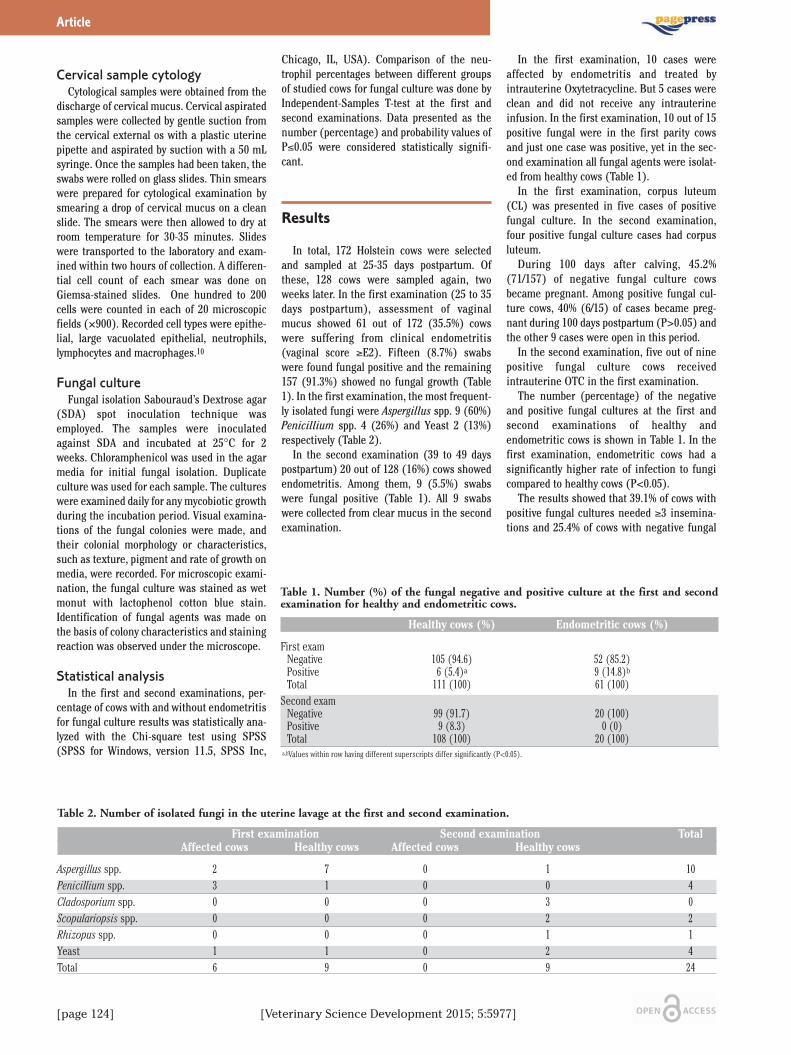
[page 124] [Veterinary Science Development 2015; 5:5977]
Cervical sample cytology Cytological samples were obtained from the
discharge of cervical mucus. Cervical aspiratedsamples were collected by gentle suction fromthe cervical external os with a plastic uterinepipette and aspirated by suction with a 50 mLsyringe. Once the samples had been taken, theswabs were rolled on glass slides. Thin smearswere prepared for cytological examination bysmearing a drop of cervical mucus on a cleanslide. The smears were then allowed to dry atroom temperature for 30-35 minutes. Slideswere transported to the laboratory and exam-ined within two hours of collection. A differen-tial cell count of each smear was done onGiemsa-stained slides. One hundred to 200cells were counted in each of 20 microscopicfields (×900). Recorded cell types were epithe-lial, large vacuolated epithelial, neutrophils,lymphocytes and macrophages.10
Fungal cultureFungal isolation Sabouraud’s Dextrose agar
(SDA) spot inoculation technique wasemployed. The samples were inoculatedagainst SDA and incubated at 25°C for 2weeks. Chloramphenicol was used in the agarmedia for initial fungal isolation. Duplicateculture was used for each sample. The cultureswere examined daily for any mycobiotic growthduring the incubation period. Visual examina-tions of the fungal colonies were made, andtheir colonial morphology or characteristics,such as texture, pigment and rate of growth onmedia, were recorded. For microscopic exami-nation, the fungal culture was stained as wetmonut with lactophenol cotton blue stain.Identification of fungal agents was made onthe basis of colony characteristics and stainingreaction was observed under the microscope.
Statistical analysisIn the first and second examinations, per-
centage of cows with and without endometritisfor fungal culture results was statistically ana-lyzed with the Chi-square test using SPSS(SPSS for Windows, version 11.5, SPSS Inc,
Chicago, IL, USA). Comparison of the neu-trophil percentages between different groupsof studied cows for fungal culture was done byIndependent-Samples T-test at the first andsecond examinations. Data presented as thenumber (percentage) and probability values ofP≤0.05 were considered statistically signifi-cant.
Results
In total, 172 Holstein cows were selectedand sampled at 25-35 days postpartum. Ofthese, 128 cows were sampled again, twoweeks later. In the first examination (25 to 35days postpartum), assessment of vaginalmucus showed 61 out of 172 (35.5%) cowswere suffering from clinical endometritis(vaginal score ≥E2). Fifteen (8.7%) swabswere found fungal positive and the remaining157 (91.3%) showed no fungal growth (Table1). In the first examination, the most frequent-ly isolated fungi were Aspergillus spp. 9 (60%)Penicillium spp. 4 (26%) and Yeast 2 (13%)respectively (Table 2).
In the second examination (39 to 49 dayspostpartum) 20 out of 128 (16%) cows showedendometritis. Among them, 9 (5.5%) swabswere fungal positive (Table 1). All 9 swabswere collected from clear mucus in the secondexamination.
In the first examination, 10 cases wereaffected by endometritis and treated byintrauterine Oxytetracycline. But 5 cases wereclean and did not receive any intrauterineinfusion. In the first examination, 10 out of 15positive fungal were in the first parity cowsand just one case was positive, yet in the sec-ond examination all fungal agents were isolat-ed from healthy cows (Table 1).
In the first examination, corpus luteum(CL) was presented in five cases of positivefungal culture. In the second examination,four positive fungal culture cases had corpusluteum.
During 100 days after calving, 45.2%(71/157) of negative fungal culture cowsbecame pregnant. Among positive fungal cul-ture cows, 40% (6/15) of cases became preg-nant during 100 days postpartum (P>0.05) andthe other 9 cases were open in this period.
In the second examination, five out of ninepositive fungal culture cows receivedintrauterine OTC in the first examination.
The number (percentage) of the negativeand positive fungal cultures at the first andsecond examinations of healthy andendometritic cows is shown in Table 1. In thefirst examination, endometritic cows had asignificantly higher rate of infection to fungicompared to healthy cows (P<0.05).
The results showed that 39.1% of cows withpositive fungal cultures needed ≥3 insemina-tions and 25.4% of cows with negative fungal
Article
Table 1. Number (%) of the fungal negative and positive culture at the first and secondexamination for healthy and endometritic cows.
Healthy cows (%) Endometritic cows (%)
First exam Negative 105 (94.6) 52 (85.2) Positive 6 (5.4)a 9 (14.8)b
Total 111 (100) 61 (100)Second exam Negative 99 (91.7) 20 (100) Positive 9 (8.3) 0 (0) Total 108 (100) 20 (100)a,bValues within row having different superscripts differ significantly (P<0.05).
Table 2. Number of isolated fungi in the uterine lavage at the first and second examination.
First examination Second examination Total Affected cows Healthy cows Affected cows Healthy cows
Aspergillus spp. 2 7 0 1 10Penicillium spp. 3 1 0 0 4Cladosporium spp. 0 0 0 3 0Scopulariopsis spp. 0 0 0 2 2Rhizopus spp. 0 0 0 1 1Yeast 1 1 0 2 4Total 6 9 0 9 24

[Veterinary Science Development 2015; 5:5977] [page 125]
cultures were conceived after three or moreinseminations (Table 3; P=0.17), however, thisdifference was not significant.
The results of cytological change percent-ages of PMNs (mean ± SD) of the negative andpositive fungal cultures at the first and secondexamination are shown in Table 4. There wasno significant difference between cows withpositive and negative fungal cultures in thefirst and second examination (P>0.05).
Discussion
The uterine lumen was sterile before partu-rition. After parturition, the microorganismsinflow from the animal’s environment, skin,and feces to the uterine lumen.11 The fungican invade tissues and cause clinical infec-tions.4 About 100 species of fungi are generallyidentified as pathogens of humans and ani-mals.4 In the present study, six different gen-era, Aspergillus, Penicillium, Cladosporium,Scopulariopsis, Rhizopus and Yeast were iso-lated from healthy and endometritic cows(Table 2). The isolation of fungi in the firstexamination was higher than the second(8.7% vs 7%); however, this difference was notsignificant (P>0.05). In the first examinationthe growth of fungi in endometritis affectedcows was higher than healthy cows. But in thesecond examination there was no isolation offungi in the endometritic cows and there werenine isolations in the healthy cows. Verma etal. reported mycobiotic agents from 27.8% and33.3% of endometritic buffaloes and cows,respectively.4 Contamination of fungi inendometritic and healthy cows was 39.34% and28.57% respectively.12 Sharma and Singhfound 15.5% mycotic isolation in repeatedbreeder cows.13 Ramsingh et al. have reported
that out of 168 uterine discharge samples fromrepeated breeder endometrtic cows, a total of168 (10.5%) mycotic isolates were recordedand identified.14 In our study, there was alower prevalence of mycobiotic agents. Thismay be because of the hot and dry climate ofShiraz. The cows in our farm were housed inmat or sand bed. However, the examined cowsin the present study were young and havegreater ability to clean their uteri from con-taminations. Other effective factors are thedensity of animals and topographic variations.4
Aspergillus spp. were the most importantfungi isolated from all samples (10 from 24)which means from samples in first examina-tion (9 from 15) and from endometritis affect-ed cows (7 from 9). According to the survey ofVerma et al., Aspergillus spp. was reported asthe most important mycobiotic agent, 43.7%(7/16), among endometritic cases in cows andbuffaloes.4 Also, Aspergillus spp., Penicilliumspp. and Cladosporium spp. were the highestisolated fungi from vaginal mucus of cows withcomplicated puerperium.15 In another survey,Penicillium spp. and Candida albicans werethe most common isolated fungi. Penicilliumspp. was more commonly isolated from uterusof cows with reproductive diseases andCandida albicans was more frequently isolatedfrom healthy uterus.12
Cows with lower rate of infected uteri tofungi get pregnant during a 100 day period anda higher percentage of them require three ormore inseminations to become pregnant,although it was not significant. Vlcek et al.found increase of the insemination interval,service-period and insemination index in cowsthat yielded pathogenic and potentially patho-genic micromycetes.15
No significant difference between cows withpositive and negative fungal cultures in thefirst and second examinations (P>0.05) con-
firmed that the immune system did notrespond to the exposure of uterus to fungi.This issue is reported for the first time and tothe best of the researchers’ knowledge therehave been no reports regarding this subject.No significant difference between conceptionin positive and negative fungal cultures mayindicate that the immune system in reproduc-tive tract of cow with positive fungal culturewas unaffected. In the second examination,there were five cows with contaminated uterito fungi that received intrauterine OTC in thefirst examination. It is suggested that use ofintrauterine antibiotics can cause fungalinfections.4,14 Antibiotic therapy, especiallyfrom intrauterine route, or corticosteroid ther-apy, might eliminate bacterial agents from thereproductive tract but produce immunosup-pression and trigger fungal infections.4
Conclusions
In conclusion, treatment of cows affected bypostpartum endometritis with intrauterineinfusion of antibiotic such as OTC, hygiene ofbed, number of cows in one yard, age and par-ity of cows may increase incidence of mycoticendometritis. The clinical signs of endometri-tis may be not found in cows that were affectedby mycotic endometritis.
References
1. Sheldon IM, Lewis GS, LeBlanc S, GilbertRO. Defining postpartum uterine diseasein cattle. Theriogenology 2006;65:1516-30.
2. Sheldon IM, Williams EJ, Miller ANA, et al.Uterine diseases in cattle after parturi-tion. Vet J 2008;176:115-21.
3. Noakes DE, Parkinson TJ, England GCW.Veterinary reproduction and obstetrics.9th ed. Amsterdam: Elsevier Sci. Ltd.;2009. pp 407-19.
4. Verma S, Katoch RC, Jand SK, et al.Mycobiotic flora of female genitalia of buf-faloes and cows with reproductive disor-ders. Vet Res Commun 1999;23:337-41.
5. Biziulevichius GA, Lukauskas K. In vivoStudies on lysosubtilin: 2. Efficacy fortreatment of post-partum endometritis incows. Vet Res 1998;29:47-58.
6. LeBlanc SJ, Duffield TF, Leslie KE, et al.Defining and Diagnosing postpartum clin-ical endometritis and its impact on repro-ductive performance in dairy cows. J DairySci 2002;85:2223-36.
7. Williams EJ, Fischer DP, Pfeiffer DU, et al.Clinical evaluation of postpartum vaginalmucus reflects uterine bacterial infectionand the immune response in cattle.
Article
Table 3. Number (%) of cows that had fungal negative and positive culture in both examsbased on the service per conception.
Service per conception Fungal culture Negative (%) Positive (%)
≤2 103 (74.6) 14 (60.9)≥3 35 (25.4) 9 (39.1)Total 138 (100) 23 (100)
Table 4. Comparison of cytological change of neutrophils percentages (mean ± SD) of thefungal negative and positive culture at the first and second exam.
Fungal culture Examination First exam Second exam
Negative 11.3±14.3 (142) 8.2±13.6 (135)Positive 11.6±9.3 (13) 4.1±3.2 (9)
[page 126] [Veterinary Science Development 2015; 5:5977]
Theriogenology 2005;63:102-17.8. Dubuc J, Duffield TF, Leslie KE, et al.
Definitions and diagnosis of postpartumendometritis in dairy cows. J Dairy Sci2010;93:5225-33.
9. Santos TMA, Caixeta LS, Machado VS, etal. Antimicrobial resistance and presenceof virulence factor genes inArcanobacterium pyogenes isolated fromthe uterus of postpartum dairy cows. VetMicrobiol 2010;145:84-9.
10. Ahmadi MR, Nazifi S, Ghaisari HR.
Comparative cervical cytology and concep-tion rate in postpartum dairy cows. VetArch 2006;76:323-32.
11. Földi J, Kulcsar M, Pecsi A, et al. Bacterialcomplications of postpartum uterine invo-lution in cattle. Anim Reprod Sci2006;96:265-81.
12. Talebkhan Garoussi M, Khosrave AR,Hovarashte P. The survey of mycotic floraof uterine cows with reproductive disor-ders and healthy. J Vet Res 2008;63:7-10.
13. Sharma S, Singh M. Mycotic endometritis
in cows and its therapeutic management.Intas Polivet 2012;13:29-30.
14. Ramsingh L, Murali Mohan K, SadasivaRao K. Clinical management of mycoticendometritis in cows. Int J Pharm SciInvent 2013;2:3-4.
15. Vlček Z, Kudláč E, Nesňalová E, LáníkováA. Fertility of cows after dystocias andcomplicated puerperium with respect tobacteriological and mycological findings inthe genitalia. Acta Veterinaria Brno1989;58:245-60.
Article

[Veterinary Science Development 2015; 5:5986] [page 99]
Electrocardiographic indices of clinically healthy ChiossheepAliasghar Chalmeh, Iman Saadat Akhtar,Mohamad Hadi Zarei, MehrdadBadkoubehSchool of Veterinary Medicine, Shiraz University, Iran
Abstract
Information regarding normal electrocardio-graphic features of different breeds of animalscan help veterinarians to detect any abnormal-ities in cardiac electrical activities. The cur-rent research was conducted to present thenormal electrocardiographic indices of clini-cally healthy Chios ewes and lambs. The elec-trocardiograms were recorded from clinicallyhealthy Chios ewes (n=27; 2-3 years old) andlambs (n=20; 4-6 months old) by using baseapex lead system. T and QRS-duration inlambs were significantly lower than adultChios ewes. The electrocardiographic ampli-tudes in lambs were lower than ewes, non-sig-nificantly. P-R, R-R, Q-T and S-T intervals inChios lambs were significantly lower thanewes. No normal sinus rhythm was detected inChios lambs. The proportion of sinus arrhyth-mia and sinus tachycardia in lambs was signif-icantly more than ewes. Sino-atrial block wasalso detected in lambs. Based on the presenteddata it could be stated that aging can affectelectrocardiographic findings of Chios sheep.Finally, our results will provide a good basis forjudging the electrocardiograms in base apexlead system of Chios lambs and ewes.
Introduction
The heart electrical activities can be record-ed over a period of time by electrocardiogra-phy, as a non-invasive procedure. The heartbeats caused from depolarization and repolar-ization of the myocardium, which aredetectable by electrocardiography and finallyrecord and produce electrocardiograms(ECGs). Recording ECG in large animals hasseveral indications such as evaluating theheart rate and rhythm, size of cardiac cham-bers, myocardial damages, electrolyte imbal-ances and drugs side effects.1There are several electrocardiographic lead
systems in large animals to record ECGs con-tain bipolar (I, II, III, base-apex, X, Y and Z ofthe orthogonal lead system) and unipolar leads(aVF, aVR, aVL, thoracic); but the recorded
waves by these leads in each animal’s breed,size, body type and sex are different from oth-ers and these factors should not affect theECGs.2 Base apex lead in large animals has notthe complications in other leads and canrecord the clear and large waves and animalmovements have minimum effects on therecordings.1,3The Chios is known as a breed of semi-fat
tailed domestic sheep which originated fromthe Greek island of Chios and they are com-monly bred for their milk production.4 Severalresearchers studied the electrocardiographicparameters in clinically healthy sheep andgoats by base apex lead and leads other thanthe base apex;5-9 but according to the author’sknowledge, the study on normal electrocardio-graphic indices of clinically healthy Chiossheep is lacking. Hence, the current study wasperformed to present and clarify the normalelectrocardiographic characteristics of Chioslambs and ewes tracing by base apex lead sys-tem. The data presented here may be used asguideline to evaluate the cardiac electricalactivities of this breed.
Materials and Methods
The present study was performed in March2015 on clinically healthy Chios ewes (n=27; 2-3 years old) and lambs (n=20; 4-6 months old),around Shiraz, Fars province, southwest ofIran. All animals were grazing in a green pas-ture with free access to water and shade. Theanimals were examined prior to ECG record-ings and proved to be clinically healthy. Noneof the ewes and lambs used in this study hadany clinical signs of heart diseases (edema,jugular distension or pulsation and cardiacmurmurs), coughing and exercise intolerance. The ECGs were recorded on a bipolar base
apex lead, using limb lead I. Animals were keptstanding without any sedation and minimumrestraint, also. When animals got calm(decreased panting behavior and muscletremors), the ECGs were recorded, using alli-gator-type electrodes which were attached toskin after cleaning it with ethanol and apply-ing electrocardiographic jelly to improve skincontact. The positive electrode (left arm) wasplaced over cardiac apex in the fifth left inter-costal space at the level of the elbow, the neg-ative electrode (right arm) was placed in theleft jugular furrow at the top of heart base, theneutral electrode (right foot) was used on theskin of thoracic inlet and the ground wasplaced on the dorsal spine or another site awayfrom the heart.1 All ECGs were obtained in asingle channel electrocardiographic machine(Kenz-line EKG 110, Suzuken Co., Ltd., Japan)with paper speed of 25 mm/sec and calibrationof 10 mm equal to 1 mV. The precision of dura-
tion was 0.02 second and amplitude was 0.05mV. The ECGs were examined to evaluate nor-mal electrocardiographic indices and cardiacarrhythmias, subsequently. Mean and standard deviation (SD) were cal-
culated for all studied electrocardiographicindices. Two independent samples t-test wasused to detect statistical differences betweenewes and lambs about similar electrocardio-graphic parameter. Fisher’s exact test wasused to detect any proportional distribution ofarrhythmias between ewes and lambs. All datawere analyzed by using SPSS software (SPSSfor Windows, version 20, SPSS Inc, Chicago,Illinois). P<0.05 was considered statisticallysignificant.
Results
The normal electrocardiographic indices ofclinically healthy Chios sheep are presented inTables 1 to 3. T and QRS-duration in lambswere significantly lower than adult Chios ewes(P<0.05; Table 1). The electrocardiographicamplitudes in lambs were lower than ewes,non-significantly (P>0.05; Table 2). P-R, R-R,Q-T and S-T intervals in Chios lambs were sig-nificantly lower than ewes (P<0.05; Table 3).No normal sinus rhythm was detected in Chioslambs. The proportion of sinus arrhythmia andsinus tachycardia in lambs was significantlymore than ewes. Sino-atrial block was alsodetected in lambs (Figures 1 and 2).
Veterinary Science Development 2015; volume 5:5986
Correspondence: Aliasghar Chalmeh,Department of Clinical School of VeterinaryMedicine, Sciences, Shiraz University, PO Box71345, Shiraz, Iran.Tel.: +98.711.36138700 - Fax: +98.711.32286940.E-mail: [email protected]
Key words: Electrocardiogram; normal index;Chios sheep.
Contributions: the authors contributed equally.
Conflict of interest: the authors declare no poten-tial conflict of interest.
Received for publication: 5 May 2015.Accepted for publication: 17 June 205.
This work is licensed under a Creative CommonsAttribution NonCommercial 3.0 License (CC BY-NC 3.0).
©Copyright A. Chalmeh et al., 2015Licensee PAGEPress srl, ItalyVeterinary Science Development 2015; 5:5986doi:10.4081/vsd.2015.5986

[page 100] [Veterinary Science Development 2015; 5:5986]
Discussion
Electrocardiography can be used as a clini-cal method of choice to assess the cardiacproblems regarding heart electrical activitiesand the initiation and conduction of waves ofdepolarization and repolarization.3 There areseveral literature about electrocardiographicstudies on different breeds of the small rumi-nants,8,9 but information regarding the baseapex electrocardiography of clinically healthyChios sheep was lacking. The data in basicelectrocardiographic indices of this breedcould be used as reference values to assessheart electrical activities. The base apex lead appears to be most use-
ful in measuring conduction times (i.e., dura-tions of component deflections, intervals, andsegments) because the origins and termina-tions of deflections could be identified easily.3It has been reported that the base apex leadgave the least variable ECG tracings in all theanimals; furthermore, the P waves, QRS com-plexes, and T waves in the base apex lead hadthe highest mean amplitude of all the leadsrecorded.3 It seems that our study on ChiosECG parameters in base apex lead system maybe helpful in standardizing the base apex leadof this breed.The conductive properties of the body mass
of ruminants, attributable to the volume of thegastrointestinal tract, influence the distribu-
tion of body surface potentials comprising theECG.3 This may explain the differences amongdifferent ECG parameters between the differ-ent ages in our study (Tables 1 to 3). The grad-ual development of body mass may cause diffi-culty in reaching the waves to the body surfacedue to relative electrical insulation by increas-ing body mass and decrease of amplitude inadults.10In the present study the electrocardiograph-
ic durations and intervals were increased sig-nificantly during aging. The transition time ofheart electrical impulses produces durationsand intervals. It may be stated that the smallestcardiac size and more superficial purkinjefibers in lambs in comparison with ewes cancreate lowest durations and intervals inyounger ones. It could be suggested that as themass of heart in larger animals became largerin the process of growth, the duration of trans-fer of cardiac electrical activity also increas-es.11 The amplitude of P, R, S and T wavesrecorded in the studied sheep were not fol-lowed a specific pattern during aging. Thechanging of amplitudes presumably could bedue to a high degree of synchronized ventricu-lar polarization passing in any given direction.Furthermore, alterations between heart situa-tion and attached positive electrode canchange amplitudes. This may be also due tothe presence of high degree of synchronizationof depolarization of individual myocardialfibers. At the birth, significant hypoxemia and
acidaemia may develop.12 Moreover, after theonset of breathing, the change in pulmonaryvascular resistance with expansion of thelungs results in a great increase in pulmonaryblood flow, accompanied by a rise in left atrialpressure.13,14 This generates distension of theatrial walls and stretching of the atrial musclesand these might be related to the occurrence ofcardiac arrhythmias.15 It was suggested thatthe development of hypoxia was relative to thearrhythmias. It was considered that a hypoxiccondition during delivery may contribute to theoccurrence of neonatal arrhythmias in hors-es.16 Belenky et al.17 demonstrated that thehypoxic carotid chemoreflex, in lamb, is pres-ent at birth, but has a significantly longerresponse time than later in the newborn peri-od. Moreover, the CNS-mediated ventilatoryresponse to hypoxia was also noted to be pres-ent in the newborn animal through at least 12days or longer times of postnatal age.17Cardiac arrhythmias also occur commonly
in association with gastrointestinal disordersin the dairy cow and less commonly in thehorse and resolve without specific antiarrhyth-mic treatment when the primary gastrointesti-nal disorder is corrected.1 At birth the rumen isa rudimentary nonfunctional sac. Normaldevelopment of the rumen requires the estab-lishment of a viable microbial population andthe formation of volatile fatty acids.18Establishment of ruminal microbial fermenta-tion begins between two to four weeks of age
Article
Table 1. The durations (sec) of standard electrocardiographic indices (mean ± standard deviation) of base apex lead of clinically healthyChios ewes and lambs.
Age groups P-duration T-duration QRS-duration S-duration
Lambs (n=30) 0.020±0.005 0.029±0.007 0.095±0.005 0.010±0.001Ewes (n=30) 0.021±0.007 0.039±0.006 0.125±0.017 0.011±0.002P-value 0.091 0.026* 0.019* 0.089*Significant differences between groups at similar indices (P<0.05).
Table 2. The amplitudes (mV) of standard electrocardiographic indices (mean ± standard deviation) of base apex lead of clinicallyhealthy Chios ewes and lambs.
Age groups P-amplitude T-amplitude R-amplitude S-amplitude
Lambs (n=30) 0.052±0.016 0.110±0.057 0.054±0.012 0.261±0.089Ewes (n=30) 0.055±0.010 0.122±0.050 0.059±0.018 0.272±0.065P-value 0.162 0.132 0.092 0.076
Table 3. The intervals (sec) of standard electrocardiographic indices (mean ± standard deviation) of base apex lead of clinically healthyChios ewes and lambs.
Age groups PR-interval RR-intervalQT-interval ST-interval
Lambs (n=30) 0.045±0.008 0.203±0.032 0.071±0.014 0.060±0.014Ewes (n=30) 0.056±0.008 0.263±0.040 0.112±0.019 0.090±0.049P-value 0.035* 0.002* 0.001* 0.022**Significant differences between groups at similar indices (P<0.05).

[Veterinary Science Development 2015; 5:5986] [page 101]
as a result of the initiation of solid feedintake.19,20 Changes in physiological states ofgastrointestinal tract, feed intake and alter-ations in energy metabolism, may affect thecardiac performance and changes in ECGparameters. Furthermore, changes in food reg-imen and electrolyte profile may affect cardiacmusculature and its activation.The results of the present study showed that
sinus tachycardia and sinus arrhythmia werethe most frequent cardiac arrhythmias in thestudied animals (Figure 2). Sinus tachycardiameans an increase in heart rate that is initiat-ed by the sino-atrial node. The term sinustachycardia is used to describe an increase inheart rate caused by detectable influencessuch as pain, excitement, exercise, hyperther-mia, a fall in arterial blood pressure or theadministration of adrenergic drugs.1 The heartrate returns to normal when the influence isremoved or relieved. It may be suggested thatthe higher heart rates might be due to stressand excitation resulting from isolation oflambs from their dams as well, but since theanimals were placed in a quiet state, it isunlikely to be the origin for this higher heartrate in studied animals. Because there wereno clinical signs of cardiac problems (edema,jugular distension or pulsation) in all studied
animals, this cardiac rhythm irregularity couldbe categorized as physiologic arrhythmias.Matsui et al.21 reported an elevation of theheart rate in newborn pony foals with adminis-tration of a combined blockade with atropineand propranolol. These observations indicatethe possibility of high vagal activity in the new-born Thoroughbred foal at birth.15 Sustainedtachycardia is an important clinical problem inthe fetus and newborn. The physiological prop-erties of the fetal and neonatal myocardiummake it intrinsically more vulnerable to highheart rates.22 Fetal tachycardia is an importantcause of fetal morbidity and mortality.23 Therate of fetal tachycardia is of no real help indefining the mechanism, as most tachycardiaoccurs at about higher than 120 beats/min andaverage of this parameter was 170 beats/minat the time of ECG recordings. Sinus arrhythmia was also seen frequently
in Chios ewes and lambs (Figure 2). Thisarrhythmia has been reported in cattle whichhave been deprived of food or had anorexiadue to some gastrointestinal problems.24,25None of the animals with sinus arrhythmia inthis study had any clinically obvious systemicproblems or were suffering from anorexia.High vagal tone could be suggested as thecause of this arrhythmia in any ages of these
animals.26 Sinus arrhythmia is a normal phys-iological arrhythmia that occurs at slow restingheart rates and is associated with variation inthe rate of discharge from the sino-atrial nodeassociated with variation in the intensity ofvagal stimulations. It is commonly correlatedwith respiration so that the discharge rate andheart rate increase during inspiration anddecrease during expiration. Sinus arrhythmiais more clinically obvious in tame sheep andgoats.1 It may be possible to link the genesis ofthe sinus tachycardia and sinus arrhythmia inapparently healthy lambs to the increased loadimposed on the heart or the fluctuation of thesympathetic or parasympathetic tone associat-ed with excessive exertion during the stage ofthe birth.27Sino-atrial block was also detected in Chios
lambs (Figures 1 and 2). In sino-atrial blockthe sinus node fails to discharge or its impulseis not transmitted over the atrial myocardium.It is associated with the complete absence ofheart sounds, of jugular atrial wave and of anarterial pulse for one beat period. The underly-ing rhythm is regular unless sinus arrhythmiais present. In the ECG there is completeabsence of the P, QRS and T complex for onebeat. The distance between the preblock andpostblock P waves is twice the normal P-P
Article
Figure 1. The electrocardiograms tracing from clinically healthy Chios sheep by base apex lead system (paper speed 25 mm/sec, sensi-tivity 10 mm/mV). A: Normal sinus rhythm in an ewe; B: Sinus arrhythmia in a lamb; C: Sinus tachycardia and sinus arrhythmia in alamb; D: Sino-atrial block in a lamb.

[page 102] [Veterinary Science Development 2015; 5:5986]
interval or sometimes slightly shorter. Thisarrhythmia is not uncommon in fit racinghorses at rest and can be induced in horsesand cattle by procedures that increase vagaltone.1 Since, there were no clinical abnormali-ties in lambs which had sino-atrial block, it canbe stated that this arrhythmia is categorized asphysiological cardiac rhythm irregularities inChios lambs.
Conclusions
In conclusion, it is obvious that these datawill provide a good basis for judging the ECGsin base apex lead system of Chios lambs andewes. It could be stated that aging can affectelectrocardiographic findings. Furthermore, itmay be suggested that the cardiac arrhythmiasobserved in the clinically healthy Chios ewesand lambs in this study could be accepted asthe physiological arrhythmias and so no treat-ment is necessary. Finally, the results of thepresent research can help veterinarians todetect any abnormalities in heart electricalperformance of Chios sheep and may be usedas the guideline for the assessment of any car-diac rhythm irregularities in Chios sheep suf-fering from cardiac problems.
References
1. Radostits OM, Gay CC, Hinchcliff KW,Constable PD. Diseases of the cardiovas-cular system. In: Veterinary medicine: a
text book of the diseases of cattle, horses,sheep, pigs and goats. 10th ed.Philadelphia: Elsevier, USA; 2007. pp. 399-438.
2. Reef VB, McGuirk SM. Diseases of the car-diovascular system. In: Smith BP, ed. Largeanimal internal medicine. 4th ed.Philadelphia: Elsevier, USA; 2009. pp. 453-489.
3. Santamarina G, Espino L, Suarez LM.Electrocardiographic parameters of free-ranging roe deer (Capreoluscapreolus). JZoo Wildl Med 2001;32:441-6.
4. Gelasakis AI, Valergakis GE, Arsenos G,Banos G. Description and typology ofintensive Chios dairy sheep farms inGreece. J Dairy Sci 2012;95:3070-9.
5. Pourjafar M, Badiei K, Chalmeh A, et al.Cardiac arrhythmias in clinically healthynewborn Iranian fat-tailed lambs. Glob Vet2011;6:185-9.
6. Pourjafar M, Badiei K, Chalmeh A, et al.Comparison of cardiac arrhythmiasbetween late pregnancy and early post-par-tum periods in clinically healthy Iranianfat-tailed sheep. Pak Vet J 2011;31:309-12.
7. Pourjafar M, Badiei K, Chalmeh A, et al.Age-related cardiac arrhythmias in clini-cally healthy Iranian Najdi goats. Bulg JVet Med 2012;15:37-43.
8. Mohan NH, Niyogi D, Singh HN. Analysisof normal electrocardiograms ofJamunapari goats. J Vet Sci 2005;6:295-8.
9. Ahmed JA, Sanyal S. Electrocardiographicstudies in Garol sheep and black Bengalgoats. Res J Cardiol 2008;1:1-8.
10. Kellerová E, Szathmáry V, Kozmann G, etal. Spontaneous variability and reactive
postural beat-to-beat changes of integralECG body surface potential maps. PhysiolRes 2010;59:887-96.
11. Schmidt-Nielsen K. Animal physiology:adaption and environment. 6th ed. NewYork: Cambridge University Press; 1997.
12 Comline RS, Silver M. Placental transfer ofblood gases. Br Med Bull 1975;31:25-31.
13 Dawes GS, Mott JC, Widdicombe JC, WyattDG. Changes in the lungs of the newbornlamb. J Physiol 1953;121:141-62.
14. Dawes GS. Changes in the circulation atbirth. Br Med Bull 1961;17:148-53.
15. Yamamoto K, Yasuda J, Too K.Arrhythmias in newborn Thoroughbredfoals. Equine Vet J 1992;23:169-73.
16. Takayuki I. A study of arrhythmias in thor-oughbred newborn foals immediately afterbirth. Jpn J Vet Res 1990;38:57.
17. Belenky DA, Standaert TA, Woodrum DE.Maturation of hypoxic ventilatoryresponse of the newborn lamb. J ApplPhysiol 1979;47:927-30.
18. Tamate H, McGilliard AD, Jacobson NL,Getty R. Effect of various dietaries on theanatomical development of the stomach inthe calf. J Dairy Sci 1962;45:408-20.
19. Baldwin RLVI, Jesse BW. Developmentalchanges in glucose and butyrate metabo-lism by isolated sheep ruminal cells. J Nutr1992;122:1149-53.
20. Baldwin RL. Sheep gastrointestinal devel-opment in response to different dietarytreatments. Small Ruminant Res2000;35:39-47.
21. Matsui K, Sugano S, Masuyuna L, et al.Alterations in the heart rate ofThoroughbred horse, pony and Holsteincow through pre- and post-natal stages.Jpn J Vet Sci 1984;46:505-10.
22. Rudolph AM. Congenital diseases of theheart: clinico-physiological considera-tions. Armonk, NY: Futura; 2001.
23. Wren C. Cardiac arrhythmias in the fetusand newborn. Semin Fetal Neonatal Med2006;11:182-90.
24. Machida N, Okamoto Y, Minami S, et al.Cardiac arrhythmias in normal Holsteinheifers. J Jpn Vet Med Assoc 1991;44:1176-9.
25. Gentile A, Guglielmini C, Cipone M.Alerazioni del ritmo cardiaco nel bovino inrapporto col digiuno. Arch Vet Ital1993;44:100-7.
26. Rezakhani A, Papahn AA, Gheisari HR.Cardiac dysrhythmias in clinically healthyheifers and cows. Rev d Méd Vét2004;155:159-62.
27. Machida N, Nakamura T, Kiryu K, KagotaK. Electrocardiographic features and inci-dence of atrial fibrillation in apparentlyhealthy dairy cows. ZentralblVeterinarmed A 1993;40:233-9.
Article
Figure 2. Distribution and proportion of cardiac arrhythmias in clinically healthy Chiosewes (n=27) and lambs (n=20) tracing by standard base apex lead system electrocardiog-raphy.

[page 112] [Veterinary Science Development 2015; 5:5997]
Innocuousness of conjunctivalvaccination with Brucellamelitensis strain Rev.1 in pregnant Iranian fat-tailedewes Saeed Alamian, Ramin Bagheri Nejad,Hamid Reza Jalali, Armin Kalantari,Afshar EtemadiBrucellosis Department, Razi Vaccine andSerum Research Institute, Karaj, Iran
Abstract
Brucella melitensis strain Rev.1 is the mosteffective vaccine against brucellosis in sheepand goats. In Iran, mass vaccination is carriedout all over the country in which adult animalsare immunized by subcutaneous injection ofreduced doses of the vaccine. However, due toantibody responses elicited by vaccination,concomitant implementation of test-and-slaughter is impossible. To overcome the prob-lem, vaccination through conjunctival route isrecommended. In this study, serologicalresponses of six pregnant Iranian fat-tailedewes to conjunctival vaccination with standarddoses of the vaccine were evaluated usingmodified Rose Bengal test, serum agglutina-tion test and indirect ELISA. Besides, vaccinestrain excretion in milk and vaginal dischargeswas also examined by microbiological cultureof milk and vaginal swab samples taken oneday post-parturition. Animals were vaccinatedduring the second half of gestation. As theresults, antibody titers of five (83.3%) ewesdecreased to the levels not detectable by thetests within three months after vaccination.No vaccine-induced abortions occurred andvaccinated ewes delivered healthy lambs50.33±15.56 (mean ± standard deviation) dayspost-vaccination. Vaccine strain was not isolat-ed from milk and vaginal swab samples.Generally, our study shows full doses of B.melitensis strain Rev.1 can be used conjunc-tively to vaccinate pregnant Iranian sheep dur-ing late pregnancy without abortifacienteffects, prolonged antibody responses and vac-cine strain excretion in milk and vaginal dis-charges. Nevertheless, further studies arerequired to determine safety and immuno-genicity of the vaccine in field conditions.
Introduction
Brucellosis in sheep and goats is an impor-tant zoonotic disease caused mainly by
Brucella melitensis.1,2 Vaccination of the hostanimals with B. melitensis strain Rev.1 is usedworldwide for disease control which has beenproved to be the most effective vaccine.3,4 It isrecommended to immunize replacement ani-mals from 3 to 6 months of age with standarddoses of vaccine containing at least 109 livecells.5 However, there is evidence that effectivecontrol of the disease in countries with highprevalence requires immunization of all sus-ceptible young and adult animals in a massvaccination campaign which is considered asthe most practical measure.6-8
One problem with vaccination of adult ani-mals is antibody responses induced by the vac-cine which may last for a long time and causesero-positivity of vaccinated animals in rou-tine serological tests interfering with detec-tion of infected ones.4,9,10 This makes simulta-neous implementation of vaccination and test-and-slaughter impossible since vaccinated ani-mals are falsely diagnosed as infected.4Moreover, vaccine-induced abortion and vac-cine strain excretion in milk and vaginal dis-charges may occur.5,10 Vaccination of flocksthrough conjunctival route is known as oneway to solve these problems.6,10,11
Small ruminant brucellosis is an enzooticdisease in Iran causing abortion in differentparts of the country.12 Mass vaccination hasbeen the main control measure since 2003 inwhich adult animals are vaccinated subcuta-neously using reduced doses of the vaccine.12Nevertheless, there are field reports showinglong-lasting sero-positivity of vaccinated adultsheep and goats and abortions in pregnantanimals attributed to the vaccination.Therefore, this study was done to evaluateserological responses of pregnant fat-tailedewes to ocular vaccination with standard dosesof B. melitensis strain Rev.1 as well as its safetyin terms of abortion induction and vaccinestrain excretion in milk and vaginal secre-tions.
Materials and Methods
Animals and vaccinationEleven pregnant Iranian fat-tailed ewes
were randomly selected from a known brucel-losis-free flock. Selected animals were nega-tive in modified Rose Bengal test (mRBT),serum agglutination test (SAT) and indirectELISA (iELISA) carried out twice with amonth’s interval. Six ewes were vaccinatedduring third to fifth month of pregnancy withconjunctival B. melitensis strain Rev.1 vaccinecontaining 109 colony forming units (CFU) perdose. Other ewes were used as controls inwhich normal saline was used instead of vac-cine at the same time. The vaccine used in thestudy was produced in Razi Vaccine and Serum
Research Institute according to standard pro-cedures.5,13 The original seed for vaccine pro-duction was obtained from Animal Health andVeterinary Laboratories Agency (AHVLA),Weybridge, UK. Vaccinated and control ani-mals were kept separately in the same condi-tions.
Serological evaluationAnimals were bled every 2 weeks for three
months after immunization to evaluate sero-logical responses to vaccination. Serum sam-ples were examined using mRBT, SAT andiELISA. RBT and SAT antigens were producedin Razi Vaccine and Serum Research Institutebased on standard methods,5,13 as describedpreviously.14 For mRBT, one drop of the antigenwas mixed with three drops of the serum sam-ple.15 Indirect ELISA was performed usingPrioCHECK® Brucella Ab (Prionics AG,Schlieren, Switzerland) according to the man-ufacturer’s instructions.
Bacteriological examinationTo determine vaccine strain excretion in
Veterinary Science Development 2015; volume 5:5997
Correspondence: Ramin Bagheri Nejad,Brucellosis Department, Razi Vaccine and SerumResearch Institute, 3197619751 Karaj, P.O. Box:31975/148 Iran. Tel.: +98.263.4570038 - Fax: +98.263.4552194. E-mail: [email protected]
Key words: Brucellosis; Vaccination; Sheep;Brucella melitensis strain Rev.1; Conjunctivalvaccine.
Acknowledgements: The authors would like tothank Mr. Ali Asghar Izadyar, Mr. MohammadSaffari and other personnel of BrucellosisDepartment at Razi Vaccine and Serum ResearchInstitute for their kind technical assistance. Thestudy was funded by Razi Vaccine and SerumResearch Institute.
Contributions: RBN designed the study. SA, RBN,HRJ, AK and AE performed the study. RBN did thestatistical analysis. RBN prepared the draft whichwas approved by all authors.
Conflict of interest: the authors declare no poten-tial conflict of interest.
Received for publication: 8 May 2015.Accepted for publication: 24 June 2015.
This work is licensed under a Creative CommonsAttribution NonCommercial 3.0 License (CC BY-NC 3.0).
©Copyright S. Alamian et al., 2015Licensee PAGEPress srl, ItalyVeterinary Science Development 2015; 5:5997doi:10.4081/vsd.2015.5997

[Veterinary Science Development 2015; 5:5997] [page 113]
milk and vaginal discharges, milk and vaginalswab samples were taken within 24 hours afterabortion or parturition. Milk samples were firstcentrifuged at 6000-7000 rpm for 15 minutesand then supernatant cream and precipitatedpellet were cultured.13 Swab samples were cul-tured directly on solid media. Brucella agarmedium (BD, USA) was used for vaccinestrain isolation which was supplemented withBrucella selective antibiotics (Oxoid,Basingstoke, UK) and 5% (v/v) horse serumfollowing manufacturer’s instructions. Foreach sample, at least 3 plates were inoculated.
Results
Serological responses after vaccination
While control ewes remained negative dur-ing the study, vaccinated animals showed anti-body responses from the second week afterimmunization. Percents of positive vaccinatedewes in mRBT and iELISA at two-week inter-vals over the study period are presented inFigure 1. Two weeks post-vaccination, all ani-mals were positive in mRBT but none of themin iELISA. Five ewes (83.3%) showed positiveresults in iELISA after four weeks. The mRBTand iELISA results of each ewe were similarfrom fourth week on. After 12 weeks, only onevaccinated ewe (16.7%) remained positivedetected by the two tests.
Evaluation of antibody titers using SATdemonstrated a falling trend over time (Figure2). All animals showed increased antibodytiters after two weeks which declined graduallyafterwards in a way that 5 ewes (83.3%) had noSAT titers twelve weeks after vaccine inocula-tion. The only ewe, which had antibody titersdetectable by SAT, was also reactive in mRBTand iELISA.
Vaccine safetyNo abortion occurred following vaccination
and all vaccinated animals had normal deliverywith healthy lambs 50.33±15.56 (mean±SD)days after vaccine inoculation. B. melitensisstrain Rev.1 was not isolated by microbiologi-cal methods from milk and vaginal dischargesof vaccinated sheep within 24 hours postpar-tum.
Discussion and Conclusions
In Iran, brucellosis in small ruminants is animportant enzootic disease which is a publichealth burden. Nomadic raising of sheep andgoats, traditional production practices, illegalanimal imports and uncontrolled movementsof flocks within the country contribute to thedifficulties in disease control. In these condi-tions, control of the disease has been mainlybased on mass vaccination of young and adultanimals using subcutaneous administration offull and reduced doses of B. melitensis strainRev.1, respectively.12 However, the disease stillremains prevalent in different parts of thecountry diagnosed as a significant cause ofabortion in sheep and goats.12
Although we previously demonstrated thatreduced doses containing less than or equal to106 bacteria can be safely used to immunizepregnant ewes with short-lasting serologicalresponses,14 field reports show persistenceantibody responses and abortions caused byvaccination. It is known that the innocuous-ness of the vaccine in adult and pregnant ani-mals depends on vaccine dose, time of vaccina-tion during gestation and administrationroute.8,16 Hence, these observations may bepartly due to the fact that according to a stan-dard approved by National Brucellosis ExpertCommittee, the reduced dose of vaccine usedfor adult animals immunization can contain up
to 4×10⁶ colony forming units (CFU) per dose.In addition, subcutaneous use of vaccine andextended lambing season in Iran, whichresults in presence of pregnant animalsthroughout the year in flocks, could be influen-tial. Therefore, ocular inoculation of the vac-cine is considered as an alternative proved tobe safer.6,8,10,16
In our experiment, all animals were detect-ed as positive using mRBT two weeks afterimmunization. Stournara et al.9 also reported ahundred percent positive results in mRBT ofnon-pregnant ewes 21 days post-vaccination.In another study by Zundel et al.,16 all ewesvaccinated at mid-pregnancy using the samedose as ours were positive in RBT 2 weeks fol-lowing vaccination.
The proportion of positive vaccinated ewesin iELISA reached its maximum after 4 weeks.A similar result has been observed byStournara et al.9 which was attributed to thehigher affinity of the conjugate used in theassay to immunoglobulin G (IgG). It has alsobeen reported that more than 70 percent ofnon-vaccinated ewes were detected as nega-tive by iELISA 14 days after challenge with thevirulent strain during pregnancy.17 Becausethe ewes used in our study were from a brucel-losis-free flock without previous exposure tothe pathogen, and according to the explanationprovided by Stournara et al.,9 negative iELISAresults of these naive ewes two weeks aftervaccine inoculation suggest antibody respons-es may be mainly of IgM class at this time. Thepercentage of positive animals detected byiELISA and mRBT decreased rapidly from 6weeks post-vaccination to the end of study.Similar performance of iELISA and mRBT inour study is in agreement with results of thestudy carried out by Stournara et al.9 However,in the latter study 72.6% and 84% of animalsremained positive in iELISA and mRBT,respectively 91 days after immunization, but inour study only 16.7% were positive in bothtests 12 weeks post-vaccination. This differ-
Article
Figure 2. Mean ± standard deviation of serum agglutination titersin vaccinated animals.
Figure 1. Percent of positive vaccinated ewes in modified RoseBengal test and indirect ELISA.

[page 114] [Veterinary Science Development 2015; 5:5997]
ence may be due to age and physiologic statusof animals when vaccinated or breed variationin antibody responses.7,18
Evaluation of serological responses duringthe study period using SAT revealed antibodytiters fell from the peak reached at week twotoward the end of study. SAT results are com-patible with the other two tests and the onlyewe, which had antibody titer of diagnosticvalue 12 week after immunization, was alsodetected as positive in iELISA and mRBT atthis interval. While serum antibody level ofthis ewe was decreasing from 120 IU/mL sixweeks following vaccination to 15 IU/mL atweek 10, there was a further surge in its anti-body titer (120 IU/mL) two weeks later at 12th
week. For this animal, parturition occurred 66days post-immunization 4 days prior to bloodcollection for the 10th week. This suggests thatparturition might have effects on antibodyresponses to vaccination.
Use of vaccine through conjunctival routeduring second half of pregnancy was safe interms of abortion induction, and no vaccineexcretion in milk and fetal materials wasdetected soon after delivery. Rev.1 strain deliv-ered conjunctivally is known to have a spreadconfined mainly to head lymph nodes.8Considering normal delivery of all animals oneto two months following vaccine inoculationand as vaccine strain was not isolated immedi-ately after parturition, it seems Rev.1 strainwas not generalized to the uterus and mamma-ry gland. Although there is no a completely safeway to use Rev.1 vaccine in pregnant smallruminants,4 it is known that conjunctivaladministration of the vaccine during late preg-nancy or before breeding can reduce risks ofvaccine-induced abortions and vaccine strainexcretion in milk and vaginal discharges,5which was also proved in Iranian fat-tailedewes in this study.
In general, the present experiment showedserological responses to ocular vaccination ofpregnant Iranian fat-tailed sheep with stan-dard doses of B. melitensis strain Rev.1 disap-peared in a considerable proportion of animalswithin 12 weeks. Moreover, use of vaccine dur-ing late pregnancy did not cause vaccination-
induced abortion and Rev.1 strain excretion inmilk and vaginal discharges during immediatepostpartum period. Nevertheless, furtherinvestigations are required to assess vaccineperformance in field conditions.
References
1. Diaz Aparicio E. Epidemiology of brucel-losis in domestic animals caused byBrucella melitensis, Brucella suis andBrucella abortus. Rev Sci Tech 2013;32:43-51, 3-60.
2. Garin-Bastuji B, Blasco JM, Grayon M,Verger JM. Brucella melitensis infectionin sheep: present and future. Vet Res1998;29:255-74.
3. Schurig GG, Sriranganathan N, Corbel MJ.Brucellosis vaccines: past, present andfuture. Vet Microbiol 2002;90:479-96.
4. Olsen SC, Stoffregen WS. Essential role ofvaccines in brucellosis control and eradi-cation programs for livestock. Expert RevVaccines 2005;4:915-28.
5. OIE. Manual of diagnostic tests and vac-cines for terrestrial animals. Paris: WorldOrganization for Animal Health; 2009.
6. Blasco JM. Control and eradication strate-gies for Brucella melitensis infection insheep and goats. Prilozi 2010;31:145-65.
7. Nicoletti P. Brucellosis: past, present andfuture. Prilozi 2010;31:21-32.
8. Jimenez de Bagues MP, Marin CM,Barberan M, Blasco JM. Responses ofewes to B. melitensis Rev1 vaccine admin-istered by subcutaneous or conjunctivalroutes at different stages of pregnancy.Ann Vet Res 1989;20:205-13.
9. Stournara A, Minas A, Bourtzi-Chatzopoulou E, et al. Assessment of sero-logical response of young and adult sheepto conjunctival vaccination with Rev-1 vac-cine by fluorescence polarization assay(FPA) and other serological tests for B.melitensis. Vet Microbiol 2007;119:53-64.
10. Blasco JM. A review of the use of B.melitensis Rev 1 vaccine in adult sheep
and goats. Prev Vet Med 1997;31:275-83.11. Fensterbank R, Verger JM, Grayon M.
Conjunctival vaccination of young goatswith Brucella melitensis strain Rev 1. AnnVet Res 1987;18:397-403.
12. Behroozikhah AM, Bagheri Nejad R, AmiriK, Bahonar AR. Identification at biovarlevel of Brucella isolates causing abortionin small ruminants of iran. J Pathogens2012;2012:357235.
13. Alton GG, Jones LM, Angus RD, Verger JM.Techniques for the Brucellosis Laboratory.Paris: Institut National de la RechercheAgronomique; 1988.
14. Ebrahimi M, Nejad RB, Alamian S, et al.Safety and efficacy of reduced doses ofBrucella melitensis strain Rev. 1 vaccinein pregnant Iranian fat-tailed ewes. VetItal 2012;48:405-12.
15. Ferreira AC, Cardoso R, Travassos Dias I,et al. Evaluation of a modified Rose Bengaltest and an indirect enzyme-linkedimmunosorbent assay for the diagnosis ofBrucella melitensis infection in sheep. VetRes 2003;34:297-305.
16. Zundel E, Verger JM, Grayon M, Michel R.Conjunctival vaccination of pregnant ewesand goats with Brucella melitensis Rev 1vaccine: safety and serological responses.Ann Vet Res 1992;23:177-88.
17. Duran-Ferrer M, Leon L, Nielsen K, et al.Antibody response and antigen-specificgamma-interferon profiles of vaccinatedand unvaccinated pregnant sheep experi-mentally infected with Brucella meliten-sis. Vet Microbiol 2004;100:219-31.
18. Jimenez de Bagues MP, Marin CM, BlascoJM, et al. An ELISA with Brucellalipopolysaccharide antigen for the diagno-sis of B. melitensis infection in sheep andfor the evaluation of serological responsesfollowing subcutaneous or conjunctival B.melitensis strain Rev 1 vaccination. VetMicrobiol 1992;30:233-41.
Article

[Veterinary Science Development 2015; 5:6012] [page 107]
Normal laparoscopic anatomyof the caprine pelvic cavityMujeeb ur Rehman Fazili,1Riaz Ahmad Shah,2 Maajid Hassan Bhat,2Firdous Ahmad Khan,2Anubhav Khajuria,3 Syed Hilal Yaqoob,2Niaz Ahmad Naykoo,2Nazir Ahmad Ganai21Division of Veterinary Clinical ServiceComplex, 2Division of AnimalBiotechnology, 3Divisions of VeterinarySurgery and Radiology, Faculty ofVeterinary Sciences and AnimalHusbandry, Sher-e-Kashmir University ofAgricultural Sciences and Technology ofKashmir, Srinagar, India
Abstract
Due to the several advantages over convention-al procedures, the laparoscopic disease diagnosisand surgery has now started receiving attentionin small ruminants. The normal laparoscopicanatomy needs to be described for comparisonwith the findings in animals with various dis-eases. The objective of the present study wastherefore to describe the laparoscopic anatomy ofthe caprine pelvic cavity. Adult Bakerwal andPashmina goats (n=25) of both the sexes wereincluded in this laparoscopy study. All the animalswere restrained in dorsal recumbency andTrendelenburg position under lumbosacralepidural anesthesia and sedation. After creatingthe pneumoperitoneum, the primary port for 5mm laparoscope was placed at linea alba (3.0 cmcranial to mammary glands in does), and at rightparamedian (3.0 cm cranial to the rudimentaryteat in the bucks) site. Secondary port was placedunder direct laparoscopic observation 5-6 cmaway from the primary port in horizontal plane, toallow insertion of the grasping forceps. Scan wasperformed first at the primary port and subse-quently through the secondary port for orienta-tion and exploration of the pelvic cavity. The ven-tral laparoscopic approach provided satisfactoryexposure of the pelvic cavity in goats.Comprehensive description of the pelvic organscould be obtained. However, dorsal aspect of theurinary bladder neck and accessory genitalorgans of male animals could not be visualized.Major complications were not encountered dur-ing or after laparoscopy. Laparoscopy a minimallyinvasive procedure has several advantages overalternate methods of understanding anatomy,physiology and pathology of most of the intraperi-toneal pelvic structures in goats. The techniquehas high pedagogic value. The procedure is safein experienced hands.
Introduction
Laparoscopy (keyhole or minimally invasivesurgery) is a type of surgical procedure thatallows a surgeon with the use of an instrument(laparoscope) inserted transabdominally toaccess the inside of the abdomen and pelviswithout having to make large incisions in theskin.1 This modality has diagnostic, therapeu-tic and prognostic applications. Laparoscopyhas many advantages over laparotomy. Theyinclude reduced tissue trauma, postoperativeadhesions and infections, fast recovery, stimu-lation of the immunity, better cardiovascularstability and lower pain scores.1-5
In recent years, laparoscopy has gainedacceptance in veterinary medicine.4 In smallruminants, it has been recognized as one ofthe most promising tools for improvement ofthe reproductive efficiency,6-8 disease diagno-sis and treatment.9-11 In goats particularlythose maintained for dairy purpose or as pets,this state of the art technology is expected tobe more popular in the near future.
Laparoscopic anatomy of the abdominal cav-ity in goats has recently been described.12 Thatstudy included female goats only. Additionally,organs located in the abdominal cavityreceived major consideration. With this back-ground in mind, the present study was aimedto provide description of the laparoscopicanatomy of the pelvic cavity, in goats of boththe sexes. The description will be useful forthose interested in augmenting animal pro-duction as well as for those involved in diseasediagnosis, treatment and prognosis of thepelvic disorders in goats. A significant peda-gogic value would be an additional attribute.
Materials and Methods
The prospective study was carried out ontwenty apparently healthy, adult, nonpregnantfemale and five male Pashmina and Bakerwalgoats (weighing 25-31 kg and aged 1.5-2.5years) maintained by the Division of AnimalBiotechnology, Faculty of Veterinary Sciencesand Animal Husbandry, India. The study wasundertaken after receiving due approved fromthe institutional animal ethics committee. Twodays prior to laparoscopy, wide ventral abdom-inal area from the mammary glands/rudimen-tary teats up to the umbilicus was clipped andshaved in all the animals. The food and waterwas withheld for 36 and 24 hours respectively.Regional lumbosacral epidural anesthesia wasachieved using 2% lignocaine hydrochloride(4.0 mg/kg) plus xylazine hydrochloride (0.05mg/kg). Immediately after satisfactory induc-tion, every goat was shifted to a cradle, placedin dorsal recumbency with all the legs tied
apart. The abdominal area was subjected toaseptic surgical preparation and properlydraped. The cradle was then tilted to approxi-mately 45° angle with head down(Trendelenburg) position of the animal.Laparoscopic equipment and instruments ofKarl Storz, DmbH, Germany were used.Hopkins II straight forward (0° viewing angleof vision) telescope having 5.0 mm diameterand 29.0 cm length, was used. One cm longitu-dinal skin incision at linea alba, 3.0 cm cranialto the mammary glands in does and 3.0 cm cra-nial to the rudimentary teat in the left parame-dian area in the bucks was followed by develop-ment of pneumoperitoneum with the use of aVeress needle. After withdrawing the pneu-moperitoneum needle, 6.0 mm trocar-cannulawith pyramid tip was passed into the peri-toneal cavity at its site (followed by removal ofthe trocar) to create the primary port. The
Veterinary Science Development 2015; volume 5:6012
Correspondence: Mujeeb ur Rehman Fazili,House No. 78, Lane 6, Green Park, HIG Colony,Bemina, Srinagar, Kashmir, 190018 India. Tel.: +91.941.9095830.E-mail: [email protected]
Key words: Goat, laparoscopy, pelvic cavity, pneu-moperitoneum, Trendelenburg.
Acknowledgements: financial assistance fromNAIP project of Indian Council of AgriculturalResearch (ICAR) with title “A value chain onzona free cloned embryos for quality animal pro-duction from elite buffaloes and Pashmina goats”undertaken at Centre of Animal Biotechnology,SKUAST-K, Srinagar, J & K, India is gratefullyacknowledged.
Contributions: MRF performed all the surgeriesand prepared the manuscript; RAS assisted insurgeries; MHB, assisted in surgeries, photogra-phy; FAK, photography, postoperative care of theanimals; AK, induction of anesthesia, its mainte-nance and periodic evaluation; SHY, preparationof the animals, maintenance of the optical sys-tem; NAN assisted in preparation of the manu-script, maintenance of the pneumoperitoneum;NAG, availability of the animals and drugs, criti-cal evaluation of the manuscript.
Conflict of interest: the authors declare no poten-tial conflict of interest.
Received for publication: 14 May 2015.Accepted for publication: 17 June 2015.
This work is licensed under a Creative CommonsAttribution NonCommercial 3.0 License (CC BY-NC 3.0).
©Copyright M. ur Rehman Fazili et al., 2015Licensee PAGEPress srl, ItalyVeterinary Science Development 2015; 5:6012doi:10.4081/vsd.2015.6012

[page 108] [Veterinary Science Development 2015; 5:6012]
laparoscope was subsequently passed throughthe cannula into the peritoneal cavity.Systemic examination of the entire pelvic cav-ity was done by video camera connected to theeye piece of the laparoscope and the imageswere transferred through the control unit tothe television monitor and recorded on a videotape. Secondary port was developed using a 6.0mm threaded cannula unit in the right para-median area (5.0 to 6.0 cm lateral to the firstport). This port was used to pass the instru-ments for grasping and/or manipulation of theviscera. The laparoscope and the grasping for-ceps were then exchanged to visualize throughthe secondary port and manipulate through theprimary port.
After completion of the visceral observationsession, the accessory instrument and thelaparoscope were retracted. The cannulas wereremoved only after evacuation of the abdomi-nal air. The portal sites were closed with onesubcuticular stitch using No 1 chromic catgut.Antiseptic was sprayed over the incision sites.
The animals were shifted from the cradle,placed in sternal recumbency on a level sur-face and allowed to regain complete motorpower before leading them to their sheds.During this period, they were given a dose ofamoxicillin-dicloxacillin (0.5 g) and meloxi-cam (0.5 mg/kg) intramuscularly. Antisepticdressing of the portal sites was continued dailyup to three days following suture removal onday 8. The animals were watched for complica-tions if any for 10 days following laparoscopy.
Laparoscopy was repeated in does (n=15)after a variable period of two to five monthswhile they were subjected to embryo transfer.
Results
Lumbosacral injection of lignocaine plusxylazine produced satisfactory analgesia of theabdominal area (up to the umbilical region)along with general sedation in all the goats.Supplementation or additional anestheticadministration was not required in these ani-mals.
One of the animals that had not been fastedproperly, showed regurgitation of the rumenalcontents towards the end of the session. Theanimal however did not develop any complica-tions later.
Preoperative fasting was found highly bene-ficial to reduce the size and motility of the gas-trointestinal tract. It not only allowed satisfac-tory visualization but also prevented inadver-tent puncture of the viscera in all the animals.
Pneumoperitoneum resulted in creation ofsufficient space within the abdominal wall andin between the viscera; essential for satisfacto-ry visualization and manipulation.Trendelenburg position encouraged abdominal
organs to slide cranially exposing whole of thepelvic cavity. Occasionally, some of the animalshad to be slightly rotated in the cradle to main-tain perfect dorsal recumbency.
Insertion of the laparoscope at primary por-tal site allowed optimum visualization of thepelvic viscera in animals of both the sexes. Byorienting the laparoscope in different direc-
tions and planes, various urogenital organs,segments of small and large intestines, liga-ments supporting pelvic viscera, major bloodvessels, internal inguinal ring and the muscleslining the pelvic cavity were identified.
Urinary bladder as a hollow, thin walled,ovoid organ having bluish semitransparentcoloration and tortuous serosal vessels was
Article
Figure 1. Photograph showing laparoscopic view of pelvic organs and structures in agoat: 1. remnant of urachus, 2. urinary bladder, 3. round liament of bladder, 4. internaliliac artery.
Figure 2. Photograph showing laparoscopic view of pelvic organs and structures in agoat: 1. uterine body, 2. uterine horn, 3. Fallopion tube, 4. distal colon.

[Veterinary Science Development 2015; 5:6012] [page 109]
easily located in the central area (Figure 1). Most of the occasions, it was moderately
filled with urine. Vestigial remnant of fetalurachus was visible at the vertex of the blad-der. The round and lateral ligaments weredetected (extending from both the sides of thebladder towards the corresponding lateral wallof the pelvic cavity) after elevation of the blad-der by the accessory instrument (Figure 1).The elevation of the bladder from its vertexarea towards the ventral pelvic wall was possi-ble only in animals with minimal residualurine. The lateral ligaments prevented visuali-zation of the dorsal neck area of the urinarybladder in animals of both the sexes and theaccessory genital organs in male animals. Indoes the uterus (Figure 2) and broad liga-ments and in bucks the rectum was locateddorsally.
Cecum as a curved organ was located to itsfull length after it was pulled out of the supra-omental recess or the later pushed cranially.Cecum appeared as a dilated, oval viscous oflarger diameter than the small intestinesshowing caudally directed blind end (Figure3). Its wall thickness was more but diameterlesser than that of the urinary bladder.
The descending colon was identified by itssegmentation into the pellets and its fecal con-tents. The fecal pellets appeared darker beadlike round objects within its lumen (Figure 2).
Small intestines were identified due to theirmesenteric attachment and emanating vascu-lature, peristalsis, thin wall, smooth surface,smaller diameter and more closely coiled pat-tern than the colon and the cecum (Figure 3).
Segment of the uterine horns as pinkish,tubular, soft tissue, curved, smooth structureswithout peristalsis and without prominentsuperficial vasculature were visible just cra-nial to the bladder only in goats with mild tomoderate distension of urinary bladder(Figure 2). The ovaries, their proper ligamentsand mesovarium, fallopian tubes, broad liga-ments and uterine horns were inspected indetail only after retraction of the overlying gas-trointestinal structures, grasping and eleva-tion of the reproductive tract by accessoryinstruments (Figure 2). Body of the uterus andrectogenital pouch was viewed easily after ele-vation of the uterus against the ventral abdom-inal wall by the grasping forceps. The fallopiantubes appeared tortuous with light pinkishcolor starting from the narrow tip of each uter-ine horn (Figure 2). The tubes were bestexposed when the mesovarium was displacedor spread by accessory instrument. Surfaces ofthe ovaries were best viewed after the meso-varian ligaments were picked-up. The ovariesappeared as oval organs (Figure 4) and locatedcraniolateral to the fimbria of the fallopiantube. Laparoscopy provided better understand-ing of the position of supporting structures ofthe uterus and ovaries. The ovaries were sus-
pended in the abdominal cavity by proper andmesovarian ligaments. The mesovariumappeared as the cranial portion of the broadligament suspending ovaries with the lateralabdominal wall and through which blood ves-sels, lymphatics and nerves passed to theovary. The cyclic morphological alterationswere detected on the ovaries, including ovari-an follicle and corpus hemorrhagicum, corpus
luteum and corpus albicans. Internal inguinal ring as a slit like aperture
in the lateral pelvic wall and the structurespassing through it were visualized bilaterally(Figure 5). In male animals, the vas defernswas easily located as white cord like tubularstructure passing from each internal inguinalring towards the neck of the bladder (Figure5).
Article
Figure 3. Photograph showing laparoscopic view of pelvic organs and structures in agoat: 1. small intestine, 2. uterine horn, 3. urinary bladder, 4. caecum, 5. internalabdominal obliques muscle.
Figure 4. Photograph showing laparoscopic view of pelvic organs and structures in agoat: 1. uterine horn, 2. ovary, 3. mesovarium ligament, 4. urinary bladder.

[page 110] [Veterinary Science Development 2015; 5:6012]
The internal iliac artery and its bifurcationinto cranial and caudal gluteal arteries werevisible bilaterally near the caudal end of thepelvis (Figure 1).
All of the goats recovered normally withintwo hours from induction of the regional anes-thesia. However mild subcutaneous emphyse-ma around the portal sites in two goats sub-sided subsequently within three and four daysrespectively. On follow up laparoscopy, neitheradhesions nor any other complication thatcould be attributed to the previous procedurewas detected in the pelvic cavity of any ani-mals.
Discussion and Conclusions
Laparoscopy offers the advantage of directobservation of internal anatomy of the abdom-inal and pelvic cavities. In addition to the sev-eral diagnostic and therapeutic advantages,the technique has particularly a very importantpedagogic value. Magnification of the imagesand the organs being separated from eachother (due to pneumoperitoneum), this tech-nique helps in identification of even the small-er structures, rings, apertures and openingsnot seen in standard celiotomy approach.12
Laparoscopy in small ruminants has beenperformed either under general anesthesia orinfiltration of the portal sites with the localanesthetic agent along with sedation.6,8-12
However, we used lumbosacral epidural anes-thesia satisfactorily without complications. Insmall ruminants, this technique has several
advantages. Unlike local infiltration of the por-tal sites, lumbosacral epidural anesthesiaresults in abdominal wall relaxation which isparticularly beneficial during laparoscopy.
In the present study, usage of 5.0 mmlaparoscope provided good panoramic andclose-up view of the pelvic cavity. Usage of 00
laparoscope allows satisfactory orientation andeasier manipulation of the instruments.13-15 Italso maximizes light transmission comparedwith laparoscopes with an offset viewingangle.1 Usage of cold light fountain xenon,with 175 Watt lamp and 4.5 mm fiber optic lightcable provided satisfactory illumination forvideoscopy. However, several workers have rec-ommended 300-W xenon light source to per-form laparoscopy.1,15,16
Preoperative fasting was mandatory beforeany laparoscopic procedure.17-19 Fasting for 36hours but not 24 hours was found sufficient todecrease the content of rumen and large intes-tine and reduce intestinal peristaltic motilityin the goats included in this study. It not onlyreduced the risk of organ penetration duringVeress needle introduction or undue pressureof the cranially displaced abdominal organs ondiaphragm but also improved observation ofabdominal/pelvic structures. In most of theanimals, preoperative fasting is essentialwhen a ventral surgical approach is used.20
In male animals the mid ventral area beingoccupied by penis and prepuce made it neces-sary for us to introduce the primary port atparamedian location. This however did notinterfere in locating and visualizing differentpelvic organs and structures. In the dogs andcats, insertion of the Veress needle caudolater-
al to the umbilicus and directed towards thepelvis to avoid falciform ligament and injury tothe spleen has been reported.21
Trendelenburg position encourages abdomi-nal organs to slide cranially, exposing caudalfield.1,20,22
Laparoscopy provided a comprehensivedescription of the normal laparoscopic anato-my of the caprine pelvis in dorsal recumbentposition.
The subcutaneous emphysema noticed intwo goats resolved without intervention. Thisfinding corroborates well with an earlierreport.23 Follow up in the female goats indicat-ed that the exploratory laparoscopic procedureis safe. Repeated laparoscopy does notincrease the risk of intra-abdominal complica-tions.24
The minimal exposure of theabdominal/pelvic cavities to outside atmos-phere and the least visceral handling was doneduring laparoscopy. Therefore, the systemicantibiotic and analgesic were administeredonly once postoperatively in all the animals.Such drugs are reportedly required only for 24hours following laparoscopy in cattle.1
From this study, it is concluded thatlaparoscopy a minimally invasive procedurehas several advantages over alternate methodsof understanding anatomy, physiology andpathology of most of the intraperitoneal pelvicstructures in goats. The procedure is also safein experienced hands.
References
1. Bouré L. General principles of laparoscopy.Vet Clin North Am Food Anim Pract2005;21:227-49.
2. Buunen M, Gholghesaei M, Veldkamp R, etal. Stress response to laparoscopic surgery.Surg Endosc 2004;18:1022-8.
3. Babkine M, Desrochers A. Laparoscopicsurgery in adult cattle. Vet Clin North AmFood Anim Pract 2005;21:251-79.
4. Yanmaz LE, Okumus Z, Dogan E.Laparoscopic surgery in veterinary medi-cine. Vet Res 2007;1:23-39.
5. Newman KD, Harvey D, Roy J. Minimallyinvasive field abomasopexy techniques forcorrection and fixation of left displace-ment of the abomasum in dairy cows. VetClin North Am Food Anim Pract 2008;24:359-82.
6. Shin ST, Jang SK, Yang HS, et al.Laparoscopy vs. laparotomy for embryotransfer to produce transgenic goats(Capra hircus). J Vet Sci 2008;9:103-7.
7. Silva JCB, Okabe WK, Traldi AS. From cat-tle to sheep: a view of the difficulties andsuccess of commercial in vitro productionof sheep embryos. Anim Reprod 2012;9:
Article
Figure 5. Photograph showing laparoscopic view of pelvic organs and structures in amale goat: 1. inguinal ring, 2. vas deferns, 3. urinary bladder.

[Veterinary Science Development 2015; 5:6012] [page 111]
195-200.8. Toni D, Plamen T, Vladimir P, et al.
Laparoscopy-promising tool for improve-ment of reproductive efficiency of smallruminants. Mac Vet Rev 2013;35:5-11.
9. Franz S, Dadak AM, Schoffmann G, et al.Laparoscopic-assisted cystotomy: anexperimental study in male sheep. VetMed Czech 2009;54:367-73.
10. Zhang J, Wang H, Liu Y, et al. Laparoscopicsplenectomy in goats. Vet Surg 2009;38:406-10.
11. Hunter BG, Huber MJ, Riddick TL.Laparoscopic-assisted urinary bladdermarsupialization in a goat that developedrecurrent urethral obstruction followingperineal urethrostomy. J Am Vet MedAssoc 2012;241:778-81.
12. Kassem MM, El-Gendy SAA, Abdel-WahedRE, El-Kammar M. Laparoscopic anatomyof caprine abdomen and laparoscopic liverbiopsy. Res Vet Sci 2011;90:9-15.
13. Prescott R. Optical principles oflaparoscopy. In: Harrison R, Wildt D, eds.
Animal laparoscopy. Baltimore: Williamsand Wilkins; 1980. pp 15-29.
14. Freeman LG. Operating room set up,equipment, and instrumentation. In:Freeman LG, ed. Veterinary endosurgery.St. Louis: Mosby; 1999. pp 3-23.
15. Chamness CJ. Non disposable instrumen-tation for equine laparoscopy. In: FisherAT, ed. Equine diagnostic and surgicallaparoscopy. Philadelphia: W.B. SaundersCompany; 2002. pp 37-49.
16. Magne ML. Laparoscopy: instrumentationand technique. In: Tams TR, ed. Small ani-mal endoscopy, 7th ed. St. Louis: Mosby;1999. pp 367-375.
17. Seeger KH, Klatt PR. Laparoscopy in thesheep and goat. In: Harrison RM, Wildt DEeds. Animal Laparoscopy, Chapter 6, 1980;Williams and Wilkins; Baltimore, London.
18. Maxwell D, Kraemer D. Laparoscopy incattle. In: Harrison RM, Wildt DE eds.Animal Laparoscopy, 1980; Williams andWilkins; Baltimore, pp 133-157.
19. Steiner A, Zulauf M. Diagnostic
laparoscopy in the cow. Schweizer ArchivFur Tierheilkunde 1999;141:397-406.
20. Kolata RJ, Freeman LJ. Access, port place-ment and basic endosurgical skills. In:Freeman Lynetta J ed. Veterinary endo-surgery. St. Louis: Mosby; 1999. pp 39-60.
21. Kenawy AA. Experimental laparoscopy andinstrumentation in dogs. Assiut Vet Med J1998;39:51-62.
22. Vilos GA, Ternamian A, Dempster J,Laberge PY. Laparoscopic entry: a reviewof techniques, technologies, and complica-tions. J Obstet Gynecol Can 2007;29:433-65.
23. Anderson DE, Gaughan EM, St-Jean G.Normal laparoscopic anatomy of thebovine abdomen. Am J Vet Res 1993;54:1170-6.
24. Naoi M, Kokue E, Takahashi Y, Kido Y.Laparoscopic-assisted serial biopsy of thebovine kidney. Am J Vet Res 1985;46:699-702.
Article
[Veterinary Science Development 2015; 5:6017] [page 115]
The effects of aflatoxin B1 and silymarin-containing milkthistle seeds on ileal morphology and digestibility in broiler chickensSeyed Ahmad Hasheminejad,1,2 Omid Fani Makki,3,4 Hossein Ansari Nik,5Ahmad Ebrahimzadeh5
1University of Applied Science andTechnology, Jahad Daneshgahi, Kashmar;2Planning and Information Technology,Zarin Gostar Sarina Company, Kashmar;3Department of Animal Sciences, Facultyof Agriculture, University of Birjand;4Education and Research Office, ZarinGostar Sarina Company, Kashmar;5Research Center of Special DomesticAnimal, University of Zabol, Iran
Abstract
This study investigated the effects of afla-toxin B1 (AFB1) and milk thistle seed (MTS)on some apparent ileal morphology anddigestibility variables in the small intestines ofbroilers. A total of 216 Ross 308 male broilerchickens were allocated in a 3�3 factorialarrangement of the treatments with three con-centration of AFB1 (0, 250, and 500 ppb) andthree levels of MTS (0, 5, and 10 g kg-1). On day35, the birds that received diets with 500 ppb ofAFB1 alone (main effect) showed significantdecrease in apparent ileal digestibility [drymatter (DM; 72.46±0.27), calcium (Ca;40.81±1.11), crude protein (CP; 29.42±1.89),apparent digestible energy (2653±58.82)],ileal morphology [villus length (VL;822.5±7.47), villus width (VW; 90.16±2.17)and ratio of VL to crypt depth (VL/CD;4.74±0.07)] in their ileum segments (P<0.01).However, the mean nitrogen (N; 61.39±0.48)and crypt depth (CD; 173.5±9.87), in the ileumwere significantly greater for the birds thatwere fed with 500 ppb AFB1 alone in their dietswhen compared with the control (P<0.01).Also, thistle seeds can ameliorate the toxiceffects of AFB1 on some ileal digestibility fac-tors, that is, DM, N, Ca, and CP, in broilerchicks. Nevertheless, ileum morphology of VWand goblet cell numbers were not affected neg-atively by the AFB1 plus MTS in diets. Theresults of this study indicated that the use ofMTS independently reduced the toxic effects ofAFB1, facilitated the absorption of nutrients,and reduced the metabolic demands of theintestinal tract in broiler chickens.
Introduction
Aflatoxin B1 (AFB1) is a secondary metaboliteproduced by Aspergillus flavus and A. parasiticus,and it has carcinogenic, mutagenic, hepatotoxic,and teratogenic effects.1,2 Several diseases areassociated with the human consumption of thesetoxins, including toxic hepatitis and even pri-mary hepatocellular carcinomas.1,2 Aflatoxin B1
8,9-epoxide is the reactive form of the compound,and it binds to cellular macromolecules and caus-es periportal hepatic injury.3 However, extrahep-atic effects, namely, within the intestine, havenot been studied thoroughly. Other researchershave documented the negative effects of AFB1 ontotal tract retention of energy, mean nitrogen(N), and amino acids in poultry.4-7 It seems thatAFB1 alone has a harmful effect on the metabo-lization of nutrients, a harmful effect on theintestine, or both, resulting in increased loss ofendogenous nutrients, reduced digestibility ofnutrients, or both.8 From the aforementionedstudies, it is difficult to discern a dose-effect rela-tionship between AFB1 and histological changesin the gastrointestinal tract (GIT). AFB1 is widelybelieved to result in malabsorption syndromeregarding macronutrients and also to result inreduced activity of digestive enzymes.9,10
Silymarin is a mixture of flavonoids extractedfrom milk thistle seed (MTS) (Silybum mari-anum L. Gaertn.), and it contains silybin, silydi-anin, and silychristin as the major fractions.11
Silymarin acts in five different ways; as anantioxidant, absorber and regulator of the intra-cellular glutathione, as a stabilizer and regulatorof cell membrane permeability that prevents theentering of hepatotoxic substances into hepato-cytes, as the ribosomal ribonucleic acid (rRNA)synthesis promoter stimulating regeneration ofthe liver and an inhibitor of the transformation ofliver stellate cells into myofibroblasts.12 This sug-gests that silymarin may contribute to the pre-vention of aflatoxicosis-induced damage.13-15
There has been no reports that have dealt withthe effect of interactions of AFB1 combined withMTS on the ileal morphology and digestibility ofbroilers to date. Thus, this study was conductedto evaluate the effects of simultaneous supple-mentation of AFB1 and MTS on ileal morphologyand digestibility in broiler chickens.
Materials and Methods
Plants collectionMilk thistle seeds were collected from
Kashmar-Kohsorkh district (16.35° north lati-tude, 18.58° east longitude, about 1052 metersabove sea level) in Khorasan-Razavi province,in the north-east of Iran, during autumn 2011(Figure 1).
Contaminating poultry feedA. flavus was obtained from the Center of
Scientific and Industrial ResearchOrganization in Iran, PTCC NO: 5004 (IR111),and was cultured on potato dextrose agar(PDA) medium and used for in vitro studies.The AFB1 content in rice powder was analyzedby the method of Shotwell et al.16 and meas-ured on a thin layer chromatography (TLC) flu-orometric densitometer (Camag-iII, Basel,Switzerland) on the TLC spots. The yield ofAFB1 produced was 60 ppb gr-25 of sample fromeach flask.
Experimental designCombinations of three levels of AFB1 (0,
250, and 500 ppb) with three levels of MTS (0,5, and 10 g kg-1) were incorporated into thebasal diet (corn and soybean meal). A total of216 one-day-old chicks (Ross 308) were allo-cated to nine treatments with four replicatesbased on a completely randomized design in a3×3 factorial arrangement. There were nineexperimental diets with four replicates of sixbirds in each replicate. All of the birds were feda typical, commercial diet for the 35 days of theexperiment. The birds were housed in wirecages with nipple waterers and 516 cm2 of floorspace per bird. Feed and water were providedad libitum. A basal diet was formulated on
Veterinary Science Development 2015; volume 5:6017
Correspondence: Seyed Ahmad Hasheminejad,Planning and Information Technology, ZarinGostar Sarina Company, Khorasan RazaviProvince, PO Box: 9671668851, Kashmar, Iran. E-mail: [email protected]
Key words: Aflatoxin B1; milk thistle; silymarin;ileal digestibility; ileal morphology; broiler.
Acknowledgments: this study was supported byresearch center of special domestic animal inZabol University (South-Eastern-Iran) and ZarinGostar Sarina Company, Khorasan RazaviProvince, Kashmar, Iran. We are also grateful toMr. Morteza Hosseini for help in conducting thisproject.
Contributions: the authors contributed equally.
Conflict of interest: the authors declare no poten-tial conflict of interest.
Received for publication: 18 May 2015.Revision received: 29 July 2015.Accepted for publication: 1 July 2015.
This work is licensed under a Creative CommonsAttribution NonCommercial 3.0 License (CC BY-NC 3.0).
©Copyright S.A. Hasheminejad et al., 2015Licensee PAGEPress srl, ItalyVeterinary Science Development 2015; 5:6017

[page 116] [Veterinary Science Development 2015; 5:6017]
corn-soybean meal base for starter, grower,and finisher periods according to [NationalResearch Council, NRC (1994)]17 the recom-mendations. The birds received the followingdiets with equal energy and protein levels: (T1)control basal diet; (T2) basal diet plus 250 ppbAFB1; (T3) basal diet plus 500 ppb AFB1; (T4) 5g kg-1 of MTS; (T5) 5 g kg-1 MTS plus 250 ppbAFB1; (T6) 5 g kg-1 MTS plus 500 ppb AFB1; (T7)10 g kg-1 MTS; (T8) 10 g kg-1 MTS plus 250 ppbAFB1; and (T9) 10g kg-1 MTS plus 500 ppb AFB1.MTSs were acquired from the outskirts of theBirjand district in South Khorasan Province,Birjand, Iran. Also, all animals receivedhumane care in compliance with the guide-lines of animal science Dept. at BirjandUniversity, Birjand, Iran.
Ileal digestibilityAt the end of experiment (day 35), two birds
per pen were sacrificed, and two-thirds fromMeckel’s diverticulum to the cecal junctionwas removed (about 10 cm), and the ilealdigesta were flushed with distilled water. Theileal digesta was collected and stored at −20°C,freeze-dried, and ground with a mortar andpestle before the analyses. Feed, excreta, andileal digesta were analyzed for determinationof nutrient digestibility and retention. The drymatter (DM) content was determined onground diets and freeze-dried ileal digesta andexcreta by drying the samples at 100°C for 24h. Titanium (Ti) was determined by the induc-
tively coupled plasma atomic emission spec-troscopy method (AOAC, 1995)18 followingnitric-perchloric acid wet ash digestion. Grossenergy (N) determinations of feed and excretasamples were performed in a bomb calorimeter(Gallenkamp Autobomb, Loughborough, UK)with benzoic acid as a standard.19 The appar-ent digestible energy (AMEn) (excreta) andAMEn (ileal digesta) of the diets were calculat-ed using the index method (using Ti as thedigestive marker) by using the formula ofMeng and Slominski20 as described by the NRC(1994).17 The calcium (Ca) concentration ofthe feed and digesta were determined byFlame Atomic Absorption Spectrophotometer(A Analyst 100, Perkin-Elmer Inc., Waltham,MA, USA). The crude protein (CP) content (N� 6.25) of the diet and individual samples ofdigesta were determined by the Kjeldahlmethod (AOAC, 1995).18
Ileal morphologyOn day 35, two birds per pen were sacrificed
by rupture of their carotid arteries and jugularveins. Then, two-thirds (about 5 cm) of theileum was removed and flushed with distilledwater. The mucosa was collected by scrapingwith a microscope slide and subsequentlyfrozen in liquid nitrogen. A 3-cm section of theproximal ileum (Mecke’s diverticulum to thececal junction) was rinsed with 0.01 M phos-phate buffered saline (PBS, pH 7.2) and placedin a 10% buffered, neutral formaldehyde (pH
7.2 to 7.4) solution. As a result, all sampleswere gradually dehydrated, sectioned at 6 mmthickness, and stained with hematoxylin andeosin. VL, CD, VW, and the thickness of theepithelium were measured at 100 � magnifica-tion using computer software (Sigma Scan,Jandel Scientific, San Rafael, CA, USA). Also,the ratio of VL/CD was calculated (Figure 2).Two slides were made for each intestinal sam-ple, and each slide from the ileum sample wasstained with Periodic acid-Schiff (PAS)reagent (McManus, 1948).21 The tissues weredeparaffinized, hydrated, oxidized in periodicacid (6 g L-1) for 5 min, rinsed in distilledwater, and then placed in Coleman’s Schiff’sreagent (Polysciences, Inc.) for 30 min. After15 min, the slides were rinsed in tap water, thetissues were counterstained in hematoxylin,rinsed, dehydrated, and mounted. The posi-tively stained, PAS GCN were enumerated onsix villi per sample, and the means were uti-lized for statistical analysis (Figure 2).Measurements of VL and VW were taken fromthe tip of the villus to the valley between theindividual villi, and measurements for CDwere taken from the valley between the indi-vidual villi to the basolateral membrane(AOAC, 1995).18
Statistical analysisThe data were statistically analyzed with the
standard procedures of analysis of variance(ANOVA), using a 3�3 factorial with completely
Article
Table 1. Effect of aflatoxin B1 (AFB1) and milk thistle seed (MTS) on apparent ileal digestibility in broilers at the end of the period(35 day).
AFB1 (ppb) MTS Dry matter, % Nitrogen, % Apparent digestible energy, Calcium, % Crude protein, % (g kg-1) Kcal kg-1
Treatment
0 0 77.36±2.12a 56.33±1.97b 2754±49.77 53.38±1.68a 39.41±2.38a250 0 74.32±2.46ab 57.42±1.83b 2674±38.62 44.21±1.87b 28.44±2.23bc500 0 72.39±2.34b 69.52±2.18a 2621±57.76 41.24±2.14c 26.41±3.12c0 5 72.41±3.01b 55.38±1.84b 2762±67.64 53.31±1.68a 41.46±3.75a250 5 73.29±2.57b 54.36±2.39b 2681±81.65 43.38±1.93a 31.59±3.35b500 5 76.33±2.67ab 55.51±1.83b 2664±69.13 40.77±1.49a 30.44±2.66b0 10 72.58±2.96b 53.69±2.74b 2809±84.77 51.33±2.25a 38.51±2.96b250 10 72.35±2.65b 54.76±1.67b 2727±73.65 42.45±2.17a 36.52±4.11b500 10 74.26±2.75b 59.14±1.96b 2675±77.71 40.42±1.69a 31.43±3.68b
Main effects
0 - 75.98±0.27a 55.13±0.48b 2775±58.82a 52.68±1.11a 41.79±1.89a250 - 73.32±0.27b 55.52±0.48ab 2685±58.82b 43.35±1.11b 32.18±1.89b500 - 72.46±0.27b 61.39±0.48a 2653±58.82b 40.81±1.11b 29.42±1.89c- 0 73.12±0.27 61.11±0.48a 2674±58.82 46.27±1.11 31.41±1.89b- 5 74.11±0.27 55.12±0.48b 2702±58.82 45.82±1.11 34.51±1.89ab- 10 74.69±0.27 55.86±0.48b 2737±58.82 44.73±1.11 37.49±1.89a
Probabilities (P value)
AFB1 0.01 0.01 0.05 0.01 0.01MTS Ns 0.01 Ns Ns 0.05AFB1 × MTS 0.05 0.05 Ns 0.05 0.05a-cMeans within a column lacking a common superscript differ significantly (P<0.05 and P>0.05). Ns: not significant.

[Veterinary Science Development 2015; 5:6017] [page 117]
randomized design, as suggested by Macrossoftware.22 The data were compared withTukey-Kramer post hoc test. Least squaresmeans ± standard errors are reported andP≤0.05 and 0.01 indicates statistical signifi-cance. All of the care and procedures used intesting the birds in this experiment were con-ducted from 21 March to 24 May 2012 atUniversity of Birjand (South KhorasanProvince in 59° 13� east longitude and 32° 53�north latitude, East-Iran).
Results
The results of this study indicated that theinteraction effects between AFB1 and MTSwere significant for apparent ileal digestibility,that is, DM, N, Ca, and CP (P<0.05) (Table 1).In contrast, retention and digestibility of AMEnwere unaffected by the combinations of AFB1and MTS. The interaction effect from apparentN digestibility indicated that a quadraticincrease occurred when the amount of AFB1administered was increased from 250(57.42±1.83 ppb) to 500 (69.52±2.18 ppb)(P<0.05). Also, different levels of AFB1 did notcause significant changes in the Ca and CP ofdiets that contained 5 or 10 g kg-1 of MTS(intraction effect) (Table 1). Feeding of 5 or 10g kg-1 of MTS increased the CP (P<0.05) anddecreased apparent N (P<0.01) digestibility
(main effect). In contrast, apparent DM,AMEn, and Ca retention were unaffected bydifferent levels of MTS alone. However, theaverage apparent ileal digestibility of CP andCa that contained 5 and 10 g kg-1 of MTS alonewere higher than different levels of AFB1 (250and 500 ppb). Also, interaction and the maineffect from ileal morphology indicated thatthere was a linear increase in CD and a lineardecrease in VL when using diets contaminatedwith AFB1 compared to the control animalsthat were not fed the contaminated food (Table2). Also, VL/CD ratio in the ileum wasdecreased significantly (P<0.05 and 0.01) atthe end of study (day 35). In contrast, interac-tion from VW and GCN was unaffected by con-sumption of AFB1 plus MTS (Table 2). Also, forthe broilers that were fed with the contaminat-ed diet, a main effect was a decrease in VW(90.67±2.17 to 90.16±2.17) (P<0.01).
Discussion
The mechanism of action of MTS in appar-ent ileal digestibility on animals is not clearlyunderstood. Currently, it seems that this plantcan be referred to AFB1 absorbent on apparentileal digestibility in broiler chicks. Diaz et al.23
reported that low levels of AFB1 in the diet didnot affect DM and N digestibility in birds.Verma et al.24 reported a reduction in net pro-tein utilization and AMEn when 1 to 2 mg kg-1
of AFB1 was fed to broiler chicks. The results ofthis study were in agreement with those ofprevious studies when the levels of AFB1 alonewere increased from 250 to 500 ppb (maineffect). Kermanshahi et al.7 also noted differ-ences in energy and protein utilization withlow-level inclusion of AFB1 in the feed given tobroiler chicks. In this report, feeding of 0.8 to1.2 mg kg-1 of AFB1 reduced AMEn and appar-ent N retention.7 Also, when apparent N reten-tion was corrected for uric acid excretion, thedifferences were negated, suggesting a reduc-tion in uric acid excretion and, plausibly, areduction in amino acid digestibility.25
Although the interactions between aflatoxinand MTSs are not clear, there are two possibil-ities, that is, first, MTSs may increase proteinabsorption by increasing its solubility in diges-ta and, as a result, by prolonging the transfertime in the small intestine and second, MTSsmay provide better conditions for the action ofileal enzymes by acidification of the diet andthe digestive fluids.26 The effects of higherdosages of AFB1 in broilers on these variablesare not known. Contrary to the observations inbroilers, other authors noted a linear increasein the crypt depth in the distal jejunum withincreasing levels of AFB1 in the diet, that is, 0,0.6, 1.2, and 2.5 mg kg-1, but they observed noeffects of the toxin on villus height or the num-ber of goblet cells.27 From the recent studies ofbroilers by Kana et al.28, Yunus et al.27 andKumar and Balachandran,8 it appeared that theunit absorptive surface of the small intestine
Article
Table 2. Effect of aflatoxin B1 (AFB1) and Milk thistle seed (MTS) on ileal morphology variables in broilers at the end of the period(35 day).
AFB1 MTS (g kg-1) Villus length, µm Villus width, µm Crypt depth, µm Ratio* Goblet cell number**(ppb)
Treatment
0 0 844.4±8.22a 93.23±3.34 147.3±18.34b 5.74±0.13a 10.93±1.23250 0 824.6±8.17b 90.24±3.62 153.5±18.68b 5.37±0.12b 11.92±1.12500 0 819.6±8.51c 89.61±4.11 177.3±19.66a 4.62±0.14c 14.45±1.380 5 841.5±9.12a 93.18±5.75 146.3±19.58b 5.76±0.14a 11.54±1.62250 5 825.4±9.28b 90.34±5.95 150.3±14.55b 5.51±0.14a 11.35±1.18500 5 822.3±9.61b 88.44±4.47 169.6±15.71ab 5.85±0.12a 11.69±1.250 10 842.3±8.44a 93.38±4.44 144.6±19.77b 5.84±0.15a 11.42±1.46250 10 827.2±9.98b 91.45±4.46 151.3±19.78b 5.47±0.13a 11.53±1.52500 10 825.7±9.39b 92.14±4.89 173.5±16.87ab 4.75±0.17a 11.73±1.22
Main effects
0 - 842.7±7.47a 93.26±2.17a 146.1±9.87c 5.74±0.07a 11.31±0.43250 - 825.7±7.47b 90.67±2.17b 151.7±9.87b 5.45±0.07b 11.62±0.43500 - 822.5±7.47b 90.16±2.17b 173.5±9.87a 4.74±0.07a 12.62±0.43- 0 829.5±7.47 91.13±2.17 159.3±9.87 5.24±0.07 12.44±0.43- 5 829.7±7.47 90.65±2.17 155.4±9.87 5.37±0.07 11.53±0.43- 10 831.7±7.47 92.32± 2.17 156.4±9.87 5.35±0.07 11.56±0.43
Probabilities (P value)
AFB1 0.01 0.01 0.01 0.01 NsMTS Ns Ns Ns Ns NsAFB1 × MTS 0.05 Ns 0.05 0.05 Nsa-cMeans within a column lacking a common superscript differ significantly (P<0.05). *Ratio of villus length to crypt depth. **Numbers in area of epithelial cells. Ns: not significant.

[page 118] [Veterinary Science Development 2015; 5:6017]
deteriorated during chronic exposures to lowlevels of AFB1. Administration of AFB1 resultedin a reduction of T cells and alkaline phos-phatase activity in the intestine.29 Also, entero-cytes and other ileal enzymes must differenti-ate during their time along the axis of thecrypt-villus to fully express these digestivefunctions.25-30 However, intestinal mucin pro-duction and secretion is a dynamic processthat is continually degraded and renewed. Italso has an effect on ileal morphology factors,especially villus length and the number of gob-let cells.31,32 Previous studies have not identi-fied any positive effects of MTS on ilealdigestibility and morphology. MTSs potentiallyare protective against intestinal diseases.However, the mechanisms of their action arenot fully understood. Bean et al.33 reported thatsilymarin has a good safety record, but somereports have indicated that it causes gastroin-testinal disturbances and skin allergies.
Conclusions
In conclusion, these results suggest thatMTSs might be used in chickens to prevent theeffects of AFB1 in contaminated feed. Thisinformation provides a basis for further stud-ies for the establishment of the mechanismsexisting between MTS and protection againstAFB1 toxicity. However, more research on thistopic especially on the farm and field conditionneeds to be done to improve the safety andquality of poultry products.
References
1. Darwish HR, Omara EA, Abdel-Aziz KB, etal. Saccharomyces cerevisiae modulatesAflatoxin-induced toxicity in male Albinomice. Report Opinion 2011;3:32-43.
2. Fani makki O, Omidi A, Afzali N, et al.Efficacy of silybum marianum seeds inameliorating the toxic effects of aflatoxinB1 in broilers. Iran J Toxicol 2014;8:977-982.
3. Yunus AW, Ghareeb K, Abd-El-Fattah AAM,et al. Gross intestinal adaptations in rela-tion to broiler performance during chronicaflatoxin exposure. Poult Sci 2011;90:1683-9.
4. Nelson TS, Johnson ZB, Kirby IK, BeasleyJN. Digestion of dry matter and aminoacids and energy utilization by chicks fedmolded corn containing mycotoxins. PoultSci 1982;61:584-5.
5. Ostrowski-Meissner HT. Effect of dietaryaflatoxins on protein and energy utiliza-tion by two Indonesian breeds of ducklings(Albio and Tegal). Poult Sci 1983;72:599-607.
6. Verma J, Johri TS, Swain BK. Effect of afla-toxin, ochratoxin and their combinationon protein and energy utilization in WhiteLeghorn laying hens. J Sci Food Agric2007;87:760-4.
7. Kermanshahi H, Akbari MR, Maleki M,Behgar M. Effect of prolonged low levelinclusion of aflatoxin B1 into diet on per-formance, nutrient digestibility,histopathology and blood enzymes of broil-er chickens. J Anim Vet Adv 2007;6:686-92.
8. Kumar R, Balachandran C. Histopa -
thological changes in broiler chickens fedaflatoxin and cyclopiazonic acid. Vet Arh2009;79:31-40.
9. Devegowda G, Murthy, TNK, Diaz DE.Mycotoxins: their effects in poultry andsome practical solutions. In: Diaz DE, ed.The mycotoxin blue book. Nottingham:Nottingham University Press; 2005. pp 25-56.
10. Surai PF, Dvorska JE. Effects of mycotox-ins on antioxidant status and immunity.The mycotoxin blue book, 2005.
11. Ding TM, Tian SJ, Zhang ZX, et al.Determination of active component in sily-marin by RP-LC and LC/MS. J PharmBiomed Anal 2001;26:155-61.
12. Suchy P, Strakova E, Kummer V, et al.Hepatoprotective effects of milk thistle(Silybum marianum) seed cakes duringthe chicken broiler fattening. Acta VetBrno 2008;77:31-8.
13. Amiri dumari H, Sarir H, Afzali N, Fanimakki O. Effects of milk thistle seedagainst aflatoxin B1 in broiler model. JRes Med Sci 2013;18:786-90.
14. Amiri dumari H, Sarir H, Fani makki O,Afzali N. Effect of milk thistle (Silybummarianum L.) on biochemical parametersand immunity of broiler chicks fed aflatox-in B1 after three weeks. Iran J Toxicol2014;8:1098-103.
15. Fani makki O, Afzali N, Omidi A. Effect ofmilk thistle on the immune system, intes-tinal related variables, appearance andmortality of broilers contaminated withAflatoxin B1. J Herb Drug 2013;4:33-8.
16. Shotwell OL, Hesseltine CV, StubblefieldRD, Sorenson WG. Production of aflatoxinon rice. J Appl Microbiol 1966;14:425-8.
17. National Research Council. Nutrient
Article
Figure 1. Milk thistle plant collected from the outskirts ofKashmar-Kohsorkh district in Khorasan-Razavi province, Iran.
Figure 2. Ileal morphology variables measured at 100 X magnifi-cation: A) Crypt depth; B) Villus length; C) Villus width; D)Goblet cells.

[Veterinary Science Development 2015; 5:6017] [page 119]
requirements of poultry. 9th ed.Washington, DC: National Academy Press;1994.
18. AOAC International. Official methods ofanalysis of AOAC International. 2 vol. 16thed. Arlington: Association of AnalyticalCommunities; 1995.
19. Schneitz C, Kiiskinen T, Toivonen V, NasiM. Effect of Broilcat on the broilers. PoultSci 1998;77:426-32.
20. Meng X, Slominski BA. Nutritive values ofcorn, soybean meal, canola meal and peasfor broiler chickens as affected by a multy-carbohydrase preparation of cell walldegrading enzymes. Poult Sci2005;84:1242-51.
21. McManus JFA. Histological and histo-chemical uses of periodic acid. BiotechHistochem 1948;23:99-108.
22. Onofri A. DSAASTAT a new Excel® VBAmacro to perform basic statistical analysesof field trials. Department of Agricultureand Environmental Sciences, University ofPerugia, Italy. 2010.
23. Diaz GJ, Calabrese E, Blain R.
Aflatoxicosis in chickens (Gallus gallus):an example of hormesis. Poult Sci2008;87:727-32.
24. Verma J, Swain BK, Johri TS. Effect of var-ious levels of aflatoxin and ochratoxin Aand combinations there of on protein andenergy utilisation in broilers. J Sci FoodAgric 2002;82:1412-7.
25. Applegate TJ, Schatzmayr G, Pricket K, etal. Effect of aflatoxin culture on intestinalfunction and nutrient loss in laying hens.Poult Sci 2009;88:1235-41.
26. Nourmohammadi R, Hosseini SM,Farhangfar H, Bashtani M. Effect of citricacid and microbial phytase enzyme onileal digestibility of some nutrients inbroiler chicks fed corn-soybean meal diets.Ital J Anim Sci 2012;11:36-40.
27. Yunus AW, Razzazi-Fazeli E, Bohm J.Aflatoxin B1 in affecting broiler’s perform-ance, immunity, and gastrointestinal tract:a review of history and contemporaryissues. Toxins 2011;3:566-90.
28. Kana JR, Teguia A, Tchoumboue J. Effectof dietary plant charcoal from Canarium
schweinfurthii Engl. and maize cob onaflatoxin B1 toxicosis in broiler chickens.Adv Anim Biosci 2010;1:462-3.
29. Tomkova I, Sevcikova Z, Levkut M, et al.Effect of aflatoxin B1 on CD3 T cells andal-kaline phosphatase in the intestine ofmice. Mycopathologia 2002;154:15-9.
30. Uni Z, Platin R, Sklan D. Cell proliferationin chicken intestinal epithelium occursboth in the crypt and along the villus. JComp Physiol 1998;168:241-7.
31. Montagne L, Pluske JR, Hampson DJ. Areview of interactions between dietaryfibre and the intestinal mucosa, and theirconsequences on digestive health inyoung non-ruminant animals. Anim FeedSci Technol 2003;108:95-117.
32. Smirnov A, Sklan D, Uni Z. Mucin dynam-ics in the chick small intestine are alteredby starvation. Nutrition 2004;134:736-42.
33. Bean P. The use of alternative medicine inthe treatment of hepatitis C. Am Clin Lab2002;21:19-21.
Article

[Veterinary Science Development 2015; 5:6017] [page 115]
The effects of aflatoxin B1 and silymarin-containing milkthistle seeds on ileal morphology and digestibility in broiler chickensSeyed Ahmad Hasheminejad,1,2 Omid Fani Makki,3,4 Hossein Ansari Nik,5Ahmad Ebrahimzadeh5
1University of Applied Science andTechnology, Jahad Daneshgahi, Kashmar;2Planning and Information Technology,Zarin Gostar Sarina Company, Kashmar;3Department of Animal Sciences, Facultyof Agriculture, University of Birjand;4Education and Research Office, ZarinGostar Sarina Company, Kashmar;5Research Center of Special DomesticAnimal, University of Zabol, Iran
Abstract
This study investigated the effects of afla-toxin B1 (AFB1) and milk thistle seed (MTS)on some apparent ileal morphology anddigestibility variables in the small intestines ofbroilers. A total of 216 Ross 308 male broilerchickens were allocated in a 3�3 factorialarrangement of the treatments with three con-centration of AFB1 (0, 250, and 500 ppb) andthree levels of MTS (0, 5, and 10 g kg-1). On day35, the birds that received diets with 500 ppb ofAFB1 alone (main effect) showed significantdecrease in apparent ileal digestibility [drymatter (DM; 72.46±0.27), calcium (Ca;40.81±1.11), crude protein (CP; 29.42±1.89),apparent digestible energy (2653±58.82)],ileal morphology [villus length (VL;822.5±7.47), villus width (VW; 90.16±2.17)and ratio of VL to crypt depth (VL/CD;4.74±0.07)] in their ileum segments (P<0.01).However, the mean nitrogen (N; 61.39±0.48)and crypt depth (CD; 173.5±9.87), in the ileumwere significantly greater for the birds thatwere fed with 500 ppb AFB1 alone in their dietswhen compared with the control (P<0.01).Also, thistle seeds can ameliorate the toxiceffects of AFB1 on some ileal digestibility fac-tors, that is, DM, N, Ca, and CP, in broilerchicks. Nevertheless, ileum morphology of VWand goblet cell numbers were not affected neg-atively by the AFB1 plus MTS in diets. Theresults of this study indicated that the use ofMTS independently reduced the toxic effects ofAFB1, facilitated the absorption of nutrients,and reduced the metabolic demands of theintestinal tract in broiler chickens.
Introduction
Aflatoxin B1 (AFB1) is a secondary metaboliteproduced by Aspergillus flavus and A. parasiticus,and it has carcinogenic, mutagenic, hepatotoxic,and teratogenic effects.1,2 Several diseases areassociated with the human consumption of thesetoxins, including toxic hepatitis and even pri-mary hepatocellular carcinomas.1,2 Aflatoxin B1
8,9-epoxide is the reactive form of the compound,and it binds to cellular macromolecules and caus-es periportal hepatic injury.3 However, extrahep-atic effects, namely, within the intestine, havenot been studied thoroughly. Other researchershave documented the negative effects of AFB1 ontotal tract retention of energy, mean nitrogen(N), and amino acids in poultry.4-7 It seems thatAFB1 alone has a harmful effect on the metabo-lization of nutrients, a harmful effect on theintestine, or both, resulting in increased loss ofendogenous nutrients, reduced digestibility ofnutrients, or both.8 From the aforementionedstudies, it is difficult to discern a dose-effect rela-tionship between AFB1 and histological changesin the gastrointestinal tract (GIT). AFB1 is widelybelieved to result in malabsorption syndromeregarding macronutrients and also to result inreduced activity of digestive enzymes.9,10
Silymarin is a mixture of flavonoids extractedfrom milk thistle seed (MTS) (Silybum mari-anum L. Gaertn.), and it contains silybin, silydi-anin, and silychristin as the major fractions.11
Silymarin acts in five different ways; as anantioxidant, absorber and regulator of the intra-cellular glutathione, as a stabilizer and regulatorof cell membrane permeability that prevents theentering of hepatotoxic substances into hepato-cytes, as the ribosomal ribonucleic acid (rRNA)synthesis promoter stimulating regeneration ofthe liver and an inhibitor of the transformation ofliver stellate cells into myofibroblasts.12 This sug-gests that silymarin may contribute to the pre-vention of aflatoxicosis-induced damage.13-15
There has been no reports that have dealt withthe effect of interactions of AFB1 combined withMTS on the ileal morphology and digestibility ofbroilers to date. Thus, this study was conductedto evaluate the effects of simultaneous supple-mentation of AFB1 and MTS on ileal morphologyand digestibility in broiler chickens.
Materials and Methods
Plants collectionMilk thistle seeds were collected from
Kashmar-Kohsorkh district (16.35° north lati-tude, 18.58° east longitude, about 1052 metersabove sea level) in Khorasan-Razavi province,in the north-east of Iran, during autumn 2011(Figure 1).
Contaminating poultry feedA. flavus was obtained from the Center of
Scientific and Industrial ResearchOrganization in Iran, PTCC NO: 5004 (IR111),and was cultured on potato dextrose agar(PDA) medium and used for in vitro studies.The AFB1 content in rice powder was analyzedby the method of Shotwell et al.16 and meas-ured on a thin layer chromatography (TLC) flu-orometric densitometer (Camag-iII, Basel,Switzerland) on the TLC spots. The yield ofAFB1 produced was 60 ppb gr-25 of sample fromeach flask.
Experimental designCombinations of three levels of AFB1 (0,
250, and 500 ppb) with three levels of MTS (0,5, and 10 g kg-1) were incorporated into thebasal diet (corn and soybean meal). A total of216 one-day-old chicks (Ross 308) were allo-cated to nine treatments with four replicatesbased on a completely randomized design in a3×3 factorial arrangement. There were nineexperimental diets with four replicates of sixbirds in each replicate. All of the birds were feda typical, commercial diet for the 35 days of theexperiment. The birds were housed in wirecages with nipple waterers and 516 cm2 of floorspace per bird. Feed and water were providedad libitum. A basal diet was formulated on
Veterinary Science Development 2015; volume 5:6017
Correspondence: Seyed Ahmad Hasheminejad,Planning and Information Technology, ZarinGostar Sarina Company, Khorasan RazaviProvince, PO Box: 9671668851, Kashmar, Iran. E-mail: [email protected]
Key words: Aflatoxin B1; milk thistle; silymarin;ileal digestibility; ileal morphology; broiler.
Acknowledgments: this study was supported byresearch center of special domestic animal inZabol University (South-Eastern-Iran) and ZarinGostar Sarina Company, Khorasan RazaviProvince, Kashmar, Iran. We are also grateful toMr. Morteza Hosseini for help in conducting thisproject.
Contributions: the authors contributed equally.
Conflict of interest: the authors declare no poten-tial conflict of interest.
Received for publication: 18 May 2015.Revision received: 29 July 2015.Accepted for publication: 1 July 2015.
This work is licensed under a Creative CommonsAttribution NonCommercial 3.0 License (CC BY-NC 3.0).
©Copyright S.A. Hasheminejad et al., 2015Licensee PAGEPress srl, ItalyVeterinary Science Development 2015; 5:6017

[page 116] [Veterinary Science Development 2015; 5:6017]
corn-soybean meal base for starter, grower,and finisher periods according to [NationalResearch Council, NRC (1994)]17 the recom-mendations. The birds received the followingdiets with equal energy and protein levels: (T1)control basal diet; (T2) basal diet plus 250 ppbAFB1; (T3) basal diet plus 500 ppb AFB1; (T4) 5g kg-1 of MTS; (T5) 5 g kg-1 MTS plus 250 ppbAFB1; (T6) 5 g kg-1 MTS plus 500 ppb AFB1; (T7)10 g kg-1 MTS; (T8) 10 g kg-1 MTS plus 250 ppbAFB1; and (T9) 10g kg-1 MTS plus 500 ppb AFB1.MTSs were acquired from the outskirts of theBirjand district in South Khorasan Province,Birjand, Iran. Also, all animals receivedhumane care in compliance with the guide-lines of animal science Dept. at BirjandUniversity, Birjand, Iran.
Ileal digestibilityAt the end of experiment (day 35), two birds
per pen were sacrificed, and two-thirds fromMeckel’s diverticulum to the cecal junctionwas removed (about 10 cm), and the ilealdigesta were flushed with distilled water. Theileal digesta was collected and stored at −20°C,freeze-dried, and ground with a mortar andpestle before the analyses. Feed, excreta, andileal digesta were analyzed for determinationof nutrient digestibility and retention. The drymatter (DM) content was determined onground diets and freeze-dried ileal digesta andexcreta by drying the samples at 100°C for 24h. Titanium (Ti) was determined by the induc-
tively coupled plasma atomic emission spec-troscopy method (AOAC, 1995)18 followingnitric-perchloric acid wet ash digestion. Grossenergy (N) determinations of feed and excretasamples were performed in a bomb calorimeter(Gallenkamp Autobomb, Loughborough, UK)with benzoic acid as a standard.19 The appar-ent digestible energy (AMEn) (excreta) andAMEn (ileal digesta) of the diets were calculat-ed using the index method (using Ti as thedigestive marker) by using the formula ofMeng and Slominski20 as described by the NRC(1994).17 The calcium (Ca) concentration ofthe feed and digesta were determined byFlame Atomic Absorption Spectrophotometer(A Analyst 100, Perkin-Elmer Inc., Waltham,MA, USA). The crude protein (CP) content (N� 6.25) of the diet and individual samples ofdigesta were determined by the Kjeldahlmethod (AOAC, 1995).18
Ileal morphologyOn day 35, two birds per pen were sacrificed
by rupture of their carotid arteries and jugularveins. Then, two-thirds (about 5 cm) of theileum was removed and flushed with distilledwater. The mucosa was collected by scrapingwith a microscope slide and subsequentlyfrozen in liquid nitrogen. A 3-cm section of theproximal ileum (Mecke’s diverticulum to thececal junction) was rinsed with 0.01 M phos-phate buffered saline (PBS, pH 7.2) and placedin a 10% buffered, neutral formaldehyde (pH
7.2 to 7.4) solution. As a result, all sampleswere gradually dehydrated, sectioned at 6 mmthickness, and stained with hematoxylin andeosin. VL, CD, VW, and the thickness of theepithelium were measured at 100 � magnifica-tion using computer software (Sigma Scan,Jandel Scientific, San Rafael, CA, USA). Also,the ratio of VL/CD was calculated (Figure 2).Two slides were made for each intestinal sam-ple, and each slide from the ileum sample wasstained with Periodic acid-Schiff (PAS)reagent (McManus, 1948).21 The tissues weredeparaffinized, hydrated, oxidized in periodicacid (6 g L-1) for 5 min, rinsed in distilledwater, and then placed in Coleman’s Schiff’sreagent (Polysciences, Inc.) for 30 min. After15 min, the slides were rinsed in tap water, thetissues were counterstained in hematoxylin,rinsed, dehydrated, and mounted. The posi-tively stained, PAS GCN were enumerated onsix villi per sample, and the means were uti-lized for statistical analysis (Figure 2).Measurements of VL and VW were taken fromthe tip of the villus to the valley between theindividual villi, and measurements for CDwere taken from the valley between the indi-vidual villi to the basolateral membrane(AOAC, 1995).18
Statistical analysisThe data were statistically analyzed with the
standard procedures of analysis of variance(ANOVA), using a 3�3 factorial with completely
Article
Table 1. Effect of aflatoxin B1 (AFB1) and milk thistle seed (MTS) on apparent ileal digestibility in broilers at the end of the period(35 day).
AFB1 (ppb) MTS Dry matter, % Nitrogen, % Apparent digestible energy, Calcium, % Crude protein, % (g kg-1) Kcal kg-1
Treatment
0 0 77.36±2.12a 56.33±1.97b 2754±49.77 53.38±1.68a 39.41±2.38a250 0 74.32±2.46ab 57.42±1.83b 2674±38.62 44.21±1.87b 28.44±2.23bc500 0 72.39±2.34b 69.52±2.18a 2621±57.76 41.24±2.14c 26.41±3.12c0 5 72.41±3.01b 55.38±1.84b 2762±67.64 53.31±1.68a 41.46±3.75a250 5 73.29±2.57b 54.36±2.39b 2681±81.65 43.38±1.93a 31.59±3.35b500 5 76.33±2.67ab 55.51±1.83b 2664±69.13 40.77±1.49a 30.44±2.66b0 10 72.58±2.96b 53.69±2.74b 2809±84.77 51.33±2.25a 38.51±2.96b250 10 72.35±2.65b 54.76±1.67b 2727±73.65 42.45±2.17a 36.52±4.11b500 10 74.26±2.75b 59.14±1.96b 2675±77.71 40.42±1.69a 31.43±3.68b
Main effects
0 - 75.98±0.27a 55.13±0.48b 2775±58.82a 52.68±1.11a 41.79±1.89a250 - 73.32±0.27b 55.52±0.48ab 2685±58.82b 43.35±1.11b 32.18±1.89b500 - 72.46±0.27b 61.39±0.48a 2653±58.82b 40.81±1.11b 29.42±1.89c- 0 73.12±0.27 61.11±0.48a 2674±58.82 46.27±1.11 31.41±1.89b- 5 74.11±0.27 55.12±0.48b 2702±58.82 45.82±1.11 34.51±1.89ab- 10 74.69±0.27 55.86±0.48b 2737±58.82 44.73±1.11 37.49±1.89a
Probabilities (P value)
AFB1 0.01 0.01 0.05 0.01 0.01MTS Ns 0.01 Ns Ns 0.05AFB1 × MTS 0.05 0.05 Ns 0.05 0.05a-cMeans within a column lacking a common superscript differ significantly (P<0.05 and P>0.05). Ns: not significant.

[Veterinary Science Development 2015; 5:6017] [page 117]
randomized design, as suggested by Macrossoftware.22 The data were compared withTukey-Kramer post hoc test. Least squaresmeans ± standard errors are reported andP≤0.05 and 0.01 indicates statistical signifi-cance. All of the care and procedures used intesting the birds in this experiment were con-ducted from 21 March to 24 May 2012 atUniversity of Birjand (South KhorasanProvince in 59° 13� east longitude and 32° 53�north latitude, East-Iran).
Results
The results of this study indicated that theinteraction effects between AFB1 and MTSwere significant for apparent ileal digestibility,that is, DM, N, Ca, and CP (P<0.05) (Table 1).In contrast, retention and digestibility of AMEnwere unaffected by the combinations of AFB1and MTS. The interaction effect from apparentN digestibility indicated that a quadraticincrease occurred when the amount of AFB1administered was increased from 250(57.42±1.83 ppb) to 500 (69.52±2.18 ppb)(P<0.05). Also, different levels of AFB1 did notcause significant changes in the Ca and CP ofdiets that contained 5 or 10 g kg-1 of MTS(intraction effect) (Table 1). Feeding of 5 or 10g kg-1 of MTS increased the CP (P<0.05) anddecreased apparent N (P<0.01) digestibility
(main effect). In contrast, apparent DM,AMEn, and Ca retention were unaffected bydifferent levels of MTS alone. However, theaverage apparent ileal digestibility of CP andCa that contained 5 and 10 g kg-1 of MTS alonewere higher than different levels of AFB1 (250and 500 ppb). Also, interaction and the maineffect from ileal morphology indicated thatthere was a linear increase in CD and a lineardecrease in VL when using diets contaminatedwith AFB1 compared to the control animalsthat were not fed the contaminated food (Table2). Also, VL/CD ratio in the ileum wasdecreased significantly (P<0.05 and 0.01) atthe end of study (day 35). In contrast, interac-tion from VW and GCN was unaffected by con-sumption of AFB1 plus MTS (Table 2). Also, forthe broilers that were fed with the contaminat-ed diet, a main effect was a decrease in VW(90.67±2.17 to 90.16±2.17) (P<0.01).
Discussion
The mechanism of action of MTS in appar-ent ileal digestibility on animals is not clearlyunderstood. Currently, it seems that this plantcan be referred to AFB1 absorbent on apparentileal digestibility in broiler chicks. Diaz et al.23
reported that low levels of AFB1 in the diet didnot affect DM and N digestibility in birds.Verma et al.24 reported a reduction in net pro-tein utilization and AMEn when 1 to 2 mg kg-1
of AFB1 was fed to broiler chicks. The results ofthis study were in agreement with those ofprevious studies when the levels of AFB1 alonewere increased from 250 to 500 ppb (maineffect). Kermanshahi et al.7 also noted differ-ences in energy and protein utilization withlow-level inclusion of AFB1 in the feed given tobroiler chicks. In this report, feeding of 0.8 to1.2 mg kg-1 of AFB1 reduced AMEn and appar-ent N retention.7 Also, when apparent N reten-tion was corrected for uric acid excretion, thedifferences were negated, suggesting a reduc-tion in uric acid excretion and, plausibly, areduction in amino acid digestibility.25
Although the interactions between aflatoxinand MTSs are not clear, there are two possibil-ities, that is, first, MTSs may increase proteinabsorption by increasing its solubility in diges-ta and, as a result, by prolonging the transfertime in the small intestine and second, MTSsmay provide better conditions for the action ofileal enzymes by acidification of the diet andthe digestive fluids.26 The effects of higherdosages of AFB1 in broilers on these variablesare not known. Contrary to the observations inbroilers, other authors noted a linear increasein the crypt depth in the distal jejunum withincreasing levels of AFB1 in the diet, that is, 0,0.6, 1.2, and 2.5 mg kg-1, but they observed noeffects of the toxin on villus height or the num-ber of goblet cells.27 From the recent studies ofbroilers by Kana et al.28, Yunus et al.27 andKumar and Balachandran,8 it appeared that theunit absorptive surface of the small intestine
Article
Table 2. Effect of aflatoxin B1 (AFB1) and Milk thistle seed (MTS) on ileal morphology variables in broilers at the end of the period(35 day).
AFB1 MTS (g kg-1) Villus length, µm Villus width, µm Crypt depth, µm Ratio* Goblet cell number**(ppb)
Treatment
0 0 844.4±8.22a 93.23±3.34 147.3±18.34b 5.74±0.13a 10.93±1.23250 0 824.6±8.17b 90.24±3.62 153.5±18.68b 5.37±0.12b 11.92±1.12500 0 819.6±8.51c 89.61±4.11 177.3±19.66a 4.62±0.14c 14.45±1.380 5 841.5±9.12a 93.18±5.75 146.3±19.58b 5.76±0.14a 11.54±1.62250 5 825.4±9.28b 90.34±5.95 150.3±14.55b 5.51±0.14a 11.35±1.18500 5 822.3±9.61b 88.44±4.47 169.6±15.71ab 5.85±0.12a 11.69±1.250 10 842.3±8.44a 93.38±4.44 144.6±19.77b 5.84±0.15a 11.42±1.46250 10 827.2±9.98b 91.45±4.46 151.3±19.78b 5.47±0.13a 11.53±1.52500 10 825.7±9.39b 92.14±4.89 173.5±16.87ab 4.75±0.17a 11.73±1.22
Main effects
0 - 842.7±7.47a 93.26±2.17a 146.1±9.87c 5.74±0.07a 11.31±0.43250 - 825.7±7.47b 90.67±2.17b 151.7±9.87b 5.45±0.07b 11.62±0.43500 - 822.5±7.47b 90.16±2.17b 173.5±9.87a 4.74±0.07a 12.62±0.43- 0 829.5±7.47 91.13±2.17 159.3±9.87 5.24±0.07 12.44±0.43- 5 829.7±7.47 90.65±2.17 155.4±9.87 5.37±0.07 11.53±0.43- 10 831.7±7.47 92.32± 2.17 156.4±9.87 5.35±0.07 11.56±0.43
Probabilities (P value)
AFB1 0.01 0.01 0.01 0.01 NsMTS Ns Ns Ns Ns NsAFB1 × MTS 0.05 Ns 0.05 0.05 Nsa-cMeans within a column lacking a common superscript differ significantly (P<0.05). *Ratio of villus length to crypt depth. **Numbers in area of epithelial cells. Ns: not significant.

[page 118] [Veterinary Science Development 2015; 5:6017]
deteriorated during chronic exposures to lowlevels of AFB1. Administration of AFB1 resultedin a reduction of T cells and alkaline phos-phatase activity in the intestine.29 Also, entero-cytes and other ileal enzymes must differenti-ate during their time along the axis of thecrypt-villus to fully express these digestivefunctions.25-30 However, intestinal mucin pro-duction and secretion is a dynamic processthat is continually degraded and renewed. Italso has an effect on ileal morphology factors,especially villus length and the number of gob-let cells.31,32 Previous studies have not identi-fied any positive effects of MTS on ilealdigestibility and morphology. MTSs potentiallyare protective against intestinal diseases.However, the mechanisms of their action arenot fully understood. Bean et al.33 reported thatsilymarin has a good safety record, but somereports have indicated that it causes gastroin-testinal disturbances and skin allergies.
Conclusions
In conclusion, these results suggest thatMTSs might be used in chickens to prevent theeffects of AFB1 in contaminated feed. Thisinformation provides a basis for further stud-ies for the establishment of the mechanismsexisting between MTS and protection againstAFB1 toxicity. However, more research on thistopic especially on the farm and field conditionneeds to be done to improve the safety andquality of poultry products.
References
1. Darwish HR, Omara EA, Abdel-Aziz KB, etal. Saccharomyces cerevisiae modulatesAflatoxin-induced toxicity in male Albinomice. Report Opinion 2011;3:32-43.
2. Fani makki O, Omidi A, Afzali N, et al.Efficacy of silybum marianum seeds inameliorating the toxic effects of aflatoxinB1 in broilers. Iran J Toxicol 2014;8:977-982.
3. Yunus AW, Ghareeb K, Abd-El-Fattah AAM,et al. Gross intestinal adaptations in rela-tion to broiler performance during chronicaflatoxin exposure. Poult Sci 2011;90:1683-9.
4. Nelson TS, Johnson ZB, Kirby IK, BeasleyJN. Digestion of dry matter and aminoacids and energy utilization by chicks fedmolded corn containing mycotoxins. PoultSci 1982;61:584-5.
5. Ostrowski-Meissner HT. Effect of dietaryaflatoxins on protein and energy utiliza-tion by two Indonesian breeds of ducklings(Albio and Tegal). Poult Sci 1983;72:599-607.
6. Verma J, Johri TS, Swain BK. Effect of afla-toxin, ochratoxin and their combinationon protein and energy utilization in WhiteLeghorn laying hens. J Sci Food Agric2007;87:760-4.
7. Kermanshahi H, Akbari MR, Maleki M,Behgar M. Effect of prolonged low levelinclusion of aflatoxin B1 into diet on per-formance, nutrient digestibility,histopathology and blood enzymes of broil-er chickens. J Anim Vet Adv 2007;6:686-92.
8. Kumar R, Balachandran C. Histopa -
thological changes in broiler chickens fedaflatoxin and cyclopiazonic acid. Vet Arh2009;79:31-40.
9. Devegowda G, Murthy, TNK, Diaz DE.Mycotoxins: their effects in poultry andsome practical solutions. In: Diaz DE, ed.The mycotoxin blue book. Nottingham:Nottingham University Press; 2005. pp 25-56.
10. Surai PF, Dvorska JE. Effects of mycotox-ins on antioxidant status and immunity.The mycotoxin blue book, 2005.
11. Ding TM, Tian SJ, Zhang ZX, et al.Determination of active component in sily-marin by RP-LC and LC/MS. J PharmBiomed Anal 2001;26:155-61.
12. Suchy P, Strakova E, Kummer V, et al.Hepatoprotective effects of milk thistle(Silybum marianum) seed cakes duringthe chicken broiler fattening. Acta VetBrno 2008;77:31-8.
13. Amiri dumari H, Sarir H, Afzali N, Fanimakki O. Effects of milk thistle seedagainst aflatoxin B1 in broiler model. JRes Med Sci 2013;18:786-90.
14. Amiri dumari H, Sarir H, Fani makki O,Afzali N. Effect of milk thistle (Silybummarianum L.) on biochemical parametersand immunity of broiler chicks fed aflatox-in B1 after three weeks. Iran J Toxicol2014;8:1098-103.
15. Fani makki O, Afzali N, Omidi A. Effect ofmilk thistle on the immune system, intes-tinal related variables, appearance andmortality of broilers contaminated withAflatoxin B1. J Herb Drug 2013;4:33-8.
16. Shotwell OL, Hesseltine CV, StubblefieldRD, Sorenson WG. Production of aflatoxinon rice. J Appl Microbiol 1966;14:425-8.
17. National Research Council. Nutrient
Article
Figure 1. Milk thistle plant collected from the outskirts ofKashmar-Kohsorkh district in Khorasan-Razavi province, Iran.
Figure 2. Ileal morphology variables measured at 100 X magnifi-cation: A) Crypt depth; B) Villus length; C) Villus width; D)Goblet cells.
[Veterinary Science Development 2015; 5:6017] [page 119]
requirements of poultry. 9th ed.Washington, DC: National Academy Press;1994.
18. AOAC International. Official methods ofanalysis of AOAC International. 2 vol. 16thed. Arlington: Association of AnalyticalCommunities; 1995.
19. Schneitz C, Kiiskinen T, Toivonen V, NasiM. Effect of Broilcat on the broilers. PoultSci 1998;77:426-32.
20. Meng X, Slominski BA. Nutritive values ofcorn, soybean meal, canola meal and peasfor broiler chickens as affected by a multy-carbohydrase preparation of cell walldegrading enzymes. Poult Sci2005;84:1242-51.
21. McManus JFA. Histological and histo-chemical uses of periodic acid. BiotechHistochem 1948;23:99-108.
22. Onofri A. DSAASTAT a new Excel® VBAmacro to perform basic statistical analysesof field trials. Department of Agricultureand Environmental Sciences, University ofPerugia, Italy. 2010.
23. Diaz GJ, Calabrese E, Blain R.
Aflatoxicosis in chickens (Gallus gallus):an example of hormesis. Poult Sci2008;87:727-32.
24. Verma J, Swain BK, Johri TS. Effect of var-ious levels of aflatoxin and ochratoxin Aand combinations there of on protein andenergy utilisation in broilers. J Sci FoodAgric 2002;82:1412-7.
25. Applegate TJ, Schatzmayr G, Pricket K, etal. Effect of aflatoxin culture on intestinalfunction and nutrient loss in laying hens.Poult Sci 2009;88:1235-41.
26. Nourmohammadi R, Hosseini SM,Farhangfar H, Bashtani M. Effect of citricacid and microbial phytase enzyme onileal digestibility of some nutrients inbroiler chicks fed corn-soybean meal diets.Ital J Anim Sci 2012;11:36-40.
27. Yunus AW, Razzazi-Fazeli E, Bohm J.Aflatoxin B1 in affecting broiler’s perform-ance, immunity, and gastrointestinal tract:a review of history and contemporaryissues. Toxins 2011;3:566-90.
28. Kana JR, Teguia A, Tchoumboue J. Effectof dietary plant charcoal from Canarium
schweinfurthii Engl. and maize cob onaflatoxin B1 toxicosis in broiler chickens.Adv Anim Biosci 2010;1:462-3.
29. Tomkova I, Sevcikova Z, Levkut M, et al.Effect of aflatoxin B1 on CD3 T cells andal-kaline phosphatase in the intestine ofmice. Mycopathologia 2002;154:15-9.
30. Uni Z, Platin R, Sklan D. Cell proliferationin chicken intestinal epithelium occursboth in the crypt and along the villus. JComp Physiol 1998;168:241-7.
31. Montagne L, Pluske JR, Hampson DJ. Areview of interactions between dietaryfibre and the intestinal mucosa, and theirconsequences on digestive health inyoung non-ruminant animals. Anim FeedSci Technol 2003;108:95-117.
32. Smirnov A, Sklan D, Uni Z. Mucin dynam-ics in the chick small intestine are alteredby starvation. Nutrition 2004;134:736-42.
33. Bean P. The use of alternative medicine inthe treatment of hepatitis C. Am Clin Lab2002;21:19-21.
Article

[page 120] [Veterinary Science Development 2015; 5:6037]
Juvenile sterile granulomatousdermatitis (puppy strangle) inPekingese and German shep-herd puppiesMohammad Abbaszadeh Hasiri, EfatBaghaei MoghaddamDepartment of Clinical Studies, School ofVeterinary Medicine, Shiraz University,Shiraz, Iran
Abstract
Juvenile sterile granulomatous dermatitis isan uncommon granulomatous and pustulardisorder of the face, pinnae, and submandibu-lar lymph nodes of puppies. A 10-week-oldmale Pekingese and a 8-week-old femaleGerman shepherd presented with sub-mandibular lymphadenomegaly, skin lesionson muzzle and periocular area (Papules, crustsand pustules). The case did not respond toantibiotic therapy. Results of a hemogram, bio-chemical panel, and urinalysis were normal.Due to skin scraping, cytology examination(impression smear), fungal and bacterial cul-ture and response to therapy puppy strangle(juvenile cellulitis) was diagnosed. The pup-pies made a full recovery on glucocorticoidtherapy. The present case report describes thefirst report of juvenile sterile granulomatousdermatitis in Iran.
Introduction
Juvenile cellulitis (juvenile pyoderma,puppy strangles, juvenile sterile granuloma-tous dermatitis and lymphadenitis) is an
uncommon granulomatous and pustular disor-der of the face, pinnae, and submandibularlymph nodes, usually of puppies.1-3
Puppies can be febrile, depressed, andanorexic. There is an acute swelling of themuzzle, lips, and eyelids. Sterile pustules oftendevelop in the skin of these areas as well as onthe inner surface of the pinnae. Otitis externais common, and pinnae are frequently thick-ened and edematous.3,4
After the pustules rupture, small ulcers,draining tracts, seropurulent exudates, orcrusts can develop. Submandibular lym-phadenopathy occurs and, occasionally, lymphnodes will abscessate and drain. Nodules overthe trunk, preputial, and perineal areas due topyogranulomatous panniculitis, as well as ster-ile suppurative arthritis, have been reported ina small number of cases. Permanent areas ofalopecia and scarring may result if the lesionsare extensive.3
Diagnosis is based on history, physicalexamination, and result of skin biopsy of earlylesions.4-6
Reported treatments are glucocorticoids andantibiotics, administrated daily until response,then tapered.3,4,6
Case Report #1
A 10-week-old, 1 kg body weight, malePekingese dog was presented to the clinic ofFaculty of Veterinary Medicine, ShirazUniversity, Iran with clinical signs of depres-sion, anorexia and lesions on muzzle and eye-lids. The dog had been treated with PenicillinG (30 mg/kg, IM, q24h for 5 days) by the refer-ring veterinarian, but this treatment did notlead to any significant improvement in thedog’s condition. On physical examination, thepuppy was pyrexic (39.6°C), and heart and res-piratory rates were within normal range.Submandibular lymphadenomegaly was
observed. Papules, crusts and pustular lesionslocalized on the chin, muzzle and perioculararea of this dog were detected (Figure 1A,B).Additionally, otitis externa was present (Figure1C). Results of a hemogram, biochemicalpanel, and urinalysis were normal. Impressionsmears from the exudative lesions from skinor the ear revealed macrophages, with non-degenerate neutrophils. Microorganisms werenot seen (Figure 2). No significant parasites,bacteria, and fungi were found on deep andsuperficial skin scraping and cytology exami-nation. Culture results for bacteria and der-matophytes were negative. Diagnosis of juve-nile sterile granulomatous dermatitis wasmade according to the clinical signs, cytology
Veterinary Science Development 2015; volume 5:6037
Correspondence: Mohammad Abbaszadeh Hasiri,Department of Clinical Studies, School ofVeterinary Medicine, Shiraz University, P.O. Box71345-1731, Shiraz, Iran.Tel.: +98.71.32288660 - Fax: +98.71.32286950.E-mail: [email protected]
Key words: Juvenile cellulitis; granulomatouslesions; corticosteroids; dog; Iran.
Contributions: the authors contributed equally.
Conflict of interest: the authors declare no poten-tial conflict of interest.
Received for publication: 26 May 2015.Revision received: 15 July 2015.Accepted for publication: 15 July 2015.
This work is licensed under a Creative CommonsAttribution NonCommercial 3.0 License (CC BY-NC 3.0).
©Copyright M. Abbaszadeh Hasiri and E.B.Moghaddam, 2015Licensee PAGEPress srl, ItalyVeterinary Science Development 2015; 5:6037doi:10.4081/vsd.2015.6037
Figure 1. Case #1: A) lesions in facial region before treatment and B) 7 days after treatment (improvement). C) Otitis externa and puru-lent exudates in ear of the dog with juvenile cellulitis.
A B C

[Veterinary Science Development 2015; 5:6037] [page 121]
finding and the results of fungal and bacterialculture. Prednisolone (2 mg/kg, po, q24h) for14 days and continuing for another one week(1 mg/kg, po, 24h) was prescribed. Significantimprovement was seen during the first 24-48hours of treatment (Figure 1A,B). The three-month follow up showed no regression.
Case Report #2
A female 8-week-old German shepherd wasreferred to clinic with similar signs of case A(Papules, pustules and crusts). In contrast tocase 1, there was no otitis or pyrexia. Thelesions did not respond to topical treatment withTetracycline (q12h) and Co-amoxiclav (25 mg/kgq12h). Similar examinations (cytology, skinscraping, fungal and bacterial culture) weremade and the same results were obtained.Treatment and signs of improvement were sim-ilar (to case 1). Skin lesions before and 7 daysafter treatment are shown in Figure 3.
Results and Discussion
The present case report describes the firstreport of juvenile sterile granulomatous der-matitis in Iran. Juvenile cellulitis is an uncom-mon acute dermatitis of dogs. Usually, puppiesare affected between the ages of 3 weeks and 4months, and one or several puppies in a littermay have the condition. The etiology andpathogenesis of this condition are unknown.An immunologic abnormality may be involved,as glucocorticoid therapy results in resolutionof lesions. There is some evidence for a hered-itary factor, as some breeds as well as particu-lar lines within a breed, are predisposed.Predisposed breeds include the golden retriev-er, dachshund, beagle, pointer and Gordon set-ter.3-5 In the present cases, the dogs werePekingese and German shepherd puppies andboth were non predisposed breeds.
Surprisingly, although the sibling of Pekingesepuppy (case 1) was kept elsewhere, he had thesimilar lesions (he was not referred to thementioned clinic for further investigation). Itcould be attributed to the hereditary nature ofthe disease.
The main differential diagnoses includeangioedema, staphylococcal dermatitis,demodicosis, and adverse cutaneous drugreaction.3 Due to marked regional lym-phadenopathy, systemic illness, impressionsmear and skin scraping results and no historyof medication and vaccination, these possibili-ties (diagnosis) were ruled out. Besides,response to treatment with no regressionrevealed no possibility of demodicosis, pyoder-
ma or any autoimmune disease (likePemphigus foliaceus).4,5 As in these cases,cytologic examination of papulopustularlesions of juvenile cellulitis reveals pyogranu-lomatous inflammation with no microorgan-isms and bacterial or fungal cultures are nega-tive. Biopsies of early lesions reveal multiplediscrete or confluent granulomas and pyogran-ulomas consisting of clusters of large epithe-lioid macrophages with variably sized cores ofneutrophils.3
These two cases were the only puppy stran-gles cases in 2014 and among 2100 cases thatreferred to Shiraz Veterinary School Clinic dur-ing this time, the prevalence of this disease is0.09%.
Case Report
Figure 2. Non-degenerate neutrophils (pointer) in the dog with juvenile cellulitis.
Figure 3. Case #2, A,B) lesions in facial region before treatment and C) 7 days after treat
A B C
[page 122] [Veterinary Science Development 2015; 5:6037]
Conclusions
The juvenile cellulitis could be fatal if nottreated. The prognosis is good and recoverywithout scarring is possible if early treatmentis undertaken.5 Prednisolone (with or withoutcyclosporine), oral dexamethasone and topicaltherapy, especially wet soaks with aluminumacetate or magnesium sulfate have been men-tioned in treatment of this disease.3-5 Thepresent cases were successfully treated withoral prednisolone.
When puppies are first presented with whatappears to be staphylococcal pyoderma, juve-
nile cellulitis, a relatively rare condition, maynot be considered. However, it is importantthat the possibility of juvenile cellulitis beexplored early,1,6 allowing for initiation of glu-cocorticoid therapy, which is contraindicatedfor treatment of bacterial pyoderma.1,3,6
References
1. Mason IS, Jones J. Juvenile cellulitis inGordon setters. Vet Rec 1989;124:642.
2. Reinmann KA, Evans MG, Chalofoux LVS,et al. Clinicopathologic char-acterization
of canine juvenile cellulitis. Vet Pathol1989;26:499-504.
3. Miller WH, Griffin CE, Campbell KL. Mullerand Kirk’s small animal dermatology. 7thed. St. Louis, Mosby Elsevier; 2013. pp 709-710.
4. McKeever PJ, Nuttal T, Harvey RG. A colourhandbook of skin diseases of the dog andcat. 2nd ed. London: Manson PublishingLtd; 2009. pp 159-160.
5. Paterson S. Manual of skin diseases of thedog and cat. 2nd. Oxford: BlackwellPublishing; 2008. pp 296-297.
6. Hutchings SM. Juvenile cellulitis in apuppy. Can Vet J 2003;44:418-9.
Case Report
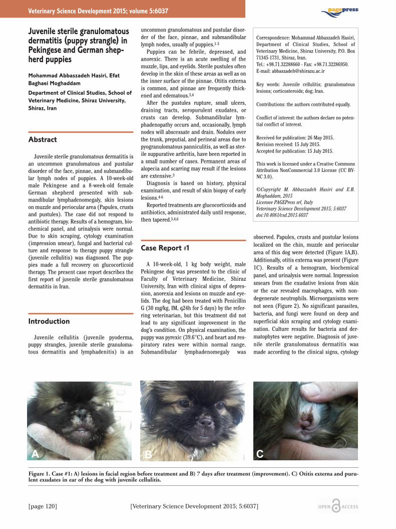
[page 120] [Veterinary Science Development 2015; 5:6037]
Juvenile sterile granulomatousdermatitis (puppy strangle) inPekingese and German shep-herd puppiesMohammad Abbaszadeh Hasiri, EfatBaghaei MoghaddamDepartment of Clinical Studies, School ofVeterinary Medicine, Shiraz University,Shiraz, Iran
Abstract
Juvenile sterile granulomatous dermatitis isan uncommon granulomatous and pustulardisorder of the face, pinnae, and submandibu-lar lymph nodes of puppies. A 10-week-oldmale Pekingese and a 8-week-old femaleGerman shepherd presented with sub-mandibular lymphadenomegaly, skin lesionson muzzle and periocular area (Papules, crustsand pustules). The case did not respond toantibiotic therapy. Results of a hemogram, bio-chemical panel, and urinalysis were normal.Due to skin scraping, cytology examination(impression smear), fungal and bacterial cul-ture and response to therapy puppy strangle(juvenile cellulitis) was diagnosed. The pup-pies made a full recovery on glucocorticoidtherapy. The present case report describes thefirst report of juvenile sterile granulomatousdermatitis in Iran.
Introduction
Juvenile cellulitis (juvenile pyoderma,puppy strangles, juvenile sterile granuloma-tous dermatitis and lymphadenitis) is an
uncommon granulomatous and pustular disor-der of the face, pinnae, and submandibularlymph nodes, usually of puppies.1-3
Puppies can be febrile, depressed, andanorexic. There is an acute swelling of themuzzle, lips, and eyelids. Sterile pustules oftendevelop in the skin of these areas as well as onthe inner surface of the pinnae. Otitis externais common, and pinnae are frequently thick-ened and edematous.3,4
After the pustules rupture, small ulcers,draining tracts, seropurulent exudates, orcrusts can develop. Submandibular lym-phadenopathy occurs and, occasionally, lymphnodes will abscessate and drain. Nodules overthe trunk, preputial, and perineal areas due topyogranulomatous panniculitis, as well as ster-ile suppurative arthritis, have been reported ina small number of cases. Permanent areas ofalopecia and scarring may result if the lesionsare extensive.3
Diagnosis is based on history, physicalexamination, and result of skin biopsy of earlylesions.4-6
Reported treatments are glucocorticoids andantibiotics, administrated daily until response,then tapered.3,4,6
Case Report #1
A 10-week-old, 1 kg body weight, malePekingese dog was presented to the clinic ofFaculty of Veterinary Medicine, ShirazUniversity, Iran with clinical signs of depres-sion, anorexia and lesions on muzzle and eye-lids. The dog had been treated with PenicillinG (30 mg/kg, IM, q24h for 5 days) by the refer-ring veterinarian, but this treatment did notlead to any significant improvement in thedog’s condition. On physical examination, thepuppy was pyrexic (39.6°C), and heart and res-piratory rates were within normal range.Submandibular lymphadenomegaly was
observed. Papules, crusts and pustular lesionslocalized on the chin, muzzle and perioculararea of this dog were detected (Figure 1A,B).Additionally, otitis externa was present (Figure1C). Results of a hemogram, biochemicalpanel, and urinalysis were normal. Impressionsmears from the exudative lesions from skinor the ear revealed macrophages, with non-degenerate neutrophils. Microorganisms werenot seen (Figure 2). No significant parasites,bacteria, and fungi were found on deep andsuperficial skin scraping and cytology exami-nation. Culture results for bacteria and der-matophytes were negative. Diagnosis of juve-nile sterile granulomatous dermatitis wasmade according to the clinical signs, cytology
Veterinary Science Development 2015; volume 5:6037
Correspondence: Mohammad Abbaszadeh Hasiri,Department of Clinical Studies, School ofVeterinary Medicine, Shiraz University, P.O. Box71345-1731, Shiraz, Iran.Tel.: +98.71.32288660 - Fax: +98.71.32286950.E-mail: [email protected]
Key words: Juvenile cellulitis; granulomatouslesions; corticosteroids; dog; Iran.
Contributions: the authors contributed equally.
Conflict of interest: the authors declare no poten-tial conflict of interest.
Received for publication: 26 May 2015.Revision received: 15 July 2015.Accepted for publication: 15 July 2015.
This work is licensed under a Creative CommonsAttribution NonCommercial 3.0 License (CC BY-NC 3.0).
©Copyright M. Abbaszadeh Hasiri and E.B.Moghaddam, 2015Licensee PAGEPress srl, ItalyVeterinary Science Development 2015; 5:6037doi:10.4081/vsd.2015.6037
Figure 1. Case #1: A) lesions in facial region before treatment and B) 7 days after treatment (improvement). C) Otitis externa and puru-lent exudates in ear of the dog with juvenile cellulitis.
A B C

[Veterinary Science Development 2015; 5:6037] [page 121]
finding and the results of fungal and bacterialculture. Prednisolone (2 mg/kg, po, q24h) for14 days and continuing for another one week(1 mg/kg, po, 24h) was prescribed. Significantimprovement was seen during the first 24-48hours of treatment (Figure 1A,B). The three-month follow up showed no regression.
Case Report #2
A female 8-week-old German shepherd wasreferred to clinic with similar signs of case A(Papules, pustules and crusts). In contrast tocase 1, there was no otitis or pyrexia. Thelesions did not respond to topical treatment withTetracycline (q12h) and Co-amoxiclav (25 mg/kgq12h). Similar examinations (cytology, skinscraping, fungal and bacterial culture) weremade and the same results were obtained.Treatment and signs of improvement were sim-ilar (to case 1). Skin lesions before and 7 daysafter treatment are shown in Figure 3.
Results and Discussion
The present case report describes the firstreport of juvenile sterile granulomatous der-matitis in Iran. Juvenile cellulitis is an uncom-mon acute dermatitis of dogs. Usually, puppiesare affected between the ages of 3 weeks and 4months, and one or several puppies in a littermay have the condition. The etiology andpathogenesis of this condition are unknown.An immunologic abnormality may be involved,as glucocorticoid therapy results in resolutionof lesions. There is some evidence for a hered-itary factor, as some breeds as well as particu-lar lines within a breed, are predisposed.Predisposed breeds include the golden retriev-er, dachshund, beagle, pointer and Gordon set-ter.3-5 In the present cases, the dogs werePekingese and German shepherd puppies andboth were non predisposed breeds.
Surprisingly, although the sibling of Pekingesepuppy (case 1) was kept elsewhere, he had thesimilar lesions (he was not referred to thementioned clinic for further investigation). Itcould be attributed to the hereditary nature ofthe disease.
The main differential diagnoses includeangioedema, staphylococcal dermatitis,demodicosis, and adverse cutaneous drugreaction.3 Due to marked regional lym-phadenopathy, systemic illness, impressionsmear and skin scraping results and no historyof medication and vaccination, these possibili-ties (diagnosis) were ruled out. Besides,response to treatment with no regressionrevealed no possibility of demodicosis, pyoder-
ma or any autoimmune disease (likePemphigus foliaceus).4,5 As in these cases,cytologic examination of papulopustularlesions of juvenile cellulitis reveals pyogranu-lomatous inflammation with no microorgan-isms and bacterial or fungal cultures are nega-tive. Biopsies of early lesions reveal multiplediscrete or confluent granulomas and pyogran-ulomas consisting of clusters of large epithe-lioid macrophages with variably sized cores ofneutrophils.3
These two cases were the only puppy stran-gles cases in 2014 and among 2100 cases thatreferred to Shiraz Veterinary School Clinic dur-ing this time, the prevalence of this disease is0.09%.
Case Report
Figure 2. Non-degenerate neutrophils (pointer) in the dog with juvenile cellulitis.
Figure 3. Case #2, A,B) lesions in facial region before treatment and C) 7 days after treat
A B C

[page 122] [Veterinary Science Development 2015; 5:6037]
Conclusions
The juvenile cellulitis could be fatal if nottreated. The prognosis is good and recoverywithout scarring is possible if early treatmentis undertaken.5 Prednisolone (with or withoutcyclosporine), oral dexamethasone and topicaltherapy, especially wet soaks with aluminumacetate or magnesium sulfate have been men-tioned in treatment of this disease.3-5 Thepresent cases were successfully treated withoral prednisolone.
When puppies are first presented with whatappears to be staphylococcal pyoderma, juve-
nile cellulitis, a relatively rare condition, maynot be considered. However, it is importantthat the possibility of juvenile cellulitis beexplored early,1,6 allowing for initiation of glu-cocorticoid therapy, which is contraindicatedfor treatment of bacterial pyoderma.1,3,6
References
1. Mason IS, Jones J. Juvenile cellulitis inGordon setters. Vet Rec 1989;124:642.
2. Reinmann KA, Evans MG, Chalofoux LVS,et al. Clinicopathologic char-acterization
of canine juvenile cellulitis. Vet Pathol1989;26:499-504.
3. Miller WH, Griffin CE, Campbell KL. Mullerand Kirk’s small animal dermatology. 7thed. St. Louis, Mosby Elsevier; 2013. pp 709-710.
4. McKeever PJ, Nuttal T, Harvey RG. A colourhandbook of skin diseases of the dog andcat. 2nd ed. London: Manson PublishingLtd; 2009. pp 159-160.
5. Paterson S. Manual of skin diseases of thedog and cat. 2nd. Oxford: BlackwellPublishing; 2008. pp 296-297.
6. Hutchings SM. Juvenile cellulitis in apuppy. Can Vet J 2003;44:418-9.
Case Report

[Veterinary Science Development 2015; 5:6061] [page 133]
Cd and Hg significantly (P<0.05) decreasedthe total anaerobic bacteria both in day 21 and42 of sampling. Also, our data indicated thatusage of probiotics significantly (P<0.05)affected the anaerobic count and increased thenumber of bacteria from 7.32 Log CFU/g to 7.54Log CFU/g.
Figure 3 represents the effect of synbioticdiet (probiotic and prebiotic) on Lactic AcidBacteria (LAB) count of stool in rats exposedto Cd and Hg. Data showed that there was sig-nificant change in LAB counts in heavy metalgroups compared with control group whichcaused reduction in the LAB microbiota popu-lation from 8.83 Log CFU/g to 7.16 Log CFU/g.Both Cd and Hg significantly decreased thetotal LAB in day 21 and 42 of sampling. Thepresence of probiotics in heavy metal treated-rat diets significantly affected the total LABand increased the population to 8.77 Log CFU/gcompared with heavy metal group. Both L.plantarum and B. coagulans produced an equaleffect on total LAB counts. Statistically signifi-cant differences were observed for the count ofL. plantarum in heavy metals-treated groupsand synbiotic groups (Table 2). Prescription ofL. plantarum in rat diets caused significantincrease (P<0.05) in the L. plantarum popula-tions in rat intestine and faeces in day 21 and42 of sampling that the number of the bacteri-um from 7.26 and 6.93 Log CFU/g in controlgroup increased to 8.17 and 7.90 Log CFU/g inL. plantarum group, respectively. Cd and Hgsignificantly decreased the L. plantarum countin rats that did not receive this bacteria both inday 21 and 42 of sampling. Particularly, feedingL. plantarum in Cd and Hg treated groupscause significant increase in L. plantarumcount (Table 2).
Table 3 shows the effect of synbiotic supple-ment on B. coagulans count of stool in ratsexposed to Cd and Hg. The data revealed thatapplication of B. coagulans in rat diets causedsignificant increase (P<0.05) in the B. coagu-lans populations in rat stool in day 21 and 42 ofsampling that the number of B. coagulans from4.47 Log CFU/g in control group increased to 7.36and 7.84 Log CFU/g in B. coagulans group,respectively. Cd and Hg significantly decreasedthe B. coagulans count in rats that did notreceive this bacteria both in day 21 and 42 ofsampling. Feeding B. coagulans in Cd and Hgtreated groups cause significant increase in B.coagulans count that raise the number (Table 3).
Discussion
The heavy metals poisoning has become amajor concern in industrialized countries.Here, a murine model was used to examine theeffect of synbiotic supplementation on theintestinal microbiota of rats that had been poi-
Article
Table 3. Effect of synbiotic diets on Bacillus coagulans count of stool in rats exposed tocadmium and mercury.
Treatments Bacillus coagulans count (Log CFU/g) Day 21 Day 42
Control 4.47±0.07a 4.47±0.10a
Lp 7.36±0.04b 7.84±0.07b
Cd 2.45±0.30c 2.41±0.25c
Lp+Cd 5.49±0.06d 5.40±0.03d
Hg 2.63±0.11c 2.55±0.14c
Lp+Hg 5.70±0.05d 5.65±0.05dResults are mean ± SD. Bc: B. coagulans, Cd: cadmium, Hg: mercury.Different letters indicate significant differences between treatments(P<0.05)
Figure 3. Effect of synbiotic diets on lactic acid bacteria count of stool in rats exposed tocadmium (A) and mercury (B). Cd: cadmium, Hg: mercury, Lp: L. plantarum, Bc: B.coagulans. The different letters indicate statistically significant differences betweengroups in each day of sampling (P<0.05).

[page 130] [Veterinary Science Development 2015; 5:6061]
Impact of synbiotic dietsincluding inulin, Bacillus coagulans and Lactobacillusplantarum on intestinal microbiota of rat exposed to cadmium and mercury
Dornoush Jafarpour,1 Seyed Shahram Shekarforoush,1Hamid Reza Ghaisari,1 Saeid Nazifi,2Javad Sajedianfard3
Departments of 1Food Hygiene andPublic Health, 2Clinical Pathology,3Physiology, School of VeterinaryMedicine, Shiraz University, Iran
Abstract
The aim of this study was to investigate theefficacy of two probiotics and a prebiotic(inulin) on intestinal microbiota of rats exposedto cadmium and mercury. Fifty-four male Wistarrats were randomly divided into nine groups. Allgroups except control group were fed standardrat chow with 5% inulin and treated as follows: i)control (standard diet), ii) Lactobacillus plan-tarum-treated group (1×10⁹ CFU/day), iii)Bacillus coagulans-treated group (1×10⁹spores/day), iv) cadmium-treated group (200µg/rat/day), v) L. plantarum and cadmium-treat-ed group, vi) B. coagulans and cadmium-treatedgroup, vii) mercury-treated group (10µg/rat/day), viii) L. plantarum and mercury-treated group, ix) B. coagulans and mercury-treated group. Cadmium, mercury and probioticswere daily gavaged to individual rats for 42 days.Treatment effects on intestinal microbiota com-position of rats were determined. Data showedthat cadmium and mercury accumulation in ratintestine affected the gastrointestinal tract andhad a reduction effect on all microbial counts(total aerobic bacteria, total anaerobic bacteria,total Lactic acid bacteria, L. plantarum and B.coagulans counts) compared to the controlgroup. It was also observed that application ofsynbiotics in synbiotic and heavy metals-treatedgroups had a significant effect and increased thenumber of fecal bacteria compared to the heavymetals groups. Based on our study, it can be con-cluded that L. plantarum and B. coagulans alongwith prebiotic inulin play a role in protectionagainst cadmium and mercury inhibitory effectand have the potential to be a beneficial supple-ment in rats’ diets.
Introduction
Gastrointestinal microbiota consists of a
complex of microorganism species that live inthe digestive tracts of human and other mam-mals. Bacteria make up most of the flora in thecolon and up to 60% of the dry mass of feces.1Bacteria in the gut fulfill a host of useful phys-iological functions and have a direct impact onhuman health, including digestion of unuti-lized energy substrates,2 repressing the growthof harmful microorganisms, helping with theproduction of some vitamins (B and K), train-ing the immune system to respond only topathogens and defending against some dis-eases.3,4 Gut microbiota’s balance can beaffected through some conditions due to theirhigh sensitivity to physiochemical and envi-ronmental factors. These factors consist ofantimicrobial agents, disorders of peristalsis,inflammatory bowel diseases, cancer, stress,redox potential, drugs, temperature and nutri-ents.5
Heavy metals are other factors that havetoxic effects on the gut ecosystem. Cadmium(Cd) and mercury (Hg) are such heavy metalsthat have become a major concern for publichealth. Cd is present at low concentrations insoil, rock and drinking water.6 Because of itshighly soluble nature compared to other met-als, Cd is taken up by plants and is stored infood and feed production.7 Dietary exposure tolarge Cd doses has been reported to result inadverse health effects in the kidneys, liver,bone, mammary gland, breast, pancreas andcolon.8-10 Liu et al.11 reported that Cd exposurehas toxic effects on microbiota of the intestin-al tract in mice. Hg can also be found in air,water and soil. Fish and shellfish are mainsources of this toxic element. Hg exposure athigh levels can harm the brain, heart, kidneyslungs, and immune system.12,13 Many studiesinvestigated how early gut development maybe stimulated and overall efficiency of theintestinal microbiota progressed by accumula-tion of synbiotics (probiotic and prebiotic)with feed.14 Lactic acid bacteria (LAB) such asL. acidophilus, L. plantarum andBifidobacterium are the most claimed probi-otics. These probiotics are very sensitive tonormal physiological conditions such as thevery low pH of the stomach and bile salts.15,16Hence, a novel beneficial probiotics is intro-duced that can survive under extreme status.Some strains of B. coagulans are able to standthrough the gastrointestinal tract and contin-ue their metabolic activities via spore produc-tion.17,18 Prebiotics are typically non-digestiblefibre compounds that pass undigested throughthe gastrointestinal tract and stimulate thegrowth and activity of advantageous bacterialike probiotics.19 Accordingly, the purpose ofthe present study is to investigate the influ-ence of two probiotics (L. plantarum and B.coagulans) and a prebiotic (inulin) on gutmicrobiota of rats exposed to Cd and Hg.
Materials and Methods
Preparing suspension of probioticbacteria
The bacterial strains used in this study wereL. plantarum (NCDO 1193) and probioticstrain of B. coagulans. Lyophilized L. plan-tarum CNR273 was obtained from the culturecollection of the Department of Food Scienceand Technology, Shiraz University, Iran andwas plated aerobically on the De Man RogosaSharpe (MRS) agar (Difco, Detroit, MI, USA)at 37°C for 48 h. Then, a single colony wasinoculated into 500 ml of MRS broth and incu-bated at 37°C with shaking at 250 rpm for 48 h.The L. plantarum pellets were achieved by cen-trifugation at 3000×g for 20 min and washedwith sterile normal saline. To determine theviable bacterial cells per ml of suspension,appropriate serial dilution was done and platedin MRS agar. Lyophilized probiotic B. coagu-lans were donated by the Pardis RoshdMehregan Company, Iran. Spore suspension ofthe bacterium (1×10⁹ spore/mL of sterilesaline) was prepared according to the methodof Abhari et al.20
Veterinary Science Development 2015; volume 5:6061
Correspondence: Seyed Shahram Shekarforoush,Department of Food Hygiene and Public Health,School of Veterinary Medicine, Shiraz University,Shiraz, Iran.Tel.: +98.713.2286950. E-mail: [email protected]
Key words: Cadmium; mercury; synbiotic diet; gutmicrobiota.
Acknowledgments: This research was financiallysupported by "Natural Antimicrobials Centre ofExcellence (NACE)" which is gratefully acknowl-edged. We would like to thank Miss M. Aghazi andMr. G. Niknia for their technical assistance.
Contributions: the authors contributed equally.
Conflict of interest: the authors declare no poten-tial conflict of interest.
Received for publication: 7 June 2015.Revision received: 17 July 2015.Accepted for publication: 29 July 2015.
This work is licensed under a Creative CommonsAttribution NonCommercial 3.0 License (CC BY-NC 3.0).
©Copyright D. Jafarpour et al., 2015Licensee PAGEPress srl, ItalyVeterinary Science Development 2015; 5:6061doi:10.4081/vsd.2015.6061

[Veterinary Science Development 2015; 5:6061] [page 131]
Preparation of rat dietsThe experimental diets were based on the
standard diets for rats plus 5% chicory basedinulin (Roosendaal, The Netherlands) andcontained 24.5% protein, 6.2% ash, 52.2%starch, 6.6% sugar, 6.5% fat and 4% moisture.Regarding micronutrients, the rat chow con-tained 0.72% calcium, 0.6% phosphorus, 0.25%chloride and 0.23% magnesium among others.The inulin content in the rat’s diet was calcu-lated based on food intake. The food intake ofeach rat with mean of 200 g body weight is 10g/day, so feedstuff was mixed with 5% inulin; itmeans each rat received 0.5 g inulin/day.
Preparation of heavy metal solu-tions
The preparation of Cd and Hg solutions wasdone according to the method of Nwokocha etal.21 The CdCl2 (Merck, Darmstadt, Germany)solution (200 µg/mL) was administered con-tinuously at a dose of 200 µg/rat/day. The HgCl2(Merck) solution (10 µg/mL) was performedcontinuously at a dose of 10 µg/rat/day. Bothmetals were fed to each rat using a special gav-age needle.
Animals and treatmentFifty-four male Wistar rats weighing 170±10
g were purchased from the Razi Vaccine andSerum Research Institute, Shiraz, Iran. Ratswere kept in stainless steel cages under stan-dardized conditions at temperature of 23±2°C,relative humidity of 60±5% and exposure to a12 h light/dark cycle with ad libitum access todiet and tap water.
After an acclimatization period of 1 week, asshown in detail in Table 1, the animals wererandomly divided into nine groups(n=6/group) and treated for 42 days. All theexperimental procedures were done followingthe ethical guidelines of the animal welfare(approved by Shiraz University animal welfarelaws, guidelines and policies in Iran).
Culturing of fecal microbiota Fresh fecal samples were collected from
each rat on two mentioned days of the experi-mental period (21 and 42) by gently handlingtheir tails to induce defecation. Then, sampleswere immediately brought to the laboratory,accurately weighed and diluted in a ratio of 9:1with sterile saline. After homogenizing for 90 susing a stomacher (Model BA6021, StewardLab., UK), specimens were diluted in 10-folddilution solution (saline) to count the bacterialload.
Total aerobic bacteria countPlate count agar (PCA, Merck, Germany)
was used for detection of total aerobic bacteri-al. Plates were incubated aerobically at 37°Cfor 24 h.
Article
Figure 1. Effect of synbiotic diets on total aerobic count of stool in rats exposed to cad-mium (A) and mercury (B). Cd: cadmium, Hg: mercury, Lp: L. plantarum, Bc: B. coag-ulans. The different letters indicate statistically significant differences between groups ineach day of sampling (P<0.05).
Table 1. Treatment groups used in the experimental study.
Treatment groups Feeding Gavaging (1 mL volume, once daily)
Control Standard diet Normal salineLp Standard diet + 5% inulin L. plantarum (1×10⁹ CFU/mL)Bc Standard diet + 5% inulin B. coagulans (1×10⁹ spore/mL)Cd Standard diet + 5% inulin Cadmium (200 mg/L)Lp+Cd Standard diet + 5% inulin Cadmium + L. plantarumBc+Cd Standard diet + 5% inulin Cadmium + B. coagulansHg Standard diet + 5% inulin Mercury (10 mg/L)Lp+Hg Standard diet + 5% inulin Mercury + L. plantarumBc+Hg Standard diet + 5% inulin Mercury + B. coagulans

[page 132] [Veterinary Science Development 2015; 5:6061]
Total anaerobic bacteria countTotal anaerobic bacteria were counted on
PCA with anaerobic condition (Anaerocult A®,Merck, Germany). Incubation was done at37°C for 24 h.
Total lactic acid bacteria countMRS agar was used for the enumeration of
total LAB. All plates were aerobically incubatedat 37°C for 48 h.
Lactobacillus plantarum countThe number of L. plantarum was deter-
mined by applying appropriate dilutions on theMRS agar supplemented with 4 mg/Lciprofloxacin (Sigma, USA). The plates wereaerobically incubated at 37°C for 48 h.
Bacillus coagulans countNYSM agar (0.5% NaCl, 0.5% pepton, 0.3%
beef extract, 0.05% yeast extract, 0.01% CaCl2,0.02% MgCl2, 0.001% MnCl2, 1% glucose and1.5% agar) was used for the enumeration of B.coagulans. NYSM agar plates were aerobicallyincubated at 37°C for 24 h.
Statistical analysisThe results are expressed as mean ± SD.
Statistical analysis for significant differencesamong group means was tested by one-wayanalysis of variance (ANOVA), followed byDuncan’s post hoc test with the help of a soft-ware SPSS 16.0 windows. P<0.05 was consid-ered significance level.
Results
Figure 1 shows the effect of synbiotic diet(probiotic and prebiotic) on total aerobiccounts in rats exposed to Cd and Hg. The num-ber of Log Total Aerobic Count (TAC) in thecontrol group was 8.61 CFU/g. Cd and Hg sig-nificantly decrease the total aerobic bacteriaboth in day 21 and 42 of sampling. Addition ofsynbiotic in heavy metal groups causedchanges in Log TVC level. The results showedthat feeding two probiotics, L. plantarum andB. coagulans to Cd and Hg- treated rats signif-icantly (P<0.05) increased the total aerobiccounts. For example in Hg- treated rats the LogTVC at day 42 was 7.49 CFU/g but applying L.plantarum increased the Log TVC to 7.91CFU/g.
The effect of synbiotic diet (probiotic andprebiotic) on total anaerobic counts in ratsexposed to Cd and Hg is shown in Figure 2.The number of log total anaerobic count in thecontrol group was 8.84 Log CFU/g.Consumption of Cd in treated rats decreasedthe number to 7.26 CFU/g and in Hg-treatedrats to 7.32 Log CFU/g. The data indicated that
Article
Table 2. Effect of synbiotic diets on Lactobacillus plantarum count of stool in ratsexposed to cadmium and mercury.
Treatments Lactobacillus plantarum count (Log CFU/g) Day 21 Day 42
Control 7.26±0.02a 6.93±0.15a
Lp 8.17±0.06b 7.90±0.04b
Cd 4.82±0.10c 4.37±0.06c
Lp+Cd 7.26±0.10a 7.22±0.05d
Hg 5.01±0.01d 5.06±0.09e
Lp+Hg 7.30±0.06a 7.39±0.02dResults are mean ± SD. Lp: L. plantarum, Cd: cadmium, Hg: mercury. Different letters indicate significant differences between treatments(P<0.05)
Figure 2. Effect of synbiotic diets on total anaerobic count of stool in rats exposed to cad-mium (A) and mercury (B). Cd: cadmium, Hg: mercury, Lp: L. plantarum, Bc: B. coag-ulans. The different letters indicate statistically significant differences between groups ineach day of sampling (P<0.05).

[page 134] [Veterinary Science Development 2015; 5:6061]
soned with two heavy metals, cadmium andmercury. In this study, we focused on themicrobiota and its response to host consump-tion of Cd and Hg along with synbiotic supple-ment.
Two probiotic bacteria (B. coagulans and L.plantarum) and prebiotic (inulin) wereapplied as synbiotic diets. It was observed thatcadmium and mercury accumulation in ratintestine affected the gastrointestinal tractand impaired the gut barrier. Diets containingheavy metals (Cd or Hg) were investigated todetermine whether they have a reductioneffect on all microbial counts in treatedgroups. Many reports indicated that heavy met-als (Cd, Zn, Cs) have an inhibitory effect onbacteria by delaying the initiation of bacterialgrowth and inhibiting their growth.22-24Ghorbani et al.25 expressed that sufficientmetal exposure will result in immediate deathof microorganisms due to disruption of essen-tial functions, and to more gradual changes inpopulation sizes due to changes in viability orcompetitive ability. Probiotic bacteria B. coagu-lans and L. plantarum received significantstress from the two mentioned heavy metals(Cd and Hg) compared to the non-exposedgroups. Our result is confirmed by the report ofLiu et al.11 that Cd treatment could decreasethe population of gut bacteria and the thick-ness of mice inner mucus layer was also atten-uated by Cd treatment.
In contrast, it was found that in treated syn-biotic + heavy metals groups the microbialcounts were not so reduced compared withheavy metals groups. It seems that when thenumber of bacteria is low the heavy metalsaffect them, but in synbiotic + heavy metalsgroups by increasing the probiotic bacteriaheavy metals’ effect is reduced. In recent yearsstudies have demonstrated that some bacteriasuch as probiotics have the largest role inbinding metals, preventing their entry to thebody and, thus, protecting the host.26,27 It wasfound that inulin and probiotics had a consid-erable effect on fecal microbiota. According toVan Heugten et al.28 dietary supplement withprobiotics can potentially alter gut microbiotaby selectively stimulating the growth of benefi-cial bacteria while suppressing the growth ofpathogenic bacteria. Also, Lin et al.29 reportedthat the use of probiotics in the feed enhancedLactobacillus number and reduced E. coli pop-ulation indicating that dietary supplementsare efficient to improve broiler intestinalmicrobiota balance.
Moreover, the utilization of inulin in rat dietas a prebiotic ingredient demonstrated aremarkable effect on the ecosystem of intestin-al tract by increasing the total lactic acid bacte-ria population in rats. This result is supportedby the report of Roberfroid that fermented non-digestible food ingredients, such as inulin,beneficially affect the host by selectively stim-
ulating the growth and/or activity of one or alimited number of bacteria in the colon by act-ing as substrate for them.30
Conclusions
In conclusion, this study provided substan-tial insight in illustrating the inhibitory effectof cadmium and mercury and the impact ofsynbiotic to rat gut microbiota. This workreveals the potential of synbiotics to decreasethe cadmium and mercury repressive effect onthe population of gut bacteria.
These interesting findings demonstrate thatsince B. coagulans and L. plantarum have thepotential to supply the gut microbiota to thenormal condition in Cd and Hg treated rats, itcan be used as a supplementary component intreatment diets.
References
1. Scott KP, Gratz SW, Sheridan PO, et al. Theinfluence of diet on the gut microbiota.Pharmacol Res 2013;69:52-60.
2. Wynne AG, McCartney AL, Brostoff J, et al.An in vitro assessment of the effects ofbroad-spectrum antibiotics on the humangut microflora and concomitant isolationof a Lactobacillus plantarum with anti-Candida activities. Anaerobe 2004;10:165-9.
3. Guarner F, Malagelada J. Gut flora inhealth and disease. Lancet 2003;361:512-9.
4. Sears CL. A dynamic partnership: celebrat-ing our gut flora. Anaerobe 2005;11:247-51.
5. Heavey P, Rowland I. The gut microflora ofthe developing infant: microbiology andmetabolism. Microbiol Ecol Health Dis1999;11:75-83.
6. Jarup L. Hazards of heavy metal contami-nation. Br Med Bull 2003;68:167-82.
7. Sarwar N, Malhi SS, Zia MH, et al. Role ofmineral nutrition in minimizing cadmiumaccumulation by plants. J Sci Food Agric2010;90:925-37.
8. Nordberg GF, Jin T, Wu X, et al. Prevalenceof kidney dysfunction in humans - rela-tionship to cadmium dose, metallothionin,immunological and metabolic factors.Biochemie 2009;91:1282-5.
9. Satarug S, Garrett SH, Sens MA, Sens DA.Cadmium, environmental exposure, andhealth outcomes. Environ Health Perspect2010;118:182-90.
10. Waalkes MP. Cadmium carcinogenesis inreview. J Inorgan Biochem 2000;79:241-4.
11. Liu Y, Li Y, Liu K, Shen J. Exposing to cad-mium stress cause profound toxic effect
on microbiota of the mice intestinal tract.PLOS One 2014;9:e85323.
12. Clarkson TW, Magos L, Myers GJ. The tox-icology of mercury: current exposures andclinical manifestations. N Engl J Med2003;349:1731-7.
13. Houston MC. The role of mercury and cad-mium heavy metals in vascular disease,hypertension, coronary heart disease, andmyocardial infarction. Altern Ther HealthMed 2007;13:S128-33.
14. Yang SC, Chen JY, Shang HF, et al. Effect ofsynbiotics on intestinal microflora anddigestive enzyme activities in rats. World JGastroenterol 2005;11:7413-7.
15. Bezkorovainy A. Probiotics: determinantsof survival and growth in the gut. Am JClin Nutr 2001;73:399S-405S.
16. Holzapfel WH, Haberer P, Snel J, et al.Overview of gut flora and probiotics. Int JFood Microbiol 1998;41:85-101.
17. Hyronimus B. Acid and bile tolerance ofspore-forming lactic acid bacteria. Int JFood Microbiol 2000;61:193-7.
18. Katsutoshi AR. Effect spore-bearing lacticacid-forming bacteria (Bacillus coagulansSANK 70258) administration on the intes-tinal environment, defecation frequency,faecal characteristics and dermal charac-teristics in humans and rats. MicrobiolEcol Health Dis 2003;14:4-13.
19. Gibson GR, Roberfroid MB. Dietary modu-lation of the human colonic microbiota:introducing the concept of prebiotics. JNutr 1995;125:1401-12.
20. Abhari KH, Shekarforoush SS,Sajedianfard J, et al. The effects of prebiot-ic, probiotic and synbiotic diets includingBacillus coagulans and inulin on rat intes-tinal microbiota. Iranian J Vet Res. 2015;In Press.
21. Nwokocha CR, Owu DU, Nwokocha MI, etal. Comparative study on the efficacy ofAllium sativum (garlic) in reducing someheavy metal accumulation in liver ofWistar rats. Food Chem Toxicol2012;50:222-6.
22. Gikas P, Şengör SS, Ginn TR, et al. Theeffects of heavy metals and temperatureon microbial growth and lag. Global Nest J2009;1:325-32.
23. Şengör SS, Barua S, Gikas P, et al.Influence of heavy metals on microbialgrowth kinetics including lag time: mathe-matical modeling and experimental verifi-cation. Environ Toxicol Chem2009;28:2020-9.
24. Şengör SS, Gikas P. The influence of singleand combined effects of Zn, Cu and tem-perature on microbial growth. Global NestJ 2014;16:699-706.
25. Ghorbani, NR, Salehrastin, N, Moeini, A.Heavy metals affect the microbial popu-lations and their activities, in
Article

[Veterinary Science Development 2015; 5:6061] [page 135]
Symposium of 17th WCSS, 14-21 August,Thailand. 2002; 2234-2245.
26. Bhakta JN, Ohnishi K, Munekage Y, et al.Characterization of lactic acid bacteria-based probiotics as potential heavy metalsorbents. J Appl Microbiol 2012;112:1193-206.
27. Ibrahim F, Halttunen T, Tahvonen R,Salminen S. Probiotic bacteria as potential
detoxification tools: assessing their heavymetal binding isotherms. Canadian JMicrobiol 2006;52:877-85.
28. Van Heugten E, Funderburke DW, DortonKL. Growth performance, nutrientdigestibility, and fecal microflora in wean-ling pigs fed live yeast. J Anim Sci2003;81:1004-12.
29. Lin SY, Hung ATY, Lu JJ. Effects of supple-
ment with different level of Bacillus coag-
ulans as probiotic on growth performance
and intestinal microflora populations of
broiler chickens. J Anim Vet Adv 2011;10:1 -
11-4.
30. Roberfroid MB. Prebiotics and probiotics:
are they functional foods? Am J Clin Nutr
2000;71:1682S-7S.
Article