proceedings of the 22nd national congress on parasitology
343
PHOTOGRAPHS OF THE 22ND NATIONAL CONGRESS OF PARASITOLOGY
-
Upload
khangminh22 -
Category
Documents
-
view
2 -
download
0
Transcript of proceedings of the 22nd national congress on parasitology

PHOTOGRAPHS
OF THE
22ND NATIONAL CONGRESS
OF
PARASITOLOGY

PROCEEDINGSOF THE 22ND NATIONAL CONGRESS ON
PARASITOLOGYOctober 30 - November 1, 2010
Editor-in-chiefPROF. P. K. BANDYOPADHYAY
DEPARTMENT OF ZOOLOGYUniversity of Kalyani, Kalyani-741235
West Bengal, India

Proceedingsof the 22nd National Congress
on ParasitologyOctober 30-November 1, 2010
Advances in Parasitology :A Novel Approach Towards a Disease Free World
Organised by
DEPARTMENT OF ZOOLOGY,University of Kalyani, Kalyani-741235,
West Bengal, India
In collaboration with
INDIAN SOCIETY FOR PARASITOLOGY&
ZOOLOGICAL SURVEY OF INDIA
PROF. P. K. BANDYOPADHYAY
PROF. D. R. MANDAL


INDIAN SOCIETY FORPARASITOLOGY COUNCIL
PRESIDENT
DR. S.L. HOTI, PUDUCHERRY
VICE PRESIDENT
PROF. P. PRAKASH BABU, HYDERABAD
DR. S.K.PURI, LUCKNOW
DR. R. KALEYSA RAJ, TRIVANDRUM
PROF. MRIDULA JAIN, CHANDIGARH
SECRETARY
DR. J.K. SAXENA, CDRI, LUCKNOW
EXECUTIVE COMMITTEE MEMBERS
DR. A.K. GUPTA, RAIPUR
DR. FAYAZ AHAMED, SRINAGAR
DR. NEENA GOYAL, LUCKNOW
PROF. H.S. BANYAL, SHIMLA
PROF. M.V.R. REDDY, WARDHA
DR. U. SHAMEEM, VISHAKAPATNAM
DR. S.M.A. ABIDI, ALIGARH
DR. S.K. MALHOTRA, ALLAHABAD
DR. A.K. YADAV, SHILLONG
DR. P.D. JUYAL, LUDHIANA
PROF. P.K. BANDYOPADHYAY, WEST BENGAL
DR. KUMKUM SRIVASTAVA, LUCKNOW
PROF. SUKHBIR KAUR, CHANDIGARH
DR. A.M. KHAN, DIBRUGARH
DR. VAS DEV, ASSAM

22nd National Congress of ParasitologyLOCAL ORGANIZING COMMITTEE
CHIEF PATRONPROF. ALOK KUMAR BANERJEE, VICE CHANCELLOR
PATRONPROF. SITAL K. CHATTOPADHYAY, DEAN, FACULTY OF SCIENCE
PRESIDENTDR. DEBJANI NATH, HEAD, DEPT. OF ZOOLOGY
VICE PRESIDENTPROF. SAMIRAN CHAKRABARTI AND PROF. SACHINANDAN KUNDU
ORGANIZING SECRETARYPROF. PROBIR K. BANDYOPADHYAY
Ph : 9433214527
JOINT SECRETARYPROF. GOUTAM CHANDRA AND DR. AMLAN K. MITRA
TREASURERDR. ASISH K. PANIGRAHI
MEMBERS
SRI U. BHATTACHARYAY, Registrar, K.U.
PROF. B. B. JANA, International Centre for Ecological Engineering, K.U.
PROF. A. R. KHUDABUKHSH, Department of Zoology, K.U.
PROF. C. R. SAHU, Department of Zoology, K.U.
PROF. D. MUKHERJEE, Department of Zoology, K.U.
PROF. A. KAVIRAJ, Department of Zoology, K.U.
PROF. D. K. BHATTACHARYAY, Department of Zoology, K.U.
PROF. C. SENGUPTA, Department of Botany, K.U.
PROF. D. R. MONDAL, Durgapur Government College, Durgapur
PROF. S. BISWAS, West Bengal University of Animal and Fisheries Sciences, Kolkata
PROF. S. SINHABABU, Department of Zoology, Visva Bharati, Santinekatan
SRI M. KUNDU, Finance Officer, K.U.
SRI S. BANIK CHOWDHURY, University Engineer, K.U.
DR. S. N. SINHA, Department of Botany, K.U.
DR. A. BHATTACHARYAY, Department of Zoology, University of Calcutta, Kolkata
DR. S. CHATTERJEE, Department of Zoology, Burdwan University, Burdwan
DR. A. GOSWAMI, West Bengal State University, Barasat
DR. M. K. DAS, Central Inland Fisheries Research Institute, Barrackpore
PROF. N. C. SAHA, Barasat Government College, Barasat
DR. M. MANNA, PG Department of Zoology, Bethune College, Kolkata
DR. A. MAJHI, School of Tropical Medicine, Kolkata
DR. B. BHOWMICK, Department of Zoology, Panchakot College, Purulia
DR. R. SHUKLA RAY, PG Department of Zoology, Bethune College, Kolkata
SMT. S. CHETTRI, PG Department of Zoology, Darjeeling Government College, Darjeeling

It is my proud privilege to extend my cordial and warm greetings to the distinguished and celebrated scientists, eruditeresearchers, professors, clinicians of national and international repute, faculties, scholars and students who have assembledin the 22nd National Congress of Parasitology organized by the Department of Zoology, University of Kalyani, Kalyani, WestBengal in collaboration with the Indian Society for Parasitology and Zoological Survey of India, Kolkata held during the period30th October, 2010 to 1st November, 2010.
During the three days of the congress, it became an important podium for the dissemination and argument on the newfindings of chemotherapy, immune response, molecular biology and biochemistry, host parasite interaction, parasite infectionsin aquatic ecosystem, taxonomy, systematics and phylogeny, vector biology, control and epidemiology. The congress providedthe scientific depth with a range of plenary and invited talks with a compliment of large number of oral and poster presentationsin parallel thematic session. Privilege had also been obtained to felicitate some veteran scientists of West Bengal whocontributed immensely in the field of parasitology. The discussion and interactions amongst both seasoned and buddingenthusiasts in the area of parasitological research from the diverse section of India was much cheering.
I do feel that the deliberations presented at the congress had encompassed each and every arena of the subject therebyproviding a dynamic platform for the vibrant , meaningful, evocative, incomparable and unending exchange of new findings,investigating and enigmatic questions and state of the art techniques between the scientists, researchers, clinicians, faculties,scholars and students as well to usher a score of new avenues for future research projects on parasitology at the nationaland international level. And, to encounter a number of dreadful diseases inflict on human, domestic animals, aquatic systemsand economically important plants to ensure good health for all and self-sufficiency in food thereby benefiting the humansociety immensely.
The executives of the “Indian society for Parasitology” do strongly believe that organizing such congress in the daysahead would surely deliver educative lessons, hygienic intuitions and general awareness to all concerned especially the poorerand economically marginalized rural populace enormously. To enliven this spirit of our society consistent governmental supportand assistance from other benevolent organizations are absolutely supplicated.
I do also feel that our society could emerge as a harbinger in disseminating basic parasitological facts, intrinsic tenetsand other insights among the rural, semi-urban and urban populace then we could think of a healthier tomorrow thereby aimingat a huge leap towards developing a disease free world by conquering a number of insurmountable diseases through ingeniousparasitological researches to alleviate acute human sufferings.
I like to put on record our sincere thanks to all funding agencies and advertisers for their financial support to the Congress.Thanks are also due to all contributors for providing the manuscripts and for undertaking necessary editorial changes.
I express my heartfelt thanks to the University authorities and all teachers of the Department of Zoology for their kindhelp, suggestions and active co-operation. I am thankful to all scholars of the Parasitology Laboratory of the department fortheir active help and co-operation. Last but not be the least, thanks are also due to Sri Dipanjan Mondal, East India PhotoComposing Centre, Kolkata-700 006 for printing the proceedings.
Prof. Probir K. Bandyopadhyay
Kalyani Editor-in-chiefDecember 25, 2011 Editorial Board
PREFACE

The National Academy of Sciences, India
Prof. V.P. SharmaICMR Chair in Public Health ResearchCRDT, IIT-Delhi-110016.
Dear Dr. Prabir Bandyopadhyay,
It gives me great pleasure that the 22nd National Congress of Parasitology held atthe University of Kalyani from October 30 to November 1, 2010 was a spectacularsuccess. Scientific presentations were of very high quality and discussions enriched thepresentation. The plenary lectures, invited talks, memorial & oration lectures, youngscientist award lectures and oral and poster presentations provided opportunities for theyoung scientists to learn and interact on the latest developments in various fields ofparasitology. Congress arrangements were all excellent and deserve appreciation. Publicationof the Proceedings would provide a valuable material for the researchers to make use ofthe information in their own work. I wish to congratulate you and your team of KalyaniUniversity for organizing such an excellent event and for publishing the Proceedings.
With best Wishes,
Sincerely Yours,
Date : 20.10.2011
V.P. SharmaEditor in ChiefJournal of Parasitic Disease
FOREWORD
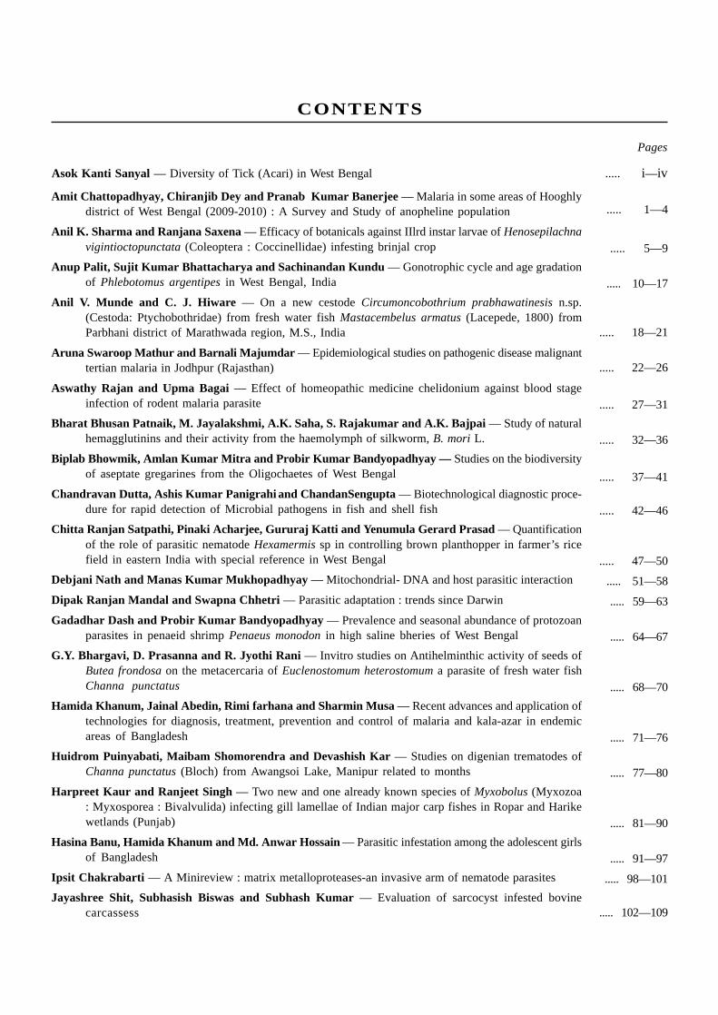
Amit Chattopadhyay, Chiranjib Dey and Pranab Kumar Banerjee — Malaria in some areas of Hooghlydistrict of West Bengal (2009-2010) : A Survey and Study of anopheline population
Anil K. Sharma and Ranjana Saxena — Efficacy of botanicals against IIlrd instar larvae of Henosepilachnavigintioctopunctata (Coleoptera : Coccinellidae) infesting brinjal crop
Anup Palit, Sujit Kumar Bhattacharya and Sachinandan Kundu — Gonotrophic cycle and age gradationof Phlebotomus argentipes in West Bengal, India
Anil V. Munde and C. J. Hiware — On a new cestode Circumoncobothrium prabhawatinesis n.sp.(Cestoda: Ptychobothridae) from fresh water fish Mastacembelus armatus (Lacepede, 1800) fromParbhani district of Marathwada region, M.S., India
Aruna Swaroop Mathur and Barnali Majumdar — Epidemiological studies on pathogenic disease malignanttertian malaria in Jodhpur (Rajasthan)
Aswathy Rajan and Upma Bagai — Effect of homeopathic medicine chelidonium against blood stageinfection of rodent malaria parasite
Bharat Bhusan Patnaik, M. Jayalakshmi, A.K. Saha, S. Rajakumar and A.K. Bajpai — Study of naturalhemagglutinins and their activity from the haemolymph of silkworm, B. mori L.
Biplab Bhowmik, Amlan Kumar Mitra and Probir Kumar Bandyopadhyay — Studies on the biodiversityof aseptate gregarines from the Oligochaetes of West Bengal
Chandravan Dutta, Ashis Kumar Panigrahi and ChandanSengupta — Biotechnological diagnostic proce-dure for rapid detection of Microbial pathogens in fish and shell fish
Chitta Ranjan Satpathi, Pinaki Acharjee, Gururaj Katti and Yenumula Gerard Prasad — Quantificationof the role of parasitic nematode Hexamermis sp in controlling brown planthopper in farmer’s ricefield in eastern India with special reference in West Bengal
Debjani Nath and Manas Kumar Mukhopadhyay — Mitochondrial- DNA and host parasitic interaction
Dipak Ranjan Mandal and Swapna Chhetri — Parasitic adaptation : trends since Darwin
Gadadhar Dash and Probir Kumar Bandyopadhyay — Prevalence and seasonal abundance of protozoanparasites in penaeid shrimp Penaeus monodon in high saline bheries of West Bengal
G.Y. Bhargavi, D. Prasanna and R. Jyothi Rani — Invitro studies on Antihelminthic activity of seeds ofButea frondosa on the metacercaria of Euclenostomum heterostomum a parasite of fresh water fishChanna punctatus
Hamida Khanum, Jainal Abedin, Rimi farhana and Sharmin Musa — Recent advances and application oftechnologies for diagnosis, treatment, prevention and control of malaria and kala-azar in endemicareas of Bangladesh
Huidrom Puinyabati, Maibam Shomorendra and Devashish Kar — Studies on digenian trematodes ofChanna punctatus (Bloch) from Awangsoi Lake, Manipur related to months
Harpreet Kaur and Ranjeet Singh — Two new and one already known species of Myxobolus (Myxozoa: Myxosporea : Bivalvulida) infecting gill lamellae of Indian major carp fishes in Ropar and Harikewetlands (Punjab)
Hasina Banu, Hamida Khanum and Md. Anwar Hossain — Parasitic infestation among the adolescent girlsof Bangladesh
Ipsit Chakrabarti — A Minireview : matrix metalloproteases-an invasive arm of nematode parasites
Jayashree Shit, Subhasish Biswas and Subhash Kumar — Evaluation of sarcocyst infested bovinecarcassess
CONTENTS
..... 1—4
..... 5—9
..... 10—17
..... 18—21
..... 22—26
..... 27—31
..... 32—36
..... 37—41
..... 42—46
..... 47—50
..... 51—58
..... 59—63
..... 64—67
..... 68—70
..... 71—76
..... 77—80
..... 81—90
..... 91—97
..... 98—101
..... 102—109
Asok Kanti Sanyal — Diversity of Tick (Acari) in West Bengal
Pages
..... i—iv

Konineeka Sen and Chandan Sengupta — Suppression of Fusarium oxysporum affecting cucumber hostby treatment with plant growth-promoting rhizobacteria
Loukrakpam Bina, Naorem Mohilal and Loitongbam Victoria — Report on a fungal parasite of plantparasitic nematodes of Manipur, India
Lalan Kumar Arya, SR. Rathinam, Lalitha Prajna, Usha Kim and Veena Tandon — Presumed trematodeinduced granulomatousuveitis in south India
Madhusree Gangopadhyay Maitra, Rupendu Ray and Probir Kumar Bandopadhyay — Protoopalinaandulensis and Protoopalina limnocharis (Protozoa : Slopalinida) with a note on their heterospecificassociation with anuran host of West Bengal, India
Manoja Patnaik, Dilip Kumar Bhatacharya, Prabir Mitra, Atul Kumar Saha and Arivind Kumar Bajpai —Effect of chaetothyrium species, a causal agent of sooty mould on silk production of Bombyx mori
Md. Manzoorul Kibria, Hadiul Islam, M. M. A. Habib, Liton Chondra Shutradhar and Ghazi S. M. Asmat— Trichodinid ectoparasites (Ciliophora : Trichodinidae) from the gills of freshwater fishes in theShitalakhsya River, Bangladesh
Maibam Pramodini and Naorem Mohilal — A preliminary survey on entomophagous nematodes of Manipur
Manne Venkata Santha Kumar, Paushalee Datta, Samiran Chakrabarti, Swapan Kumar Mukhopadhyay,Nirvan Kumar Das, Prabir Mitra, Atul Kumar Saha and Arvind Kumar Bajpai — Influence of abioticfactors and parasitoid, Eritmocerus adustiscutum on the incidence of whitefly, Dialeuroporadecempuncta
Nimai Chandra Saha, Ashis Kumar Panigrahi and Kishore Dhara — A report on effective treatment withciprofloxacin along with vitamin C and E to EUS infected snake head fish Ophiocephalus gachua(Hamilton, 1822)
Nilendu Jyoti Maitra, Probir Kumar Bandyopadhyay and Arunasis Goswami — Distribution of worminfestation in naturally grazing garole sheep of Sundarbans region
Norem Mohilal and M. Manjur Shah — Structural peculiarities of the eggs of nematodes-their significancein taxonomy
Oinam Sonia, Raj Kumar Gambhir, Thockchom Tarnita, Ngaseppam Zenith and Wahengbam Lakshmipyari— On a new species, Oxysomatium teraensis sp.nov. (Nematoda : Oxyuridae) from Hyla annectens,from Manipur, India
Papia Das and Buddhadeb Manna — On the nematofauna inhabiting mosses available at Indian BotanicGarden, Howrah, West Bengal, India and description of Thornenema thornei n.sp.
Paushalee Dutta, Manne Venkata Santha Kumar, Samiran Chakrabarti, Swapan Kumar Mukhopadhyay,Nirvan Kumar Das, Prabir Mitra, Atul Kumar Saha and Arvind Kumar Bajpai — Impact of hostpopulation and abiotic factors on the percent parasitism by Eretmocerus adustiscutum, a parasitoidof Aleuroclava pentatuberculata
Pinky Kaur, Tufel Ahamad Qureshi, Rekha Shrivastav and Susan Manohar — Occurrence of Eustrongylideslarvae sp. in Xenentodon cancila (Ham.) from lower lake, Bhopal and its effect on ovaries
Prabir Mitra, Satadal Chakrabarti, Probir Kumar Bandyopadhyay and Durga Prasad Haldar —Characterization of haemocyte types, their counts in different breeds of silkworm, Bombyx mori L.and their progressive changes following bacterial inoculation
Rajatendu Banik, Rina Bhattacharya and Probir Kumar Bandhopadhyay — On the relation of meteorologicalparameters and malaria transmission
Ratnabir Singha, Maibam Shomorendra and Devashish Kar — Nematode infection in the fishes of DoluLake, Silchar, Assam, during different seasons of the year
Remya Madhavan, Probir Kumar Bandyopadhyay and B. Santosh — Nature of parasitic infestations in threeeconomically important Labeo spp. inhabiting natural and cultured water bodies of Tripura
Rimi Farhana and Hamida Khanum — The effect of parasitic infestation on tissues and organs of Mystusaor (hamilton) and Mystus bleekeri (day)
Pages
..... 110—116
..... 117—121
..... 122—125
..... 126—130
..... 131—134
..... 135—149
..... 150—152
..... 153—158
..... 159—164
..... 165—170
..... 171—173
..... 174—177
..... 178—185
..... 186—191
..... 192—195
..... 196—205
..... 206—212
..... 213—217
..... 218—222
..... 223—227

Sapna Singh and Kedar Prasad Sinha — On the seasonal occurrence of Clinostomatid metacercariaeinfections in an Indian mudskipper Channa punctatus bl. from the fresh water swamps nearDarbhanga town, north Bihar
Santanu Chakrabarti — Acid tolerance as a vehicle for food-borne shigellosis
Savita Rani, Juhie Rani and S.S Lal — Immunohistopathological changes in the bursa of fabricius duringthe hypersensitivity reactions-IV induced by experimental ascaridiasis in white leg horn chicks
Satadal Chakrabarty, Buddhadeb Manna, Prabir Mitra, Atul Kumar Saha and Bharat Bhushan Bindroo— Studies on immunological impact of some chemicals, botanicals, antibacterial proteins and live non-pathogenic bacteria in silkworm, Bombyx mori L. to control bacterial disease
Sabina Yesmin and Hamida Khanum — On the Studies of Parasites Infestation in Clarias batrachus(Linnaeus) and Clarias gariepinus (Burchell)
S. Ravichandran and G. Rameshkumar — Host specificity of parasitic isopods in marine fishes
Sayani Banerjee, Jayati Chakraborti, Somerita Panda and Probir K. Bandyopadhyay — Antibacterial effectof some locally available plant extracts against some fish pathogenic bacteria
Somsuvra Dasgupta, Asosh Kumar Panigrahi and Raman Kumar Trivedi — Studies on parasitic diseasesof fish in east kolkata wetland and their anti-parasitic treatment which threat to environment
Sharbani Dutta (Roy) and Ashis Kumar Panigrahi — Studies of the parasitic infestation in indigenousornamental fish silver danio, danio devario with its prophylactic measures
Supriya Khanra, Subir K Bandopadhyay, Priyanka Chakraborty, Dona Mitra, Khudiram Naskar, SyamalRoy and Madhumita Manna — Random amplified polymorphic dna and drug sensitivity analysis ofrecent clinical isolates from indian kala-azar patient
Sutapa Sarkar and Probir Kumar Bandyopadhyay — First record of a gregarine from an earthwormof bangladesh
Soumendranath Chatterjee, Swapan Kumar Rudra, Tuhin Subhra Ghosh and Paltu Kumar Dhal —Molecular characterization and phylogenetic analysis of mosquito-pathogenic Bacillus cereus(HM026606)
Shyma Kunhipurayil, Surender Kumar Gupta, Ajit Singh, Sumeer Singh Chaudhary and JayprakashGupta — Monoclonal antibody based latex agglutination test for the diagnosis of trypanosomiosisin cattle
Shyma Kunhipurayil, Surender Kumar Gupta, Ajit Singh, Sumeer Singh Chaudhary and JayprakashGupta — Polymerase chain reaction based detection of Trypanosoma evansi in whole blood ofdomestic animals
Suman Kundu and Larisha Mawkhling Lyndem — Anticestodal effect of medicinal plants from West Bengal
Sujoy Kumar Bag, Jayati Chakraborty, Sayani Banarjee and Probir kumar Bandyopadhyay — Changesin haematological parameters in trypanosome infected Clarias batrachus (L.) in West Bengal
Tanima Biswas, Sabir Hossen Molla and Probir Kumar Bandyopadhyay — On the occurrence of aprotozoan parasite from edible oysters of Sunderbans region of West Bengal
Thounaojam Hemananda and Naorem Mohilal — Variation in the diversity of protozoan parasites of fishesin manipur : climate change - a possible cause
Pages
..... 228—231
..... 232—236
..... 237—243
..... 244—251
..... 252—258
..... 259—264
..... 265—270
..... 271—275
..... 276—281
..... 282—286
..... 287—292
..... 293—298
..... 299—301
..... 302—304
..... 305—309
..... 310—314
..... 315—318
..... 319—324


ACKNOWLEDGEMENTOUR SPONSORERS
1. Council of Scientific and Industrial Research (CSIR), New Delhi
2. Indian Council of Agricultural Research (ICAR), New Delhi
3. Indian Council of Medical Research (ICMR), New Delhi
4. Department of Higher Education, Govt of West Bengal, Kolkata
5. Ministry of Earth Science, New Delhi
6. The National Academy of Sciences, India
7. Department of Biotechnology (DBT), New Delhi
8. Zoological Survey of India, Kolkata
9. Department of Science and Technology (DST), New Delhi
10. Defense Research and Development Organization (DRDO), New Delhi
11. Department of Science and Technology (Govt. of West Bengal), Kolkata
12. Indian National Science Academy (INSA), New Delhi
13. West Bengal Biodiversity Board, Kolkata
14. Central Inland Fisheries Research Institute, Barrackpore
15. CIFA
16. Dinabandhu Andrews Institute of Technology and Management, Kolkata
17. Directorate of Open and Distance Learning (DODL), Kalyani University
18. West Bengal State University, Barasat, West Bengal
19. The University of Burdwan, Burdwan
20. Indian Institute of Chemical Biology, Kolkata


EDITORIAL BOARD
Editor-in-Chief :Prof. Probir Kumar Bandyopadhyay
Professor of Zoology, University of Kalyani, Kalyni, West Bengal, India andOrganizing Secretary, 22nd National Congress of Parasitology
Co-Editor :Prof. Dipak Ranjan Mandal
Principal, Moulana Azad College, Kolkata, West Bengal, India
Associate Editors :
Published by Prof. P. K. Bandyopadhyay, Organizing Secretary, 22nd National Congress of Parasitology. Printed at theEast India Photo Composing Centre 69, Sisir Bhaduri Sarani, Kolkata-700 006, Phone : 2350-0132
Prof. Goutam ChandraDepartment of Zoology,The University of Burdwan, Burdwan, West Bengal, India
Dr. Amlan K. MitraDepartment of Zoology,Ranaghat College, Ranaghat, West Bengal, India
Dr. Ardhendu MajiSchool of Tropical Medicine,C. R. Avenue, Kolkata, West Bengal, India
Dr. Soumendranath ChatterjeeDepartment of Zoology,The University of Burdwan, Burdwan, West Bengal, India
Editorial Assistants :
Prof. Nimai Chandra SahaP. G. Department of Zoology,Barasat Govt. College, Barasat, West Bengal, India
Dr. Madhumita MannaP. G. Department of Zoology,Bethun College, Kolkata, West Bengal, India
Dr. Roli Shukla RoyP. G. Department of Zoology,Bethun College, Kolkata, West Bengal, India
Ms. Sayani BanerjeeDepartment of Zoology,University of Kalyani, Kalyani, West Bengal, India
Ms. Jayati ChakrobortiDepartment of Zoology,University of Kalyani, Kalyani, West Bengal, India
Ms. Sutapa SarkarDepartment of Zoology,University of Kalyani, Kalyani, West Bengal, India
Ms. Subarna Ghosh
Department of Zoology,
University of Kalyani, Kalyani, West Bengal, India
Ms. Somerita Panda
Department of Zoology,
University of Kalyani, Kalyani, West Bengal, India
Mr. Sabir Hossen Molla
Department of Zoology,
University of Kalyani, Kalyani, West Bengal, India
Mr. Sukanta Majumder
Department of Zoology,
University of Kalyani, Kalyani, West Bengal, India
Ms. Tanima Biswas
Department of Zoology,
University of Kalyani, Kalyani, West Bengal, India

22nd National Congress of Parasitology, Oct. 30-Nov. 01, 2011Dept. of Zoology, University of Kalyani, Kalyani, W.B.
1
Malaria in some areas of Hooghly district of West Bengal (2009-2010) :A survey and study of anopheline population
Amit Chattopadhyay, Chiranjib Dey and Pranab Kumar BanerjeeVector Cytogenetics Research Unit, Department Zoology (U.G. and P.G. Studies),
Serampore College, Serampore 712 201, West Bengal, India
Abstract : Recent resurgence of malaria in suburban and rural areas of different districts of West Bengal has become a serious health problem.It has been reported that some blocks of Hooghly are malaria prone. Therefore, a systemic investigation has been carried out in some areasof Hooghly (especially Serampore subdivision) in order to know the prevalence of the disease as well as the species diversity of Anophelesmosquito. The present study reveals that falciparum malaria is on the rise in urban area in relation to vivax malaria but rural area has followedreverse trend during 2009-2010. Our data also reveals that the Anopheline population includes mainly Anopheles subpictus as also A. anularisand A. stephensi in these malaria prone areas.
Key Words : Anopheles spp.; Falciparum malaria; Prevalence; Resurgence; Vivax malaria
* Corresponding Author : E-mail : [email protected],
IntroductionMalaria has been one of the most prominent and an ancient disease which is still a major problem. About 100 countries
are malarious. India is reporting about 2 million positive cases per year. In India death caused by P. falciparum in last 5 yearsis as follows 134, 193, 291, and 222 in 2005, 2006, 2007 and 2008 respectively. Fatality rate in West Bengal is 5% (WHO, 2009).
There are 4 sub-divisions and 18 blocks in our Hooghly district. Among these 5 blocks are malaria prone viz. Mogra,Singur, Kanaipur, Goghat-II, and Khanakul-II.
The aims and objectives of this study is to know the Slide Positivity Rate (S.P.R.) in different rural blocks of Seramporesubdivisions, the seasonal prevalence of malaria, the parasitic variations as well as the Anopheline diversities in the studiedareas viz. Kanaipur, Serampore and Seoraphuli of Serampore sub-division, West Bengal.
Materials and methodsA) Survey : The S.P.R. and death records during 2009-2010 of different blocks have been collected. A systemic survey
has been carried out in several health centers and hospitals of Serampore and Seoraphuli to know the parasitic variations andseasonal prevalence of this disease in 2009-2010.
B) Collection of mosquitoes and morphological identification : By using manual aspirator Anopheles mosquitoes werecollected during the month of September and October of 2010. Samples were collected from different cattle sheds just besidethe dwelling houses and the time of collection was 6-9 am in morning and 6-8 pm in evening. The sites of collection wereSerampore and Seoraphuli as urban and sub- urban areas and Kanaipur as a rural area. Morphological identification was donefollowing the taxonomical keys developed by Christopher (1933) and Nagpal et al. (2005).
C) Preparation of polytene chromosome : Polytene chromosome has been prepared from the ovarian nut cell afterSaifuddin et al. (1978) and Banerjee and Chatterjee (1995).
D) Identification at molecular level : It has been carried out in following way: Isolation of genomic D.N.A. (using phenol–chloroform method) - P.C.R. using ITS2 primer - sequencing for the confirmation of identification.
ResultTable 1 and 2 represents the S.P.R. and malaria death in the blocks of Serampore sub-division during 2009 and 2010(Jan-
Dec) respectively. Only death case was reported in the Kanaipur area in 2010. S.P.R. ranges in between 0.01 and 0.23 in 2009and 2010. Fig.1 represents the seasonal prevalence of Malaria in Sub-urban and Urban areas (Serampore and Seoraphuli) during2009-2010. Fig.2 shows the variation of parasitic infection during 2009-2010. In the Municipal area occurrence of P. falciparum,P. vivax, and mixed infections (P. falciparum and P. vivax both) were reported in 2009 and 2010 as 0, 19 (95%), 01(5%) and05 (20%), 18 (72%), 02 (8%) respectively. The same in the Rural area was reported as 4 (10%), 36 (90%), 0 and 01 (9%), 10

22nd National Congress of Parasitology, Oct. 30-Nov. 01, 2011Dept. of Zoology, University of Kalyani, Kalyani, W.B.
2
(91%), 0 respectively. Table 3 represents the Anopheline diversities in the studied areas viz. Kanaipur, Serampore andSeoraphuli. Fig.3 shows inversions in the 2R polytene chromosome of A. subpictus. Fig.4 depicts the sequences of ITS2 ofsome of our collected samples.
Block S.P.R Death
Kanaipur 0.231 0
Jungipara 0.010 0
Chanditala-I 0.022 0
Chanditala-II 0.102 0
Table 1. S.P.R and Death in the blocks of Serampore sub-division in 2009 (Jan- May)
Block S.P.R Death
Kanaipur 0.069 1
Jungipara 0 0
Chanditala-I 0 0
Chanditala-II 0.115 0
Table 2. S.P.R and Death in the blocks of Serampore sub-division in 2010 (Jan- May)
Place of Mosquito Anopheles Species Number of Month of
Collection Found Occurrence Sampling
Kanaipur Anopheles subpictus 8 Sept. 2010
Anopheles annularis 3
Anopheles vagus 1
Serampore Anopheles subpictus 6 Sept. 2010
Anopheles vagus 2
Seoraphuli Anopheles subpictus 8 Oct. 2010
Anopheles vagus 1
Anopheles annularis 1
Serampore Anopheles subpictus 3 Oct. 2010
Anopheles vagus 1
Table 3. Anopheline diversities in the studied areas of Kanaipur, Serampore, and Seoraphuli.
ab
Fig. 1. Seasonal Prevalence of Malaria during 2009(a) and 2010(b) (Jan-Dec)

22nd National Congress of Parasitology, Oct. 30-Nov. 01, 2011Dept. of Zoology, University of Kalyani, Kalyani, W.B.
3
Fig. 2. A comparative account of the parasitic infection during 2009(a) and 2010(b) in municipal areas, and in 2009(c) and 2010(d) in ruralblocks.
Fig. 3. Inversion in polytene chromosome of A. subpictus
Fig. 4. Sequences of ITS2 of some of our collected samples.
a b
c d
P.falciparum
P.vivax
Mixed90%
10%
P.falciparum
P.vivax
Mixed91%
9%
P.falciparum
P.vivax
Mixed infection
P.falciparum
P.vivax
Mixed infection
Anpoheles Annularis; internal transcribed spacer 2
Anpoheles subpictus; internal transcribed spacer 2
Fig : Sequences of ITS2 of some of our collected samples.

22nd National Congress of Parasitology, Oct. 30-Nov. 01, 2011Dept. of Zoology, University of Kalyani, Kalyani, W.B.
4
DiscussionSurvey : Though the S.P.R.s of the blocks of Serampore sub-division is less than 1, a death occurred in Kanaipur in 2010.
According to the collected reports from sub-urban and urban areas it can be observed that post-monsoon shows the mostprevalence of malaria in 2009 but in 2010, 10 cases have been found in both monsoon and post-monsoon seasons whichindicates the increasing trend of malaria in these localities.
Morphological identification : Anopheles subpictus, A. vagus and A. anularis mosquitoes were identified and reportedin the present studied areas. In India, it has been reported that A. anularis has role in malaria transmission in the states ofOrissa, Assam, West Bengal and Andhra Pradesh (Alam et al., 2007). At selected localities in Kolkata, P. vivax sporozoidinfection was detected in salivary glands of one A. anularis (sporozoid rate 1.5%) ( Ghosh et.al., 2010). It has also been reportedthat A. subpictus has role in malaria transmission. As for example sporozoites were detected during summer (2004-2007) in A.subpictus in Angul district, Orissa (Kumari et.al., 2009). Again sibling species of A. subpictus (fresh water form) has beenestablished as a primary vector of malaria in an area of Tarakeswar, W.B. in India (Chatterjee and Chandra 2000). Therefore,these species might be the causative factors of this disease transmission in our studied areas.
Polytene chromosome : Inversions in the 2R polytene chromosome were observed. It indicates the presence of geneticload in natural population. This mutation is one of the causes of Anopheline variation i.e. even in the formation of siblingspecies which might be a causative factor to make them more potent vector.
Identification at molecular level : Less homology is found in the internal spacer regions (ITS1 and ITS2). So, we haveintended to study the ITS2 sequence for the proper identification of species complex.
Therefore, from the foregoing discussion it can be opined that there is a clear signal of re-emergence of malaria in Hooghlydistrict and the Anopheline population in this district should be further studied to reveal their vectorial attributes and hence,identification of sibling species by designing specific primer and detection of the presence of parasite and the type of bloodmeal within the body of mosquito needs further investigation.
AcknowledgementWe are thankful to Rev. Dr. Lalchungnunga, Principal, Serampore College, Serampore, Hooghly; Dr. R.K.Hazra, Scientist
B, Regional Medical Research Centre, Bhubaneswar for their continuous support and encouragement throughout the study.
ReferencesAlam MT, Das MK, Dev V, Ansari MA, Sharma YD (2007) PCR-RFLP method for the identification of four members of the
Anopheles annularis group of mosquitoes (Diptera: Culicidae). Trans R Soc Trop Med Hyg 101: 239.
Banerjee PK, Chattrejee RN (1995) Polytene chromoseme of Anopheles subpictus: banding and puffing patterns. Proceed NatAcad Vect Born Dis 1: 104-110.
Chatterjee SN Chandra G (2000) Role of Anopheles subpictus as a primary vector of Malaria in an area in India. Jap J TropMed Hyg 28(3): 177-181.
Christophers SR (1933) The fauna of British India, including Ceylon and Burma. Diptera. Vol. IV. Family Culicidae. TribeAnophelini. Taylor and Francis, London. P. 371, pl.3.
Ghosh A, Mandal S, Chandra G (2010) Seasonal distribution, parity, resting, host seeking behavior and association of malariaparasites and Anopheles stephensi Liston in Kolkata, West Bengal. Environ Res 40(1): 46-54.
Kumari S, Parida SK, Marai N, Tripathy A, Hazra RK, Kar SK, Mahapartra N (2009) Vectorial Role of Anopheles subpictus Grassiand Anopheles culicifacies Giles in Angul District, India. Southeast Asian J Trop Med Public Health 40(4): 713-719.
Nagpal BN, Srivastava A, Saxena R, Ansari MA, Dash AP, Das SC (2005) Pictorial identification key for Indian Anophelines. Delhi:Malaria Research Centre (ICMR).
Saifuddin UT, Baker RH, Sakai RK (1978) The chromosomes of Anopheles culicifacies. Mosquito News 32:233–239.
WHO 2009 December. World Malaria Report.
Winzeler E A (2008) Malaria research in the post-genomic era. Nature 455: 751-756.

22nd National Congress of Parasitology, Oct. 30-Nov. 01, 2011Dept. of Zoology, University of Kalyani, Kalyani, W.B.
5
Efficacy of botanicals against IIlrd instar larvae of Henosepilachnavigintioctopunctata (Coleoptera : Coccinellidae) infesting brinjal crop
Anil K. Sharma1 and Ranjana Saxena2
1Entomology Laboratory, Division of Parasitology, Indian Veterinary Research Institute, Izatnagar, Bareilly-243122, India2Pests and Parasites Research Laboratory, PG Department of Zoology, Bareilly College Bareilly-243005, India
Abstract : The present study has been made to develop eco-friendly management of hadda beetle, Henosepilachna vigintioctopunctata usingcertain botanicals against IIIrd instar larvae. It is a common coleopteran pest infesting brinjal in India and apart and causes economic lossesto standing crop right from seedling to harvest. Petroleum ether leaf extracts of Ageratum conyzoides and Nerium indicum adversely affectedthe normal development of the beetle. Both the herbal extracts showed a range of mean larval mortality from 33.33 to 83.33% in differentconcentrations in contrast to 5.56% observed in control. 0.5% concentration of both the extracts negatively affected the feeding behaviorof the larvae which resulted in the enhancement of developmental period by 3.1 and 2.2 days in comparison to control where larvae took12.7 days to complete their life cycle from IIIrd instar to pupae. However, other concentrations did not show any remarkable change indevelopmental period. A highly significant (p<0.001) inhibition in adult emergence was observed from to be 11.1±9.6 to 33.3±9.6% with0.5 and 1.0% extracts of A. conyzoides and N. indicum with some morphological deformities. However, a mean of 66.7% adults weresuccessfully emerged from the pupae in 0.1 % extracts of both the plants in comparison with control where 94.4±5.6% emergence wasrecorded. LC50 values of A. conyzoides and N. indicum were calculated as 0.216 and 0.238%, respectively.
Keywords : Ageratum conyzoides, Henosepilachna vigintioctopunctata, mortality, Nerium indicum
IntroductionHenosepilachna vigintioctopunctata (Fabr.) is a polyphagous coccinellid pest which feeds on cucurbitaceous and
solanaceous crops including brinjal and causes heavy damage to them. According to Choudhary (1967), it is an important dietwhich is consumed largely by common men and has medicinal properties also. It is a source of vitamin A and B, carbohydrates,sterols as well as proteins besides minerals, especially iodine. It is a cheaper source of these nutrients and even a commonand poor man takes this vegetable in his diet. As larvae and young beetles during the first 5 days after their emergence arethe most voracious and harmful and cause a big yield loss in brinjal (Solanum melongena) crop by feeding on leaves, flowers,and fruits (Krishnamurti and Appanna, 1951). The fed leaves become the skeleton of veins, dried and finally shed from theplants. The yield losses of brinjal crop often reach upto 25% in an endemic situation and 10% in the zone of low infestation.
Scientists had tried various methods to tackle pest problems among which the most conventional method was theapplication of pesticides. The management of this notorious pest was also based on chemical pesticides (Jagan Mohan, 1985;Ghosh, 1986; Samanta et al., 1999; Das et al., 2002; Liu et al., 2003). Soon, it was realized that repetitive and indiscriminateuse of these pesticides in the fields results into several unwanted ill effects which include health hazards, development ofresistance, presence of toxic residues in food, destruction of beneficial insects like honeybees, pollinators, parasites andpredators and increase in environmental pollution.
Thus, it is becoming increasingly important to develop and use selective insect control measures that are effective anddo not pose hazards for man or the natural environment. Therefore, keeping view in mind, a number of plant products orbotanicals with a series of important properties such as; insecticidal, antifeedant, repellent, growth inhibitory, chitin synthesisinhibitor property and environmental friendly nature, attracted the attention of researchers in the direction of pest controlprogramme (Satpathi and Ghatak, 1990; Chitra et al., 1992; Venkataramireddy et al., 1993; Lee et al., 2004). As these botanicalspossess more than one active components, there will be less chance of development of resistance and easily bio-degradablein the environment. Thus, the present study has been made to control this coleopteran pest by using the leaves extracts ofAgeratum conyzoides and Nerium indicum.
Materials and methodsPlant material : In search of the effective herbal extract for the control of hadda beetle, the plants were selected from
the literature and medicinal information provided by local people. Ageratum conyzoides is herbaceous plant commonly known
* Corresponding Author : E-mail : [email protected]

22nd National Congress of Parasitology, Oct. 30-Nov. 01, 2011Dept. of Zoology, University of Kalyani, Kalyani, W.B.
6
as ‘Goat weed’ which comes under the family Compositae and occurs widely in the field having purple coloured bunch offlowers. It contains precocene which shows JH like activity on different insects. Nerium indicum is a large evergreen shrubwith milky juice producing red or pinkish flowers commonly known as ‘Kaner’ or ‘Pink Kaner’ belonging to the familyApocynaceae. It is an ornamental plant of medicinal importance cultivating in gardens and homes. Its chemical constituentsinclude cardiac glucosides, oleandrin, digitalin, nerientin, folinerin, cornerin etc. It also contains hydrocyanic acid, ursolic acidand sterol.
Organic extraction : For organic extraction, green leaves of Ageratum conyzoides (Goat weed) and Nerium indicum(kaner) were collected, washed thrice under tap water to remove dust and other particles. The washed leaves were shade driedfor 10 days and ground to powder. The powdered material was extracted with petroleum ether (60°-80°C) as solvent in SoxhletApparatus for 8 hrs and extracted material was kept in a petridish for overnight to evaporate extra solvent from the crude extract(Mehta et al., 1995). From this crude extract, four concentrations viz. 1.0, 0.5, 0.2 and 0.1 % were prepared in distilled waterand tested against the IIIrd instar larvae of H. vigintioctopunctata.
Rearing and maintenance of test insect, H. vigintioctopunctata : Different life stages of H. vigintioctopunctata wereoriginally collected from the brinjal fields of Nariawal village of Bareilly and were continuously reared in Pests and ParasitesResearch Laboratory in the Department of Zoology, Bareilly College, Bareilly. The culture of the test insect was reared andmaintained as per the method described by Mehta et al. (1995) and Saxena & Sharma (2007). Different stages of the beetlewere reared on fresh and tender leaves of brinjal by changing the regularly in plastic jars measuring 12.5cm x 25cm and stalksof leaves were dipped in glass tubes filled with water and corked with thermacole to avoid drying the food material as larvaeof hadda beetle do not prefer dried leaves. The glass tubes with leaves were kept in plastic jars covered with muslin cloth.The whole culture jars were placed in Biological Oxygen Demand (BOD) incubator maintained at 28±1°C and 65±5% relativehumidity (RH). The culture was continuously maintained to supply different life stages of the insect for in vitro evaluationof various herbal extracts.
In vitro bioassay : To evaluate the insecticidal activity of herbal extracts, 2 ml. of each concentration was sprayed onfresh and tender brinjal leaves which were embedded in water filled glass tubes corked with thermacole and fed to newlyemerged IIIrd instar larvae of H. vigintioctopunctata for 24 hours in plastic jars covered with muslin cloth. After 24 hoursfeeding, larvae were fed with normal fresh leaves by changing them regularly upto pupation. The control experiment withdistilled water was also run simultaneously. The entire experiment was studied at 28±1°C and 65±5% relative humidity in BODincubator. The experiments were carried out with six IIIrd instar larvae in each concentration and were replicated thrice to collectthe data on mortality, developmental period, adult emergence and any other morphological deformity in hadda beetle.Observations on larval mortality were recorded regularly after the treatment.
Statistical analysis : Data were statistically analyzed by Graph Pad Prism 4 software and LC50 values of the extracts weredetermined by probit analysis (Finney 1962).
ResultsIn the present study, efficacy of two commonly occurring plants, A. conyzoides and N. indicum, was tested against
IIIrd instar larvae of H. vigintioctopunctata by feeding treated brinjal leaves are presented in Table-I and revealed that 1.0%concentration of both the plant extracts caused 83.3±9.6% larval mortality which was highly significant at p<0.001 level. A
-1.25 -1.00 -0.75 -0.50 -0.25 0.003.0
3.5
4.0
4.5
5.0
5.5
6.0
6.5
7.0A. conyzoidesN. indicum
Log conc.
Prob
it m
orta
lity
Fig. 1 : Showing probit mortality of treated larvae of H. vigintioctopunctata with A. conyzoides and N. indicum leaf extracts.

22nd National Congress of Parasitology, Oct. 30-Nov. 01, 2011Dept. of Zoology, University of Kalyani, Kalyani, W.B.
7
significant reduction in larval population was also seen in 0.5% concentration killing 72.2 ± 5.5 and 66.7 ± 9.6% larvae(p<0.001) with A. conyzoides and N. indicum extracts, respectively, as compared to control where 5.6 ± 5.6% larvae couldnot survive in similar conditions. However, both the extracts could also control the larvae at 0.2% concentration which wassignificant at p<0.05 when compared to control data. Plotting the log-probit mortality graph of transformed values, the LC50
of A. conyzoides and N. indicum against IIIrd instar larvae were calculated as 0.216 (95%CI= 0.131-0.356) and 0.238% (95%CI=0.143-0.395) (Fig. 1).
Data on adult emergence affected by these herbal extracts showed that 1.0 % of A. conyzoides and N. indicum had showndrastic effect on normal emergence of the adult beetles showing 11.1±9.6 and 16.7±9.6% emergence, respectively which werestatistically significant at p<0.001 level when compared with control set 94.4 ± 5.6%. However, 0.2 and 0.5% of both the extractssignificantly inhibited 22.2 ± 5.5 to 55.6 ± 5.6% adults to hatch from the pupae (Table I). Some of the adults which were emergedfrom larvae treated with both the extracts were deformed showing abnormal wings and elytra and were unable to fly. It suggeststhat the plant extracts possess more than one active component which act on target insect variously affecting differentphysiological and morphological activities of the insect. The effect of the plant extracts were also seen in the form of diapausedpupae. In course of investigation, it was noticed that larvae could not detach their exuviae easily at the time of moultingretaining them longer on their body and later they turned black and died.
Table I : Effect of A. conyzoides and N. indicum extracts on the development of H. vigintioctopunctata.
Extract Conc. Mean developmental % Adult % Mortality% period (days) Emergence (± SE) (± SE)
Larva Pupa Total
Ageratum conyzoides 0.1 8.8 5.5 14.3 66.7 ± 9.6 33.3 ± 9.6
0.2 8.9 4.7 13.6 55.6 ± 5.6a 44.4 ± 5.6a
0.5 10.1 5.7 15.8 22.2 ± 5.5c 72.2 ± 5.5c
1.0 8.2 5.7 13.9 11.1 ± 9.6c 83.3 ± 9.6c
Nerium indicum 0.1 8.9 4.5 13.4 66.7 ± 0.0 33.3 ± 0.0
0.2 8.9 5.0 13.9 55.6 ± 5.6a 38.9 ± 5.6a
0.5 9.8 5.1 14.9 33.3 ± 9.6c 66.7 ± 9.6c
1.0 7.4 5.7 13.1 16.7 ± 9.6c 83.3 ± 9.6c
Control 8.5 4.2 12.7 94.4 ± 5.6 5.6 ± 5.6
a significant at p<0.05; b significant at p<0.01; c significant at p<0.001
The study on developmental period of hadda beetle revealed that only 0.5% extract of A. conyzoides increased totaldevelopmental period by 3.1 days to complete its emergence. However, lower concentrations of both extracts could not beefficacious to elongate developmental period of H. vigintioctopunctata. On the basis of LC50 values calculated as 0.216 and0.238% of A. conyzoides and N. indicum leaf extract against IIIrd instar larvae, A. conyzoides is found more effective than N.indicum.
DiscussionAlthough at present, the emphasis of pest control is based on the minimum application of chemical insecticides and
maximum application of other eco-friendly control measures either alone or in integrated form despite that chemical control isstill very common practice to the farmers. The herbal extracts could become environmentally safe pest-control agentspossessing so many active principles which reduce the possibility of development resistance in targeted pests. In the presentstudy, it was observed that 83.3 ± 9.6% larval mortality of H. vigintioctopunctata was recorded with 1.0% concentration ofboth, A. conyzoides and N. indicum leaf extracts, while workers like Satpathi and Ghatak (1990) have noted 90% mortality ofthe same beetle with same concentration of root extract of N. oleander which was very close to the present findings and confirmthe insecticidal activity of the plant. However, Saxena and Sharma (2005) also reported high insecticidal activity of both theplants when treated to Ist instar larvae of H. vigintioctopunctata in dose dependent manner ranging from 16.67-100% mortalityin 0.1-1.0% concentration which was similar to the present study as shown in dose-mortality data (Fig. 1). Similarly, Saxenaand Sharma (2007) also reported high larvicidal property of seed extract N. indicum when IIIrd instar larvae of the same beetle

22nd National Congress of Parasitology, Oct. 30-Nov. 01, 2011Dept. of Zoology, University of Kalyani, Kalyani, W.B.
8
were fed on treated leaves. Besides, Bai & Koshy (1999) described antifeedant and insecticidal properties of Thevetia nerifoliaagainst H. 28-punctata, whereas Patil et al. (2000) reported anti-feedant and anti-beetle activities of N. indicum against storedgrain pest, Callosobruchus chinensis.
Efficacy data of A. conyzoides on adult emergence of hadda beetle documented in the present work were also incorroboration with the results made by Singh and Rao (2000) who observed 59.86% adult emergence in a lepidopteran pest,Spodoptera litura, with A. conyzoides leaves extract which revealed the presence of active principle in the extract whichinterferes with chitin formation in the treated insects. Juvenile hormone-like activity of A. conyzoides was also reported againstDysdercus cingulatus (Fab.) leading to the malformed adults (Srivastava et al., 1985). Here, emergence of some deformed adultsand diapaused pupae of hadda beetle also confirm the chitin inhibitor like property of the plants. Similar to the present findings,Gehlot et al. (2005) also reported significant inhibition in adult emergence of another coleopteran stored grain pest,Callosobruchus maculates, when treated with certain plant extracts including Eucalyptus globules.
In the present investigation, 0.5% extract of A. conyzoides inhibited the larval growth showing a delay of 3.1 days indevelopmental period to complete its emergence which is in the agreement with Mehta et al. (1999) who also reported theprolonged larval developmental period of hadda beetle by another species, Ageratum haustonianum, indicating its growthregulatory activity against H. vigintioctopunctata. In contrast to the present findings on N. indicum leaf extract, Saxena andSharma (2007) recorded prolongation in total developmental period with seed extract of the same plant when IIIrd instar larvaeof H. vigintioctopunctata were treated. Srivastawa et al. (1985) reported that A. conyzoides extract possesses growth inhibitoryand juvenile hormone activities against Dysdercus cingulatus. Considering the important properties of the extracts mentionedhere, it can be suggested that herbal extracts can play a significant role in integrated pest control management.
AcknowledgementThe authors earnestly thank Dr. R.P. Singh, Principal, Bareilly College, Bareilly, for providing agricultural land for brinjal
plantation and Dr. Mahesh Verma, Head of Zoology Department, Bareilly College, Bareilly, for all the necessary facilities thatwere made available to carry out the research work.
ReferencesBai H, Koshy, G (1999) Yellow oleander (Thevetia nerifolia) Juss.). A bioantifeedant for Epilachna beetle (Henosepilachna
vigintioctopunctata F.). Journal of Tropical Agriculture 37: 64-67.
Chitra KC, Reddy PS, Rao PJ, Goel SC (1992) Efficacy of petroleum ether extracts of certain plants in the control of brinjalspotted leaf beetle, Henosepilachna vigintioctopunctata (Fabr.). Proceedings of National Symposium on Growth,Development and Control Technology of Insect Pests 175-178.
Choudhary BC (1967) Vegetables, National Book Trust Publishers, New Delhi.
Das G, Islam KS, Saha KC (2002) Effectiveness of three chemical insecticides in controlling epilachna beetle, Epilachnadodecastigma Muls. Journal of Biological Science 2(10): 679-681.
Finney DJ (1962) Probit analysis- a statistical treatment of the response curve. Cambridge University Press, Cambridge, 1-318.
Gehlot L, Singhvi PM, Jain M, Mathur M (2005) Evaluation of certain plant extracts on adult emergence of Callosobruchusmaculatus infesting moth bean (Vigna aconitifolia). Journal of Applied Zoological Research 16(2): 203-205.
Ghosh MR (1986) Results of preliminary trial on control of pest complex of brinjal. Pestology 10(1): 25.
Jagan Mohan N (1985) Control of Epilachna and fruit borer on brinjal. Pesticides 19(7): 32-33.
Krishnamurti B, Appanna M (1951) Occurrence, distribution and control of major insect pests of some important cropsin Mysore. Mysore Agricultural Journal 27: 1-23.
Lee BH, Annis PC, Tumaalii F, Choi WS (2004) Fumigant toxicity of essential oils from the Myrtaceae family and 1, 8-cineole against 3 major stored grain insects. Journal of Stored Products Research 40(5): 553-564.
Liu DQ, Wang SM, Xin SR, Li SY (2003) A study on efficacy of different insecticides on control of eggplant Henosepilachnavigintioctopunctata (Fabricius). Acta Agricultural Universitatis Jeangxiensis 25(4): 574-576.
Mehta PK, Thakur M, Chandel RS (1999) Effect of some plant extracts on growth and development of Henosepilachnavigintioctopunctata (Fabr.). Pest Management and Economic Zoology 7(2): 119-123.
Mehta PK, Vaidya DN, Kashyap NP (1995) Antifeedant properties of some plant extracts against hadda beetle,Henosepilachna vigintioctopunctata F. Journal of Entomological Research 19(2): 147-150.

22nd National Congress of Parasitology, Oct. 30-Nov. 01, 2011Dept. of Zoology, University of Kalyani, Kalyani, W.B.
9
Patil SG, Patil MG, Mendki PS, Maheshwari VL, Kothari RM (2000) Study of antimicrobial and pesticidal property of Neriumindicum. Pestology 24(5): 37-40.
Samanta A, Roy P, Das AK, Majumdar D, Somchoudhary AK (1999) Bioefficacy of a new formulation of quinalphos againstinsect pests of brinjal. Journal of Interacademicia 31(1): 49-52.
Satpathi CR, Ghatak SS (1990) Evaluation on the efficiency of some indigenous plant extracts against Henosepilachnavigintioctopunctata (Coleoptera: Coccinellidae), a pest of brinjal. Environment and Ecology 8(4): 1293-1295.
Saxena R, Sharma AK (2005) Insecticidal potentialities of Ageratum conyzoides and Nerium indicum leaves extracts againstEpilachna 28-punctata (F.). Vegetos 18(1&2): 43-45.
Saxena R, Sharma AK (2007) Pesticidal property of few plant extracts against IIIrd instar larvae of brinjal hadda beetle,Epilachna vigintioctopunctata Fabr. (Coleoptera: Coccinellidae). Flora and Fauna 13(2): 445-448.
Singh S, Rao PJ (2000) Effect of Ageratum conyzoides on development and reproduction of Spodoptera litura. Indian JournalEntomology 62(3): 231-238.
Srivastawa WS, Jaiswal AK, Abidi R (1985) Juvenoid activity in extracts of certain plants. Current Science 54: 576-578.
Venktaramireddy P, Chitra KC, Rao PK (1993) Efficacy of the plant extracts in the control of brinjal spotted leaf beetle,Henosepilachna 28-punctata. Botanical Pesticides in IPM 225-227.

22nd National Congress of Parasitology, Oct. 30-Nov. 01, 2011Dept. of Zoology, University of Kalyani, Kalyani, W.B.
10
Gonotrophic cycle and age gradation of Phlebotomus argentipesin West Bengal, India
Anup Palit1, Sujit Kumar Bhattacharya2 and Sachinandan Kundu3
1National Institute of Choera and Enteric Diseases (ICMR), Kolkata, India2World Health Organization, Regional Office for South-East Asia (SEARO), New Delhi, India
3Department of Zoology, University of Kalyani, Kalyani, West Bengal, India
Abstract : One of the major challenges in the research on Visceral leishmaniasis is limited knowledge of transmission dynamics of local
vector species.Studies on the gonotrophic cycle of vector species P. argentipes, was performed in endemic foci of West Bengal, India to
establish their physiological age and understand the maximum number of “refeeding”, an index factor in the transmission dynamics of P.
argentipes.
1200 wild caught adult P.argentipes were dissected and observed throughout the study year (100/month) for estimation of gonotrophic
cycle, of which, 21.92% were nullipara, 38.75% were primigravids, 23% were primipara, 12.42% bipara, 3.5% tripara and 0.4% were
quadripara. Availability of P.argentipes of greater life span in nature decreased steadily with the attainment of higher parous individuals
as 23%, 12.42%, 3.5% and 0.4% primipar, bipar, tripar and quadripar females respectively were found in nature.Presumptive mortality
rate from primiparous (P1) to biparous (P2) was 46.01, from biparous to triparous (P3) 71.8, from triparous to quadriparous (P4) was 88.1.
The distribution of both bi- and triparous females in the rainy and summer seasons respectively were significantly higher than the
winter and this disposition in a natural population is of immense epidemiological significance for transmission of the disease, as during
these months sandfly population with an average life span of 10/15 days were available in nature. Detection of a few quadriparous females
in natural population of P.argetipes pointed out that it can undergo four gonotrophic cycles and have a possibility of surviving up to 25
days in nature. Presumptive mortality rate of P.argentipes depicted that older the population, the fewer would be its number in nature
which explains the availability of a lesser proportion of higher parous females in natural population.The implications of the study has been
discussed.
Key words : Visceral leishmaniasis, P.argentipes, Gonotrophic cycle, age grading, transmission dynamics
IntroductionVisceral leishmaniasis (kala-azar) ranks as the second important protozoan disease next to malaria. The disease,
prevalent worldwide, is considered to be endemic in 88 countries,72 of which are developing countries and 13 are amongthe least developed countries.It is believed that 350 million people are at risk, and 12 million people are affected byleishmaniasis worldwide. Of this, 1.5 - 2 million new cases are esti mated to occur annually of which only 600,000 casesare officially reported (Sinha et al., 2005). Kala-azar has re-emerged from near eradication. The global estimate for theincidence and prevalence of Kala-azar cases per year is 0.5 million and 2.5 million, respectively. However, 90% of theworldwide cases occur in five countries (Bangladesh, Brazil, India, Nepal and Sudan), 60% in well-defined areas ofBangladesh, India and Nepal. (Bora, 1999).
The disease has ravaged parts of India for more than one hundred years with devastating consequences on populationand economic development. Indian continent has been ravaged by a series of epidemics of visceral leishmaniasis (VL)since early part of the 19th century (Sengupta, 1947). The endemic states has been mainly North-eastern states, viz. Assam,Bihar & Bengal, with low endemicity in Madras and Gujarat. Kala-azar (KA) mostly proves fatal if untreated. With theresurgence of the disease in seventies in Bihar and Bengal thereafter, it developed renewed interest for epidemiologicalinvestigation.
Visceral leishmaniasis is caused by Leishmania donovani and its subspecies. It is a vector borne disease. The vectorof kala-azar belongs to order Insecta commonly called as “sandflies”. Only the members belonging to subfamily
* Corresponding Author : E-mail : [email protected], [email protected]

22nd National Congress of Parasitology, Oct. 30-Nov. 01, 2011Dept. of Zoology, University of Kalyani, Kalyani, W.B.
11
Phlebotominae are transmitting agents for kala-azar in Bihar and West Bengal. In north-eastern part of India, only one
sandfly species i.e. Phlebotomus argentipes is the known vector of Visceral Leishmaniasis (Swaminath et al,1942; Dinesh
et al,2000) . It is known to occur in well-defined areas in the eastern sectors of the country namely Bihar, West Bengal,eastern districts of Uttar Pradesh, Jharkhand, Assam, foothills of Sikkim and to a lesser extent in Tamil Nadu and Orissa
(Marinkelle, 1980).
Resurgence of Kala-azar in India :
As a consequence of withdrawal of DDT spray under NMEP from kala-azar endemic areas in 1963-64, the diseaseresurgence occurred in late seventies because of slow build up of vector population. In early seventies, kala-azar cases
started being reported from many hospitals in north Bihar districts from 1974 onwards. However, by 1977, several districts
of Bihar reported fresh cases and then onwards the problem become a regular phenomenon in the entire north districtsand some district situated south of the river Ganges in Bihar. The state has witnessed two major epidemic outbreak of
kala-azar in the year 1978 and 1992 (Kar et. al.1999; Ranjan and Bhattacharya, 2002) The disease also spread to West
Bengal where indigenous transmissions become perceptible in 1980’s and Uttar Pradesh in 1990’s (Ranjan and Bhattacharya,2002)
Although spraying of DDT helped control of vector vis-à-vis kala-azar, there are reports of the vector Phlebotomus
argentipes developing increased tolerance/resistance (Mukhopadhyay et al., 1990, Palit et al., 1994) Again chemotherapeutically
the disease presents a clinical dilemma because of its serious complications and rapid development of resistance to first and
second line drug of choice i.e. Sodium Antimony Gluconate (SAG) and Pentamidine. Therefore, until a safe and effectivevaccine is developed, a combination of sandfly control ably supported by a detailed understanding of its bioecology and
treatment of patients will act as a fulcrum for controlling Kala-azar.
Geographical priority& Epidemiological priorities :
According to the WHO scientific working group’s report (TDR/SWG/VEC/03.1, 2003) on Insect vectors and publichealth in respect of leishmaniasis, some of the major challenges in the research on leishmaniasis is limited knowledge of
population structure and dynamics of local vector species/populations relevant to endophilic and exophilic
transmission.Importance of epidemiological and entomological studies or bioecologic studies together in any focus ofvisceral leishmaniasis has been highlighted in several earlier studies (Dancesco & Chadli 1982; Ascione et al., 1996; Maroli
et al., 2001).
Studies on gonotrophic cycle of Phlebotomus argentipes :
Studies on the gonotrophic cycle of P. argentipes by means of parity status i.e. number of dilatations of ovariole has
not been worked before. This year long experiment was performed to gather the information about their longevity i.e. to
establish their physiological age as well as to know maximum number of “refeeding”, a single female can undertake duringits life, an utmost important factor in the transmission dynamics of P. argentipes. This will also give an idea about parous/
nulliparous proportion as a parameter necessary for evaluation of sandfly longevity.
The attempt has been made to evaluate the present position of P. argentipes with regard to its gonotrophic cycle andage grading. The study is directly related to bionomics of P. argentipes and has never been conducted on such a longitudinal
basis before to arrive at a specific and logical conclusion. This study on gonotrophic cycle of P. argentipes of India, therefore,
is likely to pave the way for addressing effective control measures.
Objectives :
• To obtain the parous/nulliparous proportion as a parameter necessary for the estimation of sandfly longevity.
• To check the number of refeeding of gonotrophic cycles.
• To study the duration of the gonotrophic cycle.
• To establish the physiological age of female P.argentipes (number of batches of eggs laid by a given female
sandfly).

22nd National Congress of Parasitology, Oct. 30-Nov. 01, 2011Dept. of Zoology, University of Kalyani, Kalyani, W.B.
12
Materials & methodsThe study was conducted from a period of April 2002 to March 2003. In each month 100 wild caught female flies were
dissected for examination. In all 1200 female P. argentipes throughout the study period were examined.
The sandflies were anaesthetized by a few drops of chloroform. Then the insect was held by one wing and the legs wereremoved one at a time and afterwards the other wing was pulled off. The fly was then returned to the slide and the remainingwing was cut off with the dissecting needle. In order to avoid contamination of the slide by scales caused by wings or partof leg or tiny bristles, these were removed before dissection. The sandfly was then placed on a dry slide and arranged in amore suitable position for dissection.
The method was applied for investigation of sand flies of all physiological stages, viz. unfed, freshly fed, late fed or evengravid females , all wild caught. The ovaries of unfed or freshly fed females with Christopher’s stage II were extracted.
The procedure was as follows: (a) The anaesthetised sandfly was placed on a slide. (b) A drop of normal saline was addednear the extremity of the abdomen. (c) One dissecting needle was inserted in the thoracic muscle and a small cut was madebetween the sixth and seventh sternite using the second needle. (d) The ovaries were extracted moving the second needlegently. When ovaries were in stage I – II they come out before malpighian tube and stomach were extracted. (e) The ovarieswere separated by cutting the hindgut. (WHO Manual, 1975)
The nulliparous female flies were identified by observing the following aspects, presence of coiled tracheolar skin in theovary in the Christopher’s stage I and early and middle stage II in unfed or fed flies. (ii) Ovary in stage I or early stage IIin unfed or fed flies. (iii) Unfertilized females with ovaries in stage I or early II and mid II. (iv) Absence of ovariolar sac ordilatation.
The Parous females were identified by (i) Uncoiled tracheolar skin in the ovary. (ii) Presence of ovariolar sac or dilatation.(iii) Presence of retained eggs. (iv) Ovaries in Christopher’s stage II late and Malpighian tubes partially emptied or completelywithout granules of secretion.
In the ovaries with the ova at a more advanced stage than Christopher’s stage III, observations were made for the presenceof degenerated follicles by advanced technique.
This time the ovaries were dissected under a high power dissecting microscope in normal saline. The dissection of ovarieson normal saline was carried out as follows: - (i) The wall of the ovary was cut in several places. (ii) The ovarian sheath wasdetached leaving the ovariole free. (iii) Now the right needle was inserted in the follicle of the eggs and the stalk of the ovarioleinserted in the calyx was extended as much as possible to see the number of dilatations or presence of sacs.
Application of the technique :
• Physiological age : By the application of the advanced age-grading technique, the physiological age of P.argentipes wasestablished.
• Calendar age : The calculation of calendar age was worked out with the help of physiological age more accurately. Thecalendar age was calculated by multiplying the number of dilatations with the average number of days of the gonotrophiccycle.
ResultsThis study, as mentioned earlier, was carried out for a yearlong period, from April, 2002 to March, 2003, with wild caught
flies captured from the central study village, Karia. In all 1200 wild caught adult P.argentipes were dissected for observationof parity status (100 in each month) for estimation of gonotrophic cycle. Out of 1200 P.argentipes, 263(21.92%) were nullipara(Fig.1), 465(38.75%) were primigravids (Fig. 2), 276(23%) were primipara (Fig. 3 and 4), 149 (12.42%) bipara (Fig. 5), 42 (3.5%)(Fig.6) tripara and 5(0.4%) were quadripara. Therefore, majority of P.argentipes captured in nature were primigravids. It wasalso clear that 472(39.3%) flies had the opportunity of laying eggs more than once. Availability of P.argentipes of greater lifespan in nature decreased steadily along with the attainment of higher parous individuals as 23%, 12.42%, 3.5% and 0.4%primipar, bipar, tripar and quadripar females respectively were found in nature (Table 1).
Going through the results seasonally, out of 400 females examined in the summer (March’03 and April, May, June’ 02),166 (41.5%) were primigravids, 82(20.5%) nullipars, 81(20.25%) one parous, 52(13%) bipara, 16(4%) tripara and 3(0.75%) werequadripara. Similarly, of 400 females examined in the rainy season (July to October’02), 138(34.5%) were primigravids,86 (21.5%)nullipara, 89 (22.5%) primipara, 65 (16.25%) bipara, 20(5%) tripara and 2(0.5%) quadripara. Dissection of 400 female P.argentipes

22nd National Congress of Parasitology, Oct. 30-Nov. 01, 2011Dept. of Zoology, University of Kalyani, Kalyani, W.B.
13
in the winter (November 2002 to February 2003) showed that 161(40.25%) were primigravids, 95(23.75%) nullipara, 106 (26.5%)primipara, 32(8%) bipara and 6(1.5%) tripara. All these results are presented in Table 2.
Presumptive mortality rate :Presumptive mortality rate was calculated following the method of Gillies and Wilkes (1965), as applied by them on
mosquitoes, Anopheles funestus and A.gambiae. This rate from primiparous (P1) to biparous (P2) was 46.01, from biparous totriparous (P3) 71.8, from triparous to quadriparous (P4) was 88.1 (Table 3).
Fig. 1. Nulliparous eggs Fig. 2. Primigravid eggs
Fig. 3. Primipara (P1) eggs (Early stage) Fig. 4. Primipara ( P1) eggs (Late stage)
P1
Fig. 5. Bipara (P2) stage
Fig. 6. Tripara (P3) stage
P1
P2
P1
P2
P3
P1

22nd National Congress of Parasitology, Oct. 30-Nov. 01, 2011Dept. of Zoology, University of Kalyani, Kalyani, W.B.
14
Table 1. Parity status of P.argentipes
Month No. of P.argentipes examined PARITYP* N P1 P2 P3 P4
April’02 100 45 26 14 11 3 1
May 100 48 21 16 10 5 -
June 100 50 20 10 17 2 1
July 100 40 22 17 14 6 1
August 100 27 25 23 16 9 -
September 100 32 21 26 18 2 1
October 100 39 18 23 17 3 -
November 100 48 15 28 7 2 -
December 100 53 17 24 6 - -
January’03 100 40 46 7 5 2 -
February 100 20 17 47 14 2 -
March 100 23 15 41 14 6 1
Total 1200 465 263 276 149 42 5
% 100 38.75 21.92 23.0 12.42 3.5 0.4
*P= Primigravid, N= Nulliparous, P1= Primiparous, P2= Bipara, P3=Tripara, P4=Quadripara.
Table 2. Parity status of Phlebotomus argentipes in three seasons (number and percentage-wise)
SEASON
PARITY Summer Rainy Winter
No. % No % No. %
Primigravid 166 41.5 138 34.5 161 40.5
Nulliparous 82 20.5 86 21.5 95 23.75
Primiparous 81 20.25 89 22.25 106 26.5
Bipara 52 13.0 65 16.25 32 8.0
Tripara 16 4.0 20 5.0 6 1.5
Quadripara 3 0.75 2 0.5 - -
Table. 3. Season wise and year wise mortality of P.argentipes
PARITY
SEASON P N P1 P2 P3 P4 TOTAL
Summer No. 166 82 81 52 16 3 400
Presumptive mortality 1.2 35.8 69.2 81.25
Rainy No. 138 86 89 65 20 2 400
Presumptive mortality 27.0 69.2 90.0
Winter No. 161 95 106 32 6 - 400
Presumptive mortality 69.8 81.25
TOTAL No. 465 263 276 149 42 5 1200
Presumptive mortality 46.01 71.8 88.1

22nd National Congress of Parasitology, Oct. 30-Nov. 01, 2011Dept. of Zoology, University of Kalyani, Kalyani, W.B.
15
In the summer, mortality rate from nulliparous to primiparous was 1.2, from primiparous to biparous 35.8, from biparousto triparous 69.2 and from triparous to quadriparous 81.25. In the rainy season mortality rates from primiparous to biparous,biparous to triparous and triparous to quadriparous were 27, 69.2 and 90 respectively. In the winter again mortality rates fromprimiparous to biparous and biparous to triparous were 69.8 and 81.25 respectively.
Calendar age :
Calendar age was calculated by multiplying the number of dilatations of the overiolar stalks with the average number ofdays of the gonotrophic cycle.
In the present study quadriparous females were detected in nature, i.e. with 4 dilatations in their ovariolar stalks. Now it isknown that in laboratory condition a fully engorged P.argentipes requires 5 days on an average in digesting the blood mealand subsequently laying eggs, there by requiring 5 days to complete a single gonotrophic cycle. Therby female P.argentipescan survive upto atleast 20 (twenty) days in nature.
DiscussionThe study of the age composition of P.argentipes is one of the most important aspects in understanding transmission
dynamics in any foci of visceral leishmaniasis in the Indian subcontinent.
In Indian sub-continent, knowledge about the gonotrophic cycle and age-gradation of medically important insects werealmost rare and practically no direct attempt was made to understand the age-composition of vector species of sandflies inthis area.
Studies on the age composition of P.argentipes with the application of advanced age- grading techniques, on such alongitudinal basis, was the first of its kind in India. The only two other preliminary studies for understanding the gonotrophicnature of Phlebotomus argentipes was studied in the two endemic states of Bihar and West Bengal by Palit et al. (1990) andGhosh and Bhattacharya (1992) respectively.
Data analysis from the yearlong study carried out on wild caught P.argentipes from the central field station revealed thatnulliparous (those which had not yet completed one ovarian cycle) and primigravids (gravid for the first time) constituted themajor part of the population (60.7%) in the study area. But at the same time one revelation was that, they almost maintaineda steady population throughout the year irrespective of seasonal effects {p= pr( ÷2 >5.8111/d.f. =2)= 0.05502>0.01}. Therefore,a distinct indication was that, in a natural P.argentipes population younger females were always in abundance than the olderones {p= pr( Z> 7.39)=very small<< 0.01}.This result is similar to the observations of Scorza & Oviedo (1994) who in theirattempt to trace physiological age in Lutzomyia youngi populations from an endemic area for cutaneous leishmaniasis,Venezuela, considered that these differences can be used for epidemiological studies as a means of estimating the physiologicalage of female populations.
The number and proportion of biparous and triparous females, would form the most interesting part, although a fewquadriparous females were also detected in nature, since percent of females, which were on subsequent gonotrophic cyclesare epidemilogically most important part of population capable of spreading infection (Dergacheva,1979).
Analysis of data revealed that the distribution of both bi- and triparous females in the rainy and summer seasonsrespectively were significantly higher than those in the winter (when Rainy vs. Winter is analysed, Z value = 4.607 and p <<0.01,whereas in Summer vs. Winter, Z value = 3.128 and p <<0.01), although the highest proportion was found in the rainy season(85 out of 400 i.e.21.25%), followed by the summer (68 out of 400 i.e.17%). This particular result ought to be looked into morecarefully. According to Dolmatova (1965) vector population had the most epidemiological significance at the time when itsdensity was still high enough and percent of parous females was already high enough and tended to go up. It was observedthat percentage of biparous and triparous population of P.argentipes in the summer went further up in the rainy seasoncovering more than 1/5th of the population. There by it could be opined that the last few summer months (like May and June)and the rainy season (July to October) possibly might be the ideal transmission period for an infective population ofP.argentipes. So the rainy months, with a higher proportion of biparous and triparous females in the natural population areof immense epidemiological significance for transmission of the disease as vector population with an average life span of 10/15 days will be available in nature and as per laboratory observation it is known that, it takes 7/8 days on an average fordevelopment of Leishmania in P. argentipes prior to its becoming ready for an infective bite/blood meal, thereby requiring aminimum of 2 blood meals. Our finding corroborates with that of Guilvard et al. (1980), that the end of the summer was theperiod of maximum risk for the transmission of leishmaniasis by P. ariasi in a particular focus in France.
Detection of a few quadriparous females in natural population of P.argetipes pointed out that they could lay eggs fora fourth time and undergoes four gonotrophic cycles and have a theoretical possibility of fifth blood meal in nature. Guilvardet al., (1980) and Killick-Kendirck & Rioux (2002) in their studies reported thaty in France, P.ariasi undergo at least three

22nd National Congress of Parasitology, Oct. 30-Nov. 01, 2011Dept. of Zoology, University of Kalyani, Kalyani, W.B.
16
gonotrophic cycles. Mukhopadhyay & Ghosh (1999) while studying vector potential of Phlebotomus duboscqi and P. papatasiobserved that P. duboscqi females could complete up to eight gonotrophic cycles and in contrast, P. papatasi could onlycomplete a maximum of four gonotrophic cycles.
Presumptive mortality rates of P.argentipes was calculated following the method applied on mosquitoes by Gillies andWilkes (1965). It was revealed that mortality rate steadily increased among the female population along with the attainmentof higher parity status (Table 3). So the number of gonotrophic cycle of P.argentipes would be inversely proportional withthe availability of such population in nature. Precisely the older the population, the fewer would be its number in nature. Theavailability of a lesser proportion of higher parous females in the winter might be explained by the fact that mortality rate washighest during this season.
Rebollar-Tellez et al. (1996) in a similar observation on parity rate of new world sandfly, Lutzomyia cruciata in an endemicfocus of localized cutaneous leishmaniasis in southern Mexico reported a survival rate per oviposition cycle of 0.68 from theleast square regression of parous on total females.
As P.argentipes is, in general, gonotrophically concordant (i.e. their ovaries would develop in step with the digestionof a single blood meal and that they would not normally take another blood meal until after oviposition), their calendar agewas estimated, which showed that they (quadripars) could live at least up to 20 days in nature. However, quadriparous femalescaught with a fifth blood-meal in their gut does not rule out the possibility of a few P.argentipes surviving up to 25 daysin nature. Killick-Kendirck & Rioux (2002) reported that the length of the gonotrophic cycle of P.ariasi in nature appeared tobe from 6 to 29 days. They viewed that the method appeared to give a clear indication of the number of times female flieshad oviposited, and therefore, the number of times they had taken bloodmeals.
However, it could not always be assumed that in nature the cycles conformed to a completely regular pattern.Consequently, the life span of a particular female might be longer than what would be expected. Irrespective of these facts,since estimation of parity status is of utmost importance before implementing control measures against P. argentipes in a givenfocus, the outcome of this study highlighting parity status and life expectancy of a natural population in a given foci, thefirst of its kind in India, will be of immense help, prior to implementing cost effective, time specific and result oriented sprayingoperation.
ReferencesAscione R, Gradoni L, Maroli M (1996). Eco-epidemiological study of Phlebotomus perniciosus in foci of visceral leishmaniasis
in Campania. Parasitologia Dec 38(3):495-500.
Bora D (1999) Magnitude of VL Problem in India: Epidemiology of visceral leishmaniasis in India. Natl Med J India Mar-Apr12 (2):62-8.
Dancesco P, Chadli A (1982) Bioecologic aspects of Phlebotomine sandflies in relation to human and canine visceralleishmaniasis in northern Tunisia. Arch Inst Pasteur Tunis Jun-Sep 59(2-3): 225-41.
Dinesh DS, Kar SK, Kishore K, Palit A, Verma N, Gupta AK, Chauhan DS, Singh D, Sharma VD, Katoch VM (2000) Screeningsandflies for natural infection with Leishmania donovani, using a non-radioactive probe based on the total DNA ofthe parasite. Ann Trop Med Parasitol Jul 94(5): 447-51.
Dolmatova AV (1965) On the main factors determining the epidemiologic significance of individual species of sandflies(Phlebotominae) in foci of leishmaniasis. Med Parazitol (Mosk) May-Jun 34(3):297-302.
Ghosh K, Mukhopadhyay J, Desai MM, Senroy S, Bhattacharya A (1999) Population ecology of Phlebotomus argentipes(Diptera: Psychodidae) in West Bengal, India. J Med Entomol Sep 36(5): 588-94.
Ghosh KN, Bhattacharya A (1992) Gonotrophic nature of Phlebotomus argentipes (Diptera: Psychodidae) in the laboratory.Rev Inst Med Trop Sao Paulo. Mar-Apr 34 (2):181-2.
Gillies MT, Wilkes TJ (1965) A study of the age composition of populations of Anopheles gambiae Giles and A.funestus Gilesin North Eastern Tanzania. Bull Ent Res 56: 237-262.
Guilvard E, Wilkes TJ, Killick–Kendrick R, Rioux JA (1980) Ecologie des leishmanioses dans le and de la France, 15. Deroulementdes cycles gonotrophiques chiz. Phlebotomus ariasi, Tonnoir, 1921. et. Phlebotomus mascittii Grassi, 1908 en cevennes.Corollaire epidemiologique. Ann Parasitol Hum Comp 55(6): 659-664.
Kar SK, Ranjan A, Palit A (1999) Epidemiology of Kala-azar in India. Proc. Round Table Conference Ser.No.5. Kala-azar. RanbaxyScience Foundation. N. Delhi (India) 15-26.
Killick-Kendrick R, Rioux JA (2002) Mark-release-recapture of sand flies fed on leishmanial dogs: the natural life-cycle ofLeishmania infantum in Phlebotomus ariasi. Parasitologia 44 (1-2): 67-71.
22nd National Congress of Parasitology, Oct. 30-Nov. 01, 2011Dept. of Zoology, University of Kalyani, Kalyani, W.B.
17
Marinkelle CJ (1980) The control of leishmaniases. Bull World Health Organ 58(6):807-18.
Maroli M, Krasnonos L, Gafurov I (2001) Epidemiological and entomological survey in a focus of visceral leishmaniasis in Papdistrict (Fergana Valley) of Namangan region, Uzbekistan. Acta Trop Dec 21 80(3): 223-8.
Mukhopadhyay AK Saxena NBL, Narsimham MVVL (1990) Susceptibility status of Phlebotomus argentipes to DDT insomeKala-azar endemic areas of Bihar (India). Indian J Med Res 91 (A) 85:458-60.
Mukhopadhyay J, Ghosh K (1999) Vector potential of Phlebotomus duboscqi and P. papatasi: a comparison of feedingbehaviour, reproductive capacity and experimental infection with Leishmania major. Ann Trop Med Parasitol Apr 93(3):309-18.
Palit A, Kesari S, Kishore K, Sen AB, Saran R (1994) Tolerance of Phlebotomus argentipes to Organochlorine insecticides inBihar (India). Tropical Diseases, Molecular Biology and Control Strategies, Publication & Information Directorate, CSIR,New Delhi 495-499.
Palit A, Kishore K, Sen AB (1990) Gonotrophic cycle of Phlebotomus argentipes in nature in Bihar. Preliminary experiences.Ind Jour Parasitol 14(2): 121-123.
Ranjan A, Bhattacharya SK (2002) Epidemiology of Kala-azar in India. Proc. WHO Workshop on “Strategies for control of kala-azar and malaria” at RMRIMS, Patna, India Dec; 27-28, 2001. pp. 11-18.
Rebollar-Tellez EA, Fernandez-Salas I, Andrade-Narvaez FJ (1996) Abundance and parity rate of Lutzomyia cruciata(Diptera:Psychodidae) in an endemic focus of localized cutaneous leishmaniasis in southern Mexico. J Med EntomolJul; 33(4): 683-5.
Scorza JV, Oviedo M (1994) Physiological age in Lutzomyia youngi (Diptera: Psychodidae) populations from an endemic areafor cutaneous leishmaniasis, Venezuela Rev Saude Publica Dec 28(6): 400-5.
Sengupta PC (1947) History of kala-azar in India. Ind Med Gaz, Vol. LXXX II, No. 5: 209-224.
Sinha PK, Pandey K, Bhattacharya SK (2005) Diagnosis and management of Leishmania /HIV co-infection. Ind J Med Res 121:407-414.
Swaminath CS, Shortt HE and Anderson LAP (1942) Transmission of Indian kala-azar to man by the bites of Phlebotomusargentipes. Ind J Med Res 30: 473-477.
WHO (1975) Manual on practical entomology in Malaria. Part I and II.

22nd National Congress of Parasitology, Oct. 30-Nov. 01, 2011Dept. of Zoology, University of Kalyani, Kalyani, W.B.
18
On a new Cestode Circumoncobothrium prabhawatinesis n.sp. (Cestoda :Ptychobothridae) from fresh water fish Mastacembelus armatus (Lacepede,
1800) from Parbhani district of Marathwada region, M.S., India
Anil V. Munde1 and C. J. Hiware2
1Department of Zoology, Mahatma Gandhi College, Ahmedpur Latur M.S., India2Department of Zoology, Dr. Babasaheb Ambedkar Marahwada University, Aurangabad.M.S.India.
Abstract : The present communication deals with the study of new species of cestode Circumoncobothrium collected from fresh waterfish Mastacembelus armatus (Lacepede, 1800) from Ahmedpur Dist.Latur Marathwada region M.S., India. The worm under descriptionhas in shape the scolex is large in size, spindle shape and broader in the middle region The scolex bears two bothria, the bothria are notequal in size, one is longer and another is broader, The hooks are small in size, 37 in number rod shaped, the hooks are attached quadrantin the centre on both sides, neck is present, The testes are distributed ventro-lateral side of proglottid and 150 – 167 in number, mediumin size, oval in shape The vitellaria are granular, distributed anterior to posterior margin of proglottid laterally, The eggs are small in size,oval in shape.
Key Words : Cestode parasites, fresh water fish, Mastacembelus armatus (Lacepede, 1800) Circumoncobothrium prabhawatinesis n. sp.
IntroductionShinde G.B., in 1968 errected the genus Circumoncobothrium from the intestine of freshwater fish Ophiocephalus
leucopunctuatus, as a type species C.ophiocephali Chincholikar, 1976 described two new species of the genus as C. shindeifrom fresh water fish, Mastacembelus armatus and C. bagarius from Bagarius bagarius.
In 1977, Shinde added a new species C.khami from Ophiocephalus striataus. Later on Jadhav and Shinde, 1976 addedtwo new species, under the genus viz.C.aurangabadnesis and C.raoii from Mastacembelus armatus Jadhav and Shinde 1980described C.gachuai from Ophiocephalus gachua. Jadhav et.al 1990 described C.yamaguti form Mastacembelus armatus. Lateron Shinde et.al. 1994 added C.alli from Mastacembelus armatus. Patil et.al 1998, described C.vadgonesis from Mastacembelusarmatus. Jadhav described new species.C.wangswad in.1998.Patil and Jadhav also added a new species.C.vitellariensiscollected from Mastacembelus armatus at Varpade Tq.Shindkheda Dist.Dhule in 1999.Tat and Jadhav added by two newspecies. C.baimaii and C.manjari from Mastacembelus armatus in 2004.
Materials and MethodsTwelve cestode parasites were collected from the Mastacembelus armatus (Lacepede, 1800) in the month of January –
2005.They were preserved in 4% formalin stained with Greanchers Alcoholic Borax Carmine, dehydrated through alcoholicgrades and mounted in DPX (Yamaguti, 1959 & Wardle et al.,1974) the figures are drawn with aid of camera lucida. Allmeasurements are in millimeters.
* Corresponding Author : E-mail : [email protected]
Circumoncobothrium prabhawatinesis N.Sp
A. Scolex B. Hooks C. Mature Segment D. Gravid Segment E. Egg
A
B
C
D
E

22nd National Congress of Parasitology, Oct. 30-Nov. 01, 2011Dept. of Zoology, University of Kalyani, Kalyani, W.B.
19
Mastacembelus armatus (Lacepede, 1800)
Description : (Fig. I – A, B, C, D, & E)
On closer observation it is seen that, the worm hashe scolex large in size, spindle shape and broader in the middle region.and measure 1.2135 – 1.5291 in length and 0.3106 – 0.7038 breadth. The scolex bears two bothria, the bothria are not equalin size, one is longer and another is broader, which are attached to the rostellum and measure 0.7038 – 0.7281 in length and0.0242 – 0.0679 breadth. The rostellum is armed, medium in size, oval in shape. It is present at the apex of scolex region andmeasure 0.2912 – 0.3155 in length and 0.0776 – 0.1262 breadth.
The hooks are small in size, 37 in number rod shaped, the hooks are attached quadrant in the centre on both sides andmeasure 0.0128 – 0.6796 in length and 0.0021 – 0.0194 breadth.Neck is present, measure 0.2272 – 0.2184 in length and 0.4924breadth.
The mature proglottids are large in size and measure 1.1287 – 0.8834 in length and 1.4015 – 0.9466 breadth. The testesare distributed ventro-lateral side of proglottid and 150 – 167 in number, medium in size, oval in shape and measure 0.0530– 0.0166 in length and 0.0227 – 0.0053 breadth.
The cirrus pouch is medium size; spindle shaped and measures 0.3409 – 0.2669 in length and 0.0530 – 0.0728 breadth.The cirrus is thin straight and measure 0.3409 – 0.2427 in length and 0.0681 – 0.0303 breadth.
The vas deferens is thin, straight and measures 0.0530 – 0.0681 in length and 0.0075 – 0.0227 in length and 0.0075– 0.0227 in breadth.
The genital pores are medium in size, oval is shape and measures 0.0757 – 0.0909 in length and 0.0227 – 0.0378 in breadth.The ovary is medium in size, oval is shape and each lobe is equal in size and its measure 0.3787 – 0.3939 in length and 0.0681 –0.1893 breadth. In the ovarian lobes the isthmus is present, which measures 0.0606 – 0.0833 in length and 0.0303 0.0454 breadth.
The vagina is a short tube, thin and it runs posterior region to the ootype and measure 0.0378 – 0.0530 in length and0.0151 – 0.0227 in breadth. The ootype is small in size, oval in shape and measure 0.0378 – 0.0530 breadth.
The vitellaria are granular, distributed anterior to posterior margin of proglottid laterally and measure about 0.0606 indiameter and are in 3-4 rows.
The uterus is oval in shape and measure 0.2384 – 0.2651 in length and 0.0984 – 0.1742 in breadth. The gravid proglottidsare broader and one end is slight curved and measure 1.2121 – 1.25 in length and 0.8333 – 0.02878 in breadth.
The eggs are small in size, oval in shape and measure 0.0149 – 0.1363 in length and 0.0042 – 0.0454 in breadth.
DiscussionThe genus Circumoncobothrium was errected by Shinde in 1968 as a type species C. ophiocephali Later on following
species are added to this genus.
1) C. aurangabadnesis, Jadhav & Shinde, 1976
22nd National Congress of Parasitology, Oct. 30-Nov. 01, 2011Dept. of Zoology, University of Kalyani, Kalyani, W.B.
20
2) C. raoii, Shinde & Jadhav 19763) C. shindei, Shinde and Chincholikar 19764) C. bagarius, Chincholikar and Shinde, 19765) C. khami, Shinde 19766) C. gachuai, Jadhav and Shinde 19807) C. yamaguti, Jadhav et. al. 19948) C. alii, Shinde et. al. 19949) C. vadgaonesis, Patil et.al. 199810) C. baimaii, Wangsawad and Jadhav 199811) C. manjari,Tat & Jadhav, 200412) C. vitellariensis, Supugade et.al. 2005
The present cestode differs from C. ophiocephali, Shinde in 1968, which is having scolex (Large spindle shape vs.distinct),hooks (37 vs.80), testes (150-167 vs.70-80) in number, ovary (Oval vs. round) in shape, vitellaria (Granular vs. follicular).
The present cestode differs from C. aurangabadnesis, Jadhav & Shinde, 1976, which is having scolex (Large spindle shapevs. broad), hooks (37 vs.42), testes (150-167 vs.135-145) in number, ovary (Oval vs. bilobed) in shape.
The present cestode differs from C. raoii, Shinde & Jadhav 1976, which is having scolex (Large spindle shape vs. broad),hooks (37 vs.46), testes (150-167 vs.260-275) in number, ovary (Oval vs. bilobed) in shape.
The present cestode differs from C. bagarius, Chincholikar and Shinde, 1976,which is having scolex (Large spindle shapevs. narrow), hooks (37 vs.55), neck (Present vs. absent), testes (150-167 vs.275-285), in number, ovary (Oval vs. bilobed) inshape, vitellaria (Granular vs. follicular).
The present cestode differs from C. khami, Shinde 1976, which is having scolex (Large spindle shape vs. cylindrical),hooks (37 vs.48), neck (Present vs. absent), testes (150-167 vs.199-200), in number, ovary (Oval vs. bilobed) in shape, vitellaria(Granular vs. follicular).
The present cestode differs C. gachuai, Jadhav and Shinde 1980, which is having scolex (Large spindle shape vs. pearshaped), hooks (37vs.46, testes (150-167 vs.375-400), in number, ovary (Oval vs. bilobed) in shape, vitellaria (Granular vs.follicular).
The present cestode differs C. yamaguti, Jadhav et al. 1994, which is having scolex (Large spindle shape vs. distinct),hooks (37 vs.56), testes (150-167 vs.130-150), in number, ovary (Oval vs. bilobed) in shape.
The present cestode differs C. alii, Shinde et al. 1994, which is having scolex (Large spindle shape vs. triangular), hooks(37 vs.34), testes (150-167 vs.230-240), in number, ovary (Oval vs. distinctly bilobed) in shape.
The present cestode differs C. vadgaonesis, Patil et al. 1998, which is having scolex (Large spindle shape vs. broad), hooks(37 vs.56), testes (150-167 vs.490-510), in number, ovary (Oval vs. bilobed) in shape, vitellaria (Granular vs. follicular).
The present cestode differs C. baimaii, Wangsawad and Jadhav 1998, which is having scolex (Large spindle shape vs.pear shaped), hooks (37 vs.46-48), testes (150-167 vs.88-100), in number, ovary (Oval vs. compact) in shape.
The present cestode differs C. manjari, Tat & Jadhav, 2004, which is having scolex (Large spindle shape vs. triangular),hooks (37 vs.48), testes (150-167 vs.128-145), in number, ovary (Oval vs. bilobed) in shape, vitellaria (Granular vs. follicular)
The present cestode differs C. vitellariensis, Supugade et al. 2005, which is having scolex (Large spindle shape vs.triangular), hooks (37 vs.46-48), neck (Present vs. absent), testes (150-167 vs.250-260), in number, ovary (Oval vs. distinctlybilobed) in shape, vitellaria (Granular vs. follicular).
The worm under discussion Circumoncobothrium prabhawatinesis n.sp is differing from all other known species ofthe genus Circumoncobothrium Shinde G.B.,1968 The detailed differences are shown in comparative chart. These characterswhen compared with already known species of genus it is found that these worm has many different differentiating characterswhich are emerge to accommodate these worm as a new species hence name Circumoncobothrium prabhawatinesis n.sp givenin the honour of Mother of author, Mr. Munde Anil Vishvanath.
Type Species Circumoncobothrium prabhawatinesis n.sp.Host Mastacembelus armatus (Lacepede, 1800)Habitat IntestineLocality Ahmedpur Dist. Latur M.S. IndiaDate of collection 05th March 2004.

22nd National Congress of Parasitology, Oct. 30-Nov. 01, 2011Dept. of Zoology, University of Kalyani, Kalyani, W.B.
21
AcknowledgmentThe authors are thankful to Professor & Head Department of Zoology, Dr. Babasahkeb Ambedkar Marathwada University,
Aurangabad .And Principal Mahatma Gandhi College, Ahmedpur Dist. Latur for providing necessary facilities.
ReferencesChincholikar LN, Shinde GB (1976) New species of the genus Circumoncobothrium Shinde, 1968 (Cestoda: Pseudophyllidea,
Carus 1803) from fresh water in India. Marathwada University I science Nat Sci 16 (Sci. No.9 : 183-185).
Jadhav BV, Shinde GB (1976) New Species of the genus Circumoncobothrium Shinde 1968 a Fresh water fish at AurangabadIndia. Journal of Indian Biological Science Association 112 - 163 – 166.
Jadhav BV, Shinde GB (1980) On a new species of the genus Circumoncobothrium Shinde, 1968m from Mastacembelus armatusat Aurangabad BIOR 4:25-27.
Jadhav BV, Gavhane AV (1991) Two new cestodes from Caryophyllidean, at Aurangabad Ind. Inv. Zool. And Agra Bi 01 3(1) :28-31.
Jadhav BV Gavahne AV, Sawarkar BW (1990) On a tapeworm from Mastacembelus armatus at Achalpur, Dist. Amrawati, (M.S.),India. Indian J of Parasit 14(2) : 155 - 156.
Jadhav BV Gavane AB, Jadhav AP (1991) On a new Pseudophyllidean cestode from Mastacembelus armatus at Daryapur M.S.India Riv. parass, 8(1) : 19 - 22.
Jadhav BV Shinde GB (1980) On a new species of the genus Circumoncobothrium Shinde at Aurangabad Bior 4 : 25-27.
Patil SR, Shinde GB (1998) A new species of the genus Circumoncobothrium Shinde, 1968 Cestoda: Pseudophyllidea, Carcus1863) from Mastacembelus armatus at Vadgaon, Maharashtra, India. Journal of Parasitic diseases 22 : 148 – 151.
Shinde GB, Jadhav BV (1976) New species of the genus Circumoncobothrium Shinde, 1968 (Cestoda: Pseudophyllidea) froma fresh water fish, from Maharashtra India. Marath Univ J Sc (Sc No.8) 269-272.
Shinde GB (1976) on a new species of Circumoncobothrium Shinde, 1968 (Cestoda Pseudophyllidea, Carcus, 1863) from afresh water fish in India. (Marath Univ J Sci 16 (Sci.No.9) 129-132.
Shinde GB et al (1994) On a new Species of the genus Circumoncobothrium Shinde, 1968 (Cestoda Pseudophyllidae, Carus,1993) from Mastacembelus armatus (Cuv. and Val.) from fresh water fish at Aurangabad M.S. India. Rivista diParasitologia Vol XI (LV) N.2. : 167-169.
V.B. Supugade (2005) New species of genus Circumoncobothrium Shinde 1968, (Cestoda: Ptycobothridae, Luhe, 1920) fromfresh water fish Mastacembelus armatus Circumoncobothrium vitellariensis n.sp. Journal Trajectory (13) to (1) pp.43-49.
Wardle RA, McLeod JA, Radinovsky S (1974) Advances in the zoology of tapeworms, 1950 - 1970 Univ. Minnesota press,Minneapolis 1 : 274.

22nd National Congress of Parasitology, Oct. 30-Nov. 01, 2011Dept. of Zoology, University of Kalyani, Kalyani, W.B.
22
Epidemiological studies on pathogenic disease malignant tertian malariain Jodhpur (Rajasthan)
Aruna Swaroop Mathur and Barnali MajumdarDepartment of Zoology, Jai Narain Vyas University, Jodhpur, Rajasthan, India
Abstract : Plasmodium falciparum, the pathogenic parasitic continues to be responsible for millions of death per year and the disease causedby it is known as malignant tertian malaria. Thus epidemiological studies have been carried out during March 2008 to June 2008 in summerseason to present the epidemiological status and to recognize the risk factors. The desert type climate is prevailing in Jodhpur. A totalof 7311 patients (4642 males and 2669 females) were examined in summer season.. About 45 (0.62%) patients were found to be infectedwith falciparum malaria. The infection percentage was 0.62% and males were more infected than females. Prevalence of parasitemia ofP. falciparum was observed in 21-30 years of age-group. Heterogeneous parasite species (P. falciparum and P.vivax) co-exists. Degreeof infection were mild 37.78%, moderate 37.78% and no high infection were found. Trophozoite stage of P. falciparum were 60%.Trophozoite with gametocyte stage of P. falciparum were 15.56%. The gametocyte stage of P. falciparum were not found in males andfemales. The diagnostic test performed widely were the smear test. Fever, headache, bodyache, abdominal pain, loss of apetite, nausea,vomiting were the common symptoms. Common drugs used were chloroquine-phosphate, quinine-sulphate, artesunate, sulphadoxine-pyrimethamine, artemether- lumefantrine, sulphadoxine-pyrimethamine and paracetamol.
Key words : climate, drugs, epidemiology, Jodhpur, P. falciparum.
IntroductionP. falciparum, the parasitic protozoan continues to be responsible for millions of death per year. It causes malignant tertian
malaria. The disease could be fatal if it is not properly diagnosed and treated in time. Thus, epidemiological studies have beencarried out to present the current epidemiological status and to recognize the risk factors. So, the methodologies involved areexamination of parasite in the host with respect to age and sex, variation of infection in season, areas of incidence, educationalstatus, drugs given, degree of infection have been taken into consideration.
Materials and methodsThe age, sex, locality, educational status, drugs given, body temperature and other related symptoms of the patients were
recorded. The haemoglobin level, T.L.C., D.L.C., E.S.R., R.B.C. count and platelet count were recorded. The parasitic examinationwas done either by preparation of thick and thin film with blood collected from finger prick or by Quantitative Buffy Coat Test(Q.B.C.) or by Rapid diagnostic tests.
ResultsThe infection percentage was 0.62% (males 0.33% and females 0.29%). The infection increases during the month of May.
Males were more infected than females (Table-1). The young adults of 21-30 years of age-group represents the high risk group.Prevalence was 0.80% for 0-10 years children (Table-2). Distribution of Initial infection of P. falciparum malaria was 57.78%in males and 35.56% in females. No male had reinfection but 06.66% in females had reinfection. Degree of infection were mild37.78%, moderate 37.78% and no high infection were found during summer season. Trophozoite stage of P. falciparumwere 26.66% in males and 33.33% in females. Trophozoite with gametocyte stage of P. falciparum were 06.66% in males and08.88% in females. The gametocyte stage of P. falciparum were not found in males and females (Table-3). The diagnostictest performed widely were the slide test. The average maximum temperature was 36.92°C and average minimum temperaturewas 24.2°C. The average maximum relative humidity was 51.25% and average minimum humidity was 26.25%. The averagerainfall was 60.92%, average windspeed was 06.82Km/hr and average sunshine was 262.65 hours was observed during theseason (Table-4). Total 45 patients were having uncomplicated falciparum malaria and no complicated falciparum malaria wasfound in this season. The symptoms observed as fever (95.55%), headache (64.44%), abdominal pain (46.66%), loss of apetite(77.77%) and bodyache (60%). Common drugs used were chloroquine-phosphate, quinine-sulphate, artesunate, sulphadoxine-pyrimethamine, artemether-lumefantrine, sulphadoxine-pyrimethamine and paracetamol.

22nd National Congress of Parasitology, Oct. 30-Nov. 01, 2011Dept. of Zoology, University of Kalyani, Kalyani, W.B.
23
Table No I : Shows total number of host examined and total number of host infected in summer season.
Number of host examined Number of host infectedMonth
Male Female Total Male Female Total
March 1242 837 2079 03(0.14%) 02(0.09%) 05(0.24%)
April 930 586 1516 07(0.46%) 07(0.46%) 14(0.92%)
May 1119 583 1702 10(0.59%) 06(0.35%) 16(0.94%)
June 1351 663 2014 04(0.20%) 06(0.30%) 10(0.50%)
Total 4642 2669 7311 24(0.33%) 21(0.29%) 45(0.62%)
Table No II : Shows total number of host examined and total number of host infected according to their sex, age group andtheir percentage of infection in summer season.
Number of host examined Number of host infected Percentage of infection
Age-group
Male Female Total Male Female Total Male Female Total
0-10 842 407 1249 4 6 10 0.48% 1.47% 0.80%
11-20 1109 614 1723 4 6 10 0.36% 0.98% 0.58%
21-30 1159 727 1886 9 5 14 0.78% 0.69% 0.74%
31-40 615 386 1001 4 1 5 0.65% 0.26% 0.50%
41-50 336 211 547 1 2 3 0.30% 0.95% 0.55%
51-60 233 131 364 1 1 2 0.42% 0.76% 0.55%
61-70 182 104 286 - - - - - -
71-80 84 51 135 1 - 1 1.19% - 0.74%
81-90 60 26 86 - - - - - -
91-Above 22 12 34 - - - - - -
Total 4642 2669 7311 24 21 45 0.52% 0.79% 0.62%
Table No III : Shows the percentage of infection according to areas of incidence, educational status, travelers, mixed infection,occasional cases and stages of infection having Plasmodium falciparum in summer season.
Sl. No. Status Male Female Total
1 Mixed infection 03(06.66%) 03(06.66%) 06(13.33%)
2 Initial infection 26(57.78%) 16(35.56%) 42(93.33%)
3 Reinfection - 03(06.66%) 03(06.66%)
4 Trophozoite stage 12(26.66%) 15(33.33%) 27(60.00%)
5 Gametocyte stage - - -
6 Trophozoite with gametocyte stage 03(06.66%) 04(08.88%) 07(15.56%)
7 Mild infection 09(20%) 08(17.78%) 17(37.78%)
8 Moderate infection 06(13.33%) 11(24.44%) 17(37.78%)
9 High infection - - -

22nd National Congress of Parasitology, Oct. 30-Nov. 01, 2011Dept. of Zoology, University of Kalyani, Kalyani, W.B.
24
Table No IV : Shows monthly average climatic conditions in various seasons in Jodhpur, (Raj.) during 2007 to 2008.
Temperature Relative(0C) Humidity (%)
Month Rainfall Wind Speed SunshineMax. Min Max. Min. (mm) (Km/Hrs) (Hours)(0C) (0C) (%) (%)
November 33.6 15.1 42 31 - 1.5 276.6
December 25.5 11.6 56 33 - 3.2 254.9
January 23.3 9.8 48 24 - 4.4 222.2
February 26.5 9.5 44 16 - 3.2 267.2
Average 27.22 11.5 47.5 26 - 3.07 255.22
March 35.7 19.4 36 14 1.9 3.7 268.5
April 37.8 23.1 38 17 17.9 4.6 299.5
May 37 26.2 64 29 80.4 10.5 278.7
June 37.2 28.1 67 45 143.5 8.5 203.9
Average 36.92 24.2 51.25 26.25 60.92 6.82 262.65
July 35.6 27.4 72 50 66.4 8.1 213
August 32.4 25.7 81 64 212.1 5.8 183.4
September 35.5 25.5 73 47 24.9 5.2 252.9
October 37 22.8 49 28 - 2.5 301
Average 35.12 25.35 68.75 47.25 75.85 5.4 237.57
DiscussionSharma et al. (2004) showed that parasite prevalence was very low during dry summer season. According to John et
al. (2005) in areas of highly seasonal P. falciparum transmission, the presence of a large reservoir of persistantly infectedbut asymptomatic individual in the dry season leads to predictable increase in the incidence of clinical malaria in the rainyseason. Syafruddin et al. (2009) reported that in the wet season P. falciparum accounted for 70% of infections; in the dryseason P. falciparum and P. vivax were present in equal proportion. The present epidemiological study shows thatfalciparum malaria was present throughout the year but seasonal peak of transmission occurred in September duringmonsoon season and minimum incidence was observed in March in summer season. Males were more susceptible to thedisease than females. Sharma et al. (2004) concluded all age-groups were affected but the risk of getting clinical attack wastwice as high in 5-20 years old than in adults more than 30 years old. Present study concluded that 21-30 years of age-group were more susceptible to the disease. According to Yasinzai et al. (2008) the prevalence rate of 52.87% of P.falciparum posses a significant health hazard because not only P. falciparum infection but infection with P. vivax 47.12%may also lead to serious complications. Observations made so far indicated in the present study that mixed species infectionof P. falciparum with P. vivax existed in the desert ecosystem. The co-existence of other Plasmodium species with P.falciparum were not found. Mixed species infection of P. falciparum with P. vivax were maximum in the monsoon season(23.86%) and minimum in summer season (6.66%) in males. Adhikari (2000) confirmed that reinfection occurred in a patientat any time after two weeks of the first attack. This may be due to persistant source of infection such as an asymptomaticcarrier or persistent malaria in the neighbourhood or household and breeding centres for mosquitoes. Our study shows thatin case of females reinfection were highest 6.66% in summer season. Prevalence of initial infection were more thanreinfection. However Swarthout et al. (2007) concluded the results that though the use of paracheck-P. falciparum is a
22nd National Congress of Parasitology, Oct. 30-Nov. 01, 2011Dept. of Zoology, University of Kalyani, Kalyani, W.B.
25
sensitive as microscopy in detecting true malaria cases a low specificity did present a high frequency of false positive RDTresults. But Rakotonirina et al. (2008) confirmed that the degree was very high for microscopy (0.99%) and HRP-II basedtest (0.93%) and high for pLDH based test (0.82%). Gerstl et al. (2010) advised that both RDTs were highly sensitive, metWHO standards for the detection of falciparum malaria monoinfections. Our study shows that in summer season smeartest were 28.89% applied in males and 17.78% in females. In summer season ICT card test were 15.56% applied in malesand 4.44% in females. In summer season Q.B.C. test were 15.56% applied in males and 13.33% in females. In summer seasonstrip test were 4.44% strip test was applied in males. Bousema et al. (2004) reported that gametocytes was common inchildren below five years act as an infection reservoir. In other study Drakeley et al. (2006) indicated that the gametocytesthat propagate the disease are often neglected. Our study shows that in summer season trophozoite with gametocyte were6.66% in males and 8.88% in females in females. No gametocytes were observed in summer season in males and females.Bhattacharya et al. (2006) concluded that the average relative humidity 55-80% and average temperature 15-30°C remainconductive to malaria transmission. Ye et al. (2007) suggested that systematic measurement of local temperature throughground stations and integrations of such data in the routine health information systems could support assessment of malariatransmission risk at the district level for well-targeted control efforts. Our observation shows that the minimum prevalencewas observed in summer season when the average maximum temperature was 36.92°C and average minimum temperaturewas 24.2°C. The average maximum relative humidity was 51.25% and average minimum relative humidity was 26.25%, theaverage rainfall was 60.92mm, average windspeed was 6.82 km/hr. and average sunshine was 262.65 hours. Reyburn et al.(2009) determined that oral quinine could be used to treat uncomplicated malaria when ACT are not available. Kinzer et al.(2010) showed that chloroquine / sulphadoxine-pyrimethamine combination therapy for P. falciparum to be highlyefficacious. In the present study it was observed that for the treatment of uncomplicated falciparum malaria chloroquine-phosphate, quinine-sulphate,artesunate, sulphadoxine-pyrimethamine, artemether-lumefantrine, paracetamol primaquine etc.were given.
ReferencesAdhikari P (2000) Reinfection. Indian Journal of Med. Science 54 (8): 325-9
Bhattacharya S, Sharma C, Dhiman RC, Mitra AP (2006) Special Section: Climate change and India. Current Science 90(3)
Bousema JT, Gouagna LC, Drakeley CJ, Meutstege AM, Okech BA, Akim INJ, Beier JC, Githure JI, Sourwein RW (2004) P.falciparum gametocyte carriage in asymptomatic children in Western Kenya. Malaria Journal 3: 24
Drakeley C, Sutherland C, Bousema JT, Sauerwein RW, Targett GAT (2006) The epidemiology of P. falciparum gametocytesweapons of mass disperson. Trends in Parasitology 22 (9): 424-430
Gerstl S, Dunkley S, Mukhtar A, De Smet M, Baker S, Maikere J (2010) Assessment of two malaria rapid diagnostic tests inchildren under five years of age, with follow-up of false-positive pLDH test results, in a hyperendemic falciparum malariaarea, Sierra Leone. Malar. J. 9: 28
John CC, McHugh MM, Moormann AM, Sumba PO, Ofulla AV (2005) Low prevalence of Plasmodium falciparum infectionamong asymptomatic individuals in an highland area of Kenya. Trans R Soc Trop Med Hyg 99 (10): 780-6
Kinzer MH, Chand K, Basri H, Lederman ER, Susanti AI, Elyazar I, Taleo G, Rogers WO, Bangs MJ, Maguine JD (2010) Activecase detection, treatment of falciparum malaria with combined chloroquine and sulphadoxine-pyrimethamine and vivaxmalaria with chloroquine and molecular markers of antimalarial resistance in the Republic of Vanuatu. Malaria Journal9: 89
Rakotonirinam H, Barnados C, Raherijafy R, Andrianantenaina H, Ratssimbasoa A, Randrianasolo L, Jahevitra M,Andriantsoanirina V, Menard D (2008) Accuracy and reliability of malaria diagnostic techniques for guiding febrileoutpatient treatment in malaria endemic countries. Am J Trop Med Hyg 78 (2): 217-221
Reyburn H, Mtone G, Hendriksen I, Seidlein L (2009) Oral qunine for the treatment of uncomplicated malaria. British MedicalJournal 339: b2066
Sharma SK, Chattopadhyay R, Chakrabarti K, Pati SS, Srivastava VK, Tyagi PK, Mahanty S, Mishram SK, Adak T, Das BS,Chitnis CE (2004) Epidemiology of malaria transmission and development of natural immunity in a malaria-endemicvillage, San Dulakudar, in Orissa state, India. Am J of Trop Med Hyg 71 (4): 457-465

22nd National Congress of Parasitology, Oct. 30-Nov. 01, 2011Dept. of Zoology, University of Kalyani, Kalyani, W.B.
26
Swarthout TD, Van den Broek IV, Kayembe G, Montgomery J, Pota H, Roper C (2006) Artesunate + amodiaquine and artesunate+ sulphadoxine-pyrimethamine for treatment of uncomplicated malaria in democratic republic of Congo: a clinical trialwith determination of sulphadoxine and pyrimethamine-resistant haplotypes. Trop Med Int Health 11 (10): 1503-11
Syafruddin D, Krisin Asih P, Sekartuti Dewi RM, Coutrier F, Rozy IE, Sutaminardja A, Ranmat A, Kinzer M, Rogers WO (2009)Seasonal prevalence of malarria in West Sumba District, Indonesia. Malar J 8: 8
Yasinzai MI, Kakarsulemankhel K (2008) Incidence of human malaria infection in Barkhan and Kohlu, bordering areas of EastBalochistan. Pakistan Journal of Medical Sciences 24 (2): 306-310
Ye Y, Louis VR, Simboro S, Sauerborn R (2007) Effect of meteorological factors on clinical malaria risk among children: Anassessment using village based meteorological stations and community based parasitological survey. B M C PublicHealth 7: 101

22nd National Congress of Parasitology, Oct. 30-Nov. 01, 2011Dept. of Zoology, University of Kalyani, Kalyani, W.B.
27
Effect of homeopathic medicine chelidonium against blood stage infectionof rodent malaria parasite
Aswathy Rajan and Upma Bagai
Parasitology Laboratory, Department of Zoology, Panjab University, Chandigarh-160014, India
Abstract : In spite of the global malaria eradication programs, new initiative which aims to halve the burden of malaria that targets tohalt the rising incidence of drug resistance is ardently needed. Drug resistant malaria has become a major problem in malaria control.In vivo resistance has been reported against almost all antimalarial drugs including artemisinin and its derivatives. Homeopathy is a holisticsystem of medicine which has been recommended by WHO to ensure adequate global healthcare. Homeopathy intervenes at the levelof person’s reactive, self curative powers with a goal to bring about a change in the total functioning of the body. Homeopathic drugshave been claimed for their efficacy, no side effects and low cost moreover, they are already used by humans. Thus they could be evaluatedfor their suitable candidature as potent antimalarials. In the present study the antiplasmodial efficacy of chelidonium mother tincture( ) and its different potencies (6, 30 and 200) was checked against P. berghei infection in Balb/C mice. Chelidonium majus (chelidonium)is commonly known as the greater celandine or tetterwort. The effect of the fresh herb is of a mild analgesic, antimicrobial, oncostaticand central nervous system sedative. Chelidonium is used mainly for liver conditions (Hepatitis - an enlarged liver, with pain extendingto the back and right shoulder blade, and jaundiced, yellow skin which are some of the main pathological conditions in malaria. Six groupsof mice having 6 mice each were administered orally with 0.2ml/day/mouse of chelidonium and different potencies were administeredorally. On day 5 lowest parasitaemia (8.8±4.5%) was observed in mice treated with 30 potency followed by 6, and 200 as comparedto infected control (26.35±1.9%). The mice were kept for follow up study up to 30 days. Mean survival time of group treated with30 potency (G5) was maximum (20.5±6.25 days) followed by G3 (19.6±2.63 days), G4 (17.8±2.04 days) and then G6 (16.5±2.25 days).
Keywords : Chelidonium. Homeopathy. Parasitaemia. Plasmodium berghei.
IntroductionMultidrug resistant parasites are the biggest therapeutic challenge to health care in most malaria-endemic areas (Snow
et al. 2001). Drug-resistant malarial parasites present even greater challenges to world health. The WHO has recommendedreplacement of mono-therapy of antimalarials by synthetic chemical derivatives of Artemisia annua in combination with otherantimalarials (ACT) (WHO, 2005). We can expect that it won’t take long for drug resistance to bed devil ACT. New antimalarialregimens are urgently needed, aiming to cure patients no longer responding to standard therapies.
Homeopathy has been cited by the World Health Organization as one of the systems of traditional medicine that shouldbe integrated with conventional medicine to ensure adequate global healthcare (WHO, 2002). The homeopathic approach maybe useful specifically because it does not focus on the cause of the disease but on the teleonomy of the patient’s reaction.It is therefore not being considered an alternative approach, but complementary to effective drug use (Bellavite et al. 2007).Thus they could be evaluated for their suitable candidature as potent antimalarials.
In a previous study in our laboratory, the combination of artesunate (100mg/kg) and a homeopathic drug eucalyptusmother tincture ( ) has been reported to provide a mutual protection against the blood stage infection of P. berghei infectedBalb/C mice. The combination also protected the recrudescence up to one month follow up period (Bagai et al. 2008). Inanother study it was confirmed that 7 days oral administration of homeopathic medicine china ( ) and china 30 potency inmonotherapy, leads to inhibition of parasite load in P. berghei infected Balb/C mice up to a large extent with 50% survivalrate of mice till 28th day. Both showed high efficacy when given in combination with artesunate 100mg/kg (Rajan and Bagai,2009). The present study has been designed to evaluate the antiplasmodial efficacy of ( ) and different potencies ofhomeopathic medicine chelidonium against blood stage infection of P. berghei in Balb/C mice.
Chelidonium majus extracts are used mainly in the therapy of biliary and hepatic dysfunctions and it protectshepatotoxicity and shows an established therapeutic safety. They are in accordance with available toxicity data for oralapplication, which show no hepatotoxic effects and give no indication for extended pharmacovigilance risk limitation (Adler
* Corresponding Author : E-mail : [email protected]

22nd National Congress of Parasitology, Oct. 30-Nov. 01, 2011Dept. of Zoology, University of Kalyani, Kalyani, W.B.
28
et al. 2006). Homeopathic formulation of Chelidonium (3X) is also recommended for annually recurring type of malaria(Partington, 2006) but there is no experimental/scientific studies which can support for their efficacy and safety.
Materials and methodsMouse and parasite strain
White swiss mice Mus musculus of Balb/C strain (weighing 22-35g and 4-6 weeks old) of either sex, obtained from theCentral animal house, Panjab University, Chandigarh were used as experimental model. They were maintained on a standardpellet diet and water ad libitum. Strain of P. berghei (NK- 65) was maintained by intraperitoneal inoculation of 1x10⁶ infectedRBCs to naïve mice. Parasitaemia was checked by preparing Giemsa stained thin blood smears on glass slides through tailvein incision of infected mice (Santiyanont, 1985).
Ethical Clearance
The treatment of mice was according to the guidelines of committee for the purpose of control and supervision onexperiments on animals (Reg No. 45/1999/CPCSEA), Panjab University, Chandigarh.
Drug
Homeopathic mother tincture ( ) and different potencies (6, 30 and 200) of chelidonium manufactured by Dr. Reckewegand Co. GmbH D.64625, Bensheim, Germany, were used in the present study. The medicines were diluted in distilled water(1:2). In homeopathy, potency is measure of dilution of a particular substance. A solution that is more dilute is describedas having higher potency and is described by homeopaths to be a stronger and deep acting remedy. Mother tincture ( )of any homeopathic formulation consists of 1 part of drug in 9 parts of nascent alcohol.
Experimental Design
Six groups having 6-8 mice (same sex and age) were used for present study. Groups were designated as G1 to G6 (TableIs). All groups were injected with 1x10⁶ P. berghei parasitized red blood cells on D0. Oral dose of various medicines/vehicles(0.2ml/mouse/day) was administered to mice of different groups for 4 days (D0-D3) 1 hour post inoculation. Parasitaemia waschecked on day 5 (D4) and mice were kept under observation for follow up studies up to day 28. Smears were prepared weeklyi.e. on day 7, day 14, day 21 and day 28. The survival rate of mice in each group was recorded.
Statistical analysis
Data has been presented as mean and standard deviation (SD). Statistical evaluation of differences between theexperimental groups was determined by the Student’s t-test with the level of significance of p<0.05 using GraphPad Software(San Diego, California, USA).
ResultsCourse of parasitaemia in experimental groups
In infected control (G1) 37.6+3.4% infection was observed on day 7 post inoculation after which all the mice died dueto heavy infection. In G2 (nascent alcohol) the parasitaemia was observed to be 35.1+10.9% on day 5 and all the mice diedbefore day 7 confirming that nascent alcohol in homeopathic drugs doesn’t play any role in clearing P. berghei infection.
Table I : Dose regimen for various study groups
GROUP DRUG DOSE
diluted in distilled water (1:2) OD (D0-D3)
G1(Infected Control) Diatilled Water 0.2ml/day/mouse
G2 (Placebo Control) Nascent alcohol 0.2ml/day/mouse
G3 Chelidonium 0.2ml/day/mouse
G4 Chelidonium 6 0.2ml/day/mouse
G5 Chelidonium 30 0.2ml/day/mouse
G6 Chelidonium 200 0.2ml/day/mouse
Note : All the mice were injected with 1X10⁶ infected RBC on D0
In G3, the infection was observed to decline after cessation of drug regimen up to day 7 (9.53±5.9%) but only one mousesurvived till day 21. The mouse was highly anaemic and only few infected reticulocytes were observed in the blood smear
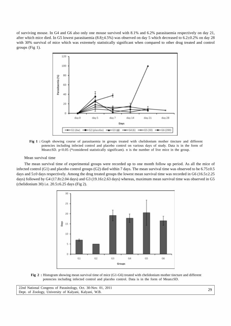
22nd National Congress of Parasitology, Oct. 30-Nov. 01, 2011Dept. of Zoology, University of Kalyani, Kalyani, W.B.
29
of surviving mouse. In G4 and G6 also only one mouse survived with 8.1% and 6.2% parasitaemia respectively on day 21,after which mice died. In G5 lowest parasitaemia (8.8+4.5%) was observed on day 5 which decreased to 6.2±0.2% on day 28with 30% survival of mice which was extremely statistically significant when compared to other drug treated and controlgroups (Fig 1).
Fig 1 : Graph showing course of parasitaemia in groups treated with chelidonium mother tincture and differentpotencies including infected control and placebo control on various days of study. Data is in the form ofMean±SD. p<0.05 (*considered statistically significant). n is the number of live mice in the group.
Mean survival time
The mean survival time of experimental groups were recorded up to one month follow up period. As all the mice ofinfected control (G1) and placebo control groups (G2) died within 7 days. The mean survival time was observed to be 6.75±0.5days and 5±0 days respectively. Among the drug treated groups the lowest mean survival time was recorded in G6 (16.5±2.25days) followed by G4 (17.8±2.04 days) and G3 (19.16±2.63 days) whereas, maximum mean survival time was observed in G5(chelidonium 30) i.e. 20.5±6.25 days (Fig 2).
Fig 2 : Histogram showing mean survival time of mice (G1-G6) treated with chelidonium mother tincture and differentpotencies including infected control and placebo control. Data is in the form of Mean±SD.

22nd National Congress of Parasitology, Oct. 30-Nov. 01, 2011Dept. of Zoology, University of Kalyani, Kalyani, W.B.
30
DiscussionAll the mice when injected with 1X10⁶ P. berghei infected red blood cells, died by day 7 post inoculation, because of
heavy infection except those in which parasite migrated to reticulocytes. The phenomenon of reticulocytosis in P. bergheihas been reported earlier too (Miller and Carter, 1976). In the present study also reticulocytosis was observed in G3(Chelidonium ).
From the present study, it can be concluded that nascent alcohol in homeopathic drugs doesn’t play any role in clearingP. berghei infection. The infection was observed to increase continuously in vehicle treated group (G2) showing that thereis no placebo effect as a result of which all the mice died within 7 days due to heavy infection. A placebo is an inert substance(eg. Sugar pill, saline injection, ethanol) or sham physical/electrical manipulation that is believed to have no chemical,electrical, or physical effect on the patient (Arnstein, 2003). WHO report, also states most of the studies published in thelast 40 years have shown homeopathic remedies to be superior to placebo and ‘equivalent to conventional medicines in thetreatment of illness, in both humans and experimental animals (www.guardian.co.uk/medicine/story/0,11381,1556831,00). Forhomeopathic research mice (Mus musculus) have been used as a model in relation to cytotoxicity, genotoxicity andcarcinogenesis as they share a high degree of homology with humans (Khuda-Bukhsh, 2009). Salazar et al. (2006) alsodemonstrated the effect of homeopathic medicines Eupatorium perfoliatum 30CH and Arsenicum album 30CH on Plasmo-dium berghei infected mice. In our laboratory also, the effect of homeopathic medicines eucalyptus has been checkedagainst rodent malaria parasite in Balb/C mice (Bagai et al. 2008).
The present study also confirms that the homeopathic medicines which are diluted beyond Avogadro’s number, showconsiderable effect on mice model. It was confirmed that homeopathic formulation of Chelidonium majus possessesantimalarial potential. Out of and different potencies (6, 30 and 200), 30 potency showed maximum parasite inhibition withsurvival of 30% of mice and mean survival time of 20.5±6.25 days up to one month follow up period when compared to othertreated groups. In the homeopathic doctrine of centesimal dilutions, it is generally believed that ‘higher the potency (dilutions)stronger the effect (Hahnemann, 1960). The homeopathic mode of treatment often encourages use of drugs at such ultra-low doses and high dilutions that even the physical existence of a single molecule of the original drug substance becomestheoretically impossible (Khuda-Bukhsh, 2003). Present study is in accordance with the homeopathic principle as highestpotency (200) showed minimum parasitaemia (6.2%) but only 1 mouse survived up to 21 days which may be due to toxicitycaused by higher potencies. The 30 potency of many homeopathic medicines were reported to be more effective than otheravailable potencies (Birch, 2007; Biswas and Khuda-Bukhsh, 2002; Salazar et al. 2006).
The considerable efficacy of chelidonium against rodent malaria parasite in the present study calls for furtherinvestigations of its antimalarial potential in terms of its safety and dosage. Chelidonium extracts shows an immune stimulatingeffect and it is used mainly for liver conditions. Extracts of C. majus are traditionally used in various complementary andalternative medicine (CAM) systems including homeopathy mainly in combating diseases of the liver (Taborska et al. 1995).Hepatic damage is one of the main pathological condition during malaria so homeopathic medicine chelidonium can be apotent antimalarial or a suitable partner for combination therapy.
AcknowledgementsThe authors hereby declare that experiments comply with the current laws in India. The authors are grateful to
chairperson and UGC-CAS programme of the department of Zoology, Panjab University, Chandigarh for continued supportand University Grants commission, New Delhi, India for funding the present work through Rajiv Gandhi National fellowshipto Aswathy Rajan.
BibliographyAdler M, Appel K, Canal T, Corvi Mora P, Delfino R, Gennaro R, Gritzko K, Pascolo L, Ruzz F, Tiribelli C, Wallner B,
Schulze J (2006) Effects of Chelidonium majus extracts in human hepatocytes in vitro. Toxicol Lett 164S: S1-S324 15-16 doi:10.1016/j toxlet 2006.07.095.
Arnstein PRN (2003) The placebo effect. Seminars in Integrative Medicine 1(3): 125-135.
Bagai U, Rajan A, Chandel S (2008) Efficacy of Eucalyptus (a homeopathic medicine) in combination with artesunate to clearPlasmodium berghei infection in Balb/C mice. Current Trends in Parasitology. Tandon, V., Yadav, A. K., Roy, B. (Ed.)In: Proceedings, 20th National Congress of Parasitology, November 3-5-2008 NEHU Shillong India, ISBN: 81-86535-69-1, pp 203-211.

22nd National Congress of Parasitology, Oct. 30-Nov. 01, 2011Dept. of Zoology, University of Kalyani, Kalyani, W.B.
31
Bellavite P, Ortolani R, Pontarollo F, Pitari G, Conforti A (2007) Immunology and Homeopathy, The Rationale of the ‘Similie’.Evid Based Compl Alternat Med 4:149-163.
Birch K (2007) Vaccine Free Prevention and Treatment of Infectious Contagious Disease with Homeopathy- by Kate Birch.Trafford Publishing, pp 361-369.
Biswas SJ, Khuda-Bukhsh AR (2002) Effect of homeopathic drug, Chelidonium, in amelioration of p-DAB inducedhepatocarcinogenesis in mice. BMC Compl Alt Med. 2: 4–12.
Hahnemann S (1960) In: Organon of Medicine. First Indian edn. M. Bhattacharyya and Co., Calcutta, pp 1-300.
Khuda-Bukhsh AR (2003) Towards understanding molecular mechanisms of action of homeopathic drugs: an overview. MolCell Biochem 253: 339-345.
Khuda-Bukhsh AR (2009) Mice as a model for homeopathy research. Homeopathy 98:267 279.
Miller LH, Carter R (1976) Innate resistance in malaria. Parasitol 40:132–146.
Partington T, Ruchira DA, Hennel L, Pittendrigh A, Taylor J (2006) Silent and deadly, prophylaxis and treatment ofmalaria. Homeo in prac Spring 14-19.
Rajan A, Bagai U (2009) In vivo efficacy of combination therapy of Artesunate+S/P and Artesunate+China against P. bergheiinfection. In: Proceedings, 21st National congress of Parasitology, November 14-16-2009 Panjab University Chandigarh,India, pp 60.
Salazar GL, Montiel EM, Monzon JT, Hernandez FH, Salasbenito JS (2006) Effects of homeopathic medications Eupatoriumperfoliatum and Arsenicum album on parasitaemia of Plasmodium berghei infected mice. Homeopathy 95(4): 223–228.
Santiyanont R (1985) Parasite identification, counting and staining : Application of genetic engineering techniques in tropicaldiseases, pathogens with special reference to plasmodia. A laboratory manual of selected techniques. UNDP/WorldBank/WHO special program for research and techniques in tropical diseases, Bangkok, Thailand, 8.7 pp 413-448.
Snow R, Trape JF, Marsh K (2001) The past, present and future of childhood malaria mortality in Africa. Trends Parasitol17: 593-597.
Taborska E, Bochorakova H, Dostal J, Paulova, H (1995) The greater celandine (Chelidonium majus L.)– A review of presentknowledge. Ceska a Slovenska Farmacie 44(2): 71–75.
World Health Organisation (2005) Malaria risk and malaria control in Asian countries affected by the tsunami disaster.Roll Back Malaria Department 2005; WHO/HQ.2005. Geneva Switzerland.
World Health Organization (2002) WHO Traditional Medicine Strategy 2002-05. WHO/EDM/TRM/2002/Geneva Switzerland.

22nd National Congress of Parasitology, Oct. 30-Nov. 01, 2011Dept. of Zoology, University of Kalyani, Kalyani, W.B.
32
Study of natural hemagglutinins and their activity from the haemolymphof silkworm, B. mori L.
Bharat Bhusan Patnaik*, M. Jayalakshmi#, Atul Kumar Saha*, S. Rajakumar$ and Arvind Kumar Bajpai*
*Central Sericultural Research & Training Institute, Central Silk Board, Ministry of Textiles (Govt. of India),Berhampore-742101, West Bengal, India
#Immunology Laboratory, School of Biological Sciences, Madurai Kamaraj University, Madurai, TamilNadu, India$Regional Sericultural Research Station, Central Silk Board, Allikuttai (PO), Veeranum Road, Salem-636003, TamilNadu, India
Abstract : Hemagglutinins or lectins are specific carbohydrate-binding proteins or glycoproteins that have the ability to agglutinate cellswith complementary carbohydrates on their surfaces and are present in most organisms. The body fluid or haemolymph of almost allinvertebrate species tested accounts for agglutinins and also has been detected in mucus as well as in certain tissues. However, itsimmunogenic potential is best understood in the haemolymph, and recent studies have shown that purified, haemolymph-derived agglutininsserved as opsonins in few insects. Thus, they are implicated in the recognition of non-self and cellular defense and are involved in celldifferentiation. The study explored naturally occurring hemagglutinin (HA) in the haemolymph of silkworm, Bombyx mori using mammalianand human erythrocytes (RBC) as indicator cells. Highest HA titer of 28 = 256 was noticed with sheep RBC followed by titer of 27 withcow, pig chicken and human RBCs. An analysis of the physio-chemical properties of the HA showed it to be specifically dependent onthe presence of Ca2+ for its activity, relatively stable between pH 7 to 9 and showed thermal stability up to 30oC. The potential affinityof natural hemagglutinins from haemolymph of Bombyx mori towards sialic acids (NeuAc/NeuGc) was suggested due to their agglutinatingactivity with human, cow, pig and chicken RBCs.
Keywords : Bombyx mori, carbohydrate-binding proteins, Hemagglutinins, immunity, lectins
IntroductionHemagglutinins or lectins are ubiquitous proteins found in plants, animals and microorganisms. In vertebrates, the role
of lectins as mediators of nonself recognition in the innate immune response has been well documented. The best studiedof these are the mannose-binding lectins, MBLs, which are essential component of vertebrate innate immune system, sinceMBL-deficient individuals are prone to recurrent infections during infancy (Turner, 1996). In invertebrates due to lack ofantibody-based immunity, agglutinins are vital for non-self recognition and clearance of invading microorganisms. Suchhumoral factors acting as recognition factors have been extensively studied due to their functional similarities to vertebrateantibodies, serving a defensive function (Jayasree, 2001; Jayaraj et al., 2008a).
Hemagglutinating proteins have been detected in mucus as well as in certain tissues of invertebrates (Suzuki and Mori,1991), but its immunogenic potential is best understood in haemolymph, and recent studies have shown that purified,haemolymph-derived agglutinins served as opsonins in few insects (Kawasaki et al., 1993). Insect haemolymph containshemagglutinating activities which agglutinates vertebrate erythrocytes and certain microorganisms and are important for therecognition of non-self and cellular defense and are involved in immune surveillance. Because of the involvement of lectinsin the recognition of non-self, a number of studies have concentrated on the role of lectins in parasite-host relationships(Grubhoffer and Matha, 1991; Mohamed et al., 1992; Volf et al., 1993).
Developmental changes in the hemagglutinating activity of Bombyx larval haemolymph have been reported to be underthe control of ecdysteroids. Increase in hemagglutinins due to the infection of Cytoplasmic Polyhedrosis Virus (CPV) suggestedpossible role of these proteins as candidates responsible in the silkworm for immune defense against exogenous pathologicalagents (Mori et al., 1989). Watanabe et al., 2006 characterized novel C-type lectin from B. mori haemolymph and designatedas B. mori multibinding protein (BmMBP) as it bound to 5 Gram-negative bacteria, 3 Gram-positive bacteria and 3 yeastsexamined under the study. Recently, the detailed specificities of a variety of these proteins towards N-linked oligosaccharideslike N-acetylglucosamine (NeuGc) and N-acetylgalactosamine (NeuAc) have been elucidated. The present study was undertakento explore the natural haemolymph agglutinins form mulberry silkworm, Bombyx mori L. and study its carbohydrate specificity
*Corresponding Author : E-mail : [email protected]

22nd National Congress of Parasitology, Oct. 30-Nov. 01, 2011Dept. of Zoology, University of Kalyani, Kalyani, W.B.
33
by studying its RBC binding activities, so as to define their utilization as potent diagnostic tool in identifying diverse epitopesin pathogenic bacteria.
Materials and methodsExperimental animals and maintenance
Silkworm, Bombyx mori L. was reared in aseptic conditions at 26 ± 2oC and 75 ± 10% Relative Humidity (RH) at theinstitute. The feeding, cleaning and sanitation schedule was followed according to Datta Biswas et al. (2007)
Preparation of Haemolymph samples
Haemolymph samples were collected from Vth instar Vth day worms by puncturing the abdominal legs and drained into1.5 ml Eppendorf tubes containing a crystal of phenylthiourea to prevent oxidation of haemolymph and subsequentmelanization. The haemolymph was centrifuged at 5400 rpm for 10 min to remove the haemocytes and cell debris and theresultant clear supernatant was used for the hemagglutination titre assay.
Preparation of erythrocyte (RBC) suspension
Human and other mammalian blood samples (viz., sheep, goat, cow, pig and chicken) were obtained by venous or cardiacpuncture and collected in sterile AMAB (Modified Alsever’s medium containing antibiotics): 0.03M Sodium citrate pH 6.1,0.077M Sodium Chloride, 0.114M Glucose, 50 mg Neomycin sulphate and 165 mg Chloramphenicol. Prior to use, the RBCs werewashed thrice with 0.9% saline with repeated centrifugation at 1600 rpm for 10 min and once with TBS (0.05M Tris-HCl, 0.1MSodium Chloride and 0.01M Calcium Chloride). The RBC pellet was finally resuspended in TBS as 1.5% suspension (v/v).
Hemagglutination (HA) assay
HA assays were performed in U-bottom microtiter plates by serial two-fold dilution of a 25 µl serum with equal volumeof TBS. After dilution, 25 µl of 1.5% RBC suspension was added to each well and incubated for 60 min at room temperature.The HA titers were recorded as the reciprocal of the highest dilution of the sample causing complete agglutination of RBC.Controls for all assays consisted of the substitution of the sample by TBS. All the HA assays were performed in duplicate.
pH and Thermal stability
The stability of haemolymph HA activity (in duplicates) at different pH was examined by dialyzing (24hr, 4oC) 300 µlsamples against the following buffers at pH ranging from 3 to 12 (Pearse, 1968): 0.2 M acetate buffer (pH 3 to 6), 0.2 M Tris-HCl buffers (pH 7 to 9) and 0.1 M Glycine-NaOH buffer (pH 10 to 12). The HA titre was determined with sheep RBCs.
To study thermal stability of HA, 300 µl haemolymph samples were held for 30 min at temperatures ranging from 10 to100oC, centrifuged and tested for HA activity with sheep RBCs.
ResultsHaemolymph HA profile
The haemolymph of mulberry silkworm, Bombyx mori L. agglutinated human and variety of mammalian (viz., sheep, goat,chicken, cow, pig) RBC types (Table I). Among the various RBC types tested, the highest titre of 28=256 was observed withsheep erythrocytes. This was followed by a titre of 27=128 with chicken, cow, pig and human erythrocytes. The hemagglutininsdidn’t discriminate Human A, B and O RBC types and agglutinated them to same degree. No visible agglutination was observedwith Goat RBCs.
Table I : Hemagglutinating (HA) activity of haemolymph from the mulberry silkworm, Bombyx mori L. against variousmammalian erythrocyte types
RBC types tested HA titer*
Sheep 256
Chicken 128
Cow 128
Pig 128
Human (O, A, B) 128
Goat NA#
*Based on 20 determinations for each RBC type. All RBC’s were collected in modified Alsevier’s solution and used withina week after collection. The cells were washed thrice with TBS before assay.# Not Agglutinated

22nd National Congress of Parasitology, Oct. 30-Nov. 01, 2011Dept. of Zoology, University of Kalyani, Kalyani, W.B.
34
pH and Thermal stability
The haemolymph hemagglutinating activity of mulberry silkworm, Bombyx mori L. was tested in the pH range of 3-12(Figure 1) with sheep RBC showing the highest HA titre. The hemagglutinating activity against sheep RBC was found toincrease above pH 3, with maximum stability between pH 7 and 9. The stability reduced above this pH range and completelylost at pH 11 and 12. The activity of haemolymph hemagglutinins against sheep RBC was unaffected up to 30oC but wasconsiderably reduced at 40oC and 50oC and completely inactivated at 60oC and above (Figure 2).
Fig. 1 : pH stability of hemagglutinating activity of haemolymph of Bombyx mori L. against sheep RBC
Fig. 2 : Thermal stability of hemagglutinating activity of haemolymph of Bombyx mori L. against sheep RBC
Temperature
Hem
aggl
utin
atio
n tit
re
pH
Hem
aggl
utin
atio
n tit
re
DiscussionThe haemolymph of mulberry silkworm, Bombyx mori L. was found to possess naturally occurring hemagglutinating
activity which showed highest reactivity with sheep RBC followed by chicken, cow, pig and human RBCs. These results confirmthat the RBC types agglutinated by the haemolymph of Bombyx mori L. possibly share a common surface receptor but withqualitative differences in its HA binding sites. The studies are in conformity to the findings of Suzuki and Natori, 1993 havingcharacterized haemolymph lectins from Bombyx larvae showing activity against sheep red blood cells with increased activityduring larval-larval ecdysis and spinning stage. Previous reports by Mori et al., (1989), have indicated the enhanceaccumulation of hemagglutinating protein in the haemolymph of silkworms infected with Cytoplasmic polyhedrosisvirus (CPV),suggesting its requirement to exclude the virion from haemolymph, derived from their multiplication in the midgut cells orhelping scavenge midgut tissue fragments resulting from infection.

22nd National Congress of Parasitology, Oct. 30-Nov. 01, 2011Dept. of Zoology, University of Kalyani, Kalyani, W.B.
35
Hapner and Jermyn (1981) observed strong hemagglutinating activity (titre 512) towards vertebrate erythrocytes in thehaemolymph of both sexes of adult cricket, Teleogryllus commodus (Walker). Adeyeye and Cheng, 1997 studied hemagglutininactivity during all developmental stages of corn earworm Helicoverpa zea, with highest HA titre during days 3-5 of fifth instarlarvae and lowest titre in adult (higher in female than in male) insects and observed highest reactivity with rabbit, sheep, porcineand guinea pig erythrocytes and low agglutinating activity with human, horse and chicken RBCs. Dorrah et al., 2009, isolatedand characterized three lectins, designated as Sg1, Sg2 and Sg3 from serum of desert locust Schistocerca gregaria, highlyspecific for rabbit RBCs than those of other vertebrates.
The haemolymph agglutinin of silkworm, Bombyx mori L. was heat-labile and susceptible to pH extremes. Hapner andJermyn (1981) showed that T. commodus haemolymph hemagglutinating activity was stable at freezing and room temperatures,labile to heating at 56oC and is precipitated with other haemolymph components when dialyzed against water.
The sialic acid affinity of natural agglutinin from haemolymph of silkworm, Bombyx mori L. was evident in the presentstudy as their agglutination was highest with sheep, followed by cow, pig, chicken and human erythrocytes (Table II). Commonspecies of sialic acids in Human A, B, O sheep and chicken erythrocytes are N-acetylneuraminic acid (NeuAc) (Jeanloz, 1966;Berman, 1984; Salmon et al., 1984) and N-acetylneuraminic acid/N-glycolylneuraminic acid (NeuAc/NeuGc) in pig erythrocytes(Sarris and Palade, 1979). Rat, mouse and horse erythrocytes constitute another group with NeuAc/NeuGc/O-acetylated sialicacid (O-AcSia) as common species of sialic acids. In rabbit, rat and mice, the position of major O-acetyl group was at C-9 andin horse at C-4 position (Ravindranath et al., 1985; Mercy and Ravindranath, 1993). This confirms striking correlation betweenthe ability of silkworm hemagglutinin towards agglutination of cells and the absence of O-acetylsialic acids. Chinzei et al., 1990,reported that the carbohydrate portion of insect haemolymph proteins consists exclusively of mannose and N-acetylglucosamine. Similar studies with crab lectin, exhibited high specificity for both free and glycosidically bound 9-O-Ac-NeuAcand 4-O-Ac-NeuAc (Ravindranath and Cooper, 1984). It would be interesting to study the reactivity of haemolymph agglutininsfrom Bombyx towards rat, mouse and horse erythrocytes as O-AcSia have been associated to cause hindrance to the bindingwith NeuAc/NeuGc. This also reflects the strong possibility that micro-organisms with NeuAc or NeuGc could be recognizedby hemagglutinins of Silkworm, Bombyx mori L. and therefore can be used as valuable diagnostic tool.
Table II : Erythrocyte hemagglutination by haemolymph of Bombyx mori as compared with the erythrocyte affinity of sialicacid binding haemolymph lectins of other invertebrates [Melanoplus differentialis (Hapner, 1983), Teleogrylluscommodus (Hapner and Jermyn, 1981), Scylla serrata (Jayaraj et al., 2010) and Cancer antennarius (Jeanloz, 1966)].
RBC types Common species of sialic acids Bombyx Melanoplus Teleogryllus Scylla Cancer
Human A NeuAc 128 64 32 8 NA
Human B NeuAc 128 256 32 32 NA
Human O NeuAc 128 256 32 8 NA
Sheep NeuAc 256 4 4 2 NA
Chicken NeuAc 128 4 32 2 NA
Cow NeuGc/NeuAc 128 4 - 16 NA
Pig NeuGc/NeuAc 128 - - 2 NA
NA – Not Agglutinated
AcknowledgementThe work was supported by UGC-NRCBS Visiting Fellowship for the year 2010 to the first author.
ReferencesAdeyeye OA, Cheng JQ (1997) Distribution, stability and binding specificity of a hemagglutinin in the corn earworm,
Helicoverpa zea (Lepidoptera: Noctuidae). Eur J Entomol 94: 167-176
Berman E (1984) Structural and conformational analysis of sialyloligossacharides using carbon-13 nuclear magnetic resonancespectroscopy. Biochem 23: 3754-3759
Chinzei Y, Haruna T, Miura K, Numata H, Nakayama S (1990) Purification and characterization of biliverdin-associatedcyanoprotein from eggs and haemolymph of the bean bug Riptortus clavatus (Heteroptera: Alydidae). Insect Biochem20: 545-555

22nd National Congress of Parasitology, Oct. 30-Nov. 01, 2011Dept. of Zoology, University of Kalyani, Kalyani, W.B.
36
Datta Biswas T, Saha AK, Das SK, Bajpai AK (2007) Season specific silkworm rearing package for Eastern India. In: MulberrySericulture Problems and Prospects. Jaiswal K, Trivedi SP, Pandey BN, Tripathy, AK (eds) APH Publishing Corp N Delhipp 193-213
Dorrah MA, Ayaad TH, Mohamed AA, Bassal TTM (2009) Isolation and characterization of multiple-lectins from serum of thedesert locust Schistocerca gregaria (Orthoptera: Acrididae). J Orthopt Res 18(1): 103-112
Grubhoffer L, Matha V (1991) New lectins of invertebrates. Zool Sci 8: 1001-1003
Hapner KD (1983) Hemagglutinin activity in the haemolymph of individual Acrididae (Grasshopper) specimens. J Insect Physiol29(1): 101-106
Hapner KD, Jermyn MA (1981) Hemagglutinin activity in the haemolymph of Teleogryllus commodus (Walker). Insect Biochem11(3): 287-295
Jayaraj SS, Thiagarajan R, Vincent S, Arumugam M (2008a) Characterization of a natural hemagglutinin from the plasma ofmarine mussel Perna viridis. Bull Euro Assoc Fish Pathol 28: 76-84
Jayasree S (2001) Biological properties of a natural agglutinin in the haemolymph of Indian white prawn, Penaeus indicus H.Milne Edwards. Aquaculture 194: 245-252
Jeanloz RW (1966) Glycoproteins. Gottschalk A (eds) Elsevier Publishing Company, Amsterdam, pp 104-129
Mercy PD, Ravindranath MH (1993) Purification and characterization of N-glycolylneuraminic acid specific lectin from Scyllaserrata. Eur J Biochem 215: 697-704
Mohamed HA, Ingram GA, Molyneux DH (1992) Carbohydrate-binding specificities of anti-erythrocyte lectins (hemagglutinins)in Anopheles gambiae gut extracts and haemolymph. Med Veter Entomol 6: 217-224
Mori H, Ohyane M, Ito M, Iwamoto S, Matsumoto T, Sumida T, Matsubara F (1989) Induction of a hemagglutinating activityin the haemolymph of silkworm, B. mori infected with Cytoplasmic Polyhedrosis Virus. J Invert Pathol 54: 112-116
Ravindranath MH, Cooper EL (1984) Progress in Clinical and Biological Research, Alan Liss, New York pp 83-96
Ravindranath MH, Higa HH, Cooper EL, Paulson JC (1985) Purification and characterization of an O-acetylsialic acid specificlectin from a marine crab Cancer antennarius. J Biol Chem 260: 8850-8856
Salmon C, Cartron J, Rouger P (1984) The human blood groups. Year Book Publishers, Chicago pp 54-80
Sarris AH, Palade GE (1979) The sialoglycoproteins of murine erythrocyte ghosts. J Biol Chem 254: 6724-6731
Suzuki T, Natori S (1983) Identification of a protein having hemagglutinating activity in the haemolymph of the silkworm,Bombyx mori. J Biochem 93(2): 583-590
Turner MW (1996) Mannose-binding lectin: the pluripotent molecule of the innate immune system. Immunol Today 17: 532
Volf P, Grubhoffer L, Muska M (1993) Detecting and identification of tissue specific lectins of the tsetse fly, Glossinatachinoides. In: Management of Insect Pest Nuclear and Related Molecular and Genetic Techniques. InternationalAtomic Energy Agency, Vienna pp 557-565.
Watanabe A, Miyazawa S, Kitami M, Tabunoki H, Ueda K, Sato R. 2006. Characterization of a novel C-type lectin, Bombyxmori multibinding protein, from the B. mori haemolymph: Mechanism of wide-range microorganism recognition and rolein immunity. J Immunol 177: 4594-4604.

22nd National Congress of Parasitology, Oct. 30-Nov. 01, 2011Dept. of Zoology, University of Kalyani, Kalyani, W.B.
37
Studies on the biodiversity of aseptate gregarines from theoligochaetes of West Bengal
Biplab Bhowmik1, Amlan Kumar Mitra2 and Probir Kumar Bandyopadhyay3
1Department of Zoology, Panchakot Mahavidyalaya, Neturia, Sarbari, Purulia, West Bengal, India2Department of Zoology, Ranaghat College, Ranaghat, Nadia, West Bengal, India
3Parasitology Laboratory, Department of Zoology, University of Kalyani, Kalyani 741235, West Bengal, India
Abstract : In a survey of the endoparasitic acephaline gregarines in the North 24 Parganas district of West Bengal, India seminal vesiclesof earthworm Lampito mauritii (Kinberg) were found to be infested with a new species of the genus Monocystis Stein 1848, Monocystislampitae sp. nov. is ovoidal and measures 85.8-155.4 (120.0 ± 18.1) µm x 32.7-69.5 (57.0 ± 10.4) µm. Nucleus rounded, 10.2-20.4 µm(14.1 ± 3.3) µm in diameter. Gametocysts are ovoid and measure 32.7-44.9 (38.6 ± 4.0) µm. Oocysts biconical, measuring 13.8-20.0(17.2±1.0) x 7.7-9.2 (8.6 ± 0.6) µm
Key words : Acephaline gregarines, earthworm, India, seminal vesicle, Monocystis lampitae sp. nov.
IntroductionIn India research on gregarine protozoan parasites infesting invertebrates has not been carried out with great importance.
Gregarines are a group of apicomplexan protozoan parasites with two forms, septate and asepate. Only a few of over 350species of Indian earthworms have been studied for the occurrence of gregarines, but this research has gained momentumsince the 1980ies. It has already resulted in the finding of representatives of the genera Apolocystis Cognetti de Martiis, 1923;Monocystis Stein, 1848; Nematocystis Hesse, 1909; Stomatophora Drzewiecki, 1907 and Zygocystis Bhatia, 1930 (Hesse, 1909;Ghosh, 1923 – cited by Levine, 1988; Bhatia and Chatterjee, 1925; Bhatia and Setna, 1926; Kar, 1946; Kalavati, 1979; Subbaraoet al., 1979; Pradhan and Dasgupta, 1980a, 1980b, 1982, 1983a, 1983b; Roychoudhury and Haldar, 1984; Bandyopadhyay etal., 2001, 2004, 2005, 2006a, 2006b, 2006c, 2006d, 2006e, 2007a, 2007b, 2008; Bandyopadhyay and Mitra, 2004, 2005a, 2005b,2005c, 2005d, 2006a).
The biodiversity survey of gregarines from earthworms in the North 24 Pargans district of West Bengal revealed a newspecies of the genus Lampito mauritii (Kinberg). This paper presents the description of Monocystis lampitae sp. nov., themorphometric comparison with closely related species and the discussion of its taxonomy and systematics.
Material and methodsSamplings were carried out in the sandy soil in the Bagdaha locality of North 24 Parganas district of the state West
Bengal. Different species of earthworms were collected during June-July, 2009. The collected individuals were put in soil-filledplastic buckets and taken to the laboratory alive. Some of them were dissected while alive and their seminal vesicles werecarefully removed. These were placed on clean glass with a drop of 0.6% NaCl solution. A thin film of seminal fluid wasdrawn out on a slide covered with a cover slip for examination of living protozoans under a phase contrast microscope(Olympus CX41). After the initial study of living protozoans, the content of the seminal vesicles was semidried and fixedin Schaudin’s fluid (20 min). The fixed smears were stored in 70% ethyl alcohol for removal of mercuric chloride. The slideswere then passed through a descending series of alcohols (5 min in each) and placed in distilled water. Then they weretransferred to a 3% iron alum solution (over night) and stained with Heidenhain’s haematoxylin solution (20 min).Differentiation was done with 1% iron alum solution under the low power objective lens of the light microscope. The slideswere then washed thoroughly, dehydrated in an ascending series of alcohols, cleared in xylene and mounted in Canadabalsam. Drawings of the different stages of gregarines were made using a camera lucida attached to the microscope;photomicrographs were taken under an Olympus phase contrast microscope (× 400 magnification) with an Olympus camera(Model C5060). All measurements are in micrometres; in each case minimum and maximum values are given, followed inparentheses by arithmetic mean, standard deviation and sample size. Plane shapes are described mainly according to Clopton(2004).
* Corresponding Author : E-mail : [email protected]

22nd National Congress of Parasitology, Oct. 30-Nov. 01, 2011Dept. of Zoology, University of Kalyani, Kalyani, W.B.
38
ResultsMonocystis lampitae sp. nov. (Figs. 1-4, Table 1)
Phylum : Apicomplexa Levine, 1988; Order: Eugregarinorida Léger, 1900; Family: Monocystidae Bütschli, 1882; Subfamily:Monocystinae Bhatia, 1930; Genus: Monocystis Stein, 1848.
Gamont Length (GL) : 85.8-155.4 (120.0 ± 18.1) µm; Gamont Width (GW): 32.7-69.5 (57.0 ± 10.4) µm; Nucleus Diameter(ND): 10.2-20.4 µm (14.1 ± 3.3) µm; Gametocyst Diameter (GD): 32.7-44.9 (38.6 ± 4.0) µm; Oocyst Length (OL): 13.8-20.0(17.2±1.0) µm; Oocyst Width (WO): 7.7-9.2 (8.6 ± 0.6) µm.
The gamont is ovoidal. The pellicle is smooth. Both ends of the gamont are rounded. Indistinct mucron. The nucleusis round and its position is in the episarc zone. Length and width of the gamont ranges from 85.8-155.4 (120.0±18.1) µm and32.7-69.5 (57.0 ±10.4) µm respectively. Nucleus measure 10.2-20.4 µm (14.1±3.3) µm. Karyosome present within the nucleus.Width of episarc is 3-5 µm. Cytoplasm is vacuolated in nature.The large and deeply stained vacuoles are found in the endosarcregion. Ectosarc is thin and poorly vacuolated. No epicyteal striations are found.
Figs. 1-4. Photomicrographs of different stages of the life history of Monocystis lampitae sp. nov.1. Gamont; 2. Syzygy; 3. Gametocyst, 4. Oocyst. (Scale bars: 100 µm (1-2), 50 µm (3), 4 (10 µm)

22nd National Congress of Parasitology, Oct. 30-Nov. 01, 2011Dept. of Zoology, University of Kalyani, Kalyani, W.B.
39
Table 1 : Morphometric comparison of Monocystis lampitae sp. nov. with M. darjeelingensis Bandyopadhyay andMitra, 2005 and M. metaphirae Bandyopadhyay et. al., 2006
Species→→→→→Characters↓↓↓↓↓
HostLocality
Site ofinfection
Gamont
SizeEctosarc
Endosarc
NucleusshapeSize of
NucleusGametocyst
shapeGametocyst sizeOocyst shapeOocyst sizeReferences
Monocystis darjeelingensisBandyopadhyay and Mitra, 2005Amynthas robustsa (Perrier, 1892)
Darjeeling, West Bengal
Coelomic fluid
Cylindroid with round extremities.One end slightly wider than theother. Conical or elliptical when
young. 210-273 µm x 84-140 µm
210-273 µm x 84-140 µmWith fine longitidunal striations
Granular, size increases withmaturationSpherical
8.3-12.5 µm
Spherical
147-163 µmNavicular
15.4-18 µm x 8.0-9.0 µmBandyopadhyay and Mitra (2005a)
Monocystis metaphiraeBandyopadhyay et. al., 2006Metaphire houleti (Perrier)
Barasat, West Bengal
Seminal vesicles
Bean shaped, anterior end isalways wider than the posteriorone. Mucron always present at
the anterior end94.0-151.0 µm x 53.0-81.0 µm94.0-151.0 µm x 53.0-81.0 µm
Ectosarc is thin , (1-3) µm
Endoplasm with evenlydistributed granules
Almost rounded
44.0-16.0 µm
Ovoid
85.0-102.0 µmBiconical and symmetrical6.5-11.0 µm x 4.0-7.5 µm
Bandyopadhyay et.al. (2006d)
Monocystis lampitae sp.nov.
Lampito mauriti sp. nov.Bagdaha, North 24 Parganas,
West BengalSeminal vesicles
Ovoidal, pellicle is smooth
85.8-155.4 µm x 32.7-69.5 µmEctosarc thin with poorly
vacuolated granulesContain deeply stained vacuoles
Rounded
10.2-20.4 µm
Ovoidal to elliptical
32.7-44.9 µmBiconical with pointed end13.8-20.0 µm x 7.7-9.2 µm
Present study
Gametocysts are ovoid to elliptical and containing two nearly similar gametocytes. Diameter of the gametocyst measures32.7-44.9 (38.6±4.0) µm. The large and small gametocytes are semicircular in shape and measures 24.5 – 38.8 (30.5±4.2) µm× 20.4-28.6 (24.6±3.1) µm and 20.4-28.6 (24.3±3.1) µm × 18.4 – 26.5 (21.9±2.1) µm respectively. Nucleus is not so prominentin both gametocytes.
Oocysts are biconical and both the ends of the oocysts are pointed and measuring 13.8-20.0 (17.2±1.0) µm × 7.7–9.2(8.6±0.6) µm. Some granules are found within the oocyst.
Taxonomic Summary
Type material : Monocystis lampitae sp. nov.
Type host : Lampito mauritii (Kinberg)
Type of locality : Bagdaha, North 24 Parganas, West Bengal, India.
Symbiotype : LM/01 deposited in the Museum of the Department of Zoology, University of Kalyani, Kalyani-741235,West Bengal, India
Site of infection : Seminal vesicles
Prevalence : 26/78 (33.3%)
Holotype : MSVIII/22 is deposited in the Museum of the Department of Zoology, University of Kalyani, Kalyani 741235,West Bengal, India

22nd National Congress of Parasitology, Oct. 30-Nov. 01, 2011Dept. of Zoology, University of Kalyani, Kalyani, W.B.
40
Paratype : MSVIII/09 and other slides are in the collection of the Parasitology Laboratory, Department of Zoology,University of Kalyani, Kalyani 741235, West Bengal, India
Etymology : The species name is given after the genus name of the type host Lampito mauritii (Kin berg)
DiscussionCylindroid, solitary with mucron, late syzygy and the parasite of seminal vesicles of oligochate host, earthworm justify
its inclusion under the family Monocystidae, sub family Monocystinae and genus Monocystis Stein, 1848.
Till now, more than seventy species belonging to the genus have been reported through out the world. Of which sixteenspecies have been reported from India. Most of the described species are reported from the seminal vesicles of earthwormbut some time parasites are also found in the coelom. The present species obtained from seminal vesicles of Lampito mauritii(Kinberg), which is considered to be a new one exhibits resemblance to some extent with Monocystis darjeeligensisBandyopadhyay et al., 2005 and Monocystis metaphire Bandyopadhyay et al., 2005 having cylindroid or ovoidal body withround extremities. In Monocystis darjeeligensis, one end is slightly wider than the other which is conical or elliptical in youngstage but in the present form it is ovoidal at maturity. Both ends of the present form is round and with vacuolated cytoplasm.The position of the nucleus in the present species is in the anterior end. Karyosome is also found. Syzygy is one of importantstage of life history which is not found in both the comparing species. Nature of syzygy is head to head. Gametocyst ofthe describing species is distinctly differs from the comparable two species. The gametocyte is unequal and ground nutshaped. Size of the gametocyst also differs from that of Monocystis darjeeligensis and M. metaphire where as it is very smallin the present form. So comparing all the morphological features, it may be concluded that no other species of the genusMonocystis can be compared with the species obtained from Lampito mauritii (Kinberg) and hence it is new to science.Therefore, I propose the species as Monocystis lampitae sp. nov. in the present paper.
ReferencesBhatia BL, Chatterjee CB (1925) On some gregarine parasites of Indian earthworms. Arch. Protistenkd 52: 189-206.
Bandyopadhyay PK, Mitra AK (2004) Description of a new species Zygocystis levinei sp. n. (Eugregarinida, Zygocystidae),from the earthworm Amynthas nicholsoni (Oligochaeta) from West Bengal, India. Protistology 3: 227-231.
Bandyopadhyay PK, Mitra AK (2005a) Observation of two new species of Monocystis Stein, 1848 (Protozoa: Apicomplexa:Monocystidae) Monocystis darjeelingensis sp. n. and Monocystis ranaghatensis sp. n. from earthworms (Annelida:Oligochaeta) of West Bengal, India. Animal Biol 55: 123-132.
Bandyopadhyay PK, Mitra AK (2005b) Observation on two new species of Nematocystis Hesse, 1909 (Protozoa: Monocystidae)from earthworms (Annelida: Oligochaeta) of West Bengal, India. Animal Biol 55: 133-139.
Bandyopadhyay PK, Mitra AK (2005c) Stomatophora cloptoni sp. n. (Apicomplexa: Eugregarinida) from the seminal vesiclesof an Indian earthworm. Acta Protozool 44: 385-388.
Bandyopadhyay PK, Mitra AK (2005d) Description of a new gregarine species, Zygocystis perionyxae sp. n. (Protozoa:Zygocystidae), from the earthworm Perionyx gravelleyi (Annelida: Oligochaeta) from West Bengal, India. Protistology4: 91-95.
Bandyopadhyay PK, Mitra AK (2006a) A new endoparasitic gregarine genus, Stomatodiscus indica gen. nov., sp. nov.(Apicomplexa: Sporozoa: Stomatophoridae) from the seminal vesicles of an Indian earthworm (Annelida: Oligochaeta)Amynthas diffringens Baird. Zootaxa 1191: 61-68.
Bandyopadhyay PK, Roychoudhury US, Biswas G (2001) Stomatophora majumdari sp. n. (Apicomplexa: Sporozoa) Anacephaline gregarine in the seminal vesicles of earthworm of West Bengal, India. Uttar J Zool 21: 221-224.
Bandyopadhyay PK, Roychoudhury US, Biswas G (2004). Descriptions of two new species of acephaline gregarines (Protozoa:Apocomplexa: Eugregarinida), Apolocystis chotonagpurensis sp. n. and Stomatophora janovyi sp. n. from earthworms(Annelida: Oligochaeta) of India. Acta Protozool 43: 275-279.
Bandyopadhyay PK, Bhowmik B, Mitra AK (2005) Observations on a new species of Monocystis Stein, 1848 (Protozoa:Apicomplexa: Monocystidae) Monocystis levinei sp. nov. from an Indian earthworm (Annelida: Oligochaeta) Eutyphoeusincommodus. Zootaxa 1016: 15-19.
Bandyopadhyay PK, Mitra AK, Bhowmik B (2006a) Nematocystis indicus sp. nov. (Apicomplexa: Monocystidae) from an Indianearthworm (Annelida: Oligochaeta) Perionyx excavatus (Perrier). Zootaxa 1296: 63-68.

22nd National Congress of Parasitology, Oct. 30-Nov. 01, 2011Dept. of Zoology, University of Kalyani, Kalyani, W.B.
41
Bandyopadhyay PK, Mitra AK, Mallik P (2006b) Biology of Monocystis clubae sp. nov. (Apicomplexa: Eugregarinida) froman Indian earthworm Lampito mauritii (Annelida: Oligochaeta) of India. Zootaxa 1120: 51–55.
Bandyopadhyay PK, Mallik P, Göçmen B, Mitra AK (2006c) Monocystis apporectodae sp. nov. (Protozoa: Apicomplexa:Eugregarinida), from an Indian earthworm Apporectodea trapezoides Duges. Acta Parasitologica Turcica 30: 51-53.
Bandyopadhyay PK, Mallik P, Göçmen B, Mitra AK (2006d) Monocystis metaphirae sp. nov. (Protista: Apicomplexa:Monocystidae) from the earthworm Metaphire houlleti (Perrier). Acta Parasitol Turcica 30: 54-56.
Bandyopadhyay PK, Göçmen B, Bhowmik, B, Mitra AK (2006e). A new species of Monocystis Stein, 1848 (Protista:Apicomplexa: Eugregarinida) from the Indian earthworm, Amynthas hawayanus Rosa, 1891 (Annelida: Oligochaeta). ActaParasitol Turcica 30: 155-157.
Bandyopadhyay PK, Mallik P, Mitra AK (2007a) Nematocystis indica sp. n. (Protozoa, Apicomplexa, Eugregarinida), anendoparasitic monocystid gregarine from the seminal vesicles of an Indian earthworm Amynthas diffringens Baird(Annelida, Oligochaeta). Protistology 4: 357-360.
Bandyopadhyay PK, Mallik P, Mitra AK (2007b) Observations on Monocystis arabindae n. sp. and Nematocystis majumdarin. sp. (Protozoa: Apicomplexa: Monocystidae) from seminal vesicles of an earthworm Eutyphoeus incommodus(Beddard) from West Bengal, India. Acta Protozool 46: 147-155.
Bhatia BL, Chatterjee CB (1925). On some gregarine parasites of Indian earthworms. Arch. Protistenk 52: 189-206.
Bhatia BL, Setna SB (1926). On some gregarine parasites of Indian earthworms. Arch. Protistenk 53: 361-377.
Clopton RE (2004) Standard nomenclature and metrics of plane shapes for use in gregarine taxonomy. Comp Parasitol 71:130-140.
Hesse E (1909) Contribution a l’etude des monocystidees Oligochaetes. Arch Zool Exp Gen 127-301.
Kalavati C (1979) Monocystis odontotermi n. sp. from the haemocoel of Odontotermes obesus Rambur. Indian J Parasitol 3:71-72.
Kar AB (1946) Observations on Monocystis megascolexae n. sp. from the seminal vesicles of the earthworm Megaloscolexsp. Col Univ J Dept. Sci (NS) 2: 50-52.
Levine ND (1988) The protozoan phylum Apicomplexa. Vol. I. CRC Press Inc., Boca Raton, Florida USA pp. 30-33.
Pradhan D, Dasgupta B (1980a) Records of some new gregarines in earthworms from the hill area of Darjeeling district-II.N Beng Univ Rev (Sc Tech) 1: 135-139.
Pradhan D, Dasgupta B (1980) Records of some new gregarines in earthworms from the hill area of Darjeeling district-II. NBeng Univ Rev (Sc Tech) 1: 141-143.
Pradhan D, Dasgupta B (1982) New gregarines in earthworms from Senchal and Goomti in Darjeeling district, West Bengal,India. J Beng Nat Hist Soc (NS) 1, 44-46.
Pradhan D, Dasgupta B (1983a) New acephaline gregarines (Apolocystis) in the hill areas of Darjeeling district. J Beng Nat HistSoc (NS) 2: 5-12.
Pradhan D, Dasgupta B (1983b) Life history and morphology of Zygocystis indicus n. sp. J Beng Nat Hist Soc (NS) 2: 17-23.
Roychoudhury US, Haldar DP (1984) Studies in aseptate gregarines from earthworms of West Bengal: Two new species ofNematocystis and one new species of Stomatophora. J Beng Nat Hist Soc (NS) 3: 17-27.
Subbarao BVSSR, Kalavati C, Narasimhamurti CC (1979) A new aseptate gregarine, Monocystis pontodrill sp. n. from the littoraloligochaetes Pontodrillus bermudensis Beddard. Acta Protozool 18: 259-264.

22nd National Congress of Parasitology, Oct. 30-Nov. 01, 2011Dept. of Zoology, University of Kalyani, Kalyani, W.B.
42
Biotechnological diagnostic procedure for rapid detection of microbialpathogens in fish and shell fish
*Chandravan Dutta1, Ashis Kumar Panigrahi1 and Chandan Sengupta2
1Fisheries and Aquaculture Extension Laboratory, Department of Zoology, University of Kalyani, Kalyani, West Bengal India 2Microbiology Laboratory, Department of Botany, University of Kalyani, Kalyani, West Bengal, India
Abstract : Bacterial, fungal, protozoan and viral infectious diseases are widespread among natural fish population. Various fish species,shrimps, crabs, giant freshwater prawns are being cultivated in different parts of India. Disease becomes evident only when stressfulcondition occurs in fish. However, the fishes raised in intensive culture system are more vulnerable to disease. Therefore, detection ofpathogen from carrier fish is essential for the effective fish disease control. In order to identify pathogen carrier fish, a cost effective, sensitiveand specific system are required for surveillance and monitoring fish population. Traditional techniques for diagnosis a disease are timeconsuming, laborious. With the advent of technological invention in the field of molecular biology and biotechnology, newer DNA basedand immunological methods have been developed and applied for rapid detection of many bacterial and viral pathogens of fish and shellfish.Diagnostic tests like ELISA, PCR, in situ hybridization and Dot blot hybridization etc have been developed and applied for detection ofvarious bacterial and viral diseases of fish and shell fish like CCV, IHNV, IPNV, VHSV, BKD, WSSV, YHV, MBV, BP Progress in techniquesaids epidemiological studies as well as identifying etiology of disease outbreaks or the presence of pathogens in fish and shellfish. Thisadvancement in molecular methods for disease detection is urgently required for sustainable development of aquaculture sector in India.In this paper an attempts are taken to highlight the disease problems of finfish and shellfish and biotechnological methods for diseasemanagement in India through effective way.
IntroductionBacterial, fungal, protozoan and viral infectious diseases are widespread among natural fish population. Various fish
species, shrimps, crabs, giant freshwater prawns are being cultivated in different parts of India. Disease becomes evident onlywhen stressful condition occurs. Under intensive aquaculture conditions the risk of stress increases and a significantproportion of the stock may become infected. Therefore, detection of pathogen from carrier fish is essential for the effectivefish disease control (Altinok and Kurt, 2003). Therefore, detection of pathogen from carrier fish is essential for the effectivefish disease control. In order to identify pathogen carrier fish, a cost effective, sensitive and specific system are required forsurveillance and monitoring fish population. Traditional techniques for diagnosis a disease are time consuming, laborious. Withthe advent of technological invention in the field of molecular biology and biotechnology, newer DNA based andimmunological methods have been developed and applied for rapid detection of many bacterial and viral pathogens of fishand shellfish. Molecular techniques are major tools for the analysis of microorganisms from food and other biologicalsubstances. The techniques provide ways to screen for a broad range of agents in a single test (Field and Wills, 1998). It hastruly come of age and its range of application is perceived to broaden in the near future. This advancement in molecularmethods for disease detection is urgently required for sustainable development of aquaculture sector in India. In this paperan attempts are taken to highlight the disease problems of finfish and shellfish and biotechnological methods for diseasemanagement in India through effective way.
Important diseases of finfish and shell fish in IndiaVibriosis : Bacterial diseases may cause a range of problems ranging from mass mortalities to growth retardation and
sporadic mortalities. Vibrio spp are the most important bacterial pathogens of shrimp. Vibrio spp are aquatic bacteria that arewidely distributed in fresh water, estuarine and marine environments. Over 20species are recognized, some of these are humanpathogens (eg. V. cholerae, V. parahaemolyticus and V.vulnificus ) while some species are pathogens of aquatic animalsincluding shrimp (eg. V. harveyi , V.spendidus, V. penaecida, V. anguillarum, V.parahaemolyticus, V.vulnificus). Vibrio spp arecommonly observed in shrimp hatcheries, grow-outponds and sediments (Otta et al. 1999, 2001).
*Corresponding Author
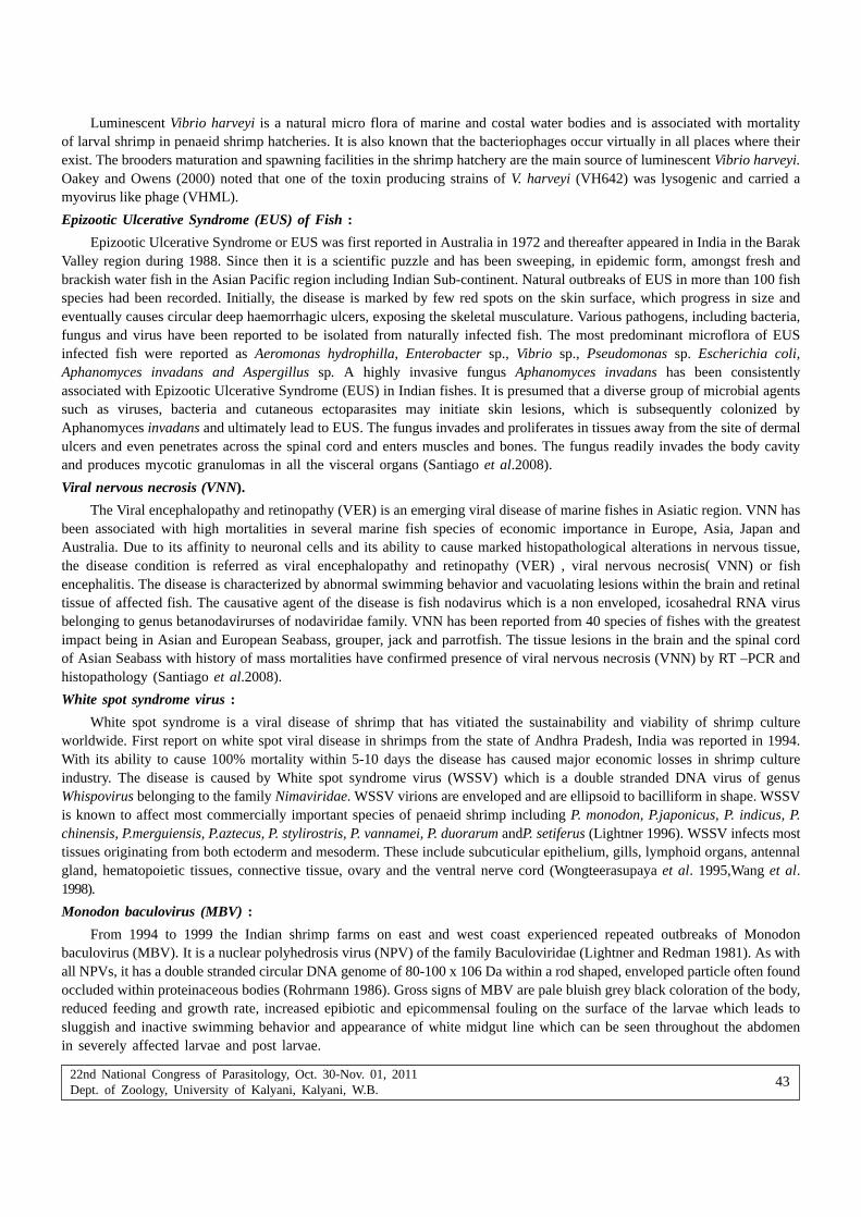
22nd National Congress of Parasitology, Oct. 30-Nov. 01, 2011Dept. of Zoology, University of Kalyani, Kalyani, W.B.
43
Luminescent Vibrio harveyi is a natural micro flora of marine and costal water bodies and is associated with mortalityof larval shrimp in penaeid shrimp hatcheries. It is also known that the bacteriophages occur virtually in all places where theirexist. The brooders maturation and spawning facilities in the shrimp hatchery are the main source of luminescent Vibrio harveyi.Oakey and Owens (2000) noted that one of the toxin producing strains of V. harveyi (VH642) was lysogenic and carried amyovirus like phage (VHML).
Epizootic Ulcerative Syndrome (EUS) of Fish :
Epizootic Ulcerative Syndrome or EUS was first reported in Australia in 1972 and thereafter appeared in India in the BarakValley region during 1988. Since then it is a scientific puzzle and has been sweeping, in epidemic form, amongst fresh andbrackish water fish in the Asian Pacific region including Indian Sub-continent. Natural outbreaks of EUS in more than 100 fishspecies had been recorded. Initially, the disease is marked by few red spots on the skin surface, which progress in size andeventually causes circular deep haemorrhagic ulcers, exposing the skeletal musculature. Various pathogens, including bacteria,fungus and virus have been reported to be isolated from naturally infected fish. The most predominant microflora of EUSinfected fish were reported as Aeromonas hydrophilla, Enterobacter sp., Vibrio sp., Pseudomonas sp. Escherichia coli,Aphanomyces invadans and Aspergillus sp. A highly invasive fungus Aphanomyces invadans has been consistentlyassociated with Epizootic Ulcerative Syndrome (EUS) in Indian fishes. It is presumed that a diverse group of microbial agentssuch as viruses, bacteria and cutaneous ectoparasites may initiate skin lesions, which is subsequently colonized byAphanomyces invadans and ultimately lead to EUS. The fungus invades and proliferates in tissues away from the site of dermalulcers and even penetrates across the spinal cord and enters muscles and bones. The fungus readily invades the body cavityand produces mycotic granulomas in all the visceral organs (Santiago et al.2008).
Viral nervous necrosis (VNN).
The Viral encephalopathy and retinopathy (VER) is an emerging viral disease of marine fishes in Asiatic region. VNN hasbeen associated with high mortalities in several marine fish species of economic importance in Europe, Asia, Japan andAustralia. Due to its affinity to neuronal cells and its ability to cause marked histopathological alterations in nervous tissue,the disease condition is referred as viral encephalopathy and retinopathy (VER) , viral nervous necrosis( VNN) or fishencephalitis. The disease is characterized by abnormal swimming behavior and vacuolating lesions within the brain and retinaltissue of affected fish. The causative agent of the disease is fish nodavirus which is a non enveloped, icosahedral RNA virusbelonging to genus betanodavirurses of nodaviridae family. VNN has been reported from 40 species of fishes with the greatestimpact being in Asian and European Seabass, grouper, jack and parrotfish. The tissue lesions in the brain and the spinal cordof Asian Seabass with history of mass mortalities have confirmed presence of viral nervous necrosis (VNN) by RT –PCR andhistopathology (Santiago et al.2008).
White spot syndrome virus :
White spot syndrome is a viral disease of shrimp that has vitiated the sustainability and viability of shrimp cultureworldwide. First report on white spot viral disease in shrimps from the state of Andhra Pradesh, India was reported in 1994.With its ability to cause 100% mortality within 5-10 days the disease has caused major economic losses in shrimp cultureindustry. The disease is caused by White spot syndrome virus (WSSV) which is a double stranded DNA virus of genusWhispovirus belonging to the family Nimaviridae. WSSV virions are enveloped and are ellipsoid to bacilliform in shape. WSSVis known to affect most commercially important species of penaeid shrimp including P. monodon, P.japonicus, P. indicus, P.chinensis, P.merguiensis, P.aztecus, P. stylirostris, P. vannamei, P. duorarum andP. setiferus (Lightner 1996). WSSV infects mosttissues originating from both ectoderm and mesoderm. These include subcuticular epithelium, gills, lymphoid organs, antennalgland, hematopoietic tissues, connective tissue, ovary and the ventral nerve cord (Wongteerasupaya et al. 1995,Wang et al.1998).
Monodon baculovirus (MBV) :
From 1994 to 1999 the Indian shrimp farms on east and west coast experienced repeated outbreaks of Monodonbaculovirus (MBV). It is a nuclear polyhedrosis virus (NPV) of the family Baculoviridae (Lightner and Redman 1981). As withall NPVs, it has a double stranded circular DNA genome of 80-100 x 106 Da within a rod shaped, enveloped particle often foundoccluded within proteinaceous bodies (Rohrmann 1986). Gross signs of MBV are pale bluish grey black coloration of the body,reduced feeding and growth rate, increased epibiotic and epicommensal fouling on the surface of the larvae which leads tosluggish and inactive swimming behavior and appearance of white midgut line which can be seen throughout the abdomenin severely affected larvae and post larvae.
22nd National Congress of Parasitology, Oct. 30-Nov. 01, 2011Dept. of Zoology, University of Kalyani, Kalyani, W.B.
44
Hepatopancreatic paravovirus (HPV)
Hepatopancreatic parvovirus infects various types of penaeid shrimps and is distributed around the world. Shrimpsaffected by HPV usually show non-specific gross signs, including atrophy of the hepatopancreas, anorexia, poor growth rate,reduced preening activities and as a consequence increased tendency for surface and gill fouling by epicommensal organisms(Lightner and Redman 1985).
Baculovirus penaei(BP)
The BP was first detected in 1974 in the pink shrimp,Penaeus duorarum (Couch 1974).BP has beenreported to causesignificant mortalities in the larval,post-larval and early juvenile stages of P.aztecus, P.stylirostris, P.vannamei and P.penicillatus(Couch1991, Lightner and Redman 1991, 1992).BP infects only the hepatopancreas and midgut epithelial cells, and it istransmitted from shrimp to shrimp.
Biotechnological DiagnosticsPolymerase Chain Reaction:
Polymerase chain reaction is a technique for amplifying a specific region of DNA, defined by a set of two “primers” atwhich DNA synthesis is initiated by a thermostable DNA polymerase. Usually, at least a million-fold increase of a specificsection of a DNA molecule can be realized and the PCR product can be detected by gel electrophoresis. The regions amplifiedare usually between 150-3,000 base pairs (bp) in length. Primer design is important to obtain greatest possible sensitivity andspecificity. Therefore, the primers should be sufficiently long to allow a high annealing temperature and reduce the opportunityfor nonspecific primer annealing, but primers that are too long may facilitate nonspecific annealing even to regions of DNAthat are not perfectly complementary to the primer sequence. The reaction includes template DNA that may be in various forms,from a simple tissue lysate to purified DNA, primers, polymerase enzyme to catalyze creation of new copies of DNA, andnucleotides to form the new copies. During each round of the thermocycling reaction, the template DNA is denatured, primersanneal to their complementary regions and polymerase enzyme catalyses the addition of nucleotides to the end of each primer,thus creating new copies of the target region in each round. Theoretically, the increase in amount of product after each roundwill be geometric (Altinok and Kurt, 2003). PCR is the most popular diagnostic technique for the detection of WSSV. Severalprimers have been designed for the detection of WSSV. Nested PCR has afforded greater sensitivity than single step PCR.Lo et al. (1996) described a set of PCR primers for detection of WSSV. Wang et al.(1996 )described PCR based detection ofBP in post larvae. Central Institute of Brackish water Aquaculture (CIBA) has developed a nested PCR kit that is being marketedby Genei, Bangalore. Similar PCR based diagnostic kits have already been developed by Mangalore Fishery College, CentralMarine Fishery Research Institute (CMFRI) which brought down the cost of the PCR test and rendered a great service to shrimpindustry.
Antibody based tests :
A variety of antibody-based tests and molecular tests have been developed to detect mainly bacterial and viral fishpathogens, although tests have also recently been reported for parasites and fungal agents. The antibody-based tests includeslide agglutination, co-agglutination/latex agglutination, immunodiffusion, direct and indirect fluorescent antibody tests (FATand IFAT),immunohistochemistry (IHC) and enzyme linked immune sorbent assay (ELISA), dot blot/dip stick and western blot(WB) (reviewed by Adams, 1999). The antibody-based test selected for the identification of pathogens depends on a varietyof factors since each method has its merits and disadvantages. Although such methods are useful for the detection ofpathogens in pure culture or/and in infected fish tissue, their sensitivity thresholds limit use in environmental samples,especially where pathogen levels are extremely low. DNA detection methods, however, such as polymerase chain reaction (PCR)and in situ hybridisation are ideally suited (Adams 1999).
Hybridoma technology :
Monoclonal antibodies ( MAbs) against several aquatic pathogens such as Aeromonas hydrophila, Aphanomycesinvadans of EUS , white spot virus and against immunoglobulins of (IgM) of Indian Major carp rohu have been developedand employed in antigen characterization, pathology, diagnosis and epidemiology.
Nucleic Acid probe :
Nucleic acid probes are segments of DNA or RNA that have been labeled with enzymes, antigenic substrates andchemiluminescent moieties or radio isotopes. They can bind with high specificity to complementary sequences of nucleic acid.

22nd National Congress of Parasitology, Oct. 30-Nov. 01, 2011Dept. of Zoology, University of Kalyani, Kalyani, W.B.
45
Probes can be directed to either DNA or RNA targets and can be twenty to thousand bases long. Oligonucleotide probeshave the advantage of hybridizing more rapidly to the target molecules and can be chemically synthesized and purified byinstruments available commercially. Commercial kits for labeling probes are available with affinity labels such as biotin anddigoxigenin ( Karunasagar et al 1999). Nucleic acid probes like in situ hybridization and dot blot hybridization have beenapplied for fish and shell fish disease diagnosis by numerous scientists.
ConclusionMolecular tools are increasingly relevant to fish diseases. The sequencing of the complete genomes of pathogens is
allowing great advances in studying the biology, and improving diagnosis and control of pathogens. Using nucleic acid astargets, and new methods of analyzing polymorphism in this nucleic acid, can improve specificity, sensitivity, and speed ofdiagnosis and offer means of examining the relationships between genotype and phenotype of various pathogens. Progressin techniques aids epidemiological studies as well as identifying causes of disease outbreaks or the presence of pathogensin future.
ReferencesAdams A (1999) Application of antibody probes in the diagnosis and control of fish diseases. Silver Jubilee Lecture,
Mangalore, India
Altinok I, Kurt I (2003) Molecular Diagnosis of Fish Diseases : A Review Turk. J Fish Aquat Sci 3:131-138
Couch JA (1974) Free and occluded virus similar to Baculovirus in hepatopancreas of pink shrimp. Nature 247: 229-231
Couch JA (1991) Baculoviridae, Nuclear polyhedrosis viruses. Part 2. Nuclear polyhedrosis viruses of invertebrates other thaninsects. In: Adams JR, Bonami JR (eds) Atlas of invertebrate viruses, CRC Press, Boca Raton, FL, p 205-226
Field D, Wills C (1998) Abundant microsatellite polymorphism in Saccharomyces cerevisiae, and the different distributionsof microsatellites in eight prokaryotes and S. cerevisiae, result from strong mutation pressures and a variety of selectiveforces. Proc Natl Acad Sci USA 95:1647-1652
Karunasagar I, Karunasagar I (1999) Diagnosis, treatment and prevention of microbial diseases of fish and shellfish. CurrentScience 76, 387-399.
Lightner DV (1996) A handbook of shrimp pathology and diagnostic procedures for diseases of penaeid shrimp. WorldAquaculture Soc, Baton Rouge, Louisiana, USA p 305.
Lightner DV, Bell TA, Redman RM, Mohney LL, Natividad JM, Rukyani A, Poernomo A (1992) A review of some majordiseasesof economic significance in Penaeid prawns/shrimpsof the Americas and Indo-Pacific. In: Shariff M, Subasinghe RP,Arthur JR (eds) Diseases in Asian Aquaculture In Proceedings of the first symposium on diseases in AsianAquaculture, Indonesia. Fish Health Section, Asian Fisheries Society, Manila pp 57-80.
Lightner DV, Redman RM (1981) A baculovirus caused disease of the penaeid shrimp, Penaeus monodon. J Invertebr Pathol38: 299-302.
Lightner DV, Redman RM (1991) Hosts, geographic range and diagnostic procedures for the penaeid virus diseases of concernto the shrimp culturists in the Americas. In: DeLoach PF, Dougherty WJ, Davidson MA (eds) Frontiers in shrimpResearch. Elsevier, Amsterdam pp 173-196.
Lightner DV, Redman RM, Williams RR, Mohney LL, Clerx JPM, Bell TA, Brock JA (1985) Recent advances in penaeid virusdisease investigations. J World Maric Soc 16: 267-274.
Lo CF, Leu JH, Ho CH, Chen CH, Peng SE, Chen YT, Chou CM,Yeh PY, Huang CJ, Chou HY, Wang CH, Kou GH (1996) Detectionof baculovirus associated with white spot syndrome(WSBV) in penaeid shrimps using polymerase chain reaction.DisAquat Org 25: 133-141.
Oakey HJ, Owens L (2000) A new bacteriophage, VHML, isolated from a toxin-producing strain of Vibrio harveyi in tropicalAustralia. J Appl Microbiol 89: 702-709.
Otta SK, Karunasagar I, Karunasagar I (1999) Bacterial flora associated with shrimp culture ponds growing Penaeus monodonin India. J Aqua Trop 14(4): 309-318.
Otta SK, Karunasagar I, Karunasagar I (2001) Bacteriological study of shrimp, Penaeus monodon Fabricius hatcheries in India.J Appl Ichthyol 17: 59-63.

22nd National Congress of Parasitology, Oct. 30-Nov. 01, 2011Dept. of Zoology, University of Kalyani, Kalyani, W.B.
46
Rohrmann GF (1986) Polyhedrin structure. J Gen Virol 67: 1499-1513.
Santiago TC, Poornima M, Alavandi SV, Kalaimani N (2008) Status of research on disease of fish and shrimp in India Inproceedings of the special symposium on fish disease, nutrition and Indian river system in 8th Indian Fisheries Forum,22-26 November, 2008 Kolkata 8-15.
Wang SY, Hong C, Lotz TM (1996) Development of PCR procedure for the detection of Baculovirus penaei in shrimp. Dis AquatOrg 25: 123-131.
Wang YC, Lo CF, Chang PS, Kou GH (1998) Experimental infection of white spot baculovirus in some cultured and wilddecapods in Taiwan. Aquaculture 164: 221-231.
Wongteerasupaya C, Vickers JE, Sriurairatana S, Nash GL, Akarajamorn A, Boonsaeng V, Panyim S, TassanakajonA,Withyachumnarnkul B, Flegel TW (1995) A non-occluded,systemic baculovirus that occurs in cells of ectodermalandmesodermal origin and causes high mortality in the black tigerprawn Penaeus monodon. Dis Aquat Org 21: 69-77.

22nd National Congress of Parasitology, Oct. 30-Nov. 01, 2011Dept. of Zoology, University of Kalyani, Kalyani, W.B.
47
Quantification of the role of parasitic nematode Hexamermis sp in controllingbrown planthopper in farmer’s rice field in eastern India with special
reference in West Bengal
Chitta Ranjan Satpathi1, Pinaki Acharjee1, Gururaj Katti* and Yenumula Gerard Prasad**
1Department of Agricultural Entomology, Bidhan Chandra Krishi Viswavidyalaya, Mohanpur, Nadia, West Bengal, India – 741252*Directorate of Rice Research, Hyderabad, A.P.,India – 500030
**Central Research Institute for Dryland Agriculture, Hyderabad, A.P.India – 500059
Abstract : Onfarm adaptive trials were carried out under World Bank aided National Agricultural Technology Project (NATP) and NationalAgricultural Innovative Project (NAIP) at Rainfed rice growing areas of West Bengal during Kharif 2003 – 04 and boro 2008 – 09. Eachtrial comprised three treatments viz. Natural Biological Control (NBC) with no pesticide application ,Schedule Treatment (ST) involvingapplication of insecticide based on schedule commonly applied by farmers and Need Based Protection (NBP) judged by periodic monitoringof Hexamermis sp population and locally recommended economic threshold. The results revealed that NBC resulted in maintaining highestlevel of parasitism of Brown Planthopper by this parasitc nematode as compared to NBP and ST.
IntroductionIndia is the second largest producer of rice in the world. Rice being the principal crop occupies 66% of the gross cropped
area of Eastern India particularly in West Bengal. Brown planthopper Nilaparavata lugens Stål has been recognised as themajor biotic stress responsible for significant reduction in yield of rice in West Bengal (Satpathi et al 2005, Katti et al 2006).About 30% of the natural control of brown planthopper in Eastern India are due to parasitic nematode (Satpathi et al 2008).Farmers of Eastern India have been relying mostly on chemical pesticides for the management of brown planthopper, howevertheir indiscriminate use has led to the destruction of this parasite. Hence on-farm trials were conducted by National AgriculturalTechnology Project (NATP) and National Agricultural Innovative project (NAIP) during kharif 2003 & 04 and boro 2008 & 09to generate the information on the effect of different treatments on Hexamermis sp population in rainfed rice of West Bengal.
Materials and MethodsField trials were conducted in 2 villages spread across Birbhum and Bankura districts of West Bengal during kharif season
of 2004 & 05 and boro seasons of 2008 & 09. In each village, 3 hectare field was selected, where there were three treatmentblocks viz (i) Natural Biological Control (NBC) – with no pesticide application throughout the crop season (ii) Need basedprotection (NBP) – judged by periodic monitoring of brown planthopper population and (iii) Schedule treatment(ST) – involvingapplication of insecticides based on a schedule commonly adopted by farmer. The details of the treatments are given in Table-1. Except for insecticidal application all the other agronomic practices are common to three treatments and followed as perrecommended package of practices.
The population of brown planthoppers were recorded randomly from 50 hills in each subplot at weekly interval. Forestimation of parasitism the mummified adult and nymphs were collected at regular interval and released on potted plant forlaboratory study (Fig-1).
The infected hopper pests were split horizontally and kept in containers with small quantity of water at 20 to 250c to enablethe nematode to emerge. After 20 to 24 hours of incubation the water was collected and retained for examination.
Results and DiscussionsAt Birbhum, the percent of parasitisation was low 1.61 to 5.0 during the kharif season of 2008 at 60 DAT and there is
a significant difference among the treatments. However the percentage of parasitism was recorded up to 30.00% in NBCtreatment showed significantly higher parasitisation in the range of 16.00 to 30.00% compared to 7.49 to 4.25% in scheduletreatment (ST) and 3.22 to 9.67% in Need Based Protection (NBP) during 70 to 130 DAT.
*Corresponding author : E-mail : [email protected]

22nd National Congress of Parasitology, Oct. 30-Nov. 01, 2011Dept. of Zoology, University of Kalyani, Kalyani, W.B.
48
Location
Season
Natural Biological Control (NBC)
Schedule treatment (ST)
Need based protection (NBC)
Birbhum
2003 & 2004
Kharif
No insecticide application throughout
the crop growth
� Seed treatment with Thiram
(fungicide) @2g/kg of seed
� Application of Butachlor 5G
(herbicide) @ 650 g ai/ha during
land preparation
� Application of Carbofuran 3G
@ 1.0 kg ai/ha at 25 to 35 DAT
� Spray application of
Chlorpyriphos 20% EC
(insecticide) @ 500 gm ai/hec
and Hexaconazole 5% EC
(Fungicide) @ 100 gm ai/ha at
50 DAT
� Spray application of
Chlorpyriphos 50% EC +
Cypermethrin 5% EC
(insecticide) @ 825gm ai/ha and
Propiconazole 25% EC
(Fungicide) @ 250 gm ai/ha at
70 to 80 DAT
� Spray application of Imidacloprid
20%SL @75 gm ai/ha at 80-90
DAT
� Seed treatment with 2g/kg seed
(Fungicide)
� Seed bed treatment with
Carbofuran 3 G @ 1kg ai/ha at
2 days before transplanting
� Application of Butachlor 5G
(herbicide) @ 650 gm ai/ha
during land preparation
� Spray application of
Propiconazole 25 EC (Fungicise)
@ 250 gm ai/ha at 30 DAT
� Spray application of Cartap
hydrochloride 500gm ai/ha at 65
DAT
Bankura
2008 & 2009
Boro
No insecticide application throughout
the crop growth
� Seed treatment with Thiram
(fungicide) @2g/kg of seed
� Application of Carbofuran 3G @
1.0 kg ai/ha at 25 to 35 DAT
� Spray application of
Chlorpyriphos 50%EC +
Cypermethrin 5% EC(insecticide)
@ 825 gm ai/ha and
Hexaconazole 5 EC (Fungicide)
@ 100 gm ai/ha at 50 DAT
� Spray application of Triazophos
40% EC (insecticide) @ 600 gm
ai/ha and Tricyclazole 75 WP
(Fungicide) @ 750 gm ai/ha at
90 DAT
� Seed treatment with Thiram 2g/
kg seed (Fungicide)
� Seed bed treatment with
Carbofuran 3 G @ 1kg ai/ha at
2 days before transplanting
� Application of Butachlor 5G
(herbicide) @ 650 gm ai/ha
during land preparatio
� Spray application of Triazophos
40 CE (insecticide) @ 600 gm/ha
and Tricyclazole 75 WP @ 750
gm ai.ha at 60 DAT(Fungicide)
Table 1 : Detail of treatment Kharif 2003 – 04 and boro 2008-09
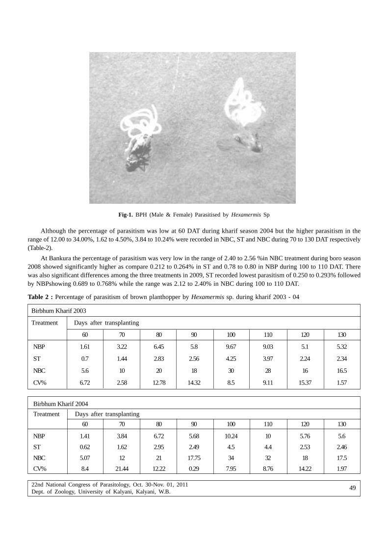
22nd National Congress of Parasitology, Oct. 30-Nov. 01, 2011Dept. of Zoology, University of Kalyani, Kalyani, W.B.
49
Fig-1. BPH (Male & Female) Parasitised by Hexamermis Sp
Although the percentage of parasitism was low at 60 DAT during kharif season 2004 but the higher parasitism in therange of 12.00 to 34.00%, 1.62 to 4.50%, 3.84 to 10.24% were recorded in NBC, ST and NBC during 70 to 130 DAT respectively(Table-2).
At Bankura the percentage of parasitism was very low in the range of 2.40 to 2.56 %in NBC treatment during boro season2008 showed significantly higher as compare 0.212 to 0.264% in ST and 0.78 to 0.80 in NBP during 100 to 110 DAT. Therewas also significant differences among the three treatments in 2009, ST recorded lowest parasitism of 0.250 to 0.293% followedby NBPshowing 0.689 to 0.768% while the range was 2.12 to 2.40% in NBC during 100 to 110 DAT.
Table 2 : Percentage of parasitism of brown planthopper by Hexamermis sp. during kharif 2003 - 04
Birbhum Kharif 2003
Treatment Days after transplanting
60 70 80 90 100 110 120 130
NBP 1.61 3.22 6.45 5.8 9.67 9.03 5.1 5.32
ST 0.7 1.44 2.83 2.56 4.25 3.97 2.24 2.34
NBC 5.6 10 20 18 30 28 16 16.5
CV% 6.72 2.58 12.78 14.32 8.5 9.11 15.37 1.57
Birbhum Kharif 2004
Treatment Days after transplanting
60 70 80 90 100 110 120 130
NBP 1.41 3.84 6.72 5.68 10.24 10 5.76 5.6
ST 0.62 1.62 2.95 2.49 4.5 4.4 2.53 2.46
NBC 5.07 12 21 17.75 34 32 18 17.5
CV% 8.4 21.44 12.22 0.29 7.95 8.76 14.22 1.97

22nd National Congress of Parasitology, Oct. 30-Nov. 01, 2011Dept. of Zoology, University of Kalyani, Kalyani, W.B.
50
Table 3 : Percentage of parasitism of brown planthopper by Hexamermis sp during boro 2008 & 09
Bankura Boro - 2008
Treatment Days after transplanting
60 70 80 90 100 110 120
NBP - - - - 0.8 0.78 -
ST - - - - 0.264 0.212 -
NBC - - - - 2.56 2.4 -
CV% 1.47 1.33
Bankura Boro - 2009
Treatment Days after transplanting
60 70 80 90 100 110 120
NBP - - - - 0.768 0.689 -
ST - - - - 0.25 0.293 -
NBC - - - - 2.4 2.12 -
CV% 1.79 0.32
In the present study, it is thus evident that Natural Biological Control (NBC) has shown the optimum result by exhibitinghigher parasitism as compare with that of schedule treatment (ST) (SBP) and Need Based Protection (NBP) due to theconsiderable reduction in number of applications of insecticide. The need based protection resulted in optimum build up ofHexamermis sp population akin to that natural biological control (NBC) making it more environmental friendly.
ReferencesSatpathi CR, Mukhopadhyay AK, Katti G, Pasalu IC, Venkateswarlu B (2005) Quantification of the role of natural biological
control in farmer’s rice fields in West Bengal. Indian Journal of Entomology 67(3) : 211 – 213.
Gururaj K, Pasalu IC, Varma NRG, Dhandapani N (2001) Quantification of natural biological control in rice ecosystem forpossible exploitation in rice IPM. Indian Journal of Entomology 63(4) : 439 – 444.
Gururaj K, Pasalu IC, Dani RC, Bora DK, Singh MP, Satpathi CR, Reddy PS, Venkateswarlu (2006) Integrated Pest Managementin rainfed rice production system of India – a farmers’ participatory study Oryza (43) 4 pp 296 – 304.
Satpathi CR, Sarkar A, Acharjee P (2008) Factors affecting abundance of Hexamermis sp in rice plant in eastern India,International Journal of Nematology 62(1) 75 – 78.

22nd National Congress of Parasitology, Oct. 30-Nov. 01, 2011Dept. of Zoology, University of Kalyani, Kalyani, W.B.
51
Mitochondrial- DNA and host parasitic interaction
Debjani Nath and Manas Kumar Mukhopadhyay
Department of Zoology, University of Kalyani, Kalyani-741235, West Bengal, India
Abstract : DNA bar coding is based on a relatively simple concept. Most eukaryote cells contain mitochondria, and mitochondrial DNA(mtDNA) has a relatively fast mutation rate, which results in significant variation in mtDNA sequences between species and, in principle,a comparatively small variance within species. A 648-bp region of the mitochondrial cytochrome c oxidase subunit I (COI) gene wasproposed as a potential ‘barcode’. DNA barcoding has met with spirited reaction from scientists, especially systematists, ranging fromenthusiastic endorsement to vociferous opposition. For example, many stress the fact that DNA bar coding does not provide reliableinformation above the species level, while others indicate that it is inapplicable at the species level, but may still have merit for higher-level groups. Others resent what they see as a gross oversimplification of the science of taxonomy. And, more practically, some suggestthat recently diverged species might not be distinguishable on the basis of their COI sequences. Due to various phenomena, it was foundthat some 23% of animal species are polyphyletic if their mtDNA data are accurate, indicating that using an mtDNA barcode to assigna species name to an animal will be ambiguous or erroneous some 23% of the time. Studies with insects suggest an equal or even greatererror rate, due to the frequent lack of correlation between the mitochondrial genome and the nuclear genome or the lack of a barcoding gap.Problems with mtDNA arising from male-killing microoroganisms and cytoplasmic incompatibility-inducing symbionts are also particularlycommon among insects. Given that insects represent over 75% of all known organisms, this suggests that while mtDNA bar coding maywork for vertebrates, it may not be effective for the majority of known organisms.
Key words : Co –speciation, DNA barcode, Mitochondrial DNA, Parasite, Symbionts.
IntroductionThe study of evolution frequently requires understanding the history of the population, species or clade under study.
In population genetics, a recent history of population bottlenecks may restrict genetic variation and thus constrain the speedof adaptation. In examining diversification over space, we need to have detailed knowledge of the different populations’histories of colonization and the gene flow between them. In comparative analyses of processes of adaptation or molecularevolution, and in studies of historical biogeography, we require resolution of the relationships between species. Biotrophicparasitism is a common mode of life that has arisen independently many times in the course of evolution. Depending on thedefinition used, as many as half of all animals have at least one parasitic phase in their life cycles (Price, P.W. 1980) and itis also frequent in plants and fungi. Moreover, almost all free-living animals are host to one or more parasite taxa. (Price, P.W.1980). As a result of host defenses, some parasites evolve adaptations that are specific to a particular host taxon and specializeto the point where they infect only a single species. Such narrow host specificity can be costly over evolutionary time, however,if the host species becomes extinct. Thus, many parasites are capable of infecting a variety of host species that are more orless closely related, with varying success. In some cases, a parasite species may coevolve with its host taxa. In theory, long-term coevolution should lead to a relatively stable relationship tending to commensalism or mutualism, in that it is in theevolutionary interest of the parasite that its host thrives. A parasite may evolve to become less harmful for its host or a hostmay evolve to cope with the unavoidable presence of a parasite to the point that the parasite’s absence causes the host harm.For example, although animals infected with parasitic worms are often clearly harmed, and therefore parasitized, such infectionsmay also reduce the prevalence and effects of autoimmune disorders in animal hosts, including humans. (Rook, G.A.W. 2007).The presumption of a shared evolutionary history between parasites and hosts can sometimes elucidate how host taxa arerelated. For instance, there has been dispute about whether flamingos are more closely related to the storks and their allies,or to ducks, geese and their relatives. The fact that flamingos share parasites with ducks and geese is evidence these groupsmay be more closely related to each other than either is to storks. In rare cases, a parasite may even undergo co-speciationwith its host. One particularly remarkable example of co-speciation exists between the simian foamy virus (SFV) and its primatehosts. In one study, the phylogenies of SFV polymerase and the mitochondrial cytochrome oxidase subunit II from African
*Corresponding Author : E-mail : nath_debjani @yahoo.co.in

22nd National Congress of Parasitology, Oct. 30-Nov. 01, 2011Dept. of Zoology, University of Kalyani, Kalyani, W.B.
52
and Asian primates were compared. (Switzer WM et.al.2005) Surprisingly, the phylogenetic trees were very congruent inbranching order and divergence times. Thus, the simian foamy viruses may have co-speciated with Old World primates forat least 30 million years.
Co-speciationA central question in the evolutionary study of any host–parasite assemblage is the relative antiquity of the host–parasite
association; is the parasite a recently acquired ‘‘souvenir’’ or an ‘‘heirloom’’ inherited from the host’s ancestor (Sprent, 1970)In the latter case, if a parasite is specific to a single host lineage then, as that host lineage speciates, populations of the parasiteon the descendant host species may themselves speciate. If parasites only speciate when the hosts speciate, and never goextinct independently of their hosts, this parallel cladogenesis or ‘‘cospeciation’’ (Brooks, 1988) will yield host and parasitephylogenies that are mirror images of each other. However, if other processes such host switching and extinction occur thenthe relationship between the evolutionary trees for the two members of the association will be more complex and hence moredifficult to unravel (Lyal, 1986; Page, 1994b) Parasites may speciate independently of their hosts, go extinct, or switch todifferent hosts. These processes can result in more than one lineage of closely related parasites on the same host. This istermed a ‘‘duplication’’ by analogy with gene duplications If the two descendant parasite lineages subsequently cospeciatewith the host then the host clade will harbor two parallel sets of parasites Following Fitch’s (1970) terminology for genes, thetwo sets of parasites are called as ‘‘paralogous’’; members of the same lineage of parasites are ‘‘orthologous’’. Recent advancesin molecular systematics facilitate rigorous analysis of the history of host–parasite assemblages, in part by providing mutuallyindependent phylogenies for host and parasite (Page et.al. 1998). Because cospeciation is inferred when the topologies of hostand parasite phylogenetic trees are more similar than would be expected by chance (Hafner and Nadler, 1990) it is vital thathost and parasite evolutionary histories are inferred independently. The use of molecular data can reduce the extent to whichknowledge of the classification of one member of the association influences that of the other, as well as minimizing thepossibility that the phylogenetic analysis is misled by host effects on parasite morphology (Downes, 1990) Molecular dataare also attractive because they provide a common yardstick for comparing evolutionary character change (anagenesis) intaxonomically (and morphologically) disparate groups (Page, 1993c). Homologous genes can be readily found in most taxa,whereas homologous morphological features may be extremely difficult to find in, say, a vertebrate and its insect parasite.Furthermore, if the molecular divergence in host and parasite is approximately clock-like then specific hypotheses about therelative antiquity of a particular host–parasite association can be tested (Page et al., 1996). At the same time, cospeciatinghost–parasite assemblages provide a unique opportunity to compare the evolutionary rates of dissimilar organisms that havevlived in intimate association over long periods of time (Hafner and Page, 1995; Page and Hafner, 1996). Calibrating the absoluterate of evolution in a single clade of organisms can be problematic if the fossil record of that clade is poor. However, if thatclade has cospeciated with a clade that does have a fossil record, then those fossils can also be used to calibrate the rateof evolution in the clade that lacks fossils of its own. This is because if a pair of host and parasite lineages has cospeciatedthen those lineages are of the same age. This approach of calibrating one lineage using another, associated, lineage has beenapplied to endosymbiotic bacteria of aphids (Moran et al., 1993) and cockroaches (Bandi et al., 1995) and alpha herpes virusesof vertebrates (McGeoch et al., 1995). Even if neither member of a cospeciating association has an adequate fossil record wecan still obtain a measure of the relative rate of evolution in the two lineages because cospeciation implies that there are eventsof the same age in the two clades, even if we do not know the absolute age of those cospeciation events. Relative rates ofmolecular evolution have been measured in this way between chewing lice and mammals (Hafner et al., 1994) andendosymbiotic bacteria and aphids (Moran et al., 1995).Calibrating rates of evolution in hosts and parasites depends cruciallyon the presence of cospeciation and on correctly identifying which events in the two clades represent cospeciation. It is notenough simply to compare divergence in pairs of hosts and their parasites. If the parasites are older than their hosts, thengreater divergence between a pair of parasites may reflect greater antiquity relative to their hosts, rather than a difference inrate of evolution. This is analogous to the mistake of using divergence between paralogous genes as a measure of time ofdivergence between organisms; valid comparisons of host and parasite divergence must use ‘‘orthologous’’ parasites (Page,1993b). Conversely, if the parasites have not cospeciated with their hosts but instead have switched hosts then the parasitepair may be much younger than their hosts. In this study we use molecular phylogenies to determine whether there is evidencefor cospeciation between a group of birds and their lice and to investigate the relative rates of evolution in these twotaxonomically distant organisms.
mtDNA- an useful toolThe most widely used molecular tool of these animals has been variation in the mitochondrial DNA (mtDNA) sequence.
Mitochondria have their own genome of about 16,500 bp that exists outside of the cell nucleus. Each contains 13 protein coding
22nd National Congress of Parasitology, Oct. 30-Nov. 01, 2011Dept. of Zoology, University of Kalyani, Kalyani, W.B.
53
genes, 22 tRNAs and 2 rRNAs. They are present in large numbers in each cell, so fewer samples is required. They have ahigher rate of substitution (mutations where one nucleotide is replaced with another) than nuclear DNA making it easier toresolve differences between closely related individuals. They are inherited only from the mother, which allows tracing of a directgenetic line. They don’t recombine. The process of recombination in nuclear DNA (except the Y chromosome) mixes sectionsof DNA from the mother and the father creating a garbled genetic history. So this choice is well reasoned. mtDNA can beeasily amplified from a variety of taxa and, because it is haploid, the sequence can be obtained without cloning. Because ithas a high evolutionary rate and an effective population size approximately one-quarter that of nuclear markers, it allows achance of recovering the pattern and tempo of recent historical events without an extensive sequencing effort. Thirdly, as anarea of at least low recombination, the whole molecule can be assumed to have the same genealogical history. In contrast,while stringent efforts have been made to develop and amplify nuclear markers, this often involves the refinement of primersfor the target species, the sampling of several genes before one with an appropriate evolutionary rate is discovered and theseparation of alleles from heterozygous individuals via cloning before sequencing. While these problems can be overcomeoutside ‘model’ taxa where genomic sequence data are available, they increase the effort needed to gain the requiredinformation. Mitochondrial DNA has therefore remained the marker of choice in many population, biogeographic andphylogenetic studies. Roderic D. M. Page et.al. (1997) studied the phylogeny for the lice (Insecta: Phthiraptera: genus Dennyus)parasitic on swiftlets (Aves: Collocalliinae) was constructed based on mitochondrial cytochrome b DNA sequences. Thisphylogeny is congruent with previous phenetic analyses of morphometric data for the lice. Comparison with a previouslyobtained phylogeny for the hosts indicates some degree of cospeciation. These cospeciation events are used to comparerelative rates of evolution in the birds and their lice for the same segment of the cytochrome b gene. Cytochrome b is evolvingtwo to three times more rapidly in lice than in birds, and louse cytochrome b is highly divergent compared to that of mostother insects. Although generation time has been suggested as an explanation for the disparity in evolutionary rates betweenlice and their hosts, and the small effective population sizes of lice coupled with founder events occurring during transmissionto new host individuals may be an important factor The use of it has also been recommended in taxonomic studies, with theproposal that all described species are given an mtDNA sequence tag or bar-code (Hebert et al. 2003). Indeed, the use ofmtDNA differentiation in defining taxonomic units has been suggested. While mtDNA is a very useful marker, its use is notwithout complication. It was recognized at the outset that mtDNA was strictly a marker for historical processes in females;should male and female history differ in a species, then this marker would not reflect the history of the species as a wholebut that of the female portion Further, there have been technical issues arising from the presence of nuclear integrations ofmtDNA sequence. mtDNA integrated into the nuclear genome may still amplify with conserved primers aimed at mitochondrialcopies, complicating or confounding analysis (Bensasson et al. 2001). Further to these known problems, Ballard & Whitlock(2004) have recently argued that mtDNA evolution is non-neutral with sufficient regularity to question its utility as a markerfor genomic history. Direct selection (selection Proc. R. Soc. B (2005) 272, 1525–1534 on mtDNA itself) and indirect selection(selection arising from disequilibrium with other maternally transmitted genes) is sufficiently common to make inferences frommtDNA data unreliable.
The effects of inherited symbiontsIt has long been recognized that many arthropods carry passenger micro-organisms: microbes that exist inside the cells
of their host and pass from a female to her progeny through the egg. These micro-organisms can broadly be classified intotwo kinds: beneficial to the host and parasitic. In the case of the former, treatment of the arthropod host with antibioticsproduces a decrease in host fitness (and commonly infertility or death). In the case of the latter, the drive that produces thespread of the micro-organism is frequently the manipulation of host reproduction towards the survival of infected females andthe daughters of infected females. Beneficial symbionts are found widely in arthropods and other invertebrates. The majorityof aphid species, for instance, depend on the presence of the bacterium Buchnera to be able to synthesize essential aminoacids. Cockroaches, termites and a variety of Hemiptera (e.g. white fly), Diptera (e.g. tsetse fly), Hymenoptera (e.g. carpenterants) and Coleoptera (e.g. weevils) likewise rely on beneficial symbionts. Feeding on depauperate diet (blood throughout life;phloem; nitrogen-poor wood) are strong predisposing factors for the occurrence of these beneficial micro-organisms. Theassociations between beneficial symbionts and their host can be very long, as indicated by cocladogenesis of symbiont andhost (e.g. Bandi et al. 1998). The observation that they have genomes that have shrunk massively over the course of symbiosisindicates that these beneficial symbionts undergo repeated selective sweeps associated with genomic rearrangements duringthe early parts of their association with the host (Wernegreen 2002). Parasitic passenger micro-organisms are found even morewidely. These micro-organisms show a variety of phenotypes associated with promoting the production and survival ofinfected daughters (which can transmit the maternally transmitted micro-organism) via negative effects on the production and

22nd National Congress of Parasitology, Oct. 30-Nov. 01, 2011Dept. of Zoology, University of Kalyani, Kalyani, W.B.
54
survival of infected males and uninfected females (which cannot). In the simplest case, this involves creating a female biasin the host sex ratio. This is represented by cases of parthenogenesis induction in haplodiploid species and feminization inCrustacea. In a twist to this manipulation, killing of male hosts during embryogenesis is common in insects. Here, the advantagederives from removing any negative effects that male hosts may have on their sisters. Examples are found in Hemiptera,Hymenoptera, Diptera, Coleoptera and Lepidoptera. Even in Drosophila, which for ecological reasons are not the most likelyto bear male-killers, there are 14 records of male-killing bacteria, indicating these parasites are widely present (Hurst and Majerus1993). The incidence is certainly higher in other species, such as the coccinellid beetles. The most commonly observed formof parasitism is cytoplasmic incompatibility, which differs subtly in logic from sex ratio distortion. In this manipulation, zygotesformed following fertilization of an uninfected egg with sperm from an infected male die during early embryogenesis. Thisbehaviour produces the spread of the infection in structured populations, as the bacterial phenotype is essentially one ofselectively killing uninfected individuals. Cytoplasmic incompatibility has been described in insects, mites and crustaceans,and is probably very common (although cryptic, as frequently all individuals within a population are infected; (Stouthameret al. 1999). Two micro-organisms, Wolbachia and Cardinium, are known to induce it (Breeuwer et al. 1992; Hunter et al. 2003).In total, these parasitic interactions are common. In large surveys, repeatable over geographical regions and across arthropodgroups, Wolbachia alone infects in excess of 20% of insect species at any point in time (Werren et al.1995;Werren & Windsor2000; Jiggins et al. 2001; Hurst and Jiggins 2005) and, in a limited survey, just over 50% of spiders (Rowley et al.2004). Thebacterium Cardinium infects around 7% of arthropods (Weeks et al. 2003).The interactions between parasitic symbionts andtheir hosts are relatively short-lived in comparison to beneficial symbioses. While cocladogenesis of host and Wolbachia issometimes observed (e.g. Marshall 2004), studies on focused host clades have indicated it is rather rare (Shoemaker et al. 2002).The mean lifespan of any particular interaction is therefore generally less than the mean time to speciation. This conclusionis reinforced by the observation of three actively spreading Wolbachia infections in natural populations (Turelli and Hoffmann1991; Hoshizaki and Shimada 1995; Riegler and Stauffer 2002). Thus, we can conclude that the 20% incidence reflects a patternwhere new infections must arise relatively commonly. As a final complicating feature, it should be noted that a single populationmay be infected with more than one strain or species of parasitic inherited microorganism and that different populations mayshow different infection statuses Finally, recent study has revealed the widespread presence of ‘secondary symbionts’. Theseare vertically transmitted micro-organisms that are not essential but appear to be locally beneficial (e.g. enhancing resistanceto parasitoids and pathogens or adaptation to growth on a particular species of host plant; (Oliver et al. 2003; Ferrari et al.2004; Tsuchida et al. 2004). These can be common and it is known that their frequency varies geographically (Tsuchida et al.2002). What is uncertain is the extent of disequilibrium with mtDNA, which will be inversely related to the rate of thesesymbionts’ horizontal transmission in the field.
Passenger microorganismsInherited micro-organisms will influence the population genetics of the host’s mtDNA if they are cotransmitted and
therefore in linkage disequilibrium. However, linkage disequilibrium will break down if either the symbiont or mitochondria arepaternally or horizontally (infectiously) transmitted with sufficient frequency (Turelli et al. 1992).This process is equivalent torecombination breaking down the linkage between nuclear genes. Horizontal or paternal transmission of both symbionts andmtDNA has been documented in insects. Despite this, symbionts in natural populations are typically found in linkagedisequilibrium with mtDNA (table 1), suggesting that such transmission is so infrequent as to be unimportant. For example,paternal transmission of both Wolbachia and mtDNA has been recorded in laboratory populations of Drosophila simulans butit is sufficiently rare that they remain in linkage disequilibrium in the field (Hoffmann and Turelli 1988; Kondo et al. 1990; Turelliet al. 1992). However, there are some species where infectious transmission of symbionts is so common that there is unlikelyto be any association between the microbe and host mtDNA (Huigens et al. 2000).Provided the assumption of linkagedisequilibrium is met, if a population becomes infected with a symbiont that has sufficient drive to spread, the mtDNA typeassociated with the initial infection will hitchhike through the population (‘indirect selection’ on the mtDNA). This processhas been recreated in laboratory populations of the mosquito Aedes albopictus infected with a Wolbachia strain that inducescytoplasmic incompatibility (Kambhampati et al. 1992). The most remarkable example, however, comes from Californianpopulations of D. simulans. These were originally uninfected, but during the 1980s they were invaded by a Wolbachia strainthat induced cytoplasmic incompatibility (Turelli and Hoffmann 1991). This infection rapidly spread to a high prevalence andcarried with it a mtDNA haplotype that was previously rare or absent in uninfected Californian populations (Turelli et al. 1992).Once the infection neared fixation, the original mtDNA haplotype was completely replaced by the haplotype linked to thesymbiont. The sweeps of mtDNA seen in California D. simulans are probably regular events in insects. Despite being transientevents, the spread of a new Wolbachia strain over space has also been documented in the delphacid bug Laodelphax striatellus

22nd National Congress of Parasitology, Oct. 30-Nov. 01, 2011Dept. of Zoology, University of Kalyani, Kalyani, W.B.
55
and the fly Rhagoletis cerasi (Hoshizaki and Shimada 1995; Riegler and Stauffer 2002).These observations, together with thelack of cocladogenesis between hosts and symbionts discussed above, indicate that new interactions (which must involvesymbiont spread) occur during the lifespan of many species. It is clear from these studies that mtDNA within a populationis affected by symbiont spread. Further, the ability of symbionts to spread between populations by occasional movement ofhosts, and the ability of different host populations to maintain different symbiont strains, can also confound interpretationattempts to reconstruct the phylogeography of a species. The drive associated with symbiont manipulation can even resultin the spread of the symbiont into a new species following an occasional hybridization event. This will homogenize the mtDNAof different species. We now review the effects that symbionts may have on patterns within populations, between populationsand between species variation in mtDNA.
Recently diverged speciesAt first sight, passenger micro-organisms are expected to affect the dynamics of mtDNA within populations but not the
branching pattern of mtDNA on a phylogeny. However, two case studies indicate this is not necessarily the case (Hurst andJiggins 2005). First, there is the A. encedon and A. encedana species pair. These both bear a male-killing Wolbachia infection,and evidence from the Wolbachia sequence indicates they are very closely related strains (note that A. encedon also has asecond, more distantly related, infection). When the phylogeny of individuals of the different species is constructed basedon mtDNA, A. encedon and A. encedana individuals that bear the same Wolbachia infection bear identical mtDNA sequences,distinct from that found in uninfected.A. encedon individuals. While the species clearly are distinct on morphological grounds,on the grounds of nuclear DNA sequences and in terms of genetic isolation, they appear identical on the basis of the mtDNAsequences of the infected individuals. The most likely explanation for this is that rare hybridization events, although producingvery little in the way of flow of nuclear genes, can produce the transfer of the male-killer and associated mitotype from speciesto species. This male-killer has a drive mechanism that results in its increasing in frequency, despite initially being in poorlyadapted hybrid individuals, and the infection and associated mitotype spreads into the new species. This transfer and fixationof mtDNA following hybridization would appear at first sight to be a rare event. However, it has also been observed inDrosophila. In Drosophila, mtDNA has introgressed between D. simulans and D. mauritiana, associated with Wolbachiainfection (Rousset and Solignac 1995; Ballard 2000b). Anecdotally, three out of four cases we have studied in our laboratorieshave shown evidence of symbiont-induced mtDNA introgression confounding phylogenetic estimation from an mtDNAsequence.It is possible that symbiont-driven introgression may explain some recent case studies where mtDNA phylogeniesconflict with those obtained from nuclear DNA. (Shaw 2002), for instance, observes that while certain crickets of the genusLaupala in Hawaii do not differ in mtDNA sequence, they do exhibit substantial differentiation at nuclear loci. Thisincongruence points to selection causing introgression of the mtDNA following hybridization. A good hypothesis for thisobservation is that hybridization carries novel symbiont infections. The spread of the introgressed symbiont would then beassociated with the spread of introgressed mtDNA, homogenizing mtDNA variation across the species boundary despite ahigh genetic integrity of the species as recorded on nuclear markers.
ConclusionOne recent and contentious use of mtDNA sequence is in DNA bar-coding (Hebert et al. 2003; 2004a). In DNA bar-coding,
the sequence of the mitochondrial 648-bp region cytochrome oxidase subunit 1 (COI) gene is used for the purpose of taxonomicidentification and assessment of biodiversity, with the philosophy that for each species there is one bar-code (and reciprocally,one bar-code indicates a given species). DNA bar-coding relies on there being low levels of mtDNA variation within a speciescompared with differentiation between species and monophyly of mtDNA within species. While there may generally be lowmtDNA diversity within a species, and species may frequently be delineable by mtDNA, this pattern can be disrupted. Whilea review of species polyphyly on the basis of mtDNA suggested 23% of species may not be monophyletic for mtDNAsequences (Funk and Omland 2003), bar-coding tests have not revealed this pattern (Hebert et al. 2003; 2004a). However, withnotable exceptions (Hebert et al., 2004b), past tests have tended not to explicitly test the ability to discriminate a range ofclosely related sibling species but rather a range of congenerics, many of which are relatively distantly related. Further, thesample size used when testing intraspecific variation has often been limited, with one or two extra individuals within knownspecies obtained and found to possess very similar COI sequences to those previously found. Indeed, where they have beenfound to carry divergent sequences, the inference has been made that the process has revealed cryptic species, rather thanthat mtDNA can act as a poor marker. mtDNA alone cannot be used to reliably infer population history or the history of closelyrelated species as there is a very high probability of an incorrect conclusion due to indirect selection arising from the presenceof a symbiont. Reciprocally, it is also problematic to infer symbiont history from mtDNA alone. There are several records of

22nd National Congress of Parasitology, Oct. 30-Nov. 01, 2011Dept. of Zoology, University of Kalyani, Kalyani, W.B.
56
inherited bacteria being present in nematodes (Adams and Eichenmuller 1963; Marti et al. 1995; Sironi et al. 1995) and alsorecords of inherited bacteria in disequilibrium with mtDNA in mollusks (Hurtado et al. 2003). It should not be assumed thattaxa lack symbionts until careful surveys have been carried out because recent history indicates that even well studied groups(e.g. filarial nematodes) can in fact be covertly infected with inherited symbionts (Sironi et al. 1995).
AcknowledgementAuthors are grateful to Vice Chancellor,University Of Kalynai, Prof. Aloke Kumar Banerjee for his interest in our work.
ReferencesAdams RE, Eichenmuller JJ (1963) A bacterial infection of Xiphenema americanum. Phytopathology 53:745.
Ballard JWO (2000b) When one is not enough: introgression of mitochondrial DNA in Drosophila. Mol Biol Evol 17: 1126–1130.
Ballard JWO, Whitlock MC (2004) The incomplete natural history of mitochondria. Mol Ecol 13: 729–744.
Bandi C, Sironi M, Damiani G, Magrassi L, Nalepa CA, Laudani U, Sacchi L (1995) The establishment of intracellular symbiosisin an ancestor of cockroaches and termites. Proc R Soc London 259(B): 293–299.
Bandi C, Anderson TJC, Genchi C, Blaxter ML (1998) Phylogeny of Wolbachia in filarial nematodes. Proc R Soc B 265: 2407–2413.
Bensasson D, Zhang DX, Hartl DL, Hewitt GM (2001) Mitochondrial pseudogenes: evolution’s misplaced witnesses. TrendsEcol Evol 16: 314–321.
Breeuwer JAJ, Stouthamer R, Barns SM, Pelletier DA, Weisburg WG, Werren JH, (1992) Phylogeny of cytoplasmic incompatabilitymicro-organisms in the parasitoid wasp Nasonia (Hymenoptera: Pteromalidae) based on 16S ribosomal DNA sequences.Insect Mol Biol 1: 25–36.
Brooks DR (1988) Macroevolutionary comparisons of host and parasite phylogenies. Annu Rev Ecol Syst 19: 235–259.
Downes BJ (1990) Host-induced morphology in mites: Implications for host-parasite coevolution. Syst Zool 39: 162–168.
Ferrari J, Darby AC, Daniell TJ, Godfray HCJ, Douglas AE (2004) Linking the bacterial community in pea aphids with host-plant use and natural enemy resistance. Ecol Ent 29: 60–65.
Fitch WM (1970) Distinguishing homologous from analogous proteins. Syst Zool 19: 99–113.
Funk DJ, Omland KE (2003) Species level paraphyly and polyphyly: frequency, causes, and consequences, with insights fromanimal mitochondrial DNA. A. Rev Ecol Evol Syst 34: 397–423.
Hafner MS, Nadler SA (1990) Cospeciation in host-parasite assemblages: Comparative analysis of rates of evolution and timingof cospeciation. Syst Zool 39: 192–204.
Hafner MS, Sudman PD, Villablanca FX, Spradling TA, Demastes JW, Nadler SA (1994) Disparate rates of molecular evolutionin cospeciating hosts and parasites. Science 265:1087–1090.
Hafner MS, Page RDM (1995) Molecular phylogenies and host-parasite cospeciation: Gophers and lice as a model system.Philos. Trans R Soc London 349(B): 77–83.
Hebert PDN, Cywinska A, Ball SL, DeWaard JR (2003) Biological identifications through DNA barcodes. Proc R Soc B 270:313–321.
Hebert PDN, Stoeckle MY, Zemlak TS, Francis CM (2004a) Identification of birds through DNA barcodes. PLoS Biol 2: 1657–1663.
Hebert PDN, Penton EH, Burns JM, Janzen DH, Hallwachs W (2004b) Ten species in one: DNA barcoding reveals crypticspecies in the neotropical skipper butterfly
Astraptes fulgerator. Proc Natl Acad Sci USA 101:14 812–14 817.
Hoffmann AA, Turelli M (1988) Unidirectional incompatibility in Drosophila simulans: inheritance, geographic variation andfitness effects. Genetics 119: 435–444.
Hoshizaki, S. and Shimada, T., 1995. PCR-based detection of Wolbachia, cytoplasmic microorganisms, infected in naturalpopulations of Laodelphax striatellus (Homoptera: Delphacidae) in central Japan: has the distribution of Wolbachiaspread recently? Insect Mol Biol 4: 237–243.

22nd National Congress of Parasitology, Oct. 30-Nov. 01, 2011Dept. of Zoology, University of Kalyani, Kalyani, W.B.
57
Huigens ME, Luck RF, Klaassen RHG, Maas M, Timmermans M, Stouthamer R (2000) Infectious parthenogenesis. Nature 405:178–179.
Hunter MS, Perlman SJ, Kelly SE (2003) A bacterial symbiont in the Bacteroidetes induces cytoplasmic incompatibility in theparasitoid wasp Encarsia pergandiella. Proc R Soc B 270: 2185–2190.
Hurst GDD, Majerus MEN, (1993) Why do maternally inherited microorganisms kill males? Heredity 71: 81–95.
Hurst GD, Jiggins FM (2005) Problems with mitochondrial DNA as a marker in population, phylogeographic and phylogeneticstudies: the effects of inherited symbionts Proc R Soc B 272: 1525–1534.
Hurtado LA, Mateos M, Lutz RA, Vrijenhoek RC (2003) Coupling of bacterial endosymbiont and host mitochondrial genomesin the hydrothermal vent clam Calyptogena magnifica. Appl Environ Microbiol 69: 2058–2064.
Kambhampati S, Rai KS, Verleye DM (1992) Frequencies of mitochondrial-DNA haplotypes in laboratory cage populations ofthe mosquito, Aedes albopictus. Genetics 132: 205–209.
Kondo R, Satta Y, Matsuura ET, Ishiwa H, Takahata N, Chigusa SI (1990) Incomplete maternal transmission of mitochondrial-DNA in Drosophila. Genetics 126: 657–663.
Lyal CHC (1986) Coevolutionary relationships of lice and their hosts: A test of Farenholz’s Rule. In ‘‘Coevolution andSystematics’(A. R. Stone and D. L. Hawksworth, Eds.), pp. 77–91, Clarendon, Oxford.
Marshall JM (2004) The Allonemobius–Wolbachia host– endosymbiont system: evidence for rapid speciation and againstreproductive isolation driven by cytoplasmic incompatibility. Evolution 58: 2409–2425.
Marti OGJ, Rogers CE, Styer EL (1995) Report of an intracellular bacterial symbiont in Noctuidonema guyanese, an ectoparasiticnematode of Spodoptera frugiperda. J Invert Pathol 66: 94–96.
McGeoch DJ, Cook S, Dolan A, Jamieson FE, Telford EAR (1995) Molecular phylogeny and evolutionary timescale for the familyof mammalian herpesviruses. J Mol Biol 247: 443–458.
Moran NA, Munson MA, Baumann P, Ishikawa H (1993) A molecular clock in endosymbiotic bacteria is calibrated using theinsect hosts. Proc R Soc London 253: 167–171.
Moran NA, van Dohlen CD, Baumann P (1995). Faster evolutionary rates in endosymbiotic bacteria than in cospeciating insecthosts. J Mol Evol 41:727–731.
Oliver KM, Russell JA, Moran NA, Hunter MS (2003) Facultative bacterial symbionts in aphids confer resistance to parasiticwasps. Proc Natl Acad Sci USA 100: 1803–1807.
Page RDM (1993b) Genes, organisms, and areas: The problem of multiple lineages. Syst Biol 42: 77–84.
Page RDM (1993c) Parasites, phylogeny and cospeciation. Int J Parasitol 23: 499–506.
Page RDM, Hafner MS (1996) Molecular phylogenies and host-parasite cospeciation: Gophers and lice as a model system. In‘‘New Uses for New Phylogenies’’ (P. H. Harvey, A. J. Leigh Brown, J. Maynard Smith, and S. Nee, Eds.), pp. 255–270,Oxford Univ. Press, Oxford.
Page RDM (1996a) Temporal congruence revisited: Comparison of mitochondrial DNA sequence divergence in cospeciatingpocket gophers and their chewing lice. Syst Biol 45: 151–167.
Page RDM, Patricia LM, Lee SA, Becher R, Griffiths DH (1998) Clayton A Different Tempo of Mitochondrial DNA Evolutionin Birds and Their Parasitic Lice molecular phylogenetic and evolution .9(2): 276–293.
Price PW (1980) Evolutionary Biology of Parasites. Princeton University Press, Princeton.
Riegler M, Stauffer C (2002) Wolbachia infections and superinfections in cytoplasmically incompatible populations of theEuropean cherry fruit fly Rhagoletis cerasi (Diptera, Tephritidae). Mol Ecol 11: 2425–2434.
Rook GAW (2007) The hygiene hypothesis and the increasing prevalence of chronic inflammatory disorders. Transactions ofthe Royal Society of Tropical Medicine and Hygiene 101 (11): 1072–4.
Rousset F, Solignac M (1995) The evolution of single and double Wolbachia symbioses during speciation in the Drosphilasimulans complex. Proc Natl Acad Sci USA 92: 6389–6393.
Rowley SM, Raven RJ, McGraw EA (2004) Wolbachia pipientis in Australian spiders. Curr Microbiol 49: 208–214.
Sironi M, Bandi C, Sacchi L, Di Sacco B, Damiani G, Genchi C (1995) A close relative of the arthropod endosymbiont Wolbachiain a filarial worm. Mol Biochem Parasitol 74: 223–227.

22nd National Congress of Parasitology, Oct. 30-Nov. 01, 2011Dept. of Zoology, University of Kalyani, Kalyani, W.B.
58
Shaw KL (2002) Conflict between nuclear and mitochondrial DNA phylogenies of a recent species radiation: What mtDNAreveals and conceals about modes of speciation in Hawaiian crickets. Proc Natl Acad Sci USA 99: 16 122–16 127.
Shoemaker DD, Machado CA, Molbo D, Werren JH, Windsor DM, Herre EA (2002) The distribution of Wolbachia in figwasps: correlations with host phylogeny, ecology and population structure. Proc R Soc B 269: 2257–2267.
Sprent JFA (1970) Baylisacaris tasmanienis sp. nov. in marsupial carnivores: Heirloom or souvenir. Parasitology 61: 75–86.
Stouthamer R, Breeuwer JAJ, Hurst GDD (1999) Wolbachia pipientis: microbial manipulator of arthropod reproduction. AnnuRev Microbiol 53: 71–102.
Switzer WM, Salemi M, Shanmugam V, Gao F, Cong ME, Kuiken C, Bhullar V, Beer BE, Vallet D, Gautier-Hion A, Tooz, Z, VillingerF, Holmes EC, Heneine W (2005) Ancient co-speciation of simian foamy viruses and primates. Nature 434(7031):376-80.
Tsuchida T, Koga R, Shibao H, Matsumoto T, Fukatsu T (2002) Diversity and geographic distribution of secondaryendosymbiotic bacteria in natural populations of the pea aphid, Acyrthosiphon pisum. Mol Ecol 11: 2123–2135.
Tsuchida T, Koga R, Fukatsu T (2004) Host plant specialization governed by facultative symbiont. Science 303: 1989–1989.
Turelli M, Hoffmann AA (1991) Rapid spread of an inherited incompatibility factor in California Drosophila. Nature 353: 440–442.
Turelli M, Hoffmann AA, McKechnie SW (1992) Dynamics of cytoplasmic incompatibility and mtDNA variation in naturalDrosophila simulans populations. Genetics 132: 713–723.
Weeks AR, Velten R, Stouthamer R (2003) Incidence of a new sex-ratio-distorting endosymbiotic bacterium among arthropods.Proc R Soc B 270: 1857–1865.
Wernegreen JJ (2002) Genome evolution in bacterial endosymbionts of insects. Nature Rev Gen 3: 850–861.
Werren JH, Zhang W, Guo LR (1995) Evolution and phylogeny of Wolbachia: reproductive parasites of the arthropods. ProcR Soc B 261: 55–71.
Werren JH, Windsor DM (2000) Wolbachia infection frequencies in insects: evidence of a global equilibrium? Proc R Soc B267: 1277–1285.

22nd National Congress of Parasitology, Oct. 30-Nov. 01, 2011Dept. of Zoology, University of Kalyani, Kalyani, W.B.
59
Parasitic adaptation : trends since Darwin
Dipak Ranjan Mandal and Swapna Chhetri*
Durgapur Government College, Durgapur-713214, Burdwan, West Bengal, India*P.G. Department of Zoology, Darjeeling Government College, Darjeeling, West Bengal, India
Abstract : Parasitic adaptation in Darwin’s speculation was a ‘degenerate’. The morphological and biochemical reductions found in adultparasites can be considered as an economy of effort rather than a degenerate condition. Parasitic adaptations are all responses to particularfeatures of the parasite’s environment, and with the exception of the host’s immune response and parasitic virulence the same features arealso found in free-living environments. In order for a parasite to continue its mode of life it or its offspring must find a new host the attributeis called ‘infectivity’. Hosts evolve to decrease the effectiveness of their parasites’ adaptations, and parasites evolve to decrease theeffectiveness of their hosts’ defenses. Virulence inevitable in parasitism is the decrease in fitness of a standard host caused by associationwith a parasite. Increase in host’s fitness via increased resistance usually invites decrease in parasite’s fitness, whereas tolerance is the modewhere host gets fitness without necessarily reducing parasite fitness. Parasite should evolve into a less virulent because death of a hostshould have negative effect on parasite’s survival. Parasitic adaptation and host parasite coevolution in terms of two attributes-infectivityand virulence have been discussed in the present communication.
Keywords : coevolution, infectivity, symbiosis, virulence
IntroductionEvolution of parasitism and parasitic adaptation are the topic of discussion since a long time. Several views are available
even in the literature of early nineteenth and several reports on morphological, physiological, biochemical and alsoimmunological aspects have been dealt with due consideration but moot-point on the evolution of sickness due to parasite’svirulence has emerged rather recently. In a parasitic association the negative effects on the host’s fitness may vary not onlybetween host species, genotypes and individuals but even between ecological conditions and subpopulations (Dybdahl andStorfer, 2003). This reduction in fitness is principally concerned with the characteristic variable called virulence. Because thelong term persistence of a pathogenic species entwined with host survival it requires balancing conflict that is selective forcesat the individual and population level as well as trade-off between pathogen and host evolutionary strategies (Price, 2008).Parasitic adaptation in Darwin’s speculation was a ‘degenerate’. Darwinism lies in the very notion of symbiosis itself, quiteapart from the ‘cooperative’ nature of symbiotic associations. There is no empirical evidence that leads to suppose that naturalselection will as a general rule lead to anything that may sensibly called ‘progressive’. During transition from prokaryotes toeukaryotes symbiosis played a crucial role. The morphological and biochemical reductions found in adult parasites either inprotozoa or helminthes can be considered as an economy of effort rather than a degenerate condition. The concept thatparasitism evolves towards becoming harmless to each of the partners has now opened the way to further analysis. Adaptationto physical environment differs from that of the parasitic organisms which practically explore in living environment. In the later,adaptation of species rather induces reciprocal biochemical and genetic response, as the other species itself evolves in specificways to alleviate those evolutionary changes. Infectivity and virulence appears also to play momentous role in parasiticadaptation.
DiscussionIn order for a parasite to continue its mode of life it or its offspring must find a new host. The host finding capacity
associated with the susceptibility and resistance of the host is referred to as infectivity. Apparently it seems to be a difficultenvironment for invasion but those organisms that have done successfully so have often been very successful both in termsof number of individuals and also in number of species. This attribute is expressed as :
Infectivity or I= host finding capacity of the parasite + susceptibility of host/ resistance of host.
Infectivity of parasites may be compared with the capacity of the free living organisms to get distribution and dispersion,failure of which may cause hindrance in adaptation as well as survivality.

22nd National Congress of Parasitology, Oct. 30-Nov. 01, 2011Dept. of Zoology, University of Kalyani, Kalyani, W.B.
60
On the other hand the extent of damage on host by the parasite i.e.
Pathogenecity is expressed as: P= I x number of parasites in host x damage to host per parasite.
Virulence : The most common definition of virulence is the mortality caused by a parasite in a standard host. Virulenceis the decrease in fitness of a standard host caused by the association with a parasite. Though there remains no scale toquantify the negative effects exerted by the parasite on the host it is related to the virulence of the parasite. No doubt directmortality reduces the fitness. On the other hand, reduced fecundity due to shift in nutritional and behavioural status orinteraction with competition or predation, parasitic castration where fecundity is zero are added to the mortality attribute.Virulence has also more to do with host specificity and generality. There are evidences that a simple mutation in the geneticconstitution of the parasite may lead to virulence. Yersinia pestis the causative agent of human virulent plague differs in havingtwo mutations than the mild pathogenic form Y. pseudotuberculosis.
Resistance and ToleranceHosts in the interacting association bears opposite force against parasitic virulence. While virulence is the harm on a
standard host, resistance on the other hand measures the harm or reduction in harm done to a host by a standard parasite.In addition to the immunological strategies, a single mutation in the host causes resistance to parasites infectivity as in thecases where sickle cell anemic or Duffy negative blood group people are shield from the infection of Plasmodium.
If the parasite induced mortality level drops by evolutionary time there remains no reason to think that virulence hasdecreased. Another important aspect in host parasite association is the evolution of tolerance. Tolerance ensures the reducedlevel of immunopathology.
Increase in host fitness via increased resistance usually invites decrease in parasite’s fitness, where as tolerance is themode where host gets fitness without necessarily reducing parasite fitness.
In the early phase of association the host and the parasite are not going to be well adapted with each other. This isexpected that recently evolved parasites to be virulent, but gradually the host would be more tolerant of the parasite and theobvious question arises: Acute? Chronic? Mutualism?
One paradigm states that parasite should evolve into less virulent mainly because death of a host should have a negativeeffect on parasite’s survival (Clayton et al, 1999).
The emerging concepts on virulence are as followed :
1. Red Queen’s Hypothesis and Evolutionary arms race : Literary Red Queen’s race is an incident that appears in LewisCarroll’s “Through the looking glass’’ and involves the Red queen a representation of a Queen in chess and Alice constantlyrunning but remaining in the same spot. This term has also been used by Isaac Asimov to illustrate the concept ofpredestination of paradox.
For an evolutionary system, continuing development is needed just in order to maintain its fitness relative to the systemit is co-evolving with (http://pespmcl.vub.ac.).This hypothesis intends to explain:
1. the advantage of sexual reproduction at the level of individuals and
2. constant evolutionary arms race between competing or interacting species.
The first segment is a micro evolutionary version while the second one is macro evolutionary that tends to explain theprobability of extinction of groups of organisms is hypothesized to be constant within the group and random among thegroups.
The Red Queen’s Hypothesis was formulated by Valen (1973) as an explanatory tangent to his proposed Law of Extinction.The hypothesis also provides a conceptual underpinning to discussion of evolutionary arms race. Literally arms race refersto the fact of fighting between two to attain supremacy by each. From the point of evolution, an evolutionary arms race isan evolutionary struggle between competing sets of co-evolving genes that develop adaptations and counter adaptationagainst each other. Co-evolving gene sets of the interacting species may vary as in a prey-predator or parasite-host association(Vermeji,1987).
Arms Race and Virulence : Coevolution itself is not necessarily an arms race. In parasitism rather an asymmetrical armsrace involves opposite selection pressures where parasite is driven to enhance infectivity and the host develops resistance(Dawkins and Krebs, 1979). Further Combes (2000) referred to evolution of parasite virulence and host resistance as

22nd National Congress of Parasitology, Oct. 30-Nov. 01, 2011Dept. of Zoology, University of Kalyani, Kalyani, W.B.
61
evolutionary arms race. Observation of Jaekel et al. (2001) supports this contention. Virulence of Sarcocystis singaporensisin wild rat is relatively constant and intermediate with maximal production of bradyzoites. Highly virulent strains may bedeveloped in laboratory mice and immune reaction of these mice reduces the production of bradyzoites (lessen infectivity).
2. Conventional Wisdom : Conventional wisdom (May and Anderson, 1983) explains that well adapted parasite harmsits host a little. The evolutionary trajectories of a parasite host association is towards a mutualistic or at least a commensalsymbiosis. Less harm a parasite does better the host does. Virulence was considered as an artifact of recent host parasiteassociation (Dubos, 1965; Burnet and White, 1972). Thus-
i. Individual hosts are healthier and survive a longer
ii. Host populations are larger providing more opportunities for parasite’s offspring and
iii. Host may evolve weaker parasite defenses.
The logic is really pleasant to human sensibilities (Levin, 1999).
Evidence supporting the trajectories :
• Syphilis (Treponema pallidum) epidemic in Europe during 1493 – 1510 appeared to have less virulent in time. Incontrast HIV is transmitted between humans and its association with Homo sapiens is almost universally consideredrecent (Essex and Kanki, 1988).
• Some virulent pathogens like Shigella and Neisseria gonorrhoeae humans act as the dominant host as well as thevector for the infectious transmission (16). Lethal pathogen of tuberculosis have had a long history in humanpopulation and also been transmitted solely by humans (Levin, 1999).
Is conventional wisdom is over simplified? Conventional wisdom is not based on the hypothesis that can be readily testedor rejected. Short term individual selection will often lead to higher virulence that may eventually lead to extinction of the hostpopulation and the parasite lineage along with it. On the other hand evolving and becoming prudent in treating their hostsrequire some form of group level or kin selection (Frank, 1996). Similarly the question arises how a host evolutionary responseunilaterally converts an otherwise virulent parasite into a commensal. Conventional wisdom theory does not account for theactual mechanisms responsible for the evolution of benign association between parasites and their hosts.
3. Tradeoff and the enlightenment : This is based on the epidemiologic model referred to the “enlightenment” (Levin andSanborg, 1990). The theory of the evolution of virulence has taken the approach of arguing evolutionary changes in virulenceare a result of tradeoff between virulence and transmission of diseases between the hosts. According to this view naturalselection could favour the evolution and maintenance of virulence in commensal as well as in other symbiotic associations.In other words, virulence could be the primitive stage of the association . Direction of natural selection in a given situationdepends on the epidemiology and ecology of the parasite and in particular, the relationship between its virulence and its rateof infectious transmission in the host population. While the existence of the parasite mediated selection and evolution in thehost population are acknowledged, the enlightenment view has concentrated on the changes in the parasite population (Levin,1999). At any given host density the fitness of the parasite in the host population is directly proportional to its transmissibility.
This agrees with the “Conventional wisdom” but from an individual selection perspective :
• Being less virulent just means that parasite’s own host will survive longer
• The parasite will be getting longer time to reproduce.
This has been postulated that natural selection would favour highly transmissible, incurable commensals. On the otherhand if the transmission and virulence in were positively coupled, natural selection could favour the evolution and maintenanceof some level of virulence in the parasite population. At present much of the research on the evolution of virulence has focusedon the association between two components:
i. The rate at which the parasite is transmitted between hosts and
ii. The rate of parasite mediated mortality in individual infected hosts.
If these relationships are positive then some level of virulence may be favoured (Levin 1999).
Experimental evidence : Evidence of host parasite coevolution comes from the experiment on myxoma virus. Myxomaviruses are the members of the genus Leporipoxvirus and native to specific species of rabbit of the genus Salvilagus foundin California and extending south to South America. A South American strain of myxoma virus was intentionally introducedin pernicious feral European rabbit (Oryctolagus cunniculus) population in Australia (1950) and was also released with

22nd National Congress of Parasitology, Oct. 30-Nov. 01, 2011Dept. of Zoology, University of Kalyani, Kalyani, W.B.
62
forethought into the native rabbit population of Europe. The massive mortalities of European rabbits that ensued in bothcontinents are land mark events in discussion of long term host species dynamic modification by a viral agent (Regnery, 2007).The viruses recovered from the then decimated and sometimes more resistant wild rabbit populations were less virulent (withlower level of disease induced mortality) on control laboratory rabbits. Viruses from the wild but attenuated were less activethan that which could be achieved in the first stage of the experiment. This was interpreted as evidence for a positive couplingbetween the rates of infectious transmission and rates of virus induced mortality- a tradeoff between virulence andtransmission. Highly virulent forms had disadvantage because the killed the rabbits too quickly and thus reduced the timeavailable for the pathogen to be picked up by the flea or mosquito vectors required for the transmission. Viruses that weretoo attenuated had the disadvantage because they generated fewer skin lesions and had lower densities of circulating virions.This also reduced the rate at which they would be picked up by the vectors and the magnitude of virions to b e picked upat any given bite. Thus where there is a positive coupling between a parasite’s virulence and its infectivity natural selectioncould favour and maintain the virulence of the parasite. A parasite’s virulence is constrained solely by the need to keep thehost active to facilitate its transmission to new host. Though myxoma virus experiment is a model to explain the tradeoffhypothesis substantive experimental data are still wanting to demonstrate that the increase in virulence generated during apassage experiment is also reflected as increased transmissibility as is necessary for the tradeoff interpretation. Indeed it maywell be that an increase in the case of mortality rate of a parasite will be reflected as a reduction in its natural transmissibility.
Though Myxoma virus provides elegant evidence that parasite transmitted at a higher rate in the host population hasa selective advantage over less transmissible forms, there is no reason to assume that in general a parasite’s rate of transmissionwill be positively associated with its virulence. Moreover, while there remains no relationship or a negative relationship betweentransmission and virulence there are some alternative ways by which natural selection can lead to the evolution andmaintenance of virulence. These are :
i. Coincidental evolution (Gould and Lewontin, 1979)
ii. Shortsighted within host selection (Levin and Bull, 1994)
Oincidental evolution : The factors responsible for the virulence of a parasite in a host may have evolved for somepurpose other than to provide the parasite an advantage of transmission (Levin, 1999). An analogy is that, the spandrels ofa gothic church may frame the frescos and paintings within, but this is not the reason for the existence of the sprendels, ratherthese are s architectural constraints (Gould and Lewontin, 1979). Clostridium tetanae though can proliferate in humans andalso produce toxin they are soil bacteria and the effect of toxin may not contribute to their capacity to colonize, proliferateand be maintained in humans to its capacity to be transmitted between human hosts. E. coli a commensal of human gutoccasionally held responsible for the morbidity of urinary tract infection and adhesin produced by the bacterium generatespainful symptoms. This may confer no advantage for E. coli and may in fact leads to clearance of the bacteria from the siteinfection (Levin and Sanborg, 1990). It is necessary to test the hypothesis that the morbidity and mortality generated by theexpression of a specific virulence provides neither a within or between host (infectious transmission) advantage to the parasite.
Shortsighted evolution : This hypothesis states that the morbidity or mortality caused by a parasite infection could bethe result of the within host evolution that is shortsighted. In such case virulence actually reduces the rate at which thatparasite is transmitted to other host. Out of several known examples the case of Polio virus causing Poliomyelitis is being citedhere. Polio viruses normally replicate in the mucosal cells of the mouth, throat and intestine and is transmitted by oral faecalroute. Viruses invading and proliferating in the central nervous system (causing pathogenesis) would almost certainly not betransmitted. The evidence in support of shortsighted (myopic) evolution for the virulence of these specific parasitesis mostlycircumstantial (Levin and Bull, 1994). On the other hand, shortsighted evolution for the virulence of specific parasite is ahypothesis that can be tested. If the hypothesis is valid the parasite responsible for the symptoms would be geneticallydifferent from their ancestors that infected the host and better adapted for proliferation in the sites of the symptoms than theancestors themselves.
Concluding comments : Results of recent studies by the evolutionary and population biologists predict at least thefollowing :
i. There is a positive relation between the parasite’s virulence and rate of infectious transmission – Direct selection
ii. Parasite virulence is due to the character(s) favoured and maintained by natural selection for some other functionand the expression of virulence by the parasite in an infected host does not confer to the net advantage ordisadvantage in the parasite population at large— Coincidental evolution.

22nd National Congress of Parasitology, Oct. 30-Nov. 01, 2011Dept. of Zoology, University of Kalyani, Kalyani, W.B.
63
iii. The parasites responsible for the morbidity and mortality of an infection are selected for within the host becauseof local advantage and evolution reduces the rate at which that locally adapted parasite is transmitted between thehosts — Shortsighted evolution.
Views on evolution of parasitism and adaptation thereof are based on hypotheses only a few of which have been testedor may be tested. Evolution occurs spontaneously in nature and it is still a burning question how much the laboratory or fieldexperiments may be correlated with those spontaneously occurring in nature. It is imperative that biologists would be muchcautious before drawing conclusion based on speculations.
ReferencesBurnet FM, White DO (1972) Natural history of infectious diseases. Cambridge UK, Cambridge University Press.
Clayton DH, Lee PL, Tompkins DM, Bordiel ED (1999) Reciprocal Natural selection on host parasite phenotype. Am Nat 154:261-270.
Combes C (2000) Selective pressure in host parasite system. J Soc Biol 194: 19-23.
Davis BD (1990) Microbiology. 4th ed. Philadelphia: Lippincott.
Dawkins R, Krebs JR (1979) Arms race between and within species. Proc R Soc Lond B 205: 489-511.
Dubos R (1965) Man adapting. New Haven, CT: Yale University Press.
Dybdahl MF, Storfer A (2003) Parasite local adaptation: Red Queen versus Suicide King. Trends in Ecology and Evolution18 (10): 523-530.
Essex M, Kanki PJ (1988) The origin of AIDS Virus. Sc Am 259: 64-100.
Frank SA (1996) Models of parasite virulence. Q Rev Biol 71: 37-78.
Gould SJ, Lewontin RC (1979) The spandrels of San Marco and the pangalossian paradigm: a critique of the adaptationistprogramme. Proc R Soc Lond Biol Sci 205: 581-598.
Jaekel TM, Scharpfenecker M, Jitrawang P, Rueckle J, Kliemt D, Mackenstedt U, Hongnark S, Khoprasert Y (2001) Reductionof transmission stage concomitant with increased host parasite immune response to hypervirulent Sarcocystissingaporensis and natural selection for intermediate virulence. Int J Parasitol 31: 1639-1647.
Leigh Brown AJ, Holmes EC (1994) Evolutionary biology of the Human Immunodeficiency virus. Annual Review of Ecologyand Systematics. 25: 127-162.
Levin BR (1999) The evolution and maintenance of virulence in microparasites. Emerging Infectious diseases. 2: 2.
Levin BR, Bull JJ (1994) Shortsighted evolution and the virulence of pathogenic microorganisms. Trends Microbiol 2: 76-81.
Levin BR, Sanborg EC (1990) Selection and evolution of virulence in bacteria: an ecumenical excursion and modest suggestion.Parasitology 100:S103-115.
May RM, Anderson R (1983) Parasite host coevolution. In : Futuyama D. j., Slaktin M. ed. Coevolution. Sunderland, M.A.:Sinauer. 186-206.
Price M (2008) Pathogen virulence: The evolution of sickness. The Science Creative Quarterly. 3rd issue.
Regnery RL (2007) Poxiviruses and the passive quest for novel hosts. In Wildlife and emerging zoonotic diseases. CTMI.Springer-Verlag Berlin Heidelberg. 315: 345-361.
Valen VL (1973) “A new evolutionary law”. Evolutionary Theory 1:1-30.
Vermeij GJ (1987) Evolution and escalation: An ecological history of life. Princeton University Press. http://pespmcl.vub.ac.be/REDQUEEN.html. :The Red Queen principle.
22nd National Congress of Parasitology, Oct. 30-Nov. 01, 2011Dept. of Zoology, University of Kalyani, Kalyani, W.B.
64
Prevalence and seasonal abundance of protozoan parasites in penaeid shrimpPenaeus monodon in high saline bheries of West Bengal
Gadadhar Dash1 and Probir Kumar Bandyopadhyay2
1Department of Aquatic Animal Health, Faculty of Fishery Sciences, West Bengal University of Animal and Fishery Sciences,Chakgaria, Kolkata-700 094, West Bengal, India
2Parasitology Laboratory, Department of Zoology, University of Kalyani, Kalyani, West Bengal, India
Abstract : Prevalence and seasonal abundance of protozoan parasites in penaeid shrimp (Penaeus monodon) of high saline bheries of WestBengal was carried out for a period of 20 months from February 2000 to November 2001 covering two successive cropping seasons. Thewater quality parameters like temperature, hardness and salinity influenced the prevalence and distribution of parasites. These variableshave definite impact individually or as a whole on the prevalence of parasites of shrimp. The protozoan ectoparasites identified commonlywere Zoothamnium sp, Vorticella sp, Epistylis sp and Acineta sp. The only endoparasite found was microsporidians (Nosema sp) with lowParasitic Frequency Index (PFI). Monsoon was found to be the most favorable season for almost all the parasites (Zoothamnium sp,Vorticella sp, Epistylis sp, Nosema sp) except Acineta sp, which showed it’s proliferation in summer season.
Keywords : Bheries, Protozoan parasites, Penaeus monodon
IntroductionBheries are the unique ecosystem that sustains the world’s largest wetland, and it happens to be one of the oldest
practices of integrated resource recovery, since beginning of the last century (Saha, 1970; Mondol, 1996 and Jana, 1998). Theyform a large spectrum in the aquaculture field of West Bengal ,both freshwater and brackish water. The present study coversa close surveillance of the incidences of various infectious and non-infectious diseases in the culture shrimps which have morethan a considerable impact on the economic statistics of shrimp aquaculture. Poor water quality ,health management, irregularfeeding practices and host specificity or preference are some of the key factors that lead to stressful conditions and subsequentparasitic infection of the host organism.
The study was conducted in five traditional shrimp farms, locally called as bheries, randomly selected in high saline region(Latitude 22°10 ‘N and Longitude 88° 1’ E) of West Bengal .Regular monthly samplings of water and shrimps were made fromthe bheries for period of 20 months between February 2000 and November 2001,covering two successive cropping seasons( C1 and C2 ).The water quality parameters were measured as described in APHA (1998).
Materials and methodsThe methods of collection and preservation of samples for ecto and endoprotozoan parasites were followed as described
by Mandal and Nandi (1980). The samples from gills, body surfaces, appendages and telson region and also from internalorgans such as muscle, intestine and hepatopancreas of live and freshly killed normal as well as diseased or abnormal shrimps,were taken separately. The parasites were observed under Light microscope and identified based on their individual features(Kudo, 1977). Parasitic Frequency Index PFI (%) was calculated by taking the percentage of number of hosts infected by anindividual parasite against the total number of hosts examined. The water quality parameters were measured as described inAPHA (1998). Correlations for the water quality parameters with parasites were done for different years (Crop -1 and 2)separately (Fisher and Yates, 1963).
ResultsThe average values of water quality parameters recorded in 5 bheries of high saline region of West Bengal are presented
in Table-I. The temperature ranged from 27.5 to 31.50C. The salinity varied from 19.5 to 21.5 ppt (C1) and 20.2 to 21.5 ppt (C2).The lowest pH observed was 7.2 and the highest was 8.1 in crop-1, while the range of pH was 7.4 - 8.1 for crop-2. Theammoniacal nitrogen varied from 0.55 to 0.61 ppm for crop-1 and 0.57 - 0.62 ppm for crop-2. The nitrite nitrogen ranged from0.004 to 0.017ppm for crop 1 and 0.003-0.016 ppm for crop-2. The lowest transparency recorded was 14.3 cm and the highest18.0 cm for crop-1 and for crop-2 the range was 13.3-18.0 cm. The hardness varied from 390.0- 485.5 ppm from crop-1 and from
*Corresponding Author : ⊥

22nd National Congress of Parasitology, Oct. 30-Nov. 01, 2011Dept. of Zoology, University of Kalyani, Kalyani, W.B.
65
370 – 475.5 ppm for crop-2. The dissolved organic matter varied from 44.5-58.8 ppm in crop-1 and 46.0-56.8ppm for crop-2. Thedissolved oxygen varied from 4.60 to 4.95 ppm for both the crops. The average PFI of Zoothamnium sp varies from 33.33 to32.75% in summer season in crop-1 & 2, where as its prevalence were more in rainy season (49.99 to 50.11 %) in crop-1 &2 compared to winter. Vorticella sp and Epistylis sp were found more in rainy season in both the crops, where as Acinetidswere dominant in summer season only. Nosema sp were more in summer season in both the crops. (Table-II)
Table I : Water quality parameters of the bheries in high saline zone in two cropping seasons [L3, C1 & C2](Average of 10 Months)
Station Temper- Salinity pH Ammoniacal- Nitrite- Trans- Hardness DOM DOature (ppt) N (ppm) N parency as CaCO3
(ºC) (ppm) (cm) (ppm) (ppm) (ppm)
S1 28.8 21.2 7.6 0.55 0.011 14.3 461.5 50.1 4.95
S2 29 21.5 7.9 0.57 0.006 17 430 58.8 4.71
C1 S3 30.5 19.5 7.4 0.59 0.017 18 390 47 4.8
S4 29.8 20.5 7.2 0.61 0.004 15.5 485.5 48 4.6
S5 31 21.5 8.1 0.58 0.005 17 435 44.5 4.9
S1 28 20.2 7.8 0.58 0.012 13.3 453.5 51.1 4.95
S2 27.5 20.5 7.6 0.59 0.005 18 435 56.8 4.71
C2 S3 31.5 21.5 7.4 0.62 0.016 17 370 46 4.8
S4 30.8 20.5 7.7 0.61 0.007 16.5 475.5 49 4.6
S5 31 21 8.1 0.57 0.003 18 445 48.5 4.9
Table II : Seasonal abundance ‘PFI’ (%) of parasites in the penaeid shrimp (P. monodon) of high saline bheries in two croppingseasons (L3, C1 & C2) (Average of 5 stations)
Parasite Summer Rainy Winter
Z 33.33 49.99 48.09
V 19.75 39.75 37.73
C1 E 42.37 44.04 35.23
A 61.11 56.31 60.85
N 7.83 3.33 7.37
Z 32.75 50.11 47.55
V 20.12 40 38.85
C2 E 41.62 43.95 36.11
A 61.23 52.9 60.05
N 8.12 3.85 7.56
Z-Zoothamnium, V–Vorticella, E–Epistylis, A–Acineta, N–Nosema
DiscussionThe observations indicated that temperature was positively correlated with Nosema sp, and Acineta sp for both the crops.
This implicated that higher temperature favour the growth of Acineta sp at higher saline region. In the higher saline bheriessalinity was negatively correlated with Zoothamnium sp and Vorticella sp for both the cropping seasons and Nosema sp forcrop-2 only. It was observed that Zoothamnium sp and Vorticella sp showed their preference towards lower salinity andAcineta sp seems to prefer higher salinity. The results corroborated with the work of Kudo (1977). In high saline bheries Acinetasp and Nosema sp showed their positive correlation for crop-1 and 2, respectively.

22nd National Congress of Parasitology, Oct. 30-Nov. 01, 2011Dept. of Zoology, University of Kalyani, Kalyani, W.B.
66
Table III : Correlation matrix (r) of protozoan parasites verses variables of high saline bheries. (L3, C1 & C2)
Water Quality Parameters
C Parasite Temp Salinity pH Ammo- Nitrite- Trans Hardness DOM DO R (ºC) (ppt) N N (cm) (as (ppm) (ppm)
O (ppm) (ppm) CaCO3)
P (ppm)
Zooth.T -0.003 -0.548* -0.012 -0.061 0.383* -0.055 -0.772* -0.786* 0.739*
Vort.T 0.209 -0.544* -0.192 0.013 0.340* -0.191 -0.791* -0.696* 0.570*
C1 Epis.T 0.022 -0.138 0.195 -0.132 0.287* 0.097 -0.392* -0.558* 0.343*
Acin.T 0.490* 0.116 0.512* -0.627* 0.155 0.357* 0.120 -0.297* 0.091
Nose.T 0.494* -0.247 -0.373* 0.404* -0.088 -0.499* -0.278 -0.124 0.002
Zooth.T -0.001 -0.495* -0.015 -0.124 0.216 0.130 -0.775* -0.796* 0.556*
Vort.T 0.163 -0.487* -0.175 -0.021 0.208 0.058 -0.816* -0.727* 0.448*
C2 Epis.T 0.053 -0.083 0.189 -0.090 0.171 0.148 -0.382* -0.493* 0.273
Acin.T 0.468* 0.184 0.530* -0.691* 0.120 0.418* 0.130 -0.321 -0.034
Nose.T 0.433* -0.302* -0.504* 0.409* -0.195 -0.326* -0.314* -0.242 0.192
Degree of Freedom = 48; n=50 *; P<0.05; T-Sin arc transformed value, (n-2)
The low pH favours the growth of microorganisms. The average low pH values may be attributed due to sewage dilutionwith bhery water, which supports the growth of peritrich ciliates like Zoothamnium sp and Epistylis sp. The pH and temperatureof water regulate the portion of total ammonia, which occurs in unionized form. In high saline zone ammonical-N was negativelycorrelated with Acineta sp and positively with Nosema sp (Table-III) for both the crops. Also, nitrate-N is toxic to crustaceansparticularly to P. monodon. Nitrite-N has also been found to oxidize the respiratory pigments and cause hypoxia to penaeidshrimps (Lightner, 1983). The mean values varied from 0 to 0.05 ppm in both the crops, which was much less as comparedto recommended safe concentration (Law, 1988). In high saline zone nitrite-N was positively correlated with Zoothamnium sp,Vorticella sp and Epistylis sp in 1st crop only (Table- III).
The sewage mixed water in bheries resulted in heavy plankton growth, thereby reduced the transparency level.Transparency was positively correlated with Acineta sp and negatively correlated with Nosema sp for both the crops (Table-III). Hardness was found to change according to the fluctuations of pH in culture ponds. However, the mean values variedfrom 370.0 to 485.5 ppm, for both the crops, which was higher than the recommended range. Though high hardness acts asprophylactic means to different types of diseases, Romero and Jimenez (1997) opined that the high hardness level (>300 ppm)enhanced the growth of ciliates, which partly supports the present observations. In this zone, the hardness was negativelycorrelated with Zoothamnium sp, Vorticella sp and Epistylis sp. While Acineta sp showed positive correlation for both thecrops. This indicated Acineta sp only preferred high hardness level. However, Nosema sp showed a negative correlation incrop-2 only. These observations were similar with the earlier works (Kudo, 1977). Acineta sp was positively correlated withsalinity, hardness, pH, ammonia and dissolved organic matter and negatively correlated with nitrite and transparency. The highPFI value for both the crops confirmed the positive correlation with salinity and coincided with the description of Camachoand Chinchilla (1989) about their preference towards saline environment. The higher hardness and salinity along with the highPFI in respective location and crop indicated their preference for growth and proliferation.
The present record of investigation with high PFI value of Acineta sp coincided with low PFI value of Vorticella sp andEpistylis sp. in the respective location and crop confirmed the description of Camacho (1987). This may be due to the preferenceof Acinetids towards higher hardness and salinity followed by its enormous appetite. They feed on their relatives, moreparticularly ciliates like Vorticella sp and Epistylis sp. and Zoothamnium sp. Dissolved organic matter was positively correlatedwith Zoothamnium sp and Vorticella sp in both the crops and Epistylis sp in crop-1 only in high saline environment. Accordingto Romero and Jimenez (1997) the poor water quality condition might allow the proliferation of large number of foulingorganisms mostly peritriches ciliates. Generally, high nutrient rich water commonly found in culture situations tend to favour

22nd National Congress of Parasitology, Oct. 30-Nov. 01, 2011Dept. of Zoology, University of Kalyani, Kalyani, W.B.
67
the proliferation of these organisms, which feed by filtering nutrients or microorganisms from the water. The intensity ofperitrich ciliates was correlated with water quality and organic detritus as described by Lightner (1983), which also supportsthe present observation. The results of study on these bheries also showed the increased dissolved organic matter withincreased salinity and hardness. In higher saline zone dissolved oxygen was positively correlated with Zoothamnium sp andVorticella sp for both the crops and Epistylis sp for 1st crop only (Table-III). High dissolved oxygen levels greatly reduce stressfactors, so that growth and propagation of fungi and parasites can be kept down. The lowest value observed was much higherthan the value (2.5 ppm) (Lightner, 1983), which ruled out the lethality of shrimp due to Zoothamnium sp. association.
AcknowledgementsOne of the author (GD) is thankful to the authorities of West Bengal University of Animal and Fishery Sciences for
providing the facilities to carry out the research work.
ReferencesAPHA, American Water works Association and Water Pollution Control Federation (1988) Standard methods for examination
of water and waste water, 20th edition, American Public Health Association, Washington, USA, pp. 1-1134.
Camacho L, Chinchilla M (1989) Epizoic ciliates on Macrobrachium rosenbergii cultured in Limon, Costa Rica, Review ofBiology 37(1):105-106.
Camacho L (1987) Epizootic ciliates from pond-reared adult prawns of Macrobrachium rosenbergii, 40th Annual Meeting ofthe Society of Parasitologists, Program and Abstracts, Society of Protozoologists, Chicago, IL, USA, pp. 22.
Fisher RA, Yates F (1963) Statistical Methods for Biological, Agricultural and Medical Research. (Pub.) Oliver and Boyd,London. pp. 1-297.
Kudo RR (1977) Protozoology, 5th Edition, Charles, C. Thomas Pub., Springfield, Illinois, USA, pp. 1-919.
Law AT (1988) Water quality requirements for Penaeus monodon culture, In : Proc Sem on Prawn Farming in Malaysia,Malaysia Fish Soc, Serdang, Malaysia, pp. 53-65.
Lightner DV (1983) Diseases of cultured penaeid shrimp. In : MacVey, J.P. (Eds.),CRC Hand Book of Mariculture,2nd Edition,Vol-I, Crustacean Aquaculture. CRC Press, Boca Raton, FL pp. 289-320.
Mandal AK, Nandi NC (1980) Collection and preservation of parasitic protozoa, Proc. Workshop on Tech. in Parasitol.,Zoological Survey of India, June 28-30. pp 29-31.
Romer X, Jimenez R (1997) Epistylis sp. (ciliate: Peritrichida) infestation on the eggs of berried claw cray fish Cheraxquadricarinatus females in Ecuador. J World Aquacult Soc 28(4): 432-435.

22nd National Congress of Parasitology, Oct. 30-Nov. 01, 2011Dept. of Zoology, University of Kalyani, Kalyani, W.B.
68
Invitro studies on antihelminthic activity of seeds of Butea frondosa on themetacercaria of Euclenostomum heterostomum a parasite of fresh
water fish Channa punctatus
G.Y. Bhargavi, *D. Prasanna and R. Jyothi Rani
Department of Zoology, S.N. Vanita Mahavidyalaya, Exhibition Grounds, Hyderabad, India*Department of Zoology, Govt. Degree College for Women Wanaparthy, A.P., India
Abstract : The present study is to prove the effect of Butea frondosa seeds as antihelminthic in the progentic metacercaria of Euclenostomumheterostomum. The progenetic metacercaria of Euclenostomum heterostomum is a parasite in the liver, kidney and viscera of fresh waterfish Channa punctatus. 3 different concentrations were prepared by adding 10ml of Tyode solution to 10µM,100µM,300µM extract.Glycogencontent in presence and absence of glucose was estimated. Decreased levels of glucose and glycogen and increased levels of lactic acid wasobserved. These results indicates the blockage of uptake of glucose which leads to energy crisis. Increased tissue lactic acid levels impliesthat either malate pathway is inhibited or excreation of lactic acid from parasite is blocked which leads to the death of the parasite.
IntroductionHelminthic infestation undoubtedly constitutes a major medical,veternary and public health problem all over the tropical
and sub tropical regions of the world, particularly in developing countries. Channa punctatus is economically important andabundantly available fish. The heavily parasitized fish looked pale in colour and lethargic in behavior which will finally leadto mortality. The intensity of infection is more during warmer months,attaining peak in the month of june. To curtail the diseaseseveral chaemotherapeutic drugs were used, which were costly beyond the reach of farmers and their toxicity to the host areposing serious threat. Alchohalic extract of Butea frondosa seeds showed significant antihelminthic effect. The trees are foundin greater parts of India. The seed oil contain glycerides of palmitic, stearic, lingoceric and linolic acids. seeds of Butea frondosahave been used as a dependent antihelmintic agent in indigenous systems of medicine (Kartikar and Basu., 1933, Mukerji andChopra 1950; Chopra et al., 1956; Dasturi, 1962 ; Mehta and Praskar 1966; Rao et al., 1977 and Satyanarayana and Krishnaiah,1982).
The present study is therefore undertaken, to elucidate the possible biochemical mechanisms of seeds of Butea frondosain vitro, taking mebendazole as a reference drug. The study may also be helpful in developing better and cheaper indigenousantihelminthic drugs.
Materials & methodsGlycogen content in presence of glucose, absence of glucose were studied in the metacercaria of Euclinostomum
heterostomum. The seeds were collected from the fruit, washed thoroughly. 50gms of dried seeds were grounded by usingmortar and pestle. The aqueous extract of the seeds was prepared with sterilized distilled water and filtered through a muslincloth. The filtrate was allowed to boil at 50°C for 2 hours to make it more concentred. The metacercaria were collected andkept in seven tissue baths with Tyrode solution. Ten metacercaria in each tissue bath.One tissue bath as control. Buteafrondosa seed extract was added in three tissue baths 10 µM, 100µM, 300 µM concentrations. And incubated for 2 hours.Mebendazole is taken in remaining 3 tissue baths 3 µM,10 µM and 100 µM were incubated for 2 hours. After incubationbiochemical analysis was done by taking 10µM of standard drug mebendazole to compare the results and 5% of homogenatewas used for biochemical studies. Glucose uptake studies were undertaken following Van den Bossche (1972), and Ahmed andNizami (1987).
After incubation the amount of glucose was estimated by the method of Holtman(1959), glucose by using Hassid et al.,(1957) and Holtman (1959), lactic acid by using Barker and Summersan, (1942) method.
ResultsThe effect of Butea frondosa seed extract and mebendazole on glucose uptake is presented in the Table I. Butea frondosa
extract at 100µM and mebendazole at 10 µM significantly inhibited the glucose uptake of Euclinostomum heterostomum, and

22nd National Congress of Parasitology, Oct. 30-Nov. 01, 2011Dept. of Zoology, University of Kalyani, Kalyani, W.B.
69
significantly decreased the tissue glycogen content. While the decrease in tissue glycogen with mebendazole 10 µM wascomparable to 100 µM of Butea frondosa extract (P < 0.05). The lactic acid levels are increased significantly (P, 0.001).
Table 1 : Invitro Effect of Mebendazole and seed extract of Butea Frondosa on glucose uptake and glycogen content andlactic acid production in the metacercaria of Euclinostomum heterostomum.
Control Mebendazole Butea seed extract10uM 10uM 100uM 300uM
Glucose uptake (mg%) n=6 4.5+0.20 2.62+0.16 3.18+0.14 2.81+0.19 1.65+1.50
Glycogen content (mg%) in the 3.18+1.3 1.32+0.20 2.14+0.13 1.47+0.15 1.14+0.20presence of glucose in incubationmedium n=6
Glycogen content (mg%) in glucose 4.23+0.7 1.22+1.40 2.01+0.13 1.54+3.31 1.18+1.20free medium n=6
Lactic Acid (ug/g) n=6 218+1.6 308+1.60 402+2.50 496+2.60 510+2.50
Significant as control to the untreated control P<0.05 in case of lactic acid P<0.001.
DiscussionThe metabolic pathways particularly the carbohydrate metabolism is the major target site for the action of antihelmintic
drugs. These targets include, inhibition of glucose uptake, inhibition of glycogen metabolism, inhibition of fumaratereductase and uncoupling of phosphorylation (Van den Bossche,1980; Wang,1984 and Sharma 1987).
Keeping this in view, the present study was conducted to delineate the mechanism of action of seed extract of Buteafrondosa, with respect to the vital invitro parameters such as glucose uptake, glycogen depletion and parasite tissue lacticacid content. MBZ known to inhibit gloucose uptake and glycogen depletion (Van den Bossche, 1972; Rahman et al., 1977and Sharma,1987) was included as a reference standard drug to compare the activities of butea seed extract. Mebendazoleis also known to increase the lactic acid production (Rahman et al., 1977; and Pampori et al., 1984).
Although, on the basis of molar concentration, MBZ was more potent in inhibiting the glucose uptake invitro in themetacercaria of Euchinostomum heterostomum. seeds extract of Butea frondosa also was equipotent in inhibiting the glucoseuptake. These results suggest that seed extract can give rise to energy crisis consequent to the blockage of the uptake ofglucose, a major source of energy for the parasite. However, the parasite can still metabolize its own glycogen source for theproduction of ATP (Mc Manus,1987). In view of this possibility, the effect of Butea frondosa seed extract was studied onthe tissue glycogen level of E.heterostomum.
In the presence of glucose in the incubation medium, MBZ, Butea Frondosa extract at equimolar concentrations, causedsignificant depletion of tissue glycogen. The mechanism of action of seed extract of Butea frondosa appears to be similar tothat of MBZ which has been reported to cause glycogen depletion invitro in the presence of glucose in the medium (Ahmedet al., 1987). Because of the presence of glucose in the medium, it is difficult to assess the effect of the drugs directly on theglycogen metabolism for the fact that glycogen depletion might result consequent inhibition of glucose uptake from the mediumto generate ATP to maintain normal physiological functions of the parasite. To eliminate such a possibility, the experiment wasdesigned to study the effect of these drugs on the tissue glycogen level in the glucose free medium. Intrestingly, mebendazoleand Butea frondosa seed extract failed to alter the glycogen levels of the metacercariae of Euclinostomum heterostomum inthe absence of glucose.
The increased tissue lactic acid level implies that either the malate pathway is inhibited or the excreation of lactic acidfrom the parasite is blocked. (Rahman et al., 1977 and Pompori et al., 1984). The possibility of either of the mechanisms cannot be ruled out based on the results of the present study. Thus the antihelmintic action of mebendazole and Butea frondosaseed extract may result from an inhibition of ATP production (Rahman, et al., 1977; Van den Bossche, 1980; Pompori etal.,1984and Wang 1984) because of main effect on malate succinate pathway or due to the deleterious effect of accumulated lacticacid.
ReferencesAhmed M, Nizami WA (1987) Invitro effects of mebendazole on the carbohydrate metabolism of Avitellina lahorea (Cestoda).
J Helminth. 61 : 247-252.
Barker BS, Summerson WH (1941) The colorimetric determination of lactic acid in biological materials. J Biol Chem 138 : 535-554.

22nd National Congress of Parasitology, Oct. 30-Nov. 01, 2011Dept. of Zoology, University of Kalyani, Kalyani, W.B.
70
Chopra RN, Nayar SL, Chopra IC (1956) Indian Medicinal plants, CSIR, New Delhi.
Dastur JF (1962) Medicinal plants of India and Pakistan, D.B. Taraporewala and Co. Ltd., Bombay.
Hassid WZ Abraham S (1957) Chemical procedures for analysis of polysaccharides. In. Methods of enzymology,# : 34-40 (Ed.Sidney P. Colowick and Nathan O. Kalpan), Academic Press Inc ., New York.
Holtman E (1959) Rapid specific method for determination of aldohexoses ( aldosaccharides) in body fluids. Nature 183 : 108-109.
Kirtikar KR, Basu BP (1933) Indian Medicinal Plants, Lalitmohan Basu, Allahahad.
Mc Manus DP (1987) Intermediary metabolism in helminth parasites. Int J. Parasitol. 17 : 79-95.
Mehta RK, Praskar GC (1966) Effect of Butea frondosa, Veronica anthelmintica and Carica papya against oxyurides in mice.Ind Vet J 43 : 744-748.
Mukerji B, Chopra IC (1950) A Treatise on Tropical Therapeutics, UN Dar and Sons Ltd., Calcutta.
Pampori NA, Singh G, Srivastava VML (1984) Energy metabolism in Cotugnia digonopora and the effect of anthelmintics. MolBiocche Parasitol 11:205-215.
Rahman MS, Bryant C (1977) Studies of regulating mechanism in Moniezia expansa : effect of cambendazole and mebendazole.Int J Parasitol 7 : 403-409.
Rao KS, Raviprakash V, Chandra S, Sabir M (1977) Anthelmintic activity of B. frondosa against A. lumbricoides. Ind J PhysiolPharmac 21 : 250.
Roberson EL (1982) Chemotherapy of parasitic diseases. In . ones Vety. Pharmocol. Therap. ( Eds. Booth, N.H. and Mcdonald,L.E.) 797-851 Kalyani Press, New Delhi.
Satyanaraynanrao V, Krishnaiah KS (1982) Note on comparative efficacy of some indigenous anthelmintics against A. galliinfection in chicks. Ind J Ani Sci 52 : 485-486.
Sharma S (1987) Chemotherapy of parasitic diseases. Prog Drug Ros 31 : 9-100.
Van den Bossche H (1972) Biochemical effect of the Anthelmintic drug mebendazole. In. Comparative Biochemistry of Parasites( Ed. H.van den Bossche ), 139-157. Academic Press, Amsterdam.
Van den Bossche H (1980) Peculiar Targets in Anthelmintic Chemothgerapy. Biochem Pharmac 29: 1981;-1990.
Wang CC (1984) Parasite enzymes as potential targets for antiparasitic chemotherapy J Med Chem 27:1-9.
22nd National Congress of Parasitology, Oct. 30-Nov. 01, 2011Dept. of Zoology, University of Kalyani, Kalyani, W.B.
71
Recent advances and application of technologies for diagnosis, treatment,prevention and control of malaria and kala-azar in endemic
areas of Bangladesh
Hamida Khanum, Jainal Abedin, Rimi Farhana and Sharmin Musa
Parasitology Branch, Department of Zoology, University of Dhaka, Dhaka 1000, Bangladesh
Abstract : The present study was carried out in 13 Upazillas of 13 endemic districts for Malaria and 15 upazillas of 3 endemic districtsfor kala-azar in Bangladesh to assess the current application of techniques for Diagnosis, Treatment, Prevention for malaria and kala-azar
by the professionals.
For Malaria, 300 professionals were interviewed of Upazilla Thana Complexes, they apply Examination of peripheral blood film
(40.8%) and Rapid Diagnostic test (38.50%) for diagnosis of Malaria and also tests CBC and MP in small range. About 85% professionalsuse Quinine as an appropriate drug for treating Malaria, drugs like Chloroquinine, Pyremethamine, Artemisinin and Malaone are also inuse.
For Kala-azar, out of 205 professionals interviewed, about 80% practice rK39 dipstick test for diagnosis of Kala-azar while, DAT,AT, Bone marrow examination and Spleen puncture are rare. About 73.3% professionals use SAG, 85.7% Miltefosine and 62.9%AmphotericinB as appropriate drug for Kala-azar.
Professionals believe in use of mosquito nets, awareness campaign, insecticide spray, early detection and treatment of kala-azar andmalaria. These modern technologies are widely used world-wide and in Bangladesh for diagnosis, treatment and prevention of malaria andkala-azar, which are safe, have less side-effect and reliable. So, Bangladesh is not behind from the advances in world.
Key words : Technologies, diagnosis, treatment, kala-azar, malaria.
IntroductionConsiderable progress has been made towards three key objectives in tropical infectious diseases in recent years,: new
approaches to overall clinical management. Inadequate vector control; poor nutrition, sanitation, and drinking water; civil war;and bare bones health budgets continue to present obstacles to preventing and controlling epidemics. Early scientific resultsinclude progress in chemotherapy for Malaria and Kala-azar; in the developing the fundamental knowledge required to developa vaccine against malaria; and in simple and accurate diagnostic field tests for malaria and kala-azar. In addition, institutionstrengthening and training support, awarded exclusively to institutions and scientists of developing endemic countries, hasincreased rapidly. Over 1 billion people are infected with one or more of the 14 Neglected Tropical Diseases (NTD) definedby WHO are most common and living on less than $2 a day Those affected are often marginalized and forgotten byGovernment, left to suffer in silence. NTD are diverse but all cause severe disability or death, bring a major economic burdenon endemic countries (WHO, 2005a).
NTDs are also known as “poverty-related” or “tropical” diseases, are sometimes fatal and inflict severe and permanentdisabilities and deformities on almost 1 billion people around the world, especially among the poorest populations in developingcountries (Mondal et al. 2007, Mondal et al. 2008). The neglected diseases impose an enormous economic burden on affectedcommunities due to lost productivity and other issues. While there are some drugs and vaccines for neglected diseases, theseinterventions do not always reach those who need them - even when the drugs and vaccines are donated.
Drug resistance is most commonly seen in P. falciparum. Resistance to chloroquine is most prevalent, while resistancesto most other antimalarials like pyrimethamine, quinine, mefloquine, artemesin and quinoline compounds have also beenreported. These developments further justify the cause and urgency for formulating an effective vaccine against malaria. Thevaccine fulfilling this extreme requirement is the type 2 vaccine. This extreme approach to Malaria Vaccine- Development doesnot take into account specifically populations affected by malaria that fall between these extremes, such as individuals inendemic regions at high risk of P. vivax infections (.Akhter et al. 2000a)
* Corresponding Author : E-mail : [email protected].

22nd National Congress of Parasitology, Oct. 30-Nov. 01, 2011Dept. of Zoology, University of Kalyani, Kalyani, W.B.
72
Malaria is one of the major public health problem in Bangladesh. Out of 64 districts, malaria is highly endemic in 13 districtsand 10.9 million people are at risk of malaria. Three hill tract districts (Banderban, Khagrachori and Rangamati) and Cox’s Bazardistricts report more than 80% of the malaria cases and deaths every year. In Bangladesh, Both falciparum and vivax malariaare prevalent in the country of which the number of falciparum cases are 75% of the total cases in recent years due toincreasing drug resistance. The first line drug Chloroquinine has been replaces by Artemisinin based Combination therapy.(ACT) for treatment of falciparum malaria cases in 2004. Anopheles dirus, An. minimus, and An. phillipensis are the principalvectors and all are susceptible to malathion and synthetic pyrethroid. Promotion and use of ITNs/LLINs, selective IRS forcontainment of outbreaks and intensive IEC for increasing awareness of the people are the main components for the vectorcontrol (WHO, 2008). Although diagnosis by microscopic examination of blood films remains standard, rapid immuno-chromatographic detection of circulating parasite antigen has entered the clinical practice. These dipstick strip tests are specific,almost as sensitive as thick blood films, and simple to perform. Polymerase chain reaction testing for plasmodium antigen ismost sensitive but is a research tool. (Jima et al. 2005, WHO, 2010).
Visceral leishmaniasis is caused by an intracellular protozoan parasite Leishmania donovani of reticulo-endothelia systemof vertebrates. Kala-azar affects the spleen, liver, bone marrow, etc. and the shin becomes black. PKDL is also prevalent there.Bangladesh has 12 endemic districts for kala-azar and prevalent in 46 districts (Haque et al. 2005, Khanum et al. 2008). .. .
Bangladesh is one of the endemic countries for visceral leishmaniasis in the world, second and largest parasitic killer andalso a major impediment to socio-economic development (Khanum and Musa, 1999). In last ten years, a total of 69000 caseswere reported in Bangladesh of which 42,780 (62%) cases were from Mymenshingh district. In 2008, 73.6% and in 2009, 71.1%of the kala-azar cases of the country were reported from Mymenshingh MPDC UNIT, DGHS (1963-97), (Malaria and ParasiticDisease Control Unit, Directorate General of Health Services, Mohakhali, unpub.)
The objectives of the present investigation were to assess the knowledge, attitude and practices of technologies fordiagnosis, treatment, prevention control for kala-azar and malaria endemic and intervention areas of Bangladesh by the FormalHealth care Providers.
Materials and methods1. Design of the study : A Cross-sectional study was conducted in selected endemic areas of Kala-azar and malaria in
Bangladesh. The study was completed during the period from 8th June, 2009 to 13th March, 2010.
2. Study sites : 28 Upazillas have been be selected, 13 for malaria and 15 for kala-azar.
For Malaria The present study was conducted in areas where malaria had been reportedly a problem for many years.The KAP study was conducted among 300 professionals of 13 Upazillas of Thana Complexes and NGOs are working in thesame Upazillas are: Kurigram, Moulvibazar, Chittagong, Khagrachari, Rangamati, Bandarban, Cox’s Bazar, Sherpur, Mymenshingh,Netrokona, Sunamganj, Sylhet and Habiganj.
For Kala-azar A total of 255 professionals and health service providers were interrogated. The present study was carriedout in 3 districts of Bangladesh comprising 15 upazillas. Intervention areas in Bangladesh, under Mymenshingh district, folloing8 upazillas are found as kala-azar intervention areas (Narail, Haluaghat, Muktagacha, Gafargaon, Bhaluka, Trishal, Fulbaria andIshwarganj) and Under Jamalpur district (Shorishabari, Melandah, Madarganj, Dewanganj, Islampur and Sadar Jamalpur) andunder Tangail district (Shakhirpur).
3. Study population : Formal health service providers of kala-azar and malaria endemic areas of Bangladesh. Total sevencategories of professionals were interviewed from each upazillas, namely UHFPO, RMO, MO, Senior Nurse, SACMO, MedicalAssistant of UHC, and Medical Assistant of one of the Union Sub-centers, Health Assistant, Health Inspector, Assistant healthInspector, Different NGOs are working in the selected epidemic/endemic areas of Bangladesh. NGO Worker and NGO Fieldworkers from the NGOs are working for vector borne disease control and treatment of patients.
Data analysis : The analysis of the collected data was done through SPSS version 15.
ResultsIn the present investigation, it was observed that, out of 300 professionals interrogated, about 40.8% of the professionals
have knowledge and practice for Examination of Peripheral blood film as the single most important blood test for diagnosisof Malaria and which is also recommended by malaria elimination program. (Table-I) Professionals (38.5%) also recommendedRapid Diagnostic Test for malaria diagnosis.

22nd National Congress of Parasitology, Oct. 30-Nov. 01, 2011Dept. of Zoology, University of Kalyani, Kalyani, W.B.
73
Table I : The percentage of the professionals regarding the practice and knowledge about blood test for diagnosis of Malaria.
Blood test Frequency Percent Valid Percent Cumulative PercentCBC 35 11.5 11.5 11.5Examination of Valid 122 40.8 40.8 52.3peripheral blood filmRapid Diagnostic test 116 38.5 38.5 90.8MP test 28 9.2 9.2 100.0Total 300 100.0 100.0
Table II : Percentage of professionals and their knowledge regarding the drug are used for treatment of Malaria.
Drug Valid Missing Total
N Percent N Percent N Percent
Quinine 255 85 45 16 100Chloroquinine 69 22.9 231 77.1 100Pyremethamine 11 3.8 289 96.2 300 100Artemisinin 189 62.9 111 37.1 100Malarone 9 2.9 291 97.1 100
Regarding the drugs are used for treating Malaria, majority of the professionals and service provider selected Quinine(85%), Artemisinin (62.9%) Chloroquinine (22.9%), Pyremethamine (62.9%), and Malarone (2.9%) (Table II). For the preventionof malaria from the community, it was observed that, considering the risk factors for malaria infection, the professionals believethe use of mosquito nets, Awareness campaign, Insecticide spray, early detection and treatment of malaria, etc. (Table-III).
In the present investigation, to assess the practice, knowledge and attitude of the professionals and service providersregarding the diagnostic tests, drugs and prevention of kala-azar, it was observed that, out of 255, about 80% of professionalshave knowledge and practice of the rk39 dipstick test as the single most important blood test for diagnosis of Kala-azar andwhich is also recommended by kala-azar elimination program. (Table-IV)
Table III : The Percentage and the knowledge regarding the measures can be taken to prevent Malaria.
Cases
Valid Missing Total
N Percent N Percent N PercentDrinking clean water 15 5 285 95 100.0%Use of mosquito net 297 99 3 1 100.0%Awareness campaign 243 81 57 19 300 100.0%Insecticide spray inside the house and its surroundings 267 89 33 11 100.0%Ensure that no crack and crevices remain on muddy walls 246 82 54 18 100.0%Early detection and treatment of Malaria cases 240 80 60 20 100.0%
Table IV : The percentage of the professionals and their knowledge about the single most important blood test for diagnosis ofKala-azar
Frequency Percent Valid Percent Cumulative Percent
Valid Examination of peripheral blood film 2 1.0 1.0 1.0Bone marrow Examination 2 1.0 1.0 1.9
rK39 dipstick test 164 80.0 80.8 82.7
DAT 18 8.6 8.7 91.3
AT 14 6.7 6.7 98.1
Splenic puncture 4 1.9 1.9 100.0
Total 203 99.0 100.0
Missing 0 2 1.0
Total 205 100.0

22nd National Congress of Parasitology, Oct. 30-Nov. 01, 2011Dept. of Zoology, University of Kalyani, Kalyani, W.B.
74
Regarding the drugs are used for treatment of Kala-azar, 73.3% of all interviewed professionals selected, SAG, 85.7%Miltefosine and 62.9% selected Amphotericin B for treatment of Kala-azar.(Table-V).
Table V : Percentage of professionals and their knowledge regarding the drug are used for treatment of Kala-azar.
CasesDrugs Valid Missing Total
N Percent N Percent N PercentSAG 150 73.3% 55 26.7% 100.0%Cephalosporin 6 2.9% 199 97.1% 100.0%Parmomycin 47 22.9% 158 77.1% 205 100.0%Eiethyl carbamazine 8 3.8% 197 96.2% 100.0%Miltefosine 176 85.7% 29 14.3% 100.0%Amphotericin B 129 62.9% 76 37.1% 100.0%
To assess the attitude and knowledge of the professionals regarding the prevention of Kala-azar from the community,it was observed that the professionals believe the use of mosquito nets (86.7%), awareness campaign (79%), Insecticide sprayinside the house and its surroundings (84.8%), Ensure that no crack and crevices remain on muddy wall (96.2%) and earlydetection and treatment of kala-azar cases etc as preventive measures for Kala-azar elimination. (Table-VI)
Table VI : Assessment of knowledge of professionals regarding the measures can be taken to prevent Kala-azar.
Preventive measures Cases
Valid Missing Total
N Percent N Percent N PercentDrinking clean water 8 3.8% 197 96.2% 100.0%Use of mosquito net 178 86.7% 27 13.3% 100.0%Awareness campaign 162 79.0% 43 21.0% 100.0%Insecticide spray inside the house 174 84.8% 31 15.2% 205 100.0%and its surroundingsEnsure that no crack and crevices 197 96.2% 8 3.8% 100.0%remain on muddy walls
DiscussionDuring the ‘Malaria Eradication program‘ through DDT spraying controlled all the transmission of vector borne diseases
like malaria, kala-azar, filarial and dengue. In 1970s kala-azar re-emerged sporadically. During 1981-85 only 8 upazillas reportedkala-azar which increased to 105 upazillas in 2008. The current estimated total cases are 45000. Annually average 10,000 casesare treated by control program but the cases treated by private clinics, practitioners and NGOs are not always reported. WHO,2010, Harith et al. 1988 and Khanum et al. 2008).].
The studies of Akhter et al. (2000a) reported a entomological surveillance on vector in a malaria endemic areas ofBangladesh. Akhter et al (2000) also assessed the effectiveness of different drug regimen according to the nature of malariacases in some selected areas of Bangladesh. The studies (Akhter et al. 2001, Ahmed et al. 2009 ) on effectiveness and impactof Deltamethrin impregnated bed nets on malaria transmission in some rural areas of Bangladesh.
Chloroquinine resistant P. vivax is now being encountered, and P. falciparum continues to develop resistance to newlyintroduced drugs. The two most important groups of drugs for malaria treatment are still based on quinine or artemisinin.. Aseparate combination, atovaquone plus proguanil (Malarone), is active against malaria around the world. In complicated severemalaria, parenteral administration of artemether and artesunate is as effective as intravenous quinine or quinidine, active againstquinine resistant parasites, and often easier to use. Chemoprophylaxis may soon be advanced if Malarone, primaquine, orTafenaquine (a new 8-aminoquinoline) become licensed for this indication. Malarone or primaquine are taken daily, butTafenaquine will probably need to be taken only once a week. In contrast to current drugs which target the blood stage ofinfection these drugs act on the liver stage so will probably require just a few days of treatment after departure from a malariousarea (Ahmed et al. 2009).

22nd National Congress of Parasitology, Oct. 30-Nov. 01, 2011Dept. of Zoology, University of Kalyani, Kalyani, W.B.
75
Insecticide impregnated bed nets appreciably reduce mortality in children in Africa, and major efforts are under way towidely deploy them in endemic regions. Despite a firm immunological rationale and tremendous effort and promise, a licensedvaccine is not anticipated in the next five years. Publication of the DNA sequence of chromosomes 2 and 3 of P. falciparumhas raised hopes that sequencing of the entire genome will lead to new, clinically useful directions in vaccine developmentas well as in diagnosis, treatment, and overall control of malaria. The WHO’s new Roll Back Malaria campaign is aimed atexpanding prevention and treatment to substantially reduce morbidity and mortality in the next five to 10 years. (WHO, 2010).
Although a number of separate epidemiological studies have been carried out, there is no specific data or report on thebasic knowledge, practice and the professionals who are handling Kala-azar and the Malaria patients in the endemic/ epidemicareas. The present study has been done to understand the basic concept and knowledge about the diseases which are creatingan extra load on the rural public health of Bangladesh. Thus leading to an economic loss of the country, for protective measuresto betaken against kala-azar and malaria, it is essential to know the current status and comparative epidemiological study ofdifferent communities ( Ben et al. 2005, Oshi et al, 2008, Brooker et al. 2004 and Haque et al. 2005).
The World Malaria Report (WHO, 2004) and WHO (2004) reveals that the tide may be beginning to turn against malariaas control and prevention programmes start to take effect. According to the World Malaria Report half of the world’s populationis at risk of malaria and an estimated 243 million cases led to nearly 863 000 deaths in 2008. The advent of long-lastinginsecticidal nets and artemisinin-based combination therapy, plus a revival of support for indoor residual spraying ofinsecticide, presents a new opportunity for large-scale malaria control.
Kala-azar is a vector borne parasitic disease that affects the cells of the mononuclear phagocyte system of the body ofhuman., caused by Leishmania donovani and is transmitted by female phlebotomine Sandfly, belonging to the species andsub-species of Phlebotomus and Lutzomyia are the proven vector of Leishmania (Alam et al. 2009)... Phlebotomus argentpesis the common species found thought the country,. The environment management methods of control are generally directedat the elimination of the breeding site in and around houses and cattle shed should be kept clean and plastered at regularintervals. The cattle shed and lose soil and organic materials should be remove daily. The situation of kala-azar tends to beepidemic in multiple foci in Bangladesh. It has a high mortality rate and gives rise to important public health problem (Haqueet al. 2005 and Amal et al. 2009).
ConclusionIt is expected that, the finding of the present study will contribute to future planning for development of knowledge, needs
for training of the professionals for prevention and control of kala-azar and malaria in endemic areas of Bangladesh. Thereis an urgent need for scientists and researchers to investigate further the potential impact of climate changes and risk factorson the transmission of specially NTD
AcknowledgementThe authors express their sincere thanks to Ministry of Science and Information & Communication Technology, Govt.
of People’s Republic of Bangladesh, for giving financial supports under R & D Proposal and Special Allocation, 2009 – 2010.
ReferencesAhmed SM, Haque S, Haque U, Hossain A (2009) Knowledge on the transmission,prevention and treatment of malaria among
tow endemic population of Bangladesh and their health-seeking behaviour. Malarial Journal 8:173
Akhter S, Chowdhury S, Khanum H, Hossain M (1999) Effect of flood on sandfly density and prevalence of visceralleishmaniasis in a kala-azar endemic village of Bangladesh. JOPSOM 18(2): 32-39.
Akhter S, Nehar A, Khanum H, Chowdhury S (2000) An entomological surveillance for vectors in a malaria endemic area ofBangladesh .JOPSOM 19(2): 32-39.
Akhter S, Khanum H, Chowdhury S (2000) Assessment of different drugs regimen according to the nature of malaria casesin some selected areas of Bangladesh. JOPSOM 19(1): 86-92.
Akhter S, Khanum H, Chowdhury S (2001) Impact of Deltamethrin impregnated bed nets on malaria transmission in some ruralareas of Bangladesh .JOPSOM 19(1): 33-42.
Alam MS, Yagatsuma Y, Mondal D, Khanum H, Haque R (2009) Relationship between sand fly fauna and kala-azar endemicityin Bangladesh. Acta Tropica 112: 23–25.
Bangali AM, Mahmood AH, Rahman M (2000) The malaria situation in Bangladesh. Mekong Malaria Forum 6: 16-23.

22nd National Congress of Parasitology, Oct. 30-Nov. 01, 2011Dept. of Zoology, University of Kalyani, Kalyani, W.B.
76
Been C, Hightower A, Chawdhury R, Ali M, Amann J, Waagasuma Y, Haque R, Kurkkjian K, Vaz L, Begum M, Akter T, Ctere-sossah CB, Ahluwalia IB, Dotson E, Secor WE, Breiman RF, Maguire JH (2005) Risk factors of kala-azar inBangladesh. Emerg Infect Dis, 11:655-62.
Brooker S, Clarke S, Njagi JK, Polack S, Mugo B, Estamble B, Muchele EM, Aagnussen P, Cox J (2004) Spatial clusteringof malaria and associated risk factors during an epidemic in a highland area of western Kenya. Trop Medlnt Health9:757-766.
Guireen PJ, Olliaro P, Sunder S, Boelaer TM, Croft SL, Desjeux P (2002) Visceral leishmaniasis: current status of control,diagnosis, and treatment, and a proposed research and development agenda. Lancet Infect Dis 2: 494–501.
Haque R, Kurkkjian K, Vaz L, Begum M, Akter T, Cetre-sossah CB, Ahluwualla IB, Dotson E, Secor WE, Breiman RF, MaguireJH (2005) Risk factors of kala-azar in Bangladesh. Emerg Infect Dis 11:655-62.
Harith AE, Kolk AHJ, Leewenbure J, Mugai R, Huigen E, Jelsma T, Kager PA (1988) Improvement of a direct agglutinationtest for filed studies of visceral Leishmaniasis. J Clin microbial 26 (7: 1321-1325, 36(7:1321-1325.
Jima D, Tesfaye G , Deressa W, Woyessa A , Kebedi D, Alamirew D (2005) Baseline survey for the implementation of insecticidetreated mosquito nets in malaria control in Ethiopia. Ethiopian J health Dev 19:16-23.
Khanum H, Musa S (1999) Relationship between hygienic environment and prevalence of post kala-azar dermal leishmaniasisin two kala-azar endemic areas of Bangladesh. Bangladesh J Life sci 11(1&2): 151-157.
Khanum H. Akhter M, Bangali M (2008) Occurrence of malarial parasite infection in haluaghat, Bangladesh. Bangladesh J ofZool 36(2): 296-300.
Khanum H, Musa S, Huda N (2009) prevalence of leishmaniasis in Trishal thana, Mymenshingh, Bangladesh Bangladesh Jof Life Sciences 21 (1): Accepted.
Mondal D, Singh SP, Kumar N, Joshi A, Sundar S, Das P, Siddhivinayak H, Kroeger A, Boelaert M (2007) VisceralLeishmaniasis Elimination Programme in India, Bangladesh, and Nepal: Reshaping the Case Finding/Case Manage-ment Strategy. Plos Neglected Tropical Diseases 3(1): e355.
Mondal D, Alam MS, Karim Z, Haque R, Boelaert M, Kroeger A (2008) Present situation of vector control in Bangladesh:a wake up call. Health Policy 87:369-376.
Oshi A, Narain JP, Prasittisuk C, Bhatia R, Ghalib H, Jorge A, Banjara MR, Kroeger A (2008) Can visceral leishmaniasis beeliminated from Asia J Vector Borne Dis 45:105-111.
World Health Organization, 2004. The World Health Report 2004. Changing History. Geneva, Switzerland.
World Health Organization, 2005. Susceptibility of Plasmodium falciparum to antimalarial drugs: report on global monitoring,1996-2004. WHO/HTM/MAL/2005. 1103.
World Health Organization, 2005. South-East Asia Regional Office: Communicable diseases; Kala-azar status in SEA Region[http://www.searo.who.int/en/Section10/Section2163.htm]
World Health Organization, 2008. World Malaria Report.. WHO Press. Geneva, Switzerland.WHO/HTM/GMP/2008.1.
World Health Organization, 2010. Basic Malaria Microscopy. Part 1. Learner’s Guide 2nd Edition, Geneva, Switzerland.

22nd National Congress of Parasitology, Oct. 30-Nov. 01, 2011Dept. of Zoology, University of Kalyani, Kalyani, W.B.
77
* Corresponding Author : E-mail : [email protected]
Studies on digenian trematodes of Channa punctatus (Bloch) fromAwangsoi Lake, Manipur related to months
Huidrom Puinyabati, Maibam Shomorendra* and Devashish Kar
Division of Wetlands, Fishery Science and Aquaculture, Department of Life Science and Bioinformatics,
Assam (Central) University, Silchar-11, India
*Fish disease Research Lab, Department of Zoology, Thambal Marik College, Oinam-795134, Manipur, Imphal, India
Abstract : The present communication was a preliminary study on digenian trematodes of Channa punctatus (Bloch) from Awangsoi Lake,Manipur. During the present investigation four species of trematode, Clinostomum complanatum Rudolphi, 1819, Allocreadium handiai
Pande 1937, A. fasciatusi Kakaji, 1969 and Genarcopsis goppo Ozaki, 1925 were recorded. In the present study there were concurrentinfections by two or more parasite species. Seasonal variation in the occurrence of these parasites may be attributed to ecological conditions,particularly distribution of intermediate hosts and also the age of the host and the life cycle of the parasite species. The prevalence of
infection among different months was significant at p<0.05 level.
Key words : Awangsoi Lake, Channa punctatus, digenian trematodes, Manipur, months.
IntroductionFish diversity of NE India hotspot region have been worked out by many workers like Viswanath, 2002; Kar et al., 2005;
Kar and Sen, 2007; Kar, 2010 and so on. In India population dynamics of helminth parasites have studied by many workerslike Gupta et al., 1984; Niyogi et al., 1982; Sinha and Sinha, 1994, 1996. Many works have been done in relation to seasonaloccurrence of helminth parasites of fresh water fishes in many countries. Chubb, 1977, 1979, 1980, 1982 illustrated the studiesof seasonal occurrence of helminthes in fresh water fishes of different climatic zones of the world. Pal, 1963; Madhavi, 1979;Jha et al., 1992; Shomorendra et al., 2005 worked on the occurrence of helminth parasites of fresh water fishes in India. Howeverpopulation dynamics of helminth parasites of fishes of Awangsoi Lake, Manipur has not yet been studied by any worker. Itwas, therefore decided to study the population dynamics of helminths infecting the freshwater fish Channa punctatus. Thepresent study reveals the prevalence and intensity of infection of digenetic trematodes in Channa punctatus from AwangsoiLake, Manipur during different months at different water temperature. Water temperature has been taken into account as itis one of the most important factor to emerge, which affects the life cycles of intermediate and definitive host and parasites,exerting a direct effect on ectoparasites and an indirect effect, through their poikilothermic host, on adult parasites in fish andlarval stages in the intermediate host.
Materials and MethodsA total of 362 indeviduals of Channa punctatus ranging from 18-38 specimens each month were examined from June’08
to May’09. The fishes were collected from the study site and brought to the laboratory in the polythene bags containing waterof the same locality. The fishes were killed by severing the spinal cord. The external body organs as well as the internal bodyorgans were thoroughly examined for the parasites. The parasites collected, upon being fully relaxed, were fixed in the fixativesprescribed for different helminthic groups. The trematodes were fixed in AFA (alcohol-formalin-acetic-acid) solution and storedin 70% alcohol. To facilitate identification of the worms the trematodes were stained in alum carmine and mounted in canadabalsam following Bylund et al., 1980. The ecological terms used in the present work are based on the description of Margoliset al., 1982.
Statistcal analysisTotal numbers of parasites were determined directly by numerical count. The number of fish sampled, prevalence, intensity
and standard deviation values are given in the table. One way ANOVA and Post Hoc Test were use to compare the meansamong different months at the level of 0.05. Statistical analysis of data was carried out using SPSS 17 package programs.

22nd National Congress of Parasitology, Oct. 30-Nov. 01, 2011Dept. of Zoology, University of Kalyani, Kalyani, W.B.
78
Table I : Prevalence and intensity of digenian trematodes in Channa punctatus from Awangsoi Lake, Manipur from June’08
to May’09.
Months June July Aug Sept Oct Nov Dec Jan’09 Feb March April May
’08
Temp. (°C) 28 29 31 28 27 22 20 16 18 20 28 28
N n=38 n=36 n=34 n=29 n=26 n=26 n=25 n=28 n=18 n=33 n=32 n=37
Clinostomum
complanatum
Prevalence 0 0 5.88 6.89 0 0 8 3.57 0 0 0 0
Intensity 0 0 4.5 5 0 0 20.5 9 0 0 0 0
SD 0 0 23.52 25.33 0 0 27.12 18.55 0 0 0 0
Allocreadium
handiai
Prevalence 5.26 2.77 0 0 0 7.69 4 0 0 0 9.37 0
Intensity 6 9 0 0 0 4 6 0 0 0 1.66 0
SD 22.32 16.43 0 0 0 26.64 19.59 0 0 0 29.14 0
Allocreadium
fasciatusi
Prevalence 0 0 0 3.44 0 3.84 0 0 11.11 3.03 0 10.8
Intensity 0 0 0 2 0 2 0 0 4.5 3 0 1.5
SD 0 0 0 18.24 0 19.23 0 0 31.42 17.14 0 31.05
Genarcopsis goppo
Prevalence 0 2.77 0 0 7.69 11.53 0 0 0 0 3.12 8.1
Intensity 0 5 0 0 6.5 1 0 0 0 0 4 1.66
SD 0 16.43 0 0 26.64 31.94 0 0 0 0 17.39 27.29
N= Number of fish examined
Results
During the study period four species of digenetic trematodes were recovered comprising Clinostomum complanatumRudolphi, 1819, Allocreadium handiai Pande 1937, Allocreadium fasciatusi Kakaji, 1969 and Genarcopsis goppo Ozaki, 1925.Out of 362 fish individuals examined, 144 fishes were infected with these parasites. Total prevalence and intensity of infectionwere 39.77 and 1.99 respectively. In the present study no digenetic trematode showed any regularity in their occurrence allthrough the months.
The trematode, Clinostomum complanatum showed maximum prevalence in December’08 and minimum in January’09.Intensity of infection was also highest in the month of December’08 and lowest in August’08. This trematode was found inliver of the fish host.
Allocreadium handiai showed maximum prevalence in April’09 and minimum in July’08. Intensity of infection wasmaximum in July’08 and minimum in April’09.This trematode was found to infect in body cavity of C. punctatus.
Allocreadium fasciatusi was also found to infect in body cavity of the host. It showed maximum prevalence in February’09and minimum in March’09. Intensity of infection was maximum in February’09 and minimum in May’09.
In case of Genarcopsis goppo the prevalence of infection was maximum in November’08 and minimum in July’08. Theintensity of infection was highest in October’08 and lowest in November’08. This worm was also recovered from the bodycavity of the host.
22nd National Congress of Parasitology, Oct. 30-Nov. 01, 2011Dept. of Zoology, University of Kalyani, Kalyani, W.B.
79
Discussion
The present findings conclude that there is a great diversity of digenian trematodes infecting C. punctatus in Awangsoi
Lake. In the present study there was concurrent infection by two or more helminth parasites. Gupta et al., 1984; Niyogi et
al., 1982; Sinha and Sinha, 1994, 1996; Amin, 1987 have also reported concurrent infection of helminth. Concurrent infection
causes niche segregation and reduction in the number of helminth parasites in fish. It seems to be no clear cut impact of watertemperature on the occurrence of digenian trematodes. Jha et al., 1992 also showed that water temperature did not play an
important role in the seasonal occurrence of helminth parasites. The prevalence of infection among different months was found
to be significant at p<0.05 level. Occurrence of helminth parasites may be attributed to ecological conditions and particularlydistribution of intermediate hosts. Ecological factors have been held widely responsible for the occurrence of adult digenetic
trematodes by Halvorsen, 1972 quoted from Chubb, 1979; Madhavi, 1978.
Acknowledgements
The authors are thankful to the Director, ZSI, Kolkata; Head, Department of Life Science and Bioinformatics, AssamUniversity, Silchar and Principal, Thambal Marik College, Oinam for giving laboratory facilities. Thanks are due to Shri S.R.
Dey Sarkar of ZSI, Kolkata for his help in identifying the specimens.
References
Amin OM (1987) Acanthocephala from lake fishes in ‘Wisconsin: Ecology and host relationships of Pomphorhynchus
bulbocolli (Pomphporhynchidac). J Parasitol 73: 278-289.
Bylund G, Fagerholm HP, Calenius G, Wikgreen BJ, Wikstrom M (1980) Parasites of fish in Finland – ii. Methods for studyingparasite fauna in fish, Acta Acad Aboenisis Ser B 40(2):1-23.
Chubb JC (1977) Seasonal occurrence of helminth parasite in fishes. Part-I. Monogenea. Advances in parasitology. Academicpress. London and New York. 15:133-199.
Chubb JC (1979) Seasonal occurrence of helminth parasite in fishes. Part-II. Trematoda. Advances in Parasitology. Academicpress. London and New york 17: 171-313.
Chubb JC (1980) Seasonal occurrence of helminth parasite in fishes. Part-III. Larval Cestoda and Nematoda. Advances in
Parasitology, Academic press. London and New york.18:1-120.
Chubb JC (1982) Seasonal occurrence of helminth parasite in fishes. Part-IV. Adult Cestoda, Nematoda and Acanthocephala.
Advances in Parasitology. Academic press. London and New york.20: 1-292.
Gupta AK, Niyogi A, Naik ML, Agarwal SM (1984) Population dynamics of endohelminths of Channa punctatus at Raipur.
Jpn J Parasitol 33: 105-118.
Jha AN, Sinha P, Mishra TN (1992) Seasonal occurrence of helminth parasites in fishes of Sikandarpur reservoir, Muzzaffarpur
(Bihar). Indian J Helminth 44(1): 1-8.
Kar D, Nangaratna AV, Ramachandra TV, Dep SC (2005) Fish diversity and conservation aspects in an Aquatic Ecosystem in
North Eastern India. Zoos Print Journal 21(4): 2308-2315.
Kar D, Sen N (2007) Systematic list and distribution of fish biodiversity in Mizoram, Tripura and Barak drainage in North East
India. Zoos Print Journal 22(3)2599-26007.
Kar D (2010) Biodiversity Conservation Prioritisation. Swastik Publication (New Delhi). pp-172.
Madhavi R (1978) Life history of Genarcopsis goppo Ozaki, 1925 (Trematoda: Himiuridae) from freshwater fish Channa
puntatus. J Helminth 52: 251-259.
Madhavi R (1979) Observations on the occurrence of Allocreadium fasciatusi in Aplocheilus melastigma. J Fish Bio 14: 47-
58.
Margolis L, Esch GW, Holmes JC, Kuris AM, Schad GA (1982) The use of ecological terms in parasitology (Report on an
adhoc committee of the American Society of Parasitologists). J Parasit 68: 131-133.

22nd National Congress of Parasitology, Oct. 30-Nov. 01, 2011Dept. of Zoology, University of Kalyani, Kalyani, W.B.
80
Niyogi A, Gupta AK, Agarwal SM (1982) Population dynamics of caryophyllaeids parasitizing Clarias batrachus at Raipur.Geobios New Reports 1: 81-93.
Pal RN (1963) Observations on fluctuations in parasitization of the Indian Shad Hilsa elisha (Ham.) of the Hoogly estury. IndianJ Helminth 15: 119-126.
Sinha AK, Sinha C (1994) Population dynamics of endohelminths of freshwater fish Heteropneustes fossilis (Bl) in Jamshedpur.Rivista di Parasitologia XI(LV)2: 267-274.
Sinha AK, Sinha C (1996) Population dynamics of Genarcopsis goppo (Ozaki) - a trematode parasite of freshwater fish Channaorientalis (Schneider) of Jamshedpur. Rivista di Parasitologia XIII(LVII)2: 271-276.
Shomorendra M, Jha AN, Kumar P (2005) Seasonal occurrence of helminth parasites in fishes of Loktak lake, Manipur, UttarPradesh J Zoo 25 (i) 23-27.
Vishwanath W (2002) Fishes of North East India. NATP Publication, Manipur University pp.1-195.

22nd National Congress of Parasitology, Oct. 30-Nov. 01, 2011Dept. of Zoology, University of Kalyani, Kalyani, W.B.
81
Two new and one already known species of Myxobolus (Myxozoa :Myxosporea : Bivalvulida) infecting gill lamellae of Indian major carp fishes
in Ropar and Harike wetlands (Punjab)
Harpreet Kaur* and Ranjeet Singh
Department of Zoology, Punjabi University, Patiala, Punjab-147002, India
Abstract : A survey of parasites of freshwater fishes in Ropar and Harike wetland of Punjab (India) revealed the presence of two newand one already known myxosporean species belonging to the genus Myxobolus Butschli, 1882 parasitizing mucous membrane around gilllamellae. Spores of the first species, M. catli sp. nov. from Catla catla (Ham.) (Cypriniformis: Cyprinidae) vern. thail measure 8.3x4.7µm,oval to egg shape in valvular view having blunt anterior end and rounded posterior end. Parietal folds are absent. Polar capsules are two,equal, measure 3.3x1.67µm, elongately oval with bluntly pointed anterior end and rounded posterior end. Both polar capsules are placedposteriorly from the tip of the spore and are parallel to each other in the spore body cavity. An intercapsular process is absent. Sporesof the second species, Myxobolus kalmani sp. nov. parasitizing Cirrhina reba vern. kursa measure 10.0x4.7µm and elongately oval toellipsoidal in valvular view. Parietal folds are absent. Polar capsules are two, equal, measure 3.4x1.67µm, placed anteriorly and slightlyconverge toward the anterior end. Spores of the third species, M. moli Fomena et al. (1985) (Revised diagnosis and new nomenclature M.sp. 4 Fomena et al., 1985) measure 9.2x6.2µm are elliptical in valvular view having blunt anterior end with a conspicuous pore and roundedposterior end. Parietal folds are absent. Polar capsules are two, equal, measure 4.75x1.7 µm are anterior in position and converge slightlytowards the anterior end of the spore. Polar capsules are broadly pyriform to elongately oval with bluntly pointed anterior end and roundedposterior end.
Key words : Harike wetland, mucous membrane, plasmodia, polar capsules
IntroductionThe wetlands of Punjab (included in the Ramsar List of wetlands of International Importance), with their vast expanse
of water bodies, have a rich freshwater fish fauna and are the major source of food fish in North India. Due to the growingeconomic value of this fish, it is important to know various parasitic infections affecting them. One particular group of parasites,the myxozoan, is well known for the diseases they cause in commercially important fish host. Up till now, Phylum Myxozoainclude 4 malacosporean and 2,180 myxosporean species to a total of 62 genera (Lom and Dykova, 2006). Recently a new genusThelohanelloid bengalensis gen. nov. sp. nov. from gall bladder of Arius sagor (a marine fish in Bay of Bengal) has beendescribed by Sarkar (2009). Today more than 2,180 myxosporean species have been described, among which Myxobolus ispredominant having more than 744 species belonging to this genus (Eiras et al 2005). Kalavati and Nandi (2007) have reportedthe existence of 104 myxobolid species from Indian species. During the present study on the fishes of Ropar and Harikewetlands of Punjab (India), a total number of 73 fishes belonging to Catla catla, Cirrhina reba and Amblypharyngodon molawere examined. The present communication describes two new species M. catli sp. nov. and M. kalmani sp. nov. and onealready known species M. moli collected from gill lamellae of 3 different genera of catfishes. The description has been preparedin accordance with the guidelines of Lom and Arhtur (1989)
Materials and methodsFishes collected from Ropar and Harike wetlands were brought to the laboratory and examined for myxozoan infections.
Plasmodia when found were removed and teased on slide and covered with cover slip and examined under the oil immersionfor the presence of myxospores. Fresh spores were treated with 8% KOH solution for the extrusion of polar filaments. Forpermanent preparation, air-dried smears were stained with Ziehl-Neelsen and Iron-haematoxylin. Drawings were made fromstained material with the aid of camera lucida. Measurements of spores were done with the aid of a calibrated ocular micrometer.All measurements are presented in µm as range values followed by mean ± SD in parentheses. The abbreviations used in the
* Corresponding Author : E-mail : - [email protected]

22nd National Congress of Parasitology, Oct. 30-Nov. 01, 2011Dept. of Zoology, University of Kalyani, Kalyani, W.B.
82
paper are as follows: -LS: Length of spore; WS: Width of spore; LPC: Length of polar capsule; WPC: Width of polar capsule;ICP: Intercapsular process; NC: Number of coils of polar filaments; SD: Standard deviation.
Results and DiscussionM. catli sp. nov.
Plasmodia
Minute, present in the mucous membrane around gill lamellae. 5-7 spores are present per plasmodium.
Spore description (Figs. 1, 2, Table I)
(Measurements based on 4-5 spores in frontal view)
The spores are histozoic, measure 8.3x4.7µm, oval to egg shape in valvular view having blunt anterior end and roundedposterior end. Shell valves are thin, smooth, symmetrical and measure 0.4µm in thickness. Sutural ridge is straight in lateralview. Parietal folds are absent. Polar capsules are two, equal, measure 3.3x1.67µm, elongately oval with bluntly pointed anteriorend and rounded posterior end. Both polar capsules are placed posteriorly from the tip of the spore and are parallel to eachother in the spore body cavity. Polar filaments form 5-7 coils arranged perpendicular to the polar capsule axis. Polar filamentsare equal, thread-like, measure 25.0µm in length and extrude independently. An intercapsular process is absent. Onecapsulogenic nucleus is present in between polar capsules measuring 0.06 µm in diameter. Sporoplasm is agranular,homogenous, half moon shaped and contain two sporoplasmic nuclei measuring 0.8-1.0(0.9±0.14) µm in diameter. Aniodinophilous vacuole is absent.
Fig. 2. : a, b spores stained in Iron-haematoxylinFig. 1. : a spore stained in Ziehl-Neelsen (valvular view)
b Spore in side view
c Spore stained in Iron-haematoxylin (extruded polarfilaments) Scale bar = 0.005 mm
Table I. Measurements (in µm) and ratio of M. catli sp. nov.
Characters Range Mean Values SD
LS 7.9-8.7 8.3 0.56
WS 4.2-5.2 4.7 0.70
LPC 2.8-3.8 3.3 0.70
WPC 1.27-2.07 1.67 0.56
Ratio: LS/WS 1.7
ICP absent
NC 5-7
Parietal Folds absent

22nd National Congress of Parasitology, Oct. 30-Nov. 01, 2011Dept. of Zoology, University of Kalyani, Kalyani, W.B.
83
Taxonomic summary of M. catli sp. nov.
Type host : Catla catla (Ham.) vern. thail
Type locality : Ropar wetland, Punjab, India
Type specimen : Paratypes are spores attained in Ziehl Neelsen and Iron-haematoxylin, deposited in themuseum of department of Zoology, Punjabi University, Patiala, India. Slide no CC/K /ZN/ 01/10.10.07 and PS/K /IH/ 01/10.10.07.
Site of infection : Gill lamellae (mucous membrane)
Prevalence of infection : 10% (3/30)
Etymology : The specific epithet catli has been given after the specific name of the host fish.
DiscussionThe present species was compared with morphologically similar Myxobolus species i.e. M. permagnus Wegener, 1910 from
gills, opercula and swim bladder of Gobio gobio gobio and Scardinius erythrophthalmus; M. koi Kudo, 1919 from gills ofCyprinus carpio; M. catlae Chakravarty, 1943 from gills of Catla catla; M. bengalensis Chakravarty and Basu, 1948 from gillsof Catla catla; M. gigi (Fujita, 1927) Shulman, 1962 from kidney of Fluvidraco nudiceps; M. aligarhensis Bhatt and Siddiqui,1964 from accessory respiratory organs, pharyngeal epithelium and fin of Channa punctatus; M. chakravarty Haldar et al 1983from internal musculature of Catla catla; M. amieti Fomena et al 1985 from spleen, eye of Ctenopoma nanum; M. pseudokoiLi and Desser, 1985 from gills, skin of Notropis cornutus; M. mahendrae Sarkar, 1986 from gill rachii of Catla catla; M.trichogasteri (Sarkar, 1985) Gupta and Khera,1988 from gall bladder of Trichogaster fasciatus; M. beninensis Sakiti et al 1991from gill arch connective tissue of Sarotherodon melanotheron; M. hydrocyni Kostoingue and Toguebaye, 1994 from gillsof Hydrocynus forskali; M. multivaderis Mukhopadhyay and Haldar, 1998 from gills of Catla catla and M. diamaensisDiamanka et al 2007 from gill filaments of S. melanotheron but differ from all of the above species in morphometriccharacteristics (Table II).
In the present species the spores are oval to egg shape in outline. The anterior end of the spore is narrow, tapered, bluntlypointed but the posterior end is rounded. In this respect, it is comparable with M. pseudokoi, M. trichogasteri, M. permagnusand M. diamaensis. The polar capsules in the present species are elongated oval with anterior end bluntly pointed and posteriorend rounded. They are situated posteriorly from the tip, placed parallel to each other in the spore body cavity in contrast toelongated, pyriform spores having pointed anterior ends with bulb-shaped polar capsules occupying nearly half of the sporebody cavity in M. pseudokoi. Spores of M. permagnus, M. diamaensis and M. trichogasteri appears to be morphologicallysimilar to spores of the present species but are exceptionally larger in M. gigi, M. permagnus, M. trichogasteri and also thepresence of unequal polar capsules in M. diamaensis and M. aligarhensis differentiate them from the present species.
In view of the above differences, the present species under study is proposed as new to the science and named as M.catli sp. nov.
M. kalmani sp. nov.
Plasmodia
Minute, present in the mucous membrane around gill lamellae. 11-12 spores are present per plasmodium.
Spore description (Figs. 3, 4, Table III)
(Measurements based on 8-10 spores in frontal view)
The spores are histozoic, measure 10.0x4.7µm and elongately oval to ellipsoidal in valvular view. Shell valves are thick,smooth, symmetrical and measure 0.5ìm in thickness. Shell valves of the spore appear thicker (stains dark blue with HeidenhainsIron-haematoxylin) along one sutural plane. Parietal folds are absent. Polar capsules are two, equal, measure 3.4x1.67µm, placedanteriorly and slightly converge toward the anterior end. Polar capsules are pyriform with pointed anterior end and roundedposterior end. Polar filaments form 6-7 coils and are arranged obliquely to the polar capsule axis. Polar filaments are thin, thread-like, equal and measure 45.7µm in length when extruded. A small intercapsular process is present. Capsulogenic nuclei are two,one beneath the polar capsule and another in between two polar capsules measuring 0.6-0.8 (0.7±0.14) ìm and 1.5-1.6 (1.5±0.70)ìm in diameter respectively. Sporoplasm is agranular and homogenous occupying whole of the extracapsular space behind polarcapsules. Sporoplasmic nuclei are two, each measuring 1.5ìm in diameter. An iodinophilous vacuole is present measuring 1.3ìmin diameter.

22nd National Congress of Parasitology, Oct. 30-Nov. 01, 2011Dept. of Zoology, University of Kalyani, Kalyani, W.B.
84
Species
M. catli sp. nov.
(present study)
M. permagnus
Wegener, 1910
M. koi Kudo, 1919
M. catlae
Chakravarty, 1943
M. bengalensis
Chakravarty and Basu,
1948
M. gigi (Fujita, 1927)
Shulman,1962
M. aligarhensis
Bhatt and Siddiqui,
1964
M. chakravarty Haldar
et al 1983
M. amieti Fomena et al
1985
M. pseudokoi Li and
Desser, 1985
M. mahendrae
Sarkar, 1986
M. trichogasteri
(Sarkar, 1985) Gupta and
Khera,1988
M. beninensis
Sakiti et al 1991
M. hydrocyni
Kostoingue and
Toguebaye, 1994
M. multivaderis
Mukhopadhyay and
Haldar,1998
M. diamaensis
Diamanka et al 2007
Host
Catla catla
Gobio gobio gobio,
Scardinius
erythrophthalmus
Cyprinus carpio
Catla catla
Catla catla
Fluvidraco
nudiceps
Channa punctatus
Catla catla
Ctenopoma
nanum
Notropis
cornutus
Catla catla
Trichogaster
fasciatus
Saratherodon
melanotheron
Hydrocynus
forskali
Catla catla
Sarotherodon
melanotheron
Site of
infection
gill lamellae
(mucous
membrane)
gills, operculum,
swim bladder
gills
gills
gills
kidney
accessory
respiratory
organs,
pharyngeal
epithelium, fin
internal eye
musculature
spleen, eye
gills, skin
gill rachii
gall bladder
gill arch conne-
ctive tissue
gills
gills
gill filaments
Locality
Ropar wetland,
Punjab (India)
-
Japan
India
India
Japan
India
India
Africa
Canada
India
India
Benin
Arica
India
West Africa
Spore
8.3x4.7
17.0-20.0x
10.0x11.5
14.0-16.0x
8.0-9.0
14.5-
16.5x6.18
8.9x6.6
12.0x6.0
12.0-14.0x
6.0-7.5
12.3x7.7-
10.5
11.5-16.0x
5.5-8.5
13.5x6.5
12.7x9.2
15.5x9.3
10.5-14.0x
5.5-9.0
13.0-14.0x
8.0-10.0
5.5x3.44
11.8x7.36
Polar
capsule
3.3x1.67
7.0-11.2x
4.0-4.5
8.0-9.0x
2.5-3.0
10.3-12.36x
2.06-3.01
4.8x2.8
7x0
7.2x2.2 and
6.0-7.0x2.5
5.5-6.6x
3.3-5.0 and
4.4-5.0x2.2-4.4
6.0-10.0x
1.5-2.5
6.5x2.5
6.98x3.73 and
5.44x3.42
10.1x3.3
6.0-8.0x
1.5-3.0
4.0-5.0x
2.0-3.0
3.0x1.5
6.1x3.03
and 5.0x2.46
Table II. Comparative description of M. catli sp. nov. with morphologically similar species (measurements are in micrometer)

22nd National Congress of Parasitology, Oct. 30-Nov. 01, 2011Dept. of Zoology, University of Kalyani, Kalyani, W.B.
85
Fig. 4. a Spores stained in Ziehl-Neelsen
b spores stained in Iron-haematoxylin(extruded polar filaments)
Fig. 3. a Spore stained in Ziehl-Neelsen (valvular view)
b Spore stained in Iron-haematoxylin (extrudedpolar filaments) Scale bar = 0.005 mm
Table III. Measurements (in µm) and ratio of M. kalmani sp. nov.
Characters Range Mean Values SD
LS 9.5-10.5 10.0 0.7
WS 4.2-5.2 4.7 0.7
LPC 3.0-3.8 3.4 0.5
WPC 1.3-1.9 1.67 0.4
Ratio: LS/WS 2.1
ICP small
NC 6-7
Parietal Folds absent
Taxonomic summary of M. kalmani sp. nov.
Type host : Cirrhina reba (Ham.) vern chunni, mori, kursa
Type locality : Harike wetland, Punjab, India
Type specimen : Paratypes are spore stained in Ziehl-Neelsen and Iron-haematoxylin, deposited in themuseum of department of Zoology, Punjabi University, Patiala, India. Slide no. ME/K/ZN/05/17/04/2010 and ME/K/IH/06/17/04/2010.
Site of infection : Gill lamellae (mucous membrane)
Prevalence of infection : 65.2% (15/23)
Etymology : The specific epithet kalmani has been given after the name of Dr. Kalman Molnar aneminent worker in the field of Protozoology, Veterinary Medical Research Institute, Hungar-ian Academy of Sciences, H-1581 Budapest, Hungry.
Discussion
The present species under study was compared with M. gigi (Fujita, 1927) Shulman, 1962 from kidney of Fluvidraconudiceps; M. curmucae Seenappa and Manohar, 1980 from scales of Puntius curmuca; M. hosadurgensis Seenappa andManohar, 1981 from gills, muscles of Cirrhina mrigala; M. amieti Fomena et al 1985 from spleen, eye of Ctenopoma nanum;M. israelensis Landsberg, 1985 from kidney, spleen of Oreochromis niloticus, O. aureus, Sarotherodon galilaceus; M.utlouensis Hemananda et al 2009 from cornea of Clarias batrachus and M. harikensis Kaur and Singh, 2010 from caudal fins(in between fin rays) but differ from all of the above in morphometric characterstics (Table IV).

22nd National Congress of Parasitology, Oct. 30-Nov. 01, 2011Dept. of Zoology, University of Kalyani, Kalyani, W.B.
86
Table IV. Comparative description of M. kalmani sp. nov. with morphologically similar species (measurements are in micrometer)
Species
M. kalmani sp.nov.(present study)
M. gigi(Fujita, 1927)Shulman,1962
M. curmucaeSeenappa andManohar, 1980
M. mathuriJayasriet al 1981
M. hosadurgensisSeenappa and
Manohar, 1981
M. amieti Fomena et al1985
M. israelensisLandsberg, 1985
M. utlouensisHemananda et al2009
M. slendriiKaur and Singh,2009
M. harikensisKaur and Singh,2010
Host
Cirrhina reba
Fluvidraco
nudiceps
Puntius
curmucae
Puntius
saranae
C. mrigala
Ctenopoma nanum
Oreochromis
niloticus,O. aureus,
Sarotherodon
galilaceus
Clarias
batrachus
Cirrhina
mrigala
Cirrhina mrigala
Site ofinfectiongill lamellae
(mucousmembrane)
kidney
scales
gills
gills, muscles
spleen, eye
kidney,
spleen
cornea
gill lamellae
(mucousmembrane)
caudal fin
(in between finrays)
Locality
Harike
wetland,Punjab (India)
Japan
India
India
India
Africa
Israel
India
India
India
Spore
10.0x4.7
12.0x6.0
9.8x7.6
8.7-23.5x
5.1-10.1
10.5x6.25
11.5-16.0x 5.5-8.5
11.5-14.0x
7.5-10.0
12.28x
8.92
14.87x3.4
10.1x8.5
Polarcapsule3.4x1.67
7.0
4.9x2.5
and 3.9x2.4
2.7-11.9x
1.8-4.6and 2.7-7.8
and 1.8-4.6
5.37x2.3and 3.3x1.43
6.0-10.0x1.5-2.5
7.7x3.5
2.34
5.74x1.48
5.0x3.1 and 1.7x1.4
The present species have spores elongately oval to ellipsoidal in shape. In this respect, the present species is comparablewith M. israelensis, M. amieti, M. curmucae, M. hosadurgensis and M. utlouensis. Spores in the present species contain twopolar capsules, equal, pyriform with anterior end pointed and rounded posterior end. They converge slightly towards theanterior end. A small intercapsular process is present. Although, spores of M. curmucae and M. hosadurgensis possessintercapsular process but differ in having unequal polar capsules. Spores of M. israelensis and M. amieti lack intercapsularprocess despite of having equal polar capsules. Spores of M. utlouensis have spherical polar capsules and a larger doublecrescent shaped intercapsular ridge.
The shell valves in the present species appear thicker at one sutural plane, similar thickened posterior shell valves havealso been reported in M. mathurii Jayasri et al 1981 and in M. slendrii Kaur and Singh, 2009.
In view of the above differences, the present species under study is proposed as new to the science and named as M.kalmani sp. nov.

22nd National Congress of Parasitology, Oct. 30-Nov. 01, 2011Dept. of Zoology, University of Kalyani, Kalyani, W.B.
87
M. moli Fomena et al 1985 (Revised diagnosis and new nomenclature M. sp. 4 Fomena et al 1985)
Plasmodia
Minute, present in the mucous membrane around gill lamellae. 12-13 spores are present per plasmodium.
Spore description (Figs. 5, 6, Table V)
(Measurements based on 10-12 spores in frontal view)
Fig. 6. a Spores stained in Ziehl-Neelsen
b spores stained in Iron-haematoxylin (extrudedpolar filaments)
Fig. 5. a Spore stained in Ziehl-Neelsen (valvular view)
b Spore stained in Iron-haematoxylin (extruded polarfilaments) Scale bar = 0.005 mm
Table V. Measurements and ratio of M. moli Fomena et al 1985
Characters Range Mean Values SD
LS 9.1-9.3 9.2 0.14
WS 5.9-6.5 6.2 0.42
LPC 4.3-5.2 4.75 0.63
WPC 1.3-2.1 1.7 0.56
Ratio: LS/WS 1.4
ICP absent
NC 6-7
Parietal Folds absent
The spores are histozoic, measure 9.2x6.2µm, elliptical in valvular view having blunt anterior end with a conspicuous poreand rounded posterior end. Two shell valves are thin, smooth, symmetrical and measure 0.3ìm in thickness. Parietal folds areabsent. Polar capsules are two, equal, measure 4.75x1.7 µm are anterior in position and converge slightly towards the anteriorend of the spore. Polar capsules are broadly pyriform to elongately oval with bluntly pointed anterior end and rounded posteriorend. They occupy more than half of the spore body cavity, contain 6-7 coils of polar filaments arranged perpendicular to thepolar capsule axis and measure 32.9µm in length when extruded. An intercapsular process is absent. Capsulogenic nuclei arethree, two beneath the polar capsule and one in between two polar capsules measuring 0.33 ìm and 0.67ìm in diameterrespectively. Sporoplasm is agranular and homogenous occupying whole of the extracapsular space behind the polar capsules.Sporoplasmic nucleus is present measuring 1.3ìm in diameter. An iodinophilous vacuole is present measuring 3.33ìm in diameter.
Taxonomic summary of M. moli Fomena et. al. 1985
Host : Amblypharyngodon mola (Ham.) vern. molelia
Locality : Harike wetland, Punjab, India
Site of infection : Gill lamellae (mucous membrane)
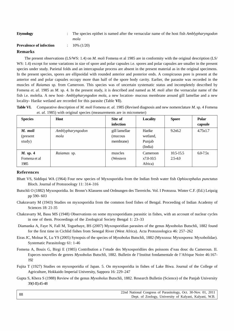
22nd National Congress of Parasitology, Oct. 30-Nov. 01, 2011Dept. of Zoology, University of Kalyani, Kalyani, W.B.
88
Etymology : The species epithet is named after the vernacular name of the host fish Amblypharyngodonmola
Prevalence of infection : 10% (1/20)
Remarks
The present observations (LS/WS: 1.4) on M. moli Fomena et al 1985 are in conformity with the original description (LS/WS: 1.4) except for some variations in size of spore and polar capsules i.e. spores and polar capsules are smaller in the presentspecies under study. Parietal folds and an intercapsular process are absent in the present material as in the original specimens.In the present species, spores are ellipsoidal with rounded anterior and posterior ends. A conspicuous pore is present at theanterior end and polar capsules occupy more than half of the spore body cavity. Earlier, the parasite was recorded in themuscles of Raiamas sp. from Cameroon. This species was of uncertain systematic status and incompletely described byFomena et. al. 1985 as M. sp. 4. In the present study, it is described and named as M. moli after the vernacular name of thefish i.e. molelia. A new host- Amblypharyngodon mola, a new location- mucous membrane around gill lamellae and a newlocality- Harike wetland are recorded for this parasite (Table VI).
Table VI. Comparative description of M. moli Fomena et. al. 1985 (Revised diagnosis and new nomenclature M. sp. 4 Fomenaet. al. 1985) with original species (measurements are in micrometer)
Species Host Site of Locality Spore Polarinfection capsule
M. moli Amblypharyngodon gill lamellae Harike 9.2x6.2 4.75x1.7(present mola (mucous wetland,study) membrane) Punjab
(India)
M. sp. 4 Raiamas sp. muscles Cameroon 10.5-15.5 6.0-7.5xFomena et al (Western x7.0-10.5 2.5-4.01985 Africa)
References
Bhatt VS, Siddiqui WA (1964) Four new species of Myxosporidia from the Indian fresh water fish Ophiocephalus punctatusBloch. Journal of Protozoology 11: 314–316
Butschli O (1882) Myxosporidia. In: Bronn’s Klassens und Ordnungen des Tierreichs. Vol. I Protozoa. Winter C.F. (Ed.) Leipzigpp 590- 603
Chakravarty M (1943) Studies on myxosporidia from the common food fishes of Bengal. Proceeding of Indian Academy ofSciences 18: 21-35
Chakravarty M, Basu MS (1948) Observations on some myxosporidians parasitic in fishes, with an account of nuclear cyclesin one of them. Proceedings of the Zoological Society Bengal 1: 23–33
Diamanka A, Faye N, Fall M, Toguebaye, BS (2007) Myxosporidian parasites of the genus Myxobolus Butschli, 1882 foundfor the first time in Cichlid fishes from Senegal River (West Africa). Acta Protozoologica 46: 257–262
Eiras JC, Molnar K, Lu YS (2005) Synopsis of the species of Myxobolus Butschli, 1882 (Myxozoa: Myxosporea: Myxobolidae).Systematic Parasitology 61: 1-46
Fomena A, Bouix G, Birgi E (1985) Contribution a l’etude des Myxosporidiies des poissons d’eau douc du Cameroun. II.Especes nouvelles de genres Myxobolus Butschli, 1882. Bulletin de l’Institut fondamentale de l’Afrique Noire 46:167-192
Fujita T (1927) Studies on myxosporidia of Japan. 5. On myxosporidia in fishes of Lake Biwa. Journal of the College ofAgriculture, Hokkaido Imperial University, Sappora 16: 229–247
Gupta S, Khera S (1988) Review of the genus Myxobolus Butschli, 1882. Research Bulletin (Science) of the Panjab University39(I-II):45-48

22nd National Congress of Parasitology, Oct. 30-Nov. 01, 2011Dept. of Zoology, University of Kalyani, Kalyani, W.B.
89
Haldar DP, Das MK, Sharma BK (1983) Studies on Protozoan Parasites from Fishes. Four New Species of the Genera HenneguyaThelohan, 1892, Thelohanellus Kudo, 1933 and Myxobolus Butschli, 1892. Archive fur Protistenkunde 127: 283-296
Hemananda T, Mohilal N, Bandyopadhyay PK, Mitra, AK (2009) Two new myxosporidia (Myxozoa: Myxosporea) of the genusMyxobolus Butschli, 1882 from cornea of Clarias batrachus (Linnaeus, 1758) caught from a fish farm in India. North-Westeren Journal of Zoology 5(1):165-169
Jayasri M, Parvateesam M, Mathur PN (1981) Myxosoma mathurii n. sp. (Protozoa: Myxosporidia) parasitic on Puntius sarana(Ham.). Current Scince 50: 16
Kalavati C, Nandi NC (2007) Handbook of Myxosporidean parasites of Indian fishes. ZSI, India, Kolkata, pp 293
Kaur H, Singh (2009) A new myxosporean species, Myxobolus eirasi sp. nov. and a known species M. venkateshi Seenappaand Manohar, 1981 from the Indian major carp fish Cirrhina mrigala (Ham.). Protistology 6 (2): 126–130
Kaur H, Singh, R (2010a) A new myxosporean species Myxobolus sclerii sp. nov. and one known species M. stomum Ali etal. 2003 from two Indian major carp fishes. Journal of Parasitic Diseases DOI 10.1007/s12639-010-0010-7
Kaur H, Singh, R (2010b) One new myxosporidian species, Myxobolus slendrii sp. nov., and one known species, M.punjabensis Gupta and Khera, 1989, infecting freshwater fishes in wetlands of Punjab, India. Parasitology Research 106(5): 1043-1047.
Kaur H, Singh, R (2011a) Myxobolus harikensis sp. nov. (Myxozoa: Myxobolidae) infecting fins of Cirrhina mrigala (Ham.)-an Indian major carp in Harike Wetland, Punjab (India). Parasitology Research (In press)
Kaur H, Singh R (2011b) Two new species of Myxobolus (Myxozoa: Myxosporea: Bivalvulida) infecting an Indian major carpin Ropar and Kanjali wetlands (Punjab). Journal of Parasitic Diseases DOI 10.1007/s12639-011-0033-8
Kaur H, Singh R (2011c) Two new species of Myxobolus (Myxozoa: Myxosporea: Bivalvulida) infecting an Indian major carpin Ropar and Kanjali wetlands (Punjab). Journal of Parasitic Diseases DOI 10.1007/s12639-011-0033-8
Kaur H, Singh R (2011d) Two new species of Myxobolus (Myxozoa:Myxosporea: Bivalvulida) from freshwater fishes of Punjabwetlands (India). Journal of Parasitic Disease DOI 10.1007/s12639-011-0024-9
Kostoingue B, Toguebaye BS (1994) Le genere Myxobolus (Myxozoa: Myxosporea) chez les poissons d’eau douce du Tchadavec la description de trios nouvelles especes. Bulletin de l’Institut fondamentale de l’Afrique Noire 47: 63–71
Kudo R (1919) Studies on Myxosporidia. A synopsis of genera and species of Myxosporidia. Illinois Biological Monographs5:1-265
Landsberg JH (1985) Myxosporean infections in cultured tilapias in Israel. Journal of Protozoology 32: 194–201
Li L, Desser SS (1985) The protozoan parasites of fish from two lakes in Algonquin Park, Ontario. Canadian Journal of Zoology63: 1846–1858
Lom J, Arthur JR (1989) A guideline for the preparation of species descriptions in Myxosporea. Journal of Fish Diseases 12:151-156
Lom J, Dykova I (2006) Myxozoan genera: Definition and notes on taxonomy, life- cycle terminology and pathogenic species.Folia Parasitologica 53: 1-36
Mukhopadhyay D, Haldar DP (1998) Spore variations in a piscine myxozoan parasite (Myxozoa: Myxosporea) from the juvenilemajor carp, Catla catla. Transactions of Zoological Society of India 2(1): 46-52
Sakiti N, Blanc E, Marques, A, Bouix G (1991) Myxosporidies (Myxozoa: Myxosporea) du genre Myxobolus Butschli, 1882parasites de poisons cichlidae du lae Nokoue au Benimm (Afrique de l’ Ouest). Journal of African Zoology 105: 173-186
Sarkar NK (1985) Some coelozoic Myxosporida (Myxozoa: Myxosporea) from a freshwater water teleost fish of River Padma.Acta Protozoologica 24(1):47-53
Sarkar NK (1986) Some Myxosporidia (Myxozoa: Myxosporea) of Anabantid fishes of West Bengal, India. Acta Protozoologica24 (2): 175-180

22nd National Congress of Parasitology, Oct. 30-Nov. 01, 2011Dept. of Zoology, University of Kalyani, Kalyani, W.B.
90
Sarkar NK (2009) Thelohanelloid bengalensis gen. and sp. nov. (Myxosporea: Thelohanellidae) from the gall bladder of marinecatfish of the Bay of Bengal, India. Uttar Pradesh Journal of Zoology 29 (2): 251-254
Seenappa D, Manohar L (1980) Two new species of Myxobolus (Myxosporidia: Protozoa) parasitic on Cirrhina mrigala(Hamilton) and Puntius curmuca (Hamilton). Current Science 49: 204-206
Seenappa D, Manohar L (1981) Five new species of Myxobolus (Myxosporea: Protozoa), parasitic in Cirrhina mrigala(Hamilton) and Labeo rohita (Hamilton), with a note on a new host record for M. curmucae Seenappa and Manohar,1980. Journal of Protozoology 28: 358-360
Shulman SS (1962) Myxosporidia. In: Key to parasites of freshwater fish of USSR. Pavlovskii. EN (Ed.) Publ. House of theAcademy of Sciences of the USSR, Moscow-Leningrad, pp 47–130
Wegener G (1910) Die Ektoparasiten der Fische Ostpreussens. Inaugural Dissertation, Konigsberg pp 90

22nd National Congress of Parasitology, Oct. 30-Nov. 01, 2011Dept. of Zoology, University of Kalyani, Kalyani, W.B.
91
Parasitic infestation among the adolescent girls of Bangladesh
Hasina Banu1, Hamida Khanum1 and Md. Anwar Hossain2
1Department of Zoology, University of Dhaka, Bangladesh2Institute of Nutrition and Food Science, University of Dhaka, Bangladesh
Abstract : The present study was conducted in rural, urban and slum areas in and around Dhaka city, Bangladesh to investigate theprevalence of parasitic infestation among the adolescent girls (aged 10-19 years). Faecal samples were collected from 1570 adolescent girls.About one third (33.50%) of the adolescent girls were found to be infected with one or more protozoan (Entamoeba histolytica, Giardialamblia) and helminth (Ascaris lumbricoides, Trichuris trichura, Strongyloieds stercoralis, hookworm) parasite infections. Among alldetected parasites, Ascaris lumbricoides (14.20%) overwhelmingly infected the adolescent girls. The most common parasite combinationinfecting adolescent girls were found to be Ascaris lumbricoides + Trichuris trichura (1.84%) and Ascaris lumbricoides + hookworm (1.27%).The prevalence was higher in rural (49.62%) area compared to the slum (33.43%) and urban (18.22%) areas. The girls aged 12-13 yearswere more vulnerable for the parasitic infestation (48.75%, p<0.08). The findings of the parasitic infestation among the adolescent girlsin this study indicate important public health problem. Measures to improve socioeconomic conditions, sanitary practice and rate of literacyin the slum and rural areas are recommended intervention approaches to control the extent of parasitic infestation.
Keywords : Adolescent girls, parasitic infestation, rural, slum, urban.
IntroductionWHO (1994) estimates suggested that approximately 1.4 billion, 1.2 billion and 1 billion persons are currently infected with
various species of intestinal helminthes: A. lumbricoides, hookworm (Ancylostoma duodenale/ Necator americanus) and T.trichura respectively. The epidemiology and population biology of hookworms has been studied extensively and recently amathematical models have been proposed to explain transmission and identify phases in the host-parasite relationship whereavailable control measures are most likely to be effective (WHO, 1994).
Bangladesh is mostly a plain land and embedded with rivers and canals. The soil humidity and temperature contributesa lot towards parasitic infection. Several studies showed that intestinal parasitic infections are present all the time everywherein this country (Nuruzzaman and Huda, 1974; Muttalib et al 1975; Shakur and Ehsan, 1993). Helminthiasis is a most commonhealth problem in the tropics because the environment is favorable for the transmission of the helminthiasis in the community.In Bangladesh, among the helminth and protozoa parasites (A. lumbricoides, A. duodenale, T. trichura, Enterobius vermicularisand E. histolytica, G. lamblia) are common (Kuntz, 1960; Banu et al 2003; Khanum et al 2010).
Adolescent girls are generally more infected than their younger peers with intestinal parasites, like A. lumbricoides, T.trichiura, hookworm, E. histolytica, G. lamblia etc. In Bangladesh it was found that 58-90% of 4-13 years old childrenharboring A. lumbricoides and 60% of 1-40 years old people suffer from E. vermicularis infection (D’Silva et al 2003, Uddinet al 2005).
Adolescents are important segment of the whole population. It is the adolescent girls of today who will usher in thegeneration of the 21st century. There are very few studies on the prevalence of parasite among the adolescents’ girls incomparison to other groups like women and children in the world and it is the case in Bangladesh too. The protozoan andhelminth parasites are the oldest pathogens and remain an important part of infectious diseases among the world’s poor whoare the most susceptible population. Adolescent girls are particularly susceptible because of their more involvement with thedomestic work. So the present study was undertaken to investigate the extent and the pattern of parasitic infestation in relationto age groups of the adolescent girls from rural, urban and slum settings of Bangladesh.
Materials and methodsThe present investigation was a cross sectional study with a sample size of 1570 adolescent girls (aged 10-19 years)
conducted during the period of June 2006 to May 2009. The six study areas are located in Dhaka district, Bangladesh
*Corresponding author : E-mail: [email protected]

22nd National Congress of Parasitology, Oct. 30-Nov. 01, 2011Dept. of Zoology, University of Kalyani, Kalyani, W.B.
92
representing rural (Kamrangirchar and Zinjira), urban (Savar and Lalbag) and slum areas (Mirpur and Mohammadpur). Faecalsamples were collected from adolescent girls and carried out to the laboratory. The most recommended Formol Ether Concen-tration Method (Cheesbrough, 1987) was applied which was suitable for concentration of parasite eggs, cysts and larvae infresh or preserved faeces. After concentration, the sediment was mixed up by using the stick. It was transferred to the slidesby dropper and covered with cover glasses. The prepared slides were examined microscopically and the presence of parasiteeggs, cysts and larvae were detected. The findings were confirmed by the help of experts and renowned books (Neva andBrown, 1994; Chatterjee, 2004).
ResultsDuring the present investigation, total of 1570 stool samples were collected for the identification of different parasite
species. Out of 1570 samples, 526 (33.50%) girls were found to be infected with one or more intestinal parasites.
The highest percentage of parasitic infection (49.62%) was found in rural Kamrangirchar and the lowest was in urbanSavar (18.22%), comparatively higher rates of infection identified in Slum girls of Mohammadpur (33.43%) and Mirpur (34.41%)(Table I).
Table I. Prevalence of infestation of intestinal parasites among the adolescent girls in different study areas.
Study areas Total no. of stool No. of parasite Prevalencesamples of the positive cases (%)adolescent girls
examined
Kamrangirchar 270 134 49.62
Zinjira 230 105 45.65
Lalbag 310 65 20.96
Savar 225 41 18.22
Mohammadpur 320 107 33.43
Mirpur 215 74 34.41
Total 1570 526 33.50
Ruralareas
Urbanareas
Slumareas
Age of the adolescent girls observed as an important factor in parasitic infection. The highest prevalence (48.74%) ofintestinal parasite was found at the age group of 12-13 years and the lowest (14.09%) at 18-19 years among all five age groups(Fig.1). So, the prevalence was negatively correlated with age groups (r = -0.83, p<0.08) which implied that as the age increased,rates of infestation tends to decrease (Fig.1).
Two protozoan parasites (E. histolytica and G. lamblia) and four helminthes parasites (A. lumbricoides, T. trichura,Hookworm and S. stercoralis,) were identified during the present investigation (Table II).
In case of protozoan single parasite species, prevalence of E. histolytica (3.62%) was comparatively higher than G.lamblia (2.67%). Regarding helminth single parasite species, A. lumbricoides was found to be overwhelmingly dominantparasites in all age groups and highest prevalence 19.65% was found in 12-13 years age category. The next prevalent helminthwas T. trichura followed by hookworm and S. stercoralis. Importantly, all of the helminth parasites infected particular age groupof 12-13 years in higher percentage. Chi-square (χ2) test showed that age group was not significantly associated (p>0.05) withinfected by protozoan parasites on the other hand, infected by helminth parasites was found to be significantly associated(p<0.000) with age groups (Table II).
Age of the adolescent girls, appears to be an important variable for the infestation of intestinal parasites; nevertheless,the infestation of double parasite (considered presence of two intestinal parasites at a time in a single host) was not highlyprevalent in this study. Considering all of the five age groups, adolescent girls of 12-13 years were more infected (7.21%) ascompared with other age groups and least infection (1.34%) was found in 18-19 years (Table III).
Among all types of parasite pairs, A. lumbricoides and T. trichura parasite combination (2.48%) mostly infected the 12-13 years aged adolescent girls. Another parasite combination (2.33%), A. lumbricoides and Hookworm was found in the same

22nd National Congress of Parasitology, Oct. 30-Nov. 01, 2011Dept. of Zoology, University of Kalyani, Kalyani, W.B.
93
3 5 .0 1
4 8 .7 5
2 9 .9 6
1 9 .2 3
1 4 .0 9
1 0 - 1 1 1 2 - 1 3 1 4 - 1 5 1 6 - 1 7 1 8 - 1 9
A ge gro up s (In ye a r)
0
1 0
2 0
3 0
4 0
5 0
6 0
r = - 0 .8 3 , p < 0 .0 8
Fig. 1. Prevalence of intestinal parasites in different age groups among the adolescent girls.
Pre
vale
nce
(%)
Table II. Prevalence of single infection of protozoan and helminth parasites in different age groups of the adolescent girls.
Infected by Protozoan Infected by Helminth
Parasites Parasites
Infected Infected Total Infected Infected Infected Infected Totalby EH by GL by AL by TT by HW by SS
n n n n n n n n
P P P P P P P P
(%) (%) (%) (%) (%) (%) (%) (%)
10-11 514 19 17 36 80 24 7 0 111
3.69 3.30 7.00 15.56 4.66 1.36 0.00 21.59
12-13 402 21 12 33 79 23 16 4 122
5.22 2.98 8.20 19.65 5.72 3.98 0.99 30.34
14-15 297 9 7 16 36 10 6 3 55
3.03 2.35 5.38 12.12 3.36 2.02 1.01 18.51
16-17 208 4 5 9 18 5 3 0 26
1.92 2.40 4.32 8.65 2.40 1.44 0.00 12.5
18-19 149 4 1 5 10 4 0 0 14
2.68 0.67 3.35 6.71 2.68 0.00 0.00 9.39
Total 1570 57 42 99 223 66 32 7 328
3.63 2.67 6.30 14.20 4.20 2.03 0.44 20.89
AL = Ascaris lumbricoides, TT = Trichuris trichura, HW = Hookworm, SS = Strongyloides stercoralis, EH = Entamoebahistolytica, GL = Giardia lamblia, P = Prevalence, n = number
For protozoan parasites : For helminth parasites :
Chi-square (χ2) = 6.89, p<0.142 Chi-square (χ2) = 43.69, p<0.000
Tota
l no.
of s
tool
sam
ples
of t
he a
dole
scen
t gi
rls
exam
ined
Age
gro
ups
(In
year
)

22nd National Congress of Parasitology, Oct. 30-Nov. 01, 2011Dept. of Zoology, University of Kalyani, Kalyani, W.B.
94
age category. So it was evident from this study that girls aged 12-13 years were identified as a remakably risk group for parasiticinfestation (Table III).
E. histolytica + A. lumbricoides+ T. trichura triplet parasite combination (considered presence of three parasites at a timein a single host) was recorded as highest (0.70%) among all triplet categories. Similar prevalences (0.38%) were found in twoother triplet combinations. The combination of A. lumbricoides + T. trichura + S. stercoralis was identified as the leastprevalent (0.12%) combination (Fig. 2).
Table III. Prevalence of double infection of protozoan and helminth parasites in different age groups of the adolescent girls.
n n n n n n n n n n
P P P P P P P P P P
(%) (%) (%) (%) (%) (%) (%) (%) (%) (%)
10-11 514 1 1 1 1 2 9 7 0 3 25
0.19 0.19 0.19 0.19 0.38 1.75 1.36 0.00 0.58 4.86
12-13 402 3 2 3 0 0 10 9 1 1 29
0.74 0.49 0.74 0.00 0.00 2.48 2.23 0.24 0.24 7.21
14-15 297 0 1 1 2 0 6 2 2 0 14
0.00 0.33 0.33 0.67 0.00 2.02 0.67 0.67 0.00 4.71
16-17 208 0 0 0 0 0 3 1 0 0 4
0.00 0.00 0.00 0.00 0.00 1.44 0.48 0.00 0.00 1.92
18-19 149 0 0 0 0 0 1 1 0 0 2
0.00 0.00 0.00 0.00 0.00 0.67 0.67 0.00 0.00 1.34
Total 1570 4 4 5 3 2 29 20 3 4 74
0.25 0.25 0.31 0.19 0.12 1.84 1.27 0.19 0.25 4.71
AL = Ascaris lumbricoides, TT = Trichuris trichura, HW = Hookworm, SS = Strongyloides stercoralis, EH = Entamoebahistolytica, GL = Giardia lamblia, P = Prevalence, n = Number.
Age
gro
ups
(In
year
)
No.
of
stoo
l sa
mpl
es o
fth
e of
the
adol
esce
nt g
irls
exam
ined
Infe
cted
by
EH
+ G
L
Infe
cted
by
AL
+ G
L
Infe
cted
by
AL
+ E
H
Infe
cted
by
TT
+ G
L
Infe
cted
by
TT
+ E
H
Infe
cted
by
AL
+ T
T
Infe
cted
by
AL
+ H
W
Infe
cted
by
AL
+ S
S
Ove
rall
Prev
alen
ce(%
)
Infe
cted
by
TT
+ H
W
AL = Ascaris lumbricoides, TT = Trichuris trichura, HW = Hookworm, SS = Strongyloides stercoralis, EH = Entamoebahistolytica, GL = Giardia lamblia

22nd National Congress of Parasitology, Oct. 30-Nov. 01, 2011Dept. of Zoology, University of Kalyani, Kalyani, W.B.
95
DiscussionOut of 1570 samples, the overall parasitic infestation was recorded as 33.50% with the highest prevalence (49.62%) in rural
area (Kamrangirchar) and the lowest (18.22%) in urban area (Savar). The prevalence in slum areas was more than 30% whichwas comparatively lower than rural but higher than the urban area. Rahman (2009) reported overall infection as 33% in hisstudy among the people of Chittagang and Chittagang Hill Tracts which was almost similar with the present study. Hyder etal (1998) reported gross prevalence (33.3%) of parasites among the adolescents and adult population in different rural areasof Mymenshing district of Bangladesh, which was also closely related with our findings. Uddin et al (2005) studied theprevalence of parasitic infestation among the adolescent girls in two rural areas, Kutombopur and Savar and reported overallprevalence as 71.01% with 62.5% in Kutombopur, which was much higher than the rural area of the present study. Rao et al(2003) reported overall prevalence 57% among tribal adolescent girls of Nepal. The findings were higher than the present workdue to differences in study population study and prevailing socioeconomic condition.
Among all age groups the highest prevalence (48.74%) was observed among the adolescent girls in the present studywho were in 12-13 years of age category. In another study by Uddin et al (2005), highest prevalence (87.50%) was also recordedamong female adolescents aged 12-14 years. So it was evident that adolescents of this particular age group were morevulnerable for parasitic infestation than other age groups.
Regarding single parasitic infestation, A. lumbricoides overwhelmingly dominated the others protozoans and helminthes.Out of 1570 samples, 223 (14.20%) was found to be infected with A. lumbricoides. The next prevalence of single parasiticinfection was T. trichura (4.20%) followed by E. histolytica (3.63%), G. lamblia (2.67%), Hookworm (2.03%) and S. stercoralis(0.44%). Considering the age groups, adolescents’ girls of the 12-13 years age category was found to be more susceptiblefor parasitic infestation. About 20% of the girls of this age group were infected by A. lumbricoides which was much higherthan the average 14.2% of all age groups for this particular parasite.
Prevalence of single parasitic infestation reported by Sharma et al (2004) in school children (4-19 years) in the northeasternpart of Kathmandu valley, Nepal were T. trichura (43.6%), A. lumbricoides (13.8%) and Khanum et al (2001) observed 20.39%of A. lumbricoides and 15.30% T. trichura among the 2-16 years of subjects from lower income families of Dhaka city. Theprevalence of A. lumbricoides (14.20%) in the present study was almost similar with the findings of Sharma et al (2004) butit was different from the findings of Khanum et al (2001) and the prevalence of T. trichura (4.20%) was much lower than theirstudies. A Brazilian study by Ferreira (2001) reported overall prevalence of T. trichura as 5% which was almost similar (4.20%)with the present study. Osazuwa et al (2011) determined the overall prevalence of intestinal helminths in children andadolescents of three rural communities in Nigeria were A. lumbricoides (75.6%), hookworm (16.19%) and T. trichiura (7.3%)which was quite different from the findings of the present work, this difference might be due to the study population, location,prevailing environment and socioeconomic status.
Ahmed and Talukder (2002) found the prevalence (69%) of A. lumbricoides, (39%) of T. trichura and Hookworm 8% amongthe school children (aged 9-16 years) from rural and urban areas of Bangladesh and further proved that the A. lumbricoidesas the dominant parasite but the prevalence of this parasite was much higher than the present investigation and this differencemight be due to the study population, location, prevailing environment and socioeconomic status.
According to WHO (1987), as much as 60% of the world’s population were infected with gut parasites. Hamimah et al(1982) found that prevalence of intestinal protozoas like E. histolytica was 2.3% and of G. lamblia was 2.6% in a study inKualalumpur, Malaysia. These values were almost similar with our study. On the other hand, in another work in Malaysia, Azaet al (2003) recorded higher prevalence of E. histolytica 21% and of G. lamblia 8.6%. Azian et al (2007) in a study on aborigincommunity in Pehang, Malysia, noted that the prevalence of G. lamblia, and E. histolytica were 29.2% and 26.2% respectively.
The extent of double infection was lower than that of single infection in the present study. The parasite combination (A.lumbricoides and T. trichura) was 1.84% and another combination (A. lumbricoides and hookworm) was 1.27% in the observedsamples. Opara et al (2007) were found 1.6% prevalence of A. lumbricoides and T. trichura combination among the schoolchildren which was closely related with the present investigation. But their other finding, 3.3% of A. lumbricoides andhookworm combination was higher than that of the findings of our study. Kaur and Sween (2007) reported 17.64% of A.lumbricoides and hookworm combination which was much higher than that of the findings of the present study.
Rahman (2009) reported 0.28% of E. histolytica + A. lumbricoides+ T. trichura and 0.03% of A. lumbricoides + T. trichura+ S. stercoralis triplet parasite combinations only from the female subjects which were much lower prevalent from our study

22nd National Congress of Parasitology, Oct. 30-Nov. 01, 2011Dept. of Zoology, University of Kalyani, Kalyani, W.B.
96
findings (0.70% and 0.12% respectively). Opara et al (2007) stated the prevalence (3.3%) of A. lumbricoides + T. trichura +hookworm triple parasite combination among the school children while it was comparatively higher than our findings (0.38%).The difference may be due to the study population and environmental obligation.
The higher prevalence of intestinal parasites among the adolescent girls in Bangladesh may be due to its climate,temperature and environment, all of which favour the growth of different parasites. The adolescent girls of the rural and slumareas were more vulnerable to parasitic infestation than urban adolescent girls due to the differences in hygienic conditions,rate of literacy and prevailing socioeconomic conditions.
AcknowledgementWe acknowledge to the authority of the University of Dhaka, Bangladesh and to the Ministry of National Science and
Information & Communication Technology (NSICT), Bangladesh for providing the financial support to continue this researchwork as a part of Ph D dissertation.
ReferencesAhmed S, Talukder KK (2002) Knowledge, attitude and practice of Environmental sanitation and personal hygiene of school
children in rural Bangladesh. Environmental threat to the health of children: Hazards and vulnerability, Bangkok,Thailand. 3-7 March.
Aza N, Ashley S, Albert J (2003) Parasitic infection in human communities living on the fringes of the Crocker Range ParkSabah, Malaysia. ASEAN Review of Biodiversity and Environmental Conservation (ARBEC).http://www.arbec.com.my/pdf/ art11/janmar03.pdf.
Azian NMY, San YM, Gan CC, Yusri MY, Nurulssyamzawaty Y, Zuhaizam AH, Maslawati, MN, Norparina I, Vythilingam I (2007)Prevalence of intestinal protozoa in an aborigine community in Pahang, Malaysia. Tropical Biomedicine 24: 55-62.
Banu H, D’Silva J, Islam N (2003) Epidemiological factors and pinworm infection in children. Bangladesh J Zool 31: 243-246.
Chatterjee KD (2004) Parasitology (Protozoology and Helminthology) in relation to clinical medicine. Chatterjee MedicalPublisher, Calcutta. pp 238.
Cheesbrough M (1987) Medical Laboratory Manual for Tropical Countries. Blackworth Co Publishers. Pp 570.
D’Silva J, Banu H, Islam. N. 2003. Pinworm infection in children and their personal habits. Dhaka Univ J Biol Sci 12: 193-198.
Ferreira H (2001) The risk of Ascaris lumbricoides infection in children as an environmental health indicator to guide preventiveactivities in Caparao and Alto-caparao, Brazil WHO Bull 80: 40-46.
Hamimah I, Zahedi M, Ainiyah AJ (1982) The prevalence of intestinal parasites among children at the General Hospitals, KualaLumpur, Malaysia. Med J Malaysia 37: 373-377.
Hyder SMZ, Chowdhury SA, Chowdhury AMR (1998) Prevalence of anaemia and intestinal parasites in a rural community ofBangladesh. Research and Evaluation Division BRAC. Res Monogr Ser no. 12.
Kaur H, Sween (2007) Intestinal parasitic infections in children of Rajpura town, Patiala J Parasitic Dis 31: 56-60.
Khanum H, Islam MN, Nahar KN (2001) Intestinal nematode infection among children of lower income group employers inDhaka city. Bangladesh J Zool 27: 177-183.
Khanum H, Islam MR, Parvin S (2010) Occurrence of eggs and larvae of gastrointestinal nematodes in nails of street inhabitantsin Dhaka city. J Life Earth Sci 5:75-79.
Kuntz RE (1960) Intestinal protozoa and helminths in school going children of Dhaka city. Am J Trop Med Hyg 9: 168-174.
Muttalib MA, Islam N, Ghani JA, Khan K, Azizullah A, Islam B (1975) Intestinal parasites in the University of Dacca Student.J Trans Med Hyg78: 10-11.
Neva FA, Brown HW (1994) Basic Clinical Parasitology. Prentice Hall International Inc. USA. Pp 356.
Nuruzzaman M, Huda Q (1974) Parasitic infestation in gastrointestinal tract disorders. Bangladesh Med J 2: 119-123.
Opara FN, Udoye AA, Okere PU, Osuala FOU, Iwuala MOE (2007) The prevalence of intestinal helminth infections in primaryschool children in Owerri municipality, Imo state, Nigeria. J Parasitic Dis 31: 44-48.
Osazuwa F, Ayo OM, Imade P (2011) A significant association between intestinal helminth infection and anaemia burden inchildren in rural communities of Edo state, Nigeria. North Am J Med Sci 3: 30-34.

22nd National Congress of Parasitology, Oct. 30-Nov. 01, 2011Dept. of Zoology, University of Kalyani, Kalyani, W.B.
97
Rahman MM (2009) Prevalence of intestinal parasites and their effects on cross section (economy and area based) of peopleresiding in Chittagong and Chittagong Hill Tracts. M. Phil. Dissertation, University of Chittagong, Bangladesh pp 145.
Rao VG, Aggrawal MC, Yadav R, Das SK, Sahare LK, Bondley MK, Minocha RK (2003) Intestinal parasitic infections, anaemiaand undernutrition among Tribal adolescent of Modhya Pradesh. Indian J Community 28: 1-5.
Shakur MS, Ehsan MA (1993) Intestinal parasites: a frequent association and contributing factor of loos motion in malnourishedchildren. Bangladesh J Child Health 17: 10-13.
Sharma A, Prasad K, Rao KV (2004) Identification of an appropriate strategy of control anaemia in adolescent girls in poorcommunities. Ind Ped 37: 261-267.
Uddin MH, Rahman MM, Khanum H (2005) Hemoglobin level among adolescent girls and it’s relation to intestinal parasites.Bangladesh J Zool 33: 183-187.
World Health Organization expert committee, 1987. Public health significance of intestinal parasitic infections. WHO Bull 65:575-588.
World Health Organization, 1994. Report of the WHO informal consultation on hookworm infection and anemia in girls andwomen. WHO/ CTD/ SIP/ 96. 1.

22nd National Congress of Parasitology, Oct. 30-Nov. 01, 2011Dept. of Zoology, University of Kalyani, Kalyani, W.B.
98
A Minireview : matrix metalloproteases-an invasive armof nematode parasites
Ipsit Chakrabarti
Postgraduate Department of Zoology, Dinabandhu Andrews College, Garia, Kolkata-700084, West Bengal, India
Abstract : Matrix metalloproteases (MMPs) are a family of Zinc-dependent endopeptidases responsible for degrading extracellular matrix(ECM) components and has been responsible for diverse set of developmental processes. However, expressions of many MMPs have beenassociated with several pathological conditions. It has been established that the MMPs are conserved throughout the animal kingdom andstudies of invertebrate MMPs have demonstrated that primarily they are involved in various developing functions in hydra, Drosophila,sea urchin and nematodes. The synthesis of these proteolytic enzymes and their release as excretory and secretory (ES) products havebeen reported in various parasitic nematodes. Host invasion and tissue migration of several nematodes have been linked to the expressionand release of parasite-derived proteases. Studies with enzyme inhibitors suggest that the enzyme may be a metalloprotease. Moreover,substrate impregnated zymographic analysis of extracts and ES products of different nematode parasites have revealed the multiple enzymeactivities of MMPs with various molecular weights. Molecular cloning of a gene encoding matrix metalloproteinase-like protein fromGnathostoma spinigerum has also been done. This MMP-like protein of G. spinigerum possesses the catalytic domain, but lacks thepropeptide and hemopexin-like domains found in other MMPs. More research on MMP degradome in nematode parasites can providevaluable information for intense evaluation of pathogenesis caused by these parasites.
Key words : Endopeptidases, Matrix metalloprotease, MMP, Nematode parasites.
IntroductionIt has been established that various biological processes including cell migration, wound healing, tissue differentiation,
immune system have been accomplished by morphological changes in extracellular matrix (Stocker et al 1995). Matrixmetalloproteases (MMPs) are the major group of enzymes with ability for degrading extra cellular matrix proteins and thisprocess of ECM remodelling is responsible for all these necessary processes (Shapiro,1998). The MMPs constitute a part oflarge family of zinc-dependent endopeptidases. These proteins may be found from bacteria to advanced forms of life evenin several viruses (Ugalde et al 2010) and have been considered as a group of highly conserved proteins throughout the courseof evolution. These enzymes seem to play various functional roles and touch many aspects of physiological and pathologicalprocesses not only in vertebrates but also in invertebrates (Masova et al 1998). The parasitic nematodes are the major groupof invertebrates, which dwell in different organs of vertebrate hosts (Anderson,1992). Many of these parasites to completetheir life cycle have to migrate or invade through various organs of their hosts. During this journey, expression and releaseof MMPs by various parasitic nematodes have been associated with the pathology resulting from histolysis (Hotez et al 1990;McKerrow et al 1990; Lai et al 2005; Williamson et al 2006). Genetic analysis and cDNA cloning of MMPs have been evaluatedin few parasitic nematodes (Newport et al 1988; Uparanukraw et al 2001; Salter et al 2002).
MMPs and nematode parasitesMetalloproteases comprise a heterogeneous group of proteolytic enzymes whose main characteristic is the utilization of
a metal ion to polarize a water molecule and perform hydrolytic reactions. MMPs are a major group of zinc-dependentendopepdidases with a ability to cleave one or more extracellular matrix constituents as well as nonmatrix proteins (Hotez etal 1990; Williamson et al 2006). These enzymes include a wide range of proteases in many organisms and play essential rolesin multiple biological processes. By virtue of these proteolytic property proteases regulate a variety of cellular processes suchas cell proliferation, cell-cycle progression, tissue differentiation and migration, apoptosis, senescence, DNA replication andautophagy. In metazoans, proteolytic activities are also involved in the maintenance of tissue homeostasis and in the regulationof different physiological processes such as fertilization and fecundation, embryonic development, wound healing, tissueremodeling, immune response and angiogenesis (Ugalde et al 2010). However, expressions of some of the MMPs have beenassociated with different pathological manifestations, which are predominantly associated with inflammation, arthritis, cancer

22nd National Congress of Parasitology, Oct. 30-Nov. 01, 2011Dept. of Zoology, University of Kalyani, Kalyani, W.B.
99
metastasis and parasitic invasion through host’s tissues (McKerrow et al 1990; Hawdon et al 1995; Ugalde et al 2010 ). Lifecycles of many nematode parasites consist of different migratory as well as invasive larval stages in host environment, evensometimes young and adult may be invasive in their nature (McKerrow et al 1990; Lai et al 2005). Host invasion and tissuemigration of several nematodes have been linked to the expression and release of parasite-derived proteases. In nematodes,MMPs are the proteases which are thought to play an important and essential role in these migratory and invasive phenomena(McKerrow et al 1990). According to their substrate specificity MMPs can be categorized as collagenases, gelatinases,elastases, Stromelysins and membrane-type MMPs. Nematode MMPs generally include collagenases, gelatinases and elastases(Petralanda et al 1986; McKerrow et al 1990; Healer et al 1991).
Research on helminth derived MMPs got momentum in eighties. Most of these works evaluated MMPs from excretory-secretory (ES) products as well as the extracts of parasites (Lai et al 2005). Initial period of MMP research in nematodesincluded detection and measurement of activity in different life stages of nematode parasites.
Petralanda et al.1986 determined collagenilytic activity in extracts of adult worms, in living microfilariae of Onchocercavolvulus and in live infective larvae and adult female worms of Brugi malayi. In culture, infective larvae of B malayi alsosecreted large amounts of collagenase. Studies with enzyme inhibitors, antigen-antibody reactions and immunoprecipitationindicated that this protease might be a metalloproptease. In another experiment scientists (Robertson et al.1989) did characterizethis metalloprotease in Toxocara canis by using different techniques. Various forms of enzyme have been determined byzymography where they found two bands with molecular weights of 120 and 32 kDs. This group also determined the optimumpH at 9 with less activity at pH 5 and 7. As a potent inhibitor Phenylmethylsulfonyl fluoride was used to characterize themetalloprotease nature of these proteins.L3 Larvae of a nematode parasite Strongyloides stercoralis have an ability to migratethrough tissue at a speed of 10 cm per hour (McKerrow et al 1990). Their studies indicated that this process of migrationwas facilitated by a metalloprotease with elastase activity. This invasive property of elastase has also been determined byinhibition study and author indicated that the parasitic virulence factor may be guided by metalloprotease and could be amolecule for therapeutic purpose. In another study inhibitor as zinc-chelator like 1,10-phenanthroline has been used to provethat proteolytic activity of MMP is zinc-dependent(Hawdon et al 1995). However this study concentrated on the role of MMPin transition from free-living to parasitic mode of life. In 1990, Hotez and colleagues published a work including the study ofmetalloprotese from a human hookworm species Ancylostoma duodenale and a zoonotic species A. caninum and they foundthat these two species could synthesis MMPs of similar moleculat weights( 68 and 38 kDs )and properties that might causetissue degradation, exsheathment and ecdysis(Hotez et al 1990; Richer et al 1992 ).In their study, radiolabelled fibronectin hasbeen degraded by both species of Ancylostoma larvae,which could be corroborated with the activity of MMPs found inStrongyloides stercoralis larvae and Serratia marcescens (Hotez et al 1990). Along with the determination of molecular weightof metalloprotease,found in larval ES product and adult stages of Nippostrongylus brasiliensis Healer et al.1991 alsocharacterized the zymogen form of MMP in larval somatic extract. However, it has been found that uninfected rat intestinaltissue has similar kind of protein with identical molecular weight, suggesting the uptake of this metalloprotease by the parasite.A high pressure liquid chromatography technique has been used to purify a zinc metalloprotease ,isolated from in vitro culturefluids of Trichuris suis adults. (Hill et al 1993). Determination of isoelectric point and immunohistochemistry of MMP has alsobeen done in this experiment.
Analysis of tissue extracts and ES products of Angiostrongylus cantonensis showed the activity of different form ofMMPs (Lai et al 2005). They examined gelatinase activities from extracts of L1, L3, young and adult stages of this parasite.Five gelatinase bands (94,86,66,42 and 30 kDs) were observed in L3 larval stages, and minimum number of bands (94 and 72kDs) were evident in young and adults. All these bands of various stages showed common characteristics with MMP, cysteinand serine proteinase. Zymographic bands of L1 (105,94,42 kDs) and L3((105,66,50, and 30 kDs) ES products were proved tohave proteolytic activity whereas adult and young ES products had no gelatinase activity. Of these bands obtained from larvalstages, 94 and 105 kDs were gelatinase and others were metalloproteinases. In this experiment about 2000 larvae were usedto detect enzyme activity in contrast to another experiments (Lai et al 2004 and Lee et al 2004) when 60 larvae were used toinfect mice and MMP activity was determined. This differentiation led the author to suggest that in later experiments MMPswere secreted by the host not by parasite. Simultaneously, several authors provided information related with the activitiesof MMPs in different helminthes and MMP mediated histolysis of skin and intestinal walls (Tort et al 1999) and degradationof ECM proteins (Petralanda et al.1986). It has been evident that after ingestion, A. cantonensis larvae could invade andpenetrate host stomach or intestinal wall (Alicata,1965). Considering all these experimental evidences, in their publication Lai

22nd National Congress of Parasitology, Oct. 30-Nov. 01, 2011Dept. of Zoology, University of Kalyani, Kalyani, W.B.
100
and group ultimately suggested that MMPs secreted by larvae could be associated with parasite spreading and pathogenesisin host.
In developing countries 740 million people are infected with hookworms (deSilva et al 2003). In most of the caseshookworm infect took place by penetrating host’s skin and after getting inside of host, larvae migrate by invading tissues ofvarious organs(Hotez et al 1990).Proteases have been considered as invasive arm for tissue penetration by parasitic helminthesand many experiments revealed that characterization of protease enzyme activity has been evaluated from larval ,young andadult crude extract as well as excretory and secretory (ES) products of many parasites( knox et al 1990; Gamble et al 1996;Halfner et al 1998 and Lai et al 2005).
Williamson et al. 2006 did an experiment where they found an astacin-like metalloprotease (Ac-MTP-1) activity in ESproduct of Ancylostoma caninum L3 larvae.This Ac-MTP-1 has a sequence similarity with zinc-metalloprotease . L3 larval stageof A caninum can exclusively express Ac-MTP-1and it’s activity in culture medium indicates its role in host tissue invasion.Activity of metalloprotease in this study has been proved both by native and recombinant emzymes and using immunoelctronmicroscopy, site of synthesis in the secretory granules of the glandular esophagus of L3 and its course of transportation tocuticle and followed by secretion from cuticle have been evaluated. More recently an orthologue of Ac-MTP-1 known as Ay-MTP-1 has also been detected in Ancylostoma ceylanicum. This group of scientists suggested a probability of using MTP-1 as the target molecule for developing vaccine to prevent larval migration through tissues.
Similar kind of metalloprotease has been characterized from sheep barber’s pole worm Hemonchus contortus (Halfner etal 1998). However, no recombinant enzymes have been used in this study. Uparanukraw et al 2001 has used the techniqueimmunoscreening with the monoclonal antibody to synthesise a cDNA clone from L3 larvae of Gnathostoma spinigerum. Fromthis clone, they have identified a gene of 732 bp encoding a metalloprotease having 33-39% similarity with MMP ofCaenorhabditis elegans.
From above discussion it has been assumed that parasitic nematodes have the ability to modify or degrade host’sextracellular matrix by secreting metalloproteases. This process of invasive nature assists these parasites to migrate throughdifferent host tissues causing pathogenesis. Simultaneously our knowledge related with characterization as well as pattern ofexpression of genes of these proteases in nematode parasites is not satisfactory.
In conclusion,it has been found that characterization of MMPs in parasitic nematode have been evaluated but not withspecific and sufficient information. Still we don’t have that much of knowledge about different MMPs in many other nematodeparasites. Present study needs much attention towards more intense morphological and functional characterizations of geneor gene family of MMPs and all these studies related with parasitic helminth secreting metalloproteases and relevant genefamily can help to investigate more intensely the pathogenesity as well as to develop antimetalloprotease drug to combatagainst helminth parasites.
References
Alicata JE (1965) Biology and distribution of the rat lungworm Angiostrongylus cantonensis, and its relationship to eosinophilicmeningocephalitis and other neurological disorders of man and animals. Advances in Parasitology 3: 223-248
Anderson RC (1992) Nematode parasites of vertebrates.Their development and Transmission. CAB International, Wallingford,UK
Desilva NR, Brooker S, Hotez PJ, Montresor A, Engels D, Savioli L (2003) soil transmitted helminth infections:updating andglobal picture. Trends in Parasitology 19:547-551
Gamble HR, Fetterer RH, Mansfield LS (1996) Developmentally regulated zinc metalloproteinases from third- and fourth-stagelarvae of the Ovinenematode Haemonchus contortus. Journal of Parasitology 82:197-202
Halfner A, Guilavogui AZ, Tischendorf FW, Brattig NW (1998) Onchocerca volvulus:microfilaree secrete elastinolytic andmalesnonelastinolytic matrix-degrading serine and metalloproteases. Experimental Parasitology 90: 26-33
Hawdon JM, Jones BF, Perregaux MA, Hotez PJ (1995) Ancylostoma caninum: metalloprotease release coincides withactivation of infective larvae in vitro. Experimental Parasitology 80:205-211
Healer J, Ashall F, Maizels RM (1991) Characterization of proteolytic enzymes from larval and adult Nippostrongylusbrasiliensis. Parasitology 103:305-314

22nd National Congress of Parasitology, Oct. 30-Nov. 01, 2011Dept. of Zoology, University of Kalyani, Kalyani, W.B.
101
Hill DE, Gamble HR, Rhodas ML, Feterrer RH, Urban JF Jr (1993) Trichuris suis:azinc metalloprotease from culture fluids ofadult parasites. Experimental Parasitology 77:170-178
Hotez P, Haggerty J, Hawdon J, Milstone L, Gamble HR, Schad G, Richards F (1990) Metalloproteases in infective AncylostomaHookworm larvae and their possible functions in tissue invasion and ecdysis. Infection and Immunity 58(12):3883-3892
Knox DP, Jones DG (1990) Studies on the presenceand releaseofproteolytic enzymes(proteinases) in fastro-intestinal nematodesof ruminants. International Journal for Parasitology 20:243-249
Lai SC, Jiang ST, Chen KM, Lee HH (2005) Matrix metalloproteinases activity demonstrated in the infective stage of thenematodes, Angiostrongylus cantonensis. Parasitology Research 97(6):466-471
Lee HH, Chou HL, Chen KM, Lai SC (2004) Association of matrix-metalloproteinase-9 in eosinophilic meningitis of BALB/c mice by Angiostrongylus cantonensis . Parasitology Research 94:321-328
Massova I, Kotra LP, Fridman R, Mobashery S (1998) Matrix metalloproteinases: Structure, evolution and diversification. FASEBJ 12:1075-1095
McKerrow JH, Brindley P, Brown M, Gam AA, Staunton C, Neva FA (1990) Strongyloides stercoralis:identification of aprotease that facilitates penetration of skin by infective larvae. Experimental. Parasitology 70:134-143
Newport GR, McKerrow JH, Hedstrom R, Petitt M, McGarrigle L, Barr PJ, Agabian N (1988) Cloning of the protenase thatfacilitates infection by Schistosome parasites. Journal of Biological Chemistry 263:13179-13184
Petralanda I, Yarzabal L, Piessens WF (1986) Studies on a filarial antigen with collagenase activity. Molecular and BiochemicalParasitology 19:51-59
Richer JK, Sakanari JA, Frank GR, Grieve RB (1992) Dirofilaria immitis: proteases produced by third and fourth-stage larvae.Experimental Parasitology 75:213-222
Robertson BD, Bianco AT, McKerrow JH, Miazels RM (1989) Toxocara canis: proteolytic enzymes secreted by the infectivelarvae in vitro. Experimental Parasitology 69:30-36
Salter JP, Choe Y, Albrecht H, Franklin C, Lim KC, Craik CS, McKerrow JH ( 2002) Cercarial elastase is encoded by a functionallyconserved gene family across multiple species of schistosomes. Journal of Biological Chemistry 277:24618-24624
Shapiro Sd (1998) Matrix metalloproteinase degradation of extracellular matrix:biological consequences. Current Opinion in CellBiology 10:602-608
Stocker W, Grams F, Baumann U, Reinemeer P, Gomis-Ruth FX, McKay DB, Bode W ( 1995) The metzincins-topological andsequenctial relations between astacins, adamlysins, andmatrixins(collagenases) define a superfamily of zinc-peptidases.Protein Science 4:823-840
Tort J. Brindley PJ, knox D, Wolfe KH, Dalton JP (1999) Proteinases and associated genes of parasitic helminthes. Advancesin parasitology 43:161-266
Ugalde AP, Gonzalo RO, Pedro MQ, Xose SP, Carlos LO (2010) Metalloproteases and the degradome. In: Clark IM (ed) MatrixMetalloproteinase Protocols(2nd ed). Humana Press, NY, pp 3-29
Uparanukraw P, Morakote N, Harnnoi T, Dantrakool A ( 2001) Molecular cloning of a gene encoding matrix-metalloproteinase-like protein from Gnathostoma spinigerum. Parasitology Research 87:752-757
Williamson AL, Lustigman S, Oksov Y, Deumic V, Plieskatt J, Mendez S, Zhan B, Bottazzi ME, Hotez PJ, Loukas A (2006)Ancylostoma caninum MTP-1,an Astacin-like metalloprotease secreted by infective hookworm larvae,is involved intissue migration. Infection and Immunity 74(2):961-967

22nd National Congress of Parasitology, Oct. 30-Nov. 01, 2011Dept. of Zoology, University of Kalyani, Kalyani, W.B.
102
Evaluation of sarcocyst infested bovine carcassess
Jayashree Shit, Arpita Basu and *Subhasish Biswas
Directorate of Open and Distance Learning, University of Kalyani, Kalyani, West Bengal, India*Department of Linestock Products Technology, West Bengal University of Animal and Fisheries Sciences, Kolkata-28
Abstract : The study was conducted to evaluate the Prevalence of sarcocyst in bovine carcasses, total 120 buffaloes (60male 60female),120 beef (60male,60 female) of age group 5–15 yrs were randomly taken from the Tanagra Slaughter House, Kolkata. In addition, age andgender of the investigated animals were assessed by visual inspection of teeth, horns, and sexual organs. Oval shaped macrocyst wereobserved in oesophagus,heart, skeletal muscle, intercostal,tongue, diagphram. Sections of cardiac muscles revealed presence of sarcocyst.Out of 120 buffaloes examined, 29.2% and 120 beef examine, 23.3% animals showed sarcocysts infection. The prevalence of sarcocystsnoted was highest in oesophagus (65%) in buffaloes, and lowest in Diaphragm (10%). In beef,also highst in oesophagus(55%) and lowestin tongue (20.1%).But no infection was recorded in intercostals in buffaloes and intercostal, diaphragm in beef carcasses. animals whenslaughtered during pre monsoon and post monsoon period showed higher 20% of infection in male,18.3% of infection in female in buffaloesand 18.3% of infection in male,15% of infection female in beef in their respective carcasses than that of the other animal slaughter duringpre monsoon and post monsoon period. Since the period of the study is limited, the work was only confined to organ wise prevalence,clinico-pathological, economic loss of carcasses and some patho morphological changes and to establish the pathologic effects ofsercocystosis in bovine.
Key words : sarcocystosis, buffalo, organ, economic impact.
IntroductionIndia is endowed with the largest livestock population in the world. It accounts for 57 per cent of the world’s buffalo
population and 15 per cent of the cattle population. According to Livestock Census (2003), the country has about 18.5 crorecattle and 9.8 crore buffaloes. The contribution of livestock to Indian GDP accounts for more than of total 32% share fromAgriculture. as per current estimate,animal sector contributes 180,000 crores and more than 50 crores daily. Livestock sectornot only provides essential protein and nutritious human diet through milk, eggs, meat, etc., but also plays an important rolein utilisation of non-edible agricultural by-products. Livestock also provides raw material/by products such as hides and skins,blood, bone, fat, etc.
The prevalence of the disease poses a threat due to mortality and morbidity losses in buffaloes and beef.Infectiousdiseases cause significance losses affecting the economy of the owner as well as the country. Besides losses, there are exportrestrictions and increasing danger of public health hazards. In case of outbreak of epizootics, the losses are much more colossal.Parasitic diseases are primarily concerned with the severe production losses, both in quality and quantity of meat and milk.Besides, some of them are of zoonotic importance. Sarcocystosis is one of such serious protozoan parasitic disease as itscauses high rate of morbidity losses in young animal (Dubey et al, 1989) along with the symptoms of reduce milkproduction.Besides it, has a zoonotic significance. Thus, it has aroused the interest of various scientists. Its sites of locationare in the muscular system mainly besides the other organs, affecting the quality and quantum of meat production.
In sarcocystosis, the endothelium and the muscles of other soft tissues are invaded by the protozoan of the genussarcocystis. Heart, diaphragm and the skeletal muscles are the preferred organs of predilection of sercocystosis sp in theintermediate hosts and can persist throughout the life in the host but many start to disappear after 3 month of inoculation(Mitchell et al. 1988). Meat is heavily infected and may be condemned as unit of human consumption (Olsen, 1974). Thedefinitive hosts are infected when it ingests tissues containing sarcocysts. All these factors are responsible for poor diagnosisof the disease and thus increase the importance of more extensive research on this disease.
Material and methodsDuring the period from March to November-2010, sample was collected on the basis of initial clinical symptoms and gross
pathological lesions observed during examination of carcass afterwards. Animal showing initial clinical resembling sarcocystis

22nd National Congress of Parasitology, Oct. 30-Nov. 01, 2011Dept. of Zoology, University of Kalyani, Kalyani, W.B.
103
infection were primarily suspected for the infection. The period of collection was categorized by premonsoon (March to May),monsoon (June to August), and post monsoon (September to November).
In total 120 buffaloes (60male 60female),120 beef (60male,60 female) of age group 5–15 yrs were randomly taken from theTanagra Slaughter House, Kolkata.
In addition, age and gender of the investigated animals were assessed by visual inspection of teeth, horns, and sexualorgans. The oesophagus, heart, tongue, cervical and abdominal muscle, diaphragm as noted by Biswas et al.(1992) were chosenfor inspection and collection of samples. Immediately after the animals were slaughtered, ocular inspection was made in orderto detect macroscopic Sarcocystis cysts.
The sample are collected in the polythene bags containing ice and brought to the pathological laboratory. Each morbidtissue was then divided into 2 parts, one part is kept as room 4 degree c temperature meticulous research and recovery ofsarcocysts and the other part of the same tissue was preserved in 10%formal saline.
Esophageal muscles were examined for the macroscopic cysts against lights after recovering the superficial fascia, fat andconnective tissues where as to detect the minute size sarcocysts and oesophagus were ballooned by filling their lumina withwater (jain and shah,1985).The macroscopic cysts were teared out with and forceps while the microscopic cysts requiredtreatment with 1% pepsin or 1% trypsin at room temperature for 1 hour which is known as pepsin digestion technique or trypsindigestion technique (Jaquobs et al,1960).
Histo-Pathological studyMuscular samples sized approximately 15 mm × 10 mm with or without macroscopically visible sarcocysts were cut from
each of the muscular tissues. The samples were fixed in neutral buffered 10% formalin, processed by standard histologicaltechniques, sectioned at 5 ìm, stained by Haematoxylin and Eosin as per the method describe by Lillie(1965), finally,sectionswere mounted with DPX and examined by light microscopy for the presence of sarcocysts or for histopathological lesion.
Results and discussion1. Clinical diagnosis
Antemortem examination
A preliminary diagnosis of sarcocystosis in buffaloes and beef were made based on manifestation of common clinical signsduring ante mortem examination prior to slaughter of the animals.
Apparent symptoms of the diseases viz fever ranging between 103-108° F, pale mucous membrane, reduced body weight,asreported earlier by Johnson et al. (1974),Jungmman et at,(1977); Seteu (1981); Chowdhury (1999);Gharagozlou et al. (2001) weretaken into consideration for clinical diagnosis. Haydorn(1977); Foreyt et al.(1995); Dubey (2001).
Post mortem examination
After opening the carcass, the individual organs were critically examined oesophagus,heart, skeletal muscle,intercostal,tongue, diagphram were thoroughly dissected.Oval shaped macrocyst were observed in the oesophagus and therwere haemorrhage in heart which confirmed the reports of Paikne et al.(1980).
2. Pathological observation
Gross lesion : Oesophagus revealed oval shaped macro cyst (fig.no.1) Haemorrhages were observed in cardiac musclewhich confirmed the reports of Paikne et al.(1980). Other organs revealed no macroscopical changes.
Oesophagus
Histopathological sections showed the presence of sarcocysts within the muscle fibre.(fig no.2) occasionally,somesections had more than one cyst embedded in the muscle fibre (fig no.2) similar observations were reported by Gajadhar etal.(1986) who has recorded massive cellular infiltration in all the muscles lodging the cyst.loss of striation of the muscle werequite clear and evident in some sections (fig no.3) which is confirned the reports of Szarek (1982); Mohanto et al.(1995) whoalso disintegration of muscle fibre along with the loss of striations.
3. Histopathological observation
Cardiac muscle
Sections of cardiac muscles revealed presence of sarcocyst (fig no.4). Fibrosis in the area of degenerated muscles alsoobserved in some cases. These finding were similar to these reported earlier by Szarek (1982); Mohanty et al.(1995); Raphael

22nd National Congress of Parasitology, Oct. 30-Nov. 01, 2011Dept. of Zoology, University of Kalyani, Kalyani, W.B.
104
(2000). Rupture of the capillaries might be brought about by the rupturing second generation schizonts. This might be initiatedan inflammatory responses and migration of the cellular components of the blood resulting in the disorganization of themyofibrils.
Skeletal muscle
Histopathological section revealed loss of striation and degeneration of the muscle fiber (fig no.5) along with fibrosis insome region which corroborated the findings of Mohanty et at. (1995) and Raphael (2004), where they are observed similertype of result in muscle of sarcocysts infected carcasses.
Figure 4. cooked rice type sarcocysts in oesophageal muscle.
outer site of part ofoesophagussarcocysts
outer site of oesophagealmuscle
sarcocysts
Figure 5. Higher magnification of figure no 4. 100x
vacuole surroundingthe sarcocysts
sarcocysts
Figure 6. Loss of striation of muscle and cellular infiltration surrounding the sarcocysts in oesophagus. (magnification)

22nd National Congress of Parasitology, Oct. 30-Nov. 01, 2011Dept. of Zoology, University of Kalyani, Kalyani, W.B.
105
cardiac muscle
sarcocysts
Figure 7. Sarcocysts located in cardiac muscle.
Figure 8. Degeneration of myofibrils with complete loss of striations.
vacuole surrounding the cysts
sarcocysts
degeneration in skeletal muscle
Figure 9. Loss of striations and degeneration in skeletal muscle with cellular infiltration.
purkinje fibre
vacuole
sarcocysts
Figure 10. Presence of sarcocysts in purkinje fibre in heart.
22nd National Congress of Parasitology, Oct. 30-Nov. 01, 2011Dept. of Zoology, University of Kalyani, Kalyani, W.B.
106
4. Prevalence
For the present study of the heart, oesophagus, diaphragm, tongue and skeletal muscle were collected from 120 beef (60male,60 female),120 buffaloes (60 male,60 female) during slaughter.Out of 120 buffaloes examined, 29.2% and 120 beef examined,23.3% animals showed sarcocysts infection. Earlier study conducted in West Bengal,Biswas et al.(1992) reported 60.93%prevalence rate of sarcocysts infection in beef cattle.the distribution of sarcocysts in different muscle and tissues waspresented in table no....
Table 1. Tissue wise Prevalence of Sarcocystosis in Buffaloes
Musculature Site Number Examine Number positive Percentage
Oesophagous 20 13 65
Diaphragm 20 02 10
Tongue 20 07 35
Heart 20 09 45
Skeletal Muscle 20 04 20
Intercostal 20 – –
total 120 35 29.2
Table 2. Tissue wise Prevalence of Sarcocystosis in Beef
Musculature Site Number Examine Number positive Percentage
Oesophagus 20 11 55
Diaphragm 20 – –
Tongue 20 4 20.1
Heart 20 8 40
Skeletal Muscle 20 5 25
Intercostal 20 – –
total 120 28 23.3
The prevalence of sarcocysts noted was highest in oesophagus (65%) in buffaloes, and lowest in Diaphragm (10%). Inbeef,also highst in oesophagus(55%) and lowest in tongue (20.1%).But no infection was recorded in intercostals in buffaloesand intercostal, diaphragm in beef carcasses.
It was observed that bovine oesophagus muscle was most prevalent site for sarcocystis infection which confirmated thereports of Biswas et al,(1990); camisasca et at.(1990); Gharagozlou et at.(2001).
And second position of sarcocystis infection in heart but Santana (1982); Desh pande (1983); O’-toole et al (1986);Carvalho (1993); Mohanto et al.(1995), while Biswas et al, reported that 47.83% infection in heart in carabeef in wast Bengal.
In this study, no infection was recorded in intercostal and diaphragm both in carabeef and buffaloes might be due tothe fact that the portion of these collected for the study was devoid of sarcocysts or parasites remain in assested in larvalstage due to environmental condition.
Table 3. Prevalence of Sarcocystis spp. according to age and gender in water buffaloes
N=120 (N = Total no. Of Organism)
Age Range (years) Gender No. examined No.positiveNo. %
2-3 Male 20 6 15Female 20 2 10
4-5 Male 20 8 41Female 20 7 36
6-7 Male 20 13 65Female 20 10 50

22nd National Congress of Parasitology, Oct. 30-Nov. 01, 2011Dept. of Zoology, University of Kalyani, Kalyani, W.B.
107
Table 4. Prevalence of Sarcocystis spp. according to age and gender in beefN=120 (N = Total no. Of Organism)
Age Range (years) Gender No. examined No.positive
No. %
2-3 Male 20 2 10Female 20 0 00Total
4-5 Male 20 9Female 20 45 7Total 36
6-7 Male 20Female 20 11
54736
In the both male and females the prevalence of sarcocystis infection increased with age. Overall, the prevalence ofinfection was lowest in 2-3 years old animal (10% in male, 15% in female in case of buffalo and 10% of male only in case ofbeef) and highest in 6-7 years old animals (65% in male, 50% in female in case of buffalo but 54% in male, 34% in female incase of beef).There was no difference between the prevalence in the age groups 4-5, 6-7.
Observation of infection of higher age group (6-7) in both the species of animals might be due to the fact that, olderanimal’s finds longer duration towards organism and there by exhibit the symptom in the form of macro and micro cysts indifferent vital organ. Where as in lower age group animals such exposure are often limited. The statement is in agreement withthe observation of Huong,(1999) reported prevalence increased with age from a 57% infection rate among 2-3 yr old animalsto 93% among 6-7 yrs olds.
The month wise prevalence revealed highest infection in post monsoon season (September to November) i.e. 20% in male,18.3% in female in case of buffalo but 18.3% in male, 15% in female in case of beef and lowest in pre monsoon season (Marchto May) i.e. 8.3% in male, 3.3% in female but only 3.3% in male, which is in partial agreement with Singh et al (1992).,Pathkarand shah (1991) and Wadajkar et.al (1992) where they also reported the similar observation.
Reason behind such observation might be due to acquire such infection during monsoon period when chances of gettinginfection where more from the pasture and such infection got settle with the system/organ differently of the animal carcass.Thus such animals when slaughtered during pre monsoon and post monsoon period showed higher 20% of infection inmale,18.3% of infection in female in buffaloes and 18.3% of infection in male,15% of infection female in beef in their respectivecarcasses than that of the other animal slaughter during pre monsoon and post monsoon period.
Economic impactThere has been very critical study of the economic loss caused by sarcocystis sp infection, but circumstantial evidence
indicates that they are responsible for a market loss in production. This infection also changes the quality and quantity ofcarcasses of buffalo and beef and reduces the market value than that of its original value.
Table 5. Approximate economic loss due to sarcocyst infection buffalo carcasses≈ Number of slaughtered animals 500 per day in Tangra Slaughter House, Kolkata
Infected % of ≈Weight of ≈Market value % of damage Market value **Carcassesorgans damage each organs of each in total of normal value of %
(kg)(up to organ slaughtered carcasses of damage6-7 age) (Rs) animals (Rs) (Rs)
Oesophagus 65 1.5 50/piece 325 25000 16250Diaphragm 10 1 10/piece 50 5000 500Tongue 35 500 gm 15 (500 gm) 175 7500 2625Heart 45 2.5 20/piece 225 10000 4500Skeletal Muscle 20 5* 50/Kg 100 25000 5000

22nd National Congress of Parasitology, Oct. 30-Nov. 01, 2011Dept. of Zoology, University of Kalyani, Kalyani, W.B.
108
*In 20%of the carcasses, the extent of the damage caused by the Sarcocyst infection was found to be high,resulting a partialcondemnation of skeletal muscles, totalling the loss of the muscle about 5 kg on average /carcasses.
**These amount is the approximate direct loss for the infection but the intensity of infection has a direct effect on the healthand growth of the animal.This part of loss is not covered with in area of the present study
Above table no. Showing the great economic loss due to cause of sarcocystosis infection of buffaloes carcasses andalso indicates the reduction of market against normal due to this disease.
Table 6. Approximate economic loss due to sarcocyst infection beef carcasses
≈Number of slaughtered animals 500 per day in Tangra Slaughter House, Kolkata
Infected % of ≈Weight ≈Market % of damage Market **Carcassesorgans damage of each value of in total value of value of
organs each slaughtered normal % of(kg)(up to organ animals carcasses damage6-7 age) (Rs) (Rs) (Rs)
Oesophagous 55 1.5 54/Piece 275 27000 14850Tongue 20.1 500 gm 12 (500 gm) 100.5 6000 1206Heart 40 2.5 20/piece 200 10000 4000Skeletal Muscle 25 5* 70/Kg 125 35000 8750
*In 20%of the carcasses, the extent of the damage caused by the Sarcocyst infection was found to be high,resulting a partialcondemnation of skeletal muscles, totalling the loss of the muscle about 5 kg on average /carcasses.
**These amount is the approximate direct loss for the infection but the intensity of infection has a direct effect on the healthand growth of the animal.This part of loss is not covered with in area of the present study
Above table no. Showing the great economic loss due to cause of sarcocystosis infection of beef carcasses and alsoindicates the reduction of market against normal due to this disease
Bibliographical reference
Biswas S, Chakraborty A, Roy S (1990) Sarcocystis infection in Buffalo meat (Carabeef) in West Bengal. Journal of Veterinaryand Animal Sciences 21(1):29-31
Biswas S, Chakraborty A, Roy S (1992) Sarcosystic infection in Beef cattle in West Bengal Journal of Veterinary and AnimalScience 23 (2):52-54
Camisasca S, Corsico G, Tessuto L, Bendetti G, Crippa L, Alfonsi R, Scanziani E (1996) Sarcocystosis in buffaloes reared &slaughtered in Italy. Ingegneria-Alimentare-LE-Conserve-Animali 12(2): 9-12.
Carvalho SP (1993) Prevalence and identity of sarcocystis spp. cyst slaughtered in cattle as lisban. Record 9 of 13 Vet scienceData Base.
Chowdhury S (1999) Pathogenesis & treatment of lands experimentally infected with sarcocystisteuella MVSc thesis, G.B. PantUniversity of Agricultural & Technology Pantnagar.
Deshpandey AV, Shastri UV, Deshpandey MS (1982) Prevalance of sarcocysts in cattle & Buffalo. Marathwada India TropicalVet and Ani Sci ResI(I):92-93
Dubey JP, Speer CA, Charleston AG (1989) Ultrastructral differentiation between sarcocystis of sarcocystis of Sarcocystishirsute and sarcocystis hominis. Vet Parasitol 34:153-157
Dubey JP (2001) Recent developments in the biology of sarcocystis neurona and equine protozolal myeloencephalitis (EPN):J of Vet. Parasitology 15(2):91-102
Fayer R, Johnson AJ (1975) Effect of amprolium on acute sarcocystosis in experimentally infected calves J. Parasitol 61: 932-936
Foreyt WJ, Baldwin TJ, Lagerquist JE (1995) Experimental infection of sarcocystis sp. in Rocky Mountain ELK (cervuselaphus)calves American J of Vet Research 119(11): 283-284

22nd National Congress of Parasitology, Oct. 30-Nov. 01, 2011Dept. of Zoology, University of Kalyani, Kalyani, W.B.
109
Gajadhar AA, Yates WD, Allen ZR (1986) Association of eosinophilic myositis with an unusual spp. of sarcocystis in a beefcow. Can J.Vet Res 51(3): 373-378
Gharangozlou MJ, Darakhshandeh K, Bokaie S (2001) A survey of the sarcocystosis in cattle slaughtered at Harredan Abattoirusing histopathological and Enzyme digestion method. J of the Faculty of Vet. Med, University of Teheran 56(1):765-79.
Hong CB, Giles RC, Newman LE, Fayer R (1982) Sarcocystosis in an aborted bovine foetus J of the American VeterinaryMedical Association 181(6):585-588
Jain AK, Gupta SL, Singh RP, Mahayan SK (1986) Experimental Sarcocystis levinei infection in Buffalo calves VeterinaryParasitology 21(1):51-53
Jain PC, Shah HL (1985) Prevalence and seasonal variance of sarcocystis of cattle in M.P. India J. of Animal Science 55:29-31
Johnson AJ, Fayer R, Hildebrandt PK (1974) The pathology of experimental sarcosporidiosis in the bovine Lab investig. 30:377-378
Jungmann R, Bergmann V, Hiepe T, Nedjari T (1977) Experimental sarcocystis bovinican is infection in cattle with a septicaemiccourse. Monatshette-fur-veterinarmedizin 32(23):885-889
Koller LD, Kistener TP, Hudkins GG (1977) Histopathologic study of experimental sarcocystis infection in fawns. American Jof Vet. Res.38(8): 1205-1209
Mitchell MA (1988) The prevalence of macroscopic sarcocyst in New Zealand cattle at Slaughter. New Zealand Vet. J 36 35-38
Mohanti BN, Mishra SC, Panda DN, Panda MR (1995) Prevalence of sarcocystis infection in ruminants in Orissa. Indian VetJ 19(2):351-357
Nakamura K, Shoya S, Nakagima Y, Shimura K, Ito S (1982) Pathology of Experimental Acute sarcocystosis in a cow. JapaneseJ of Vet Sci. 44(4):675-679
O’ Tool D, Duffel SJ, Upcott DH, Frewin D (1986) Experimental microcystis infection in lamb’s pathology. Veterinary record119(21):525-531
Olsen OW (1974) Animal Parasites 3rd edition University Park press Pp 178.
Paikne DL, Damodoran S, Chandra Sekaran KP (1980) Cardiac lesions in Bovine Cheiron Tamolnadu J of Vet sci and AniHusbandry 9(2):123-128
Raphael (2004) Sarcocporidiosis. Pubmed_online.
Santana VL, Alves LC, Faustino MA, Lima MM (1982) Occurrence of sarcocystis in bovine heart muscle from city ofsaolouranco, Brazil. Brazil Vet J 18(3): 118-120.
Suteu E (1981) Experimental sarcosporodiosis in sheep: Pathological and haematological changes Luanges Lucrarilla Sectici depotologiela Taurine ri ovine. Societatea de Medicina Veterinaria din RSR cluj Napoca 29-30 May. 233-240.
Szarek Z (1982) Pattern of the sarcocystis innovation of the myocardium & skeletal muscle of cows with lymphatic leukomeacompared with a healthy cows . Revista Iberica De Parasitologia 42(3): 315-323
Vershinin II, Petrenko VI, Kundryukova II, Vasileva GB (1982) Pathological changes during acute experiment sarcocystis incalves. Profilaktika-I-lechenie - boleznei-sciskokhozyaistvennykh-zhivotnykh-Mezhvuzovskii-sbornik-nauchnykh-trudov64-70.

22nd National Congress of Parasitology, Oct. 30-Nov. 01, 2011Dept. of Zoology, University of Kalyani, Kalyani, W.B.
110
Suppression of Fusarium oxysporum affecting cucumber host by treatmentwith plant growth-promoting rhizobacteria
Konineeka Sen and Chandan Sengupta
Microbiology Section, Department of Botany,University of Kalyani, Kalyani, 741235, West Bengal, India
Abstract : Fusarium oxysporum var cucumerinum is one of the most predominant pathogen affecting cucumber hosts. The disease is mostlyprevalent during warm summer to monsoon season when the temperature ranges between 280C-340C and the relative humidity rangesbetween 90-98 %. Several studies have been conducted since the past few decades related to the suppression of various pathogens affectingcucumber plants by application of plant growth-promoting rhizobacteria (PGPR) and subsequent induction of systemic resistance in thecucumber hosts. Our present study was aimed at testing the efficiency of the PGPR isolated from the rhizosphere of some members ofcucurbitaceae in retarding Fusarium wilt pathogen in cucumber hosts in Indian agro climatic background with special emphasis to Bengal.This experiment was conducted in the month of August to October ‘2010. Roots of cucumber (Cucumis sativus) seedlings of two weeksold were treated with PGPR suspension for an hour and transplanted to pots of 10 cm diameter. Fungal inoculums were added to the sterilesoil seven days after transplantation. The number of infected leaves, diseased plants and the plant growth parameters were observed twiceweekly for five weeks. The disease severity was found to be least in plants treated with bacterial isolates which was evident from thedelayed disease symptom development and reduced number of dead plants. In contrast to this the pathogen treated plants showed disastrousresults.
Keywords : Plant Growth Promoting rhizobacteria, suppression, pathogen.
Introduction
Fusarium oxysporum is a widely distributed soil-borne plant pathogen. (Fravel et al, 2003). It affects a wide range of hosts,mainly flowers, fruits vegetable yielding crops viz. carnation, cucumber, water melon, muskmelon, tomato, legumes like pea,beans; some field crops such as cotton, jute, tobacco, plantation crops like banana, sugarcane, coffee etc. (Agrios, 2005)Thepathogen thrives well in warm humid climate specially in sandy soil chiefly causing Fusarium wilts. In Indian agro- climaticbackground, the pathogen is more prevalent in temperate areas i. e. in the northern states during summer when temperatureranges lie between 15°C to 25°C and relative humidity is moderately high ;in the states lying in the eastern and southern partof the country the disease incidence related to the pathogen is more frequent under warm-humid climatic conditions duringthe rainy season. In India, banana, tomato, gladiolus and vanilla are the chief cash crops affected by the attack of this organism.In Fusarium wilts infected plants become stunted and soon wilt and finally die, occasionally entire fields are affected beforethe crop is harvested. Serious losses are caused by rather high soil and air temperatures. (Nelson, 1981). Several cucurbitsare cultivated in India all the year round. Of them, cucumber (Cucumis sativus) is a warm loving, second most largely growncucurbit after water melon; cultivated throughout the year in garden lands and riverbeds in tropical, sub-tropical and temperateregions of Indian territory. Cucumber is popularly known in India as ‘khira’ and gherkins. A well drained loamy soil is mostpreferred by this crop. In sandy river beds, alluvial substrata and subterranean moisture of river streams support cucurbits.The optimum temperature for growth is 26. 4°C. The crop is susceptible to frost and excess humidity makes the crop vulnerableto diseases to powdery mildew, downy mildew, Fusarium wilts, pests such as fruit fly. The immature fruits owing to their juicyand refreshing taste are commonly taken as salad or as vegetables in curries and pickles, (Singh, 2008). Tender leaves are alsoused as vegetables. The fruits are used as astringent and seeds are valued as spices and condiments. (More, 2001) The annualproduction of the crop in recent years is 50. 667 mt in 2000-2001 of which a significant portion is exported, the quantity being10, 766mt in 1997-98, thus earning an enormous amount of money. The main importers of fresh or chilled gherkins from India
*Corresponding author : E-mail : [email protected]

22nd National Congress of Parasitology, Oct. 30-Nov. 01, 2011Dept. of Zoology, University of Kalyani, Kalyani, W.B.
111
are Belgium, Spain and USA; preserved gherkins Belgium, France, Spain and USA; and prepared/preserved gherkins USA,Belgium, Canada, France, the Netherlands, Spain and UK. The total amount estimated to be imported by USA is about 1.20lakh tones in 2008-2009. (PNB report).. A number of varieties like Poona khira (a small-size pale-green fruit cultivated in westernMaharashtra), Balam khira (cultivated in Saharanpur in Uttar Pradesh); and the Darjeeling and Sikkim varieties (grown in hillsof North Bengal) have become very popular. Sheetal, Pusa Sanjog and Priya are some of the varieties developed by Indianresearchers (ICAR bulletin). Powdery mildew, downy mildew, anthracnose, Alternaria blight and Fusarium wilt are reported tobe serious fungal diseases of cucumbers causing severe economic loss. Aphid-transmitted cucumber mosaic virus damagesthe crop to a great extent. Fusarium wilt of cucumber is caused by Fusarium oxysporum f. sp cucumerinum. Susceptible plantsare infected through the roots during all stages of growth and eventually resulting in plant wilting and yield losses as highas 100%(Sherf and MacNab, 1986). Powdery mildew and anthracnose can be effectively controlled by Bavistin (0.1%) or Calixin(0.05%), whereas downy mildew can be controlled effectively with DithaneZ-78 (0.3%) and Ridomil (0.2%). Seed treatment withBavistin (0.2%) checks Fusarium sp infection in cucurbit seeds (FAO). The use of fungicides for curbing fungal diseases hasbeen a conventional practice, but such chemicals are leached off from the soil to the neighbouring water bodies along withthe irrigation water. Thus causing eutrophication of such water bodies and hence affecting aquatic life even leading toincreased mortality of fishes. Such chemicals viz carbamates consisting of dithiocarbamates, e. g. Dithane, organomercurycompounds like methyl ethyl mercuric chlorides are biomagnified as it passes through different levels of consumers in thehuman food chain leading to detrimental and toxic effects on human health. (Roy, 2003). Prolong use of such fungicides haveenabled the pathogen to develop resistance against them (Baysal et al. 2009). In this context WHO Directive related to waterintended for human consumption has outlined the maximum admissible limit for pesticide concentration in water is 2µg/l. Withthe increase in concern to reduce environmental pollution levels both at the global as well as at the local level, scientists areemphasizing and advocating organic farming and integrated pest management practices in an endeavor to accomplishsustainable agricultural system and to protect the fragile ecosystem from further damage. (UNDP, UNEP).
Several bacteria have been reported to be effective in biocontrol of diseases of cucumber viz, Pseudomonas putida andSerratia mercescens in control of Fusarium wilt (Liu et al. l995), Bacillus subtilis, Bacillus pumilis, Curtobacteriumfluccumfaciens in combating angular leaf spot caused by Psudomonas syringae pv lachrymans and anthracnose caused byColletotrichum orbiculare. (Raupach et al., 2000), root and crown rot of cucumber caused by Pythium aphanidermatum canbe suppressed by Pseudomonas corrugata and Pseudomonas aureofaciens (Chen et al. 2000);Bacillus amyloliquefaciens, B.pumilis, B. sphaericus against Rhizoctonia solani causing damping off, Ralstonia solanaraceum and wilt diseaserespectively(Jetiyanon, 2002). From previous studies it has been noted that antagonistic bacteria not only produce β1, 3-glucanase but also trigger defence related enzymes(Jayaraj, 2004).
The objective of the present study is to investigate the capability of Bacillus spp isolated from rhizospheric soil in curbingFusarium wilt disease of cucumber in Indian agro -climatic background.
Materials and methods
Isolation of bacteria, screening of isolates based on antagonistic ability and its identification. Bacteria were isolatedfrom the rhizosheric, rhizoplane soils obtained from research plots as well as agricultural fields of Nadia and Hooghly districtsduring March –June, 2009, collected in sterile plastic bags and were suitably labelled. The soil samples were then plated tonutrient agar plates by serial dilutions and adding inoculums at 10-5-10-8. Pure cultures of morphologically different isolateswere obtained by repeated serial dilutions and sub culturing. The isolates were tested for their antagonistic abilities againstfungal pathogens viz. Fusarium oxysporum (obtained from NCIM, National Chemical Laboratory, Pune, India), by dual culturetechnique on Potato Dextrose Agar (PDA) plates supplemented with yeast extract and peptone. Bacterial streaks were drawnat a distance of 1cm from the periphery of the plates and fungal discs 5mm in diameter were placed at a distance of 7 cm fromthe bacterial inoculums. The two organisms were placed on the plates at the same time and incubated at a temperature of28±2°C. The inhibition zone was measured at every 24 hours interval for 7 days. Three replica of each were maintained andthe zone of inhibition was compared against control. The isolates showing the greatest antagonistic activity were selected forfurther experiments.
The isolates were then tested for their preliminary biochemical characteristics such as-amylase production, ureaseproduction, catalase production, oxidase production, H2S production, citrate utilization, pH tolerance(3-12), carbohydrate

22nd National Congress of Parasitology, Oct. 30-Nov. 01, 2011Dept. of Zoology, University of Kalyani, Kalyani, W.B.
112
utilization(3-30%), etc. They were then morphologically characterized by Gram’s staining, flagellar staining (West,) and capsulestaining (Graham and Evans,). The test results were utilized for bacterial identification following Bergey’s manual ofdeterminative bacteriology(8th edn.) and run in biolog machine to get accurate results.
In vitro screening of isolates for their plant growth promoting activities.
The biocontrol and plant growth promoting attributes of selected isolates were tested by standard procedures, viz.chitinase, β-1, 3 glucanase production were determined following the method of Cattelan et al. (1999). IAA assay was performedby culturing the cells in Luria Bertani broth supplemented with 0.2% tryptophan (Gordon and Weber, 1951), Phosphatesolubilization ability of the strains were detected by spotting them on Pikovskaya’s agar plates and observing for halo zonesaround the colony. Siderophore production was detected on CAS blue agar medium following the method of Schwyn andNeilands (1987). HCN production was determined by growing the isolates in King’s B medium (1954) plates amended with 4.4gl-1 of glycine and placing filter paper strips soaked in picric acid solution (2.5 g l-1 of picric acid and 12.5 g l-1 of Na2CO3)in the lid of each petridish (Lee et al. 2008).
Study period and area.
Disease assessment and plant vigor test experiments were carried out during the period November to January, 2009 inthe net house of our department located adjacent to the department, enjoying same meteorological parameters as in the Kalyaniarea of Nadia district and glass house respectively having suitable temperature and humidity regulation arrangement.
Raising of seedlings under green house conditions.
Plant material : Cucumber (Cucumis sativus L.) cultivars belonging to the local variety baro pata were selected for ourexperiment.
Seeds were surface sterilized with 0.01% HgCl2 by dipping in the solution for 45 seconds and then washed with sterilewater consecutively for three times. The seeds were then allowed to imbibe water overnight and then wrapped in moist pieceof sterile cloth for germination in a petridish. The germinated seeds were then spread on 10 cm diameter pots containingsterilized mixture of field soil, leaf compost, sand and organic manure in the proportion of 4:2:2:2 w/w. Two sets were maintainedin green house and net house respectively. Green house sets were exposed to 29-30/-20-25°C (day/night) temperature (providedby natural as well as artificial light) and 84-89% relative humidity. Plants were watered when necessary.
Confirmation of the virulence of fungal pathogen.
The culture of fungal pathogen obtained were utilized to infect cucumber host seedlings of fifteen days old by addingfungal inoculum of intensities (2/100) g of inoculums/soil grown in sand maize meal in pots of 5cm diameter containing sterilesand. Water was sprayed on the seedlings at critical intervals prior to the drooping of the plants. Fungi was isolated fromseven day old infected plant that showed wilting and drooping of leaves, on water agar, after surface sterilization. The isolatedpathogen was then sub cultured on PDA plates and incubated at 28°C for 7-8 days till the appearance of spores.
PGPR mediated ISR against fungal pathogen.
Cucumber seedlings with one fully expanded true leaf were used for the experiment. The experiment was designedfollowing completely randomized block design model. Eight treatments and three replica of each were maintained in ourexperiment. The treatments were: treatment with B. subtilis (B101) of the seedlings followed by challenging with F. oxysporumat 15 day old stage, similar treatment with B. pumilis (B102), and a combination treatment of both challenged with fungalinoculum, acontrol treatment of each, pathogen control and water control.
Bacterial treatment : Bacterial treatment was done by seed surface sterilization, followed by dipping of 1 gm of seedsin bacterial suspension (10 ml having a cfu of 108 per ml) for 4hours. 10 ml of inoculum was placed in a petridish and about1gm of carboxy methyl cellulose were added for the bacterial suspension to adhere to the seeds and seeds allowed to air dryovernight before sowing.
Inoculation of cucumber host with fungal pathogen.
Fusarium oxysporum PDA disc of 5mm diameter, three in number were placed in 12 g of sand maize meal and incubatedat 28°C for 5days. The inoculums was then thoroughly nixed with the soil along with sterile distil water. CFU was found tobe of the order 108 per ml.

22nd National Congress of Parasitology, Oct. 30-Nov. 01, 2011Dept. of Zoology, University of Kalyani, Kalyani, W.B.
113
Assessment of disease progress and plant growth promotion under glass house condition.
Disease severity was determined following the 0-4 visual scale of the rhizome and root according to Rothrock (1987) inwhich the different numerical values indicates the following: 0=rhizome and root with no symptom, 1=lesions less than 25%,2=lesions ranging between 25-50%, 3=lesions 50-75%, 4=lesions 75-100%.
The disease ratio, disease index and biocontrol effect were calculated using the formula proposed by Li et al. (2008).
Disease ratio (%) = diseased plants X100/ diseased plants + healthy plants
Disease index (%) = Σ (grade of disease severity X diseased plant of this grade) X 100/total plants assessed Xhighest grade of disease severity.
Biocontrol effect (%) = disease index of pathogen control-disease index of bacteria treatmentX100/disease index ofpathogen control.
The plant growth promoting activities of the biocontrol bacteria was evaluated based on the length of root and shoot,biomass of root and shoot and the seedling vigor index (ISTA, 1966). The seedling vigor index was calculated using the formula:Vigor index= percentage of germination X (mean root length + mean shoot length) (Abdul Bake and Anderson, 1973).Percentage of seed germination was also calculated.
Statistical analysis.
Each experiment was repeated thrice Results were expressed as mean ± standard error. The analysis of variance of thetreatment effects on the plant growth parameters as well as biocontrol were performed using the statistical analysis softwareof SPSS 10 by means of one way ANOVA with factorial devices of treatments and interactions. Analysis of difference betweenthe categories was calculated using LSD and the results at the confidence level of .001-.05 was considered statisticallysignificant.
Results and discussionSelection, screening and identification of antagonistic bacteria.
90 bacterial isolates were isolated from the rhizospheric, rhizoplane and root invaded regions of cucurbits growing in theNadia and Hooghly districts and screened against F. oxysporum by dual plate assay Six isolates were selected with strongantagonistic potential and isolate B101 was found to be the best among all. The plant growth promoting ability of the sixisolates were screened by invitro assay as shown in table 1. B101 and B102 showed the best chitinase and β-1, 3 glucanaseactivity. With respect to IAA and HCN production, siderophore production and phosphate solubilization, these two isolatesproved to be superior than the other isolates in consideration. From the above results it is evident that isolate B101 and B102does not only possess good biocontrol ability but also exhibited maximum plant growth promoting potential. Some of theattributes of the two selected isolates showed distinct β1,3 glucanase and chitinase activity HCN production that are indicativeof their biocontrol ability (Lee, 2008). Such correlation with respect to biocontrol ability and plant growth promotion is wellestablished for Bacillus species (Singh, et al. 2008)
Table 1. Plant growth promoting and antagonistic characteristics of bacterial isolates
Isolates IAAa Phosphate Siderophorea HCNa Chitinasea β1, 3 glucanasea
Antagonism against solubilizationa F.oxysporumb
B101 +++ +++ ++ ++ +++ +++ 20.5 a
B102 ++ ++ ++ ++ ++ ++ 19.6b
B103 + ++ - - ++ -11.5c
B104 - + - - + -9.5c
B105 - + - - - -8.7c
B106 - + - - - -5.3d
Values bearing as exponents different lowercase letters in the same row are significantly different (p<0. 05) in accordance withLSD test.a- = negative; + = positive, ++ = good; +++ = excellent.bResults are the means of three replications.

22nd National Congress of Parasitology, Oct. 30-Nov. 01, 2011Dept. of Zoology, University of Kalyani, Kalyani, W.B.
114
Carbon substrate utilization of absolutely pure strains were analyzed by Biolog and the database comparison revealed99% similarity with Bacillus subtilis and B. pumilis. Based on the physiological and biochemical test, isolate B 101wasidentified and confirmed as B. subtilis and B 102 as B. pumilis respectively.
Biocontrol of Fusarium wilt by B101, 102 under glass house condition.
The effect of B101, 102 and their combination on F. oxysporum infection of cucumber was evaluated under green houseconditions (Table 2). Pathogen controls exhibited initial appearance of disease symptoms on stem that spread rapidly to theaerial parts of the plant accompanied by wilt of the entire plants, whereas the pgpr treated plants showed little diseasesymptoms of which single treatment withB101 showed slower progress of the disease compared to 102 and the combinationtreatment showed still less. Thus a significant (P<.05) decrease in disease severity, disease ratio and disease index was recordedin the order B101+102>b101> B102. The identification of antifungal compounds produced by Bacillus subtilis and B. pumilisneeds to be further studied. However the role of lytic enzymes cannot be ruled out and this possibility seems to be plausibleas chitin and glucan are prominent cell wall components of fungi. The role of such enzymes in biocontrol has beendemonstrated in other B. subtilis strain and B. pumilis by coworkers (Leelasuphakul et al. 2006, Raupach 1998)
Plant growth promoting effect of the isolates under glass house condition.
From table 3, it is clear that the root and stem length, fresh weights significantly increased (P<. 01) in plants treated withPGPR, the maximum being in case of the combination treatment. Seed germination rate, viability, seedling emergence and vigor
Table 2. Effect of isolate B101(B. subtilis) and B102(B. pumilis) on biological control of Fusarium wilt of cucumber.
Treatments Disease ratio(%) Disease index(%) Biocontrol effect(%)
Seed soaking with B101 12.16b10.12b 73.60b
Seed soaking with B102 17. 23c 18.43c 50.88c
Seed soaking with B101+B102 11.12a 9.11a 79.24a
Pathogen control 42.77d 38.42d -
B101 control - - -
B102 control - - -
B101+B102 control - - -
Pathogen control - - -
Untreated control - - -
Results are the means of three replications. Means followed by lowercase letters are significantly different by LSD test at p<.05 – indicates no diseased plants are recorded in the treatments.
Table 3. Effect of isolate B101 (B.subtilis), B102 (B.pumilis) on plant growth promotion of cucumber.
Treatments Percentage of Shoot length Root length Shoot fresh Root freshVigor Index weight weight
germination (%) (cm) (cm) (g seedling-1) (g seedling-1)
B101 95.0a 18.70b 25.27B 3.45B 1.60B 4117.53b
B102 93.0a 17.20b 21.67C 3.42B 1.57B 4110.12b
B101+B102 98.0a 19.12b 25.34B 3.49B 1.66B 4119.34b
B101 control 94.0a 22.40a 28.65A 3.67A 1.83A 4747.65a
B102 control 93.66a 21.43a 27.19A 3.12A 1.80A 3575.06c
B101+B102 control 92.0a 22.50a 28.99A 3.71A 1.89A 4756.00a
Untreated control 82.0b 15.53c 20.87C 3.04D 1.21D 2983.67d
Pathogen control 78.0c 14.13d 18.43D 2.85D 1.11E 2541.47e
Values are the means of the three replications.Mans followed by different lowercase letters are significantly different by LSDtest at P<0.05 and different uppercase letters are significantly different by LSD at P<.01

22nd National Congress of Parasitology, Oct. 30-Nov. 01, 2011Dept. of Zoology, University of Kalyani, Kalyani, W.B.
115
index of plants were also significantly increased at P<. 05 upon PGPR inoculation. Further the plants of the PGPR controlswithout the inoculation of F. oxysporum exhibited the best plant growth promotion. Enhanced plant growth parameters maybe attributed to the enhanced production of IAA by both the strains. (Asghar et al. 2002). Application of B. subtilis GB03and B. amyloliquefaciens IN937, a strain mixture has been reported to have enhanced the plant growth of Arabidopsis hormonalmutant(Ryu et al. 2007). Ashrafuzzaman et al. (2009) had revealed the increased rice seed germination and seedling growth byapplication of PGPR was probably due to the induction of IAA production and phosphorous solubilization. The productionof siderophore and the phosphate solubilization ability could combat low iron stress, enhance the uptake of minerals, increasethe root proliferation and thereby promote plant growth (Grossman et al. 1993; Gupta 2002).
From the above study it may be concluded that Bacillus subtilis and B. pumilis having a potential use in commercialagriculture for their yield enhancing potentiality, are equally competent to combat Fusarium wilt disease in cucumber. Effortsare to be made to design a suitable commercial formulation comprising of both the strains effective in combating widespreaddisease incidence of Fusarium wilt incidence in Indian agro climatic background. Previously some isolates of Bacillus havebeen successfully commercialized and marketed by Gustafson under the brand name Kodiak is widely used for suppressionof cotton diseases in US (Brannen and Kenney, 1997). With the growing interest and concern of the scientific world for organicfarming and sustainable agricultural practices, biocontrol agents for suppressing soil borne pathogen are gaining popularitysince the last few decades that suggests that Bacillus will be increasingly used as biofungicide in a broad spectrum ofhorticultural crops in the near future.
Acknowledgment
We owe our sincere gratitude to the University of Kalyani and especially to the department of Botany for providing uswith the necessary financial assistance and instrumental support.
We generously thank NCIM, Pune for providing the strain of Fusarium oxysporum used throughout the study.
ReferencesAbdul Baki AA, Anderson JD (1973) Vigor determination in soybean seed by multiple criterias.Crop Sci13:630-633
Agrios GN (2005) Vascular wilts caused by Ascomycetes and Imperfect fungi. In: Plant Pathology (5 ed.)Academic Press Inc.,California 342-346
Asghar HN, Zahir ZA, Arshad M, Khaliq A (2002) Relationship between invitro production of auxins by rhizobacteria andtheir growth promoting activities in Brassica juncea L. Biofertil.Soils 35:231-237
Ashrafuzzaman M, Hossen FA, Razi Ismail M, Md Annamul Hoque, Zuhurul Islam M, Sahidullah SM, Meon Sariah (2009)Efficiency of plant growth- promoting rhizobacteria(PGPR) for the enhancement of rice growth.Afr J. Biotechnol 8:1247-1252
Baysal O, Caliskan M, Yesilova O (2009) An inhibitory effect of a new Bacillus subtilis strain (EU07) against Fusariumoxysporum f.sp.radicis-lycopersici. Physiol Mol Plant Pathol.
Brannen PM, Kenney DS (1997) Kodiak – a successful biological control product for suppression of soil born plant pathogensof cotton. J.Ind Microbiol Biotech 19:169-171
Cattelan AJ, Hartel PG, Fuhrmann JJ (1999) Screening of plant growth promoting rhizobzcteria to promote early been growthSoilSci.Soc Am J 63:16701680
Chen C, Belanger RR, Benhamou N, Paulitz TC (2000) Defence enzymes induced in cucumber roots by treatment with plantgrowth –promoting rhizobacteria(PGPR) and Pythium aphanidrmatum Physiol and Mol Pl .Pathology 56:13-23
Fravel D, Olivan C, Albouvette C (2003) Fusarium oxysporum and its biocontrol .New Phytopathol 157:490-502.
Gordon SA, Weber RP (1951) Colorimetric estimation of indole acetic acid.Plant Physiol 26:192-195
Grossman TH, Trukman M, Ellested S, Osburne MS (1993) Isolation and Characterization of Bacillus subtilis genes involvedin siderophore biosynthesis:relationship between B.subtilis sfro and E.coli entD genes. J Bacteriol 175:6203-6211.
Gupta A, Meyer JM, Goel R (2000) Devlopment of Heavy metal-resistant mutants of phosphate solubilizing Psudomonas spNBRI4014 and their characterization .Curr Miocrobiol 45:323-327
ISTA .1966 International rules for seed testing .Proc Int Seed Test Assoc 31:1-152

22nd National Congress of Parasitology, Oct. 30-Nov. 01, 2011Dept. of Zoology, University of Kalyani, Kalyani, W.B.
116
Jayaraj JYiH, Liang GH, Mathukrishnan S, Velazhahan R (2004) Foliar application of Bacillus subtilis AUBS1 reduces sheathblight and triggers defense mechanisms in rice. J Plant Dis.Prot 111:115-125.
Jetiyanon K, Kloepper JW (2000) Mixtures of plant growth- promoting rhizobacteria for induction of Systemic resistance againstmultiple plant diseases.Biological Control 24:285-291.
King (1954) Bacteriological annual manual,8th ed.1999
Lee KJ, Kamala-Kannan S, Saub HS, Ceong CK, Lee GW (2008) Biological control of Phytophthora blight in red peppoer(Capsicum annuum L.) using Bacillus subtilis.World J Microbiol Biotechnol 24:1139-1145
Leelasuphakul W, Sivanunsakul P, Phongpaichit S (2006) Purification,characterization and synergistic activity of β-1,3glucanase and antibiotic exract from an antagonistic Bacillus subtilis NSRS 89-24 against rice blast and sheath blight.Enzyme Microb Tech 38:990-997
Liu L, Kloepper JW, Tuzun S (1995) Induction of Systemic resistance in cucumber against Fusarium wilt by Plant Growth-Promoting Rhizobacteria.Biological Control 85:695-698.
More TA (2001) Muskmelon.In:Thamburaj, S., Singh, N.,(eds.)Textbook of vegetables,tuber crops and spices, ICAR, New Delhi,India.
Nelson PE, Toussoun TA, Cook RJ, eds (1981) Fusarium:Diseases,Biology,and Taxonomy. Pennsylvania State Press,UniversityPark.
Raupach GS, Liu L, Murphy JF, Tuzun S, Kloepper JW (1996) Induced systemic resistance in cucumber and tomato againstcucumber mosaic cucumovirus using plant growth promoting rhizobacteria(PGPR).Plant dis 80:891-894.
Raupach GS, Kloepper JW (1998) Mixtures of plant growth- promoting rhizobzcteria enhance biological biocontrol of multiplecucumber pathogens. Phytopathology 88:1154-1164
Roy S (2003) Environmental Science :A comprehensive treatise on Ecology and Environment. Publishing syndicate, Kol, India.
Ryu CM, Murphy JH, Reddy MS (2007) A two strain mixture of rhizobacteria elicits induction of systemic resistance againstPseudomonas syringae and Cucumber Mosaic Virus coupled to promotion of plant growth on Arabidopsisthaliana.J.Microbiol.Biotechnol 17:280-286
Schwyn B, Neilands JB (1987) Universal chemical assay for the detection and determinationof siderophores Anal Biochem.160:47-56
Singh KP, Ram D (2008) Cucumber. In: Rama,M.K.(eds) Scientific Cultivation of vegetables ,Kal Publishers, India.
Singh N, Pandey P, Dubey Maheshwari DK (2008) Bioilogical control of root rot fungus Macrophomina phaseolina and growthenhancement opf Pinus roxburghii (Sarg.) by rhizosphere competent Bacillus subtilis BN1.World J.MicrobiolBiotechnol.24:1669-1679
West M, Burdash NM, Freimuth F (1977) Simplified Silver-Plating Stain for Flagella.J.Clinical Microbiol., 6:414-419

22nd National Congress of Parasitology, Oct. 30-Nov. 01, 2011Dept. of Zoology, University of Kalyani, Kalyani, W.B.
117
Report on a fungal parasite of plant parasitic nematodes of Manipur, India
Loukrakpam Bina, Naorem Mohilal and Loitongbam Victoria
Parasitology Section, Department of Life Sciences, Manipur University, Canchipur-795003, Imphal, India
Abstract : The ectomycorrhizal (ECM) root of pine wood possesses a distinct mantle of fungal mycelium. The root growth patterns ofthe host plant are often altered by ECM development on the root system. In Pinus spp., it is often observed that proliferation of shortroot is stimulated by fungal colonisation. A very large number of fungal genera have been identified in the ECM association e.g. Cenococcum,Hymenoscyphus, Laccaria etc. (Ruess et al., 2000). These mycorrhizae are found to be pathogenic to nematodes. They may be eitherendoparasitic or predaceous. During a survey for plant parasitic nematodes of pine woods in Manipur, several fungi and bacteria were foundto parasitize the nematodes. One of fungi is Cladosporium cladosporioides. The presence of the fungus is the first report of the occurrenceof nematophagous fungi from Manipur. It has been isolated, characterised and tested for pathogenecity on nematodes. The results of thestudy are presented elaborately.
Keywords : Cladosporium cladosporioides, ectomycorrhizal, nematodes, Pinus
IntroductionThe pine root is ectomycorrhizal (ECM) root, also known as “sheathing” mycorrhizae because of the presence of a mantle
of fungal mycelium or distinct sheath that covers the growing roots. Plants have a fungal association in intimate mutualisticsymbiosis, where both partners are living and exchanging substances from both directions i.e., a biotrophic association. Rootgrowth patterns of the host plant are often altered by ECM development on the root system. In Pinus, the proliferation ofshort roots is stimulated by fungal colonisation and dichotomy of short roots is very prominent in pine ECM. A very largenumber of fungal genera have been identified in the ECM association e.g. Cenococcum, Hymenoscyphus, Laccaria etc. (Ruesset al., 2000).
Ectomycorrhizal roots of pine are associated with several floras of predaceous fungi. These mycorrhizal fungi were foundto parasitize nematodes. Some of the fungal parasites are Dactylella oviparasitica (Sterling and Mankan, 1978), Fusariumoxysporum, Paeceilomyces liliacinus, Pseudopapulospora sp., Kendrickii sp. and Verticillium chlamydosporium (Godey etal., 1983), Aureobasidium pululans (Morgan-Jones et al., 1984), Cladosporium herbarum (Pers.) Link ex-Gray (Kannam &Lingaraju, 1999) etc. Considering the importance of nematophagous fungi for the control of plant parasitic nematodes inintegrated approach, a survey was conducted for the isolation of nematophagous fungi of nematodes associated with pinewoods in Manipur. The results are illustrated here.
Materials and methodsOne hundred and thirty seven soil samples were collected from around the roots of pine trees from different pine growing
areas of Manipur including Waikhom Pine Reserve Forest of Thoubal district. The nematodes were extracted using Cobb’s(1918) sieving and decanting method followed by modified Baermann’s funnel technique (Thorne, 1961). The nematodes, thuscollected were observed under stereoscopic binocular microscope.
For isolation of the fungus, the infected nematodes were transferred to the Potato Dextrose Agar, amended with 0.05%Bistrepen and incubated at 25ºC for five days. The fungal colony developed from the infected nematode was transferred toPDA slants in pure culture for further studies.
Inoculation experiments of this fungus on healthy plant nematodes were also performed. The healthy nematodes weretransferred on sterile agar blocks (1.7% water agar), placed on sterilised glass cavity slides. The agar blocks containing healthynematodes were inoculated with 0.1 ml spore suspension of Cladosporium cladosporioides. These cavity slides wereincubated in sterilised petridishes at 25ºC. Uninoculated nematodes on agar blocks were also kept as control sets. Observationof the nematodes under stereoscopic binocular microscope for fungal infection was done at 24 hours interval.
* Corresponding Author : E-mail : [email protected]

22nd National Congress of Parasitology, Oct. 30-Nov. 01, 2011Dept. of Zoology, University of Kalyani, Kalyani, W.B.
118
Fig. 1 : Xiphinema spp. infested with bacteria and fungiA - Xiphinema spp. infested with bacteria and fungi at reproductive areaB - Xiphinema spp. infested with bacteria and fungi at anterior body regionC - Xiphinema spp. infested with bacteria and fungi at mid-body region
Fig. 2 : Aporcelaimus sp. infested with bacteria and fungiD - Aporcelaimus sp. infested with bacteria and fungi at reproductive areaE - Aporcelaimus sp. infested with bacteria and fungi at tail regionF - Aporcelaimus sp. infested with bacteria and fungi at mid bodyG - Aporcelaimus sp. infested with bacteria and fungi at cephalic region
A B
C
D E
F G

22nd National Congress of Parasitology, Oct. 30-Nov. 01, 2011Dept. of Zoology, University of Kalyani, Kalyani, W.B.
119
Fig. 3 : Crude cultures of fungus and bacteriaH - Crude cultures of fungus and bacteria.I - Crude cultures of fungus and bacteria (Magnified)J - Crude cultures of fungus and bacteria (Magnified)K - Crude cultures of fungus and bacteria (Magnified)
Fig. 4 : Slants of pure culture of CladosporiumCladosporioides.
Fig. 5 : Scutellonema sp. inoculated withCladosporium cladosporioides.
H I
J K

22nd National Congress of Parasitology, Oct. 30-Nov. 01, 2011Dept. of Zoology, University of Kalyani, Kalyani, W.B.
120
ResultsSeveral species of fungi and bacteria were found parasitizing the nematodes. Xiphinema spp., considered as nematode
vector of plants which can transmit viruses to plants during their feeding of soft rhizospheric parts and Aporcelaimus spp.were found to be highly parasitized by Cladosporium cladosporioides. Parasitism of Scutellonema spp. by Cladosporiumcladosporioides was seen during the study.
Among two species of fungi and one species of bacterium, associated with Xiphinema spp. and Aporcelaimus spp., thefungus Cladosporium cladosporioides was found frequently inteteracting with the nematodes. The present finding is the firstreport of a nematophagous fungus of plant parasitic nematodes from Manipur. We again examined parasitism of Cladosporiumcladosporioides against Scutellonema sp. for its possible control by inoculation. The fungi attack the nematodes through theproduction of sticky spores and mycelia that on contact adhere to the cuticle and germinate forming germ tubes through whichpenetrate nematodes. Then they extend their hyphae inside the nematodes after penetration of the cuticle by conidia formation.These hyphae multiply profusely within 15 days at room temperature. The infestation of nematodes starts within 48 hours ofexposure at the same room temperature. The fungus was found to completely infest a healthy nematode within 15 days ofexposure to the fungus. Some nematodes had been found to be totally parasitize within 72 hours of exposure.
Chowdhury and Kaushal (1984) reported Cylindrocarpon uniseptatum inhabiting unhatched eggs of Heterodera avenae.Sikora et al., (1993) worked on Verticillium chlamydosporium, Ashraf and Khan (2008) on Paeceilomyces lilacinus infestingRotylenchulus reniformis, Khan and Goswami (1995) on Aspergillus niger and Khan (2002) on Fusarium solani. Nematodesin soil are subject to parasitism by bacteria and fungi like Pochonia chlamydosporia (Kerry, 2002). Various aspects of biologicalcontrol of nematodes using fungi have been reviewed by Jaffee (1992), Siddiqui & Mahmood (1998) and Kerry (2001). Thefungus immobilises the nematode by secreting extracellular enzymes at the point of contact to overcome the cuticle to allowfurther parasitism (Tunlid et al., 1994). The endoparasitic fungi with adhesive spores attach to the anterior and posterior partof the juvenile nematodes and infect directly from the spore. The mechanism of parasitism is suspected to be due to nematotoxicsubstances secreted by the fungus (Kamra and Ganguly, 2002). Sankaranarayan et al., (2002) studied on the detail parasitismof Meloidogyne incognita eggs by Fusarium oxysporum and some other fungi. Goswami and Singh (2002) dealt on the effectof Aspergillus niger and Cladosporium oxysporum on root-knot nematode multiplication on eggplant. Kannam and Lingaraju(2002) worked on interaction of Cladosporium herbarum and Heterodera cajani in the presence of organic amendments.
Discussion Species of Xiphinema are polyphagous and parasitize a wide variety of cultivated and wild floras. Their feeding in root
tip results in formation of cork due to impregnation of cell walls with suberin and phelloderm and induction of terminal gallsor curly tip produced by hypertrophy or hyperplasia of cortical cells of one side. The galls become attractive feeding sitesfor other worms of the same species. Xiphinema spp. have long stylets and capable of feeding even up to cells lying deeperin the vascular bundles particularly in case of young roots. They are vectors of soil borne viruses due to their sub-terraneanmode of life and root parasitism. Hewitt et al., (1958) proved that Xiphinema index transmits Grapevine Fanleaf virus (GFLV).The plant parasitic nematode, Scutellonema spp. feed on the peripheral tissues of roots, cause necrosis on tubers even duringstorage, thus causing huge qualitative losses.Necrosis also leads to secondary attack by fungi and bacteria. One of the species,Scutellonema bradys (yam nematode) is economically the most important and causes dry rot of yam (Dioscora sp.). S.cavenessi is important on groundnut in Senegal.
Most of the Cladosporium cladosporioides isolated during the present study was cultured from infected Aporcelaimusspp. The fungi were also found to parasitize Xiphinema spp., one of the economically important nematode vectors in naturalconditions of soil. They immobilise the nematodes by trapping with their hyphae and body shrinkage as a result of suckingof body fluids in almost in same manner as parasitize Scutellonema spp. in laboratory conditions. Their mode of parasitismcan be applied in biocontrol strategies of other economically important plant parasitic nematodes. Paecilomyces lilacinus(oviparasitic fungi) and Pasteuria penetrans (larval and female parasitic bacteria) were considered as potential bio controlagents. But the present finding also suggests Cladosporium cladosporioides as one of potential bio control agent. Furtherresearch on towards devising the mass propagation and application techniques for harnessing bio control potential of thisorganism against other plant parasitic nematodes are suggested.
ReferencesAshraf MS, Khan TA (2008) Biomanagement of reniform nematode, Rotylenchulus reniformis by fruit wastes and Paecilomyces
lilacinus on chickpea. World Journal of Agricultural Sceinces 4(4): 492-494.

22nd National Congress of Parasitology, Oct. 30-Nov. 01, 2011Dept. of Zoology, University of Kalyani, Kalyani, W.B.
121
Chowdhury PM, Kaushal KK (1984) Cylindrocarpon uniseptatum sp. nov. –A new fungus from India. Current Science53(4):205-207.
Cobb NA (1918) Estimating the nematode population of the soil. Agricultural Technology Circulation of U.S. Department ofAgriculture, No. 1: pp 48.
Godey G, Rodriquez-Kabana R, Morgan JG (1983) Fungal parasites of Meloidogyne arenaria in Alabama soil. A mycologicalsurvey and green house studies. Nematropica 12:201-213.
Goswami BK, Satyendra S (2002) Effect of Aspergillus niger and Cladosporium oxysporum on root-knot nematode (Meloidogyneincognita) multiplication on egg plant. Indian Journal of Nematology 32 (1):94-95.
Hewitt WB, Raski DJ Goheen AC (1958) Nematode vector of soil-borne fanleaf virus of grapevines. Phytopathology 48:586-595.
Jaffee BA, Muldoon AE, Tedford EC (1992) Trap production by nematophagous fungi growing from parasitized nematodes.Phytopathology 82: 615-620.
Kannam C, Lingaraju S (2002) Interaction of Cladosporium herbarum (Pers) Link ex-Gray, Heterodera cajani Koshy and feworganic amendments on pigeonpea [Cajanus cajan (L) Mill]. Indian Journal of Nematology 32(2): 193-196.
Kamra A, Ganguly S (2002) Pasteuria sp. infecting Helicotylenchus dihystera. Indian Journal of Nematology 30(1): 106-107.
Kerry BR (2001) Exploitation of the nematophagous fungus Verticillium chlamydosporium Goddard for the biological controlof root-knot nematodes (Meloidogyne spp.). In: Butt, T.M., Jackson, C. and Magan, N. (Eds). Fungi as biocontrol agent:Progress Problem and Potential. Wallingford, U.K. CABI Publishing 155-168.
Kerry BR (2002) Rhizosphere interactions and the exploitation of microbial control of plant parasitic nematodes. Annual Reviewof Phytopathology 38: 423-441.
Khan MR, Goswami BK (1995) Nematicidal efficacy of decomposed neem cake fungal filtrate of Paeceilomyces lilacinus andAspergillus flavus against Meloidogyne incognita. Proceedings of National Symposium on Sustainable Agriculture inSub-humid Zone March 3-5, 359-361.
Khan TA (2002) Studies on the life cycle of Meloidogyne incognita on papaya in presence of Fusarium solani. Indian Journalof Nematology 32(1): 30-32.
Morgan-Jones G, White JF, Rodriquez-Kabana R (1984) Fungal parasites of Meloidogyne incognita in an Alabama soybeanfield soil. Nematropica 14: 93-96.
Ruess L, Zapata EJ, Dighton J (2000) Food preferences of a fungal feeding Aphelenchoides species. Journal of Nematology2: 223-230.
Sankaranarayan C, Hussaini SS, SreeRamakumar P, Rangeswaran R (2002) Parasitism of Meloidogyne incognita eggs byFusarium oxysporum and other fungi. Indian Journal of Nematology 32(1): 35-36.
Siddiqui AZ, Mahmood (1996) Biological control of plant parasitic nematodes by fungi: A review. Bioresource Technology59:229-239.
Sikora RA, Pandey R, Schuster (1993) Comparision of Agar and water based bioassay techniques for detection of eggpathogenic fungi. Afro-Asian Journal of Nematology 3(2):161-164.
Sterling GR, Mankan R (1978) Dactyella oviparasitica a new fungal parasite of Meloidogyne eggs. Mycologia 70:774-783.
Thorne G (1961) Principles of Nematology. Mc Graw-Hill Book Company, Inc. New York, Toronto, London 553pp.
Tunlid A, Jansson HB, Nordbring-Hertz, B (1992) Fungal attachment to nematodes. Mycological Research 96: 401- 412.

22nd National Congress of Parasitology, Oct. 30-Nov. 01, 2011Dept. of Zoology, University of Kalyani, Kalyani, W.B.
122
Presumed trematode induced granulomatousuveitis in south India
Lalan Kumar Arya1, SR. Rathinam2, Lalitha Prajna1, Usha Kim2 and Veena Tandon3
1Department of Microbiology, AMRF, Madurai,2Department of Uvea, AEH, Madurai,
3Department of Parasitology, North Eastern Hill University, Shilong, Meghalaya, India
Background : Uveitis comprises a large group of diverse disease primarily affecting the uveal tissues with subsequent damage to the retina,optic nerve and vitreous1-2. The causes of Uveitis vary widely depending upon several factors, including geography, culture, localenvironment, and socioeconomic factors, and prevalence of causative agents3-6. According to the literature etiology of more than 30% Uveitisis unknown3. Most of the cases are idiopathic, but identifiable causes include various infections and systemic diseases, many of whichare autoimmune. Granulomatous uveitis in children is potentially vision threatening condition and it is major infectious cause for uveitis.Subconjunctival and anterior chamber granulomatous uveitis in children of South India is one of the newly recognized ocular disease. Itis a major infectious cause for pediatric uveitis and is potentially vision threatening condition. The cause of this disease was unknown toophthalmologists and many children were treated either with antitubercular treatment or with steroid. At Aravind eye hospital, it wasobserved that patients had history of exposure to village pond or river water prior to the eye disease. In our previous study we demonstratedthe presence of a tegument of trematode in the subconjunctival granuloma by histopathological analysis; this showed evidence possiblya trematode as a causative agent7.Therefore, the proposed study aims to confirm the causative agent and their source of infection usingmolecular techniques.
Key words : Granulomatous Uveitis, Trematode, PCR, Sequencing and ITS2
PurposeTo confirm the etiology of subconjunctival and anterior chamber granulomatous Uveitis, which was clinically suspected
to be due to the trematode in children from South India.
Methods
Clinical isolates and DNA preparation
In this study Children from different villages of Tamilnadu were brought to the ophthalmologist with a history of frequentred eye. On examination, all of them had good general health; there was no clinical evidence of any chronic systemic illness.Ocular examination of the 11 children revealed an anterior chamber granuloma (fig-1). Other three children had a subconjunctivalgranulom (fig-2). In all 14 patients, except the granuloma other ocular structures were normal. Only positive relevant past historywas, swimming in their village pond and history of intermittent red eye since then.
The study protocol was approved by the ethics committee for human research at the hospital. All the procedures in thisstudy adhered to the tenets of the declaration of Helsinki. Informed consent was obtained from all the subjects after explanationof the nature of the study. Eleven patients with anterior chamber granuloma =3 mm underwent aspiration of the nodule andthree cases with subconjunctival nodule = 5 mm was excised. Prior to surgical intervention, the children received topical ororal corticosteroids or both for at least 15 days. Informed consent was obtained from the patients or their parents, and allpatients were hospitalized for complete systemic examination. Under general anesthesia, with aseptic precautions, the anteriorchamber nodules were aspirated with a 25-gauge needle passed through the limbus.
DNA was extracted from the all 14 granuloma biopsy using QIAamp Mini Kit –Qiagen,Tissue lysis procedure and DNAwas stored at -80 ? c for further experiments.
Molecular analysisDNA amplification
The rDNA region spanning the ITS regions was amplified from DNA obtained from the fluke by polymerase chainreaction (PCR) following the standard protocol (White 1993) with minor modifications by (Prasad et al. 2007; Tandon et

22nd National Congress of Parasitology, Oct. 30-Nov. 01, 2011Dept. of Zoology, University of Kalyani, Kalyani, W.B.
123
al.2007). We used the universal primer based on conserved ITS2 sequences of Schistosoma species (Bowles et al. 1995)as detailed below :
ITS2 region—
3S (forward): 5'-GGTACCGGTGGATCACTCGGCTCGTG-3'
A28 (reverse): 5'-GGGATCCTGGTTAGTTTCTTTTCCTCCGC-3'
In each assay one positive control Fasciola gigantica DNA source Calf liver and one water (DNA-free) negative controlwere tested. For each PCR, 1ng of positive control (Fasciola gigantica )DNA was used in a 10 ìl mixture containing 10X PCRbuffer contaning 15 mM MgCl2, 10 mM dNTPs, 10 pmol of each primer, 1X Q-solution (Qiagen), and 3 U of Taq polymerase(Bangalore gene). PCR conditions (thermal cycler PTC 200; MJ Research) were as follows: five minutes at 94°C for denaturation,38 second at 56oC for primer annealing and seven minutes at 72°C for extension; this cycle was repeated 30 times for adequateamplification.
Molecular sequencing and sequence analysisThe amplified PCR products were loaded on the gel and purified using the biobasic PCR purification kit (Promega).
Sequencing reaction was performed with Big-Dye Terminator Mix version 3.1 sequencing kit (Applied Biosystems). Sample weredenatured at 96ºC for 2 minutes, and then cycled 28 times at 96 ºC for 10 seconds, 52ºC for 10 seconds and 60ºC for 4 minutes.Unincorporated nucleotides were removed using the NaCO3, 125 Mm EDTA and absolute alcohol. HI-DYE was used to stabilizethe single strand template before sequencing. All patient’s samples and environmental samples with positive control weresubjected to bidirectional sequencing in ABI Applied Biosystem 3130. The sequence alignment was performed using BLASTanalysis.
ResultsOut of 14 patients samples analyzed, in which two subconjunctival (Fig-3 ) and one anterior chamber granuloma (fig-4)
were found to be confirmatory for trematode DNA and their maximum sequence similarity matched with Procerovum species
Fig 1. Showing Clinical manifestation of anterior chamber granuloma
Fig 2. Showing Clinical manifestation of subconjunctival granuloma

22nd National Congress of Parasitology, Oct. 30-Nov. 01, 2011Dept. of Zoology, University of Kalyani, Kalyani, W.B.
124
Lane 1. Positive control (Trematode DNA) – 539 bp.2. Sample -1 (PCR product – 501 bp.)3. Negative control (MQ water)4. Sample – 2 (PCR product – 501 bp.)
Lane 1. 100 bp DNA Ladder.2. Positive control (Trematode DNA)-539bp.3. Sample (AC granuloma)-501bp.4. Negative control (MQ water)
Fig 3. PCR product of trematode DNA found in Subconjunctival granulomatous tissues of human eye
Fig 4. PCR Results of trematode DNA found in Anteriorchamber granuloma of human eye
(Family- Heterophyidae) of trematode. Which were similar as environmental cercariae DNA which harvested from the snail(Melenoid tuberculata )
DiscussionThe most striking features in this study are the distribution of cases in a coastal village, with the occurrence of
conjunctival nodules and anterior chamber granuloma, who were exposed to a freshwater pond, and the histologic findingsof nodules exhibiting a distinct zonal granulomatous inflammation, with some displaying evidence of a trematode infection byRaethiam et al. 2001& 2002. Because the village pond water was cited as the source of infection by several cases in whichthe water reservoirs were inspected. There were two types of ponds in the village, one for drinking and the other for bathingand cleaning purposes, Underground water was pumped into the ponds almost every day with an electrically operated motor,but the pond beds were never cleaned. Swimming and playing in the water of the second pond was the most importantrecreational activity for the children throughout the year. All patients gave a history of swimming in their village ponds.Therepresent study confirmed the etiology of granulomatous Uveitis in children of South India could be a Procerovum species(family Heterophyidae) of trematode. The possible source of the infection is by taking bath in the snail infested water bodies,which is the main reservoir for this pathogenic trematode.
1 2 3 4
1 2 3 4

22nd National Congress of Parasitology, Oct. 30-Nov. 01, 2011Dept. of Zoology, University of Kalyani, Kalyani, W.B.
125
ReferencesAshton N, Cook C (1979) Allergic granulomatous nodules of the eyelid and conjunctiva. The XXXV Edward Jackson Memorial
Lecture. Am J Ophthalmol 87:1–28
Cameron ME, Greer H (1980) Allergic conjunctival granulomas. Br J Ophthalmol 64:494 –5
Suttorp-Schulten MSA, Rothova A (1996) The possible impact of uveitis in blindness: a literature survery. BJO 80:844-848
Rao NA, Forster DJ (1992) General approach to the uveitis patient. In: Podos JM, Yanoff M, editors. Text book ofophthalmology. New York: Gower Medical Publishing.
Tugal-Tutkun I, Havrlikova K, Power WJ, Foster CS (1996) Changing patterns in uveitis of childhood. Ophthalmology 103:375–383
Pezzi PP (1996) Uveitis in children. Eur J Ophthalmol 6:293–298
Cohen KL, Peiffer RL Jr, Powell DA (1981)Sarcoidosis and ocular disease in a young child: A case report and review of theliterature. Arch Ophthalmol 99:422–424
Rathinam SR, Thomas R, Fritche M, Srinivasan P, Vijayalakshmi Prussell W Read, Romesh Gautom (2001) An outbreak oftrematode –induced granulomas of the conjunctiva. A A Ophthalmology 1223–1229
Rathinam SR, Kim R. Usha, Narsing A Rao (2002) Presumed trematode induced granulomatous anterior uveitis: a newlyrecognized cause of intraocular inflammation in children from South India. Am J Opthalmol 133:773-9
Tandon V, Prasad P K, Chatterjee A, Bhutia P T (2007) Surface fine topography and PCR-based determination of metacercariaof Paragonimus sp. from edible crabs in Arunachal Pradesh, Northeast India. Parasitology Research 102: 21-28
Goswami L M, Prasad P K, Tandon V, Chatterjee A (2009) Molecular characterization of Gastrodiscoides hominis (Platyhel-minthes: Trematoda: Digenea) inferred from ITS rDNA sequence analysis. Parasitology Research 104: 1485-1490

22nd National Congress of Parasitology, Oct. 30-Nov. 01, 2011Dept. of Zoology, University of Kalyani, Kalyani, W.B.
126
Protoopalina andulensis and Protoopalina limnocharis (Protozoa :Slopalinida) with a note on their heterospecific association with anuran
host of West Bengal, India
Madhusree Gangopadhyay Maitra1, Rupendu Ray2 and Probir Kumar Bandopadhyay1
1Parasitology Laboratory, Departtment of Zoology, University of Kalyani, Kalyani, West Bengal, India2Department of Zoology, Barasat Govt. College, Kolkata -700124, West Bengal, India
Abstract : Two new species of opaline i,e Protoopalina andulensis and Protoopalina limnocharis is described from the gut of Ranataipehensis Van Denburgh and Limnonectes limnocharis limnocharis Weigman respectively collected from district Howrah, West Bengal,India. The two species have been considered as new by the author considering their morphological and morphometric parameters.
Key words : Protoopalinav andulensis, Protoopalina limnocharis, Rana taipehensis, Limnonectes limnocharis limnocharis, opaline
IntroductionIndia with its diverse ecological habitats affords suitable living condition to a variety of anuran amphibians. In nature
,almost all the amphibians display in the rear part of their gastro-intestinal tract, luxuriant fauna of opalines and ciliates. Theopalines are large endocommensals and have at least one, but often hundreds of identical nuclei. However, the opalines posea burning dilemma regarding their status as they are found only in the gut of tadpoles and young anurans who suffer fromindigestion (Somsiri,1994).These suggests the adults develop some kind of immunity against the opalines. The opalines arevariously shaped and the body is provided with numerous flagella oriented in oblique and parallel rows termed as kineties.The surface lies in delicate parallel folds that allows interference of reflected light and opalescence. The anterior end is definedas the direction of travel and anterior margin is termed as falx. There is no mouth, nutrition by pinocytosis and no contractilevacuole. According to Levine et al (1980) they have been ranked under,
Kingdom-Protista
Phylum-Sarcomastigophora Honigberg and Balamuth,1963
Subphylum-Opalinata Corliss and Balamuth,1963
Class-Opalinatea Wenyon,1926
Order-Opalinida Poche,1913
Later, Pattrtson(1989)included the opalines under order Slopalinida. The present paper deals with the binucleated genusProtoopalina Metcalf,1918 under family- Opalinidae Claus,1874;subfamily-Protoopalininae Metcalf,1920.These opalines arespindle shaped with two nuclei and characterised by a short ,broad, axial falx parallel to the antero-posterior axis of the cell.The binucleate opalines remained undiscovered in India till Bezzenberger(1904) who reported Zelleriella macronucleataMetcalf (1918) erected the genus Protoopalina and described a large number of species throughout the world (1932,1940).
The reports from the Indian subcontinent are as follows :
Nie (1935) reported P.caudata microhyla from Microhyla ornata. Uttangi (1952,1961) described nine species from Dharwarfrogs. Mandal and Nair (1974, 1975) described P.chauhani and P.cornata from Rana cyanophlyctis. Roy, Tandon and Dutta(1991) observed their presence in frogs of Shillog, Meghalaya.
The present paper deals with the two new species of Protoopalina reported by the author from, Rana taipehensis andLimnonectes limnocharis limnocharis respectively collected from different regions of district Howrah, West Bengal, India inthe month of July-September, 2001-2002.
* Corresponding Author : E-mail : [email protected]

22nd National Congress of Parasitology, Oct. 30-Nov. 01, 2011Dept. of Zoology, University of Kalyani, Kalyani, W.B.
127
Materals and methodsThe host specimen were taken alive to the laboratory and kept in the aquaria .Faecal samples of the host were collected
by inserting micropipette through the cloacal aperture. Then the faecal matter was placed in 0.67% normal saline and placedon glass slides. The semi-dried smears were fixed in Schaudinn’s fixative and stained with Heidenhain’s iron-alum Haematoxylin.Photographs were taken with Carl Zeiss Axiolab Microscope using MC-80 Camera.
ResultsProtoopalina andulensis Sp. Nov.
The body is elongated, cylindrical and very small in size. The anterior end is nearly rounded and the posterior end tapersgradually to a narrow point from the middle to the end .L:W=4.4:1.The falx area is restricted anteriorly. The pellicle is thin andcovered with very small flagella which are more densely arranged at the anterior end. The somatic kineties run almost obliquelythroughout the body. The cytoplasm is very clearly diffentiated into ectoplasm and endoplasm. The ectoplasm is hyaline andform distinctively wide strip around the central mass. No inclusion of any kind are present in these area. The endoplasm onthe other hand is full of granules and vacuoles. The density of the granules are greater at the area from the anterior end tothe second nucleus. The remaining part posses small vacuoles. The two ovoid nuclei are located along the longitudinal axisof the body, both of which are located at the anterior half. The first one is located very close to the anterior end whereasthe second one lies just at the middle of the body.
All morphometric measurements have been given in Table 1.
Table 1 : Morphometric measurementsof Protoopalina andulensis.Measurements in micrometers : n=20
R AM SD SE CV(in %)
Body length 58.50 - 121.5 91.35 17.76 3.97 19.44
Body width 13.50 - 31.5 20.70 5.40 1.20 26.08
Nuclear length 6.75 - 9 7.74 0.77 0.17 9.94
Nuclear width 4.50 - 5.40 4.95 0.34 0.07 6.86
Distance from cell apex to first nucleus 13.50 - 27 20.87 4.71 1.05 22.56
Distance between two nuclei 18 - 36 26.77 6.54 1.46 24.43
Distance from second nucleus to posterior end 27 - 58.50 41.07 11.84 2.64 28.82
Flagellar length 4.50 - 7.20 5.94 1.04 0.23 17.50
Flagellar interval 2.25 - 3.15 2.70 0.40 0.08 14.81
Ectoplasmic width 1.80 - 4.50 2.97 0.96 0.21 32.32
Taxonomic summary :
Type material : Holotype-Slide No.PC Zoo(Para)131
Paratype : Slide No.PC Zoo(Para)132,133
Type host : Rana taipehensis Van Denburgh
Type locality : Andul, Howrah, West Bengal, India
Prevalence : 2/28
Location : 22.35N/88.23E
The materials have been deposited in the collection of Parasitology Laboratory, Department of Zoology, University ofKalyani.
Protoopalina limnocharis Sp. nov.
The body is cylindrical, gradually tapering towards the posterior end. L:W=8.2:1.The pellicle evenly covered with shortflagella. The somatic kineties run parallely from the anterior to the posterior end. The cytoplasm is well demarcated into thinectoplasmic and thick endoplasmic zone. Numerous granules (well stained with iron- haematoxylin) were evenly distributed

22nd National Congress of Parasitology, Oct. 30-Nov. 01, 2011Dept. of Zoology, University of Kalyani, Kalyani, W.B.
128
throughout the endoplasmic region. The most distinguishing feature of the opalines lies in the shape of the nuclei which havea pear shaped appearance. The anterior nucleus is located at the middle point of the first half of the body while the posteriorone is situated just below the middle point. The chromatin element of the nuclei is broken up into numerous bits.
All morphometric measurements have been given in Table 2.
Table 2 : Morphometric measurements of Protoopalina limnocharis.Measurements in micrometer n=20
R AM SD SE CV (in %)
Body length 135-283.50 208.80 42.60 9.53 20.40
Body width 22.50-31.50 25.20 3.60 0.80 14.28
Nuclear length 13.50-20.25 16.65 2.29 0.51 13.75
Nuclear width 9-18 13.05 3.73 0.83 28.58
Distance from cell apex to first nucleus 36-90 56.70 15.61 3.49 27.53
Distance between two nuclei 54-90 67.95 12.31 2.75 18.11
Distance from second nucleus to posterior end 76.50-148.50 88.65 29.37 6.57 33.13
Flagellar length 6.75-9.45 8.37 0.92 0.20 10.99
Flagellar line interval 2.70-4.50 3.60 0.55 0.12 15.27
Ectoplasmic width 1.80-3.60 2.70 0.63 0.14 23.33
Taxonomic summary :
Type material : Holotype-Slide No.PC Zoo(Para)297
Paratype : Slide No.PC Zoo(Para)298
Type host : Limnonectes limnocharis limnocharis Weigmann
Type locality : Shibpur,Howrah,West Bengal,India
Prevalence : 5/70
Location : 22.35N/88.23E
The materials have been deposited in the collection of Parasitology Laboratory, Department of Zoology, University ofKalyani.
Discussion
The first new species of opalines shows close proximity with P .tenuis (Raff,1912)Metcalf,1923 reported from Criniasignifera Girard from Australia in general body outline, nuclear shape and nuclear arrangement. But the great variation liesin the morphometric measurements. The former posses a body dimension of 530x36um. Considering the major differences thegroup of specimen has been proposed to be ranked under different species category under genus Protoopalina. Again itshows some resemblances with P.dharwarensis reported by Uttangi(1952)from Microhyla ornata in Karnataka,India. Both ofthem shows slender appearance with two nuclei placed at the anterior half of the body. But critical review shows that the exactbody cotour and also the morphometric parameters varies greatly between them.
Table3 : Comparative mensural data of P.dharwarensis and P. andulensis All measurements in micrometers.
P.dharwarensis P. andulensis
Body length 207 91.35
Body width 25 17.76
L:W 8.02:1 4.4:1
Nuclear length 11.5-13.5 7.74
Nuclear width 9-11 4.95
Ectoplasmic width 2 2.97

22nd National Congress of Parasitology, Oct. 30-Nov. 01, 2011Dept. of Zoology, University of Kalyani, Kalyani, W.B.
129
Fig. 1 : A trophozoite of P sp1 under high power objective of a microscope ( bar = 20 u m )
Fig 2a : A trophozoite of P sp2 under high power objective of a microscope ( bar = 20 u m )
Fig 2b : Anterior portion of the body of trophozoite of P sp2 under oil - immersion objective ofa microscope ( bar = 20 u m)
Fig 2c : Middle portion of the body of trophozoite of P sp2 under oil - immersion objective ofa microscope ( bar = 20 u m)
Fig 2d : Posterior portion of the body of trophozoite of P sp2 under oil - immersion objective ofa microscope ( bar = 20 u m)
Therefore, after a critical review it can be concluded that the present group of specimen falls under a new specie categoryunder genus Protoopalina and named Protoopalina andulensis after the name of the type locality of the host..
The second group of specimen under present study shows very close proximity with P.indica Uttangi,1952 described fromMicrohyla ornata from Dharwar, Karnataka, India. The similarity lies in their body appearance, nuclear shape and nuclearposition. The dense granulation is also observed in both of them. However, the pattern of granulation is different betweenthem. Again, the major difference lies in their morphometric measurements.

22nd National Congress of Parasitology, Oct. 30-Nov. 01, 2011Dept. of Zoology, University of Kalyani, Kalyani, W.B.
130
Table 4 : Comparative mensural data of Protoopalina indica and Protoopalina limnocharis
Protoopalina indica Protoopalina limnocharisR AM R AM
Body length 135 – 415 275 135 – 283.50 208.80
Body width 40 - 75 57 22.50 –31.50 25.20
L : W 4.8 : 1 8.2 : 1
Nuclear length 27 – 33 - 13.50 –20.25 16.65
Nuclear width 14 - 17 - 9 – 18 13.05
Ectoplasmic width - 3 1.80 – 3.60 2.70
Considering the above facts, the author proposes that the present group of specimen falls under new species categoryunder genus Protoopalina limnocharis after the name of the host species.
ReferencesBezzenberger E (1904) Uber Infusorien aus asiatischen Anuren. Arch Protistenk 3 : 138 – 174.
Levine J, Wallace FG (1980) A newly revised classification of Protozoa. J Protozool 27 : 37 – 59 .
Mandal AK, Nair KN (1974) Protoopalina contorta sp. nov. (Protozoa: Opalinida) with a note on its parasites and cohabitantsfrom Rana cyanophlyctis Schneider of Andaman Islands, India. Proc Third Int Cong Parasitol 3 : 1663.
Mandal AK, Nair KN (1975) Protoopalina chauhani sp. nov. (Protozoa: Opalinida) with a note on its parasites andcohabitants from Rana cyanophlyctis Schneider of Andaman Islands, India. In “Dr B.S. Chauhan commemorationvolume” (K.K Tiwari and C.B. Srivastava, eds.) pp. 311 – 315. Zoological Society of India.
Metcalf MM (1918) Opalina and the origin of the ciliate infusoria. J Wash Acad Sci 8 : 427 – 431.
Metcalf MM (1923) Opalinid ciliate infusorians ; Bul. US Nat Mus 120 : 1 – 184 .
Nie D (1935) Intestinal ciliates of Amphibia of Nanking. Contrib Biol Lab Sci Soc China (Zool Ser) 11 : 67-95.
Patterson DJ (1989) Stramenopiles : Chromophytes from a protistan perspective. In “The Chromophyte Algae : Problems andPerspectives” (J.C Green, B.S.C Leadbeater, and W.I Diver, eds.), pp. 357- 379. Clarendon Press, Oxford.
Roy B, Tandon V, Dutta M (1991) Prevalence of protozoan infection among frogs in a humid subtropical
Somsiri T (1994) Diseases of cultured frogs in Thailand AAHRI Newslett. Art Vol 3 No. 2, Dec.
Uttangi JC (1952) On some ciliate parasites of frogs and toads of Karnatak, Bombay Presidency. Rec Indian Mus 49 : 139– 156.
22nd National Congress of Parasitology, Oct. 30-Nov. 01, 2011Dept. of Zoology, University of Kalyani, Kalyani, W.B.
131
Effect of chaetothyrium species, a causal agent of sooty mould onsilk production of Bombyx mori
Manoja Patnaik, Dilip Kumar Bhatacharya*, Prabir Mitra, Atul Kumar Saha and Arivind Kumar Bajpai
Central Sericultural Research and Training Institute, Central Silk Board, Berhampore-742101, Dist. Murshidabad, West Bengal, India
*Department of Zoology, University of Kalyani, Kalyani, West Bengal, India
Abstract : In West Bengal, an unusual black incrustation of fungal mass on mulberry leaves is common during winter (November-January)when temperature is low and relative humidity is considerable (19-21º C and 68-70%) in ambience. The first fungal forays become clearin November to reach epidemic in January. The disease characterizes presence of black velvety coating on dorsal surface of leaves. Theinfection starts from ground level affecting older leaves and soon spreads to entire foliage, except uppermost tender leaves. Microscopicexamination of infected leaves has shown black incrustation, epiphytically growing mycellial threads and spores of chaetothyrium species
which were later inhabited by Curvularia affinis Boedijin (Rao et al., 1992). Different species of Chaetothyrium and Curvularia causingvarious types of sooty mould and leaf spot diseases that have been reported from forest tree species. Occurrence of these two fungi hasalso been reported from mulberry.
Like most of the other economic plantation and field crops, mulberry is also prone to attack by a varied pest complex belonging toa large number of insect orders. Whitefly, a phytophagus insect was observed infesting mulberry. Whitefly sucks the juice from tender leavesof mulberry resulting chlorosis and leaf curl. The nymphs suck the juice and secrete honeydew, which acts as a medium for the growthof the Chaetothyrium species and ultimately forms a black coating on the dorsal surface of leaf. This sooty mould disease caused by
Chaetothyrium species due to the severe infestation of whitefly on mulberry leaf. This species grows as an ecto parasite on mulberry leafcauses a leaf yield loss to the tune of 24% every year. All the economic parameters of silkworm rearing also deteriorate by feeding ofthe silkworm with sootymould infected leaf. The sooty mould disease has also got an impact on silk gland and ultimately reduces theproduction of silk.
Key Words : Bombyx mori, Sooty mould, Silk gland, Silk productivity
IntroductionMulberry, Morus alba L is the sole food plant of silkworm, Bombyx mori L like most of the other economic plantation
and field crops, mulberry is also prone to attack by a varied pests belonging to a large number of insect orders. Whitefly,a polyphagus insect was observed infesting mulberry. Whitefly sucks the juice from tender leaves of mulberry resultingchlorosis and leaf curl. The nymphs suck the juice and secret honeydew, which acts as a medium for the growth of thechaetothyrium species and ultimately forms a black coating on the upper surface of the mulberry leaves during winter (Nov-Jan) when temperature is low and humidity is optimum (19-22°C and 68-70 %) in ambience. The infection starts from groundlevel, affecting older leaves soon to spread to entire foliage, except uppermost tender leaves (Rao et al. 1992).
Different species of Cheatothyrium and curvularia causing varius types of sooty moulds and leaf spot diseases havebeen reported for forest tree species (Mukherjee and Bhasin,1986 ; Alexopoulous and Mims, 1985). In mulberry, the occuranceof these two fungi was also reported (Rao et al, 1992). Effects of sooty moulds have been studied for economic parametersof silkworms. The present investigation brings forth the effects of the mulberry diseases on silkgland of Bombyx mori andsilk production.
Materials and methodsStudies were conducted (December - January) using pure bivoltine hybrids of multivoltine x bivoltine & reciprocal and
multivoltine hybrid. The silkworms were reared on healthy leaf maintained as control, sooty mould infected leaf maintainedas treatment and sooty mould infected leaf with 0.2% bavistin (control measure) sprayed leaf of S1 mulberry variety, from dateof brushing till spinning as infected control. Rearing was conducted in replicated manner having 100 worms. Rearing wasconducted as per standard scheduled (Krishnaswami, 1978 ; Krishnaswami et al. 1973).
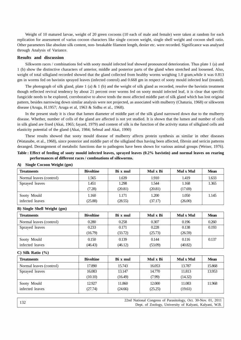
22nd National Congress of Parasitology, Oct. 30-Nov. 01, 2011Dept. of Zoology, University of Kalyani, Kalyani, W.B.
132
Weight of 10 matured larvae, weight of 20 green cocoons (10 each of male and female) were taken at random for eachreplication for assessment of varius cocoon characters like single cocoon weight, single shell weight and cocoon shell ratio.Other parameters like absolute silk content, non- breakable filament length, denier etc. were recorded. Significance was analysedthrough Analysis of Variance.
Results and discussionSilkworm races / combinations fed with sooty mould infected leaf showed pronounced deterioration. Thus plate 1 (a) and
1 (b) show the distinctive characters of anterior, middle and posterior parts of the gland when stretched and loosened. Also,weight of total silkgland recorded showed that the gland collected from healthy worms weighing 1.0 gram,while it was 0.813gm in worms fed on bavistin sprayed leaves (infected control) and 0.668 gm in respect of sooty mould infected leaf (treated).
The photograph of silk gland, plate 1 (a) & 1 (b) and the weight of silk gland as recorded, resolve the bavistin treatmentthough reflected revival tendency by about 21 percent over worms fed on sooty mould infected leaf, it is clear that specificfungicide needs to be explored, corroborative to above tends the most affected middle part of silk gland which has lost originalpattern, besides narrowing down similar analysis were not projected, as associated with mulberry (Chaturia, 1968) or silkwormdisease (Aruga, H.1957; Aruga et al, 1963 & Sidhu et al., 1968).
In the present study it is clear that lumen diameter of middle part of the silk gland narrowed down due to the mulberrydisease. Whether, number of cells of the gland are affected is not yet studied. It is shown that the lumen and number of cellsin silk gland are fixed (Akai, 1965; fayard, 1979) and content of silk is the function of the activity status of silkgland cells andelasticity potential of the gland (Akai, 1984; Sehnal and Akai, 1990)
These results showed that sooty mould disease of mulberry affects protein synthesis as similar in other diseases(Watanabe, et al., 1968), since posterior and middle part of the silkgland thus having been affected, fibroin and sericin patternsderanged. Derangement of metabolic functions due to pathogens have been shown for various animal groups (Weiser, 1976).
Table : Effect of feeding of sooty mould infected leaves, sprayed leaves (0.2% bavistin) and normal leaves on rearingperformances of different races / combnations of silkworms.
A) Single Cocoon Weight (gm)
Treatments Bivoltine Bi x mul Mul x Bi Mul x Mul Mean
Normal leaves (control) 1.565 1.639 1.910 1.419 1.633Sprayed leaves 1.451 1.298 1.544 1.168 1.365
(7.28) (20.81) (20.81) (17.69)
Sooty Mould 1.160 1.171 1.200 1.050 1.145infected leaves (25.88) (28.55) (37.17) (26.00)
B) Single Shell Weight (gm)
Treatments Bivoltine Bi x mul Mul x Bi Mul x Mul Mean
Normal leaves (control) 0.280 0.258 0.307 0.196 0.260Sprayed leaves 0.233 0.171 0.228 0.138 0.193
(16.79) (33.72) (25.73) (26.59)
Sooty Mould 0.150 0.139 0.144 0.116 0.137infected leaves (46.43) (46.12) (53.09) (40.82)
C) Silk Ratio (%)
Treatments Bivoltine Bi x mul Mul x Bi Mul x Mul Mean
Normal leaves (control) 17.890 15.743 16.053 13.787 15.868Sprayed leaves 16.083 13.147 14.770 11.813 13.953
(10.10) (16.49) (7.99) (14.32)
Sooty Mould 12.927 11.860 12.000 11.083 11.968infected leaves (27.74) (24.66) (25.25) (19.61)

22nd National Congress of Parasitology, Oct. 30-Nov. 01, 2011Dept. of Zoology, University of Kalyani, Kalyani, W.B.
133
d) Absolute Silk Content
Treatments Bivoltine Bi x mul Mul x Bi Mul x Mul Mean
Normal leaves 2557.270 2408.200 2678.670 1096.070 2335.050 (Normal)
Sprayed leaves 1710.370 1387.000 1355.200 1150.930 1525.880(33.12) (42.41) (30.74) (32.14)
Sooty Mould 882.867 1143.730 997.933 960.867 996.350infected leaves (65.48) (52.51) (62.75) (43.35)
CD at 5% for comparing treatment means and interaction effects
Treatment Interaction
Single cocoon weight 0.024 0.047
Single Shell weight 0.006 0.012
Silk Ratio 0.284 0.567
Absolute Silk Content 99.270 198.540
(Figures in parentheses are percent decreases over corresponding)
Fig. 1(a) : Silk gland fed with normal leaves
Fig. 1(b) : Silk gland fed with fungicide sprayed leaves
Fig. 1(c) : Silk gland fed with sooty mould infected leaves

22nd National Congress of Parasitology, Oct. 30-Nov. 01, 2011Dept. of Zoology, University of Kalyani, Kalyani, W.B.
134
ReferenceAkai H (1965) Studies on the ultrastructure of the silk gland in the silk worm, Bombyx mori J Insect Physiol, 24 :17-24
Akai H ( 1984) The ultrastructure and function of the silk gland cells of Bombyx mori, pp 323- 364. In R.C. king and H.Akai(eds), Insect ultra structure, vol 2.plenum, New York.
Aexopolous GJ, Mims CW (1985) Introductory Mycology Wiley Western Ltd. 3rd End, pp316-317. 402-404
Aruga H, Fukuda H, Yashitake N ( 1963) observations on a polyhedrosis virus with in the nucleus of the silk gland cellof the silk worm, Bombyx mori, L. J Seric Seri Jpn 32, p. 213-218
Aruga H ( 1957) Polyhedra disease in silk worm Bombyx mori , L. J of S 9: ( 1x2) 37-41
Chanturia NN ( 1968) Biochemical characteristics of mulberry leaves damaged by powdery mildew, Soobshch, Akad Nauk GruzSSR. 52, 779-804
Fayard JM (1979). Introduction to a genetic and embryological analysis of cell populations in silk glands of Bombyx mori.Genetica 50: 105-110
Krishnaswami S (1978). New technology of silkworm rearing. Bull No. 3 CSR&TI, Mysore, p 23
Krishnaswami S, Narashimhanna MN, Suryanarayana SK, Kamraj S (1973) Sericultural Manual 2, silkworm rearing FAOAgricultural Services Bulletin. Rome, 15,1-128
Mukherjee KG, Bhasin J (1986) Plant diseases of India 171, 306-350
Rao AA, Chauhan SS, Chakraborty S, Subha Rao G (1992) A new report on the occurance of Chaetothyrium & Cuvulariafungi on mulberry. Indian J Seric 31 : 71
Sidhu NS, Singh K (1968) Studies on ployhedrosis in silkworm, Indian J Seric Vol- V111, 1
Sehnal F, Akai H (1990) Insect silkglands, their types, development and function and effects of environmental factors andmorphogenetic hormones on them. Int J Insect Morphol and Embryol Vol.19(2): 79-132
Watanabe H, Funda T, Kubota A, Kawakami T, Aizawa Kita S, Matsuo T (1968) Effect of some disinfectants on the flacherievirus of the silkworm, Bombyx mori L. J Seric Sci Jpn 37 (3) : 213-218
Weiser J (1976) Microsporida in Invertibrates : Hostparasite relations at the organism level,1.pp. 164-201, Newyork: PlenumPress.

22nd National Congress of Parasitology, Oct. 30-Nov. 01, 2011Dept. of Zoology, University of Kalyani, Kalyani, W.B.
135
Trichodinid ectoparasites (Ciliophora : Trichodinidae) from the gills of fresh-water fishes in the Shitalakhsya River, Bangladesh
Md. Manzoorul Kibria, Hadiul Islam, M. M. A. Habib, Liton Chandra Shutradhar and Ghazi S. M. Asmat
Department of Zoology, University of Chittagong, Chittagong 4331, Bangladesh
Abstract : The systematic and taxonomy of ichthyotrichodinid parasites was investigated in the Shitalakshya River and local fish farmin the Kapasia Upazila of Gazipur District, Bangladesh between January and December, 2009. Smears of the gills of 12 host fish species,
caught by fishing nets and obtained from fish farms revealed the presence of 14 species of Trichodina. The identified species of Trichodina
are: Trichodina acuta, T. anabasi, T. centrostrigeata, T. cancilae, T. domerguei, T. matsu, T. microspina, T. modesta, T. molae, T.
mossambicusi, T. nigra, T. shitalakshyae, T. siluri and T. sylhetensis. Out of these, Trichodina cancilae, T. domerguei, T. matsu, T.
microspina, T. molae and T. siluri is reported as a first record in Bangladesh. One of the two unidentified species was published asTrichodina shitalakshyae from Glossogobius giuris based on the dry silver impregnated specimens. Photomicrographs and morphometricdata are presented for each species. General prevalence of parasitic infection in specimens obtained from the Shitalakshya River is also
recorded.
Key Words : Bangladesh, Ciliophora, Trichodinidae
IntroductionIn Bangladesh, Asmat et al. (1997) made the first report of trichodinid ciliates. Since then scanty and infrequent information
are available on the taxonomy of this particular group in this region. As a result, 17 species of trichodinid ciliates representingthe genera Trichodina, Paratrichodina, Tripartiella and Trichodinella were identified from different freshwater and estuarinefishes by Asmat et al. (1997; 2003a,b,c; 2005; 2006) and Bhouyain et al. (1999), Asmat and Sultana (2005), Habib and Asmat(2008) and Kibria et al. (2009; 2010). During a survey on the species diversity of trichodinid ciliates from the Shitalakhsya River(Latitude 23° 49' 20.11" N Longitude 90° 33' 0.47" E), a branch of the River Brahmaputra, in the Kapasia Upazila of Gazipurdistrict, 70km north-east of Dhaka (Fig. 1), during January 2008 to December 2009 fourteen species of Trichodina were identifiedfrom the gills of 38 host fish species.
Materials and methodsThe host fishes were collected from one of the Shitalakhsya River in the Kapasia Upazila of Gazipur district, by seine nets
and gill nets. Gill scrapings were made at the pond side; air-dried and then were transported to the laboratory. The slides withtrichodinid ciliates were impregnated with Klein’s silver impregnation technique (Klein, 1958) and examined under a researchmicroscope, OSK 9712 T-2 at 10x100 magnifications. Measurements were made according to the recommendations of Lom(1958), Wellborn (1967), Arthur and Lom (1984) and Van As and Basson (1989; 1992). All measurements given are inmicrometers, range in parentheses by the arithmetic mean and standard deviation. For statistical analysis, morphometricmeasurements of 20 specimens for both species were considered. Photomicrographs were made in order to have comprehensivemorphological analyses of the ciliates. The level of infection was measured as low (1-5 ciliate/slide), medium (6-10 ciliate/slide)and high (more than 11 ciliates/slide).
Results and DiscussionFourteen species of trichodinid ciliates were recorded from the gills of 12 commercially important finfish species of
Bangladesh.
Trichodina acuta Lom 1961 (Fig. 3, 17; Table 1)
Host: Mystus bleekeri (Day 1877) (Bagridae). Locality: Shitalakshya River, Kapasia Upazila of Gazipur district, Bangladesh.Location: gills. Prevalence: 19/41 (46.3%). Infection: low. Reference material: MB 1 and MB 2 (10/10/2009) of silver stained slidesare deposited in the Museum of the Department of Zoology, University of Chittagong, Chittagong 4331.
*Corresponding Author : E-mail : [email protected]

22nd National Congress of Parasitology, Oct. 30-Nov. 01, 2011Dept. of Zoology, University of Kalyani, Kalyani, W.B.
136
Figure 1. Map of sampling area from where fishes were collected from the Shitalakhsya River.
Figure 2. Denticle structure and construction of X and Y axes as fixed references
for description of denticles (after Van as and Basson 1989). Explanations: AB,
apex of blade; AM, anterior margin of blade; AR, apophysis of ray; B, blade; BA,
apophysis of blade; CA, central area of adhesive disc; CB, section connecting
blade and central art; CC, secton connecting central part and ray; CCP, central
conical part; CP, central part of blade; DC, deepest point of semilunar curve
relative to apex; DM, distal margin of blade; PM, posterior margin of blade; PP,
posterior projection; R, ray; SA, section of central part above x axis; SB, section
of central part below x axis; TP, tangent point; TR, tip of ray.

22nd National Congress of Parasitology, Oct. 30-Nov. 01, 2011Dept. of Zoology, University of Kalyani, Kalyani, W.B.
137
Figure 3-16. Photomicrographs of silver impregnated adhesive disc of trichodinid ciliates obtained during thestudy period in the Shitalakhsya River, Bangladesh. Scale bar 30 µm.
Trichodina acuta is one of most widely distributed trichodinid species from freshwater fishes. It was originally describedby Lom (1961) from five species of freshwater fishes, viz., Cyprinus carpio, Perca fluviatllis, Lucioperca lucioperca, Leucaspisdelineatus and Rhodeus sericeus, and from the skin of tadpoles belonging to several species of frogs in Czechoslovakia (CzechRepublic). Since then, T. acuta has bees reported from China (Chen 1963; 1984a; 1984b; Anon. 1973), Russia (Kandilov 1964;Kashkovsky 1965; Stein 1968; Kulemina 1968), Poland (Kazubski and Migala 1968), the USA (Lom 1970), South Africa and Israel(Basson et al. 1983), Israel (Van As and Basson 1989), from the Philippines (Duncan 1977; Natividad et al. 1986; Bondad-Reantaso and Arthur 1989; Albaladejo and Arthur 1989) from Cuba (Arthur and Lom 1984b), Czech Republic (Navratil 1991),Finland (Halmetoja et al. 1992), South Africa (Burton and Merron 1985; Basson and Van As 1993), Germany (Grupcheva andSedlaczek 1993), Turkey (Özer and Erdem 1998; 1999; Özer 2002), the United Kingdom (Gaze and Wooten 1998), India (Asmat2000), Serbia (Nikoliæ et al. 2003), Brazil (Piazza et al. 2006), Australia (Basson 2010) and Bangladesh (Kibria et al. 2010).

22nd National Congress of Parasitology, Oct. 30-Nov. 01, 2011Dept. of Zoology, University of Kalyani, Kalyani, W.B.
138
At least 25 host fish species were infested by this trichodinid through out the world. The above results, therefore, confirmthe presumption that host specificity in fish trichodinids is absent as expressed by Stein (1976), Hoffman and Lom (1967) andVan As and Basson (1987). During the present study T. acuta was obtained from Mystus bleekeri. The dimensions ofmeasurements fall well within the range given for T. acuta by Lom (1961) and Duncan (1977). The denticle measurements areclose to specimens presented by Van As and Basson (1989) from various species of Oreochromis from Israel and themorphology of denticles agree in most of the essential aspects with the summer specimens as recorded by Kazubski and Migala(1968) from C. carpio in Poland. During the present survey this ciliate was obtained mainly from October 2008 to February2009, i.e., in pre-colder to post-colder period as was reported by Asmat (2000) from India. Basson and Van As (1993) statedT. acuta as the European species which parasitizes the European and Asian cyprinids and have spread via the translocationof their hosts. They also concluded the presence of this species in South Africa as a recent introduction which will also becomepart of the fish parasite fauna of that region in the future. In Bangladesh, due to lack of comprehensive survey of trichodinidciliates from indigenous as well as exotic species, it is not possible to comment whether this ciliate is an introduced speciesor indigenous one. However, the present study reports an extension of the geographic range of T. acuta and Mystus bleekerito be a new host.
Trichodina anabasi Asmat, Mohammad and Sultana 2003 (Fig. 4, 18; Table 1)
Host: Anabas testudineus (Bloch 1795) (Anabantidae). Locality: Sarker Bari culture ponds in Kapasia Upazila of Gazipurdistrict, Bangladesh. Location: gills. Prevalence: 10/25 (40.0%). Infection: low. Reference material: AT 1 and AT 2 (10/07/2009)of silver stained slides are deposited in the Museum of the Department of Zoology, University of Chittagong, Chittagong 4331.
Trichodina anabasi was established by Asmat et al. (2003) from the gills of Anabas testudeneus. It is characterized by largebody dimensions and having uniformly stained, dark central area at the centre of the adhesive disc; broad blade with angularand slightly rounded distal margin and flat tangent point, but almost parallel anterior and posterior margin; stout and cylindricalcentral part with bluntly rounded point which extends slightly more than halfway to the y-axis; and the ray is slightly curvedin the posterior direction with anterior margin parallel to the y+1 axis and distinct central groove (Asmat et al. 2003). Thepresent specimens obtained from the gills of cultured A. testudeneus was identified on the basis of original description of T.anabasi given by Asmat et al. (2003). By the denticle morphology the present ciliate was quite similar to that of Asmast etal. (2003). However, the average size of T. anabasi of the present study appears to be slightly smaller than that of Asmat etal. (2003). T. anabasi seems to have rather narrow host range as such a distinct species has only been found from the climbingperch, a highly commercial fish species distributed widely in Bangladesh.
Trichodina cancilae Asmat 2001 (Fig. 5, 19; Table 1)
Host : Xenentodon cancila (Hamilton 1822) (Belonidae). Locality: Shitalakshya River of Gazipur district, Bangladesh.Location: gills. Prevalence: 45/120 (37.5%). Infection: low, in mixed infection with Tripartiella bursiformis. Reference material:XC 9 and XC 10 (10/10/2009) of silver stained slides are deposited in the Museum of the Department of Zoology, Universityof Chittagong, Chittagong 4331.
Trichodina cancilae was established by Asmat (2001) from the gills Xenentodon cancila in West Bengal, India. T.cancilae is a large trichodinid having denticles with elongated and arched blades; 1 or 2 cut-like notches in the convex sideof the blade and the presence of the single non-staining granule at the base of blades and the base of rays; moderate centralpart; and broad and greatly arched rays (Asmat 2001). The blade shapes and orientation of rays of T. cancilae obtained inthe present investigation were identical, but one of the identifying characters, i.e., the presence of non-impregnable pearl-shaped glistening particles in interblade space and in between ray bases in many specimens of the Indian populations of T.cancilae as inserted by Asmat (2001) was absent in the present specimens.
Trichodina centrostrigeata Basson and Van As 1983 (Fig. 6, 20; Table 1)
Host : Oreochromis mossambicus (Peters 1852) (Cichlidae). Locality: Sarker Bari culture ponds in Kapasia Upazila ofGazipur district, Bangladesh. Location: gills. Prevalence: 30/70 (42.8%). Infection: low to medium. Reference material: OM 4 andOM 5 (10/10/2009) of silver stained slides are deposited in the Museum of the Department of Zoology, University of Chittagong,Chittagong 4331.
Trichodina centrostrigeata was first described by Basson et al. 1983, from the gills, occasionally from the skin and fins,of Oreochromis mossambicus, Pseudocrcnilabnis philander, Tilapia rendalli, T. sparrmanni and Cyprinus carpio in SouthAfrica. Subsequently, this species was reported Taiwan (province of China) (Van As and Basson 1986; Basson and Van As1994), Egypt (El-Tantawy and Kazubski 1986), the Philippines (Natividad et al. 1986; Bondad-Reantaso and Arthur 1989), SouthAfrica (Van As and Basson 1992). T. centrostrigeata appears to be highly specific to cichlid fishes. There are only two reports

22nd National Congress of Parasitology, Oct. 30-Nov. 01, 2011Dept. of Zoology, University of Kalyani, Kalyani, W.B.
139
of this species of trichodinid ciliates from non-cichlids, i.e., by Basson et al. (1983) and Abdel-Meguid (1995) from commoncarp and grass carp, respectively. The ciliate obtained from the gills of O. mossambicus in the present study agree well withthe descriptions of Basson et al. (1983), Van As and Basson (1986), Van As and Basson (1992), Basson and Van As (1994)and Mitra and Bandyopadhyay (2006). Basson and Van As (1994) commented that T. centrostrigeata is a parasite endemicto Africa, which at some stage was translocated to Taiwan and Philippines. The origin of O. mossambicus is Africa. It iscommonly known as Mossambique cichlid and was introduced in Bangladesh in 1974 from Thailand to control the aquaticinsects. The species is now well adapted in nature. As a result, the present trichodinid populations are also became wide spreadand highly commercial fish in Banglasdesh. During the present study T. centrostrigeata was obtained from the gills of O.mossambicus from the Dhaka division. The Bangladeshi population of T. centrostrigeata is larger in size as well as in denticledimensions.
Trichodina domerguei (Wallengren 1897) Heider 1964 (Fig. 7, 21; Table 1)
Host : Johnius clitor (Hamilton 1822) (Sciaenidae). Locality: Shitalakshya River of Gazipur district, Bangladesh. Location:gills. Prevalence: 6/62 (9.7%). Infection: low. Reference material: JC 1 and JC 2 (13/04/2009) of silver stained slides are depositedin the Museum of the Department of Zoology, University of Chittagong, Chittagong 4331.
T. domerguei was first recorded by Wallengren (1897) as Cyclochaeta domerguei from five fishes, Pungitius pungitius,Gastrostens aculeatus, Carassius carassius, Phoxinus phoxinus and Leucaspis dilineatus. This ciliate was reported from theRumanian Black Sea coast (Lom 1962), SSR (Lom and Stein 1966), Germany (Grupcheva and Sedlaczeck 1993), UK (Gaze andWootten 1998), Turkey (Özer 2000; 2003a; 2003b; Özer and Erdem 1998; 1999), India (Asmat 1999), Baltic Sea (Morozinska-Gogol2000), Poland (Rolbiecki 2006). T. domerguei is similar to T. jadranica Raabe 1958, T. cottidarum Lom 1970 and T. murmanicaPolyanski 1955 (Lom 1970a; Lom and Dyková 1992). These species are euryhaline or marine trichodinids which may suggestthat T. domerguei is of marine origin (Gaze and Wooten 1998). Gaze and Wooten (1998) also opined that the smaller dimensionsof the marine specimens described in this study agree with the findings of Lom (1970) who remarks on the small specimensize in marine populations. In India, Asmat (1999) collected only a single specimen of this species from Mystus gulio, anestuarine fish in Canning area of West Bengal. Host specificity in trichodinids seems to be highly variable, some species suchas T. domerguei, T. acuta and T. nigra infest a large number of host species (Lom and Stein 1966; Lom 1970a; Calenius 1980).During the present study T. domerguei was recorded not from any estuarine or marine fish but from Johnius coitor, a wellknown commercial freshwater fish from a freshwater river, the Shitalaskhya River. Due to the reduced quality of stain onlya few good quality specimens were obtained from this population. However, the present study reports an extension of thegeographic range of T. domerguei and J. coitor to be a new host.
Trichodina matsu Basson and Van AS 1994 (Fig. 8, 22; Table 1)
Host : Labeo rohita (Hamilton 1822) (Cyprinidae). Locality: Sarker Bari culture ponds, Kapasia Upazila of Gazipur district,Bangladesh. Location: gills. Prevalence: 14/60 (23.3%). Infection: low. Reference material: LR 3 and LR 4 (08/03/2009) of silverstained slides are deposited in the Museum of the Department of Zoology, University of Chittagong, Chittagong 4331.
Trichodina matsu was first described by Basson and Van As (1994) from the gills of the frestwater fish Crossoptomalacustre and from the gills, skin and fins of the fish Leiocassis adiposalis in Confluence of Nankang and Peikang Rivers inTaiwan (China). This species has been subsequently reported from the gills of catfish Clarias gariepinus inhabiting Damiettabranch of the River Nile in Egypt by El-Tantawy and El-Sherbiny (2010). Although the body dimension and other features ofT. matsu obtained in the present study closely resemble those described by Basson and Van As (1994) for the same species,some minor differences were also recorded. First, the blade apophysis of the present trichodinid is more prominent than thatof Basson and Van As (1994). Secondly, Basson and Van As (1994) recorded indentation on the lower central part of the bladewhereas in the present study none of these indentations were observed. Moreover, the blade length recorded by Basson andVan As (1994) appeared to be greater than of the present study. It should be emphasized that these differences are intraspecificvariations between individuals of the same species. The present study reports the first record of T. matsu from Bangladeshand Labeo rohita to be a new host.
Trichodina microspina Van As and Basson 1992 (Fig. 9, 23; Table 1)
Host : Rita rita (Hamilton 1822) (Bagridae). Locality: Shitalakshya River, Kapasia Upazila of Gazipur district, Bangladesh.Location: gills. Prevalence: 15/80 (18.7%). Infection: low. Reference material: RR 6 (26/10/2009) of silver stained slides aredeposited in the Museum of the Department of Zoology, University of Chittagong, Chittagong 4331.
Trichodina microspina was established by Van As and Basson (1992) from the skin and gills of climbing perch Ctenopomamultispine of Lake Lisikili in South Africa. Since then this species has not been reported from any part of the world. However,

22nd National Congress of Parasitology, Oct. 30-Nov. 01, 2011Dept. of Zoology, University of Kalyani, Kalyani, W.B.
140
Tabl
e 1:
Mor
phom
etri
cal d
ata
of th
e tr
icho
dini
d pa
rasi
tes
obta
ined
from
the
gills
of f
resh
wat
er fi
shes
in th
e Sh
itala
khsy
a R
iver
.
Spec
ies
Hos
t
Loc
alit
y
Loc
atio
n
Ref
eren
ce
Dia
met
er o
f
body
adhe
sive
disc
dent
icul
ate
ring
cent
ral
area
Wid
th o
f bo
rder
mem
bran
e
Num
ber
of
dent
icle
s
radi
al
pins
/den
ticl
e
Span
of
dent
icle
Len
gth
of
dent
icle
ray
blad
e
Wid
th o
f ce
ntra
l
par
t
°Ado
ral
cilia
ture
Tric
hodi
na a
cuta
(n=
10)
Mys
tus
blee
keri
Shi
tala
kshy
a R
iver
,
Ban
glad
esh
Gill
s
Pre
sent
stu
dy
36.4
-47.
5(43
.3±
3.5)
30.3
-37.
4(34
.5±
2.4)
16.2
-22.
7(20
.0±
1.9)
7.1-
9.6(
8.7±
0.9)
3.0-
5.0(
4.4±
0.7)
18-2
1(19
.5±
1.2)
7-9(
8.0±
0.8)
10.1
-12.
1(10
.9±
0.7)
4.5-
7.1(
5.7±
0.7)
4.0-
5.0(
4.6±
0.3)
4.0-
5.0(
4.7±
0.4)
1.5-
2.0(
1.8±
0.2)
390-
4000
Tric
hodi
na a
naba
si
(n=
15)
Ana
bas
test
udin
eus
Cul
ture
Pon
d, G
azip
ur,
Ban
glad
esh
gills
Pre
sent
stu
dy
55.5
-70.
7(61
.9±
4.4)
50.5
-65.
7(57
.0±
4.4)
33.8
-43.
4(37
.2±
2.5)
13.1
-20.
2(17
.7±
2.3)
3.0-
6.1(
4.9±
0.8)
25-2
9(27
.5±
1.1)
7-10
(8.7
±0.
9)
14.1
-20.
2(17
.4±
1.6)
7.1-
10.1
(8.3
±0.
8)
7.1-
9.1(
8.2±
0.5)
5.0-
9.1(
7.0±
1.0)
2.0-
3.0(
2.4±
0.4)
——
—
Tric
hodi
na c
anci
lae
(n=
20)
Xen
ento
don
canc
ila
Shi
tala
kshy
a R
iver
,
Ban
glad
esh
gills
Pre
sent
stu
dy
57.5
-86.
9(69
.4±
6.8)
52.5
-80.
8(63
.6±
6.7)
35.3
-47.
5(42
.9±
3.6)
15.1
-21.
2(18
.2±
1.6)
5.0-
6.1(
5.8±
0.4)
28-3
3(30
.7±
1.4)
8-11
(9.7
±0.
9)
17.2
-24.
2(20
.7±
1.9)
5.0-
9.6(
7.1±
1.3)
7.1-
12.6
(10.
5±1.
4)
6.1-
8.6(
6.9±
0.6)
3.0-
4.0(
3.3±
0.4)
40
00
Tric
hodi
na
cent
rost
rige
ata
(n=
20)
Ore
ochr
omis
mos
sam
bicu
s
Cul
ture
Pon
d,
Gaz
ipur
, B
angl
ades
h
gills
Pre
sent
stu
dy
41.9
-53.
5(47
.2±
2.8)
33.3
-43.
4(38
.4±
2.6)
18.7
-27.
7(23
.9±
2.2)
10.6
-15.
1(12
.8±
1.4)
4.0-
5.0(
4.4±
0.4)
21-2
8(25
.8±
2.1)
6-8(
7.4±
0.6)
10.6
-13.
6(12
.0±
0.8)
4.0-
7.1(
4.6±
0.7)
4.0-
5.5(
4.3±
0.4)
4.5-
6.1(
5.3±
0.4)
2.0-
3.0(
2.3±
0.3)
410°
Tric
hodi
na d
omer
guei
(n=
20)
John
ius
coit
or
Shi
tala
kshy
a R
iver
,
Ban
glad
esh
Gill
s
Pre
sent
stu
dy
30.4
-38.
4(34
.6±
2.3)
25.2
-30.
3(27
.8±
1.6)
14.1
-19.
2(16
.2±
1.6)
6.1-
10.1
(7.9
±0.
9)
2.0-
4.0(
3.4±
0.7)
21-2
3(22
±0.
6)
6-9(
7.0±
0.9)
7.1-
9.1(
8.1±
0.6)
3.0-
5.0(
4.2±
0.6)
2.5-
3.5(
3.0±
0.3)
3.0-
4.0(
3.3±
0.4)
1.3-
2.0(
1.8±
0.2)
——
—-
Tric
hodi
na m
atsu
(n=
20)
Lab
eo r
ohit
a
Cul
ture
pon
d ,
Gaz
ipur
, B
angl
ades
h
Gill
s
Pre
sent
stu
dy
34.3
-48.
5(40
.5±
3.7)
28.3
-36.
4(32
.7±
2.6)
15.1
-21.
2(18
.1±
1.9)
5.5-
11.1
(7.9
±1.
3)
3.0-
6.1(
3.9±
0.8)
19-2
4(22
.4±
1.2)
7-10
(8.6
±0.
8)
8.6-
12.6
(10.
2±1.
0)
3.0-
6.1(
4.6±
0.6)
3.0-
5.5(
3.9±
0.7)
3.0-
4.3(
3.9±
0.3)
2.0-
3.0(
2.3±
0.3)
abou
t 42
0º
Tric
hodi
na
mic
rosp
ina
(n=
20)
Rit
a ri
ta
Shi
tala
kshy
a R
iver
,
Ban
glad
esh
Gill
s
Pre
sent
stu
dy
15.6
-21.
2(18
.2±
1.6)
12.1
-17.
2(14
.3±
1.5)
6.1-
9.6(
7.6±
1.0)
3.8-
6.1(
4.6±
0.7)
1.8-
2.3(
2.0±
0.2)
18-2
0(18
.9±
0.7)
5-8(
6.7±
0.8)
4.0-
5.5(
4.7±
0.5)
1.8-
2.3(
2.0±
0.1)
1.0-
1.5(
1.2±
0.1)
1.8-
3.1(
2.6±
0.4)
0.5-
1.0(
0.8±
0.2)
——
——
—

22nd National Congress of Parasitology, Oct. 30-Nov. 01, 2011Dept. of Zoology, University of Kalyani, Kalyani, W.B.
141
Figure 17-30. Diagrammatic drawings of denticles of trichodinid ciliates obtained during the present studyin the Shitalakhsya River, Bangladesh.
the present study reports the occurrence of this species from Bangladesh as well as from a new host, R. rita. T. microspinais characterised by having small-sized body dimension with broad blade but small spine-like ray, spacious and argentophiliccentral area and narrow interblade space. Although based on the denticle morphology of the present population of the ciliateresemble those of Van As and Basson (1992), the average body size of T. microspina of the present study appears to be smallerthan that recorded in the specimens of Van As and Basson (1992).
Trichodina modesta Lom 1970 (Fig. 10, 24; Table 2)
Host : Clupisoma garua (Hamilton 1822) (Schilbeidae). Locality: Shitalakshya River, Kapasia Upazila of Gazipur district,Bangladesh. Location: gills. Prevalence: 02/70 (2.8%). Infection: low. Reference material: CG 2 (26/09/2009) of silver stained slidesare deposited in the Museum of the Department of Zoology, University of Chittagong, Chittagong 4331.

22nd National Congress of Parasitology, Oct. 30-Nov. 01, 2011Dept. of Zoology, University of Kalyani, Kalyani, W.B.
142
Lom (1970) first described Trichodina modesta from the gills of Vimba vimba in Hungary. It is relatively small trichodinidhaving denticles with relatively delicate inner rays, straight or slightly curved posteriorly; elongated blades (Lom 1970) andgently curved with a noticeable thickening of the posterior border; the blades usually terminate with a rounded anterior margin,but sometimes form a flattened distal surface (Gaze and Wootten 1998). Since its discovery, T. modesta was reported from theskin, fins and gills of cyprinid and silurid fishes in the eastern Europe (Stein 1982, Arthur and Lom 1984), Poland (Wierzbicka1997), United Kingdom (Gaze and Wootten 1998), Taiwan (province of China) (Basson and Van As 1994), Turkey (Özer 2007;Ozturk and Özer 2007), and China (Zhou et al. 2007; Liu and Zhao 2010). In Bangladesh, T. modesta was first reported by Kibriaet al. (2009) from the gills of O. mossambicus in various culture ponds of Chittagong and Cox’s Bazar districts. The presentlydescribed populations of T. modesta are identical to those of Lom’s (1970) nominate population in size but host fish speciesis different, i.e. Clupisoma garua. The extent of morphological variation in this species in Bangladesh is unknown.
Trichodina molae Mitra and Haldar 2005 (Fig. 11, 25; Table 2)
Host : Amblypharyngodon mola (Hamilton 1822) (Cyprinidae). Locality: Shitalakshya River, Kapasia Upazila of Gazipurdistrict, Bangladesh. Location: gills. Prevalence: 04/56 (7.1%). Infection: low. Reference material: AM 1 and AM 2 (11/01/2009)of silver stained slides are deposited in the Museum of the Department of Zoology, University of Chittagong, Chittagong 4331.
Trichodina molae is characterized by its medium size and by the adhesive disc components, which include stronglyrectangular, paintbrush-like blade; slender, tubular central part; and leaf-like flattened ray with constriction at the base, welldeveloped central groove and sharply pointed tip. T. molae was named and described as a separate species by Mitra and Haldar(2005) from the gills of Amblypharyngodon mola in Ranaghat of West Bengal, India. The specimens described in the presentpaper exhibits a slightly lower range of body dimensions than those of Mitra and Haldar (2005). Other variations could benoted in Bangladeshi T. molae from the Indian populations are, the specimens described in this paper exhibits a slightly higherrange of body dimensions than those of Mitra and Haldar (2005). Secondly, the distal margin of blade of the present studyis flat whereas in the Indian population is slightly rounded. Thirdly, in the Bangladeshi specimens the anterior margin of bladeforms acute angle whereas slopes down gradually in Mitra and Haldar (2005). Moreover, the ray apophysis in the presentstudy is present, whereas in the originally described specimens the ray apophysis is absent. The mentioned minormorphological variations can be due to different geographical conditions etc.
Trichodina mossambicusi Asmat 2005 (Fig. 12, 26; Table 2)
Host : Oreochromis mossambicus (Peters 1852) (Cichlidae). Locality: Sarker Bari culture ponds in Kapasia Upazila ofGazipur district, Bangladesh. Location: gills. Prevalence: 05/70 (7.1%). Infection: low. Reference material: OM 7 (06/06/2009) ofsilver stained slides are deposited in the Museum of the Department of Zoology, University of Chittagong, Chittagong 4331.
Trichodina mossambicusi was originally described by Asmat (2005) from the gills of Oreochromis mossambicus in andaround Kalyani town of Nadia District, West Bengal, India. Since then it is a new country record for Bangladesh from the samehost. The denticle morphology of silver impregnated adhesive disc and denticle morphology of the species in both the Indianand Bangladeshi populations are identical. However, the average size of T. mossambicusi of the present study appears tobe smaller than that recorded by Asmat (2005).
Trichodin nigra Lom 1961 (Fig. 13, 27; Table 2)
Host : Notopterus notopterus (Pallas 1767) (Notopteridae). Locality: Shitalakshya River of Gazipur district, Bangladesh.Location: gills. Prevalence: 20/74 (27.0%). Infection: low to medium. Reference material: NN 6 (08/09/2009) of silver stained slidesare deposited in the Museum of the Department of Zoology, University of Chittagong, Chittagong 4331.
Trichodina nigra was established by Lom (1961) from several freshwater fish, like Cyprius carpio, Scardiniuserytrophthalmus, Rutilus rutilus, Abramis brama, Perca fluviatilis, Tinca tinca, Albnnnis albitnnts, Leitciscus cephalus andsome tadpoles of unidentified frogs in Czechoslovakia. T. nigra could be characterized as a trichodinid in which the blade isbroad and nicely rounded or angularly curved; the central part is more robust with bluntly or sharply rounded point; the raybase is fused with the central part and broad; a central axis extends through the length of ray; the ray is straight and tapersto a blunt point; and the central area of the adhesive disc is small (Asmat 2002). T. nigra is a common parasite of cyprinidsin Europe. This species has been recorded from Czechoslovakia (Lom 1961), UK (Gaze and Wootten 1998), USA (Hoffman andLom 1967), Poland (Lyubarskaya and Stein 1967; Kazubski and Migala 1968; and Migal, 1970; 1978), USSR (Kulemina 1968;Stein 1968; 1982; Konovalov 1971; Urazbaev 1971; Kostenko 1972; Kashkovsky 1974; Akhmetova and Diarova 1975; Bragina1975; Diarova 1975; Smirnova and Kairova 1975; Vismanis et al. 1975; Mikailov and Ibragimov 1980; Stein 1984; Arthur and

22nd National Congress of Parasitology, Oct. 30-Nov. 01, 2011Dept. of Zoology, University of Kalyani, Kalyani, W.B.
143
Tabl
e 2
: Mor
phom
etri
cal d
ata
of th
e tr
icho
dini
d pa
rasi
tes
obta
ined
from
the
gills
of f
resh
wat
er fi
shes
in th
e Sh
itala
khsy
a R
iver
.
Tric
hodi
naTr
icho
dina
Tric
hodi
naTr
icho
dina
Tric
hodi
naTr
icho
dina
Tric
hodi
na
Spec
ies
mod
esta
mol
aem
ossa
mbi
cusi
nigr
ash
ital
akhs
yae
silu
risy
lhet
ensi
s
(n=
05)
(n=
05)
(n=
20)
(n=
15)
(n=
20)
(n=
20)
(n=
20)
Hos
tC
lupi
som
a ga
rua
Am
blyp
hary
ngod
onO
reoc
hrom
isN
otop
teru
sG
loss
ogob
ius
Not
opte
rus
Nan
dus
nand
us
mol
am
ossa
mbi
cus
noto
pter
usgi
uris
noto
pter
us
Dia
met
er o
f
body
28.3
-40.
4(35
.2)
50.5
-57.
1(52
.9)
36.4
-44.
4(40
.3±
2.8)
52.0
-60.
3(54
.5±
2.5)
36.4
-47.
5(43
.3±
3.5)
48.5
-62.
1(53
.0±
3.7)
29.8
-56.
6(44
.2±7
.6)
adhe
sive
dis
c24
.2-3
9.4(
31.1
)45
.4-5
0.5(
48.1
)29
.3-3
5.3(
32.3
±2.
1)46
.5-5
4.5(
49.2
±2.
5)30
.3-3
7.4(
34.5
±2.
4)43
.4-5
6.6(
48.0
±3.
5)25
.2-4
6.5(
35.9
±6.0
)
dent
icul
ate
ring
13.1
-24.
2(17
.9)
29.8
-35.
3(32
.4)
16.7
-21.
2(19
.1±
1.1)
30.3
-35.
3(31
.7±
1.6)
16.2
-22.
7(20
.0±
1.9)
24.2
-36.
4(30
.4±
3.3)
15.1
-30.
8(21
.8±3
.9)
cent
ral a
rea
5.0-
10.1
(7.4
)13
.1-4
.6(1
3.9)
7.1-
10.1
(8.6
±0.
9)10
.1-1
3.1(
12.1
±1.
0)7.
1-9.
6(8.
7±0.
9)9.
1-15
.2(1
2.1±
1.7)
8.1-
13.1
(9.4
±1.
6)
Wid
th o
f bo
rder
mem
bran
e4.
0-4.
5(4.
1)4.
5-5.
0(4.
8)3.
0-5.
0(4.
0±0.
6)4.
5-5.
8(5.
2±0.
3)3.
0-5.
0(4.
4±0.
7)4.
0-5.
6(5.
0±0.
4)2.
3-5.
0(4.
1±0.
9)
Num
ber
of
dent
icle
s17
-23(
20)
26-2
9(27
.8)
20-2
6(22
.7±
1.5)
23-2
7(25
±1.
3)18
-21(
19.5
±1.
2)22
-27(
25.0
±1.
2)23
-26(
25±
0.9)
radi
al p
ins/
dent
icle
7-9(
8.2)
8-10
(9.2
)7-
9(7.
7±0.
7)7-
9(8±
0.7)
7-9(
8.0±
0.8)
9-10
(9.7
±0.
5)6-
9(7.
7±0.
9)
Span
of
dent
icle
9.1-
13.4
(11.
5)14
.6-1
7.7(
16.6
)10
.1-1
2.6(
11.4
±0.
8)15
.1-1
9.2(
16.9
±1.
2)10
.1-1
2.1(
10.9
±0.
7)14
.1-2
0.2(
17.2
±1.
7)7.
8-14
.1(1
1.7±
1.9)
Len
gth
of
dent
icle
4.3-
6.1(
5.0)
6.1-
7.1(
6.7)
3.8-
5.0(
4.3±
0.4)
4.0-
5.5(
4.8±
0.5)
4.5-
7.1(
5.7±
0.7)
6.6-
7.6(
7.1±
0.4)
3.5-
6.1(
4.8±
0.7)
ray
3.0-
6.0(
4.5)
7.1-
8.6(
7.6)
3.8-
6.1(
4.3±
0.6)
7.1-
9.1(
7.8±
0.6)
4.0-
5.0(
4.6±
0.3)
6.1-
9.1(
7.8±
0.9)
3.0-
7.1(
4.9±
1.0)
blad
e4.
0-5.
0(4.
7)5.
0-6.
3(5.
9)3.
8-5.
5(4.
8±0.
5)6.
1-7.
6(6.
6±0.
5)4.
0-5.
0(4.
7±0.
4)5.
1-8.
1(6.
4±0.
7)3.
0-5.
3(4.
5±0.
6)
Wid
th o
f cen
tral
part
2.0-
3.0(
2.3)
2.5-
3.5(
3.1)
1.5-
2.8(
2.2±
0.3)
2.5-
3.0(
2.5±
0.3)
1.5-
2.0(
1.8±
0.2)
2.0-
4.0(
3.0±
0.5)
1.5-
3.0(
2.3±
0.5)
°Ado
ralc
iliat
ure
—-
—-
400°
480°
390-
400
380-
3900
4000

22nd National Congress of Parasitology, Oct. 30-Nov. 01, 2011Dept. of Zoology, University of Kalyani, Kalyani, W.B.
144
Lom 1984), the Philippines (Albaladejo and Arthur 1989), China (Chen 1963; 1984a; b), Taiwan (Basson and Van As 1994),Denmark (Buchmann et al. 1995; Buchmann and Bresciani 1997), Finland (Rintamaki-Kinnunen and Valtonen 1997), and fromIndia (Mukherjee and Haldar 1982; Saha et al. 1995; and Asmat (2002). Most workers mentioned above have assigned thematerials to T. nigra or to any of its subspecies (except Arthur and Lom 1984a) adding confusion to identifying the species(Asmat 2002). To avoid further confusion in the present work the described species is also compared with the originaldescription and photomicrographs of T. nigra by Lom (1961). The appearance of the adhesive disc and denticle morphologyof T. nigra obtained in the present study is quite identical to that of Lom (1961), Arthur and Lom (1984a) and Asmat (2002).However, the Bangladeshi specimens coincide with the Indian population in morphometry (Asmat 2002), while the body sizeand denticle dimensions of the Czech Republic (Lom, 1961) and Russian Federation (Arthur and Lom 1984a) are considerablylarger. Incidentally, T. nigra is a new country record for Bangladesh and recognised as host specific on Notopterus notopterusin Indo-Bangla subcontinent.
Trichodina shitalakhsyae Kibria, Islam, Habib and Asmat, 2010 (Fig. 14, 28; Table 2)
Host : Glossogobius giuris (Hamilton 1822) (Gobidae), Locality: Shitalakhsya River of Gazipur district, Bangladesh.Location: gills. Prevalence: 06/10 (60%). Infection: low. Reference material: GG 1 (11/02/2009) of silver stained slides are depositedin the Museum of the Department of Zoology, University of Chittagong, Chittagong 4331.
Kibria et al. (2010) established Trichodina shitalakhsyae from Glossogobius giuris in the Shitalakhsya River of Gazipur District.The species is characterised by having an undivided clear central area in the adhesive disc with a rounded or slightly undulatedperimeter containing a few dark granules which form patches; elongated and rectangular blade with large interblade space andblunt tangent point; indistinct anterior blade apophysis and a shallow apex at the base of blade that never extends beyondthe y+1 axis; moderately wide and triangular central part with blunt point; and space between tip of ray and central clear areaforms a wide impregnated ring. Based on these characters and the unique shape and absence of variability of the denticlesamong the silver impregnated specimens of the present species, it resembles Trichodina porocephalusi Asmat, 2001.
Trichodina siliuri Lom 1970 (Fig. 15, 29; Table 2)
Host : Notopterus notopterus (Pallas 1767) (Notopteridae). Locality: Shitalakshya River of Gazipur district, Bangladesh.Location: gills. Prevalence: 14/74 (18.9%). Infection: low to medium. Reference material: NN 1 and NN 2 (06/05/2009) of silverstained slides are deposited in the Museum of the Department of Zoology, University of Chittagong, Chittagong 4331.
The trichodinid T. siluri was originally described from the gills of Silurus glanis from Hungary and Czechoslovakia byLom (1970). Thereafter, this species was recorded by Bondad-Reantaso and Arthur (1989) from the skin of cultured Nile tilapiawith the mixed infection of T. heterodentata in the Philippines. T. siluri may be characterised by having dark-centred adhesivedisc with considerably arched blades which sometimes bear small notches or frayed areas at their anterior margins; and therays are stout, about the same width throughout their length and extend backwards. The present report of T. siluri extendsits known geographical distribution as well as N. notopterus appears to be a new host for it.
Trichodina sylhetensis Asmat, Hafizuddin and Habib 2003 (Fig. 16, 30; Table 2)
Host : Nandus nandus (Hamilton 1822) (Nandidae). Locality: Shitalakshya River, Kapasia Upazila of Gazipur district,Bangladesh. Location: gills. Prevalence: 6/35 (17.1%). Infection: low. Reference material: NN 1 (12/12/2009) of silver stainedslides are deposited in the Museum of the Department of Zoology, University of Chittagong, Chittagong 4331.
Asmat et al. (2003) established Trichodina sylhetensis from the gills of freshwater fish Nandus nandus from the TanguarHaor at Sunamganj in Sylhet Division. The species is characterized by having medium body dimensions; slim, rectangularly-angular blade with slightly curved distal margin and blunt tangent point; bluntly rounded, slender central part having noindentation and posterior blade apophysis; broad ray with equal thickness and bluntly rounded tip; and dark-stained centreof the adhesive disc (Asmat et al. 2003). The denticle morphology of the silver impregnated adhesive disc of this species matchwith report of Asmat et al. (2003). However, the present population of T. sylhetensis is larger in size than forms recorded byAsmat et al. (2003) from the Tanguar Haor.
ReferencesAkhmetova B, Diarova GS (1975) Parasites and diseases of white Amur in the Chimkentsk pond farm, In Gvosdev, E. V., ed.,
Ecology of Parasites of Water Animals. Izdat. “Nauka” Alma-Ata, pp 47-55 (in Russian).
Albaladejo JD, Arthur JR (1989) Some trichodinids (Protozoa: Ciliophora: Peritrichida) from freshwater fishes imported into thePhilippines. Asian Fish Sci 3: 1-25.

22nd National Congress of Parasitology, Oct. 30-Nov. 01, 2011Dept. of Zoology, University of Kalyani, Kalyani, W.B.
145
Anon (1973) An illustrated guide to the diseases and the causative pathogenic fauna and flora of fishes of Hubei Province.Science Publ. House. Beijing. (in Chinese, partial English translation as Can. Transl. Fish. Aquat Sc. Nos 4509 and 5204).
Arthur JR, Lom J (1984a) Some trichodinid ciliates (Protozoa : Peritrichida) from Cuban fishes, with a description of Trichodinacubanensis n. sp. from the skin of Cichlasoma tetracantha. Trans. Am. Microsc Soc 103: 172-184.
Arthur JR, Lom J (1984b) Trichodinid Protozoa (Ciliophora: Peritrichida) from freshwater fishes of Rybinsk Reservoir, USSRJ Protozool 31: 82-91.
Asmat GSM (1999) Studies on the biodiversity of the trichodinid ciliophoran (Mobilina : Trichodinidae) in the freshwater andestuarine fishes of West Bengal. Ph. D. thesis. University of Kalyani, West Bengal, India.
Asmat GSM (2000) First record of Trichodina acuta Lom, 1961 (Ciliophora: Trichodinidae) from India. The Chittagong UnivJ Sc 24(1): 63-70.
Asmat GSM (2001) Trichodina cancilae sp. n. (Mobilina : Trichodinidae) from the Gills of a Freshwater Gar, Xenentodoncancila (Hamilton) (Belonidae). Acta Protozool 40: 141-146.
Asmat GSM (2002) Trichodinid ciliates (Ciliophora: Trichodinidae) from Indian fishes with description of two new species.Bangladesh J Zool 30(2):87-100.
Asmat GSM (2005) Trichodinid ectoparasites (Ciliophora: Trichodinidae) of fishes in India. Res J Agri Biol Sci 1(1): 31-37.
Asmat GSM, Sultana N (2005) Four New species of Trichodina Ehrenberg, 1830 (Ciliophora: Trichodinidae) from Bangladeshifish. Pak J Biol Sci 8(6): 895-900.
Asmat GSM, Bhouyain AM, Siddiqua PS (1997) First record of a species of Paratrichodina Lom, 1963 (Mobilina : Urceolariidae)from Mystus vittatus (Bloch) in Bangladesh. Environment and Ecology 15: 843-845.
Asmat GSM, Mohammad N, Sultana N (2003a) Trichodina anabasi sp. n. (Ciliophora: Trichodinidae) from climbing perch,Anabas testudineus (Bloch, 1795) (Anabantidae) in Chittagong. Pakistan Journal of Biological Sciences 6: 269-272.
Asmat GSM, Kibria MM, Naher L (2003b) Trichodina gulshae sp. n. (Ciliophora: Trichodinidae) from the Gangetic Mystus,Mystus cavasisus (Hamilton-Buchanan, 1822) (Bagridae) in Chittagong. Pakistan Journal of Biological Sciences 6: 1608-1611.
Asmat GSM, Hafizuddin AKM, Habib MMA (2003c) Trichodina sylhetensis sp. n. (Ciliophora: Trichodinidae) from the MudPerch, Nandus nandus (Hamilton-Buchanan, 1822) (Nandidae) in Sylhet. Pakistan Journal of Biological Sciences 6: 1774-1777.
Asmat GSM, Afroz F, Mohammad N (2005) Four new species of Trichodina Ehrenberg, 1830 (Ciliophora: Trichodinidae) fromBangladeshi fishes. Research Journal of Agriculture and Biological Sciences 1: 23-29.
Asmat GSM, Hoque B, Mohammad N (2006) A New Species of Trichodina Ehrenberg, 1830 (Ciliophora: Trichodinidae) fromthe Long Whiskered Catfish, Mystus gulio (Hamilton, 1822) (Siluriformes: Bagridae) in Chittagong, Bangladesh. Res JFish Hydrobiol 1(1): 28-31.
Basson L, an As JG (1989) Differential diagnosis of the genera in the family Trichodinidae (Ciliophora : Peritrichida) with thedescription of a new genus ectoparasitic on freshwater fish from Southern Africa. Syst Parasitol 13: 153-160.
Basson L, Van As JG (1993) First record of European trichodinids (Ciliophora: Peritrichida), Trichodina acuta Lom, 1961 andT. reticulata Hirschmann et Partsch, 1955 in South Africa. Acta Protozoologica 32: 101-105.
Basson L, Van As JG (1994) Trichodinid ectoparasites (Ciliophora : Peritrichia) of wild and cultured freshwater fishes in Taiwan,with notes on their origin. Syst Parasitol 28: 197-222.
Basson L, Van As JG (2002) Trichodinid ectoparasites (Ciliophora: Peritrichia) of freshwater fishes of the family Anabantidaefrom Okavango River and Delta (Botswana). Folia Parazitol 49: 169-181.
Basson L, Van As JG, Paperna I (1983) Trichodinid ectoparasites of cichlid and cyprinid fishes in South Africa and Israel. SystParasitol 5: 245-257.
Bhouyain AM, Asmat GSM, Siddiqua PS (1999) Record of Tripartiella copiosa Lom, 1959 (Mobilina: Trichodinidae) from thegills of Mystus vittatus (Bloch) in Bangladesh. The Chittagong University Journal of Science 23: 67-73.
Bondad-Reantaso MG, Arthur JR (1989) Trichodinids (Protozoa: Ciliophora: Peritrichida) of Nile Tilapia (Oreochromis niloticus)in the Philippines. Asian Fish Sci 3: 27-44.

22nd National Congress of Parasitology, Oct. 30-Nov. 01, 2011Dept. of Zoology, University of Kalyani, Kalyani, W.B.
146
Bragina EV (1975) Parasitic protozoans of juveniles of some fish in Bykhtarminsk water reservoir and spawning-rearing farm,In Gvozdev, E. V., ed., Ecology of Parasites of Water Animals. Izdat. “Nauka,” Alma-Ata. 75-85. (in Russian)
Buchmann K, Bresciani J (1997) Parasitic infections in pondreared rainbow trout Oncorhynchus mykiss in Denmark. Dis AquatOrg 28: 125–138.
Buchmann K, Uldal A and Lyholt HC (1995) Parasite injections in Danish trout farms. Acta Vet Scand 36(3): 283 - 298.
Burton MN, Merron SV (1985) Alien and translocated aquatic animals in southern Africa: a general introduction, checklist andbibliography. South African national scientific programmes report no. 113.
Bykhovskaya–Pavlovskaya IE, Gusev AV, Dubinina MN, Izyumova NA, Smirnova TS Sokolovsky IL, Stein GA, Shulman SS,Epstein VM (1964) Key to parasites of freshwater fish of the USSR Jerusalem: Israel Program for Scientific Translation,IPST Cat. No. 1136, pp 919.
Calenius G (1980) Parasites of fish in Finland. III. Ciliates of the family Urceolariidae Dujardin. 1851. Acta Acad. AboensisSer B. 40: 1-16.
Chen Chih-leu (1963) Studies on ectoparasitic trichodinids from fresh water fish, tadpole and crustacean in China. ActaHydrobiol. Sinica 3: 99-111.
Chen Chih-leu (1984a) Parasitic ciliates of fishes from Liao He (Liaoho River) of China. In Inst. Hydrobiol Acad. Sinica (ed.)parasitic organisms of freshwater fish of China, Agricultural Publ. House, Beijing. 22-40.
Chen Chih-leu (1984b) Parasitical fauna of fishes from Liao He (Liaoho River) of China. In Inst. Hydrobiol Acad. Sinica (ed.)Parasitic organisms of freshwater fish of China. Agricultural Publ. House, Beijing. 41-81.
Das MK, Haldar DP (1987) Urceoloriid ciliates of the genus Tripartiella invading gills of fresh water cultured carps in India.Arch. Protistenk 134: 169-178.
Das MK and Haldar DP (1988) Parasitofauna of cultured exotic fishes in India: Its biological significance. Abstract. Workshopon exotic aquatic species in India. pp 39.
Diarova GS (1975) The role of coarse and wild fish in the dissemination of several diseases of pond fish in Kazakhstan. InGvozdev, E. V., ed., Ecology of Parasites of Water Animals. Izdat, “Nauka” Alma-Ata 56-62. (in Russian.)
Duncan BL (1977) Urceolariid ciliates, including three new species, from cultured Philippine fishes. Trans Am Microsc Soc 96:76-81.
El-Tantawy SAM, El-Sherbiny HAE (2010) Ectoparasitic Trichodinians Infecting catfish Clarias gariepinus inhabiting NileDelta Water of the River Nile, Dakahlia Province, Egypt J American Sci 6(9) 656-668.
El-Tantawy SAM, Kazubski SL (1985) The trichodinid ciliates from fish, Tilapia nilotica from the Nile Delta (Egypt). Abstr ViiInt. Cong. Protozool; Nairobi, Kenya. 1985, 192. [Abstr]
Gaze WH, Wootten R (1998) Ectoparasitic species of the genus Trichodina (Ciliophora: Peritrichida) parasitising Britishfreshwater fish. Folia Parasitologica (Praha) 45(3):177-90.
Grupcheva G, Sedlaczek J (1993) Some trichodinid ciliates (Ciliata: Urceolariidae) from common carp and sticklebacks in easternGermany. Journal of Applied Ichthyology. 9: 123-128.
Habib MMA, Asmat GSM (2008) Record of Trichodinella epizootica (Raabe) Šrámek-Hušek (Ciliophora: Trichodinidae) froma major carp, Labeo rohita from Tanguar Haor in Sunamganj. Journal of the Asiatic Society of Bangladesh Sc 34: 89-92.
Halmetoja A, Valtonen ET, Taskinen J (1992) Trichodinids (Protozoa) on fish from four central Finnish lakes of differing waterquality. Aqua Fennica 22: 59-70.
Heider G (1964) Monographie de Familie Urceolariidae (Ciliata, Peritricha, Mobilia) mit besonderer Berucksichtigung der imsuddeutschen Raum vorkommenden Arten. Parasitol. Schrift 17: 1-251.
Hoffman GL, Lom J (1967) Observation on Tripartiella bursiformis, Trichodina nigra and pathogenic trichodinid, Trichodinafultoni. Bull Wildl Dis Assoc 3: 156-159.
Hoffman GL (1967) Parasites of North American freshwater fishes. University of California Press. Berkeley and Los Angeles.
Kandilov NK (1964) Parasite fauna of fishes of the Kura river basin. Dokl. A. N. Azerb. USSR 20: 59-63. (in Russian).

22nd National Congress of Parasitology, Oct. 30-Nov. 01, 2011Dept. of Zoology, University of Kalyani, Kalyani, W.B.
147
Kashkovsky VV (1965) Changes in the parasite fauna of fishes in Priklinsk Lake in the course of a three year period (1961-1963). Publ. House “Nauka”, 12-13. (in Russian).
Kashkovsky VV (1974) Urceolariids (Ciliata, Peritricha) from Ural fishes. Parazitologiya. 8: 368-378. (in Russian, Englishsummary)
Kazubski SL, Migala K (1968) Urceolariidae from breeding carp Cyprinus carpio L. in Zabieniec and remarks on the seasonalvariability of trichodinids. Acta Protozool 6: 137-160.
Kibria MM, Sultana N, Habib MMA, Sharmin N, Asmat GSM (2009) Two trichodinid ciliates (Ciliophora: Trichodinidae) fromOreochromis mossambicus (Peters, 1852) in Bangladesh. Bangladesh J Mar Sc Fish 1(1): 63-70.
Kibria MM, Islam H, Habib MMA, Asmat GSM (2010) Trichodina shitalakshyae sp. n. and Trichodina acuta Lom, 1961(Ciliophora: Trichodinidae) from the freshwater fishes in the Shitalakshya River, Bangladesh. Wiadomosci Parazytol. 56,2, 153-161.
Konovalov SM (1971) Differentiation of Local Populations of Sockeye Salmon Oncorhynchus nerka (Walbaum) Izdat. “Nauka,”Leningrad, pp 229. (Transl. from Russian by Univ. Wash Publ fish N. S. 6, 1975.)
Kostenko SM (1972) Urceolariidae-parasites of fishes of the Soviet part of the Danube. Gidrobiol Zh 3: 107-112. (in Russian.)
Kulemina IV (1968) Parasitic ciliates (Peritricha, Urceolariidae) from the young of several species of Lake Seliger. Acta Protozool.6: 185-206 (in Russian, English summary).
Liu CN, Zhao YJ (2010) Morphological and Taxonomic Study on Four Species of Trichodinids Parasitic on Gills of SiluriformesFishes from Freshwater. Journal of Chongqing Normal University (Natural Science) 27(1): 1-6.
Lom J (1961) Ectoparasitic trichodinids from fresh water fish in Czechoslovakia. Vestn Cesk Spol Zool 25: 215-228.
Lom J (1962) Trichodinid ciliates from fishes of the Rumanian Black Sea coast Parasitology 52: 49-61.
Lom J (1970a) Observations on trichodinid ciliates from fresh water fishes. Arch Protistenkd 112: 153-177.
Lom J (1970b) Trichodinid ciliates (Peritricha, Urceolariidae) from some marine fishes. Folia Parasitol 17: 113-125.
Lom J, Dykova I (1992) Protozoan parasites of fishes. Development of Aquaculture and Fisheries Science. 26, Elsevier. SciencePublishers B.V., Amsterdam pp 315.
Lom J, Stein GA (1966) Trichodinids from sticklebacks and a remark on the taxonomic position of Trichodina domerguei (Wall.).Vestn Cesk Spole Zool 30: 39-48.
Lyubarskaya OD, Stein GA (1967) Parasitic ciliates of fishes of Kuibyskevsk water reservoir Zool Zh 44: 811-819. (in Russian,English summary).
Migala K (1970) Badania zewnetrznych pasozytow karpia (Cyprinus carpio L.) w warunkach intesyfikowania produkcjistawowej. Protozoa. Roczn. Nauk Rolnicz 92: 49-72. (in Polish, English summary.)
Migala K (1978) From researches on parasitic Ciliata in Coregonus peled (Gmel.) in ponds. Proc. 4th Intern. Congr. Parasitol.short Comm Sec. C 199.
Mikailov TK, Ibragimov Sh. R (1980) Ecology and Zoogeography of Parasites of Fishes of the Waters of Lenkoransk NaturalRegions. Izdat. “ELM,” Baku pp 114. (in Russian).
Mitra AK, Haldar DP (2005) Descriptions of Two New Species of the Genus Trichodina Ehrenberg, 1838 (Protozoa: Ciliophora:Peritrichida) from Indian Fresh Water Fishes. Acta Protozool. 44: 159–165.
Mitra AK, Bandyopadhyay PK (2006) First record of ectoparasitic African Trichodinids (Ciliophora: Peritrichida) in a cichlidfish Oreochromis mossambicus (Peters 1852) from the Churni river system, West Bengal, India. Animal Biology 56 (3):323-333.
Morozinska-Gogol J (2000) Correlation between morphological forms of the three-spined stickleback and parasites infestationin the Baltic Sea. Baltic Coastal Zone 4: 87-94.
Mukherjee M, Haldar DP (1982) Observations on the urceolariid ciliates of the genera Trichodina and Tripartiella in freshwaterteleosts. Arch Protistenk 126: 419-426.
Natividad JM, Bondad-Reantaso MG, Arthur JR (1986) Parasites of Nile tilapia (Oreochromis niloticus) in the Philippines, p.255-259. In J. L. Maclean, L. B. Dizon, and L. V. Hosilos eds., The First Asian Fisheries Forum, Asian Fish Soc Manila.
22nd National Congress of Parasitology, Oct. 30-Nov. 01, 2011Dept. of Zoology, University of Kalyani, Kalyani, W.B.
148
Navratil S (1991) Parasitoses in the Fry of Selected Freshwater Fish Species under the Conditions of Stripping and Rearing.Acta Vet Brno 60: 357-366.
Nikoliæ V, Simonoviæ P, Polekisiæ V (2003) Preference of trichodinids (Ciliata, Peritrichia) occurring on fish-pond carp forparticular organs and some morphological implications. Acta Veterinaria (Beograd). 53: 41-46.
Özer A (2000) The occurrence of three species of Trichodina (Ciliophora: Peritrichia) on Cyprinus carpio in relation to cultureconditions, seasonality and host characteristics. Acta Protozol 39: 61-66.
Özer A (2003a) The occurrence of Trichodina domerguei Wallengren, 1897 and Trichodina tenuidens Faure-Fremiet, 1944 onthree spined stickleback, Gasterosteus aculeatus L., 1758 found in a brackish and freshwater environment. ActaProtozool 42: 41-46.
Özer A (2003b) Trichodina domerguei Wallengren, 1897 (Ciliophora: Peritrichia) Infestations on the Round Goby, Neogobiusmelanostomus Pallas, 1811 in Relation to Seasonality and Host Factors. Comp Parasitol 70 (2): 132-135.
Özer A (2007) Trichodina modesta Lom, 1970 (Ciliophora: Peritrichia) infestations of an endemic Toothcarp Aphanius danfordiiBoulenger, 1890 (Pisces: Cyprinodontidae) in Sinop. Turkey J Nat Hist 41(41-44): 2543-2549.
Ozer A, Erdem O (1998) Ectoparasitic protozoa fauna of the common carp (Cyprinus carpio L., 1758) caught in the Sinop regionof Turkey . Na. His. 32(3): 441-454.
Özer A, Erdem O (1999) The relationship between occurrence of ectoparasites, temperature and culture conditions; acomparison of farmed and wild common carp (Cyprinus carpio L., 1758) in the Sinop region of northern Turkey J NatHist 33: 483-491.
Ozturk T, Özer A (2007) Trichodinid fauna of the toothcarp Aphanius danfordii (Boulenger, 1890) (Osteichthyes: Cyprinodontidae),an endemic fish from Sarykum Lagoon Lake in Sinop (Turkey). Acta Protozool 46(1): 73-80.
Piazza RS, Martins ML, Guiraldelli L, Yamashita MM (2006) Parasitic diseases of freshwater ornamental fishes commercializedin Florianópolis, Santa Catarina, Brazil. Boletim do Instituto de Pesca, São Paulo 32: 51– 57.
Polyanskiy Yu. I (1955) Materials of fish parasitology of the northern seas of the USSR. Parasites of the fish of the BarentsSea. Tr. Zool. Inst. Akad. Nauk SSSR 19: 5-170. (in Russian.)
Raabe Z (1958) On some species of Trichodina (Ciliata - Peritricha) of gills of Adriatic fishes. Acta Parasitol Pol 6: 355-362.
Rintamaki-Kinnunen P, Valtonen ET (1997) Epizootiology of protozoans in farmed salmonids at northern latitudes. Int J Parasitol27(1): 89 - 99.
Rolbiecki L (2006) Parasites of the round goby, Neogobius melanostomus (Pallas, 1811), an invasive species in the Polish faunaof the Vistula Lagoon ecosystem. Oceanologia. 48 (4): 545�561.
Saha BS, Bandopadhyay PK, Haldar DP (1995a) Biodiversity of trichodinid ciliates in freshwater fishes of West Bengal. EnvironEco 13(4): 814-823.
Schäperclaus W (1986) Fish diseases, Vol. II. Akademie-Verlag, Berlin (in Germany).
Smirnova KV and Kairova NK (1975) Dynamics of the parasite fauna of the principal commercial fishes of the Alakolsk lakes.In Gvozdev, E. V., ed., Ecology of Parasites of Water Animals. Izdat “Nauka,” Alma-Ata pp 86-97. (in Russian.)
Šrámek-Hušek R (1953) Zur Frage der Taxonomie und der Pathogenitat unserer ektoparasitischen Urceolariiden. Folia ZoolEntomol 2: 167-180.
Stein GA (1976) Parasitic ciliates (Peritricha, Urceolariidae) of fishes of the White Sea. Acta Protozoologica 15: 447-468. (inRussian, English summary).
Stein GA (1968) Parasitic ciliates (Peritricha, Urceolariidae) from fishes of the Amur Basin. Acta Protozool 5: 229-243. (in Russian,English summery)
Stein GA (1976) Parasitic ciliates (Peritricha, Urceolariidae) of fishes of the White Sea. Acta Protozool 15(4): 447-468. (in Russian,English summary)
Stein GA (1982) Parasitic ciliates (Peritrichida, Trichodinidae) of some fishes of the Kurish Gulf. Parazitologiya 16(1): 24-29. (inRussian, English summary)
Stein GA (1984) Suborder Mobilina, p.321-381. In S. S. Shulman, ed., Guide to the parasites of the freshwater fish fauna ofthe USSR Vol. I. Parasitic Protozoa. Izdat “Nauka”, Leningrad (in Russian)

22nd National Congress of Parasitology, Oct. 30-Nov. 01, 2011Dept. of Zoology, University of Kalyani, Kalyani, W.B.
149
Urazbaev A (1971) Urceolariid ciliates of fishes from ponds in Karakalpakia. Vestn. Karakalpaksk. Fil. Akad Nauk Uzbeksk. SSR12: 45-51. (in Russian)
Van As JG, Basson L (1986a) A new species of Trichodina (Ciliophora : Peritricha) from a limnocnidid medusa in the Zambezisystem. S Afr J Zool 21: 76-78.
Van As JG, Basson L (1986b) Trichodinids (Ciliophora : Peritricha) ectoparasites of cultured cichilds from Taiwan. Bull InstZool Acad Sinica 25: 135-139.
Van As JG, Basson L (1989) A further contribution to the taxonomy of the Trichodinidae (Ciliophora : Peritrichida) and a reviewof the taxonomic status of some ectoparasitic trichodinids. Syst Parasitol 14: 157-179.
Van As JG, Basson L (1992) Trichodinid ectoparasites (Ciliophora: Peritrichida) of freshwater fishes of the Zambesi RiverSystem, with a reappraisal of host specificity. Syst Parasitol 22: 81-109.
Vismanis KO, Ivanova NS, Soldatkina AK (1975) Investigation of trichodiniasis in Latvian state pond farms. Rybokhoz Issled.Basseine Baltiisk. Morya. 11: 91-102 (in Russian.)
Wallengren H (1897) Zur Kenntnis der Gattung Trichodina Ehrbg. Biol Centralbl. 17: 55-65.
Zhou Y, Zhao YJ, Tang F (2007) Studies on trichodinid ectoparasites of Misgurnus anguillicadatus from Chongqing, ChinaJ Chongqing Norm Univ (Nat. Sci.) 24(1): 1-9.

22nd National Congress of Parasitology, Oct. 30-Nov. 01, 2011Dept. of Zoology, University of Kalyani, Kalyani, W.B.
150
A preliminary survey on entomophagous nematodes of Manipur
Maibam Pramodini and Naorem Mohilal
Department of Life Sciences, Manipur University, Canchipur-795003, Imphal, India
Abstract : Nematodes are morphologically, genetically and ecologically diverse organisms occupying various habitats. Some parasitize manand other vertebrates while others parasitize insects and other invertebrates. In the last century it has been recognized as having an importantrole in regulating key ecological processes in soil food webs. Among them Entomophagous nematodes are recognized as insect regulators.Steinernematids and Heterorhabditids are already being exploited for managing not only the insect pest of agricultural crops, but alsohousehold and veterinary pests. Other similar nematodes are also reported as parasitizing the larvae of land and fresh water arthropods,including, mosquitoes, and are placed in the Superfamily Mermithoidea, family Mermithidae (Chitwood, 1950). In the present work, a surveywas conducted to examine the prevalence of Entomophagous nematodes in various areas of Manipur. The entomophagous nematodesencountered are Mermis sp., Hexamermis sp., Heterorhabditis sp. and Diplogaster sp. The first two genera are recovered from grasshopperwhile Heterorhabditis sp. and Diplogaster sp. are isolated from soil by insect trap method. The present study deals with the isolationof indigenous entomophagous species to study their potentiality in the control of agricultural and veterinary insects.
Key words : Entomophagous, Manipur, nematodes.
IntroductionNematodes are very common and widespread group of invertebrates occurring in large numbers in different habitats. Most
of them are free living in water or soil or as parasites of plants and animals. Among them a number of nematodes are alsoassociated with insects. These associations always intrigued scientists, particularly, zoologists and entomologists and in recenttimes nematologists. These nematodes vary in their size, shape and in their associationship with insects. All the major familiesof insects involved in crop production, transmission of human or livestock diseases or those of nuisance value are susceptibleto nematode parasitism. The first record of insect nematodes was in 1623 by Aldrovandi (also referred as Aldrovandus) andthe first nematode reported was probably a mermithid. However, Mermis nigrescens, the type species of insect nematodes,was systematically described by Dujardin in 1842. Since then much work has been done on the insect nematodes and theirassociations.
In the present work, a random survey was conducted to isolate indigenous entomophagous nematodes in various areasof Manipur. The entomophagous nematodes encountered are Mermis sp., Hexamermis sp., Heterorhabditis sp. and Diplogastersp. A laboratory work on the biocontrol potential using wax moth was also done.
Materials and methods About 70 soil samples were collected from selected hill areas of Manipur. Two isolates of entomophagous were extracted
from soil samples by using Gallaria baiting. The collected nematodes populations were maintained on Galleria mellonellalarvae in the laboratory. The adult were obtained by dissecting infected G. mellonella larvae periodically in Ringer solution.Infective Juveniles (IJs) were collected by White Trap method (White, 1927).Insects (larval and adult stages) were also collectedby hand picking and using net from different areas of Imphal district. Collected insects were dissected in Ringer’s solutionand observed to detect the presence of Mermithids.
Nematodes were killed by applying gradual heat, fixed and processed to anhydrous glycerine using the methods describedby Hominick et al. (1997). Measurements of all specimens were taken by an ocular micrometer.
Results and discussionPopulations of four entomogenous nematodes were detected. Two belongs to the family Mermithidae (Mermis sp. and
Hexamermis sp.), Dilopgaster sp. belong to the family Diplogasteridae and Heterorhabditis sp.belong to the familyHeterorhabditidae. Descriptions of the identified entomophagous nematodes are given hereunder.
*Corresponding Author : E-mail : [email protected] and [email protected]

22nd National Congress of Parasitology, Oct. 30-Nov. 01, 2011Dept. of Zoology, University of Kalyani, Kalyani, W.B.
151
Nematodes in this order are parasite of invertebrates. The main morphological characteristic of the order is the presenceof a stichosome. Early juvenile stages bears a protrusible stylet while it is absent in the adults.
Mermithid species were encountered from grasshopper collected from orchid farm, Khonghampat. The specimens are inPost parasitic stage. They are long, slender, upto 10 -20cm in length, color brownish white. Cuticles containing faint criss-crossfibers near the outer layers. Head containing six cephalic papillae and amphids reduced. Tail tip with appendage. Due topresence of appendages the species is identified as Hexamermis sp.
Mermis sp. was also recovered from grasshopper collected from Porompat, Imphal East district. Cuticle smooth, with criss-cross fibres below. Body tapering in front. Head slightly offset, rounded in front. Oesophagus comparatively short andindefinite. Immediately behind head an orange-red coloured area is found in egg-laying female. Tail rounded or sub-conical.Vulva equatorial. Egg globular, 54 µm in diameter and 50 - 53 µm from pole to pole. Male with arcuate spicules. Males areapparently much less common than females.
Diplogater sp. was isolated from the soil of Siroy hill, Urkhrul district, Manipur. Labia seldom well developed; a hexaradiatesymmetry is distinct. External circle of labial sensilla setose but always short, never long and hairlike. The stoma may be slenderand elongate or spacious or any gradation between these two. Stoma armed or unarmed; the armature may be movable teeth,fossores, or a pseudostylet. Corpus muscled and distinct from the postcorpus, divisible into isthmus and glandular posteriorbulb. The metacorpus valved present. Female reproductive system may have one or two ovaries and males may or may nothave caudal alae; however, gubernaculum always present. Male tail with nine pairs of caudal papillae; three pre-anal and sixcaudal.
Heterorhabditis sp. : Adult : Head truncate or slightly rounded, having six distinct lips well developed, with one labialpapilla each. Amphidial apertures inconspicuous pore-like. Stoma short wide. Cheilostom short with retractile rhabdions.Posterior part of stoma collapsed, with reduced pro, meso, and metarhabdions. Base of stoma surrounded by oesophagous.Oesophagous rhabditoid. Nerve ring distinct, surrounding isthmus. Excretory pore located posterior to nerve ring.
Hermaphrodite (first generation) : Body curved into ‘C’ shape. Vulva located near middle of body. Female with spermsin proximal portion of ovotestes and functional vulva. Vulval lips slightly protruding. Post-anal swelling well developed. Tail64.6-84 µm long, terminus blunt and mucronate.
Female (second generation) : Amphidelphic. Body C-shaped upon fixation. Females with didelphic amphidelphic reflexedgonad. Vulval lips slightly protruding. Tail conoid, and about 65-67.2 µm in length. Postanal swelling well developed.
Males (second generation) : Monorchic. Body ventrally curved with more curvature in the posterior region when relaxed.Oesophageal structure but small in size. Testis single and reflexed anteriorly, spicules paired and separate, symmetrical, slightlycurved with pointed tips. Spicule head short, setoff from lamina by a constriction. Gubernaculum present Bursa open, peloderanwith nine pair of genital papillae.
Third-stage infective juvenile (IJ): Third stage juvenile in cuticle of second stage juvenile (J2). Body tapers more in theposterior region to a long acutely pointed tail. IJ with prominent cuticular dorsal tooth. The oesophagous narrow with a weakbasal bulb having weakly developed valve plates. Mouth and anus closed.
Locality : Chiru Hill, Bishnupur district, Manipur. Height of the location is around 6265 ft. (MSL).
In a trial of the different entomophagous nemtodes Heterorhabditis sp. was found to be highly effective. It wasdesignated as Heterorhabditis sp. EPN-MU1. The pathogenic potential of Heterorhabditis sp. EPN-MU1 using wax mothlarvae was found to have very high biocontrol potential, since it induces insect mortality within 24-42hrs and the infectivejuveniles emerged from the cadavers within 7-8 days after inoculation at room temperature (15-250C). So, there is strongpossibility that the same strain could be effective in biocontrol of the agricultural pests of economic importance.
Acknowledgements
The authors are thankful to the Head, Department of Life Sciences, Manipur University for providing necessary laboratoryfacilities. The first author is grateful to DST, New Delhi for providing financial assistance in the form of Women Scientistfellowship.

22nd National Congress of Parasitology, Oct. 30-Nov. 01, 2011Dept. of Zoology, University of Kalyani, Kalyani, W.B.
152
References
Aldrovandi U (1623) De Animalibus insectis. Tranlfort: 271
Dujerdin (1842) Menoire sur les Gordius et lea Mermis. Comptes rendus de l’Académie des Sciences, Paris 15:117-119.
Hominick WM, Briscoe BR, Pino FG del, Heng, J, Hunt DJ, Kozodoy E, Mracek Z, Nguyen, KB Reid PP, Spiridonov S, StockP, Sturhan D, Waturu C & Yoshida M. (1997) Biosystematics of entomopathogenic: Current status, protocols anddefinitions. Journal of Helminthology 71:271-298.
White GE (1927) A method for obtaining infective nematode larvae from cultures. Science 66: 302-303.
22nd National Congress of Parasitology, Oct. 30-Nov. 01, 2011Dept. of Zoology, University of Kalyani, Kalyani, W.B.
153
Influence of abiotic factors and parasitoid, Eritmocerus adustiscutum on theincidence of whitefly, Dialeuropora decempuncta
Manne Venkata Santha Kumar, Paushalee Datta*, Samiran Chakrabarti**, Swapan Kumar Mukhopadhyay,Nirvan Kumar Das, Prabir Mitra, Atul Kumar Saha and Arvind Kumar Bajpai
Central Sericultural Research and Training Institute, Berhampore, West Bengal, India*Union Christian Training College, Berhampore, West Bengal, India
** Department of Zoology, University of Kalyani, Kalyani, West Bengal, India
Abstract : Whitefly, Dialeuropora decempuncta (Quaintance & Baker) (Homoptera : Aleyrodidae) is the most important major pest ofmulberry eco-system causing a considerable yield loss by inflicting chlorosis, leaf curl and sooty mould disease. These infested leaves arenot preferred by silkworms resulting in large economic losses to sericulturists of West Bengal. The nymphal stages of this whitefly areparasitized by Eritmocerus Adustiscutum (Krishnan & David) (Hymenoptera : Chalcidoidea). It is well established that environmental factorsaffect the occurrence and intensity of a pest. An understanding of the role of critical abiotic factors conducive for field occurrence of apest will facilitate the forecasting of the outbreak in advance. The percent parasitism level on D.decempuncta (Quaintance & Baker) wascorrelated with the abiotic factors and the host population. While establishing the correlation between the total population of D. decempunctaand the abiotic factors, it was found that the population is positively correlated with all the abiotic factors considered i.e. maximumtemperature, minimum temperature, maximum relative humidity, minimum relative humidity and rainfall. When the whitefly population wascorrelated with percent parasitism to find out the effect of parasitoids in influencing the whitefly population, it was found to be positive,but non significant with r value as 0.109. The multiple regression analysis has revealed that the most significant time period showing theeffect of the abiotic factors of previous 42-48 days and the natural enemy population of previous 21 days was found to have significanteffect in influencing the population of D.decempuncta.
Key words : Correlation coefficient, multiple regression, parasitoid, population dynamics, whitefly,
Introduction
Environment in nature rarely, if ever, remain constantly favourable or unfavourable but fluctuate irregularly between twoextremes. Innate capacity of animals for increase fluctuates correspondingly, being sometimes positive and sometimes negative.If the conditions remain favourable, the capacity of a species to increase in number remains positive and indefinitely the specieswould continue to multiply (Andrewartha and Birch, 1954). The role of natural enemies and other biotic factors was alsoemphasized among the forces which control the abundance of organisms (Thompson, 1929a & 1939). The emphasis createda critical discussion of the relative importance of biotic factors in contrast to other factors of the environment which areconceived by some ecologists as deserving equal or great importance in determining abundance (Bodenheimer, 1938,Thompson, 1956). Hence, there is a need for integrating these roles and relating the status of natural enemies in the entirecomplex of forces involved in determining the abundance of populations in nature. The knowledge of insect populationdynamics is essential for developing sustainable crop protection strategies and for safeguarding the health of the agriculturalenvironments. The ecological factors affecting insect population are of major importance in insect pest control. All availableknowledge about the biotic and abiotic characteristics of the environment affecting the pest should be used in weaving apattern of insect control for a specific pest in a specific place or area. In the present study attempts were made to study theinfluence of abiotic factors and a native parasitoid Eritmocerus adustiscutum (Krishnan & David) (Hymenoptera : Chalcidoidea)on the incidence pattern of whitefly, Dialeuropora decempuncta (Quaintance & Baker) (Homoptera : Aleyrodidae). The whiteflyused to heavily infest the lower surface of leaves of a wide variety of plants resulting in chlorosis, yellowing, leaf fall andimpairment of growth. Extensive sucking of phloem juice from the lower surface of the leaves causes upward curling and 24%leaf yield loss (Bandyopadhyay et al., 2000). The honey dew excreted by nymphs favoured development of sooty mould fungi,Curvularia affinis and Chetothyrium sp. Boedijin. These fungi form a black velvety coating on the upper surface of mulberryleaves, which results in the reduction of photosynthesis and the leaves will become unfit for silkworm rearing.

22nd National Congress of Parasitology, Oct. 30-Nov. 01, 2011Dept. of Zoology, University of Kalyani, Kalyani, W.B.
154
Materials and MethodsFor studying the population dynamics of whitefly, D.decempuncta and its native parasitoid, E.adustiscutum weekly
surveys were conducted in the Mulberry fields of Central Sericultural Research & Training Institute, Berhampore, West Bengalfrom 20 mulberry plants selected randomly(Schuster, 1998 and Assad et al., 2006). From each plant, adult whitefly populationwas collected from top two mulberry leaves, early nymphal counts were made from two leaves of middle region and the latenymphs from bottom two leaves (Purohit and Deshpande, 1991, Naranjo and Flint, 1995, Karut and Sekeroglu, 2003). Forrecording the parasitoid, E.adustiscutum, the whitefly infested leaves specially the middle and bottom leaves with good numberof parasitized whitefly nymphs were plucked and brought to the laboratory for observation. The peduncles of the leaves werewrapped with moist cotton swabs to retain the freshness and were placed in petridishes. The infested leaves were thenscreened under stereomicroscope and the records of the number of parasitized nymphs of D.decempuncta were made. Numberof successfully developed and emerged parasitoids was determined basing on the color of the nymphs and their emergenceholes (Van Dreische et al., 2001b; Hoddle and Van Dreishe, 1996). The parasitized nymphal cases were counted and recordedbasing on the change in the puparial colour to black (Gill, 1992 and Michelakis, 1995). For establishing the correlation betweenthe whitefly population and the biotic and abiotic factors, coefficient of correlation was worked out. Further, to pinpoint theprevious effect of meteorological factors and biotic factor in influencing the whitefly population, data was subjected to multiplelinear regression analysis.
ResultsThe first phase of three year study on the seasonal incidence of the whitefly, D. decempuncta showed that in the year
2004-2005, the population of the whitefly was 19.76 per plant during the fourth week of August [Fig. 1(a)]. This number isalmost close to the economic threshold level (ETL) which is 20 per plant. While following the seasonal trend of the populationof the whitefly, it was observed that the population build up in the month of August is fair enough to cause a significantdamage to mulberry. This number was observed to be the highest in the particular year of study i.e. 2004-2005. During thisyear, the least number of the whitefly was recorded during fourth week of March, as 0.94 per plant [Fig. 1(a)].
During the second year i.e. 2005-2006, it was observed that the population of this species was above the economicthreshold level from the very first week of the study [Fig. 1(b)]. Unlike the previous year, during this year the populationreached a number of 50.46 per plant in the month of August [Fig. 1(b)]. The population of D. decempuncta was observed tobe above ETL till the end of November [Fig. 1(b)]. The highest population was recorded as 80.24 per plant in the second weekof November [Fig. 1(b)]. The, higher range of population was observed from November till first week of February. The whiteflynumber reached a minimum of 1.01 per plant in the first week of June.
In the third year of the study (2006-07) also it was observed that the population of the pest started building up fromAugust. The maximum incidence of the pest was recorded as 88.07 per plant in the second week of September [Fig. 1(c)]. Theminimum population of whitefly was observed as 0.6 per plant during the first week of March. Thus, the seasonal incidenceof D. decempuncta (Quaintance and Baker) during the last year of the study was found to be different from the previous twoyears of the study. In the last year the population remained below ETL for a maximum span of time from November to June.Whereas in the previous two years the population of the pest was below ETL from February to June.
During 2004-2005, the percent parasitism by E.adustiscutum on nymphs of D. decempuncta (Quaintance and Baker) wasfound to be maximum during 56.58 during second week of April and it was nil during last week of February to first week ofMarch [Fig. 2a]. In the second year of study i.e. 2005-2006, the highest percent parasitism was observed to be 63.3% per plantduring the second week of May, 2006. Like the previous year the percent parasitism came down to nil in the first week of March.[Fig. 2b]. During the third year of observation i.e. 2006-2007, the percent parasitism reached to the highest of 68.49 during lastweek of August, 2006 [Fig. 2c]. The percent parasitism was observed to be nil during fourth week of May, 2007. It is evidentfrom the fig. 2 a, b and c that the percent parasitism on nymphal stages of whitefly is highly fluctuating during three yearsof study.
DiscussionsThe statistical analysis i.e. the coefficient of correlation was determined in phases relating the whitefly (D.decempuncta)
population with meteorological factors and natural enemy, E.adustiscutum.
While establishing the correlation between the total population of species of D. decempuncta and the abiotic factors,it was found that the population is positively correlated with all the abiotic factors considered i.e. maximum temperature,

22nd National Congress of Parasitology, Oct. 30-Nov. 01, 2011Dept. of Zoology, University of Kalyani, Kalyani, W.B.
155
Fig. 1 a,b,c : Incidence of D. decempuncta (No. / Plant) in different years
Year : 2004-05
Year : 2005-06
Year : 2006-07
Fig. 2 a,b,c : Incidence of E. adustiscutum on D. decempuncta in different years
0.0 02 0.004 0.006 0.008 0.00
24-08 -2004
14-09 -2004
05-10 -2004
26-10 -2004
16-11 -2004
07-12 -2004
28-12 -2004
18-01 -2005
08-02 -2005
01-03 -2005
22-03 -2005
12-04 -2005
03-05 -2005
24-05 -2005
14-06 -2005
05-07 -2005
26-07 -2005
16-08 -2005
Da te of Obse rvation
% o
f par
asiti
sm
0.0 02 0.0 04 0.0 06 0.0 08 0.0 0
2 3-0 8 -2 0 0 5
1 3-0 9 -2 0 0 5
0 4-1 0 -2 0 0 5
2 5-1 0 -2 0 0 5
1 5-1 1 -2 0 0 5
0 6-1 2 -2 0 0 5
2 7-1 2 -2 0 0 5
1 7-0 1 -2 0 0 6
0 7-0 2 -2 0 0 6
2 8-0 2 -2 0 0 6
2 1-0 3 -2 0 0 6
1 1-0 4 -2 0 0 6
0 2-0 5 -2 0 0 6
2 3-0 5 -2 0 0 6
1 3-0 6 -2 0 0 6
0 4-0 7 -2 0 0 6
2 5-0 7 -2 0 0 6
1 6-0 8 -2 0 0 6
Da t e of O bse rva tion
% p
aras
itism
0 .0 020 .0 040 .0 060 .0 080 .0 0
2 2-0 8 -200 6
1 2-0 9 -200 6
0 3-1 0 -200 6
2 4-1 0 -200 6
1 4-1 1 -200 6
0 5-1 2 -200 6
2 6-1 2 -200 6
1 6-0 1 -200 7
0 6-0 2 -200 7
2 7-0 2 -200 7
2 0-0 3 -200 7
1 0-0 4 -200 7
0 1-0 5 -200 7
2 2-0 5 -200 7
1 2-0 6 -200 7
0 3-0 7 -200 7
2 4-0 7 -200 7
1 4-0 8 -200 7
Da te of Obse rv at ion
% p
aras
itism
Year : 2004-05
Year : 2005-06
Year : 2006-07

22nd National Congress of Parasitology, Oct. 30-Nov. 01, 2011Dept. of Zoology, University of Kalyani, Kalyani, W.B.
156
minimum temperature, maximum relative humidity, minimum relative humidity and rainfall. In cotton whitefly, Bemisia tabaci(Gennadius), it was found that rainfall is negatively correlated to the population (Gupta et al., 1998). Significant but negativecorrelation of whitefly population with rainfall was also reported in winter as well as summer by Shyam Prasad and Logiswaran(1997). Negative and non significant correlation of population of whitefly with rainfall was also reported by Nandihali et al.,1993. But in case of maximum temperature, though the correlation is positive but it is not found to be significant with r valueof 0.143. Gupta et al. (1998) reported that the correlation with minimum temperature was negative but significant in case ofcotton whitefly, B. tabaci at 5% level. Correlation with the other abiotic factors viz., minimum temperature, maximum relativehumidity, minimum relative humidity and rainfall was significant at 1% level as well as positive with r values, 0.309, 0.256, 0.457and 0.223 respectively. The population of B. tabaci was reported to have positive and significant correlation with maximumtemperature and relative humidity and negative and non significant with minimum temperature in winter (Shyam Prasad andLogiswaran, 1997). The observation related to relative humidity was in agreement with that of Nandihali et al., (1993), whohad observed positive significant correlation of whitefly population with mean morning relative humidity. However, Hirano etal. (1993 & 1995), studying population dynamics of B. tabaci in soybean and mung beans in Java and Indonesia, respectively,reported that the weather was not among the principal factors to regulate the population of this insect.
When the whitefly population was correlated with percent parasitism to find out the effect of parasitoids in influencingthe whitefly population, it was found to be positive, but non significant with r value as 0.109 .
Multiple linear regression analysis was performed to draw inference related to forecasting the pest incidence in themulberry eco-system. The regression equations were worked out for the whitefly population with respect to the abiotic andbiotic factors. While analyzing the data with various methods, the best and the most significant time period showing the effectof the abiotic factors of previous 42-48 days have influence on the total population of D. decempuncta (Quaintance and Baker)and the regression equation for it was computed as
Y = -186.188 + 3.980 X1 - 2.495X2 + 0.626X3 + 1.225X4 - 0.431X5.
Where Y= Population of D. decempuncta,
X1 = Maximum temperature,
X2 = Minimum temperature,
X3 = Maximum Relative Humidity,
X4 = Minimum Relative Humidity,
X5 = Rainfall.
With coefficient of determination R² = 0.288642 (P= 1.06 x 10⁻⁹).
Similarly the effect of the biotic factor i.e. the natural enemy population on the whitefly population was worked out andit was found that the natural enemy population of previous 21 days was found to have significant effect. The regressionequation for it was –
Y = 10.652 + 0.161 X1.
Where Y= Population of D. decempuncta,
X1 = Percent parasitism on whiteflies.
With coefficient of determination R² = 0.193349 (P= 6.22 x 10⁻⁶).
Fig. 3 : Correlation between seasonal incidence of D. decempuncta, natural enemies and abiotic factors
0
30
60
90
13 - 09-20 04
16 - 11-20 04
18 - 01-20 05
22 - 03-20 05
24 - 05-20 05
26 - 07-20 05
27 - 09-20 05
29 - 11-20 05
31 - 01-20 06
4- 0 4-2 006
06 - 06-20 06
09 - 08-20 06
10 - 10-20 06
12 - 12-20 06
13 - 02-20 07
17 - 04-20 07
19 - 06-20 07
Da te of Obse rva tion
Po
pu
lati
o
O bs e rved P rojec ted

22nd National Congress of Parasitology, Oct. 30-Nov. 01, 2011Dept. of Zoology, University of Kalyani, Kalyani, W.B.
157
The regression equation showing the combined effect (Fig.3) of the abiotic factors (previous 42-48 days) and the naturalenemy population (previous 21 days) on the total population of the whitefly was found as :
Y = -144.158 + 2.787 X1 – 1.44 X2 + 0.462 X3 + 0.962 X4 – 0.247 X5 + 0.047 X6
Where Y= Population of D. decempuncta,
X1= Maximum temperature,
X2 = Minimum temperature,
X3 = Maximum Relative Humidity,
X4 = Minimum Relative Humidity,
X5 = Rainfall
X6 = Percent parasitism on whiteflies
With coefficient of determination R² = 0.458276 (P= 7.19 x 10⁻¹⁵).
These findings are helpful to the researchers for formulating computerized forecasting models which will be of directapplicability to the extension functionaries in predicting the probable pest and natural enemy population. Accordingly theextension functionaries will be advising the policy makers and farmers’ community to take prophylactic measures for preventingthe probable outbreaks of whitefly.
Acknowledgements
Authors are thankful to Dr.B.V.David, Director, Sun Agri-Biotech, Chennai for identifying the whitefly and Prof. Md.Hayat,Department of Zoology, Aligarh Muslim University, Aligarh, U.P. for identifying the native parasitoid.
References
Andrewartha HG, Birch LC (1954) The distribution and abundance of animals. Chicago. University of Chicago. pp.238
Assad YOH, Bashir NHH, Eltonm EMA (2006) Evaluation of various insecticides on cotton whitefly, Bemisia tabaci (Genn.):Population control and development of resistance in Sudan, Gezira. Resistant Pest Management News Letter 15 (2):7-12
Bandyopadhyay UK, Santha kumar MV, Saratchandra B (2001) Yield loss in mulberry due to sucking pest, whitefly(Dialeuropora decempuncta) Quaintance and Baker (Homoptera: Aleyrodidae). International Journal of IndustrialEntomology 2: 75-78
Bodenheimer FS (1938) Problems of animal ecology. Oxford University Press. pp. 183
Gill R (1992) A review of the sweet potato whiteflies in southern California. Pan-Pacific Entomology 68: 144-152
Gupta GP, Mahapatro GK, Kundu SK, Rohan L, Lal R (1998) Impact of abiotic factors on the population dynamics of whiteflyin cotton. Indian Journal of Entomology 60: 207-296
Hoddle MS, van Driesche R (1996) Evaluation of Encarsia formosa (Hymenoptera: Aphelinidae) to control Bemisia argentifolii(Homoptera: Aleyrodidae) on poinsettia (Euphorbia pulcherrima): A life table analysis. Florida Entomologist 79(1): 1-12
Hirano K, Budiyanto E, Winarni S (1993) Biological characteristics and forecasting outbreaks of the whitefly, Bemisia tabaci:a vector of virus diseases in soybean fields. Taipei: Food and Fertilizer Technology Center. (Technical Bulletin, 135).pp 14
Hirano K, Budiyanto E, Swastika N, Fujii K (1995) Population dynamics of the white fly Bemisia tabaci Genn. (Homoptera:Aleyrodidae) in Java, Indonesia with special reference to spacio-temporal changes in the quantity of food resources.Ecological Research 10: 75-85
Karut K, Sekeroglu E (2003) Distribution pattern of Bemisia tabaci (Gennadius) (Homoptera: Aleyrodidae) and its parasitoids,Eretmocerus sp.and Encarsia sp. (Hymenoptera: Aphelinidae) in cotton, Cukorova, Turkey. 3rd International BemisiaWorkshop, 17-20 March, Barcelona pp. 38

22nd National Congress of Parasitology, Oct. 30-Nov. 01, 2011Dept. of Zoology, University of Kalyani, Kalyani, W.B.
158
Michelakis S (1995) The integrated control of the whitefly, Trialeurodes vaporariorum Westwood (Homoptera: Aleyrodidae)and the quality of the vegetable crops. Acta Horiculture 79: 587-591
Nandihali BS, Patil BV, Lingappa S (1993) Population dynamics of cotton whitefly, Bemisia tabaci (Gennadius). KarnatakaJournal of Agricultural Sciences 6 (1): 25-29
Naranjo SE, Flint HM (1995) Spatial distribution of adult Bemisia tabaci (Homoptera: Aleyrodidae) in cotton and developmentand validation of fixed precision sampling plans for estimating population density. Environmental Entomology 24(2): 201-270
Purohit, MS, Deshpande AD (1991) Effect of inorganic fertilizers and insecticides on population density of cotton whiteflyBemisia tabaci. Indian Journal of Agricultural Sciences 61(9): 696-698
Schuster, DJ (1998) Intra-plant distribution of immature life stages of Bemisia argentifolii (Homoptera: Aleyrodidae) on tomato.Environmental Entomology 27(1): 1-9
Shyam Prasad G, Logiswaran G (1997) Influence of weather factors on population fluctuation of insects pests on brinjal atMadurai, Tamil Nadu. Indian Journal of Entomology 59(4): 385-388
Thompson WR (1956) The fundamental theory of natural and biological control. Annual Review of Entomology 1: 379-402
Van Driesche RG, Hoddle MS, Roy S, Lyon SM, Sanderson JP (2001b) Effect of parasitoid release pattern on whitefly control(Homoptera: Aleyrodidae) control in commercial poinsettia. Florida Entomologist 84(1): 63-69

22nd National Congress of Parasitology, Oct. 30-Nov. 01, 2011Dept. of Zoology, University of Kalyani, Kalyani, W.B.
159
A report on effective treatment with ciprofloxacin along with vitamin C andE to EUS infected snake head fish Ophiocephalus gachua (Hamilton, 1822)
Nimai Chandra Saha*, Ashis Kumar Panigrahi** and Kishore Dhara*
*Post-Graduate Department of Zoology, Barasat Govt. Collage, Barasat, Kolkata 700124, West Bengal, India**Department of Zoology, University of Kalyani, Kalyani, Nadia-741235
West Bengal, India
Abstract : The dreaded fish disease, Epizootic Ulcerative Syndrome (EUS) has become one of the major threats to the inland aquaculturefor the last few decades. It is a seasonal disease primarily affects on the wild fish including snake heads and eventually causes death ofulcerated fish. The EUS has a multiple and complex etiology. A number of opportunistic bacteria and a fungus Aphanomyces invadus areassociated with necrotic ulcers of diseased fish. In the present experiment Ciprofloxacin in combination with vitamins (vitamin C & E)and lime took shortest time for recovering the ulcers of fish due to EUS. The findings of the study will provide the useful informationregarding the application of Ciprofloxacin in the control of fish disease like EUS.
Key Words : Ciprofloxacin, EUS, Ophiocephalus gachua, Vitamin C & E
IntroductionEpizootic Ulcerative Syndrome (EUS) or Ulcerative Disease Syndrome (UDS) is a dreaded fish disease of epizootic nature
and of complex infectious aetiology characterized by dermatitis, myostitis and mycotic granuloma (Mohan and Shankar,2000).It causes sudden and often mass mortalities in wild freshwater and some brackish water fishes.This seasonal necrotic diseaseinitially was reported from wild waters, rivers and canals but later with the entry of flood waters the infestation graduallyexpanded to almost the entire flood plain including low-lying water logged areas, beels/baors, ponds, paddy fields etc.Since1972, this disease affects several wild to culturable species fish and thus causing a serious damage to the inland fisheryof different parts of Asia (Kumar and Dey, 1992; Vishwanath et al, 2000). More than hundred fish species with a widegeographical distribution have been reported for being affected by EUS (Lilley et al, 1992). In India, among the susceptiblespecies, the most severely affected are Puntius sp., Channa sp., Clarias batrachus, Heteropneustes fossilis and Mastacembelussp. and mildly affected species include Indian major carps which suffer high mortality in fingerling stage (Roberts et al, 1989; Kumar and Dey, 1992).
Recently it has been confirmed that a highly invasive and specific slow growing monoclonal species of fungusAphanomyces invadans is the principal factor in EUS (Willoughby et al, 1995; Lilley et al, 1998; Chinabut and Roberts, 1999),but it is also believed that the fungal invasion is secondary to primary skin damage and /or immuno-suppression ( Raj et al,2003). EUS has a multiple and complex etiology. Besides one or more invasive fungi, a number of opportunistic bacteria havebeen isolated from necrotic ulcer of fishes with EUS outbreaks, which cause eventual death of ulcerated fish (Roberts et al,1993; Vishwanath et al, 2000 ; Raj et al, 2003). As the definite primary pathogen has not yet been identified, the effective controland treatment of EUS is the main problem today. Unfortunately, in view of its complex infectious etiology, it is yet to beaccurately defined.
In the present study an attempt was made on the effective use of Ciprofloxacin along with two natural anti-oxidants(vitamin C and E) to snake head fish Ophiocephalus gachua (Hamilton, 1822) for the recovery from the disease, EpizooticUlcerative Syndrome (EUS). The findings of the study will provide the useful information regarding the application ofCiprofloxacin in the control of fish disease like EUS.
Materials & methodsMature dwarf snake head fish Ophiocephalus gachua (12.5 + 1.5 cm; 17 + 2 gm) irrespective of sex were collected from
EUS infected pond connected with Kangsabati cannal irrigation systems at Indpur in the district Bankura, West Bengal byrandom sampling in the month of October 2003. The fishes were severely affected and lethargic. The major clinical signs were
*Corresponding author : E-mail: [email protected]

22nd National Congress of Parasitology, Oct. 30-Nov. 01, 2011Dept. of Zoology, University of Kalyani, Kalyani, W.B.
160
the presence of brown to dark red haemorrhagic lesions to large deep necrotic ulcers of various sizes on the caudal regionof fish (Photograph I). Some fishes were also with small or large grey or red shallow ulcers in the ventro-cephalic region.
At the time of fish collection, physico-chemical parameters of the pond water were measured following the methods ofAPHA (1992).
Collected fishes were allowed to bath treatment in 1% NaCl solution for 2-3 minutes and then were kept in glass aquaria(24” x 12” x 12”) containing 15 litres of unchloronited tap water (Tempt. 27.0 + 0.5 °C, pH 6.8 + 0.1, Dissolved oxygen 5.4+ 1.0) with proper aeration system under laboratory condition. In this condition fishes were acclimatized for 3-4 days. Thebioassays were conducted to observe fish behavior and ulcer condition due to EUS during different days of treatment.During the present experiment six different treatments were made along with sufficient replicates (four replicates for eachtreatment). Eight infected fishes were exposed to each treatment (two per aquarium). The treatments details of the presentexperiment are given in table I.
Table I. Details of the treatment schedules maintained during experiment
Sl. Treatment Treatment schedule No. No.
1. T-1 Feed (5% of fish body weight)(Control)
2. T-2 Feed (as T-1) + Lime (25 ppm)
3. T-3 Feed(as T-1) + Ciprofloxacin (200 mg kg-1 feed for first 3 days followed by 100 mg kg-1 feed for next 2 days)
4. T-4 Feed(as T-1) + Ciprofloxacin (as T-3) + Lime(as T-2)
5. T-5 Feed(as T-1) + Ciprofloxacin (as T-3)+ Vitamin C (100 mg kg-1 feed for 7 days) and Vitamin E (50 mgkg-1 feed for 7 days)
6. T-6 Feed(as T-1) + Ciprofloxacin (as T-3)+ Vitamin C&E (as T-5) + Lime (as T-2)
The feed was used as egg custard given twice a day @ 5% of the fish body weight in split doses. The Ciprofloxacin (the mostpotent first generation quinolone antimicrobial active against broad range of bacteria including Escherichia, Pseudomonas,Klebsiella, Vibrio, Salmonella, Staphylococcus etc.) in the form of CIFRAN tab. (containing Ciprofloxacin hydrochloride, mar-keted by Ranbaxy) @ 200 mg kg-1 feed for first 3 days and @ 100 mg kg-1 feed for next 2 days together with two natural antioxidantsvitamin C in the form of CELIN tab. (containing Ascorbic acid IP and Sodium ascorbate IP, marketed by Glaxo SmithKline) @ 100 mgkg-1 feed and vitamin E in the form of EVION cap. (containing Tocopheryl acetate IP, marketed by Merck Ltd.) @ 50 mg kg-1 feedfor 7 days were added to the egg custard following the schedule given in the table I.
The uneaten feed was siphoned out from the aquaria in the everyday morning to avoid organic pollution. Aquariums werescrubbed, cleaned and dried at every week for preventing the accumulation of harmful pathogens and to minimize the variationin the result to be obtained. Moreover, at every change, the lime was added to the aquarium @ 25 mg/l of water to maintainsteady pH and alkaline level during the experiment. The experiment was continued for 30 days. The behavior of fish and theulcer conditions during the experiment were recorded daily.
ResultsThe physico-chemical parameters of the pond water were measured during the collection of diseased fish and recorded
in table II.
Table II. Physico-chemical parameters of the EUS infested pond water
Parameters Value
Colour Light brown
Temperature ( °C) 25.8± 0.3pH 5.9± 0.7
Dissolved oxygen (mg/l) 4.9± 1.0
Free CO2 (mg/l) 20.7± 2.2Total alkalinity as CaCO3 (mg/l) 35.2± 3.2
Hardness as CaCO3 (mg/l) 25.9± 4.2
Ammonia nitrogen ((mg/l)) [NH4 - N] 0.9± 0.1
(N.B. Values are mean of four replicates ±SD)

22nd National Congress of Parasitology, Oct. 30-Nov. 01, 2011Dept. of Zoology, University of Kalyani, Kalyani, W.B.
161
The physico-chemical parameters of the treated waters recorded during experiment have been given in table III.
Table III. Physico-chemical parameters of treated waters during experiment
Treatment Temperature pH Free CO2 DO Total Hardness(°C) (mg/l) (mg/l) alkalinity as CaCO3
CaCO3 (mg/l) as(mg/l)
T 1 27.4 6.97 20.2 5.4 134 110± 0.2 ± 0.12 ± 2.50 ± 0.52 ± 4.1 ± 3.9
T 2 27.3 8.33 0 5.5 175 126± 0.1 ± 0.15 ± 0.43 ± 5.0 ± 5.0
T 3 27.4 6.81 25.2 5 146 107± 0.1 ± 0.22 ± 3.40 ± 0.60 ± 3.8 ± 3.9
T 4 27.5 8.4 0 5.6 170 123± 0.1 ± 0.12 ± 0.41 ± 5.1 ± 5.0
T 5 27.3 6.82 20.15 5.3 132 98± 0.2 ± 0.26 ± 2.60 ± 0.56 ± 5.3 ± 4.2
T 6 27.3 8.3 0 5.7 172 124± 0.2 ± 0.30 ± 0.50 ± 4.1 ± 4.3
(N.B. Values are mean of four replicates ±SD)
The progress of recovery from EUS after treatment of Ciprofloxacin and Vitamin C&E was recorded during the experimentand has been summarized in table IV. The recovery of ulcer was observed in all treatments except control. In control (T-1) allthe fishes died within 15-20 days of experiment. The recovery from the disease was faster i.e. within 5-7 days in the treatment(T- 6) where Ciprofloxacin was used along with vitamin – C and E in presence of lime (Photograph II). Such efficacy ofCiprofloxacin to combat bacterial fish pathogens was also in agreement with Rahman et.al., (2009). The lowest progress forthe recovery of the disease i.e. within 21-30 days was found in the treatment (T-2) where fishes were treated with lime only.The intermediate progress in curing of disease was recorded where Ciprofloxacin was used alone (T-3) or in combination withvitamin C and E (T-5) or lime (T-4). It has been observed that the vitamin C and E along with lime accelerate the activity ofCiprofloxacin and causes faster recovery from the ulcer and development of the scale in the healing part (Photograph II).
DiscussionIn the present study, the values of temperature, pH, dissolved oxygen, free carbon dioxide and ammonia nitrogen of pond
water obtained during the disease outbreak period which did not reveal any noticeable differences from their optimum values(table 2). However, there was a severe fall in total alkalinity and hardness values of the pond water prior to and during EUSoutbreak which probably caused ‘stress’ condition for fishes in the water and making them susceptible to attack by thepathogens leading to the outbreak of EUS. Similar phenomenon was also recorded by Kar and Dey (1990).
In the present experiment vitamin C & E in presence of lime accelerate the activity of ciprofloxacin leading to fasterrecovery from ulcer due to EUS and development of scales in the healed part. Perhaps vitamin C plays an important role inthe regulation of growth, RNA synthesis and tissue repair by collagen synthesis in fish (Reddy, 1992; Mitra, 1998) and vitaminE along with C prevents muscular dystrophy (Paulraj, 1995). Application of lime increased the total alkalinity and hardnessof the test water and caused faster recovery from the disease reducing predisposing ‘stress’ factor aroused due to lowalkalinity.
The present findings will no doubt provide the useful information regarding the application of Ciprofloxacin in controlingfish disease like EUS and also provide a support in making a package in favour of proper fish health management.
AcknowledgementThe authors would like to thank the Principal, Barasat Govt. College, Barasat, West Bengal for extending all necessary
infrastructural facilities to carry out the work. The authors are also thankful to the Head, Department of Zoology, Universityof Kalyani for providing all other facilities for completion of the work.

22nd National Congress of Parasitology, Oct. 30-Nov. 01, 2011Dept. of Zoology, University of Kalyani, Kalyani, W.B.
162
Table IV. Observation on fish behavior and ulcer condition of Ophiocephalus gachua due to EUS during different daysof treatment.
Treatments
T -1 (Control:only feed)
T – 2(Feed + Lime)
T – 3(Feed + Cipro-floxacin)
-
0
1) Brown todark red
haemorrhagiclesions to largedeep necroticulcers ofvarious sizeson the caudal
region wererecorded.(Photograph I)
2) Small orlarge grey or
red shallowulcers in theventro-cephalicregion werealso found insome fish.
3) Lethargicmovement wasrecorded
1-2
i) Lethargicii) Took rest
in the corneriii) Did nottake anyfood
i) Bodycolourdarkenii) Lowfeeding rateiii) Fish
restricted atcorner
i) Some fishshowed jerkmovementii) Lowfeeding rate
i) Irregularmovement
was recordedii) Reddishcolouredulcers turnedto dull white
i) Somefishedshowed jerkmovementii) Body
colour wasslightlywhitish
3-4
i) Motion-less
ii) Did nottake foodiii) Ulcersextended
i) Looseningof scalesfrom the sideof ulcerswere foundii) Low
feeding rateiii) Ulcersturned todull whitishiv) Slowmovement
i) Slowmovementon thesurface levelwas recorded
ii) Bodycolour wasslightlywhitishi) Reddish orbrownish
colour ofulcerschanged towhitish
i) Freemovementwas recordedii) Reddishwounds
changed towhitish
5-7
i) Disturbed
movementwith Jumpingpropertyii) Spreadingof ulcersiii) Did not
take foodi) Erraticswimmingobservedii) Ulcers incaudal
regionbecamewhitishiii) Caudalfin wasbroken
down insome fishesi) Reddishulcersturned towhitishii) Caudal
fin rays wasbrokendown insome fishesi) Ulcerson cephalic
regiongraduallydisappearedii) Woundshealed up incaudal
regioni) Ulcersgettinghealed upii) Feedingbehavior
normal
8-10
i) Erratic anddisturbed
movementii) Did nottake foodiii) Ulcersbecamereddish
and largesizedi) Jerkingmovementwas recordedii) Reddish
colour ofulcers turnedto whitish
i) Ulcersstarted todisappear
i) Woundshealed up
ii) Scales onthe ulcers didnot regenerate
i) Recoveryof ulcerswas inprogress
11-15
i) Loss ofbalance was
recordedii) Ulcerswere deeplyexposed
i) Ulcerschanged towhitishcolour
i) Healingwas inprogress butfullrecovery
was notrecorded
i) Ulcers oncephalicregiondisappearedii) Wounds
on caudalpart healedup
16-20
i) All fishdied
i) Ulcersstarted toheal upii) Red ulcersin ventro-cephalic part
weredisappeared
i) Woundhealed upii) Normalswimmingwas found
i) Scales onulcer partsstarted toregenerateii) Normal
behavior wasrecorded
21-30
i) Ulcershealed up
ii) Normalmove-mentand behave-
or was found
-
DAYS

22nd National Congress of Parasitology, Oct. 30-Nov. 01, 2011Dept. of Zoology, University of Kalyani, Kalyani, W.B.
163
Treatments
T – 6 (Feed +Ciprofloxacin+ VitaminC&E + Lime)
0 1-2
i) Most ofthe fishes
started jerkmovementii) Body wasslightlywhite incolour
iii) Somecaudal finrays werebroken downin somefishes
iv) Reddishcoloura-tionof woundslightlyturned towhitish
3-4
i) Reddishcolour in
ulcersdisappeare,turned towhitishii) Ulcerswere healed
upiii) Reddishpatch ofulcers oncephalicregion
graduallydisappeared
5-7
i) Reddishcolour of
wound oncephalicregiongraduallydisappearedii) Wounds
on caudalpart healedupiii) Caudalfin brittleddown in
some fishesiv) Scales onthe ulcersparts startedto regenerate(Photo-
graph (II)
8-10
i) Scales re-generated
ii) Normalmovementand behaviorwere recordediii) Ulcercondition was
much moreimproved
11-15
-
16-20
-
21-30
-
DAYS
Photograph I : EUS infected fish(Ophiocephalus gachua ) beforetreatment.
Photograph II : EUS infected fish(Ophiocephalus gachua) aftertreatment (T-6).
ReferencesAPHA (1992) Standard Methods for the Examination of water and Wastewater. XVII (Ed.) American Public Health Association,
Washington D.C. pp. 522.
Chinabut S, Roberts RJ (1999) Pathology and Histopathology of Epizootic Ulcerative Syndrome (EUS). Aquatic Animal HealthResearch Institute, Bangkok, Thailand, pp. 33.
Kar D, Dey SC (1990) A preliminary study of diseased fishes from Cachar district of Assam, Matsya.15- 16:155-161.
Kumar D, Dey RK (1992) Outbreak of Epizootic Ulcerative Syndrome of fish in India- A Preliminary Account. In: Aquacultureresearch needs for 2000 AD. Ed. Dehadrai, P.V. and Jaw-kai-Wang. Oxford and IBH Publishing Co. Pvt. Ltd., N. Delhi,pp. 233-242.

22nd National Congress of Parasitology, Oct. 30-Nov. 01, 2011Dept. of Zoology, University of Kalyani, Kalyani, W.B.
164
Lilley JH, Callinan RB, Chinabut S, Kanchanakhan S, MacRae IH, Phillips MJ (1998) Epizootic Ulcerative Syndrome (EUS).Technical Hand book . AAHRI, Bangkok. pp. 88.
Lilley, JH, Phillips, MJ, Tonguthai K (1992) A review of Epizootic Ulcerative Syndrome (EUS) in Asia. AAHRI and NACA,Bangkok. pp 73.
Mitra G, (1998) Essentiality of dietary vitamin C (Ascorbic Acid) for increased fish productivity Fishing Chimes 17 (11): 33-34.
Mohan CV, Shankar, KM (2000) Strategies for management of Epizootic Ulcerative Syndrome (EUS) In Indian Major CarpCulture. In: Abstract of the 5th Indian Fisheries Forum, 17-20 January 2000, CIFA, Bhubaneswar. pp. 113.
Paulraj R (1995) Aquaculture Feed. MPEDA, Cochi. pp. 27, 35.
Rahman T, Akanda, MMR, Rahman MM, Chowdhury MBR (2009) Evaluation of the efficacies of selected antibiotics andmedicinal plants on common bacterial fish pathogens. J Bang Agril Univ 7(1): 163-168.
Raj AJA, Haniffa MA, Perumalsamy PRR, Muruganantham M, Manmuthu K (2003) An effective treatment to EUS infectedspotted Murrel (Channa punctatus) , using Streptomycin . Fishing chimes 23 (2): 26-27.
Reddy HRV (1992) New Sources of vitamin C for use in aquaculture feeds. Fishing chimes. 12(8): 13.
Roberts RJ, Willaughby RG and Chinabut S (1993) Mycotic aspects of Epizootic Ulcerative Syndrome (EUS) of Asian Fishes.J Fish Dis (16): 169-183.
Roberts RJ, Wotten R, MacRae I, Millar SD, Structhers W (1989) Ulcerative disease survey, Bangladesh. In: Final report tothe Govt. of Bangladesh and the Overseas Dev. Administration. Institute of Aquaculture, Stirling University, Scotland.104 pp.
Vishwanath TS, Nagesh TS, Giresha O (2000) Epizootic Ulcerative Syndrome of Fishes: A review. Fishing Chimes 20(5): 35-39.
Willoughby LV, Roberts RJ, Chinabut, S (1995) Aphanomyces invaders the fungal pathogen of freshwater tropical fish affectedby Epizootic Ulcerative Syndrome. J Fish Dis 18: 272-275.

22nd National Congress of Parasitology, Oct. 30-Nov. 01, 2011Dept. of Zoology, University of Kalyani, Kalyani, W.B.
165
Distribution of worm infestation in naturally grazing garole sheep ofSundarbans region
Nilendu Jyoti Maitra1, Probir Kumar Bandyopadhyay2 and Arunasis Goswami3
1Ramkrishna Ashram Krishi Vigyan Kendra, Nimpith, South 24 Parganas, West Bengal, India2Parasitology Laboratory, Department of Zoology, University of Kalyani, Kalyani, West Bengal, India
3Department of Agriculture and Rural Development, West Bengal State University, Barasat, West Bengal, India
Abstract : Garole sheep of Sundarbans, West Bengal, India is one of such sheep breed which is well adapted to harsh climate and servesas an alternative source of income. Worm infestations greatly affect the performance of garole sheep which in-turn influence the economy.Thus, the present study was carried out to indentify the worm infestation of sheep, so that a parasitological map can be developed inand around Sundarban district for garole sheep. Five blocks namely Joynagar-I, Joynagar-II, Mathurapur-I, Mathurapur-II and Pathar Pratima
of South 24-Parganas, West Bengal were selected purposively for the present study. Sixty faecal samples from each block were collectedin a moisture tight container and were fixed by mixing it with 10% formalin for the present study. Faecal samples were examined for thepresence of parasitic eggs through sedimentation by Gravitation method and Floatation by Levitation method by Willi’s technique. It wasfound that 87.33% faecal samples were positive for worm infestations. Mixed types of infestations were found among the animals. 68.66%,51.66%, 14.00%, 10.66% and 56.33% positive cases for Amphistome, Strongyle, Strongyloides, Trichuris and Coccidia infestationsrespectively were documented. No faecal sample with Fasciola infestation was found.
Key words : garole sheep, sundarban, worm infestation.
IntroductionThe world heritage delta, Sundarbans, falls under the complex-diverse-risk-prone agro-eco system. The entire South 24-
Parganas district of West Bengal as a whole and especially Sundrbans is one of the most agriculturally under developed regiondue to poor drainage and irrigation facilities, low lying area accentuated with high salinity in the soil. The climatic conditionof this coastal saline zone is hot and humid. Thus, animal husbandry holds a pivotal role being alternative source of income,stabilizing their meager revenue from the land based mono-cropped system. Sheep occupy a special niche in this husbandrypractices and are important for this rural economy acting as “mortgage lifter” (Gopalkrishnan and Lal, 1985). Garole (Ovis aries),is one of the largest and most abundant meat breed of high prolificacy found in this area. They are reputed for multiple birthsand these hardy breed can thrive well in coastal agro-climatic zone of Sundarbans. They are also habituated with knee-dipgrazing in water logging area and having high disease resistant property (Ghosh et al., 1999). But due lack of knowledge aboutscientific managemental practices and husbandry concept, the performance of this bred in poor in this locality along with fearof improper conservation of such lucrative breed which can only be possible under strict disease monitoring systememphasizing on reproductive disorders and adoption of improved sheep husbandry practices. This can be achieved by carefulstudy of important haematological, serum bio-chemical and parasitological parameters controlling the reproductive efficiencyof the sheep. The reproductive disorders mainly the anoestrus and repeat breeding in garole sheep is a multi-factorial syndromewhich includes the factors like age, breed, environment, nutritional status, micro and macro-minerals present in the body fluid,parasitic load and behavioral attitude and consciousness of sheep owners. With the above objective the present experimentwas conducted for three years including follow-up period.
Materials and methodsSelection of experimental site and animals
Ten Garole sheep each from twenty five villages were selected randomly. The age of the experimental animals consideredwere more than two years and all were maintained as the normal rearing practice adopted by the sheep owners. Total 250 animals
* Corresponding Author : E-mail : [email protected]

22nd National Congress of Parasitology, Oct. 30-Nov. 01, 2011Dept. of Zoology, University of Kalyani, Kalyani, W.B.
166
were purposely divided into two groups, viz normal cyclic garole sheep (Gr-I) and reproductively disordered garole sheep (Gr-II). A survey was conducted to assess the normal characteristic features of garole sheep, adoption behaviour andcommunicating ability of garole sheep owners.
MethodsLaboratory changes in the level between the two groups in respect of haemoglobin, and changes in biochemical
parameters like total serum protein, serum albumin, serum globulin, plasma calcium, plasma inorganic phosphorus, microminerals like copper, zinc, iron and manganese in the normal sheep and reproductively disordered garole sheep were measured.The trace minerals were estimated as per methods described by Sandel (1950) and modified by Arneza et al. (1977) in AtomicAbsorption Spectrophotometer. Serum calcium and magnesium level were measured by as per the method of Trudean et al.,1967. Serum phosphorus levels were measured by the method of Verley, 1991. Serum total protein and albumin in each samplewere determined by Biuret methods of Reinhold (1953) in a photoelectric colorimeter using a yellow green filter. Level ofhaemoglobin concentration was measured by the method of Verly (1991).
Determination of Parasitic Infestation
The faeces of the animal was collected in a moisture tight container and kept covered except when the sample was removedfor microscopical examination. The faeces containing eggs were fixed by mixing them with sufficient quantity of 10% formalinand then these were kept in a refrigerator for few hours. The standard Sedimentation by gravitation, Sedimentation bycentrifugation and floatation (Willi’s Technique and Lane’s Method) method were conducted for examination of the faecalsamples.
Result
Table-1. Comparatives of some characteristics of garole sheep between two groups
Category 10-12 months age Gestational characters Post partum Age at puberty(kg) body wt. Conception rate (%) % of abortion oestrus (days) (days)
Gr-I 11.39 ± 0.83 65.23±0.01 03.01±0.12 92.36 ± 07.29 272.85 ± 22.78
Gr-II 8.95± 0.52 18.02±0.08 12.32±0.24 113.15±21.06 289.75 ± 12.78
It is observed (Table-1) from the present study that normal cyclic animals (Gr-I) attains puberty early with more bodyweight compared to reproductively disordered animals (Gr-II). Normal cyclic animals are also having more conception rate andthe abortion percentage is more in Gr-II. The most significant finding is that, in Gr-I post partum oestrus period in much shorterthan in Gr-II which is more advantageous for profitable and sustainable farming.
Table-II. Comparison of serum trace elements between normal cyclic and reproductive disordered garole sheep
Sl.No. Item Normal cyclic (Mean ± SE) Reproductive disordered (Mean ± SE)
1 Copper (µgm/ml) 0.571 ± 0.034 1.653 ± 0.065
2 Zinc (µgm/ml) 2.129 ± 0.088 0.714 ± 0.061
3 Iron (µgm/ml) 1.835 ± 0.126 2.465 ± 0.093
4 Manganese (µgm/ml) 0.099 ± 0.009 0.125 ± 0.016
In the present study, it is evident that the level of Zinc is high in Gr-II than Gr-I where as all other parameters are justreverse (Table-II).
Table-III. serum macro elements status of normal cyclic and reproductive disordered garole sheep
Sl.No. Item Normal cyclic (Mean ± SE) Reproductive disordered (Mean ± SE)
1 Calcium (mg/dl) 11.464 ± 0.229 8.398 ± 0.224
2 Phosphorus (mg/dl) 6.831 ± 0.611 5.142 ± 0.411

22nd National Congress of Parasitology, Oct. 30-Nov. 01, 2011Dept. of Zoology, University of Kalyani, Kalyani, W.B.
167
Table-III shows macro elements status of normal cyclic and reproductive disordered garole sheep indicating higher valuesof both the parameters in Gr-I than Gr-II.
Table-IV. Comparison between normal cyclic and reproductive disordered garole sheep in relation to serum bio-chemicalconstituents
Sl.No. Item Normal cyclic Reproductive disordered(Mean ± SE) (Mean ± SE)
1 Total protein (gm/dl) 6.584 ± 0.118 5.920 ± 0.185
2 Albumin (gm/dl) 2.583 ± 0.104 4.255 ± 0.184
3 Globulin (gm/dl) 4.001 ± 0.132 1.664 ± 0.239
In this experiment, total protein content was comparatively higher in fertile group than the infertile, anoestrus and repeatbreeding groups of animals (Table-IV).
Table-V. Estimation of haematological parameters of blood in normal cyclic and reproductive disordered garole sheep
Sl. No. Item Normal Reproductive disordered(Mean ± SE) (Mean ± SE)
1 Haemoglobin (gm %) 12.902 ± 0.437 11.379 ± 0.504
In the present findings, the significant difference of haemoglobin value between the normal cyclic and reproductivedisordered garole sheep was found (Table-V).
Table-VI. Showing the worm infestation of naturally grazing garole sheep of Sundarbans
Normal cyclic sheep Reproductive disordered sheep
Total No. of Parasite Positive % No. of No. of Parasite Positive %sample positive sample sample positive sampletaken sample taken sample
Amphistome 48 32 Amphistome 21 42
Strongyle. 24 16 Strongyle 10 20spp spp.
Strongyloides 12 8 Strongyloides 5 10spp. spp.
150 123 50 49
Trichuris spp. 9 6 Trichuris spp. 2 4
Coccidia 21 14 Coccidia 8 16
Mixed 9 6 Mixed 3 6
A total 150 number of faecal samples from normal cyclic sheep and 50 numbers from reproductive disordered garole sheepwere taken to observe the parasitic infestation and it has been observed from the Table-VI that out of 150 number of faecalsamples from normal cyclic garole sheep, 123 numbers were positive (82%) in different worms of which Amphistome – 32percent, Strongyle – 16 percent, Strongyloides – 8 percent, Trichuris – 6 percent, Coccidia – 14 percent and mixed infection– 6 percent. On the other hand, 49 samples out of 50 samples were positive (98 percent) in different worms in case ofreproductive disordered garole sheep. The percentage of Amphistome, Strongyle, Strongyloides, Trichuris, Coccidia and mixedinfection were 42, 20, 10, 4, 16 and 6 respectively in case of reproductive disordered garole sheep.
DiscussionTrace elements play an important role in body metabolism, protein synthesis, haemopoiesis, immuno competence,
maintenance of vascular and skeletal integrity, structure, function of central nervous system. The imbalance of copper ordeficiency of trace elements leads to inactive ovaries and repeat breeding in dairy animals (Hidiroglou, 1979). Increased copperlevel had significant positive co-relation with Estradiol-17B activity and influenced the gonadal hormones and pituitarygonadotropins affecting the ovarian activity with more occurrence of reproductive disorderness mainly infertility. Besides these,

22nd National Congress of Parasitology, Oct. 30-Nov. 01, 2011Dept. of Zoology, University of Kalyani, Kalyani, W.B.
168
copper has significant role in maintaining the optimum fertility as copper behaves in a regular way to be used as an indicatorfor FSH, LH and Oestrogen activities (Desai et al., 1982). Optimum level of zinc is also essential to maintain the activity ofFSH and LH and thereby facilitating normal reproductive performance. Besides these, prostaglandin binds zinc and facilitatesits transport. A reduction in zinc level might interfere with prostaglandin receptor-mediated phase and consequently theluteolytic process which causes the reproductive pathology (Carlson et al., 1982). Moreover, zinc influences certain enzymaticactivities such as lactate dehydrogenase, DNA and RNA polymerase, carbonic anhydrase, alkaline phosphatase etc. whichmight have certain role in reproductive performance. The low level of iron could possibly result in improper tissue oxygenationto the uterus resulting in impaired nutrition to the uterus for the conceptus causing death of embryo. A deficiency of ironmight also interfere with enzymatic reaction on the release of luteinizing hormone which is highly essential for the maintenanceof pregnancy in cows reported by Reddy and Reddy (1988) but Maynard and Loosli (1969) suggested that iron was of littleimportance in reproduction as compared with copper and zinc. In the present study there is non-significant difference inmanganese level between the two groups of garole sheep were found which was also supported by Das et al. (2002).
The low level of serum calcium level in the reproductive disordered sheep might be due to failure of endocrine systemto mobilize the body calcium leading to reproductive failure. Though, deficiency of calcium does not have direct bearing onreproduction (Morrow, 1977) but calcium has a definite role in sensitizing the tubular genitalia for the action of hormones whichis required to enhance the reproductive performance of animals. The significant (p<0.05) difference between the two groupsof sheep were found in the present study which was corroborative with the findings of Shrivastava and Kadu (1995), Duttaet al. (2001), Das et al. (2002), Chandrahar et al. (2003) and Shah et al. (2004). It is well defined that the most prevalent deficiencyaffecting reproduction in animal appeared to be due to the lack of phosphorus because inorganic phosphorus has beenreported to be essential for energy transformation at cellular level and is associated with the maintenance of sperm glycolysisand respiration which causes the higher number of services per conception.
It is well known that protein is very much important for the release of amino acids required for synthesis of proteinhormones, gonadotrophins responsible for augmentation of reproduction of animals. Low protein level therefore will put theanimal in negative nitrogen balance and continued status of such nitrogen deficiency obviously will affect on reproductiveability. Kavani et al. (1987), Dutta et al. (1988) and Yadav et al. (2004) observed that there was significant decrease in totalprotein level in anoestrus crow as compared to normal cyclic animals which was also corroborative with the present findings.
In the present findings, the significant difference of haemoglobin value between the normal cyclic and reproductivedisordered garole sheep might be due to more parasitic load which was found in the reproductive disordered garole sheepin the present observation. This observation is in agreement of the findings of Garcia et al. (1994) who reported significantlow level of haemoglobin and Packed Cell Volume (PCV) were due to higher faecal count of nematodes.
From the table presenting the different worm infestations (Table-VI), it can be revealed that in case of reproductivedisordered garole sheep the percentage of worm load is much more than that of the normal cyclic sheep where Amphistomefollowed by Strongyle and Coccidia were prevalent for both the groups. As gastrointestinal parasites are one of the mostimportant factors affecting the productive and reproductive traits of domestic animals especially in sheep and goats, the moreworm load associated with low level of certain micro elements have direct impact on reproductive ability in garole sheep. Highworm load resulting to intermittent diarrhoea causes the loss of certain important minerals of body hindering the animal tocome in oestrus cycle in proper time as well as to maintain the pregnancy. The present findings are corroborative with thefindings of Makhdoomi et al. (1995) who recorded that the Trichostrongylus and Strongyloides spp. were predominant intemperate Himalayan region but in the present study Amphistome showed the highest level of predominancy. The presentstudy was also corroborative with the findings of Purohit (2002) who observed Paramphistomiasis is one of the most importantworm infestations in garole sheep of West Bengal, India. The present study was also inconsistent with the findings ofMaichamo et al. (2004). He reported the overall prevalence of GI nematodes in sheep (80 percent) and goat (82 percent) alongwith the coccidial oocysts occurred in sheep (44 percent) and in goats (45 percent). The coccidial oocysts observed in thenormal cyclic and reproductive disordered garole sheep as 14 percent and 16 percent respectively which were lower than thatof the observation of Maichomo et al. (2004). This variation might be due to the region, season and breed etc. However, thepresent study was in full corroborative with the observation of Choudhuri (2004) where major species of GI parasites of sheepwere Paramphistomes. Trichostrongylus, Strongyloides and Trichuries spp. Nwosu et al. (2007) observed that nematodeinfestation in small ruminants of Nigeria were Strongyles sp. (22.5 percent), Trichuries sp. (5.9 percent) and Strongyloides sp.(4.9 percent) which was also in agreement with the findings of present study.

22nd National Congress of Parasitology, Oct. 30-Nov. 01, 2011Dept. of Zoology, University of Kalyani, Kalyani, W.B.
169
Conclusion
The study helps to understand the different etiological factors which directly or indirectly influence the reproductiveperformances of garole sheep in Sundarban. The study also lend a hand to institute the off-putting feature for reproductivedisappointment and hence steer to correct those oddities ultimately boarding to better recital vis-à-vis higher fiscal revisit.
References
Arneza JS, Hathi DS, Sing B, Verma PN (1977) Status of some micro minerals in neonatal buffalo calves and their mothers, IndianJournal of Dairy Sciences 30: 255
Carlson JC, Bhur MM, Wentworth R, Hansel W (1982) Evidence of membrane changes during regression in the bovine corpusluteum, Endocrinology 10:1472-1476
Chandrahar D, Tiwari RP, Awasthi MK, Dutta GK (2003) Serum biochemical profile of repeat breeder cross bred cows, IndianJournal of Animal reproduction 24: 125-127
Choudhury SS (2004) Epidemiology of parasitic diseases in small ruminants. XV Annual Congress of Indian Association forthe advancement of Veterinary Parasitology and National Symposium on Application of Molecular Biology in ParasiticDiseases for Rural Development pp 6.
Das S, Bandopadhyay SK, Basu S, Ghosh BB, Duttagupta R (2002) Blood mineral profile of normal cyclic and repeat breedercross bred cows under condition, Indian Journal of Animal reproduction 23: 167-169
Desai MC, Thakkar TP, Amin Darshone R, Janakiramon J (1982) A note on serum copper and iron and Surti buffaloes in relationto reproduction and gonadotropins, Indian Journal of Animal Sciences 52: 443-444
Dutta A, Baruah B, Sharma BC, Baruah KK, Goswami RN (2001) Macro mineral levels in cyclic, post partum anoestrus andrepeat breeding local cows in lower Brahamaputra Velley of Assam, Indian Journal of Animal reproduction 22:41-44
Dutta JC, Baruah RN, Dutta L, Talikda SC (1988) Blood biochemical studies in anoestrus and normal cyclic cattle, IndianVeterinary Journal 65: 239-241
Garcia BA, Soto AVA, Carrion M, Boza TP, Constela L (1994) Parasitic infection and haematobiochemical indicators in Creolebreeding ewes, Ravis. de. Prod. Anim. 8:175-178
Ghosh S S, Chattopadhyay PS, Saha D (1999) Livestock development in coastal Bengal with special reference to Sundarbans,J. Ind. Soc. Coas. Agril. Res. 17: 229-234
Gopalkrishnan CA, Lal GM (1985) Role of sheep in rural economy, Livestock and poultry enterprises for Rural Development.Vikas Publishing House Pvt. Ltd 607-616.
Hidiroglou M (1979) Trace elements deficiencies and fertility in ruminants - A Review, Indian Journal of Dairy Sciences 62:1195-1206.
Kavani FS, Sharma VK, Siddiquee GM, Vadodaria VP (1987) Serum proteins, ascorbic acid and total cholesterol in anestrousKankrej heifers, Indian Journal of Animal reproduction 8: 148- 150
Maichomo MW, Kagira JM, Wallker T (2004) The point prevalence of gastrointestinal parasites in calves, sheep and goatsin Magadi division, South-Estern Kenya. Onderstepoort Journal of Veterinary Research 71: 257-261
Makhdoomi DM, Nasreen S, Bandy SD, Moulve BA. (1995) Incidence of different gastrointestinal parasites in Kashmir, IndianVeterinary Journal 72: 898-900.
Maynard L A, Loosli J. K (1969) Animal Nutrition, McGraw-Hill Company.
Morrow D A (1977) Nutrition, reproduction relationship in dairy cattle. 1st All India Symp.Anim. Repv. Punj. Agril. Univ.Ludhiana.
Purohit K (2002) Spontaneous amphistomiasis in Garole sheep Incidence, Haemato-biochemical and pathological changes.M.V.Sc. Thesis.WBUAFSc. Kolkata.
Reddy LYS, Reddy SM (1988) Blood serum levels of iron in fertile and infertile cows, Indian Journal of Dairy Sciences 41:18-20

22nd National Congress of Parasitology, Oct. 30-Nov. 01, 2011Dept. of Zoology, University of Kalyani, Kalyani, W.B.
170
Reinhold JG (1953) In: standard method of clinical chemistry, edited by M.Reiner, Academic Press, New York and London1: 88.
Shah RG, Dhami AJ, Patel KP, Patil NV, Kavani FS (2004) Biochemical and trace minerals profile in fertile and infertile post partumSurti buffaloes, Indian Journal of Animal reproduction 24:16-21
Srivastava OP, Kadu MS (1995) Blood biochemical profile in normal cyclic and delayed pubertal cross bred heifers, IndianJournal of Animal reproduction 16: 91-92
Trudean DL, Freir EF (1967) Determination of calcium in urine and serum by atomic absorption spectrophotometer (AAS),Clinical chemistry 13: 101
Verley H (1991) Text Book of Clinical Practical Biochemistry, CBS Publisher and Distributors, Delhi 1: 1-480.
Yadav YP, Singh AP, Kunj V, Akhtar MH, Roy GP, Singh C (2004) Study on incidence of anoestrus and blood biochemicalconstituents in non-cyclic and cyclic cross bred cows, Indian Journal of Animal reproduction 25: 116-119
22nd National Congress of Parasitology, Oct. 30-Nov. 01, 2011Dept. of Zoology, University of Kalyani, Kalyani, W.B.
171
Structural peculiarities of the eggs of nematodes-their significancein taxonomy
Norem Mohilal and Md. Manjur Shah
Department of Life Sciences, Manipur University, Canchipur, Imphal-795003, India
Abstract : The paper analyses various peculiarities of the nematode eggs and elaborates their significance in taxonomy. The structure ofthe egg-shall of nematodes varies from species to species within the genus and among the genera within a family/sub family.Photomicrography of the egge-shell provides a first hand information about the status of the specimen within a heirarchial system of
classification. The mode of attachment of eggs, the shape and the mode of egg laying also varies from one species to another.
IntroductionNematodes are very often unsegmented, spindle-shaped roundworms with bilateral symmetry. The egg shell structure also
provides first hand information while identifying a species. Precise identification of the cause of an infection is an essentialfor treatment in case of animal parasitic nematodes and plays a major role in our understanding of the epidemiology of thedisease and the implementation of control measures. In such case, the egg shell provides one of the important characters. Theegg-shall structure is widely used not only by classical taxonomist but also by other human/animal related fields comprisingboth basic and applied biological sciences.
Both free living and parasitic species take this form and the vast majority has a highly conservative developmentalsequence. In addition to those species which are causative agents of human diseases, a number of other nematodes are ofsignificance either because they infect agricultural animals/plants or because they have been utilized extensively as laboratoryanimals. Among the developmental stages, the eggs of nematodes are important as without them the life cycle will not becompleted. The shell forms the general contours of the egg. The shell is ideally suited as a transmission stage and by virtueof the physico-chemical properties of its resistant shell, is able to withstand the rigors of the external environment forconsiderable periods of time. The character of individual helminth egg-shell is sufficiently specific to warrant a correctdiagnosis. Some of the major human parasitic nematodes that are identified by their peculiar egg-shell structure are Trichuristrichiura, Ancylostoma duodenale, Enterobius vermicularis, Ascaris lumbricoides, etc. The eggs of parasitic nematodes arevariously modified to adapt them to their complicated life cycles. Some of the major specializations are presence of 1. Byssi(Examples: Mermis subnigrescens), 2. Filaments (Examples: Spinitectus, Metabronema and Citellina marmotae,Tetrameres,Pseudonymus, Cameronia, Binema, Chitwoodiella, etc. 3. Operculate shells (e.g. Cyathostoma ,Pharyngodon spinicola,Oxyuris equi, Dermatoxyx veligera, Heterakis papillosa, Metabronema magnum, Trichuris ovis, etc. 4. Mammillation of shell(e.gs Ascaris lumbricoides, Metastrongylus salmi, Hedruris siredonis, Physaloptera ortleppi ortleppi 5. Protuberances/spineslike outgrowths (e.g. Diploscaptor coronata, Rhabditis filiformis, some cephalobids, Gryllophila skrjabini, etc.). Chitwood& Chitwood (1937) points out that species with filaments on the eggs are basically associated with an aquatic habitat andthat the filaments may function by entangling the egg in the vegetation thus preventing it from settling into the debris ofthe substratum which would reduce its chances of survival. Van Beneden (1871) was the first to observe polar filaments inspecies of Ascarophis and stated that the eggs are distinguished from those of other nematodes by the presence of twofilaments which garnish one of the poles. Polar filaments have also been observed In the genera Spinitectus, Metabronemaand Citellina marmotae
Materials and methodsNematodes collected from the gut of Periplaneta Americana, Gryllotalpa africana and Hydrophilus triangularis were
fixed in warm TAF (Triethanol amine formalin), dehydrated using glycerine alcohol with calcium chloride(anhydrous) as medium
* Corresponding Author : E-mail : [email protected]

22nd National Congress of Parasitology, Oct. 30-Nov. 01, 2011Dept. of Zoology, University of Kalyani, Kalyani, W.B.
172
for absorbing water vapour released from the nematodes. By using very fine needles, the nematodes were teased in normalsaline in order to separate the eggs contained within the uterus. The eggs were mounted on glass slides using anhydrousglycerine. Glass wool of suitable thickness was used while mounting the eggs in order to avoid flattening. The prepared slidescontaining eggs were photomicrographed using digital camera.
Results and discussionThe eggs of the species of Binema and Mirzaiella are encapsulated (Figs.A,B,C,D and N) The number of eggs within
the capsule varies from species to species (Shah & Rizvi,2004; Shah,2008). The eggs of Pseudonymus basiri and Zonothrixalata (Shah &Rizvi,2004) have filaments coiled around the eggshell (Figs. M,P). The eggs shell of Protrellus shamimi (Shah& Rizvi,2005) contains a crest at one pole(Fig.O). The eggs of Chitwoodiella longicardia Shah,2008 are attached to oneanother in strings (Fig.L) by polar filaments. In Cameronia triovata (Fig.I), three eggs are fused with ridges and furrows toone another along their flattened surfaces, the attachment between eggs is brought about by alternate arrangement of ridgesand furrows (Shah,2007). In case of Cameronia biovata Basir,1948 (Fig.K) two eggs fused with ridges and furrows to oneanother along their flattened surfaces. The eggs of Camerinia manipurensis Shah,2007(Fig.J) are simply attached to one anotherforming a chain (Shah,2007). The eggs Thelastoma periplaneticola Leibersperger, 1960(Fig.H) are oval in shape while the eggsof Leidynema appendiculatum (Leidy, 1850) Chitwood, 1932 (Fig.F) are elliptical in shape and laind singly (Shah,2007). Theeggs of Gryllophila skrjabini (Sergiev,1923) Basir, 1956 (Fig.E) possess spine-like outgrowths, deposited in strings heldtogether by uterine secretions(Shah,2007). Lastly, the eggs of hammerschmidtiella diesingi (Hammerschmidt, 1838) Chitwood,1932 (Fig.G) are elongate and ellipsoidal in shah (Shah,2007). From the above observations it has been observed that , themode of attachment of eggs, the presence and absence of spine on eggs shell, the presence and absence of filaments, presenceof cuticular crests, etc. provide a very significant information while identifying a species. The photomicrographs these showsactual figure of the egg shell as compared to line drawings.
AcknowledgementsThe authors are thankful to the Head, Department of Life sciences, Manipur University, Canchipur for providing necessary
laboratory facilities. The financial assistance provided by Department of Biotechnology, Ministry of Science and Technology,Government of India, New Delhi is gratefully acknowledged by the second author.
ReferencesBeneden PJ (1871) Les poissons des cote de belgique leus parasites et leurs commen saux. Men, Acad Roy Sc Belgique 38
(4):20-100.
Chitwood MG, Chitwood BG (1937) An introduction to Nematology. 242pp.
Shah MM, Rizvi AN, Jairajpuri DS (2005) Protrellus shamimi n.sp. (Protrelloididae: Thelastomatoidea) from the cockroachPeriplaneta americana from Manipur, North-East India. Journal of Parasitic Diseases 29 (1): 47-52.
Shah MM, Rizvi AN (2004) Some studies on three known and a new species of the genus Binema Travasos, 1925 from Manipur,North-East, India. Parassitologia 46 (3): 317-326
Shah MM, Rizvi AN (2004) Pseudonymus basiri sp. n. and Zonothrix alata sp.n. (Pseudonymidae: Thelastomatoidea) fromwater beetle Hydrophilus triangularis. International Journal of Nematology 14 (2): 229-235
Shah MM (2007) Two new species of the genus Cameronia Basir, 1948 (Oxyurida:Thelastomatoidea: Thelastomatidae) fromManipur, North-EastIndia. Acta Parasitologica 52 (3): 225-232.
Shah MM (2007) Some studies on insect parasitic nematodes (Oxyurida:Thelastomatoidea: Thelastomatidae) from Manipur,North East India. Acta Parasitologica 52 (4), 346-362.
Shah MM (2008) A new species of Chitwoodiella Basir, 1948 with first report on Mirzaiella asiatica Basir, 1942 (Nematoda:Travassosinematidae) from Manipur, North-East India. Acta Parasitologica 53(2): 145-152.

22nd National Congress of Parasitology, Oct. 30-Nov. 01, 2011Dept. of Zoology, University of Kalyani, Kalyani, W.B.
173
Plate-1. A-Binema ornata Travassos, 1925; B-Binema korsakowi (Sergiev,1923) Basir,1956; C-Binema mirzaia (Basir,1942)Basir, 1956; D-Binema anulinervus Shah & Rizvi, 2004; E-Gryllophila skrjabini Basir, 1942; F-Leidynemaappendiculatum Schwenk (in Travassos,1929); G-Hammerschmidtiella diesingi Chitwood,1932; H-Thelatomaperiplaneticola;I-Cameronia triovata Shah & Rizvi, 2004;J-Cameronia manipurensis Shah & Rizvi,2004; K-Cameroniabiovata Basir 1948 ;L-Chitwoodiella longicardia Shah,2008; M-Zonothrix alata Shah & Rizvi, 2004;N-Mirzaiellaasiatica Basir, 1942; O-Protrellus shamimi Shah et al., 2004;P-Pseudonymus basiri Shah & Rizvi, 2004
A C
G
OJ
A B C
D E F G H
I K
J N
L M
O P

22nd National Congress of Parasitology, Oct. 30-Nov. 01, 2011Dept. of Zoology, University of Kalyani, Kalyani, W.B.
174
On a new species, Oxysomatium teraensis sp.nov. (Nematoda : Oxyuridae)from Hyla annectens, from Manipur, India
Oinam Sonia, Raj Kumar Gambhir, Thockchom Tarnita, Ngaseppam Zenith and Wahengbam Lakshmipyari
Parasitology Section, Department of Life Sciences, Manipur University, Canchipur-795003, Imphal, India
Abstract : A new nematode species, Oxysomatium teraensis sp.nov. (Oxyuridae) is described from Hyla annectens from Manipur, India.The new species is characterized by the presence of body cuticle marked by coarse, unevenly spaced striations ; males with numerousand variable caudal papillae, absence of gubernaculum and comparatively longer spicules (longer than O. hylae, the most closely relatedspecies).
Keywords : Hyla annectens, Manipur, nematode, new species, Oxysomatium teraensis.
IntroductionManipur lies in the Indo- Burma region, which is one of the 34 Global Hotspots (Myers et al. 2000) and Manipur alone
harbours 30 species of amphibia (Bhanu,Y.2000). In the course of investigative study of nematode infestation of this richamphibian fauna, a new nematode belonging to the genus Oxysomatium Railliet et Henry, 1913 was recovered from the rectumof Hyla annectens. The genus Oxysomatium was erected by Railliet and Henry, 1913 for its type species O. brevicaudatum(Schneider,1866).
During the past decades, a few species of Oxysomatium have been reported from different parts of India : O. macintoshii(Stewart, 1924) Karve, 1927 ; O.srinagarensis Fotedar, 1960 ; O.annurae Biswas and Chakravarty,1963 ; O. stomatici Biswasand Chakravarty, 1963 ; O.manipurensis Gambhir and Tarnita,2005 ; O. hylae Gambhir and Tarnita, 2005 and O. striatus (Tarnitaet al.Personal communications). A systematic study on the present species collected from Hyla annectens appeared to differfrom all known species of the genus and is described herein as belonging to a new species, giving the name Oxysomatiumteraensis sp. nov.
Materials and methodsAmphibian host, Hyla annectens were collected from Tera locality, in Imphal West district of Manipur. They were then
brought to laboratory and examined. The nematodes were recovered from the rectum of Hyla annectens. The parasitesrecovered were fixed in warm A.F.A. (Alcohol Formaline Acetate), preserved in 10% Glycerine alcohol (Seinhorst’s (1959) rapidglycerine method for dehydration process) and permanent slides were prepared in dehydrated glycerine. Nematodes werestudied, photographs were taken with the help of Olympus digital camera ; camera lucida drawing was made and measurementswere taken with ocular micrometer. All measurements are in mm unless otherwise indicated.
ResultsOxysomatium teraensis sp. nov. (Fig.1)
Descriptions : Body cylindrical, stout and attenuated at both the ends. Mouth with 3 inconspicuous lips leading intoa small buccopharyngeal region followed by a long tubular oesophagus terminating in a posterior valvated end-bulb. Cuticleis marked by unevenly spaced striations. Excretory pore anterior to bulb. 2-3 pairs of rectal gland present around the rectum.
Male : The caudal papillae are numerous and variable.The tail bears 9 pairs of post-anal papillae and 10-15 pairs of pre-anal papillae. Of post-anal papillae, 6 are lateral papillae and 3 are ventral papillae. The post-anal lateral papillae are very smallin size of which two pairs are present near the tail tip and the remaining are distributed around middle of tail and at the levelof the cloaca. Of the pre-anal papillae, 5-8 are ventral papillae and 5-7 are lateral papillae. The papillae arrangement of pre-analpapillae is irregular. The first ventral papillae is closely situated to the anal opening and the 2nd, 3rd and 4th ventral papillae
* Corresponding Author : E-mail : [email protected] / [email protected]
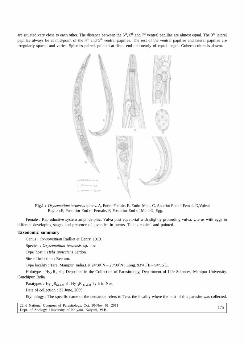
22nd National Congress of Parasitology, Oct. 30-Nov. 01, 2011Dept. of Zoology, University of Kalyani, Kalyani, W.B.
175
are situated very close to each other. The distance between the 5th, 6th and 7th ventral papillae are almost equal. The 3rd lateralpapillae always lie at mid-point of the 4th and 5th ventral papillae. The rest of the ventral papillae and lateral papillae areirregularly spaced and varies. Spicules paired, pointed at distal end and nearly of equal length. Gubernaculum is absent.
Fig.1 : Oxysomatium teraensis sp.nov. A, Entire Female. B, Entire Male. C, Anterior End of Female.D,VulvalRegion.E, Posterior End of Female. F, Posterior End of Male.G, Egg.
Female : Reproductive system amphidelphic. Vulva post equatorial with slightly protruding vulva. Uterus with eggs indifferent developing stages and presence of juveniles in uterus. Tail is conical and pointed.
Taxonomic summaryGenus : Oxysomatium Railliet et Henry, 1913.
Species : Oxysomatium teraensis sp. nov.
Type host : Hyla annectens Jerdon.
Site of infection : Rectum.
Type locality : Tera, Manipur, India.Lat.24º30´N – 25º00´N ; Long. 93º45´E – 94º15´E.
Holotype : Hy2 R1 � ; Deposited in the Collection of Parasitology, Department of Life Sciences, Manipur University,Canchipur, India.
Paratypes : Hy 2R(2,3,4) �, Hy 2R (1,2,3) �; 6 in Nos.
Date of collection : 23 June, 2009.
Etymology : The specific name of the nematode refers to Tera, the locality where the host of this parasite was collected.

22nd National Congress of Parasitology, Oct. 30-Nov. 01, 2011Dept. of Zoology, University of Kalyani, Kalyani, W.B.
176
DiscussionOxysomatium teraensis sp.nov. is characterized by male having 9 post-anal papillae and 10-15 pre-anal papillae at variable
positions, absence of gubernaculum,comparatively longer spicule, cuticular striations which are more prominent at cloacalregion and female having long conical tail, post-equatorial vulva and presence of juveniles in maternal uterus. The cuticularstriations are more prominent around vulval region.
Oxysomatium teraensis sp.nov. is morphologically most similar to O.hylae in the structure of tail, shape and size ofoesophagus, position of nerve ring from anterior end and position of excretory pore from the anterior end. Gubernaculum isabsent in both species. However, the present species differs from O.hylae in having coarse unevenly spaced cuticular striationscompared to faint or indistinct cuticular striations in O.hylae; comparatively larger body size and anal body diameter. A pairof short cuticular extension protruding forwards at the anterior tip in female of O.hylae is absent in O.teraensis sp.nov. Anotherstriking difference is the number and arrangement of the caudal papillae. O.hylae possess 9 pairs of caudal papillae whileO.teraensis sp.nov. possess 19-24 pairs of caudal papillae. O.teraensis sp.nov. have comparatively longer spicules (0.16-0.17)than O.hylae (0.13-0.14). Compared to O.hylae, the present species have numerous and smaller sized eggs and juveniles arepresent in the maternal uterus which is not reported in O.hylae.Comparative chart is given in Table I.
Table I. Comparision of Oxysomatium teraensis sp.nov. with O. hylae Gambhir & Tarnita, 2005
Characters Oxysomatium teraensis sp.nov. O. hylae Gambhir &
Tarnita, 2005
��������������� �������������� �������� ��������
1. Body length 2.52 – 2.7 3.29 – 3.31 1.41 – 2.22 2.73 – 2.93
2. Body width 0.15 – 0.16 0.21 – 0.24 0.07 – 0.10 0.12 – 0.14
3. Total length of 0.40 – 0.42 0.41 – 0.43 0.35 – 0.42 0.43 – 0.47
Oesophagus
4. Muscular oesophagus 0.33 – 0.35 x 0.35 – 0.37 x 0.29 – 0.35 0.36 – 0.40
(Length & width) 0.02 – 0.03 0.03- 0.05
5. Glandular oesophagus or end bulb 0.06 – 0.07 x 0.06 – 0.08 x 0.05 – 0.07 x 0.07 – 0.08 x
(Length & width) 0.05 – 0.06 0.05 – 0.06 0.05 0.06 – 0.07
6. Nerve ring 0.15 – 0.17 0.13 – 0.16 0.13 – 0.16 0.14 – 0.20
7. Excretory pore from the anterior end 0.31 – 0.39 0.32 – 0.33 0.25 – 0.35 0.32 – 0.36
8. Anal opening or cloacal aperture from 2.32 – 2.55 3.0 – 3.01 1.24 - 1.99 2.40 – 2.70
the anterior end
9. Tail length 0.19 – 0.22 0.26 – 0.29 0.12 – 0.23 0.28 – 0.33
10. Spicules length 0.16 – 0.17 0.13 – 0.14
11. Vulva from anterior end 1.76 – 1.81 1.57 – 1.65
12. Eggs 0.08 – 0.15 x 0.16 - 0.21 x
0.05 – 0.09 0.10
13. Anal Body Diameter (ABD) 0.08 – 0.09 0.08 – 0.09 0.05 – 0.06 0.05 – 0.06
14. Caudal papillae Postanal – 9 pairs 5 Postanal
Preanal – 10 to 4 Preanal
15 pairs
15. Cuticularisation Anterior –1.95 – Anterior – 3.9-7.8 µm Cuticle with faint or
3.9 µm Middle – 7.8 µm indistinct striations
Middle – 3.9- 5.85 µm Vulva – 19.5-27.3 µm
Caudal – 7.8- 27.3 µm Caudal – 7.8 µm

22nd National Congress of Parasitology, Oct. 30-Nov. 01, 2011Dept. of Zoology, University of Kalyani, Kalyani, W.B.
177
The new species resembles O.variabilis Harwood (1930) in the presence of coarse cuticular striations and in the patternsof caudal papillae. But it differs from O.variabilis in the absence of gubernaculum, in number of caudal papillae, in havingcomparatively shorter spicules and in the absence of tail spike which is characteristic of O.variabilis. O.teraensis sp.nov. alsohave relatively smaller size oesophagus and differs also in nerve ring position. O.teraensis sp. nov. differs from some similarspecies like O.tibetanum Baylis (1927) ; O.macintoshii (Stewart, 1924) Karve, 1927 ; O. ranae Walton (1931)in the absence ofgubernaculum, body measurements, size of spicules and in the number and arrangement of caudal papillae.
Fotedar (1960) described a new species, O.srinagarensis from rectum of Bufo viridis from Srinagar, Kashmir. O.teraensissp. nov. differs from O.srinagarensis in having coarse cuticular striations (smooth in O.srinagarensis), in the absence of lateralalae, gubernaculum and body papillae which are present in O.srinagarensis. The position of vulva is post-equatorial in thepresent species while in O.srinagarensis the vulva is nearly median in position. The present species also differs fromO.annurae and O.stomatici found in the Indian sub-continent in body measurements, cuticular striations, size of spicules andin the number and arrangement of caudal papillae.
Geographically O.teraensis sp.nov. is close to O. manipurensis and O. striatus. O.teraensis sp.nov. differs from O. striatusin the body width, greater oesophagus length and smaller end-bulb, position of excretory pore (which lies behind the end-bulb in O. striatus), shape of the tail and in having only cuticular striations (O.striatus have striations all over the body exceptthe anterior region). O.teraensis sp.nov. differs from O.manipurensis in nerve ring position, in absence of post- analmembranous lip in the female and absence of warts which are characteristic of O. manipurensis. Cuticular striations are absentin O. manipurensis and it have longer tail, lesser number of caudal papillae (10 pairs) and their mode of arrangement.
On the basis of the above mentioned differences and comparison with the earlier reported ones, the species is consideredto be a new species to which the name Oxysomatium teraensis is given. We anticipate that the rich amphibian fauna of Manipurand other regions in Indo-Burma Hotspot harbours numerous species of Oxysomatium that still await their discovery.
AcknowledgementsThe authors express their sincere thanks to the Head, Department of Life Sciences, Manipur University, Canchipur for
providing necessary laboratory facilities.
ReferencesBhanu Y (2000) A systematic study on the amphibian fauna of Manipur (PhD Thesis). Manipur University pp 65-224
Biswas RK, Chakravarty GK (1963) The systematic studies of zoo-parasitic Oxyuroid nematodes. Z f Parasitenkunde , Berlin 23:411-428
Chaturvedi Y, Kansal KC (1977) Checklist of Indian nematodes. Rec Zool Surv, India Occ Pap 5:16-20
Fotedar DN (1960) On a new species of Oxysomatium (Railliet and Henry, 1913) and some notes on theGenera Oxysomatium and Aplectana. Journal of Helminthology Vol XXXIV, 1/2:141-150
Gambhir RK, Tarnita Th (2005) On a new species of the genus Oxysomatium (Railliet and Henry, 1913) from Hyla annectens ofManipur. J Curr Sc 7(2) : 495-498
Gambhir RK, Tarnita Th (2005) Study of a new species of the genus Oxysomatium from Bufo melanostictus . Flora andFauna II: 1 : 5-7
Harwood PD (1930) A new species of Oxysomatium (Nematode ) with some remarks on the genera Oxysomatium and Aplectana,and observations on the Life History. The Journal of Parasitology Vol XVII : 61-73
Karve JN (1927) A redescription of the species Oxysomatium macintoshii (Stewart, 1914) Ann Mag Hist London 9, 20 : 620-628
Sahay Umapati, Nath Shambhu (1966) On Oxysomatium macintoshii var. nov. (Oxyuridae, Oxysomatinae, Oxysomatium Raillietet Henry, 1913 ) from Rana tigrina . Journal of Ranchi University Vol III : 49-56
Skryabin KI ( 1984) Key to parasitic nematodes , Vol 4 . Amerind Publishing Co Pvt Ltd New Delhi, Bombay, Calcutta, NewYork pp 711-728
Soota TD, Chaturvedi Y (1971) Notes on some nematodes from the unnamed collections of the Zoological Survey of India.Proc Zool Soc Calcutta 24: 13-24
Tarnita Th (2007) Study on the Parasitic Fauna of Amphibia Manipur (PhD Thesis). Manipur University pp 50-68
Walton AC (1933) The Nematode as parasite of Amphibia. The Journal of Parasitology Vol XX, 1 :1-32
Yamaguti S (1961) Systema Helminthum, Vol III. The nematodes of vertebrates. New York, Interscience , Part I&II pp 14 - 99

22nd National Congress of Parasitology, Oct. 30-Nov. 01, 2011Dept. of Zoology, University of Kalyani, Kalyani, W.B.
178
On the nematofauna inhabiting mosses available at Indian Botanic Garden,Howrah, West Bengal, India and description of Thornenema thornei n.sp.
Papia Das and Buddhadeb Manna
Department of Zoology, University of Calcutta, Kolkata-700019, West Bengal, India
Abstract : The study of moss associated nematofauna is the first of its kind in India except one report of Gadea (1968). The collectednematofauna constitute 18 species belonging to 14 genera in 7 orders. Among this, omnivores were the most common and dominant trophicgroup and Dorylaimida was the most dominant order in the nematocoenosis of mosses at Botanical Garden, Kolkata. Mesodorylaimuschamoliensis Ahmad, 1995 and Microdorylaimus n.sp. were the most common and prevalent species in mosses of this area, whileMesodorylaimus chamoliensis Ahmad, 1995 was the most prominent species. But Aporcelaimellus n.sp 2 was the most dominant andimportant species in the nematode community of mosses of Botanic Garden, Howrah, West Bengal. A new species Thornenema thornein.sp. is described here.
Key words : nematofauna, Botanic garden, nematocoenosis of mosses, Mesodorylaimus, Thornenema
InroductionMosses harbour a characteristics fauna of nematodes which are able to withstand periodic desiccation and extremes of
temperature (Menzel, 1920; Burkhalter, 1928). Throughout the world numbers of studies have been done on nematode faunaof mosses. (Steiner, 1916; Gadea, 1970, 1972; Zullini, 1971, 1975; Lazarova et al., 2000; Barbuto et al., 2006), but practicallynothing is known in this field in India except one papers by Gadea (1968) on this aspect from East Ghates (Orissa) and anotherby Duggal and Koul (1985) on plant parasitic nematodes associated with bryophytes and pterydophytes in north-west India.
The present work is the humble effort to analyze the distribution of nematodes from mosses in Indian Botanic Gardenat Shibpur, West Bengal. This paper will be the first contribution to the knowledge of moss inhabiting nematofauna from WestBengal and also that from Botanic Garden.
Materials and MethodsNematodes were collected from mosses found in Indian Botanical Garden, Howrah (22o33’N, 80o22’E) lying on the west
bank of river Hooghli and covers 109 hectares of land. The species of mosses collected were of genera Bryum, Fissidens,Drepanocladus, Thuidium.
The moss samples collected from soil, walls and trees during June and September 2007 were immersed in water for 24hours and then nematodes were extracted with the help of modified Baermann funnel technique (Christie and Perry, 1951).Nematodes were fixed with hot F.A solution (Formalin: Acetic acid, 4:1) (Baqri, 1990), processed to an anhydrous glycerine(Seinhorst,1959), mounted on slides and identified to species level using a high magnification microscope following schemesof Andrassy (1976,1984) for orders Monhysterida, Araeolaimida, Chromadorida, Rhabditida, Tylenchida, Enoplida and Jairajpuri& Khan (1982) for Mononchida and Jairajpuri & Ahmad (1992) for Dorylaimida. The moss dwelling nematode genera wereassigned to trophic groups according to classification of Yeates et al. (1993). The various trophic groups considered werepredators, omnivores, bacteriovores, hyphal feeders and unicellular eukaryote feeders. All the ecological parameters werecalculated by the methods given by Norton (1978) and Boag (1993).
ResultsVertical distribution of nematodes in mosses
The average pH of the collected moss samples was found to be 5.7 and vertical distributions of nematodes in these mosseswere studied. Out of 26 moss samples, collected from different substrates and different heights from ground in Botanic Garden,
* Corresponding Author : Email ID : [email protected]; [email protected]

22nd National Congress of Parasitology, Oct. 30-Nov. 01, 2011Dept. of Zoology, University of Kalyani, Kalyani, W.B.
179
Table I. Distribution of nematodes in mosses collected from different sites in Indian Botanic Garden, Shibpur, Howrah
Sample No. Site of collection Abundance/10gm
1 Bark of Casuarina at 5.5ft from ground 02 Bark of Polyalthia at 3.0ft from ground 53 Bark of Swietenia at 4.5ft from ground 04 Bark of Samanea at 3ft from ground 05 Bark of Swietenia at 4.5ft from ground 1006 Bark of Palm tree at 4.0ft from ground 07 Bark of Swietenia at 1.0ft from ground 08 Bark of Swietenia at 0.75ft from ground 4749 Bark of Polyalthia at 1.5ft from ground 4210 Bark of Tabebuia at 4.5ft from ground 011 Rock at ground 012 Soil around Cactus plant 1813. Bark of Tamarindus at 2.5ft from ground 5914 Bark of Samanea at 4.5ft from ground 4515 Bark of Palm tree at 1.5ft from ground 3916 Bark of Tabebuia at 3.5ft from ground 2017 Bark of Casuarina at 5.0ft from ground 1018 Bark of Swietenia at 5.5ft from ground 3919 Brick at ground 9120 Bark of Palm tree at 1.0ft from ground 8221 Bark of Dalbergia at 2.5ft from ground 12822 Soil around Cactus plant 1323 Bark of Palm tree at 1.0ft from ground 4824 Bark of Casuarina at 5.5ft from ground 6325 Bark of Adenanthera at 2.5ft from ground 026 Bark of Eriophyton at 5.0ft from ground 5
18 samples i.e., 69.23 % of the samples contain nematodes. The total number of nematodes varied widely among the samples(0-474 individuals/10g of moss). There were atleast one and atmost four nematode species per sample. But no definite relationwas found between the vertical height of site of moss sampling and abundance of nematodes in these sites. (Table I.)
Percentage of nematode order
A total of seven orders of nematodes have been encountered in the present study, of which Dorylaimida was the mostdominant order (90.04%) among the moss inhabiting nematodes of this area followed by Enoplida (3.46%), while Mononchidawas least frequent order (0.3 %) (Table II). A total of 18 species belonging to 14 genera in 7 orders were identified from differentmoss samples of this area. These included 11 species belonging to 7 genera in order Dorylaimida, 2 species belonging to 2genera in order Araeolaimida and 1 species from each of order Enoplida, Mononchida, Rhabditida, Chromadorida andMonhysterida.
Table II. Percentage of different orders of nematodes inhabiting mosses of Botanical Garden, Kolkata, West Bengal
Serial Number Different orders of nematodes Percentage (%)
1. Dorylaimida 90.042. Araeolaimida 3.003. Chromadorida 0.604. Rhabditida 0.605. Monhysterida 2.006. Mononchida 0.307. Enoplida 3.46

22nd National Congress of Parasitology, Oct. 30-Nov. 01, 2011Dept. of Zoology, University of Kalyani, Kalyani, W.B.
180
Table III. Community analysis of moss dwelling nematodes of Indian Botanic Garden, Howrah, West Bengal.(N- Frequency,P- Prevalence, I- Mean Intensity, PV- Prominence value, TG-Total biomass, FT- Feeding type, IV-Importance value)
Sl. Nematode Taxa N P I PV TG IV FTNo µgm
Dorylaimida
1. Aporcelaimellus n. sp1 1 3.85 12.00 23.55 3.75 9.27 o
2. Aporcelaimellus n.sp2 4 15.38 51.75 202.95 30.86 56.56 o
3. Aporcelaimellus chauhani Baqri et Khera, 1975 1 3.85 4.00 7.85 0.99 4.49 o
4. Mesodorylaimus chamoliensis Ahmad, 1995 6 23.08 57.67 277.06 9.72 37.76 o
5. Mesodorylaimus n.sp 1 3.85 19.00 37.28 2.26 8.83 o
6. Thornenema n.sp. 3 11.54 27.67 93.9 2.28 15.60 o
7. Tylencholaimus n.sp 1 3.85 72.00 141.27 1.53 7.02 hf
8. Microdorylaimus n.sp. 6 23.08 21.67 132.93 5.98 26.54 o
9. Eudorylaimus n.sp. 1 3.85 75.00 147.16 3.27 20.58 o
10. Eudorylaimus brevis (Altherr, 1952) Andrassy,1959 4 15.38 34.00 133.34 17.39 37.26 o
11. Thornia juvenilis (De Coninck,1935) Meyl, 1954 1 3.85 14.00 27.47 0.12 5.35 o
Enoplida
12. Alaimus acutus Thorne, 1939 3 11.54 22.33 75.86 2.01 14.28 b
Araeolaimida
13. Plectus parietinus Bastian, 1865 1 3.85 14.00 27.47 1.68 7.20 b
14. Cylindrolaimus communis De Man, 1880 1 3.85 43.00 42.57 1.05 11.91 b
Mononchida
15. Mylonchulus lacustris (Cobb in Cobb, 1915), 2 7.69 2.50 6.93 0.53 6.23 pCobb, 1917
Rhabditida
16. Eucephalobus oxyroides (De Man, 1876), 1 3.85 7.00 13.73 0.12 4.03 bSteiner, 1936
Chromadorida
17. Achromadora ruricola (De Man, 1880) 1 3.85 20.00 39.24 0.71 7.18 uMicoletzky,1925
Monhysterida
18. Geomonhystera villosa (BÜTSCHLI 1873) 1 3.85 33.00 64.75 0.27 9.11 bAndrassy, 1981
Feeding Types : bacterial feeders (b), omnivores (o), predators (p), unicellular eukaryote feeders (u), hyphal feeders (hf).
Community analysis of moss dwelling nematodes of Indian Botanic Garden
Mesodorylaimus chamoliensis Ahmad, 1995 and Microdorylaimus n.sp. were the most common and prevalent speciesin mosses of Botanic Garden, Howrah. Eudorylaimus n. sp. showed the highest mean intensity followed by Tylencholaimusn.sp., while Mylonchulus lacustris (Cobb in Cobb, 1915) Cobb, 1917 showed least mean intensity. Mesodorylaimus chamoliensisAhmad, 1995 was the most prominent species among the mosses of this area followed by Aporcelaimellus n.sp 2, whileMylonchulus lacustris (Cobb in Cobb, 1915) Cobb, 1917 was least prominent. Aporcelaimellus n.sp 2 was found to be themost dominant species among the moss associated nematodes of this area followed by Eudorylaimus brevis (Altherr, 1952)Andrassy 1959. Aporcelaimellus n.sp 2 had the highest importance value while Eucephalobus oxyroides (de Man, 1876)Steiner, 1936 had lowest importance value.(Table III).

22nd National Congress of Parasitology, Oct. 30-Nov. 01, 2011Dept. of Zoology, University of Kalyani, Kalyani, W.B.
181
Trophic relation of the nematodes
Among the 18 species of nematodes encountered from the moss samples of this area, 10 species were omnivorous, 5species were bacteriovorous, 1 species was predator, 1 species was hyphal feeder and 1 species was unicellular eukaryotefeeder. Omnivores were found to be the most dominant and frequent group in the nematocoenosis of mosses at Indian BotanicGarden, Howrah, followed by bacterial feeders while predators were least frequent group. (Table IV). All the omnivoresencountered in the present study belong to Order Dorylaimida and Superfamily Dorylaimoidea.
Table IV. Percentage of trophic groups of nematodes inhabiting mosses of Botanical Garden, Kolkata, West Bengal
Serial Number Feeding group Percentage (%)
1. Omnivores 89.80
2. Bacterial feeders 6.00
3. Unicellular eukaryote feeders 0.50
4. Hyphal feeders 3.50
5. Predators 0.20
Description of Thornenema thornei n. sp. (Fig.1)
A
B
C
D
E
Figure 1. Thornenema thornei n. sp. (Camera lucida drawing) Female. A- Head region, B- Tail region,C- Gonad, D- Entire body, E- Oesophago-intestinal junction

22nd National Congress of Parasitology, Oct. 30-Nov. 01, 2011Dept. of Zoology, University of Kalyani, Kalyani, W.B.
182
Order- Dorylaimida Pearse, 1942
Suborder- Dorylaimina Pearse, 1936
Superfamily- Dorylaimoidea De Man, 1876
Family- Dorylaimidae De Man, 1876
Subfamily- Thornenematinae Siddiqi, 1969
Genus- Thornenema Andrassy, 1959
Species- Thornenema thornei n.sp.
Measurements
Female (paratypes; n = 2): L = 0.65-0.66 (0.65) mm; a = 23.6-26.9 (25.2); b = 4.0-4.3 (4.2); c = 10.3-10.8 (10.5); c/ = 3.8-4.0(3.9); V = 32.6-34.1 (33.3); G2 = 26.7-29.8 (28.2); odontostyle = 10.0-12.5 (11.3) µm; odontophore = 18.75 µm; oesophagus = 150.0-165.0 (157.5) µm; prerectum = 40.0-52.5 (46.3) µm; rectum = 20.0-24.0 (22.0) µm; tail = 60.0-64.0 (62.0) µm; ABD = 16.0 µm.
Female (holotype): L = 0.66 mm; a = 23.6; b = 4.4; c =11.0; c/ = 3.8; V = 31.8; G2 =30.9; odontostyle = 10.0 µm; odontophore= 18.8 µm; oesophagus = 165.0µm; prerectum = 40.0 µm; rectum = 20.0 µm; tail =60.0 µm; ABD = 16.0 µm.
Female : - Body small sized, ventrally curved upon fixation, 24-28 µm wide at mid region. Cuticle with fine transversestriations, 2.5µm at mid region and at tail end. Labial region 6.3 µm wide, amalgamated, rounded and offset by a constriction;framework sclerotised; post-labial sclerotisation present and slightly sigmoid in optical section. Body at posterior end ofoesophagus 3.8 times as wide as head. Amphid stirrup-shaped; 5.0µm wide. Odontostyle cylindrical, but more or less taperingtowards tip, 10.0-12.5µm, 1.6-2.0 lip region widths long. Aperture occupying about one-fourth of the stylet length. Guiding ringsingle and plicate, at 7.5-8.8 µm from anterior end. Odontophore rod-shaped, 1.9 times the odontostyle length. Odontophoreregion of pharynx spindle-shaped; followed by a long slender portion which becomes narrower at nerve ring level and graduallywidens to the basal enlarged portion; the latter about 34.6 - 36.4 % of total length. Nerve ring encircling the anterior slenderpart of pharynx at 44.0% of total neck length. Pharyngeal gland nuclei are located as D = 68.9-72.0 % , AS1 = 26.8-29.0% ,AS2 = 26.8-29.0% % , PS1 = 41.5% , PS2 = 51.2%. Cardia conoid with rounded terminus, about 0.42-0.53 times as long ascorresponding body width. Reproductive system mono-opisthodelphic, anterior uterine sac absent. Posterior gonad normal,176-204 µm long or 6.9-7.3, occupying 26.7-30.9% of body length. Ovary reflexed with oocytes arranged in a single row. Oneuterine egg observed in one female specimen measuring 64x24 µm. Vulva pore-like with moderately sclerotised pars refringes.Vagina 11.3 µm long or 1/2-1/2.5 of the corresponding body width long. A pair of glands present on both sides of vagina.Pars distalis vaginae 3.8 µm long; pars refringes 1.3 µm long; pars proximalis vaginae 6.3 µm long. Uterus 40-64 µm long.Prerectum 2.8-3.3 anal body width long. Rectum 20-24 µm long, 1.3-1.5 anal body width long. Tail elongated convex-conoid,60-64µm long, 3.8-4.0 times as long as anal body diameter, and occupying 9.1-9.7% of entire body length.
Male : - Not found.
Remarks : Presently there are eighteen valid species, Thornenema andrassyi Khan et al., 1989, T. baldum (Thorne, 1939)Andrássy, 1959, T. coomansi Ahmad & Ahmad, 2003, T. elaboratum Baqri & Jana, 1986, T. elegans Carbonell & Coomans,1986, T. garhwalicum Srivastava et al., 2000, T. laevicapitatum (Cobb in Thorne & Swanger, 1936) Andrássy, 1959, T. lissum(Thorne, 1939) Andrássy, 1959, T. maraouense Coomans & Carbonell, 1981, T. mauritianum (Williams, 1959) Baqri & Jairajpuri,1967,
T. oryzae (Ahmad & Jairajpuri, 1982) Carbonell & Coomans, 1986, T. paraconurum (Heyns, 1963) Goseco et al., 1976, T.pseudosartum Carbonell & Coomans, 1986, T. punjabsinghi Azmi & Ahmad, 1993, T. samsoeni Coomans & Carbonell, 1981,T. shahi Khan & Saeed, 1986, T. shamimi (Baqri & Jana, 1980) Carbonell & Coomans, 1986 and T. spicatum Shaheen & Ahmad,2005 in the genus Thornenema.
Among these eighteen valid species of this genus described so far, the present species closely resembles T. lissum(Thorne, 1939) Andrássy, 1959, but differs from it in having smaller body length (vs.1.2mm), smaller value of c’ (vs.7.0), largervalue of c (vs.9.0), pore-like vulva and differently-shaped tail.
It also closely resembles T. paraconurum Heyns, 1963 but differs from the latter in having smaller body length (vs.1.3-1.6mm), larger value of c (vs.5.9-9.5), and smaller value of c’ (vs.7.0-12.0), pore-like vulva and differently-shaped tail.
Type locality and habitat : The new species was collected from moss (Bryum sp.) at Indian Botanic Garden, Howrah,West Bengal, India.

22nd National Congress of Parasitology, Oct. 30-Nov. 01, 2011Dept. of Zoology, University of Kalyani, Kalyani, W.B.
183
Type specimens : One female specimen as holotype in one slide under Accession number 000041N/10 and 2 femalespecimens as paratype in one slide under Accession number 000042N/10 are deposited at present to the nematode collectionof Parasitology Research Unit, Department of Zoology, University of Calcutta, 35 Ballygunge Circular Road, Kolkata-700019,India.
DiscussionThe total number of nematodes associated with mosses varied greatly between samples and substrates, probably due
to micro environmental conditions. It is also possible that the variation in both species abundance and community compositioncan be related to seasonal changes in temperature and precipitation, as has been demonstrated by Zullini (1971) and Steiner(1994), although Spaull (1973) found no direct correlation between water content of mosses and total number of nematodes.
Mesodorylaimus chamoliensis Ahmad, 1995 was found to be the most common, prevalent and prominent species amongthe moss inhabiting nematodes of Botanical Garden, Kolkata. But Aporcelaimellus n.sp 2 was found to be the most dominantand important species among the moss dwelling nematodes of this area due to its highest mean total biomass. The mean totalbiomass Aporcelaimellus n.sp 2 was higher than other prevalent species because of their larger body size. Gadea (1968) foundMesodorylaimus bastiani (Bütschli, 1873) Andrássy, 1959 as the dominant species among the moss dwelling nematodes ofEastern Ghates, India followed by Rhabdolaimus terrestris de Man 1880.
Omnivores were found to be most dominant and frequent group in the nematocoenosis of mosses in Botanic Garden,Howrah as also observed by Zullini and Peretti (1986), Lobaton (1991), Gadea (1968), and Gonzales (1972) from various mosssamples from different countries. It has further been found that all the omnivorous nematodes encountered in the present studybelong to order Dorylaimida. According to some authors (Johnson et al., 1974, Zullini and Peretti 1986, Bongers, 1990),dorylaimids are considered k-strategists in the broad sense, being very sensitive to disturbances. Thus great number ofcollected dorylaimids in the present study could also be the indicative of nematode community structure in undistributedenvironment ie, it confirms the good environmental condition of the investigated station.
The present species, Thornenema thornei n. sp. is morphologically close to T. lissum and T. paraconurum but differsfrom them in a number of morphological characters. Considering all these differences the authors suggest its name as T. thorneiafter an eminent nematologist, Prof. Gerald Thorne in recognition of his contribution to the taxonomy of nematodes.
AcknowledgementsAuthors are thankful to the Director, Indian Botanic Garden, Shibpur, Howrah for giving me the opportunity to collect
samples from this area for my research work. The authors are also grateful to the Head, Department Of Zoology, Universityof Calcutta for providing laboratory and other facilities.
ReferencesAhmad A and Ahmad W (2003). Studies on some species of Thornenematidae (Nematoda: Dorylaimida) from south India.
Journal of Nematode Morphology and Systematics 5(2): 131-144.
Ahmad W (1995). Description of two new species of Mesodorylaimus (Nematoda: Dorylaimida) from India. Afro-Asian journalof Nematology 5: 178-181.
Ahmad W, Jairajpuri M S (1982). Some new and known species of Dorylaimoides. Nematologica 28(1): 39-61.
Altherr E (1952). Les nématodes du Parc national suisse. (Nématodes libres du sol.) – Ergebn Wiss Untersuch Schweiz Nat26: 315-356.
Andrássy I (1959). Taxonomische Übersicht der Dorylaimen (Nematoda), I. Acta Zoologica Hungarica 5: 191-240.
Andrássy I (1976). Evolution as a basis for the systematization of nematodes. London: Pitman Publishing pp 1- 288.
Andrássy I (1984). Klasse Nematoda (Ordnungen Monhysterida, Desmocolecida, Araeolaimida, Chromadorida, Rhabditida).Stuttgart, Gustav Fisher Verlag pp 1-509.
Azmi M I, Ahmad S T (1993). Discolaimium rakebi n. sp. and Thornenema punjabsinghi n. sp. from Jhansi, India. CurrentNematology 4(2): 233-238.
Baqri Q H (1990). Collection and preservation : Nematoda. In collection and preservation of animals; Director, Zoological Surveyof India (ed.) 43-49.
Baqri Q H, Jairajpuri M S (1967). Review of the genus Thornenema Andrássy, 1959 and proposal of Willinema n. gen.Nematologica 13: 353-366.
22nd National Congress of Parasitology, Oct. 30-Nov. 01, 2011Dept. of Zoology, University of Kalyani, Kalyani, W.B.
184
Baqri Q H, Jana A (1986). Nematodes from West Bengal (India), IX. Three new species of the superfamily Dorylaimoidea (DeMan, 1876) Thorne, 1934. Indian Journal of Helminthology NS 3(2): 11-20.
Barbuto M, Zullini A (2006). Moss inhabiting nematodes: influence of the moss substratum and geographical distribution inEurope. Nematology 8: 575-582.
Boag B (1993). Standardisation of ecological terms in nematology. Fundamental and Applied Nematology 16:190-191.
Bongers T (1990). The maturity index: an ecological measure of environmental disturbance based on nematode speciescomposition. Oecologia 83: 14–19.
Burkhalter M (1928). Die Verbreitung der freilebenden Ernematoden im Masif der Rochers de Naye. Revue Suissede Zoologie, 35. In: The Invertebrates: Acanthocephala, Aschelminthes and Entoprocta. The pseudocoelomateBilateria. by L.H. Hyman. Mc.Graw. Hill Book Company, Inc 3: 480-486.
Bütschli O (1873). Beiträge zur Kenntnis der freilebenden Nematoden. – Nova Acta Acad Nat Curios 36: 1-24.
Carbonell E, Coomans A (1986). The genus Thornenema Andrassy, 1959 (Nematoda: Thornenematidae). Nematologica 32: 125-179.
Christie JR, Perry VG (1951). Removing nematodes from soil. Proc Helm Soc Wash 18: 106-108.
Cobb N A (1917). The Mononchs (Mononchus Bastian, 1865). A genus of freeliving predatory nematodes. Soil Science 3: 431-486.
Coomans A, Carbonell E (1981). Observations on the genus Thornenema Andrassy, 1959 with description of two new speciesfrom Ivory Coast. Nematologica 27: 292-305.
De Man J G (1876). Onderzoekingen over vrij in de aarde levende Nematoden. Tijdschrift der Nederlandsche DierkundigeVereeniging 2: 78-196.
De Man J G (1880). Die einheimischen, frei in der reinen Erde und im süssen Wasser lebende Nematoden. Vorläufi ger Berichtund descriptivsystematischer Theil. Tijdschrift der Nederlandsche Dierkundige Vereeniging 5: 1-104.
Duggal CL, Koul R (1985). On some plant parasitic nematodes associated with bryophytes and pterydophytes in north-westIndia. Helminthologia 22(4): 245-251.
Gadea E (1968). Sobre la nematofauna muscícola de los Gates orientales (India). Publicaciones del Istituto de Biologia Aplicada45: 57-64.
Gadea E (1970). Algunas considerationes sobre las nematocenosis de suelos primordiales. Publicaciones del Istituto de BiologiaAplicada 49: 59-64.
Gadea E (1972). Peuplement et nématofaune muscicole de Minorque. Rapports et Procès Verbaux des Réunions CommissionInternationale pour l’Exploration Scientifique de la Mer Méditerranée 21: 93-95.
Goseco CG, Ferris VR, Ferris JM (1976). Revisions in Leptonchoidea (Nematoda: Dorylaimida), Dorylaimoides in Dorylaimoididae,Dorylaimoidinae; Calolaimus & Timmus n. gen. in Dorylaimoididae, Calolaiminae; and Miranema in Miranematidae.Purdue University Agriculture Experimental Station Research Bulletin pp 941: 46.
Gonzales AP (1972). Sobre algunos nematodos muscìcolas de la zona de Ledesma (prov. Salamanca). Publicationes del Istitutode Biologia Aplicada 52: 107-119.
Heyns J (1963). New species of the superfamily Dorylaimoidea (Nematoda) from South African soils, with a description of anew genus Kochinema. South African Journal of Agricultural Science 6: 289-302.
Jairajpuri M S, Ahmad W (1992). Dorylaimida. Free-living, Predacious and Plant-parasitic nematodes. Brill, Leiden, pp1- 458.
Jairajpuri M S, Khan WU (1982). Predatory nematodes (Mononchida) with special reference to India. New Delhi, India:Associated Publishing Company, pp 1-131.
Johnson S R, Ferris J M, Ferris V R (1974). Nematode community structure of forest woodlots. III. Ordinations of Taxonomicgroups and biomass. Journal of Nematology 6:118-126.
Khan T H, Jairajpuri M S, Ahmad W (1989). Descriptions of some new and known species of dorylaim nematodes. Nematologica35(4): 419-437.
Khan HA, Saeed A (1986). Studies on dorylaimid nematodes from the Indus Delta. 1. Mesodorylaimus mehraniensis n. sp.Bangladesh Journal of Zoology 14(2): 117-122.

22nd National Congress of Parasitology, Oct. 30-Nov. 01, 2011Dept. of Zoology, University of Kalyani, Kalyani, W.B.
185
Lazarova S, Peneva V, Penev L (2000). Nematode assemblages from the moss Hypnum cupressiforme Hedw. growing on differentsubstrates in a balkanic durmast oak forest (Quercus dalechampii Ten.) on Mount Vitosha, Bulgaria. Nematology 2:263-272.
Lobaton M, Del CS (1991). Sobre la nematofauna muscìcola del Pinsapar (Abies pinsapo Boiss) de la Sierra de Grazalema(España). Nematologia Mediterranea 19: 197-206.
Menzel R (1920). Uber die Nahrung der freilebenden Nematoden. Verhandl. Naturforsch. Gesell. Basel 31. In: “The Invertebrates:Acanthocephala, Aschelminthes and Entoprocta. The pseudocoelomate Bilateria.” by L.H. Hyman. Mc.Graw. Hill BookCompany, Inc 3: 480-486.
Norton DC (1978). Ecology of plant parasitic nematodes. John Wiley and Sons, New York 1-268.
Pearse A S (1936). Zoological Names. A List of Phyla, Classes, and Orders Prepared for Sect. F. American Association for theAdvancement of Science, Durham, N.C.: Duke University Press 1-24.
Pearse A S (1942). Introduction to Parasitology. Springfield, III:Baltimore, Md., pp 1-357.
Seinhorst J W (1959). A rapid method for the transfer of nematodes from fixative to anhydreous glycerin. Nematologica 4: 67-69.
Shaheen A, Ahmad W (2005). Descriptions of four new species of Dorylaimida (Nematoda). Journal of Nematode Morphologyand Systematics 8(1): 19-29.
Siddiqi MR (1969). Mumtazium mumtazae n.gen., n.sp. (Nematoda: Tylencholaimidae) with the proposal of Laimydorus n. gen.(Thornenematidae). Nematologica 15: 234-240.
Srivastava N, Rawat V S, Ahmad M (2000). Description of three new species of dorylaims (Nematoda: Dorylaimida) associatedwith Litchi fruit trees in Doon Valley (U.P.) India. Indian Journal of Nematology 30(1): 46-52.
Steiner G (1916). Freilebende Nematoden aus der Barentsee. Zool. Jahrb. Ab. System.39. In “The Invertebrates: Acanthocephala,Aschelminthes and Entoprocta. The pseudocoelomate Bilateria.” by L.H. Hyman. Mc.Graw. Hill Book Company, Inc.,3, pp 480-486.
Steiner G (1936). Opuscula miscellanea nematologica IV. Proceedings of the Helminthological Society of Washington 3: 74-80.
Steiner WA (1994). The influence of air pollution on moss-dwelling animals: 2. Aquatic fauna with emphasis on Nematoda andTardigrada. Revue Suisse de Zoologie 101: 1017-1031.
Spaull V W (1973). Qualitative and quantitative distribution of soil nematodes of Signy Island, South Orkney Islands. BritishAntarctic Survey Bulletin 33-34: 177-184.
Thorne G (1939). A monograph of the nematodes of the superfamily Dorylaimoidea. Capita Zoologica 8 (5): 1-261.
Thorne G, Swanger H H (1936). A monograph of the nematode genera Dorylaimus Dujardin, Aporcelaimus n. g., Dorylaimoidesn. g. and Pungentus. Capita Zoologica 6: 1-223.
Yeates GW, Bongers T, De Goede RG, Freckman DW, Georgieva SS (1993). Feeding habits in soil nematode families and genera- an outline for soil ecologists. Journal of Nematology 25: 315-331.
Zullini A (1971). Studio sulle variazioni del popolamento nematologico in un muschio. Rendiconti dell’Istituto LombardoAccademia di Scienze e Lettere B 105: 89-106.
Zullini A (1975). Ecologia dei nematodi del suolo. Rendiconti dell’Istituto Lombardo Accademia di Scienze e Lettere B 26: 289-380.
Zullini A, E Peretti (1986). Lead pollution and moss inhabiting nematodes of an industrial area. Water, Air and Soil Pollution27:403-410.

22nd National Congress of Parasitology, Oct. 30-Nov. 01, 2011Dept. of Zoology, University of Kalyani, Kalyani, W.B.
186
Impact of host population and abiotic factors on the percent parasitism byEretmocerus adustiscutum, a parasitoid of Aleuroclava pentatuberculata
Paushalee Dutta1, Manne Venkata Santha Kumar*, Samiran Chakrabarti**, Swapan Kumar Mukhopadhyay*,Nirvan Kumar Das*, Prabir Mitra*, Atul Kumar Saha* and Arvind Kumar Bajpai*
1Union Christian College, Berhampore, West Bengal, India*Central Sericultural Research and Training Institute, Berhampore, West Bengal, India
** Department of Zoology, University of Kalyani, Kalyani, West Bengal, India
Abstract : The dynamic role of parasitoids in a natural host-parasitoid interaction can be resolved into two components: The degree ofdepression in the host population density caused by parasitism and the degree of stability conferred on the interaction in the long term.Both of these components are likely to vary from one host-parasitoid system to another. The importance of parasitoids within theassortment of natural mortality factors affecting host populations can only be evaluated by detailed field studies of both host and parasitoidover a number of generations. For understanding the interactions between whitefly, Aleuroclava pentatuberculata (Sundararaj & David)(Homoptera: Aleyrodidae) and its parasitoid, Eretmocerus adustiscutum (Krishnan & David) (Hymenoptera : Chalcidoidea) populationdynamics studies were conducted during 2004-2007 and their relation with abiotic factors were worked out. The observations have revealedthat percent parasitism was positively and significantly correlated with maximum and minimum temperature. The significance level formaximum temperature was at 1% level whereas that of minimum temperature was at 5% level. The correlations of percent parasitism withthat of maximum and minimum relative humidity and rainfall were observed as negatively significant with maximum relative humidity andnon significant with that of minimum relative humidity and rainfall. The r values for maximum and minimum relative humidity and rainfallwere found to be -0.295, -0.043 and -0.030 respectively. The correlation of the host population with the percent parasitism was notedto be negative and non-significant with r value as -0.080. Multiple regression analysis was performed to draw inference related to forecastingthe probable incidence of the parasitoid in relation to host population. The observations revealed that the abiotic factors of previous 27-33 days and the host population of same day were found to influence the incidence of the parasitoid.
Key words : Abiotic factors, Aleuroclava pentatuberculata, biotic factor, correlation coefficient, Eretmocerus adustiscutum, multiple linear
regression, parasitism.
IntroductionIn insects, the population dynamics is the result of interaction between physical conditions, population movements and
both density dependent and independent factors. The relative importance of each of the factors might vary among populationsor even within a population from time to time (Huffaker et al., 1984). It was reported that the climatic factors have dominatinginfluence on various aspects of an individual insect’s life cycle such as reproduction, development, fecundity and longevity(Howe, 1953). Fluctuation in number of insects has been closely correlated with changes in weather and the populationdensities were determined coupled with relatively small degree of mortality caused by parasitoids, predators and diseases havecommonly been regarded as establishing the importance of the physical factors in determining the abundance of insects ina given population (Cook, 1930). In any ecosystem, populations of different organisms interact with biotic and abioticenvironments with the limiting factors most likely to maintain population densities in equilibrium (Berryman, 1993). Thesefactors which limit population growth and regulate population dynamics can be used as parameters to make quantitativeforecasts of population fluctuations (Berryman, 1997). Accordingly in the present study interactions between population ofwhitefly, Aleuroclava pentatuberculata (Sundar raj & David) (Homoptera : Aleryrodidae) and their native parasitoid,Eritmocerus adustiscutum (Krishnan & David) (Hymenoptera : Chalcidoidea) were worked out in order to facilitate theforecasting the native parasitoid population for effective biological control of this serious pest.
Materials and methodsThe population size of whitefly was studied in the mulberry fields of Central Sericultural Research and Training Institute,
Berhampore during 2004-2007. Population was recorded at weekly intervals in randomly selected 20 mulberry plants. The

22nd National Congress of Parasitology, Oct. 30-Nov. 01, 2011Dept. of Zoology, University of Kalyani, Kalyani, W.B.
187
vertical population of whitefly was assessed by considering top (1-3), middle (4-7) and bottom (7-14) strata (Lynch andSymmons, 1993). To reduce disturbance that might have interfered with an accruable census, counts were done on two leaves(top, middle, bottom). The investigations were made during cooler hours of the day preferably 6.00 A.M. – 7.00 A.M. (Naranjoand Flint, 1995). Counts of whitefly were made carefully turning the leaf over by rotating the petiole (Naik and Lingappa, 1992).The incidence of E.adustiscutum was noted by collecting the middle and bottom positioned leaves from all 20 plants infestedwith nymphal population of whitefly. These nymphs were screened in the laboratory under stereomicroscope that whether theyare parasitized or not by following the methods suggested by (Van Dreishe et al., 2001b). For understanding the relationshipbetween the parasitoid, whitefly and meteorological factors, the data was subjected to analysis by Coefficient of Correlation.To develop forecasting models of this parasitoid the data was analyzed by multiple linear regression analysis and fittings ofthe regression equations were adjudged by the significance of R² (the coefficient of determination).
ResultsThe seasonal incidence studies of whitefly, A. pentatuberculata in the mulberry ecosystem had revealed that, during the
first year of the study (2004-05), it was noticed that the population (23.93 per plant) was above the economic threshold level(20/plant) from the very first week of the observation i.e. during fourth week of August, 04. The population of the pest aftershowing an increasing trend during the first week suddenly came down for two consecutive weeks in September (Fig.1a). Thewhitefly population was maximum (41.83 per plant) during second week of October, 2010. The count of the whitefly remainedbelow the threshold level during November, 2004 – August, 2005.
Fig. 1(a, b, c) : Incidence of A. pentatuberculata (No./Plant) in different years
5040302010
0
Pop
ulat
ion
24-08-2004
14-09-2004
05-10-2004
16-11-2004
07-12-2004
28-12-2004
18-01-2005
08-02-2005
01-03-2005
22-03-2005
12-04-2005
03-05-2005
24-05-2005
14-06-2005
05-07-2005
26-07-2005
16-08-2005
Date of Observation
Year : 2004-05
80
60
40
20
0
Pop
ulat
ion
23-08-2005
13-09-2005
04-10-2005
25-10-2005
15-11-2005
06-12-2005
27-12-2005
17-01-2006
07-02-2006
28-02-2006
21-03-2006
11-04-2006
02-05-2006
23-05-2006
13-06-2006
04-07-2006
25-07-2006
16-08-2006
Date of Observation
Year : 2005-06
100
75
50
25
0
Pop
ulat
ion
22-08-2006
12-09-2006
03-10-2006
24-10-2006
14-11-2006
05-12-2006
26-12-2006
16-01-2007
06-02-2007
27-02-2007
20-03-2007
10-04-2007
01-05-2007
22-05-2007
12-06-2007
03-07-2007
24-07-2007
14-08-2007
Date of Observation
125
Year : 2006-07

22nd National Congress of Parasitology, Oct. 30-Nov. 01, 2011Dept. of Zoology, University of Kalyani, Kalyani, W.B.
188
Year : 2004-05
60.00
40.00
20.00
0.00
% P
opul
atio
n
24-08-2004
14-09-2004
05-10-2004
26-10-2004
16-11-2004
07-12-2004
28-12-2004
18-01-2005
08-02-2005
01-03-2005
22-03-2005
12-04-2005
03-05-2005
24-05-2005
14-06-2005
05-07-2005
26-07-2005
16-08-2005
Date of Observation
During the second year (2005-06), the population of A. pentatuberculata was highest (70.05 per plant) during second weekof October, 2005 (Fig. 1b], one month earlier in comparison to the previous year of the study. The population of A.pentatuberculata lowered down from the end of December and was found to be below 10 per plant till August, 2006. Theminimal population was found to be 0.83 per plant during second week of May, 2006 (Fig. 2b). Thus, the trend of seasonalincidence was almost same as that of the previous year with very low population from March to May.
Fig. 2(a, b, c) : Incidence of E. adustiscutum on A. pentatuberculata in different years
80.00
60.00
40.00
20.00
% P
opul
atio
n
23-08-2005
13-09-2005
04-10-2005
25-10-2005
15-11-2005
06-12-2005
27-12-2005
17-01-2006
07-02-2006
28-02-2006
21-03-2006
11-04-2006
02-05-2006
23-05-2006
13-06-2006
04-07-2006
25-07-2006
16-08-2006
Date of Observation
0.00
Year : 2005-06
Year : 2006-0760.00
40.00
20.00
0.00
% P
opul
atio
n
22-08-2006
12-09-2006
03-10-2006
24-10-2006
14-11-2006
05-12-2006
26-12-2006
16-01-2007
06-02-2007
27-02-2007
20-03-2007
10-04-2007
01-05-2007
22-05-2007
12-06-2007
03-07-2007
24-07-2007
14-08-2007
Date of Observation
During the final year of the study (2006-07) the observation was quite similar to that of the preceding year. The maximumpopulation (122 per plant) was during the first week of September, 2006. The population recorded below the ETL from thirdweek of November, 2006 till the end of June, 2007 (Fig. 2c). From July, 2007 the population slowly started to build up. Thelowest population during the year of the study was recorded as 0.37 per plant in the fourth week of February, 2007.
During 2004-2005, higher percent parasitism of A. pentatuberculata on D. decempuncta was found in November andDecember while in August, September, October and beyond December till May the percentage remained low (Fig. 2a). Theparasitism percentage started to increase with fluctuations in it after May. During 2005-2006, the percent parasitism was morethan that of the previous year with high percentage noticed in February, May and June and lower from September to February(Fig. 2b). In the final year of the study (2006-2007) highest peak was noted in April (Fig. 8c). Very low percent parasitism wasfound in November to January and during the rest of the year the percentage was average.
DiscussionsIt can be observed from the data as well as the figures that the population of A. pentatuberculata showed almost a similar
trend of increase in their number in the month of October during the first two years of the study. Further, it was found thatthe population size was good till the second week of November for three consecutive years. The population was very low

22nd National Congress of Parasitology, Oct. 30-Nov. 01, 2011Dept. of Zoology, University of Kalyani, Kalyani, W.B.
189
from the month of December to June during the entire study period (Fig. 1 a, b, c). This was in agreement with the observationof Sewify et al. (1996), who observed maximum population of Bemisia tabaci (Gennadius) (Homoptera : Aleyrodidae) duringAugust and September on cotton. Further maximum incidence of B.tabaci was also reported during November (Rao et al.,1989a and Venugopal et al., 1989). This observation was in variance with that of spiralling whitefly, Aleurodicus dispersus(Russel) (Homoptera : Aleyrodidae), which has recorded the peak incidence in summer (March – June) and low in winter(October – January) (Mani and Krishnamurthy, 2000). Severe infestation of A.disperses was observed in March in Guava(Ramani, 2000) and Sunhemp (Pruthi and Samuel, 1942). The peak infestation of sweet potato whitefly, Bemisia argentifolli(Bellows and Perring) was also noticed in spring (April – May) (Feidler and Sosnowska, 2002).
While correlating the percent parasitism on A. pentatuberculata with that of the abiotic factors, it was found that it ispositively correlated with maximum and minimum temperatures. The relation was found to be significant for both maximum andminimum temperature. The significance level for maximum temperature was at 1% level whereas that of minimum temperature,it was at 5% level. The r values found were 0.272 and 0.199 respectively for maximum and minimum temperatures. Thecorrelations of percent parasitism with that of maximum relative humidity, minimum relative humidity and rainfall were observedas negatively significant with maximum relative humidity and non significant with that of minimum relative humidity and rainfall.The r values for maximum relative humidity, minimum relative humidity and rainfall were found to be -0.295, -0.043 and -0.030respectively. The correlation of the host population i.e. A. pentatuberculata with the percent parasitism, was noted to benegative and non significant with r value as -0.080.
Further, the data was analyzed through Linear multiple regression anlalysis, which has shown that abiotic factors ofprevious 27-33 days were found to influence the whitefly population. The derived regression equation was :
Y= 46.792 + 0.611X1 – 0.172X2 – 0.664X3 + 0.139X4 – 0.082X5
Where Y = Percent parasitism on A. pentatuberculata,
X1 = Maximum temperature,
X2 = Minimum temperature,
X3 = Maximum Relative Humidity,
X4 = Minimum Relative Humidity,
X5 = Rainfall.
With coefficient of determination R² = 0.104161 (P = 0.00568901).
The host population of the same day was found to influence the percent parasitism on A.pentatuberculata.
Y = 13.146 – 0.059 X1
Where Y = Percent parasitism on A. pentatuberculata,
X1 = Population of A. pentatuberculata .
With coefficient of determination R² = 0.008593 (P = 0.51673432).
The regression equation showing the combined effect of the abiotic factors (previous 27-33 days) and host populationon the percent parasitism was derived as:
Y = 39.221 + 0.784X1 – 0.254X2 – 0.674X3 + 0.248X4 – 0.052X5 – 0.132X6
Where Y = Percent parasitism on A. pentatuberculata,
X1 = Maximum temperature,
X2 = Minimum temperature,
X3 = Maximum Relative Humidity,
X4 = Minimum Relative Humidity,
X5 = Rainfall,
X6 = Population of A. pentatuberculata (Fig. 3)
With coefficient of determination R² = 0.12071 (P = 0.007885857).

22nd National Congress of Parasitology, Oct. 30-Nov. 01, 2011Dept. of Zoology, University of Kalyani, Kalyani, W.B.
190
It was evident from the correlation studies that the whitefly population was positively and significantly correlated withminimum temperature, minimum relative humidity and rainfall. While with other meteorological factors there is a differencein the correlationship. The correlation with the parasitoid, E. adusticutum shown in the form of percent parasitism wasobserved to be positive and non significant. This study will be helpful in formulating computerized forecasting models, thatfacilitates prediction of the incidence of bio-control agent, E. adusticutum in the mulberry fields of West Bengal. In caseof low incidence, of this parasitoid, farmers need to be advised for inundative releases to manage whitefly infestations inmulberry eco-system.
Acknowledgements
Authors are thankful to Dr.B.V.David, Director, Sun Agri-Biotech, Chennai for identifying the whitefly and Prof. Md.Hayat,Department of Zoology, Aligarh Muslim University, Aligarh, U.P. for identifying the parasitoid.
References
Berryman AA (1993) Food web connectance and feedback dominance or does everything really depend on everything else?Oikos 68 : 183-185
Berryman AA (1997) On the principles of population dynamics and theoretical models. American Entomologist 43 : 147-151
Cook WC (1930) Field studies of the pale western cutworm (Porosagrotis orthogonia Morr.). Montana Agriculture ExperimentStation Bulletin 225 pp.
Feidler Z, Sosnowska D (2002) Coloured sticky traps for monitoring the pests of ornamental plants in the palm house in Poznan.Progress in Plant Protection 42(2) : 424-426
Howe RW (1953) Studies on beetles of the family, Ptinidae. VIII. The intrinsic rate of increase of some ptinid beetles. Annalsof Applications Biology 40 : 121-134
Huffaker CB, Berryman AA, Laing JE (1984) Natural control of insect population. In C.B. Huffaker and R.L. Labb. EcologicalEntomology. Wiley. New York pp.359-398
Lynch RE, Simmons AV (1993) Distribution of immatures and monitoring of adult sweet potato whitefly, Bemisia tabaci(Gennadius) (Homoptera: Aleyrodidae) in peanut. Environmental Entomology 22(2) : 375-380
Mani M, Krishnamoorthy A (2000) Population dynamics of spiraling white fly, Aleurodicus dispersus Russell (Aleyrodidae:Homoptera) and its natural enemies on guava in India. Entomon 25 : 29-34
Naik LK, Lingappa S (1992) Distribution pattern of Bemisia tabaci (Gennadius) in cotton plant. Insect Science Applications8(3) : 377-379
Naranjo SE, Flint HM (1995) Spatial distribution of adult Bemisia tabaci (Homoptera: Aleyrodidae) in cotton and developmentand validation of fixed precision sampling plans for estimating population density. Environmental Entomology 24(2): 201-270
Fig. 3 : Correlation between seasonal incidence of E. adustiscutum, A. pentatuberculata and abiotic factors
80.00
60.00
40.00
20.00Pop
ulat
ion
06-09-2004
09-11-2004
11-01-2005
15-03-2005
17-05-2005
19-07-2005
20-09-2005
22-11-2005
24-01-2006
28-03-2006
02-08-2006
03-10-2006
05-12-2006
06-02-2007
10-06-2007
12-06-2007
14-08-2007
Date of Observation
0.00
Year : 2005-06
30-05-2006
Observed Projected
22nd National Congress of Parasitology, Oct. 30-Nov. 01, 2011Dept. of Zoology, University of Kalyani, Kalyani, W.B.
191
Pruthi HS, Samuel CK (1942) Entomological investigations on the leaf curl disease of tobacco in northern India. V. Biologyand population of the whitefly vector (B.tabaci Genn.) in relation to the incidence of the disease. Indian Journal ofAgricultural Sciences 12 : 35-57
Ramani S (2000) Fortuitous introduction of an aphelinid parasitoid of the spiraling whitefly, Aleurodicus dispersus Russel(Homoptera : Aleyrodidae) into Lakshadweep islands with notes on host plants and other natural enemies. Journal ofBiological Control 14 : 55-60
Rao NV, Reddy AS, Rao KT (1989a). A method to monitor whitefly (Bemisia tabaci) in cotton (Gossypium hirsutum). IndianJournal of Agricultural Sciences 59(7) : 459-461
Sewify GH, El Arnaouty SA, Belal, MH (1996) The effect of cotton late planting on population densities of sucking insectsand their associated predators in Giza region, Egypt. Bulletin-of-Faculty-of Agriculture, University of Cairo 47(4) : 665-675

22nd National Congress of Parasitology, Oct. 30-Nov. 01, 2011Dept. of Zoology, University of Kalyani, Kalyani, W.B.
192
Occurrence of Eustrongylides larvae sp. in Xenentodon cancila (Ham.) fromlower lake, Bhopal and its effect on ovaries
Pinky Kaur, Tufel Ahmad Qureshi, Rekha Shrivastav* and Susan Manohar
Department of Zoology and Applied Aquaculture, Barkatullah University, Bhopal, India
*Department of Zoology, Sarojini Naidu Govt. Girls P.G. College, Bhopal, India
Abstract : The present investigation was carried out to study the prevalence and effect of Dioctophymotidae nematode (Eustrongylides
sp.) in freshwater fish, Xenentodon cancila. A bright red colored nematode of length 450 – 680 µm were found encapsulated or free in
the body muscles, swim bladder, liver, intestine and ovaries of host fish. The overall prevalence of 56.63±36.99 and maximum prevalence
of 100% was observed in the month of March and November, 2008 i.e. during the pre–spawning and post-spawning period. Female hosts
were only found infected as compared to males. Ovaries showed 5.08± 0.83 prevalence and 0.125±0.0095 relative intensity of Eustrongylides
sp. The calculated value of GSI (6.52±0.87) and fecundity (146.36±29.58) in infected fishes were observed to be low as compared to the
GSI (11.84±1.53) and fecundity (209.6±23.68) of uninfected ones. The histopathological damage caused by parasites in ovaries is
characterized by necrosis, reduction in the size and number of oocytes, atresia and degeneration of developing oocytes. The present
observation attributes towards the reduction in fecundity and further decline in the population of the infected fish.
Keywords : Eustrongylides, fecundity, GSI, lower lake, Xenentodon
Introduction
Parasites can act as one of the factors regulating the host populations by affecting host survival and reproduction which
may directly or indirectly influencing their behavior. The seasonal pattern of host reproduction is an important factor
influencing population dynamics of host-parasite interactions (White et al., 1996). Parasitic infestations also tend to decrease
the growth rate resulting in stunting of the fish. A heavy worm burden may reduce the host’s reproductive potential (as in
philometrid nematode infections) or may delay sexual maturity in the fish. The present investigation was carried out to study
the prevalence of Eustrongylides sp. larva (Dioctophymotidae nematode) and their effect in the ovaries of freshwater fish,
Xenentodon cancila. Eustrongylides have been reported to utilize aquatic annelids (oligochaetes) as the first intermediate host
and fish as second intermediate host (Ibiwoye et al., 2004) and its larvae have been reported from 17 orders of fish worldwide
(Spalding et al., 1993). As this represents the first record of Eustrongylides species from Bhopal region and their effect on
the histology of ovaries.
Materials and methods
1. Collection of fish specimens and parasites : Living specimens of this fish were collected from the Lower Lake of
Bhopal. They were brought to the laboratory and examined morphologically and internally for the occurrence of helminth
parasites. The host fish, X. cancila was collected continuously for one year at regular intervals. Fish specimens were dissected
out in physiological saline (0.75% NaCl solution) for collecting helminth parasites. Nematodes collected were washed
thoroughly in normal saline. Then killed and fixed in hot 70% alcohol, stored in glycerine alcohol (1:3) and studied as wet
mounts or temporary mounts in glycerine.
2. Histopathological study : The infected gonads of X. cancila was taken out and fixed in alcoholic Bouin’s fluid for
72 hours. After the complete removal of picric acid, the tissue was processed for preparation of paraffin wax blocks. The tissue
was then cut at 6 µm thick sections which after proper processing prepared in permanent slides.
3. Statistical analysis : Ecological analysis of parasites was done according to the method of Margolis et al. (1982).

22nd National Congress of Parasitology, Oct. 30-Nov. 01, 2011Dept. of Zoology, University of Kalyani, Kalyani, W.B.
193
ResultsOut of 103 specimens of Xenentodon cancila examined, 43 were found infected with Eustrongylides sp. larva. A bright
red coloured larva measuring 450 – 680 µm in length, were found encapsulated or free in the body muscles, swim bladder, liver,intestine and ovaries of host fish. The maximum prevalence (100%) was recorded in the months of March and November, 2008.In X. cancila, the maximum intensity (3.33) of the parasites was recorded in October, 2008 and the minimum (1.0) was recordedin January and September, 2008. The recorded value of abundance ranged from 0.142 to 2.8. The highest abundance (2.8) wasrecorded in November, 2008 and minimum (0.142) in September, 2008. The monthly variations in prevalence, intensity andabundance of parasite in X. cancila are given in Figs. 1 and 2. The infected specimens of X. cancila showing the larva extrudingfrom anus (Fig. 3).
Histo-pathological effects – The Eustrongylides infected ovaries of X. cancila revealed irregular shaped and damagedoocytes and showing necrosis and atresia. The lumen of the ovary is filled with the sections of parasite which resulted instrong inflammatory reaction. Liquification of yolk globules was the dominant pathological response which shows the reductionin the yolk formation in ovary and indirectly attributes toward the reduction in fecundity (Figs. 4, 5 and 6).
Fig. 2. Monthly variations in the rate of intensity andabundance of parasites in Xenentodon cancila
Fig. 1. Monthly variations in the percentage of prevalenceof parasites in Xenentodon cancila
Fig. 1. Photograph of X. cancila infected by the larvae of EMonthly variations in the percentage of prevalence of para-sites in Xenentodon cancila
Fig. 3. Photograph of X. cancila Infected by the larvae ofEustrongylides sp. showing the larva extrading from anus (arrow)
Fig. 4. Microphotograph of a cross section of infectedovary of X. cancila showing parasites (P), ovary (O)and damaged ovarian wall (DOW) X12
Fig. 5. Microphotograph of a cross section of infected ovary ofX. cancila showing atresia (A), liquifaction of yolk globules (LYG),necrosis (N), deshaped oocytes (DOC) and parasite (P) X100.
Fig. 6. Microphotograph of a cross section of infected ovaryof X. cancila showing damaged and disrupted ovary wall(DDOW) and inflammatory reaction X100.

22nd National Congress of Parasitology, Oct. 30-Nov. 01, 2011Dept. of Zoology, University of Kalyani, Kalyani, W.B.
194
Quantitative analysis showed a significant change in the gonado-somatic index (6.52±0.87) and fecundity (146.36±29.58)values of infected ovaries of X. cancila as compared to gonado-somatic index (11.84±1.53) and fecundity (209.6±23.68) of non-infected ovaries (Tab. I).
Table-I : Mean values (±SD) and significance of the GSI and fecundity of X. cancila in infected and non-infected specimens
S. Parameters Non-infected Infected SignificanceNo. (mean±SD) (mean±SD)
1. Gonado-somatic index 11.84±1.53 6.52±0.87 P<0.05
2. Fecundity 209.6±23.68 146.36±29.58 P<0.05
DiscussionLarvae of this genus were reported by Fernando and Furtado (1963) from three species of fishes, viz. Heteropneustes
fossilis, Ompok bimaculatus and Wallago attu from Sri Lanka. Kalyankar (1974) reported Eustrongylides from Mystusseenghala from Maharashtra.
Chen (1973) and Wang et al. (1997) reported the larvae of Eustrongylides from the fish species of the families Engraulidae,Cyprinidae, Siluridae, Bagridae, Channidae and Percichthyidae. Ibiwoye et al. (2004) studied the density of infection cause bythe larvae of Eustrongylides africanus in Clarias from Bida floodplain. They emphasized that the female fishes are morefrequently infected with the parasite than the males. The same is observed under present investigation. The infection wasmaximum in winter or post winter season and minimum in rainy season. This was probable due to the decrease in water volumeduring dry season which caused nutritional imbalance results in less production of fish food organisms while during winterseason the temperature of water falls which reduces the immune response of fish and increases its vulnerability to parasiticinfection.
Under present investigation, in ovaries of infected Xenentodon cancila exhibited, necrosis and decrease in the yolkformation due to disappearance of vitellogenic oocytes. It has also been observed that the gonado-somatic index and fecundityof infected X. cancila decreased significantly as compared to non-infected specimens. The present observation gets supportfrom the work of Heins and Baker (2003) who found that mean egg size in three spined stickleback (Gasterosteus aculeatus)was smaller in fish infected by the cestode, Schistocephalus solidus than in non-infected fish. Oliva et al. (1992) suggestedthat philometrid infection resulted in reduced fecundity in Chilean sea bass (Paralabrax humeralis) because the effectivevolume of the ovaries was reduced. Necrosis was described in the ovaries of striped mullet (Mugil cephalus) due to infectionby Philometra cephalus (Ramachandran 1975).
Atrophy in the ovaries of New Zealand snapper (Chrysophrys auratus) due to infection by Philometra sp. wasdescribed by Hine and Anderson (1981). Similarly Moravec et al. (1997), Hesp et al. (2002) and Moravec et al. (2002)reported Philometra induced ovary damage in various reef-fishes. Present study on the prevalence and histo-pathologicaleffect of parasite in ovaries of X. cancila is first of its kind and Eustrongylides sp. larvae is reported for the first time fromBhopal region.
AcknowledgmentFirst author is thankful to UGC, New Delhi for providing the financial assistance.
ReferencesChen C (1973) An illustrated guide to the Fish diseases and causative pathogenic fauna and flora in the Hubei province. Science
Press Beijing pp 1- 456.
Fernando CH, Furtado JI (1963) A study of some helminths parasites of freshwater fishes in Ceylon. Z. ParasitKde 23: 141– 162.
Heins DC, Baker JA (2003) Reduction of egg size in natural populations of threespines stickleback infected with a cestodemacroparasite. J Parasitol 89: 1-6.
Hesp SA, Hobbs RP, Potter IC (2002) Infection of the gonads of Glaucosoma hebraicum by the nematode Philometralateolabracis: occurrence and host response. Journal of fish Biology 60: 663- 673.
Hine PM, Anderson CD (1981) Diseases of the gonads and kidneys of New Zealand snapper, Chrysophrys auratus Forster(F. Sparidae). In Wildlife diseases of the Pacific Basin and other countries (M. E. Fowler ed) Academic Press, London.pp. 166 - 170.

22nd National Congress of Parasitology, Oct. 30-Nov. 01, 2011Dept. of Zoology, University of Kalyani, Kalyani, W.B.
195
Ibiwoye TII, Balogun AM, Ogunsusi RA, Agbontale JJ (2004) Determination of the infection densities of mudfishEustrongylides in Clarias gariepinus and C. anguillaris from Bida floodplain of Nigeria. J Appl Sci Environ Agt8(2): 39 - 44.
Kalyankar SD (1974) First report on Eustrongylides larva (Nematoda: Dioctophymidae) from fish in India. Acta Parasit Pol22(30): 331 – 333.
Margolis L, Esch GW, Holmes JC, Kuris AM, Shad GA (1982) The use of ecological terms in parasitology. (Report of an AdHoc Committee of American Society of Parasitology). Journal of Parasitology 68: 131-133.
Moravec F, Ogawa K, Suzuki M, Miyazaki K, Donai H (2002) On two species of Philometra (Nematoda: Philometridae) fromthe serranid fish Epinephelus septemfascistus in Japan. Acta Parasitologica 47: 34 – 40.
Moravec F, Vidal-Martinez VM, Vargas-Vazquez J, Vivas-Rodriguez C, Gonzalez-Solis D, Mendoza-Franco E, Sima-Alvarez R,Guemez-Ricalde J (1997) Helminths parasites of Epinephelus morio (Pisces: Serranidae) of the Yucatan peninsula,southeastern Mexico. Folia parasitological 44: 255 – 266.
Oliva ME, Srquez ASB and Olivares AN (1992) Sexual status of Paralabrax humeralis (Serranidae) and infection by Philometrasp. (Nematoda: Dracunculoidea). J Fish Biol 40: 979 - 980.
Ramachandran P (1975) Philometra cephalus sp. n. infecting the gonads of the striped mullet, Mugil cephalus L. from theArabian coast of Kerala, India, with a note on its pathology. Zoologischer Anzeiger 19: 140 – 144.
Spalding MG, Forrester DJ (1993) Pathogenesis of Eustrongylides ignotus (Nematoda: Dioctophymatoidea) in Ciconiiformes.Journal of wildlife Diseases 29(2): 250 – 260.
Wang W, Li L, Yu Y, Feng W, Xiao C, Wang G, Liu J, Yao W, Feng S (1997) Parasite fauna of fishes in Wuling Mountainsarea. In: D. Song (Ed.), Invertebrates of Wuling Mountains Area, Southwestern China. Science press Beijing pp.73– 146.
White KA, Grenfell BT, Hendry RJ, Lejeune O, Murray ID (1996) Effect of seasonal host reproduction on host–macroparasitedynamics. Math Biosci 137: 79 – 99.
Abbreviations : O- ovary, P- parasites, DOW- damaged ovarian wall, A- atresia, LYG- liquification of yolk globules, N-necrosis, DOC- deshaped oocytes, DDOW- damaged and disrupted ovary wall, IR- inflammatory reaction.

22nd National Congress of Parasitology, Oct. 30-Nov. 01, 2011Dept. of Zoology, University of Kalyani, Kalyani, W.B.
196
Characterization of haemocyte types, their counts in different breeds ofsilkworm, Bombyx mori L. and their progressive changes following
bacterial inoculation
Prabir Mitra⊥⊥⊥⊥⊥ , Satadal Chakrabarti, #Probir Kumar Bandyopadhyay and ##Durga Prasad Haldar
*Central Sericultural Research and Training Institute, Berhampore, West Bengal, India#Department of Zoology, University of Kalyani, Kalyani, West Bengal, India
##1/5 R.B.C. Road, Choncord Apartment, Kolkata-28, West Bengal, India
Abstract : Breed specific response against bacterial inoculation is observed in silkworm, Bombyx mori L. In insects cellular defense responseto bacteria is well studied in which haemocytes respond to the infection through phaogocytosis, nodulation and encapsulation etc. Therefore,the difference in response pertaining to the total haemocyte count (THC) and differential count (DHC) has been studied. It was foundthat in circulation there was increase in THC in tolerant breeds than the susceptible breeds depending upon the pathogenecity of the bacteriainvaded. In multivoltines the haemocyte counts ranged from 1.5 x 10⁶ to 3.3 x 10⁶ / mm3 whereas in bivoltine the counts varied between3.0 x 106 to 4.4 x 10⁶ / mm3 in mature 5th instar larvae. Likewise DHC was observed increased with regard to plasmatocytes and granulocytesin case of tolerant breeds under normal and progressive bacterial infection. Plasmatocytes comprise 35 - 40% of the total haemocytes.
Running title : Characterization of haemocytes in Bombyx mori L.
Key words : Bombyx mori L, Bacteria, Differential Haemocyte Count (DHC), Haemocytes, Nodulation, Total Haemocytes Count (THC)
IntroductionInsects depict two broad categories of defense responses to bacterial infection: hemocytic and humoral (Gupta, 1991 and
Bulet et al, 1991). Hemocytic defense responses feature direct interactions between circulating haemocytes and invadingpathogens and this typically occurs within minutes through phagocytosis, nodulation and encapsulation rather than hours.Humoral responses require several hours for their full expression and involve induced synthesis of antibacterial proteins suchas cecropins, attacins, diptericins and defensins. Classification of blood cells of the silkworm Bombyx mori L. has been doneby Nittono (1960). Lea (1964) reported that cell counts increase continuously during cocoon spinning, but decrease at theprepupal stage in Hyalophora cecropia. Prohaemocytes are reduced during the last larval instar up to prepupation in Prodenia(Yeager, 1945), Galleria (Shapiro, 1966) and Euxoa (Arnold and Hinks, 1976) and later the number increases with larvaldevelopment. This study was conducted to have a first hand knowledge of the hemogram pattern in B. mori during differentlarval stages.
So, the present study was taken up with a view to characterize the haemocyte types of silkworm, Bombyx mori in orderto further elucidate the role of the haemocytes during the defensive response. Besides, efforts have been made to understandthe variation in total and differential haemocyte counts in different breeds of the mulberry silkworm, Bombyx mori L. againstbacterial inoculation.
Materials and MethodsInsect
Larvae of the silkworm, Bombyx mori, L. were reared at room temperature (27 ± 2°C), relative humidity of 85 ± 5 % andnatural photoperiod. Larvae were fed on mulberry (Morus alba) leaves. Healthy larvae were used in the experiment.
Collection of hemolymph
The fifth day fifth instars larvae weighing 1.52 ± 0.05 g on an average was chilled for 3 to 5 minutes at 4°C and was surfacesterilized with 70% ethyl alcohol before bleeding. The larvae were bleeding by cutting a pro leg and letting the haemolymph
* Corresponding Author ⊥

22nd National Congress of Parasitology, Oct. 30-Nov. 01, 2011Dept. of Zoology, University of Kalyani, Kalyani, W.B.
197
drip freely into an Anticoagulant (AC) saline solution [0.098M NaOH, 0.146 M NaCl, 0.017M EDTA (Ethylene DiamineTetraacetic Acid) and 0.041 M Citric acid at pH 4.5] capacity 500 µl in 1.5 ml sterile polypropylene tubes cooled with crushedice. About fifty larvae were bleeding in this manner. The AC saline and haemolymph were mixed well using transfer pipettes(a fresh pipette was used for each haemolymph sample and discarded after use) and finally pooled and transferred to freshtubes. Initially the cells were gently pelleted at 500g at 2°C in a swing-out rotor (SHMT-12) of a Sorvall RC5C centrifuge. Thesupernatant was gently discarded and the pellet was again diluted with AC saline. The process was repeated twice ensuringremoval of breeds of plasma without cell lyses. Finally, the diluted haemocyte suspension (1:1) in AC saline (capacity 1 ml)was layered onto 50% continuous gradient of Percoll (Colloidal PVP coated silica particles) (7 ml of 50% gradient was composedof 0.7 ml anticoagulant and 3.15 ml pure Percoll and 3.15 ml Endotoxin free water). A continuous gradient was formed bycentrifugation at 2°C at 22,000 g in an angle head rotor (SS-34) in a Sorvall RC5C centrifuge. The gradient was stored below10°C and used within a few hours to avoid precipitation of the Percoll in the AC saline and centrifuged at 650g at 2°C in aswing out rotor (HB- 4) of a Sorvall RC5C centrifuge for 15 min. The granular cells formed a distinct band at the top with theplasmatocytes 1 to 1.2 cm below in two distinct bands. Pasteur pipettes were used to remove from top down the two layersof cells. For cell viability, dye exclusion test was carried out on some samples after 2 m incubation at 25°C with an equal volumeof 0.2% trypan blue dye in 0.1 x AC saline.
Harvesting of haemocytes
The percentage recovery of prohaemocytes, spherule cells and oenocytoids is far less compared to that of the defensivecells like plasmatocytes and granular cells by using Percoll density gradients following Mead et al (1986) and Anggraeni andRatcliffe (1991).
Collection of haemolymph for total and differential counts
Larvae of 2nd, 3rd, 4th, 5th instars, spinning day 1, pupa of day 3 and adult male and female were selected for elucidatingthe haemogram pattern. Twenty-five larvae were considered for study of total and differential haemocyte counts. The sameprocedure was followed for amputation and collection of haemolymph from silkworm larvae. Total and differential haemocytecounts were estimated following the method of Christensen et al. (1989). The haemolymph was diluted in ice cold Dulbecco’sphosphate buffered saline (DPBS). 1µl samples of freshly collected haemolymph were diluted in 19 µl of ice cold DPBS (1.5mMK2HPO4, 8mM Na2HPO4-H2O, 0.9 mM CaCl2, 2.7 mM KCl, 0.5mM MgCl2, 0.3mM NaCl, pH7.2, (Miranpuri et al., 1991) and 5µl aliquots placed on slides for 2 to 3 m to allow the haemocytes to settle.
Total (THC) and differential (DHC) haemocyte counts were determined from 200 cells using Neubauer hemocytometer ina Leitz Diaplan Clinical microscope (Cantwell, 1973) following identification key of Nittono (1960) and Gupta (1979).
The cellular defense response of the silkworm breeds (multivoltine and bivoltine) was evaluated against bacteria (highlypathogenic: Bacillus thuringiensis var.sotto. medium pathogenic: Bacillus subtilis and non-pathogenic: Escherichia coli).Cultures of the bacteria were grown on 1.5 % Solid Nutrient Agar medium at 30°C in a bacteriological incubator following thestandard procedure ( Aneja, 2003). The bacteria were used in mid logarithmic phase at a titre of 3.2 x 10⁴ c.f.u. / ml.
Three promising bivoltine breeds ( BHR-2,SK6 and SK7) and five promising multivoltine breeds [Debra, Nistari (M), Nistari(DN)P, M6DP(c) and M12 (W)] were injected intrahemocoelically (2 to 4 µl @ 1.25 x 10⁵ c.f.u /ml) at mature 5th instar with threelive bacteria of varied pathogenecity as mentioned earlier separately. Following inoculation the THC was recorded at varioushours post inoculation (h.p.i.) to elucidate the cellular defense response of the breeds to the bacteria (30 m to 6 h.p.i.).
Results
Characterization of haemocytes
Plasmatocytes (PLs) (Plate-1)
The plasmatocytes were usually ovoid or spindle shaped structures, which had few to many pseudopodia. The plasmamembrane contained many micropapillae, filopodia or other irregular processes, as well as pinocytotic or vesicular invagina-tions. The nucleus was round or elongate and was generally centrally located. The PLs were generally 3.5 to 15 µm long and3 to 4 µm wide. The nucleus on the other hand was 3.0 µm long and 1.5 µm wide. The cytoplasm was granular or agranular.These cells comprised the bulk of the haemocytes ca. 35 to 40%. The most important character of the plasmatocyte was itsspreading behavior with substrates or with glass surfaces.

22nd National Congress of Parasitology, Oct. 30-Nov. 01, 2011Dept. of Zoology, University of Kalyani, Kalyani, W.B.
198
Granulocytes (GRs) (Plate-2)
These cells were polymorphous with numerous digitations and pinocytic vesicles. The main characteristic of these cellswas the presence of numerous granules or inclusions. These cells were small to large, spherical or oval cells with size rangingfrom 10 to 14 µm in diameter when spherical. The nucleus was relatively small compared to the PLs, round or elongate andwas centrally located. The size varied about 1.2 to 2 µm. The granules were structured or structure less resembling globules.These cells also had the capacity to spread, but they degranulate before they were fully spread, thus making them easilydistinguishable from the PLs.
Prohaemocytes (PRs) (Plate-3)
These cells had a regular rounded shape, deprived of pseudopods. These were small cells observed infrequently incirculation with a high nucleo-cytoplasmic ratio. The size varied between 7 to 9 µm, with the centrally located nucleus measuringabout 2.8 µm. The plasma membrane was generally smooth but sometimes vesiculations were noticed. Sometimes the cytoplasmcontained granules, droplets or vacuoles. The PRs were generally in groups.
Spherulocytes (SPs) (Plate-4)
When viewed under phase contrast optics, SP granules were highly refractile. This morphology was sufficiently distinctive.The cytoplasm of these cells was filled with large inclusions known as “spherules”. These characteristic inclusions weremembrane bound. In Bombyx mori, they consist of an internal structure of membrane arranged in concentric layers. These cellswere observed to be smaller than the GRs, though, in certain instances they were larger than the GRs. The cells were 7 to12 µm in diameter with small nucleus, which was central or eccentric. In phase contrast microscopy, the SPs appeared irregularin shape exhibiting an aspect of a ‘morula’ which facilitates their recognition.
Oenocytoids (OEs) (Plate-5)
These were large cells (15 to 20 µm) and regular in shape, homogeneous cytoplasm and eccentric nucleus. These neverrepresent more than 1 to 2% of all the haemocytes present in B. mori L. The problem with recovery or obtaining purepopulations of this cell type was that they were very labile in nature and lyse quickly, ejecting material into the haemolymph.
Different types of Haemocytes in B.mori
Plate : 1) Plasmatocytes (PLs) 2) Granulocytes (GRs) 3) Prohaemocytes (PRs) 4) Spherulocytes (SPs) 5) Oenocytoids(OEs) 6) Darkly melanized nodules (arrow) seen in situ against the background of larval fatbody
Total haemocyte counts during larval development
The total number of unfixed haemocytes increases significantly with the development of the larval stages and is themaximum in mature 5th instar day 5 larvae. With the commencement of spinning there was a fall in the THC, which drasticallyreduced in the pupal stage (0.34 x 10 6 ± 0.303 / mm3). THC was higher in adults than the pupae; however, no significant
2 3
4 5 6
1

22nd National Congress of Parasitology, Oct. 30-Nov. 01, 2011Dept. of Zoology, University of Kalyani, Kalyani, W.B.
199
difference was recorded among the sexes hence an average THC value irrespective of sex has been depicted in hemogram ofsilkworm, B. mori (Fig. 1).
0.00E+00
5.00E+05
1.00E+06
1.50E+06
2.00E+06
2.50E+06
3.00E+06
3.50E+06
4.00E+06
2ndInstar
3rdinstar
4thinstar
5thinstar
SpinningDay 1
Pupa Adult
Stages
TH
C /m
l (x
106 )
Fig. 1 : Hemogram of silkworm, Bombyx mori L.
Differential haemocyte counts during larval development
It was evident that the largest population of cells in all the stages was the plasmatocytes, followed by the prohaemocytes,the granular cells, the spherule cells and then lastly the oenocytoids. There was a gradual increase in plasmatocyte countsand maximum during the mature 5th instar and the granular cells also followed a similar pattern. The prohaemocytes, however,varied among various stages. The spherule cells appeared in the 5th instar and maximum during the spinning stage, but absentin adult haemolymph (Table 1).
Table 1 : Differential haemocyte counts (expressed as % of absolute values) during larval (2nd instar - 5th instar and spinningday 1), pupal and adult stages in B. mori
Haem ocyte 2nd 3rd 4th 5th Spinning Pupa Adulttype instar instar instar instar (mature) (Day 1) (3-4 Day old) (irrespective of sex)PR 41 28 30 25 22 25 34
PL 36 38 40 42 35 36 38
GR 22 28 26 26 21 24 28
SP 0 2 2 3 20 12 0
OE 1 2 2 4 2 3 0
(PR = Prohaemocytes, PL = Plasmatocytes, GR = Granular cells, SP = Sperulocytes,OE = Oenocytoids)

22nd National Congress of Parasitology, Oct. 30-Nov. 01, 2011Dept. of Zoology, University of Kalyani, Kalyani, W.B.
200
The THC and DHC of five multivoltine [Nistari, Nistari (DN) p, M12 (w), M6DP(C), and Debra] and three bivoltine (BHR2,SK6 and SK7) breeds were enumerated in healthy condition of 5th day 5th larval instar. In multivoltines the haemocyte countsranged from 1.5 x 10⁶ to 3.3 x10⁶ /mm3 whereas in bivoltine the counts varied between 3.0 x 10⁶ to 4.4 x 10⁶ / mm3 in mature5th instar larvae (Table 2a and 2b).
It is evident that largest population of cells among all the multivoltine breeds is plasmatocytes followed by prohaemocytes,granulocytes, oenocytoids and lastly spherulocytes. But there was no significant differeneces in DHC counts within the breeds(Table 3a). But in case of differential haemocyte counts in bivoltine breeds the largest population of cells among the breedsis the prohaemocytes followed by granulocytes, sphererulocytes, oenocytoids and then lastly plasmatocytes. But there wasno significant difference in DHC counts within the breeds (Table 3b).
Table 2a : Total unfixed circulating haemocytes in multivoltine breeds. Data represent Range ± SD of mean (n= 5).
Breeds THC Range /mm3 (Means) SD of Means
Nistari 3.3 x 10⁶ - 3.75 x 10⁶ ± 0.04
Nistari DN(p) 1.8 x 10⁶ - 3.58 x 10⁶ ± 0.16
Debra 1.9 x 10⁶ - 3.65 x 10⁶ ± 0.08
M6DP(C) 3.0 x 10⁶ - 3.15 x 10⁶ ± 0.07
M12 (w) 1.5 x 10⁶ - 2.32 x 10⁶ ± 0.04
Table 2b : Total unfixed circulating haemocytes in bivoltine breeds
Breeds THC Range /mm3 (Means) SD of Means
SK6 4.4 x 10⁶ - 4.65 x 10⁶ ± 0.05
SK7 4.0 x 10⁶ - 4.35 x 10⁶ ± 0.04
BHR2 4.2 x 10⁶ - 4.55 x 10⁶ ± 0.04
Table 3a : Differential haemocyte counts of multivoltine breeds. Data represent Mean ± SE (n= 5).
Breeds Differential Haemocyte Counts (x 106 / ml)
Prohemo Plasmato Granulo Spherulo Oenocytoids
cytes cytes cytes cytes
Nistari 0.99±0.01 1.16±0.01 0.49±0.01 0.16±0.01 0.49±0.01
Nistari DN(P) 0.54±0.01 0.61±0.01 0.27±0.01 0.09±0.01 0.27±0.02
Debra 0.57±0.02 0.68±0.01 0.28±0.01 0.09±0.01 0.28±0.01
M6DP(C) 0.90±0.01 1.06±0.02 0.45±0.02 0.15±0.01 0.45±0.01
M12(W) 0.45±0.01 0.53±0.04 0.22±0.02 0.07±0.01 0.22±0.01
Table 3b : Differential haemocyte counts of bivoltine breeds
Breeds Differential Haemocyte Counts (x 106/ ml)
Prohemo- Plasmato- Granulo- Spherulo- Oenocytoids
cytes cytes cytes cytes
SK6 1.54±0.02 0.42±0.01 1.32±0.01 0.44±0.02 0.28±0.01
SK7 1.40±0.02 0.45±0.03 1.20±0.02 0.48±0.01 0.35±0.01
BHR2 1.47±0.02 0.45±0.02 1.26±0.01 0.50±0.02 0.50±0.02

22nd National Congress of Parasitology, Oct. 30-Nov. 01, 2011Dept. of Zoology, University of Kalyani, Kalyani, W.B.
201
It was noted that initially there is a sharp fall in the unfixed circulating haemocytes at 3 to 6 h.p.i. and then THC levelsreaches (and in some cases exceeds) the normal levels. At 12 h.p.i. the THC was recorded to fall indicating the deploymentof defense cells (mainly plasmatocytes and granular cells) to combat the multiplying bacteria. The initial decrease in the THC’sis indicative of the prompt deployment of cells to the injury site to combat the invading pathogens. Thereafter hemopoesisoccurs to replenish the depleted circulating cells. However, once the live bacteria start multiplication in the logarithmic phasethe THC again decreases with further deployment of cells. In this ‘physiological breed’ since the quantum of inoculated bacteriawas large, the defense process of the silkworm is slowly overcome by the pathogen.
A differential response based on THC and DHC was recorded in both bivoltine and multivoltine breeds of silkworm tothe three kinds of bacteria (Table 4a, 4b, 5a, 5b).
Discussion
Information on the haemocyte population within an insect is essential for many types of physiological studies. Thehaemocyte picture of an insect at a particular time, or from one time to another, is called the hemogram and consists ofmeasuring the total haemocyte count (THC), differential haemocyte count (DHC) along with the absolute haemocyte count(AHC), and the haemolymph or blood volume (BV) where necessary.
The haemolymph of B. mori showed the classic haemocyte types viz., Plasmatocytes, Prohaemocytes, Granulocytes,Oenocytoids and Spherulocytes described by other authors for Lepidopteran species and results are also in agreement withthe work on the ultrastructure of larval haemocytes of B. mori ( Nittono, 1960; Akai and Sato, 1973; Beaulaton, 1979; Wago,1991; Yamashita and Iwabuchi, 2001). But the present are not in concurrent with the observations of Lea (1964), Yeager (1945),Shapiro (1966) and Arnold and Hinks (1976). Oenocytoids exhibited phenoloxidase activity, which is in agreement with theobservations of Rebeiro et al., (1996). Spherule cells may be related to silk synthesis, which is also in consistent with the workof Nittono (1960).
Table 4a : Total Haemocyte Count (THC) (x 103) / mm3 of bivoltine breeds in response to bacteria over various h.p.i.
Breeds 1 hour 3 hour 6 hour 9 hour 12 hour
BHR 2 A 3.79 ± 0.25 3.76 ± 0.21 3.80 ± 0.30 3.76 ± 0.35 3.79 ± 0.13
B 2.88 ± 0.41 3.84 ± 0.45 3.72 ± 0.15 3.96 ± 0.42 2.64 ± 0.49
C 1.93 ± 0.22 1.87 ± 0.33 3.32 ± 0.27 3.20 ± 0.23 2.20 ± 0.27
D 3.32 ± 0.15 3.18 ± 0.34 3.45 ± 0.15 3.28 ± 0.35 3.32 ± 0.15
SK7 A 2.76 ± 0.64 3.12 ± 0.38 2.97 ± 0.32 3.16 ± 0.27 3.30 ± 0.13
B 1.68 ± 0.20 1.98 ± 0.11 3.08 ± 0.35 2.48 ± 0.33 1.74 ± 0.13
C 1.72 ± 0.57 1.83 ± 0.35 3.32 ± 0.20 3.12 ± 0.38 1.84 ± 0.31
D 2.54 ± 0.13 2.45 ± 0.20 3.56 ± 0.33 2.28 ± 0.34 2.16 ± 0.25
SK6 A 3,48 ± 0.27 3.48 ± 0.27 3.62 ± 0.21 3.50 ± 0.24 3.57 ± 0.16
B 2.51 ± 0.13 2.49 ± 0.18 4.26 ± 0.29 2.92 ± 0.15 2.02 ± 0.34
C 2.05 ± 0.14 1.84 ± 0.32 3.60 ± 0.27 3.08 ± 0.23 2.08 ± 0.15
D 3.32 ± 0.15 3.18 ± 0.34 3.45 ± 0.15 3.28 ± 0.35 3.32 ± 0.15

22nd National Congress of Parasitology, Oct. 30-Nov. 01, 2011Dept. of Zoology, University of Kalyani, Kalyani, W.B.
202
Table 4b : Total Haemocyte Count (THC) (x 103) / mm3 of multivoltine breeds in response to bacteria over various h.p.i.
Breeds 1 hour 3 hour 6 hour 9 hour 12 hour
Debra (p) A 1.88 ± 0.27 1.89 ± 0.44 2.12 ± 0.20 2.14 ± 0.18 2.09 ± 0.19
B 1.39 ± 0.21 1.76 ± 0.22 2.56 ± 0.29 2.35 ± 0.07 1.69 ± 0.18
C 1.18 ± 0.13 2.20 ± 0.20 2.40 ± 0.29 2.00 ± 0.21 1.56 ± 0.18D 1.40 ± 0.33 1.66 ± 0.21 2.55 ± 0.30 2.92 ± 0.40 1.84 ± 0.09
Nistari A 3.44 ± 0.20 3.40 ± 0.14 3.45 ± 0.24 3.57 ± 0.17 3.70 ± 0.12
B 1.96 ± 0.35 2.25 ± 0.18 3.21 ± 0.37 3.64 ± 0.27 2.23 ± 0.12C 1.82 ± 0.18 2.20 ± 0.16 2.76 ± 0.15 3.05 ± 0.26 2.04 ± 0.24
D 3.32 ± 0.15 3.18 ± 0.34 3.45 ± 0.15 3.28 ± 0.35 3.32 ± 0.15
Nistari A 2.76 ± 0.27 2.75 ± 0.23 2.74 ± 0.31 2.65 ± 0.27 2.69 ± 0.21DN (p) B 1.94 ± 0.09 2.13 ± 0.14 2.16 ± 0.29 2.36 ± 0.15 1.75 ± 0.06
C 1.40 ± 0.33 1.66 ± 0.21 2.55 ± 0.30 2.92 ± 0.40 1.84 ± 0.09
D 2.00 ± 0.21 2.71 ± 0.13 3.56 ± 0.15 3.39 ± 0.14 2.68 ± 0.20
M6DP A 3.48 ± 0.42 3.69 ± 0.13 3.52 ± 0.50 3.36 ± 0.22 3.44 ± 0.08
B 2.36 ± 0.15 2.72 ± 0.26 3.69 ± 0.13 3.77 ± 0.07 2.61 ± 0.25
C 2.00 ± 0.21 2.71 ± 0.13 3.56 ± 0.15 3.39 ± 0.14 2.68 ± 0.20D 3.32 ± 0.15 3.18 ± 0.34 3.45 ± 0.15 3.28 ± 0.35 3.32 ± 0.15
M12(W) A 2.92 ± 0.24 2.33 ± 0.07 2.34 ± 0.06 2.35 ± 0.09 2.32 ± 0.27
B 1.52 ± 0.42 1.85 ± 0.15 2.64 ± 0.20 3.48 ± 0.30 1.96 ± 0.33
C 1.28 ± 0.13 1.96 ± 0.27 2.40 ± 0.26 2.24 ± 0.22 1.80 ± 0.14D 2.32 ± 0.07 2.52 ± 0.07 2.88 ± 0.37 3.07 ± 0.19 2.19 ± 0.14
A=Healthy, B=Medium Pathogenic bacteria, C=Highly Pathogenic bacteria D=Non-pathogenic bacteria
Table 5a : Approximate Differential Haemocyte Count (DHC) (x103 /cu.mm) of plasmatocytes and granulocytes upon infectionwith bacteria at 12 h.p.i. (converted to absolute values from percentage) among multivoltine races.
RACE Cell counts Approx Cells Deployed Nodulation response(x 103 /cu.mm) (x 103 /cu.mm) against bacterial
Plasmatocytes Granulocytes Plasmatocytes Granulocytes infection (No.)
Nistari A 1.11 0.590.87 0.43 57.45± 0.15
B 0.24 0.16
Nistari A 0.75 0.430.60 0.34 36.23 ± 0.12
DN(p) B 0.15 0.09
M12(w) A 0.65 0.370.47 0.26 28.35 ± 0.05
B 0.18 0.11
M6DP A 0.96 0.520.70 0.36 32.05 ± 0.15
B 0.26 0.16
Debra A 0.75 0.420.58 0.34 48.52 ± 0.34
B 0.17 0.08
A = Healthy, B = Infected

22nd National Congress of Parasitology, Oct. 30-Nov. 01, 2011Dept. of Zoology, University of Kalyani, Kalyani, W.B.
203
In insects, morphologically distinct haemocyte types (Price and Ratcliffe, 1974) have been shown to play different rolesin the host defense reactions. In the silkworm B. mori, classification of haemocytes has historically been based on theirmorphology at the light microscope level (Iwasaki, 1930; Nittono, 1960). In B.mori there is a steady increase in the totalhaemocyte counts during larval development with the peak counts being reached at the mature 5th instar larvae. Theobservation is in agreement with the work of Jones (1967). Further, the increment in count is in concurrence with an increasein haemolymph volume during various stages of development of the 5th instar larvae. A fall in THC at the spinning day isalso reflected by a fall in haemolymph volume on that day and simultaneously hemopoesis starts which is more in agreementwith the observations of Rebeiro et al (1996) in the Lepidopteran, Mythimna unipuncta.
Only the granulocytes and plasmatocytes are apparently involved in nodule formation which is in agreement with the studyof Krishnan et al. (2000). Thus it is apparent that after attaching to the target, granular cells initiate capsule / nodule formationby releasing factors that induce plasmatocytes to adhere to the target. This is supported by the experimental studies conductedby Krishnan et al (2000), which indicate that neither granular cells nor plasmatocytes are capable of forming a capsuleindependently.
It was observed that B. mori larvae are able to calibrate nodulation reactions to the sizes of the bacterial infection. It isin agreement with the observation made by Rahmet-Alla and Rowley (1989). The findings of this study are consistent withthe general idea that the nature of infecting bacteria influences the nodulation response. High level of nodulation was notedin response to Bacillus thuringensis var. Sotto infection followed by significantly lower levels of nodules in response toBacillus subtilis. Finally, Escherichia coli induced the least number of nodules. It is in consistent with the results reportedby Howard et al (1998) in Manduca sexta and Zophobas atratus.
Nodulation is the first and quantitatively predominant cellular defense reaction to bacterial infection in most insects(Horohov and Dunn, 1983) and depends upon eicosanoid biosynthesis (Jurenka et al 1997). The findings of the work of Milleret al (1994, 1996) suggested that the ability to form nodules did not completely ablate due to inhibition of eicosanoidbiosynthesis. Other signalling moieties are also involved in nodule formation in the event of inhibition of eicosanoidbiosynthesis (Howard, et al., 1998). Microaggregates in B.mori chiefly consists of granulocytes, which release flocculentmaterials after lysis. Presumably through chemotaxis the microaggregates continue to grow attracting haemocytes and attachedbacteria. In this process, plasmatocytes spread around the microaggregates (Krishnan et al 2000). The overall processculminates with the phase of melanization, which leaves darken nodules containing haemocytes, bacteria and some haemocytes,which have phagocytosed bacterial cells deposited in fat bodies and other internal tissues of the silkworm. This is inconsistence with the results obtained by Rahmet-Alla and Rowley (1989) in their work with the Madeira Cockroach,Leucophaea maderae. Nodule formation is a rapid process, taking place within a few minutes of encounter with the foreignmaterial (Ratcliffe and Gagen, 1977). It is, however, likely that cellular reactivity is enhanced or depressed depending on theinsect species and size (Ratcliffe and Walters, 1983) and mode of pathogenicity of the bacterial strains.
Table 5b : Approximate Differential Haemocyte Count (DHC) (x 103 / cu.mm) of plasmatocytes and granulocytes upon infectionwith bacteria at 12 h.p.i. (Converted to absolute values from percentage) among bivoltine races.
RACE Cell counts Approx Cells Deployed Nodulation response(x 103 /cu.mm) (x 103 /cu.mm) against bacterial
Plasmatocytes Granulocytes Plasmatocytes Granulocytes infection (No.)
BHR-2 A 1.16 0.730.22 0.17 18.34 + 0.14
B 0.94 0.56
SK6 A 1.26 0.700.34 0.15 24.67 ± 0.45
B 0.92 0.55
SK7 A 1.23 0.620.20 0.06 12.68 ± 0.14
B 1.03 0.56
A = Healthy, B = Infected

22nd National Congress of Parasitology, Oct. 30-Nov. 01, 2011Dept. of Zoology, University of Kalyani, Kalyani, W.B.
204
The most important components of the bacterial wall, in terms of induction of nodule formation, are peptidoglycan,specially the lysozyme soluble fraction and the capsular polysaccharide. This capsular polysaccharide acts as an elicitor ofnodule formation (Brookman et al 1989). They also established the participation of the pPO cascade in a cellular defensereaction in insects in nodule formation process. The identification of the nodules was normally relatively easy due to boththeir size and melanized appearance. They were loosely embedded in the fat body and on the surface of the alimentary canal.The extent of nodule formation in response to bacteria has been linked to the number of bacteria present in the challenge andto the pathogenicity of the species for the host (Walters and Ratcliffe, 1983).The principal defense reaction against bacteriawas demonstrated to be the nodulation reaction in B. mori. The nodulation reaction is a temporal and can easily be quantified.
Finally, it is clear from the study that multivoltines in general with more circulating defense cells were capable of formingmore nodules and hence better defending themselves against bacterial invasion. The difference in the differential responseper se lies in the differences in the circulating cells.
References
Akai H, Sato S (1973) Ultra structure of the larval haemocytes of the silkworm, Bombyx mori L. (Lepidoptera: Bombycidae).International Journal of Insect Morphology and Embryology 2: 207-231.
Aneja KR (2003) Experiment in microbiology, plant pathology and biotechnology. 4th ed. New Age International (P) Ltd.Publisher. New Delhi. p –592.
Anggraeni T, Ratcliffe NA (1991) Studies on cell-cell co-operation during phagocytosis by purified haemocyte populations ofthe wax moth, Galleria mellonella. Journal of Insect Physiology 37: 453-460.
Arnold JW (1979) Controversies about haemocyte types in insects. In: Insect Haemocytes Gupta A P (Ed.) CambridgeUniversity Press, Cambridge, U.K. pp. 231-258.
Beaulaton J (1979) Haemocytes and hemocytopoiesis in silkworms. Biochimie 61: 157-164.
Brookman JL, Ratcliffe NA, Rowley AF (1989) Studies on the activation of the prophenoloxidase system of the insects bybacterial cell wall components. Insect Biochemistry 19: 47- 57.
Bulet P, Cociancich S, Dimarcq JL, Lambers J, Reichhart, JM, Hoffmann D, Hetru C, Hoffmann JA (1991) Isolation from aColeopteran insect of a novel inducible antibacterial peptite and a new members of the insect defencin family. Journalof Biological Chemistry 266: 24520-24525.
Cantwell GH (1973) Methods for determining the level of Nosema infection in honeybees. In: Insect diseases, G.E.Cantwell (Ed.),Marcel Dekker, Inc.New York, 2: pp. 539 - 542.
Christensen BM, Huff BM, Miranpurri GS, Harris KL, Christensen LA (1989) Hemocyte population changes during the immuneresponse of Aedes agypti to inoculated microfilarae of Dirofilaria immitis . Journal of Parasitology 75:119-123.
Gupta AP (1979) Identification key for haemocyte types in hanging drop preparations. In: Insect Haemocytes (Ed. Gupta, A.P.),Cambridge University Press, Cambridge, pp. 527- 529.
Gupta AP(Ed) (1991) Immunology of Insects and other Arthropods. CRC Press, Boca Raton, Florida.
Horohov DW, Dunn PE (1982) Changes in the circulating haemocyte population of Manduca sexta larvae following injectionof bacteria. Journal of Invertebrate Pathology 40: 327- 339.
Howard RW, Mille JS, Stanley DW (1998) The influence of bacterial species and intensity of infections on nodule formationin insects. Journal of Insect Physiology 44: 157-164.
Iwasaki Y (1930) Researches on the larval blood corpuscles of Bombyx mori and nine other lepidoptera. Bulletin of KagoshimaImperial Collaterallal Agriculture Forest 8: 172-284.
Jones JC (1967) Changes in the haemocyte picture of Galleria mellonella (Linnaeus). Biolological Bulletin 132: 211-221.
Jurenka RA, Miller JS, Pedibhotla VK, Rana, RL, Stanley-Samuelson DW (1997) Eicosanoids mediate microaggregation andnodulation responses to bacterial infections in black cutworm, Agrotis ipsilon, and true armyworm, Pseudaletiaunipuncta. Journal of Insect Physiology 43: 125-133.
Krishnan N, Madana Mohanan N, Chaudhuri A, Mitra P, Saratchandra B, Roy AK (2000) Role of 3, 4-Dihydroxy phenyl alaninein the nodulation response of silkworm, Bombyx mori L. to bacterial infection. Current Science 79: 1011-1016.

22nd National Congress of Parasitology, Oct. 30-Nov. 01, 2011Dept. of Zoology, University of Kalyani, Kalyani, W.B.
205
Lea MS (1964) A study of the haemocytes of the silkworm Hyalophora cecropia. Ph. D. Thesis. Northwestern University,Evanston, Illinois.
Mead GP, Ratcliffe NA, Renwrantz LR (1986) The separation of insect haemocyte types on percoll gradients: methodology andproblems. Journal of Insect Physiology 32: 167-177.
Miller JS, Howard RW, Nguyen T, Nguyen A, Rosario RMT, Stanley Samuelson DW (1996) Eicosanoids mediate insectnodulation responses to bacterial infections in larvae of the tenebrionid beetle, Zophobas atratus. Journal of InsectPhysiology 42: 3-12.
Miller JS, Nguyen T, Stanley Samuelson DW (1994) Eicosanoids mediate insect nodulation responses to bacterial infections.Proceedings of National Academic Science USA 91: 12418-12422.
Miranpuri GS, Bidochka MJ, Khachatourians GG (1991) Morphology and cytochemistry of hemocytes and analysis ofhemoplymph from the migratory grasshopper , Melanoplus sanguinipes ( Orthoptera : Acrididae ). Journal of EconomicEntomology 84: 371-378.
Nittono Y (1960) Studies on the blood cells in the silkworm, Bombyx mori L. Bulletin of Sericulture Experiment Station 16:261-266.
Price CD, Ratcliffe NA (1974) A reappraisal of insect haemocyte classification by examination of blood cells from fifteen insectorders. Zhurnal Zellforsch 147: 537-549.
Rahmet-Alla M, Rowley AF (1989) Studies on the cellular defense reactions of the madeira cockroach, Leucophaea maderae:Nodule formation in response to injected bacteria. Journal of Invertebrate Pathology 54: 200-207.
Ratcliffe NA, Gagen SJ (1977) Studies on the in vivo cellular reactions of insects: An ultrastructural analysis of nodule formationin Galleria mellonella. Tissue Cell 9:73 - 85.
Ratcliffe NA, Walters JB (1983) Studies on the in vivo cellular reactions of insects: Clearance of pathogenic and nonpathogenicbacteria in Galleria mellonella larvae. J Insect Physiol 29: 407- 415.
Ratner S, Vinson SB (1983) Phagocytosis and encapsulation: Cellular immune responses in arthropods. Annals of Zoology 23:185-194.
Rawley AF, Ratcliffe NA, Leonard CM, Richards EH, Renwrantz L (1986) Humoral recognition factors in insects, with particularreference to agglutinins and the prophenol oxidase system. In: “Hemocytic and Humoral Immunity in Arthropods” (A.P.Gupta, ed.), Wily, NY pp. 381-406.
Ribeiro C, Simoes N, Brehelin M (1996) Insect immunity: the haemocytes of the army worm, Mythimna unipuncta (Lepidoptera:Noctuidae) and their role in defense reactions in vivo and in vitro studies. Journal Insect Physiology 42: 815-822.
Shapiro M (1966) Pathologic changes in the blood of the greater wax moth, Galleria mellonella (Linnaeus) during the courseof starvation and nucleo polyhedrosis. Ph.D. Thesis, University of California, Berkeley, California.
Wago H (1991) Phagocytic recognition in Bombyx mori. In: Gupta, A.P. (ed.) Immun Insect and Arthro CRC Press, Boca Raton,pp. 215-235.
Walters JB, Ratcliffe NA (1983) Studies on the in vivo cellular reactions of insects: fate of pathogenic and nonpathogenicbacteria in Galleria mellonella. Journal Insect Physiology 29: 417- 424.
Yamashita M, Iwabuchi K (2001) Bombyx mori prohaemocyte division and differentiation in individual micro cultures. JournalInsect Physiology 47: 325-331.
Yeager JF (1945) The blood picture of the southern armyworm (Prodenia eridania). Journal of Agriculture Research 71: 1- 40.

22nd National Congress of Parasitology, Oct. 30-Nov. 01, 2011Dept. of Zoology, University of Kalyani, Kalyani, W.B.
206
On the relation of meteorological parameters and malaria transmission
Rajatendu Banik1, Rina Bhattacharya1 and Probir Kumar Bandhopadhyay2
1Department of Environmental Science, University of Kalyani, Kalyani-741 235, West Bengal, India2Parasitology Laboratory, Department of Zoology, University of Kalyani, Kalyani-741 235, West Bengal, India
Abstract : Malaria incidences are increased day by day particularly in Kolkata. The malaria incidence as per records is found maximumin the year 2004. The incidence of vivax malaria is found highest during the months August to October, whereas malignant malaria is highestin the month of October and November. The cases of falciparum malaria are found to increase from April to September. The average relativehumidity of Kolkata (~70%) throughout the year is suitable for the production and transmission of malaria parasite. The transmission ofmalaria parasite is highest during July to September when the atmosphere is always humid. From the survey it is noted that the averagemalaria incidences per year at Bagbazar (borough 1) is 119.41. Statistical analysis shows that the correlation of vivax malaria withtemperature is highly significant (r=0.84). Further investigations are required to infer a direct relationship of malaria transmission withclimate.
Key words : Malaria, Climate, Environment, Transmission
IntroductionThe World Health Organization ranked malaria in the eighth position among the global burden diseases in 2004. Malaria
caused mortality and morbidity is high in tropics. In India this disease imposes enormous human and economic costs on thecountry (Mahapatra et al, 1999; Dev et al, 2001; Bhattacharya et al, 2007). Malaria parasite was identified by a military doctorCharles Louis Alphonse Laveran of Algeria in 1880. After that another researcher Dr. Ronald Ross took several years to bridgethe life cycle of malaria parasite in humans and mosquito (Tren, 2002). The host of the malaria epidemiology is man and thevectors are protozoan parasite Plasmodium and anopheline mosquitoes. Transmission dynamics of vector borne diseases arevery sensitive to climate. The climatic variables that have significant influence on transmission dynamics of malaria are ambienttemperature, relative humidity and precipitation (Martens et al, 1995; Akhtar and McMichael, 1996; Singh and Sharma, 2002;Prakash et al, 2003a). Therefore public concern is raised over the risk of the disease due to climate change. The official numberof malaria incidences is found about 2 million per year in India. This number is almost stabilized after 1989 as depicted fromFig. 1. There are severe outbreaks or epidemics of the disease on every five to seven years in India (Sharma, 1999;Teklehaimanot et al, 2004; Bhattacharya et al, 2006; Ramesh et al, 2008).
* Corresponding Author : E-mail : [email protected]
Fig. 1 : Positive malaria cases during the period 1960 – 2000 in India

22nd National Congress of Parasitology, Oct. 30-Nov. 01, 2011Dept. of Zoology, University of Kalyani, Kalyani, W.B.
207
There are six major species of malaria vectors viz Anopheles culicifacies (rural), Anopheles stephensi (urban) Anophelesfluviatilis (foothills) Anopheles sundaicus (Andaman and Nicobar Islands) and Anopheles minimus and Anopheles dirus (NEIndia). The two entomological variables, duration of gonotrophic cycle and extrinsic incubation period are very sensitive toenvironmental temperature (Gubler et al, 2001; Reiter, 2001; Pemola and Jauhari, 2006). Minimum temperature for developmentof Plasmodium falciparum and Plasmodium vivax parasite (Fig. 2) in anopheline mosquitoes are respectively 18ºC and 15ºC.Both Plasmodium falciparum and Plasmodium vivax occur in abundance in tropical countries but Plasmodium falciparumis the killer parasite which accounts to 65% of the incidences. 15°C to 40°C is assumed to be the broad malaria transmissionwindow in terms of temperature. It has been observed that the Plasmodium vivax requires 15 to 25 days to complete its lifecycle if the temperature remains in between 15°C to 20°C whereas life cycle may be completed in 6 to 10 days for the temperature>25°C provided the relative humidity lies from 55% to 80%. The best transmission window of malaria in India as reported byBhattacharya et al (2006) is illustrated in Table I. The number of days decreases with temperature as we move from class Ito class III.
Table I : Transmission window of malaria
Parasite Class Temperature (°C) Complete life cycle (days)
Plasmodium vivax I 15 - 20 15 - 25II 20 - 25 10 - 20III 25 - 30 06 - 10
Plasmodium falciparum I 20 - 25 20 -30II 25 - 30 15 - 25III 30 - 35 08 -12
Table II : Climatic conditions and potential of malaria
Cause Effect
I Heavy rain over preceding season Larger reservoir of parasites /vectorsII Warm preceding summerIII Prolonged high rainfallIV Prolonged high temperature
I High water table Transmission starts earlyII Preceding winter wetIII Preceding winter warmIII Early rain/temperature rise in spring
I Wet summer Extremely high transmissionII Warm summer
I Prolonged autumn rainfall Transmission ends lateII Prolonged high autumn temperature
Fig. 2 : Minimum Temperature for development of Plasmodium falciparum and Plasmodium vivax parasite

22nd National Congress of Parasitology, Oct. 30-Nov. 01, 2011Dept. of Zoology, University of Kalyani, Kalyani, W.B.
208
The climate risk factors depend on the degree of dryness and wetness, duration of warm days and also time of heavyrainfall (Tonnang et al, 2010). The Climatic conditions and the probability of malaria transmission are presented in Table II (Craiget al, 2004). It is observed that 0.5°C increase in temperature may cause 30% - 100% increase in mosquito abundance i.e anextremely high biological amplification (Pascual et al, 2006). High resolution climate model HadRM2 which integrate theatmosphere, surface variables, cloud covers, insolation etc. are used by Bhattacharya et al (2006) to identify the endemicregions of India during the period 1980 to 2000 and the regions to be affected by 2050 as shown in Fig. 3. This analysis willbe helpful to take preventive measures to control the outbreak well in advance in addition to the conventional methods (Prakashet al, 2003b; Dev et al, 2006).
Fig. 3 : (a) Malaria prone regions and (b) likely to be affected by 2050 shown by dark shade
Increasing trend of surface temperature, change of rainfall pattern with respect to normal and occurrence of extreme eventsare the signals of climate change. As a result there must be growing concern about the climate sensitive diseases like malaria.The survival of malaria parasite also depends on the time of the year. The average temperature in West Bengal is around 34°Cparticularly in summer months and hence experience reduction of transmission window. However, there is still a growing needto identify the influencing factors, malaria prone zones and early warning system to take protective measures. The objectiveof the present work is to identify the causes behind the malaria transmission in Kolkata, West Bengal.
Materials and methodsStudy Area : The study area extends from 22062/ N to 22068/ N and 88042/ to 88048/ E covering the Kolkata Municipal
Corporation (KMC) area as shown in the Fig. 4. The population of the city is about fifty two lakhs. The region is highly prone
Fig. 4 : Map showing study area (source: www.mapsofindia.com)

22nd National Congress of Parasitology, Oct. 30-Nov. 01, 2011Dept. of Zoology, University of Kalyani, Kalyani, W.B.
209
to malaria transmission due to excessive rainfall particularly in the month of September promoting vector breeding and longevitydue to humid and warmer climates. The climate normals of Kolkata as obtained from India Meteorological Department are givenin the Table III.
Table III : Climate normals of Kolkata
Months Daily Mean Temperature (ºC) Rain fall Rainy Days
Maximum Minimum (mm)
January 26.6 13.9 16.8 0.9
February 29.7 16.9 22.9 1.5
March 34.0 21.7 32.8 2.3
April 36.3 25.1 47.7 3.0
May 36.0 26.4 101.7 5.9
June 34.1 26.5 259.9 12.3
July 32.2 26.1 331.8 16.8
August 32.0 26.1 328.8 17.2
September 32.2 25.8 295.9 13.4
October 31.9 24.0 151.3 7.4
November 29.8 18.9 17.2 1.1
December 27.0 14.3 7.4 0.4
Data Collection : Data of malaria incidences in Kolkata are collected from Kolkata Municipal Corporation and CalcuttaSchool of Tropical Medicine for the period 2000 to 2007. In addition a questionnaire including personal details, livingenvironment, health conditions, literacy etc is prepared for face to face interview from the affected locality. Some sampleenvironmental conditions of the malaria prone area under Kolkata Municipal Corporation are sited in Fig. 5.
Fig. 5 : Environmental conditions of the survey area (a) resting, (b)) adult and (c) & (d) breeding sites of mosquitoes
Daily records of temperature and relative humidity are obtained from National Data Centre, Pune. Silva ADC Pro(Resolution: Temperature 0.1°C, Relative humidity 0.1%) is used to record the environmental temperature of the locality duringsurvey.
Analysis : MATLAB 7.0.4.365 (R14) Service Pack 2 is used to compute the data.
Results and discussionThe comparative epidemiological data on malaria incidences during the period 2000 – 07 as registered by the State
Health Department are presented in Fig. 6. The malaria incidences as obtained from the record are found to be maximum
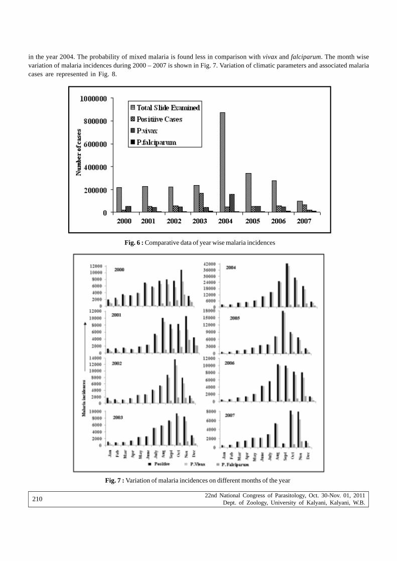
22nd National Congress of Parasitology, Oct. 30-Nov. 01, 2011Dept. of Zoology, University of Kalyani, Kalyani, W.B.
210
in the year 2004. The probability of mixed malaria is found less in comparison with vivax and falciparum. The month wisevariation of malaria incidences during 2000 – 2007 is shown in Fig. 7. Variation of climatic parameters and associated malariacases are represented in Fig. 8.
Fig. 6 : Comparative data of year wise malaria incidences
Fig. 7 : Variation of malaria incidences on different months of the year

22nd National Congress of Parasitology, Oct. 30-Nov. 01, 2011Dept. of Zoology, University of Kalyani, Kalyani, W.B.
211
The correlation of malaria incidences caused by vivax, falciparum and mixed malaria with the climatic parameters are givenin Table IV. The correlation of vivax malaria with temperature is significant at p < 0.01.
Table IV : Correlation coefficient between malaria parasites and surface parameters
Parameter Vivax Falciparum Mixed
Rainfall 0.49 -0.45 -0.49Relative humidity 0.05 -0.05 -0.15Temperature 0.84 -0.48 -0.79
From the survey it is noted that the locality of the effected persons may be one of the causes of malaria transmissionof that area. It is also noted that majority of the affected persons never use mosquito net and they have lack of awarenessabout the precautions needed to prevent malaria transmission
Conclusions
It is observed from the analysis that the malaria incidence increases in dry season. Meteorological parameters are notsufficient to explain malaria transmission. Environmental conditions of a particular locality may also play key role (Guerra etal, 2008). Changing landscape pattern may have great influence on local climate compare to long term effect of climate change.Temperature, evaporation, surface runoff, PET, water logging etc are affected by the change of land cover and hence havesignificant influences in determining the mosquito abundance of that particular locality (Patz et al, 2005; Foley et al, 2005;Patz and Olsen, 2006). Presence of malaria vectors does not always translate into disease. There are many places in the globewhere this vector borne disease disappeared in spite of having Anopheles species. Improved structure of house, watermanagement, adequate health service, life pattern, living environment etc is the determinants for malaria transmission.
Acknowledgements
Authors are thankful to Dr. D. Biswas, Vector Control Officer, Kolkata Municipal Corporation and National Data Centre,India Meteorological Department, Pune for providing some of the relevant data.
Referrences
Akhtar R, McMichael AJ (1996) Rainfall and malaria outbreaks in Western Rajasthan, Lancet. 348: 1457-1458.
Bhattacharyya DR, Prakash A, Sarma NP, Mohapatra PK, Singh, S Sarma, DK, Kalita MC, Mahanta J (2010) Molecular evidencefor the involvement of Anopheles nivipes (Diptera: Culicidae) in the transmission of Plasmodium falciparum in north-eastern India. Annals of Tropical Medicine and Parasitology 104: 331-336.
Fig. 8 : Variation of meteorological parameters and associated malaria incidences

22nd National Congress of Parasitology, Oct. 30-Nov. 01, 2011Dept. of Zoology, University of Kalyani, Kalyani, W.B.
212
Bhattacharya S, Sharma C, Dhiman RC, Mitra AP (2006) On climate change and malaria in India. Current Science 90: 362-368.
Craig MH, Kleinschmidt I, Nawn JB, Sueur DL, Sharp BL (2004) Exploring 30 years of malaria case data in KwaZulu-Natal, SouthAfrica: Part I. The impact of climatic factors. Tropical Medicine and International Health 9: 1247-1257.
Dev V, Dash AP, Khound K (2006) High-risk areas of malaria and prioritizing interventions in Assam. Current Science 90: 32-36.
Dev V, Hira CR, Rajkhowa MK (2001) Malaria attributable morbidity in Assam, northeastern India. Annals of Tropical Medicineand Parasitology 95: 789–796.
Foley JA, DeFries R, Asner GP, Barford C, Bonan G, Carpenter SR, Chapin FS, Coe MT, Daily GC, Gibbs HK, HelkowskiJH, Holloway T, Howard EA, Kucharik, CJ, Monfreda C, Patz JA, Prentice IC, Ramankutty N, Snyder PK (2005) GlobalConsequences of Land Use. Science 309: 570-574.
Gubler DJ, Reiter P, Ebi KL, Yap W, Nasci P, Patz JA (2001) Climate variability and change in the United States; potential impacton vector and rodent borne diseases. Environmental Health Perspective 109: 223-233.
Guerra CA, Gikandi PW, Tatem AJ, Noor AM, Smith DL, Hay SI, Snow RW (2008) The limits and intensity of Plasmodiumfalciparum transmission: implications for malaria control and elimination worldwide. PLoS Med 5: 300-311.
Martens WJM, Niessen LW, Rotmans J, Jetten TH, McMichael AJ (1995) Potential impact of global climate change on malariarisk. Environmental Health Perspectives 03: 458- 464.
Mohapatra PK, Prakash A, Das HK, Mahanta J, Srivastava, VK (1999) Malaria outbreak in lower Assam, An epidemiologicalappraisal. Journal of Parasitic Diseases 19: 175-178.
Pascual M, Ahumada JA, Chaves LF, Rodo X, Bouma M (2006) Malaria resurgence in the East African highlands: temperaturetrends revisited. Proceedings of the National Academy of Sciences 103: 5829-5834.
Patz JA, Campbell LD, Holloway T, Foley JA (2005) Impact of regional climate change on human health. Nature 438: 310–317.
Patz JA, Olson SH (2006) Malaria risk and temperature: Influences from global climate change and local land use practicesPANS. 103: 5635 – 5636.
Pemola ND, Jauhar RK (2006) Climatic variables and malaria incidence in Dehradun, Uttaranchal, India. Journal of Vector BorneDiseases 43: 21-28.
Prakash A, Bhattacharyya DR, Mohapatra PK, Barua U, Phukan A, Mahanta J (2003a). Malaria control in a forest camp in anoil exploration area of upper Assam. The National Medical Journal of India 16: 135-138.
Prakash A, Mohapatra P K, Bhattacharyya DR, Goswami BK, Mahanta J (2003b). Plasmodium ovale: First case report fromAssam, India. Current Science 84: 1187-1188.
Reiter P (2001) Climate Change and Mosquito-Borne Disease. Environmental Health Perspectives 109: 141-161.
Ramesh C, Dhiman RC, Pahwa S, Dash AP (2008) Climate change and malaria in India: Interplay between temperatures andmosquitoes. Regional Health Forum 12: 27-30.
Sharma VP (1999) Current scenario of malaria in India. Parasitologia (Rome) 41: 349-353.
Singh N, Sharma VP (2002) Patterns of rainfall and malaria in Madhyapradesh, Central India. Annals of Tropical Medicine andParasitology 96: 349-359.
Teklehaimanot HD, Lipsitch M, Teklehaimanot A, Schwartz J (2004) Weather based prediction of plasmodium falciparum malariain epidemic prone regions of Ethiopia. Malaria Journal 3: 1-11.
Tonnang HEZ, Richard YMK, Pius ZY (2010) Predicting and mapping malaria under climate change scenarios: the potentialredistribution of malaria vectors in Africa. Malaria J 9: 111-120.
Tren R (2002) Malaria and Climate change, Working paper series published by Africa Fighting Malaria, European Science andEnvironment Forum.

22nd National Congress of Parasitology, Oct. 30-Nov. 01, 2011Dept. of Zoology, University of Kalyani, Kalyani, W.B.
213
Nematode infection in the fishes of Dolu Lake, Silchar, Assam,during different seasons of the year
Ratnabir Singha, Maibam Shomorendra* and Devashish Kar
Division of Wetlands, Fishery Science and Aquaculture,Department of Life Science and Bioinformatics,Assam (Central) University, Silchar-11, India
*Fish Disease Research Lab, Department of Zoology,Thambal Marik College, Oinam-795134, Manipur, Imphal, India
Abstract : A total of 749 fishes of different species of Dolu Lake were examined for parasitic infection. Out of 749 fishes examined,53(7.07%) fishes were found to be parasitized by nematodes. The highest intensity was found 62 in Mastacembelus armatus. The highestpercentage of infection was found 81.81% in Notopterus notopterus in Autumn and lowest was found 5.9% in Channa punctatus in Winter.The total highest percentage of infection was 108.3 in Summer and lowest was 53.33 in Spring.
Key Words : Fishes, Helminth, Sex, Length, Dolu Lake.
Introduction
Nematode parasite causes infection and diseases of fish both in freshwater and marine environments. Small numbers ofnematodes often occur in healthy fish, but high numbers cause illness or even death. Juvenile fish infected by small numbersof nematodes are more likely to show signs of illness and also have reduced growth rates. Therefore it is an essential areafor proper attention to be given by the scientists for sustainable aquaculture production.
The first comprehensive report on seasonal occurrence fish parasites was given by Chubb (1977b) who concentrated onthe monoseneans of freshwater fish and this was followed by annually extensive studies of the same topic for digeneans byChubb (1979), larval cestode and nematode by Chubb, (1980b) and cestoda, nematode and Acanthocephala by Chubb (1982).
Seasonal diversity of worms in the flesh and other organ systems of various marine food fishes investigated by Linton(1914). Sinderman (1966) showed that the effect of worm larvae on the host fish included growth retardation, tissue disruption,metabolic disturbances and even death in heavy infections. Rumpus (1974) discussed the seasonal fluctuation of parasitesand the effect of the size maturity and sex of fishes on the incidence and intensity of their parasites.
The influence of parasitic infection in relation to seasons has been studied by many workers like Shomorendra et.al.,(2005,2007), Chubb (1982), Pennycuick (1971), Jha and Sinha (1990), Tavares and Luque (2004).
In Assam, particularly in Barak Valley studies on the frequency of nematode infection and its variation due to seasonsis limited. This study mainly fulfills the gaps in this direction.
Study Area : Fishes examined in this study were collected from Dolu Lake which is situated 24055’23.4’’N and 92047’21.5’’E.The lake water ranges from 200C - 230C during December- January and maximum is 310C - 320C during June-July. The pH valuefluctuates between 6.38 to 7.08. The maximum value was recorded during April.
Materials and Methods
Different species of fish from Dolu lake were collected during different months of the year. The identification of each fishwere determined after following Jayaram (2010). The external body surface and internal organs were thoroughly examined forparasites. After being relaxed the collected parasites were fixed in the fixatives prescribed for different parasite groups.Trematodes were fixed in AFA (alcohol-formalin-acetic acid) solution, stored in 70% alcohol. Acanthocephala were fixed andpreserved in AFA , cestodes in 5% formalin and nematode after immersing in hot 70% alcohol, were finally stored in 70% alcoholafter following Bylund et.al (1980).

22nd National Congress of Parasitology, Oct. 30-Nov. 01, 2011Dept. of Zoology, University of Kalyani, Kalyani, W.B.
214
ResultsA total of 749 fishes of different species of Dolu Lake were examined for parasitic infection. Out of 749 fishes examined,
53(7.07%) fishes were found to be parasitized by nematodes. The highest intensity was found 62 in Mastacembelus armatus(Table-I. A & B). The highest percentage of infection was found 81.81% in Notopterus notopterus in Autumn and lowestwas found 5.9% in Channa punctatus in Winter. The total highest percentage of infection was 108.3 in Summer and lowestwas 53.33 in Spring.(Table-II).
Table-I.(A) Nematode parasite burden in the fishes of Dolu Lake.
Fish species No. of fishes No. of fishes Percentage Intensity ofExamined Infected of infection infection
Channa punctatus 126 3 2.38 1.33
Notopterus notopterus 59 38 64.40 13.57
Heteropneustes fossilis 55 1 1.81 1
Mystus tangara 58 2 3.44 1
Clarias batrachus 8 2 25 1
Colisa fasciatus 21 2 9.52 3.5
Mastacembelus armatus 3 1 33.33 62
Channa striata 8 2 25 5
Lepidocephalus guntea 14 1 7.14 1
Channa orientalis 8 1 12.5 1
Table-I.(B). Prevalence and intensity of nematode infection in the fishes.

22nd National Congress of Parasitology, Oct. 30-Nov. 01, 2011Dept. of Zoology, University of Kalyani, Kalyani, W.B.
215
Table- II. Percentage of infection in the four different fish species during different seasons at different water temperatureand pH.
Fish species Autumn Winter Spring Summer
Temp 280 C – 320C 200C – 250C 240C - 270C 290C -320C(0 C)
pH 5.5 - 7.7 6.4 - 6.8 6.4 - 7.0 6.3 - 6.5
Channa punctatus 9.0 5.9
Notopterus notopterus 81.81 66.66 33.33 75
Heteropneustes fossilis 20
Mystus tangara 9.0 33.3
Total 90.81 81.56 53.33 108.3
The parasite species collected from the fishes of the study area are :
Nematode
1) Species : Pseudoproleptus notopteri
Host : Notopterus notopterus
Location : Mouth, stomach, Intestine.
2) Species : Pseudoproleptus vestibulus
Host : Notopterus notopterus
Location : Mouth, stomach, Intestine
3) Species : Paragendria sp
Host : Channa striata
Location : Intestine
4) Species : Parascarophis sp
Host : Lepidocephalus guntea, Channa punctatus
Location : Intestine
5) Species : Camallanus sp
Host : Colisa fasciatus, Clarias batrachus
Location: Intestine
6) Species : Paracamallanus sp
Host : Colisa fasciatus
Location : Intestine
7) Species : Spinitectus sp
Host : Notopterus notopterus
Location: Intestine
8) Species : Contracaecum sp
Host : Lepidocephalus guntea
Location : Intestine
Discussion
The highest percentage of infection by nematodes was found in Notopterus notopterus. This findings supports earlierwork of Mgbemena (1983) who discovered higher number of nematode infection on C.tilapiae. Llcwellyn proved that therewas an exacting topographical relationship between a parasite and its host and this was probably an important factor in the

22nd National Congress of Parasitology, Oct. 30-Nov. 01, 2011Dept. of Zoology, University of Kalyani, Kalyani, W.B.
216
mechanism of host specificity. Highest infestation rate among nematodes have been observed by the Pseudoproleptusvestibulus and Pseudoproleptus notopteri, adult recovered from stomach and intestine.
It can be noted from the data that the percentage of infection in different fish species is more or less variable with seasonsof the year. The total value of infection for all four fish species is found to be highest (108.3%) in Summer and lowest is (53.33%)in Spring. But in Autumn the highest percentage of infection was found 81.81% in Notopterus notopterus and in Winter lowestwas found 5.9% in Channa punctatus. This findings agreed in case of Autumn only with Moharram (1980) who said,’ Autumnand Winter are the two seasons in which we meet the biggest number of infected fish with cope pods, as well as the greatestnumber of parasites per fish. The present observations showed that the effect of temperature on the fishes is an importantfactor for the infestation rate of parasites. During the hot season, the infestation rate was high. It has also been reported byKelle, 1977; Bussmann and Ehrich, 1979; Fatima and Bilqees, 1989.
One of the most important factors to emerge is environmental temperature, which affects the life cycles of intermediateand definitive hosts and parasites, temperature also affects the immune response of the fish hosts, which has itself emergedas an important factor in seasonal variations, producing seasonal change in resistance to infection. Many of these aspectswere discussed by Polyanski (1958) in reviews of the ecology of parasites of marine fishes. Biotic factors such as overcrowdingor interaction with other parasites and abiotic factors such as temperature, they all contribute to the pattern of seasonalvariation.
AcknowledgementsThe authors are thankful to S.R.Dey Sarkar, ZSI, Kolkata for helping in identifying the nematodes. The first author is also
thankful to UGC for providing fellowship.
ReferencesBussmann B, Ehrich S (1979) Investigations on infection of blue whiting (Micromesistius poutassou) with larval Anisakis sp.
(Nematoda; Ascaridida). Arch Fischereiwiss 29 : 155-165.
Bylund G, Fagerholm HP, Calenius G, Wikgreen BJ, Wikstrom M (1980) Parasites of fish in Finland – ii. Methods for studyingparasite fauna in fish, Acta Acad Aboenisis Ser B 40(2) : 1-23.
Chubb JC (1977b) Seasonal occurrence of helminthes in freshwater fishes. Part I. Monogenea Adv Parasit 15 : 133-199.
Chubb JC (1979) Seasonal occurrence of helminthes in freshwater fishes. Part II. Trematoda.. Adv Parasit 17 : 141-313.
Chubb JC (1980b) Seasonal occurrence of helminthes in freshwater fishes. Part III. Larval Cestoda and Nematoda. Adv Parasit18 : 1-120.
Chubb JC (1980b) Seasonal occurrence of helminthes in freshwater fishes. Part IV. Adult Cestoda, Nematoda and Acanthocephala.Adv Parasit 20 : 1-292.
Chubb JC (1982) Seasonal occurrence of helminth parasite in fishes. Part-iv. Adult Cestoda, Nematoda and Acanthocephala.Advances in Parasitology. Academic press. London & New york.20 : 1-292.
Fatima H, Bilqees F M (1989) Seasonal variation of nematodes and acanthocephalan of some fishes of Karachi coast. ProcParasitol 7&8: 1-201.
Jayaram KC (2010) The Freshwater Fishes of the Indian Region. Narendra Publishing House (Delhi). 616p.
Jha AN, Sinha P (1990) The occurrence of helminth parasites in relation to size of fish. Bio Journal 2(11) : 311-316.
Kelle W (1977) Unterschiedlich starker Parasitenbefall der Wittlinge Merlangius merlangus des Neuwerker Fabrwassers inSommer 1974. Arch Fischereiwissenschaft 28 : 65-68.
Linton E (1914) On the seasonal distribution of fish parasites. Trans Am Fish Soc 44 : 48-56.
Mgbemena MO (1983) Parasitic Fauna of Some Cichlids and Clariids in Jos Plateau. M.Sc. Thesis, Hydrobiology and FisheriesResearch Unit Unijos pp 1-58.
Moharram SG (1980) Studies on Parasitic Fauna of Merluccius merluccius of Alexandria and its Effect on the General Conditionof the Fish, M.Sc. Thesis. Faculty of Science, Alexandria University.
Pennycuick L (1971) Differences in the parasite infestation in three spined sticklebacks (Gasterosteus aculeatus L) of differentsex, age and size. Parasitology 63: 407-418.
22nd National Congress of Parasitology, Oct. 30-Nov. 01, 2011Dept. of Zoology, University of Kalyani, Kalyani, W.B.
217
Polyanski Yu I (1958) Ecology of parasites of marine fishes. In: Parasitology of Fishes (Eds. Dogiel, V.A., Petrushevski, G.Kand Polyanski, Y.I) Leningrad University Press, pp. 1-384.
Rumpus AE (1974) The helminth parasites of the bull head Cottus gobio (L) and the stone loach Noemacheilus barbatulus(L) from the River Avon, Hampshire, J Fish Biol 7 (4) : 469-483.
Shomorendra M, Jha AN, Kumar P (2005) Seasonal occurrence of helminth parasites in fishes of loktak lake, Manipur, UttarPradesh J Zoo 25 (i) 23-27.
Shomorendra M, Jha AN, Kumar P (2007) Effect of length of fish on the occurrence of helminth parasites. Uttar Pradesh J Zoo27 (10) : 87-91.
Sinderman CJ (1966) Diseases of marine fishes, Academic Press, London and New York, Adv Mar Biol 4 : 1-89.
Singha R, Shomorendra M, Kar D (2010) Helminth parasites of the fishes of Dolu Lake, Silchar (Assam). Life Science Bulletin7 (2) : 223-225.
Singha R, Shomorendra M and Kar D (2010) Influence of sex and length of host on the infection of helminth parasite in thefishes of Dolu Lake, Silchar, Assam. In : Proceedings of 21st All India Congress of Zoology & National Seminar. CentralInland Fisheries Research Institute, Barrackpore. pp : 26-27.
Singha R, Shomorendra M, Kar D (2010) Helminth parasites in relation to length of three freshwater fishes of Dolu Lake ofSilchar, Assam, India. Environment & Ecology 28 (4A) : 2506-2508.
Tavares LER, Luque JL (2004) Community ecology of the metazoan parasites of white sea catfish, Netuma barba (Osteichthyes:Ariidae) from the coastal zone of the State of Rio de Janeiro, Brazil, Braz J Biol 64(1).
22nd National Congress of Parasitology, Oct. 30-Nov. 01, 2011Dept. of Zoology, University of Kalyani, Kalyani, W.B.
218
Nature of parasitic infestations in three economically important Labeo spp.inhabiting natural and cultured water bodies of Tripura
Remya Madhavan1, Probir Kumar Bandyopadhyay1 and B. Santosh2
1Parasitology Laboratory, Department of Zoology, University of Kalyani, Kalyani, West Bengal, India2Vizhinjam Research Centre of Central Marine Fisheries Research Institute, ICAR, Vizhinjam,
Thiruvananthapuram-695521, Kerala, India
Abstract : A detailed investigation was conducted on the parasitic fauna harbored by three species Labeo calbasu, L. gonius and L. bataof genus Labeo Cuvier, 1816 collected from the four different districts of Tripura. The survey was done for a period of two years fromApril 2007 to March 2009. The host fishes were collected in live condition for carrying out the pathological examinations. In all the hostspecies, external infections especially on the gills, fins, scales and skin was found most prevalent than infections on the visceral organs.Majority of the infected fishes including fishes carrying a heavy parasitic load did not exhibit any notable external disease symptoms andappeared healthy. Parasitic fauna was represented by both protozoan and metazoan parasites. Six species of protozoan parasites and twelvespecies of metazoan parasites were obtained. Of the protozoan parasites there were five species of the myxozoan group and a singlerepresentative of the ciliophoran group. Metazoans were represented by five monogenean species, two digenean species, one species eachof Ergasilus, Neoergasilus and Catlaphilla and two species of Argulus. With regard to seasonal analytic studies, the prevalence of infectionof the protozoan and the helminth parasites showed no remarkable variations. Among the copepod parasites, Neoergasilus longispinosus,exhibited pronounced seasonal changes. The infection by N. longispinosus was intense during winter seasons with maximum load of parasiteson the fins of the infected hosts. Labeo calbasu exhibited the maximum prevalence of infection (88.30%) and L. bata was the least infectedwith a prevalence of infection (29.50%) among the host species examined. The mean intensity of parasites was maximum in Labeo gonius(5.88). Myxozoan and monogenean infection was highest in Labeo calbasu and Labeo bata had the highest copepod infection. The fryand fingerling stages of the host species also showed instances of infection. Pronounced parasitic infestations in host fishes collected fromthe culture ponds indicate that overstocking and stunting leads to large scale multiplication of parasites in aquaculture systems.
Key words : Labeo, metazoan, parasite, prevalence, protozoan
IntroductionTripura one among the eight north-eastern states occupies a total geographical area of 10,491 sq.km and lies in the sub-
Himalayan region. Among the native minor carps of the state Labeo bata, L. calbasu and L. gonius of the genus Labeo Cuvier,1816 are very popular nutritionally and commercially throughout the state. Though there is ample information on the biologicaland culture aspects of these Labeo spp. a comprehensive knowledge regarding the parasitic infestation of this group is verylimited. Majority of the earlier studies done on the Labeo spp. of the north eastern region and of Tripura concentrated onthe taxonomic classification and distribution (Ghosh and Lipton, 1982; Lipton, 1983; Barman, 1988; Govt. of Tripura, 2006)Moreover in spite of the great consumer demand for these fishes, the parasitic investigations done on them are very scarcecompared to the Indian Major carp, Labeo rohita. Kabata (1985) reinstated the need of a thorough and comprehensive researchon the fish parasites of the tropical waters which are inadequately explored in comparison with the temperate regions. Thepresent work represents the first endeavor from the north-eastern part of the country as well as from the state of Tripura tocollect an inclusive knowledge on the total parasitic fauna harbored by the three important Labeo spp. other than Labeo rohita.
Materials and methods
The survey was done for a period of two years from April 2007 to March 2009. The host fishes selected were Labeocalbasu, Labeo bata and Labeo gonius belonging to family- Cyprinidae and genus- Labeo Cuvier, 1816. They were collectedfrom the natural collection sites like Rudrasagar Lake, the principal rivers of the state, River Gumti, River Manu, River Dhalaiand a major reservoir called Dumbur reservoir and culture ponds spanning over the four districts; the North, South and West
* Corresponding Author : E-mail : [email protected]

22nd National Congress of Parasitology, Oct. 30-Nov. 01, 2011Dept. of Zoology, University of Kalyani, Kalyani, W.B.
219
Tripura and the Dhalai district. The fishes were brought in live or moribund condition to the laboratory. Prior to dissection,the body weight and the standard length of the specimens were recorded. A general examination of the fish was done andabnormalities like red colorations, lesions or swellings were recorded. The specimens were then subjected to thoroughexamination for protozoan and metazoan parasites according to the methods proposed by Kennedy (1979), Meyers (2000) andWoodland (2006).
Pattern of infection, parasite density and monthly and seasonal variations of the parasite faunas were studied using thestatistical analysis measures like prevalence of infection (P) or percentage of fish infected calculated as total fishes infected/total fishes examined x 100; mean intensity of infection (MI) or the mean number of parasites per infected fish calculated asmean no. of parasites collected/ infected fish; simple dominance index (DI), calculated as abundance of a given parasite species/taxa ÷ total abundance of all species/taxa; richness index (RI), calculated as (S-1) / loge N, where, S = number of species/taxaof parasites and N = total number of parasites; evenness index (EI), calculated as H / loge S, where H = Shannon index ofdiversity and S = number of species/taxa of parasites; shannon index of diversity (H), calculated as H = (n loge n) – ( fi logefi) / n, where n = fi; fi = abundance of parasite species/taxa expressed as percentage.
Results1. Taxonomical Studies
Out of the thousand one hundred and twenty seven fishes examined, six hundred and seventy-four were infected withprotozoan and metazoan parasites (Table1). Eighteen species of parasites comprising six protozoan species and twelvemetazoan species were obtained with a total parasite count of three thousand nine hundred and nineteen.
The protozoan parasites obtained from the present study were from genera Myxobolus Butschli, 1882, ThelohanellusKudo, 1933 and Tripartiella Lom, 1959. The parasite species were Myxobolus rohitae from the scales of all three host species;Myxobolus sp.I.n.sp. from gills of all three hosts; Myxobolus sp.II.n.sp. from gills of Labeo calbasu and L. bata.Thelohanellus sp.I.n.sp. from fins of all three host species ; Thelohanellus sp.II.n.sp. from gills of Labeo bata and Tripartiellacopiosa from gills of Labeo gonius.
The metazoan parasites encountered in the present survey were monogenetic trematodes, Dactylogyrus labei from thegills of all the three hosts species; Dactylogyrus catlaius, Dactylogyrus spinitubus and Dactylogyrus vicinus from gills ofLabeo calbasu and a new sp. of genus Dactylogyrus Diesing, 1850 from gills of Labeo bata.
The digenetic trematodes found were metacercaria of Centrocestus formosanus from gills of all three host species, andmetacercaria of Clinostomum sp. from gills of Labeo bata.
The crustacean parasites obtained from the study were a new sp. of genus Ergasilus Nordmann, 1832 from the gills anda described species, Neoergasilus longispinosus of the genus Neoergasilus Yin, 1956 from the fins and gills of all the threehost species; Argulus foliaceus and Argulus japonicus of genus Argulus Muller, 1785 were found from the scales and skinof the host fishes; A. foliaceus from Labeo calbasu and L. bata and A. japonicus from L. gonius and L. bata. A new speciesof genus Catlaphilla Tripathi, 1960 was encountered on the gills of Labeo calbasu.
DiscussionIn all the host species external infections was found most prevalent than infection on the visceral organs. The infected
fishes which carried a heavy parasitic load were also apparently healthy. From among all the three host species, Labeo calbasuexhibited the maximum prevalence of infection, 88.30% and L. bata was the least infected with a prevalence of 29.50%. Themean intensity of parasites was maximum in Labeo gonius, 5.88. Protozoan and trematode infection was highest in Labeocalbasu, 19.68% and 70.48% respectively. Labeo bata exhibited the highest infection prevalence, 15.93% with crustaceanparasites. Among the crustacean parasites, the infection by Neoergasilus longispinosus was intense during winter seasonswith maximum load of parasites on the fins of the infected hosts.
With regard to the seasonal analytical studies, dominance indices (DI) which represents the dominance of any oneparticular parasite species upon all the others were very similar throughout the year except for the winter months in Labeocalbasu and L. gonius, whereas it was higher in case of Labeo bata (Table 2, Figs. 1,2 and 3). Labeo bata harbored the richestparasite fauna with Richness Index (RI) 2.58 followed by Labeo calbasu, 2.24 and for Labeo gonius the richness index waslow, 1.81. The Shannon index of diversity (H) shows similar peak values almost throughout the season for all species whichsignifies that all the three species harbored the same number of parasite species. The Evenness index (EI) indicates the

22nd National Congress of Parasitology, Oct. 30-Nov. 01, 2011Dept. of Zoology, University of Kalyani, Kalyani, W.B.
220
Table I. Summary of Total parasitic infestation by the protozoan and metazoan parasites on three Labeo spp. of Tripura.
Labeo 376 218 1357 57.9 6.2 4 6 1.0 1.5 43 108 11.4 2.5 74 459 19.6 6.2 332 1930 88.3 5.9calbas u
Labeo 368 161 1000 43.7 6.2 5 7 1.3 1.4 38 104 10.3 2.7 35 235 9.5 6.7 229 1346 62.2 5.9gonius
Labeo 383 34 171 8.8 5.0 5 6 1.3 1.2 61 210 15.9 3.4 33 256 8.6 7.8 113 643 29.5 5.6bata
Total 1127 413 2528 36.6 6.1 14 19 1.2 1.4 142 422 12.6 3.0 142 950 12.6 6.7 674 3919 59.8 5.8
Monogenean Digenean Crustacean Protozoan Totalinfection infection infection parasites infection infection
Nam
e of
hos
t sp.
No.
of f
ishe
s exa
min
ed
No.
of f
ishe
s in
fect
ed
No.
of p
aras
ites
colle
cted
Prev
alen
ce(P
)
Mea
n in
tens
ity (
MI)
No.
of f
ishe
s in
fect
ed
No.
of p
aras
ites
colle
cted
Prev
alen
ce(P
)
Mea
n in
tens
ity (
MI)
No.
of f
ishe
s in
fect
ed
No.
of p
aras
ites
colle
cted
Prev
alen
ce(P
)
Mea
n in
tens
ity (
MI)
No.
of f
ishe
s in
fect
ed
No.
of p
aras
ites
colle
cted
Prev
alen
ce(P
)
Mea
n in
tens
ity (
MI)
No.
of f
ishe
s in
fect
ed
No.
of p
aras
ites
colle
cted
Prev
alen
ce(P
)
Mea
n in
tens
ity (
MI)
Table II. Monthly and seasonal variations of different ecological indices for parasitic infections in three Labeo spp.
Shannon Index (S) 1.44 1.35 1.631 1.76 1.48 1.46 1.48 1.31 1.47 0.65 0.5 0.61 2.04 1.93 1.79 2.22Richness Index (RI) 1.09 0.79 1.51 1.4 1.39 0.94 1.01 0.76 1.57 0.38 0.43 0.43 1.89 1.79 2.07 2.59Dominance Index (DI) 0.25 0.27 0.236 0.21 0.28 0.25 0.25 0.29 0.26 0.54 0.68 0.58 0.14 0.16 0.2 0.13Evenness Index (EI) 0.9 0.97 0.784 0.85 0.76 0.9 0.92 0.95 0.82 0.94 0.72 0.88 0.89 0.88 0.86 0.86
Shannon Index (S) 0.62 1.18 1.47 1.02 1.17 1.33 1.04 1.31 1.59 1.47 1.09 1.12 1.51 1.46 1.86 1.78Richness Index (RI) 0.66 0.89 1.46 0.67 1.00 1.17 1.19 1.16 1.47 1.08 0.67 0.76 1.51 1.55 1.65 1.81Dominance Index (DI) 0.69 0.36 0.32 0.43 0.38 0.35 0.42 0.31 0.26 0.28 0.39 0.37 0.31 0.31 0.19 0.22Evenness Index (EI) 0.45 0.85 0.71 0.73 0.65 0.68 0.54 0.67 0.76 0.82 0.79 0.81 0.73 0.66 0.84 0.77
Shannon Index (S) 0.85 1.87 1.77 1.80 1.71 1.53 1.78 1.59 1.83 1.30 1.35 1.56 2.03 1.98 2.06 2.16Richness Index (RI) 0.73 1.92 1.48 1.36 1.46 1.31 1.27 1.34 1.51 0.90 0.86 1.25 1.94 1.82 1.93 2.24Dominance Index (DI) 0.57 0.19 0.20 0.20 0.24 0.29 0.20 0.27 0.20 0.32 0.30 0.29 0.16 0.17 0.16 0.14Evenness Index (EI) 0.61 0.85 0.80 0.86 0.78 0.74 0.85 0.77 0.83 0.81 0.84 0.80 0.85 0.83 0.86 0.84
Ecological Indices
Labeo calbasu
Febr
uary
Mar
ch
Apr
il
May
June
July
Aug
ust
Sept
embe
r
Oct
ober
Nov
embe
r
Dec
embe
r
Janu
ary
Sum
mer
Mon
soon
Win
ter
Ann
ual
Ecological Indices
Febr
uary
Mar
ch
Apr
il
May
June
July
Aug
ust
Sept
embe
r
Oct
ober
Nov
embe
r
Dec
embe
r
Janu
ary
Sum
mer
Mon
soon
Win
ter
Ann
ual
Ecological Indices
Febr
uary
Mar
ch
Apr
il
May
June
July
Aug
ust
Sept
embe
r
Oct
ober
Nov
embe
r
Dec
embe
r
Janu
ary
Sum
mer
Mon
soon
Win
ter
Ann
ual
Labeo gonius
Labeo bata

22nd National Congress of Parasitology, Oct. 30-Nov. 01, 2011Dept. of Zoology, University of Kalyani, Kalyani, W.B.
221
p
0.0
0.5
1.0
1.5
2.0
2.5
Feb Mar Apr May Jun Jul Aug Sep Oct Nov Dec Jan
0.0
0.2
0.4
0.6
0.8
1.0
1.2
1.4
1.6
1.8
Feb Mar Apr May Jun Jul Aug Sep Oct Nov Dec Jan
p
0.00.2
0.40.60.81.01.21.41.6
1.82.0
Feb Mar Apr May Jun Jul Aug Sep Oct Nov Dec Jan
In dex v al ue s
In dex v al ue s
In dex v al ue s
Fig.I Monthly variation of different species level diversity indices for parasitic fauna in Labeo calbasu
Fig.II Monthly variation of different species level diversity indices for parasitic fauna in Labeo gonius
Fig.III Monthly variation of different species level diversity indices for parasitic fauna in Labeo bata
Shannon Index Richness IndexDominence Index Evenness Index
uniformity of the number of parasites of the different parasite species present at a particular time within a given host. In thepresent study, the parasites were almost evenly distributed throughout the year represented by similar values. In the courseof the survey it was observed that the fry and the fingerling stages of the host fishes also showed instances of infection.
The present results indicate that Labeo calbasu, L. bata and L. gonius which are the most preferred minor carps in Tripuraharbour a rich and diverse parasitic fauna represented by four major taxa- Protista, Monogenea, Digenea and Crustacea. Thisresult contradicts the view of Thoney (1991, 1993) that the parasite diversity of freshwater or estuarine fishes are less thanthe off shore region. Manter (1947) and Dogiel et al. (1958) observed that the phylogenetic closeness of the hosts is the majorreason for the similarity in their parasite fauna thus it can be suggested that the three Labeo spp. have remarkable phylogeneticcloseness.

22nd National Congress of Parasitology, Oct. 30-Nov. 01, 2011Dept. of Zoology, University of Kalyani, Kalyani, W.B.
222
AcknowledgementsThe first author is grateful to The Fisheries Division of Indian Council of Agricultural Research (ICAR), Tripura Centre
for providing the necessary laboratory facilities for the study.
ReferencesBarman RP (1988) Fishes of the river Gumti, Tripura. Records of Zoological Survey of India, Occasional Paper No. 119.
Dogiel VA, Petrushevskii GK, Polyanski YI (1958) Parasitology of Fishes. Leningrad University Press, Leningrad, EnglishTranslation Kabata, Z., 1961. Oliver and Boyd, Edinburg pp 384.
Ghosh SK, Lipton AP (1982) Ichthyofauna of the N.E.H. Region with special reference to their economic importance. Specialbulletin No.1, ICAR Research Complex, Shillong, Meghalaya.
Govt. of Tripura (2006) Fish fauna of Tripura. Vol. 1. Department of Fisheries, Govt. of Tripura pp 23.
Kabata Z (1985) Parasites and diseases of fish cultured in the tropics. Taylor and Francis, London pp 318.
Kennedy MJ (1979) Basic methods of specimen preparation in parasitology. IDRC Manual report pp 8: 44.
Lipton AP (1983) Fish fauna of Tripura. Matsya. 9: 110-118.
Manter HW (1947) The digenetic trematodes of marine fishes of Tortugas, Florida. American Midland Naturalist 38: 257-416.
Meyers TR (2000) Fish Pathology Section. Laboratory Manual (Meyers, 2000). Special Publication, No.12, Second Edition.
Thoney DA (1991) Population dynamics and community analysis of the parasite fauna of juvenile spot, Leiostomus xanthurus(Lacepede), and Atlanic croaker, Micropogonias undulates (Scianidae) in two estuaries along middle Atlantic coast ofthe United States. Journal of Fish Biology 39: 515-534.
Thoney DA (1993) Community ecology of the parasites of adult spot, Leiostomus xanthurus and Atlantic croaker, Micropogoniasundulates (Scianidae) in the Cape Hatteras region. Journal of Fish Biology 43: 781-804.
Woodland J (2006) National Wild Fish Health Survey. Laboratory Procedures Manual. 3.1 Edition. U.S. Fish and Wildlife Service,Pinetop, AZ.

22nd National Congress of Parasitology, Oct. 30-Nov. 01, 2011Dept. of Zoology, University of Kalyani, Kalyani, W.B.
223
The effect of parasitic infestation on tissues and organs of Mystus aor(hamilton) and Mystus bleekeri (day)
Rimi Farhana and Hamida Khanum
Department of Zoology (Parasitology Branch), University of Dhaka, Dhaka-1000, Bangladesh
Abstract : A total of 1011 Mystus aor and 1039 Mystus bleekeri were examined during January 2004 to December 2005. The juveniletrematode Isoparorchis hypselobagri was recovered from body muscles, swim bladder and visceral organs of the fishes having prevalence80.02 %, intensity 6.83 ± 3.79 in M. aor and 58.90% , intensity 5.23 ± 2.89 in M. bleekeri.
In both the hosts, encysted and free I. hypselobagri were frequently found in the body cavity, viscera and more rarely in the mouth,urinary system, biliary system, ovaries etc. The main effect of the parasite was found on the skin surface, body musculature and visceral
organs. I. hypselobagri were found attached to the body muscles causing extensive tissue damages including inflammation, necrosis, andempty spaces with fragmented blood capillaries, tissue debris’s, lymphocytes and fluids.
Infected liver, swimbladder and kidney showed vacuolation and massive melanization. Helminth parasite specially I. hypselobagri
caused tissue damages through formation of tunnels by lysing and ingestion of tissues. Fluid accumulation, tissue destruction, hemorrhageand massive melanization in different parts and organs of the hosts were observed.. . Due to the presence of some gut helminth, the intestinalmucosa and villi were disrupted, ruptured blood vessels and necrosis of tissues were also observed.
Key words : Histopathology, Isoparorchis hypselobagri, Mystus aor, Mystus bleekeri, Parasitic infestation.
Introduction
Parasite causes damage to their host and they occupy a definite position or site in suitable environment on their hosts.Fishes are one of the most common hosts of helminth parasites. The influence of the parasite may result in extensive changein individual organs or tissue or it can exert a general effect on host. Helminth parasites generally affect the internal organsof the host fish, particularly the gut and inhibit host’s growth. The normal growth of fishes is interrupted and inhibited if theyare heavily infested with endoparasites viz., trematode, nematode, cestode and acanthocephalan and microscopic lesions intheir host’s tissues which become the site for the secondary infection by bacteria . Helminths are very common in freshwaterfishes. Very few lesions have been attributed to intestinal forms. Histozoic helminths, particularly migrating forms, cause greaterdamage in fishes. In severe cases hypermia, hemorrhage, cellular infiltration, lesion, narcosis, fibrosis etc. After encystmentand fibrotic encapsulation, many larval helminths produce no further obvious damage except pressure on adjacent host tissue.Migrating larva cercariae, plerocercoides and nematode produce the most serious reactions: leukocytes, fibrosis, hemorrhageand necrosis. Continual migrations, such as larvae of Contracaecum spp. produce peritonitis which results in fibrosis andextensive adhesions. Rapid invasion by large number of cercariae produce extensive hemorrhage, hypermia, necrosis and evendeath if present in sufficient numbers (Ribelin and Migaki, 1975).
Among the workers who have undertaken histological research on tissues of various freshwater fishes are Mackiewicz(1972); Hine and Kennedy (1974); Jain et al. (1976); Ahmed and Sanaullah (1979); Mitchell, et al. (1982); Khanum (1994);Khanum and Farhana, (2002.) etc. Helminth in fishes is also recognized as causing serious effect on their hosts [1]. Changesbrought on by nematodes have been noted by Yeh (1960).
Very few studies have been done on parasite infestation, host parasite relationship and histopathology of I. hypselobagri.(Siddique and Nizami, 1978) reported incidence of this trematode from W. attu.], studied the incidence of this trematode andits destructive effects on air bladder of W. attu, viscera and body musculature of Callichrous bimaculatus and ovary of Mystusaor.. Mahajan et al. (1978) reported the effect of parasitization of juvenile I. hypselobagri in Channa punctatus. Bhalerao (1932)gave a note on the probability of infection of man and domestic carnivores by this trematode. Verma and Ahluwalia (1980)reported similar unusual records and high survival index of I. hypselobagri, but none of them explained the pathology of these

22nd National Congress of Parasitology, Oct. 30-Nov. 01, 2011Dept. of Zoology, University of Kalyani, Kalyani, W.B.
224
infections. Khanum (1994) observed severe pathogenic lesions done by juvenile I. hypselobagri on the skin surface, bodymusculature, liver, intestine, kidney and other visceral organs in two species of Ompok spp.
Parasite -induced histopathology in M. aor and M. bleekeri have never adequately described. According to Bashirullah(1972) the occurrence of young fluke in the muscle and body cavities of fishes, suggested, that the parasite bored their wayin the tissue. Sometimes the juvenile parasites penetrate the intestinal wall and migrate into the swimbladder.
Materials and methodsA total of 1011 Mystus aor and 1039 Mystus bleekeri were autopsied during January 2004 to December 2005 from the
rivers of Kuliarchar upazila and brought to the Parasitology laboratory, Department of Zoology University of Dhaka for theinvestigations of helminth infestation.
The affected parts and organs of the fish, e.g. skin, liver, muscles, kidney, swim bladder and alimentary canal wereseparated and treated according to the methods instructed by for histological studies. After detection, the affected tissueswere carefully fixed by a gradual addition of 10% Buffered neutral formalin solution.
The methods of [17] and [18] were followed for the preparation of the permanent histological slides. For preparation ofhistological slides, the tissue materials were kept in Buen’s fluid for 24-48 hr for fixation, dehydrated in ascending grades ofethanol (50%, 70%, 10% and 100%), impregnated and embedded in paraffin and sectioned at 5µ. Sections were mounted onslides, deparafined by low grading and stained with haematoxylin and counter stained with eosin, dehydrated and finallymounted in Canada balsom..
ResultsIn the present study, some pathological changes were observed on the skin, body musculature, swimbladder and visceral
organs of M. aor and M. bleekeri. Trematode, Histopathological studies showed that skin, muscle layer, swimbladder, intestine,lower part of intestine, liver and kidney were damaged by the infestation of helminth parasites. Skin, muscles, intestine, liver,kidney tissues were found to be more infected. I. hypselobagri was found to be the most pathogenic and damaging one. InM. bleekeri it was mainly found in the swimbladder and body cavity but in M. aor predominantly found almost every organsuch as swim bladder, liver, kidney (over the surface) and also in cavity, musculature (Plate- VIII), skin surface etc.
Due to the structural construction and the ability of the immature I. hypselobagri, they are normally capable of dissolvingand penetrating the skin and muscle layers when they are living in the regular habitat and corresponding microenvironment.But along with the onset of decomposition of viscera or any unusual change of host body’s microenvironment, the trematodebecome stimulated or compelled to utilize the dissolving and penetrating capacity for their survival. It can be noted as ahomeostatic adjustment or adaptation of the juvenile trematode I. hypselobagri. The juvenile parasites dissolve the skin withthe help of the penetration glands, located in the oral sucker situated at the anterior region of the body and in the acetabulum(Plate- I & VII). Many fishes were observed with marks of perforation, lacerations and scars on their surface and abdomen.It is assumed that many metacercarial forms might be lost through these laceration processes. Any kind of internal changesor due to the cause of host’s death, metacercarial forms need to leave the host immediately. At that time they bored their wayin to the external environment for their survival through laceration processes. Sometimes, it was also observed during the studyperiod that, the trematode reaches the buccal cavity or gill and sometimes came out through the anus or genital opening oflower abdomen.
In the present observation, due to the presence of juvenile I. hypselobagri in the swimbladder, liver and intestine massivemelanization was observed. Sometimes, small vacuoles were formed in the liver due to the presence of cysts. Melanin-macrophage centers along with heavy melanin deposition were observed in the infected liver. The melanin-macrophage centersshould possibly be considered as a component of the reticuloendothelial system and hence, part of the defensive system ofthe fish against any infection. Presence of massive melanization again confirms this. Sometimes hepatic blood vessels wereruptured. The affected liver showed mild hepatic or hemopoietic degenerative changes with hemorrhages (Plate- II & III).
A number of observable histopathological changes occurred in the intestinal tissue of infected fishes. The gut helminthsdamaged the walls at the sites of their attachment. This disruption was mainly due to the action of sucker of the trematodeparasites. As a result the intestinal wall was heavily destroyed. Intestinal tissues were observed with deposited melanin.Perforation in the gut wall was found where the host tissue reacted vigorously. Large vacuoles and fluid filled empty spacealong with debris and lymphocytes were present. The intestinal mucosa and villi tissue was disrupted, the blood vessels wereruptured and necrosis of intestinal tissue was also found (Plate-V).

22nd National Congress of Parasitology, Oct. 30-Nov. 01, 2011Dept. of Zoology, University of Kalyani, Kalyani, W.B.
225
In infected kidneys, some vacuolar renal tissue degeneration was observed. The important changes included chronicinflammation, degeneration of blood vessels, and degeneration of kidney tubules as well as interstitial cells. Due to parasiticinfestation, vacuolar empty spaces and tunnels filled with fluid. Sometimes the associated haemopoietic tissue became looseand in severe cases renal tubular necrotic lesions were observed. Large macrophages containing erythrocytic debris in thehematic sinusoids were found (Plate- IV).
During the migration of immature I. hypselobagri from visceral cavity to the swimbladder, massive disruption anddislocation of the visceral organs occurred. Irregular black pigmentation was scattered throughout the swimbladder, due tothe infection by juvenile I. hypselobagri. In severe cases, the alveolar sacs and capillary plexuses were disrupted causingnecrosis (Plate-VI).
DiscussionOf the low level animal protein consumption, 80% is contributed by the fisheries with proper utilization of these rich
resources, the demand of animal protein for the entire population of the country can be resolved to a fairy extent. It is alsoa high quality food containing high percentage of protein and variable quantities of vitamins, fats, calcium, phosphorus andother nutrients essential for human health. To achieve healthy fish stock, we have to implement program like fish parasitologicalresearch, control of fish diseases, some biological aspects and maintenance of healthy relationship between fish and theirenvironments.
Now a days, the need and importance of study of Parasitology cannot be denied. The study of parasites and its functionis a long felt demand in a developing country like Bangladesh which could promote the Nation’s agricultural economy,particularly the economy of fish conservations. Parasites require some vital factors which they can obtain only from their hostsoften inflicting some degree of injury affecting its welfare. Recent physiological studies of nutritional requirements of parasitesespecially endoparasitic forms, have indicted that, depletion of host’s nutrients by parasites may have serious consequences.The organ which are heavily infested by parasites may cause malnutrition, destroy tissues, reduce growth rate and immunityagainst virus, bacteria produce toxin etc.. Even man and piscivorous wild and domestic animals may become affected from theconsumption of parasitized fishes. All of these influences may lead to disease. These parasites play an important harmful roleon aquatic and terrestrial food chain.
In the body musculature, intestine and kidney fluid filled empty spaces along with debris’s and lymphocytes were present.Similar observation also reported by Sanaullah et al. (1997). This observation also showed positive agreement with the findingsof Kinkelin et al. (1968). The melanin-macrophage centers should possibly be considered as a component of the reticuloen-dothelial system and hence, part of the defensive system of the fish against any infection (Ribelin and Migaki, 1975). Theyalso theorized that the black pigment of the interfacicular connective tissue of the host was mediated by an enzyme reaction,which caused mechanical obstruction due to the occurrence of parasite in clusters. The reported that the internal organ ofinfected fish showed only mild histopathological changes which may be the result of background pathology. In 1966, larvaeof Bucephalus polymorphus caused high mortality of cyprinid fish in the Seine river near Paris, France (Kinkelin et al. 1068).They found cysts in many parts of the fish and the mouth and bases of caudal and anal fins with marked inflammation andhemorrhage. Migrating cercariae cause muscle necrosis. In some cases metacercariae were numerous in the cornea and retinawith retinal hemorrhages.
The metacercaria of certain digenean parasites, found encysted in the skin and muscle of teleost fish, stimulatedproduction of black spots which are visible in naked eye, (Mcqueen et al. 1973). stated that, one of the means by which ahost combats internal parasites is by production of melanin pigments from the surrounding tissues. Presence of cysts withincreasing juvenile I. hypselobagri again confirmed the purpose for which melanin pigments were produced. Edelstein, (1971)suggested a number of potential biological functions for melanin compounds apart from direct pigmentation. They added that,melanin has a capacity for binding aromatic and cyclic compounds, and also it may selectively take up harmful compounds.The number of empty cysts tended to increase slightly with increase in size of the host. The degree of pigmentation dependson the species, age and state of health of the fish.
A number of observable histopathological changes occurred in the kidney tissue of infected fishes. The importantchanges included chronic inflammation, degeneration of blood vessels, fluid accumulation and degeneration of renal tubulesas well as interstitial cells. Kidney samples have shown tubular, vacuolar degeneration with granular occlusion andhaemopoietic tissue degeneration and focal proliferation. Damage to kidney of teleosts occurs when cercariae of various

22nd National Congress of Parasitology, Oct. 30-Nov. 01, 2011Dept. of Zoology, University of Kalyani, Kalyani, W.B.
226
species enter the fish and migrate through the tissues and most cercariae encysted in the tissues following the migratory period,which cause tissue destruction due to pressure necrosis as the organism continues a short period of growth and stimulatesfibroplasias (Hoffman, 1967). They also noticed that, the organism was surrounded by a fibrous tissue capsule which containsa few lymphocytes and melanophore that have migrated into the area, and the metacercariae remain in the encysted state untilingested by the final host.
The present study provides an account of parasitological aspects of taxonomic studies, distribution, association andpathogenecity of the helminth parasites of the host fishes as well as nutritional aspects of infected and uninfected host fishes.It has been established that, the catfish M. aor of all ages are not highly susceptible to all groups of metazoan parasites andsome of those parasites have zoonotic importance (e.g. I. hypselobagri and G. spinigerum) which are potentially transferableto man only through freshwater fishes. Bhalerao (1932) wrote a note on the probability of infection of Isoparorchishypselobagri in man and domestic domestic carnivores. He listed the occurrence of I. hypselobagri from gas bladder ofWallago attu and also found these parasites in the stomach of a crocodile which had devoured some siluroid fishes. He alsostated that any animal eating the infected siluroid fish was exposed to infection with Isoparorchis. Among the internal organ,intestine was heavily infected by a large no of gut helminth specially trematode. In conclusion, controlling measures shouldbe taken to interrupt the steps of parasitic transmission from one host to another. Therefore, extensive study should be carriedout on the parasites of M. aor and M. bleekeri; otherwise they will lower the productivity of these catfishes.
ReferencesAhmed ATA, Sanaullah M (1979) Pathological observations of the intestinal lesions induced by Caryophyllid cestodes in
Clarias batrachus (L.) (Siluriformes Clariidae). Fish Path Japan 14(1): 1-7.
Bashirullah AKM (1972a) On the occurrence of the trematode, Isoparorchis hypselobagri (Billet, 1898), in fishes and noteson its life history. Norwegian J Zool 20: 209-212.
Bhalerao GD (1932) A note on the probability of infection of man and domestic carnivores by Isoparorchis hypselobagri (Billet,1898). Indian J Vet Sci Anim Husb. 2(4): 404-405.
Devraj M, Ranganathan V (1971) Incidence of I. hypselobagri among the catfishes of Bhabanisagar reservoir. Indian J ofFishery 14(1): 232-250.
Edelstein LM (1971) Melanin: A unique biopolymer. In Parasitology Annual, ed. H L. Loachim. Butterworth, London. pp. 220.
Esch GW, Huffines WJ (1973) Histopathology associated with endoparasitic helminths in Bass. The J of Parasitology. 59(2):306-313.
Gray P (1964) Handbook of Basic Microtechnique. 3rd ed. McGraw Hill Book Company, New York. pp. 302.
Hine PM, Kennedy CR (1974) Observations on the distribution, specificity and pathogenecity of the acanthocephalanPomphorhynchus laevis (Muller). J of Fish Biol 5: 521-535.
Hoffman GL (1967) Parasites of North American Freshwater Fishes. Univ. of Calif
Press. Berkeley and Los Angeles. pp. 486.
Humason GL (1979) Animal Tissue Techniques. 4th ed. W. H. Freemann and
Company, San Francisco. pp. 641.
Huizinga HW, Esch GW, Hazen TC (1979) Histopathology of red sore Disease (Aeromonas hydrophila) in naturally andexperimentally infected largemouth Bass Micropterus salmoides (Lacepede). J Fish Diseases 2: 263-277.
Jain PS, Pandey KC, Pandey AK (1976) Some histopathological observations on the stomach wall of Heteropneustes fossilis(Bloch) infected with a cestode. Agra Univ Jour of Res Sci 25(3): 1-4.
Khanum H (1994) Endoparasitic helminth infestation in Ompok bimaculatus (Bloch) and Ompok pabda (Hamilton andBuchanan) in relation to some of their biological, pathological and biochemical aspects. Ph. D Thesis. Dept. of Zoology.Univ. of Dhaka. pp. 323.
Khanum H, Farhana R (2002) Histopathological effects of a trematode Isoparorchis hypselobagri (Billet) in Wallago attu (Blochand Schneider). 30(1): 65-69.
Kinkelin P, Tuffery G, Leynaud G, Arrignon J (1968) ‘Etude ‘epizootiologique de la buc‘ephalose larvaire A Bucephaluspolymorphus (Baer, 1827) dans le peuplement piscicole du bassin de la Seine. Rech vet Paris 1: 77.

22nd National Congress of Parasitology, Oct. 30-Nov. 01, 2011Dept. of Zoology, University of Kalyani, Kalyani, W.B.
227
Mackiewicz JS (1972) Relationship of pathology to scolex morphology among caryophyllid cestodes. Z Parasitenk 39: 233-246.
Mcqueen A, Mackenzie K, Roberts RJ, Young HS (1973) Studies on the skin of plaice (Pleuronectes platessa). III. The effectof temperature on the inflammatory response to the metacercariae of Cryptocotyle lingua (Digenea: Heterophylidae).J Fish Biol 5: 388-399.
Mitchell AJ, Smith CE, Hoffman GL (1982) Pathogenecity and histopathology of an unusually intense infection of white grubs(Posthodiplostomum m. minimum) in the fathead minnow (Pimephales promelas). J of Wildlife Diseases 18(1): 51-57.
Ribelin WE, Migaki G (1975) The pathology of fishes. The Uni. of Wisconsin press. Madison, Wisconsin. pp. 1004.
Sanaullah M, Hjeltnes B, Ahmed ATA (1997) Histopathological aspects of epizootic ulcerative syndrome (EUS) in wild fishesfrom Faridpur, Bangladesh. Bangladesh J Zool 25(2): 183-192.
Siddiqui A, Nizami A (1978) Incidence of Isoparorchis hypselobagri (Billet, 1898) (Trematoda, Isoparorchiidae) in Wallago attu,with remarks on its life cycle. Acta Parasitologilca Polonica 25(25): 223-227.
Yeh LS (1960) On a reconstruction of the genus Camallanus Ralliet et Henry, 1951. J Helminth 34: 117-124.

22nd National Congress of Parasitology, Oct. 30-Nov. 01, 2011Dept. of Zoology, University of Kalyani, Kalyani, W.B.
228
On the seasonal occurrence of clinostomatid metacercariae infections in anIndian mudskipper Channa punctatus Bl. from the fresh water swamps near
Darbhanga town, north Bihar
Sapna Singh and Kedar Prasad Sinha
Helminthology Laboratory, Department of Zoology, L. N. Mithila University, Darbhanga, Bihar, India
Abstract : The seasonal occurrence of clinostomatid metacercariae in the mudskipper Channa punctatus from the freshwater swamps nearDarbhanga town, North Bihar was studied from June, 2009 to May, 2010.
The matacercariae belong to two genera - Clinostomum and Euclinostomum. These showed incidence during February to Septemberwith peak periods in June (44.44-54.17%). Average worm burden too was found higher during peak periods of July-August (4.0-7.2). Thefindings showed clearly that the warmer months favoured the incidence as well as average worm burden of these metacereariae in this fishhost indicating a significant and positive relationship between the incidence and worm burden (r = + 0.828 at P>0.001, df = 11) whethermale or female fish host.
Therefore, as to the Indian record of the seasonal occurrence of the clinostomatid metaceracariae the present report is worth mentioning.
Key Words : Seasonal, occurrence, Clinostomum, metacercaria, Channa, fish host
IntroductionCultivation of edible fish species has now-a-days been promoted, rather on commercial scale, with a view to enhancing
not only economic profit but also the supply of protein in our diet at a relatively cheaper rate. So, the fish cultivation hasnow come to share in uplifting the agricultural or rural economy. Our country, India, is a vast tropical country with variedclimatic conditions, say climatic instability, which are certain to affect the fish living in the freshwater habitats, particularly thewetland or swampy waters so much so that there is even high mortality rate of fish particularly living in shallow ponds orswampy areas. Perhaps this may be due to one or other kind of environmental stress. Helminthic infections, concomitantlywith microbial infections, prove to be adding to the miseries as being much fatal to the fish cultivation. Stocked ponds orswampy areas provide a congestion that definitely favours for rapid spread of infections among the fishes living therein andthus infestations become more liable to occur and reoccur. It becomes more imperative to know the period of heavy occurrenceso as to plan out prophylactic measures. As such seasonal studies warrant attention. Due to the scarcity of seasonal studiesof helminths in freshwater fishes in tropical parts of the world, including India, very little is known about the seasonal patternand occurrence of this group of metacercaria. Rather, as far as the authors are aware, no study on the seasonal changes ofoccurrence of this group of parasites has yet been attempted. Therefore , the present study carried out from June 2009 to May2010 provides the first report on the seasonal occurrence of clinostomatid metacercariae from the mudskipper Channapunctatus syn. Ophiocephalus punctatus from Bihar, India.
Materials & methodsThe live specimens of Channa were collected at regular monthly intervals with the help of local fishermen from the
freshwater swamps with water temperature ranging from 11 to 40oC at the outskirts of Darbhanga town, North Bihar. Immediatelyafter collection the live fresh samples were brought to the laboratory and are examined for the helminth parasites. A total of319 fish were examined during the period from June 2009 to May 2010.
The metacercariae recovered from the body cavity and even attached to mesentery were removed out and fixed in 4%alcohol and stained in Gower’s carmine for morphological study.
Results (Table-1&2; Fig-1)
Out of 319 fish (86 males and 233 females) only 71 (=23.19%) were found to be infected with these metacercariae including15 (16.28%) males and 56 (24.88%) females. These metacercariae belong to the family Clinostomatidae including two genera-

22nd National Congress of Parasitology, Oct. 30-Nov. 01, 2011Dept. of Zoology, University of Kalyani, Kalyani, W.B.
229
Clinostomum & Enclinostomum. Although these two genera have been reported by other workers from Channa (syn.Ophiocephalus) punctatus, only a few from other fish hosts viz.
Table-1. Prevalence of clinostomatid metacercaricae in Channa punctatus from freshwater swamps near Darbhanga town.
Months Mean Fish Fish Average
Temp Examined Infected Prevalence (%) Wormburden
(°C) M F M F M F M F
Jan 16.2 8 12 0 0 0 0 0 0
Feb 22.4 8 16 0 2 0 12.50 0 2.0
Mar 30.6 6 16 1 3 16.66 18.75 2.1 3.4
Apr 33.2 9 17 1 4 11.00 23.50 1.5 3.5
May 35.4 9 19 2 7 22.22 36.80 2.5 5.5
Jun 38.6 9 24 4 13 44.44 54.17 3.2 5.8
Jul 33.2 8 24 3 11 37.50 45.83 4.0 6.2
Aug 32.1 7 21 3 9 42.57 42.86 5.2 7.2
Sep 28.2 8 22 1 5 12.50 27.77 3.8 4.8
Oct 25.6 5 21 0 2 0.0 9.00 0.0 4.0
Nov 18.2 5 23 0 0 0.0 0.0 0.0 0.0
Dec 15.3 4 18 0 0 0.0 0.0 0.0 0.0
86 223 15 56 16.28 24.88
Total = =319 =56.71 = 23.19
M = Male, F = Female
Table-2. Seasonal variation in prevalence of metacercariae infection in Channa punctatus from freshwater swamps.
Particular MALE FISH FEMALE FISH
W S R W S R
Incidence (%) 0 24 25 3 36 31
Av. Worm burden 0 2 3 1 5 6
Trichogaster and Mastacembellus (Jaiswal, 1957) which too live in the similar habitats. Very remarkably thesemetacercariae were neotenic i.e. premature adults marked by conspicuous and prominent genitalia but without uterus andeggs, as if these were just to become adults in suitable hosts such as fish–eating birds or mammals. It means that this fishhost serves as paratenic host. Data presented in Table (1 & 2) clearly show that metacercariae not recovered during October-February but only during March to September show the marked and statistically significant seasonal difference (F=25.19,P>0.001 for df= 2,7).
DiscussionSo far the authors are aware no reports on the seasonal occurrence of the clinostomatid metacercariae have been made
from the tropics including India except those of Sinha (1991) on that of Diplostomulum from a catfish H. fossilis Bl. Therefore,the present study provides the first report on the seasonal occurrence of the clinostomatid metacercariae from an Indian mudskipper Channa punctatus .As evident from the works of Grabda-Kazubska (1974) on the occurrence of the adult Clinostomum

22nd National Congress of Parasitology, Oct. 30-Nov. 01, 2011Dept. of Zoology, University of Kalyani, Kalyani, W.B.
230
Figures rounded off to the nearest whole number
W = NOV-FEB
S = MAR-JUNE
R = JULY-OCTOBER
Graph showing relationship between the mean temp (0C), prevalence (%) and Av. Worm Burdenof clinostomatid metacercariae infection in Channa punctatus Bl.
Months
complanatum from herons and also of Pojmanska (1976) in Poland an association between the habitat of the lakes andoccurrence of the metacercariae has been suggested. Rather in Poland, in the months of February and July the water hasaverage monthly temperature of 6.870c and 29.160c respectively and it has been speculated that the increased watertemperatures would have favoured hatching of eggs .The findings of the present study show increasing incidence as wellas average worm burden during the months of February to September with peak periods during June to August. The watertemperature too shows increasing trend from February to a maximum of 38.60c in June and then decreasing due to rainfallduring July-October .Hence the findings of the present study do support the above speculation that the warm water of thesemonths would have favoured not only hatching of eggs but also the emergence of cercaria from the snail host into waterand their entry into the fish host where develop further into metacercariae. Further, the warmer months-Summer and Rainy-favoured not only the occurrence but also to a greater extent maturity, hence neotenic or precocious, as if only waitingfor the definitive host, whether aquatic birds/mammals, to eat upon such metacercariae infested fish and these becomingadult ones. In other words, the season plays an important role, along with temperature as a factor, in influencing theoccurrence of these metacercariae. In conformity with the hypothesis of Chubb (1979,1982) and also De (1993) that severalfactors, but no less important, including water temperature have a significant influence on the seasonal changes inoccurrence of helminths in freshwater fish in the tropical areas, and presumably the host interactions play the dominantrole. However, the seasonal studies on the other helminth parasites should be carried out in the rainy climatic zone of tropicalareas to find the determining factors.

22nd National Congress of Parasitology, Oct. 30-Nov. 01, 2011Dept. of Zoology, University of Kalyani, Kalyani, W.B.
231
RefrencesChubb JC (1979) Seasonal occurrence of helminthes in fresh water fishes Part II Trematoda” In Advances in Parasitology
(W.H.R. Lumsden, R.Muller and J.R.Baker, eds) 17 : 142-313
Chubb JC (1982) Seasonal occurrence of helminthes in freshwater fishes. Part IV Adult Cestoda, Nematoda and Acanthocephalain ‘Advances in Parasitology’ (W.H.R. Lumsden, R.Muller, and J.R. Baker, eds) 20:1-292.
De NC (1993) Seasonal dynamics of Camallanus anabantis infections on the climbing perch, Anabas testudineus, from thefreshwater swamps near Kalyani town, West Bengal, India. Folia Parasitologica 40:49-52
Jaiswal GP (1957) Studies on the trematode parasites of fishes and birds found in Hyderabad state. Zool Johrb Abst Syst85:1-72
Grabda-Kazubskq B (1974) Clinostomum complanatum (Rudolphi 1819) and Euclinostomum heterostomum (Rudolphi 1809)….,their occurrence and possibility of acclimatization in artificially reated lakes in Polan. Acta Parasitologica Polonica 22: 285-93
Pojmansk T (1976) The influence of thermic pollution on parasitocenoses structure in water reservoirs. Wiadmosci Parazytologiczne.22:445-449
Sinha KP (1991) Diplostomulum singhii (Strigeoidea : Digenea) infection in the fish H. Fossilis BL. Env. and Ecology 9(2):538-40

22nd National Congress of Parasitology, Oct. 30-Nov. 01, 2011Dept. of Zoology, University of Kalyani, Kalyani, W.B.
232
Acid tolerance as a vehicle for food-borne shigellosis
Santanu Chakrabarti
Post-Graduate Department of Zoology, Barasat Government College,
Barasat, North 24 Paraganas, West Bengal, India
Abstract : Shigellosis still remains an important cause of mortality and morbidity in India and other South Asian countries where peoplelive in densely populated, poor sanitation areas. The problem is aggravated with an increasing incidence of multiple drug resistance amongshigellae. Incidences of food-borne shigellosis are reported, mainly associated with consumption of fresh produce. The growth of globalfood trade makes food from around the world available, the increase in fresh produce consumption and the growing popularity of minimallyprocessed fruits and vegetables may play a role in the introduction of Shigella spp. in these types of foods. Shigellosis is characterized
by a low infective dose and Shigella spp. are pathogens of faecal origin can be carried by food handlers and transmitted through fruit-products The safety of fruit and fruit products relies mainly on their low pH and storage under refrigeration which are considered as barriersfor growth and survival of microbes. However, a few closely related species of Shigella showed extraordinary tolerance to low pH. Sothe rationale of the present study was – the application of a combination of suboptimal intrinsic and extrinsic factors such as reduced pHand reduced temperature can indeed inhibit the outgrowth but may induce prolonged survival of highly virulent pathogens such as Shigella.With this objective, the growth and survival efficiency of shigellae in acidified media was monitored at different time intervals and viable
counts were determined. At 37ºC shigellae die rapidly at pH 4.0 or lower, but these pathogens could survive for an extended period undersimilar adverse pH conditions when kept at refrigeration temperatures. A die-off of the shigellae was observed at pH 3.5 -4.0 and pH 3.0-3.5, respectively after 10 days at 4ºC and 6 days at 20ºC. Thus the recovery of shigellae was affected by exposure to reduced pH butincreases with refrigeration. So it can be concluded that acid foods such as tomato juice, apple juice, orange juice, acidic cheese, freshlycut papaya etc, usually kept at refrigeration temperatures, may support survival of Shigella and cause food poisoning. Once contaminatedduring cutting, juicing or in transit, these pathogens can survive for an extended period even under adverse pH conditions and these foods
may serve as a vehicle for Shigella infections.
Key words : Shigellosis; acid foods; refrigeration; survival efficiency; food poisoning
IntroductionShigellosis still remains an important cause of mortality and morbidity in India and other South Asian countries where
people live in densely populated, poor sanitation areas. The problem is aggravated with an increasing incidence of multipledrug resistance among shigellae. The Shigella bacteria are found in human feces which often contaminate the soil and water.Infection is initiated by ingestion of shigellae (usually via fecal-oral contamination). For most other infections, it takesthousands of organisms before an illness becomes apparent but it only takes 10 shigellae to cause the disease (1).
Fruits and their products can serve as vehicles for pathogenic microorganisms (2). Often involved are fruit products thathave undergone some kind of minimal non-thermal processing followed by time or temperature combinations that permitpathogens to survive and grow. Normally the peel of a fruit serves as a natural protectant, preventing bacteria from penetratingto the interior but on wounded fruit or fresh cut fruit slices, this external barrier is broken, thus creating an opportunity forbacterial colonization. Mechanical processing of fresh fruit such as transport, cutting, juicing and mild heat treatment makesfruit products more vulnerable to microbial penetration and growth (3). Enteric pathogens have been implicated in fruit-associated outbreaks. Outbreaks of Salmonella infections were traced to precut watermelons and fresh tomatoes (3). Outbreaksof Escherichia coli O157:H7 infection was associated with un-pasteurized fresh apple cider (4). Parish (1998) reported un-pasteurized orange juice contained E. coli (5) and outbreaks of enterotoxigenic E. coli infection associated with orange juicehave been reported by Singh et al. (6).
Although the reported incidence of food-borne shigellosis, mainly caused by Shigella sonnei and S. flexneri is loweramong enteric pathogens, each year there are a significant number of outbreaks, mainly associated with consumption of fresh
* Corresponding Author : Email : [email protected]

22nd National Congress of Parasitology, Oct. 30-Nov. 01, 2011Dept. of Zoology, University of Kalyani, Kalyani, W.B.
233
produce (7; 8). The chief contributing factor leading to Shigella food-borne outbreaks is poor personal hygiene of a foodhandler (7). The growth in global food trade that makes food from around the world available, the increase in fresh produceconsumption and the growing popularity of minimally processed fruits and vegetables may play a role in the introduction ofShigella sp. in these types of foods (3).
The safety of fruit and fruit products relies mainly on their low pH and storage under refrigeration which are barriersfor growth and survival (9). Growth of Salmonella spp. and E. coli O157:H7 on peeled orange was observed only at abusivestorage temperature. Refrigeration effectively inhibited growth and caused a reduction in the population of Salmonella spp.(10). E. coli O157:H7 showed extraordinary tolerance to the low pH upon refrigeration (11). Shigella is closely related toE. coli in their DNA homology and closely resemble in pathology, biochemical activity and antigenicity.
Few data are available on the growth and survival of shigellae in acid conditions. S. flexneri grown to stationary phasehas the ability to survive a few hours at pH 2.5. This acid resistance may contribute to the low infective dose associated withshigellosis (1). In laboratory conditions the minimum pH for growth was 4.9– 5.0 (9). All of the strains of S. flexneri and S.sonnei survived for 4 h in a nutrient medium at pH 4.0 or 4.5, however, at pH < 4.0 shigellae die rapidly. In this part of theglobe the prevalence of S. dysenteriae is more than any other species of Shigella (1). Therefore, in the present study thebehaviour of S. dysenteriae Type 1 strains under acid conditions was studied covering a pH interval which is relevant forthe pH of acid foods. It was also monitored that these pathogens could survive for an extended period under similar adversepH conditions when kept at refrigeration temperatures.
Materials and methods
Bacterial strains and preparation of inoculam
S. dysenteriae Type1 strains were obtained from the National Institute of Cholera and Enterc Diseases, Kolkata, India.Stock cultures of the strains were maintained on Tryptone Soy Agar (TSA;Hi-Media) at 4°C and revived by transferringa loop inoculum into 5 ml Tryptone Soy broth (TSB; pH 7.0) and incubating for 18–20 h at 37°C.
Acid tolerance of Shigella using acidified media
The behaviour of the Shigella strains under acidic conditions was determined by inoculating 0.1 ml of a 100-fold diluted18–20 h TSB culture into 9.9 ml of TSB adjusted to pH 3.0–5.0 (intervals of 0.5) with a citric acid (Merck, India) solution (10%)in order to obtain an inoculum of 105 cfu /ml). The TSB was held at 37°C during subsequent incubation. Viable counts ofShigella were determined after 6, 24 and 30 h incubation.
Survival efficiency of Shigella in acidified media stored at refrigeration temperatures
The behaviour of the Shigella strains under acidic conditions was determined in the same way by storing the inoculatedmedia at 4 ºC and 20 ºC, respectively up to 15days. The cultures were taken out at regular intervals to determine viability.
Determination of viability of Shigella
Viable counts of Shigella were determined by preparing 10-fold serial dilutions in sterile 0.1% peptone water and platingon TSA at each sampling time. If low numbers were expected, 0.1 ml of sample was directly spread out on the surface of TSA.All plates were incubated at 37°C for 24 h before colonies were counted. The difference in log cfu /ml) was determined onTSA plates were considered as an index of the number of injured cells.
Results
Acid tolerance of Shigella dysenteriae using acidified media
The results are shown in Table 1. At pH 5.0 Shigella strains grew to maximum numbers (108 cfu /ml) within 6 h. At pH4.5 Shigella had multiplied in 6 h, however, variation in the maximum numbers obtained was noticed (Number of injured cellswas 0.9). The minimum pH for growth of Shigella was thus determined as pH 4.5. They were able to multiply at pH 4.5 butthe number of injured cells was much more than that obtained at pH 5.0. At pH 4.0 the bacterial strains showed a viable countwithin the range of the initial inoculum level after 24 h (105 cfu /ml). At pH 3.5 the tested Shigella strains could be recoveredafter 24 h incubation although they survived for 6 h at pH 3.5 with quite a remarkable reduction in the population as revealedby the number of injured cells obtained.

22nd National Congress of Parasitology, Oct. 30-Nov. 01, 2011Dept. of Zoology, University of Kalyani, Kalyani, W.B.
234
Table 1 : Indication of the number of injured cells of Shigella after 6h incubation at 37°C in acidified media (Difference inlog cfu/ml was determined on TSA plates)
pH 5.0 pH 4.5 pH 4.0 pH 3.5
Number of 0.5 0.9 1.8 3.8
Injured Cells
Studies reported a 4 h survival time for a few strains of S. flexneri and S. sonnei at pH as low as 3.5. However, no survivalwas noted after 24 h for all the strains at pH 4.0. Though it is conceivable that different strains of microorganisms differ intheir acid resistance, the results of the present study confirm that the S.dysenteirae Type1 strains found in this country arein general more stress resistant. Direct enumeration of Shigella in acid foods using a selective medium as TSA may lead toan underestimation of the pathogen’s presence and prior enrichment of media with different supplements are recommendedby FDA BAM (12) which may aid in the resuscitation and recovery of stressed cells.
Survival efficiency of Shigella in acidified media stored at refrigeration temperatures
Table 2 and table 3 show the enhanced survival efficiency of Shigella in acidified media after keeping them at refrigerationtemperatures.
Table 2 : Indication of the number of injured cells of Shigella after incubation at 4°C in acidified media.
Number of Injured Cells
Culture At pH 5.0 At pH 4.5 At pH 4.0 At pH 3.5Time
in Days
2 0.2 0.3 0.5 0.7
4 0.4 0.5 0.8 1.1
6 0.7 0.8 1.2 1.8
8 0.9 1.2 1.4 3.7
10 1.1 1.8 3.8 Not Detected
Table 3 : Indication of the number of injured cells of Shigella after incubation at 20°C in acidified media
Number of Injured Cells
Culture At pH 5.0 At pH 4.5 At pH 4.0 At pH 3.5Time
in Days
2 0.4 0.4 1.6 4.0
4 0.6 0.7 1.8 5.1
6 0.9 1.1 2.7 Not Detected
From table 2 and table 3 a clear die-off of the S.dysenteriae Type1 was observed at pH 3.5 or below after keeping thecultures 10 days at 4 ºC and 6 days at 20 ºC. So it can be revealed that acid foods, especially if kept at refrigeration temperaturessupport survival of Shigella.
Discussion
Shigella dysenteriae, the causative agent of bacillary dysentery, is a major public health problem in developing countrieswere sanitation is poor. It is endemic in Eastern India, especially in Kolkata and adjoining districts of West Bengal, India (1).
The minimum pH (4.0) for growth under otherwise optimum conditions was established in the present study for Shigella.This was lower than the minimum pH for growth mentioned by ICSMF (9). Aerobic log-phase cultures grown at pH 5.0 weresimilarly acid resistant whereas survival in exponential phase cells grown at pH 8.0 survival decreased 10- to 100-fold (13).

22nd National Congress of Parasitology, Oct. 30-Nov. 01, 2011Dept. of Zoology, University of Kalyani, Kalyani, W.B.
235
Acid resistance depends on several factors including the test strains, the growth phase and the pH of the medium in whichcells are grown prior to the acid challenge. In this study, it was observed that aerobic stationary-phase cultures of S.dysenteriae Type1 grown initially at external pH of 5–8 showed 100% survival after an acid challenge of pH 3.5 for 2 h ( datanot shown). The acid resistance of Shigella depends on the expression of the stationaryphase-specific sigma factor sigmaS, a homologue of RpoS. The RpoS requirement can be overcome by anaerobic growth in moderate acid (13). In the presentstudy, acid tolerance of non acid-adapted aerobic stationary-phase cultures was determined.
In foods, the interactions between such factors as temperature, pH and the presence of salt can effectively inhibit thegrowth of Shigella spp. Because of the acidity of fruits, fruit juices do not constitute particular hazards for growth of humanpathogens but they can sustain survival during a restricted period of time depending upon strain and exact pH. Many fruitjuices on the market are pasteurized. This keeps them free from spoilage micro-organisms (9). Shigella is heat-sensitivewhich means that common pasteurization temperatures are sufficiently high to kill the pathogen (9). However, problems withShigella in fruit juices could occur by post-contamination after opening or by under-pasteurized or fresh-squeezed non-pasteurized juice kept in refrigeration, the latter ones gaining popularity both in the market and in bars because of increasednutritional health benefits. The safety of non- or under-pasteurized juices relies mainly on juice pH and proper sanitationduring processing and distribution. Shigellosis is characterized by a low infective dose and Shigella as other foodbornepathogens of faecal origin are known to be carried by food handlers and have been transmitted and caused vegetable-fruitassociated illness (7). Decreased temperature enhanced survival of Shigella. In a study on the survival characteristics ofShigella flexneri, Zaika (2001) also reported that in general survival increased as temperature decreased and as pH increased(14). The application of a combination of suboptimal intrinsic and extrinsic factors such as reduced pH and reducedtemperature can indeed inhibit the outgrowth of pathogens in foods but may induce prolonged survival of highly virulentpathogens such as Shigella. In laboratory media shigellae die rapidly at pH 4.0, but in the present study on survival ofShigella it was observed that they can survive for an extended period under adverse pH conditions, if kept at refrigerationtemperatures.
Fruit juices, such as tomato juice usually has pH 3.9-4.1, apple juice pH 3.3-3.4, orange juice pH 3.5, freshly cut papayapH 5.6 and also these types of fresh produces are usually kept at low temperatures. Once contaminated during cutting, juicingor in transit, these pathogens can survive for an extended period at low temperatures even under adverse pH conditions andthese foods may serve as a vehicle for Shigella infections.
Acknowledgements
The study was conducted with an UGC-MRP No. F.PSW-032/05-06(ERO) dated 21.3.2006.
References
Niyogi SK (2005) Shigellosis. Journal of Microbiology 43:133-43.
Beuchat LR (1996) Pathogenic micro-organisms associated with fresh produce. Journal of Food Protection 59:204–216.
Tauxe R, Kruse H, Hedberg C, Potter M, Madden J, Wachsmuth K (1997) Microbial hazards and emerging issues associatedwith fresh produce: a preliminary report to the National Advisory Committee on Microbiological Criteria for Foods.Journal of Food Protection 60:1400–1408.
Janisiewicz WJ, Conway WS, Brown MW, Sapers GM, Fratamico P, Buchanan RL (1999) Fate of Escherichia coli O157:H7 onfresh-cut apple tissue and its potential for transmission by fruit flies. Applied and Environmental Microbiology 65: 1–5.
Parish ME (1998) Coliforms, Escherichia coli and Salmonella serovars associated with a citrus processing facility implicatedin a salmonellosis outbreak. Journal of Food Protection 61: 280–284.
Singh BR, Kulshreshtha SB, Kapoor KN (1996) An orange-juice outbreak due to enterotoxigenic Escherichia coli. Journal ofFood Science and Technology India 32:504–506.
Smith JL (1987) Shigella as a foodborne pathogen. Journal of Food Protection 50: 788–801.
Mead PS, Slutsker L, Dietz V, McCaig LF, Bresee JS, Shapiro C, Griffin PM, Tauxe RV (1999) Food-related illness and deathin the United States. Emerging Infectious Diseases 5:1–21

22nd National Congress of Parasitology, Oct. 30-Nov. 01, 2011Dept. of Zoology, University of Kalyani, Kalyani, W.B.
236
International Commission on Microbiological Specifications for Foods (1998) Fruits and fruit products. In: Micro-organismsin Foods, Roberts TA, Pitt JA, Farkas J, Graw FH (Ed). London: Blackie Academic & Professional, Chapman & Hall,pp. 252–261.
Pao S, Brown GE, Schneider KR (1998) Challenge studies with selected pathogenic bacteria on freshly peeled hamlin orange.Journal of Food Science 63: 359–362.
Ryu JH, Beuchat LR (1998) Influence of acid tolerance responses on survival, growth, and thermal cross-protection ofEscherichia coli O157: H7 in acidified media and fruit juices. International Journal of Food Microbiology 45:185–193.
Association of Official Analytical Chemists (AOAC) International (1998) US FDA Bacteriological Analytical Manual, 8th edn.Gaithersburg, MA, USA
Small P, Blankenhorn D, Welty D, Zinser E, Sloncezwski JL (1994) Acid and base resistance in Escherichia coli and Shigellaflexneri: role of rpoS and growth pH. Journal of Bacteriology 176:1729–1737
Zaika LL (2001) The effect of temperature and low pH on survival of Shigella flexneri in broth. Journal of Food Protection64:1162–1165.

22nd National Congress of Parasitology, Oct. 30-Nov. 01, 2011Dept. of Zoology, University of Kalyani, Kalyani, W.B.
237
Immunohistopathological changes in the bursa of fabricius during thehypersensitivity reactions-IV induced by experimental ascaridiasis
in white leg horn chicks
Savita Rani1, Juhie Rani1 and S.S Lal2
1Department of Zoology, D.N. (P.G.) College, Meerut, U.P., India3Department of Zoology, C.C.S. University Campus, U.P., India
Abstract : Bursa is a primary lymphoid organ develops as a dorsal diverticulumn of the proctadeal region of the cloaca. The luminalsurface of the bursa in chicken is plicated with as many as 15 primary and 7 secondary plicae or folds. The B cell differentiatesin the bursa or its equivalent and is the responsible for the synthesis of Ig and antibody. During present investigation chicks werehyperensitized with the high and low doses of the egg antigen of ascaridia galli after the 10 days of the primary sensitization withthe same doses subsequently. Immunohistopathological changes have been observed at 25th, 35th, 45th days of the experiment which
showed thickening of the muscle layer of the capsular wall of the bursa. At the 35th, 45th days lumen of bursa was found to be greatlyreduced. In each follicle cortex and medulla revealed abundant concentration of B lymphocytes. Marked hypertrophy of follicles wasalso distinct. The arterioles were found to be dilated. Surface epithelium was observed to be ruptured at several places.
Key words : Ascaridia galli, Bursa of fabricious, hypersensitivity type-IV, Hypersensitization, white leg horn chicks
IntroductionParasitic infection account for hundred of millions of dollars annual losses for livestock and poultry industry throughout
the world. The most costly parasite in terms of production losses are the gastrointestinal nematodes in ruminants and poultry(Gamble and Zarlenga, 1986). Infections with the fowl nematode Ascaridia galli (Shrank 1788) are widespread amongdomesticated and wild birds throughout the world (Soulsby 1982). Ascaridia galli (gastrointestinal nematode) causeascaridiasis in the white leg horn chicks. Parasites induce different immune responses in the host. Sometime these immuneresponses are severe .These inappropriate responses called hypersensitivity reactions. Type –IV immunity reaction involveimmunopathological changes in the lymphoid organs. The lymphoid system of chicken consists of unique organs (bursa andthymus). Bursa is a primary lymphoid organ develops as a dorsal diverticulumn of the proctadeal region of the cloaca.lymphoid follicle associated epithelial cells possess micropinocytotic activity responsible for the uptake of antigen presentin the bursal lumen and their transfer inside the medulla of the lymphoid follicle(Bockman&Cooper,1973; Schaffner et al. 1974;Sorvari et al.1975; Bockman &Stevens, 1977).These antigens causes severe changes in the immunohistological archeitectureof the tissue. Hypersensitivity reaction against experimental ascaridiasis has not been study so far hence the present studieswere taken up.
Material and methodExperimental host- white leghorn chicks
Experimental parasite- Ascaridia galli
Dose of infection – low dose- 1000 embryonated eggs of the A.gall i/chick
High dose-2000 embryonated eggs of the A.galli/chick
Culturing of the egg-
Adult worms were isolated from the intestine of fowl from the local abattoir. A .galli eggs were cultured upto infectivestage according to Reidel method (1947). The worms were gently squeezed with the help of a pair of blunt end forceps sothat all extraneous material except the uteri were separated and removed from the petridish.The distal ends of uteri containing
* Corresponding Author : E-mail : [email protected]

22nd National Congress of Parasitology, Oct. 30-Nov. 01, 2011Dept. of Zoology, University of Kalyani, Kalyani, W.B.
238
mature eggs were separated and gently squeezed for fertilized eggs. The eggs were kept in sterile solution at 32°c forembryonation for 2-3 weeks. The eggs of A. galli were washed regularly with normal saline. Two drops of 0.1% fromalinewas added to prevent fungal infections. Now these embryonated eggs have been used for sensitization and hypersensitization of the chicks.
Preparation of the inocula-
The number of embryonated eggs per dose was estimated by dilution technique. The eggs were suspended in knownvolume of normal saline. The embryonated and infective eggs were counted with the help of stereoscopic binocularmicroscope. Three values were taken by repeating the process. Mean of these three values was used for calculating thenumber of embryonated infective eggs of A.galli.
During present investigation chicks were divided into 3 groups-
Group-A having 6 nonsensitized (control) chicks,
Group-B having 10 chicks, sensitized with the low dose of the egg antigen
Group-C- having 10 chicks, sensitized with the high dose of the egg antigen.
15 days old white leg horn chicks were sensitized with the low dose and high dose of the egg antigens. Now both thegroup-B and Group-C were further divided into two group-Ba &group-Bb and group-Ca & group-Cb. Groups Ba &Ca werethe sensitized groups with the low and high doses subsequently. Sensitized chicks with the low and high doses wereautopcised at 25th (Ba1&Ca1), 35th (Ba2&Ca2), 45th day (Ba3&Ca3) of the experimental design. While the 4 chicks form eachgroup (Bb&Cb) were hypersensitized after the 10 days of primary sensitization. The hypersensitized chicks were autopcisedat 35th (Bb1&Cb1) and 45th days (Bb2&Cb2) day of the experimental design.
Preparation of the tissue used for screening-
Specimens of bursa of fabricious were obtained from freshly killed animals according to experimental design. Tissuesamples have been fixed in 10% formalin. Regular washing has been done after fixation. Tissue has been processed formicrotomy and histolpathoogical slides were prepared.
ResultsImmunopathological changes in the bursa of fabricious of the white leg horn chicks have been observed during
sensitization and hypersensitization of the chicks with the low and high doses of the embryonated eggs of the A.galli at25th ,35th and 45th days of the experiment in the following groups-
Group (Aa1) - Nonsensitized group (Control) - fig-1a, fig-1b)
The transverse section of bursa from nonsensitized chicks showed the following structure. The bursa was broadlydivided into two regions-
1. Capsule- : It was made up of outer thin serosa layer and inner muscularis.
2. Sub-capsule: It consisted of mucosa and surface epithelium. It is made up of two parts-
1. Plicae- Mucosa developed into a number of villus like projections invading the central lumen called plicae.(1a).
Figure 1(a) : Slide showing several plicae in the bursaof the nonsensitized (control) (10 x)
Figure 1(b) : Slide showing a plicae having polyhedralfollicles in the bursa of the nonsensitized chicks. (40 x)

22nd National Congress of Parasitology, Oct. 30-Nov. 01, 2011Dept. of Zoology, University of Kalyani, Kalyani, W.B.
239
2. Follicles- Each plicae consisted of a number of polyhedral follicles, closely packed together with small amount ofconnective tissue separating them and blood vessels lying between them.(1b.)
Each follicle was divided into two regions :
a) Outer cortex and
b) Inner medulla. Both cortex and medulla possessed a supporting .network of stellate reticulo-endothelial cells whosemeshes were filled with lymphoid cells or lymphocyte cells.
Group (Ba1) - Sensitized group with low dose - (fig.-2)
During sensitization with the low dose, Surface epithelium of plicae was found to be ruptured at several places.Interfollicular spaces were not so distinct. Moderate enlargements of follicles size were observed. Lymphocyte percentagewas significantly increased in follicles.
Group (Ca1)-Sensitized group with high dose-(fig-3)
During sensitization with the high dose, surface epithelium was also ruptured at several places. Slightly atrophiedfollicles were observed and also revealed comparatively less lymphocytes in the cortex region of the follicles. Sensitized groupshowed some inflammatory responses.
B. Analysis of immunopathological changes at 35th day of the experimental design-At 35th day immunopathological studyof the bursa was done in the following group-
Figure 2 : Slide showing ruptured surface epithelium atseveral places, moderate enlargement of folli-cles size, and increased lymphocytes. (10x)
Figure 3 : Slide showing ruptured surface epithelium at severalplaces, slightly atrophied follicles, less lymphocytein the cortex region of the follicles. (40x)
Group (Ba2) - Sensitized group with low dose- (fig-4)
Bursa of this group revealed atrophied lymphoid follicles. Blood vessels get enlarged.interfollicular space showedhemorrhage. This group revealed severe retrogressive changes of lymphocyte cells in most of the follicles. The medulla ofthe follicles showed comparatively more depletion of lymphocytes than cortex.
Figure 4 : Slide showing atrophied follicles, haemorrhage in interfollicular space, less depletion oflymphocyte in cortex region than medulla (40 x)

22nd National Congress of Parasitology, Oct. 30-Nov. 01, 2011Dept. of Zoology, University of Kalyani, Kalyani, W.B.
240
Group (Bb1) - Hypersensitized group with low dose – (fig-5)
In this group, hypertrophied follicles were found. The pathological disturbances were observed more in cortex revealingdepletion of lymphocytes. The interfollicular spaces were filled with oedematous fluid.
Group (Ca2) - Sensitized group with high dose– (fig-6a,6b)
In this group, bursa was characterized by hypertrophy of follicles in the plicae. Interfollicular spaces were filled withoedematous fluid. At certain places, plicae wall observed to be disappeared while at some places thickening was alsoobserved in the plicae epithelium. Lymphocyte increased in the cortex region of the follicle.
Figure 5 : Slide revealing depletion of lymphocytes in the cortex region, interfollicular spaces filledwith oedematous fluid. (40x).
Figure 6(a) : Slide revealing hypertrophied follicles, thick-ening in plicae epithelium, increasedlymphocyte in the cortex region(10x).
Figure 6(b) : Slide showing hypertrophied follicles,thickening in plicae epithelium, increasedlymphocyte in the cortex region(40x).
Group (Cb1)-Hypersensitized group with high dose– (fig-7a,7b)
In this group, surface epithelium of the plicae was thickened. Atrophoid follicles showed depletion of lymphocytes inboth cortex and medulla region. Interfollicular spaces were clearly observed.
Figure 7(a) : Slide showing thickened plicae epithelium,atrophied follicles, depletion of lymphocyte in both cortex
and medulla region(10x)
Figure 7(b) : Slide showing cloudy swelling in the plicaeepithelium,atrophoid follicles, depletion of lymphocytes in
both cortex and medulla region(40x)

22nd National Congress of Parasitology, Oct. 30-Nov. 01, 2011Dept. of Zoology, University of Kalyani, Kalyani, W.B.
241
C. Analysis of immunopathological changes at 45th day of the experimental design-At 45th day changes were observed inthe following groups-
Group (Ba3) - Sensitized group with low dose–(fig-8a,8b)
In this group, at some places plicae wall ruptured. The bursal lumen became irregular. The follicles were observed tobe decreased in size while some follicles showed hypertrophied condition.
Figure 8(a) : Slide showing ruptured plicae wall, atrophoidfollicles, as well as hypertrophoid follicles (10x)
Figure 8(b) : Slide showing increased lymphocytepercentage (40 x)
Group (Bb2) - Hypersensitized group with the low dose–(fig-9)
Hypersensitized chicks of this group revealed that at certain places follicles became elongated in shape and interfollicularspace of some plicae was increased while bursal lumen was noticeably wider. There was distinct vacuolization anddegeneration of follicles, the follicles revealed rounded vacuoles numbering 2 to 4, increased in number and showed edema.
Group (Ca3) - Sensitized group with high dose– (fig-10)
Bursa of this group showed atrophoid follicles.Interfollicular spaces was clearly observed. There was depletion of thelymphocytes from both cortex and medulla region.
Figure 9 : Slide showing elongated follicles, wide bur-sal lumen, distinct vacuolization and degen-eration of follicles. (40 x)
Figure 10 : Slide showing atrophied follicles, clearinterfollicular spaces, depletion oflymphocytes in both cortex and medullaregion. (40 x)
Group (Cb2) - Hypersensitized group with high dose– (fig-11)
At 45th day hypersensitized chicks with the high dose showed atrophy in the follicles. Follicles were appeared to beoval in shape. Some follicles were separated from epithelial layers. Cortex region showed marked depletion in lymphocyteconcentration.

22nd National Congress of Parasitology, Oct. 30-Nov. 01, 2011Dept. of Zoology, University of Kalyani, Kalyani, W.B.
242
DiscussionDuring present investigation several degenerative changes have been found in the bursa of fabricious of sensitized and
hypersensitized chicks with the low and high doses of the embryonated eggs. Slightly enlargement in the follicles in thesensitized group with low dose at 25th day showed the effect of the low dose. Hypertrophied condition in the sensitized andhypersensitized chicks show high rate of maturation of lymphocytes due to high amount of antigen interaction. Rupturedsurface epithelium caused by enlargement of follicles. Similar finding have been showed by (Carpenter et al., 1992).Histopathologically the capsular wall was found to be more or less thickened. The follicles were found to be atrophied anddepletion of lymphocytes was observed in present study. Lymphoid follicle atrophy was probably the result of the migrationof the lymphocytes to the site of infection.Immunopathological lesions in infected chicken are formed resulting intohypoplasia and atrophied bursa of fabricius (Bagust et al. 1979; Taniguchi et al., 1977). Degenerative changes and depletionof lymphoid cells were observed in bursa of fabricius of vaccinated broiler chicks (Jeurissen et al.. 1998; Stoevetal.,2000).Presentauthor find out the immunopathological changes were directly proportional to the virulence of the infection of the parasites.Hypersensitized group revealed more severe histopathological changes in the bursa of fabricious in comparison to sensitizedchicks. Vacuolization and degeneration of the follicles were observed due to the leakage of endotoxins into the bursa, releasedduring antigen-antibody interaction. It may be due to severity of infection. Bursal lesions have been observed by Okoyo(1984), Mohanty and Rao (1984) and Mishra (1984).Immunopathologically the bursa of fabricious revealed severe retrogres-sive changes of lymphocytes in most of the lymphoid follicle and many of the lymphocytes were replaced by reticuloen-dothelial cells. Diffused lymphoid tissue was noted in the bursal canal and the follicles became separated from epithelial layerwith the medulla. The epithelial layer and lumen also disappeared leaving muscle, connective tissue, blood vessel and follicleswith and without medulla. The main cause of these severe changes is that T cell play important role during hypersensitivityreaction-IV. T cell, sensitized to antigen, release lymphokines following secondary contact with the antigen. cytokines induceinflammatory responses; they also activate and attract macrophages, which release inflammatory mediators. These mediatorsaffect lymphoid organs.
AcknowledgementWe are thankful to the Head, Department of Zoology, D.N. (P.G) College Meerut.
ReferencesBagust J, Grime TM, Dennett DP (1979) Infection studies on reticuloendothelial virus contaminant of a commercial Haeke
disease vaccine. Vet Journal 53(4): 153-157.
Bookman DE, Cooper MD (1973) Pinocytosis by epithelium associated with lymphoid follicles in the bursa of fabricious,appendixand peyer,s patches. An electron microscopic study. American Journal of Anatomy 136: 455-478.
Bookman DE, Stevens W (1977)Gut associated lymph epithelial tissue: bidirectional transport of tracer by specialized epithelialcells associated with lymphoid follicles. Journal of the Retiiculoendothelial society 21: 245-254.
Figure 11 : Slide revealing atrophied oval follicles, separated from epithelial layer,marked depletion of lymphocyte. (40 x)

22nd National Congress of Parasitology, Oct. 30-Nov. 01, 2011Dept. of Zoology, University of Kalyani, Kalyani, W.B.
243
Carpenter S, Miller B, Wannemuchler Y, Miller JM and Rath JA (1992) Characterization of the early pathogenic effects afterexperimental infection of calves with bovine immunodificiency like virus. Journal of Virology 66: 1074-1083
Gamble HR, Zarlenga DS (1986) Biotechnology in the development of vaccines for animal parasites. Vet Parasitol 20:237-250.
Jeurissen SHM (1998) Working mechanism of an immune complex vaccine. Immunology 95: 494-500.
Mishra GS (1972) Serum protein changes associated withAngiostrongylus contonensis infection in rat, dog and cat. Ind VetJ 49(4): 333-336.
Schaffner T, Mueller J, hess MW, Cottier H, Sordat B, Ropke C (1974) The bursa of fabricious: a central organ providingfor contact between the lymphoid system and intestinal content. Cellular Immunology 13: 304-312.
Shrank (1788) cited in “Nematodes parasites of domestic animals and of man” by N.D. Levine. Buraess PublishingCompany.Minnesota 316-365.
Sorvari T, Sorvari R, Ruotsal Ainen P, Toivanen A, Toivanen P (1975) Uptake of environmental antigens by the bursa offabricious.Nature 253: 217-219.
Stoev SW, Anguelov G, Ivanov I, Paviov D (2000) Influence of Ochratoxin A and an extract of arichoke on the vaccinalimmunity and health in broiler chicks. Exp Toxicol Pathol 52(1): 43-55.
Tanigueri T, Yuasa N, Sato S, Hnriuchi T (1977) Pathologicalchanges in chickens in oculated with reticuloendotheliosis viruscontaminated marks disease, vaccine Nat & Inst Anim Health (Tokyo)17(4): 41-50.

22nd National Congress of Parasitology, Oct. 30-Nov. 01, 2011Dept. of Zoology, University of Kalyani, Kalyani, W.B.
244
Studies on immunological impact of some chemicals, botanicals, antibacterialproteins and live non-pathogenic bacteria in silkworm, Bombyx mori L.
to control bacterial disease
Satadal Chakrabarty, Buddhadeb Manna#, Prabir Mitra, Atul Kumar Saha and Bharat Bhushan Bindroo
Silkworm Pathology laboratory, Central Sericultural Research and Training Institute, Berhampore (W.B.), India#Parasitology Research Unit, Department of Zoology, University of Calcutta, 35, Kolkata 700 019, India
Abstract : Bombyx mori L., mulberry silkworm is susceptible to protozoan, bacterial, fungal and viral infections like other economic insects.Bacterial diseases affecting silkworms are collectively known as ‘flacherie’ due to the flaccid conditions of the diseased larvae. 40% crop lossesare attributed due to diseases of silkworm in India including more than 15% crop loss is figured due to bacterial diseases under conditions of lowhumidity and extreme heat, particularly during pre-monsoon seasons (April - June). Poor disinfection, accumulation of pathogenic load in therearing trays, feeding of contaminated mulberry leaves and improper use of bactericide leads to large scale crop loss due to bacterial diseases.
Biological and chemical control measures as well as development of disease tolerant silkworm breeds are insufficient to control the disease in thepresent degraded environmental scenario. Therefore, silkworms are very often attacked by the pathogens. To overcome this problem, promisingbivoltine and multivoltine breeds available in our Germplasm bank have been screened on the basis of haemocyte counts in response against lowerto higher pathogenic bacterial challenge for future breeding programme. Highly pathogenic bacteria, Streptococcus sp. is known to play an activerole in causing ‘Bacteremia’ a bacterial disease of digestive organ of silkworm. This communication reports the impact of induced immunity byinduction of some immunogens to control bacterial disease in silkworm. A comparative study based on economic parameters was undertaken
among the treatments of distilled water sprayed healthy control; highly pathogenic bacteria sprayed infected control; immunogens sprayedimmunized larvae challenged with highly pathogenic bacteria. This study could help to select the highly effective immunogens (s) to improve thehumoral defense response to control the bacterial disease in silkworm at the farmer’s field.
Key words : Antibacterial protein, Bacterial disease, Bombyx mori L., Flacherie, Immunogen.
Introduction
Bombyx mori L. is highly susceptible to various diseases like Pebrine (Microsporida), Flacherie (Bacteria), Grasserie (Virus)and Muscardine (Fungus) and the pathogens have a great role in deterioration of sericulture industry. 40% crop losses areattributed due to silkworm diseases in India (Sheeba Rajakumari et al, 2007). About 36 % crop loss is attributed due to Pebrinedisease with occasional crop failure (Nataraju et al, 2005). Further, crop loss due to silkworm diseases ranged from 0.58 - 11.18%, 2.03 - 12.44 % and 3.65 -15.58 % respectively, in three potential districts i.e., Murshidabad, Birbhum and Malda of WestBengal and the crop loss was high during unfavourable seasons (May-September, 1987) (Subba Rao et al, 1991). Grasserie,Flacherie and Muscardine are the major silkworm diseases and average crop loss ranged from 35.7 - 44.7 % during unfavourableseasons in West Bengal, when both temperature and humidity remain high, specially prevailing high humidity is the majorconstraint (Madana Mohanan, 2009). Among the silkworm diseases, Bacterial or ‘Flacherie’ disease is prevalent and about 33- 44% crop loss was recorded out of total loss in West Bengal during unfavourable seasons (Subba Rao et al, 1991). The larvaedie in 3-7 days and 100% mortality was obtained by 7th day of post inoculation due to ‘Bacteremia’ (Nataraju et al, 2005). Severaldisease control options are available; these include biological and chemical control as well as introduction of disease tolerantsilkworm breeds based on direct antibiotic principle. Most of the studies are conducted for evaluation of disease tolerantsilkworm breeds on screening of silkworm from Germplasm bank and making hybrids from the tolerant silkworm breeds.
However, the main draw back of such studies is that the Lethal Dose (LD) levels of the pathogens vary with theenvironmental factors and the identified tolerant silkworm breeds are fail to cope up with day to day degraded environmentand easily attacked by bacterial, viral and fungal infection. On the other hand total haemocyte counts responsible for a cellular
* Corresponding Author : E-mail : [email protected]

22nd National Congress of Parasitology, Oct. 30-Nov. 01, 2011Dept. of Zoology, University of Kalyani, Kalyani, W.B.
245
defense of a breed remain almost same. So, there is no way to increase further disease tolerance through cellular defenseresponse of the tolerant silkworm breeds, identified so far.
In insects two different types of immune systems exist - cellular and humoral. These two immunological responses arecomplementary and both may be seen in the same infection. The immunity in insects differs from higher vertebrates as insectproduce no lymphocytes, no T cells and no helper cells. Further insect do not biosynthesize immunoglobulin. Haemocyte acomplex circulating cell in haemolymph are involved in conferring a cellular immunity. Prohemocytes, Plasmatocytes,Granulocytes, Spherulocytes and Oenocytoids are well defined haemocyte are found in haemolymph of silkworm. Humoralimmune system does not involve antigen-antibody reactions and has efficient self defense mechanism against bacterialinfection through induction of antibacterial peptides (Dunn, 1986; Stanley-Samuelson et al, 1991)
Therefore, present attempt is restricted to immunize the silkworm breeds by the induction of immunogens through humoralimmunity to give more resistance to the silkworm against bacterial disease. Many workers have also worked in different insects(Chadwick, 1970; Hultmark et al, 1980; Hoffman et al, 1981; Hughes et al, 1983, Stanley-Samuelson, 1991; Ischimori et al, 1992,Morishima et al, 1992; Sumida et al, 1992; Hara and Yamakawa, 1995; Kato et al, 1993; Abhram et al, 1995; Iketani et al, 1999;Sharma et al, 2005, Sheng et al, 2005; Choudhury et al, 2004; Cytraynska, 2006). But no retrospective work is available toimmunize B.mori with some micronutrient supplements, botanicals, antibacterial proteins and live non-pathogenic bacteria tocontrol bacterial disease caused by, Streptococcus sp. except the preliminary work done by Sheeba Rajakumari et al (2007) withfeed supplements of some probiotic.
Materials and methods
I (a) : Screening of suitable micronutrient supplements for immunization
20 ml. of five different micronutrient supplements (Retinol - T1, Pyrodoxine hydrochloride - T2 , Ascorbic acid - T3,Cholecalcipherol – T4 and Tochopherol - T5 purchased from the market ) were sprayed on the fresh mulberry leaves ( Morusalba, var. S1635) i.e., orally immunized separately with different selective doses chronologically, 1250 IU/ml, 2mg/ml, 25mg/ml,1500 IU/ml and 20mg / ml along with suitable adjuvant (distilled water and 90 % ethyl alcohol) to the five different batchesof 60 larvae of B.mori (Breed: Nistari, multivoltine maintained at the Institute) in each batch after brushing (1st day ‘0’ hr.) asfive treatments. All the above treatments were challenged with Streptococcus sp. (stock prepared from the infected larvae,Choudhury et al, 2004) with selective dose (1 x 10⁸ c.f.u. / ml) to the larvae after resumed from 2nd moult (1st day ‘0’ hr.). Triplicatereplications each of 60 larvae were maintained for each treatment. Triplicate replications each of 60 larvae were maintained ashealthy control (HC) where neither any treatment for oral immunization nor any inoculation of pathogen was done. Triplicatereplications each of 60 larvae were also maintained as infected control (IC) where only selected dose of pathogen wasinoculated without oral immunization. Rearing was conducted under 26 - 33ºC and 68 - 86% relative humidity with 12L and12D photoperiodic condition. After completion of rearing, mortality %, mature larval weight at fifth stage larvae before spinning(g) Effective Rearing Rate (No.), Single pupa weight (g), single cocoon weight (g) and single shell weight (g) were recordedand total pupal protein (mg/g tissue weight) was estimated following standard method (Lowry et al, 1951).
I (b) : Identification of larval stage suitable for immunization
For the 2nd experiment, the same procedure was followed in case of oral immunization and inoculation of pathogens tosilkworm except that the larvae resumed from 3rd moult (1st day ‘0’ hr.) were considered for the experiment and selective doseof pathogen inoculations was done after 48 hrs of oral immunization. Triplicate replications as healthy control and infectedcontrol were also maintained following the same procedure followed in previous experiment. Rearing was conducted under sameclimatic condition followed in previous experiment. Data were recorded, calculated and analyzed statistically as like as previousexperiment.
I(c) : Short listing of immunogens for immunization
For the 3rd experiment, the same procedure was followed in case of inoculation of pathogens to silkworm except that oralimmunization was done with micronutrient supplements to the larvae resumed from 2nd moult (1st day ‘0’ hr.) and againimmunization was done with live non-pathogenic bacteria, Lacobacillus acidophilus etc. in the dose of 1.5 x 10⁸ c.f.u./ml tothe larvae resumed from 3rd moult (1st day ‘0’ hr.) and selective dose of pathogen inoculations was done after 48 hrs of secondimmunization. L. acidophillus were grown overnight at 30ºC in Luria broth (g / L : tryptone 10, yeast extract 5, NaCl 5) in arotary shaker (200 rpm, Adolf Kuhner AG). Bacteria were washed by centrifugation in the next day (4ºC, 5000 rpm, Sorvall RC

22nd National Congress of Parasitology, Oct. 30-Nov. 01, 2011Dept. of Zoology, University of Kalyani, Kalyani, W.B.
246
5B plus SS34) and the pellete was resuspended in sterile saline (Choudhury et al, 2004). Triplicate replications of healthy controland infected control were also maintained following the same procedure followed in case of previous experiments. Rearing wasconducted under same climatic condition followed in previous experiment. Data were recorded and calculated as like as previousexperiment.
All the data were analyzed statistically and Tochopherol in 90 % alcohol (T1) along with live non-pathogenic bacteria,Lacobacillus acidophilus etc. performed better than others and those were selected for further experiment as well as the larvaeresumed from second moult (1st day ‘0’ hr.) were considered for the experiment. Results of the first three experiments arepresented in the Table-I.
II. Selection of immunogens during unfavourable seasons
For the 4th experiment, the same procedure was followed for oral immunization to the larvae resumed from second moult(1st day 1st hr.) and inoculations of pathogens was done 48 hrs of post imminuzation, where, Tochopherol in 90 % alcohol(T-1) selected as best from previous experiment, Nicotinic Acid in saline water (0.65% NaCl) (T-2) as micronutrient supplement,Proline in saline water (0.65% NaCl) (T-3), Proline in 90 % alcohol (T4) and Lysozyme in saline water (0.65% NaCl) (T-5) areas antibacterial proteins, Lactobacillus acidophilus etc, in saline water (0.65% NaCl) (T-6) as live non-pathogenic bacteriumselected as best from previous experiment as well as Spriluna sp. in saline water (0.65% NaCl) (T-7) and 20 % Helica chibuliapowder in distill water (T-8) as two botanicals were considered for the experiment. Triplicate replications of 60 larvae as healthycontrol (HC) and infected control (IC) were also maintained following the same procedure followed in previous experiments.Rearing was conducted during three unfavourable seasons during June – July, 2009 under 28 - 35ºC and 68 - 93 % relativehumidity, July-Aug, 2009 under 26 - 32ºC and 74 - 93 % relative humidity and Aug - Sep, 2009, under 27 - 32ºC and 74 - 93% relative humidity with 12L and 12D photoperiodic condition in three unfavourable seasons. After completion of rearingsthe data were analyzed statistically. Results of this experiment are presented in the Table-II.
III. Effect of selected immunogens in available breeds during favourable seasons
For the 5th experiment, the same procedure was followed for oral immunization to the larvae resumed from 3rd moult (1stday ‘0’ hr.) and inoculation of pathogens was done 48 hrs of post immunization, where, Nicotinic Acid in saline water (0.65%NaCl) (T1) as micronutrient supplement, Proline in saline water (0.65% NaCl) (T2) and Lysozyme in saline water (0.65% NaCl)(T3) are two antibacterial proteins as well as L.acidophilus etc. in saline water (0.65% NaCl) (T4) as live non-pathogenicbacterium performed better than other immunogens in the previous experiments and were considered for the present experiment.Besides Nistari, two other popular multivoltine breeds in West Bengal , M 12(w) and M6DP(c) were considered for theexperiment.
Triplicate replications of 60 larvae healthy control (HC) and infected control (IC) were also maintained following the sameprocedure followed in first experiment. Rearing was conducted during two favourable seasons during Nov. – Dec, 09 under22 - 29ºC and 51- 85 % relative humidity and during Feb. – Mar,10 under 19 - 31ºC and 43 - 77 % relative humidity with 12Land 12D photoperiodic condition.
Mortality %, mature larval weight at fifth stage larvae before spinning (g) Effective Rearing Rate (No.), Single pupa weight(g), single cocoon weight (g) and single shell weight (g) were recorded. After completion of three rearing all the data wereanalyzed statistically. One micronutrient supplement, two antibacterial proteins and one non-pathogenic bacterium, whichperformed better than other immunogens, were selected for further experiment. Results of this experiment are presented in theTable-III.
Results
All the experiments, treatments as well as experiments and treatments were found highly significant (P<0.01) in respectof larval mortality%, Effective Rearing Rate (No.), weight of whole pupa, mature larval weight, single shell weight and fecundity(Table-1). All the treatments among the experiments were effective to reduce larval mortality%. However, T-5 was very mucheffective to reduce maximum larval mortality% in both the 2nd (3.33 %) and 3rd experiment (2.00%) whereas larval mortality %were recorded more than 99.0 % and 13.0 – 30.7% in infected control (IC) and healthy control (HC) batches respectively. Larvaeresumed from 3rd moult (1st day ‘0’ hr) were suitable for oral immunization than larvae resumed from 2nd moult (1st day ‘0’ hr)as larval mortality % for all the treatments were found lower in 2nd and 3rd experiments than 1st experiment (Table-I).

22nd National Congress of Parasitology, Oct. 30-Nov. 01, 2011Dept. of Zoology, University of Kalyani, Kalyani, W.B.
247
Table-I. Screening, selection and short listing of suitable of immunogens against bacterial disease of silkworm, Bombyx mori L.[ T1 = Retinol, T2 = Pyrodoxine hydrochloride,T3 = Ascorbic acid,T4 = Cholecalcipherol, T5 = Tochopherol in 90 %alcohol, HC = Healthy control in distill water, IC = Infected Control in distill water, Data in parentheses represents standarderror of mean,*** = p > 0.001(significant at 0.1 % level) , ns = non significant, EXP = Experiment ]
Treat
T1
T2
T3
T4
T5
HC
IC
T1
T2
T3
T4
T5
HC
IC
T1
T2
T3
T4
T5
HC
IC
ET
Ex TCD
at 5 %
Mortality%
62.00(2.00)55.00(2.00)48.00(2.00)65.00(2.65)44.33
(1.667)25.33(1.53)98.00(2.00)40.00(2.00)8.667(1.53)11.667(1.53)16.00(2.00)3.333(1.53)30.667(2.58)99.00(2.00)14.000(1.00)4.000(1.00)7.333(1.53)6.000(1.00)2.000(1.00)13.000(2.00)99.10(1.89)*********1.061.492.59
Maturelarval wt
(g)2.321(0.14)2.217(0.18)2.243(0.16)2.175(0.17)2.175(0.17)2.287(0.22)1.172(0.22)2.875(0.23)3.197(0.31)3.176(0.21)2.818(0.30)3.147(0.15)2.867(0.21)1.181(0.12)2.888(0.24)2.997(0.30)3.185(0.23)2.874(0.21)2.995(0.31)2.914(0.27)1.182(0.17)*********0.0580.0820.142
ERR(No)
3600(100)4722(48)5433(58)3500(100)5655(51)7552(63)0333(12)7685(17)8891(60)9517(104)7650(50)9629(279)8233(100)0500(21)9364(200)9747(122)9962(28)9974(36)9985(17)8877(11)0166(12)*********61.3286.72150.22
SinglePupa wt.
(g)1.805
(0.035)1.932
(0.019)1.865
(0.310)1.582
(0.044)1.549
(0.016)1.812
(0.039)1.032
(0.010)1.810
(0.021)1.944
(0.012)1.869
(0.007)1.602
(0.020)1.584
(0.031)1.822
(0.011)1.002
(0.010)1.822
(0.011)1.851
(0.066)1.914
(0.066)1.654
(0.045)1.649
(0.036)1.886
(0.013)1.012
(0.010)******ns
0.0160.022
ns
Protein(mg/Gm pupa
tissue wt.)91.2
(0.643)73.3
(1.94)71.4
(1.70)73.4
(2.76)88.8
(1.51)88.4
(1.00)21.20(0.98)91.8
(0.79)73.1
(0.20)71.8
(0.54)73.7
(0.25)89.2
(0.46)88.8
(0.42)21.4
(0.10)92.5
(0.26)74.5
(0.29)73.3
(0.53)75.3
(0.31)90.8
(0.40)90.1
(0.12)20.0
(0.10)ns
***nsns
3.898ns
SingleCocoonwt (g)1.021(0.21)1.009(0.31)1.067(0.17)1.035(0.21)0.961(0.29)1.040(0.21)0.732(0.11)1.260(0.24)1.379(0.24)1.322(0.33)1.221(0.20)1.266(0.27)1.214(0.27)0.985(0.10)1.251(0.26)1.204(0.23)1.330(0.24)1.252(0.37)1.300(0.30)1.260(0.29)0.973(0.10)***nsns
0.043nsns
SingleShell
wt. (g)0.114(0.03)0.122(0.03)0.121(0.20)0.115(0.02)0.124(0.02)0.136(0.03)0.098(0.01)0.168(0.02)0.190(0.02)0.178(0.03)0.166(0.04)0.168(0.02)0162(0.03)0.087(0.01)0.152(0.04)0.175(0.03)0.180(0.03)0.164(0.03)0.181(0.04)0.171(0.03)0.093(0.01)*********0.0060.0070.014
Fecun-dity
220.7(6.0)199.3(4.0)291.7(5.7)192.7(5.1)219.7(2.5)207.3(2.5)84.7(2.5)232.3(6.4)259.0(6.6)321.6(3.8)231.0(3.6)335.0(5.0)321.3(1.5)89.7(2.5)260.7(3.1)253.3(1.5)364.0(2.0)282.7(2.5)370.3(2.5)349.0(3.6)86.0(4.6)*********2.0713.1635.478
EXP
1
2
3

22nd National Congress of Parasitology, Oct. 30-Nov. 01, 2011Dept. of Zoology, University of Kalyani, Kalyani, W.B.
248
Table II : Selection of immunogens in silkworm Bombyx mori (Breed : Nistari ) against bacterial disease during unfavourableseasons
(ns = Non significant , * = Significant at 5% level , ** = significant at 1% level)Treatment details :T1 = Tochopherol in 90 % alcohol, T2 = Nicotinic Acid in saline water (0.65% NaCl), T3 = Proline in saline water (0.65% NaCl),T4 = Proline in 90 % alcohol, T5 = Lysozyme in saline water (0.65% NaCl), T6 = Lactobacillus acidophilus etc, in saline water (0.65%NaCl). T7 = Spriluna in saline water (0.65% NaCl), T8 = Helica chibulia in distill water, HC = Healthy control in distill waterIC = Infected Control in distill water.
Treat
T1T2
T3
T4T5
T6
T7T8
HC
ICCD
value
at 5%
3rd instar10 larval
wt before
immuni-zation
(g)
0.2620.249
0.250
0.2510.257
0.251
0.2450.251
0.247
0.250
ns
3rd instar10 larval
wt before
moulting(g)
1.0541.137
1.183
1.1061.158
1.079
1.0241.100
1.058
1.087
ns
4th instar10 larval
wt before
moulting(g)
4.4554.602
4.598
4.4304.744
4.620
4.2904.307
4.544
4.470
ns
5th instar
10 larval
wt beforepathogen
innocula-
tion (g)12.921
12.884
14.70814.291
13.832
13.51914.198
13.903
13.46013.404
1.000*
5th instar10 larval
wt (g)
beforespinning
15.85416.027
16.912
16.13916.044
16.129
16.92017.447
16.474
15.167
1.160**
Spin-ning
worms
(No)
6674
74
6972
80
7283
74
48
11.5**
Total
Coccon/
Pupa(%)
66
71
6766
65
7361
71
7139
ns
Good
Co-
coon(%)
57
61
6158
60
6757
61
6735
4.078**
Mortal-ity
(%)
3429
33
3435
27
3929
29
61
4.439**
SingleCocoon
Wt
(g)
0.7080.767
0.718
0.7940.764
0.748
0.7550.750
0.675
0.655
0.0628**
SingleShell
wt
(g)
0.0840.088
0.087
0.0950.093
0.085
0.0860.084
0.083
0.069
0.00114**
Shell(%)
11.8511.53
12.09
12.0112.17
11.39
11.4411.25
10.83
10.49
ns
Rank
(Mano
Index)
9
6
24
1
38
5
710
Higher mature larval weight was observed in the T-2, T-3 and T-5 in both the 2nd and 3rd experiments. Mature larval weightsof all the treatments were observed more than healthy control (except T-4 in the 2nd and 3rd experiments and T-2 in 2nd
experiment as well as T-1 in all the three experiments). Mature larval weight at 5th stage was found higher and at par in 2nd
and 3rd experiments respectively. But it was recorded highest in T-3 than T-2 and T-5. Highest ERR (No.) were also observedin T-5 of Experiment - 2 and 3. ERR (No.) were recorded higher in all the treatments of the 3rd experiment and T-2, T-3 andT-5 of 2nd experiment than healthy control. Weight of whole pupa was recorded highest in 3rd experiment among the threetreatments. But T-2 and T-3 were effective to increase pupal weight in Experiment-1 and 2 but Treatment-3 were effective in3rd experiment compare to healthy control. All the treatments were found highly significant (P<0.01) in respect of pupal proteinper gram tissue weight. Pupal protein per gram tissue weight was found maximum in T-1 and T-5 in all the 3rd experiment buthighest protein was observed in T-1 in 3rd experiment. All the experiments were found highly significant (P<0.01) in respectsingle shell weight. Single cocoon weight and single shell weight was found highest in T-3 and T-5 of 3rd experiment. Meanfecundity was recorded higher in 3rd experiment than 1st and 2nd experiments. Fecundity was recorded highest T-5 of 3rd
experiment. Average fecundity was recorded highest in T-5 in all the experiments (Table - I).
Mortality % was recorded significantly lowest (P< 0.1) in larvae immunized under T-6 (27.0 %) followed by T-2 and T-8 (29.0 %) which is at par with Healthy Control (HC) where the mortality % was recorded highest (61.0%) in Infected Control(IC) during three unfavourable seasons (pool data) in 4th experiment. Mature larval weight followed by inoculation of pathogenwas found highest in larvae under T-8 (17.4 g) followed by T-3 and T-7 (16.9 g) in 2nd experiment. However, Shell % was foundmaximum (12.17%) in T-5 in 2nd experiment (Table - II).
No mortality was recorded in larvae of M6DP(c) and mortality % was recorded significantly lowest (P < 0.1) in larvaeimmunized in T-3 (1.03 %) of M12 (w) followed by T-2 (1.09 %) of Nistari. Mortality % was recorded highest (17.8 – 29.9 %)

22nd National Congress of Parasitology, Oct. 30-Nov. 01, 2011Dept. of Zoology, University of Kalyani, Kalyani, W.B.
249
Table III : Effect of selected immunogens in available breeds of silkworm, Bombyx mori during favourable seasons
(ns = Non significant , * = Significant at 5% level , ** = significant at 1% level)
Treatment details
T1 = Nicotinic Acid in distill water , T2 = Proline in saline water (0.65% NaCl),T3 = Lysozyme in saline water (0.65% NaCl), T4 = Lactobacillus
acidophilus etc, in saline water (0.65% NaCl), HC = Healthy control in distill water IC = Infected Control in distill water) . Nistari,M12(w) and
M6DP(c) are the breeds of B.mori.
Treat
T1
T2
T3
T4
HC
IC
T1
T2
T3
T4
HC
IC
T1
T2
T3
T4
HC
IC
CDvalue
Race
Nistari
Nistari
Nistari
Nistari
Nistari
Nistari
M12 (w)
M12 (w)
M12 (w)
M12 (w)
M12 (w)
M12 (w)
M6DP (C)
M6DP (C)
M6DP (C)
M6DP (C)
M6DP (C)
M6DP (C)
3rd instar
10 larval
wt before
immuniza-
tion (g)
0.296
0.299
0.297
0.307
0.298
0.295
0.328
0.328
0.323
0.313
0.315
0.317
0.311
0.312
0.311
0.295
0.288
0.329
0.030**
3rd instar
10 larval
wt before
moulting
(g)
1.134
1.124
1.145
1.120
1.078
1.095
1.347
1.366
1.418
1.549
1.266
1.196
1.273
1.246
1.219
1.182
1.096
1.131
0.217**
4th instar
10 larval
wt before
moulting
(g)
5.965
5.732
5.916
5.900
5.792
5.754
6.474
7.035
6.768
6.783
6.705
6.616
5.822
5.992
5.978
5.848
5.843
5.770
0.369**
5th instar
10 larval
wt before
pathogen
innoculation
(g)
9.692
10.334
10.653
9.783
10.270
9.741
12.321
12.409
13.406
13.192
14.355
13.877
10.284
10.034
10.603
9.893
9.689
9.865
1.603**
5th instar
10 larval
wt before
spinning
(g)
28.4
29.2
28.7
30.0
28.1
29.0
30.0
33.6
32.6
33.5
32.0
32.3
30.4
31.0
31.0
31.4
30.1
30.2
2.707**
Spinning
worms
(No)
93
92
92
93
90
89
91
92
99
92
92
94
89
94
93
92
87
95
ns
Good
Cocoon
(No)
89
91
91
91
89
75
89
89
93
92
80
75
86
93
90
91
84
66
7.346**
Mortality
(%)
2.14
1.09
2.14
1.79
2.86
17.82
2.55
3.51
1.03
2.81
13.21
19.65
0.73
0.69
1.79
0.00
11.21
29.91
6.873**
Single
Cocoon
Wt
(g)
1.115
1.111
1.152
1.156
1.103
1.148
1.270
1.280
1.289
1.326
1.327
1.266
1.186
1.191
1.203
1.184
1.191
1.159
ns
Single
Shell
wt
(g)
0.139
0.140
0.144
0.144
0.138
0.139
0.181
0.186
0.182
0.191
0.201
0.180
0.161
0.163
0.159
0.160
0.164
0.154
0.020**
Shell
(%)
12.5
12.6
12.5
12.5
12.5
12.1
14.3
14.5
14.1
14.4
15.1
14.2
13.5
13.7
13.2
13.5
13.8
13.3
1.255**
Rank
(Mano
Index)
15
13
12
11
17
18
5
3
1
2
4
6
10
7
8
9
16
14
in infected control (IC) of all the breeds during two favourable seasons (pool data) in 5th experiment. Good cocoon % andpercentage of spinning worms was also more than 86 - 93 % and 90 – 94 % respectively in case of all the treatments andbreeds. Besides, there was no significant difference was observed in single cocoon weight. However, shell % was highlysignificant (P < 0.1) and at par with the healthy control in case of Nistari and M6DP(c) and more than infected control in M12(w). Larvae treated with T-3 performed best in all respect followed by T-4 and T-2 in M12 (w) than other breeds duringfavourable seasons. All the treatments performed best in M12 (w) in all respect followed by M6DP(c) and Nistari (Table-III)
Discussions
The primary defense of insects against pathogens is the prevention of infection via structural barriers such as rigid cuticleand periotrophic membrane that protects the midgut (Ono and Kato, 1968). Even after this if the bacteria persist in the systemand then initial hemolymph response is mediated by circulating hemocytes by the process of phagocytosis, encapsulation andnodulation. If this innate mechanism of wiping out the antigen fails, synthesis of several proteins occurs through de novosynthesis of RNA and specific proteins, including lysozymes (Sharma et al, 2005), cecropins, defensins, attacins and moricin(Chadwick and Aston, 1991; Hara and Yamakawa, 1995) which give rise to increasing antibacterial activity in haemolymph(Hughes et al, 1983). This study reports first time on the immunological effects of few chemicals, micronutrient supplements,antibacterial proteins and live non-pathogenic bacteria in silkworm, Bombyx mori L. to control the bacterial disease.

22nd National Congress of Parasitology, Oct. 30-Nov. 01, 2011Dept. of Zoology, University of Kalyani, Kalyani, W.B.
250
Two of the micronutrient supplements, nicotinic acid and tochopherol are found very effective and essential for mountingoptical immune responses and recommended doses are sufficient to give the best immune responses in young, healthy anduninfected silkworms. Diets of selective doses of micronutrient supplements could protect against certain signs such as weightloss, larval mortality % and economic parameters and high dose of micronutrient supplements safely enhance in immuneresponses in healthy elderly. Micronurient supplements can really inverse infections disease resistance especially in youngaged silkworm larvae (Sheeba -Rajakumari et al, 2007).
The biological defense against pathogens in insects includes the innate physical barriers viz., integument and intestinalwall and humoral responses such as activation of prophenol oxidase cascade and induction of immune proteins namely,lysozyme, lectins, antibacterial proteins and antifungal proteins primarily by the fat bodies. Intestine harbours a great diversityof native microbes that promote gut maturation and integrity, antagonism against pathogens by producing antibacterial proteinsand immune modulations (Girishkumar et al, 2005). Two of the antibacterial proteins, Proline and Lysozyme are found very muchactive for immune response to the silkworm in the present study. Inducible antibacterial proteins are present in muga silkwormwhich is a complex multicomponent, inducible, haemolymph protein system involved in defense against bacterial infection(Sharma et al, 2005).
Today the microbes are widely used in probiotic therapy where live microbial feed supplements which beneficially affectthe host improving the intestinal microbial balance. These non-pathogenic bacteria play a key role in enhancing resistanceto colonization by exogenous potentially pathogenic organism (Orhange and Nord, 2000). A mixture of live microorganism(probiotic) and non-digestible oligosaccharides (prebiotic) have been demonstrated to modify the composition of the microflora , restore the microbial balance and therefore have the potential to provide health benefits when normal intestinal florais disturbed due to diarrhea, food intoxification etc. Probiotics prevent infections by means of exclusion of pathogens due tocompetition for binding sites and available substrates, lowering luminal PH, production of bactericins and production of otherantibacterial substances enhancement of intestinal motility and up gradation of genes medialing innate immunity. Prebioticpromote the bifidobacterial growth ( Roberfroid, 2000). As innate immunity is an important defense system in B.mori (Ponnuveland Yamakawa, 2002) the non-pathogenic bacteria enhanced the immunity factors and reduced the susceptibility to bacterialpathogenic infections in B.mori. Mortality% was recorded lowest in all the treatments supplements with ascorbic acid, so oralimmunization with L. acidophillus is giving satisfactory result than micronutrients. Total pupal protein per gram tissue of bodyweight were found are at par in the larvae immunized with micronutrient supplements and healthy control batches of larvae,it indicates that immunization is not interfering the protein level required for formation of silk in later stages. Protein profileof ‘vaccinated, larvae (muga) challenged with live (pathogenic) bacteria was similar to that of the control suggesting unhinderedmetabolism (Choudhury et al, 2004). It has been known that insects inoculated with live non-pathogenic bacteria can acquireresistance to subsequent challenge by bacterial pathogen ( Boman and Hultmark, 1987).
Hence, it is concluded that supplementation of non-pathogenic bacteria L. acidophillus etc., antibacterial proteins andinduction with micronutrient supplements had enhanced the immunity in B.mori and hence the bacterial infection wasdecreased.
Acknowledgement
The first author is grateful to late Dr A.K.Bajpai, Ex Director, Central Sericultural Research and Training Institute, CentralSilk Board, Ministry of Textiles, Govt. of India, Berhampore, Murshidabad, West Bengal, India for his keen interest and kindhelp in this work.
References
Abraham EG, Nagaraju J, Saluka D, Gupta HM, Dutta RK (1995) Purification and partial characterization of the induced bacterialprotein in the silkworm, Bombyx mori. Journal of Invertebrate Pathology 65: 17 -24.
Boman HG, Hultmark D(1987) Cell free immunity in insects. Annual Review of Microbiology 41: 103-126.
Chadwick JS (1970) Relation of lysozyme concentration to acquired immunity against Pseudomonas aeruginosa in Galleriamellonella. Journal of Invertebrate Pathology 15: 455-456.
Choudhury A, Guha A, Yadav A, Unni BG, Roy M K(2004) Induced immunity in Antheraea assama Ww. Larvae againstflacherie disease causing Pseudomonas aeruginosa AC3. Experimental Parasitology 106:75-84.

22nd National Congress of Parasitology, Oct. 30-Nov. 01, 2011Dept. of Zoology, University of Kalyani, Kalyani, W.B.
251
Cytrynska M, Mak P, Agnieszka Z B, Piotr S (2006) Purification and characterization of eight peptides from Galleria mellonellaimmune hemolymph. Peptides 28 : 533-546.
Dunn P E (1986) Biochemical aspects of insect immunity. Annual Review of Entomology 31: 321-339.
Hara S, Yamakawa M (1995) A novel antibacterial peptide family isolated from the silkworm, Bombyx mori. Journal ofBiochemistry 310: 651-656.
Hoffman D, Hultmark D, Boman HD (1981) Insect immunity: Galleria mellonella and other lepidopteran have cecropin P9 likefactors active against gram-negative bacteria. Insect Biochemistry 11: 537-548.
Hughes JA, Hurlbert RE, Ruppand RA, Spence KD (1983) Bacteria induced haemolymph proteins a Menduca sexta pupae andlarvae. Journal of Insect Physiology 29: 625-632.
Hultmark D, Steiner H, Ramuson T, Boman HG (1980) Insect immunity.Purification and properties of three inducible bactericidalproteins from hemolymph of immunized pupae of Hyalophora cecropia. European Journal of Biochemistry 106:7-16.
Ichimori H, Yuhki T, Mori H, Matsubara F. Sumida M (1992) Oral immunization with Escheria coli K-12 of the fifth instar larvaeof the silkworm, Bombyx mori reared on an artificial diet under completely aseptic conditions. Comparative Biochemistryand Physiology.
Iketani M, Nishimura H, Akayama K, Yamano Y, Morishima I (1999) Minimum structure of peptidoglycan required for inductionof antibacterial protein synthesis in the silkworm Bombyx mori L. Insect Biochemistry and Molecular Biology 29: 19-24.
Lowry OH, Rosebrough NL, Farr AL, Randall RJ (1951) Protein measurement with the folin phenol reagent. Journal of Biologyand Chemistry 193: 265-275.
KatoY, Taniai K, Hirochinka, H, Yamakawa M (1993) Expression and characterization of cDNAs for Cecropin B and antibacterialprotein of the silkworm Bombyx mori . Insect Biochemistry and Molecular Biology 23: 285-290.
Madana MN, Krishnan N, Mitra P, Saratchandra B(2009) Sericillin : A new silkworm bed disinfectant. Indian Silk 48:16-17.
Morishima I, Yamada K, Ueno T (1992) Bacterial peptidoglycan as elicitor of antibacterial protein synthesis in the larvae ofsilkworm, Bombyx mori. Insect Biochemistry and Molecular Biology 22: 363-367.
Nataraju B, Stahyaprasad K, Manjunath D, Aswani Kumar C (2005) Silkworm crop protection. Member Secretary (Ed.), CentralSilk Board, Bangalore, India pp 26.
Ono M, Kato S (1968) Studies on the dissolution of peritrophic membrane of the silkworm, Bombyx mori, SericultureExperimental Station, Japan. 23: 9-16.
Orhage K, Nord CE (2000) Bifidobacteria and Lactobacilli in human health Drugs. Experimental Clinical Research 26: 95-111.
Ponnuvel KM, Yamakawa MY (2002) Immune responses against bacterial infection in Bombyx mori and regulation of host geneexpression. Current Science 83: 447-454.
Roberfroid LA(2000) Prebiotics and probiotics : are they functional food? – a review. The American Journal of Clinical Nutrition71(Suppl.) 1682-1687.
Sharma J, Yadav A, Unni BG, Kalita MC (2005) Antibacterial proteins from non-mulberry silkworm against flacherie causingPseudomonas aeruginosa AC-3. Current Science. 89: 1613-1618.
Sheng Li, Zhang B, Zhang WD, Ma TH, Huang Y, Yi LH, M Yu JM (2005) Immunization of mice with concentrated liquor frommale zooid of Antheraea pernyi, World Journal of Gasteroenterocoloy 11: 4254 - 4257.
Sheeba-Rajakumari DV, Padmalata CS, Das S M, Ranjitsingh AJA(2007) Efficacy of probiotic and neutraceutical feedsupplements against flacherie disease in mulberry silkworm, Bombyx mori L. Indian Journal of Sericulture 46:179 -182.
Stanley Samuelson DW, Jensen E, Nickerson KW, Tiebel K, Ogg CL, Howard RW (1991) Insect immune response to bacterialinfection is mediated by eicosanoid. Proceedings National of Academic Science, USA 88:1064-1068.
Sumida MH, Ischimori H, Yuhiki TY, Mori H, Matsubara F (1992) Induction of antibacterial activity in the haemolymph of thesilkworm, Bombyx mori by injection of formalin treated Escheria coli K-12 in the anterior and posterior body part ofthe ligated larvae. Comparative Biochemistry and Physiology 101: 173-178.

22nd National Congress of Parasitology, Oct. 30-Nov. 01, 2011Dept. of Zoology, University of Kalyani, Kalyani, W.B.
252
On the Studies of Parasites Infestation in Clarias batrachus (Linnaeus)and Clarias gariepinus (Burchell)
Sabina Yesmin and Hamida Khanum
Department of Zoology, University of Dhaka, Dhaka-1000, Bangladesh
Abstract : Clarias batrachus (Linnaeus) and Clarias gariepinus (Burchell) were examined in this present investigation (June 2004 to May2006).
Out of 1000 C. batrachus, 16 species of parasites were recovered (ten were cestodes, three trematodes and three nematodes). Theparasites recovered from C. gariepinus (500) two were two cestodes, one trematode, and one nematode. The prevalence was 80.60% inC. batrachus and mean intensity was 13.74 (SD ± 12.67) while, in C. gariepinus prevalence was 37.20% and intensity was 1.60 (SD ±0.958). Most of the parasites preferred their habitat at anterior and posterior intestine, except Paraquimperia tenerrima (larvae) which washarbored the body cavity.
The parasite community was dominated by cestodes, comprising 65.79% of the total number of parasites recovered from C. batrachus
and 58.39% of those in C. gariepinus. The most numerically dominant cestode was Djombangia penetrans, 20.5% in C. batrachus and
18.6% in C. gariepinus. Allocreadium isoporum was more prevalent in C. batrachus (9.5%) than in C. gariepinus (7.4%). The larvae ofParaquimperia tenerrima having prevalence 15.3% and mean intensity of 8.37 ± 3.07 in C. batrachus, while, Procamallanus bengalensis
with 7% prevalence and intensity 1.14 ± 0.03.
Key words : Clarias batrachus, Clarias gariepinus, Helminth parasites, organal distribution.
IntroductionBangladesh is by its water resources are blessed with various species of fishes, both marine and freshwater. A number
of exotic fish are also recently introduced to our water bodies for higher production, which make further change in the natureof parasitic infestations of exotic origins [1].
Parasite is an important factor in fisheries and aquaculture as it causes diseases and in many cases responsible formortality. In Bangladesh, the most of the investigations on helminth infections of fishes have been done in natural ecosystem.In Bangladesh, the helminth fauna including trematodes, acanthocephala, nematodes and cestodes, mainly carryophylloidcestodes are found in both marine and fresh water fishes. It is known that in Bangladesh the temperature never falls below4°c that provides a suitable temperature for an excellent survival and distribution of carryophylloid cestodes [2].
Among the freshwater fishes, the catfishes are more important as fish food in Asia, Africa even in USA. More than100 species of the genus Clarias have been described all over the world. In Bangladesh family Claridae has only oneindigenous species the Clarias batrachus (L.) and the African magur Clarias gariepnus (Burchell, 1822) (Synonyms : C.lazera and C. mossambicus) has introduced in 1982 in this country. To promote catfish culture for an optimum productionone of the main significant factors to be stressed is the disease dangers and the parasites. For this reason the researchaim and objective were :
– To identify the community of parasites, and the prevalence and intensity in the two species of Clarias (C. batrachusand C. gariepinus).
– To find the organal distribution of the recovered parasites in different region of the alimentary canal of thefishes.
Materials and methodsA total of 1000 C. batrachus and 500 C. gariepinus were examined for the present investigation. After collection, the
fishes were kept in plastic bag and transport to the laboratory. At first, the total number of fishes was identified, grouped,
* Corresponding Author : E-mail : hamida–[email protected]

22nd National Congress of Parasitology, Oct. 30-Nov. 01, 2011Dept. of Zoology, University of Kalyani, Kalyani, W.B.
253
differentiate the sexes and their total lengths and total weights were measured. Identification of host fishes was made by usingthe keys of following Laggler [3], Shafi and Quddus [4].
Each organ of the viscera was separated and kept separately in different petridishes containing physiological salinesolution and examined thoroughly for helminth parasites. The oesophagus, stomach, intestine and rectum were separated andsplit open carefully by a longitudinal incision through their entire length and were shaken well with physiological salinesolution and the contents were scrapped with a blunt-end of scalpel to separate parasite. The collected parasites were examinedby both macro and microscopically into different groups : Trematoda, Cestoda and Nematoda. The parasites belonging to theabove groups were isolated and counted; in several cases some larval stages were also recorded. All the worms then preservedin 70% ethyl alcohol. For cleaning, the parasites were directly put into lacto phenol at normal temperature from 70% ethylalcohol. The parasites were mounted both temporarily and permanently. For taxonomic classification of helminth parasitesYamaguti [5-8] and available reference papers were consulted.
Result
The present investigation was done on two species of genus Clarias, Clarias batrachus and Clarias gariepinus. Outof 1000 C. batrachus, 806 were found infected (80.60%) and out of 500 C. gariepinus, 186 were found infected (37.20%) withdifferent helminth parasites. In the present study, sixteen species of parasites from different helminth groups found in C.batrachus while, only four species found in C. gariepinus. The intensity of parasites at per infected C. batrachus was 13.74(SD ± 12.67) and in C. gariepinus was only 1.60 (SD ± 0.958). Prevalence of infestation in male C. batrachus and C. gariepinuswere 79.39% and 36.42%, while in female were 82.67% and 37.61% respectively. Intensity of parasites in male C. batrachusand C. gariepinus were 13.52 ± 12.79 and 1.44 ± 0.832, on the other hand, intensity of parasites in female were 14.06 ± 12.48and 1.68 ± 1.018 respectively (table 1).
Table 1 : Diversity of helminth parasites in Clarias batrachus and Clarias gariepinus
Factors Clarias Clariasbatrachus gariepinus
Number of fish examined 1000 500
Number of fish infected 806 186
Prevalence of infestation 80.60% 37.20%
Total number of parasites collected from host fishes 11071 298
Total number of parasites species collected from host fishes 16 2
Mean intensity per infected fishes 13.74 1.60
± standard deviation ± 12.67 ± 0.958
Prevalence of infestation in Male 79.39% 36.42%
Prevalence of infestation in Female 82.67% 37.61%
13.52 1.44Intensity of parasites in male
± 12.79 ± 0.832
14.06 1.68Intensity of parasites in female
± 12.48 ± 1.018
In C. batrachus, among the sixteen parasite species, ten were Cestods : Djombangia penetrans, Pseudocaryophyllaeusindica, Capingentoides batrachii, Lytocestus parvulus, Lytocestus indicus, Marsipometra confusa, Stocksia pujehuni,Caryophyllaeus laticeps, Bothriocephalus scorpii and Bothriocephalus salvelini; three Trematods (Lissorchis fairporti,Holorchis legendrei and Allocreadium isoporum, three Nematods : Spirocamallanus olsenia, Procamallanus bengalensis andLarvae of Paraquimperia tenerrima. Only four species of parasites were collected from C. gariepinus, among them, two werecestodes : Djombangia penetrans, Lytocestus parvulus, one Trematoda : Allocreadium isoporum and one Nematoda wasProcamallanus bengalensis.

22nd National Congress of Parasitology, Oct. 30-Nov. 01, 2011Dept. of Zoology, University of Kalyani, Kalyani, W.B.
254
In the present investigation, the cestodes were found most dominant and numerically higher group of parasites comprising65.8% of the total number of parasites recovered from C. batrachus, and 73.82% in C. gariepinus. Djombangia penetrans wasthe most numerically dominant in both the species of catfish (Table 2). In C. batrachus, the prevalence and mean intensityof this parasite were 20.5% and 8.88 ± 5.24. The occurrence of Djombangia penetrans accounting for 16.45% of the totalparasites and 25% of the cestode group in C. batrachus (Table 2 and 3). Djombangia penetrans in C. gariepinus, showedprevalence 18.6% and 6.59 ± 0.59 as mean intensity, and accounting for 42.28% of the total number of parasite and 57.27%of the cestode group (Table 2 and 3). Pseudocaryophyllaeus. indica was recovered only from C. batrachus with prevalence14.8% and 8.36 ± 3.85 as mean intensity. The occurrence of this cestode was recorded 11.18% of the total number of parasitesand 17% of the cestode group, Capingentoides batrachii in Clarias batrachus showed prevalence 13.5% and mean intensity7.56 ± 3.25 and 9.21% of the total parasite and 14% of the total number of cestodes (Table 2 and 3).
In Clarias batrachus, Lytocestus parvulus showed 13.9% prevalence and 6.29 ± 2.75 mean intensity when counted 7.89%of the total parasites and 12% of the cestode group. Lytocestus parvulus in C. gariepinus, showed 16.2% prevalence andintensity 1.16 ± 0.46 (Table 2). This parasite found 31.54% of the total parasites and 42.73% of the total number of cestodes(Table 3). In Clarias batrachus, the numerically recessive four cestodes were, Lytocestus indicus, Marsipometra confusa, S.pujehuni and Caryophyllaeus laticeps showed low prevalence (12%, 9.8%, 7.3% and 5.7% respectively) and mean intensity(6.02 ± 2.47, 5.95 ± 2.37, 4.99 ± 1.66 and 5.11 ± 1.5 respectively). The above four parasites found as 6.58%, 5.27%, 3.29% and2.63% respectively of the total parasites (Table 2 and 3) and 10%, 8%, 5% and 4% respectively of the cestode group. In C.batrachus, Bothriocephalus scorpii and Bothriocephalus, selvelini also with low prevalence (4% and 3%) and mean intensity(5.48 ± 1.32 and 4.17 ± 0.89). These parasites found 1.98% (B. scorpii) and 1.32% (B. selvelini) of the total parasites while,3% (B. scorpii) and 2% (B. selvelini) of the cestode group (Table 2 and 3).
In C. batrachus, three species of trematode parasite were observed while in C. gariepinus, it was only one. The digeniantrematode Allocreadium. isoporum collected from C. batrachus, accounted for 4.7% of the total parasites and 49.01% of thetrematode fauna. The prevalence was found 9.5% with 5.47 ± 1.89 as mean intensity (Table 2 and 3). Allocreadium isoporumwas the only trematode found in C. gariepinus, with prevalence 7.4% and 1.03 ± 0.27 as mean intensity, this parasite accountingfor 12.76% of the total parasites recovered from C. gariepinus.
In C. batrachus, Capingentoides batrachii, Lytocestus parvulus, Lytocestus indicus and Lissochis fairporti were foundin stomach, anterior and posterior intestine. Among the nematode, in C. batrachus, Procamallanus bengalensis was foundin oesophagus in a very few number and most of the Procamallanus bengalensis in C. batrachus and C. gariepinus werecollected from stomach. In C. Batrachus, among the nematode, quimperridae larva was numerically dominant. In C. gariepinus,Lytocestus parvulus and Allocreadium isoporum were collected from stomach, anterior and posterior intestine. In C. batrachus,Spirocamallanus olsenia and Pseudocaryophyllaeus indica were collected from stomach and anterior intestine. Allocreadiumisoporum, Stocksis pujehuni, Holorchis legendrei, Marsipometra confusa, Caryophyllaeus laticeps, Bothriocephalus scorpiiand Bothriocephalus salvelini parasites were collected from anterior and posterior intestine (Table 4).
Discussion
During the two year of investigation, C. batrachus showed higher prevalence and intensity of infestation by helminthparasites than the C. gariepinus. The prevalence of infestation of Allocreadium isoporum, and Lytocestus parvulus were morein C. gariepinus than C. batrachus, while, the prevalence of Djombangia penetrans and Procamallanus bengalensis werehigher in C. batrachus than C. gariepinus. In C. gariepinus, there was no infestation in oesophagus and the body cavity.In C. batrachus, stomach was (55.55%) found as a crowded habitat for the helminth parasites and similar findings wereobserved in C. gariepinus (59.73%).
In C. batrachus and C. gariepinus, the parasite fauna had been observed to occupy the stomach, anterior and posteriorintestine, few in oesophagus and only encysted larval form of Paraquimperia tenerrima was found to be attached to thestomach, anterior intestine, liver and fat bodies. It was also noted that, a particular parasite was found to infect one or moreorgan in the both host fishes.
Each parasite species prefers to live in a definite zone of the microhabitats, though some can migrate to other organs,which are normally not their usual sites of infection. Dogiel et al. [9] suggested five factors that directly influence the parasite

22nd National Congress of Parasitology, Oct. 30-Nov. 01, 2011Dept. of Zoology, University of Kalyani, Kalyani, W.B.
255
Table 2 : Prevalence and intensity of parasites in C. batrachus and C. Gariepinus
Clarias batrachus Clarias gariepinus
No. of Prevalence of No. of Prevalence ofParasite Parasite species hosts infestation hosts infestationgroups infected (%) infected (%)
20.5% 18.6%Djombangia penetrans 205 93
(8.88 ± 5.24) (6.59 ± 0.59)
14.8%Psudocaryophyllaeus indica 148 — —
(8.36 ± 3.85)
13.5%Capingentoides batrachii 135 — —
(7.56 ± 3.25)
13.9% 16.2%Lytocestus parvulus 139 81
(6.29 ± 2.75) (1.16 ± 0.46)
12%Lytocestus indicus 121 — —
(6.02 ± 2.47)
9.8%Cestoda Marsipometra confusa 98 — —
(5.95 ± 2.37)
7.3%Stocksia pujehuni 73 — —
(4.99 ± 1.66)
5.7%Caryophyllaeus laticeps 57 — —
(5.11 ± 1.5)
4%Bothriocephalus scorpii 40 — —
(5.48 ± 1.32)
3%Bothriocephalus salvelini 35 — —
(4.17 ± 0.89)
Total 1051 1747.5%
Lissorchis fairporti 75 — —(4.81 ± 1.52)
4.7%Trematoda Holorchis legendrei 47 — —
(3.83 ± 94)
9.5% 7.4%Allocreadium isoporum 95 37
(5.47 ± 1.89) (1.03 ± 0.27)
Total 217 3713.6%
Spirocamallanus olsenia 136 — —(6.5 ± 2.51)
10.7% 7%Nematoda Procamallanus bengalensis 107 35
(5.61 ± 1.96) (1.14 ± 0.03)
15.3%Paraquimperia tenerrima 153 — —
(Larvae) (8.37 ± 3.6)
Total 398 35
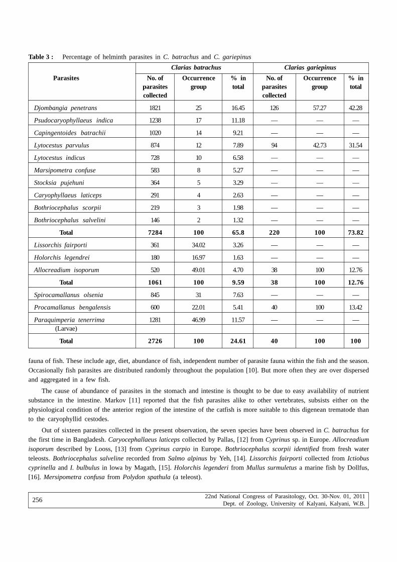
22nd National Congress of Parasitology, Oct. 30-Nov. 01, 2011Dept. of Zoology, University of Kalyani, Kalyani, W.B.
256
Table 3 : Percentage of helminth parasites in C. batrachus and C. gariepinus
Clarias batrachus Clarias gariepinus
Parasites No. of Occurrence % in No. of Occurrence % inparasites group total parasites group totalcollected collected
Djombangia penetrans 1821 25 16.45 126 57.27 42.28
Psudocaryophyllaeus indica 1238 17 11.18 — — —
Capingentoides batrachii 1020 14 9.21 — — —
Lytocestus parvulus 874 12 7.89 94 42.73 31.54
Lytocestus indicus 728 10 6.58 — — —
Marsipometra confuse 583 8 5.27 — — —
Stocksia pujehuni 364 5 3.29 — — —
Caryophyllaeus laticeps 291 4 2.63 — — —
Bothriocephalus scorpii 219 3 1.98 — — —
Bothriocephalus salvelini 146 2 1.32 — — —
Total 7284 100 65.8 220 100 73.82
Lissorchis fairporti 361 34.02 3.26 — — —
Holorchis legendrei 180 16.97 1.63 — — —
Allocreadium isoporum 520 49.01 4.70 38 100 12.76
Total 1061 100 9.59 38 100 12.76
Spirocamallanus olsenia 845 31 7.63 — — —
Procamallanus bengalensis 600 22.01 5.41 40 100 13.42
Paraquimperia tenerrima 1281 46.99 11.57 — — —(Larvae)
Total 2726 100 24.61 40 100 100
fauna of fish. These include age, diet, abundance of fish, independent number of parasite fauna within the fish and the season.
Occasionally fish parasites are distributed randomly throughout the population [10]. But more often they are over dispersed
and aggregated in a few fish.
The cause of abundance of parasites in the stomach and intestine is thought to be due to easy availability of nutrient
substance in the intestine. Markov [11] reported that the fish parasites alike to other vertebrates, subsists either on the
physiological condition of the anterior region of the intestine of the catfish is more suitable to this digenean trematode thanto the caryophyllid cestodes.
Out of sixteen parasites collected in the present observation, the seven species have been observed in C. batrachus forthe first time in Bangladesh. Caryocephallaeus laticeps collected by Pallas, [12] from Cyprinus sp. in Europe. Allocreadium
isoporum described by Looss, [13] from Cyprinus carpio in Europe. Bothriocephalus scorpii identified from fresh water
teleosts. Bothriocephalus salveline recorded from Salmo alpinus by Yeh, [14]. Lissorchis fairporti collected from Ictiobus
cyprinella and I. bulbulus in lowa by Magath, [15]. Holorchis legenderi from Mullus surmuletus a marine fish by Dollfus,
[16]. Mersipometra confusa from Polydon spathula (a teleost).

22nd National Congress of Parasitology, Oct. 30-Nov. 01, 2011Dept. of Zoology, University of Kalyani, Kalyani, W.B.
257
Table 4 : Organal distribution of helminth parasites in C. batrachus and C. gariepinus
Host Name of the parasites Oestophagus Stomach Anterior Posterior Body Totalintestine intestine cavity
Djombangia penetrans 0 1366 455 0 0 1821
Psudocaryophyllaeus 37indica 0 1201 0 0 1238Capingentoides 190batrachii 0 816 14 0 1020
Lytocestus parvulus 0 790 69 15 0 874
Lytocestus indicus 0 615 93 20 0 728Marsipometra confusa 0 0 560 23 0 583Stocksia pujehuni 0 0 343 21 0 291
Caryophyllaeus laticeps 0 0 275 16 0 291Bothriocephallus 201
Clarias scorpii 0 0 5 0 146Bothriocephallus 141
batrachus 0 0 5 0 146salvelini
Lissorchis fairporti 0 18 331 12 0 361
Allocreadium isoporum 0 0 511 9 0 520Holorchis legendrei 0 0 173 7 0 180
Procamallanus 09 591 0 0 600
bengalensis
Spirocamallanus 910 754 0 0 845
olsenia
Paraquimperia 0tenerrima 0 0 0 1281 1281(Larvae)
Total 9 6151 3470 160 1281 11071Djombangia penetrans 0 63 30 0 0 93
Lytocestus parvulus 0 63 30 0 0 93
Clarias Allocreadium isoporum 0 0 47 21 0 68Procamallanus 0
gariepinus 0 56 0 0 56bengalensis
Total 0 178 91 29 0 298
ReferencesChandra K J (2004) Fish Parasitology. Lima Printing Press. 8, Chotta Bazar, Mymensing. pp. 179
Ahmed ATA, Sanaullah M(1976) Observation on the incidence and intensity of some helminth in different length groups ofHeteropneustes fossilis (Bloch) and Clarias batrachus (Linn). Dhaka Univ. Studies. Part B, 15(2) : 91-98.
Lagger K F (1949) Studies in freshwater fish’s biology. Ann. Arbar. Michigan. pp. 119.
Shafi M, Quddus MMA, Ali M, Quddus M M A (1982) Bangladesher Mathso Shampad, Bangla Academy, Dacca pp. 98.
Yamaguti S (1958) ‘Systema Helminthum’. The digenetic trematodes of vertebrates, Vol. 3. Part I and II. Inter science Publishers,New York pp. 1575.

22nd National Congress of Parasitology, Oct. 30-Nov. 01, 2011Dept. of Zoology, University of Kalyani, Kalyani, W.B.
258
Yamaguti S (1959) ‘Systema Helminthum’. The cestodes of vertebrates. Vol. 2. Part I and II. Jhon Wiley and Sons pp. 860.
Yamaguti S (1961) ‘Systema Helminthum’. The Nematodes of Vertebrates, Vol. 3. Part I and II. Ibid. pp. 1261.
Yamaguti S (1963) ‘Systema Helminthum’. Acanthocephala. Vol. 5. Ibid. pp. 423.
Dogiel V A (1961) Ecology of the parasites of freshwater fishes. In parasitology of fishes, Oliver and Boyd. London. pp. 652.
Hopkings C A (1969) Seasonal variation in the incidence and development of the cestode, Proteocephalus filicolis (Rub 1810)in Gasterosteus aculeatus (L. 1766). Parasitol 49 : 529-542.
Markov G S (1946) Modes of feeding of parasites. Priroda, XII (Quoted from Dogiel 1961). 117-139.
Pallas P S (1781) Bemerkungen under dei Bandwurmer in Menshen and Thieren. N. Nord Beytr. Phys. U. Geogr. Erd.–u.Volkerbeschr. I. 39-112.
Looss A (1902) Uber neul undbekannte Trematoden aus Shildkroten, nebst Erorterungen zur systematic and Nomenclatur. ZoolJabrb Syst 16(3-6), 411-894.
Yeh LS (1955) A new tapeworm D. Salvelini sp. Nov. from a salmon Salvelinus alpinus, in Greenland. J. Helm. 29(1-2): 37-443.
Magath F B (1917) The morphology and life history of a new trematode parasite Lissorches fairporti nov. gen. et. nov. spec.from buffalo fish, Ictiobus J Prasit 4(2): 58-69(1917).
Dollfus R Ph (1946) Sur un distome parasite de Mullus surmuletus L. et peut-etra attributable au genre Holorches M. Stossich.1900 (Trematoda). Bull d. I’ inst. oceanogr pp. 896.

22nd National Congress of Parasitology, Oct. 30-Nov. 01, 2011Dept. of Zoology, University of Kalyani, Kalyani, W.B.
259
Host specificity of parasitic isopods in marine fishes
S. Ravichandran and G. Rameshkumar
Centre of Advanced Study in Marine Biology, Faculty of Marine Science, Annamalai University,Parangipettai-608502, Tamil Nadu, India
Abstract : Isopods associate with many species of commercially important fishes around the world and cause significanteconomic losses to fisheries by killing, stunting, or damaging these fishes. Twenty-six species of marine fishes were infestedby 12 species of parasitic isopods along the Tamil Nadu coastal environment, Southeast coast of India. Nature of infestationof the isopods on different species of fishes was described. Parasites were found in the, branchial and buccal regions andon body surface. Most of the isopods (e.g., Joryma brachysoma and Nerocila phaeopleura) were host-specific, while Ryukyuacircularis and Glossobius sp. showed less specificity. The prevalence rate of parasites during different months and the rangeof host specificity were studied. A significant reduction in the gill surface was observed due to infestation. The infestationssuch as, hemorrhagic lesions, anemia, encapsulation, inflammation and penetration of dactylus usually pressure atrophy oftenaccompanied by the presence of larger parasites. This may lead to huge economic losses in commercial species of fish.
Key words : Marine fishes; Isopod parasites; Infestation; Distribution; Host specificity
IntroductionThe parasitic isopods include members of Cymothoidae, Gnathiidae and Bopyridae families. Cymothoidae and Gnathiidae
prefer fishes as host, while Bopyridae are generally found on decapod crustaceans. Cymothoidae inhabit freshwater, brackishwater and marine environment, and are ectoparasite of various fish species. They may be observed on the body or within thebuccal cavity or gill cavity of the host (Trilles 1969; Brusca 1981). Parasites have received considerable scientific attentionbecause they cause severe damage to fishery resources. Isopod occurs very commonly as parasites in food fishes are usuallylarge and fierce looking and cause considerable damage to their host (Overstreet 1978). A few related works are available onthe nature of infestation of isopods parasites in fishes (Williams and Williams 1994; Ravichandran et al. 1999, 2000, 2001, 2009,2010; Ravichandran 2007; Grutter 2003; Cuyas et al. 2004; Rameshkumar and Ravichandran 2010; Rameshkumar et al. 2011).
Rokichi (1985) studied the specificity the zoogeography and the vertical distribution of isopod parasites in host systemsin the northwestern African shelf. Host specificity is the tendency of a parasite to occur on one or a few host species andis a product of co-existence between both parasite and host lineages (Timms and Read 1999; Poulin 2007). Up to date, thereare no detailed studies on the host specificity and infestation of isopod parasites on fishes in Indian waters. The present studywas carried out to understand the host specificity of isopod parasites and the degree of infestation in fishes.
Materials and MethodsAn extensive survey was undertaken over a period of one year from December 2008 to November 2009, along the Tamil
Nadu coast (Southeast coast of India). Fishes were collected from major landing centres from January to November 2009, exceptin December 2008 and May and examined thoroughly for the presence of isopod parasites. The number of parasite, the siteof attachment and their orientation were recorded for each host. The parasites were than carefully removed and transferredto 10% aqueous sodium bicarbonate solution to dissolve the mucus attached to the parasites then preserved in 70% alcoholor 2% formalin for further studies. Total length, width, weight and sex of the hosts were recorded.
Results and DiscussionsDistribution
A total of 223 parasites representing 12 species and nine genera were collected from 26 species of fishes. The majority wereectoparasites. Their distribution of existing parasites host systems along the Tamil Nadu coast was mostly on pelagic fishes.
* Corresponding Author : E-mail : [email protected]
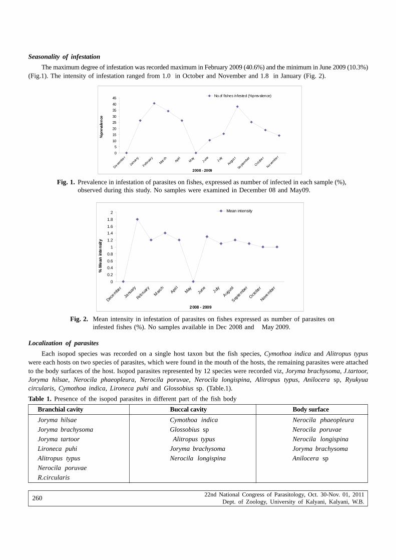
22nd National Congress of Parasitology, Oct. 30-Nov. 01, 2011Dept. of Zoology, University of Kalyani, Kalyani, W.B.
260
Seasonality of infestation
The maximum degree of infestation was recorded maximum in February 2009 (40.6%) and the minimum in June 2009 (10.3%)(Fig.1). The intensity of infestation ranged from 1.0 in October and November and 1.8 in January (Fig. 2).
Fig. 1. Prevalence in infestation of parasites on fishes, expressed as number of infected in each sample (%),observed during this study. No samples were examined in December 08 and May09.
0
5
10
15
20
25
30
35
40
45
Decem
ber
Janu
a ry
Febru
ary
March
April
May
J une
J uly
Augus
t
Septem
ber
Octo
ber
Novem
ber
2008 - 2009
%pr
eva
len
ceNo.of fishes infested (%prevalence)
Fig. 2. Mean intensity in infestation of parasites on fishes expressed as number of parasites oninfested fishes (%). No samples available in Dec 2008 and May 2009.
0
0.2
0.4
0.6
0.8
1
1.2
1.4
1.6
1.8
2
Decembe
r
Janu
ary
Febru
ary
Marc
hApri
lMay
June
July
Augus
t
Septe
mber
Octobe
r
Novembe
r
2008 - 2009
% M
ean
inte
nsi
ty
Mean intensity
Localization of parasites
Each isopod species was recorded on a single host taxon but the fish species, Cymothoa indica and Alitropus typuswere each hosts on two species of parasites, which were found in the mouth of the hosts, the remaining parasites were attachedto the body surfaces of the host. Isopod parasites represented by 12 species were recorded viz, Joryma brachysoma, J.tartoor,Joryma hilsae, Nerocila phaeopleura, Nerocila poruvae, Nerocila longispina, Alitropus typus, Anilocera sp, Ryukyuacircularis, Cymothoa indica, Lironeca puhi and Glossobius sp. (Table.1).
Table 1. Presence of the isopod parasites in different part of the fish body
Branchial cavity Buccal cavity Body surface
Joryma hilsae Cymothoa indica Nerocila phaeopleura
Joryma brachysoma Glossobius sp Nerocila poruvae
Joryma tartoor Alitropus typus Nerocila longispina
Lironeca puhi Joryma brachysoma Joryma brachysoma
Alitropus typus Nerocila longispina Anilocera sp
Nerocila poruvae
R.circularis

22nd National Congress of Parasitology, Oct. 30-Nov. 01, 2011Dept. of Zoology, University of Kalyani, Kalyani, W.B.
261
Branchial parasites
The maximum infestation was recorded in the branchial chamber of host. They were found either in the gill chambers ofhost. The orientation of the branchial parasites varied in most of the hosts. Joryma hilsae, J.tartoor, Lironeca puhi, Alitropustypus, R.circularis and Nerocila poruvae infest the host with its ventral region facing the inner surface of the opercula anddorsal region deeply plunged in the gill arches towards the lower half of the operculum (Fig.3).
Fig. 3. Joryma brachysoma on Secutor sp
Fig. 5. Nerocila longispina on Otolithes ruber
Fig. 4. A.typus on Carangoides sp

22nd National Congress of Parasitology, Oct. 30-Nov. 01, 2011Dept. of Zoology, University of Kalyani, Kalyani, W.B.
262
Buccal parasites
Species of Cymothoa and Glossobius were found in the buccal cavity. Glossobius sp, Alitropus typus, Jorymabrachysoma and Nerocila longispina were found attached to the bucco-pharyngeal region of their hosts (Trichurus lepturus,Carangids sp and Otolithes ruber respectively) (Figs.4&5). These parasites are normally recorded as the mouth aperture. Theparasites were attached in such a way that the broader posterior part of the parasite was lodged in the wider portion of thefloor of the buccal cavity and the narrow anterior part was either located towards the mouth opening the mouth. The malecounter part however showed an orientation with the cephalon directed either towards the anterior or to the posterior partof the mouth of the host.
Body surface parasites
Nerocila phaeopleura infesting the host of fish Sardinella sp, Carangoides sp, Trichurus lepturas, Hilsha ilisha,Chirocentrus dorab, Sphyrnea jello, and Rastrelliger kanakurta were found attached to the dorsalolateral region and nearthe pectoral fin (Fig.6). The digestive secretions/excretions of isopods apparently cause significant damage to the host’stissues.
Fig. 6. N. Phaeopleura on Sardinella sp
Narrow and Broad Host Specificity
Joryma tartoor, Joryma hilsae, Nerocila poruvae, Nerocila longispina, Alitropus typus, Anilocera sp, C. indica, R.circularis, L. puhi and Glossobius sp, have narrow host specificity is limited to the environment of one host species. Nerocilaphaeoplepura and Joryma brachysoma have a broad host specificity is maximum to the environment of different host. Nerocilaphaeopleura, in being host specific, would thus seem to fit in with an apparently generic characteristic and it seems reasonableto assume that the major host species. The constant position and the deep wound caused by N. phaeopleura suggest thatthe position on the host is constant and that the animal does not move about. The location of the parasite in this positioncan be explained by the interaction of two factors which can be called the horizontal and vertical components.
Parasitic crustaceans are the largest fish parasites, which cause considerable damage to their hosts. Isopods inhabiting thebuccal cavity and branchial chamber inflict damage to gills through attachment and feeding and that the extent of damage isdirectly proportional to the size of the parasite and duration of settlement. Infestation causes serious problems to host animalseither directly or indirectly affecting the physiological status of host.
The present study showed that Joryma brachysoma, J.tartoor, Joryma hilsae, Nerocila poruvae, Nerocila longispina,Alitropus typus, Anilocera sp, Cymothoa indica, Lironeca puhi and Glossobius sp have narrow host specificity. Brusca (1981)suggested that specific physical properties of the estuaries could favour broader spreading of the isopods, so that fishinfestation was generally higher. Rokichi (1985) stated that the distribution of Cymothoa plebia is limited to the environmentof one host species Brachydeuterus auritus and considered the parasite as phylogenetically order. The incidence and intensity

22nd National Congress of Parasitology, Oct. 30-Nov. 01, 2011Dept. of Zoology, University of Kalyani, Kalyani, W.B.
263
of crustacean parasites along the South-West coast of India noticed the higher incidence and intensity of infestation in femaleswere than that in males (Radhakrishnan and Nair 1983). Romestand and Trills (1979) stated that the size of fishes that can beinfested will be within certain maximum-minimum variables depending upon the species of host. Similar observation was madein Epipenaeon elegans chopra from the Persian Gulf (Dowson 1958) where infestation was also found to be parasites weremore in females than in males.
The feeding activity of the Cymothoidae isopods in the gill chamber can cause damage to the host in several ways,such as tissue and blood loss, reduction of the gill filaments, and reduction of the breathing efficiency. The presence ofthese ectoparasites also affects the metabolism, and can cause a decrease in the growth rate (Thatcher and Neto 1994).Thatcher (1993) revealed that the Cymothoidae Anphira branchialis can also cause the growth of tumors in the piranhaSerrasalmus sp.
Trilles (1964) significantly showed that a number of cymothoids, including N. orbignyi and N. bivittata are specific intheir choice of hosts, whereas other genera are less specific. The results of this investigation indicate that N. phaeopleura,although comparatively primitive in being an external parasite and being highly host specific is also highly specialised to amode of life upon a pelagic, fast swimming host. It lives on a highly specific region of the body. This position is determinedby the needs of the parasite and the limitations exerted by the morphology and habits of the host.
The parasite occupies the entire branchial chamber of the host may produce pressure on the gill surface and thus affectingthe efficiency of respiration. Although, the infestation did not cause immediate death, it had affected the normal growth ofthe host fish. They may lead to economic losses among some fish species.
AcknowledgementAuthors are thankful to Department of Science and Technology & Ministry of Environment & Forest, Government of India
for providing financial support and Dr. T. Balasubramanian, Dean, faculty of Marine Sciences for providing facilities andencouragement.
ReferencesBrusca RC (1981) A monograph of the Isopoda Cymothoidae (Crustacea) of the eastern Pacific. Zoo J Lin Socie 73:117-199
Cuyas C, Castro JJ, Santana Ortega AT, Carbonnel E (2004) Insular stock identification of Serranus atricauda (Pisces:Serranidae) through the presence of Ceratothoa steindacheri (Isopoda: Cymothoidae) and Pentacapsula cutancea(Myoxoa: Pentacapsulidae) in the Canary Islands. Sci Mar Bare 68:159-163
Dowson CW (1958) A record of Epipenaeon elegans chopra from the Persian fulf. J Parasit 44 : 240-242
Grutter A (2003) Feeding ecology of the fish ectoparasite, Gnathia sp. (Crustacea: Isopoda), from the Great Barrier Reef,Australia and its implications for fish cleaning behavior. Mar Ecol Prog Ser 259: 295-302.
Overstreet RM (1978) Marine Maladies? Worms, Germs, and other Symbionts from the Northern Gulf of Mexico, Mississippi,Alabama Sea Grant Consortium pp140
Poulin R. (2007) Evolutionary Ecology of Parasites. Chapman and Hall, London, UK, pp.212
Radhakrishnan N, Nair NB (1983) Nature of crustacean infestation of fishes along the South West coast of India. Acta InctiEt pise 13 (2): 93-115
Rameshkumar G, Ravichandran S (2010) Cymothoa indica (Isopoda;Cymothoidae) and Alitropus typus (Isopoda;Aegidae) on freshwater fish Tilapia mossambica (Cichlidae) in Vellar estuary, Southeast coast of India. Biotemas 23 (3):67-70
Rameshkumar G, Ravichandran S, Trilles JP (2011) Cymothoidae (Crustacea, Isopoda) from Indian fishes. Acta Parasit 56(1):78-91
Ravichandran S, Ranjith Sing AJA, Veerappan N, Kannapandi T (1999) Effect of isopod parasitc Joryma brachysoma on Ilishamelastoma from Parangipettai coastal waters (South east of India). Ecol Env Conser 5(2):95 -101
Ravichandran S, Soundarapandian P, Kannupandi T (2000) Infestation of isopod parasitc Epipenacon ingens Nobili parasitisedon Penacus monodon from Parangipettai coastal environment. Advan Biosci 19 ( 91):55- 62
Ravichandran S, Ranjith Singh AJA, Veerappan N (2001) Parasite induced vibriosis in Chirocentrus dorab off Parangipettaicoastal waters. Curr Sci 80(5): 101-102

22nd National Congress of Parasitology, Oct. 30-Nov. 01, 2011Dept. of Zoology, University of Kalyani, Kalyani, W.B.
264
Ravichandran S (2007) Infestation of isopod parasite Lironeca puhi in slender needle fish Strongylura leiura. Res J Parasit2 (2):87-93
Ravichandran S, Rameshkumar G, Mahesh Babu B, Kumaravel K (2009) Infestation of Rastrelliger kanagurta, with CymothoidIsopod, Joryma brachysoma in the Colachel Environment of Southwest Coast of India. Wor J Fish Mar Sci 1 (2): 80-84.
Ravichandran S, Rameshkumar G, Balasubramanian (2010) Infestation of isopod parasites in commercial marine fishes. J ParasitDis 34(2):97–98
Rokichi J (1985) Biology of adult Isopoda (Crustacea) parasitizing fishes of Northern West Africa shelf. Acta Inctic Et pise15 (1): 95–119
Romestand B, Trilles JP (1979) Influence of the Cymothoid isopods Meinertia oestraides, Meinerstia parallela and Aniloceraphysodes (Crustacea: parasites of fish) on the growth of hosts. Boops boops and Pagellus erythrinus (Sparidae) ZooParasit 59: 195-202
Thatcher VE (1993) Anphira branchialis gen. et sp. nov. (Crustacea, Isopoda, Cymothoidae) a gill cavity parasite of piranhas(Serrasalmus spp.) in the Brazilian Amazon. Acta Amazv 23 (2-3): 297-307
Thatcher VE, Neto JB (1994) Diagnóstico, prevenção e tratamento das enfermidades de peixes neotropicais de água doce. RevBra de Med Vet 16 (3): 111-127
Timms R, Read AF (1999) What makes a specialist special? Trends Ecol Evol 14: 333-334Trilles JP (1964) Parasitic specificity in the Mediterranean isopod Cymothoidae: Preliminary note. Vie Milieu 15: 105-116Trilles JP (1969) Recherches sur les Isopodes Cymothoidae des côtes Françaises. Aperçu général et comparatif sur la bionomie
et la sexualité de ces crustacés. Bull Soc Zool Fr 94(3): 433- 445Williams EHTr, Williams LB (1994) Ryukyu globbosa n. gen. sp and R. circularis n.comb, parasite in the opercular chambers
of pacific and Indian ocean clupeid fishes. J Aquat Anim Health 6:155-161.

22nd National Congress of Parasitology, Oct. 30-Nov. 01, 2011Dept. of Zoology, University of Kalyani, Kalyani, W.B.
265
Antibacterial effect of some locally available plant extracts againstsome fish pathogenic bacteria
Sayani Banerjee, Jayati Chakraborti, Somerita Panda and Probir Kumar Bandyopadhyay
Parasitology Laboratory, Department of Zoology, University of Kalyani, Kalyani, West Bengal, India
Abstract : Aquaculture is a growing industry in India. Bacterial disease is a growing threat to aquaculture. The use of commercial antibiotics
in aquaculture increases antibiotic resistance among pathogenic bacteria in exposed ecosystem and also creating environmental and publichealth hazards. For this reason many countries have banned the use of antibiotics in aquaculture. As a consequence nowadays scientistsare in search of alternate antibiotics.
Therefore, this study was conducted to evaluate the antibacterial activity of locally available plant extracts against pathogenic bacteriaof aquatic animals namely, Aeromonas hydrophila, Aeromonas sobria, Vibrio paraharmolyticus. Five plants viz: Terminalia arjuna (Arjuna),C. asiatica (Thankuni), Ziziphus mauritiana (Kul), Murraya koenigii (Kari), Ocimum sanctum (Tulsi) were selected for in vitro antibacterialactivity assay. The Minimum Inhibitory Concentration of the methanolic extracts of the leaves of the said plants was determined by Disc
Diffusion method. The plant extracts except tulsi showed antibacterial activity against all the three bacteria. Significant inhibitory activitywas found in C. asiatica, T. arjuna, M. koengii, Z. maurtitiana respectively. Methanol was used as positive control and Tetracyclinehydrochloride was used as negative control. Higher concentration of methanolic extract showed better result consistently for all the plantsthan lower concentration. The most effective inhibitory activity was observed in C. asiatica against A. sobria.
Results of the present study indicate the potential of these plants for further work on isolation and characterization of the activeprinciple responsible for antibacterial activity and its exploitation as therapeutic agent.
Keywords : Antibiotic, Aeromonas hydrophila, Aeromonas sobria, plant biomolecules, Vibrio paraharmolyticus.
IntroductionOccurrence of bacterial diseases is very much threatening to the intensive fish farming system in India. Thus proper
medication is needed to maintain animal health and manage fish population. Applications of commercial antibiotics are inpractice but indiscriminate use of current antibiotics raises super resistant bacterial strains (Ahmed et al., 1998). Many countriesalso banned the use of antibiotics since their prolonged exposure to environment is associated with harmful side effects andis a concern of public health (Cuhna, 2000). Since plant based drugs cause much lower incidence of adverse reactions comparedto synthetic pharmaceutical (Shariff et al., 2006), scientists felt the urgency to develop an alternative approach of herbalmedication towards management of diseases.
In the present study antimicrobial activity of four different plant species are tested against four gram negative bacteria,Aeromonas hydrophilla, Aeromonas sobria, Vibrio parahaemolyticus, Pseudomonas aeruginosa isolated from the fishescollected from local farms. All the four plants viz: Ziziphus mauritiana (in Bengali Kul), Centella asiatica (in Bengali Thankuni),Terminalia arjuna (in Bengali Arjun), Murraya keonigii (in Bengali Curry) are widely exploited by the local community forits medicinal value.
Ziziphus mauritiana leaves are helpful in liver trouble, asthma and fever (Morton, 1987; Michel, 2002). The extract of Z.spinachristi was found to contain beautic acid and ceanothic acid, cyclopeptides, as well as saponins, glycoside andflavonoids, lipids and proteins (Abalaka et al., 2010).
Centella asiatica is an herb growing in wet places through out India is used in ayurvedic preparations either as wholeplant or as leaves in the fresh or extract form (Sharma, 1992). This herb comprises of three active components, namely,asiaticoside, madecassoside and Asiatic acid (Zainol et al., 2008).
* Corresponding Author : E-mail : [email protected]

22nd National Congress of Parasitology, Oct. 30-Nov. 01, 2011Dept. of Zoology, University of Kalyani, Kalyani, W.B.
266
Bioactive fractions of the active extracts of T. arjuna yielded three known oleane compounds: arjunic acid (Kumar et al.,1987), arjugenin (Row et al., 1970) and arjunetin (Honda et al., 1976).
Mahanimbine and koenigine, two carbazole alkaloids were isolated from the leaves of M. koenigii showed antioxidantactivity and antimicrobial activity. Koenigine also showed a high degree of radical-scavenging (Banerjee et al., 2010).
Materials and Methods
Plant materials
The four plants viz: Ziziphus mauritiana (in Bengali Kul), Centella asiatica (in Bengali Thankuni), Terminalia arjuna(in Bengali Arjun), Murraya keonigii (in Bengali Curry) were collected from the garden of the University. The plants wereidentified based on their physical characteristics by the Department of Botany, University of Kalyani. Fresh plant leaves werethoroughly washed several times in running tap water and twice with sterile distill water, dried in blotting paper under shadefor about a month.
Preparation of extract
The dried leaf samples were crushed to powder in mixer grinder. 5 gms of leaf dust of each of the four plants were soakedin 20 ml of absolute methanol at room temperature for two days. The extracts were then filtered using standard whatman filterpaper. Then the filtrates were transferred to petridishes and allowed to evaporate until dried to paste and stored at 4°C. Thedried extracts were re-suspended in the same solvent in two dilutions. Final concentrations were made to 1mg in 10 µL and1mg in 100 µL.
Microorganism used
The pure strains of four species of gram negative bacteria, Aeromonas hydrophila, Aeromonas sobria, Vibrioparahaemolyticus, and Pseudomonas aeruginosa obtained from the Microbiology laboratory of the National Institute ofCholera and Enteric diseases (NICED), Kolkata. Subculture of each bacterium was maintained in nutrient agar and respectiveselective medium. Aeromonas hydrophila, Aeromonas sobria and Pseudomonas aeruginosa were then inoculated to freshwatercarp fishes as they are common fish pathogens. Vibrio parahaemolyticus was inoculated to fresh water shrimp as it is acommon pathogen of shrimp. After the fishes and shrimps showed symptoms, tissues were removed aseptically from the livehost for bacteriologicalculture to confirm the respective bacteria as the cause of death. The isolated bacteria were then againcultured on nutrient agar and respective selective medium (Hi- Media) and confirmatory biochemical tests were performed.These bacteria served as test pathogens for antibacterial activity assay. A direct suspension of the test organisms was preparedin broth and the optical density (OD) of the solution was adjusted to 0.5 at 456 nm which corresponded to 1x 10⁷ CFU.
Screening for antibacterial activity :
The bioassay was done using standard disc diffusion method adopted from Taylor et al., 1995. Bacteria inoculums werespread on the agar plates using sterile cotton swabs. The 5 mm diameter sterile Whatman No.1 filter paper discs were soaked
Table I : Antimicrobial activities of the methanolic extracts of the four plant material against bacterial strains based on Discdiffusion method.
Bacterial strains Zone of Inhibition (mm)
Ziziphus mauritiana Centella asiatica Murayya koenigii Terminalia arjuna Methanol Tetracyclin
1mg/10µl 1mg/100µl 1mg/10µl 1mg/100µl 1mg/10µl 1mg/100µl 1mg/10µl 1mg/100µl
Aeromonas 15±0.75 0 30±0.5 20±0.75 18±0.5 12±0.5 20±0.75 15±0.5 0 30±0.5
hydrophilla
Aeromonas 22±0.5 10±0.6 32±0.75 18±0.5 25±0.5 10±0.2 30±0.75 20±0.75 0 40±0.5
sobria
Vibrio 17±0.65 14±0.5 16±0.7 15±0.5 20±0.7 17±0.2 15±0.5 11±0.75 0 25±0.5
parahaemolyticus
Pseudomonas 0 0 12.75±0.6 10.63±0.5 18.25±0.6 15.13±0.75 0 0 0 21.75±0.6
aeruginosa

22nd National Congress of Parasitology, Oct. 30-Nov. 01, 2011Dept. of Zoology, University of Kalyani, Kalyani, W.B.
267
Fig. 3 : Antibacterial effect of Ziziphus mauratiana (Kul)plant extract on Vibrio parahaemolyticus.
Fig. 4 : Antibacterial effect of Murayya keonigii (Curry)plant extract on Vibrio parahaemolyticus.
Fig. 5 : Antibacterial effect of Terminalia arjuna (Arjun) plantextract on Aeromonas hydrophilla.
Fig. 1 : Antibacterial effect of Terminalia arjuna (Arjun)plant extract on Vibrio parahaemolyticus.
Fig. 2 : Antibacterial effect of Centella asiatica (Thankuni)plant extract on Vibrio parahaemolyticus.
Fig. 6 : Antibacterial effect of Ziziphus mauritiana (Kul) plantextract on Aeromonas hydrophilla.

22nd National Congress of Parasitology, Oct. 30-Nov. 01, 2011Dept. of Zoology, University of Kalyani, Kalyani, W.B.
268
Fig. 7 : Antibacterial effect of Murayya keonigii (Curry)plant extract on Aeromonas hydrophilla.
Fig. 8 : Antibacterial effect of Centella asiatica (Thankuni)plant extract on Aeromonas hydrophilla.
and saturated in extracts. Prepared discs were placed on the inoculated agar plate. The cultures were then incubated upsidedown for 24 hrs at 37oC. The paper discs were impregnated in the same amount of methanol which was used as negative controlwhile, tetracycline hydrochloride paper discs were used as positive control. Tetracycline hydrochloride solution was preparedby mixing 0.8 ml of tetracycline in 9.2 ml distilled water. Final concentration of tetracycline was 0.25 mg/ ml. Results wererecorded as presence or absence of zone of inhibition (Lennette, 1995). The inhibitory zone around test paper disc indicatedabsence of bacterial growth and it was reported as positive. Absence of zone was indicated as negative. The tests wererepeated thrice to ensure the results.
Results and Discussions :
The in vitro potency of the anti microbial activity of the methanolic plant extracts against these four bacteria were assessedby measuring the diameter of the clear zone around the discs placed on the petriplates. The methanolic extracts of differentplants showed various inhibitory results against different bacteria. The mother solution i.e. 1mg/10µl showed better result thanthe solution having concentration 1mg/100µl in every cases. Previously it was found that the ethanol extract of Centella
asiatica was active against E.coli ATCC 25922, Staphylococcus aureus ATCC 25923, Vibrio parahaemolyticus ATCC 17802Pseudomonas aeruginosa CC 27853 (Mamtha et al, 2004). Our observation for Centella asiatica also showed similar resultsby inhibiting Pseudomonas aeruginosa and Vibrio parahaemolyticus. Along with that it was also found effective against bestantimicrobial potency was showed against A. hydrophila and A. sobria.
The present study showed that methanolic extracts of Murraya keonigii has significant antibacterial results against allthe test bacteria though less effective than Centella asiatica. Previous records on antibacterial activity of this plant havingtremendous medicinal value are present. The petroleum ether extract of M. keonigii leaf inhibited the growth of S. aureus, B.
subtilis, E. coli and P. vulgaris (Rahman and Gray, 2005). Aqueous extract of M. keonigii leaf can be effective on the growthof A. hydrophila, C. freundii, S. putrifacien, V. alginolyticus, V. harveyi and V. vulnificus respectively (Lee Seong Wei et al,2008).
It is known that T. arjuna has antibacterial activity. Earlier, it was found that biomolecules isolated from bark extracts ofT. arjuna possessed antibacterial activity, which can specifically act against S. epidermidis (Singh et al, 2008). Inhibitory roleof aqueous leaf extracts on P. aeruginosa, was assessed by Ramya et al. (2008) along with many other bacteria. In our study,methanolic leaf extract of T. arjuna showed significant inhibitory results against all the three bacteria excepting P. aeruginosa
which could not be inhibited by Arjun leaf extract.
Previous results on inhibitory role of methanol Ziziphus leaf extract against B. subtilis, E coli, P. fluorescens, S. aureus,X. axonopodis, V. malvacearum, A. flavus, D. turcica and F. verticillioide (Mahesh and Satish, 2008) are available. In our study

22nd National Congress of Parasitology, Oct. 30-Nov. 01, 2011Dept. of Zoology, University of Kalyani, Kalyani, W.B.
269
methanolic leaf extract of Kul showed inhibitory activity against the other three bacteria, A. hydrophila, A. sobria, V.
parahaemolyticus but with much less potential than the other extracts. It was not effective against P. aeruginosa though it
could inhibit P. fluorescens (Mahesh and Satish, 2008). Thus it can be concluded that its inhibition potency is organism or
species specific.
It is to be mentioned that zone of inhibition was not observed around any control group i.e. in methanol treated discs
whereas, the inhibition zone diameter around the standard antibiotic (tetracyclin) impregnated discs was highly significant
between 45- 30 mm.
The present study shows the potential antibacterial effect of these locally available plant extracts against the common
bacterial pathogen of aquaculture. Therefore, these plant materials should be further screened for commercial use as antibiotics
against the bacterial diseases causing great economic loss in the fish industry.
Acnowledgement
Two of the authors (SB and SP) are thankful to the Department of Science and Technology, Govt. of India, New Delhi
for financial support to run the project (Project Ref.No.ST (WB)/ST/LSR/461) and another author (JC) is thankful to University
Grants Commission, New Delhi for financial support in the form of a scholarship to run the project (Project no.F.32-483/2006
(SR). Thanks are also due to the Microbiology Laboratory, NICED, Kolkata for their kind help in providing the bacteria sample
and Department of Botany, University of Kalyani, Kalyani for their identification the plant materials.
Reference
Ahmad I, Mehmood Z, Mohammad F (1998) Screening of some Indian medicinal plants for their antimicrobial properties. J
Ethnopharm 62: 183- 198.
Cuhna BA (2000) The diagnostic significance of relative bradycardia in infectious disease. Clinical Microbiol Infect 6(12):
633-634.
Honda T, Murae T, Tsuyuki T, Takahashi T, Sawai M (1976) Arjungenin, arjunglucosidase I, and arjunglucosidase II, a new
triterpene and new triterpene glucosides from Terminalia arjuna. Bull Chem Soc Japan 49: 3213 – 3218.
Kumar DS, Prabhakar YS (1987) On the ethnomedical significance of the Arjun tree. J Ethnopharm 20: 173- 190.
Lee Seong W, Najiah M, Chuah TS, Wendy W, Mohd Shazili NA (2008) Antimicrobial properties of tropical plants against
12 pathogenic bacteria isolated from aquatic organisms. Afr J Biotech 7(13): 2275 – 2278.
Lenntte EH (1995) Manual of clinical microbiology. (4th Ed.) American association for microbiology, Washington, D.C
pp978 – 987.
Mahesh B, Satish S (2008) Antimicrobial activity of some important medicinal plant and human pathogens. W J Agri Sci 4(S):
839-843.
Mamtha B, Kavitha K, Srinivasan KK, Shivananda PG (2004) An in vitro study of the effect of Centella asiatica (Indian
pennywort) on enteric pathogens. Ind J Pharm 36(1): 41.
Michel A (2002) Tree, Shrub and Liana of West African Zone. Margraf Publishers GMBH, Paris pp 440.
Morton J (1987) Indian Jujube. In: Fruits of warm climates. Morton, J. F. (Ed.). Miami, Florida pp. 272 – 275.
Nayak A, Mandal S, Banerji A, Banerji J (2010) Review on Chemistry and pharmacology of Murraya keonogii (Spreng). J Chem
Pharm Res 2: 286-299.
Rahman MM, Gray A, I (2005) A benzoisofuranone derivative and carbazole alkaloids from Murayya keonigii and their
antimicrobial activity. Phytochem 66: 1601- 1606.
Ramya S, Kalaivani T, Rajasekaran C, Jepachannderamohan P, Alaguchamy N, Kalayansundaram M, Jayakumararaj R (2008)
Antimicrobial activity of aqueous extracts of bark, root, leaves and fruits of Terminalia arjuna Wight & Arn.
Ethnobotanical Leaflets 12: 1192 – 97.

22nd National Congress of Parasitology, Oct. 30-Nov. 01, 2011Dept. of Zoology, University of Kalyani, Kalyani, W.B.
270
Row LR, Murty PS, Subba Rao, GSR, Sastry CSP, Rao KVJ, 1970. Chemical examination of Terminalia species: Part XIII isolation
and structure determination of arjunic acid, a new trihydroxytriterpene carboxylic acid from Terminalia arjuna bark.
Indian J Chem 8: 716-721.
Sharma PV (1992) Dravyaguna Vigyana. (13th Ed.) Chaukhamba Publications Vishwa. Bharati Academy, New Delhi, India,
pp3-5.
Shariff N, Sudarshana MS, Umesha S, Hariprasad P (2006) Antimicrobial activity of Rauvolfia tetraphylla and Physalis minimaleaf and callus extracts. Afr J Biotech 5: 946- 950.
Singh DV, Gupta MM, Santha Kumar TR, Saikia SD, Khanuja PS (2008) Antibacterial Principles from the bark of Terminaliaarjuna. Current Science 94:1- 10.
Taylor RSL, Towers GH (1995) Screening of selected medicinal plants of Nepal for antimicrobial activities. J Ethnopharm 46:153- 159.
Zainol NA, Voo SC, Sarmidhi MR, Aziz RA (2008) Profiling of Centella asiatica (L.) Urban Extract. Mal J Anal Sci 12(2):322-327.

22nd National Congress of Parasitology, Oct. 30-Nov. 01, 2011Dept. of Zoology, University of Kalyani, Kalyani, W.B.
271
Studies on parasitic diseases of fish in east Kolkata wetland and theiranti-parasitic treatment which threat to environment
Somsuvra Dasgupta, Asosh Kumar Panigrahi and Raman Kumar Trivedi*
Fisheries and Aquaculture Extension Laboratory Department of Zoology, University of Kalyani, Kalyani-741235, West Bengal, India*Department of Fisheries Environment, W.B.University of Animal and fishery Sc, Panchasayar, Kolkata-700094, West Bengal, India
Abstract : East Kolkata Wetland, only Ramsar wetland of West Bengal a vast low land located at eastern fringes of Kolkata. This uniquesystem where treatment of the sewage is done, then allowed for pisciculture. This wetland is also important for its fishery activity andis now producing about 13000 tonnes of fish per year. Here fish production is affected by aquatic pollution through different industrialeffluent discharged in it and disease is a significant problem to the majority of producers affecting all development stages. As the majorityof diseases occur during winter from low temperature, although in some cases disease problems occur throughout the year where parasitic,protozoan, bacterial and fungal pathogens are indicated as the most likely causes. The copepod parasite, Argulus sp., Lernaea sp. affectedmost businesses with a variety of possible bacterial and fungal species being second and affecting a greater range of production systems.In summer high temperatures caused fish seed mortalities in production and distribution. Kerosene oil, endosulfan and the organophosphates,methyl parathion and Malathion were substances utilized in anti-parasitic treatments of fish and in pond preparation which can have effectsof long persistence in the environment and or humans through mechanisms of bio-accumulation and direct toxic effects on non-targetorganisms. So, herbal medication should be taken.
Key words : East Kolkata Wetland, Environment, Fish, Herbal medication, Parasitic Disease
Introduction
East Kolkata Wetland is famous all over the World for its ecological importance. It was declared as Ramsar wetland onNovember 2002 in Ramsar convention bureau. East Kolkata wetland EKW) stretches its area from North 24 Parganas to South 24Parganas districts of West Bengal and located at eastern side of Kolkata metropolis. It is known as kidney of Kolkata city. In thisshallow water body, treatment of sewage has done and then allowed for pisciculture. Everyday about 250 million liter of waste wateralong with solid waste is discharged here though different canals. Huge amounts of hazardous substances, heavy metal, sulfide,grease, oil originated from different industries of the surroundings, domestic waste water and industrial effluent from Kolkata cityalso discharged here though Tiljala, Bagjola,vangor canal and pollute the aquatic environment. Thus fish production is hampered.Other than fish production is suffered by the fish disease. Mainly diseases are broken out during winter season. Although Fishesare suffered by parasitic, bacterial, fungal diseases, the copepod parasite Argulus sp. Lernaea sp. are the main causative agents offish disease of the most of the water bodies. Low temperature of winter and excessive temperature during summer are alsoresponsible for death of fish seeds. A number of chemicals like Kerosene oil, endosulfan and the organophosphates, methylparathion and Malathion are used in anti-parasitic treatments of fish and in pond preparation. These substances have effects oflong persistence in the environment and or humans through mechanisms of bio-accumulation and direct toxic effects on non-targetorganisms. In our laboratory we are trying to utilize herbal medication for the diseased fish which not only help to prevent parasiticdiseases of fish but also help to reduce the aquatic pollution of the wetland to reduce the uses of the chemicals that are used as antiparasitic agents.
Methodology
Prelimininary survey has done in East Kolkata wetland area to collect information on fish production, their disease problemand disease management taken by the fish farmers. Some indigenous ornamental fishes that are common in this wetland arecollected from the ornamental fish farms in this area and also collected from the wetland. This fishes has bought to thelaboratory. Then fishes are thoroughly checked, scraping of different parts of fishes has dried and stained in giemsa or silver
* Corresponding Author : E-mail : [email protected]; [email protected]

22nd National Congress of Parasitology, Oct. 30-Nov. 01, 2011Dept. of Zoology, University of Kalyani, Kalyani, W.B.
272
nitrate or double stained in eosin and haematoxylin. Then slides have prepared and observed under microscope. On the otherhand infected fishes are treated with herbal crude extract by deep bathing.
State West Bengal
District Kolkata
Co-ordinates 220 25’ to 22040’ N and 88020’ to88035’E
Area 12741 hectares
Ownership Government of West Bengal andPrivate
Declared as 19th August 2002Ramsar wetland
Ramsar criteria 1
Area 12,741 Hectares(Source: State Govt. report.)
Ramsar wetland O (Permanent fresh watertype bodies)
No. of Bheris 264
Different land use classes in EKW
*Additionally 241.30 ha. Are eing added tothe system for making the system integral.
Table 1. Represent land use pattern of EKW Table 2. Represent information on EKW
Over view on East Kolkata Wetland :
Pollutants Comments
Sewage and 600 million lit/daywaste water
Garbage >2500 tones
Heavy metals Cr, Cu, Pb, Etc
Sulphate _
Oil, grease _
Name of the heavy Name of themetal discharged industriesthrough effluents
Chromium (Cr) Tannery and leather
industries
Copper (Cu) Metal handicraft and
electroplating industries
Lead (Pb) Tannery and battery
manufacturing industries
Rubidium (Rb) Painting and Dye
industries
Table. 3 Table. 4
Result and Discussiona. Industries located surround East Kolkata Wetland and their Pollutants
b. Parasitic diseases shown in the fishes of East Kolkata Wetland :
Different types of fish diseases are observed in the fishes collected from East Kolkata wetland area and they arerepresented below in fig1, fig 2 and fig:3
c. Symptoms of the parasitic diseases of fish and affected body parts :
Land use
Water body
Agriculture Land
GarbageDisposal Site
UrbanSettlement Area
Rural Area
Total Area
Area
5,852.14 ha of whichfish farming roughlyconstitutes about3,898.70 ha. (45.93%)
Approximately4,959.86ha.(38.92%)
602.78 ha. (4.73%)
91.53 ha.(0.73%)
1234.99 ha.(9.69%)
12500.00 ha*
WaterAgricultureRural settlementUrban settlementProductive farming

22nd National Congress of Parasitology, Oct. 30-Nov. 01, 2011Dept. of Zoology, University of Kalyani, Kalyani, W.B.
273
Fig. 3 : crustacean parasite represented hereFig. 2 : Represent the helminth parasite of fishes,
Name of parasite
Ichthyopthirius sp
Trichodina sp
Epistylis sp
Myxobolus sp
Gyrodactylus sp
Dactylogyrus sp.
Argulus sp.
Ergasilus sp.
Lernaea sp.
Affected parts
Body,fins,gill,tail
Body, gill
Body, gill
Gill
Body
Gill
Body, gill
Gill
Body surface
Symptoms
The ich symptoms appear as white spots that look like grains of salt. Opportun-istic ich parasite burrow under the skin and starts sucking blood and other fishjuices and the fish trys to protect itself by growing a covering over the invaders-hence the characteristic white spot appears
They are probably not harmful when present in small numbers but in severeinfection acts as true ectoparasite feeding on gill and skin tissue leading tonecrosis and erosion of the tissues and respiratory trouble of the fishes.
The symptom by this stalked ciliate appears lesion pale & white in colour andresembles a fungus disease.
Cyst appear in the form of small white spots on gills and sometimes in fins andare less pathogenic can infect other internal organs that cause high mortality.
With this infection the fish growth is hampered, erratic movement occurs and inheavy infestation the fishes are feeble and rest near the water margin.
With this monogenean infection, the gill fade ness and excessive mucoussecretion take place causing severe respiratory trouble
Commonly known as fish louse, feed on body fluids and are especially harmfulto small fishes. Reddish lesions occur at the site of attachment which causessecondary infection
Also known as gill maggot. Attacks the gill and sometimes skin of the fishes.Appear as whitish green threads hanging out of fish gill. Heavy infestation resultin severe gill damage, anemia and death usually due to secondary infection.
The fish scrapes itself against objects, whitish-green threads hang out of thefish’s skin with an inflamed area at the point of attachment.
Fig: 1 : represent the causative agent of protozoan parasite of fish diseases.
Table no. 5
Helminth parasite
Gyrodactylus sp
Dactylogyrus sp.
Crustaceanparasite
Argulus sp.
Ergasilus sp.
Lernaea sp.
Protozoanparasite
Ichthyopthirius sp. Trichodina sp. Tripertiella sp. Epistylis sp. Myxobolus sp.

22nd National Congress of Parasitology, Oct. 30-Nov. 01, 2011Dept. of Zoology, University of Kalyani, Kalyani, W.B.
274
d. Chemicals used as anti-parasitic reagents and their hazardous effects :
In East Kolkata Wetland different chemicals are used to prevent parasitic diseases of fishes, some chemicals are alsoapplied during pond preparation. All these chemical substances have hazardous effect on target organisms as well as non targetorganisms. It has also hazardous effect on aquatic environment. These chemical substances present in environment thoroughbio accumulation and affects nervous, immune, respiratory system and produce genetical disorder. These chemicals and theirhazardous effects are represented in Table no. 6
Hazardous effect
Insoluble in water and present in environmentfor a long period through bioaccumulation andaffect nervous system, immune system andproduce genetical disorder
present in environment for a long periodthrough bioaccumulation and affect nervoussystem, skin and mental ability
Affect respiratory and nervous systems
Affect nervous system as well as geneticaldispute shown
Skin disease and respiratory problem occur
Hazardous effect absent
Use
To control parasiticdisease
Pond preparation
To controlparasitic disease
Pond preparation
To control parasiticdisease
To prevent Fishdiseases
Type
Pesticide
Pesticide
Pesticide
Pesticide
Pesticide
Fungicide &antibacterialreagent
Name of chemicals
Endosulfan
Kerosene oil
Methyl parathion
Thion (mathion)
Butox
Tetramycin
Table no. 6
e. Herbal medication practiced to prevent fish diseases
Chemicals that are used as anti- parasitic substances and used during pond preparation have used on target and nontarget organisms. That is why we are experimented with herbal product and get good results when applying it on diseasedfish. In Table no.7 those herbs are represented that we are used as anti parasitic substances to prevent parasitic diseases offish.
Crude extracts of garlic and water in particular ratio helps to prevent parasitic disease of fish due to presence of alliumand allicin in it. If Garlic paste and common salt in 2:5 raio with adequate amount of water is applied on diseased fish, it isfound that this mixture gives better results in case of bacterial and fungal disease. It is also found that turmeric, lime andcommon salt in 1:2:10 ratio gives good result in bacterial and fungal diseases of fish. Neem extract along with lime in 1:2 ratiohave good anti parasitic activity against fish diseases. Mixture of crude extract of neem and turmeric and water is beneficialfor copepod diseases of fish. Zingerone, gingerols present in ginger act as anti allergic and ginger’s crude extract with adequateamount of water gives good result in case of allergic diseases of fishes. Extract of tulsi, ginger, pan lata give good resultsif it applied on copepod disease affected fishes. Herbs are used from ancient age. We get best result from ginger to preventthe parasitic diseases of fish collected from east Kolkata wetland. Herbal medication has no hazardous effects on theenvironment and it has no side effects on target organism as well as non target organisms. Herbal medication is also importantdue to it is cost effective, easily available and no side effects in it.
ConclusionIn East Kolkata Wetland, fish production is hampered by fish diseases and aquatic pollution. Maximum diseases are
outburst during winter. Here Fishes suffer more or less Parasitic, bacterial, protozoan and fungal diseases throughout the year.

22nd National Congress of Parasitology, Oct. 30-Nov. 01, 2011Dept. of Zoology, University of Kalyani, Kalyani, W.B.
275
Fishes of most of the ponds are attacked by copepod parasite Argulus and Lernaea. Winter temperatures were associatedwith disease, whilst in summer high temperatures caused fish seed mortalities in production and distribution. Different chemicalsubstances utilized in anti-parasitic treatments of fish and in pond preparation which can have effects of long persistence inthe environment and or humans through mechanisms of bio-accumulation and direct toxic effects on non-target organisms.So, herbal medication should be taken. A detailed study regarding this is under processed in our laboratory.
ReferencesDDas JN, Biswas (2008) Ornamental fish resources of North Eastern states and their trade potential Ornamental breeding,
farming and trade pp.51-63.
Panigrahi AK, Bandhopadhyay Dutta (Roy).S (2009): “Studies on indigenous ornamental fish species with reference toentrepreneurship development in some district of West Bengal”, J. Environ & Sociobiol 6(1): pp.7-11.
Riji JK, Gorge.m.Rosaline (2008) Health management in Ornamental fish culture, Proceedings of the International seminar onOrnamental breeding, faming and trade held in Kerala, pp-155-174.
Sanil NK, Vijayan KK (2008) Disease of ornamental fishes Proceedings of International seminar on Ornamental breeding, famingand trade held in Kerala 175-189.
De M, Bhunia S, Sengupta T (1989) A Preliminary Account on Major Wetland Fauna of Calcutta and Surroundings. Ecology3 (9): 5-11.
Roychowdhuri S (2008) East Calcutta Wetland: A Multifaceted Treasure to Be Preserved. International Conference onEnvironmental Research and Technology 2008, Kolkata.
Active ingredient
zingerone, shogaols,
gingerols
Curcumin, C.I.75300, or
Natural Yellow 3.
Azadirachtin, azadirachtol,
azadiradione, gedunin,
nimbiol, nimboeinol,
nemolinone, nimbocetin
allicin,alliin,ajoene,
diallylsulfide,dithiin
Rotenone,
dehydrorotenone, lupeol,
oleanolicacid, ursolicacid
Bornylacetate, cadinene
Characters
Anti- allergic, anti-
bacterial
Anti-bacterial
Antiseptic
Antibiotic, Antiseptic
Anti- allergic, anti-
bacterial
Anti- allergic
Scientific name
Zingiber officinale
Curcuma longa
Azadirachta indica
Alliumsativum
Derris sp.
Ocimum sanctum
Name of herbs
Ginger
Turmeric
Neem
Garlic
Pan lata
Tulsi
Table no. 7

22nd National Congress of Parasitology, Oct. 30-Nov. 01, 2011Dept. of Zoology, University of Kalyani, Kalyani, W.B.
276
Studies of the parasitic infestation in indigenous ornamental fish Silver Danio,Danio devario with its prophylactic measures
Sharbani Dutta(Roy) and Ashis Kumar Panigrahi
Fisheries and Aquaculture Extension Laboratory, Department of Zoology, University of Kalyani, Kalyani, West Bengal, India
Abstract : Ornamental fish culture and breeding is becoming an important component in fishery sector. India is one of the mega diversitycountries which is bestowed with a number of aquatic biodiversity not only in the food fishery sector but also in the ornamental fisherysector and in ornamental fishery West Bengal particularly enriched with a number of indigenous ornamental fishes which are the sourceof income generation in the local people in many ways like seed collection, preparation of brooders, breeding and culture purposes. Amongthe indigenous ornamental fishes the Silver Danio- Danio devario is one of the potential fish which has body silvery, olivacious green onback, a dark longitudinal band in the middle of sides extending from caudal fin base to below dorsal fin origin. This fish has high demandas aquarium fish in native as well as export market. For the study of parasitic infection a number of silver danio were collected from thenatural habitats of Howrah district of West Bengal in different seasons and different types of parasitic infections were observed. Amongthe crustacean parasite- Ergasilus sp. and Argulus sp. were identified, among the protozoan parasite Trichodina sp., Tripertiella sp., Epistylissp., Ichthyoptyhries sp. were identified. And among the helminth parasite Dactylogyrus sp. and Gyrodactylus sp. were identified. Theseparticular infections are major reasons for huge economic loss in the aqua business producing lower quality of ornamental fishes with reducedcolouration which are of low value and actually do not confirm quality for the export market. These parasitic infections are being treatedwith different types of herbs extracts to control these diseases and the detailed study regarding this is under processed in our laboratory.
Keywords : Control, Indigenous, Ornamental, Parasitic,
Introduction
Ornamental fish culture is becoming an important component in Indian fisheries along with food fishes for bothemployment generation and export earnings (Sinha etal, 2010). India is one of the mega diversity countries having vast potentialof ornamental fishes. Specially in the Ornamental sectors it’s contribution is of immense important and particularly in case ofWest Bengal the sector is much more explored than other states of India. Specially in the native ornamental fishes i.e. theindigenous ornamental fishes are the source of income generation among the local people through the aquarium business ofthese fishes. Ornamental fish farming is a sub sector of aquaculture and many technologies currently applied to ornamentalfish production arise from food fish and live stock farming sector (Rana, 2008). Among the indigenous aquarium fishes thesilver danio- Danio devario is one of the potential fishes which is exported to different countries from India in terms of US$0.263 and Rs. 6.00/- per fish in Indian money( Sinha etal, 2010). So the fish dominates the local as well as export market forit’s colouration and structural organization.
Description of the fish
Body silvery, olivacious green on back, a dark longitudinal band in the middle of sides extending from caudal fin baseto below dorsal fin origin.The silver danio found in lower region hill stream, plain rivers and lentic wet lands like beels withclear slow running water. This species usually swim on the lower surface of upper column zone in shoal.
Determination of sex
Body shape slender in male but slightly convex in female. Dorsal profile is slightly convex in male. In female the ventraledge is comparatively less keeled. Mouth blunt in male but in female it is faint. In male the dorsal caudal and anal fins arereddish yellow in colour during breeding season. Fins are pale yellowish in colour during breeding season in female. In naturalpopulation the sex ratio is 2:1 (Male: Female).
* Corresponding Author : E-mail : [email protected]

22nd National Congress of Parasitology, Oct. 30-Nov. 01, 2011Dept. of Zoology, University of Kalyani, Kalyani, W.B.
277
Material and methods
Survey design :
First of all an arial survey was conducted to Howrah district of West Bengal to identify the ornamental prone areas of
the district. Then the areas were selected –Pakuria, Dopmjur, Kadamtala. Amta and ramrajatala. After that a number of survey
were conducted to those areas in a year from November 2009 to October 2010 dividing the period into three seasons i.e. Pre
monsoon, Monsoon and post monsoon. Simultaneously a number of silver danio was randomly collected from the natural
habitat of that fish from the selected ornamental prone areas.
Laboratory preparation :
After collecting those fishes, the fishes were brought to laboratory and checked thoroughly for the infestation of
parasites. For this purpose the following steps were done-
• Scrapings from the body, gill, fins and tails were taken
• Thin smear was prepared in the slide from those scrapings.
• The smear was then allowed to semidry.
• After that the smears were either single stained with Giemsa or Silver nitrate or double stained with Haematoxylene
and Eosin.
• After staining the slides were observed under microscope for the idemtification of parasites.
Result and Discussion
Parasites are generally opportunistic pathogenic organism, causing diseases under congenital condition. Usually parasites
are present in almost all animals in low numbers, but generally do not cause any disease condition but if present in large
numbers they can turn fatal for the host (Sanil et.al 2008).The stained Slides Showed Different Types of protozoan, and
Helminthes and Crustacean parasites of the fish – Danio devario.
Identification of Protozoon parasites
Among the protozoon parasiotes the fish was infected with a number of Ichthyopthirius sp, Trichodina sp, Tripartiella
sp and Epistylis sp were identified.
Description and Symptoms of the Protozoan parasites
A. Ichthyopthirius multifilis :
Ich is the most common disease in aquaria and is caused by the ciliate parasite, Ichthyopthirius multifilis. Symptoms
include white glistening spots or salt like specks on the body and fins, excessive slime on body, difficulty in breathing, clamped
fins and loss of appetite. The parasite lacks host specificity can infect any fish species, making it the most destructive fish
parasite in ornamental fishes.
B. Trichodina sp :
Dogiel (1961) reported that trichodiniasis is caused by Trichodina spp; the infection being stimulated by the high density
of fish in ponds. A disc shaped ciliate protozoan found on the skin and gills of many fresh water fishes. Circular rows of
denticles and a ciliary girdle give this parasite a unique symmetry. Probably not harmful when present in small numbers but
on stressed and young fishes, especially when organic load in the water is high, they proliferate rapidly and behave like true
ectoparasites, starts feeding on gill and skin tissues leading to necrosis and erosions of tissues. Lack of host specificity
coupled with their pathogenicity.
C. Tripartiella sp :
Another disc shaped urceolarid protozoan parasite that resembles with Trichodina sp. but differs in size other internal
characters. The parasite found on gill, skin, fin & tail region of fresh water ornamental fishes.They become dangerous when
present in large numbers and causes tissue necrosis & erosions of the surrounding tissue.

22nd National Congress of Parasitology, Oct. 30-Nov. 01, 2011Dept. of Zoology, University of Kalyani, Kalyani, W.B.
278
Table 1 : Distribution and Biodiversity of the Protozoan parasites
Host Collection Parasite Phylum Infectedsite/District Organ
Danio devario Howrah Ichthyopthirius sp. Ciliophora Body, Gill, Fin, Tail
Trichodina sp. Ciliophora Body, Gill
Tripartiella sp. Ciliophora Body, Gill
Epistylis sp. Ciliophora Body, Gill
D. Epistylis sp :
A stalked ciliate which is commonly found in fresh water containing a high organic loads. Lesions appear pale and whitein color and resemble a fungal disease. Microscopically, it appears as a ciliated crown with a long stalk which is prone tofrequent contractions. This disease is usually not fatal in itself but may open the fish up to secondary infection.
Prevalence of Protozoon parasitesAccording to our study we have taken 50 numbers of fishes randomly in each seasons thoroughly checked those fishes
for the study of infection. We observed that 40% of fishes were infected with Ichthyopthirius sp in Pre monsoon while 64%and 84% fishes were infected during the Monsoon and post monsoon seasons. On the other hand in case of Trichodina spthe prevalence rate is 24%, 36% and 54% in Premonsoon, Monsoon and Post monsoon respectively. In case of Tripartiellasp the prevalence rate is 34%, 34% and 62% in Pre monsoon, Monsoon and Post monsoon respectively. And in case ofEpistylis sp the prevalence rate of infection is 44%, 52% and 86% in Premonsoon, Monsoon and Post monsoon respectively.The details of the protozoan prevalence is shown in the chart below.
Seasonal prevalence of Protozoan parasites
020406080
100
Parasit
e
Ichth
yopt
hirius
Tricho
dina
Tripartie
lla
Epistyl
is
Parasites
Pre
vale
nce
Premonsoon
Monsoon
Post monsoon
Identification of Helminthes ParasitesAmong the Helminthes parasites two types of parasites were identified i.e. Dactylogyrus sp and Gyrodactylus sp.
Description and Symptoms of the Helminthes parasites
Dactylogyrus sp :
Monogenean parasite including Dactyloyrus sp is considered as an ectoparasite which can cause huge damage to thehost when present in large numbers. The parasite possess a multiple hooked attachment organ called an opisthaptor whichdisrupts the integrity of the host’s skin and mucus membranes. It actually infects the gill of the fishes in which condition thefish feels suffocated and becomes stressed and generally fading of body colouration is seen.

22nd National Congress of Parasitology, Oct. 30-Nov. 01, 2011Dept. of Zoology, University of Kalyani, Kalyani, W.B.
279
Gyrodactylus sp :
Another monogean worm present on the body surface of fresh water fishes is Gyrodactylus sp. It also causes hugedamage when present in large numbers. In general there is growth retardation in afflicted fishes for the disease is encountered.
Table 2 : Distribution and Biodiversity of Helminthes Parasite
Host Collection site/ Parasite Phylum Infected OrganDistrict
Danio devario Howrah Dactylogyrus sp Platyhelminthes Gill
Gyrodactylus sp Platyhelminthes Body
Prevalence of Helminthes parasites50 numbers of fishes were randomly selected in each season from the natural habitat of Howrah district and the prevalence
of helminthes parasites were checked. According to our observation 32% fishes are infected with Dactylogyrus sp in the premonsoon season. While 50% and 60% fishes are infected with this parasite during the monsoon and post monsoon season.Onthe contrary 26%,54% and 58% fishes are infected with the Gyrodactylus sp during the pre monsoon, monsoon and postmonsoon season. The chart below shows the details.
Identification of Crustacean parasitesAmong the Crustacean parasites the prepared slides showed the infestation of two types of parasites i.e. Ergasilus sp
and Argulus sp.
Seasonal Prevalence of Helminthes Parasites
010203040506070
Dactylog
yrus
Gyrod
actylus
Parasites
Pre
vale
nce
Premonsoon
Monsoon
Post monsoon
Description and Symptoms of the Crustacean Parasites
Ergasilus sp :
The parasite is also known as “Gill maggot” which infest the gill of the fishes. The adult female Ergasilus sp is parasitic.
The parasite looks as white bodies less than 2mm long. Surfacing, lethargy and restlessness occur during heavy infestation
and the intensity of infection is directly proportional to the size of the fish(Das, 1997).
Argulus sp :
The parasite is generally known as “Fish louse” with flattened mite-like crustacean that attaches itself to the gill and some
times the body of the fish. They irritate the host fish which have clamped fins, becomes restless and may show inflamed areas
where the lice have been situated. The fish scratches itself against objects. The parasite can be visible with naked eye in hugeinfestation.

22nd National Congress of Parasitology, Oct. 30-Nov. 01, 2011Dept. of Zoology, University of Kalyani, Kalyani, W.B.
280
Table 3 : Distribution and Biodiversity of Crustacean parasites
Host Collection spot/ Parasites Phylum Infected organDistrict
Danio devario Howrah Ergasilus Crustacea Gill, skin
Argulus Crustacea Skin, fin
Prevalence of Crustacean ParasitesHere also 50 number of fishes were collected from its natural habitat and the prevalence of crustacean parasites were
estimated. So according to our investigation the prevalence of Ergasilus sp is 34% in the pre monsoon and it is 46% and64% during the monsoon and post monsoon seasons respectively. On the other hand the prevalence rate of Argulus sp is32%, 56% and 40% during the pre monsoon, monsoon and post monsoon season respectively. The details has been showedin the chart below.
Prophylactic controlGenerally Protozoan ectoparasites like Ichthyopthirius sp, Trichodina sp and Tripartiella sp are treated with Sodium
chloride, Malachite green and with other commercial products by administrating different doses. But at the same time thosechemicals lefts some residual effects on the fish species. So in this respect we tries to eradicate the diseases with the applicationof some medicinal plants. In this regard Chitmanat etal 2004 recommended the use of Garlic-Allium sativum and Indian almond-Terminalia catappa to eliminate Trichodina sp from Tilapia fish. More over Abo-Esa 2008 recommended the use of Ginger-Zingiber officiale to eradicate the Trichodina sp. from Cat fish –Clarias gariepinus. Ekanem.etal 2004recommended the useof Mucuna pruriens and Carica papaya to eliminate the Ichthyopthirius sp from fresh water fishes.
In case of helminthes parasite according to Abo-Esa 2008 managed to eradicate some of the Dactylogyrus sp from Catfish –Clarias gariepinus. and rest of the other parasites are still to be examined and recommended by any scientist. In thisrespect we tryinr to eradicate some of the parasites in our laboratory, Some good result have achieved also and rest is beingdone in our laboratory.
ReferencesAbo-Esa (2008) Study on some Ectoparasitic Diseases of Catfish, Clarias gariepinus with their Control by ginger, Zingiber
officiale. Mediterranean Aquaculture Journal 191: 1-9.
Chitmanat C, Tongdonmuan K, Nunsong W (2004) The use of crude extracts from traditional medicinal plants to eliminateTrichodina sp. in Tilapia (Oreochromis niloticus) fingerlings.
Seasonal Prevalence of Crustacean Parasites
010203040506070
Ergasilus Argulus
Parasites
Pre
vale
nce
Pre monsoon
Monsoon
Post monsoon

22nd National Congress of Parasitology, Oct. 30-Nov. 01, 2011Dept. of Zoology, University of Kalyani, Kalyani, W.B.
281
Dogiel VA (1961) Ecology of the parasites of fresh water fish. In: Parasitology of fishes (ed.by V.A. Dogiel,C.K.Petrushevskiand Yn.I.Polyanski), pp 1-48, Oliver and Boyd, London(Eglish translation by Z.Kabata).
Ekanem.etal (2004) Effect of crude extracts of Mucuna pruriens(Fabaceae) and Carica papaya(Caricaceae) against theprotozoan fish parasite Ichthyophthirius multifilis. Parasitol Res 92: 361-366, DOI 10.1007/s0043-033-1038-8
Panigrahi et al. (2009) Selective study on the availability of indigenous fish species having ornamental value in some districtsof West Bengal. Aquaculture Asia Vol XIV No.4
Rana (2008) International fish supply lines, markets and Regulations in Key Markets. Procedings of the international seminaron “Ornamental breeding, Farming and Trade”held in Kerala pp 13-24
Sanil. et. al. (2008) Diseases of Ornamental Fishes, Procedings of the international seminar on “Ornamental breeding, Farming andTrade”held in Kerala pp 175-189
Sarbani Dutta(Roy) et al.2009 - Livelihood Generation through Ornamental Fish Culture with its Major Constraint – The DiseaseProblem. Proceedings of National Seminar on Fisheries Based Livelihoods in India, 28-29th November 2009, Kolkata.
Sinha. et.al. (2010) Indigenous Ornamental Fish, Their Propagation and Trade. Diversification of Aquaculture through locallyavailable fish species (DALAF-2010)-National Seminar, Organized by –Kolkata centre-CIFE, 2010

22nd National Congress of Parasitology, Oct. 30-Nov. 01, 2011Dept. of Zoology, University of Kalyani, Kalyani, W.B.
282
Random amplified polymorphic DNA and drug sensitivity analysis of recentclinical isolates from Indian kala-azar patient
Supriya Khanra1, Subir K Bandopadhyay2, Priyanka Chakraborty1,Dona Mitra1, Khudiram Naskar3, Syamal Roy3 and Madhumita Manna1*
1Department of Zoology, Bethune College, Kolkata 700006, India2Department of Medicine, Calcutta National Medical College, Kolkata 700014, India
3Infectious Diseases and Immunology, Indian Institute of Chemical Biology, Kolkata 700032, India
Abstract : Kinetoplastid protozoan parasites belonging to the Genus Leishmania cause a great loss to mankind throughout the world interms of morbidity and mortality. The estimated annual new cases are 1.5 to 2 million with up to 350 million people at risk of infectionand disease. In our previous study, we typed the nine clinical isolates from confirmed KA and Post Kala-azar Dermal Leishmaniasis (PKDL)patients by RAPD-PCR method using eight selected primers. We identified one isolate (study code T5) with different RAPD PCR profilefrom other clinical isolates and also from the WHO reference strain for Leishmania donovani, DD8. In the present study, we have extendedour RAPD PCR work and checked the drug sensitivity of the isolates for Sodium Antimony Gluconate (SAG), the first line drug for KAand also for Miltefosine (ML), the oral cancer drug, now in use for treating KA of India. While all clinical isolates of KA showed sensitivityto SAG and ML, T5 showed unresponsiveness to SAG. The RAPD PCR banding pattern of T5 with two random primers (OPA1 andOPA4) also confirmed our earlier observations that T5 is not L.donovani and clinical isolates may behave differently towards the drugs,SAG and ML.
Key words : Indian Kala-azar, Leishmania donovani, L. tropica, Miltefosine, Random Amplified Polymorphic DNA, Sodium Antimony
Gluconate.
Introduction
Visceral Leishmaniasis (VL) or Kala-Azar (KA) is a serious global health problem. It is caused by infection of thereticuloendothelial system of the vertebrate host including humans by Leishmania species transmitted by the female sand flyvector. The endemic countries are India, Nepal, and Bangladesh. Other endemic areas for VL are East Africa, the Mediterranean,and Brazil, annual burden being 5,00,000 cases (Desjeux, 2004).The disease manifests with intermittent fever, hepatosplenomegaly,weight loss, and pancytopenia and is fatal if the patients left untreated ( Herwaldt, 1999).Till date there is no vaccine for thedisease. Chemotherapy is the only weapon to combat the disease (Croft et al 2006).
Urea stibamine, the pentavalent antimony compound, was first emerged as an effective chemotherapeutic agent againstIndian kala-azar (Brahmachari, 1922). Other antimony drugs namely, sodium stibogluconate (Pentostam) and meglumineantimoniate (Glucantime), are still the first choice for treatment of leishmaniases despite extended treatment regimens, parenteraladministration, and toxic side effects (Herwaldt, 1999; Murray et al 2005). Standard treatment regimen is daily injections 20 mgpentavalent antimonial compounds per kg body weight for 28 days. Pentamidine along with Amphotericin B represents thesecond line of treatment in regions of India where there is a high frequency of resistance to antimony but both the drugshave tremendous side effects (Davidson and Martino, 1996; Amato et al 1998). Recently several lipid-based formulations ofAmphotericin B with reduced levels of toxicity have been used but these are not affordable by all (Jha et al 1995; Sundar etal 1997; Guerin et al 2002).
Miltefosine (ML) is hexadecylphosphocholine, an alkylphosphocholine analogue that was originally developed for thetreatment of cutaneous metastasis from mammary carcinomas (Unger et al 1992). ML has been hailed as first potential oraltreatment of human leishmaniasis (Sundar et al 2000). So, chemotherapy for Indian Kala-azar is besieged with problems ofproper drug formulations.
*Corresponding author : Email : [email protected]

22nd National Congress of Parasitology, Oct. 30-Nov. 01, 2011Dept. of Zoology, University of Kalyani, Kalyani, W.B.
283
Before going for any drug regimen, the identification of the parasites is a must. Characterization and identification basedon morphotaxonomy do not hold good for the parasites belonging to Leishmania spp as they are indistinguishablemorphologically and sometimes also clinically. This difficulty has been overcome by various modern techniques involvingbiochemical, immunological or molecular criteria. With the advent of PCR based characterization procedures, the researchesin the field of molecular taxonomy, phylogenetics and evolutionary biology have received tremendous impetus. The RandomAmplified Polymorphic DNA (RAPD) analysis is one of the most reliable, easy to perform method that has extensively beenutilized for typing Leishmania sp all over the World (Tibayrenc et al 1993). It can differentiate between different Old Worldand New World species of Leishmania (Oliveira et al 2007; Hamad et al 2010). In the present study, we have extended ourRAPD PCR work for recent (2009-2010) clinical isolates of KA. One isolate (T5) was not matching with WHO reference strain,DD8. This finding prompted us to check the responsiveness of the clinical isolates to Sodium Antimony Gluconate andMiltefosine, previous one is still the first line drug for Kala-azar while the latter one is now introduced to treat Indian Kala-azar patients including those who are not responsive to SAG (Sundar and Jha, 2002).
Materials and MethodsChemicals and Reagents
The parasite in vitro culture chemicals like medium M199, medium RPMI-1640, sodium bicarbonate, streptomycin, andpenicillin and other reagents used were obtained from Sigma chemical (St. Louis, MO, USA). Fetal Bovine Serum (FBS) waspurchased from Invitrogen, USA. Random primers were procured from Eurofins Scientific GmbH, Germany. Sodium AntimonyGluconate was purchased from Albert David (India) Pvt. Ltd., Kolkata.
Reference strains and Clinical isolates
World Health Organization (WHO) reference strains for Leishmania donovani (DD8) and L.tropica (K27) were kindlyprovided by Dr. L. F. Schnur, Hebrew University-Hadassah Medical School, Jerusalem, Israel. We have collected six clinicalisolates of KA from patients admitted in the Calcutta National Medical College, Kolkata. All the parasites were maintained inculture medium M199 supplemented with 10% Fetal Bovine Serum (FBS).
Preparation of nucleic acid from the reference strains and clinical isolates of KA
Total nucleic acids from five clinical isolates and two WHO reference strains were prepared according to Bhattacharyaet al (1993). In brief, Leishmania promastigotes were suspended in 0.02M Phosphate Buffered Saline (PBS) pH 7.4. The pelletsfrom different isolates and standard strains were obtained by centrifugation at 6000 rpm for 10 min at 4°C. Pellets were washedin PBS and resuspended in 400 µl of NET buffer. DNA was prepared by digestion with 100µg/ml Proteinase K in the presenceof 1% SDS keeping overnight at 37°c. This was followed by Phenol Chloroform extraction. Ethanol precipitated DNA wasdissolved in TE buffer and stored at -20°C for further experiments.
RAPD PCR amplification of the DNA of reference strains and clinical isolates of KA
DNA amplification using random primers were preformed according Manna et al (2005) with the following two randomprimers, OPA 1(5’ -TGC CGA GCT G-3’), and OPA 4(5’-GTG ACG TAG G-3’). Sequences of the primers were selected randomlywith the requirement that their GC contents are 60% to 70% as Leishmania genome is GC rich. The primers also do not haveany self complementary ends.
The procedure, in short, was as follows, reaction mixtures contained 10x PCR buffer, 200µM of each dNTP, 1 µM of eachprimer, 25 ng total nucleic acid and 1.2 units of Taq DNA polymerase and 50 mM Mgcl
2. The volume of the reaction mixtures
were made up to 20 µl with PCR grade distilled water. Amplification was carried out for 30 cycles in a thermal cycler with thefollowing programme: 5 minutes at 94ºC, 1 minute at 94ºC, 2 minutes at (50ºC -60ºC), 2 minutes at 72ºC. After the completion,all tubes were kept for 5 minutes at 72ºC for extension. The products were electrophoresed in 1.2% agarose gel in TAE bufferand photographed for analysis.
Drug Sensitivity Assay
The stock concentration of SAG was 100mg/ml from which we made three dilutions (1µg/ml, 10µg/ml, 100µg/mlrespectively). For ML, the stock concentration was 1mg/ml from which we made four dilutions (0.01µg/ml, 0.1µg/ml, 1µg/ml,10µg/ml respectively). Sterile Phosphate buffered saline (PBS) used as diluent. Promastigotes (105/ml) were taken in 2ml culturemedium in triplicate for each concentration for both the drugs. Parasites with flagellar movement were considered as viable.Cell counts were taken with the help of haemocytometer.

22nd National Congress of Parasitology, Oct. 30-Nov. 01, 2011Dept. of Zoology, University of Kalyani, Kalyani, W.B.
284
ResultsIn the present study, RAPD PCR analysis of five recent clinical isolates with primers OPA1 (Figure 1) and OPA 4 (Figure
2) supported our previous observations (Khanra et al, unpublished data) that the clinical isolate, T5 (lane 7 of both the Figures1 & 2 respectively) has shown similar banding profiles with K27, WHO reference strain for L.tropica (lane 8 of both the Figures1 & 2 respectively) while other clinical isolates (T2, T3, T4, T7 in lanes 3, 4, 5, 6 respectively for Figure1 and Figure 2) showedsame pattern of amplification as that of DD8, the WHO reference strain for L. donovani (lane 2 for both the Figures 1 & 2respectively). We calculated Jaccard’s Similarity Co efficient (Sneath and Sokal, 1973) with the data obtained from these twoprimers and saw that T5 showed 100% similarity with L.tropica WHO reference strain, K27 and approximately 15% similaritywith L.donovani WHO reference strain, DD8 and also with other clinical isolates of KA studied in the present work.
Fig.1 and Fig. 2 RAPD-PCR amplification of 25ng of total nucleic acid of different clinical isolates of Kala-Azar alongwith DD8 and K27 using primer OPA1 (Fig.1) and OPA 4 (Fig. 2) respectively
Lane 1 = MW marker (100 – 2000 size in bp); Lane 2 = DD8; Lane 3 = T2; Lane 4 = T3; Lane 5 = T4; Lane 6 = T7;Lane 7 = T5; Lane 8 = K27
DD8 and K27 are WHO reference strains for Leishmania donovani and Leishmania tropica respectively, T2, T3, T4, T5 andT7 were clinical isolates of Indian Kala-azar (KA) patients
We screened the clinical isolates for their drug responsiveness to SAG (Figure 3) and ML (Figure 4). We noticed T5responded to SAG (Figure 3B) differently than the other clinical isolates (Figure 3A for T7). The isolate (T7) seemed to stopdividing and died within fifth day while T5 not only thriving but also dividing. The growth rate was much reduced comparedto the control culture but they sustained even after fifth day at highest conc. of SAG used (100µg/ml). They survived evenafter eight days if fresh medium was being added (data not shown). Regarding response to ML, all isolates showed sensitivityto the drug including T5 but here again, T5 showed some uniqueness in the response pattern.
While other isolates (T7 for example in Figure 4A) died gradually at 120-hour for drug conc. 10µg/ml, T5 parasites diedabruptly (Figure 4B) within 24 hours of initiation of treatment with the same conc. of ML. For drug conc. 1µg/ml, T5 died withinthree days while T7 survived till fifth day and then disappeared from the medium. For drug conc. 0.1µg/ml, T5 died withinfour days and T7 again survived till fifth days. The drug conc. 0.01 µg/ml had different effects on T5 and T7. T5 was moreresistant to this conc. than T7. This conc. was growth limiting for T7 but not for T5 as evident from the growth curves depictedhere.
Discussion
In India, Kala-azar is caused by L.donovani and is endemic in the eastern part of the country (Salotra and Singh, 2006)while L.tropica , the causal agent of cutaneous leishmaniasis is found only in the western part (Kumar et al 2007). In ourpilot study, it was observed that though T5 was collected from confirmed Kala-azar patient admitted to Calcutta NationalMedical College, Kolkata, it does not belong to L.donovani species. On contrary, T5 showed extreme homology with L.tropica.There is none but one report which stated that L.tropica causes Indian Kala-azar (Sacks et al 1995) and the authors alsosuggested that this might be related to the drug unresponsiveness in the field. When we screened all clinical isolates for their

22nd National Congress of Parasitology, Oct. 30-Nov. 01, 2011Dept. of Zoology, University of Kalyani, Kalyani, W.B.
285
Fig. 3 Growth Curves of the clinical isolates of Kala-Azar for Sodium Antimony Gluconate (SAG) sensitivity: Control tubescontained 1X10⁵ Leishmania clinical isolate, T7 (A) and T5 (B) per ml medium M199 supplemented with 10% Fetal BovineSerum and no drug. Experimental tubes (three sets) contained 1X10⁵ Leishmania clinical isolate, T7 (A) and T5 (B) in thesame conditions along with three different SAG concentrations (1µg/ml, 10µg/ml and 100 µg/ml respectively). Flagellarmovement and increase in parasite number was considered as markers of viable parasites and was counted with the helpof haemocytometer. Control and experimental tubes were taken in triplicate and one representative data set out of threerepeat experiments is presented here.
The level of significance is p < 0.01.
Fig .3 A
0
1
2
3
4
5
6
0 24 48 72 96 120
Tim e s (h)
Para
site n
o. (m
illio
n/m
l)
c o n t ro l
1µ g /m l
10µ g /m l
100µ g /m l
Fig .3 B
0
1
2
3
4
5
6
0 24 48 72 96 120
Tim e (h )
Par
asite
no. (m
illio
n/m
l)
c o n t ro l
1µ g /ml
10µ g /ml
100µ g /ml
Fig .4 A
0
1
2
3
4
5
6
0 24 48 72 96 12 0
Tim e s (h )
Para
site n
o. (m
illion
/ml)
c o n t ro l
0.01 µ g / m l
0.1µ g / ml
1µ g / m l
10 µ g / m l
Fig .4 B
0
1
2
3
4
5
6
0 24 48 72 96 120
T im e (h )
Para
site n
o. (m
illoi
n/m
l)
c ontro l
0.01µg/m l
0.1µg /m l
1µg/m l
10µg/m l
Fig. 4 Growth Curves of the clinical isolates of Kala-Azar for Miltefosine (ML) sensitivity: Control tubes contained 1X10⁵
Leishmania clinical isolate, T7 (A) and T5 (B) per ml culture medium M199 supplemented with 10% Fetal Bovine Serumand no drug. Experimental tubes (four sets) contained 1X10⁵ Leishmania clinical isolate, T7 (A) and T5 (B) in sameconditions along with four different ML concentrations (0.01 µg/ml, 0.1µg/ml, 1 µg/ml & 10 µg/ml respectively). Flagellarmovement and increase in parasite number was considered as markers of viable parasites and was counted with the helpof haemocytometer. Control and experimental tubes were taken in triplicate and one representative data set out of threerepeat experiments is presented here.
The level of significance is p < 0.001.
drug responsiveness to SAG and ML, we noticed that T5 was not responding to SAG the ways other clinical isolates did.The sensitivity experiments was carried out on promasitgote (vector) stage of the parasites in culture which is reported to beless sensitive compared to the amastigote (vertebrate host) stage , still it gave the hint of differential response of T5 towardsSAG with respect to other isolates (data with T7 shown here). Our works on sensitivity of amastigotes for SAG in macrophagemodel are underway. For responsiveness to ML, again T5 showed different behavior than other isolates. Though thepromastigote stage is sensitive stage to ML (Verma and Dey, 2004), we have to conduct the experiments taking amastigotestage in host macrophage system as this mimics the actual situation of drug treatment to the KA patients. Our present studyindicated the urgent need for more systematic screening of each clinical isolate of KA patients before any treatment schedulestarts as the drug for L. donovani may not work well for L.tropica and thus may exacerbate the disease.

22nd National Congress of Parasitology, Oct. 30-Nov. 01, 2011Dept. of Zoology, University of Kalyani, Kalyani, W.B.
286
AcknowledgementThe authors sincerely acknowledged the University Grant Commission, India for financial support. They were also thankful
to Director, Public Instructions, Govt. of West Bengal, India and Officer in Charge, Bethune College, Kolkata for providing theinfrastructural help. The authors also acknowledged Dr. Mitali Chatterjee, Associate Professor, Institute of Post GraduateMedical Education and Research, Kolkata.
ReferencesAmato V, Amato J, Nicodemo A, Uip D, Amato-Neto V, Duarte M (1998) Treatment of mucocutaneous leishmaniasis with
pentamidine isothionate. Ann Dermatol Venereol 125(8) : 492–495
Bhattacharyya R, Singh R, Hazra TK, Majumder HK (1993) Application of polymerase chain reaction with specific and arbitraryprimers to identification and differentiation of Leishmania parasites. FEMS Microbiol Lett 114(1) : 99–104
Brahmachari UN (1922) Chemotherapy of antimonial compounds in kala-azar infection. Part I. Ind J Med Res 10 : 492–498
Croft SL, Sundar S, Fairlamb AH (2006) Drug resistance in leishmaniasis. J Clin Microbiol Rev 19 : 111–126
Davidson RN, Martino L.D (1996) Short-course treatment of visceral leishmaniasis with liposomal amphotericin B (AmBisome).Clin Infect Dis 22 : 938–943
Desjeux P (2004) Leishmaniasis: current situation and new perspectives. Comp Immunol Microbiol Infect Dis 27 : 305–318
Guerin P J, Olliaro P, Sundar S, Boelaert M, Croft S L, Desjeux P, Wasunna M K, Bryceson AD (2002) Visceral leishmaniasis:current status of control, diagnosis, and treatment, and a proposed research and development agenda. Lancet InfectDis 2 : 94–501
Hamad SH, Khalil EAG, Musa AM, Ibrahim ME, Younis BM, Elfaki MEE, El-Hassan AM (2010) Leishmania donovani: Geneticdiversity of isolates from Sudan characterized by PCR-based RAPD. Exp Parasitol 125 : 389–393
Herwaldt BL (1999) Leishmaniasis. Lancet 354 : 1191–1199
Jha TK, Giri YN, Singh TK, Jha S (1995) Use of amphotericin B in drug-resistant cases of visceral leishmaniasis in north Bihar,India. Am J Trop Med Hyg 52 : 536–538
Kumar R, Bumb RA, Ansari NA, Mehta RD, Salotra P (2007) Cutaneous Leishmaniasis caused by Leishmania tropica in Bikaner,India: Parasite identification and characterization using molecular and immunologic tools. Am J Trop Med Hyg 76(5): 896–901
Manna M, Majumder H, Sunder S, Bhaduri A (2005) Molecular characterizations of the clinical isolates of Kala-azar patientsof India suggest Leishmania donovani is the only causal agent of Indian Kala-azar. Med Sci Monit 11(7) : 220–227
Murray HW, Berman JD, Davies CR, Saravia NG (2005) Advances in leishmaniasis. Lancet 366 : 1561–1577
Oliveira JPC, Fernandes F, Cruz AK, Trombela V, Monteiro E, Camargo AA, Barral A, Oliveira CI (2007) Genetic diversity ofLeishmania amazonensis strains isolated in northeastern Brazil as revealed by DNA sequencing, PCR-based analysesand molecular karyotyping. Kinetoplast Biol Dis 6 : 1–8
Sacks DL, Kenny RT et al (1995) Indian Kala-azar caused by L. tropica. Lancet 345 : 959–961
Salotra P, Singh R (2006) Challenges in the diagnosis of post Kala-azar dermal Leishmaniasis. Indian J Med Res 123 : 295–310
Sneath PHA, Sokal RR (1973) Numerical Taxonomy. Freeman, San Franscisco, USA.
Sundar S, Agrawal NK, Sinha PR, Horwith GS, Murray HW (1997) Short-course, low-dose amphotericin B lipid complextherapy for visceral leishmaniasis unresponsive to antimony. Ann Int Med 127 : 133–136
Sundar S, Makharia A, More DK, Agrawal G, Voss A, Fischer C, Bachmann P, Murray HW (2000) Short-course of oralmiltefosine for treatment of visceral leishmaniasis. Clin Infect Dis 31 : 1110–1113
Sundar S, Jha TK (2002) Oral miltefosine for visceral leishmaniasis. New Engl J Med 347 : 1739-1746.
Tibayrenc M, Neubauer K, Barnabe C, Guerrini F, Skarecky D, Ayala FJ (1993) Genetic characterization of six parasitic protozoa:parity between random-primer DNA typing and multilocus enzyme electrophoresis. Proc Natl Acad Sci USA 90 : 1335–1339
Unger CM, Peukert H, Sindermann P, Hilgard G, Nagel EH (1992) Hexadecylphosphocholine in the topical treatment of skinmetastases in breast cancer patients. Prog Exp Tumor Res 34 : 153–159.
Verma NK, Dey CS (2004) Possible Mechanism of Miltefosine-Mediated Death of Leishmania donovani. Antimicrob AgentsChemother 48 (8) : 3010-3015

22nd National Congress of Parasitology, Oct. 30-Nov. 01, 2011Dept. of Zoology, University of Kalyani, Kalyani, W.B.
287
First record of a gregarine parasite from an earthworm of Bangladesh
*Sutapa Sarkar and Probir Kumar Bandyopadhyay
*Department of Zoology, Satkhira Government College, Satkhira, BangladeshParasitology Laboratory, Department of Zoology, University of Kalyani, Kalyani, West Bengal, India
Abstract : The present communication deals with the description of a new species of the genus Monocystis (Apicomplexa:Eugregarinorida).During the study of aseptate gregarines from the earthworm hosts of Bangladesh, one species of the genus Monocystis Von Stein, 1848has been recorded which appears to be the first record from Bangladesh. Keeping it in mind extensive survay on aseptate gregarines ofOligochaetes have been done. In this paper the description, taxonomy and systematics of Monocystis ayeshae n. sp., its morphology andmorphometric comparisons with closely related species are presented.
Key words : Gregarine, earthworms, Bangladesh.
AbbreviationsSD = Standard Deviation
SE = Standard Error
CV(%) = Coefficient of Curvature
TL = Trophozoite Length
TW = Trophozoite Width
DN = Diameter of nucleus
LG = Length of Gametocyst
WG = Width of Gametocyst
OL = Oocyst Length
WO = Oocyst Width
IntroductionGregarines are a diverse and successful group of protozoan parasites belonging to the phylum Apicomplexa, order
Eugregarinorida Lager, 1900. The Eugregarinida are all parasitic and are restricted to invertebrates (Clopton 2002). Investigationon the incidence of aseptate gregarines from the annelids have immense important because of severe pathogenecity of theparasites. Aseptate gregarine fauna has been reported from various parts of the world including India. Bhatia (1929) listedfifteen species of Monocystis and seven species of related genera from the seminal vesicles of a single species of commonearthworm, Lumbricus terrestris. Twenty seven species have been recorded from L. rubellus as stated by Meier (1956). Levine(1977a) listed seventy species under the genus Monocystis. Later on, five more species were added: two by Segun (1978), oneeach by Kalavati (1979), Subbarao et al (1979) and Pradhan and Dasgupta (1982). Prior to that, Kar (1946) described M.megascolexae. India has gained momentum since 1980 (Bandyopadhyay et al, 2001, 2002, 2005a, 2005d, 2006c, 2007, 2008)Pradhan and Dasgupta 1983a, 1983b, Roy Choudhury and Haldar, 1984. In course of the ongoing survey of the aseptategregarines in India, 15 species of Monocystis have been recorded. Out of them 13 species have been reported from Oligochaetehosts. However, especially in Bangladesh, the search is far from this type of study. The biodiversity survey of gregarines fromthe Satkhira district of Bangladesh revealed a new species of the genus Monocystis Stein, 1848 from the seminal vesicles ofthe earthworm, Metaphire posthuma. This paper presents the description of a new species of the genus Monocystis.Themorphometric comparison with other related species and the discussion of its taxonomy and systematics have beenincorporated. Out of twelve host specimens studied about nine hosts were infested with this new species. The infestation rateof Monocystis was very significant.
* Corresponding Author : E-mail : prabir [email protected]

22nd National Congress of Parasitology, Oct. 30-Nov. 01, 2011Dept. of Zoology, University of Kalyani, Kalyani, W.B.
288
Materials and MethodsOligochaetes, host specimens were collected from the Satkhira district of Bangladesh. The host earthworms were collected
during winter season (November 09 – February 10) and the collected earthworms were kept in soil in a tub and taken to thelaboratory alive. Some of the earthworms were dissected while alive and their seminal vesicles were carefully removed. Thesewere placed on clean glass with a drop of 06% Nacl solution. A thin film of seminal fluid was drawn out on a slide coveredwith a cover slip for examination of living protozoan under a light microscope. After initial study of living protozoans thecontent of the seminal vesicles semidried and fixed in Schaudin’s fluid for 20 minutes. The smears were stored in 70% ethanolfor removal of mercuric chloride. The slides were then passed through a descending series of alcohol (5 minutes each) andplaced in distilled water. These were transferred to a 3% iron alum solution (Over night) and stained with Heidenhain’shematoxylin solution for 20 minutes. Differentiation was done with 1% iron alum solution under the low power objective lensof the light microscope. The slides were then washed thoroughly, dehydrated in an ascending series of alcohol, cleared inxylin and mounted in DPX. Camera Lucida drawings of different stages of gregarines were made and photomicrographs weretaken with an olympus phase contrast microscope (15 × 40 magnification, Model CH-2, 396250) fitted with digital camera. Allmeasurements were taken with the aid of calibrated ocular micrometers (µm). In each case minimum and maximum values aregiven, followed in parentheses by arithmetic mean, standard deviation and sample size, n = 12. The methods of describingshapes of planes and solids have been done following clopton (2004).
ResultsMonocystis ayeshae n.sp. (Fig: 1-4, Table-1, 2).
Fig 1-4. Photomicrographs of different stages of the life cycle of Monocystis ayeshae n.sp. obtained from the seminal vesicles ofthe earthworm Metaphire posthuma.
Fig 1. A mature trophozoite, Fig 2. Gametocyst, Fig 3-4. Oocysts.
Trophozoites are oval in shape, middle portion is slightly wide. Mucorn not visible. It measures 92.82-97.24 µm (95.21±1.98)× 50.83-57.46 µm (53.40±3.24), n=12 and has alveolated protoplasm without any inclusions. Ectosarc thin, endosarc is moregranulated. Nucleus is large and also oval in shape. It is placed at the anterior part of the trophozoites. There is no chromatinbodies in the nucleus. Nucleus measures 13.26-17.68 µm (16.02±1.91) in diameter. Gametocyst almost rounded and consists twounequal sized of gametocytes. Gametocyst measures 132.6-141.44 µm (136.83±4.04) × 97.24-101.66 µm (99.26±1.98). The largegametocyte is bean shaped or kidney shaped whereas the small gametocyte is broadly elliptoid. Oocysts are with pointed endand navicular in shape. It measures 8.84-15.47 µm (11.78±2.17) × 4.42-8.84 µm (6.81±1.75).

22nd National Congress of Parasitology, Oct. 30-Nov. 01, 2011Dept. of Zoology, University of Kalyani, Kalyani, W.B.
289
Taxonomic summary
Phylum : Apicomplexa Levine, 1988;
Order : Eugregarinorida Leger, 1900;
Family : Monocystidae, Biitschili, 1882;
Subfamily : Monocystinae, Bhatia, 1930
Genus : Monocystis Stein, 1848;
Typehost : Metaphire posthuma.
Type material : Monocystis ayeshae sp.nov
Type locality : Satkhira, Bangladesh.
Site of infestation : Seminal Vesicles.
Prevalence : 9/12 = (75%)
Holotype : BD/05/10 deposited in the museum of the Parasitology Laboratory, Department of Zoology, University ofKalyani, Kalyani -741235, West Bengal, India.
Paratype : BD/06/10 deposited in the museum of the Parasitology Laboratory, Department of Zoology, University ofKalyani. Kalyani–741235, West Bengal, India.
Specific epithet : The name of the species is given after the name of Prof (Dr.) Ayesha Khatun, retired Professor ofZoology, Principal, Eden Girls’ College, Dhaka, Bangladesh and former Director General of Secondary and Higher Education,Dhaka, Bangladesh for her contribution in the field of Parasitology.
DiscussionSpherical to cylindrical gamont with irregular mucorn and little different anterior end if at all, seminal vesicles or coelom
dwelling, solitary and are parasitic to earthworms. Oocysts of this species are bioconical and systemetical. Inclusion of thepresent form under the family Monocystidae (Biitschli, 1982) Sub-family monocystinae (Bhatia, 1930) and genus MonocystisStein, 1848 is on the basic of aforesoid properties. The present species differs from other described species namely Monocystisllyodi, Ghosh 1923, Monocystis pheritimi, Bhatia and Chatterjee, 1925, Monocystis lalbagensis, Bandyopadhyay et al, 2001,Monocystis metaphirae Bandyopadhyay et al Mitra, 2006, in shape and size in following significant ways:- In the presentstudy the trophozoite is oval in shape, middle part wider just like an egg, where as in M. llyodi the trophozoite rounded tooval some what fusiform. It is variable spherical, ovoidal, ellipsoidal or dump-bell shaped in M. pheretimi, cone shaped in M.lalbagensis, bean shaped with broad anterior end and posterior end in M. metaphirae.
The shape of the nucleus of the present study is also oval, large without any karyosome. While it is spherical, ovoidal,ellipsoidal or elongated in other mentioned species. Position of the nucleus is also differs in different species of describedMonocystis.
Endoplasm with scattered granules in M. llyodi, in M. metaphirae is vacuolated with few black granules which arecompletely absent in the former one, but in M. lalbagensis and M. pheritimi poses vacuolated endoplasm.
The gametocyst of the present form is oval with two unequal sized gametocytes, which resembles the shape with the otherdescribed species.
Oocyst of the present study is navicular with pointed ends while it is bioconical in M. lalbagenensis, navicular in M.metaphirae.
So it is described that the width of trophozoites, endoplasmic organization, shapes of the gametocyst etc. are differentin present form. Moreover their hosts are different such as M. metaphirae is from Metaphire houlleti (Perrier). M. llyodi, M.pheritimi and M. lalbagensis are from Pheritima posthuma (L. vaill). The present species is also from M. posthuma ofBangladesh.
In India, a good number of aseptate gregarines parasites have so far been reported from oligochaetes but it found forthe first time from M. posthuma of the south region of Bangladesh. On the basis of above taxonomic and comparative studybased on the morphology of the trophozoite, gametocyst and oocyst, the species is described here as a new species underthe genus Monocystis and hence Monocystis ayshae n. sp. is designated.

22nd National Congress of Parasitology, Oct. 30-Nov. 01, 2011Dept. of Zoology, University of Kalyani, Kalyani, W.B.
290
Table 1 : Summary of morphometric data of 12 specimens of trophozoites, gametocysts and oocysts are given below.
Parameters Range (R) Mean (x –) SD SE CV(%) n=12
Trrophozoite length (TL) 92.82-97.24 95.21 1.98 0.57 2.07
Trophozoite width (TW) 50.83-57.46 53.4 3.24 0.93 6.06
Diameter of Nucleus (DN) 13.26-17.68 16.02 1.91 0.55 11.92
Length of gametocyst (LG) 132.6-141.44 136.83 4.04 1.16 2.95
Width of gametocyst (WG) 97.24-101.66 99.26 1.98 2.07 1.99
Oocyst length (OL) 8.84-15.47 11.78 2.17 0.62 18.42
Oocyst width (Ow) 4.42-8.84 6.81 1.75 0.5 25.69
Species
Characters
Host
Locality
Site of infestation
Trophozoiteshape
Trophozoite size
Ectosarc
Endosarc
Nucleus shape
Size of nucleus
Gametocystshape
Gametocyst size
Oocyst shape
Oocyst size
MonocystisHyodi,Ghosh,
1923
Pheritima posthuma(L.Vaill )
Culcutta
Seminal vesicle
Rounded or oval,somewhat fusiform.
100 µm in length.
Smooth
Finley granular withscattered granules
(Shape not stated) withsmall ecentrickarysome.
Not stated.
Spherical.
84 µm in diameter.
Not stated.
Not stated.
Monocystispheritimi, Bhatiaand Chatterjee.
1925
Pheritima posthuma.
Lahore (Pakistan) andMumbai.
Not stated.
Variable, sphericalovoidal, ellipsoidal ordumb-bell shaped.
Up to 200×50µm
Smooth.
Vacular.
Spherical or ovoidalwith eccentricallyplaced karyosome.
Not stated.
Spherical.
80 µm in diameter.
Not stated.
Not stated.
Monocystislalbagensis
Bandyopadhyayet al, 2001
Metaphireposthuma
Lalbag,Murshidabad.
Seminal vesicle
Cone shaped, pellicleis distinct but verydelicate.
62.4-108.1µm× 33.2-53.2 µm.
Not stated.
Endoplasm isexcavated byvacuoles
Spherical.
8.3-12.5 µm
Ovoidal.
71.0-83.2 µm× 45.0-76.1 µm
Biconical.
6.4-6.6 µm×3.1-3.33µm.
Monocystismetaphirae
Bandyopadhyay et alMitra, 2006.
Metaphire houlleti(Perrier).
Madhyammgram, North24 Parganas.
Seminal vesicle.
Bean shaped with broadanterior end and theposterior end iscomparatively narrow.
9.40-151.0 (119.0) µm×53.0-81.0 (66.0) µm.
Thin and smooth.
With evenly distributedvacuoles
Almost rounded.
4.0-16.0 µm.
Ovoid.
85.0-102.0 µm.
Navicular.
6.5-11.0 (9.0) µm× 4.0-7.5 (5.5) µm.
Present study (n=12)
Metaphire posthuma.
Satkhira, Bangladesh.
Seminal vesicles
Trophozoites are oval in shape, middleportion is slightly wide. Mucorn notvisible.
It measures 92.82-97.24 µm(95.21±1.98) × 50.83-57.46 µm(53.40±3.24)
Ectosarc is thin.
Endosarc is more granulated.
Nucleus is large and also oval inshape. It is placed at the anterior partof the trophozoite. There is no chromatinbodies in the nucleus.
It measures 13.26-17.68 µm(16.02±1.91) in diameter.
Rounded. Large gamet bean shapeand small one is ellipsoid.
It measures 132.6-141.44 µm(136.83±4.04) × 97.24-101.66 µm(99.26±1.98).
Navicular with pointed ends.
It measures 8.84-15.47 µm(11.78±2.17) × 4.42-8.84 µm(6.81±1.75).
Table 2 : Morphometric comparison of the new species with previously described sp. of the genus n. sp.

22nd National Congress of Parasitology, Oct. 30-Nov. 01, 2011Dept. of Zoology, University of Kalyani, Kalyani, W.B.
291
Acknowledgment
One of the authors (SS) is thankful to the authorities of the University of Kalyani for extending their permission to workat the Parasitology Laboratory of the Department of Zoology, University of Kalyani and Dr. C.K. Mandal of ZSI, Kolkata foridentification of the host specimens.
Reference
Bandyopadhyay PK, Roychoudhuri US, Biswas G (2001) Observation on an endoparasite (Apicomplexa: Sporozoea) from theearthworm of West Bengal, India. U P J Zool. 21 (1): 65-67.
Bandyopadhyay PK, Biswas G (2002) Studies on an endoparasite. Monocystis nadiensis sp. n. from the earthworm of Nadia,West Bengal, India, Trans Zool Soc East Ind. 6(1): 12-15.
Bandyopadhyay PK, Mitra AK (2005a) Observation of two new species of Monocystis stein, 1848 (Protozoa: Apicomplexa:Monocystidae) Monocystis darjelensis sp. n. and Monocystis ranaghatensis sp. n. from earthworms (Annelida:Oligochaeta) of West Bengal, India. Animal Biology 55(2): 123-132.
Bandyopadhyay PK, Bhowmik B, Mitra AK (2005d) Observations on a new species of Monocystis Stein, 1848 (Protozoa:Apicomplexa: Monocystidae) Monocystis levinei sp. nov. from an Indian earthworm (Annelida : Oligochaeta) Eutyphoeusincommodus. Zootaxa 1016: 15-19.
Bandyopadhyay PK, Mitra AK, Mallik P (2006a) Biology of Monocystis clubae sp. nov. (Apicomplexa: Eugregarinida) froman Indian earthworm Lampito mauritii (Annelida: Oligochaeta) of India. Zootaxa 1120: 51-55.
Bandyopadhyay PK, Mallik P, Goemen B, Mitra AK (2006b) Monocystis apporectodae sp. nov. (Protoza: Apicomplexa:Eugregarinida), from an Indian earthworm Apporectodea trapezoids Duges. Acta Parasitol Turcica 30(1): 51-53.
Bandyopadhyay PK, Mallik P, Gocmen B Mitra AK (2006c) Monocystis metaphirae sp. nov. (Protista: Apicomplexa Monocystidae)from the earthworm Metaphire houlleti (Perrier). Acta Parasitol Ture 30 (1): 54-56.
Bandyopadhyay PK, Mallik P, Mitra AK (2007) Observations on Monocystis arabindae n. sp. and Nematocystis majumdarin. sp. (Protozoa: Apicomplexa: Monocystidae) from Seminal Vesicles of an Earthworm Eutyphoeus incommodus(Beddard) from West Bengal, India. Acta Protozool 46: 147-155.
Bandyopadhyay PK, Chakraborty A, Mitra AK, Gocmen B (2008) Monocystis elongatum n. sp. (Protoza: Apicomplexa:Monocystidae) from seminal vesicles of the earthworm Perionyx excavatus (Perrier) (Annelida: Oligochaeta) in WestBengal, India. North-Western J Zool 4(1): 91-98.
Bhatia BL, Chatterjee CB (1925) On some gregarine parasites of Indian earthworms Arch. Darjeeling district. West Bengal, India.J Beng Nat Hist Soc (NS) 1. 4-46.
Bhatia BL, Chatterjee CB (1925) On some gregarine parasites of Indian earthworms. Arch Protistenk 52: 189 – 206.
Bhatia BL, Setna SB (1926) On some gregarine parasites of Indian earthworms. Arch Protistenked 53: 361 – 377.
Biitschli O (1882) Gregarinida. Bornn’s Klassen and Ordnungen des theireiches 1(1): 384-409.
Clopton RE (2004) Standard nomenclature and metrics of plane shapes for use in gregarine taxonomy. Comp Parasitol 71:130-140.
Ghosh E (1923) Monocystidae from the earthworms of Calcutta. J R Micro Soc 423-427.
Kalavati C (1979) Monocystis odontotermi n. sp. from the haemocoel of Odontotermes obesus Rambur. Indian J Parasitol 3:71 – 72.
Kar AB (1946) Observations on Monocystis megascolexae n. sp. from the seminal vesicles of the earthworm Megaloscolexsp. Col Univ J Dept Sci (N S) to 50 – 52.
Levine ND (1977a) Revision and cleck-list of the species of the aseptate gregarine family Monocystidae. Folia Parasitol(Praha. 24: 1-24.

22nd National Congress of Parasitology, Oct. 30-Nov. 01, 2011Dept. of Zoology, University of Kalyani, Kalyani, W.B.
292
Meier (sym.. Nematocystis lumbricoides Hesse. 1909) in Alloalobophora c.caliginosa, A.longa, A.r.rosea. Dendrobaenatentuis, Eisenia foetida. and Pheretima heterochaeta (Oligochaeta) seminal vesicles and coelom.
Stein F (1848) Uber die Natur der gregarines. Muller’s Arch F Anat Phys U Wiss Med 181-223.
Subbarao BVSSR, Kalavati C, Narasimamurti CC (1979) A new aseptate gregarine, Monocystis pontrodrili sp. n. from the litoraloligochaete annelids Pontrodilus bermudensis Beddard. Acta Protozool 18(2): 259-264.
Pradhan D, Dasgupta B (1980a) Record of some new gregarines in earthworms from the hill areas of Darjeeling district-I N.B.Univer. Review (Sci and Tech.) 1 (2): 135-139.
Pradhan D, Dasgupta B (1980b) Records of some new gregarines in earthworms from the hill areas of Darjeeling district -ii.A! B. Univer Review (Sci. and Tech) 1(2): 141-143.
Pradhan D, Dasgupta B (1083a) New acephaline gregarines (Apolocystis ) in the hill areas of Darjeeling district. J Beng NatHist Soc (N.S.) 2 (1): 5-12.
Pradhan D, Dasgupta B (1983b) Life history and morphology of Zygocystis indicits n. sp. J Berea Nat Hist Soc (N S ) 2 (2):17-23.
Roychoudhury US, Haldar DP (1984) Studies in aseptate gregarines from earthworms of West Bengal: Two new species ofNematocystis and one new species of Stomatophora. J Beng Nat Hirt Soc (NS) 3(2): 17-27.

22nd National Congress of Parasitology, Oct. 30-Nov. 01, 2011Dept. of Zoology, University of Kalyani, Kalyani, W.B.
293
Molecular characterization and phylogenetic analysis of mosquito-pathogenicBacillus cereus (HM026606)
Soumendranath Chatterjee1, Swapan Kumar Rudra2, Tuhin Subhra Ghosh1 and Paltu Kumar Dhal3
1Parasitology and Microbiology Laboratory, Department of Zoology, The University of Burdwan,Burdwan-713104, West Bengal, India
2Support Health and Human, Asansol-713304, West Bengal, India3Department of Biotechnology, Indian Institute of Technology, Kharagpur, Kharagpur-721302, West Bengal, India
Abstract : Out of 1012 Aedes albopictus larvae collected from different breeding habitats of Hooghly district, west Bengal, 205 werefound to be unhealthy and 2.5% of unhealthy larvae were naturally infected by Bacillus cereus. Bacteria formed white, circular, and flatcolonies. Bacteria were gram positive, ellipsoidal/oval spore forming aerobic rods. Although the isolate was positive for catalase, urease,gelatinase, lipase and H2S production, but negative for indole production, nitrate reduction, Vogues-Proskauer test, oxidase test and acid/gas production from carbon sources. The organisms were sensitive to recommended doses of kanamycin, gatifloxacin,gentamycin,levofloxacin, doxycyclin, nalidixic acid, streptomycin, rifampicin, vancomycin, ofloxacin, but found resistant to ampicillin. Thephylogenetic tree showed that the strain Ts-118 branched with Bacillus cereus strain XW2b and Bacillus cereus strain 14893 with 97%and 100% bootstrap support, respectively. In the laboratory, B. cereus suspension (8.5×10⁷ bacteria/ml) resulted 83.46% death of Ae.albopictus larvae after 6 hours.
Keywords : Bacillus cereus, mosquito-pathogen, 16S rRNA gene sequence, phylogenetic tree
IntroductionThere are more than 2500 species of Culicinae of which the main genera are Aedes with more than 900 species (Kettle,
1984). Aedes is the best known vector of yellow fever and dengue fever. Some Aedes species may act as vectors of somefilariasis and viral disease (WHO, 1997). Dengue fever (DF)/dengue haemorrhagic fever (DHF) outbreaks have been reportedin various parts of India during the past four decades (Yadava and Narasimha, 1992). An estimated 100 million cases of denguefever, 500,000 cases of dengue haemorrhagic fever and 25,000 deaths have been reported annually (Gubler et al., 1998).Although Aedes aegypti is the principal vector for dengue transmission (Yadava and Narasimha, 1992; Das et al, 2000), Aedesalbopictus has been known to be present in some of the peri-urban and rural areas (Das et al, 2000) and also consideredas a potential vector of dengue and chikungunya along with Ae aegypti (Singh and Pauri, 1967). Adult Aedes aegypti andAedes albopictus may be differentiated by the patterns of white scales on the dorsal side of the thorax. For Aedes aegypti,the scale pattern consists of two straight lines surrounded by curved lyre-shaped lines on the side (Harwood and James,1979; WHO, 1995).On the other hand, Aedes albopictus has only a single broad line of white scales situated in the middleof the thorax (WHO, 1995). In Aedes aegypti larvae, the comb teeth have well developed lateral denticles but the pecten teethhave less defined denticles. Whereas in Aedes albopictus larvae, the comb teeth have no lateral denticles but the pectenteeth have three well defined pointed denticles (Cheong, 1986; WHO, 1995). Aedes density is found significantly higher inrainy season than the other two seasons due to the availability of preferable breeding sites like tree holes, bamboo pots andother rejected containers (Rudra and Mukhopadhyay, 2010).The bacterial strains of Bacillus sp can survive under adverseconditions due to their resistance of their endospores to air drying and to other stresses. The primary habitat of majorityof Bacillus species is the soil (Winogardsky, 1949). These organisms are now regarded as r-strategists (Campbell, 1989) andbecome metabolically active in the suitable substrates. Bacillus cereus is widely distributed in nature, usually found in soil,milk, cereals and other dried food stuffs etc. The bacteria in insects have been studied as pathogens of useful insects aswell as the pathogens of insects pests and vectors or as insect- borne pathogens in humans and other animals. The interestfor using bacteria as mosquito control agents was favoured by the increasing number of mosquito species resistant tochemical insecticides. For this purpose particular attention was given to the spore-forming bacteria of the genus Bacillus.A great number of bacteria isolated from insects can be regarded as facultative pathogens. The entomopathogenic bacteria

22nd National Congress of Parasitology, Oct. 30-Nov. 01, 2011Dept. of Zoology, University of Kalyani, Kalyani, W.B.
294
Bacillus thuringiensis ver. israelensis (Bti) has high mosquito larvaecidal activity. At sporulation these bacteria can produceproteinaceous prasporal crystalline proteins lethal to mosquito larvae upon ingestion (Gill et al., 1992). Although Bacillussphericus spore/crystal toxins have been proved to be powerful agent for controlling mosquito vectors (Poopathi et al., 1999;Chatterjee et al, 2008), some studies have shown the resistance and cross-resistance against Bacillus sphericus and Bti inmosquito vectors (Poopathi et al., 1999, 2000, 2010; Wirth et al., 2000, Chatterjee et al., 2008). It is a hard job to find outa new potential mosquito pathogenic strain which would be effective against mosquito larvae both in the laboratory and fieldconditions. Present study is an attempt to observe the role of Bacillus cereus as a facultative pathogen of Ae. albopictuslarvae.
Materials and methodsBacteria isolation and characterization :
The larvae of Ae.albopictus were collected from water-filled containers, cemented tanks and Tree-holes of Tarakeswarof Hooghly district, West Bengal and brought to the Parasitology and Microbiology Laboratory, Department of Zoology, TheUniversity of Burdwan, Burdwan. Moribund, paralyzed irritable/sluggish, fragile, putrefied, blackish and fluid oozing larvaewere recorded as the unhealthy insects (Poinar and Thomas, 1984). The moribund larvae were surface-sterilized by 70%ethanol (5 min) and washed three times with sterile distilled water. The gut was dissected out under the laminar air flow, thecontent was aspirated with a sterile syringe, diluted up to 10-2 level with sterile distilled water and pour-plated on 5 platesat 100 µl/100 ml with nutrient agar (g/l: peptone 5, beef extract 3, agar 3, pH 7) media (Poinar and Thomas, 1984; Lacey, 1997).The predominant bacterial colonies obtained from then media were isolated, purified and characterized following standardmethods (Pelczar, 1957; Sneath, 1986; Lacey, 1997). Antibiotic sensitivity test was done with standard antibiotics discsfollowing Brown (2007). The bacterial isolates were identified both phenotypically (Sneath, 1986) and on the basis of 16SrRNA gene sequence analysis. Genomic DNA was isolated from the pure culture, about 15 kbp rDNA fragment was amplifiedby PCR, product was sequenced and the phylogenetic tree was prepared through the NJ method (Saitou and Nei 1987).
Toxicity test : For the determination of the toxicity of B. cereus against Ae.albopictus larvae, the bioassay tests werecarried out at 35 ± 2°C using 100 larvae (late third instar) kept in 1000 ml water in glass bowls. The larvae were exposed tothe dose of 5 ml of B. cereus suspension (8.5 x 10⁷bacteria/ml) per liter of water. Each test was replicated three times alongwith a control and the mortality (%) was determined with the following Abbott’s formula :
(% mortality in the experiment) – (% mortality in control)Mortality (%) = ——————————————————————— x 100
100 - (% mortality in control)
Results and discussionThe bacterium TS118 formed circular, white, flat colonies (Table 1). Bacteria were Gram positive, spore forming aerobic
rods and did not produce acid and gas from carbon sources (Table 1). The organism was positive for catalase, lipase, urease,protease and H2S production but negative for indole production, nitrate reduction, oxidase test and Vogues-Proskauer test(Table1). The nucleotide composition is shown in Fig1. AT and GC content were 46.78% and 53.22% respectively. Fingerprintof nucleotides of Bacillus cereus Ts118 is shown in Fig 2.
Fig. 1. Nucleotide composition of Bacillus cereus (Ts-118)

22nd National Congress of Parasitology, Oct. 30-Nov. 01, 2011Dept. of Zoology, University of Kalyani, Kalyani, W.B.
295
Bacillus cereus strain Ts-118 (HM026606) Bacillus cereus strain XW2b (EU545398) Bacillus cereus strain ATCC11778 (AF290546) Bacillus cereus ATCC 14893 (AJ310098) Bacillus halmapalus DSM 8723 (X76447) Bacillus flexus (AB021185) Bacillus megaterium (D16273)
Bacillus lentus (AB021189) Bacillus eolicus (AJ504797)
Bacillus licheniformis (CP000002) Bacillus vallismortis (AB021198) Bacillus subtilis (AJ276351)
Bacillus pseudalcaliphilus DSM 8725 (X76449) Bacillus sp. KMM 3737 (AY228462)
Pseudomonas aeruginosa (X06684)
100
100
100
97
100
90
82
0.02
Fig. 2. Fingerprint of nucleotides of Bacillus cereus Ts-118
Fig. 3. Neighbor-joining tree constructed based on partial 16S rRNA genes sequences of Bacillus cereus strain Ts-118 alongwith few other16S rRNA genes retrieved from NCBI and RDP database. Numbers at nodes indicate percent bootstrapvalues above 80 supported by 1000 replicates. Bar indicates Jukes-Cantor evolutionary distance.
Phylogenetic affiliation of the bacterium under study (Bacillus cereus Ts-118) was done by 16S rRNA gene sequenceanalysis. BLASTN from NCBI and RDP classifier from RDP databases was done to investigate the most similar sequences.To assign the taxonomical affiliation of this bacterium, the phylogenetic trees (Fig 3) were constructed based on partialsequences of 16S rRNA genes of (Bacillus cereus strain Ts-118) along with other 16S rRNA gene sequences of type strainof genus Bacillus retrieved from NCBI and RDP databases. Phylogenetic relationships were derived from a neighbor-joininganalysis of 16S rRNA gene sequence of the isolate Ts-118 described species of the genus Bacillus. The phylogenetic treeshowed that the strain Ts-118 branched with Bacillus cereus strain XW2b and Bacillus cereus strain 14893 with 97% and100% bootstrap support, respectively. The branch closest to this cluster contains four strains of Bacillus cereus (EU545398,HM026606, AJ310098, and AF290546) with 100% bootstrap support. Altogether, 1012 Ae.albopictus larvae were collected fromdifferent breeding habitats of Tarakeswar of Hooghly district, West Bengal, India out of which, 205 were found to beunhealthy and 2.5% of unhealthy larvae were naturally infected by B. cereus. The bacterial infection to different larval instars

22nd National Congress of Parasitology, Oct. 30-Nov. 01, 2011Dept. of Zoology, University of Kalyani, Kalyani, W.B.
296
Table 1. Phenotypic characterization of the Bacillus cereus (Ts118)
Character Observation Character Observation
Colony character Circular, white, Urease production +
Flat, Entire test
Bacterium Rods,Gram(+)ve Oxidase -
(l x w, µm) (2.1-4.2 x 1) H2S Production test +
Citrate Test +
Spore (Dia., µm) Round, size Gelatinase +
(0.95-1.15) Casein hydrolysis +
NaCl tolerance (%) 7 Amylase +
Acid and gas Negative Lipase +
production Nitrate reduction -
Catalase + test
Indole production - Antibiotic sensitive
Methyl red test - (µg/ml)
Vogues-Proskauer Testt - Kanamycin (30)
Test Levofloxacin(5)
Antibiotic resistant Rifampicin(5)
(µg/ml) Vancomycin(30)
Ampicillin(10) Gatifloxacin (10)
Streptomycin(10)
Gentamycin (10)
Nalidixicacid(30)
Ofloxacin(5)
Doxycycline(30)
Table 2. Population of Ae.albopictus larvae infected by Bacillus cereus at breeding habitats
Breeding habitat Larval Collected Number Bacillus Unidentifiedinstar larvae of cereus bacteria
unhealthy infection (%)larva (%)
Water-filled 3rd 540 131 2.46 0.54
containers,
cemented tanks, 4th 472 74 2.53 0.67
tree-holes
Total/average* 1012 205 *2.49 *0.605
of Ae.albopictus is shown in Table 2. Infection caused by B. cereus (Ts-118) suggests that they would be the pathogensof Ae. Albopictus. In the laboratory condition, B. cereus (Ts-118) suspension resulted to 44.85 % and 83.46% death ofAe.albopictus larva after the exposure of 3 and 6 h, respectively. No larval mortality was observed in control experiment duringthe 6 h. B. cereus is able to colonize in the guts of the mosquito larvae (Plearnpis et al., 2001) and considered as naturalpathogen of mosquito larva (Krattiger, 1997; Cooping and Menn, 2001; Wirth et al., 2004; Teng et al., 2005, Chatterjee et al.,

22nd National Congress of Parasitology, Oct. 30-Nov. 01, 2011Dept. of Zoology, University of Kalyani, Kalyani, W.B.
297
2010). Insecticidal activity of spores of B. cereus against Aedes aegypti has been documented (Dana et al., 1981).Presentstudy reveals that B. cereus (TS-118) is a natural facultative pathogen of Ae.albopictus larvae, and may be exploited in vector-control programme.
ReferencesBrown AE (2007) Benson‘s Microbiological Applications. Laboratory Manual in General Microbiology. Short Version.10th
Edition.The McGraw Hill companies
Campbell R (1989) Biological Control of Microbial Plant Pathogens. Cambridge University Press, Cambridge, UK.
Chatterjee SN, Das D, Dangar TK (2008) Efficacy of Bacillus sphaericus against Aedes (Stegomyid) aegypti, MansoniaIndiana (Edward) and Culex vishnui. Entomon 33(3): 181-187
Chatterjee SN, Ghosh TS, Das S (2010) Virulence of Bacillus cereus as natural facultative pathogen of Anopheles subpictusGrassi (Diptera: Culicidae) larvae in submerged rice-fields and shallow ponds. African J Biotechnology 9(41): 6983-6987.
Cheong WH (1986) The vectors of dengue and dengue hemorrhagic fevers in Malaysia. Institute for. Medical Research,Bulletin No 23.
Copping LG, Menn JJ (2001) Biopesticides: a review of their action, applications and efficacy. Pest Management Science 56:651-676
Das BP, Sharma SK, Datta KK (2000) Prevalence of Aedes aegypti at the International port and airport, Kolkata (West Bengal),India. Dengue Bulletin 24: 124-26
Gubler DJ (1998) Dengue and dengue haemorrhagic fever. Clinical Microbiology Review 11(3): 480-496
Harwood RF and James MT (1979) Entomology in human and animal health, 7thed, New York; Macmillan
Kettle DS (1984) Medical and Veterinary Entomology. John Wiley & Sons, New York and Toronto
Krattiger F (1997) Insect resistant crops: A case study of Bacillus thuringiensis (Bt) and its transfer to developing countries.ISAAA Briefs, no 2, 42
Lacey LA (1997) Manual of techniques in Insect pathology. Academic Press, New York, USA
Pelczar MJ, Bard RC, Burnett GW, Conn HJ, Demoss RD, Euans EE, Weiss FA, Jennison MW, Meckee AP, Riker AJ,Warren J, Weeks OB (1957) Manual of microbiological methods. Society of American Bacteriology. McGraw Hill BookCompany Inc., New York,USA
Plearnpis L, Haruyuki A, Sakol P, Tadayuki I( 2001) Isolation of bacterial strains colonizable in mosquito larval guts as novelhost cells for mosquito control. J Biosci. Bioeng 92(4): 342-345
Poinar GO, Thomas GM (1984) Laboratory Guide to Insect Pathogens and Parasites. Plenum Press, NY, USA, p 392
Poopathi S, Kabilan L, Mani TR, Rao DR, Baskaran G (1999) A comparative ultrastructural studies on the midgut of Bacillussphaericus resistant and susceptible Culex quinquefasciatus say. Insect Environ 5: 129-130
Poopathi S, Abidha S (2010) Mosquitocidal bacterial toxins (Bacillus sphaericus and Bacillus thuringiensis serovarisraelensis): Mode of action, cytopathological effects and mechanism of resistance. J Physiol and Pathophysiol 1(3):22-38
Poopathi S, Raghunatha RD, Mani TR, Baskaran G, Kabilan L (2000) Susceptibilty levels of resistance of Culex quinquefasciatusto the insecticidal toxin of Bacillus sphaericus (strain2362). Environ. Ecol 18(3):703-710
Rudra SK, Mukhopadhyay A ( 2010) Mosquito species composition of the Dooars of West Bengal, India. Proc Zool Soc63(1): 21-25
Saitou N, Nei M (1987) The neighbour-joining method: a new method for reconstructing phylogenetic trees. Mol Biol Evol4: 406-25
Singh KRP, Pauri KM (1967) Experimental studies with chikungunya virus in Aedes aegypti and Ae albopictus. Acta Virol11: 517-526
Sneath PHA (1986) Endospore-forming Gram-positive rods and cocci. Sergey’s manual of systematic bacteriology Vol. II. In:Sneath PHA. (ed). William and Wilkins, Baltimore 141-219

22nd National Congress of Parasitology, Oct. 30-Nov. 01, 2011Dept. of Zoology, University of Kalyani, Kalyani, W.B.
298
Teng HJ, Lu LC, Wu YL, Fang JG (2005) Evaluation of various control agents against mosquito larvae in rice paddies inTaiwan. J Vector Ecology 30: 126-132
Tyrell DJ, Bulla LA, Davidson LI (1981) Comparative Biochemistry and Physiology Part B: Comparative Biochemistry 70(3):535-539
Wirth MC, Delecluse A, Walton WE, (2004) Laboratory selection for resistance to Bacillus thuringiensis subsp. jegathesanor a component toxin, Cry 11B, in Culex quinquefasciatus (Diptera:Culicidae). J Medical Entomol 41 (3): 435-41
Yadava RL, Narasimham MVVL (1992) Dengue/dengue hemorrhagic fever and its control in India. Dengue Newsletter 17:3-8
Winogardsky (1949) The development of soil microbiology, ‘The methods of soil microbiology’ pp 383-470
World Health Organization (1995) Guidelines for Dengue Surveillance and Mosquito Control, Geneva
World Health Organization (1997) Dengue haemorrhagic fever: diagnosis, treatment, prevention and control. 2nd edition.Geneva

22nd National Congress of Parasitology, Oct. 30-Nov. 01, 2011Dept. of Zoology, University of Kalyani, Kalyani, W.B.
299
Monoclonal antibody based latex agglutination test for the diagnosis oftrypanosomiosis in cattle
Shyma Kunhipurayil1, Surender Kumar Gupta1, Ajit Singh2, Sumeer Singh Chaudhary1 and Jayprakash Gupta3
1Department of Veterinary Parasitology, CCSHAU, Hisar-125004 (Haryana), India2Immunology section, Department of Veterinary Microbiology, CCSHAU, Hisar-125004 (Haryana), India
3Division of Animal Genetics, Indian Veterinary Research Institute, Izzatnagar- 243122 (UP), India
Abstract : Trypanosomosis which is an arthropod borne disease have become menace to the Indian farmers because of itssignificant impact on the productive status of the animals. Moreover the zoonotic effect of this disease has also been provednow. Research on newer techniques for the diagnosis of this important disease has been carried out for the past hundred yearsand still this search is going on for finding a more sensitive and specific test. The parasitological examination which is usedfor the diagnosis at the field level misses about 80% of positive cases. Keeping in view the shortcomings of the conventionaldiagnostic methods we carried out the present investigation for the detection of active infection of Trypanosoma evansi bymonoclonal antibody based latex agglutination test (MAb-LAT). About 88 blood samples collected from cattle of karnal districtof Haryana were screened initially by Wet Blood Film (WBF) immediately after collection and their corresponding serumsamples were subjected to latex agglutination test. WBF could detect the presence of motile trypanosomes only in threesamples (3.41%) where as MAb-LAT detected 53 samples (60.23%) positive for the circulating antigens of Trypanosoma evansi.Study found that MAb-LAT is much sensitive than the conventional parasitological examination. Moreover MAb-LAT issimple to perform, rapid, and cost-effective and can be used in field-level.
Keywords : Diagnosis, MAb-LAT, Trypanosoma evansi, WBF
IntroductionTrypanosoma evansi, a blood protozoan parasite, causes a serious disease known as ‘surra’ in domestic and wild animals.
It is a mechanically transmitted arthropod borne disease and Tabanus spp. has been implicated as the main vector. It is themost widely geographically distributed pathogenic trypanosome in Africa, South and Central America and Asia (Luckins, 1998;Pathak and Khanna, 1995). In India, T. evansi infection is widely prevalent in different parts and is of significant economicimportance in livestock production (Juyal et al., 2007). Surra may occur in acute, sub-acute, chronic and inapparent forms.Acute and sub-acute forms of the disease are usually fatal. In buffaloes, cattle and camels, the disease is usually chronic,though acute cases have also been reported. Recently a case of surra infection in a man has been reported in India (Joshiet al., 2005; Powar et al., 2006). This report assumes significance, for it indicates possible zoonotic threat in future (Laha andSasmal, 2007).
Though trypanosomes have been studied over the past 130 years, their definite diagnosis still suffers from low sensitivityand specificity. Trypanosomosis is responsible for fluctuating nature of parasitaemia, which is often difficult to be detectedby the commonly used parasitological methods. Though, animal inoculation methods are more sensitive for diagnosis of surra,yet they are laborious, time consuming and unsuitable for large scale use in the field. Further, SPCA (Society for Preventionof Cruelty to Animals) does not permit the use of experimental animals, if alternatives are available.
The limitations in terms of low sensitivity of parasitological diagnostic techniques had been a driving force for researchinto alternate techniques such as serological and DNA based methods, which have got a great potential for unequivocalidentification of the causative agent with higher sensitivity. Serological diagnosis using antibody detection is hampered byits inability to distinguish between current and past infections because of persistent titres and occurrence of false positiveresults. The identification of circulating variable antigen types (VAT) would be a great value in developing more sensitive
* Corresponding Author : E-mail : [email protected]

22nd National Congress of Parasitology, Oct. 30-Nov. 01, 2011Dept. of Zoology, University of Kalyani, Kalyani, W.B.
300
diagnostic tests. Accurate diagnosis of ‘surra’ is extremely important to identify animals for treatment, to track the prevalenceof the disease and to avoid misuse of the trypanocidal drugs. So development of cost-effective and field-oriented diagnostictest is required for large scale screening of animals for effective control of disease. The present study reports the use ofmonoclonal antibody based latex agglutination test for detection of circulating antigen of T. evansi in the serum of infectedcattle.
Materials and methodsCollection of blood and serum samples: A total of 88 blood samples from naturally infected, suspected and healthy cattle
from different places of Haryana state were collected during September, 2008 – January, 2009 in separate vials with and withoutanticoagulant. The blood samples with anticoagulant (heparin, 10 units/ml of blood) were used for detecting T. evansi in wetblood film (WBF) within three hours of collection. The blood samples without anticoagulant were collected to separate serumfor use in latex agglutination test. The blood and serum samples were stored in sterilized vials at –70ºC until further use.
WBF : A drop of blood from each sample was used for preparation of WBF in triplicate on a clean glass slide and examinedfor T. evansi at a magnification of 100x and 400x according to the method of Killick-Kendrik (1968).
Monoclonal antibody based latex agglutination test (MAb-LAT): For detection of T. evansi antigen in cattle sera samplesby MAb-LAT, latex reagent which is a suspension of latex microbeads, 0.8 µm in diameter, coated with anti-T. evansi murinemonoclonal antibody was used. MAb-LAT was performed according to the method of Rayulu et al. (2007).
Twenty microlitres of the latex reagent were taken in the cavity of the slide (Himedia, India) and an equal volume of cattleserum was added to it. The reagent and the serum sample were mixed by gentle swirling motion of the slide for five to tenminutes. In case the clumps or granular aggregates formed within five minutes, the sample was scored as strong positive andwithin ten minutes, the sample was scored as weak positive. The five and ten min. reaction criteria had been established earlierin a larger study. Controls including known positive and negative rat serum samples were also used in parallel.
Results and DiscussionParasitological examination by WBF: Examination of blood samples collected from cattle by WBF revealed the presence
of T. evansi parasite in three samples (3.41%). One obvious reason for low number of positive samples by WBF is the inherentlow sensitivity of the test. Similar observations have been made by numerous workers during the past two decades in India(Swarnkar et al., 1993; Pathak et al., 1993; Singh et al., 1995; Rayulu et al., 2007) and in other countries (Masake and Nantulya,1991; Olaho-Mukani et al., 1993; Davison et al., 2000; Ngaira et al., 2003). Another reason for this low number of cases couldbe probably due to the treatment of animals for trypanosomosis on symptomatological basis-quite common practice in the fieldin India, including the state of Haryana. It was difficult to extract history of treatment of the substantial number of cases, ifnot all from which the samples had been taken.
MAb-LAT of the sera samples: Out of 88 cattle sera samples examined, 53 samples (60.23%) showed positive for thecirculating antigens of Trypanosoma evansi. by MAb-LAT. Similar observations have been reported by Nantulya (1994) usingmonoclonal antibody-based latex agglutination test (Suratex®), detected the antigens in 53 (88.3%) of 60 blood samplescollected from experimentally infected rabbits in comparison to 22 (36.7%) and 2 (2.3%) by buffy coat and WBF, respectively.Olaho-Mukani et al. (1996) screened 549 camels by Suratex® and found T. evansi antigens in 254 (46.3%) camels. Rayulu etal. (2007) using latex agglutination test (LAT) declared an overall of 42.59% positive out of 1538 field samples. Overall, bothMAb-LAT reagents (Nantulya’s and Rayulu’s) could detect far more samples positive than those detected by WBF, indicatingthereby higher sensitivity of the LAT than that of WBF.
An inherent limitation of Ag-detecting LAT or any other Ag-detecting serological test is high probability of declaringthe recently-treated animals as positive, since the antigens released from the killed parasites remains in blood circulation uptonearly four weeks after treatment, as observed previously in other studies (Olaho-Mukani et al., 1996; Thammasart et al., 2001;Wernery et al., 2001; Singh and Chaudhri, 2002). Therefore, this necessitates getting reliable history of the animal that receivesanti-trypanosome treatment during past few weeks before sample collection to make LAT more dependable. In the present studywe could get the history of treatment of cattle from few cases but not from all.
MAb-LAT detected far more samples positive for T.evansi than WBF. Most of field samples were, in fact, collected fromthe area where the vector density is high, so the number of animals infected in the region should be certainly higher thanthat those detected by parasitological examination. Moreover, the test is simple to perform neither requiring multiple and

22nd National Congress of Parasitology, Oct. 30-Nov. 01, 2011Dept. of Zoology, University of Kalyani, Kalyani, W.B.
301
complex procedural steps, nor the use of sophisticated equipment for reading the results. MAb-LAT was found to be a rapid,convenient, cost-effective and field adaptable test. The merits of MAb-LAT make the test suitable as a field-level test forscreening of T. evansi infected cattle, thereby helping in effective control of the disease.
AcknowledgementThe authors sincerely acknowledge the Dean, College of Veterinary Sciences, CCS Haryana Agricultural University, Hisar
for providing necessary facilities for carrying out this work.
ReferencesDavison HC, Thrusfield MV, Husein A, Muharsini S, Partoutomo S, Rae P. and Luckins AG (2000) The occurrence of
Trypanosoma evansi in buffaloes in Indonesia, estimated using various diagnostic tests. Epidemiol. Infect 124: 163-172.
Joshi PP, Shegokar VR, Powar RM, Herder S, Katti R, Salkar HR, Dani VS, Bhargava A, Jannin J and Truc P (2005) Humantrypanosomiasis caused by Trypanosoma evansi in India: The first case report. Am J Trop Med Hyg 73(3): 491-495.
Juyal PD, Kaur P and Singh NK (2007) Control strategies against trypanosomosis (Surra) due to Trypanosoma evansi in India.In: Proceedings of the National Seminar on “Recent diagnostic trends and control strategies for haemo-protozoaninfections in livestock”, Feb, 9-11, 2007, held at S.D. Agricultural University, Sardar Krushinagar, Gujarat. pp: 4-7.
Killick-Kendrick R (1968) The diagnosis of trypanosomiasis of livestock. A review of current techniques. Vet Bull 38: 191-197.
Laha R and Sasmal NK (2007) Advancement in diagnosis of surra and its dissemination for the field. In: Proceedings ofNational Seminar on “Recent diagnostic trends and control strategies for haemo-protozoan infections in livestock”,Feb, 9-11, 2007 held at S.D. Agricultural University, Sardar Krushinagar, Gujarat. pp: 59-65.
Luckins AG (1998) Trypanosoma evansi in Asia Parasitol Today 4: 137-142.
Masake RA and Nantulya VM (1991) Sensitivity of antigen detection enzyme linked immunosorbent assay (antigen ELISA)for diagnosis of Trypanosoma congolense. J Parasitol 77: 231-236.
Nantulya VM (1994) Suratex: a simple latex agglutination antigen test for diagnosis of Trypanosoma evansi infections (Surra).Trop Med Parasitol 45(1), 9-12.
Ngaira JM, Bett B, Karanja SM. and Njagi ENM (2003) Evaluation of antigen and antibody rapid detection tests forTrypanosoma evansi infection in camels in Kenya. Vet Parasitol 114: 131-141.
Olaho-Mukani W, Munyua WK, Mutugi MW and Njogu AR (1993) Comparison of antibody-and antigen-detection enzymeimmunoassays for the diagnosis of Trypanosoma evansi infection in camels. Vet Parasitol 45: 231-240.
Olaho-Mukani W, Nyang‘ao JMN and Ouma JO (1996) Use of Suratex for field diagnosis of patent and non-patent Trypanosomaevansi infections in camels. Br Vet J 152: 109-111.
Pathak KML and Khanna ND (1995) Trypanosomosis in camel (Camelus dromedarius) with particular reference to Indiansubcontinent: a review. Int J Anim Sci 10: 157-162.
Pathak KML, Arora JK and Kapoor M (1993) Camel trypanosomosis in Rajasthan, India. Vet Parasitol 49: 319-323.
Powar RM, Shegokar VR Joshi PP, Dani VS Tankhiwale NS, Truc P, Janin J and Bhargava A (2006) A rare case of humantrypanosomiasis caused by Trypanosoma evansi. Indian J Med Microbiol 24(1): 72-74.
Rayulu VC, Singh A and Chaudhri SS (2007) Monoclonal antibody based immunoassays for detection of circulating antigensof Trypanosoma evansi in buffaloes. Ital. J Anim Sci 6: 907-910.
Singh A and Chaudhri SS (2002) Comparison of efficiency of parasitological methods with Ag-ELISA in Trypanosoma evansiinfected crossbred calves. Indian J Anim Sci 72 (2): 117-119.
Singh V, Chaudhri SS, Kumar S and Chhabra MB (1995) Polyclonal antibody-based antigen-detection immunoassay fordiagnosis of Trypanosoma evansi in buffaloes and horses. Vet Parasitol 56: 261-267.
Swarnkar CP, Raisinghani PM, Kumar D and Bhan AK (1993) Detection of circulating antigen of Trypanosoma evansi in surrasuspected cattle and buffaloes. Indian J Anim Hlth 32(2): 177-178.
Thammasart S, Kanitpun R, Saithasao M and Kashiwazaki Y (2001) Preliminary studies by ELISA on the antigen and antibodywith Trypanosoma evansi in cattle. Trop Anim Health Prod 33(3): 189-199
Wernery U, Zachariah R, Mumford JA and Luckins T (2001) Preliminary evaluation of diagnostic tests using horsesexperimentally infected with Trypanosoma evansi. Vet J 161: 287-300.

22nd National Congress of Parasitology, Oct. 30-Nov. 01, 2011Dept. of Zoology, University of Kalyani, Kalyani, W.B.
302
Polymerase chain reaction based detection of Trypanosoma evansi in wholeblood of domestic animals
Shyma Kunhipurayil1, Surender Kumar Gupta1, Ajit Singh2, Sumeer Singh Chaudhary1 and Jayprakash Gupta3
1Department of Veterinary Parasitology, CCSHAU, Hisar-125004 (Haryana) India2Immunology section, Department of Veterinary Microbiology, CCSHAU, Hisar-125004 (Haryana), India
3Division of Animal Genetics,, Indian Veterinary Research Institute, Izzatnagar-243122 (UP), India
Abstract : Trypanosoma evansi, a blood protozoan parasite causing ‘surra’ in domestic animals is widely prevalent in different parts andis of significant economic importance in livestock production. Parasitaemia is often intermittent and is not always possible to demonstratethe parasites in blood and hence diagnosis of trypanosomosis is often difficult. The present investigation has been carried out with theaim of detecting T. evansi in cattle, buffaloes and equines in the state of Haryana by parasitological (WBF), and DNA-detecting (TE-PCR)
tests. Out of 205 field blood samples, only 2% were positive for T. evansi by WBF while PCR using synthetic oligonulceotide primers(21 mer sense and 22 mer antisense) targeted to a repetitive nuclear DNA sequence of T. evansi detected 60.49% samples positive. Studyrevealed TE-PCR to be highly sensitive to the conventional parasitological method and could be used for early of diagnosis oftrypanosomosis in domestic animals.
Keywords : Diagnosis, TE-PCR, Trypanosoma evansi, WBF
Introduction
Animal husbandry sector plays a pivotal role in the Indian economy and socio-economic development of the country.Though India owns the largest livestock population in the world, recovery of produce from this sector is lower than itspotential. Loss of production on account of animal diseases alone amounts to more than billions per year. The long termdebilitating effects of parasitic infestations in livestock assume greater importance in terms of production losses (Rao, 2006).This losses can be reduced by early diagnosis of the diseases to certain extend.
Trypanosoma evansi, a unicellular haemoflagellate causes surra in domestic animals which has the economic significancedue to its high morbidity, mortality and chronic debilitating effects. Though this disease has been studied for past manydecades, the definite diagnosis is still in menace. The parasitological examinations frequently fail to detect patent infectionsbecause parasitaemia is scanty in peripheral blood in the chronic forms (Killick-Kendrick, 1968). Serological diagnosis usingantibody detection is hampered by its inability to distinguish between current and past infections because of persistent titresand occurrence of false positive results. So we relied on PCR for diagnosis of trypanosomosis which was found to be highlysensitive and specific in the present study.
Materials and method
Collection of blood : Two hundred and five blood samples from naturally infected, suspected and healthy cattle (n=88),buffaloes (n=46), and equines (n=71) from different places of Haryana state were collected in vials with anticoagulant (heparin,10 units/ml of blood). The blood samples were subjected to Wet Blood Film (WBF) for detecting T. evansi. The blood samplepositive for T. evansi parasites by WBF was inoculated in 0.2 ml volume into one rat by intraperitoneal (i.p) route for bulkharvests of parasites for extraction of DNA.
Genomic DNA extraction : Genomic DNA was extracted from field blood samples using phenol: chloroform: isoamylalcohol (25:24:1) according to the method modified from Sambrook and Russell (2001). A set of primers specific to T. evansirepetitive DNA sequence probe pMUTec 6.258 as described by Wuyts et al. (1994) were used for amplification byTE-PCR.
* Corresponding Author : E-mail : [email protected]

22nd National Congress of Parasitology, Oct. 30-Nov. 01, 2011Dept. of Zoology, University of Kalyani, Kalyani, W.B.
303
Forward primer (21 mer)
5'-TGCAGACGACCTGACGCTACT-3'
Reverse primer (22 mer)
5'-CTCCTAGAAGCTTCGGTGTCCT-3'
Polymerase chain reaction : TE-PCR was carried out in 50µl reaction mixtures containing 10x PCR buffer with KCl, 1.5mMMgCl2, 200µM each dNTP, primers each at 200 pM and 2U of thermostable Taq DNA polymerase. The cycles included an initialstep at 95ºC for 4 minutes followed by 29 cycles of denaturing at 95ºC for 1 minute, primer annealing at 60ºC for 1 minute andprimer extension at 72ºC for 1 minute. This is followed by last cycle of denaturing at 95ºC for 1 minute, annealing at 60ºC for1 minute and. extension at 72ºC for 10 minutes and hold at 4ºc for indefinite time. Amplification products were resolved in 2%agarose stained with ethidium bromide (5mg/ml) and visualized by UV transillumination and photographs were taken.
Results and discussionOut of 205 blood samples examined by WBF, only four were positive for Trypanosoma evansi i.e., nearly 2% samples.
The low number of positive samples by WBF can be due to the inherent low sensitivity of the test. Similar observations havebeen made by numerous workers during the past two decades in India (Pathak et al., 1993; Singh et al., 2004). Wet filmexamination for the parasite in the infected animals is often the only test used in the field even today, but it is probably theleast sensitive test missing 50-80%. Another reason why WBF figures could be low might be probably due to the treatmentof animals for trypanosomosis on symptomatological basis which is a quite common practice in the field in India, includingthe state of Haryana. It was difficult to extract history of treatment of the substantial number of cases, if not all from whichthe samples had been taken.
TE-PCR reaction was initially optimized using genomic DNA extracted from T. evansi infected rat blood which could obtainspecific band of 227 bp and further the test was applied on field blood samples (Fig.I). Out of two hundred and five samples,124 (60.49%) were positive by TE-PCR which included 58 cattle (65.91%), 35 buffaloes (76.09%), and 31 equines (43.66%) (TableI). However, most of these samples showed weak band of 227 bp by TE-PCR which indicated that most samples either containedlow number of parasites or there were some inhibitors coming in the DNA during its extraction from blood samples. This couldbe the probable reason for the two WBF-positive samples showing weak positive by TE-PCR. In the present study, TE-PCRpositive signal was obtained with template DNA content of 12 trypanosomes extracted from whole blood sample. The limitof detection was higher than that obtained in other studies. Wuyts et al. (1994) could achieve limit of detecting 0.5 pg ofparasite DNA or one parasite in 10ml of blood sample. Omanwar et al. (1999a), however, using the same primers could detectfive organisms in 10 µl crude blood samples. PCR could detect much larger number of positive samples than WBF. Several
Fig. I. T. evansi - specific PCR using DNA extracted from field blood samples[Lane M: 100 bp DNA ladder; Lane 1: positive control, Lane 2 - Lane 11 field samples]
M 1 2 3 4 5 6 7 8 9 10 1112

22nd National Congress of Parasitology, Oct. 30-Nov. 01, 2011Dept. of Zoology, University of Kalyani, Kalyani, W.B.
304
investigators have reached similar conclusions using PCR, Mugittu et al. (2001) detected trypanosome DNA in 27 (43.55%)out of the 62 parasitologically-negative samples. In another study, Omanwar et al. (1999b) using the Wuyts’ primers in PCRdetected 3 (15%) out of 20 parasitologically-negative blood samples from camels in Rajasthan.
It was concluded that TE-PCR was sensitive and could be used for the large scale screening of domestic animals fortrypanosomosis. Moreover TE-PCR has an advantage of detecting the active infection because the test depends on thepresence of parasitic DNA.
Acknowledgement
The authors sincerely acknowledge the Dean, College of Veterinary Science, Chaudhary Charan Singh HaryanaAgricultural University, Hisar for providing necessary facilities for carrying out this work.
ReferencesKillick-Kendrick R (1968) The diagnosis of trypanosomiasis of livestock. A review of current techniques. Vet Bull 38: 191-197.
Mugittu KN, Silayo RS, Majiwa PAO, Kimbita EK, Mutayoba BM and Maselle R (2001) Application of PCR and DNA probesin the characterization of trypanosomes in the blood of cattle in farms in Morogoro, Tanzania. Vet. Parasitol. 94: 177-189.
Omanwar S, Rao JR, Basagoudanavar SH, Singh RK and Butchaiah G (1999a) Direct and sensitive detection of Trypanosomaevansi by polymerase chain reaction. Acta. Vet. Hung. 47: 351-359.
Omanwar S, Rao JR, Basagoudanavar,SH, Singh RK and Butchaiah G (1999b) A simple and highly sensitive method to detectTrypanosoma evansi by DNA amplification from crude blood samples collected on filter papers. J. Vet. Parasitol. 13(1):27-29.
Pathak KML, Arora JK and Kapoor M (1993) Camel trypanosomosis in Rajasthan, India. Vet. Parasitol. 49: 319-323.
Rao JR (2006) Challenges and opportunities in Veterinary Parasitology. In: Proc. XVII Conference of Veterinary Parasitology,15-17th Nov. 2006 held at Rajiv Gandhi College of Veterinary and Animal Science, Pondicherry. w.e.f. 15-17th Nov., 2006.
Sambrook J and Russell DW (2001) Molecular cloning: A Laboratory Manual. 3rd Ed. Vol. 1 Cold Spring Harbour Lab Press.New York, USA.
Singh N, Pathak KML and Kumar R (2004) A comparative evaluation of parasitological, serological and DNA amplificationmethods for diagnosis of natural Trypanosoma evansi infection in camels. Vet Parasitol 126: 365-373.
Wuyts N, Chodesajjawatee N and Panyim S (1994) A simplified and highly sensitive detection of Trypanosoma evansi by DNAamplification. Southeast Asian J Trop Med Public Hlth 25: 266-271.
Table I : Detection of Trypanosoma evansi in domestic animals by TE-PCR
Animal Number of TE-PCR
species blood samples Number Positive Percent Positive
examined Strong Weak Total Strong Weak Total
Cattle 88 5 53 58 5.68 60.23 65.91
Buffalo 46 5 30 35 10.87 65.22 76.09
Equine 71 0 31 31 - 43.66 43.66
Total 205 10 114 124 4.88 55.61 60.49
TE-PCR : T. evansi specific Polymerase Chain Reaction.

22nd National Congress of Parasitology, Oct. 30-Nov. 01, 2011Dept. of Zoology, University of Kalyani, Kalyani, W.B.
305
Anticestodal effect of medicinal plants from West Bengal
Suman Kundu and Larisha Mawkhling Lyndem
Department of Zoology, Visva-Bharati Viswa Vidyalaya, Santiniketan-731235, West-Bengal, India
Abstract : An ethanolic crude extract from the leaves of Cassia angustifolia Vahl., C. alata Linn., and C. occidentalis Linn., (Caesalpinaceae),were tested in vitro on the domestic fowl gastro-intestinal cestode parasite, Raillietina tetragona to determine their anthelmintic efficacy.The parasites were exposed to varying concentrations, (viz. 5, 10, 20, 40 and 80 mg/ml) of the three crude leaf extracts and comparedwith praziquantel (0.0005, 0.001, 0.025 and 0.05 mg/ml) a reference cestocidal drug so as to assess the motility and mortality of the worms.Dose dependent efficacy of plant extracts were clearly observed in all experiments. All the concentrations were made in normal Phosphatebuffer saline (pH 7.4) with 1% DMSO and one set of experiment containing only PBS and 1% DMSO was used as a control medium.Survivability of the parasites in the controlled medium was 81.93 ± 4.71 hours, whereas in the crude extract of C. angustifolia and C. alataat a concentration of 80 and 40 mg/ml respectively took very less time (1.67 ± 0.08 h and 1.68 ± 0.11 h respectively) for paralysis whichis comparable with that of praziquantel (1.27 ± 0.13 h) at 0.001 mg/ml. concentration. C. occidentalis showed longer time for paralysisas compared to the other two plant extracts at 80 mg/ml concentrations (3.57 ± 0.06 h). Histological studies also showed topographicalchanges in the tegument of the treated parasites with all plant extracts and praziquantel as compared to control. These finding mayindicate that the crude plant extracts used may have vermifugal activity on the parasites and can be an important inexpensive clinicalagent in eliminating the cestode parasite from the host. However, further studies are required to determine its value as a potentialanticestodal agent.
Key words : Raillietina tetragona, anthelmintic, medicinal plant, cestocidal.
Introduction
Herbal medicines play a very important role in healthcare of large population, particularly in developing countries, wherethey often bridge the gap between the availability, and demand for modern medicines (Akerele, 1988) and are both sustainableand environmentally acceptable. Cassia plant, widely distributed shrubs found in the tropical and subtropical regions includingIndia, commonly known as Senna is cultivated throughout India and in south West-Bengal. There are three species viz. C.angustifolia Vahl, C. alata Linn, and C. occidentalis Linn of the family Caesalpinaceae which are widely used in traditionalfolk medicines as purgative, for regularization of bowel movement, and in skin diseases etc (Paria, 2005), fungicides (Ibrahimand Osman, 1995). Hydroxyanthracene derivatives were demonstrated as the active constituents of Cassia alata (Elujoba etal. 1989). While their properties as laxative and anti bacterial has been widely explored, (Peter et al. 2006; Makinde et al.2007; Agarwal et al. 2010) the locals, also use aqueous concoction of the leaves of Senna as anthelmintic, however, itsvermicidal and/or vermifugal properties have not been studied extensively and therefore detail investigation is awaited. Thusthe present study aimed at exploring the antihelminthic activity of Cassia species and to further validate if the plant-derivedcomponents caused any alterations in the tegumental architecture of the parasite comparing them with a reference drugpraziquantel on a parasitic model Raillietina tetragona (fowl tapeworm).
Materials and Methods
Preparation of crude plant extract
Fresh young leaves of the 3 plants (C. angustifolia vahl, C. alata linn, C. occidentalis linn) were collected from the nearbyplaces in and around the university campus. Leaves were weighed and washed with deionized water, oven-dried at 50oC andcrushed into powdered form, soaked in Ethanol (90%) for 5-7 days and filtered through muslin cloth. Filtrate was evaporatedin hot air oven at 55-60oC and the crude extracts procured from each plant used in the experiment were weighed and storedat 4oC till further used.
* Corresponding Author : E-mail : [email protected]

22nd National Congress of Parasitology, Oct. 30-Nov. 01, 2011Dept. of Zoology, University of Kalyani, Kalyani, W.B.
306
Chemicals and drugs
All the chemicals used were standard analytical grades, obtained from Merck, USA. Ethanol was supplied by BengalChemicals, Kolkata, India, and the reference drug praziquantel with trade name Distocide (composed of 600 mg praziquantel)is a product of Chandrabhagat Pharma Pvt. Ltd., Mumbai, India.
Collection of worms and in vitro treatments of parasites
The parasites were collected from the intestine of freshly slaughtered domestic fowl and the live worms were treated invitro with various concentrations, (10, 20, 40, 80 mg/ml, in 0.9% phosphate buffer saline, PBS), of ethanolic plant extract and0.0005, 0.001, 0.0025, 0.005 mg/ml praziquantel (PZQ, the broad spectrum reference drug) along with maintenance of controlsin PBS with 1% dimethylsulfoxide (DMSO) at 37±10C.
Motility, mortality, histological studies
Paralysis and death were the parameters taken for observation for the treated parasites. Paralysis was observed to ensuewithin a time span that was comparable with the paralysis timing of worms treated with PZQ. Thus this concentration wasselected for further histological studies where the mature proglottids of the control and the treated parasites were fixed inBouin’s fluid and processed through double staining technique using eosin and haematoxylin for histological studies (Bancroftand Stevans, 1977).
Statistical analysis
Data are presented as the mean ± standard error of mean for each group (n=6). For determining the statistical significance,standard error of mean and analysis of variance (ANOVA) at 5% level significance was employed; p<0.001 were consideredsignificant.
Results
The time recorded for paralysis and death are presented in Table. It was observed that each plant extract showed a dosedependence efficacy. Paralysis at 40mg/ml concentration of plant extracts took lesser time (1.68±0.11 hrs) with C. alata andmore time (4.13±0.13 hrs) with C. occidentalis while it took 2.95±0.12 hrs for C. angustifolia. Similarly the post paralytic time(death) showed the same pattern where C. alata took lesser time (8.55±0.13 hrs) and C. occidentalis a longer time (22.39±0.49hrs). With other low concentrations (5, 10 and 20 mg/ml) the paralytic and death time showed the same pattern where C. alatashowed more effect followed by C. angustifolia. The paralytic time of parasites treated with Cassia alata 40 mg/ml can be
Table : Effect of the three plant crude leaf extracts and praziquantel on R. tetragona.
Concentration Cassia angustifolia Cassia alata Cassia occidentalis
(mg/ml) Paralysis Death time Paralysis Death time Paralysis Death time
Plant extracts time (hrs) (hrs) time (hrs) (hrs) time (hrs) (hrs)
5 19.38±1.40 31.29±0.39 4.88±0.34 16.99±0.26 49.89±0.63 59.92±0.52
10 14.16±0.83 31.14±0.28 3.1±0.06 15.88±0.19 32.75±0.1 48.06±0.07
20 5.39±0.19 21.15±0.26 2.89±0.1 13.09±0.15 9.31±0.07 26.96±0.09
40 2.95±0.12 18.52±0.04 1.68±0.11 8.55±0.13 4.13±0.13 22.39±0.49
80 1.67±0.08 6.85±0.08 0.82±0.02 5.99±0.52 3.57±0.06 20.83±0.16
Praziquantel Paralysis time (hrs) Death time (hrs)
0.0005 3.22±0.03 28.33±0.03
0.0010 1.27±0.13 21.79±0.77
0.0025 0.76±0.05 19.14±0.48
0.0050 0.17±0.01 17.12±0.14
Control parasite survivability time = (81.93 ± 4.71) hours.
Each values are represented as mean ± standard deviation (n= 6).

22nd National Congress of Parasitology, Oct. 30-Nov. 01, 2011Dept. of Zoology, University of Kalyani, Kalyani, W.B.
307
compared with that treated with praziquantel at 0.001mg/ml. The post paralytic time in plant extracts are comparatively shorteras compared to the drug. However, the control parasites showed survivability up to 81.93 ± 4.71 hours.
The control worms revealed typical cestode structure with respect to the tegument, its outer wall (OW) has a wellorganized basal lamina and subtegumental muscle layers (ML). (Fig 1a). In worms treated with crude plant extracts (Fig 1c-d) extensive alterations in the tegument were noticeable; infolding of the OW into the ML leading to disformity in the wholetegumental structure, and dislocation, segregation and degradation of the circular and longitudinal muscle layers. Severeinfolding is found to occur in C. angustifolia (Fig 1c) amongst the plant extracts. All these observations were comparable withthat treated with PZQ (Fig 1b) having similar morphological changes at the integument.
Fig.1. Light microscopic studies of proglottids of R. tetragona. Fig1a- Control showing well-defined tegument withno constriction in the brush border. Fig 1b-e; treated parasites with Praziquantel, C. angustifolia, C. alata and C.occidentalis respectively showed infolding regions and cracks in the tegument. All bars 50 µm.
Discussion
The primary evaluation on the early loss of spontaneous movement and complete immobilization of the treated wormscompared to the control were observed by many workers on several ethnomedicinal plants that established their anthelminticpotency (Temjenmongla and Yadav 2005; Tangpu et al. 2006).
The present study showed a dose dependent efficacy of the crude plant extract, as an increase in the concentration ofthe test material caused more pronounced destructive effect on the tegumental surface of the parasites, as observed byDasgupta et al. (2010). Also many chemically derived marketed drugs having broad spectrum showed dose dependent efficacyas well as tegumental alteration, enzymatic change (Markoski et al. 2006). Following in vitro incubation with variousconcentrations Raillietina tetragona showed severe tegumental damage. The plant extract caused swelling of the basal infoldsand intense cracks of the tegument as observed herein, these swellings could have an osmotic basis, due to impairment ofenergy-dependent ion pumps (Anderson and Fairweather, 1995; Meaney et al. 2004). Further vacuolization of the tegumenthas been known to be induced by a triggering of calcium ion flux, for example like PZQ (Jiraungkoorskul et al. 2006) andmetabolic disorder on the helminth parasite are known to enter the parasite tegument through simple diffusion and then cause

22nd National Congress of Parasitology, Oct. 30-Nov. 01, 2011Dept. of Zoology, University of Kalyani, Kalyani, W.B.
308
disruption of the tegumental and muscle layers (MacKinstry et al. 2003). The plant treated parasites also showed rupture ofmuscle layers and this may increase chloride ion conductance of worm muscle membrane and produces hyper polarization andreduced excitability that leads to muscle relaxation and flaccid paralysis (Martin 1985).
In the PZQ-treated parasites also, most of the damage induced was tegumental and the rapid paralysis observed thus inducedCa2+ influx (Redman et al. 1996) that account for both the body contraction seen in histological observation.
Permanent damages in the contour of the scolex and disorganization of tegumental appearance and dislocation of hooksin the scolex exhibited extensive distortion in the form of shrinkage observed in the present experiment may be attributed onthe tegumental enzymes as they are the primary target to such action (Pal and Tandon, 1998).
Conclusion
Structural alterations and deformity in the tegumental interface of the parasite subsequent to the exposure to the test plantextract and the concoction of two plants indicate towards the vermicidal potential of the three Cassia plant. Further, biochemicaland molecular analyses are required to form concrete inferences to the actual mode of action and to determine the specificactive compound(s) responsible for such anthelmintic activity.
Acknowledgements
The first author is thankful to the UGC, New Delhi for providing financial assistance as research fellow under RFSMSand to the Department of Zoology, Visva-Bharati University for providing infrastructure.
References
Agarwal V, Bajpai M (2010) Pharmacognostical and biological studies on senna and its product: an overview. Internationaljournal of pharma and biosciences. 1(2).
Akerele O (1988) Medicinal plants and primary healthcare: an agenda for action. Fitoterapia. 59: 355-363.
Anderson HR, Fairweather L (1995) Fasciola hepatica: ultrastructural changes to the tegument of juvenile flukes followingincubation in vitro with the deacetylated (amine) metabolite of diamphenathide. International journal of Parasitology.25: 319-333.
Bancroft JD, Stevans A (1977) Theory and Practice of Histological techniques. Churchill livingstone, Med.Divn of LongmanGr.ltd.
Dasgupta S, Roy B, Tandon V (2010) Ultrastructural alterations of the tegument of Raillietina echinobothrida treated withthe stem bark of Acacia oxyphylla (Leguminosae). Journal of Ethnopharmocology 127: 568-571.
Elujoba AA, Ajulo OO, Iweibo GO (1989) Chemical and biological analyses of Nigerian Cassia species for laxative activity.J. Pharmaceut Biomed 7(12): 1453-1457.
Ibrahim D, Osman H (1995) Antimicrobial activity of Cassia alata from malaysia. Journal of Ethnopharmacology 45(3): 151-156.
Jiraungkoorskul W, Sahaphong S, Sobhon P, Riengrojpitak S, Kangwanrangsan N (2006) Schistosoma mekongi: the in vitroeffect of praziquantel and artesunate on the adult fluke. Experimental Parasitology 113: 16-23.
Makinde AA, Igoli JO, TA’Ama L, Shaibu SJ, Garba A (2007) Antimicrobial activity of Cassia alata. African journal ofbiotechnology. 6(13): 1509-1510.
Markoski MM, Trindade ES, Cabrera G (2006) Praziquantel and albendazole damaging action on in vitro developingMesocestoides corti (Platyhelminthes: Cestoda). Parasitol Int 55: 51-61.
Martin RJ (1985) Y-aminobutyric acid and piperazine activated single channel currents from Ascaris suum body muscle. Br. JPharmacol 84: 445-461.
McKinstry B, Fairweather I, Brennan GP, Forbes, AB (2003) Fasciola hepatica: tegumental surface alterations followingtreatment in vivo and in vitro with nitroxynil (Trodax). Parasitol Res 91: 251-263.
Meaney M, Fairweather I, Brennan GP, Forbes AB (2004) Transmission electron microscope study of the ultrastructuralchanges induced in the tegument and gut of Fasciola hepatica following in vivo drug treatment with clorsulon. ParasitolRes 92: 232-241.

22nd National Congress of Parasitology, Oct. 30-Nov. 01, 2011Dept. of Zoology, University of Kalyani, Kalyani, W.B.
309
Pal P, Tandon V (1998) Anthelmintic efficacy of Flemingia vestita (Leguminoceae): Genistein induced alterations in the activityof tegumental enzymes in the cestode, Raillietina echinobothrida. Parasitol Int 47: 233-243.
Paria ND (2005) Medicinal plant resources of south West Bengal. Directorate of forests, Government of West Bengal, Kolkata.
Peter W, Deogracious O (2006). The in vitro Ascaricidal activity of selected indigenous medicinal plants used in ethnoveterinary medicinal practices in Uganda. Afr J Trad CAM 3(2): 94-103.
Redman CA, Robertson A, Fallon PG, Modha J, Kusel JR, Doenhoff MJ, Martin, RJ (1996) Praziquantel: an urgent and excitingchallenge. Parasitology Today 12: 14-20.
Roy B, Tandon V (1991) Usefulness of tetramethylsilane in the preparation of helminth parasites for scanning electronmicroscopy. Riv Parassitol 8: 207-215.
Tandon V, Lyndem LM, Kar PK, Pal P, Das B, Rao HSP (2004) Anthelmintic efficacy of extract of Stephania glabra and aerialroot extract of Tricosanthes multiloba in vitro: two indigenous plants in Shillong, India. Journal of Parasitic Diseases28(1): 37-44.
Tangpu V, Temjenmongla Yadav AK (2006) Anticestodal efficacy of Psidium guajava against experimental Hymenolepisdiminuta infection in rats. Indian J Pharmacol 38: 29-32.
Temjenmongla Yadav AK (2005). Anticestodal efficacy of folklore medicinal plants of Naga tirbes in North-east India. Afr JTrad CAM 2: 129-133.

22nd National Congress of Parasitology, Oct. 30-Nov. 01, 2011Dept. of Zoology, University of Kalyani, Kalyani, W.B.
310
Changes in haematological parameters in trypanosome infected Clariasbatrachus (L.) in West Bengal
Sujoy Kumar Bag, Jayati Chakraborty, Sayani Banarjee and Probir kumar Bandyopadhyay
Parasitology Laboratory, Department of Zoology, University of Kalyani, Kalyani-741235, West Bengal, India
Abstract : This study has been undertaken to investigate the haematological abnormalities of the fish due to trypanosome infection, whichretard the growth of economically important geol fish Clarias batrachus (L.). A total of 128 fishes from several locations of ponds andwater bodies of South 24 Parganas and North 24 Parganas, West Bendal, India were examined from January 2008 to April, 2010. Theprevalence of trypanosome infection was 58.6%. The values of Haemoglobin, PCV, MCHC and TEC were declined in infected fishes andthe values of MCV, MCH and TLC were increased in infected fishes in comparison to normal fish. Based on the average values, it wasobserved that the percentage of haemoglobin, percentage of PCV, percentage of MCHC and TEC were reduced by 37.81%, 15.08%, 20.06%and55.19% respectively. On the other hand, the values of MCV, MCH and TLC were increased by 67.47%, 59.51and 29.92% respectively.The shape of the RBC was decreased and most of the nuclei were fragmented into two or more equal or unequal parts. Monocyte, largelymphocyte and nutrophil were observed. They are vacuolated in infected fishes while the cell lining of eosinophil and basophil wereirregular.
Keywords : Trypanosomiasis, Clarias batrachus, haematology, West Bengal.
Introduction
Live fishes are cultured in pollutant rich water. So they are prone to parasitic infection. Trypanosomiasis is dreadful tolive fish. It is transmitted rapidly from fish to fish by intermediate host – Leech. So blood parameters can be served as indicatorsof fish health.
The study of the physiological and hematological characteristics of cultured fish species is an important tool for thedevelopment of aquaculture system, particularly in regard to the use in detection of healthy fish from diseased or stressedanimal (Rainza-paiva et al., 2000; O’Neal and Weirich, 2001). The lakes, rivers and seas have become illegally the end pointof the discharge of pollutants (Elnwishy et al., 2007), the majority of fish diseases might be occurred as a result of parasiticinfection or environmental pollution (Hussain et al., 2003). It is known that certain blood parameters serve as reliable indicatorsof fish health (Bond, 1979) Therefore, the changes associated with hematological parameters due to various parasite infectionsestablish a database, which could be used in disease diagnosis and in guiding the implementation of treatment or preventivemeasures. The changes in the blood characteristics of Clarias gariepinus caused by stress due to exposure to environmentalpollutions, diseases or attack by pathogens have been studies by a number of authors (Onusiriuka and Ufodike, 2000; Ezeri,2001; Gabriel et al., 2001).
Trypanosoma are widely reported to dwell in the blood of fish in both freshwater and marine fishes (Becker, 1977; Joshi,1982a, 1985, 1989; Khan, 1985; Gupta and Pilarczyk, 1994; Gupta et al., 2006; Burreson, 2007; Ruszczyk et al., 2008), but studiespertaining to concomitant hemoflagellates infection in fish are relatively scant (Mandal, 1979; Joshi, 1982b; Gupta and Gupta,1990). The hemoflagellates are known to cause changes in the various hematological alteration are also relatively spasmodic(Tandon and Joshi, 1973; Sharma and Joshi, 1991; Gupta and Gupta, 1985; Akmirza and Tepecik, 2006). Hematologicalinvestigation in Haeteropneustes fossilis by Gupta et al., (2002) indicated that hemoglobin concentration (16.18%) and PCV(10.98%) decreases and WBC counts (19.03%) increases due to trypanosome infection.
The present study aims in establishing the changes in haematological parameters due to trypanosome infected Clariasbatrachus under natural condition.
* Corresponding Author : E-mail : [email protected]

22nd National Congress of Parasitology, Oct. 30-Nov. 01, 2011Dept. of Zoology, University of Kalyani, Kalyani, W.B.
311
Materials and MethodsThe study has been done in examination of 128 fishes collected from ponds of several locations of South 24 Parganas
(270 13' N - 890 53' E) and North 24 (23015' N - 890 5'
E) Parganas, West Bengal. Thin hart blood smear was prepared and wasallowed to air dry.
It was then fixed in absolute methanol. Slides were stained with May-Grunwald Giemsa Stain (pH 6.8), mounted in DPXand were examined under 1000 magnification of the phase contrast microscope. Blood parameters like- Hb, PCV, MCV, MCH,MCHC, TEC, TLC, Serum cholesterol and blood glucose were measured by standard protocol.
ResultsA total of 128 fishes from several locations of ponds and water bodies of South 24 Parganas and North 24 Parganas,
West Bendal, India were examined from January 2008 to April, 2010. It was found that only 75 out of 128, C. batrachus wereinfected. The prevalence of trypanosome infection was 58.6%.
The concentration of haemoglobin (Hb) in infected host fish ranged from 8.3 to 12.5 gm % in C. batrachus. The averagevalue showed that this concentration was decreased in infected fish in comparison to normal by 37.81%.
Table I : Showing parameters to assess the haematological conditions of infected fish of C. batrachus with respect to normalfish. Average values were calculated and represented with standard error of mean (SE) from twenty samples (N =20). Unlike superscripts denote significant mean difference at 5% level (P < 0.05).
Parameters Normal fish Infected fish
Range Average Range Average
Hb (gm%) 8.3 – 12.5 10.5 ± 0.56 5.6 – 8.1 6.53 ± 0.35
PCV (gm%) 31.2 – 35.2 32.5 ± 1.53 22.4 – 30.1 27.6 ± 1.33
MCV (µm3 ) 139.91 – 181.14 158.3 ± 8.07 225.5 – 772.41 486.7 ± 33.09
MCH (ñg) 48.26 – 56.05 51.17 ± 2.92 60.90 – 193.10 126.4 ± 6.95
MCHC (%) 0.309 – 0.345 0.319 ± 0.02 0.349 – 0.269 0.255 ± 0.01
TEC (106/mm3) 1.72 – 2.23 1.83 ± 0.09 0.29 – 1.33 0.32 ± 0.02
TLC (103/mm3) 7.92 – 12.86 10.33 ± 0.59 8.93 – 20.79 14.74 ± 0.77
The range of packed cell volume (PCV) of normal and infected fish was shown in table -I. The average value showedthat this concentration was decreased in infected fish in comparison to normal by 15.08% (Table-1). The concentration of MeanCorpuscular Haemoglobin Concentration (MCHC) in infected fishes ranged from 0.349 – 0.269% in C. batrachus (table-1). Theaverage value 20.06% was decreased by 20.06% in infected fish in comparison to normal. Total Erythrocyte Count (TEC) wasdifferent among different species. Its range in infected and normal fish of C. batrachus was shown in table-1. The averagevalue was decreased in infected fishes from normal by 55.19% which was statistically significant (Paired-t Test, P < 0.05).Meancorpuscular volume (MCV) was different among different species. The concentration of MCV in infected host fish ranged from225.5 – 772.41 µm3 in C. batrachus (Table-1). The average value showed that this concentration was increased in infected fishin comparison to normal by 67.47% (Table-1). It was statistically significant. The range and mean values of Mean CorpuscularHaemoglobin (MCH) of both infected and normal fish of host species was recorded in table-1. It was revealed that the meanvalue of MCH was increased in normal and infected fishes by 59.51% which was statistically significant (Paired-t Test, P <0.05). The Total Leukocyte Count (TLC) ranged from 8.93 – 20.79 103/mm3 in infected fish of C. batrachus was recorded intable -1. The average value was increased in infected fish from normal by 29.92% which was statistically significant (Paired-t Test, P < 0.05).
Blood smear of trypanosome infected and none infected C. batrachus of host fish were stained with May-GrunwaldGiemsa stain and based on their staining quality the morphology of Read Blood Cell (RBC) and White Blood Cell (WBC)(monocyte, lymphocyte, neutruphil, eosinophil and basophil) were investigated.
In normal fish, the shape of the RBC was oval in shape but a few irregular circular cells with rounded nucleus were seenat the centre of the cytoplasm (Fig-1A). The size was also same. The volume of cytoplasm was more or less four times higherthan the nucleus. The colour of the cytoplasm and nucleus was reddish and purple respectively

22nd National Congress of Parasitology, Oct. 30-Nov. 01, 2011Dept. of Zoology, University of Kalyani, Kalyani, W.B.
312
But in infected fishes, the number of RBC was decreased gradually and the volume of cytoplasm of these RBC was also reducedgradually. The most of the nuclei were elongated and some of them were fragmented into two or more equal or unequal parts(Fig-1B). In some cases, the cytoplasm was vacuolated, tear drop like or completely lost leaving the nucleus either intact orswelling (Fig-1C&D).
Fig 1 (A –D) : Morphology of RBC of Clarias batrachus. A: normal RBC in non infected hostfish. B - D: abnormal RBC in Trypanosome infected host fish (B- fragmented RBC,C – vacuolated cytoplasm with extruded nucleus and D – tear drop like RBC andnucleus with out cytoplasm)
In non infected fish blood, monocytes were large, spherical in shape and nucleus was indented and surrounded bylarge amount of cytoplasm. Where as in trypanosome infected fish blood, monocytes were reduced in shape and bothcytoplasm and indented nucleus was vacuolated. In normal blood, lymphocytes were round in shape and nucleussurrounded by a rim of large amount of cytoplasm. But in infected blood, the amount of cytoplasm was reduced andlymphocyte was migrated to periphery. Nucleus and cytoplasm were vacuolated occasionally. The normal neutrophils werecircular with nucleus. But in infected blood, neutrophils were irregular in shape. Both cytoplasm and nucleus werevacuolated and sluggish. The normal eosinophil was rounded and regular in cell outline. Nuclei were bilobed, dumb bellshaped and stained purple colour.
But in infected blood, the cytoplasm of eosinophil was vacuolated. Vacuoles were enlarged gradually and nucleus waspushed to its periphery. The normal basophil was roughly oval in shape. In infected blood, morphological deformities wereobserved in basophil.
DiscussionThe average concentration of haemoglobin was decreased by 37.81% in infected fish than normal of C. batrachus host
fish and this occurred due to trypanosome infection. It was reported that parasitization by blood pathogen like Trypanosoma
A B
C D

22nd National Congress of Parasitology, Oct. 30-Nov. 01, 2011Dept. of Zoology, University of Kalyani, Kalyani, W.B.
313
sp. was metabolically depended on the blood of their host and alter the host physiology. So decrease in Hb concentrationin fish was due to trypanosome infection.
The PCV was decreased significantly in all infected fishes due to the parasitization of Trypanosoma. During parasitizationthe parasite released catecholamine that mobilized RBCs from spleen (Wells and Weber, 1990). This catecholamine also inducedRBC swelling as a result of fluid shift into the intracellular compartment (Chiocchia and Motais, 1989).
The average values of TEC was decreased significantly in infected fishes from normal by 55.19% in host fish(Table-1)and this value was also decreased with the increase in prevalence of infection. Besides, erythropaenia to damage ofhaematopoietic tissues in the kidney was due to parasitic infection.
The average values of TLC were increased by 29.92% in the infected host fish. This might be due to interference of sometoxins or by products released by the haemoflagellates or a process of gradual immunization, being achieved by thereticubendothelial system of the host C. batrachus. This toxin or by products stimulated the haemopoietic tissues and immunesystem of the host by producing antibodies and chemical substances working as defense against infection (Wedmeyer andWood, 1974; Lebelo et al., 2001 and Hassen, 2002). This could also attribute to the increase in the number of lymphocytesin the parasitized fishes (Murad and Mustafa, 1998 and Hassen, 2002). Akmirza and Tepecik (2007) reported that season alsoplays an important role in decreasing TLC. In a study on roach, Rutilus rutilus infected with Cryptobia tincae, they observedthat the eosinophil count was increased in autumn and decreased in spring.
All the morphological abnormalities of erythrocytes found in trypanosome infected blood of host C. batrachus, mightbe due to abnormal erythropoiesis. Reduction in the volume, vacuolation of cytoplasm and fragmentation of RBC are theindication of anemia. Gradual increase in number of WBC was the peculiar effect of anemic condition and was depended ontrypanosome infection. This work corroborated with work done by Joshi and Dabral (1981). They also observed that the numberof neutrophils and thrombocytes would fall conspicuously due to trypanosome infection.
AcknowledgementOne of the authors (SKB) is thankful to the University Grants Commission, New Delhi, India for proving him a fellowship
to carry out the research work.
ReferencesAkmirza A, Tepecik RE (2006) Seasonal variations in haematological parameters in infected and uninfected fresh water fishes.
Bulletin of European Association of Fish Pathologists 26: 180-185
Akmirza A, Tepecik RE (2007) Seasonal variation in some haematological parameters in naturally infected and uninfected roach(Rutilus rutilus) with Cryptobia tincae. Journal of Applied Biological Sciences 1(3): 61-65
Becker CD (1977) Flagellate parasite of fishes with emphis on North American records, in “Parasitic Protozoa”. Vol I (J.P.Krisered.). Academic Press, INC, New York. pp.357–416
Bond CE (1979) Biology of fishes. Sauders College publishing, Philadelphia, Pennsyivania
Burreson EM (2007) Hemoflagellates of Oregon marine fishes with the description, of new species. Journal of Parasitology 93:1442-1451
Chiocchia G, Motais R (1989) Effect of catecholamines on deformability of red cells from trout; relative roles of cyclic AMPand cell volume. Journal of Physiology 412: 321-332
Elnwishy NM, Ahmed M, Shreif E, Elhameed MA (2007) The effect of diazing on glutathine and acetylecholinesterase in tilapia(Oreochromis niloticus). J Agric Soc Sci 3: 52-54
Ezeri GNO (2001) Hematological response of Clarias gariepinus to bacterial from the fishes Aurangabad, India. U P J Zool22 (1): 99-92
Gabriel UU, Alagoa JK, Allison ME (2001) Effects of dispersed crude oil water dispersion on the haemoglobin and haematocritof Clarias gariepinus. Journal of Aquatic Science and Environment Management 5 (2): 9-11
Gupta DK, Gupta N, Gangwar R (2006) Two new species of from fresh water fishes (from Bareilly, India). Journal of ParasiticDiseases 30: 58-63
Gupta DK, Gupta N, Gupta A (2002) Dimorphism of Trypanosoma sp. from the Fishes Aurangabad, India. U P J Zool 22(1):91-92

22nd National Congress of Parasitology, Oct. 30-Nov. 01, 2011Dept. of Zoology, University of Kalyani, Kalyani, W.B.
314
Gupta N, Gupta DK (1985) Hematological changes due to infection in two fresh water teleosts. Angew Parasitol 26: 193-196
Gupta N, Gupta DK, (1990) Concomitant haemoflagellates in Degree of infectivity, inhibition and host specificity. Riv IberParasitol 50: 9-13
Gupta N, Pilarczyk A, Chou EA (1994) Effect of flagellates on the growth of table carp, in ponds from Poland. Asi Fish SocMani Philip 365-368
Hassen FEZM (2002) Studies on diseases of fish caused by H enneguya infestation. Ph.D Thesis, Faculty of VeterinaryMedicine, Suez Canal University, Egypt
Hussain S, Hassan MZ, Mukhtar Y, Saddiqui BN (2003) Impact of level through mass media among the people of Faisalabadcity. Int J Agric Biol 5: 660-661
Joshi BD (1982a) Hematological studies on infected with trypanosomes. Him J Env Zool. 3: 203-210
Joshi BD (1982b) Three new species of haematozoan teleosts (Pisces). Proc Ind Acad Sci 91: 397–406
Joshi BD (1985) On the occurrence of trypanosomes from certain hill stream fishes of Almora and related alterations in someblood values of a fish (Hora). U P J Zool 5: 199-203
Joshi BD (1989) Haematological studies on infected with trypanosomes. Him Env Zool 3: 203-210
Joshi BD, Dabral R (1981) Some haematological changes in fresh water cat fish Heteropneustes fossilis, infected with thetrypanosome, Trypanosoma maguri. Proc Ind Acad Sci 90: 295 – 301
Khan RA (1985) Pathogenesis of in marine fish of the north western Atlantic following. Experimental transmission. Can J Zool63: 2141-2144.
Lebelo SL, Saunders DK, Crawford TG (2001) Observations on Blood Viscosity in Striped Bass, Morone saxatilis (Walbam)Associated with Fish hatchery Conditions. Kansas Acad Sci 104: 183-194.
Mandal AK (1979) Studies on the Haematozoa of some catfishes belonging to the genus Mystus scopoli from India. Bull ZoolSurv Ind 2 (1): 17–23.
Murad A, Mustafa S (1988) Blood parameter of catfish, Heteropnestes fossilis (Bloch) parasitized by metacercaria ofDiplostomum species. J Fish Dis 9: 295-302.
O’neal CC, Weirich CR (2001) Effects of low level salinity on production and haematological parameters of channel catfish(Ictalurus punctatus) reared in multi-crop ponds. World Aqua Soc 21-25.
Onusiriuka BC, Ufodike EBC (2000) Effects of sublethal concentrations of Akee apple (Bligha sapida) and sausage plat (KigellaAfricana). J Aqua Sci 15: 47-49.
Rainza-Paivsa MJT, Ishikawa CM, Das-Eiras AA, Felizardo N (2000) Haematological analysis of ‘chara’ Pseudoplatystomafasciatm. Nice, France. Europ Aqua Soc 28: pp 590.
Ruszczyk A, Forlenza M, Joerink M, Ribeiro CMS, Jurecka P, Wiegertjes GF (2008) Cysteine proteinase activities support aconservation of function with respect to digestion of host proteins in common carp. Develop Comp immunol 32: 13481361
Sharma T, Joshi BD (1991) Haematological alterations in a hill stream fish, due to trypanosome infection. Him J Env Zool 5:57-60
Tandon RS, Joshi BD (1973) Studies on the Physiopathology of blood of freshwater fishes infected with two new forms oftrypanosomes. Z Wiss Zool 185: 207-221
Wedmeyer GA, Wood J (1974) Stress a Predisposing Factor in Fish Disease. US Fish Wildlife Service pp 399
Wells RMG, Weber RE (1990) The spleen in hypoxic and exercised rainbow trout. J Exp Biol 150: 461-466
22nd National Congress of Parasitology, Oct. 30-Nov. 01, 2011Dept. of Zoology, University of Kalyani, Kalyani, W.B.
315
On the occurrence of a protozoan parasite from edible oysters ofSunderbans region of West Bengal
Tanima Biswas, Sabir Hossen Molla and Probir Kumar Bandyopadhyay
Parasitology Laboratory, Department of Zoology, University of Kalyani, Kalyani-741235, West Bengal, India
Abstract : The Sundarbans region of the West Bengal is highly diverse and ecologically important place which thrive many organisms. Oysteris one of the keystone species of this region and typically serves as habitat and shelter for many other organisms. Saccostrea cucullata,Crassostrea gryphoides are the common edible oysters species found in the intertidal zone of this region and can tolerate huge variationof salinity. These are used as food by local people and marketed to earn money. The population of this species has been declined dueto parasitic infection and pollution. There are many protozoan parasites which infect these oysters causing diseases. During survey, aprotozoan parasite of the genus Cristigera have been observed from mantle and gill of the edible oyster Crassostrea gryphoides, collectedfrom Kaikhali of Sundarbans region, south 24 parganas. The parasite is a hymanostome ciliate measuring 22-40µm in length and 7-12µmin breadth. The macro nucleus is 3.8-9.6µm in length and 2.8-6.6µm in width. The micronucleus is 1.9-2.8µm in diameter. While comparingthe organism with other related species it appears to be new to science. Hence Cristigera has been designated in the communication.
Key words : Sundarbans, keystone, protozoan parasites, ciliate.
IntroductionIn Protozoa, comparatively highly complicated organisms are represented by numerous ciliates. Ciliates are a diverse and
successful group of protozoan parasites belonging to the phylum Ciliophora , order Hymenostomatida . Genus Cristigera (Roux,1901) are common in invertebrates of marine and fresh water organisms. Species of Cristigera sp. are isolated from the mantle,gill, and labial palp of the oysters. Most of the species of the genus Cristigera are free living ciliates excepting Cristigerasusmai (Jamadar and Choudhury, 1988). In India the Cristigera parasites have so far been reported from Crassostrea cuculatacollected from the meeting place of the Hooghly River with the Bay of Bengal, Sagar Island, West Bengal, India (Jamadar andChoudhury, 1988). Species of ciliates capable of facultative parasitism are also known (Corliss, 1961). This paper deals withthe incidence of a species under the genus Cristigera from an oyster Crassostrea gryphoides of Sundarbans region, WestBengal.
Materials and methodsHost specimens were randomly sampled from the Kaikhali near Jaynagar-majilpur, South 24 parganas of Sundarbans
region, West Bengal and brought alive or in moribund condition to the laboratory. Oysters (n=30 each) were collectedseasonally from the specified sites from November, 2009 to September 2010. Oysters were taken directly from the reefs.
In the laboratory the shell of the oyster were opened with fine knife and smeared materials were scrapped out on glassslides in 0.6% saline and observed under microscope for examination of parasites. For permanent preparations the smearedslides are semi air-dried, fixed in schaudinn’s fluid and stained in Heidenhains iron alum haematoxylin.
Measurements of the parasites were taken with the aid of a calibrated ocular micrometer. All measurements are presentedin hm as mean ± SD followed in parentheses by the range. Drawings were made on stained material with the aid of a mirrortype camera lucida. Photographs were taken with Olympus phase contrast microscope fitted with Olympus digital camera.
ResultMorphology of Cristigera sp. :
The body is ovoid, much compressed, anterior end pointed and posterior end slightly broad with a distinctly convex dorsalsurface and a comparatively flattened ventral surface. The cytoplasm of the body is scattered with dense granule. These dense
* Corresponding Author : E-mail : [email protected]

22nd National Congress of Parasitology, Oct. 30-Nov. 01, 2011Dept. of Zoology, University of Kalyani, Kalyani, W.B.
316
granules are easily distinguishable in the live materials. The parasites measure 22-40µm in length and 7-12µm in breadth. Themacro nucleus is 3.8-9.6µm in length and 2.8-6.6µm in width. The micronucleus is 1.9-2.8µm in diameter. Buccal cavity presenton the ventral surface of the body, lying just above the macronucleus. The cytostome is located near the buccal cavity andpossess an undulating membrane. The circular contractile vacuole present in the posterior end of the body of the parasites.
Fig. : Photomicrograph of Cristigera sp.
Taxonomic Hierarchy :
Kingdom Protozoa Phylum Ciliophora
Class Ciliatea Subclass Rhabdophorina
Order Hymenostomatida Suborder Pleuronematina
Family Cyclidiidae Genus Cristigera (Roux, 1901)
Taxonomic summary :
a) Type specimen : Syntypes, 6K/CG/02/2010; deposited in the Parasitology Laboratory, Department of Zoology,University of Kalyani, West Bengal, India.
b) Type host : Crassostrea gryphoides
c) Type locality : Kaikhali near Jaynagar-majilpur, 24 Parganas (South) West Bengal, India
d) Collection date : 2010, February.
e) Site of infection : Mantle, gill, labial palp
f) Prevalence : 03 out of 30 hosts sampled and examined randomly. i.e. 10% of the sampled hosts were found to be infectedby this parasites.
DiscussionThe most conspicuous group of Protozoa is Ciliate, containing two nuclei; macronucleus and micronucleus. Sexual
reproduction is through conjugation (Kudo, 1966). We owe Kahl a great deal for his series of comprehensive taxonomic studiesof free-living ciliates (Kahl, 1931, 1935). The ciliate are grouped under the four orders, of which the present species of Cristigerais under Hymenostomatida .The class Ciliata has completely been reclassified, following Kahl’s admirable work on free-livingciliates (Kahl, 1930- 1935); however, unlike the latter, all parasitic ciliates have also been considered in the present work.
Genus Cristigera Roux. much compressed; with a postoral depression; peristome closer to mid-ventral line; fresh or saltwater. Several species of this genus are found so far. The present form differs in morphology or morphometry from other twelvealready described species of Genus Cristigera in shape and size in following significant ways.

22nd National Congress of Parasitology, Oct. 30-Nov. 01, 2011Dept. of Zoology, University of Kalyani, Kalyani, W.B.
317
The present form is smaller in size in comparison to Cristigera paucisetosa; C. pleuronemoides, C. media, C. penardiand C. suleata. The size of the body of the present form is more or less similar with the C. phoenix and C. minor but the presentform is ovoid whereas C. phoenix and C. minor are elliptical in shape. The size and the shape of the body of the present formis also more or less similar with C. setosa and C. cirrifera but the cilia are restricted to anterior and posterior part of the bodyin C. setosa and C. cirrifera while in the present form the cilia are present throughout the body. The cilia are also presentthroughout the body except the anterior tip in C. minuta and C. vestita. The species under discussion resembles closely toC. susmai in morphometry. The only difference is that the anterior end of the present form is not as much compressed as inC. susmai.
The shape and size of the species also differs. It is ovoid in shape with pointed anterior end in the species underdiscussion while the aforesaid species is cylindrical or elliptical ovoid in shape.
On the basis of above taxonomic comparative study and based on the morphology, the species described in this paperis new to science and has been designated as Cristigera crassostrae n.sp.
Detailed morphometric comparison has been given in Table 1.
Sl.No.1.
2.
3.
4.
5.
6.
7.
Table 1 : Morphometric comparison of the new species with previously described species of the genus Cristigera sp.(Survey on the data on the Genus Cristigera sp. reproduced from Kahl, 1931, 1933).
Species
Cristigerapaucisetosa
C. pleuronemoides
C. phoenix
C. minor
C. setosa
C. media
C. minuta
Author
Gourret & R.1888
Roux,1901
Penard,1922
Penard,1922
Khal,1928
Khal,1928
Khal,1928
Habitat
Marine
Marine
Freshwater
Marine
Marine
Marine
Marine
BodySize50-60 µ
60-70µ
35-50µ
25-30µ
26-33µ
45-50µ
26-33µ
Body Shape
Small spindleform, ventralside concave.
Ovoid, frontalplate notprominent,broken ciliationin middleregion,cytoplasomicinclusion in theposterior partof the body.Long, elliptical
Small, elliptical.
Ovoid body,broad frontalplate.Broad andovoid.
Small. Ovoid,frontal platerelativelysmaller, plasmawith shininggranules.
Perisome
Peristomialmembrane ½of the bodylength.Peristome notextended upto the middle.
_
Not reachingthe middle.
2/3 of thebody length.
½ of the bodylength withbroadmembrane.½ of the bodylength withbroadmembrane.
SomaticCiliaCilia restricted toanterior andposterior part.
Cilia restricted toanterior andposterior part.
Cilia in themiddle portion,very smallCilia in themiddle portion,very small.Cilia restricted toanterior andposterior part.Cilia through outthe body exceptthe anterior tip.
Cilia through outthe body exceptthe anterior tip.
CaudalCiliumLong
Long
Long
Long
Long
Long
Long

22nd National Congress of Parasitology, Oct. 30-Nov. 01, 2011Dept. of Zoology, University of Kalyani, Kalyani, W.B.
318
Sl.No.8.
9.
10.
11.
12.
13.
Species
C. vestita
C. cirrifera
C. penardi
C. suleata
C. susmai
Presentform
Author
Khal,1928
Khal,1928
Khal,1931
Khal,1933
Jamadar &Choudhury,1988
Habitat
Marine
Marine
Marine
Marine
Brackishwaterentocommensal.
Brackishwater.
BodySize25µ
24-28µ
58-70µ
60µ
28.5-42.5µm
22-40µm
Body Shape
Cylindrical.Post oraldepressionsmall.Ovoid bodywith cirri,membrane aslong as thebody length.Post oraldepressionsmall.Similar to C.phoenix butmore broadconspicuousfrontal plate.Ovid, muchcompressed,anterior endpointed andposterior endslightly broadwith a distinctlyconvex dorsalsurface and asomewhatflattenedventral surface.Ovoid bodywith pointedanterior end.
Perisome
½ of the bodylength withbroadmembrane.½ of the bodylength withbroadmembrane
1/3 of thebody length.
½ of the bodylength.
1/3 of thebody length.
1/3 of thebody length.
SomaticCiliaCilia through outthe body exceptthe anterior tip.
Cilia restricted toanterior andposterior part.
Cilia restricted toanterior andposterior part.Cilia through outthe body exceptthe anterior tip.
Cilia through outthe body, 5.1µmat the anteriorpart and 3.7 µmat posterior partof the body.
Cilia through outthe body, verysmall.
CaudalCiliumLong
Long
Long
Long
Long
Long
AcknowledgmentsOne of the authors (TB) is thankful to University of Kalyani for financial support in the form of a research scholarship
to carry out this work. Sincere thanks are also due to Dr. Nelendu Jyoti Moitra and Avay Haldar for their active cooperationwithout which the paper could not have come into the light.
ReferencesCorliss JO (1961) The Ciliated Protozoa, characterization, classification and guide to the literature. Pergamon Press, London
and New York.
Jamadar YA and Choudhury A (1988) Ciliates of some marine and estuarine molluscs from Indian coastal region. Calcutta:Zoological Survey of India. Bibliography 62-79.
Kahl A (1930-1935) Urtiere oder Protozoa I: Wimpertiere oder Ciliata (Infusoria). In Dahl: Die Tierwelt Deidschlands underangrenzenden Meeresteile nach ihren Merkmalen und nach ihrer Lebensweise. Parts 18, 21, 25, 30.
Kahl A (1931, 1935) Dahl’s Die Tierwalt Deutschlands. 21 Teil: Holotriha: 382-386.
Kahl A (1935) In Dahl’s Die Tierwelt Deutschlands. 30 Teil:Peritricha and Chonotricha:835.
Kudo R.R (1966) Protozoology, 5th ed. Thomas, Spring-field. Illinois: 1174.
Roux J (1901) Faune infusorienne des eaux stagnantes des environs de Geneve. Mem. fac. sci. I’Univ. Geneve:1-148.

22nd National Congress of Parasitology, Oct. 30-Nov. 01, 2011Dept. of Zoology, University of Kalyani, Kalyani, W.B.
319
Variation in the diversity of protozoan parasites of fishes in Manipur :climate change - a possible cause
Thounaojam Hemananda and Naorem Mohilal
Parasitilogy Section, Life Sciences Department, Manipur University, Canchipur-785003, Imphal, India
Abstract : Climate change is predicted to have important effects on parasitism in freshwater ecosystems, with consequencesfor human health and socio-economics. The distribution of parasites will be directly affected by global warming, but alsoindirectly, through effects on host range and abundance. In general, transmission rates of parasites are expected to increasewith increasing temperature. Evidence from a research done in Manipur suggests that distribution of some protozoan parasitesof fishes increases, a few decreases while some are newly recorded. These protozoans include Myxozoans, Ciliates andTrypanosomes. Among the Myxozoans, species of Myxobolus increases while those of Henneguya and Myxidium are foundto be decreased. But the species of Thelohanellus remains without any change. Among the Ciliates, Trichodinid ciliophorans(Tripartiella) increased in large numbers. The study also reports the new record of the species of Ichthyophthirius andTrypanosome recovered from fresh water fishes of Manipur which otherwise were not observed in earlier works. The findingof these species that prefer warm climate suggests the possible change in diversity may be due to the increase in temperature.
In Manipur Wetlands had decreased to 19 from 500 and temperature had increased from 200C to 380C during summer from1950’s to 2009 (Profile on state Environment Report of Manipur, 2006-07). The study believes that variation in the speciesdiversity may be directly related with climate change and indirectly with the drying up of wetland, alteration in fish metabolismand physiology, increase in the concentration of host in small area, alteration of host range etc. (Parry et al., 2007; Scaviaet al., 2002; Schindler, 2001.)
Key words : Protozoa; Climate change; Myxozoan parasites; Ciliophoran; Trypamosoma.
Introduction
In Manipur, temperature has shot up in the region with official sources quoting a rise of almost 5oC on an average inthe last two decades. Due to scarcity of rainfall, the entire state of Manipur was declared to be draught-hit by the CentralGovernment on July 14, 2009. In the early 20th Century, Manipur had about 500 wetlands. But today even the few existingones are on the verge of drying up because of climate change and human interference. Survey conducted by the Environmentand Ecology Wing of State Forest Department pointed out that only19 wetlands are left in Manipur (Profile on State ofEnvironment Report of Manipur 2006-07). This will highly affect the aquatic organism of the state which belongs to one ofthe Mega Biodiversity Hotspot region. All aquatic ecosystems are influenced by climate change (Parry et al. 2007, Scavia etal. 2002, Schindler, 2001). A relative small temperature changes alter fish metabolism and physiology with consequences forgrowth, fecundity, feeding behavior, distribution, migration and abundance (Ficke et al. 2007, Reis et al. 2006a, b, Roessig etal. 2004). Accordingly the parasites which are associated with fish may also be changed. Harvell et al. (1999, 2002) andMarcogliese (2001) pointed out that climate change will have a profound impact on the spread of parasites and disease inaquatic ecosystems. Not only will climate change affect parasite species directly, but also through changes in the distributionand abundance of their host (Dobson & Cauper, 1992 and Marcogliese, 2001).
A survey was conducted for 8 years with a view to assess the distribution pattern of protozoan parasites in fishes anddisease outbreak caused by this parasites in relation to climate change in the state of Manipur. This communication reportssome preliminary findings regarding the impact of climate change on some commonly found protozoan parasites of fishes inManipur.
* Corresponding Author : E-mail : [email protected]

22nd National Congress of Parasitology, Oct. 30-Nov. 01, 2011Dept. of Zoology, University of Kalyani, Kalyani, W.B.
320
Materials and MethodsFishes of small size (10 – 15cm in length) were collected from different wetland areas and observed externally for the
presence of any parasites at the collection site if immediate transportation to the lab were not possible. Otherwise they werebrought to the lab and properly examined for the parasites. Almost all the organs of the host were examined carefully andsmear of each organ were made on clean grease free slides. For identification of Myzoxoan and Trypanosome parasites, smeareswere air dried, fixed in acetone free methanol, stained with Geimsa and washed with buffer solution. It was then mounted withDPX and observed under microscope. For Ciliate protozoans, smear were air dried, stained with 2 – 3% silver nitrate solutionfor 8 – 10 minutes and washed two to three times in distilled water and then exposed to UV light. During exposing the slidewas dipped in distilled water in a Petridish (Klein, 1958). The exposure was done for 45 minutes to 1 hours after which theslide was observed under microscope for identification. Photomicrograph was taken with the help of CCD camera fitted toTrinocular Microscope, Model No. GE – 52 TRH.
Results
In Manipur research on protozoan parasites of fishes started from 2002. The study reveals the presence of differentprotozoan parasites in different fishes of Manipur. The protozoans include the species belonging to the genus Myxobolus,Thelohanellus, Henneguya, Myxidium, Trichodina, Tripartiella, Ichthyophthirious, Trypanosoma etc.
GENUS - MYXOBOLUS BÜTSCHLI (FIG: 1)
Myxobolus Bütschli, 1882 : Klass Ordn., des Tierreiches, Protozoa. 1: 590 – 603 (Type species – Myxobolus muelleriBuetschli, 1982; Type host – Leuciscus cephalus)
Myxosoma Thélohan, 1892; Bull. Soc. Philom. 4: 165 – 178.
Lentospora Plehn, 1905; Arch. Protistenk. 5: 145 – 166.
Facieplatycauda Wyatt, 1977; J. Protozool. 26(1): 47 – 51.
Rudicapsula Kalavati and Narasimhamurti, 1984; Z. Parasitenkd. 70(1): 21 – 27.
Myxobolus are the myxozoan species having two polar capsules at one end and spores without any caudal prolongation.This type of protozoa is found infecting most of the fish species found in Manipur throughout the year. During the year 2002to 2006 only 10 – 15 species were recorded from the state. The number gradually increased year after year. During 2009 – 10as many as 36 species including known ones were recorded. Most of the species are not host specific and tissue specificexcept Myxobolus neurophilous which is the only species that specific to Channa gachua (Chanid). The variation in thepopulation is presumed to be related with increase in temperature during the last few years in the state as the species ofMyxobolus favour warmer climate which is in conformity with the observation of Hiner and Moffitt (2001).
GENUS - THELOHANELLUS KUDO (FIG: 2)
Thelohanellus Kudo, 1933; Trans. Am. Microsc. Soc. 52: 195 – 216. (Type species – Thelohanellus pryriformis (Thélohan,1892) Kudo, 1933; Type host – Tinca tinca)
Fig. 1 : A Myxobolus species Fig. 2 : A Thelohanellus species
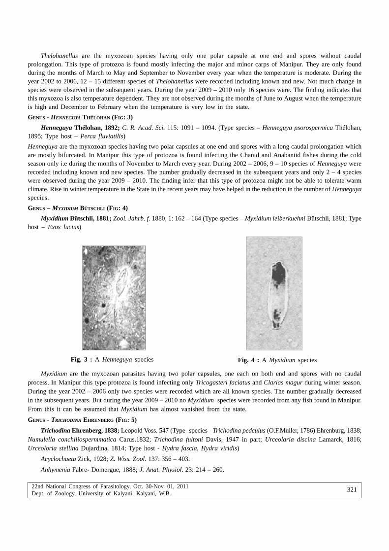
22nd National Congress of Parasitology, Oct. 30-Nov. 01, 2011Dept. of Zoology, University of Kalyani, Kalyani, W.B.
321
Thelohanellus are the myxozoan species having only one polar capsule at one end and spores without caudalprolongation. This type of protozoa is found mostly infecting the major and minor carps of Manipur. They are only foundduring the months of March to May and September to November every year when the temperature is moderate. During theyear 2002 to 2006, 12 – 15 different species of Thelohanellus were recorded including known and new. Not much change inspecies were observed in the subsequent years. During the year 2009 – 2010 only 16 species were. The finding indicates thatthis myxozoa is also temperature dependent. They are not observed during the months of June to August when the temperatureis high and December to February when the temperature is very low in the state.
GENUS - HENNEGUYA THÉLOHAN (FIG: 3)
Henneguya Thélohan, 1892; C. R. Acad. Sci. 115: 1091 – 1094. (Type species – Henneguya psorospermica Thélohan,1895; Type host – Perca fluviatilis)
Henneguya are the myxozoan species having two polar capsules at one end and spores with a long caudal prolongation whichare mostly bifurcated. In Manipur this type of protozoa is found infecting the Chanid and Anabantid fishes during the coldseason only i.e during the months of November to March every year. During 2002 – 2006, 9 – 10 species of Henneguya wererecorded including known and new species. The number gradually decreased in the subsequent years and only 2 – 4 specieswere observed during the year 2009 – 2010. The finding infer that this type of protozoa might not be able to tolerate warmclimate. Rise in winter temperature in the State in the recent years may have helped in the reduction in the number of Henneguyaspecies.
GENUS – MYXIDIUM BÜTSCHLI (FIG: 4)
Myxidium Bütschli, 1881; Zool. Jahrb. f. 1880, 1: 162 – 164 (Type species – Myxidium leiberkuehni Bütschli, 1881; Typehost – Exos lucius)
Fig. 3 : A Henneguya species Fig. 4 : A Myxidium species
Myxidium are the myxozoan parasites having two polar capsules, one each on both end and spores with no caudalprocess. In Manipur this type protozoa is found infecting only Tricogasteri faciatus and Clarias magur during winter season.During the year 2002 – 2006 only two species were recorded which are all known species. The number gradually decreasedin the subsequent years. But during the year 2009 – 2010 no Myxidium species were recorded from any fish found in Manipur.From this it can be assumed that Myxidium has almost vanished from the state.
GENUS - TRICHODINA EHRENBERG (FIG: 5)
Trichodina Ehrenberg, 1838; Leopold Voss. 547 (Type- species - Trichodina pedculus (O.F.Muller, 1786) Ehrenburg, 1838;Numulella conchiliospermmatica Carus.1832; Trichodina fultoni Davis, 1947 in part; Urceolaria discina Lamarck, 1816;Urceoloria stellina Dujardina, 1814; Type host - Hydra fascia, Hydra viridis)
Acyclochaeta Zick, 1928; Z. Wiss. Zool. 137: 356 – 403.
Anhymenia Fabre- Domergue, 1888; J. Anat. Physiol. 23: 214 – 260.

22nd National Congress of Parasitology, Oct. 30-Nov. 01, 2011Dept. of Zoology, University of Kalyani, Kalyani, W.B.
322
Cyclocyrrha Fabre-Domergue, 1888 ; J. Anat. Physiol. 23: 214 – 260
Paravauchomia Raabe, 1963; Acta Protozool. 1: 121 – 138.
Trichodina are the ciliophoran species having denticulate ring composed of denticles with straight or curved blades,distinct rays of various shapes and length, adoral spiral describing an arc of 3300 – 4500. This type of protozoa is foundinfecting the gills and body surface of major carps and Mistus found in Manipur. They favour warm climate and are obtainedduring the months of May to September every year. During winter when the temperature falls down their presence are notobserved. During the year 2002 – 2006 only two species were recorded with low population. The number increased in thesubsequent years. During the year 2009 – 2010 as many as 5 species were recorded and the population drastically increased.In a slide smeared from one gill clip of an infected carp nearly hundreds of individuals were observed. Such populations werenot seen in the previous years. This indicates that warm climate stimulate their activities.
GENUS - TRIPARTIELLA LOM (FIG: 6)
Tripartiella Lom, 1959; Acta Parasitol. Polon. 7: 573 – 590. (Type- species - Tripartiella copiosa Lom, 1959: Type-host- Rhodeus sericues)
Trichodina - proparte Davis, 1947; Dogiel, 1940; Tr. Len. Obsh. Yestiestvoispitatieley. 68: 8 – 31.
Fig. 5 : A Trichodina species Fig. 6 : A Tripartiella species
Tripartiella are the ciliophoran parasites having denticles consisting of blades, rays and central parts and adoral spiralmaking turn of 1800 – 2700. This type of protozoa is found infecting only the gills of Major carps inhabiting Manipur duringthe months of May to September. During the years 2002 – 2006 only one species were recorded with low population but duringthe years 2009 – 2010 three species were recorded with unexpected rise in population. A slide smeared from one gill clipcontained hundreds of individual while the host seemed to be non symptomatic when observed.
GENUS – ICHTHYOPHTHIRUS FOUQUET (FIG: 7)
Ichthyophthirius Fouquet, 1876 (Type- species – Ichthyophthirus multifiliis Fouquet, 1876)
Fig. 7 : An Ichthyophthirius species Fig. 8 : A Trypanosoma species

22nd National Congress of Parasitology, Oct. 30-Nov. 01, 2011Dept. of Zoology, University of Kalyani, Kalyani, W.B.
323
Ichthyophthirius are the ciliate parasites having ovoid body, uniform ciliation, a big rod shaped or horse shoe shapednucleus inside the body. This type of parasite is found infecting the gills of carps in Manipur. It was recently recorded fromthe gills of Labeo rohita in April, 2010. Before this no Ichthyophthirius species were recorded from the state. This will provedthat parasite species entered new areas searching for suitable habitat in response to climate change.
GENUS – TRYPANOSOMA GRUBY(FIG: 8)
Trypanosma GRUBY, 1843
Trypanosoma are the blood flagellate protozoa having highly flattened body, pointed at flagellate end, and bluntly roundedor pointed at the other end. There is a blepharoplast from which the flagellum arises and nuns toward opposite end, makingthe outer boundary of the undulating membrane; in most cases flagellum extends freely beyond body. This type of protozoawas recently recorded from the blood smear of Clarias magur collected from Utlou, Manipur. Till date no report on the presenceof such protozoa from Manipur was recorded.
DiscussionMost of the Scientists agree that climate change has great impact on the parasites and infectious diseases of aquatic
animals. Numerous host – parasite systems are affected by climate. Climate change directly affect parasitic file cycles orindirectly affect host populations and communities (Mouritsen and Poulin, 2002). Asha Gupta and Usharani (2009) reportedthat the average temperature during summer is 320C in Manipur in 2008. Again according to Manipur Weather report theaverage temperature during the year 2009 is 350C – 370C and that of 2010 is 330C – 360C upto September and sometime reach410C. With such changes in temperature in the state Manipur some of the protozoan parasites have undergone populationchange. Species of the genus Myxobolus had increased in number from 2002 – 2010 in which maximum species were recordedduring 2009 – 2010. Species of ciliate trichodinid that favour warmer climate also increased in number. Ichthyophthirius andTrypanosoma were also recovered from the state. These two parasites were not reported from any part of the North East inthe previous years and the present communication is the first report of the two species. This predicts that many new parasitesthat were not observed before existed through transmission processes from other regions due to global warming (Poulin, 2006).
On the other hand, myxozoan species of the genus Henneguya and Myxidium had negative response to warm climate.They only favour winter season. The species of Myxidium could not be recovered during 2009 – 2010 in Manipur. This maybe due to many reason, 1st – they cannot withstand high temperature (cannot complete life cycle), 2nd – reduction in hostpopulation (Channid fishes). In Manipur, the production of Channid fishes (commonly harvested during winter) were greatlyreduced during the last 2-3 years. 3rd – reduction in habitat i.e. wetland areas, 4th – cannot expand host range. These findingsconfirm that “Parasites may respond to increasing temperatures more strongly than their host” (Poulin, 2006).
Thus, there will be correctness in the prediction that Global warming brings alteration in host range, parasite range, hostimmunocompetence, parasite virulence, parasite transmission rates, disease occurrence and parasite infection etc. Even theparasites that are uncommon to humans may also be transmited to human being. White Pelican (Avian Piscivores) acting asas vectors for Myxobolus cerebralis (causal organism for whirling disease in salmonid fishes) can be taken as an example forexpansion of host range of the myxozoa (Koel et al.2020). After all human being are the last target for all the parasites.
AcknowledgementThe Head, Department of Life Sciences, Manipur University is acknowledged for kindly providing the necessary
laboratory facilities.
ReferencesBütschli O (1881) Myxosporidien. Zool Jahrb f 1880 1: 162 – 164.
Bütschli O (1882) Myxosporidia. In Bronn’s Klass Ordn., des Tierreiches, Protozoa 1: 590 – 603.
Davis HS (1947) Studies of the protozoan parasites of freshwater fishes. U. S. Dept. Interior Fish Bull 51: 1 – 29.
Dobson A, Carper R (1992) Global warming and potential changes in host-parasites and disease-vector relationships. In. Globalwarming and biological diversity (R. L. Peters & T. E. Lovejoy. Eds). Yale University Press, New Haven, Connecticut201 – 217.
Dujardin F (1814) Historie naturelle des zoophyte. Infusorie etc. Paris: Librariae Encyclopédique de Roret pp 684.
Ehrenberg CG (1838) Die Infusionsthierchen als vollkom nene Organismen. Ein Blick in das tiefere organische Leben der Natur,Leipzig: Leopold Voss pp 547.

22nd National Congress of Parasitology, Oct. 30-Nov. 01, 2011Dept. of Zoology, University of Kalyani, Kalyani, W.B.
324
Fabre-Domergue PR (1888) Étude sur porganisation des Urcéolaires et sur quelques genres d’infudoires voi – sins de cettefamille. J Anat Physiol 23: 214 – 260.
Ficke AD, Myrick CA, Hansen LJ (2007) Potential impacts of global climate change on freshwater fisheries. Rev Fish Biol Fish17(4): 581.
Gupta A, Usharani L (2009) Rainfall partitioning in a tropical forest of Manipur, North East India. Tropical Ecology 355 – 358.
Harvell CD, Kim K, Burkholder JM, Colwell RR, Epstein PR, Grimes DJ, Hofmann EE, Lipp EK (1999) Emerging marine diseases– climate links and anthropogenic factors. Science 285: 1505 – 1510.
Havell CD, Mitchell CE, Ward JR, Altizer S, Dobson AP, Ostfeld RS, Samuel MD (2002) Climate warming and disease risks forterrestrial and marine biota. Science 296(5576): 2158 – 2162.
Kalavati C, Narasimhamurti CC (1984) A new myxosporidian, Rudicapsula esomi n. gen. n. sp. from freshwater fish Esomussp. Z Parasitenkd 70(1): 21 – 27.
Klein BM (1958) The dry silver method and its proper use. J Protozool 5: 99 – 103.
Koel TM, Kerans BL, Barras SC, Hanson KC, Wood JS (2010) Avian Piscivores ans vectors for Myxobolus cerebralis in theGreater Yellowstone Ecosystem. Transactions of the American Fisheries Society 138: 976 – 988.
Kudo RR (1933) A taxonomic consideration of myxosporidia. Trans Am Microsc Soc 52: 195 – 216.
Kudo RR (1986) Protozoology (1st Ed.) Periodicals Corporation, New Delhi.
Lom J (1959) On the systematic of the genus Trichodinella Srámek-Hušek (=Brachyspira Raabe). Acta Parasitol Polon 7: 573– 590.
Lom J, Dyková I (1992) Protozoan parasites of Fishes. Development of Aquaculture and Fisheries Science. 26, Elsevier. SciencePublishers B. V., Amsterdam pp 315.
Manipur Weather Report, 2009.
Manipur Weather Report, 2010.
Marcogliese DJ (2001) Implications of climate change for parasitism of animals in the aquatic environment. Can J Zool 79(8):1331 – 1352.
Mouritsen KN, Poulin R (2002) Parasitism, climate oscillations and the structure of natural communities. Oikos 97(3): 462 – 468.
Parry ML, Canzian OF, Palutikok JP, van der Linden PJ, Hanson CE (ed.) (2007) Technical summary. In Climate change 2007:impacts, adaptation and vulnerability. Contribution of Working Group II to the 4th assessment report of the Intergov-ernmental Panel on Climate Change. Cambridge University Press, Cambridge 23 – 78.
Plehn M (1925) Eine neue Schleinkrankheit Fisherei Zeitung (Neudamm) 28: 299 – 300.
Poulin R (2006) Global warming and temperature-mediated increases in cercarial emergence in trematode parasites. Parasitology132: 143 – 151.
Profile on State of Environment Report of Manipur, 2006 – 07.
Raabe Z (1963) Systematics of the family Urceolariidae Dujardin, 1841. Acta Protozool 1: 121 – 138.
Reist JD, Wrona FJ, Prowse TD, Power M, Dempson JB, Beamish RJ, King JR, Carmichael TJ, Sawartzky CD (2006a) Generaleffects of climate change on Arctic fishes and fish populations. Ambio 35(7): 370 – 380.
Reist JD, Wrona FJ, Prowse TD, Power M, Dempson JB, Beamish RJ, King JR, Beamish RJ (2006b) An overview of effectsof climate change on selected arctic freshwater and anadromus fishes. Ambio 35(7): 381 – 387.
Roessig JM, Woodley CM, Cech JJ Jr, Hansen LJ (2004) Effects of global climate change on marine and estuarine fishes andfisheries. Rev Fish Biol Fish 14: 251 – 275.
Scavia D, Field JC, Boescg DF, Buddemneier RW, Burkette V, Cayan DY, Fogarty M, Harwell M (2002) Climate change impactson US coastal and marine ecosystems. Estuaries 25: 149 – 196.
Schindller DW (2001) The cumulative effects of climate warming and other human stresses on Canadian freshwaters in thenew millennium. Can J Fish Aquat Sci 58: 18 – 29.
Thélohan P (1892) Myxosporidies de la vesicule biliaire des poisons, Especes nouvelles. C R Acad Sci 115: 1091 – 1094.
Wyatt EJ (1977) Faciaplatycauda pratti gen. n. sp. and two new species of Myxobolus (Myxosporidian). J Protozool 26(1):47 – 51.
Zick K (1928) Urceolaria korscheti sp. n. eine nenu marine Urceolariidae nebst einen uberblick uber die Urceolarinen. Z WissZool 137: 356 – 403.

22nd National Congress of Parasitology, Oct. 30-Nov. 01, 2011Dept. of Zoology, University of Kalyani, Kalyani, W.B. i
Diversity of Tick (Acari) in West Bengal
Asok Kanti Sanyal
Zoological Survey of India, Kolkata-700053
Abstract : Though the study of ticks in India was initiated by Linnaeus in 1758, the first record of tick from West Bengal wasdone by Rudow (1870). He described an ixodid tick Amblyomma bengalense from Python sp. Later many workers studied thetaxonomy of ticks of the state and till date 32 species under 9 genera of the family Ixodidae are known from West Bengal.Ticks have been recorded from all the districts in which Darjiling occupies the highest position in number of species. Thetick species recorded from the state are known from outside India and show maximum similarity in species composition withpalaearctic region (25%). Amblyomma supinoi is known only from the Indian state West Bengal.
Key words : Tick, Acari, W.B.
Introduction
The ticks belong to the order Metastigmata or Ixodida under the class Acari. They occur throughout the world, but aremore frequently encountered in tropical and subtropical realms. The ticks are distinguished from other acarines by their largersize (2,000 µm to over 30,000 µm) and few other peculiarities. A hypostome armed with retrose teeth is of unique feature whichserves to anchor the tick to its host. A complex sensory setal field, Haller’s organ, is located on the dorsal side of tarsus-I, providing sites for contact or alfactory chemoreception. There are other distinguishing characters like stigmata situatedposterior to coxa IV or dorsal to coxa III-IV, palp with only three or four segments, 2-segmented chelicerae, etc. Of the threefamilies under Ixodida, the Ixodidae or hard ticks and Argasidae or soft ticks are found plenty in India but the third familyNuttalliellidae is known only from Africa.
The ixodid ticks are scutate with terminal capitulum, well marked sexual dimorphism, spiracle posterior to coxa-IV and padsand with porose areas and festoon. The argasid ticks are non-scutate with leathery integument, slight sexual dimorphism, smallspiracle situated anterior to caxa-IV and pads and without porse areas and festoon.
The ticks live as ectoparasite of vertebrates and feed obligatorily on the blood of mammals, birds and reptiles. There aretick species recognized as significant pests of man and animals. In temperate and tropical countries, they surpass all otherarthropods as transmitters in the number and variety of diseases of domestic animals and man. The ticks serve as reservoirand vectors for many infective viruses, rickettsia, bacteria, sporozoans and spirochaetes. They also cause paralysis andanaemia to hosts. Ticks are the main vectors of Kayasanur Forest Disease (KFD) in man and monkeys in Karnataka State.Other arboviruses like Kaisodi, Ganjam and Bhanja have also been isolated from ticks in India. Ticks are oviparous. Their lifehistory passes through egg, larva, nymph and adult stages.
The study of ticks in India was started by Linnaeus in 1758. But the first record of tick from West Bengal was done byRudow (1870). He described an ixodid tick Amblyomma bengalense from Python sp. The presence of the species was reporteddoubtful by Neumann (1911) and since then there is no record of the species from the State. Sharif (1928) reported thatNeumann (1899) first recorded Aponomma gervaisi (Lucas) from West Bengal and later Warburton (1910) recorded the samespecies from Zoological Garden, Calcutta. Later, medical entomologists and acarologists got interested about tick fauna of thestate and described and recorded a good number of genera and species of ticks. The detailed accounts of studies so far donehave been summarized by Sanyal and De (1991, 1992, 2001).
Status of tick diversity
The analysis of diversity of tick species in West Bengal vis-à-vis in India (12 genera and 107 species) clearly indicatesthat the state alone represents 30% of the total Indian tick fauna. The total number of taxa of ticks so far known from

22nd National Congress of Parasitology, Oct. 30-Nov. 01, 2011Dept. of Zoology, University of Kalyani, Kalyani, W.B.ii
West Bengal is represented by 32 species under 9 genera and a single family Ixodidae. It is presumed that no record
of argasid tick from the state may be due to poor abundance of these ticks and lack of attention to this family by the
acarologists.
Of the 32 species of tick known from the state, only three species viz., Hyalomma brevipunctata Sharif, 1928 from
Medinipur, Haemaphysalis darjeeling Hoogstraal and Dhanda, 1970 from Darjiling and H. ramachandrai Dhanda, Hoogstraaland Bhat, 1970 from Darjiling and Jalpaiguri have been described as new to science from the State. The analysis also showed
that Boophilus microplus, Haemaphysalis bispinosa and Hyalomma anatolicum anatolicum are most dominant species being
present in all the districts of the State.
Table I and II (Sanyal and De, 2001) showing host and distribution of tick species in different districts of the State, indicate
that the species are well adapted on different hosts in different states from plains to higher altitude of the Himalaya.
Table I. Species of ticks known from different districts of West Bengal
Sl. No. Districts in West Bengal Species of ticks
1. Kolkata (Calcutta) Amblyomma helvolum Koch, A. javanense (Supino), Aponomma gervaisi
(Lucas), Boophilus microplus (Canenstrini), Dermacentor auratus Supino,Haemaphysalis aponommoides Warburton, H. bispinosa Neumann, H. indica
Warburton, Hyalomma anatolium anatolicum Koch, Rhipicephalus
sanguineus (Latrielle), R. turanicus Pomerantzev
2. North 24-Parganas Amblyomma testudinarium Koch, Boophilus microplus, Dermacentor
auratus, Haemaphysalis bispinosa, Hyalomma anatolicum anatolicum, H.
hussaini Sharif, Rhipicephalus haemaphysaloides Supino
3. South 24-Parganas Boophilus microplus, Haemaphysalis bispinosa, Hyalomma anatolicum
anatolicum
4. Barddhaman Aponomma lucasi, Boophilus microplus, Haemaphysalis bispinosa,Hyalomma anatolicum anatolicum
5. Medinipur Boophilus microplus, Haemaphysalis bispinosa, Hyalomma anatolicum
anatolicum, H. marginatum isaaci Sharif, H. Brevipunctata Sharif, Nosomma
monstrosum (Nuttall & Warburton), Rhipicephalus sanguineus
6. Nadia Boophilus microplus, Haemaphysalis bispinosa, Hyalomma anatolicum
anatolicum, Rhipicephalus turanicus
7. Hugli Boophilus microplus, Haemaphysalis bispinosa, H. indica, Hyalomma
anatolicum anatolicum, Rhipicephalus turanicus
8. Bankura Boophilus microplus, Haemaphysalis bispinosa, Hyalomma anatolicum
anaatolicum, H. marginatum isaaci
9. Puruliya Boophilus microplus, Haemaphysalis bispinosa, Hyalomma anatolicum
anatolicum, H. marginatum isaaci, Rhipicephalus sanguineus, R. turanicus
10. Jalpaiguri Amblyomma javanense, A. supinoi Neumann, A. testudinarium, Boophilus
microplus, Dermacentor auratus, Haemaphysalis aborensis Warburton, H.
bispinosa, H. cornigera shimoga Trapido and Hoogstraal, H. hystricis
Supino, H. obese Larrousse, H. ramachandrai Dhanda, Hoogstraal & Bhat,H. spinigera Neumann, Hyalomma anatolicum anatolicum, Rhipicephalus
haemaphysaloides
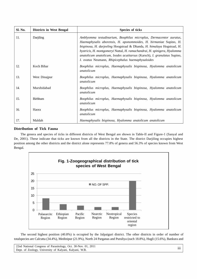
22nd National Congress of Parasitology, Oct. 30-Nov. 01, 2011Dept. of Zoology, University of Kalyani, Kalyani, W.B. iii
Distribution of Tick Fauna
The genera and species of ticks in different districts of West Bengal are shown in Table-II and Figure-1 (Sanyal andDe, 2001). These indicate that ticks are known from all the districts in the State. The district Darjiling occupies highestposition among the other districts and the district alone represents 77.8% of genera and 56.3% of species known from WestBengal.
11. Darjiling Amblyomma testudinarium, Boophilus microplus, Dermacentor auratus,Haemaphysalis aborensis, H. aponommoides, H. birmaniae Supino, H.bispinosa, H. darjeeling Hoogstraal & Dhanda, H. himalaya Hogstraal, H.hystricis, H. montgomeryi Nuttal, H. ramachandrai, H. spinigera, Hyalommaanatolicum anatolicum, Ixodes acutitarsus (Karsch), I. granulatus Supino,I. ovatus Neumann, Rhipicephalus haemaphysaloides
12. Koch Bihar Boophilus microplus, Haemaphysalis bispinosa, Hyalomma anatolicumanatolicum
13. West Dinajpur Boophilus microplus, Haemaphysalis bispinosa, Hyalomma anatolicumanatolicum
14. Murshidabad Boophilus microplus, Haemaphysalis bispinosa, Hyalomma anatolicumanatolicum
15. Birbhum Boophilus microplus, Haemaphysalis bispinosa, Hyalomma anatolicumanatolicum
16. Haora Boophilus microplus, Haemaphysalis bispinosa, Hyalomma anatolicumanatolicum
17. Maldah Haemaphysalis bispinosa, Hyalomma anatolicum anatolicum
Sl. No. Districts in West Bengal Species of ticks
The second highest position (40.8%) is occupied by the Jalpaiguri district. The other districts in order of number oftotalspecies are Calcutta (34.4%), Medinipur (21.9%), North 24 Parganas and Puruliya (each 18.8%), Hugli (15.6%), Bankura and
Fig. 1-Zoogeographical distribution of tickspecies of West Bengal
NO. OF SPP.
Speciesrestricted to
orientalregion
NearcticRegion
NeotropicalRegion
PacificRegion
EthiopianRegion
PalaearcitcRegion
25
20
15
10
5
0

22nd National Congress of Parasitology, Oct. 30-Nov. 01, 2011Dept. of Zoology, University of Kalyani, Kalyani, W.B.iv
Barddhaman (each 12.6%), Birbhum, Haora, Koch Bihar, Nadia, South 24 Parganas and West Dinajpur (each 9.4%) and Maldahand Murshidabad (each 6.3%). The poor distribution and abundance of ticks in many districts may be due to the fact thatno intensive survey for study of ticks in different districts is done due to inaccessibility and administrative reasons.
Zoogeographical distribution of ticks found in West Bengal
The figure-I shows the zoogeographical relationship of ticks so far known from West Bengal. It shows that maximumsimilarity in distribution of species exists between West Bengal and Palaeractic region (25%). The other zoogeographicalregions in order of similarity are Ethiopion (12.5%), Pacific (9.4%). Nearctic and Neotropical (each 6.3%). The Figure-I alsoindicates that 62.5% of tick species known from West Bengal are found to occur only in the oriental region.
It is also noted from the available data that Amblyomma supinoi is recorded from the only Indian state West Bengal. Theother interesting observation by Sanyal and De, 2001, reports that there is no tick species in West Bengal which is endemicto India.
Acknowledgements
The author expresses sincere thanks to the Director, Zoological Survey of India for extending working facilities.
References
Linnaeus, C (1758). Syst. Nat 1: 823
Neumann, LG (1911) Note rectificative a propose de deux especes d’Ixodinae. Arch. Parasit 14(3): 415
Rudow, F (1870) Einige neue Ixoden. Z. ges. Naturw 35 N
Sanyal, AK, De, SK (1991) Acari: Metastigmata. Animal Resources in India (ed., Director, ZSI), ZSI 513-517.
Sanyal, AK, De, SK (1992) Ixodid ticks ( Acari : Ixodidae). State Fauna Series 3: Fauna of West Bengal part 3: 17-60
Sanyal, AK, De, SK (2001) Diversity in ticks (Acari) of West Bengal. Rec. zool. Surv. India, 99 (part 1-4) : 65-74
Warburton, C (1910). On two collections of Indian ticks. Parasitol 3(4): 395-407