HONG KONG - MDS Congress
134
October 5-9, 2018 HONG KONG International Congress of Parkinson’s Disease and Movement Disorders® www.mdscongress.org MDS-0818-039 Guided Poster Tours
-
Upload
khangminh22 -
Category
Documents
-
view
5 -
download
0
Transcript of HONG KONG - MDS Congress

October 5-9, 2018
HONG KONG
International Congress of Parkinson’s Disease and Movement Disorders®
www.mdscongress.org
MDS-0818-039
Guided Poster Tours

International Congress of Parkinson’s Disease and Movement Disorders®
Hong Kong October 5-9, 2018 – Guided Poster Tour Abstracts
International Congress of Parkinson’s Disease and Movement Disorders®
Guided Poster Tour Abstracts
Guided Poster Group 1:
Clinical Trials and Therapy in Movement Disorders
5
Improvements of tremor control and life quality of
refractory essential tremor patients after MR-
guided focused ultrasound thalamotomy – A
Taiwan experience
H.C. Lai, K. Tsai, W.C. Chang, T. Taira, C.Y. Wei
(Changhua County, Taiwan)
7
Specific active immunotherapy (SAIT) against
alpha-synuclein with AFFITOPE® PD01A and
PD03A: Results from the AFF009 phase I trial
W. Meissner, A. Pavy-Le Traon, A. Foubert-Samier, M.
Galitzky, B. Laurens, U. Sabatini, S. Schmidhuber, D.
Winter, G. Galabova, G. Staffler, A. Schneeberger, A.
Kutzelnigg, O. Rascol (Bordeaux Cedex, France)
8
PASSPORT, An Ongoing Phase 2 Study in Patients
with PSP– Baseline Characteristics
T. Dam, A. Boxer, L. Golbe, G. Höglinger, H. Morris,
I. Litvan, J.C. Corvol, A. Lang, C. Bechtold, I. Qureshi,
M. Grundman, B. Han, J. O'Gorman, T. Olsson, S.
Budd Haeberlein (Cambridge, MA, USA)
9
A German-Austrian multicenter, non-
interventional, prospective study for the treatment
with abobotulinumtoxinA injections in naïve and
previously treated patients suffering from cervical
dystonia
W. Jost, A. Schramm, M. Müngersdorf, A. Stenner, P.
Schwingenschuh, P. Maisonobe, M. Koch, B. Haslinger
(Wolfach, Germany)
12
Low-Fat Versus Ketogenic Diet In Parkinson's
Disease: A Pilot Randomized Controlled Trial
M. Phillips, D. Murtagh, L. Gilbertson, F. Asztely, C.
Lynch (Hamilton, New Zealand)
15
Deep brain stimulation (DBS) for dyskinetic
cerebral palsy: A pilot study
S. Duma, N. Mahant, A. Ha, S. Kim, A. Phu, K.
Stewart, M-C. Waugh, N. Wolfe, D. Russell, B. Owler,
M. Krause, V. Fung (Westmead, Australia)
17
Zonisamide improves parkinsonism in DLB
patients: A randomized phase 3 trial
M. Murata, T. Odawara, K. Hasegawa, R. Kajiwara,
H. Takeuchi, M. Tagawa, K. Kosaka (Kodaira, Japan)
32
Alleviation of freezing of gait in patients with
Parkinson’s disease by high-frequency rTMS over
SMA is associated with normalization of brain
connectivity patterns
T.M. Mi, S. Garg, F. Ba, T. Wu, P.P. Liang, L.L. Gao,
K.C. Li, P. Chan, M. McKeown (Beijing, China)
35
PPMI driven sample size estimation for clinical
trials in Parkinson’s disease
K. Marek, J. Seibyl, C. Caspell-Garcia, C. Coffey, B.
Mollenhauer, K. Kieburtz, C. Tanner, L. Chahine, A.
Siderowf, T. Simuni (New Haven, CT, USA)
Guided Poster Group 2:
Parkinson’s Disease: Clinical Trials, Pharmacology
And Treatment
251
The anti-dyskinetic effect of the clinically-available
5-HT3 receptor antagonist granisetron in the 6-
OHDA-lesioned rat model of Parkinson's disease
C. Kwan, I. Frouni, D. Bedard, A. Hamadjida, P. Huot
(Montreal, QC, Canada)
301
Directional Lead Impedances and Their Possible
Effects on Deep Brain Stimulation
L. Juarez Paz, A. Dalal Kirsch, F. Steigerwald, C.
Matthies, S. Meoni, V. Fraix, T. Ten Brinke, R. de Bie,
K. Wynants, D. Blum, N. Van Dyck, PR. Schuurman, E.
Moro, S. Chabardes, J. Volkmann (Valencia, CA, USA)
326
DIRECT DBS: A Prospective, Multicenter Clinical
Study with Double-Blinding for a Directional Deep
Brain Stimulation Lead – Therapeutic Windows
with Directional Stimulation
F. Steigerwald, J. Volkmann, C. Matthies, A. Dalal
Kirsch, S. Chabardes, R. de Bie, P. Schuurman, E.
Moro, V. Fraix, S. Meoni, D. Blum, L. Juarez Paz, K.
Wynants, N. Van Dyck (Würzburg, Germany)
International Congress of Parkinson’s Disease and Movement Disorders®
Hong Kong October 5-9, 2018 – Guided Poster Tour Abstracts
341
‘PDSAFE’ – A multi-dimensional fall rehabilitation
intervention for people with Parkinson’s with
specialist physiotherapy training: qualitative
exploration of physio’s experiences
K. Seymour, S. Hulbert, V. Goodwin, L. Rochester, A.
Nieuwboer, A. Ashburn (Southampton, United
Kingdom)
364
The Effect of Short Pulse Width Settings on Speech
in Subthalamic Nucleus Deep Brain Stimulation for
Parkinson’s disease
V. Dayal, T. Grover, E. Tripoliti, P. Limousin, T.
Foltynie (London, United Kingdom)
380
Sex differences by design and outcome in the Safety
of Urate Elevation in PD (SURE-PD) trial
M. Schwarzschild, E. Macklin, R. Bakshi, S.
Bhattacharyya,, R. Logan, A. Ascherio; on behalf of the
Parkinson Study Group SURE-PD Investigators
(Boston, MA, USA)
388
The highly selective 5-HT2A receptor antagonist
EMD-281,014 alleviates L-DOPA-induced
dyskinesia in the 6-OHDA-lesioned rat model of
Parkinson's disease
I. Frouni, D. Bedard, S. Belliveau, E. Bourgeois-Cayer,
A. Hamadjida, P. Huot (Montreal, QC, Canada)
394
Patient and Provider Experiences and Attitudes
toward Rytary
S. Horn, D. Coughlin, J. Chou, F. Wang, M. Stacy, R.
Dolhun, C. Kopil, N. Amondikar, A. Deik, H. Sarva
(Philadelphia, PA, USA)
420
Mitochondrial morphometrics in idiopathic
Parkinson‘s disease fibroblasts
P. Antony, O. Boyd, K. Mommaerts, K. Sokolowska, M.
Ostaszewski, A. Baumuratov, L. Longhino, F. Poulain,
R. Krueger, R. Balling, N. Diederich (Belvaux,
Luxembourg)
427
Dysphagia predicts poor outcome in late-stage
Parkinson’s disease
M. Fabbri, D. Abreu, L. Guedes, M. Rosa, A. Antonini,
M. Zibetti, L. Lopiano, J. Fereira, M. Coelho (Lisbon,
Portugal)
Guided Poster Group 3:
Surgical Therapy: Parkinson's Disease
512
Post-Deep Brain Stimulation Psychosis in
Parkinson’s Disease: A Meta-analysis
YC. Tai, N. Mahant, B. Stubbs, A. Carvalho, YW. Chen,
TY. Chen, SP. Hsu, PT. Tseng (Kaohsiung, Taiwan)
515
Motor and cognitive progression in GBA-related
PD patients submitted to Deep Brain Stimulation
L. Correia Guedes, C. Silva, R. Machado Bouça, N.
Gonçalves, M. Fabbri, A. Castro Caldas, P. Pita Lobo,
M.M. Rosa, B. Cattoni, H. Carvalho, A. Gonçalves
Ferreira, J.J. Ferreira, M. Coelho (Lisbon, Portugal)
537
Directional deep brain stimulation in Parkinson’s
disease guided by local field potentials
F. Alonso-Frech, C. Fernandez-Garcia, M. Monge,
M.J. Catalan Alonso, G. Foffani (Madrid, Spain)
543
Surgery-, hardware - and chronic stimulation-
related adverse events following subthalamic
nucleus deep brain stimulation for Parkinson’s
disease
B. Gonenli Kocer, E. Ozturk, S. Comoglu, M. Sorar, H.
Kertmen (Ankara, Turkey)
544
Poor responders to STN DBS in Parkinson’s
disease: 1 year follow-up study
M. Zibetti, L. Ricciardi, E. Montanaro, M. Sarchioto,
M. Edwards, L. Lopiano, F. Morgante (Toriino, Italy)
546
A wireless brain-spine interface alleviating gait
deficits of non-human primates model of
Parkinson's disease
F. Raschella, T. Milekovic, M. Perich, S. Sun, G.
Schiavone, C. Hitz, Y. Jianzhong, W. Ko, Q. Li, C. Qin,
S. Lacour, J. Bloch, S. Micera, E. Bezard, G. Courtine
(Geneva, Switzerland)
552
Deep brain stimulation for monogenic Parkinson's
disease: A systematic review
T. Kuusimäki, J. Korpela, E. Pekkonen, M.
Martikainen, A. Antonini, V. Kaasinen (Turku,
Finland)
558
Propionibacterium acnes Infection with
Intracerebral Abscess in Deep Brain Stimulation
R. Lewis, F. Farrokhi, M. Marsans (Seattle, WA, USA)
International Congress of Parkinson’s Disease and Movement Disorders®
Hong Kong October 5-9, 2018 – Guided Poster Tour Abstracts
563
Intraoperative Electromyography can optimize
pallidotomy for Parkinson's disease and dystonias
P. Brainer, A. Campos, A. Brainer, J. Brainer, P.
Brainer, P. Coutinho, C. Lima, S. Laurentino, R.
Bandin, M. Andrade (Recife, Brazil)
567
The effects of deep brain stimulation of the
pedunculopontine nucleus on cognition in
Parkinson’s disease and Progressive Supranuclear
Palsy
F. Leimbach, J. Gratwicke, T. Foltynie, M. Hariz, P.
Limousin, L. Zrinzo, M. Jahanshahi (London, United
Kingdom)
572
Is lowering stimulation frequency a feasible option
to improve speech in Parkinson’s disease patients
undergoing subthalamic deep brain stimulation?
M. Fabbri, M. Zibetti, G. Ferrero, A. Accornero, I.
Guimaraes, M. Rizzone, A. Romagnolo, J. Ferreira, L.
Lopiano (Lisbon, Portugal)
Guided Poster Group 4:
Ataxia
618
Loss of paraplegin drives spasticity rather than
ataxia in SPG7: A European cohort analysis of 238
patients
G. Coarelli, R. Schule, B. vande Warrenburg, P. de
Jonghe, C. Ewenczyk, A. Martinuzzi, M. Synofzik, E.
Hamer, J. Baets, M. Anheim, L. Schöls, T. Deconinck,
B. Fontaine, T. Klockgether, MG. D'Angelo, ML.
Monin, P. Charles, MT. Bassi, T. Klopstock, E.
Ollagnon-Roman, C. Kamm, M. Papin, CS. Davoine,
G. Banneau, S. Tezenasdu Montcel, D. Seilhean, A.
Brice, C. Duyckaerts, G. Stevanin, A. Durr (Paris,
France)
622
Cerebellar Ataxia case series study from southern
Spain: Clinical and molecular description
A. Adarmes Gomez, S. Jesus Maestre, C. Mendez
delBarrio, D. Macias Garcia, F. Carrillo Garcia, M.
Carballo, P. Gomez Garre, P. Mir Rivera (Seville,
Spain)
624
Genotype-phenotype correlations in 104 Uzbekish
families with Spinocerebellar ataxias
F. Rakhimov, Y. Majidova, G. Rakhimbaeva (Tashkent,
Uzbekistan)
626
Objective Measures of Ataxic Gait Using Wearable
Inertial Sensors
M. El-Gohary, L. Horak, C. Gomez (Portland, OR,
USA)
627
Multi-tiered Diagnostic Approaches Reveal a High
Degree of Genetic Heterogeneity for Hereditary
Cerebellar Ataxias: A Retrospective Review of an
Australian Cohort
C. Kang, C. Liang, K. Ahmad, Y. Gu, SF. Siow, J.
Colebatch, S. Whyte, K. Ng, P. Cremer, R. Davis, T.
Roscioli, M. Cowley, J.S. Park, C. Sue, K. Kumar
(Sydney, Australia)
633
MRI and behavioural account of the aging
cerebellum
M. Bares, P. Filip, C. Gallea, S. Lehericy, O. Lungu
(Brno, Czech Republic)
655
Transcriptional profiling of peripheral blood
monocytes from child Friedreich’s ataxia patient:
New molecules and patterns of gene expression
H. Singh, V. Swarup, R. Singh, I. Singh, M. Faruq, S.
Vivekanandhan, A. Srivastava (Delhi, India)
660
Comorbid Pediatric Early Onset Ataxia and
Dystonia - Is the Cerebellum Involved?
D. Sival, M. Tijssen, D. Verbeek (Groningen,
Netherlands)
663
The etiologies of chronic progressive cerebellar
ataxia in a Korean population
J. Youn, M. Kim, JH. AHN, JW. Cho, JS. Kim (Seoul,
Republic of Korea)
Guided Poster Group 5:
Dystonia
691
DBS neuromodulation reduces severe dystonic pain
in children and young people
S. Perides, J.P. Lin, G. Lee, H. Gimeno, R. Selway, K.
Ashkan, M. Kaminska (London, United Kingdom)
696
The clinical value of SPECT in identifying dystonic
muscles of patients with cervical dystonia
L. Jin, L. Feng, I. Djibo, S. Chen, F. Teng, B. Li, H. Ma
(Shanghai, China)
International Congress of Parkinson’s Disease and Movement Disorders®
Hong Kong October 5-9, 2018 – Guided Poster Tour Abstracts
713
Characterizing Bulbar Dysfunction in X-Linked
Dystonia-Parkinsonism (XDP): A Pilot Study
J. de Guzman, B. Perry, C. Go, J. Green, N. Sharma
(Boston, MA, USA)
727
The association of primary dystonia with tics -
chance or new syndrome?
C. Del Gamba, A. Latorre, U. Bonuccelli, R. Ceravolo,
K. Bhatia (London, United Kingdom)
734
Zolpidem effect in task specific dystonia – clinical
and neurophysiological study
K. Vogelnik, M. Grmek, R. Perellon Alfonso, P. Tomše,
M. Trošt, M. Kojović (Ljubljana, Slovenia)
746
A Registry of Real-World Outcomes Using Deep
Brain Stimulation for the Treatment of Dystonia
J. Krauss, C. Nicholson, M. Barbe, V. Visser-
Vanderwalle, A. Kuehn, M. Poetter-Nerger, R. Jain, H.
Scholtes, N. Van Dyck, A. Albanese (Hannover,
Germany)
761
Comorbidity and retirement in primary focal
cervical dystonia
R. Ortiz, F. Scheperjans, T. Mertsalmi, E. Pekkonen
(Helsinki, Finland)
762
Frequency and Phenoptypic Spectrum of KMT2B
Mutations in Childhood-Onset Dystonia: Results
from a Single-Centre Cohort Study
M. Carecchio, G. Zorzi, F. Invernizzi, C. Panteghini, L.
Romito, F. Zibordi, V. Leuzzi, S. Galosi, P. Morana, B.
Morana, C. Piano, A. Bentivoglio, C. Reale, F. Girotti,
M. Topf, A. Joseph, M. Kurian, S. Lubbe, B.
Garavaglia, N. Mencacci, N. Nardocci (Milan, Italy)
769
A machine learning approach to determine the
important patient characteristics for tremor
prevalence and tremor irregularity in dystonia
S. Balta Beylergil, L. Scorr, A. Cotton, H. Jinnah, A.
Shaikh (Cleveland, OH, USA)
776
Cohort profile of the Japan Dystonia Consortium:
Genetic diagnosis and characteristics of movement
disorders in Japan
T. Kawarai, R. Miyamoto, A. Orlacchio, R. Kaji
(Tokushima, Japan)
Guided Poster Group 6:
Huntington’s Disease
681
Chorea and Ataxia as Manifestations of Xeroderma
Pigmentosum: A Case Report
A. Jocson, K. Ngo, D. Togasaki, B. Fogel (Los Angeles,
CA, USA)
805
Prediction of suicidality in Huntington disease:
Analysis of Enroll-HD data using machine learning
approach
Y. Seliverstov, A. Borzov, E. van Duijn, B.
Landwehrmeyer, M. Belyaev (Moscow, Russian
Federation)
807
Vertical and horizontal eye movements in a large
cohort of early manifest Huntington's disease:
Evidence from ENROLL-HD
W. Muller, M. MacAskill, L. Paermentier, T. Anderson
(Christchurch, New Zealand)
809
White matter microstructural alterations in
Huntington Disease: When neurodegeneration
starts?
P. Azevedo, L. Piovesana, M. Nogueira, R. Guimarães,
A. Amato Filho, F. Cendes, Í. Lopes-Cendes, C. Yasuda
(Campinas, Brazil)
814
Interrater reliability of the Unified Huntington’s
Disease Rating Scale-Total Motor Score
certification
J. Winder, R. Roos, J.M. Burgunder, J. Marinus, R.
Reilmann (Leiden, Netherlands)
821
Cognitive Reserve and Physical Activity Modulate
Functional Brain Re-organisation in Premanifest
Huntington's Disease: Preliminary Evidence
M. Soloveva, S. Jamadar, G. Poudel, N. Georgiou-
Karistianis (Melbourne, Australia)
823
Comparing risperidone and olanzapine to
tetrabenazine for the management of Huntington’s
chorea
J. Schultz, J. Kamholz, P. Nopoulos, A. Killoran (Iowa
City, IA, USA)
International Congress of Parkinson’s Disease and Movement Disorders®
Hong Kong October 5-9, 2018 – Guided Poster Tour Abstracts
830
Driving performance of Huntington’s disease gene
carriers
M. Jacobs, E. Hart, Y. Mejia Miranda, G.J.
Groeneveld, J. van Gerven, R. Roos (Leiden,
Netherlands)
838
Cerebrospinal fluid flow dynamics in Huntington's
disease evaluated by phase contrast MRI
F. Rodrigues, L. Byrne, E. De Vita, E. Johnson, N.
Hobbs, J. Thornton, R. Scahill, E. Wild (London,
United Kingdom)
Guided Poster Group 7:
Parkinsonism, MSA, PSP (Secondary and
Parkinsonism-Plus)
927
Clinical features and natural history of
pathologically-confirmed corticobasal degeneration:
A Japanese validation study of CBD (J-VAC study)
I. Aiba, T. Shimohata, S. Murayama, K. Hasegawa, Y.
Iwasaki, O. Yokota, H. Fujimura, M. Sakai, T. Yokota,
I. Yabe, H. Takigawa, K. Sugaya, K. Mori, M. Ito, C.
Ishida, M. Kobayashi, Y. Hashizume, T. Ikeuchi, M.
Hasegawa, M. Yoshida, T. Komori, K. Wakabayashi, Y.
Saito, A. Tokumaru, K. Sakurai, K. Nakashima
(Nagoya, Japan)
929
Quantitative mobility metrics from a body-fixed
sensor predict incident parkinsonism in older adults
R. von Coelln, R. Dawe, J. Shulman, L. Yu, S.
Leurgans, J. Hausdorff, L. Shulman, D. Bennett, A.
Buchman (Baltimore, MD, USA)
933
Apolipoprotein E and multiple system atrophy
K. Ogaki, Y. Martens, M. Heckman, S. Koga, L. Labbé,
O. Lorenzo-Betancor, A. Wernick, R. Walton, A. Soto,
E. Vargas, H. Nielsen, S. Fujioka, T. Kanekiyo, R.
Uitti, J. van Gerpen, W. Cheshire, Z. Wszolek, P. Low,
W. Singer, N. Hattori, D. Dickson, G. Bu, O. Ross
(Tokyo, Japan)
939
ARISE study: Study Design and Baseline
Characteristics for a Phase 2 Trial of the Anti-Tau
Antibody ABBV-8E12 in Progressive Supranuclear
Palsy
N. Mendonca, R. Bateman, A. Boxer, J. Braunstein, D.
Claassen, D. Holtzman, D. Kerwin, B. Rendenbach-
Mueller, H. Soares, D. Wang, G. Höglinger
(Ludwigshafen, , Germany)
948
The characteristic of gait in progressive progressive
supranuclear palsy
Y. Takamatsu, N. Matsuda, I. Aiba (Kyoto, Japan)
957
Olfactory testing in Parkinson plus syndromes
O. Abdukarimov, F. Akhmedova (Tashkent,
Uzbekistan)
964
Nilotinib for treating MSA: A preclinical proof of
concept study
P. Guerin, M. Lopez-Cuina, E. Bezard, W. Meissner,
P-O. Fernagut (Bordeaux, France)
974
Evolution of diagnostic certainty and PSP-
predominance types in 187 pathologically confirmed
PSP patients
M. Grimm, G. Respondek, I. Piot, T. Arzberger, Y.
Compta, E. Englund, L. Ferguson, E. Gelpi, A. Giese,
S. Roeber, D. Irwin, W. Meissner, C. Nilsson, A.
Pantelyat, A. Rajput, C. Troakes, G. Höglinger
(Munich, Germany)
979
Rapamycin for treating MSA: A preclinical proof of
concept study
M. Lopez-Cuina, P. Guerin, E. Bezard, W. Meissner,
P-O. Fernagut (Bordeaux, France)
Guided Poster Group 8:
Technology
1096
The BlueSky Project: monitoring motor and non-
motor characteristics of people with Parkinson’s
disease in the laboratory, a simulated apartment,
and home and community settings
K. Erb, J. Daneault, S. Amato, P. Bergethon, C.
Demanuele, T. Kangarloo, S. Patel, V. Ramos, D.
Volfson, P. Wacnik, H. Zhang, D. Karlin, H. Huggins,
L. Soll, G. Costante, G. Vergara-Diaz, F. Parisi, J.
Banghu, C. Brooks, C. Dethridge, A. Abrami, E. Bilal,
V. Caravagio, S. Heisig, R. Norel, E. Pissadaki, J.
Rice, B. Ho, K. Thomas, P. Bonato (Cambridge, MA,
USA)
1097
Quantitative assessment of the hand motor
symptoms in Parkinson’s disease based on a custom
wearable device: A Proof-of-Principle Study
Q. Ye , Z. Lin, H. Dai, Y. Xiong, G. Cai (Jinjiang,
China)
International Congress of Parkinson’s Disease and Movement Disorders®
Hong Kong October 5-9, 2018 – Guided Poster Tour Abstracts
1103
Does the Parkinson’s Kinetigraph change clinical
practice?
S. Jones, C. Grose, S. Mahon, T. Williams, C. Thomas,
B. Mohamed (Cardiff, United Kingdom)
1111
The use of smartphone task derived features to
predict clinical scores in Parkinson’s Disease (PD)
C. Lo, S. Arora, F. Baig, T. Barber, M. Lawton, A.
Zhan, M. Little, M. Hu (Oxford, United Kingdom)
1113
Temporal Gait Parameters in Parkinson’s Disease:
A Study Using PDlogger, A Quantitative Gait
Measuring Device
N. Chia, J. Derrick, V. Mikos, S. Ng, A. Tay, S-C. Yen,
K. Koh, D. Tan, K. Prakash, L. Tan, W.L. Au
(Singapore, Singapore)
1117
Feasibility of a multi-sensor data fusion method for
assessment of Parkinson’s disease motor symptoms
M. Memedi, S. Aghanavesi, D. Nyholm, F. Bergquist,
M. Senek (Örebro, Sweden)
1118
Assessment of Motor Symptoms in Parkinson’s
disease using Flexible Wearable Sensors and Deep
Neural Networks: A CIS-PD substudy
L. Lonini, A. Dai, C. Poon, N. Shawen, L. Shimanovich,
T. Simuni, D. Daeschler, J. Rogers, A. Jayaraman
(Chicago, IL, USA)
1124
Objective monitoring of drug response in early PD
patients using remote, at-home typing data through
machine learning analysis
M. Matarazzo, T. Arroyo-Gallego, P. Montero
Escribano, V. Puertas-Martín, I. Butterworth, C. S.
Mendoza, MJ. Ledesma-Carbayo, MJ. Catalán, JA.
Molina-Arjona, F. Bermejo-Pareja, JC. Martínez
Castrillo, L. López-Manzanares, A. Alonso-Cánovas, J.
Herreros-Rodríguez, I. Obeso, P. Martínez Martín, JC.
Martínez-Ávila, A. Gómez de-la-Cámara, M. Gray, JA.
Obeso, L. Giancardo, Á. Sánchez-Ferro (Vancouver,
BC, Canada)
1138
The Levodopa Response Trial and the Parkinson
Disease Digital Biomarker Challenge: Monitoring
symptoms of Parkinson’s disease in the lab and
home using wearable sensors
J. Daneault, G. Vergara-Diaz, G. Costante, E. Fabara,
G. Ferreira-Carvalho, F. Golabchi, F. Parisi, S.
Sapienza, Y. Chae, P. Snyder, P. Aubin, P. Banda, D.
Brunner, R. Dorsey, L. Mangravite, W. Marks, E. Neto,
U. Rubin, E. Soderberg, D. Daeschler, S. Moore, S.
Sieberts, L. Omberg, P. Bonato, The Parkinson's
Disease Digital Biomarker DREAM Challenge
Consortium (Newark, NJ, USA)
1148
The international MDSGene initiative:
Systematically exploring genotype-phenotype
correlations of hereditary movement disorders
S. Petkovic, S. Schaake, J. Huang, H. Madoev, A.
Rasheed, C. Lill, K. Lohmann, C. Klein, C. Marras
(Lübeck, Germany)
Guided Poster Group 9:
Tremor
1169
Phase-locked transcranial alternating current
stimulation of the cerebellum for essential tremor
S. Schreglmann, D. Wang, E. Boyden, K. Bhatia, J.
Rothwell, N. Grossman (London, United Kingdom)
1174
Gamma Knife Radiosurgery for essential and
parkinsonian tremor: Long-term experience in a
Spanish center
JR. Perez-Sanchez, R. Martinez-Alvarez, NE. Martinez-
Moreno, I. Cuervo-Arango, G. Rey Portoles, I. Parees,
A. Del Barrio, J. Alvarez-Linera, MM. Kurtis (Madrid,
Spain)
1175
A new clinical and research smartphone application
to assess tremor and bradykinesia in patients with
movement disorders
C. Duval, J.F. Daneault, B. Carignan, C.É. Coderre, S.
Bogard (Montreal, QC, Canada)
1177
Shaky legs: The clinical spectrum and treatment of
orthostatic tremor; a systematic review
A. Buijink, M. Meulepas, A. van Rootselaar
(Amsterdam, Netherlands)
1182
The spectrum of involuntary movements in patients
with motor neuron disease – a cross-sectional study
K. Vogelnik, L. Dolenc Grošelj, B. Koritnik, L.
Leonardis, J. Zidar, M. Kojović (Ljubljana, Slovenia)
1188
Imaging neurodegeneration biomarkers in Essential
Tremor
S. Pietracupa, M. Bologna, G. Pasqua, S. Tommasin,
N. Petsas, F. Elifani, A. Berardelli, P. Pantano
(Pozzilli, Italy)
International Congress of Parkinson’s Disease and Movement Disorders®
Hong Kong October 5-9, 2018 – Guided Poster Tour Abstracts
1190
Quantitative characterisation of tremor in
functional and organic tremor patients
Z. Dominguez-Vega, G. Kramer, J. Elting, M. de
Koning-Tijssen, N. Maurits (Groningen, Netherlands)
1203
Repetitive Transcranial Magnetic Stimulation
Therapy is a Potential Therapeutic option for
Primary Orthostatic Tremor
W. Hu, J. Legacy, A. Ferng, A. Wagle Shukla
(Gainesville, FL, USA)
1210
Tremor in motor neuron disease may be central
rather than peripheral in origin
A. Latorre, L. Rocchi, M. Stamelou, A. Batla, M.
Ciocca, B. Balint, K. Sidle, A. Berardelli, J. Rothwell,
K. Bhatia (London, United Kingdom)
1213
Regional homogeneity changes detected between
Essential tremor with resting tremor and tremor-
dominant Parkinson’s disease
J. Li, Z. Lu, X. Suo, N. Li, L. Wang, J. Peng, J. Zhang,
Q. Gong, R. Peng (Chengdu, China)
Guided Poster Group 10:
Parkinson's Disease: Genetics
1299
PLA2G6-related juvenile-onset Parkinsonism:
clinical features and cognitive profile in a cohort of
Chinese patients
C. Chen, Y.M. Sun, F.T. Liu, S.S. Luo, Z.T. Ding, J.J.
Wu, J. Wang (Shanghai, China)
1304
Resistance to Parkinson's disease among LRRK2
mutation carriers is associated with higher plasma
levels of urate but not its purine precursors
M. Schwarzschild, R. Bakshi, R. Logan, M. Zorlu, X.
Chen, A. Ascherio, E. Macklin (Boston, MA, USA)
1307
Interest in Genetic Testing in PD Patients with DBS
A. Fraint, G. Pal, L. Verhagen, D. Hall, K. Marder
(Chicago, IL, USA)
1312
Application of the Movement Disorder Society
Prodromal Criteria in healthy G2019S-LRRK2
carriers
A. Mirelman, R. Saunders-Pullman, R. Alcalay, S.
Shustak, A. Thaler, B. Cohen, A. Hillel, T. Gurevich, D.
Raymond, H. Mejia-Santana, L. Ozelius, L. Clark, M.
Gana-Weisz, A. Bar-Shira, A. Orr-Urtreger, S.
Bressman, K. Marder, N. Giladi (Tel Aviv, Israel)
1326
LRRK2 and GBA genetic mutations are not
uncommon in an unselected Ashkenazi elderly
cohort with PD
S. Isaacson, J. Isaacson (Boca Raton, FL, USA)
1340
BDNF(V66M), EIF4G1(R1205H), VPS35(D620N)
gene polymorphisms in South Indian PD Patients
T. Syed, T. S.D, S. Meka, S. Kumar, S. Thandra, V.
Kutala, R. Kandadai, R. Borgihain (Hyderabad, India)
1341
Full sequencing and haplotype analysis reveals
LRRK2 protective haplotype in REM sleep
behavior disorder
B. Ouled Amar-Bencheikh, J. Ruskey, I. Arnulf, Y.
Dauvilliers, C. Charley Monaca, V. Cochen De-Cock,
JF. Gagnon, D. Spiegelman, M. Hu, B. Högl, A.
Stefani, L. Ferini-Strambi, G. Plazzi, E. Antelmi, P.
Young, A. Heidbreder, B. Mollenhauer, F. Sixel-
Döring, C. Trenkwalder, W. Oertel, J. Montplaisir, R.
Postuma, G. Rouleau, Z. Gan-Or (Montreal, QC,
Canada)
1353
Association of GBA polymorphisms and mutations
with dementia in Parkinson disease: A 7-year study
of three population-based incident cohorts
K. Lunde, J. Chung, I. Dalen, K. Pedersen, J. Linder,
M. Domellöf, E. Elgh, A. Macleod, C. Tzoulis, J.
Larsen, O. Tysnes, L. Forsgren, C. Counsell, G. Alves,
J. Maple-Grødem (Stavanger, Norway)
1370
Molecular mechanisms of GCH1-associated
Parkinson's disease
J. Terbeek, W. Vandenberghe (Leuven, Belgium)
Guided Poster Group 11:
Parkinson's Disease: Neuroimaging And
Neurophysiology
1376
Presynaptic dopamine depletion determines the
timing of levodopa-induced dyskinesia onset in
Parkinson’s disease
HS. Yoo, SJ. Chung, BS. Ye, YH. Sohn, PH. Lee (Seoul,
Republic of Korea)
1388
Automated Differential Diagnosis of Parkinsonian
Syndromes Using FDG-PET Metabolic Brain
Network Analysis
International Congress of Parkinson’s Disease and Movement Disorders®
Hong Kong October 5-9, 2018 – Guided Poster Tour Abstracts
T. Rus, P. Tomše, L. Jensterle, M. Grmek, Z. Pirtošek,
D. Eidelberg, C. Tang, M. Trošt (Ljubljana, Slovenia)
1400
Parkinson's disease motor symptoms are linked to
red nucleus volume and cerebellar metabolism
A. Bonnet, R. Sanford, A. Riou, S. Drapier, F. Le
Jeune, M. Vérin, D.L. Collins (Rennes, France)
1403
Brain Network Connectivity Measured by Diffusion
Tensor Imaging Predicts Prognosis in Parkinson’s
Disease
S.M. Fereshtehnejad, N. Abbasi, Y. Zeighami, K.
Larcher, R. Postuma, A. Dagher (Montreal, QC,
Canada)
1415
Longitudinal development of nigral iron load in
Parkinson’s Disease
S. Franthal, L. Pirpamer, C. Rodler, N. Homayoon, M.
Koegl, P. Katschnig-Winter, K. Wenzel, C.
Langkammer, S. Ropele, R. Schmidt, P.
Schwingenschuh (Graz, Austria)
1430
Identifying the neural correlates of doorway
freezing in Parkinson’s disease
E. Matar, J. Shine, P. Ward, M. Gilat, K. Ehgoetz-
Martens, M. Frank, A. Moustafa, S. Naismith, S. Lewis
(Sydney, Australia)
1433
Abnormal Functional Connectivity in Cerebellar
Locomotor Region is associated with the severity of
freezing of gait in patients with Parkinson’s disease
K. Bearti, A. Suppa, S. Pietracupa, N. Upadhyay, C.
Giannì, G. Leodori, F. Di Biasio, N. Modugno, N.
Petsas, G. Grillea, A. Zampogna, A. Berardelli, P.
Pantano (Rome, Italy)
1464
Resting-state connectivity and cognitive changes in
Parkinson’s disease: A four-year follow-up study
HC. Baggio, B. Segura, A. Abos, A. Campabadal, C.
Uribe, A.I. Garcia-Diaz, M.J. Marti, Y. Compta, F.
Valldeoriola, C. Junque (Barcelona, Spain)
1479
A cortical neural signature of motor interruption in
patients with Parkinson’s disease and freezing of
gait
J. Baarbé, M. Brown, U. Saha, K. Lizarraga, A.
Weissbach, N. Drummond, C. Rinchon, P. Kapoor, J.
Saravanamuttu, R. Chen (Toronto, ON, Canada)
1483
The difference in cerebellar blood flow reduction in
multiple system atrophy and Parkinson’s disease
N. Murakami, W. Sako, S. Haji, Y. Izumi, R. Kaji
(Tokushima, Japan)
1484
Structural Abnormalities in the Cerebellar
Peduncles of patients with Freezing of Gait: A
Diffusion Tensor Imaging Study
K. Bharti, A. Suppa, S. Pietracupa, N. Upadhyay, C.
Giannì, G. Leodori, F. Di Biasio, N. Modugno, N.
Petasas, G. Grilla, A. Zampogna, A. Berardelli, P.
Pantano (Rome, Italy)
Guided Poster Group 12:
Parkinson's Disease: Non-Motor Symptoms
1531
Characterization of the gastrointestinal dysfunction
in mice overexpressing the human mutation (A53T)
of alpha-synuclein
S. Diwakarla, R. Constable, O. Artaiz, D. Finklestein,
J. Berger, J. Furness (Melbourne, Australia)
1533
Four-year course of impulsive and compulsive
behaviors in Parkinson’s disease
A. Erga, G. Alves, O.B. Tysnes, K. Pedersen
(Stavanger, Norway)
1553
Striatal dopamine transporter availability changes
reflect gastrointestinal dysautonomia severity in
early Parkinson’s disease
J. Hinkle, K. Perepezko, K. Mills, Z. Mari, A. Butala, T.
Dawson, A. Pantelyat, L. Rosenthal, G. Pontone
(Baltimore, MD, USA)
1554
Early gastrointestinal dysfunction is predictive of
faster progression in Parkinson’s disease
M. Camacho, J. Evans,, D. Breen, G. Cummins, R.
Wijeyekoon, K. Scott, T. Stoker, R. Barker, C.
Williams-Gray (Cambridge, United Kingdom)
1573
Non-motor symptoms in Parkinson’s Disease:
Frequency, types and correlated factors compared
to a group of healthy controls. Results from the
COPPADIS Study Cohort
L. Planellas, M. Martí, P. Santacruz, A. Cámara, S.
Jesus Maestre, F. Carrillo García, P. Mir, M. Aguilar,
P. Pastor, J. García Caldentey, E. Estelrich Peyret, N.
Caballol Pons, I. Legarda, B. Vives Pastor, J.
Hernández Vara, G. Martí Andrés, I. Cabo, L. López
Manzanares, M. Gallego-de-la-Sacristana, P. Martínez

International Congress of Parkinson’s Disease and Movement Disorders®
Hong Kong October 5-9, 2018 – Guided Poster Tour Abstracts
Martín, D. Santos García, G. COPPADIS STUDY
(Barcelona, Spain)
1576
Non-motor outcomes of subthalamic DBS in PD
depend on the location of volume of activated tissue
JN. Petry-Schmelzer, H. Dafsari, M. Krause, T.
Dembek, A. Keyoumars, A. Rizos, M. Silverdale, J.
Evans, M. Barbe, G. Fink, P. Martinez-Martin, V.
Visser-Vandewalle, A. Antonini, L. Timmermann, K.
Ray-Chaudhuri (Cologne, Germany)
1591
A detailed clinical study of pain in 1957 participants
with Parkinson’s disease
M. Silverdale, C. Kobylecki, L. Kass-Iliyya, P.
Martinez-Martin, D. Grosset, M. Hu, M. Lawton, S.
Cotterill, K. Ray Chaudhuri, H. Morris, F. Baig, N.
Williams, L. Hubbard (Manchester, United Kingdom)
1600
Gastrointestinal Symptoms and Enteric Nerve
Dysfunction in A53T Mice
R. McQuade, P. Rajasekhar, S. Diwakarla, R.
Constable, D. Poole, J. Berger, D. Finkelstein, J.
Furness (Melbourne, Australia)
1635
Gut microbiota geography in Parkinson’s disease in
the world
M. Hirayama, T. Maeda, T. Minato, M. Itoh, J. Takeda,
T. Hamaguchi, M. Katsuno, K. Ohno (Nagoya, Japan)
1641
Progression of sleep disorders spectrum in
Parkinson’s Disease: A 5 year clinical longitudinal
study
Z. Xu, K. Anderson, DJ. Brooks, N. Pavese (Singapore,
Singapore)
Guided Poster Group 13:
Parkinson's Disease: Pathophysiology
1663
Bidirectional gut-to-brain and brain-to-gut
propagation of α-synuclein pathology in non-human
primates
ML. Arotçarena, S. Dovero, A. Prigent, M. Bourdenx,
P. Aubert, I. Trigo, G. Porras, ML. Thiolat, M.
Tasselli, C. Estrada, A. Recasens, J. Blesa, M. Herrero,
N. Carillo, M. Vila, J. Obeso, P. Derkinderen, B.
Dehay, E. Bezard (Bordeaux, France)
1666
Risk estimation in the years preceding diagnosis of
Parkinson’s disease in the PREDICT-PD cohort
S. Auger, D. Rack, J. Bestwick, G. Giovannoni, A. Lees,
A. Schrag, A. Noyce (London, United Kingdom)
1680
Predictors of freezing of gait in newly diagnosed
Parkinson’s disease: Clinical, dopamine transporter
imaging and CSF markers in the PPMI cohort
R. Kim, J. Lee, H.J. Kim, A. Kim, B. Jeon, U.J. Kang,
S. Fahn (Seoul, Republic of Korea)
1691
Neurophysiological correlates of bradykinesia in
Parkinson’s disease
M. Bologna, A. Guerra, G. Paparella, L. Giordo, D.
Alunni Fegatelli, AR. Vestri, J. Rothwell, A. Berardelli
(Rome, Italy)
1715
Unraveling gut microbiota in Parkinson’s disease
and atypical parkinsonism
R. Cilia, M. Barichella, M. Severgnini, E. Cassani, C.
Bolliri, S. Caronni, V. Ferri, R. Cancello, S. Faierman,
G. Pinelli, C. Ceccarani, G. De Bellis, L. Zecca, E.
Cereda, C. Consolandi, G. Pezzoli (Milan, Italy)
1722
Association between serum Vitamin D levels and
Parkinson’s disease: A systematic review and meta-
analysis
XY. Luo, R. Ou, HF. Shang (Chengdu, China)
1734
Antibodies to alpha-Synuclein in Parkinson disease
D. Labunskiy (Santa Rosa, CA, USA)
1750
Natural occurring antibodies reduce aggregation of
α-synuclein
A. Braczynski, E. Agerschou, Y. Kronimus, W. Hoyer,
R. Dodel, B. Falkenburger, J. Schulz, J. Bach (Aachen,
Germany)
1757
Alpha-synuclein oligomer and rotenone treatments
injury the dopaminergic neuron via inhibiting the
expression of gene SEMA6D
X. Yingyu (Guangzhou, China)

International Congress of Parkinson’s Disease and Movement Disorders®
Hong Kong October 5-9, 2018 – Guided Poster Tour Abstracts
5
Improvements of tremor control and life quality of refractory essential tremor patients after MR-
guided focused ultrasound thalamotomy – A Taiwan experience
H.C. Lai, K. Tsai, W.C. Chang, T. Taira, C.Y. Wei (Changhua County, Taiwan)
Objective: To quantify the tremor control and life quality changes of refractory essential tremor (ET)
patients after the MR-guided focused ultrasound (MRgFUS) thalamotomy in Taiwan.
Background: There were more than 1 million people suffered from ET by estimation, and near more than
60% of ET patients were refractory to medication. The uncontrollable tremor from the bilateral hands, legs,
head, truck, or even vocal cord impacted the daily regulation of patients and further affected to the social
interaction. Recently review suggested that MRgFUS thalamotomy has more benefits than to other surgical
approaches to mitigate ET. Here we reported the first quantitative analysis on the tremor control and life
quality improvements of refractory ET patients after the MRgFUS thalamotomy in Taiwan.
Methods: Ten patients (two females, 52.3±11.6 years old, one left handiness) with informed consents
were recruited in this study. Different medication and dosages were given during a one-month screening
period. Brain MRI, Tc99m-ECD SPECT, CT, laboratory exam, and behavioral movement test were arranged
to excluded the other movement disorders. CDR, CASI, MMSE, MoCA, and NPI and recorded to estimation
the psychological conditions. The scores of Clinical Rating Scale for Tremor (CRST) and Quality of Life in
Essential Tremor Questionnaire (QUEST) were recorded to quantify the tremor control and life quality
before and one-month after the MRgFUS thalamotomy respectively. Single-tailed pair t test was exam to
CRSTs and QUESTs indices before and one-month after the MRgFUS thalamotomy.
Results: These ten patients showed refractory to different combination of medication and dosages. Brain
MRI, Tc99m-ECD SPECT, CT, laboratory exam, and behavioral movement test suggested that these ten
patients were ET people. CDR (0.25±0.42), CASI (52.3±11.6), MMSE (52.3±11.6), MoCA (52.3±11.6), and
NPI (52.3±11.6) revealed normal psychological conditions. CRST and QUEST before and one-month after
the MRgFUS thalamotomy were 36.0±10.2 vs. 14.2±4.8 (p<10-5) and 65.8±26.0 vs. 44.5±22.6 (p<10-4),
respectively.
Conclusions: Our results suggested that MRgFUS thalamotomy could significantly improve the tremor
control and life quality of refractory ET patients in Taiwan. MRgFUS could not only mitigate ET but also the
other types of tremor such as Parkinson disease. MRgFUS become a promising tool to improve tremor
control and life quality of variety of movement disorders in Taiwan.
References: 1. Louis, E. D., Ford, B., Lee, H., Andrews, H. & Cameron, G. Diagnostic criteria for
essential tremor: a population perspective. Arch. Neurol. 55, 823–8 (1998). 2. Hariz, G.-M., Bergenheim, A.
T., Hariz, M. I. & Lindberg, M. Assessment of ability/disability in patients treated with chronic thalamic
stimulation for tremor. Mov. Disord. 13, 78–83 (1998). 3. Rohani, M. & Fasano, A. Focused Ultrasound for
Essential Tremor: Review of the Evidence and Discussion of Current Hurdles. Tremor Other Hyperkinet.
Mov. (N. Y). 7, 462 (2017).
7
Specific active immunotherapy (SAIT) against alpha-synuclein with AFFITOPE® PD01A and PD03A:
Results from the AFF009 phase I trial
W. Meissner, A. Pavy-Le Traon, A. Foubert-Samier, M. Galitzky, B. Laurens, U. Sabatini, S. Schmidhuber,
D. Winter, G. Galabova, G. Staffler, A. Schneeberger, A. Kutzelnigg, O. Rascol (Bordeaux Cedex, France)
Objective: Phase I study with AFFITOPE® PD01A and PD03A to assess safety and tolerability as well
as immunogenicity of this approach in MSA patients.
Background: Multiple system atrophy (MSA) is characterized by the accumulation of aggregated alpha-
synuclein (aSyn) in oligondendrocytes forming glial cytoplasmic inclusions. Some symptomatic treatments
Guided Poster Group 1:
Clinical Trials and Therapy in Movement Disorders
International Congress of Parkinson’s Disease and Movement Disorders®
Hong Kong October 5-9, 2018 – Guided Poster Tour Abstracts
are available, while disease-modification remains an urgent unmet treatment need in MSA. aSyn-targeting
AFFITOPE®-based specific active immunotherapies are a novel approach aimed to achieve disease-
modification in synucleinopathies.
Methods: In this 52-week phase I study (AFF009; NCT02270489), 30 early stage MSA patients on stable
symptomatic therapy received four s.c. injections at 4-weekly intervals followed by a boost injection at week
36. Subjects were randomized to receive either AFFITOPE® PD01A 75 µg or AFFITOPE® PD03A 75 µg
or matching placebo (adjuvant only) in a patient-blinded fashion (random assignment on a 2:2:1 basis). The
study was conducted at two centers in France as part of an EU-funded program (FP7, SYMPATH Grant
Agreement No. 602999). The objectives were to evaluate the safety and tolerability (primary endpoint) of
repeated s.c. injections with PD01A and PD03A, and to explore the immunological response (secondary
outcome). Clinical ratings of MSA symptoms were performed as exploratory endpoints.
Results: There was no significant signal in terms of safety and tolerability. Treatment-emergent adverse
events (TEAE) were observed in 29 of 30 patients. Serious TEAE were recorded in all treatment groups
(7/12 patients for PD01A, 2/12 patients for PD03A and 2/6 patients for placebo). Serious TEAE were mostly
due to worsening of MSA symptoms. Two patients died because of worsening of MSA-related symptoms or
complications and one from fatal pulmonary embolism. There were no signs on clinical examination or
magnetic resonance imaging suggestive of inflammatory responses in the brain in any of the patients. PD01A
was able to induce a significant and sustained immune response against aSyn protein and could be
reactivated by boost injection. Antibodies induced by PD01A recognized the aSyn target epitope.
Conclusions: AFFITOPE® PD01A and PD03A had a favorable safety and tolerability profile in early
MSA patients. The results of this study support further clinical development of this novel treatment approach
for MSA, mainly of PD01A which was able to induce a significant and sustained immune response against
aSyn.
8
PASSPORT, An Ongoing Phase 2 Study in Patients with PSP– Baseline Characteristics
T. Dam, A. Boxer, L. Golbe, G. Höglinger, H. Morris, I. Litvan, J.C. Corvol, A. Lang, C. Bechtold, I.
Qureshi, M. Grundman, B. Han, J. O'Gorman, T. Olsson, S. Budd Haeberlein (Cambridge, MA, USA)
Objective: To describe baseline characteristics of the participants with progressive supranuclear palsy
(PSP) enrolled in the ongoing PASSPORT (NCT03068468) phase 2 study.
Background: PSP is a rare, rapidly progressing, neurodegenerative 4-repeat tauopathy. Currently, no
medications are approved for treatment of PSP. BIIB092 is a humanized IgG4P monoclonal antibody
directed against N-terminal tau fragments found extracellularly (eTau) in the interstitial and cerebrospinal
fluid and hypothesized to spread tau pathology between neurons. BIIB092 has been shown to suppress eTau
in cerebrospinal fluid of participants with PSP.[1]
Methods: PASSPORT is a randomized, double-blind, placebo-controlled, parallel group study.
Participants aged 41–86 years diagnosed with possible or probable PSP (MDS criteria [2]) are randomized to
52 weeks of treatment with BIIB092 or placebo administered intravenously every 4 weeks. Planned
recruitment is 396. The primary efficacy endpoint is the change from baseline to Week 52 in BIIB092- vs.
placebo-treated participants on the PSP Rating Scale (PSPRS) score. Change from baseline to Week 52 will
also be evaluated on the Clinical Global Impression of Severity scale (CGI-S), Movement Disorder Society -
modified-Unified Parkinson’s Disease Rating Scale Part II score (MDS-UPDRS Part II), PSP Quality of Life
scale (PSP-QoL), and Repeatable Battery for the Assessment of Neuropsychological Disease Severity scale
(RBANS).
Results: PASSPORT began enrolling in April 2017 and is ongoing; select baseline characteristics (age,
sex, race, ethnicity, height, weight, body mass index, symptom duration) of the initial enrollees will be
presented. In addition, select baseline disease characteristics (PSPRS, MDS-UPDRS Part II, CGI-S, RBANS,
PSP-QoL) will be presented.
Conclusions: Baseline characteristics of enrolled PASSPORT participants will be described to provide
contemporaneous information on recruitment into PSP clinical trials.
International Congress of Parkinson’s Disease and Movement Disorders®
Hong Kong October 5-9, 2018 – Guided Poster Tour Abstracts
References: 1. Qureshi I, Grundman M, Tirucherai GS, et al. Multiple ascending dose study of the tau-
directed monoclonal antibody BIIB092 in patients with progressive supranuclear palsy. Poster presented at
10th Clinical Trials on Alzheimer’s Disease (CTAD). November 1–4,2017. Poster LBP32. 2. Höglinger GU,
Respondek G, Stamelou M, et al, for the Movement Disorder Society-endorsed PSP Study Group. Clinical
Diagnosis of Progressive Supranuclear Palsy - The Movement Disorder Society Criteria. Movement
Disorders, 2017;32(6):853-864.
9
A German-Austrian multicenter, non-interventional, prospective study for the treatment with
abobotulinumtoxinA injections in naïve and previously treated patients suffering from cervical
dystonia
W. Jost, A. Schramm, M. Müngersdorf, A. Stenner, P. Schwingenschuh, P. Maisonobe, M. Koch, B.
Haslinger (Wolfach, Germany)
Objective: In this prospective, multicenter, non-interventional study (NCT01840462) the primary
objective was effectiveness of abobotulinumtoxinA in BoNT treatment-naïve and previously treated subjects
after two injection cycles. Secondary objectives included the effectiveness of abobotulinumtoxinA in
different CD subtypes.
Background: Cervical dystonia (CD) is a focal dystonia prevalent in roughly 8/100,000 inhabitants, and
characterised by involuntary muscle contractions that result in movement and undesired positioning of the
head. Depending on the dystonic function of the affected muscles, CD can be further classified by the
location (as head or neck type) and the movement (as a turn, shift, or inclination). AbobotulinumtoxinA
(Dysport) has been demonstrated to be an effective treatment with a well-established safety profile for CD.
Methods: Subjects received 4 injection cycles (each 3-4 months), with 5 visits (V1-V5), resulting in a 12-
16 months study program. Effectiveness was determined using the TSUI score and Quality of Life measures
(CDQ-24) with the primary effectiveness variable as the difference of the total TSUI score at visit 1 (V1) and
visit 3 (V3).
Results: 361 subjects were enrolled in 41 centers across Germany and Austria. 273 subjects were
included in the main analysis population. At baseline, 62.6% had been previously treated with BoNT. The
major primary components of CD were torticollis (64.5%) and torticaput (17.6%).
Previously treated subjects showed a slight reduction of the TSUI scores (mean V1: 5.6 [SD: 3.3]; mean
change V3-V1: -0.3 [SD: 2.4]), whereas BoNT-naïve subjects had a more severe baseline TSUI score (mean
V1: 7.8 [SD: 4.2]) and improved much more over all cycles (mean change V3-V1: -2.6 [SD: 4.3]). Results
were similar for CDQ-24.
Interestingly, improvements mainly occurred in the TSUI subscore A (amplitude of sustained posture) with
mean change V3-V1 previously treated: -0.1 [SD: 1.1] and mean change V3-V1 naïve: -1.2 [1.7]. Marked
differences between CD subtypes regarding effectiveness could not be determined.
Conclusions: To our knowledge this is the first large multi-centre study investigating and illustrating the
effectiveness of BoNT-A in different primary components of CD over several injection cycles.
12
Low-Fat Versus Ketogenic Diet In Parkinson's Disease: A Pilot Randomized Controlled Trial
M. Phillips, D. Murtagh, L. Gilbertson, F. Asztely, C. Lynch (Hamilton, New Zealand)
Objective: To compare the plausibility, safety, and efficacy of a low-fat, high-carbohydrate diet versus a
ketogenic diet in a hospital clinic of PD patients.
Background: Preliminary evidence suggests that diet manipulation may influence motor and non-motor
symptoms in PD, yet conflict exists over the ideal fat to carbohydrate ratio. A low-fat, high-carbohydrate diet
may trigger an insulin-induced rise in brain dopamine and enhance beneficial short chain fatty acid
production in the gut. Alternatively, a high-fat, low-carbohydrate "ketogenic" diet produces ketones that may
International Congress of Parkinson’s Disease and Movement Disorders®
Hong Kong October 5-9, 2018 – Guided Poster Tour Abstracts
bypass the respiratory chain complex 1 defect and stimulate mitochondrial biogenesis in central and
peripheral neurons.
Methods: Single-phase, parallel-group 1:1 randomization study of 47 patients (aged 40-75 years,
fulfilling UK PD Brain Bank criteria) to either a low-fat (per 1750 kcal - 42 g fat, 75 g protein, 246 g net
carbohydrate, 33 g fiber) or ketogenic (152 g fat, 75 g protein, 16 g net carbohydrate, 11 g fiber) diet for
eight weeks. Patients monitored blood glucose and ketones daily. Primary outcomes (assessed by an MDS-
certified, diet-blinded neurologist) were within- and between-group changes in MDS-UPDRS Parts 1-4 from
baseline to week 8.
Results: There were no between-group differences at baseline (Table 1). We randomized 47 patients;
there was an 86% completion rate for patients commencing the diets (Figure 1). The ketogenic group
maintained physiological ketosis (Figure 2). Both groups significantly decreased their MDS-UPDRS scores,
but the ketogenic group decreased more in Part 1 (-4.58 +/- 2.17 vs -0.99 +/- 3.63 points, P<0.001), with the
largest between-group decreases observed for urinary problems, pain, fatigue, daytime sleepiness, and
cognitive impairment; there were no between-group differences for Parts 2-4 (Table 2). The most common
adverse effects were excessive hunger in the low-fat group and a transient, intermittent exacerbation of the
PD tremor and/or rigidity in the ketogenic group (Table 3).
Conclusions: It is plausible and safe for PD patients to maintain a low-fat or ketogenic diet for eight
weeks. Both diet groups significantly improved in motor and non-motor symptoms, however the ketogenic
group showed greater improvements in non-motor symptoms (41% vs 11% reduction in baseline Part 1
scores). Adverse effects were generally mild and differed between the two groups.
*Presented at NANZ (Nov 2017) and ANZAN (May 2018).
13
The PDSAFE falls prevention programe for people with Parkinson's: A multicentre randomised
controlled trial
A. Ashburn, R. Pickering, L. Rochester, H. Roberts, C. Ballinger, S. Hulbert, I. Marian, C. Fitton, E.
McIntosh, V. Goodwin, A. Nieuwboer, S. Lamb, K. Chivers Seymour (Southampton, United Kingdom)
Objective: The aim was to examine the effectiveness of an exercise and strategy based intervention
(PDSAFE) for fall reduction.
Background: Evidence suggests exercise-based interventions might reduce fall risk although research
findings were inconclusive. This is the largest trial on fall prevention for people with Parkinson’s.
Methods: People with a confirmed diagnosis of Parkinson’s at risk of falls were randomly allocated to a
two-group multi-centred, community-based (1:1) controlled trial. PDSAFE, individually tailored and
structured around functional fall avoidance strategies with balance and strengthening exercises was delivered
by physiotherapists in the home. The primary outcome was risk of repeat falling between 0-6 months post-
randomisation using self-completed monthly falls diaries. Secondary outcomes assessed blind included:
balance (Mini-BESTest); functional strength (chair stand test), disease severity (UPDRS and H&Y); falls
efficacy (FES); freezing (new Freezing of Gait (NFoG) questionnaire).
Results: 541 participants were screened and recruited to pre-randomisation monitoring, 474 participants
(56% male; mean age 72 years; Hoehn & Yahr 1-4) were randomised to intervention (238) or control (236).
No difference in repeat falling within 6 months of randomisation was found (PDSAFE to control odds ratio
1.21, 95% CI 0.74 to 1.98, P=0.447). Secondary analysis demonstrated better balance (Mini-BESTest mean
difference 0.95, 95%CI 0.24 to 1.67, P=0.009) and balance confidence (FES-I mean difference 1.6, 95% CI -
3.0 to -0.19, P=0.026), in addition near-falling was reduced with PDSAFE (odds ratio 0.67, 95%CI 0.53 to
0.86, P=0.001) at 6 months. Pre-specified subgroup analysis revealed a varied PDSAFE effect according to
UPDRS at baseline (P=0.009) and retrospective falling recorded at trial entry (P=0.050) with decreased
falling among those in the moderate group and increased repeat falling following PDSAFE between 0 and 6
months among those with freezing of gait (interaction P=0.025), and a trend of increasing falls with those
with cognitive impairment (interaction P=0.088).
International Congress of Parkinson’s Disease and Movement Disorders®
Hong Kong October 5-9, 2018 – Guided Poster Tour Abstracts
Conclusions: No significant difference in the overall fall rate was found as a result of the intervention.
Secondary analysis demonstrated improvements in fall risks such as balance and diverse responses in falls
rate according to disease severity, freezing and cognitive deficits illustrating the heterogeneity of the sample.
Future research should target specified groups.
15
Deep brain stimulation (DBS) for dyskinetic cerebral palsy: A pilot study
S. Duma, N. Mahant, A. Ha, S. Kim, A. Phu, K. Stewart, M-C. Waugh, N. Wolfe, D. Russell, B. Owler, M.
Krause, V. Fung (Westmead, Australia)
Objective: To investigate whether deep brain stimulation (DBS) is effective in reducing symptoms and
improving function in dyskinetic cerebral palsy (CP).
Background: DBS targeting the internal segment of the globus pallidus (GPi) is effective for several
forms of dystonia, particularly idiopathic isolated dystonia. DBS may also be helpful for some causes of
chorea and other hyperkinetic disorders. A minority of people with CP have dystonia or choreoathetoid
movements (labelled dyskinetic CP). Treatment options to improve function for this group are limited.
Methods: This study was a randomised, placebo-controlled, double-blinded, cross-over trial. Four
participants (2M:2F, aged 11-48yr) with dyskinetic CP were included between 2010-2011. Participants
underwent GPi DBS implantation and were randomised to active or sham stimulation for 3-months,
following which their DBS stimulation was switched for a further 3-months. The Bourke-Fahn-Marsden
Dystonia Rating Scale (BFMDRS) was used to rate the severity of dystonia at baseline, 3-months after initial
treatment; and 3-months after cross-over treatment. The study was terminated early due to slow recruitment.
Results: One participant had a reduction in BFMDRS score with active stimulation; this participant was
the oldest and had the mildest BFMDRS score. The remainder of the participants had either no change, or a
slight increase in BFMDRS score. Despite this, in longer term follow-up, 3 participants reported
symptomatic improvement and continue active DBS treatment 7-8 years post-surgery.
Conclusions: We did not identify a benefit of GPi DBS for dyskinetic CP in our randomised controlled
trial. However, 3 participants have had symptomatic improvement on long-term follow-up, consistent with
other reports of benefit with GPi DBS. Limiting factors of the study include small sample size, participant
heterogeneity and study design. It was noted that the final (efficacious) stimulation parameters required open
label programming to achieve and were outside of those permitted by the protocol.
References: This abstract has been submitted, but not yet accepted, for presentation at the Australia and
New Zealand Association of Neurology (ANZAN) Annual Scientific Meeting 2018.
17
Zonisamide improves parkinsonism in DLB patients: A randomized phase 3 trial
M. Murata, T. Odawara, K. Hasegawa, R. Kajiwara, H. Takeuchi, M. Tagawa, K. Kosaka (Kodaira, Japan)
Objective: To verify the efficacy and safety of zonisamide (ZNS) for parkinsonism in patients with
dementia with Lewy bodies (DLB).
Background: DLB parkinsonism is responsive to levodopa, but the responsive rate is moderate and the
higher dose has a risk of worsening underlying psychiatric symptoms. In Japan, ZNS is now available as
anti-Parkinson drug. Previously, our randomized studies with Parkinson's disease showed ZNS improves
motor symptoms and wearing off without affecting psychiatric symptoms[1]. Therefore, we hypothesized
ZNS would improve DLB parkinsonism and conducted the randomized phase 2 trial[2]. Here, we will show
results of a randomized phase 3 trial using larger number of patients.
Methods: The trial consisted of 12-week randomized double-blind confirmatory and subsequent 40-week
open-label extension phases. Outpatients diagnosed with probable DLB were enrolled. The patients
randomized into placebo (PLA), ZNS 25 or 50 mg/d groups and received any of drugs at fixed dose for 12
weeks in the confirmatory phase. In the extension phase, the patients received ZNS at an initial dose of 25
mg/d over 2 weeks, and then at a flexible dose of 25 or 50 mg/d depending on patients' condition. Change

International Congress of Parkinson’s Disease and Movement Disorders®
Hong Kong October 5-9, 2018 – Guided Poster Tour Abstracts
from baseline (BL) in UPDRS part3 (UPDRS3) score at Week (W) 12 was the primary endpoint. Throughout
the trial, UPDRS3, MMSE, NPI-10 as well as safety were evaluated.
Results: Of 373 patients screened, 351 were randomized. Patients' background for primary endpoint was
following, mean age, 77.2 years; mean durations of dementia and motor dysfunction, 3.6 and 2.7 years; mean
UPDRS3 score, 31.2. Although any groups showed the score reduction in UPDRS3 at W12 (change from
BL; -1.4 [PLA], -4.1 [ZNS25], -4.0 [ZNS50]), the reduction in both ZNS groups was statistically greater than
in PLA (figure 1). Subsequently, the UPDRS3 scores further reduced until W24-28 in both ZNS groups (-5.1
to -6.3) and then were almost constant until W52 (figure 2). In contrast, the score reduction in MMSE at
W12 was greater in ZNS50 than in PLA, but in term of long-term evaluation, the scores of MMSE as well as
NPI-10 were not affected by ZNS treatment. Of 335 patients for long-term evaluation, 230 completed the 52-
week treatment. There was no remarkable adverse event throughout the trial.
Conclusions: Zonisamide improves DLB parkinsonism and is well-tolerated. (A part of the results has
been presented at several meetings such as WCN [Sep 2017] and IAPRD [Nov 2017].)
References: [1] Murata M, Hasegawa K, Kanazawa I, et al. Zonisamide improves wearing-off in
Parkinson’s disease: a randomized, double-blind study. Mov Disord. 2015; 30: 1343–1350. [2] Murata M,
Odawara T, Hasegawa K, et al. Adjunct zonisamide to levodopa for DLB parkinsonism: a randomized,
double-blind phase 2 study. Neurology. 2018; 90: e664–e672.
FIG. 1 (17) Change from baseline in UPDRS part3 score at Week12 [Primary endpoint]
International Congress of Parkinson’s Disease and Movement Disorders®
Hong Kong October 5-9, 2018 – Guided Poster Tour Abstracts
FIG. 2 (17) Time courses of change from baseline in UPDRS part3 score in Long‐term population
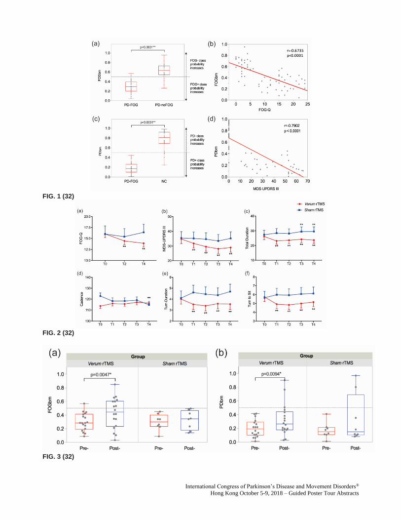
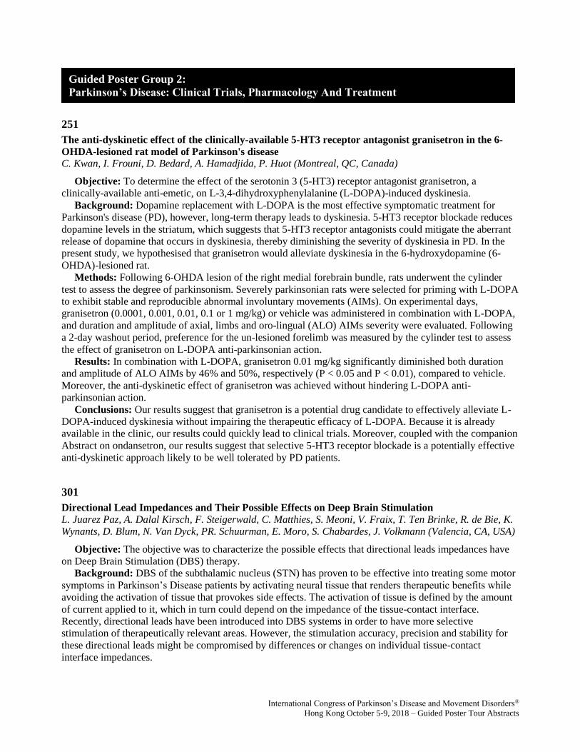
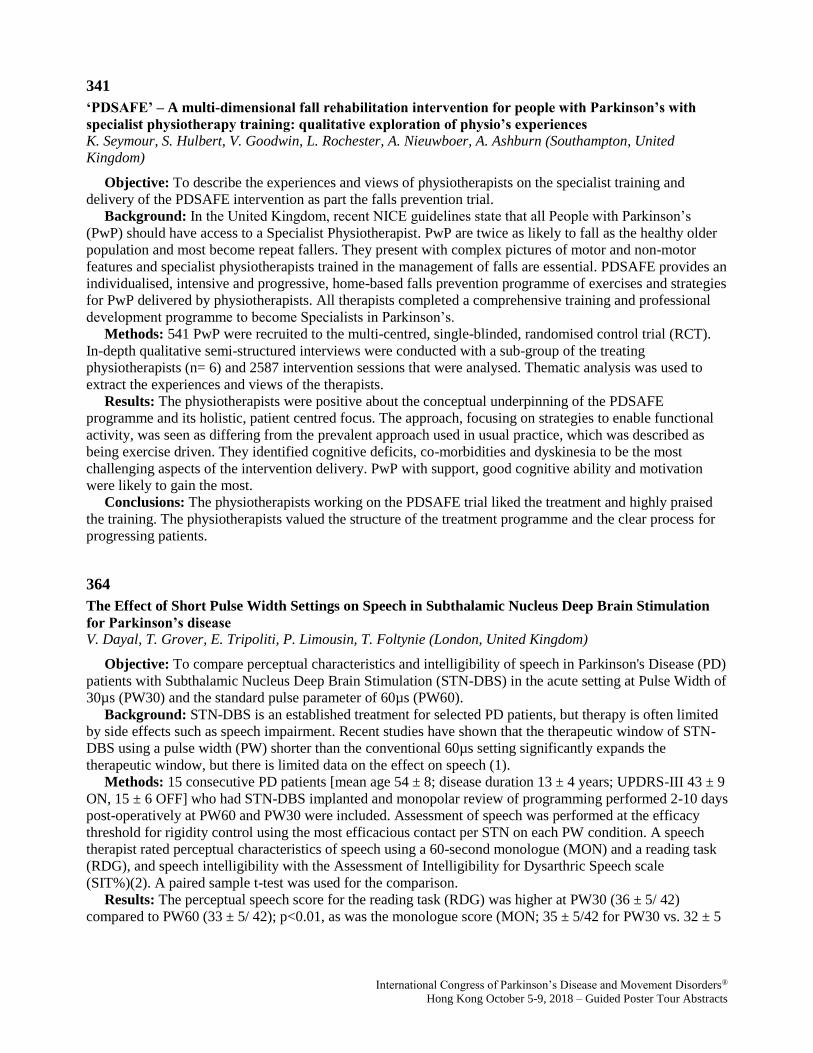
32
Alleviation of freezing of gait in patients with Parkinson’s disease by high-frequency rTMS over SMA
is associated with normalization of brain connectivity patterns
T.M. Mi, S. Garg, F. Ba, T. Wu, P.P. Liang, L.L. Gao, K.C. Li, P. Chan, M. McKeown (Beijing, China)
Objective: We explored the efficacy and neural mechanisms of repetitive transcranial magnetic
stimulation (rTMS) over the supplementary motor area (SMA) on freezing of gait (FOG) in Parkinson's
disease (PD).
Background: FOG contributes to falls and greatly reduced mobility in PD, however, robust effective
treatments remain elusive.
Methods: We first conducted a resting-state fMRI (rs-fMRI) study using a group of 40 PD patients with
FOG (PD-FOG), 31 without FOG (PD-noFOG) and 30 normal controls (NC). A subset of 30 PD-FOG
participated in a randomized, double-blind, sham-controlled trial to investigate the effects of rTMS on FOG.
Patients were randomly assigned (with 2:1 allocation) to receive ten sessions of either verum or sham 10 Hz
rTMS over SMA. The primary clinical outcome was the Freezing of Gait Questionnaire (FOG-Q). The
Movement Disorder Society Unified Parkinson’s Disease Rating Scale motor score (MDS-UPDRS III) and
Timed Up and Go test were secondary clinical outcomes. Rs-fMRI studies were performed at baseline and
after the rTMS sessions off medication. The functional connectivity between 48 ROIs that included the
fronto-parietal network, frontostriatal loop, and locomotor network were assessed with a Bayesian network
methodology. We used the baseline scans to create imaging biomarkers for FOG (FOGbm) and PD (PDbm)
by using logististic Least Absolute Shrinkage and Selection Operator regression applied to the rs-fMRI
connectivity profiles, and contrasting PD-FOG to PD-noFOG and NC respectively. These biomarkers were
then interrogated to assess the effects of rTMS on connectivity patterns.
Results: The FOGbm and PDbm consisted of 14 and 16 functional connections, and included connections
to/from the basal ganglia, cerebellar and thalamic regions, precuneus, anterior cingulate cortex, insula cortex
and superior temporal gyrus. As anticipated, FOGbm was negatively correlated with FOGQ, and PDbm was
negatively correlated with MDS-UPDRS III. After long-term follow-up, significant clinical improvements in
FOG-Q, MDS-UPDRS III and several gait variables were found in the verum group. Moreover, both FOGbm
and PDbm were significantly improved after verum stimulation. Neither significant clinical improvements
nor biomarker changes were found in the sham group.
Conclusions: Our results suggest that high-frequency rTMS over SMA can alleviate FOG in PD through
normalizing the abnormal brain functional connectivity patterns of PD-FOG and makes it not only more
similar to PD-noFOG, but also more similar to NC.
International Congress of Parkinson’s Disease and Movement Disorders®
Hong Kong October 5-9, 2018 – Guided Poster Tour Abstracts
FIG. 1 (32)
FIG. 2 (32)
FIG. 3 (32)

International Congress of Parkinson’s Disease and Movement Disorders®
Hong Kong October 5-9, 2018 – Guided Poster Tour Abstracts
35
PPMI driven sample size estimation for clinical trials in Parkinson’s disease
K. Marek, J. Seibyl, C. Caspell-Garcia, C. Coffey, B. Mollenhauer, K. Kieburtz, C. Tanner, L. Chahine, A.
Siderowf, T. Simuni (New Haven, CT, USA)
Objective: To utilize clinical, imaging and CSF data from the Parkinson’s Progression Markers Initiative
(PPMI) to estimate the sample size for disease modification studies in Parkinson’s disease (PD) participants.
Background: The PPMI study provides longitudinal clinical and biomarker data that may be utilized to
design PD therapeutic trials to assess putative disease modifying therapies. A major roadblock for these PD
studies is to accurately establish the sample size required to establish drug effect and dosage in a reasonably
sized Phase 2 program.
Methods: PPMI PD and healthy control participants are evaluated annually with a wide range of
assessments including the MDS-UPDRS, DAT/VMAT imaging and CSF measures. PPMI subjects have
been followed for more than five years. We utilized PPMI longitudinal data to establish sample size
estimates for these measures for studies of up to two years duration assessing PD subjects. Given the large
confound of MDS-UPDRS in subjects treated with dopaminergic therapies, we examined the sample size in
treated and untreated PD after 12 month of PPMI participation. In addition, since an exponential fit of DAT
imaging data provided a better fit than a linear model, we have used the exponential model to explore sample
size estimates.
Results: Table 1 shows the one and two year sample size for all PD subjects evaluated for one and two
years. Of the variables assessed mean putamen and striatum DAT scores allow the smallest sample size.
Additional data demonstrate that DAT imaging sample size estimates may be substantially improved by
utilizing an exponential fit of three scans. VMAT2 imaging may provide an alternative imaging biomarker
with improved sample size estimates but additional data are required. MDS-UPDRS I-III untreated reduces
sample size compared to the UPDRS for the entire group. Extending the follow up to 2 years would allow
reducing the sample size by close to 50%. CSF measures would require a substantially larger sample size.
Conclusions: Sample size data can be utilized in planning future clinical trials of potential disease
modifying interventions in PD. Powering the study based on DAT imaging utilizing an exponential fit can
substantially reduce the sample size in the treated PD population. Extending the duration of the study to 2
years will also reduce the sample size but has to be balanced with the challenges of retention and overall
cost.
TABLE 1 (35)
International Congress of Parkinson’s Disease and Movement Disorders®
Hong Kong October 5-9, 2018 – Guided Poster Tour Abstracts
251
The anti-dyskinetic effect of the clinically-available 5-HT3 receptor antagonist granisetron in the 6-
OHDA-lesioned rat model of Parkinson's disease
C. Kwan, I. Frouni, D. Bedard, A. Hamadjida, P. Huot (Montreal, QC, Canada)
Objective: To determine the effect of the serotonin 3 (5-HT3) receptor antagonist granisetron, a
clinically-available anti-emetic, on L-3,4-dihydroxyphenylalanine (L-DOPA)-induced dyskinesia.
Background: Dopamine replacement with L-DOPA is the most effective symptomatic treatment for
Parkinson's disease (PD), however, long-term therapy leads to dyskinesia. 5-HT3 receptor blockade reduces
dopamine levels in the striatum, which suggests that 5-HT3 receptor antagonists could mitigate the aberrant
release of dopamine that occurs in dyskinesia, thereby diminishing the severity of dyskinesia in PD. In the
present study, we hypothesised that granisetron would alleviate dyskinesia in the 6-hydroxydopamine (6-
OHDA)-lesioned rat.
Methods: Following 6-OHDA lesion of the right medial forebrain bundle, rats underwent the cylinder
test to assess the degree of parkinsonism. Severely parkinsonian rats were selected for priming with L-DOPA
to exhibit stable and reproducible abnormal involuntary movements (AIMs). On experimental days,
granisetron (0.0001, 0.001, 0.01, 0.1 or 1 mg/kg) or vehicle was administered in combination with L-DOPA,
and duration and amplitude of axial, limbs and oro-lingual (ALO) AIMs severity were evaluated. Following
a 2-day washout period, preference for the un-lesioned forelimb was measured by the cylinder test to assess
the effect of granisetron on L-DOPA anti-parkinsonian action.
Results: In combination with L-DOPA, granisetron 0.01 mg/kg significantly diminished both duration
and amplitude of ALO AIMs by 46% and 50%, respectively (P < 0.05 and P < 0.01), compared to vehicle.
Moreover, the anti-dyskinetic effect of granisetron was achieved without hindering L-DOPA anti-
parkinsonian action.
Conclusions: Our results suggest that granisetron is a potential drug candidate to effectively alleviate L-
DOPA-induced dyskinesia without impairing the therapeutic efficacy of L-DOPA. Because it is already
available in the clinic, our results could quickly lead to clinical trials. Moreover, coupled with the companion
Abstract on ondansetron, our results suggest that selective 5-HT3 receptor blockade is a potentially effective
anti-dyskinetic approach likely to be well tolerated by PD patients.
301
Directional Lead Impedances and Their Possible Effects on Deep Brain Stimulation
L. Juarez Paz, A. Dalal Kirsch, F. Steigerwald, C. Matthies, S. Meoni, V. Fraix, T. Ten Brinke, R. de Bie, K.
Wynants, D. Blum, N. Van Dyck, PR. Schuurman, E. Moro, S. Chabardes, J. Volkmann (Valencia, CA, USA)
Objective: The objective was to characterize the possible effects that directional leads impedances have
on Deep Brain Stimulation (DBS) therapy.
Background: DBS of the subthalamic nucleus (STN) has proven to be effective into treating some motor
symptoms in Parkinson’s Disease patients by activating neural tissue that renders therapeutic benefits while
avoiding the activation of tissue that provokes side effects. The activation of tissue is defined by the amount
of current applied to it, which in turn could depend on the impedance of the tissue-contact interface.
Recently, directional leads have been introduced into DBS systems in order to have more selective
stimulation of therapeutically relevant areas. However, the stimulation accuracy, precision and stability for
these directional leads might be compromised by differences or changes on individual tissue-contact
interface impedances.
Guided Poster Group 2:
Parkinson’s Disease: Clinical Trials, Pharmacology And Treatment

International Congress of Parkinson’s Disease and Movement Disorders®
Hong Kong October 5-9, 2018 – Guided Poster Tour Abstracts
Methods: DIRECT-DBS is a prospective, randomized, multi-center, double-blind study with a crossover
design, for which 12 subjects have been enrolled and implanted for STN DBS with bilateral directional leads
(Vercise Cartesia, Boston Scientific) connected to a pulse generator. Within the framework of the study,
impedance and stimulation settings data will be collected at different time points for analysis.
Results: The collected data so far shows significant differences between ring and segmented contacts’
impedances. The data also shows substantial impedance changes through time for ring and especially the
segmented contacts (5.7% and 11.4% average impedance change, respectively). By calculating the
equivalent current delivered by single source voltage (and current DBS systems) under these impedance
changes conditions, it was found that these systems are unable to deliver the same current distribution for the
desired stimulation setting when multiple contacts are needed for effective stimulation. In addition, these
systems are unable to constantly deliver the desired current as opposed to DBS systems using multiple
independent current sources.
Conclusions: The results suggest that in order to leverage the advantages of directional leads for an
effective DBS therapy, it is fundamental to use them in combination with multiple independent current
sources systems to allow for accurate, precise and stable current delivery. The clinical relevance of these
findings has to be further investigated.
326
DIRECT DBS: A Prospective, Multicenter Clinical Study with Double-Blinding for a Directional Deep
Brain Stimulation Lead – Therapeutic Windows with Directional Stimulation
F. Steigerwald, J. Volkmann, C. Matthies, A. Dalal Kirsch, S. Chabardes, R. de Bie, P. Schuurman, E. Moro,
V. Fraix, S. Meoni, D. Blum, L. Juarez Paz, K. Wynants, N. Van Dyck (Würzburg, Germany)
Objective: To evaluate changes in therapeutic window values for changes in directional Deep Brain
Stimulation (DBS) stimulation.
Background: Historically, DBS systems have delivered stimulation using cylindrical electrodes, which
may stimulate neurons around the entire circumference of the lead. In this study, we test a directional DBS
lead, which adds radially segmented electrodes designed for selective stimulation in directions orthogonal to
the lead trajectory. One way to show clinical proof of the additional capabilities of directional leads is to
examine the therapeutic windows of varying directional stimulation settings.
Methods: DIRECT-DBS is a prospective, randomized, multi-center, double-blind study employing a
crossover design. A total of 12 subjects have been enrolled and implanted per standard of care with bilateral
directional DBS leads (Vercise Cartesia, Boston Scientific) connected to a pulse generator providing an
independent current source for each of 16 contacts. Visits occur in 3 major periods: during implant, at 3-5
months, and at 1 year. At 3 months, multiple single-day programming visits will be undertaken to optimize
directional programming, based on observed clinical responses. In one of the visits, various directional
stimulation settings are explored at the optimal longitudinal level, first in 90 degree increments, then in 30
degree increments. These fine explorations require precise fractionalization of the current between sets of
segmented electrodes. At each of these settings, the therapeutic window is calculated as the difference
between the minimum amplitude which gives full rigidity control and the minimum amplitude that elicits a
limiting side effect.
Results: Examination of the results collected thus far show differences in therapeutic windows at the
various directional stimulation settings. These differences can manifest in changes as small as 30 degrees in
the rotational direction.
Conclusions: These results show that directional stimulation is a useful advancement in DBS technology
as it may enable the user to elicit differential clinical responses which may not have been observed with other
programming changes.
International Congress of Parkinson’s Disease and Movement Disorders®
Hong Kong October 5-9, 2018 – Guided Poster Tour Abstracts
341
‘PDSAFE’ – A multi-dimensional fall rehabilitation intervention for people with Parkinson’s with
specialist physiotherapy training: qualitative exploration of physio’s experiences
K. Seymour, S. Hulbert, V. Goodwin, L. Rochester, A. Nieuwboer, A. Ashburn (Southampton, United
Kingdom)
Objective: To describe the experiences and views of physiotherapists on the specialist training and
delivery of the PDSAFE intervention as part the falls prevention trial.
Background: In the United Kingdom, recent NICE guidelines state that all People with Parkinson’s
(PwP) should have access to a Specialist Physiotherapist. PwP are twice as likely to fall as the healthy older
population and most become repeat fallers. They present with complex pictures of motor and non-motor
features and specialist physiotherapists trained in the management of falls are essential. PDSAFE provides an
individualised, intensive and progressive, home-based falls prevention programme of exercises and strategies
for PwP delivered by physiotherapists. All therapists completed a comprehensive training and professional
development programme to become Specialists in Parkinson’s.
Methods: 541 PwP were recruited to the multi-centred, single-blinded, randomised control trial (RCT).
In-depth qualitative semi-structured interviews were conducted with a sub-group of the treating
physiotherapists (n= 6) and 2587 intervention sessions that were analysed. Thematic analysis was used to
extract the experiences and views of the therapists.
Results: The physiotherapists were positive about the conceptual underpinning of the PDSAFE
programme and its holistic, patient centred focus. The approach, focusing on strategies to enable functional
activity, was seen as differing from the prevalent approach used in usual practice, which was described as
being exercise driven. They identified cognitive deficits, co-morbidities and dyskinesia to be the most
challenging aspects of the intervention delivery. PwP with support, good cognitive ability and motivation
were likely to gain the most.
Conclusions: The physiotherapists working on the PDSAFE trial liked the treatment and highly praised
the training. The physiotherapists valued the structure of the treatment programme and the clear process for
progressing patients.
364
The Effect of Short Pulse Width Settings on Speech in Subthalamic Nucleus Deep Brain Stimulation
for Parkinson’s disease
V. Dayal, T. Grover, E. Tripoliti, P. Limousin, T. Foltynie (London, United Kingdom)
Objective: To compare perceptual characteristics and intelligibility of speech in Parkinson's Disease (PD)
patients with Subthalamic Nucleus Deep Brain Stimulation (STN-DBS) in the acute setting at Pulse Width of
30µs (PW30) and the standard pulse parameter of 60µs (PW60).
Background: STN-DBS is an established treatment for selected PD patients, but therapy is often limited
by side effects such as speech impairment. Recent studies have shown that the therapeutic window of STN-
DBS using a pulse width (PW) shorter than the conventional 60µs setting significantly expands the
therapeutic window, but there is limited data on the effect on speech (1).
Methods: 15 consecutive PD patients [mean age 54 ± 8; disease duration 13 ± 4 years; UPDRS-III 43 ± 9
ON, 15 ± 6 OFF] who had STN-DBS implanted and monopolar review of programming performed 2-10 days
post-operatively at PW60 and PW30 were included. Assessment of speech was performed at the efficacy
threshold for rigidity control using the most efficacious contact per STN on each PW condition. A speech
therapist rated perceptual characteristics of speech using a 60-second monologue (MON) and a reading task
(RDG), and speech intelligibility with the Assessment of Intelligibility for Dysarthric Speech scale
(SIT%)(2). A paired sample t-test was used for the comparison.
Results: The perceptual speech score for the reading task (RDG) was higher at PW30 (36 ± 5/ 42)
compared to PW60 (33 ± 5/ 42); p<0.01, as was the monologue score (MON; 35 ± 5/42 for PW30 vs. 32 ± 5
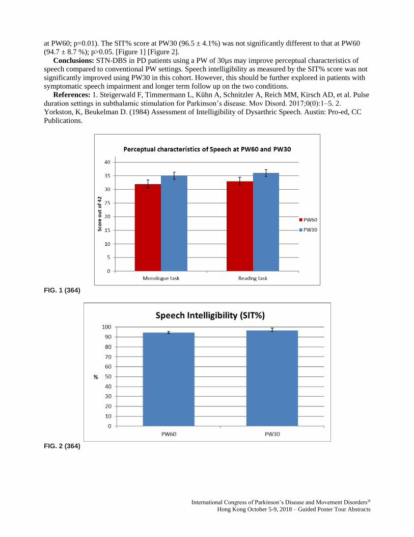
International Congress of Parkinson’s Disease and Movement Disorders®
Hong Kong October 5-9, 2018 – Guided Poster Tour Abstracts
at PW60; p=0.01). The SIT% score at PW30 (96.5 ± 4.1%) was not significantly different to that at PW60
(94.7 ± 8.7 %); p>0.05. [Figure 1] [Figure 2].
Conclusions: STN-DBS in PD patients using a PW of 30µs may improve perceptual characteristics of
speech compared to conventional PW settings. Speech intelligibility as measured by the SIT% score was not
significantly improved using PW30 in this cohort. However, this should be further explored in patients with
symptomatic speech impairment and longer term follow up on the two conditions.
References: 1. Steigerwald F, Timmermann L, Kühn A, Schnitzler A, Reich MM, Kirsch AD, et al. Pulse
duration settings in subthalamic stimulation for Parkinson’s disease. Mov Disord. 2017;0(0):1–5. 2.
Yorkston, K, Beukelman D. (1984) Assessment of Intelligibility of Dysarthric Speech. Austin: Pro-ed, CC
Publications.
FIG. 1 (364)
FIG. 2 (364)
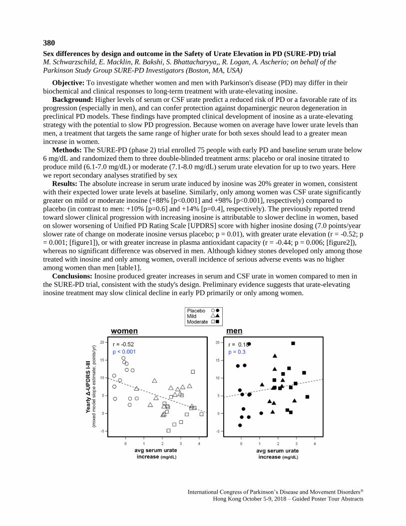
International Congress of Parkinson’s Disease and Movement Disorders®
Hong Kong October 5-9, 2018 – Guided Poster Tour Abstracts
380
Sex differences by design and outcome in the Safety of Urate Elevation in PD (SURE-PD) trial
M. Schwarzschild, E. Macklin, R. Bakshi, S. Bhattacharyya,, R. Logan, A. Ascherio; on behalf of the
Parkinson Study Group SURE-PD Investigators (Boston, MA, USA)
Objective: To investigate whether women and men with Parkinson's disease (PD) may differ in their
biochemical and clinical responses to long-term treatment with urate-elevating inosine.
Background: Higher levels of serum or CSF urate predict a reduced risk of PD or a favorable rate of its
progression (especially in men), and can confer protection against dopaminergic neuron degeneration in
preclinical PD models. These findings have prompted clinical development of inosine as a urate-elevating
strategy with the potential to slow PD progression. Because women on average have lower urate levels than
men, a treatment that targets the same range of higher urate for both sexes should lead to a greater mean
increase in women.
Methods: The SURE-PD (phase 2) trial enrolled 75 people with early PD and baseline serum urate below
6 mg/dL and randomized them to three double-blinded treatment arms: placebo or oral inosine titrated to
produce mild (6.1-7.0 mg/dL) or moderate (7.1-8.0 mg/dL) serum urate elevation for up to two years. Here
we report secondary analyses stratified by sex
Results: The absolute increase in serum urate induced by inosine was 20% greater in women, consistent
with their expected lower urate levels at baseline. Similarly, only among women was CSF urate significantly
greater on mild or moderate inosine (+88% [p<0.001] and +98% [p<0.001], respectively) compared to
placebo (in contrast to men: +10% [p=0.6] and +14% [p=0.4], respectively). The previously reported trend
toward slower clinical progression with increasing inosine is attributable to slower decline in women, based
on slower worsening of Unified PD Rating Scale [UPDRS] score with higher inosine dosing (7.0 points/year
slower rate of change on moderate inosine versus placebo; p = 0.01), with greater urate elevation (r = -0.52; p
= 0.001; [figure1]), or with greater increase in plasma antioxidant capacity (r = -0.44; p = 0.006; [figure2]),
whereas no significant difference was observed in men. Although kidney stones developed only among those
treated with inosine and only among women, overall incidence of serious adverse events was no higher
among women than men [table1].
Conclusions: Inosine produced greater increases in serum and CSF urate in women compared to men in
the SURE-PD trial, consistent with the study's design. Preliminary evidence suggests that urate-elevating
inosine treatment may slow clinical decline in early PD primarily or only among women.

International Congress of Parkinson’s Disease and Movement Disorders®
Hong Kong October 5-9, 2018 – Guided Poster Tour Abstracts
FIG. 1 (380) Relationships between change in serum urate and rate of clinical decline in women and men.
For each subject the increase in scrum urate from baseline to the average of on‐treatment values was
plotted against the individual's annualized change in total UPDRS (parts I‐III) score estimated from a
random‐slopes mixed model. The placebo group data (circles) appear on the left of each plot given the
expected lack of change in scrum urate. Several men in the placebo group showed no decline during the trial
(filled circles in lower left) contributing to a non‐significant overall trend toward worsening with increasing
urate among men.
FIG. 2 (380) Relationships between change in plasma total antioxidant capacity and rate of clinical decline in
women and men. For each subject the increase in plasma FRAP from baseline to the average of on‐treatment (6 month and ∼ 18 month visit) values was plotted against the individual's annualized change in
total UPDRS (parts I‐III) score estimated from a random‐slopes mixed model. The placebo group data
(circles) appear on the left of each plot given the lack of FRAP change as expected in this group. Several men in the placebo group showed no clinical decline during the trial (filled circles in lower left) contributing to
a non‐significant overall trend toward worsening with increasing FRAP (like urate) among men.
TABLE 1 (380) Serious and known hyperuricemic adverse events, by treatment group and sex.

International Congress of Parkinson’s Disease and Movement Disorders®
Hong Kong October 5-9, 2018 – Guided Poster Tour Abstracts
388
The highly selective 5-HT2A receptor antagonist EMD-281,014 alleviates L-DOPA-induced dyskinesia
in the 6-OHDA-lesioned rat model of Parkinson's disease
I. Frouni, D. Bedard, S. Belliveau, E. Bourgeois-Cayer, A. Hamadjida, P. Huot (Montreal, QC, Canada)
Objective: To investigate the effect of the highly-selective serotonin 2A (5-HT2A ) receptor antagonist
EMD-281,014 at alleviating L-3,4-dihydroxyphenylalanine (L-DOPA)-induced dyskinesia in the 6-
hydroxydopamine (6-OHDA)-lesioned rat.
Background: Chronic administration of L-DOPA, the most effective symptomatic treatment for
Parkinson's disease (PD), leads to motor complications such as dyskinesia in as many as 95% of patients.
Previous studies have suggested that antagonising 5-HT2A receptors may alleviate dyskinesia in animal
models of PD, but the drugs assessed interacted with targets other than 5-HT2A receptors and, as such, a 5-
HT2A-selective mechanism may not be the only factor underlying their effectiveness. Here, we hypothesised
that EMD-281,014, a clinically-ready compound that is currently the most selective 5-HT2A antagonist
available, may effectively decrease dyskinesia in PD.
Methods: Rats were rendered hemi-parkinsonian by administration of 6-OHDA. They were then primed
with L-DOPA to elicit stable and reproducible axial, limbs and oro-lingual (ALO) abnormal involuntary
movements (AIMs). On experimental days, rats were administered L-DOPA in combination with EMD-
281,014 (vehicle, 0.01, 0.03 and 0.1 mg/kg), after which ALO AIMs were assessed for 1 min, every 20 min,
for 180 min. After a 72h washout period, animals were administered an acute challenge of EMD-281,014 in
combination with L-DOPA and the degree of parkinsonism was assessed with the cylinder test.
Results: In combination with L-DOPA, EMD-281,014 mildly, but significantly, diminished the amplitude
of dyskinesia, when compared to vehicle. Thus, EMD-281,014 (0.01, 0.03 and 0.1 mg/kg) significantly
reduced cumulative ALO AIMs amplitude, when compared to vehicle, by 20%, 14% and 13%, respectively
(all P < 0.01). EMD-281,014 (0.01 and 0.1 mg/kg) also reduced cumulative axial AIMs amplitude, by 21%
and 18% (both P < 0.05), respectively, when compared to vehicle. Lastly, EMD-281,014 (0.01 and 0.1
mg/kg) diminished cumulative limbs AIMs amplitude, by 40% and 20% (both P < 0.05), respectively, when
compared to vehicle. Importantly, EMD-281,014 did not hinder the anti-parkinsonian action of L-DOPA.
Conclusions: These results suggest that the highly-selective 5-HT2A antagonist EMD-281,014 is a
promising drug to reduce the severity of dyskinesia in PD.
394
Patient and Provider Experiences and Attitudes toward Rytary
S. Horn, D. Coughlin, J. Chou, F. Wang, M. Stacy, R. Dolhun, C. Kopil, N. Amondikar, A. Deik, H. Sarva
(Philadelphia, PA, USA)
Objective: To survey Parkinson’s disease (PD) patients and providers about their experiences using
Rytary, an oral extended release carbidopa/levodopa capsule (ER C/L).
Background: ER C/L demonstrated in clinical trials to decrease OFF time and improve ON time without
increasing troublesome dyskinesia when compared to other levodopa formulations (Dhall & Kreitzman
2016). However, individual provider and patient experiences vary, and barriers to using ER C/L exist.
Methods: 801 PD patients and 250 physicians, nurse practitioners, and physician assistants completed
separate online nationwide surveys to determine utility, side effects, satisfaction and barriers to using ER
C/L.
Results: Movement disorders specialists were 27% of provider respondents; 44% were general
neurologists. The top reasons providers chose ER C/L include reducing OFF time (69%) and reducing
medication dosing frequency (32%). Reduction in OFF time was reported by 74% of providers but by only
50% of patients. Among providers, 71% reported reduction in dyskinesia, whereas only 27% of patients
experiencing dyskinesia reported a decrease and 32% reported worsening dyskinesia. Dry mouth (33%) and
fatigue (25%) were the most common side effects reported by patients. The proportion of patients reporting
improvements in quality of life, motor symptoms, and non-motor symptoms with ER C/L were 51%, 55%,

International Congress of Parkinson’s Disease and Movement Disorders®
Hong Kong October 5-9, 2018 – Guided Poster Tour Abstracts
and 38% respectively. Patient perceived quality of counseling prior to initiation of ER C/L correlated
positively with satisfaction (p <0.001). Providers cited high cost (38%), need for prior authorization (30%),
and complicated dosing and conversion processes (12%) as most common barriers. Patients reported high
cost (52%), inconvenient dosing (37%) and suboptimal symptom control (32%) as challenges. Twenty-six
percent of patients discontinued ER C/L; the most common reasons were side effects (40%) and suboptimal
symptom control (21%).
Conclusions: These large surveys evaluated patient and provider experiences with ER C/L. Both noted
reduction in OFF time as a benefit. Patients reported moderate satisfaction with motor benefits and quality of
life, but less satisfaction with the effect on non-motor symptoms. Providers noted a reduction in dyskinesia
greater than what patients reported. High out-of-pocket costs and complicated conversion and dosing
regimens were common barriers. There is continued need for accessible symptomatic therapies for PD to
serve unmet patient needs.
References: Dhall, R., & Kreitzman, D. L. (2016). Advances in levodopa therapy for Parkinson disease
Review of RYTARY (carbidopa and levodopa) clinical efficacy and safety. Neurology, 86(14 Supplement
1), S13-S24.
420
Mitochondrial morphometrics in idiopathic Parkinson‘s disease fibroblasts
P. Antony, O. Boyd, K. Mommaerts, K. Sokolowska, M. Ostaszewski, A. Baumuratov, L. Longhino, F.
Poulain, R. Krueger, R. Balling, N. Diederich (Belvaux, Luxembourg)
Objective: To identify mitochondrial morphometric phenotypes in fibroblasts derived from patients with
idiopathic Parkinson’s disease (IPD).
Background: Mitochondrial dysfunction (MD) has been proposed as cellular phenotype in patients with
idiopathic Parkinson’s disease (IPD). However, study results remain controversial for fibroblasts derived
from IPD. Here we aim to identify mitochondrial morphometric changes that eventually can be used in cell
based drug screening assays targeting IPD.
Methods: Axilla skin punch biopsy derived fibroblasts from 41 IPD patients with a mean disease duration
of 6.5 years (st.dev. 5.5 years) and from 21 healthy age-matched controls (HC) were stained with Hoechst,
CellMask, and TMRM. Mitochondrial morphometrics were analyzed via automated confocal microscopy and
in house developed computational image analysis (figure).
Results: Significant morphometric changes were seen in IPD fibroblasts. The number of mitochondria
was increased (p=0.002) whereas the mitochondrial morphometric complexity was substantially decreased as
visualized by reduction of mitochondrial perimeter (p=0.029) and mitochondrial form factor (p=0.013).
Further analysis with established algorithms from computer vision and graph theory confirmed mitochondrial
fragmentation: The mitochondrial skeleton corresponding to the final chain of pixels remaining after step by
step erosion of the mitochondrial mask was significantly reduced in IPD fibroblasts (p=0.020). The number
of mitochondrial nodes in the sense of graph theory applied to the mitochondrial skeleton reflects reduced
mitochondrial branching in IPD (p=0.013). Finally, the mitochondrial node degree representing the average
branch length in the mitochondrial skeleton network was also reduced (p<0.001). [figure1] Figure: (white)
mitochondrial shapes, (blue) skeleton, (green) branch points, (red) endpoints. Note the reduced branching in
the IPD patient sample.
Conclusions: Morphometric changes of IPD mitochondria with increased number of mitochondria on one
side and reduced branching on the other side reflect mitochondrial fragmentation. This may be due to
impaired mitophagy and/or also – at least temporary efficient – compensatory mechanisms, as also indicated
by increased mitochondrial membrane potential. The results are in agreement with similar changes observed
in submucosal enteric ganglia neurons of IPD patients (doi:10.1038/srep33117).
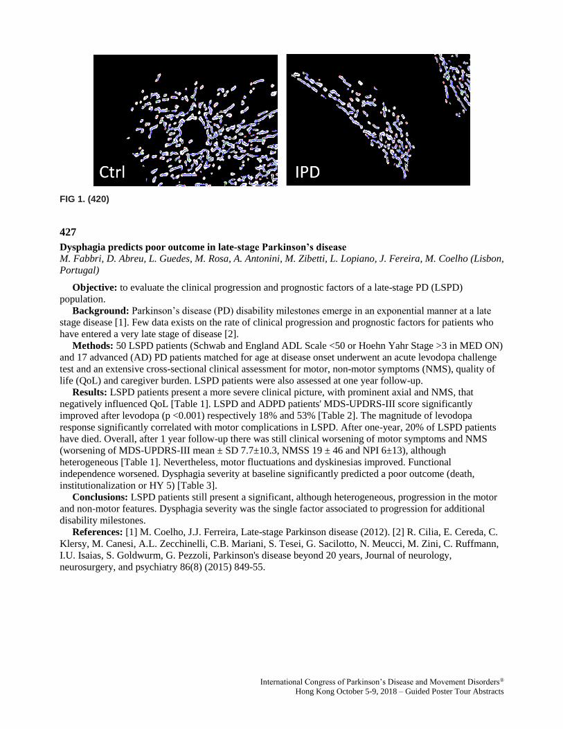
International Congress of Parkinson’s Disease and Movement Disorders®
Hong Kong October 5-9, 2018 – Guided Poster Tour Abstracts
FIG 1. (420)
427
Dysphagia predicts poor outcome in late-stage Parkinson’s disease
M. Fabbri, D. Abreu, L. Guedes, M. Rosa, A. Antonini, M. Zibetti, L. Lopiano, J. Fereira, M. Coelho (Lisbon,
Portugal)
Objective: to evaluate the clinical progression and prognostic factors of a late-stage PD (LSPD)
population.
Background: Parkinson’s disease (PD) disability milestones emerge in an exponential manner at a late
stage disease [1]. Few data exists on the rate of clinical progression and prognostic factors for patients who
have entered a very late stage of disease [2].
Methods: 50 LSPD patients (Schwab and England ADL Scale <50 or Hoehn Yahr Stage >3 in MED ON)
and 17 advanced (AD) PD patients matched for age at disease onset underwent an acute levodopa challenge
test and an extensive cross-sectional clinical assessment for motor, non-motor symptoms (NMS), quality of
life (QoL) and caregiver burden. LSPD patients were also assessed at one year follow-up.
Results: LSPD patients present a more severe clinical picture, with prominent axial and NMS, that
negatively influenced QoL [Table 1]. LSPD and ADPD patients' MDS-UPDRS-III score significantly
improved after levodopa (p <0.001) respectively 18% and 53% [Table 2]. The magnitude of levodopa
response significantly correlated with motor complications in LSPD. After one-year, 20% of LSPD patients
have died. Overall, after 1 year follow-up there was still clinical worsening of motor symptoms and NMS
(worsening of MDS-UPDRS-III mean ± SD 7.7±10.3, NMSS 19 ± 46 and NPI 6±13), although
heterogeneous [Table 1]. Nevertheless, motor fluctuations and dyskinesias improved. Functional
independence worsened. Dysphagia severity at baseline significantly predicted a poor outcome (death,
institutionalization or HY 5) [Table 3].
Conclusions: LSPD patients still present a significant, although heterogeneous, progression in the motor
and non-motor features. Dysphagia severity was the single factor associated to progression for additional
disability milestones.
References: [1] M. Coelho, J.J. Ferreira, Late-stage Parkinson disease (2012). [2] R. Cilia, E. Cereda, C.
Klersy, M. Canesi, A.L. Zecchinelli, C.B. Mariani, S. Tesei, G. Sacilotto, N. Meucci, M. Zini, C. Ruffmann,
I.U. Isaias, S. Goldwurm, G. Pezzoli, Parkinson's disease beyond 20 years, Journal of neurology,
neurosurgery, and psychiatry 86(8) (2015) 849-55.
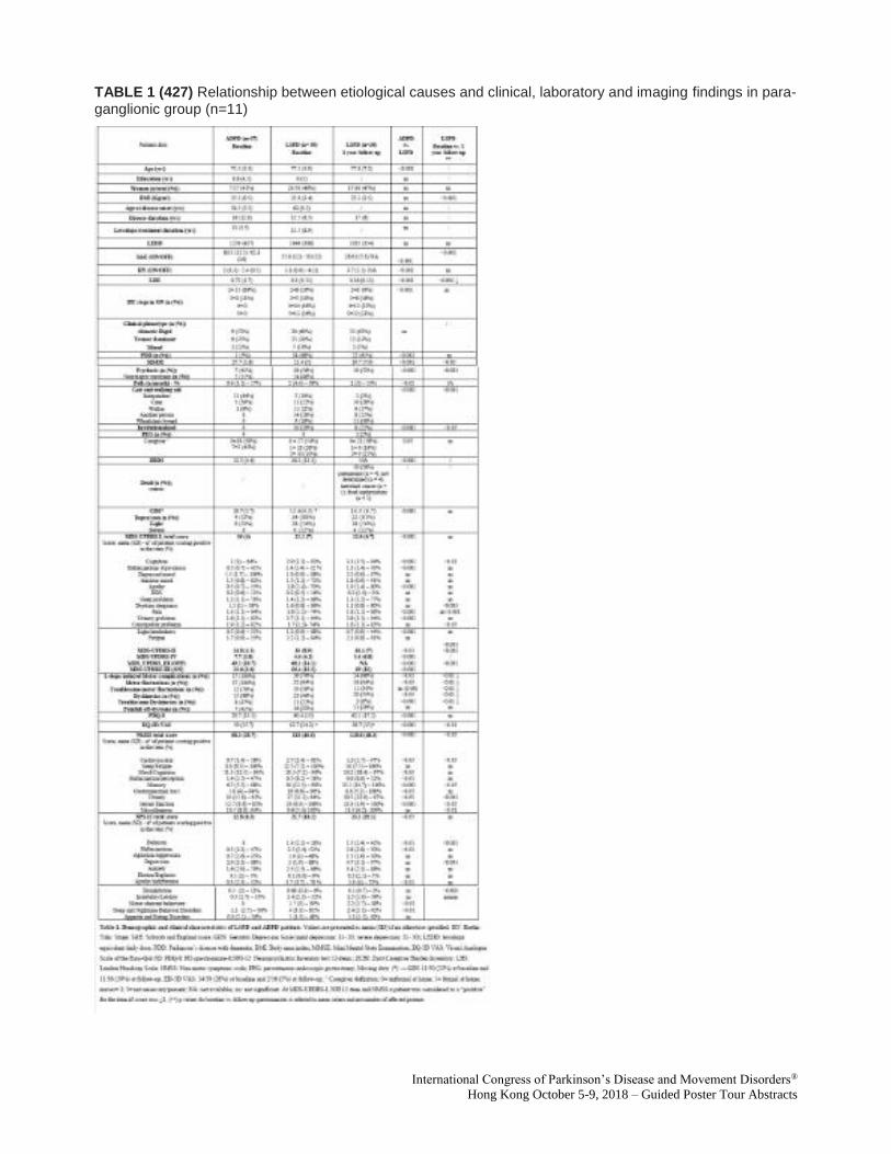
International Congress of Parkinson’s Disease and Movement Disorders®
Hong Kong October 5-9, 2018 – Guided Poster Tour Abstracts
TABLE 1 (427) Relationship between etiological causes and clinical, laboratory and imaging findings in para-ganglionic group (n=11)
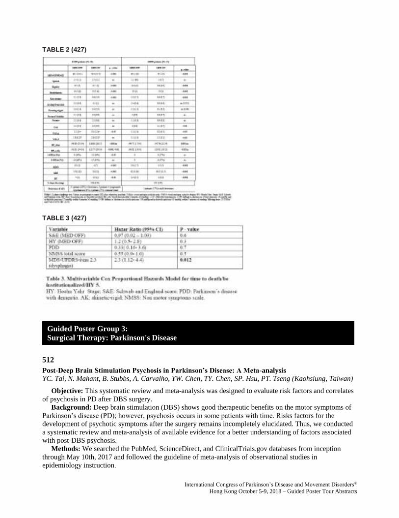
International Congress of Parkinson’s Disease and Movement Disorders®
Hong Kong October 5-9, 2018 – Guided Poster Tour Abstracts
TABLE 2 (427)
TABLE 3 (427)
512
Post-Deep Brain Stimulation Psychosis in Parkinson’s Disease: A Meta-analysis
YC. Tai, N. Mahant, B. Stubbs, A. Carvalho, YW. Chen, TY. Chen, SP. Hsu, PT. Tseng (Kaohsiung, Taiwan)
Objective: This systematic review and meta-analysis was designed to evaluate risk factors and correlates
of psychosis in PD after DBS surgery.
Background: Deep brain stimulation (DBS) shows good therapeutic benefits on the motor symptoms of
Parkinson’s disease (PD); however, psychosis occurs in some patients with time. Risks factors for the
development of psychotic symptoms after the surgery remains incompletely elucidated. Thus, we conducted
a systematic review and meta-analysis of available evidence for a better understanding of factors associated
with post-DBS psychosis.
Methods: We searched the PubMed, ScienceDirect, and ClinicalTrials.gov databases from inception
through May 10th, 2017 and followed the guideline of meta-analysis of observational studies in
epidemiology instruction.
Guided Poster Group 3:
Surgical Therapy: Parkinson's Disease
International Congress of Parkinson’s Disease and Movement Disorders®
Hong Kong October 5-9, 2018 – Guided Poster Tour Abstracts
Results: Twelve studies comprising 1037 participants with PD met inclusion criteria. The cross-sectional
prevalence of post-DBS psychosis was positively associated with follow-up duration and higher stimulation
frequency but inversely with a higher stimulation voltage. The person-year incidence of post-DBS psychosis
was positively associated with lower pre-DBS UPDRS scores. In subgroup analysis, post-DBS psychosis
was associated with longer follow-up duration. Incidence was lower in studies that employed standard
diagnostic criteria for psychosis but higher in patients with baseline UPDRS scores <40 and levodopa-
equivalent doses <1000mg. It was noted that gait disturbance and Hoenh and Yahr stage >=3 were not
related to psychosis
Conclusions: A number of other findings may not have been predicted a priori and may be explicable by
the complexities of DBS, including patient selection and management to attempt to reduce or treat psychosis.
With early post-DBS psychosis, physicians tend to reduce the stimulating voltage but increase the
stimulating frequency, which may be the reason for that prevalence of psychosis was positively associated
with stimulating frequency but inversely related to the stimulating voltage. Lower dose of LEDD before or
after the operation reflects that clinicians tend to reduce medication doses when patients exhibit psychotic
symptoms. Those unexpected findings need to be confirmed in prospective studies.
References: 1. Constantinescu R, Eriksson B, Jansson Y, et al. Key clinical milestones 15 years and
onwards after DBS-STN surgery-A retrospective analysis of patients that underwent surgery between 1993
and 2001. Clin Neurol Neurosurg 2017;154:43-48. 2. Castelli L, Perozzo P, Zibetti M, et al. Chronic deep
brain stimulation of the subthalamic nucleus for Parkinson's disease: effects on cognition, mood, anxiety and
personality traits. Eur Neurol 2006;55(3):136-144. 3. Qureshi AA, Cheng JJ, Sunshine AN, et al.
Postoperative symptoms of psychosis after deep brain stimulation in patients with Parkinson's disease.
Neurosurg Focus 2015;38(6):E5. 4. Voon V, Saint-Cyr J, Lozano AM, Moro E, Poon YY, Lang AE.
Psychiatric symptoms in patients with Parkinson disease presenting for deep brain stimulation surgery. J
Neurosurg 2005;103(2):246-251. 5. Cozac VV, Ehrensperger MM, Gschwandtner U, et al. Older Candidates
for Subthalamic Deep Brain Stimulation in Parkinson's Disease Have a Higher Incidence of Psychiatric
Serious Adverse Events. Front Aging Neurosci 2016;8:132. 6. Boel JA, Odekerken VJ, Schmand BA, et al.
Cognitive and psychiatric outcome 3 years after globus pallidus pars interna or subthalamic nucleus deep
brain stimulation for Parkinson's disease. Parkinsonism Relat Disord 2016;33:90-95. 7. Umemura A, Oka Y,
Yamamoto K, Okita K, Matsukawa N, Yamada K. Complications of subthalamic nucleus stimulation in
Parkinson's disease. Neurol Med Chir (Tokyo) 2011;51(11):749-755. 8. Chopra A, Abulseoud OA, Sampson
S, et al. Mood stability in Parkinson disease following deep brain stimulation: a 6-month prospective follow-
up study. Psychosomatics 2014;55(5):478-484. 9. Welter ML, Schupbach M, Czernecki V, et al. Optimal
target localization for subthalamic stimulation in patients with Parkinson disease. Neurology
2014;82(15):1352-1361. 10. Chen CC, Lee ST, Wu T, Chen CJ, Chen MC, Lu CS. Short-term effect of
bilateral subthalamic stimulation for advanced Parkinson's disease. Chang Gung Med J 2003;26(5):344-351.
11. Vesper J, Haak S, Ostertag C, Nikkhah G. Subthalamic nucleus deep brain stimulation in elderly patients-
-analysis of outcome and complications. BMC Neurol 2007;7:7. 12. Herzog J, Volkmann J, Krack P, et al.
Two-year follow-up of subthalamic deep brain stimulation in Parkinson's disease. Mov Disord
2003;18(11):1332-1337.
515
Motor and cognitive progression in GBA-related PD patients submitted to Deep Brain Stimulation
L. Correia Guedes, C. Silva, R. Machado Bouça, N. Gonçalves, M. Fabbri, A. Castro Caldas, P. Pita Lobo,
M.M. Rosa, B. Cattoni, H. Carvalho, A. Gonçalves Ferreira, J.J. Ferreira, M. Coelho (Lisbon, Portugal)
Objective: Our study aimed to evaluate the motor and cognitive outcome after Deep Brain Stimulation
(DBS) in GBA-related PD.
Background: Parkinson’s disease (PD) patients carrying a glucocerebrosidase gene mutation (GBA-
related PD) have been associated to worse cognitive decline after DBS, although the overall response to DBS
is still poorly understood.
International Congress of Parkinson’s Disease and Movement Disorders®
Hong Kong October 5-9, 2018 – Guided Poster Tour Abstracts
Methods: Comparison of motor and cognitive outcomes between GBA-related PD and idiopathic PD
(iPD) patients after subthalamic nucleus (STN)-DBS. Pre- and post-DBS MDS-UPDRS and MMSE were
assessed. Post-DBS evaluations were performed in 4 conditions concerning ON/OFF medication and/or
stimulation.
Results: Eight GBA-related PD and 10 iPD patients, with no significant differences in gender, age at
disease onset (AO), time to DBS, pre-DBS levodopa response, follow-up post-DBS and pre- and post-DBS
levodopa equivalent daily dose. Post-DBS MDS-UPDRS-III MedON/StimON was significantly different
between groups (40.1±9.1 GBA-PD; 28.8±11.2 iPD; P=0.05). In GBA-related PD, post-DBS MDS-UPDRS-
III MedON/StimON was worse than pre-DBS MDS-UPDRS-III MedON (40.1±9.1 vs 24.5±8; P=0.017).
MMSE did not differ significantly between groups.
6/8 GBA-related PD patients were evaluated in the 4 post-DBS conditions. Stimulation significantly
improved MDS-UPDRS-III (MedOFF/StimOFF 57.7±14.8 vs MedOFF/StimON 36.7±14.3; P=0.026), but
the benefit with stimulation (post-DBS MDS-UPDRS-III MedOFF/StimOFF vs MedOFF/StimON) was
worse compared to benefit from levodopa pre-DBS (pre-DBS MedOFF vs MedON).
Conclusions: GBA-related PD patients benefit from acute STN-DBS stimulation, although less than iPD
patients. Motor symptoms of GBA-related PD seem to worsen 4.5 years after STN-DBS, whereas we found
no cognitive decline. More data is warranted but available evidence do not support excluding GBA-related
PD patients from DBS.
Abstract presented at the 4th Congress of the European Academy of Neurology, 16-19 June 2018, Lisbon,
Portugal.
537
Directional deep brain stimulation in Parkinson’s disease guided by local field potentials
F. Alonso-Frech, C. Fernandez-Garcia, M. Monge, M.J. Catalan Alonso, G. Foffani (Madrid, Spain)
Objective: Implementation of directional stimulation of subthalamic nucleus with segmented leads
guided by local field potential (LFP) recordings in order to personalize DBS parameters, optimize therapeutic
window and minimizing side effects.
Background: Novel directional electrodes may have the advantage over the conventional ones of
avoiding adverse effects, however the programming optimization could be very time-consuming.
Methods: Since March 2016 to December 2017, 14 patients have been implanted with 28 directional
Vercise Cartesia leads (Boston SC). Mean age of patients were 54,6 years (37-74 years). Mean evolution was
9,2 years (5-18). Postoperatively CT scans/ MRI fusion were performed to asses lead placement. Local field
potentials of all contacts from each lead in bipolar montage were recorded 5 days after surgery to analyze
beta band peaks. Therapeutic window effect for rigidity were assessed blinded to LFP results at least 1 week
after surgery.
Results: No postoperative complications after directional leads implantation were observed in any
patient. LFP analyses showed significant beta peaks in 100% of patients. Directional deep brain stimulation
guided by LFP widened therapeutic window to 39% compared to spherical stimulation. Negative correlation
between beta band and final score for rigidity was found to be statistical significant (R -0,31). Contacts with
higher beta power were selected for chronic stimulation of the subthalamic nucleus with good clinical
response, obtaining 78% improvement in UPDRS-III, 82% reduction of medication.
Conclusions: Deep subthalamic stimulation with directional leads is safe and efficient. Local field
potentials and beta oscillations could optimize settings parameters and alleviating programming burden as it
seems to predict the optimal directional contact for improving rigidity. In advance, these neurophysiological
parameters may be taken into consideration to personalize directional stimulation and can be time saving
tools for programming.
References: Directional local field potentials: A tool to optimize deep brain stimulation. Tinkhauser G,
Pogosyan A, Debove I, Nowacki A, Shah S.A, Seidel K, Tan H, Brittain J.S, Petermann K, di Biase L, Oertel
M, Pollo C, Brown P, Schuepbach M. Mov Disord. 2018 Jan;33(1):159-164.
International Congress of Parkinson’s Disease and Movement Disorders®
Hong Kong October 5-9, 2018 – Guided Poster Tour Abstracts
543
Surgery-, hardware - and chronic stimulation-related adverse events following subthalamic nucleus
deep brain stimulation for Parkinson’s disease
B. Gonenli Kocer, E. Ozturk, S. Comoglu, M. Sorar, H. Kertmen (Ankara, Turkey)
Objective: To evaluate the adverse events (AEs) following subthalamic nucleus (STN) deep brain
stimulation (DBS) for Parkinson’s disease (PD) during 6 years period.
Background: The adverse events associated with DBS surgery were evaluated in 3 main categories:
Surgery-, device- and stimulation-related issues.
Methods: Between 2011 and 2017, 147 consecutive patients (92 males, %62.6) who had undergone STN
DBS surgery for PD in our Movement Disorders Clinic, were assessed retrospectively. Patients’
characteristics including modified Hoehn and Yahr (mH&Y) stages, Unified Parkinson’s Disease Rating
Scale (UPDRS) part II and III scores, levodopa equivalent doses (LED) at the pre- and postoperative period
were obtained from the continuous medical records. Surgery-, hardware- and chronic stimulation related AEs
were also recorded after the patients’ data was reviewed comprehensively.
Results: The mean age and disease duration were 54.92 ± 9.65 and 13.64 ± 6.35 years. After STN DBS,
54.44%, 55.85%, and 51.9% reduction were found on UPDRS part II, III scores and LED, respectively.
There was no mortality and also persistent morbidity related to the surgery. The most surgery-related AE was
perioperative confusion (8.2%) and the most hardware-related AE was lead or electrode fracture (6.1%).
Chronic stimulation-related AEs including weight gain, dysarthria/dysphonia, and gait/postural instability
were found 30.6%, 21.8%, and 16.3%, respectively.
Conclusions: Surgery- and hardware–related AEs are usually non-severe and non-persistent after the
STN DBS surgery. Most of AEs are treatable but chronic stimulation-related axial and non-motor symptoms
including gait and/or speech disturbances and weight gain may persist in the long-term period.
544
Poor responders to STN DBS in Parkinson’s disease: 1 year follow-up study
M. Zibetti, L. Ricciardi, E. Montanaro, M. Sarchioto, M. Edwards, L. Lopiano, F. Morgante (Toriino, Italy)
Objective: To determine which factors contribute to a poor outcome at 1 year after Deep Brain
Stimulation (DBS) of the Subthalamic Nucleus (STN).
Background: The efficacy of STN-DBS for advanced Parkinson’s disease (PD) has been consistently
demonstrated. Yet, despite accurate selection and electrode positioning, a few patients report unsatisfactory
outcome.
Methods: We conducted a retrospective analysis of prospectively acquired data at University of Turin
DBS Centre. Out of 203 consecutive PD patients treated with STN-DBS, we identified those with available
UPDRS II scores before surgery (T0) and at 1-year follow-up (T1) respectively in the OFF medication and in
the OFF medication/ON stimulation condition. We defined as “Poor DBS Responders (Poor-DBS)” those
who had <20% of improvement at UPDRS-II OFF MED/ON STIM at T1. “Poor” and “Good DBS
responders (Good-DBS)” were compared for demographical, clinical, cognitive and affective variables at T0
and T1. We included in the analysis: demographical and clinical variables, Schwab-England Scale (SE),
UPDRS-III (in the practically defined OFF state (OFF MED) at T0 and in OFF MED/ON STIM at T1).
Psychiatric assessment included Beck Depression Inventory (BDI) and State-Trait Anxiety Inventory (STAI-
X1 and X2).
Results: We retrieved baseline UPDRS II data in OFF MED from 126 subjects (Poor-DBS = 35, Good-
DBS = 91). At T0 UPDRS II, UPDRS III and axial UPDRS III were significantly less impaired in poor-DBS.
Motor fluctuations were also less severe in this group. UPDRS III improved at T1 in both Poor-DBS and
Good-DBS (p <0.0001). UPDRS II, SE, UPDRS axial score, dyskinesia score, OFF score significantly
improved at T1 compared to T0 in Good-DBS. In Poor-DBS, UPDRS II significantly worsened at T1 (p=
0.0001), SE remained unchanged (p=0.7), Axial motor UPDRS score tended to decrease but not significantly
(p= 0.06), OFF time score was not modified (p=0.17). Only dyskinesia score and total UPDRS IV were
International Congress of Parkinson’s Disease and Movement Disorders®
Hong Kong October 5-9, 2018 – Guided Poster Tour Abstracts
improved at T1 in poor-DBS (respectively p = 0.0025 and p= 0.0055). Depression by BDI decreased only in
Good-DBS (p<0.01) whereas it remained unchanged in poor-DBS. STAI-X1 and X2 did not differ between
groups.
Conclusions: Our data suggest that poor outcome on activity of daily living after STN DBS in PD might
be caused by mild improvement of axial symptoms and of OFF time score, despite improvement of UPDRS-
III in OFF MED/ON STIM.
546
A wireless brain-spine interface alleviating gait deficits of non-human primates model of Parkinson's
disease
F. Raschella, T. Milekovic, M. Perich, S. Sun, G. Schiavone, C. Hitz, Y. Jianzhong, W. Ko, Q. Li, C. Qin, S.
Lacour, J. Bloch, S. Micera, E. Bezard, G. Courtine (Geneva, Switzerland)
Objective: While levodopa and deep brain stimulation alleviate most of the symptoms associated with
Parkinson's Disease (PD), axial gait disorders are less responsive to these treatments. Impairments include
short and slow steps, balance deficits and freezing of gait. Using MPTP-treated non-human primates, we
studied the impact of a brain-spine interface on alleviating axial gait deficits observed in PD.
Background: We have established a mechanistic and technological framework that guided the design of
electrical spinal cord stimulation protocols engaging extensor and flexor muscle groups. We created an
interface that linked gait events decoded from leg motor cortex activity to spatially selective stimulation
protocols that reinforced the movements associated with these events. As early as 6 days after spinal cord
injury, this brain-spine interface restored weight-bearing locomotor movements of a paralyzed leg in a non-
human primate model of spinal cord injury [1].
Methods: Three MPTP-treated Rhesus macaque monkeys, the gold standard model for PD
symptomatology, were implanted with the wireless brain-spine interface. Recordings of multi-unit activity
from the left and right leg motor cortex were used to detect neural states related to flexion and extension
movements of both legs while the animal walked freely overground or over a horizontal ladder. The
detection of these gait events triggered the delivery of spatially selective electrical stimulation protocols that
reinforced the extension and flexion movements of the legs. Stimulation protocols were delivered using an
implantable pulse generator with real-time triggering capability that was connected to a custom-made
electrode array. The electrode layout was based on a computational model that estimated optimal locations to
target the dorsal roots of each lumbar spinal cord segment.
Results: MPTP-treated monkeys exhibited moderate to severe axial gait deficits, including short and slow
steps, balance deficits, freezing of gait, and poor precision of paw placement when traversing the horizontal
ladder. The brain-spine interface instantly alleviated these deficits, allowing the monkeys to increase their
walking speed, improved their balance and regained coordinated gait patterns. Moreover, the brain-spine
interface enabled the monkeys to regain the ability to position the paws precisely on the rungs of the
horizontal ladder.
Conclusions: These preliminary results illustrate the ability of the brain-spine interface to alleviate axial
gait deficits and restore visuomotor control of leg movements in MPTP-treated non-human primates,
standard model for PD symptomatology. These findings open promising avenues for targeting gait deficits in
people with PD, which are still resistant to current treatments.
References: [1] Capogrosso M, Milekovic T, Borton D, et al. A brain-spine interface alleviating locomotor
deficits after spinal cord injury. Nature 2016; 539: 284-88.
552
Deep brain stimulation for monogenic Parkinson's disease: A systematic review
T. Kuusimäki, J. Korpela, E. Pekkonen, M. Martikainen, A. Antonini, V. Kaasinen (Turku, Finland)
Objective: The aim was to systematically investigate the outcome of deep brain stimulation (DBS) in
genetic Parkinson’s disease (PD) compared to the patients with documented negative genetic screening.
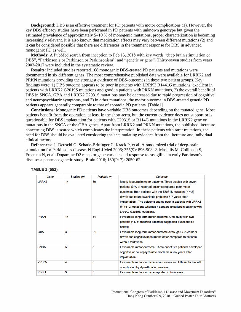
International Congress of Parkinson’s Disease and Movement Disorders®
Hong Kong October 5-9, 2018 – Guided Poster Tour Abstracts
Background: DBS is an effective treatment for PD patients with motor complications (1). However, the
key DBS efficacy studies have been performed in PD patients with unknown genotype but given the
estimated prevalence of approximately 5–10 % of monogenic mutations, proper characterization is becoming
increasingly relevant. It is also known that medication effects may vary between different mutations (2) and
it can be considered possible that there are differences in the treatment response for DBS in advanced
monogenic PD as well.
Methods: A PubMed search from inception to Feb 13, 2018 with key words “deep brain stimulation or
DBS”, “Parkinson’s or Parkinson or Parkinsonism’’ and “genetic or gene”. Thirty-seven studies from years
2003-2017 were included in the systematic review.
Results: Included studies reported 168 monogenic DBS-treated PD patients and mutations were
documented in six different genes. The most comprehensive published data were available for LRRK2 and
PRKN mutations providing the strongest evidence of DBS-outcomes in these two patient groups. Key
findings were: 1) DBS outcome appears to be poor in patients with LRRK2 R1441G mutations, excellent in
patients with LRRK2 G2019S mutations and good in patients with PRKN mutations, 2) the overall benefit of
DBS in SNCA, GBA and LRRK2 T2031S mutations may be decreased due to rapid progression of cognitive
and neuropsychiatric symptoms, and 3) in other mutations, the motor outcome in DBS-treated genetic PD
patients appears generally comparable to that of sporadic PD patients. [Table1]
Conclusions: Monogenic PD patients have variable DBS outcomes depending on the mutated gene. Most
patients benefit from the operation, at least in the short-term, but the current evidence does not support or is
questionable for DBS implantation for patients with T2031S or R114G mutations in the LRRK2 gene or
mutations in the SNCA or the GBA genes. Apart from LRRK2 and PRKN mutations, the published literature
concerning DBS is scarce which complicates the interpretation. In these patients with rarer mutations, the
need for DBS should be evaluated considering the accumulating evidence from the literature and individual
clinical factors.
References: 1. Deuschl G, Schade-Brittinger C, Krack P, et al. A randomized trial of deep-brain
stimulation for Parkinson's disease. N Engl J Med 2006; 355(9): 896-908. 2. Masellis M, Collinson S,
Freeman N, et al. Dopamine D2 receptor gene variants and response to rasagiline in early Parkinson's
disease: a pharmacogenetic study. Brain 2016; 139(Pt 7): 2050-62.
TABLE 1 (552)
International Congress of Parkinson’s Disease and Movement Disorders®
Hong Kong October 5-9, 2018 – Guided Poster Tour Abstracts
558
Propionibacterium acnes Infection with Intracerebral Abscess in Deep Brain Stimulation
R. Lewis, F. Farrokhi, M. Marsans (Seattle, WA, USA)
Objective: Propionibacterium acnes infection in DBS patients has distinct characteristics that distinguish
it from the most commonly associated bacteria in postoperative wound infections, Staphylococcus aureus.
Whereas S. aureus is typically presents with early fever and elevated inflammatory markers, P. acnes
infections in DBS more typically present with later clinical onset and unremarkable inflammatory markers.
Our objective is to highlight the unique characteristics of P. acnes infections in DBS patients. The unifying
feature amongst the cases was the presentation of neurological symptoms in the absence of fever or elevated
inflammatory markers.
Background: Deep Brain Stimulation (DBS) in as increasingly applied modality for treatment of
movement disorders such and Parkinson’s Disease and essential tremor. Although DBS has an excellent and
well proven safety profile, a small risk of infection still exists. A rare and previously underreported form is
noted here. Propionibacterium acnes infections in DBS patients have distinct presentation characteristics,
including later clinical onset and normal inflammatory markers. We review a series of infections and the
literature in an effort to raise awareness of a process that can go unnoticed and lead to devastating clinical
outcomes.
Methods: We review the literature in conjunction with a series of three P. acnes DBS infections at our
institution, with attention to commonalities between patients and clinical presentation.
A standard frame-based DBS surgical approach was followed on all cases. Preoperative prophylactic
treatments included the use of nafcillin for Case 1, vancomycin for Case 2, and cefazolin for Case 3.
Results: Common characteristics of infections included lack of clinical history of fever, chills, or sweats.
Another similarity was seen in absence of elevation in inflammatory markers. All cases showed delayed
presentation months after the initial surgery and presented with slow onset of neurologic deficits.
Case 1 resulted with a 1.7 cm abscess at the tip of the implanted electrode. Case 2 developed a 2.4 cm cystic
lesion around the tip of the electrode. Case 3 developed a 2.2 cm cystic lesion. All three cases yielded P.
acnes in cultures of explanted leads.
Conclusions: In context of delayed and subtle neurological changes in DBS patients, infection caused by
P. acnes must be taken into consideration and routinely evaluated by cranial imaging for more rapid
diagnosis and to avoid additional harm.
References: 1. Bhatia R, Dalton A, Richards M, Hopkins C, Aziz T, et al. (2011) The incidence of deep
brain stimulator hardaware infection: the effect of change in antibiotic prophylaxis regimen and review of the
literature. British Journal of Neurosurgery 25: 625-631. 2. Bjerknes S, Skogseid IM, Saehle T, Dietrichs E,
Toft M (2004) Surgical site infections after deep brain stimulation surgery: frequency, characteristics and
management in a 10-year period. PLoS ONE 9: e105288. 3. Gorgulho A, Juillard C, Uslan DZ, Tajik K,
Aurasteh P, et al. (2009) Infection following deep brain stimulator implantation performed in the
conventional versus magnetic resonance imaging-equipped operating room. J Neurosurg 110: 239-246. 4.
Temel Y, Ackermans L, Celik H, Spincemaille GH, van der Linden C, et al. (2004) Management of hardware
infections following deep brain stimulation. Acta Neurochir 155: 355-361. 5. Sillay KA, Larson PS, Starr PA
(2008) Deep brain stimulator hardware-related infections: incidence and management in a large series.
Neurosurgery 62: 360-367. 6. Voges J, Waerzeggers Y, Maarouf M, Lehrke R, Koulousakis A, et al. (2006)
Deep-brain stimulation: long-term analysis of complication caused by hardware and surgery - experiences
from a single centre. J Neurol Neurosurg Psychiatry 77: 868-872. 7. Fily F, Haegelen C, Tattevin P, Buffet-
Batillon S, Revest M, et. al. (2011) Deep brain stimulation hardware-related infections: a report of 12 cases
and review of the literature. Clinical Infectious Diseases 52: 1020-1023. 8. Umemura A, Jaggi JL, Hurtig HI,
Siderowf AD, Colcher A, et al. (2003) Deep brain stimulation for movement disorders: morbidity and
mortality in 109 patients. J Neurosurg 98: 779-784. 9. Farrokhi FR, Gunther M, Williams B, Blackmore CC
(2013) Application of lean methodology for improved quality and efficiency in operating room instrument
availability. Journal for Healthcare Quality. 10. Hegde V, Meredith DS, Kepler CK, Huang RC (2012)
Management of postoperative spinal infections. World J Orthop 3: 182-189.
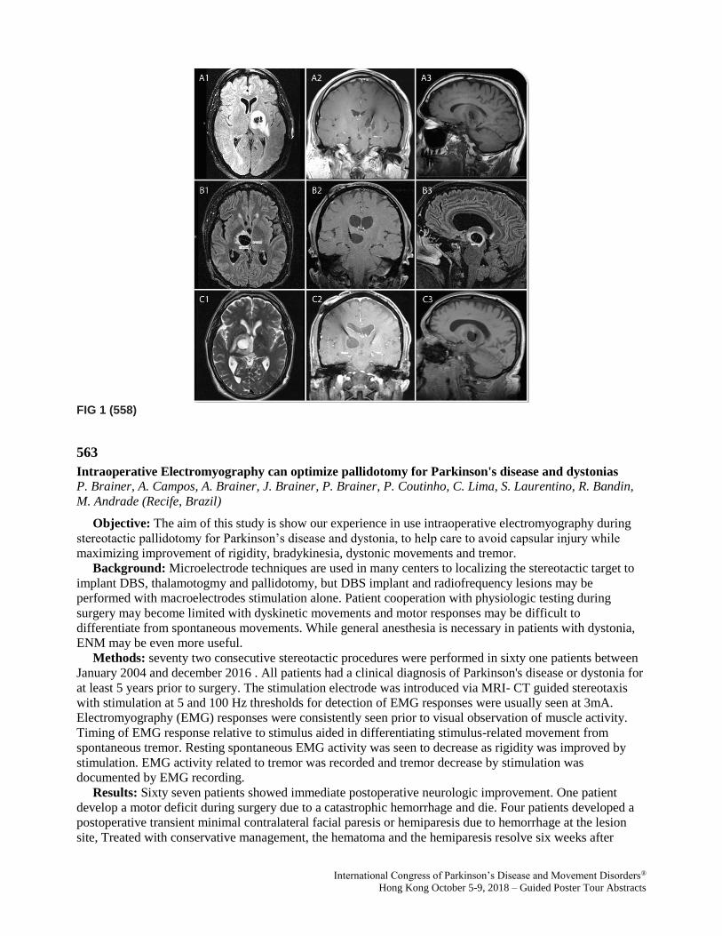
International Congress of Parkinson’s Disease and Movement Disorders®
Hong Kong October 5-9, 2018 – Guided Poster Tour Abstracts
FIG 1 (558)
563
Intraoperative Electromyography can optimize pallidotomy for Parkinson's disease and dystonias
P. Brainer, A. Campos, A. Brainer, J. Brainer, P. Brainer, P. Coutinho, C. Lima, S. Laurentino, R. Bandin,
M. Andrade (Recife, Brazil)
Objective: The aim of this study is show our experience in use intraoperative electromyography during
stereotactic pallidotomy for Parkinson’s disease and dystonia, to help care to avoid capsular injury while
maximizing improvement of rigidity, bradykinesia, dystonic movements and tremor.
Background: Microelectrode techniques are used in many centers to localizing the stereotactic target to
implant DBS, thalamotogmy and pallidotomy, but DBS implant and radiofrequency lesions may be
performed with macroelectrodes stimulation alone. Patient cooperation with physiologic testing during
surgery may become limited with dyskinetic movements and motor responses may be difficult to
differentiate from spontaneous movements. While general anesthesia is necessary in patients with dystonia,
ENM may be even more useful.
Methods: seventy two consecutive stereotactic procedures were performed in sixty one patients between
January 2004 and december 2016 . All patients had a clinical diagnosis of Parkinson's disease or dystonia for
at least 5 years prior to surgery. The stimulation electrode was introduced via MRI- CT guided stereotaxis
with stimulation at 5 and 100 Hz thresholds for detection of EMG responses were usually seen at 3mA.
Electromyography (EMG) responses were consistently seen prior to visual observation of muscle activity.
Timing of EMG response relative to stimulus aided in differentiating stimulus-related movement from
spontaneous tremor. Resting spontaneous EMG activity was seen to decrease as rigidity was improved by
stimulation. EMG activity related to tremor was recorded and tremor decrease by stimulation was
documented by EMG recording.
Results: Sixty seven patients showed immediate postoperative neurologic improvement. One patient
develop a motor deficit during surgery due to a catastrophic hemorrhage and die. Four patients developed a
postoperative transient minimal contralateral facial paresis or hemiparesis due to hemorrhage at the lesion
site, Treated with conservative management, the hematoma and the hemiparesis resolve six weeks after

International Congress of Parkinson’s Disease and Movement Disorders®
Hong Kong October 5-9, 2018 – Guided Poster Tour Abstracts
surgery. After 5 years on average( 2 to 10 years), Statistically significant improvements were seen in the
UPDRS scores for activities of daily living, motor examination in the off- on states. Significant
improvements were observed in dyskinesia and rigidity. Improvements in gait and posture in the off state
were not statistically significant.
Conclusions: EMG recordings during stereotactic DBS have been helpful in this group of patients and
have elevated the level of confidence in the safety of the site of electrode implantation, without increase in
operative complications or significative surgical time.
References: Functional lesional neurosurgery for tremor-a protocol for a systematic review and meta-
analysis. Schreglmann SR, Krauss JK, Chang JW, Bhatia KP, Kägi G. BMJ Open. 2017 May 9;7(5). Seventy
years of pallidotomy for movement disorders. Cif L, Hariz M, Mov disord 2017 Jul;32(7):972-982. A
metaanalysis comparing the results of pallidotomy performed using microelectrode recording or
macroelectrode stimulation. Palur RS, Berk C, Schulzer M, Honey CR. J Neurosurg. 2002 Jun; 96(6):1058-
62. Electromyography during stereotactic pallidotomy for Parkinson's disease. Einer WJ, Calancie B, Bowen
B. Stereotact Funct Neurosurg. 2000;74(1):21-9. Stereotactic pallidotomy performed without using
microelectrode guidance in patients with Parkinson's disease: surgical technique and 2-year results. Eskandar
EN, Shinobu LA, Penney JB Jr, Cosgrove GR, Counihan TJ. J Neurosurg. 2000 Mar; 92(3):375-83.
FIG 1 (563)
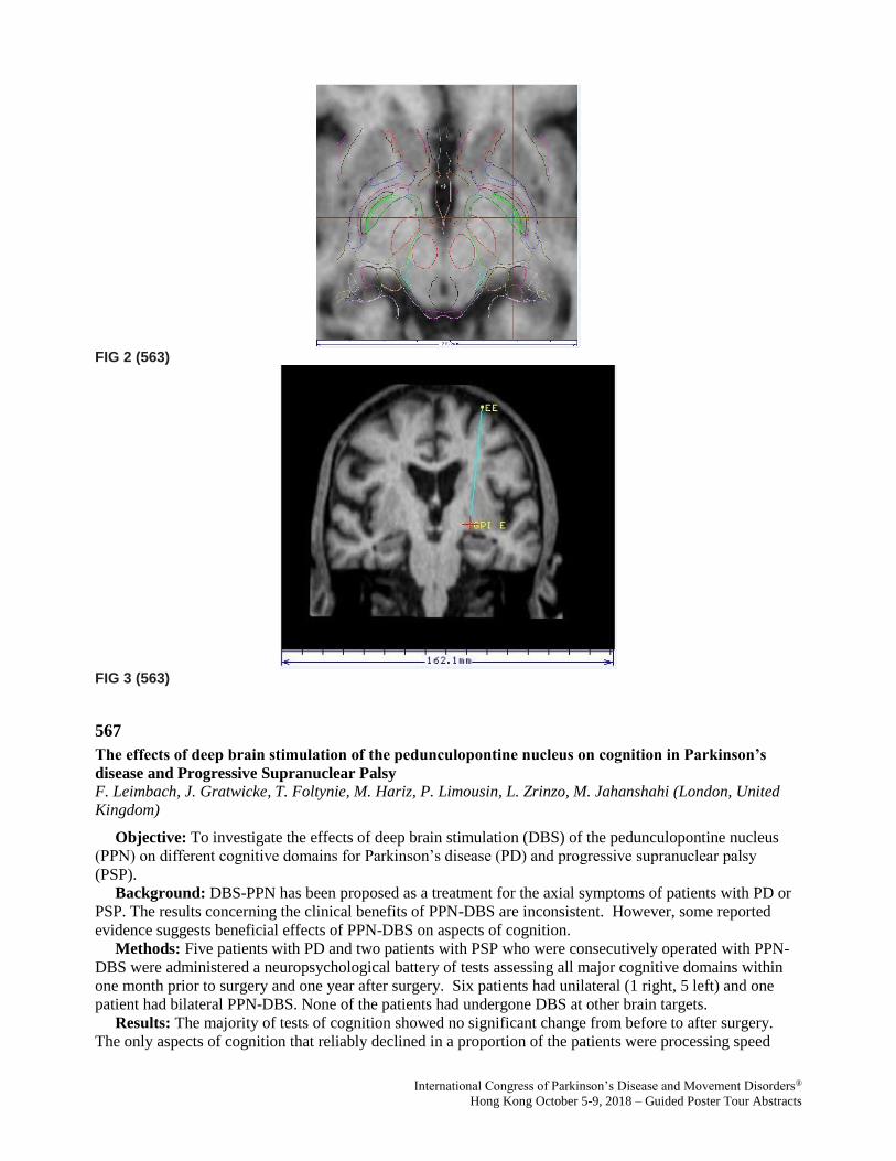
International Congress of Parkinson’s Disease and Movement Disorders®
Hong Kong October 5-9, 2018 – Guided Poster Tour Abstracts
FIG 2 (563)
FIG 3 (563)
567
The effects of deep brain stimulation of the pedunculopontine nucleus on cognition in Parkinson’s
disease and Progressive Supranuclear Palsy
F. Leimbach, J. Gratwicke, T. Foltynie, M. Hariz, P. Limousin, L. Zrinzo, M. Jahanshahi (London, United
Kingdom)
Objective: To investigate the effects of deep brain stimulation (DBS) of the pedunculopontine nucleus
(PPN) on different cognitive domains for Parkinson’s disease (PD) and progressive supranuclear palsy
(PSP).
Background: DBS-PPN has been proposed as a treatment for the axial symptoms of patients with PD or
PSP. The results concerning the clinical benefits of PPN-DBS are inconsistent. However, some reported
evidence suggests beneficial effects of PPN-DBS on aspects of cognition.
Methods: Five patients with PD and two patients with PSP who were consecutively operated with PPN-
DBS were administered a neuropsychological battery of tests assessing all major cognitive domains within
one month prior to surgery and one year after surgery. Six patients had unilateral (1 right, 5 left) and one
patient had bilateral PPN-DBS. None of the patients had undergone DBS at other brain targets.
Results: The majority of tests of cognition showed no significant change from before to after surgery.
The only aspects of cognition that reliably declined in a proportion of the patients were processing speed
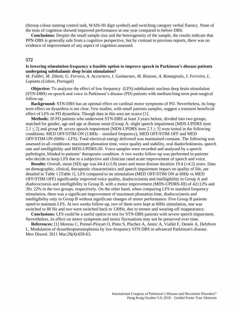
International Congress of Parkinson’s Disease and Movement Disorders®
Hong Kong October 5-9, 2018 – Guided Poster Tour Abstracts
(Stroop colour naming control task, WAIS-III digit symbol) and switching category verbal fluency. None of
the tests of cognition showed improved performance at one year compared to before DBS.
Conclusions: Despite the small sample size and the heterogeneity of the sample, the results indicate that
PPN-DBS is generally safe from a cognitive perspective, but by contrast to previous reports, there was no
evidence of improvement of any aspect of cognition assessed.
572
Is lowering stimulation frequency a feasible option to improve speech in Parkinson’s disease patients
undergoing subthalamic deep brain stimulation?
M. Fabbri, M. Zibetti, G. Ferrero, A. Accornero, I. Guimaraes, M. Rizzone, A. Romagnolo, J. Ferreira, L.
Lopiano (Lisbon, Portugal)
Objective: To analyzse the effect of low frequency (LFS) subthalamic nucleus deep brain stimulation
(STN-DBS) on speech and voice in Parkinson’s disease (PD) patients with medium/long term post-surgical
follow-up.
Background: STN-DBS has an optimal effect on cardinal motor symptoms of PD. Nevertheless, its long-
term effect on dysarthria is not clear. Few studies, with small patients samples, suggest a transient beneficial
effect of LFS on PD dysarthria. Though data in this area are scarce [1].
Methods: 20 PD patients who underwent STN-DBS at least 3 years before, divided into two groups,
matched for gender, age and age at disease onset (Group A: slight speech impairment [MDS-UPDRS item
2.1 ≤ 2] and group B: severe speech impairment [MDS-UPDRS item 2.3 ≥ 3] were tested in the following
conditions: MED OFF/STIM ON (130Hz – standard frequency), MED OFF/STIM OFF and MED
OFF/STIM ON (60Hz - LFS). Total electrical energy delivered was maintained constant. The following was
assessed in all conditions: maximum phonation time, voice quality and stability, oral diadochokinesis, speech
rate and intelligibility and MDS-UPDRS-III. Voice samples were recorded and analyzed by a speech
pathologist, blinded to patients’ therapeutic condition. A two weeks follow-up was performed in patients
who decide to keep LFS due to a subjective and clinician rated acute improvement of speech and voice.
Results: Overall, mean (SD) age was 64.4 (±5.8) years and mean disease duration 19.4 (±4.5) years. Data
on demographic, clinical, therapeutic characteristics and speech impairment impact on quality of life, are
detailed in Table 1 [Table 1]. LFS compared to no stimulation (MED OFF/STIM ON at 60Hz vs MED
OFF/STIM OFF) significantly improved voice quality, diadococinesis and intelligibility in Group A and
diadococinesis and intelligibility in Group B, with a motor improvement (MDS-UPDRS-III) of 42±12% and
39± 22% in the two groups, respectively. On the other hand, when comparing LFS to standard frequency
stimulation, there was a significant improvement of maximum phonation time, diadococinesis and
intelligibility only in Group B without significant changes of motor performance. Five Group B patients
opted to maintain LFS. At two weeks follow-up, two of them were kept at 60Hz stimulation, one was
switched to 80 Hz and two were switched back to 130Hz, due to tremor and wearing-off reappearance.
Conclusions: LFS could be a useful option to test for STN-DBS patients with severe speech impairment.
Nevertheless, its effect on motor symptoms and motor fluctuations may not be preserved over time.
References: [1] Moreau C, Pennel-Ployart O, Pinto S, Plachez A, Annic A, Viallet F, Destée A, Defebvre
L. Modulation of dysarthropneumophonia by low-frequency STN DBS in advanced Parkinson's disease.
Mov Disord. 2011 Mar;26(4):659-63.
International Congress of Parkinson’s Disease and Movement Disorders®
Hong Kong October 5-9, 2018 – Guided Poster Tour Abstracts
TABLE 1 (572)
618
Loss of paraplegin drives spasticity rather than ataxia in SPG7: A European cohort analysis of 238
patients
G. Coarelli, R. Schule, B. vande Warrenburg, P. de Jonghe, C. Ewenczyk, A. Martinuzzi, M. Synofzik, E.
Hamer, J. Baets, M. Anheim, L. Schöls, T. Deconinck, B. Fontaine, T. Klockgether, MG. D'Angelo, ML.
Monin, P. Charles, MT. Bassi, T. Klopstock, E. Ollagnon-Roman, C. Kamm, M. Papin, CS. Davoine, G.
Banneau, S. Tezenasdu Montcel, D. Seilhean, A. Brice, C. Duyckaerts, G. Stevanin, A. Durr (Paris, France)
Objective: The aim of this work was to delineate the clinical phenotype of SPG7 patients, integrating
genetic data and follow-up examinations, taking advantage of a large multinational recruitment.
Background: The SPG7 gene was the first identified gene for inherited spastic paraplegia (HSP)1.
Actually, SPG7 is a common cause of adult onset spastic ataxia 2, variably presenting either predominantly
with ataxia or spasticity. It is not known whether genetic factors and/or neuropathology account for these
differences.
Methods: We analyzed clinical and genetic data from 238 SPG7 patients. Neuropathological examination
was performed in one case.
Results: SPG7 was a late onset (35.2± 14.6 years, n=230) and slowly progressive disease with annual
SARA progression of 0.64. At onset, ataxia was present in 71 patients. Disease durations of more than 20
years (n=58) showed significantly increased cerebellar dysarthria (p=.02), deep sensory loss (p=<.001),
wasting (p=.001) and ophthalmoplegia (p=.04). Brain MRI showed cerebellar atrophy in 63% of the patients.
We did not find a cluster of mutations along the gene nor phenotype correlations based on the location of the
mutations in case of missense. Patients homozygous for loss of function (LOF) mutations (n=63) versus
Guided Poster Group 4:
Ataxia
International Congress of Parkinson’s Disease and Movement Disorders®
Hong Kong October 5-9, 2018 – Guided Poster Tour Abstracts
those with missense variants or composite missense and LOF mutations (n=173), presented significantly
more often with pyramidal signs (p=.01), pes cavus (p=.04) and diminished visual acuity (p=<.0001). The
missense variant A510V was the most frequent mutation (58% of patients). The presence of at least one
A510V mutation was associated with later age at onset (37.14± 14 vs 32.67± 15, p=.02) and cerebellar signs
at onset (p=.04), but no differences in disease severity was observed compared to patients carrying other
mutations. Apparently dominant inheritance was found in 11 families (6%), more often reported in patients
that had cerebellar symptoms at onset (p=.02). Neuropathological examination in a SPG7 patient
(c.1749G>C; c.2181+2dup) who died at age 56, revealed a 20% pyramidal tract reduction, moderate loss of
Purkinje cells, moderate neuronal loss in the substantia nigra without Lewy Body.
Conclusions: This is the first longitudinal follow-up study of a large cohort of 238 SPG7 patients,
showing that LOF mutations was associated more often to spastic predominance and that at least one A510V
mutation more often to cerebellar ataxia and later onset.
References: 1. Casari G, De Fusco M, Ciarmatori S, Zeviani M, Mora M, Fernandez P, De Michele G,
Filla A, Cocozza S, Marconi R, Dürr A, Fontaine B, Ballabio A. Spastic paraplegia and OXPHOS
impairment caused by mutations in paraplegin, a nuclear-encoded mitochondrial metalloprotease. Cell 1998;
93: 973–83. 2. Pfeffer G, Pyle A, Griffin H, Miller J, Wilson V, Turnbull L, Fawcett K, Sims D, Eglon G,
Hadjivassiliou M, Horvath R, Németh A, Chinnery PF. SPG7 mutations are a common cause of undiagnosed
ataxia. Neurology. 2015 Mar 17;84(11):1174-6.
622
Cerebellar Ataxia case series study from southern Spain: Clinical and molecular description
A. Adarmes Gomez, S. Jesus Maestre, C. Mendez delBarrio, D. Macias Garcia, F. Carrillo Garcia, M.
Carballo, P. Gomez Garre, P. Mir Rivera (Seville, Spain)
Objective: Describe clinical features of a Cerebellar Ataxia non-Friedreich case series from southern
Spain, and their molecular diagnosis.
Background: Cerebellar Ataxias are a highly heterogeneous group, the diagnosis approach constitute a
challenge to neurologist, with most patients having a lack of conclusive molecular diagnosis. To our
knowledge no previous case series from southern Spain has been study.
Methods: We selected cerebellar ataxia patients that have been assessed in our Unit through a systematic
protocol of study. We analyzed different variables such as inheritance pattern, age of onset, different
symptoms and signs, and molecular diagnosis. We compare this variables between early (<40 y) and late
(>40 y) onset groups.
Results: We recruited 102 patients with a mean age of onset of 39.24 ±18.91 y, sex predominance male
(51%), early onset in 52% of cases. Mean years of evolution of disease 15.64 ±10.89 y, positive familiar
history in 55.9% of cases, with a distribution of inheritance pattern as follow: Autosomal dominant 30.4%,
autosomal recessive 19.6%, X-link related 5.9%, and the rest 44.1% consider as sporadic in absent of other
affected family members. Early onset group preserved independent walking only in 45.3%, while in the late
onset group a 51% walk without support. Early onset group also have a large proportion of cases with other
neurological signs associated beside than cerebellar signs (69.8% vs 57.1% in the late onset group), being
pyramidal signs the most frequents ones. Regarding MRI, 76.9% of early onset group has evident cerebellar
atrophy, while only 63.6% of the late onset group has this alteration. We have been able to confirm diagnosis
with genetic test in 36.7% of cases. The list of diagnosis in order of frequency is: SCA3 11, FXTAS 8, SPG7
4, NPC 3, Cerebrotendinous xantomatosis 3, SCA23 2, SCA2 2, SYNE1/ARCA1 1, SPG46 1, SCA1 1,
ATM 1.
Conclusions: In our case series, syndromes with early onset and autosomic recessive pattern has a major
severity, being associated with others neurological signs in most of the cases. Assessed patients with a
progressive cerebellar syndrome through a schematic protocol in a specialized unit have provided a
molecular diagnosis in a large percentage of cases.
References: Németh AH, Kwasniewska AC, Lise S, et al. Next generation sequencing for molecular
diagnosis of neurological disorders using ataxias as a model. Brain. 2013;136(10):3106-3118.
International Congress of Parkinson’s Disease and Movement Disorders®
Hong Kong October 5-9, 2018 – Guided Poster Tour Abstracts
doi:10.1093/brain/awt236. Synofzik, Matthis, Schüle, Rebecca. Overcoming the divide between ataxias and
spastic paraplegias: Shared phenotypes, genes, and pathways. Movement Disorders. 32- 3. 1531-8257.
http://dx.doi.org/10.1002/mds.26944. 10.1002/mds.26944-332- 345. 2017.
624
Genotype-phenotype correlations in 104 Uzbekish families with Spinocerebellar ataxias
F. Rakhimov, Y. Majidova, G. Rakhimbaeva (Tashkent, Uzbekistan)
Objective: Spinocerebellar ataxias are neurodegenerative disorders involving the cerebellum and its
connections. There are more than 30 distinct subtypes, 16 of which are associated with an identified gene.
The aim of the current study was to evaluate a large group of patients from 104 Uzbekish families with
spinocerebellar ataxias.
Background: The background of the current study was to evaluate a large group of patients from 104
Uzbekish families with spinocerebellar ataxias.
Methods: We studied 150 patients from 104 families with spinocerebellar ataxias who had received
molecular genetic testing for spinocerebellar ataxia types 1, 2, 3, 6, 7, 8, 10, 12, 17, and dentatorubral-
pallidoluysian atrophy. A statistical analysis of the results was performed using basic descriptive statistics
and the correlation coefficient (r), Student's t-test, chi-square test, and Yates' correction. The statistical
significance level was established for p-values <0.05.
Results: The results show that the most common subtype was spinocerebellar ataxia 3, which was
followed by spinocerebellar ataxia 10. Moreover, the comparison between patients with spinocerebellar
ataxia 3, spinocerebellar ataxia 10, and other types of spinocerebellar ataxia revealed distinct clinical features
for each type. In patients with spinocerebellar ataxia 3, the phenotype was highly pleomorphic, although the
most common signs of disease included cerebellar ataxia (CA), ophthalmoplegia, diplopia, eyelid retraction,
facial fasciculation, pyramidal signs, and peripheral neuropathy. In patients with spinocerebellar ataxia 10,
the phenotype was also rather distinct and consisted of pure cerebellar ataxia and abnormal saccadic eye
movement as well as ocular dysmetria. Patients with spinocerebellar ataxias 2 and 7 presented highly
suggestive features of cerebellar ataxia, including slow saccadic ocular movements and areflexia in
spinocerebellar ataxia 2 and visual loss in spinocerebellar ataxia 7.
Conclusions: Spinocerebellar ataxia 3 was the most common subtype examined, followed by
spinocerebellar ataxia 10. Patients with spinocerebellar ataxia 2 and 7 demonstrated highly suggestive
features, whereas the phenotype of spinocerebellar ataxia 3 patients was highly pleomorphic and
spinocerebellar ataxia 10 patients exhibited pure cerebellar ataxia. Epilepsy was absent in all of the patients
with spinocerebellar ataxia 10 in this series.
626
Objective Measures of Ataxic Gait Using Wearable Inertial Sensors
M. El-Gohary, L. Horak, C. Gomez (Portland, OR, USA)
Objective: To investigate whether gait measures from wearable inertial sensors were sensitive to ataxia
and related to SARA scores in patients with SCA.
Background: Clinical trials on spinocerebellar ataxias are hampered by the lack of objective measures to
precisely measure the disease severity, progression, and clinical efficacy. Gait impairments precede
symptoms in patients with spinocerebellar ataxia (SCA) and may be valid surrogate markers of disease
severity.
Methods: Twenty one patients with SCA (mean age 61 years) and 34 control subjects (mean aged 58
years) underwent SARA testing. SARA total scores ranged from 0 (prodromal) to 25. The subjects wore 2
inertial sensors on their feet and walked for 2 minutes at their normal walking pace.
Results: Step duration, double support time, and foot angle at heel strike were all very sensitive to ataxia
(p<.02). In addition to these gait measure, other gait measures including gait speed, cadence, foot elevation at
mid-swing, and toe off angle were significantly (p<.003) related to the total SARA score (r=.53 - .82). The
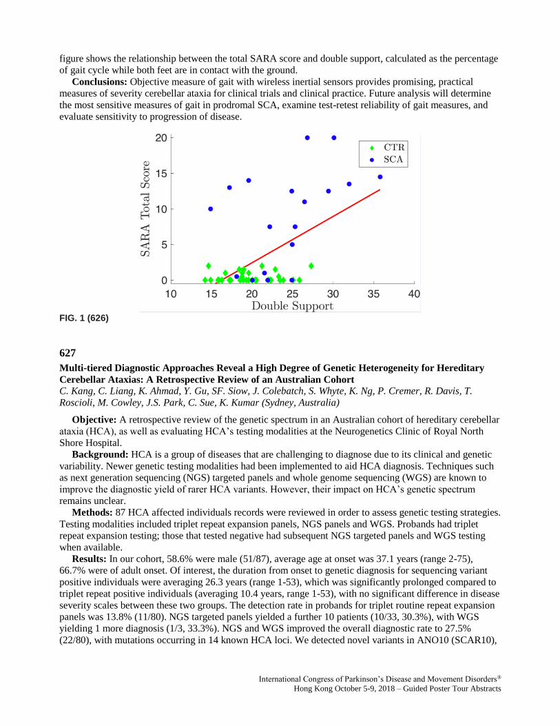
International Congress of Parkinson’s Disease and Movement Disorders®
Hong Kong October 5-9, 2018 – Guided Poster Tour Abstracts
figure shows the relationship between the total SARA score and double support, calculated as the percentage
of gait cycle while both feet are in contact with the ground.
Conclusions: Objective measure of gait with wireless inertial sensors provides promising, practical
measures of severity cerebellar ataxia for clinical trials and clinical practice. Future analysis will determine
the most sensitive measures of gait in prodromal SCA, examine test-retest reliability of gait measures, and
evaluate sensitivity to progression of disease.
FIG. 1 (626)
627
Multi-tiered Diagnostic Approaches Reveal a High Degree of Genetic Heterogeneity for Hereditary
Cerebellar Ataxias: A Retrospective Review of an Australian Cohort
C. Kang, C. Liang, K. Ahmad, Y. Gu, SF. Siow, J. Colebatch, S. Whyte, K. Ng, P. Cremer, R. Davis, T.
Roscioli, M. Cowley, J.S. Park, C. Sue, K. Kumar (Sydney, Australia)
Objective: A retrospective review of the genetic spectrum in an Australian cohort of hereditary cerebellar
ataxia (HCA), as well as evaluating HCA’s testing modalities at the Neurogenetics Clinic of Royal North
Shore Hospital.
Background: HCA is a group of diseases that are challenging to diagnose due to its clinical and genetic
variability. Newer genetic testing modalities had been implemented to aid HCA diagnosis. Techniques such
as next generation sequencing (NGS) targeted panels and whole genome sequencing (WGS) are known to
improve the diagnostic yield of rarer HCA variants. However, their impact on HCA’s genetic spectrum
remains unclear.
Methods: 87 HCA affected individuals records were reviewed in order to assess genetic testing strategies.
Testing modalities included triplet repeat expansion panels, NGS panels and WGS. Probands had triplet
repeat expansion testing; those that tested negative had subsequent NGS targeted panels and WGS testing
when available.
Results: In our cohort, 58.6% were male (51/87), average age at onset was 37.1 years (range 2-75),
66.7% were of adult onset. Of interest, the duration from onset to genetic diagnosis for sequencing variant
positive individuals were averaging 26.3 years (range 1-53), which was significantly prolonged compared to
triplet repeat positive individuals (averaging 10.4 years, range 1-53), with no significant difference in disease
severity scales between these two groups. The detection rate in probands for triplet routine repeat expansion
panels was 13.8% (11/80). NGS targeted panels yielded a further 10 patients (10/33, 30.3%), with WGS
yielding 1 more diagnosis (1/3, 33.3%). NGS and WGS improved the overall diagnostic rate to 27.5%
(22/80), with mutations occurring in 14 known HCA loci. We detected novel variants in ANO10 (SCAR10),

International Congress of Parkinson’s Disease and Movement Disorders®
Hong Kong October 5-9, 2018 – Guided Poster Tour Abstracts
CACNA1A (SCA6), and SPG7 (HSP7) via NGS panels. We also report identifying a novel variant in
PRKCG (SCA14) detected by WGS analysis.
Conclusions: Our findings highlight the genetic heterogeneity of HCAs in Australia and support the use
of subsequent NGS targeted panel for patients who were negative on repeat expansion testing. Furthermore,
our results suggest that individuals with sequencing variants have a less rapid disease progression, which
may be of prognostic value.
TABLE 1 (627) Overall demographics and clinical manifestations of patient with suspected HCA referred to Neurogenetic Clinic, RNSH.
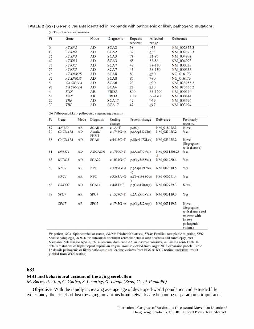
International Congress of Parkinson’s Disease and Movement Disorders®
Hong Kong October 5-9, 2018 – Guided Poster Tour Abstracts
TABLE 2 (627) Genetic variants identified in probands with pathogenic or likely pathogenic mutations.
633
MRI and behavioural account of the aging cerebellum
M. Bares, P. Filip, C. Gallea, S. Lehericy, O. Lungu (Brno, Czech Republic)
Objective: With the rapidly increasing average age of developed-world population and extended life
expectancy, the effects of healthy aging on various brain networks are becoming of paramount importance.
International Congress of Parkinson’s Disease and Movement Disorders®
Hong Kong October 5-9, 2018 – Guided Poster Tour Abstracts
Background: While the majority of current literature focuses on the supratentorial circuits, especially the
prefrontal cortex, the cerebellum has largely avoided attention despite important implications.
Methods: We included 67 healthy subjects of various ages ranging from 25 to 72 volunteers (36 women,
mean age 45.61; SD 14.63) to a fMRI study using a task heavily based on cerebellar functions – predictive
motor timing. The subjects were supposed to hit a target moving with various speeds with a projectile
released by button press. In addition to the behavioural assessment of the success rate, we performed
complex fMRI analysis, utilizing an advanced method of cerebellar processing of BOLD signal and
anatomical scans (voxel-based morphometry), allowing us to exclude the supratentorial structures to vastly
improve the co-registration and avoid cerebellar deformation.
Results: The increasing age did not affect the success rate at all. However, this stable behavioural
performance was associated with significant hyperactivation dominantly in the posterior lobes of the
cerebellum. The profile of atrophy was more pronounced in the posterior cerebellum (bilateral crus I, left
crus II, right lobule VI, and right lobule and vermis IX), the anterior cerebellum was affected only slightly
(left lobule V). No age-related GM volume increase was found. Moreover, the extent of cerebellar grey
matter volume decrease was far more significant than the general grey matter decrease in the supratentorial
area and the very parts showing hyperactivation in BOLD fMRI were affected by the age-related atrophy far
more than other cerebellar segments.
Conclusions: The cerebellum responds to the increasing age by hyperactivating relevant areas, thus
compensating for the age-related atrophy. Interestingly, despite the vast decrease of grey matter volume, it is
able to maintain stable performance even in high age.
References: Seidler RD, Bernard JA, Burutolu TB, Fling BW, Gordon MT, Gwin JT, et al. Motor control
and aging: links to age-related brain structural, functional, and biochemical effects. Neuroscience &
Biobehavioral Reviews. 2010;34(5):721-33. Filip P, Lungu OV, Bares M. Dystonia and the cerebellum: A
new field of interest in movement disorders? Clinical Neurophysiology. 2013 124(7):1269-76. Filip P, Gallea
C, Lehéricy S, Bertasi E, Popa T, Mareček R, et al. Disruption in cerebellar and basal ganglia networks
during a visuospatial task in cervical dystonia. Movement Disorders. 2017;32(5):757-68.
655
Transcriptional profiling of peripheral blood monocytes from child Friedreich’s ataxia patient: New
molecules and patterns of gene expression
H. Singh, V. Swarup, R. Singh, I. Singh, M. Faruq, S. Vivekanandhan, A. Srivastava (Delhi, India)
Objective: To explore peripheral biomarkers related to Friedreich's ataxia and identification of
pathophysiological insights of complex phenotype
Background: Friedreich's ataxia(FRDA) causes nervous system damage and movement problems which
starts in children leading to early death. Although reduction in frataxin level is responsible for FRDA, other
deregulated bio-markers may also contribute in disease phenotype mediated by cellular pathways.Therefore,
identification of such biomarkers can be helpful in order to understand disease etiology and development of
future medications. In the present study genome-wide expression analysis was performed in FRDA child
patients as compared to normal children.
Methods: Transcriptome expression profiling of FRDA and controls peripheral blood cells was
performed on a cohort of 28 patients and 10 controls that was extracted from GSE11204. To calculate
mRNA expression, the bioconductor R package “limma” was used. Significantly deregulated genes were
filtered using unpaired Student-t test and bonferrani test. The gene list associated with FRDA was extracted
from DisGeNET database (http://www.disgenet.org). The ontology analysis of significantly altered genes
was done using String Database (string-db.org).
Results: A total of 144 genes were found to be significantly altered (p<1.0E-06) in children manifest
FRDA phenotype (Figure 1). Out of 144 genes, 76 genes were found to be downregulated and 68 genes were
upregulated. It is also interesting to note that the identified genes were not reported earlier in FRDA provided
at DisGeNET database. Genes associated with metabolic processes such as cellular metabolism of the
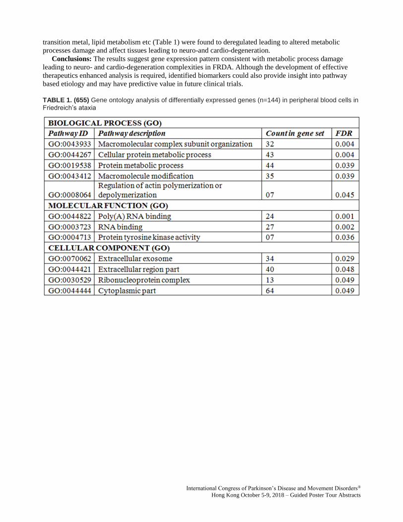
International Congress of Parkinson’s Disease and Movement Disorders®
Hong Kong October 5-9, 2018 – Guided Poster Tour Abstracts
transition metal, lipid metabolism etc (Table 1) were found to deregulated leading to altered metabolic
processes damage and affect tissues leading to neuro-and cardio-degeneration.
Conclusions: The results suggest gene expression pattern consistent with metabolic process damage
leading to neuro- and cardio-degeneration complexities in FRDA. Although the development of effective
therapeutics enhanced analysis is required, identified biomarkers could also provide insight into pathway
based etiology and may have predictive value in future clinical trials.
TABLE 1. (655) Gene ontology analysis of differentially expressed genes (n=144) in peripheral blood cells in Friedreich’s ataxia

International Congress of Parkinson’s Disease and Movement Disorders®
Hong Kong October 5-9, 2018 – Guided Poster Tour Abstracts
FIG. 1. (655) Heatmap of mRNA expression of significantly altered 144 genes in GSE11204 dataset Clustering the samples using -means revealed two groups, FRDA child patients (n=28) and normal children (n=10).
660
Comorbid Pediatric Early Onset Ataxia and Dystonia - Is the Cerebellum Involved?
D. Sival, M. Tijssen, D. Verbeek (Groningen, Netherlands)
Objective: In children with Early Onset Ataxia (EOA), we aimed to determine the prevalence of
comorbid dystonia and to explore the pathogenesis by the shared genetic background.
Background: Recent publications in patients with cervical dystonia have indicated cerebellar Purkinje
cell pathology. In pediatric EOA patients with developing cerebello-thalamo-basal ganglia motor networks,
we hypothesized that the association between ataxia and dystonia could be particularly strong due to
additional (mal)adaptive plasticity. In EOA children, however, the prevalence and etiology of comorbid
dystonia is still unknown. We reasoned that exploration of co-expression between genes involved in EOA,
AOA and dystonia disorders could provide pathophysiologic insight in shared biological pathways.
Methods: In a historic cohort of 36 well-phenotyped EOA children by 6 internationally recognized
movement disorder specialists (ref 1), we retrospectively determined the prevalence of comorbid dystonia.
As previously published, we determined gene co-expression by GeneNetwork* and PANTHER software (ref
2).

International Congress of Parkinson’s Disease and Movement Disorders®
Hong Kong October 5-9, 2018 – Guided Poster Tour Abstracts
Results: In pediatric EOA, the prevalence of comorbid dystonic features was 67% (24/36), detected by 1-
6 (mean 2) movement disorder experts. Network analysis of genes co-expressed with shared EOA and AOA
genes revealed a 9 fold overrepresentation of genes involved in GABA receptor activity and subsequent
network analysis of shared EOA, AOA and dystonia genes revealed an 11 fold overrepresentation of genes
involved in Tricarboxylic acid (TCA), necessary for mitochondrial ATP production and GABA synthesis
(the neurotransmitter of cerebellar Purkinje cells).
Conclusions: In pediatric EOA, the prevalence of comorbid dystonic features appears particularly high
(EOA 67% vs AOA 14-54%), potentially due to maladaptive plasticity. Our genetic data suggest that
hampered mitochondrial energy production and GABA synthesis may provide a shared disease mechanism
for comorbid dystonia with EOA and AOA. These results in EOA children could support a direct (metabolic)
and indirect (maladaptive plasticity) association between cerebellar Purkinje cell pathology and dystonia.
References: 1. Lawerman TL, Brandsma R, van Geffen JT, Lunsing RJ, Burger H, Tijssen MA, de Vries
JJ, de Koning TJ and Sival DA. Reliability of phenotypic early-onset ataxia assessment. DMCN
2016;58(1):70-6. 2. Nibbeling E, Delnooz CS, de Koning TJ, Sinke RJ, Jinnah HA, Tijssen MAJ, Verbeek
DS. Using the shared genetics of dystonia and ataxia to unravel their pathogenesis. Neurosci & Biobehav
Rev 2017; 75: 22-39. *http://129.125.135.180:8080/GeneNetwork/cytoscape.html.
663
The etiologies of chronic progressive cerebellar ataxia in a Korean population
J. Youn, M. Kim, JH. AHN, JW. Cho, JS. Kim (Seoul, Republic of Korea)
Objective: The etiologies and frequency of cerebellar ataxias vary among countries. Our primary aim was
to assess the frequency of each diagnostic group of cerebellar ataxia patients in a Korean population.
Background: The prevalence and frequency of cerebellar ataxia subtypes vary among countries.
Although estimation of subtype frequency is necessary for planning a diagnostic strategy in a specific
population, studies regarding epidemiology in all categories of cerebellar ataxias is surprisingly rare in
Korea. Our primary aim was to assess the frequency of each diagnostic group (familial and sporadic) in a
movement disorders out-patient clinic at a tertiary referral center.
Methods: We reviewed medical records of patients those who were in the process of follow-up during the
period from November 1994 to February 2016. We divided patients with cerebellar ataxias into familial and
non-familial groups and analyzed the frequency of each etiology. Finally, we categorized patients into
genetic, sporadic, secondary and suspected genetic, but undetermined ataxia.
Results: A total of 820 patients were included in the study, among whom 136(16.6%) familial patients
and 684(83.4%) non-familial cases were identified. Genetic diagnosis confirmed 98/136(72%) familial and
72/684(11%) non-familial patients. The overall etiologies of progressive ataxias consisted of 170(20.7%)
genetic, 516(62.9%) sporadic, 43(5.2%) secondary and 91(11.1%) undetermined ataxia. The most common
cause of ataxia was multiple system atrophy(57.3%). In the genetic group, the most common etiology was
spinocerebellar ataxia(152/170, 89.4%) and the most common subtype was spinocerebellar ataxia 3. 38 of
136 familial and 53 of 684 sporadic cases(91/820, 11.1%) were undetermined ataxia.
Conclusions: This is the largest epidemiological study to analyze the frequencies of various cerebellar
ataxias in a Korean population based on a large database of a tertiary hospital movement disorders clinic in
South Korea. These data would be helpful for clinicians in constructing diagnostic strategies and counseling
for patients with cerebellar ataxias.
References: 1. Gebus O, Montaut S, Monga B, Wirth T, Cheraud C, Alves Do Rego C, et al. Deciphering
the causes of sporadic late-onset cerebellar ataxias: a prospective study with implications for diagnostic
work. J Neurol 2017;264:1118-1126. 2. Hadjivassiliou M, Martindale J, Shanmugarajah P, Grünewald RA,
Sarrigiannis PG, Beauchamp N, et al. Causes of progressive cerebellar ataxia: prospective evaluation of 1500
patients. J Neurol Neurosurg Psychiatry 2017;88:301-309.
International Congress of Parkinson’s Disease and Movement Disorders®
Hong Kong October 5-9, 2018 – Guided Poster Tour Abstracts
691
DBS neuromodulation reduces severe dystonic pain in children and young people
S. Perides, J.P. Lin, G. Lee, H. Gimeno, R. Selway, K. Ashkan, M. Kaminska (London, United Kingdom)
Objective: This review analyses the prevalence of painful dystonia in a cohort of children undergoing
neuromodulation. The aim was to better understand the dystonic pain experience and evaluate improvements
one year following initiation of deep brain stimulation (DBS) therapy.
Background: Dystonic pain is reported as prevalent in genetic, acquired and idiopathic dystonic
conditions. However, it is yet to be systematically evaluated. There is an overall paucity of literature
evaluating the effect of DBS on dystonic pain using validated, reliable methods. The methods that have been
used in the literature are varied and at times inappropriate for the population being assessed.
Methods: Dystonic pain was assessed in a cohort of children (n=144) undergoing DBS. Assessment was
multi-modal, six different pain assessment methods were used: intensity (proxy- Paediatric Pain Profile- PPP
and self-report - Numerical Rating Scale - NRS); parental perception (CPCHILD questionnaire); pain
frequency, pain severity and analgesia use. SPSS version 21 was used to analyse both the whole cohort, and
separately, by etiological sub-classification; inherited DYT-positive dystonias (n=8), inherited
heredodegenerative dystonias (n=9), acquired dystonias (n=37) including Cerebral Palsy (n= 21) and
idiopathic dystonias (n= 8).
Results: We found that 44.5% (63/144) of this DBS cohort reported dystonic pain. Pain improved after
DBS surgery in each group. Clinically significant improvements P<0.001 were noted in the whole cohort,
using NRS (n=27), PPP (n=17/63) and the CPCHILD (n=48/63) assessments. Subjective reductions in
frequency and severity were also reported. Very severe pain fell in 9/28 (30%) cases. Constant pain fell from
27/63 to 11/63 cases, a 40.7% reduction, and 18/63 (28.6%) became pain-free. We found a 40% reduction in
children receiving daily medications and an increase of 46.1% not requiring analgesia. Whole cohort findings
were comparable to results in the etiological sub-classifications, except the heredodegenerative group, where
subjective improvements were noted only.
Conclusions: This is the first evaluation focusing on the impact of DBS surgery on dystonic pain in
children. The multi-assessment approach in pain research reduces the risk of bias in an otherwise complex
population, both to assess and to treat. Longitudinal data collection and the consideration of a multi-center
research project would improve validity and reliability further. A reduction in dystonic pain should remain a
goal for DBS surgery, particularly given the relationship between pain and poor quality of life, social
isolation, self-perception and overall poor health status.
696
The clinical value of SPECT in identifying dystonic muscles of patients with cervical dystonia
L. Jin, L. Feng, I. Djibo, S. Chen, F. Teng, B. Li, H. Ma (Shanghai, China)
Objective: The aim of this study is to compare the efficacy of selecting target muscles for botulinum
toxin injection by abnormal movement pattern and by SPECT combined with abnormal movement pattern.
Background: Cervical dystonia (CD) is caused by involuntary and excessive contraction of cervical
muscles which leads to abnormal movement and posture. Botulinum toxin injection has become the first line
treatment for CD, the critical step which decides the treatment efficacy is to accurately identify dystonic
muscles. At present, dystonic muscles are selected by patients’ abnormal movement pattern. However, most
patients have a combination of various patterns, and the same pattern may be caused by contraction of
Guided Poster Group 5:
Dystonia
International Congress of Parkinson’s Disease and Movement Disorders®
Hong Kong October 5-9, 2018 – Guided Poster Tour Abstracts
different muscles. SPECT imaging uses 99mTc-MIBI as a contrast agent, which can reveal abnormal
contraction of the muscle. However, SPECT has not been reported to guide the treatment of CD.
Methods: Forty patients with idiopathic CD were enrolled. These patients had not received botulinum
toxin injection for last 3 months.The first group: dystonic muscles were screened based on cervical dystonia
pattern (n1=20). The second group: we analyzed cervical muscles with increased uptake of 99mTc-MIBI in
cervical SPECT, the SPECT results combined with cervical dystonia pattern were used to determine dystonic
muscles (n2=20). SPSS20.0 software was used to compare the results between the two groups after
botulinum toxin injection. The TWSTRS score reduction rate and Tsui score reduction rate at the second
week, the first month, the third month and the sixth month were compared.
Results: The number of botulinum toxin reinjection in 6 months was significantly higher in the first group
than in the second group (13: 4). The interval between the first and the second injection of the second group
was significantly longer than the first group (p < 0.05). There was no significant difference between
TWSTRS score reduction rate and Tsui score reduction rate in the second week and the first month after
treatment, p> 0.05. At the third month, TWSTRS score reduction rate and Tsui score reduction rate of the
second group was significantly higher than the first group, p <0.05. At the 6th month, although the number of
follow-up in was decreased in both groups because of repeat injection (n1 = 5: n2 = 14), the TWSTRS
reduction rate and the Tsui reduction rate in the second group were significantly higher than the first group ,
P <0.05.
Conclusions: SPECT imaging can more accurately select the dystonic muscles and thus greatly improve
the efficiency and remission rate of botulinum toxin treatment.
713
Characterizing Bulbar Dysfunction in X-Linked Dystonia-Parkinsonism (XDP): A Pilot Study
J. de Guzman, B. Perry, C. Go, J. Green, N. Sharma (Boston, MA, USA)
Objective: This study aims to characterize bulbar motor impairments in patients with X-linked XDP and
to identify clinical correlates for prediction of aspiration.
Background: X-linked dystonia-parkinsonism (XDP) is an inherited neurodegenerative disease endemic
in Panay Island, Philippines that predominantly affects males. Initial stages are characterized by focal
dystonia, which then generalizes. Seven to ten years after onset, parkinsonian symptoms emerge alongside
symptoms of dystonia. Around 15 years post onset, parkinsonism predominates. In approximately 28% of
cases, dystonic symptoms initially manifest in the oromandibular and cervical area causing significant bulbar
impairment. Over the course of the disease, most persons with XDP develop bulbar dysfunction resulting in
speech and swallowing impairments. The characteristics and severity of bulbar impairment in XDP is not
well understood.
Methods: Twenty-five XDP patients underwent assessments of respiratory function, speech, lip and
tongue strength, diadochokinetic rates, articulation during connected speech, and swallowing function.
Patient-reported measures of speech and swallowing impairment included the Communicative Participation
Item Bank and the Eating Assessment Tool – 10.
Results: All participants exhibited bulbar impairment. Sixteen subjects had evidence of aspiration. The
average lip and tongue strength were both reduced with respective mean values of 9.48 ± 4.03 kPa (NV= 35
kPa) and 19 ± 11.29 kPa (NV = 63 kPa). The average pause percentage for speech sample was 34.18%.
The mean scores for the following questionnaires were all abnormal: EAT-10 score of 19.28 ± 10.15 (score
>/= 3 is abnormal), FOIS score of 4.88 ± 0.97 (score < 7 is abnormal), and CPIB score of 7.32 ±. 7.72
(maximum score of 30 and higher scores are more favorable). Logistic regression analysis showed that the
duration of illness (p = 0.463), site of onset (p = 0.553), and phase of illness (dystonic p = 0.977,
parkinsonian p = 0.829, dystonia-parkinsonian p = 0.999) did not contribute significantly to the prediction
model of aspiration. The resulting model was not statistically significant, X2(4) = 4.71, p = 0.316.
Conclusions: Bulbar dysfunction in XDP results in speech and swallowing impairments characterized by
decreased phonation time, decreased lip and tongue strength, decreased diadochokinetic rates, evidence of
aspiration, and patient-reported speech and swallowing deficits.
International Congress of Parkinson’s Disease and Movement Disorders®
Hong Kong October 5-9, 2018 – Guided Poster Tour Abstracts
References: Kreisler, A, Verpraet, AC, Veit, S, Pennel-Ployart, O, Behal, H, Duhamel, A, Destee, A.
Clinical characteristics of voice, speech, and swallowing disorders in oromandibular dystonia. Journal of
Speech, Language, and Hearing Research, Vol. 59, October 2016, pp. 940-949. LaPointe, LL, Murdoch, BE.
Movement disorders in neurologic disease: effects on communication and swallowing. Plural Publishing,
Inc., 2014, pp 123-159.
727
The association of primary dystonia with tics - chance or new syndrome?
C. Del Gamba, A. Latorre, U. Bonuccelli, R. Ceravolo, K. Bhatia (London, United Kingdom)
Objective: This study aimed to evaluate the association between dystonia and tics as a primary clinical
entity.
Background: Primary cranio-cervical dystonia (PCCD) is an idiopathic condition, which typically occurs
in late adulthood and in women more than men. Primary tics (PT) generally start during
childhood/adolescence, and a later age of onset or other associated movement disorders are “red flags” to
suspect secondary causes. Primary dystonia and PT are distinct entities, but nevertheless, a clinical syndrome
with these two features has been described. We report a series of patients presenting both PCCD and PT.
Methods: 248 patients with PCCD attending our Botulinum toxin clinic at the National Hospital of
Neurology and Neurosurgery - Queen Square, were examined for PT over a period of 4 months. Secondary
tic causes were excluded by clinical interview and appropriate investigations (brain MRI, extensive blood
tests, acanthocytes, genetics).
Results: We have found 16 patients (6,5%) with PCCD, also presenting PT. Thirteen patients are males
while three are females. In eleven dystonia started below the age of 40. Eleven had a focal involvement,
while 5 a segmental. Dystonia was found to affect neck (13), vocal cords (4), jaw (1) eyes (1) and arms (4).
Six patients displayed a sensory trick. The association with tics can be stratified as follows: 3/16 patients
presented tics before 21 years old, fitting Tourette’s syndrome (GTS) criteria (also presenting >1 motor tic
and ≥1 vocal tic); for the other patients, we were not able to recall the onset, which was therefore more likely
to fall into the Adult Primary Tics (APT) category. Most of our patients also showed psychiatric issues
(hyperactivity, anxiety, depression or obsessive-compulsive features), which were generally mild.
Conclusions: We described a primary clinical entity of PCCD associated with PT. This syndrome differs
from pure cranio-cervical dystonia by a higher prevalence in males and a lower age of onset. It presents a tic
disorder, which, in a minority of cases resembles GTS, whereas more frequently falls into APT category.
References: Damasio J, Edwards MJ, Alonso-Canovas A, Schwingenschuh P, Kagi G and Bhatia KP.
The Clinical Syndrome of Primary Tic Disorder Associated with Dystonia: A Large Clinical Series and a
Review of the Literature, Movement Disorders, Vol. 26, No. 4, 2011. Stone LA and Jankovic J. The
coexistence of tics and dystonia. Arch Neurol 1991;48:862–865.
734
Zolpidem effect in task specific dystonia – clinical and neurophysiological study
K. Vogelnik, M. Grmek, R. Perellon Alfonso, P. Tomše, M. Trošt, M. Kojović (Ljubljana, Slovenia)
Objective: We conducted a double blind, placebo-controlled crossover study to examine clinical and
neurophysiological effect of zolpidem in patients with task specific dystonia.
Background: Task specific dystonia is disabling movement disorder that includes writer’s cramp and
musician dystonia. It causes impaired hand use and may lead to the termination of professional career.
Currently, there is no effective treatment. Zolpidem, a short-acting hypnotic drug that binds to GABA –A
receptor benzodiazepine site, has been reported to transiently improve various movement disorders,
including a proportion of patients with primary focal and generalized dystonia. The mechanisms underlying
its therapeutic effects has not been investigated.
Methods: Six patients with writer’s cramp and one guitarist with musician dystonia underwent
transcranial magnetic stimulation (TMS) and 18F-FDG-PET brain imaging after single 5 mg dose of
International Congress of Parkinson’s Disease and Movement Disorders®
Hong Kong October 5-9, 2018 – Guided Poster Tour Abstracts
zolpidem and placebo, in four separate sessions. We measured resting motor threshold (RMT), active motor
threshold (AMT), resting and active input/output (IO) curve, short interval intracortical inhibition (SICI)
curve, long interval intracortical inhibition (LICI), intracortical facilitation (ICF) and cortical silent period
(CSP). Clinical improvement was rated using writer’s cramp rating scale (WCRS). Clinical and TMS
measures were compared using paired sample t-test or repeated measures ANOVA; correlations were tested
using Spearman analysis. Statistical parametric mapping was used to identify zolpidem effect on global
brain metabolism.
Results: There was significant improvement on WRCS on zolpidem. Zolpidem reduced the steepness of
active IO curve, while there was no difference in AMT, RMT, resting IO curve, SICI, LICI, CSP and ICF on
the group level. Significant positive correlation was found between clinical improvement and enhancement
of LICI on zolpidem. 18F-FDG-PET revealed that zolpidem treatment was associated with hypometabolism
in primary sensori-motor cortex, cerebellum and medial temporal lobes and hypermetabolism in caput nuclei
caudate, parietal cortex and frontal regions.
Conclusions: Clinical effect of zolpidem in task specific dystonia was associated with evident changes in
TMS measures of corticospinal excitability and intracortical inhibition and changes in brain metabolism on
brain 18F-FGD-PET. Zolpidem may be an effective treatment in a proportion of patients with task specific
dystonia.
References: 1. Lin, P. T. & Hallett, M. The pathophysiology of focal hand dystonia. J. Hand Ther. Off. J.
Am. Soc. Hand Ther. 22, 109–114 (2009). 2. Konczak, J. & Abbruzzese, G. Focal dystonia in musicians:
linking motor symptoms to somatosensory dysfunction. Front. Hum. Neurosci. 7, (2013). 3. Depoortere, H.
et al. Zolpidem, a novel nonbenzodiazepine hypnotic. I. Neuropharmacological and behavioral effects. J.
Pharmacol. Exp. Ther. 237, 649–658 (1986). 4. Abe, K. Zolpidem therapy for movement disorders. Recent
Patents CNS Drug Discov. 3, 55–60 (2008). 5. Miyazaki, Y. et al. Efficacy of Zolpidem for Dystonia: A
Study Among Different Subtypes. Front. Neurol. 3, (2012). 6. Martinez-Ramirez, D., Paz-Gomez, V. &
Rodriguez, R. L. Response to zolpidem in oromandibular dystonia: a case report. Parkinsonism Relat.
Disord. 21, 154–155 (2015). 7. Sunwoo, M. et al. A Case with Improvement of Blepharospasm by Zolpidem.
J. Mov. Disord. 4, 53–54 (2011).
746
A Registry of Real-World Outcomes Using Deep Brain Stimulation for the Treatment of Dystonia
J. Krauss, C. Nicholson, M. Barbe, V. Visser-Vanderwalle, A. Kuehn, M. Poetter-Nerger, R. Jain, H.
Scholtes, N. Van Dyck, A. Albanese (Hannover, Germany)
Objective: The objective of this device registry entails collecting clinical outcomes, economic value and
technical performance of a Deep Brain Stimulation (DBS) system capable of multiple independent current
control (MICC) for use in the treatment of dystonia.
Background: Several studies have now published clinical outcomes using DBS for the treatment of
dystonia encompassing a range of dystonic conditions including primary generalized, cervical dystonia,
tardive dystonia, and other types of secondary dystonia, and all have reported effective results with use of
DBS for the treatment of dystonia. Here we report the initial outcomes from a multi-center registry of
dystonia patients implanted with an MICC-based DBS system.
Methods: This is a prospective, on-label, multi-center, international registry study consisting of up to 200
patients implanted with a DBS system (Vercise, Boston Scientific) for use in the treatment of dystonia
followed out to 3 years (post-implant) at up to 40 sites in Europe. Study assessments conducted will be based
on dystonia sub-group, classification, and age and include (but not limited) to the following: Burke-Fahn-
Marsden Dystonia Rating Scale, Clinical Global Impression of Change, Global Dystonia Scale, SF-36v2 or
SF-10v2 Health Survey, and Toronto Western Spasmodic Torticollis Rating Scale.
Results: Initial results of this on-going registry of DBS outcomes in dystonia patients will be reported.
Conclusions: Large patient data registries may facilitate insights regarding real-world, clinical use of
DBS. This registry represents the first comprehensive, large scale collection of outcomes associated with
International Congress of Parkinson’s Disease and Movement Disorders®
Hong Kong October 5-9, 2018 – Guided Poster Tour Abstracts
dystonia patients implanted with a DBS system capable of multiple independent current control (MICC) and
will include assessment of economic value and device technical performance.
761
Comorbidity and retirement in primary focal cervical dystonia
R. Ortiz, F. Scheperjans, T. Mertsalmi, E. Pekkonen (Helsinki, Finland)
Objective: The objective of this study was to investigate comorbidities in cervical dystonia (CD) and effect
of CD to retirement rate in Finland.
Background: Cervical dystonia is most common form of dystonia, the prevalence being 394 persons per
million in Finland. The onset of CD is usually before 60 years of age, and even though CD does not reduce
life expectancy, it may cause severe functional and psychosocial impairment in everyday life. Besides the
motoric symptoms, recently non-motoric symptoms, especially psychiatric comorbidities, have been reported
to occur in CD affecting substantially quality of life.
Methods: We studied the comorbidity in primary focal CD in Finland based on ICD-10 codes obtained from
care registry and patient records of 937 confirmed adult primary focal CD patients between years 2007-2016.
The retirement months and diagnosis of retirement were calculated from pension registry information. The
results were compared to 3746 age and gender matched controls.
Results: Most prominent comorbidities with primary focal CD were depression (14%), anxiety (7%), and
dorsalgia (11%)[table]. There was significantly more of retirement months before age of 65 years in CD
group than in control (32,8 ± 67,2 vs.13,3 ± 47,1 months, p<10-5) [figure]. Also, the retirement age was
significantly younger in CD patients compared to control group controls (56,1 ± 8,3 vs. 59,8 ± 6,7 years,
p<10-5). Of the dystonia patients, who were retired because of sickness, the most common retirement
diagnoses were dystonia (51%), depression (14%) and anxiety (8%). Patients with anxiety and depression
had more of retirement months than other dystonia patients.
Conclusions: Cervical dystonia reduces considerably working ability and leads to earlier retirement. Anxiety
and depression are most notable comorbidities, and co-occurrence of them further reduces more working
ability. Spinal degenerative diseases were also common with CD, but they did not affect retirement rate
significantly compared with control group. Our results suggest that more health care resources should be
administered in treatment of CD to longer maintain working ability of CD patients. Further, psychiatric
comorbidities should be taken into consideration in CD treatment.

International Congress of Parkinson’s Disease and Movement Disorders®
Hong Kong October 5-9, 2018 – Guided Poster Tour Abstracts
TABLE 1 (761) Comorbidities with cervical dystonia patients
FIG. 1 (761)
International Congress of Parkinson’s Disease and Movement Disorders®
Hong Kong October 5-9, 2018 – Guided Poster Tour Abstracts
762
Frequency and Phenoptypic Spectrum of KMT2B Mutations in Childhood-Onset Dystonia: Results
from a Single-Centre Cohort Study
M. Carecchio, G. Zorzi, F. Invernizzi, C. Panteghini, L. Romito, F. Zibordi, V. Leuzzi, S. Galosi, P. Morana,
B. Morana, C. Piano, A. Bentivoglio, C. Reale, F. Girotti, M. Topf, A. Joseph, M. Kurian, S. Lubbe, B.
Garavaglia, N. Mencacci, N. Nardocci (Milan, Italy)
Objective: To assess the frequency of KMT2B mutations in a cohort of patients with childhood-onset
dystonia and characterize the related molecular and phenotypic spectrum.
Background: Heterozygous mutations in KMT2B, encoding a histone lysine methyltransferase, were
recently reported as a cause of childhood-onset generalized dystonia variably associated with additional
neurological and systemic features.
Methods: 55 patients (23F,22M) with genetically undefined childhood-onset dystonia were screened by
means of Whole Exome Sequencing or customized gene panels including known dystonia-associated genes.
KMT2B variants were confirmed by Sanger sequencing and further characterized by segregation analysis,
bioinformatics tools and in silico homology modelling in some cases.
Results: 12/55 (21.8%) patients were found to carry KMT2B variants (8 missense, 3 frameshift, 1 in-
frame deletion) [table 1] [figure 1], of which 5 were confirmed to be de novo, 4 were inherited from one
unaffected parent and 3 were likely de novo segregating with disease status in available family members
[figure 2]. All but one variant had not been reported previously. Mean age at onset was 6.5 years (range 3.5-
14); mean age at last examination was 28 years (range 17-45) and mean disease duration was 22.5 years
(range 2-40). 10/12 (83%) patients had lower limb presentation, 11/12 (92%) progressed to generalization
with laryngeal dystonia observed in 10/12 (83%). Mild intellectual disability, short stature, minor facial
dysmorphisms were recurrently observed [table 2]. Brain MRI was normal in all cases, including DWI/SWI
sequences. 7/12 patients underwent bilateral pallidal DBS with excellent, long-lasting motor improvement
(mean post-operative follow-up: 12 years). Additional genetic findings included a novel, de novo GNAO1
missense mutation (p.Cys215Tyr) identified in a 42-year-old male with a 30-year history of myoclonus
dystonia and biallelic ATM mutations (p.Cys907*/p.Glu2052Lys) in a 40-year-old female with isolated
generalized dystonia presenting at age 13.
Conclusions: Mutations in KMT2B are a relevant cause of dystonia in the paediatric population, with
autosomal dominant inheritance and possible incomplete penetrance in some cases. Red flags include onset
in the lower limbs, caudo-cranial spreading, oro-mandibular and laryngeal involvement, short stature and
mild intellectual disability. Brain MRI in our cases did not confirm previously reported basal ganglia
alterations. Childhood-onset dystonia can rarely be an atypical presentation of other genetic diseases.
References: Meyer E, Carss KJ, Rankin J, et al. Mutations in the histone methyltransferase gene KMT2B
cause complex early-onset dystonia. Nat Genet 2017;49:223-237. Zech M, Boesch S, Maier EM, et al.
Haploinsufficiency of KMT2B, encoding the lysine-specific histone methyltransferase 2b, results in early-
onset generalized dystonia. Am J Hum Genet 2016;99:1377-1387.

International Congress of Parkinson’s Disease and Movement Disorders®
Hong Kong October 5-9, 2018 – Guided Poster Tour Abstracts
TABLE 1 (762) KMT2B variants identified in the study; * based on NM_014727
FIG. 1 (762) Position of KMT2B variants individuated in the study along the protein structure.
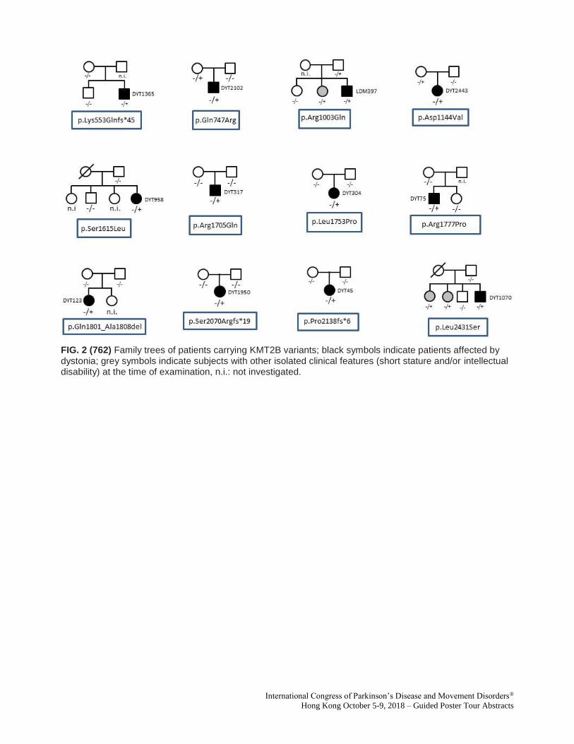
International Congress of Parkinson’s Disease and Movement Disorders®
Hong Kong October 5-9, 2018 – Guided Poster Tour Abstracts
FIG. 2 (762) Family trees of patients carrying KMT2B variants; black symbols indicate patients affected by dystonia; grey symbols indicate subjects with other isolated clinical features (short stature and/or intellectual disability) at the time of examination, n.i.: not investigated.

International Congress of Parkinson’s Disease and Movement Disorders®
Hong Kong October 5-9, 2018 – Guided Poster Tour Abstracts
TABLE 2. (762) clinical features of KMT23 variant carriers. AAO: Age at onset; LL: lower limbs; OM: oro-mandibular dystonia; LAR: laryngeal dystonia; CRAIM; cranial dystonia; NA: not available; ID: intellectual disability; TBZ: tetrabenazine; LD: Levodopa; * following post-surgical lenticular haemorrhage
769
A machine learning approach to determine the important patient characteristics for tremor
prevalence and tremor irregularity in dystonia
S. Balta Beylergil, L. Scorr, A. Cotton, H. Jinnah, A. Shaikh (Cleveland, OH, USA)
Objective: To evaluate the importance of dystonia features that can predict concurrent tremor prevalence
and tremor irregularity. (2) To cluster dystonia cases based on important data-driven features using a large,
multi-institutional cohort of 2362 patients, and adopting state of the art feature selection and clustering
methods of machine learning.
Background: Dystonia is often accompanied by regular or irregular tremor. The dystonia features that
increase the likelihood of concurrent tremor are unclear. Dystonia traits associated with irregular tremor are
also not well defined.
Methods: We used a permutation-based feature selection algorithm to evaluate various dystonia attributes
and select the relevant ones to be used in predicting tremor prevalence and irregularity. We performed
clustering analyses to group dystonia patients into clusters with similar characteristics using an
agglomerative hierarchical clustering algorithm.
Results: The first feature selection analysis indicated that body part affected by dystonia provides the
most useful information for predicting tremor prevalence. Duration of dystonia, total Global Dystonia Rating
Scale score, and age at dystonia onset also play a significant role in determining whether dystonia and tremor
coexist. With these parameters, a random forest classifier (RFC) was able to classify a test data set with 69%
accuracy. The clustering analysis yielded 4 distinct clusters with 16%, 31%, 62% and 67% tremor prevalence
International Congress of Parkinson’s Disease and Movement Disorders®
Hong Kong October 5-9, 2018 – Guided Poster Tour Abstracts
rates. The second feature selection analysis showed that tremor irregularity is sensitive to the extent to which
dystonia and tremor locations overlap. Investigator site is also an important feature that discriminates
between regular and irregular tremor. RFC was able to predict irregularity with 79% test accuracy, and
clustering analysis formed 4 distinct clusters with 28%, 76%, 79%, and 84% irregular tremor rates.
Handedness, gender, and race were found unimportant for predicting tremor prevalence and irregularity in
dystonia.
Conclusions: We identified the most relevant dystonia traits for predicting concurrent tremor prevalence
and irregularity using modern machine learning methods. Our results also exemplify the benefit of these
methods in understanding the relationship between subtypes of heterogeneous movement disorders.
776
Cohort profile of the Japan Dystonia Consortium: Genetic diagnosis and characteristics of movement
disorders in Japan
T. Kawarai, R. Miyamoto, A. Orlacchio, R. Kaji (Tokushima, Japan)
Objective: To reveal molecular epidemiology of hereditary dystonia through resequencing of the
currently-known dystonia genes and identification of novel genetic defects.
Background: The Japan Dystonia Consortium was established to construct a nationwide clinical and
DNA database on dystonia patients, including genotype-phenotype-outcome correlations and natural course,
which would contribute to elucidation of dystonia pathomechanism and refinement of guideline for dystonia.
Methods: A total of 587 patients with dystonia or other movement disorders were recruited. Patients were
videotaped, which were phenomenologically examined by movement disorder specialists with long-standing
experience. After phenomenological evaluation, candidate genes are chosen for sequence analysis. Direct
sequencing of polymerase chain reaction products with the Sanger-based method or whole-exome
sequencing was applied for genetic screening in the currently-known dystonia genes. Furthermore, whole
exome trio sequencing was conducted in order to identify new genetic defect(s) in dystonia.
Results: Mutations in DYT genes have been revealed in familial and seemingly sporadic cases with
dystonia, including DYT-TOR1A, DYT-THAP1, DYT-GNAL, DYT/PARK-GCH1, DYT/PARK-TH,
DYT/PARK-ATP1A3, DYT/PARK-TAF1, and DYT-SGCE. In addition, we identified mutations in other
dystonia or dyskinesia-associated genes, including PRRT2, MR-1, TUBB4A, KMT2B and ADCY5.
Genotype –phenotype correlations in most cases were consistent with those previously reported.
Conclusions: The cohort contains enough sample power to detect novel genes for dystonia. Correct
phenomenological evaluation is indispensable for variant interpretation and establishment of phenotype-
genotype correlations.
681
Chorea and Ataxia as Manifestations of Xeroderma Pigmentosum: A Case Report
A. Jocson, K. Ngo, D. Togasaki, B. Fogel (Los Angeles, CA, USA)
Objective: To report a case of a 51-year-old woman with recurrent basal cell carcinoma, severe
photosensitivity, and progressive chorea and ataxia caused by xeroderma pigmentosum (XP).
Background: XP is an autosomal recessive (AR) disorder associated with photosensitivity and skin
malignancies. Severe cases may present with mental retardation, dwarfism, and hypogonadism. XP-A to G
are due to mutations in nucleotide excision repair (NER) of ultraviolet (UV)-induced DNA lesions.
Neurologic signs are found in 20% of cases. Presentations include chorea, ataxia, and peripheral neuropathy.
Chorea is rare in AR ataxias, except for ataxia telangiectasia and AOA1/2; thus, XP should be on the
Guided Poster Group 6:
Huntington’s Disease
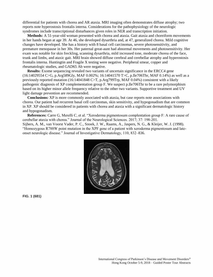
International Congress of Parkinson’s Disease and Movement Disorders®
Hong Kong October 5-9, 2018 – Guided Poster Tour Abstracts
differential for patients with chorea and AR ataxia. MRI imaging often demonstrates diffuse atrophy; two
reports note hyperostosis frontalis interna. Considerations for the pathophysiology of the neurologic
syndromes include transcriptional disturbances given roles in NER and transcription initiation.
Methods: A 51-year-old woman presented with chorea and ataxia. Gait ataxia and choreiform movements
in her hands began at age 39. At 46, she developed dysarthria and, at 47, generalized chorea. Mild cognitive
changes have developed. She has a history with 8 basal cell carcinomas, severe photosensitivity, and
premature menopause in her 30s. Her paternal great-aunt had abnormal movements and photosensitivity. Her
exam was notable for skin freckling, scanning dysarthria, mild increased tone, moderate chorea of the face,
trunk and limbs, and ataxic gait. MRI brain showed diffuse cerebral and cerebellar atrophy and hyperostosis
frontalis interna. Huntingtin and Fragile X testing were negative. Peripheral smear, copper and
rheumatologic studies, and GAD65 Ab were negative.
Results: Exome sequencing revealed two variants of uncertain significance in the ERCC4 gene
(16:14029554 C>G, p.Arg589Gly, MAF 0.002%; 16:14041570 T>C, p.Ile706Thr, MAF 0.14%) as well as a
previously reported mutation (16:14041848 C>T, p.Arg799Trp, MAF 0.04%) consistent with a likely
pathogenic diagnosis of XP complementation group F. We suspect p.Ile706Thr to be a rare polymorphism
based on its higher minor allele frequency relative to the other two variants. Supportive treatment and UV
light damage prevention are recommended.
Conclusions: XP is more commonly associated with ataxia, but case reports note associations with
chorea. Our patient had recurrent basal cell carcinomas, skin sensitivity, and hypogonadism that are common
in XP. XP should be considered in patients with chorea and ataxia with a significant dermatologic history
and hypogonadism.
References: Carre G, Merelli C, et al. “Xeroderma pigmentosum complentation group F: A rare cause of
cerebellar ataxia with chorea.” Journal of the Neurological Sciences. 2017; 37: 198-201.
Sijbers, A. M., van Voorst Vader, P. C., Snoek, J. W., Raams, A., Jaspers, N. G., & Kleijer, W. J. (1998).
“Homozygous R788W point mutation in the XPF gene of a patient with xeroderma pigmentosum and late-
onset neurologic disease.” Journal of Investigative Dermatology, 110, 832–836.
FIG. 1 (681)
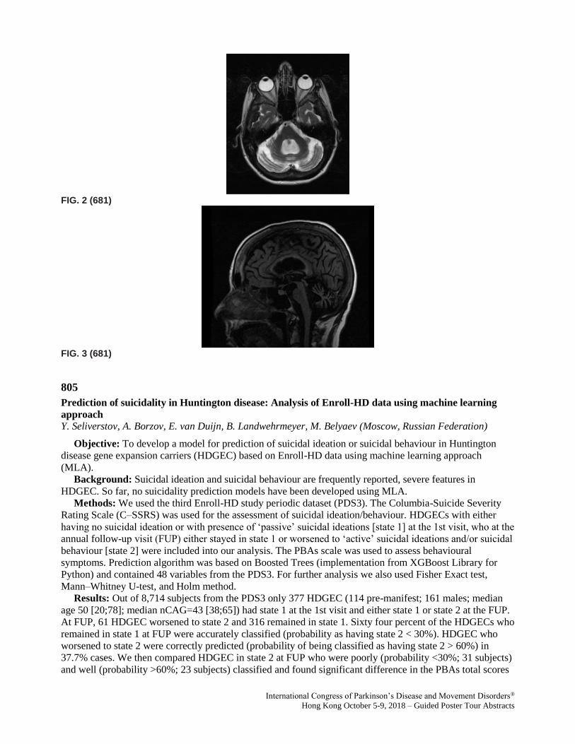
International Congress of Parkinson’s Disease and Movement Disorders®
Hong Kong October 5-9, 2018 – Guided Poster Tour Abstracts
FIG. 2 (681)
FIG. 3 (681)
805
Prediction of suicidality in Huntington disease: Analysis of Enroll-HD data using machine learning
approach
Y. Seliverstov, A. Borzov, E. van Duijn, B. Landwehrmeyer, M. Belyaev (Moscow, Russian Federation)
Objective: To develop a model for prediction of suicidal ideation or suicidal behaviour in Huntington
disease gene expansion carriers (HDGEC) based on Enroll-HD data using machine learning approach
(MLA).
Background: Suicidal ideation and suicidal behaviour are frequently reported, severe features in
HDGEC. So far, no suicidality prediction models have been developed using MLA.
Methods: We used the third Enroll-HD study periodic dataset (PDS3). The Columbia-Suicide Severity
Rating Scale (C–SSRS) was used for the assessment of suicidal ideation/behaviour. HDGECs with either
having no suicidal ideation or with presence of ‘passive’ suicidal ideations [state 1] at the 1st visit, who at the
annual follow-up visit (FUP) either stayed in state 1 or worsened to ‘active’ suicidal ideations and/or suicidal
behaviour [state 2] were included into our analysis. The PBAs scale was used to assess behavioural
symptoms. Prediction algorithm was based on Boosted Trees (implementation from XGBoost Library for
Python) and contained 48 variables from the PDS3. For further analysis we also used Fisher Exact test,
Mann–Whitney U-test, and Holm method.
Results: Out of 8,714 subjects from the PDS3 only 377 HDGEC (114 pre-manifest; 161 males; median
age 50 [20;78]; median nCAG=43 [38;65]) had state 1 at the 1st visit and either state 1 or state 2 at the FUP.
At FUP, 61 HDGEC worsened to state 2 and 316 remained in state 1. Sixty four percent of the HDGECs who
remained in state 1 at FUP were accurately classified (probability as having state 2 < 30%). HDGEC who
worsened to state 2 were correctly predicted (probability of being classified as having state 2 > 60%) in
37.7% cases. We then compared HDGEC in state 2 at FUP who were poorly (probability <30%; 31 subjects)
and well (probability >60%; 23 subjects) classified and found significant difference in the PBAs total scores
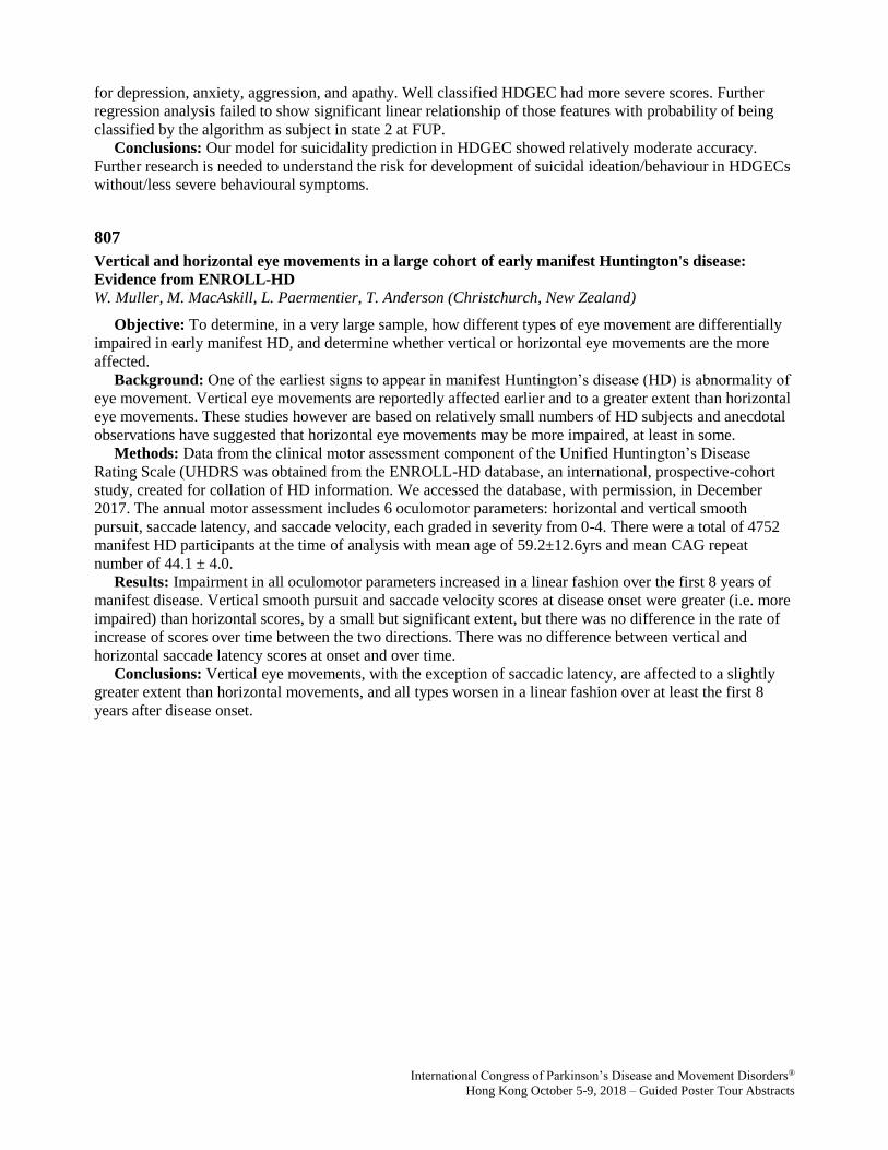
International Congress of Parkinson’s Disease and Movement Disorders®
Hong Kong October 5-9, 2018 – Guided Poster Tour Abstracts
for depression, anxiety, aggression, and apathy. Well classified HDGEC had more severe scores. Further
regression analysis failed to show significant linear relationship of those features with probability of being
classified by the algorithm as subject in state 2 at FUP.
Conclusions: Our model for suicidality prediction in HDGEC showed relatively moderate accuracy.
Further research is needed to understand the risk for development of suicidal ideation/behaviour in HDGECs
without/less severe behavioural symptoms.
807
Vertical and horizontal eye movements in a large cohort of early manifest Huntington's disease:
Evidence from ENROLL-HD
W. Muller, M. MacAskill, L. Paermentier, T. Anderson (Christchurch, New Zealand)
Objective: To determine, in a very large sample, how different types of eye movement are differentially
impaired in early manifest HD, and determine whether vertical or horizontal eye movements are the more
affected.
Background: One of the earliest signs to appear in manifest Huntington’s disease (HD) is abnormality of
eye movement. Vertical eye movements are reportedly affected earlier and to a greater extent than horizontal
eye movements. These studies however are based on relatively small numbers of HD subjects and anecdotal
observations have suggested that horizontal eye movements may be more impaired, at least in some.
Methods: Data from the clinical motor assessment component of the Unified Huntington’s Disease
Rating Scale (UHDRS was obtained from the ENROLL-HD database, an international, prospective-cohort
study, created for collation of HD information. We accessed the database, with permission, in December
2017. The annual motor assessment includes 6 oculomotor parameters: horizontal and vertical smooth
pursuit, saccade latency, and saccade velocity, each graded in severity from 0-4. There were a total of 4752
manifest HD participants at the time of analysis with mean age of 59.2±12.6yrs and mean CAG repeat
number of 44.1 ± 4.0.
Results: Impairment in all oculomotor parameters increased in a linear fashion over the first 8 years of
manifest disease. Vertical smooth pursuit and saccade velocity scores at disease onset were greater (i.e. more
impaired) than horizontal scores, by a small but significant extent, but there was no difference in the rate of
increase of scores over time between the two directions. There was no difference between vertical and
horizontal saccade latency scores at onset and over time.
Conclusions: Vertical eye movements, with the exception of saccadic latency, are affected to a slightly
greater extent than horizontal movements, and all types worsen in a linear fashion over at least the first 8
years after disease onset.
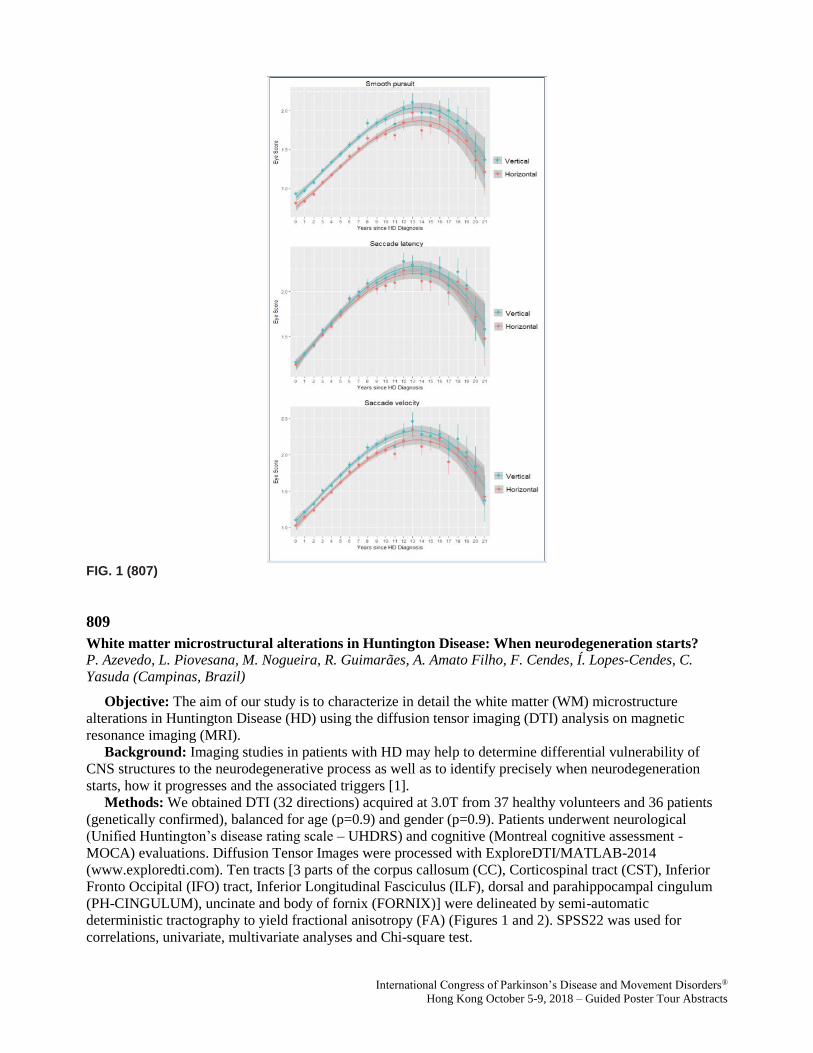
International Congress of Parkinson’s Disease and Movement Disorders®
Hong Kong October 5-9, 2018 – Guided Poster Tour Abstracts
FIG. 1 (807)
809
White matter microstructural alterations in Huntington Disease: When neurodegeneration starts?
P. Azevedo, L. Piovesana, M. Nogueira, R. Guimarães, A. Amato Filho, F. Cendes, Í. Lopes-Cendes, C.
Yasuda (Campinas, Brazil)
Objective: The aim of our study is to characterize in detail the white matter (WM) microstructure
alterations in Huntington Disease (HD) using the diffusion tensor imaging (DTI) analysis on magnetic
resonance imaging (MRI).
Background: Imaging studies in patients with HD may help to determine differential vulnerability of
CNS structures to the neurodegenerative process as well as to identify precisely when neurodegeneration
starts, how it progresses and the associated triggers [1].
Methods: We obtained DTI (32 directions) acquired at 3.0T from 37 healthy volunteers and 36 patients
(genetically confirmed), balanced for age (p=0.9) and gender (p=0.9). Patients underwent neurological
(Unified Huntington’s disease rating scale – UHDRS) and cognitive (Montreal cognitive assessment -
MOCA) evaluations. Diffusion Tensor Images were processed with ExploreDTI/MATLAB-2014
(www.exploredti.com). Ten tracts [3 parts of the corpus callosum (CC), Corticospinal tract (CST), Inferior
Fronto Occipital (IFO) tract, Inferior Longitudinal Fasciculus (ILF), dorsal and parahippocampal cingulum
(PH-CINGULUM), uncinate and body of fornix (FORNIX)] were delineated by semi-automatic
deterministic tractography to yield fractional anisotropy (FA) (Figures 1 and 2). SPSS22 was used for
correlations, univariate, multivariate analyses and Chi-square test.
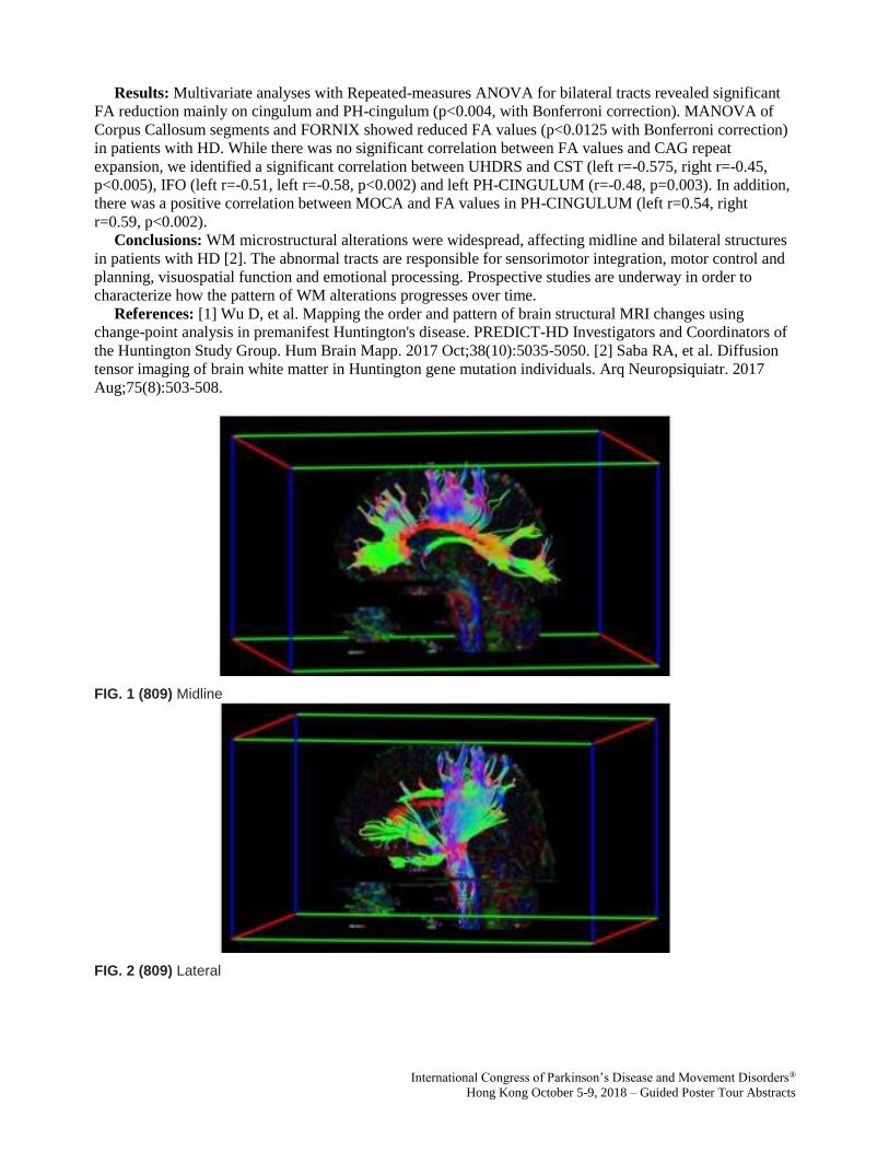
International Congress of Parkinson’s Disease and Movement Disorders®
Hong Kong October 5-9, 2018 – Guided Poster Tour Abstracts
Results: Multivariate analyses with Repeated-measures ANOVA for bilateral tracts revealed significant
FA reduction mainly on cingulum and PH-cingulum (p<0.004, with Bonferroni correction). MANOVA of
Corpus Callosum segments and FORNIX showed reduced FA values (p<0.0125 with Bonferroni correction)
in patients with HD. While there was no significant correlation between FA values and CAG repeat
expansion, we identified a significant correlation between UHDRS and CST (left r=-0.575, right r=-0.45,
p<0.005), IFO (left r=-0.51, left r=-0.58, p<0.002) and left PH-CINGULUM (r=-0.48, p=0.003). In addition,
there was a positive correlation between MOCA and FA values in PH-CINGULUM (left r=0.54, right
r=0.59, p<0.002).
Conclusions: WM microstructural alterations were widespread, affecting midline and bilateral structures
in patients with HD [2]. The abnormal tracts are responsible for sensorimotor integration, motor control and
planning, visuospatial function and emotional processing. Prospective studies are underway in order to
characterize how the pattern of WM alterations progresses over time.
References: [1] Wu D, et al. Mapping the order and pattern of brain structural MRI changes using
change-point analysis in premanifest Huntington's disease. PREDICT-HD Investigators and Coordinators of
the Huntington Study Group. Hum Brain Mapp. 2017 Oct;38(10):5035-5050. [2] Saba RA, et al. Diffusion
tensor imaging of brain white matter in Huntington gene mutation individuals. Arq Neuropsiquiatr. 2017
Aug;75(8):503-508.
FIG. 1 (809) Midline
FIG. 2 (809) Lateral
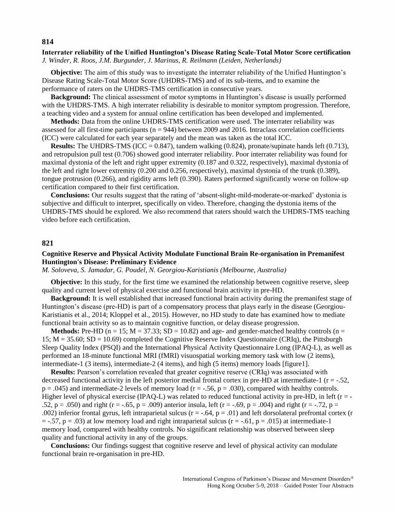
International Congress of Parkinson’s Disease and Movement Disorders®
Hong Kong October 5-9, 2018 – Guided Poster Tour Abstracts
814
Interrater reliability of the Unified Huntington’s Disease Rating Scale-Total Motor Score certification
J. Winder, R. Roos, J.M. Burgunder, J. Marinus, R. Reilmann (Leiden, Netherlands)
Objective: The aim of this study was to investigate the interrater reliability of the Unified Huntington’s
Disease Rating Scale-Total Motor Score (UHDRS-TMS) and of its sub-items, and to examine the
performance of raters on the UHDRS-TMS certification in consecutive years.
Background: The clinical assessment of motor symptoms in Huntington’s disease is usually performed
with the UHDRS-TMS. A high interrater reliability is desirable to monitor symptom progression. Therefore,
a teaching video and a system for annual online certification has been developed and implemented.
Methods: Data from the online UHDRS-TMS certification were used. The interrater reliability was
assessed for all first-time participants (n = 944) between 2009 and 2016. Intraclass correlation coefficients
(ICC) were calculated for each year separately and the mean was taken as the total ICC.
Results: The UHDRS-TMS (ICC = 0.847), tandem walking (0.824), pronate/supinate hands left (0.713),
and retropulsion pull test (0.706) showed good interrater reliability. Poor interrater reliability was found for
maximal dystonia of the left and right upper extremity (0.187 and 0.322, respectively), maximal dystonia of
the left and right lower extremity (0.200 and 0.256, respectively), maximal dystonia of the trunk (0.389),
tongue protrusion (0.266), and rigidity arms left (0.390). Raters performed significantly worse on follow-up
certification compared to their first certification.
Conclusions: Our results suggest that the rating of ‘absent-slight-mild-moderate-or-marked’ dystonia is
subjective and difficult to interpret, specifically on video. Therefore, changing the dystonia items of the
UHDRS-TMS should be explored. We also recommend that raters should watch the UHDRS-TMS teaching
video before each certification.
821
Cognitive Reserve and Physical Activity Modulate Functional Brain Re-organisation in Premanifest
Huntington's Disease: Preliminary Evidence
M. Soloveva, S. Jamadar, G. Poudel, N. Georgiou-Karistianis (Melbourne, Australia)
Objective: In this study, for the first time we examined the relationship between cognitive reserve, sleep
quality and current level of physical exercise and functional brain activity in pre-HD.
Background: It is well established that increased functional brain activity during the premanifest stage of
Huntington’s disease (pre-HD) is part of a compensatory process that plays early in the disease (Georgiou-
Karistianis et al., 2014; Kloppel et al., 2015). However, no HD study to date has examined how to mediate
functional brain activity so as to maintain cognitive function, or delay disease progression.
Methods: Pre-HD (n = 15; M = 37.33; SD = 10.82) and age- and gender-matched healthy controls (n =
15; M = 35.60; SD = 10.69) completed the Cognitive Reserve Index Questionnaire (CRIq), the Pittsburgh
Sleep Quality Index (PSQI) and the International Physical Activity Questionnaire Long (IPAQ-L), as well as
performed an 18-minute functional MRI (fMRI) visuospatial working memory task with low (2 items),
intermediate-1 (3 items), intermediate-2 (4 items), and high (5 items) memory loads [figure1].
Results: Pearson’s correlation revealed that greater cognitive reserve (CRIq) was associated with
decreased functional activity in the left posterior medial frontal cortex in pre-HD at intermediate-1 (r = -.52,
p = .045) and intermediate-2 levels of memory load (r = -.56, p = .030), compared with healthy controls.
Higher level of physical exercise (IPAQ-L) was related to reduced functional activity in pre-HD, in left (r = -
.52, p = .050) and right (r = -.65, p = .009) anterior insula, left (r = -.69, p = .004) and right (r = -.72, p =
.002) inferior frontal gyrus, left intraparietal sulcus (r = -.64, p = .01) and left dorsolateral prefrontal cortex (r
= -.57, p = .03) at low memory load and right intraparietal sulcus (r = -.61, p = .015) at intermediate-1
memory load, compared with healthy controls. No significant relationship was observed between sleep
quality and functional activity in any of the groups.
Conclusions: Our findings suggest that cognitive reserve and level of physical activity can modulate
functional brain re-organisation in pre-HD.

International Congress of Parkinson’s Disease and Movement Disorders®
Hong Kong October 5-9, 2018 – Guided Poster Tour Abstracts
References: Georgiou-Karistinis et al., 2014. Kloppel et al., 2015.
FIG. 1 (821)
823
Comparing risperidone and olanzapine to tetrabenazine for the management of Huntington’s chorea
J. Schultz, J. Kamholz, P. Nopoulos, A. Killoran (Iowa City, IA, USA)
Objective: To compare the efficacy of risperidone and olanzapine to tetrabenazine (TBZ) for the
management of Huntington’s chorea in Enroll-HD participants.
Background: TBZ is provenly-effective and FDA-approved for the treatment of chorea in Huntington's
disease (HD). Antipsychotic drugs (APD), such as Risperdal and Olanzapine, are often prescribed off-label
for HD chorea, especially when psychiatric comorbidities are present. The APD' anti-choreic efficacy is
presumed primarily from their long-standing use by HD experts, as they have never been evaluated in large
clinical trials.
Methods: This was a retrospective analysis using the database from Enroll-HD, a longitudinal,
observational study with 8714 participants. Those with manifest HD were grouped based on their use of
risperidone, olanzapine, or TBZ. The effects of these anti-choreic drugs were compared using MANCOVA
analyses. The primary outcome measure was the total motor score (TMS) annual rate of change between the
baseline and second visits. Secondary outcome measures included TMS changes and the rates of change
between the baseline and second visits of the: total chorea score, total functional capacity score, weight and
body mass index (BMI). Covariates included sex, years between visits and baseline: TMS, TFC, and disease
burden score. An exploratory outcome measure was the odds of having an annualized improvement in the
TMS by at least 3.3 units, which was TBZ’s treatment effect in the TETRA-HD study.1
Results: The risperidone group showed an improvement in the annualized percent change in TMS of
3.9% compared to a 17.5% worsening in the TBZ group (p=0.042). In addition, the odds of having an
annualized improvement in the TMS by at least 3.3 units was significantly higher in subjects taking
risperidone (42.9%) vs those on TBZ (9.0%) (p=0.031). The olanzapine group increased in weight (p=0.023)
and BMI (p=0.022) compared to the TBZ group. The APD and TBZ results were otherwise comparable.
Conclusions: This is the first study to provide robust, objective data on the anti-choreic effect of
risperidone and olanzapine. It is also the first study to compare these APD to TBZ for their efficacy on HD
chorea. Our results establish that risperidone and olanzapine are comparable to TBZ, but for some superiority
to tetrabenazine on certain measures. These findings bridge the gap between expert opinion and evidence-
based medicine in the management of HD chorea.
References: 1. Huntington Study Group. Tetrabenazine as antichorea therapy in Huntington Disease: A
randomized controlled Trial. Neurology 2006;66:366-372.
International Congress of Parkinson’s Disease and Movement Disorders®
Hong Kong October 5-9, 2018 – Guided Poster Tour Abstracts
830
Driving performance of Huntington’s disease gene carriers
M. Jacobs, E. Hart, Y. Mejia Miranda, G.J. Groeneveld, J. van Gerven, R. Roos (Leiden, Netherlands)
Objective: To investigate if differences in driving performance between Huntington's disease (HD) gene
carriers and healthy individuals can be detected with a driving simulator. Furthermore, we wanted to
determine if certain cognitive and motor symptoms contribute to driving performance in HD.
Background: The ability to drive a car is important for an individual’s independence, work, and social
activities. HD patients become more and more dependent in their daily life activities as the disease
progresses and, for most patients, it can be difficult to quit driving.
Methods: We included 58 HD gene carriers (28 premanifest HD, 30 manifest HD) and 29 controls in this
cross-sectional study. All participants were active drivers and assessed using a driving simulator, a driving
history questionnaire, the Unified Huntington’s Disease Rating Scale, and neuropsychological assessments.
The driving simulator session included both urban and motorway scenarios.
Results: Manifest HD drove slower compared to controls and premanifest HD when speed limits
increased (80 and 100 km/h) and they had a less steady speed compared to premanifest HD on the motorway
and in a 30 km/h zone. Manifest HD also had a larger standard deviation of the lateral position (i.e., more
weaving of the car/less vehicle control) compared to controls and premanifest HD on the motorway.
Manifest HD performed worse on all clinical assessments compared to controls. Postural instability and
slower speed of processing were predictors of the driving simulator outcome measures. There were no
significant differences between premanifest HD and controls.
Conclusions: Manifest HD drive more cautious in a driving simulator when speed limits increase
compared to premanifest HD and controls and they have less vehicle control on the motorway. A driving
simulator is able to detect differences in driving performance between manifest HD and healthy individuals.
Lower performances on cognitive tasks might assist physicians in their referral for an official on-road driving
test. A genetic confirmation of HD should not be decisive in the advice to cease driving. Further studies are
necessary to determine if a driving simulator can be used to monitor longitudinal changes in fitness to drive.
Investigating fitness to drive is important in HD, because symptoms of the disease can affect the ability to
drive at a relatively young age.
838
Cerebrospinal fluid flow dynamics in Huntington's disease evaluated by phase contrast MRI
F. Rodrigues, L. Byrne, E. De Vita, E. Johnson, N. Hobbs, J. Thornton, R. Scahill, E. Wild (London, United
Kingdom)
Objective: To elucidate the dynamics of CSF flow through the neuraxis in Huntington’s disease (HD)
using phase contrast magnetic resonance imaging (PCMRI).
Background: HD is a fatal neurodegenerative condition. Multiple targeted therapeutics are now in the
early clinical stage, including intrathecally-delivered compounds. Preclinical and clinical research suggests
cerebrospinal fluid dynamics may be altered in HD, which could be of paramount relevance to intrathecal
drug delivery to the brain.
Methods: Ten manifest gene expansion carriers and 10 age- and gender-matched healthy controls were
recruited. All participants underwent extensive clinical assessment and PCMRI at the level of the cerebral
aqueduct, T1 and T8. CSF velocities and flow measurements were derived using a semi-automated method.
The influence of age, gender, CAG repeat-length, serum osmolality, and brain volumes on these
measurements were tested using Spearman correlations or Fisher’s exact tests. Group comparisons between
healthy controls and manifest carriers were achieved via two-sample Wilcoxon rank-sum tests. All tests were
two-sided with a significance level of 0.05, and corrected for multiple comparisons.
Results: All participants were well matched across study groups. Measures of CSF dynamics were
consistent across raters. None of the studied covariates was found to have an effect on the CSF velocities and
flow measurements after corrected for multiple comparisons. No statistically significant difference was found

International Congress of Parkinson’s Disease and Movement Disorders®
Hong Kong October 5-9, 2018 – Guided Poster Tour Abstracts
across groups in any of the measures studied. Sample size calculations have shown that for the most
clinically significant effect size attained, 993 participants per group would be needed to show a significant
difference between two groups with 90% power.
Conclusions: While external validation is required, the attained effect sizes in this exploratory study are
sufficient to conclude tentatively that a clinically-relevant alteration of cerebrospinal fluid flow dynamics –
i.e. one that would justify dose-adjustments of intrathecal drugs – is unlikely to exist in Huntington’s disease.
927
Clinical features and natural history of pathologically-confirmed corticobasal degeneration: A
Japanese validation study of CBD (J-VAC study)
I. Aiba, T. Shimohata, S. Murayama, K. Hasegawa, Y. Iwasaki, O. Yokota, H. Fujimura, M. Sakai, T. Yokota,
I. Yabe, H. Takigawa, K. Sugaya, K. Mori, M. Ito, C. Ishida, M. Kobayashi, Y. Hashizume, T. Ikeuchi, M.
Hasegawa, M. Yoshida, T. Komori, K. Wakabayashi, Y. Saito, A. Tokumaru, K. Sakurai, K. Nakashima
(Nagoya, Japan)
Objective: To elucidate the clinical features and natural history of CBD.
Background: Recently, various clinical phenotypes of corticobasal degeneration (CBD) have been
reported; on the other hand, the pathological background of corticobasal syndrome (CBS) is broad. Hence,
the rate of antemortem diagnosis of CBD is extremely low.
Methods: Clinical evaluations were performed on patients with pathologically-, genetically-, and
biochemically-verified CBD during central review and each patient’s clinical information was retrospectively
studied. We analyzed the frequency of signs and clinical phenotypes, and the interval from initial symptoms
to key clinical milestones. Clinical features observed in over half of patients were selected as key milestones.
Results: We identified 35 pathologically-confirmed patients, from 15 Japanese institutions. The mean age
at onset was 65.0 years. The mean disease duration was 8.1 years. At initial presentation, CBS/CBD and
progressive supranuclear palsy (PSP) were the most common diagnoses (19%, respectively), followed by
Lewy body disease (17%) and Alzheimer's disease (AD) (11%). CBS/CBD was the most frequent final
clinical diagnosis (44%), followed by PSP (28%), AD (8%) and frontotemporal dementia (FTD) (8%). Limb
rigidity, gait disturbance, and postural instability or falls were observed in more than 80% of patients with
CBD. Dystonia (39%) and myoclonus (20%) were infrequently observed. In higher cortical features, both
frontal executive dysfunction and general cognitive impairment were observed in more than 80% of patients,
followed by speech and language impairments (77%) and behavioral changes (69%). Apraxia (48%), cortical
sensory loss (20%), and the alien-limb sign (10%) were uncommonly observed. The mean intervals from the
initial symptoms to onset of key clinical milestones were as follows: gait disturbance, 1.0 years; behavioral
changes, 1.2 years; falls, 2.0 years; cognitive impairment, 2.4 years; speech impairment, 2.6 years;
supranuclear gaze palsy, 3.1 years; urinary incontinence, 3.3 years; and dysphagia, 4.3 years.
Conclusions: The pathology of CBD was predicted antemortem in only 44% of patients. In patients with
CBD, parkinsonism, frontal executive dysfunction, and general cognitive impairment were more commonly
observed than the cardinal features of CBS. Behavioral changes appeared in the early stages, although the
natural disease course was similar to PSP.
Guided Poster Group 7:
Parkinsonism, MSA, PSP (Secondary and Parkinsonism-Plus)
International Congress of Parkinson’s Disease and Movement Disorders®
Hong Kong October 5-9, 2018 – Guided Poster Tour Abstracts
929
Quantitative mobility metrics from a body-fixed sensor predict incident parkinsonism in older adults
R. von Coelln, R. Dawe, J. Shulman, L. Yu, S. Leurgans, J. Hausdorff, L. Shulman, D. Bennett, A. Buchman
(Baltimore, MD, USA)
Objective: To investigate whether body-fixed sensor mobility metrics identify older adults without a
diagnosis of Parkinson disease (PD) at risk for developing parkinsonism.
Background: Parkinsonism is common in the elderly, and predicts adverse health outcomes. In prior
work we showed that mobility metrics from a body-fixed sensor are related to the severity of parkinsonism in
older adults without PD. However, it is not known if these metrics identify older adults at risk for developing
parkinsonism.
Methods: 683 ambulatory, community-dwelling older adults without parkinsonism were evaluated
annually for four parkinsonian signs (bradykinesia, tremor, rigidity, parkinsonian gait) using a modified
Unified Parkinson’s Disease Rating Scale. Subjects also wore a belt with a body-fixed triaxial
accelerometer/gyroscope as they performed 3 motor tests (32 ft walk, standing with eyes closed, Timed Up
and Go test [TUG]). Baseline measures were summarized into 12 gait scores including gait speed metrics,
which quantified 5 subtasks including: a) walking, b) standing posture, plus 3 TUG subtasks: c) sit to stand
transition, d) stand to sit transition, and e) turning. In a series of Cox proportional hazards models controlled
for age, sex, education and race, we examined which gait scores were associated with incident parkinsonism
(2 or more parkinsonian signs at a follow-up examination). Stepwise backwards elimination was performed
to identify combinations of gait scores independently associated with incident parkinsonism.
Results: 139 out of 683 (20.4%) participants developed parkinsonism during 2.5 ± 1.28 years of follow-
up. When modeled separately, 6 out of 12 gait scores were associated with incident parkinsonism. Two of 12
scores (stand to sit transition and turning scores) survived stepwise backward elimination, and were
independently associated with incident parkinsonism (p=0.003 and p<0.001, respectively).
Conclusions: Mobility metrics derived from a wearable body-fixed sensor facilitate identification of older
adults without PD at risk of developing parkinsonism.
933
Apolipoprotein E and multiple system atrophy
K. Ogaki, Y. Martens, M. Heckman, S. Koga, L. Labbé, O. Lorenzo-Betancor, A. Wernick, R. Walton, A.
Soto, E. Vargas, H. Nielsen, S. Fujioka, T. Kanekiyo, R. Uitti, J. van Gerpen, W. Cheshire, Z. Wszolek, P.
Low, W. Singer, N. Hattori, D. Dickson, G. Bu, O. Ross (Tokyo, Japan)
Objective: This study evaluated genetic associations of Apolipoprotein E alleles with risk of multiple
system atrophy (MSA) and α-synuclein pathology, and also examined whether apolipoprotein E isoforms
differentially affect α-synuclein uptake in a oligodendrocyte cell.
Background: Dysregulation of the specialized lipid metabolism involved in myelin synthesis and
maintenance by oligodendrocytes has been associated with the unique neuropathology of MSA. We
hypothesized that apolipoprotein E, which is associated with neurodegeneration, may also play a role in the
pathogenesis of MSA.
Methods: One hundred sixty-eight pathologically confirmed MSA patients, 89 clinically diagnosed MSA
patients, and 1,277 control subjects were genotyped for Apolipoprotein E. Human oligodendrocyte cell lines
were incubated with α-synuclein and recombinant human apolipoprotein E, with internalized α-synuclein
imaged by confocal microscopy and cells analyzed by flow cytometry.
Results: No significant association with risk of MSA or was observed for either Apolipoprotein E ɛ2 or
ɛ4. α-Synuclein burden was also not associated with Apolipoprotein E alleles in the pathologically confirmed
patients. Interestingly, in our cell assays, apolipoprotein E ɛ4 significantly reduced α-synuclein uptake in the
oligodendrocytic cell line.

International Congress of Parkinson’s Disease and Movement Disorders®
Hong Kong October 5-9, 2018 – Guided Poster Tour Abstracts
Conclusions: Despite differential effects of apolipoprotein E isoforms on α-synuclein uptake in a human
oligodendrocytic cell, we did not observe a significant association at the Apolipoprotein E locus with risk of
MSA or α-synuclein pathology.
939
ARISE study: Study Design and Baseline Characteristics for a Phase 2 Trial of the Anti-Tau Antibody
ABBV-8E12 in Progressive Supranuclear Palsy
N. Mendonca, R. Bateman, A. Boxer, J. Braunstein, D. Claassen, D. Holtzman, D. Kerwin, B. Rendenbach-
Mueller, H. Soares, D. Wang, G. Höglinger (Ludwigshafen, , Germany)
Objective: This double-blind, placebo-controlled phase 2 study (M15-562 ARISE study, NCT02985879)
assesses the safety and efficacy of ABBV-8E12 treatment in patients with progressive supranuclear palsy
(PSP) for 52 weeks.
Background: ABBV-8E12 is a humanized anti-tau monoclonal antibody being developed for the
treatment of PSP and early Alzheimer’s disease (AD). A phase 1 double-blind, placebo-controlled, single
ascending dose study on the safety, tolerability, and pharmacokinetics of ABBV-8E12 in PSP patients
(NCT02494024) was recently completed. The phase 1 results showed that ABBV-8E12, when administered
as a single dose up to 50 mg/kg, exhibited an acceptable safety and tolerability profile to support repeat-dose
testing in larger cohorts of patients with tauopathies. Here we present the design and baseline characteristics
of the ongoing phase 2 ARISE study of ABBV-8E12 in PSP patients.
Methods: Male and female patients, at least 40 years of age, are being enrolled at approximately 40
global study sites. Prior to enrollment, patients will have had symptoms for less than 5 years and meet the
NINDS-PSP criteria for possible or probable PSP. Patients will be randomized to one of two ABBV-8E12
dose arms or placebo.
Results: The primary efficacy outcome is the change from baseline to week 52 in Progressive
Supranuclear Palsy Rating Scale total score. Key secondary endpoints include the Schwab and England
Activities of Daily Living Scale, the Unified Parkinson's Disease Rating Scale Part II, the Clinical Global
Impression of Severity and Change, the PSP-Quality of Life subscale scores, and the Visual Analog scale.
Table 1 includes baseline characteristics for the first 91 enrolled patients [table1].
Conclusions: The current study is ongoing and designed to evaluate the 52-week safety and efficacy of
ABBV-8E12 in PSP patients. In addition to the ongoing phase 2 study in PSP patients, a phase 2 study
evaluating ABBV-8E12 in early AD patients is also currently ongoing (NCT02880956).

International Congress of Parkinson’s Disease and Movement Disorders®
Hong Kong October 5-9, 2018 – Guided Poster Tour Abstracts
TABLE 1 (939) ARISE demographics and baseline characteristics
948
The characteristic of gait in progressive progressive supranuclear palsy
Y. Takamatsu, N. Matsuda, I. Aiba (Kyoto, Japan)
Objective: To elucidate the characteristic of gait in patients with progressive supranuclear palsy (PSP)
and compare to healthy elderly and patients with Parkinson’s disease (PD)
Background: Gait disturbance is one of main symptoms in PSP. PSP is a rare disease and their
characteristic of gait has not been well understood. Therefore, the optimal physical therapy approach has not
been established for their gait dysturbance in PSP.
Methods: Subjects were PSP (n = 24, 72.9 ± 6 y/o, man/female = 18/6), PD(n = 32, 71.1 ± 9 y/o,
man/female = 18/14) and neurologically healthy elderly (CON, n = 29, 71.6 ± 6 y/o, man/female = 12/17)
whose modified Rankin scale (mRS) were 4 or less. Gait patterns analysis was performed by using Walk
Way MW-1000 (anima inc., Tokyo) and walking speed, cadence, stride length, step width, foot angle,
walking cycle time and coefficient of variation (CV) of each parameter were measured. In the statistics, one-
way ANOVA with post hoc Scheffe test was performed to compare all groups. For group differences were
assessed using t-test and Mann-Whiteney U test. SPSS ver.23 statistical software was used to analyze the
data and the criterion for significance was set at p < 0.05.
Results: There were no differences in the disease duration (PSP: 64.4 ± 53 mos., PD: 68.8 ± 56 mos.) and
mRS (PSP: 3 [2.25-4], PD: [2-3]) between PSP and PD. The walking speed was lower and stride length was
shorter significantly in PSP and PD than that of CON (p<0.05), but there was no difference between PSP and
PD. The step width and foot angle were larger significantly in PSP than that of CON (p<0.05), but there was
no difference between PD and CON. In addition, CV of walking cycle time was larger in PSP than that of
CON and PD (p<0.05).
Conclusions: In PSP and PD, their walking abilities were markedly disturbed compared to CON.
Furthermore, their step width, foot angle and variation of walking cycle time were significantly larger in PSP
than PD. It has been reported that dentate nucleus, cerebellar white matter, potine nucleus, and inferior
olivary nucleus were pathologically involved unlike PD. Therefore, the gait of PSP might be highly unstable
International Congress of Parkinson’s Disease and Movement Disorders®
Hong Kong October 5-9, 2018 – Guided Poster Tour Abstracts
by dysfunction of the cerebellum. As the result, they might increase in step width and foot angle as
compensation to stable at their walking.
957
Olfactory testing in Parkinson plus syndromes
O. Abdukarimov, F. Akhmedova (Tashkent, Uzbekistan)
Objective: To determine the diagnostic utility of olfactory testing in patients with neurodegenerative
Parkinsonism.
Background: Olfactory dysfunction can be identified in the vast majority of patients with Parkinson’s
disease (PD). Less is known about olfactory function in multiple system atrophy (MSA) and progressive
supranuclear palsy (PSP) - two atypical Parkinsonian disorders (APD) that may closely mimic PD in early
disease stages – but current evidence suggests that olfactory function remains largely intact in these
conditions.
Methods: The Sniffin’ Sticks test battery for assessment of odor identification, odor discrimination, and
olfactory threshold was applied to two independent cohorts – a screening and a validation cohort. The
screening cohort included Parkinson’s disease (PD) patients and healthy controls (HC) and was used to
calculate optimal cut-offs for a diagnosis of PD with a sensitivity or specificity exceeding 95%. The
validation cohort was used to determine the diagnostic accuracy of the newly established cut-off values in
discriminating patients with PD from those with atypical Parkinsonian disorders (APD) including multiple
system atrophy (MSA) and progressive supranuclear palsy (PSP).
Results: PD patients (screening cohort n520, validation cohort n547) performed significantly worse in
olfactory testing than HC (n541) and patients with MSA (n523) or PSP (n523). Diagnostic performance of
the identification subscore was similar to the sum score of the Sniffin’ Sticks test (AUC identification test
0.94, AUC sum score
0.96). In subjects with neurodegenerative Parkinsonism, the specificity cut-off predicted a diagnosis of PD
with a sensitivity and specificity of 76.6 and 87.0%, respectively. The discriminative value of this cut-off in
separating PD from MSA was 76.7% (sensitivity) and 95.7% (specificity). Optimal cut-offs of the sum score
provided a sensitivity and specificity for a diagnosis of PD of 78.7% and 76.1%, respectively. The positive
predictive value of olfactory testing adjusted for PD prevalence in the community exceeded 95%.
Conclusions: Our data suggest that assessment of olfactory function, particularly odor identification, can
be useful to discriminate PD from APDs, particularly MSA patients.
964
Nilotinib for treating MSA: A preclinical proof of concept study
P. Guerin, M. Lopez-Cuina, E. Bezard, W. Meissner, P-O. Fernagut (Bordeaux, France)
Objective: To assess the effects of nilotinib on motor behavior, α-synuclein burden and surrogate markers
of neurodegeneration in a transgenic mouse model of multiple system atrophy (MSA).
Background: The pathological hallmark of MSA is the presence of α-synuclein bearing glial cytoplasmic
inclusions. Treatment is available for some symptoms, in particular autonomic dysfunction, while disease
modification remains an urgent unmet need. Activation of the tyrosine kinase c-Abl protein is increased in
Parkinson’s disease (PD) and alpha-synuclein has been identified as one of its substrates. Through C-Abl
inhibition, nilotinib (a commercially available treatment for a type of leukemia) is thought to potentially
counteract α-synuclein accumulation and to protect neurons from degeneration. In this regard, positive
effects of nilotinib on the neurodegenerative process have been reported in preclinical models of PD. The
aim of this study was to evaluate the effects of nilotinib in PLP-SYN mice, a transgenic mouse model of
MSA.
Methods: Wild-type (WT) mice received daily intraperitoneal injections of vehicle and transgenic PLP-
SYN received daily intraperitoneal injections of either vehicle, nilotinib 1mg/kg or 10mg/kg, for 12 weeks
International Congress of Parkinson’s Disease and Movement Disorders®
Hong Kong October 5-9, 2018 – Guided Poster Tour Abstracts
since age 6 weeks. Motor behavior was assessed at baseline and every 4 weeks until termination. The
histopathological analysis included cell survival in the substantia nigra pars compacta (SNpc) as assessed by
the number of tyrosine hydroxylase and Nissl positive neurons. Immunoblotting was performed to measure
α-synuclein load.
Results: Nilotinib was safe and well tolerated at both doses by PLP-SYN mice. There was no difference
in motor performance between the different treatment groups of PLP-SYN mice. As expected, placebo-
treated PLP-SYN mice showed dopaminergic cell loss in the SNpc compared to WT mice, while nilotinib
failed to protect neurons from degeneration and to attenuate α-synuclein burden in PLP-SYN mice.
Conclusions: Nilotinib failed to demonstrate positive effects in a transgenic mouse model of MSA.
974
Evolution of diagnostic certainty and PSP-predominance types in 187 pathologically confirmed PSP
patients
M. Grimm, G. Respondek, I. Piot, T. Arzberger, Y. Compta, E. Englund, L. Ferguson, E. Gelpi, A. Giese, S.
Roeber, D. Irwin, W. Meissner, C. Nilsson, A. Pantelyat, A. Rajput, C. Troakes, G. Höglinger (Munich,
Germany)
Objective: To examine the evolution of diagnostic certainty and clinical predominance types as defined
by the movement disorder society (MDS) criteria for the clinical diagnosis of PSP (Höglinger et al, 2017)
during the course of disease in autopsy-confirmed patients with PSP.
Background: Three degrees of diagnostic certainty ("suggestive of" = s.o., "possible", or "probable"),
and definition of clinical predominance types (PSP-RS, PSP-PGF, PSP-P, PSP-F, PSP-OM, PSP-SL, PSP-
CBS, PSP-PI) were implemented into the new clinical diagnostic criteria for PSP (MDS criteria for the
clinical diagnosis of PSP, Höglinger et al, 2017). The evolution of diagnostic certainty and predominance
types over the course of disease in PSP patients according to the MDS-criteria has not been studied so far.
However, this is relevant for further understanding the natural history and clinical spectrum of PSP.
Methods: Features relevant for the diagnosis of PSP according to the MDS-diagnostic criteria were
collected in 187 autopsy-confirmed PSP patients by chart review. Diagnostic certainty and PSP-
predominance types according to the MDS-diagnostic criteria were determined for each patient and each
year.
Results: According to the MDS-PSP diagnostic criteria, 62% (n=115) of patients had a clinical diagnosis
of PSP in the first year of disease, as opposed to 14% (n=26) according to the NINDS SPSP criteria, and
98% (n=183) at final record, as opposed to 79% (n=147) according to the NINDS/SPSP criteria. A diagnosis
of s.o. PSP was present in 44% (n=83) of patients in the first year. Of these, 77% (n=64) had a diagnosis of
"probable" PSP at final record. In the first year of disease, the variability of predominance types was greatest,
and PSP-RS represented only 11% of patients. At final record, 56% patients had a PSP-RS predominance
type.
Conclusions: The conversion of s.o. PSP into possible and probable PSP in 72% of the cases shows that
the concept of s.o. PSP indeed allows identification of PSP patients at a very early clinical stage.
Furthermore, our data suggest that many initially variant PSP presentations converge to a PSP-RS phenotype
during the course of the disease.
References: MDS criteria for the clinical diagnosis of PSP, Höglinger et al, 2017.
979
Rapamycin for treating MSA: A preclinical proof of concept study
M. Lopez-Cuina, P. Guerin, E. Bezard, W. Meissner, P-O. Fernagut (Bordeaux, France)
Objective: To assess the effects of rapamycin on motor behavior, α-synuclein burden and surrogate
markers of neurodegeneration in a transgenic mouse model of multiple system atrophy (MSA).
Background: Growing evidence suggests impairment of the autophagy-lysosomal pathway in MSA.
Since this pathway has a major role in the degradation of α-synuclein, its impaired function may contribute to
International Congress of Parkinson’s Disease and Movement Disorders®
Hong Kong October 5-9, 2018 – Guided Poster Tour Abstracts
the accumulation of α-synuclein in glial cytoplasmic inclusions in MSA. The mammalian target of
rapamycin complex 1 (mTORC1) is a key protein complex regulating autophagy. Rapamycin is an mTORC1
inhibitor that enhances autophagy, by increasing autophagosomes and boosting lysosomal biogenesis. We
here assessed if rapamycin exerts neuroprotective effects by enhancing autophagic α-synuclein clearance in a
transgenic mouse model of MSA.
Methods: Wild-type (WT) and PLP-SYN transgenic mice were fed either with normal food or food
enriched with 14mg/kg of rapamycin for 16 weeks since age 6 weeks. Motor behavior was assessed at
baseline and every 4 weeks until termination. Histopathological analysis included cell survival in the
substantia nigra pars compacta (SNpc) as assessed by the number of tyrosine hydroxylase and Nissl positive
neurons, and the amount of α-synuclein aggregates in oligodendrocytes in the SNpc and the striatum after
incubation of sections with or without proteinase K. Additional immunoblotting was performed to measure
α-synuclein load.
Results: Rapamycin was safe and well-tolerated. There was no difference in motor performance between
groups. As expected, placebo-treated PLP-SYN mice showed dopaminergic cell loss in the SNpc compared
to WT mice. Rapamycin provided partial neuroprotection since the number of Nissl positive neurons in the
SNpc was similar compared to WT mice, while the number of tyrosine hydroxylase positive neurons was
significantly lower and not different from placebo-treated PLP-SYN mice. Rapamycin also significantly
reduced the amount of α-synuclein aggregates in the SNpc, while only a trend was observed for the striatum.
Conclusions: Rapamycin partially rescued neurons in the SNpc, although they remained dysfunctional as
suggested by the down-regulated tyrosine hydroxylase expression. The partial rescue was paralleled by a
positive effect on the amount of α-synuclein aggregates.
1096
The BlueSky Project: monitoring motor and non-motor characteristics of people with Parkinson’s
disease in the laboratory, a simulated apartment, and home and community settings
K. Erb, J. Daneault, S. Amato, P. Bergethon, C. Demanuele, T. Kangarloo, S. Patel, V. Ramos, D. Volfson,
P. Wacnik, H. Zhang, D. Karlin, H. Huggins, L. Soll, G. Costante, G. Vergara-Diaz, F. Parisi, J. Banghu, C.
Brooks, C. Dethridge, A. Abrami, E. Bilal, V. Caravagio, S. Heisig, R. Norel, E. Pissadaki, J. Rice, B. Ho, K.
Thomas, P. Bonato (Cambridge, MA, USA)
Objective: We present data collected for the BlueSky Project which aims to develop novel assessments of
motor and non-motor function using mobile and wearable technology.
Background: A major challenge for the management of Parkinson’s disease (PD) and for clinical trials
investigating new treatment strategies is to accurately monitor the severity of symptoms over time. Wearable
sensors can enable the long-term monitoring of patients in the home and community settings. However, more
data is needed to develop robust algorithms to accurately estimate symptom severity.
Methods: This project encompasses 4 studies that investigate motor and non-motor monitoring of healthy
volunteers and people with PD experiencing motor fluctuations. In Study 1, 60 healthy volunteers donned
several inertial, EMG, and ECG sensors. They were asked to perform speech tasks, a battery of scripted
motor tasks, and activities of daily living (ADLs) twice in a laboratory setting. In Study 2, 35 people with PD
were asked to perform the same data collection protocol as in Study 1 on two occasions: once in a
practically-defined OFF state, and once in an ON state. In Study 3, 25 people with PD donned the same
sensors as in Studies 1&2 plus sensors to record galvanic skin response. Subjects performed 5 repetitions of a
battery of motor and speech tasks as well as ADLs over a complete medication cycle (6 hours). Subjects
came back for a second visit where semi-scripted and unscripted activities were performed in a simulated
apartment setting over 6 hours of continuous monitoring using the same sensors. In Study 4, 34 people with
PD were asked to perform two weeks of continuous home monitoring while wearing similar devices. During
Guided Poster Group 8:
Technology
International Congress of Parkinson’s Disease and Movement Disorders®
Hong Kong October 5-9, 2018 – Guided Poster Tour Abstracts
both weeks, they completed a medication log, motor diary, and QOL instruments. Both weeks of monitoring
began and ended with clinic visits where they performed scripted motor tasks as well as the MDS-UPDRS.
Results: 60 healthy volunteers (aged 23-69; 33 females) and 94 people with PD were recruited (aged 42-
80; 37 females; years since diagnosis 1-24 years; Hoehn & Yahr 1-3). Motor UPDRS scores were obtained in
the ON and OFF states for 60 subjects. 50 subjects exhibited dyskinesia.
Conclusions: Multi-day studies using wearable sensors, even those requiring long laboratory data
collections, are feasible in patients with PD. The current project has yielded large amounts of clinically
relevant data that will enable us to develop robust algorithms for the estimation of motor and non-motor
symptom severity.
1097
Quantitative assessment of the hand motor symptoms in Parkinson’s disease based on a custom
wearable device: A Proof-of-Principle Study
Q. Ye, Z. Lin, H. Dai, Y. Xiong, G. Cai (Jinjiang, China)
Objective: The objective of this study was to evaluate the feasibility of a wrist-worn wearable device to
assist with the quantitative assessment of hand motor symptom, involving tremor, bradykinesia, and rigidity,
in patients with PD.
Background: Currently, the clinical evaluation of PD mainly relies on the MDS-UPDRS, which is
affected by the subjective judgment and clinical experience of the evaluators. Objective quantification of
hand motor symptoms in PD based on wearable motion sensor technology has been the subject of several
studies. However, most of the previous studies unable to measure all hand motor symptoms in a single
assessment system.
Methods: In this study, a custom wrist-worn wearable device (Fig. 1) based on the nine degrees-of-
freedom (9DoF) motion sensors and force sensors was proposed to capture the hand movement information
during the clinical assessment tasks. Rapid hand opening/closing, tremor during rest, postural and kinetic
task, and wrist rigidity movements were measured with the proposed quantification system. The
multivariable regression model was used to involve quantitative tremor parameters to output quantitative
tremor scores, and the multi-class support vector machine (SVM) classifier was employed to estimate the
bradykinesia severity. In describing the severity of rigidity, the mechanical impedance model was applied to
find an optimal quantitative evaluation index.
Results: Clinical experiments with 20 PD patients and 10 age-matched healthy controls showed that the
predicted tremor scores correlated well with the clinical assessment results (r2=0.95), and the proposed
method can correctly distinguish the bradykinesia severity with superior classification accuracy (93.3%). The
mechanical impedance correlated well with the clinical rigidity scores (Pearson correlation coefficient
r=0.872), and it can be used as an optimal quantitative evaluation index to describe the rigidity severity.
Conclusions: The methodology described and the results presented in this study demonstrated the
applicability of quantifying hand motor symptoms in PD patients with a single wearable system. Smart
clothes and insoles based on motion sensors are developing and full motor assessment of the whole body of
PD patients will be carried out in the near future.
References: [1] Guoen Cai, Zhirong Lin, Houde Dai, et al. Quantitative assessment of parkinsonian
tremor based on a linear acceleration extraction algorithm. Biomedical Signal Processing and Control, vol.
42, pp. 53-62, April 2018
[2] Zhirong Lin, Houde Dai, Yongsheng Xiong, et al. Quantification assessment of bradykine-sia in
Parkinson’s disease based on a wearable device. In 39th Annual International Con-ference of the IEEE
Engineering in Medicine and Biology Society (EMBC’17), Seogwipo, South Korea, July 2017, pp. 803-806.

International Congress of Parkinson’s Disease and Movement Disorders®
Hong Kong October 5-9, 2018 – Guided Poster Tour Abstracts
FIG 1 (1097)
1103
Does the Parkinson’s Kinetigraph change clinical practice?
S. Jones, C. Grose, S. Mahon, T. Williams, C. Thomas, B. Mohamed (Cardiff, United Kingdom)
Objective: To assess whether information obtained from Parkinson’s Kinetigraph (PKG) influences
clinical decision-making.
Background: PKG is a relatively new technology: the device is worn continuously for 6-10 days in the
home environment and records data on motor function, offers dose alerts and can register medication use.
Graphical representation of motor patterns (bradykinesias and dyskinesias) is produced and information can
be gained about immobility, somnolence, propensity to impulsiveness and responsiveness to medication.
Methods: A descriptive study of 70 patients attending a hospital-based movement disorder clinic between
December 2015 and February 2017, all of whom had PKG data requested by the examining physician as part
of routine care. All patients had a diagnosis of PD with either self-reported severe or worsening
symptomatology, or an uncertain response to a treatment change and a clinical query over the next best
management course. Clinicians were asked to record their proposed management before and after PKG data
was available to them.
Results: Data from 70 patients were analysed. The median age was 65 years and 40 (53%) of patients
were male. 68 (94%) patients were living in own home. 51 (74%) of the patients had Hoehn and Yahr rating
between 2 and 3. Median time from diagnosis was 6.9 years. Increasing symptoms and wearing-off were the
commonest reasons for undertaking PKG. Kinetigraph analysis was consistent with clinical impression in 53
patients (76%). It gave additional clinically-relevant information in 18 (25%) patients (unidentified brady- or
dyskinesia). Clinical decision changed in 24 (34%) patients based on the results of PKG. 4 (6%) patients
clinically considered to require an advanced treatment had current medication titrated instead. 5 (7%)
patients in whom advanced treatments were not being considered pre-PKG, were deemed to require them and
were subsequently referred. In 2 patients the PKG demonstrated a poor response to medication which led to
revision of the diagnosis.
Conclusions: Patient management has actively changed based on PKG information for a significant
number of patients in this clinic cohort. The PKG may allow better identification of individuals needing more
complex therapies, as well as facilitating medication changes that may delay the need for such treatments.
Such outcomes may improve quality of life for the individuals as well as improving cost-efficiency in the
service.
International Congress of Parkinson’s Disease and Movement Disorders®
Hong Kong October 5-9, 2018 – Guided Poster Tour Abstracts
1111
The use of smartphone task derived features to predict clinical scores in Parkinson’s Disease (PD)
C. Lo, S. Arora, F. Baig, T. Barber, M. Lawton, A. Zhan, M. Little, M. Hu (Oxford, United Kingdom)
Objective: To capitalise on the ubiquity of smartphones and to develop tools to objectively assess
symptoms associated with PD.
Background: Accurate and reproducible outcome measures resistant to the inherent inter- and intra-rater
variability associated with clinician derived measures of disease change are critically needed to inform PD
research.
Methods: We obtained smartphone recordings from deeply phenotyped participants enrolled in a large
longitudinal cohort study involving participants with early PD and healthy controls. Participants performed
tasks assessing voice, balance, gait, dexterity, reaction time, rest and postural tremor. 2674 time-
synchronised recordings of all 7 tasks were analysed from 329 participants with PD (63% male, mean age
68.1 years, standard deviation 9.3 years, mean Movement Disorder Society Unified Parkinson’s Disease
Rating Scale (MDS-UPDRS) III score 28.7, standard deviation 12.5). In total, 998 features were extracted.
Using the smartphone-based features, machine learning algorithms were employed to predict scores derived
from semi-quantitative tests of motor function, namely the Purdue pegboard test, Timed up and go and the
Flamingo test as well as the MDS-UPDRS part III, Montreal Cognitive Assessment score and Beck
Depression Inventory. Model accuracy was evaluated using a 10-fold cross validation scheme, whereby the
data was randomly split into training and test sets comprising 90% and 10% of the data respectively.
Results: Having demonstrated around 85% sensitivity and specificity in distinguishing PD from healthy
controls using smartphone motor testing, we also predict semi-quantitative tests of motor function and
cognition with relatively high levels of accuracy. This includes the prediction of the motor MDS-UPDRS
score with a mean absolute error of 4.9 points, within previously observed limits of inter-rater variability of
between 1.7 and 5.4 points.[1]
Conclusions: Objective smartphone assessments of voice and movement accurately predict clinical
scores in early PD. Advantages include low cost, high-frequency, data capture across the clinic and home
environment, with the potential for individual stratification and treatment monitoring.
References: 1. Post B, Merkus MP, de Bie RM, et al. Unified Parkinson's disease rating scale motor
examination: are ratings of nurses, residents in neurology, and movement disorders specialists
interchangeable? Movement disorders: official journal of the Movement Disorder Society 2005;20(12):1577-
84. doi: 10.1002/mds.20640 [published Online First: 2005/08/24].
1113
Temporal Gait Parameters in Parkinson’s Disease: A Study Using PDlogger, A Quantitative Gait
Measuring Device
N. Chia, J. Derrick, V. Mikos, S. Ng, A. Tay, S-C. Yen, K. Koh, D. Tan, K. Prakash, L. Tan, W.L. Au
(Singapore, Singapore)
Objective: Freezing of gait (FOG) is a disabling symptom in patients with Parkinson’s disease (PD). This
study aims to investigate the differences in temporal gait parameters between early PD subjects and healthy
controls, and between PD subjects with FOG (PD+FOG) and early PD cohort.
Background: Wearable sensors allow clinicians and researchers to objectively assess movements and
analyse the gait disturbances in patients.
Methods: 137 subjects (Early PD=50, PD+FOG=25, Controls=62) were recruited from two large tertiary
hospitals in Singapore. Subjects performed the 7-metre Timed Up and Go (7mTUG) while wearing the
PDlogger, an in-house quantitative gait measuring device with inertia motion sensors worn over the ankles
and the back of the neck. Temporal gait parameters such as stride time, angular velocity and cadence were
recorded. FOG severity was expressed as the percentage of time in which FOG occurred (%FOG) in video
recordings. Differences between groups were analysed using Kruskal-Wallis with post-hoc Mann-Whitney U
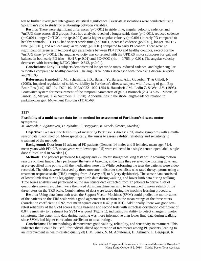
International Congress of Parkinson’s Disease and Movement Disorders®
Hong Kong October 5-9, 2018 – Guided Poster Tour Abstracts
test to further investigate inter-group statistical significance. Bivariate associations were conducted using
Spearman’s rho to study the relationship between variables.
Results: There were significant differences (p<0.001) in stride time, angular velocity, cadence, and
7mTUG time across all 3 groups. Post-hoc analysis revealed a longer stride time (p<0.001), reduced cadence
(p<0.001), longer 7mTUG time (p<0.001) and a higher angular velocity (p<0.001) in early PD compared to
healthy controls. PD+FOG had shorter stride time (p<0.001), increased cadence (p<0.001), longer 7mTUG
time (p<0.001), and reduced angular velocity (p<0.001) compared to early PD cohort. There were no
significant differences in temporal gait parameters between PD+FOG and healthy controls, except for the
7mTUG time (p<0.001). The angular velocity was correlated with the UPDRS motor subscores for gait and
balance in both early PD (rho= -0.417, p<0.01) and PD+FOG (rho= -0.785, p<0.01). The angular velocity
decreased with increasing %FOG (rho= -0.642, p<0.01).
Conclusions: Early PD subjects demonstrated longer stride times, reduced cadence, and higher angular
velocities compared to healthy controls. The angular velocities decreased with increasing disease severity
and %FOG.
References: Hausdorff, J.M., Schaafsma, J.D., Balash, Y., Bartels, A.L., Guverich, T. & Giladi, N.
(2003). Impaired regulation of stride variability in Parkinson's disease subjects with freezing of gait. Exp
Brain Res (149) 187-194. DOI: 10.1007/s00221-002-1354-8. Hausdorff J.M., Ladin Z. & Wei, J.Y. (1995).
Footswitch system for measurement of the temporal parameters of gait. J Biomech (28) 347-351. Morris, M.
Iansek, R., Matyas, T. & Summers, J. (1998). Abnormalities in the stride length-cadence relation in
parkinsonian gait. Movement Disorder (13) 61-69.
1117
Feasibility of a multi-sensor data fusion method for assessment of Parkinson’s disease motor
symptoms
M. Memedi, S. Aghanavesi, D. Nyholm, F. Bergquist, M. Senek (Örebro, Sweden)
Objective: To assess the feasibility of measuring Parkinson’s disease (PD) motor symptoms with a multi-
sensor data fusion method. More specifically, the aim is to assess validity, reliability and sensitivity to
treatment of the methods.
Background: Data from 19 advanced PD patients (Gender: 14 males and 5 females, mean age: 71.4,
mean years with PD: 9.7, mean years with levodopa: 9.5) were collected in a single center, open label, single
dose clinical trial in Sweden [1].
Methods: The patients performed leg agility and 2-5 meter straight walking tests while wearing motion
sensors on their limbs. They performed the tests at baseline, at the time they received the morning dose, and
at pre-specified time points until the medication wore off. While performing the tests the patients were video
recorded. The videos were observed by three movement disorder specialists who rated the symptoms using a
treatment response scale (TRS), ranging from -3 (very off) to 3 (very dyskinetic). The sensor data consisted
of lower limb data during leg agility, upper limb data during walking, and lower limb data during walking.
Time series analysis was performed on the raw sensor data extracted from 17 patients to derive a set of
quantitative measures, which were then used during machine learning to be mapped to mean ratings of the
three raters on the TRS scale. Combinations of data were tested during the machine learning procedure.
Results: Using data from both tests, the Support Vector Machines (SVM) could predict the motor states
of the patients on the TRS scale with a good agreement in relation to the mean ratings of the three raters
(correlation coefficient = 0.92, root mean square error = 0.42, p<0.001). Additionally, there was good test-
retest reliability of the SVM scores during baseline and second tests with intraclass-correlation coefficient of
0.84. Sensitivity to treatment for SVM was good (Figure 1), indicating its ability to detect changes in motor
symptoms. The upper limb data during walking was more informative than lower limb data during walking
since SVMs had higher correlation coefficient to mean ratings.
Conclusions: The methodology demonstrates good validity, reliability, and sensitivity to treatment. This
indicates that it could be useful for individualized optimization of treatments among PD patients, leading to
an improvement in health-related quality of[1] M. Senek, S. M. Aquilonius, H. Askmark, F. Bergquist, R.
International Congress of Parkinson’s Disease and Movement Disorders®
Hong Kong October 5-9, 2018 – Guided Poster Tour Abstracts
Constantinescu, A. Ericsson, et al., "Levodopa/carbidopa microtablets in Parkinson's disease: a study of
pharmacokinetics and blinded motor assessment," Eur J Clin Pharmacol, Jan 18 2017. [2] C. G. Goetz, G. T.
Stebbins, K. A. Chung, R. A. Hauser, J. M. Miyasaki, A. P. Nicholas, et al., "Which dyskinesia scale best
detects treatment response?," Mov Disord, vol. 28, pp. 341-6, Mar 2013. life.
FIG. 1 (1117)
1118
Assessment of Motor Symptoms in Parkinson’s disease using Flexible Wearable Sensors and Deep
Neural Networks: A CIS-PD substudy
L. Lonini, A. Dai, C. Poon, N. Shawen, L. Shimanovich, T. Simuni, D. Daeschler, J. Rogers, A. Jayaraman
(Chicago, IL, USA)
Objective: To develop an algorithm to characterize in-office motor symptoms of Parkinson’s disease
(PD) using multiple wireless sensors, and to compare symptom recognition in activities of daily living
(ADLs) vs. standard clinical assessments.
Background: Objective assessment of PD motor disability could greatly impact design of PD clinical
trials and clinical care. It still remains to be determined what is the minimal number of sensors necessary to
provide accurate characterization of PD motor function, specifically motor fluctuations, while patients are
engaged in routine activities of daily living (ADLs).
Methods: This study was conducted as a single center sub-study of the Michael J Fox Foundation (MJFF)
funded clinician input study (CIS-PD). A subset of 14 participants (mean=61.6y; SD=10.1y) were fitted with
10 sensors [figure1], assessed with serial MDS-UPDRS and performed standardized ADL tasks including
walking and fine dexterity movements. The tasks were performed first in the medications OFF state and then
5 more times every 30 minutes after taking medications (ON state). All exams were videotaped. An
additional, single assessment was performed 2 weeks later. A clinician rated the severity of tremor,
bradykinesia and dyskinesia of the upper-extremities on a scale from 0-4. We trained a convolutional neural
network model to detect whether tremor or bradykinesia were present (score > 0) in 5-second recordings of
the raw sensor data. A leave-one-visit-out cross validation was performed to assess the accuracy of the model
at predicting symptoms at different time points.
Results: This report is limited to the analysis of the upper extremities sensors. Area under the curve
(AUC) of the model, averaged across tasks and time, was 0.81 (SD=0.047) for bradykinesia detection, and
0.87 (SD=0.041) for tremor. Bradykinesia detection was higher during walking and assembling nuts and
bolts (median recall=0.87); recall of tremor was comparable between walking and finger to nose tasks (0.69,
p=0.48)[figure2].

International Congress of Parkinson’s Disease and Movement Disorders®
Hong Kong October 5-9, 2018 – Guided Poster Tour Abstracts
Conclusions: Flexible sensors are a non-invasive method to continuously record movement data in PD.
Deep networks can successfully detect PD symptoms from such data. The analysis of the full dataset and
minimal number of sensors to optimize model accuracy is underway. All raw data from this study will be
made publicly available by MJFF.
FIG. 1 (1118)
FIG. 2 (1118)
International Congress of Parkinson’s Disease and Movement Disorders®
Hong Kong October 5-9, 2018 – Guided Poster Tour Abstracts
1124
Objective monitoring of drug response in early PD patients using remote, at-home typing data through
machine learning analysis
M. Matarazzo, T. Arroyo-Gallego, P. Montero Escribano, V. Puertas-Martín, I. Butterworth, C. S. Mendoza,
MJ. Ledesma-Carbayo, MJ. Catalán, JA. Molina-Arjona, F. Bermejo-Pareja, JC. Martínez Castrillo, L.
López-Manzanares, A. Alonso-Cánovas, J. Herreros-Rodríguez, I. Obeso, P. Martínez Martín, JC. Martínez-
Ávila, A. Gómez de-la-Cámara, M. Gray, JA. Obeso, L. Giancardo, Á. Sánchez-Ferro (Vancouver, BC,
Canada)
Objective: We designed an algorithm to detect response to medication in an early PD population using at-
home, unsupervised, unobtrusive typing data.
Background: Advances in technology are opening a new era to remotely evaluate people with PD. In
previous studies we have shown that features of inlab keyboard typing can be used to evaluate motor skills
and to classify subjects as having PD or not [1]. More recently we have shown the same capability from
typing on a touchscreen based information and from keyboard data at-home [2]. We now hypothesized that
typing on an electronic device, a habitual behavior, likely controlled by the nigrostriatal dopaminergic
pathway, could allow for objectively and non-obtrusively monitoring parkinsonian features and response to
medication in an at-home setting.
Methods: We designed a naturalistic prospective validation study to evaluate whether typing patterns
changed in accordance with responsiveness to medication. 31 early PD subjects, who were going to start a
dopaminergic drug, and 30 matched controls were enrolled. We remotely monitored their typing pattern over
a 6-month follow-up period while antiparkinsonian medications were being titrated (fig.1). A novel deep
learning algorithm (nQRNN) was developed to detect participants' outcome defined as the response to
medication assessed by the UPDRS-III minimal clinically important difference (MCID) at the final visit (6
months). Further, we tested if this model could predict that outcome earlier than 6 months.
Results: The nQRNN had an overall moderate kappa agreement (k=0.50) and fair 0.73 area under the
ROC curve with the time-coincident UPDRS-III MCID-based classification of response (fig.2).
Furthermore, the nQRNN at week 3 (and beyond) could reliably predict which subjects would respond and
which wouldn't. (fig.3)
Conclusions: This preliminary study suggests that a habitual task based on remotely-gathered
unsupervised typing data at home allows for an accurate and predictive classification of drug response in PD.
If confirmed by a larger prospective study, this approach could provide supplementary information to
clinicians for a more continuous monitoring of motor symptoms of PD, thus helping to take informed
decision on therapeutic strategies and disease management. Also, this tool could be useful as a cost effective
and reliable outcome measure for clinical trials to test response to medication.
References: 1. Giancardo L, Sánchez-Ferro Á, Arroyo-Gallego T, et al. Computer keyboard interaction as
an indicator of early Parkinson’s disease. Sci Rep 2016;6:34468. doi:10.1038/srep34468. 2. Arroyo-Gallego,
T, Ledesma-Carbayo, M, Butterworth, I, et al. Detecting Motor Impairment in Early Parkinson's Disease via
Natural Typing Interaction with Keyboards: Validation of the neuroQWERTY Approach in an Uncontrolled
At-Home Setting. JMIR 2018 in press. doi: 10.2196/jmir.9462.
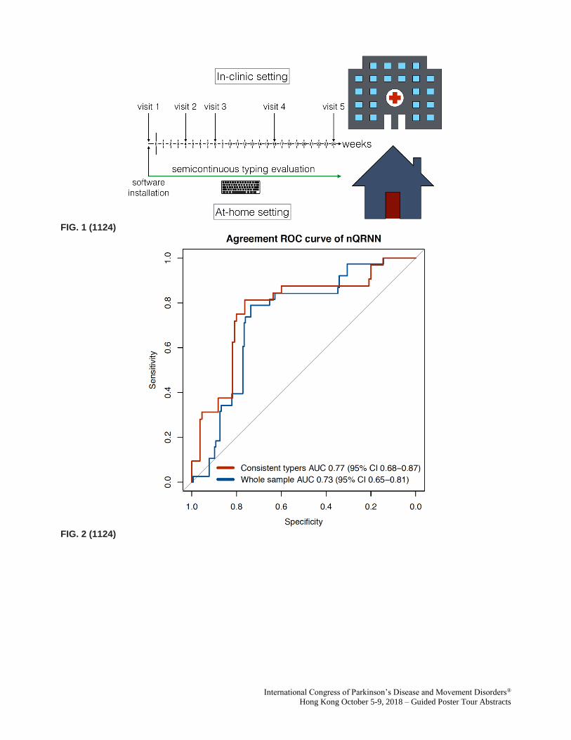
International Congress of Parkinson’s Disease and Movement Disorders®
Hong Kong October 5-9, 2018 – Guided Poster Tour Abstracts
FIG. 1 (1124)
FIG. 2 (1124)

International Congress of Parkinson’s Disease and Movement Disorders®
Hong Kong October 5-9, 2018 – Guided Poster Tour Abstracts
FIG. 3 (1124)
1138
The Levodopa Response Trial and the Parkinson Disease Digital Biomarker Challenge: Monitoring
symptoms of Parkinson’s disease in the lab and home using wearable sensors
J. Daneault, G. Vergara-Diaz, G. Costante, E. Fabara, G. Ferreira-Carvalho, F. Golabchi, F. Parisi, S.
Sapienza, Y. Chae, P. Snyder, P. Aubin, P. Banda, D. Brunner, R. Dorsey, L. Mangravite, W. Marks, E. Neto,
U. Rubin, E. Soderberg, D. Daeschler, S. Moore, S. Sieberts, L. Omberg, P. Bonato, The Parkinson's Disease
Digital Biomarker DREAM Challenge Consortium (Newark, NJ, USA)
Objective: To leverage a community of researchers and shared wearable data to develop algorithms to
estimate the severity of PD specific symptoms.
Background: People with Parkinson’s disease (PwPD) often experience fluctuations in motor symptom
severity. Wearable sensors have the potential to help clinicians monitor symptoms over time, outside the
clinic. However, to gather accurate and clinically-relevant measures, there is a need to develop robust
algorithms based on clinically-labelled data.
Methods: The Levodopa Response Trial captured three-axis acceleration from two wrist-worn sensors
and a smartphone located at the waist from 29 PwPD continuously over 4 days. On day 1, in an in-clinic
visit, participants performed clinical assessments and motor tasks on their regular medication regimen.
During these visits, a clinician also provided symptom severity scores for tremor, bradykinesia, and
dyskinesia. On days 2 & 3, sensor data was collected while participants were at home. On day 4, participants
returned to the clinic for the same assessments as day 1, but arrived without having taken their medication for
at least 10 hours.
Leveraging this dataset, Sage Bionetworks, the Michael J Fox Foundation and the Robert Wood Johnson
Foundation launched the PD Digital Biomarker DREAM Challenge which made a subset of the data
available to researchers to develop robust and accurate algorithms for the estimation of specific symptoms’
severity.
Results: Teams participating in the challenge used several technical approaches, from signal processing
to deep learning. 35 submissions were received for the estimation of action tremor severity. Teams achieved
an area under the precision-recall curve (AUPR) of 0.444 to 0.75. As for dyskinesia during movement, 37
submissions were received and the teams achieved an AUPR of 0.175 to 0.477. Finally, 39 submissions were
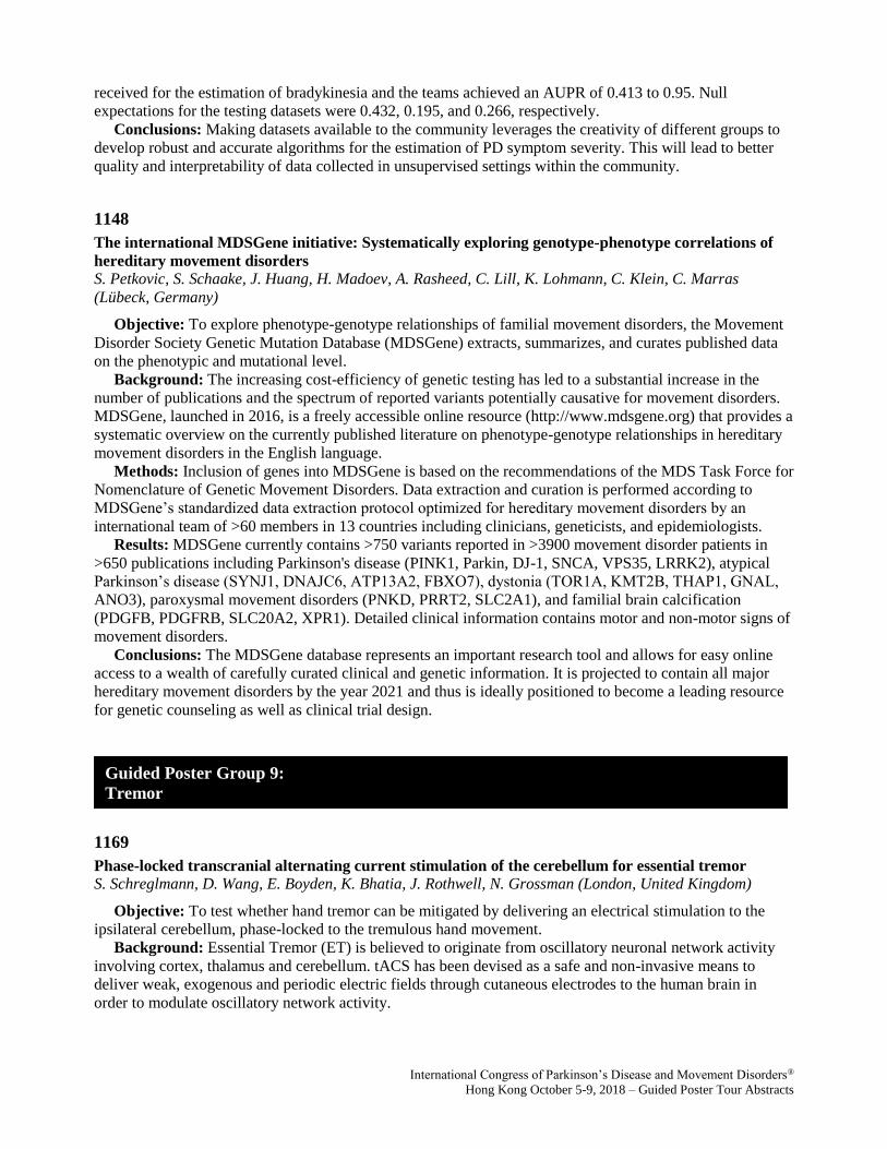
International Congress of Parkinson’s Disease and Movement Disorders®
Hong Kong October 5-9, 2018 – Guided Poster Tour Abstracts
received for the estimation of bradykinesia and the teams achieved an AUPR of 0.413 to 0.95. Null
expectations for the testing datasets were 0.432, 0.195, and 0.266, respectively.
Conclusions: Making datasets available to the community leverages the creativity of different groups to
develop robust and accurate algorithms for the estimation of PD symptom severity. This will lead to better
quality and interpretability of data collected in unsupervised settings within the community.
1148
The international MDSGene initiative: Systematically exploring genotype-phenotype correlations of
hereditary movement disorders
S. Petkovic, S. Schaake, J. Huang, H. Madoev, A. Rasheed, C. Lill, K. Lohmann, C. Klein, C. Marras
(Lübeck, Germany)
Objective: To explore phenotype-genotype relationships of familial movement disorders, the Movement
Disorder Society Genetic Mutation Database (MDSGene) extracts, summarizes, and curates published data
on the phenotypic and mutational level.
Background: The increasing cost-efficiency of genetic testing has led to a substantial increase in the
number of publications and the spectrum of reported variants potentially causative for movement disorders.
MDSGene, launched in 2016, is a freely accessible online resource (http://www.mdsgene.org) that provides a
systematic overview on the currently published literature on phenotype-genotype relationships in hereditary
movement disorders in the English language.
Methods: Inclusion of genes into MDSGene is based on the recommendations of the MDS Task Force for
Nomenclature of Genetic Movement Disorders. Data extraction and curation is performed according to
MDSGene’s standardized data extraction protocol optimized for hereditary movement disorders by an
international team of >60 members in 13 countries including clinicians, geneticists, and epidemiologists.
Results: MDSGene currently contains >750 variants reported in >3900 movement disorder patients in
>650 publications including Parkinson's disease (PINK1, Parkin, DJ-1, SNCA, VPS35, LRRK2), atypical
Parkinson’s disease (SYNJ1, DNAJC6, ATP13A2, FBXO7), dystonia (TOR1A, KMT2B, THAP1, GNAL,
ANO3), paroxysmal movement disorders (PNKD, PRRT2, SLC2A1), and familial brain calcification
(PDGFB, PDGFRB, SLC20A2, XPR1). Detailed clinical information contains motor and non-motor signs of
movement disorders.
Conclusions: The MDSGene database represents an important research tool and allows for easy online
access to a wealth of carefully curated clinical and genetic information. It is projected to contain all major
hereditary movement disorders by the year 2021 and thus is ideally positioned to become a leading resource
for genetic counseling as well as clinical trial design.
1169
Phase-locked transcranial alternating current stimulation of the cerebellum for essential tremor
S. Schreglmann, D. Wang, E. Boyden, K. Bhatia, J. Rothwell, N. Grossman (London, United Kingdom)
Objective: To test whether hand tremor can be mitigated by delivering an electrical stimulation to the
ipsilateral cerebellum, phase-locked to the tremulous hand movement.
Background: Essential Tremor (ET) is believed to originate from oscillatory neuronal network activity
involving cortex, thalamus and cerebellum. tACS has been devised as a safe and non-invasive means to
deliver weak, exogenous and periodic electric fields through cutaneous electrodes to the human brain in
order to modulate oscillatory network activity.
Guided Poster Group 9:
Tremor

International Congress of Parkinson’s Disease and Movement Disorders®
Hong Kong October 5-9, 2018 – Guided Poster Tour Abstracts
Methods: We tested this stimulation modality on postural tremor in ET patients (n=11) in a quadruple
replication, semi-randomized block-design including six phase-locked, one non phase-locked stimulation and
one sham condition.
Each block of stimulation included 15s of baseline period, 30s of stimulation period and 15s of post
stimulation period. Tremor activity measured by a tri-axial accelerometer was used to compute the
instantaneous tremor phase in real-time using a Hilbert transformation-based algorithm implemented on a
microcontroller (Arduino Due). A sinusoidal voltage waveform was generated a) with a frequency equalling
the tremor frequency adjusted to have a fixed lag of 0°, 60°, 120°, 180°, 240° or 300° relative to the
instantaneous tremor phase (phase-locked condition), b) at the tremor frequency without adjustment (non
phase-locked condition), c) and dropped to zero after the initial 5 s ramp-up period (sham condition). The
stimulation signal was fed to an isolated current source DS4 (Digitimer Ltd., Welwyn Garden City, UK).
Stimulation was delivered just above the individual cutaneous sensory threshold via an active (30x30mm)
and return (50x50mm) skin electrode placed over the lateral cerebellar hemisphere and contralateral, frontal
cortex.
After removal of sessions with baseline tremor amplitude ± 2 SD of mean baseline recordings per subject,
offline analysis was done using MatLab (The MathWorks). Changes in tremor amplitude were calculated as
z-scores ± SD.
Results: Phase-locked stimulation was achieved reliably throughout the phase-spectrum. A comparison of
tremor amplitude across all blocks showed a significant reduction in tremor amplitude during phase-locked (-
2.22 ± 1.5) versus non phase-locked stimulation (-0.94 ± 0.92; p=0.01; paired t-test).
Conclusions: Our data provide first evidence that phase-locked tACS can modulate ET tremor amplitude,
most likely by influencing network activity via cerebellar cortical stimulation.
1174
Gamma Knife Radiosurgery for essential and parkinsonian tremor: Long-term experience in a
Spanish center
JR. Perez-Sanchez, R. Martinez-Alvarez, NE. Martinez-Moreno, I. Cuervo-Arango, G. Rey Portoles, I.
Parees, A. Del Barrio, J. Alvarez-Linera, MM. Kurtis (Madrid, Spain)
Objective: To describe the experience of a single Spanish center in gamma knife thalamotomy (GKT) as
treatment of refractory tremor in patients with Essential Tremor (ET) and Parkinson´s Disease (PD).
Background: GKT is a minimally invasive stereotactic surgical option to treat medically refractory
tremor in patients with ET and PD, particularly indicated for elderly and fragile patients. Recent studies have
shown good results in terms of safety and efficacy (1,2). However, long-term evidence is limited, especially
for PD tremor.
Methods: During a 5-year period (2013-2017), 12 patients underwent unilateral GKT and were followed-
up for at least 12 months with motor and neuropsychiatric evaluations. Five patients had asymmetric tremor
predominant PD, 4 cases had intractable ET and 3 patients had ET-PD. Mean age was 78.8±6.8 years old (6
patients >75 years old). 2 patients were on anticoagulants. Leksell Gamma Knife was used to target the
ventral intermediate nucleus with a radiation dose of 130 Gy. Efficacy was scored based on subscales of the
Fahn-Tolosa-Marin (FTM) tremor rating scale, target-side tremor subscales of the motor part of the Unified
Parkinson´s Disease Rating Scale (UPDRS), and EuroQoL-5D to assess quality of life.
Results: Median follow-up duration was 22 months (12-54 months). After GKT, 11 patients (91.7%)
improved their tremor scores. 6 patients (50%), 3 PD, 2 ET and 1 ET+PD reported tremor erradication or
minimal symptoms. Tremor improvement was sustained at the end of follow-up in 10 out of 11 patients
(90.9%). Mean FTM scores improved significantly: for hand postural tremor, from 2.86 pre to 1.14
(p=0.006); for kinetic tremor, from 3.14 to 1.29 (p=0.007); for handwriting, from 2.57 to 1.29 (p=0.016); and
for drinking, from 3.14 to 1.71 (p=0.015). Mean motor UPDRS scores significantly decreased: for rest
tremor in the targeted upper limb, from 3.00 to 0.71 (p=0.001); and for overall tremor in the target-side, from
10.29 to 2.14 (p=0.002). EuroQoL-5D ameliorated from 8.89 to 6.56 (p=0.027). No severe adverse events

International Congress of Parkinson’s Disease and Movement Disorders®
Hong Kong October 5-9, 2018 – Guided Poster Tour Abstracts
occurred. Three patients reported transient hand paresthesias, cognitive disturbances without changes in
neurophysicological tests and depression, respectively.
Conclusions: This study suggests that GKT is a safe and long-term efficacious treatment option for
medically refractory ET, and may be considered for asymmetric tremor predominant PD, especially in
patients who are elderly, on anticoagulants or have high surgical risk that contraindicates DBS.
References: 1. Martínez-Moreno NE, Sahgal A, De Salles A, Hayashi M, Levivier M, Ma L, Paddick I,
Régis J, Ryu S, Slotman J, Martínez-Álvarez R. Stereotactic radiosurgery for tremor: systematic review. J
Neurosurg. 2018 Feb 23:1-12. 2. Niranjan A, Raju SS, Kooshkabadi A, Monaco E, Flickinger JC, Lunsford
LD. Stereotactic radiosurgery for essential tremor: Retrospective analysis of a 19-year experience. Mov
Disord. 2017;32(5):769-777.
1175
A new clinical and research smartphone application to assess tremor and bradykinesia in patients with
movement disorders
C. Duval, J.F. Daneault, B. Carignan, C.É. Coderre, S. Bogard (Montreal, QC, Canada)
Objective: Determine the ability of on-board gyroscopes in smartphones to assess the performance of
rapid alternating movements (RAM) during fast repetitive pronation-supination movements at the wrist, a
measure of core bradykinesia.
Background: Smartphones are now ubiquitous, and possess several sensors capable of measuring
tremulous activity and voluntary movements with relatively good accuracy. Following our work on the
validation of smartphone accelerometers for the assessment of abnormal tremors (1,2), we developed a free
smartphone application for patients, physicians and researchers who are interested in the assessment of
abnormal tremors and bradykinesia. The app can characterize several characteristics of rest, kinetic and
postural tremors and quantify characteristics of bradykinesia from RAM such as mean and maximal velocity
of pronation-supination movements of the hand using onboard gyroscopes. However, the accuracy of
gyroscopes to assess RAM performance during a fast repetitive pronation-supination movements of the
forearm remains to be determined.
Methods: The accuracy of the gyroscopes of two smartphones, one Iphone 6s and one Android LG-D852
(Android 6.0) was assessed against an optical rotational encoder having an accuracy of 0.33 deg. Both
phones were linked and held against a ball attached to the optical rotational encoder. Once recording
initiated, the phones were pronated and supinated simultaneously with the ball. 5 trials of thirty seconds
were performed, each having different amplitudes, velocity and variability characteristics. From those,
analysis was done on 5-second epochs. First, the rotational axis was automatically detected. Second,
differences in sampling rate were adjusted and signals from rotational encoders were differentiated to
velocity and aligned with data from both smartphones. Correlation was used to assessed agreement between
signals; rotational encoders versus Iphone and Android, as well both smartphones against each other.
Results: Mean correlations between time series of both smartphones were r = 0.997 ± 0.003. Mean
correlations between time series from the rotational encoder and those from the iPhone and Android phone
were r = 0.909 ± 0.100 and 0.910 ± 0.099, respectively. All correlations were significant at p<0.05.
Conclusions: Our results demonstrate the ability of on-board gyroscopes in smartphones to assess RAM
performance during fast repetitive movements at the wrist. This will enable us to propose a proven method to
assess core bradykinesia in patients with movements disorders.
References: 1. Daneault JF, Carignan B, Codère CÉ, Sadikot AF, Duval C. (2013) Using a smart phone
as a standalone platform for detection and monitoring of pathological tremors. Front Hum Neurosci.
18;6:357. 2. Carignan B, Daneault JF, Duval C (2015) Measuring tremor with a smartphone. Methods Mol
Biol. 1256:359-74.
International Congress of Parkinson’s Disease and Movement Disorders®
Hong Kong October 5-9, 2018 – Guided Poster Tour Abstracts
1177
Shaky legs: The clinical spectrum and treatment of orthostatic tremor; a systematic review
A. Buijink, M. Meulepas, A. van Rootselaar (Amsterdam, Netherlands)
Objective: To provide a systematic literature overview of orthostatic tremor (OT).
Background: Orthostatic tremor (OT) is characterized by a high-frequency tremor of the leg muscles
during stance, resulting in an unsteady feeling. The pathophysiology is still not understood. Treatment
options are limited and are often unsatisfactory. A systematic literature review on primary OT is lacking the
literature.
Methods: Here we review data of a total of 617 OT-patients, with a focus on primary OT, retrieved after
a systematic search in Pubmed (583 individual patients from 45 case reports, 20 case series, and 7 therapeutic
trials) and from a questionnaire-based study in Dutch OT-patients (n=34).
Results: Overall, 67% of identified OT patients is female; mean age at onset is 57 (17-81) years. Most
patients were primary OT cases, however this distinction is not always made. Six percent (22 of 390 cases)
has a positive family history for OT. Mean delay to diagnosis is 7.7 years (n=268). A substantial number of
patients reports falls. Quality of life is affected. Clonazepam is the most prescribed drug but is not always
effective. Trials report a possible positive effect of gabapentin. Deep brain stimulation (DBS), performed in a
limited number of patients, has shown a positive effect.
Conclusions: OT can be disabling and is under-recognized. The most effective treatment remains
uncertain, with some evidence for clonazepam and gabapentin. DBS provides an alternative for drug-
resistant and disabling OT, although long term effects are still unknown. To gain more insight in OT, it is of
importance to differentiate primary OT from OT-plus and secondary OT. Increasing awareness for OT will
shorten the delay in time to diagnosis and possibly lead to development of better treatment options.
1182
The spectrum of involuntary movements in patients with motor neuron disease – a cross-sectional
study
K. Vogelnik, L. Dolenc Grošelj, B. Koritnik, L. Leonardis, J. Zidar, M. Kojović (Ljubljana, Slovenia)
Objective: To describe prevalence, clinical characteristics and electrophysiological findings of
involuntary movements in patients with motor neuron disease (MND).
Background: Involuntary movements are not considered typical finding in MND. However, we have
frequently observed involuntary jerks in patients with otherwise typical MND.
Methods: Sixty-five consecutive patients followed in MND outpatient clinic were clinically examined by
movement disorders experts and video-recorded. The presence and quality of involuntary movements was
noted and severity was graded on a scale from + to +++. Movements were further classified in one of the
following: minipolymiyoclonus, thumb rest tremor, large amplitude irregular hand tremor and regular small
amplitude hand tremor. In proportion of patients, surface poly-electromyography (EMG) and accelerometry
were performed.
Results: 44/65 patients had involuntary movements. 27 had non-rhythmical, small amplitude, random
finger jerks present at rest and/or activation, suggestive of minipolymyoclonus. Tremor, defined as rhythmic,
oscillation of a body part, was noted in 34 patients. Action hand tremor was big amplitude and irregular in 17
patients, while 11 patients had more regular and smaller amplitude tremor. All except one tremor patient able
to perform finger to nose test also had intention tremor component. (Table 1). 17 patients had thumb rest
tremor, almost always associated with pseudo-dystonic thumb posture (16/17). Accelerometry confirmed the
presence of tremor with frequencies ranging from 4 – 7.5 Hz. Poly EMG showed that patients were almost
never in a state of complete relaxation. Tremor and minipolymyoclonus were commonly associated with
firing of enlarged motor units (MU) (Figure 1 and 2).
Conclusions: Contrary to widespread belief, involuntary movements are present in the majority of
patients with MND. Based on the analysis of EMG and accelerometry recording, we hypothesize that
pseudodystonic thumb posture, thumb “rest” tremor, minipolymiyoclonus and irregular large-amplitude
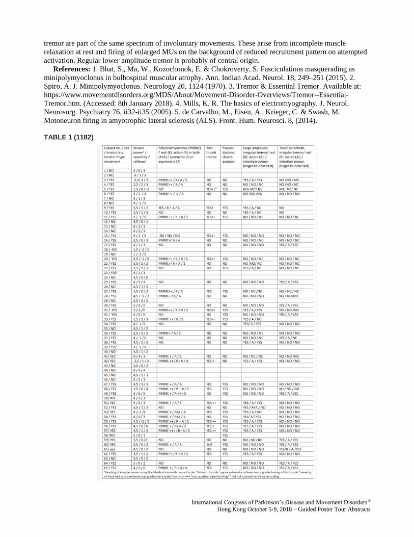
International Congress of Parkinson’s Disease and Movement Disorders®
Hong Kong October 5-9, 2018 – Guided Poster Tour Abstracts
tremor are part of the same spectrum of involuntary movements. These arise from incomplete muscle
relaxation at rest and firing of enlarged MUs on the background of reduced recruitment pattern on attempted
activation. Regular lower amplitude tremor is probably of central origin.
References: 1. Bhat, S., Ma, W., Kozochonok, E. & Chokroverty, S. Fasciculations masquerading as
minipolymyoclonus in bulbospinal muscular atrophy. Ann. Indian Acad. Neurol. 18, 249–251 (2015). 2.
Spiro, A. J. Minipolymyoclonus. Neurology 20, 1124 (1970). 3. Tremor & Essential Tremor. Available at:
https://www.movementdisorders.org/MDS/About/Movement-Disorder-Overviews/Tremor--Essential-
Tremor.htm. (Accessed: 8th January 2018). 4. Mills, K. R. The basics of electromyography. J. Neurol.
Neurosurg. Psychiatry 76, ii32-ii35 (2005). 5. de Carvalho, M., Eisen, A., Krieger, C. & Swash, M.
Motoneuron firing in amyotrophic lateral sclerosis (ALS). Front. Hum. Neurosci. 8, (2014).
TABLE 1 (1182)
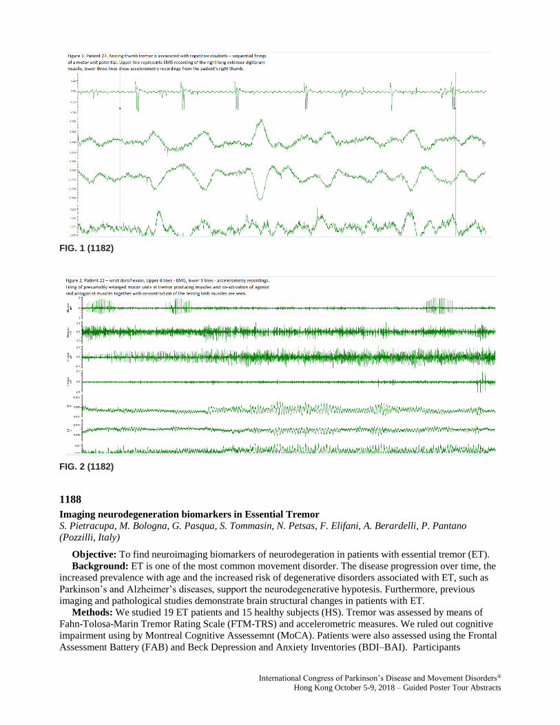
International Congress of Parkinson’s Disease and Movement Disorders®
Hong Kong October 5-9, 2018 – Guided Poster Tour Abstracts
FIG. 1 (1182)
FIG. 2 (1182)
1188
Imaging neurodegeneration biomarkers in Essential Tremor
S. Pietracupa, M. Bologna, G. Pasqua, S. Tommasin, N. Petsas, F. Elifani, A. Berardelli, P. Pantano
(Pozzilli, Italy)
Objective: To find neuroimaging biomarkers of neurodegeration in patients with essential tremor (ET).
Background: ET is one of the most common movement disorder. The disease progression over time, the
increased prevalence with age and the increased risk of degenerative disorders associated with ET, such as
Parkinson’s and Alzheimer’s diseases, support the neurodegenerative hypotesis. Furthermore, previous
imaging and pathological studies demonstrate brain structural changes in patients with ET.
Methods: We studied 19 ET patients and 15 healthy subjects (HS). Tremor was assessed by means of
Fahn-Tolosa-Marin Tremor Rating Scale (FTM-TRS) and accelerometric measures. We ruled out cognitive
impairment using by Montreal Cognitive Assessemnt (MoCA). Patients were also assessed using the Frontal
Assessment Battery (FAB) and Beck Depression and Anxiety Inventories (BDI–BAI). Participants

International Congress of Parkinson’s Disease and Movement Disorders®
Hong Kong October 5-9, 2018 – Guided Poster Tour Abstracts
underwent a standardized 3T-MRI protocol. Total grey matter (GM) and white matter (WM) volume were
assessed by means of SIENAX (FSL toolbox), cortical thickness (Cth) using by CAT 12 (SPM 12 toolbox)
and subcortical volumes using by FIRST-FSL. Finally, we assessed iron deposition obtained from SWAN
images in seven specific Regions of Interest (ROIs): thalamus, putamen, globus pallidus, caudate, substantia
nigra, red nucleus and dentate nucleus. We then investigated possible correlations between clinical scores
and accelerometric measures of tremor and neuroimaging data.
Results: No differences in total GM, WM and Cth were found between the two groups. Moreover, no
differences in terms of iron accumulation were observed between ET patients and HS. Subcortical volumes
analysis demonstrated significant differences in thalamus volume bilaterally (p<0.015) in the comparison
CS>ET, positively correlating with MoCA. By contrast, thalamus volumes did not correlate with tremor
severity, as assessed by clinical scores and accelerometric measures.
Conclusions: Apart for the observation of thalamic atrophy, our study indicates no structural brain
abnormalities supporting neurodegenerative hypothesis in ET. Thalamic atrophy positively correlated with
cognitive measures in patients, providing further insight into the pathophysiological mechanisms of the
disease. However, the lack of correlation between tremor severity and thalamic volume indicates that
structural abnormalities does not necessarily play a key role in generating tremor in this condition.
1190
Quantitative characterisation of tremor in functional and organic tremor patients
Z. Dominguez-Vega, G. Kramer, J. Elting, M. de Koning-Tijssen, N. Maurits (Groningen, Netherlands)
Objective: To quantify tremor characteristics using inertial sensors in functional and organic tremor
patients and to present the differences in tremor quantification between these two groups, during long term
tremor recordings and to determine the minimum number of days necessary to obtain reliable estimates.
Background: Distinguishing tremor types is important for treatment and prognosis. One major distinction
is between tremor with an organic cause (organic tremor; OT) and functional tremor (FT). FT tends to have
less stable characteristics compared to organic tremor; exhibiting frequent interruptions, irregularities and
frequency changes [1]. Fluctuations in tremor characteristics affect all patients, making it difficult to assess
these from a momentary observation in the clinic. Long-term continuous monitoring would facilitate overall
occurrence and treatment response assessment.
Methods: Inertial sensor data were recorded from 44 tremor patients (16 FT, 28 OT) during
unconstrained activities of daily living during 30 days, each day over a 10-hour period. Sensors were
attached to the dorsal side of the forearm (close to the wrist). Start and end of recording per day were
obtained from electronic patient diaries. The accelerometer signal and a tremor identification algorithm [2]
implementing the periodogram were used to identify time windows with tremor, from which the percentage
of tremor and tremor frequency variability were calculated per patient across all days. Non-parametric
distributions were generated and Z-tests performed to determine whether estimates of tremor characteristics
obtained from the first 3, 5, 7 or 10 days were representative of estimates obtained from any 3, 5, 7 or 10
days within the 30 days.
Results: Tremor percentage ranged from 10 to 34% for FT patients and from 4.7 to 70.0% for OT
patients. Frequency variability ranged from 0.6 to 2.1 Hz for FT patients and from 0.4 to 1.5 Hz for OT
patients. Seven days of tremor recording were the minimum number of days resulting in a non-significant Z-
test, meaning that seven days are the shortest period of time needed to obtain reliable estimates of tremor
characteristics.
Conclusions: Using long-term recordings with inertial sensors, fluctuations in tremor characteristics over
the days could be observed in FT as well as OT patients based on percentage of tremor and tremor frequency
variability. Seven days of recording are sufficient to determine these characteristics reliably.
References: [1] P. E. O’Suilleabhain and J. Y. Matsumoto, “Time-frequency analysis of tremors.,” Brain,
vol. 121 ( Pt 11), pp. 2127–34, Nov. 1998. [2] O. Martinez Manzanera, J. W. Elting, J. H. van der Hoeven,
and N. M. Maurits, “Tremor Detection Using Parametric and Non-Parametric Spectral Estimation Methods:
A Comparison with Clinical Assessment,” PLoS One, vol. 11, no. 6, p. e0156822, Jun. 2016.

International Congress of Parkinson’s Disease and Movement Disorders®
Hong Kong October 5-9, 2018 – Guided Poster Tour Abstracts
1203
Repetitive Transcranial Magnetic Stimulation Therapy is a Potential Therapeutic option for Primary
Orthostatic Tremor
W. Hu, J. Legacy, A. Ferng, A. Wagle Shukla (Gainesville, FL, USA)
Objective: Determine whether low frequency repetitive transcranial magnetic stimulation (rTMS) therapy
to the cerebellum improves symptoms in primary orthostatic tremor (POT)
Background: POT is a rare disabling tremor disorder characterized by high frequency tremor appearing
in the legs upon standing. Treatment opportunities for POT are poor. Previous research found an increased
cerebellar activity and low frequency rTMS reduces increased brain excitability.
Methods: Patients diagnosed with POT participated in a randomized double-blind placebo-controlled
crossover-design study. All subjects received single session of real and sham rTMS session delivered to each
cerebellar hemisphere on two separate days in random order. Nine hundred pulses were delivered
consecutively to each side at a frequency of 1 Hz and at an intensity of 90% of the resting motor threshold
(hand motor area). The primary outcome was the mean difference between the two arms for the Fullerton
Advanced Balance Rating Scale (FABRS) standing score and the blinded video-rated standing duration
measured immediately after rTMS (T1) and 60+ minutes after rTMS (T2) compared to before rTMS therapy
(baseline, T0).
Results: 10 POT subjects (6 females and 4 males, mean age 70.2 ± 8.0 years, mean disease duration 13.5
± 5.8 years) participated and tolerated the therapy well. Compared to baseline, subjects randomized to the
real arm improved significantly on the FABRS standing score (baseline score 1.3; T1 score 2.3; p = 0.02) at
T1 than those in the sham arm. With regards to video-rated standing duration time, there was mean increase
of 126.0 ± 172.8 sec in the real arm compared to 12.9 ± 49.9 sec in the sham arm that approached
significance (p = 0.11). However, at an individual level, there were 8/10 subjects in the real arm showed
improvement (figure). No significant improvements were seen at T2.
Conclusions: Low frequency rTMS to cerebellum is a promising therapy for POT however requires
confirmation in a larger follow-up study. A longer duration of rTMS will likely result in longer benefits.
FIG. 1 (1203)

International Congress of Parkinson’s Disease and Movement Disorders®
Hong Kong October 5-9, 2018 – Guided Poster Tour Abstracts
1210
Tremor in motor neuron disease may be central rather than peripheral in origin
A. Latorre, L. Rocchi, M. Stamelou, A. Batla, M. Ciocca, B. Balint, K. Sidle, A. Berardelli, J. Rothwell, K.
Bhatia (London, United Kingdom)
Objective: To investigate the pathophysiology of action tremor in motor neuron disease (MND).
Background: MND refers to a spectrum of degenerative diseases affecting motor neurons. Recent
clinical and postmortem observations have revealed considerable variability in the phenotype (1). Rhythmic
involuntary oscillations of the hands during action, resembling tremor, can occur in MND, but its
pathophysiology has not yet been investigated.
Methods: One hundred and twenty consecutive MND patients were screened for tremor. Twelve patients
with action tremor and no other movement disorders were found. Ten took part in the study. Tremor was
recorded bilaterally using surface electromyography and triaxial accelerometer, with and without a variable
weight load. Power spectra of rectified electromyography and accelerometric signal were calculated. To
investigate a possible cerebellar involvement, eye blink classic conditioning (EBCC) was performed in five
patients.
Results: Action tremor was present in about 10% of our population. All patients showed distal postural
tremor of low amplitude and constant frequency, bilateral with a small degree of asymmetry. Two of them
showed also simple kinetic tremor. A peak at the electromyography and accelerometric recordings ranging
from 4 Hz to 12 Hz was found in all patients. Loading did not change peak frequency in either the
electromyographic or accelerometric power spectra. Compared with healthy volunteers, patients had a
smaller number of conditioned responses during EBCC.
Conclusions: our data suggest that MND patients can present with action tremor of a central origin,
possibly due to a cerebellar dysfunction. This evidence supports the novel idea of MND as a multisystem
neurodegenerative disease and that action tremor can be part of this condition.
References: 1. Sabatelli M, Conte A, Zollino M. Clinical and genetic heterogeneity of amyotrophic
lateral sclerosis. Clinical genetics. 2013;83(5):408-16.
FIG. 1 (1210)
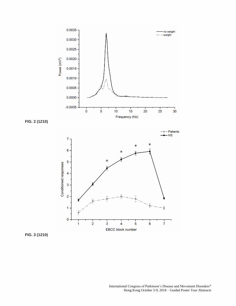
International Congress of Parkinson’s Disease and Movement Disorders®
Hong Kong October 5-9, 2018 – Guided Poster Tour Abstracts
FIG. 2 (1210)
FIG. 3 (1210)
International Congress of Parkinson’s Disease and Movement Disorders®
Hong Kong October 5-9, 2018 – Guided Poster Tour Abstracts
1213
Regional homogeneity changes detected between Essential tremor with resting tremor and tremor-
dominant Parkinson’s disease
J. Li, Z. Lu, X. Suo, N. Li, L. Wang, J. Peng, J. Zhang, Q. Gong, R. Peng (Chengdu, China)
Objective: To explore neural activity patterns in the essential tremor (ET) with resting tremor (rET) and
tremor-dominant Parkinson's disease (tPD) by using resting-state functional magnetic resonance imaging (rs-
fMRI) and regional homogeneity (ReHo) method.
Background: Differentiating between rET from tPD may be challenging in the early phases of the
diseases. Although some imaging studies have indicated that there are differences between rET and tPD,
little is known about the rs-fMRI findings.
Methods: Our study conducted rs-fMRI with a ReHo method to investigate the modulations of neural
activity in 58 ethnic Han Chinese subjects comprising 19 patients of rET, 24 patients of tPD and 25 age- and
gender-matched healthy controls. Differences were compared by two-sample t tests (corrected with
AlphaSim, p<0.05). All participants underwent clinical assessment and the correlation with ReHo was
analyzed.
Results: Compared with healthy controls, rET patients and tPD patients both displayed ReHo alterations
in left inferior temporal gyrus and right inferior orbitofrontal lobe. In addition, rET patients exhibited more
ReHo changes in the inferior temporal gyrus, while tPD patients in extensive cortical and sub-cortical areas,
including frontal, temporal, putamen, cingulum, precuneus, and cerebelum, which are involved both in basal
ganglia and the cerebello-thalamo-cortical (CTC) loops. Direct comparison between rET and tPD displayed
significant differences in the primary visual cortex, and in brain regions within the so-called default mode
network (DMN, e.g., precuneus, cingulum).
Conclusions: Our results indicated the rET and tPD share common parrerns of ReHo abnormalities in the
temporal and orbitofrontal lobe, which may contribute to the broad spectrum of olfactory, visual, emotional
and behavioral disturbances observed in these two diseases. In addition, we found disorder-specific
involvement of temporal in rET and the basal ganglia and the CTC loops in tPD. These findings provide new
evidence regarding the shared and specific neuropathological mechanisms that underlie rET and tPD.
1299
PLA2G6-related juvenile-onset Parkinsonism: clinical features and cognitive profile in a cohort of
Chinese patients
C. Chen, Y.M. Sun, F.T. Liu, S.S. Luo, Z.T. Ding, J.J. Wu, J. Wang (Shanghai, China)
Objective: Among young or juvenile onset Parkinson (PD) patients, we assessed cognitive and clinical
performances, comparing homozygotes and compound heterozygotes who carry PLA2G6 pathogenic
mutations with non-carriers and age-matched healthy controls.
Background: Phospholipase A2 Group 6 (PLA2G6) is the causative gene for three recessively inherited
neurodegeneration disease, namely infantile neuroaxonal dystrophy (INAD) and neurodegeneration
associated with brain iron accumulation (NBIA) and PLA2G6 -linked juvenile onset dystonia-parkinsonism
(PARK14). Although the phenotypic spectrum of PARK14 had different features from classic Parkinson's
disease, no quantitative study has been conducted in comparing clinical manifestations and cognitive profiles
between cohorts of PLA2G6-related cases and other young onset Parkinson's disease cases.
Methods: We collected 10 Chinese patients with homozygous or compound heterozygous PLA2G6
mutations from the outpatient neurology clinics of Huashan Hospital consecutively who met the MDS
diagnostic criteria of clinically probable PD or established PD with an age at onset of younger than 40 years
old. Pathogenicity of the PLA2G6 mutations was evaluated by enzyme activity. Unified Parkinson Disease
Guided Poster Group 10:
Parkinson's Disease: Genetics

International Congress of Parkinson’s Disease and Movement Disorders®
Hong Kong October 5-9, 2018 – Guided Poster Tour Abstracts
Rating Scale Part III (UPDRS-III) and neuropsychological performance of the patients was measured. The
neuropsychological scores of age and sex matched healthy controls was also collected.
Results: PLA2G6-related Juvenile onset Parkinson's disease (PARK14) patients had no statistical
difference in age at onset of PD, gender, disease duration and age at examination compared to noncarriers.
More pyramid signs (P<0.001), dyskinesia (P=0.019), higher Beck depression inventory scores (P=0.002)
and higher RBD questionnaire scores (P=0.046) were observed in PLA2G6 mutation carriers. Carriers
developed posture instability (P=0.029) and dyskinesia (P=0.030) more quickly. They performed poorer than
noncarriers on the Mini-Mental State Examination (P = 0.005) and were more likely to receive lower scores
on other neuropsychological tests. PARK14 patients had a remarkable impairment in language domain
(P=0.015). No significant difference was observed in attention, execution, memory and visuospatial
cognitive domains.
Conclusions: In this study, PARK14 patients demonstrated poorer cognitive performance and motor
performance than did noncarriers, especially in language domain, suggesting a more rapid disease
progression and a different pattern of cognitive impairment. A longitudinal follow-up and a functional
imaging study is required to confirm these findings.
FIG. 1 (1299)
1304
Resistance to Parkinson's disease among LRRK2 mutation carriers is associated with higher plasma
levels of urate but not its purine precursors
M. Schwarzschild, R. Bakshi, R. Logan, M. Zorlu, X. Chen, A. Ascherio, E. Macklin (Boston, MA, USA)
Objective: To determine whether plasma purine concentrations differ between people with Parkinson's
disease (PD) and matched controls among carriers of pathogenic mutations in Leucine-Rich Repeat Kinase 2
(LRRK2) gene.
Background: LRRK2 mutations (mLRRK2) are the most common genetic cause of PD but are
incompletely penetrant. Understanding the genetic and environmental factors that reduce LRRK2 mutation
penetrance could help predict and possibly prevent development of PD among healthy people who are at
high risk due to a mLRRK2. Urate is an endogenous purine with antioxidant, Nrf2 activator and
neuroprotectant properties, and elevated serum or CSF concentrations are predictive of a reduced risk of
idiopathic PD and a slower rate of PD progression.
Methods: Urate and other purines were measured in plasma samples by enzymatic and HPLC-UV/ECD
assays. Baseline levels from 380 subjects (120 mLRRK2+PD-, 120 mLRRK2+PD+, 70 mLRRK2-PD-, 70
mLRRK2-PD+) matched for key covariates from the LRRK2 Cohort Consortium (LCC) and from 1,053

International Congress of Parkinson’s Disease and Movement Disorders®
Hong Kong October 5-9, 2018 – Guided Poster Tour Abstracts
subjects (241 mLRRK2+PD-, 210 mLRRK2+PD+, 202 mLRRK2-PD-, 400 mLRRK2-PD+) in the
Parkinson Progression Marker Initiative (PPMI) database were compared, adjusting for age and sex.
Results: Among mLRRK2 carriers in the LCC, unaffected subjects had higher levels of urate ± SE (5.0 ±
1.3 mg/dL) than those with PD (4.2 ± 1.2 mg/dL) by 0.8 mg/dL after adjusting for sex and age (p<0.0001).
There was no significant difference between urate's purine precursors hypoxanthine and xanthine. A smaller
difference was observed among subjects without a mLRRK2; plasma urate in unaffected subjects was 0.4
mg/dL greater than in PD (p=0.02). Similarly, among mLRRK2+ (in contrast to mLRRK2-) subjects in the
PPMI cohort, those who were unaffected had higher levels of urate than those with PD (by 0.4 mg/dL) after
adjusting for sex and age (p<0.001); adjusting further for BMI yielded the same result (p<0.001).
Conclusions: The reproducible finding of higher plasma urate levels among those resistant to PD despite
a LRRK2 mutation supports the hypothesis that higher urate is a marker and possibly a mediator of the
incomplete penetrance of LRRK2 mutations.
1307
Interest in Genetic Testing in PD Patients with DBS
A. Fraint, G. Pal, L. Verhagen, D. Hall, K. Marder (Chicago, IL, USA)
Objective: Determine interest in genetic testing (GT) among PD patients with DBS.
Background: About 26-29% of PD patients who undergo DBS have a mutation in one of three genes:
glucocerebrosidase (GBA), leucine-rich repeat kinase 2, and parkin. As access to GT for PD increases, it is
critical to understand the current knowledge base regarding GT in PD subjects and their expectations
regarding how genetic information impacts their treatment.
Methods: Consecutive non-demented PD patients with bilateral STN-DBS were recruited. The Genetic
Attitude Questionnaire (GAQ) - an assessment of GT knowledge - was administered to subjects.
Demographic and clinical data were compared using student t-test or Mann-Whitney U as appropriate.
Subjects were dichotomized according to whether they did or did not want GT. Correct genetic knowledge
responses were quantified for all subjects and then compared using chi-square tests according to desire to
obtain GT and GBA mutation status. Identical analysis was performed for reasons for desiring GT. A
multivariate logistic regression model was used to identify predictors of desire to obtain GT.
Results: One hundred subjects were enrolled and complete GAQ data were available for 88 (Table 1).
Fourteen respondents (16%) were GBA mutation carriers. The mean percent of correct responses for all
subjects regarding genetic knowledge was 58.5% (Fig 1). Genetic knowledge was not significantly higher in
subjects who desired GT (p>0.05) nor among GBA mutation carriers (p>0.05). Fifty-three subjects (60%)
would opt for GT if it could predict their response to DBS. Subjects who desired GT were more likely than
those who did not to be aware that genes have been identified which confer a higher risk of developing PD (p
= 0.02). Both those who desired GT and those who did not were more likely to consider eligibility in clinical
trials an important consideration in making their decision (95.83% and 76.67%) (Fig 2). In a multivariate
logistic regression model, age, disease duration, sex, UPDRS-III scores, HY stage, and GBA mutation status
were not predictive of desire to obtain GT (p’s>0.05).
Conclusions: Most patients would want GT it if it could predict response to DBS. Knowledge gaps were
identified in this advanced PD cohort. Desire to be eligible for clinical trials was associated with the desire
for GT.

International Congress of Parkinson’s Disease and Movement Disorders®
Hong Kong October 5-9, 2018 – Guided Poster Tour Abstracts
TABLE 1 (1307)
FIG. 1 (1307)
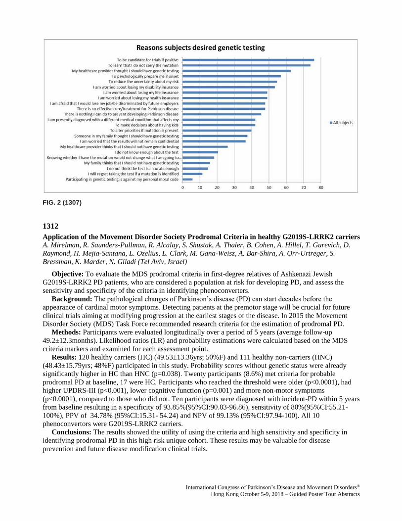
International Congress of Parkinson’s Disease and Movement Disorders®
Hong Kong October 5-9, 2018 – Guided Poster Tour Abstracts
FIG. 2 (1307)
1312
Application of the Movement Disorder Society Prodromal Criteria in healthy G2019S-LRRK2 carriers
A. Mirelman, R. Saunders-Pullman, R. Alcalay, S. Shustak, A. Thaler, B. Cohen, A. Hillel, T. Gurevich, D.
Raymond, H. Mejia-Santana, L. Ozelius, L. Clark, M. Gana-Weisz, A. Bar-Shira, A. Orr-Urtreger, S.
Bressman, K. Marder, N. Giladi (Tel Aviv, Israel)
Objective: To evaluate the MDS prodromal criteria in first-degree relatives of Ashkenazi Jewish
G2019S-LRRK2 PD patients, who are considered a population at risk for developing PD, and assess the
sensitivity and specificity of the criteria in identifying phenoconverters.
Background: The pathological changes of Parkinson’s disease (PD) can start decades before the
appearance of cardinal motor symptoms. Detecting patients at the premotor stage will be crucial for future
clinical trials aiming at modifying progression at the earliest stages of the disease. In 2015 the Movement
Disorder Society (MDS) Task Force recommended research criteria for the estimation of prodromal PD.
Methods: Participants were evaluated longitudinally over a period of 5 years (average follow-up
49.2±12.3months). Likelihood ratios (LR) and probability estimations were calculated based on the MDS
criteria markers and examined for each assessment point.
Results: 120 healthy carriers (HC) (49.53±13.36yrs; 50%F) and 111 healthy non-carriers (HNC)
(48.43±15.79yrs; 48%F) participated in this study. Probability scores without genetic status were already
significantly higher in HC than HNC (p=0.038). Twenty participants (8.6%) met criteria for probable
prodromal PD at baseline, 17 were HC. Participants who reached the threshold were older (p<0.0001), had
higher UPDRS-III (p<0.001), lower cognitive function (p=0.001) and more non-motor symptoms
(p<0.0001), compared to those who did not. Ten participants were diagnosed with incident-PD within 5 years
from baseline resulting in a specificity of 93.85%(95%CI:90.83-96.86), sensitivity of 80%(95%CI:55.21-
100%), PPV of 34.78% (95%CI:15.31- 54.24) and NPV of 99.13% (95%CI:97.94-100). All 10
phenoconvertors were G2019S-LRRK2 carriers.
Conclusions: The results showed the utility of using the criteria and high sensitivity and specificity in
identifying prodromal PD in this high risk unique cohort. These results may be valuable for disease
prevention and future disease modification clinical trials.

International Congress of Parkinson’s Disease and Movement Disorders®
Hong Kong October 5-9, 2018 – Guided Poster Tour Abstracts
1326
LRRK2 and GBA genetic mutations are not uncommon in an unselected Ashkenazi elderly cohort
with PD
S. Isaacson, J. Isaacson (Boca Raton, FL, USA)
Objective: Analysis of positive LRRK2 and GBA gene mutations, demographic characteristics, and
family history in 1200 consecutive Ashkenazi patients with PD who had genetic testing for LRRK2 and
GBA mutations.
Background: Patients with Parkinson’s disease who are of Ashkenazi Jewish descent may have a LRRK2
and/or GBA gene mutation. Approximately 30-40% of unaffected LRRK2 gene carriers and approximately
8% of GBA gene carriers may develop clinical motor PD during their lifetime. It is unknown if PD diagnosis
occurs earlier in life due to the LRRK2 or GBA gene, or if elderly have lower vs. higher risk.
Methods: Genetic testing was conducted at our site through the MJFF PPMI genetic screening program.
We reviewed prodromal clinical characteristics of unaffected relatives of PD patients and also of patients
already diagnosed with PD who were found to have the LRRK2 and/or GBA gene mutation associated with
Ashkenazi PD. We also compared these gene positive to those who were tested but did not have these gene
mutations. We also compared PD gene-positive and gene-negative with a family history of PD to those
without.
Results: Over 1200 patients underwent genetic testing after clinical counseling for LRRK2, and
approximately half of these for GBA gene too. Of 350 unaffected relatives with a PD family history tested,
32 had LRRK2 gene mutation and 150 were also tested for GBA with 10 with a GBA gene mutation. Of 850
patients with PD, approximately 50% of patients reported a known family history of PD. Of patients with a
family history, 48 had LRRK2 gene and 18 had GBA gene. Of patients with PD not reporting a family
history of PD, 24 had LRRK2 gen and 12 had GBA gene.
Conclusions: Routine gene testing of patients with PD who have Ashkenazi heritage (ie one grandparent)
may be of value as these genes seem relatively common in patients reporting a known family history of PD
and ven in those unaware of a PD family history, As new therapies enter clinical development, PD patients of
Ashkenazi descent will need counseling that LRRK2 and GBA gene mutations are not uncommon, have
autosomal dominant inheritance to to their children, and may be amenable to trials of emerging therapies
seeking to slow or retard progression of PD.
1340
BDNF(V66M), EIF4G1(R1205H), VPS35(D620N) gene polymorphisms in South Indian PD Patients
T. Syed, T. S.D, S. Meka, S. Kumar, S. Thandra, V. Kutala, R. Kandadai, R. Borgihain (Hyderabad, India)
Objective: To investigate the association of BDNF(V66M), EIF4G1(R1205H) and VPS35(D620N)
polymorphisms in South Indian PD patients.
Background: Parkinson’s disease (PD) is the most common form of movement disorder that embroiled
multiple neuro-anatomical areas, due to various genetic and environmental factors. Advances in genetic
analysis, Next Generation Sequencing, and Genome Wide Association Studies helps to identify the risk
genes and understand the pathological pathway associated with PD development and progression. BDNF
plays a role in differentiation, survival and maintenance of neurons in central and peripheral nervous system.
In PD, the expression of BDNF (Brain Derived Neurotropic factor) NA is decreased there by making it a
susceptible candidate gene. Studies have revealed that EIF4G1 (Eukaryotic Translation Initiation Factor 4
Gamma 1) variants may impair the ability of cells to rapidly and dynamically respond to stress, thus probably
participating in PD development. Variations in VPS35 (Vacuolar Protein Sorting-Associated Protein 35) may
disrupt the retrograde transport system and thereby contributing to dopaminergic neuronal cell death in PD.
Methods: A total of 168 PD patients and 151 age and ethnicity matched controls were included in the
study. Blood samples were collected after taking the consent. DNA was isolated genetic analysis was done
by PCR-RFLP method.
International Congress of Parkinson’s Disease and Movement Disorders®
Hong Kong October 5-9, 2018 – Guided Poster Tour Abstracts
Results: The frequency of genotypic distribution of BDNF (V66M) polymorphism among the PD cases
and controls were: C/C – 60.1 % and 67.5%, C/T – 28.6% and 26.4%, and T/T was 11.3 % and 5.9%
respectively. The Allelic frequency distribution of C & T alleles in cases and controls were C- 74.4 % and
80.7%, T allele – 25.5% and 19.2% respectively. The Fishers exact test revealed that, there is a borderline
significant difference between cases and controls (C/C versus C/T+T/T, OD 1.44, CI at 95% 0.95-2.18, P
value 0.058. The frequencies of GG genotype of EIF4G1 (R1205H) and CC of VPS35 (D620N)
polymorphisms among PD cases and controls are 100 %. There is no occurrence of mutant either in cases or
in controls.
Conclusions: From the results we conclude that BDNF (V66M), EIF4G1 (R1205H) and VPS35 (D620N)
polymorphisms are not associated with the PD risk in South Indian population.
1341
Full sequencing and haplotype analysis reveals LRRK2 protective haplotype in REM sleep behavior
disorder
B. Ouled Amar-Bencheikh, J. Ruskey, I. Arnulf, Y. Dauvilliers, C. Charley Monaca, V. Cochen De-Cock, JF.
Gagnon, D. Spiegelman, M. Hu, B. Högl, A. Stefani, L. Ferini-Strambi, G. Plazzi, E. Antelmi, P. Young, A.
Heidbreder, B. Mollenhauer, F. Sixel-Döring, C. Trenkwalder, W. Oertel, J. Montplaisir, R. Postuma, G.
Rouleau, Z. Gan-Or (Montreal, QC, Canada)
Objective: To examine the role of LRRK2 mutations and variants in susceptibility for RBD.
Background: Rapid eye movement (REM)-sleep behavior disorder (RBD) is, in most cases, a prodromal
synucleinopathy, likely to progress to either Parkinson’s disease (PD), dementia with Lew-bodies (DLB) or
multiple system atrophy (MSA) in rare cases. There is only partial overlap between the genetics of RBD and
PD. LRRK2 mutations that are known to cause PD are rare in RBD, yet thus far no comprehensive, full
sequencing study of LRRK2 was performed in RBD.
Methods: A total of 350 RBD patients, diagnosed by clinical interview and polysomnography according
to the International Classification of Sleep Disorders, version 2 (ICSD-2) criteria, and 869 controls
participated in the current study. The full coding sequence, exon-intron boundaries and 5’ and 3’ untranslated
regions of LRRK2 were sequenced using targeted next-generation sequencing with molecular inversion
probes (MIPs). Regression and burden models were used to examine the association between LRRK2
variants and RBD.
Results: None of the known PD-causing LRRK2 pathogenic mutations was identified in RBD patients.
Interestingly, the haplotype that includes the variants p.N551K-p.R1398H-p.K1423K was associated with a
reduced RBD risk (OR=0.66, 95% CI 0.44-0.98, p=0.0055 for the tagging p.N551K substitution). In
addition, a common variant, p.S1647T, was nominally associated with risk for RBD (OR=1.28, 95% CI 1.05-
1.56, p=0.029), but was not significant after correction for multiple comparisons. Burden analysis identified
associations with domains and exons that were derived by the variants of the protective haplotype, and no
burden of other rare variants was identified.
Conclusions: Carriage of the LRRK2 p.N551K-p.R1398H-p.K1423K haplotype is associated with a
reduced risk for RBD. However, the known PD-causing LRRK2 mutations probably have minor or no role in
RBD. Additional studies are needed to replicate these results and to identify the mechanism associated with
reduced risk for RBD.
1353
Association of GBA polymorphisms and mutations with dementia in Parkinson disease: A 7-year study
of three population-based incident cohorts
K. Lunde, J. Chung, I. Dalen, K. Pedersen, J. Linder, M. Domellöf, E. Elgh, A. Macleod, C. Tzoulis, J.
Larsen, O. Tysnes, L. Forsgren, C. Counsell, G. Alves, J. Maple-Grødem (Stavanger, Norway)
Objective: To study the effect of GBA variants on dementia in three deeply phenotyped population-based
prospective cohorts of patients with incident Parkinson disease.
International Congress of Parkinson’s Disease and Movement Disorders®
Hong Kong October 5-9, 2018 – Guided Poster Tour Abstracts
Background: Dementia is among the most common and severe non-motor symptoms of Parkinson
disease (PD), affecting nearly 20% of all patients within the first 5 years of the disease, and the majority of
patients if they survive for more than 10 years after diagnosis. Both polymorphisms and mutations in GBA
may influence the development of dementia in patients with Parkinson’s disease, but few longitudinal studies
have investigated the role of GBA variant subcategories in disease heterogeneity and progression.
Methods: Three Northern European population-based studies (the Swedish NYPUM study, the
Norwegian ParkWest study and the Scottish PINE study) designed to determine the incidence, neurobiology
and prognosis of Parkinson disease were analyzed for GBA genetic variants by SNP genotyping assays or
whole exome sequencing. A total of 442 patients and 419 controls were followed for seven years and
dementia was diagnosed using established criteria. GBA variant carriers were analyzed as one group, and
subcategorized into “polymorphism” or “deleterious mutation” carriers. Associations between GBA carrier
status and dementia were assessed with Cox survival analysis.
Results: A total of 12.0% of patients with Parkinson’s disease were carriers of a GBA variant, and nearly
half of these (22/53) progressed to dementia during follow-up. Carriers of deleterious GBA mutations
(adjusted HR 3.81, 95% CI 1.35 to 10.72; P = .011) or polymorphisms (adjusted HR 1.79; 95% CI 1.07 to
3.00; P = .028) progressed to dementia more rapidly than non-carriers.
Conclusions: GBA variants are of great clinical relevance for the development of dementia in Parkinson
disease, especially due to the relatively higher frequency of these alleles in patients with Parkinson disease
compared to other risk alleles.
1370
Molecular mechanisms of GCH1-associated Parkinson's disease
J. Terbeek, W. Vandenberghe (Leuven, Belgium)
Objective: To unravel the molecular mechanisms by which loss of GCH1 function enhances the risk of
Parkinson´s disease (PD).
Background: Loss-of-function mutations in GCH1 are the most common cause of autosomal dominant
DOPA-responsive dystonia (DRD), a non-neurodegenerative movement disorder. GCH1 encodes GTP
cyclohydrolase 1, the rate-limiting enzyme in the biosynthesis of tetrahydrobiopterin (BH4). BH4 is an
essential cofactor for tyrosine hydroxylase, the rate-limiting enzyme in the synthesis of dopamine. Recent
genetic evidence indicates that GCH1 mutations also increase the risk of PD. The mechanisms by which
GCH1 mutations predispose to nigrostriatal cell death, are unknown.
Methods: We cultured skin fibroblasts from 2 patients (Patient 1, Patient 2) with the same heterozygous
missense mutation in GCH1 (p.Y75S; c. 224A>C) and from 2 healthy age-matched controls (Control 1,
Control 2). Patient 1 was a 42-year-old male who developed clinically typical DRD at the age of 8 years and
had severely abnormal dopamine transporter (DAT) imaging at the age of 37 years. Patient 2, the 63-year-old
mother of Patient 1, developed clinically typical DRD at the age of 8 years and had normal DAT imaging at
the age of 60. We induced GCH1 expression in the fibroblasts by 24-hour treatment with interferon-gamma.
We assessed apoptosis using TUNEL staining, western blotting for PARP and immunostaining for cleaved
caspase 3.
Results: GCH1 abundance after IFN-gamma treatment was lower in the mutant fibroblasts than in their
respective controls. Moreover, GCH1 abundance was lower in Patient 1 than in Patient 2. Fibroblasts from
Patient 1 were more susceptible to staurosporine- and H2O2-induced apoptosis than those of Control 1. By
contrast, staurosporine- and H2O2-induced apoptosis did not differ between Patient 2 and Control 2. The
survival defect of fibroblasts of Patient 1 was rescued by incubation with sepiapterin, a precursor of BH4 via
the GCH1-independent salvage pathway, indicating that the survival defect was caused by BH4 deficiency.
Conclusions: The clinical phenotypes of the 2 GCH1 mutant patients correlated with the susceptibility of
their skin fibroblasts to apoptosis in vitro. As skin fibroblasts do not produce dopamine, our findings suggest
that GCH1 mutations can impair cellular survival via mechanisms unrelated to dopamine synthesis defects.
International Congress of Parkinson’s Disease and Movement Disorders®
Hong Kong October 5-9, 2018 – Guided Poster Tour Abstracts
1376
Presynaptic dopamine depletion determines the timing of levodopa-induced dyskinesia onset in
Parkinson’s disease
HS. Yoo, SJ. Chung, BS. Ye, YH. Sohn, PH. Lee (Seoul, Republic of Korea)
Objective: To investigated whether dopaminergic function in the nigrostriatal system is associated with
the timing of levodopa-induced dyskinesia (LID) onset in Parkinson's disease (PD).
Background: LID is one of the most debilitating effects of levodopa therapy, and is associated with a
poor quality of life and health-related costs. In terms of pathogenesis, two main factors are involved in the
development of LID. The first is nigral dopaminergic denervation of the striatum presynaptically, and the
second consists of plastic changes in striatal dopaminergic neuron cell signalling that leads to abnormal basal
ganglion firing patterns postsynaptically. Striatal dopaminergic depletion is required for the development of
LID and predicts LID in patients with PD. Reduced presynaptic dopaminergic activity plays an important
role in the development of LID in PD.
Methods: From among 412 drug-naive PD patients who underwent a dopamine transporter (DAT) PET
scan during their baseline evaluation, we enrolled 65 patients who developed LID during a follow-up period
of > 2years. Based on the time from PD onset, LID was classified as early, intermediate or late onset. We
then compared DAT availability in the striatal subregions of the patients in the three groups.
Results: Patients with de novo PD with LID had lower putaminal DAT activity than those without LID
[Figure 1]. The demographic characteristics did not differ among the early-onset, intermediate-onset, and
late-onset groups except for earlier intervention of levodopa therapy in the early LID onset group (p =
0.001). After adjusting for age at PD onset, gender, timing of levodopa therapy from PD onset, and the
severity of PD motor symptoms, DAT activity in the posterior putamen was found to be significantly lower
in the early LID onset group than in the late LID onset group (p = 0.017) [Table 1]. Multivariate linear
regression analysis showed that low DAT activity in the posterior putamen was significantly associated with
the early appearance of LID in the early LID onset group (β = 16.039, p = 0.033).
Conclusions: This study demonstrated that low DAT activity in the posterior putamen at baseline is a
major risk factor for the early onset of LID in patients with PD, suggesting that the degree of presynaptic
dopaminergic denervation plays an important role in determining the timing of LID onset.
Guided Poster Group 11:
Parkinson's Disease: Neuroimaging And Neurophysiology

International Congress of Parkinson’s Disease and Movement Disorders®
Hong Kong October 5-9, 2018 – Guided Poster Tour Abstracts
FIG. 1 (1376)
TABLE 1 (1376) Initial striatal DAT activities in the patients with Parkinson disease with levodopa-induced dyskinesia

International Congress of Parkinson’s Disease and Movement Disorders®
Hong Kong October 5-9, 2018 – Guided Poster Tour Abstracts
1388
Automated Differential Diagnosis of Parkinsonian Syndromes Using FDG-PET Metabolic Brain
Network Analysis
T. Rus, P. Tomše, L. Jensterle, M. Grmek, Z. Pirtošek, D. Eidelberg, C. Tang, M. Trošt (Ljubljana, Slovenia)
Objective: To discriminate among parkinsonian patients using an automated network analysis of FDG-
PET brain images.
Background: Clinical differentiating among parkinsonian syndromes: Parkinson’s disease (PD), multiple
system atrophy (MSA) and progressive supranuclear palsy (PSP) may be challenging early in disease course.
Disease specific metabolic brain patterns were identified for PD, MSA and PSP using Scaled Subprofile
Modelling/Principal Component Analysis of FDG-PET images: PD related pattern (PDRP), MSA related
pattern (MSARP) and PSP related pattern (PSPRP) (Poston KL, et al. 2010). Additionally, a probabilistic
algorithm for the classification of individual patients with clinically uncertain parkinsonism was developed
based on the expression of metabolic covariance patterns specific for PD, MSA and PSP (Tang CC, et al.
2010).
Methods: 137 patients with parkinsonism (disease duration 4.9±4 years) and uncertain clinical diagnosis
underwent diagnostic FDG-PET. A network analysis was performed and the expressions of PDRP, MSARP
and PSPRP were calculated using Topographic Profile Rating method. A probability for PD, MSA and PSP
was calculated using the two stage logistic algorithm that distinguishes between PD and atypical
parkinsonian syndrome (APS) in the first stage, and between MSA and PSP in the second. The calculated
diagnosis was than compared to final clinical diagnosis made 21.4±13 months after imaging.
Results: Among 137 patients, 38 were excluded for alternative (not PD, MSA or PSP) diagnosis. Among
99 patients 66 were clinically diagnosed with PD, 17 with MSA and 16 with PSP. Automated algorithm
diagnosed 57 patients with PD, 9 with MSA, 18 with PSP. Results were indeterminate in 15 cases.
Discriminative measures of diagnostic accuracy for the first stage (PD vs atypical parkinsonian syndrome
(APS)) and the second stage of diagnostic algorithm (MSA and PSP) are presented in table 1 (PPV – positive
predictive value, NPV – negative predictive value). [table 1]
Conclusions: FDG-PET brain imaging with an automated differential diagnostic algorithm (Tang CC, et
al. 2010) is a reliable tool for the improvement of early diagnosis in patients with uncertain parkinsonism.
References: 1. Poston KL, Eidelberg D. FDG PET in the Evaluation of Parkinson's Disease. PET Clin.
2010;5(1):55-64. 2. Tang CC, Poston KL, Eckert T, et al. Differential diagnosis of parkinsonism: a metabolic
imaging study using pattern analysis. Lancet Neurol. 2010;9(2):149-58.
TABLE 1 (1388)
1400
Parkinson's disease motor symptoms are linked to red nucleus volume and cerebellar metabolism
A. Bonnet, R. Sanford, A. Riou, S. Drapier, F. Le Jeune, M. Vérin, D.L. Collins (Rennes, France)
Objective: To investigate the involvement of the red nucleus (RN) and cerebellum in the motor
symptoms of Parkinson's disease (PD).
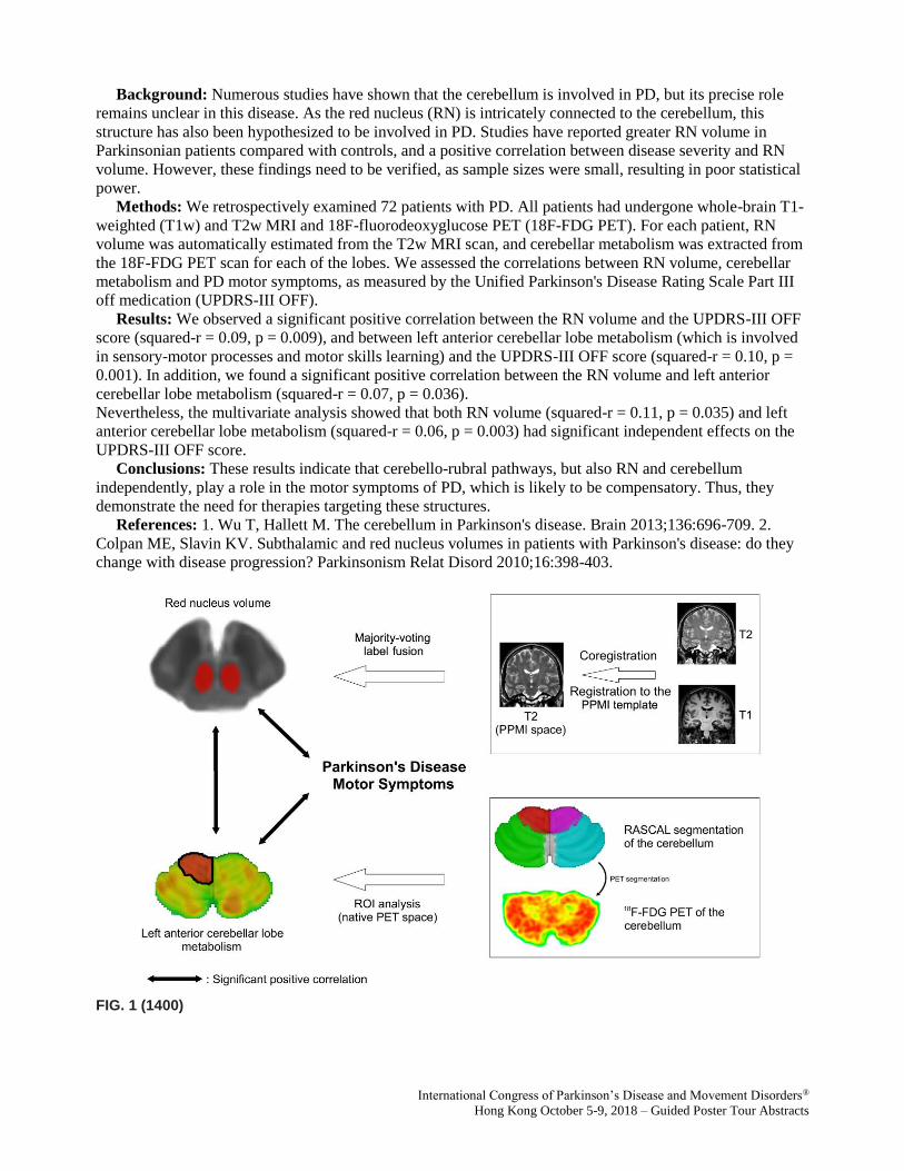
International Congress of Parkinson’s Disease and Movement Disorders®
Hong Kong October 5-9, 2018 – Guided Poster Tour Abstracts
Background: Numerous studies have shown that the cerebellum is involved in PD, but its precise role
remains unclear in this disease. As the red nucleus (RN) is intricately connected to the cerebellum, this
structure has also been hypothesized to be involved in PD. Studies have reported greater RN volume in
Parkinsonian patients compared with controls, and a positive correlation between disease severity and RN
volume. However, these findings need to be verified, as sample sizes were small, resulting in poor statistical
power.
Methods: We retrospectively examined 72 patients with PD. All patients had undergone whole-brain T1-
weighted (T1w) and T2w MRI and 18F-fluorodeoxyglucose PET (18F-FDG PET). For each patient, RN
volume was automatically estimated from the T2w MRI scan, and cerebellar metabolism was extracted from
the 18F-FDG PET scan for each of the lobes. We assessed the correlations between RN volume, cerebellar
metabolism and PD motor symptoms, as measured by the Unified Parkinson's Disease Rating Scale Part III
off medication (UPDRS-III OFF).
Results: We observed a significant positive correlation between the RN volume and the UPDRS-III OFF
score (squared-r = 0.09, p = 0.009), and between left anterior cerebellar lobe metabolism (which is involved
in sensory-motor processes and motor skills learning) and the UPDRS-III OFF score (squared-r = 0.10, p =
0.001). In addition, we found a significant positive correlation between the RN volume and left anterior
cerebellar lobe metabolism (squared-r = 0.07, p = 0.036).
Nevertheless, the multivariate analysis showed that both RN volume (squared-r = 0.11, p = 0.035) and left
anterior cerebellar lobe metabolism (squared-r = 0.06, p = 0.003) had significant independent effects on the
UPDRS-III OFF score.
Conclusions: These results indicate that cerebello-rubral pathways, but also RN and cerebellum
independently, play a role in the motor symptoms of PD, which is likely to be compensatory. Thus, they
demonstrate the need for therapies targeting these structures.
References: 1. Wu T, Hallett M. The cerebellum in Parkinson's disease. Brain 2013;136:696-709. 2.
Colpan ME, Slavin KV. Subthalamic and red nucleus volumes in patients with Parkinson's disease: do they
change with disease progression? Parkinsonism Relat Disord 2010;16:398-403.
FIG. 1 (1400)
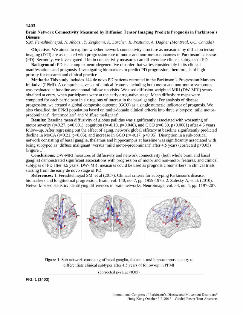
International Congress of Parkinson’s Disease and Movement Disorders®
Hong Kong October 5-9, 2018 – Guided Poster Tour Abstracts
1403
Brain Network Connectivity Measured by Diffusion Tensor Imaging Predicts Prognosis in Parkinson’s
Disease
S.M. Fereshtehnejad, N. Abbasi, Y. Zeighami, K. Larcher, R. Postuma, A. Dagher (Montreal, QC, Canada)
Objective: We aimed to explore whether network connectivity structure as measured by diffusion tensor
imaging (DTI) are associated with progression rate of motor and non-motor outcomes in Parkinson’s disease
(PD). Secondly, we investigated if brain connectivity measures can differentiate clinical subtypes of PD.
Background: PD is a complex neurodegenerative disorder that varies considerably in its clinical
manifestations and prognosis. Investigating biomarkers to predict PD progression, therefore, is of high
priority for research and clinical practice.
Methods: This study includes 144 de novo PD patients recruited in the Parkinson’s Progression Markers
Initiative (PPMI). A comprehensive set of clinical features including both motor and non-motor symptoms
was evaluated at baseline and annual follow-up visits. We used diffusion-weighted MRI (DW-MRI) scans
obtained at entry, when participants were at the early drug-naïve stage. Mean diffusivity maps were
computed for each participant in six regions of interest in the basal ganglia. For analysis of disease
progression, we created a global composite outcome (GCO) as a single numeric indicator of prognosis. We
also classified the PPMI population based on multi-domain clinical criteria into three subtypes: ‘mild motor-
predominant’, ‘intermediate’ and ‘diffuse malignant’.
Results: Baseline mean diffusivity of globus pallidus was significantly associated with worsening of
motor severity (r=0.27, p=0.001), cognition (r=-0.18, p=0.040), and GCO (r=0.30, p<0.0001) after 4.5 years
follow-up. After regressing out the effect of aging, network global efficacy at baseline significantly predicted
decline in MoCA (r=0.21, p<0.05), and increase in GCO (r=-0.17, p<0.05). Disruption in a sub-cortical
network consisting of basal ganglia, thalamus and hippocampus at baseline was significantly associated with
being subtyped as ‘diffuse malignant’ versus ‘mild motor-predominant’ after 4.5 years (corrected p<0.05)
[Figure 1].
Conclusions: DW-MRI measures of diffusivity and network connectivity (both whole brain and basal
ganglia) demonstrated significant associations with progression of motor and non-motor features, and clinical
subtypes of PD after 4.5 years. DW- MRI measures could be used as prognostic biomarkers in clinical trials
starting from the early de novo stage of PD.
References: 1. Fereshtehnejad SM, et al (2017). Clinical criteria for subtyping Parkinson's disease:
biomarkers and longitudinal progression. Brain, vol. 140, no. 7, pp. 1959-1976. 2. Zalesky A, et al. (2010).
Network-based statistic: identifying differences in brain networks. Neuroimage, vol. 53, no. 4, pp. 1197-207.
FIG. 1 (1403)
International Congress of Parkinson’s Disease and Movement Disorders®
Hong Kong October 5-9, 2018 – Guided Poster Tour Abstracts
1415
Longitudinal development of nigral iron load in Parkinson’s Disease
S. Franthal, L. Pirpamer, C. Rodler, N. Homayoon, M. Koegl, P. Katschnig-Winter, K. Wenzel, C.
Langkammer, S. Ropele, R. Schmidt, P. Schwingenschuh (Graz, Austria)
Objective: We for the first time investigated longitudinal changes of nigral iron load in Parkinson’s
disease (PD) measured by Quantitative Susceptibility Mapping (QSM), a highly iron-sensitive MRI method,
and its relation to clinical variables.
Background: Iron accumulation in the substantia nigra (SN) is discussed to be an important factor in the
pathogenesis of PD. Former studies, based on histological and imaging techniques, showed higher
concentrations of iron-ions in the basal ganglia, especially in the substantia nigra of parkinsonian patients
compared to healthy controls. However longitudinal data are sparse.
Methods: We included 52 PD-patients (36 male, mean age 62,6±10,7; mean disease duration 4,1 years)
and 29 healthy controls (HC) (13 male; mean age 68,1±9,1). All subjects underwent a clinical examination
and a 3T MRI scan at baseline and after a follow-up period of approximately 2 years. Nigral iron
concentration was determined by QSM. For cross-sectional group comparisons we performed one-way
ANOVAs corrected for age and gender, for longitudinal comparisons repeated-measure ANOVAs corrected
for age, gender and between-scan-time and for clinical correlations Spearman correlations. To achieve
normal distribution QSM values were logarithmised, significances were corrected with Bonferroni correction
for multiple comparisons.
Results: QSM values in total SN, SN pars compacta (SNc) and SN pars reticulata (SNr) were
significantly higher in PD compared to HC (p<0.001) at baseline and follow-up. There were no significant
group differences in longitudinal QSM-change. QSM values in PD tented to increase in SNc and decrease in
SNr, in HC they tended to decrease in SNc and SNr. There was no significant correlation for QSM change
and change in clinical parameters (MDS-UPDRS, FTM-tremor rating scale, Non Motor symptoms
questionnaire, MMSE, LED).
Conclusions: We confirmed higher nigral iron load in PD compared to HC. However there was only a not
significant trend for stronger shortterm longitudinal increase of iron concentration in SNc in PD compared to
HC. This might be due to relatively long baseline disease duration in our PD subjects and suggests nigral iron
accumulation as an early factor in the pathogenesis of PD.
References: This abstract has previously been presented at the 4th Congress of the European Academy of
Neurology, 16 - 19 June 2018 and at the 15th Annual Meeting of the Austrian Society of Neurology, 21-23
March 2018.
1430
Identifying the neural correlates of doorway freezing in Parkinson’s disease
E. Matar, J. Shine, P. Ward, M. Gilat, K. Ehgoetz-Martens, M. Frank, A. Moustafa, S. Naismith, S. Lewis
(Sydney, Australia)
Objective: We aimed to combine a previously validated virtual reality gait paradigm with functional
Magnetic Resonance Imaging (fMRI) to investigate the neural correlates underlying doorway-provoked
freezing behaviour in Parkinson's disease. Based on previous models we predicted that disturbances in
connectivity of the hyperdirect pathway would correlate with the motor disturbance observed in response to
doorways.
Background: Freezing of gait (FOG) in Parkinson’s disease (PD) is frequently triggered upon passing
through narrow spaces such as doorways. Often this freezing behaviour is improved with dopaminergic
treatments. Despite being common the neural mechanisms underlying freezing are poorly understood due to
the inherent limitations accompanying the functional imaging of gait. We have previously shown that
freezing behaviour can be reliably modelled in a scanner using a virtual reality (VR) paradigm wherein
subjects use foot-pedals to pass through a virtual corridor. Using this method, we have found that changes in

International Congress of Parkinson’s Disease and Movement Disorders®
Hong Kong October 5-9, 2018 – Guided Poster Tour Abstracts
activation within fronto-parietal networks contributes to freezing of gait generally but doorway-freezing
specifically has not been studied.
Methods: In our study, nineteen patients who experience FOG performed a lower limb motor sequencing
task wherein foot-pedals were used to navigate a series of wide and narrow doorways within a previously
validated VR environment. Patients underwent testing both ‘ON’ and ‘OFF’ their medications and task
performance together with blood oxygenation level dependent (BOLD) signal changes, were compared
within each patient.
Results: Patients demonstrated significantly longer 'footstep’ latencies in the OFF state compared to the
ON state as they passed through a doorway in the VR environment (p<0.05). As with real FOG, this motor
disturbance was primarily triggered by narrow doorways rather than wide doorways in the OFF state (Fig 1).
fMRI analysis revealed that this footstep prolongation on passing through doorways was associated with
specific hypoactivation in the pre-supplementary motor area (pSMA) bilaterally (p<0.05; Fig 2).
Furthermore, task-based functional connectivity analyses revealed that increased latency in response to
doorways was inversely correlated with the degree of functional connectivity between the pSMA and the
subthalamic nucleus (STN) across both hemispheres (r = 0.441, p < 0.05). Conversely, increased frequency
of prolonged footstep latency was associated with increased connectivity in bilateral STN (r = 0.575, p =
0.01).
Conclusions: The findings of this study suggest that the effect of perceptual cues on triggering FOG
reflects impaired processing within the pSMA and its normal signalling with the STN thus implicating the
'hyper-direct' pathway in the pathophysiology of this phenomenon.
References: 1. Shine, J. M. et al. Modeling freezing of gait in Parkinson's disease with a virtual reality
paradigm. Gait Posture 38, 104 08, doi:10.1016/j.gaitpost.2012.10.026 (2013). 2. Matar, E., Shine, J. M.,
Naismith, S. L. & Lewis, S. J. Virtual reality walking and dopamine: opening new doorways to
understanding freezing of gait in Parkinson's disease. J Neurol Sci 344, 182-185,
doi:10.1016/j.jns.2014.06.054 (2014).
FIG. 1 (1430)

International Congress of Parkinson’s Disease and Movement Disorders®
Hong Kong October 5-9, 2018 – Guided Poster Tour Abstracts
FIG. 2 (1430)
1433
Abnormal Functional Connectivity in Cerebellar Locomotor Region is associated with the severity of
freezing of gait in patients with Parkinson’s disease
K. Bearti, A. Suppa, S. Pietracupa, N. Upadhyay, C. Giannì, G. Leodori, F. Di Biasio, N. Modugno, N.
Petsas, G. Grillea, A. Zampogna, A. Berardelli, P. Pantano (Rome, Italy)
Objective: We aim to analyze cerebellar functional connectivity (FC) in PD patients with FOG (PD-
FOG).
Background: Freezing of gait (FOG) is a disabling disorder frequently affecting patients with Parkinson's
disease (PD) and consists of transient inability to start or maintain locomotion [1]. The pathophysiological
mechanism of FOG has not yet been identified. Although recent studies with magnetic resonance imaging
(MRI) have shown alterations in several cortical and subcortical brain areas of patients with FOG, the
cerebellum, a key structure in posture and gait control, has only been partially studied [2].
Methods: We recruited 15 PD-FOG, 16 PD without FOG (PD-nFOG) patients, and 16 age-matched
healthy subjects (HS). The motor and cognitive impairment of patients was evaluated with validated clinical
scales. FOG severity was assessed by the FOG Questionnaire (FOG-Q). Resting-state fMRI data were
acquired using a high field magnet (3 Tesla, Siemens, Verio). By using the FSL software we located a seed
region on the cerebellar locomotor region (CLR) and obtained FC maps of the CLR with the whole brain.
The z scores of clusters showing significant differences in FC between PD-FOG patients and HS were
correlated with scores at FOG-Q in SPSS toolbox.
Results: The two subgroups of patients showed decreased FC of the CLR with the prefrontal cortex
(superior, middle and inferior frontal gyrus) of both the cerebral hemispheres. In addition, only PD-FOG
exhibited increased FC of CLR with the cerebellar vermis, cerebellar lobules left VI, IX, and right crus I-IV,
VI, VII b as well as with the parieto-occipital cortex (both fusiform gyrus and lateral occipital cortex
bilaterally and right lingual gyrus) [figure1].
FC of the CLR with cerebellar and posterior cortical areas in PD-FOG positively correlated with the FOG-Q
score [figure2].
Conclusions: Our results suggest that the altered functional connectivity of the cerebellum may
contribute to the pathophysiology of FOG; in particular, FC of the CLR, correlated with FOG severity,

International Congress of Parkinson’s Disease and Movement Disorders®
Hong Kong October 5-9, 2018 – Guided Poster Tour Abstracts
supporting the hypothesis that abnormal cerebellar function can contribute to the genesis of FOG in patients
with PD.
References: 1. Giladi N, McDermott MP, Fahn S, Przedborski S, Jankovic J, Stern M, et al. Freezing of
gait in PD: prospective assessment in the DATATOP cohort. Neurology. 2001;56:1712–21. 2. Tessitore A,
Amboni M, Esposito F, Russo A, Picillo M, Marcuccio L, et al. Resting-state brain connectivity in patients
with Parkinson’s disease and freezing of gait. Parkinsonism Relat Disord. 2012;18:781–7.
FIG. 1 (1433)
FIG. 2 (1433)

International Congress of Parkinson’s Disease and Movement Disorders®
Hong Kong October 5-9, 2018 – Guided Poster Tour Abstracts
1464
Resting-state connectivity and cognitive changes in Parkinson’s disease: A four-year follow-up study
HC. Baggio, B. Segura, A. Abos, A. Campabadal, C. Uribe, A.I. Garcia-Diaz, M.J. Marti, Y. Compta, F.
Valldeoriola, C. Junque (Barcelona, Spain)
Objective: To study functional connectivity changes using graph theory and their association with
cognitive evolution in a sample of non-demented Parkinson’s disease (PD) and matched healthy controls
(HC) after a four-year follow-up.
Background: Previous studies demonstrated that cognitive deficits in PD are associated with alterations
in different parameters derived from resting-state functional connectivity. Evidence regarding the
relationship between connectivity changes and cognitive evolution is nonetheless very scarce.
Methods: A final sample of 27 HC and 45 matched PD patients, assessed twice at an interval of
approximately four years, was included. Resting-state fMRI was used for functional connectivity analysis.
Connectivity matrices, obtained after parcellating the brain into 246 regions using the Brainnetome atlas,
were used to compute graph-theory parameters that describe network organization (path length, clustering
coefficient, modularity, small-world coefficient). For cognitive assessment, a thorough neuropsychological
battery was used, and tests were subsequently grouped into four cognitive domains (attention/working
memory, executive function, memory, and visuospatial/perceptual function). A repeated-measures general
linear model was used to test group and time effects, as well as group-by-time interactions.
Results: Analysis of cognitive data in patients showed a significant effect of time for executive function
(p=.004) and visuospatial/perceptual scores (p=.015). Significant effects of group (p=.034, patients
performing worse than controls), time (p=.006), and group-by-time interaction (p=0.027) were found.
Connectivity parameters showed significant effects of group for clustering coefficients and modularity
(p=.023 and p=.045, respectively, with patients showing higher values). For path lengths, a significant effect
of time was found (p=.022, both groups showing increasing values). When performing correlations between
longitudinal cognitive and connectivity changes, we found that differences in executive function scores
correlated significantly and positively with differences in clustering coefficients (r=0.404, p=0.007),
modularity (r=0.384, p=0.011), and small-world coefficients (r=0.335, p=0.028).
Conclusions: Our findings indicate that functional connectivity changes explain executive function in PD
patient’s evolution.
1479
A cortical neural signature of motor interruption in patients with Parkinson’s disease and freezing of
gait
J. Baarbé, M. Brown, U. Saha, K. Lizarraga, A. Weissbach, N. Drummond, C. Rinchon, P. Kapoor, J.
Saravanamuttu, R. Chen (Toronto, ON, Canada)
Objective: To investigate whether cortical oscillations involved in sensorimotor and perceptual processes
[alpha (8-12 Hz), low beta (13-20 Hz) and high beta (21-35 Hz)] change during interruptions of motor output
in Parkinson’s disease (PD) patients with freezing of gait (FoG).
Background: FoG is a debilitating symptom in PD and can often be resistant to treatment. FoG is often
triggered by narrow passageways and crowded rooms, and its incidence can be reduced by visual aids that
prompt rhythmic stepping. Sensory influence on FoG suggests that sensory, perceptual and/or motor
processes in the cortex may be intrinsically involved in FoG pathophysiology.
Methods: We recorded 64 channel electroencephalography in 10 PD patients (68 years old±6; disease
duration: 9 years±5; UPDRS-III on medication: 27±9; UPDRS-III off medication: 31±8; levodopa equivalent
dose: 880 mg±420) and 9 healthy controls (67 years old±6). Six patients had FoG confirmed by the New
Freezing of Gait Questionnaire and neurologist assessment. While seated, participants manipulated foot
pedals to walk through a virtual environment (VE). Participants completed 4 sets of 5 minutes walking
within the VE. Doorways within the VE were used to trigger motor interruptions (MI). We also had
participants intentionally stop to assess cortical activity unique to MI. Alpha, low beta and high beta were
International Congress of Parkinson’s Disease and Movement Disorders®
Hong Kong October 5-9, 2018 – Guided Poster Tour Abstracts
determined pre MI (-1200 to -600 ms), upon onset of MI (-300 to 300 ms), during MI (600 to 1200 ms),
during an active state and upon intentionally stopping. Cortical activities were assessed in predetermined
premotor/frontal (F1,Fz,F2,FC1,FCz,FC2), motor (C1,Cz,C2) and parietal (CP1,CPz,CP2,P1,Pz,P2) clusters.
Results: Patients with FoG experienced a total of 38 MI (mean duration 2.5 s). MI influenced alpha
power within premotor/frontal (p=0.042; Onset MI<During MI) and motor clusters (p=0.016; Pre MI>Onset
MI; Onset MI<During MI). Onset of MI affected low beta power in motor (p=0.022; Onset MI<During MI)
and parietal clusters (p=0.039; Onset MI<During MI). MI did not induce changes to high beta power within
any of the clusters, but intentional stopping increased alpha, low beta and high beta in all clusters (p<0.001).
Conclusions: Alpha and low-beta oscillations in motor areas may be important predictive signals of MI in
patients with PD and FoG. Uniquely, the absence of high beta distinguishes MI from intentional stops. These
findings may be useful for the development of closed-loop brain stimulation or biofeedback strategies to
alleviate FoG.
1483
The difference in cerebellar blood flow reduction in multiple system atrophy and Parkinson’s disease
N. Murakami, W. Sako, S. Haji, Y. Izumi, R. Kaji (Tokushima, Japan)
Objective: The aim of this study is to determine if cerebral blood flow (CBF) reduction in the cerebellum
could be a tool for differential diagnosis between patients with multiple system atrophy (MSA) and patients
with Parkinson’s disease (PD).
Background: MSA is the neurodegenerative disease showing progressive atypical parkinsonism, which is
frequently misdiagnosed as PD, especially in case without cerebellar symptom or atrophy. The accurate tool
for differential diagnosis between MSA and PD could facilitate clinical trials of disease modifying therapy.
Methods: Twenty one patients with PD were diagnosed as clinically established PD or clinically probable
PD (10 men and 11 women with a mean age of 67.7 ± 12.3 years), fifteen patients with MSA with
predominant parkinsonian features (MSA-P), six patients with MSA with predominant cerebellar ataxia
(MSA-C) were diagnosed as probable or possible MSA (11 men and 10 women with a mean age of 65.2 ±
9.3 years), fifteen patients with progressive supranuclear palsy (PSP) were diagnosed as probable or possible
PSP (5 men and 10 women with a mean age of 73.7 ± 6.1 years) according to the internationally established
criteria were included in this study. All patients underwent N-isopropyl-[123I] p-iodoamphetamine (IMP)
single photon emission computed tomography (123I-IMP SPECT) and data were analyzed using three-
dimensional stereotactic surface projection (3D-SSP) software. Area under curve (AUC) of receiver
operating characteristic (ROC) of CBF reduction in the cerebellum was assessed.
Results: Cerebellar perfusion was significantly decreased in the MSA-P (P < 0.001, one-way ANOVA; P
< 0.001, post hoc, Dunnett), MSA-C (P < 0.001, post hoc, Dunnett) compared with the PD. There was no
difference in cerebellar perfusion between PSP and PD (P = 0.598, post hoc, Dunnett). Specificity of CBF
reduction in the cerebellum for MSA-P vs PD was 1.00, while sensitivity was 0.67. CBF reduction may be an
optimal tool for differentiating between MSA-P and PD (AUC = 0.849).
Conclusions: In conclusion, our findings suggested that CBF reduction in the cerebellum using 123I-IMP
SPECT could be a useful tool for differentiating between MSA-P and PD.
1484
Structural Abnormalities in the Cerebellar Peduncles of patients with Freezing of Gait: A Diffusion
Tensor Imaging Study
K. Bharti, A. Suppa, S. Pietracupa, N. Upadhyay, C. Giannì, G. Leodori, F. Di Biasio, N. Modugno, N.
Petasas, G. Grilla, A. Zampogna, A. Berardelli, P. Pantano (Rome, Italy)
Objective: We used diffusion tensor imaging (DTI) to investigate cerebellar structural connectivity by
evaluating white matter properties along the superior (SCP), middle (MCP), and inferior cerebellar peduncles
(ICP) in patients with Parkinson’s disease (PD) and freezing of gait (FOG).
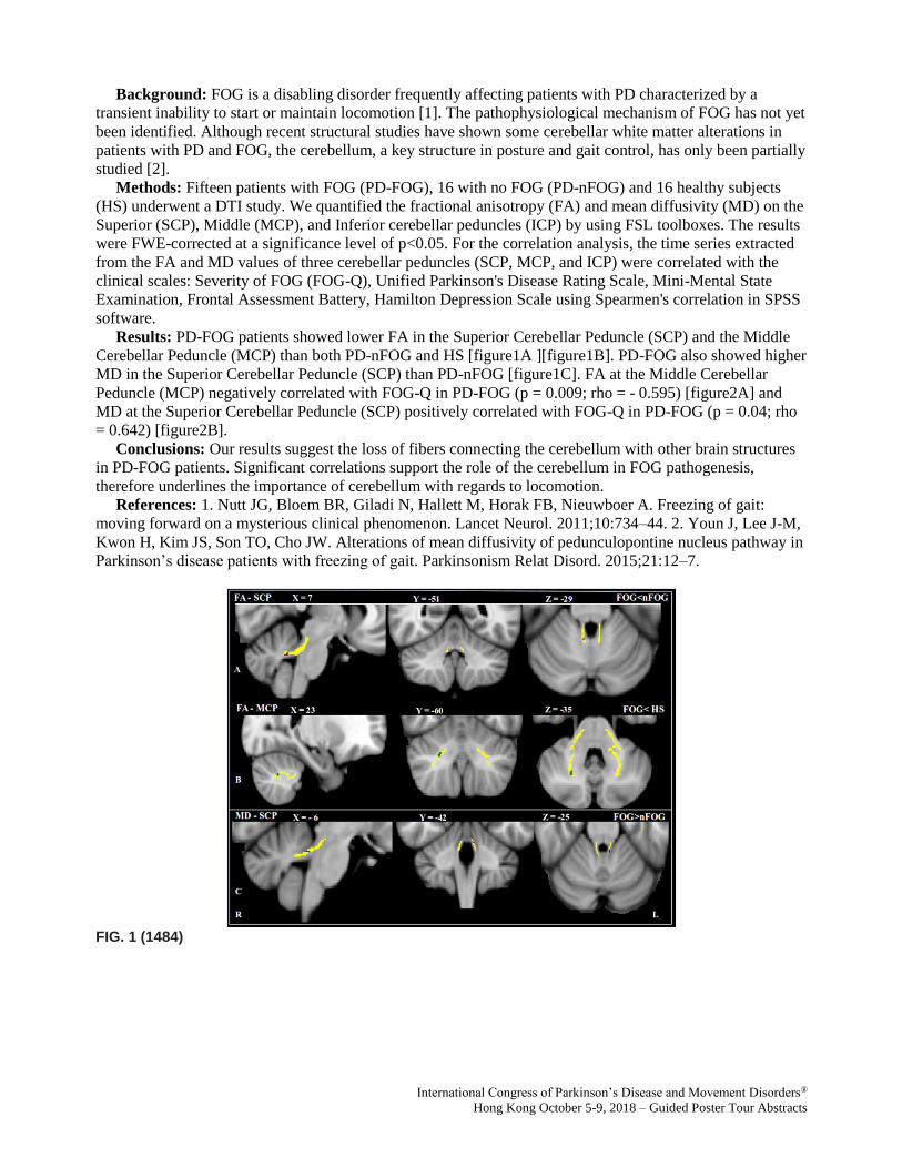
International Congress of Parkinson’s Disease and Movement Disorders®
Hong Kong October 5-9, 2018 – Guided Poster Tour Abstracts
Background: FOG is a disabling disorder frequently affecting patients with PD characterized by a
transient inability to start or maintain locomotion [1]. The pathophysiological mechanism of FOG has not yet
been identified. Although recent structural studies have shown some cerebellar white matter alterations in
patients with PD and FOG, the cerebellum, a key structure in posture and gait control, has only been partially
studied [2].
Methods: Fifteen patients with FOG (PD-FOG), 16 with no FOG (PD-nFOG) and 16 healthy subjects
(HS) underwent a DTI study. We quantified the fractional anisotropy (FA) and mean diffusivity (MD) on the
Superior (SCP), Middle (MCP), and Inferior cerebellar peduncles (ICP) by using FSL toolboxes. The results
were FWE-corrected at a significance level of p<0.05. For the correlation analysis, the time series extracted
from the FA and MD values of three cerebellar peduncles (SCP, MCP, and ICP) were correlated with the
clinical scales: Severity of FOG (FOG-Q), Unified Parkinson's Disease Rating Scale, Mini-Mental State
Examination, Frontal Assessment Battery, Hamilton Depression Scale using Spearmen's correlation in SPSS
software.
Results: PD-FOG patients showed lower FA in the Superior Cerebellar Peduncle (SCP) and the Middle
Cerebellar Peduncle (MCP) than both PD-nFOG and HS [figure1A ][figure1B]. PD-FOG also showed higher
MD in the Superior Cerebellar Peduncle (SCP) than PD-nFOG [figure1C]. FA at the Middle Cerebellar
Peduncle (MCP) negatively correlated with FOG-Q in PD-FOG (p = 0.009; rho = - 0.595) [figure2A] and
MD at the Superior Cerebellar Peduncle (SCP) positively correlated with FOG-Q in PD-FOG (p = 0.04; rho
= 0.642) [figure2B].
Conclusions: Our results suggest the loss of fibers connecting the cerebellum with other brain structures
in PD-FOG patients. Significant correlations support the role of the cerebellum in FOG pathogenesis,
therefore underlines the importance of cerebellum with regards to locomotion.
References: 1. Nutt JG, Bloem BR, Giladi N, Hallett M, Horak FB, Nieuwboer A. Freezing of gait:
moving forward on a mysterious clinical phenomenon. Lancet Neurol. 2011;10:734–44. 2. Youn J, Lee J-M,
Kwon H, Kim JS, Son TO, Cho JW. Alterations of mean diffusivity of pedunculopontine nucleus pathway in
Parkinson’s disease patients with freezing of gait. Parkinsonism Relat Disord. 2015;21:12–7.
FIG. 1 (1484)
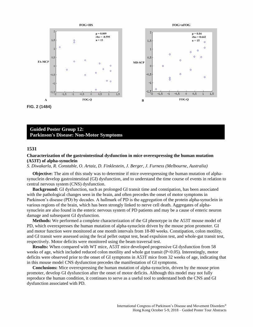
International Congress of Parkinson’s Disease and Movement Disorders®
Hong Kong October 5-9, 2018 – Guided Poster Tour Abstracts
FIG. 2 (1484)
1531
Characterization of the gastrointestinal dysfunction in mice overexpressing the human mutation
(A53T) of alpha-synuclein
S. Diwakarla, R. Constable, O. Artaiz, D. Finklestein, J. Berger, J. Furness (Melbourne, Australia)
Objective: The aim of this study was to determine if mice overexpressing the human mutation of alpha-
synuclein develop gastrointestinal (GI) dysfunction, and to understand the time course of events in relation to
central nervous system (CNS) dysfunction.
Background: GI dysfunction, such as prolonged GI transit time and constipation, has been associated
with the pathological changes seen in the brain, and often precedes the onset of motor symptoms in
Parkinson’s disease (PD) by decades. A hallmark of PD is the aggregation of the protein alpha-synuclein in
various regions of the brain, which has been strongly linked to nerve cell death. Aggregates of alpha-
synuclein are also found in the enteric nervous system of PD patients and may be a cause of enteric neuron
damage and subsequent GI dysfunction.
Methods: We performed a complete characterization of the GI phenotype in the A53T mouse model of
PD, which overexpresses the human mutation of alpha-synuclein driven by the mouse prion promoter. GI
and motor function were monitored at one month intervals from 18-80 weeks. Constipation, colon motility,
and GI transit were assessed using the fecal pellet output test, bead expulsion test, and whole-gut transit test,
respectively. Motor deficits were monitored using the beam traversal test.
Results: When compared with WT mice, A53T mice developed progressive GI dysfunction from 58
weeks of age, which included reduced colon motility and whole gut transit (P<0.05). Interestingly, motor
deficits were observed prior to the onset of GI symptoms in A53T mice from 32 weeks of age, indicating that
in this mouse model CNS dysfunction precedes the manifestation of GI symptoms.
Conclusions: Mice overexpressing the human mutation of alpha-synuclein, driven by the mouse prion
promoter, develop GI dysfunction after the onset of motor deficits. Although this model may not fully
reproduce the human condition, it continues to serve as a useful tool to understand both the CNS and GI
dysfunction associated with PD.
Guided Poster Group 12:
Parkinson's Disease: Non-Motor Symptoms
International Congress of Parkinson’s Disease and Movement Disorders®
Hong Kong October 5-9, 2018 – Guided Poster Tour Abstracts
1533
Four-year course of impulsive and compulsive behaviors in Parkinson’s disease
A. Erga, G. Alves, O.B. Tysnes, K. Pedersen (Stavanger, Norway)
Objective: To (1) determine the longitudinal risk of ICBs for patients compared to controls, (2) determine
the stability and transition probabilities of ICBs over the course of four years.
Background: Impulsive and compulsive behaviours (ICBs) include a range of behavioural addictions and
related behaviours, that are common in Parkinson’s disease (PD), affecting between 14 - 30 % of patients. A
major limitation in previous research is use of cross-sectional or retrospective designs, and the course of
ICBs is largely undescribed in the current literature.
Methods: Data from the Norwegian ParkWest study, a population-based longitudinal cohort study of the
incidence, neurobiology and prognosis of PD in Western Norway were used in this study. Patients where
included five years after diagnosis and inclusion in the ParkWest study and followed prospectively for four
years. At study entry, 125 patients and 159 normal controls were included. Standardized clinical rating scales
were used to evaluate motor severity, global cognitive function and depressive symptoms. ICBs were
evaluated using the short version of the Questionnaire for Impulsive-Compulsive Disorders in PD (QUIP).
The development of ICDs was investigated using Markov chain analysis, with ICD status and
dropout/dementia as endpoint states.
Results: Although ICBs where consistently more frequent in PD than controls, the risk of ICBs decreased
over time. The transition probability for patients with ICBs at baseline was 42.9 % for recovery of ICB
symptoms, 36.3 % for persistent ICBs and 20.8 % for development of dementia or drop out from the study.
Patients who were ICB negative at baseline had an estimated transition-probability of 14.2 % to develop
ICBs, 64.9 % to remain without these symptoms and 20.9 % for development of dementia or drop out from
the study.
Conclusions: These findings suggest that ICBs can develop in the later stages of PD, and have a high
probability to remain stable once developed.
References: 1. Weintraub D, Koester J, Potenza MN, et al. Impulse control disorders in Parkinson
disease: a cross-sectional study of 3090 patients. Archives of neurology 2010;67(5):589-595. 2. Erga AH,
Alves G, Larsen JP, Tysnes OB, Pedersen KF. Impulsive and Compulsive Behaviors in Parkinson's Disease:
The Norwegian ParkWest Study. Journal of Parkinson's disease 2017;7(1):183-191.
1553
Striatal dopamine transporter availability changes reflect gastrointestinal dysautonomia severity in
early Parkinson’s disease
J. Hinkle, K. Perepezko, K. Mills, Z. Mari, A. Butala, T. Dawson, A. Pantelyat, L. Rosenthal, G. Pontone
(Baltimore, MD, USA)
Objective: To test the hypothesis that constipation and gastrointestinal (GI) dysautonomia may be
correlated with nigrostriatal dopaminergic (DA) dysfunction in PD.
Background: Constipation is a prodromal feature of PD and the GI tract is implicated in the pathogenesis
of PD. Autopsy evidence supports nigral degeneration as a correlate of late-life constipation (Petrovitch et
al., 2009), but no studies have demonstrated ante-mortem relationships between nigrostriatal dysfunction and
GI dysautonomia in PD. However, it was recently demonstrated in rodents that nigral projections regulate
gastrointestinal motility via DA receptors on neurons in the dorsal vagal complex (Anselmi et al., 2017).
Methods: The Scale for Outcomes in Parkinson’s disease for Autonomic Symptoms (SCOPA-AUT)
assesses dysautonomia in the multi-center Parkinson’s Progression Marker Initiative (PPMI). We used linear
mixed-effects (LME) models and reliable change indices (RCIs) to examine longitudinal associations
between dysautonomia and dopamine transporter (DAT) striatal binding ratios (SBRs) measured by single-
photon emission computerized tomography (SPECT) in PPMI participants over four years (n=397 at
baseline).

International Congress of Parkinson’s Disease and Movement Disorders®
Hong Kong October 5-9, 2018 – Guided Poster Tour Abstracts
Results: Adjusted LME models of longitudinal data [table1] showed that constipation—but not
orthostatic hypotension or urinary dysfunction—was associated with reduced SBR in both caudate (P<0.001)
and putamen (P=0.022). In both regions, SBR reductions between baseline and 4-year follow-up were
significant and measurable (P < 0.0001), with larger decline and variances in the caudate nucleus [figure1].
Logistic regression adjusting for disease duration and baseline SCOPA-GI score showed that 4-year change
in caudate—but not putaminal —SBR was significantly associated with RCI-indicated progression of GI
dysautonomia (P=0.007), but not other types of dysautonomia [table2]. The association remained after
adjusting for the use of medications or supplements to control constipation. Consistent with prior PPMI
reports, motor impairment progression was not associated with SBR reduction.
Conclusions: GI dysautonomia correlates with reduced striatal DAT availability in PD and constipation
may be most closely associated with caudate-DAT reductions. Worsening GI dysautonomia may accompany
advancing nigral degeneration or changes in nigrostriatal dopamine function.
References: Anselmi L, Toti L, Bove C, Hampton J, Travagli RA. A Nigro-Vagal Pathway Controls
Gastric Motility and is Affected in a Rat Model of Parkinsonism. Gastroenterology 2017: 36138–3.
Petrovitch H, Abbott RD, Ross GW, Nelson J, Masaki KH, Tanner CM, et al. Bowel movement frequency in
late-life and substantia nigra neuron density at death. Mov. Disord. 2009; 24: 371–376.
TABLE 1 (1553)
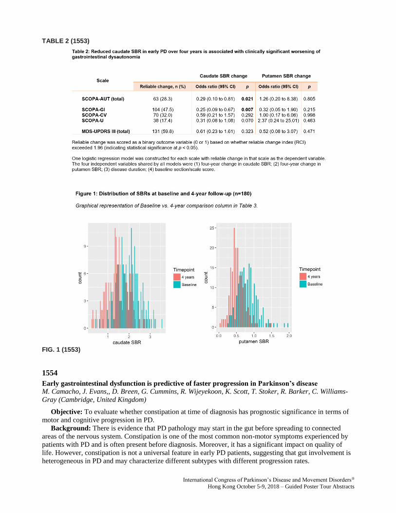
International Congress of Parkinson’s Disease and Movement Disorders®
Hong Kong October 5-9, 2018 – Guided Poster Tour Abstracts
TABLE 2 (1553)
FIG. 1 (1553)
1554
Early gastrointestinal dysfunction is predictive of faster progression in Parkinson’s disease
M. Camacho, J. Evans,, D. Breen, G. Cummins, R. Wijeyekoon, K. Scott, T. Stoker, R. Barker, C. Williams-
Gray (Cambridge, United Kingdom)
Objective: To evaluate whether constipation at time of diagnosis has prognostic significance in terms of
motor and cognitive progression in PD.
Background: There is evidence that PD pathology may start in the gut before spreading to connected
areas of the nervous system. Constipation is one of the most common non-motor symptoms experienced by
patients with PD and is often present before diagnosis. Moreover, it has a significant impact on quality of
life. However, constipation is not a universal feature in early PD patients, suggesting that gut involvement is
heterogeneous in PD and may characterize different subtypes with different progression rates.
International Congress of Parkinson’s Disease and Movement Disorders®
Hong Kong October 5-9, 2018 – Guided Poster Tour Abstracts
Methods: We studied a newly diagnosed community-based cohort of 281 IPD patients (PICNICS,
Parkinsonism: Incidence and Cognitive Heterogeneity In Cambridgeshire) and followed them up
prospectively every 18 months with standardized assessments including the MDS-UPDRS, ACE-R and
PDQ-39. According to scores on the MDS-UPDRS constipation item at baseline visit, we stratified the
sample using two different cut-offs: mild to high (score of ≥1; n=78) and moderate to high (score ≥2; n=35)
constipation and followed the natural progression of PD for up to 108 months after diagnosis.
Results: At baseline, 27.8% of patients had mild to high constipation and 12.5% of patients had moderate
to high constipation. Regardless of cut-off scores, constipated and non-constipated patients did not differ in
age, education level, disease duration, disease stage (Hoehn and Yahr), number of comorbidities and L-dopa
equivalent daily dose. Patients with moderate to high constipation had significantly higher disease severity
(MDS-UPDRS total) and worse quality of life (PDQ-39), whereas there were no between-group differences
in the group with mild to high constipation. Survival analysis showed that patients with moderate to high
constipation had an earlier onset of PD related dementia (according to MDS criteria) and more rapid
progression to development of postural instability (Hoehn and Yahr stage 3). No differences were observed
in mortality between groups.
Conclusions: Constipation only affects one third of PD patients at diagnosis, but its severity at baseline
seems to predict a faster motor and cognitive progression.
1573
Non-motor symptoms in Parkinson’s Disease: Frequency, types and correlated factors compared to a
group of healthy controls. Results from the COPPADIS Study Cohort
L. Planellas, M. Martí, P. Santacruz, A. Cámara, S. Jesus Maestre, F. Carrillo García, P. Mir, M. Aguilar,
P. Pastor, J. García Caldentey, E. Estelrich Peyret, N. Caballol Pons, I. Legarda, B. Vives Pastor, J.
Hernández Vara, G. Martí Andrés, I. Cabo, L. López Manzanares, M. Gallego-de-la-Sacristana, P. Martínez
Martín, D. Santos García, G. COPPADIS STUDY (Barcelona, Spain)
Objective: The aim of this study is to describe the prevalence and burden of NMS in a group of PD
patients without dementia compared to an age-matched control group.
Background: Non-motor symptoms (NMS) are frequent in patients with Parkinson’s disease (PD) and
impact quality of life.
Methods: This is a descriptive study conducted in Spain using data from the baseline visit of the
COPPADIS-2015 Study (Santos García et al. BMC Neurol 2016;16:26), an observational, descriptive, 5-year
follow-up, national, multicenter, evaluation study. The prevalence and burden of NMS were evaluated using
the Non-Motor Symptoms Scale (NMSS), a scale that assess 30 non-motor items grouped in 9 domains.
Results: 696 patients (mean age 62.77 ± 9.03, males 59.7%) and 206 controls (mean age 61.16 ± 8.45,
males 49.8%) were included in the analysis. NMS were more prevalent in PD patients than controls (total
prevalence: 99.3% vs 84.9%, p<0.0001). The mean number of domains affected was 5.74 ± 2.10 in PD group
vs 3.25 ± 2.24 in control group (p<0.0001). PD patients had higher prevalence and burden of NMS in all
domains and also in all items except “difficulty falling asleep” and “altered sexual interest”. Fatigue (62.2%),
nocturia (61.2%) and urinary urgency (60.5%) were the most common NMS in the PD group while nocturia
(43.2%), difficulty falling asleep (34.0%) and altered interest in sex (32.0%) were the most frequent in
control group. Among patients, females had higher scores than males in items related to mood while males
had higher scores in daytime sleepiness and sialorrhea. Postural instability and gait difficulty (PIGD)
phenotype was associated with higher total NMSS score than tremor dominant PD (51.31 ± 40.96 vs 40.07 ±
34.82; p=0.004) and higher scores in sleep/fatigue, mood/apathy, gastrointestinal and miscellaneous domains
too.
Conclusions: In our study PD patients have more NMS with higher NMSS scores than controls.
Moreover, our results suggest that NMS could be influenced by gender and PD phenotype.

International Congress of Parkinson’s Disease and Movement Disorders®
Hong Kong October 5-9, 2018 – Guided Poster Tour Abstracts
1576
Non-motor outcomes of subthalamic DBS in PD depend on the location of volume of activated tissue
JN. Petry-Schmelzer, H. Dafsari, M. Krause, T. Dembek, A. Keyoumars, A. Rizos, M. Silverdale, J. Evans, M.
Barbe, G. Fink, P. Martinez-Martin, V. Visser-Vandewalle, A. Antonini, L. Timmermann, K. Ray-Chaudhuri
(Cologne, Germany)
Objective: To investigate the impact of stimulation location on non-motor symptoms (NMS) in PD
patients with DBS in the subthalamic nucleus (STN) via an analysis of volumes of activated tissue (VAT).
Background: STN-DBS improves non-motor and motor symptoms. In a recent study, we showed that
NMS improvement is higher with a more anterior, ventral and medial position of the active contact, whereas
motor symptoms improve more with a more posterior and lateral position of the active contact. However,
investigating only electrode locations neglects the spatial extent of electrical stimulation, which also depends
on the stimulation parameters. We thus conducted an investigation into the spatial distribution of NMS
improvement in the STN using VATs.
Methods: Clinical data was collected from an ongoing prospective, open-label, multicenter study
(Cologne, London, Manchester) including 92 patients with bilateral STN-DBS. The following scales were
collected at preoperative baseline (MedON) and on follow-up (FU) six months after surgery
(MedON/StimON): SCOPA-motor examination (SCOPA-motor), -activities of daily living (ADL),
NMSScale (NMSS), NMSQuestionnaire (NMSQ), Hospital Anxiety and Depression Scale-anxiety and
depression subscales (HADS-A/-D). Wilcoxon signed-rank test was used to test for significant changes
between baseline and follow-up and Bonferroni-correction was applied for multiple comparisons. Individual
VATs, based on the stimulation parameters used in clinical setting, were calculated in MNI space (ICBM
2009b) as described elsewhere (1). To analyse the relationship between VATs and change scores, we
employed probabilistic stimulation maps and projected them on the DISTAL-Atlas (2).
Results: All outcomes, besides HADS-D, improved significantly at FU. Probabilistic stimulation maps
showed higher improvement of NMS for stimulation localized in the limbic and associative STN, whereas
motor symptoms improved more for stimulation localized in the motor STN.
Conclusions: Our preliminary results support the finding that the non-motor outcome after DBS may
depend on the location of neurostimulation. DBS in non-motor STN subregions was associated with bigger
improvement of NMS. The underlying mechanisms and clinical relevance of our results should be
investigated in future studies.
References: 1 Horn, A., Reich, M., Vorwerk, J., Li, N., Wenzel, G., Fang, Q.,. . . Fox, M. D. (2017).
Connectivity predicts deep brain stimulation outcome in Parkinson's disease. Ann Neurol.
doi:10.1002/ana.24974. 2 Ewert, S., Plettig, P., Li, N., Chakravarty, M. M., Collins, D. L., Herrington, T. M.,
. . . Horn, A. (2017). Toward defining deep brain stimulation targets in MNI space: A subcortical atlas based
on multimodal MRI, histology and structural connectivity. Neuroimage.
doi:10.1016/j.neuroimage.2017.05.015.
1591
A detailed clinical study of pain in 1957 participants with Parkinson’s disease
M. Silverdale, C. Kobylecki, L. Kass-Iliyya, P. Martinez-Martin, D. Grosset, M. Hu, M. Lawton, S. Cotterill,
K. Ray Chaudhuri, H. Morris, F. Baig, N. Williams, L. Hubbard (Manchester, United Kingdom)
Objective: We performed a large clinical study to investigate potential factors contributing towards pain
in Parkinson’s disease (PD).
Background: The causes of pain in PD are not well understood. Although peripheral factors such as
rigidity, reduced joint movements and poor posture may contribute towards the development of pain, central
mechanisms including altered nociceptive processing may also be involved.
Methods: We recruited 1957 PD participants who had detailed assessments of pain, motor and non-motor
symptoms. The King’s Parkinson’s Pain scale was used to quantify different subtypes of pain.
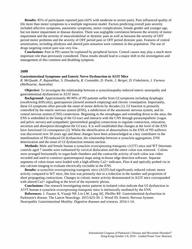
International Congress of Parkinson’s Disease and Movement Disorders®
Hong Kong October 5-9, 2018 – Guided Poster Tour Abstracts
Results: 85% of participants reported pain (42% with moderate to severe pain). Pain influenced quality of
life more than motor symptoms in a multiple regression model. Factors predicting overall pain severity
included affective symptoms, autonomic symptoms, motor complications, female gender and younger age,
but not motor impairment or disease duration. There was negligible correlation between the severity of motor
impairment and the severity of musculoskeletal or dystonic pain as well as between the severity of OFF
period motor problems and the severity of OFF period pain or OFF period dystonic pain. Features of central
sensitization, including allodynia and altered pain sensation were common in this population. The use of
drugs targeting central pain was very low.
Conclusions: Pain in PD cannot be explained by peripheral factors. Central causes may play a much more
important role than previously considered. These results should lead to a major shift in the investigation and
management of this common and disabling symptom.
1600
Gastrointestinal Symptoms and Enteric Nerve Dysfunction in A53T Mice
R. McQuade, P. Rajasekhar, S. Diwakarla, R. Constable, D. Poole, J. Berger, D. Finkelstein, J. Furness
(Melbourne, Australia)
Objective: To investigate the relationship between α synucleinopathy-induced enteric neuropathy and
gastrointestinal dysfunction in A53T mice.
Background: Approximately 80-90% of PD patients suffer from GI symptoms including dysphagia
(swallowing difficulty), gastroparesis (slowed stomach emptying) and chronic constipation. Importantly,
these GI symptoms often precede the onset of motor deficits by decades (1). GI function is primarily
controlled by the enteric nervous system (ENS), a subdivision of the autonomic nervous system, and its
central nervous system (CNS) connections. Beginning in the oesophagus and extending down to the anus, the
ENS is embedded in the lining of the GI tract and interacts with the CNS through parasympathetic (vagus
and pelvic nerves) and sympathetic (prevertebral ganglia) connections to regulate contraction, relaxation,
secretion and absorption throughout the GI tract. It is well established that changes at the level of the ENS
have functional GI consequence (2). Whilst the identification of abnormalities in the ENS of PD sufferers
was discovered over 30 years ago and these changes have been acknowledged as a key contributor in the
manifestation of PD-induced GI dysfunction, the relationship between α synuclein aggregation, ENS
deterioration and the onset of GI dysfunction remains unclear.
Methods: Male and female human α synuclein overexpressing transgenic (A53T) mice and WT littermate
controls aged 7 months were euthanised by cervical dislocation and the entire colon was removed. Colons
were arranged horizontally in organ-bath chambers and the contractile activity of each colon was video
recorded and used to construct spatiotemporal maps using in-house edge detection software. Separate
segments of colon tissue were loaded with a high-affinity Ca2+ indicator, Fluo-4 and optically probed via in
situ calcium imaging to evaluate electrically excitable in the ENS.
Results: α-synuclein overexpressing transgenic mice (A53T) had significantly reduced colonic motor
activity compared to WT mice, this loss was primarily due to a reduction in the number and proportion of
short propagating contractions. Changes in colonic motor activity demonstrated in A53T mice corresponded
with altered Ca2+ signalling at the level of the myenteric plexus.
Conclusions: Our research investigating motor patterns in isolated colon indicate that GI dysfunction in
A53T human α synuclein overexpressing transgenic mice is intrinsically mediated by the ENS.
References: 1. Fasano A, Visanji NP, Liu LW, Lang AE, Pfeiffer RF. Gastrointestinal dysfunction in
Parkinson's disease. The Lancet Neurology. 2015:625-39. 2. Wood JD. Enteric Nervous System:
Neuropathic Gastrointestinal Motility. Digestive diseases and sciences. 2016:1-14.
International Congress of Parkinson’s Disease and Movement Disorders®
Hong Kong October 5-9, 2018 – Guided Poster Tour Abstracts
FIG. 1 (1600)
1635
Gut microbiota geography in Parkinson’s disease in the world
M. Hirayama, T. Maeda, T. Minato, M. Itoh, J. Takeda, T. Hamaguchi, M. Katsuno, K. Ohno (Nagoya,
Japan)
Objective: The 16 rRNA analysis and the shotgun metagenome analysis in our cohort were compared
with 5 previous reports by others.
Background: In PD patients, the frequency of constipation is higher than control. People who received
total vagotomy for the purpose of treating duodenal ulcers in the past have a 50% reduction in the incidence
of PD. These facts suggest that the changes in intestinal environment may be involved in the development of
PD. It has been clarified that the intestinal microbiota is related to various diseases. Compositional changes
of intestinal bacteria in PD have been reported in many countries.
Methods: In the 16s rRNA analysis, the V3-V4 regions were amplified by PCR and 300-bp pair-ends
were sequenced with Illumina MiSeq. The obtained data were analyzed by Qiime 2. In the shotgun
metagenome analysis, 150-bp pair-ends of the whole metagenome were sequenced with Illumina HiSeq
2500.The obtained data were analyzed by MetaPhlAn 2. We also obtained raw sequencing datasets in the
past reports from the GEO database and others. For each downloaded dataset, 16S rRNA analysis and the
shotgun metagenome analysis were performed using Qiime 2 and MetaPhlAn 2, respectively.
Results: The compositions of microbiota varied widely between countries. The difference in controls
between countries was larger than the difference between healthy and PD individuals in each country. In

International Congress of Parkinson’s Disease and Movement Disorders®
Hong Kong October 5-9, 2018 – Guided Poster Tour Abstracts
Finland, Prevotella was the major intestinal bacterial species. However, in Germany, Japan, and the USA, the
ratio of Prevotella was small. In Japan, the ratio of Bifidobacterium was higher than those in other countries.
Conclusions: Differences in microbiota compositions between healthy and PD individuals have been
reported in many countries. However, since the microbiota composition in controls in each country varies
greatly, it is unlikely that specific bacteria are uniquely changed in PD. In the future, functional analysis, not
taxonomic analysis, of shotgun metagenome datasets will elucidate intestinal dysbiosis that is unique to PD.
1641
Progression of sleep disorders spectrum in Parkinson’s Disease: A 5 year clinical longitudinal study
Z. Xu, K. Anderson, DJ. Brooks, N. Pavese (Singapore, Singapore)
Objective: To investigate longitudinal changes in the prevalence of different sleep disorders in the PPMI
cohort of patients with Parkinson’s Disease (PD).
Background: Sleep disturbances are common in advanced PD but they are frequently present also in the
early and prodromal stages of disease. However, little is known about the prevalence and potential
interactions between different types of sleep disorders and their evolution over time, particularly in the
natural history of early PD.
Methods: Data was obtained from the Parkinson Progression Markers Initiative (PPMI) database. REM
sleep behavior Disorder (RBD), excessive daytime sleepiness (EDS), and insomnia were defined using the
RBD Screening Questionnaire (>5), Epworth Sleepiness Score (> or = 10) and UPDRS Part 1.7 (> or = 2).
Results: Two hundred and eighteen PD patients with a completed 5 year-clinical follow-up were
included. At baseline, 55.5% PD patients did not report any sleep complaint. 31.7% of PD patients reported 1
sleep disorder: 12.8% reported insomnia only, 10.6% reported RBD only, and 8.3% EDS only. 11.5% PD
patients reported 2 sleep disorders: 5.0% reported EDS and RBD, 4.1% reported both insomnia and RBD,
and 2.3%reported both insomnia and EDS. Only 1.4% PD patients reported all 3 sleep disorders. At 5 years,
the percentage of patients with no sleep complaints was reduced to 30.3%. 39.0% PD patients reported 1
sleep complaint: 19.3% reported insomnia only, 11.0% reported RBD only, and 8.7% reported EDS only.
23.4% of PD patients reported 2 sleep disorders: 10.6% reported insomnia and EDS, 7.3% reported insomnia
and RBD, and 5.5% reported RBD and EDS. 7.3% PD patients reported 3 sleep disorders. The largest
increase in reported prevalence was seen for insomnia, followed by EDS and then RBD. At baseline, 20.6%
of PD subjects reported insomnia which increased to 55.5% at 5 years (p<0.001); 17.0% of PD subjects
reported EDS which increased to 32.1% at 5 years (p<0.001); 21.1% of PD subjects reported RBD, which
increased to 31.2% at 5 years (p=0.005).
Conclusions: Although sleep disorders are common in early PD, one-third of PD patients in the PPMI
cohort remained free of sleep complaints 5 years after disease onset. Amongst the sleep disorders studied, the
greatest reported increase in prevalence over the 5-year period was observed for insomnia, followed by EDS
and RBD. The number of patients with multiple sleep disorders was relatively low suggesting that they can
have different pathogenesis.
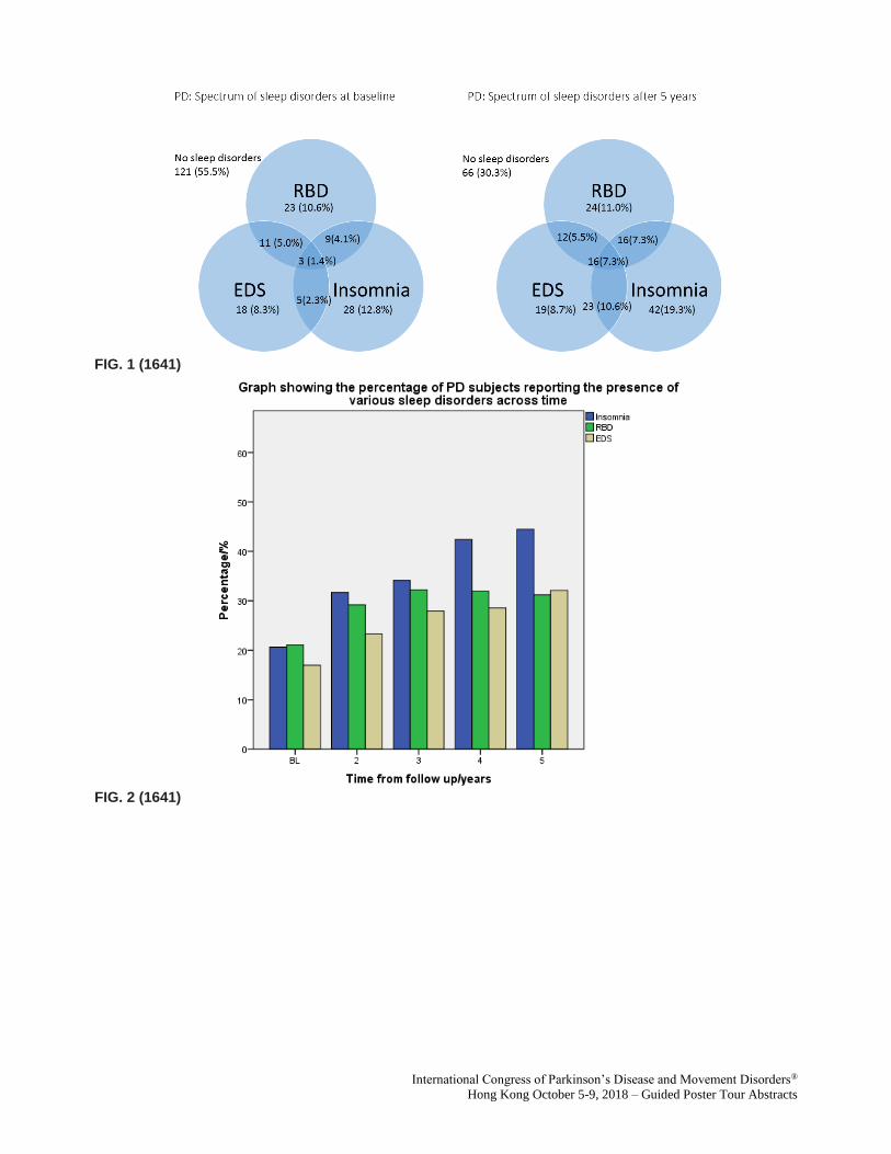
International Congress of Parkinson’s Disease and Movement Disorders®
Hong Kong October 5-9, 2018 – Guided Poster Tour Abstracts
FIG. 1 (1641)
FIG. 2 (1641)
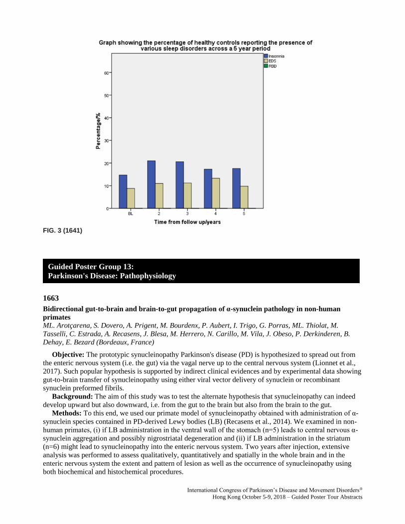
International Congress of Parkinson’s Disease and Movement Disorders®
Hong Kong October 5-9, 2018 – Guided Poster Tour Abstracts
FIG. 3 (1641)
1663
Bidirectional gut-to-brain and brain-to-gut propagation of α-synuclein pathology in non-human
primates
ML. Arotçarena, S. Dovero, A. Prigent, M. Bourdenx, P. Aubert, I. Trigo, G. Porras, ML. Thiolat, M.
Tasselli, C. Estrada, A. Recasens, J. Blesa, M. Herrero, N. Carillo, M. Vila, J. Obeso, P. Derkinderen, B.
Dehay, E. Bezard (Bordeaux, France)
Objective: The prototypic synucleinopathy Parkinson's disease (PD) is hypothesized to spread out from
the enteric nervous system (i.e. the gut) via the vagal nerve up to the central nervous system (Lionnet et al.,
2017). Such popular hypothesis is supported by indirect clinical evidences and by experimental data showing
gut-to-brain transfer of synucleinopathy using either viral vector delivery of synuclein or recombinant
synuclein preformed fibrils.
Background: The aim of this study was to test the alternate hypothesis that synucleinopathy can indeed
develop upward but also downward, i.e. from the gut to the brain but also from the brain to the gut.
Methods: To this end, we used our primate model of synucleinopathy obtained with administration of α-
synuclein species contained in PD-derived Lewy bodies (LB) (Recasens et al., 2014). We examined in non-
human primates, (i) if LB administration in the ventral wall of the stomach (n=5) leads to central nervous α-
synuclein aggregation and possibly nigrostriatal degeneration and (ii) if LB administration in the striatum
(n=6) might lead to synucleinopathy into the enteric nervous system. Two years after injection, extensive
analysis was performed to assess qualitatively, quantitatively and spatially in the whole brain and in the
enteric nervous system the extent and pattern of lesion as well as the occurrence of synucleinopathy using
both biochemical and histochemical procedures.
Guided Poster Group 13:
Parkinson's Disease: Pathophysiology

International Congress of Parkinson’s Disease and Movement Disorders®
Hong Kong October 5-9, 2018 – Guided Poster Tour Abstracts
Results: Enteric injection of LB in non-human primates results in enteric nervous system pathology and
nigrostriatal lesion in keeping with the well-accepted hypothesis. However, striatum LB-injected animals, in
addition to the expected nigrostriatal degeneration, showed also enteric ner¬vous system pathology at the
stomach level.
Conclusions: This study establishes that α-synuclein species might move up and down the neural axis in
non-human primates questioning (i) the hypothesis of a peripheral origin of synucleinopathies (ii) and the
specificity of enteric nervous system as biomarker of early/presymptomatic PD.
References: [1] A. Lionnet, L. Leclair-Visonneau, M. Neunlist, S. Murayama, M. Takao, C.H. Adler, P.
Derkinderen, T.G. Beach, Does Parkinson's disease start in the gut?, Acta Neuropathol, 135 (2018) 1-12.
[2] A. Recasens, B. Dehay, J. Bove, I. Carballo-Carbajal, S. Dovero, A. Perez-Villalba, P.O. Fernagut, J.
Blesa, A. Parent, C. Perier, I. Farinas, J.A. Obeso, E. Bezard, M. Vila, Lewy body extracts from Parkinson
disease brains trigger alpha-synuclein pathology and neurodegeneration in mice and monkeys, Ann Neurol,
75 (2014) 351-362.
1666
Risk estimation in the years preceding diagnosis of Parkinson’s disease in the PREDICT-PD cohort
S. Auger, D. Rack, J. Bestwick, G. Giovannoni, A. Lees, A. Schrag, A. Noyce (London, United Kingdom)
Objective: To test and refine the use of probability-based algorithms to identify individuals at high-risk
of developing Parkinson’s disease (PD).
Background: Neurodegeneration preceding a formal diagnosis of PD is associated with identifiable
motor and non-motor features. Evidence-based algorithms have been developed to attempt identifying
individuals in this pre-diagnostic phase according to exposure to common risk factors and results of simple
screening tests. Two notable approaches to risk estimation are the PREDICT-PD algorithm (Noyce at al
2013) and the MDS research criteria (Berg et al 2015).
Methods: The PREDICT-PD pilot cohort comprised 1,323 healthy 60 to 80 year olds who completed
annual online surveys and a keyboard-tapping task. Here, we considered those in the cohort who were
diagnosed with PD during follow-up and tracked their risk estimation in the years preceding formal
diagnosis, according to both the PREDICT-PD and MDS research criteria algorithms. A key difference
between the MDS and PREDICT-PD algorithms, is that whereas PREDICT-PD previously only used
demographic information, environmental exposures and simple symptomology to estimate risk with tapping
speed, hyposmia and REM sleep behaviour disorder as intermediate outcome markers, MDS-criteria
combines an extensive set of clinical and radiological tests. PREDICT-PD scores are also not updated in the
known absence of a risk factor and are based on odds ratios, rather than likelihood ratios. We sought to refine
the PREDICT-PD algorithm’s risk estimation by including our motor and non-motor intermediate markers
(hyposmia and REM sleep behaviour disorder) in the risk score.
Results: 8 people in the PREDICT-PD cohort have been diagnosed with PD from a total 864 with follow-
up at year 4. For these 8 with PD, mean probability risk score was higher over the 4 years according to the
MDS criteria than with PREDICT-PD basic score (25.5% vs 8.4%), both of which were higher than the rest
of the cohort (2.7% & 2.8% respectively, both p<0.0001). Incorporating intermediate markers into the
PREDICT-PD algorithm produced markedly greater probability risk scores than without for PD patients
(38.7% vs 8.4%, p<0.0001), with values remaining greater than healthy comparators’ (6.6%, p<0.0001).
Conclusions: Probability-based algorithms offer a means of identifying people at highest risk of
developing PD prior to a formal diagnosis. Their accuracy can be significantly improved by incorporating
simple, remotely administered screening tests.
References: Noyce AJ, Bestwick JP, Silveira-Moriyama L, et al. 2013. PREDICT-PD: Identifying risk of
Parkinson's disease in the community: methods and baseline results. J Neurol Neurosurg Psychiatry. Berg D,
Postuma R, Adler C, et al. 2015. MDS research criteria for prodromal Parkinson's disease. Movement
Disorders.
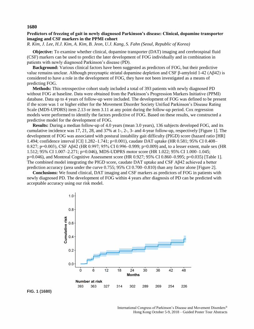
International Congress of Parkinson’s Disease and Movement Disorders®
Hong Kong October 5-9, 2018 – Guided Poster Tour Abstracts
1680
Predictors of freezing of gait in newly diagnosed Parkinson’s disease: Clinical, dopamine transporter
imaging and CSF markers in the PPMI cohort
R. Kim, J. Lee, H.J. Kim, A. Kim, B. Jeon, U.J. Kang, S. Fahn (Seoul, Republic of Korea)
Objective: To examine whether clinical, dopamine transporter (DAT) imaging and cerebrospinal fluid
(CSF) markers can be used to predict the later development of FOG individually and in combination in
patients with newly diagnosed Parkinson’s disease (PD).
Background: Various clinical factors have been suggested as predictors of FOG, but their predictive
value remains unclear. Although presynaptic striatal dopamine depletion and CSF β-amyloid 1-42 (Aβ42) is
considered to have a role in the development of FOG, they have not been investigated as a means of
predicting FOG.
Methods: This retrospective cohort study included a total of 393 patients with newly diagnosed PD
without FOG at baseline. Data were obtained from the Parkinson’s Progression Markers Initiative (PPMI)
database. Data up to 4 years of follow-up were included. The development of FOG was defined to be present
if the score was 1 or higher either for the Movement Disorder Society Unified Parkinson’s Disease Rating
Scale (MDS-UPDRS) item 2.13 or item 3.11 at any point during the follow-up period. Cox regression
models were performed to identify the factors predictive of FOG. Based on these results, we constructed a
predictive model for the development of FOG.
Results: During a median follow-up of 4.0 years (mean 3.0 years), 136 subjects developed FOG, and its
cumulative incidence was 17, 21, 28, and 37% at 1-, 2-, 3- and 4-year follow-up, respectively [Figure 1]. The
development of FOG was associated with postural instability gait difficulty (PIGD) score (hazard ratio [HR]
1.494; confidence interval [CI] 1.282–1.741; p<0.001), caudate DAT uptake (HR 0.581; 95% CI 0.408–
0.827; p=0.003), CSF Aβ42 (HR 0.997; 95% CI 0.996–0.999; p=0.009) and, to a lesser extent, male sex (HR
1.512; 95% CI 1.007–2.271; p=0.046), MDS-UDPRS motor score (HR 1.022; 95% CI 1.000–1.045;
p=0.046), and Montreal Cognitive Assessment score (HR 0.927; 95% CI 0.860–0.995; p=0.035) [Table 1].
The combined model integrating the PIGD score, caudate DAT uptake and CSF Aβ42 achieved a better
prediction accuracy (area under the curve 0.755; 95% CI 0.700–0.810) than any factor alone [Figure 2].
Conclusions: We found clinical, DAT imaging and CSF markers as predictors of FOG in patients with
newly diagnosed PD. The development of FOG within 4 years after diagnosis of PD can be predicted with
acceptable accuracy using our risk model.
FIG. 1 (1680)

International Congress of Parkinson’s Disease and Movement Disorders®
Hong Kong October 5-9, 2018 – Guided Poster Tour Abstracts
FIG. 2 (1680)
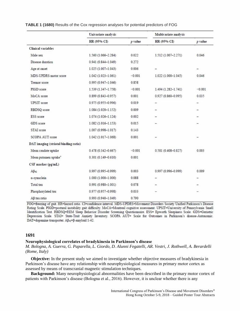
International Congress of Parkinson’s Disease and Movement Disorders®
Hong Kong October 5-9, 2018 – Guided Poster Tour Abstracts
TABLE 1 (1680) Results of the Cox regression analyses for potential predictors of FOG
1691
Neurophysiological correlates of bradykinesia in Parkinson’s disease
M. Bologna, A. Guerra, G. Paparella, L. Giordo, D. Alunni Fegatelli, AR. Vestri, J. Rothwell, A. Berardelli
(Rome, Italy)
Objective: In the present study we aimed to investigate whether objective measures of bradykinesia in
Parkinson’s disease have any relationship with neurophysiological measures in primary motor cortex as
assessed by means of transcranial magnetic stimulation techniques.
Background: Many neurophysiological abnormalities have been described in the primary motor cortex of
patients with Parkinson’s disease (Bologna et al., 2016). However, it is unclear whether there is any

International Congress of Parkinson’s Disease and Movement Disorders®
Hong Kong October 5-9, 2018 – Guided Poster Tour Abstracts
relationship between them and bradykinesia, one of the cardinal motor features of the condition (Espay et al.,
2011).
Methods: Twenty-two patients with Parkinson’s disease and 18 healthy subjects were enrolled. Objective
measurements of repetitive finger tapping (amplitude, speed and decrement) were obtained using a motion
analysis system. The excitability of primary motor cortex was assessed by recording the input/output curve
of the motor-evoked potentials and using a conditioning-test paradigm for the assessment of short-interval
intracortical inhibition and facilitation. Plasticity-like mechanisms in primary motor cortex were indexed
according to the amplitude changes in motor-evoked potentials after the paired associative stimulation
protocol. Patients were assessed in two sessions, i.e. ‘OFF’ and ‘ON’ medication. A canonical correlation
analysis was used to test for relationships between the kinematic and neurophysiological variables.
Results: Patients with Parkinson’s disease tapped more slowly and with smaller amplitude than normal,
and displayed decrement as tapping progressed. They also had steeper input/output curves, reduced short-
interval intracortical inhibition and a reduced response to the paired associative stimulation protocol. Within
the patient group, bradykinesia features correlated with the slope of the input/output curve and the after-
effects of the paired associative stimulation protocol. Although dopaminergic therapy improved movement
kinematics as well as neurophysiological measures, there was no relationship between them.
Conclusions: Neurophysiological changes in primary motor cortex relate to bradykinesia in patients with
Parkinson’s disease, although other mechanisms sensitive to dopamine levels must also play a role.
References: Bologna M, Suppa A, Conte A, Latorre A, Rothwell JC, Berardelli A. Are studies of motor
cortex plasticity relevant in human patients with Parkinson's disease? Clin Neurophysiol. 2016;127:50-9.
Espay AJ, Giuffrida JP, Chen R, Payne M, Mazzella F, Dunn E, et al. Differential response of speed,
amplitude, and rhythm to dopaminergic medications in Parkinson's disease. Mov Disord. 2011;26:2504-8.
1713
Microbiota DNA in the blood of patients with Parkinson’s disease
S.Q. Xu, Y.W. Qian, X.D. Yang, S.D. Chen, Q. Xiao (Shanghai, China)
Objective: The aim of this study was to explore the blood microbiota compositions in Chinese patients
with Parkinson’s disease (PD).
Background: Emerging evidences suggest that the microbiota present in the feces plays a role in PD.
However, the alterations of the microbiome in the blood of PD patients remains unknown.
Methods: Microbiota communities in the blood of 45 patients and their healthy spouses were investigated
using high-throughput Illumina Hiseq sequencing targeting the V3-V4 region of 16S ribosomal RNA (rRNA)
gene. The relationships between the microbiota in blood and PD clinical characteristics were analyzed.
Results: No difference was detected in the structure and richness between PD patients and healthy
controls. Genera Isoptericola, Cloacibacterium, Enhydrobacter, and Microbacterium were enriched in the
blood of PD patients, whereas genus Limnobacter was enriched in the healthy controls after adjusting for
age, gender, body mass index (BMI), and constipations. Furthermore, not only genera Cloacibacterium and
Isoptericola, which were identified enriched in the PD patients, but also genera Paludibacter and
Saccharofermentans were positively associated with disease duration. Some specific genera in the blood
were related to mood disorders.
Conclusions: We give direct evidences firstly to support the hypothesis that the identified microbiota in
the blood are associated with PD. Additionally, some microbiota in the blood are closely related to PD
clinical characteristics. Elucidating these differences in the blood microbiomes will provide a foundation to
improve our understanding the role of the microbiota in the pathogenesis of PD.
International Congress of Parkinson’s Disease and Movement Disorders®
Hong Kong October 5-9, 2018 – Guided Poster Tour Abstracts
1715
Unraveling gut microbiota in Parkinson’s disease and atypical parkinsonism
R. Cilia, M. Barichella, M. Severgnini, E. Cassani, C. Bolliri, S. Caronni, V. Ferri, R. Cancello, S.
Faierman, G. Pinelli, C. Ceccarani, G. De Bellis, L. Zecca, E. Cereda, C. Consolandi, G. Pezzoli (Milan,
Italy)
Objective: (1) To evaluate the differences in gut microbiota among Parkinson’s disease (PD), atypical
parkinsonism (i.e. multiple system atrophy [MSA] and progressive supranuclear palsy [PSP]) and healthy
control subjects (HC). (2) To investigate whether specific microbiota taxa may act as modulators of disease
progression and clinical phenotype.
Background: Recent evidences support the hypothesis that PD pathology originates into the gut and
propagates to the brain by different pathophysiologic pathways. However, findings are heterogeneous
probably due to the presence of several confounders.
Methods: We recruited patients with idiopathic PD (n=193, of whom 39 were de novo), PSP (n=22),
MSA (n=22), and HC (n=113). Several confounders have been taken into account, including
pharmacological therapy and dietary habits. Information on the type of lactation were also recorded. Early-
onset PD (≤50 ys) were screened for mutations on parkin, DJ-1, PINK-1 genes.
Results: Despite simple non-parametric comparison of PD patients and HC showed several differences in
relative taxa abundances, the number of significant comparisons was greatly reduced after adjusting for
multiple confounders. We observed a constant effect of age on almost all abundances. The use of COMT
inhibitors appeared to influence the level of several taxa.
Overall, PD patients had increased Verrucomicrobia, Christensenellaceae, Lactobacillaceae, and decreased
Lachnospiraceae and Ruminococcaceae than HC. Reduced level of Lachnospiraceae was significant in all
PD duration strata, while many of these differences were associated with disease progression. De novo PD
differed from HC only by lower abundance in Lachnospiraceae. Compared to PD, Lachnospiraceae and
Ruminococcaceae were not significantly lower in MSA, while in PSP cases other genera of
Ruminococcaceae and Lactobacillaceae were higher and comparable, respectively. Increased
Lactobacillaceae, Christensenellaceae, Verrucomicrobia and decreased Lachnospiraceae were associated
with worse disease severity, including intellectual impairment and other non-motor symptoms, and axial
features (gait disturbances and postural instability).
Conclusions: Gut microbiota may play a role in the pathogenesis of PD and act as modulators of
individual differences in disease severity, especially non-dopaminergic features (cognitive functions and
axial symptoms).
1722
Association between serum Vitamin D levels and Parkinson’s disease: A systematic review and meta-
analysis
XY. Luo, R. Ou, HF. Shang (Chengdu, China)
Objective: The aim of this systematic review and meta-analysis was to evaluate the association between
serum vitamin D levels and the risk and severity of PD.
Background: Vitamin D is an important molecular involving in the development and regulation of brain.
Several studies have focused on exploring the relationship between serum vitamin D levels and Parkinson’s
disease (PD), but the conclusion remains ambiguous yet.
Methods: We searched observational studies that explored the association between serum vitamin D
levels and PD based on Pubmed, EMBASE and Cochrane library. The quality of included studies was
evaluated by using Newcastle-Ottawa Scale. Statistical analysis of this meta-analysis was performed by Stata
version 12.0 and R software.
Results: 22 studies with a total of 3516 PD patients and 3481 controls were included. Our meta-analysis
revealed that compared with controls, PD patients had lower vitamin D levels(WMD -5.21, 95%CI -6.48, -
3.94), especially in higher latitude regions (WMD -5.53, 95%CI -7.49, -3.57). Assay methods contributed
International Congress of Parkinson’s Disease and Movement Disorders®
Hong Kong October 5-9, 2018 – Guided Poster Tour Abstracts
significantly to high heterogeneity. Furthermore, PD patients with deficient vitamin D levels had advanced
risk(OR 2.08, 95%CI1.35, 3.19) than those patients with insufficient levels (OR=1.73, 95%CI 1.41, 2.12). In
addition, Vitamin D levels was also related to PD severity (WMD-5.69, 95%CI-8.42, 2.96), and the
summary correlation coefficient showed strong negative correlation (r=-0.609,95%CI-0.76, -0.39).
Moreover, the pooled effect size revealed that vitamin D concentrations were also negatively related to
mUPDRS scores(r=-0.36, 95%CI-0.53, -0.16), but not associated with duration(P=0.37) and age(P=0.49).
Conclusions: Vitamin D levels are inversely associated with the PD risk and severity. Our results may
provide a update evidence to better explore the effect of vitamin D on PD and prompt the adjunctive
therapeutic decisions about vitamin D application.
1734
Antibodies to alpha-Synuclein in Parkinson disease
D. Labunskiy (Santa Rosa, CA, USA)
Objective: Alpha-synuclein (α-syn) plays a very important role in pathogenesis of Parkinson’s disease
(PD). There are enough evidence of neuroinflammation in the brain tissue of many neurodegenertive
diseases, including PD. Many researchers found lymphocytes and macrophages infiltration in basal ganglia
of the brain samples of patients died from PD complications. On the same time, systematical study of α-syn
in the brain, serum and CSF of these patient still not performed.
Background: Under our observations were 56 PD patients, 39 men and 17 women, ages from 44 to 83
years old. Control group comprised from 124 healthy donors/ Also we used paraffin slices of cerebellum,
brain stem and basal ganglia from patients died from PD complications.
Methods: We produced an enzyme-linked immunosorbent assay (ELISA) for measuring α-syn AAB
(IgG) in the serum and CSF of PD patients. Also immunohistochemical study of the brains of 4 patients died
from PD. We used antibodies marked by fluorescein-isotiocianate (FITC). Control slices of the brain tissue
were taken from person died from PD and from non-neurological causes.
Results: Anti α-syn AAB were increased in 86 percent PD patients, both men -89 per cent and women -
74 per cent) Confocal luminescence was revealed fluorescence in substantia nigra, globus pallidum and locus
cereleum in the brain histological samples received from dead PD patients. The histological brain samples
from dead patients without any neurological diseases did not revealed any immunofluorescence in basal
ganglia or any other brain region.
Conclusions: The studies of immune response in neurodegenerative disorders is a new part of
pathogenetic approach to this problem. Antibodies to α-syn probably play an important role in clinical
picture and complications of PD.
References: Elevated Serum α-Synuclein Autoantibodies in Patients with Parkinson's Disease Relative to
Alzheimer's Disease and Controls. Shalash A, Salama M, Makar M, Roushdy T1, Elrassas H, Mohamed W,
El-Balkimy M, Abou Donia M. Front Neurol. 2017 Dec 22;8:720. doi: 10.3389/fneur.2017.00720.
eCollection 2017.
1750
Natural occurring antibodies reduce aggregation of α-synuclein
A. Braczynski, E. Agerschou, Y. Kronimus, W. Hoyer, R. Dodel, B. Falkenburger, J. Schulz, J. Bach (Aachen,
Germany)
Objective: We tested naturally occurring antibodies (nAbs) against α-synuclein both in vitro as well as in
HEK293T cells to determine binding, cell viability and capacity to reduce αSyn aggregation.
Background: Parkinson disease (PD) is the second frequent neurodegenerative disorder and causes
severe motor disturbances. nAbs are part of the innate immune system. They are produced without contact to
the specific antigen they recognize and are polyclonal. Variation in specific nAbs levels are described for
αSyn and tau. PD patients have lower levels of αSyn specific nAbs. nAbs-αSyn are thought to decrease the
International Congress of Parkinson’s Disease and Movement Disorders®
Hong Kong October 5-9, 2018 – Guided Poster Tour Abstracts
load of αSyn deposits in the brain, but the mechanism of action is fairly unclear. Therefore we want to
elucidate the behavior of nAbs-αSyn in cell culture systems.
Methods: We isolated nAbs-αSyn from intravenous Immunoglobulins (IVIG) using column affinity
purification. Recombinant human αSyn was coupled to AminoLink Plus coupling resin. IVIGs were
incubated in the column repeatedly followed by a pH dependent elution step. nAbs-αSyn from several
purification rounds were pooled to reach reasonable protein concentrations. We tested the pooled antibodies
to both in vitro as well as in HEK293T cells.
Results: nAbs-αSyn could be isolated from human IVIGs. They bound monomeric αSyn in a dot blot as
well as in an ELISA against aSyn. nAbs-αSyn did not affect cell viability as tested in a Cell Titer Blue and an
LDH Assay. In an in vitro Thioflavin T aggregation assay they inhibited fibrillation of αSyn. In a cell culture
system of HEK293T cells transfected with EGFP-tagged αSyn we saw a reduction of aggresome load as
visible by fluorescence microscopy.
Conclusions: nAbs-αSyn inhibited aggregation of αSyn both in vitro and in cell culture systems. These
findings have to be corroborated further. Next, we want to analyze the effects of nAbs-αSyn in a Parkinson
mouse modell and describe the binding behavior of nAbs-αSyn to different conformational αSyn forms. As
an outlook, we want to understand the mechanism of action of nAbs-αSyn to better extent and hopefully add
antibodies to improved treatment of PD patients.
1757
Alpha-synuclein oligomer and rotenone treatments injury the dopaminergic neuron via inhibiting the
expression of gene SEMA6D
X. Yingyu (Guangzhou, China)
Objective: To investigate the expression of Semaphorin6D (SEMA6D) and its interaction with Plexin-A1
in cellular Parkinson's disease(PD) models induced by α-synuclein and rotenone.
Background: In order to uncover the molecular pathological process in Parkinson's disease (PD) ,we
employed a microarray analysis upon the alpha-synuclein oligomer induced cellular PD model and
investigate the significant differentially expressed genes(DEGs) screened from the microarray analysis.
Methods: SY-SH5Y cells cultured in vitro were divided into three groups:normal control group, α-
synuclein oligomer-induced group, rotenone-induced group.The last two groups were treated with α-
synuclein oligomer and rotenone respectively to establish the cellular PD models. The mRNA levels of
SEMA6D and plexin-A1 were evaluated using real-time polymerase chain reaction analysis(rt-PCR), and the
determinations had also been made on related proteins by Western blot analysis. The interaction between
SEMA6D and Plexin-A1 was validated by co-immunoprecipitation. Immunofluorescence and co-focusing
experiment were used to investigate the co-location of SEMA6D and PlexinA1 inside the cell.
Results: As the result showed, the expression of SEMA6D was significantly decreased in cellular PD
model. KEGG Pathway analysis showed that SEMA6D was closely related to the network of MARK
pathway. Comparing with the normal control group, the expressions of SEMA6D in cellular PD model
groups were down-regulated obviously. Immunoprecipitation analysis and Immunofluorescence co-
localization analysis comfirmed that the association of SEMA6D with Plexin-A1 was reversed in PD models.
Conclusions: α-synuclein and rotenone treatments inhibit expression of SEMA6D and its interaction with
Plexin-A1. The decline of their interaction could trigger the MAPK signaling pathway, which is associated
with the progress of PD.Our results suggest that SEMA6D is a critical factor in the pathogenesis of PD, and
therefore could be an important drug target for novel treatments towards PD.