Biohybrid nanofiber constructs with anisotropic biomechanical properties
Upload
khangminh22Category
view
1download
0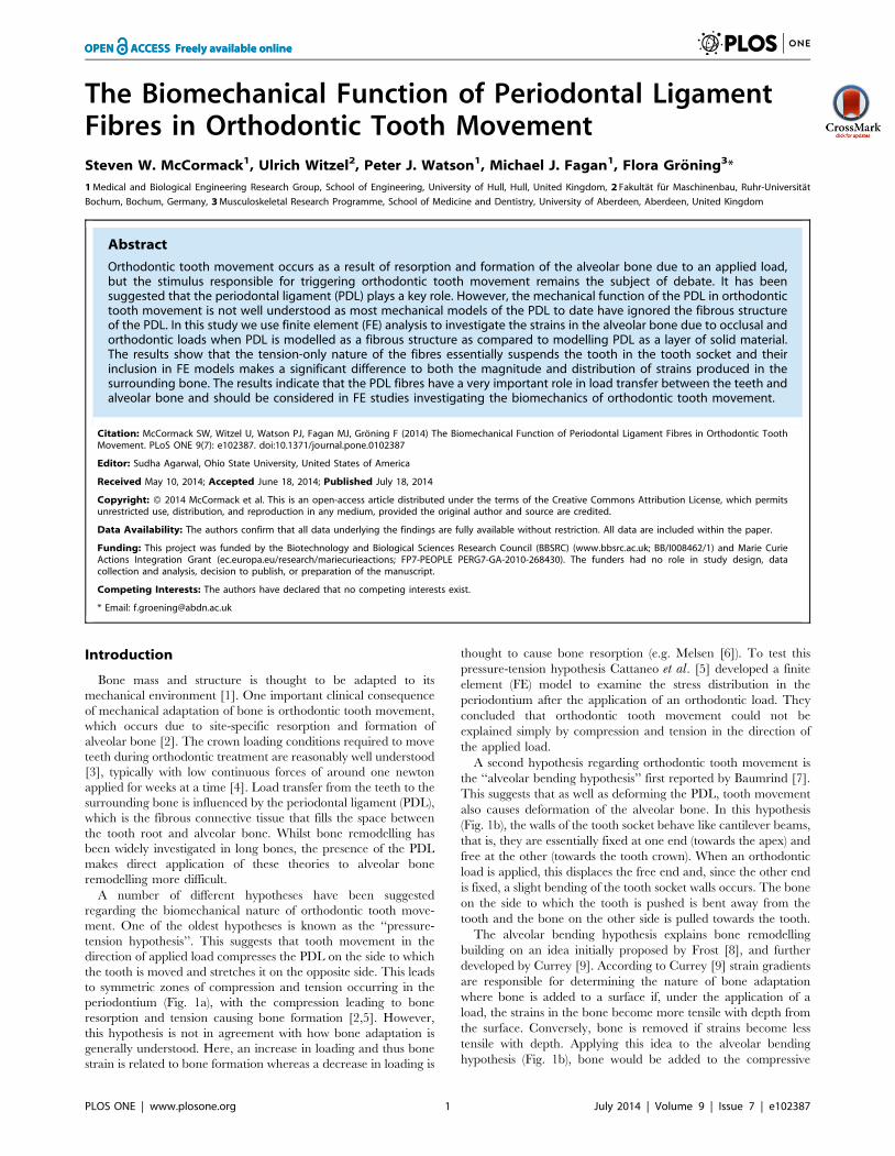
The Biomechanical Function of Periodontal LigamentFibres in Orthodontic Tooth MovementSteven W. McCormack1, Ulrich Witzel2, Peter J. Watson1, Michael J. Fagan1, Flora Groning3*
1 Medical and Biological Engineering Research Group, School of Engineering, University of Hull, Hull, United Kingdom, 2 Fakultat fur Maschinenbau, Ruhr-Universitat
Bochum, Bochum, Germany, 3 Musculoskeletal Research Programme, School of Medicine and Dentistry, University of Aberdeen, Aberdeen, United Kingdom
Abstract
Orthodontic tooth movement occurs as a result of resorption and formation of the alveolar bone due to an applied load,but the stimulus responsible for triggering orthodontic tooth movement remains the subject of debate. It has beensuggested that the periodontal ligament (PDL) plays a key role. However, the mechanical function of the PDL in orthodontictooth movement is not well understood as most mechanical models of the PDL to date have ignored the fibrous structureof the PDL. In this study we use finite element (FE) analysis to investigate the strains in the alveolar bone due to occlusal andorthodontic loads when PDL is modelled as a fibrous structure as compared to modelling PDL as a layer of solid material.The results show that the tension-only nature of the fibres essentially suspends the tooth in the tooth socket and theirinclusion in FE models makes a significant difference to both the magnitude and distribution of strains produced in thesurrounding bone. The results indicate that the PDL fibres have a very important role in load transfer between the teeth andalveolar bone and should be considered in FE studies investigating the biomechanics of orthodontic tooth movement.
Citation: McCormack SW, Witzel U, Watson PJ, Fagan MJ, Groning F (2014) The Biomechanical Function of Periodontal Ligament Fibres in Orthodontic ToothMovement. PLoS ONE 9(7): e102387. doi:10.1371/journal.pone.0102387
Editor: Sudha Agarwal, Ohio State University, United States of America
Received May 10, 2014; Accepted June 18, 2014; Published July 18, 2014
Copyright: � 2014 McCormack et al. This is an open-access article distributed under the terms of the Creative Commons Attribution License, which permitsunrestricted use, distribution, and reproduction in any medium, provided the original author and source are credited.
Data Availability: The authors confirm that all data underlying the findings are fully available without restriction. All data are included within the paper.
Funding: This project was funded by the Biotechnology and Biological Sciences Research Council (BBSRC) (www.bbsrc.ac.uk; BB/I008462/1) and Marie CurieActions Integration Grant (ec.europa.eu/research/mariecurieactions; FP7-PEOPLE PERG7-GA-2010-268430). The funders had no role in study design, datacollection and analysis, decision to publish, or preparation of the manuscript.
Competing Interests: The authors have declared that no competing interests exist.
* Email: [email protected]
Introduction
Bone mass and structure is thought to be adapted to its
mechanical environment [1]. One important clinical consequence
of mechanical adaptation of bone is orthodontic tooth movement,
which occurs due to site-specific resorption and formation of
alveolar bone [2]. The crown loading conditions required to move
teeth during orthodontic treatment are reasonably well understood
[3], typically with low continuous forces of around one newton
applied for weeks at a time [4]. Load transfer from the teeth to the
surrounding bone is influenced by the periodontal ligament (PDL),
which is the fibrous connective tissue that fills the space between
the tooth root and alveolar bone. Whilst bone remodelling has
been widely investigated in long bones, the presence of the PDL
makes direct application of these theories to alveolar bone
remodelling more difficult.
A number of different hypotheses have been suggested
regarding the biomechanical nature of orthodontic tooth move-
ment. One of the oldest hypotheses is known as the ‘‘pressure-
tension hypothesis’’. This suggests that tooth movement in the
direction of applied load compresses the PDL on the side to which
the tooth is moved and stretches it on the opposite side. This leads
to symmetric zones of compression and tension occurring in the
periodontium (Fig. 1a), with the compression leading to bone
resorption and tension causing bone formation [2,5]. However,
this hypothesis is not in agreement with how bone adaptation is
generally understood. Here, an increase in loading and thus bone
strain is related to bone formation whereas a decrease in loading is
thought to cause bone resorption (e.g. Melsen [6]). To test this
pressure-tension hypothesis Cattaneo et al. [5] developed a finite
element (FE) model to examine the stress distribution in the
periodontium after the application of an orthodontic load. They
concluded that orthodontic tooth movement could not be
explained simply by compression and tension in the direction of
the applied load.
A second hypothesis regarding orthodontic tooth movement is
the ‘‘alveolar bending hypothesis’’ first reported by Baumrind [7].
This suggests that as well as deforming the PDL, tooth movement
also causes deformation of the alveolar bone. In this hypothesis
(Fig. 1b), the walls of the tooth socket behave like cantilever beams,
that is, they are essentially fixed at one end (towards the apex) and
free at the other (towards the tooth crown). When an orthodontic
load is applied, this displaces the free end and, since the other end
is fixed, a slight bending of the tooth socket walls occurs. The bone
on the side to which the tooth is pushed is bent away from the
tooth and the bone on the other side is pulled towards the tooth.
The alveolar bending hypothesis explains bone remodelling
building on an idea initially proposed by Frost [8], and further
developed by Currey [9]. According to Currey [9] strain gradients
are responsible for determining the nature of bone adaptation
where bone is added to a surface if, under the application of a
load, the strains in the bone become more tensile with depth from
the surface. Conversely, bone is removed if strains become less
tensile with depth. Applying this idea to the alveolar bending
hypothesis (Fig. 1b), bone would be added to the compressive
PLOS ONE | www.plosone.org 1 July 2014 | Volume 9 | Issue 7 | e102387
surfaces of the alveolar bone and removed from the tensile surfaces
causing the position of the tooth to move in the direction of the
applied load.
Recently, a third hypothesis has been suggested by Melsen [6]
which is intended to match orthodontic tooth movement with
orthopaedic bone remodelling in accordance with Frost’s me-
chanostat theory [10], in which low strain leads to bone resorption
and high strain leads to bone formation. The hypothesis suggested
by Melsen [6] is the ‘‘stretched fibre hypothesis’’. Typically teeth
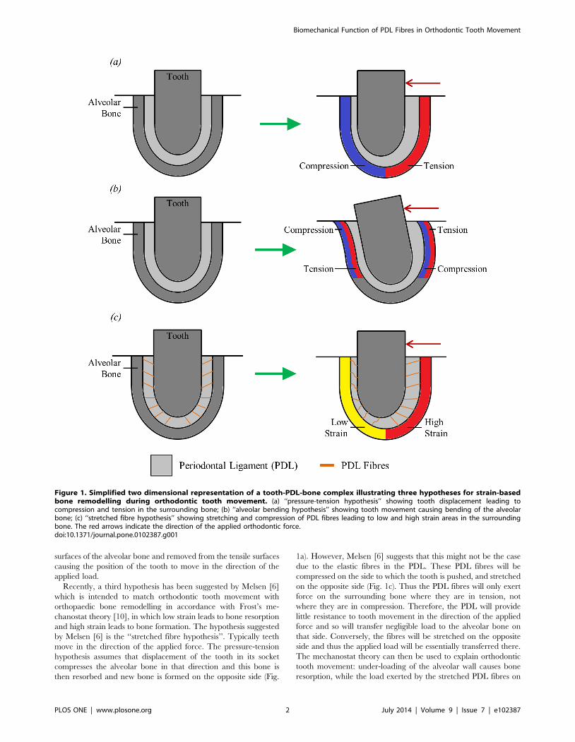
move in the direction of the applied force. The pressure-tension
hypothesis assumes that displacement of the tooth in its socket
compresses the alveolar bone in that direction and this bone is
then resorbed and new bone is formed on the opposite side (Fig.
1a). However, Melsen [6] suggests that this might not be the case
due to the elastic fibres in the PDL. These PDL fibres will be
compressed on the side to which the tooth is pushed, and stretched
on the opposite side (Fig. 1c). Thus the PDL fibres will only exert
force on the surrounding bone where they are in tension, not
where they are in compression. Therefore, the PDL will provide
little resistance to tooth movement in the direction of the applied
force and so will transfer negligible load to the alveolar bone on
that side. Conversely, the fibres will be stretched on the opposite
side and thus the applied load will be essentially transferred there.
The mechanostat theory can then be used to explain orthodontic
tooth movement: under-loading of the alveolar wall causes bone
resorption, while the load exerted by the stretched PDL fibres on
Figure 1. Simplified two dimensional representation of a tooth-PDL-bone complex illustrating three hypotheses for strain-basedbone remodelling during orthodontic tooth movement. (a) ‘‘pressure-tension hypothesis’’ showing tooth displacement leading tocompression and tension in the surrounding bone; (b) ‘‘alveolar bending hypothesis’’ showing tooth movement causing bending of the alveolarbone; (c) ‘‘stretched fibre hypothesis’’ showing stretching and compression of PDL fibres leading to low and high strain areas in the surroundingbone. The red arrows indicate the direction of the applied orthodontic force.doi:10.1371/journal.pone.0102387.g001
Biomechanical Function of PDL Fibres in Orthodontic Tooth Movement
PLOS ONE | www.plosone.org 2 July 2014 | Volume 9 | Issue 7 | e102387
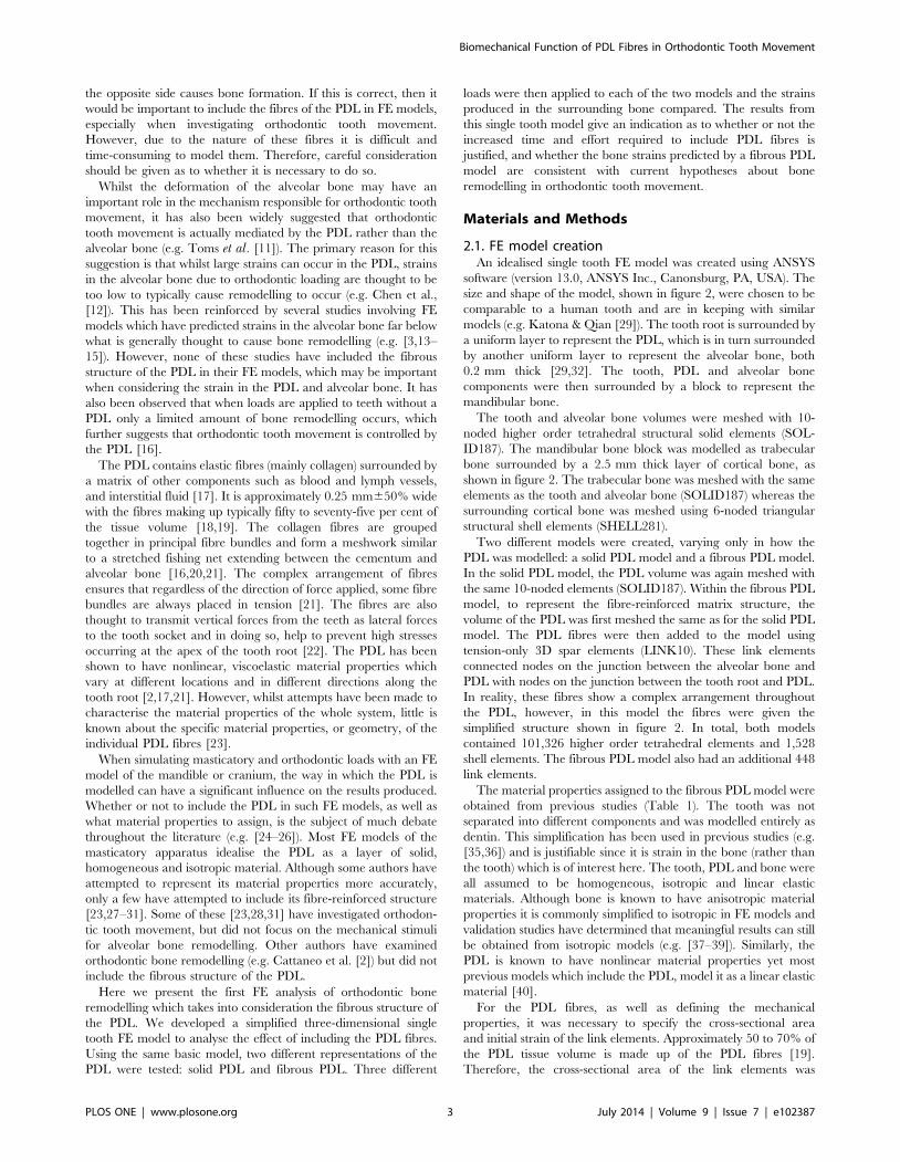
the opposite side causes bone formation. If this is correct, then it
would be important to include the fibres of the PDL in FE models,
especially when investigating orthodontic tooth movement.
However, due to the nature of these fibres it is difficult and
time-consuming to model them. Therefore, careful consideration
should be given as to whether it is necessary to do so.
Whilst the deformation of the alveolar bone may have an
important role in the mechanism responsible for orthodontic tooth
movement, it has also been widely suggested that orthodontic
tooth movement is actually mediated by the PDL rather than the
alveolar bone (e.g. Toms et al. [11]). The primary reason for this
suggestion is that whilst large strains can occur in the PDL, strains
in the alveolar bone due to orthodontic loading are thought to be
too low to typically cause remodelling to occur (e.g. Chen et al.,
[12]). This has been reinforced by several studies involving FE
models which have predicted strains in the alveolar bone far below
what is generally thought to cause bone remodelling (e.g. [3,13–
15]). However, none of these studies have included the fibrous
structure of the PDL in their FE models, which may be important
when considering the strain in the PDL and alveolar bone. It has
also been observed that when loads are applied to teeth without a
PDL only a limited amount of bone remodelling occurs, which
further suggests that orthodontic tooth movement is controlled by
the PDL [16].
The PDL contains elastic fibres (mainly collagen) surrounded by
a matrix of other components such as blood and lymph vessels,
and interstitial fluid [17]. It is approximately 0.25 mm650% wide
with the fibres making up typically fifty to seventy-five per cent of
the tissue volume [18,19]. The collagen fibres are grouped
together in principal fibre bundles and form a meshwork similar
to a stretched fishing net extending between the cementum and
alveolar bone [16,20,21]. The complex arrangement of fibres
ensures that regardless of the direction of force applied, some fibre
bundles are always placed in tension [21]. The fibres are also
thought to transmit vertical forces from the teeth as lateral forces
to the tooth socket and in doing so, help to prevent high stresses
occurring at the apex of the tooth root [22]. The PDL has been
shown to have nonlinear, viscoelastic material properties which
vary at different locations and in different directions along the
tooth root [2,17,21]. However, whilst attempts have been made to
characterise the material properties of the whole system, little is
known about the specific material properties, or geometry, of the
individual PDL fibres [23].
When simulating masticatory and orthodontic loads with an FE
model of the mandible or cranium, the way in which the PDL is
modelled can have a significant influence on the results produced.
Whether or not to include the PDL in such FE models, as well as
what material properties to assign, is the subject of much debate
throughout the literature (e.g. [24–26]). Most FE models of the
masticatory apparatus idealise the PDL as a layer of solid,
homogeneous and isotropic material. Although some authors have
attempted to represent its material properties more accurately,
only a few have attempted to include its fibre-reinforced structure
[23,27–31]. Some of these [23,28,31] have investigated orthodon-
tic tooth movement, but did not focus on the mechanical stimuli
for alveolar bone remodelling. Other authors have examined
orthodontic bone remodelling (e.g. Cattaneo et al. [2]) but did not
include the fibrous structure of the PDL.
Here we present the first FE analysis of orthodontic bone
remodelling which takes into consideration the fibrous structure of
the PDL. We developed a simplified three-dimensional single
tooth FE model to analyse the effect of including the PDL fibres.
Using the same basic model, two different representations of the
PDL were tested: solid PDL and fibrous PDL. Three different
loads were then applied to each of the two models and the strains
produced in the surrounding bone compared. The results from
this single tooth model give an indication as to whether or not the
increased time and effort required to include PDL fibres is
justified, and whether the bone strains predicted by a fibrous PDL
model are consistent with current hypotheses about bone
remodelling in orthodontic tooth movement.
Materials and Methods
2.1. FE model creationAn idealised single tooth FE model was created using ANSYS
software (version 13.0, ANSYS Inc., Canonsburg, PA, USA). The
size and shape of the model, shown in figure 2, were chosen to be
comparable to a human tooth and are in keeping with similar
models (e.g. Katona & Qian [29]). The tooth root is surrounded by
a uniform layer to represent the PDL, which is in turn surrounded
by another uniform layer to represent the alveolar bone, both
0.2 mm thick [29,32]. The tooth, PDL and alveolar bone
components were then surrounded by a block to represent the
mandibular bone.
The tooth and alveolar bone volumes were meshed with 10-
noded higher order tetrahedral structural solid elements (SOL-
ID187). The mandibular bone block was modelled as trabecular
bone surrounded by a 2.5 mm thick layer of cortical bone, as
shown in figure 2. The trabecular bone was meshed with the same
elements as the tooth and alveolar bone (SOLID187) whereas the
surrounding cortical bone was meshed using 6-noded triangular
structural shell elements (SHELL281).
Two different models were created, varying only in how the
PDL was modelled: a solid PDL model and a fibrous PDL model.
In the solid PDL model, the PDL volume was again meshed with
the same 10-noded elements (SOLID187). Within the fibrous PDL
model, to represent the fibre-reinforced matrix structure, the
volume of the PDL was first meshed the same as for the solid PDL
model. The PDL fibres were then added to the model using
tension-only 3D spar elements (LINK10). These link elements
connected nodes on the junction between the alveolar bone and
PDL with nodes on the junction between the tooth root and PDL.
In reality, these fibres show a complex arrangement throughout
the PDL, however, in this model the fibres were given the
simplified structure shown in figure 2. In total, both models
contained 101,326 higher order tetrahedral elements and 1,528
shell elements. The fibrous PDL model also had an additional 448
link elements.
The material properties assigned to the fibrous PDL model were
obtained from previous studies (Table 1). The tooth was not
separated into different components and was modelled entirely as
dentin. This simplification has been used in previous studies (e.g.
[35,36]) and is justifiable since it is strain in the bone (rather than
the tooth) which is of interest here. The tooth, PDL and bone were
all assumed to be homogeneous, isotropic and linear elastic
materials. Although bone is known to have anisotropic material
properties it is commonly simplified to isotropic in FE models and
validation studies have determined that meaningful results can still
be obtained from isotropic models (e.g. [37–39]). Similarly, the
PDL is known to have nonlinear material properties yet most
previous models which include the PDL, model it as a linear elastic
material [40].
For the PDL fibres, as well as defining the mechanical
properties, it was necessary to specify the cross-sectional area
and initial strain of the link elements. Approximately 50 to 70% of
the PDL tissue volume is made up of the PDL fibres [19].
Therefore, the cross-sectional area of the link elements was
Biomechanical Function of PDL Fibres in Orthodontic Tooth Movement
PLOS ONE | www.plosone.org 3 July 2014 | Volume 9 | Issue 7 | e102387

calculated so that the combined volume of the link elements would
be between 50 and 70% of the PDL volume in the model. From
this the cross-sectional area was chosen to be 0.06 mm2. This
value makes the link elements in the model one to two orders of
magnitude thicker than the collagen fibres found in a real PDL
[21,31]. This is due to the fact that the number of modelled PDL
fibres is well below the number of fibres in the real PDL. No
reliable data was available to suggest a suitable value for any initial
strain of the link elements and so it was defined as zero.
The solid PDL model was constructed with the same method as
the fibrous PDL model, except it did not include the link elements
representing the PDL fibres. In order to make a fair comparison
between the two models, it was necessary to adjust Young’s
modulus value given to the PDL component of the solid PDL
model, while all other material properties remained the same. This
value was adjusted so that the overall effective elastic modulus of
the PDL layer in the two models was the same. From this it follows
that any differences observed in the stress and strain values
between the solid PDL and fibrous PDL models could be
attributed to the structural difference, i.e. the presence or absence
of PDL fibres, between the two models (rather than to the
difference in the elastic modulus of the PDL) [22].
As a criterion for adjusting Young’s modulus we used tooth
displacement, i.e. the two models were assumed to have the same
overall effective elastic modulus when they showed the same tooth
displacement under identical loading conditions. Tooth displace-
ment was defined as the change in distance between the most
apical node on the tooth root and the corresponding node on the
alveolar bone [29]. Thus, under a load of 500 N, the tooth
displacement for the fibrous tooth model was 0.0703 mm. This
Figure 2. Single tooth FE model including dimensions in millimetres. (a) the whole 3D single tooth model; (b) section through tooth, PDLand alveolar bone showing the location of the link elements which span the PDL layer connecting the tooth and alveolar bone; (c) section throughthe centre of the model showing the tooth, PDL and alveolar bone including an expanded view of the apex region.doi:10.1371/journal.pone.0102387.g002
Table 1. Mechanical properties assigned to each material in the fibrous PDL model.
Component Young’s Modulus (MPa) Poisson’s Ratio
Trabecular bonea 56 0.30
Cortical boneb 17 000 0.30
Toothb 17 000 0.30
PDL matrixc 1 0.45
PDL fibresd 1 000 0.35
aMisch et al. [33].bGroning et al. [24].cJones et al. [15], Qian et al. [23].dGautieri et al. [34], Katona and Qian [29], Meyer et al. [31], Rees and Jacobsen [18].doi:10.1371/journal.pone.0102387.t001
Biomechanical Function of PDL Fibres in Orthodontic Tooth Movement
PLOS ONE | www.plosone.org 4 July 2014 | Volume 9 | Issue 7 | e102387
value would seem reasonable when compared to the vertical
intrusion of 0.12 mm cited by Borak et al. [32] from an
experiment by Kato [41]. To get the same displacement for the
solid PDL model, it was found necessary to use a Young’s modulus
value of 12.2 MPa for the PDL layer (determined through the
ANSYS optimisation facility).
In single tooth FE models, one common way to apply the
boundary conditions is simply to fix all nodes on the two opposing
sides of the model to which the rest of the mandible would be
connected (e.g. Qian et al. [23]). However, a number of studies
have reported that over-constraining a model can lead to
inaccurate results (e.g. Marinescu et al. [42]). Therefore, this
method was not used here. Instead, the boundary conditions
illustrated in figure 3 were applied. Briefly, all of the nodes around
the edge of these two sides were constrained in the x-direction
(mesiodistal, figure 3) to represent the increased stiffness in this
location due to the presence of cortical bone. The nodes on the
rear corners on the base of the model were also constrained in the
y-direction (coronoapical) and the nodes on the front corners of
the base were constrained in all degrees of freedom. This
prevented rigid body translation of the model, while still allowing
some deformation in the z-direction (buccolingual).
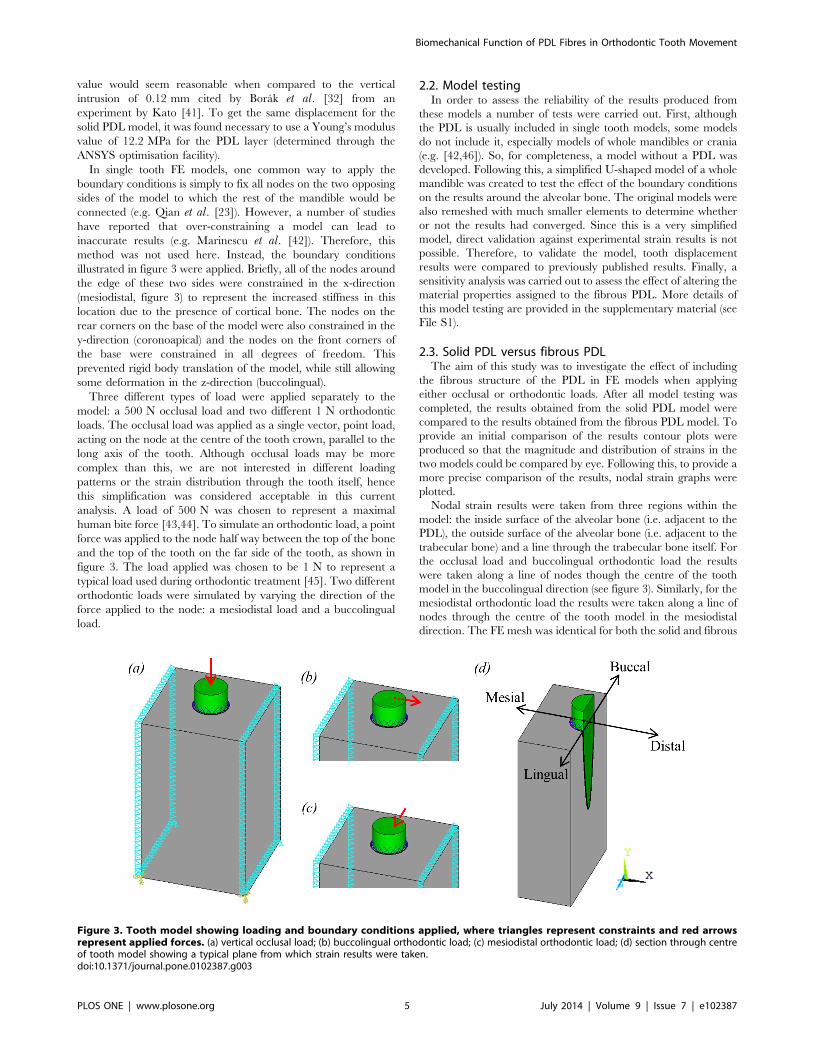
Three different types of load were applied separately to the
model: a 500 N occlusal load and two different 1 N orthodontic
loads. The occlusal load was applied as a single vector, point load,
acting on the node at the centre of the tooth crown, parallel to the
long axis of the tooth. Although occlusal loads may be more
complex than this, we are not interested in different loading
patterns or the strain distribution through the tooth itself, hence
this simplification was considered acceptable in this current
analysis. A load of 500 N was chosen to represent a maximal
human bite force [43,44]. To simulate an orthodontic load, a point
force was applied to the node half way between the top of the bone
and the top of the tooth on the far side of the tooth, as shown in
figure 3. The load applied was chosen to be 1 N to represent a
typical load used during orthodontic treatment [45]. Two different
orthodontic loads were simulated by varying the direction of the
force applied to the node: a mesiodistal load and a buccolingual
load.
2.2. Model testingIn order to assess the reliability of the results produced from
these models a number of tests were carried out. First, although
the PDL is usually included in single tooth models, some models
do not include it, especially models of whole mandibles or crania
(e.g. [42,46]). So, for completeness, a model without a PDL was
developed. Following this, a simplified U-shaped model of a whole
mandible was created to test the effect of the boundary conditions
on the results around the alveolar bone. The original models were
also remeshed with much smaller elements to determine whether
or not the results had converged. Since this is a very simplified
model, direct validation against experimental strain results is not
possible. Therefore, to validate the model, tooth displacement
results were compared to previously published results. Finally, a
sensitivity analysis was carried out to assess the effect of altering the
material properties assigned to the fibrous PDL. More details of
this model testing are provided in the supplementary material (see
File S1).
2.3. Solid PDL versus fibrous PDLThe aim of this study was to investigate the effect of including
the fibrous structure of the PDL in FE models when applying
either occlusal or orthodontic loads. After all model testing was
completed, the results obtained from the solid PDL model were
compared to the results obtained from the fibrous PDL model. To
provide an initial comparison of the results contour plots were
produced so that the magnitude and distribution of strains in the
two models could be compared by eye. Following this, to provide a
more precise comparison of the results, nodal strain graphs were
plotted.
Nodal strain results were taken from three regions within the
model: the inside surface of the alveolar bone (i.e. adjacent to the
PDL), the outside surface of the alveolar bone (i.e. adjacent to the
trabecular bone) and a line through the trabecular bone itself. For
the occlusal load and buccolingual orthodontic load the results
were taken along a line of nodes though the centre of the tooth
model in the buccolingual direction (see figure 3). Similarly, for the
mesiodistal orthodontic load the results were taken along a line of
nodes through the centre of the tooth model in the mesiodistal
direction. The FE mesh was identical for both the solid and fibrous
Figure 3. Tooth model showing loading and boundary conditions applied, where triangles represent constraints and red arrowsrepresent applied forces. (a) vertical occlusal load; (b) buccolingual orthodontic load; (c) mesiodistal orthodontic load; (d) section through centreof tooth model showing a typical plane from which strain results were taken.doi:10.1371/journal.pone.0102387.g003
Biomechanical Function of PDL Fibres in Orthodontic Tooth Movement
PLOS ONE | www.plosone.org 5 July 2014 | Volume 9 | Issue 7 | e102387
PDL models and so the same nodes were used to plot the results
for both models. Results were not plotted at nodes which were
connected to link elements. For the line through the trabecular
bone, suitable nodes were selected (approximately 1 mm from the
alveolar bone) by eye since there was no smooth line of nodes
through this region. However, since these nodes were the same in
all models, the strain distributions could still be compared with
confidence.
Maximum and minimum principal strains were extracted along
each of these regions for both solid and fibrous PDL models and
these results were plotted against each other for visual comparison.
For the solid PDL model, the nodal results were plotted and a
smooth line was drawn connecting the points. However, for the
fibrous PDL model, strain results obtained at the inside and
outside surfaces of the alveolar bone were not smooth but rather
contained fluctuating values due to strain concentrations develop-
ing at the location of the link elements. In a real PDL these high
peak strains would not develop due to the much greater number of
PDL fibres than represented in this model which would cause a
smoother distribution of strain. Since these peak strains are not
biologically realistic, rather than simply connecting the points, a
third order trend line was plotted through the data to smooth out
the results. This was performed for the results from the inside and
outside surface of the alveolar bone for the fibrous PDL model in
all three load cases. This smoothing was not necessary for the
trabecular bone nodal results from the fibrous PDL model, as
these nodes were sufficiently far away from the location of the link
elements to not be effected by the local stress concentrations.
Results
3.1. Model testing resultsAs well as testing the results from a no PDL model, four key
issues were investigated with respect to model testing: boundary
conditions, mesh convergence, model validation and sensitivity to
changes in the PDL. The no PDL model showed that the PDL is
important in order to reduce the local bone strain in the region
surrounding the tooth root (see Fig. S1 in File S1). The results from
the U-shaped model showed that the boundary conditions chosen
for the single tooth model were acceptable since they produced
very similar results (see Table S1, Fig. S4, and Fig. S5 in File S1).
The mesh was shown to have converged and the tooth
displacement results were seen to be of similar magnitude to
previously reported results (see File S1 for details). The sensitivity
analysis revealed that adjusting the material property values
assigned to the PDL can have a substantial influence on the results
obtained (see Table S2 in File S1). Further details of the model
testing can be seen in the supplementary material (see File S1).
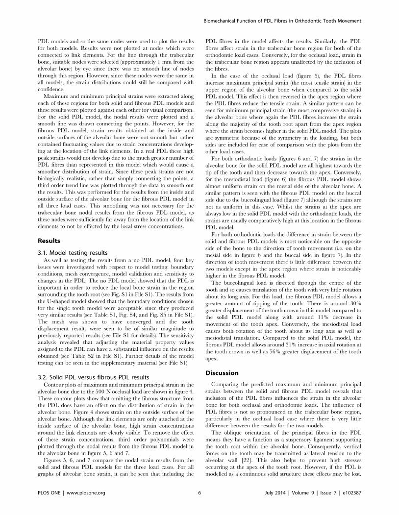
3.2. Solid PDL versus fibrous PDL resultsContour plots of maximum and minimum principal strain in the
alveolar bone due to the 500 N occlusal load are shown in figure 4.
These contour plots show that omitting the fibrous structure from
the PDL does have an effect on the distribution of strain in the
alveolar bone. Figure 4 shows strain on the outside surface of the
alveolar bone. Although the link elements are only attached at the
inside surface of the alveolar bone, high strain concentrations
around the link elements are clearly visible. To remove the effect
of these strain concentrations, third order polynomials were
plotted through the nodal results from the fibrous PDL model in
the alveolar bone in figure 5, 6 and 7.
Figures 5, 6, and 7 compare the nodal strain results from the
solid and fibrous PDL models for the three load cases. For all
graphs of alveolar bone strain, it can be seen that including the
PDL fibres in the model affects the results. Similarly, the PDL
fibres affect strain in the trabecular bone region for both of the
orthodontic load cases. Conversely, for the occlusal load, strain in
the trabecular bone region appears unaffected by the inclusion of
the fibres.
In the case of the occlusal load (figure 5), the PDL fibres
increase maximum principal strain (the most tensile strain) in the
upper region of the alveolar bone when compared to the solid
PDL model. This effect is then reversed in the apex region where
the PDL fibres reduce the tensile strain. A similar pattern can be
seen for minimum principal strain (the most compressive strain) in
the alveolar bone where again the PDL fibres increase the strain
along the majority of the tooth root apart from the apex region
where the strain becomes higher in the solid PDL model. The plots
are symmetric because of the symmetry in the loading, but both
sides are included for ease of comparison with the plots from the
other load cases.
For both orthodontic loads (figures 6 and 7) the strains in the
alveolar bone for the solid PDL model are all highest towards the
tip of the tooth and then decrease towards the apex. Conversely,
for the mesiodistal load (figure 6) the fibrous PDL model shows
almost uniform strain on the mesial side of the alveolar bone. A
similar pattern is seen with the fibrous PDL model on the buccal
side due to the buccolingual load (figure 7) although the strains are
not as uniform in this case. Whilst the strains at the apex are
always low in the solid PDL model with the orthodontic loads, the
strains are usually comparatively high at this location in the fibrous
PDL model.
For both orthodontic loads the difference in strain between the
solid and fibrous PDL models is most noticeable on the opposite
side of the bone to the direction of tooth movement (i.e. on the
mesial side in figure 6 and the buccal side in figure 7). In the
direction of tooth movement there is little difference between the
two models except in the apex region where strain is noticeably
higher in the fibrous PDL model.
The buccolingual load is directed through the centre of the
tooth and so causes translation of the tooth with very little rotation
about its long axis. For this load, the fibrous PDL model allows a
greater amount of tipping of the tooth. There is around 30%
greater displacement of the tooth crown in this model compared to
the solid PDL model along with around 11% decrease in
movement of the tooth apex. Conversely, the mesiodistal load
causes both rotation of the tooth about its long axis as well as
mesiodistal translation. Compared to the solid PDL model, the
fibrous PDL model allows around 31% increase in axial rotation at
the tooth crown as well as 56% greater displacement of the tooth
apex.
Discussion
Comparing the predicted maximum and minimum principal
strains between the solid and fibrous PDL model reveals that
inclusion of the PDL fibres influences the strain in the alveolar
bone for both occlusal and orthodontic loads. The influence of
PDL fibres is not so pronounced in the trabecular bone region,
particularly in the occlusal load case where there is very little
difference between the results for the two models.
The oblique orientation of the principal fibres in the PDL
means they have a function as a suspensory ligament supporting
the tooth root within the alveolar bone. Consequently, vertical
forces on the tooth may be transmitted as lateral tension to the
alveolar wall [22]. This also helps to prevent high stresses
occurring at the apex of the tooth root. However, if the PDL is
modelled as a continuous solid structure these effects may be lost.
Biomechanical Function of PDL Fibres in Orthodontic Tooth Movement
PLOS ONE | www.plosone.org 6 July 2014 | Volume 9 | Issue 7 | e102387
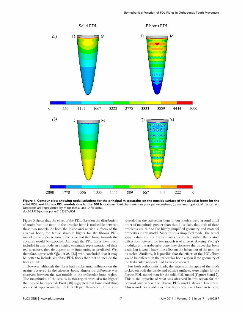
Figure 5 shows that the effect of the PDL fibres on the distribution
of strain from the tooth to the alveolar bone is noticeable between
these two models. At both the inside and outside surfaces of the
alveolar bone, the tensile strain is higher for the fibrous PDL
model in the upper section of the bone and then lower towards the
apex, as would be expected. Although the PDL fibres have been
included in this model in a highly schematic representation of their
real structure, they do appear to be functioning as predicted. We,
therefore, agree with Qian et al. [23] who concluded that it may
be better to include simplistic PDL fibres than not to include the
fibres at all.
However, although the fibres had a substantial influence on the
strains observed in the alveolar bone, almost no difference was
observed between the two models in the trabecular bone region.
The magnitudes of the strains in this region were also far higher
than would be expected. Frost [10] suggested that bone modelling
occurs at approximately 1500–3000 me. However, the strains
recorded in the trabecular bone in our models were around a full
order of magnitude greater than that. It is likely that both of these
problems are due to the highly simplified geometry and material
properties in this model. Since this is a simplified model, the actual
strain values are not the primary concern but rather the relative
differences between the two models is of interest. Altering Young’s
modulus of the trabecular bone may decrease the trabecular bone
strain but it would have little effect on the behaviour of the tooth in
its socket. Similarly, it is possible that the effects of the PDL fibres
would be different in the trabecular bone region if the geometry of
the trabecular network had been considered.
For both orthodontic loads, the strains at the apex of the tooth
socket, on both the inside and outside surfaces, were higher for the
fibrous PDL model than for the solid PDL model (Figures 6 and 7).
This is the opposite of what was observed in this region for the
occlusal load where the fibrous PDL model showed less strain.
This is understandable since the fibres only exert force in tension,
Figure 4. Contour plots showing nodal solutions for the principal microstrains on the outside surface of the alveolar bone for thesolid PDL and fibrous PDL models due to the 500 N occlusal load. (a) maximum principal microstrain; (b) minimum principal microstrain.Directions are represented by M for mesial and D for distal.doi:10.1371/journal.pone.0102387.g004
Biomechanical Function of PDL Fibres in Orthodontic Tooth Movement
PLOS ONE | www.plosone.org 7 July 2014 | Volume 9 | Issue 7 | e102387
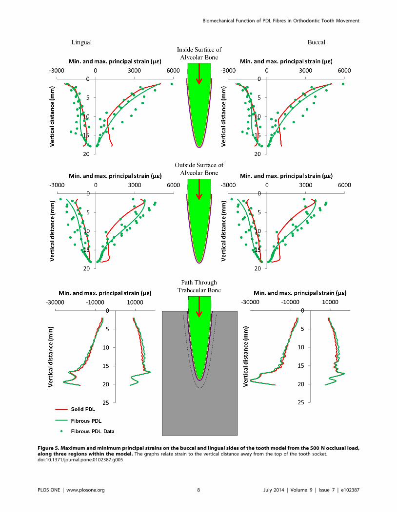
Figure 5. Maximum and minimum principal strains on the buccal and lingual sides of the tooth model from the 500 N occlusal load,along three regions within the model. The graphs relate strain to the vertical distance away from the top of the tooth socket.doi:10.1371/journal.pone.0102387.g005
Biomechanical Function of PDL Fibres in Orthodontic Tooth Movement
PLOS ONE | www.plosone.org 8 July 2014 | Volume 9 | Issue 7 | e102387
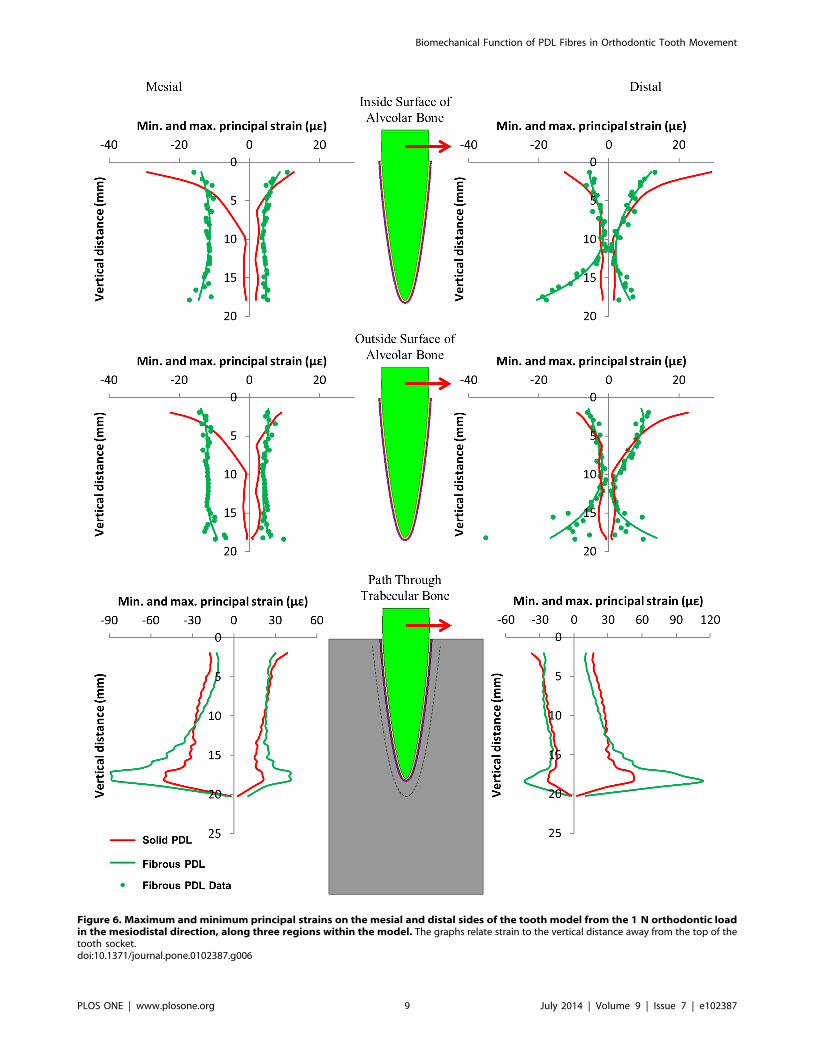
Figure 6. Maximum and minimum principal strains on the mesial and distal sides of the tooth model from the 1 N orthodontic loadin the mesiodistal direction, along three regions within the model. The graphs relate strain to the vertical distance away from the top of thetooth socket.doi:10.1371/journal.pone.0102387.g006
Biomechanical Function of PDL Fibres in Orthodontic Tooth Movement
PLOS ONE | www.plosone.org 9 July 2014 | Volume 9 | Issue 7 | e102387

Figure 7. Maximum and minimum principal strains on the buccal and lingual sides of the tooth model from the 1 N orthodonticload in the buccolingual direction, along three regions within the model. The graphs relate strain to the vertical distance away from the topof the tooth socket.doi:10.1371/journal.pone.0102387.g007
Biomechanical Function of PDL Fibres in Orthodontic Tooth Movement
PLOS ONE | www.plosone.org 10 July 2014 | Volume 9 | Issue 7 | e102387
not in compression. When a vertical load is applied the fibres at
the apex are not stretched and so only the soft PDL matrix resists
the tooth at this point. However, when an orthodontic load is
applied, translation and rotation of the tooth occur which would
stretch the apical fibres causing them to exert a force on the
alveolar bone at this location.
For both orthodontic load cases, strain in the solid PDL model is
always highest at the top of the alveolar bone and then decreases
towards the apex of the tooth on both sides of the model. This is
not the case for the fibrous PDL model. In this model, there is a
more uniform distribution of strain along the length of the alveolar
bone on the side of the tooth away from the direction of tooth
movement. This is particularly obvious with the mesiodistal load.
On the side to which the tooth is moved, there is less difference
between the solid and fibrous PDL models except at the apex. This
is due to the fact that the PDL fibres only exert force in tension
and so on this side the fibrous PDL behaves similarly to a solid
PDL.
Compared to the solid PDL model, the fibrous PDL model
allows for a greater amount of tooth movement with both
orthodontic loads. The crossed structure of the PDL fibres (figure
2) ensures that some fibres are always in tension and therefore
resisting tooth movement, regardless of the direction of an applied
load [21]. However, this also means that some fibres are in
compression and thus not playing any role in resisting the tooth
movement. It is therefore not surprising that the fibrous PDL
model allows for greater rotation and tipping of the tooth
compared to the solid PDL model.
The strain results in figures 6 and 7 can be interpreted in light of
the three hypotheses shown in figure 1. Neither set of results agree
with the pressure-tension hypothesis. This hypothesis predicts that
there should be compression on the side to which the tooth is
moved and tension on the opposite side. Neither set of results show
distinct regions of compression and tension as predicted.
The alveolar bending hypothesis predicts both compression and
tension occurring in the bone on each side of the tooth but on
opposite surfaces of the alveolar bone, i.e. tension on the inside
surface and compression on the outside surface or vice versa.
However, for both the mesiodistal and buccolingual orthodontic
loads (figures 6 and 7) the type of stress occurring, either
compressive or tensile, is the same at the inside and outside
surfaces of the alveolar bone on both sides of the tooth. This
suggests that perhaps the bone is not bending as predicted.
Alternatively, since the strain results are very similar at the inside
and outside surfaces, this may be due to the alveolar bone being
very thin in this model. Consequently, the bending may involve
the trabecular bone and so the sign change, e.g. from compression
to tension, may occur in this region rather than in the alveolar
bone region. This appears to be the case for the buccolingual load
(figure 7) where on the lingual side the alveolar bone is
predominantly in tension while the trabecular bone is slightly
more in compression, and on the buccal side the alveolar bone is
in compression while the trabecular bone is in tension.
The mesiodistal load causes both tilting of the tooth in the
mesiodistal direction and rotation of the tooth about its long axis
which is not taken into consideration in the alveolar bending
hypothesis (figure 1). A closer inspection of the direction of strains
in the alveolar bone indicates that the tensile strains on the distal
side and the compressive strains on the mesial side (which are
predicted by the alveolar bending hypothesis) are predominantly
orientated in the axial direction (i.e. parallel to the long axis of the
tooth). This suggests that if the model was only being considered in
two dimensions, the alveolar bending hypothesis may appear
correct. However, in three dimensions, simple cantilever bending
of the alveolar bone is not the only cause of strain in the alveolar
bone.
For the buccolingual load, the lingual side is predominantly in
tension as predicted. Additionally, the tensile strains are mostly
orientated in the axial direction. Conversely, the compressive
strains, which are not predicted by the alveolar bending
hypothesis, are orientated in the hoop (i.e. directed around the
tooth within the horizontal plane) and radial (i.e. directed away
from the long axis of the tooth within the horizontal plane)
directions. Similarly, on the compression side, the compressive
strains are mostly orientated in the axial direction whereas the
tensile strains on that side are mostly in the hoop and radial
directions. These results also provide some support for the alveolar
bending hypothesis, although again strain must be considered in
all directions.
Unlike the pressure-tension hypothesis and the alveolar bending
hypothesis, the stretched fibre hypothesis does not consider
whether the bone is in compression or tension, but is mainly
concerned with whether bone is in high strain or low strain. This
hypothesis is also the one that should be most affected by whether
or not fibres are included in the FE model. For both the
mesiodistal load and the buccolingual load, strain is higher in the
fibrous PDL model than the solid PDL model on the side which
the tooth is moved away from (the mesial side in figure 6 and the
buccal side in figure 7). This provides some support to this
hypothesis which predicts that strains should be high on this side
due stretching of the PDL fibres.
Whilst there is some support for the stretched fibre hypothesis
on the side which bone is formed, it is not clear whether resorption
on the other side is due to low strains. For the buccolingual load
the magnitude of strain on the lingual side is not greatly different
to that on the buccal side. For the mesiodistal load there is almost
uniform strain on the mesial side (the side from which the tooth is
moved away). Conversely, on the distal side (the side to which the
tooth is moved) the strains are reasonably low in the upper portion
of the alveolar bone but get much higher towards the apex. In
neither case are the strains particularly low in the direction of
tooth movement to suggest that bone resorption is due to under-
loading.
For both orthodontic loads, the strains observed in the alveolar
bone are all far below those values typically thought to cause bone
remodelling [10]. However, it should be emphasised that this is a
simplified model and so the absolute values of strain in the model
cannot be assumed to be accurate. Nevertheless, this agrees with
the results of other authors who also found very low strains in the
alveolar bone (e.g. [3,15]). The low strains commonly observed in
the alveolar bone have led to the proposal that the stimulus for
orthodontic tooth movement comes from the PDL rather than the
alveolar bone (e.g. Chen et al. [12]). However, if the PDL is
responsible for controlling orthodontic tooth movement, the
mechanical stimulus for this is still unclear. For example, Qian
et al. [47] used normal strain in the PDL to drive tooth movement
whereas Chen et al. [12] chose hydrostatic stress in the PDL.
It has been suggested that, rather than all bones responding to
the same levels of strain, it may be the case that resorption and
formation are triggered at different strain levels in different bones
[48]. Also, the alveolar bone is thought to be one of the most
physiologically active bones in mammals [49]. Therefore, whilst
the strain in the alveolar bone may not be at the level known to
cause remodelling in other bones, it may still be sufficiently high to
cause remodelling here. Therefore, the fact that strains are low in
the alveolar bone does not eliminate the possibility that the
alveolar bone itself provides the stimulus for orthodontic tooth
movement.
Biomechanical Function of PDL Fibres in Orthodontic Tooth Movement
PLOS ONE | www.plosone.org 11 July 2014 | Volume 9 | Issue 7 | e102387
The results from these models are inconclusive in regards to
which of the proposed hypotheses for orthodontic tooth movement
may be correct. The results do not appear to provide any support
for the pressure-tension hypothesis but provide some support for
each of the other two. Regions of compression and tension
correspond roughly to where they would be expected from the
alveolar bending hypothesis. The results also show that including
the PDL fibres is particularly important for increasing strain on the
bone formation side as predicted by the stretched fibre hypothesis.
It is possible that the exact mechanism responsible for orthodontic
tooth movement is a combination of these two hypotheses along
with regulation from the PDL itself.
These results show that including the fibrous structure of the
PDL are important in FE models when investigating orthodontic
loads. Compared to the fibrous PDL model, the solid PDL model
restricts the amount of tooth movement observed. The fibres also
help to distribute the load throughout the alveolar bone compared
to the solid PDL model where most of the load is supported by the
upper portion of the bone. Unsurprisingly, the fibres have most
effect on the side of the bone from which the tooth is moved away
since they will mostly be in tension.
Of course, care must be taken when interpreting these results
due to the highly simplified geometry and material properties in
the model. However, as we were mainly concerned with the
relative difference between the results from two models, the results
clearly show that including the PDL fibres influences both the
magnitude and distribution of the strain produced in the
surrounding bone. With the limitations of this model in mind, it
will be interesting to investigate the role of PDL fibres in
morphologically accurate models based on microCT scans of real
human mandibles.
Supporting Information
File S1 Model testing.
(PDF)
Author Contributions
Conceived and designed the experiments: SWM UW MJF FG. Performed
the experiments: SWM PJW. Analyzed the data: SWM PJW MJF FG.
Contributed to the writing of the manuscript: SWM UW PJW MJF FG.
References
1. Currey JD (2002) Bones: structure and mechanics. Princeton: Princeton
University Press.
2. Cattaneo PM, Dalstra M, Melsen B (2009) Strains in periodontal ligament and
alveolar bone associated with orthodontic tooth movement analyzed by finite
element. Orthod Craniofac Res 12: 120–128.
3. Middleton J, Jones M, Wilson A (1996) The role of the periodontal ligament in
bone modeling: The initial development of a time-dependent finite element
model. Am J Orthod Dentofac Orthop 109: 155–162.
4. Toms SR, Dakin GJ, Lemons JE, Eberhardt AW (2002) Quasi-linear viscoelastic
behavior of the human periodontal ligament. Journal of Biomechanics 35: 1411–
1415.
5. Cattaneo PM, Dalstra M, Melsen B (2005) The Finite Element Method: a Tool
to Study Orthodontic Tooth Movement. J Dent Res 84(5): 428–433.
6. Melsen B (2001) Tissue reaction to orthodontic tooth movement – a new
paradigm. European Journal of Orthodontics 23: 671–681.
7. Baumrind S (1969) A reconsideration of the propriety of the ‘‘pressure-tension’’
hypothesis. American Journal of Orthodontics 55: 12–22.
8. Frost HM (1964) The Laws of Bone Structure. Springfield, Illinois: Thomas.
9. Currey JD (1968) The Adaptation of Bones to Stress. J Theoret Biol 20: 91–106.
10. Frost HM (1987) Bone ‘‘Mass’’ and the ‘‘Mechanostat’’: A Proposal. Anat Rec
219: 1–9.
11. Toms SR, Lemons JE, Bartolucci AA, Eberhardt AW (2002) Nonlinear stress-
strain behaviour of periodontal ligament under orthodontic loading. Am J
Orthod Dentofacial Orthop 122: 174–179.
12. Chen J, Li W, Swain MV, Darendeliler MA, Li Q (2014) A periodontal ligament
driven remodeling algorithm for orthodontic tooth movement. Journal of
Biomechanics 47: 1689–1695.
13. Bourauel C, Freudenreich D, Vollmer D, Kobe D, Drescher D, et al. (1999)
Simulation of Orthodontic Tooth Movements – A Comparison of Numerical
Models. J Orofac Orthop 60: 136–151.
14. Bourauel C, Vollmer D, Jager A (2000) Application of Bone Remodeling
Theories in the Simulation of Orthodontic Tooth Movements. J Orofac Orthop
61: 266–279.
15. Jones ML, Hickman J, Middleton J, Knox J, Volp C (2001) A Validated Finite
Element Method Study of Orthodontic Tooth Movement in the Human
Subject. Journal of Orthodontics 28: 29–38.
16. McCulloch CAG, Lekic P, McKee MD (2000) Role of physical forces in
regulating the form and function of the periodontal ligament. Periodontology 24:
56–72.
17. Dorow C, Krstin N, Sander FG (2002) Experiments to Determine the Material
Properties of the Periodontal Ligament. J Orofac Orthop 63: 94–104.
18. Rees JS, Jacobsen PH (1997) Elastic modulus of the periodontal ligament.
Biomaterials 18: 995–999.
19. Dorow C, Krstin N, Sander FG (2003) Determination of the Mechanical
Properties of the Periodontal Ligament in a Uniaxial Tensional Experiment. J
Orofac Orthop 64: 100–107.
20. Sloan P (1979) Collagen fibre architecture in the periodontal ligament. Journal
of the Royal Society of Medicine 72: 188–191.
21. Berkovitz BKB (1990) The structure of the periodontal ligament: an update.
European Journal of Orthodontics 12: 51–76.
22. Atmaram GH, Mohammed H (1981) Estimation of Physiologic Stresses with a
Natural Tooth Considering Fibrous PDL Structure. J Dent Res 60(5): 873–877.
23. Qian H, Chen J, Katona TR (2001) The influence of PDL principal fibers in a 3-
dimensional analysis of orthodontic tooth movement. Am J Orthod Dentofacial
Orthop 120: 272–279.
24. Groning F, Fagan MJ, O’Higgins P (2011) The effects of the periodontal
ligament on mandibular stiffness: a study combining finite element analysis and
geometric morphometrics. Journal of Biomechanics 44: 1304–1312.
25. Groning F, Fagan MJ (2012) Comment on ‘‘The effects of modelling
simplifications on craniofacial finite element models: The alveoli (tooth sockets)
and periodontal ligaments’’ (volume 44, issue 10, pages 1831–1838). Journal of
Biomechanics 45: 1749–1750.
26. Grosse IR, Wood SA, Strait DS, Dumont ER, Ross CF (2012) Response to the
Comment by Groning and Fagan on ‘‘The effects of modelling simplifications on
craniofacial finite element models: The alveoli (tooth sockets) and periodontal
ligaments’’ (volume 44, issue 10, pages 1831–1838). Journal of Biomechanics 45:
1750–1751.
27. Witzel U, Jackowski J, Johren P (1998) Analyse der Spannungsverteilung im
spongiosen Sinus-Augmentat nach sekundarer Implantation mit Hilfe der
Methode der finiten Elemente (FEM). Target 1.98: 10–11.
28. Provatidis CG (2000) A comparative FEM-study of tooth mobility using isotropic
and anisotropic models of the periodontal ligament. Medical Engineering and
Physics 22: 359–370.
29. Katona TR, Qian H (2001) A mechanism of noncontinuous supraosseous tooth
eruption. Am J Orthod Dentofacial Orthop 120: 263–271.
30. Limbert G, Middleton J, Laizans J, Dobelis M, Knets I (2003) A Transversely
Isotropic Hyperelastic Constitutive Model of the PDL. Analytical and
Computational Aspects. Computer Methods in Biomechanics and Biomedical
Engineering 6(5–6): 337–345.
31. Meyer BN, Chen J, Katona TR (2010) Does the center of resistance depend on
the direction of tooth movement? Am J Orthod Dentofacial Orthop 137: 354–
361.
32. Borak L, Florian Z, Bartakova S, Prachar P, Murakami N, et al. (2011) Bilinear
elastic property of the periodontal ligament for simulation using a finite element
mandible model. Dental Materials Journal 30(4): 448–454.
33. Misch CE, Qu Z, Bidez MW (1999) Mechanical Properties of Trabecular Bone
in the Human Mandible: Implications for Dental Implant Treatment Planning
and Surgical Placement. J Oral Maxillofac Surg 57: 700–706.
34. Gautieri A, Vesentini S, Redaelli A, Buehler MJ (2012) Viscoelastic properties of
model segments of collagen molecules. Matrix Biology 31: 141–149.
35. Poppe M, Bourauel C, Jager A (2002) Determination of the Elasticity
Parameters of the Human Periodontal Ligament and the Location of the
Center of Resistance of Single-rooted Teeth. J Orofac Orthop 63: 358–370.
36. Cattaneo PM, Dalstra M, Melsen B (2008) Moment-to-force ratio, center of
rotation, and force level: A finite element study predicting their interdependency
for simulated orthodontic loading regimens. Am J Orthod Dentofacial Orthop
133: 681–689.
37. Groning F, Liu J, Fagan MJ, O’Higgins P (2009) Validating a voxel-based finite
element model of a human mandible using digital speckle pattern interferom-
etry. Journal of Biomechanics 42: 1224–1229.
38. Groning F, Bright JA, Fagan MJ, O’Higgins P (2012) Improving the validation
of finite element models with quantitative full-field strain comparisons. Journal of
Biomechanics 45: 1498–1506.
Biomechanical Function of PDL Fibres in Orthodontic Tooth Movement
PLOS ONE | www.plosone.org 12 July 2014 | Volume 9 | Issue 7 | e102387
39. Groning F, Fagan M, O’Higgins P (2012) Modeling the Human Mandible
Under Masticatory Loads: Which Input Variables are Important? TheAnatomical Record 295: 853–863.
40. Fill TS, Toogood RW, Major PW, Carey JP (2012) Analytically determined
mechanical properties of, and models for the periodontal ligament: Criticalreview of literature. Journal of Biomechanics 45: 9–16.
41. Kato H (1982) The function of tooth supporting structures. Part II. Thedynamics of molars in function and at rest. J Jpn Prosthodont Soc 26: 133–147.
42. Marinescu R, Daegling DJ, Rapoff AJ (2005) Finite-Element Modeling of the
Anthropoid Mandible: The Effects of Altered Boundary Conditions. Anat Rec283A: 300–309.
43. Daegling DJ, Hylander WL (1997) Occlusal forces and mandibular bone strain:Is the primate jaw ‘‘overdesigned’’? Journal of Human Evolution 33: 705–717.
44. O’Connor CF, Franciscus RG, Holton NE (2005) Bite Force ProductionCapability and Efficiency in Neandertals and Modern Humans. Am J Phys
Anthropol 127: 129–151.
45. Van Driel WD, van Leeuwen EJ, Von den Hoff JW, Maltha JC, Kuijpers-
Jagtman AM (2000) Time-dependent mechanical behaviour of the periodontal
ligament. Proc Instn Mech Engrs Part H 214: 497–504.
46. Boryor A, Geiger M, Hohmann A, Wunderlich A, Sander C, et al. (2008) Stress
distribution and displacement analysis during an intermaxillary disjunction – A
three-dimensional FEM study of a human skull. Journal of Biomechanics 41:
376–382.
47. Qian Y, Liu Z, Fan Y (2010) Numerical simulation of canine bodily movement.
Int J Numer Meth Biomed Engng 26: 157–163.
48. Currey JD (1984) What Should Bones Be Designed to Do? Calcif Tissue Int 36:
S7–S10.
49. Hall BK (2005) Bone Diversity. In: Bones and Cartilage: Developmental and
Evolutionary Skeletal Biology. San Diego: Elsevier Academic Press. pp. 338–
348.
Biomechanical Function of PDL Fibres in Orthodontic Tooth Movement
PLOS ONE | www.plosone.org 13 July 2014 | Volume 9 | Issue 7 | e102387
Copyright © 2022 FDOKUMEN