Clinical Outcomes After Anterior Cruciate Ligament Injury
21
Western University Western University Scholarship@Western Scholarship@Western Bone and Joint Institute 7-1-2020 Clinical Outcomes After Anterior Cruciate Ligament Injury: Clinical Outcomes After Anterior Cruciate Ligament Injury: Panther Symposium ACL Injury Clinical Outcomes Consensus Panther Symposium ACL Injury Clinical Outcomes Consensus Group Group Eleonor Svantesson UPMC Sports Medicine Eric Hamrin Senorski UPMC Sports Medicine Kate E. Webster UPMC Sports Medicine Jón Karlsson UPMC Sports Medicine Theresa Diermeier UPMC Sports Medicine See next page for additional authors Follow this and additional works at: https://ir.lib.uwo.ca/boneandjointpub Part of the Medicine and Health Sciences Commons Citation of this paper: Citation of this paper: Svantesson, Eleonor; Hamrin Senorski, Eric; Webster, Kate E.; Karlsson, Jón; Diermeier, Theresa; Rothrauff, Benjamin B.; Meredith, Sean J.; Rauer, Thomas; Irrgang, James J.; Spindler, Kurt P.; Ma, C. Benjamin; Musahl, Volker; the Panther Symposium ACL Injury Clinical Outcomes Consensus Group; Fu, Freddie H.; Ayeni, Olufemi R.; Della Villa, Francesco; Della Villa, Stefano; Dye, Scott; Ferretti, Mario; Getgood, Alan; Järvelä, Timo; Kaeding, Christopher C.; Kuroda, Ryosuke; Lesniak, Bryson; Marx, Robert G.; Maletis, Gregory B.; Pinczewski, Leo; Ranawat, Anil; Reider, Bruce; Seil, Romain; van Eck, Carola; Wolf, Brian R.; and Yung, Patrick, "Clinical Outcomes After Anterior Cruciate Ligament Injury: Panther Symposium ACL Injury Clinical Outcomes Consensus Group" (2020). Bone and Joint Institute. 1478. https://ir.lib.uwo.ca/boneandjointpub/1478
-
Upload
khangminh22 -
Category
Documents
-
view
0 -
download
0
Transcript of Clinical Outcomes After Anterior Cruciate Ligament Injury

Western University Western University
Scholarship@Western Scholarship@Western
Bone and Joint Institute
7-1-2020
Clinical Outcomes After Anterior Cruciate Ligament Injury: Clinical Outcomes After Anterior Cruciate Ligament Injury:
Panther Symposium ACL Injury Clinical Outcomes Consensus Panther Symposium ACL Injury Clinical Outcomes Consensus
Group Group
Eleonor Svantesson UPMC Sports Medicine
Eric Hamrin Senorski UPMC Sports Medicine
Kate E. Webster UPMC Sports Medicine
Jón Karlsson UPMC Sports Medicine
Theresa Diermeier UPMC Sports Medicine
See next page for additional authors
Follow this and additional works at: https://ir.lib.uwo.ca/boneandjointpub
Part of the Medicine and Health Sciences Commons
Citation of this paper: Citation of this paper: Svantesson, Eleonor; Hamrin Senorski, Eric; Webster, Kate E.; Karlsson, Jón; Diermeier, Theresa; Rothrauff, Benjamin B.; Meredith, Sean J.; Rauer, Thomas; Irrgang, James J.; Spindler, Kurt P.; Ma, C. Benjamin; Musahl, Volker; the Panther Symposium ACL Injury Clinical Outcomes Consensus Group; Fu, Freddie H.; Ayeni, Olufemi R.; Della Villa, Francesco; Della Villa, Stefano; Dye, Scott; Ferretti, Mario; Getgood, Alan; Järvelä, Timo; Kaeding, Christopher C.; Kuroda, Ryosuke; Lesniak, Bryson; Marx, Robert G.; Maletis, Gregory B.; Pinczewski, Leo; Ranawat, Anil; Reider, Bruce; Seil, Romain; van Eck, Carola; Wolf, Brian R.; and Yung, Patrick, "Clinical Outcomes After Anterior Cruciate Ligament Injury: Panther Symposium ACL Injury Clinical Outcomes Consensus Group" (2020). Bone and Joint Institute. 1478. https://ir.lib.uwo.ca/boneandjointpub/1478

Authors Authors Eleonor Svantesson, Eric Hamrin Senorski, Kate E. Webster, Jón Karlsson, Theresa Diermeier, Benjamin B. Rothrauff, Sean J. Meredith, Thomas Rauer, James J. Irrgang, Kurt P. Spindler, C. Benjamin Ma, Volker Musahl, the Panther Symposium ACL Injury Clinical Outcomes Consensus Group, Freddie H. Fu, Olufemi R. Ayeni, Francesco Della Villa, Stefano Della Villa, Scott Dye, Mario Ferretti, Alan Getgood, Timo Järvelä, Christopher C. Kaeding, Ryosuke Kuroda, Bryson Lesniak, Robert G. Marx, Gregory B. Maletis, Leo Pinczewski, Anil Ranawat, Bruce Reider, Romain Seil, Carola van Eck, Brian R. Wolf, and Patrick Yung
This conference proceeding is available at Scholarship@Western: https://ir.lib.uwo.ca/boneandjointpub/1478

Consensus Statement
Clinical Outcomes After Anterior CruciateLigament Injury
Panther Symposium ACL Injury Clinical OutcomesConsensus Group
Eleonor Svantesson,* MD, Eric Hamrin Senorski, PT, PhD, Kate E. Webster, PhD,Jon Karlsson, MD, PhD, Theresa Diermeier, MD, Benjamin B. Rothrauff, MD,Sean J. Meredith, MD, Thomas Rauer, MD, James J. Irrgang, PT, PhD, FAPTA,Kurt P. Spindler, MD, C. Benjamin Ma, MD, Volker Musahl, MD,and the Panther Symposium ACL Injury Clinical Outcomes Consensus Group
Investigation performed at UPMC Freddie Fu Sports Medicine Center,Pittsburgh, Pennsylvania, USA
A stringent outcome assessment is a key aspect of establishing evidence-based clinical guidelines for anterior cruciate ligament(ACL) injury treatment. To establish a standardized assessment of clinical outcome after ACL treatment, a consensus meetingincluding a multidisciplinary group of ACL experts was held at the ACL Consensus Meeting Panther Symposium, Pittsburgh,Pennsylvania, USA, in June 2019. The aim was to establish a consensus on what data should be reported when conducting an ACLoutcome study, what specific outcome measurements should be used, and at what follow-up time those outcomes should beassessed. The group reached consensus on 9 statements by using a modified Delphi method. In general, outcomes after ACLtreatment can be divided into 4 robust categories: early adverse events, patient-reported outcomes (PROs), ACL graft failure/recurrent ligament disruption, and clinical measures of knee function and structure. A comprehensive assessment after ACLtreatment should aim to provide a complete overview of the treatment result, optimally including the various aspects of outcomecategories. For most research questions, a minimum follow-up of 2 years with an optimal follow-up rate of 80% is necessary toachieve a comprehensive assessment. This should include clinical examination, any sustained reinjuries, validated knee-specificPROs, and health-related quality of life questionnaires. In the midterm to long-term follow-up, the presence of osteoarthritis shouldbe evaluated. This consensus paper provides practical guidelines for how the aforementioned entities of outcomes should bereported and suggests the preferred tools for a reliable and valid assessment of outcome after ACL treatment.
Keywords: reconstruction; patient-reported outcome; laxity; osteoarthritis; consensus statement
The evolution of evidence-based medicine is considered oneof the most important paradigm shifts in modern medi-cine,28,104 for which conduction of high-quality research isfundamental. Anterior cruciate ligament (ACL) injuries areamong the most studied in the field of orthopaedics andsports medicine, with over 25,000 publications availablein the PubMed database up to mid-2019. Despite ongoingresearch and advancements in treatment regimens for ACLinjuries over the past decades, the goal of restored kneefunction and preserved long-term knee-related healthremains a challenge. Reinjury rates are high, especiallyamong the young and active,117,120 and the high rate of
subsequent development of posttraumatic osteoarthritis(OA) is worrying.1,21,81,86 In the best interest of ourpatients, a deepened understanding of how to optimize anindividualized approach to ACL injury treatment is needed.One important part of this process is to strive for a stan-dardized and homogeneous research methodology of clini-cal outcome assessment after ACL treatment.
A rigorous outcome assessment after ACL injury is a keyaspect for determining the clinical efficacy and effective-ness of treatment. It can also identify modifiable and non-modifiable predictors of good and poor outcome, whichprovide valuable insights for the patient’s prognosis andshould be discussed in the context of shared decisionmaking for the treatment choice after ACL injury. More-over, a standardized outcome assessment and reporting ofdata are required for comparisons between studies and for
The Orthopaedic Journal of Sports Medicine, 8(7), 2325967120934751DOI: 10.1177/2325967120934751ª The Author(s) 2020
1
This open-access article is published and distributed under the Creative Commons Attribution - NonCommercial - No Derivatives License (https://creativecommons.org/licenses/by-nc-nd/4.0/), which permits the noncommercial use, distribution, and reproduction of the article in any medium, provided the original author and source arecredited. You may not alter, transform, or build upon this article without the permission of the Author(s). For article reuse guidelines, please visit SAGE’s website athttp://www.sagepub.com/journals-permissions.

pooling of data in meta-analyses to provide the highest levelof evidence-based medicine. Current literature related toACL treatment is limited by the fact that no consensusexists on how to assess and report clinical outcome. Thereis a wide range of validated outcome assessment tools forACL treatment. Although each of these outcome measuresmay offer certain advantages and the patient’s perspectiveof outcome should always be evaluated, caution must betaken to ensure that outcome measures accurately capturepatient-centered and clinically relevant outcomes for anACL-injured patient. Another debated area in ACL out-come assessment is the use of ACL graft failure as an end-point for research. This is highly relevant to the patient;however, there is no universally accepted definition of graftfailure utilized in the literature. Moreover, the lack of aconsistent approach as to the timing of when outcomesshould be measured after treatment and how such mea-sures are reported makes appraisal of the current litera-ture challenging, which limits the recommendations forthe patient’s best possible care.
As the body of evidence on ACL treatment grows, there isan urgent need to reach consensus on how clinical outcomeshould be assessed and reported. Surgeons and researchersshould strive to create optimal conditions for appraisal ofthe cumulative evidence regarding ACL treatment,thereby promoting an evidence-based approach by usingoutcome measures that are reliable, valid, responsive overtime, and comparable. Therefore, a multidisciplinary groupof experts was assembled for an international consensusmeeting aiming to establish a standardized approach toclinical outcome assessment for patients receiving ACLtreatment, that is, both operative and nonoperative treat-ment.79 The purpose of this article is to provide the resultsfrom the consensus meeting in terms of what outcomesshould be reported when conducting an ACL outcomestudy, the recommended outcome measurements, and atwhich follow-up time points those measurements shouldbe used.
METHODS
A multidisciplinary panel of national and internationalexperts in ACL injury, including orthopaedic surgeons,physical medicine and rehabilitation physicians, physicaltherapists, and scientists, was convened in a 1-yearconsensus-building effort, which culminated in the ACLConsensus Meeting Panther Symposium held at theUniversity of Pittsburgh and University of PittsburghMedical Center in Pittsburgh, Pennsylvania, USA, in June2019. The symposium included delegates from 18 countriesencompassing 6 continents. The working group of this topicconsisted of 25 participants.
A list of 13 statements on clinical outcomes was drafted bythe steering committee of the meeting based on current lit-erature and controversies in clinical outcome assessment.The consensus group members completed an online surveyaddressing the 13 statements before the consensus meeting.The initial statements and corresponding responses arefound in the supplementary material (Appendix).
A modified Delphi consensus discussion for each of the 13statements was subsequently held at the in-person consen-sus meeting. The session was moderated by 2 seniorresearchers (K.E.W. and J.K.). Each statement was dis-cussed and revised by the working group, after which a voteon agreement with the statement was performed. No countwas held on the number of roundtables, but discussion wascontinued until consensus was met for each statement. Amajority of 80% agreement was determined a priori asbeing a satisfactory level of consensus. Opposing viewswere documented, and it was determined that those state-ments for which 80% agreement was not achieved shouldbe discussed in the paper, noting the percentage of agree-ment and accompanied with the discussion held during themeeting. Statements that the panel determined as irrele-vant, redundant, or overlapping with another statementwere either excluded or combined with the overlappingstatement. Statement 2 in this consensus paper was
*Address correspondence to Eleonor Svantesson, MD, Department of Orthopaedics, Institute of Clinical Sciences, Sahlgrenska Academy, University ofGothenburg, SE-431 80 Molndal, Sweden (email: [email protected]).
All authors are listed in the Authors section at the end of this article.This article has been copublished in Knee Surgery, Sports Traumatology, Arthroscopy and Journal of ISAKOS. Minor differences exist between this
version and others to be consistent with OJSM editorial style.Final revision submitted April 28, 2020; accepted May 12, 2020.One or more of the authors has declared the following potential conflict of interest or source of funding: The ACL Consensus Meeting Panther Symposium
at the University of Pittsburgh Medical Center (UPMC) was sponsored by Smith & Nephew, the UPMC, Elizur, Arthrex, Conmed, DJO, Mid-Atlantic SurgicalSystems, RTI Surgical, and BlackRhino Medical. K.S. has received consulting fees from the National Football League, Service Excellence, Mitek, FlexionTherapeutics, Samumed, and NovoPedics and royalties from nPhase. C.B.M. has received consulting fees from Linvatec, Medacta, Stryker, Wright Medical,and Zimmer Biomet. V.M. has received educational grants, consulting fees, and speaking fees from Smith & Nephew and educational grants from Arthrex.O.R.A. has received speaking fees from Conmed and honoraria from DJO. S.D. has received personal fees from Zimmer Biomet. A.G. has received grantsand personal fees from Smith & Nephew and Ossur and personal fees from Graymont and Olympus. C.C.K. has received grant support from DJO, edu-cational support from CDC Medical, consulting fees from Zimmer Biomet, and nonconsulting fees from Arthrex. R.K. has received grants from Smith &Nephew, Zimmer Biomet, Stryker, and Johnson & Johnson; consulting fees from Medacta, Arthrex, Japan Tissue Engineering, and Hirosaki Life ScienceInnovation; and speaking fees from Arthrex, Smith & Nephew, Zimmer Biomet, Johnson & Johnson, and Japan Tissue Engineering. B.L. has receivedroyalties from Wolters Kluwer Health–Lippincott Williams & Wilkins. R.G.M. has received stock/stock options from MEND Nutrition and royalties fromSpringer and Demos Health. L.P. has received research support from the Australian Orthopaedic Association, Friends of the Mater Foundation, and Smith &Nephew; has received speaking fees from Smith & Nephew; has received royalties from Australian Biotechnologies and Signature Orthopaedics; has stock/stock options in Australian Biotechnologies; and has patents with Hip Developments, Smith & Nephew, and Surgical Apps. A.R. has received other supportfrom Enhatch, Conformis, Stryker, Smith & Nephew, Arthrex, Anika Therapeutics, and Bodycad. B.R. has received fellowship support from Smith & Nephew,has received royalties from Elsevier, has stock in Johnson & Johnson and Merck, and is the Editor-in-Chief of The Orthopaedic Journal of Sports Medicine.B.R.W. has received educational support from Wardlow Enterprises, consulting fees from Linvatec, and faculty/speaking fees from Linvatec. M.Z. is thefounder of and has stock in Orthocell. AOSSM checks author disclosures against the Open Payments Database (OPD). AOSSM has not conducted anindependent investigation on the OPD and disclaims any liability or responsibility relating thereto.
2 Svantesson et al The Orthopaedic Journal of Sports Medicine

combined from 2 original statements (originally statements10 and 11 in the online survey [Appendix]) because thesewere considered as overlapping. There was 100% agree-ment for the original statement 10, and when proceedingto discussion and voting on the original statement 11, thepanel instead agreed to combine statements 10 and 11 intoone. However, no formal voting was undertaken for thefinalized combination of the two. Thus, the percentage ofagreement for statement 2 in this consensus paper couldnot be reported.
This working group was assigned 2 liaisons (E.S. andE.H.S.), who were responsible for amending each statementas requested over the course of the discussion. Liaisonstranscribed the discussion, performed data analyses, andsubsequently completed a MEDLINE literature review foreach finalized statement. To reduce the potential for bias inthe data analysis and/or literature review, liaisons did notsubmit answers to the online questionnaire, nor did theypartake in the voting process. A description of the consen-sus process is presented in Figure 1, and a list of definitionsused at the consensus meeting for the specific statements isprovided in Table 1.
CONSENSUS STATEMENTS AND DISCUSSION
Of the 13 statements discussed by the working group,9 achieved consensus, and 4 were excluded because thesewere considered to include information similar to �1 of the
other statements. Thus, some of the 9 statements achievingconsensus were slightly modified to include aspects fromthe 4 excluded statements. The 9 final statements, withsupporting literature review, are presented below. Thesestatements are presented in 3 main sections for readabilitypurposes: (1) planning for outcome assessment, (2) clinicaloutcome assessment, and (3) patient-reported outcome(PRO). An overview of the consensus statements is pre-sented in Table 2.
Mee�ng LeadershipCourse Chairman: Freddie H. Fu, MDOrganizing Commi�ee: James J. Irrgang, PhD, PT, ATCBryson P. Lesniak, MDAndrew Lynch, PhD, PTVolker Musahl, MD
ACL Consensus Mee�ng Panther Symposium 2019
Panther SymposiumACL experts from 18 countries –orthopaedic surgeons, sports medicine physicians, physical therapists, scien�sts
Clinical Outcome Consensus GroupSession chairs – JK, KWVo�ng members – 25 interna�onal ACL experts
Scien�fic Organizing Commi�eeJames J. Irrgang, PhD, PT, ATCJon Karlsson, MD, PhDBryson P. Lesniak, MDAndrew Lynch, PhD, PTVolker Musahl, MD
Internet survey – ini�al 13 statements
Clinical Outcome Consensus Mee�ng – In-person discussion and vo�ng
Final list of 9 consensus statements
First manuscript dra�
Second manuscript dra�
Lead authors – ES, EHS, KW, JK, TD, BBR, SJM, TR, JI, KS, CBM, VM
Final manuscript
Clinical Outcome Consensus Group
Literature review of suppor�ng evidence
Figure 1. The process of the consensus project. ACL, anterior cruciate ligament.
TABLE 1Operational Definitionsa
Chronic ACLinjury
A nonoperatively treated ACL injury withpersistent complaints of instability morethan 6 months after initial injury.
Acute ACLreconstruction
An ACL reconstruction taking place within 3months from injury.
Delayed ACLreconstruction
An ACL injury that is planned to be treatedwith reconstruction and take place after 6months from injury, or an ACLreconstruction that takes place afternonoperative treatment has been triedwithout a satisfactory outcome.
Instability A patient’s perception of the knee not feelingstable.
Laxity The passive displacement of the knee jointwhen an external force or torque is applied.
aACL, anterior cruciate ligament.
The Orthopaedic Journal of Sports Medicine Clinical Outcomes After ACL Injury 3

Section 1: Planning for Outcome Assessment
1. A priori power calculation of sample size in relation to theprimary endpoint must be performed and reported to avoidunderpowered studies (25/25; 100% agreement).
“Sample size is key to avoiding underpowered studies. Weshould always try to perform high-quality research, andpower calculation is part of this.”
A critical point when evaluating a study outcome is toensure that the sample size is large enough to detect adifference when a true difference in fact exists. Otherwise,the study may be underpowered and subject to beta error(type II error). This can have serious consequences on clin-ical practice if no difference in outcome is concluded to existbetween, for example, 2 interventions, even though one ofthe interventions is truly inferior, or superior, comparedwith the other. Ultimately, underpowered studies fail toidentify the best possible care for our patients. Approxi-mately two-thirds of randomized controlled trials relatedto ACL reconstruction failed to report an a priori sample
size calculation.4,94 Although a more recent assessment ofthe literature shows that these numbers have substantiallyimproved since 2009,54 improvements can be made. A studyshould have a power of at least 80% (1 – b), which meansthat the risk of a type II error, or false negative result, is20%. A priori power calculation helps to ensure that thesample size will be large enough to minimize the risk oftype II error. The power calculation should be determinedfor the primary patient-centered endpoint, meaning that ifan endpoint is chosen that has a low event rate, the studysample size will need to be larger than if one expects thatmany patients will reach the endpoint. The sample sizecalculation therefore aids in the determination of feasibilityand will help reduce the rate of incomplete studies andwasted resources. It is also an ethical responsibility to per-form a sample size calculation because it is unethical toinclude substantially more patients than necessary. Inrelation to large registry studies, a power calculation maybe redundant, but this can depend on the outcome. It istherefore recommended that a statement on power alwaysshould be included. A sample size calculation should beperformed whenever possible before the start of the study.However, a post hoc power calculation to test the validity ofthe study results can be an acceptable method under cer-tain conditions, for instance, in the case of a retrospectivestudy, but caution must be given to the high risk of over-estimating power.38,116
CONCLUSION. Researchers must report the power ofthe study to ensure that the sample size is sufficient todetect a difference if one truly existed and to give readersof the paper an understanding of the strength and general-izability of the results.
2. Improvement from pretreatment status is the outcome ofinterest. Minimum description of pretreatment statusshould include demographic data, validated knee-specificPRO assessment, HRQoL, and measure of type and level ofpreinjury sport/activity.
“We must know where we started to determine whetherthe treatment was effective.”
The goal of all available treatments for an ACL injury is toimprove the outcome from the pretreatment status. Hence,without assessment of the pretreatment status, the relativeimprovement cannot be measured and reported. Assess-ment of the pretreatment status is also important to iden-tify baseline variables that may confound or explain a givenstudy result. When comparative trials are conducted, vari-ables known to influence the outcome of interest should beequally distributed between the groups or otherwiseadjusted for by using appropriate statistical methods.Adjustments can be planned a priori based on previousstudies or assessed by adjusting for variables that correlatewith both the predictor and the outcome. Researchersshould thoroughly plan data collection before the studystart while considering their study population and theirresearch question.
The demographic data should give an overview of thecharacteristics of the investigated population, which aids
TABLE 2Summary of the Consensus Statements for Clinical
Outcome Assessment After ACL Injurya
Planning for outcome assessment� A priori power calculation of sample size in relation to the
primary endpoint must be performed and reported to avoidunderpowered studies.
� Improvement from pretreatment status is the outcome ofinterest. Minimum description of pretreatment status shouldinclude demographic data, validated knee-specific PROassessment, HRQoL, and measure of type and level of preinjurysport/activity.
Clinical outcome assessment� Minimal length of follow-up when reporting outcomes depends
on the outcome being assessed and should optimally include80% of the entire cohort.
� Comprehensive assessment after ACL surgery (minimum 2years) should include adverse events, clinical measures of kneefunction and structure, PRO, activity level, and recurrentligament disruption.
� Comprehensive assessment after ACL surgery in the mediumto long term (�5 years) should also include measures ofposttraumatic OA.
� Clinical assessment of ACL injury treatment should includemeasures of AP and rotatory knee laxity.
PRO� Assessment of PRO should optimally include at least 1 knee-
specific outcome tool, 1 activity rating scale, and 1 measure ofHRQoL.
� The IKDC-SKF is the recommended knee-related outcomemeasure for ACL injury and treatment.
� Measurement of the PASS is valuable in the assessment ofoutcome of ACL injury and treatment.
aACL, anterior cruciate ligament; AP, anteroposterior; HRQoL,health-related quality of life; IKDC-SKF, International Knee Doc-umentation Committee Subjective Knee Form; OA, osteoarthritis;PASS, Patient Acceptable Symptom State; PRO, patient-reportedoutcome.
4 Svantesson et al The Orthopaedic Journal of Sports Medicine

to determine the generalizability of the study results.Demographic data should at a minimum include patientsex, age, anthropometric data, relevant medical history,and prior knee joint injuries. Family history of ACL injuriesmay also be relevant because a heritable component of ACLinjuries appears to exist.18,118 Moreover, the type and levelof preinjury sport or activity should be reported to deter-mine whether the treatment successfully returned thepatients to their preinjury activity level. The recommendedtool for sport and activity assessment is the Marx activityscale,66 which has been validated and has high reliability.The Marx activity scale enables an evaluation of both thetype of activity and the exposure time, which are both crucialaspects when reporting on activity. In this aspect, it differsfrom other measures of activity, for example, the Tegneractivity scale,105 which enables grading of activity level butdoes not account for activity exposure. Other validated toolsfor activity include, for example, the International Knee Doc-umentation Committee Subjective Knee Form (IKDC-SKF),50 which includes 1 item (item 8) related to the activitylevel that the patient performs on a regular basis. The itemis answered by choosing 1 of 5 responses ranging from verystrenuous activity to unable to perform light activities. Clas-sification of activity and sports participation can also berated according to level I to IV activity, which was includedin the original version of the IKDC Knee Ligament StandardEvaluation Form45 and is still frequently used in ACLresearch.30,39,70 Another example of a tool for activity assess-ment is the Cincinnati sports activity scale.9 The tools foractivity assessment are presented in Table 3. It is of impor-tance to further distinguish between preinjury and presur-gery activity level. Because a presurgery activity level has arisk of being representative of the patient’s activity whileinjured, preinjury activity should always be reported.
Pretreatment assessment of PROs is particularly valu-able for patients with chronic ACL injuries or as a presur-gical treatment baseline for patients undergoing delayedACL reconstruction. This is because patients with chronicACL injury may have had the time to live with and try tocope with the potential limitations of their ACL-deficientstatus as opposed to the acutely injured patients who areimpaired because of injury-related factors (eg, pain andhemarthrosis). There is, however, no strict definition forwhat should be regarded as early and delayed ACL recon-struction, and the timing of ACL reconstruction varies con-siderably between geographical regions.87 Surgery within 3weeks has been defined as an early ACL reconstruc-tion,33,101 although this definition is not consistent, and arecent literature review found that the definition of earlyACL reconstruction ranged from 2 days to 7 months amongthe included trials.2 For correct interpretation of the pre-treatment assessment, it is important that the time frominjury to pretreatment assessment is always reported, asoutcomes may be very different for a patient who is com-pleting such an assessment soon after injury comparedwith a patient who was injured many years previously.
The impact of the ACL injury on the patient’s overall well-being and quality of life before treatment should also bemeasured.72,85 A health-related quality of life (HRQoL)measure covers a larger picture of how an ACL injury affects
a patient in terms of physical, social, and emotional health,which must not be overlooked among patients sustaining anACL injury.34 Pretreatment assessment of HRQoL allows forevaluation of health status over time and whether the treat-ment restores the patient to better, similar, or worse health.Most measures of HRQoL also have the advantage of provid-ing the possibility to determine utilities that are used inestimating the economic impact of the injury and allow forcomparison between many other conditions and treatments.A list of HRQoL measures is provided in Table 4.
CONCLUSION. Description of the sample in terms ofdemographic characteristics, preinjury activity level, andpretreatment PROs is necessary to interpret the results oftreatment and generalizability of the study.
Section 2: Clinical Outcome Assessment
3. Minimal length of follow-up when reporting outcomesdepends on the outcome being assessed and shouldoptimally include 80% of the entire cohort (25/25; 100%agreement).
“An �80% follow-up rate is optimal. Follow-up timeshould reflect the primary outcome, be based upon thepurpose of the study, and be stated a priori.”
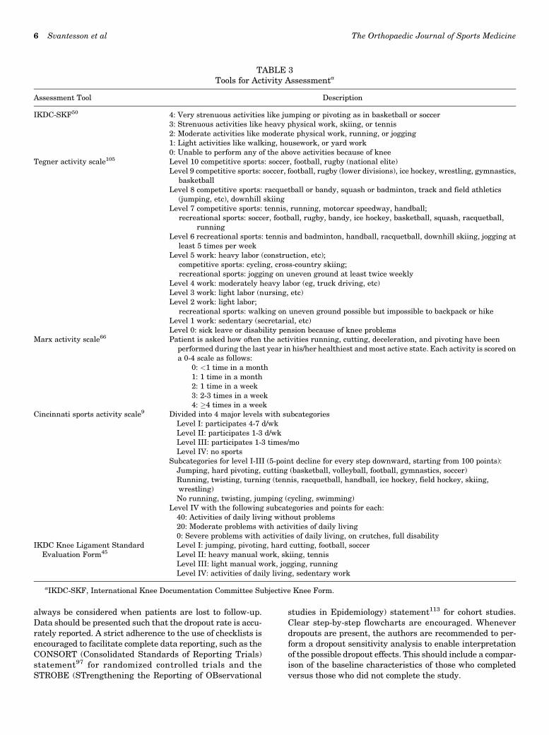
The follow-up time of a study should be defined dependingon what is relevant in relation to the primary investigatedoutcome. In general, outcomes after ACL treatment can bedivided into 4 categories: early adverse events, PROs, ACLfailure/recurrent ligament disruption, and clinical mea-sures of knee function and structure (Table 5), all of whichcould be further stratified in specific outcomes, necessitat-ing different considerations for follow-up time as exempli-fied in Table 6.
Evidence provided by previous research as well as clinicalexperience is the foundation to determine what a relevantfollow-up time is. For example, the rates of ACL reruptureand ACL revision peak at 1 to 2 years after an ACL recon-struction and with return to sport (RTS).32,40,61,82,118 There-fore, a study with a shorter follow-up than this is notrelevant if the primary outcome is rerupture or ACL revi-sion, and a study aiming to make conclusions about ACLtreatment failure should not have a follow-up time of lessthan 2 years and should report RTS as a proxy of risk expo-sure. In contrast, the outcome of septic arthritis or hardwarefailure can manifest soon after an ACL reconstruction,99,114
and a follow-up time of�6 months is sufficient to collect datathat will represent a true estimation of such outcomes. Thus,it is important that the follow-up time is defined and basedupon the study aims and outcomes.
In most studies, especially with increasing length offollow-up time, a certain degree of patients lost to follow-up is inevitable. Even a small proportion of patients lost tofollow-up can lead to considerable study bias,16 although acommon opinion is that a dropout rate of more than 20% isassociated with a serious threat to the internal and externalvalidity and power of the study.93 A study is therefore recom-mended to optimally include at least an 80% follow-up rate.However, the possibility of dropout/retention bias should
The Orthopaedic Journal of Sports Medicine Clinical Outcomes After ACL Injury 5
always be considered when patients are lost to follow-up.Data should be presented such that the dropout rate is accu-rately reported. A strict adherence to the use of checklists isencouraged to facilitate complete data reporting, such as theCONSORT (Consolidated Standards of Reporting Trials)statement97 for randomized controlled trials and theSTROBE (STrengthening the Reporting of OBservational
studies in Epidemiology) statement113 for cohort studies.Clear step-by-step flowcharts are encouraged. Wheneverdropouts are present, the authors are recommended to per-form a dropout sensitivity analysis to enable interpretationof the possible dropout effects. This should include a compar-ison of the baseline characteristics of those who completedversus those who did not complete the study.
TABLE 3Tools for Activity Assessmenta
Assessment Tool Description
IKDC-SKF50 4: Very strenuous activities like jumping or pivoting as in basketball or soccer3: Strenuous activities like heavy physical work, skiing, or tennis2: Moderate activities like moderate physical work, running, or jogging1: Light activities like walking, housework, or yard work0: Unable to perform any of the above activities because of knee
Tegner activity scale105 Level 10 competitive sports: soccer, football, rugby (national elite)Level 9 competitive sports: soccer, football, rugby (lower divisions), ice hockey, wrestling, gymnastics,
basketballLevel 8 competitive sports: racquetball or bandy, squash or badminton, track and field athletics
(jumping, etc), downhill skiingLevel 7 competitive sports: tennis, running, motorcar speedway, handball;
recreational sports: soccer, football, rugby, bandy, ice hockey, basketball, squash, racquetball,running
Level 6 recreational sports: tennis and badminton, handball, racquetball, downhill skiing, jogging atleast 5 times per week
Level 5 work: heavy labor (construction, etc);competitive sports: cycling, cross-country skiing;recreational sports: jogging on uneven ground at least twice weekly
Level 4 work: moderately heavy labor (eg, truck driving, etc)Level 3 work: light labor (nursing, etc)Level 2 work: light labor;
recreational sports: walking on uneven ground possible but impossible to backpack or hikeLevel 1 work: sedentary (secretarial, etc)Level 0: sick leave or disability pension because of knee problems
Marx activity scale66 Patient is asked how often the activities running, cutting, deceleration, and pivoting have beenperformed during the last year in his/her healthiest and most active state. Each activity is scored ona 0-4 scale as follows:
0: <1 time in a month1: 1 time in a month2: 1 time in a week3: 2-3 times in a week4: �4 times in a week
Cincinnati sports activity scale9 Divided into 4 major levels with subcategoriesLevel I: participates 4-7 d/wkLevel II: participates 1-3 d/wkLevel III: participates 1-3 times/moLevel IV: no sports
Subcategories for level I-III (5-point decline for every step downward, starting from 100 points):Jumping, hard pivoting, cutting (basketball, volleyball, football, gymnastics, soccer)Running, twisting, turning (tennis, racquetball, handball, ice hockey, field hockey, skiing,wrestling)No running, twisting, jumping (cycling, swimming)
Level IV with the following subcategories and points for each:40: Activities of daily living without problems20: Moderate problems with activities of daily living0: Severe problems with activities of daily living, on crutches, full disability
IKDC Knee Ligament StandardEvaluation Form45
Level I: jumping, pivoting, hard cutting, football, soccerLevel II: heavy manual work, skiing, tennisLevel III: light manual work, jogging, runningLevel IV: activities of daily living, sedentary work
aIKDC-SKF, International Knee Documentation Committee Subjective Knee Form.
6 Svantesson et al The Orthopaedic Journal of Sports Medicine

It should be emphasized that there can be circumstanceswhere an acceptable follow-up rate for a study is deter-mined by weighing the disadvantages of loss to follow-upagainst certain advantages, for example, a long-termfollow-up or a considerable amount of data in a study. Insuch cases, a lower threshold for follow-up rate is accept-able. Large registry studies can be used to exemplify this,where the patient response rates to PROs are a challenge.43
Registries comprise data on large numbers of patients andinclude multiple follow-up occasions, sometimes over morethan a decade.43,103 Hence, they are important sources fordetermining the effectiveness of ACL treatment and forproviding hypotheses-generating results.104 Nonetheless,a large dropout rate increases the importance of a stringentdata reporting, and a statistical analysis of patients lost tofollow-up also needs to be considered.
CONCLUSION. Follow-up time should be determined bythe purpose of the study and primary outcome and should bestated a priori. The follow-up rate should optimally exceed80%, and data must be reported so that the possible effectsof patients lost to follow-up can be considered.
4. Comprehensive assessment after ACL surgery (minimum2 years) should include adverse events, clinical measuresof knee function and structure, PRO, activity level, andrecurrent ligament disruption (25/25; 100% agreement).
“The comprehensive assessment needs to cover both clin-ical assessment and the patient’s perspective and shouldoptimally also include RTS.”
A comprehensive assessment after ACL reconstructionshould aim to provide a complete picture of outcome relatedto different dimensions of limitations, which involves
numerous aspects of knee-related health and function,objective assessment of hard endpoints (Table 6), and tech-nical aspects of the surgery (graft choice, fixation, tunnelplacement, meniscus/cartilage assessment, and treatment).A minimum follow-up of 2 years is likely necessary toenable a comprehensive assessment. Multiple follow-upsduring the first 2 years could certainly fulfill the purposeof evaluating, for example, the progress such as in theearly, middle, and end state of the rehabilitation. However,the final assessment should be withheld until 2 years post-operatively because a substantial number of outcomesrequire that this time has been given for the ACL recon-struction to completely heal47,83,112,123 and for the patientto complete rehabilitation and progress to testing the kneein more demanding activities including full participation insport or activity. A follow-up of 2 years should allow fordetermining the patient’s capability of a successful RTS,6
and importantly, it will include a period when patients areparticipating at high-risk exposure for ACL failures andreinjuries.32,40,61,82,118 An optimal 2-year outcome assess-ment should therefore include reporting of the rate and timeof RTS. A consensus statement related to assessment andreporting of RTS was similarly reached at the ACL Consen-sus Meeting Panther Symposium 2019 and is provided in aseparate publication.67
A comprehensive assessment also implies that the con-tralateral knee should be examined and assessed for eachoutcome. Outcome tools such as the IKDC Knee LigamentStandard Evaluation Form45 require a comparison with thecontralateral knee for the standardized reporting. The unin-jured contralateral knee serves as a reference for theACL-injured knee in terms of range of motion, laxity, andfunctional performance,119 which helps to account for differ-ences between patients. It should also be noted that the con-tralateral limb/leg/knee might also be affected by an ACLinjury such as altered kinematics53,68 and a decrease in mus-cle strength,119 which underscores the importance to ensurethat the function of the contralateral limb is optimized beforeallowing the patient to return to knee-strenuous activities byassessing it likewise. It is therefore recommended that thestandard practice is to assess the contralateral knee andreport such data, which ultimately will contribute toincreased knowledge of risk factors for a patient sustaininga subsequent contralateral ACL injury.
TABLE 4Health-Related Quality of Life Outcome Measuresa
Instrument Developer No. of Items Response Options
KOOS Roos et al91 42 items, of which 5 are related to quality of life Each item scored 0-4ACL-QoL Mohtadi69 32 items A 100-mm visual analog scale for each itemSF-817 Quality Metric 8 items Each item scored on a 6-point scaleEQ-5D31 EuroQoL 6 items Item specificSF-36 Ware and Sherbourne115 36 items Item specificSIP Bergner et al15 136 items Yes/noQWB Anderson et al3 71 items Via interview
aACL-QoL, Quality of Life Outcome Measure for Chronic Anterior Cruciate Ligament Deficiency; EQ-5D, European Quality of Life–5Dimensions; KOOS, Knee injury and Osteoarthritis Outcome Score; QWB, Quality of Well-Being; SF-8/SF-36, Short Form Health Survey;SIP, Sickness Impact Profile.
TABLE 54 Robust Outcome Categories After ACL Injury Treatmenta
Adverse eventsPRO measurementsACL failure or recurrent ligament disruptionClinical measures of knee function and structure
aACL, anterior cruciate ligament; PRO, patient-reported out-come.
The Orthopaedic Journal of Sports Medicine Clinical Outcomes After ACL Injury 7
Failure of ACL reconstruction is a nonspecific termthat is commonly used without a stringent definition inthe literature. It is therefore recommended that well-defined outcome assessments are used and that theauthors, if choosing to use the term failure, report ana priori definition of what a failure is in detail. Todefine failure as reoperation is verifiable and clear;however, it introduces a risk of underestimating thetrue failure rate. Other examples of definitions for ACLgraft failure include recurrent/persistent instability,pathological anterior or rotatory laxity, or evidence ofgraft failure assessed by magnetic resonance imaging(MRI) or arthroscopic surgery. In overall terms, reasonsfor ACL failure may be classified as traumatic (eg, rein-jury), technical (eg, surgical errors), and patient related(eg, compliance to rehabilitation, recovery of neuromuscu-lar function, or generalized hyperlaxity). Technical errorsaccount for a great amount of all graft failures, with fem-oral tunnel malposition being a common cause.71,106 It hasalso been reported that previous tibial tunnel malpositionis a significant predictor for worse 2-year PROs after ACLrevision.122 It is therefore recommended that reporting ofACL reconstruction failure is complemented by reportingof details with regard to the surgical technique. A usefultool is the Anatomic ACL Reconstruction ScoringChecklist (AARSC),107 which enables grading of surgicalvariables that define ACL tunnel position in an anatomicmanner.
CONCLUSION. A minimum of 2-year follow-up is nec-essary for a comprehensive and reliable determination ofoutcome. The comprehensive assessment should includeoutcomes provided by clinical examination, PROs, activitylevel, and verified reinjuries.
5. Comprehensive assessment after ACL surgery in themedium to long term (�5 years) should also includemeasures of posttraumatic OA (25/25; 100% agreement).
“A common methodology of outcome assessment for OA isneeded and should be included in midterm to long-termfollow-up studies.”
It is well known that sustaining an ACL injury entails ahigh risk of developing posttraumatic OA in the midterm tolong term, especially if concomitant intra-articular injuriesare present.1,21,81,86 Reducing the risk of OA is a clinicalpriority, which means that the midterm to long-termfollow-up assessment should include measures of OA to mon-itor and evaluate the degenerative changes in the knee joint.This is necessary for developing therapeutic interventionsaiming to counter the high rate of OA after an ACL injury.
Measures of OA may include clinical examination,PROs, and imaging modalities. Clinical examination find-ings that may indicate OA are joint-line tenderness orcrepitus, which previously have been found to be strongpredictors for OA.96 Good interobserver reliability for
TABLE 6Examples of Outcome Measurements and Considerations for Follow-up Timea
Outcome Category Example of Specific Outcome Comment
Adverse events Intraoperative complications Usually less than 1-year follow-up required to detect these outcomes.When identifying adverse events, these should be reported as soon aspossible, regardless of the minimum time lapsed from treatmentstart.
Surgery- or device-related complicationsInfectionsVenous thromboembolismReoperation
PRO Validated knee-specific outcome scores Depending on study purpose, population, and the specific outcome toolused. Generally, at least 1-year follow-up is required to obtainmeaningful measures for interpretation of treatment effect,preferably 2 years. However, for the IKDC-SKF and the KOOS, the 1-and 2-year results have been reported equivalent.80,95 Patients couldbe followed over several years to detect changes over time and tocompare short-term, midterm, and long-term results.
Psychological measuresHRQoLActivity levelRTS
ACL failure andrecurrent ligamentdisruption
Graft rupture/failure The follow-up time must allow for sufficient time to detect events suchas rerupture and ACL revision. These events tend to occur after thepatient returns to knee-strenuous activities, which means that a 2-year follow-up should be a minimum.
ACL revisionContralateral ACL injury
Clinical measures ofknee function andstructure
Strength testing Largely depending on the specific outcome and the study purpose.However, care should be taken not to draw conclusion about theshort-term treatment result until a 2-year follow-up is obtained.Functional performance tests, knee joint laxity, and range of motionassessments are preferably performed in multiple follow-ups beforethe 2-year follow-up for changes over time. OA assessment shouldhave at least 5-year follow-up. Concomitant knee joint injuries shouldbe reported whenever identified.
Hop testingPerformance testingKnee joint laxityRange of motionImagingOAConcomitant knee joint injuries
aACL, anterior cruciate ligament; HRQoL, health-related quality of life; IKDC-SKF, International Knee Documentation CommitteeSubjective Knee Form; KOOS, Knee injury and Osteoarthritis Outcome Score; OA, osteoarthritis; PRO, patient-reported outcome; RTS,return to sport.
8 Svantesson et al The Orthopaedic Journal of Sports Medicine

joint-line tenderness and crepitus has been reported whena standardized approach is used.65 The IKDC Knee Liga-ment Standard Evaluation Form includes a grading sys-tem for such an examination and should be used forstandardized reporting.45
The use of PROs is valuable to capture the patients’ per-ception of impairments caused by OA. Questionnaires spe-cifically developed and validated for assessment of OA arethe Western Ontario and McMaster Universities Osteoar-thritis Index (WOMAC)13 and the Knee injury and Osteo-arthritis Outcome Score (KOOS).91 However, the WOMACwas developed for evaluation of established OA; as such,the KOOS may be a more appropriate assessment forpatients after ACL injury. This is because the KOOS ismore likely to detect early development of OA comparedwith WOMAC, as the KOOS was developed to cover abroader spectrum from knee injury to manifest OA.91,92
Imaging modalities still provide the most sensitive assess-ment of OA, although not without limitations. One shouldremember that radiographic findings of OA are not necessar-ily accompanied by symptomatic OA,5,84 and other intra-articular abnormalities may give similar symptoms as OA.It is therefore recommended to combine radiographic imagingassessment with PROs for decision making when it comes tosymptomatic OA. Radiographic findings should be describedin a standardized manner using validated tools, where theKellgren-Lawrence perhaps is the most commonly used tool,taking into account osteophyte formation, sclerosis, jointspace narrowing, and bone deformity.55 Although plain radi-ography has long been the established method for imaging ofOA, it must be acknowledged that the modality has a limitedcapacity to visualize early stages of OA and to grade OAprogression.63
The rapid evolution of MRI techniques enables a muchmore comprehensive assessment of knee joint structure,such as early morphological and biochemical changes ofarticular and periarticular structures. Quantitative mea-surements of cartilage thickness on MRI have a higher sen-sitivity for knee OA compared with traditional radiologicalmeasures.121 In addition, MRI detects characteristic OAsigns earlier and with a greater sensitivity compared withradiography.41 Structural intra-articular changes areindicative for OA and can be seen as early as 2 years afteran ACL reconstruction with MRI, which is earlier thanthese changes can be seen on radiographs.19,108 In addition,MRI can also rule out other intra-articular injuries thatmay explain symptoms perceived by patients. Thus,although plain radiography has an established role inassessment of OA and is favorable from an availability andcost perspective, its main role is to assess the developmentof OA in the long term and for already established OA. Forearly or midterm assessment of OA, attempts should bemade to include MRI to detect early changes with greatervalidity and sensitivity.41
It is not known when clinically relevant posttraumatic OAoccurs or when in this process the structural changes of theknee joint start to appear. With advancement in imagingtechniques, there is a risk of overdiagnosis of OA becausestructural changes without clinical significance might bedetected. Future research will hopefully provide a clearer
picture of this as well as methods todistinguishbetween whatare pathological changes and what changes are related tonormal aging.64 Until then, an assessment of knee OA shouldalways be made in relation to a “control knee” to provide areference for such variables. A synthesis of current literatureshows that the contralateral knee is most commonly used forthis purpose, followed by using an age- and sex-matched con-trol group.86 The latter methodology, using a separate com-parison group, is the preferred method because degenerationcan occur inthe contralateral knee,although itwas not partofthe original injury. Some studies have used baseline imagingof the acute ACL-injured knee as the control,1 which cannotbe recommended because this method does not take intoaccount the impact of natural aging occurring between theinjury and the long-term follow-up.
CONCLUSION. Outcome assessment of OA shouldinclude clinical examination, PROs, and imaging modalities,for which MRI is the preferred modality for increased accu-racy. Imaging findings should always be set in context withthe patient’s perception and the clinical examination fordecision making. Hence, these outcome assessments areequally important for determining the outcome of OA.
6. Clinical assessment of ACL injury treatment should includemeasures of anteroposterior and rotatory knee laxity (25/25;100% agreement).
“Evaluation of knee joint laxity is a cornerstone for eval-uating the outcome of ACL treatment. Quantitative mea-sures of knee joint laxity increase the reliability andvalidity.”
The anatomic properties of the ACL make it a primary pas-sive restraint to both anteroposterior (AP) and rotatoryforces of the knee joint.46 Valid assessment of knee jointlaxity is therefore key in the evaluation of the outcome ofsurgical treatment after ACL injury, preferably at multiplefollow-ups to detect any changes over time. Failure to elim-inate knee joint laxity with ACL reconstruction could indi-cate treatment failure, while patients undergoingnonoperative treatment should be assessed for excessivelaxity or propagation of knee joint laxity. The latter sce-nario might be an indication for subsequent operative treat-ment, although the term laxity should be distinguishedfrom instability or stability. Knee joint laxity is defined asthe passive response of the knee joint when an externalforce or torque is applied, while instability is the patient’sperception of symptoms during functional movement inde-pendent of laxity.78 Hence, knee joint laxity can be reliablymeasured and reported, which makes it the preferred met-ric for clinical outcome assessment. To minimize the risk ofbias, every attempt should be made to blind the assessors,and all participating assessors should be trained in using astandardized execution technique of the laxity test.
Laxity assessment consists of static and dynamic examina-tions, and methods for both grading by the examiner andquantification of laxity have been developed. Laxity assess-ments should always include a side-to-side comparison withthe contralateral knee. Static AP knee laxity tests consider asingle degree of freedom of motion and include application of a
The Orthopaedic Journal of Sports Medicine Clinical Outcomes After ACL Injury 9

unidirectional force in a single plane, such as the Lachmantest and the anterior drawer test. The IKDC Knee LigamentStandard Evaluation Form provides a standardized classifi-cation of the degree of AP translation.45 For instrumentedquantitative assessment of AP laxity, the KT-1000/2000 arth-rometer (MEDmetric Corp)27 and the Rolimeter (Aircast)8
provide among the most accurate measurements, althoughthe intraclass correlation coefficient is variable according tothe literature, and the results are examiner dependent.88
Another instrument is the GNRB (Genourob), which is arobotic arthrometer developed toalleviate the difficulties withexaminer-dependent measurements. The patient’s leg isplaced in the robotic system, and a predefined forceis applied to the proximal calf, while the relative displacementof the anterior tibial tubercle with respect to the patella isrecorded by a displacement sensor. The GNRB also offers theadvantage of using electromyography sensors to record ham-string activity to detect incomplete hamstring relaxation thataffects the result.89 Static AP measurements do not necessar-ily correlate with clinical outcome and function,7,57,58 whichindicates that laxity assessment should not solely rely onstatic AP translation because it fails to capture the more com-plex knee kinematics.
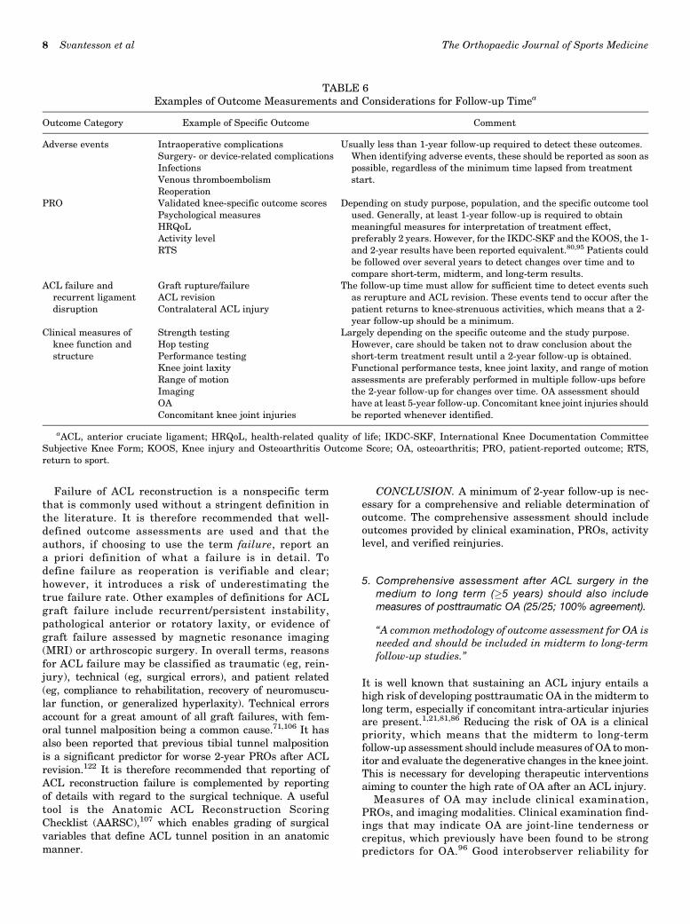
The pivot-shift (PS) test is considered to simulate a morephysiological multiaxial loading of the knee joint because it isa dynamic test of laxity that evaluates both AP and rotatorylaxity.48 It has been reported as the most specific test for ACLdeficiency.14 On the other hand, the PS is characterized by alarge variability in execution techniques,60,77 which may leadto a variation in clinical grading between examiners. To over-come this, a standardized PS test has been described, whichhas led to an improved accuracy of the test.77 Moreover, user-friendly devices for noninvasive quantitative PS have beendeveloped and determined to be valid for objective assess-ment of the PS.76 Such devices may include an inertial sensorsystem (KiRA; Orthokey)124,125 to quantify the tibial acceler-ation during the PS and an image analysis system,74 whichenables a quantification of the lateral tibial translation dur-ing the PS. Both devices have been shown be able to validlydetect differences between clinically high- and low-grade PS
(Figures 2 and 3).76 Examples of devices for quantitative APand rotatory knee laxity that are easily applicable in the clin-ical setting are summarized in Table 7.
CONCLUSION. Knee joint laxity should be assessed afterACL treatment and reported in a standardized manner usingthe IKDC Knee Ligament Standard Evaluation Form whenclinical grading is used. The use of quantitative measures isencouraged to increase the reliability and validity of theassessment.
Section 3: Patient-Reported Outcome
7. Assessment of PRO should optimally include at least 1knee-specific outcome tool, 1 activity rating scale, and1 measure of HRQoL (25/25; 100% agreement).
“There is a fine balance between multiple outcome assess-ments and the responder burden in clinical outcomeassessment.”
The use of PROs has become a cornerstone for researchers tounderstand the patients’ perspective of the impact of ACLinjury and treatment. During recent decades, technicaldevelopment has facilitated the use of PROs, as patients canreport and researchers can collect responses electronically.The time-efficient collection has tempted researchers to bur-den patients with more PROs in studies. Responder burdenis an important term in research and is defined as the time tocomplete items as well as the physical energy and cognitivedemands placed on those responding. In addition, all clinicaltesting of patients is part of the burden placed on ourpatients. Because of the risk of excessive responder burden,which threatens the validity of a patient’s responses andthus their score, researchers are advised to wisely choosePROs specific for the study purpose.
Similar to statement number 2 of this consensus paper onbaseline information to collect, it is recommended to use atleast 1 knee-specific tool, 1 HRQoL tool, and 1 activity ratingscale. This provides the researcher with a comprehensive pic-ture of the patients’ perception of outcome after treatment.
CONCLUSION. To give a comprehensive assessment ofthe patients’ perception of the impact of ACL injury andoutcome of treatment, validated knee-specific PRO assess-ment, HRQoL, and measure of type and level of preinjury
Figure 2. The KiRA inertial sensor system for quantifying lat-eral tibial acceleration during the pivot-shift test.
Figure 3. Image analysis system on iPad for quantifying lat-eral tibial translation during the pivot-shift test.
10 Svantesson et al The Orthopaedic Journal of Sports Medicine
sport/activity should be collected before and aftertreatment.
8. The IKDC-SKF is the recommended knee-related outcomemeasure for ACL injury and treatment (24/25; 96%agreement).
“It is important to find a universal metric. The IKDC-SKF is currently the optimal scale, but we should becareful not to neglect the other scores.”
The evaluation of treatment outcome started historicallywith use of objective measurements as proxies for whatclinicians and patients really cared about. For instance,both rating scales and measures of range of motion,strength, and laxity were frequently used; however, thesemeasures are limited by interrater and intrarater variabil-ity and alone failed to determine symptoms and limitationsperceived important by the patient. Failure to report andquantify the patients’ perspective of treatment outcomeafter ACL injury led to the development of knee-relatedPROs during the late 1990s and early 2000s. The 2 mostcommonly used PROs after ACL injury are the KOOS andthe IKDC-SKF, which were both developed during this time
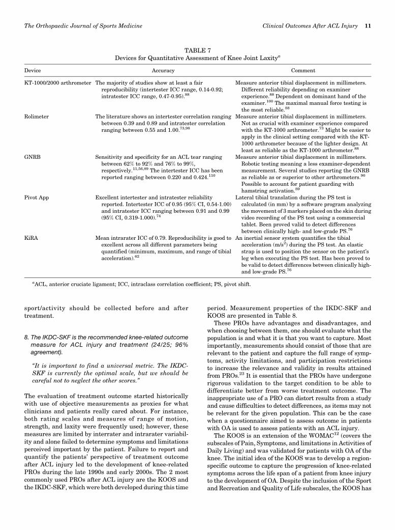
period. Measurement properties of the IKDC-SKF andKOOS are presented in Table 8.
These PROs have advantages and disadvantages, andwhen choosing between them, one should evaluate what thepopulation is and what it is that you want to capture. Mostimportantly, measurements should consist of those that arerelevant to the patient and capture the full range of symp-toms, activity limitations, and participation restrictionsto increase the relevance and validity in results attainedfrom PROs.23 It is essential that the PROs have undergonerigorous validation to the target condition to be able todifferentiate better from worse treatment outcome. Theinappropriate use of a PRO can distort results from a studyand cause difficulties to detect differences, as items may notbe relevant for the given population. This can be the casewhen a questionnaire aimed to assess outcome in patientswith OA is used to assess patients with an ACL injury.
The KOOS is an extension of the WOMAC12 (covers thesubscales of Pain, Symptoms, and limitations in Activities ofDaily Living) and was validated for patients with OA of theknee. The initial idea of the KOOS was to develop a region-specific outcome to capture the progression of knee-relatedsymptoms across the life span of a patient from knee injuryto the development of OA. Despite the inclusion of the Sportand Recreation and Quality of Life subscales, the KOOS has
TABLE 7Devices for Quantitative Assessment of Knee Joint Laxitya
Device Accuracy Comment
KT-1000/2000 arthrometer The majority of studies show at least a fairreproducibility (intertester ICC range, 0.14-0.92;intratester ICC range, 0.47-0.95).88
Measure anterior tibial displacement in millimeters.Different reliability depending on examinerexperience.88 Dependent on dominant hand of theexaminer.100 The maximal manual force testing isthe most reliable.88
Rolimeter The literature shows an intertester correlation rangingbetween 0.39 and 0.89 and intratester correlationranging between 0.55 and 1.00.73,98
Measure anterior tibial displacement in millimeters.Not as crucial with examiner experience comparedwith the KT-1000 arthrometer.73 Might be easier toapply in the clinical setting compared with the KT-1000 arthrometer because of the lighter design. Atleast as reliable as the KT-1000 arthrometer.88
GNRB Sensitivity and specificity for an ACL tear rangingbetween 62% to 92% and 76% to 99%,respectively.11,56,89 The intertester ICC has beenreported ranging between 0.220 and 0.424.110
Measure anterior tibial displacement in millimeters.Robotic testing meaning a less examiner-dependentmeasurement. Several studies reporting the GNRBas reliable as or superior to other arthrometers.90
Possible to account for patient guarding withhamstring activation.89
Pivot App Excellent intertester and intratester reliabilityreported. Intertester ICC of 0.95 (95% CI, 0.54-1.00)and intratester ICC ranging between 0.91 and 0.99(95% CI, 0.319-1.000).74
Lateral tibial translation during the PS test iscalculated (in mm) by a software program analyzingthe movement of 3 markers placed on the skin duringvideo recording of the PS test using a commercialtablet. Been proved valid to detect differencesbetween clinically high- and low-grade PS.76
KiRA Mean intrarater ICC of 0.79. Reproducibility is good toexcellent across all different parameters beingquantified (minimum, maximum, and range of tibialacceleration).62
An inertial sensor system quantifies the tibialacceleration (m/s2) during the PS test. An elasticstrap is used to position the sensor on the patient’sleg when executing the PS test. Has been proved tobe valid to detect differences between clinically high-and low-grade PS.76
aACL, anterior cruciate ligament; ICC, intraclass correlation coefficient; PS, pivot shift.
The Orthopaedic Journal of Sports Medicine Clinical Outcomes After ACL Injury 11

limited measurement properties in the 3 original WOMACsubscales when used for patients after ACL reconstruc-tion.23,59 It is also worth mentioning that the hybrid versionof the KOOS, the KOOS4 (a modified version in which theitems related to activities of daily living have been excludedto avoid ceiling effects)36 has not undergone a valida-tion.22,23 This is problematic, as the ability to detect differ-ences between treatments will be limited with the KOOSused in patients with an ACL injury.59 Using PRO measure-ments that include items that are not relevant or do notcover important limitations of the target condition is notoptimal. Using such PROs entails a potential washout oftreatment effects, inadequate measurement properties, andrisk of false negative findings.23,95,102 In terms of the KOOS,
several questions are at risk for a ceiling effect when used inpatients after ACL reconstruction; that is, the item is too“easy” for the patient. In addition, the KOOS does notinclude specific items relating to instability, which is oneof the most common symptoms and one of the strongestindications for an ACL reconstruction. The KOOS consistsof 42 items entailing higher responder burden comparedwith other outcomes such as the IKDC-SKF. Awareness ofthe limitations of the KOOS for the patients after an ACLinjury or reconstruction is important to avoid missing theeffects of treatment results.
The IKDC-SKF was developed as a region-specific out-come relevant for a variety of conditions including ligamentand intra-articular abnormalities.50 This PRO underwentrigorous testing during its development including a reduc-tion from 42 to 18 items and an exploratory factor analysis,suggesting that it was reasonable to combine the items intoa single overall score. To test the relevance of the IKDC-SKF for patients with an ACL injury, Rasch analysis wasperformed separately for patients with and without kneeligament injury.50,109 The analysis supported the premisethat the items of the IKDC performed similarly in terms ofdifficulty for individuals with or without a ligament injury.The results from the primary testing of the IKDC-SKF alsoindicated that the IKDC-SKF items performed the same,regardless of age, sex, and a variety of diagnoses includingligament, meniscal, and articular cartilage injury andpatellofemoral pain.29,50
The IKDC-SKF is recommended as the knee-relatedPRO to use for patients after ACL reconstruction becauseof its quick-to-use 18 items.50 The IKDC-SKF shows ade-quate internal consistency and has no floor or ceiling effectsacross mixed groups of patients with knee conditions.29 Italso has high levels of test-retest reliability, constructvalidity, and responsiveness. Moreover, normative datahave been determined, which is valuable for comparisonsas well as cutoffs for what the patients consider an accept-able symptom state.52
There are also other promising PROs used to cover dif-ferent aspects of recovery after ACL reconstruction, includ-ing the Quality of Life Outcome Measure for ChronicAnterior Cruciate Ligament Deficiency (ACL-QoL)69 andthe Knee Numeric-Entity Evaluation Score (KNEES-ACL).24 The ACL-QoL is used to determine the effective-ness of ACL reconstruction, or any other treatment, and is a32-item condition-specific quality of life scale for patientswith ACL deficiency.69 The KNEES-ACL was developed in2013,24 and the thorough development process and dimen-sionality assessment resulted in 42 items across 7 latentconstructs. There is strong positive evidence given to con-tent validity.24,25
The ACL-QoL and the KNEES-ACL are promising out-come measurements and likely will help us to better under-stand patients who have sustained an ACL injury. However,these PROs have mainly been used in comparative studiesand are yet to be compared with the established IKDC-SKFand KOOS to prove their respective strengths of constructs.
CONCLUSION. The IKDC-SKF is the recommendedknee-related outcome measure for ACL injury andtreatment.
TABLE 8Psychometric Properties of the IKDC-SKF and the
KOOS37a
IKDC-SKF KOOS
PASS 75.9 Pain ¼ 88.9Symptoms ¼ 57.1ADL ¼ 100.0Sport ¼ 75.0QoL ¼ 62.5
MCID 11.5 N/AMIC 10.9 Pain ¼ 2.5
Symptoms ¼ –1.2ADL ¼ 2.4Sport ¼ 12.1QoL ¼ 18.3
MDC 11.5 Pain ¼ 6.0 to 6.1Symptoms ¼ 5.0 to 8.5ADL ¼ 7.0 to 8.0Sport ¼ 5.8 to 12.0QoL ¼ 7.0 to 7.2
Content validity Poor No evidenceStructural validity No evidence No evidenceInternal consistency 0.77 to 0.97 Pain ¼ 0.84 to 0.91
Symptoms ¼ 0.25 to 0.75ADL ¼ 0.94 to 0.96Sport ¼ 0.85 to 0.89QoL ¼ 0.64 to 0.90
Measurement error 3.2 to 5.6 Pain ¼ 2.2 to 10.1Symptoms ¼ 3.1 to 9.0ADL ¼ 2.9 to 11.7Sport ¼ 2.1 to 24.6QoL ¼ 2.6 to 10.8
Test-retest reliability 0.85 to 0.99 Pain ¼ 0.85 to 0.93Symptoms ¼ 0.83 to 0.95ADL ¼ 0.75 to 0.91Sport ¼ 0.61 to 0.89QoL ¼ 0.83 to 0.95
Responsiveness Good PoorCross-cultural validity Fair No evidence
aADL, Activities of Daily Living; IKDC-SKF, InternationalKnee Documentation Committee Subjective Knee Form; KOOS,Knee injury and Osteoarthritis Outcome Score; MCID, minimalclinically important difference; MDC, minimum detectable change;MIC, minimally important change; N/A, not available; PASS,Patient Acceptable Symptom State; QoL, Quality of Life.
12 Svantesson et al The Orthopaedic Journal of Sports Medicine

9. Measurement of the patient acceptable symptom state(PASS) is valuable in the assessment of outcome of ACLinjury and treatment (25/25; 100% agreement).
“One question can carry the advantage of giving thepatient the opportunity to tell the story.”
As researchers and clinicians of today, we are equippedwith a great variety of PROs. However, the developmentand use of these PROs means little if the results are notinterpreted in a clinically meaningful manner. The use ofnumeric scores poses a risk that researchers focus myopi-cally at numbers and statistically significant findings with-out reflecting over whether such findings really areimpactful from the patient’s perspective. For many suchPROs, the same score can be achieved, despite that patientsrespond differently to the items that comprise the PROmeasure. The question of whether the patient perceivesan acceptable symptom state is a priority for all clinicians,and the use of the PASS in PRO assessment is important.The PASS considers a single-item question and aims todetermine a threshold beyond which the patients considerthemselves “well.”75 Thresholds for the PASS have beenestablished for the KOOS and the IKDC-SKF by asking thequestion “Taking account of all the activity you have duringyour daily life, your level of pain, and also your activitylimitations and participation restrictions, do you considerthe current state of your knee satisfactory?” alongside theadministered PRO.75 Several studies have since thenapplied the PASS values for the KOOS and IKDC-SKFwhen reporting on outcome after ACL treatment.26,42,44,111
A single-item outcome like the PASS summarizes thepatient’s perception and allows the patient to make an over-all statement through a binary answer: “yes” or “no.” Anumeric scale might have its advantages; however, it isassociated with difficulties of interpretation for bothpatients and researchers. That is, what is considered as agood and poor outcome, respectively? The PASS referencevalue at which a majority of the patients feel well is valu-able for determining this important question, and its use iswarranted to overcome limitations with numeric PROssuch as ceiling effects and poor responsiveness.49,72
In addition, the evidence to support the interpretationand use of a PRO should include the minimum detectablechange (MDC) score and the minimal clinically importantdifference (MCID) score. These scores collectively describethe responsiveness of the PRO, which is the ability to detecta clinically important change in outcome for the metric. TheMDC is the amount of change that is needed to confidentlystate that the change is beyond measurement error.10
Thus, if a study finds a difference that is smaller than theMDC for the chosen PRO, one should be careful to draw anyconclusions because the observed difference is within therange of measurement error for the PRO. On the otherhand, if the change in outcome is larger than the MDC, itstill remains unknown whether this change is clinicallyrelevant. This is where the MCID becomes valuable. If achange in outcome exceeds the value of the MCID for thePRO, the difference is likely to be perceived as important bymost patients.51
CONCLUSION. The PASS is a valuable complement tonumeric PROs and should be used to facilitate interpreta-tion of PROs. Researchers should also consider the MDCand MCID for the PRO when reporting and discussing theirstudy findings.
FUTURE DIRECTIONS
Reaching consensus for clinical outcome assessment afterACL treatment is an important step toward refining andimproving the quality of ACL research. Further effortsshould be made to develop methods for outcome assessmentthat provide the most relevant and valid data for patientsreceiving ACL treatment. A focus is to improve the PROassessment. The collection of PROs has become increas-ingly important among health care professions. Not onlyis it a valuable asset for a clinician to understand a patient’sperception of health and results of treatment, it has alsogained importance for policy makers in determining healthcare quality and developing a value-based health care.72
Commonly used PROs in ACL research are limited by aformat of fixed-length surveys that many times includeitems of questionable relevance for the young and activepopulation sustaining ACL injuries, leading to ceilingeffects and potentially survey fatigue. Therefore, a currentpriority is to decrease the responder burden for patients inPRO assessment.
Improved PRO data collection may be achieved throughthe use of the item response theory (IRT),20,35 which hasenabled the introduction of computer adaptive testing(CAT). The underlying premise of IRT is that the way apatient responds to an item (question) is based on the dif-ficulty of the question and the ability of the patient. Whenadministered as a CAT, a mathematical algorithm is uti-lized to select items that are matched to the ability of thepatient. For example, if a patient responds to an item thathe/she is unable to walk a mile, the computer algorithm willbypass “harder” items such as running a mile and select aneasier item such as ability to walk a block. This means thatonly items that are relevant about the patient’s ability levelare administered, which substantially reduces the time andburden associated with administration of PROs. Efforts areunder way to convert the IKDC-SKF to a CAT format thatis based on IRT.
Although computer-aided PRO assessment likely is thefuture, further research for optimization of currently usedPROs is needed. Research should focus on determining themost responsive items of current PROs to condense thesurveys to include only the most responsive questions. Thisis important when considering the already collected PROdata for tens of thousands of patients in large registries andnational databases. Such data might need to be reanalyzedusing the condensed PROs and thereby provide results witha greater precision on clinically relevant outcomes.
Other important aspects for further research are out-come measures on activity and RTS after ACL treatment.Optimally, a tool that is able to quantify sports participa-tion in terms of level, volume, and intensity should bedeveloped and implemented as a standardized tool used
The Orthopaedic Journal of Sports Medicine Clinical Outcomes After ACL Injury 13

across studies. With the rapid evolution of technology, thefuture will likely also hold easily accessible use of quanti-tative instruments for quantitatively measuring patientactivity, for example, the use of Global Positioning Systemand motion detectors during sports participation, measure-ments of joint function, and measurements of heart rateand speed to estimate intensity.
CONCLUSION
Clinical outcome assessment after ACL injury can bedivided in 4 robust categories: early adverse events, PROs,ACL failure/recurrent ligament disruption, and clinicalmeasures of knee function and structure. A minimum of2-year follow-up is necessary for a comprehensive and reli-able determination of outcome, which should include out-comes provided by clinical examination, PROs, and verifiedreinjuries. The PRO assessment is a cornerstone in evalu-ating outcome after ACL injury, where validated knee-specific PRO assessment, HRQoL, and measure of type andlevel of sport/activity should be collected. The IKDC-SKF isthe recommended knee-related PRO measure for ACLtreatment, and the use of PASS is encouraged to facilitateinterpretation of PROs.
AUTHORS
Eleonor Svantesson, MD (Department of Orthopaedics,Institute of Clinical Sciences, Sahlgrenska Academy, Uni-versity of Gothenburg, Gothenburg, Sweden; GothenburgSports and Trauma Research Center, Gothenburg, Swe-den); Eric Hamrin Senorski, PT, PhD (Gothenburg Sportsand Trauma Research Center, Gothenburg, Sweden;Department of Rehabilitation and Health, Institute of Neu-roscience and Physiology, Sahlgrenska Academy, Univer-sity of Gothenburg, Gothenburg, Sweden); Kate E.Webster, PhD (School of Allied Health, Human Servicesand Sport, La Trobe University, Melbourne, Victoria, Aus-tralia); Jon Karlsson, MD, PhD (Department of Orthopae-dics, Institute of Clinical Sciences, Sahlgrenska Academy,University of Gothenburg, Gothenburg, Sweden; Gothen-burg Sports and Trauma Research Center, Gothenburg,Sweden; Department of Orthopaedics, Sahlgrenska Univer-sity Hospital, Gothenburg, Sweden); Theresa Diermeier,MD (Department of Sports Orthopedics, Klinikum Rechtsder Isar, Technical University of Munich, Munich, Ger-many); Benjamin B. Rothrauff, MD (UPMC Freddie FuSports Medicine Center, Department of Orthopaedic Sur-gery, University of Pittsburgh, Pittsburgh, Pennsylvania,USA); Sean J. Meredith, MD (UPMC Freddie Fu SportsMedicine Center, Department of Orthopaedic Surgery, Uni-versity of Pittsburgh, Pittsburgh, Pennsylvania, USA;Department of Orthopaedics, University of Maryland Schoolof Medicine, Baltimore, Maryland, USA); Thomas Rauer,MD (UPMC Freddie Fu Sports Medicine Center, Depart-ment of Orthopaedic Surgery, University of Pittsburgh,Pittsburgh, Pennsylvania, USA; Department of TraumaSurgery, University Hospital Zurich, Zurich, Switzerland);
James J. Irrgang, PT, PhD, FAPTA (Department of Ortho-paedic Surgery, University of Pittsburgh Medical Center,Pittsburgh, Pennsylvania, USA; Department of PhysicalTherapy, University of Pittsburgh, Pittsburgh, Pennsylva-nia, USA); Kurt P. Spindler, MD (Sports Health Center,Cleveland Clinic, Garfield Heights, Ohio, USA); C. BenjaminMa, MD (Department of Orthopaedic Surgery, University ofCalifornia, San Francisco, San Francisco, California, USA);Volker Musahl, MD (Department of Orthopaedic Surgery,University of Pittsburgh Medical Center, Pittsburgh, Penn-sylvania, USA); and the Panther Symposium ACL InjuryClinical Outcomes Consensus Group: Freddie H. Fu, MD(Department of Orthopaedic Surgery, University of Pitts-burgh Medical Center, Pittsburgh, Pennsylvania, USA);Olufemi R. Ayeni, MD, PhD, FRCSC (Division of Orthopae-dic Surgery, McMaster University, Hamilton, Ontario,Canada); Francesco Della Villa, MD (Education andResearch Department, Isokinetic Medical Group, Bologna,Italy); Stefano Della Villa, MD (FIFA Medical Centre ofExcellence, Bologna, Italy); Scott Dye, MD (Department ofOrthopaedic Surgery, University of California, San Fran-cisco); Mario Ferretti, MD, PhD (Federal University of SaoPaulo, Sao Paulo, Brazil); Alan Getgood, MD, FRCS(Tr&Orth), DipSEM (Fowler Kennedy Sport Medicine ClinicLondon, London, Ontario, Canada); Timo Jarvela, MD, PhD(Arthroscopic and Sports Medicine Center, Pohjola Hospital,Helsinki, Finland); Christopher C. Kaeding, MD (The OhioState University, Columbus, Ohio, USA); Ryosuke Kuroda,MD, PhD (Kobe University Graduate School of Medicine,Kobe, Hyogo, Japan); Bryson Lesniak, MD (Department ofOrthopaedic Surgery, University of Pittsburgh Medical Cen-ter, Pittsburgh, Pennsylvania, USA); Robert G. Marx, MD,MSc, FRCSC (Medicine Institute, Hospital for Special Sur-gery, New York, USA); Gregory B. Maletis, MD (SouthernCalifornia Permanente Medical Group, Baldwin Park, Cali-fornia, USA); Leo Pinczewski, MD (North Sydney Orthopae-dic & Sports Medicine Centre, Sydney, Australia); AnilRanawat, MD (Center for Hip Pain and Preservation, Hos-pital for Special Surgery, New York, New York, USA); BruceReider, MD (University of Chicago, Section of Orthopaedicsand Rehabilitation Medicine, Department of Surgery, Chi-cago, Illinois, USA); Romain Seil, MD, PhD (Department ofOrthopaedic Surgery, Clinique d’Eich-Centre Hospitalier deLuxembourg, Luxembourg); Carola van Eck, MD, PhD(Department of Orthopaedic Surgery, University of Pitts-burgh Medical Center, Pittsburgh, Pennsylvania, USA);Brian R. Wolf, MD, MS (UI Sports Medicine, Iowa City,IA, USA); Patrick Yung, MD (Department of Orthopaedicsand Traumatology, Faculty of Medicine, The Chinese Uni-versity of Hong Kong, Hong Kong, China); Stefano Zaffag-nini, MD (Istituto Ortopedico Rizzoli, Bologna, Italy); andMing Hao Zheng, MD, PhD, FRCPath (Centre for Orthopae-dic Translational Research, School of Surgery, University ofWestern Australia, Perth, Western Australia, Australia).
REFERENCES
1. Ajuied A, Wong F, Smith C, et al. Anterior cruciate ligament injury and
radiologic progression of knee osteoarthritis: a systematic review and
meta-analysis. Am J Sports Med. 2014;42(9):2242-2252.
14 Svantesson et al The Orthopaedic Journal of Sports Medicine

2. Andernord D, Karlsson J, Musahl V, et al. Timing of surgery of the
anterior cruciate ligament. Arthroscopy. 2013;29(11):1863-1871.
3. Anderson JP, Kaplan RM, Berry CC, Bush JW, Rumbaut RG. Interday
reliability of function assessment for a health status measure: the
Quality of Well-Being scale. Med Care. 1989;27(11):1076-1083.
4. Andersson D, Samuelsson K, Karlsson J. Treatment of anterior cruci-
ate ligament injuries with special reference to surgical technique and
rehabilitation: an assessment of randomized controlled trials. Arthros-
copy. 2009;25(6):653-685.
5. Andersson Molina H, Karlsson H, Rockborn P. Arthroscopic partial
and total meniscectomy: a long-term follow-up study with matched
controls. Arthroscopy. 2002;18(2):183-189.
6. Ardern CL, Taylor NF, Feller JA, Whitehead TS, Webster KE. Sports
participation 2 years after anterior cruciate ligament reconstruction
in athletes who had not returned to sport at 1 year: a prospective
follow-up of physical function and psychological factors in 122 ath-
letes. Am J Sports Med. 2015;43(4):848-856.
7. Ayeni OR, Chahal M, Tran MN, Sprague S. Pivot shift as an outcome
measure for ACL reconstruction: a systematic review. Knee Surg
Sports Traumatol Arthrosc. 2012;20(4):767-777.
8. Balasch H, Schiller M, Friebel H, Hoffmann F. Evaluation of anterior
knee joint instability with the Rolimeter: a test in comparison with
manual assessment and measuring with the KT-1000 arthrometer.
Knee Surg Sports Traumatol Arthrosc. 1999;7(4):204-208.
9. Barber-Westin SD, Noyes FR, McCloskey JW. Rigorous statistical
reliability, validity, and responsiveness testing of the Cincinnati Knee
Rating System in 350 subjects with uninjured, injured, or anterior cru-
ciate ligament-reconstructed knees. Am J Sports Med. 1999;27(4):
402-416.
10. Beaton DE, Bombardier C, Katz JN, Wright JG. A taxonomy for
responsiveness. J Clin Epidemiol. 2001;54(12):1204-1217.
11. Beldame J, Mouchel S, Bertiaux S, et al. Anterior knee laxity mea-
surement: comparison of passive stress radiographs Telos® and
“Lerat”, and GNRB® arthrometer. Orthop Traumatol Surg Res.
2012;98(7):744-750.
12. Bellamy N, Buchanan WW. A preliminary evaluation of the dimension-
ality and clinical importance of pain and disability in osteoarthritis of
the hip and knee. Clin Rheumatol. 1986;5(2):231-241.
13. Bellamy N, Buchanan WW, Goldsmith CH, Campbell J, Stitt LW. Val-
idation study of WOMAC: a health status instrument for measuring
clinically important patient relevant outcomes to antirheumatic drug
therapy in patients with osteoarthritis of the hip or knee. J Rheumatol.
1988;15(12):1833-1840.
14. Benjaminse A, Gokeler A, van der Schans CP. Clinical diagnosis of an
anterior cruciate ligament rupture: a meta-analysis. J Orthop Sports
Phys Ther. 2006;36(5):267-288.
15. Bergner M, Bobbitt RA, Carter WB, Gilson BS. The Sickness Impact
Profile: development and final revision of a health status measure.
Med Care. 1981;19(8):787-805.
16. Bhandari M, Guyatt GH, Swiontkowski MF. User’s guide to the ortho-
paedic literature: how to use an article about a surgical therapy.
J Bone Joint Surg Am. 2001;83(6):916-926.
17. Bost JE, Williams BA, Bottegal MT, Dang Q, Rubio DM. The 8-item
Short-Form Health Survey and the physical comfort composite score
of the quality of recovery 40-item scale provide the most responsive
assessments of pain, physical function, and mental function during
the first 4 days after ambulatory knee surgery with regional anesthe-
sia. Anesth Analg. 2007;105(6):1693-1700, table of contents.
18. Bourke HE, Salmon LJ, Waller A, Patterson V, Pinczewski LA. Survival
of the anterior cruciate ligament graft and the contralateral ACL at a
minimum of 15 years. Am J Sports Med. 2012;40(9):1985-1992.
19. Bowes MA, Lohmander LS, Wolstenholme CBH, et al. Marked and
rapid change of bone shape in acutely ACL injured knees: an explor-
atory analysis of the Kanon trial. Osteoarthritis Cartilage. 2019;27(4):
638-645.
20. Brodke DJ, Hung M, Bozic KJ. Item response theory and computer-
ized adaptive testing for orthopaedic outcomes measures. J Am Acad
Orthop Surg. 2016;24(11):750-754.
21. Claes S, Hermie L, Verdonk R, Bellemans J, Verdonk P. Is osteoar-
thritis an inevitable consequence of anterior cruciate ligament recon-
struction? A meta-analysis. Knee Surg Sports Traumatol Arthrosc.
2013;21(9):1967-1976.
22. Comins J, Brodersen J, Krogsgaard M. Treatment for acute anterior
cruciate ligament tear. N Engl J Med. 2010;363(19):1871, author reply
1872-1873.
23. Comins J, Brodersen J, Krogsgaard M, Beyer N. Rasch analysis of the
Knee injury and Osteoarthritis Outcome Score (KOOS): a statistical
re-evaluation. Scand J Med Sci Sports. 2008;18(3):336-345.
24. Comins JD, Krogsgaard MR, Brodersen J. Development of the Knee
Numeric-Entity Evaluation Score (KNEES-ACL): a condition-specific
questionnaire. Scand J Med Sci Sports. 2013;23(5):e293-e301.
25. Comins JD, Krogsgaard MR, Kreiner S, Brodersen J. Dimensionality
of the Knee Numeric-Entity Evaluation Score (KNEES-ACL): a
condition-specific questionnaire. Scand J Med Sci Sports. 2013;
23(5):e302-e312.
26. Cristiani R, Mikkelsen C, Edman G, et al. Age, gender, quadriceps
strength and hop test performance are the most important factors
affecting the achievement of a patient-acceptable symptom state
after ACL reconstruction. Knee Surg Sports Traumatol Arthrosc.
2020;28(2):369-380.
27. Daniel DM, Malcom LL, Losse G, et al. Instrumented measurement of
anterior laxity of the knee. J Bone Joint Surg Am. 1985;67(5):720-726.
28. Djulbegovic B, Guyatt GH. Progress in evidence-based medicine: a
quarter century on. Lancet. 2017;390(10092):415-423.
29. Ebrahimzadeh MH, Makhmalbaf H, Golhasani-Keshtan F, Rabani S,
Birjandinejad A. The International Knee Documentation Committee
(IKDC) Subjective Short Form: a validity and reliability study. Knee
Surg Sports Traumatol Arthrosc. 2015;23(11):3163-3167.
30. Eitzen I, Moksnes H, Snyder-Mackler L, Risberg MA. A progressive
5-week exercise therapy program leads to significant improvement in
knee function early after anterior cruciate ligament injury. J Orthop
Sports Phys Ther. 2010;40(11):705-721.
31. EuroQol Group. EuroQol: a new facility for the measurement of health-
related quality of life. Health Policy. 1990;16(3):199-208.
32. Faltstrom A, Hagglund M, Magnusson H, Forssblad M, Kvist J. Pre-
dictors for additional anterior cruciate ligament reconstruction: data
from the Swedish national ACL register. Knee Surg Sports Traumatol
Arthrosc. 2016;24(3):885-894.
33. Ferguson D, Palmer A, Khan S, Oduoza U, Atkinson H. Early or
delayed anterior cruciate ligament reconstruction: is one superior?
A systematic review and meta-analysis. Eur J Orthop Surg Traumatol.
2019;29(6):1277-1289.
34. Filbay SR, Culvenor AG, Ackerman IN, Russell TG, Crossley KM.
Quality of life in anterior cruciate ligament-deficient individuals: a sys-
tematic review and meta-analysis. Br J Sports Med. 2015;49(16):
1033-1041.
35. Fries JF, Bruce B, Cella D. The promise of PROMIS: using item
response theory to improve assessment of patient-reported out-
comes. Clin Exp Rheumatol. 2005;23(5)(suppl 39):S53-S57.
36. Frobell RB, Roos EM, Roos HP, Ranstam J, Lohmander LS. A ran-
domized trial of treatment for acute anterior cruciate ligament tears.
N Engl J Med. 2010;363(4):331-342.
37. Gagnier JJ, Shen Y, Huang H. Psychometric properties of patient-
reported outcome measures for use in patients with anterior cruciate
ligament injuries: a systematic review. JBJS Rev. 2018;6(4):e5.
38. Gelman A. Don’t calculate post-hoc power using observed estimate
of effect size. Ann Surg. 2019;269(1):e9-e10.
39. Grindem H, Eitzen I, Moksnes H, Snyder-Mackler L, Risberg MA. A
pair-matched comparison of return to pivoting sports at 1 year in
anterior cruciate ligament-injured patients after a nonoperative versus
an operative treatment course. Am J Sports Med. 2012;40(11):
2509-2516.
40. Grindem H, Snyder-Mackler L, Moksnes H, Engebretsen L, Risberg
MA. Simple decision rules can reduce reinjury risk by 84% after ACL
reconstruction: the Delaware-Oslo ACL Cohort Study. Br J Sports
Med. 2016;50(13):804-808.
The Orthopaedic Journal of Sports Medicine Clinical Outcomes After ACL Injury 15

41. Guermazi A, Niu J, Hayashi D, et al. Prevalence of abnormalities in
knees detected by MRI in adults without knee osteoarthritis: popula-
tion based observational study (Framingham Osteoarthritis Study).
BMJ. 2012;345:e5339.
42. Hamrin Senorski E, Sundemo D, Svantesson E, et al. Preoperative
and intraoperative predictors of long-term acceptable knee function
and osteoarthritis after anterior cruciate ligament reconstruction: an
analysis based on 2 randomized controlled trials. Arthroscopy. 2019;
35(2):489-499.
43. Hamrin Senorski E, Svantesson E, Baldari A, et al. Factors that affect
patient reported outcome after anterior cruciate ligament reconstruc-
tion: a systematic review of the Scandinavian Knee Ligament Regis-
ters. Br J Sports Med. 2019;53(7):410-417.
44. Hamrin Senorski E, Svantesson E, Beischer S, et al. Factors affecting
the achievement of a patient-acceptable symptom state 1 year after
anterior cruciate ligament reconstruction: a cohort study of 343
patients from 2 registries. Orthop J Sports Med. 2018;6(4):
2325967118764317.
45. Hefti F, Muller W, Jakob RP, Staubli HU. Evaluation of knee ligament
injuries with the IKDC form. Knee Surg Sports Traumatol Arthrosc.
1993;1(3-4):226-234.
46. Hirschmann MT, Muller W. Complex function of the knee joint: the
current understanding of the knee. Knee Surg Sports Traumatol
Arthrosc. 2015;23(10):2780-2788.
47. Hofbauer M, Soldati F, Szomolanyi P, et al. Hamstring tendon auto-
grafts do not show complete graft maturity 6 months postoperatively
after anterior cruciate ligament reconstruction. Knee Surg Sports
Traumatol Arthrosc. 2019;27(1):130-136.
48. Hoshino Y, Kuroda R, Nagamune K, et al. In vivo measurement of the
pivot-shift test in the anterior cruciate ligament-deficient knee using
an electromagnetic device. Am J Sports Med. 2007;35(7):
1098-1104.
49. Ingelsrud LH, Terwee CB, Terluin B, et al. Meaningful change scores
in the Knee injury and Osteoarthritis Outcome Score in patients
undergoing anterior cruciate ligament reconstruction. Am J Sports
Med. 2018;46(5):1120-1128.
50. Irrgang JJ, Anderson AF, Boland AL, et al. Development and valida-
tion of the International Knee Documentation Committee Subjective
Knee Form. Am J Sports Med. 2001;29(5):600-613.
51. Jaeschke R, Singer J, Guyatt GH. Measurement of health status:
ascertaining the minimal clinically important difference. Control Clin
Trials. 1989;10(4):407-415.
52. Kanakamedala AC, Anderson AF, Irrgang JJ. IKDC Subjective Knee
Form and Marx Activity Rating Scale are suitable to evaluate all ortho-
paedic sports medicine knee conditions: a systematic review. J ISA-
KOS. 2016;1(1):25.
53. Kaur M, Ribeiro DC, Theis JC, Webster KE, Sole G. Movement pat-
terns of the knee during gait following ACL reconstruction: a system-
atic review and meta-analysis. Sports Med. 2016;46(12):1869-1895.
54. Kay J, Memon M, Sa D, et al. A historical analysis of randomized
controlled trials in anterior cruciate ligament surgery. J Bone Joint
Surg Am. 2017;99(24):2062-2068.
55. Kellgren JH, Lawrence JS. Radiological assessment of osteo-
arthrosis. Ann Rheum Dis. 1957;16(4):494-502.
56. Klouche S, Lefevre N, Cascua S, et al. Diagnostic value of the GNRB®
in relation to pressure load for complete ACL tears: a prospective
case-control study of 118 subjects. Orthop Traumatol Surg Res.
2015;101(3):297-300.
57. Kocher MS, Steadman JR, Briggs K, et al. Determinants of patient
satisfaction with outcome after anterior cruciate ligament reconstruc-
tion. J Bone Joint Surg Am. 2002;84(9):1560-1572.
58. Kocher MS, Steadman JR, Briggs KK, Sterett WI, Hawkins RJ. Rela-
tionships between objective assessment of ligament stability and
subjective assessment of symptoms and function after anterior cru-
ciate ligament reconstruction. Am J Sports Med. 2004;32(3):629-634.
59. Krogsgaard MR, Brodersen J, Comins J. A scientific approach to
optimal treatment of cruciate ligament injuries. Acta Orthop. 2011;
82(3):389-390, discussion 390-392.
60. Kuroda R, Hoshino Y, Kubo S, et al. Similarities and differences of
diagnostic manual tests for anterior cruciate ligament insufficiency: a
global survey and kinematics assessment. Am J Sports Med. 2012;
40(1):91-99.
61. Kyritsis P, Bahr R, Landreau P, Miladi R, Witvrouw E. Likelihood of
ACL graft rupture: not meeting six clinical discharge criteria before
return to sport is associated with a four times greater risk of rupture.
Br J Sports Med. 2016;50(15):946-951.
62. Lopomo N, Zaffagnini S, Signorelli C, et al. An original clinical meth-
odology for non-invasive assessment of pivot-shift test. Comput
Methods Biomech Biomed Engin. 2012;15(12):1323-1328.
63. Madry H, Kon E, Condello V, et al. Early osteoarthritis of the knee.
Knee Surg Sports Traumatol Arthrosc. 2016;24(6):1753-1762.
64. Magnusson K, Kumm J, Turkiewicz A, Englund M. A naturally aging
knee, or development of early knee osteoarthritis? Osteoarthritis Car-
tilage. 2018;26(11):1447-1452.
65. Maricar N, Callaghan MJ, Parkes MJ, Felson DT, O’Neill TW. Interob-
server and intraobserver reliability of clinical assessments in knee
osteoarthritis. J Rheumatol. 2016;43(12):2171-2178.
66. Marx RG, Stump TJ, Jones EC, Wickiewicz TL, Warren RF. Develop-
ment and evaluation of an activity rating scale for disorders of the
knee. Am J Sports Med. 2001;29(2):213-218.
67. Meredith SJ, Rauer T, Chmielewski TL, et al. Return to sport after
anterior cruciate ligament injury: Panther Symposium ACL Injury
Return to Sport Consensus Group. Orthop J Sports Med. 2020;8(6):
2325967120930829.
68. Mirkov DM, Knezevic OM, Maffiuletti NA, et al. Contralateral limb
deficit after ACL-reconstruction: an analysis of early and late phase
of rate of force development. J Sports Sci. 2017;35(5):435-440.
69. Mohtadi N. Development and validation of the quality of life outcome
measure (questionnaire) for chronic anterior cruciate ligament defi-
ciency. Am J Sports Med. 1998;26(3):350-359.
70. Moksnes H, Snyder-Mackler L, Risberg MA. Individuals with an ante-
rior cruciate ligament-deficient knee classified as noncopers may be
candidates for nonsurgical rehabilitation. J Orthop Sports Phys Ther.
2008;38(10):586-595.
71. Morgan JA, Dahm D, Levy B, Stuart MJ. Femoral tunnel malposition in
ACL revision reconstruction. J Knee Surg. 2012;25(5):361-368.
72. MOTION Group. Patient-reported outcomes in orthopaedics. J Bone
Joint Surg Am. 2018;100(5):436-442.
73. Muellner T, Bugge W, Johansen S, Holtan C, Engebretsen L. Inter-
and intratester comparison of the Rolimeter knee tester: effect of
tester’s experience and the examination technique. Knee Surg Sports
Traumatol Arthrosc. 2001;9(5):302-306.
74. Muller B, Hofbauer M, Rahnemai-Azar AA, et al. Development of com-
puter tablet software for clinical quantification of lateral knee com-
partment translation during the pivot shift test. Comput Methods
Biomech Biomed Engin. 2016;19(2):217-228.
75. Muller B, Yabroudi MA, Lynch A, et al. Defining thresholds for the
patient acceptable symptom state for the IKDC Subjective Knee Form
and KOOS for patients who underwent ACL reconstruction. Am J
Sports Med. 2016;44(11):2820-2826.
76. Musahl V, Griffith C, Irrgang JJ, et al. Validation of quantitative mea-
sures of rotatory knee laxity. Am J Sports Med. 2016;44(9):
2393-2398.
77. Musahl V, Hoshino Y, Ahlden M, et al. The pivot shift: a global user
guide. Knee Surg Sports Traumatol Arthrosc. 2012;20(4):724-731.
78. Musahl V, Hoshino Y, Becker R, Karlsson J. Rotatory knee laxity and
the pivot shift. Knee Surg Sports Traumatol Arthrosc. 2012;20(4):
601-602.
79. Musahl V, Karlsson J. Anterior cruciate ligament tear. N Engl J Med.
2019;380(24):2341-2348.
80. Nguyen J, Rodeo SA. No clinically relevant change between 1 and 2
year outcomes following ACL reconstruction. Orthop J Sports Med.
2017;5(7)(suppl 6):2325967117S2325900258.
81. Oiestad BE, Engebretsen L, Storheim K, Risberg MA. Knee osteoar-
thritis after anterior cruciate ligament injury: a systematic review. Am J
Sports Med. 2009;37(7):1434-1443.
16 Svantesson et al The Orthopaedic Journal of Sports Medicine

82. Paterno MV, Rauh MJ, Schmitt LC, Ford KR, Hewett TE. Incidence of
contralateral and ipsilateral anterior cruciate ligament (ACL) injury
after primary ACL reconstruction and return to sport. Clin J Sport
Med. 2012;22(2):116-121.
83. Pauzenberger L, Syre S, Schurz M. “Ligamentization” in hamstring
tendon grafts after anterior cruciate ligament reconstruction: a sys-
tematic review of the literature and a glimpse into the future. Arthros-
copy. 2013;29(10):1712-1721.
84. Petty CA, Lubowitz JH. Does arthroscopic partial meniscectomy
result in knee osteoarthritis? A systematic review with a minimum
of 8 years’ follow-up. Arthroscopy. 2011;27(3):419-424.
85. Poolman RW, Swiontkowski MF, Fairbank JC, et al. Outcome instru-
ments: rationale for their use. J Bone Joint Surg Am. 2009;91(suppl
3):41-49.
86. Poulsen E, Goncalves GH, Bricca A, et al. Knee osteoarthritis risk is
increased 4-6 fold after knee injury: a systematic review and meta-
analysis. Br J Sports Med. 2019;53(23):1454-1463.
87. Prentice HA, Lind M, Mouton C, et al. Patient demographic and
surgical characteristics in anterior cruciate ligament reconstruction:
a description of registries from six countries. Br J Sports Med. 2018;
52(11):716-722.
88. Pugh L, Mascarenhas R, Arneja S, Chin PY, Leith JM. Current con-
cepts in instrumented knee-laxity testing. Am J Sports Med. 2009;
37(1):199-210.
89. Robert H, Nouveau S, Gageot S, Gagniere B. A new knee arthrom-
eter, the GNRB: experience in ACL complete and partial tears.
Orthop Traumatol Surg Res. 2009;95(3):171-176.
90. Rohman EM, Macalena JA. Anterior cruciate ligament assessment
using arthrometry and stress imaging. Curr Rev Musculoskelet Med.
2016;9(2):130-138.
91. Roos EM, Roos HP, Lohmander LS, Ekdahl C, Beynnon BD. Knee
injury and Osteoarthritis Outcome Score (KOOS): development of a
self-administered outcome measure. J Orthop Sports Phys Ther.
1998;28(2):88-96.
92. Roos EM, Toksvig-Larsen S. Knee injury and Osteoarthritis Outcome
Score (KOOS): validation and comparison to the WOMAC in total
knee replacement. Health Qual Life Outcomes. 2003;1:17.
93. Sackett DL. Evidence-Based Medicine: How to Practice and Teach
EBM. Churchill Livingstone; 1997.
94. Samuelsson K, Andersson D, Karlsson J. Treatment of anterior cru-
ciate ligament injuries with special reference to graft type and surgi-
cal technique: an assessment of randomized controlled trials.
Arthroscopy. 2009;25(10):1139-1174.
95. Samuelsson K, Magnussen RA, Alentorn-Geli E, et al. Equivalent
Knee injury and Osteoarthritis Outcome Scores 12 and 24 months
after anterior cruciate ligament reconstruction: results from the
Swedish National Knee Ligament Register. Am J Sports Med.
2017;45(9):2085-2091.
96. Schiphof D, van Middelkoop M, de Klerk BM, et al. Crepitus is a first
indication of patellofemoral osteoarthritis (and not of tibiofemoral
osteoarthritis). Osteoarthritis Cartilage. 2014;22(5):631-638.
97. Schulz KF, Altman DG, Moher D. CONSORT 2010 statement:
updated guidelines for reporting parallel group randomised trials.
BMJ. 2010;340:C332.
98. Schuster AJ, McNicholas MJ, Wachtl SW, McGurty DW, Jakob RP.
A new mechanical testing device for measuring anteroposterior knee
laxity. Am J Sports Med. 2004;32(7):1731-1735.
99. Schuster P, Schulz M, Immendoerfer M, et al. Septic arthritis after
arthroscopic anterior cruciate ligament reconstruction: evaluation of
an arthroscopic graft-retaining treatment protocol. Am J Sports
Med. 2015;43(12):3005-3012.
100. Sernert N, Helmers J, Kartus C, Ejerhed L, Kartus J. Knee-laxity
measurements examined by a left-hand- and a right-hand-
dominant physiotherapist, in patients with anterior cruciate ligament
injuries and healthy controls. Knee Surg Sports Traumatol Arthrosc.
2007;15(10):1181-1186.
101. Smith TO, Davies L, Hing CB. Early versus delayed surgery for
anterior cruciate ligament reconstruction: a systematic review and
meta-analysis. Knee Surg Sports Traumatol Arthrosc. 2010;18(3):
304-311.
102. Spindler KP, Huston LJ, Chagin KM, et al. Ten-year outcomes and
risk factors after anterior cruciate ligament reconstruction: a MOON
longitudinal prospective cohort study. Am J Sports Med. 2018;46(4):
815-825.
103. Svantesson E, Hamrin Senorski E, Baldari A, et al. Factors associ-
ated with additional anterior cruciate ligament reconstruction and
register comparison: a systematic review on the Scandinavian Knee
Ligament Registers. Br J Sports Med. 2019;53(7):418-425.
104. Svantesson E, Hamrin Senorski E, Spindler KP, et al. While modern
medicine evolves continuously, evidence-based research method-
ology remains: how register studies should be interpreted and
appreciated. Knee Surg Sports Traumatol Arthrosc. 2017;25(8):
2305-2308.
105. Tegner Y, Lysholm J. Rating systems in the evaluation of knee liga-
ment injuries. Clin Orthop Relat Res. 1985;198:43-49.
106. Trojani C, Sbihi A, Djian P, et al. Causes for failure of ACL recon-
struction and influence of meniscectomies after revision. Knee Surg
Sports Traumatol Arthrosc. 2011;19(2):196-201.
107. van Eck CF, Gravare-Silbernagel K, Samuelsson K, et al. Evidence to
support the interpretation and use of the Anatomic Anterior Cruciate
Ligament Reconstruction Checklist. J Bone Joint Surg Am. 2013;
95(20):e153.
108. Van Ginckel A, Verdonk P, Witvrouw E. Cartilage adaptation after
anterior cruciate ligament injury and reconstruction: implications for
clinical management and research? A systematic review of longitu-
dinal MRI studies. Osteoarthritis Cartilage. 2013;21(8):1009-1024.
109. van Meer BL, Meuffels DE, Vissers MM, et al. Knee injury and Oste-
oarthritis Outcome Score or International Knee Documentation
Committee Subjective Knee Form: which questionnaire is most use-
ful to monitor patients with an anterior cruciate ligament rupture in
the short term? Arthroscopy. 2013;29(4):701-715.
110. Vauhnik R, Morrissey MC, Perme MP, Sevsek F, Rugelj D. Inter-rater
reliability of the GNRB® knee arthrometer. Knee. 2014;21(2):541-543.
111. Vega JF, Jacobs CA, Strnad GJ, et al. Prospective evaluation of the
Patient Acceptable Symptom State to identify clinically successful
anterior cruciate ligament reconstruction. Am J Sports Med. 2019;
47(5):1159-1167.
112. Vogl TJ, Schmitt J, Lubrich J, et al. Reconstructed anterior cruciate
ligaments using patellar tendon ligament grafts: diagnostic value of
contrast-enhanced MRI in a 2-year follow-up regimen. Eur Radiol.
2001;11(8):1450-1456.
113. von Elm E, Altman DG, Egger M, et al. The Strengthening the Report-
ing of Observational Studies in Epidemiology (STROBE) statement:
guidelines for reporting observational studies. Lancet. 2007;
370(9596):1453-1457.
114. Wang C, Lee YH, Siebold R. Recommendations for the management
of septic arthritis after ACL reconstruction. Knee Surg Sports Trau-
matol Arthrosc. 2014;22(9):2136-2144.
115. Ware JE Jr, Sherbourne CD. The MOS 36-item Short-Form Health
Survey (SF-36), I: conceptual framework and item selection. Med
Care. 1992;30(6):473-483.
116. Wasserstein RL, Lazar NA. The ASA’s statement on p-values: con-
text, process, and purpose. Am Stat. 2016;70(2):129-133.
117. Webster KE, Feller JA. Exploring the high reinjury rate in younger
patients undergoing anterior cruciate ligament reconstruction. Am J
Sports Med. 2016;44(11):2827-2832.
118. Webster KE, Feller JA, Leigh WB, Richmond AK. Younger patients
are at increased risk for graft rupture and contralateral injury after
anterior cruciate ligament reconstruction. Am J Sports Med. 2014;
42(3):641-647.
119. Wellsandt E, Failla MJ, Snyder-Mackler L. Limb symmetry indexes
can overestimate knee function after anterior cruciate ligament
injury. J Orthop Sports Phys Ther. 2017;47(5):334-338.
120. Wiggins AJ, Grandhi RK, Schneider DK, et al. Risk of secondary
injury in younger athletes after anterior cruciate ligament reconstruc-
tion: a systematic review and meta-analysis. Am J Sports Med.
2016;44(7):1861-1876.
The Orthopaedic Journal of Sports Medicine Clinical Outcomes After ACL Injury 17

121. Wirth W, Duryea J, Hellio Le Graverand MP, et al. Direct comparison
of fixed flexion, radiography and MRI in knee osteoarthritis: respon-
siveness data from the Osteoarthritis Initiative. Osteoarthritis Carti-
lage. 2013;21(1):117-125.
122. Wright RW, Huston LJ, Haas AK, et al. Predictors of patient-reported
outcomes at 2 years after revision anterior cruciate ligament recon-
struction. Am J Sports Med. 2019;47(10):2394-2401.
123. Zaffagnini S, De Pasquale V, Marchesini Reggiani L, et al.
Neoligamentization process of BTPB used for ACL graft:
histological evaluation from 6 months to 10 years. Knee. 2007;
14(2):87-93.
124. Zaffagnini S, Lopomo N, Signorelli C, et al. Innovative technology for
knee laxity evaluation: clinical applicability and reliability of inertial
sensors for quantitative analysis of the pivot-shift test. Clin Sports
Med. 2013;32(1):61-70.
125. Zaffagnini S, Signorelli C, Grassi A, et al. Assessment of the pivot
shift using inertial sensors. Curr Rev Musculoskelet Med. 2016;9(2):
160-163.
APPENDIX
Panther Consensus 2 Outcomes: Initial Survey Data
Answer choices: strongly agree, somewhat agree, neither agree nor disagree, somewhat disagree, strongly disagree
Q1. Short-term outcome assessment should includea minimum 2 years and 80% follow-up of the entire cohort.
78% strongly agree17% somewhat agree5% somewhat disagree
Q2. Medium-term outcome assessment should includea minimum 5 years and 80% follow-up of the entire cohort.
70% strongly agree30% somewhat agree
Q3. Assessment of patient-reported outcome (PRO) shouldinclude one knee-specific outcome tool, one activity ratingscale, and one measure of health-related quality of life(HRQoL).
50% strongly agree50% somewhat agree
Q4. The International Knee Documentation CommitteeSubjective Knee Form (IKDC-SKF) is the accepted knee-related outcome measure for anterior cruciate ligament(ACL) injury and surgery.
61% strongly agree22% somewhat agree13% neither agree nor disagree4% strongly disagree
Q5. A single numerical measure is sufficient for assessingoverall outcome after ACL injury/surgery.
13% strongly agree8% somewhat agree29% neither agree nor disagree25% somewhat disagree25% strongly disagree
Q6. The evidence to support the interpretation and use of aPRO for the knee should include:
� The minimum detectable change (MDC) score� The minimal clinically important difference (MCID)
score� The patient acceptable symptom state (PASS) score� Normative data from a large and representative
sample52% strongly agree
35% somewhat agree13% neither agree nor disagree
Q7. After ACL surgery, short-term outcome assessment(�2 years) should include PRO, return to play, and graftfailure/reinjury.
82% strongly agree18% somewhat agree
Q8. After ACL surgery, medium- to long-termoutcome assessment (�5 years) should include osteoarthri-tis (OA) progression, PRO, and graft failure/reinjury.
64% strongly agree36% somewhat agree
Q9. Rotatory knee laxity outcome should be assessed as anoutcome after ACL reconstruction.
33% strongly agree54% somewhat agree8% neither agree nor disagree4% somewhat disagree
Q10. For acute injury, final functional status is the out-come of interest, and minimum preoperative outcomereporting must include demographic information andrecording of type and frequency of preinjury activity andparticipation.
50% strongly agree46% somewhat agree4% strongly disagree
Q11. For chronic injury, improvement from presurgerystatus is the outcome of interest, and minimum preop-erative outcome reporting must include full PRO assess-ment (demographic, knee specific, activity level,HRQoL).
48% strongly agree52% somewhat agree
Q12. For medium- to long-term outcome assessment(�5 years), OA progression is the outcome of interest, andbaseline radiographic status of the ipsilateral knee and
18 Svantesson et al The Orthopaedic Journal of Sports Medicine

concurrent radiographic status of the contralateral kneemust be reported.
38% strongly agree42% somewhat agree4% neither agree nor disagree13% somewhat disagree4% strongly disagree
Q13. Sample sizes of 400 to 500 are required for studiesassessing absolute reinjury risk reduction of 10% in high-risk patients.
25% strongly agree42% somewhat agree29% neither agree nor disagree4% somewhat disagree
The Orthopaedic Journal of Sports Medicine Clinical Outcomes After ACL Injury 19