Outcomes of anterior cruciate ligament reconstruction from 2 ...
373
Outcomes of anterior cruciate ligament reconstruction from 2 to 20 years post-surgery: a mixed-method approach Mandeep Kaur A thesis submitted for the degree of Doctor of Philosophy (PhD) At the University of Otago, Dunedin, New Zealand April, 2018
-
Upload
khangminh22 -
Category
Documents
-
view
0 -
download
0
Transcript of Outcomes of anterior cruciate ligament reconstruction from 2 ...

Outcomes of anterior cruciate ligament reconstruction
from 2 to 20 years post-surgery: a mixed-method
approach
Mandeep Kaur
A thesis submitted for the degree of
Doctor of Philosophy (PhD)
At the University of Otago, Dunedin,
New Zealand
April, 2018

i
Abstract
Background: Rupture of Anterior Cruciate Ligament (ACL) is one of the common injuries
during sports world-wide. Repair of the ruptured ligament with reconstruction is considered
as a primary treatment option, especially for those persons who want to return to sport.
Persons with such injury are considered to be at 10% higher risk of osteoarthritis.
Participants present with reduced muscle strength, physical performance and altered gait
pattern following surgery. As osteoarthritis is a multifactorial disease, exploring the muscle
strength, physical performance, knee laxity and biomechanics along with the participants’
perspectives related to their knee health would provide insights regarding the recovery in
participants with anterior cruciate ligament reconstruction (ACLR).
Thesis aims: The overall aim of this thesis was to explore medium (2-10 years) and long-
term (10- 20 years) outcomes of ACLR in New Zealand.
Specific aims were, firstly, to determine medium (2-10 years) and long-term (10-20 years)
outcomes of current management of ACLR in terms of muscle strength, physical
performance, knee laxity and biomechanical outcomes, with an emphasis on risk factors
associated with post-traumatic osteoarthritis. The second aim was to explore the participants’
experiences of the outcomes of their surgery more than 2 years in relation to physical
activity, sports, occupation and quality of life. Thirdly, the relationship between knee
moments and participant-related factors such as muscle strength, time since surgery and sex
of the participants was explored.
Methods: A series of five related studies were conducted to explore the outcomes of ACLR
more than 2 years following surgery. A systematic review and meta-analysis (Study 1)
reviewed the literature focusing on the knee angles and moments in participants with ACLR
compared to the contralateral limb and uninjured Control groups during walking, stair
navigation and jogging activities (Study 1). Reviewing the literature related to the muscle
strength and physical performance provided support for a cross-sectional study to explore the
patient-reported outcomes [comprising Tegner physical activity scale, Knee Injury
Osteoarthritis Outcome Scale (KOOS), Confidence during sports Scale, and Short form-12
(SF-12) Health Survey], thigh muscle strength, physical performance and knee laxity in
participants with ACLR. Results of ACLR group were compared to the Control group (Study
2). A qualitative study was conducted to gain deeper insight into the participants’
perspectives related to their knee health, 2-10 years following surgery (Study 3). A cohort of
ten participants took part in the face to face semi-structured interviews. The systematic
review and meta-analysis (Study 1) provided the methodological insights for the main cross-
sectional study (Study 4) regarding the study design, task for analysis and the variables. To
explore the knee angles and moments on injured side in participants with ACLR, a cross-
sectional study analysed the peak angles and moments in participants with ACLR during stair
navigation 2-10 years of following surgery and results were compared to the contralateral
limb and the uninjured Control group (Study 4). Results of the systematic review also
informed the variables for the next biomechanical study exploring the association of peak
flexion and adduction moments with the muscle strength, time since injury and sex of the
participants with ACLR. This study involved 35 participants with ACLR from 2 to 20 years
following surgery, and biomechanical variables and muscle strength were measured (Study
5).
Results: Results of the systematic review and meta-analysis (Study 1) indicated that joint
kinematics of ACL reconstructed knees were similar to Control groups during walking and
stair navigation within a few months after surgery. The meta-analysis indicated lower pooled
external peak flexion moments for people with ACLR compared to controls during walking
and stair ascent. Furthermore, inspection of the forest plots indicated potentially increased
peak adduction moments over time following ACLR. Results of Study 2 indicated that
participants with ACLR had lower quadriceps eccentric quadriceps strength (p=0.004),
physical performance (p=0.019), and higher knee laxity (p=0.027) on the injured side
compared to the contralateral knee. Participants with ACLR had higher knee-related pain and
symptoms (p<0.001), and poor knee function in sports and quality of life domains (p<0.001)
on the KOOS scores compared to the uninjured Control group (Study 2). Participants with
ACLR indicated lower scores in Confidence during sports scale, indicating the presence of
fear of injury. There was no differences in the level of physical activities among both groups
(p=0.009). Results of the qualitative study (Study 3) indicated presence of fear of injury,
behavioural manifestations of the fear of injury, and low confidence during sports in most of
the ten participants. Results of the cross-sectional study (Study 4) indicated lower peak knee
flexion angle in participants with ACLR compared to the Control group on the injured side
(p=0.022). Participants with ACLR had lower peak flexion moment (p=0.024) and higher
extension moment (p=0.027) on the injured side compared to the contralateral knee during
stair ascent. There were no significant differences in the adduction moment on the injured
side compared to the contralateral knee in participants with ACLR knee compared to the
Control group during stair navigation. Further results from the next cross-sectional study
(Study 5) indicated significant associations between the knee flexion moment and concentric
quadriceps muscle strength (p< 0.001) and sex of the participant (p= 0.026) during stair
ascent, while no association was present with the time since surgery. There were no
significant associations between the muscle strength, time since surgery and the sex of the
participants with knee adduction moments during ascent and descent.
Conclusion: Physical impairments persist mid- to long-term in participants with ACLR.
Quadriceps eccentric strength, in particular, does not recover fully. Peak knee flexion
moments are reduced on the injured side compared to the contralateral side in participants
with ACLR. Furthermore, knee flexion moments are associated with the concentric
quadriceps strength and sex of the participants during stair ascent. Women seem to have
higher knee flexion moment. Therefore, strengthening the thigh muscle groups may help in
restoring and optimising moment symmetry. Persisting fear of injury and low confidence
levels in sports was described by a sub-group of the participants. Physical and psychological
impairments persist in the mid- to long-term following injury and surgery, therefore,
optimum measures targeting those impairments depending on the individual requirement are
required to improve the surgical and rehabilitative outcomes and decrease the patient burden.

Acknowledgements
I owe my deepest gratitude to my primary supervisor Dr. Gisela Sole for taking me on board
as part of her research team. Without her enthusiasm, encouragement, and support this study
would hardly have been completed. Her vision, precision and motivation have deeply
inspired me. She has taught me the methodology to carry out the research and to present it as
clearly as possible. It was a great privilege to work under her guidance. I also express my
warmest gratitude to my co-supervisor Dr. Daniel Cury Ribeiro, for his invaluably
constructive and friendly advice throughout this thesis. His guidance into the data collection
and analysis has been essential during this work. I am deeply grateful to my advisor
Associate Professor Kate Webster, La Trobe University (Australia) and Professor Jean
Claude-Theis, Orthopaedic Surgeon, for providing me feedback from time to time.
I would like to acknowledge the Centre for Health Activity and Rehabilitation Research
(CHARR), School of Physiotherapy, for valuable contributions in the implementation of this
study. I would like to thank all the administrative and technical staff in the School for
providing the required support. I want to thank Dr. Marina Moss for helping me in the
recruitment process and other timely help. Special thanks to Bruce Knox for helping me to
learn the technical details related to biomechanics data. I want to thank Mr Andrew Gray for
enlightening me regarding statistical issues. Special thanks to Peter Lamb, lecturer at School
of Physical Education, Sport and Exercise Sciences, for helping me with his technical
expertise. Lastly, I thank my participants without whom this thesis would not be possible at
all.
I would also like to thank all of my friends and fellow PhD colleagues- Poonam Mehta,
Aleksandra Mącznik, Arun Prasad Balasundaram, and Codi Ramsey who supported me
throughout this journey.
Finally, I would like to thank my family for supporting me throughout my life and during my
PhD. I would like express deepest appreciation to my husband, Sanchit Sandhu, who was my
support whenever things were most challenging. This expedition could not have been
completed without him!
Refereed publications
Kaur, M, Ribeiro D.C, Theis, J.C., Webster, E.K, Sole. G. Movement patterns of the knee
during gait following ACL reconstruction: a systematic review and meta-analysis. Sports
Medicine 1-27, 2016. DOI 10.1007/s40279-016-0510-4.
Refereed published abstracts
Kaur, M, Ribeiro D.C, Theis, J.C., Webster, E.K, Sole. G. Movement patterns of the knee
during gait following ACL reconstruction: a systematic review and meta-analysis.
Physiotherapy New Zealand Conference, page 77, Auckland, New Zealand, 2016.
Kaur, M, Ribeiro D.C, Theis, J.C., Webster, E.K, Sole. G. Differences in sagittal plane
kinematics and kinetics during stair negotiation in participants with Anterior Cruciate
Ligament reconstruction compared to Control group. International Society of Biomechanics
conference, page 159, Brisbane, Australia, 2017.
Kaur, M, Ribeiro D.C, Theis, J.C., Webster, E.K, Sole. G. Participants’ perspectives of
anterior cruciate ligament reconstruction surgery. Otago Research symposium, page 42,
University of Otago, New Zealand, 2017.
Refereed conference presentations
Kaur, M, Ribeiro D.C, Theis, J.C., Webster, E.K, Sole. G. Movement patterns of the knee
during gait following ACL reconstruction: a systematic review and meta-analysis, at
‘Arthritis theme meeting’, held at Dunedin, University of Otago, 2015. (Platform).

Kaur, M, Ribeiro D.C, Theis, J.C., Webster, E.K, Sole. G. Movement patterns of the knee
during gait following ACL reconstruction: a systematic review and meta-analysis, at
Physiotherapy New Zealand, 2016 Conference, Auckland, New Zealand. (Platform).
Kaur, M, Ribeiro D.C, Theis, J.C., Webster, E.K, Sole. G. Differences in sagittal plane
kinematics and kinetics during stair negotiation in participants with Anterior Cruciate
Ligament reconstruction compared to control group, International Society of Biomechanics
conference, Brisbane, Australia, 2017. (Platform).
Kaur, M, Ribeiro D.C, Theis, J.C., Webster, E.K, Sole. G. Participants’ perspectives of
anterior cruciate ligament reconstruction surgery, Otago Research symposium, University of
Otago, 2017. (Platform).
Table of contents
Contents
Abstract ................................................................................................................................................... i
Acknowledgements ................................................................................................................................ v
Refereed publications ........................................................................................................................... vii
Table of contents ................................................................................................................................... ix
List of tables ......................................................................................................................................... xv
List of figures ..................................................................................................................................... xvii
List of abbreviations ............................................................................................................................ xix
1 Introduction .................................................................................................................................... 1
1.1 Background ............................................................................................................................ 1
1.2 Need of mixed-method thesis ................................................................................................. 3
1.3 Research question ................................................................................................................... 5
1.3.1 Thesis aims ..................................................................................................................... 5
1.3.2 Study objectives.............................................................................................................. 6
1.4 Research pathway ................................................................................................................... 7
1.5 Significance of the research .................................................................................................... 9
2 Literature review .......................................................................................................................... 11
2.1 Prelude to Chapter 2 ............................................................................................................. 11
2.2 Background .......................................................................................................................... 12
2.3 Anterior cruciate ligament injury and management ............................................................. 12
2.4 Consequences of Anterior cruciate ligament reconstruction ................................................ 14
2.4.1 Re-injury of the ligament .............................................................................................. 14
2.4.2 Post-traumatic osteoarthritis ......................................................................................... 16
2.5 Recovery process following the anterior cruciate ligament reconstruction .......................... 18
2.5.1 Muscle strength-related outcomes ................................................................................ 18
2.5.2 Physical performance in participants with ACLR ........................................................ 21
2.5.3 Knee laxity following anterior cruciate ligament reconstruction ................................. 23
2.6 Patient-perspectives following anterior cruciate ligament reconstruction ............................ 25
2.6.1 Person-centred treatment approach .............................................................................. 26
2.6.2 Quality of life following ACL reconstruction .............................................................. 27
2.6.3 Patient-reported outcomes ............................................................................................ 32
2.7 Summary .............................................................................................................................. 35
3 Movement patterns following ACL reconstruction: a systematic review and meta-analysis ....... 37
3.1 Prelude to Chapter 3 ............................................................................................................. 37
3.2 Background .......................................................................................................................... 38
3.3 Methods................................................................................................................................ 40
3.3.1 Search strategy and study selection.............................................................................. 40
3.3.2 Data extraction and meta-analysis ............................................................................... 42
3.3.3 Level of evidence ......................................................................................................... 43
3.4 Results .................................................................................................................................. 44
3.4.1 Search results ............................................................................................................... 44
3.4.2 Risk of bias .................................................................................................................. 45
3.4.3 Overview of included studies ....................................................................................... 49
3.4.4 Meta-analyses............................................................................................................... 68
3.4.5 Joint angles ................................................................................................................... 71
3.4.6 Joint moments .............................................................................................................. 74
3.4.7 Time course of recovery .............................................................................................. 78
3.5 Discussion ............................................................................................................................ 79
3.5.1 Peak joint angles .......................................................................................................... 79
3.5.2 Joint moments .............................................................................................................. 80
3.5.3 Clinical implications .................................................................................................... 83
3.5.4 Methodological considerations and directions for future research .............................. 84
3.6 Conclusion ........................................................................................................................... 85
3.7 Summary .............................................................................................................................. 86
4 Patient reported outcomes and physical performance measures in participants with ACLR
compared to a Control group –a cross-sectional study ........................................................................ 87
4.1 Prelude to Chapter 4 ............................................................................................................. 87
4.2 Background .......................................................................................................................... 88
4.2.1 Aim of the study ........................................................................................................... 90
4.2.2 Hypothesis .................................................................................................................... 90
4.3 Methods................................................................................................................................ 90
4.3.1 Study design ................................................................................................................. 90
4.3.2 Ethical approval ........................................................................................................... 91
4.3.3 Study settings ............................................................................................................... 91
4.3.4 Recruitment of participants with the ACLR and the Control group ............................ 91
4.3.5 Sample size estimation ................................................................................................. 92
4.3.6 Inclusion and exclusion criteria ................................................................................... 93
4.3.7 Procedures .................................................................................................................... 94
4.3.8 Data processing and analysis ...................................................................................... 100
4.3.9 Statistical analysis ...................................................................................................... 101
4.4 Results ................................................................................................................................ 102
4.4.1 Data provided by ACC ............................................................................................... 104
4.4.2 Patient-reported outcomes .......................................................................................... 104
4.4.3 Muscle strength .......................................................................................................... 106
4.4.4 Single-leg hop ............................................................................................................. 112
4.4.5 Knee laxity in sagittal plane ....................................................................................... 112
4.5 Discussion .......................................................................................................................... 113
4.5.1 Patient-reported outcomes .......................................................................................... 114
4.5.2 Muscle strength .......................................................................................................... 116
4.5.3 Single-leg hop ............................................................................................................. 119
4.5.4 Knee laxity in the sagittal plane ................................................................................. 120
4.5.5 Limitations .................................................................................................................. 121
4.6 Conclusions ........................................................................................................................ 122
4.7 Summary ............................................................................................................................ 122
5 Participants’ perspectives of the outcome of anterior cruciate ligament (ACL) reconstruction
surgery: a mixed-method study. ......................................................................................................... 123
5.1 Prelude to Chapter 5 ........................................................................................................... 123
5.2 Background ........................................................................................................................ 124
5.3 Methods .............................................................................................................................. 125
5.3.1 Study Design .............................................................................................................. 125
5.3.2 Recruitment, inclusion and exclusion criteria for the study ....................................... 126
5.3.3 Procedures .................................................................................................................. 126
5.3.4 Data analysis ............................................................................................................... 129
5.4 Results ................................................................................................................................ 130
5.4.1 The ‘fear of re-injury’ versus ‘confidence’ continuum .............................................. 135
5.4.2 Live life normally ....................................................................................................... 141
5.4.3 Need of reassurance and maintenance of knee health ................................................ 143
5.5 Discussion .......................................................................................................................... 149
5.5.1 Methodological considerations ................................................................................... 154
5.6 Conclusion .......................................................................................................................... 155
5.7 Summary ............................................................................................................................ 155
6 Knee biomechanics in participants with anterior cruciate ligament reconstruction compared to
the Control group during stair navigation .......................................................................................... 157
6.1 Prelude to Chapter 6 ........................................................................................................... 157
6.2 Background ........................................................................................................................ 158
6.2.1 Hypothesis .................................................................................................................. 160
6.3 Methods.............................................................................................................................. 160
6.3.1 Study design ............................................................................................................... 160
6.3.2 Equipment .................................................................................................................. 160
6.3.3 Procedures .................................................................................................................. 162
6.3.4 Data processing and analysis ..................................................................................... 165
6.3.5 Statistical analysis ...................................................................................................... 170
6.4 Results ................................................................................................................................ 171
6.4.1 Stair ascent ................................................................................................................. 172
6.4.2 Stair descent ............................................................................................................... 179
6.4.3 Repeatability of knee joint moments using the adopted biomechanical model ......... 186
6.5 Discussion .......................................................................................................................... 186
6.5.1 Demographics ............................................................................................................ 187
6.5.2 Spatiotemporal variables for stair ascent and descent ................................................ 188
6.5.3 Joint angles ................................................................................................................. 189
6.5.4 Moments .................................................................................................................... 190
6.5.4.1 Ascent .......................................................................................................................1904
6.5.4.2 Descent………………………………………………………………………………………………………………….198
6.5.5 Methodological considerations .................................................................................. 194
6.6 Conclusion ......................................................................................................................... 197
6.7 Summary ............................................................................................................................ 197
7 Association of knee moments with the participant-related factors. ........................................... 198
7.1 Prelude to Chapter 7 ........................................................................................................... 198
7.2 Background ........................................................................................................................ 199
7.2.1 Muscle strength and the knee flexion moment .......................................................... 199
7.2.2 Muscle strength and knee adduction moment ............................................................ 200
7.2.3 Moments and their association with time since surgery ............................................ 202
7.2.4 Recovery of knee moments for men and women following ACLR ........................... 203
7.2.5 Aims ........................................................................................................................... 204
7.2.6 Hypothesis .................................................................................................................. 204
7.3 Methods.............................................................................................................................. 205
7.3.1 Ethical approval, Study design, setting and recruitment ............................................ 205
7.3.2 Inclusion criteria for ACLR participants .................................................................... 206
7.3.3 Exclusion criteria for the ACLR group ...................................................................... 206
7.3.4 Procedures .................................................................................................................. 206
7.3.5 Data processing .......................................................................................................... 207
7.3.6 Statistical analysis ...................................................................................................... 207
7.4 Results ................................................................................................................................ 209
7.4.1 Descriptive results ...................................................................................................... 210
7.1.1.1 Multiple regression for the knee flexion and adduction moment during stair ascent . 212
7.2.1.1 Multiple regression for the knee flexion and adduction moment during stair descent213
7.5 Discussion .......................................................................................................................... 216
7.5.1 Limitations .................................................................................................................. 221
7.6 Conclusion .......................................................................................................................... 223
7.7 Summary ............................................................................................................................ 223
8 Summary and recommendations ................................................................................................ 224
8.1 Background ........................................................................................................................ 224
8.2 Overall summary of results ................................................................................................ 224
8.3 Factors influencing the clinical outcomes in patients with ACLR: A conceptual model. .. 230
8.4 Implications for clinical practice ........................................................................................ 236
8.4.1 Optimising the knee moments following ACLR ........................................................ 236
8.4.2 Restoring knee moments symmetry ........................................................................... 237
8.4.3 Need for an extended rehabilitation and the self-management of knee health ........... 239
8.4.4 Need of patient education ........................................................................................... 240
8.4.5 Tool for assessment .................................................................................................... 241
8.5 Strengths of this thesis ........................................................................................................ 241
8.5.1 Sequential design of studies ....................................................................................... 241
8.5.2 Mixed method approach ............................................................................................. 242
8.5.3 Data analysis ............................................................................................................... 242
8.5.4 Contribution to the literature ...................................................................................... 243
8.6 Limitations .......................................................................................................................... 243
8.6.1 Knee-related impairments only .................................................................................. 244
8.6.2 Recruitment of participants with 10-20 years post-reconstruction ............................. 244
8.6.3 Lack of imaging .......................................................................................................... 245
8.6.4 Surgical factors ........................................................................................................... 245
8.7 Recommendations for future research ................................................................................ 245
8.7.1 Analysis of data using statistical parametric mapping ............................................... 245

8.7.2 In depth exploration of knee flexion moment and quadriceps muscle strength ......... 246
8.7.3 Patient education and exercises for long-term management ...................................... 246
8.8 Conclusions ........................................................................................................................ 247
References .......................................................................................................................................... 248
List of appendices .......................................................................................................................... 277
Appendix A1- Modified from Downs and Black ........................................................................... 278
Appendix A2- Mean differences for peak knee angles between participants with ACLR for
between- and within-group comparisons ....................................................................................... 280
Appendix B1- Final ethics approval letter from the ethical committee ......................................... 282
Appendix B2- Amendment letter to the ethical committee ........................................................... 284
Appendix B3- Approval letter from the ethical committee to the amendment letter ..................... 285
Appendix B4- Research consultation with Maori committee ........................................................ 286
Appendix B5- Study flyer for recruiting the participants through Newspaper .............................. 287
Appendix B6- Study flyer for participants recruitment from community ..................................... 288
Appendix B7- Participant information sheet for ACLR group ...................................................... 289
Appendix B8- Participant information sheet for control group ..................................................... 294
Appendix B9- Consent form for ACLR group .............................................................................. 298
Appendix B10- Consent form for control group ............................................................................ 300
Appendix B11- ACC release form ................................................................................................. 302
Appendix C1- Knee Injury and Osteoarthritis Outcome Score (KOOS) ....................................... 303
Appendix C2- Tegner activity Scores ............................................................................................ 308
Appendix C3- Confidence during your sport scale ........................................................................ 309
Appendix C4- Short Form-12 Health Survey ................................................................................ 310
Appendix C5- History of previous injuries .................................................................................... 312
Appendix C6- Reliability of KT-arthrometer in sagittal plane ...................................................... 313
Appendix C7- Data provided by Participants and Accident Compensation Corporation (ACC) .. 314
Appendix C8- Peak torque and weight data of participants with ACLR ....................................... 316
Appendix C9-Time between injury and surgery ............................................................................ 318
Appendix D1- Bracket ................................................................................................................... 320
Appendix D2- Additional participant quotes ................................................................................. 322
Appendix D3- Data check with the participants for qualitative study ........................................... 331
Appendix E1- Visual 3D algorithm for determining gait events ................................................... 334
Appendix E2- MATLAB code to plot the linear envelops for angles and moments ..................... 336
Appendix- F1 Assumptions of the multilinear regression- stair ascent ......................................... 348
Appendix F2 Assumptions of the multilinear regression- stair descent......................................... 350
List of tables
Table 2.1. Knee laxity scores in sagittal plane ..................................................................................... 25
Table 2.2. Knee injury and osteoarthritis outcome scale (KOOS) ....................................................... 29
Table 2.3. Tegner activity level scale ................................................................................................... 34
Table 3.1. Keywords used for data search ............................................................................................ 40
Table 3.2 Methodological quality assessment. ..................................................................................... 46
Table 3.3 Study characteristics and variables measures at knee in included studies............................ 51
Table 3.4. Effect sizes for joint angles between participants with ACLR for between- and within-
group comparisons ............................................................................................................................... 69
Table 3.5. Effect sizes for moments between participants with ACLR for between- and within-group
comparisons. ......................................................................................................................................... 70
Table 4.1. Participant characteristics .................................................................................................. 105
Table 4.2. The ACLR group scored significantly worse in all five dimensions of the KOOS and
Physical component of SF-12 Health survey ..................................................................................... 106
Table 4.3. Differences in muscle strength, single-leg hop, knee laxity among ACLR and Control
group ................................................................................................................................................... 110
Table 4.4. Results of post-hoc testing the ACLR and Control groups. ............................................. 111
Table 5.1. Interview guide .................................................................................................................. 128
Table 5.2. Participant demographics and patent-reported outcomes. ................................................. 132
Table 5.3. Themes and subthemes from participant data. .................................................................. 135
Table 6.1. Joint angles and moments .................................................................................................. 168
Table 6.2. Outcome measures ............................................................................................................ 169
Table 6.3 Participant characteristics .................................................................................................. 172
Table 6.4. Mean (SD) of spatiotemporal variables, moments and angles: stair ascent ...................... 177
Table 6.5. Results for post-hoc tests between-group and side-to-side comparisons during stair ascent
............................................................................................................................................................ 178
Table 6.6. Mean (SD) of spatiotemporal, angles, and moments: stair descent ................................... 182
Table 6.7. Results for post-hoc tests between-group and side-to-side comparisons during stair descent
............................................................................................................................................................ 184
Table 6.8. Repeatability of knee joint moments using the adopted biomechanical model ................. 185
Table 7.1. Descriptive statistics .......................................................................................................... 210
Table 7.2. Descriptive statistics for patient-reported outcomes of the ACLR cohort (n = 33) ........... 211
Table 7.3. Mean of predictor and outcome variables of injured and uninjured sides ......................... 212

Table 7.4 Multiple regression analysis of predictors of the flexion and adduction moment of the
involved limb during stair navigation. ............................................................................................... 215
Table 7.5 Model summary for the regression analysis. ..................................................................... 215
List of figures
Figure 1.1. Research pathway ................................................................................................................ 9
Figure 3.1 Flow diagram for study selection process. .......................................................................... 45
Figure 3.2 Forest plot for peak knee flexion angle during walking, stair navigation and running
compared with Control group. .............................................................................................................. 72
Figure 3.3 Forest plot for peak knee adduction angle during stance phase of gait during walking
compared with Control group. .............................................................................................................. 73
Figure 3.4 Forest plot for peak tibial external rotation angle during stance phase of gait during
walking compared with Control group. ................................................................................................ 73
Figure 3.5 Forest plot for peak tibial internal rotation angle during stance phase of gait compared with
Control group. ...................................................................................................................................... 73
Figure 3.6 Forest plot for knee flexion moment during stance phase of gait in different activities. .... 75
Figure 3.7 Forest plot for first peak of knee adduction moment during stance phase of gait in different
activities................................................................................................................................................ 77
Figure 4.1. Recruitment of ACLR and Control group participants for the study. ................................ 94
Figure 4.2. Positioning and application of KT-Arthrometer ................................................................ 99
Figure 4.3. Flow of participants with ACLR in the study. ................................................................. 102
Figure 4.4. Flow of Control group participants in the study. ............................................................. 103
Figure 4.5. Differences in the quadriceps concentric strength between ACLR and Control group. .. 107
Figure 4.6. Differences in quadriceps eccentric strength between ACLR and Control group. .......... 108
Figure 4.7. Differences in hamstring concentric strength between ACLR and Control group. ......... 109
Figure 4.8. Group x Side interaction for the groups for hamstring concentric peak torque. .............. 109
Figure 4.9. Differences in single-leg hop performance between ACLR and Control group. ............. 112
Figure 4.10. Differences in knee laxity between ACLR and Control group. ..................................... 113
Figure 5.1. Causes fear of re-injury and its consequences (n=8). ...................................................... 139
Figure 5.2. Concept of the three themes emanating from the analysis of the participant concern ..... 148
Figure 5.3 ICF model based on the study results. .............................................................................. 149
Figure 6.1 Camera set up for the motion capture, black outline near the force plate represents the
capture volume. .................................................................................................................................. 161
Figure 6.2. Stair case used for data collection. Step 1 was placed over the force platform. .............. 161
Figure 6.3. Marker set: front and side view ........................................................................................ 163
Figure 6.4. Stair ascent (1) starting position (2) foot on step 1, (3) foot on step 1 continue (4) finishing
position. .............................................................................................................................................. 164
Figure 6.5. Stair descent (1) starting position; (2) foot on step 1, (3) foot on step 1 continue (4)
finishing position. .............................................................................................................................. 165
Figure 6.6. Global and local coordinate system ................................................................................. 166
Figure 6.7 Linear envelopes (means and SDs) for angles and moments during the stance phase of stair
ascent in participants with ACLR. ..................................................................................................... 174
Figure 6.8 Diagram indicating the flexion moment asymmetry in participants with ACLR. ............ 176
Figure 6.9 Linear envelopes (means and SDs) for angles and moments during the stance phase of stair
descent in participants with ACLR. ................................................................................................... 180
Figure 7.1. Scatter plot depicting the relationship between the peak knee flexion moment and
concentric quadriceps strength during stair ascent............................................................................. 213
Figure 8.1. Factors influencing the clinical outcomes in patients with ACLR: A conceptual model. 232
List of abbreviations
ACL: Anterior cruciate ligament
ADL: Activities of daily living
ACLR: Anterior cruciate ligament reconstruction
AMTI: Advanced Mechanical technology
ANOVA: Analysis of variance
BMI: Body mass index
ES: Effect size
CI: Confidence interval
COREQ: Consolidated Criterion for Reporting Qualitative Research
DR: Daniel Cury Ribeiro
HRQOL: Health related quality of life
HT: Hamstring tendon
ICC: Intraclass Correlation Coefficient
KOOS: Knee osteoarthritis outcome scale
LSI: Limb symmetry index
MATLAB: Matrix Laboratory
MCS: Mental component summary
MD: Mean difference
NZ: New Zealand
QOL: Quality of life
SF-12: Short form-12 Health Survey
SD: Standard deviation
SEM: Standard error of measurement
STROBE: Strengthening the Reporting of Observational studies in Epidemiology
P: Participant
PT: Patellar tendon
PCS: Physical component summary
GS: Gisela Sole
MK: Mandeep Kaur
USA: United States of America
VIF: Variance inflation factor
X, Y, Z: Vectors of global coordinate system
x, y, z: Vectors of local coordinate system
Chapter 1
Introduction
1 Introduction
1.1 Background
Rupture of the anterior cruciate ligament (ACL) is a common injury in sports such as netball,
basketball, rugby, and soccer, and is especially common among young adults. The ACL
injury has an extensive impact not only on an individual, but also on their family, work, and
the quality of life. Rupture to the ACL predisposes the individual to increased risk of early
onset of degenerative changes (Von Porat, Roos, & Roos, 2004). Of those suffering an ACL
injury, 50-70% have symptoms associated with post-traumatic osteoarthritis 10 years
following the injury (Lohmander, Englund, Dahl, & Roos, 2007). As these injuries have the
highest rate for people below 30 years of age, by the time they reach 40, approximately 50%
of those injured individuals are likely to have signs of knee osteoarthritis. It is important to
understand the potential outcomes of an ACL injury, as it is associated with high levels of
impairment and represents a long-term burden on the health system and for the individual.
The ACL injuries are treated with non-surgical management or with surgery. The treatment
costs of ACL injuries are high: nearly ten years ago average costs were reported to be
NZ$11,157 for surgery, compared with non-surgical treatment costing NZ$885 (Gianotti &
Marshall Stephen., 2009). Surgical treatment aims to allow the patient to return to sports, and
to restore knee function as optimally as possible. Reconstruction of the ACL is commonly
associated with two main outcomes: firstly, the risk of re-injury on return to physical activity
and sport (Paterno, Rauh, Schmitt, Ford, & Hewett, 2012), and secondly, the early onset of
post-traumatic osteoarthritis in the long-term (Lohmander et al., 2007). Mechanisms
contributing towards both risks of re-injury and development and progression of post-

Chapter 1
Introduction
traumatic osteoarthritis are multi-factorial and complex. For the the development of knee
osteoarthritis, these include biomechanical, physiological, and inflammatory factors
(Andriacchi, Favre, Erhart-Hledik, & Chu, 2015). Re-injury is not only associated with
biomechanical aspects, but also to the level of return to physical activity and the age of the
participants (Swärd, Kostogiannis, & Roos, 2010; Wiggins et al., 2016). Therefore, it is
important to understand the outcomes of ACL reconstruction (ACLR).
Additionally, ACL rupture has been shown to alter gait biomechanics as indicated by joint
angles and moments. Joint angles and moments are measures used to estimate suboptimal
joint loading, a contributing factor towards the development of post-traumatic osteoarthritis
(Foroughi, Smith, & Vanwanseele, 2009; Nigg, MacIntosh, & Mester, 2000). Lower knee
flexion angles and reduced moments were reported by participants with ACLR compared
with the control group at 3 years post-surgery (Hart, Culvenor, et al., 2015). It seems that
knee function, along with joint angles and moments, may not be restored even mid- to long-
term post-surgery (Tengman et al., 2014; Tengman, Grip, & Hager, 2013). However, there is
a dearth of studies exploring the lower limb kinetics and kinematics in the mid- to long-term
following ACLR.
Exploring the biomechanical outcomes without analysing the muscle function in participants
with ACLR would provide incomplete information relating to joint function. Loss of thigh
muscle strength has been reported up to five years after surgery in participants with ACLR
(Lautamies, Harilainen, Kettunen, Sandelin, & Kujala, 2008). Contraction of the muscles
helps to protect the joint from high stresses. In contrast, decreased muscle strength can
increase the risk of joint damage, potentially contributing towards onset and progression of
Chapter 1
Introduction
osteoarthritis (Buckwalter, 2002). Therefore, it is important to explore the recovery of muscle
strength in participants with ACLR as it can influence the knee biomechanics.
Rehabilitation following ACLR aims not only to gain optimum muscle strength, but is
important to restore the joint function and physical performance as well. It is possible that
joint function and physical performance on the injured side are not restored optimally
compared with the contralateral limb in participants with ACLR or with the uninjured control
group. Understanding of joint loading, physical performance, and muscle strength-related
outcomes in the mid- to long-term can help researchers to understand the aetiology of post-
traumatic osteoarthritis. It will also highlight if the onset and progression of osteoarthritis are
related to the presence of physical impairments and biomechanical factors following the
surgery. There is a dearth of studies in New Zealand investigating overall outcomes of ACLR
in the mid- to long-term (2-20 years) following surgery.
1.2 The need of mixed-method thesis
As per shifting of the health care model to more patient-centered approach, patients’ beliefs
and satisfaction should be explored in depth as it can provide information related to their
recovery process and outcomes of surgery. Based on the International Classification of
Functioning, Disability and Health (ICF) model, functioning and disability are
multidimensional concepts such that a person's level of functioning is a dynamic interaction
between their health conditions, environment, and personal factors (Stucki, Cieza, & Melvin,
2007). In addition, the complexity and variation in surgical techniques and rehabilitation
following ACLR has made it difficult to understand the overall outcomes of the surgery.
Moreover, recovery process following the episode of trauma could differ from person to
Chapter 1
Introduction
person. Therefore, focus is needed on the participants’ concerns regarding their knee health
in the longer-term.
It has been advocated that the complex phenomenon of the health care system relating to the
outcomes of a disease or a particular condition, cannot be studied simply through a single
approach (Morgan, 1998). This is primarily due to the wide disparity in individuals’ needs
based on their age, culture, gender, level of physical activities, psychological needs,
behaviours and perceptions towards their own health. Due to these individual differences,
examining the outcome of a condition through a single research approach, either quantitative
or qualitative, may not be appropriate for every participant. Therefore, a mixed-method
approach was used in this thesis. Mixed methods provide more clarity and a deeper insight
into the phenomenon under study by combining quantitative and qualitative research
methods in the same project (Greene, 2007; Van Griensven, Moore, & Hall, 2014).
Qualitative methods of research are considered valuable tools to study and analyse the
patients’ experiences, especially for the conditions which take a long time to develop. Also,
the form of data which is hard to quantify, such as verbal or non-verbal communication is
studied under the qualitative research paradigm (Denzin & Lincoln, 2005). Reviewing the
outcomes in the mid- to long-term following ACLR can help the health care system to
improve management strategies based on the patient feedback, ultimately improving patient
health. Many questions related to the physical attributes of patients’ health could be answered
by quantitative laboratory-based studies and using questionnaires. However, it is a daunting
task to understand the reasons behind changed levels of physical functions. Therefore,

Chapter 1
Introduction
defining outcomes of ACLR through mixed-methods can be an important guide to ACL
injury management in the long-term.
1.3 Research question
Do physical, psychosocial, biomechanical and knee function-related impairments persist
following ACLR in participants up to 20 years following surgery?
The research presented in this thesis aims to understand the nature of the physical,
psychosocial, knee-function and biomechanical impairments that may result from ACLR, as
well as the detrimental impact such outcomes may have on a patient’s quality of life in the
longer-term following surgery. While various studies have explored the short-, mid-, and
long-term consequences of outcomes of ACLR, these have not yet been investigated within
the New Zealand health care system. Outcomes may vary in different geographic regions
because of the surgeons’ choice of technique and graft, the rehabilitation protocol, and
differences in patients’ levels of physical activity and their lifestyle. In addition, different
methodologies used in previous biomechanical studies have made it difficult to compare the
variables and the outcomes of interest. Using a mixed-method approach in this thesis can
highlight the physical impairments along with the impact of ACL injury on the quality of life,
physical activity and behavioural changes in participants with ACLR in long-term.
1.3.1 Thesis aims
The overall aim of this thesis was to explore medium (2-10 years) to long-term (10- 20 years)
outcomes of ACLR in New Zealand. The specific aims were to:

Chapter 1
Introduction
Determine medium (2-10 years) and long-term (10-20 years) outcomes of current
management of ACLR in terms of muscle strength, physical performance, knee laxity
and biomechanical outcomes, with an emphasis on risk factors associated with post-
traumatic osteoarthritis.
Explore the participants’ experiences of the outcomes of their surgery more than 2
years following surgery in relation to physical activity, sports, occupation and quality
of life.
Determine if there is an association between knee moments and participant-related
factors such as muscle strength, time since surgery and sex of the participants.
1.3.2 Study objectives
To systematically review the literature that investigated the knee joint angles and
moments in participants with ACLR compared with their contralateral side, or an
uninjured Control group, during different activities including walking, stair ascent,
stair descent, and jogging (Chapter 3).
To determine the patient-reported outcomes, muscle strength, physical performance
and knee laxity in participants with ACLR from 2-10 years following surgery
compared to their contralateral limb and Control group (Chapter 4).
To determine the participants’ perspectives about the outcomes of their injury,
surgery, and rehabilitation in relation to physical activity, sports, occupation and
quality of life (Chapter 5).
To determine the differences in peak angles and moments in participants with ACLR
from 2-10 years following surgery compared with their contralateral limb and Control
group (Chapter 6).

Chapter 1
Introduction
To determine if a significant relationship exists between knee moments following
ACLR with the muscle strength, time since surgery, and sex of the participant
(Chapter 7).
1.4 Research pathway
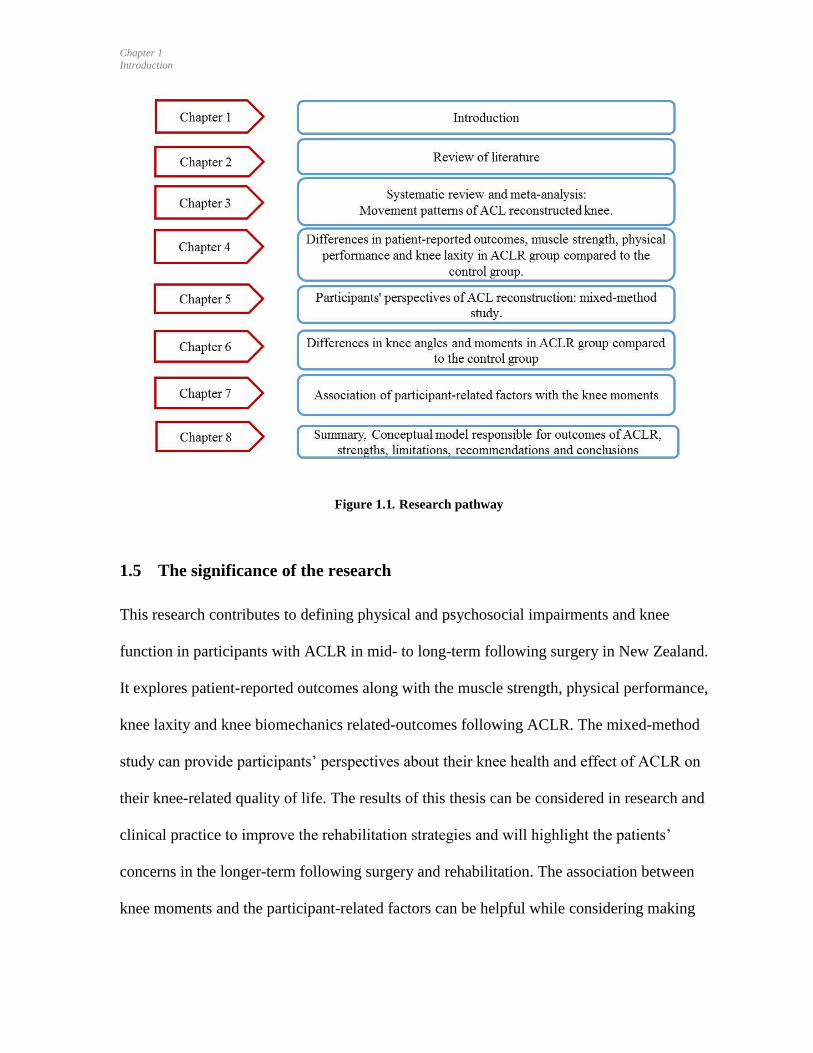
The research pathway of this thesis is structured through a series of steps as illustrated in
Figure 1.1. Firstly, literature related to ACL ruptures, incidence, and its management was
studied. Secondly, literature related to the recovery of participants following surgery such as
muscle strength, physical performance, and knee laxity were studied. Participants’
perspectives about their health condition are important to understand, therefore, their
perspectives and the patient-reported outcomes were studied. This was followed by the
literature related to the biomechanical outcomes following surgery. The primary literature
review facilitated the need for a systematic review and meta-analysis focusing on the
moments and angles in participants with ACLR compared with the uninjured Control group
(Chapter 3). This review included the studies examining peak knee moments and angles on
the injured side compared with the contralateral limb, or to the uninjured Control group,
during walking, stair ascending, stair descending and jogging activities. Methodological
insights regarding the laboratory-based cross-sectional study (Chapter 6) task and the
variables were informed through this systematic review. This led to the development of the
cross-sectional study comparing the peak moments and angles during stair ascent and descent
in participants with ACLR compared with the contralateral limb and with the uninjured
Control group 2-10 years following surgery (Chapter 6). Results of the systematic review
also informed the biomechanical study to determine whether muscle strength, time since

Chapter 1
Introduction
injury, and sex of the participants are predictors of peak moment in participants with ACLR.
This study involved 35 participants with ACLR, and peak biomechanical variables were
studied during stair ascent and descent (Chapter 7).
The literature review (Chapter 2) emphasized that the participants’ perspectives relating to
their recovery and quality of life following surgery as important in the recovery process. This
inspired the qualitative study, which aimed to gain deeper insight into participants’
perspectives 2-10 years following surgery in relation to physical activity, sports, occupation,
and quality of life. A mixed method approach was undertaken for this study, using valid and
reliable electronic patient-reported outcomes, followed by face-to-face interviews, to
understand the influences of the surgery on their life quality of life (Chapter 5). The thesis
concluded (Chapter 8) with a summary and discussion of the major findings of chapters 3, 4,
5, 6, and 7, and an assessment of the overall strengths and limitations of the thesis. This
chapter also presents the conceptual model representing the factors responsible for the
outcomes of the ACLR, and provides recommendations for further research.
Chapter 1
Introduction
Figure 1.1. Research pathway
1.5 The significance of the research
This research contributes to defining physical and psychosocial impairments and knee
function in participants with ACLR in mid- to long-term following surgery in New Zealand.
It explores patient-reported outcomes along with the muscle strength, physical performance,
knee laxity and knee biomechanics related-outcomes following ACLR. The mixed-method
study can provide participants’ perspectives about their knee health and effect of ACLR on
their knee-related quality of life. The results of this thesis can be considered in research and
clinical practice to improve the rehabilitation strategies and will highlight the patients’
concerns in the longer-term following surgery and rehabilitation. The association between
knee moments and the participant-related factors can be helpful while considering making

Chapter 1
Introduction
assessments for impairments in participants with ACLR. Overall, the mixed-method research
approach can provide deeper understanding of the knee-related impairments.
.
Chapter 2
Review of literature
11
2 Literature review
2.1 Prelude to Chapter 2
This chapter provides a narrative review of the background to this thesis. Literature related to
ACL injury, its management, and consequences following ACLR in the short-term and long-
term are reviewed. Following that, a summary of research concerning recovery related to
muscle strength, physical performance, and knee laxity is presented, supporting the aim of
the thesis. This is followed by reviewing patients’ perspectives and patient-reported
outcomes at different times following surgery.
Chapter 2
Review of literature
12
Chapter 2
2.2 Background
The literature on ACLR and the recovery process following the surgery is vast. This narrative
literature review focuses on 4 areas relevant to this thesis:
1. Anterior cruciate ligament rupture, incidences and management.
2. Consequences following the ACLR.
3. Recovery process following the ACLR involving muscle strength-related outcomes,
physical performance, and knee laxity.
4. Patients’ perspectives related to the influence of ACLR on their lives.
2.3 Anterior cruciate ligament injury and management
The ACL is an intra-articular ligament of the knee, comprised of anteromedial and a
posterolateral bundle (Amis & Dawkins, 1991). The primary role of the ACL is to resist the
anterior translation of the tibia, and the secondary role is to stabilise internal rotation and
valgus movement and the forces of the tibia on the femur (Sakane et al., 1997). It is
considered a strong ligament capable of resisting the forces as much as 2,200 N (Woo &
Adams, 1990). Overall, the ACL plays an important role in the knee joint function by guiding
movement and providing stability during ambulatory and functional activities.
Anterior cruciate ligament injury occurs most frequently through non-contact mechanisms
(Zantop, Brucker, Vidal, Zelle, & Fu, 2007) in sports such as netball, basketball, rugby, and
soccer (Gianotti., 2009; Magnussen, Carey, & Spindler, 2011). The ACL consists of two
bundles, the anteriomedial and posterolateral, and injury may be partial, involving less than
Chapter 2
Review of literature
13
50% of ligament tear (Hong et al., 2003), whereas full rupture involves tearing both the
bundles (Zantop et al., 2007). ACL ruptures are treated with non-surgical rehabilitation or
with surgery plus rehabilitation (Delincé & Ghafil, 2012).
According to a recent report from the USA, the overall incidence of complete isolated ACL
tears were 68.6 per 100,000 person-years (Sanders et al., 2016). Eighty percent of those ACL
injuries underwent surgery, which indicates the prevalence of reconstruction management
(Sanders et al., 2016). That study also found a decreasing incidence of isolated ACL tears
with increasing age for men, while it remained relatively stable in women (Sanders et al.,
2016). In 2009 in New Zealand, the incidence of this injury was reported to be 36.9 per
100,000 person-years, with an increase to 50.1 per 100,000 person-years in 2015 (Gianotti. &
Marshall Stephen., 2009). The injury represents a substantial cost for the Accident
Compensation Corporation (ACC): NZ$11,157 on an average for surgical treatment for a
patient (Gianotti, & Marshall Stephen. , 2009), besides the associated physical, psychological
and social costs for the patient. The incidence of ACLR is unknown in New Zealand,
however, among the total number of cases identified with knee injuries in five years from
2004- 2009, approximately 80% underwent surgery for torn ACL (Gianotti. & Marshall
Stephen., 2009). A more recent study from New Zealand found that men who are aged
between 20-29 are more frequently affected by ACL injury with 150-160 ACL
reconstructions per 100,000 person-years (Janssen, Orchard, Driscoll, & van Mechelen,
2012). Based on the New Zealand ACL registry 2,933 patients underwent a primary ACLR,
and 306 revision ACLR surgeries were recorded in the period of September 2015-September
2016 (NZ Orthopaedic Association, 2016). It is important to note that incidence of ACL
Chapter 2
Review of literature
14
injuries presents an increasing trend (Janssen et al., 2012), which probably will also influence
the frequency of surgeries.
Surgical treatment aims to allow the patient to return to sports and restore knee function as
optimally as possible. However, only 55% of these athletes are able to return to their pre-
injury level of sports participation (Ardern, Taylor, Feller, & Webster, 2014). Different
intrinsic and extrinsic factors play an important role in the participants’ being able to return
to competitive sports (Zaffagnini, Grassi, Serra, & Marcacci, 2015), however, a reduced fear
of re-injury, a greater psychological readiness to return to sport and a more positive
subjective assessment of knee function favour the return to sports (Ardern, 2015).
Nevertheless, knee muscle weakness and altered gait patterns are common impairments that
are persistent following ACLR (Brown, Palmieri-Smith, & McLean, 2009; Gokeler et al.,
2013).
2.4 Consequences of Anterior cruciate ligament reconstruction
2.4.1 Re-injury of the ligament
Re-injury to the operated knee or the contralateral knee is common following ACLR
(Salmon, Russell, Musgrove, Pinczewski, & Refshauge, 2005) with the reported rates
between 6% to 30% (Leys, Salmon, Waller, Linklater, & Pinczewski, 2012; Wiggins et al.,
2016). The second ACL re-injury rate was 15%, with an ipsilateral re-injury rate of 7% and
contralateral re-injury rate of 8% (Wiggins et al., 2016). The secondary ACL injury rate
(ipsilateral and contralateral) for patients younger than 25 years was 21%. These combined
data indicate that nearly 1 in 4 young athletic patients who sustain an ACL injury and return
Chapter 2
Review of literature
15
to high-risk sport will go on to sustain another ACL injury at some point in their career
(Wiggins et al., 2016).
Numerous factors could be responsible for the occurrence of re-injury. These include: sex of
the participants, with women at higher risk (Paterno et al., 2012; Shelbourne, Gray, & Haro,
2009); time since reconstruction, with high risk in the first 24 months (Lee, Karim, & Chang,
2008; Salmon et al., 2005); and biomechanical compensatory behaviour, with the
compensatory role of hip and ankle in the injured limb during different activities. Among
these factors, altered biomechanics and neuromuscular function is a modifiable factor (Swärd
et al., 2010). Altered movement asymmetries have been shown to be present at 12 months to
2 years following surgery (Castanharo et al., 2011; Pozzi, Di Stasi, Zeni, & Barrios, 2017),
and can predispose the individuals to re-injury by asymmetric loading.
Lower limb kinetic and kinematic variables have been explored to predict the role of knee
and hip moments and angles for the occurrence of injury. Different sports-related tasks have
been explored in previous studies, such as single-leg horizontal hop (Elias, Hammill, &
Mizner, 2015; Nyland, Klein, & Caborn, 2010; Trigsted, Post, & Bell, 2015), counter-
movement jump and vertical jump (Ernst, Saliba, Diduch, Hurwitz, & Ball, 2000). These
studies indicated that the moments were not similar to the Controls until 2 years following
surgery, indicating the persistence of neuromuscular deficits (Trigsted et al., 2015) which
may have huge implications in the biomechanics of lower limb.

Chapter 2
Review of literature
16
2.4.2 Post-traumatic Osteoarthritis
Patients with ACL injuries are likely to have early Osteoarthritis (Lohmander et al., 2007).
According to data presented by the New Zealand government, Osteoarthritis is a significant
burden to society and the healthcare system (Access Economics for Arthritis New Zealand,
2010). The population affected by osteoarthritis between the ages of 15 to 64 is predicted to
rise to 16.9% by year 2020 (Access Economics for Arthritis New Zealand, 2010). The
literature has also highlighted osteoarthritis as the single greatest cause of disability (Brooks,
2002). Osteoarthritis is classified as primary or secondary. The reason for the development of
primary osteoarthritis is idiopathic, while those for secondary osteoarthritis, injuries to joint
and cartilage are major causes. Among all those who develop osteoarthritis, 20% of the
overall burden arises secondary to joint injury, and is termed post-traumatic osteoarthritis
(Dirschl et al., 2004).
Joint injuries increase the risk of post-traumatic osteoarthritis. It has been reported that 14%
of people developed knee osteoarthritis who had a knee injury at a young age, while only 6%
of those who did not have a knee injury developed Osteoarthritis (Gelber et al., 2000).
Articular fractures can increase the risk of osteoarthritis up to 20-fold, and a significant
ligamentous or capsular injury increases the risk of osteoarthritis up to 10-fold (Anderson et
al., 2011).
The role of different factors causing early onset osteoarthritis following ACL injury and
surgery are being explored. Osteoarthritis development in the injured joints is caused by
intra-articular pathogenic processes initiated at the time of injury, combined with long-term
changes in dynamic joint loading. The disease is initiated, and its progression caused, by a

Chapter 2
Review of literature
17
combination of endogenous and environmental risk factors. Phenotype variability is further
capable of influencing the process (Lohmander et al., 2007). Variables such as re-injury,
muscle strength, and body mass index also influence the outcome of the injury (Lohmander
et al., 2007). Biomechanical factors (Guilak et al., 2004) could be responsible for the onset
and progression of post-traumatic osteoarthritis post-ACL rupture. Mechanical loading at the
joint has been extensively explored as a risk for post-traumatic osteoarthritis (Sharma et al.,
1998). The optimal amount of joint loading is important for cartilage health (Griffin &
Guilak, 2005); on the other hand, suboptimal loading, either the repetitive torsional stresses
(Dekel & Weissman, 1978) or sudden impact load (Buckwalter, 1992), increases the risk of
post-traumatic osteoarthritis. Culvenor et al., 2015 explored MRI findings of Osteoarthritis in
a cohort of 111 participants with ACLR 1 year following surgery. The patellofemoral
compartment was at risk for early of degeneration. Further, presence of Osteoarthritis was
evident 5 years following surgery in another cohort, and was related to the lower joint
loading in the frontal compartment (Wellsandt et al., 2016). It is possible that loading is not
restored following initial joint injury and influences the cartilage following the surgery.
Various factors could be responsible for the onset and progression of post-traumatic
osteoarthritis post-ACL rupture. For instance, raised levels of the inflammatory markers have
been reported in the year following the ACL injury and surgery (Harkey et al., 2015). High
levels of the inflammatory markers can lead to abnormal breakdown of the cartilage, and
along with the lesser biomechanical loading of the injured leg compared to the uninjured,
creating a loading asymmetry (Pietrosimone et al., 2017). Levels of inflammatory markers
have been reported to be high up to 6 years following the surgery, and greater levels of
matrix metalloproteinase-3 are associated with lesser knee adduction moment and the lesser
Chapter 2
Review of literature
18
vertical ground reaction force (Pietrosimone et al., 2017). The level of biomarkers has been
reported to be at normal levels within 8 years following surgery (Åhlén et al., 2015). A
combination of inflammation and suboptimal biomechanics can be detrimental to the joint, as
both processes operate concurrently in the joint. These reviews did not mention the
psychological aspect of the injury, which could influence the outcomes of surgery by
influencing the attitude of the patient towards physical activity, sports and their motivation
regarding exercise. Theoretically, numerous factors can be responsible for the onset and
progression of osteoarthritis following such surgery.
2.5 Recovery process following the anterior cruciate ligament reconstruction
Gait analysis, muscle strength, range of motion at the joint, laxity of the graft, and physical
performance tests are some of the criteria used clinically to assess joint recovery following
the surgery, during rehabilitation, and to assess decisions to return to sports, recreation and
occupational-related tasks. This section will summarise the recovery of muscle strength,
physical performance, and knee laxity-related outcomes following the ACLR. Gait analysis
in participants with ACLR will be explained in Chapter 3. Patient perspectives, which are
integral for a biopsychosocial approach for rehabilitation, are discussed in this section.
2.5.1 Muscle strength-related outcomes
Muscle weakness following ACLR has been reported in various studies at different time
periods following surgery, comparing the injured side to the contralateral limb or to the
Control group (Keays, Bullock-Saxton, & Keays, 2000; Mattacola et al., 2002). For instance,
maximal strength deficits were present at 6 months following surgery (Petersen, P. Taheri,
Forkel, & Zantop, 2014) and a clear pattern of improvement in the quadriceps and hamstring
Chapter 2
Review of literature
19
strength was found from 6 to 12 months following surgery (Jong, Caspel, Haeff, & Saris,
2007).
Thigh muscle strength deficits have been reported by two systematic reviews exploring
muscle strength assessments up to 24 months following surgery (Dauty, Tortellier, &
Rochcongar, 2005; Xergia, McClelland, Kvist, Vasiliadis, & Georgoulis, 2011). Muscle
strength differed depending on the graft used (Dauty et al., 2005; Xergia et al., 2011; Dauty
et al., 2005) reported no muscle strength deficits regardless of the type of graft at 24 months.
However, another systematic review indicated that muscle strength deficits may be related to
the surgical graft: patients who had received a patella tendon graft were more likely to have
knee extensor strength deficits, while those with hamstring grafts were likely to have flexor
strength deficits (Xergia et al., 2011).
Another review exploring the muscle strength recovery following ACLR has indicated that
muscle weakness at the quadriceps and hamstrings may persist from 3 months to 6 years
following surgery with some inconsistent results which could be due to different
methodologies used among the included studies (Petersen et al., 2014). This thesis explored
the muscle strength in participants with ACLR from 2 to 10 years following surgery using
the isokinetic strength tests, which is considered a ‘gold standard’ for strength testing.
A recent long-term retrospective study reported reduced peak torque of extensors concentric
and eccentric muscle strength compared to the uninjured limb at more than 20 years post-
ACLR compared to age-matched Controls, and when comparing the injured to uninjured
sides (Tengman, Olofsson, Stensdotter, Nilsson, & Häger, 2014). Asymmetrical quadriceps
strength at the time of return to sports is associated with asymmetrical knee biomechanics
Chapter 2
Review of literature
20
during hopping (Palmieri-Smith & Lepley, 2015), which in turn can predict ACL re-injury
(Paterno et al., 2010). Therefore, symmetric muscle strength prior to returning to sports may
be required to decrease risk of further injuries (Grindem, Snyder-Mackler, Moksnes,
Engebretsen, & Risberg, 2016). Apart from the knee muscle strength deficits, hip muscle
strength deficits in the hip flexors at 24 months following the surgery have also been reported
(Dalton et al., 2011; Petersen et al., 2014). However, the focus of this thesis is on examining
the knee-related impairments and function; therefore, hip joint muscle strength was not
explored.
A number of reasons could be responsible for short to long-term persistence of muscle
weakness. Factors contributing to the muscle weakness include factors such as such as pre-
operative quadriceps activation and muscle strength (Lepley & Palmieri-Smith, 2016), lower
activation of quadriceps muscle post-operatively (Hart, Pietrosimone, Hertel, & Ingersoll,
2010), and reduced proprioception (Bonfim, Paccola, & Barela, 2003). Reduced quadriceps
function in isokinetic knee-extensor strength and lower voluntary activation was found in a
study with participants at 3 years post-surgery in the involved limb compared to the
uninvolved limb in the ACLR patients (Otzel, Chow, & Tillman, 2015).
The ACL injury and surgery may also influence the activation at the central nervous system.
Studies have reported the changed activation of the sensory cortex (Valeriani et al., 1996),
depressed cortical excitability (requiring higher level of stimulus at the motor cortex to
generate quadriceps contractions) (Lepley, Ericksen, Sohn, & Pietrosimone, 2014), and
neuroplasticity in the brain (Grooms, Appelbaum, & Onate, 2015). Alterations of cortical and
spinal reflexive excitability pathways have been reported to affect the muscle function and
Chapter 2
Review of literature
21
overall physical performance (Pietrosimone, McLeod, & Lepley, 2012). Thus, besides
peripheral mechanisms, central mechanisms of the central nervous system may also
contribute towards long-term muscle strength deficits following ACLR. Probably, this is the
reason that muscle strength cannot reach the pre-injury level following extensive
rehabilitation.
Quadriceps weakness has been considered as a primary risk factor for knee pain, disability,
and progression of joint damage in patients with osteoarthritis of the knee (Slemenda et al.,
1997). Muscle weakness appears to precede the onset of symptoms related to osteoarthritis,
irrespective of the reason for this weakness (Valderrabano & Steiger, 2010), and may also
contribute to the development of osteoarthritis (Keays, Newcombe, Bullock-Saxton, Bullock,
& Keays, 2010). Muscle weakness has been associated with joint space narrowing at 4 years
following ACLR (Tourville et al., 2014) and further quadriceps weakness is a known risk
factor for knee osteoarthritis (Øiestad, Juhl, Eitzen, & Thorlund, 2015). Quadriceps strength,
on the other hand, has been indicated to have a protective effect against the progression of
osteoarthritis (Hochberg et al., 2012; Jordan et al., 2003). Therefore, it is important to explore
if any differences persist in thigh muscle strength participants with ACLR compared to the
contralateral limb and to uninjured, matched controls.
2.5.2 Physical performance in participants with ACLR
Physical performance tests are included in assessments of readiness for return to physical
activity and sports (Reiman & Manske, 2009). A variety of hop tests have been used to assess
the limb function following surgery. These tests include single-leg hop for distance, timed
hop, triple-hops for distance, and cross-over hops for distance (Noyes, Barber, & Mangine,
Chapter 2
Review of literature
22
1991). Physical performance-based outcome measures are known to be highly reliable
(Intraclass correlation coefficient 0.82 to 0.93) (Reid, Birmingham, Stratford, Alcock, &
Giffin, 2007) and are practical, easy to administer and need little time to execute in clinical
and research settings.
The physical performance tests are used either as a battery or as a single test to examine the
limb function. These tests are often used as a clinical assessment of neuromuscular control
and confidence level in the use of the limb (Borsa, Lephart, & Irrgang, 1998; De Carlo &
Sell, 1997; Petschnig, Baron, & Albrecht, 1998) and are also correlated with the knee
extension and flexion isokinetic strength in participants with ACLR (Laudner et al., 2015;
Xergia, Pappas, & Georgoulis, 2015). Among the commonly used physical performance
tests, the single-leg hop test has been widely used in research as a single test or clustered with
other physical performance tests, and is known to be a reliable test (Intraclass correlation
coefficient 0.76–0.97) (Palmieri-Smith & Lepley, 2015; Trigsted et al., 2015).
Following the execution of the single-leg hop tests, the distance of the hops is measured for
each limb, and presented as a Limb Symmetry Index (LSI). Rationale for assessing the LSI is
to assess whether the injured side reaches an acceptable level of muscle strength. Different
LSI percentages have been used in previous studies. Assessing the muscle function outcome
using a battery of tests or increasing the acceptable LSI level from 90% to 95% or 100% has
been previously documented. A high rate of successful muscle function outcome was
reported with an LSI of 80% (Thomeé et al., 2012). Therefore, it was suggested to consider
the LSI percentages wisely when presenting results after ACL rehabilitation, deciding on the
Chapter 2
Review of literature
23
criteria for a safe return to sports, or designing rehabilitation programmes after ACLR
(Thomeé et al., 2012).
Exploring the physical performance in the injured leg indicated the poor results when
categorised by considering the LSI of 90% compared to the uninjured leg and Control group
at 20 years following the surgery (Tengman, 2014). This study recruited participants who had
surgery in the 1990’s and had 20 years as the mean time following reconstruction. The
surgical techniques have been updated many times since then, therefore, we may expect
better surgical outcomes nowadays. However, this has not yet been determined. The research
reported in this thesis intends to address this gap in the research. Here, the physical
performance of participants 2 to 10 years following surgery are compared with age- and
gender-matched controls in order to explore any deficits present in the mid-term following
the surgery.
2.5.3 Knee laxity following anterior cruciate ligament reconstruction
The anteroposterior laxity of the knee joint is considered an important parameter for
evaluating ACL rupture (Van Thiel & Bach, 2010). It is difficult to measure and compare the
knee laxity manually, partially due to the variation in the force exerted by the examiner, and
it is not possible to quantify the magnitude of tibia movement relative to the femur while
performing the clinical tests such as the Lachman and Pivot Shift. The KT-1000 arthrometer
is therefore a better option as it can quantify the force and the amplitude of movement.
Previous studies have reported good reliability of the KT-arthrometer (Anderson &
Lipscomb, 1989), but it has been questioned by others (Barcellona, Christopher, &
Morrissey, 2013; Forster, Warren-Smith, & Tew, 1989). Regarding the validity using the

Chapter 2
Review of literature
24
KT-1000 score, it may be more appropriate as a dichotomous diagnostic test with a threshold
of 2 or 3 mm (Arneja & Leith, 2009). It is often used in addition to clinical examination to
establish the diagnosis of ACL rupture and during the follow-up after ACLR (Aglietti, Buzzi,
Giron, Simeone, & Zaccherotti, 1997).
Previous studies exploring knee laxity in participants with ACLR have presented varied
results. A significant reduction in knee laxity of the injured limb 9 months to 12 months
following surgery, had been reported (Semay, Rambaud, Philippot, & Edouard, 2016) (Table
2.1). Those authors suggested that the joint laxity is the product of isometric positioning of
the transplant, transplant ligamentisation phenomena by collagen remodeling to mechanical
stress, local muscle condition, and exposure to hormonal factors. Therefore, a return to sport
without satisfactory joint control may not yield a favorable outcome. Similarly, a reduction in
knee laxity was found over time after an ACLR using either a bone patellar tendon bone or
hamstring graft autograft from 6 months to 7 years (Ahldén, Kartus, Ejerhed, Karlsson, &
Sernert, 2009) (Table 2.1). Decrease in laxity may be due to the lack of knee extension,
which has been reported in participants a few years following surgery (Sernert et al., 2002;
Shelbourne, Klootwyk, Wilckens, & De Carlo, 1995). In addition, the development of early
degenerative changes and secondary changes in the integrity of ligamentous tissue, as well as
the soft tissue envelope, may also influence laxity.
In contrast to above studies, no differences in knee laxity were found over time from 2–6
years after ACLR using bone patellar tendon bone autografts using the KT-1000 arthrometer
(Shelbourne et al., 1995), and from 7 to13 years after ACLR (Salmon et al., 2006).
Differences in inter-limb knee laxity may influence physical performance (Sernert et al.,
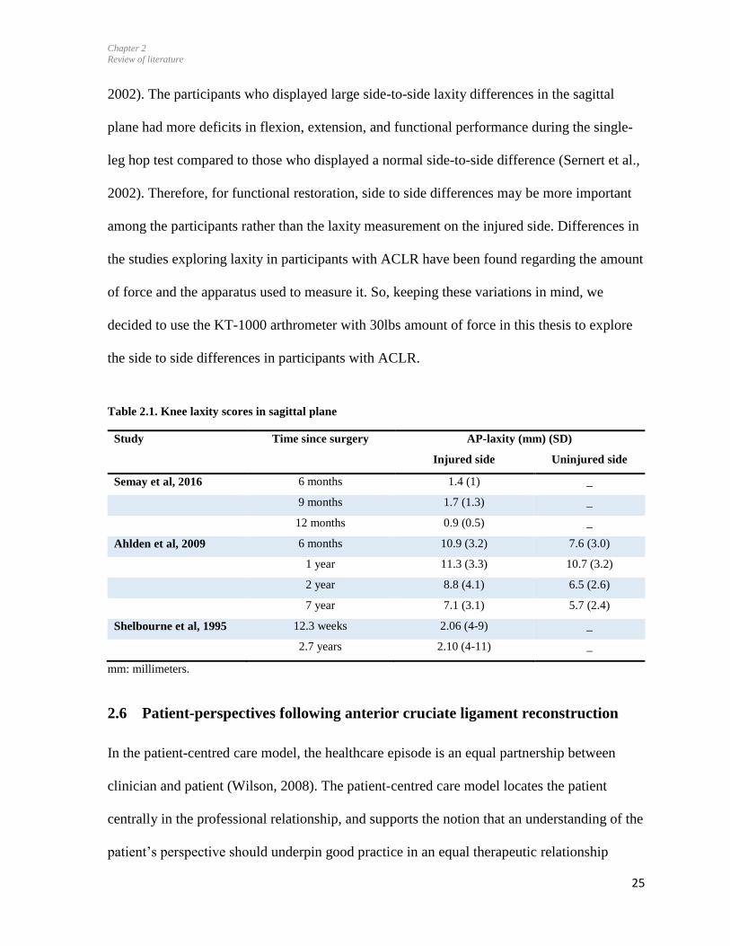
Chapter 2
Review of literature
25
2002). The participants who displayed large side-to-side laxity differences in the sagittal
plane had more deficits in flexion, extension, and functional performance during the single-
leg hop test compared to those who displayed a normal side-to-side difference (Sernert et al.,
2002). Therefore, for functional restoration, side to side differences may be more important
among the participants rather than the laxity measurement on the injured side. Differences in
the studies exploring laxity in participants with ACLR have been found regarding the amount
of force and the apparatus used to measure it. So, keeping these variations in mind, we
decided to use the KT-1000 arthrometer with 30lbs amount of force in this thesis to explore
the side to side differences in participants with ACLR.
Table 2.1. Knee laxity scores in sagittal plane
Study Time since surgery AP-laxity (mm) (SD)
Injured side Uninjured side
Semay et al, 2016 6 months 1.4 (1) _
9 months 1.7 (1.3) _
12 months 0.9 (0.5) _
Ahlden et al, 2009 6 months 10.9 (3.2) 7.6 (3.0)
1 year 11.3 (3.3) 10.7 (3.2)
2 year 8.8 (4.1) 6.5 (2.6)
7 year 7.1 (3.1) 5.7 (2.4)
Shelbourne et al, 1995 12.3 weeks 2.06 (4-9) _
2.7 years 2.10 (4-11) _
mm: millimeters.
2.6 Patient-perspectives following anterior cruciate ligament reconstruction
In the patient-centred care model, the healthcare episode is an equal partnership between
clinician and patient (Wilson, 2008). The patient-centred care model locates the patient
centrally in the professional relationship, and supports the notion that an understanding of the
patient’s perspective should underpin good practice in an equal therapeutic relationship
Chapter 2
Review of literature
26
(Kidd, Bond, & Bell, 2011). This approach was followed in this thesis to understand the
participants’ perspectives following surgery.
2.6.1 Person-centred treatment approach
The physical performance measures, although critical in making clinical decisions, are not
enough to fully understand the patients’ experiences. Being informed about the patient
experience in the management of health-related conditions is an important step to actively
improve the quality of health care. A positive association between self-reported patient
experiences, clinical outcomes and resource utilisation has been noted in the past (Doyle,
Lennox, & Bell, 2013). Multimodal approaches to exploring patients’ experiences are needed
following ACLR to advance an understanding of their concerns, complaints, and outcomes of
the injury, surgery and rehabilitation.
The biopsychosocial model states that health and illness are determined by a dynamic
interaction between biological, psychological, and social factors leading to a given outcome.
Each component on its own is insufficient to lead definitively to health or illness (George &
Engel, 1980). The ICF model, as explained in Chapter 1, is based on the biopsychosocial
model of disability (Stucki et al., 2007). The ICF conceptualises a person's level of
functioning as a dynamic interaction between their health condition or disease, environmental
factors, and personal factors (psychological and social).
Biological factors related to the outcome of the surgery have been studied extensively in
terms of biomechanical variables, muscle strength, and physical performance tests as
described in section 2.5. Recent studies have emphasised the role of psychological and social
Chapter 2
Review of literature
27
factors in the overall outcomes of the ACLR (Ardern, Taylor, Feller, Whitehead, & Webster,
2013). Athletes who managed to return to sport are known to have significantly higher self-
esteem levels than those who do not return to sports, without observable differences in knee
stability or time since surgery. Athletes in this study had higher levels of self-esteem and
higher reported KOOS-QOL scores (Christino, Fleming, Machan, & Shalvoy, 2016).
Similarly, in another study with 65 individuals (mean age= 22 years), higher motivation
during rehabilitation was associated with returning to the pre-injury sports. Also, these
participants were satisfied with their knee function 1 year after the ACLR (Sonesson, Kvist,
Ardern, Österberg, & Silbernagel, 2017). A study exploring the functional outcome of
surgery in 48 competitive athletes highlighted that psychosocial issues affect the overall
results of the surgery with respect to return to sports. In that study, 18 showed a decrease in
their level of sports. Of the 18 patients, 12 referred to fear of reinjury as the primary reason
for the decrease in their level of sports reported, while persisting knee pain and instability in
6 patients were the reasons for a fall in their sporting abilities (Devgan, Magu, Siwach,
Rohilla, & Sangwan, 2011).
2.6.2 Quality of life following ACL reconstruction
When exploring the outcomes of disease, it is important to investigate and evaluate the
impact of the condition on patients’ quality of life. Quality of life is a multidimensional
concept incorporating domains related to physical, mental and emotional, and social
functioning (Ferrans, 2005). Different definitions appear in the literature for health-related
quality of life. Most conceptualizations of health-related quality of life emphasize the effects
of disease on physical, social role, psychological or emotional, and cognitive functioning. For
this thesis, the definition by Wilson and Cleary (1995, p-60) was adopted, defining quality of

Chapter 2
Review of literature
28
life as “an individual's satisfaction or happiness with domains of life insofar as they affect or
are affected by health” page 60.
There is a gradual decrease in participants’ quality of life following the surgery. Participants
who were satisfied with their knee health reported less knee-related pain and symptoms
(KOOS: pain and symptom scores, Table 2.2), and better quality of life (KOOS: quality of
life scores) at approximately 3 years (Ardern et al., 2016) and at 5 years following surgery
(Tagesson, Oberg, & Kvist, 2015). On the contrary, a systematic review indicated lowest
scores in the KOOS sport/recreation function and knee-related quality of life subscales at 10
years following surgery (Magnussen, Verlage, Flanigan, Kaeding, & Spindler, 2015). This
may indicate gradual decrease in quality of life over time following ACLR. In addition,
exploring the quality of life at 23 years following ACLR (Tengman, 2014), KOOS quality of
life scores in participants were similar to the patients with knee osteoarthritis (Aksekili et al.,
2016) (Table 2.2). The ACL injury is managed by the surgery with two aims: (1) to return the
participants to competitive sports in the short-term, (2) to maintain the quality of life in the
long-term. Regarding the first surgical aim, it is known that only 55% of participants return
to competitive sports (Ardern et al., 2014), although the percentage is higher for the elite
athletes with 83% of them able to return to pre-injury level of sports following ACR (Lai,
Ardern, Feller, & Webster, 2017) . The second aim for undergoing surgery, which is to
maintain quality of life in the long-term, seems not to be fully achieved, as the quality of life
deteriorates over time following the surgery.

Chapter 2
Review of literature
29
Table 2.2. Knee injury and osteoarthritis outcome scale (KOOS)
ADL: activities of daily living, QOL: quality of life, sports/rec: sports/recreation. (‘0’ indicates maximum
problems, ‘100’ indicates no problem)
Numerous factors can influence the quality of life in participants with ACLR. Quality of life
may be associated with psychological satisfaction with knee function (Ardern et al., 2016),
preference for the competitive sports, fear of re-injury after ACLR (Filbay, Crossley, &
Ackerman, 2016), and the number of injured structures (Øiestad, Engebretsen, Storheim, &
Risberg, 2009). Filbay et al. (2016) conducted a qualitative study with two groups of
participants with ACLR up to 20 years: those with high ACL-quality of life scores, and the
second group with low ACL-quality of life scores. Participants described the importance of
physically active lifestyle. Participants who avoided sport or activity reported experiencing
reduced quality of life and those who overcame re-injury fears to continue sports described
experiencing a satisfactory quality of life. Participants who enjoyed recreational exercise
often adapted their lifestyle early after ACLR, while others described adapting their lifestyle
at a later stage to accommodate knee impairments. (Filbay et al., 2016). However, this study
Study Time since
surgery
(years, mean)
Pain Symptoms ADL Sport/Rec QOL
Participants with ACLR
Ardern et al. (2016)
Satisfied group
3.4 93.9 86.5 98.1 85.8 78.2
Mostly satisfied 2.6 86.0 78.7 93.2 69.6 60.9
Dissatisfied group 2.6 73.1 62.0 80.7 48.4 40.6
Tagesson et al. (2015) 5 97 91 100 95 75
Magnussen et al. (2015) 10 85.8 78.9 90.5 66.1 67.6
Tengman et al. (2014) 23 78 79 84 50 49
Participants with osteoarthritis
Aksekili et al. 2016 60 71.42 70.59 35 50
Chapter 2
Review of literature
30
involved participants with revision surgery and contralateral limb surgeries as well. It is
known that combined injuries to the knee can influence the knee health differently by
increasing the predisposition to knee osteoarthritis (Øiestad et al., 2009), and ultimately
affecting the quality of life (Filbay, Ackerman, Russell, Macri, & Crossley, 2014).
Furthermore, those who develop severe osteoarthritis following ACLR also reported poorer
health-related quality of life compared to those without osteoarthritis (Øiestad et al., 2009).
Similarly, a study with the participants from 18-45 years (mean age=28) and 3 years post-
surgery reported that people who return to preinjury physical activity levels, and report
higher knee-related self-efficacy and quality of life, were more likely to be satisfied with the
outcome of ACLR (Ardern et al., 2016). Less than half of the participants of this study
reported that they were satisfied with their knee function after surgery. This suggests a
combined role of psychological factors and functional recovery for patient satisfaction after
ACLR. Therefore, it is important to review the psychological and other underlying causes
responsible for the poor quality of life in these participants.
Among the above-mentioned factors determining the quality of life, fear of re-injury can play
a huge role in the quality of life in the longer-term (Filbay et al., 2016). Fear of re-injury was
present at 2 years following surgery (Heijne, Axelsson, Werner, & Biguet, 2008) and was
reported to reduce 3 years following ACLR with the restoration of the sports participation
among participants (Gignac et al., 2015). Participants described their rehabilitation as an
arduous experience (Scott, Perry, & Sole, 2017), their struggle with fear of injury (Gignac et
al., 2015; Heijne et al., 2008) and return to sports (Gignac et al., 2015). Participants who
overcome the fear of re-injury and continued their physical activities reported better quality
of life than those who avoided sports and physical activities (Filbay et al., 2016). Fear of re-
Chapter 2
Review of literature
31
injury can affect the sports-performance of patients who attempt to return to their chosen
activity, by reducing their confidence. Overall, fear of injury is present to a different extent in
participants following surgery and has major consequences for them in their sports life.
Quality of life has been assessed with the number of tools in the literature. The type of tool
used for reporting the quality of life has been found to affect the results. A review used
KOOS-QOL scale to report knee-related quality of life in participants with ACLR and it was
found to be low at 5 years post-surgery (Filbay et al., 2014). Another study used SF-36
Health Survey to report quality of life and indicated similar quality of life in participants with
ACLR 11.5 years following surgery to the uninjured controls (Möller, Weidenhielm, &
Werner, 2009). Unlike the KOOS-QOL scale, the SF-36 Health survey is a generic health
questionnaire and features domains covering physical, mental, and social health. This survey
addresses topics such as tiredness, sadness, and nervousness. Considering the occurrence of
ACL injuries in young athletic participants, it is possible that this patient group scored higher
than aged-matched, less active counterparts. Similarly, ACL-QOL has been used in the
literature in some of the studies. There was a statistically significant difference between the
mean ACL-QOL scores at 6, 12 and 24 months post-operatively. Patients demonstrated a
statistically significant improvement in ACL-QOL score following ACLR (Heard, Lafave,
Kerslake, Hiemstra, & Buchko, 2015). However, it is recommended that the ACL-QOL
questionnaire be used in conjunction with currently available objective and functional
outcome measures during the pre-operative, conservative and post-surgery treatment of
patients with chronic ACL deficiency. Therefore, there is a need to explore the quality of life
with the knee-specific questionnaire. Also, there is a need to explore the participants’
experiences of the outcomes of their surgery in relation to physical activity, sports,
Chapter 2
Review of literature
32
occupation and health-related quality of life so as to determine the extent to which the ACLR
has influenced their lives. The research in this thesis investigated patients’ perspectives
related to the effect of ACLR on their lives until 10 years following ACLR through a
qualitative study. Until now, there have been no studies exploring the quality of life in
participants with ACLR in the New Zealand context. The ACC funds the surgery and
rehabilitation of patients with ACL injury, thus this study will potentially inform the health-
care system regarding the participants’ knee-related concerns and outcomes of surgery.
2.6.3 Patient-reported outcomes
To fully understand outcomes of surgery and rehabilitation, patient-reported outcomes are
very important measures as they provide first-hand information relating to the patient’s
condition, are easy to administer, and are undemanding in terms of cost and the time
involved to perform them. A number of tools exploring the patient-reported outcome
measures have been used in the literature. Some of them were used in this thesis and are
described below in relation to the recovery process.
2.6.3.1 The Knee Injury and Osteoarthritis Outcome Score.
The Knee Injury and Osteoarthritis Outcome scale (KOOS) has been widely used for
osteoarthritis and ACL-related research: it enables analysis of knee-related outcomes such as
pain, symptoms, quality of life, and knee function in daily life and sports. This scale
measures the patients’ opinions about their knee health and associated problems over the past
week (Appendix-C1). The reliability of this questionnaire has been previously established
(Roos, Roos, Lohmander, Ekdahl, & Beynnon, 1998). This questionnaire is valid for
measuring the functional status and the quality of life in participants with ACLR (Salavati,
Chapter 2
Review of literature
33
Akhbari, Mohammadi, Mazaheri, & Khorrami, 2011). For patients with knee injuries, the
pain, ADL, and sport/recreation subscales have adequate internal consistency, while the knee
symptom and Quality of life (QOL) subscales have had reports of lower as well as adequate
internal consistency (Intraclass correlation coefficient: Pain= 0.85–0.93 Symptoms= 0.83–
0.95, ADL= 0.75–0.91, Sport/rec= 0.61–0.89, QOL= 0.83–0.95). KOOS has standard error
of measurement from 2.2 to 3.1 among the subscales (Pain= 2.2 Symptoms= 3.1 ADL= 2.9
Sport/recreation = 2.1, QOL= 2.6). The KOOS appears to be responsive to change in patients
with a variety of conditions treated with nonsurgical and surgical interventions (Bekkers, De
Windt, Raijmakers, Dhert, & Saris, 2009). It has been suggested that a change of 8–10
KOOS points constitutes a clinically relevant difference (Roos & Lohmander, 2003).
2.6.3.2 The Tegner activity level scale
Tegner activity level scale involves a list of activities of daily living, recreation, and
competitive sports (Tegner & Lysholm, 1985) (Appendix-C2). A score of 0-10 is assigned
where 0 represents “sick leave or disability pension because of knee problems,” whereas 10
corresponds to “participation in national and international elite competitive sports”. Higher
scores represent participation in higher-level activities. Reliability and validity of this scale
has been established in individuals with ACL injury with an intraclass correlation coefficient
of 0.8 and a minimum detectable change of 1 (Briggs et al., 2009).
Table 2.3 shows the potential inability of the participants to return to preinjury activity levels,
following injury, surgery and rehabilitation. A review involving multiple studies indicated
that participants were at 5.1 post-injury Tegner score at the mean of 10 years following
surgery (Magnussen et al., 2015). There has been a decrease in the level of physical activities
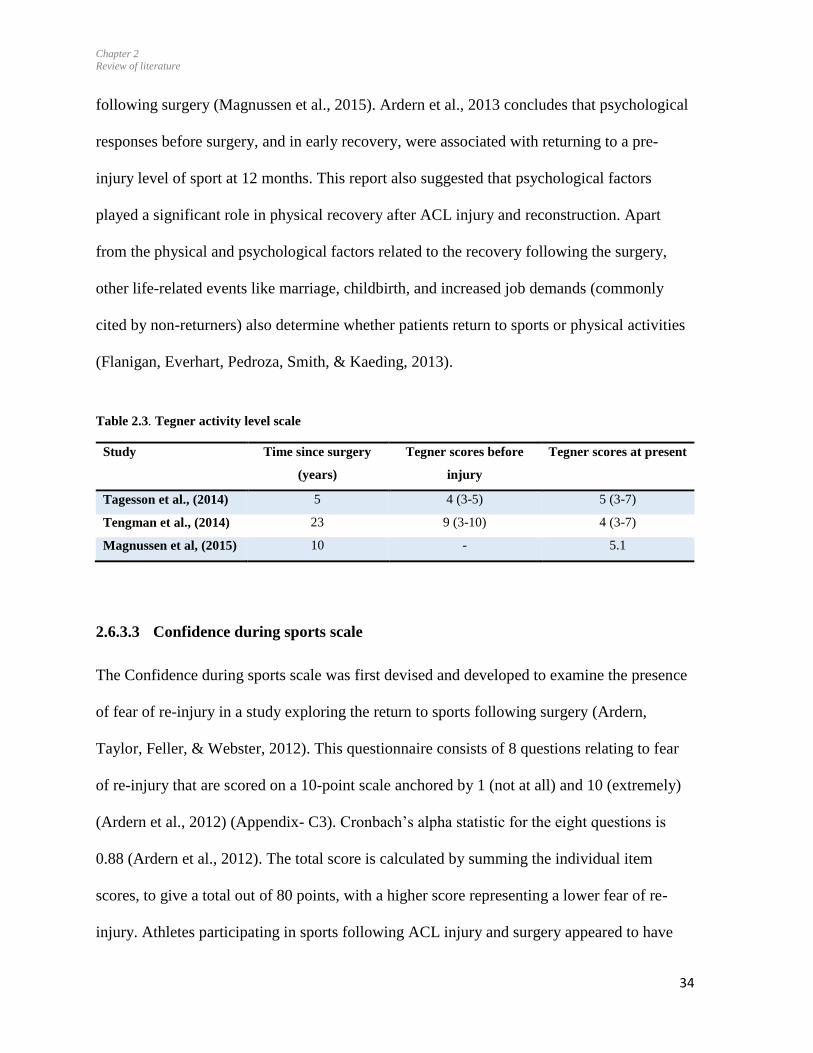
Chapter 2
Review of literature
34
following surgery (Magnussen et al., 2015). Ardern et al., 2013 concludes that psychological
responses before surgery, and in early recovery, were associated with returning to a pre-
injury level of sport at 12 months. This report also suggested that psychological factors
played a significant role in physical recovery after ACL injury and reconstruction. Apart
from the physical and psychological factors related to the recovery following the surgery,
other life-related events like marriage, childbirth, and increased job demands (commonly
cited by non-returners) also determine whether patients return to sports or physical activities
(Flanigan, Everhart, Pedroza, Smith, & Kaeding, 2013).
Table 2.3. Tegner activity level scale
Study Time since surgery
(years)
Tegner scores before
injury
Tegner scores at present
Tagesson et al., (2014) 5 4 (3-5) 5 (3-7)
Tengman et al., (2014) 23 9 (3-10) 4 (3-7)
Magnussen et al, (2015) 10 - 5.1
2.6.3.3 Confidence during sports scale
The Confidence during sports scale was first devised and developed to examine the presence
of fear of re-injury in a study exploring the return to sports following surgery (Ardern,
Taylor, Feller, & Webster, 2012). This questionnaire consists of 8 questions relating to fear
of re-injury that are scored on a 10-point scale anchored by 1 (not at all) and 10 (extremely)
(Ardern et al., 2012) (Appendix- C3). Cronbach’s alpha statistic for the eight questions is
0.88 (Ardern et al., 2012). The total score is calculated by summing the individual item
scores, to give a total out of 80 points, with a higher score representing a lower fear of re-
injury. Athletes participating in sports following ACL injury and surgery appeared to have

Chapter 2
Review of literature
35
considerably less fear of injury and more confidence 2 to 7 years following surgery (Ardern
et al., 2012).
2.6.3.4 The Short Form-12 health survey
The Short Form-12 health survey (SF-12) was designed to measure a health-related quality of
life at a single time point (Ware, Kosinski, & Keller, 1996). The SF-12 is a generic measure
and does not target a specific age or disease group. However, the Short Form-12 is known to
provide a simple health outcome assessment following ACLR surgery at a single time point
(Webster & Feller, 2014). This questionnaire explores the participant’s views about their
health (Appendix- C4), and has been validated in participants with ACLR. This questionnaire
summarises the physical and mental components of health. Physical and Mental Health
Composite Scores (PCS & MCS) are computed using the scores of twelve questions and
range from 0 to 100, where a zero score indicates the lowest level of health measured by the
scales and 100 indicates the highest level of health. The PCS and MCS scores tend to vary
over the life span (PCS tends to decrease with age, while MCS tends to increase). This
questionnaire can be self-administered and can be administered in 2 or less than 2 minutes.
2.7 Summary
Muscle strength, physical performance and knee laxity-related deficits were explored in
participants following ACLR. This informed the need for an exploratory study to explore the
muscle strength-related, physical performance, knee laxity, and patient-reported outcomes in
participants with ACLR from 2 to 10 years following surgery (Chapter 4). Furthermore, this
chapter justified the need to explore the participants’ perspectives related to their physical
activity, sports and quality of life following the surgery (Chapter 5).
Chapter 2
Review of literature
36
The next chapter is a systematic review of peak knee moments and angles in participants
with ACLR during walking, jogging, and stair navigation.
Chapter 3
Systematic review and meta-analysis
37
3 Movement patterns following ACL reconstruction: a systematic review
and meta-analysis
3.1 Prelude to Chapter 3
Muscle strength deficits were reported in different studies in participants with ACLR in the
literature (Chapter 2). Muscle strength deficits can alter the moments as by definition
moments (or torque) are the turning effect of a force that causes angular acceleration at a
specific axis (Nigg et al., 2000). Knee biomechanics is studied through exploring the knee
joint angles and moments. Moments will be presented as the external moments throughout
the thesis.
Lower limb kinematics and kinetics of the ACL reconstructed knee was compared to (a) the
contralateral limb, and (b) healthy age-matched participants during different activities.
Whether there is any change in knee angles and moments over time following ACLR was
also explored. A systematic search with specific key words was run and the articles exploring
the recovery of angles and moments following surgery were included. Studies with the
similar variables were pooled together to generate the forest plots for meta-analysis was
performed. Analysing the movement pattern helped to understand if the loading pattern of
ACLR knee compared to the contralateral knee and to the uninjured control group.
Chapter 3
Systematic review and meta-analysis
38
Chapter 3
3.2 Background
Altered gait biomechanics can be assessed via joint angles and moments, refer to unresolved
impairments following injury and surgery, and may also be a mechanical contributing factor
towards the development of post-traumatic osteoarthritis (Foroughi et al., 2009; Nigg et al.,
2000). Differences in these biomechanical variables appear to be important when comparing
the injured side with the contralateral uninjured side in patients with ACLR and also with
Control groups. Individuals with ACLR have been shown to walk with greater peak knee
flexion angles compared to controls in the early phases (< 6 months), but with less flexion
(more extended) after 1 year post-surgery (Hart, Culvenor, et al., 2015).
In the sagittal plane, lower external flexion moments were found following ACLR at mid-
stance of walking at 2 to 12 months post-operatively compared to controls (Timoney et al.,
1993), while no significant differences were reported on average 17 months post-surgery
(Bulgheroni, Bulgheroni, Andrini, Guffanti, & Giughello, 1997). Decreased flexion moments
while walking and during stair navigation have also been found in patients with knee
osteoarthritis. Furthermore, a recent systematic review found increased flexion moments
during walking in patients with knee osteoarthritis following joint replacements, compared to
pre-operative measures (Sosdian et al., 2014). Flexion moments require an internal
quadriceps moment to counteract the forces of body weight (Schmitt, Paterno, Ford, Myer, &
Hewett, 2015). Decreased flexion moments are likely to indicate unloading of the knee joint
or a compensatory strategy, possibly due to decreased neuromuscular control, lingering
symptoms and hesitancy for weight-bearing on that side (Andriacchi, 1990).
Chapter 3
Systematic review and meta-analysis
39
In terms of joint loading in the frontal plane, external knee adduction moments are often used
as the measure of load distribution in the frontal plane (Schipplein & Andriacchi, 1991).
Increased peak adduction moments during walking have been associated with progression
(Foroughi et al., 2009; Miyazaki et al., 2002), and severity of medial tibio-femoral
compartment osteoarthritis (Sharma et al., 1998). Studies have reported conflicting results for
peak adduction moments in participants with ACLR varying from low to high magnitude
(Patterson, Delahunt, & Caulfield, 2014; Webster & Feller, 2012a). The status of the
adduction moments in participants with ACLR needs to be explored further in mid-to long-
term to understand the movement pattern.
While several studies have explored gait biomechanics following ACLR, various
methodologies and tasks were used. Summarizing these gait biomechanics will increase
understanding of possible impairments and guide rehabilitation interventions to improve
outcome and decrease for long-term disability. Previous reviews on the recovery of gait
biomechanics following ACLR have not included meta-analysis (Gokeler et al., 2013) or
have analyzed only sagittal plane moments (Hart, Ko, Konold, & Pietrosimione, 2010).
(Hart, Culvenor, et al., 2015) recently reviewed gait analysis only during walking activity,
finding deficits in sagittal plane moments but not in the frontal plane. The current review will
include other activities of daily living, namely stair ascent, descent and running, in addition
to walking. It is possible that long-term impairments and asymmetries following ACLR may
be more evident during those cyclical and repetitive activities that are associated with higher
joint loading than walking (Kutzner et al., 2010). Thus, the primary aim of this systematic
review and meta-analysis was to compare knee kinematics and kinetics in individuals with
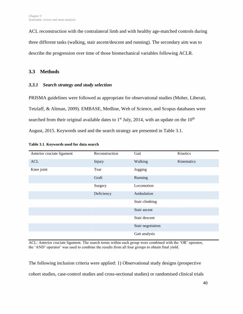
Chapter 3
Systematic review and meta-analysis
40
ACL reconstruction with the contralateral limb and with healthy age-matched controls during
three different tasks (walking, stair ascent/descent and running). The secondary aim was to
describe the progression over time of those biomechanical variables following ACLR.
3.3 Methods
3.3.1 Search strategy and study selection
PRISMA guidelines were followed as appropriate for observational studies (Moher, Liberati,
Tetzlaff, & Altman, 2009). EMBASE, Medline, Web of Science, and Scopus databases were
searched from their original available dates to 1st July, 2014, with an update on the 10th
August, 2015. Keywords used and the search strategy are presented in Table 3.1.
Table 3.1. Keywords used for data search
Anterior cruciate ligament Reconstruction Gait Kinetics
ACL Injury Walking Kinematics
Knee joint Tear Jogging
Graft Running
Surgery Locomotion
Deficiency Ambulation
Stair climbing
Stair ascent
Stair descent
Stair negotiation
Gait analysis
ACL: Anterior cruciate ligament. The search terms within each group were combined with the ‘OR’ operator,
the ‘AND’ operator’ was used to combine the results from all four groups to obtain final yield.
The following inclusion criteria were applied: 1) Observational study designs (prospective
cohort studies, case-control studies and cross-sectional studies) or randomised clinical trials
Chapter 3
Systematic review and meta-analysis
41
(RCT) with the comparison to control group or comparison with the contralateral uninjured
side at baseline; 2) men and women, with isolated ACL ruptures or in addition to menisci and
other ligamentous injury of the knee, managed with reconstructions with any type of graft; 3)
studies estimating kinematic and kinetic data (discrete variables: peak angles in the three
planes of movement, peak external knee flexion and adduction moments) of the ACLR knee
and the asymptomatic controls during walking, stair negotiation, or running. Studies focusing
on other lower limb injuries, or other tasks, such as jumping or pivoting were excluded. No
language restrictions were applied. Two reviewers (MK and GS) screened all relevant titles
and abstracts and were blinded to authors and journals. Full-text articles were screened
according to the pre-set inclusion and exclusion criteria by the two reviewers. Any
discrepancies were resolved through consensus, and a third reviewer was available if needed
(DR). A manual search of the reference lists of the included articles was also conducted.
2.2 Risk of bias within included studies
A modified version of Downs and Black Quality Index (Downs & Black, 1998) was used to
assess the risk of bias within included studies. The modified checklist included 16 questions
from the following sub-groups: reporting (items 1, 2, 3, 5, 6, 7 and 10), external validity
(items 11 and 12), internal validity-bias (item 15, 16, 18 and 20), internal validity-
confounding (selection bias) (item 21 and 25), and power (item 27) (Appendix –A1 Modified
Downs and Black quality index). Item 25 relates to confounding factors: walking or running
speed were considered as the most relevant confounders, as these variables have been shown
to be correlated with external knee adduction and flexion moments (Anne Mündermann,
Dyrby, Hurwitz, Sharma, & Andriacchi, 2004). Thus, a score of “1” was applied if the speed
Chapter 3
Systematic review and meta-analysis
42
of ambulation was not significantly different between participant groups, or if appropriate
statistical analyses were performed, including speed as a covariate. Item 27 was reworded as
“Were appropriate power calculations reported?” and the score was changed from 0 to 5 to a
scale of 0 or 1. The study was assigned a score of “1” if prior power analysis was performed,
with no scores being assigned in the absence of power analysis. The risk of bias assessment
was performed independently by two reviewers (MK and GS), with discrepancies resolved
during a consensus meeting. Kappa’s correlation coefficient was calculated to determine the
agreement of the scores of the individual questions for each study between the two reviewers.
The modified Downs and Black Quality Index was scored out of 17. Studies with scores of
11 and above (65%) were considered to have a low risk of bias, and studies with scores
below 11 were considered to have high risk of bias (Barton, Lack, Malliaras, & Morrissey,
2013).
3.3.2 Data extraction and meta-analysis
Data extracted from each study included sample size, participants’ demographics, type of
functional activities, and discrete variables for knee kinematics and kinetics (i.e. peak angles
and moments during the stance phase of gait). For external adduction moments, the first of
the two peaks was extracted. Peak knee tibial rotations were reported during the stance phase
of gait. The corresponding authors were contacted when relevant data were not presented or
were published as graphs.
Meta-analysis was performed using Review Manager v5.3 (RevMan) (Copenhagen: The
Nordic Cochrane Centre, The Cochrane Collaboration, 2014) when more than one study
investigated the same dependent variable during a given task using three-dimensional
Chapter 3
Systematic review and meta-analysis
43
movement analysis. The main aim of the forest plot was to determine (standardised) mean
differences and confidence intervals. Effect sizes (ES) and 95% confidence intervals (CI)
were calculated for between-group (ACLR groups versus uninjured Control groups) and
within-group (injured sides versus contralateral uninjured sides of the ACLR groups)
comparisons of kinematic data and kinetic data. Calculated individual or pooled ES were
categorised as small (<0.5), medium (≥0.5 and <0.8) or large (≥0.8) (Cohen, 1988). The level
of statistical heterogeneity for pooled data was established using γ2 and I2 statistics
(heterogeneity defined as p<0.05). For peak joint angles, mean differences (MD) (net
difference in peak angle among any two groups) were also calculated in degrees for findings
of individual studies or pooled data from two or more studies, as these may be more easily
translated to clinical practice than ES.
To conduct between- and within-group comparisons of the biomechanical variables over
time, the included studies were ordered in the forest plots according to ‘time since surgery’
(in weeks, months, or years). The shortest time since surgery was presented at the top, and
the longest average time since surgery was presented at the bottom of the forest plot. This
approach allowed a qualitative analysis of differences in biomechanical variables between-
and within-groups over time.
3.3.3 Level of evidence
The level of evidence was defined for each variable as per Van Tulder, Furlan, Bombardier,
Bouter, and Group (2003) as follows: (1) Strong evidence provided by pooled results derived
from three or more studies, including minimum of two studies with low risk of bias, which
are homogenous (p > 0.05); the analysis may be associated with a statistically significant or
Chapter 3
Systematic review and meta-analysis
44
non-significant pooled results; (2) Moderate evidence provided by statistically significant
pooled results derived from multiple studies, including at least one study with low risk of
bias, which are statistically heterogeneous (p < 0.05); or from multiple studies with high risk
of bias which are statistically homogenous (p > 0.05); (3) Limited evidence provided by
results from multiple studies with high risk of bias which are statistically heterogeneous (p <
0.05), or from one study with low risk of bias; (4) Very limited evidence provided by results
from one study with high risk of bias; (5) Conflicting evidence provided by pooled results
insignificant and derived from multiple studies regardless of quality which are statistically
heterogeneous (p < 0.05, i.e., inconsistent).
3.4 Results
3.4.1 Search results
The first search yielded 1,550 articles (Figure 3.1) and 65 remained after excluding
duplicates, irrelevant titles and screening of abstracts. A further 25 studies were excluded
based on the inclusion and exclusion criteria, giving a final yield of 40 studies which were
included in the qualitative analysis. Thirty-seven studies were in English, two in German
(Schmalz, Blumentritt, Wagner, & Gokeler, 1998; Schmalz, Blumentritt, Wagner, & Junge,
1998) and one in Chinese (Wang et al., 2009). Data from 27 studies could be included in
meta-analyses.
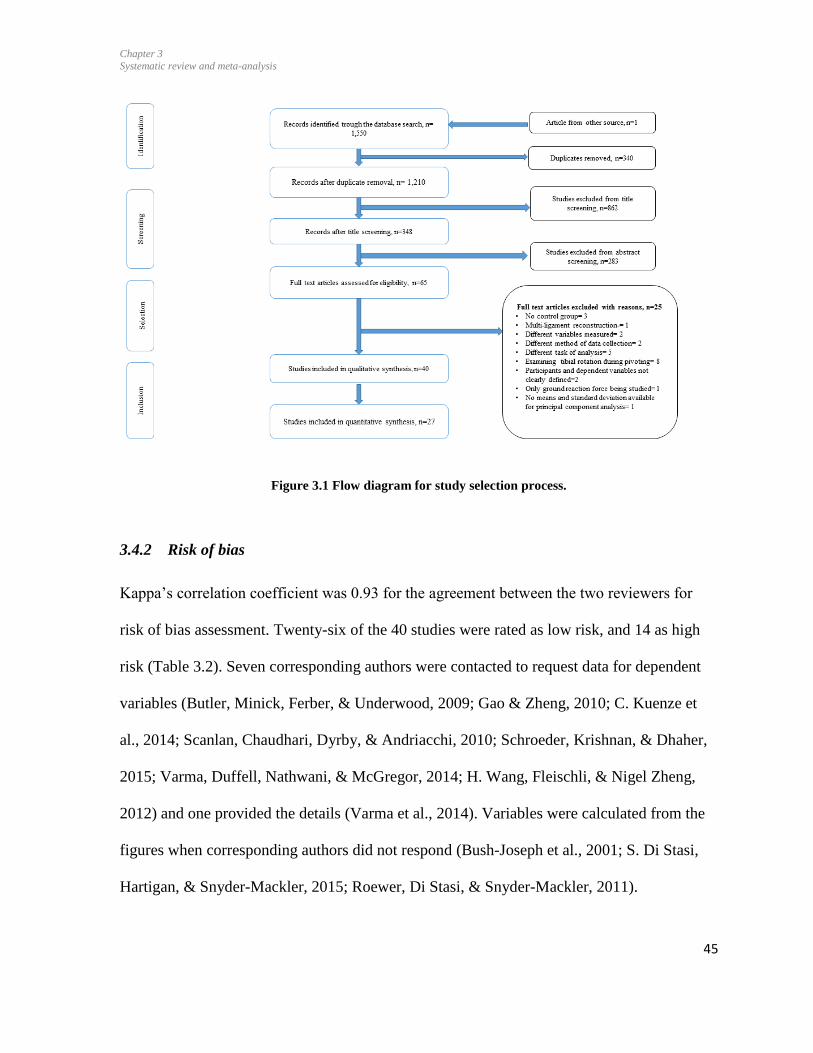
Chapter 3
Systematic review and meta-analysis
45
Figure 3.1 Flow diagram for study selection process.
3.4.2 Risk of bias
Kappa’s correlation coefficient was 0.93 for the agreement between the two reviewers for
risk of bias assessment. Twenty-six of the 40 studies were rated as low risk, and 14 as high
risk (Table 3.2). Seven corresponding authors were contacted to request data for dependent
variables (Butler, Minick, Ferber, & Underwood, 2009; Gao & Zheng, 2010; C. Kuenze et
al., 2014; Scanlan, Chaudhari, Dyrby, & Andriacchi, 2010; Schroeder, Krishnan, & Dhaher,
2015; Varma, Duffell, Nathwani, & McGregor, 2014; H. Wang, Fleischli, & Nigel Zheng,
2012) and one provided the details (Varma et al., 2014). Variables were calculated from the
figures when corresponding authors did not respond (Bush-Joseph et al., 2001; S. Di Stasi,
Hartigan, & Snyder-Mackler, 2015; Roewer, Di Stasi, & Snyder-Mackler, 2011).
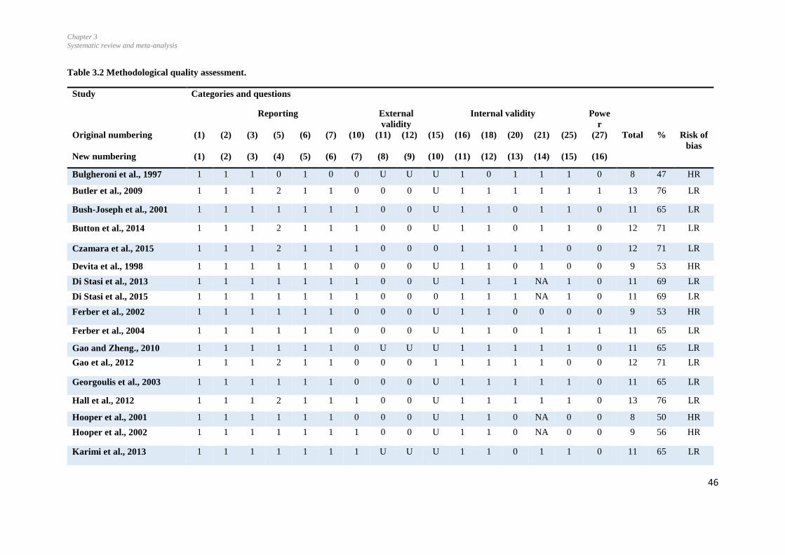
Chapter 3
Systematic review and meta-analysis
46
Table 3.2 Methodological quality assessment.
Study Categories and questions
Reporting External
validity
Internal validity Powe
r
Original numbering (1) (2) (3) (5) (6) (7) (10) (11) (12) (15) (16) (18) (20) (21) (25) (27) Total % Risk of
bias
New numbering (1) (2) (3) (4) (5) (6) (7) (8) (9) (10) (11) (12) (13) (14) (15) (16)
Bulgheroni et al., 1997 1 1 1 0 1 0 0 U U U 1 0 1 1 1 0 8 47 HR
Butler et al., 2009 1 1 1 2 1 1 0 0 0 U 1 1 1 1 1 1 13 76 LR
Bush-Joseph et al., 2001 1 1 1 1 1 1 1 0 0 U 1 1 0 1 1 0 11 65 LR
Button et al., 2014 1 1 1 2 1 1 1 0 0 U 1 1 0 1 1 0 12 71 LR
Czamara et al., 2015 1 1 1 2 1 1 1 0 0 0 1 1 1 1 0 0 12 71 LR
Devita et al., 1998 1 1 1 1 1 1 0 0 0 U 1 1 0 1 0 0 9 53 HR
Di Stasi et al., 2013 1 1 1 1 1 1 1 0 0 U 1 1 1 NA 1 0 11 69 LR
Di Stasi et al., 2015 1 1 1 1 1 1 1 0 0 0 1 1 1 NA 1 0 11 69 LR
Ferber et al., 2002 1 1 1 1 1 1 0 0 0 U 1 1 0 0 0 0 9 53 HR
Ferber et al., 2004 1 1 1 1 1 1 0 0 0 U 1 1 0 1 1 1 11 65 LR
Gao and Zheng., 2010 1 1 1 1 1 1 0 U U U 1 1 1 1 1 0 11 65 LR
Gao et al., 2012 1 1 1 2 1 1 0 0 0 1 1 1 1 1 0 0 12 71 LR
Georgoulis et al., 2003 1 1 1 1 1 1 0 0 0 U 1 1 1 1 1 0 11 65 LR
Hall et al., 2012 1 1 1 2 1 1 1 0 0 U 1 1 1 1 1 0 13 76 LR
Hooper et al., 2001 1 1 1 1 1 1 0 0 0 U 1 1 0 NA 0 0 8 50 HR
Hooper et al., 2002 1 1 1 1 1 1 1 0 0 U 1 1 0 NA 0 0 9 56 HR
Karimi et al., 2013 1 1 1 1 1 1 1 U U U 1 1 0 1 1 0 11 65 LR

Chapter 3
Systematic review and meta-analysis
47
Study Categories and questions
Reporting External
validity
Internal validity Powe
r
Original numbering (1) (2) (3) (5) (6) (7) (10) (11) (12) (15) (16) (18) (20) (21) (25) (27) Total % Risk of
bias
New numbering (1) (2) (3) (4) (5) (6) (7) (8) (9) (10) (11) (12) (13) (14) (15) (16)
Knoll 2004 et al., 1 1 1 1 1 1 0 0 0 U 1 1 0 0 1 0 9 53 HR
Kowalk et al., 1997 1 1 1 1 1 1 1 0 0 U 1 1 1 1 0 0 11 65 LR
Kuenze et al., 2013 1 1 1 2 1 1 1 0 0 0 1 1 1 1 1 1 14 82 LR
Lewek et al., 2002 1 1 1 1 1 1 1 0 0 U 1 1 0 0 1 0 10 59 HR
Noehren et al.,2013 1 1 1 2 1 1 1 0 0 U 1 1 0 1 1 0 12 71 LR
Patterson et al., 2013 1 1 1 2 1 1 1 0 0 U 1 1 1 1 0 0 12 71 LR
Roewer et al., 2011 1 1 1 1 1 1 1 0 0 U 1 1 1 NA 1 0 10 69 LR
Sato et al., 2013 1 1 1 2 1 1 0 0 0 1 1 1 1 1 0 0 12 69 LR
Scanlan et al., 2010 1 1 1 1 1 1 0 0 0 0 1 1 1 1 0 0 10 56 HR
Schmalz et al., 1998 1 1 1 1 0 0 0 0 0 0 1 1 0 1 0 0 7 41 HR
Schmalz et al., 1998 1 1 1 1 0 0 0 0 0 0 1 1 0 1 1 0 8 47 HR
Schroeder et al., 2015 1 1 1 1 1 1 1 0 0 0 1 1 0 1 0 0 10 59 HR
Tellini et al., 2013 1 1 1 2 1 1 1 0 0 U 1 1 0 1 0 0 10 59 HR
Timoney et al., 1993 1 1 1 1 1 1 0 0 0 U 1 1 0 1 1 0 10 59 HR
Varma et al., 2014 1 1 1 2 1 1 1 0 0 U 1 1 0 1 1 0 12 71 LR
Wang et al., 2009 1 1 1 1 1 0 0 0 0 U 1 1 0 1 1 0 10 63 HR
Wang et al., 2012 1 1 1 1 1 1 1 0 0 U 1 1 1 1 0 0 11 65 LR
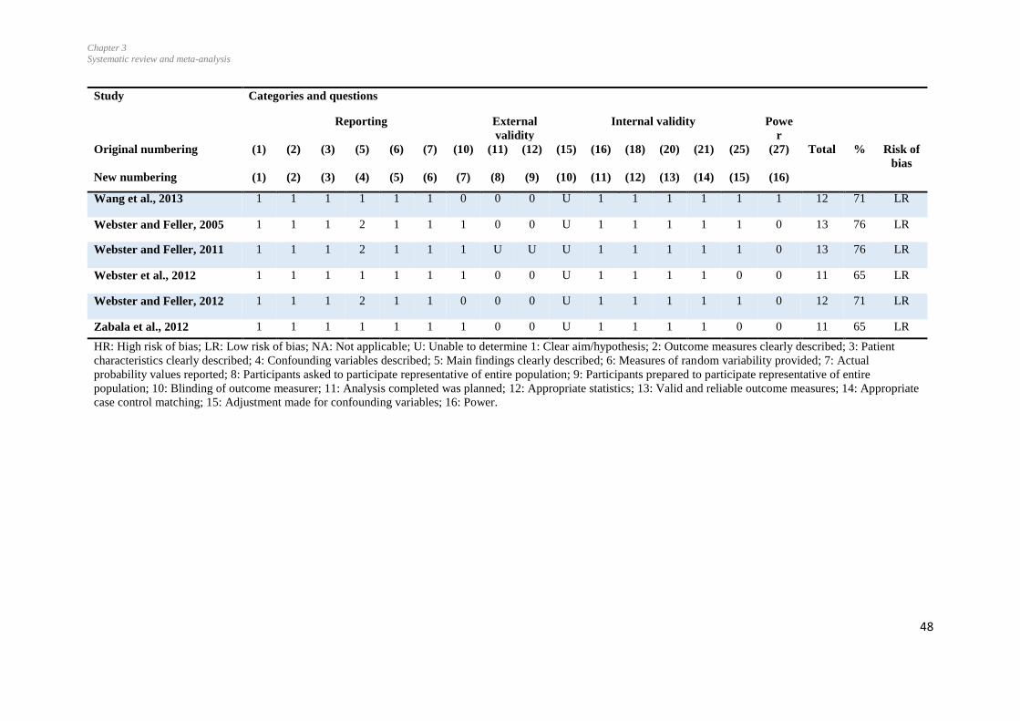
Chapter 3
Systematic review and meta-analysis
48
Study Categories and questions
Reporting External
validity
Internal validity Powe
r
Original numbering (1) (2) (3) (5) (6) (7) (10) (11) (12) (15) (16) (18) (20) (21) (25) (27) Total % Risk of
bias
New numbering (1) (2) (3) (4) (5) (6) (7) (8) (9) (10) (11) (12) (13) (14) (15) (16)
Wang et al., 2013 1 1 1 1 1 1 0 0 0 U 1 1 1 1 1 1 12 71 LR
Webster and Feller, 2005 1 1 1 2 1 1 1 0 0 U 1 1 1 1 1 0 13 76 LR
Webster and Feller, 2011 1 1 1 2 1 1 1 U U U 1 1 1 1 1 0 13 76 LR
Webster et al., 2012 1 1 1 1 1 1 1 0 0 U 1 1 1 1 0 0 11 65 LR
Webster and Feller, 2012 1 1 1 2 1 1 0 0 0 U 1 1 1 1 1 0 12 71 LR
Zabala et al., 2012 1 1 1 1 1 1 1 0 0 U 1 1 1 1 0 0 11 65 LR
HR: High risk of bias; LR: Low risk of bias; NA: Not applicable; U: Unable to determine 1: Clear aim/hypothesis; 2: Outcome measures clearly described; 3: Patient
characteristics clearly described; 4: Confounding variables described; 5: Main findings clearly described; 6: Measures of random variability provided; 7: Actual
probability values reported; 8: Participants asked to participate representative of entire population; 9: Participants prepared to participate representative of entire
population; 10: Blinding of outcome measurer; 11: Analysis completed was planned; 12: Appropriate statistics; 13: Valid and reliable outcome measures; 14: Appropriate
case control matching; 15: Adjustment made for confounding variables; 16: Power.
Chapter 3
Systematic review and meta-analysis
49
3.4.3 Overview of included studies
Thirty-one studies included comparisons with asymptomatic age-matched subjects, and nine
with the contralateral knee of this ACLR participant group (Table 3.3). Kinematic and kinetic
variables were studied during walking in 37 studies at self-selected comfortable speed, during
stair ascent and descent in eight studies (Bush-Joseph et al., 2001; Gao, Cordova, & Zheng,
2012; Hall, Stevermer, & Gillette, 2012; Hooper, Morrissey, Drechsler, Morrissey, & King,
2001; Hooper et al., 2002; Kowalk, Duncan, McCue, & Vaughan, 1997; Schroeder et al.,
2015; Zabala, Favre, Scanlan, Donahue, & Andriacchi, 2013), and during running in five
studies (Bush-Joseph et al., 2001; Kuenze et al., 2014; Lewek, Rudolph, Axe, & Snyder-
Mackler, 2002; Noehren, Wilson, Miller, & Lattermann, 2013; Sato et al., 2013). The number
of included participants ranged from 8 with ACLR and 10 controls (Kuenze et al., 2014;
Lewek et al., 2002), to 45 in both groups (Zabala et al., 2013), and time since surgery from 2
weeks (Hooper et al., 2002) to 18 years (Hall et al., 2012).
A RCT conducted by (Hooper et al., 2001) was included and, for the purpose of this review,
the baseline comparisons for the injured versus the contralateral uninjured knees were
considered for the meta-analysis. A longitudinal cross-sectional study by (Webster, Feller, &
Wittwer, 2012) was included with the results being reported at one year and 3.3 years. Data
from both of these time periods were included for meta-analyses.
Grand means and standard deviations (SD) were calculated (EMathZone) where studies
presented results for different groups with ACLR. In one study (Hooper et al., 2001), baseline
results of participants undergoing either closed or open kinetic training were combined.
Where studies compared outcomes of different surgical procedures, specifically, between
hamstring and patella tendon grafts (Webster & Feller, 2011; Webster, Wittwer, O'Brien, &
Chapter 3
Systematic review and meta-analysis
50
Feller, 2005), single- and double-bundle grafts (Czamara, Markowska, Królikowska, Szopa,
& Domagalska Szopa, 2015), and use of different portal techniques (Wang, Fleischli, &
Zheng, 2013), the results of the ACLR groups were combined and compared with the
controls. A study by (Lewek et al., 2002) included two groups of participants based on
quadriceps strength: (1) “strong ACLR” group; (2) “weak ACLR” group. The data of these
two groups were combined into a grand mean and SD. Furthermore, variables for participants
with ACLR for the dominant and non-dominant limbs were combined and compared with the
control group in a study by (Wang et al., 2012). Results of two groups reported by (Di Stasi,
Logerstedt, Gardinier, & Snyder-Mackler, 2013), participants who passed return to sports
criteria and those who did not pass, were also combined into grand means and SD, calculated
from the published 95% CI. In another study by (Di Stasi et al., 2015), means and CI were
estimated from the given figure and grand mean and SD for injured and uninjured sides were
calculated, combining results of men and women. (Roewer et al., 2011) conducted a
longitudinal study, reporting data at 6 months and at 2 years post-reconstruction. CI were
estimated from the figure and SD were calculated. Data reported at both time periods were
included in the forest plots (Roewer et al., 2011). Also, among the included studies only two
studies performed the priori power calculations (Kuenze et al., 2014; Wang et al., 2013).

Chapter 3
Systematic review and meta-analysis
51
Table 3.3 Study characteristics and variables measures at knee in included studies
Study
ACLR group
Characteristics
Control group
Characteristics
Methodology of data
collection
Task and measurable variable
Bulgheroni
et al., 1997
Participants: 15 (M)
Mean (SD) age: 25 (3) years
Time between injury and
surgery: NR
Surgical procedure : BPTB
Time since surgery: 17 (5)
months
5 (M)
28 (3) years
4 cameras, 100 Hz
Kistler force plate, 500 Hz
Walking: Natural cadence (112 ± 5.1 steps/min); footwear
condition not defined.
Average linear envelopes only.
Kinematics: flexion/extension; adduction/abduction;
internal/external rotation.
Kinetics: external moments
flexion/extension; adduction/abduction; internal/external
rotation.
Ground reaction forces: Fz, Fx, Fy
Bush-Joseph
et al., 2001
Participants: 22 (13M:9F)
Mean age (SD): 27 (11) years
Same weight in both groups
Time between injury and
surgery: 8 months
Surgical procedure: BPTB
Time since surgery: 22 (12)
months
22 (13M:9F)
29 (8) years
Same weight in both
groups
Multi-camera
optoelectronic
marker
Force plate
Walking: Speed 1.18 (0.018) m/s in ACLR and 1.16 (0.016)
m/s in Control group; foot wear condition not specified.
Kinetics: external moment.
flexion and extension.
Stair climbing
Kinetics: Peak external flexion moment.
Running Speed 2.70 (0.39) m/s in ACLR and 2.58 (0.042) m/s
in Control group.
Kinetics: external flexion moment.
Butler et al.,
2008
Participants: 17 (4M:13F)
Mean (SD) age: 23.6 (5.8) years
24.0 (3.2) kg
Time between injury and
surgery: NR
Surgical procedure : NR
Time since surgery: 5.3 (4.4)
years
No. not reported
23.4 (5.7) years
23.3 (2.4) kg
8-camera motion analysis
system, 80 Hz
Force plate, 1050 Hz
Walking: “Intentional” walking speed, monitored by average
velocity of L5/S1 marker; neutral laboratory shoes.
Kinematics: peak adduction during stance; total
adduction/abduction excursion (range).
Kinetics: internal moments
peak abduction moment.
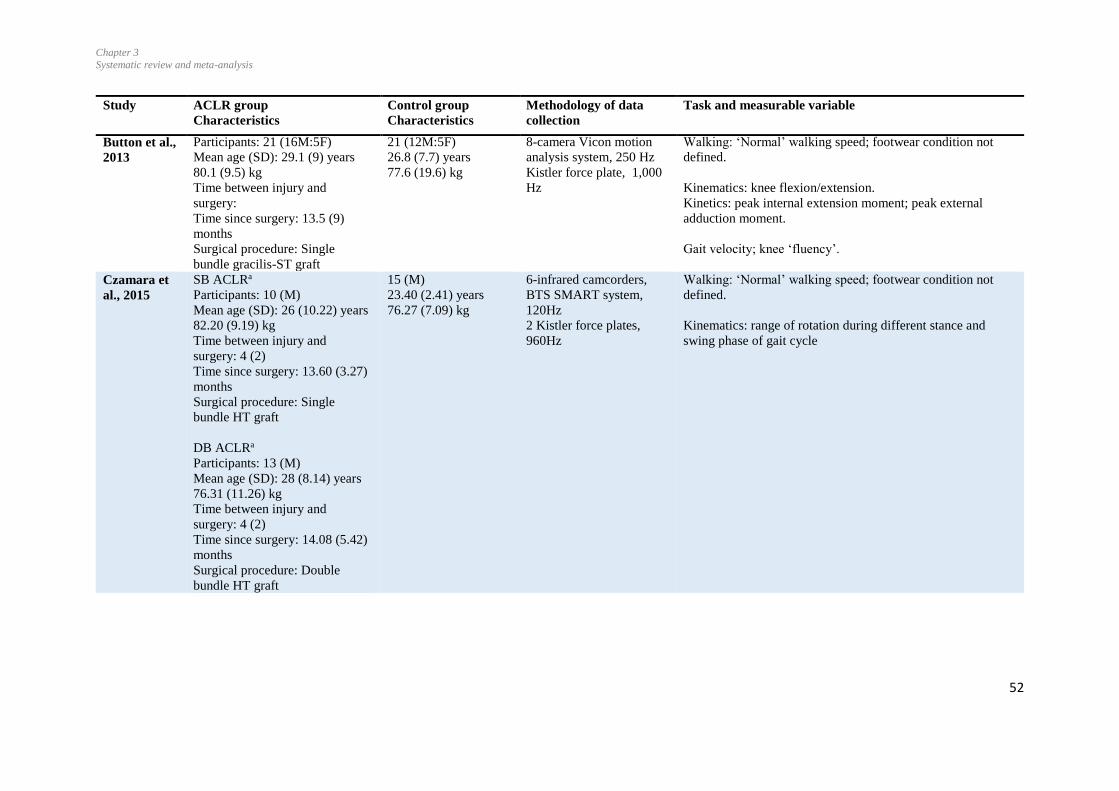
Chapter 3
Systematic review and meta-analysis
52
Study
ACLR group
Characteristics
Control group
Characteristics
Methodology of data
collection
Task and measurable variable
Button et al.,
2013
Participants: 21 (16M:5F)
Mean age (SD): 29.1 (9) years
80.1 (9.5) kg
Time between injury and
surgery:
Time since surgery: 13.5 (9)
months
Surgical procedure: Single
bundle gracilis-ST graft
21 (12M:5F)
26.8 (7.7) years
77.6 (19.6) kg
8-camera Vicon motion
analysis system, 250 Hz
Kistler force plate, 1,000
Hz
Walking: ‘Normal’ walking speed; footwear condition not
defined.
Kinematics: knee flexion/extension.
Kinetics: peak internal extension moment; peak external
adduction moment.
Gait velocity; knee ‘fluency’.
Czamara et
al., 2015
SB ACLRa
Participants: 10 (M)
Mean age (SD): 26 (10.22) years
82.20 (9.19) kg
Time between injury and
surgery: 4 (2)
Time since surgery: 13.60 (3.27)
months
Surgical procedure: Single
bundle HT graft
DB ACLRa
Participants: 13 (M)
Mean age (SD): 28 (8.14) years
76.31 (11.26) kg
Time between injury and
surgery: 4 (2)
Time since surgery: 14.08 (5.42)
months
Surgical procedure: Double
bundle HT graft
15 (M)
23.40 (2.41) years
76.27 (7.09) kg
6-infrared camcorders,
BTS SMART system,
120Hz
2 Kistler force plates,
960Hz
Walking: ‘Normal’ walking speed; footwear condition not
defined.
Kinematics: range of rotation during different stance and
swing phase of gait cycle
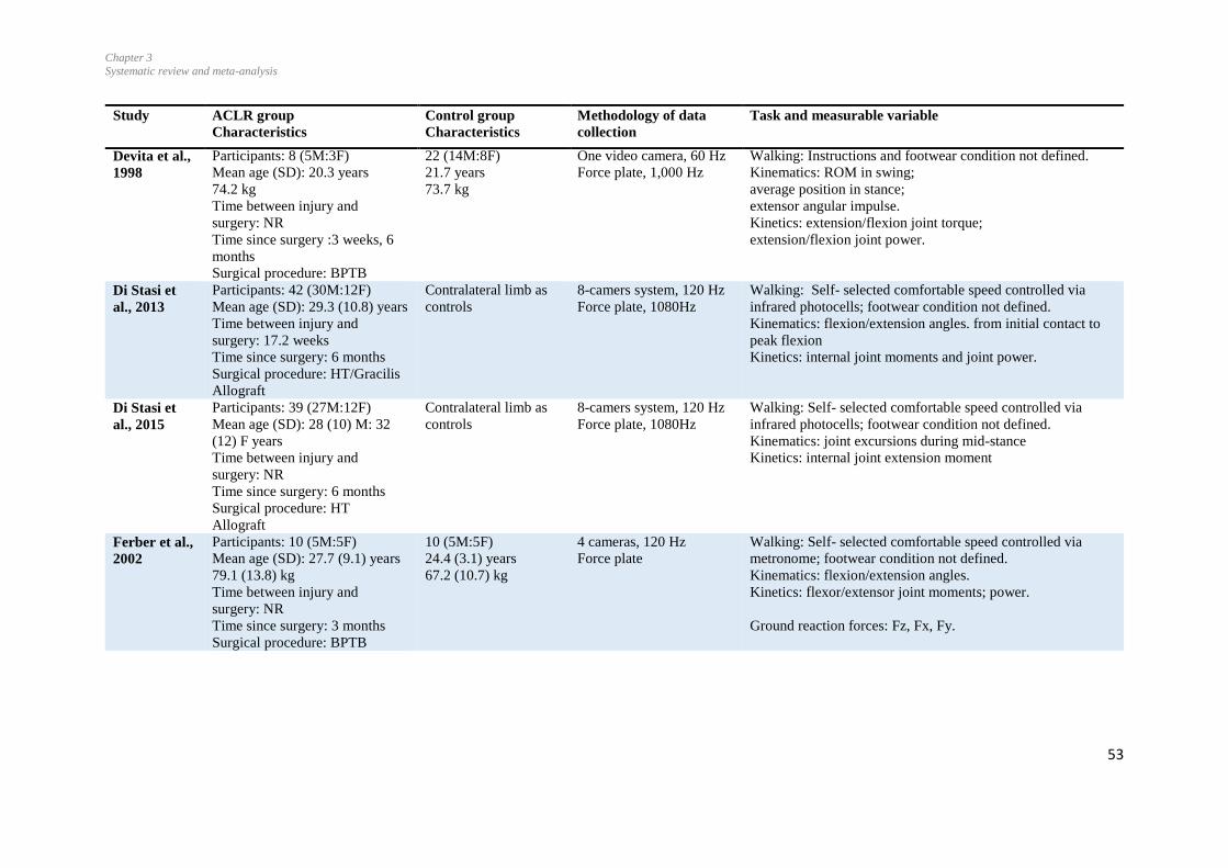
Chapter 3
Systematic review and meta-analysis
53
Study
ACLR group
Characteristics
Control group
Characteristics
Methodology of data
collection
Task and measurable variable
Devita et al.,
1998
Participants: 8 (5M:3F)
Mean age (SD): 20.3 years
74.2 kg
Time between injury and
surgery: NR
Time since surgery :3 weeks, 6
months
Surgical procedure: BPTB
22 (14M:8F)
21.7 years
73.7 kg
One video camera, 60 Hz
Force plate, 1,000 Hz
Walking: Instructions and footwear condition not defined.
Kinematics: ROM in swing;
average position in stance;
extensor angular impulse.
Kinetics: extension/flexion joint torque;
extension/flexion joint power.
Di Stasi et
al., 2013
Participants: 42 (30M:12F)
Mean age (SD): 29.3 (10.8) years
Time between injury and
surgery: 17.2 weeks
Time since surgery: 6 months
Surgical procedure: HT/Gracilis
Allograft
Contralateral limb as
controls
8-camers system, 120 Hz
Force plate, 1080Hz
Walking: Self- selected comfortable speed controlled via
infrared photocells; footwear condition not defined.
Kinematics: flexion/extension angles. from initial contact to
peak flexion
Kinetics: internal joint moments and joint power.
Di Stasi et
al., 2015
Participants: 39 (27M:12F)
Mean age (SD): 28 (10) M: 32
(12) F years
Time between injury and
surgery: NR
Time since surgery: 6 months
Surgical procedure: HT
Allograft
Contralateral limb as
controls
8-camers system, 120 Hz
Force plate, 1080Hz
Walking: Self- selected comfortable speed controlled via
infrared photocells; footwear condition not defined.
Kinematics: joint excursions during mid-stance
Kinetics: internal joint extension moment
Ferber et al.,
2002
Participants: 10 (5M:5F)
Mean age (SD): 27.7 (9.1) years
79.1 (13.8) kg
Time between injury and
surgery: NR
Time since surgery: 3 months
Surgical procedure: BPTB
10 (5M:5F)
24.4 (3.1) years
67.2 (10.7) kg
4 cameras, 120 Hz
Force plate
Walking: Self- selected comfortable speed controlled via
metronome; footwear condition not defined.
Kinematics: flexion/extension angles.
Kinetics: flexor/extensor joint moments; power.
Ground reaction forces: Fz, Fx, Fy.
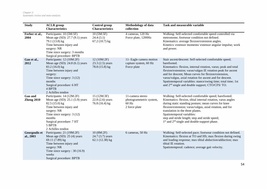
Chapter 3
Systematic review and meta-analysis
54
Study
ACLR group
Characteristics
Control group
Characteristics
Methodology of data
collection
Task and measurable variable
Ferber et al.,
2004
Participants: 10 (5M:5F)
Mean age (SD): 27.7 (9.1) years
79.1 (13.8) kg
Time between injury and
surgery: NR
Time since surgery: 3 months
Surgical procedure: BPTB
10 (5M:5F)
24.4 (3.1)
67.2 (10.7) kg
4 cameras, 120 Hz
Force plate, 1200Hz
Walking: Self-selected comfortable speed controlled via
metronome; footwear condition not defined.
Kinematics: average flexion/extension angles.
Kinetics: extensor moments/ extensor angular impulse; work
and power.
Gao et al.,
2012
Participants: 12 (10M:2F)
Mean age (SD): 24.8 (6.1) years
83.2 (16.0) kg
Time between injury and
surgery:
Time since surgery: 3 (12)
months
Surgical procedure: 6 HT
4 BPTB
2 Achilles tendon
12 (10M:2F)
23.3 (2.5) years
78.8 (15.8) kg
11- Eagle camera motion
capture system, 60 Hz
Force plate
Stair ascent/descent: Self-selected comfortable speed;
barefooted.
Kinematics: flexion, internal rotation, varus; peak and total
flexion/extension; varus/valgus IE rotation peak for ascent
and for descent; Mean curves for flexion/extension,
varus/valgus, axial rotation for ascent and for descent.
Spatiotemporal variables: stance/swing time; total time; 1st
and 2nd single and double support; CTO/CFS/ TO.
Gao and
Zheng 2010
Participants: 14 (12M:2F)
Mean age (SD): 25.1 (5.9) years
82.5 (15.0) kg
Time between injury and
surgery: NR
Time since surgery: 3 (12)
months
Surgical procedure: 7 HT
5 BPTB
2 Achilles tendon
15 (12M:3F)
22.8 (2.6) years
76.8 (16.4) kg
11-camera stereo
photogrammetric system,
60 Hz
2 force plate
Walking: Self-selected comfortable speed; barefooted.
Kinematics: flexion, tibial internal rotation, varus angles
during static standing posture; mean curves for knee
flexion/extension; varus/valgus, axial rotation, and for
translation in the three planes.
Spatiotemporal variables:
step and stride length; step and stride speed;
1st and 2nd single and double-support phase.
Georgoulis et
al., 2003
Participants: 21 (19M:2F)
Mean age (SD): 25 (4) years
69.11 (7.89) kg
Time between injury and
surgery: NR
Time since surgery : 30 (16.9)
weeks
Surgical procedure: BPTB
10 (8M:2F)
24.7 (3.7) years
62.1 (12.38) kg
6 cameras, 50 Hz
Walking: Self-selected pace; footwear condition not defined.
Kinematics: flexion at TO and HS; max flexion during swing
and loading response; max tibial abduction/adduction; max
tibial IE rotation.
Spatiotemporal: cadence; average gait velocity.

Chapter 3
Systematic review and meta-analysis
55
Study
ACLR group
Characteristics
Control group
Characteristics
Methodology of data
collection
Task and measurable variable
Hall et al.,
2012
Participants: 15 (7M:8F)
Mean age (SD): 26 (6) years
75 (15) kg
Time between injury and
surgery: NR
Time since surgery: 6 (2 -18)
years
Surgical procedure: 41% HT
41:Patellar tendon
12% cadaver + hamstring
6% HT
17 (7M:10F)
26 (4) years
65 (12) kg
8-Camera 3D motion
analysis system, 160 Hz
Force plate, 1600 Hz
Walking: Self- selected pace, with preferred shoes.
Kinematics: initial flexion angle.
Kinetics: peak internal extension moment; peak external varus
moment.
Stair ascent/descent:
Kinematics: initial flexion angle.
Kinetics: peak extension moment; peak varus moment.
Hooper et
al., 2001
CKC groupa
Participants: 18 (13M:5F)
Mean age (SD): NR
75.1 (12.3) kg
Time between injury and
surgery: 50.3 (61.8) months
Time since surgery: 2 weeks
Surgical procedure: BPTB
OKC groupa
Participants: 19 (16M:3F)
Mean age (SD): NR
77.4 (15.3) kg
Time between injury and
surgery: 34.1 (30.4) months
Time since surgery: 2 weeks
Surgical procedure: BPTB
Contralateral limb as
controls
3 cameras, 50 Hz
Force plate, 200 Hz
Walking: Gait speed and footwear not specified.
Kinematics: flexion at HS, TO, MS excursion.
Kinetics: concentric /eccentric energy; flexor/extensor
impulse; flexion/extension moment.
Stair ascent:
Kinematics: flexion at HS
Kinetics: peak extensor moment; extensor impulse; peak
concentric power; concentric energy.
Stair descent:
Kinematics: flexion at HS
Kinetics: peak extensor moment; extensor impulse;
peak eccentric power; eccentric energy.
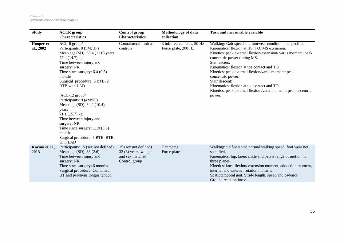
Chapter 3
Systematic review and meta-analysis
56
Study
ACLR group
Characteristics
Control group
Characteristics
Methodology of data
collection
Task and measurable variable
Hooper et
al., 2002
ACL-6 groupb
Participants: 8 (5M: 3F)
Mean age (SD): 33.4 (11.0) years
77.4 (14.7) kg
Time between injury and
surgery: NR
Time since surgery: 6 4 (0.5)
months
Surgical procedure: 6 BTB, 2
BTB with LAD
ACL-12 groupb
Participants: 9 (4M:5F)
Mean age (SD): 34.2 (10.4)
years
71.1 (15.7) kg
Time between injury and
surgery: NR
Time since surgery: 11.9 (0.6)
months
Surgical procedure: 5 BTB, BTB
with LAD
Contralateral limb as
controls
3 infrared cameras, 50 Hz
Force plate, 200 Hz
Walking: Gait speed and footwear condition not specified.
Kinematics: flexion at HS, TO; MS excursion.
Kinetics: peak external flexion/extension/ varus moment; peak
concentric power during MS.
Stair ascent:
Kinematics: flexion at toe contact and TO.
Kinetics: peak external flexion/varus moment; peak
concentric power.
Stair descent:
Kinematics: flexion at toe contact and TO.
Kinetics: peak external flexion /varus moment; peak eccentric
power.
Karimi et al.,
2013
Participants: 15 (sex not defined)
Mean age (SD): 33 (2.6)
Time between injury and
surgery: NR
Time since surgery: 6 months
Surgical procedure: Combined
HT and peroneus longus tendon
15 (sex not defined)
32 (3) years, weight
and sex matched
Control group
7 cameras
Force plate
Walking: Self-selected normal walking speed; foot wear not
specified.
Kinematics: hip, knee, ankle and pelvis range of motion in
three planes
Kinetics: knee flexion/ extension moment, adduction moment,
internal and external rotation moment
Spatiotemporal gait. Stride length, speed and cadence
Ground reaction force
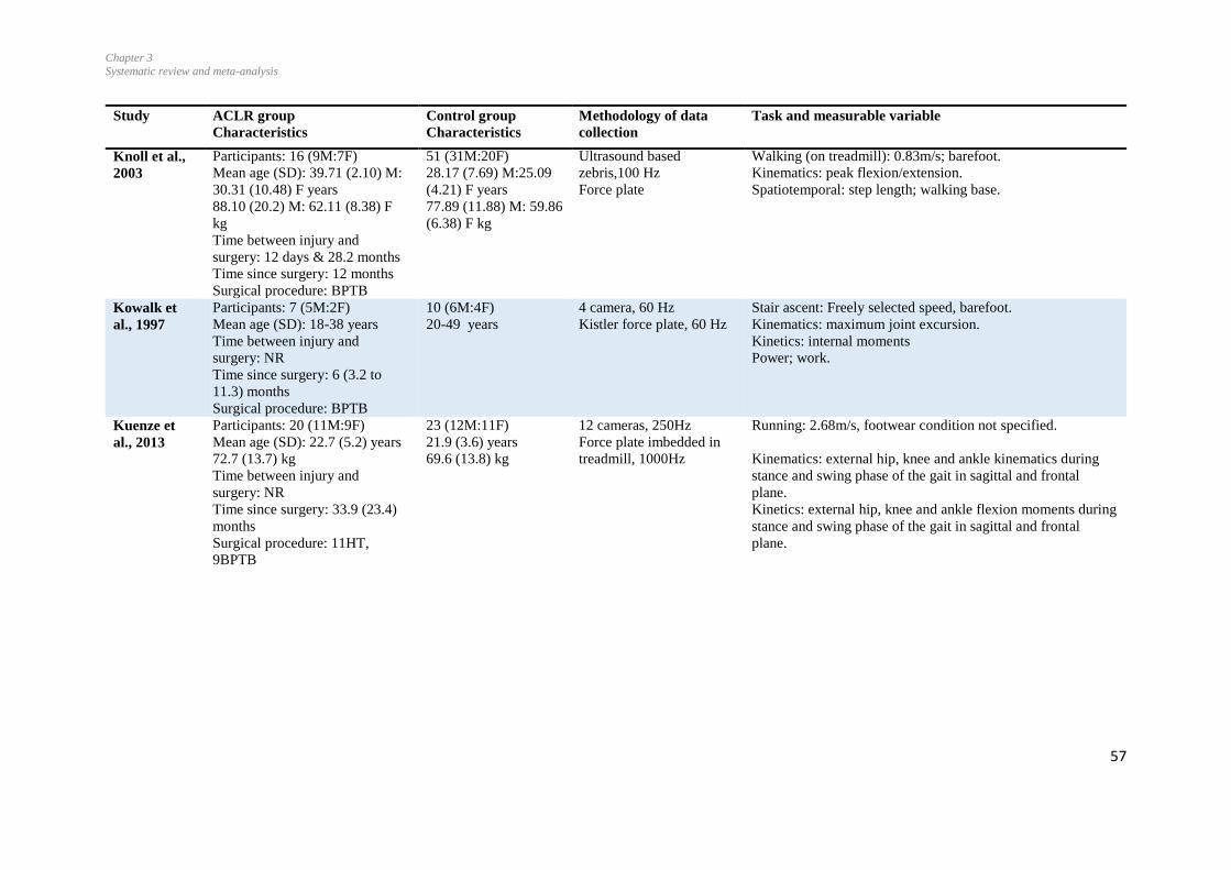
Chapter 3
Systematic review and meta-analysis
57
Study
ACLR group
Characteristics
Control group
Characteristics
Methodology of data
collection
Task and measurable variable
Knoll et al.,
2003
Participants: 16 (9M:7F)
Mean age (SD): 39.71 (2.10) M:
30.31 (10.48) F years
88.10 (20.2) M: 62.11 (8.38) F
kg
Time between injury and
surgery: 12 days & 28.2 months
Time since surgery: 12 months
Surgical procedure: BPTB
51 (31M:20F)
28.17 (7.69) M:25.09
(4.21) F years
77.89 (11.88) M: 59.86
(6.38) F kg
Ultrasound based
zebris,100 Hz
Force plate
Walking (on treadmill): 0.83m/s; barefoot.
Kinematics: peak flexion/extension.
Spatiotemporal: step length; walking base.
Kowalk et
al., 1997
Participants: 7 (5M:2F)
Mean age (SD): 18-38 years
Time between injury and
surgery: NR
Time since surgery: 6 (3.2 to
11.3) months
Surgical procedure: BPTB
10 (6M:4F)
20-49 years
4 camera, 60 Hz
Kistler force plate, 60 Hz
Stair ascent: Freely selected speed, barefoot.
Kinematics: maximum joint excursion.
Kinetics: internal moments
Power; work.
Kuenze et
al., 2013
Participants: 20 (11M:9F)
Mean age (SD): 22.7 (5.2) years
72.7 (13.7) kg
Time between injury and
surgery: NR
Time since surgery: 33.9 (23.4)
months
Surgical procedure: 11HT,
9BPTB
23 (12M:11F)
21.9 (3.6) years
69.6 (13.8) kg
12 cameras, 250Hz
Force plate imbedded in
treadmill, 1000Hz
Running: 2.68m/s, footwear condition not specified.
Kinematics: external hip, knee and ankle kinematics during
stance and swing phase of the gait in sagittal and frontal
plane.
Kinetics: external hip, knee and ankle flexion moments during
stance and swing phase of the gait in sagittal and frontal
plane.
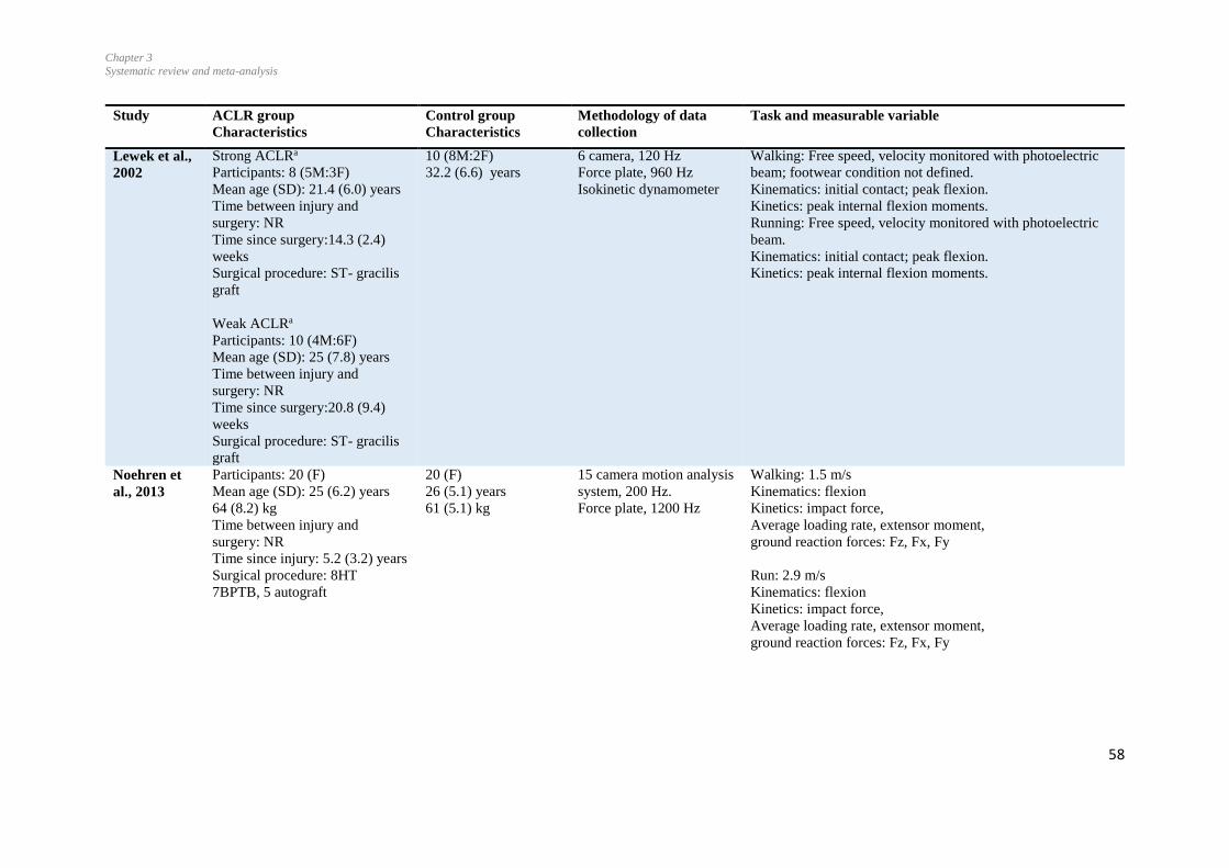
Chapter 3
Systematic review and meta-analysis
58
Study
ACLR group
Characteristics
Control group
Characteristics
Methodology of data
collection
Task and measurable variable
Lewek et al.,
2002
Strong ACLRa
Participants: 8 (5M:3F)
Mean age (SD): 21.4 (6.0) years
Time between injury and
surgery: NR
Time since surgery:14.3 (2.4)
weeks
Surgical procedure: ST- gracilis
graft
Weak ACLRa
Participants: 10 (4M:6F)
Mean age (SD): 25 (7.8) years
Time between injury and
surgery: NR
Time since surgery:20.8 (9.4)
weeks
Surgical procedure: ST- gracilis
graft
10 (8M:2F)
32.2 (6.6) years
6 camera, 120 Hz
Force plate, 960 Hz
Isokinetic dynamometer
Walking: Free speed, velocity monitored with photoelectric
beam; footwear condition not defined.
Kinematics: initial contact; peak flexion.
Kinetics: peak internal flexion moments.
Running: Free speed, velocity monitored with photoelectric
beam.
Kinematics: initial contact; peak flexion.
Kinetics: peak internal flexion moments.
Noehren et
al., 2013
Participants: 20 (F)
Mean age (SD): 25 (6.2) years
64 (8.2) kg
Time between injury and
surgery: NR
Time since injury: 5.2 (3.2) years
Surgical procedure: 8HT
7BPTB, 5 autograft
20 (F)
26 (5.1) years
61 (5.1) kg
15 camera motion analysis
system, 200 Hz.
Force plate, 1200 Hz
Walking: 1.5 m/s
Kinematics: flexion
Kinetics: impact force,
Average loading rate, extensor moment,
ground reaction forces: Fz, Fx, Fy
Run: 2.9 m/s
Kinematics: flexion
Kinetics: impact force,
Average loading rate, extensor moment,
ground reaction forces: Fz, Fx, Fy
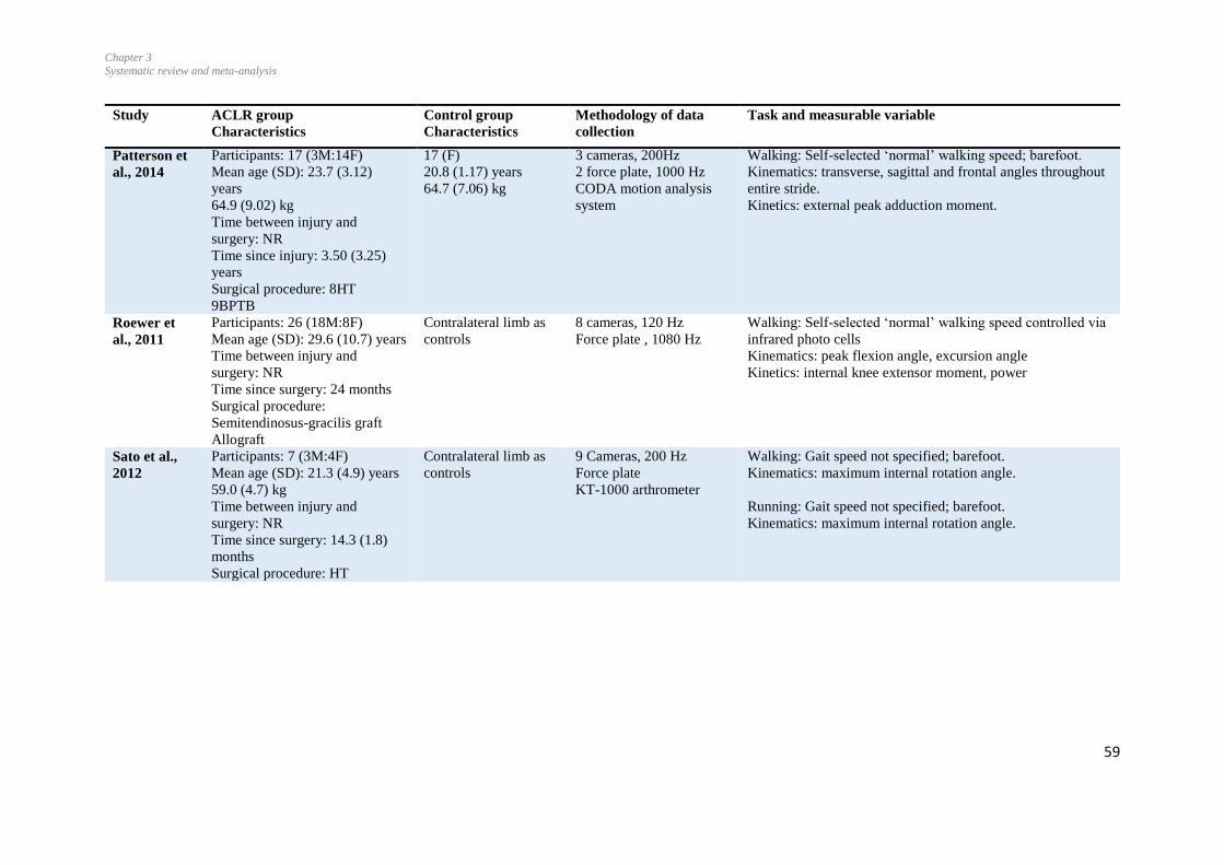
Chapter 3
Systematic review and meta-analysis
59
Study
ACLR group
Characteristics
Control group
Characteristics
Methodology of data
collection
Task and measurable variable
Patterson et
al., 2014
Participants: 17 (3M:14F)
Mean age (SD): 23.7 (3.12)
years
64.9 (9.02) kg
Time between injury and
surgery: NR
Time since injury: 3.50 (3.25)
years
Surgical procedure: 8HT
9BPTB
17 (F)
20.8 (1.17) years
64.7 (7.06) kg
3 cameras, 200Hz
2 force plate, 1000 Hz
CODA motion analysis
system
Walking: Self-selected ‘normal’ walking speed; barefoot.
Kinematics: transverse, sagittal and frontal angles throughout
entire stride.
Kinetics: external peak adduction moment.
Roewer et
al., 2011
Participants: 26 (18M:8F)
Mean age (SD): 29.6 (10.7) years
Time between injury and
surgery: NR
Time since surgery: 24 months
Surgical procedure:
Semitendinosus-gracilis graft
Allograft
Contralateral limb as
controls
8 cameras, 120 Hz
Force plate , 1080 Hz
Walking: Self-selected ‘normal’ walking speed controlled via
infrared photo cells
Kinematics: peak flexion angle, excursion angle
Kinetics: internal knee extensor moment, power
Sato et al.,
2012
Participants: 7 (3M:4F)
Mean age (SD): 21.3 (4.9) years
59.0 (4.7) kg
Time between injury and
surgery: NR
Time since surgery: 14.3 (1.8)
months
Surgical procedure: HT
Contralateral limb as
controls
9 Cameras, 200 Hz
Force plate
KT-1000 arthrometer
Walking: Gait speed not specified; barefoot.
Kinematics: maximum internal rotation angle.
Running: Gait speed not specified; barefoot.
Kinematics: maximum internal rotation angle.
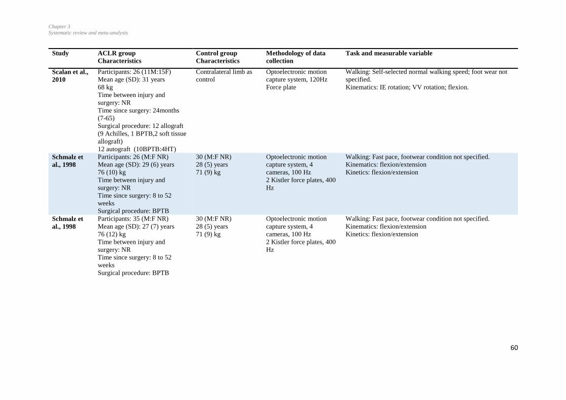
Chapter 3
Systematic review and meta-analysis
60
Study
ACLR group
Characteristics
Control group
Characteristics
Methodology of data
collection
Task and measurable variable
Scalan et al.,
2010
Participants: 26 (11M:15F)
Mean age (SD): 31 years
68 kg
Time between injury and
surgery: NR
Time since surgery: 24months
(7-65)
Surgical procedure: 12 allograft
(9 Achilles, 1 BPTB,2 soft tissue
allograft)
12 autograft (10BPTB:4HT)
Contralateral limb as
control
Optoelectronic motion
capture system, 120Hz
Force plate
Walking: Self-selected normal walking speed; foot wear not
specified.
Kinematics: IE rotation; VV rotation; flexion.
Schmalz et
al., 1998
Participants: 26 (M:F NR)
Mean age (SD): 29 (6) years
76 (10) kg
Time between injury and
surgery: NR
Time since surgery: 8 to 52
weeks
Surgical procedure: BPTB
30 (M:F NR)
28 (5) years
71 (9) kg
Optoelectronic motion
capture system, 4
cameras, 100 Hz
2 Kistler force plates, 400
Hz
Walking: Fast pace, footwear condition not specified.
Kinematics: flexion/extension
Kinetics: flexion/extension
Schmalz et
al., 1998
Participants: 35 (M:F NR)
Mean age (SD): 27 (7) years
76 (12) kg
Time between injury and
surgery: NR
Time since surgery: 8 to 52
weeks
Surgical procedure: BPTB
30 (M:F NR)
28 (5) years
71 (9) kg
Optoelectronic motion
capture system, 4
cameras, 100 Hz
2 Kistler force plates, 400
Hz
Walking: Fast pace, footwear condition not specified.
Kinematics: flexion/extension
Kinetics: flexion/extension
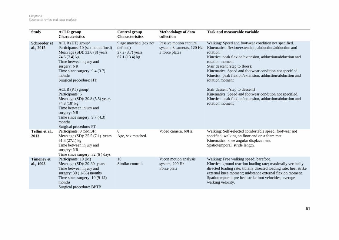
Chapter 3
Systematic review and meta-analysis
61
Study
ACLR group
Characteristics
Control group
Characteristics
Methodology of data
collection
Task and measurable variable
Schroeder et
al., 2015
ACLR (HT) groupa
Participants: 10 (sex not defined)
Mean age (SD): 32.6 (8) years
74.6 (7.4) kg
Time between injury and
surgery: NR
Time since surgery: 9.4 (3.7)
months
Surgical procedure: HT
ACLR (PT) groupa
Participants: 6
Mean age (SD): 30.8 (5.5) years
74.8 (18) kg
Time between injury and
surgery: NR
Time since surgery: 9.7 (4.3)
months
Surgical procedure: PT
9 age matched (sex not
defined)
27.2 (3.7) years
67.1 (13.4) kg
Passive motion capture
system, 8 cameras, 120 Hz
3 force plates
Walking: Speed and footwear condition not specified.
Kinematics: flexion/extension, abduction/adduction and
rotation.
Kinetics: peak flexion/extension, adduction/abduction and
rotation moment
Stair descent (step to floor):
Kinematics: Speed and footwear condition not specified.
Kinetics: peak flexion/extension, adduction/abduction and
rotation moment
Stair descent (step to descent)
Kinematics: Speed and footwear condition not specified.
Kinetics: peak flexion/extension, adduction/abduction and
rotation moment
Tellini et al.,
2013
Participants: 8 (5M:3F)
Mean age (SD): 25.5 (7.1) years
61.3 (27.1) kg
Time between injury and
surgery: NR
Time since surgery: 32 (6 ) days
8
Age, sex matched.
Video camera, 60Hz
Walking: Self-selected comfortable speed; footwear not
specified; walking on floor and on a foam mat
Kinematics: knee angular displacement.
Spatiotemporal: stride length.
Timoney et
al., 1993
Participants: 10 (M)
Mean age (SD): 20-30 years
Time between injury and
surgery: 30 ( 1-66) months
Time since surgery: 10 (9-12)
months
Surgical procedure: BPTB
10
Similar controls
Vicon motion analysis
system, 200 Hz
Force plate
Walking: Free walking speed; barefoot.
Kinetics: ground reaction loading rate; maximally vertically
directed loading rate; tibially directed loading rate; heel strike
external knee moment; midstance external flexion moment.
Spatiotemporal: pre heel strike foot velocities; average
walking velocity.
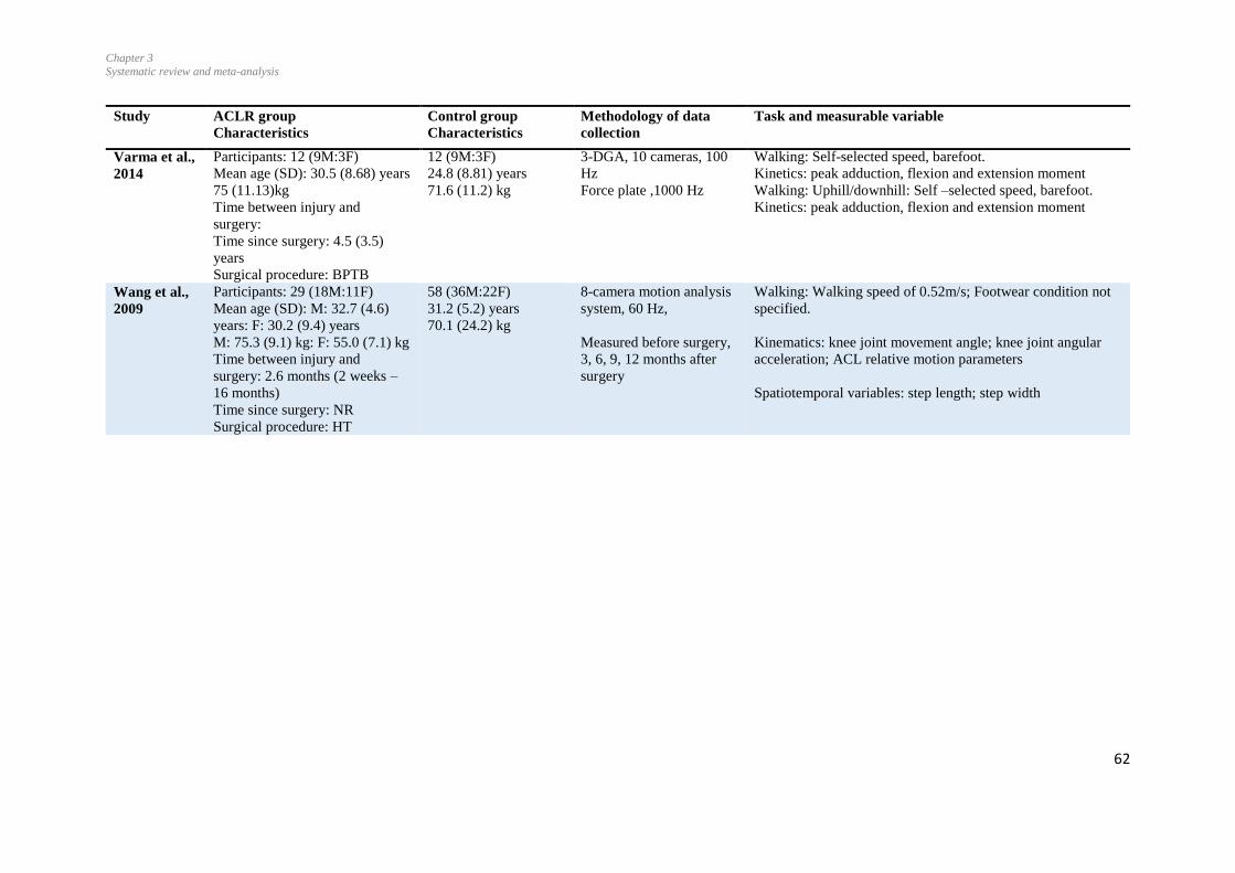
Chapter 3
Systematic review and meta-analysis
62
Study
ACLR group
Characteristics
Control group
Characteristics
Methodology of data
collection
Task and measurable variable
Varma et al.,
2014
Participants: 12 (9M:3F)
Mean age (SD): 30.5 (8.68) years
75 (11.13)kg
Time between injury and
surgery:
Time since surgery: 4.5 (3.5)
years
Surgical procedure: BPTB
12 (9M:3F)
24.8 (8.81) years
71.6 (11.2) kg
3-DGA, 10 cameras, 100
Hz
Force plate ,1000 Hz
Walking: Self-selected speed, barefoot.
Kinetics: peak adduction, flexion and extension moment
Walking: Uphill/downhill: Self –selected speed, barefoot.
Kinetics: peak adduction, flexion and extension moment
Wang et al.,
2009
Participants: 29 (18M:11F)
Mean age (SD): M: 32.7 (4.6)
years: F: 30.2 (9.4) years
M: 75.3 (9.1) kg: F: 55.0 (7.1) kg
Time between injury and
surgery: 2.6 months (2 weeks –
16 months)
Time since surgery: NR
Surgical procedure: HT
58 (36M:22F)
31.2 (5.2) years
70.1 (24.2) kg
8-camera motion analysis
system, 60 Hz,
Measured before surgery,
3, 6, 9, 12 months after
surgery
Walking: Walking speed of 0.52m/s; Footwear condition not
specified.
Kinematics: knee joint movement angle; knee joint angular
acceleration; ACL relative motion parameters
Spatiotemporal variables: step length; step width
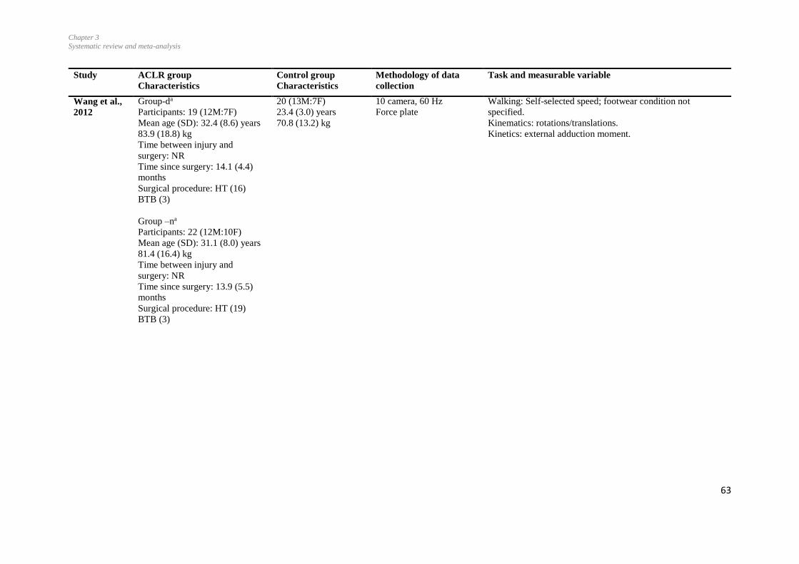
Chapter 3
Systematic review and meta-analysis
63
Study
ACLR group
Characteristics
Control group
Characteristics
Methodology of data
collection
Task and measurable variable
Wang et al.,
2012
Group-da
Participants: 19 (12M:7F)
Mean age (SD): 32.4 (8.6) years
83.9 (18.8) kg
Time between injury and
surgery: NR
Time since surgery: 14.1 (4.4)
months
Surgical procedure: HT (16)
BTB (3)
Group –na
Participants: 22 (12M:10F)
Mean age (SD): 31.1 (8.0) years
81.4 (16.4) kg
Time between injury and
surgery: NR
Time since surgery: 13.9 (5.5)
months
Surgical procedure: HT (19)
BTB (3)
20 (13M:7F)
23.4 (3.0) years
70.8 (13.2) kg
10 camera, 60 Hz
Force plate
Walking: Self-selected speed; footwear condition not
specified.
Kinematics: rotations/translations.
Kinetics: external adduction moment.
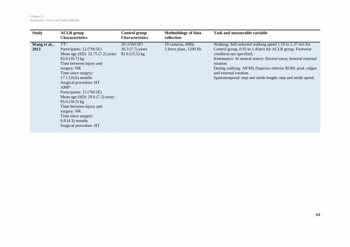
Chapter 3
Systematic review and meta-analysis
64
Study
ACLR group
Characteristics
Control group
Characteristics
Methodology of data
collection
Task and measurable variable
Wang et al.,
2013
TTa
Participants: 12 (7M:5F)
Mean age (SD): 32.75 (7.2) years
83.0 (16.7) kg
Time between injury and
surgery: NR
Time since surgery:
17.1 (10.6) months
Surgical procedure: HT
AMPa
Participants: 12 (7M:5F)
Mean age (SD): 29.6 (7.2) years
85.6 (16.5) kg
Time between injury and
surgery: NR
Time since surgery:
8.8 (4.3) months
Surgical procedure: HT
20 (15M:5F)
26.3 (7.7) years
81.6 (15.5) kg
10 cameras, 60Hz
2 force plate, 1200 Hz
Walking: Self-selected walking speed 1.10 to 1.37 m/s for
Control group, 0.92 to 1.45m/s for ACLR group. Footwear
condition not specified;
Kinematics: At neutral stance: flexion/varus; femoral external
rotation.
During walking: AP/ML/Superior-inferior ROM; peak valgus
and external rotation.
Spatiotemporal: step and stride length; step and stride speed.
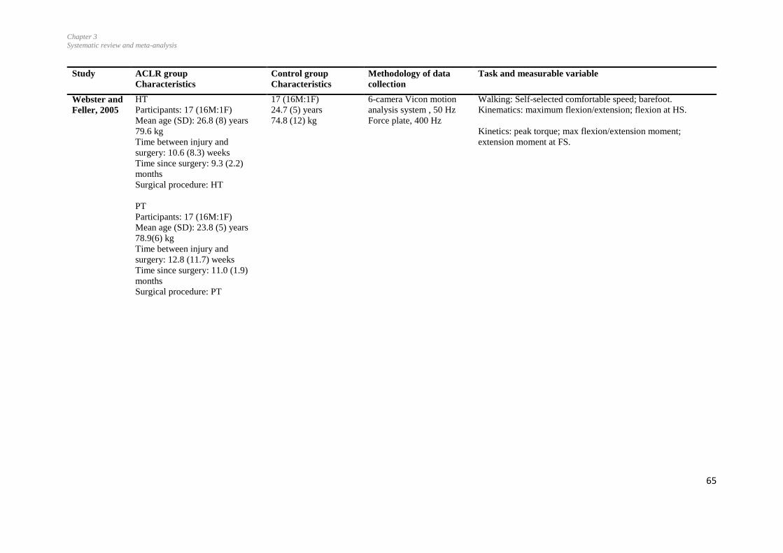
Chapter 3
Systematic review and meta-analysis
65
Study
ACLR group
Characteristics
Control group
Characteristics
Methodology of data
collection
Task and measurable variable
Webster and
Feller, 2005
HT
Participants: 17 (16M:1F)
Mean age (SD): 26.8 (8) years
79.6 kg
Time between injury and
surgery: 10.6 (8.3) weeks
Time since surgery: 9.3 (2.2)
months
Surgical procedure: HT
PT
Participants: 17 (16M:1F)
Mean age (SD): 23.8 (5) years
78.9(6) kg
Time between injury and
surgery: 12.8 (11.7) weeks
Time since surgery: 11.0 (1.9)
months
Surgical procedure: PT
17 (16M:1F)
24.7 (5) years
74.8 (12) kg
6-camera Vicon motion
analysis system , 50 Hz
Force plate, 400 Hz
Walking: Self-selected comfortable speed; barefoot.
Kinematics: maximum flexion/extension; flexion at HS.
Kinetics: peak torque; max flexion/extension moment;
extension moment at FS.
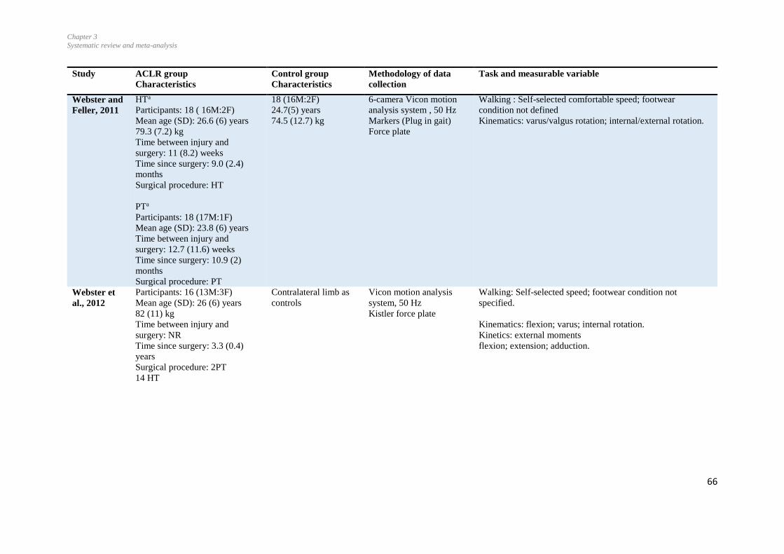
Chapter 3
Systematic review and meta-analysis
66
Study
ACLR group
Characteristics
Control group
Characteristics
Methodology of data
collection
Task and measurable variable
Webster and
Feller, 2011
HTa
Participants: 18 ( 16M:2F)
Mean age (SD): 26.6 (6) years
79.3 (7.2) kg
Time between injury and
surgery: 11 (8.2) weeks
Time since surgery: 9.0 (2.4)
months
Surgical procedure: HT
PTa
Participants: 18 (17M:1F)
Mean age (SD): 23.8 (6) years
Time between injury and
surgery: 12.7 (11.6) weeks
Time since surgery: 10.9 (2)
months
Surgical procedure: PT
18 (16M:2F)
24.7(5) years
74.5 (12.7) kg
6-camera Vicon motion
analysis system , 50 Hz
Markers (Plug in gait)
Force plate
Walking : Self-selected comfortable speed; footwear
condition not defined
Kinematics: varus/valgus rotation; internal/external rotation.
Webster et
al., 2012
Participants: 16 (13M:3F)
Mean age (SD): 26 (6) years
82 (11) kg
Time between injury and
surgery: NR
Time since surgery: 3.3 (0.4)
years
Surgical procedure: 2PT
14 HT
Contralateral limb as
controls
Vicon motion analysis
system, 50 Hz
Kistler force plate
Walking: Self-selected speed; footwear condition not
specified.
Kinematics: flexion; varus; internal rotation.
Kinetics: external moments
flexion; extension; adduction.

Chapter 3
Systematic review and meta-analysis
67
Study
ACLR group
Characteristics
Control group
Characteristics
Methodology of data
collection
Task and measurable variable
Webster and
Feller, 2012
HT groupa
Participants: 16M
Mean age (SD): 27.5 (6) years
79.5 (7) kg
Time between injury and
surgery: 10.7 (9) weeks
Time since surgery: 9.4 (3)
months
Surgical procedure: HT
PT groupa
Participants: 16M
Mean age (SD): 23.8 (6) years
79.2 (6) kg
Time between injury and
surgery: 11.9 (11) weeks
Time since surgery: 11.2 (2)
months
Surgical procedure: PT
16M
25.0 (5) years
75.9 (12) kg
Vicon motion analysis
system
Kistler force plate
Walking: Self-selected speed; barefoot.
Kinematics: peak adduction angle.
Kinetics: external peak adduction moment; vertical ground
reaction force.
Zabala et al.,
2012
Participants: 45 (26M:19F)
Mean age (SD): 29.5 (6.1) years
74.4 (12.4) kg
Time between injury and
surgery: 2.2 (0.4-8.0) months
Time since surgery: 26 months
(22-36)
Surgical procedure: Achilles
tendon
45 (26M:19F)
30.2 (4.68) years
74.2 (12.3) kg
Optoelectronic motion
capture system, 120 Hz
Force plate
Walking: Self-selected speed, footwear condition not
specified.
Kinetics: (1st and 2nd peak) adduction external moments;
abduction /adduction; flexion/extension; external /internal
rotation moments.
Stair ascent/descent
Kinetics: (1st and 2nd peak) adduction external moments;
abduction /adduction; flexion/extension; external /internal
rotation moment.
ACL: Anterior cruciate ligament ; ACLR: Anterior cruciate ligament reconstruction; a: Groups combined for meta-analysis; b: Both groups considered separately for meta-
analysis; AP: Anterior posterior; AMP: Anteromedial portal; BPTB: Bone patellar tendon bone; BTB: Bone patellar tendon bone; CKC: Close kinetic chain; CTO:
Contralateral toe off; CFS: Contralateral foot strike; DB: Double bundle; DGA: Dimensional gait analysis; Fz: Vertical forces; Fx: Horizontal antero-posterior forces; Fy:
Medio-lateral forces; F: Female; FS: Foot strike HS: Heel strike; HT: Hamstring tendon; IE: Internal-external; LAD: Ligament augmentation device; L5:Lumbar 5
vertebral level; M: Male; ML: Medio-lateral; MS: Mid-stance; Max: maximum; NR: Not reported; OKC: Open kinetic chain; PT: Patellar tendon; ROM: Range of
motion; S1: Sacrum 1 vertebral level; SB: Single bundle; SD: Standard deviation; ST: Semitendinosus; TO: Toe off; TT: Transtibial; NR: Not reported; VV: Varus-valgus.
Chapter 3
Systematic review and meta-analysis
68
3.4.4 Meta-analyses
Meta-analyses were conducted for peak angles and moments (peak knee flexion and
adduction moments) during the stance phase of gait (Figure 3.2 to 3.7). Tables 3.4 and 3.5
provide the ES and CI for the meta-analyses and for variables that were explored in one or
more studies. Mean differences for joint angles are provided in the appendix A2.
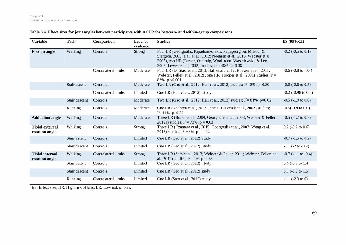
Chapter 3
Systematic review and meta-analysis
69
Table 3.4. Effect sizes for joint angles between participants with ACLR for between- and within-group comparisons
Variable Task Comparison Level of
evidence
Studies ES (95%CI)
Flexion angle Walking Controls Strong Four LR (Georgoulis, Papadonikolakis, Papageorgiou, Mitsou, &
Stergiou, 2003; Hall et al., 2012; Noehren et al., 2013; Webster et al.,
2005), two HR (Ferber, Osternig, Woollacott, Wasielewski, & Lee,
2002; Lewek et al., 2002) studies; I2 = 48%, p=0.08
-0.2 (-0.5 to 0.1)
Contralateral limbs Moderate Four LR (Di Stasi et al., 2013; Hall et al., 2012; Roewer et al., 2011;
Webster, Feller, et al., 2012) , one HR (Hooper et al., 2001) studies; I2=
83%, p <0.001
-0.6 (-0.8 to -0.4)
Stair ascent Controls Moderate Two LR (Gao et al., 2012; Hall et al., 2012) studies; I2= 8%, p=0.30 -0.0 (-0.6 to 0.5)
Contralateral limbs Limited One LR (Hall et al., 2012) study -0.2 (-0.98 to 0.5)
Stair descent Controls Moderate Two LR (Gao et al., 2012; Hall et al., 2012) studies; I2= 81%, p=0.02 -0.5 (-1.0 to 0.0)
Running Controls Moderate One LR (Noehren et al., 2013), one HR (Lewek et al., 2002) studies;
I2=11%, p=0.29
-0.5(-0.9 to 0.0)
Adduction angle Walking Controls Moderate Three LR (Butler et al., 2009; Georgoulis et al., 2003; Webster & Feller,
2012a) studies; I2 = 73%, p = 0.03
-0.5 (-1.7 to 0.7)
Tibial external
rotation angle
Walking Controls Strong Three LR (Czamara et al., 2015; Georgoulis et al., 2003; Wang et al.,
2013) studies; I2=68%, p = 0.04
0.2 (-0.2 to 0.6)
Stair ascent Controls Limited One LR (Gao et al., 2012) study -0.7 (-1.5 to 0.2)
Stair descent Controls Limited One LR (Gao et al., 2012) study -1.1 (-2 to -0.2)
Tibial internal
rotation angle
Walking Contralateral limbs Strong Three LR (Sato et al., 2013; Webster & Feller, 2011; Webster, Feller, et
al., 2012) studies; I2= 0%, p=0.63
-0.7 (-1.1 to -0.4)
Stair ascent Controls Limited One LR (Gao et al., 2012) study 0.6 (-0.3 to 1.4)
Stair descent Controls Limited One LR (Gao et al., 2012) study 0.7 (-0.2 to 1.5)
Running Contralateral limbs Limited One LR (Sato et al., 2013) study -1.1 (-2.3 to 0)
ES: Effect size; HR: High risk of bias; LR: Low risk of bias;
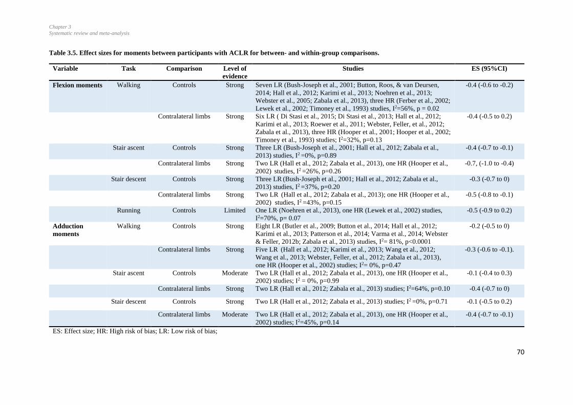
Chapter 3
Systematic review and meta-analysis
70
Table 3.5. Effect sizes for moments between participants with ACLR for between- and within-group comparisons.
Variable Task Comparison Level of
evidence
Studies ES (95%CI)
Flexion moments Walking Controls Strong Seven LR (Bush-Joseph et al., 2001; Button, Roos, & van Deursen,
2014; Hall et al., 2012; Karimi et al., 2013; Noehren et al., 2013;
Webster et al., 2005; Zabala et al., 2013), three HR (Ferber et al., 2002;
Lewek et al., 2002; Timoney et al., 1993) studies, I2=56%, p = 0.02
-0.4 (-0.6 to -0.2)
Contralateral limbs Strong Six LR ( Di Stasi et al., 2015; Di Stasi et al., 2013; Hall et al., 2012;
Karimi et al., 2013; Roewer et al., 2011; Webster, Feller, et al., 2012;
Zabala et al., 2013), three HR (Hooper et al., 2001; Hooper et al., 2002;
Timoney et al., 1993) studies; I2=32%, p=0.13
-0.4 (-0.5 to 0.2)
Stair ascent Controls Strong Three LR (Bush-Joseph et al., 2001; Hall et al., 2012; Zabala et al.,
2013) studies, I2 =0%, p=0.89
-0.4 (-0.7 to -0.1)
Contralateral limbs Strong Two LR (Hall et al., 2012; Zabala et al., 2013), one HR (Hooper et al.,
2002) studies, I2 =26%, p=0.26
-0.7, (-1.0 to -0.4)
Stair descent Controls Strong Three LR (Bush-Joseph et al., 2001; Hall et al., 2012; Zabala et al.,
2013) studies, I2 =37%, p=0.20
-0.3 (-0.7 to 0)
Contralateral limbs Strong Two LR (Hall et al., 2012; Zabala et al., 2013); one HR (Hooper et al.,
2002) studies, I2 =43%, p=0.15
-0.5 (-0.8 to -0.1)
Running Controls Limited One LR (Noehren et al., 2013), one HR (Lewek et al., 2002) studies,
I2=70%, p= 0.07
-0.5 (-0.9 to 0.2)
Adduction
moments
Walking Controls Strong Eight LR (Butler et al., 2009; Button et al., 2014; Hall et al., 2012;
Karimi et al., 2013; Patterson et al., 2014; Varma et al., 2014; Webster
& Feller, 2012b; Zabala et al., 2013) studies, I2= 81%, p<0.0001
-0.2 (-0.5 to 0)
Contralateral limbs Strong Five LR (Hall et al., 2012; Karimi et al., 2013; Wang et al., 2012;
Wang et al., 2013; Webster, Feller, et al., 2012; Zabala et al., 2013),
one HR (Hooper et al., 2002) studies; I2= 0%, p=0.47
-0.3 (-0.6 to -0.1).
Stair ascent Controls Moderate Two LR (Hall et al., 2012; Zabala et al., 2013), one HR (Hooper et al.,
2002) studies; I2 = 0%, p=0.99
-0.1 (-0.4 to 0.3)
Contralateral limbs Strong Two LR (Hall et al., 2012; Zabala et al., 2013) studies; I2=64%, p=0.10 -0.4 (-0.7 to 0)
Stair descent Controls Strong Two LR (Hall et al., 2012; Zabala et al., 2013) studies; I2 =0%, p=0.71 -0.1 (-0.5 to 0.2)
Contralateral limbs Moderate Two LR (Hall et al., 2012; Zabala et al., 2013), one HR (Hooper et al.,
2002) studies; I2=45%, p=0.14
-0.4 (-0.7 to -0.1)
ES: Effect size; HR: High risk of bias; LR: Low risk of bias;
Chapter 3
Systematic review and meta-analysis
71
3.4.5 Joint angles
Peak knee flexion angles during walking were reported in eleven studies (Di Stasi et al.,
2013; Ferber et al., 2002; Gao et al., 2012; Georgoulis et al., 2003; Hall et al., 2012; D.
Hooper et al., 2001; Lewek et al., 2002; Noehren et al., 2013; Roewer et al., 2011;
Webster, Feller, et al., 2012; Webster et al., 2005). The meta-analysis indicated strong
evidence for no difference compared to controls (Hall et al., 2012; Lewek et al., 2002)
(Ferber et al., 2002; Georgoulis et al., 2003; Noehren et al., 2013; Webster et al., 2005),
and strong evidence for less flexion for the ACLR compared to the contralateral limb (ES
-0.06 95%CI -0.8 to-0.4) (MD = 4.3°) (Di Stasi et al., 2013; Hall et al., 2012; Hooper et
al., 2001; Roewer et al., 2011; Webster, Feller, et al., 2012). A sensitivity test was
performed by excluding a study by (Hooper et al., 2001) as this study compared ACLR
participants at 2 weeks post-reconstruction with the contralateral limbs. With the
exclusion of that study, a significant difference was still evident for peak knee flexion
angle between injured and uninjured contralateral limb (ES-0.4, 95%CI -0.60 to-0.12). No
significant differences were found for peak knee flexion during stair ascent compared to
controls (Figure 3.2, (2.1.3)) and the contralateral limbs (Hall et al., 2012). However,
there was moderate evidence that peak flexion was decreased for ACLR knees compared
to controls during stair descent (ES-2.69, 95%CI -4.71 to-0.67). (Figure 3.2, (2.1.5)).
During running, no significant differences were found for the knee flexion angle
compared to controls (Figure 3.2, (2.1.6)).

Chapter 3
Systematic review and meta-analysis
72
Figure 3.2 Forest plot for peak knee flexion angle during walking, stair navigation and running
compared with Control group.
ACLR = anterior cruciate ligament reconstruction; SD = standard deviation; CI = confidence interval; IV =
inverse variance.’
Only three studies reported peak knee adduction angles (Figure 3.3), with a range from
7.5 months to 5.3 years post-reconstruction (Butler et al., 2009; Georgoulis et al., 2003;
Webster & Feller, 2012a). The meta-analysis indicated moderate evidence for no
significant difference between the ACLR knee and controls.

Chapter 3
Systematic review and meta-analysis
73
Figure 3.3 Forest plot for peak knee adduction angle during the stance phase of gait during walking
compared with Control group.
ACLR = anterior cruciate ligament reconstruction; SD = standard deviation; CI = confidence interval; IV =
inverse variance.’
Figure 3.4 Forest plot for peak tibial external rotation angle during the stance phase of gait during
walking compared with Control group.
ACLR = anterior cruciate ligament reconstruction; SD = standard deviation; CI = confidence interval; IV =
inverse variance.
Figure 3.5 Forest plot for peak tibial internal rotation angle during the stance phase of gait compared
with Control group.
ACLR = anterior cruciate ligament reconstruction; SD = standard deviation; CI = confidence interval; IV =
inverse variance.
Chapter 3
Systematic review and meta-analysis
74
In terms of rotation at the knee, studies either reported maximal external or internal tibial
rotation during the loading phase (Czamara et al., 2015; Georgoulis et al., 2003; Sato et
al., 2013; Webster & Feller, 2011). Strong evidence indicated no significant difference for
external tibial rotation for ACLR knees compared to controls during walking (maximum
17 months post-surgery, Figure 3.4). While peak external rotation angles were considered
for analysis, (Czamara et al., 2015) reported external rotation at initial foot contact. Their
results were included into the meta-analysis as peak external rotation is most likely to
occur at heel strike phase during the stance phase of the walking gait (Lafortune,
Cavanagh, Sommer, & Kalenak, 1992). One study (Gao et al., 2012) found less external
rotation for the ACLR knees at 3 months post-surgery during stair descent (ES -1.1,
95%CI -2 to -0.2; MD -5.8°, 95%CI -1.8 to -9.9). Strong evidence was found for less
tibial internal rotation ES -0.7 (95%CI -1.1 to -0.4) (Figure 3.5) for ACLR knees
compared to the contralateral limbs during walking (Sato et al., 2013; Webster & Feller,
2011; Webster, Feller, et al., 2012). However, results of one study (Kuenze et al., 2014)
indicated no significant difference between the ACLR and the contralateral limbs while
running, with limited evidence (ES -1.1, 95%CI -2.3 to 0).
3.4.6 Joint moments
Overall, participants with ACLR presented with significantly lower peak flexion moments
during walking compared to controls and the contralateral limbs (strong evidence) (Figure
3.6, (6.2.1 and 6.2.2)). Strong evidence was found for peak flexion moments being lower
for the injured side compared to the contralateral uninjured sides during walking, stair
ascent and descent after ACLR (Figure 3.6, (6.2.3 to 6.2.6)). Moderate evidence indicated
no significant differences during running in ACLR participants compared to controls
(Figure 3.6, (6.2.7)).

Chapter 3
Systematic review and meta-analysis
75
Figure 3.6 Forest plot for knee flexion moment during stance phase of gait in different activities.
ACLR = anterior cruciate ligament reconstruction; SD = standard deviation; CI = confidence interval; IV =
inverse variance
Chapter 3
Systematic review and meta-analysis
76
The meta-analysis showed moderate to strong evidence for significantly lower first peak
adduction moments during walking for the ACLR knees when compared to controls and
to the contralateral limbs respectively (Figure 3.7, (7.1.1 and 7.1.2)). Strong and moderate
evidence indicated no significant differences to be present for the peak adduction
moments during stair ascent and descent respectively for the ACLR knees compared to
the Control group (Figure. 3.7, (7.1.3 and 7.1.5)). In contrast, lower peak adduction
moments were found for ACLR during stair ascent (moderate evidence) and descent
(strong evidence) compared to the contralateral limbs (Figure 3.7, (7.1.4 and 7.1.6)). No
studies evaluating adduction moments during running were identified and included in the
review.

Chapter 3
Systematic review and meta-analysis
77
Figure 3.7 Forest plot for the first peak of knee adduction moment during the stance phase of gait in
different activities.
ACLR = anterior cruciate ligament reconstruction; SD = standard deviation; CI = confidence interval; IV =
inverse variance;
ES for differences for the flexion and adduction moments were low (< 0.59) for all
comparisons, except for the comparison of flexion moments during stair ascent (ES 0.7,
95%CI 0.1 to 0.4) and descent (ES 0.5, 95% CI 0.1 to 0.8), when comparing ACLR knees
to the contralateral limbs.
Chapter 3
Systematic review and meta-analysis
78
3.4.7 Time course of recovery
The qualitative assessment of the forest plots suggests patients tend to restore peak knee
flexion angle over time after surgery for studies that included participants up to 6 years,
and present peak knee flexion angle that is similar to the contralateral limb (Figure 3.2).
Such an improvement over time, however, was not evident for peak flexion moments
during walking. When ascending and descending stairs, patients tended to present
improved flexion moments over time when compared to the contralateral sides, but not
when compared to controls (Figure 3.6).
During walking, peak adduction moments appeared to be significantly lower in
participants with ACLR than controls within the first year following surgery (average
10.3 months) (Webster & Feller, 2012b). However, while not significant (95%CI, -0.02
to 1.4), these adduction moments showed a tendency to be slightly higher than controls at
a later phase (5.3 years) (Butler et al., 2009) (Figure 3.7). Interestingly, (Karimi et al.,
2013) found higher peak adduction moments at 6 months post-reconstruction compared to
controls. Functional braces were worn by their participants for 3 months post-surgery,
which were not reported to be worn in any of the other studies. A sensitivity test was
performed by excluding the adduction moments for this study (Karimi et al., 2013), and
the results indicate that, overall, peak adduction moments were significantly lower in
ACLR group compared to Control group (ES -0.32, 95%CI -0.55 to -0.08). When
excluding their results, there appears to be a response in time, from lower to equivalent
adduction moments for the ACLR group compared to controls while walking.
Chapter 3
Systematic review and meta-analysis
79
3.5 Discussion
The primary aim of this review was to compare knee joint angles and moments in
participants with ACLR to the contralateral limbs and to age-matched controls during
walking, stair climbing and running. In terms of joint angles, moderate to strong evidence
was found for no differences for peak flexion during walking, stair ascent and jogging
when comparing the ACLR knees to controls. However, when compared to the uninjured
contralateral sides, less flexion and less internal rotation were found for the ACLR knees
during walking. During stair descent, peak flexion was decreased for ACLR knees
compared to controls. There was moderate evidence for no differences for peak adduction
angles between the ACLR knee and controls. Moderate to strong evidence was found for
lower peak flexion moments in participants with ACLR during walking and stair ascent
compared to controls and to the contralateral limbs. Moderate to strong evidence also
indicated lower peak adduction moment for the ACL-reconstructed knees during walking,
stair ascent, and descent compared to the contralateral limbs, and during walking when
compared to the controls.
3.5.1 Peak joint angles
Results of the meta-analysis suggest that participants with ACLR regain similar peak
knee flexion angles compared to the Control group during walking by 6 years following
surgery (Figure 3.2). However, pooled peak flexion angles were lower in the ACLR knee
compared to the contralateral sides (ES 0.6, 95%CI -0.4 to -0.8). The MD 4.3° was higher
than the minimal clinically important difference of 3° reported for knee flexion during the
stance phase of walking (Di Stasi & Snyder-Mackler, 2012). This difference was
influenced in part by the findings by (Hooper et al., 2001) at 2 weeks post-surgery: the
MD 8.5° which may be due to pain or swelling at early stages after surgery. Considering

Chapter 3
Systematic review and meta-analysis
80
the individual studies, limb asymmetry for peak flexion angles was evident until 6 months
post-reconstruction (Roewer et al., 2011; Di Stasi et al., 2013), while no significant
bilateral differences were found, on average, 10 months to 6 years following surgery.
Similarly, our meta-analysis indicated no significant between-group differences for knee
adduction and external rotation angles during walking. However, peak external rotation
was decreased during stair descent based on one study (Gao et al., 2012). Internal rotation
was decreased during walking for the ACLR knee compared to contralateral limb.
Rotational shifts varied from 1 to 5.8°. A biomechanical model of degeneration showed
that 5° rotational shift may be enough to cause accelerated degeneration of the cartilage
(Andriacchi & Mündermann, 2006) due to alterations in the stresses applied to joint
surfaces (Tashman, Collon, Anderson, Kolowich, & Anderst, 2004). Whether these small
differences in kinematics for participants with ACLR compared to controls and
contralateral limbs indicate increased risk for post-traumatic knee osteoarthritis still needs
confirmation.
3.5.2 Joint moments
Strong evidence was found that peak flexion moments were significantly lower in
participants with ACLR during walking, stair ascent and descent activities compared to
controls and contralateral limb. However, the effect size were small during walking and
when compared to the controls during stair ascent and descent, while moderate effect
sizes were found for decreased flexion moments when compared to the contralateral sides
during stair ascent and descent. It is thus possible that an asymmetry may be more evident
during stair ascent and descent than during walking. Peak external flexion moments are
considered to reflect net quadriceps function (Andriacchi & Dyrby, 2005) and reduced
Chapter 3
Systematic review and meta-analysis
81
moments may be associated with lower quadriceps strength (Schmitt et al., 2015).
Neurophysiological changes following ACL injury and reconstruction have been shown
to include decreased lower limb muscle activation, as determined by electromyography
on the injured side (Nyland et al., 2010) and lower cortical activation in injured limbs
compared to uninjured and healthy population, contributing towards reduced net
quadriceps function (Kuenze et al., 2015). The presence of pain reported even after 2-5
years of surgery (Kartus et al., 1999), or reflex inhibition (Hodges & Tucker, 2011) could
further influence the movement patterns, as apparent with reduced flexion moments.
Furthermore, compensatory movements at the hip and trunk may also decrease knee joint
loading (Courtney & Rine, 2006). It is thus likely that multiple mechanisms contribute
towards long-term decreased flexion moments in the ACLR knees.
Similar to external knee flexion moments, the adduction moments for the ACLR knees
were significantly smaller during walking when compared to controls, albeit with low
effect size. No significant differences were found for both the flexion moments and
adduction moments during stair descent for the ACLR knees compared to the controls,
while they were lower for the ACLR knees compared to the contralateral side. This
indicates a bilateral asymmetry during this task and could infer that during stair descent,
the contralateral side has higher flexion and adduction moments than controls. While this
is speculative, compensatory patterns may be evident on the contralateral side (Hart et al.,
2010) which are likely due to central nervous system changes following the injury and
surgery. Such changes have been documented in patients with ACL-deficient knees
(Courtney & Rine, 2006).
The meta-analysis along with sensitivity test for one study (Karimi et al., 2013) showed
peak adduction moments were lower compared to controls in participants with ACLR the
Chapter 3
Systematic review and meta-analysis
82
short- and medium-term (up to 3.5 years) and higher as time following reconstruction
progresses. Our findings contrast thus with those by (Hart, Culvenor, et al., 2015) who
recently found that the frontal plane moments may not be affected by ACLR. Our
findings, thus, suggest that frontal plane moments cannot yet be disregarded as outcomes
and long-term consequences of ACLR. The finding of lower adduction moments in the
ACLR knees contrast with increased adduction moments often reported in patients with
knee osteoarthritis (Setton, Elliott, & Mow, 1999). Risk of progression of osteoarthritis in
patients with that disorder increases 6.46 times with 1% increase in adduction moments
(Miyazaki et al., 2002), hence the magnitude of adduction moments is correlated with
osteoarthritis severity (Foroughi et al., 2009; Sharma et al., 1998). While decreased
adduction moments in the early phase following ACLR may reflect decreased weight-
bearing and compensatory strategies of the trunk, hip and ankle (Myer, Chu, Brent, &
Hewett, 2008; Paterno, Ford, Myer, Heyl, & Hewett, 2007; Setton et al., 1999), a
combination of changing flexion and adduction moments and joint rotations may
contribute towards loading at unconditioned areas of the cartilage potentially leading to
development of osteoarthritis (Andriacchi, Briant, Bevill, & Koo, 2006). Studies included
in the present review did not report radiographic osteoarthritis changes for their
participants, thus it is unknown whether those with higher adduction moments present
with radiographic signs of osteoarthritis (Miyazaki et al., 2002). Whether changes for
external adduction moments during functional tasks predict the onset of osteoarthritis
following ACLR still needs to be substantiated. While the development of osteoarthritis is
multi-factorial, these biomechanical factors should be explored longitudinally in all
planes.
Chapter 3
Systematic review and meta-analysis
83
3.5.3 Clinical implications
Overall, reduced flexion and adduction moments indicate altered joint loading in the
long-term following ACLR. These changes are likely associated with quadriceps
weakness (Shelburne, Torry, & Pandy, 2006) and various neurophysiological mechanisms
(Barret, 1991; Urbach, Nebelung, Becker, & Awiszus, 2001; Urbach, Nebelung, Röpke,
Becker, & Awiszus, 2000), and are potentially associated with risk of early onset joint
degeneration (Roos, Adalberth, Dahlberg, & Lohmander, 1995; Roos, Neu, Hull, &
Howell, 2005), due to suboptimal joint loading during functional tasks such as walking,
running, stair navigation (Knoll, Kiss, & Kocsis, 2004). Altered kinematics along with
reduced flexion moments may significantly change the stress distribution within the
cartilage and initiates a degenerative process (Setton et al., 1999). Di Stasi et al., 2013
showed that athletes with a superior functional performance at 6 months following ACLR
appear to demonstrate less asymmetrical gait patterns compared to those that did not pass
functional return to play criteria. It thus appears to be important to include assessment of
potential gait asymmetries following ACLR in decision making for readiness for
rehabilitation progression and return to sports (Di Stasi et al., 2013; Myer et al., 2008).
While small differences in walking may not be detectable on clinical examination, these
may be more evident during high knee loading activities, such as stair ascent/descent and,
running (Myer et al., 2008) and jumping (Paterno et al., 2007). The finding of movement
and loading asymmetries in the current review indicates that it may be important for
people with ACRL to continue with exercise programme focusing on strength,
neuromuscular control and proprioception even after rehabilitation period is completed.
Chapter 3
Systematic review and meta-analysis
84
3.5.4 Methodological considerations and directions for future research
The risk of bias assessment identified methodological limitations for the included studies:
the absence of outcome measurer blinding, reporting reliability of methodology, lack of
confounding factors as control of gait speed, a priori power calculation for sample size.
While blinding of the researcher is difficult during data collection due to visible scarring
at the operated knee, blinding for the participant group (Control versus ACLR) can occur
while processing kinematic and kinetic data.
Sixteen studies included in the meta-analysis controlled the speed of the tasks (Bush-
Joseph et al., 2001; Butler et al., 2009; K. Button et al., 2014; Di Stasi et al., 2015; Di
Stasi et al., 2013; Ferber et al., 2002; Georgoulis et al., 2003; Kuenze et al., 2014; Lewek
et al., 2002; Noehren et al., 2013; Roewer et al., 2011; Timoney et al., 1993; Varma et al.,
2014; Wang et al., 2013; Webster & Feller, 2012a; Webster et al., 2005). A compensatory
strategy to maintain or decrease joint loading is to walk slower (Robbins & Maly, 2009).
Thus, in those studies where speed was not reported, it is possible that participants with
ACLR may have walked slower thereby maintaining or controlling the joint loading
effectively. This could lead to no significant differences between-group, as seen for stair
descent for flexion and adduction moments. Furthermore, other compensatory strategies
which develop over time post-reconstruction, such as transferring the load to the hip and
ankle (Orishimo, Kremenic, Mullaney, McHugh, & Nicholas, 2010) and compensation
with trunk lean, can also be used to decrease joint moments, as shown in patients with
knee osteoarthritis (Annegret Mündermann, Dyrby, & Andriacchi, 2005; Simic, Hinman,
Wrigley, Bennell, & Hunt, 2011). These factors should be addressed in future research.
Chapter 3
Systematic review and meta-analysis
85
Only cross-sectional studies or baseline measures of RCTs were considered for the
purpose of the time course of recovery analysis in this review. However, longitudinal
studies following patients at different time points following ACLR are needed to fully
investigate how gait biomechanics change over time, and how these changes may
influence the development of osteoarthritis or re-injury. Furthermore, the aim of this
review was to explore differences for peak knee joint angles and moments when
comparing people with ACLR to controls, and not between different surgical or
rehabilitative programs. Findings of groups of participants with ACLR undergoing
different procedures within individual studies were thus combined into grand means for
the meta-analyses, which masks differences between these groups. Findings of this
review are thus limited to comparisons with controls and when comparing the ACLR
knees to the contralateral uninjured sides, irrespective of surgical methods. Finally, this
review was limited to the discrete variables of peak angles and moments, and did not
explore biomechanical variables in other phases of the gait cycle.
3.6 Conclusion
Joint kinematics of ACL reconstructed knees were found to be similar to Control groups
during walking and stair navigation within a few months after surgery. However,
differences in knee external moments persist over a longer period. The meta-analysis
indicated lower pooled external flexion moments for people with ACLR compared to
controls during walking and stair ascent. The meta-analysis also indicated decreased peak
adduction moments during walking and stair navigation for ACLR-knees, specifically
when compared to contralateral limbs. Based on individual studies included in the review,
such moments may be lower compared to controls in the early phase following ACLR (10
months), but higher at later phase (5 years) after surgery. While this review did not
Chapter 3
Systematic review and meta-analysis
86
explore compensatory mechanisms, it is likely that multiple mechanisms influence altered
gait patterns, including changes in muscle function and neuromuscular control at the
trunk, hip knee, and ankle. Gait patterns at the knee are not fully restored after more than
5 years following surgery. It may also indicate that early programs post-surgery are
undertaken poorly and hence they should be re-examined. Also, it indicates that long-term
rehabilitative or maintenance programmes may need to be considered for patients
undergoing ACLR.
3.7 Summary
This chapter highlighted the partial recovery of knee flexion and adduction moments
following ACLR during stair ascent and descent. The included studies in this review
explored the moments up to an average of 5 years following surgery, however, there is
need to explore the moments in the long-term as it may be possible that moments have
higher magnitude as the time since surgery increases. Therefore, moments were measured
and analysed in participants with ACLR from 2 to 10 years following surgery and
compared with the contralateral limb and the Control group (Chapter 6).
The next Chapter (Chapter 4) is a cross-sectional study aimed to explore and compare the
patient-reported outcomes, muscle strength, physical performance and knee laxity related
outcomes in participants with ACLR with the Control group

Chapter 4
Physical measures in ACLR vs Control group
87
4 Patient-reported outcomes and physical performance measures in
participants with ACLR compared to a Control group – a cross-
sectional study
4.1 Prelude to Chapter 4
Participants with ACL injury and surgery are at higher risk of early onset of osteoarthritis,
which may take a few years for development. Following the patient-centered approach, it
is important to explore patient-reported outcomes. The literature review (Chapter 2)
indicated that the muscle strength and physical performance deficits on the injured side in
participants with ACLR may persist in the long term post-surgery. This indicated the need
to explore the physical constraints related to the knee in participants with ACLR in the
context of New Zealand.
In this study, patient-reported outcomes, muscle strength, physical performance and knee
laxity in participants with ACLR were measured and compared with their contralateral
limb, and to an uninjured Control group. This chapter provides a detailed description of
the entire participant cohort involved in this thesis before exploring the biomechanics-
related outcomes in participants 2 to 10 years following surgery (Chapter 6).
Chapter 4
Physical measures in ACLR vs Control group
88
4.2 Background
Anterior cruciate ligament reconstruction surgery is undertaken to restore joint anatomy,
but the joint function is more than just the restoration of anatomical structures.
Assessment of joint function requires muscle strength assessment. Strength deficits can
persist until 5 years post-surgery on the injured side in the knee extensor group compared
to the Control group (Keays, Bullock-Saxton, Keays, Newcombe, & Bullock, 2007; Wolf
Petersen, Pouria Taheri, Phillip Forkel, & Thore Zantop, 2014) and were related to the
presence of osteoarthritis in participants with ACLR (Keays et al., 2007). Muscle strength
deficits have been reported to persist from 3 years (Hiemstra, Webber, MacDonald, &
Kriellaars, 2007) to 7 years following surgery (Yasuda, Ohkoshi, Tanabe, & Kaneda,
1991). Strength deficits, particularly quadriceps strength asymmetry following ACLR, is
known to alter knee joint biomechanics at time of return to activity (Palmieri-Smith &
Lepley, 2015). Patients with low quadriceps strength displayed greater movement
asymmetries at the knee in the sagittal plane such that there was greater symmetry for
knee flexion angle and external moments in patients with high and moderate quadriceps
symmetry compared to those with low symmetry during single leg hop (Palmieri-Smith &
Lepley, 2015). Also, quadriceps strength was related to movement asymmetries and
physical performance (Palmieri-Smith & Lepley, 2015). It is important to explore the
muscle strength deficits in the current thesis as the results can help to understand the knee
biomechanics clearly. Presence of muscle strength deficits are more likely to be found
during the maximal muscle force output, therefore, peak torque during maximal muscle
force output was used as an outcome measure. Further, eccentric quadriceps muscle
activity is required during jumping, landing and stair descent (Bobbert, 2001), while
concentric quadriceps and hamstring strength is required during the stair navigation
activity (Benedetti, Agostini, Knaflitz, & Bonato, 2012). Assessing both concentric and
Chapter 4
Physical measures in ACLR vs Control group
89
eccentric muscle strength thus is important to understand function related to daily
activities.
Considering patients’ perspectives is also needed to fully understand the influence of
injury on joint-related and general quality of life. A review has reported good scores in
patient-reported outcomes up to 10 years following ACLR (Magnussen et al., 2015);
however, the quality of life for participants with ACLR can deteriorate in the long-term
following surgery (Tengman, 2014). Muscle function was associated with future patient-
reported outcomes in young adults with ACL injury suggesting that improving muscle
function during rehabilitation could improve present and future patient–reported
outcomes (Flosadottir, Roos, & Ageberg, 2016). It has been reported that patients
experience decreased levels of physical activity up to several years after surgery
(Magnussen et al., 2011), which may be related to the performance levels on the injured
side. Exploring the physical performance on the injured side can provide information
relating to the neuromuscular status of the limb.
Along with the muscles providing the dynamic stability, knee stability is also determined
by joint laxity. Conflicting research reports exist in the literature regarding antero-
posterior knee laxity following surgery, with some studies reporting a decrease in knee
laxity (Ahldén et al., 2009), while others reported no change up to 7 years following
surgery (Semay et al., 2016). Decrease in the laxity of the knee may be related to
development of stiffness of the knee in the long-term. However, less is known about knee
laxity in participants with ACLR in the long-term.
Chapter 4
Physical measures in ACLR vs Control group
90
4.2.1 Aim of the study
The aim of this study was to determine the patient-reported outcomes, thigh muscle
strength, and physical performance and knee laxity-related outcomes of participants with
ACLR, and to compare them with the contralateral limb and with the Control group.
4.2.2 Hypothesis
Participants with ACLR will have lower scores on the KOOS, SF-12 Health
survey, Confidence during Sports, and Tegner scores compared with the Control
group.
Participants with ACLR will have lower isokinetic concentric and eccentric peak
torque of the quadriceps, and concentric peak torque of the hamstrings, compared
to the contralateral limb and the Control group.
Participants with ACLR will demonstrate lower performance during the single-leg
hop compared to the contralateral limb and the Control group.
Participants with ACLR will have reduced knee laxity in the sagittal plane on the
injured side compared to the contralateral limb, and compared to the Control
group.
4.3 Methods
The STROBE (Strengthening the Reporting of Observational studies in Epidemiology)
statement was used for reporting this study (Von Elm et al., 2014).
4.3.1 Study design
This was a cross-sectional study, assessing the within- and between-group differences
among the ACLR and Control group.

Chapter 4
Physical measures in ACLR vs Control group
91
4.3.2 Ethical approval
The University of Otago Human Ethics Committee granted approval for the study
(reference number H15/034) (Appendix-B1 to B3). Consultation with the Ngāi Tahu
Research Consultation Committee (Māori committee) was completed prior to the study
(Appendix-B4).
4.3.3 Study settings
The study was carried out at the Mark Steptoe Laboratories located in the Centre for
Health, Activity and Rehabilitation Research (CHARR), School of Physiotherapy at the
University of Otago, Dunedin, New Zealand.
4.3.4 Recruitment of participants with the ACLR and the Control group
Participants were recruited from the Otago region of the South Island of New Zealand via
adverts or leaflets sent to local community newspapers (Appendix B5), sports clubs,
swimming pools, fitness centres, and posted on public notice boards (Appendix B6).
Physiotherapy and sports physicians likely to be working with sports people with past
ACL injury and reconstructions were sent information sheets and leaflets, and asked to
provide information about the study to their patients. Leaflets were placed in
physiotherapy and general practice patient waiting areas. Study information was also sent
to Sports Physiotherapy New Zealand, and an advert was placed in their monthly
newsletter to members. Information was posted on the School of Physiotherapy website
and the School of Physiotherapy Clinics’ Facebook site. Participants for the Control
group were recruited by the same community adverts, in addition to word-of-mouth
during data collection in the School of Physiotherapy.
Chapter 4
Physical measures in ACLR vs Control group
92
Interested participants were asked to provide their e-mail address and contact phone
number to the Clinical Research Administrator of the School of Physiotherapy. A detailed
Participant Information Sheet for the ACLR group (Appendix –B7) and Control group
(Appendix –B8) was sent to participants by e-mail. Participants who met the initial
inclusion criteria were then screened by the PhD candidate, and an appointment was made
to book a session in the laboratory to collect data. An online questionnaire link (Qualtrics,
Provo, UT, USA, 2015) was sent to participants, who completed the questionnaire
(Appendices- C2 to C7) prior to attending the data collection session in the Biomechanics
Laboratory. Participants were recruited from 04/2015 to 05/2016.
4.3.5 Sample size estimation
Sample size estimation for this research was performed for the peak knee adduction
moment for the ACLR group, and it indicated the need for 26 participants within the
ACLR group and 26 in the Control group (Chapter 6, section 6.3.5.1). Muscle strength,
physical performance, knee laxity and patient-reported outcomes were examined as
secondary outcome measures. This study was an exploratory study in the thesis, therefore,
no power calculations were performed; therefore, the results should be considered with
caution.
Participants attended two sessions in the laboratory: the first session included data
collection relating to the knee laxity measurements and the three-dimensional motion
analysis; the second session was for data collection relating to the muscle strength and the
physical performance testing.

Chapter 4
Physical measures in ACLR vs Control group
93
4.3.6 Inclusion and exclusion criteria
4.3.6.1 Inclusion criteria for ACLR participants
Men and women participants, aged between 20 and 50 years, who had undergone ACLR
with any type of graft two to ten years’ ago were included in the study. Participants may
have had associated ligamentous (such as of medial collateral ligament), meniscal or
chondral injury and repair.
4.3.6.2 Exclusion criteria for the ACLR group
Participants with ACLR were excluded if they had only non-surgical rehabilitation, had
undergone revision surgery for the ACL injury, had a previous recurrence of ACL injury
following the primary reconstruction, or had a bilateral ACL injury. Participants with
other lower limb, pelvic or low back musculoskeletal injuries that required health care
over the past 6 months, or who were limited their daily function, sports or occupational
performance were excluded as well. Pregnant females at the time of data collection were
also excluded.
4.3.6.3 Inclusion criteria for the Control group participants
Controls - defined by age, gender, and physical activity (Tegner scores) level-matched
participants with no ACL or other knee injury - were recruited.
4.3.6.4 Exclusion criteria for Control group
Participants with knee injuries, lower limb or musculoskeletal injuries that needed health
care over the past 12 months or who were limited their daily function, sports or
occupational performance, were excluded from the study.
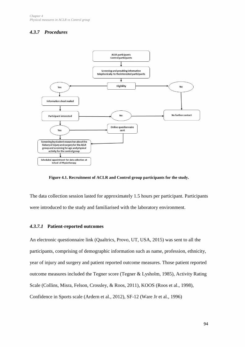
Chapter 4
Physical measures in ACLR vs Control group
94
4.3.7 Procedures
Figure 4.1. Recruitment of ACLR and Control group participants for the study.
The data collection session lasted for approximately 1.5 hours per participant. Participants
were introduced to the study and familiarised with the laboratory environment.
4.3.7.1 Patient-reported outcomes
An electronic questionnaire link (Qualtrics, Provo, UT, USA, 2015) was sent to all the
participants, comprising of demographic information such as name, profession, ethnicity,
year of injury and surgery and patient reported outcome measures. Those patient reported
outcome measures included the Tegner score (Tegner & Lysholm, 1985), Activity Rating
Scale (Collins, Misra, Felson, Crossley, & Roos, 2011), KOOS (Roos et al., 1998),
Confidence in Sports scale (Ardern et al., 2012), SF-12 (Ware Jr et al., 1996)

Chapter 4
Physical measures in ACLR vs Control group
95
(Appendices C1 to C4), and were sent to both groups (Figure 4.1). An appointment was
made for the data collection as soon as the participants had submitted the questionnaire.
4.3.7.2 Informed consent and Accident Compensation Corporation release form
Following the familiarisation and demonstration of the task, and if participants agreed to
take part in the study, they were asked to sign a written, informed consent form. Different
consent forms were prepared for the ACLR group (Appendix-B9) and for the Control
group (Appendix-B10).
Participants in the ACLR group were asked to sign an ACC Release form (Appendix-
B11). The ACC provides comprehensive, no-fault personal injury cover for all New
Zealand residents and visitors, and keeps the associated medical and health-related
records. Participants provided permission for the researchers to contact ACC to request
the following information: date of injury; date of surgery; type of surgery; MRI report (if
relevant) to define possible associated injuries of the knee ligament, cartilage or bone; and
the number of pre- and post-operative physiotherapy sessions.
4.3.7.3 Muscle strength testing
Peak torque was assessed bilaterally for concentric and eccentric knee extension, and
concentric knee flexion, with an isokinetic dynamometer (Biodex Medical Systems, Inc,
Shirley, NY) using previously published procedures (Sole, Hamrén, Milosavljevic,
Nicholson, & Sullivan, 2007). It is known to have a high relative reliability (Intraclass
correlation coefficient range> 0.90) and the smallest real difference of less than 18%
(Sole et al., 2007). The test procedure started with the standardised 5-minute general
warm-up on a stationary bicycle at moderate intensity.
Chapter 4
Physical measures in ACLR vs Control group
96
Participants were seated with the hips in 100° of flexion with their arms resting on their
lap. Individual adjustments were made for the seat to accommodate the length of the
femur, and for the mechanical axis of the dynamometer lever to be aligned as closely as
possible with the anatomical axis of the knee joint. Velcro straps were used to stabilise
the participants’ trunk, hips and thigh. The distal pad of the dynamometer arm was placed
approximately 2 cm above the medial malleolus. The range of movement was from 0°
(anatomic 0) to 85° of knee flexion.
Before testing, limb weight was measured using the manufacturer’s recommended
procedures. Before data collection, participants performed at least five practice trials at
sub-maximal level for concentric and eccentric contraction respectively, as part of their
familiarisation and as a specific warm-up, followed by two maximal contractions for each
type of muscle contraction. A two-minute rest was given after the familiarisation period.
The uninjured leg was tested first for participants with ACLR. The dominant leg was
tested first, defined as the preferred kicking leg, for the Control group participants.
Participants performed five reciprocal repetitions at 60°/s of maximal extension of each
leg (concentric quadriceps) followed by maximal flexion (concentric hamstring) up to
90°. A five-minute rest was given before the contralateral leg was tested. Reciprocal
extension and flexion isokinetic testing was done for concentric quadriceps and
hamstring, first on both sides, which was to examine the quadriceps (extension) and
hamstring (flexion) strength in the same protocol. Participants were requested to use
maximal effort by verbal encouragement that they need to “push or pull as hard as they
can”. Following completion of the reciprocal concentric quadriceps and hamstring set, the
eccentric quadriceps strength was assessed at 60°/s with a five-minute rest before starting
the next test. Participants were instructed to extend the knee against the shin pad from 90°

Chapter 4
Physical measures in ACLR vs Control group
97
knee flexion to full extension, then trigger lever flexion by pressing against the shin pad
in the extension direction, and to resist the lever during eccentric extension.
The speed of 60°/sec was chosen for both concentric and eccentric contractions as this is
the most commonly used speed in the literature (Undheim et al., 2015). Peak torque was
considered as the outcome measure which is the single highest torque output recorded
throughout the range of motion of each repetition (Kannus, 1994). Gravity correction of
peak torque was made with the Biodex software. Peak torque data was downloaded using
the Sys 3 DBM (Version 1.7) system software of Biodex, and was normalised for body
weight (Nm/kg). Torque measures in Nm of participants with ACLR have been presented
in Appendix C-8.
4.3.7.4 Single-leg hop
The single-leg hop was used as a physical performance measure. This test is known to be
reliable in examining neuromuscular function in participants with ACLR (Intraclass
correlation coefficient 0.76–.97) (Kramer, Nusca, Fowler, & Webster-Bogaert, 1992).
Following the warm up and muscle strength isokinetic testing, the student researcher
demonstrated the task of single-leg hop to the participants. Participants were asked to
stand on the testing limb with their arms folded across their chest and hop as far as
possible. Participants were asked to land on the same limb and maintain their balance for
at least 2 seconds following landing. Following the demonstration, participants performed
two practice trials for each leg. They then performed three maximal hops for distance for
each limb (Gustavsson et al., 2006). Participants wore their own sport shoes. It was
important for them to wear the most comfortable shoes which they would prefer for the
sports activity in order not to influence the performance. Wearing their own shoes was

Chapter 4
Physical measures in ACLR vs Control group
98
also considered to increase external validity; that is, reflecting the footwear they use in
their daily lives.
The uninjured side was tested first for participants with ACLR, followed by the operated
limb. For Control group participants, the dominant limb was tested first followed by the
non-dominant limb. An inability to maintain balance was considered to be a
disqualification for that trial and the test was repeated. The distance was measured in
centimetres from the toe at push-off to the heel where the participant landed. The mean
distance across three trials was the outcome measure (Myers, Jenkins, Killian, &
Rundquist, 2014).
4.3.7.5 Knee laxity in sagittal plane
A KT-1000 arthrometer (MEDmetric® Corp. San Diego, CA, USA) was used to measure
the posterior-anterior tibio-femoral laxity of the ACLR and the Control groups in the
sagittal plane. The reliability for measuring anterior-posterior tibial displacement relative
to the femur has been reported to show substantial variations in measurements (Forster et
al., 1989). The specific intra-tester (test-retest) reliability for this measurement was
determined by repeating these on a second occasion with 10 participants with ACLR. The
Intraclass correlation coefficient was > 0.84, indicating high reliability (Appendix- C6).
Participants were positioned supine with instructions to relax, with their hands resting on
their abdomen. Following the manufacturer’s recommendations, a support was placed
under the thighs to position the knees in 20° to 30° flexion and it was checked with a
goniometer (Fred Sammons, Inc) to ensure that the knee was at the correct angulation. It
was maintained approximately at 25°, and a foot support was used to position the feet in a
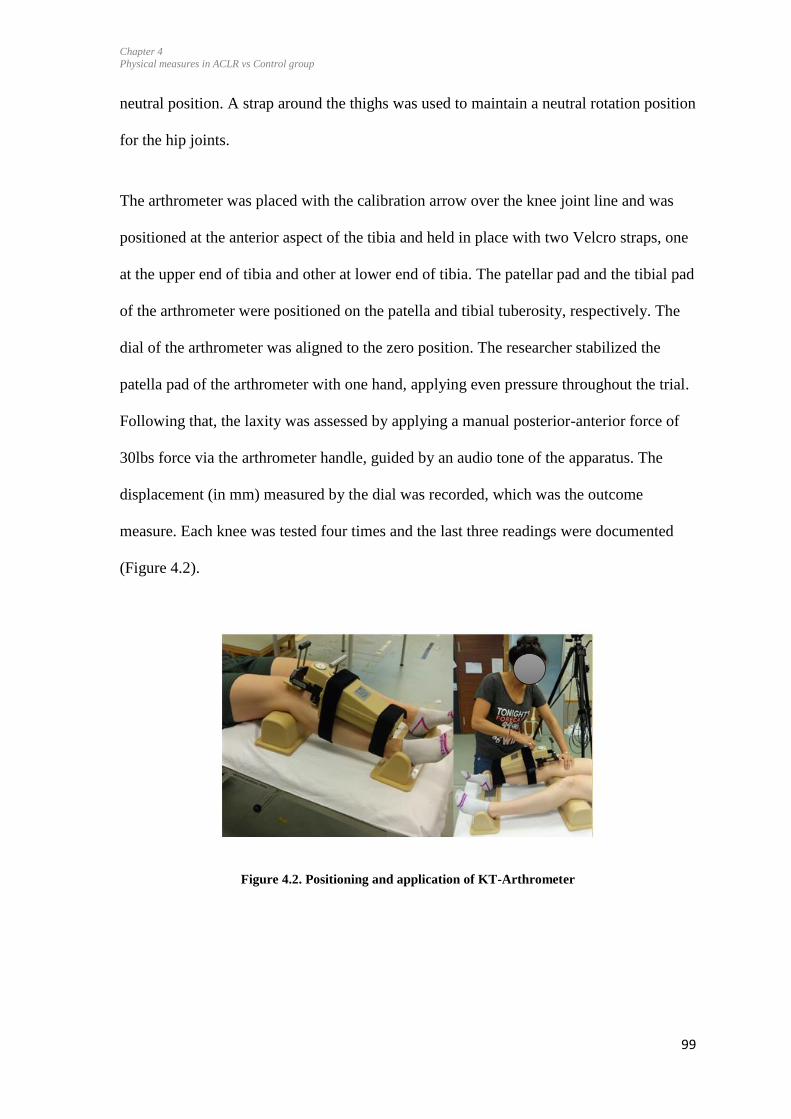
Chapter 4
Physical measures in ACLR vs Control group
99
neutral position. A strap around the thighs was used to maintain a neutral rotation position
for the hip joints.
The arthrometer was placed with the calibration arrow over the knee joint line and was
positioned at the anterior aspect of the tibia and held in place with two Velcro straps, one
at the upper end of tibia and other at lower end of tibia. The patellar pad and the tibial pad
of the arthrometer were positioned on the patella and tibial tuberosity, respectively. The
dial of the arthrometer was aligned to the zero position. The researcher stabilized the
patella pad of the arthrometer with one hand, applying even pressure throughout the trial.
Following that, the laxity was assessed by applying a manual posterior-anterior force of
30lbs force via the arthrometer handle, guided by an audio tone of the apparatus. The
displacement (in mm) measured by the dial was recorded, which was the outcome
measure. Each knee was tested four times and the last three readings were documented
(Figure 4.2).
Figure 4.2. Positioning and application of KT-Arthrometer
Chapter 4
Physical measures in ACLR vs Control group
100
4.3.8 Data processing and analysis
4.3.8.1 Patient-reported outcomes
Mean data from the ACLR group were compared with the Control group.
4.3.8.2 Muscle strength, single-leg hop and knee laxity
For the ACLR group, data from the injured sides were pooled together. For the Control
group, the left and right sides were randomized to Side 1 and Side 2 through the online
software, Research Randomizer (Version 4.0) (Urbaniak & Plous, 2013), to generate the
numbers for randomization to have the same ratio in terms of right/left sides for the
ACLR group.
4.3.8.2.1 Muscle strength
Peak torque was used for the isokinetic muscle strength, and the average distance of three
trials was used for the jump distance for the horizontal hop. Limb symmetry was
calculated for peak torque of each contraction type: concentric quadriceps, concentric
hamstring, eccentric hamstring and for the average jumping distance.
4.3.8.2.2 Single-leg hip
To calculate the limb symmetry index (LSI), the mean scores (5 trials) of the involved
limb were divided by the mean score of the uninvolved limb and multiplied by 100 for the
hop (Gustavsson et al., 2006). For the healthy participants, the LSI was calculated by the
mean scores of side 1 divided by the mean scores of side 2, and the result was multiplied
by 100. A LSI greater than 90% was considered to be within the normal range
(Gustavsson et al., 2006; Neeter et al., 2006).
Chapter 4
Physical measures in ACLR vs Control group
101
4.3.8.2.3 Knee laxity
The average of three trials for each limb for knee laxity was used in the ACLR and
Control group. Mean values were utilised, as high variability in the trial was found. This
was also based on the previously published papers (Ahldén et al., 2009).
4.3.9 Statistical analysis
SPSS Version 23 (IBM SPSS Statistics) was used for statistical analysis. Normality of the
data was tested with histograms and Normal Q-Q plots. Data were found to be normally
distributed with histograms, and data points were close to the diagonal line in Q-Q plots.
4.3.9.1 Patient-reported outcomes
Descriptive statistics for all demographic variables and injury factors were reported for
groups using means and SD for continuous variables and proportions for categorical
variables. Independent t-tests were used to determine the mean differences and 95%CI for
the demographic data. The dependent variables were age, body mass index (BMI), Tegner
scores, KOOS scale, Activity rating scale, Confidence during sports, and SF-12 Health
Survey. An independent t-test was used for each dependent variable. A dependent t-test
was used to determine mean differences and 95%CI for the differences in the Tegner
scores before injury and after surgery in ACLR group (Table 4.2).
4.3.9.2 Muscle strength, single-leg hop and knee laxity data
A repeated measures analysis of variance (ANOVA, 2 x 2) was used to compare between-
group (ACLR versus Control group) and side effects (within-group, injured versus
uninjured sides for ACLR group, and Side 1 and Side 2 for the Control group) or
interactions effects (p<0.05) between ACLR and Control group. Mauchly’s Test was used
to test the assumption of Sphericity. Pairwise post-hoc tests were performed to analyse

Chapter 4
Physical measures in ACLR vs Control group
102
the side differences among the groups if significant effects or interactions were found. A
Bonferroni test was used for the pair-wise comparisons. The Bonferroni method is
considered to be a robust method and able to control alpha levels, and reduces the chances
of a Type 1 error (Field, 2009). Effect sizes (Cohen’s d) were calculated for the
significant differences, and categorised as small (b0.5), medium (≥0.5 and b0.8) or large
(≥0.8) (Cohen, 1988).
4.4 Results
Figure 4.3. Flow of participants with ACLR in the study.
A total of 46 individuals with unilateral ACLR showed an interest in taking part in the
study. Data from 27 participants with ACLR were collected, and reasons for not
participating are provided in Figure 4.3. Similarly, a total of 44 Control group participants
volunteered for the study and data from 25 participants were finally collected Figure 4.4.
Data of 1 participant in the Control group was excluded due to inter-limb differences in
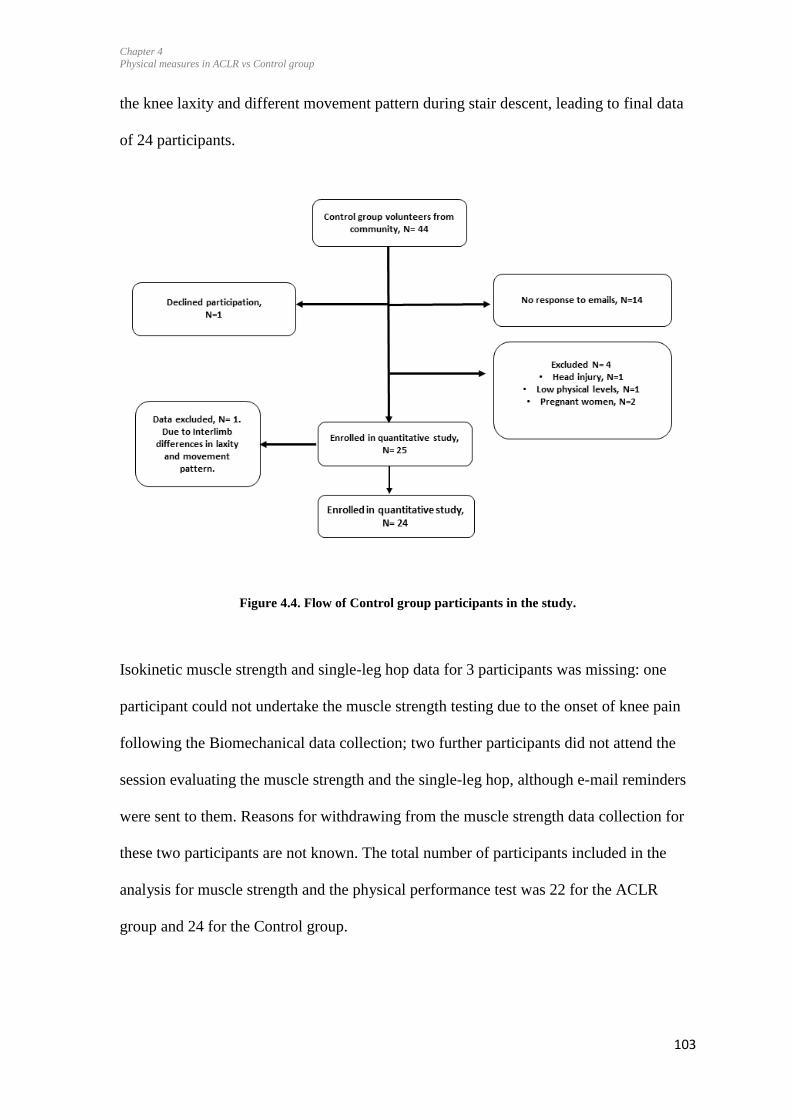
Chapter 4
Physical measures in ACLR vs Control group
103
the knee laxity and different movement pattern during stair descent, leading to final data
of 24 participants.
Figure 4.4. Flow of Control group participants in the study.
Isokinetic muscle strength and single-leg hop data for 3 participants was missing: one
participant could not undertake the muscle strength testing due to the onset of knee pain
following the Biomechanical data collection; two further participants did not attend the
session evaluating the muscle strength and the single-leg hop, although e-mail reminders
were sent to them. Reasons for withdrawing from the muscle strength data collection for
these two participants are not known. The total number of participants included in the
analysis for muscle strength and the physical performance test was 22 for the ACLR
group and 24 for the Control group.
Chapter 4
Physical measures in ACLR vs Control group
104
4.4.1 Data provided by ACC
The ACC provided the data for 21 participants with ACLR, confirming the dates of injury
and surgery (Appendix-C7). Data was also provided by the participants that related to
type of surgery and graft. Participants had attended, on average, 13 pre-operative (range
1-27) and 14 post-surgery physiotherapy treatments (range 1-48).
4.4.2 Patient-reported outcomes
Patient-reported outcomes of the ACLR group were compared to the Control group.
Participants with ACLR were from 2 to 10 (mean 4.7) years following surgery.
Information related to time since surgery and time between injury and surgery has been
presented in Appendix C9. Significant between-group differences were found for BMI,
with higher mean scores in the ACLR group (p<0.001) compared with the Control group
(Table 4.1). Results from the patient-reported outcomes indicated no difference in the
level of physical activity (Tegner scores, Table 4.2). The participants of this study were a
mixed cohort with athletic and non-athletic participants in both the ACLR and Control
group.

Chapter 4
Physical measures in ACLR vs Control group
105
Table 4.1. Participant characteristics
ACLR (SD) Control (SD) p-value
Men/Women (n) 25 (13W) 24 (13W)
Age (years) 30.8 (9.7) 31.4 (10) 0.829
BMI (kg/m2) 26.6 (3.6) 22.7 (3.6) <0.001
Injured side 18 left/7 right _
Limb dominancy 22 Right dominant 23 Right dominant
Time since ACLR
(years)
4.7 (1.8) (range 2 to 10) _
Graft types PT- 36%
HT-56%
Unknown-8%
Meniscal injuries 40%
n= Number, ACLR= Anterior cruciate ligament reconstruction, BMI= Body mass index, SD= Standard
deviation, PT= Patellar tendon, HT= Hamstring tendon graft, W=Women. Data indicate mean and
standard deviation for the participant characteristics.
For the KOOS, the largest differences were seen for the Sports/Recreation function
(ACLR: 75.8; controls: 98.0, p = <0.001, Table 4.2) and knee-related QOL (ACLR: 47.0;
controls: 80.4, p = <0.001). Significant differences were also found in KOOS4 scores with
lower scores in the ACLR group (mean= 65.6) compared to the Control group (86.5).
Participants with ACLR scored low (mean= 42.6) on the Confidence during sports scale.
Data from four control participants was missing for the Confidence during Sports scale
due to a technical error in the online questionnaire response section, therefore this data
was not compared to the Control group of this study.
From the results of SF-12, there were significant differences in the Physical Component
Summary score among the ACLR and Control group. The ACLR group had lower scores
on the Physical Component Summary compared to the Control group. No significant
differences were found between the groups for the Mental Component Summary Scores.
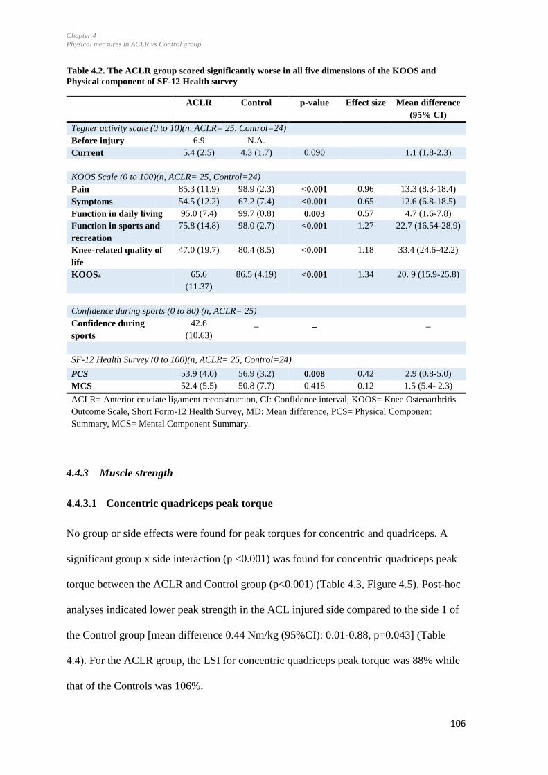
Chapter 4
Physical measures in ACLR vs Control group
106
Table 4.2. The ACLR group scored significantly worse in all five dimensions of the KOOS and
Physical component of SF-12 Health survey
ACLR Control p-value Effect size Mean difference
(95% CI)
Tegner activity scale (0 to 10)(n, ACLR= 25, Control=24)
Before injury 6.9 N.A.
Current 5.4 (2.5) 4.3 (1.7) 0.090 1.1 (1.8-2.3)
KOOS Scale (0 to 100)(n, ACLR= 25, Control=24)
Pain 85.3 (11.9) 98.9 (2.3) <0.001 0.96 13.3 (8.3-18.4)
Symptoms 54.5 (12.2) 67.2 (7.4) <0.001 0.65 12.6 (6.8-18.5)
Function in daily living 95.0 (7.4) 99.7 (0.8) 0.003 0.57 4.7 (1.6-7.8)
Function in sports and
recreation
75.8 (14.8) 98.0 (2.7) <0.001 1.27 22.7 (16.54-28.9)
Knee-related quality of
life
47.0 (19.7) 80.4 (8.5) <0.001 1.18 33.4 (24.6-42.2)
KOOS4 65.6
(11.37)
86.5 (4.19) <0.001 1.34 20. 9 (15.9-25.8)
Confidence during sports (0 to 80) (n, ACLR= 25)
Confidence during
sports
42.6
(10.63)
_ _ _
SF-12 Health Survey (0 to 100)(n, ACLR= 25, Control=24)
PCS 53.9 (4.0) 56.9 (3.2) 0.008 0.42 2.9 (0.8-5.0)
MCS 52.4 (5.5) 50.8 (7.7) 0.418 0.12 1.5 (5.4- 2.3)
ACLR= Anterior cruciate ligament reconstruction, CI: Confidence interval, KOOS= Knee Osteoarthritis
Outcome Scale, Short Form-12 Health Survey, MD: Mean difference, PCS= Physical Component
Summary, MCS= Mental Component Summary.
4.4.3 Muscle strength
4.4.3.1 Concentric quadriceps peak torque
No group or side effects were found for peak torques for concentric and quadriceps. A
significant group x side interaction (p <0.001) was found for concentric quadriceps peak
torque between the ACLR and Control group (p<0.001) (Table 4.3, Figure 4.5). Post-hoc
analyses indicated lower peak strength in the ACL injured side compared to the side 1 of
the Control group [mean difference 0.44 Nm/kg (95%CI): 0.01-0.88, p=0.043] (Table
4.4). For the ACLR group, the LSI for concentric quadriceps peak torque was 88% while
that of the Controls was 106%.
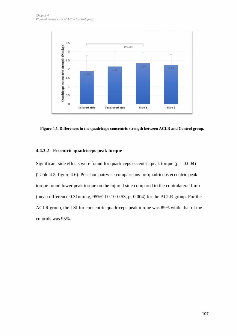
Chapter 4
Physical measures in ACLR vs Control group
107
Figure 4.5. Differences in the quadriceps concentric strength between ACLR and Control group.
4.4.3.2 Eccentric quadriceps peak torque
Significant side effects were found for quadriceps eccentric peak torque (p = 0.004)
(Table 4.3, figure 4.6). Post-hoc pairwise comparisons for quadriceps eccentric peak
torque found lower peak torque on the injured side compared to the contralateral limb
(mean difference 0.31nm/kg, 95%CI 0.10-0.53, p=0.004) for the ACLR group. For the
ACLR group, the LSI for concentric quadriceps peak torque was 89% while that of the
controls was 95%.

Chapter 4
Physical measures in ACLR vs Control group
108
Figure 4.6. Differences in quadriceps eccentric strength between ACLR and Control group.
4.4.3.3 Concentric hamstring peak torque
Significant side effects were found for hamstring concentric peak torque (p=0.019)
(Figure 4.7) while the group effects were not significant. The ACLR injured sides had
significantly lower concentric hamstring peak torque than the uninjured sides (p=0.019).
A significant group x side interaction (p=0.045) were found for hamstring concentric
strength (Figure 4.8) although the post-hoc comparisons were not significant. Post-hoc
pairwise comparisons showed significantly lower peak torque for concentric hamstring
(mean difference 0.05 nm/kg (95%CI): 0.03-0.15, p=0.003) for the injured side compared
to the contralateral limb in the ACLR group. For the ACLR group, the mean LSI for
concentric quadriceps peak torque was 92% while that of the controls was 101%.
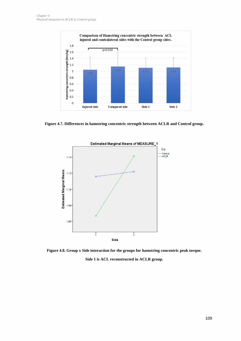
Chapter 4
Physical measures in ACLR vs Control group
109
Figure 4.7. Differences in hamstring concentric strength between ACLR and Control group.
Figure 4.8. Group x Side interaction for the groups for hamstring concentric peak torque.
Side 1 is ACL reconstructed in ACLR group.
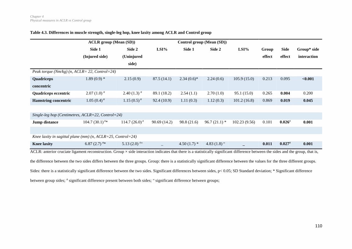
Chapter 4
Physical measures in ACLR vs Control group
110
Table 4.3. Differences in muscle strength, single-leg hop, knee laxity among ACLR and Control group
ACLR group (Mean (SD)) Control group (Mean (SD))
Side 1
(Injured side)
Side 2
(Uninjured
side)
LSI% Side 1 Side 2 LSI% Group
effect
Side
effect
Group* side
interaction
Peak torque (Nm/kg) (n, ACLR= 22, Control=24)
Quadriceps
concentric
1.89 (0.9) * 2.15 (0.9) 87.5 (14.1) 2.34 (0.6)* 2.24 (0.6) 105.9 (15.0) 0.213 0.095 <0.001
Quadriceps eccentric 2.07 (1.0) # 2.40 (1.3) # 89.1 (18.2) 2.54 (1.1) 2.70 (1.0) 95.1 (15.0) 0.265 0.004 0.200
Hamstring concentric 1.05 (0.4) # 1.15 (0.5) # 92.4 (10.9) 1.11 (0.3) 1.12 (0.3) 101.2 (16.8) 0.869 0.019 0.045
Single-leg hop (Centimetres, ACLR=22, Control=24)
Jump distance 104.7 (30.1) #* 114.7 (26.0) # 90.69 (14.2) 98.8 (21.6) 96.7 (21.1) * 102.23 (9.56) 0.101 0.026# 0.001
Knee laxity in sagittal plane (mm) (n, ACLR=25, Control=24)
Knee laxity 6.87 (2.7) #* 5.13 (2.0) #∞ _ 4.50 (1.7) * 4.83 (1.8) ∞ _ 0.011 0.027# 0.001
ACLR: anterior cruciate ligament reconstruction. Group × side interaction indicates that there is a statistically significant difference between the sides and the group, that is,
the difference between the two sides differs between the three groups. Group: there is a statistically significant difference between the values for the three different groups.
Sides: there is a statistically significant difference between the two sides. Significant differences between sides, p< 0.05; SD Standard deviation; * Significant difference
between group sides; # significant difference present between both sides; ∞ significant difference between groups;
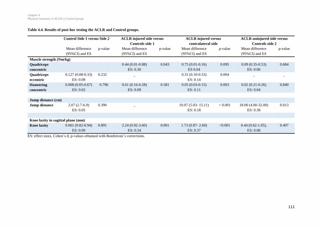
Chapter 4
Physical measures in ACLR vs Control group
111
Table 4.4. Results of post-hoc testing the ACLR and Control groups.
Control Side 1 versus Side 2 ACLR injured side versus
Controls side 1
ACLR injured versus
contralateral side
ACLR uninjured side versus
Controls side 2
Mean difference
(95%CI) and ES
p-value Mean difference
(95%CI) and ES
p-value Mean difference
(95%CI) and ES
p-value Mean difference
(95%CI) and ES
p-value
Muscle strength (Nm/kg)
Quadriceps
concentric
0.44 (0.01-0.88)
ES: 0.30
0.043 0.75 (0.01-0.16)
ES 0.04
0.095 0.09 (0.35-0.53)
ES: 0.06
0.684
Quadriceps
eccentric
0.127 (0.08-0.33)
ES: 0.08
0.232 _ 0.31 (0.10-0.53)
ES: 0.14
0.004 _ _
Hamstring
concentric
0.008 (0.05-0.67)
ES: 0.02
0.796 0.61 (0.16-0.28)
ES: 0.09
0.581 0.05 (0.03-0.15)
ES: 0.11
0.003 0.02 (0.21-0.26)
ES: 0.04
0.840
Jump distance (cm)
Jump distance 2.07 (2.7-6.9)
ES: 0.05
0.390 _ 10.07 (5.03- 15.11)
ES: 0.18
< 0.001 18.00 (4.00-32.00)
ES: 0.38
0.013
Knee laxity in sagittal plane (mm)
Knee laxity 0.061 (0.82-0.94)
ES: 0.09
0.891 2.24 (0.92-3.60)
ES: 0.54
0.001 1.73 (0.87- 2.60)
ES: 0.37
<0.001 0.44 (0.62-1.05),
ES: 0.08
0.407
ES: effect sizes, Cohen’s d, p-values obtained with Bonferroni’s corrections.

Chapter 4
Physical measures in ACLR vs Control group
112
4.4.4 Single-leg hop
No significant group effects were found for jump distances among the ACLR and Control
group (Table 4.3), but side effects were significant. Post-hoc pairwise comparisons
indicated a significant side-to-side difference for the ACLR group with a shorter distance
jumped by the injured side compared to the contralateral limb (mean difference 10.07,
95%CI 5.03- 15.11, p < 0.001)(Table 4.4). The group x side interaction was also
significant. Pairwise comparisons indicated a higher mean jump distance on the uninjured
side of ACLR group compared to the Side 2 of the Control group (18.00, 95% CI 4.00-
32.00, p=0.013) but a shorter distance for the ACLR injured side compared to the Control
Side 1 (Figure 4.9).
Figure 4.9. Differences in single-leg hop performance between ACLR and Control group.
4.4.5 Knee laxity in sagittal plane
Significant group (p = 0.011) and side (p = 0.027) effects and a significant group x side
interaction were found for knee laxity in the sagittal plane (Table 4.3) (Figure 4.10). Post-
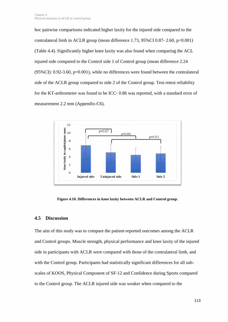
Chapter 4
Physical measures in ACLR vs Control group
113
hoc pairwise comparisons indicated higher laxity for the injured side compared to the
contralateral limb in ACLR group (mean difference 1.73, 95%CI 0.87- 2.60, p<0.001)
(Table 4.4). Significantly higher knee laxity was also found when comparing the ACL
injured side compared to the Control side 1 of Control group (mean difference 2.24
(95%CI): 0.92-3.60, p=0.001), while no differences were found between the contralateral
side of the ACLR group compared to side 2 of the Control group. Test-retest reliability
for the KT-arthrometer was found to be ICC- 0.86 was reported, with a standard error of
measurement 2.2 mm (Appendix-C6).
Figure 4.10. Differences in knee laxity between ACLR and Control group.
4.5 Discussion
The aim of this study was to compare the patient-reported outcomes among the ACLR
and Control groups. Muscle strength, physical performance and knee laxity of the injured
side in participants with ACLR were compared with those of the contralateral limb, and
with the Control group. Participants had statistically significant differences for all sub-
scales of KOOS, Physical Component of SF-12 and Confidence during Sports compared
to the Control group. The ACLR injured side was weaker when compared to the
Chapter 4
Physical measures in ACLR vs Control group
114
contralateral side and to the Controls. Physical performance on the contralateral side was
higher than the control side 1. Higher laxity was present in the ACLR group on the
injured side compared to the contralateral side and to the Control group.
4.5.1 Patient-reported outcomes
There were significant differences in all subscales of the KOOS scores with ACLR group
experiencing more pain, symptoms, lower levels of knee function in daily life and sports,
and lower quality of life compared to the Control group, but there was no significant
difference between the level of physical activities among both groups (Tegner scores).
The KOOS scores in all subscales ACLR group were lower than the ‘acceptable state’
scores as given by the (Muller et al., 2016). This finding may be due to the older age
(mean 30.5 years) and greater time following surgery (4.7 years) for participants in the
current study, compared to participants in the Muller study (mean age 26.2 years, and 3.4
years following ACLR). One of the goals for the management of the injury is to optimise
the quality of life in the long-term. However, this goal doesn’t seem to be fully
accomplished in this group of participants. The presence of symptoms has been
negatively correlated with levels of satisfaction (Kocher et al., 2002). This group of
participants still reported experiencing knee-related symptoms (KOOS symptoms=
54.5/100), which may have affected their perceptions about their quality of life, defined
as the expectations of health versus actual experience (Carr, Gibson, & Robinson, 2001).
As participants of this study were relatively young (median, 28.5 years), their health
expectations may be high, which, in turn, may have influenced their rating of current
quality of life, as defined by the KOOS QOL scores (47.0/100) and the PCS of SF-12.
A previous study has reported quality of life for participants 12 to 14 years following
ACL injury with visible radiographic changes for knee osteoarthrosis (Von Porat et al.,
Chapter 4
Physical measures in ACLR vs Control group
115
2004). They indicated mean (SD) scores for KOOS QOL of 60 (24.6) and 63 (28.7) for
KOOS function in sports and recreation. While the mean KOOS QOL in the current study
was lower than that (47.0), the mean KOOS function in sports and recreation score of
75.8 (14.8) indicated that our participants were more active in sports and recreational
activities, and had better knee function in their daily life activities compared to the above-
mentioned study. The KOOS results were also supported by the SF -12 Physical
Component Summary scores, which were significantly lower for the ACLR group than
the Control group (ACLR= 53.9, Control= 56.9, p=0.008). Knee-related quality of life
scores of the participants of this study (KOOS QOL mean score = 47) were comparable to
those of the participants with knee osteoarthritis (KOOS QOL mean score = 50.0)
(Aksekili et al., 2016). Those participants had knee osteoarthritis classified as Grades 2 or
3 on the Kellgren-Lawrence scale. Participants in our study had less pain (mean= 85.3)
and more knee-related symptoms (mean= 54.5) compared with the participants with knee
osteoarthritis pain (mean= 60.0) and knee-related symptoms (mean= 71.4). However,
interestingly, despite lower levels of knee function (KOOS ADL score 70 versus 95), the
quality of life was similar in both groups (Aksekili et al., 2016). The similarity in the
quality of life in both groups could be attributed to their different levels of sports
recreational activities, and their preferences and expectations from life. Furthermore,
based on findings of knee osteoarthritis being evident as soon as 5 years following ACL
injury or surgery (Wellsandt et al., 2016), it is possible that some of the participants with
ACLR already exhibited knee osteoarthritis in one or more compartments. However, as
no investigatory procedures such as X-ray and MRI were performed, it is a speculation.
The participants with ACLR, on average 4.5 years following surgery (range 2–10),
appeared to have continued low confidence in the injured knee during sports, despite
indicating good function in sports and daily living (KOOS: sports and recreation mean=
Chapter 4
Physical measures in ACLR vs Control group
116
76/100), and mean Tegner scores post-injury (6). It is difficult to speculate on the reasons
for reduced confidence level in sports from this study; however, it is important to
investigate as reduced levels in confidence can affect performance levels in sports
(Ardern et al., 2012). Lower confidence during sports may be related to fear of re-injury,
exacerbating symptoms, and reduced physical function (Hart, Collins, Ackland, &
Crossley, 2015). Therefore, improving confidence in the knee may relieve knee
symptoms and enhance physical function in this group of participants.
SF12 scores were compared to the national averages of the South Australian data using a
PCS of 52.5 and MCS of 52.4 as the cut-off for an individual’s score based on the
standard error of measurement ± 6.97 (Utah Health Status Survey, 2001). The PCS scores
of the ACLR group (53.9 (4.0) in the current study were similar to those figures, (Quality
of life in Southern Australia, 2005). The SF scores can vary based on the underlying
conditions such as arthritis, high blood pressure, high cholesterol, overweight and obesity,
smoking and high risk alcohol use. We did not account for the above-mentioned factors.
4.5.2 Muscle strength
According to our results participants had strength deficits on the injured side compared to
the contralateral limb, and the ACLR injured side had weaker concentric quadriceps peak
torque compared to the controls. Deficits in knee extension strength have been reported
by a recent study with participants at 2.5 years on an average following surgery. Strength
deficits of 6 to 9% were found in the involved leg compared to the uninvolved leg. These
deficits were small compared to the other studies in the literature, and were considered
due to an incomplete rehabilitation of the ACLR knee and/or the inability to fully activate
the muscle as a result of the initial and post-operative joint damage (Otzel et al., 2015).
Participants in our study were from 2 to 10 years from surgery and were a mixed-cohort
Chapter 4
Physical measures in ACLR vs Control group
117
from the community varying from sedentary to physically active life-styles. Therefore, it
is difficult to comment on the reasons for the present muscle strength deficits. Deficits are
most pronounced in the concentric quadriceps muscle strength and the least in the knee
flexor muscle group. This is similar to another study where 10% strength deficits were
found in the concentric knee extension group 4-7 years following surgery (Moisala et al.,
2007). Results of our study can also be compared to another study exploring the outcomes
of ACLR at 20 years following surgery and had similar peak torque values for the knee
flexor and extensor strength (normalised to body weight). That study indicated the
presence of concentric and eccentric quadriceps and hamstring strength deficits (Tengman
et al., 2014). The LSI values of our study for quadriceps concentric and eccentric values
(87 % and 89% respectively) were similar to the mean values, with values of (89% and
86%, respectively) Tengman et al., 2014. Similarly, the LSI for concentric hamstring in
our study was 92.4% and for the control group was 101%. Though the participants of the
Tengman study had a mean age of 45 years, which is higher than our study participants
(mean age 30.8), the extent of physical impairment is similar. It may mean that
impairments may not recover with time. Ninety percent symmetry in quadriceps (and
hamstring) strength was recently identified by a consensus as a measure important for
achieving successful outcome after ACLR (Lynch et al., 2013). Our cohort had symmetry
in the concentric hamstring muscle strength; however, it was lower for concentric and
eccentric quadriceps strength. Previously rehabilitation programs including aggressive
quadriceps strengthening and perturbation training have been proven successful in
restoring limb symmetries (Logerstedt, Lynch, Axe, & Snyder-Mackler, 2013). The
participants of the current study are from mid- to long-term following surgery, therefore,
it needs to be explored further if the similar rehabilitation protocol can help to restore the
symmetry at this stage.
Chapter 4
Physical measures in ACLR vs Control group
118
Reduced muscle strength can be due to complex interaction between various components
of the body, for instance, there is reduced proprioception following the surgery at the
knee (Bonfim et al., 2003), along with less activation of the sensory cortex (Valeriani et
al., 1996). There is lower activation of quadriceps muscle following the ACL injury and
surgery (Hart et al., 2010), which is thought to be due to the alterations of corticomotor
excitability (Pietrosimone et al., 2012). Changes in the central nervous system are
supported by more recent findings of reduced activation of the ipsilateral motor cortex
following ACL injury (Grooms et al., 2017). Although the ruptured ligament may be
repaired with a graft, complex interaction and deficit seen at the muscular, neural and
central nervous system level is perhaps not fully restored to optimal levels. Therefore,
management should not focus on muscle strength training only, as there is need of an
exercise protocol enhancing the link between the joint, muscle, and central nervous
system.
Muscle strength deficits may be a risk factor for post-traumatic osteoarthritis (Oiestad,
Holm, Gunderson, Myklebust, & Risberg, 2010). Keays et al., 2007 followed participants
with ACLR from 6 months to 6 years following surgery, and found a 6% deficit in
quadriceps muscle strength in those who had undergone patellar tendon grafts. The
quadriceps deficit appeared to be associated with radiographic evidence of knee
osteoarthritis. Similarly, reduced joint space width was found in participants with lower
concentric quadriceps and hamstring muscle strength following surgery ACLR, on
average 4 years following surgery (Tourville et al., 2014). An association between
quadriceps muscle strength and primary osteoarthritis is already known (Farr et al., 2010;
Mihelic, Jurdana, Jotanovic, Madjarevic, & Tudor, 2011; Palmieri-Smith, Thomas,
Karvonen-Gutierrez, & Sowers, 2010). Therefore, lower muscle strength could
potentially be a risk factor for osteoarthritis in this group of participants. Our study found

Chapter 4
Physical measures in ACLR vs Control group
119
between-side differences of 5% for concentric hamstring and 22.3% for eccentric
quadriceps in the ACLR group (2 to 10 years following surgery). While we did not
examine radiographic changes related to osteoarthritis, the question is raised whether
these muscle deficits, particularly for the eccentric quadriceps strength could be a marker
for future knee osteoarthritis.
4.5.3 Single-leg hop
The participants with ACLR hopped a shorter distance on the injured side compared to
the uninjured side. The LSI for the ACLR was 90%, whereas LSI for the Control group
was 102%. Their hop distance for the uninjured side, however, was significantly longer
compared to the Control group side 2. The jump distances of our study are similar to hop
distance in another study (Injured side= 112.0 cm, Contralateral side= 119.0 cm)
exploring the functional outcomes in participants with ACLR 20 years following surgery
(Tengman et al., 2014). Participants of that study were of higher age group (mean=45
years) but had similar level of Physical activity levels (Tegner score=4), which may be
responsible for the similarity in the results. The lower jump distance on the injured side
indicate the preference of participants for less loading activities on the injured side; which
may represent the consequences of the surgery as the contralateral side had the higher
jump capacity.
In another study, participants with poor muscle strength (<85%) performed poorly during
the hop test compared with participants with good muscle strength (>90%) and an
uninjured Control group (Schmitt, Paterno, & Hewett, 2012), indicating an association
between jump performance and muscle strength. Other factors such as reduced
proprioception, fear of injury, hesitation during weight bearing, and lower confidence in
this group of participants may also have contributed towards the single-leg hop LSI.

Chapter 4
Physical measures in ACLR vs Control group
120
Muscle weakness, particularly eccentric muscle strength, is required to control the limb
during landing following the jump. The eccentric quadriceps deficit found in these
participants may thus have contributed to, or be associated with, the reduced hop distance.
Similar conclusions may be drawn relating to the sports- related task during competitive
sports.
4.5.4 Knee laxity in the sagittal plane
Participants with ACLR had higher laxity on the injured side compared to the
contralateral side and the Control group. The standard error of measurement (SEM) for
KT-Arthrometer was 2.2 mm, and the side-to-side mean difference for the ACLR group is
less than the SEM, therefore, the side to side differences found in the study should be
considered with caution. However, the mean differences were greater than the SEM for
the mean difference between the injured side and the side 2 of the Control group. Higher
laxity on the injured side is considered multifactorial and may be due to the type of graft,
change in the graft tension over time, preconditioning of the graft (Ejerhed et al., 2001)
and positioning of the tunnel at the time of surgery (Rayan et al., 2015). In the present
study, we were unable to control for the above-mentioned factors. Participants with
different grafts were included in the study which could have influenced the findings of the
study. Our results are in contrast to previous studies that have either reported the reduced
sagittal plane tibio-femoral laxity at 7 years following surgery (Ahldén et al., 2009), or
have reported no change in the graft laxity over time from mid- to long-term following
surgery (Salmon et al., 2006; Shelbourne et al., 1995). Higher laxity may potentially
reduce the stability of the joint and can make the joint vulnerable to the physical stresses
experienced during ambulatory activities, which can be a major risk factor for
osteoarthritis. Theoretically, joint laxity observed may be controlled by muscle action.

Chapter 4
Physical measures in ACLR vs Control group
121
However, as no EMG for the thigh muscles was performed, it is difficult to comment on
whether the muscles were controlling the increased knee laxity in this group of
participants
Taken together, higher laxity with poor muscle strength and poor performance in the
single-leg hop in participants with the ACLR persist in the mid- to long-term following
surgery, and indicate the presence of physical impairment which can, potentially, be
responsible for the onset and progression of osteoarthritis.
4.5.5 Limitations
Outcomes of surgery may be dependent on the graft type (Spindler et al., 2004). As the
participants were recruited from the community, we could not control for the type of graft
in the study participants. Due to the cross-sectional design, all measurements were taken
only at one-time point. Thus, the influence of time following ACLR on the included
variables cannot be established. Higher laxity on the contralateral knee compared to the
Control group side 2 indicates potential generalised higher laxity in ACLR group. For the
physical performance, only a single-leg hop test was used instead of a battery of tests.
However, this test is known to be reliable either used alone or along with the battery of
tests (Palmieri-Smith & Lepley, 2015). Moreover, as this test is only analysing the
performance of one limb at a time, it reduces the chances of compensatory strategies by
the contralateral limb during execution of the test. Participants were at different levels of
physical activity varying from a Tegner score of 9 (competitive sports like soccer, ice
hockey) to as low as 3 (competitive and recreational sports like swimming). This could
have led to some dilution in the average scores of the ‘confidence during sports’ scale.
However, it is already known that those who return to sports have less fear of injury
(Ardern et al., 2012).
Chapter 4
Physical measures in ACLR vs Control group
122
4.6 Conclusions
Differences in patient-reported outcomes, muscle strength, physical performance, and
knee laxity were evident in ACLR cohort compared to Control group. These physical
impairments indicate suboptimal joint function along with a lower quality of life
compared to the age- and gender-matched Control group. Higher laxity may potentially
reduce the stability of the joint and can make the joint vulnerable to the physical stresses
experienced during ambulatory activities, which can be a major risk factor for
osteoarthritis.
4.7 Summary
This chapter presented and discussed the physical impairments in participants with ACLR
up to 10 years following surgery. Participants also experienced low confidence levels
during sports and a poor quality of life compared to the age- and gender-matched
controls. The next study (Chapter 5) explored the participants’ perspectives of the
outcomes of ACLR relating to their life.

Chapter 5
Participants’ perspectives of ACLR
123
5 Participants’ perspectives of the outcome of anterior cruciate
ligament (ACL) reconstruction surgery: a mixed-method study.
5.1 Prelude to Chapter 5
This mixed-method study provided an in-depth understanding of participants’ concerns
and experiences of the outcomes of ACLR. This study highlighted the factors related to
fear of re-injury, reduced confidence in the injured limb during sports, hesitancy in
weight bearing on the injured side and participants’ daily struggles related to activities of
daily life. These are important findings for the patients and clinicians as it can lead to
longer-term deficits and biomechanical implications.
Chapter 5
Participants’ perspectives of ACLR
124
Chapter 5
5.2 Background
There appears to be a trend for increasing number of patients choosing ACLR over non-
surgical rehabilitation only (Sanders et al., 2016). Most patients undergo ACLR with the
intention to return to sports (Heijne et al., 2008; Thing, 2006), however, only 55% are
able to return to competitive sports (Ardern et al., 2014). Participants may continue to
play for approximately 2 years and thereafter often tend to change to a lower level of
sports (Ardern et al., 2014; Smith, Rosenlund, Aune, MacLean, & Hillis, 2004) due to
various reasons, such as fear of re-injury and changed life or occupational commitments
(Doyle, Wilson, & King, 2013; Kvist, Ek, Sporrstedt, & Good, 2005). Those who
continue to play competitive sport appear to report lower knee-related quality of life in
the long-term (up to 20 years post-ACLR) than those who have changed to recreational-
level sports (Filbay et al., 2016). Decision making regarding continuation of level of
sports participation thus appears to be one of the factors influencing the reported quality
of life.
Knee injuries have been reported to decrease knee-specific function and health-related
quality of life in collegiate athletes (Lam, Thomas, Valier, McLeod, & Bay, 2015).
Furthermore, development of osteoarthritis also impacts the health-related quality of life
in general (Salaffi, Carotti, Stancati, & Grassi, 2005; Turner, Barlow, & Heathcote-
Elliott, 2000). Different results have been reported regarding the health-related quality of
life in participants with ACLR, varying from low at an average of 5 years (Filbay,
Ackerman, Russell, Macri, & Crossley, 2014), to that of similar quality of life to
uninjured controls at an average of 11.5 years post-surgery (Möller et al., 2009) as
discussed in Chapter 2 section 2.6.2.
Chapter 5
Participants’ perspectives of ACLR
125
Outcomes of an intervention are usually considered in terms of restoration or
improvement of individual-specific function related to activities of daily life (ADL),
sports, recreation and occupation-related demands (Tanner, Dainty, Marx, & Kirkley,
2007). Understanding the individual patients’ perspectives of outcomes of ACLR is a key
concept of patient-centered care (Epstein & Street, 2011). A recent qualitative study with
the participants up to 3 years following ACLR indicated that recovering from an ACL
injury experience was a long, arduous and disruptive “journey” influencing their
individual identity (Scott et al., 2017). To improve our understanding of outcomes of
ACL injury and reconstruction, the aim of the present study was to explore the
participants’ experiences of the outcomes of their surgery more than 2 years following
surgery in relation to physical activity, sports, occupation and health-related quality of
life.
5.3 Methods
5.3.1 Study Design
This is a mixed-method study design with patient-reported outcome measures and semi-
structured interviews. Consolidated Criterion for Reporting Qualitative Research
(COREQ) checklist was used for reporting the study (Tong, Sainsbury, & Craig, 2007).
Prior to the beginning of the study, researchers bracketed their perceptions and thoughts
related to the field of the study. Bracketing in descriptive phenomenology entails
researchers setting aside their pre-understanding and acting non-judgmentally along with
adding scientific rigour (Sorsa, Kiikkala, & Åstedt-Kurki, 2015)(Appendix-D1).
Moreover, a phenomenological approach to data was followed, where bracketing is
considered important.

Chapter 5
Participants’ perspectives of ACLR
126
5.3.2 Recruitment, inclusion and exclusion criteria for the study
Participants were recruited from the local community via advertisements for quantitative
studies as explained in Chapter 4, section 4.3.4. Participants had provided written
informed consent to participate in the quantitative study and they opted to take part in the
qualitative study by stating confirming “willingness to participate in an interview” on
their informed consent form (Appendix- B9). The participants were selected sequentially
based on that response. Inclusion and exclusion criteria has been explained in Chapter 4,
section 4.3.6. In addition to the previously mentioned criteria, for this study, participants
who were fluent English speakers, were included.
5.3.3 Procedures
Participants completed an online questionnaire (Qualtrics, Provo, UT, USA, 2015) prior
to the taking part in the quantitative studies and the interview. The questionnaire included
demographic data and the patient-reported outcome measures: the Tegner Activity Scale
(Tegner & Lysholm, 1985), Sports Confidence Scale (Ardern et al., 2012), KOOS (Roos
et al., 1998), and SF-12 (Ware et al., 1996). The KOOS4 was calculated as an average
score of four sub-scales excluding function during daily activities due to a high ceiling
effect for that sub-scale (Senorski et al. 2017).
Face-to-face individual interviews were held. Four of the interviews were conducted only
by MK, while two interviewers (MK and GS, both females) were present for the
remaining six interviews. GS is an established researcher within the field of
musculoskeletal physiotherapy, in addition to also having extensive experience in
rehabilitation of patients with ACLR. MK is the PhD candidate, with clinical experience
in musculoskeletal rehabilitation.
Chapter 5
Participants’ perspectives of ACLR
127
All interviews were conducted in the School of Physiotherapy, lasting from 20 to 40
minutes. No interview was repeated. The interview guide had open-ended questions and
was developed and refined by the research team via review and reflection after 2 to 3
interviews (Table 5.1). The interview guide was updated following discussion in the
research team regarding the on-going interview process following 2-3 interviews. It was
to reflect back on the process if there is any room for the improvement. Sequence of
questions in the interview guide was changed by placing the open-ended questions in the
start of the interview. While the guide provided some structure, the selection of specific
questions and their respective order depended on how the interview proceeded. Field
notes were made during and after the interview. Special phrases, words or expressions of
the participants were noted down during the interview. It helped in the data analysis while
generating the codes for the participants. The interviews were recorded with a digital
audio-recorder (Sony R- IC Recorder) and transcribed verbatim.
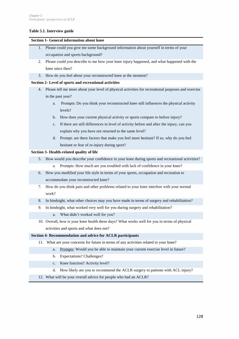
Chapter 5
Participants’ perspectives of ACLR
128
Table 5.1. Interview guide
Section 1- General information about knee
1. Please could you give me some background information about yourself in terms of your
occupation and sports background?
2. Please could you describe to me how your knee injury happened, and what happened with the
knee since then?
3. How do you feel about your reconstructed knee at the moment?
Section 2- Level of sports and recreational activities
4. Please tell me more about your level of physical activities for recreational purposes and exercise
in the past year?
a. Prompts: Do you think your reconstructed knee still influences the physical activity
levels?
b. How does your current physical activity or sports compare to before injury?
c. If there are still differences in level of activity before and after the injury, can you
explain why you have not returned to the same level?
d. Prompt: are there factors that make you feel more hesitant? If so, why do you feel
hesitant or fear of re-injury during sport?
Section 3- Health-related quality of life
5. How would you describe your confidence in your knee during sports and recreational activities?
a. Prompts: How much are you troubled with lack of confidence in your knee?
6. How you modified your life style in terms of your sports, occupation and recreation to
accommodate your reconstructed knee?
7. How do you think pain and other problems related to your knee interfere with your normal
work?
8. In hindsight, what other choices may you have made in terms of surgery and rehabilitation?
9. In hindsight, what worked very well for you during surgery and rehabilitation?
a. What didn’t worked well for you?
10. Overall, how is your knee health these days? What works well for you in terms of physical
activities and sports and what does not?
Section 4- Recommendation and advice for ACLR participants
11. What are your concerns for future in terms of any activities related to your knee?
a. Prompts: Would you be able to maintain your current exercise level in future?
b. Expectations? Challenges?
c. Knee function? Activity level?
d. How likely are you to recommend the ACLR surgery to patients with ACL injury?
12. What will be your overall advice for people who had an ACLR?
Chapter 5
Participants’ perspectives of ACLR
129
5.3.4 Data analysis
The quantitative data were entered into the Microsoft Excel spreadsheet (Microsoft Office
2013) and descriptive analyses (median and ranges) were performed for the patient-
reported outcome measures. Wilcoxon signed rank test (IBM SPSS 23) was performed
for Tegner Activity Scale to compare the pre- and post-injury physical activity levels.
The General Inductive Method was used for the interview data (Thomas, 2006). This
method allows to condense extensive and varied raw text data into a brief, summary
format and also helps to establish clear links between the research objectives. Moreover,
this method allows the analysis to be data driven. The transcriptions were read multiple
times and text segments that reflected the participants’ experience were identified and
coded. The codes were categorised and the researchers developed links between these
categories, identifying themes relevant for the research aims. After the primary analysis
of the available data and when no new codes evolved from the next two interviews, it was
deemed that data saturation had occurred (Baker, Edwards, & Doidge, 2012; Fugard &
Potts, 2015). Data saturation was reached by the eighth participant and no further
categories were added with the final two participants. Moreover, the participant sample
consisted of the mixture of people with different ages and professions, indicating a rich
data.
One researcher (MK) analysed all the interviews, while a second researcher (GS)
analysed every second interview. The codes produced by the researchers were data-driven
and were then compared, discussed and negotiated. The categories and emerging themes
and sub-themes discussed and confirmed within the research team. The key themes were
cross-referenced back to the original text to ensure that it was an accurate representation
of the participants’ perceptions of their experiences. Supporting quotes that most
Chapter 5
Participants’ perspectives of ACLR
130
accurately reflected the key themes and sub-themes were selected. A summary of the
results were sent to the participants for verification and feedback from the participants
was synthesised into the final analysis. The interpretation of these combined findings is
presented in the discussion section.
5.4 Results
Twelve participants were asked to participate in the study. Two of them chose not to take
part in the study due to unknown reasons. Seven females and three males (median age
28.5 years, range 20-52 years) participated in the study. They had undergone ACLR at a
median 6 years (range 3-10 years) previously and five of the participants had injured their
left side. Four participants had patellar tendon graft, five had hamstring and one
participant was operated using the allograft. Four of the participants were students while
the remainder had sedentary occupations.
There was no statistically significant difference between pre- and post-injury level of
physical activities, as defined by the Tegner Activity Scale (Wilcoxen Signed Ranks test
p=0.37). Based on the Tegner Activity Scale, five participants had returned to their pre-
injury level of sports participation, one had increased the level (pre-injury level 3; post-
injury level 4), while for remaining four participants the level of participation had
decreased by two or more scores (Table 5.2).
The KOOS-function daily living indicated that participants had good function with
activities of daily living (median 99/100) yet still experienced knee-related symptoms
(KOOS-Symptoms median 59/100), and impaired knee function during sports and
recreational activities (KOOS-Sports and recreational activities median 84/100). The
median KOOS4 was 69/100 (Table 5.2). Overall, they scored low quality of life (KOOS-
Chapter 5
Participants’ perspectives of ACLR
131
Quality of life= 53/100; range from 19 to 75) (SF- PCS= 55.50, MCS: 44.80). Results
from the Sports Confidence scale (Table 5.2) indicated that, as a group, the participants
still had decreased confidence in their knee (41.5/80).
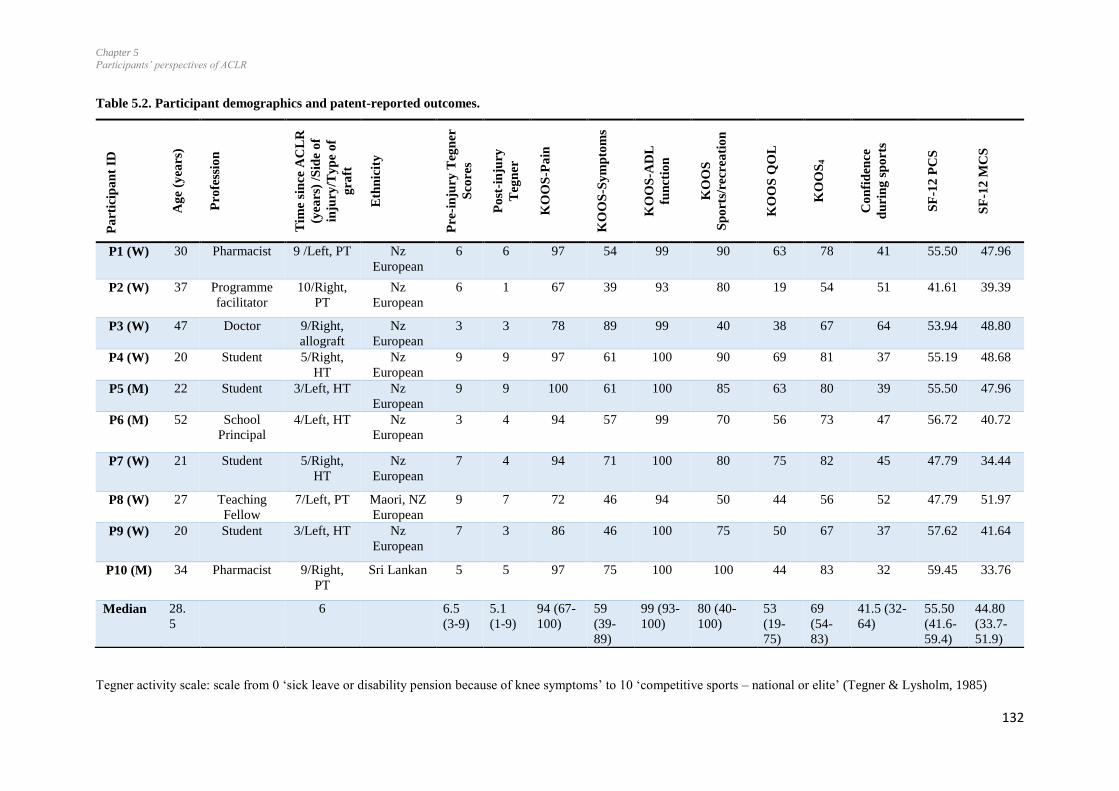
Chapter 5
Participants’ perspectives of ACLR
132
Table 5.2. Participant demographics and patent-reported outcomes.
Tegner activity scale: scale from 0 ‘sick leave or disability pension because of knee symptoms’ to 10 ‘competitive sports – national or elite’ (Tegner & Lysholm, 1985)
Pa
rtic
ipa
nt
ID
Ag
e (y
ears
)
Pro
fess
ion
Tim
e si
nce
AC
LR
(yea
rs)
/Sid
e o
f
inju
ry/T
yp
e o
f
gra
ft
Eth
nic
ity
Pre
-in
jury
Teg
ner
Sco
res
Po
st-i
nju
ry
Teg
ner
KO
OS
-Pa
in
KO
OS
-Sy
mp
tom
s
KO
OS
-AD
L
fun
ctio
n
KO
OS
Sp
ort
s/re
cre
ati
on
KO
OS
QO
L
KO
OS
4
Co
nfi
den
ce
du
rin
g s
po
rts
SF
-12
PC
S
SF
-12
MC
S
P1 (W) 30 Pharmacist 9 /Left, PT Nz
European
6 6 97 54 99 90 63 78 41 55.50 47.96
P2 (W) 37 Programme
facilitator
10/Right,
PT
Nz
European
6 1 67 39 93 80 19 54 51 41.61 39.39
P3 (W) 47 Doctor 9/Right,
allograft
Nz
European
3 3 78 89 99 40 38 67 64 53.94 48.80
P4 (W) 20 Student 5/Right,
HT
Nz
European
9 9 97 61 100 90 69 81 37 55.19 48.68
P5 (M) 22 Student 3/Left, HT Nz
European
9 9 100 61 100 85 63 80 39 55.50 47.96
P6 (M) 52 School
Principal
4/Left, HT Nz
European
3 4 94 57 99 70 56 73 47 56.72 40.72
P7 (W) 21 Student 5/Right,
HT
Nz
European
7 4 94 71 100 80 75 82 45 47.79 34.44
P8 (W) 27 Teaching
Fellow
7/Left, PT Maori, NZ
European
9 7 72 46 94 50 44 56 52 47.79 51.97
P9 (W) 20 Student 3/Left, HT Nz
European
7 3 86 46 100 75 50 67 37 57.62 41.64
P10 (M) 34 Pharmacist 9/Right,
PT
Sri Lankan 5 5 97 75 100 100 44 83 32 59.45 33.76
Median 28.
5
6 6.5
(3-9)
5.1
(1-9)
94 (67-
100)
59
(39-
89)
99 (93-
100)
80 (40-
100)
53
(19-
75)
69
(54-
83)
41.5 (32-
64)
55.50
(41.6-
59.4)
44.80
(33.7-
51.9)
Chapter 5
Participants’ perspectives of ACLR
133
KOOS: Knee injury and Osteoarthritis Outcome Score: ‘0’ indicates extreme knee problems and ‘100’ indicates no knee problems, KOOS4: average of Pain, Symptoms,
function during sports and recreation, and quality of life., ADL: activities of daily living, QoL: Quality of Life, Confidence during sports: Scored out of 80, higher the scored
lower is the fear of injury, Short form-12 Health Survey: Scores range from 0 to 100, where a zero score indicates the lowest level of health measured by the scales and 100
indicates the highest level of health, SF-12 PCS Physical component score, MCS: Mental component score, PT: Patellar tendon; HT: Hamstring tendon. M: Man, W: Woman.
Chapter 5
Participants’ perspectives of ACLR
134
From the interview data, sixteen categories were converged into three overlapping themes
(Table 5.3). Among these themes two themes, ‘Fear of re-injury versus confidence
continuum’ and ‘Live life normally’, answered our research aim. The third theme, ‘need
of reassurance and maintenance of the knee’ was important to report as it explained the
participants’ knee-related concerns clearly. Themes and subthemes with supporting
quotes are further described below. Ellipsis indicates an intentional omission of a word,
or sentence from a text without altering its original meaning. Brackets indicate where
additional words have been added into the sentence to clarify the meaning. Additional
supporting quotes for the themes are shown in the Appendix-D2.
Focus of this study was to obtain the deeper understandings related to the participants’
health concerns. Therefore, the quantitative and qualitative data were interpreted together.
For instance, results from the Confidence During Sports scale indicated the presence of
fear of injury in participants, the results of the interview data indicated the reasons for the
persistence of fear of injury.
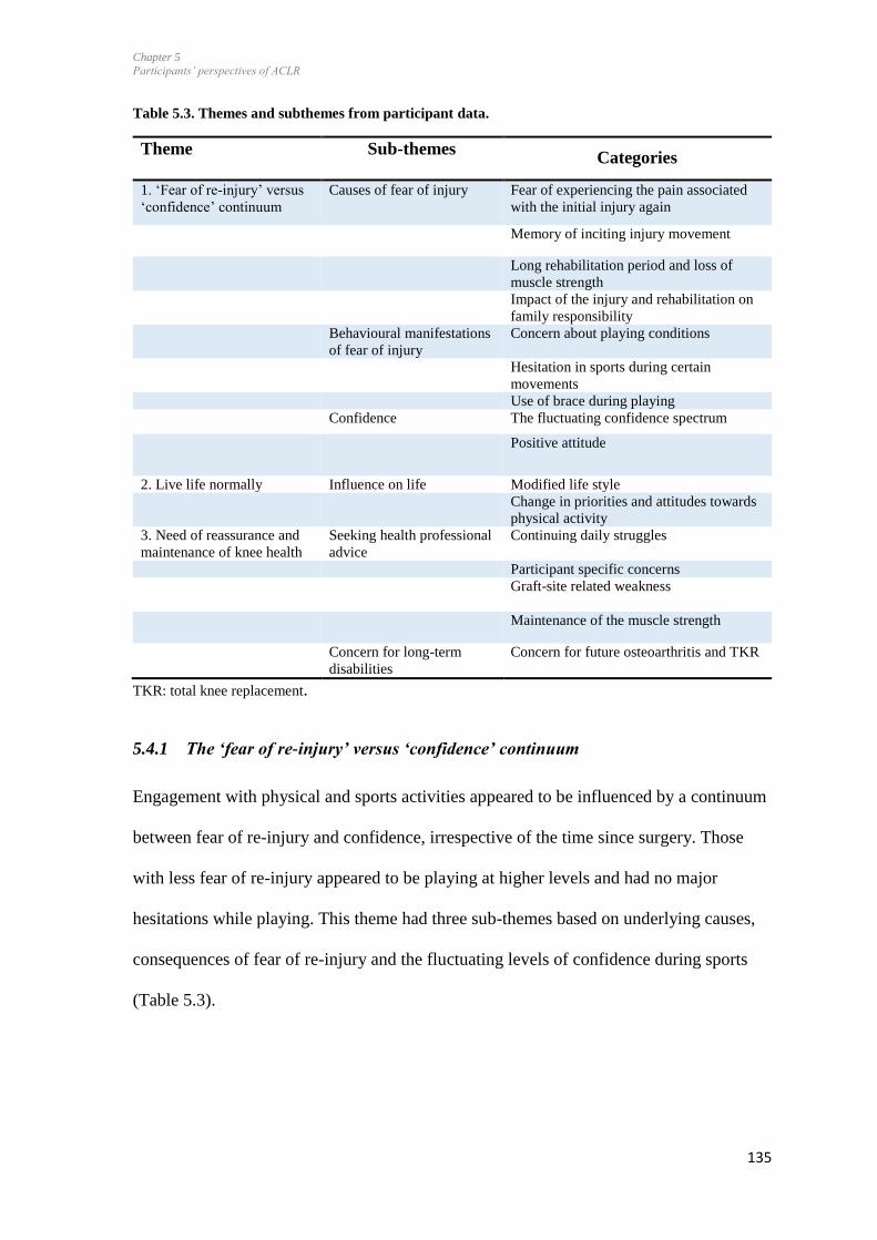
Chapter 5
Participants’ perspectives of ACLR
135
Table 5.3. Themes and subthemes from participant data.
Theme Sub-themes Categories
1. ‘Fear of re-injury’ versus
‘confidence’ continuum
Causes of fear of injury Fear of experiencing the pain associated
with the initial injury again
Memory of inciting injury movement
Long rehabilitation period and loss of
muscle strength
Impact of the injury and rehabilitation on
family responsibility
Behavioural manifestations
of fear of injury
Concern about playing conditions
Hesitation in sports during certain
movements
Use of brace during playing
Confidence The fluctuating confidence spectrum
Positive attitude
2. Live life normally Influence on life Modified life style
Change in priorities and attitudes towards
physical activity
3. Need of reassurance and
maintenance of knee health
Seeking health professional
advice
Continuing daily struggles
Participant specific concerns
Graft-site related weakness
Maintenance of the muscle strength
Concern for long-term
disabilities
Concern for future osteoarthritis and TKR
TKR: total knee replacement.
5.4.1 The ‘fear of re-injury’ versus ‘confidence’ continuum
Engagement with physical and sports activities appeared to be influenced by a continuum
between fear of re-injury and confidence, irrespective of the time since surgery. Those
with less fear of re-injury appeared to be playing at higher levels and had no major
hesitations while playing. This theme had three sub-themes based on underlying causes,
consequences of fear of re-injury and the fluctuating levels of confidence during sports
(Table 5.3).
Chapter 5
Participants’ perspectives of ACLR
136
5.4.1.1 Causes of fear of re-injury
Reasons for fear of re-injury were related to the participants’ specific experience with the
injury, surgery and rehabilitation. It included participants’ fear of experiencing the pain
associated with the initial injury again, memory of the inciting injury movement,
undergoing a long period of rehabilitation, and further loss of muscle strength. The injury
and rehabilitation had impacted on family dynamics and commitments, and participants
did not want to go through that experience again.
Two participants remembered experiencing intense pain when the ACL rupture occurred.
“I remember just being in a lot of pain so I don’t really want to
have to feel it [again].” P4
The memory of the inciting injury movement lead to decreased confidence during those
particular movements, preferring to avoid those individual-specific movements.
“I wasn’t doing anything different, like I’ve jumped that way 100
times before so it’s just kind of ….a memory thing, I suppose, and
like when I came back, one of the first games I played, I stopped
real suddenly to stop myself from going offside and it kind of locked
back on me and I just …freaked out a little bit […], it’s not that I
think it’ll happen again, it’s more like just a memory.” P7
The process of rehabilitation and regaining lower limb muscle strength had been a
challenge for two participants, and they did not want to go through the process again.
“It’s mostly the weakness that I’ve got on that side and… yeah the
long-term, like ….. if I’m fit, then it’s fine but I’m not always fit

Chapter 5
Participants’ perspectives of ACLR
137
and yeah there’s definitely weakness on that side and I don’t want
to have to repeat all the rehab and everything else again, 'cause it
does take a while for it to feel normal.” P1
The ACLR and rehabilitation had required a change in family responsibilities for one
participant in terms of child care and sharing responsibilities with their partner. Another
participant had to take greater day-to-day responsibility for her children. Both participants
noted that they did not want to impose the inconvenience associated with the commitment
for rehabilitation on their family again.
“Main thing probably is the fear, because my husband works in
Australia and I am here with kids so I can’t afford to be injured
because I got no family [close by], and will be really difficult […]
if I re-injured my knee.” P3
5.4.1.2 Behavioural manifestations of fear of injury
Participants appeared to be oscillating between fear of re-injury and confidence. While
they were confident with certain movements, they had low confidence and high fear of re-
injury during inciting movements they associated with the initial injury. The fear
influenced execution of the movements, and thereby their sports performance. Fear of re-
injury led to concern about the playing condition and hesitation with specific activities.
For example, five participants expressed concern about wet, frosty or uneven playing
surfaces as they felt this increased the chance of slipping and re-injuring their knee.
“I hate playing on slippery, when it is wet. If we are playing outside
I don’t like playing it, it is unstable. Wet surfaces are pretty bad.
Just the uneven surface makes me feel uncomfortable.” P8
Chapter 5
Participants’ perspectives of ACLR
138
Six participants reported being hesitant during specific sports-related movements,
particularly during turning and changing directions. Even the participants who were able
to return to their pre-injury level of physical activities reported being unable to perform at
maximum effort. The primary reason for being hesitant appeared to relate to the risk of
re-injury. Four participants reported guarding the knee to protect from injury during the
game, and one participant indicated pain avoidance.
“I’m just a bit, I think twice when I’m doing a heavy pivotal
movement on the right and on the left I will just do it without
thinking. Obviously I, actually modified, meaning …… , I try to
change it to my left if I could.” P10
Fear of experiencing the pain associated with the initial injury, memory of inciting the
injury movement, long rehabilitation period, loss of muscle strength and impact of injury
and rehabilitation on family responsibility had led to the fear of reinjury in participants
with ACLR. The results of this sub-theme are highlighted in Figure 5.1. This fear was
reported by eight participants, leading to behavioural manifestation in participants such as
their increased concern about the playground conditions, hesitation in sports during
certain movements, use of brace while playing and the fluctuating confidence spectrum in
sports.
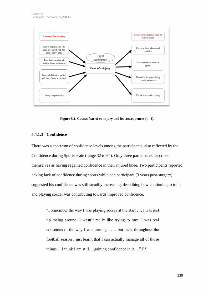
Chapter 5
Participants’ perspectives of ACLR
139
Figure 5.1. Causes fear of re-injury and its consequences (n=8).
5.4.1.3 Confidence
There was a spectrum of confidence levels among the participants, also reflected by the
Confidence during Sports scale (range 32 to 64). Only three participants described
themselves as having regained confidence in their injured knee. Two participants reported
having lack of confidence during sports while one participant (3 years post-surgery)
suggested his confidence was still steadily increasing, describing how continuing to train
and playing soccer was contributing towards improved confidence.
“I remember the way I was playing soccer at the start …, I was just
tip toeing around, I wasn’t really like trying to turn, I was real
conscious of the way I was turning …… but then, throughout the
football season I just learnt that I can actually manage all of those
things….I think I am still….gaining confidence in it….” P5

Chapter 5
Participants’ perspectives of ACLR
140
One participant described himself as confident during sports but still cautious with
specific knee-related activities. For some participants lack of confidence was not a
problem.
“But with the right conditions and knowing that my fitness is in the
right place, then it doesn’t hold me back.” P1
A varying spectrum from low to high confidence levels was seen among the participants
which may affect their overall performance. Those with low levels of confidence were
unable to perform at maximum effort, and one participant also indicated loss of
confidence in the contralateral uninjured knee.
“I don’t feel confident in that knee or either of them now though, so
no it’s not just the one that had the operation.” P2
“There is definitely times where I don’t trust myself to run, well I
don’t trust myself to run or to sprint to be able to catch a ball or
something so I will just jog because I don’t have the confidence
because both times that I have done it has been at a sprint so that is
the time when I am the least confident.” P8
On the other hand some participants no longer focussed on the knee while playing.
“I’d say most of the time I don’t think about it [the knee] too much.”
P1
Two participants used braces for their knee during sports to increase confidence, while
one participant strapped the knee during weight training in the gym.

Chapter 5
Participants’ perspectives of ACLR
141
“I do use a brace when I do snow skiing. I don’t think it does anything
but it just gives me a little bit more confidence or […] knowing just
to protect it as such.” P9
A positive attitude played an important role in the overall outcome. Some participants re-
gained trust in their knees, investing much effort to optimise results.
“I know that it’s never going to be as good as my left but I can live
with that but I want to make sure I do everything possible to make
that better. For some reason I think I’m actually stronger now than
before.” P10
However, others described their struggles with returning to sport.
“I played in the team but I had to pull out of the team because…. I
don’t know if I didn’t trust it or if my knee wasn’t strong enough,
but it wouldn’t hold out with my head. I was almost protecting my
knee in everything I did, so every time I jumped up I’d land on the
other leg. I’d always turn on the other leg, and I was kind of just
leaving the injured one behind, or the surgery one behind. Tried
again in 2014 and then just decided I’d flag it and just do something
else.” P9
5.4.2 Live life normally
This theme describes the participants’ overall approach to continue with life as normally
as possible in terms of ADL, professional and social domains, though with modified life
style strategies, priorities and attitude towards physical activity. None of the participants
had to change their occupation or career plans due to their knee injury. However, a range

Chapter 5
Participants’ perspectives of ACLR
142
of levels of recovery was apparent, with one participant describing the ACLR as having a
deleterious effect on her life (P2, 10 years post-ACLR, KOOS4: 54), while the other
reported minimal effect of their injury to their current ADL, professional and social life
(P10, 9 years post-ACLR, KOOS4: 83).
The participants had adopted strategies to continue with an equivalent or increased level
of physical activity, and often sought alternative solutions when knee symptoms affected
movements or loading.
While most participants described being cautious, caring and mindful of the knee during
physical activity, those with poor knee-related outcomes were more likely to express
anxiety associated with avoidance of specific activities. Besides knee-related factors,
changes due to family, occupation and other commitments also had influenced the
participants’ priorities for physical and sports activities.
“I don’t jump on the trampoline as much, I do it sometimes but I’m
really conscious that it feels, in my head, I think it could go wrong
very easily […]. I probably walk, like so I used to walk a lot and
now I think if I walked to work, for example then how sore am I
gonna be for the rest of the day or will my knee be able to cope then
walking home, does that make sense 'cause…” P2
Three participants who had a Tegner score decrease of ≥ 3, described continuing to avoid
‘high’ risk activities, including those considered to be for fun:
“….I’m not as adventurous anymore, how I used to be […]; I play
the safe road now. [….] I’ve got older brothers and they’re go go
go, so I do pull back a bit and stay with mum sometimes now instead

Chapter 5
Participants’ perspectives of ACLR
143
of going off with them. But other than that I still join in. I still go
bike riding and skiing and everything.” P9
It became apparent during the interviews that a fear of re-injury was contributing towards
changed behaviour in terms of physical activity.
5.4.3 Need of reassurance and maintenance of knee health
Continued advice from health professionals regarding individual-specific needs related to
the knee was suggested by six participants. Those who had been able to return to sports
had different knee-related concerns than the less physically active participants. Continued
daily struggles were still evident for some participants, while they also were aware of the
need of long term maintenance of muscle strength, in particular for graft-related
weakness.
Nine participants reported continuing to struggle with knee symptoms such as pain,
stiffness, achiness, and pain after running. However, those problems did not always
appear to limit their ADL or sports-related knee function.
“Kneeling gets really uncomfortable. I can do it for a little bit, but I
prefer to sit in a low squat than kneel... Pain will be minor and it is
not…. too major but then sometimes it will be mainly, from after
playing a game or training and it will just ache. Going upstairs is
generally fine, it is the going down, if I am walking in bare feet, on
like hard concrete it is not very good.” P8
Apart from the daily struggles, three participants indicated their concerns related to running.
Participants indicated their difficulties related to the changing directions while sprinting, or
being conscious about the knee while running. One participant indicated her preferences for
avoiding hard surfaces while running.
Chapter 5
Participants’ perspectives of ACLR
144
“Yeah I don’t like to [run on hard surface], well I do run on concrete
a bit but I really prefer not to, just 'cause like I get a lot more pain,..a
little bit more pain, not a lot but definitely the softer surfaces.” P7
Those who had not been able to return to sports appeared to have different knee-related
problems, for instance, problems related to pain after an extended amount of physical
activity, and had an anxious attitude towards their knee.
“I probably walk, like so I used to walk a lot and now I think if I
walked to work, for example then how sore am I gonna be for the
rest of the day or will my knee be able to cope then walking
home…” P2
However, those who had successfully managed to return to sports highlighted graft site-
related weakness as an issue. Graft site weakness was identified as a subcategory for
seeking professional advice.
“I pulled it [Hamstrings] about three times and then I just went, I
had to sit out of football for about a month or two this season. P5
Participants understood that they continuously needed to maintain thigh muscle strength
to decrease risk of re-injury and graft-related muscle strains. One participant (P6,
KOOS4=73) indicated changes in daily behaviour specifically to maintain and increase
knee-related physical performance, for example taking stairs instead of the lift.
“At this stage I feel like I have …, got to keep, ….building or at
least, maintain the leg strength, and I have learnt from earlier in the

Chapter 5
Participants’ perspectives of ACLR
145
year I can’t neglect the hamstrings especially because if I do, I think
it makes [them].. susceptible, so, I better get working at that.” P5
Muscle strength was perceived to be critical to manage fear of re-injury and improve
confidence.
“I think probably my fear of injury is less when I’m stronger ……so
like the stronger I get, the less worried I am about injury and then I
get weak again, I’m like oh it could go but I know that’s not true
'cause it’s a ligament, but it’s just the feeling of being strong and
knowing that you’ve got a lot of support around my knee, that would
make me a lot more confident” P7.
Another participant suggested the need for a supervised exercise program which should
decrease fear of re-injury.
“I think maybe an exercise plan that challenges my head and lets
me know that I can do these things. Maybe to know I’ve got the
strength, like physically do a test or something and see that yes, you
do have the strength to do these things. It’s not going to give way.
And then do the activities and realise that it’s fine” P9
Participants, thus, indicated the need for on-going health professional advice to manage
fear of injury and continuing exercises to improve muscle strength and movement control.
For some participants, contact with their health professional provided re-assurance.
“I need my Specialist to every so often to and have a check up and
make sure my knees fine ‘cause for me it really helps to reassurance
from for example from the likes of Doctor XX. Because, …. I have
a lot of faith in him and he’s a Specialist in his area. So if he tells
Chapter 5
Participants’ perspectives of ACLR
146
me I’m doing the right thing then I know I’m doing the right thing.”
P10
Six of the participants were concerned about long-term risk of knee osteoarthritis and
pain, and one mentioned the possibility of future need for knee replacement.
“I am aware that I might develop osteoarthritis in the knee and I just
take it as it comes.” P3
“ I’m just worried about when I get old, it’s gonna be really sore but
immediate future, no issues.” P7
Overall, participants’ perspectives can be summarised into negative and positive
experiences based on the outcome of the surgery, as indicated by the themes and
subthemes of the study results (Figure 5.2). Participants had adopted different strategies
to adapt to the influences of injury on their lives. Negative experiences of the ACL injury
and surgery appeared to have led to fear of injury, avoidance of specific activities, change
in priorities towards the physical activities and minor struggles related to daily life which
indicated the negative experience of the participants. Those with positive experiences
were able to regain confidence during sports, return to preinjury level of sports, or even
increase the level of physical activities compared to the preinjury level following the
surgery. Participants with negative experiences appeared to have more anxiety and fear
related to the future of the knee. In contrast, participants with positive experiences were
‘mindful’ and ‘caring’ towards their knee instead of worrying about the future of the
knee. Changed priorities in terms of physical activity were evident in participants, both
those with negative or positive outcomes. Those with negative experiences preferred
activities associated with low levels of exertion (e.g. driving rather than walking). On the
other hand, those with positive experiences, were more interested in engaging in physical
Chapter 5
Participants’ perspectives of ACLR
147
activity (e.g. preferring to walk up the stairs rather than using the lift). They appeared to
have focussed on all possible efforts to improve and maintain knee health.
Overall, participants are trying to live life normally, whether they had positive or negative
experiences with surgery and rehabilitation. While participants with negative experiences
are more influenced by the fear of injury and they avoid specific activities, those who
were more confident are trying to live normally though are more careful or mindful
during the sports or physical activities. Most of the participants suggested a need for
access to health professional’s advice in the long-term, either to improve their knee
health, or for reassurance related to their knee, such as graft-site related weakness, minor
pain and soreness in the knee and maintenance of the muscle strength.

Chapter 5
Participants’ perspectives of ACLR
148
Figure 5.2. Concept of the three themes emanating from the analysis of the participant concern
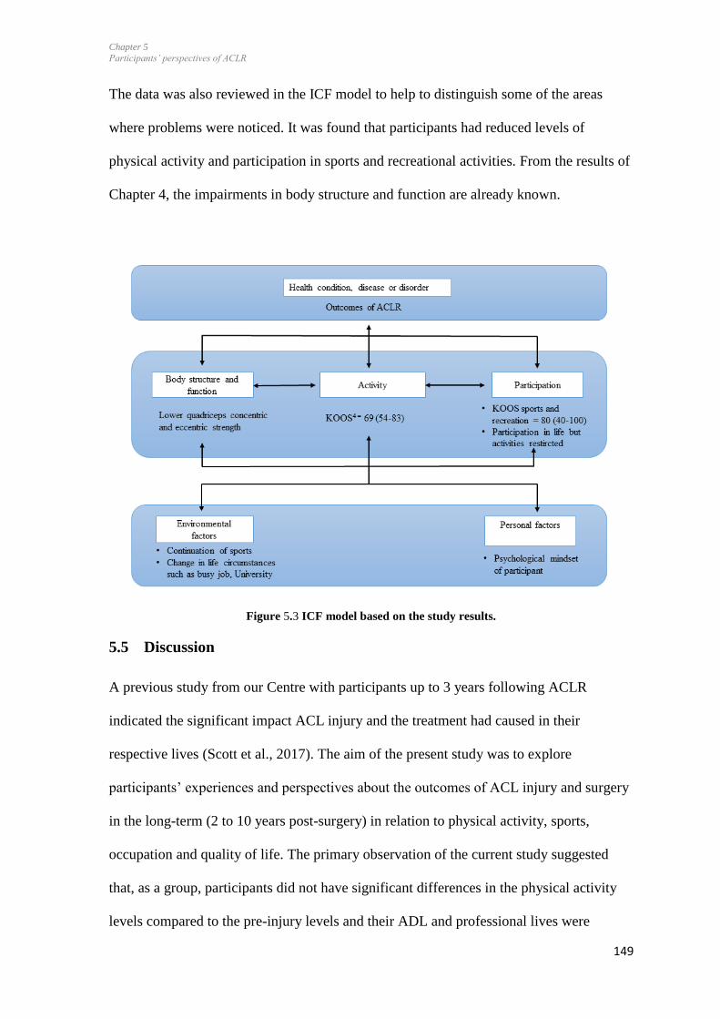
Chapter 5
Participants’ perspectives of ACLR
149
The data was also reviewed in the ICF model to help to distinguish some of the areas
where problems were noticed. It was found that participants had reduced levels of
physical activity and participation in sports and recreational activities. From the results of
Chapter 4, the impairments in body structure and function are already known.
Figure 5.3 ICF model based on the study results.
5.5 Discussion
A previous study from our Centre with participants up to 3 years following ACLR
indicated the significant impact ACL injury and the treatment had caused in their
respective lives (Scott et al., 2017). The aim of the present study was to explore
participants’ experiences and perspectives about the outcomes of ACL injury and surgery
in the long-term (2 to 10 years post-surgery) in relation to physical activity, sports,
occupation and quality of life. The primary observation of the current study suggested
that, as a group, participants did not have significant differences in the physical activity
levels compared to the pre-injury levels and their ADL and professional lives were

Chapter 5
Participants’ perspectives of ACLR
150
minimally affected. However, they continued to experience knee-related symptoms and
had fluctuating levels of confidence during specific sports-related tasks.
As discussed in the previous Chapter ( Chapter 4, Section 4.8) quality of life has been
defined as the expectations of health versus actual experience (Carr et al., 2001),
participants perceived their KOOS QOL as low (Table 5.2). It may be because of their
relatively young age (median 28.5 years), and high health expectations from their health,
thereby potentially contributing towards their low ratings of for the KOOS QOL scores.
This cohort of participants reported better quality of life KOOS QOL than the larger
cohort in Chapter 4 (Table 4.4), which may be due to the less experience of knee pain,
symptoms and better function in activities of daily living and sports and recreation.
Scores of KOOS QOL for this group of participants were similar to other studies
exploring outcomes of ACLR but with older participants, averages of 37 and 45.6 years
respectively (Meunier, Odensten, & Good, 2007; Tengman et al., 2014). The low KOOS-
QOL scores of the current study were supported by the SF-12 scores. We found low
scores for physical (median =55.50) and mental components (median = 44.80),
suggesting that apart from the physical impairment, lowered mental health was also an
important concern for participants with ACLR. The KOOS ADL scores indicated
excellent recovery (99/100), however, the interview data indicated that participants still
had minor daily struggles such as knee symptoms during stair negotiation, kneeling or
running.
Our study results are similar to another study from Sweden, exploring the knee function
at the median duration of 11.5 years in a cohort of 56 participants following surgery in
Swedish population (Möller et al., 2009). Participants of that study had similar scores in
KOOS pain (median=94), function in ADL (median=100), recreation (median-75) to that
Chapter 5
Participants’ perspectives of ACLR
151
of our study results except from the quality of life scores (median=81). The differences
may be due to higher age group (approximately 38) of participants in that study group
compared to that of our study. Therefore, the differences in outcomes across various
regions seems negligible.
Two main themes relating to the research question emanated from the interviews: ‘fear of
re-injury versus confidence continuum’, and ‘live life normally’ (Figure 5.2, Table 5.3).
Despite ACLR having been performed up to 10 years earlier, fear of re-injury was still
present, irrespective of participants’ level of physical activity. The fear of injury in the
current study appeared to be primarily driven by negative experiences from ACL injury,
surgery and the commitments the long post-surgical rehabilitation period had required
and their role in the family commitments, which is in concordance with findings by
(Ross, Clifford, & Louw, 2017). The fear influenced their return to play such that one
participant was unable to return to pre-injury sport, changing to an alternative sports with
less knee-related loading.
The current study showed that, while participants had continued with their lives “as
normally” as they could, the re-injury fear persisted. They suggested that maintaining the
physical activity and confidence in the knee would require vigilance for life in order to
not to injure the knee again (Scott et al., 2017). Previous studies have shown that fear of
re-injury was present between one and two years following surgery (Flanigan et al., 2013;
Heijne et al., 2008), decreasing at around 3 years following surgery (Gignac et al., 2015).
Our findings contrast with those by (Ardern et al., 2012), finding that participants playing
sports on average 7 years following ACLR did so with low levels of fear. Those
participants were regular recreational or competitive athletes before ACL injury and at the
time of the study, which might have contributed to low levels of fear of injury (Ardern et

Chapter 5
Participants’ perspectives of ACLR
152
al., 2012). On the contrary, we had a mixed cohort of participants and only some of them
were the regular recreational athletes, which may explain why some of the participants of
this study had less fear of injury than the others.
Findings of Chapter 4 indicated the muscle strength deficits on the injured side (Section,
Table 4.3). In this study, two participants had explained their struggles with gaining the
muscle strength in rehabilitation and a factor causing fear of re-injury in them.
Participants also indicated the need of health professional to maintain the muscle strength
in the long-term following surgery. Exercise protocol to improve the muscle strength can
be prescribed to the patients by health professionals followed by patient-education
regarding the strength maintenance exercise programme. Muscle strength deficits on the
injured side seem to have wider implications on participants overall health, concerns and
confidence, therefore, needs more attention in the rehabilitation and in later stages as
well.
Competitive athletes potentially are more likely to have continued access to health care
and support beyond the usual timeframes of immediate rehabilitation. On the other hand,
our study included participants who, at the time of injury, were undertaking different
levels of physical activity, ranging from highly competitive sports (Tegner Score 9, e.g.
competitive soccer, rugby) to those who had low levels of knee-related activity (Tegner
Score 3, swimming or walking) and had variable access to continued health care related
to the knee. This may have contributed to the persistence of fear of injury in the current
study, despite the duration following ACL injury being up to 10 years following surgery.
Despite minor daily struggles, participants were trying to continue their lives ‘normally’
with some modification in life style and priorities. They reported having had a change in

Chapter 5
Participants’ perspectives of ACLR
153
the attitude towards their knee. Participants choose to consider the safety of their knee
first while making the choices of activities. While some participants were more mindful
of their knee in a positive, caring attitude, others had anxiety associated with it.
One of the participants indicated behaviour modification by taking stairs instead of lifts
towards maintenance of knee-related health. In another study, participants from 5 to 20
years following surgery, who changed physical activity preferences and their lifestyle
early after ACLR reported better quality of life (Filbay et al., 2016). Similar behaviour
modification of overall physical activity and exercise may need to be considered.
Behavioural cognitive therapy, including relaxation and imagery, has been used during
the ACL rehabilitation and was reported to enhance psychological and physical aspects of
recovery along with the, improved coping, and reduced re-injury anxiety (Cupal &
Brewer, 2001). Gymnasts also have been reported to use strategies to overcome fear
including thought-stopping “just go for it”, imagery, self-talk, positive thoughts, selective
focus and attention, and relaxation (Magyar & Chase, 1996). Similar strategies may be
considered following ACLR to enhance self-management and self-efficacy in the long-
term.
Besides concerns with daily struggles, fear of re-injury and fluctuating levels of self-
confidence, the participants also discussed concerns about potential risk of future
osteoarthritis. While health professionals should inform patients with ACLR of
implications of such risk, the discussions should be within the context of providing
individual-specific strategies to minimise such risk. The importance of considering
psychosocial responses to ACL injury was highlighted in the previous study exploring
patients’ perspectives of surgery up to 3 years following surgery (Scott et al., 2017). In a
parallel study, physiotherapists reported using a biopsychosocial approach as part of the

Chapter 5
Participants’ perspectives of ACLR
154
care they provided towards rehabilitation of patients with ACLR (Von Aesch, Perry, &
Sole, 2016), highlighting that they were aware of the need to address psychological and
physical recovery during rehabilitation. However, they also perceived that they may need
more formalised training in this field to optimise rehabilitation and patient outcomes in
the long-term. Collectively, past studies and the current study highlight the importance of
addressing the psychological and social needs of the patients during rehabilitation to
minimise lingering symptoms and enhance self-efficacy in the long-term.
5.5.1 Methodological considerations
The quantitative data towards this research project was collected prior to the interviews,
allowing the participants opportunity to meet the principle researcher for this study (MK),
establishing rapport, prior to the interviews. The patients’ perspectives about the
influence of ACLR on their lives were in agreement with the results of patient-reported
outcome measures especially about the Confidence during sports scale and KOOS
(Teddlie & Tashakkori, 2009). Credibility of the results of this study was ensured by: (1)
development of the interview guide by the research team through discussion and was
updated following an initial 2-3 interviews; (2) the audio-recordings and verbatim
transcriptions of the interview, open atmosphere during the interviews, the researchers
bracketing prior experiences and thoughts, member checks of the study results (Appendix
– D3)(Thomas, 2006); (3) peer debriefing of the overall results following the complete
analysis of the transcriptions independently by three of the authors.
Trustworthiness and dependability of data was established by parallel coding of every
alternative interview by a second researcher (GS) and by providing the additional quotes
(Appendix- D2) (Thomas, 2006). All the interviews were face to face, employing the
same method of data collection for all the participants, hence, decreasing the risk of bias
Chapter 5
Participants’ perspectives of ACLR
155
which could be due to lack of visual cues and loss of informal communication and
contextual information with the telephone interviews (Sweet, 2002).
Confirmability of the study findings were ensured by the open discussion among three of
the authors following the analysis of the results. The results are valid experiences for the
ten participants included in this study and most of the participants were from New
Zealand, therefore, it is difficult to generalise the findings to different origins and
countries.
5.6 Conclusion
Participants strove to “continue living life normally”, with variable levels of current
physical activity and involvement in sports. The ACL rupture appears to lead to long-term
fear of injury and behavioural manifestations, with fluctuating levels of confidence during
physical activity and sports. The participants, up to 10 years post ACLR, reported
variable levels of knee-related quality of life based on the KOOS QOL scores.
Participants were concerned about the future risk of re-injury or osteoarthritis. Health
professionals should consider a long-term individual-specific maintenance programme to
improve and maintain confidence and self-efficacy, and promote physical activity.
5.7 Summary
Participants continue to experience fear of re-injury and low confidence in the injured
limb during sports-related movements. Different reasons for the persistence of fear of re-
injury were found along with the consequences of this fear. Hesitancy during movements,
experienced by some of the participants may be related to the less weight bearing on the
injured side and shifting the joint loading on the contralateral side. These are important
findings for the patients and clinicians as longer-term deficits such as lower loading on
the injured side may be explained, in part, by those emotional responses.

Chapter 5
Participants’ perspectives of ACLR
156
Lower limb biomechanics on the injured limb along with the contralateral side of
participants with ACLR were explored and compared to the uninjured control group in
the next chapter (Chapter 6).
Chapter 6
Knee biomechanics during stair navigation: ACLR vs Control group
157
6 Knee biomechanics in participants with anterior cruciate ligament
reconstruction compared to the Control group during stair
navigation
6.1 Prelude to Chapter 6
Findings from the systematic review and meta-analysis (Chapter 3) indicated that knee
angles were likely to recover fully by 6 years, on average, post-ACLR when comparing
the injured side to the contralateral limb, and to a Control group. However, joint moments
continue to be low when comparing the moments of the injured to the contralateral limbs
up to an average of 6 years post-surgery, especially the external peak knee flexion and
adduction moments. Together, the findings related to the joint moments indicate the need
to explore whether the moments recover in the mid- to long-term following the surgery.
In this chapter, differences in the knee angles and moments in the injured limb were
compared to the contralateral limb in participants with ACLR from 2 to 10 years
following surgery, and to a Control group, during stair ascent and descent. The results of
this study point to moment symmetry being compromised in the long-term following
surgery in participants with ACLR.

Chapter 6
Knee biomechanics during stair navigation: ACLR vs Control group
158
Chapter 6
6.2 Background
Differences in biomechanical variables such as knee flexion and adduction moments
appear to be important when comparing the injured side with the contralateral uninjured
side in participants with ACLR and with uninjured Control groups (Chapter 3, Figure 3.6
and 3.7). Lower flexion and adduction moments were present on the injured side in
participants with ACLR from 22 to 26 months following surgery (Zabala et al., 2013).
Similarly, knee flexion and adduction moments were lower on the injured side in
participants with ACLR compared to the contralateral side and the Control group from 2
to 18 years following surgery (Hall et al., 2012). Altered joint angles and moments during
ambulatory activities following ACL injury and reconstruction may contribute to the
development of post-traumatic osteoarthritis (Foroughi et al., 2009; Nigg et al., 2000;
Pietrosimone et al., 2017). A recent study has indicated that the external flexion moment
is a contributor to medial compartment joint loading, therefore, analysing both flexion
and adduction moment can provide deeper insight into the mechanics of joint loading
(Manal, Gardinier, Snyder-Mackler, & Buchanan, 2013; Walter, D'Lima, Colwell, &
Fregly, 2010).
Based on the results presented in Chapter 3, inspection of the forest plots (Chapter 3,
Figure 3.7, Subsection 7.1.1) suggested a possible increase in the adduction moments
with time following surgery. Previous studies have explored the moments early following
surgery (less than 3 years) (Karimi et al., 2013; Webster & Feller, 2012a; Zabala et al.,
2013), or have included participants with wide range of time following surgery (Hall et
al., 2012), which could have diluted the results in the study regarding the magnitude of
Chapter 6
Knee biomechanics during stair navigation: ACLR vs Control group
159
moments. The current study recruited the participants from 2-10 years post-surgery to
explore if moments recover in the long-term.
The findings of the systematic review (Chapter 3)(Kaur, Ribeiro, Theis, Webster, & Sole,
2016) and other reviews were inconclusive regarding the magnitude of rotation moment
and angles (Hart, Culvenor, et al., 2015). Lower internal rotation moments were present
on the injured side compared to the contralateral knees of the ACLR group during stair
ascent (second peak) and descent (second peak) (Zabala et al., 2013). Higher internal
rotation moments are associated with mild to moderate knee osteoarthritis (Astephen,
Deluzio, Caldwell, Dunbar, & Hubley-Kozey, 2008). Participants with ACL rupture have
a higher risk of developing knee osteoarthritis (Lohmander et al., 2007). So the question
is raised as to whether internal rotation moments might increase over time in individuals
following ACLR, which may then become an indicator for knee osteoarthritis risk.
Asymmetry in moments and angles was present such that peak external moments were
lower on the injured side compared to the contralateral side 8 months following surgery
during drop vertical jump (Schmitt et al., 2015). These asymmetries may persist up to 2
years, or longer, following surgery (Paterno et al., 2007) and can be a risk factor for joint
re-injury (Paterno et al., 2010). It is important to explore if the moment asymmetries
persist in the long-term following ACLR. Moment asymmetries indicate altered loading
and it may predisposes the individual to the risk of re-injury or early onset of knee
osteoarthritis (Khandha et al., 2016). Asymmetries for knee moments were more apparent
during stair ascent and descent compared to walking, as described in Chapter 3 (Figure
3.6 and 3.7). Therefore, this study was designed to explore the knee moments and angles
during stair ascent and descent. The primary aim of this study was to assess differences in
knee kinematics and moments between (1) the injured knee and contralateral knee in
Chapter 6
Knee biomechanics during stair navigation: ACLR vs Control group
160
participants with ACLR; and (2) the injured knee in participants with ACLR and age-
matched uninjured controls, during stair ascent and descent.
6.2.1 Hypothesis
1) The injured knees in the ACLR group will exhibit lower flexion and adduction
moments and angles compared to the contralateral uninjured knees during stair
ascent and descent.
2) ACLR group will have lower knee flexion moments and higher knee adduction
and internal rotation moments during stair ascent and descent compared to the
Control group.
6.3 Methods
6.3.1 Study design
This was a cross-sectional study, assessing differences in knee joint angles and moments
in the ACL reconstructed knees compared to the contralateral uninjured knees and the
uninjured Control group. The study design, study setting, ethic approval and participant
recruitment were described in Chapter 4, section 6.3.5.1.
6.3.2 Equipment
6.3.2.1 Three-dimensional motion analysis system
To monitor lower-limb and pelvic movements during the stair ascent and descent tasks, a
three-dimensional motion analysis system (Motion Analysis Corporation, Cortex 5.5
Santa Rosa, CA, USA) with 11 infra-red cameras, sampled at 150 Hz was used. The
laboratory set up and positioning of the cameras has been shown in Figure 6.1. One floor-
mounted force platform, AMTI force plate (AMTI Inc., Newton, MA, USA) sampled at
1,050 Hz, was used to measure the ground reaction force. The stairs consisted of two
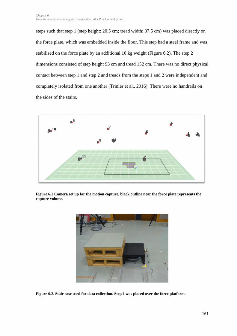
Chapter 6
Knee biomechanics during stair navigation: ACLR vs Control group
161
steps such that step 1 (step height: 20.5 cm; tread width: 37.5 cm) was placed directly on
the force plate, which was embedded inside the floor. This step had a steel frame and was
stabilised on the force plate by an additional 10 kg weight (Figure 6.2). The step 2
dimensions consisted of step height 93 cm and tread 152 cm. There was no direct physical
contact between step 1 and step 2 and treads from the steps 1 and 2 were independent and
completely isolated from one another (Trinler et al., 2016). There were no handrails on
the sides of the stairs.
Figure 6.1 Camera set up for the motion capture, black outline near the force plate represents the
capture volume.
Figure 6.2. Stair case used for data collection. Step 1 was placed over the force platform.
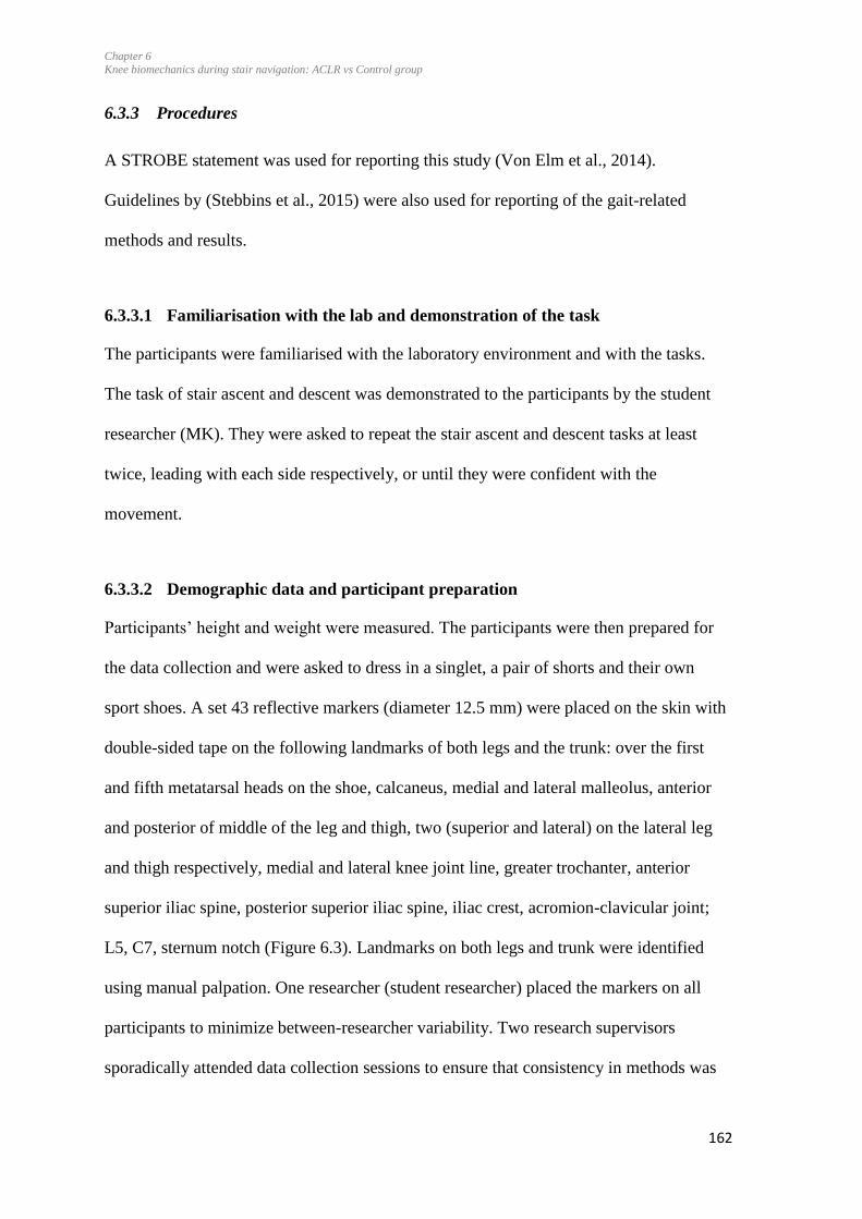
Chapter 6
Knee biomechanics during stair navigation: ACLR vs Control group
162
6.3.3 Procedures
A STROBE statement was used for reporting this study (Von Elm et al., 2014).
Guidelines by (Stebbins et al., 2015) were also used for reporting of the gait-related
methods and results.
6.3.3.1 Familiarisation with the lab and demonstration of the task
The participants were familiarised with the laboratory environment and with the tasks.
The task of stair ascent and descent was demonstrated to the participants by the student
researcher (MK). They were asked to repeat the stair ascent and descent tasks at least
twice, leading with each side respectively, or until they were confident with the
movement.
6.3.3.2 Demographic data and participant preparation
Participants’ height and weight were measured. The participants were then prepared for
the data collection and were asked to dress in a singlet, a pair of shorts and their own
sport shoes. A set 43 reflective markers (diameter 12.5 mm) were placed on the skin with
double-sided tape on the following landmarks of both legs and the trunk: over the first
and fifth metatarsal heads on the shoe, calcaneus, medial and lateral malleolus, anterior
and posterior of middle of the leg and thigh, two (superior and lateral) on the lateral leg
and thigh respectively, medial and lateral knee joint line, greater trochanter, anterior
superior iliac spine, posterior superior iliac spine, iliac crest, acromion-clavicular joint;
L5, C7, sternum notch (Figure 6.3). Landmarks on both legs and trunk were identified
using manual palpation. One researcher (student researcher) placed the markers on all
participants to minimize between-researcher variability. Two research supervisors
sporadically attended data collection sessions to ensure that consistency in methods was

Chapter 6
Knee biomechanics during stair navigation: ACLR vs Control group
163
maintained. A research assistant was present at all times during data collection to assist
with the participant preparation and execution of procedures.
Figure 6.3. Marker set: front and side view
6.3.3.3 Order of tasks
The order of the tasks consisted of static trials followed by dynamic trials. Participants
were asked to stand still for at least 7 seconds for the static trial, which was used to obtain
a reference point for the markers in quiet standing. Following the static trials, participants
were asked to perform dynamic trials which were used for calculating the functional hip
joint centres. Dynamic trials consisted of participants in single-leg standing; performing
hip flexion and extension, abduction and adduction, and circumduction movement with
the non-weight bearing leg. Each movement was performed twice. The student researcher
first demonstrated the hip movements to each participant, after which participants
practiced the movements. The participants were asked to perform these movements in a
smooth, continuous motion while maintaining balance and minimising trunk movements.
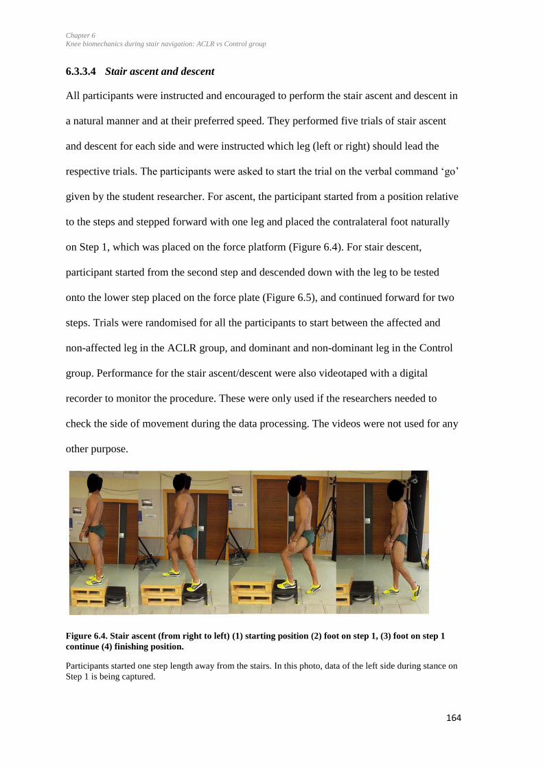
Chapter 6
Knee biomechanics during stair navigation: ACLR vs Control group
164
6.3.3.4 Stair ascent and descent
All participants were instructed and encouraged to perform the stair ascent and descent in
a natural manner and at their preferred speed. They performed five trials of stair ascent
and descent for each side and were instructed which leg (left or right) should lead the
respective trials. The participants were asked to start the trial on the verbal command ‘go’
given by the student researcher. For ascent, the participant started from a position relative
to the steps and stepped forward with one leg and placed the contralateral foot naturally
on Step 1, which was placed on the force platform (Figure 6.4). For stair descent,
participant started from the second step and descended down with the leg to be tested
onto the lower step placed on the force plate (Figure 6.5), and continued forward for two
steps. Trials were randomised for all the participants to start between the affected and
non-affected leg in the ACLR group, and dominant and non-dominant leg in the Control
group. Performance for the stair ascent/descent were also videotaped with a digital
recorder to monitor the procedure. These were only used if the researchers needed to
check the side of movement during the data processing. The videos were not used for any
other purpose.
Figure 6.4. Stair ascent (from right to left) (1) starting position (2) foot on step 1, (3) foot on step 1
continue (4) finishing position.
Participants started one step length away from the stairs. In this photo, data of the left side during stance on
Step 1 is being captured.
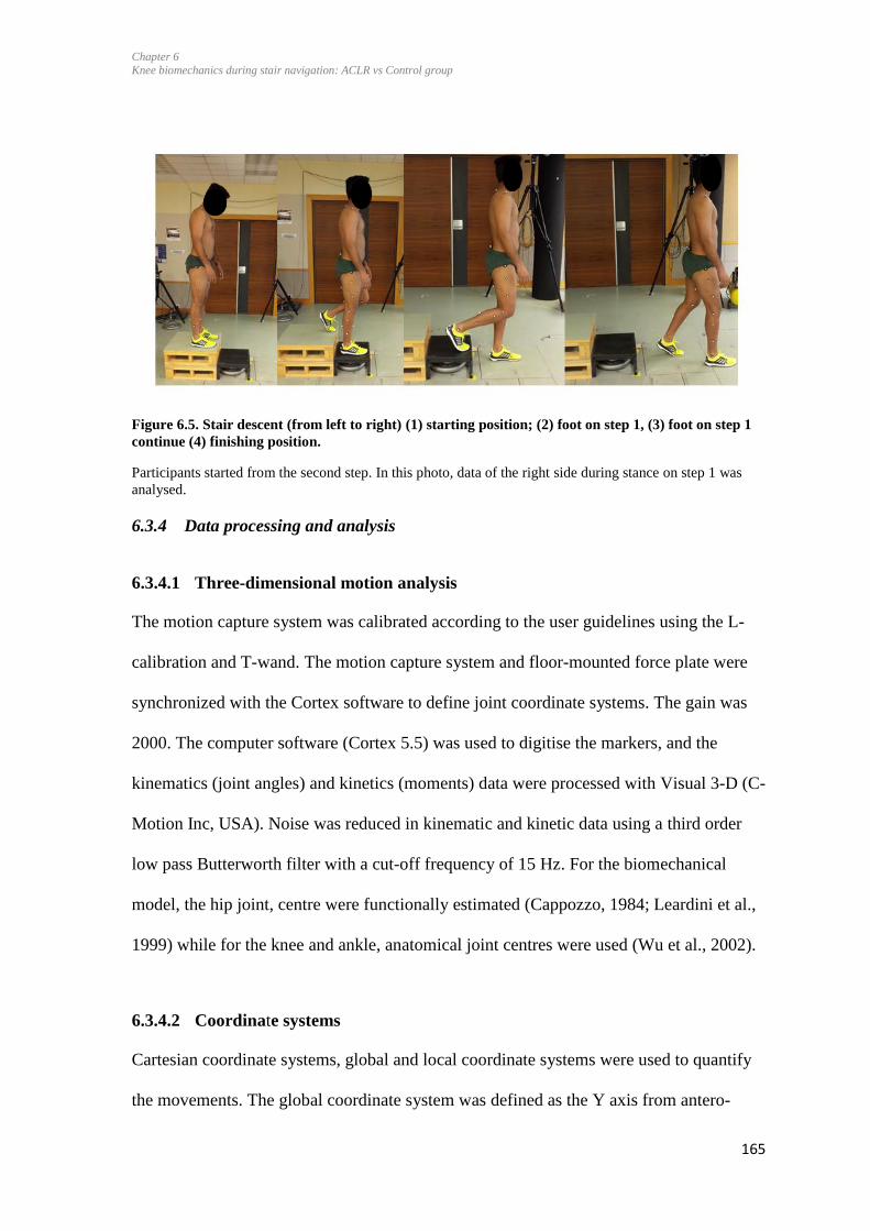
Chapter 6
Knee biomechanics during stair navigation: ACLR vs Control group
165
Figure 6.5. Stair descent (from left to right) (1) starting position; (2) foot on step 1, (3) foot on step 1
continue (4) finishing position.
Participants started from the second step. In this photo, data of the right side during stance on step 1 was
analysed.
6.3.4 Data processing and analysis
6.3.4.1 Three-dimensional motion analysis
The motion capture system was calibrated according to the user guidelines using the L-
calibration and T-wand. The motion capture system and floor-mounted force plate were
synchronized with the Cortex software to define joint coordinate systems. The gain was
2000. The computer software (Cortex 5.5) was used to digitise the markers, and the
kinematics (joint angles) and kinetics (moments) data were processed with Visual 3-D (C-
Motion Inc, USA). Noise was reduced in kinematic and kinetic data using a third order
low pass Butterworth filter with a cut-off frequency of 15 Hz. For the biomechanical
model, the hip joint, centre were functionally estimated (Cappozzo, 1984; Leardini et al.,
1999) while for the knee and ankle, anatomical joint centres were used (Wu et al., 2002).
6.3.4.2 Coordinate systems
Cartesian coordinate systems, global and local coordinate systems were used to quantify
the movements. The global coordinate system was defined as the Y axis from antero-
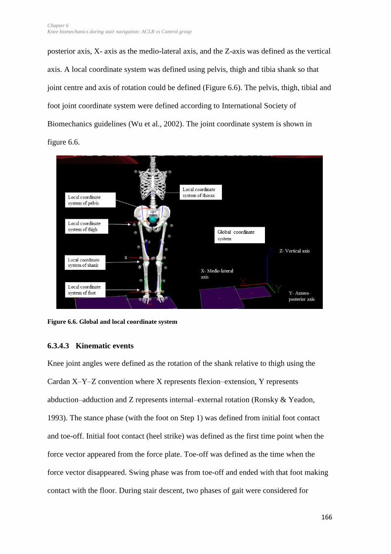
Chapter 6
Knee biomechanics during stair navigation: ACLR vs Control group
166
posterior axis, X- axis as the medio-lateral axis, and the Z-axis was defined as the vertical
axis. A local coordinate system was defined using pelvis, thigh and tibia shank so that
joint centre and axis of rotation could be defined (Figure 6.6). The pelvis, thigh, tibial and
foot joint coordinate system were defined according to International Society of
Biomechanics guidelines (Wu et al., 2002). The joint coordinate system is shown in
figure 6.6.
Figure 6.6. Global and local coordinate system
6.3.4.3 Kinematic events
Knee joint angles were defined as the rotation of the shank relative to thigh using the
Cardan X–Y–Z convention where X represents flexion–extension, Y represents
abduction–adduction and Z represents internal–external rotation (Ronsky & Yeadon,
1993). The stance phase (with the foot on Step 1) was defined from initial foot contact
and toe-off. Initial foot contact (heel strike) was defined as the first time point when the
force vector appeared from the force plate. Toe-off was defined as the time when the
force vector disappeared. Swing phase was from toe-off and ended with that foot making
contact with the floor. During stair descent, two phases of gait were considered for
Chapter 6
Knee biomechanics during stair navigation: ACLR vs Control group
167
analysis: weight acceptance and propulsion (McFadyen & Winter, 1988; Sole, Tengman,
Grip, & Häger, 2016). Weight acceptance of the leading leg was defined as the phase
from heel strike of that leg on the first step to toe-off of the trailing leg on the starting
step. Propulsion of the leading leg was defined as the toe-off of the leading leg to the toe-
off of the trialing leg. All events were identified by the automatic algorithm (Visual 3D,
(C-Motion Inc, USA) (Appendix-E1). All events were then visually inspected during the
process and manually corrected if needed. All stride events were set and data extracted
prior to expressing the events as percentage of the stance phase of the gait cycle. Kinetics,
kinematics, and speed were averaged across the five trials for each side.
During stair ascent, the following outcomes were extracted: peak flexion and extension
angles; peak knee adduction and abduction; peak tibial internal and external rotation
angles. During stair descent, the following variables were extracted for weight acceptance
and propulsion phase: peak knee flexion and extension, peak knee abduction and
adduction, peak internal and external rotation. Table 6.2 includes the list of all variables.
All stride events were set and data extracted prior to expressing the events as percentage
of the gait cycle.
6.3.4.4 Kinetics
External joint moments were calculated through inverse dynamics (TP Andriacchi &
Strickland, 1985). To account for variability between participants, moments were
normalised to %BWxHt (Moisio, Sumner, Shott, & Hurwitz, 2003). The maximum and
minimum knee moments in the three planes were used for analysis during stair ascent.
For stair descent, the peak variables were calculated during the weight acceptance and
propulsion phase. This resulted in two values of peak moments (1st and 2nd half of
stance) for each of the three moments and each activity per knee. Knee extension,
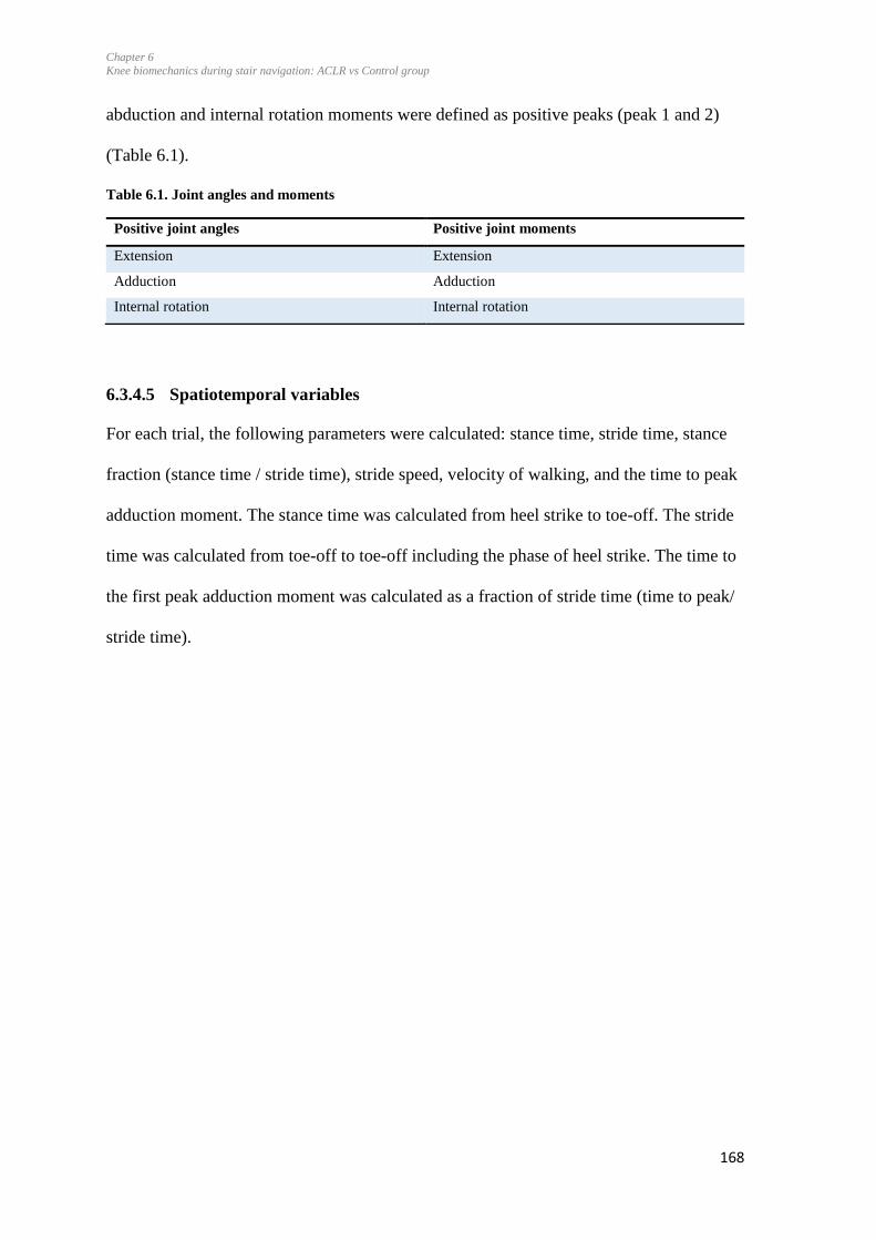
Chapter 6
Knee biomechanics during stair navigation: ACLR vs Control group
168
abduction and internal rotation moments were defined as positive peaks (peak 1 and 2)
(Table 6.1).
Table 6.1. Joint angles and moments
Positive joint angles Positive joint moments
Extension Extension
Adduction Adduction
Internal rotation Internal rotation
6.3.4.5 Spatiotemporal variables
For each trial, the following parameters were calculated: stance time, stride time, stance
fraction (stance time / stride time), stride speed, velocity of walking, and the time to peak
adduction moment. The stance time was calculated from heel strike to toe-off. The stride
time was calculated from toe-off to toe-off including the phase of heel strike. The time to
the first peak adduction moment was calculated as a fraction of stride time (time to peak/
stride time).
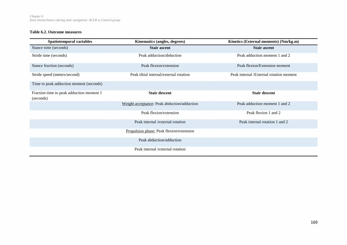
Chapter 6
Knee biomechanics during stair navigation: ACLR vs Control group
169
Table 6.2. Outcome measures
Spatiotemporal variables Kinematics (angles, degrees) Kinetics (External moments) (Nm/kg.m)
Stance time (seconds) Stair ascent Stair ascent
Stride time (seconds) Peak adduction/abduction Peak adduction moment 1 and 2
Stance fraction (seconds) Peak flexion/extension Peak flexion/Extension moment
Stride speed (meters/second) Peak tibial internal/external rotation Peak internal /External rotation moment
Time to peak adduction moment (seconds)
Fraction time to peak adduction moment 1
(seconds)
Stair descent Stair descent
Weight acceptance: Peak abduction/adduction Peak adduction moment 1 and 2
Peak flexion/extension Peak flexion 1 and 2
Peak internal /external rotation Peak internal rotation 1 and 2
Propulsion phase: Peak flexion/extension
Peak abduction/adduction
Peak internal /external rotation
Chapter 6
Knee biomechanics during stair navigation: ACLR vs Control group
170
6.3.5 Statistical analysis
6.3.5.1 Sample size estimation
The sample size was estimated using the software GPower 3.1 (Faul, Erdfelder, Lang, &
Buchner, 2007). The meta-analysis described in Chapter 3 reported an effect size of 0.38
for differences in knee adduction moments between injured and contralateral limbs during
stair descent. The sample size for this study was estimated assuming an effect size of
0.38, an alpha set at 0.05 and power set at 0.80. Based on these values, the required
sample size was 26 participants for each group, when conducting between- and within-
group comparisons. Thus, a sample of 26 injured and 26 age-, gender-, and physical
activity level-matched controls were recruited.
6.3.5.2 Kinematics and kinetics
SPSS Version 23 (IBM SPSS Statistics) was used for statistical analysis. Data from the
ACLR group of the injured side were pooled together. For Control groups, the left and
right sides were randomly assigned to “Side 1” and “Side 2” using an online software
(Urbaniak & Plous, 2013) to have the same ratio as for the ACLR group in terms of
right/left sides. Normality of the data was tested with histograms and Normal Q-Q plots.
Data were found to be normally distributed with the data points close to the diagonal line
in the Normal Q-Q plots. The normalised data (as a percentage of the stance phase) were
exported from Visual 3D to MATLAB R2016a (The Mathworks Inc., Natick, MA) to
create linear envelopes (means and SD) for the injured and contralateral sides of the
ACLR group and the averages of Side 1 of the Control group (Appendix-E2).
Repeated measures analysis of variance (ANOVA) (2 x 2) were used to determine
whether significant differences between-group (ACLR versus Control group) and side
Chapter 6
Knee biomechanics during stair navigation: ACLR vs Control group
171
effects (injured versus uninjured sides for ACLR group, and Side 1 and Side 2 for the
Control group) or interactions existed (p>0.05) between ACLR and Control group.
Mauchly’s test was used to test the assumption of Sphericity. Pairwise post-hoc tests were
performed if significant group or side effects or interactions (group x sides) were found.
Bonferroni test was used for the pair-wise comparisons. The Bonferroni method is
considered to be a robust method and able to control alpha levels and reduces the chances
of Type 1 error (Field, 2009). Effect sizes (Cohen's d) were calculated for significant
differences and categorized as small (b0.5), medium (≥0.5 and b0.8) or large (≥0.8)
(Cohen, 1988).
6.3.5.3 Repeatability of knee flexion and adduction moments
Trial-to-trial reliability and variability for assessing the peak flexion and adduction
moments during stair ascent and descent were performed using the data of 12 ACLR and
12 Control participants. Intraclass Correlation Coefficient (ICC (95%, CI)) and Standard
Error of Measurement (SEM) were calculated. Absolute values were reported for the ICC.
The SEM was calculated by extracting the square root of the error mean square term from
the ANOVA test (Weir, 2005). This method has the advantage of estimating the SEM
independently from the ICC magnitude (Weir, 2005). The ICC’s were classified as ≥0.7:
high consistency/low variability, 0.5-0.69: moderate consistency/variability and <0.5: low
consistency/high variability (Munro, 2005).
6.4 Results
Twenty-five participants with unilateral ACLR and 24 uninjured age- and gender-
matched participants took part in the study. The characteristics of participants are
presented in Table 6.3. The BMI of the ACLR participants were significantly higher than
the controls (p<0.001, Table 6.3). Participant data related to the Surgery and
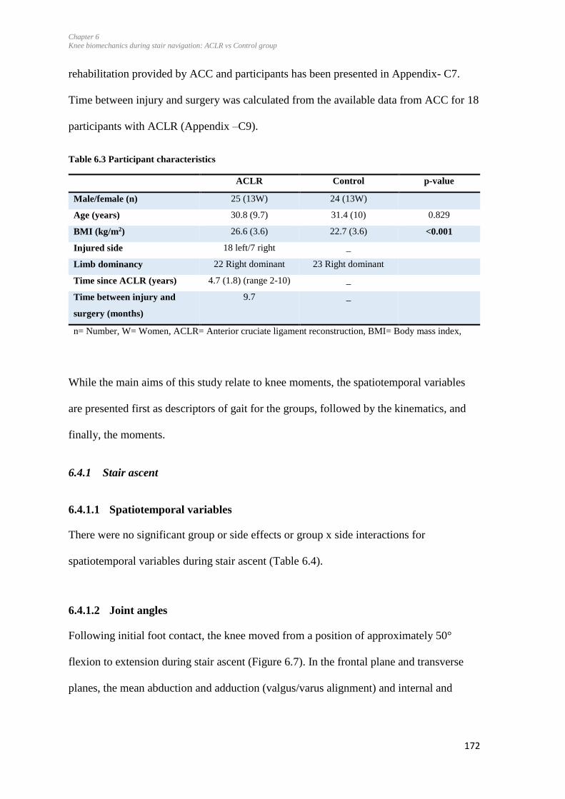
Chapter 6
Knee biomechanics during stair navigation: ACLR vs Control group
172
rehabilitation provided by ACC and participants has been presented in Appendix- C7.
Time between injury and surgery was calculated from the available data from ACC for 18
participants with ACLR (Appendix –C9).
Table 6.3 Participant characteristics
ACLR Control p-value
Male/female (n) 25 (13W) 24 (13W)
Age (years) 30.8 (9.7) 31.4 (10) 0.829
BMI (kg/m2) 26.6 (3.6) 22.7 (3.6) <0.001
Injured side 18 left/7 right _
Limb dominancy 22 Right dominant 23 Right dominant
Time since ACLR (years) 4.7 (1.8) (range 2-10) _
Time between injury and
surgery (months)
9.7 _
n= Number, W= Women, ACLR= Anterior cruciate ligament reconstruction, BMI= Body mass index,
While the main aims of this study relate to knee moments, the spatiotemporal variables
are presented first as descriptors of gait for the groups, followed by the kinematics, and
finally, the moments.
6.4.1 Stair ascent
6.4.1.1 Spatiotemporal variables
There were no significant group or side effects or group x side interactions for
spatiotemporal variables during stair ascent (Table 6.4).
6.4.1.2 Joint angles
Following initial foot contact, the knee moved from a position of approximately 50°
flexion to extension during stair ascent (Figure 6.7). In the frontal plane and transverse
planes, the mean abduction and adduction (valgus/varus alignment) and internal and

Chapter 6
Knee biomechanics during stair navigation: ACLR vs Control group
173
external rotation angles remained below 5°. Standard deviations clouds appear to be large
relative to the course of the means of the angles in the frontal and transverse planes.
In the sagittal plane, significant group effects were found for peak knee flexion angle
between the ACLR and Control group (p=0.022) (Table 6.4) (Figure 6.7). Post-hoc
pairwise comparisons found less flexion for the injured side compared to the side 1 of the
Control group (mean difference 4.5°, 95%CI 1.8-7.1, p=0.001) (Table 6.5) while no
significant difference was found when comparing the uninjured side of the ACLR group
to Side 2 of the controls. Significant differences were present in the ACLR group between
injured and contralateral side (mean difference 2.3°, 95%CI 0.80-3.80, p=0.003).
There were significant group x side interactions for peak flexion (p=0.004) and flexion-
extension excursion (p=0.004). Post-hoc pairwise comparisons reported lower flexion-
extension excursion angles (total range from peak extension to peak flexion) on the
injured side compared to the side 1 of the Control group (mean difference 4.0°, 95%CI
1.12-6.72, p=0.007). However, no differences were found for the flexion extension
excursion angles when comparing the contralateral side (ACLR) to Side 2 (controls)
(mean difference 0.51°, 95%CI 2.80-3.82, p=0.755). No significant differences were
present among the injured and contralateral sides in the ACLR group (mean difference
2.1°, 95%CI 0.57-3.74, p=0.009).
No significant group, side effects or group x side interaction effects were found for the
frontal and transverse plane angles (Table 6.4). The analysis of the discrete variables in
the frontal planes appeared to have greater knee adduction angles on the injured and
uninjured sides of the ACLR group compared to the Control group (Figure 6.7).
However, the statistical analysis found no significant differences between the groups
(Table 6.4).
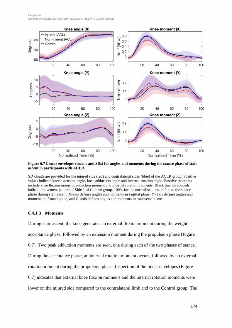
Chapter 6
Knee biomechanics during stair navigation: ACLR vs Control group
174
Figure 6.7 Linear envelopes (means and SDs) for angles and moments during the stance phase of stair
ascent in participants with ACLR.
SD clouds are provided for the injured side (red) and contralateral sides (blue) of the ACLR group. Positive
values indicate knee extension angle, knee adduction angle and internal rotation angle. Positive moments
include knee flexion moment, adduction moment and internal rotation moments. Black line for controls
indicate movement pattern of Side 1 of Control group. 100% for the normalised time refers to the stance
phase during stair ascent. X-axis defines angles and moments in sagittal plane, Y- axis defines angles and
moments in frontal plane, and Z- axis defines angles and moments in transverse plane.
6.4.1.3 Moments
During stair ascent, the knee generates an external flexion moment during the weight
acceptance phase, followed by an extension moment during the propulsion phase (Figure
6.7). Two peak adduction moments are seen, one during each of the two phases of stance.
During the acceptance phase, an internal rotation moment occurs, followed by an external
rotation moment during the propulsion phase. Inspection of the linear envelopes (Figure
6.7) indicates that external knee flexion moments and the internal rotation moments were
lower on the injured side compared to the contralateral limb and to the Control group. The
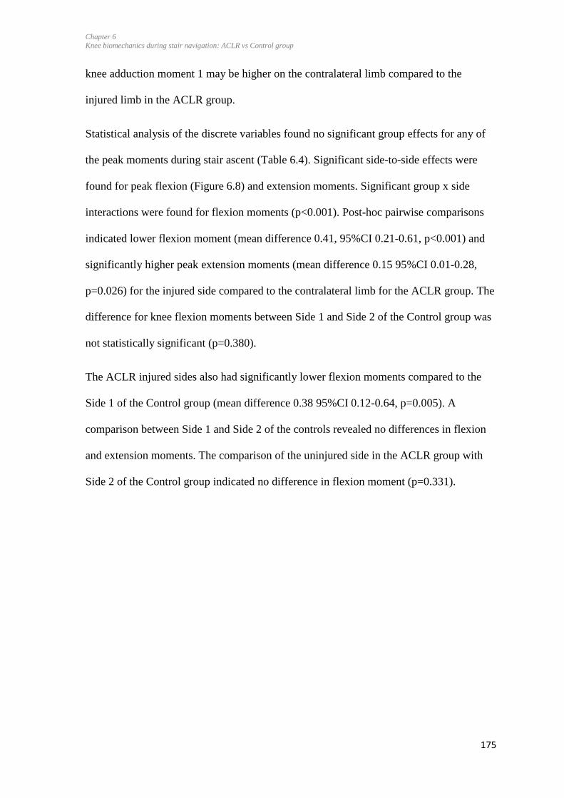
Chapter 6
Knee biomechanics during stair navigation: ACLR vs Control group
175
knee adduction moment 1 may be higher on the contralateral limb compared to the
injured limb in the ACLR group.
Statistical analysis of the discrete variables found no significant group effects for any of
the peak moments during stair ascent (Table 6.4). Significant side-to-side effects were
found for peak flexion (Figure 6.8) and extension moments. Significant group x side
interactions were found for flexion moments (p<0.001). Post-hoc pairwise comparisons
indicated lower flexion moment (mean difference 0.41, 95%CI 0.21-0.61, p<0.001) and
significantly higher peak extension moments (mean difference 0.15 95%CI 0.01-0.28,
p=0.026) for the injured side compared to the contralateral limb for the ACLR group. The
difference for knee flexion moments between Side 1 and Side 2 of the Control group was
not statistically significant (p=0.380).
The ACLR injured sides also had significantly lower flexion moments compared to the
Side 1 of the Control group (mean difference 0.38 95%CI 0.12-0.64, p=0.005). A
comparison between Side 1 and Side 2 of the controls revealed no differences in flexion
and extension moments. The comparison of the uninjured side in the ACLR group with
Side 2 of the Control group indicated no difference in flexion moment (p=0.331).
Chapter 6
Knee biomechanics during stair navigation: ACLR vs Control group
176
Figure 6.8 Diagram indicating the flexion moment asymmetry in participants with ACLR.
The contralateral (uninjured) sides (Side 2) of the ACLR group were generating higher moments compared
to the injured sides (Side 1).

Chapter 6
Knee biomechanics during stair navigation: ACLR vs Control group
177
Table 6.4. Mean (SD) of spatiotemporal variables, moments and angles: stair ascent
ACLR group Control group
Injured side Uninjured side Side 1 Side 2 Group
effect
Side effect Group x side
interaction
Spatiotemporal variables (n, ACLR=25, Control=24)
Stance time (seconds) 0.94 (0.9) 0.95 (0.1) 0.91 (0.9) 0.91 (0.1) 0.255 0.541 0.526
Stance fraction (seconds) 0.64 (0.2) 0.65 (0.2) 0.65 (0.4) 0.65 (0.2) 0.466 0.820 0.091
Stride time (seconds) 1.43 (0.1) 1.45 (0.1) 1.43 (0.1) 1.4 (0.1) 0.515 0.924 0.773
Stride speed (meters/second) 0.68 (0.05) 0.68 (0.06) 0.69 (0.06) 0.69 (0.07) 0.599 0.821 0.986
Time to peak adduction
moment (seconds)
0.32 (0.07) 0.30 (0.06) 0.32 (0.06) 0.31 (0.06) 0.809 0.118 0.406
Kinematics (angles; degrees) (n, ACLR=25, Control=24)
Flexion -55.6 (4.9) ∞* -57.7 (4.4) -60.0 (4.2) ∞* -59.2 (5.0) 0.022∞ 0.185 0.004*
Extension -6.3 (4.3) -6.4 (4.2) -6.9 (4.0) -7.2 (4.8) 0.547 0.658 0.837
Flexion-extension excursion 49.3 (4.9) * 51.5 (5.9) 53.2 (4.9) * 52.0 (5.6) 0.124 0.427 0.004*
Abduction -3.8 (5.2) -4.5 (4.1) -5.0 (5.0) -4.6 (6.0) 0.616 0.843 0.383
Adduction 4.8 (6.6) 5.3 (7.3) 3.7 (5.2) 3.5 (5.2) 0.347 0.896 0.656
External rotation -7.4 (5.4) -8.1 (6.1) -7.8 (5.3) -8.9 (6.9) 0.745 0.200 0.806
Internal rotation 3.8 (6.7) 3.2 (5.6) 2.8 (5.0) 1.1 (6.4) 0.305 0.190 0.528
External moments (Nm/kg*m) (n, ACLR=25, Control=24)
Flexion peak 1.93 (0.49) #* 2.35 (0.39) # 2.31 (0.41) * 2.23 (0.47) 0.227 0.024# <0.001*
Extension peak 0.23 (0.25) # 0.08 (0.36) # 0.14 (0.31) 0.08 (0.32) 0.534 0.027# 0.350
Adduction peak 1 0.53 (0.29) 0.58 (0.23) 0.50 (0.33) 0.51 (0.35) 0.502 0.575 0.695
Adduction peak 2 0.35 (0.24) 0.46 (0.19) 0.42 (0.30) 0.43 (0.30) 0.645 0.305 0.348
External rotation peak 1 -0.43 (0.14) -0.56 (0.17) -0.56 (0.18) -0.56 (0.20) 0.111 0.070 0.072
Internal rotation peak 2 0.10 (0.06) 0.09 (0.07) 0.10 (0.06) 0.10 (0.07) 0.910 0.379 0.446
ACLR: ACL reconstruction; * Significant difference between group sides; # significant difference present between both sides; ∞ significant difference between groups; Group
× side interaction indicates that there is a statistically significant difference between the sides and the group, that is, the difference between the two sides differs between
groups. Group: there is a statistically significant difference between the values for groups. Sides: there is a statistically significant difference between the two sides. *
Significant differences for sides between groups (between groups); # significant difference between sides (within groups); ∞ significant difference between groups;
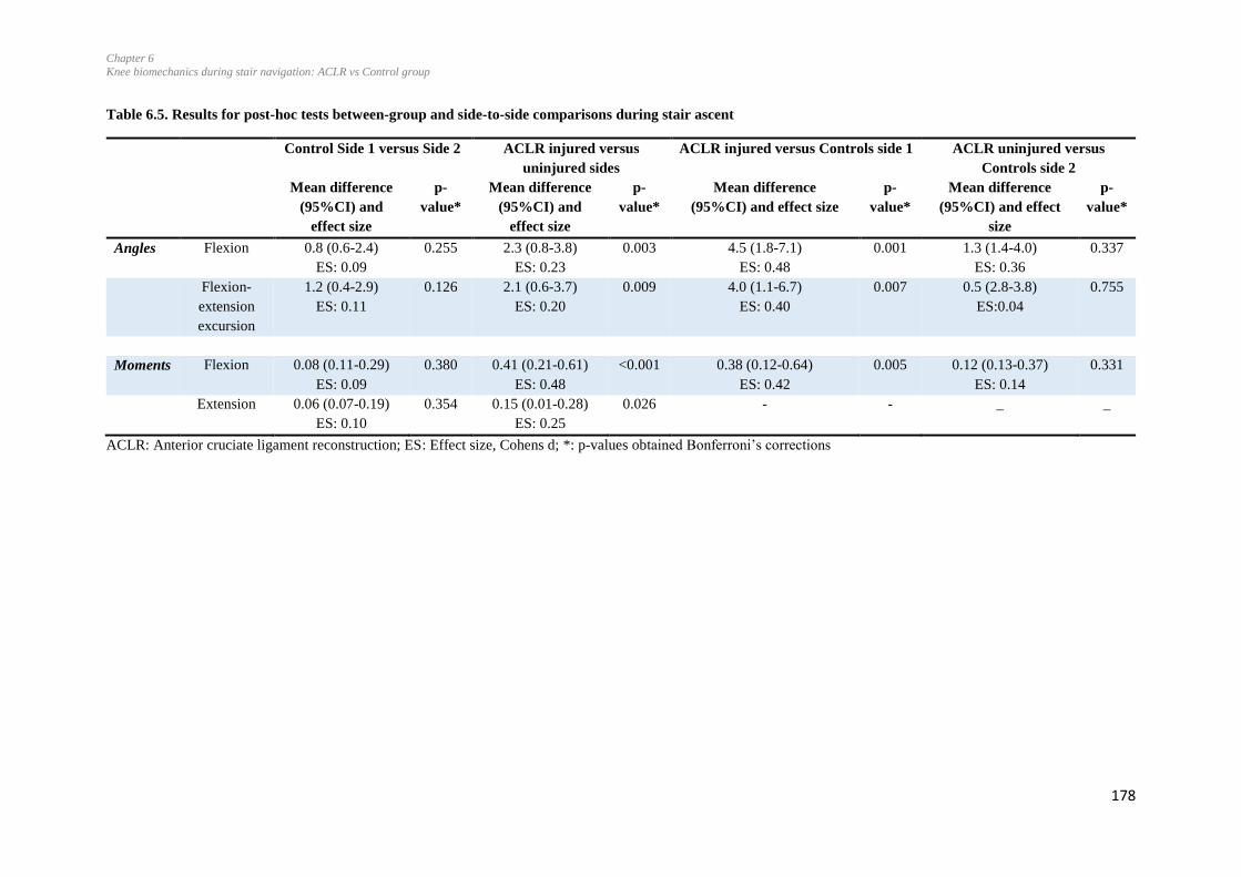
Chapter 6
Knee biomechanics during stair navigation: ACLR vs Control group
178
Table 6.5. Results for post-hoc tests between-group and side-to-side comparisons during stair ascent
Control Side 1 versus Side 2 ACLR injured versus
uninjured sides
ACLR injured versus Controls side 1 ACLR uninjured versus
Controls side 2
Mean difference
(95%CI) and
effect size
p-
value*
Mean difference
(95%CI) and
effect size
p-
value*
Mean difference
(95%CI) and effect size
p-
value*
Mean difference
(95%CI) and effect
size
p-
value*
Angles Flexion 0.8 (0.6-2.4)
ES: 0.09
0.255 2.3 (0.8-3.8)
ES: 0.23
0.003 4.5 (1.8-7.1)
ES: 0.48
0.001 1.3 (1.4-4.0)
ES: 0.36
0.337
Flexion-
extension
excursion
1.2 (0.4-2.9)
ES: 0.11
0.126 2.1 (0.6-3.7)
ES: 0.20
0.009 4.0 (1.1-6.7)
ES: 0.40
0.007 0.5 (2.8-3.8)
ES:0.04
0.755
Moments Flexion 0.08 (0.11-0.29)
ES: 0.09
0.380 0.41 (0.21-0.61)
ES: 0.48
<0.001 0.38 (0.12-0.64)
ES: 0.42
0.005 0.12 (0.13-0.37)
ES: 0.14
0.331
Extension 0.06 (0.07-0.19)
ES: 0.10
0.354 0.15 (0.01-0.28)
ES: 0.25
0.026 - - _ _
ACLR: Anterior cruciate ligament reconstruction; ES: Effect size, Cohens d; *: p-values obtained Bonferroni’s corrections
Chapter 6
Knee biomechanics during stair navigation: ACLR vs Control group
179
6.4.2 Stair descent
6.4.2.1 Spatiotemporal variables
No group, side and group x side interactions were found for the stride time and stance time
during stair descent (Table 6.6). There was a significant side effect for the time taken to reach
the peak of adduction moment 1 (p= 0.020). Post-hoc analysis indicated a significant
difference when comparing the injured to uninjured sides of the ACLR group: the injured
sides took longer than the contralateral sides to reach peak adduction moment 1 (mean
difference 0.01seconds, 95%CI 0.0-0.04, p=0.006, Table 6.7). No significant difference was
found when comparing the time to peak adduction of Side 1 to Side 2 for the controls
(p=0.592).
6.4.2.2 Joint angles
Figure 6.9 demonstrates the linear envelopes (Mean and SD clouds) for the injured and
uninjured sides of the ACLR group and the side 1 of the Control group. In the sagittal plane,
the knee moved from a position of approximately 4° flexion at the time of initial foot contact
to approximately 70° flexion during the propulsion phase (Figure 6.9). No difference was
evident for the course of the flexion angles within and between groups. In the frontal plane,
abduction/adduction angles appear to remain less than 5° for both sides of the ACLR group,
however, for the control, greater abduction is evident during the propulsion phase. In the
transverse plane, the knee appears to be moving from a relative position of external rotation
at initial foot contact towards internal rotation during the weight acceptance and propulsion
phases for both groups of participants.
No significant side effects, group effects and group x interaction effects were found for the
discrete variables of the knee kinematics during the stance phase during stair descent (Table
6.6).
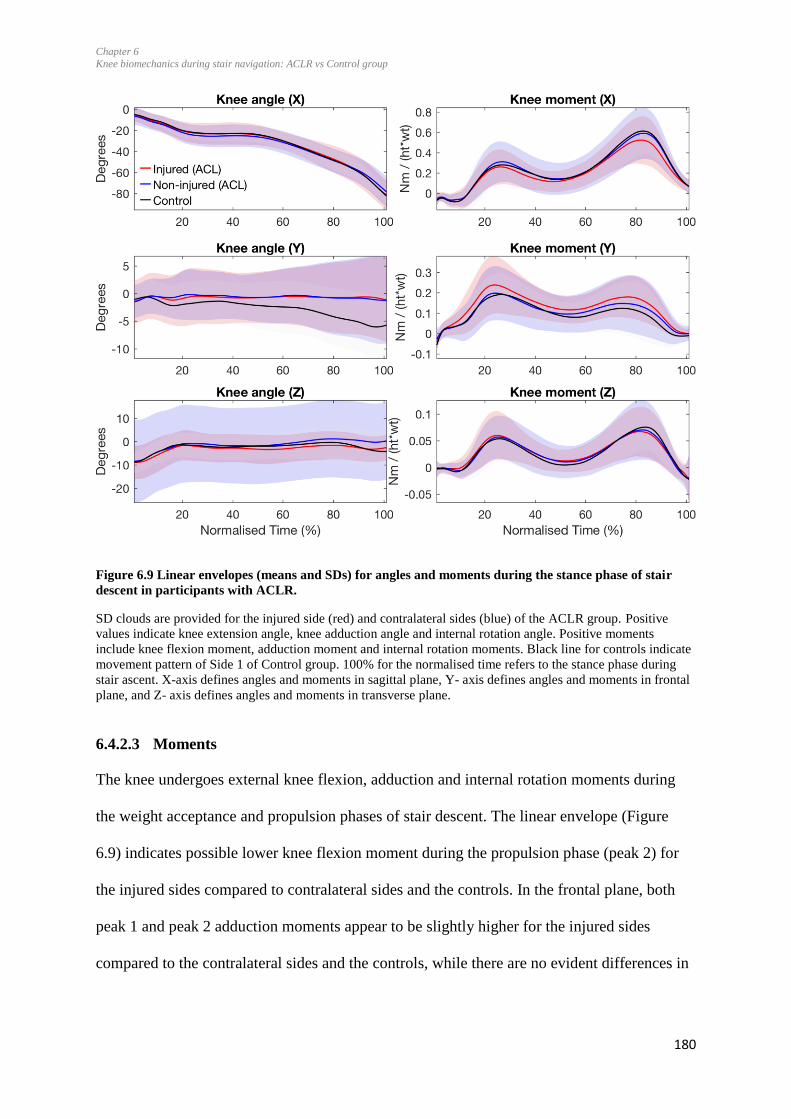
Chapter 6
Knee biomechanics during stair navigation: ACLR vs Control group
180
Figure 6.9 Linear envelopes (means and SDs) for angles and moments during the stance phase of stair
descent in participants with ACLR.
SD clouds are provided for the injured side (red) and contralateral sides (blue) of the ACLR group. Positive
values indicate knee extension angle, knee adduction angle and internal rotation angle. Positive moments
include knee flexion moment, adduction moment and internal rotation moments. Black line for controls indicate
movement pattern of Side 1 of Control group. 100% for the normalised time refers to the stance phase during
stair ascent. X-axis defines angles and moments in sagittal plane, Y- axis defines angles and moments in frontal
plane, and Z- axis defines angles and moments in transverse plane.
6.4.2.3 Moments
The knee undergoes external knee flexion, adduction and internal rotation moments during
the weight acceptance and propulsion phases of stair descent. The linear envelope (Figure
6.9) indicates possible lower knee flexion moment during the propulsion phase (peak 2) for
the injured sides compared to contralateral sides and the controls. In the frontal plane, both
peak 1 and peak 2 adduction moments appear to be slightly higher for the injured sides
compared to the contralateral sides and the controls, while there are no evident differences in
Chapter 6
Knee biomechanics during stair navigation: ACLR vs Control group
181
the transverse plane. However, the analysis of discrete variables indicated no significant side
or group effects or group x side interactions.
No group, side and group x side interactions were found for other peak moments (Table 6.6,
Figure 6.9).
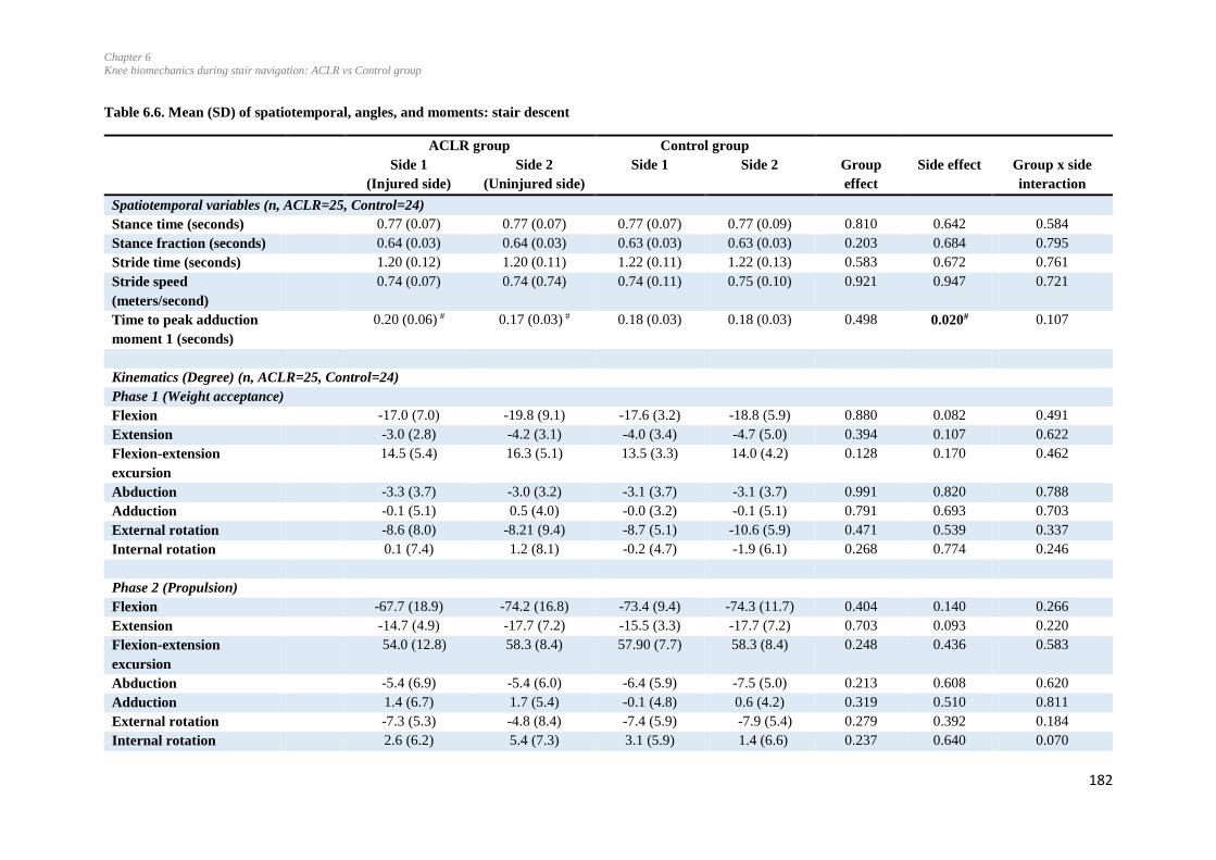
Chapter 6
Knee biomechanics during stair navigation: ACLR vs Control group
182
Table 6.6. Mean (SD) of spatiotemporal, angles, and moments: stair descent
ACLR group Control group
Side 1
(Injured side)
Side 2
(Uninjured side)
Side 1 Side 2 Group
effect
Side effect Group x side
interaction
Spatiotemporal variables (n, ACLR=25, Control=24)
Stance time (seconds) 0.77 (0.07) 0.77 (0.07) 0.77 (0.07) 0.77 (0.09) 0.810 0.642 0.584
Stance fraction (seconds) 0.64 (0.03) 0.64 (0.03) 0.63 (0.03) 0.63 (0.03) 0.203 0.684 0.795
Stride time (seconds) 1.20 (0.12) 1.20 (0.11) 1.22 (0.11) 1.22 (0.13) 0.583 0.672 0.761
Stride speed
(meters/second)
0.74 (0.07) 0.74 (0.74) 0.74 (0.11) 0.75 (0.10) 0.921 0.947 0.721
Time to peak adduction
moment 1 (seconds)
0.20 (0.06) # 0.17 (0.03) # 0.18 (0.03) 0.18 (0.03) 0.498 0.020# 0.107
Kinematics (Degree) (n, ACLR=25, Control=24)
Phase 1 (Weight acceptance)
Flexion -17.0 (7.0) -19.8 (9.1) -17.6 (3.2) -18.8 (5.9) 0.880 0.082 0.491
Extension -3.0 (2.8) -4.2 (3.1) -4.0 (3.4) -4.7 (5.0) 0.394 0.107 0.622
Flexion-extension
excursion
14.5 (5.4) 16.3 (5.1) 13.5 (3.3) 14.0 (4.2) 0.128 0.170 0.462
Abduction -3.3 (3.7) -3.0 (3.2) -3.1 (3.7) -3.1 (3.7) 0.991 0.820 0.788
Adduction -0.1 (5.1) 0.5 (4.0) -0.0 (3.2) -0.1 (5.1) 0.791 0.693 0.703
External rotation -8.6 (8.0) -8.21 (9.4) -8.7 (5.1) -10.6 (5.9) 0.471 0.539 0.337
Internal rotation 0.1 (7.4) 1.2 (8.1) -0.2 (4.7) -1.9 (6.1) 0.268 0.774 0.246
Phase 2 (Propulsion)
Flexion -67.7 (18.9) -74.2 (16.8) -73.4 (9.4) -74.3 (11.7) 0.404 0.140 0.266
Extension -14.7 (4.9) -17.7 (7.2) -15.5 (3.3) -17.7 (7.2) 0.703 0.093 0.220
Flexion-extension
excursion
54.0 (12.8) 58.3 (8.4) 57.90 (7.7) 58.3 (8.4) 0.248 0.436 0.583
Abduction -5.4 (6.9) -5.4 (6.0) -6.4 (5.9) -7.5 (5.0) 0.213 0.608 0.620
Adduction 1.4 (6.7) 1.7 (5.4) -0.1 (4.8) 0.6 (4.2) 0.319 0.510 0.811
External rotation -7.3 (5.3) -4.8 (8.4) -7.4 (5.9) -7.9 (5.4) 0.279 0.392 0.184
Internal rotation 2.6 (6.2) 5.4 (7.3) 3.1 (5.9) 1.4 (6.6) 0.237 0.640 0.070
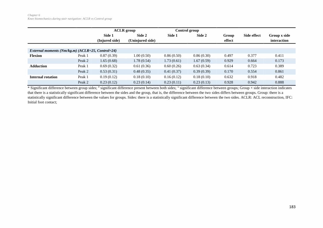
Chapter 6
Knee biomechanics during stair navigation: ACLR vs Control group
183
ACLR group Control group
Side 1
(Injured side)
Side 2
(Uninjured side)
Side 1 Side 2 Group
effect
Side effect Group x side
interaction
External moments (Nm/kg.m) (ACLR=25, Control=24)
Flexion Peak 1 0.87 (0.39) 1.00 (0.50) 0.86 (0.50) 0.86 (0.30) 0.497 0.377 0.411
Peak 2 1.65 (0.68) 1.78 (0.54) 1.73 (0.61) 1.67 (0.59) 0.929 0.664 0.173
Adduction Peak 1 0.69 (0.32) 0.61 (0.36) 0.60 (0.26) 0.63 (0.34) 0.614 0.723 0.389
Peak 2 0.53 (0.31) 0.48 (0.35) 0.41 (0.37) 0.39 (0.39) 0.170 0.554 0.861
Internal rotation Peak 1 0.19 (0.12) 0.18 (0.10) 0.16 (0.12) 0.18 (0.10) 0.632 0.918 0.482
Peak 2 0.23 (0.12) 0.23 (0.14) 0.23 (0.11) 0.23 (0.13) 0.928 0.942 0.888
* Significant difference between group sides; # significant difference present between both sides; ∞ significant difference between groups; Group × side interaction indicates
that there is a statistically significant difference between the sides and the group, that is, the difference between the two sides differs between groups. Group: there is a
statistically significant difference between the values for groups. Sides: there is a statistically significant difference between the two sides. ACLR: ACL reconstruction, IFC:
Initial foot contact;
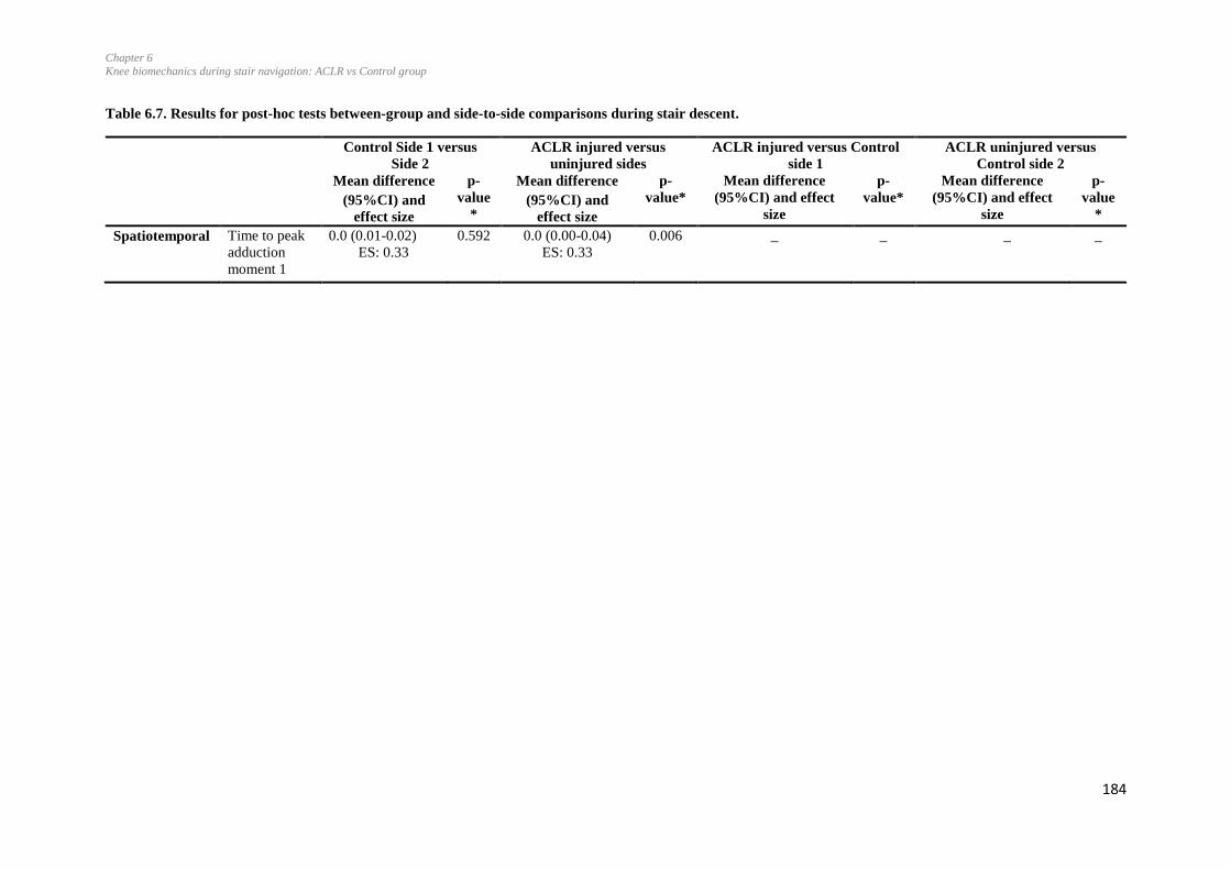
Chapter 6
Knee biomechanics during stair navigation: ACLR vs Control group
184
Table 6.7. Results for post-hoc tests between-group and side-to-side comparisons during stair descent.
Control Side 1 versus
Side 2
ACLR injured versus
uninjured sides
ACLR injured versus Control
side 1
ACLR uninjured versus
Control side 2
Mean difference
(95%CI) and
effect size
p-
value
*
Mean difference
(95%CI) and
effect size
p-
value*
Mean difference
(95%CI) and effect
size
p-
value*
Mean difference
(95%CI) and effect
size
p-
value
*
Spatiotemporal Time to peak
adduction
moment 1
0.0 (0.01-0.02)
ES: 0.33
0.592 0.0 (0.00-0.04)
ES: 0.33 0.006 _ _ _ _
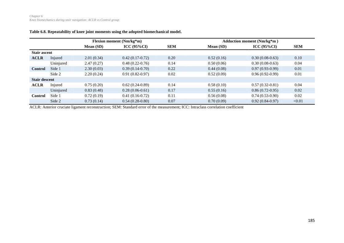
Chapter 6
Knee biomechanics during stair navigation: ACLR vs Control group
185
Table 6.8. Repeatability of knee joint moments using the adopted biomechanical model.
Flexion moment (Nm/kg*m) Adduction moment (Nm/kg*m )
Mean (SD) ICC (95%CI) SEM Mean (SD) ICC (95%CI) SEM
Stair ascent
ACLR Injured 2.01 (0.34) 0.42 (0.17-0.72) 0.20 0.52 (0.16) 0.30 (0.08-0.63) 0.10
Uninjured 2.47 (0.27) 0.48 (0.22-0.76) 0.14 0.50 (0.06) 0.30 (0.08-0.63) 0.04
Control Side 1 2.30 (0.03) 0.39 (0.14-0.70) 0.22 0.44 (0.08) 0.97 (0.93-0.99) 0.01
Side 2 2.20 (0.24) 0.91 (0.82-0.97) 0.02 0.52 (0.09) 0.96 (0.92-0.99) 0.01
Stair descent
ACLR Injured 0.75 (0.20) 0.62 (0.24-0.89) 0.14 0.58 (0.10) 0.57 (0.32-0.81) 0.04
Uninjured 0.83 (0.48) 0.28 (0.06-0.61) 0.17 0.55 (0.16) 0.86 (0.72-0.95) 0.02
Control Side 1 0.72 (0.19) 0.41 (0.16-0.72) 0.11 0.56 (0.08) 0.74 (0.53-0.90) 0.02
Side 2 0.73 (0.14) 0.54 (0.28-0.80) 0.07 0.70 (0.09) 0.92 (0.84-0.97) <0.01
ACLR: Anterior cruciate ligament reconstruction; SEM: Standard error of the measurement; ICC: Intraclass correlation coefficient

Chapter 6
Knee biomechanics during stair navigation: ACLR vs Control group
186
6.4.3 Repeatability of knee joint moments using the adopted biomechanical model
For stair ascent, high to low consistency was shown for flexion moments with the ICC
ranging from 0.39 to 0.91 and SEM 0.02 to 0.22 Nm/kg.m. During descent, flexion
moments had a moderate to low consistency with ICC ranging from 0.28 to 0.62 and
SEM from 0.07 to 0.14 Nm/kg.m, were found (Table 6.8).
For the adduction moment, during stair ascent high to low consistency between trials was
shown in all groups with ICC ranging from 0.30 to 0.97 and SEM 0.01 to 0.10 Nm/kg.m.
For stair descent, the adduction moment ranged from high to moderate consistency for
both groups with ICC ranging from 0.57 to 0.92 and SEM from <0.01 to 0.04 Nm/kg.m.
Therefore, low to high consistency was found for peak external flexion and adduction
moments of the knee using 3D motion analysis in ACLR and control participants (Table
6.8).
6.5 Discussion
The primary aim of this study was to compare knee kinematics and kinetics of the ACLR
knee to the contralateral limb and with the age-matched uninjured controls during stair
ascent and descent. It was hypothesized that the ACLR group would have a lower knee
flexion moment, and higher adduction and internal rotation moments during stair ascent
and descent compared to the Control group. The first hypothesis was partially accepted as
the injured limbs of the ACLR group presented with lower knee flexion moments
compared to the contralateral uninjured limbs during stair ascent. The second hypothesis
was partially accepted as group differences were found for the peak flexion angles in the
ACLR group compared to the Control group. Interestingly, the peak knee extension
moment during the second half of the ascent stance phase was higher in the ACLR
injured side than the contralateral uninjured limbs. The differences in knee moments

Chapter 6
Knee biomechanics during stair navigation: ACLR vs Control group
187
indicate altered joint loading and possible loss of control of movement, both of which can
contribute to the development of early onset of knee osteoarthritis following ACL injury
(Andriacchi et al., 2004).
6.5.1 Demographics
Twenty-five participants with ACLR, and 24 control age- and gender-matched
participants, took part in the study. Participants with ACLR were from 2 to 10 (mean 4.7)
years following surgery and had higher body mass index (p<0.001) compared to the
Control group (Table 6.3). Higher BMI in participants with ACLR may be associated
with decreased levels of physical activity. The Tegner Score found no significant
difference in the sports participation for pre-injury compared to post-injury levels (pre-
injury Tegner sore: 7.0/10; post-injury score: 5.0/10, p=0.09, Chapter 4, Table 4.2). The
current amount of weekly sports participation or physical activity was not assessed in this
study. Previous studies have indicated different reasons for the gradual decrease in the
level of physical activities following ACLR which could be related to the presence of
knee pain or strength deficits (Gobbi & Francisco, 2006) and other life events such as
marriage, childbirth, change in life style, and increased job demands (Flanigan et al.,
2013). Results of the KOOS indicated that the ACLR group rated the Sports/Recreation
function lower than the Control group (ACLR= 75.8; controls= 98.0, p <0.001).
Therefore, it is possible that decreased level of their physical activity in the ACLR group
could have led to the higher body mass index in these participants compared with the
controls.
From the KOOS scores, the participants indicated having problems with stair ascending
and descending for the question about stairs in knee function and daily living (Chapter 4).
More participants from the ACLR group appeared to experience symptoms during stair

Chapter 6
Knee biomechanics during stair navigation: ACLR vs Control group
188
ascent than descent. The mean score for the KOOS question relating to stair ascent was
93/100 for stair ascent and 90/100 for stair descent. Differences were also found during
stair navigation compared to Control group (Stair ascent=99/100, stair descent=98/100).
There was minimal difference in the symptom rating when considering the scores for
ascent and descent. Thus, symptoms alone during those activities are unlikely to explain
the asymmetries during ascent compared to descent.
6.5.2 Spatiotemporal variables for stair ascent and descent
No differences were found between the ACLR and Control group for any of the variables
during stair ascent and descent except for the time to the first peak adduction moment
during stair descent. It was longer for the injured compared to the contralateral uninjured
side during stair descent (Table 6.6). It may be a compensatory strategy used by the
participants with ACLR by taking longer time to reach the peak adduction moment during
the stance phase. This strategy could have helped the participants to distribute the
moments in the frontal plane. Results of the current study did not find differences in other
spatiotemporal variables such as stance time or stride time, which is similar to another
study (Hall et al., 2012), which also indicated no differences in the stance time in
participants from 2 to 18 years following surgery in young participants (mean age of 26
years) during stair ascent and descent.
A previous study from our unit and collaborators at the University of Umeå, Sweden,
exploring kinematics during stair descent following ACLR (>20 years post-injury)
showed the differences in speed among participants with ACLR compared with the age
matched the Control group during stair descent (Sole et al., 2016). Participants of that
study had higher age (45.6 years on average) and lower knee-function in activities of
daily living (KOOS ADL= 84) and sports and recreation (KOOS Sports/recreation=50)
Chapter 6
Knee biomechanics during stair navigation: ACLR vs Control group
189
on KOOS subscale compared to the current study (KOOS ADL= 95, KOOS
Sports/recreation=75), indicating reduced knee function. Younger participants, better
knee function, and less time since surgery in the current study compared to that study
(Sole et al., 2016) could be the reason for the presence of fewer discrepancies in
spatiotemporal variables.
6.5.3 Joint angles
6.5.3.1 Ascent
Participants in ACLR group exhibited significantly lower peak knee flexion angles
compared to the controls during stair ascent (Table 6.4). Similar findings have been
reported in previous study (Lewek et al., 2002) indicating lower knee flexion angle in
ACLR group compared to the uninjured Control group. That study divided the ACLR
group based on the muscle strength, and the cohort with poor muscle strength had
reduced knee flexion moment during walking. In the current study, flexion-extension
excursion angles were lower on the injured side compared to side 1 of Control group
during stair ascent. Maximum flexion during stair ascent occurs at initial contact and it
appears that this occurred with more knee flexion in the ACLR injured side, while
extension did not differ between groups and sides. These small alterations in the joint
angles are believed to cause a spatial shift in the location of load contact which could lead
to degeneration of the articular cartilage (Andriacchi et al., 2004). Further, peak knee
flexion angles during stance phase of gait were found to be progressing in participants
with severe osteoarthritis (Astephen, Deluzio, Caldwell, Dunbar, & Hubley-Kozey,
2008). As the stair navigation involves greater degrees of sagittal plane range of motion,
such activities could be challenging for the participants.
Chapter 6
Knee biomechanics during stair navigation: ACLR vs Control group
190
6.5.3.2 Descent
No significant differences in knee angles were found in all three planes during weight
acceptance and propulsion phases of stair descent. Another study, Sole et al., 2016, also
reported differences in temporal variables than for kinematic variables during stair
descent at more than 20 years following ACL injury. In that study the participants of
ACLR group walked more slowly compared to the Control group. The current study did
not find differences in speed during stair descent; however, differences were present in
the time to reach the first peak knee adduction moment compared to the Control group.
Again it indicates a compensation strategy or persistent caution during weight bearing on
the injured side during stair descent. Moreover, lack of differences in peak knee flexion
angles may be due to the fact that stair descent involves less knee joint loading, as evident
with peak flexion moments when compared to stair ascent. Alternatively, it may be
possible that compensatory strategies are more evident in other body segments (for
instance the trunk or hip) while descending stairs. This needs further investigation.
6.5.4 Moments
6.5.4.1 Ascent
Lower knee flexion moments were present on the injured side compared to the
contralateral limb in ACLR group and the side 1 of the Control group during stair ascent.
The mean difference between the injured and uninjured side was greater than the SEM
(SEM= 0.14 to 0.20 Nm/kg.m) for both sides, therefore the differences in moments
among both sides in ACLR group are not incidental. Lower flexion moments on the
injured side indicate between-sides moments asymmetry and also the compensatory role
played by the contralateral side by generating higher flexion moments during ambulation.
Chapter 6
Knee biomechanics during stair navigation: ACLR vs Control group
191
Side-to-side differences in moments are considered as a risk factor for re-injury during
sports (Schmitt et al., 2012).
Higher extension moments on the injured side indicate compensatory behaviour on the
injured side during ambulation. This is similar to the ‘quadriceps avoidance’ reported in
participants with ACL deficiency where they ascended the stairs with lower external
flexion moments and higher knee extension moments at 60° of knee flexion, which was
interpreted as the patients’ effort to avoid the quadriceps contraction (Berchuck,
Andriacchi, Bach, & Reider, 1990). Participants of the current study had similar findings
where they ascended the stairs with lower knee flexion moment and higher knee
extension moments at 57° of knee flexion. As these participants had higher knee laxity
compared to the contralateral limb (Chapter 4, Table 4.3), it may be due to participants’
efforts to stabilise the knee. Other plausible reasons could be related to the lack of
neuromuscular control of movement, or change in the co-contraction between hamstring
and quadriceps, or it may be related to the changes in control of the hip muscles.
Quadriceps phenomenon was reported to decrease following surgery in a group of
participants who were tested during treadmill walking at 8 months following surgery
(Knoll et al., 2004). Surprisingly, it seems to persist for long-term.
Our results are similar to other studies (Hall et al., 2012; Zabala et al., 2013) analysing the
moments during stair ascent and descent. These studies also reported lower flexion and
adduction moments on the injured sides in participants with ACLR in short-term (from 22
months to 34 months) (Zabala et al., 2013) and long-term (2-18 years) (Hall et al., 2012).
Lower flexion moments on the injured side compared to the uninjured side are likely to
indicate reduced loading at the knee joint and perhaps due to decreased neuromuscular
control, and a hesitation before weight-bearing on the injured side (Andriacchi, 1990).
Lower joint loading is also confirmed by the lower ground reaction force on the injured
Chapter 6
Knee biomechanics during stair navigation: ACLR vs Control group
192
side during eccentric deceleration phase of vertical jump in another study (Baumgart,
Hoppe, & Freiwald, 2017).
Lower joint loading may be associated with the progression of osteoarthritis. Altered joint
mechanics including joint moments and angles can lead to onset and progression of the
symptoms related to osteoarthritis by initiating cartilage degradation (Andriacchi, Koo, &
Scanlan, 2009). Recent research has indicated that joint unloading, not overloading, may
be associated with the process of early degeneration at the knee after ACL injury (Hall et
al., 2012; Zabala et al., 2013). The lower joint moments and joint contact forces were
found 6 months after injury and reconstruction in participants who developed
osteoarthritis (Wellsandt et al., 2016). Participants with radiographic knee osteoarthritis
walked with lower knee adduction moments and medial compartment joint contact forces
compared to those without osteoarthritis early 5 years following ACLR (Wellsandt et al.,
2016). Similarly, another study indicated lower knee flexion moment and lower
tibiofemoral contact forces in participants with ACLR from 2-3 years following surgery
compared to the Control group during walking, running and side stepping (Saxby et al.,
2016). Participants who had decreased knee joint loading at 2 years following ACLR had
increased risk of developing symptoms of early knee osteoarthritis 5 years following
surgery (Khandha et al., 2016). That study found lower peak knee flexion angle and
external flexion moments in the group of ACLR participants who developed
osteoarthritis. Results of the current study in terms of decreased flexion angles and
flexion moments are similar to the above-mentioned studies, though the evidence for the
presence or absence of osteoarthritis was not explored in this study.
In contrast to the lower joint loading following reconstruction surgery, it is important to
acknowledge that there is data to support that there is increased progression of
osteoarthritis with higher joint loading in the frontal plane 5 years following ACLR
Chapter 6
Knee biomechanics during stair navigation: ACLR vs Control group
193
(Butler et al., 2009). That study indicated higher peak knee-abduction moment by 21% in
the ACL compared with the Control group (Butler et al., 2009) and speculated that over-
loading on the damaged cartilage can have deleterious effects over the joint health. It
seems that the future research direction should be to perform a subgroup analysis on the
participants’ biomechanical characteristics in order to develop strategies to achieve
‘optimal joint loading’.
Muscles responsible for controlling joint loading and flexion moments have also been
shown to be influenced by the quadriceps muscles activation and force development. In
an animal model, lower activation of quadriceps muscle was associated with the lower
ground reaction forces in the cat knee (Herzog, Longino, & Clark, 2003), indicating the
role of muscles in the joint loading. Isokinetic thigh muscle strength of the participants of
the ACLR and the Control groups was presented in Chapter 4 (Table 4.3). The
participants of the ACLR group had lower concentric quadriceps strength compared to the
controls, and lower eccentric quadriceps strength compared to the contralateral sides.
Lower joint loading, as evident with decreased external knee flexion moments, along with
reduced muscle strength, may lead to the onset and progression of joint degeneration.
Gait mechanics, particularly, side to side differences in the knee flexion and adduction
moments at 2 years post-surgery during walking were associated with worse patient-
reported outcomes, such as KOOS pain and KOOS QOL, on average, 8 years following
surgery (Erhart‐Hledik, Chu, Asay, & Andriacchi, 2016). Side-to-side differences have
also been found in the current study in the peak knee flexion moments in the ACLR
group. KOOS results from Chapter 4 (Table 4.2) indicate higher pain (KOOS pain= 85.3)
and lower quality of life (KOOS QOL= 47.0) in participants with ACLR compared to
Control group, which may also be related to the differences in the movement patterns
between these groups.
Chapter 6
Knee biomechanics during stair navigation: ACLR vs Control group
194
6.5.4.2 Descent
The adduction moments peak 1 and peak 2 were higher, based on figure 6.9 and Table
6.6, although the differences were not statistically significant. The differences did not
reach significant levels, which may be due to the greater time taken by the participants
with ACLR to reach the adduction moment peak 1, or it may be due to the large
individual variability, as shown by the SD in the knee abduction angles. Contrasting
findings have been found from the previous studies indicating the higher adduction
moment (Butler et al., 2009) to lower (Zabala et al., 2013) or no differences (Hart,
Culvenor, et al., 2015) in the adduction moment compared to the uninjured control group
or the contralateral limb. These studies included participants from early to late duration
following surgery; therefore, it is difficult to speculate on the reasons behind the
variability in study findings. Studies measuring the joint contact forces are required to
provide a clear picture of joint loading and contact forces in this cohort.
6.5.5 Methodological considerations
There is debate in the literature regarding the use of anatomical or functional joint
estimation for the biomechanical model. Both methods have been shown to be equally
reliable, with the only statistically significant difference being in kinematics during the
transverse plane, of a magnitude of less than 0.78° for knee internal rotation/external
rotation (Pohl, Lloyd, & Ferber, 2010). However, functional joint estimations have been
reported to give more repeatable gait curves for the hip joint moments and angles during
walking (Besier, Sturnieks, Alderson, & Lloyd, 2003). For the purposes of this study, for
the biomechanical model, the hip joint centre was functionally estimated (Cappozzo,
1984; Leardini et al., 1999) while for the knee and ankle anatomical joint centres were
used (Wu et al., 2002). The dynamic trials, as mentioned in section 6.3.3.3, were used to
estimate the hip joint centre. Knee and ankle joints were calculated through the
Chapter 6
Knee biomechanics during stair navigation: ACLR vs Control group
195
anatomical method and the static trials were used as a reference to create the knee and
ankle joint centre. The joint centres were determined with respect to the shank local
coordinate systems.
A further consideration was the placement of the step on top of the force plates. While
embedded force plates within the stair set-up have been described (Strutzenberger,
Richter, Schneider, Mündermann, & Schwameder, 2011), these were not available for this
study. Movement artefacts between the step and the force plate were minimised by
placing a 10 kg weight on the frame of the step, and force plate data were normalised to
zero during data collection. Previous studies in the field of ACL injury and reconstruction
have also used similar set-up for stair ascent and descent (Gao et al., 2012; Hall et al.,
2012).
6.5.5.1 Limitations
Participants wore their own shoes during the stair ascent and descent. Shoes can
influence the joint loads (Simic et al., 2011) and lower limb biomechanics (Paterson et al.,
2017; Radzimski, Mündermann, & Sole, 2012). It was indicated in another study
(Paterson et al., 2017) involving patients with medial knee osteoarthritis that wearing flat
flexible shoes can lower knee adduction moments compared to stable supportive shoes
during walking. Therefore, in the current study participants wearing standardised shoes
rather than their own shoes would have resulted in different mechanics than the usual
during stair navigation. The aim of the study was to measure and analyse the moments as
closely as possible to their day-to-day life, where individuals prefer wearing different
shoes. A decision had thus been made to test the participants in their own shoes. Trunk
flexion angles were not measured during stair ascent and descent, therefore alterations in
Chapter 6
Knee biomechanics during stair navigation: ACLR vs Control group
196
the trunk angles could have influenced the moments (Mundermann, Asay, Mundermann,
& Andriacchi, 2008).
Another limitation was the use of a two-step staircase instead of a full flight of stairs. It
could have influenced the speed of the participants, as it may alter their speed by knowing
the point of stoppage. However, as the same set of steps was used for both groups, and no
difference in the stride speed was found, the set-up of the stairs and subsequent data
processing would have had similar influence for all of the participants.
Participants were not grouped according to the type of graft, as this was not the focus of
the study. However, a study has indicated no differences in the moments between graft
types in men (Webster & Feller, 2012a). Numerous factors could be responsible for
altered moments such as quadriceps strength (Lewek et al., 2002), graft orientation
(Scanlan, Blazek, Chaudhari, Safran, & Andriacchi, 2009), and type of rehabilitation
(Grindem, Risberg, & Eitzen, 2015). As the participants of this study were recruited from
the community, it was not possible to control for all factors. Despite the differences from
other studies, the results of our study are also consistent with other studies (Hall et al.,
2012; Zabala et al., 2013), indicating lower moments on the injured side and higher on the
contralateral limb compared to the Control group. These are important findings
irrespective of the causes for differences in the moments.
Only knee related moments and angles were explored in this study. Hip moments are
likely to compensate for the lower knee moments on the injured sides (Webster,
Gonzalez-Adrio, & Feller, 2004). Ankle and hip moments were not examined in this
study, and should be considered in future studies. Similarly, knee moments can be
influenced by the position of the trunk (Simic et al., 2011; Simic, Hunt, Bennell, Hinman,
& Wrigley, 2012), and trunk angles during stair ascent and descent were not recorded,
Chapter 6
Knee biomechanics during stair navigation: ACLR vs Control group
197
therefore, we acknowledge this limitation. Further studies are required to explore the
causes of altered moments. Also, no EMG of the thigh muscles was recorded which could
have provided a meaningful information about the internal loading of the knee such as the
co-contraction or the contraction of the thigh muscles. Also, it may be possible that some
of the participants already had development of osteoarthritis; therefore, there is similarity
among the movement patterns in participants with ACLR to what is reported in patients
with knee osteoarthritis in the literature. However, without the imaging data it is not
possible to rule out how many participants in our cohort had presence of knee
osteoarthritis.
6.6 Conclusion
Differences in kinematics and moments in participants with ACLR compared to controls
suggest incomplete recovery of the knee moments, or changes in neuromuscular control
and joint function following surgery. Asymmetric loading during ascent between
uninjured and injured sides following ACLR and compensatory mechanisms potentially
indicate risk for the development of osteoarthritis.
6.7 Summary
Based on the findings of previous chapters of this thesis, deficits in the quadriceps muscle
strength (Chapter 4), along with the hesitancy in weight bearing on the injured side
(Chapter 5) with potential changes in neuromuscular control of the knee appear to affect
the knee joint loading, and the differences in knee flexion and extension moment were
shown to persist up to 10 years following the surgery. The next chapter (Chapter 7) will
explore the association of knee flexion and adduction moments with the participant-
related factors.
Chapter 7
Association of knee moments with participant-related factors
198
7 Association of knee moments with the participant-related factors.
7.1 Prelude to Chapter 7
We intended to compare the knee flexion and adduction moments in participants with
ACLR from 2-10 years to the 10-20 years following ACLR. This was a further
exploration of the finding from the forest plots from Chapter 3 indicating that knee
flexion and adduction moments may increase within increasing time following surgery.
To allow a regression analysis, the time since surgery was extended up to 20 years.
The findings of Chapter 4 indicated lower quadriceps concentric and eccentric peak
torque on the injured side compared to the Control group and the contralateral limb.
Findings from Chapter 3 and 6 indicated lower knee flexion moments in participants with
ACLR, compared to contralateral limb and controls while ascending stairs. These
moments may be associated with the participant-related factors such as muscle strength,
time since reconstruction, and sex of the participants. This chapter describes a cross-
sectional study assessing whether the peak knee flexion and adduction moments were
associated with quadriceps muscle strength, time since surgery, and the sex of the
participants. Additional participants with ACLR were recruited to expand the study to
include participants who had reconstruction surgery 20 years ago.
.
Chapter 7
Association of knee moments with participant-related factors
199
Chapter 7
7.2 Background
The potential relationship between muscle strength and knee flexion moments following
ACLR was discussed in Chapter 3 and Chapter 6. However, literature regarding the
association of moments with the participant-related factors such as muscle strength, time
since surgery, sex of the participants has not yet been reviewed. The following section
provides the literature exploring current understanding of a potential association of the
participant-related factors with the knee moments.
7.2.1 Muscle strength and the knee flexion moment
Previous studies suggest that lower knee flexion moments during walking, jogging and
drop vertical jump are associated with reduced quadriceps muscle strength in participants
with ACLR (Lewek et al., 2002; Patel, Hurwitz, Bush-Joseph, Bach, & Andriacchi, 2003;
Schmitt et al., 2015). Patel et al., 2003 explored a relationship between external knee
flexion moments during jogging and stair climbing and isokinetic concentric quadriceps
strength (at 60°/s) in 44 individuals with unilateral ACL deficiency. The muscle strength
was normalised to body weight and height, and was assessed for the injured sides only.
They found a significant linear association such that reduced knee flexion moments were
correlated with reduced quadriceps strength (Patel et al., 2003). However, as they
included participants that had not undergone reconstructive surgery, their results may not
be applicable following surgery.
Association among the knee moments and muscle strength has been explored in previous
studies. Lewek et al., 2002 and Schmitt et al., 2015, both explored relationships between
movement patterns and muscle strength in participants with ACLR. Lewek et al., 2002
included a group of participants with ACL deficiencies and a group who had undergone
Chapter 7
Association of knee moments with participant-related factors
200
ACLR (up to 43 weeks post-surgery), and explored the potential relationship between
quadriceps strength and gait patterns, specifically knee angles and moments during the
early stance phase of walking and running. Muscle strength was assessed with an
isometric quadriceps contraction with the knee in 90° flexion and lateral asymmetry
indices were calculated (Lewek et al., 2002). Regression analysis showed a direct
relationship between quadriceps muscle force and the flexion moments (Lewek et al.,
2002). In a later study, participants with ACLR (averaging around 8 months post-surgery)
with weaker quadriceps (LSI < 85%) demonstrated lower external knee flexion moments
during jump landing compared to those with stronger quadriceps strength (LSI≥90%)
(Schmitt et al., 2015). Conversely, those with nearly symmetrical quadriceps strength had
landing patterns similar to uninjured participants (Schmitt et al., 2015). Collectively,
results of the three studies (Lewek et al., 2002; Patel et al., 2003; Schmitt et al., 2015),
suggest a positive correlation between quadriceps strength and knee flexion moments
during various activities. However, an association between the knee flexion moment and
quadriceps muscle strength has, to our knowledge, not yet been clearly shown in
participants with ACLR in the longer-term. Quadriceps muscle strength deficits have
been found up to 23 years following surgery (Tengman et al., 2014), thus it is possible
that a similar relationship to that found in the above studies (Lewek et al., 2002; Patel et
al., 2003; Schmitt et al., 2015) may be demonstrated in the ACLR participants in the
current study.
7.2.2 Muscle strength and knee adduction moment
Knee adduction moment represents the knee loading in the medical knee compartment
(Miyazaki et al., 2002) and higher adduction moment is associated with progression of
osteoarthritis (Hall et al., 2017). Knee adduction moment was associated with the knee
Chapter 7
Association of knee moments with participant-related factors
201
adduction angle and the ground reaction force in a cohort of healthy participants during
treadmill walking at their preferred speed (Schmitz & Noehren, 2014). According to that
study, 58% of variance in the knee adduction moment was explained by the knee
adduction angle, and 20% by the ground reaction force (Schmitz & Noehren, 2014). It is
important to consider that this study included asymptomatic young (average age = 22
years) participants, and development of knee pain or potential osteoarthritis could lead to
different influences on gait and the knee moments. Adduction moments are also
influenced by the knee adduction moment arm such that a higher association between
peak knee adduction moment and peak frontal plane lever arm was present during
walking in knees with osteoarthritis than between peak knee adduction moment and peak
frontal plane ground reaction force (Hunt, Birmingham, Giffin, & Jenkyn, 2006).
There is some evidence that muscle strength can counteract the knee adduction moment
by generating knee abduction moments primarily generated by quadriceps and the
gastrocnemius (Shelburne et al., 2006). Although quadriceps and gastrocnemius had
relatively small abduction moment arms at the knee, they can exert large abduction
moments during the stance phase of normal gait because they have been arranged
symmetrically about the centre of the knee in the frontal plane (Shelburne et al., 2006) .
This allows the muscles to provide frontal plane knee stability during walking. This
association has been explored in participants with knee osteoarthritis (Lim et al., 2009).
Thirteen percent of variance in adduction moment during walking can be explained by the
isometric quadriceps muscle strength (Lim et al., 2009). Walking is a lower loading
activity compared to other activities of day-to day life such as stair ascent navigation and
running; moreover, isometric quadriceps strength might not reflect submaximal dynamic
strength. It may be the reason that no association was found by Lim et al., 2009 .

Chapter 7
Association of knee moments with participant-related factors
202
The association between quadriceps muscle strength or knee adduction moment has not
been explored in participants with ACLR. If there is a positive relationship between
quadriceps muscle strength and the adduction moments, strengthening the quadriceps
muscle to normalise the moments could be targeted in the clinical setting. Alternatively,
appropriate long-term conservative strategies can be addressed by the clinicians to offload
the higher moment.
7.2.3 Moments and their association with time since surgery
As discussed in Chapter 3, it is possible that the magnitude of knee flexion or adduction
moments changes over the course of time following surgery. Specifically, visual
inspection of the forest plot (Figure 3.6, (5.2.1) and 3.7, (6.2.1)) indicated peak adduction
moments are likely to increase over time following reconstruction. Peak adduction
moments were significantly lower in participants with ACLR than in controls within the
first year following surgery (average 10.3 months) during walking (Webster, Feller, et al.,
2012). However, in another study, adduction moments were higher than controls at mid-
term (5.3 years) (Butler et al., 2009).
Time since surgery can influence the participants’ subjective perception regarding their
knee function (Bodkin, Goetschius, Hertel, & Hart, 2017). Functional demands related to
the knee may change throughout life with the change in personal and professional roles
and preferences in life. Participants with ACLR (from 2 to 5 years) demonstrated
moderate relationships with unilateral normalised muscle strength and symmetry
measures of hopping performance to subjective knee function; while participants greater
than 5 years since surgery indicated stronger association with the hopping task symmetry
(Bodkin et al., 2017). This chapter will explore whether there is a significant relationship
Chapter 7
Association of knee moments with participant-related factors
203
between the time since surgery and knee flexion and adduction moments during stair
ascent and descent.
7.2.4 Recovery of knee moments for men and women following ACLR
Knee adduction moments may differ between men and women. Higher knee adduction
moments normalised to the height and weight of the participants were found during
walking in women compared to men with ACLR, from 3 weeks to 12 months following
surgery (Webster, McClelland, Palazzolo, Santamaria, & Feller, 2012). Participants of
that study were matched for age, time since surgery, and walking speed. Women
demonstrated 23% higher adduction moments on the operated knee compared to men
with ACLR, in this study. Another study reported lower peak knee adduction moment
during walking in women, on average, 3.5 years following ACLR, compared to women
controls (Patterson et al., 2014).
The difference in the pattern of recovery in the sagittal plane was found in one study. This
study compared knee mechanics during walking in a group of men and women 6 months
following ACLR. Women demonstrated smaller internal knee extension moments in the
involved limb compared to the uninvolved limb at 6 months post-surgery. Differences in
internal knee extension moments were also present in men; however, the differences were
more pronounced in women (Di Stasi et al., 2015). Further, a study indicated lower knee
flexion moments in women with ACLR compared to uninjured women during walking
and running at an average of 5.2 years following surgery, and that chronic changes in
joint loading in women may persist in long-term (Noehren et al., 2013). So the question is
whether there is difference in flexion and adduction moments following ACLR in men
compared to women.
Chapter 7
Association of knee moments with participant-related factors
204
The aetiology of knee osteoarthritis is multi-factorial, consisting of structural,
physiological and biomechanical factors (Andriacchi et al., 2015). In terms of the
biomechanical factors, higher adduction moments are associated with the onset and
progression of knee osteoarthritis (Sharma et al., 1998)). Women are known to be at
higher risk of development of osteoarthritis following ACLR (Li et al., 2011; Lohmander
et al., 2007). Moreover, the differences in the patient-reported outcomes were reported
among men and women, with significantly worse outcomes in women compared to men
before and at 1 and 2 years following surgery in a study that included more than 4000
participants with ACLR (Ageberg, Forssblad, Herbertsson, & Roos, 2010). This indicates
that differences in the recovery may persist among men and women following surgery. In
terms of joint loading, if the recovery of knee adduction and flexion moments differs for
women compared to men, it could potentially explain, in part, why women have a higher
risk of developing knee osteoarthritis following ACLR.
7.2.5 Aims
The aim of this study was to assess the association between knee flexion or adduction
moments with quadriceps muscle strength, time since ACLR, and sex of the participants,
during stair ascent and descent in participants with ACLR.
7.2.6 Hypothesis
It was hypothesised that:
1. Concentric quadriceps muscle strength would be associated with the knee flexion
moment and adduction moment during stair ascent. Higher quadriceps strength
would be associated with higher knee flexion and lower adduction moments.
Similarly, eccentric quadriceps strength would be associated with the knee flexion
Chapter 7
Association of knee moments with participant-related factors
205
and adduction moments during stair descent. Higher eccentric peak torque would
be associated with the higher flexion and lower adduction moment.
2. Time since reconstruction influences the knee flexion and adduction moment such
that there would be increase in flexion and adduction moment as the time since
surgery increases.
3. The participants’ sex will influence the knee flexion and adduction moment such
that women would be associated with higher adduction and lower flexion moment
than men.
7.3 Methods
7.3.1 Ethical approval, Study design, setting and recruitment
This was a cross-sectional study of participants with ACLR from 2 to 20 years post-
surgery. All participants were recruited from the community, and the recruitment
strategies have been described in Chapter 4, section 4.3.1 to 4.3.4. Participants from the
cross-sectional study (Chapter 4) with ACLR from 2 to 10 years post-surgery were
included. In addition, participants with 10 to 20 years post-surgery, were recruited for this
study.
The STROBE statement for reporting cross-sectional studies was used to report this
study. As with the cross-sectional investigation described in Chapter 6, this study was
completed at the Biomechanics and Mark Steptoe Laboratories located in the Centre for
Health, Activity and Rehabilitation Research (CHARR), School of Physiotherapy at the
University of Otago. The University of Otago Ethics Committee granted approval for the
study in the same application as for the cross-sectional study (Chapter 5, reference
number H15/034, Appendix B1).
Chapter 7
Association of knee moments with participant-related factors
206
7.3.2 Inclusion criteria for ACLR participants
The same inclusion and exclusion criteria as for the ACLR group in the previous study
was used, extending the time since surgery to 20 years. Thus, men and women aged
between 20 and 51 years, who had undergone ACLR with any type of graft, from 2 to 20
years ago were included. Participants may have had an associated ligamentous (such as of
medial collateral ligament), meniscal or chondral injury.
7.3.3 Exclusion criteria for the ACLR group
Participants for the ACLR group were excluded if they had only non-surgical
rehabilitation, had undergone revision surgery for the ACL injury, had had a recurrence
of injury, or had a bilateral ACL injury. Participants with other lower limb, pelvic or low
back musculoskeletal injuries that needed health care over the previous 6 months or were
limiting in their daily function, sports or occupational performance due to such injuries,
were excluded as well. Women reporting pregnancy at the time of data collection were
also excluded.
7.3.4 Procedures
Patient-reported outcomes (Tegner score, KOOS, Confidence during sports scale and SF-
12) were determined as reported in Chapter 4. Isokinetic thigh muscle strength was
assessed, and three-dimensional movement analysis was conducted during stair ascent
and descent, as explained in Chapter 4 section 4.3.7.3, and Chapter 6, section 6.3.
7.3.4.1 Predictor variables
Muscle strength, time since ACLR and the sex of the participants were considered as
predictor variables. Muscle strength and time since reconstruction were considered as
continuous variables, while sex (men, women) was considered as a categorical variable.
Chapter 7
Association of knee moments with participant-related factors
207
The outcome variable for muscle strength was knee extensor peak torque of the injured
side in participants with ACLR. Concentric quadriceps strength was considered as the
predictor variable for the stair ascent, while quadriceps eccentric muscle strength was
considered as predictor variable during the stair descent. Concentric and eccentric
quadriceps strengths were selected for stair ascent and descent, respectively, to reflect the
functional contraction type during those two activities (Benedetti et al., 2012).
7.3.4.2 Outcome variables
Outcome variables were: (1) peak external knee flexion and (2) peak external adduction
moment of the injured side in participants with ACLR during (1) stair ascent and (2) stair
descent.
7.3.5 Data processing
Data processing and analysis were explained in Chapter 4, section 4.3.8 and Chapter 6,
section 6.3.1. Peak knee flexion and adduction moments were the outcome variable and
were normalised for mass and height (Nm/BWXHT).
7.3.6 Statistical analysis
SPSS version 23 (IBM, SPSS Statistics 23) was used for all analyses, and the alpha level
was set at 0.05. Descriptive statistics were calculated for the participant characteristics
and the patient-reported outcomes.
7.3.6.1 Analysis of descriptors
Descriptive statistics (means and standard deviation) were generated for the demographic
data. Mean and standard deviation values were obtained for the patient-reported data
(Tegner activity score, KOOS, Confidence during sports scale and SF-12), for primary
Chapter 7
Association of knee moments with participant-related factors
208
outcome measures (i.e. peak flexion and adduction moments), and for continuous
predictor variables (muscle strength and time since surgery). Frequencies and percentages
were generated for the categorical predictor variables (i.e. men and women).
7.3.6.2 Multiple regression analyses for the predictor and outcome variables
There are eight assumptions for the regression analysis (Field, 2009). Data met all
assumptions for inclusion in the multiple regression analyses (Field, 2009) (Appendix
F1and F2).
Assumption 1: dependent variable should be measured using a continuous scale.
Knee flexion and adduction moments were the continuous variables.
Assumption 2: two or more independent variables should be either continuous (an
interval or ratio variable) or categorical (i.e., an ordinal or nominal variable). For
the purpose of this analysis, muscle strength and time since reconstruction were
continuous variables and the sex of the participants was categorical variable.
Assumptions 1 and 2 were checked manually before performing the regression
diagnostics.
Assumption 3: observations should be independent. Participants for this study
were recruited from the community and had undergone surgery and rehabilitation
at different hospitals and were not related to each other in any sense. This supports
the assumption of independence of observations. This assumption was also
checked from the Durbin-Watson statistic by using SPSS Statistics.
Chapter 7
Association of knee moments with participant-related factors
209
Assumption 4: there must be a linear relationship between (a) the dependent
variable and each of the independent variables, and (b) the dependent variable and
the independent variables collectively.
Assumption 5: homoscedasticity, where the variances along the line of best fit
should remain similar as you move along the line.
Assumption 6: multi-collinearity, which occurs when there are two or more
independent variables that are highly correlated with each other. This was tested
by the Tolerance/VIF values.
Assumption 7: absence of significant outliers. This was tested using the Normal P-
P plots.
Assumption 8: residuals with approximately normally distribution. This was
examined from the histograms with superimposed curves.
Multiple regression analysis were performed to assess the relationship between the
quadriceps muscle strength, time since surgery, and sex of the participants with the knee
flexion and adduction moments during stair ascent and descent, using the ENTER
method. The dependent variables were the knee flexion and adduction moment and
independent variables were the muscle strength, time since surgery, and sex. An alpha
was set at < 0.05 to identify the individual predictor contribution to the models for both
knee flexion and adduction moment.
7.4 Results
All data presented a distribution that was considered close to normal distribution.
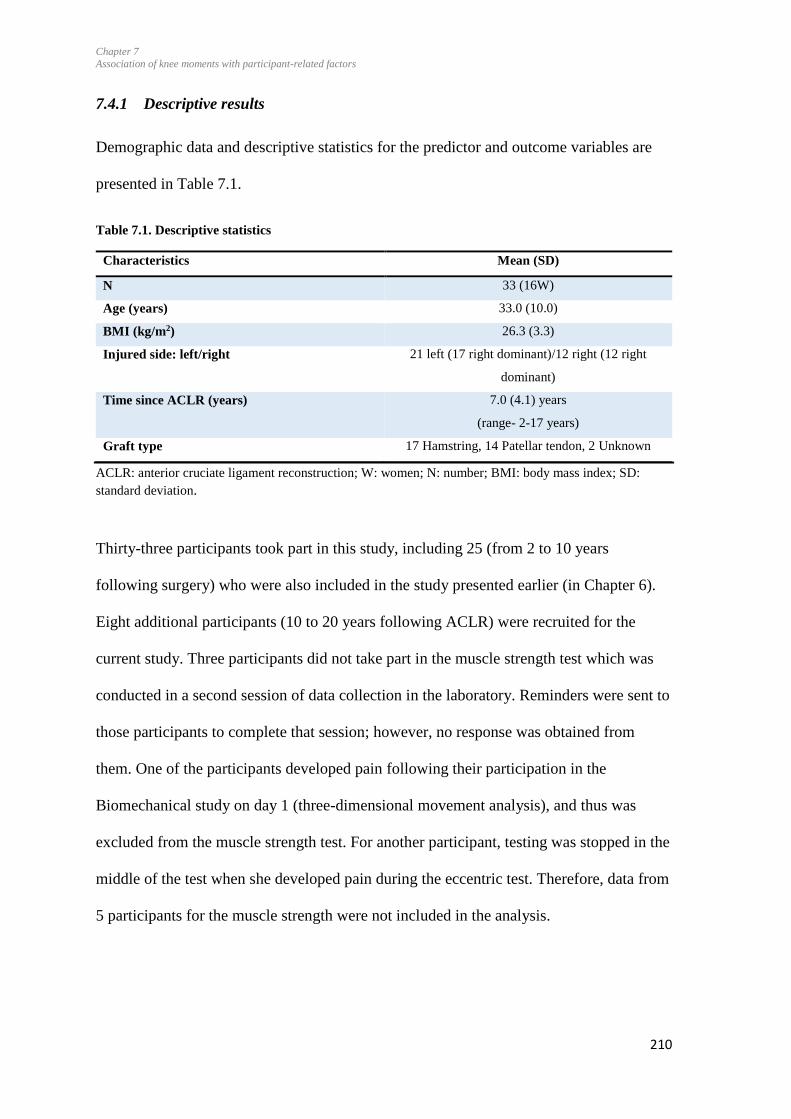
Chapter 7
Association of knee moments with participant-related factors
210
7.4.1 Descriptive results
Demographic data and descriptive statistics for the predictor and outcome variables are
presented in Table 7.1.
Table 7.1. Descriptive statistics
Characteristics Mean (SD)
N 33 (16W)
Age (years) 33.0 (10.0)
BMI (kg/m2) 26.3 (3.3)
Injured side: left/right 21 left (17 right dominant)/12 right (12 right
dominant)
Time since ACLR (years) 7.0 (4.1) years
(range- 2-17 years)
Graft type 17 Hamstring, 14 Patellar tendon, 2 Unknown
ACLR: anterior cruciate ligament reconstruction; W: women; N: number; BMI: body mass index; SD:
standard deviation.
Thirty-three participants took part in this study, including 25 (from 2 to 10 years
following surgery) who were also included in the study presented earlier (in Chapter 6).
Eight additional participants (10 to 20 years following ACLR) were recruited for the
current study. Three participants did not take part in the muscle strength test which was
conducted in a second session of data collection in the laboratory. Reminders were sent to
those participants to complete that session; however, no response was obtained from
them. One of the participants developed pain following their participation in the
Biomechanical study on day 1 (three-dimensional movement analysis), and thus was
excluded from the muscle strength test. For another participant, testing was stopped in the
middle of the test when she developed pain during the eccentric test. Therefore, data from
5 participants for the muscle strength were not included in the analysis.

Chapter 7
Association of knee moments with participant-related factors
211
Table 7.2. Descriptive statistics for patient-reported outcomes of the ACLR cohort (n = 33)
Type of outcome Mean (SD)
Tegner scores
Before injury 7.0 (2.0)
After surgery 5.3 (2.4)
Confidence during sports 42.0 (13.0)
KOOS
Pain 84.5 (13.0)
Symptoms 55.3 (11.3)
ADL 95.0 (8.0)
Sports recreation 77.1 (16.0)
Quality of life 47.1 (21.0)
KOOS4 66.0 (12.0)
Short form-12 Health survey
PCS 53.7 (5.0)
MCS 53.0 (6.0)
Tegner scores (0 = worst to 10 =highly active); KOOS: Knee Osteoarthritis Outcome Scale; (KOOS, all
domains: 0 = worst to 100 = best); ADL: Activities of daily living; Confidence during sports (0 = worst to
80 = best); PCS: Physical Component Scale; MCS: Mental Component Summary Scale.
Patient reported outcomes are presented in Table 7.2. Based on the Tegner Activity Scale,
results indicated a reduction in the level of physical activities of participants with ACLR
(Pre-injury=7, competitive sports such as tennis, athletics; recreational sports such as
soccer, squash, after surgery = 5.0 work, heavy labour, competitive sports such as
cycling, cross-country skiing). Results from the Confidence during Sports Scale indicated
that, as a group, participants still had decreased confidence in their knee (42/80). Results
of KOOS indicate that participants had good function with activities of daily living
(KOOS ADL=95), yet still experienced knee-related symptoms (KOOS- Symptoms=55)
and impaired knee function during sports and recreational activities (KOOS- Sports and
recreational activities=77). Overall, they scored low quality of life (KOOS- Quality of
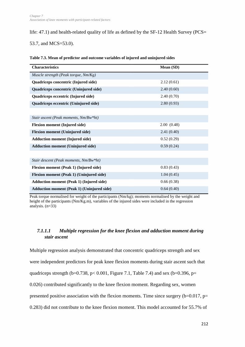
Chapter 7
Association of knee moments with participant-related factors
212
life: 47.1) and health-related quality of life as defined by the SF-12 Health Survey (PCS=
53.7, and MCS=53.0).
Table 7.3. Mean of predictor and outcome variables of injured and uninjured sides
Characteristics Mean (SD)
Muscle strength (Peak torque, Nm/Kg)
Quadriceps concentric (Injured side) 2.12 (0.61)
Quadriceps concentric (Uninjured side) 2.40 (0.60)
Quadriceps eccentric (Injured side) 2.40 (0.70)
Quadriceps eccentric (Uninjured side) 2.80 (0.93)
Stair ascent (Peak moments, Nm/Bw*ht)
Flexion moment (Injured side) 2.00 (0.48)
Flexion moment (Uninjured side) 2.41 (0.40)
Adduction moment (Injured side) 0.52 (0.29)
Adduction moment (Uninjured side) 0.59 (0.24)
Stair descent (Peak moments, Nm/Bw*ht)
Flexion moment (Peak 1) (Injured side) 0.83 (0.43)
Flexion moment (Peak 1) (Uninjured side) 1.04 (0.45)
Adduction moment (Peak 1) (Injured side) 0.66 (0.38)
Adduction moment (Peak 1) (Uninjured side) 0.64 (0.40)
Peak torque normalised for weight of the participants (Nm/kg); moments normalised by the weight and
height of the participants (Nm/Kg.m), variables of the injured sides were included in the regression
analysis. (n=33)
7.1.1.1 Multiple regression for the knee flexion and adduction moment during
stair ascent
Multiple regression analysis demonstrated that concentric quadriceps strength and sex
were independent predictors for peak knee flexion moments during stair ascent such that
quadriceps strength (b=0.738, p< 0.001, Figure 7.1, Table 7.4) and sex (b=0.396, p=
0.026) contributed significantly to the knee flexion moment. Regarding sex, women
presented positive association with the flexion moments. Time since surgery (b=0.017, p=
0.283) did not contribute to the knee flexion moment. This model accounted for 55.7% of
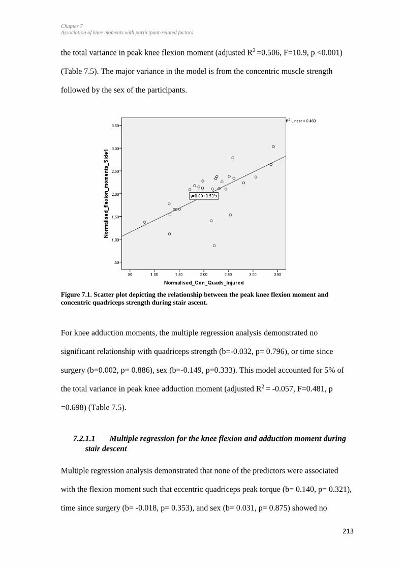
Chapter 7
Association of knee moments with participant-related factors
213
the total variance in peak knee flexion moment (adjusted R2 =0.506, F=10.9, p <0.001)
(Table 7.5). The major variance in the model is from the concentric muscle strength
followed by the sex of the participants.
Figure 7.1. Scatter plot depicting the relationship between the peak knee flexion moment and
concentric quadriceps strength during stair ascent.
For knee adduction moments, the multiple regression analysis demonstrated no
significant relationship with quadriceps strength (b=-0.032, p= 0.796), or time since
surgery (b=0.002, p= 0.886), sex (b=-0.149, p=0.333). This model accounted for 5% of
the total variance in peak knee adduction moment (adjusted R2 = -0.057, F=0.481, p
=0.698) (Table 7.5).
7.2.1.1 Multiple regression for the knee flexion and adduction moment during
stair descent
Multiple regression analysis demonstrated that none of the predictors were associated
with the flexion moment such that eccentric quadriceps peak torque (b= 0.140, p= 0.321),
time since surgery (b= -0.018, p= 0.353), and sex (b= 0.031, p= 0.875) showed no
Chapter 7
Association of knee moments with participant-related factors
214
significant relationship. This model accounted for 7% of the total variance in peak knee
flexion moment (adjusted R2 = -0.037, F=0.664, p =0.582) (Table 7.5).
Multiple regression analysis demonstrated that none of the predictor variables were
associated with the adduction moment. Neither the eccentric quadriceps peak torque (b= -
0.081, p= 0.523, Table 7.4), nor the time since surgery (b= -0.025, p= 0.164) or the sex
(b= -0.042, p= 0.813) contributed significantly to the adduction moment. This model
accounted for 9% of the total variance in peak knee adduction moment (adjusted R2 = -
0.013, F=0.782, p =0.466) (Table 7.5).
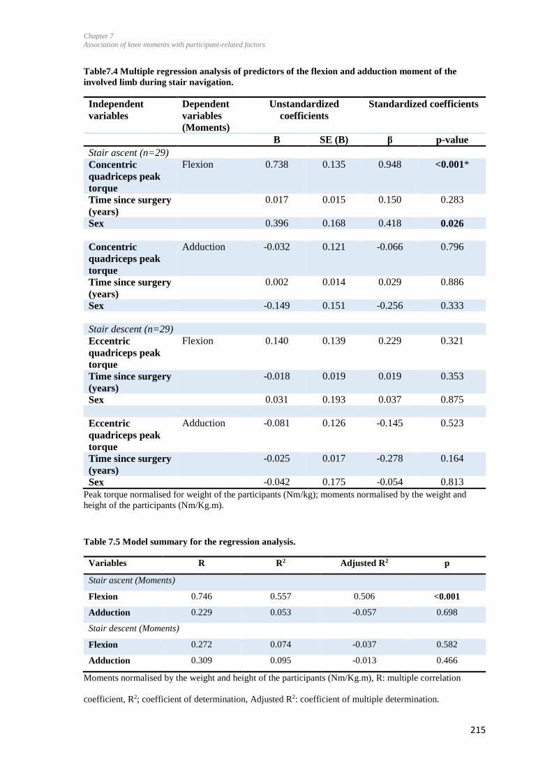
Chapter 7
Association of knee moments with participant-related factors
215
Table7.4 Multiple regression analysis of predictors of the flexion and adduction moment of the
involved limb during stair navigation.
Independent
variables
Dependent
variables
(Moments)
Unstandardized
coefficients
Standardized coefficients
B SE (B) β p-value
Stair ascent (n=29)
Concentric
quadriceps peak
torque
Flexion 0.738 0.135 0.948 <0.001*
Time since surgery
(years)
0.017 0.015 0.150 0.283
Sex 0.396 0.168 0.418 0.026
Concentric
quadriceps peak
torque
Adduction -0.032 0.121 -0.066 0.796
Time since surgery
(years)
0.002 0.014 0.029 0.886
Sex -0.149 0.151 -0.256 0.333
Stair descent (n=29)
Eccentric
quadriceps peak
torque
Flexion 0.140 0.139 0.229 0.321
Time since surgery
(years)
-0.018 0.019 0.019 0.353
Sex 0.031 0.193 0.037 0.875
Eccentric
quadriceps peak
torque
Adduction -0.081 0.126 -0.145 0.523
Time since surgery
(years)
-0.025 0.017 -0.278 0.164
Sex -0.042 0.175 -0.054 0.813
Peak torque normalised for weight of the participants (Nm/kg); moments normalised by the weight and
height of the participants (Nm/Kg.m).
Table 7.5 Model summary for the regression analysis.
Variables R R2 Adjusted R2 p
Stair ascent (Moments)
Flexion 0.746 0.557 0.506 <0.001
Adduction 0.229 0.053 -0.057 0.698
Stair descent (Moments)
Flexion 0.272 0.074 -0.037 0.582
Adduction 0.309 0.095 -0.013 0.466
Moments normalised by the weight and height of the participants (Nm/Kg.m), R: multiple correlation
coefficient, R2; coefficient of determination, Adjusted R2: coefficient of multiple determination.

Chapter 7
Association of knee moments with participant-related factors
216
7.5 Discussion
The study investigated the relationship between the quadriceps muscle strength, time
since ACLR, and sex of the participants with the knee flexion and adduction moments
during stair ascent and descent in participants with ACLR. It was hypothesized that
quadriceps concentric muscle strength would be associated with the peak knee flexion
and adduction moments during stair ascent, and eccentric quadriceps strength would be
associated with the knee flexion and adduction moments during stair descent. The first
hypothesis was partially supported, as a significant association was found between
concentric quadriceps strength and the flexion moment during stair ascent. However, no
statistically significant association was present between quadriceps peak torque and the
adduction moment during stair ascent and descent.
Lower knee flexion moments during stair ascent were correlated with the quadriceps
index during isometric contraction in a study at 21 weeks on an average following surgery
(Lewek et al., 2002). Results of the current study were similar to that of Patel et al., 2003
who found a significant association between knee flexion moment and concentric
quadriceps strength at during stair navigation in participants with ACL-deficiency.
Similarly, quadriceps femoris strength deficits were associated with the sagittal plane
knee mechanics during landing 8 months, on an average, following ACLR. The findings
imply that strengthening the quadriceps may improve the knee flexion moment in
participants with ACLR and can help to restore the sagittal plane moment asymmetry.
Although Shelburne et al., 2006 suggested that quadriceps muscle function contributes
towards frontal plane knee mechanics, the relationship between the adduction moment
and quadriceps muscle strength is poorly understood in the literature. No association was
found between quadriceps muscle strength and the adduction moment during stair ascent

Chapter 7
Association of knee moments with participant-related factors
217
or descent in the current study. These results are similar to a previous study exploring the
association between isometric knee strength (normalised to participants weight) and knee
adduction moment in participants with osteoarthritis during walking. That study found no
association between these variables (Lim et al., 2009). The lack of a significant
association between adduction moment and muscle strength could be explained by the
type of contraction participants are exposed to during the testing. During walking, the
quadriceps muscles are functioning concentrically, and isometric contractions do not
reflect that. This may explain the reason for lack of association between quadriceps
muscle strength and knee adduction moments, both during stair ascent and descent,
despite muscle strength being measured isotonically. Although we assessed both
concentric and eccentric muscle strength for the quadriceps, no significant relationship
was found with knee adduction moments. We included the PT of the injured side in the
analysis. It is possible that exploring the LSI (thus, comparing muscle strength of the
injured to the uninjured side) would better explain differences in knee moments. This will
be a future investigation. There are several potential explanations for the lack of
association between muscle strength and the adduction moment in the current study.
Firstly, knee adduction moments seem to be related to the knee extensor power (Calder et
al., 2014) and the endurance of the quadriceps muscle, rather than the muscle strength
(Lee, Lee, Ahn, Park, & Lee, 2015). This is supported by findings from a randomised
clinical trial exploring the effects of a strengthening programme on knee adduction
moments during walking in patients with knee osteoarthritis (Bennell et al., 2014). The
12-week programme entailed neuromuscular training and quadriceps strengthening for
participants with knee osteoarthritis during walking. The results indicated no significant
change for knee adduction moments following the programme (Bennell et al., 2014).
Secondly, adduction moments are also influenced by the knee adduction moment arm
Chapter 7
Association of knee moments with participant-related factors
218
(Hunt et al., 2006). In that study, participants with clinically and radiologically diagnosed
osteoarthritis were recruited, and they exhibited increased peak knee adduction moments
compared to less affected knees, even though the peak frontal plane ground reaction force
was less, suggesting that the larger peak adduction moments were mainly the result of the
larger lever arm magnitudes exhibited in knees with osteoarthritis. Lastly, muscle strength
was measured with the isokinetic dynamometer at the 60°/sec in the current study, while
another study has indicated the association between the adduction moments and total
work of quadriceps at 180°/sec rather than at 60°/sec and also indicated that 60°/sec may
not be the most valid speed (Lee et al., 2015). Speed of 60°/sec was used in this study for
muscle strength testing as it is more likely to find the deficits during maximal strength
testing compared to endurance testing.
No association was found among the knee flexor moment and the eccentric quadriceps
strength during the stair descent. Quadriceps work eccentrically during the stair descent;
therefore, an association was expected among the variables. However, no association was
found which may be due to the fact the moments may depend on various other factors
such as angle of the force vector, trunk angle, or hip abduction moment during stair
descent. Perhaps a further study with EMG from the muscles could help to explore this
association further.
Based on findings from the systematic review involving visual inspection of the forest
plot (Chapter 3, Figure 3.6 and 3.7), it was also hypothesised that time since surgery
would influence the knee flexion and adduction moment. No such association was found
in the data of the present study. This may be partly because knee flexion moments
continue to be lower compared to the contralateral knee and the uninjured Control group
(Hall et al., 2012; Wellsandt et al., 2016) and at 2 to 10 years following surgery (Chapter
Chapter 7
Association of knee moments with participant-related factors
219
6). However, regarding the adduction moment, limb symmetry scores were 88% and
103% for stair ascent and descent respectively. Moments may be influenced by many
factors as described above, and the participant related-factors such as their muscle
strength and trunk angles during ambulation. We were unable to consider all the factors in
this study; therefore, it is difficult to enlist the reasons for no known associations among
the moments and the time since reconstruction.
Normalised peak flexion moments seemed to be higher in women in the current study.
Knee flexion moment is known to have greater influence on cartilage such that tibial
cartilage changes at 5 years were associated with the baseline knee flexion moment in
patients with knee osteoarthritis (Chehab, Favre, Erhart-Hledik, & Andriacchi, 2014).
This indicates that strengthening the quadriceps to optimise the knee moments may be
helpful, particularly for women. Interventions need to be carefully designed and executed
in these participants by taking knee symptoms into account, as the higher loading beyond
the optimal levels can be disruptive to the cartilage (Andriacchi et al., 2004). Participants
should be educated regarding the role of muscle strength and maintenance of knee health
in the long-term. Results regarding the association of adduction moment with the
participant sex remain inconclusive, as this study did not shown any significant
association. Previous studies have indicated higher moments in women compared to men
from 3 weeks to 12 months post-surgery (Webster, McClelland, et al., 2012), and lower
adduction moment compared to the women controls at 3.5 years following surgery
(Patterson et al., 2014). Future studies should focus on exploring the differences in joint
loading in men and women along with patient-reported outcomes in the long-term
following surgery.
Chapter 7
Association of knee moments with participant-related factors
220
Participants had reduced levels of physical activities compared to pre-injury level (Tegner
scores = 5, heavy labour, competitive sports- cycling, cross-country skiing, recreational
sports- jogging on uneven ground at least twice weekly) and confidence during sports
(Confidence during sports scale= 42) overall. On average, the participants scored low
knee-related quality of life (KOOS-QOL=47.0) as a group, which could potentially be
due to the presence of knee-related symptoms (KOOS symptoms= 55.0) and knee
function during sports (KOOS sports/recreation= 77.0). However, the SD (21.0) for
quality of life indicated a wide range, so while some participants considered their quality
of life to be low, others rated this as being high. Participants’ low average knee-related
quality of life 2 to 17 years following surgery is an important finding, as the average age
of these participants was 33 years (range 21 to 51) at the time of data collection. A recent
study also explored quality of life in participants with ACLR, comparing those that
reported poor quality of life with those reporting high quality of life, 5 to 20 years post-
surgery (Filbay, Ackerman, Russell, & Crossley, 2016; Filbay et al., 2014). Poor long-
term QOL outcomes were related to not returning to sport, higher body mass index,
contralateral ACLR, and subsequent knee surgery in people with knee difficulties (Filbay
et al., 2016). The current study excluded participants with contralateral ACLR and
subsequent knee surgeries, so these were not influencing factors in this study. However,
the body mass index of five participants was within in the ‘overweight’ category (>30
kg/m2), which potentially could have influenced the knee mechanics (Messier, Gutekunst,
Davis, & DeVita, 2005). However, as the muscle strength and moments data were
normalised to the weight of the participants, there was less influence of the participants’
weight on the overall study results. Higher body mass index is associated with more
depressive symptoms and worse quality of life in men (Filbay et al., 2016). Fear of re-
injury is known to influence the quality of life up to 20 years following ACLR (Filbay et
Chapter 7
Association of knee moments with participant-related factors
221
al., 2016). Participants expressed a fear of injury up to 10 years post-surgery in the
mixed-method study (Chapter 5) and also lower scores in the confidence during sports
scale (Table 5.2), indicating the presence of fear of injury during sports. This may have
also influenced the quality of life in this group of participants. However, no information
related to the return to sports following surgery by these participants was collected in this
study. Therefore, we are unable to count on this factor. Based on the Tegner scores, the
present levels of sports were lower than the pre-injury levels (Table 7.2).
7.5.1 Limitations
This study included participants with different types of grafts, which may influence
muscle strength recovery. Different types of grafts influence the thigh muscle strength up
to 2 years following surgery (Xergia, McClelland, Kvist, Vasiliadis, & Georgoulis, 2011).
However, the type of graft has not been found to influence the quadriceps and hamstring
muscle strength 6 years following surgery (Keays et al., 2007). As participants of this
study were more than 2 years post-surgery, grouping the participants with different grafts
would not have influenced their muscle strength. Participants with or without meniscal
injuries were recruited. Presence of meniscal injuries influence the knee mechanics,
which could have influenced the results of this study (Lohmander et al., 2007).
The use of skin marker to monitor lower limb movement can generate artefacts during the
data collection due to the movement of the markers on the skin. In addition, there is intra-
assessor subject variability during placement of skin markers. However, only one
researcher (MK) applied all the markers on the participants, thus reducing differences that
may have been due to inter-assessor marker placement, each individual was exposed to
the same amount of artefact. Movement of the skin markers can be more in individuals
with high body mass index.
Chapter 7
Association of knee moments with participant-related factors
222
Other techniques which allow for the bone to be directly imaged such as fluoroscopy and
stereoradiographic systems cannot be utilized during stair ascent and descent due to
limitations of restricted fields of view. Therefore, motion analysis at present is the most
feasible way to measure kinematics.
Dependent variables only on the injured side of the participants with the ACLR were
analysed for the purpose of this study, and not on the contralateral side or in a Control
group. It is possible that including limb symmetry scores for flexion and adduction
moments and muscle strength, instead of the actual values for the injured sides, may have
shown significant relationships between predictor and outcome variables. As the aim of
current study was to explore the associations among the muscle strength and moments on
the injured side in order to develop treatment protocols to optimise the moment
symmetry, only the variables of injured side were considered in the analysis instead of the
limb symmetry index. Future research should examine whether these correlations also
occur in asymptomatic subjects and in the contralateral limb of participants with
osteoarthritis.
Speed of 60°/sec was used in this study rather than 180°/sec for muscle strength testing as
it was more likely to find the deficits during maximal strength testing compared to
endurance testing. Another possibility was to test at both of the speeds; however, due to
time constraints during data collection it was not possible to do testing at both speeds.
Also, testing at both speeds would have led to fatigue, which predisposes participants to
the risk of reinjury. During data collection, participants immediately followed the data
collection session for single-leg hop testing, this would have put the participants at risk of
reinjury.

Chapter 7
Association of knee moments with participant-related factors
223
7.6 Conclusion
Joint moments can be related to different parameters. This study indicated the association
of the concentric muscle strength and sex of the participants with the knee flexion
moment during stair ascent. More attention should be given to the women participants
while training to restore the moments, as the study has indicated their tendency towards
increase in flexion moment. This is an important finding, and future studies should
explore this association further.
7.7 Summary
The biomechanical deficits on the injured side in participants with ACLR are related to
the muscle strength and hesitancy on weight bearing on the injured side. Behavioural
modifications due to fear of injury as discussed in Chapter 5 have huge impact on the
movement pattern. These are the important findings and clinicians should focus on the
psychological aspects of the injury during the rehabilitation period.

Chapter 8
Summary and recommendations
224
8 Chapter
Summary and recommendations
8.1 Background
The overarching research question this thesis explored was: ‘Do physical, psychosocial,
biomechanical and knee function-related impairments persist following ACLR in
participants’ up to 20 years following surgery?’ This chapter discusses the overall
findings of the studies conducted and their contributions to the existing literature in the
fields of sports physiotherapy and clinical biomechanics. The specific aims of this chapter
are to: (1) provide a concise summary of the results of the thesis; (2) discuss a conceptual
model of how biomechanical, psychological and inflammatory factors can determine
overall outcomes of ACLR; (3) discuss the implications for clinical practice from the
findings of this thesis; (4) highlight the strengths of the thesis; (5) acknowledge the
limitations of the thesis; (6) recommend future research directions; (7) draw conclusions
based upon the key results.
8.2 Overall summary of results
Through the series of interconnected studies, this thesis has explored patient-reported
outcomes, quadriceps muscle strength, physical performance, knee laxity and the
biomechanics of the injured knee following ACLR compared to Control group data. A
mixed-method approach was used, combining quantitative and qualitative study designs
to ensure an in-depth study of the data obtained. Firstly, Chapter 1 explored the gap in the
research field which highlighted the need to explore the long-term outcomes of ACLR.
Chapter 2 explored the literature related to the muscle strength, physical performance,
patient-reported outcomes, and quality of life in participants with ACLR. A systematic
review and meta-analysis was conducted to explore and compare the knee moments and
Chapter 8
Summary and recommendations
225
angles in participants with ACLR compared to controls during walking, running and stair
navigation (Chapter 3). This review highlighted the dearth of studies investigating the
moments and angles during high loading activities in the long-term following ACLR.
Therefore, the subsequent cross-sectional study (Chapter 6) compared knee moments and
angles between participants of the ACLR and Control groups (2 – 10 years post-surgery).
A cross-sectional study compared the patient-reported outcomes, isokinetic muscle
strength of quadriceps and hamstrings, physical performance, and anterior-posterior knee
laxity between an ACLR group (2 – 10 years post-surgery) and a Control group (Chapter
4). Further mixed-method study provided deeper insight into the patients’ perspectives
related to the effect of the injury and surgery on their lives (Chapter 5) and that of the
Control group during stair ascent and descent (Chapter 6). Lastly, the association between
participant-related factors and the knee moments during stair ascent and descent were
explored in a group of participants with ACLR, 2 to 20 years post-surgery (Chapter 7).
Following are the research questions for each study along with key results.
1. Do the differences persist in the knee joint angles and moments in participants with
ACLR compared to the contralateral limb and with age-matched controls during
walking, stair climbing, and running? What is the time course of recovery of those
biomechanical variables following ACLR?
A systematic search was performed on the electronic databases using specific keywords.
Based on the inclusion and exclusion criteria, 31 studies were included in the systematic
review, and a meta-analysis was performed on 27 studies (Chapter 3). A systematic
review with meta-analysis found that joint kinematics were restored, on average, 6 years
following reconstruction, but joint moments were still low 4-5 years following the
surgery. There was strong to moderate evidence for no significant difference in peak

Chapter 8
Summary and recommendations
226
flexion angles between ACLR and Control groups during walking and stair ascent.
Although the knee adduction moment was lower at 3 years, by 4-5 years it was higher
compared to uninjured individuals. Peak knee flexion moments were still lower after 5
years of surgery in participants with ACLR than in uninjured controls. Differences in the
knee moments on the injured side were more apparent compared to the contralateral limb,
indicating the bilateral effect of the unilateral injury. Lower moments indicate reduced
loading at the knee joint. Findings indicated that knee moments were not fully restored
following reconstruction, even a few years after surgery. Optimal joint loading may need
to be achieved, for which long-term maintenance programmes, including self-
management of the knee along with patient education, is required. Participants may need
‘booster’ sessions of exercise prescription after a few years following surgery.
2. Do the differences persist between the patient-reported outcomes, muscle strength,
physical performance, and knee laxity in participants with ACLR from 2 to 10 years
following surgery compared to the Control group?
This cross-sectional study included 25 participants with ACLR from 2 to 10 years
following surgery and 24 age- and gender-matched uninjured controls. Patient-reported
outcomes, muscle strength, physical performance, and knee laxity data were collected
using an electronic questionnaire (comprising the Tegner activity scale, KOOS,
Confidence during sports scale, and the SF-12), isokinetic muscle strength testing, single-
leg hop testing, and anterior to posterior knee laxity measured with a KT-arthrometer.
Data of participants with ACLR were compared to the contralateral limb and the Control
group using a repeated measures ANOVA (Chapter 4). Patient-reported outcomes
indicated the presence of knee-related pain and symptoms, and low knee-related quality
of life in ACLR group compared to the Control group and KOOS, respectively. The
Chapter 8
Summary and recommendations
227
injured side had statistically significant lower concentric hamstring and eccentric
quadriceps strength compared to the contralateral side within the ACLR group.
Significantly, lower concentric hamstring peak torque and eccentric quadriceps peak
torque were found when comparing the injured to the contralateral uninjured sides. The
concentric quadriceps peak torque of the injured ACLR sides were significantly lower
compared to the controls side 1, similarly for hamstring peak torque of the injured sides in
ACLR group were significantly lower compared to controls.
Outcomes of the single-leg hop revealed significant within-group differences with lower
jump distance on injured sides compared to the contralateral sides in the ACLR group.
Interestingly, a higher jump distance was evident for the contralateral sides in the ACLR
group compared to the side 2 of Control group, indicating reduced physical performance
only on the injured sides in ACLR group. Significant within- and between-group
differences were present for knee laxity in the sagittal plane. Higher laxity on the injured
side was present compared to the contralateral side in the ACLR group and side 1 of the
Control group. Overall, participants with ACLR had lower muscle strength, poor physical
performance as assessed by single-leg hop, and higher laxity on the injured side. A
possible explanation for reduced muscle strength and knee function is likely to be due to
lower activation of quadriceps muscles (Hart et al., 2010), reduced proprioception
(Bonfim et al., 2003) and other factors as explained in Chapter 2 section 2.5.1 and
Chapter 4 section 4.8. Further ACL injury is associated with less activation of the sensory
cortex (Valeriani et al., 1996) and increased activation of the contralateral motor cortex
(Grooms et al., 2017), and thus also has effects on the central nervous system
3. What are the participants’ experiences in relation to physical activity, sports,
occupation and quality of life more than 2 years following ACLR?
Chapter 8
Summary and recommendations
228
Ten of the participants of the above cross-sectional study (2-10 years following ACLR; 7
female; aged 20 to 52 years) also participated in individual face-to-face semi-structured
interviews (Chapter 5). The patient-reported outcomes were analysed descriptively and
the interview data were analysed using a general inductive approach. Participants had
good knee function in activities of daily living, yet still scored low on the quality of life
scale as per KOOS and SF-12. The Sports confidence scale indicated low confidence and
fear of re-injury. Three overlapping themes were identified from the interviews:
‘Continuum of fear of re-injury and confidence’, ‘live life normally’ and ‘need of
reassurance and the maintenance of knee health’. The ACL rupture leads to long-term
fear of injury and behavioural manifestations, with fluctuating levels of confidence during
specific sports activities. Participants were concerned about the future risk of re-injury
and osteoarthritis. The findings highlight the importance of considering individual-
specific strategies as part of rehabilitation following ACLR, and should plan continued
knee function maintenance exercises according to individual needs.
4. Do the differences in the peak knee angles and moments persist in participants with
ACLR compared to the Control group from 2 to 10 years following surgery?
The biomechanical cross-sectional study (Chapter 6) included 25 participants with ACLR
from 2 to 10 years following surgery and 24 age- and gender-matched uninjured controls.
Three-dimensional movement analysis was performed during stair ascent and descent.
Peak knee angles and moments in all three planes were calculated during stair ascent and
descent. Differences in knee flexion angles were found, with lower peak knee flexion
angle during stair ascent. Results also showed lower knee flexion moments on the injured
side compared to the contralateral limb in participants with ACLR, indicating reduced
joint loading. Higher knee extension moments were found on the injured side in the
Chapter 8
Summary and recommendations
229
ACLR group. Lower flexion moment and higher extension moment on the injured side
indicates the presence of adaptive strategies in participants with ACLR. Lower loading on
the injured side may be due to a combination of reluctance or hesitation to bear weight on
the injured side, reduced muscle strength, and potential neurophysiological consequences
of the injury and surgery.
5. Are the knee flexion and adduction moment associated with the muscle strength, time
since reconstruction and the participant sex on the injured side in participants with
ACLR?
This study (Chapter 7) included 29 participants with ACLR from 2 to 20 years following
surgery. The kinetic data, including the peak knee flexion and adduction moments, were
collected using three-dimensional motion analysis during stair ascent and descent. The
isokinetic data were collected for peak torque concentric and eccentric quadriceps muscle
strength. Moment data were normalised for weight and height of the participants. Muscle
strength data were normalised for participants’ weight. Regression analyses were
performed to explore the association between the moments and the participant-related
data. Results found that concentric quadriceps peak torque and the sex of the participant
were 55% responsible for the variance in the first peak knee flexion moment during stair
ascent. Women showed an increase in knee flexion moments compared with men. There
was no significant association between the knee flexion moment and the time since
surgery (Chapter 7). Knee adduction moments were not associated with the quadriceps
muscle strength, sex, and the time since surgery during stair ascent and descent. The
association of the knee flexion moment with concentric quadriceps strength is an
important finding. This theoretically implies that quadriceps strength may be used as an
indicator for knee flexion moments during stair ascent. This also implies that restoring the
Chapter 8
Summary and recommendations
230
concentric quadriceps muscle strength may help in optimising the moment symmetry in
participants following surgery. Women may need careful administration of rehabilitation
support in the longer-term as indicated by the association of sex of participants with the
flexion moments. Also, the increase in flexion moments indicates higher loading in
women; therefore, this may mean that if loading is increased beyond the optimal levels on
the cartilage which is associated with concomitant injuries in 16-46 % cases (Brophy,
Zeltser, Wright, & Flanigan, 2010), it may lead to a greater predisposition of women for
knee osteoarthritis following the surgery.
8.3 Factors influencing the clinical outcomes in patients with ACLR: A
conceptual model.
An ACL injury is caused by a high-force event causing isolated ACL injury or also
injuring multiple knee structures such as the joint cartilage, subchondral bone and menisci
(Brophy et al., 2010). The injury event also initiates a series of changes in the joint which
involve biomechanical and biochemical changes (e.g. inflammation and metabolic
imbalances of tissue turnover) following injury (Beynnon et al., 2005; Frobell et al.,
2008), known to initiate the joint degenerative changes following injury (Cattano et al.,
2013). Regarding the treatment approach, surgical treatment is considered as the option,
especially for those patients who aim to return to competitive sports. However, only 55%
of the patients are able to return to the same level of sports within a year following the
surgery (Ardern et al., 2014).
Considering the outcomes of the ACLR, participants are at high risk of post-traumatic
osteoarthritis. The onset and progression of post-traumatic osteoarthritis is a complex and
multifactorial process, and involves an interaction between biochemical and
biomechanical factors. Higher levels of inflammatory markers are known to initiate the
Chapter 8
Summary and recommendations
231
inflammatory process immediately following the injury (Li, Chen, & Chen, 2015), but the
levels of inflammatory markers have been found to lie within the normal ranges at 8 years
following surgery (Åhlén et al., 2015). Other than the altered kinetics and kinematics
(Swärd et al., 2010), deficits in muscle strength (Petersen et al., 2014) are known to
influence the short and long-term outcomes.
Outcomes of ACLR are dependent on numerous factors including psychosocial
components: self-esteem and internal locus of control (Christino et al., 2016), fear of re-
injury and lower confidence; concomitant injury to the other joint structures such as
cartilage, menisci and joint capsule; surgical techniques (Scanlan et al., 2009), type of
graft (Sherman & Banffy, 2004), timing of surgery (Salmon et al., 2005), and femoral
canal orientation at the time of surgery can all play important roles in determining the
overall outcomes. Among the modifiable factors, intense pre-operative rehabilitation
(Eitzen, Moksnes, Snyder-Mackler, & Risberg, 2010) has led to clinically significant
improvement in the knee function post-operatively. Similarly, post-operative
rehabilitation along with the patient motivation and goal-setting during rehabilitation have
been seen to deliver favourable surgical outcomes. Repeated functional testing throughout
the pre-and post-operative rehabilitation to provide feedback has been related to improved
outcomes after ACL rehabilitation (Grindem et al., 2015). Pre- and post-operative
rehabilitation should be combined with goal-setting, and quality patient education and
feedback, to improve the outcomes.
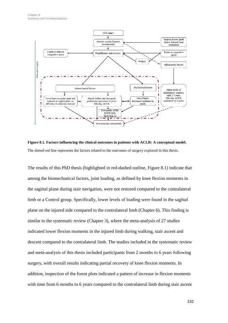
Chapter 8
Summary and recommendations
232
Figure 8.1. Factors influencing the clinical outcomes in patients with ACLR: A conceptual model.
The dotted red line represents the factors related to the outcomes of surgery explored in this thesis.
The results of this PhD thesis (highlighted in red-dashed outline, Figure 8.1) indicate that
among the biomechanical factors, joint loading, as defined by knee flexion moments in
the sagittal plane during stair navigation, were not restored compared to the contralateral
limb or a Control group. Specifically, lower levels of loading were found in the sagittal
plane on the injured side compared to the contralateral limb (Chapter 6). This finding is
similar to the systematic review (Chapter 3), where the meta-analysis of 27 studies
indicated lower flexion moments in the injured limb during walking, stair ascent and
descent compared to the contralateral limb. The studies included in the systematic review
and meta-analysis of this thesis included participants from 2 months to 6 years following
surgery, with overall results indicating partial recovery of knee flexion moments. In
addition, inspection of the forest plots indicated a pattern of increase in flexion moments
with time from 6 months to 6 years compared to the contralateral limb during stair ascent
Chapter 8
Summary and recommendations
233
and descent. However, participants in the cross-sectional study (Chapter 6) were from 2
to 10 years following surgery (mean 4.7 years) and this showed that the moments
continue to be lower, hence suboptimal level of joint loading, which may persist for up to
10 years.
Findings regarding the magnitude of the adduction moment in the cross-sectional study
(Chapter 6) were similar to the systematic review during the stair ascent (Chapter 3).
Inspection of the forest plots (Chapter 3, Figure 3.6 and 3.7) indicated that the knee
adduction moment may increase over time on the injured side compared to the Control
group during walking. However, no differences in the moments were found in the
analysis in Chapter 6 which included participants up to 10 years following surgery. The
systematic review (Chapter 3)(Kaur et al., 2016) showed that there were no differences in
the knee adduction moments on the injured side compared to the contralateral side and the
Control group following the reconstruction. Findings of Chapter 3 may be explained by
the included study (Zabala et al., 2013) in the review, as the participants included were
from 22-34 months following ACLR. Lower moments during the initial years may be due
to hesitancy in weight bearing. Other studies in the review indicated no differences in the
magnitude of the adduction moment, which are in agreement with our study results.
Overall, taking together the results from Chapter 6 and Chapter 3, joint loading is lower
in the injured limb compared to the contralateral limb. Lower joint loading is associated
with early knee osteoarthritis 5 years following ACL injury (Wellsandt et al., 2016).
Those authors speculated the reduced level of joint loading may be an indicator for risk
for early onset and progression of osteoarthritis participants with ACLR, rather than the
increased loading in the injured knee years after surgery (Wellsandt et al., 2016).
Concurrently, increased loading is also detrimental to articular cartilage. Higher
adduction moment was found in patients with knee osteoarthritis (Astephen et al., 2008).
Chapter 8
Summary and recommendations
234
There are few plausible explanations for the lower level of joint loading in participants
with ACLR. Joint moments (external knee flexion moment) have been associated the
muscle strength (Lewek et al., 2002; Schmitt et al., 2015), thus lower strength can lead to
the lower joint loading (Chapter 7). However, other factors need to be considered. For
instance, fear of injury, lower confidence levels (Chapter 5), and hesitation during weight
bearing seen in this cohort of participants, could be responsible for the lower joint
loading. Moreover, the changes in the central nervous system have been recently reported
following ACL injury and reconstruction (Grooms et al., 2017). Neuroplasticity in the
central nervous system was expressed by increased activation in the contralateral motor
cortex, and diminished activation in the ipsilateral motor cortex and cerebellum compared
to healthy controls following ACLR (Grooms et al., 2017). Reduced knee joint loading
following ACLR may be associated with the neuroplasticity in the central nervous
system. The mechanical model of degeneration is generally thought to be a cause of early
onset post-traumatic osteoarthritis (Andriacchi et al., 2004); however, findings from this
thesis have shown that there are additional psychosocial aspects which might contribute
to an unfavourable outcomes to the ACLR surgery. Psychological aspects of the injury
and surgery, as indicated in Chapter 5, may affect not only the loading at the knee but
may also impact the participants’ quality of life and participation in sports.
Suboptimal level of joint loading is deleterious to the joint health. Lower moments along
with abnormal kinematics can lead to a shift in the joint loads to the areas of the cartilage
which may not be conditioned to the high level of loading (Chaudhari, Briant, Bevill,
Koo, & Andriacchi, 2008). Concurrently, lower muscle strength can affect the physical
performance in participants with ACLR (Schmitt et al., 2012) and has been considered as
a risk factor for the onset and progression of osteoarthritis (Keays et al., 2007; Tourville
et al., 2014). This indicates the need to restore the optimal level of joint loading and
Chapter 8
Summary and recommendations
235
muscle strength. Therefore, the physical and muscle-related impairments could be the
marker of osteoarthritis in participants with ACLR.
Findings from this thesis and the literature suggest that patients with ACLR present with
psychological impairments that impact on their ability to engage with sports at the same
level prior to injury. The results of the mixed-method study (Chapter 5) indicate the
impact of psychological factors on the overall participants’ confidence during sports and
therefore impact the overall outcomes of the ACLR (Christino et al., 2016). Fear of injury
and lower confidence is a hindrance to participation in sports and recreational activities,
and can be considered to be the result of an adaptive protective behaviour (Brewer,
Cornelius, Stephan, & Van Raalte, 2010) and other sensory-motor changes in the brain
(Grooms et al., 2017). Psychological aspects of the injury, such as the participants’
struggle through the rehabilitation to attain the desired physical milestones and the
emotional distress that accompanies this, have been highlighted previously (Scott et al.,
2017).
Strategies to address the fear of re-injury such as behavioural cognitive therapy or mental-
imagery techniques, have been reported to enhance the psychological and physical
aspects of recovery during the early stages following surgery (Cupal & Brewer, 2001;
Magyar & Chase, 1996). A previous study has indicated that physiotherapists are aware
of the psychological aspects of the rehabilitation following ACLR (Von Aesch et al.,
2016). However, the support provided by the health professional to deal with the
psychological issues may not be sufficient, as indicated by participants’ struggle with the
psychological issues in the longer-term.
Chapter 8
Summary and recommendations
236
Biomechanical and psychological impairments are equally influencing the quality of life
in this group of participants (KOOS QOL scores, Chapter 4). These, along with the joint
damage sustained at the time of injury (Culvenor et al., 2015), and the influence of
inflammatory factors up to 3 years after the surgery, can potentially lead to post-traumatic
osteoarthritis.
8.4 Implications for clinical practice
The studies reported in this thesis add to the existing body of knowledge by identifying
the physical and psychological impairments in the longer-term. Based on the
biopsychosocial approach and following the ICF model of the WHO, management of
joint injuries may need an extensive approach (Quinn et al., 2012).
8.4.1 Optimising the knee moments following ACLR
Overall, reduced flexion moments indicate altered joint loading in the long-term
following ACLR. These changes are likely to be associated with quadriceps weakness
(Shelburne et al., 2006) and various neurophysiological mechanisms (Barret, 1991;
Urbach et al., 2001; Urbach et al., 2000), and are potentially associated with risk of early
onset joint degeneration (Roos et al., 1995; Roos et al., 2005) due to suboptimal joint
loading during functional tasks such as walking, running, and stair navigation (Knoll et
al., 2004). Altered kinematics along with reduced flexion moments may significantly
change the stress distribution within the cartilage and initiate a degenerative process
(Setton et al., 1999). Athletes with better physical performance at 6 months following
ACLR appear to demonstrate less asymmetrical gait patterns compared to those that did
not pass functional return to play criteria (Di Stasi et al., 2013). It thus appears to be
important to include assessment of potential gait asymmetries following ACLR in
decision making for readiness for rehabilitation progression and return to sports (Di Stasi
Chapter 8
Summary and recommendations
237
et al., 2013; Myer et al., 2008). While small differences in walking may not be detectable
on clinical examination, these may be more evident during high knee loading activities,
such as stair ascent/descent, and running (Myer et al., 2008) and jumping (Paterno et al.,
2007). The findings of movement and loading asymmetries in this thesis indicate that it
may be important to continue with the exercise programme focusing on strength,
neuromuscular control and proprioception even after the rehabilitation period is over.
Based on findings from this thesis, clinicians, physiotherapists, sports doctors should
consider the presence of knee loading asymmetries.
8.4.2 Restoring knee moments symmetry
This thesis contributes to the literature by adding that physical and mental impairments
may persist in the long-term in patients with ACLR. There are differences in flexion
moments in the injured and contralateral limb in participants with ACLR. Such
differences are a risk factor for the re-injury on both the ipsilateral and contralateral side
in the short-term (Paterno et al., 2010), and a risk factor for osteoarthritis in the long-term
(Ajuied et al., 2014). However, to date, there is no rehabilitation program that addresses
these residual impairments following ACLR in the longer-term. Participants suffer from
residual muscle and biomechanical impairments, and only a few contact health
professionals in the longer-term, as it was discovered in the mixed-method study (Chapter
5). In the New Zealand context, ACC usually funds surgery and rehabilitation.
Rehabilitation is usually limited to 16 treatments, with the possibility of extending it
depending on the progress of the individual and outcomes during rehabilitation. The
number of physiotherapy sessions undertaken by participants of this study ranged from 1
to 27, based on information provided by ACC. However, it is not known how many
Chapter 8
Summary and recommendations
238
patients access health care in the longer-term for their knee-related impairments across
New Zealand.
The findings of this thesis, particularly the pattern of lower knee flexion moment and
muscle strength, are similar to those seen in patients with osteoarthritis (Astephen et al.,
2008), with the exception of higher adduction moments. Moment asymmetry and lower
muscle strength is related to joint space narrowing and osteoarthritic changes in the knee
joint in participants with ACLR (Keays et al., 2007; Tourville et al., 2014). Thus, a partial
similarity in the movement pattern in participants with ACLR and osteoarthritis exists
years before the onset of the symptoms. Therefore, it is important to restore the moment
symmetry in these participants.
The statistically significant association of concentric quadriceps muscle strength with the
knee flexion moment during stair ascent indicates that quadriceps concentric muscle
strengthening exercises may be employed to restore the moments in the sagittal plane.
This gives future direction for further studies. There was increase in the flexion moment
in women. This indicates the need to carefully execute the rehabilitation in women to
optimally restore the joint loading and not to overload the joints, which may be
detrimental for joint health. The differences in the gait pattern among women and men
have been found in previous studies as well (Asaeda et al., 2017), and this difference may
explain the higher incidence of women of developing primary osteoarthritis. In women
participants with ACLR, emphasis should be placed on restoring and maintaining the
thigh muscle strength.
Chapter 8
Summary and recommendations
239
8.4.3 Need for an extended rehabilitation and the self-management of knee health
The ACLR affects the quality of life and levels of confidence among the participants in
the longer-term. Participants with ACLR need to be followed-up beyond the rehabilitation
period to address their concerns related to muscle strength, physical performance, low
confidence in sports, pain, and other knee-related symptoms. Restoration of muscular
strength is important for optimal joint function; therefore, future research is needed to
develop an exercise protocol to address the muscular deficits in the long-term
impairments. Participants should be encouraged to maintain their muscle strength.
An in-depth understanding of participants’ concerns and experiences of the outcomes of
ACLR highlighted the factors relating to fear of re-injury, reduced confidence in the
injured limb during sports, hesitancy in weight bearing on the injured side and
participants’ daily struggles related to the regular activities of daily life. The presence of
physical and psychological impairments indicate that an extended rehabilitation,
addressing both physical impairments and psychological responses, may be needed.
Participants were aware of the residual impairments in their knee and the influence of
those impairments on the knee during sports-related activities. Also, participants were
aware that the fear of re-injury may be related to the deficits in the muscle strength
leading to hesitancy in fully engaging during sports activities. These patients need
additional support aiming to restore the knee function, reduce the fear of re-injury and
boost their confidence in the knee during sports. Cognitive behavioural therapy may be
one helpful technique for these patients (Cupal & Brewer, 2001). Other strategies such as
mental imagery techniques are suggested for those struggling with fear of re-injury
(Magyar & Chase, 1996). Other minor concerns, such as knee pain and soreness
following the strenuous exercises, need the attention of the health professional as well.

Chapter 8
Summary and recommendations
240
The findings of this thesis suggest that a broader approach may be helpful for participants
with ACLR in the longer-term. This longer-term care plan should include a protocol
emphasising muscle strengthening along with the psychological aspects of the injury to
deal with the persistent fear of re-injury in those who are unable to engage fully in sports.
Self-management of the knee condition needs to be incorporated in the long-term. Good
response to a web-based intervention has been found in another study, exploring the self-
management of acute or chronic knee conditions in adults (Button, Nicholas, Busse,
Collins, & Spasić, 2018). In that study, web-based intervention consisting of a complex
intervention combining information resources, exercise videos, personalised exercise
plans and remote contact with a physiotherapist alongside face-to-face treatment was
administered. It was reported to be acceptable, easy to use and appropriate for a wide
spectrum of patients. Similar web-based programs may be defined and administered in
this cohort of patients in the long-term. Also, strategies to improve the self-efficacy in the
long-term for patients needs to be developed and incorporated. Short-term postoperative
self-efficacy was found to be a better predictor of long-term outcome after total hip or
knee arthroplasty in a cohort of 103 patients following surgery (Akker-Scheek, Stevens,
Groothoff, Bulstra, & Zijlstra, 2007). That study recommended interventions focusing on
enhancing short-term postoperative rather than preoperative self-efficacy. Similar
strategies may need to be followed for individuals with ACLR.
8.4.4 Need of patient education
Patient education may be beneficial, as they would be aware of the possibility of
impairments following ACLR, and therefore could take control and improve self-efficacy.
Participants indicated that they wished to know about the future of the knee in the long-
term. Patient education, focusing on identifying the presence of fear of re-injury and their
predisposition to the early onset of osteoarthritis, is required in early stages of
Chapter 8
Summary and recommendations
241
rehabilitation process. Patients need to be educated regarding the protective role of
muscle strength in the prevention of the osteoarthritis.
8.4.5 Tool for assessment
Flexion moments were associated with the concentric quadriceps muscle strength during
stair ascent (Chapter 7). Quadriceps muscle deficits may thus be an indicator of reduced
loading. Therefore, assessing the concentric quadriceps muscle strength could be useful
as an assessment tool to assess residual impairments in participants with ACLR.
Participants in the research described in Chapter 5 revealed problems with stair ascent
and descent. Moment asymmetry was found during stair ascent and descent activities in
this cohort of participants (Chapter 6). Some of the studies in the literature did not report
the deficits in moments or angles during walking activity (Hall et al., 2012; Webster,
McClelland, et al., 2012), which may be due to walking as an activity with a lower level
of joint loading than the stairs. Negotiating stairs involves a higher level of joint loading,
and therefore can be used as the task for analysis in future studies.
8.5 Strengths of this thesis
8.5.1 Sequential design of studies
The sequential design of the studies aimed to investigate different aspects of the
overarching research question in a logical way. Dependent variables for the quantitative
study (Chapter 6) were finalised based on the findings of the systematic review and meta-
analysis (Chapter 3) and it also directed the research process to compare the moments and
angles in participants with ACLR to the Control group in the longer-term following
surgery. Also, the association of the knee flexion moment with the concentric quadriceps

Chapter 8
Summary and recommendations
242
muscle strength pointed towards a way of the restoring the knee flexion moment (Chapter
7), although this needs to be explored further in future studies.
8.5.2 Mixed method approach
A strength of this study was that a biopsychosocial approach could be followed by using a
mixed-method design across the studies, enriching the understanding of the research
topic. To answer the research question in more depth, the quantitative and qualitative
research paradigms were combined. This approach helped to understand not only the
physical impairments of participants with ACLR, but also their concerns, beliefs and fears
associated with their health.
Comparing the patient-reported outcomes in Chapter 4 between groups indicated lower
confidence during sports and quality of life in participants with ACLR compared to the
Control group. These findings were deepened through the mixed-method study (Chapter
5) which focused on the participants’ concerns. Using mixed-methods in the field of
musculoskeletal physiotherapy practice is not common, so its use in this thesis was
innovative, and provided a deeper understanding of the phenomenon and generated the
resulting conceptual model of the outcomes of ACLR.
8.5.3 Data analysis
The statistical methods used in Chapter 4 and 6 helped us to explore the inter-limb
differences within and between the groups among the participants with ACLR compared
to the Control group using the repeated measure ANOVA. Regression analysis, the
statistical test used in Chapter 7, is known to be a robust test in terms of predicting the
relationships between the variables. The thematic analysis used for the analysis of
Chapter 8
Summary and recommendations
243
interview data in the mixed-method study (Chapter 5) is one of the most robust methods
used in this research paradigm.
8.5.4 Contribution to the literature
Previous studies exploring the outcomes of ACLR have focused on the short-term
outcomes of the surgery, and only few have included a combination of the biomechanical
and patient perspectives of the surgery. The systematic review and meta-analysis (Chapter
3) provided the time course of the recovery of angles and moments following surgery and
provided the subgroup analysis during different tasks. The mixed-method study (Chapter
5) indicated that fear of injury continues to persist for a long time following ACLR, and
this may impact differently on the execution of the movement. This thesis explored the
long-term outcomes following the surgery, and explored muscle strength, physical
performance, knee laxity, and patient-reported outcomes related to the surgery, along with
the biomechanical analysis during stair ascent and descent. This approach helped in the
study of the outcomes of surgery considering the person as ‘whole’, and enabled an
understanding of the reasons behind the psychological concerns. The psychological
concerns of these individuals can affect physical performance and participation in sport
and exercise. It can have implications for the athletes, coaches, and parents regarding
injury, rehabilitation, and career transition following ACL injury and surgery.
8.6 Limitations
Although specific limitations for each study have been already discussed in the previous
chapters, this section highlights the limitations in the context of the whole thesis.

Chapter 8
Summary and recommendations
244
8.6.1 Knee-related impairments only
Studies were designed to explore the thigh muscle strength and knee moments only,
whereas there could be strength impairments present at the hip and ankle which were not
explored. Only knee-related moments were studied in this thesis; however, there could be
compensations from the other joints such as hip and ankle, and also from the trunk,
influencing the overall moments. However, these were not explored in this thesis as the
focus was on the knee-related impairments.
The thesis analysed the stair ascent and descent as the functional task for performed by
participants. Although it is a high loading activity compared to walking, a more detailed
level of understanding could have been gained from the analysis of additional activities
such as vertical jump and running.
8.6.2 Recruitment of participants with 10-20 years post-reconstruction
Sample size estimations indicated the need to recruit 26 participants with ACLR 10-20
years following surgery. Recruiting patients 10-20 years following surgery was a
challenge. We could not recruit more than 9 participants in this group, despite extensive
advertising in the Masters’ games, including sports and golf clubs, and social media. It is
possible that priorities in life in terms of family and occupational commitments have
changed in the 10 to 20 years following surgery, with possibly greater acceptance of the
post-surgery limitations, resulting in less interest by such patients to participate in this
research study. Interpretation of the results of the regression analysis needs to consider
the small number of participants more than 10 years following ACLR. It is possible that a
Type 2 error occurred: a larger group of participants needs to be explored to confirm
whether knee moments change over time following surgery in the longer-term.
Chapter 8
Summary and recommendations
245
8.6.3 Lack of imaging
Biomechanical and physical impairments were analysed in this thesis, which are known
to be predisposing factors for osteoarthritis. However, the presence or absence of knee
osteoarthritis was not confirmed radiologically in this group of participants, which could
have provided definite association between the physical impairments and the onset and
progression of osteoarthritis in this group of participants.
8.6.4 Surgical factors
Outcome of the surgery depends on many factors related to that surgery, such as the
positioning of the tunnel, type of graft, time between injury and surgery, and type of
rehabilitation. For instance, muscle strength deficits may be related to the type of graft
(Dauty et al., 2005; Xergia et al., 2011). As participants were recruited from the
community, groupings were not based on the type of graft. This could have led to dilution
of the results in the muscle strength-related outcomes in this thesis.
8.7 Recommendations for future research
8.7.1 Analysis of data using statistical parametric mapping
Only discrete variables were used in all planes to find the within- and between-group
differences. However, there are chances that differences are more pronounced among
groups at different times compared to the peak moments and angles. Biomechanical data
collected for this PhD project can be revisited in the future for performing a statistical
parametric mapping (SPM). This method uses a 1-dimensional (1D) continuous vector
trajectory that changes in time or space (Pataky, 2012; Sole, Pataky, Tengman, & Häger,
2017). Random Field Theory is used to make probabilistic conclusions based on the
random behaviour of that 1-dimensional observational unit. The SPM could be applied to
Chapter 8
Summary and recommendations
246
test differences for knee trajectories during stair ascent and descent to explore the
differences in knee trajectories.
8.7.2 In depth exploration of knee flexion moment and quadriceps muscle strength
Concentric quadriceps muscle strength and the sex of the participants were associated
with the knee flexion moment. This association should be explored further in future
studies by determining the improvement in the flexion moment with quadriceps
strengthening in participants with ACLR. If improving the quadriceps muscle strength
leads to an optimised knee flexion moment, then this can be helpful in the development of
an exercise protocol to optimise loading. Later on, those exercises can be recommended
to be included in the rehabilitation following surgery if the protocol is successful to
optimise the moment asymmetry.
The regression analysis indicated that women participants had higher weight- and height-
normalised flexion moments compared to men. This needs to be confirmed (or disputed)
in a larger study as the current study did not have the power to determine differences for
knee moments following ACLR between men and women.
8.7.3 Patient education and exercises for long-term management
Participants were concerned about developing osteoarthritis, and about having to undergo
a knee replacement on the injured knee; they continued to suffer from a fear of re-injury
and had lower confidence during sports. Patients should be guided by the health
professionals by explaining the exact risk-related percentage for the predisposition to
osteoarthritis, along with the preventive strategies, so that the participants are
knowledgeable about the maintenance of their knee health.

Chapter 8
Summary and recommendations
247
Participants with ACLR have shown the need for psychological support during
rehabilitation due to the challenging and unexpected nature of the ACL injury and the
rehabilitation which follows (Scott et al., 2017). In another study, physiotherapists have
suggested providing psychological support during rehabilitation (Von Aesch et al., 2016).
Some psychological support from health professionals needs to be continued, along with
incorporating a biopsychosocial approach and patient education about management
strategies. Persistence of the fear of injury in the long-term should be managed by
developing the right exercise protocols challenging their fears, along with ensuring that
their knee can sustain those difficult movements to regain their confidence. A previous
study has indicated that psychological factors may constitute an underappreciated effect
on surgical outcomes after ACLR (Christino et al., 2016). Therefore, psychological
interventions aimed at increasing self-esteem, locus of control, self-efficacy, and
confidence could help improve the surgical outcomes (Christino et al., 2016).
8.8 Conclusions
Five studies were undertaken to address the aims of the thesis. Physical impairments
related to the muscle strength deficits and lower knee flexion moment persist in the
longer-term, which may be responsible for suboptimal knee joint health. Previously, the
emphasis has been on the mechanical model of joint loading being responsible for
degeneration. It is likely that it is the combination of biomechanical, physical and
psychosocial factors which are responsible for the short and long-term outcomes
following the surgery. Lower confidence in the knee and fear of injury also persist in
participants 2-10 years following surgery, which may affect their sports-related
performance. These physical impairments and psychosocial factors may play a significant
role in the overall outcome and burden of ACLR.
248
References
Access Economics for Arthritis New Zealand, (2010). The economic cost of Arthritis in New
Zealand in 2010.
Ageberg, E., Forssblad, M., Herbertsson, P., & Roos, E. M. (2010). Sex differences in patient-
reported outcomes after anterior cruciate ligament reconstruction: data from the Swedish
knee ligament register. The American Journal of Sports Medicine, 38(7), 1334-1342.
Aglietti, P., Buzzi, R., Giron, F., Simeone, A., & Zaccherotti, G. (1997). Arthroscopic-assisted
anterior cruciate ligament reconstruction with the central third patellar tendon A 5–8-year
follow-up. Knee Surgery, Sports Traumatology, Arthroscopy, 5(3), 138-144.
Ahldén, M., Kartus, J., Ejerhed, L., Karlsson, J., & Sernert, N. (2009). Knee laxity measurements
after anterior cruciate ligament reconstruction, using either bone–patellar–tendon–bone or
hamstring tendon autografts, with special emphasis on comparison over time. Knee
Surgery, Sports Traumatology, Arthroscopy, 17(9), 1117-1124.
Åhlén, M., Roshani, L., Lidén, M., Struglics, A., Rostgård-Christensen, L., & Kartus, J. (2015).
Inflammatory cytokines and biomarkers of cartilage metabolism 8 years after anterior
cruciate ligament reconstruction: results from operated and contralateral knees. The
American Journal of Sports Medicine, 43(6), 1460-1466.
Ajuied, A., Wong, F., Smith, C., Norris, M., Earnshaw, P., Back, D., & Davies, A. (2014).
Anterior cruciate ligament injury and radiologic progression of knee osteoarthritis: a
systematic review and meta-analysis. The American Journal of Sports Medicine, 42(9),
2242-2252.
Aksekili, M., E., Fidan, F., Alkan, B. M., Alemdar, A., Aksekili, H., & Ardıçoğlu, Ö. (2016).
Quality of life in knee osteoarthritis; correlation with clinical measures and the knee
injury and osteoarthritis outcome score. Acta Medica Anatolia, 4(1), 1-7.
Amis, A., & Dawkins, G. (1991). Functional anatomy of the anterior cruciate ligament. Fibre
bundle actions related to ligament replacements and injuries. Bone & Joint Journal,
73(2), 260-267.
Anderson, A. F., & Lipscomb, A. B. (1989). Preoperative instrumented testing of anterior and
posterior knee laxity. The American Journal of Sports Medicine, 17(3), 387-392.
Anderson, D. D., Chubinskaya, S., Guilak, F., Martin, J. A., Oegema, T. R., Olson, S. A., &
Buckwalter, J. A. (2011). Post‐traumatic osteoarthritis: Improved understanding and
opportunities for early intervention. Journal of Orthopaedic Research, 29(6), 802-809.
Andriacchi, T., Mündermann, A., Smith, R. L., Alexander, E., Dyrby, C., & Koo, S. (2004). A
Framework for the in Vivo Pathomechanics of Osteoarthritis at the Knee. Annals of
Biomedical Engineering, 32(3), 447-457.
249
Andriacchi, T., & Strickland, A. (1985). Gait analysis as a tool to assess joint kinetics
Biomechanics of normal and pathological human articulating joints, 83-102: Springer.
Andriacchi, T. P. (1990). Dynamics of pathological motion: applied to the anterior cruciate
deficient knee. Journal of Biomechanics, 23, 99-105.
Andriacchi, T. P., Briant, P. L., Bevill, S. L., & Koo, S. (2006). Rotational changes at the knee
after ACL injury cause cartilage thinning. Clinical Orthopaedics and Related Research
(442), 39-44.
Andriacchi, T. P., & Dyrby, C. O. (2005). Interactions between kinematics and loading during
walking for the normal and ACL deficient knee. Journal of Biomechanics, 38(2), 293-
298.
Andriacchi, T. P., Favre, J., Erhart-Hledik, J. C., & Chu, C. R. (2015). A systems view of risk
factors for knee osteoarthritis reveals insights into the pathogenesis of the disease. Annals
of Biomedical Engineering, 43(2), 376-387.
Andriacchi, T. P., Koo, S., & Scanlan, S. F. (2009). Gait mechanics influence healthy cartilage
morphology and osteoarthritis of the knee. The Journal of Bone and Joint Surgery.
American volume, 91 Suppl 1, 95-101.
Andriacchi, T. P., & Mündermann, A. (2006). The role of ambulatory mechanics in the initiation
and progression of knee osteoarthritis. Current Opinion in Rheumatology, 18(5), 514-518.
Ardern, C. L. (2015). Anterior cruciate ligament reconstruction—not exactly a one-way ticket
back to the preinjury level: a review of contextual factors affecting return to sport after
surgery. Sports Health, 7(3), 224-230.
Ardern, C. L., Österberg, A., Sonesson, S., Gauffin, H., Webster, K. E., & Kvist, J. (2016).
Satisfaction with knee function after primary anterior cruciate ligament reconstruction is
associated with self-efficacy, quality of life, and returning to the preinjury physical
activity. Arthroscopy: The Journal of Arthroscopic & Related Surgery, 32(8), 1631-1638.
e1633.
Ardern, C. L., Taylor, N. F., Feller, J. A., & Webster, K. E. (2012). Fear of re-injury in people
who have returned to sport following anterior cruciate ligament reconstruction surgery.
Journal of Science and Medicine in Sports, 15(6), 488-495.
Ardern, C. L., Taylor, N. F., Feller, J. A., & Webster, K. E. (2014). Fifty-five per cent return to
competitive sport following anterior cruciate ligament reconstruction surgery: an updated
systematic review and meta-analysis including aspects of physical functioning and
contextual factors. British Journal of Sports Medicine, 48(21), 1543-1552.
Ardern, C. L., Taylor, N. F., Feller, J. A., Whitehead, T. S., & Webster, K. E. (2013).
Psychological responses matter in returning to preinjury level of sport after anterior
cruciate ligament reconstruction surgery. American Journal of Sports Medicine, 41(7),
1549-1558.
250
Arneja, S., & Leith, J. (2009). Validity of the KT-1000 knee ligament arthrometer. Journal of
Orthopaedic Surgery, 17(1), 77-79.
Asaeda, M., Deie, M., Fujita, N., Kono, Y., Terai, C., Kuwahara, W., Watanabe, Hiroaki,
K.,Adachi, H., Nobuo, A., Sunagawa, T. (2017). Gender differences in the restoration of
knee joint biomechanics during gait after anterior cruciate ligament reconstruction. Knee,
24(2), 280-288.
Astephen, J. L., Deluzio, K. J., Caldwell, G. E., Dunbar, M. J., & Hubley-Kozey, C. L. (2008).
Gait and neuromuscular pattern changes are associated with differences in knee
osteoarthritis severity levels. Journal of Biomechanics, 41(4), 868-876.
Baker, S. E., Edwards, R., & Doidge, M. (2012). How many qualitative interviews is enough?:
Expert voices and early career reflections on sampling and cases in qualitative research.
Retrueved from {http://eprints.brighton.ac.uk/id/eprint/11632}
Barcellona, M. G., Christopher, T., & Morrissey, M. C. (2013). Bench testing of a knee joint
arthrometer. Orthopedics, 36(8), e1000-e1006.
Barret, D. S. (1991). Proprioception and function after anterior cruciate reconstruction. Journal of
Bone and Joint Surgery - Series B, 73(5), 833-837.
Barton, C. J., Lack, S., Malliaras, P., & Morrissey, D. (2013). Gluteal muscle activity and
patellofemoral pain syndrome: a systematic review. British Journal of Sports Medicine,
47(4), 207-214.
Baumgart, C., Hoppe, M. W., & Freiwald, J. (2017). Phase-Specific ground reaction force
analyses of bilateral and unilateral jumps in patients with ACL reconstruction.
Orthopaedic Journal of Sports Medicine, 5(6), 2325967117710912.
Bekkers, J., De Windt, T. S., Raijmakers, N., Dhert, W., & Saris, D. (2009). Validation of the
Knee Injury and Osteoarthritis Outcome Score (KOOS) for the treatment of focal
cartilage lesions. Osteoarthritis and Cartilage, 17(11), 1434-1439.
Benedetti, M. G., Agostini, V., Knaflitz, M., & Bonato, P. (2012). Muscle activation patterns
during level walking and stair ambulation Applications of EMG in clinical and sports
medicine: InTech. Jan. 2012, Chap. 8.
Bennell, K. L., Kyriakides, M., Metcalf, B., Egerton, T., Wrigley, T. V., Hodges, P. W., Ageberg,
E. (2014). Neuromuscular versus quadriceps strengthening exercise in patients with
medial knee osteoarthritis and varus malalignment: a randomized controlled trial.
Arthritis & Rheumatology, 66(4), 950-959.
Berchuck, M., Andriacchi, T. P., Bach, B. R., & Reider, B. (1990). Gait adaptations by patients
who have a deficient anterior cruciate ligament. Journal of Bone and Joint Surgery, 72(6),
871-877.

251
Besier, T. F., Sturnieks, D. L., Alderson, J. A., & Lloyd, D. G. (2003). Repeatability of gait data
using a functional hip joint centre and a mean helical knee axis. Journal of Biomechanics,
36(8), 1159-1168.
Beynnon, B. D., Uh, B. S., Johnson, R. J., Abate, J. A., Nichols, C. E., Fleming, B. C., . Roos, H.
(2005). Rehabilitation after anterior cruciate ligament reconstruction: a prospective,
randomized, double-blind comparison of programs administered over 2 different time
intervals. American Journal of Sports Medicine, 33(3), 347-359.
Bobbert, M. (2001). Dependence of human squat jump performance on the series elastic
compliance of the triceps surae: a simulation study. Journal of Experimental Biology,
204(3), 533-542.
Bodkin, S., Goetschius, J., Hertel, J., & Hart, J. (2017). Relationships of muscle function and
subjective knee function in patients after ACL reconstruction. Orthopaedic Journal of
Sports Medicine, 5(7), 2325967117719041.
Bonfim, T. R., Paccola, C. A. J., & Barela, J. A. (2003). Proprioceptive and behavior impairments
in individuals with anterior cruciate ligament reconstructed knees. Archives of Physical
Medicine and Rehabilitation, 84(8), 1217-1223.
Borsa, P. A., Lephart, S. M., & Irrgang, J. J. (1998). Comparison of performance-based and
patient-reported measures of function in anterior-cruciate-ligament-deficient individuals.
Journal of Orthopaedic & Sports Physical Therapy, 28(6), 392-399.
Brewer, B. W., Cornelius, A. E., Stephan, Y., & Van Raalte, J. (2010). Self-protective changes in
athletic identity following anterior cruciate ligament reconstruction. Psychology of Sport
and Exercise, 11(1), 1-5.
Briggs, K. K., Lysholm, J., Tegner, Y., Rodkey, W. G., Kocher, M. S., & Steadman, J. R. (2009).
The reliability, validity, and responsiveness of the lysholm score and tegner activity scale
for anterior cruciate ligament injuries of the knee. American Journal of Sports Medicine,
37(5), 890-897.
Brooks, P. M. (2002). Impact of osteoarthritis on individuals and society: how much disability?
Social consequences and health economic implications. Current Opinion in
Rheumatology, 14(5), 573-577.
Brophy, R. H., Zeltser, D., Wright, R. W., & Flanigan, D. (2010). Anterior cruciate ligament
reconstruction and concomitant articular cartilage injury: incidence and treatment.
Arthroscopy : the journal of arthroscopic & related surgery : official publication of the
Arthroscopy Association of North America and the International Arthroscopy
Association, 26(1), 112-120.
Brown, T. N., Palmieri-Smith, R. M., & McLean, S. G. (2009). Sex and limb differences in hip
and knee kinematics and kinetics during anticipated and unanticipated jump landings:
252
Implications for anterior cruciate ligament injury. British Journal of Sports Medicine,
43(13), 1049-1056.
Buckwalter, J. (2002). Articular cartilage injuries. Clinical Orthopaedics and Related Research,
402, 21-37.
Buckwalter, J. A. (1992). Mechanical injuries of articular cartilage. The Iowa Orthopaedic
Journal, 12, 50.
Bulgheroni, P., Bulgheroni, M. V., Andrini, L., Guffanti, P., & Giughello, A. (1997). Gait patterns
after anterior cruciate ligament reconstruction. Knee Surgery Sports Traumatology
Arthroscopy, 5(1), 14-21.
Bush-Joseph, C. A., Hurwitz, D. E., Patel, R. R., Bahrani, Y., Garretson, R., Bach, B. R., Jr., &
Andriacchi, T. P. (2001). Dynamic function after anterior cruciate ligament reconstruction
with autologous patellar tendon. American Journal of Sports Medicine, 29(1), 36-41.
Butler, R. J., Minick, K. I., Ferber, R., & Underwood, F. (2009). Gait mechanics after ACL
reconstruction: Implications for the early onset of knee osteoarthritis. British Journal of
Sports Medine, 43(5), 366-370.
Button, K., Roos, P. E., & van Deursen, R. W. (2014). Activity progression for anterior cruciate
ligament injured individuals. Clin Biomech (Bristol, Avon), 29(2), 206-212.
Calder, K. M., Acker, S. M., Arora, N., Beattie, K. A., Callaghan, J. P., Adachi, J. D., & Maly, M.
R. (2014). Knee power is an important parameter in understanding medial knee joint load
in knee osteoarthritis. Arthritis Care & Research, 66(5), 687-694.
Cappozzo, A. (1984). Gait analysis methodology. Human movement science, 3(1), 27-50.
Carr, A. J., Gibson, B., & Robinson, P. G. (2001). Is quality of life determined by expectations or
experience? British Medical Journal, 322(7296), 1240.
Castanharo, R., da Luz, B. S., Bitar, A. C., D'Elia, C. O., Castropil, W., & Duarte, M. (2011).
Males still have limb asymmetries in multijoint movement tasks more than 2 years
following anterior cruciate ligament reconstruction. Journal of Orthopaedic Science,
16(5), 531-535.
Cattano, N., Barbe, M., Massicotte, V., Sitler, M., Balasubramanian, E., Tierney, R., & Driban, J.
(2013). Joint trauma initiates knee osteoarthritis through biochemical and biomechanical
processes and interactions. Osteoarthritis Musculoskeletal Medicine, 1(1), 3.
Chaudhari, A. M., Briant, P. L., Bevill, S. L., Koo, S., & Andriacchi, T. P. (2008). Knee
kinematics, cartilage morphology, and osteoarthritis after ACL injury. Medine and
Science in Sports and Exercise, 40(2), 215-222.
Chehab, E. F., Favre, J., Erhart-Hledik, J. C., & Andriacchi, T. P. (2014). Baseline knee adduction
and flexion moments during walking are both associated with 5 year cartilage changes in
patients with medial knee osteoarthritis. Osteoarthritis and Cartilage, 22(11), 1833-1839.

253
Christino, M. A., Fleming, B. C., Machan, J. T., & Shalvoy, R. M. (2016). Psychological factors
associated with anterior cruciate ligament reconstruction recovery. Orthopaedic Journal
of Sports Medicine, 4(3), 2325967116638341.
Cohen, J. (1988). Statistical power analysis for behavioural sciences. Hillsdale, NJ: Lawrence
Eribaum Associates. Inc.:[sn].
Collins, N. J., Misra, D., Felson, D. T., Crossley, K. M., & Roos, E. M. (2011). Measures of knee
function: International Knee Documentation Committee (IKDC) Subjective Knee
Evaluation Form, Knee Injury and Osteoarthritis Outcome Score (KOOS), Knee Injury
and Osteoarthritis Outcome Score Physical Function Short Form (KOOS-PS), Knee
Outcome Survey Activities of Daily Living Scale (KOS-ADL), Lysholm Knee Scoring
Scale, Oxford Knee Score (OKS), Western Ontario and McMaster Universities
Osteoarthritis Index (WOMAC), Activity Rating Scale (ARS), and Tegner Activity Score
(TAS). Arthritis care & Research, 63(S11), S208-S228.
Courtney, C. A., & Rine, R. M. (2006). Central somatosensory changes associated with improved
dynamic balance in subjects with anterior cruciate ligament deficiency. Gait and posture,
24(2), 190-195.
Culvenor, A. G., Collins, N. J., Guermazi, A., Cook, J. L., Vicenzino, B., Khan, K. M., Crossley,
K. M. (2015). Early knee osteoarthritis is evident one year following anterior cruciate
ligament reconstruction: a magnetic resonance imaging evaluation. Arthritis &
Rheumatology, 67(4), 946-955.
Cupal, D. D., & Brewer, B. W. (2001). Effects of relaxation and guided imagery on knee strength,
reinjury anxiety, and pain following anterior cruciate ligament reconstruction.
Rehabilitation Psychology, 46(1), 28.
Czamara, A., Markowska, I., Królikowska, A., Szopa, A., & Domagalska Szopa, M. (2015).
Kinematics of rotation in joints of the lower limbs and pelvis during gait: early results—
SB ACLR approach versus DB ACLR approach. BioMed Research International, 2015,
13.
Dalton, E. C., Pfile, K. R., Weniger, G. R., Ingersoll, C. D., Herman, D., & Hart, J. M. (2011).
Neuromuscular changes after aerobic exercise in people with anterior cruciate ligament–
reconstructed knees. National Athletic Trainers' Association, Inc. 46(5), 476-483.
Dauty, M., Tortellier, L., & Rochcongar, P. (2005). Isokinetic and anterior cruciate ligament
reconstruction with hamstrings or patella tendon graft: analysis of literature. International
Journal of Sports Medicine, 26(07), 599-606.
De Carlo, M. S., & Sell, K. E. (1997). The effects of the number and frequency of physical
therapy treatments on selected outcomes of treatment in patients with anterior cruciate
ligament reconstruction. Journal of Orthopaedic & Sports Physical Therapy, 26(6), 332-
339.
254
Dekel, S., & Weissman, S. (1978). Joint changes after overuse and peak overloading of rabbit
knees in vivo. Acta orthopaedica Scandinavica, 49(6), 519-528.
Delincé, P., & Ghafil, D. (2012). Anterior cruciate ligament tears: conservative or surgical
treatment? A critical review of the literature. Knee Surgery, Sports Traumatology,
Arthroscopy, 20(1), 48-61.
Denzin, N. K., & Lincoln, Y. S. (2005). Introduction: The discipline and practice of qualitative
research.. Handbook of qualitative research (2nd edn)(sid. 1l32) (Thousand Oaks, CA,
Sa).
Devgan, A., Magu, N., Siwach, R., Rohilla, R., & Sangwan, S. (2011). Functional outcome in
athletes at five years of arthroscopic anterior cruciate ligament reconstruction. ISRN
orthopedics, 2011. 1–6.
Di Stasi, S., Hartigan, E. H., & Snyder-Mackler, L. (2015). Sex-specific gait adaptations prior to
and up to 6 months after anterior cruciate ligament reconstruction. Journal of
Orthopaedic & Sports Physical Therapy, 45(3), 207-214.
Di Stasi, S. L., Logerstedt, D., Gardinier, E. S., & Snyder-Mackler, L. (2013). Gait patterns differ
between ACL-reconstructed athletes who pass return-to-sport criteria and those who fail.
American Journal of Sports Medicine, 41(6), 1310-1318.
Di Stasi, S. L., & Snyder-Mackler, L. (2012). The effects of neuromuscular training on the gait
patterns of ACL-deficient men and women. Clinical Biomechanics, 27(4), 360-365.
Downs, S. H., & Black, N. (1998). The feasibility of creating a checklist for the assessment of the
methodological quality both of randomised and non-randomised studies of health care
interventions. Journal of Epidemiology and Community Health, 52(6), 377-384.
Doyle, C., Lennox, L., & Bell, D. (2013). A systematic review of evidence on the links between
patient experience and clinical safety and effectiveness. British Medical Journal Open,
3(1), e001570.
Doyle, K., Wilson, F., & King, E. (2013). Factors which influence return to sport following
anterior cruciate ligament (ACL) reconstruction surgery: a narrative review: review
article. International Sportmed Journal, 14(3), 168-184.
EItzEn, I., Moksnes, H., Snyder-Mackler, L., & Risberg, M. A. (2010). A progressive 5-week
exercise therapy program leads to significant improvement in knee function early after
anterior cruciate ligament injury. Journal of Orthopaedic & Sports Physical Therapy,
40(11), 705-721.
Ejerhed, L., Kartus, J., Köhler, K., Sernert, N., Brandsson, S., & Karlsson, J. (2001).
Preconditioning patellar tendon autografts in arthroscopic anterior cruciate ligament
reconstruction: a prospective randomized study. Knee Surgery, Sports Traumatology,
Arthroscopy, 9(1), 6-11.
255
Elias, A. R., Hammill, C. D., & Mizner, R. L. (2015). Changes in quadriceps and hamstring
cocontraction following landing instruction in patients with anterior cruciate ligament
reconstruction. Journal of Orthopaedic & Sports Physical Therapy, 45(4), 273-280.
EMathZone, C. v. Reference for grand mean: EMathZone, Combined variance. [cited Accessed
7th October 2014. ; Available from: http://www.emathzone.com/tutorials/basic-
statistics/combined-variance.html; .
Epstein, R. M., & Street, R. L. (2011). The values and value of patient-centered care: Annals
Family Medicne, (9), 100-103.
Erhart‐Hledik, J. C., Chu, C. R., Asay, J. L., & Andriacchi, T. P. (2016). Gait mechanics 2 years
after anterior cruciate ligament reconstruction are associated with longer‐term changes in
patient‐reported outcomes. Journal of Orthopaedic Research. 35(3), 634-640.
Ernst, G. P., Saliba, E., Diduch, D. R., Hurwitz, S. R., & Ball, D. W. (2000). Lower-extremity
compensations following anterior cruciate ligament reconstruction. Physical Therapy,
80(3), 251-260.
Farr, J. N., Going, S. B., McKnight, P. E., Kasle, S., Cussler, E. C., & Cornett, M. (2010).
Progressive resistance training improves overall physical activity levels in patients with
early osteoarthritis of the knee: a randomized controlled trial. Physical Therapy, 90(3),
356.
Faul, F., Erdfelder, E., Lang, A.-G., & Buchner, A. (2007). G* Power 3: A flexible statistical
power analysis program for the social, behavioral, and biomedical sciences. Behavior
Research Methods, 39(2), 175-191.
Ferber, R., Osternig, L. R., Woollacott, M. H., Wasielewski, N. J., & Lee, J. H. (2002). Gait
mechanics in chronic ACL deficiency and subsequent repair. Clinical Biomechanics
(Bristol, Avon), 17(4), 274-285.
Ferrans, C. E. (2005). Definitions and conceptual models of quality of life. Outcomes assessment
in cancer: Measures, methods, and applications, 14-30. by Lipscomb, Joseph (ed.) ;
Gotay, Carolyn C. (ed.) ; Snyder, Clarie (ed.) . 14-30, Chapter xiv, 662 Pages. New
York, NY, US: Cambridge University Press, 2005
Field, A. (2009). Discovering Statistics using SPSS. 3rd Edition, Sage Publications Ltd. London.
Filbay, S., Ackerman, I., Russell, T., & Crossley, K. (2016). Return to sport matters—longer‐term
quality of life after ACL reconstruction in people with knee difficulties. Scandinavian
Journal of Medicine & Science in Sports, 27(5), 514-524.
Filbay, S. R., Ackerman, I. N., Russell, T. G., Macri, E. M., & Crossley, K. M. (2014). Health-
related quality of life after anterior cruciate ligament reconstruction: A systematic review.
The American Journal of Sports Medicine, 42(5), 1247-1255.
256
Filbay, S. R., Crossley, K. M., & Ackerman, I. N. (2016). Activity preferences, lifestyle
modifications and re-injury fears influence longer-term quality of life in people with knee
symptoms following anterior cruciate ligament reconstruction: a qualitative study.
Journal of Physiotherapy, 62(2), 103-110.
Flanigan, D. C., Everhart, J. S., Pedroza, A., Smith, T., & Kaeding, C. C. (2013). Fear of reinjury
(kinesiophobia) and persistent knee symptoms are common factors for lack of return to
sport after anterior cruciate ligament reconstruction. Arthroscopy : The Journal of
Arthroscopic & Related Surgery : official publication of the Arthroscopy Association of
North America and the International Arthroscopy Association, 29(8), 1322-1329.
Flosadottir, V., Roos, E. M., & Ageberg, E. (2016). Muscle function is associated with future
patient-reported outcomes in young adults with ACL injury. BMJ Open Sport & Exercise
Medicine, 2(1), e000154.
Foroughi, N., Smith, R., & Vanwanseele, B. (2009). The association of external knee adduction
moment with biomechanical variables in osteoarthritis: a systematic review. Knee, 16(5),
303-309.
Forster, I., Warren-Smith, C., & Tew, M. (1989). Is the KT1000 knee ligament arthrometer
reliable? Bone & Joint Journal, 71(5), 843-847.
Frobell, R. B., Roos, H., Roos, E., Le Graverand, M.-P. H., Buck, R., Tamez-Pena, J., Totterman,
S., Boegard, T., Lohmander, L. (2008). The acutely ACL injured knee assessed by MRI:
are large volume traumatic bone marrow lesions a sign of severe compression injury?
Osteoarthritis and Cartilage, 16(7), 829-836.
Fugard, A. J., & Potts, H. W. (2015). Supporting thinking on sample sizes for thematic analyses: a
quantitative tool. International Journal of Social Research Methodology, 18(6), 669-684.
Gao, B., Cordova, M. L., & Zheng, N. (2012). Three-dimensional joint kinematics of ACL-
deficient and ACL-reconstructed knees during stair ascent and descent. Human Movement
Science, 31(1), 222-235.
Gao, B., & Zheng, N. N. (2010). Alterations in three-dimensional joint kinematics of anterior
cruciate ligament-deficient and-reconstructed knees during walking. Clinical
Biomechanics, 25(3), 222-229.
Gelber, A. C., Hochberg, M. C., Mead, L. A., Wang, N.-Y., Wigley, F. M., & Klag, M. J. (2000).
Joint injury in young adults and risk for subsequent knee and hip osteoarthritis. Annals of
Internal Medicine, 133(5), 321-328.
George, E., & Eegel, L. (1980). The clinical application of the biopsychosocial model. American
Journal of Psychiatry, 137, 535-544.
Georgoulis, A. D., Papadonikolakis, A., Papageorgiou, C. D., Mitsou, A., & Stergiou, N. (2003).
Three-dimensional tibiofemoral kinematics of the anterior cruciate ligament-deficient and
reconstructed knee during walking. American Journal of Sports Medicine, 31(1), 75-79.

257
Gignac, M. A., Cao, X., Ramanathan, S., White, L. M., Hurtig, M., Kunz, M., & Marks, P. H.
(2015). Perceived personal importance of exercise and fears of re-injury: a longitudinal
study of psychological factors related to activity after anterior cruciate ligament
reconstruction. BMC Sports Science, Medicine and Rehabilitation, 7(1), 1.
Gianotti M., Stephen M, Humeb PA., Bunt L (2009). Incidence of anterior cruciate ligament
injury and other knee ligament injuries: A national population-based study. Journal of
Science and Medicine in Sport. 12(6), 622-627.
Gobbi, A., & Francisco, R. (2006). Factors affecting return to sports after anterior cruciate
ligament reconstruction with patellar tendon and hamstring graft: a prospective clinical
investigation. Knee Surg Sports Traumatology Arthroscopy, 14(10), 1021-1028.
Gokeler, A., Benjaminse, A., van Eck, C. F., Webster, K. E., Schot, L., & Otten, E. (2013).
Return of normal gait as an outcome measurement in acl reconstructed patients. A
systematic review. International Journal of Sports Physical Thearpy, 8(4), 441-451.
Greene, J. C. (2007). Mixed methods in social inquiry (Vol. 9): John Wiley & Sons.
Griffin, T. M., & Guilak, F. (2005). The role of mechanical loading in the onset and progression
of osteoarthritis. Exercise and Sports Sciences Review, 33(4), 195-200.
Grindem, H., Risberg, M., & Eitzen, I. (2015). Two factors that may underpin outstanding
outcomes after ACL rehabilitation: BMJ Publishing Group Ltd and British Association of
Sport and Exercise Medicine.;(49), 1425
Grindem, H., Snyder-Mackler, L., Moksnes, H., Engebretsen, L., & Risberg, M. A. (2016).
Simple decision rules can reduce reinjury risk by 84% after ACL reconstruction: the
Delaware-Oslo ACL cohort study. British Journal of Sports Medicine, 50(13), 804-808.
Grooms, D., Appelbaum, G., & Onate, J. (2015). Neuroplasticity following anterior cruciate
ligament injury: a framework for visual-motor training approaches in rehabilitation.
Journal of Orthopaedic & Sports Physical Therapy, 45(5), 381-393.
Grooms, D. R., Page, S. J., Nichols-Larsen, D. S., Chaudhari, A. M., White, S. E., & Onate, J. A.
(2017). Neuroplasticity associated with anterior cruciate ligament reconstruction. Journal
of Orthopaedic & Sports Physical Therapy, 47(3), 180-189.
Guilak, F., Fermor, B., Keefe, F. J., Kraus, V. B., Olson, S. A., Pisetsky, D. S., Setton, Lori A.,
Weinberg, J. B. (2004). The role of biomechanics and inflammation in cartilage injury
and repair. Clinical Orthopaedics and Related Research,(423), 17-26.
Gustavsson, A., Neeter, C., Thomeé, P., Silbernagel, K. G., Augustsson, J., Thomeé, R., &
Karlsson, J. (2006). A test battery for evaluating hop performance in patients with an
ACL injury and patients who have undergone ACL reconstruction. Knee Surgery, Sports
Traumatology, Arthroscopy, 14(8), 778-788.
Hall, M., Bennell, K., Wrigley, T., Metcalf, B., Campbell, P., Kasza, J., Paterson, KL., Hunter, DJ
Hinman, R. (2017). The knee adduction moment and knee osteoarthritis symptoms:
258
relationships according to radiographic disease severity. Osteoarthritis and Cartilage,
25(1), 34-41.
Hall, M., Stevermer, C. A., & Gillette, J. C. (2012). Gait analysis post anterior cruciate ligament
reconstruction: knee osteoarthritis perspective. Gait Posture, 36(1), 56-60.
Harkey, M., Luc, B., Golightly, Y., Thomas, A., Driban, J., Hackney, A., & Pietrosimone, B.
(2015). Osteoarthritis-related biomarkers following anterior cruciate ligament injury and
reconstruction: a systematic review. Osteoarthritis and Cartilage, 23(1), 1-12.
Hart, H. F., Collins, N. J., Ackland, D. C., & Crossley, K. M. (2015). Is impaired knee confidence
related to worse kinesiophobia, symptoms, and physical function in people with knee
osteoarthritis after anterior cruciate ligament reconstruction? Journal of Science and
Medicine in Sport, 18(5), 512-517.
Hart, H. F., Culvenor, A. G., Collins, N. J., Ackland, D. C., Cowan, S. M., Machotka, Z., &
Crossley, K. M. (2015). Knee kinematics and joint moments during gait following
anterior cruciate ligament reconstruction: a systematic review and meta-analysis. British
Journal of Sports Medicine. 50(10), 597-612.
Hart, J. M., Ko, J.-W. K., Konold, T., & Pietrosimione, B. (2010). Sagittal plane knee joint
moments following anterior cruciate ligament injury and reconstruction: A systematic
review. Clinical Biomechanics, 25(4), 277-283.
Hart, J. M., Pietrosimone, B., Hertel, J., & Ingersoll, C. D. (2010). Quadriceps activation
following knee injuries: a systematic review. Journal of Athletic Training, 45(1), 87-97.
Heard, S. M., Lafave, M., Kerslake, S., Hiemstra, L., & Buchko, G. (2015). Anterior Cruciate
Ligament Quality of Life (ACL-QOL) Questionnaire: Responsiveness to Change up to 2-
years post ACL Reconstruction. Arthroscopy : The Journal of Arthroscopic & Related
Surgery : official publication of the Arthroscopy Association of North America and the
International Arthroscopy Association, 31(6), e5-e6.
Heijne, A., Axelsson, K., Werner, S., & Biguet, G. (2008). Rehabilitation and recovery after
anterior cruciate ligament reconstruction: patients' experiences. Scandinavian Journal of
Medicine & Science in Sports cand, 18(3), 325-335.
Herzog, W., Longino, D., & Clark, A. (2003). The role of muscles in joint adaptation and
degeneration. Langenbeck's Archives of Surgery, 388(5), 305-315.
Hochberg, M. C., Altman, R. D., April, K. T., Benkhalti, M., Guyatt, G., McGowan, J., Towheed,
T., Wells, G., Tugwell, P. (2012). American College of Rheumatology 2012
recommendations for the use of nonpharmacologic and pharmacologic therapies in
osteoarthritis of the hand, hip, and knee. Arthritis Care & Research, 64(4), 465-474.
Hodges, P. W., & Tucker, K. (2011). Moving differently in pain: a new theory to explain the
adaptation to pain. Pain, 152(3), S90-S98.
259
Hong, S. H., Choi, J.-Y., Lee, G. K., Choi, J.-A., Chung, H. W., & Kang, H. S. (2003). Grading of
anterior cruciate ligament injury: diagnostic efficacy of oblique coronal magnetic
resonance imaging of the knee. Journal of Computer Assisted Tomography, 27(5), 814-
819.
Hooper, D. M., Morrissey, M. C., Drechsler, W., Morrissey, D., & King, J. (2001). Open and
closed kinetic chain exercises in the early period after anterior cruciate ligament
reconstruction: Improvements in level walking, stair ascent, and stair descent. American
Journal of Sports Medicine, 29(2), 167-174.
Hooper, D. M., Morrissey, M. C., Drechsler, W. I., Clark, N. C., Coutts, F. J., & McAuliffe, T. B.
(2002). Gait analysis 6 and 12 months after anterior cruciate ligament reconstruction
surgery. Clinical Orthopaedics and Related Research, 403, 168-178.
Hunt, M. A., Birmingham, T. B., Giffin, J. R., & Jenkyn, T. R. (2006). Associations among knee
adduction moment, frontal plane ground reaction force, and lever arm during walking in
patients with knee osteoarthritis. Journal of Biomechanics, 39(12), 2213-2220.
Utah Health Status Survey, Utah Department of Health (2001). Interpreting SF-12: Assigning
meaning to the PCS and MCS summary scores. Retrieved from
(http://health.utah.gov/opha/publications/2001hss/sf12/SF12_Interpreting.pdf.
Janssen, K. W., Orchard, J. W., Driscoll, T. R., & van Mechelen, W. (2012). High incidence and
costs for anterior cruciate ligament reconstructions performed in Australia from 2003-
2004 to 2007-2008: time for an anterior cruciate ligament register by Scandinavian
model? Scand Journal of Medicine and Sciences in Sports, 22(4), 495-501.
Jong, S. N. D., Caspel, D. R. V., Haeff, M. J. V., & Saris, D. B. (2007). Functional assessment
and muscle strength before and after reconstruction of chronic anterior cruciate ligament
lesions. Arthroscopy : The Journal of Arthroscopic & Related Surgery : official
publication of the Arthroscopy Association of North America and the International
Arthroscopy Association, 23(1), 21-28, 28.e21-23.
Jordan, K., Arden, N., Doherty, M., Bannwarth, B., Bijlsma, J., Dieppe, P., . . . Kaklamanis, P.
(2003). EULAR Recommendations 2003: an evidence based approach to the management
of knee osteoarthritis: Report of a Task Force of the Standing Committee for International
Clinical Studies Including Therapeutic Trials (ESCISIT). Annals of Rheumatic Diseases,
62(12), 1145-1155.
Kannus, P. (1994). Isokinetic evaluation of muscular performance. International Journal of
Sports medicine, 15(S 1), S11-S18.
Karimi, M., Fatoye, F., Mirbod, S. M., Omar, H., Nazem, K., Barzegar, M. R., & Hosseini, A.
(2013). Gait analysis of anterior cruciate ligament reconstructed subjects with a combined
tendon obtained from hamstring and peroneus longus. Knee, 20(6), 526-531.
260
Kartus, J., Magnusson, L., Stener, S., Brandsson, S., Eriksson, B. I., & Karlsson, J. (1999).
Complications following arthroscopic anterior cruciate ligament reconstruction A 2–5-
year follow-up of 604 patients with special emphasis on anterior knee pain. Knee Surgery,
Sports Traumatology, Arthroscopy, 7(1), 2-8.
Kaur, M., Ribeiro, D. C., Theis, J.-C., Webster, K. E., & Sole, G. (2016). Movement patterns of
the knee during gait following ACL reconstruction: A systematic review and meta-
analysis. Sports Medicine, 46(12), 1-27.
Keays, S., Bullock-Saxton, J., & Keays, A. (2000). Strength and function before and after anterior
cruciate ligament reconstruction. Clinical Orthopaedics and Related Research, 373, 174-
183.
Keays, S., Bullock-Saxton, J. E., Keays, A. C., Newcombe, P. A., & Bullock, M. I. (2007). A 6-
year follow-up of the effect of graft site on strength, stability, range of motion, function,
and joint degeneration after anterior cruciate ligament reconstruction patellar tendon
versus semitendinosus and gracilis tendon graft. The American Journal of Sports
Medicine, 35(5), 729-739.
Keays, S., Newcombe, P. A., Bullock-Saxton, J. E., Bullock, M. I., & Keays, A. C. (2010).
Factors involved in the development of osteoarthritis after anterior cruciate ligament
surgery. The American Journal of Sports Medicine, 38(3), 455-463.
Khandha, A., Manal, K., Wellsandt, E., Capin, J., Snyder‐Mackler, L., & Buchanan, T. S. (2016).
Gait mechanics in those with/without medial compartment knee osteoarthritis 5 years
after anterior cruciate ligament reconstruction. Journal of Orthopaedic Research. 35(3),
625-633.
Kidd, M. O., Bond, C. H., & Bell, M. L. (2011). Patients’ perspectives of patient-centredness as
important in musculoskeletal physiotherapy interactions: a qualitative study.
Physiotherapy, 97(2), 154-162.
Knoll, Z., Kiss, R. M., & Kocsis, L. (2004). Gait adaptation in ACL deficient patients before and
after anterior cruciate ligament reconstruction surgery. Journal of Electromyography and
Kinesiology, 14(3), 287-294.
Kocher, M. S., Steadman, J. R., Briggs, K., Zurakowski, D., Sterett, W. I., & Hawkins, R. J.
(2002). Determinants of patient satisfaction with outcome after anterior cruciate ligament
reconstruction. Journal of Bone Joint Surgery American, 84(9), 1560-1572.
Kowalk, D. L., Duncan, J. A., McCue, F. C., 3rd, & Vaughan, C. L. (1997). Anterior cruciate
ligament reconstruction and joint dynamics during stair climbing. Medicine & Science in
Sports & Exercise, 29(11), 1406-1413.
Kramer, J. F., Nusca, D., Fowler, P., & Webster-Bogaert, S. (1992). Test-retest reliability of the
one-leg hop test following ACL reconstruction: LWW. 240-243.
261
Kuenze, C., Hertel, J., Weltman, A., Diduch, D. R., Saliba, S., & Hart, J. M. (2014). Jogging
biomechanics after exercise in individuals with ACL-reconstructed knees. Medicine
Sciences in Sports Exercises, 46(6), 1067-1076.
Kuenze, C. M., Hertel, J., Weltman, A., Diduch, D., Saliba, S. A., & Hart, J. M. (2015). Persistent
neuromuscular and corticomotor quadriceps asymmetry after anterior cruciate ligament
reconstruction. Jounal of Athletic Trianing, (3), 303-312.
Kutzner, I., Heinlein, B., Graichen, F., Bender, A., Rohlmann, A., Halder, A., Beier, A.,
Bergmann, G. (2010). Loading of the knee joint during activities of daily living measured
in vivo in five subjects. Journal of Biomechanics, 43(11), 2164-2173.
Kvist, J., Ek, A., Sporrstedt, K., & Good, L. (2005). Fear of re-injury: a hindrance for returning to
sports after anterior cruciate ligament reconstruction. Knee Surgery, Sports
Traumatology, Arthroscopy, 13(5), 393-397.
Lafortune, M., Cavanagh, P., Sommer, H., & Kalenak, A. (1992). Three-dimensional kinematics
of the human knee during walking. Journal of Biomechanics, 25(4), 347-357.
Lai, C. C., Ardern, C. L., Feller, J. A., & Webster, K. E. (2017). Eighty-three per cent of elite
athletes return to preinjury sport after anterior cruciate ligament reconstruction: a
systematic review with meta-analysis of return to sport rates, graft rupture rates and
performance outcomes. British Journal of Sports Medicine, bjsports-2016-096836,
Lam, K. C., Thomas, S. S., Valier, A. R. S., McLeod, T. C. V., & Bay, C. (2015). Previous knee
injury and health-related quality of life in collegiate athletes. Journal of Athletic Training.
52(6), 534-540
Laudner, K., Evans, D., Wong, R., Allen, A., Kirsch, T., Long, B., & Meister, K. (2015).
Relationship between isokinetic knee strength and jump characteristics following anterior
cruciate ligament reconstruction. International Journal of Sports Physical Therapy, 10(3),
272.
Lautamies, R., Harilainen, A., Kettunen, J., Sandelin, J., & Kujala, U. M. (2008). Isokinetic
quadriceps and hamstring muscle strength and knee function 5 years after anterior
cruciate ligament reconstruction: comparison between bone-patellar tendon-bone and
hamstring tendon autografts. Knee Surgery, Sports Traumatology, Arthroscopy, 16(11),
1009-1016.
Leardini, A., Cappozzo, A., Catani, F., Toksvig-Larsen, S., Petitto, A., Sforza, V., Cassanelli, G.,
Giannini, S. (1999). Validation of a functional method for the estimation of hip joint
centre location. Journal of Biomechanics, 32(1), 99-103.
Lee, D. Y., Karim, S. A., & Chang, H. C. (2008). Return to sports after anterior cruciate ligament
reconstruction - a review of patients with minimum 5-year follow-up. ANNALS Academy
of Medicine Singapore, 37(4), 273-278.
262
Lee, S.-H., Lee, J.-H., Ahn, S.-E., Park, M.-J., & Lee, D.-H. (2015). Correlation between
quadriceps endurance and adduction moment in medial knee osteoarthritis. PLOS ONE,
10(11), e0141972.
Lepley, A. S., Ericksen, H. M., Sohn, D. H., & Pietrosimone, B. (2014). Contributions of neural
excitability and voluntary activation to quadriceps muscle strength following anterior
cruciate ligament reconstruction. Knee, 21(3), 736-742.
Lepley, L. K., & Palmieri-Smith, R. M. (2016). Pre-operative quadriceps activation is related to
post-operative activation, not strength, in patients post-ACL reconstruction. Knee
Surgery, Sports Traumatology, Arthroscopy, 24(1), 236-246.
Lewek, M., Rudolph, K., Axe, M., & Snyder-Mackler, L. (2002). The effect of insufficient
quadriceps strength on gait after anterior cruciate ligament reconstruction. Clinical
Biomechanics (Bristol, Avon), 17(1), 56-63.
Leys, T., Salmon, L., Waller, A., Linklater, J., & Pinczewski, L. (2012). Clinical results and risk
factors for Reinjury 15 Years After anterior cruciate ligament reconstruction a
prospective study of hamstring and patellar tendon grafts. The American Journal of
Sports Medicine, 40(3), 595-605.
Li, H., Chen, C., & Chen, S. (2015). Posttraumatic knee osteoarthritis following anterior cruciate
ligament injury: Potential biochemical mediators of degenerative alteration and specific
biochemical markers (Review). Biomedical reports, 3(2), 147-151.
Li, R. T., Lorenz, S., Xu, Y., Harner, C. D., Fu, F. H., & Irrgang, J. J. (2011). Predictors of
radiographic knee osteoarthritis after anterior cruciate ligament reconstruction. American
Journal of Sports Medicine, 39(12), 2595-2603.
Lim, B. W., Kemp, G., Metcalf, B., Wrigley, T. V., Bennell, K. L., Crossley, K. M., & Hinman,
R. S. (2009). The association of quadriceps strength with the knee adduction moment in
medial knee osteoarthritis. Arthritis care & Research, 61(4), 451-458.
Logerstedt, D., Lynch, A., Axe, M. J., & Snyder-Mackler, L. (2013). Symmetry restoration and
functional recovery before and after anterior cruciate ligament reconstruction. Knee
Surgery, Sports Traumatology, Arthroscopy, 21(4), 859-868.
Lohmander, L. S., Englund, P. M., Dahl, L. L., & Roos, E. M. (2007). The long-term consequence
of anterior cruciate ligament and meniscus injuries: osteoarthritis. American Journal of
Sports Medicine, 35(10), 1756-1769.
Lynch, A. D., Logerstedt, D. S., Grindem, H., Eitzen, I., Hicks, G. E., Axe, M. J., . . . Snyder-
Mackler, L. (2013). Consensus criteria for defining 'successful outcome' after ACL injury
and reconstruction: A Delaware-Oslo ACL cohort investigation. British Journal of Sports
Medicine. bjsports-2013.
263
Magnussen, R. A., Carey, J. L., & Spindler, K. P. (2011). Does autograft choice determine
intermediate-term outcome of ACL reconstruction? Knee Surgery Sports Traumatology
Arthroscopy, 19(3), 462-472.
Magnussen, R. A., Verlage, M., Flanigan, D. C., Kaeding, C. C., & Spindler, K. P. (2015).
Patient-reported outcomes and their predictors at minimum 10 years after anterior
cruciate ligament reconstruction: a systematic review of prospectively collected data.
Orthopaedic Journal of Sports Medicine, 3(3), 2325967115573706.
Magyar, T. M., & Chase, M. A. (1996). Psychological strategies used by competitive gymnasts to
overcome the fear of injury. Technique, 16, 1-5.
Malterud, K. (2001). Qualitative research: standards, challenges, and guidelines. The lancet,
358(9280), 483-488.
Manal, K., Gardinier, E., Snyder-Mackler, L., & Buchanan, T. (2013). An alternate predictor of
peak medial compartment loading: the product of the peak knee extensor and abductor
moments American Society of Biomechanics.
Mattacola, C. G., Perrin, D. H., Gansneder, B. M., Gieck, J. H., Saliba, E. N., & McCue, I. F. C.
(2002). Strength, functional outcome, and postural stability after anterior cruciate
ligament reconstruction. Journal of Athletic Training, 37(3), 262-268.
McFadyen, B. J., & Winter, D. A. (1988). An integrated biomechanical analysis of normal stair
ascent and descent. Journal of Biomechanics, 21(9), 733-744.
Messier, S. P., Gutekunst, D. J., Davis, C., & DeVita, P. (2005). Weight loss reduces knee‐joint
loads in overweight and obese older adults with knee osteoarthritis. Arthritis &
Rheumatology, 52(7), 2026-2032.
Meunier, A., Odensten, M., & Good, L. (2007). Long‐term results after primary repair or non‐
surgical treatment of anterior cruciate ligament rupture: A randomized study with a 15‐
year follow‐up. Scandivian Journal of Medicine Sciencea and Sports, 17(3), 230-237.
Mihelic, R., Jurdana, H., Jotanovic, Z., Madjarevic, T., & Tudor, A. (2011). Long-term results of
anterior cruciate ligament reconstruction: a comparison with non-operative treatment with
a follow-up of 17–20 years. International Orthopedics, 35(7), 1093-1097.
Miyazaki, T., Wada, M., Kawahara, H., Sato, M., Baba, H., & Shimada, S. (2002). Dynamic load
at baseline can predict radiographic disease progression in medial compartment knee
osteoarthritis. Annals of Rheumatic Diseases, 61(7), 617-622.
Moher, D., Liberati, A., Tetzlaff, J., & Altman, D. G. (2009). Preferred reporting items for
systematic reviews and meta-analyses: the PRISMA statement. Annals of Internal
Medicine, 151(4), 264-269.
Moisio, K. C., Sumner, D. R., Shott, S., & Hurwitz, D. E. (2003). Normalization of joint moments
during gait: a comparison of two techniques. Journal of Biomechanics, 36(4), 599-603.
264
Moisio, K. C., Sumner, D. R., Shott, S., & Hurwitz, D. E. (2003). Normalization of joint moments
during gait: a comparison of two techniques. Journal of Biomechanics, 36(4), 599-603.
Möller, E., Weidenhielm, L., & Werner, S. (2009). Outcome and knee-related quality of life after
anterior cruciate ligament reconstruction: a long-term follow-up. Knee Surgery, Sports
Traumatology, Arthroscopy, 17(7), 786-794.
Morgan, D. L. (1998). Practical strategies for combining qualitative and quantitative methods:
Applications to health research. Qualitative Health Research, 8(3), 362-376.
Muller, B., Yabroudi, M. A., Lynch, A., Lai, C.-L., Van Dijk, C. N., Fu, F. H., & Irrgang, J. J.
(2016). Defining thresholds for the patient acceptable symptom state for the IKDC
subjective knee form and KOOS for patients who underwent ACL reconstruction. The
American Journal of Sports Medicine, 44(11), 2820-2826.
Mundermann, A., Asay, J. L., Mundermann, L., & Andriacchi, T. P. (2008). Implications of
increased medio-lateral trunk sway for ambulatory mechanics. Journal of Biomechanics,
41(1), 165-170.
Mündermann, A., Dyrby, C. O., & Andriacchi, T. P. (2005). Secondary gait changes in patients
with medial compartment knee osteoarthritis: increased load at the ankle, knee, and hip
during walking. Arthritis & Rheumatism, 52(9), 2835-2844.
Mündermann, A., Dyrby, C. O., Hurwitz, D. E., Sharma, L., & Andriacchi, T. P. (2004). Potential
strategies to reduce medial compartment loading in patients with knee osteoarthritis of
varying severity: reduced walking speed. Arthritis & Rheumatism, 50(4), 1172-1178.
Munro, B. H. (2005). Statistical methods for health care research (Vol. 1): Lippincott Williams &
Wilkins.
Myer, G. D., Chu, D. A., Brent, J. L., & Hewett, T. E. (2008). Trunk and hip control
neuromuscular training for the prevention of knee joint injury. Clinical Sports Medicine,
27(3), 425-448.
Myers, B. A., Jenkins, W. L., Killian, C., & Rundquist, P. (2014). Normative data for hop tests in
high school and collegiate basketball and soccer players. International Journal of Sports
Physical Therapy, 9(5), 596.
Neeter, C., Gustavsson, A., Thomeé, P., Augustsson, J., Thomeé, R., & Karlsson, J. (2006).
Development of a strength test battery for evaluating leg muscle power after anterior
cruciate ligament injury and reconstruction. Knee Surgery, Sports Traumatology,
Arthroscopy, 14(6), 571-580.
NZ Orthopaedic Surgeons Association. (2016). New Zealand ACL registry Annual report 2016.
Retrieved from
https://nzoa.org.nz//system/files/New%20Zealand%20ACL%20Registry%20Annual%20
Report%202016.
265
Nigg, B. M., MacIntosh, B. R., & Mester, J. (2000). Biomechanics and biology of movement. IL,
USA: Human Kinetics.
Noehren, B., Wilson, H., Miller, C., & Lattermann, C. (2013). Long-term gait deviations in
anterior cruciate ligament-reconstructed females. Medicine Science Sports and Exercise,
45(7), 1340-1347.
Noyes, F. R., Barber, S. D., & Mangine, R. E. (1991). Abnormal lower limb symmetry
determined by function hop tests after anterior cruciate ligament rupture. The American
Journal of Sports Medicine, 19(5), 513-518.
Nyland, J., Klein, S., & Caborn, D. N. (2010). Lower extremity compensatory neuromuscular and
biomechanical adaptations 2 to 11 years after anterior cruciate ligament reconstruction.
Arthroscopy : The Journal of Arthroscopic & Related Surgery : official publication of the
Arthroscopy Association of North America and the International Arthroscopy
Association, 26(9), 1212-1225.
Øiestad, B., Juhl, C., Eitzen, I., & Thorlund, J. (2015). Knee extensor muscle weakness is a risk
factor for development of knee osteoarthritis. A systematic review and meta-analysis.
Osteoarthritis and Cartilage, 23(2), 171-177.
Øiestad, B. E., Engebretsen, L., Storheim, K., & Risberg, M. A. (2009). Knee osteoarthritis after
anterior cruciate ligament injury a systematic review. The American Journal of Sports
Medicine, 37(7), 1434-1443.
Oiestad, B. E., Holm, I., Gunderson, R., Myklebust, G., & Risberg, M. A. (2010). Quadriceps
muscle weakness after anterior cruciate ligament reconstruction: a risk factor for knee
osteoarthritis? Arthritis Care & Research, 62(12), 1706-1714.
Orishimo, K. F., Kremenic, I. J., Mullaney, M. J., McHugh, M. P., & Nicholas, S. J. (2010).
Adaptations in single-leg hop biomechanics following anterior cruciate ligament
reconstruction. Knee Surgery Sports Traumatology Arthroscopy, 18(11), 1587-1593.
Otzel, D. M., Chow, J. W., & Tillman, M. D. (2015). Long-term deficits in quadriceps strength
and activation following anterior cruciate ligament reconstruction. Physical Therapy in
SportS, 16(1), 22-28.
Palmieri-Smith, R. M., & Lepley, L. K. (2015). Quadriceps strength asymmetry after anterior
cruciate ligament reconstruction alters knee joint biomechanics and functional
performance at time of return to activity. American Journal of Sports Medicine, 43(7),
1662-1669.
Palmieri-Smith, R. M., Thomas, A. C., Karvonen-Gutierrez, C., & Sowers, M. F. (2010).
Isometric quadriceps strength in women with mild, moderate, and severe knee
osteoarthritis. American Journal of Physical Medicine & Rehabilitation/Association of
Academic Physiatrists, 89(7), 541.
266
Patel, R. R., Hurwitz, D. E., Bush-Joseph, C. A., Bach, B. R., Jr., & Andriacchi, T. P. (2003).
Comparison of clinical and dynamic knee function in patients with anterior cruciate
ligament deficiency. American Journal of Sports Medicine, 31(1), 68-74.
Pataky, T. C. (2012). One-dimensional statistical parametric mapping in Python. Computer
Methods in Biomechanics and Biomedical Engineering, 15(3), 295-301.
Paterno, M. V., Ford, K. R., Myer, G. D., Heyl, R., & Hewett, T. E. (2007). Limb asymmetries in
landing and jumping 2 years following anterior cruciate ligament reconstruction. Clinical
Journal of Sport Medicine, 17(4), 258-262.
Paterno, M. V., Rauh, M. J., Schmitt, L. C., Ford, K. R., & Hewett, T. E. (2012). Incidence of
contralateral and ipsilateral anterior cruciate ligament (ACL) injury after primary ACL
reconstruction and return to sport. Clininical Journal of Sport Medicine, 22(2), 116-121.
Paterno, M. V., Schmitt, L. C., Ford, K. R., Rauh, M. J., Myer, G. D., Huang, B., & Hewett, T. E.
(2010). Biomechanical measures during landing and postural stability predict second
anterior cruciate ligament injury after anterior cruciate ligament reconstruction and return
to sport. American Journal of Sports Medicine, 38(10), 1968-1978.
Patterson, M. R., Delahunt, E., & Caulfield, B. (2014). Peak knee adduction moment during gait
in anterior cruciate ligament reconstructed females. Clinical Biomechanics, 29(2), 138-
142.
Paterson, K., Bennell, K., Wrigley, T., Metcalf, B., Kasza, J., & Hinman, R. (2017). Effects of
footwear on the knee adduction moment in medial knee osteoarthritis: classification
criteria for flat flexible vs stable supportive shoes. Osteoarthritis and Cartilage, 25(2),
234-241.
Petersen, W., Taheri, P., Forkel, P., & Zantop, T. (2014). Return to play following ACL
reconstruction: a systematic review about strength deficits. Archives of Orthopaedic and
Trauma Surgery, 134(10), 1417-1428.
Petschnig, R., Baron, R., & Albrecht, M. (1998). The relationship between isokinetic quadriceps
strength test and hop tests for distance and one-legged vertical jump test following
anterior cruciate ligament reconstruction. Journal of Orthopaedic & Sports Physical
Therapy, 28(1), 23-31.
Pietrosimone, B., Loeser, R. F., Blackburn, J. T., Padua, D. A., Harkey, M. S., Stanley, L. E., . . .
Jordan, J. M. (2017). Biochemical markers of cartilage metabolism are associated with
walking biomechanics 6‐months following anterior cruciate ligament reconstruction.
Journal of Orthopaedic Research. 35(10), 2288-2297.
Pietrosimone, B., McLeod, M. M., & Lepley, A. S. (2012). A theoretical framework for
understanding neuromuscular response to lower extremity joint injury. Sports Health,
4(1), 31-35.
267
Pohl, M. B., Lloyd, C., & Ferber, R. (2010). Can the reliability of three-dimensional running
kinematics be improved using functional joint methodology? Gait Posture, 32(4), 559-
563.
Pozzi, F., Di Stasi, S., Zeni, J. A., & Barrios, J. A. (2017). Single-limb drop landing biomechanics
in active individuals with and without a history of anterior cruciate ligament
reconstruction: A total support analysis. Clinical Biomechanics, (43), 28-33.
Quality of life in Sothern Australia, March 2005, Retrieved from
https://health.adelaide.edu.au/pros/docs/reports/general/qol_quality_of_life_sf_12.
Quinn, F., Johnston, M., Dixon, D., Johnston, D. W., Pollard, B., & Rowley, D. I. (2012). Testing
the integration of ICF and behavioral models of disability in orthopedic patients:
Replication and extension. Rehabilitation Psychology, 57(2), 167.
Radzimski, A. O., Mündermann, A., & Sole, G. (2012). Effect of footwear on the external knee
adduction moment—a systematic review. Knee, 19(3), 163-175.
Rayan, F., Nanjayan, S. K., Quah, C., Ramoutar, D., Konan, S., & Haddad, F. S. (2015). Review
of evolution of tunnel position in anterior cruciate ligament reconstruction. World Journal
of Orthopedics, 6(2), 252.
Reid, A., Birmingham, T. B., Stratford, P. W., Alcock, G. K., & Giffin, J. R. (2007). Hop testing
provides a reliable and valid outcome measure during rehabilitation after anterior cruciate
ligament reconstruction. Physical Therapy, 87(3), 337-349.
Reiman, M. P., & Manske, R. C. (2009). Functional testing in human performance. Champaign
(IL):Human kinetics.
Robbins, S. M., & Maly, M. R. (2009). The effect of gait speed on the knee adduction moment
depends on waveform summary measures. Gait Posture, 30(4), 543-546.
Roewer, B. D., Di Stasi, S. L., & Snyder-Mackler, L. (2011). Quadriceps strength and weight
acceptance strategies continue to improve two years after anterior cruciate ligament
reconstruction. Journal of Biomechanics, 44(10), 1948-1953.
Ronsky, J., & Yeadon, M. (1993). Application of the joint coordinate system to three-dimensional
joint attitude and movement representation: a standardization proposal. 115(4A), 344-
349.
Roos, E. M., & Lohmander, L. S. (2003). The Knee injury and Osteoarthritis Outcome Score
(KOOS): from joint injury to osteoarthritis. Health and Quality of Life Outcomes, 1(1),
64.
Roos, E. M., Roos, H. P., Lohmander, L. S., Ekdahl, C., & Beynnon, B. D. (1998). Knee Injury
and Osteoarthritis Outcome Score (KOOS)—development of a self-administered outcome
measure. Journal of Orthopaedic & Sports Physical Therapy, 28(2), 88-96.
268
Roos, H., Adalberth, T., Dahlberg, L., & Lohmander, L. S. (1995). Osteoarthritis of the knee after
injury to the anterior cruciate ligament or meniscus: the influence of time and age.
Osteoarthritis Cartilage, 3(4), 261-267.
Roos, P. J., Neu, C. P., Hull, M. L., & Howell, S. M. (2005). A new tibial coordinate system
improves the precision of anterior-posterior knee laxity measurements: a cadaveric study
using Roentgen stereophotogrammetric analysis. Journal of Orthopaedic Research, 23(2),
327-333.
Ross, C. A., Clifford, A., & Louw, Q. A. (2017). Factors informing fear of reinjury after anterior
cruciate ligament reconstruction. Physiotherapy Theory and Practice, 33(2), 103-114.
Sakane, M., Fox, R. J., Glen, S. L. Y. W., Livesay, A., Li, G., & Fu, F. H. (1997). In situ forces in
the anterior cruciate ligament and its bundles in response to anterior tibial loads. Journal
of Orthopaedic Research, 15(2), 285-293.
Salaffi, F., Carotti, M., Stancati, A., & Grassi, W. (2005). Health-related quality of life in older
adults with symptomatic hip and knee osteoarthritis: a comparison with matched healthy
controls. Aging Clinical and Experimental research, 17(4), 255-263.
Salavati, M., Akhbari, B., Mohammadi, F., Mazaheri, M., & Khorrami, M. (2011). Knee injury
and Osteoarthritis Outcome Score (KOOS); reliability and validity in competitive athletes
after anterior cruciate ligament reconstruction. Osteoarthritis and Cartilage, 19(4), 406-
410.
Salmon, L., Russell, V., Musgrove, T., Pinczewski, L., & Refshauge, K. (2005). Incidence and
risk factors for graft rupture and contralateral rupture after anterior cruciate ligament
reconstruction. Arthroscopy : The Journal of Arthroscopic & Related Surgery : official
publication of the Arthroscopy Association of North America and the International
Arthroscopy Association, 21(8), 948-957.
Salmon, L. J., Russell, V. J., Refshauge, K., Kader, D., Connolly, C., Linklater, J., & Pinczewski,
L. A. (2006). Long-term outcome of endoscopic anterior cruciate ligament reconstruction
with patellar tendon autograft minimum 13-year review. The American Journal of Sports
Medicine, 34(5), 721-732.
Sanders, T. L., Kremers, H. M., Bryan, A. J., Larson, D. R., Dahm, D. L., Levy, B. A., Stuart,
MJ., Krych, A. J. (2016). Incidence of anterior cruciate ligament tears and reconstruction
a 21-year population-based study. The American Journal of Sports Medicine, 44(6), 1502-
1507.
Sato, K., Maeda, A., Takano, Y., Matsuse, H., Ida, H., & Shiba, N. (2013). Relationship between
static anterior laxity using the KT-1000 and dynamic tibial rotation during motion in
patients with anatomical anterior cruciate ligament reconstruction. Kurume Medical
Journal, 60(1), 1-6.
269
Saxby, D. J., Bryant, A. L., Modenese, L., Gerus, P., Killen, B., Konrath, J., Cicuttini, F. M.
(2016). Tibiofemoral contact forces in the anterior cruciate ligament-reconstructed knee.
Medicine Science and Sports Exercise, 48(11), 2195-2206.
Scanlan, S. F., Blazek, K., Chaudhari, A. M. W., Safran, M. R., & Andriacchi, T. P. (2009). Graft
orientation influences the knee flexion moment during walking in patients with anterior
cruciate ligament reconstruction. American Journal of Sports Medicine, 37(11), 2173-
2178.
Scanlan, S. F., Chaudhari, A. M., Dyrby, C. O., & Andriacchi, T. P. (2010). Differences in tibial
rotation during walking in ACL reconstructed and healthy contralateral knees. Journal of
Biomechanics, 43(9), 1817-1822.
Schipplein, O., & Andriacchi, T. (1991). Interaction between active and passive knee stabilizers
during level walking. Journal of Orthopaedic Research, 9(1), 113-119.
Schmalz, T., Blumentritt, S., Wagner, R., & Gokeler, A. (1998). Gait analysis of patients within
one year after ACL reconstruction. Physikalische Medizin Rehabilitationsmedizin
Kurortmedizin, 8(1), 1-8.
Schmalz, T., Blumentritt, S., Wagner, R., & Junge, R. (1998). Evaluation with biomechanical gait
analysis of various treatment methods after rupture of the anterior cruciate ligament.
[German]. [Biomechanisch-ganganalytische Bewertung verschiedener
Behandlungsmethoden nach Ruptur des vorderen Kreuzbandes.]. Sportverletz
Sportschaden, 12(4), 131-137.
Schmitt, L. C., Paterno, M. V., Ford, K. R., Myer, G. D., & Hewett, T. E. (2015). Strength
asymmetry and landing mechanics at return to sport after anterior cruciate ligament
reconstruction. Medicine and Science in Sports and Exercise, 47(7), 1426-1434.
Schmitt, L. C., Paterno, M. V., & Hewett, T. E. (2012). The impact of quadriceps femoris strength
asymmetry on functional performance at return to sport following anterior cruciate
ligament reconstruction. Journal of Orthopaedic & Sports Physical Therapy, 42(9), 750-
759.
Schmitz, A., & Noehren, B. (2014). What predicts the first peak of the knee adduction moment?
Knee, 21(6), 1077-1083.
Schroeder, M. J., Krishnan, C., & Dhaher, Y. Y. (2015). The influence of task complexity on knee
joint kinetics following ACL reconstruction. Clinical Biomechanics, 30(8), 852-859.
Scott, S. M., Perry, M. A., & Sole, G. (2017). “Not always a straight path”: patients’ perspectives
following anterior cruciate ligament rupture and reconstruction. Disability and
Rehabilitation, 1-7.
Semay, B., Rambaud, A., Philippot, R., & Edouard, P. (2016). Evolution of the anteroposterior
laxity by GnRB at 6, 9 and 12 months post-surgical anterior cruciate ligament
reconstruction. Annals of Physical and Rehabilitation Medicine, 59, e19.

270
Senorski, E. H., Sundemo, D., Murawski, C. D., Alentorn-Geli, E., Musahl, V., Fu, F. (2017)
Samuelsson, K. No differences in subjective knee function between surgical techniques of
anterior cruciate ligament reconstruction at 2-year follow-up: a cohort study from the
Swedish National Knee Ligament Register. Knee Surgery, Sports Traumatology,
Arthroscopy, 1-10.
Sernert, N., Kartus, J., Köhler, K., Ejerhed, L., Brandsson, S., & Karlsson, J. (2002). Comparison
of functional outcome after anterior cruciate ligament reconstruction resulting in low,
normal and increased laxity. Scandivian Journal of Medicine and Science in Sports,
12(1), 47-53.
Setton, L. A., Elliott, D. M., & Mow, V. C. (1999). Altered mechanics of cartilage with
osteoarthritis: human osteoarthritis and an experimental model of joint degeneration.
Osteoarthritis and Cartilage, 7(1), 2-14.
Sharma, L., Hurwitz, D. E., Eugene, J. A. T., Sum, J. A., Lenz, M. E., Dunlop, D. D., . . .
Andriacchi, T. P. (1998). Knee adduction moment, serum hyaluronan level, and disease
severity in medial tibiofemoral osteoarthritis. Age, 6(9), 15.
Shelbourne, K. D., Gray, T., & Haro, M. (2009). Incidence of subsequent injury to either knee
within 5 years after anterior cruciate ligament reconstruction with patellar tendon
autograft. The American Journal of Sports Medicine, 37(2), 246-251.
Shelbourne, K. D., Klootwyk, T. E., Wilckens, J. H., & De Carlo, M. S. (1995). Ligament
stability two to six years after anterior cruciate ligament reconstruction with autogenous
patellar tendon graft and participation in accelerated rehabilitation program. The
American Journal of Sports Medicine, 23(5), 575-579.
Shelburne, K. B., Torry, M. R., & Pandy, M. G. (2006). Contributions of muscles, ligaments, and
the ground‐reaction force to tibiofemoral joint loading during normal gait. Journal of
Orthopaedic Research, 24(10), 1983-1990.
Sherman, O. H., & Banffy, M. B. (2004). Anterior cruciate ligament reconstruction: which graft is
best? Arthroscopy: The Journal of Arthroscopic & Related Surgery, 20(9), 974-980.
Simic, M., Hinman, R. S., Wrigley, T. V., Bennell, K. L., & Hunt, M. A. (2011). Gait
modification strategies for altering medial knee joint load: a systematic review. Arthritis
care & Research, 63(3), 405-426.
Simic, M., Hunt, M. A., Bennell, K. L., Hinman, R. S., & Wrigley, T. V. (2012). Trunk lean gait
modification and knee joint load in people with medial knee osteoarthritis: the effect of
varying trunk lean angles. Arthritis care & Research, 64(10), 1545-1553.
Slemenda, C., Brandt, K. D., Heilman, D. K., Mazzuca, S., Braunstein, E. M., Katz, B. P., &
Wolinsky, F. D. (1997). Quadriceps weakness and osteoarthritis of the knee. Annals of
internal medicine, 127(2), 97-104.
271
Smith, F., Rosenlund, E., Aune, A., MacLean, J., & Hillis, S. (2004). Subjective functional
assessments and the return to competitive sport after anterior cruciate ligament
reconstruction. British Journal of Sports Medicine, 38(3), 279-284.
Sole, G., Hamrén, J., Milosavljevic, S., Nicholson, H., & Sullivan, S. J. (2007). Test-retest
reliability of isokinetic knee extension and flexion. Archives of Physical Medicine and
Rehabilitation, 88(5), 626-631.
Sole, G., Tengman, E., Grip, H., & Häger, C. K. (2016). Knee kinematics during stair descent
20years following anterior cruciate ligament rupture with and without reconstruction.
Clinical Biomechanics, 32, 180-186.
Sole, G., Pataky, T., Tengman, E., & Häger, C. (2017). Analysis of three-dimensional knee
kinematics during stair descent two decades post-ACL rupture–Data revisited using
statistical parametric mapping. Journal of Electromyography and Kinesiology, 32, 44-50.
Sonesson, S., Kvist, J., Ardern, C., Österberg, A., & Silbernagel, K. G. (2017). Psychological
factors are important to return to pre-injury sport activity after anterior cruciate ligament
reconstruction: expect and motivate to satisfy. Knee Surgery, Sports Traumatology,
Arthroscopy, 25(5), 1375-1384.
Sorsa, M. A., Kiikkala, I., & Åstedt-Kurki, P. (2015). Bracketing as a skill in conducting
unstructured qualitative interviews. Nurse researcher, 22(4), 8-12.
Sosdian, L., Dobson, F., Wrigley, T., Paterson, K., Bennell, K., Dowsey, M., Choong, P., Allison,
K Hinman, R. (2014). Longitudinal changes in knee kinematics and moments following
knee arthroplasty: A systematic review. Knee, 21(6), 994-1008.
Spindler, K. P., Kuhn, J. E., Freedman, K. B., Matthews, C. E., Dittus, R. S., & Harrell, F. E.
(2004). Anterior cruciate ligament reconstruction autograft choice: bone-tendon-bone
versus hamstring does it really matter? A systematic review. The American Journal of
Sports Medicine, 32(8), 1986-1995.
Stebbins, J., Trinler, U. K., Baker, R., Brunner, R., Wren, T., & Theologis, T. (2015).
Recommendations for reporting gait studies. Gait Posture, 41(2), 339-340.
Strutzenberger, G., Richter, A., Schneider, M., Mündermann, A., & Schwameder, H. (2011).
Effects of obesity on the biomechanics of stair-walking in children. Gait Posture, 34(1),
119-125.
Stucki, G., Cieza, A., & Melvin, J. (2007). The international classification of functioning,
disability and health: A unifying model for the conceptual description of the rehabilitation
strategy. Journal of Rehabilitation Medicine, 39(4), 279-285.
Swärd, P., Kostogiannis, I., & Roos, H. (2010). Risk factors for a contralateral anterior cruciate
ligament injury. Knee Surgery, Sports Traumatology, Arthroscopy, 18(3), 277-291.

272
Sweet, L. (2002). Telephone interviewing: is it compatible with interpretive phenomenological
research? Contemporary Nurse, 12(1), 58-63.
Tagesson, S., Oberg, B., & Kvist, J. (2015). Static and dynamic tibial translation before, 5 weeks
after, and 5 years after anterior cruciate ligament reconstruction. Knee Surgery Sports
Traumatology Arthroscopy, 23(12), 3691-3697.
Tanner, S. M., Dainty, K. N., Marx, R. G., & Kirkley, A. (2007). Knee-Specific Quality-of-Life
Instruments Which Ones Measure Symptoms and Disabilities Most Important to Patients?
The American Journal of Sports Medicine, 35(9), 1450-1458.
Tashman, S., Collon, D., Anderson, K., Kolowich, P., & Anderst, W. (2004). Abnormal rotational
knee motion during running after anterior cruciate ligament reconstruction. American
Journal of Sports Medicine, 32(4), 975-983.
Teddlie, C., & Tashakkori, A. (2009). Foundations of mixed methods research: Integrating
quantitative and qualitative approaches in the social and behavioral sciences: Sage.
Tegner, Y., & Lysholm, J. (1985). Rating systems in the evaluation of knee ligament injuries.
Clinical Orthopaedics and Related Research, (198), 42-49.
Tengman, E. (2014). Long-term consequences of anterior cruciate ligament injury: knee function,
physical activity level, physical capacity and movement pattern. (Doctoral dissertation,
Umeå University).
Tengman, E., Brax Olofsson, L., Nilsson, K. G., Tegner, Y., Lundgren, L., & Häger, C. (2014).
Anterior cruciate ligament injury after more than 20 years: I. Physical activity level and
knee function. Scandivian Journal of Medicine and Science in Sports, 24(6), e491-500.
Tengman, E., Brax Olofsson, L., Stensdotter, A.-K., Nilsson, K. G., & Häger, C. (2014). Anterior
cruciate ligament injury after more than 20 years. II. Concentric and eccentric knee
muscle strength. Scandivian Journal of Medicine and Science in Sports, 24(6), e501-509.
Tengman, E., Grip, H., & Hager, C. (2013). One-leg hop-kinematic analysis ~20 years after
anterior cruciate ligament injury. Gait and posture, (38), S25-S26.
Thing, L. F. (2006). “Voices of the broken body.” The resumption of non‐professional female
players' sports careers after anterior cruciate ligament injury. The female player's
dilemma: is she willing to run the risk? Scandivian Journal of Medicine and Science in
Sports, 16(5), 364-375.
Thomas, D. R. (2006). A general inductive approach for analyzing qualitative evaluation data.
American Journal of Evaluation, 27(2), 237-246.
Thomeé, R., Neeter, C., Gustavsson, A., Thomeé, P., Augustsson, J., Eriksson, B., & Karlsson, J.
(2012). Variability in leg muscle power and hop performance after anterior cruciate
ligament reconstruction. Knee Surgery, Sports Traumatology, Arthroscopy, 20(6), 1143-
1151.
273
Timoney, J. M., Inman, W. S., Quesada, P. M., Sharkey, P. F., Barrack, R. L., Skinner, H. B., &
Alexander, A. H. (1993). Return of normal gait patterns after anterior cruciate ligament
reconstruction. American Journal of Sports Medicine, 21(6), 887-889.
Tong, A., Sainsbury, P., & Craig, J. (2007). Consolidated criteria for reporting qualitative
research (COREQ): a 32-item checklist for interviews and focus groups. International
Journal for Quality in Health Care, 19(6), 349-357.
Tourville, T. W., Jarrell, K. M., Naud, S., Slauterbeck, J. R., Johnson, R. J., & Beynnon, B. D.
(2014). Relationship between isokinetic strength and tibiofemoral joint space width
changes after anterior cruciate ligament reconstruction. The American Journal of Sports
Medicine, 42(2), 302-311.
Trigsted, S. M., Post, E. G., & Bell, D. R. (2017). Landing mechanics during single hop for
distance in females following anterior cruciate ligament reconstruction compared to
healthy controls. Knee Surgery, Sports Traumatology, Arthroscopy, 25(5), 1395-1402.
Trinler, U. K., Baty, F., Mundermann, A., Fenner, V., Behrend, H., Jost, B., & Wegener, R.
(2016). Stair dimension affects knee kinematics and kinetics in patients with good
outcome after TKA similarly as in healthy subjects. Journal of Orthopaedic Research :
official publication of the Orthopaedic Research Society, 34(10), 1753-1761.
Turner, A. P., Barlow, J. H., & Heathcote-Elliott, C. (2000). Long term health impact of playing
professional football in the United Kingdom. British Journal of Sports Medicine, 34(5),
332-336.
Undheim, M. B., Cosgrave, C., King, E., Strike, S., Marshall, B., Falvey, É., & Franklyn-Miller,
A. (2015). Isokinetic muscle strength and readiness to return to sport following anterior
cruciate ligament reconstruction: is there an association? A systematic review and a
protocol recommendation. British Journal of Sports Medicine, 49(20), 1305-1310.
Urbach, D., Nebelung, W., Becker, R., & Awiszus, F. (2001). Effects of reconstruction of the
anterior cruciate ligament on voluntary activation of quadriceps femoris a prospective
twitch interpolation study. Journal of Bone & Joint Surgery - British Volume, 83(8),
1104-1110.
Urbach, D., Nebelung, W., Röpke, M., Becker, R., & Awiszus, F. (2000). [Bilateral dysfunction
of the quadriceps muscle after unilateral cruciate ligament rupture with concomitant
injury central activation deficit]. Unfallchirurg, 103(11), 949-955.
Urbaniak, G., & Plous, S. (2013). Research Randomizer (Version 4.0)[Computer software].
Retrieved on June 22, 2013.
Valderrabano, V., & Steiger, C. (2010). Treatment and prevention of osteoarthritis through
exercise and sports. Journal of Aging Research, 2011;2011:374653.
274
Valeriani, M., Restuccia, D., Lazzaro, V. D., Franceschi, F., Fabbriciani, C., & Tonali, P. (1996).
Central nervous system modifications in patients with lesion of the anterior cruciate
ligament of the knee. Brain, 119(5), 1751-1762.
Van den Akker-Scheek, I., Stevens, M., Groothoff, J. W., Bulstra, S. K., & Zijlstra, W. (2007).
Preoperative or postoperative self-efficacy: which is a better predictor of outcome after
total hip or knee arthroplasty? Patient Education and Counseling, 66(1), 92-99.
Van Griensven, H., Moore, A. P., & Hall, V. (2014). Mixed methods research–The best of both
worlds? Manual Therapy, 19(5), 367-371.
Van Thiel, G., & Bach, B. (2010). Arthrometric evaluation of the failed ACL: Normal ACL,
Injured ACL, Reconstructed ACL, and the Failed ACL. ACL Surgery: How to Get it
Right the First Time and What to do if it Fails, 191-202.
Van Tulder, M., Furlan, A., Bombardier, C., Bouter, L., & Group, E. B. o. t. C. C. B. R. (2003).
Updated method guidelines for systematic reviews in the Cochrane Collaboration Back
Review Group. Spine, 28(12), 1290-1299.
Varma, R. K., Duffell, L. D., Nathwani, D., & McGregor, A. H. (2014). Knee moments of
anterior cruciate ligament reconstructed and control participants during normal and
inclined walking. BMJ Open, 4(6), e004753.
Von Aesch, A. V., Perry, M., & Sole, G. (2016). Physiotherapists' experiences of the management
of anterior cruciate ligament injuries. Physical Therapy in Sport, 19, 14-22.
Von Elm, E., Altman, D. G., Egger, M., Pocock, S. J., Gøtzsche, P. C., Vandenbroucke, J. P., &
Initiative, S. (2014). The Strengthening the Reporting of Observational Studies in
Epidemiology (STROBE) Statement: guidelines for reporting observational studies.
International Journal of Surgery, 12(12), 1495-1499.
Von Porat, A., Roos, E., & Roos, H. (2004). High prevalence of osteoarthritis 14 years after an
anterior cruciate ligament tear in male soccer players: a study of radiographic and patient
relevant outcomes. Annals of Rheumatic Diseases, 63(3), 269-273.
Walter, J. P., D'Lima, D. D., Colwell, C. W., & Fregly, B. J. (2010). Decreased knee adduction
moment does not guarantee decreased medial contact force during gait. Journal of
Orthopaedic Research, 28(10), 1348-1354.
Wang, H., Fleischli, J. E., & Nigel Zheng, N. (2012). Effect of lower limb dominance on knee
joint kinematics after anterior cruciate ligament reconstruction. Clinical Biomechanics,
27(2), 170-175.
Wang, H. S., Fleischli, J. E., & Zheng, N. Q. (2013). Transtibial versus anteromedial portal
technique in single-bundle anterior cruciate ligament reconstruction outcomes of knee
joint kinematics during walking. American Journal of Sports Medicine, 41(8), 1847-1856.
275
Wang, W. M., Zhao, D. W., Cui, D. P., Li, R. X., Liu, Y. P., & Yang, S. (2009). Gait analysis
associated with anterior cruciate ligament reconstruction. [Chinese]. Zhonghua Yi Xue Za
Zhi, 89(29), 2025-2029.
Ware Jr, J. E., Kosinski, M., & Keller, S. D. (1996). A 12-Item Short-Form Health Survey:
construction of scales and preliminary tests of reliability and validity. Medical care,
34(3), 220-233.
Webster, K. E., & Feller, J. A. (2011). Alterations in joint kinematics during walking following
hamstring and patellar tendon anterior cruciate ligament reconstruction surgery. Clinical
Biomechanics (Bristol, Avon), 26(2), 175-180.
Webster, K. E., & Feller, J. A. (2012a). The knee adduction moment in hamstring and patellar
tendon anterior cruciate ligament reconstructed knees. Knee Surgery Sports Traumatology
Arthroscopy, 20(11), 2214-2219.
Webster, K. E., & Feller, J. A. (2012b). Tibial rotation in anterior cruciate ligament reconstructed
knees during single limb hop and drop landings. Clinical Biomechanics, 27(5), 475-479.
Webster, K. E., & Feller, J. A. (2014). Use of the short form health surveys as an outcome
measure for anterior cruciate ligament reconstruction. Knee Surg Sports Traumatology
Arthroscopy, 22(5), 1142-1148.
Webster, K. E., Feller, J. A., & Wittwer, J. E. (2012). Longitudinal changes in knee joint
biomechanics during level walking following anterior cruciate ligament reconstruction
surgery. Gait Posture, 36(2), 167-171.
Webster, K. E., Gonzalez-Adrio, R., & Feller, J. A. (2004). Dynamic joint loading following
hamstring and patellar tendon anterior cruciate ligament reconstruction. Knee Surgery
Sports Traumatology Arthroscopy, 12(1), 15-21.
Webster, K. E., McClelland, J. A., Palazzolo, S. E., Santamaria, L. J., & Feller, J. A. (2012).
Gender differences in the knee adduction moment after anterior cruciate ligament
reconstruction surgery. British Journal of Sports Medicine, 46(5), 355-359.
Webster, K. E., Wittwer, J. E., O'Brien, J., & Feller, J. A. (2005). Gait patterns after anterior
cruciate ligament reconstruction are related to graft type. American Journal of Sports
Medicine, 33(2), 247-254.
Weir, J. P. (2005). Quantifying test-retest reliability using the intraclass correlation coefficient
and the SEM. Journal of Strength and Conditioning Research, 19(1), 231-240.
Wellsandt, E., Gardinier, E. S., Manal, K., Axe, M. J., Buchanan, T. S., & Snyder-Mackler, L.
(2016). Decreased knee joint loading associated with early knee osteoarthritis after
anterior cruciate ligament injury. The American Journal of Sports Medicine, 44(1), 143-
151.
Wiggins, A. J., Grandhi, R. K., Schneider, D. K., Stanfield, D., Webster, K. E., & Myer, G. D.
(2016). Risk of secondary injury in younger athletes after anterior cruciate ligament
276
reconstruction: a systematic review and meta-analysis. The American Journal of Sports
Medicine, 44(7), 1861-1876.
Wilson, H. (2008). Becoming ‘patient-centred’: a review. NZ Fam Pract, 35(3), 164-170.
Wilson, I. B., & Cleary, P. D. (1995). Linking clinical variables with health-related quality of life.
Jama, 273(1), 59-65.
Woo, S. L., & Adams, D. J. (1990). The tensile properties of human anterior cruciate ligament
(ACL) and ACL graft tissues Knee Ligaments: Structure, Function, Injury, and Repair
(pp. 279-289): Raven, New York, NY.
Wu, G., Siegler, S., Allard, P., Kirtley, C., Leardini, A., Rosenbaum, D., Witte, H. (2002). ISB
recommendation on definitions of joint coordinate system of various joints for the
reporting of human joint motion—part I: ankle, hip, and spine. Journal of Biomechanics,
35(4), 543-548.
Xergia, S., Pappas, E., & Georgoulis, A. D. (2015). Association of the single-limb hop test with
isokinetic, kinematic, and kinetic asymmetries in patients after anterior cruciate ligament
reconstruction. Sports Health, 7(3), 217-223.
Xergia, S. A., McClelland, J. A., Kvist, J., Vasiliadis, H. S., & Georgoulis, A. D. (2011). The
influence of graft choice on isokinetic muscle strength 4–24 months after anterior cruciate
ligament reconstruction. Knee Surgery, Sports Traumatology, Arthroscopy, 19(5), 768-
780.
Yasuda, K., Ohkoshi, Y., Tanabe, Y., & Kaneda, K. (1991). Muscle weakness after anterior
cruciate ligament reconstruction using patellar and quadriceps tendons. Bulletin of the
Hospital for Joint Diseases Orthopaedic Institute, 51(2), 175-185.
Zabala, M. E., Favre, J., Scanlan, S. F., Donahue, J., & Andriacchi, T. P. (2013). Three-
dimensional knee moments of ACL reconstructed and control subjects during gait, stair
ascent, and stair descent. Journal of Biomechanics, 46(3), 515-520.
Zaffagnini, S., Grassi, A., Serra, M., & Marcacci, M. (2015). Return to sport after ACL
reconstruction: how, when and why? A narrative review of current evidence. Joints, 3(1),
25.
Zantop, T., Brucker, P. U., Vidal, A., Zelle, B. A., & Fu, F. H. (2007). Intraarticular rupture
pattern of the ACL. Clinical Orthopaedics and Related Research, 454, 48-53.
277
List of appendices
Chapter 3 Modified Downs and Black scale (Appendix A)
Mean difference for peak knee angles for participants with ACLR and Control
group.
Chapter 4 Data provided by ACC (Appendix C)
Reliability table for KT-arthrometer.
Torque and weight data of the participants with ACLR (Appendix- C8).
Chapter 5 Bracket (Appendix D)
Participants’ additional quotes
Data check with the participants for the qualitative study results
Chapter 6 Data related to surgery provided by ACC and participants (Appendix- C7)
Peak torque and weight data of participants with ACLR (Appendix- C8)
Time between injury and surgery (Appendix- C9)
Pipeline for generate the discrete variables through Visual 3D. (Appendix E)
MATLAB script for generating the linear envelops for the angles and
moments.
Chapter 7 Assumptions of Multiple regression analysis for stair ascent and descent
(Appendix- F1).
Assumptions of Multiple regression analysis for stair descent (Appendix- F2).

278
Appendix A1- Modified from Downs and Black
Methodological quality assessment, modified from Downs and Black
Category Question
number
in
Downs
and
Black
Question Application to this review
Reporting 1. 1. Is the hypothesis/aim/objective of the
study clearly described?
Score of “1” if
hypothesis/aim/objective described.
“0” for no description.
2. 2. Are the main outcomes to be measured
clearly described in the introduction or
methods section?
Score of “1” if main outcome
measures described in introduction or
methods. “0” for no description.
3. 3. Are the characteristics of the patients
included in the study clearly described?
ACLR group: Clear inclusion and
exclusion criteria; Control group: No
history of surgery, knee injury or
pathology; Score of “1” for yes; “0”
for no defined criteria’s.
5. 4. Are the distributions of principal
confounders for each group to be
compared clearly described?
Age, BMI, gender, footwear, sports
activity (Tegner score) were
considered the main confounders.
If 4 of these were specified: score of
“2”
If 2 of these were specified: score “1”
If 1 or none of these were specified:
score “0”
6. 5. Are the main findings of the study
clearly described?
Score of “1” if main findings clearly
described. Score “0” for no
description.
7. 6. Does the study provide estimates of the
random variability in the data for the
main outcomes?
SD, SE, CI were considered for
measures of variability. Score of “1” if
any of these measures of variability
given. Score “0” for no description of
measures of variability.
10. 7. Have actual probability values been
reported (e.g. 0.035 rather than <0.05) for
the main outcomes except where
probability value is less than 0.001?
Score of “1” if actual probability
values described. Score “0” for no
description.
External
validity
11. 8. Were the subjects asked to participate
in the study representative of the entire
population from which they were
recruited?
Score of “1” if participants recruited
were from the community. Score “0”
for no description or if the participants
were from a single hospital.
12. 9. Were those subjects who were
prepared to participate representative of
the entire population from which they
were recruited?
Score of “1” if participants who
contacted were from the community
and the cohort was finalised from this
community based population based on
the inclusion criteria. Score “0” for no
description.
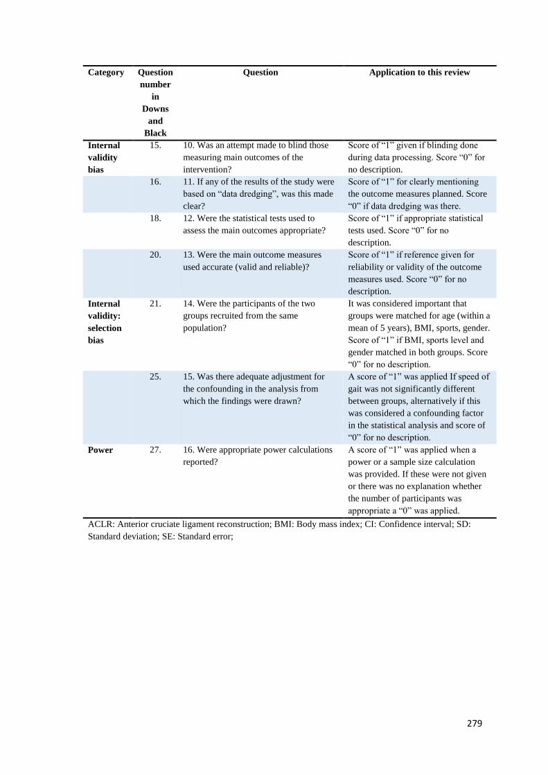
279
Category Question
number
in
Downs
and
Black
Question Application to this review
Internal
validity
bias
15. 10. Was an attempt made to blind those
measuring main outcomes of the
intervention?
Score of “1” given if blinding done
during data processing. Score “0” for
no description.
16. 11. If any of the results of the study were
based on “data dredging”, was this made
clear?
Score of “1” for clearly mentioning
the outcome measures planned. Score
“0” if data dredging was there.
18. 12. Were the statistical tests used to
assess the main outcomes appropriate?
Score of “1” if appropriate statistical
tests used. Score “0” for no
description.
20. 13. Were the main outcome measures
used accurate (valid and reliable)?
Score of “1” if reference given for
reliability or validity of the outcome
measures used. Score “0” for no
description.
Internal
validity:
selection
bias
21. 14. Were the participants of the two
groups recruited from the same
population?
It was considered important that
groups were matched for age (within a
mean of 5 years), BMI, sports, gender.
Score of “1” if BMI, sports level and
gender matched in both groups. Score
“0” for no description.
25. 15. Was there adequate adjustment for
the confounding in the analysis from
which the findings were drawn?
A score of “1” was applied If speed of
gait was not significantly different
between groups, alternatively if this
was considered a confounding factor
in the statistical analysis and score of
“0” for no description.
Power 27. 16. Were appropriate power calculations
reported?
A score of “1” was applied when a
power or a sample size calculation
was provided. If these were not given
or there was no explanation whether
the number of participants was
appropriate a “0” was applied.
ACLR: Anterior cruciate ligament reconstruction; BMI: Body mass index; CI: Confidence interval; SD:
Standard deviation; SE: Standard error;

280
Appendix A2- Mean differences for peak knee angles between participants with ACLR for between- and
within-group comparisons
Variable
(angles)
Task Comparison Level of
evidence
Studies MD (95%CI)
Flexion Walking Controls Strong Four LR (Georgoulis et al., 2003; Hall et al., 2012;
Noehren et al., 2013; Webster et al., 2005), two HR
(Ferber et al., 2002; Lewek et al., 2002) studies; I2 = 58%,
p=0.04
MD -0.6° (-2.0 to 0.8)
Contralateral limbs Moderate Four LR (Di Stasi et al., 2013; Hall et al., 2012; Roewer et
al., 2011; Webster, Feller, et al., 2012) , one HR (Hooper
et al., 2001); I2= 84%, p <0.001
MD -4.3° (-5.5 to -3.2)
Stair ascent Controls Moderate Two LR (Gao et al., 2012; Hall et al., 2012) studies; I2=
8%, p=0.30
MD -0.5° (-3.2 to 2.3)
Contralateral limbs Limited One LR (Hall et al., 2012) study MD -1.2° (-5.1 to 2.7)
Stair descent Controls Moderate Two LR (Gao et al., 2012; Hall et al., 2012) studies; I2=
66%, p=0.08
MD -2.7° (-4.7 to -0.7)
Running Controls Moderate One LR (Noehren et al., 2013), one HR (Lewek et al.,
2002) studies; I2=44%, p=0.44
MD -2.4°(-5.3 to 0.5)
Adduction Walking Controls Moderate Three LR (Butler et al., 2009; Georgoulis et al., 2003;
Webster & Feller, 2012a) studies; I2 = 73%, p = 0.03
MD -1°(-1.7 to 0.7)
Tibial
external
rotation
Walking Controls Strong Three LR (Czamara et al., 2015; Georgoulis et al., 2003;
Wang et al., 2013) studies; I2=64%, p = 0.06
MD 1.8° (0.10 to 3.6)
Stair ascent Controls Limited One LR (Gao et al., 2012) study MD -3.7° (-8.1 to 0.7)
Stair descent Controls Limited One LR (Gao et al., 2012) study MD -5.8° (-9.8 to -1.8)
Tibial
internal
rotation
Walking Contralateral limbs Strong Three LR (Sato et al., 2013; Webster & Feller, 2011;
Webster, Feller, et al., 2012) studies; I2= 0%, p=0.63
MD-3.9° (-5.8 to -2)
Stair ascent Controls Limited One LR (Gao et al., 2012) study MD 2.9° (-1.1 to 6.8)
Stair descent Controls Limited One LR (Gao et al., 2012) study MD 3.2° (-1 to 7)

281
Running Contralateral limbs Limited One LR (Sato et al., 2013) study MD -5.3° (-10 to 0.7)
HR: High risk of bias; LR: Low risk of bias; MD: Mean difference

282
Appendix B1- Final ethics approval letter from the ethical committee
H15/034
Dr G Sole
School of Physiotherapy
Dear Dr Sole,
I am again writing to you concerning your proposal entitled “Outcomes of anterior cruciate
ligament reconstructions more than two years following surgery”, Ethics Committee reference
number H15/034.
Thank you for your memo of 25th May 2015 addressing the issues raised by the Committee.
The Committee thanks you for the further comment provided in respect of the issues raised in relation
to confounding variables. The Committee also note the revised Information Sheet and the addition of
the shortened advert to be used for local newspapers.
On the basis of this response, I am pleased to confirm that the proposal now has full ethical
approval to proceed.
The standard conditions of approval for all human research projects reviewed and approved by the
Committee are the following:
Conduct the research project strictly in accordance with the research proposal submitted and granted
ethics approval, including any amendments required to be made to the proposal by the Human
Research Ethics Committee.
Inform the Human Research Ethics Committee immediately of anything which may warrant review
of ethics approval of the research project, including: serious or unexpected adverse effects on
participants; unforeseen events that might affect continued ethical acceptability of the project; and a
written report about these matters must be submitted to the Academic Committees Office by no
later than the next working day after recognition of an adverse occurrence/event. Please note that in
cases of adverse events an incident report should also be made to the Health and Safety Office:
http://www.otago.ac.nz/healthandsafety/ind
ex.html
Advise the Committee in writing as soon as practicable if the research project is discontinued.
Make no change to the project as approved in its entirety by the Committee, including any wording
in any document approved as part of the project, without prior written approval of the Committee
for any change. If you are applying for an amendment to your approved research, please email your
request to the Academic Committees Office:
Academic Services Manager, Academic Committees, Mr Gary Witte
27 May 2015

283
Approval is for up to three years from the date of this letter. If this project has not been completed
within three years from the date of this letter, re-approval or an extension of approval must be
requested. If the nature, consent, location, procedures or personnel of your approved application
change, please advise me in writing.
Yours sincerely,
Mr Gary Witte
Manager, Academic Committees
Tel: 479 8256
Email: [email protected]
c.c. Professor G D Baxter Dean School of Physiotherapy
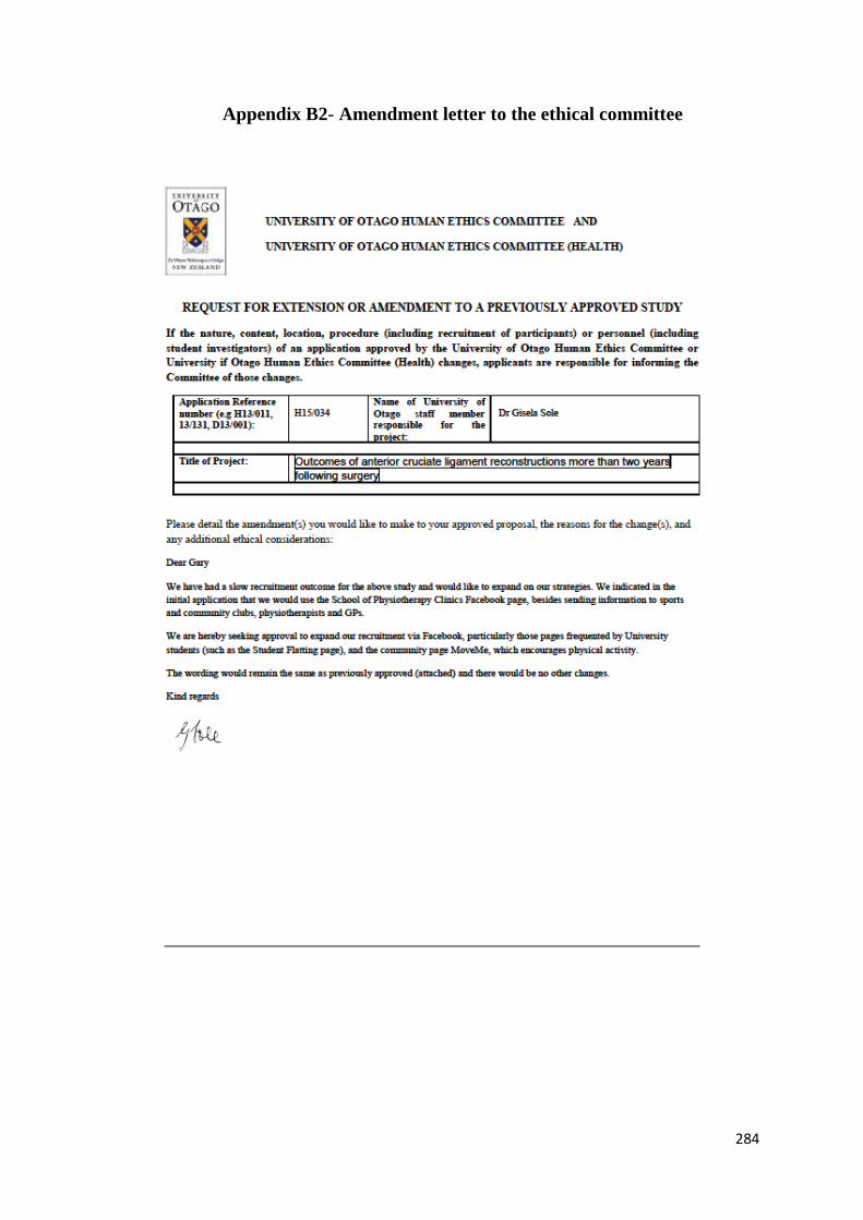
284
Appendix B2- Amendment letter to the ethical committee
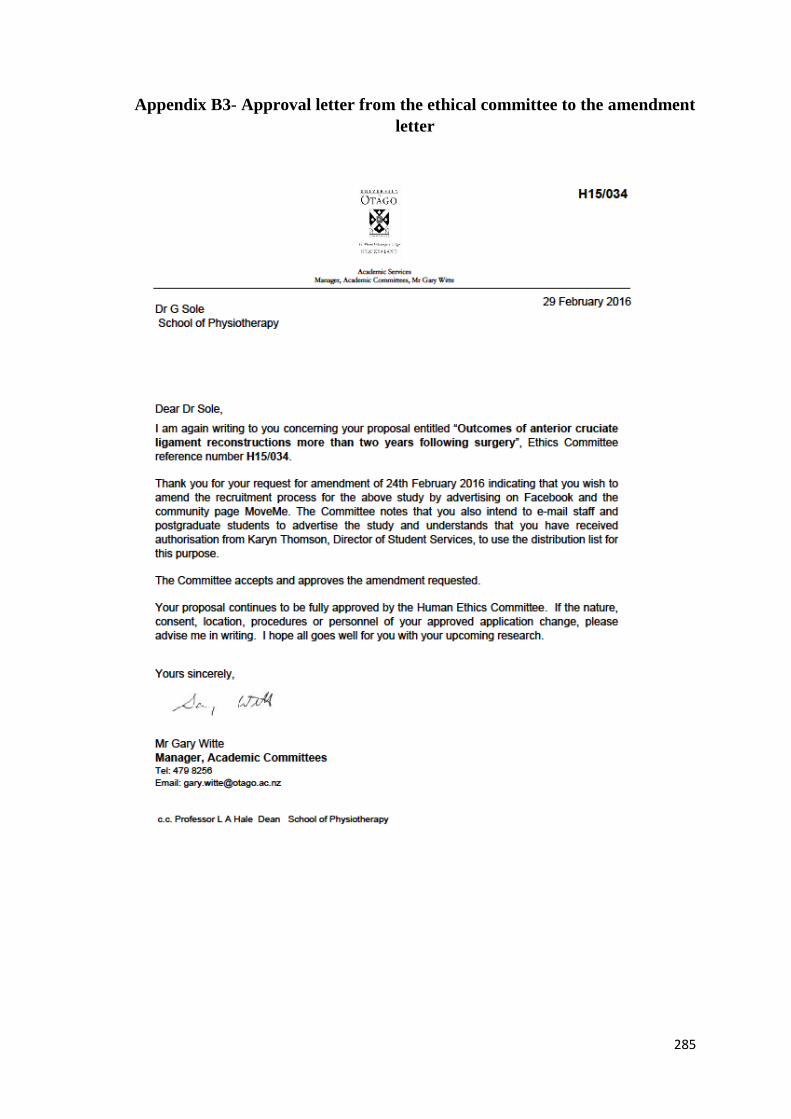
285
Appendix B3- Approval letter from the ethical committee to the amendment
letter
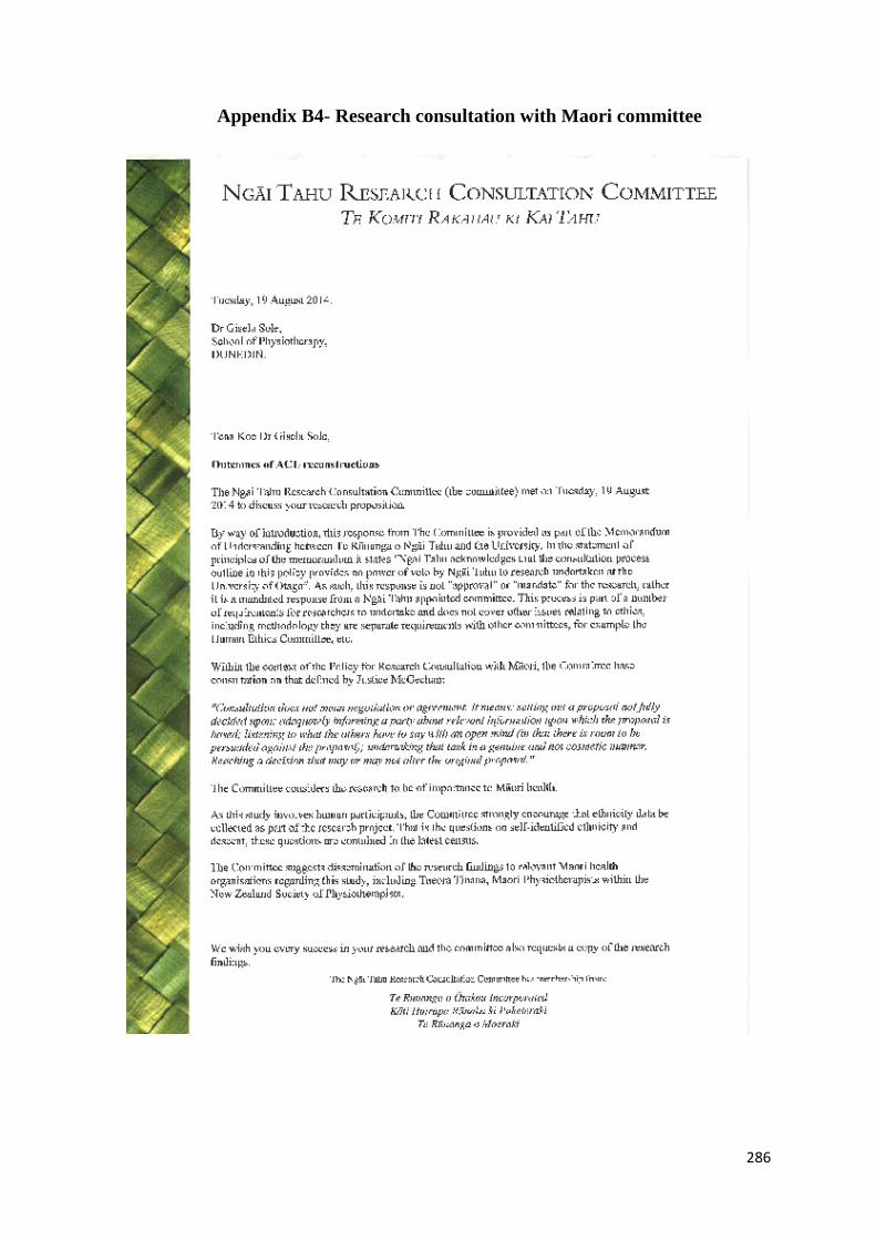
286
Appendix B4- Research consultation with Maori committee
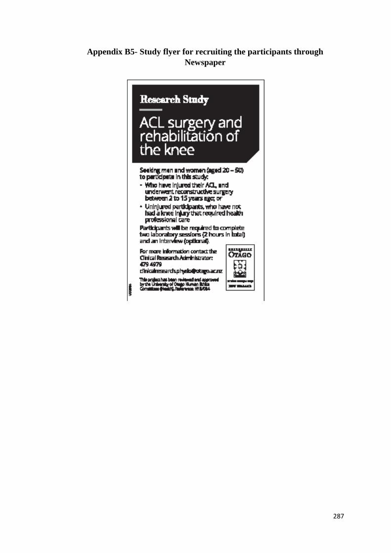
287
Appendix B5- Study flyer for recruiting the participants through
Newspaper
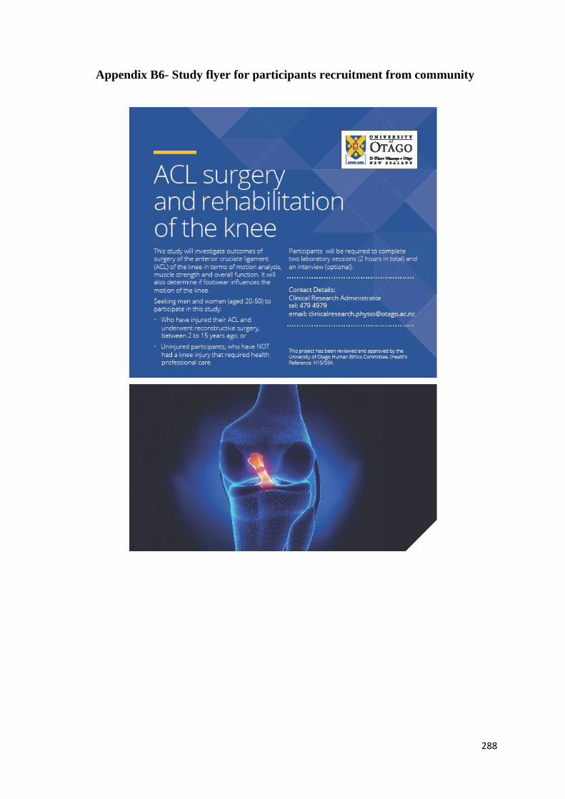
288
Appendix B6- Study flyer for participants recruitment from community
289
Appendix B7- Participant information sheet for ACLR group
Participants with ACL reconstructions
Study title: OUTCOMES OF ACL RECONSTRUCTION MORE THAN 2 YEARS
FOLLOWING SURGERY.
Principal
investigator:
Dr. Gisela Sole
School of Physiotherapy
Senior Lecturer
03-479 7936
Introduction
Thank you for showing an interest in this project. Please read this information sheet carefully. Take
time to consider and, if you wish, talk with relatives or friends, before deciding whether or not to
participate. If you decide to participate we thank you. If you decide not to take part there will be no
disadvantage to you and we thank you for considering our request.
What is the aim of this research project?
If you had surgery on an injury of your anterior cruciate ligament (ACL) in your knee more than two
years ago, we would like to know how you are moving now when you walk up and down two steps,
and when landing after a jump. We also want to assess how far you can jump on each leg, and your
thigh muscle strength. We would like to see if there is a difference in your movements when you walk
up and down steps with, and without, shoes on. Finally, we would like to know if and how your knee
injury has affected your life. Part of this project is the PhD research of Mandeep Kaur.
Who is funding this project?
This study has funding from Physiotherapy New Zealand, and from the Mark Steptoe Memorial Trust
Research Grant-in-Aid (School of Physiotherapy).
Who are we seeking to participate in the project?
290
We need three groups of participants: (1) people who have injured an ACL and had surgery followed
by physiotherapy rehabilitation, 2 to 8 years ago; (2) people who injured an ACL, had surgery, and
had physiotherapy rehabilitation, 10 to 15 years ago; and (3) uninjured people (the control group).
Inclusion criteria for participants with ACL reconstructions:
o Age: 20-50 years old, men and women;
o No other major injury to the legs and lower back that needed treatment by a health care
professional in the last 12 months.
All participants will be offered a voucher to contribute towards travel costs to the School of
Physiotherapy in Dunedin.
If you participate, what will you be asked to do?
You will be asked to attend two laboratory sessions for data collection at the School of Physiotherapy,
Dunedin. You may also be asked to attend a one-hour interview with the researchers. You are
welcome to bring a support person with you to any of the sessions.
Session 1: This session will last about 1½ hours. During this session you will be asked to:
o Complete a questionnaire about your current physical activity and your knee injury. This will
take about 15 minutes. You can do this online before the laboratory session if you prefer. We
will send you the electronic link by e-mail.
o Bring your own sports, running, or recreational shoes for us to assess for wear.
o If you had an injury, complete a release form for ACC to confirm the date of your knee
injury, the date and MRI report (if you had one), the date and type of surgery you had, the
number of physiotherapy sessions you attended for rehabilitation, and the date of the last
session.
o Undergo a routine screening examination of knee, hip, and ankle by the student researcher to
confirm that you are eligible to take part in the study, and to have your weight and height
measured.
o Perform practice trials of walking up and down two steps, and jumping forward on each leg to
make sure that these movements do not cause pain or discomfort.
o Undergo an examination of the knee to measure laxity of the ligaments.
o Perform five trials of walking up and down the steps, and jumping forward on each leg.
Participants with ACL reconstructions 2 to 8 years ago will walk up and down the steps (1)
barefoot and (2) with their own sports shoes. Participants with ACL reconstructions 10 to 15
years ago will perform the stair ascent/descent only in their sports shoes. The jumping task
will be performed with your own sports shoes.

291
Your movements will be recorded and measured using 3D motion analysis equipment. We will place
a set of small reflective markers on your skin, and on the clothing on your legs, pelvis, trunk, and
arms, using double-sided tape. A video will also be made with a digital recorder during the stair
ascent/descent and the jumping trials in case the researchers need to check your movements later. This
video will not be used for any other purpose.
Session 2:
This session will last about 45 minutes. Following a warm-up, you will be asked to jump as far as you
can, three times on each leg. Your thigh muscle strength will then be measured: you will be asked to
straighten and bend your knee as hard as you can five times against the measuring machine’s lever.
This will be performed for both legs. The result of the thigh muscle strength test will be given to you
as soon as you have finished this session.
Session 3:
Approximately 10 injured participants will be recruited for this part of the study, a face-to-face one-
hour interview with Ms Mandeep Kaur and Dr Sole. If you agree to be interviewed, and live outside
Dunedin, the interview can be conducted via Skype or telephonically, at a time that suits you best.
If you live outside Dunedin, all three sessions can take place on the same day if you wish. The
laboratory sessions will last up to 2¼ hrs, and the interview will be an additional hour if you agree to
do this. In this case, we will provide you with refreshments.
The interview will be tape-recorded and the interviewers may make notes. The questions will be about
how well you have recovered from your injury and the surgery in terms of your daily life. The precise
nature of the questions has not been determined in advance, but will depend on the way in which the
interview develops. In the event that the line of questioning develops in such a way that you feel
hesitant or uncomfortable, you may decline to answer any particular question(s) without any
disadvantage. A summary of the results of all of the interviews will be emailed or posted to you, and
you can correct or add anything if you wish.
Is there any risk of discomfort or harm from participation?
There is no risk of physical harm or discomfort to you. You may feel a bit sore after the muscle
strength test, as you might in any strengthening exercise programme. You may contact the researchers
if you have any concerns about that muscle soreness, or if you have any other questions.
What data or information will be collected, and how will they be used?
292
We will collect the following information:
o Your name and date of birth; weight, height, ethnicity, leg dominance;
o Your level of physical activity, now and prior to the knee injury; confidence in your knee during
sport; and your general quality of life.
o Movement analysis data during the stair ascent/descent and jump landing; the jump distance and
thigh muscle strength for each leg
We will use this information to describe the groups of participants and compare with healthy control
group.
What about anonymity and confidentiality?
All data and the recordings will be securely stored in such a way that only Dr Sole, Dr Ribeiro, Ms
Kaur and a designated Research Assistant will be able to gain access to it. Data obtained as a result of
the research will be kept in secure storage for at least 10 years. Any personal information held about
the participants (such as contact details, audio files after they have been transcribed, and video files)
will be destroyed at the completion of the research except as required by university’s policy.
Information used for any publication will be kept anonymous.
The results for the footwear study (barefoot versus shod during stair ascent/descent) will be written up
as an individual study by the principal investigator and submitted for publication. The results of the
remaining studies will be written up as Ms Mandeep’s research towards her doctorate degree and may
be published. It will be available in the University of Otago Library (Dunedin, New Zealand).
If you agree to participate, can you withdraw later?
You may withdraw from participation in the project at any time and without any disadvantage to
yourself of any kind. If you agree to take part in the interview and the line of questioning develops in
such a way that you feel hesitant or uncomfortable, you may decline to answer any particular
question(s), and/or may withdraw from the project without disadvantage of any kind. Your future
health care provision will not be affected by participating in the study, or by withdrawing or
declining.
Any questions?
If you have any questions now or in the future, please feel free to contact either:
Clinical Research Administrator
School of Physiotherapy
Tel: 03-479 4979
Dr. Gisela Sole Tel: 03 479 7936
293
Senior Lecturer
School of Physiotherapy
Dr. Daniel Cury Ribeiro
Lecturer
School of Physiotherapy
Tel: 03 479 7455
Mandeep Kaur
PhD candidate, student researcher
School of Physiotherapy
This study has been approved by the University of Otago Human Ethics Committee (Health), Ref. no.
H15/034. If you have any concerns about the ethical conduct of the research you may contact the
Committee through the Human Ethics Committee Administrator (phone +64 3 479 8256 or email
[email protected]). Any issues you raise will be treated in confidence and investigated and you
will be informed of the outcome.

294
Appendix B8- Participant information sheet for control group
Study title: OUTCOMES OF ACL RECONSTRUCTION MORE THAN 2 YEARS
FOLLOWING SURGERY.
Principal
investigator:
Dr. Gisela Sole
School of Physiotherapy
Senior Lecturer
03-4797936
Introduction
Thank you for showing an interest in this project. Please read this information sheet carefully. Take
time to consider and, if you wish, talk with relatives or friends, before deciding whether or not to
participate. If you decide to participate we thank you. If you decide not to take part there will be no
disadvantage to you and we thank you for considering our request.
What is the aim of this research project?
We would like to know whether people who have had surgery (reconstruction) to the anterior cruciate
ligament (ACL) in the knee move differently compared to uninjured people while walking up and
down a set of steps and when landing after a jump. We also want to see how far people with an ACL
reconstruction can jump on each leg compared to uninjured people, and how strong the thigh muscles
are. Lastly, we would like to see if there is a difference in your movements when you walk up and
down steps with, and without, shoes on. Part of this project is the PhD research of Mandeep Kaur.
Who is funding this project?
This study has funding from Physiotherapy New Zealand, and from the Mark Steptoe Memorial Trust
Research Grant-in-Aid (School of Physiotherapy).
Who are we seeking to participate in the project?
295
We need three groups of participants: (1) people who have injured an ACL and had surgery followed
by physiotherapy rehabilitation, 2 to 8 years ago; (2) people who injured an ACL, had surgery, and
had physiotherapy rehabilitation, 10 to 15 years ago; and (3) uninjured people (the control group).
Inclusion criteria for control participants:
o Age: 20-50 years old, and both men and women;
o No major injury to either legs or lower back that needed treatment by a health care
professional in the past 12 months.
All participants will be offered a voucher to contribute towards travel costs to the School of
Physiotherapy in Dunedin.
If you participate, what will you be asked to do?
You will be asked to attend two laboratory sessions for data collection at the School of Physiotherapy,
Dunedin. You are welcome to bring a support person with you to any of the sessions.
Session 1: This session will last about 1½ hours. During this session you will be asked to:
o Complete a questionnaire about your current physical activity and knee-related health. This
will take about 15 minutes. You can do this online before the laboratory session if you prefer.
We will send you an electronic link by e-mail.
o Bring your own sports, running, or recreational shoes for us to assess for wear.
o Undergo a routine screening examination of knee, hip, and ankle by the student researcher to
confirm that you are eligible to take part in the study, and to have your weight and height
measured.
o Perform practice trials of walking up and down two steps, and jumping forward on each leg.
o Undergo an examination of the knee to measure the laxity of the ligaments.
o Walk up and down a set of steps, and jump forward on each leg. You will be asked to walk
up and down the steps (1) barefoot and (2) with your own sports shoes. You will do this five
times for each leg (for each footwear condition). You will perform the jumping tasks with
your own sports shoes, five trials for each leg.
Your movements will be recorded and measured using 3D motion analysis equipment. We will place
a set of small reflective markers on your skin, and on the clothing on your legs, pelvis, trunk, and
arms, using double-sided tape. A video will also be made with a digital recorder during the stair

296
ascent/descent trials and the jumping trials in case the researchers need to check your movements
later. This video will not be used for any other purpose.
Session 2:
This session will last about 45 minutes.
Following the warm up, you will be asked to jump as far as you can and land on the same leg (three
times on each side). Your thigh muscle strength will then be measured: you will be asked to straighten
and bend your knee as hard as you can five times against the measuring machine’s lever. This will be
performed for both legs. The result of the thigh muscle strength test will be given to you as soon as
you have finished this session.
If it suits you better, both sessions can be performed on the same day. The laboratory sessions will be
a total of 2 ¼ hrs. Refreshments will be provided by the researchers in this case.
Is there any risk of discomfort or harm from participation?
There is no risk of physical harm or discomfort to you. You may feel a bit sore after the muscle
strength test, as you might in any strengthening exercise programme. You may contact the researchers
if you have any concerns about that muscle soreness, or if you have any other questions.
What data or information will be collected, and how will they be used?
We will collect the following information:
o Your name and date of birth; weight, height, ethnicity, leg dominance;
o Your level of physical activity; confidence in your knee during sport; and your general quality of
life.
o Movement analysis data during the stair ascent/descent and jump landing; the jump distance and
thigh muscle strength for each leg
We will use this information to describe the groups of participants and to compare with the ACL
reconstructed group.
What about anonymity and confidentiality?
All the data and the recordings will be securely stored in such a way that only Dr Sole, Dr Ribeiro, Ms
Kaur and a designated Research Assistant will be able to gain access to it. Data obtained as a result of
the research will be kept in secure storage for at least 10 years. Any personal information held about
the participants (such as contact details, audio files after they have been transcribed, and video files)
297
will be destroyed at the completion of the research except as required by university’s research policy.
Information used for any publication will be kept anonymous.
The results for the footwear study (barefoot versus shod during stair ascent/descent) will be written up
as an individual study by the principal investigator and submitted for publication. The results of the
remaining studies will be written up as Ms Mandeep’s research towards her doctorate degree and may
be published. It will be available in the University of Otago Library (Dunedin, New Zealand).
If you agree to participate, can you withdraw later?
You may withdraw from participation in the project at any time and without any disadvantage to
yourself of any kind. Your future health care provision will not be affected by participating in the
study, or by withdrawing or declining.
Any questions?
If you have any questions now or in the future, please feel free to contact either:
Clinical Research Administrator
School of Physiotherapy
Tel: 03-479 4979
Dr. Gisela Sole
Senior Lecturer
School of Physiotherapy
Tel: 03 479 7936
Dr. Daniel Cury Ribeiro
Lecturer
School of Physiotherapy
Tel: 03 479 7455
Mandeep Kaur
PhD candidate, student researcher
School of Physiotherapy
This study has been approved by the University of Otago Human Ethics Committee (Health), Ref. no.
H15/034. If you have any concerns about the ethical conduct of the research you may contact the
Committee through the Human Ethics Committee Administrator (phone +64 3 479 8256 or email
[email protected]). Any issues you raise will be treated in confidence and investigated and you
will be informed of the outcome.
298
Appendix B9- Consent form for ACLR group
CONSENT FORM FOR PARTICIPANTS with ACL RECONSTRUCTIONS
Principal Investigator: Dr Gisela Sole; [email protected]
Following signature and return to the research team this form will be stored in a secure place for ten
years.
Name of participant:…………………………………………..
1. I have read the Information Sheet concerning this study and understand the aims of this research
project.
2. I have had sufficient time to talk with other people of my choice about participating in the study.
3. I confirm that I meet the criteria for participation which are explained in the Information Sheet.
4. All my questions about the project have been answered to my satisfaction, and I understand that I
am free to request further information at any stage.
5. I know that my participation in the project is entirely voluntary, and that I am free to withdraw
from the project at any time without disadvantage.
6. I know that as a participant, small markers will be placed on my trunk and legs and I will be required
to walk up and down a stair (5 times), and perform five jumps on each leg during Session one.
During Session 2 I will be asked three times as far as I can on each leg and my thigh muscle strength
will be measured with a Biodex machine by bending and straightening the leg. If I feel hesitant or
uncomfortable I can withdraw at any point of the time.
7. If I agree, I may also choose to participate in an individual interview with the researchers. The
general line of questioning includes outcomes of my knee surgery related to my health-related
quality of life. The precise nature of the questions which will be asked has not been determined in
advance, but will depend on the way in which the interview develops. In the event that the line of
questioning develops in such a way that I feel hesitant or uncomfortable I may decline to answer
any particular question(s) and/or may withdraw from the project without disadvantage of any kind.
299
8. I grant the researchers permission to contact ACC to confirm the date of my injury, date and
report of MRI (if applicable), date and procedure of the surgery I underwent, the number of
physiotherapy treatments I had for the knee, and the date of the last physiotherapy treatment.
Yes No (please indicate Yes or No)
9. I understand the nature and size of the risks of discomfort or harm which are explained in the
Information Sheet.
10. I know that when the project is completed all personal identifying information will be removed
from the paper records and electronic files which represent the data from the project, and that
these will be placed in secure storage and kept for at least ten years.
11. I understand that the results of the project may be published and be available in the University of
Otago Library, but that any personal identifying information will remain confidential between
myself and the researchers during the study, and will not appear in any spoken or written report
of the study.
12. I know that there is no remuneration offered for this study, and that no commercial use will be
made of the data. After participating in the study I will be offered a petrol voucher in recognition
of costs associated with participation in the study.
Signature of participant: Date:

300
Appendix B10- Consent form for control group
OUTCOMES OF ACL RECONSTRUCTION MORE THAN 2 YEARS FOLLOWING
SURGERY.
CONSENT FORM FOR CONTROL PARTICIPANTS
Principal Investigator: Dr Gisela Sole; [email protected]
Following signature and return to the research team this form will be stored in a secure place for ten
years.
Name of participant:…………………………………………..
1. I have read the Information Sheet concerning this study and understand the aims of this research
project.
2. I have had sufficient time to talk with other people of my choice about participating in the study.
3. I confirm that I meet the criteria for participation which are explained in the Information Sheet.
4. All my questions about the project have been answered to my satisfaction, and I understand that I
am free to request further information at any stage.
5. I know that my participation in the project is entirely voluntary, and that I am free to withdraw
from the project at any time without disadvantage.
6. I know that as a participant, small markers will be placed on my trunk and legs and I will be required
to walk up and down a stair (5 times), and perform five jumps on each leg during Session one.
During Session 2 I will be asked three times as far as I can on each leg and my thigh muscle strength
will be measured with a Biodex machine by bending and straightening the leg. If I feel hesitant or
uncomfortable I can withdraw at any point of the time.
7. I understand the nature and size of the risks of discomfort or harm which are explained in the
Information Sheet.
8. I know that when the project is completed all personal identifying information will be removed
from the paper records and electronic files which represent the data from the project, and that
these will be placed in secure storage and kept for at least ten years.
301
9. I understand that the results of the project may be published and be available in the University of
Otago Library, but that any personal identifying information will remain confidential between
myself and the researchers during the study, and will not appear in any spoken or written report
of the study.
10. I know that there is no remuneration offered for this study, and that no commercial use will be
made of the data. After participating in the study I will be offered a petrol voucher in recognition
of costs associated with participation in the study.
Signature of participant: Date:
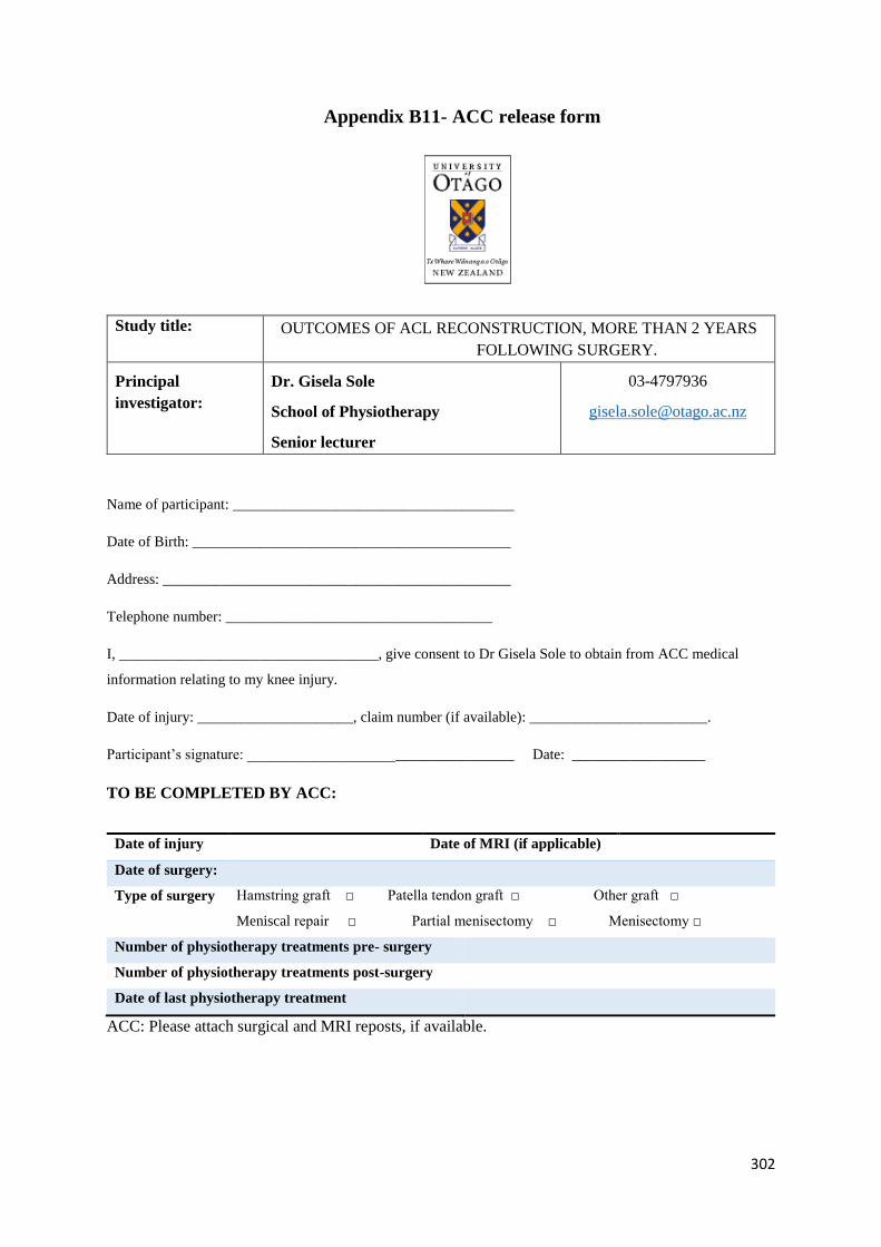
302
Appendix B11- ACC release form
Study title: OUTCOMES OF ACL RECONSTRUCTION, MORE THAN 2 YEARS
FOLLOWING SURGERY.
Principal
investigator:
Dr. Gisela Sole
School of Physiotherapy
Senior lecturer
03-4797936
Name of participant: ______________________________________
Date of Birth: ___________________________________________
Address: _______________________________________________
Telephone number: ____________________________________
I, ___________________________________, give consent to Dr Gisela Sole to obtain from ACC medical
information relating to my knee injury.
Date of injury: _____________________, claim number (if available): ________________________.
Participant’s signature: ____________________________________ Date: __________________
TO BE COMPLETED BY ACC:
Date of injury Date of MRI (if applicable)
Date of surgery:
Type of surgery Hamstring graft □ Patella tendon graft □ Other graft □
Meniscal repair □ Partial menisectomy □ Menisectomy □
Number of physiotherapy treatments pre- surgery
Number of physiotherapy treatments post-surgery
Date of last physiotherapy treatment
ACC: Please attach surgical and MRI reposts, if available.
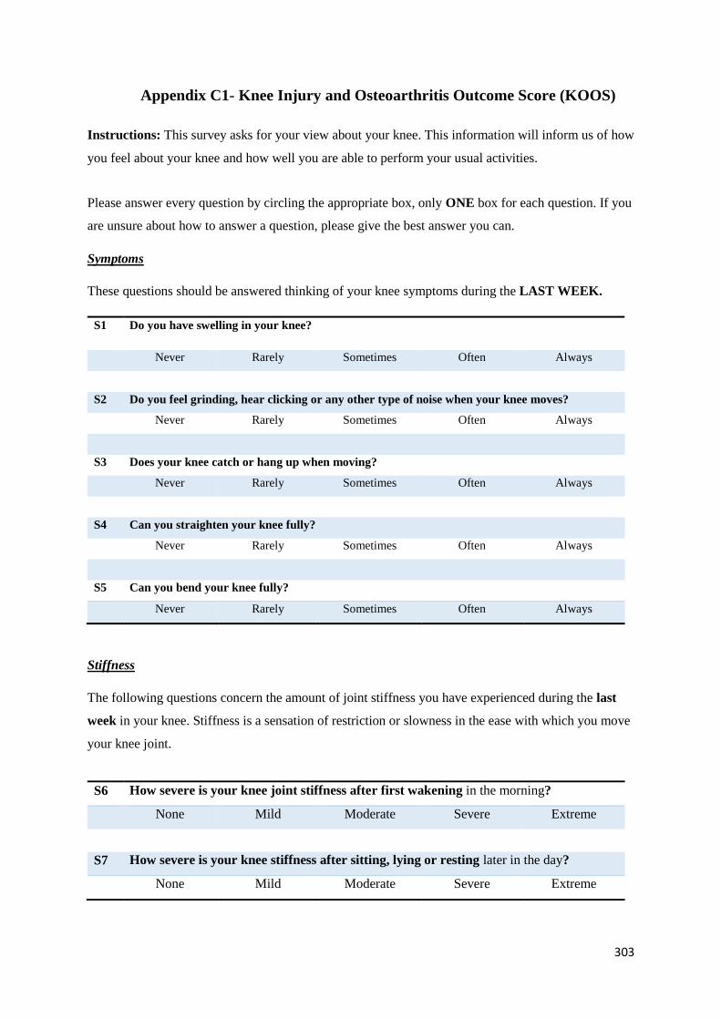
303
Appendix C1- Knee Injury and Osteoarthritis Outcome Score (KOOS)
Instructions: This survey asks for your view about your knee. This information will inform us of how
you feel about your knee and how well you are able to perform your usual activities.
Please answer every question by circling the appropriate box, only ONE box for each question. If you
are unsure about how to answer a question, please give the best answer you can.
Symptoms
These questions should be answered thinking of your knee symptoms during the LAST WEEK.
S1 Do you have swelling in your knee?
Never Rarely Sometimes Often Always
S2 Do you feel grinding, hear clicking or any other type of noise when your knee moves?
Never Rarely Sometimes Often Always
S3 Does your knee catch or hang up when moving?
Never Rarely Sometimes Often Always
S4 Can you straighten your knee fully?
Never Rarely Sometimes Often Always
S5 Can you bend your knee fully?
Never Rarely Sometimes Often Always
Stiffness
The following questions concern the amount of joint stiffness you have experienced during the last
week in your knee. Stiffness is a sensation of restriction or slowness in the ease with which you move
your knee joint.
S6 How severe is your knee joint stiffness after first wakening in the morning?
None Mild Moderate Severe Extreme
S7 How severe is your knee stiffness after sitting, lying or resting later in the day?
None Mild Moderate Severe Extreme

304
Pain
P1 How often do you experience knee pain?
Never Monthly Weekly Daily Always
What amount of knee pain have you experienced the LAST WEEK during the following activities?
P2 Twisting/pivoting on your knee
None Mild Moderate Severe Extreme
P3 Straightening knee fully
None Mild Moderate Severe Extreme
P4 Bending knee fully
None Mild Moderate Severe Extreme
P5 Walking on flat surface
None Mild Moderate Severe Extreme
P6 Going up or down stairs
None Mild Moderate Severe Extreme
P7 At night while in bed
None Mild Moderate Severe Extreme
P8 Sitting or lying
None Mild Moderate Severe Extreme
P9 Standing upright
None Mild Moderate Severe Extreme
Function, daily living
The following questions concern your physical function. By this we mean your ability to move around
and to look after yourself. For each of the following activities please indicate the degree of difficulty
you have experienced in the LAST WEEK dur to your knee.
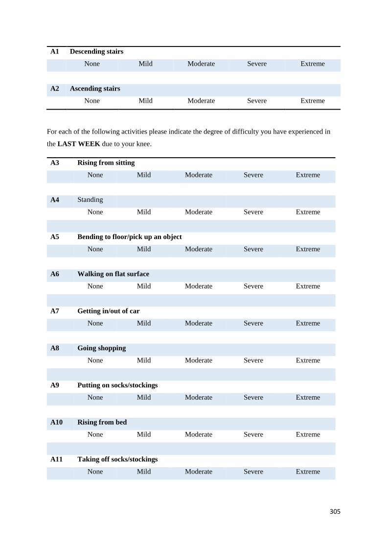
305
A1 Descending stairs
None Mild Moderate Severe Extreme
A2 Ascending stairs
None Mild Moderate Severe Extreme
For each of the following activities please indicate the degree of difficulty you have experienced in
the LAST WEEK due to your knee.
A3 Rising from sitting
None Mild Moderate Severe Extreme
A4 Standing
None Mild Moderate Severe Extreme
A5 Bending to floor/pick up an object
None Mild Moderate Severe Extreme
A6 Walking on flat surface
None Mild Moderate Severe Extreme
A7 Getting in/out of car
None Mild Moderate Severe Extreme
A8 Going shopping
None Mild Moderate Severe Extreme
A9 Putting on socks/stockings
None Mild Moderate Severe Extreme
A10 Rising from bed
None Mild Moderate Severe Extreme
A11 Taking off socks/stockings
None Mild Moderate Severe Extreme

306
A12 Lying in bed (turning over, maintaining knee position)
None Mild Moderate Severe Extreme
A13 Getting in/out of bath
None Mild Moderate Severe Extreme
A14 Sitting
None Mild Moderate Severe Extreme
A15 Getting on/off toilet
None Mild Moderate Severe Extreme
For each of the following activities please indicate the degree of difficulty you have experienced in
the LAST WEEK dur to your knee
A16 Heavy domestic duties (moving heavy boxes, scrubbing floors, etc.)
None Mild Moderate Severe Extreme
A17 Light domestic duties (cooking, dusting, )
None Mild Moderate Severe Extreme
Function, sports and recreational activities
The following questions concern your physical function when being active on a higher level. The
questions should be answered thinking of what degree of difficulty you have experienced during the
LAST WEEK due to your knee.
SP1 Squatting
None Mild Moderate Severe Extreme
SP2 Running
None Mild Moderate Severe Extreme
SP3 Jumping
None Mild Moderate Severe Extreme
SP4 Twisting/pivoting on your injured knee
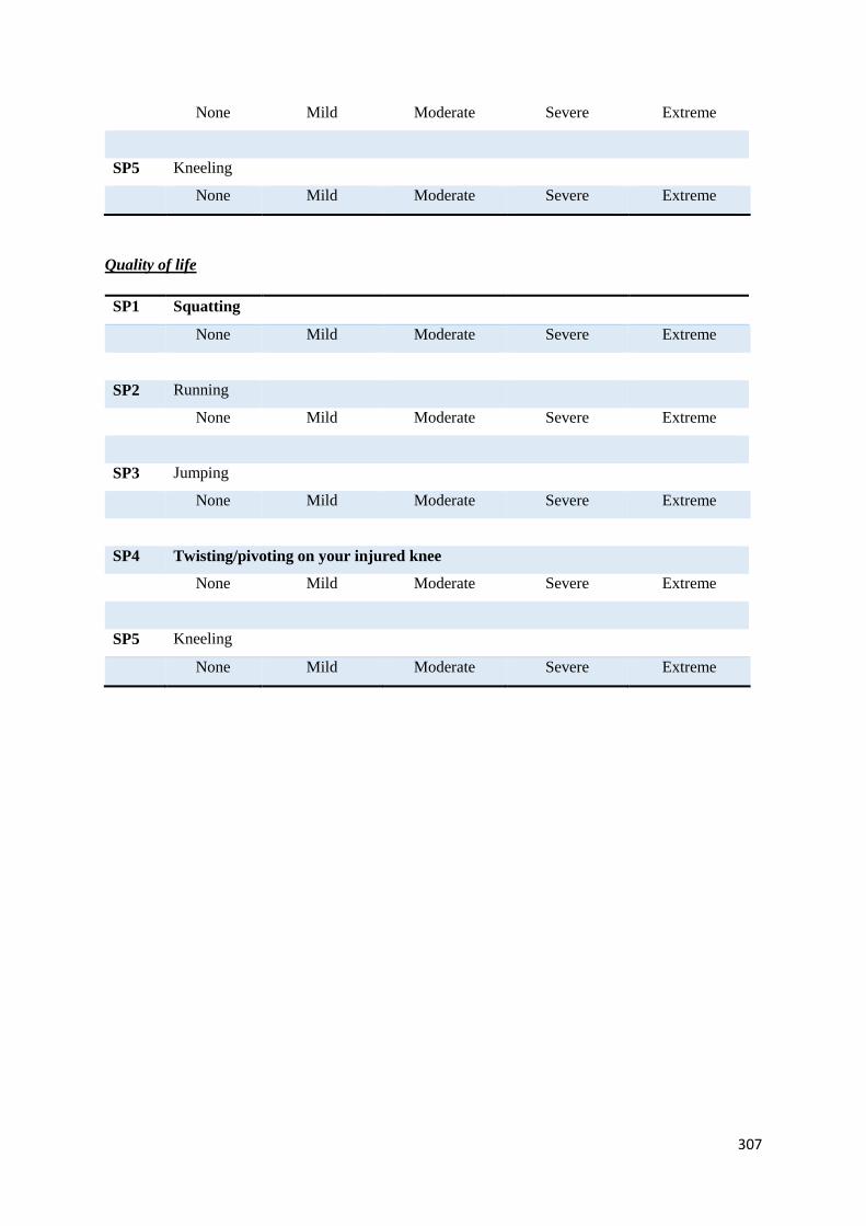
307
None Mild Moderate Severe Extreme
SP5 Kneeling
None Mild Moderate Severe Extreme
Quality of life
SP1 Squatting
None Mild Moderate Severe Extreme
SP2 Running
None Mild Moderate Severe Extreme
SP3 Jumping
None Mild Moderate Severe Extreme
SP4 Twisting/pivoting on your injured knee
None Mild Moderate Severe Extreme
SP5 Kneeling
None Mild Moderate Severe Extreme
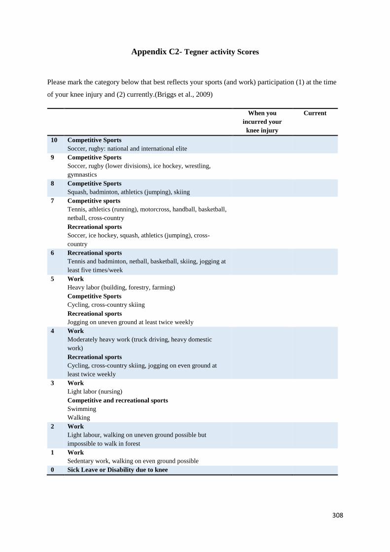
308
Appendix C2- Tegner activity Scores
Please mark the category below that best reflects your sports (and work) participation (1) at the time
of your knee injury and (2) currently.(Briggs et al., 2009)
When you
incurred your
knee injury
Current
10 Competitive Sports
Soccer, rugby: national and international elite
9 Competitive Sports
Soccer, rugby (lower divisions), ice hockey, wrestling,
gymnastics
8 Competitive Sports
Squash, badminton, athletics (jumping), skiing
7 Competitive sports
Tennis, athletics (running), motorcross, handball, basketball,
netball, cross-country
Recreational sports
Soccer, ice hockey, squash, athletics (jumping), cross-
country
6 Recreational sports
Tennis and badminton, netball, basketball, skiing, jogging at
least five times/week
5 Work
Heavy labor (building, forestry, farming)
Competitive Sports
Cycling, cross-country skiing
Recreational sports
Jogging on uneven ground at least twice weekly
4 Work
Moderately heavy work (truck driving, heavy domestic
work)
Recreational sports
Cycling, cross-country skiing, jogging on even ground at
least twice weekly
3 Work
Light labor (nursing)
Competitive and recreational sports
Swimming
Walking
2 Work
Light labour, walking on uneven ground possible but
impossible to walk in forest
1 Work
Sedentary work, walking on even ground possible
0 Sick Leave or Disability due to knee
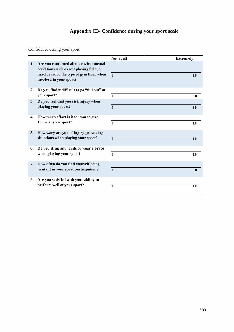
309
Appendix C3- Confidence during your sport scale
Confidence during your sport
Not at all Extremely
1. Are you concerned about environmental
conditions such as wet playing field, a
hard court or the type of gym floor when
involved in your sport?
0 10
2. Do you find it difficult to go “full out” at
your sport?
0 10
3. Do you feel that you risk injury when
playing your sport?
0 10
4. How much effort is it for you to give
100% at your sport?
0 10
5. How wary are you of injury-provoking
situations when playing your sport?
0 10
6. Do you strap any joints or wear a brace
when playing your sport?
0 10
7. How often do you find yourself being
hesitant in your sport participation?
0 10
8. Are you satisfied with your ability to
perform well at your sport?
0 10

310
Appendix C4- Short Form-12 Health Survey
This survey asks for your views about your health. Answer each question by choosing joint one
answer. If you are unsure how to answer a question, please give the best answer you can.
1. In general, would you say your health is:
1 Excellent 2 Very good 3 Good 4 Fair 5 Poor
The following questions are about activities you might do during a typical day. Does your health now
limit you in these activities? If so, how much?
YES,
limited a
lot
YES,
limited a
little
NO, not
limited
at al.
2. Moderate activities such as moving a table, pushing a vacuum
cleaner, bowling or playing golf.
1 2 3
3. Climbing several flights of stairs. 1 2 3
During the past 4 weeks, have you had any of the following problems with your work or other regular
daily activities as a result of your physical health?
YES NO
4. Accomplished less than you would like. 1 2
5. Were limited in the kind of work or other activities 1 2
During the past 4 weeks, have you had any of the following problems with your work or other regular
daily activities as a result of any emotional problems (such as feeling depressed or anxious)?
YES NO
6. Accomplished less than you would like. 1 2
7. Did work or activities less carefully than usual. 1 2
8. During the past 4 weeks, how much did pain interfere with your normal work (including work
outside the home and housework)?
1 Not at all 2 A little bit 3 Moderately 4 Quite a bit 5 Extremely
311
These questions are about how you have been feeling during the past 4 weeks. For each question,
please give the one answer that comes closest to the way you have been feeling.
How much of the time during the past 4 weeks……
All of
the
time
Most of
the
time
A good
bit of
the
time
Some of
the
time
A little
of the
time
None of
the
time
9. Have you felt calm & peaceful?
10. Did you have a lot of energy?
11. Have you felt down-hearted and
blue?
12. During the past 4 weeks, how much of the time has your physical health or emotional
problems interfered with your social activities (like visiting friends, relatives, etc)?
1 All of the time 2 Most of the time 3 Some of the time 4 A little of the
time
5 None of the time
Thank you very much for completing this questionnaire.

312
Appendix C5- History of previous injuries
For questions 1-5, please circle the relevant box for an injury you have had treatment for in
the past 6 months and indicate in the box whether the injury was on your left or right.
(Participants with ACL injury: detail pertaining to your knee injury will be asked on the
following page.)
1. Have you ever pulled
(strained) or hurt a muscle or
tendon other than a
hamstring?
Back
1
Buttock/pelvis
2
Groin 3 Front of
thigh 4
Calf/Achilles 5
2. Have you ever torn (sprained)
or stretched a ligament
Back 6 Buttock 7 Hip 8 Knee 9 Foot/Ankle 10
3. Have you ever dislocated a
joint or had a bone come out
of joint?
Back 11 Pelvis 12 Hip 13 Knee 14 Foot/Ankle 15
4. Have you ever broken
(fractured) a bone?
Trunk 16 Pelvis 17 Upper leg
18
Lower leg
19
Foot 20
5. Have any of your joints ever
swollen?
Back 21 Hip 23 Knee 24 Ankle 25 Foot 26
If yes was answered for any question above,
please provide detail and date (month, year) and
weeks absence from sport due to the injury.
Are you currently on any prescribed
medication? If yes, please provide detail.
313
Appendix C6- Reliability of KT-arthrometer in sagittal plane
Participant ID Side Session 1 (Average, mm) Session 2 (Average, mm)
574 1 6.2 6.3
0 4.9 5.2
251 1 4.9 5.2
0 9.3 9.3
837 1 5.8 4.4
0 5.6 7.0
686 1 3.7 6.7
0 6.6 6.0
116 1 10.7 12.4
0 8.0 7.6
673 1 5.0 5.5
0 6.3 6.7
797 1 7.8 10.1
0 7.4 12.5
449 1 2.2 5.0
0 6.2 8.3
251 1 5.5 6.1
0 8.9 8.6
832 1 3.8 3.0
0 10.5 9.0
AP: Antero-posterior, 1: Right, 0: Left.
Reliability and SEM of KT-Arthrometer
Test measurement AP-laxity (mm)
ICC (95% CI) SEM (mm)
N=10 participants with ACLR
AP knee laxity 0.84 (0.59-0.93) 2.2
AP: Antero-posterior, SEM: Standard error of measurement, ICC: Intraclass correlation coefficient, ACL:
Anterior cruciate ligament, mm= millimetre.
314
Appendix C7- Data provided by Participants and Accident Compensation Corporation (ACC)
Participant
ID
Accident
Date
Date of
MRI
Date of
Surgery
Type of
Surgery
Surgical details
(Type of graft,
meniscal
resection)
Number of
Physio
treatments
Pre-
Surgery
Number
of Physio
treatments
Post-
Surgery
Date of last
physio
treatments
Group 1 (ACLR 2-10 years post-operative))
1. 468 HT, No meniscal
resection
2. 116 4/11/2008 3/05/2010 6/09/2010 KNE81(Primary ACLR) PT, Meniscectomy 27 48 7/07/2011
3. 844 HT, No meniscal
resection
4. 574 25/10/2009 1/03/2010 Meniscal repair, partial
menisectomy, –routine ACLR–
bucket handle tear of the medial
meniscus.
PT 20 6 26/04/2010
5. 861 27/04/2010 26/05/2010
4/10/2010
4/06/2010
24/03/2011
KNE80
KNE83 Revision ACLR
PT 21 37 12/09/2011
6. 434 5/09/2009 12/11/2009 3/06/2010 KNE80 PT, no meniscal
resection
19 14 23/08/2010
7. 686 PT,
Menisectomy
8. 713 16/12/2009 16/12/2011 KNE81 (Primary ACLR) _ 12 1 6/01/2012
9. 832 24/04/2010 21/05/2010 23/11/2010 KNE81 (Primary ACLR) HT, meniscal
resection
25 17 26/01/2012
10. 232 14/04/2012 11/05/2012
28/05/2014
1/11/2012 KNE81 (Primary ACLR) HT, No meniscal
resection
7 5 17/06/2014
11. 251 3/09/2011 1/06/2012 23/07/2012 KNE81(Primary ACLR) HT 9 23 9/04/2013
12. 451 PT, Partial lateral
menisectomy
315
Participant
ID
Accident
Date
Date of
MRI
Date of
Surgery
Type of
Surgery
Surgical details
(Type of graft,
meniscal
resection)
Number of
Physio
treatments
Pre-
Surgery
Number
of Physio
treatments
Post-
Surgery
Date of last
physio
treatments
13. 449 21/05/2011 25/08/2011 16/04/2012 KNE81 (Primary knee ACLR) HT with meniscal
repair
14. 704 5/02/2011 10/04/2012 16/06/2011 KNE81 (Primary knee ACLR) HT, Partial
Menisectomy
9 40 19/03/2013
15. 608 HT, No meniscal
resection
16. 896 PT, Meniscal
repair
17. 532 13/07/2010 7/10/2010 17/02/2011 KNE81 (Primary ACLR) HT 8 12 2/09/2011
18. 751 3/10/2012 17/01/2013 Arthroscopic ACLR (L) knee,
single bundle with meniscal repair
HT, meniscal
repair
2 14 10/05/2013
19. 494 8/05/2010 18/06/2010 16/12/2010 KNE81(Primary ACLR) HT, no
meniscectomy
13 15 28/03/2011
20. 673 9/02/2008 11/04/2008 27/01/2009 KNE80 PT, Meniscal
repair
11 23 27/08/2009
21. 837 13/07/2013 7/10/2013 17/12/2013 KNE81 (Primary knee ACLR) HT , No meniscal
resection
19 14 3/03/2014
22. 711 30/04/2011 26/06/2012 KNE91 (Primary ACLR
with Meniscal Repair &/or
Outerbridge drilling)
HT, Posterior horn
lateral meniscus
repair.
1 9 8/01/2013
23. 701 10/08/2013 15/10/2013 6/12/2013 KNE81 (Primary ACLR) HT 10 12 25/03/2014
24. 255 23/05/2009 12/10/2010 KNE81 (Primary knee ACLR) _ 14 18 31/10/2014
25. 408 PT, no meniscal
resection
26. 160 9/09/2005 31/01/2006 ACLR and menisectomy of right
knee
Allograft Unknown 3 5/04/2007
HT: Hamstring tendon, PT: Patellar tendon, MRI: Magnetic resonance imaging, ACLR: Anterior cruciate ligament reconstruction

316
Appendix C8- Peak torque and weight data of participants with ACLR
Participant ID Group Gender Mass (kg) Concentric quadriceps (nm) Concentric hamstring (nm) Eccentric quadriceps (nm)
Right Left Right Left Right Left
1. 468 2 1 80 151.9 148.8 69 76.8 126 135.6
2. 116 2 1 76 98.5 102.3 45.6 48.3 139.1 157
3. 844 2 1 75 148.5 174.6 67.3 88.5 171.2 169.5
4. 574 2 0 98 329.8 312.3 154.8 151.5 410.5 430.6
5. 861 2 1 77 _ _ _ _ _ _
6. 434 2 0 117 243.2 275 136.2 161.5 410.3 390.3
7. 686 2 0 87 143.9 106.8 75.9 67.8 145.8 189.5
8. 713 2 1 71 139.8 132.2 76.7 70.5 255 190.3
9. 832 2 1 62 157.4 154.8 73.7 87.6 165.7 159
10. 232 2 0 67 180.3 171 86.8 79.5 159.6 198.9
11. 251 2 1 68 132.2 98.9 73.1 79.3 188.5 154
12. 451 2 1 76 262.1 193.5 149.8 130 258.9 220.9
13. 449 2 0 89 144.9 67.7 77.6 68.1 210.8 140.7
14. 704 2 1 85 _ _ _ _ _ _
15. 608 2 0 83 238.3 170.5 136.8 134.6 217.8 166.8
16. 896 2 0 77 128.5 114.3 73.1 71.9 0 0
17. 532 2 1 63 253.2 229.2 146.2 112.6 273.4 164.8
18. 751 2 0 75 172.2 185.3 93.9 63.4 187.8 185.8
19. 494 2 0 71 130.1 112.9 78 74.7 103 78.9
20. 673 2 1 86 247.4 230.1 126 117.3 235.9 228.2
21. 837 2 0 94 281.6 290.1 150.3 151 266.3 210.4
22. 711 2 0 85 94.7 89.5 55.7 50.7 105.4 107.9
23. 701 2 1 64 114 86.1 61.3 61 111.3 88.3
24. 255 2 1 60 220.8 277.3 125.2 137.1 292.1 477.5
25. 408 3 0 87 187 219 94.2 147.4 196.4 169.7
26. 789 3 0 86 147.7 196.7 127.2 102.4 262 255.1
27. 634 3 0 86 241.4 235.2 121.1 107.5 268.9 314.1
28. 491 3 0 75 169.8 167.3 111.9 90.2 182.4 185.1
29. 206 3 1 57 _ _ _ _ _ _

317
Participant ID Group Gender Mass (kg) Concentric quadriceps (nm) Concentric hamstring (nm) Eccentric quadriceps (nm)
Right Left Right Left Right Left
30. 797 3 1 71 250.3 183.8 121.4 102.7 271.2 175.1
31. 703 3 1 79 190 156 86.7 93.2 211 116.5
32. 851 3 0 81 128.7 181.7 86.9 86.7 145.6 184.6
33. 467 3 0 79 214.3 187.2 99.9 93.4 273.4 212.3
1: women, 0: men, 2: Participants with ACLR from 2 to 10 years following surgery, 3: Participants with ACLR from 10-20 years following surgery, _: missing data.
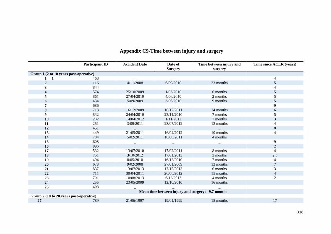
318
Appendix C9-Time between injury and surgery
Participant ID Accident Date Date of
Surgery
Time between injury and
surgery
Time since ACLR (years)
Group 1 (2 to 10 years post-operative)
1 1 468 _ _ _ 4
2 116 4/11/2008 6/09/2010 23 months 5
3 844 _ _ _ 4
4 574 25/10/2009 1/03/2010 6 months 5
5 861 27/04/2010 4/06/2010 2 months 5
6 434 5/09/2009 3/06/2010 9 months 5
7 686 _ _ _ 9
8 713 16/12/2009 16/12/2011 24 months 6
9 832 24/04/2010 23/11/2010 7 months 5
10 232 14/04/2012 1/11/2012 7 months 3
11 251 3/09/2011 23/07/2012 12 months 4
12 451 _ _ _ 8
13 449 21/05/2011 16/04/2012 10 months 4
14 704 5/02/2011 16/06/2011 4 months
15 608 _ _ _ 9
16 896 _ _ _ 2
17 532 13/07/2010 17/02/2011 8 months 4
18 751 3/10/2012 17/01/2013 3 months 2.5
19 494 8/05/2010 16/12/2010 7 months 4
20 673 9/02/2008 27/01/2009 12 months 7
21 837 13/07/2013 17/12/2013 6 months 3
22 711 30/04/2011 26/06/2012 15 months 4
23 701 10/08/2013 6/12/2013 4 months 2
24 255 23/05/2009 12/10/2010 16 months
25 408 _ _ _
Mean time between injury and surgery: 9.7 months
Group 2 (10 to 20 years post-operative)
27. 789 21/06/1997 19/01/1999 18 months 17
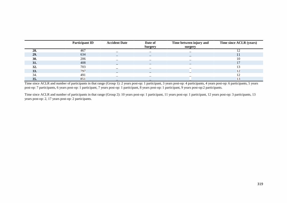
319
Participant ID Accident Date Date of
Surgery
Time between injury and
surgery
Time since ACLR (years)
28. 467 _ _ _ 12
29. 634 _ _ _ 11 30. 206 _ _ _ 10
31. 408 _ _ _ 17
32. 703 _ _ _ 13
33. 797 _ _ _ 12
34. 491 _ _ _ 12
35. 851 _ _ _ 13
Time since ACLR and number of participants in that range (Group 1): 2 years post-op: 1 participant, 3 years post-op: 4 participants, 4 years post-op: 6 participants, 5 years
post-op: 7 participants, 6 years post-op: 1 participant, 7 years post-op: 1 participant, 8 years post-op: 1 participant, 9 years post-op:2 participants.
Time since ACLR and number of participants in that range (Group 2): 10 years post-op: 1 participant, 11 years post-op: 1 participant, 12 years post-op: 3 participants, 13
years post-op: 2, 17 years post-op: 2 participants.
320
Appendix D1- Bracket
Rupture of anterior cruciate ligament (ACL) is one of the common injuries in sports. The
rupture of ACL commonly occurs in netball, skiing and other non-contact sports. Females are at
higher risk of injury because of anatomical and hormonal variations than males. Treatment
options include non-surgical treatment where participants go through a rehabilitation protocol
with the aim of restoring limb stability and function to pre-injury level. The other treatment
option is the reconstruction of the injured ligament with autograft and allograft which is
followed by rehabilitation protocol.
Outcomes after ACL reconstruction (ACLR) may vary among individuals and is affected by
factor such as the pre-injury level of sports, severity of injury, time between injury and surgery,
surgical technique, type of graft, psychosocial factors, self-efficacy of the participant and type
of rehabilitation protocol.
Incomplete recovery of thigh muscle strength and persistence of pain is reported by some of the
patients following surgery. Factors responsible for persistence of muscle weakness are still
being explored, and factors such as neuro-physiological mechanisms are reported for pain and
quadriceps or hamstring muscle atrophy. The treatment options to deal with the residual
atrophy and pain successfully are still fewer and this can be challenging for patients to deal
with. Muscle weakness may also be related to the graft site morbidity. Patients with patellar
tendon graft present with quadriceps weakness and pain during kneeling activities whereas
those with hamstring graft present with hamstring muscle weakness. Pain and major thigh
muscle weakness can influence the social and health-related quality of life. Pain can lead to
modifying or reducing level of activities. Problems related to returning to pre-injury level of
work are reported more during the initial 2-3 years after surgery and after that most of the
individuals are able to develop their own adapting strategies. Physiotherapists and surgeons
should aim to restore the participant to pre-injury level of activities.
Inability to achieve the pre-injury level of activities and confidence in the knee is the biggest
regret by majority of the participant following ACLR. Only 45% of the operated individuals are
able to return back pre-injury level of sports and activities. Fear of re-injury on the operated
side makes the participants to be selective for less risky and safe options when it comes to
going full out at sport. A sports psychologist should be the part of the rehabilitation team and
should try to restore the patient’s confidence in their knee to eradicate the fear of injury. Some
of the experienced and national level soccer players also express their concern about their

321
inability to go ‘full out’ as they used to before injury. Fear of re-injury and constant awareness
about the operated knee hinders the performance of the players.
Patients with ACLR can have some further consequences after surgery, one of them being at
risk of post-traumatic osteoarthritis years after reconstruction. Different underlying mechanisms
are believed to be responsible for this: for example, partial recovery of muscle strength or the
abnormal loading of the cartilage. Usually patients are aware of these consequences and are
also motivated to stick to the exercise regimen. Another consequence to ACLR is the risk of re-
injury to the ipsilateral and contralateral knee joint. Anatomical variations, ligament laxity and
abnormal kinematics and kinetics can make a participant prone to re-injury.
To summarize, overall, patients seem satisfied with their knee surgery although it has
influenced their family, social and professional life to a large extent. Patients are continuously
concerned about their knee during daily activities and have poor confidence in the knee along
with fear of re-injury. Hence, ACLR influences activity levels, health–related quality of life and
social life of the participant to differing extents.

322
Appendix D2- Additional participant quotes
Themes Subtheme Categories Quotes
1. Fear of re-
injury versus
confidence
continuum
Causes of fear of
injury
Fear of experiencing
injury pain
associated with the
initial injury again
“I’ve got no control whatsoever ice skating and I just feel like I would be at too much of a risk of
doing something to myself. I was a good enough skier beforehand that I’ve got the control to do it
whereas I’ve got no control whatsoever ice skating and I just feel like I would be at too much of a
risk of doing something to myself and it’s that knowing that yes, it is a possibility to do again
whether it would be the same knee or the other knee and, but I don’t want to do it again.” P1
“I don’t like running, I don’t like high impact sports, too scared to go back to skiing because I
know several people who had the operation and they re-injured their knee, when they had a crash,
so I am quite happy the way my knee is there, I don’t want to re-injure it, I keep away from
contact sports and skiing which is a shame but I am happy the way it is. It’s partly is the fear that I
will re-injure my knee. I am just worried that I will fall of my knee, that’s all…you know.” P3
“I just would never want to go through it again, I mean I know I could get through it but it’s, yeah
it buggered me up for a bit….” P6
Memory of inciting
injury movement
“…it kind of scares you ‘cause you know exactly how you’ve done it and how you could do it again
so like just trying to avoid doing a similar movement to what I did at the time I guess.” P4
Long rehabilitation
period and loss of
muscle strength
“So it’s definitely less weight on the leg with the reconstruction when it comes to anything using
my quads.” P1
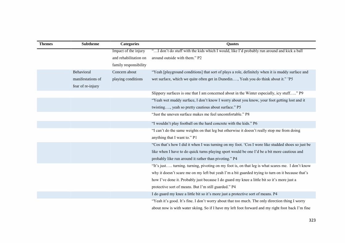
323
Themes Subtheme Categories Quotes
Impact of the injury
and rehabilitation on
family responsibility
“…I don’t do stuff with the kids which I would, like I’d probably run around and kick a ball
around outside with them.” P2
Behavioral
manifestations of
fear of re-injury
Concern about
playing conditions
“Yeah [playground conditions] that sort of plays a role, definitely when it is muddy surface and
wet surface, which we quite often get in Dunedin.…, Yeah you do think about it.” ’P5
Slippery surfaces is one that I am concerned about in the Winter especially, icy stuff…..” P9
“Yeah wet muddy surface, I don’t know I worry about you know, your foot getting lost and it
twisting…., yeah so pretty cautious about surface.” P5
“Just the uneven surface makes me feel uncomfortable.” P8
“I wouldn’t play football on the hard concrete with the kids.” P6
“I can’t do the same weights on that leg but otherwise it doesn’t really stop me from doing
anything that I want to.” P1
“Cos that’s how I did it when I was turning on my foot. ‘Cos I wore like studded shoes so just be
like when I have to do quick turns playing sport would be one I’d be a bit more cautious and
probably like run around it rather than pivoting.” P4
“It’s just….. turning. turning, pivoting on my foot is, on that leg is what scares me. I don’t know
why it doesn’t scare me on my left but yeah I’m a bit guarded trying to turn on it because that’s
how I’ve done it. Probably just because I do guard my knee a little bit so it’s more just a
protective sort of means. But I’m still guarded.” P4
I do guard my knee a little bit so it’s more just a protective sort of means. P4
“Yeah it’s good. It’s fine. I don’t worry about that too much. The only direction thing I worry
about now is with water skiing. So if I have my left foot forward and my right foot back I’m fine
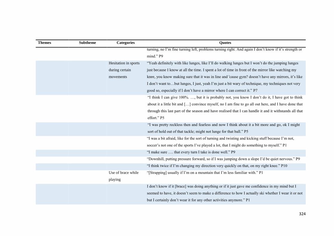
324
Themes Subtheme Categories Quotes
turning, no I’m fine turning left, problems turning right. And again I don’t know if it’s strength or
mind.” P9
Hesitation in sports
during certain
movements
“Yeah definitely with like lunges, like I’ll do walking lunges but I won’t do the jumping lunges
just because I know at all the time. I spent a lot of time in front of the mirror like watching my
knee, you know making sure that it was in line and 'cause gym? doesn’t have any mirrors, it’s like
I don’t want to…but lunges, I just, yeah I’m just a bit wary of technique, my techniques not very
good so, especially if I don’t have a mirror where I can correct it.” P7
“I think I can give 100%. …, but it is probably not, you know I don’t do it, I have got to think
about it a little bit and […] convince myself, no I am fine to go all out here, and I have done that
through this last part of the season and have realised that I can handle it and it withstands all that
effort.” P5
“I was pretty reckless then and fearless and now I think about it a bit more and go, ok I might
sort of hold out of that tackle; might not lunge for that ball.” P5
“I was a bit afraid, like for the sort of turning and twisting and kicking stuff because I’m not,
soccer’s not one of the sports I’ve played a lot, that I might do something to myself.” P1
“I make sure …. that every turn I take is done well.” P9
“Downhill, putting pressure forward, so if I was jumping down a slope I’d be quiet nervous.” P9
“I think twice if I’m changing my direction very quickly on that, on my right knee.” P10
Use of brace while
playing
“[Strapping] usually if I’m on a mountain that I’m less familiar with.” P1
I don’t know if it [brace] was doing anything or if it just gave me confidence in my mind but I
seemed to have, it doesn’t seem to make a difference to how I actually ski whether I wear it or not
but I certainly don’t wear it for any other activities anymore.” P1
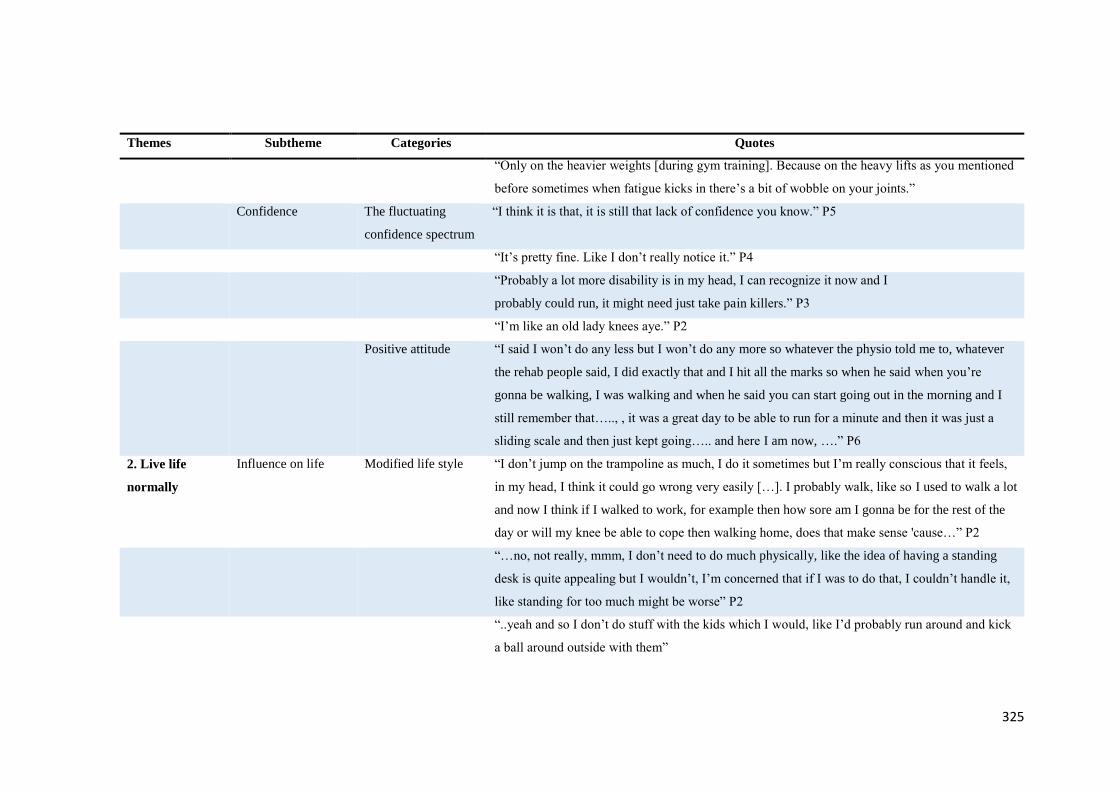
325
Themes Subtheme Categories Quotes
“Only on the heavier weights [during gym training]. Because on the heavy lifts as you mentioned
before sometimes when fatigue kicks in there’s a bit of wobble on your joints.”
Confidence The fluctuating
confidence spectrum
“I think it is that, it is still that lack of confidence you know.” P5
“It’s pretty fine. Like I don’t really notice it.” P4
“Probably a lot more disability is in my head, I can recognize it now and I
probably could run, it might need just take pain killers.” P3
“I’m like an old lady knees aye.” P2
Positive attitude “I said I won’t do any less but I won’t do any more so whatever the physio told me to, whatever
the rehab people said, I did exactly that and I hit all the marks so when he said when you’re
gonna be walking, I was walking and when he said you can start going out in the morning and I
still remember that….., , it was a great day to be able to run for a minute and then it was just a
sliding scale and then just kept going….. and here I am now, ….” P6
2. Live life
normally
Influence on life Modified life style “I don’t jump on the trampoline as much, I do it sometimes but I’m really conscious that it feels,
in my head, I think it could go wrong very easily […]. I probably walk, like so I used to walk a lot
and now I think if I walked to work, for example then how sore am I gonna be for the rest of the
day or will my knee be able to cope then walking home, does that make sense 'cause…” P2
“…no, not really, mmm, I don’t need to do much physically, like the idea of having a standing
desk is quite appealing but I wouldn’t, I’m concerned that if I was to do that, I couldn’t handle it,
like standing for too much might be worse” P2
“..yeah and so I don’t do stuff with the kids which I would, like I’d probably run around and kick
a ball around outside with them”
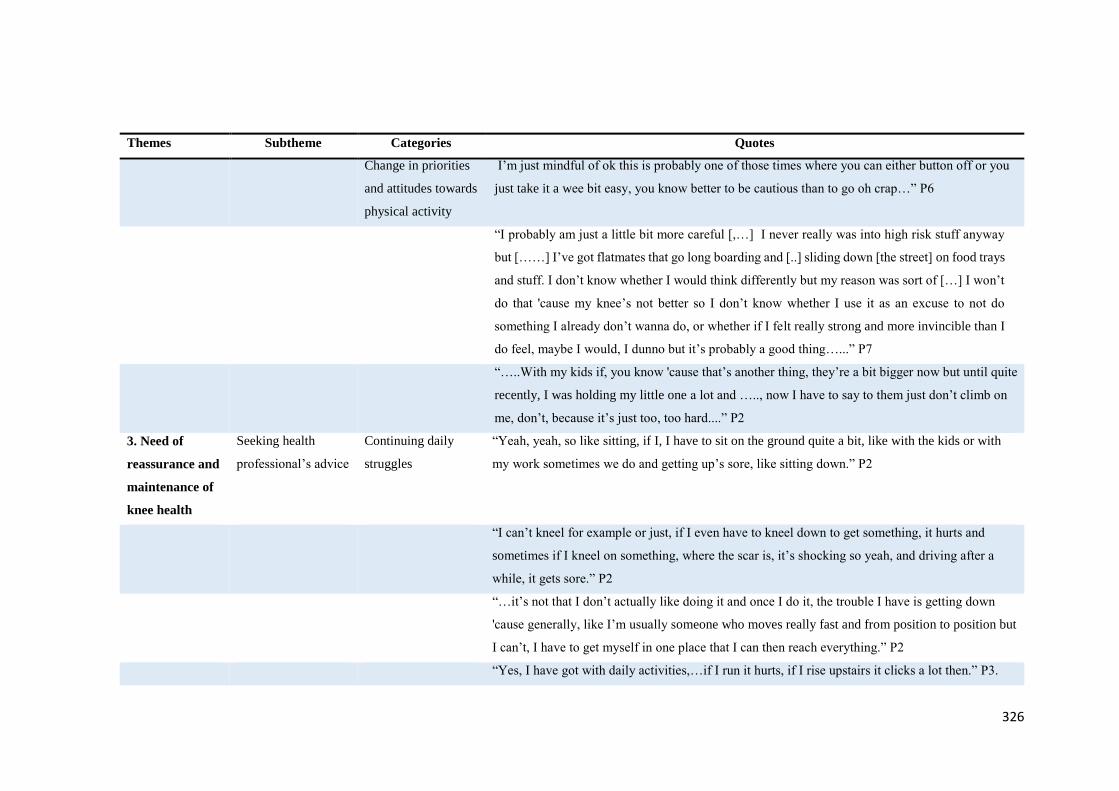
326
Themes Subtheme Categories Quotes
Change in priorities
and attitudes towards
physical activity
I’m just mindful of ok this is probably one of those times where you can either button off or you
just take it a wee bit easy, you know better to be cautious than to go oh crap…” P6
“I probably am just a little bit more careful [,…] I never really was into high risk stuff anyway
but [……] I’ve got flatmates that go long boarding and [..] sliding down [the street] on food trays
and stuff. I don’t know whether I would think differently but my reason was sort of […] I won’t
do that 'cause my knee’s not better so I don’t know whether I use it as an excuse to not do
something I already don’t wanna do, or whether if I felt really strong and more invincible than I
do feel, maybe I would, I dunno but it’s probably a good thing…...” P7
“…..With my kids if, you know 'cause that’s another thing, they’re a bit bigger now but until quite
recently, I was holding my little one a lot and ….., now I have to say to them just don’t climb on
me, don’t, because it’s just too, too hard....” P2
3. Need of
reassurance and
maintenance of
knee health
Seeking health
professional’s advice
Continuing daily
struggles
“Yeah, yeah, so like sitting, if I, I have to sit on the ground quite a bit, like with the kids or with
my work sometimes we do and getting up’s sore, like sitting down.” P2
“I can’t kneel for example or just, if I even have to kneel down to get something, it hurts and
sometimes if I kneel on something, where the scar is, it’s shocking so yeah, and driving after a
while, it gets sore.” P2
“…it’s not that I don’t actually like doing it and once I do it, the trouble I have is getting down
'cause generally, like I’m usually someone who moves really fast and from position to position but
I can’t, I have to get myself in one place that I can then reach everything.” P2
“Yes, I have got with daily activities,…if I run it hurts, if I rise upstairs it clicks a lot then.” P3.
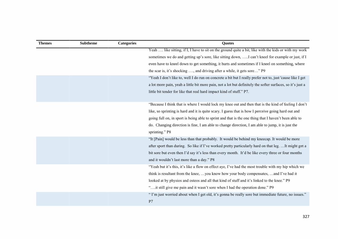
327
Themes Subtheme Categories Quotes
Yeah …. like sitting, if I, I have to sit on the ground quite a bit, like with the kids or with my work
sometimes we do and getting up’s sore, like sitting down, …..I can’t kneel for example or just, if I
even have to kneel down to get something, it hurts and sometimes if I kneel on something, where
the scar is, it’s shocking …., and driving after a while, it gets sore…” P9
“Yeah I don’t like to, well I do run on concrete a bit but I really prefer not to, just 'cause like I get
a lot more pain, yeah a little bit more pain, not a lot but definitely the softer surfaces, so it’s just a
little bit tender for like that real hard impact kind of stuff.” P7.
“Because I think that is where I would lock my knee out and then that is the kind of feeling I don’t
like, so sprinting is hard and it is quite scary. I guess that is how I perceive going hard out and
going full on, in sport is being able to sprint and that is the one thing that I haven’t been able to
do. Changing direction is fine, I am able to change direction, I am able to jump, it is just the
sprinting.” P8
“It [Pain] would be less than that probably. It would be behind my kneecap. It would be more
after sport than during. So like if I’ve worked pretty particularly hard on that leg. …It might get a
bit sore but even then I’d say it’s less than every month. It’d be like every three or four months
and it wouldn’t last more than a day.” P8
“Yeah but it’s this, it’s like a flow on effect aye, I’ve had the most trouble with my hip which we
think is resultant from the knee, …you know how your body compensates, …and I’ve had it
looked at by physios and osteos and all that kind of stuff and it’s linked to the knee.” P9
“….it still give me pain and it wasn’t sore when I had the operation done.” P9
“ I’m just worried about when I get old, it’s gonna be really sore but immediate future, no issues.”
P7
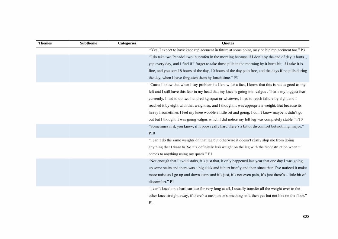
328
Themes Subtheme Categories Quotes
“Yea, I expect to have knee replacement in future at some point, may be hip replacement too.” P3
“I do take two Panadol two ibuprofen in the morning because if I don’t by the end of day it hurts..,
yep every day, and I find if I forget to take those pills in the morning by it hurts bit, if I take it is
fine, and you sort 18 hours of the day, 10 hours of the day pain free, and the days if no pills during
the day, when I have forgotten them by lunch time.” P3
“Cause I know that when I say problem its I know for a fact, I know that this is not as good as my
left and I still have this fear in my head that my knee is going into valgus . That’s my biggest fear
currently. I had to do two hundred kg squat or whatever, I had to reach failure by eight and I
reached it by eight with that weight so, and I thought it was appropriate weight. But because its
heavy I sometimes I feel my knee wobble a little bit and going, I don’t know maybe it didn’t go
out but I thought it was going valgus which I did notice my left leg was completely stable.” P10
“Sometimes if it, you know, if it pops really hard there’s a bit of discomfort but nothing, major.”
P10
“I can’t do the same weights on that leg but otherwise it doesn’t really stop me from doing
anything that I want to. So it’s definitely less weight on the leg with the reconstruction when it
comes to anything using my quads.” P1
“Not enough that I avoid stairs, it’s just that, it only happened last year that one day I was going
up some stairs and there was a big click and it hurt briefly and then since then I’ve noticed it make
more noise as I go up and down stairs and it’s just, it’s not even pain, it’s just there’s a little bit of
discomfort.” P1
“I can’t kneel on a hard surface for very long at all, I usually transfer all the weight over to the
other knee straight away, if there’s a cushion or something soft, then yes but not like on the floor.”
P1
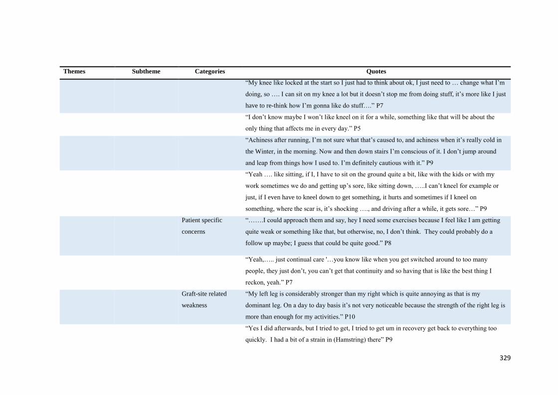
329
Themes Subtheme Categories Quotes
“My knee like locked at the start so I just had to think about ok, I just need to … change what I’m
doing, so …. I can sit on my knee a lot but it doesn’t stop me from doing stuff, it’s more like I just
have to re-think how I’m gonna like do stuff….” P7
“I don’t know maybe I won’t like kneel on it for a while, something like that will be about the
only thing that affects me in every day.” P5
“Achiness after running, I’m not sure what that’s caused to, and achiness when it’s really cold in
the Winter, in the morning. Now and then down stairs I’m conscious of it. I don’t jump around
and leap from things how I used to. I’m definitely cautious with it.” P9
“Yeah …. like sitting, if I, I have to sit on the ground quite a bit, like with the kids or with my
work sometimes we do and getting up’s sore, like sitting down, …..I can’t kneel for example or
just, if I even have to kneel down to get something, it hurts and sometimes if I kneel on
something, where the scar is, it’s shocking …., and driving after a while, it gets sore…” P9
Patient specific
concerns
“…….I could approach them and say, hey I need some exercises because I feel like I am getting
quite weak or something like that, but otherwise, no, I don’t think. They could probably do a
follow up maybe; I guess that could be quite good.” P8
“Yeah,….. just continual care '…you know like when you get switched around to too many
people, they just don’t, you can’t get that continuity and so having that is like the best thing I
reckon, yeah.” P7
Graft-site related
weakness
“My left leg is considerably stronger than my right which is quite annoying as that is my
dominant leg. On a day to day basis it’s not very noticeable because the strength of the right leg is
more than enough for my activities.” P10
“Yes I did afterwards, but I tried to get, I tried to get um in recovery get back to everything too
quickly. I had a bit of a strain in (Hamstring) there” P9
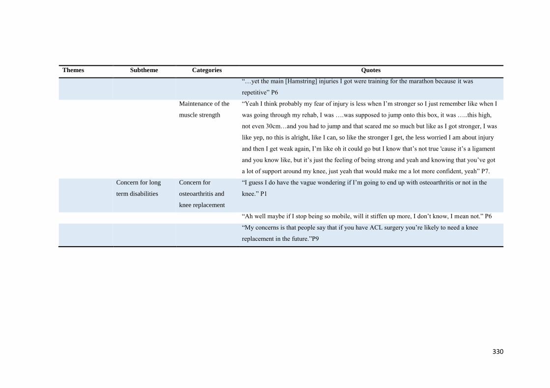
330
Themes Subtheme Categories Quotes
“…yet the main [Hamstring] injuries I got were training for the marathon because it was
repetitive” P6
Maintenance of the
muscle strength
“Yeah I think probably my fear of injury is less when I’m stronger so I just remember like when I
was going through my rehab, I was ….was supposed to jump onto this box, it was …..this high,
not even 30cm…and you had to jump and that scared me so much but like as I got stronger, I was
like yep, no this is alright, like I can, so like the stronger I get, the less worried I am about injury
and then I get weak again, I’m like oh it could go but I know that’s not true 'cause it’s a ligament
and you know like, but it’s just the feeling of being strong and yeah and knowing that you’ve got
a lot of support around my knee, just yeah that would make me a lot more confident, yeah” P7.
Concern for long
term disabilities
Concern for
osteoarthritis and
knee replacement
“I guess I do have the vague wondering if I’m going to end up with osteoarthritis or not in the
knee.” P1
“Ah well maybe if I stop being so mobile, will it stiffen up more, I don’t know, I mean not.” P6
“My concerns is that people say that if you have ACL surgery you’re likely to need a knee
replacement in the future.”P9

331
Appendix D3- Data check with the participants for qualitative study
Patients’ perspectives of the outcomes of anterior cruciate ligament (ACL) reconstruction of the knee.
“Living with the ACL-reconstructed knee.”
Mandeep Kaur, Ph.D. Student
Dr Daniel Ribeiro, Senior Lecturer
Prof. Jean-Claude Theis, Dunedin Hospital
Assoc Prof Kate Webster, La Trobe University, Australia
Dr Gisela Sole, Senior Lecturer
Centre for Health, Activity and Rehabilitation Research
School of Physiotherapy
University of Otago
We thank you for participating in this research study and for the information that you shared with us.
We hereby would like to provide you with a summary of the results and invite you to respond to us if
there is any further aspect you would like to highlight, or if you feel the summary below does not
portray your experiences.
Why this study was conducted.
We were interested to explore the experiences of individuals who had undergone an anterior cruciate
ligament (ACL) reconstruction within the past 2 to 10 years. We focussed on the impact of the injury
and surgery on the individual’s lives and overall quality of life, from your perspective. A further aim
of this study was to explore if individuals who had undergone such surgery have any concerns related
to the knee which we, as health care professionals, should be considering during usual clinical
consultations. This information would allow us to understand the patients’ perspectives and provide
more individualised approaches to rehabilitation and management of long-term knee health.
Results of the study
We interviewed ten volunteers and the recording of the interviews were transcribed. The researchers
then analysed the transcriptions, and defined main themes and sub-themes that emanated from them.
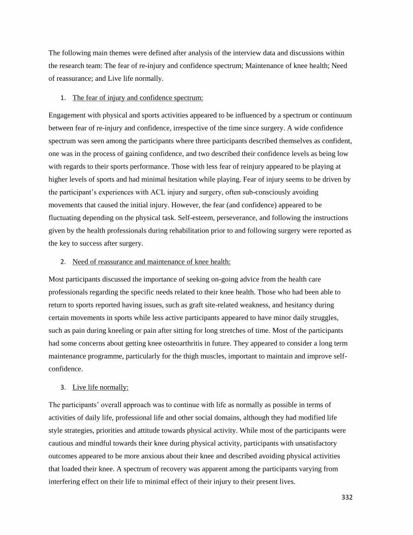
332
The following main themes were defined after analysis of the interview data and discussions within
the research team: The fear of re-injury and confidence spectrum; Maintenance of knee health; Need
of reassurance; and Live life normally.
1. The fear of injury and confidence spectrum:
Engagement with physical and sports activities appeared to be influenced by a spectrum or continuum
between fear of re-injury and confidence, irrespective of the time since surgery. A wide confidence
spectrum was seen among the participants where three participants described themselves as confident,
one was in the process of gaining confidence, and two described their confidence levels as being low
with regards to their sports performance. Those with less fear of reinjury appeared to be playing at
higher levels of sports and had minimal hesitation while playing. Fear of injury seems to be driven by
the participant’s experiences with ACL injury and surgery, often sub-consciously avoiding
movements that caused the initial injury. However, the fear (and confidence) appeared to be
fluctuating depending on the physical task. Self-esteem, perseverance, and following the instructions
given by the health professionals during rehabilitation prior to and following surgery were reported as
the key to success after surgery.
2. Need of reassurance and maintenance of knee health:
Most participants discussed the importance of seeking on-going advice from the health care
professionals regarding the specific needs related to their knee health. Those who had been able to
return to sports reported having issues, such as graft site-related weakness, and hesitancy during
certain movements in sports while less active participants appeared to have minor daily struggles,
such as pain during kneeling or pain after sitting for long stretches of time. Most of the participants
had some concerns about getting knee osteoarthritis in future. They appeared to consider a long term
maintenance programme, particularly for the thigh muscles, important to maintain and improve self-
confidence.
3. Live life normally:
The participants’ overall approach was to continue with life as normally as possible in terms of
activities of daily life, professional life and other social domains, although they had modified life
style strategies, priorities and attitude towards physical activity. While most of the participants were
cautious and mindful towards their knee during physical activity, participants with unsatisfactory
outcomes appeared to be more anxious about their knee and described avoiding physical activities
that loaded their knee. A spectrum of recovery was apparent among the participants varying from
interfering effect on their life to minimal effect of their injury to their present lives.
333
The results of this study adds towards health care professionals’ understand of the patients’ specific
needs and sports-related concerns following ACL injury and surgery. The results show that, overall,
participants were motivated to live their life normally and had minimal pain and knee-related
symptoms. However, they continued to experience sporadic fear of re-injury and low confidence
levels during very specific sporting or physical activities. Maintaining the thigh muscle strength were
seen as a self-strategy to maintain confidence and knee health, and graft-site related weakness and the
possibility of future symptoms were some of the patients’ concerns.
The results of the study will be published in the thesis written by Mandeep Kaur for her Ph.D. degree.
We will also be submitting a manuscript for publication in an international journal.
Please respond to Mandeep Kaur at [email protected] before 1st March, 2017.
Thank you once again for helping us with this research.
Yours Sincerely,
Mandeep Kaur
334
Appendix E1- Visual 3D algorithm for determining gait events
In trials:
- Odd no. - going upstairs
- Even no.- downstairs
Axis and movements
- X axis- flexion
- Y axis- medial-lateral movement
- Z axis- rotation
Pipelines:
- 1 Model; Loading of trials, analyzing gait phases
- 2 Model: Moments
- 3 Model: Getting all the data and saving it.
Sequence: Pipeline 12 model
1. Open the required folder with C3D files; it should have different files for barefoot and shoe
conditions.
2. Open visual 3D.
3. Go to pipeline and click “open”.
4. Open the folder to be processed; it should have one static and two dynamic files.
5. Open model (12model).
6. Execute pipeline.
7. Confirm folder (ok).
8. Enter weight/height of the participant
9. Select all by selecting top one, press “Shift” while selecting the lowermost one.
10. Zero errors = acceptable
11. Go to workspace; check individual trials and “ticks” if they are representing the desired trial.
12. Go to Signals and events
13. Open target folder
14. Go to filtered
15. Click on right trial
16. Right click (for left foot)
17. Graph (Z)
18. Right foot
19. Open events label
20. Go to original and highlight the phases for example; LHS, LTo, LToff; for right foot RHS, RTO,
RTOFF
21. Go through each trial and make sure two toe off are there and are at right place.
22. Run the trail and see if it is at right place
23. If not; Open E’ and chose right frame if not basically right
24. Got through all trials
25. Once checked all, open pipeline and replace
Sequence: Pipeline 22model
1. Execute pipeline
2. Confirm folder
3. Zero errors i.e. acceptable
4. Un-highlight previous selections
5. Highlight moments
6. Right click on graphs and remove all previous graphs
7. Go to link model- based folder
335
8. Open processed> knee moments
9. Graph X, Y & Z > new graph
10. Go through all graphs
11. “Remove graphs” when to do for other limb
12. Again get new graphs from link-model based> X-Y-Z- new graph
13. If all good > open pipeline> Replace
Sequence: Pipeline 32 matrices model
1. Click open > it will crash if everything is not eight. It has to have 2 toe-offs for each trial
2. If it worked it would create ‘metric folder’.
Note: CMO files will be there, which can be used if we left the processing in between, than go to visual 3D,
open COM file and resume the working from where you left.
336
Appendix E2- MATLAB code to plot the linear envelops for angles and
moments
Step 1
%% This script reads in, labels and reorganises data exported from Visual3D
% Written by Peter Lamb
% 27/06/2017
%% Define paths and directories
% Clear the command line and workspace and close all figures
clc; clear variables; close all;
% Identify file directories Enter directory path here, similar to:
root = '/Users/peterlamb/Documents/People/Mandeep Kaur';
dataPath = [root '/data/Mat/'];
%% Set up metadata
% How many .mat files are there in the data path? Should correspond to
% number of participants
matFiles = dir([dataPath '*.mat']);
nParticipants = length(matFiles);
% Identify group: for ACL which leg is injured; for control which leg is
% "side 1".
load([root '/m-files/participant_information_ht_wt.mat'])
% Pre-allocate memory for dataset:
% 1-3 stepping knee angle XYZ, 4-6 stepping knee moment XYZ
nVariables = 6;
% if ACL, 1=injured, 2=non-injured, if control, 1=left, 2=right.
nLegs = 2;
max_nTrials = 10;
nDirections = 2;
max_nFrames = 2000;
stairD = nan(nParticipants, nLegs, nDirections, nVariables, ...
max_nTrials, max_nFrames);
%% Reorganise data
% Loop through each participant's .mat file and organise into the array.
parInfo = zeros(1000,4);
parCnt = 1;
samp_rate = nan(nParticipants,1);
fp_samp_rate = 1050;
for iPar = 1 : nParticipants
% Pick the file name of the ith file
337
fName = matFiles(iPar).name;
% Load the ith file
load([dataPath fName])
% Determine frame rate and save
samp_rate(iPar) = FRAME_RATE {1,1}(1,1);
% Fix variable naming inconsistencies
if exist('kneeMomentsR', 'var')
KneeMomentsR = kneeMomentsR;
clear kneeMomentsR
end
% Create Participant ID variable
id = str2double(fName(1:end-4));
% Find row index to ith participant ID in labels
labelRow = find([labels {:,1}] == id);
% Record missing participants and jump to next if found
if isempty(labelRow)
parInfo(parCnt,:) = [iPar id NaN 999];
parCnt = parCnt + 1;
continue
end
% Create Participant 'group' variable
group = labels{labelRow, 2};
if strcmp(group, 'Control')
group = 0;
else
group = 1;
end
weight = labels{labelRow, 4};
height = labels{labelRow, 5};
% Create participant leg variable, group=ACL leg represents injured
% leg, if group=control, leg represents side 1 leg.
parLeg = labels{labelRow, 3};
% Find the number of trials so that we can loop through the trials
nTrials = length(FILE_NAME);
% Start counters
338
ascCounterL = 1; ascCounterR = 1;
dscCounterL = 1; dscCounterR = 1;
for iTrial = 1 : nTrials
% Read trial number and leg from FILE_NAME (MATLAB orders numbers
% differently e.g. 1, 10, 2, 3, ...)
scanned = textscan(FILE_NAME{iTrial,1}, '%s', 'delimiter', '\');
lastpart = textscan(scanned{1,1}{end,1}, '%s', 'delimiter', '_');
trialLeg = lastpart{1,1}{2,1}(1);
trialNum = str2double(lastpart{1,1}{2,1}(2:end-4));
% Check that required data are available
if (strcmp(trialLeg, 'L') && (isempty(KneeAngL{iTrial,1}) || ...
isempty(KneeMomentsL{iTrial,1}))) || ...
(strcmp(trialLeg, 'L') && (isempty(KneeAngR{iTrial,1}) || ...
isempty(KneeMomentsR{iTrial,1})))
parInfo(parCnt, :) = [iPar id group trialNum];
parCnt = parCnt + 1;
continue
end
% Determine number of frames
nFrames = length(KneeAngL{iTrial,1});
% Determine which leg, 1 if stepping leg is injured (or side 1), 2
% if not
if (strcmp(parLeg, 'l') && strcmp(trialLeg, 'L')) || ...
(strcmp(parLeg, 'r') && strcmp(trialLeg, 'R'))
legInd = 1;
else
legInd = 2;
end
% Determine ascending or descending
if mod(trialNum,2)==0
directionInd = 2;
else
directionInd = 1;
end
% Identify start and end points using force data
fData = FP2{iTrial,1}(:,1);
fpFrames = length(fData);
old_time = (0:fpFrames-1)/(fpFrames-1);
new_time = (0:nFrames-1)/(nFrames-1);
fDresamp = interp1(old_time, fData, new_time, 'pchip');
nonzero = find(fDresamp);
339
first = nonzero(1);
last = nonzero(length(nonzero));
new_len = 1:(last-first)+1;
% Downsample to common frame rate with mocap
% If ascending, use the ascending trial counter - ascCounter
if directionInd==1
% If the left leg is being, start with left knee variables and
% transform Y and Z
if strcmp(trialLeg, 'L')
stairD(iPar, legInd, directionInd, 1, ascCounterL, new_len)...
= KneeAngL{iTrial,1}(first:last,1);
stairD(iPar, legInd, directionInd, 2, ascCounterL, new_len)...
= KneeAngL{iTrial,1}(first:last,2)*-1;
stairD(iPar, legInd, directionInd, 3, ascCounterL, new_len)...
= KneeAngL{iTrial,1}(first:last,3)*-1;
stairD(iPar, legInd, directionInd, 4, ascCounterL, new_len)...
= KneeMomentsL{iTrial,1}(first:last,1)/height/weight;
stairD(iPar, legInd, directionInd, 5, ascCounterL, new_len)...
= KneeMomentsL{iTrial,1}(first:last,2)*-1/height/weight;
stairD(iPar, legInd, directionInd, 6, ascCounterL, new_len)...
= KneeMomentsL{iTrial,1}(first:last,3)*-1/height/weight;
% Else if the right leg is being used, start with the right
% knee variables
else
stairD(iPar, legInd, directionInd, 1, ascCounterR, new_len)...
= KneeAngR{iTrial,1}(first:last,1);
stairD(iPar, legInd, directionInd, 2, ascCounterR, new_len)...
= KneeAngR{iTrial,1}(first:last,2);
stairD(iPar, legInd, directionInd, 3, ascCounterR, new_len)...
= KneeAngR{iTrial,1}(first:last,3);
stairD(iPar, legInd, directionInd, 4, ascCounterR, new_len)...
= KneeMomentsR{iTrial,1}(first:last,1)/height/weight;
stairD(iPar, legInd, directionInd, 5, ascCounterR, new_len)...
= KneeMomentsR{iTrial,1}(first:last,2)/height/weight;
stairD(iPar, legInd, directionInd, 6, ascCounterR, new_len)...
= KneeMomentsR{iTrial,1}(first:last,3)/height/weight;
end
% Else if not ascending, use the descending counter -
% dscCounter
else
% If the left leg is being, start with left knee variables and
% transform Y and Z
if strcmp(trialLeg, 'L')
stairD(iPar, legInd, directionInd, 1, dscCounterL, new_len)...
340
= KneeAngL{iTrial,1}(first:last,1);
stairD(iPar, legInd, directionInd, 2, dscCounterL, new_len)...
= KneeAngL{iTrial,1}(first:last,2)*-1;
stairD(iPar, legInd, directionInd, 3, dscCounterL, new_len)...
= KneeAngL{iTrial,1}(first:last,3)*-1;
stairD(iPar, legInd, directionInd, 4, dscCounterL, new_len)...
= KneeMomentsL{iTrial,1}(first:last,1)/height/weight;
stairD(iPar, legInd, directionInd, 5, dscCounterL, new_len)...
= KneeMomentsL{iTrial,1}(first:last,2)*-1/height/weight;
stairD(iPar, legInd, directionInd, 6, dscCounterL, new_len)...
= KneeMomentsL{iTrial,1}(first:last,3)*-1/height/weight;
% Else if the right leg is being used, start with the right
% knee variables
else
stairD(iPar, legInd, directionInd, 1, dscCounterR, new_len)...
= KneeAngR{iTrial,1}(first:last,1);
stairD(iPar, legInd, directionInd, 2, dscCounterR, new_len)...
= KneeAngR{iTrial,1}(first:last,2);
stairD(iPar, legInd, directionInd, 3, dscCounterR, new_len)...
= KneeAngR{iTrial,1}(first:last,3);
stairD(iPar, legInd, directionInd, 4, dscCounterR, new_len)...
= KneeMomentsR{iTrial, 1}(first:last,1)/height/weight;
stairD(iPar, legInd, directionInd, 5, dscCounterR, new_len)...
= KneeMomentsR{iTrial, 1}(first:last,2)/height/weight;
stairD(iPar, legInd, directionInd, 6, dscCounterR, new_len)...
= KneeMomentsR{iTrial, 1}(first:last,3)/height/weight;
end
end
% Increase appropriate counter
if directionInd==1 && strcmp(trialLeg, 'L')
ascCounterL = ascCounterL + 1;
elseif directionInd==1 && strcmp(trialLeg, 'R')
ascCounterR = ascCounterR + 1;
elseif directionInd==2 && strcmp(trialLeg, 'L')
dscCounterL = dscCounterL + 1;
else
dscCounterR = dscCounterR + 1;
end
end
parInfo(parCnt,:) = [iPar id group 0];
parCnt = parCnt + 1;
end
% Check for unique sampling rate values, if consistent across experiment

341
% proceed to save variables to file
samp_rate = unique(samp_rate);
if length(samp_rate) > 1
error('Inconsistent sampling rates!')
end
% Save variabels to .mat file
save([root '/data/step1_reorganised_trimmed_data.mat'], 'stairD', ...
'parInfo', 'samp_rate', 'labels', 'id')
Step 2
%% This script loads reorganised data and processes them
% Written by Peter Lamb
% 03/07/2017
%% Define paths and directories
% Clear the command line and workspace and close all figures
clc; clear variables; close all;
% Identify file directories
% Identify file directories Enter directory path here, similar to:
root = '/Users/peterlamb/Documents/People/Mandeep Kaur';
dataPath = [root '/data/'];
%% Set up metadata
variableNames = {'Knee angle (X)'; 'Knee angle (Y)'; 'Knee angle (Z)';...
'Knee moment (X)'; 'Knee moment (Y)'; 'Knee moment (Z)'};
directionNames = {'Asc'; 'Dsc'};
% Load data
load([dataPath 'step1_reorganised_trimmed_data.mat'])
% Redefine for looping:
% 1-3 stepping knee angle XYZ, 4-6 stepping knee moment XYZ
nVariables = 6;
% 1=ACL, 2=control
nGroups = 2;
% if ACL, 1=injured, 2=non-injured, if control, 1=left, 2=right.
nLegs = 2;
max_nTrials = 10;
nDirections = 2;
max_nFrames = 2000;
% stairD dimensions: 1-participant, 2-experimental group, 3-experimental
% leg, 4-step direction, 5-variable, 6-trial, 7-frame
nParticipants = size(stairD,1);
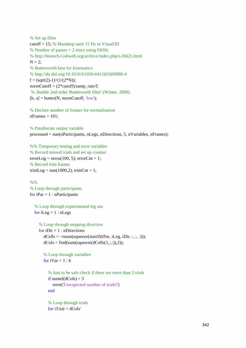
342
% Set up filter
cutoff = 15; % Mandeep used 15 Hz in Visual3D
% Number of passes = 2 since using filtfilt:
% http://biomch-l.isbweb.org/archive/index.php/t-26625.html
N = 2;
% Butterworth best for kinematics
% http://dx.doi.org/10.1016/S1050-6411(03)00080-4
f = (sqrt(2)-1)^(1/(2*N));
normCutoff = (2*cutoff)/samp_rate/f;
% 'double 2nd order Butterworth filter' (Winter, 2009)
[b, a] = butter(N, normCutoff, 'low');
% Declare number of frames for normalisation
nFrames = 101;
% Preallocate output variable
processed = nan(nParticipants, nLegs, nDirections, 5, nVariables, nFrames);
%% Temporary testing and error variables
% Record missed trials and set up counter
errorLog = zeros(100, 5); errorCnt = 1;
% Record trim frames
trimLog = nan(1000,2); trimCnt = 1;
%%
% Loop through participants
for iPar = 1 : nParticipants
% Loop through experimental leg use
for iLeg = 1 : nLegs
% Loop through stepping direction
for iDir = 1 : nDirections
dCells = ~isnan(squeeze(stairD(iPar, iLeg, iDir, :, :, :)));
dCols = find(sum(squeeze(dCells(1,:,:)),2));
% Loop through variables
for iVar = 1 : 6
% Just to be safe check if there are more than 5 trials
if numel(dCols) > 5
error('Unexpected number of trials!')
end
% Loop through trials
for iTrial = dCols'
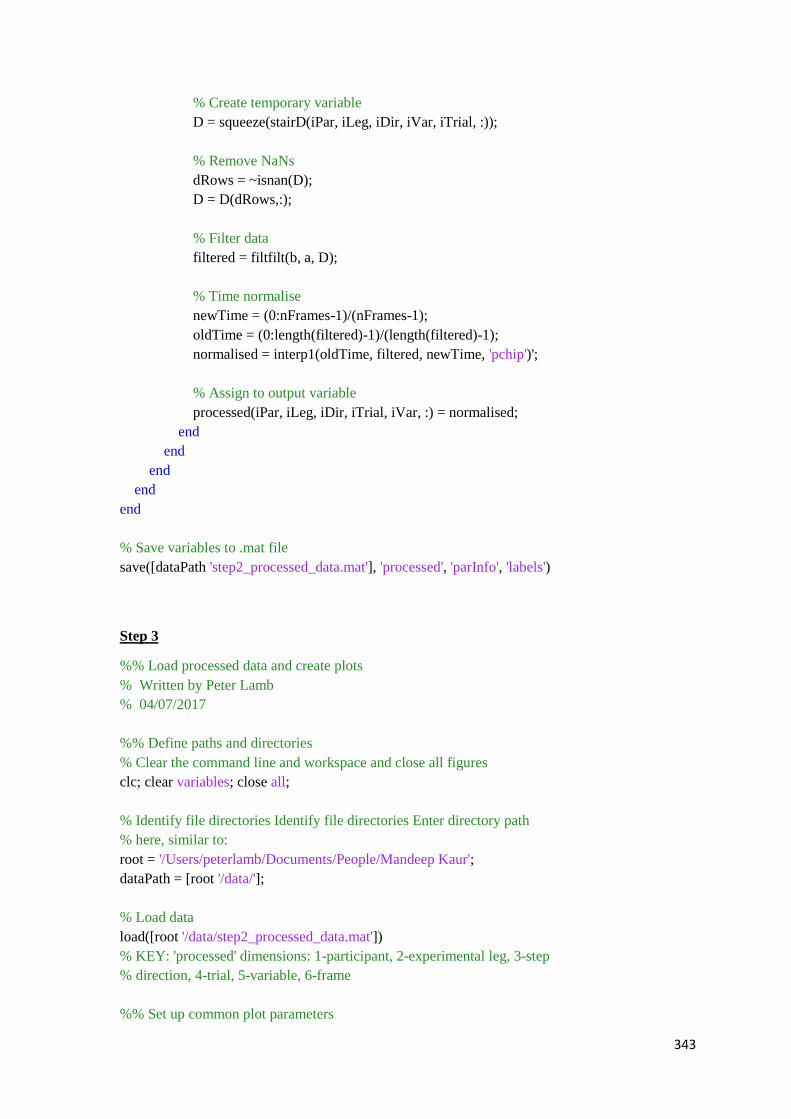
343
% Create temporary variable
D = squeeze(stairD(iPar, iLeg, iDir, iVar, iTrial, :));
% Remove NaNs
dRows = ~isnan(D);
D = D(dRows,:);
% Filter data
filtered = filtfilt(b, a, D);
% Time normalise
newTime = (0:nFrames-1)/(nFrames-1);
oldTime = (0:length(filtered)-1)/(length(filtered)-1);
normalised = interp1(oldTime, filtered, newTime, 'pchip')';
% Assign to output variable
processed(iPar, iLeg, iDir, iTrial, iVar, :) = normalised;
end
end
end
end
end
% Save variables to .mat file
save([dataPath 'step2_processed_data.mat'], 'processed', 'parInfo', 'labels')
Step 3
%% Load processed data and create plots
% Written by Peter Lamb
% 04/07/2017
%% Define paths and directories
% Clear the command line and workspace and close all figures
clc; clear variables; close all;
% Identify file directories Identify file directories Enter directory path
% here, similar to:
root = '/Users/peterlamb/Documents/People/Mandeep Kaur';
dataPath = [root '/data/'];
% Load data
load([root '/data/step2_processed_data.mat'])
% KEY: 'processed' dimensions: 1-participant, 2-experimental leg, 3-step
% direction, 4-trial, 5-variable, 6-frame
%% Set up common plot parameters
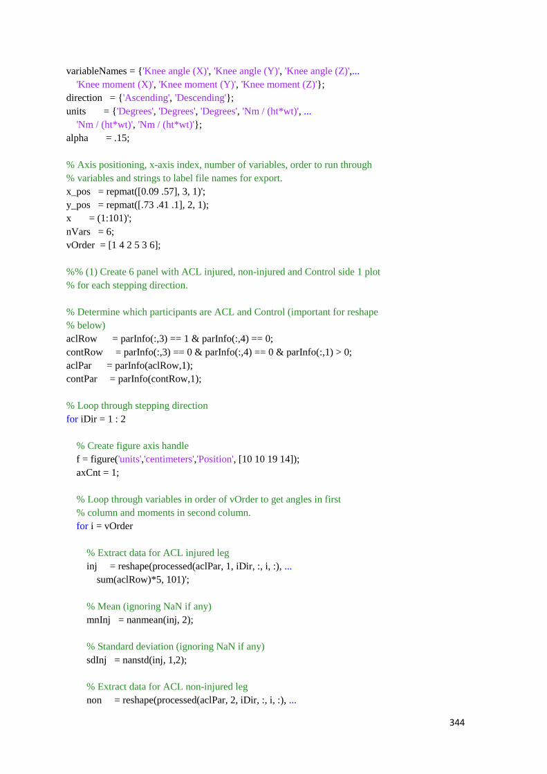
344
variableNames = {'Knee angle (X)', 'Knee angle (Y)', 'Knee angle (Z)',...
'Knee moment (X)', 'Knee moment (Y)', 'Knee moment (Z)'};
direction = {'Ascending', 'Descending'};
units = {'Degrees', 'Degrees', 'Degrees', 'Nm / (ht*wt)', ...
'Nm / (ht*wt)', 'Nm / (ht*wt)'};
alpha = .15;
% Axis positioning, x-axis index, number of variables, order to run through
% variables and strings to label file names for export.
x_pos = repmat([0.09 .57], 3, 1)';
y_pos = repmat([.73 .41 .1], 2, 1);
x = (1:101)';
nVars = 6;
vOrder = [1 4 2 5 3 6];
%% (1) Create 6 panel with ACL injured, non-injured and Control side 1 plot
% for each stepping direction.
% Determine which participants are ACL and Control (important for reshape
% below)
aclRow = parInfo(:,3) == 1 & parInfo(:,4) == 0;
contRow = parInfo(:,3) == 0 & parInfo(:,4) == 0 & parInfo(:,1) > 0;
aclPar = parInfo(aclRow,1);
contPar = parInfo(contRow,1);
% Loop through stepping direction
for iDir = 1 : 2
% Create figure axis handle
f = figure('units','centimeters','Position', [10 10 19 14]);
axCnt = 1;
% Loop through variables in order of vOrder to get angles in first
% column and moments in second column.
for i = vOrder
% Extract data for ACL injured leg
inj = reshape(processed(aclPar, 1, iDir, :, i, :), ...
sum(aclRow)*5, 101)';
% Mean (ignoring NaN if any)
mnInj = nanmean(inj, 2);
% Standard deviation (ignoring NaN if any)
sdInj = nanstd(inj, 1,2);
% Extract data for ACL non-injured leg
non = reshape(processed(aclPar, 2, iDir, :, i, :), ...

345
sum(aclRow)*5, 101)';
mnNon = nanmean(non, 2);
sdNon = nanstd(non, 1, 2);
% Extract data for Control side 1
cont = reshape(processed(contPar, 1, iDir, :, i, :), ...
sum(contRow)*5, 101)';
mnCont = nanmean(cont, 2);
sdCont = nanstd(cont, 1, 2);
% Set up axis for current panel
ax(i) = axes('Position', [x_pos(axCnt) y_pos(axCnt) 0.4 0.22]);
hold on
% Draw the standard deviation clouds for each condition
h1 = fill([x; flipud(x)], [mnInj-sdInj; flipud(mnInj+sdInj)], ...
[.9 0 0], 'linestyle', 'none');
h2 = fill([x; flipud(x)], [mnNon-sdNon; flipud(mnNon+sdNon)], ...
[0 0 .9], 'linestyle', 'none');
h3 = fill([x; flipud(x)], [mnCont-sdCont; flipud(mnCont+sdCont)], ...
[.9 .9 .9], 'linestyle', 'none');
% Make SD clouds mostly transparent
set([h1 h2 h3], 'facealpha', alpha)
% Plot mean lines
p1 = plot(mnInj, 'r', 'LineWidth', 1);
p2 = plot(mnNon, 'b', 'LineWidth', 1);
p3 = plot(mnCont, 'k', 'LineWidth', 1);
box on
% Make axis fit data range
axis tight
% Label panel with variable name
title(variableNames{i})
% Increase axis counter
axCnt = axCnt + 1;
% Axis labels
ylabel(units{i})
if i == 3 || i == 6
xlabel('Normalised Time (%)')
end
end
if iDir==1
legendflex([p1 p2 p3], ...
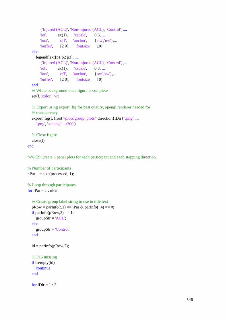
346
{'Injured (ACL)', 'Non-injured (ACL)', 'Control'},...
'ref', ax(1), 'xscale', 0.3, ...
'box', 'off', 'anchor', {'nw','nw'},...
'buffer', [2 0], 'fontsize', 10)
else
legendflex([p1 p2 p3], ...
{'Injured (ACL)', 'Non-injured (ACL)', 'Control'},...
'ref', ax(1), 'xscale', 0.3, ...
'box', 'off', 'anchor', {'sw','sw'},...
'buffer', [2 0], 'fontsize', 10)
end
% White background once figure is complete
set(f, 'color', 'w')
% Export using export_fig for best quality, opengl renderer needed for
% transparency
export_fig(f, [root '/plots/group_plots/' direction{iDir} '.png'],...
'-png', '-opengl', '-r300')
% Close figure
close(f)
end
%% (2) Create 6 panel plots for each participant and each stepping direction.
% Number of participants
nPar = size(processed, 1);
% Loop through participants
for iPar = 1 : nPar
% Create group label string to use in title text
pRow = parInfo(:,1) == iPar & parInfo(:,4) == 0;
if parInfo(pRow,3) == 1;
groupStr = 'ACL';
else
groupStr = 'Control';
end
id = parInfo(pRow,2);
% P16 missing
if isempty(id)
continue
end
for iDir = 1 : 2
347
% Create figure axis handle
f = figure('units','centimeters','Position', [10 10 19 14]);
% Loop through variables
axCnt = 1;
for iVar = vOrder
mnInj = nanmean(squeeze(processed(iPar, 1, iDir, :, iVar, :)))';
sdInj = nanstd(squeeze(processed(iPar, 1, iDir, :, iVar, :)))';
mnNon = nanmean(squeeze(processed(iPar, 2, iDir, :, iVar, :)))';
sdNon = nanstd(squeeze(processed(iPar, 2, iDir, :, iVar, :)))';
ax(iVar) = axes('Position', [x_pos(axCnt) y_pos(axCnt) 0.4 0.22]);
hold on
h1 = fill([x; flipud(x)], [mnInj-sdInj; flipud(mnInj+sdInj)], ...
[.9 0 0], 'linestyle', 'none');
h2 = fill([x; flipud(x)], [mnNon-sdNon; flipud(mnNon+sdNon)], ...
[0 0 .9], 'linestyle', 'none');
set([h1 h2], 'facealpha', alpha)
p1 = plot(mnInj, 'r', 'LineWidth', 1);
p2 = plot(mnNon, 'b', 'LineWidth', 1);
axis tight
title(variableNames{iVar})
ylabel(units{iVar})
if iVar == 3 || iVar == 6
xlabel('Normalised Time (%)')
end
box on
axCnt = axCnt + 1;
end
if parInfo(pRow,3) == 1;
legendflex([p1 p2], {'Injured', 'Non-injured'},...
'ref', ax(1), 'xscale', 0.3, ...
'box', 'off', 'anchor', {'nw','nw'},...
'buffer', [2 0], 'fontsize', 10)
else
legendflex([p1 p2], {'Control Side', 'Non-Control Side'},...
'ref', ax(1), 'xscale', 0.3, ...
'box', 'off', 'anchor', {'nw','nw'},...
'buffer', [2 0], 'fontsize', 10)
end
set(f, 'color', 'w')
export_fig(f, [root '/plots/par_plots/P' num2str(id) '_' ...
direction{iDir} '.png'],'-png', '-opengl', '-r300')
close(f)
end
end
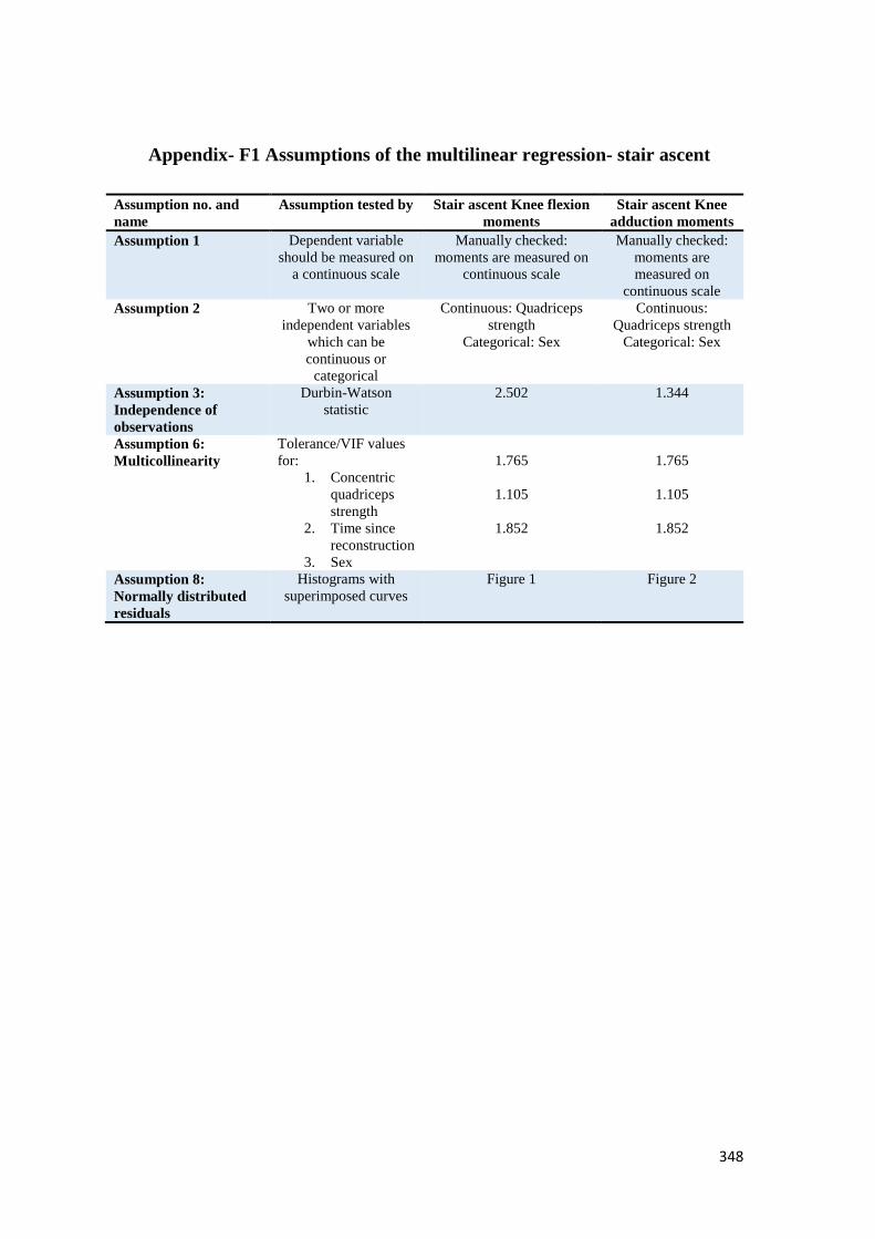
348
Appendix- F1 Assumptions of the multilinear regression- stair ascent
Assumption no. and
name
Assumption tested by Stair ascent Knee flexion
moments
Stair ascent Knee
adduction moments
Assumption 1 Dependent variable
should be measured on
a continuous scale
Manually checked:
moments are measured on
continuous scale
Manually checked:
moments are
measured on
continuous scale
Assumption 2 Two or more
independent variables
which can be
continuous or
categorical
Continuous: Quadriceps
strength
Categorical: Sex
Continuous:
Quadriceps strength
Categorical: Sex
Assumption 3:
Independence of
observations
Durbin-Watson
statistic
2.502 1.344
Assumption 6:
Multicollinearity
Tolerance/VIF values
for:
1. Concentric
quadriceps
strength
2. Time since
reconstruction
3. Sex
1.765
1.105
1.852
1.765
1.105
1.852
Assumption 8:
Normally distributed
residuals
Histograms with
superimposed curves
Figure 1 Figure 2
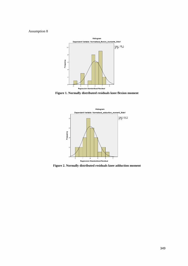
349
Assumption 8
Figure 1. Normally distributed residuals knee flexion moment
Figure 2. Normally distributed residuals knee adduction moment
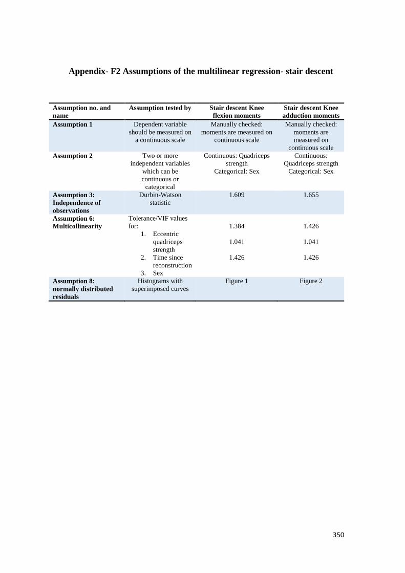
350
Appendix- F2 Assumptions of the multilinear regression- stair descent
Assumption no. and
name
Assumption tested by Stair descent Knee
flexion moments
Stair descent Knee
adduction moments
Assumption 1 Dependent variable
should be measured on
a continuous scale
Manually checked:
moments are measured on
continuous scale
Manually checked:
moments are
measured on
continuous scale
Assumption 2 Two or more
independent variables
which can be
continuous or
categorical
Continuous: Quadriceps
strength
Categorical: Sex
Continuous:
Quadriceps strength
Categorical: Sex
Assumption 3:
Independence of
observations
Durbin-Watson
statistic
1.609 1.655
Assumption 6:
Multicollinearity
Tolerance/VIF values
for:
1. Eccentric
quadriceps
strength
2. Time since
reconstruction
3. Sex
1.384
1.041
1.426
1.426
1.041
1.426
Assumption 8:
normally distributed
residuals
Histograms with
superimposed curves
Figure 1 Figure 2
351
Assumption 8
Figure 1. Normally distributed residuals knee flexion moment
Figure 2. Normally distributed residuals knee adduction moment