Patellar tendon versus quadrupled bone-semitendinosus anterior cruciate ligament reconstruction: a...
10
Patellar Tendon Versus Quadrupled Bone–Semitendinosus Anterior Cruciate Ligament Reconstruction: A Prospective Clinical Investigation in Athletes Alberto Gobbi, M.D., Sanjeev Mahajan, M.S., Milco Zanazzo, Ph.T., and Benjamin Tuy, M.D. Purpose: Patellar tendon and hamstrings are both used in anterior cruciate ligament (ACL) recon- struction, and comparisons have been reported with different results. The purpose of this clinical study was to compare the results of ACL reconstruction in athletes with 2 different graft types, both using bone-to-bone healing: bone–patellar tendon– bone graft and a quadrupled bone–semitendinosus graft. Type of Study: Outcomes study. Methods: From 1994 to 1997, 2 groups of 40 athletes who underwent ACL replacement with patellar tendon and quadrupled bone–semitendinosus grafts were prospectively evaluated. Preinjury activity level, age, and gender were comparable in both groups. All patients were operated on by the same surgeon within 5 months from injury and underwent group-specific rehabilitation programs. An independent examiner performed the final evaluations at 36 months. Review included clinical examination, radiographs, computed analysis, isokinetic and functional strength tests, and subjective and objective evaluation with standard knee scores. Addi- tional procedures were recorded. Statistical analysis was performed with both parametric and nonparametric tests. Results: Average surgical time was longer with the semitendinosus graft, and postoperative pain was higher in the patellar tendon group. Standard knee evaluation scores and subjective assessment revealed no significant differences. Isokinetic testing of flexion-extension and internal-external rotation showed lower quadriceps strength and a mild deficit of external rotation in the patellar tendon group and slightly lower flexor strength in the semitendinosus group at 12 months. Computerized laxity analysis showed no difference between the 2 groups with 90% of patients having less than 3 mm side-to-side difference, with a gender difference in the semitendinosus group. Kneeling pain was higher in patellar tendon group. Conclusions: The bone–patellar tendon– bone and quadrupled bone–semitendinosus autograft provide excellent grafts for ACL reconstruction. Both techniques are comparable regarding final stability, but in patients with extensor mechanism prob- lems or those who engage in sports with a high incidence of patellar tendonitis, the semitendinosus graft should be considered. Key Words: ACL reconstruction—Patellar tendon—Semitendinosus— Hamstrings—Graft bone interface. T he surgical treatment of the anterior cruciate lig- ament (ACL) deficient knee continues to stir de- bate among many orthopaedic surgeons. Included in the controversy is the selection of graft material to be used for the ACL reconstruction. The bone–patellar tendon– bone and the hamstring tendons are used most frequently. Advantages and disadvantages of each type of graft have been described. 1-8 The advantages of using the bone–patellar tendon– bone graft include strength, ready availability, strong bone-to-bone fixa- tion, and prompt healing of the bone plugs; however, harvesting the central third of the patellar tendon may contribute to dysfunction in some patients. 1-5 Advan- tages of the doubled semitendinosus and gracilis in- clude adequate strength, avoidance of extensor mech- anism complications, possibly less morbidity and decreased postoperative pain. 6-10 From Orthopaedic Arthroscopic Surgery International, Milan, Italy. Presented at the 16th Annual Meeting of the Arthroscopy Asso- ciation of North America. Address correspondence and reprint requests to Alberto Gobbi, MD, Orthopaedic Arthroscopic Surgery International, Via Amadeo 24, Milano 20133, Italy. E-mail: [email protected] © 2003 by the Arthroscopy Association of North America 0749-8063/03/1906-3369$30.00/0 doi:10.1016/S0749-8063(03)00393-1 592 Arthroscopy: The Journal of Arthroscopic and Related Surgery, Vol 19, No 6 (July-August), 2003: pp 592-601
Transcript of Patellar tendon versus quadrupled bone-semitendinosus anterior cruciate ligament reconstruction: a...

Patellar Tendon Versus Quadrupled Bone–SemitendinosusAnterior Cruciate Ligament Reconstruction: A Prospective
Clinical Investigation in Athletes
Alberto Gobbi, M.D., Sanjeev Mahajan, M.S., Milco Zanazzo, Ph.T., and Benjamin Tuy, M.D.
Purpose: Patellar tendon and hamstrings are both used in anterior cruciate ligament (ACL) recon-struction, and comparisons have been reported with different results. The purpose of this clinicalstudy was to compare the results of ACL reconstruction in athletes with 2 different graft types, bothusing bone-to-bone healing: bone–patellar tendon–bone graft and a quadrupled bone–semitendinosusgraft. Type of Study: Outcomes study.Methods: From 1994 to 1997, 2 groups of 40 athletes whounderwent ACL replacement with patellar tendon and quadrupled bone–semitendinosus grafts wereprospectively evaluated. Preinjury activity level, age, and gender were comparable in both groups.All patients were operated on by the same surgeon within 5 months from injury and underwentgroup-specific rehabilitation programs. An independent examiner performed the final evaluations at36 months. Review included clinical examination, radiographs, computed analysis, isokinetic andfunctional strength tests, and subjective and objective evaluation with standard knee scores. Addi-tional procedures were recorded. Statistical analysis was performed with both parametric andnonparametric tests.Results: Average surgical time was longer with the semitendinosus graft, andpostoperative pain was higher in the patellar tendon group. Standard knee evaluation scores andsubjective assessment revealed no significant differences. Isokinetic testing of flexion-extension andinternal-external rotation showed lower quadriceps strength and a mild deficit of external rotation inthe patellar tendon group and slightly lower flexor strength in the semitendinosus group at 12 months.Computerized laxity analysis showed no difference between the 2 groups with 90% of patients havingless than 3 mm side-to-side difference, with a gender difference in the semitendinosus group.Kneeling pain was higher in patellar tendon group.Conclusions: The bone–patellar tendon–boneand quadrupled bone–semitendinosus autograft provide excellent grafts for ACL reconstruction. Bothtechniques are comparable regarding final stability, but in patients with extensor mechanism prob-lems or those who engage in sports with a high incidence of patellar tendonitis, the semitendinosusgraft should be considered.Key Words: ACL reconstruction—Patellar tendon—Semitendinosus—Hamstrings—Graft bone interface.
The surgical treatment of the anterior cruciate lig-ament (ACL) deficient knee continues to stir de-
bate among many orthopaedic surgeons. Included inthe controversy is the selection of graft material to be
used for the ACL reconstruction. The bone–patellartendon–bone and the hamstring tendons are used mostfrequently. Advantages and disadvantages of eachtype of graft have been described.1-8 The advantagesof using the bone–patellar tendon–bone graft includestrength, ready availability, strong bone-to-bone fixa-tion, and prompt healing of the bone plugs; however,harvesting the central third of the patellar tendon maycontribute to dysfunction in some patients.1-5 Advan-tages of the doubled semitendinosus and gracilis in-clude adequate strength, avoidance of extensor mech-anism complications, possibly less morbidity anddecreased postoperative pain.6-10
From Orthopaedic Arthroscopic Surgery International, Milan, Italy.Presented at the 16th Annual Meeting of the Arthroscopy Asso-
ciation of North America.Address correspondence and reprint requests to Alberto Gobbi,
MD, Orthopaedic Arthroscopic Surgery International, Via Amadeo24, Milano 20133, Italy. E-mail: [email protected]
© 2003 by the Arthroscopy Association of North America0749-8063/03/1906-3369$30.00/0doi:10.1016/S0749-8063(03)00393-1
592 Arthroscopy: The Journal of Arthroscopic and Related Surgery, Vol 19, No 6 (July-August), 2003: pp 592-601

A review of current literature discussing clinicaloutcomes after ACL replacement reveals no signifi-cant differences between these 2 techniques.1-5 How-ever, although there seem to be no significant differ-ences between the 2 grafts in clinical outcome, manystudies show lower donor site morbidity and a slightlyincreased anterior laxity with hamstring reconstruc-tion.6-10 The use of 2 different types of grafts for ACLreconstruction are discussed in this paper; the press-fitpatellar tendon graft and the quadrupled bone–semi-tendinosus graft.
The conical press fit fixation technique is a simplemethod that offers several advantages, including bio-logic graft healing and the absence of any intra-artic-ular hardware, making revision surgery easier. Theuse of quadrupled semitendinosus graft was suggestedby Rosenberg and Cooley8 and Rosenberg and Pazik11
to compensate for the mechanical inferiority of adoubled tendon and avoid harvesting the gracilis. Thehypothesis of this prospective study among athleteswas that the quadrupled bone–semitendinosus graftcould provide the same stability as a bone–patellartendon–bone graft with a decreased morbidity.
METHODS
From 1994 to 1997, we performed 150 ACL recon-structions in 2 hospitals. Patients who satisfied thefollowing criteria were included in the study: (1) ath-letes playing in competitive sports at regional or na-tional levels or participating in active sports at least 3times per week, (2) patients with ACL injury, reportedwithin 5 months of injury, including those with asso-ciated meniscus tears, and (3) patients whose oppositeknee was normal. Patients also had to be willing tofollow the same rehabilitation program at the samerehabilitation center. Patients also needed to consentto participation in the study and to be available forfollow-up at 3, 6, and 12 months and 3 years aftersurgery or when requested to attend follow-up.
The following exclusion criteria were used to selectpatients: (1) subjects with history of injury or ailmentin either knee before the index injury; (2) patients withassociated ligament injuries, (3) patients with grade IIIor IV chondral damage or abnormal radiographs, and(4) patients who showed abnormal bone structure onstandard knee radiographs.
The first 40 patients in each institution who metthese criteria were included in the study. The 40patients who underwent ACL reconstruction in insti-tution A (a social medicine hospital) received a patel-lar tendon graft (PT), and the 40 patients in institution
B (a private hospital) received a bone–semitendinosusgraft (STB). Thus, we had 2 simultaneous groups ofconsecutive patients. The choice of where to have thesurgery performed was the sole decision of the patient,but was mainly influenced by economic consider-ations because one institution was a private hospitaland the other a social medicine institution.
All patients were treated primarily with anti-inflam-matory medicines and a rehabilitation program withthe goal of regaining full range of motion of the knee.All patients underwent surgery performed by the samesurgeon and followed a “group-specific” rehabilitationprogram at the same rehabilitation center.
The surgeon and a physical therapist examined allsubjects independently at 3, 6, and 12 months. Anindependent examiner performed final evaluations at36 months for both groups. Standard anteroposterior(AP) and lateral weight-bearing roentgenographicstudies performed on the first postoperative day werecompared with those at final follow-up to evaluatetunnel widening. This was measured in terms of per-centage change in the tunnel area, with a 50% increaseseen as tunnel widening, as proposed by Clatworthy etal.12
Subjective evaluation was performed with the In-ternational Knee Documentation Committee (IKDC)questionnaire, which included present symptoms,range of motion, joint stability, kneeling pain, satis-faction, and return to sport. In the IKDC functionalchart, patients were asked to score the treated knee,with the normal knee considered to be 100%. Exten-sion loss was measured with the patient prone andcompared with the normal knee. Flexion loss wasmeasured with the patient supine and compared withthe uninvolved knee. The patellofemoral joint wasexamined for crepitus, pain, swelling, and tightness inthe retinaculum. Pain over the metal hardware andanterior knee pain were recorded by an independentexaminer using a visual analogue scale (VAS) at1-year and at final evaluation.13
Computerized laxity analysis was performed with aknee motion analyzer (OSI CA4000, Orthopedic Sys-tem, Hayward, CA) preoperatively, at 3 months, at 1year, and at final evaluation. Anteroposterior tibialtranslation was measured by applying a 200-N force,and dynamic tests were performed during active flex-ion-extension against resistance.14-16
Isokinetic testing for evaluation of muscle strengthwas performed with the Biodex multijoint system(Biodex, Shirley, NY) by a single examiner. The testswere performed at 3, 6, and 12 months after surgery.The maximum peak torque in flexion and extension
593PATELLAR TENDON VERSUS QUADRUPLED BONE–SEMITENDINOSUS

was measured at speeds of 60°, 180°, and 300° persecond. Total work and hamstring/quadriceps ratiowere calculated. We also studied the internal andexternal rotation muscle strength to evaluate whetherthere is any deficit of muscle strength due to semiten-dinosus graft harvesting. The maximum peak torquedeficit was measured in internal and external rotation,with the patient in a sitting position and the kneeflexed at 90°. This was done at speeds of 60°, 180°,and 300° per second.
All patients underwent a series of functional tests.In the hop test, take-off and landing in equilibrium onthe same lower extremity and free use of arms areused. Three jump trials were performed for each leg,measuring the average distance and comparing thehealthy and pathologic limbs. Tegner and Noyesscales, standard IKDC, Lysholm knee scores, andsubjective assessments were also used for final eval-uation.
Surgical Technique
The surgeries were performed under general anes-thesia in 68 patients and under epidural anesthesia in12 patients. To reduce total tourniquet time, the tour-niquet was used only during graft harvest.
In the PT group, the central one third of the bone–patellar tendon–bone autograft was harvested with atrapezoidal shaped tibial bone block, which was usedto obtain a femoral press-fit (Fig 1). The tibial boneplug measured 30 mm in length, 14 mm wide at itsbase, and 10 mm wide at the bone–tendon junction.The patellar bone plug measured 10 mm wide and 20to 25 mm long. The total length of the grafts averaged10 to 11 cm (Fig 2). Using a modified 2-incisiontechnique, a femoral press fit was achieved by insert-ing the graft in a retrograde fashion through a femoraltunnel. The femoral tunnel was created using a specialconical cannulated reamer with a tapering diameterfrom proximal to distal. The conical bone plug corre-sponded to the flare of the femoral tunnel so that theplug fit exactly into the femoral tunnel (Fig 3). Tibialfixation was obtained using a metal wire (0.8 mm) tiedover a bicortical screw post. The bone block harvestsites were filled with the previously spared cancellousbone, and the incision was closed. A postoperativerehabilitation program as reported by Shelbourne andNitz17 was followed.
For the STB group, when hamstring reconstructionwas performed, the semitendinosus tendon was dis-sected and detached proximally but not distally fromthe tibia. The distal margin of the semitendinosus was
detached, along with a bone plug and periosteum tothe tibial crest, which was harvested using an os-teotome. The required minimum tendon length was 28cm, which was noted in every case, and the averagetendon length was 30 cm. The graft was whipstitchedand the tendon was quadrupled, with the bone plugtied at the external surface (Fig 4). The tibial tunnelwas positioned at the anatomic ACL insertion with anendoscopic aimer adjusted to a 45° position in the
FIGURE 1. The bone–patellar tendon–bone graft harvested with atrapezoidal-shaped tibial bone block.
594 A. GOBBI ET AL.

coronal plane. The alignment was performed at 70°with respect to the medial plateau. The femoral tunnelwas positioned about 7 mm in front of the “over the topposition” at the 11-o’clock position for a right knee.
Another bone plug harvested during the creation ofthe tibial tunnel with a coring system was used to fillthe tibial tunnel and press-fitted with the graft in thetibial tunnel. Femoral fixation was achieved using oneEndoButton (Smith & Nephew Endoscopy, Andover,MA) and tied with a quadrupled 5-mm Mersilene tape;the length of the Mersilene loop was 20 to 30 mm. Thegraft was pretensioned and preconditioned before tib-ial fixation with cyclic flexion and extension of theknee under maximum manual traction.
Tibial fixation was obtained with one 8-mm tita-nium Fastlok device (Neoligaments, Leeds, UK),which was also connected to the graft with a quadru-pled polyester tape (Fig 5). The Fastlok is a 2-piecefixation device with staple and buckle that allow tripleclamping action on the linkage material. Postoperativerehabilitation followed a program as reported byRosenberg and Pazik.11
Statistical Analysis
For continuous data, we used both parametric andnonparametric tests for the statistic comparison be-tween the 2 groups. For parametric data analysis, theStudent t test has often been performed in its Welch-modified version, to analyze unpaired samples withdifferent variances. For nonparametric technique, theWilcoxon rank-sum test has been used (also known asMann-Whitney statistics). Nonparametric techniques,even if more conservative, allowed us to relax as-sumptions of normality in sample distributions to ob-tain a robust confirmation of our results.
For categorical data, we adopted the �-square test orthe Fisher’s exact test to evaluate any significant dif-ference between groups. The �-square test providesbetter results when used in contingency tables withmore than 5 counts per each cell. Fisher’s test is betterfor small counts contingency tables and cannot beused when the sum of counts exceeds 200. We as-sumed a standard target for significance to be P � .05.
The Null hypothesis assumes no difference inmeans, no differences in ranks, no difference (or con-tingency) for Student t test, Wilcoxon test, and�-square (or Fisher test), respectively. S-plus statisti-cal software was used to compute the results.
RESULTS
The PT group included 26 men and 14 women, witha mean age of 28 years. The STB group included 22
FIGURE 3. Insertion of patellar tendon graft through the conicaltunnel in the femur.
FIGURE 2. The bone–patellar tendon–bone graft and its dimen-sions.
595PATELLAR TENDON VERSUS QUADRUPLED BONE–SEMITENDINOSUS

men and 18 women, with a mean age of 29 years. Themean injury-to-surgery interval was 2.5 months(range, 1–5 months), which was similar in the 2groups. The preinjury activity levels were comparablebetween the 2 groups (average Tegner Score, 7.5 forboth groups).
The 2 groups were comparable in terms of gender,age, and level of sports participation; the type ofsports performed by the patients were similar (Table
1). Average surgical time was 83 minutes in the PTgroup and 90 minutes in the STB group. Harvesting ofthe grafts required 10 minutes for PT and 16 minutesfor STB, and graft preparation took 5 minutes and 15minutes, respectively. Postoperative pain was higher inthe PT group (VAS 7 of 10), and only 20% of the pa-tients were discharged from the hospital within the first24 hours. For the STB group, 80% of the STB patients(VAS 5 of 10) could return home on oral analgesics.
FIGURE 4. The quadrupledbone-semitendinosus graft. (A)The harvested graft with a boneblock at its distal end. The av-erage length of the graft is 300mm; the average size of thebone block is 8 � 8 � 4 mm.(B) The graft is folded on itself.The end with the attached boneblock is hooked over the loopof Mersilene so that the bone ison the external surface of thegraft. The proximal 2.5 cm iswhipstitched with Ethibond No.1 suture, taking care to attachthe bone block securely to thegraft surface. At the distal end,2 strands of 3-mm Poly-tape aresecured around the looped endsof the graft using a sliding knot,leaving 4 free strands of tape.(C) Photograph of the preparedgraft.
596 A. GOBBI ET AL.

Knee Scores
Final IKDC assessment revealed that 85% of PTpatients and 92% of STB had “normal” or “nearlynormal” knee joints after surgery. Of the PT patients,55% were rated group A (normal) versus 60% of theSTB patients (Table 2). Using the Fisher exact test, nostatistically significant difference between the 2groups was noted (P � .8004).
Evaluation Form
Tegner activity scale (preoperative and postopera-tive), Noyes and Lysholm score results did not revealsignificant differences between the 2 groups (�-squaretest, P � .9156; Table 2).
Subjective Assessment
On the IKDC evaluation form, all patients wereasked to assess the treated knee, assuming the normalknee as 100%. The PT group had an average score of80% and the STB group, 85%.
Isokinetic Tests
Isokinetic testing of flexion-extension at 3 monthsrevealed lower quadriceps muscle strength in PT pa-tients (23% extensor total work deficit v 7.3% flexortotal work deficit) and hamstring strength in STBpatients (21.3% extensor total work deficit v 22.4%flexor total work deficit) at 60°/s. At the 1-year eval-uation, no statistically significant difference was seenat 60°, 180°, and 300° in extension. In flexion, astatistically significant difference (P � .0164) wasseen in the STB group at 60° and 180°, and nodifference was seen at 300°/s. Internal and externalrotation revealed a mild deficit of external rotation inthe PT group (average, 9%) and internal rotation in theSTB group (average, 6%). After 12 months, all pa-tients showed normal internal-external rotation data,with no statistically significant difference as calcu-lated by Welch modified Student t test and Wilcoxonmethod in between the 2 groups.
Computed Analysis
Computed analysis (OSI) was available for all pa-tients at 3-month, 1-year, and 3-year evaluations. Thisanalysis showed that 90% of these patients had less
FIGURE 5. The completed reconstruction. The EndoButton withMersilene tape is secured to the femur. Tibial fixation is achievedby securing the free end of the Poly-tape with a Fastlok. The boneplug removed from the creation of the tibial tunnel has beenreinserted to press-fit the graft.
TABLE 1. Type of Sports Performed by Patientsin the Study Groups
PT Group STB Group
Soccer 6 8Downhill skiing 8 9Motocross 6 7Basketball 6 5Volleyball 4 4Tennis 4 3Mountain bike 3 1Handball 1 1Alpinism 1 1Horse riding 1 1Total 40 40
TABLE 2. Knee Assessment Scores
TEST PT STB
Tegner Preinjury 7.5 (3-9) 7.5 (3-10)Postoperative 6.5 (3-9) 6.7 (2-9)
Noyes Final 88 (60-100) 86 (40-100)Lysholm Final 90 (80-100) 92 (80-100)Hop test (% Deficit) 6 Months 10 9
12 Months 6 6Final IKDC Group A 22 24
Group B 12 13Group C 5 3Group D 1 0
597PATELLAR TENDON VERSUS QUADRUPLED BONE–SEMITENDINOSUS
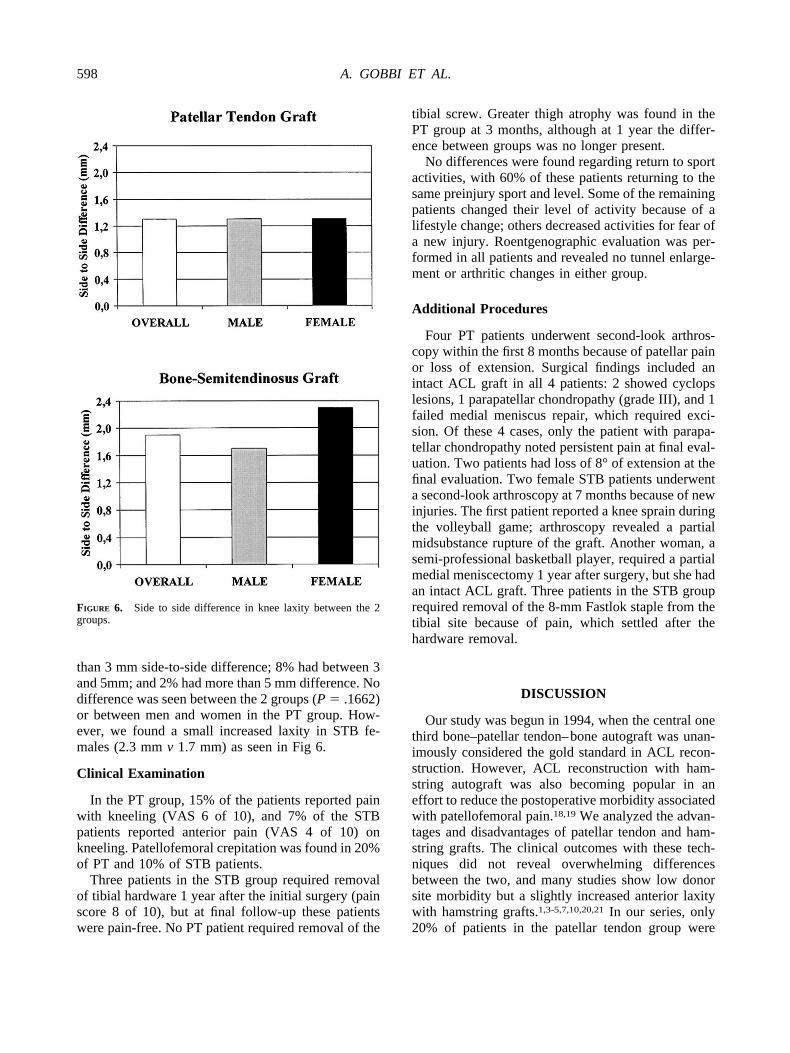
than 3 mm side-to-side difference; 8% had between 3and 5mm; and 2% had more than 5 mm difference. Nodifference was seen between the 2 groups (P � .1662)or between men and women in the PT group. How-ever, we found a small increased laxity in STB fe-males (2.3 mm v 1.7 mm) as seen in Fig 6.
Clinical Examination
In the PT group, 15% of the patients reported painwith kneeling (VAS 6 of 10), and 7% of the STBpatients reported anterior pain (VAS 4 of 10) onkneeling. Patellofemoral crepitation was found in 20%of PT and 10% of STB patients.
Three patients in the STB group required removalof tibial hardware 1 year after the initial surgery (painscore 8 of 10), but at final follow-up these patientswere pain-free. No PT patient required removal of the
tibial screw. Greater thigh atrophy was found in thePT group at 3 months, although at 1 year the differ-ence between groups was no longer present.
No differences were found regarding return to sportactivities, with 60% of these patients returning to thesame preinjury sport and level. Some of the remainingpatients changed their level of activity because of alifestyle change; others decreased activities for fear ofa new injury. Roentgenographic evaluation was per-formed in all patients and revealed no tunnel enlarge-ment or arthritic changes in either group.
Additional Procedures
Four PT patients underwent second-look arthros-copy within the first 8 months because of patellar painor loss of extension. Surgical findings included anintact ACL graft in all 4 patients: 2 showed cyclopslesions, 1 parapatellar chondropathy (grade III), and 1failed medial meniscus repair, which required exci-sion. Of these 4 cases, only the patient with parapa-tellar chondropathy noted persistent pain at final eval-uation. Two patients had loss of 8° of extension at thefinal evaluation. Two female STB patients underwenta second-look arthroscopy at 7 months because of newinjuries. The first patient reported a knee sprain duringthe volleyball game; arthroscopy revealed a partialmidsubstance rupture of the graft. Another woman, asemi-professional basketball player, required a partialmedial meniscectomy 1 year after surgery, but she hadan intact ACL graft. Three patients in the STB grouprequired removal of the 8-mm Fastlok staple from thetibial site because of pain, which settled after thehardware removal.
DISCUSSION
Our study was begun in 1994, when the central onethird bone–patellar tendon–bone autograft was unan-imously considered the gold standard in ACL recon-struction. However, ACL reconstruction with ham-string autograft was also becoming popular in aneffort to reduce the postoperative morbidity associatedwith patellofemoral pain.18,19 We analyzed the advan-tages and disadvantages of patellar tendon and ham-string grafts. The clinical outcomes with these tech-niques did not reveal overwhelming differencesbetween the two, and many studies show low donorsite morbidity but a slightly increased anterior laxitywith hamstring grafts.1,3-5,7,10,20,21 In our series, only20% of patients in the patellar tendon group were
FIGURE 6. Side to side difference in knee laxity between the 2groups.
598 A. GOBBI ET AL.

discharged from the hospital within 24 hours becauseof pain in the postoperative site.
The most common method of fixation of patellartendon was with metal interference screws, but thesehave not gone without complications.22 To avoid us-ing intra-articular hardware with patellar tendon graftsand to reduce surgical costs in a social medicinehospital setting, we thought of adopting a biologicalfixation of our grafts. We developed a conical press-fitfemoral bone plug fixation technique. The shape of thehole on the femur was made congruent with the boneplug harvested from the tibia so that it could hold andfit snugly.
The use of quadrupled semitendinosus with extra-articular fixation was suggested by researchers8,11 tocompensate for the mechanical inferiority of a dou-bled tendon and avoid harvesting the gracilis. Further-more, many authors agreed that harvesting the semi-tendinosus tendon alone is associated with lessmorbidity18,19 and provides strength and a high cross-sectional area.23-25
To combine low donor site morbidity with the ad-vantage of bone-to-bone biologic healing, we used aquadrupled bone–semitendinosus construct. Incorpo-ration of the semitendinosus tendon graft in the bonetunnel is the basic requirement for the long-term sur-vival of the cruciate ligament replacement. Therefore,fixation, revascularization, and incorporation of ham-string tendons within the bone are a theoretical as wellas practical concern for the surgeon.26-30
Weiler et al.31 and Shino et al.32 studied the initialfixation strength of the hamstring graft under staticand dynamic loading conditions with different tech-niques. They showed that a hamstring construct withan attached bone plug results in a superior initialfixation strength similar to conventional bone–ten-don–bone graft fixation.
We used the bone plug harvested along with thesemitendinosus tendon for fixation in the femoral tun-nel. To make this ST graft comparable with the bone–patellar tendon–bone construct, we harvested anotherbone plug during the creation of the tibial tunnel witha coring reamer. This bone plug was press-fit into thetibial tunnel after insertion of the graft.
Recent studies6,33,34 suggested that the tensilestrength of the graft was determined by the quality ofsoft tissue fixation. Mersilene tape has been indicatedas the weaker point in semitendinosus reconstructionwith the EndoButton, with a failure load calculatedbetween 492 N and 873 N.35,36
Following the suggestion of Ishibashi et al.,37 weadopted a short polyester tape loop to connect our
graft to the button to reduce creep and graft-tunnelmotion. We also used cyclic preconditioning of thegraft before tibial fixation. Furthermore, the primarystability of a quadrupled hamstring graft was sufficientto allow an immediate accelerated rehabilitation pro-tocol, which involves conditioning and stretching ofthe graft to sub-failure load.6,22,38,39,40
Rehabilitation after ACL reconstruction surgery isprolonged and time demanding. The only differencebetween our 2 groups was in the first phase of theprogram. We used the postoperative brace with theknee in full extension in the PT group, and in the STBgroup, we adopted a brace with extension lock at 10°;this helped us obtain immediate full extension of PTpatients while reducing the postoperative hamstringstretch in the STB patients.
Yasuda et al.10 showed that it is possible to have acomplete recovery of isometric hamstring musclestrength after ACL reconstruction by 3 months. In ourgroups, regaining the muscle strength was slower, butat 1-year follow-up, we found no difference in eithergroup when compared with the opposite side.
Endoscopic hamstring ACL reconstruction has beenassociated with tibial tunnel enlargement.12,41-44 Theetiology of bone tunnel enlargement is probably mul-tifactorial. Some authors favor biologic compo-nents,2,41 and others favor mechanical factors.40,42,43
Synovial fluid leakage into the tibial tunnel may alsoplay an influential role, as well as motion of the graftwithin the tunnel, the so-called “windshield wiper”effect.40,44,45
This phenomenon can be related to an incorrectmatching of graft-tunnel length with the patellar ten-don or the great distance of fixation and graft-tunnelmotion when using hamstring tendons. We did notfind tunnel enlargement in our series using anatomicfixation and a bone block press-fitted in both thetunnels. Our finding suggests that there may be abiological cause for the high incidence of tunnel wid-ening when using hamstring without filling the tunnelwith bone.
One of the features of our study was the evaluationof objective stability after reconstruction of the ACLwith a computed analysis system (OSI 4000), believ-ing that it would allow a more objective assessment ofthe anterior tibial translation than simple KT manualtesting. It allowed us to not only test the maximumstatic load but to perform the dynamic muscle testwith flexion and extension exercises as well. An in-dependent examiner other than the surgeon performedthe tests to eliminate potential bias. None of these
599PATELLAR TENDON VERSUS QUADRUPLED BONE–SEMITENDINOSUS

patients sustained major injuries to the contralateralknee during this interval.
Our study showed that more than 80% of the pa-tients in both groups showed less than 3 mm of side-to-side difference. We did not find major changesbetween 1- and 3-year test results.
Marder et al.4 and Harter et al.46 reported no statisti-cally significant difference when using a patellar tendonor a combined semitendinosus–gracilis tendon with re-spect to subjective evaluation and objective stability.
Aglietti et al.47 found no overwhelming differencebetween the 2 types of grafts. However, a trend towardbetter objective stability (KT-1000) with the use of thepatellar tendon grafts, but there was also more exten-sion loss and patellofemoral problems. Otero andHutcheson5 concluded that both the patellar tendonand the semitendinosus–gracilis are good choices inACL reconstruction, but the patellar tendon autograftprovides more stability than the doubled semitendino-sus–gracilis tendons. We believe that our results weresimilar with either graft type because both were con-structed with the concept of bone-to-bone healing.
We also noted that with the semitendinosus graft,tibial hardware protrusion above the tibial surfacecould be a significant cause of pain. Therefore, theprotrusion height of the system should be consideredwhen selecting the tibial fixation device of the semi-tendinosus graft.
Regarding return to sports, we found that only 60%of these patients could return to sports at the preinjurylevel. This was assessed with standard questionnairescompleted by each patient independently. Not all ofthe patients who returned to preinjury activity levelswere classified as IKDC group A: 9 patients (5 STBand 4 PT) were classified as group B. The work ofDaniel et al.48 revealed that restoration of certainlaxity parameters alone, like KT1000, after ACL re-construction does not prove restoration of normalfunction. A meta-analysis performed by Yunes et al.49
estimated that 75% of patients undergoing PT ACLreconstruction and 64% of those undergoing ham-string tendon ACL reconstruction returned to prein-jury levels of activity. However, direct comparisonwith our study is difficult, because our study popula-tion consisted exclusively of athletes, and the samplesincluded in that meta-analysis were not homogenousand included nonathletes.
CONCLUSIONS
Several conclusions can be drawn from this study.First, bone–patellar tendon–bone and quadrupled
bone semitendinosus autografts both provide excellentgrafts for ACL reconstruction, with good functionalstability of the knee and low primary failure rates.Second, although a high percentage of athletes in thestudy group had good results in terms of stability, only60% returned to sports activities at the preinjury level.Normal laxity parameters and commonly used kneescores after ACL reconstruction do not always predictreturn to preinjury level of function.
Finally, we recommend that patients with pre-exist-ing patellar or extensor apparatus problems, and thoseperforming sports with a high incidence of patellartendonitis, consider alternative ACL grafts includingsemitendinosus graft.
Acknowledgment: The authors thank Prof. Fabio Arico,University of Pavia, Italy for his help in statistical analysisof the study data.
REFERENCES
1. Aglietti P, Buzzi R, Zaccherotti G, De Biase P. Patellar tendonversus doubled semitendinosus and gracilis tendons for ante-rior cruciate ligament reconstruction. Am J Sports Med 1994;22:211-217.
2. Beynnon BD, Johnson RJ, Kannus P, et al. A prospective,randomized, clinical investigation of anterior cruciate ligamentreconstruction: A comparison of the bone–patellar tendon–bone and semitendinosus–gracilis autograft. Arthroscopy1998;14:S20.
3. Corry SI, Jonathan WM, Clingeleffer JA, Pinczewski LA.Arthroscopic reconstruction of the anterior cruciate ligament:A comparison of patellar tendon autograft and four-strandhamstring tendon autograft. Am J Sports Med 1999;27:444-454.
4. Marder RA, Raskind JR, Carroll M. Prospective evaluation ofarthroscopically assisted anterior cruciate reconstruction: Pa-tellar tendon versus semitendinosus and gracilis tendons. Am JSports Med 1991;19:478-484.
5. Otero AL, Hutcheson LA. A comparison of the doubled semi-tendinosus/gracilis and central third of the patellar tendonautografts in arthroscopic anterior cruciate reconstruction. Ar-throscopy 1993;9:143-148.
6. Maeda A, Shino K, Horibe S, et al. Anterior cruciate ligamentreconstruction with multistranded autogenous semitendinosustendon. Am J Sports Med 1996;24:504-509.
7. Pinczewski LA, Clingeleffer AJ, Corry IS, Webb JM. A com-parision of endoscopic ACL reconstruction using patellar ten-don autograft and hamstring tendon autograft at 2 years. J AmAcad Orthop Surg 1997;13:385-386.
8. Cooley VJ, Deffner KT, Rosenberg TD. Quadrupled semiten-dinosus anterior cruciate ligament reconstruction: 5 year re-sults in patients without meniscus loss. Arthroscopy 2001;17:795-800.
9. Goradia VK, Grana WA, Egle DM. Biomechanics of endo-scopic ACL reconstructions: Semitendinosus versus patellartendon graft. Arthroscopy 1997;13:383-384.
10. Yasuda K, Tsujino J, Ohkoshi Y, et al. Graft site morbiditywith autogenous semitendinosus and gracilis tendons. Am JSports Med 1995;23:706-714.
11. Rosenberg TD, Pazik JT. Anterior cruciate ligament recon-struction with quadrupled semitendinosus autograft. In: Pa-
600 A. GOBBI ET AL.

risien JS, ed. Current techniques in arthroscopy. Philadelphia:Current Medicine, 1996;77-88.
12. Clatworthy MG, Annear P, Bulow J-U, Bartlett RJ. Tunnelwidening in anterior cruciate ligament reconstruction: A pro-spective evaluation of hamstring and patellar tendon grafts.Knee Surg Sports Traumatol Arthrosc 1999;7:138-145.
13. Jensen MP, Karoly P. Self report scales and procedures forassessing pain in adults. In: Turk DC, Melzack R, eds. Hand-book of pain assessment. New York: Guilford Press, 1992;135-151.
14. Daniel DM, Stone ML, Sachs R, Malcolm J. Instrumentedmeasurement of anterior knee laxity in patients with acuteanterior cruciate ligament disruption. Am J Sports Med 1985;13:401-407.
15. Neuschwander DC, Prez D. Comparison of anterior laxitymeasurements in ACL deficient knees with two instrumentedtesting devices. Orthopaedics 1990;13:299-302.
16. Markolf KL, Amstuz HC. The clinical relevance of instru-mented testing for anterior cruciate ligament insufficiency.Clin Orthop 1987;223:198-207.
17. Shelbourne KD, Nitz P. Accelerated rehabilitation after ante-rior cruciate ligament reconstruction. Am J Sports Med 1990;18:292-299.
18. Sachs RA, Daniel DM, Stone ML. Patellofemoral problemsafter anterior cruciate ligament reconstruction. Am J SportsMed 1987;17:760-765.
19. Shino K, Nakagawa S, Inoue M. Deterioration of patellofemo-ral articular surface after anterior cruciate ligament reconstruc-tion. Am J Sports Med 1993;21:206-211.
20. Holmes PF, James SL, Larson RL, et al. Retrospective directcomparison of 3 intra-articular anterior cruciate ligament re-constructions. Am J Sports Med 1991;19:596-600.
21. O’Neill DB. Arthroscopically assisted reconstruction of theanterior cruciate ligament: A prospective randomized analysisof three techniques. J Bone Joint Surg Am 1996;78:803-813.
22. Kurosaka M, Yoshiya S, Andrish JT. A biomechanical com-parison of different surgical techniques of graft fixation inanterior cruciate ligament reconstruction. Am J Sports Med1987;15:225-229.
23. Noyes FR, Butler DL, Grood ES, et al. Biomechanical analysisof human ligament grafts used in knee ligament repairs andreconstructions. J Bone Joint Surg Am 1984;66:344-352.
24. Steiner ME, Hecker AT, Brown CH Jr, Hayes WC. Anteriorcruciate ligament graft fixation: Comparison of hamstring andpatellar tendon grafts. Am J Sports Med 1994;22:240-247.
25. Masayuki H, Shino K, Mitsouka T, et al. Cross-sectional areameasurement of the semitendinosus tendon for anterior cruci-ate ligament reconstruction. Arthroscopy 1998;14:696-701.
26. Grana WA, Eagle DM, Mahnken R, Goodhart CW. An anal-ysis of autograft fixation after anterior cruciate ligament re-construction in a rabbit model. Am J Sports Med 1994;22:344-351.
27. Pinczewski LA, Clingeleffer AJ, Otto DD, et al. Integration ofhamstring tendon graft with bone in reconstruction of theanterior cruciate ligament. Arthroscopy 1997;13:641-643.
28. Liu SH, Kabo JM, Osti L. Biomechanics of two types ofbone–tendon–bone graft for ACL reconstruction. J Bone JointSurg Br 1995;77:232-235.
29. Lane JG, McFadden P, Bowden L, Amiel D. The ligamenti-zation process: A four-year case study following ACL recon-struction with a semitendinosus graft. Arthroscopy 1993;9:149-153.
30. Liu SH, Panossian V, Al-Shaikh R, et al. Morphology andmatrix composition during early tendon to bone healing. ClinOrthop 1997;339:253-260.
31. Weiler A, Hoffman RGF, Stahelin AC, et al. Hamstring tendon
fixation using interference screws: A biomechanical study incalf tibial bone. Arthroscopy 1998;14:29-37.
32. Shino K, Kawasaki T, Hirose H, et al. Replacement of theanterior cruciate ligament by an allogeneic tendon graft: Anexperimental study in the dog. J Bone Joint Surg Br 1984;66:672-681.
33. Mc Kernan DJ, Weiss JA, Deffner KT, Greenwald RM. Ten-sile properties of gracilis, semitendinosus and patellar tendonsfrom the same donor. Trans Orthop Res Soc 1995;20:39.
34. Rowden NJ, Sher D, Rogers GJ, Schindhelm K. Anteriorcruciate ligament graft fixation: Initial comparison of patellartendon and semitendinosus autografts in fresh cadavers. Am JSports Med 1997;25:472-478.
35. Brown CH Jr, Sklar JH. Endoscopic anterior cruciate ligamentreconstruction using doubled gracilis and semitendinosus ten-dons and endobutton femoral fixation. Techn Orthop 1998;13:281-298.
36. Brand J Jr, Weiler A, Caborn DNM, et al. Graft fixation incruciate ligament reconstruction. Am J Sports Med 2000;28:761-774.
37. Ishibashi Y, Rudy T, Kim H, et al. The effect of the anteriorcruciate ligament graft fixation level on knee stability. Arthro-scopy 1995;11:373.
38. Weiler A, Hoffmann R, Windhagen H, et al. Biomechanicalevaluation of different biodegradable interference screws. Ar-throscopy 1997;13:403-404.
39. Hoher J, Moller HD, Fu FH. Bone tunnel enlargement afteranterior cruciate ligament reconstruction: Fact or fiction? KneeSurg Sports Traumatol Arthrosc 1998;6:231-240.
40. Insalata JC, Klatt B, Fu FH, Harner CD. Tunnel expansionfollowing anterior cruciate ligament reconstruction: A com-parison of hamstring and patellar tendon autografts. Knee SurgSports Traumatol Arthrosc 1997;5:234-238.
41. Fahey M, Indelicato PA. Bone tunnel enlargement after ante-rior cruciate replacement. Am J Sports Med 1994;22:410-414.
42. Fink A, Micheal CM, Benedetto KP, et al. Tibial tunnelenlargement following anterior cruciate ligament reconstruc-tion with patellar tendon autograft. Arthroscopy 2001;17:138-143.
43. Schultz K, Majewski M, Irregang JJ, et al. Radiographic tunnelchanges following arthroscopic ACL reconstruction: Autograftversus allograft. Arthroscopy 1996;11:272-273.
44. Hoher J, Livesay GA, Ma CB, Withrow JD, Fu FH, Woo SL.Hamstring graft motion in the femoral bone tunnel when usingtitanium button/polyester tape fixation. Knee Surg SportsTraumatol Arthrosc 1999;7:215-219.
45. Morgan CD, Kalmam VR, Grawl DM. Isometry testing foranterior cruciate ligament revisited. Arthroscopy 1995;11:647-659.
46. Harter RA, Osternig CR, Singer KM. Instrumented Lachmantest for evaluation of anterior laxity after reconstruction ofanterior cruciate ligament. J Bone Joint Surg Am 1987;71:975-983.
47. Aglietti P, Zaccherotti G, Buzzi R, DeBiase P. A comparisonbetween patellar tendon and doubled semitendinosus/gracilistendon for anterior cruciate ligament reconstruction: A mini-mum of five year follow up. J Sports Traumatol Rel Res1997;19:57-68.
48. Daniel DM, Stone ML, Dobson BE, et al. Fate of the ACL-injured patient: A prospective outcome study. Am J SportsMed 1994;22:632-644.
49. Yunes M, Richmond JC, Engels EA, Pinczewski LA. Patellartendon versus hamstring tendons in anterior cruciate liga-ment reconstruction: A meta-analysis. Arthroscopy 2001;17:248-257.
601PATELLAR TENDON VERSUS QUADRUPLED BONE–SEMITENDINOSUS