MR Imaging of Complications of Anterior Cruciate Ligament Graft Reconstruction
13
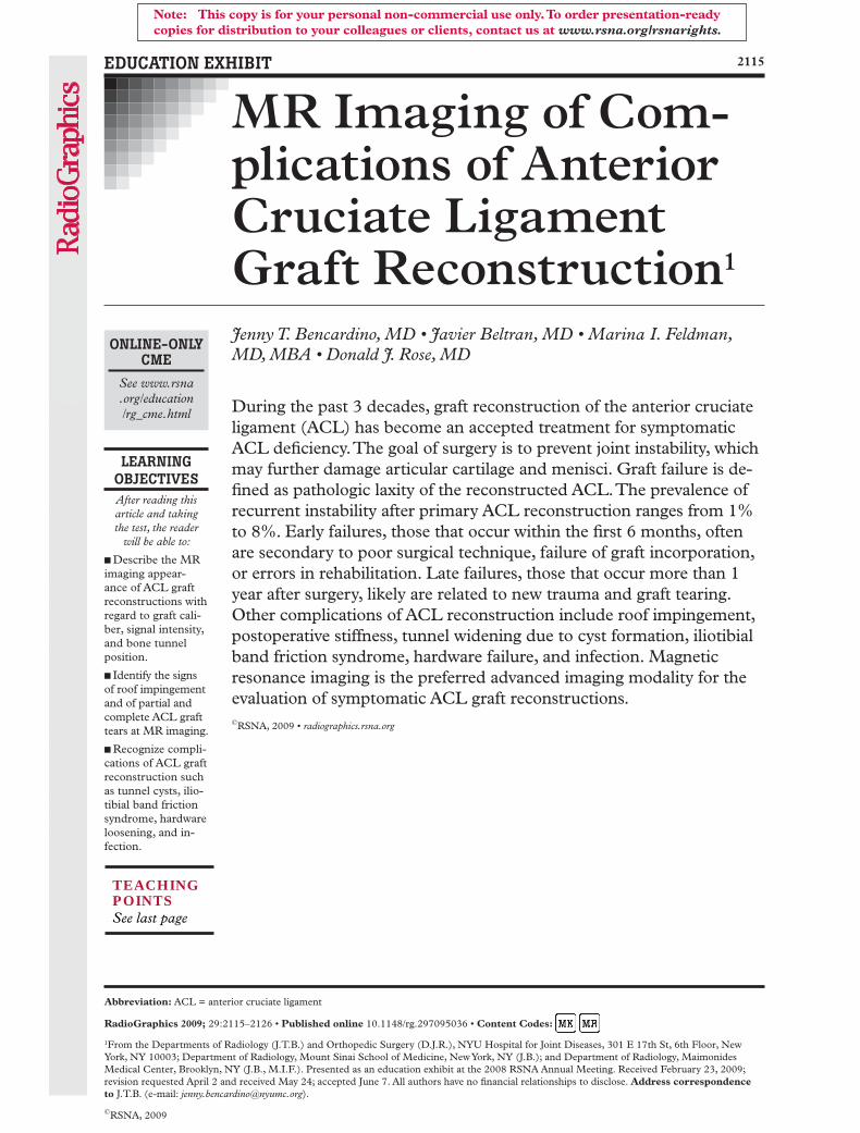
2115 EDUCATION EXHIBIT Jenny T. Bencardino, MD • Javier Beltran, MD • Marina I. Feldman, MD, MBA • Donald J. Rose, MD During the past 3 decades, graft reconstruction of the anterior cruciate ligament (ACL) has become an accepted treatment for symptomatic ACL deficiency. The goal of surgery is to prevent joint instability, which may further damage articular cartilage and menisci. Graft failure is de- fined as pathologic laxity of the reconstructed ACL. The prevalence of recurrent instability after primary ACL reconstruction ranges from 1% to 8%. Early failures, those that occur within the first 6 months, often are secondary to poor surgical technique, failure of graft incorporation, or errors in rehabilitation. Late failures, those that occur more than 1 year after surgery, likely are related to new trauma and graft tearing. Other complications of ACL reconstruction include roof impingement, postoperative stiffness, tunnel widening due to cyst formation, iliotibial band friction syndrome, hardware failure, and infection. Magnetic resonance imaging is the preferred advanced imaging modality for the evaluation of symptomatic ACL graft reconstructions. © RSNA, 2009 • radiographics.rsna.org MR Imaging of Com- plications of Anterior Cruciate Ligament Graft Reconstruction 1 ONLINE-ONLY CME See www.rsna .org/education /rg_cme.html LEARNING OBJECTIVES After reading this article and taking the test, the reader will be able to: Describe the MR ■ imaging appear- ance of ACL graft reconstructions with regard to graft cali- ber, signal intensity, and bone tunnel position. Identify the signs ■ of roof impingement and of partial and complete ACL graft tears at MR imaging. Recognize compli- ■ cations of ACL graft reconstruction such as tunnel cysts, ilio- tibial band friction syndrome, hardware loosening, and in- fection. Abbreviation: ACL = anterior cruciate ligament RadioGraphics 2009; 29:2115–2126 • Published online 10.1148/rg.297095036 • Content Codes: 1 From the Departments of Radiology (J.T.B.) and Orthopedic Surgery (D.J.R.), NYU Hospital for Joint Diseases, 301 E 17th St, 6th Floor, New York, NY 10003; Department of Radiology, Mount Sinai School of Medicine, New York, NY (J.B.); and Department of Radiology, Maimonides Medical Center, Brooklyn, NY (J.B., M.I.F.). Presented as an education exhibit at the 2008 RSNA Annual Meeting. Received February 23, 2009; revision requested April 2 and received May 24; accepted June 7. All authors have no financial relationships to disclose. Address correspondence to J.T.B. (e-mail: [email protected]). © RSNA, 2009 See last page TEACHING POINTS Note: This copy is for your personal non-commercial use only. To order presentation-ready copies for distribution to your colleagues or clients, contact us at www.rsna.org/rsnarights.
-
Upload
independent -
Category
Documents
-
view
0 -
download
0
Transcript of MR Imaging of Complications of Anterior Cruciate Ligament Graft Reconstruction
2115EDUCATION EXHIBIT
Jenny T. Bencardino, MD • Javier Beltran, MD • Marina I. Feldman, MD, MBA • Donald J. Rose, MD
During the past 3 decades, graft reconstruction of the anterior cruciate ligament (ACL) has become an accepted treatment for symptomatic ACL deficiency. The goal of surgery is to prevent joint instability, which may further damage articular cartilage and menisci. Graft failure is de-fined as pathologic laxity of the reconstructed ACL. The prevalence of recurrent instability after primary ACL reconstruction ranges from 1% to 8%. Early failures, those that occur within the first 6 months, often are secondary to poor surgical technique, failure of graft incorporation, or errors in rehabilitation. Late failures, those that occur more than 1 year after surgery, likely are related to new trauma and graft tearing. Other complications of ACL reconstruction include roof impingement, postoperative stiffness, tunnel widening due to cyst formation, iliotibial band friction syndrome, hardware failure, and infection. Magnetic resonance imaging is the preferred advanced imaging modality for the evaluation of symptomatic ACL graft reconstructions.©RSNA, 2009 • radiographics.rsna.org
MR Imaging of Com-plications of Anterior Cruciate Ligament Graft Reconstruction1
ONlINE-ONly CME
See www.rsna .org/education /rg_cme.html
lEARNING OBJECTIVESAfter reading this article and taking the test, the reader
will be able to:
Describe the MR ■
imaging appear-ance of ACL graft reconstructions with regard to graft cali-ber, signal intensity, and bone tunnel position.
Identify the signs ■
of roof impingement and of partial and complete ACL graft tears at MR imaging.
Recognize compli- ■
cations of ACL graft reconstruction such as tunnel cysts, ilio-tibial band friction syndrome, hardware loosening, and in-fection.
Abbreviation: ACL = anterior cruciate ligament
RadioGraphics 2009; 29:2115–2126 • Published online 10.1148/rg.297095036 • Content Codes: 1From the Departments of Radiology (J.T.B.) and Orthopedic Surgery (D.J.R.), NYU Hospital for Joint Diseases, 301 E 17th St, 6th Floor, New York, NY 10003; Department of Radiology, Mount Sinai School of Medicine, New York, NY (J.B.); and Department of Radiology, Maimonides Medical Center, Brooklyn, NY (J.B., M.I.F.). Presented as an education exhibit at the 2008 RSNA Annual Meeting. Received February 23, 2009; revision requested April 2 and received May 24; accepted June 7. All authors have no financial relationships to disclose. Address correspondence to J.T.B. (e-mail: [email protected]).
©RSNA, 2009
See last page
TEACHING POINTS
Note: This copy is for your personal non-commercial use only. To order presentation-ready copies for distribution to your colleagues or clients, contact us at www.rsna.org/rsnarights.
2116 November-December 2009 radiographics.rsna.org
IntroductionDuring the past 3 decades, graft reconstruc-tion of the anterior cruciate ligament (ACL) has become an accepted treatment for symptomatic ACL deficiency (1,2). The goal of surgery is to prevent joint instability, which may further dam-age articular cartilage and menisci. Advances in reconstructive techniques and rehabilitation have led to substantially improved results. Nev-ertheless, the percentage of patients who develop symptomatic osteoarthritis after ACL recon-struction remains high (13.6%–21.5%) (3). The combination of ACL reconstruction and menis-cectomy also has been described as a potential accelerator of osteoarthritis (3). The prevalence of osteoarthritis 10 years after ACL reconstruc-tion increases from 8% to 27% in patients who undergo meniscectomy and have a time interval longer than 1 year between meniscal injury and surgery (3). Graft failure is defined as pathologic laxity of the reconstructed ACL. The prevalence of recurrent instability after primary ACL recon-struction ranges from 1% to 8% (4,5). Early fail-ures, those that occur within the first 6 months, often are secondary to poor surgical technique, failure of graft incorporation, or errors in reha-bilitation. Late failures, those that occur more than 1 year after surgery, likely are related to new trauma and graft tearing (5). Other complications of ACL reconstruction include roof impinge-ment, postoperative stiffness, tunnel widening due to cyst formation, iliotibial band friction syndrome, hardware failure, and infection. Mag-netic resonance (MR) imaging is the preferred advanced imaging modality for the evaluation of symptomatic ACL graft reconstructions (6,7). Patients with contraindications to MR imaging (eg, pacemakers or stimulators) may undergo high-resolution computed tomographic arthrog-raphy of the knee for evaluation of postoperative ACL graft abnormalities.
In this article we describe the postoperative MR imaging appearance of normal ACL graft
reconstructions and potential complications in-cluding roof impingement, partial and complete graft tears, arthrofibrosis, tunnel cysts, iliotibial band friction syndrome, hardware loosening, and infection.
Normal ACL Graft Reconstruction
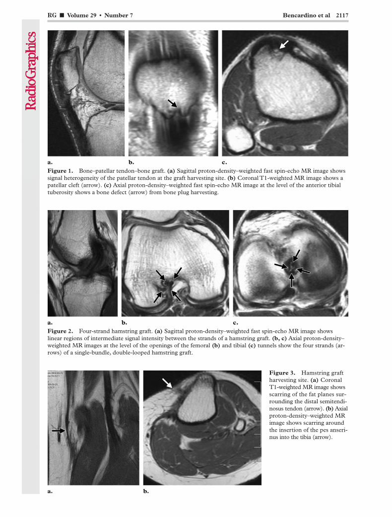
The question of which type of graft to employ in ACL reconstruction remains controversial (8). The most commonly used methods are bone–patellar tendon–bone and hamstring autografts. The use of the middle one-third of the patellar tendon, with bone plugs attached to each end, has historically been considered the reference standard for ACL graft reconstruction because of the inherent strength and stiffness of the graft (Fig 1). However, subsequent patellar-tendon ab-normalities and anterior knee pain are relatively common complications with this type of graft. A four-strand hamstring graft often is made of segments from the semitendinosus tendon, the gracilis tendon, or both. The tendon segments are folded and braided together to form a quadruple-thickness strand, which is then fixed to the femur and tibia (Fig 2). The hamstring graft became popular because of the low reported morbidity related to the graft harvesting site (9,10) (Fig 3). However, hamstring grafts traditionally have been subject to slippage. New fixation techniques seem to have addressed this problem. Proper fixation of the graft with interference screws, endobuttons (Fig 4), a screw-washer construct, or staples is crucial to avoid changes in the graft position dur-ing the initial postoperative incorporation period. Interference screws commonly are used as fixa-tion devices in bone–patellar tendon–bone graft reconstruction.
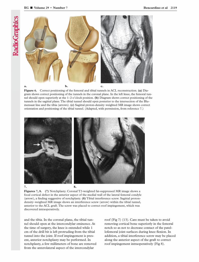
Positioning of the femoral and tibial tunnels is of paramount importance for proper function of the ACL graft. Accurate location of the femoral tunnel is essential to achieve isometry of the ACL graft (11). Isometry refers to adequate constancy in length and tension of the graft during the com-plete range of knee motion (from flexion to ex-tension). Over-the-top placement is not isomet-ric; it results in increased graft length and tension
TeachingPoint
TeachingPoint
RG ■ Volume 29 • Number 7 Bencardino et al 2117
Figure 2. Four-strand hamstring graft. (a) Sagittal proton-density–weighted fast spin-echo MR image shows linear regions of intermediate signal intensity between the strands of a hamstring graft. (b, c) Axial proton-density–weighted MR images at the level of the openings of the femoral (b) and tibial (c) tunnels show the four strands (ar-rows) of a single-bundle, double-looped hamstring graft.
Figure 1. Bone–patellar tendon–bone graft. (a) Sagittal proton-density–weighted fast spin-echo MR image shows signal heterogeneity of the patellar tendon at the graft harvesting site. (b) Coronal T1-weighted MR image shows a patellar cleft (arrow). (c) Axial proton-density–weighted fast spin-echo MR image at the level of the anterior tibial tuberosity shows a bone defect (arrow) from bone plug harvesting.
Figure 3. Hamstring graft harvesting site. (a) Coronal T1-weighted MR image shows scarring of the fat planes sur-rounding the distal semitendi-nosus tendon (arrow). (b) Axial proton-density–weighted MR image shows scarring around the insertion of the pes anseri-nus into the tibia (arrow).
2118 November-December 2009 radiographics.rsna.org
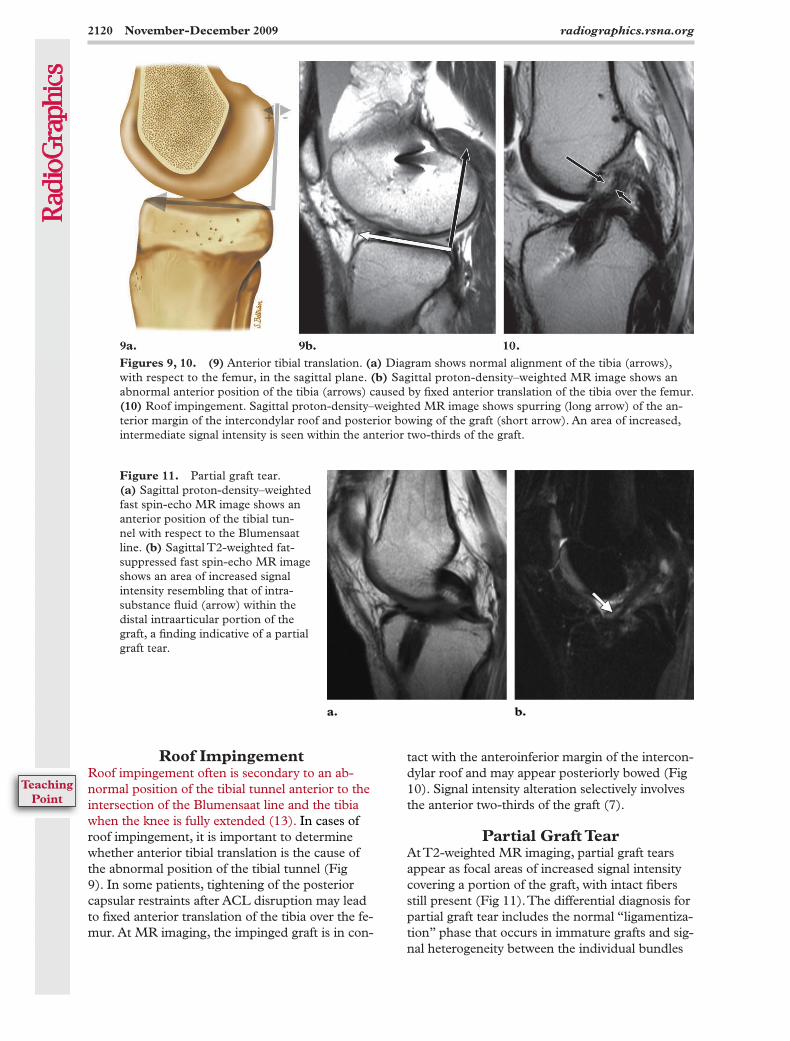
better than the classic single-bundle, two-tunnel technique. Recent data suggest that the conven-tional single-bundle technique, if implemented with a more horizontally positioned femoral tunnel, produces clinical results comparable to those obtained with the double-bundle tech-nique (12). On coronal images, the femoral tunnel should open superiorly above the lateral femoral condyle at the 10–11-o’clock position in the right knee and the 1–2-o’clock position in the left knee (Fig 6) (7,12).
The tibial tunnel should be oriented parallel to the projected slope of the intercondylar roof (the Blumensaat line) (Fig 6b, 6c). In the sagittal plane, the opening of the proximal tibial tunnel should be posterior to the intersection of the Blumensaat line
as the knee is extended (Fig 5a). If the femoral tunnel is placed too far anteriorly, the length and tension of the graft greatly increase as the knee is flexed (Fig 5b). The femoral tunnel should be placed as far posteriorly as possible without dis-rupting the posterior cortex of the femur. Ideally, a 1–2-mm-thick cortical rim should remain.
The ACL consists of two major functional bundles: the anteromedial bundle and the pos-terolateral bundle. Both contribute substantially to the anterior and rotational stability of the knee. It has been postulated that an anatomic double-bundle, four-tunnel reconstruction tech-nique may restore the biomechanics of the knee
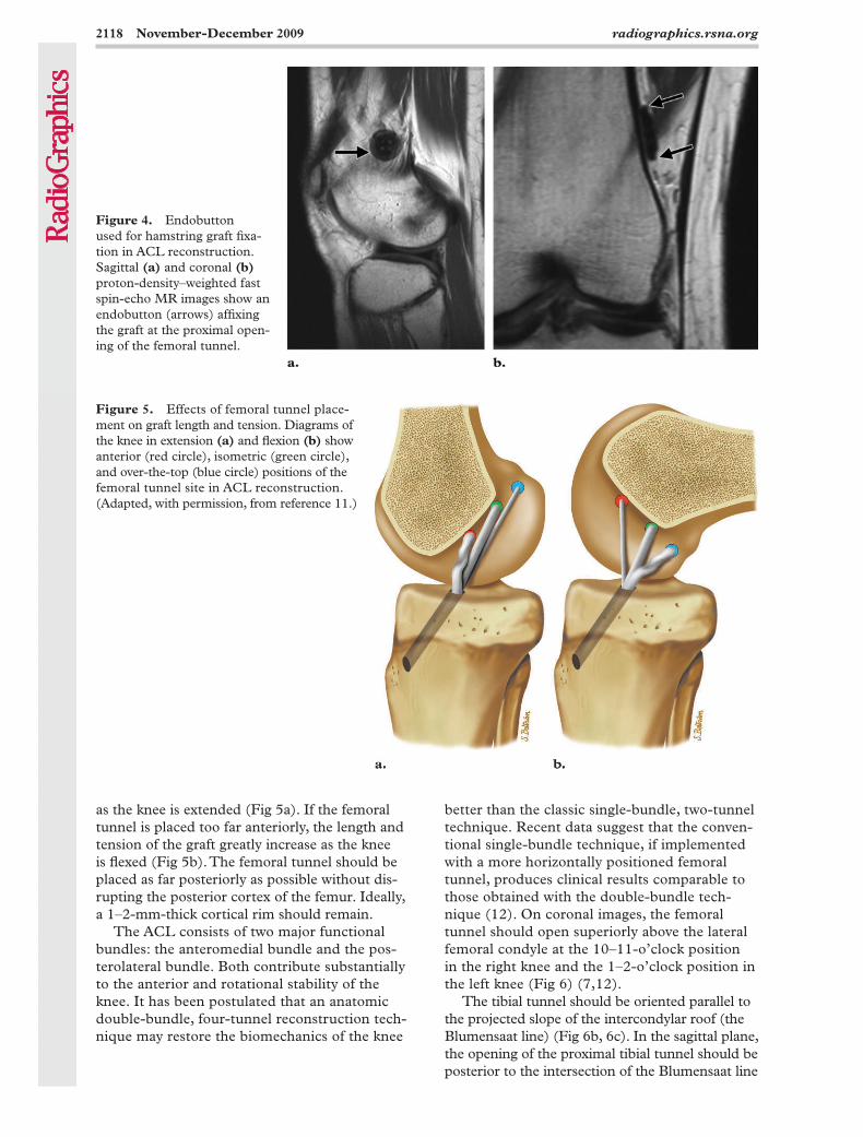
Figure 4. Endobutton used for hamstring graft fixa-tion in ACL reconstruction. Sagittal (a) and coronal (b) proton-density–weighted fast spin-echo MR images show an endobutton (arrows) affixing the graft at the proximal open-ing of the femoral tunnel.
Figure 5. Effects of femoral tunnel place-ment on graft length and tension. Diagrams of the knee in extension (a) and flexion (b) show anterior (red circle), isometric (green circle), and over-the-top (blue circle) positions of the femoral tunnel site in ACL reconstruction. (Adapted, with permission, from reference 11.)
RG ■ Volume 29 • Number 7 Bencardino et al 2119
Figure 6. Correct positioning of the femoral and tibial tunnels in ACL reconstruction. (a) Dia-gram shows correct positioning of the tunnels in the coronal plane. In the left knee, the femoral tun-nel should open superiorly at the 1–2-o’clock position. (b) Diagram shows correct positioning of the tunnels in the sagittal plane. The tibial tunnel should open posterior to the intersection of the Blu-mensaat line and the tibia (arrows). (c) Sagittal proton-density–weighted MR image shows correct orientation and positioning of the tibial tunnel. (Adapted, with permission, from reference 7.)
roof (Fig 7) (13). Care must be taken to avoid removing cortical bone superiorly in the femoral notch so as not to decrease contact of the patel-lofemoral joint surfaces during knee flexion. In addition, a tibial interference screw may be placed along the anterior aspect of the graft to correct roof impingement intraoperatively (Fig 8).
and the tibia. In the coronal plane, the tibial tun-nel should open at the intercondylar eminence. At the time of surgery, the knee is extended while 1 cm of the drill bit is left protruding from the tibial tunnel into the joint. If roof impingement is pres-ent, anterior notchplasty may be performed. In notchplasty, a few millimeters of bone are removed from the anterolateral aspect of the intercondylar
Figures 7, 8. (7) Notchplasty. Coronal T2-weighted fat-suppressed MR image shows a focal cortical defect in the anterior aspect of the medial wall of the lateral femoral condyle (arrow), a finding suggestive of notchplasty. (8) Tibial interference screw. Sagittal proton-density–weighted MR image shows an interference screw (arrow) within the tibial tunnel, anterior to the ACL graft. The screw was placed to correct roof impingement, which was discovered intraoperatively.
2120 November-December 2009 radiographics.rsna.org
Figures 9, 10. (9) Anterior tibial translation. (a) Diagram shows normal alignment of the tibia (arrows), with respect to the femur, in the sagittal plane. (b) Sagittal proton-density–weighted MR image shows an abnormal anterior position of the tibia (arrows) caused by fixed anterior translation of the tibia over the femur. (10) Roof impingement. Sagittal proton-density–weighted MR image shows spurring (long arrow) of the an-terior margin of the intercondylar roof and posterior bowing of the graft (short arrow). An area of increased, intermediate signal intensity is seen within the anterior two-thirds of the graft.
tact with the anteroinferior margin of the intercon-dylar roof and may appear posteriorly bowed (Fig 10). Signal intensity alteration selectively involves the anterior two-thirds of the graft (7).
Partial Graft TearAt T2-weighted MR imaging, partial graft tears appear as focal areas of increased signal intensity covering a portion of the graft, with intact fibers still present (Fig 11). The differential diagnosis for partial graft tear includes the normal “ligamentiza-tion” phase that occurs in immature grafts and sig-nal heterogeneity between the individual bundles
Roof ImpingementRoof impingement often is secondary to an ab-normal position of the tibial tunnel anterior to the intersection of the Blumensaat line and the tibia when the knee is fully extended (13). In cases of roof impingement, it is important to determine whether anterior tibial translation is the cause of the abnormal position of the tibial tunnel (Fig 9). In some patients, tightening of the posterior capsular restraints after ACL disruption may lead to fixed anterior translation of the tibia over the fe-mur. At MR imaging, the impinged graft is in con-
Figure 11. Partial graft tear. (a) Sagittal proton-density–weighted fast spin-echo MR image shows an anterior position of the tibial tun-nel with respect to the Blumensaat line. (b) Sagittal T2-weighted fat-suppressed fast spin-echo MR image shows an area of increased signal intensity resembling that of intra-substance fluid (arrow) within the distal intraarticular portion of the graft, a finding indicative of a partial graft tear.
TeachingPoint
RG ■ Volume 29 • Number 7 Bencardino et al 2121
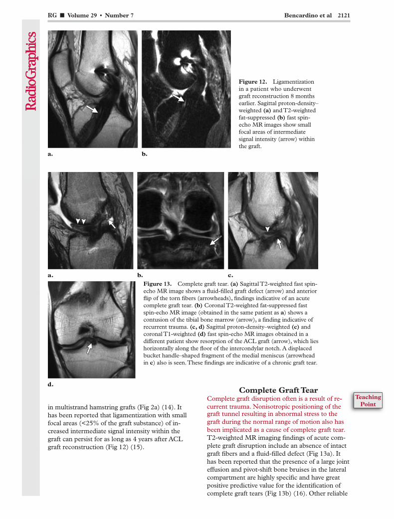
in multistrand hamstring grafts (Fig 2a) (14). It has been reported that ligamentization with small focal areas (<25% of the graft substance) of in-creased intermediate signal intensity within the graft can persist for as long as 4 years after ACL graft reconstruction (Fig 12) (15).
Complete Graft TearComplete graft disruption often is a result of re-current trauma. Nonisotropic positioning of the
Figure 12. Ligamentization in a patient who underwent graft reconstruction 8 months earlier. Sagittal proton-density–weighted (a) and T2-weighted fat-suppressed (b) fast spin-echo MR images show small focal areas of intermediate signal intensity (arrow) within the graft.
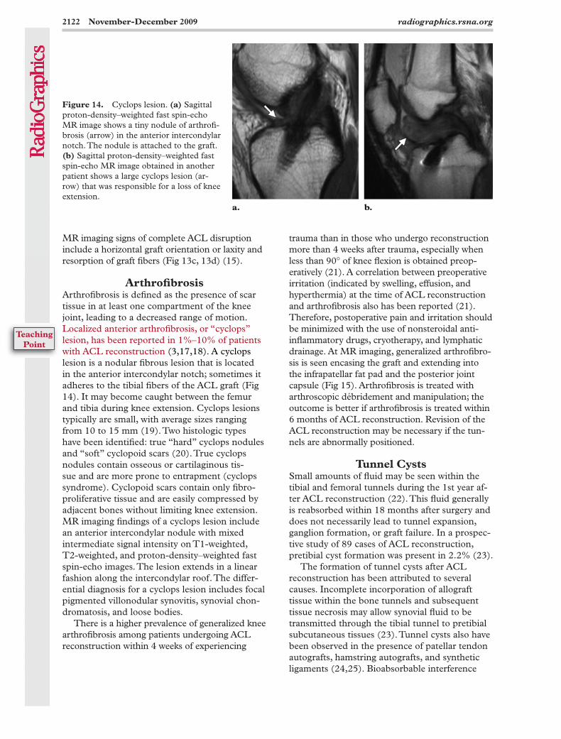
Figure 13. Complete graft tear. (a) Sagittal T2-weighted fast spin-echo MR image shows a fluid-filled graft defect (arrow) and anterior flip of the torn fibers (arrowheads), findings indicative of an acute complete graft tear. (b) Coronal T2-weighted fat-suppressed fast spin-echo MR image (obtained in the same patient as a) shows a contusion of the tibial bone marrow (arrow), a finding indicative of recurrent trauma. (c, d) Sagittal proton-density–weighted (c) and coronal T1-weighted (d) fast spin-echo MR images obtained in a different patient show resorption of the ACL graft (arrow), which lies horizontally along the floor of the intercondylar notch. A displaced bucket handle–shaped fragment of the medial meniscus (arrowhead in c) also is seen. These findings are indicative of a chronic graft tear.
graft tunnel resulting in abnormal stress to the graft during the normal range of motion also has been implicated as a cause of complete graft tear. T2-weighted MR imaging findings of acute com-plete graft disruption include an absence of intact graft fibers and a fluid-filled defect (Fig 13a). It has been reported that the presence of a large joint effusion and pivot-shift bone bruises in the lateral compartment are highly specific and have great positive predictive value for the identification of complete graft tears (Fig 13b) (16). Other reliable
TeachingPoint
2122 November-December 2009 radiographics.rsna.org
MR imaging signs of complete ACL disruption include a horizontal graft orientation or laxity and resorption of graft fibers (Fig 13c, 13d) (15).
ArthrofibrosisArthrofibrosis is defined as the presence of scar tissue in at least one compartment of the knee joint, leading to a decreased range of motion. Localized anterior arthrofibrosis, or “cyclops” lesion, has been reported in 1%–10% of patients with ACL reconstruction (3,17,18). A cyclops lesion is a nodular fibrous lesion that is located in the anterior intercondylar notch; sometimes it adheres to the tibial fibers of the ACL graft (Fig 14). It may become caught between the femur and tibia during knee extension. Cyclops lesions typically are small, with average sizes ranging from 10 to 15 mm (19). Two histologic types have been identified: true “hard” cyclops nodules and “soft” cyclopoid scars (20). True cyclops nodules contain osseous or cartilaginous tis-sue and are more prone to entrapment (cyclops syndrome). Cyclopoid scars contain only fibro-proliferative tissue and are easily compressed by adjacent bones without limiting knee extension. MR imaging findings of a cyclops lesion include an anterior intercondylar nodule with mixed intermediate signal intensity on T1-weighted, T2-weighted, and proton-density–weighted fast spin-echo images. The lesion extends in a linear fashion along the intercondylar roof. The differ-ential diagnosis for a cyclops lesion includes focal pigmented villonodular synovitis, synovial chon-dromatosis, and loose bodies.
There is a higher prevalence of generalized knee arthrofibrosis among patients undergoing ACL reconstruction within 4 weeks of experiencing
trauma than in those who undergo reconstruction more than 4 weeks after trauma, especially when less than 90° of knee flexion is obtained preop-eratively (21). A correlation between preoperative irritation (indicated by swelling, effusion, and hyperthermia) at the time of ACL reconstruction and arthrofibrosis also has been reported (21). Therefore, postoperative pain and irritation should be minimized with the use of nonsteroidal anti-inflammatory drugs, cryotherapy, and lymphatic drainage. At MR imaging, generalized arthrofibro-sis is seen encasing the graft and extending into the infrapatellar fat pad and the posterior joint capsule (Fig 15). Arthrofibrosis is treated with arthroscopic débridement and manipulation; the outcome is better if arthrofibrosis is treated within 6 months of ACL reconstruction. Revision of the ACL reconstruction may be necessary if the tun-nels are abnormally positioned.
Tunnel CystsSmall amounts of fluid may be seen within the tibial and femoral tunnels during the 1st year af-ter ACL reconstruction (22). This fluid generally is reabsorbed within 18 months after surgery and does not necessarily lead to tunnel expansion, ganglion formation, or graft failure. In a prospec-tive study of 89 cases of ACL reconstruction, pretibial cyst formation was present in 2.2% (23).
The formation of tunnel cysts after ACL reconstruction has been attributed to several causes. Incomplete incorporation of allograft tissue within the bone tunnels and subsequent tissue necrosis may allow synovial fluid to be transmitted through the tibial tunnel to pretibial subcutaneous tissues (23). Tunnel cysts also have been observed in the presence of patellar tendon autografts, hamstring autografts, and synthetic ligaments (24,25). Bioabsorbable interference
Figure 14. Cyclops lesion. (a) Sagittal proton-density–weighted fast spin-echo MR image shows a tiny nodule of arthrofi-brosis (arrow) in the anterior intercondylar notch. The nodule is attached to the graft. (b) Sagittal proton-density–weighted fast spin-echo MR image obtained in another patient shows a large cyclops lesion (ar-row) that was responsible for a loss of knee extension.
TeachingPoint

RG ■ Volume 29 • Number 7 Bencardino et al 2123
screws, nonabsorbable suture fragments, and joint fluid leakage during failed ACL revision surgery also have been implicated in tunnel cyst formation. “Bungee-cord” or “windshield-wiper” tunnel widening may occur when intraosseous fixation is not performed. Extrusion of joint fluid into the tunnel may lead to formation of a gan-
Figure 15. General-ized arthrofibrosis. Sag-ittal (a) and axial (b) proton-density–weighted fast spin-echo MR im-ages show encasement of the ACL graft by fibrovascular prolifera-tive tissue (arrows) that extends into the infra-patellar fat and supra-patellar bursa.
Figure 16. Tibial tunnel cyst. (a, b) Oblique coronal proton-density–weighted MR images show a linear intrasub-stance tear of the ACL graft (arrow in a), a finding associated with a tibial tunnel cyst (arrowheads in b). (c) Sagittal T2-weighted fat-suppressed MR image shows reactive marrow edema (arrows) surrounding the tibial tunnel cyst. (d) Axial T2-weighted fat-suppressed MR image shows extension of the cyst (*) into the pretibial soft tissues.
glion, which may enlarge over time and cause postoperative pain. Tibial tunnel cysts may be incidentally found at MR imaging, or they may manifest as a palpable mass in the pretibial soft tissues (Fig 16). Femoral tunnel cysts are less common than tibial tunnel cysts; there are no re-ports in the literature regarding their prevalence. All four femoral cysts from our teaching file col-lection were associated with complete disruption of the ACL graft (Fig 17).
2124 November-December 2009 radiographics.rsna.org
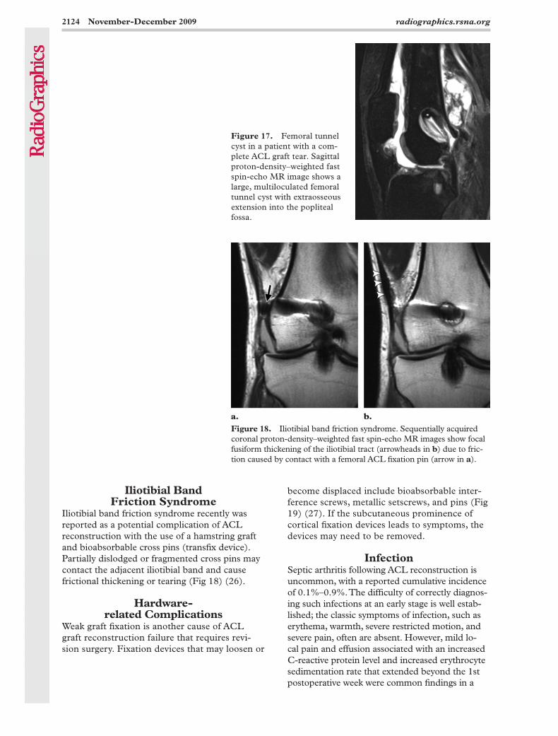
Figure 18. Iliotibial band friction syndrome. Sequentially acquired coronal proton-density–weighted fast spin-echo MR images show focal fusiform thickening of the iliotibial tract (arrowheads in b) due to fric-tion caused by contact with a femoral ACL fixation pin (arrow in a).
Figure 17. Femoral tunnel cyst in a patient with a com-plete ACL graft tear. Sagittal proton-density–weighted fast spin-echo MR image shows a large, multiloculated femoral tunnel cyst with extraosseous extension into the popliteal fossa.
Iliotibial Band Friction Syndrome
Iliotibial band friction syndrome recently was reported as a potential complication of ACL reconstruction with the use of a hamstring graft and bioabsorbable cross pins (transfix device). Partially dislodged or fragmented cross pins may contact the adjacent iliotibial band and cause frictional thickening or tearing (Fig 18) (26).
Hardware- related Complications
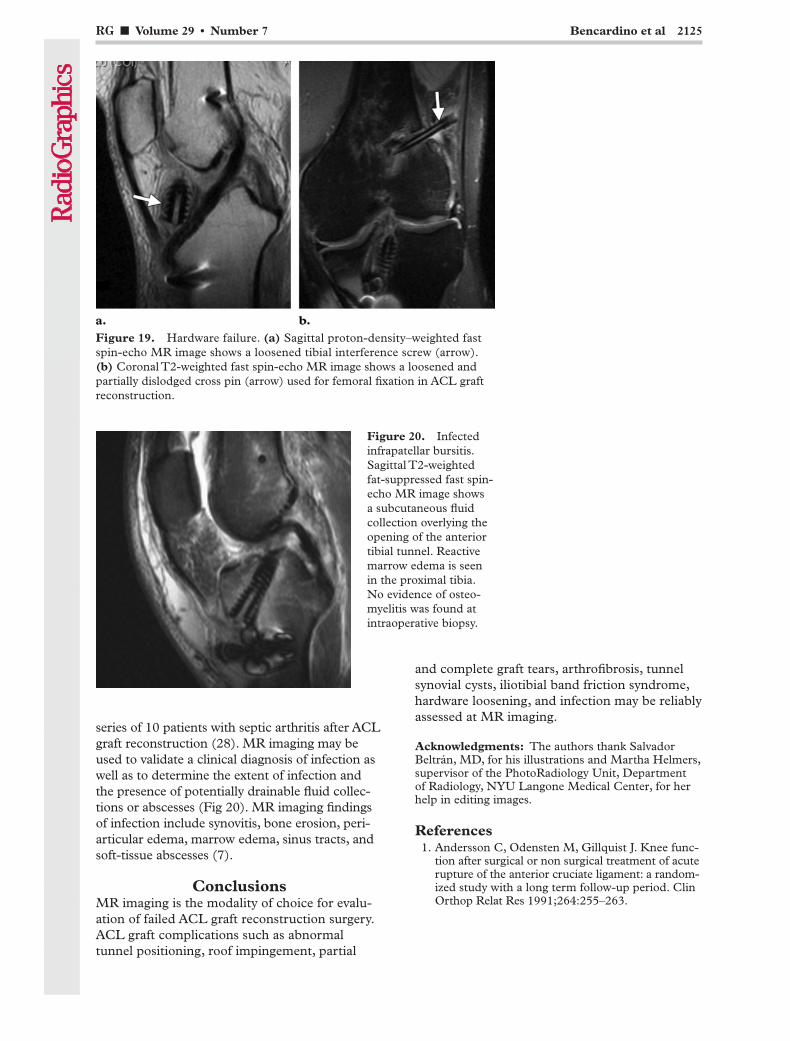
Weak graft fixation is another cause of ACL graft reconstruction failure that requires revi-sion surgery. Fixation devices that may loosen or
become displaced include bioabsorbable inter-ference screws, metallic setscrews, and pins (Fig 19) (27). If the subcutaneous prominence of cortical fixation devices leads to symptoms, the devices may need to be removed.
InfectionSeptic arthritis following ACL reconstruction is uncommon, with a reported cumulative incidence of 0.1%–0.9%. The difficulty of correctly diagnos-ing such infections at an early stage is well estab-lished; the classic symptoms of infection, such as erythema, warmth, severe restricted motion, and severe pain, often are absent. However, mild lo-cal pain and effusion associated with an increased C-reactive protein level and increased erythrocyte sedimentation rate that extended beyond the 1st postoperative week were common findings in a
RG ■ Volume 29 • Number 7 Bencardino et al 2125
Figure 19. Hardware failure. (a) Sagittal proton-density–weighted fast spin-echo MR image shows a loosened tibial interference screw (arrow). (b) Coronal T2-weighted fast spin-echo MR image shows a loosened and partially dislodged cross pin (arrow) used for femoral fixation in ACL graft reconstruction.
series of 10 patients with septic arthritis after ACL graft reconstruction (28). MR imaging may be used to validate a clinical diagnosis of infection as well as to determine the extent of infection and the presence of potentially drainable fluid collec-tions or abscesses (Fig 20). MR imaging findings of infection include synovitis, bone erosion, peri-articular edema, marrow edema, sinus tracts, and soft-tissue abscesses (7).
ConclusionsMR imaging is the modality of choice for evalu-ation of failed ACL graft reconstruction surgery. ACL graft complications such as abnormal tunnel positioning, roof impingement, partial
and complete graft tears, arthrofibrosis, tunnel synovial cysts, iliotibial band friction syndrome, hardware loosening, and infection may be reliably assessed at MR imaging.
Acknowledgments: The authors thank Salvador Beltrán, MD, for his illustrations and Martha Helmers, supervisor of the PhotoRadiology Unit, Department of Radiology, NYU Langone Medical Center, for her help in editing images.
References 1. Andersson C, Odensten M, Gillquist J. Knee func-
tion after surgical or non surgical treatment of acute rupture of the anterior cruciate ligament: a random-ized study with a long term follow-up period. Clin Orthop Relat Res 1991;264:255–263.
Figure 20. Infected infrapatellar bursitis. Sagittal T2-weighted fat-suppressed fast spin-echo MR image shows a subcutaneous fluid collection overlying the opening of the anterior tibial tunnel. Reactive marrow edema is seen in the proximal tibia. No evidence of osteo-myelitis was found at intraoperative biopsy.
2126 November-December 2009 radiographics.rsna.org
2. O’Brien SJ, Warren RF, Pavlov H, Panariello R, Wickiewicz TL. Reconstruction of the chronically insufficient anterior cruciate ligament with the cen-tral third of the patellar tendon. J Bone Joint Surg Am 1991;73:278–286.
3. Lebel B, Hulet C, Galaud B, Burdin G, Locker B, Vielpeau C. Arthroscopic reconstruction of the an-terior cruciate ligament using bone-patellar tendon-bone autograft: a minimun 10-year follow-up. Am J Sports Med 2008;36:1275–1282.
4. Lahav A, Burks RT. Evaluation of the failed ACL reconstruction. Sports Med Arthrosc 2005;13:8–16.
5. Lind M, Menhert F, Pedersen AB. The first results from the Danish ACL reconstruction registry: epi-demiologic and 2 year follow-up results from 5818 knee ligament reconstructions. Knee Surg Sports Traumatol Arthrosc 2009;17:117–124.
6. Recht MP, Kramer J. MR imaging of the postopera-tive knee: a pictorial essay. RadioGraphics 2002;22: 765–774.
7. Papakonstantinou O, Chung CB, Chanchairujira K, Resnick DL. Complications of anterior cruciate ligament reconstruction: MR imaging. Eur Radiol 2003;13:1106–1117.
8. Spindler KP, Kuhn JE, Freedman KB, Matthews CE, Dittus RS, Harrell FE Jr. Anterior cruciate ligament reconstruction autograft choice: bone-tendon-bone versus hamstring: does it really matter? A systematic review. Am J Sports Med 2004;32:1986–1995.
9. Rispoli DM, Sanders TM, Miller MD, Morrison WB. Magnetic resonance imaging at different time periods following hamstring harvest for anterior cruciate liga-ment reconstruction. Arthroscopy 2001;17:2–8.
10. Aglietti P, Buzzi R, Zacherotti G, De Biase P. Pa-tellar tendon versus doubled semitendinosus and gracilis tendons for anterior cruciate ligament re-construction. Am J Sports Med 1994;22:211–217.
11. Fineberg MS, Zarins B, Sherman OH. Practical considerations in anterior cruciate ligament replace-ment surgery. Arthroscopy 2000;16:715–724.
12. Streich NA, Friedrich K, Gotterbarm T, Schmitt H. Reconstruction of the ACL with a semitendino-sus tendon graft: a prospective randomized single blinded comparison of double-bundle versus single bundle technique in male athletes. Knee Surg Sports Traumatol Arthrosc 2008;16:232–238.
13. Howell SM. Principles for placing the tibial tunnel and avoiding roof impingement during reconstruc-tion of a torn anterior cruciate ligament. Knee Surg Sports Traumatol Arthrosc 1998;6(suppl 1):S49–S55.
14. Steiner ME, Hecker AT, Brown CH Jr, Hayes WC. Anterior cruciate ligament graft fixation: compari-son of hamstring and patellar tendon grafts. Am J Sports Med 1994;22:240–246.
15. Saupe N, White LM, Chiavaras MM, et al. Anterior cruciate ligament reconstruction grafts: MR imaging
features at long-term follow-up—correlation with functional and clinical evaluation. Radiology 2008; 249:581–590.
16. Collins MS, Unruh KP, Bond JR, Mandrekar JN. Magnetic resonance imaging of surgically confirmed anterior cruciate ligament graft disruption. Skeletal Radiol 2008;37:233–243.
17. Jackson DW, Schaefer RK. Cyclops syndrome: loss of extension following intra-articular anterior cruci-ate ligament reconstruction. Arthroscopy 1990;6: 171–178.
18. Marzo JM, Bowen MK, Warren RF, Wickiewicz TL, Altchek DW. Intraarticular fibrous nodule as a cause of loss of extension following anterior cruciate liga-ment reconstruction. Arthroscopy 1992;8:10–18.
19. Bradley DM, Bergman AG, Dillingham MF. MR imaging of cyclops lesions. AJR Am J Roentgenol 2000;174:719–726.
20. Muellner T, Kdolsky R, Grossschmidt K, Schabus R, Kwasny O, Plenk H Jr. Cyclops and cyclopoid formation after anterior cruciate ligament recon-struction: clinical and histomorphological differ-ences. Knee Surg Sports Traumatol Arthrosc 1999; 7:284–289.
21. Mayr HO, Weig TG, Plitz W. Arthrofibrosis follow-ing ACL reconstruction: reasons and outcome. Arch Orthop Trauma Surg 2004;124:518–522.
22. Sanders TG, Tall MA, Mulloy JP, Leis HT. Fluid col-lections in the osseous tunnel during the first year af-ter anterior cruciate ligament repair using an autolo-gous hamstring graft: natural history and clinical cor-relation. J Comput Assist Tomogr 2002;26:617–621.
23. Deie M, Sumen Y, Ochi M, Murakami Y, Fujimoto E, Ikuta Y. Pretibial cyst formation after anterior cruciate ligament reconstruction using auto ham- string grafts: two case reports in a prospective study of 89 cases. Magn Reson Imaging 2000;18: 973–977.
24. Victoroff BN, Paulos L, Beck C, Goodfellow DB. Subcutaneous pretibial cyst formation associated with anterior cruciate ligament allografts: a report of four cases and literature review. Arthroscopy 1995;11:486–494.
25. Simonian PT, Wickiewicz TL, O’Brien SJ, Dines JS, Schatz JA, Warren RF. Pretibial cyst formation after anterior cruciate ligament surgery with soft tissue autografts. Arthroscopy 1998;14:215–220.
26. Pelfort X, Monllau JC, Puig L, Caceres E. Iliotibial band friction syndrome after anterior cruciate liga-ment reconstruction using the transfix device: report of two cases and review of the literature. Knee Surg Sports Traumatol Arthrosc 2006;14:586–589.
27. Bush-Joseph CA, Bach BR Jr. Migration of femoral interference screw after anterior cruciate ligament reconstruction. Am J Knee Surg 1998;11:32–34.
28. Schollin-Borg M, Michaelsson K, Rahme H. Pre-sentation, outcome and cause of septic arthritis after anterior cruciate ligament reconstruction: a case control study. Arthroscopy 2003;19:941–947.
This article meets the criteria for 1.0 AMA PRA Category 1 CreditTM. To obtain credit, see www.rsna.org/education /rg_cme.html.
RG Volume 29 • Number 5 • November-December 2009 Bencardino et al
MR Imaging of Complications of Anterior Cruciate Ligament Graft Reconstruction
Jenny T. Bencardino, MD, et al
Page 2116 Magnetic resonance (MR) imaging is the preferred advanced imaging modality for the evaluation of symptomatic ACL graft reconstructions. Page 2116 Positioning of the femoral and tibial tunnels is of paramount importance for proper function of the ACL graft. Page 2120 Roof impingement often is secondary to an abnormal position of the tibial tunnel anterior to the intersection of the Blumensaat line and the tibia when the knee is fully extended. Page 2121 Complete graft disruption often is a result of recurrent trauma. Nonisotropic positioning of the graft tunnel resulting in abnormal stress to the graft during the normal range of motion also has been implicated as a cause of complete graft tear. Page 2122 Localized anterior arthrofibrosis, or “cyclops” lesion, has been reported in 1%–10% of patients with ACL reconstruction.
RadioGraphics 2009; 29:2115–2126 • Published online 10.1148/rg.297095036 • Content Codes: