The sway-density curve and the underlying postural stabilization process
Upload
independentCategory
view
1download
0
Journal of Athletic Training 2013;48(2):172–185doi: 10.4085/1062-6050-48.2.05� by the National Athletic Trainers’ Association, Incwww.natajournals.org
original research
Lower Limb Kinematics and Dynamic Postural Stabilityin Anterior Cruciate Ligament-Reconstructed FemaleAthletes
Eamonn Delahunt, PhD, BSc; Mark Chawke, BSc; Judy Kelleher, BSc;Katie Murphy, BSc; Anna Prendiville, BSc; Lauren Sweeny, BSc;Matt Patterson, MSc, BSc
University College Dublin, Ireland
Context: Deficits in lower limb kinematics and posturalstability are predisposing factors to the development of kneeligamentous injury. The extent to which these deficits arepresent after anterior cruciate ligament (ACL) reconstruction isstill largely unknown.The primary hypothesis of the present study was that femaleathletes who have undergone ACL reconstruction and who havereturned to sport participation would exhibit deficits in dynamicpostural stability as well as deficiencies in hip- and knee-jointkinematics when compared with an age-, activity-, and sex-matched uninjured control group.
Objective: To investigate dynamic postural stability asquantified by the Star Excursion Balance Test (SEBT) andsimultaneous hip- and knee-joint kinematic profiles in femaleathletes who have undergone ACL reconstruction.
Design: Descriptive laboratory study.Setting: University motion-analysis laboratory.Patients or Other Participants: Fourteen female athletes
who had previously undergone ACL reconstruction (ACL-R) and17 age- and sex-matched uninjured controls.
Intervention(s): Each participant performed 3 trials of the
anterior, posterior-medial, and posterior-lateral directional com-ponents of the SEBT.
Main Outcome Measure(s): Reach distances for eachdirectional component were quantified and expressed as apercentage of leg length. Simultaneous hip- and knee-jointkinematic profiles were recorded using a motion-analysis system.
Results: The ACL-R group had decreased reach distanceson the posterior-medial (P , .01) and posterior-lateral (P , .01)directional components of the SEBT. During performance of thedirectional components of the SEBT, ACL-R participantsdemonstrated altered hip-joint frontal-, sagittal-, and transverse-plane kinematic profiles (P , .05), as well as altered knee-jointsagittal-plane kinematic profiles (P , .05).
Conclusions: Deficits in dynamic postural stability andconcomitant altered hip- and knee-joint kinematics are presentafter ACL reconstruction and return to competitive activity. Theextent to which these deficits influence potential future injury isworthy of investigation.
Key Words: anterior cruciate ligament, reconstruction,kinematics, postural stability
Key Points
� Female athletes who have undergone anterior cruciate ligament (ACL) reconstruction and returned to full sportparticipation have decreased dynamic postural stability as quantified by the posterior-medial, and posterior-lateralreach directions of the Star Excursion Balance Test (SEBT).
� Female athletes who have undergone ACL reconstruction and returned to full sport participation have alteredkinematic patterns when performing the anterior, posterior-medial, and posterior-lateral reach directions of theSEBT.
� Rehabilitation after ACL reconstruction should incorporate the SEBT as a dynamic postural-stability training tool andan evaluative tool for monitoring rehabilitation progression.
� Ongoing neuromuscular-training programs should be incorporated into daily training and practice sessions ofathletes who have undergone ACL reconstruction.
Anterior cruciate ligament (ACL) rupture is a sportinjury frequently incurred by athletes who areengaged in field and court sports and is of
particular concern for clinicians who specialize in thetreatment and rehabilitation of such athletes. A highproportion of ACL injuries are the result of a noncontactmechanism, with reports in the literature1–3 suggesting thatfemale athletes have a higher prevalence of noncontactACL injuries than male athletes.
Of particular concern to clinicians and surgeons is therisk of a subsequent ACL injury after return to sportparticipation after ACL reconstruction. Wright et al4
reported a reinjury incidence of 1 in 17 athletes, which isconsiderably higher than other reported5,6 rates for initialACL injury of 1 in 60 to 100 athletes. In a 5-year follow-upstudy of ACL-reconstructed (ACL-R) patients, Shelbourneet al7 noted that the risk of subsequent ACL injury to eitherknee was 17% for patients younger than 18 years, 7% for
172 Volume 48 � Number 2 � April 2013

patients aged 18 to 25 years, and 4% for patients older than25 years.
The neuromuscular system comprises all of the sensory-,motor-, and central-integration and -processing componentsthat govern the maintenance of joint homeostasis duringdynamic movement and thus is responsible for overallfunctional joint stability.8 The ACL plays an integral role inknee-joint proprioception,9 and thus, ACL injury influenceslower limb neuromuscular control and joint stability.10
Authors of a number of recently published studies11–13 haveexamined lower limb gait kinematics after ACL recon-struction; all showed that knee-joint mechanics are alteredpostsurgery. Studies using more challenging tasks, such aspivoting and jump landing, have also demonstrated deficitsin knee-joint mechanics after ACL reconstruction.14–16 Theresults of the aforementioned studies, although informative,are limited because the primary focus has been on knee-joint mechanics with little consideration being given toproximal structures. Delahunt et al17 observed altered hip-and knee-joint angular displacements in female ACL-Rathletes who had returned to sport participation whenperforming a high-velocity, high-load jump-landing sport-specific task. In a systematic review, Quatman et al18
suggested that neuromuscular deficiencies involving thefrontal, sagittal, and transverse planes are likely to beimplicated in the mechanism of and predisposition tononcontact ACL injury. Direct support for the multiplanarmechanism of injury is evidenced in a cohort study thatshowed altered neuromuscular control of the hip and kneejoint and deficits in postural stability predicted reinjuryafter ACL reconstruction.19
Observing knee and hip kinematics while ACL-R athletesperform a dynamic postural-control task may help to shed
light on deficiencies during activity and may contribute tothe development of more effective rehabilitation programsafter ACL reconstruction. One mechanism to accomplishthis would be to combine dynamic postural-stabilityevaluation as quantified by the Star Excursion BalanceTest (SEBT) with simultaneous lower limb kinematicevaluation using a motion-analysis capture system. Perfor-mance on the SEBT places multiple demands on theneuromuscular system and, thus, may facilitate theidentification of athletes who are at a greater risk of lowerlimb injury.20 Previous authors21,22 have reported thatparticipants with chronic ankle instability (CAI) havedecreased reach distances when compared with uninjuredparticipants, reflecting a deficiency in dynamic posturalstability. Furthermore, Gribble et al22 indicated that thedecreased reach distance observed in the CAI group couldbe a manifestation of a decreased knee-flexion angle duringthe test, an effect that was amplified by the induction ofmuscular fatigue. To date, to our knowledge, only 1 group23
has investigated dynamic postural stability as quantified bythe SEBT in ACL-injured athletes; reach distances weredecreased in the ACL-deficient group when compared withan uninjured control group.
Considering the importance of dynamic postural stabilityas a neuromuscular factor and its importance as a clinicalmeasure in predicting the risk of future injury,19,20 ourobjective was to investigate dynamic postural stability asquantified by the SEBT and simultaneous hip- and knee-joint kinematics in participants with previous ACLreconstructions. We hypothesized that, when comparedwith an uninjured control group, the ACL-R female athleteswould demonstrate decreased reach distances on the SEBTas well as deviations from the kinematic profiles observedin the uninjured control group.
METHODS
Participants
Seventeen female athletes (age ¼ 20.76 6 1.14 years,height ¼ 1.65 6 0.06 m, body mass ¼ 65.38 6 7.36 kg,body mass index (BMI)¼ 23.82 6 2.42 kg/m2) volunteeredas controls. None of these athletes had previouslyexperienced a knee injury. All athletes played field orcourt-based sports (eg, Gaelic football, soccer, hockey,basketball) at the club or county level.
Fourteen recreational female athletes (age ¼ 23.00 63.37 years, height¼ 1.64 6 0.05 m, body mass¼ 64.85 68.67 kg, BMI ¼ 24.03 6 2.38 kg/m2) volunteered to
Table 1. IKDC Subjective Knee Form and KOOS Subscale Resultsa
IKDC KOOSpain KOOSsymptoms KOOSADL KOOSsport KOOSKQoL
Control group 99.12 6 3.62 99.82 6 0.72 98.23 6 2.61 99.82 6 0.72 99.41 6 2.42 99.29 6 1.99
ACL-R group 82.18 6 11.83b 92.00 6 11.32b 85.78 6 9.29b 98.07 6 4.44 81.07 6 15.83b 72.00 6 15.15b
95% Confidence interval for the
mean difference between
groups
�23.93, �9.84 �14.40, �1.25 �17.93, �6.97 �4.33, 0.83 �27.53, �9.15 �36.08, �18.50
Effect size (g2) 0.24 (large) 0.14 (large) 0.23 (large) 0.08 (medium) 0.21 (large) 0.29 (large)
Abbreviations: ACL-R, anterior cruciate ligament reconstructed; IKDC, International Knee Documentation Committee Subjective KneeForm; KOOS, Knee Injury and Osteoarthritis Outcome Score; KOOSADL, subscale for function in daily living; KOOSKQoL, subscale for knee-related quality of life.a Values are presented as mean 6 SD.b Different from control group.
Table 2. Star Excursion Balance Test Resultsa
Reach Direction
Anterior Posterior-medial Posterior-lateral
Control group 71.25 6 4.35 105.06 6 7.68 98.87 6 8.59
ACL-R group 68.54 6 3.82 96.06 6 7.56b 89.53 6 7.42b
95% Confidence
interval for the
mean difference
between groups
�0.33, 5.76 3.37, 14.63 3.36, 15.30
Effect size (g2) 0.10 (moderate) 0.17 (large) 0.17 (large)
Abbreviation: ACL-R, anterior cruciate ligament reconstructed.a Values are presented as mean 6 SD and represent a percentage
of limb length.b Different from control group.
Journal of Athletic Training 173

participate. All athletes had previously experienced anisolated noncontact ACL injury that required surgicalstabilization. Seven athletes had surgical stabilization usinga bone-patellar tendon-bone autograft, with the remainingathletes having had a hamstring autograft. The mean timefrom surgical stabilization to the study was 2.9 6 2.8 years(range, 10 months–6 years). At the time of testing, allathletes were fully engaged in field or court-based sports(eg, Gaelic football, soccer, hockey, basketball) at the clubor county level, and no athlete was undergoing any form offormal rehabilitation.
Procedures
All testing was undertaken in a university motion-analysis laboratory. Each participant attended the laborato-ry on 1 occasion. Upon arrival for the testing session, eachparticipant was informed and familiarized with the testingprocedure. All testing was supervised by a charteredphysiotherapist. Each participant signed an informedconsent form, which had previously been approved by theUniversity Human Research Ethics Committee, which alsoapproved the study.
Subjective Knee Questionnaires
All athletes were required to complete the InternationalKnee Documentation Committee (IKDC) Subjective KneeForm as well as the Knee Injury and OsteoarthritisOutcome Score (KOOS) subscales.
The IKDC Subjective Knee Form and KOOS are site-specific instruments that were designed to measuresymptoms, function, and sport activity in patients with avariety of knee conditions.24,25 Both instruments have beenvalidated for use with an ACL-R population.25,26
The IKDC Subjective Knee Form consists of 18 itemsrelated to knee-joint symptoms, function, and sportactivity.24 All items are summed to produce a single indexscore, which can be used to interpret knee function; higherscores represent higher levels of function and lower levelsof symptoms. A score of 100 is considered no limitation inactivities of daily living or sport activities and completeabsence of symptoms.24
The KOOS is a 42 self-reporting instrument andcomprises 5 subscales: KOOSpain (9 items), KOOSsymptoms
(7 items), KOOSADL (function in daily living; 17 items),KOOSsport (5 items), and KOOSKQoL (knee-related qualityof life; 4 items). For each subscale, the score is normalizedto a 0–100 scale with higher scores representing betterlevels of knee status.
By using both scales, we endeavored to determine howACL reconstruction influences overall knee-joint functionand symptoms as well as important specific domains suchas those included in the KOOS.
SEBT Performance
For the performance of the SEBT, participants initiallystood barefoot with their left and right feet on 2 adjacentforce plates. The directional components of the SEBT
Figure 1. Hip-joint adduction-abduction angle during performance of the anterior directional component of the Star Excursion BalanceTest (SEBT). Adduction is positive; abduction is negative; values are mean 6 SEM. Shaded area¼ area of statistical significance. Dashedline¼ point of maximum reach. Abbreviation: ACLR, anterior cruciate ligament-reconstructed.
174 Volume 48 � Number 2 � April 2013

chosen for the present investigation were the anterior,posterior-medial, and posterior-lateral reach directions. Thedecision to use these specific reach directions was based onresearch by Plisky et al20 and Hertel et al.27
Before each test session, participants were instructed inthe correct performance of the test and allowed 4 practicetrials in each of the chosen directional components, asadvocated by Robinson and Gribble.28 Three consecutivetrials in each direction were performed after a short restperiod. The order of performance of each directionalcomponent was randomized across participants using arandom sequence of number generation.
While performing each reach direction, the participantinitially stood with the big toe positioned at the center of agrid laid on the laboratory floor and extending from theforce plate directly under the stance (test) leg. Each trialwas initiated when the participant transitioned from double-to single-legged stance, and the trial ended when shereturned to the double-legged stance position.29 The verticalcomponent of ground reaction force data was used todetermine the onset and end of each trial as previouslydescribed in Delahunt et al.29 For ease in quantifying eachreach distance, the line of each SEBT directionalcomponent was simulated by a 1.5-m measuring tape.Reach distances were read from the center of the grid to thepoint of maximum reach, which was visually observed andnoted by one of the investigators. Reach distances weredivided by limb length, as measured from the anterior-superior iliac spine to the ipsilateral medial malleolus, and
multiplied by 100 to calculate a dependent variable thatrepresents reach distance as a percentage of limb length.30
During each trial, the participant placed her hands on herhips while reaching in the specified directional component.Furthermore, the participant was required to maintaincontact between the force plate and heel of the stance(test) leg during each trial. A trial was deemed unsuccessfulif she failed to keep her hands on her hips, moved or liftedthe stance (test) foot, transferred weight onto the reach footwhen touching the measuring tape, failed to touch the tape,failed to return the reach foot to the starting position, or losther balance and was unable to maintain a unilateral stanceposition during the trial. Unsuccessful trials were discarded,and additional trials were completed accordingly.
Kinematic Analysis
Kinematic data acquisition occurred at 200 Hz using 3CODA mpx1 units (Charnwood Dynamics Ltd, Leicester-shire, UK), which were fully integrated with 4 AMTI(Watertown, MA) walkway embedded force plates (sam-pled at 100 Hz).
We marked each participant’s specific anatomicallandmarks with a skin-marker pencil before recording heranthropometric values and attaching the markers andmarker wands as outlined in Monaghan et al.31,32 Themarkers and marker wands were applied by the sameinvestigator for all participants as follows. Markers werepositioned on the lateral aspect of the knee joint line in themedian frontal plane, the anterior aspect of the lateral
Figure 2. Hip-joint flexion-extension angle during performance of the anterior directional component of the Star Excursion Balance Test(SEBT). Flexion is positive; extension is negative; values are mean 6 SEM. Shaded area¼ area of statistical significance. Dashed line ¼point of maximum reach. Abbreviation: ACLR, anterior cruciate ligament-reconstructed.
Journal of Athletic Training 175

malleolus, the posterior-inferior lateral aspect of the heel,and the lateral aspect of the fifth metatarsal head. Wandswith anterior and posterior markers attached were posi-tioned on the pelvis and sacrum, the thigh, and the shank.This specific marker and wand setup has been previouslyused in our laboratory.31–35 A neutral-stance trial was usedto align the participant with the laboratory coordinatesystem and to function as a reference position forsubsequent kinematic analysis.
Kinematic data were calculated by comparing the angularorientations of the coordinate systems of adjacent limbsegments using the angular coupling set ‘‘Euler Angles’’ torepresent clinical rotations in 3 dimensions. Markerpositions within a Cartesian frame are processed intorotation angles using vector algebra and trigonometry(CODA mpx30 User Guide, Charnwood Dynamics Ltd,Leicestershire, UK). Joint angular displacements werecalculated for the hip and knee joints in the sagittal,frontal, and transverse planes. Kinematic data wereanalyzed using the CODA software, with the followingaxis conventions: x axis ¼ frontal-plane motion; y ¼sagittal-plane motion; z ¼ transverse-plane motion. Kine-matic data for each SEBT trial were extracted andconverted to Microsoft Excel file format by convertingthe number of output samples to 100þ 1 in the data-exportoption of the CODA software, which represented thecomplete SEBT trial as 100%, for averaging and furtheranalysis. The 3 normalized trials for each participant foreach reach direction were combined to create an averageensemble curve for each participant, with group profiles
then being calculated. This specific analysis technique haspreviously been used in our laboratory.31–35
Statistical Analysis
The sample size calculation for the present study wasbased on data previously published by Herrington et al,23
who showed a difference in SEBT performance betweenACL-deficient and control participants. The computedeffect size for the difference in reach distance betweenthe ACL-deficient and control participants in the anteriordirection was 1.3. An a priori sample size of 14 participantsper group was computed using G*Power36 considering thefollowing values: a¼ .05, 1 – b ¼ .9.
Independent-samples t tests were used to assess differ-ences in the IKDC Subjective Knee Form and KOOSsubscale scores (PASW Statistics, 24 Version 18.0, IBMCorporation, NY). Associated effect sizes (g2) werecalculated using the formula as described in Pallant:37 t2/t2þ (N1þN2� 2) and quantified according to Cohen38 as0.01 ¼ small effect size, 0.06 ¼ medium effect size, and0.14¼ large effect size. The level of significance was set atP , .05.
Independent-samples t tests were used to test fordifferences in normalized reach distances on the SEBT.Associated effect sizes (g2) were calculated using theformula as described in Pallant:37 t2/t2þ (N1þN2� 2) andquantified according to Cohen38 as noted earlier. The levelof significance was set at P , .05. However, due to thehighly correlational nature of these data, an adjusted P
Figure 3. Knee-joint flexion-extension angle during performance of the anterior directional component of the Star Excursion Balance Test(SEBT). Flexion is positive; extension is negative; values are mean 6 SEM. Shaded area¼ area of statistical significance. Dashed line¼point of maximum reach. Abbreviation: ACLR, anterior cruciate ligament-reconstructed.
176 Volume 48 � Number 2 � April 2013
value of (0.05/3) and therefore an a level of P , .016 wasset for all comparisons.
Time-averaged profiles for hip- and knee-joint kine-matics were calculated for each participant, with groupmean profiles then being calculated. Differences in ACL-R and control group time-averaged profiles were testedfor statistical significance using independent-samples ttests for each data point. This specific analysis techniquehas previously been used in our laboratory.31–35 Effectsizes were not calculated for this part of the data analysisdue to the number of separate comparisons for eachkinematic variable. The level of significance was set at P, .05.
RESULTS
Subjective Knee Questionnaires
The Levene test for equality of variance revealed that theassumption of equal variance was violated for all subjectivemeasures. The ACL-R participants differed from thecontrol participants on the IKDC Subjective Knee Form(P , .001), as well as on the KOOSpain (P , .05),KOOSsymptoms (P , .001), KOOSsport (P , .05), andKOOSKQoL (P , .001). The associated effects sizes werelarge (Table 1). No difference was seen between the ACL-R participants and the control participants on the KOOSADL
(P . .05).
SEBT Performance
A difference between groups was observed for both theposterior-medial (P , .005) and posterior-lateral reachdirections (P , .005). The associated effect sizes were both0.17, indicating a large effect size for both directions (Table2). No difference between groups was observed for theanterior reach direction (P . .016), with an associatedmoderate effect size of 0.10 (Table 2).
Kinematics
Differences were observed between the kinematicprofiles of the ACL-R group and control group duringperformance of the anterior, posterior-medial, and posteri-or-lateral directional components of the SEBT (Figures 1–9). Kinematic differences were observed for hip-jointfrontal-, sagittal-, and transverse-plane motion, as well asknee-joint sagittal-plane motion.
During performance of the anterior reach direction, theACL-R group differed from the control group (P , .05),exhibiting increased hip adduction (entire trial, Figure 1),less hip flexion (3%–89% of trial, Figure 2), and less kneeflexion (12%–65% of trial, Figure 3).
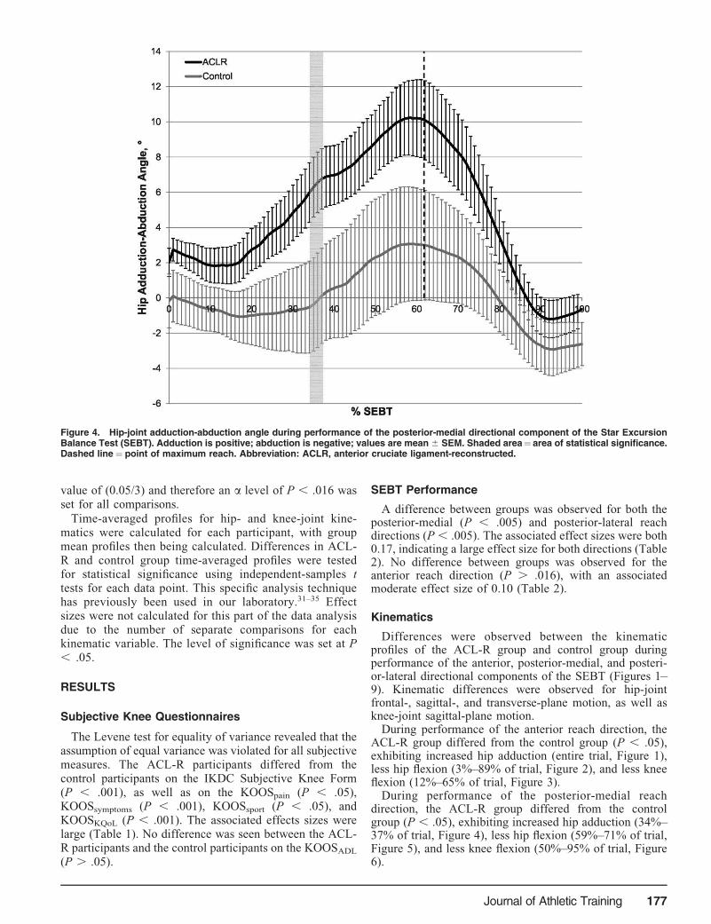
During performance of the posterior-medial reachdirection, the ACL-R group differed from the controlgroup (P , .05), exhibiting increased hip adduction (34%–37% of trial, Figure 4), less hip flexion (59%–71% of trial,Figure 5), and less knee flexion (50%–95% of trial, Figure6).
Figure 4. Hip-joint adduction-abduction angle during performance of the posterior-medial directional component of the Star ExcursionBalance Test (SEBT). Adduction is positive; abduction is negative; values are mean 6 SEM. Shaded area¼ area of statistical significance.Dashed line ¼ point of maximum reach. Abbreviation: ACLR, anterior cruciate ligament-reconstructed.
Journal of Athletic Training 177

During performance of the posterior-lateral reach direc-tion, the ACL-R group differed significantly from thecontrol group (P , .05), exhibiting increased hip adduction(94%–100% of trial, Figure 7), less hip external rotation(14%–53% and 79%–86%, Figure 8), as well as less kneeflexion (19%–72% and 89%–100% of trial, Figure 9).
DISCUSSION
The specific aim of our study was to investigate dynamicpostural stability, as quantified by the SEBT, andsimultaneous hip- and knee-joint kinematic profiles infemale athletes who have undergone ACL reconstruction.The results indicated that ACL-R athletes displayeddecreased normalized reach distances on the posterior-medial and posterior-lateral reach directions of the SEBTwhen compared with an uninjured control group. Further-more, specific kinematic differences were observed be-tween the groups.
Subjective Knee Questionnaires
We observed a difference for the IKDC Subjective KneeForm scores between the ACL-R and control groups. Theeffect size was large, which is consistent with the clinicallymeaningful effect based on a .9-point difference betweenthe groups.12 Our results compare favorably with those ofLee et al,39 who reported that, at 5 years after ACLreconstruction, the mean IKDC Subjective Knee Formscore was 79.5.
We noted a difference between the ACL-R and controlgroups on 4 of the KOOS subscales. The associated effectsizes were all large, which is consistent with the clinicallymeaningful effect based on a 10-point difference betweenthe groups,26 which is also supported by previouslypublished data.40,41
Even upon return to full sport participation, femaleathletes who have undergone ACL reconstruction stillreport deficits in knee function and ongoing kneesymptoms, which affect their sport performance and qualityof life. Taken together, the results of the IKDC SubjectiveKnee Form and KOOS subscales suggest that considerationshould be given to ongoing therapeutic intervention afterreturn to sport following ACL reconstruction; too often,interventions are discontinued by athletes and cliniciansafter return to play.
SEBT Performance
A recent systematic review has suggested that futureauthors examining postural stability in patients undergoingACL reconstruction or after reconstruction should focus onassessments using dynamic postural-stability tasks.42 TheSEBT has been previously established as a reliable andsensitive measure of dynamic postural stability.43,44 Al-though the SEBT has been most often used as a measure ofdynamic postural stability in participants with CAI,45–49 todate, only 1 paper has been published on SEBTperformance and ACL injury.23
Figure 5. Hip-joint flexion-extension angle during performance of the posterior-medial directional component of the Star ExcursionBalance Test (SEBT). Flexion is positive; extension is negative; values are mean 6 SEM. Shaded area ¼ area of statistical significance.Dashed line ¼ point of maximum reach. Abbreviation: ACLR, anterior cruciate ligament-reconstructed.
178 Volume 48 � Number 2 � April 2013

Our results support our original hypothesis that ACL-Rfemale athletes would demonstrate decreased posturalstability on specific directional components of the SEBTwhen compared with an age-, sex-, and activity-matcheduninjured control group and are in agreement with previousfindings that competitive athletes who have returned to fullsport participation after ACL reconstruction still exhibitpostural-control deficits.50,51
In the present study, ACL-R participants’ reach distanceson the posterior-medial and posterior-lateral directions ofthe SEBT were decreased. These observations are inagreement with those of Herrington et al,23 who reportedsimilar deficits in reach distances in ACL-deficient athletes.We hypothesize that the decreased performance on theposterior-medial and posterior-lateral directional compo-nents could be a manifestation of the multiplanarneuromuscular demands and rotator torques requiredaround the hip and knee joints associated with thesedirectional components. The potential link between de-creased reach distances and joint angular displacementswill be discussed in the next section (Kinematics).Interestingly, no between-groups difference was observedfor the anterior reach direction.
A deficit in postural stability is a risk factor for futurelower limb injury.20,52 Thus, intervention programs afterACL injury should emphasize both static and dynamicpostural-stability training because we have shown thatpostural-stability deficits are still evident, even after returnto full sport participation, in ACL-R female athletes.
Kinematics
Our ACL-R group was characterized by multiplanar hip-and knee-joint kinematic deficiencies when compared withthe control group while undergoing dynamic postural-stability testing on the SEBT. Several theories regarding themechanism of ACL injury exist in the published literatureand are eloquently described by Quatman et al18 in a recentsystematic review. The sagittal-plane mechanism of ACLinjury is based on the premise that the ACL is the primaryrestraint to anterior translation of the tibia53,54 and that, atlow knee-flexion angles, the sagittal-plane displacement ofthe tibia is increased.55,56 Furthermore, it is postulated thatthe small knee-flexion angle commonly observed with ACLinjury allows a powerful contraction of the quadriceps,which has sufficient force to cause excessive anterior tibialtranslation and subsequent ACL injury.57–60 In our study,decreased knee flexion in the ACL-R group duringperformance of the 3 SEBT reach directions was aconsistent finding, thus supporting previously publisheddata concerning a sagittal-plane deficit association withACL injury. The frontal-plane mechanism of ACL injury isbased on the premise that knee-joint abduction collapse isthe inciting event.61,62 A prospective study by Hewett et al63
indicated that female athletes who sustained a noncontactACL injury during the study period exhibited a greaterknee-valgus kinematic profile during a drop vertical jumpwhen compared with athletes who did not sustain such aninjury. More recently published literature18,64 suggestedthat ACL injury is most likely the result of multiplanar
Figure 6. Knee-joint flexion-extension angle during performance of the posterior-medial directional component of the Star ExcursionBalance Test (SEBT). Flexion is positive; extension is negative; values are mean 6 SEM. Shaded area ¼ area of statistical significance.Dashed line ¼ point of maximum reach. Abbreviation: ACLR, anterior cruciate ligament-reconstructed.
Journal of Athletic Training 179

neuromuscular deficits. The present results are consistentwith the literature suggesting that ACL injury is associatedwith multiplanar movement deficiencies. Specifically,differences in frontal- and transverse-plane hip-jointkinematics were observed between the ACL-R and controlgroups, with the ACL-R group being characterized byincreased hip-joint adduction and internal rotation.
During performance of the anterior reach direction of theSEBT, the ACL-R group was characterized by a moreadducted position of the hip joint throughout the entire trial.They also displayed less hip flexion from 3% to 89% of thetrial as well as less knee flexion from 12% to 65% of thetrial. However, even though the aforementioned kinematicdeficits were observed, the ACL-R group did not differfrom the control group in anterior reach distance. Previousresearchers65 have suggested that performance in theanterior reach direction of the SEBT is influenced bysagittal-plane kinematics. Participants with CAI are char-acterized by less hip and knee flexion while performing theanterior reach direction of the SEBT,22 a finding consistentwith our results. Even though significant emphasis has beenplaced on the importance of sagittal-plane kinematics forperformance of the anterior direction of the SEBT,65 thedecreased hip and knee flexion in the ACL-R group did nottranslate to a decreased anterior reach distance. We believethat the absence of a between-groups difference in reachdistance anteriorly may be attributed to the observedipsilateral increased hip adduction in the ACL-R group.This ipsilateral increased hip adduction essentially creates aTrendelenburg position, with lengthening of the contralat-
eral limb and a subsequent increased anterior reach, afinding supported by Zeller et al,66 who observed thatsingle-legged squat mechanics are influenced by frontal-plane hip-joint position. However, further support of thistheory needs full validation.
During performance of the posterior-medial reachdirection of the SEBT, the ACL-R group displayed a moreadducted position of the hip joint from 34% to 37% of thetrial, as well as less hip flexion from 59% to 71% of thetrial. They also exhibited less knee flexion from 50% to95% of the trial. The decreased hip and knee flexionobserved during the trial may be a manifestation of a deficitin quadriceps strength, but this would require furthervalidation because we did not measure this variable.
During performance of the posterior-lateral reach direc-tion of the SEBT, the ACL-R group was characterized bymore hip internal rotation from 14% to 53% and 79% to86% of the trial. Less knee flexion from 19% to 72% and89% to 100% of the trial was also observed. In contrast tothe anterior reach direction, we believe that the increasedadducted position of the hip joint during the posterior-medial reach direction and the increased internally rotatedposition of the hip joint during the posterior-lateral reachdirection could be contributing to the observed decreasedreach distance in both these directions. To a greater extent,the body’s trunk and center of mass are influenced by hip-joint frontal- and transverse-plane motion while reaching inthe posterior-medial and posterior-lateral reach directionsof the SEBT when compared with the anterior reachdirection. Greater hip adduction during the posterior-medial
Figure 7. Hip-joint adduction-abduction angle during performance of the posterior-lateral directional component of the Star ExcursionBalance Test (SEBT). Adduction is positive; abduction is negative; values are mean 6 SEM. Shaded area¼ area of statistical significance.Dashed line ¼ point of maximum reach. Abbreviation: ACLR, anterior cruciate ligament-reconstructed.
180 Volume 48 � Number 2 � April 2013

reach direction and greater hip internal rotation during theposterior-lateral reach direction cause lateral deviation ofthe trunk. Consequently, these changes in hip-joint frontal-and transverse-plane motion place greater rotary loads onthe knee joint than those observed with sagittal-planereaching. This may result in greater apprehension on thepart of the ACL-R group, with a consequent decrease inreach distance in the posterior-medial and posterior-lateralreach directions. Further research is necessary to determinewhether this is the case. However, recent evidence tosupport the connection among trunk position, knee-jointloading, and subsequent ACL injury has been put forth byHewett et al,67 whereby lateral deviation of the trunk withrespect to the ACL-injured limb was observed as part of theinjury mechanism.
These observations concur with previous findings in theliterature that female athletes perform jump landings withless hip and knee flexion.47 Furthermore, Delahunt et al17
reported altered hip-joint frontal- and transverse-planeangular displacements in ACL-R female athletes duringdrop vertical-jump testing. Thus, these deficits seem to beconsistent, even during a less demanding task such asdynamic postural stability, as assessed by performance inspecific reach directions of the SEBT. Our results suggestthat, after return to athletic participation, ACL-R femaleathletes still exhibit deficits in hip-joint sagittal-, frontal-,and transverse-plane kinematics, as well as knee-jointsagittal-plane kinematics. The exact consequence of thesedeficits and their potential influence on future injury,however, cannot yet be fully elucidated. Thus, prospective
studies are now required to determine the extent to whichaltered lower limb kinematics influence future injury risk inACL-R athletes.
Altered lower limb kinematics during the performanceof different landing tasks after ACL reconstruction arecommonly reported in the literature.14–17 However, noprevious authors have reported on the kinematic profilesof female ACL-R athletes during the performance of adynamic postural stability task such as the SEBT.Nonetheless, it is possible to suggest that the samerehabilitation principles could be applicable to bothsituations. Deficits in neuromuscular control as manifest-ed by aberrant lower limb kinematic profiles are amodifiable risk factor for future injury or reinjury afterinitial injury.68 Neuromuscular training programs incor-porating correction of landing techniques, posterior chainand core strengthening, plyometrics, and postural stabil-ity have been successfully implemented to reduce the riskof initial ACL injury.68 We suggest that similar protocolsshould be implemented after ACL reconstruction. It is theclinician’s responsibility to complete a thorough evalu-ation and develop an exercise program that addressesthose specific deficits that are present in ACL-R athletes.This will ensure better functional outcomes after ACLreconstruction and help prevent recurrent injury, as wellas improve physiotherapeutic and athletic trainingmethods in general. We suggest that performance on theSEBT could be used as an evaluative tool to monitorimprovements in dynamic postural-stability performance,lower limb alignment, coordination, and neuromuscular
Figure 8. Hip-joint internal-external rotation angle during performance of the posterior-lateral directional component of the StarExcursion Balance Test (SEBT). Internal rotation is positive; external rotation is negative; values are mean 6 SEM. Shaded area¼ area ofstatistical significance. Dashed line ¼ point of maximum reach. Abbreviation: ACLR, anterior cruciate ligament-reconstructed.
Journal of Athletic Training 181

control after ACL reconstruction. Treating cliniciansshould be aware of the correct execution of movementpatterns while providing continuing feedback to athletesregarding potentially injurious movement patterns, whichhave been shown to be linked with ACL injurymechanisms.19,20,64,67
The potential mechanisms underlying the kinematicdifferences observed in the present study can only behypothesized. Two potential mechanisms exist. Firstly, theobserved aberrant lower limb kinematic profiles may havebeen present before the ACL injury. Recently publishedliterature69,70 suggests that lower limb kinematic andkinetic changes during puberty may result in an increasedrisk of knee injury in female athletes when compared withage-matched male athletes. Thus, it is possible that theaberrant lower limb kinematic profiles we observed werepresent even before the inciting event. Alternatively, theobserved aberrant lower limb kinematics could be amanifestation of altered motor programming. Johansson etal9 suggested that ligament injury, through its influence onthe c muscle spindle system, could cause sensory feedbackthat does not fit the existing motor program. Consequent-ly, this may in turn induce errors in the normal activationsequence of muscles, resulting in altered joint kinematics.However, we did not measure lower limb muscle activity;thus, future research is warranted to investigate the alteredmotor-programming mechanism.
Clinical Applications
The presence of the observed aberrant lower limbkinematics at an average of 2.9 years after ACLreconstruction warrants consideration. Evidence-basedclinical practice involves the conscientious, judicious, andexplicit use of current clinical evidence to support decisionsregarding patient care.71 Although empirical clinicaljudgments are often indicated, to maintain the higheststandards of rehabilitation care, physiotherapists andathletic trainers must ensure that they have a correctunderstanding of those neuromuscular deficits that arepresent after ACL injury. Recent evidence regarding theneuromuscular deficits present in participants with CAIsuggests that this may not be the case,72 with experiencedclinicians displaying a less-than-satisfactory understandingof the neuromuscular deficits associated with CAI. Wehypothesize that this may be the case regarding ACL injury.Further research should be undertaken to determineclinicians’ understanding of the neuromuscular deficitsassociated with ACL injury. Furthermore, the results of thepresent study indicate that rehabilitation should not stopafter athletes are deemed ready for return to full sportingparticipation. Continuing neuromuscular programs shouldbe incorporated into daily training and practice sessionsunder the guidance of a suitably qualified rehabilitationspecialist. We would also advocate the use of video-basedbiomechanical screening of high-velocity, high-load ath-
Figure 9. Knee-joint flexion-extension angle during performance of the posterior-lateral directional component of the Star ExcursionBalance Test (SEBT). Flexion is positive; extension is negative; values are mean 6 SEM. Shaded area ¼ area of statistical significance.Dashed line ¼ point of maximum reach. Abbreviation: ACLR, anterior cruciate ligament-reconstructed.
182 Volume 48 � Number 2 � April 2013

letic tasks as part of the decision process regardingsuitability for return to play.
CONCLUSIONS
One of the most important risk factors for ACL injury is ahistory of previous ACL injury. This is particularly true forathletes who return to sport participation. Deficits inpostural stability and lower limb kinematics have beenpreviously shown to be risk factors for ACL injury. Theextent to which athletes recover postural stability andassociated lower limb kinematics profiles after ACLreconstruction has yet to be investigated. Thus, our aimwas to clarify this issue. The ACL-R athletes included inthe present study reported significantly and clinicallymeaningful decreased functional status as quantified bythe IKDC Subjective Knee Form and KOOS subscales at amean of 2.9 years after ACL reconstruction. Deficits indynamic postural stability were evident during performanceof the posterior-medial and posterior-lateral directionalcomponents of the SEBT. Associated deficits in hip-jointsagittal frontal-, and transverse-plane and knee-jointsagittal-plane kinematic profiles were also evident. Theobserved deficits during performance of the directionalcomponents of the SEBT are modifiable and thus emphasisshould be placed on rehabilitation programs after ACLreconstruction, which could address such postural-stabilitydeficits and associated aberrant kinematic profiles.
REFERENCES
1. Agel J, Arendt EA, Bershadsky B. Anterior cruciate ligament injury
in national collegiate athletic association basketball and soccer: a 13-
year review. Am J Sports Med. 2005;33(4):524–530.
2. Arendt E, Dick R. Knee injury patterns among men and women in
collegiate basketball and soccer. NCAA data and review of literature.
Am J Sports Med. 1995;23(6):694–701.
3. Walden M, Hagglund M, Werner J, Ekstrand J. The epidemiology of
anterior cruciate ligament injury in football (soccer): a review of the
literature from a gender-related perspective. Knee Surg Sports
Traumatol Arthrosc. 2011;19(1):3–10.
4. Wright RW, Dunn WR, Amendola A, et al. Risk of tearing the intact
anterior cruciate ligament in the contralateral knee and rupturing the
anterior cruciate ligament graft during the first 2 years after anterior
cruciate ligament reconstruction: a prospective MOON cohort study.
Am J Sports Med. 2007;35(7):1131–1134.
5. Gomez E, DeLee JC, Farney WC. Incidence of injury in Texas girls’
high school basketball. Am J Sports Med. 1996;24(5):684–687.
6. Messina DF, Farney WC, DeLee JC. The incidence of injury in Texas
high school basketball: a prospective study among male and female
athletes. Am J Sports Med. 1999;27(3):294–299.
7. Shelbourne KD, Gray T, Haro M. Incidence of subsequent injury to
either knee within 5 years after anterior cruciate ligament
reconstruction with patellar tendon autograft. Am J Sports Med.
2009;37(2):246–251.
8. Riemann BL, Lephart SM. The sensorimotor system, part I: the
physiologic basis of functional joint stability. J Athl Train. 2002;
37(1):71–79.
9. Johansson H, Sjolander P, Sojka P. A sensory role for the cruciate
ligaments. Clin Orthop Relat Res. 1991;268:161–178.
10. Bonfim TR, Jansen Paccola CA, Barela JA. Proprioceptive and
behavior impairments in individuals with anterior cruciate ligament
reconstructed knees. Arch Phys Med Rehabil. 2003;84(8):1217–1223.
11. Webster KE, Feller JA. Alterations in joint kinematics during
walking following hamstring and patellar tendon anterior cruciate
ligament reconstruction surgery. Clin Biomech (Bristol, Avon). 2011;
26(2):175–80.
12. Gao B, Zheng NN. Alterations in three-dimensional joint kinematics
of anterior cruciate ligament-deficient and -reconstructed knees
during walking. Clin Biomech (Bristol, Avon). 2010;25(3):222–229.
13. Butler RJ, Minick KI, Ferber R, Underwood F. Gait mechanics after
ACL reconstruction: implications for the early onset of knee
osteoarthritis. Br J Sports Med. 2009;43(5):366–370.
14. Claes S, Neven E, Callewaert B, Desloovere K, Bellemans J. Tibial
rotation in single- and double-bundle ACL reconstruction: a
kinematic 3-D in vivo analysis. Knee Surg Sports Traumatol
Arthrosc. 2011;19(suppl 1):S115–S121.
15. Deneweth JM, Bey MJ, McLean SG, Lock TR, Kolowich PA,
Tashman S. Tibiofemoral joint kinematics of the anterior cruciate
ligament-reconstructed knee during a single-legged hop landing. Am
J Sports Med. 2010;38(9):1820–1828.
16. Gokeler A, Hof AL, Arnold MP, Dijkstra PU, Postema K, Otten E.
Abnormal landing strategies after ACL reconstruction. Scand J Med
Sci Sports. 2010;20(1):e12–e19.
17. Delahunt E, Sweeney L, Chawke M, et al. Lower limb kinematic
alterations during drop vertical jumps in female athletes who have
undergone anterior cruciate ligament reconstruction. J Orthop Res.
2012;30(1):72–78.
18. Quatman CE, Quatman-Yates CC, Hewett TE. A ‘plane’ explanation
of anterior cruciate ligament injury mechanisms: a systematic review.
Sports Med. 2010;40(9):729–746.
19. Paterno MV, Schmitt LC, Ford KR, et al. Biomechanical measures
during landing and postural stability predict second anterior cruciate
ligament injury after anterior cruciate ligament reconstruction and
return to sport. Am J Sports Med. 2010;38(10):1968–1978.
20. Plisky PJ, Rauh MJ, Kaminski TW, Underwood FB. Star Excursion
Balance Test as a predictor of lower extremity injury in high school
basketball players. J Orthop Sports Phys Ther. 2006;36(12):911–919.
21. Olmsted LC, Carcia CR, Hertel J, Shultz SJ. Efficacy of the Star
Excursion Balance Tests in detecting reach deficits in subjects with
chronic ankle instability. J Athl Train. 2002;37(4):501–506.
22. Gribble PA, Hertel J, Denegar CR, Buckley WE. The effects of
fatigue and chronic ankle instability on dynamic postural control. J
Athl Train. 2004;39(4):321–329.
23. Herrington L, Hatcher J, Hatcher A, McNicholas M. A comparison of
Star Excursion Balance Test reach distances between ACL deficient
patients and asymptomatic controls. Knee. 2009;16(2):149–152.
24. Irrgang JJ, Anderson AF, Boland AL, et al. Development and
validation of the international knee documentation committee
subjective knee form. Am J Sports Med. 2001;29(5):600–613.
25. Roos EM, Roos HP, Lohmander LS, Ekdahl C, Beynnon BD. Knee
Injury and Osteoarthritis Outcome Score (KOOS)–development of a
self-administered outcome measure. J Orthop Sports Phys Ther.
1998;28(2):88–96.
26. Risberg MA, Holm I, Steen H, Beynnon BD. Sensitivity to changes
over time for the IKDC form, the Lysholm score, and the Cincinnati
knee score: a prospective study of 120 ACL reconstructed patients
with a 2-year follow-up. Knee Surg Sports Traumatol Arthrosc. 1999;
7(3):152–159.
27. Hertel J, Braham RA, Hale SA, Olmsted-Kramer LC. Simplifying the
Star Excursion Balance Test: analyses of subjects with and without
chronic ankle instability. J Orthop Sports Phys Ther. 2006;36(3):
131–137.
28. Robinson RH, Gribble PA. Support for a reduction in the number of
trials needed for the star excursion balance test. Arch Phys Med
Rehabil. 2008;89(2):364–370.
29. Delahunt E, McGrath A, Doran N, Coughlan GF. Effect of taping on
actual and perceived dynamic postural stability in persons with
chronic ankle instability. Arch Phys Med Rehabil. 2010;91(9):1383–
1389.
Journal of Athletic Training 183

30. Gribble PA, Hertel J. Considerations for normalizing measures of the
Star Excursion Balance Test. Measurements Phys Educ Exerc Sci.
2003;7:89–100.
31. Monaghan K, Delahunt E, Caulfield B. Ankle function during gait in
patients with chronic ankle instability compared to controls. Clin
Biomech (Bristol, Avon). 2006;21(2):168–174.
32. Monaghan K, Delahunt E, Caulfield B. Increasing the number of gait
trial recordings maximises intra-rater reliability of the CODA motion
analysis system. Gait Posture. 2007;25(2):303–315.
33. Delahunt E, Monaghan K, Caulfield B. Altered neuromuscular
control and ankle joint kinematics during walking in subjects with
functional instability of the ankle joint. Am J Sports Med. 2006;
34(12):1970–1976.
34. Delahunt E, Monaghan K, Caulfield B. Ankle function during
hopping in subjects with functional instability of the ankle joint.
Scand J Med Sci Sports. 2007;17(6):641–648.
35. Delahunt E, Monaghan K, Caulfield B. Changes in lower limb
kinematics, kinetics, and muscle activity in subjects with functional
instability of the ankle joint during a single leg drop jump. J Orthop
Res. 2006;24(10):1991–2000.
36. Faul F, Erdfelder E, Lang AG, Buchner A. G*Power 3: a flexible
statistical power analysis program for the social, behavioral, and
biomedical sciences. Behav Res Methods. 2007;39(2):175–191.
37. Pallant J. SPSS Survival Manual. 3rd edition. Berkshire, UK: Open
University Press; 2007.
38. Cohen J. Statistical Power Analysis for the Behavioral Sciences. 2nd
edition. Hillsdale, NJ: Lawrence Erlbaum Associates; 1988.
39. Lee DY, Karim SA, Chang HC. Return to sports after anterior
cruciate ligament reconstruction: a review of patients with minimum
5-year follow-up. Ann Acad Med Singapore. 2008;37(4):273–278.
40. Lind M, Menhert F, Pedersen AB. The first results from the Danish
ACL reconstruction registry: epidemiologic and 2 year follow-up
results from 5,818 knee ligament reconstructions. Knee Surg Sports
Traumatol Arthrosc. 2009;17(2):117–124.
41. Granan LP, Forssblad M, Lind M, Engebretsen L. The Scandinavian
ACL registries 2004–2007: baseline epidemiology. Acta Orthop.
2009;80(5):563–567.
42. Howells BE, Ardern CL, Webster KE. Is postural control restored
following anterior cruciate ligament reconstruction? A systematic
review. Knee Surg Sports Traumatol Arthrosc. 2011;19(7):1168–77.
43. Miller S, Denegar C. Intratester and intertester reliability during the
Star Excursion Balance Test. J Sport Rehabil. 2000;9(2):104–116.
44. Kinzey SJ, Armstrong CW. The reliability of the star-excursion test
in assessing dynamic balance. J Orthop Sports Phys Ther. 1998;
27(5):356–360.
45. Gribble PA, Hertel J, Denegar CR. Chronic ankle instability and
fatigue create proximal joint alterations during performance of the
Star Excursion Balance Test. Int J Sports Med. 2007;28(3):236–242.
46. Hale SA, Hertel J, Olmsted-Kramer LC. The effect of a 4-week
comprehensive rehabilitation program on postural control and lower
extremity function in individuals with chronic ankle instability. J
Orthop Sports Phys Ther. 2007;37(6):303–311.
47. Hertel J, Braham RA, Hale SA, Olmsted-Kramer LC. Simplifying the
Star Excursion Balance Test: analyses of subjects with and without
chronic ankle instability. J Orthop Sports Phys Ther. 2006;36(3):
131–137.
48. McKeon PO, Ingersoll CD, Kerrigan DC, Saliba E, Bennett BC,
Hertel J. Balance training improves function and postural control in
those with chronic ankle instability. Med Sci Sports Exerc. 2008;
40(10):1810–1819.
49. Sesma AR, Mattacola CG, Uhl TL, Nitz AJ, McKeon PO. Effect of
foot orthotics on single- and double-limb dynamic balance tasks in
patients with chronic ankle instability. Foot Ankle Spec. 2008;1(6):
330–337.
50. Zouita Ben Moussa A, Zouita S, Dziri C, Ben Salah FZ. Single-leg
assessment of postural stability and knee functional outcome two
years after anterior cruciate ligament reconstruction. Ann Phys
Rehabil Med. 2009;52(6):475–484.
51. Webster KA, Gribble PA. Time to stabilization of anterior cruciate
ligament-reconstructed versus healthy knees in National Collegiate
Athletic Association Division I female athletes. J Athl Train. 2010;
45(6):580–585.
52. McGuine TA, Greene JJ, Best T, Leverson G. Balance as a predictor
of ankle injuries in high school basketball players. Clin J Sport Med.
2000;10(4):239–244.
53. Butler DL, Noyes FR, Grood ES. Ligamentous restraints to anterior-
posterior drawer in the human knee: a biomechanical study. J Bone
Joint Surg Am. 1980;62(2):259–270.
54. Markolf KL, Mensch JS, Amstutz HC. Stiffness and laxity of the
knee: the contributions of the supporting structures. A quantitative in
vitro study. J Bone Joint Surg Am. 1976;58(5):583–594.
55. Daniel DM, Malcom LL, Losse G, Stone ML, Sachs R, Burks R.
Instrumented measurement of anterior laxity of the knee. J Bone
Joint Surg Am. 1985;67(5):720–726.
56. Fukubayashi T, Torzilli PA, Sherman MF, Warren RF. An in vitro
biomechanical evaluation of anterior-posterior motion of the knee:
tibial displacement, rotation, and torque. J Bone Joint Surg Am. 1982;
64(2):258–264.
57. Li G, Rudy TW, Sakane M, Kanamori A, Ma CB, Woo SL. The
importance of quadriceps and hamstring muscle loading on knee
kinematics and in-situ forces in the ACL. J Biomech. 1999;32(4):
395–400.
58. Markolf KL, Burchfield DM, Shapiro MM, Shepard MF, Finerman
GA, Slauterbeck JL. Combined knee loading states that generate high
anterior cruciate ligament forces. J Orthop Res. 1995;13(6):930–935.
59. Pandy MG, Shelburne KB. Dependence of cruciate-ligament loading
on muscle forces and external load. J Biomech. 1997;30(10):1015–
1024.
60. Yu B, Lin CF, Garrett WE. Lower extremity biomechanics during the
landing of a stop-jump task. Clin Biomech (Bristol, Avon). 2006;
21(3):297–305.
61. Boden BP, Dean GS, Feagin JA Jr, Garrett WE Jr. Mechanisms of
anterior cruciate 613 ligament injury. Orthopedics. 2000;23(6):573–
578.
62. Krosshaug T, Nakamae A, Boden BP, et al. Mechanisms of anterior
cruciate ligamentinjury in basketball: video analysis of 39 cases. Am
J Sports Med. 2007;35(3):359–367.
63. Hewett TE, Myer GD, Ford KR, et al. Biomechanical measures of
neuromuscular control and valgus loading of the knee predict anterior
cruciate ligament injury risk in female athletes: a prospective study.
Am J Sports Med. 2005;33(4):492–501.
64. Koga H, Nakamae A, Shima Y, et al. Mechanisms for noncontact
anterior cruciate ligament injuries: knee joint kinematics in 10 injury
situations from female team handball and basketball. Am J Sports
Med. 2010;38(11):2218–2225.
65. Gribble PA, Robinson RH, Hertel J, Denegar CR. The effects of
gender and fatigue on dynamic postural stability. J Sport Rehabil.
2009;18(2):240–257.
66. Zeller BL, McCrory JL, Kibler WB, Uhl TL. Differences in
kinematics and electromyographic activity between men and women
during the single-legged squat. Am J Sports Med. 2003;31(3):449–
456.
67. Hewett TE, Torg JS, Boden BP. Video analysis of trunk and knee
motion during non-contact anterior cruciate ligament injury in female
athletes: lateral trunk and knee abduction motion are combined
components of the injury mechanism. Br J Sports Med. 2009;43(6):
417–422.
68. Hewett TE, Myer GD, Ford KR. Reducing knee and anterior cruciate
ligament injuries among female athletes: a systematic review of
neuromuscular training interventions. J Knee Surg. 2005;18(1):82–
88.
184 Volume 48 � Number 2 � April 2013

69. Ford KR, Myer GD, Hewett TE. Longitudinal effects of maturation
on lower extremity joint stiffness in adolescent athletes. Am J Sports
Med. 2010;38(9):1829–1837.
70. Ford KR, Shapiro R, Myer GD, Van Den Bogert AJ, Hewett TE.
Longitudinal sex differences during landing in knee abduction in
young athletes. Med Sci Sports Exerc. 2010;42(10):1923–1931.
71. Sackett DL, Rosenberg WM, Gray JA, Haynes RB, Richardson WS.
Evidence based medicine: what it is and what it isn’t. 1996. Clin
Orthop Relat Res. 2007;455:3–5.
72. Kerin F, Delahunt E. Physiotherapists’ understanding of functional
and mechanical insufficiencies contributing to chronic ankle
instability. Athl Train Sports Health Care. 2011;3(3):125–130.
Address correspondence to Eamonn Delahunt, PhD, BSc, University College Dublin, School of Public Health, Physiotherapy andPopulation Science, Health Sciences Centre, Belfield, Dublin 04, Ireland. Address e-mail to [email protected].
Journal of Athletic Training 185
Copyright © 2022 FDOKUMEN