POST ANTERIOR CRUCIATE LIGAMENT RECONSTRUCTION INFECTION: REVIEW OF LITERATURE

identified as significantly influencing the biomechanicalcharacteristics and the functional outcome of an ACLreconstructed knee joint. These factors are: (1) individualchoice of autologous graft material using either patellartendon-bone grafts or quadrupled hamstring tendon grafts,(2) anatomical bone tunnel placement within the footprintsof the native ACL, (3) adequate substitute tension aftercyclic graft preconditioning, and (4) graft fixation close tothe joint line using biodegradable graft fixation materialsthat provide an initial fixation strength exceeding thoseloads commonly expected during rehabilitation. Underobservance of these factors, the literature encourages mid-to long-term clinical and functional outcomes after ACLreconstruction.
Key words Anterior cruciate ligament • ACL reconstruc-tion • Biomechanics • Graft fixation • Graft tension
Introduction
The anterior cruciate ligament (ACL) is one of the mostfrequently injured structures of the knee joint [1]. Becauseof its key function as the primary restraint against anteriortibial translation, ACL disruption inevitably causes alter-ations in knee kinematics which are most likely to result insecondary degenerative changes and long-term functionalimpairment [2, 3]. As the ACL fails to heal in a mannerthat would restore normal knee kinematics, reconstructivetechniques have been emphasised for patients who desirerestoration of knee function and stability as well as returnto high-level physical performance [4]. Although currentconcepts in knee ligament repair are reported to be clini-cally successful in most trials, ACL reconstruction hasfailed from a biomechanical point of view to both fullyrestore normal knee kinematics and to anatomically mimicthe native ACL. Therefore, it may be postulated that surgi-
Strat Traum Limb Recon (2007) 2:1–12DOI 10.1007/s11751-007-0016-6
J. Dargel • M. Gotter • K. Mader • D. Pennig • J. Koebke • R. Schmidt-Wiethoff
Biomechanics of the anterior cruciate ligament and implications for sur-gical reconstruction
R E V I E W
Received: 1 February 2007 / Accepted in revised form: 15 March 2007
Abstract Injury to the anterior cruciate ligament (ACL) isregarded as critical to the physiological kinematics of thefemoral-tibial joint, its disruption eventually causinglong-term functional impairment. Both the initial traumaand the pathologic motion pattern of the injured knee mayresult in primary degenerative lesions of the secondarystabilisers of the knee, each of which are associated withthe early onset of osteoarthritis. Consequently, there is awide consensus that young and active patients may profitfrom reconstructing the ACL. Several factors have been
J. Dargel (�) • K. Mader • D. PennigDepartment for Trauma and Orthopaedic Surgery,Hand and Reconstructive SurgerySt. Vinzenz HospitalMerheimer Strasse 221–223D-50733 Cologne, Germanye-mail: [email protected]
M. Gotter • R. Schmidt-WiethoffDepartment for Sport TraumatologyInstitute for Biomechanics and OrthopedicsGerman Sport University Cologne, Germany
J. KoebkeInstitute II for Anatomy University of CologneCologne, Germany
Limb Reconstruction
cal ACL reconstruction would not adequately prevent sec-ondary lesions or early degenerative changes of the injuredknee joint. The purpose of this paper is to systematicallyreview the basic research on ACL anatomy and biome-chanics and to discuss its implications on current conceptsin surgical ACL repair.
Kinematics of the knee
Description of motion about the knee implies sagittalplane motion and rotational components of the femorotib-ial joint. It is best described by the “instantaneous centreof motion” theory, which suggests that motion occursabout any instant point that acts as the centre of rotationand therefore does not move [5]. Mapping of successiveinstant centres throughout the range of motion, however,does not generate a single motion axis. It rather allowsdetermination of an “instant centre pathway”, which isshaped semicircularly and is located close to the joint sur-face in flexion and distant to the joint line in extension.
Relative surface motion between the tibia and thefemur during flexion and extension occurring about thecentre pathway appears to be that of gliding and rolling,the ratio of which is determined by the geometry of thearticulating joint surfaces [6]. It changes from flexion toextension, with rolling of the femorotibial joint predomi-nating near extension and gliding predominating as theknee is flexed [7]. The ratio of gliding and rolling, howev-er, differs between the medial and the lateral condyle.While the medial femoral condyle sagittally is composedof two functional facets, the radius of which decreasesfrom anterior to posterior, the lateral femoral condyle usu-ally is composed of a single circular facet. This morpho-logical principle implicates that rolling of the femur rela-tive to the tibia would rather occur within the lateral com-partment, whereas gliding would predominantly occurwithin the medial femorotibial compartment [8–10].
The combined movement of the medial and the lateralcompartment finally equates to external rotation of thetibia with extension and internal tibial rotation with flex-ion of the knee joint. The axis of external and internal rota-tion, which is primarily determined by the geometry of thearticulating surfaces, generally passes through the medialintercondylar tubercle of the tibial plateau [11]. Due to theincongruency in the radius of the medial and lateralcondyle, the total range of tibial rotation depends upon thedegree of knee flexion and sharply decreases near exten-sion. Although surface geometry has been identified as amajor guide of femorotibial joint motion, the complexkinematics of the knee under physiological loading in vivohave yet to be completely investigated. It appears to beobvious that the constraints provided by the femorotibialjoint surface are insufficient for functional knee stability,
2
though a combination of resisting structures such as themenisci, the joint capsule, the muscles, and the intra- andextraarticular ligaments account for both functional rangeof motion and joint stability under loading conditions.
Ligamentous stability of the knee
As the knee derives its stability from ligament structuresrather than from its osteochondral surfaces, disruption ofany of the supporting ligamentous structures will likelyalter the overall motion characteristics of the knee.Moreover, damage to the central ligamentous column (i.e.,the anterior and posterior cruciate ligaments), is found tophysiologically stress other joint structures due to thepathologic shift in the sagittal and longitudinal axis ofrotation [7]. The ligamentous structures of the knee pro-vide stability as they compensate for tensile stresses actingin line with the axis of collagen fibres. As motion of theknee is mechanically complex, with several simultaneous-ly changing axes of rotation, forces are not restricted byone ligament acting alone. Rather, they are restricted by acombination of ligament fibre bundles that are beingrecruited depending on both the flexion angle of the kneeand the loading condition.
The amount that a specific structure contributes to theabsorption of deforming forces has been described by theconcept of primary and secondary stabilisers of the kneejoint [12]. The cruciate ligaments, by guiding the motionof the femoral and tibial joint surfaces past one another,have been clearly identified as the primary stabilisers ofanteroposterior translation when the knee is flexed.Studies have shown that the ACL provided more than 80%of anterior restraining force from 30° to 90° of knee flex-ion, while other ligamentous structures such as the medialjoint capsule, the iliotibial tract, and the medial and later-al collateral ligaments provided no relevant secondaryrestraint to this motion [12]. Markolf et al. were able todemonstrate that anterior tibial translation was greatestbetween 20° and 45° of knee flexion, while beyond 90° offlexion both the superficial and the deep medial collateralligaments secondarily contributed to anteroposterior sta-bility [13].
Secondly, the ACL forces the tibia to internally rotateduring anterior tibial translation, indicating that the ACLprimarily restrains internal rotational moments duringanteroposterior translation [14]. After sectioning the ACL,a significant increase of internal tibial rotation has beenreported while subsequent sectioning of the collateral lig-aments produced no progressive increase in internal tibialrotation near extension [15, 16]. With the knee flexed,anterolateral and posteromedial capsular structures arerecruited during internal rotation as the ACL slackens andthe posterior cruciate ligament tightens.
J. Dargel et al.: Biomechanics of the anterior cruciate ligament

Functional anatomy of the ACL
The ACL is a ligamentous structure composed of denseconnective tissue containing parallel rows of fibroblastsand type I collagen, which has been shown to be the pre-dominant structural component [17]. It originates from theposterior medial aspect of the lateral femoral condyle andinserts to the anterior and lateral aspect of the medial tib-ial spine [18]. The area of origin and insertion of the ACLis reported to average 113 and 136 mm2, respectively. Thecross-sectional area at midsubstance varies between 36and 44 mm2, while the length of the anterior and posterioraspect of the ligament is reported to vary between 22 and41 mm [19–22].
The ACL does not function as a simple tube of fibreswith a constant tension, but rather consists of fibre groupsthat are subjected to episodes of lengthening and slacken-ing throughout the range of motion (Fig. 1) [23]. This hasadvocated the functional subdivision of the ACL into ananteromedial and a posterolateral bundle, although histo-logical investigations suggested a subdivision of fibre bun-dles to be rather arbitrary [24, 25]. According to function-al observations, the fibres of the anteromedial bundle orig-inate most anteriorly on the femoral side and insert anteri-orly and medially at the tibial attachment. The fibres of the
posterolateral bundle run from the posterior part of thefemoral attachment to the posterior and lateral aspect ofthe tibial ACL footprint (Fig. 2). With the knee in fullextension, the fibres of the smaller anteromedial and thelarger posterolateral bundle run parallel, while during kneeflexion the anteromedial fibres tighten and twist aroundthe slackened posterolateral fibres, leaving the anterome-dial fibres as the primary restraint against anterior tibialdisplacement at 90° of knee flexion. With both internal andexternal rotation, the ACL tightens so that it may operateas a major restraint against rotational moments actingabout the knee joint [7].
Biomechanics of the ACL
Like any other ligamentous structure, the biomechanicalproperties of the ACL are determined by the geometry ofthe ligament as well as the tensile characteristics of bothligament midsubstance and the ligament-to-bone insertionsite. Basically, they can be characterised by the relation-ship between ligament length and ligament tension, whichcan be determined when simultaneously measuring loadand the corresponding elongation during experimental uni-axial tensile testing.
The resultant load-elongation curve can be divided intofour distinct regions according to the structural propertiesof the ACL. A first nonlinear region, the so-called ‘toeregion’, is described as collagen fibres, which are arrangedin varying degrees of crimp, easily extend under low axialforces [26–28]. The toe region is followed by a quasilinearregion where collagen fibres reversibly deform. The slopeof the linear region allows for reproducible determinationof ligament stiffness (measured in Newtons per millime-tre) and corresponds to the loads acting on the ACL duringdaily activities. In the intact knee, both the toe region andthe linear region of the ACL loading curve will allow thetibia to translate anteriorly for 3–5 mm during knee motionas well as during an anterior drawer manoeuvre. Withadditional loading, the slope of the load-elongation curvedecreases (yield-point) as plastic deformation of the colla-gen fibres occurs. Finally, the curve reaches the ultimateload, which is described as failure of the bone-ligament-bone complex. It may be derived from the load-elongationcurve, that applying high loads to a ligament will increasethe stiffness and may therefore sufficiently restrict exces-sive joint motion when high external loads are applied.Even more accurately, the biomechanical properties of aligament are represented by the relation of stress andstrain, where stress is defined as deformation per unitlength (%) and where strain is defined as load per unitcross-sectional area (N/mm2) [26].
When a constant load is applied to a ligament, theincrease in ligament length is called ‘creep’, whereas the
J. Dargel et al.: Biomechanics of the anterior cruciate ligament 3
Fig. 1 Fibre arrangement of the anteromedial (a, black arrow) andthe posterolateral (b, white arrow) during extension (a) and flexion(b) of the knee
Fig. 2 Anatomical footprint of the ACL. a Femoral origin of theanteromedial (black) and posterolateral (white) bundle. b Tibialinsertion of the anteromedial (black) and posterolateral (white)bundle
a b
a b
decrease in load with the ligament constantly elongated iscalled ‘relaxation’. In vivo, cyclic loading of the ACL willcause gradual creep and relaxation, which results inincreased knee laxity after physical activity. However, itwill return to the original stiffness after a period of rest.The parameters derived during experimental ligamentloading are considered to be essential to understanding lig-ament function and evaluating the appropriateness of dif-ferent graft materials and fixation techniques commonlyused in ACL reconstruction [26–28]. In addition, visual orapperative control of the mode of failure during tensiletesting allows identification of either graft slippage ordeterioration of graft material under mono- or polycylicloading conditions, thus indicating what amount of grafttension loss should be expected during the postoperativerehabilitation period.
Estimations of ACL forces during activities of daily liv-ing calculated by Morrison revealed that ACL loads of 169N may be expected during normal level walking, whiledescending stairs generated 445 N of in situ force due to theactivation of the knee extensor apparatus [29–31]. In con-trast, ascending stairs as well as ascending or descending aramp generated in situ forces below 100 N.
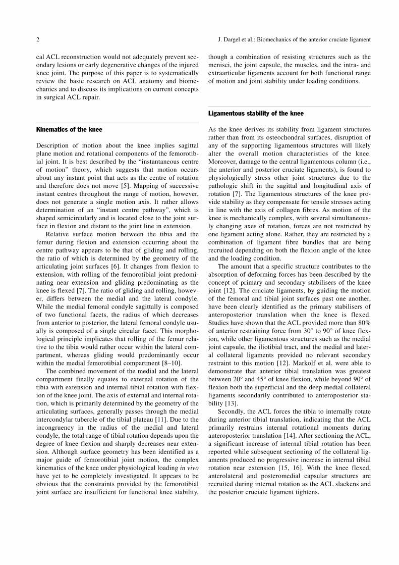
While estimation of in vivo ACL forces during normalactivity have been subject to computational analyses, thebiomechanical properties of the native ACL have exten-sively been analysed in ex vivo studies. Measurementsusing a universal force-moment sensor revealed that the insitu force of the intact ACL was largest at 15° of knee flex-ion and continuously decreased until 90° of knee flexion[23]. Focusing on the ACL force and ligament deformationduring anterior tibial translation, Sakane et al. demonstrat-ed that near full extension, the in situ force of the antero-medial bundle significantly differed from that of the pos-terolateral bundle with 110 N of anterior tibial load applied[23]. While there was no significant difference in the insitu force of the anteromedial bundle throughout the rangeof knee motion, the in situ force of the posterolateral bun-dle was significantly lower at 90° of knee flexion whencompared to full extension, similar to what was measuredfor the entire ACL. This suggests that the role of the pos-terolateral bundle in response to anterior tibial loads nearextension may be of more importance than was previouslythought (Fig. 3).
Performing tensile testing of the bone-ligament-bonecomplex, Woo et al. reported the ultimate failure load ofthe native femur-ACL-tibia complex in younger cadavericspecimens to average 2160±157 N, while mean ACL stiff-ness was 242±28 N/mm [32, 33]. They were also able todemonstrate that both the ultimate failure load and the lin-ear stiffness significantly decreased with age and with theaxis of loading. Ultimate failure loads in older specimensbeing loaded in an anterior drawer mechanism averaged496±85 N with a mean stiffness of 124±16 N/mm. Giventhis data, Noyes et al. concluded that the initial fixation
4
strength of an ACL graft required for sufficient knee sta-bility during daily activities should exceed 450 N,although earlier studies performed by Shelbourne andGray reported excellent clinical results using graft fixationtechniques with a significantly lower initial failurestrength of only 248 N [34–36].
The ACL deficient knee
ACL injuries commonly occur during sport activities withsudden stresses applied to the knee joint while the tibia isin contact with the ground. Typically, the ACL is torn in anon-contact deceleration situation that produces valgusand internal rotational moments on the knee joint thatbegins to flex from near extension. This usually occurs inhigh-risk pivoting sports when the athlete lands on the legand starts rotating to the opposite direction [37].
Active quadriceps pull is considered to play an impor-tant role in the pathomechanism of ACL injury. Reflectiveeccentric quadriceps contraction accompanied by apparentweakness of the hamstring muscles allows the extensormechanism to strain the ACL during anterior tibial transla-tion. This mechanism can be observed during jump stoplandings. Less frequently, direct contact injuries occur as aresult of extensive valgus stress or hyperextension of theknee joint.
Isolated disruption of the ACL is a rather rare condi-tion, while complete or incomplete ruptures accompaniedby traumatic lesions to the medial or lateral meniscus aswell as to the medial collateral ligament are reported in80% of all cases [3, 37–39]. Those concomitant lesions arereported to significantly influence the long-term function-al outcome. Additionally, in both isolated and combinedACL injuries, minor or major bruising of chondral andsubchondral structures may be present. Histologic analy-ses of bruised bone performed by Johnson et al. and othersdemonstrated areas of necrotic osteocytes and chondro-
J. Dargel et al.: Biomechanics of the anterior cruciate ligament
Fig. 3 In situ force of the intact ACL, the anteromedial bundle(AMB) and the posterolateral bundle (PLB) under 110 N of anteri-or tibial load. Adapted from [23]
In s
itu
forc
e [N
]
Flexion angle of the knee [°]
0 20 40 60 80 100
ACL
AMB
PLB
120
100
80
60
40
20
0

cytes, indicating severe damage to the local osteochondraltissue homeostasis after ligament injuries of the knee[38–43].
Disruption of the ACL inevitably results in alterationsin knee kinematics as a transfer of loads can be effectiveonly if the joint is mechanically stable. ACL insufficiencycauses deterioration of the physiologic roll-glide mecha-nism of the femorotibial joint and results in an increasedanterior tibial translation as well as an increased internaltibial rotation. In the advent of muscular fatigue or defi-cient neuromuscular control, the patient will experiencethis combined anterior and rotatory instability as a sublux-ation of the femorotibial joint. According to the concept ofprimary and secondary restraints, failure of a primaryrestraint will cause recruitment of secondary structures inorder to resist external forces and to stabilise joint motion.The increase in loads applied to secondary structures mayrender them more susceptible to degeneration or secondaryfailure [44–46]. Studies investigating the long-term func-tional outcome after conservative treatment of ACL rup-tures, though not characterised by well designed prospec-tive cohort studies, reported on the prevalence of radi-ographic osteoarthritis in 60%–90% of subjects 10–15years after injury [2, 3, 44, 47].
Principles of ACL reconstruction
Current research supports the concept that under obser-vance of several key factors, arthroscopically assistedACL reconstruction done with a biologic autograft signif-icantly improves the stability and function of the knee inmost patients. The key factors significantly influencing thefunctional outcome are:- individual choice of graft material;- anatomic bone tunnel placement;- adequate graft (pre-)tension;- anatomical graft fixation;- sufficient initial graft fixation strength.
Individual choice of graft material
Currently recommended graft materials for ACL recon-struction are biologic autografts and, although rarely avail-able in Western Europe, allografts [48]. Graft choices basi-cally include the patellar tendon-bone graft, semitendi-nosus/gracilis tendon or the quadriceps tendon graft[49–56]. Although a surgeon may prefer one specific sub-stitute for reconstruction, modern knee surgery requiresindividual concepts and a variability of treatment options.In their metaanalysis on the functional outcome of patellartendon and hamstring tendon ACL reconstructions,
Freedman et al. reported that patellar tendon grafts dis-played significantly less failure and better knee stabilitybut resulted in an increased rate of donor side morbidity[56]. Several other studies confirmed no significant differ-ence in functional parameters and subjective resultsobtained at follow-up when comparing patellar tendon-bone and hamstring tendon grafts [57].
From a biomechanical point of view, no graft materialcommonly used has ultimate failure strength or stiffnesscomparable to the native ACL. Although ultimate failureloads of the native bone-patellar tendon-bone complex, aquadrupled hamstring tendon complex, or the quadricepstendon-bone complex average 2977, 4140 and 2353 N,respectively, and consequently exceed the values reportedfor the native ACL, graft harvest and artificial graft fixa-tion into bone significantly decreases both the ultimatestrength and the linear stiffness [58–60]. Patellar tendon-bone grafts should be used for young patients and high-demand athletes who prefer early return to high-level acti-vities, while hamstring tendons are advantageous when alarge skin incision or anterior knee pain should be avoid-ed. Quadriceps tendon grafts should primarily be used forrevision surgery as they are difficult to harvest and sizeand location of donor-site scar are disadvantageous [61].
Anatomic bone tunnel placement
The key to proper postoperative knee function is to restorethe physiologic roll-glide mechanism of the femorotibialjoint, and thus avoid both increased anterior displacementand pathologic patterns of knee rotation. In order to achievethese functional demands, several studies have shown thatgraft positioning is one of the most important factors inACL reconstruction [62–65] (Table 1). Investigating theanterior–posterior stability of the knee after ACL recon-struction, Rupp et al. reported that an increase in postoper-ative knee laxity was most likely to be due to malposition
J. Dargel et al.: Biomechanics of the anterior cruciate ligament 5
Table 1 Malposition of the femoral tunnel and resulting functionalconsequences [65]
Position Kinematic consequences
Femoral tunnelAnterior Tightens in flexion/slackens in extension
Posterior Slackens in flexion/tightens in extension
Tibial tunnelAnterior Tightens in flexion/notch-impingement with
extension
Posterior Tightens in extension/impingement withposterior cruciate ligament
Medial/lateral Impingement at ipsilateral femoral condyle

of the bone tunnels [66]. Fu et al. further reported that ante-rior positioning of both the femoral and the tibial tunnelwas the most common technical mistake during arthroscop-ic surgery [61]. Consequently, avoiding nonphysiologicalstrain patterns of a ligament graft throughout the function-al range, which also avoids graft failure and any limitationin knee motion, has been emphasised for successful recon-struction [7]. A proposed isometric bone tunnel placement(i.e., the distance between the proximal and the distal inser-tion site remained constant throughout the entire range ofmotion), however, was not possible to obtain in either invivo or in vitro observations [62, 65].
Hefzy et al. investigated the resulting changes in dis-tance between a given point for the femoral and the tibialattachment of an ACL graft [62]. They reported that alteringthe location of the femoral bone tunnel had a much greatereffect on the length of the substitute than did altering the tib-ial attachment site did. They also pointed out that the area inwhich tunnel placement resulted in length changes of thegraft of less than 2 mm throughout the range of motion wassmaller than the cross-sectional area of grafts commonlyused for reconstruction of the ACL. The resulting inho-mogenous tension patterns within a graft would thereforenot support the concept of isometric graft placement.
Csizy and Friederich noted that with an arthroscopicview of the knee joint, the femoral tunnel is most suscep-tible to being placed anterior to the anatomic ACL foot-print, thus resulting in excessive graft tension during kneeflexion and correlating well with poor functional outcomes[65, 67]. Varying the femoral tunnel position between theanatomic ACL footprint and the most isometric position,Musahl et al. demonstrated that neither tunnel positionfully restored the physiologic kinematics of the femorotib-ial joint [63]. However, they concluded that anatomic graftplacement resulted in kinematics closer to the normal kneejoint than did a tunnel placement for best isometry.
Several methods for measuring femoral graft positionin postoperative radiographs have been introduced, henceenabling visual control of bone tunnel placement whenusing intraoperative flouroscopic imaging [68–72] (Fig. 4).Investigations performed by Klos et al. revealed that themeasurement technique described by Amis et al. producedreliable data in both rotated and non-rotated (i.e., full over-lap of the femoral condyles) lateral radiographs of the knee[64, 69]. Probably the most popular technique for radi-ographic bone tunnel measurement is the quadrant methoddescribed by Bernard et al. [72]. Although commonly usedto identify the anatomic ACL origin, it is reliable onlywhen the projection of the femoral condyles perfectlyoverlap in lateral radiographs of the knee.
Tibial bone tunnel placement has been reported to beless sensitive with respect to postoperative knee kinemat-ics, however, may cause graft impingement or unphysio-logic loading patterns when malplaced [73–75]. The tibialtunnel should be placed within the posterior half of thenative ACL footprint, which will allow the anterior fibres
6
of the graft to run parallel with the intercondylar roof infull extension (Fig. 1) [73]. Anterior placement of the tib-ial tunnel as well as a vertically oriented intercondylar roofin the sagittal plane will either initially subject the graft toroof impingement or secondarily will cause roof impinge-ment with contraction of the quadriceps muscle. It has be-en recommended in the literature to leave 6 mm of clear-ance between the anterior aspect of the graft and the inter-condylar roof during extension of the knee. Arthroscopi-cally, the bone tunnel should be drilled at an angle of40°–50° to the long axis of the tibia and should be placedanteromedially or posterior to the anterior horn of the me-dial meniscus and slightly anterior to the posterior cruciateligament insertion [76–79].
In the frontal plane, the position of the femoral tunnelalong the intercondylar notch can be described by the angu-lar position of numerals on the face of a clock. In general,a tunnel positioned at 11 o’clock for a right knee (or 1o’clock for a left knee) has been considered the correct tun-nel position [80–82]. However, Markolf et al. emphasisedthe intercondylar notch to have a three-dimensional config-uration, such that variations in graft placement in thefrontal plane inevitably resulted in variations in tunnelplacement in the sagittal plane [83]. Hence, they demon-strated that the biomechanical consequences of varyingfemoral tunnel placement in the frontal plane were less crit-ical than varying the anteroposterior position.
J. Dargel et al.: Biomechanics of the anterior cruciate ligament
Fig. 4 Radiographic analysis of femoral bone tunnel placement. a60% of the anterioposterior diameter of the lateral femoral condyle([69]); b 80% of the anteroposterior length of Blumensaat’s line([70]); c 65% of the anteroposterior cortical depth of the distalfemur in line with Blumensaat’s line ([71]); d anteroinferior cornerof the most proximal and posterior quadrant adapted to the heightand length of the lateral femoral condyle ([72])
a b
c d
In order to obey the complex structure of the intact ACL,several authors have claimed ACL reconstruction to be moreanatomical when both the anteromedial and the posterolater-al bundle were replaced independently [84–87]. Yagi et al.measured the in situ ACL force as well as knee kinematicswith the ACL intact, sectioned, reconstructed with one bun-dle and reconstructed with two bundles using a robotic uni-versal force-moment sensor testing system [84]. They de-monstrated that under combined anterior, internal tibial andvalgus torque, knee stability using a double-bundle techniquewas superior to a single-bundle reconstruction technique.There is agreement among ACL surgeons that double-bundleACL reconstruction is a demanding procedure that currentlymay only be performed in experienced trauma centres.
Adequate graft (pre-)tension
The tension applied to the graft before final graft fixationsignificantly influences the kinematics of the knee jointand the long-term ability of a graft to stabilise the kneejoint. A low initial graft tension will not provide adequatejoint stability, while excessive initial graft tension willrestrain range of motion and is susceptible of early graftfailure. Yoshia et al. studied the effect of initial graft ten-sion on knee stability using an in vivo animal model [88].They demonstrated that anteroposterior knee stability didnot significantly differ between grafts fixed at 1 N and 39N three months after surgery, however, knee joints withhigher initial graft tension showed evidence of degenera-tive cartilage lesions. In a goat model, Abramowitch et al.reported similar results with the biomechanical character-istics of an ACL substitute not differing significantly whencomparing grafts fixed with high (35 N) and low (5 N) ini-tial tension six weeks after surgery, but differing signifi-cantly when compared to an uninjured control group [89].In a prospective randomised trial, Kim et al. failed toprove that forces of either 8, 12 or 15 kg applied to thegraft during fixation resulted in significant differences inanterior knee laxity one year after surgery [90].
In accordance, in vitro studies on the course of grafttension have shown that both the patellar tendon-bonegraft and the hamstring tendon graft lose their initial ten-sion when being cyclically loaded [91, 92]. There is a lackof scientifically based data on the clinical impact of initialgraft tension as follow-up studies so far have failed toprove that variations of graft tension resulted in clinicalsymptoms after ACL reconstruction. At present, theamount of tension that should be applied to a graft prior tofixation has yet to be precisely defined. Excessive tensionas well as loose fixation should therefore be controlledintraoperatively by testing range of motion and anteriorknee stability under arthroscopic visualisation.
Although the influence of the viscoelastic behaviour ofACL replacements so far has not been entirely charac-
terised, the viscoelastic creep or relaxation may contributeto a decrease in graft tension over time. Consequently, sev-eral authors have emphasised that grafts should be cycli-cally preconditioned prior to implantation in order todecrease the viscoelastic elongation behaviour duringrehabilitation [93, 94]. The subject of graft precondition-ing remains controversial as the preconditioning protocolsdescribed in the literature are not commonly applicable tothe surgical procedure and may not be as beneficial as sug-gested by ex vivo biomechanical studies [93–95].
Anatomical graft fixation
Most fixation devices currently used rely on linkage mate-rial between the graft substance and the bone, thus influ-encing graft healing and altering the initial biomechanicalproperties of a graft material. Generally, fixation devicesdistant to the joint line (i.e., buttons, staples, washers orpost-screws) fail to reconstruct the complex nature of thenative tibial or femoral ACL insertion close to the jointsurface. As a consequence, the strain that is induced in asubstitute during cyclic loading is significantly largerwhen compared to the intact ACL [48]. This allows forlongitudinal (‘bungee-effect’) and transverse (‘wind-shield-wiper-effect’) graft motion within the bone tunnel,which in turn may lead to bone tunnel dilation, may impairhealing of the graft to the bone tunnel and may complicaterevision surgery due to loss of bone material.
Furthermore, it should be considered that the linearstiffness of grafts fixated distant to the joint line is lessthan placing the graft close to the entrance of the bone tun-nel. Although Magen et al. demonstrated that the stiffnessof a graft complex was influenced more by the method offixation than the length of the graft, it has been previouslyreported that keeping the length of a substitute as short aspossible may increase graft stiffness and knee stabilitythroughout the range of motion [96–99]. In accordance,studies have shown that linear graft stiffness is higherwhen fixation systems that are placed close to the tunnelentrance are used [48]. Investigations on the overall stabil-ity of porcine knees after ACL reconstruction revealed thatfixating the graft proximally on the tibial side resulted insignificantly more anterior knee stability than central ordistal tibial graft fixation [97].
Sufficient initial graft fixation strength
The importance of secure graft fixation has dramaticallyincreased as current rehabilitation protocols emphasiseearly weight bearing after ACL reconstruction and as thefixation site is known to be the weakest link during theearly postoperative period [26]. Graft fixation to bone
J. Dargel et al.: Biomechanics of the anterior cruciate ligament 7
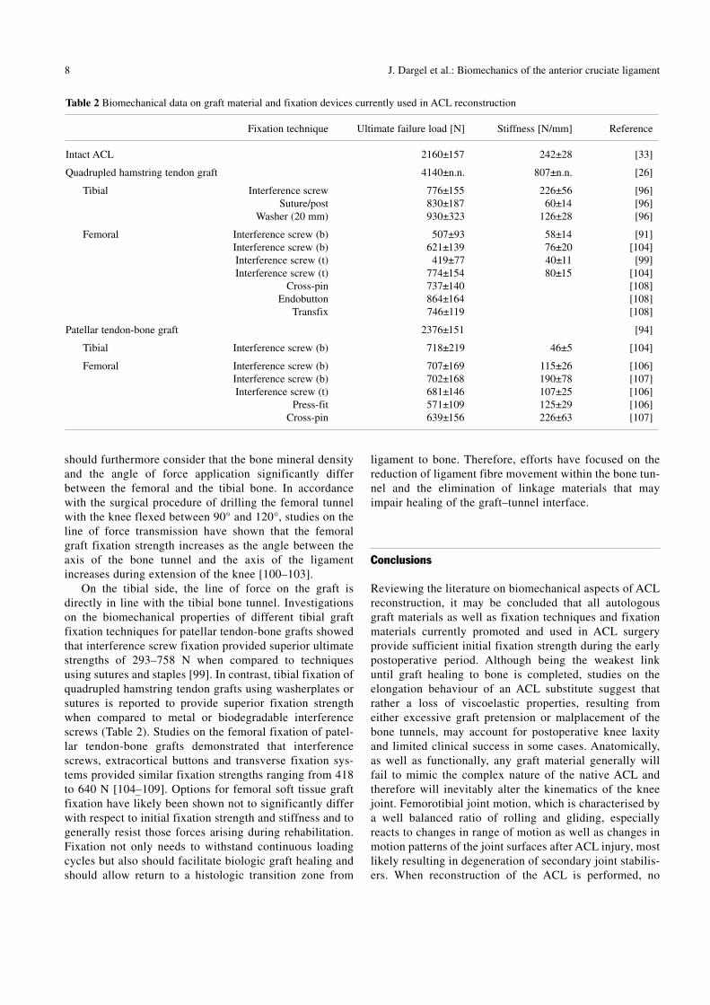
should furthermore consider that the bone mineral densityand the angle of force application significantly differbetween the femoral and the tibial bone. In accordancewith the surgical procedure of drilling the femoral tunnelwith the knee flexed between 90° and 120°, studies on theline of force transmission have shown that the femoralgraft fixation strength increases as the angle between theaxis of the bone tunnel and the axis of the ligamentincreases during extension of the knee [100–103].
On the tibial side, the line of force on the graft isdirectly in line with the tibial bone tunnel. Investigationson the biomechanical properties of different tibial graftfixation techniques for patellar tendon-bone grafts showedthat interference screw fixation provided superior ultimatestrengths of 293–758 N when compared to techniquesusing sutures and staples [99]. In contrast, tibial fixation ofquadrupled hamstring tendon grafts using washerplates orsutures is reported to provide superior fixation strengthwhen compared to metal or biodegradable interferencescrews (Table 2). Studies on the femoral fixation of patel-lar tendon-bone grafts demonstrated that interferencescrews, extracortical buttons and transverse fixation sys-tems provided similar fixation strengths ranging from 418to 640 N [104–109]. Options for femoral soft tissue graftfixation have likely been shown not to significantly differwith respect to initial fixation strength and stiffness and togenerally resist those forces arising during rehabilitation.Fixation not only needs to withstand continuous loadingcycles but also should facilitate biologic graft healing andshould allow return to a histologic transition zone from
8
ligament to bone. Therefore, efforts have focused on thereduction of ligament fibre movement within the bone tun-nel and the elimination of linkage materials that mayimpair healing of the graft–tunnel interface.
Conclusions
Reviewing the literature on biomechanical aspects of ACLreconstruction, it may be concluded that all autologousgraft materials as well as fixation techniques and fixationmaterials currently promoted and used in ACL surgeryprovide sufficient initial fixation strength during the earlypostoperative period. Although being the weakest linkuntil graft healing to bone is completed, studies on theelongation behaviour of an ACL substitute suggest thatrather a loss of viscoelastic properties, resulting fromeither excessive graft pretension or malplacement of thebone tunnels, may account for postoperative knee laxityand limited clinical success in some cases. Anatomically,as well as functionally, any graft material generally willfail to mimic the complex nature of the native ACL andtherefore will inevitably alter the kinematics of the kneejoint. Femorotibial joint motion, which is characterised bya well balanced ratio of rolling and gliding, especiallyreacts to changes in range of motion as well as changes inmotion patterns of the joint surfaces after ACL injury, mostlikely resulting in degeneration of secondary joint stabilis-ers. When reconstruction of the ACL is performed, no
J. Dargel et al.: Biomechanics of the anterior cruciate ligament
Table 2 Biomechanical data on graft material and fixation devices currently used in ACL reconstruction
Fixation technique Ultimate failure load [N] Stiffness [N/mm] Reference
Intact ACL 2160±157 242±28 [33]
Quadrupled hamstring tendon graft 4140±n.n. 807±n.n. [26]
Tibial Interference screw 776±155 226±56 [96]Suture/post 830±187 60±14 [96]
Washer (20 mm) 930±323 126±28 [96]
Femoral Interference screw (b) 507±93 58±14 [91]Interference screw (b) 621±139 76±20 [104]Interference screw (t) 419±77 40±11 [99]Interference screw (t) 774±154 80±15 [104]
Cross-pin 737±140 [108]Endobutton 864±164 [108]
Transfix 746±119 [108]
Patellar tendon-bone graft 2376±151 [94]
Tibial Interference screw (b) 718±219 46±5 [104]
Femoral Interference screw (b) 707±169 115±26 [106]Interference screw (b) 702±168 190±78 [107]Interference screw (t) 681±146 107±25 [106]
Press-fit 571±109 125±29 [106]Cross-pin 639±156 226±63 [107]
functional restitio ad integrum may be expected, as ananatomically placed single bundle ACL substitute willreconstruct only part of the fibre mass of the intact ACL.
Whether or not double-bundle ACL reconstructionsfunctionally imitated the native ACL more closely andconsequently restored the kinematics of the knee jointmore accurately, currently remains subject to debate. Sofar, clinical follow-up studies have shown that the physio-logic anterior knee stability cannot be completely restoredafter ACL reconstruction. However, under observance ofthe biomechanical factors that significantly influence thekinematics of the knee joint, encouraging mid- to long-term clinical and functional outcomes after ACL recon-struction have been reported.
References
1. Steinbrück K (1999) Epidemiologie von Sportverletzungen –25-Jahres-Analyse einer sportorthopädisch-traumatologis-chen Ambulanz. Sportverletz Sportschaden 13:38–52
2. Nebelung W, Wuschech H (2005) Thirty-five years of follow-up of anterior cruciate ligament-deficient knees in high-levelathletes. Arthroscopy 21:696–702
3. Gillquist J, Messner K (1999) Anterior cruciate ligamentreconstruction and the long-term incidence of gonarthrosis.Sports Med 27:143–156
4. Zysk SP, Refior HJ (2000) Operative or conservative treat-ment of the acutely torn anterior cruciate ligament in middle-aged patients. A follow-up study of 133 patients between theages of 40 and 59 years. Arch Orthop Trauma Surg 120:59–64
5. Frankel VH, Burstein AH, Brooks DB (1971) Biomechanicsof internal derangement of the knee. Pathomechanics as deter-mined by analysis of the instant centers of motion. J BoneJoint Surg Am 53:945–962
6. Kapendji IA (1970) The physiology of the joints. ChurchillLivingstone, New York, 114–123
7. Muller W (1983) The knee: form, function, and ligamentreconstruction. Springer Verlag, New York, pp 8–75
8. Smith PN, Refshauge KM, Scarvell JM (2003) Developmentof the concepts of knee kinematics. Arch Phys Med Rehabil84:1895–1902
9. Iwaki H, Pinskerova V, Freeman MA (2000) Tibiofemoralmovement 1: the shapes and relative movements of the femurand tibia in the unloaded cadaver knee. J Bone Joint Surg Br82:1189–1195
10. Freeman MA, Pinskerova V (2005) The movement of the nor-mal tibio-femoral joint. J Biomech 38:197–208
11. Shaw JA, Murray DG (1974) The longitudinal axis of theknee and the role of the cruciate ligaments in controllingtransverse rotation. J Bone Joint Surg Am 56:1603–1609
12. Butler DL, Noyes FR, Grood ES (1980) Ligamentousrestraints to anterior-posterior drawer in the human knee. Abiomechanical study. J Bone Joint Surg Am 62:259–270
13. Markolf KL, Kochan A, Amstutz HC (1984) Measurement ofknee stiffness and laxity in patients with documented absenceof the anterior cruciate ligament. J Bone Joint Surg Am66:242–252
14. Fukubayashi T, Torzilli PA, Sherman MF, Warren RF (1982)An in vitro biomechanical evaluation of anterior-posteriormotion of the knee. Tibial displacement, rotation, and torque.J Bone Joint Surg Am 64:258–264
15. Grood ES, Stowers SF, Noyes FR (1988) Limits of movementin the human knee. Effect of sectioning the posterior cruciateligament and posterolateral structures. J Bone Joint Surg Am70:88–97
16. Lipke JM, Janecki CJ, Nelson CL et al (1981) The role ofincompetence of the anterior cruciate and lateral ligaments inanterolateral and anteromedial instability. A biomechanicalstudy of cadaver knees. J Bone Joint Surg Am 63:954–960
17. Amiel D, Frank C, Harwood F et al (1984) Tendons and liga-ments: a morphological and biochemical comparison. J Or-thop Res 1:257–265
18. Petersen W, Tillmann B (2002) Anatomy and function of theanterior cruciate ligament. Orthopäde 31:710–718
19. Duthon VB, Barea C, Abrassart S et al (2006) Anatomy of theanterior cruciate ligament. Knee Surg Sports TraumatolArthrosc 14:204–213
20. Harner CD, Baek GH, Vogrin TM et al (1999) Quantitativeanalysis of anterior cruciate ligament insertions. Arthroscopy15:741–749
21. Anderson AF, Dome DC, Gautam S et al (2001) Correlationof anthropometric measurements, strength, anterior cruciateligament size, and intercondylar notch characteristics to sexdifferences in anterior cruciate ligament tears. Am J SportsMed 29:58–63
22. Kummer B, Yamamoto (1988) Funktionelle Anatomie derKreuzbänder. Arthroskopie 1:2–10
23. Sakane M, Fox RJ, Woo SL et al (1997) In situ forces in theanterior cruciate ligament and its bundles in response to ante-rior tibial loads. J Orthop Res 15:285–293
24. Odensten M, Gillquist J (1985) Functional anatomy of theanterior cruciate ligament and a rationale for reconstruction.J Bone Joint Surg Am 67:257–262
25. Colombet P, Robinson J, Christel P et al (2006) Morphologyof anterior cruciate ligament attachments for anatomic recon-struction: a cadaveric dissection and radiographic study.Arthroscopy 22:984–992
26. Brand J, Weiler A, Caborn DN et al (2000) Graft fixation incruciate ligament reconstruction. Am J Sports Med28:761–774
27. Frisen M, Magi M, Sonnerup I, Viidik A (1969) Rheologicalanalysis of soft collagenous tissue. Part I: theoretical consid-erations. J Biomech 2:13–20
28. Noyes FR, De Lucas JL, Torvik PJ (1974) Biomechanics ofanterior cruciate ligament failure: an analysis of strain-ratesensitivity and mechanisms of failure in primates. J BoneJoint Surg Am 56:236–253
29. Morrison JB (1970) The mechanics of the knee joint in rela-tion to normal walking. J Biomech 3:51–61
30. Morrison JB (1969) Function of the knee joint in variousactivities. Biomed Eng 4:573–580
31. Morrison JB (1968) Bioengineering analysis of force actionstransmitted by the knee joint. Biomed Eng 4:164–170
32. Woo SL, Debski RE, Withrow JD, Janaushek MA (1999)Biomechanics of knee ligaments. Am J Sports Med27:533–543
33. Woo SL, Hollis JM, Adams DJ et al (1991) Tensile propertiesof the human femur-anterior cruciate ligament-tibia complex.
J. Dargel et al.: Biomechanics of the anterior cruciate ligament 9

The effects of specimen age and orientation. Am J Sports Med19:217–225
34. Noyes FR, Butler DL, Grood ES et al (1984) Biomechanicalanalysis of human ligament grafts used in knee-ligamentrepairs and reconstructions. J Bone Joint Surg Am66:344–352
35. Johnson DP (1998) Operative complications from the use ofbiodegradable Kurosawa screws. J Bone Joint Surg80:103–109
36. Shelbourne KD, Gray T (1997) Anterior cruciate ligamentreconstruction with autogenous patellar tendon graft followedby accelerated rehabilitation. A two- to nine-year follow-up.Am J Sports Med 25:786–795
37. Strobel M, Stedtfeld HW, Eichhorn HJ (1995) Diagnostik desKniegelenkes. Springer Verlag, Berlin
38. Beynnon BD, Johnson RJ, Abate JA et al (2005) Treatment ofanterior cruciate ligament injuries, part I. Am J Sports Med33:1579–1602
39. Seitz H, Marlovits S, Kolonja A et al (1998) Meniskusläsionennach vorderen Kreuzbandrupturen. Arthroskopie 11:82–85
40. Beynnon BD, Johnson RJ, Abate JA et al (2005) Treatment ofanterior cruciate ligament injuries, part II. Am J Sports Med33:1751–1767
41. Johnson DL, Urban WP, Caborn DN et al (1998) Articularcartilage changes seen with magnetic resonance imaging-detected bone bruises associated with acute anterior cruciateligament rupture. Am J Sports Med 26:409–414
42. Costa-Paz M, Muscolo DL, Ayerza M et al (2001) Magneticresonance imaging follow-up study of bone bruises associat-ed with anterior cruciate ligament ruptures. Arthroscopy17:445–449
43. Mathis CE, Noonan K, Kayes K (1998) “Bone bruises” of theknee: a review. Iowa Orthop J 18:112–117
44. Bray RC, Dandy DJ (1989) Meniscal lesions and chronicanterior cruciate ligament deficiency. Meniscal tears occur-ring before and after reconstruction. J Bone Joint Surg Br71:128–130
45. Conteduca F, Ferretti A, Mariani PP et al (1991)Chondromalacia and chronic anterior instabilities of the knee.Am J Sports Med 19:119–123
46. Noyes FR, Mooar PA, Matthews DS, Butler DL (1983) Thesymptomatic anterior cruciate-deficient knee. Part I: the long-term functional disability in athletically active individuals.J Bone Joint Surg Am 65:154–162
47. Sommerlath K, Lysholm J, Gillquist J (1991) The long-termcourse after treatment of acute anterior cruciate ligament rup-tures. A 9 to 16 year follow up. Am J Sports Med 19:156–162
48. Fu FH, Bennett CH, Lattermann C, Ma CB (1999) Currenttrends in anterior cruciate ligament reconstruction. Part 1:Biology and biomechanics of reconstruction. Am J SportsMed 27:821–830
49. Howe JG, Johnson RJ, Kaplan MJ et al (1991) Anterior cruciateligament reconstruction using quadriceps patellar tendon graft.Part I. Long-term followup. Am J Sports Med 19:447–457
50. Paulos LE, Cherf J, Rosenberg TD, Beck CL (1991) Anteriorcruciate ligament reconstruction with autografts. Clin SportsMed 10:469–485
51. Shelbourne KD, Klootwyk TE, Wilckens JH, De Carlo MS(1995) Ligament stability two to six years after anterior cruci-ate ligament reconstruction with autogenous patellar tendongraft and participation in accelerated rehabilitation program.Am J Sports Med 23:575–579
10
52. Shelbourne KD, Rettig AC, Hardin G, Williams RI (1993)Miniarthrotomy versus arthroscopic-assisted anterior cruciateligament reconstruction with autogenous patellar tendon graft.Arthroscopy 9:72–75
53. Aglietti P, Buzzi R, Zaccherotti G, De Biase P (1994) Patellartendon versus doubled semitendinosus and gracilis tendonsfor anterior cruciate ligament reconstruction. Am J SportsMed 22:211–217
54. Howell SM, Taylor MA (1996) Brace-free rehabilitation, withearly return to activity, for knees reconstructed with a double-looped semitendinosus and gracilis graft. J Bone Joint SurgAm 78:814–825
55. Johnson LL (1993) The outcome of a free autogenous semi-tendinosus tendon graft in human anterior cruciate reconstruc-tive surgery: a histological study. Arthroscopy 9:131–142
56. Freedman KB, D’Amato MJ, Nedeff DD et al (2003)Arthroscopic anterior cruciate ligament reconstruction: ametaanalysis comparing patellar tendon and hamstring tendonautografts. Am J Sports Med 31:2–11
57. Anderson AF, Snyder RB, Lipscomb AB (2001) Anterior cru-ciate ligament reconstruction. A prospective randomizedstudy of three surgical methods. Am J Sports Med 29:272–279
58. Cooper DE, Deng XH, Burstein AL et al (1993) The strengthof the central third patellar tendon graft: A biomechanicalstudy. Am J Sports Med 21:818–824
59. Hamner DL, Brown CH, Steiner ME et al (1999) Hamstringtendon grafts for reconstruction of the anterior cruciate liga-ment: Biomechanical evaluation of the use of multiple strandsand tensioning techniques. J Bone Joint Surg 81:549–557
60. Stäubli HU, Schatzmann L, Brunner P et al (1996) Quadricepstendon and patellar ligament: cryosectional anatomy andstructural properties in young adults. Knee Surg SportsTraumatol Arthrosc 4:100–110
61. Fu FH, Bennett CH, Ma CB et al (2000) Current trends inanterior cruciate ligament reconstruction. Part II. Operativeprocedures and clinical correlations. Am J Sports 28:124–130
62. Hefzy MS, Grood ES, Noyes FR (1989) Factors affecting theregion of most isometric femoral attachments. Part II: Theanterior cruciate ligament. Am J Sports Med 17:208–216
63. Musahl V, Plakseychuk A, Van Scyoc A et al (2005) Varyingfemoral tunnels between the anatomical footprint and isomet-ric positions: effect on kinematics of the anterior cruciate lig-ament-reconstructed knee. Am J Sports Med 33:712–718
64. Klos TV, Harman MK, Habets RJ et al (2000) Locatingfemoral graft placement from lateral radiographs in anteriorcruciate ligament reconstruction: a comparison of 3 methodsof measuring radiographic images. Arthroscopy 16:499–504
65. Csizy M, Friederich NF (2002) Bohrkanallokalisation in deroperativen Rekonstruktion des vorderen Kreuzbandes. Posi-tion-Fehlplatzierung-Anatomometrie. Orthopäde 31:741–750
66. Rupp S, Müller B, Seil R (2001) Knee laxity after ACL recon-struction with a BPTB graft. Knee Surg Sports TraumatolArthrosc 9:72–76
67. Sommer C, Friederich NF, Muller W (2000) Improperly pla-ced anterior cruciate ligament grafts: correlation between ra-diological parameters and clinical results. Knee Surg SportsTraumatol Arthrosc 8:207–213
68. Cole J, Brand JC, Caborn DN, Johnson DL (2000) Radio-graphic analysis of femoral tunnel position in anterior cruciateligament reconstruction. Am J Knee Surg 13:218–222
69. Amis AA, Beynnon B, Blankevoort L et al (1994) Proceedin-gs of the ESSKA Scientific Workshop on Reconstruction of
J. Dargel et al.: Biomechanics of the anterior cruciate ligament

the Anterior and Posterior Cruciate Ligaments. Knee SurgSports Traumatol Arthrosc 2:124–132
70. Harner CD, Marks PH, Fu FH et al (1994) Anterior cruciateligament reconstruction: endoscopic versus two-incision tech-nique. Arthroscopy 10:502–512
71. Aglietti P, Zaccherotti G, Menchetti PP, De Biase P (1995) Acomparison of clinical and radiological parameters with twoarthroscopic techniques for anterior cruciate ligament recon-struction. Knee Surg Sports Traumatol Arthrosc 3:2–8
72. Bernard M, Hertel P, Hornung H, Cierpinski T (1997)Femoral insertion of the ACL. Radiographic quadrant method.Am J Knee Surg 10:14–21
73. Howell SM (1998) Principles for placing the tibial tunnel andavoiding roof impingement during reconstruction of a tornanterior cruciate ligament. Knee Surg Sports TraumatolArthrosc 6:49–55
74. Goble EM, Downey DJ, Wilcox TR (1995) Positioning of thetibial tunnel for anterior cruciate ligament reconstruction.Arthroscopy 11:688–695
75. Jackson DW, Gasser SI (1994) Tibial tunnel placement inACL reconstruction. Arthroscopy 10:124–131
76. Cooper DE, Urrea L, Small J (1998) Factors affecting isome-try of endoscopic anterior cruciate ligament reconstruction: theeffect of guide offset and rotation. Arthroscopy 14:164–170
77. Good L, Gillquist J (1993) The value of intraoperative isome-try measurements in anterior cruciate ligament reconstruction:an in vivo correlation between substitute tension and lengthchange. Arthroscopy 9:525–532
78. Hefzy MS, Grood ES, Noyes FR (1989) Factors affecting theregion of most isometric femoral attachments. Part II: Theanterior cruciate ligament. Am J Sports Med 17:208–216
79. Penner DA, Daniel DM, Wood P, Mishra D (1988) An in vitrostudy of anterior cruciate ligament graft placement and isom-etry. Am J Sports Med 16:238–243
80. Bylski-Austrow DI, Grood ES, Hefzy MS et al (1990)Anterior cruciate ligament replacements: a mechanical studyof femoral attachment location, flexion angle at tensioning,and initial tension. J Orthop Res 8:522–531
81. Fu FH, Bennett CH, Ma CB et al (2000) Current trends inanterior cruciate ligament reconstruction. Part II. Operativeprocedures and clinical correlations. Am J Sports Med28:124–130
82. Good L, Odensten M, Gillquist J (1994) Sagittal knee stabili-ty after anterior cruciate ligament reconstruction with a patel-lar tendon strip. A two-year follow-up study. Am J Sports Med22:518–523
83. Markolf KL, Hame S, Hunter DM et al (2002) Effects offemoral tunnel placement on knee laxity and forces in an ante-rior cruciate ligament graft. J Orthop Res 20:1016–1024
84. Yagi M, Wong EK, Kanamori A et al (2002) Biomechanicalanalysis of an anatomic anterior cruciate ligament reconstruc-tion. Am J Sports Med 30:660–666
85. Norwood LA, Cross MJ (1979) Anterior cruciate ligament:functional anatomy of its bundles in rotatory instabilities. AmJ Sports Med 7:23–26
86. Muneta T, Sekiya I, Yagishita K et al (1999) Two-bundlereconstruction of the anterior cruciate ligament using semi-tendinosus tendon with endobuttons: operative technique andpreliminary results. Arthroscopy 15:618–624
87. Pederzini L, Adriani E, Botticella C, Tosi M (2000) Technicalnote: double tibial tunnel using quadriceps tendon in anteriorcruciate ligament reconstruction. Arthroscopy 16:E9
88. Yoshiya S, Andrish JT, Manley MT, Bauer TW (1987) Grafttension in anterior cruciate ligament reconstruction. An invivo study in dogs. Am J Sports Med 15:464–470
89. Abramowitch SD, Papageorgiou CD, Withrow JD et al (2003)The effect of initial graft tension on the biomechanical prop-erties of a healing ACL replacement graft: a study in goats.J Orthop Res 21:708–715
90. Kim SG, Kurosawa H, Sakuraba K et al (2006) The effect ofinitial graft tension on postoperative clinical outcome in ante-rior cruciate ligament reconstruction with semitendinosus ten-don. Arch Orthop Trauma Surg 126:260–264
91. Arnold MP, Lie DT, Verdonschot N et al (2005) The remainsof anterior cruciate ligament graft tension after cyclic kneemotion. Am J Sports Med 33:536–542
92. Boylan D, Greis PE, West JR et al (2003) Effects of initialgraft tension on knee stability after anterior cruciate ligamentreconstruction using hamstring tendons: a cadaver study.Arthroscopy 19:700–705
93. Graf BK, Vanderby R, Ulm MJ et al (1994) Effect of precon-ditioning on the viscoelastic response of primate patellar ten-don. Arthroscopy 10:90–96
94. Schatzmann L, Brunner P, Staubli HU (1998) Effect of cyclicpreconditioning on the tensile properties of human quadricepstendons and patellar ligaments. Knee Surg Sports TraumatolArthrosc 6:56–61
95. Nagarkatti DG, McKeon BP, Donahue BS, Fulkerson JP(2001) Mechanical evaluation of a soft tissue interferencescrew in free tendon anterior cruciate ligament graft fixation.Am J Sports Med 29:67–71
96. Magen HE, Howell SM, Hull ML (1999) Structural propertiesof six tibial fixation methods for anterior cruciate ligamentsoft tissue grafts. Am J Sports Med 27:35–43
97. Ishibashi Y, Rudy TW, Livesay GA et al (1997) The effect ofanterior cruciate ligament graft fixation site at the tibia onknee stability: evaluation using a robotic testing system.Arthroscopy 13:177–182
98. Morgan CD, Kalmam VR, Grawl DM (1995) Isometry testingfor anterior cruciate ligament reconstruction revisited.Arthroscopy 11:647–659
99. Weiler A, Hoffmann RF, Stahelin AC et al (1998) Hamstringtendon fixation using interference screws: a biomechanicalstudy in calf tibial bone. Arthroscopy 14:29–37
100. Dargel J, Schmidt-Wiethoff R, Schneider T et al (2006)Biomechanical testing of quadriceps tendon-patellar bonegrafts: an alternative graft source for press-fit anterior cruci-ate ligament reconstruction? Arch Orthop Trauma Surg126:265–270
101. Boszotta H (1997) Arthroskopische femorale Press-fit-Fixation des Lig.-patellae-Transplantats beim Ersatz desvorderen Kreuzbands. Arthroskopie 10:126–132
102. Dargel J, Schmidt-Wiethoff R, Schneider T et al (2006)Biomechanical testing of quadriceps tendon-patellar bonegrafts: An alternative graft source for press-fit anterior cruci-ate ligament reconstruction? Arch Orthop Trauma Res126:265–270
103. Schmidt-Wiethoff R, Dargel J, Gerstner M et al (2006) Boneplug length and loading angle determine the primary stabilityof patellar tendon-bone grafts in press-fit ACL reconstruction.Knee Surg Sports Traumatol Arthrosc 14:108–111
104. Kousa P, Jarvinen TL, Kannus P, Jarvinen M (2001) Initialfixation strength of bioabsorbable and titanium interferencescrews in anterior cruciate ligament reconstruction.
J. Dargel et al.: Biomechanics of the anterior cruciate ligament 11
Biomechanical evaluation by single cycle and cyclic loading.Am J Sports Med 29:420–425
105. Becker P, Stärke C, Schröder M, Nebelung W (2000)Biomechanische Eigenschaften tibialer Fixationsverfahrenvon Hamstring-Transplantaten zur Kreuzbandrekonstruktion.Arthoskopie 13:314–317
106. Lee MC, Jo H, Bae TS et al (2003) Analysis of initial fixationstrength of press-fit fixation technique in anterior cruciate lig-ament reconstruction. A comparative study with titanium andbioabsorbable interference screw using porcine lower limb.Knee Surg Sports Traumatol Arthrosc 11:91–98
12
107. Zantop T, Weimann A, Rummler M et al (2004) Initial fixa-tion strength of two bioabsorbable pins for the fixation ofhamstring grafts compared to interference screw fixation: sin-gle cycle and cyclic loading. Am J Sports Med 32:641–649
108. Ahmad CS, Gardner TR, Groh M et al (2004) Mechanical prop-erties of soft tissue femoral fixation devices for anterior cruciateligament reconstruction. Am J Sports Med 32:635–640
109. Zantop T, Welbers B, Weimann A et al (2004) Biomechanicalevaluation of a new cross-pin technique for the fixation of dif-ferent sized bone-patellar tendon-bone grafts. Knee SurgSports Traumatol Arthrosc 12:520–527
J. Dargel et al.: Biomechanics of the anterior cruciate ligament
Copyright © 2022 FDOKUMEN