WITHDRAWN: Exercise for treating isolated anterior cruciate ligament injuries in adults
Upload
innovativepublicationCategory
view
2download
0
64 Chaurasia et al. Indian Journal of Orthopaedics Surgery 2015; 1(1): 64-74.
-------------------------------------------------------------Review Article-----------------------------------------------------------
ISSN 2395-1354(Print)
e-ISSN 2395-1362(Online)
Indian Journal of Orthopaedics
Surgery
POST ANTERIOR CRUCIATE LIGAMENT RECONSTRUCTION INFECTION: REVIEW OF LITERATURE
Sanyam Chaurasia1,*, Georgios Karnatzikos2, Alberto Gobbi3, Vivek Gupta4
1,4Department of Orthopaedics, Irene Swasthik Hospital, A-9, Raghuvir Enclave,
Dhichaun Chowk, Nangloi Stand, Najafgarh, New Delhi, India 2,3OASI Bioresearch Foundation,Via Amadeo 24, 20133, Milan, Italy
*Corresponding Author: E-mail: [email protected], [email protected]
Abstract Purpose: The purpose of this study was to review and summarize the literature and suggest the probable
most effective protocol in the management of post anterior cruciate ligament reconstruction (ACLR) infection. Methods: We reviewed 16 studies (Level 1- Level 4) published between January 2000 and December 2013, by a thorough search in PUBMED, MEDLINE and EMBASE databases. Results: There were 246 cases of infection amongst 35,795 ACLR making the proportion of infection 0.68% (range- 0.14-2.6%). The mean time of onset of infection from index surgery was between 7.5 days to 32 days.
The most common organism was Coagulase-negative Staphylococci (CNS) followed by Staphylococcus Aureus. Optimal outcome was reported in most studies using serial arthroscopic lavage and intravenous antibiotics with graft retention as a prime protocol. Conclusion: Septic arthritis after ACLR is a rare and disastrous complication which can be successfully managed by early diagnosis and arthroscopic debridement with a proper protocol. Level of Evidence: Review Article; Level 4.
Introduction
Infection after anterior cruciate
ligament reconstruction (ACLR) is relatively uncommon1-18. However, it comes as a
disaster due to its devastating consequences
such as graft failure, arthrofibrosis, articular
cartilage loss, or may even require graft
removal.14,19,20 According to an ongoing
study in the United States, the incidence of ACL injury is roughly one in 3,000 people
per year and an estimated 200,000 ACLR are
performed annually.21 Most of the intra-
articular post-ACLR knee infections are
acute (< 2 weeks) or sub-acute (> 2 weeks- 2
months).13 Full-thickness cartilage lesions, diffuse chondral thinning, degenerative
arthritis and osteomyelitis are severe
sequelae of knee sepsis.12, 13 While cartilage
loses more than half of its
glycosaminoglycan and collagen within 7 days from the onset of infection13 , early
diagnosis and prompt aggressive treatment
is crucial for optimal outcome.
Present study investigated literature with the aim to provide data on the
incidence, risk factors, causes and
important investigation parameters such as
prognosis and complications after post ACLR
infection. In addition, we summarized the
ongoing research articles and gave most efficient treatment protocol to enhance in the
pool of evidence in this concern; this will
encourage more close and appropriate
adherence to clinical guidelines to improve
quality care for patients and vise versa will
reduce the incidence of post-ACLR infection.
Review Material
By a thorough search in PUBMED,
MEDLINE and EMBASE databases, we summarized 16 articles between January
2000 and December 2013, on post ACLR
65 Chaurasia et al. Indian Journal of Orthopaedics Surgery 2015; 1(1): 64-74.
-------------------------------------------------------------Review Article-----------------------------------------------------------
infection. Case reports, animal and/or
experimental studies were excluded from
this review. Relevant data were tabulated for easier reference. From the included studies,
2 studies1,3 were level-2, 6 studies4,5,6,8,11,15
were level-3 and 9 studies2,7,9,10,12,13,14,15,16
were level-4 studies (Table 1).
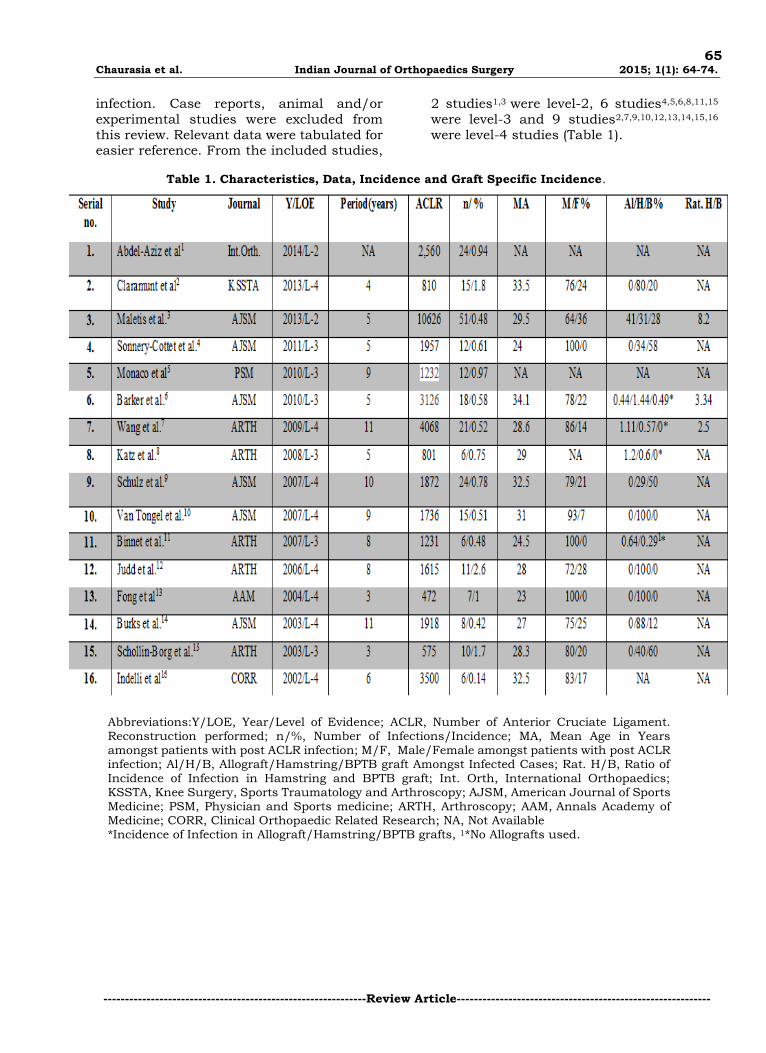
Table 1. Characteristics, Data, Incidence and Graft Specific Incidence.
Abbreviations:Y/LOE, Year/Level of Evidence; ACLR, Number of Anterior Cruciate Ligament. Reconstruction performed; n/%, Number of Infections/Incidence; MA, Mean Age in Years amongst patients with post ACLR infection; M/F, Male/Female amongst patients with post ACLR infection; Al/H/B, Allograft/Hamstring/BPTB graft Amongst Infected Cases; Rat. H/B, Ratio of
Incidence of Infection in Hamstring and BPTB graft; Int. Orth, International Orthopaedics; KSSTA, Knee Surgery, Sports Traumatology and Arthroscopy; AJSM, American Journal of Sports Medicine; PSM, Physician and Sports medicine; ARTH, Arthroscopy; AAM, Annals Academy of Medicine; CORR, Clinical Orthopaedic Related Research; NA, Not Available *Incidence of Infection in Allograft/Hamstring/BPTB grafts, 1*No Allografts used.
66 Chaurasia et al. Indian Journal of Orthopaedics Surgery 2015; 1(1): 64-74.
-------------------------------------------------------------Review Article-----------------------------------------------------------
Post ACLR Infection Incidence
There were 246 infections amongst 35,795 ACLR.1-16 Average incidence was
0.68% (range- 0.14-2.6%), while the reported
incidence in 2 systematic reviews was
between 0.3 and 1.7%17 and 0.6%18,
respectively. Average age was between 23-
341-16. Incidence of infection in allograft 6,7,8, hamstring and BPTB grafts 6,7,8,11 was
between 0.44-1.2%, 0.57-1.44% and 0-
0.49% respectively. Hamstring autografts
were more frequently infected (29-100%)
while BPTB autografts and allografts were found in 0-60% and 0-41% of the total
reported cases1-5,9,10.12-15. The ratio of
incidence of infection after autologous
hamstring and BPTB graft was between 2.5-
8.2, however it was provided only in 3
studies3,6,7.
Pathogenesis and Risk Factors
Grafts have been reported in some
studies as the nidus for infection because they act as a foreign body11,12,16, while other
studies have attributed hematoma at tibial
tunnel end as a origin of infection in sub-
acute and late cases (Table 2). 11,12,13,15 The
reported risk factors included: high body
mass index3 (BMI) > 30kg\m2, infection prone hamstring graft2,3,6,22, previous knee
surgery (arthroscopy or open) 12,13,14,23,
concomitant surgery13,15, short hamstring
tendon with more suture material11, post-
operative effusion11, subcutaneous position
of the metallic post/washer/braided suture construct17, different implants12 and the use
of intra-operative/intra-articular steroids15
(Table 2). Other causes postulated were
persistent communication between skin and
joint by sutures11, unsterile metal part and the rubber membranes on the suture clamps
which cannot be sterilized satisfactorily
despite sterilization performed in certified
autoclaves15, environmental contamination
of surgical equipments or hospital stuff in
studies24,25 demonstrating epidemics and adjacent tunnel osteomyelitis in persistent
cases11. It has been also hypothesized that if
microorganism of normal skin flora was
cultured from the joint, then inoculation
must have occurred at the time of surgery, or shortly thereafter through femoral or
tibial site.12

67 Chaurasia et al. Indian Journal of Orthopaedics Surgery 2015; 1(1): 64-74.
-------------------------------------------------------------Review Article-----------------------------------------------------------
Table 2. Risk factors and Pathogenesis
Abbreviations: SRN, study reference numbers; BMI, Body mass index
68 Chaurasia et al. Indian Journal of Orthopaedics Surgery 2015; 1(1): 64-74.
-------------------------------------------------------------Review Article-----------------------------------------------------------
Clinical Signs and Symptoms
The reported mean time of onset (MTO) of infection from index surgery was
between 7.5 to 61.7 days (Table 3). A recent
systematic review 18, reported an average of
16.8 ± 10.5 days (114/123, 92.6 %), with
most infections in acute or sub-acute phases
symptoms. Classical clinical features given were: acutely swollen painful joint, limited
range of motion, sudden increase of pulsatile
knee pain, rapidly increased and persistent
effusion, incision drainage, local erythema, warmth and intermittent fever (usually over
38°C).7,13,14,16 Other features included
hyperemic with serous or purulent
discharge11 and indolent presentation (60%
missed on first visit)15, excluding large
hematomas simulating acute (Table 3).
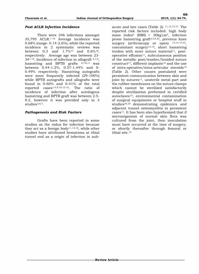
Table 3.Clinical Presentation and Laboratory Values
Abbreviations: MTO, Mean Time of Onset; ESR, Erythrocyte Sedimentation Rate; CRP, C-Reactive Protein; TLC, Total Leucocyte Count; CNS, Coagulase Negative Staphylococcous; MSSA, Methicillin Sensitive Staphylococcus Aureus; MRSA, Methicillin Resistant Staphylococcus Aureus; SA, Staphylococcus Aureus; P, Propionibacterium; OS, Other Species; NG, No Growth; NA, Not Available
69 Chaurasia et al. Indian Journal of Orthopaedics Surgery 2015; 1(1): 64-74.
-------------------------------------------------------------Review Article-----------------------------------------------------------
Diagnostic Evaluation
Laboratory investigations are required for infection confirmation.12 The
average erythrocyte sedimentation rate
(ESR) ranged between 51 -80 mm/hour and
average C-reactive protein(CRP) between
4.8-146.6 mg/l, with total leucocyte count
(TLC) in blood between 8.1- 11.7 × 10⁹/l.1,5-
13,15 In only 2 studies the average TLC in
synovial fluid was provided and was 11.5
and 5.2 × 10⁹/l, respectively.6,12 One
systematic review17 reported that the ESR
and CRP were markedly increased in 90% (50–100%) and 95% (67–100%) of patients,
respectively, and if the CRP level does not
decrease to nearly normal by 2 weeks
postoperatively, or there is a secondary rise,
infection should be suspected. Complete
CRP levels normalization is seen after 2–12 weeks.10,13 Blood culture is less sensitive in
diagnosis.17 Synovial fluid culture and
staining revealed Coagulase Negative
Staphylococcus (CNS), Staphylococcus
Aureus (SA) and Propionibacterium, in nearly all studies. Among CNS,
Staphylococus Epidermidis was the most
common pathogen. Other species1-16
included non/hemolytic Streptococcus,
Peptostreptococcus, Klebsiella, Enterobacter
species, Erysipelothrix Rhusiopathiae, Fungus, Mycobacterium Tuberculosis and
other anaerobic or gram-negative organisms.
Treatment Protocol
Serial arthroscopic lavages and
intravenous antibiotics with graft
retention, remains the most efficient
treatment protocol in most studies.18
Basic guidelines were as follows: Empirical intravenous antibiotic
therapy at the time of presentation:
intravenous (IV) ceftazidime (2g/8hr)
and vancomycin (1g/12hr)2, or
Cafazolin11, or flucloxacillin (6 × 1
g/day) and gentamycin (320 mg/day). 12 Pathogen-specific antibiotics after culture; additional
cultures during operation(s). IV
antibiotics changed to culture
sensitive oral antibiotics as soon as
the CRP levels had nearly normalized
(<1 mg/mL)7 for 6 weeks or until normalization of clinical and lab
parameters. Average duration of
antibiotics ranged for IV between
17.3days-6weeks followed by oral up
to 3.2months in 9 studies(Table
4a)1,2,7,10-13,15,16. Delayed diagnosis of more than 7 days or SA infection
required a longer duration of
antibiotic therapy and increased the
likelihood for graft removal and
restricted range of motion.18 Arthroscopic debridement and lavage:
extensive arthroscopic removal of
necrotic tissue with a shaver, as near
total synovectomy as possible,
debridement of fibrinous exudates of
graft’s surface, arthroscopic lysis of fibrous adhesions and extensive
pulsatile lavage with 10-15 lit of
saline. Additional lavage if clinical
and laboratory parameters are not
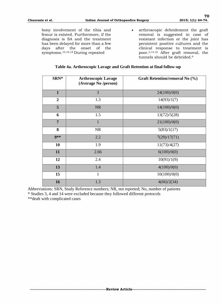
satisfactory. Average number of
arthroscopic lavage per person ranged between 1 to 3 in 11
studies(Table 4a).1,2,6,7,9-13,15,16 66-
100% of patients retained graft and
graft removal resulted in 0-34% of
patients in 12 studies1,2,5-8,10-13,15,16 excluding the study9 which dealt
with complicated cases(Table 4a).
This protocol remained more or less
the same in nearly all studies
supporting graft retention.1-8,10-13,15,16 Concomitant open incision and
drainage, through the old scars of
arthroscopy and meniscal repair
portals, at the same time of the
arthroscopic lavage, in cases of
complicated or infected wounds, in
order to avoid extra-articular fluid collection and to eliminate the
infection.10 The wounds are left open
with only a sterile dressing applied,
in order to promote secondary wound
closure.9 Continuous irrigation drains in the joint may be used for 2
days.15 Average hospital stay ranged
between 17.3 to 27.2 days in 3
studies(Table 4a)10,11,13. Immediate graft removal should be
considered if the graft is unstable resulting in nonfunctional ligament
during clinical examination and
arthroscopic evaluation and causing
instability or block. The same if the
graft is impregnated by a tenacious,
thick purulent exudation which could not be removed without graft
damage and the articular cartilage is
appeared soft and swollen or possible
70 Chaurasia et al. Indian Journal of Orthopaedics Surgery 2015; 1(1): 64-74.
-------------------------------------------------------------Review Article-----------------------------------------------------------
bony involvement of the tibia and
femur is existed. Furthermore, if the
diagnosis is SA and the treatment has been delayed for more than a few
days after the onset of the
symptoms.10,16,18 During repeated
arthroscopic debridement the graft
removal is suggested in case of
resistant infection or the joint has persistent positive cultures and the
clinical response to treatment is
poor.2,14,16 After graft removal, the
tunnels should be debrided.9
Table 4a. Arthroscopic Lavage and Graft Retention at final follow-up
Abbreviations: SRN, Study Reference numbers; NR, not reported; No, number of patients
* Studies 3, 4 and 14 were excluded because they followed different protocols
**dealt with complicated cases
SRN* Arthroscopic Lavage
(Average No /person)
Graft Retention/removal No (%)
1 3 24(100)/0(0)
2 1.3 14(93)/1(7)
5 NR 14(100)/0(0)
6 1.5 13(72)/5(28)
7 1 21(100)/0(0)
8 NR 5(83)/1(17)
9** 2.2 7(29)/17(71)
10 1.9 11(73)/4(27)
11 2.66 6(100)/0(0)
12 2.4 10(91)/1(9)
13 1.4 4(100)/0(0)
15 1 10(100)/0(0)
16 1.3 4(66)/2(34)
71 Chaurasia et al. Indian Journal of Orthopaedics Surgery 2015; 1(1): 64-74.
-------------------------------------------------------------Review Article-----------------------------------------------------------
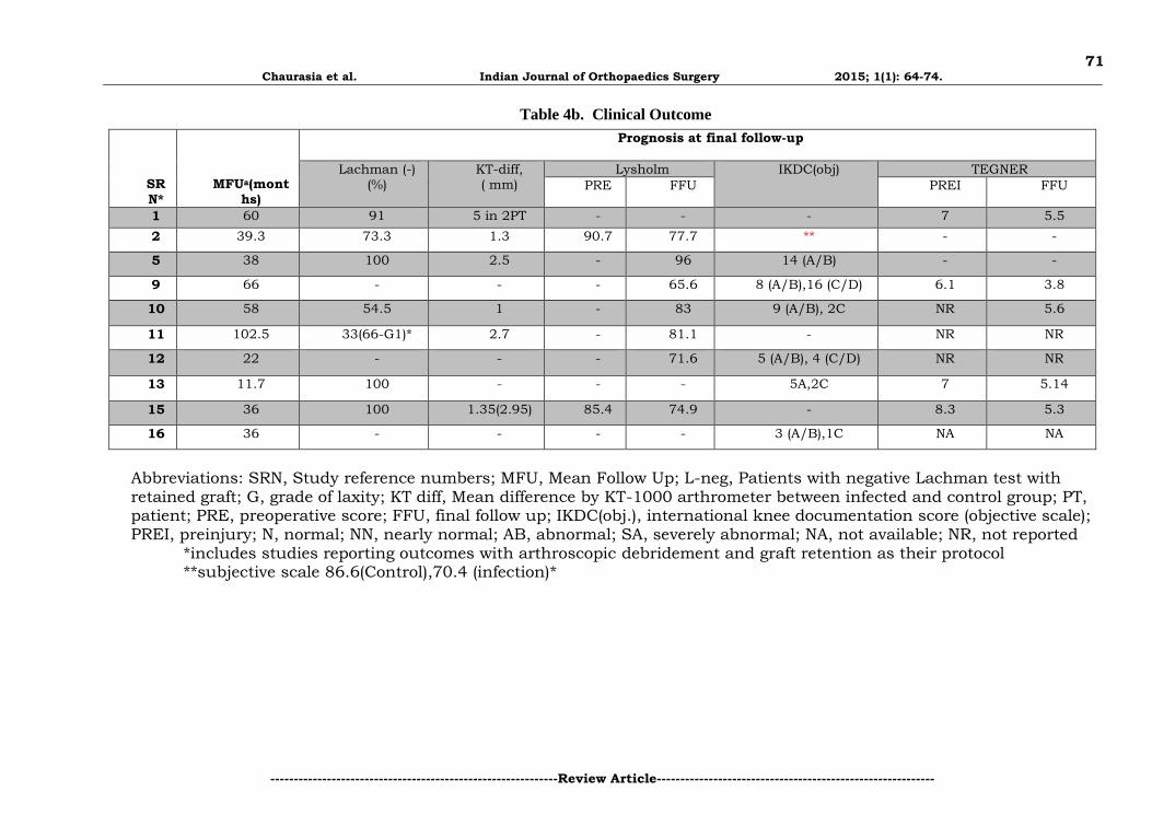
Table 4b. Clinical Outcome
Abbreviations: SRN, Study reference numbers; MFU, Mean Follow Up; L-neg, Patients with negative Lachman test with retained graft; G, grade of laxity; KT diff, Mean difference by KT-1000 arthrometer between infected and control group; PT, patient; PRE, preoperative score; FFU, final follow up; IKDC(obj.), international knee documentation score (objective scale); PREI, preinjury; N, normal; NN, nearly normal; AB, abnormal; SA, severely abnormal; NA, not available; NR, not reported
*includes studies reporting outcomes with arthroscopic debridement and graft retention as their protocol **subjective scale 86.6(Control),70.4 (infection)*
SRN*
MFUa(months)
Prognosis at final follow-up
Lachman (-) (%)
KT-diff,
( mm) Lysholm IKDC(obj)
TEGNER
PRE FFU PREI FFU
1 60 91 5 in 2PT - - - 7 5.5
2 39.3 73.3 1.3 90.7 77.7 ** - -
5 38 100 2.5 - 96 14 (A/B) - -
9 66 - - - 65.6 8 (A/B),16 (C/D) 6.1 3.8
10 58 54.5 1 - 83 9 (A/B), 2C NR 5.6
11 102.5 33(66-G1)* 2.7 - 81.1 - NR NR
12 22 - - - 71.6 5 (A/B), 4 (C/D) NR NR
13 11.7 100 - - - 5A,2C 7 5.14
15 36 100 1.35(2.95) 85.4 74.9 - 8.3 5.3
16 36 - - - - 3 (A/B),1C NA NA
72 Chaurasia et al. Indian Journal of Orthopaedics Surgery 2015; 1(1): 64-74.
-------------------------------------------------------------Review Article-----------------------------------------------------------
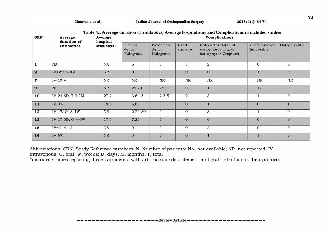
Table 4c. Average duration of antibiotics, Average hospital stay and Complications in included studies SRN* Average
duration of antibiotics
Average hospital stay(days)
Complications
Flexion deficit-N,degrees
Extension deficit N,degrees
Graft rupture
Osteoarthritis(Joint space narrowing or osteophytes,Crepitus)
Graft removal (nonviable)
Osteomyelitis
1 NA NA 5 0 3 2 0 0
2 IV4W,O2-4W NR 0 0 0 0 1 0
7 IV-19.4 NR NR NR NR NR NR NR
9 NR NR 24,20 24,3 0 1 17 0
10 IV-24.6D, T-3.2M 27.2 3,6-15 2,3-5 2 3 1 0
11 IV-3W 19.5 6,6 0 0 1 0 1
12 IV-4W,O- 3-4W NR 2,20-30 0 0 3 1 0
13 IV-17.3D, O-4-6W 17.3 7,20 0 0 0 0 0
15 IV+O- 4-12 NR 0 0 0 5 0 0
16 IV-6W NR 0 0 0 1 1 0
Abbreviations: SRN, Study Reference numbers; N, Number of patients; NA, not available; NR, not reported; IV, intravenous; O, oral; W, weeks; D, days; M, months; T, total *includes studies reporting these parameters with arthroscopic debridement and graft retention as their protocol
73 Chaurasia et al. Indian Journal of Orthopaedics Surgery 2015; 1(1): 64-74.
-------------------------------------------------------------Review Article-----------------------------------------------------------
Outcome and Complications
Ten studies1,2,5,9-13,15,16 (Table 4b), reported the follow-ups along with the use of
more or less the above proposed most
effective treatment protocol and gave
satisfactory results. Lachman’s test was
negative in 54.5 - 100% of the patients, at a
mean final follow-up between 11.7 - 60 months1,2,5,10,11,13,15. Mean KT-1000
arthrometer difference between control and
infected group ranged from 1 - 5 mm at an
average of 21 - 102.5 months 1,2,5,10,11,15.
Average Lysholm score ranged between 65.6 - 96, at 22 - 102.5 months1,2,5,9-12,15, while
average Tegner score was between 3.8 - 5.6 1,5,9,10,13,15 and preinjury average Tegner
score was 6.1-8.3 9,13,15. Out of 75 infected
patients, 43 patients had normal or near
normal and 25 patients had abnormal or severely abnormal scores in International
Knee Documentation Committee (IKDC)
subjective scale5,9,10,12,13,16. Out of 142
infected patients, 47 patients had a flexion
deficit ranging from an average of 6 - 30°, 26 patients had extension deficit ranging from
3-5°.1,2,5,9,10-13,15,16 Other complications are
reported in Table 4c.
Discussion
Infection following ACLR is not
common; the reported incidence of infection is between 0.14-2.6%. Staphylococcus Epidermidis, a CNS remains the most
common species consistent with post ACLR
infection (Table 3). Currently, post-ACLR infection is considered multifactorial. As
ACL grafts act as a foreign body,
pathogenesis behind this factor is universal.
Also, we used autologous hamstring tendon
as a graft which may be more prone to get
infected as compared to BPTB graft as it has
2.5-8.2 times more chances to get infected
(Table 1). Additional literature reports more chances of infection during preparation in
hamstring grafts.24 Another proposed
predisposing factor is the extended operative
time of DB compared to single bundle (SB)
ACLR.25 However, another study26 showed
no significant differences concerning the time for operation between the DB and SB
groups and there is no study to our
knowledge in literature confirming that rate
of septic arthritis is more in DB than in SB
groups. Most of the untabled literature27,28
reports same clinical symptoms and similar
range laboratory parameters as reported in
Table 3. Apart from risk factors enumerated
in Table 2, literature27,28,29 includes:
operative time, tourniquet inflation time,
contaminated sterile inflow cannula, concomitant open surgical procedures,
increased foreign body load (suture material
or hardware), and use of a drain. The most
effective treatment protocol proposed was
irrigation and debridement with graft retention as infection subsides usually in 1-
3 arthroscopic debridements and the 66-
100% grafts can be retained (Table 4a).
Following the proposed protocol, most
studies reported comparable or inferior
outcome to control group and associated complications as reported before (Tables 4b
and 4c).
Conclusion
Septic arthritis after ACLR is a rare
and disastrous complication which can be
successfully managed by early diagnosis and
arthroscopic debridement with a proper
protocol.
References
1. Abdel-Aziz A, Radwan YA, Rizk A. Multiple arthroscopic debridement and graft retention in septic knee
arthritis after ACL reconstruction: a prospectivecase-control study. Int Orthop 2014;38:73-82. 2. Torres-Claramunt R, Pelfort X, Erquicia J, et al. Knee joint infection after ACL reconstruction:
prevalence, management and functional outcomes. Knee Surg Sports Traumatol Arthrosc 2013;21:2844-9.
3. Maletis GB, Inacio MC, Reynolds S, Desmond JL, Maletis MM, Funahashi TT. Incidence of postoperative anterior cruciate ligament reconstruction infections: graft choice makes a difference. Am J Sports Med 2013;41:1780-5.
4. Sonnery-Cottet B, Archbold P, Zayni R, et al. Prevalence of septic arthritis after anterior cruciate ligament
reconstruction among professional athletes. Am J Sports Med 2011;39:2371-6. 5. Monaco E, Maestri B, Vadalà A, Iorio R, Ferretti A. Return to sports activity after postoperative septic
arthritis in ACL reconstruction. Phys Sportsmed 2010;38:69-76.
74 Chaurasia et al. Indian Journal of Orthopaedics Surgery 2015; 1(1): 64-74.
-------------------------------------------------------------Review Article-----------------------------------------------------------
6. Barker JU, Drakos MC, Maak TG, Warren RF, Williams RJ 3rd, Allen AA. Effect of graft selection on the incidence of postoperative infection in anterior cruciate ligament reconstruction. Am J Sports Med 2010;38:281-6.
7. Wang C, Ao Y, Wang J, Hu Y, Cui G, Yu J. Septic arthritis after arthroscopic anterior cruciate ligament reconstruction: a retrospective analysis of incidence,presentation, treatment, and cause. Arthroscopy 2009 ;25:243-9.
8. Katz LM, Battaglia TC, Patino P, Reichmann W, Hunter DJ, Richmond JC. A retrospective comparison of the incidence of bacterial infection following anterior cruciate ligament reconstruction with autograft versus allograft. Arthroscopy 2008;24:1330-5.
9. Schulz AP, Götze S, Schmidt HG, Jürgens C, Faschingbauer M. Septic arthritis of the knee after anterior cruciate ligament surgery: a stage-adapted treatment regimen. Am J Sports Med 2007;35:1064-9.
10. Van Tongel A, Stuyck J, Bellemans J, Vandenneucker H. Septic arthritis after arthroscopic anterior cruciate ligament reconstruction: A retrospective analysis of incidence, management and outcome. Am J Sports Med 2007;35:1059-1063.
11. Binnet MS, Başarir K. Risk and outcome of infection after different arthroscopic anterior cruciate
ligament reconstruction techniques. Arthroscopy 2007;23:862-8. 12. Judd D, Bottoni C, Kim D, Burke M, Hooker S. Infections following arthroscopic anterior cruciate
ligament reconstruction. Arthroscopy 2006;22:375-84. 13. Fong SY, Tan JL. Septic arthritis after arthroscopic anterior cruciate ligament reconstruction. Ann Acad
Med Singapore 2004;33:228-34.
14. Burks RT, Friederichs MG, Fink B, Luker MG, West HS, Greis PE. Treatment of postoperative anterior cruciate ligament infections with graft removal and early reimplantation. Am J Sports Med 2003;31:414-8.
15. Schollin-Borg M, Michaëlsson K, Rahme H. Presentation, outcome, and cause of septic arthritis after anterior cruciate ligament reconstruction: a case control study. Arthroscopy 2003;19:941-7.
16. Indelli PF, Dillingham M, Fanton G, Schurman DJ. Septic arthritis in postoperative anterior cruciate
ligament reconstruction. Clin Orthop Relat Res 2002;(398):182-8. 17. Mouzopoulos G, Fotopoulos VC, Tzurbakis M. Septic knee arthritis following ACL reconstruction: a
systematic review. Knee Surg Sports Traumatol Arthrosc 2009;17:1033-42. 18. Wang C, Lee YH, Siebold R. Recommendations for the management of septic arthritis after ACL
reconstruction. Knee Surg Sports Traumatol Arthrosc 2013. [Epub ahead of print] PubMed PMID:
24061716. 19. Goldenberg DL, Reed JI. Bacterial arthritis. N Engl J Med 1985;312:764–771. 20. Hogan CJ, Fang GD, Scheld WM, Linden J, Dibuch DR. Inhibiting the inflammatory response in joint
sepsis. Arthroscopy 2001;17:311–315. 21. National Institutes of Health (NIH), National Institute of Arthritis and Musculoskeletal and Skin Diseases
(NIAMS), Vanderbilt University, United States. Prognosis and Predictors of ACL Reconstruction—A
Multicenter Cohort Study. Available at: http://clinicaltrials.gov/ct2/show/NCT00463099. Accessed March 1, 2011.
22. Plante MJ, Li X, Scully G, Brown MA, Busconi BD, DeAngelis NA. Evaluation of msterilization methods following contamination of hamstring autograft during anterior cruciate ligament reconstruction. Knee Surg Sports Traumatol Arthrosc 2013;21:696-701.
23. Parada SA, Grassbaugh JA, Devine JG, Arrington ED. Instrumentation-specific infection after anterior cruciate ligament reconstruction. Sports Health. 2009;1:481-5.
24. Hantes ME, Basdekis GK, Varitimidis SE, Giotikas D, Petinaki E, Malizos KN. Autograft contamination during preparation for anterior cruciate ligament reconstruction. J Bone Joint Surg Am 2008;90:760-764.
25. Kondo E, Yasuda K, Azuma H. Prospective clinical comparisons of anatomic double-bundle versus single-bundle anterior cruciate ligament reconstruction procedures in 328 consecutive patients. Am J Sports
Med 2008;36:1675-87. 26. Yasuda K, Kondo E, Ichiyama H. Clinical evaluation of anatomic double-bundle anterior cruciate
ligament reconstruction procedure using hamstring tendon grafts: comparisons among different procedures. Arthroscopy 2006;22:240-51.
27. Azar FM, Arthur ST. Complications of anterior cruciate ligament reconstruction. Tech Knee Surg 2004;3:238-250.
28. Williams RJ III, Laurencin CT, Warren RF, Speciale AC, Brause BD, O’Brien S. Septic arthritis after arthroscopic anterior cruciate ligament reconstruction: Diagnosis and management. Am J Sports Med 1997;25:261-267.
29. Viola R, Marzano N, Vianello R. An unusual epidemic of Staphylococcus-negative infections involving anterior cruciate ligament reconstruction with salvage of the graft and function. Arthroscopy
2000;16:173-177.
30. Millett PJ, Wickiewicz TL, Warren RF. Motion loss after ligament injuries to the knee. Part I: causes. Am J Sports Med 2001;29:664-75.
Copyright © 2022 FDOKUMEN