Rationale for Periodontal Treatment
101
Periodontal Regenerative Therapy in Old Adults Dr. Masoud Amiri Moghaddam Assistant Professor of Periodontics Gilan University of Medical Scienc e 1
-
Upload
khangminh22 -
Category
Documents
-
view
0 -
download
0
Transcript of Rationale for Periodontal Treatment
Periodontal Regenerative
Therapy in Old Adults
Dr. Masoud Amiri Moghaddam
Assistant Professor of Periodontics
Gilan University of Medical Science
1

Geriatric population comprises of adults of ≥65 years old and currently
counts some 11% of the world’s population with an estimated increase
of up to 22% by 2050 according to the United Nations
Whether periodontitis represents a part of physiological aging or its
simple consequence was a subject of controversy over the years
The simple fact is that periodontitis is highly prevalent lifelong
multifactorial disease and the sixth most common health condition
2
The effectiveness of periodontal therapy is made possible by the
remarkable healing capacity of the periodontal tissues.
Periodontal therapy restore chronically inflamed gingiva
identical with gingiva that has never been exposed to excessive
plaque accumulation
3

Periodontal disease >>> genetic background, general inflammatory status, environmental factors, and systemic diseases,
Neglected oral hygiene
Decreased salivary flow >>> Xerostomia is reported in 25–50% of older people and is associated with systemic diseases (such as Sjögren syndrome, parkinsonism, or DM)
4
Healing Capacity is not decreased significantly due to aging
(multiple systematic reviews)
>>> Regenerative therapy of incidental periodontal defects and
diseases is feasible
5
Properly performed, periodontal treatment
can eliminate pain, exudate, gingival inflammation, and bleeding.
It can also reduce periodontal pockets,
eliminate infection,
arrest the destruction of soft tissue and bone,
and reduce abnormal tooth mobility
Other benefits are to establish optimal occlusal function,
restore tissue destroyed by disease
reestablish physiologic gingival contour,
and prevent the recurrence of disease
6

Local Therapy
The removal of plaque and all of the factors that favor its
accumulation is the primary goal in local therapy
The thorough elimination of plaque and the prevention of its
formation can help maintain periodontal health, even if traumatic
forces are allowed to persist
However, the elimination of trauma may increase the chances for
bone regeneration and the gain of attachment.
creating occlusal relationships that are more tolerable to the
periodontal tissues increases the margin of safety of the
periodontium to the buildup of plaque, in addition to reducing tooth
mobility.
7
Systemic Therapy
Systemic therapy may be employed as an adjunct to local measures :
systemic complications from acute infections or chemotherapy and preventing
harmful effects of post-treatment bacteremia
The control of systemic diseases that aggravate the patient’s periodontal
condition is always a consideration so proper precautions can be instituted
Systemic therapy for treatment of the periodontal condition in conjunction
with local therapy is indicated in patients with aggressive periodontitis
the concept of host modulation paper by Nyman, Schroeder, and Lindhe
it was possible to block periodontal bone loss in animals with the aspirin-
like drug indomethacin
8
(NSAIDs), such as flurbiprofen and ibuprofen, can reduce the
development of experimental gingivitis and the loss of alveolar bone
in periodontitis
NSAIDs are propionic acid derivatives and act by inhibiting the
cyclooxygenase pathway of arachidonic acid metabolism, thereby
reducing prostaglandin formation. These NSAIDs can be
administered by mouth or applied topically
bisphosphonate, which is currently used to treat metabolic diseases
in humans, such as Paget disease or hypercalcemia of malignancy,
which result in bone resorption Experimental studies in
monkeys have shown that alendronate reduced the bone loss
associated with periodontitis
9

Factors That Affect Healing
Local Factors :
local factors, particularly plaque microorganisms, are the most
common deterrents to healing after periodontal treatment
Healing is also delayed by
(1)excessive tissue manipulation during treatment
(2)trauma to the tissues
(3)the presence of foreign bodies
(4)repetitive treatment procedures that disrupt the orderly cellular
activity in the healing process
10

Systemic Factors:
extensively documented in animal experiments but are less clearly defined in
humans.
Healing capacity diminishes with age probably because of the atherosclerotic
vascular changes common in aging and the resulting reduction in blood circulation
delayed in patients with generalized infections and in those with
diabetes and other debilitating diseases
insufficient food intake; bodily conditions that interfere with the use of nutrients;
and deficiencies in vitamin C , proteins, and other nutrients.
11

affected by hormones. Systemically administered glucocorticoids
such as cortisone hinder repair by depressing the inflammatory
reaction or by inhibiting the growth of fibroblasts, the production of
collagen, and the formation of endothelial cells
Systemic stress, thyroidectomy, testosterone, adrenocorticotropic
hormone (ACTH), and large doses of estrogen suppress the formation
of granulation tissue and impair healing
Progesterone increases and accelerates the vascularization of
immature granulation tissue and appears to increase the susceptibility
of the gingiva to mechanical injury by causing dilation of the
marginal vessels.
12
Healing After Periodontal Therapy
basic healing processes are the same after all forms of
periodontal therapy removal of degenerated tissue
debris and the replacement of tissues destroyed by
disease regeneration and repair of the periodontal
structures but not necessarily a gain in attachment
13

Regeneration
Is the natural renewal of a structure, produced by growth and
differentiation of new cells and intercellular substances to form new
tissues or part
Regeneration occurs through growth from the same type of tissue that
has been destroyed or from its precursor
Gingival epithelium is replaced by epithelium, and the underlying
connective tissue and periodontal ligament are derived from connective
tissue. Bone and cementum are replaced by connective tissue, which is
the precursor of both. Undifferentiated connective tissue cells develop
into osteoblasts and cementoblasts, which form bone and cementum
14

Regeneration of the periodontium is a continuous physiologic process
wear and tear repair
(1) mitotic activity in the epithelium of the gingiva and the connective
tissue of the periodontal ligament,
(2) the formation of new bone, and
(3) the continuous deposition of cementum
occurring even during destructive periodontal disease.
Most gingival and periodontal diseases are chronic inflammatory
processes
15
bacteria and bacterial products plus inflammatory exudate
prevent completion of the healing process
removing bacterial plaque and creating the conditions to
prevent its new formation, periodontal treatment removes
the obstacles to regeneration and enables the patient to
benefit from the inherent regenerative capacity of the
tissues
16
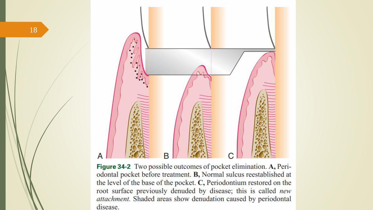
Repair
restores the continuity of the diseased marginal gingiva and
reestablishes a normal gingival sulcus at the same level on the root as
the base of the preexisting periodontal pocket
This process healing by scar
arrests bone destruction but does not result in gain of gingival
attachment or bone height.
17
18

Involves regeneration and mobilization of epithelial and
connective tissue cells into the damaged area and increased local
mitotic divisions to provide sufficient numbers of cells
For the diseased gingiva and attachment apparatus to regain
(totally or partially) their level on the root therapy must
include special materials and techniques. If these are not used or
are not successful, tissues undergo repair only
reconstruction of the periodontium therapeutic techniques that
seek to rebuild the periodontium and result in a significant gain
of attachment and bone height
19

20
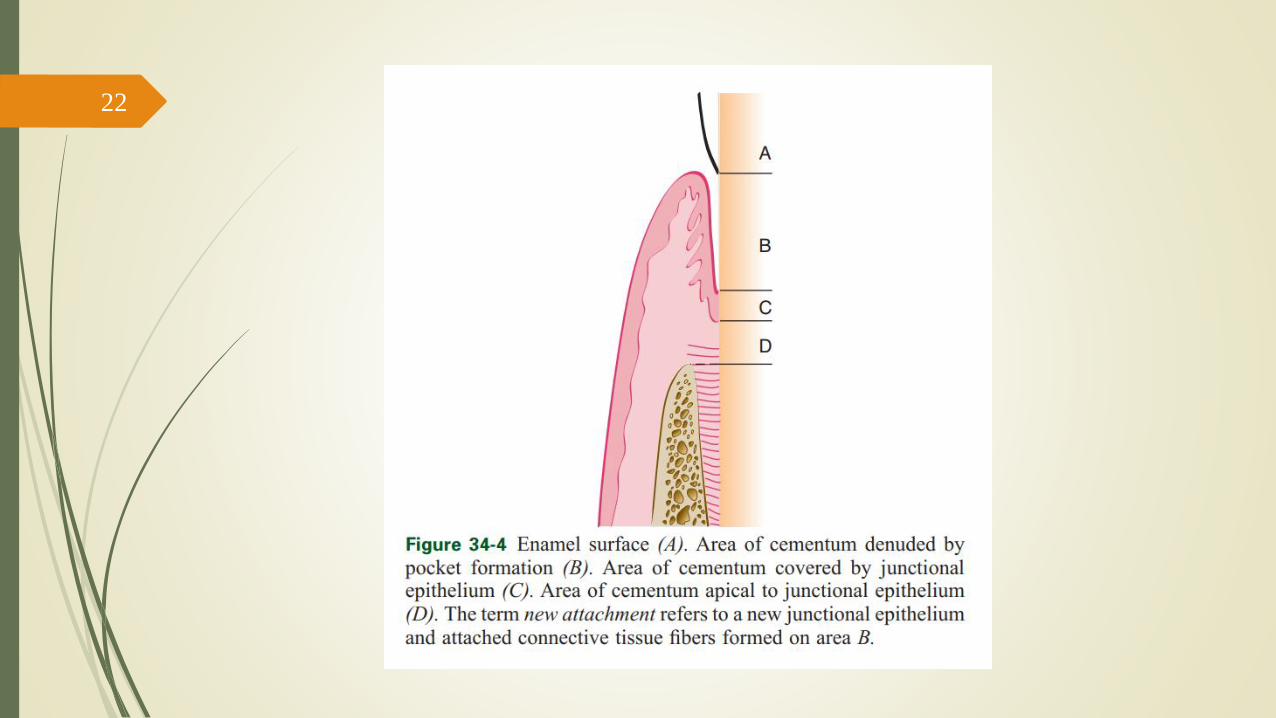
New Attachment
New attachment is the embedding of new periodontal ligament fibers into new
cementum and the attachment of the gingival epithelium to a tooth surface
previously denuded by disease
The attachment of the gingiva or the periodontal ligament to areas of the tooth
from which they have been removed in the course of treatment (or during
preparation of teeth for restorations) represents simple healing or reattachment
of the periodontium, not new attachment
reattachment repair in areas of the root not previously exposed to the pocket
such as after surgical detachment of the tissues or following traumatic tears in
the cementum, tooth fractures, or the treatment of periapical lesions
21
22
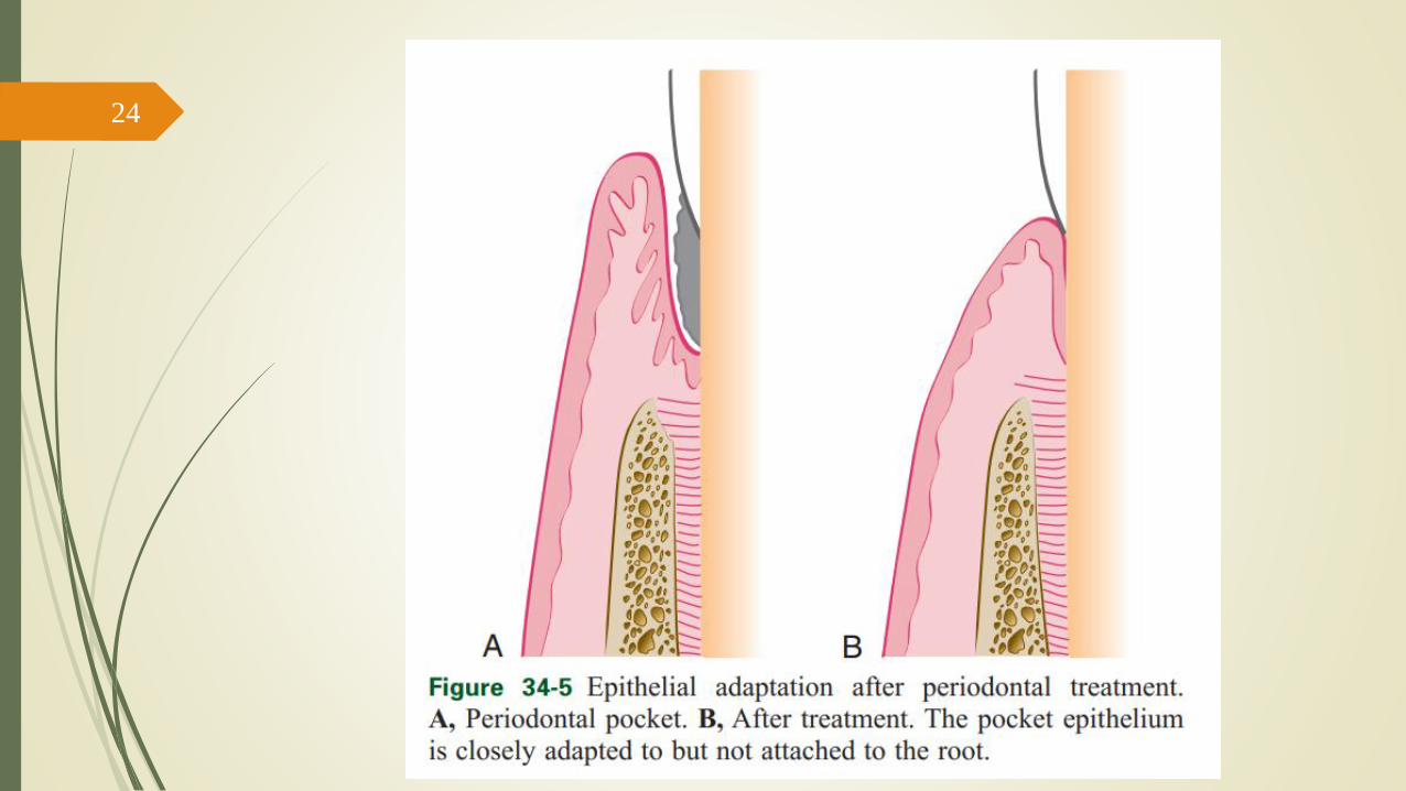
Epithelial adaptation close apposition of the gingival epithelium
to the tooth surface, with no gain in height of gingival fiber
attachment. The pocket is not completely obliterated, although it
may not permit passage of a probe
23
24
studies have shown that these deep sulci lined by long, thin
epithelium may be as resistant to disease as true connective tissue
attachments
The absence of bleeding or secretion on probing, the absence of
clinically visible inflammation, and the absence of stainable plaque
on the root surface when the pocket wall is deflected from the tooth
may indicate that the “deep sulcus” persists in an inactive state,
causing no further loss of attachment posttherapy depth of 4 mm
or even 5 mm may therefore be acceptable in these cases
25
Periodontal Reconstruction
(1) gain of attachment level,
(2) formation of new periodontal ligament fibers, and
(3) a level of alveolar bone significantly coronal to that present
before treatment
Melcher regeneration of the periodontal ligament is the key
to periodontal reconstruction because it “provides continuity
between the alveolar bone and the cementum and also because
it contains cells that can synthesize and remodel the three
connective tissues of the alveolar part of the periodontium.
26
During the healing stages of a periodontal pocket, the area is
invaded by cells from four different sources : oral epithelium,
gingival connective tissue, bone, and periodontal
ligament
If the epithelium proliferates along the tooth surface before the other
tissues reach the area, the result will be a long junctional epithelium
If the cells from the gingival connective tissue are the first to
populate the area, the result will be fibers parallel to the tooth
surface and remodeling of the alveolar bone with no attachment to
the cementum
If bone cells arrive first, root resorption and ankylosis may occur.
only when cells from the periodontal ligament proliferate coronally
is there new formation of cementum and periodontal ligament
27
Important goal of periodontal therapy is to obtain a reduced
pocket depth after treatment in order to prevent further
disease progression
Can be accomplished by non-surgical therapy in patients
with moderate periodontitis, whereas in severe cases -
presence of intrabony defects and furcations, the treatment
must be supplemented with periodontal surgery.
28
Regenerative Periodontal Therapy29

Fundamental objective of periodontal surgery is to provide
access for proper instrumentation and cleaning of the root
surface
Most surgical procedures result in the elimination or the
reduction of the soft tissue component of the periodontal pocket.
Periodontal treatment, both surgical and nonsurgical, results in
recession of the gingival margin after healing (Isidor et al. 1984)
30
Localized gingival recession and root exposure may
represent an esthetic problem to the patient, and it is often
associated with root sensitivity.
Such a situation is an indication to apply regenerative
periodontal therapy to obtain root coverage in order to
improve esthetics and reduce root sensitivity.
31
Successful root coverage implies regeneration of the
attachment apparatus on the exposed root surface including
cementum with inserting collagen fibers, as well as an
esthetically acceptable restoration of the anatomy of the
mucogingival complex.
32

Another indication for RPT is furcation-involved teeth. The
furcation area is often inaccessible to adequate
instrumentation and frequently the roots present concavities
and furrows which make proper cleaning of the area after
resective surgery impossible.
Long-term prognosis of furcation-involved teeth can be
improved considerably by successful regenerative periodontal
therapy.
33
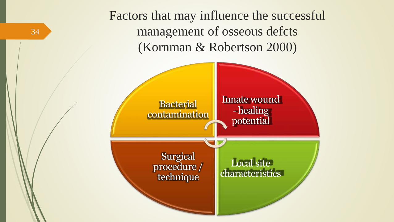
Factors that may influence the successful
management of osseous defcts
(Kornman & Robertson 2000)
Bacterial contamination
Surgical procedure / technique
Innate wound- healing potential
Local site characteristics
34

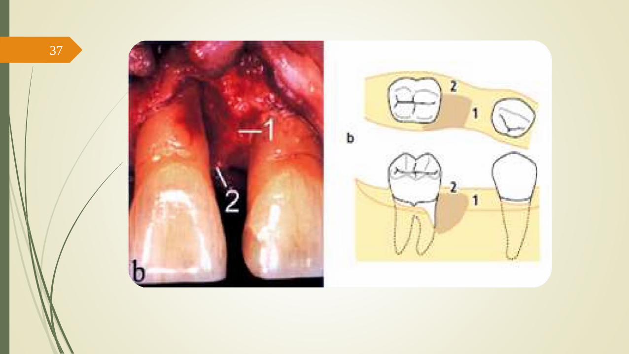
The morphology of the periodontal bony defect - essential
for the establishment of a predictable prognosis
Goldman and Cohen (1958) introduced a classification of
periodontal intrabony defects which was based on the
number of osseous walls surrounding the defect, being
either three-wall,two-wall or one-wall defects or a
combination of such situations
35
36
37

38
Results from a study by Ellegaard and Löe (1971) comprising 191
defects in 24 patients with periodontal disease indicated that
complete regeneration, determined radiographically and by
periodontal probing, had occurred in around 70% of the three-wall
defects, in 40% of the combined two-wall and three-wall defects,
and in 45% of the two-wall defects.
39
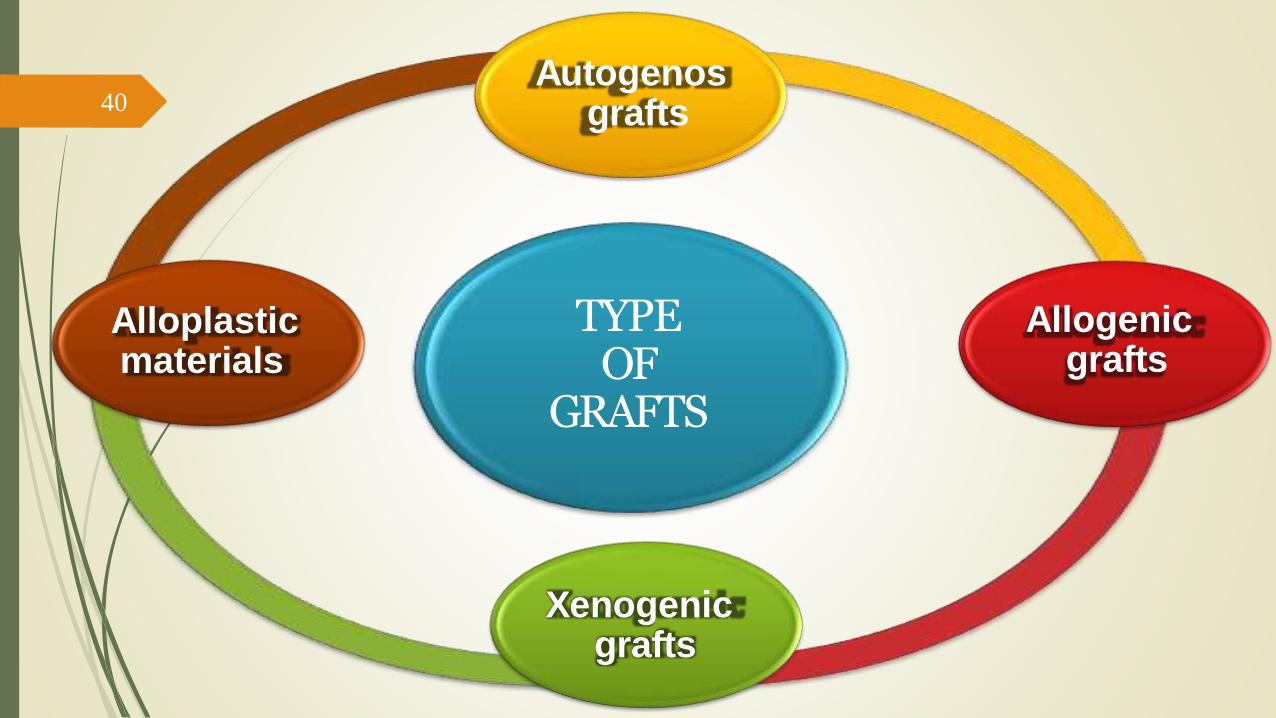
Autogenosgrafts
Alloplastic materials
Xenogenicgrafts
Allogenicgrafts
TYPE OF
GRAFTS
40
Autogenos grafts
Grafts transferred from one position to another within the same individual.
No potential problems of histocompatibility and
disease transmission
Comprises (i) cortical bone or (ii) cancellous bone and marrow
Promote bone healing
Mainly through osteogenesis and/or osteoconduction.
Harvested either from intraoral or extraoral donor sites.
Intaroral :edentulous areas of the jaw, healing extraction sites, maxillary tuberosities or the mandibular retromolar area
Extraoral : iliac crest marrow
41

42
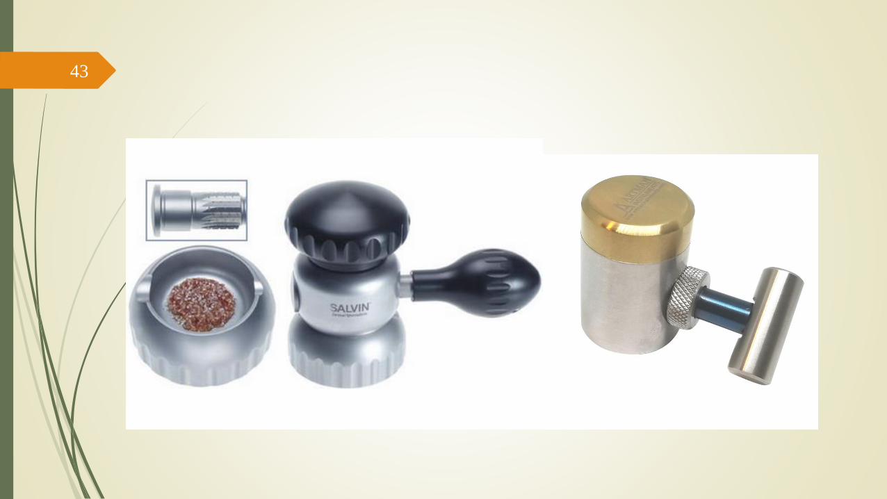
43
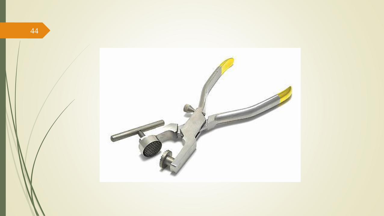
44
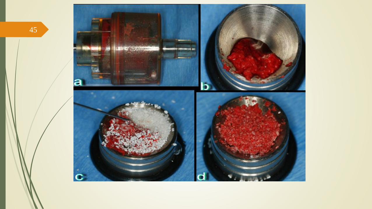
45
Allogenic grafts
Grafts transferred between genetically dissimilar
members of the same species.
Frozen iliac cancellous bone and marrow, mineralized freeze dried bone allogeneic grafts (FDBA), and decalcified freeze-dried alogeneic bone grafts (DFDBA).
The need for cross matching to decrease the likelihood of graft rejection as well as the risk of disease transmission virtually eliminated the use of frozen iliac allogeneic grafts in periodontics.
46

47

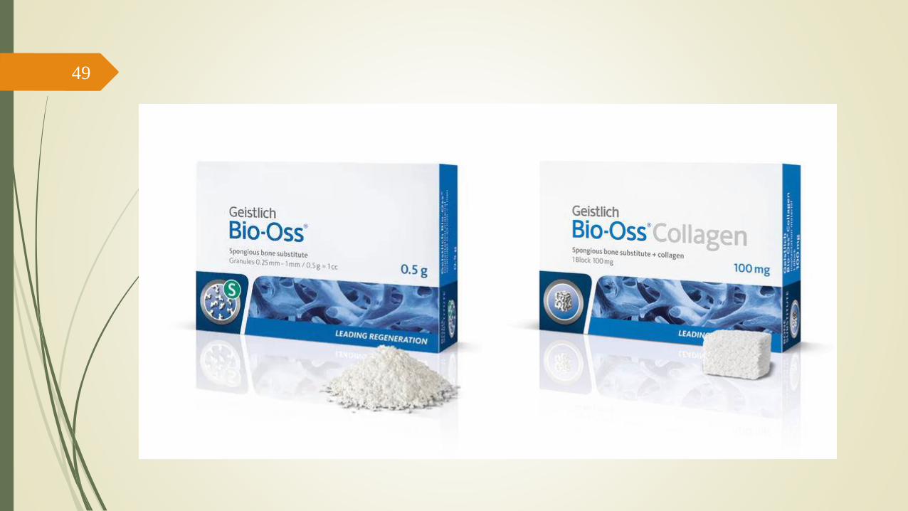
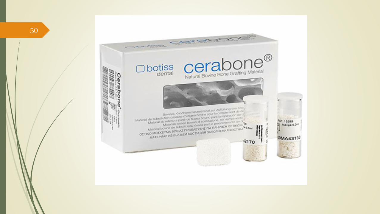
Xenogenic grafts
Grafts taken from a donor of another species.
Nielsen et al. (1981) treated 46 intrabony defects with Kielbone® (i.e. defatted and
deproteinized ox bone) and another 46 defects with intraoral autogenous bone
grafts. The results showed no difference between the amount of clinical gain of
attachment and bone fill obtained in the two categories of defect.
Bio-Oss®, Geistlich AG, Switzerland;
Lubboc®/Laddec®, Ost Development
SA,France;
Endobone®, Biomet Inc. Dordrecht, The Netherlands;
OsteoGraf®/N, DENTSPLY, Friadent Cera-Med,
Lakewood, CO, USA;
Cerabone®, aap Implantate AG,Berlin,Germany
48
49
50
51
Alloplastic materials
Synthetic, biocompatible, inorganic implant materials which synthetic, which are
used as substitutes for bone grafts.
Promote bone healing through osteoconduction.
1. Hydroxyapatite (HA) - non-resorbable ceramic / resorbable non-ceramic
2. Betatricalcium phosphate (β-TCP),
3. Polymers - a non-resorbable, calcium hydroxide coated co-polymer of
polymethylmethacrylate (PMMA) and polyhydroxylethylmethacrylate (PHEMA)
4. Bioactive glasses (bio-glasses) - composed of SiO2, Na2O, P2O5 and are
resorbable or not resorbable
52
53
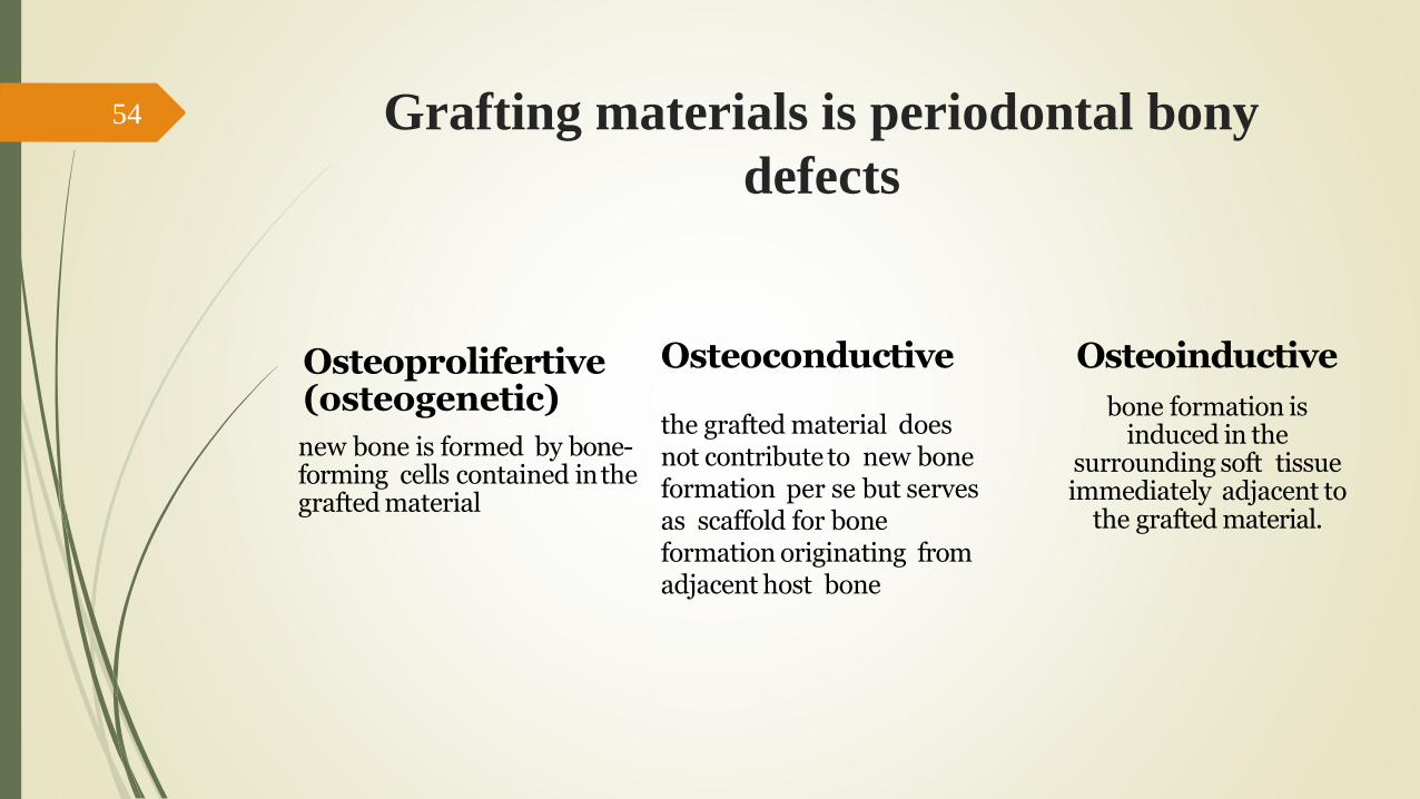
Grafting materials is periodontal bony
defects
Osteoprolifertive(osteogenetic)
new bone is formed by bone-forming cells contained inthe grafted material
Osteoconductive
the grafted material does not contribute to new bone formation per se but serves as scaffold for bone formation originating from adjacent host bone
Osteoinductive
bone formation is induced in the
surrounding soft tissueimmediately adjacent to
the grafted material.
54
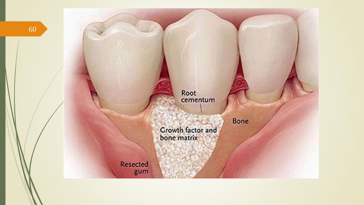
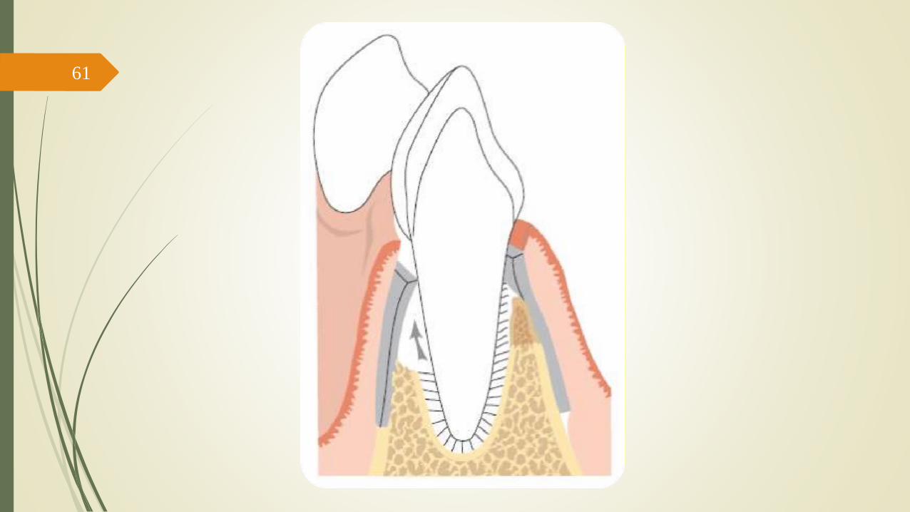
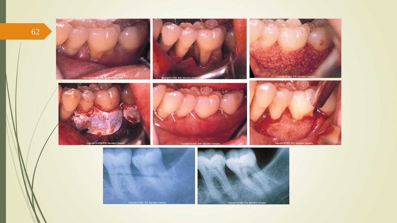
Guided Tissue Regeneration (GTR)55
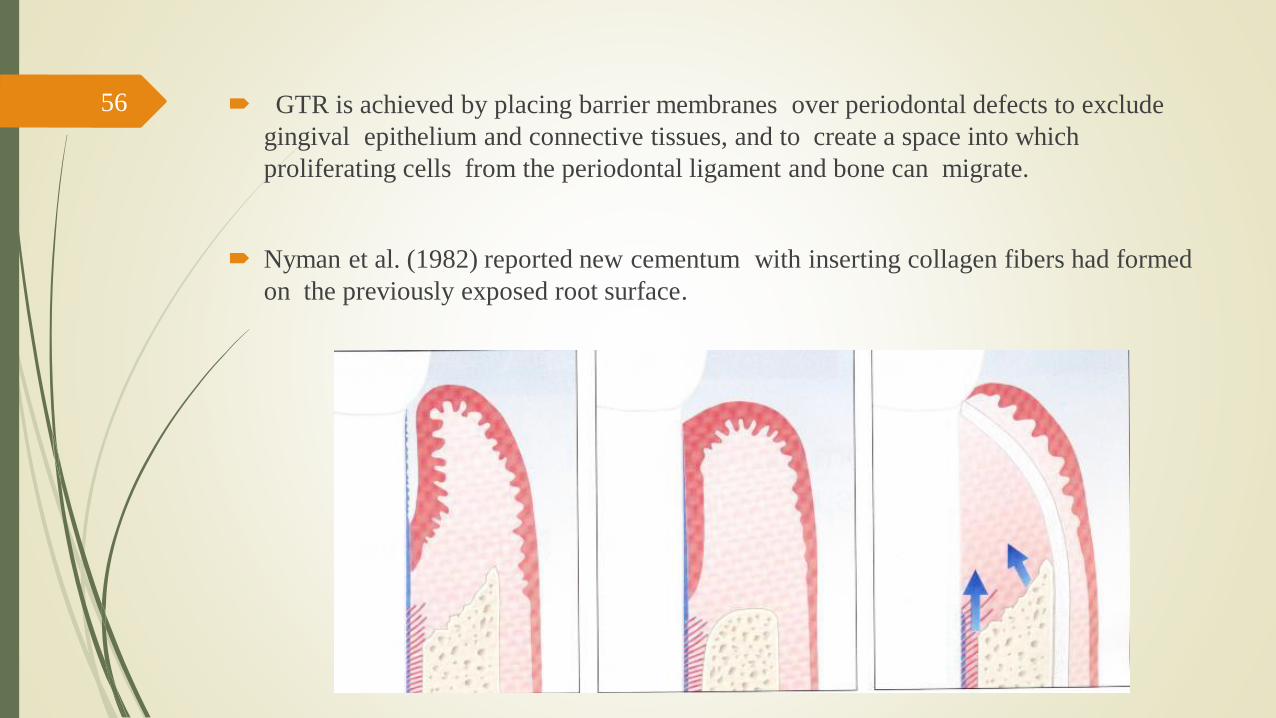
GTR is achieved by placing barrier membranes over periodontal defects to exclude
gingival epithelium and connective tissues, and to create a space into which
proliferating cells from the periodontal ligament and bone can migrate.
Nyman et al. (1982) reported new cementum with inserting collagen fibers had formed
on the previously exposed root surface.
56
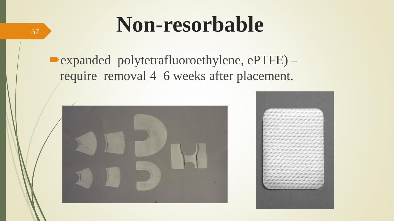
Non-resorbable
expanded polytetrafluoroethylene, ePTFE) –
require removal 4–6 weeks after placement.
57

Resorbable
(e.g. polylactic acid membranes, collagen membranes) - biodegrade
within the tissues over 1–2 months and do not require a second surgical
procedure for removal.
May also be placed over implants and in conjunction with bone grafts
in an attempt to increase the quantity of available bone.
GTR produces most predictable results in class II furcations and in two-
and three-walled osseous defects.
58

59
60
61
62

63
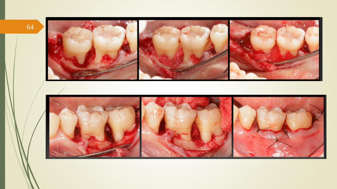
64
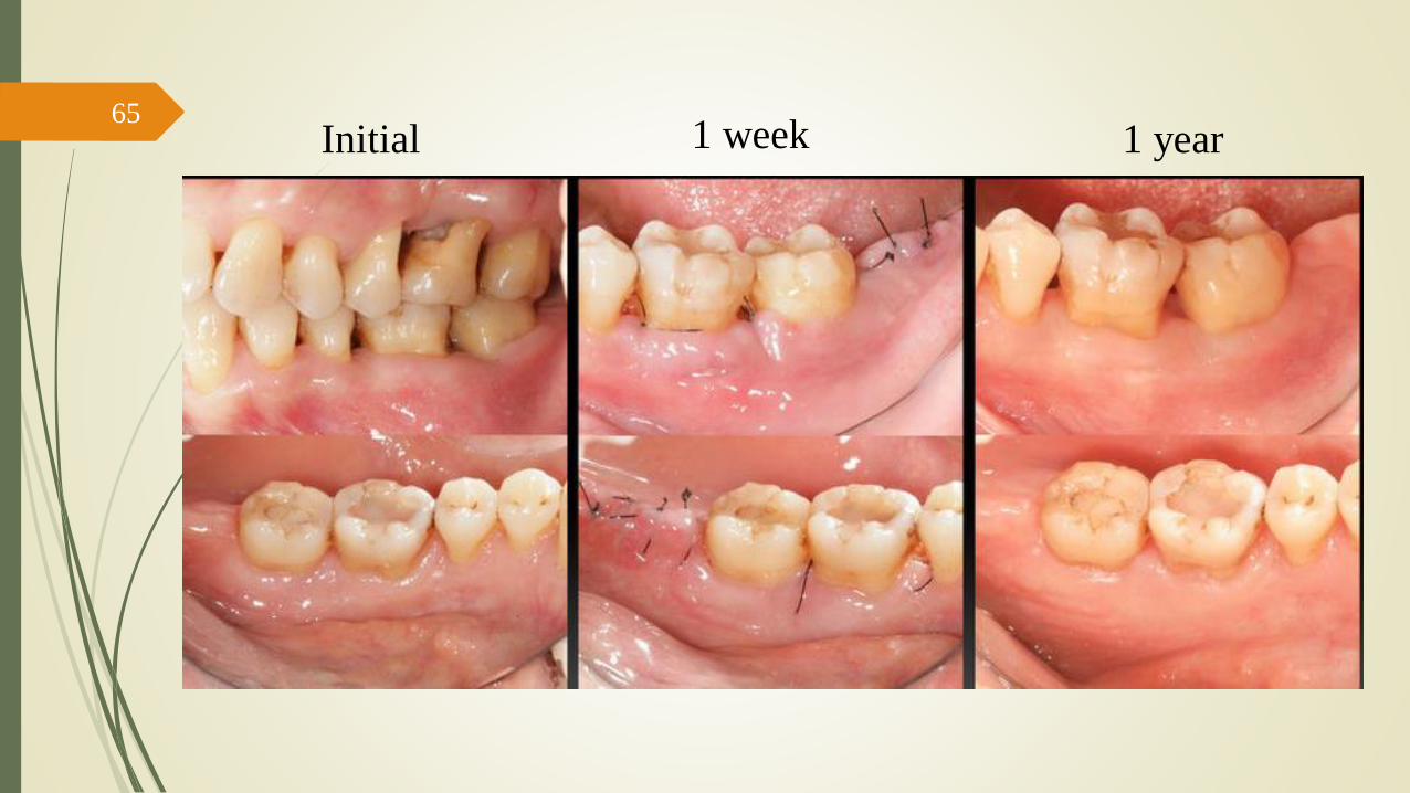
65Initial 1 week 1 year
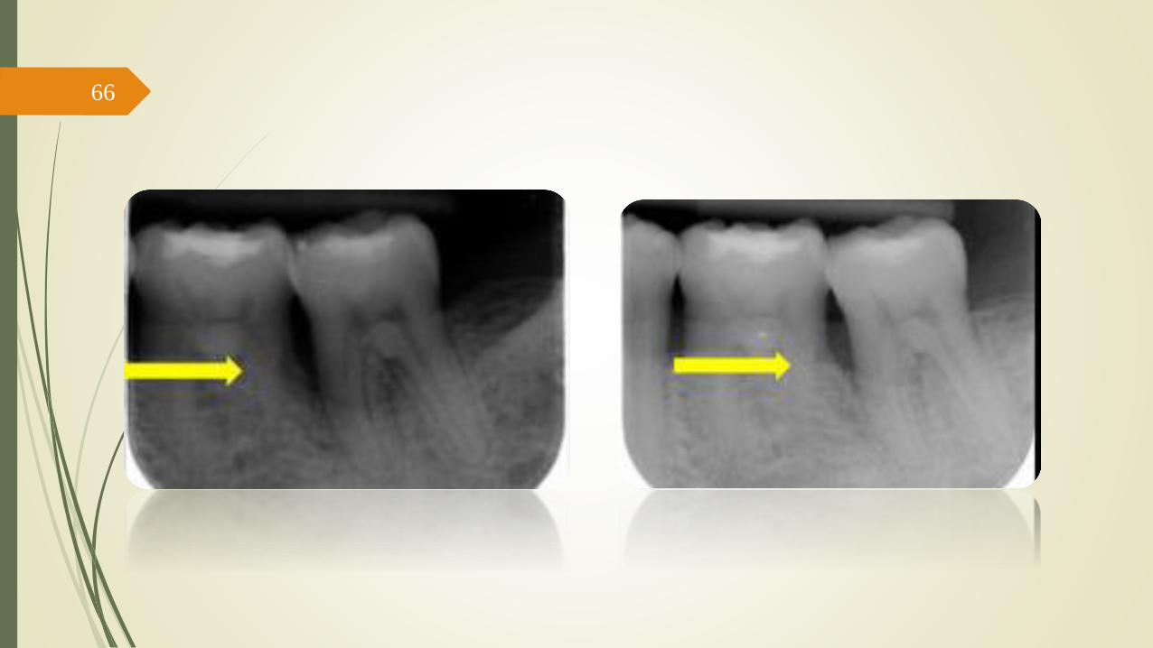
66
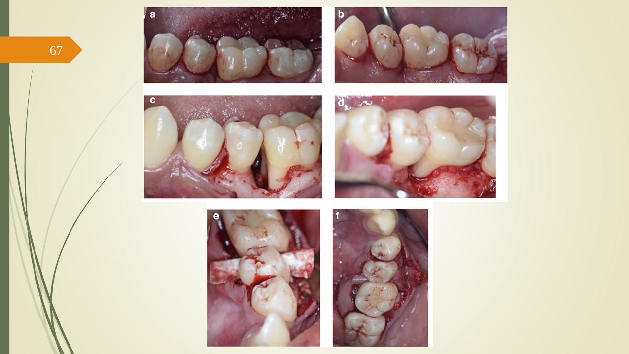
67
68

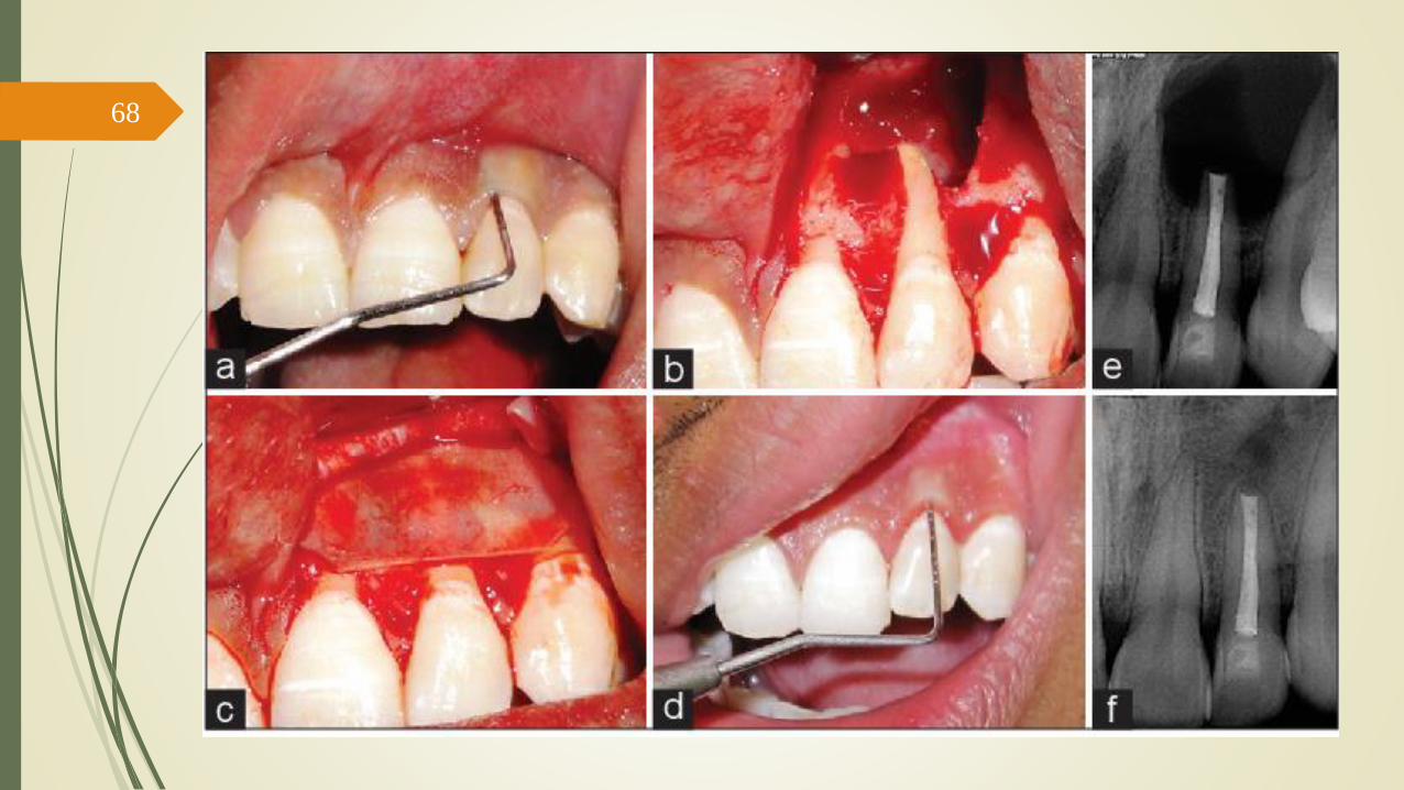
69 Obstacles to new attachment
Degeneration of remnants of Sharpey’s fibers
Accumulation of bacteria and their products
Disintegration of the cementum and dentin
Bio modification of the root surface
Citric acid
Fibronectin (The glycoprotein that fibroblasts require to attach to
root surface) >>> New Attachmet
Tetracycline >>> (In vivo): greater connective tissue attachment
after tetracyclin treatment of roots
Polypeptide growth factors >>> PDGF, IGF, bFGF , TGF
Enamel Matrix Proteins >>> amelogenin (Emdogain ) >>>
osteopromotive
70
Outcome
The clinical outcomes of GTR - frequently evaluated by changes in
clinical attachment levels , bone levels, PPD and the position of the
gingival margin.
In some studies on grade II and III furcations, horizontal changes in
clinical attachment, bone level, and pocket depth were also
measured.
However, evidence of true regeneration of periodontal attachment
can only be provided by histologic means
71
Assessment of periodontal regeneration
Successful regeneration is assessed by periodontal probing,
radiographic analysis, direct measurements of new bone, and
histology.
Although histology remains the ultimate standard in assessing true
periodontal regeneration, periodontal probing, direct bone
measurements, and radiographic measurements of osseous changes
are used in the majority of studies of regenerative therapy (Reddy
& Jeffcoat 1999).
72
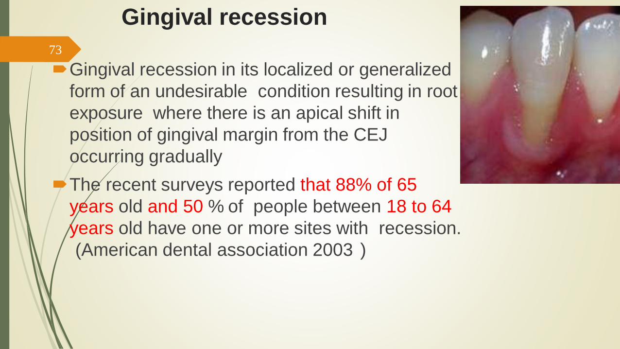
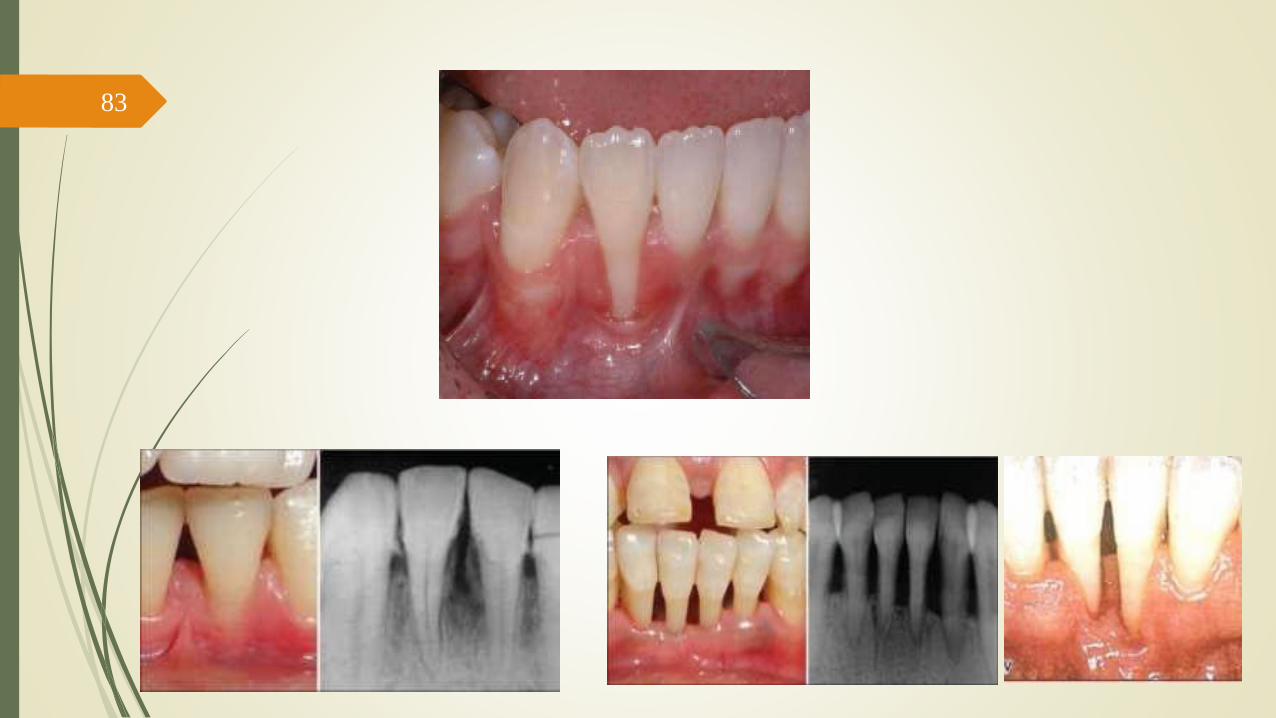
Gingival recession
Gingival recession in its localized or generalized
form of an undesirable condition resulting in root
exposure where there is an apical shift in
position of gingival margin from the CEJ
occurring gradually
The recent surveys reported that 88% of 65
years old and 50 % of people between 18 to 64
years old have one or more sites with recession.
(American dental association 2003 )
73
Symptoms
Sensitivity
Esthetic complains
Root caries
Teeth discoloration
Abrasion
Difficulty maintaining plaque control
74
Etiology
1 Anatomical2 Plaque induced periodontitis
3 Trauma
4 Iatrogenic factors
5 Smoking and Tobacco products
6 Aging
7 Hormonal changes
8 Gingival biotype
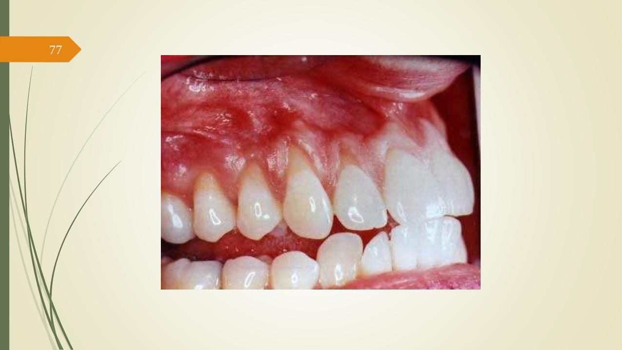
75
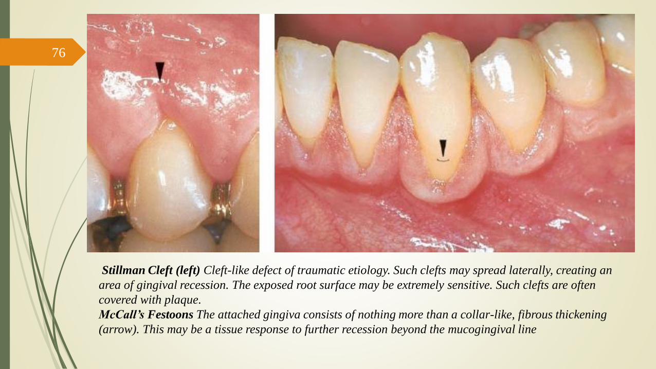
Stillman Cleft (left) Cleft-like defect of traumatic etiology. Such clefts may spread laterally, creating an
area of gingival recession. The exposed root surface may be extremely sensitive. Such clefts are often
covered with plaque.
McCall’s Festoons The attached gingiva consists of nothing more than a collar-like, fibrous thickening
(arrow). This may be a tissue response to further recession beyond the mucogingival line
76
77

78
79
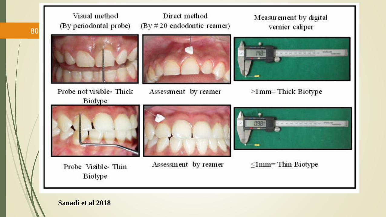
Sanadi et al 2018
80
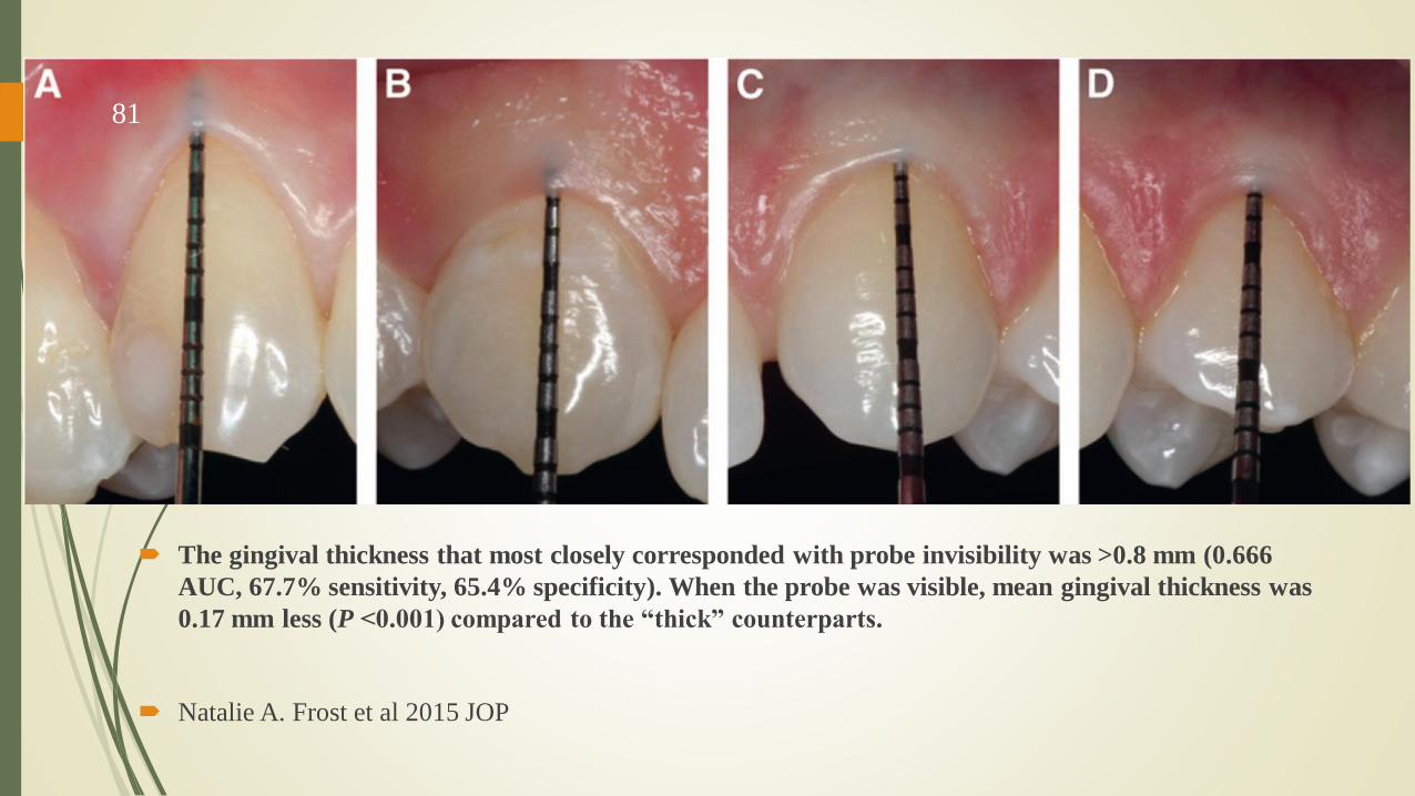
The gingival thickness that most closely corresponded with probe invisibility was >0.8 mm (0.666
AUC, 67.7% sensitivity, 65.4% specificity). When the probe was visible, mean gingival thickness was
0.17 mm less (P <0.001) compared to the “thick” counterparts.
Natalie A. Frost et al 2015 JOP
81

Miller’s Classification82
83

Mucogingival Surgery for
Root Coverage84
Types
• Free soft tissue graft procedures
• Free gingival grafts
• Subepithelial connective tissue graft
• Pedicle soft tissue graft procedures
• Rotational flaps
• Double-papilla repositioned flap
• Laterally positioned flaps
• Transpositional flaps
• Advanced flap
• Coronally positioned flap
• Semilunar coronally positioned flaps
85
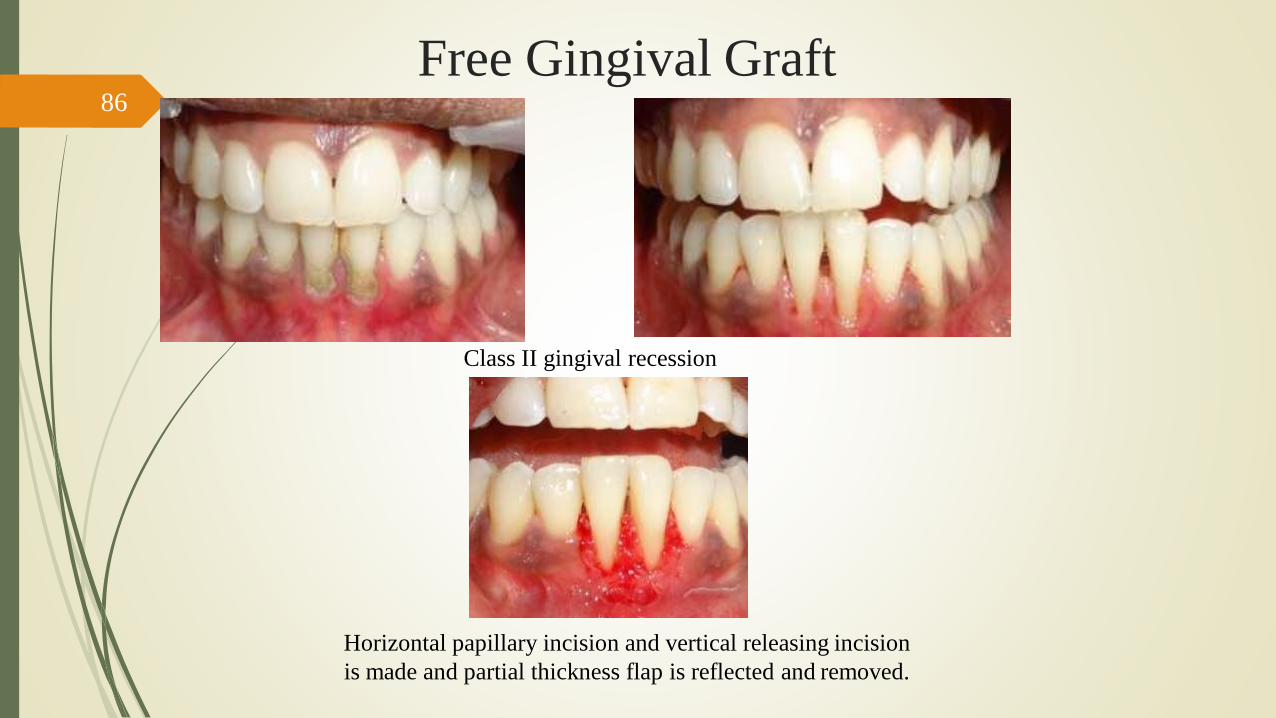
Free Gingival Graft
Class II gingival recession
Horizontal papillary incision and vertical releasing incision
is made and partial thickness flap is reflected and removed.
86

87
88

89
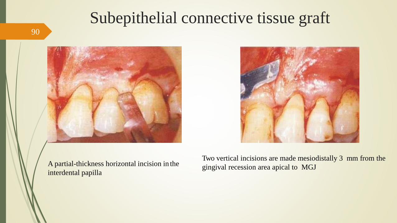
Subepithelial connective tissue graft
A partial-thickness horizontal incision in the
interdental papilla
Two vertical incisions are made mesiodistally 3 mm from the
gingival recession area apical to MGJ
90
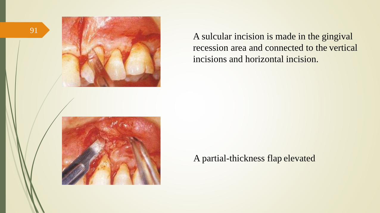
A sulcular incision is made in the gingival
recession area and connected to the vertical
incisions and horizontal incision.
A partial-thickness flap elevated
91
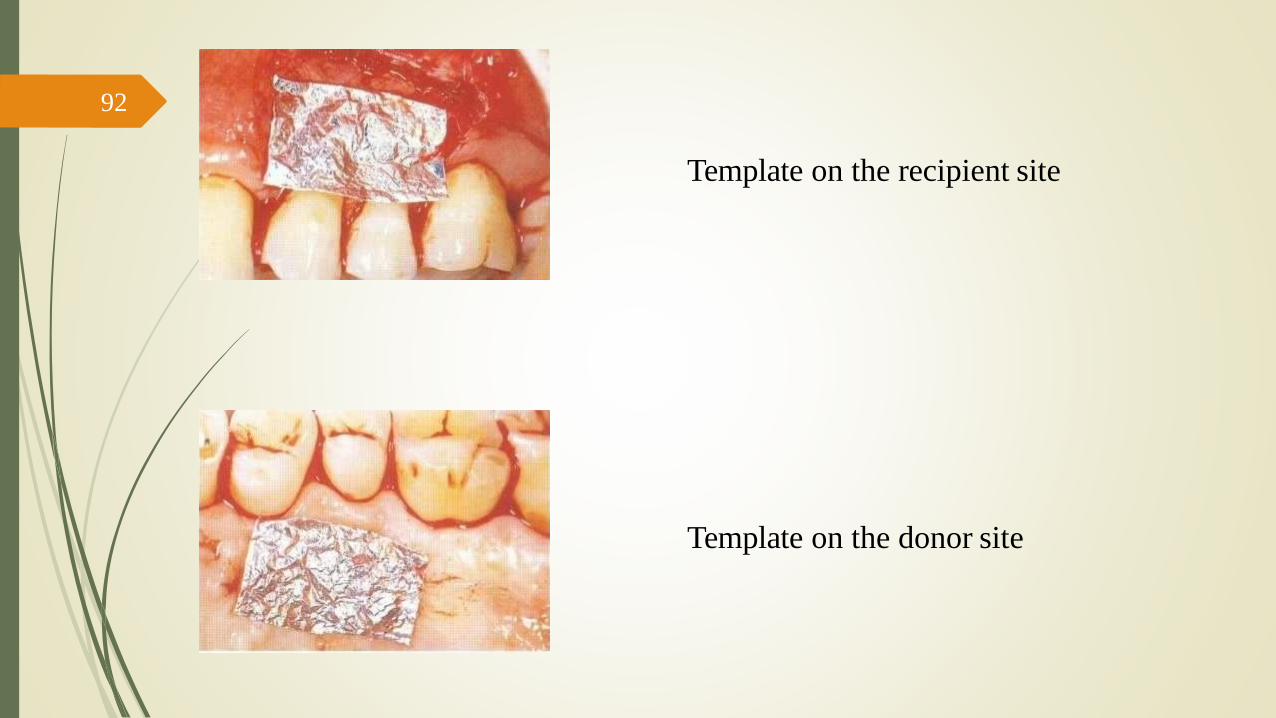
Template on the recipient site
Template on the donor site
92

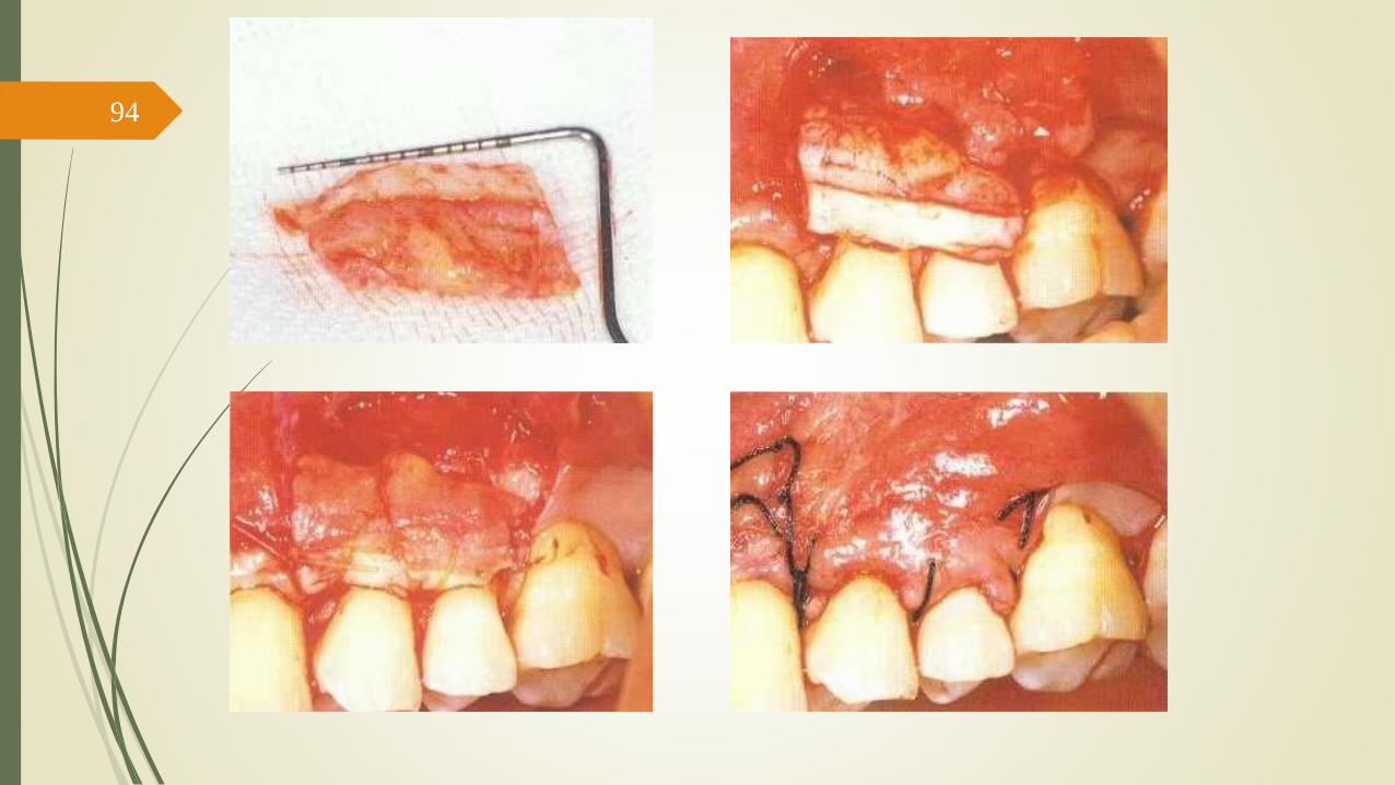
A full-thickness periosteal connective tissue is reflected from the bone and
separated from the bone surface.
93
94

95

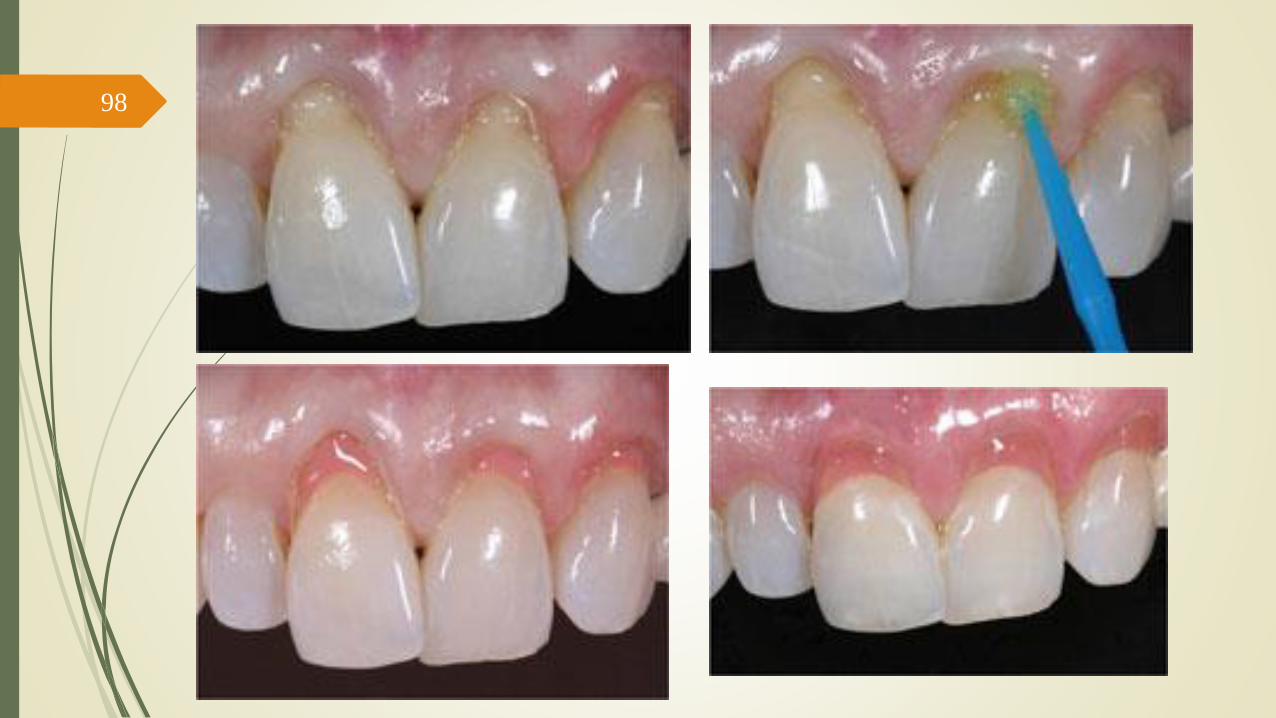
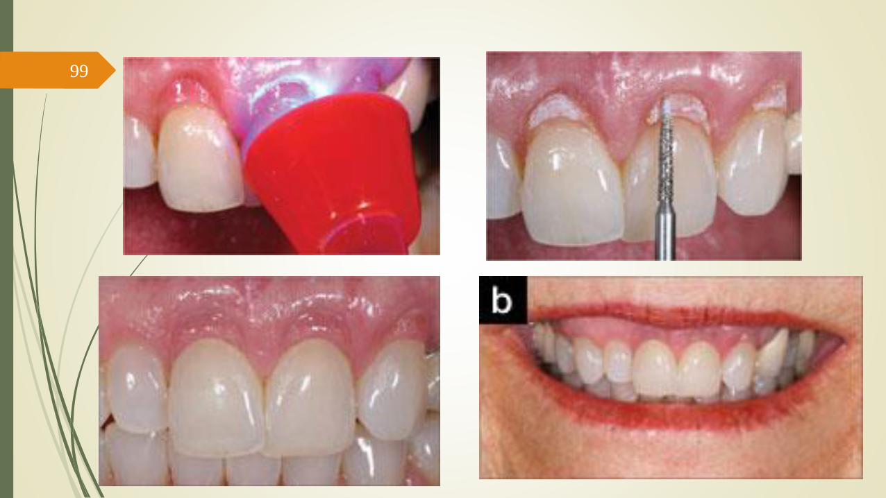
Composite restorations!
Use of composite resin to mask recession defects and eliminate black triangles caused
by recession.
Enameloplasty was carried out to even incisal plane in this case
96

97
98
99
100

101 Thanks for
Your Attention