Extracellular matrix-mediated differentiation of periodontal progenitor cells
24
Extracellular Matrix-Mediated Differentiation of Periodontal Progenitor Cells Smit J. Dangaria, Yoshihiro Ito, Cameron Walker, Robert Druzinsky, Xianghong Luan, and Thomas G.H. Diekwisch * Brodie Laboratory for Craniofacial Genetics, Departments of Oral Biology and Orthodontics, UIC College of Dentistry, The University of Illinois at Chicago Abstract The periodontal ligament (PDL) is a specialized connective tissue that connects the surface of the tooth root with the bony tooth socket. The healthy PDL harbors stem cell niches and extracellular matrix (ECM) microenvironments that facilitate periodontal regeneration. During periodontal disease, the PDL is often compromised or destroyed, reducing the life-span of the tooth. In order to explore new approaches toward the regeneration of diseased periodontal tissues, we have tested the effect of periodontal ECM signals, fibroblast growth factor 2 (FGF2), connective tissue growth factor (CTGF), and the cell adhesion peptide Arg-Gly- Asp (RGD) on the differentiation of two types of periodontal progenitor cells, PDL progenitor cells (PDLPs) and dental follicle progenitor cells (DFCs). Our studies documented that CTGF and FGF2 significantly enhanced the expression of collagens I & III, biglycan and periostin in tissue engineered regenerates after 4 weeks compared to untreated controls. Specifically, CTGF promoted mature PDL-like tissue regeneration as demonstrated by dense periostin localization in collagen fiber bundles. CTGF and FGF2 displayed synergistic effects on collagen III and biglycan gene expression, while effects on mineralization were antagonistic to each other: CTGF promoted while FGF2 inhibited mineralization in PDL cell cultures. Incorporation of RGD peptides in hydrogel matrices significantly enhanced attachment, spreading, survival and mineralization of the encapsulated DFCs, suggesting that RGD additives might promote the use of hydrogels for periodontal mineralized tissue engineering. Together, our studies have documented the effect of three key components of the periodontal ECM on the differentiation of periodontal progenitor populations. Keywords growth factors; extracellular matrix; periodontal regeneration; progenitor cells Introduction Adult stem cells have been identified in most mammalian tissues of the adult body and are known to support the continuous repair and regeneration of tissues (Pekovic and Hutchison., 2008). These stem cells are often located in and controlled by special tissue microenvironments known as niches - specific anatomic locations that regulate how they participate in tissue generation, maintenance and repair (Ohlstein et al., 2004; Scadden, 2006). Stem cell niches maintain adult stem cells in a balance of quiescence and activity (Moore and Lemischka, 2006) and constitute the basic unit of tissue physiology, integrating signals that mediate the balanced response of stem cells to the needs of the organism (Scadden, 2006). Through its *Author for Correspondence email: [email protected], Fax: 312-996-6044. NIH Public Access Author Manuscript Differentiation. Author manuscript; available in PMC 2010 September 1. Published in final edited form as: Differentiation. 2009 ; 78(2-3): 79–90. doi:10.1016/j.diff.2009.03.005. NIH-PA Author Manuscript NIH-PA Author Manuscript NIH-PA Author Manuscript
Transcript of Extracellular matrix-mediated differentiation of periodontal progenitor cells

Extracellular Matrix-Mediated Differentiation of PeriodontalProgenitor Cells
Smit J. Dangaria, Yoshihiro Ito, Cameron Walker, Robert Druzinsky, Xianghong Luan, andThomas G.H. Diekwisch*Brodie Laboratory for Craniofacial Genetics, Departments of Oral Biology and Orthodontics, UICCollege of Dentistry, The University of Illinois at Chicago
AbstractThe periodontal ligament (PDL) is a specialized connective tissue that connects the surface of thetooth root with the bony tooth socket. The healthy PDL harbors stem cell niches and extracellularmatrix (ECM) microenvironments that facilitate periodontal regeneration. During periodontaldisease, the PDL is often compromised or destroyed, reducing the life-span of the tooth. In order toexplore new approaches toward the regeneration of diseased periodontal tissues, we have tested theeffect of periodontal ECM signals, fibroblast growth factor 2 (FGF2), connective tissue growth factor(CTGF), and the cell adhesion peptide Arg-Gly- Asp (RGD) on the differentiation of two types ofperiodontal progenitor cells, PDL progenitor cells (PDLPs) and dental follicle progenitor cells(DFCs). Our studies documented that CTGF and FGF2 significantly enhanced the expression ofcollagens I & III, biglycan and periostin in tissue engineered regenerates after 4 weeks compared tountreated controls. Specifically, CTGF promoted mature PDL-like tissue regeneration asdemonstrated by dense periostin localization in collagen fiber bundles. CTGF and FGF2 displayedsynergistic effects on collagen III and biglycan gene expression, while effects on mineralization wereantagonistic to each other: CTGF promoted while FGF2 inhibited mineralization in PDL cell cultures.Incorporation of RGD peptides in hydrogel matrices significantly enhanced attachment, spreading,survival and mineralization of the encapsulated DFCs, suggesting that RGD additives might promotethe use of hydrogels for periodontal mineralized tissue engineering. Together, our studies havedocumented the effect of three key components of the periodontal ECM on the differentiation ofperiodontal progenitor populations.
Keywordsgrowth factors; extracellular matrix; periodontal regeneration; progenitor cells
IntroductionAdult stem cells have been identified in most mammalian tissues of the adult body and areknown to support the continuous repair and regeneration of tissues (Pekovic and Hutchison.,2008). These stem cells are often located in and controlled by special tissue microenvironmentsknown as niches - specific anatomic locations that regulate how they participate in tissuegeneration, maintenance and repair (Ohlstein et al., 2004; Scadden, 2006). Stem cell nichesmaintain adult stem cells in a balance of quiescence and activity (Moore and Lemischka,2006) and constitute the basic unit of tissue physiology, integrating signals that mediate thebalanced response of stem cells to the needs of the organism (Scadden, 2006). Through its
*Author for Correspondence email: [email protected], Fax: 312-996-6044.
NIH Public AccessAuthor ManuscriptDifferentiation. Author manuscript; available in PMC 2010 September 1.
Published in final edited form as:Differentiation. 2009 ; 78(2-3): 79–90. doi:10.1016/j.diff.2009.03.005.
NIH
-PA Author Manuscript
NIH
-PA Author Manuscript
NIH
-PA Author Manuscript

interactions with the extracellular matrix, neighboring cells, and soluble local and systemiccues, the microenvironments of these niches influences stem cell proliferation, differentiation,and death (Geiger et al., 2001; Nelson and Bissell., 2006; Scadden, 2006). A number of stemcell niches in the adult body have been well characterized, including intestinal, hair follicle,and hematopoietic stem cell niches (Moore and Lemischka, 2006). Recent evidence suggeststhat teeth and their surrounding tissues contain several typical mesenchymal stem cell nichesresiding in the dental pulp, the apical papilla, the periodontal ligament, and the dental follicle(Luan et al., 2006; Luan et al., in press; Sonoyama et al., 2008). Among these, periodontalligament stem cell niches are of great interest for the study of tissue maintenance andregeneration as the periodontal ligament is one of the tissues with the highest turnover rate inthe human body (Stallard, 1963; Carneiro and Leblond, 1966; Melcher, 1985; Beertsen et al.,1997).
Periodontal tissues differentiate from dental follicle cells as a result of a superbly orchestratedinterplay between cytokines, growth factors, and transcription factors (Sodek and McKee,2000; Luan et al., 2006). In mammals, this finely tuned network of signals and receptors affectsneural crest-derived progenitor cell populations residing in a connective tissue sac surroundingthe developing tooth organ, the dental follicle (Chai et al., 2000; Luan et al., 2006). Soon afterthe formation of the tooth root, cementoblast progenitors in the dental follicle migrate to theroot dentin surface and differentiate to form cementum (Diekwisch 2002). Dental folliclesprogenitor cells (DFCs) differentiate into the periodontal ligament (PDL), which retains a nicheof periodontal progenitors cells (PDLPs) for its renewal (Bosshardt and Schroeder, 1996; Seoet al., 2004; Luan et al., 2006). A third periodontal tissue, the alveolar bone, also arises fromthe dental follicle (TenCate 1971; Sodek and McKee, 2000). In previous studies, we haveprovided evidence that the three mammalian mesenchymal periodontal tissues, alveolar bone,root cementum and PDL, are distinct but closely related and suggested that factors fromHertwig’s Epithelial Root Sheath might be responsible for the maintenance of the non-mineralized status of the PDL (Luan et al., 2006, 2007). The close relationship between thethree periodontal tissues of mesenchymal origin provides a tempting scenario with only alimited number of key factors responsible for periodontal cytodifferentiation, which in turnmight be exploited for the benefit of periodontal therapeutics.
The non-mineralized PDL sandwiched between mineralized cementum-root dentin andalveolar bone is a highly vascularized connective tissue with a heterogeneous cell populationembedded in a protein-rich ECM, which is constantly remodeled in response to various stimuli.The periodontal ECM facilitates resilience of mammalian teeth by allowing limited degrees ofmovement and the ability to react to occlusal stresses while remaining firmly attached to thesurrounding alveolar. The periodontal ECM is composed of both collagenous and non-collagenous proteins (proteoglycans and glycoproteins) along with growth factors, cytokinesand matrix degradation enzymes, metalloproteinases (MMPs) (Waddington and Embery,2001). Collagen type I (collagen I) and type III (collagen III) are the main constituents of thecollagenous ECM while versican, dextran sulphated biglycan, and decorin contribute to thenon-collagenous part of the ECM, which also includes glycoproteins such as fibronectin,vitronectin, thrombospondin, tenascin, periostin and osteopontin (Watanabe and Kubota,1998; Matsuura et al., 1995; Rios et al., 2005). Many of the periodontal ECM glycoproteinscontain RGD motifs, which act as cell adhesion moieties for the binding of cell surface integrinsand facilitate important cellular functions such as attachment, spreading, proliferation,differentiation and ECM synthesis (Rezania and Healy,1999). In addition, several growthfactors such as fibroblast growth factors (FGFs), bone morphogenetic proteins (BMPs),platelet-derived growth factors (PDGFs) and connective tissue growth factor (CTGF) featureunique temporo-spatial expression patterns during various stages of tooth root development,mesenchymal tissue regeneration and periodontal healing (Madan et al., 2005; Lalani et al.,2003; Friedrichsen et al., 2003, 2005; Lin et al., 2003). Two of the above described growth
Dangaria et al. Page 2
Differentiation. Author manuscript; available in PMC 2010 September 1.
NIH
-PA Author Manuscript
NIH
-PA Author Manuscript
NIH
-PA Author Manuscript
factors reveal particularly attractive features to aide targeted periodontal regeneration: FGFsbecause of their capability to reduce potential ankylosis of regenerated tissues, and CTGFbecause of its effect on collagen I synthesis (Dereka et al., 2006; Asano et al., 2005).
In the present study we have hypothesized that ECM growth factors such as FGF2 and CTGFand cell adhesion peptides such as RGD can modulate the differentiation and behavior ofperiodontal progenitor populations such as PDLPs and DFCs. In order to mimic the effect ofgrowth factors during periodontal tissue differentiation and regeneration, we used in vivo andin vitro models to ask the question whether individual growth factors alone or combinationsthereof elicit a response in the periodontal ECM and affect matrix molecules such as collagenI & III, biglycan and periostin. Our studies have allowed us to gain insights into the suitabilityof FGF and CTGF as an aide in periodontal regeneration. In addition, we were able to identifya number of mineral formation induction conditions that may be useful for the regeneration ofperiodontal mineralized tissues such as alveolar bone or cementum. Together, our studycharacterized a number of ECM -based factors that promote periodontal lineage differentiationand might be used for future periodontal tissue regeneration approaches.
Materials and methodsDF and PDL cell culture
Dental follicle tissues from impacted human 3rd molars were dissected while PDL attached tothe roots of erupted 3rd molars was scraped off, and respective tissues were then digested in5mg/ml collagenase/dispase (Roche Applied Science, Indianapolis, IN) with gentle rotation at37°C for 40 mins. Approval for the use of human 3rd molars for our study was granted by theUIC Office for the Protection of Research Subjects. Subsequently, primary cells were platedat low density and cultured in six-well plates containing Dulbecco’s Modified Eagle medium(DMEM) supplemented with 10% fetal calf serum (FCS) and 100 units/ml of penicillin/streptomycin. The plastic adherent colonies from single cells were selected and cultured untilsubconfluence and expanded into 100-mm dishes (Luan et al, 2006). Clonal populations fromboth cell types with the highest ability to differentiate into osteogenic and adipogenic lineages(data not shown) were used as progenitor cells in all further experiments.
Differentiation induction of primary DFCs and PDLPsThird passage DFCs and PDLPs were treated with DMEM with rhFGF (2ng/ml) (Peprotech)for a period of 2 weeks. Alternatively, PDLPs were seeded at a low density of 2×10⁴ cells ina 100 mm tissue culture dish and treated with DMEM medium supplemented with either FGF2(10ng/ml, Peprotech), CTGF (50ng/ml, Cell sciences) or a combination of both for a period of10 days. Concentrations of the growth factors were chosen based on previous studiesdemonstrating optimum effects for these growth factors on cell proliferation and DNAsynthesis in bone marrow stromal cells and mouse PDL cells at 10ng/ml or lower (FGF2) and50ng/ml (CTGF), respectively (Hankemeier et. al., 2005; Asano et. al., 2005). PDLPs withoutany growth factor treatment served as the control.
Collagen gel preparation and suspension of DFCs and PDLPsEight ml of chilled Nutragen (Inamed Biomaterials, Fremont CA) was mixed with 1ml of 10xα-MEM and 1ml of 0.1M NaOH. The pH was adjusted to 7.4 +/− 0.2 by dropwise addition of0.1M of endotoxin free HCl. The collagen concentration was adjusted to 2.5mg/ml for cellsuspension and was used within hours of preparation. PDLPs cultured in media containingeither FGF, CTGF, or no growth factors were trypsinized and suspended in collagen solutionat a density of 2.5×10⁵ cells/ml which contained either FGF2 (10ng/ml) or CTGF (50ng/ml)or a combination of both or no growth factors. About 300μl of this cell-collagen suspensionalong with the above mentioned concentration of growth factors was placed in each well of a
Dangaria et al. Page 3
Differentiation. Author manuscript; available in PMC 2010 September 1.
NIH
-PA Author Manuscript
NIH
-PA Author Manuscript
NIH
-PA Author Manuscript

48 well plate and allowed to form gels at 37°C in 100% air for a period of 60 minutes. Theconstructs were subcutaneously implanted in nude mice for a period of 1 day, 2weeks and 4weeks. Alternatively, DFCs and PDLPs in collagen gels were cultured in vitro for a period of1day, 1 week and 2 weeks.
Synthesis of Acryloyl-PEG-RGDAcrylated peptide containing a MW 3400 PEG (polyethylene glycol) spacer between thepeptide and acrylate moiety was synthesized by mixing the peptide with acryloyl-PEG-N-hydroxysuccinimide ester as previously described (Hern and Hubble, 1998). Briefly, YRGDS(Bachem) was dissolved to a final aqueous concentration of 1mg/ml in 50mM sodiumbicarbonate buffer, pH 8.4. The MW 3400 acryloyl-PEG-NHS was also dissolved in 50mMsodium bicarbonate buffer at a final molar ratio of peptide to acryloyl-PEG-NHS of 1:3. ThePEG solution was added drop wise to the peptide solution. The final solution was allowed toreact at room temperature for 2 hours whereupon it was dialyzed against water with a molecularweight cutoff at 1000Da overnight, followed by lyophilization to obtain the mono-vinyl PEGwith a pendant RGD sequence (Acryloyl-PEG-RGD).
Cell attachment and spreading on 2D hydrogel surfaceHydrogel disks about 2mm thick were created in a 48 well plate by pipetting about 300 μl of10% polyethylene glycol diacrylate (PEGDA) in PBS with or without the addition of 2 mMacryloyl-PEG-RGD and then photopolymerized with UV light for 10 mins. DFCs at a densityof 3 × 10³ cells/cm2 were seeded onto these disks. After 2 or 24 hour intervals the disks wererinsed with PBS and cell attachment was monitored using phase contrast microscopy. Attachedcells were counted on a minimum of 3 random fields per disk (under 20x magnification). Cellspreading was quantified by measuring the area occupied by the cells after 2 hours ofattachment using the NIH imaging software.
Photoencapsulation of DFCs in RGD-modified and unmodified hydrogels80% confluent passage 3 DFCs were trypsinized from culture plates and centrifuged to forma cell pellet. The 10% PEGDA solution in PBS with 0.05% D2959 (Ciba-Geigy), and 1%w/vof streptomycin and penicillin with or without 2 mM acryloyl-PEG-RGD was added directlyto the cells adjusting the cell concentration to about 15 million cells per ml of solution. About50 μl of the cell/polymer solution was then pipetted into 1-ml sterile BD syringes that had theirtips cut off and then photopolymerized using UV light of intensity 5mW/cm2 for 10 mins(Nuttelman et al., 2005). Hydrogels were then pushed out of the syringes and cultured in eitherDMEM, DMEM supplemented with 10ng/ml PDGF (Peprotech), or osteogenic differentiationmedium (Cambrex) for a period of 2 weeks.
MTT viability assayTo measure cell viability DFCs-hydrogel constructs were incubated in MTT solution (2mg/mlof MTT in DMEM with 2% FBS) for 4 hours. To quantify viability, the MTT stained constructswere homogenized in HCL/Isopropanol and the absorbance was detected at 570nm withbackground subtraction at 630nm from centrifugal supernatants (Wang et al., 2003).
Isolation of RNA and real time RT-PCRTotal RNA was extracted from subcutaneously implanted PDLPs-collagen gel constructs at 1day, 2 and 4 weeks and also from in vitro cultured DFCs and PDLPs cell-collagen gel constructsat 1 and 2 weeks using Trizol Reagent (Invitrogen). RNA quality and quantity were verifiedusing agarose gel electrophoresis and spectrophotometry, respectively. About 1 μg of RNAwas reverse transcribed and the cDNA was amplified using selected primers (Table 1) by Realtime RT-PCR using the SuperScript III Platinum two-step qPCR kit with LUX fluorogenic
Dangaria et al. Page 4
Differentiation. Author manuscript; available in PMC 2010 September 1.
NIH
-PA Author Manuscript
NIH
-PA Author Manuscript
NIH
-PA Author Manuscript

primer (Invitrogen). Primer sequences for collagen I, collagen III, biglycan, periostin andGAPDH genes were designed by LUX™ Designer (FAM labeled LUX primer Invitrogen) andare shown in Table 1. Reaction conditions were as follows: 2 min at 50°C (one cycle), 10 minsat 95°C (one cycle), 15 sec at 95 °C and then 1 min at 58°C (40 cycles). PCR products werecontinuously monitored with an ABI PRISM 7900 detection system (RRC-CORE at UIC).Samples were normalized using human GAPDH (Joe-labeled LUX primer set, Invitrogen) asthe internal control. Results are expressed as fold increases in expression over the control groupby using the 2−ΔDelta;Ct method (Livak and Schmittgen, 2001).
ImmunohistochemistryPDLPs-collagen gel regenerates after 4 weeks of implantation were fixed in 10% bufferedformalin and prepared for paraffin sectioning. Alternatively, mandibles from 4 week post natal(pn) mice were harvested and fixed in 10% formalin and decalcified in 8% phosphate bufferedEDTA (pH 7.4) for 3 weeks, dehydrated in a series of alcohol changes, cleared by xylene andembedded in paraffin. For immunohistochemistry, slides were deparaffinized and tissues wererehydrated. Immunoreactions were performed as previously described (Luan et al., 2007).Peroxidase activity was blocked by incubating the slides using a 9:1 ratio of methanol andhydrogen peroxide. The antigenic epitopes were exposed by microwave treatment of the slidesin 1% citrate buffer pH 6.0 for 5 mins and then cooling it down to room temperature. Non-specific sites were blocked using serum. Immunoreactions were performed using monoclonalprimary antibody for periostin (abcam) on PDLPs-collagen regenerates and collagen I, collagenIII, biglycan and periostin antibodies were used on sections from 4 weeks pn mice mandiblesto demonstrate localization of these proteins in the periodontium. Sections were then incubatedwith primary antibody at room temperature for 1 hour at a dilution of 1:500 in PBS. Sectionswere washed three times in PBS and incubated for 10 min with anti-mouse IgG. After threemore washes in PBS, sections were incubated for 10 min with streptavidin-enzyme conjugateand then washed for three more times with PBS. Signals for immunoreactions were detectedusing AEC substrate-chromogen mixture (color substrate), counterstained with hematoxylin,and slides were mounted using GVA mount. The negative controls for the antibody were theones in which the primary antibody was replaced with a similar amount of PBS.
Western blot analysisAfter 4 weeks of implantation in vivo, tissue engineered PDLPs-collagen gel constructs wereextracted and washed with PBS. The constructs were then homogenized in sodiumdodecylsulfate-polyacrylamide gel electrophoresis (SDS-PAGE) sample buffer and proteinsextracted as described (Luan et al., 2006). Identical amounts of protein extracts from allregenerates were separated on a 10% SDS-PAGE gel and transferred to PVDF membrane ina semi-dry blotting apparatus containing transfer buffer (25mM tris, 40mM glycine, 10%methanol) for 40 minutes at 75mA. The PVDF membrane was then blocked with 2.5% BSAfor 1 hour at room temperature and the blot was incubated with 1:1000 dilution of anti-humanperiostin (Abcam), collagen III (Abcam), biglycan (R&D Biosystems) and GAPDH (Abcam)antibodies for 2 hour, washed with TBST 3 times and incubated with 1:2500 dilution of HRPconjugated anti-rabbit or anti-mouse secondary antibody respectively (Zymed, South SanFrancisco, CA) for 1 hour, and further washed 3 times with TBST. HRP detection wasperformed using a chemiluminescent substrate (Supersignal West Pico ChemiluminescentSubstrate, Pierce). The bands obtained from Western blots were then quantified for itsdensitometric value using the Image analysis software (Kodak Image station 440CF)
Mineralization induction in PDLPs culturesPDLPs were seeded at a density of 5 × 10⁴/well of a 12 well plate and cultured in DMEMmedia supplemented with 10% FBS and 1% antibiotics. FGF2 and CTGF were added the
Dangaria et al. Page 5
Differentiation. Author manuscript; available in PMC 2010 September 1.
NIH
-PA Author Manuscript
NIH
-PA Author Manuscript
NIH
-PA Author Manuscript

following day. One set of wells was treated with FGF2 (10 ng/ml) or CTGF (50 ng/ml) or acombination of both for the entire duration of 10 days. Another set of wells was treatedsequentially with FGF2 for the first 4 days followed by CTGF for the next 6 days and viceversa. Wells that were cultured without addition of any growth factor served as the untreatedcontrol. All cells were cultured in an osteogenic differentiation media prepared by addition of0.2mM ascorbic acid 2 phosphate and 4mM beta glycerophosphate in the above mentionedDMEM. At the end of 10 days of culture in mineralizing media, cell cultures were washed withPBS and fixed in 70% ethanol for 1 hr. To detect mineral deposits, cultures were stained with40mM Alizarin Red S, pH 4.0, for 10 mins, washed with 70% ethanol and then with PBS toremove non-specific staining. Each experiment was done in triplicate and repeated 3 times. Todetermine the calcium levels in PDLPs cultures, the mineral in the cell-matrix was dissolvedin 0.6 N HCL overnight and analyzed using the o-cresolphthalein complexone method(Baginski et al., 1973).
Mineralization analysisAfter 2 weeks of in vitro culture, hydrogel constructs containing DFCs either treated withosteogenic, PDGF supplemented media, or untreated controls were fixed overnight in 10%formalin. The constructs were dehydrated through a series of alcohol changes and Citrisolveover 1 hr steps and finally in molten paraffin at 60 oC under vacuum with two changes to freshparaffin every hour. Constructs were then embedded in paraffin and sections of 7 μm thicknesswere cut using a microtome (Leica). Sections were dried overnight on polylysine coated slidesand stained using standard histological techniques. Von Kossa’s silver stain was used toevaluate the amount of mineralization with safranin-O as the counterstain. Micrographs weretaken at magnifications of 20× and 40×.
Statistical analysisAll data are presented as means +/− standard deviation of the mean. Student’s t test was appliedfor data comparison and P values of less than 0.05 were judged significant.
ResultsCollagens I and III, periostin, and biglycan localization in the periodontal ECM
In order to map the distribution of key ECM components in the periodontium, four keyperiodontal matrix proteins were localized using immunohistochemistry. Collagens I and IIIas well as biglycan were localized in the interstitial ECM of the periodontal extracellular matrix(PDL). Sharpey’s fibers were intensely stained for periostin (asterisk, Fig. 1C), moderately forbiglycan and collagen I and weakly for collagens III (Fig. 1A,1B, 1D). Note the distinctlocalization of biglycan in the predentin (Fig. 1D).
CTGF and FGF2 up-regulated collagen III and biglycan in implanted PDL/collagen gelconstructs
In order to compare expression levels of collagens as principle matrix components of the PDL,both DFCs and PDLPs were subjected to 2ng/ml of FGF2 and cultured at a density of 2×10⁵
cells/ml in collagen gels for 1d, 1week and 2 weeks in DMEM. Alternatively, cells werecultured in monolayer for 1 week. After 1 week of in vitro monolayer culture, DFCs formedelongated and parallel arranged spindle shaped cells (Fig. 2A), while PDLPs assembled intodense colonies of radial symmetry (Fig. 2B). Real-time RT-PCR analysis of the cell-collagengel constructs showed that there was significantly higher collagen I and III expression (5.88-fold (*) and 6.47-fold (*), respectively) in PDLPs compared to the DFCs after 2 weeks of invitro culture (Fig. 2C, 2D). Both collagen I and III expression in DFCs increased at 1 week ofculture but decreased 2.33-fold (**) and 2.01-fold (**) respectively after 2 weeks. In PDLPs,
Dangaria et al. Page 6
Differentiation. Author manuscript; available in PMC 2010 September 1.
NIH
-PA Author Manuscript
NIH
-PA Author Manuscript
NIH
-PA Author Manuscript
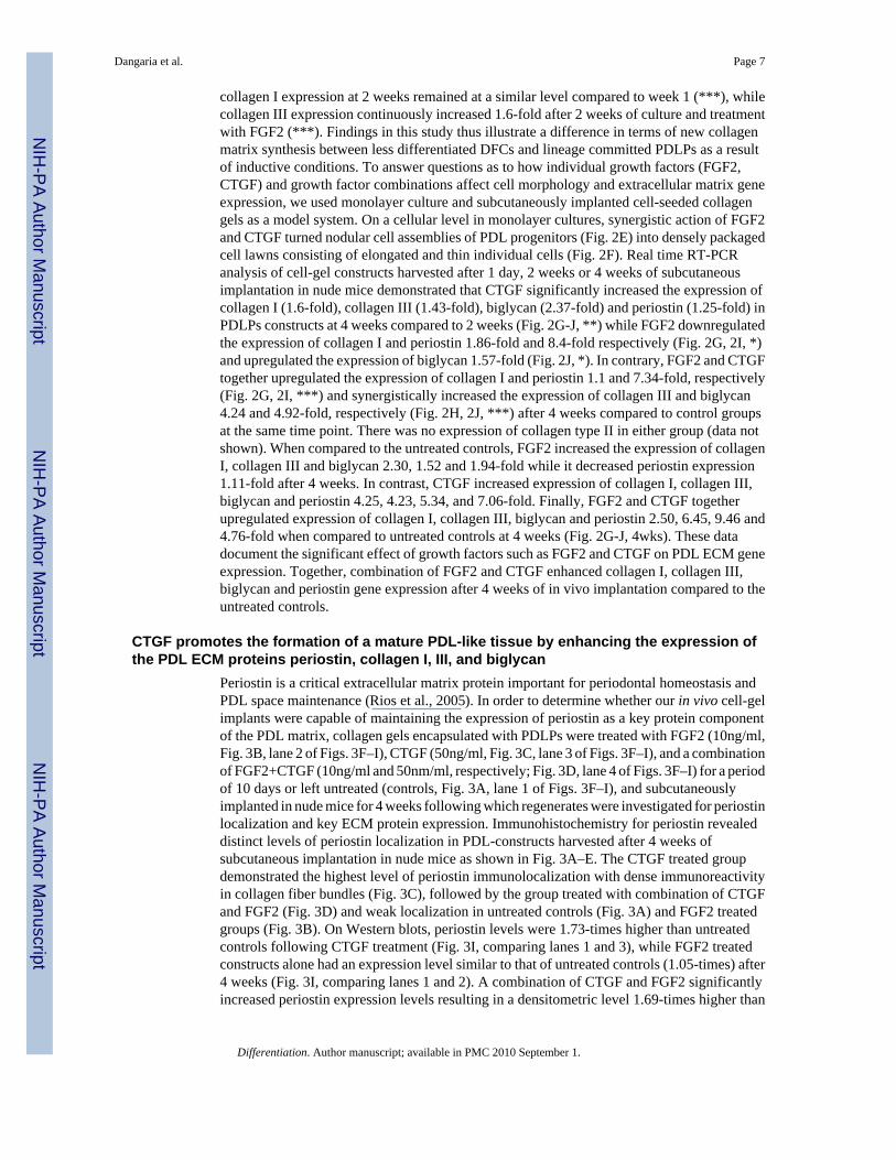
collagen I expression at 2 weeks remained at a similar level compared to week 1 (***), whilecollagen III expression continuously increased 1.6-fold after 2 weeks of culture and treatmentwith FGF2 (***). Findings in this study thus illustrate a difference in terms of new collagenmatrix synthesis between less differentiated DFCs and lineage committed PDLPs as a resultof inductive conditions. To answer questions as to how individual growth factors (FGF2,CTGF) and growth factor combinations affect cell morphology and extracellular matrix geneexpression, we used monolayer culture and subcutaneously implanted cell-seeded collagengels as a model system. On a cellular level in monolayer cultures, synergistic action of FGF2and CTGF turned nodular cell assemblies of PDL progenitors (Fig. 2E) into densely packagedcell lawns consisting of elongated and thin individual cells (Fig. 2F). Real time RT-PCRanalysis of cell-gel constructs harvested after 1 day, 2 weeks or 4 weeks of subcutaneousimplantation in nude mice demonstrated that CTGF significantly increased the expression ofcollagen I (1.6-fold), collagen III (1.43-fold), biglycan (2.37-fold) and periostin (1.25-fold) inPDLPs constructs at 4 weeks compared to 2 weeks (Fig. 2G-J, **) while FGF2 downregulatedthe expression of collagen I and periostin 1.86-fold and 8.4-fold respectively (Fig. 2G, 2I, *)and upregulated the expression of biglycan 1.57-fold (Fig. 2J, *). In contrary, FGF2 and CTGFtogether upregulated the expression of collagen I and periostin 1.1 and 7.34-fold, respectively(Fig. 2G, 2I, ***) and synergistically increased the expression of collagen III and biglycan4.24 and 4.92-fold, respectively (Fig. 2H, 2J, ***) after 4 weeks compared to control groupsat the same time point. There was no expression of collagen type II in either group (data notshown). When compared to the untreated controls, FGF2 increased the expression of collagenI, collagen III and biglycan 2.30, 1.52 and 1.94-fold while it decreased periostin expression1.11-fold after 4 weeks. In contrast, CTGF increased expression of collagen I, collagen III,biglycan and periostin 4.25, 4.23, 5.34, and 7.06-fold. Finally, FGF2 and CTGF togetherupregulated expression of collagen I, collagen III, biglycan and periostin 2.50, 6.45, 9.46 and4.76-fold when compared to untreated controls at 4 weeks (Fig. 2G-J, 4wks). These datadocument the significant effect of growth factors such as FGF2 and CTGF on PDL ECM geneexpression. Together, combination of FGF2 and CTGF enhanced collagen I, collagen III,biglycan and periostin gene expression after 4 weeks of in vivo implantation compared to theuntreated controls.
CTGF promotes the formation of a mature PDL-like tissue by enhancing the expression ofthe PDL ECM proteins periostin, collagen I, III, and biglycan
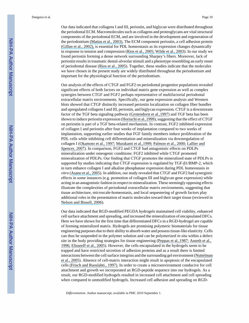
Periostin is a critical extracellular matrix protein important for periodontal homeostasis andPDL space maintenance (Rios et al., 2005). In order to determine whether our in vivo cell-gelimplants were capable of maintaining the expression of periostin as a key protein componentof the PDL matrix, collagen gels encapsulated with PDLPs were treated with FGF2 (10ng/ml,Fig. 3B, lane 2 of Figs. 3F–I), CTGF (50ng/ml, Fig. 3C, lane 3 of Figs. 3F–I), and a combinationof FGF2+CTGF (10ng/ml and 50nm/ml, respectively; Fig. 3D, lane 4 of Figs. 3F–I) for a periodof 10 days or left untreated (controls, Fig. 3A, lane 1 of Figs. 3F–I), and subcutaneouslyimplanted in nude mice for 4 weeks following which regenerates were investigated for periostinlocalization and key ECM protein expression. Immunohistochemistry for periostin revealeddistinct levels of periostin localization in PDL-constructs harvested after 4 weeks ofsubcutaneous implantation in nude mice as shown in Fig. 3A–E. The CTGF treated groupdemonstrated the highest level of periostin immunolocalization with dense immunoreactivityin collagen fiber bundles (Fig. 3C), followed by the group treated with combination of CTGFand FGF2 (Fig. 3D) and weak localization in untreated controls (Fig. 3A) and FGF2 treatedgroups (Fig. 3B). On Western blots, periostin levels were 1.73-times higher than untreatedcontrols following CTGF treatment (Fig. 3I, comparing lanes 1 and 3), while FGF2 treatedconstructs alone had an expression level similar to that of untreated controls (1.05-times) after4 weeks (Fig. 3I, comparing lanes 1 and 2). A combination of CTGF and FGF2 significantlyincreased periostin expression levels resulting in a densitometric level 1.69-times higher than
Dangaria et al. Page 7
Differentiation. Author manuscript; available in PMC 2010 September 1.
NIH
-PA Author Manuscript
NIH
-PA Author Manuscript
NIH
-PA Author Manuscript

the control (Fig. 3I, lanes 1 and 4). The effects of CTGF, FGF2, and combinations on collagenIII and biglycan expression (Fig. 3F–H) were similar to that of periostin; however, there wasa synergistic action between FGF2 and CTGF on collagen III expression (Fig. 3F(collagen III-lanes1–4), G). Together, our findings illustrate that growth factors such as CTGF and FGF2alone or in combination significantly enhance the expression levels of PDL ECM proteins suchas periostin, biglycan, and collagen III.
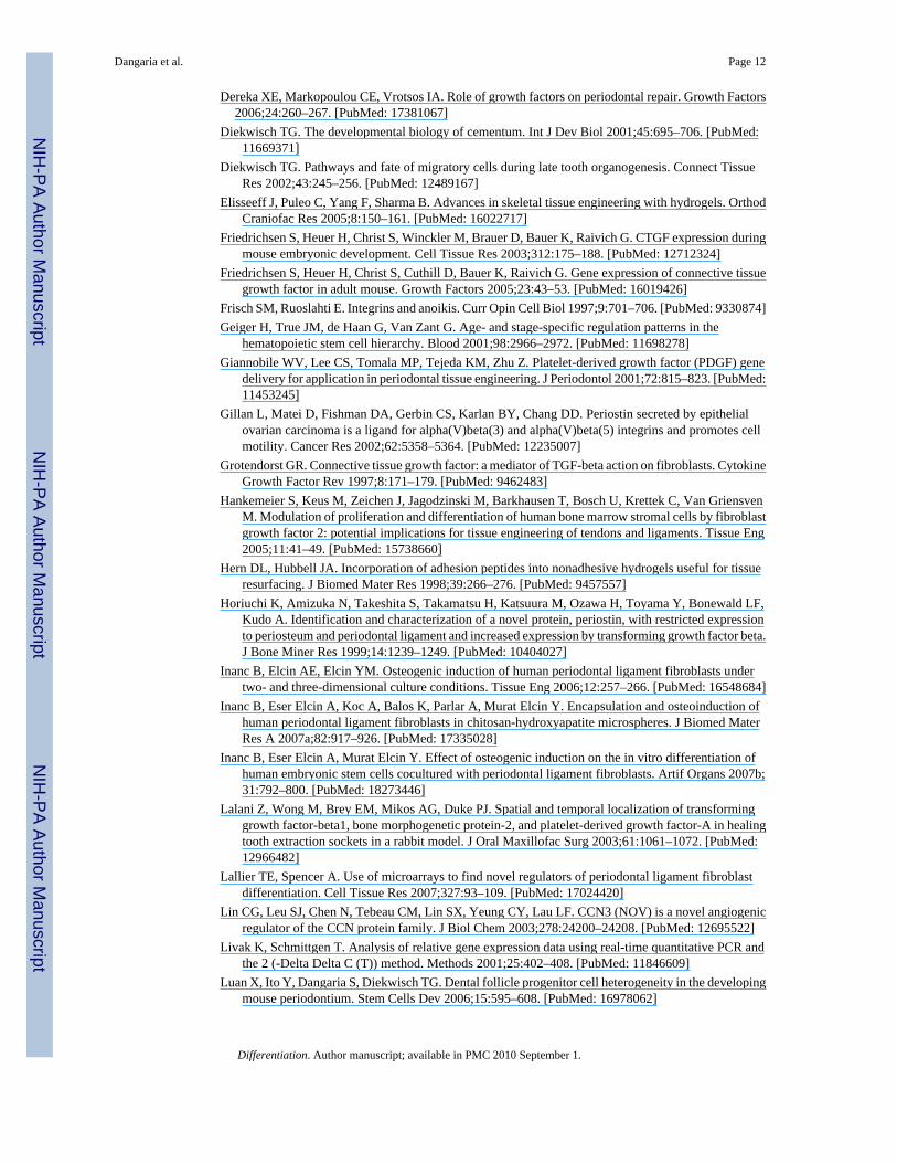
Antagonistic effects of FGF2 and CTGF on the mineralization of PDLPs in vitroTo determine whether the periodontal ECM growth factors FGF2 and CTGF had differenteffects on PDL matrix mineralization, we cultured PDLPs in mineralizing media for a total of10 days under the following conditions: (i) Continuous treatment with FGF2 for 10 days, (ii)continuous treatment with CTGF for 10 days, (iii) continuous treatment with FGF2 and CTGFfor 10 days, (iv) sequential treatment with FGF2 for 4 days followed by CTGF for the next 6days, (v) sequential treatment with CTGF for 4 days followed by FGF2 for the next 6 days,and (vi) without growth factor (untreated control). The amount of mineralization was evaluatedby staining cell cultures with Alizarin Red S. Our studies revealed that untreated culturescontained a moderate level of mineralization nodules (Fig. 4F, G), which were enhanced 5.6-fold by continuous treatment with CTGF (Fig. 4B, G), but completely blocked by continuoustreatment with FGF2 (Fig. 4A, G). The cultures that were treated with simultaneous applicationof FGF2 and CTGF demonstrated a few mineralization nodules approximately, 0.3-fold of theuntreated controls (Fig. 4C, G). Sequential application of FGF2 followed by CTGF also failedto form significant amounts of mineralization nodules (Fig. 4D, G), while early application ofCTGF promoted mineral nodule formation, which was inhibited after application of FGF2 andresulted in a 1.87-fold increase compared to the untreated controls. (Fig. 4E, G). Taken together,these results suggest that FGF2 and CTGF have an antagonistic effect on the mineralizationcharacteristics of PDLPs.
RGD-modified PEGDA hydrogels maintained cell viability, enhanced cell surface attachmentand spreading and increased the mineralization of encapsulated DFCs
RGD is a tripeptide sequence present in many cell adhesion ECM proteins and glycoproteinsthat facilitates cell anchorage, traction for migration, proliferation and differentiation byproviding binding sites for specific integrin cell surface receptors (Ruoslahti et al.,1987). Inorder to determine the effect of RGD peptides on DF cell behavior we monitored the attachmentand spreading of DFCs on PEGDA hydrogel surfaces conjugated with PEGylated RGD andfurther tested their viability and mineralization potential in 3-dimensional RGD-modified orunmodified PEGDA hydrogels under the influence of osteogenic and PDGF supplementedconditions. When DFCs were allowed to attach on RGD-modified and unmodified hydrogelsfor 2 hrs, cell density was reduced on unmodified hydrogels (0.388 × 10³ cells/cm2)comparedto 2.29 × 10³ cells/cm2 on RGD-modified hydrogels. After cells were allowed to attach for 24hrs, cell density of attached cells increased from 1.49 × 10³ cells/cm2 on unmodified hydrogelsto 7.11 × 10³ cells/cm2 on RGD-modified hydrogels (Fig. 5A), indicating that RGD-modifiedhydrogels not only provided a favorable substrate for cells to adhere but also increased theirproliferation as the population more than doubled in 24 hrs. There was a statistical difference(p<0.05) between the number of DFCs attached on RGD-modified and unmodified hydrogelswhen comparing identical attachment times (Fig. 5A). Moreover, cell attachment alsoincreased when comparing 24hrs with 2hrs incubation time (Fig. 5A). To monitor the spreadingof DFCs on adhesive (10% PEGDA with 2mM Acr-PEG-RGD) and non adhesive (10%PEGDA) hydrogels, the area occupied by the cells on the two surfaces was measured after 2hrs of attachment. The degree of cell spreading was quantified as the surface area occupied bythe cells and was classified as follows: 0–20 mm2 (least spread), 20–40 mm2, 40–60 mm2, 60–80 mm2 and 80–100 mm2 (most spread). 69% of the cells attached on unmodified hydrogelsmaintained a round morphology and were least spread, compared to 25% of the cells attached
Dangaria et al. Page 8
Differentiation. Author manuscript; available in PMC 2010 September 1.
NIH
-PA Author Manuscript
NIH
-PA Author Manuscript
NIH
-PA Author Manuscript

on RGD-modified hydrogels. More than 30% of the cells on RGD-modified hydrogels hadexpanded in size to measure 40–100 mm2 in surface area while non of the cells on unmodifiedhydrogels had grown to that extend, indicating that addition of RGD peptides enhanced celladhesion and spreading (Fig. 5B). To determine if conjugation of RGD peptide had any adverseeffect on viability of encapsulated cells, DFCs in the two types of hydrogels were subjected toMTT viability assay after 3 days of culture in control, osteogenic differentiation and PDGFsupplemented media. We found that there was no statistical difference (p>0.05) in the viabilityof DFCs in the modified and unmodified hydrogels and their treatment groups (Fig. 5C),indicating that conjugation of RGD peptides in PEGDA hydrogels did not affect the viabilitythe DFCs. Finally, to investigate if RGD-modified hydrogels enhances the mineralizationcharacteristics of DFCs, encapsulated DFCs in the RGD-modified and unmodified hydrogelswere cultured in vitro in either control, osteogenic and PDGF supplemented media for 2 weeksat the end of which we measured the area of mineralized nodules in each group as obtained byvon Kossa’s silver stain. Unmodified hydrogels accounted for 192 +− 23 mm2 of themineralized noduleswhile the RGD-modified hydrogels accounted for 365 +− 59 mm2,resulting in a 1.90-fold increase in the mineralization area in the RGD-modified hydrogels overunmodified hydrogels, the control group. (Fig. 5D). The RGD-modified hydrogel constructswhen treated with osteogenic differentiation medium increased the mineralization region to1739 +− 116mm2 compared to the unmodified osteogenic treated constructs (499 +− 52mm2), a 3.49-fold increase while in PDGF treated groups the area of mineralized nodulesincreased from 473+− 41 mm2 in unmodified hydrogels to 1408 +− 131 mm2 in RGD-modifiedhydrogels resulting in a 2.98-fold increase in the area of mineralization (Fig. 5D). Thus, RGD-peptide incorporation in hydrogels increased mineralization by 1.90, 3.49 and 2.98-fold inuntreated, osteogenic treated and PDGF supplemented groups, respectively. Together, thesedata indicate that attachment, cell spreading, and mineralization were significantly increasedin RGD-modified hydrogels, while cell viability was not affected, at least for the time periodinvestigated.
DiscussionIn the present study we have tested the function of several key extracellular matrixenvironmental factors as they affect the differentiation potential and behavior of periodontalprogenitor populations. In order to facilitate biological recognition and cell machinery survival,we have chosen collagen Type I and hydrogels as scaffolds for implantation studies. CollagenI is the major ECM protein of the PDL (Watanabe and Kubota, 1998) and provided a near idealmicroenvironment for the PDLPs to attach, proliferate and form a PDL spindle likemorphology. Hydrogels emerged as a second scaffold material of choice because of the elasticproperties and hydrated states found in periodontal microenvironments. Using collagenousscaffolds and hydrogels, we have studied the differentiation capacities of two major periodontalprogenitor populations, DFCs and PDLPs (Luan et al., 2006; Seo et al., 2004). Both DFCs andPDLPs are neural crest derived, nestin positive (neural crest marker) cell populations (Luan etal., in press), and PDLPs originate from migratory DFCs during tooth development (Palmerand Lumsden 1987; Diekwisch 2001, 2002). Here we have subjected clonally expanded,multipotent DF and PDL progenitor cells from the periodontal region to a variety ofperiodontium-related ECM factors and environments. To test the role of growth factors onperiodontal ECM, we have chosen the periodontal connective tissue growth factors FGF2 andCTGF. In a second set of studies we have used a classic cell adhesion molecule that occurs inthe integrin-rich periodontal extracellular matrix, the adhesion peptide RGD, and tested itseffect on DFC function and mineralization. Together, our studies have revealed the significanteffect of key periodontal extracellular matrix components on the differentiation of periodontalprogenitor populations.
Dangaria et al. Page 9
Differentiation. Author manuscript; available in PMC 2010 September 1.
NIH
-PA Author Manuscript
NIH
-PA Author Manuscript
NIH
-PA Author Manuscript
Our data indicated that collagens I and III, periostin, and biglycan were distributed throughoutthe periodontal ECM. Macromolecules such as collagens and proteoglycans are vital structuralcomponents of the periodontal ECM, and are involved in the development and regeneration ofthe periodontium (Matias et al., 2003). The ECM component periostin, a cell adhesion protein(Gillan et al., 2002), is essential for PDL homeostasis as its expression changes dynamicallyin response to tension and compression (Rios et al., 2005; Wilde et al., 2003). In our study wefound periostin forming a dense network surrounding Sharpey’s fibers. Moreover, lack ofperiostin results in traumatic dental-alveolar stimuli and a phenotype resembling an early onsetof periodontal disease (Rios et al., 2005). Together, these studies indicate that the moleculeswe have chosen in the present study are widely distributed throughout the periodontium andimportant for the physiological function of the periodontium.
Our analysis of the effects of CTGF and FGF2 on periodontal progenitor populations revealedsignificant effects of both factors on individual matrix gene expression as well as complexsynergies between CTGF and FGF2 perhaps representative of multifactorial periodontalextracellular matrix environments. Specifically, our gene expression analysis and Westernblots showed that CTGF distinctly increased periostin localization on collagen fiber bundlesand upregulated collagens I and III, periostin, and biglycan expression. CTGF is a downstreamfactor of the TGF beta signaling pathway (Grotendorst et al.,1997) and TGF beta has beenshown to induce periostin expression (Horiuchi et al.,1999), suggesting that the effect of CTGFon periostin is part of a TGF beta-related mechanism. In contrast, FGF2 inhibited expressionof collagen I and periostin after four weeks of implantation compared to two weeks ofimplantation, supporting earlier studies that FGF family members induce proliferation of thePDL cells while inhibiting cell differentiation and mineralization via downregulation ofcollagen I (Okamoto et al., 1997; Murakami et al.,1999; Palmon et al., 2000; Lallier andSpencer, 2007). In comparison, FGF2 and CTGF had antagonistic effects on PDLPsmineralization under osteogenic conditions: FGF2 inhibited while CTGF promotedmineralization of PDLPs. Our finding that CTGF promotes the mineralized state of PDLPs issupported by studies indicating that CTGF expression is regulated by TGF-β1/BMP-2, whichin turn enhance collagen I and alkaline phosphatase expression during PDL homeostasis invitro (Asano et al., 2005). In addition, our study revealed that CTGF and FGF2 had synergisticeffects in some instances (e.g. promotion of collagen III and biglycan gene expression) whileacting in an antagonistic fashion in respect to mineralization. These seemingly opposing effectsillustrate the complexities of periodontal extracellular matrix environments, suggesting thattissue architecture, microscale-homeostasis, and local sequestering of growth factors playadditional roles in the presentation of matrix molecules toward their target tissue (reviewed inNelson and Bissell, 2006).
Our data indicated that RGD-modified PEGDA hydrogels maintained cell viability, enhancedcell surface attachment and spreading, and increased the mineralization of encapsulated DFCs.Here we have shown for the first time that differentiated DFCs in a RGD-hydrogel are capableof forming mineralized matrix. Hydrogels are promising polymeric biomaterials for tissueengineering purposes due to their ability to absorb water and possess tissue-like elasticity. Cellscan thus be suspended in the polymer solution and can be polymerized in situ within a defectsite in the body providing strategies for tissue engineering (Peppas et al.,1987; Anseth et al.,1996; Elisseeff et al., 2005). However, the cells encapsulated in the hydrogels seem to betrapped and have restricted secretion of adhesion proteins and as a result there is limitedinteractions between the cell surface integrins and the surrounding gel environment (Nuttelmanet al., 2005). Absence of cell-matrix interaction might result in apoptosis of the encapsulatedcells (Frisch and Ruoslahti., 1997). In order to create a microenvironment conducive for cellattachment and growth we incorporated an RGD-peptide sequence into our hydrogels. As aresult, our RGD-modified hydrogels resulted in increased cell attachment and cell spreadingwhen compared to unmodified hydrogels. Increased cell adhesion and spreading on RGD-
Dangaria et al. Page 10
Differentiation. Author manuscript; available in PMC 2010 September 1.
NIH
-PA Author Manuscript
NIH
-PA Author Manuscript
NIH
-PA Author Manuscript

modified hydrogels is the result of the interactions between the cell surface integrins and RGDmotifs on the hydrogel surface. The RGD motifs presented by the PEGDA surface provideadditional integrin binding sites, supporting cell anchorage, proliferation, and differentiation,which are otherwise inhibited on unmodified hydrogels. The increase of cell attachment andproliferation on adhesive hydrogel surfaces was previously reported (Alsberg et al., 2002;Yang et al., 2005). Addition of RGD had no detrimental effects on the viability of cells whenobserved after 3 days of in vitro culture. Osteogenic differentiation media and PDGFsupplemented media significantly enhanced the mineralization of DFCs encapsulated in RGD-modified hydrogels. Growth factors such as PDGF have been shown to act through cell surfacereceptors like PDGF-Rα and PDGF-Rβ to promote regeneration of bone and cementum(Giannobile et al., 2001). The higher amount of mineralization in these treatment groups canbe attributed to the fact that RGD modified hydrogels promote increased cell proliferation andtheir subsequent differentiation. One drawback of this system however is the limited diffusivityand the non-biodegradable nature of the hydrogel. Synthesizing hydrogels that degrade duringthe formation of new tissues would result in clinically viable regenerates for therapeuticalapplications. In addition, three-dimensional scaffold architecture (microspheres andmultisheets) have been shown to significantly enhance PDL cell properties and matrix synthesisunder osteogenic differentiation conditions (Inanc et. al., 2006, 2007a, b) and might thus bebeneficial in conjunction with RGD-based strategies to design optimum periodontal scaffolds.
In summary, our morphological, gene expression and immunohistochemical data revealed thatECM growth factors such as FGF2 and CTGF induce periodontal progenitor cells todifferentiate towards a PDL-like tissue in a collagen gel microenvironment and might thus besuitable for periodontal regeneration therapy. In addition, RGD-modified hydrogelsmaintained cell viability, enhanced surface attachment and increased the mineralization of theencapsulated DFCs, and thus could have potential applications in healing periodontal defectsby regenerating new bone and cementum.
AcknowledgmentsSupport for these studies by NIDCR grants DE15425 and DE17447 is gratefully acknowledged.
ReferencesAlsberg E, Anderson KW, Albeiruti A, Rowley JA, Mooney DJ. Engineering growing tissues. Proc Natl
Acad Sci USA 2002;99:12025–12030. [PubMed: 12218178]Anseth KS, Bowman CN, Brannon-Peppas L. Mechanical properties of hydrogels and their experimental
determination. Biomaterials 1996;17:1647–1657. [PubMed: 8866026]Asano M, Kubota S, Nakanishi T, Nishida T, Yamaai T, Yosimichi G, Ohyama K, Sugimoto T, Murayama
Y, Takigawa M. Effect of connective tissue growth factor (CCN2/CTGF) on proliferation anddifferentiation of mouse periodontal ligament-derived cells. Cell Commun Signal 2005;3:11.[PubMed: 16207372]
Baginski ES, Marie SS, Clark WL, Zak B. Direct microdetermination of serum calcium. Clin Chim Acta1973;46:49–54. [PubMed: 4732888]
Beertsen W, McCulloch CA, Sodek J. The periodontal ligament: a unique, multifunctional connectivetissue. Periodontology 1997;13:20–40.
Bosshardt DD, Schroeder HE. Cementogenesis reviewed: a comparison between human premolars androdent molars. Anat Rec 1996;245:267–292. [PubMed: 8769668]
Carneiro J, Leblond CP. Suitability of collagenase treatment for the radioautographic identification ofnewly synthesized collagen labeled with 3H-glycine or 3H-proline. J Histochem Cytochem1966;14:334–344. [PubMed: 4289967]
Chai Y, Jiang X, Ito Y, Bringas P Jr, Han J, Rowitch DH, Soriano P, McMahon AP, Sucov HM. Fate ofthe mammalian cranial neural crest during tooth and mandibular morphogenesis. Development2000;127:1671–1679. [PubMed: 10725243]
Dangaria et al. Page 11
Differentiation. Author manuscript; available in PMC 2010 September 1.
NIH
-PA Author Manuscript
NIH
-PA Author Manuscript
NIH
-PA Author Manuscript
Dereka XE, Markopoulou CE, Vrotsos IA. Role of growth factors on periodontal repair. Growth Factors2006;24:260–267. [PubMed: 17381067]
Diekwisch TG. The developmental biology of cementum. Int J Dev Biol 2001;45:695–706. [PubMed:11669371]
Diekwisch TG. Pathways and fate of migratory cells during late tooth organogenesis. Connect TissueRes 2002;43:245–256. [PubMed: 12489167]
Elisseeff J, Puleo C, Yang F, Sharma B. Advances in skeletal tissue engineering with hydrogels. OrthodCraniofac Res 2005;8:150–161. [PubMed: 16022717]
Friedrichsen S, Heuer H, Christ S, Winckler M, Brauer D, Bauer K, Raivich G. CTGF expression duringmouse embryonic development. Cell Tissue Res 2003;312:175–188. [PubMed: 12712324]
Friedrichsen S, Heuer H, Christ S, Cuthill D, Bauer K, Raivich G. Gene expression of connective tissuegrowth factor in adult mouse. Growth Factors 2005;23:43–53. [PubMed: 16019426]
Frisch SM, Ruoslahti E. Integrins and anoikis. Curr Opin Cell Biol 1997;9:701–706. [PubMed: 9330874]Geiger H, True JM, de Haan G, Van Zant G. Age- and stage-specific regulation patterns in the
hematopoietic stem cell hierarchy. Blood 2001;98:2966–2972. [PubMed: 11698278]Giannobile WV, Lee CS, Tomala MP, Tejeda KM, Zhu Z. Platelet-derived growth factor (PDGF) gene
delivery for application in periodontal tissue engineering. J Periodontol 2001;72:815–823. [PubMed:11453245]
Gillan L, Matei D, Fishman DA, Gerbin CS, Karlan BY, Chang DD. Periostin secreted by epithelialovarian carcinoma is a ligand for alpha(V)beta(3) and alpha(V)beta(5) integrins and promotes cellmotility. Cancer Res 2002;62:5358–5364. [PubMed: 12235007]
Grotendorst GR. Connective tissue growth factor: a mediator of TGF-beta action on fibroblasts. CytokineGrowth Factor Rev 1997;8:171–179. [PubMed: 9462483]
Hankemeier S, Keus M, Zeichen J, Jagodzinski M, Barkhausen T, Bosch U, Krettek C, Van GriensvenM. Modulation of proliferation and differentiation of human bone marrow stromal cells by fibroblastgrowth factor 2: potential implications for tissue engineering of tendons and ligaments. Tissue Eng2005;11:41–49. [PubMed: 15738660]
Hern DL, Hubbell JA. Incorporation of adhesion peptides into nonadhesive hydrogels useful for tissueresurfacing. J Biomed Mater Res 1998;39:266–276. [PubMed: 9457557]
Horiuchi K, Amizuka N, Takeshita S, Takamatsu H, Katsuura M, Ozawa H, Toyama Y, Bonewald LF,Kudo A. Identification and characterization of a novel protein, periostin, with restricted expressionto periosteum and periodontal ligament and increased expression by transforming growth factor beta.J Bone Miner Res 1999;14:1239–1249. [PubMed: 10404027]
Inanc B, Elcin AE, Elcin YM. Osteogenic induction of human periodontal ligament fibroblasts undertwo- and three-dimensional culture conditions. Tissue Eng 2006;12:257–266. [PubMed: 16548684]
Inanc B, Eser Elcin A, Koc A, Balos K, Parlar A, Murat Elcin Y. Encapsulation and osteoinduction ofhuman periodontal ligament fibroblasts in chitosan-hydroxyapatite microspheres. J Biomed MaterRes A 2007a;82:917–926. [PubMed: 17335028]
Inanc B, Eser Elcin A, Murat Elcin Y. Effect of osteogenic induction on the in vitro differentiation ofhuman embryonic stem cells cocultured with periodontal ligament fibroblasts. Artif Organs 2007b;31:792–800. [PubMed: 18273446]
Lalani Z, Wong M, Brey EM, Mikos AG, Duke PJ. Spatial and temporal localization of transforminggrowth factor-beta1, bone morphogenetic protein-2, and platelet-derived growth factor-A in healingtooth extraction sockets in a rabbit model. J Oral Maxillofac Surg 2003;61:1061–1072. [PubMed:12966482]
Lallier TE, Spencer A. Use of microarrays to find novel regulators of periodontal ligament fibroblastdifferentiation. Cell Tissue Res 2007;327:93–109. [PubMed: 17024420]
Lin CG, Leu SJ, Chen N, Tebeau CM, Lin SX, Yeung CY, Lau LF. CCN3 (NOV) is a novel angiogenicregulator of the CCN protein family. J Biol Chem 2003;278:24200–24208. [PubMed: 12695522]
Livak K, Schmittgen T. Analysis of relative gene expression data using real-time quantitative PCR andthe 2 (-Delta Delta C (T)) method. Methods 2001;25:402–408. [PubMed: 11846609]
Luan X, Ito Y, Dangaria S, Diekwisch TG. Dental follicle progenitor cell heterogeneity in the developingmouse periodontium. Stem Cells Dev 2006;15:595–608. [PubMed: 16978062]
Dangaria et al. Page 12
Differentiation. Author manuscript; available in PMC 2010 September 1.
NIH
-PA Author Manuscript
NIH
-PA Author Manuscript
NIH
-PA Author Manuscript

Luan X, Ito Y, Holliday S, Walker C, Daniel J, Galang TM, Fukui T, Yamane A, Begole E, Evans C,Diekwisch TG. Extracellular matrix-mediated tissue remodeling following axial movement of teeth.J Histochem Cytochem 2007;55:127–140. [PubMed: 17015623]
Luan X, Dangaria S, Ito Y, Walker C, Jin T, Schmidt M, Galang T, Druzinsky R, White K. Neural crestlineage segregation: A blueprint for periodontal regeneration. J Dent Res. 2009(In Press)
Madan AK, Kramer B. Immunolocalization of fibroblast growth factor-2 (FGF-2) in the developing rootand supporting structures of the murine tooth. J Mol Histol 2005;36:171–178. [PubMed: 15900407]
Matias MA, Li H, Young WG, Bartold PM. Immunohistochemical localization of fibromodulin in theperiodontium during cementogenesis and root formation in the rat molar. J Periodontal Res2003;38:502–507. [PubMed: 12941075]
Matsuura M, Herr Y, Han KY, Lin WL, Genco RJ, Cho MI. Immunohistochemical expression ofextracellular matrix components of normal and healing periodontal tissues in the beagle dog. JPeriodontol 1995;66:579–593. [PubMed: 7562350]
Melcher AH. Cells of periodontium: their role in the healing of wounds. Ann R Coll Surg Engl1985;67:130–131. [PubMed: 3977253]
Moore KA, Lemischka IR. Stem cells and their niches. Science 2006;311:1880–1885. [PubMed:16574858]
Murakami S, Takayama S, Ikezawa K, Shimabukuro Y, Kitamura M, Nozaki T, Terashima A, Asano T,Okada H. Regeneration of periodontal tissues by basic fibroblast growth factor. J Periodontal Res1999;34:425–430. [PubMed: 10685372]
Nelson CM, Bissell MJ. Of extracellular matrix, scaffolds, and signaling: tissue architecture regulatesdevelopment, homeostasis, and cancer. Annu Rev Cell Dev Biol 2006;22:287–309. [PubMed:16824016]
Nuttelman CR, Tripodi MC, Anseth KS. Synthetic hydrogel niches that promote hMSC viability. MatrixBiol 2005;24:208–218. [PubMed: 15896949]
Ohlstein B, Kai T, Decotto E, Spradling A. The stem cell niche: theme and variations. Curr Opin CellBiol 2004;16:693–699. [PubMed: 15530783]
Okamoto T, Yatsuzuka N, Tanaka Y, Kan M, Yamanaka T, Sakamoto A, Takata T, Akagawa Y, SatoGH, Sato JD, Takada K. Growth and differentiation of periodontal ligament-derived cells in serum-free defined culture. In Vitro Cell Dev Biol Anim 1997;33:302–309. [PubMed: 9156347]
Palmer RM, Lumsden AG. Development of periodontal ligament and alveolar bone in homograftedrecombinations of enamel organs and papillary, pulpal and follicular mesenchyme in the mouse. ArchOral Biol 1987;32:281–289. [PubMed: 3478030]
Palmon A, Roos H, Edel J, Zax B, Savion N, Grosskop A, Pitaru S. Inverse dose- and time-dependenteffect of basic fibroblast growth factor on the gene expression of collagen type I and matrixmetalloproteinase-1 by periodontal ligament cells in culture. J Periodontol 2000;71:974–980.[PubMed: 10914801]
Pekovic V, Hutchison CJ. Adult stem cell maintenance and tissue regeneration in the ageing context: therole for A-type lamins as intrinsic modulators of ageing in adult stem cells and their niches. J Anat2008;213:5–25. [PubMed: 18638067]
Peppas NA. Time- and position-dependent drug delivery in controlled-release systems. J Pharm Sci1987;76:267. [PubMed: 3585747]
Rezania A, Healy KE. Biomimetic peptide surfaces that regulate adhesion, spreading, cytoskeletalorganization, and mineralization of the matrix deposited by osteoblast-like cells. Biotechnol Prog1999;15:19–32. [PubMed: 9933510]
Rios H, Koushik SV, Wang H, Wang J, Zhou HM, Lindsley A, Rogers R, Chen Z, Maeda M, Kruzynska-Frejtag A, Feng JQ, Conway SJ. periostin null mice exhibit dwarfism, incisor enamel defects, andan early-onset periodontal disease-like phenotype. Mol Cell Biol 2005;25:11131–11144. [PubMed:16314533]
Ruoslahti E, Pierschbacher MD. New perspectives in cell adhesion: RGD and integrins. Science1987;238:491–497. [PubMed: 2821619]
Scadden DT. The stem-cell niche as an entity of action. Nature 2006;441:1075–1079. [PubMed:16810242]
Dangaria et al. Page 13
Differentiation. Author manuscript; available in PMC 2010 September 1.
NIH
-PA Author Manuscript
NIH
-PA Author Manuscript
NIH
-PA Author Manuscript
Seo BM, Miura M, Gronthos S, Bartold PM, Batouli S, Brahim J, Young M, Robey PG, Wang CY, ShiS. Investigation of multipotent postnatal stem cells from human periodontal ligament. Lancet2004;364:149–155. [PubMed: 15246727]
Sodek J, McKee MD. Molecular and cellular biology of alveolar bone. Periodontology 2000;24:99–126.Sonoyama W, Liu Y, Yamaza T, Tuan RS, Wang S, Shi S, Huang GT. Characterization of the apical
papilla and its residing stem cells from human immature permanent teeth: a pilot study. J Endod2008;34:166–171. [PubMed: 18215674]
TenCate AR. Physiological resorption of connective tissue associated with tooth eruption. An electronmicroscopic study. J Periodontal Res 1971;6:168–181. [PubMed: 4272009]
Waddington RJ, Embery G. Proteoglycans and orthodontic tooth movement. J Orthod 2001;28:281–290.[PubMed: 11709593]
Wang DA, Williams CG, Li Q, Sharma B, Elisseeff JH. Synthesis and characterization of a noveldegradable phosphate-containing hydrogel. Biomaterials 2003;24:3969–3980. [PubMed: 12834592]
Watanabe T, Kubota T. Characterization of fibromodulin isolated from bovine periodontal ligament. JPeriodontal Res 1998;33:1–7. [PubMed: 9524315]
Wilde J, Yokozeki M, Terai K, Kudo A, Moriyama K. The divergent expression of periostin mRNA inthe periodontal ligament during experimental tooth movement. Cell Tissue Res 2003;312:345–351.[PubMed: 12761672]
Yang F, Williams CG, Wang DA, Lee H, Manson PN, Elisseeff J. The effect of incorporating RGDadhesive peptide in polyethylene glycol diacrylate hydrogel on osteogenesis of bone marrow stromalcells. Biomaterials 2005;26:5991–5998. [PubMed: 15878198]
Dangaria et al. Page 14
Differentiation. Author manuscript; available in PMC 2010 September 1.
NIH
-PA Author Manuscript
NIH
-PA Author Manuscript
NIH
-PA Author Manuscript

Fig 1. ECM components of the mouse periodontiumCollagens I and III as well as biglycan were localized in the interstitial extracellular matrix(ECM) of the periodontal ECM. Periodontal fibers were intensely stained for periostin(asterisk). AB = alveolar bone, PDL = periodontal ligament, Root = tooth root including dentin,predentin, and cementum.
Dangaria et al. Page 15
Differentiation. Author manuscript; available in PMC 2010 September 1.
NIH
-PA Author Manuscript
NIH
-PA Author Manuscript
NIH
-PA Author Manuscript
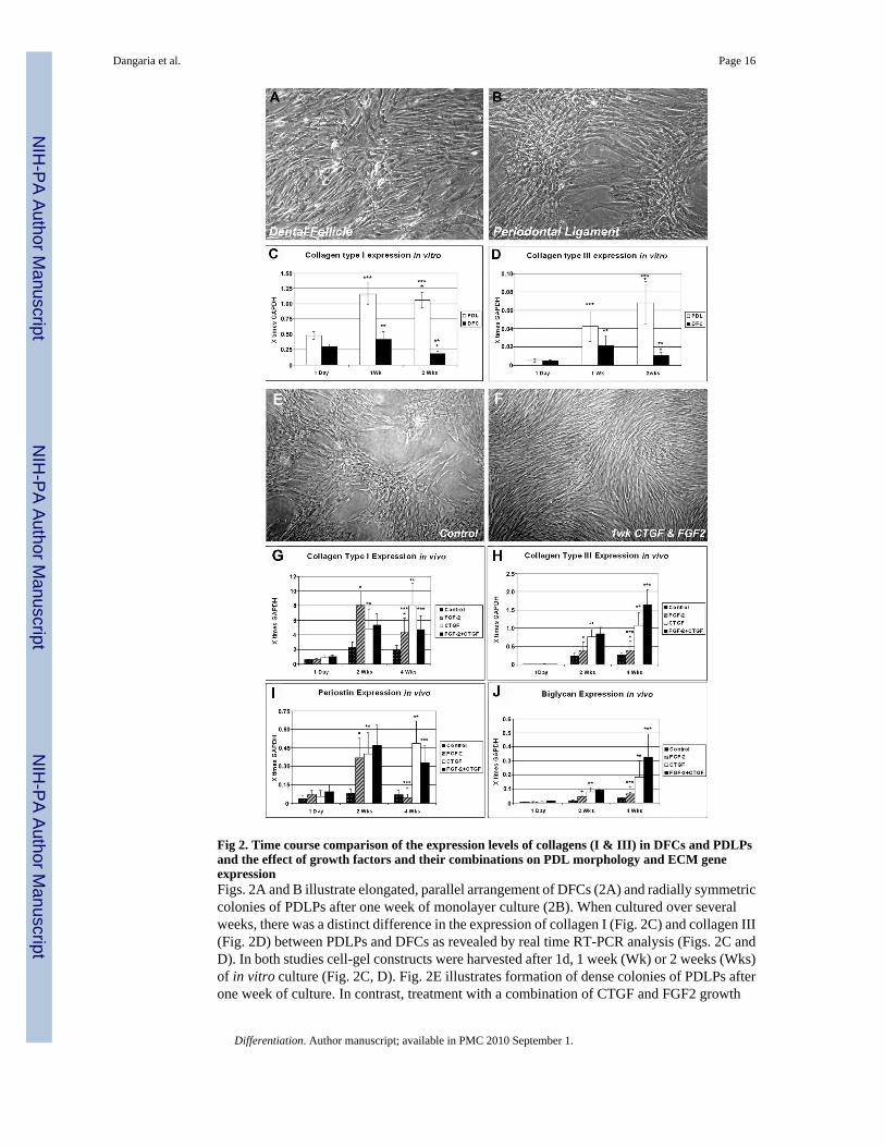
Fig 2. Time course comparison of the expression levels of collagens (I & III) in DFCs and PDLPsand the effect of growth factors and their combinations on PDL morphology and ECM geneexpressionFigs. 2A and B illustrate elongated, parallel arrangement of DFCs (2A) and radially symmetriccolonies of PDLPs after one week of monolayer culture (2B). When cultured over severalweeks, there was a distinct difference in the expression of collagen I (Fig. 2C) and collagen III(Fig. 2D) between PDLPs and DFCs as revealed by real time RT-PCR analysis (Figs. 2C andD). In both studies cell-gel constructs were harvested after 1d, 1 week (Wk) or 2 weeks (Wks)of in vitro culture (Fig. 2C, D). Fig. 2E illustrates formation of dense colonies of PDLPs afterone week of culture. In contrast, treatment with a combination of CTGF and FGF2 growth
Dangaria et al. Page 16
Differentiation. Author manuscript; available in PMC 2010 September 1.
NIH
-PA Author Manuscript
NIH
-PA Author Manuscript
NIH
-PA Author Manuscript

factors during this time period resulted in the formation of elongated and thin individual cells(Fig. 2F). In Figs. 2G–J, gene expression collagen type I (2G), collagen type III (2H), periostin(2I) and biglycan (2J) genes in cell-gel constructs was determined by Real time RT-PCRanalysis and cells were harvested after 1 day, 2 weeks (Wks) or 4 weeks (Wks) of subcutaneousimplantation in nude mice. While in general, gene expression levels increased after two or fourweeks implantation when treated with FGF2 and/or CTGF growth factors, individual ECMgene responses to growth factors were distinctly different and growth factor combinations hadeither synergistic or antagonistic effects (Fig. 2G–F). In all bar graphs, gene expression levelswere displayed relative to the house keeping gene GAPDH.
Dangaria et al. Page 17
Differentiation. Author manuscript; available in PMC 2010 September 1.
NIH
-PA Author Manuscript
NIH
-PA Author Manuscript
NIH
-PA Author Manuscript

Fig 3. Immunohistochemical and Western blot analysis of key ECM molecules in tissue engineeredPDL regeneratesFig. 3A–E are paraffin sections of PDLP/collagen tissue engineered constructs subcutaneouslyimplanted in nude mice for 4 weeks. Prior to implantation, PDLPs were treated for ten dayswith various growth factors such as FGF2 (10ng/ml, Fig. 3B), CTGF (50ng/ml, Fig. 3C), FGF2+CTGF (10/50ng/ml Fig. 3D), or left untreated (controls, Fig. 3A). The strongest stainingintensity for periostin was found in the CTGF-treated group (Fig. 3C), followed by the grouptreated with CTGF + FGF2 (Fig. 3D). There was little or no periostin localization in untreatedcontrols (Fig. 3A) and in the FGF2 treated group (Fig. 3B). Fig. 3E is the negative control thatwas not subjected to primary antibody. In Fig. 3F we have used Western blot analysis to
Dangaria et al. Page 18
Differentiation. Author manuscript; available in PMC 2010 September 1.
NIH
-PA Author Manuscript
NIH
-PA Author Manuscript
NIH
-PA Author Manuscript

determine levels of collagen III, biglycan, and periostin in our growth factor-treated tissueengineered constructs; lane 1 was the control group, lane 2 treated with FGF2, lane 3 treatedwith CTGF, and lane 4 treated with FGF2 plus CTGF (for concentrations see above). Theresults of our Western blot analysis were quantified using the Kodak 1D Image analysissoftware, and graphs were plotted by normalizing the value of collagen III, biglycan andperiostin with GAPDH as control (Fig. 3G–I). The results of this study illustrate that all threegrowth factor treatment modalities (FGF2, CTGF, and FGF2+CTGF) resulted in a significantincrease in the expression level of ECM matrix proteins (Collagen III, Biglycan, and Periostin).Moreover, CTGF alone or combinations with CTGF and FGF2 had a stronger effect on matrixproteins than FGF2 alone.
Dangaria et al. Page 19
Differentiation. Author manuscript; available in PMC 2010 September 1.
NIH
-PA Author Manuscript
NIH
-PA Author Manuscript
NIH
-PA Author Manuscript

Fig 4. Effects of FGF2 and CTGF treatment on matrix mineralization in PDLPsFig.(4 A–F) is a photograph of a culture plate containing PDLPs cultured at a density of 5 ×10⁴ cells/well for a total of 10 days in osteogenic differentiation media with or without growthfactors stained with Alizarin Red S to visualize relative levels of calcium. Cells were culturedin the following conditions: (i) continuous treatment with FGF2 for 10 days (Fig. 4A), (ii)continuous treatment with CTGF for 10 days (Fig. 4B), (iii) continuous treatment with FGF2and CTGF (Fig. 4C), (iv) sequential treatment with FGF2 for 4 days followed by CTGF forthe next 6 days (Fig. 4D), (v) sequential treatment with CTGF for 4 days followed by FGF2for the next 6 days (Fig. 4E), and (vi) without growth factor (Fig. 4F). In Fig. 4G, relativecalcium levels were calculated based on the o-cresolphthalein complexone method and
Dangaria et al. Page 20
Differentiation. Author manuscript; available in PMC 2010 September 1.
NIH
-PA Author Manuscript
NIH
-PA Author Manuscript
NIH
-PA Author Manuscript

spectrophotometric evaluation. Note that both CTGF treatment alone and CTGF followed byFGF2 treatment resulted in the most significant increases in calcium levels while simpleaddition of FGF2 and CTGF to the medium significantly decreased calcium levels (Fig. 4G).
Dangaria et al. Page 21
Differentiation. Author manuscript; available in PMC 2010 September 1.
NIH
-PA Author Manuscript
NIH
-PA Author Manuscript
NIH
-PA Author Manuscript

Fig 5. Attachment, spreading, viability and mineralization of DFCs depending on RGD-modification of PEGDA hydrogelsFig. 5A. Cell density of attached DFCs after 2 and 24 hrs on 10% PEGDA without Acr-PEG-RGD or containing 2mM Acr-PEG-RGD. Values are reported as an average of three randomfields times three hydrogel wells at each time point. There was a statistical difference (p<0.05)in the number of DFCs attached on the two types of hydrogels at the same time point (**) andon the same type of hydrogel at different time points (*). Fig. 5B. Histogram of DFC spreadingafter 2 hr attachment on either 10% PEGDA or 10% PEGDA with 2mM Acr-PEG-RGD. Tovisualize the morphology of the attached cells on these two types of hydrogel surfaces,micrographs were obtained following 2 hrs of attachment, and the area of cell spreading wascalculated by measuring the area individual cell occupied using the NIH imaging software.Values are reported as the percentage of the attached cells spreading and occupying areas inthe range specified (0–100 mm2) over three random fields per composition. Fig. 5C. Viabilityof DFCs in 10% PEGDA with or without 2mM of Acr-PEG-RGD after 3 days culture inosteogenic differentiation medium, PDGF supplemented medium, and control medium. Valuesare reported as the average of three constructs per composition per treatment. There was nostatistical difference (p>0.05) in the viability of DFCs between the modified and unmodifiedhydrogels and their treatment groups. Fig. 5D. Von Kossa stained sections of untreated
Dangaria et al. Page 22
Differentiation. Author manuscript; available in PMC 2010 September 1.
NIH
-PA Author Manuscript
NIH
-PA Author Manuscript
NIH
-PA Author Manuscript
(control) and treated (osteogenic, PDGF) DFCs encapsulated in unmodified (upper row) andRGD-modified (lower row) hydrogels after 2 weeks of in vitro culture. Mineralization wassignificantly increased in the RGD-modified group as seen by dense mineralization nodules.
Dangaria et al. Page 23
Differentiation. Author manuscript; available in PMC 2010 September 1.
NIH
-PA Author Manuscript
NIH
-PA Author Manuscript
NIH
-PA Author Manuscript
NIH
-PA Author Manuscript
NIH
-PA Author Manuscript
NIH
-PA Author Manuscript
Dangaria et al. Page 24
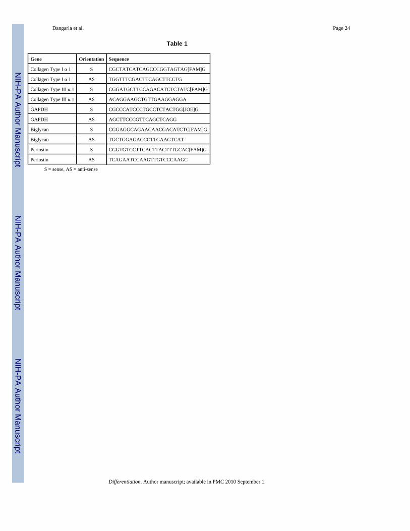
Table 1
Gene Orientation Sequence
Collagen Type I α 1 S CGCTATCATCAGCCCGGTAGTAG[FAM]G
Collagen Type I α 1 AS TGGTTTCGACTTCAGCTTCCTG
Collagen Type III α 1 S CGGATGCTTCCAGACATCTCTATC[FAM]G
Collagen Type III α 1 AS ACAGGAAGCTGTTGAAGGAGGA
GAPDH S CGCCCATCCCTGCCTCTACTGG[JOE]G
GAPDH AS AGCTTCCCGTTCAGCTCAGG
Biglycan S CGGAGGCAGAACAACGACATCTC[FAM]G
Biglycan AS TGCTGGAGACCCTTGAAGTCAT
Periostin S CGGTGTCCTTCACTTACTTTGCAC[FAM]G
Periostin AS TCAGAATCCAAGTTGTCCCAAGC
S = sense, AS = anti-sense
Differentiation. Author manuscript; available in PMC 2010 September 1.