COBRA, an Arabidopsis extracellular glycosyl-phosphatidyl ...
17
University of Massachuses Amherst ScholarWorks@UMass Amherst Biology Department Faculty Publication Series Biology 2005 COB, an Arabidopsis extracellular glycosyl- phosphatidyl inositol-anchored protein, specifically controls highly anisotropic expansion through its involvement in cellulose microfibril orientation F Roudier AG Fernandez M Fujita R Himmelspach GHH Borner See next page for additional authors Follow this and additional works at: hps://scholarworks.umass.edu/biology_faculty_pubs Part of the Biology Commons is Article is brought to you for free and open access by the Biology at ScholarWorks@UMass Amherst. It has been accepted for inclusion in Biology Department Faculty Publication Series by an authorized administrator of ScholarWorks@UMass Amherst. For more information, please contact [email protected]. Recommended Citation Roudier, F; Fernandez, AG; Fujita, M; Himmelspach, R; Borner, GHH; Schindelman, G; Song, S; Baskin, TI; Dupree, P; Wasteneys, GO; and Benfey, PN, "COB, an Arabidopsis extracellular glycosyl-phosphatidyl inositol-anchored protein, specifically controls highly anisotropic expansion through its involvement in cellulose microfibril orientation" (2005). Plant Cell. 23. hps://10.1105/tpc.105.031732
-
Upload
khangminh22 -
Category
Documents
-
view
4 -
download
0
Transcript of COBRA, an Arabidopsis extracellular glycosyl-phosphatidyl ...
University of Massachusetts AmherstScholarWorks@UMass Amherst
Biology Department Faculty Publication Series Biology
2005
COBRA, an Arabidopsis extracellular glycosyl-phosphatidyl inositol-anchored protein, specificallycontrols highly anisotropic expansion through itsinvolvement in cellulose microfibril orientationF Roudier
AG Fernandez
M Fujita
R Himmelspach
GHH Borner
See next page for additional authors
Follow this and additional works at: https://scholarworks.umass.edu/biology_faculty_pubs
Part of the Biology Commons
This Article is brought to you for free and open access by the Biology at ScholarWorks@UMass Amherst. It has been accepted for inclusion in BiologyDepartment Faculty Publication Series by an authorized administrator of ScholarWorks@UMass Amherst. For more information, please [email protected].
Recommended CitationRoudier, F; Fernandez, AG; Fujita, M; Himmelspach, R; Borner, GHH; Schindelman, G; Song, S; Baskin, TI; Dupree, P; Wasteneys,GO; and Benfey, PN, "COBRA, an Arabidopsis extracellular glycosyl-phosphatidyl inositol-anchored protein, specifically controlshighly anisotropic expansion through its involvement in cellulose microfibril orientation" (2005). Plant Cell. 23.https://10.1105/tpc.105.031732

AuthorsF Roudier, AG Fernandez, M Fujita, R Himmelspach, GHH Borner, G Schindelman, S Song, TI Baskin, PDupree, GO Wasteneys, and PN Benfey
This article is available at ScholarWorks@UMass Amherst: https://scholarworks.umass.edu/biology_faculty_pubs/23

that derive from the nature of the different wall polymers: a load-
bearing cellulose/xyloglucan array and a compression-resistant
pectin gel (Carpita and Gibeaut, 1993; Roberts, 1994). Cell
expansion depends directly on the local control of these struc-
tures and must combine remodeling activities to allow for
slippage of existing polymers with the incorporation of newly
synthesized materials (Cosgrove, 2000, 2001). The molecular
mechanisms that regulate the orientation and extent of cell
expansion remain poorly understood, and the identification of
the key proteins involved is of paramount importance.
Among the different wall materials, cellulose microfibrils
represent the most important component in the control of
anisotropic expansion. Inhibition of cellulose biosynthesis with
compounds such as 2,6-dichlorobenzonitrile (DCB), isoxaben, or
thaxtomin (Scheible et al., 2003) results in a rapid loss of growth
anisotropy. Cellulose-deficient mutants of Arabidopsis thaliana,
such as rsw1 (Arioli et al., 1998; Williamson et al., 2001) and
korrigan (kor/rsw2) (Nicol et al., 1998; Lane et al., 2001), also
show a dramatic loss of anisotropic expansion often accompa-
nied by cell swelling.
Cellulosemicrofibrils are synthesized at the plasmamembrane
by multimeric complexes called rosettes (Doblin et al., 2002),
which are typically arranged in parallel arrays, oriented
INTRODUCTION
In the absence of cell movement, plant morphogenesis is de-pendent on tight regulation of cell division and expansion. Plantdevelopment is characterized by complex patterns of growth
that determine organ form and function. Most plant cells growanisotropically by controlling how their extracellular matrix, thecell wall, yields to internal and isotropic turgor pressure. When
the direction of maximal anisotropic expansion coincides with
the longitudinal axis of the growing organ, it is called elongation.The primary cell wall combines unique properties of strength andrigidity but also of plasticity and viscosity, physical properties
1 Current address: Laboratoire de Biologie Cellulaire, Institut National de la Recherche Agronomique, Route de Saint Cyr, 78026 Versailles Cedex, France.2 To whom correspondence should be addressed. E-mail philip. [email protected]; fax 919-613-8177.The author responsible for distribution of materials integral to the findings presented in this article in accordance with the policy described in the Instructions for Authors (www.plantcell.org) is: Philip N. Benfey ([email protected]).
COBRA, an Arabidopsis Extracellular Glycosyl-PhosphatidylInositol-Anchored Protein, Specifically Controls HighlyAnisotropic Expansion through Its Involvement inCellulose Microfibril Orientation W
Francois Roudier,a,1 Anita G. Fernandez,b Miki Fujita,c,d Regina Himmelspach,c Georg H.H. Borner,eGary Schindelman,b Shuang Song,a Tobias I. Baskin,f Paul Dupree,e Geoffrey O. Wasteneys,c,d
and Philip N. Benfeya,2a Department of Biology, Duke University, Durham, North Carolina 27708b Department of Biology, New York University, New York, New York 10003c Department of Botany, University of British Columbia, Vancouver, British Columbia, Canada V6T 1Z4d Research School of Biological Sciences, Australian National University, Canberra ACT 2601, Australiae Department of Biochemistry, University of Cambridge, CB2 1QW Cambridge, United Kingdomf Biology Department, University of Massachusetts, Amherst, Massachusetts 01003
The orientation of cell expansion is a process at the heart of plant morphogenesis. Cellulose microfibrils are the primary anisotropic material in the cell wall and thus are likely to be the main determinant of the orientation of cell expansion. COBRA (COB) has been identified previously as a potential regulator of cellulose biogenesis. In this study, characterization of a null allele, cob-4, establishes the key role of COB in controlling anisotropic expansion in most developing organs. Quantitative polarized-light and field-emission scanning electron microscopy reveal that loss of anisotropic expansion in cob mutants is accompanied by disorganization of the orientation of cellulose microfibrils and subsequent reduction of crystalline cellulose. Analyses of the conditional cob-1 allele suggested that COB is primarily implicated in microfibril deposition during rapid elongation. Immunodetection analysis in elongating root cells revealed that, in agreement with its substitution by a glycosylphosphatidylinositol anchor, COB was polarly targeted to both the plasma membrane and the longitudinal cell walls and was distributed in a banding pattern perpendicular to the longitudinal axis via a microtubule-
dependent mechanism. Our observations suggest that COB, through its involvement in cellulose microfibril orientation, is an essential factor in highly anisotropic expansion during plant morphogenesis.

transversely to the axis of elongation. This pattern is thought to
be critical for the proper orientation of cell expansion (Taiz, 1984).
Biophysical, pharmacological, and genetic studies have sug-
gested that the direction of alignment of microfibrils can be
regulated at multiple levels. However, despite its importance for
morphogenesis and development, the mechanism for aligning
cellulose microfibrils is poorly understood.
For many years, the deposition of microfibrils has been con-
sidered to be under the control of microtubules (Green, 1962;
Ledbetter and Porter, 1963; Hepler andNewcomb, 1964). Cortical
microtubules have been demonstrated both genetically and
chemically to regulate the directionality of cell expansion.Mutants
in which the microtubule arrays are either disorganized, as in
tonneau (ton) (Camilleri et al., 2002) and bot1/fra2 (Bichet et al.,
2001; Burk et al., 2001; Burk and Ye, 2002), or more severely
disrupted, as inmor1-1 (Whittington et al., 2001; Sugimoto et al.,
2003), lose their ability to control growth anisotropy. Similar
defects can be phenocopied by the application of microtubule-
depolymerizing agents, such as oryzalin (Baskin et al., 1994).
Reduction in the anisotropy of expansion upon disruption
of microtubules and the correlation between the alignment of
cortical microtubules and cellulose microfibrils are the two
principal reasons for the belief that cortical microtubules align
newly synthesized microfibrils. The most popular models posit
that microtubules constrain rosette movement by serving either
as tracks or guard rails (Giddings and Staehelin, 1991). A
functional relationship between microtubule and microfibril
alignment recently received support from several studies.
Gardiner et al. (2003) demonstrated, for secondary cell wall syn-
thesis, that cortical microtubules are required to confine cellu-
lose synthase subunits to regions of cell wall deposition during
xylem development.Moreover, the characterization of the botero/
fra2mutant (deficient in a katanin p60 subunit; Burk and Ye, 2002),
in which cortical microtubules and cellulose microfibrils are both
disorganized, supports the existence of a functional link between
the two networks, although it should be noted that the rate of
cellulose synthesis is also reduced in thismutant. Recently, Baskin
(2001) proposed an alternative model in which microfibrils are
alignedbyvirtueofbindinganorientedscaffold,withtheorientation
cues provided by either the cell wall or the cortical microtubules.
Furthermore, the idea that microtubules influence growth
anisotropy by controlling microfibril alignment may be incom-
plete. There are actually few cases in which the alignment of
cellulose has been examined carefully after microtubule disrup-
tion, and several cases in which little change in microfibril ori-
entation occurred despite a wholesale change in microtubule
orientation (Baskin, 2001). For example, recently, the mor1-1
mutant was reported to have undisturbed microfibril align-
ment despite significantly disrupted cortical microtubule arrays
(Sugimoto et al., 2003), and recovery of well-ordered microfibrils
can proceed without the oriented microfibrillar or microtubular
scaffold (Himmelspach et al., 2003).
First identified in a genetic screen for regulators of cell expan-
sion, the cobra (cob) mutant was shown to have a conditional and
root-specific cell expansion defect (Benfey et al., 1993; Hauser
et al., 1995). Initial characterization indicated that COB is a mem-
brane protein required to orient cell expansion at the onset of rapid
elongation in the root, a function that appeared to be associated
with crystalline cellulose production (Schindelman et al., 2001). In
this article, an analysis of COB expression and a newly identified
null allele indicate thatCOB is a key regulator of diffuse anisotropic
expansion throughout postembryonic development. We provide
evidence that COB is anchored on the extracellular side of
the plasma membrane by a glycosyl-phosphatidyl inositol (GPI)
moiety and also released into the wall. We show that COB is
required for the oriented deposition of cellulose microfibrils and is
aligned in narrow bands perpendicular to the longitudinal axis in
cells undergoing rapid elongation, a pattern that depends on, but
is not closely related to, cortical microtubule organization.
RESULTS
COB Expression Is Developmentally Regulated and
Linked to Anisotropic Expansion
Previously, COB was shown to be expressed in most plant
organs on the basis of RNA gel blots and RT-PCR analyses
(Schindelman et al., 2001; Roudier et al., 2002). To specify the
spatial and temporal expression pattern of COB during plant
development, a b-glucuronidase (GUS) reporter fusion was
generated. Approximately 2.5 kb of COB 59 upstream sequence
was fused to the uidA gene followed by 1.4 kb of COB 39
downstream sequence, and the resulting PCOB:GUS fusion was
introduced into wild-type Arabidopsis. Assaying GUS activity at
different times in the plant life cycle revealed that COB was
expressed only postembryonically, with its strongest expression
in the root. Weaker GUS activity was detectable in growing
leaves, particularly in epidermal cells, guard cells, and fully
developed trichomes, as well as in the stigma and anthers of
developing flowers. GUS activity was first detectable in the root
tips of germinating seedlings (Figure 1A),where it showedasharp
upregulation at or near the onset of the elongation zone, and
expression remained strong in the more differentiated parts of
the root. At all stages, COB expression was absent in primary
and lateral root meristems (Figures 1B to 1D). This expression
pattern is consistent with the in situ hybridization pattern re-
ported previously (Schindelman et al., 2001). COB expression
was also observed in etiolated hypocotyls, which undergo rapid
anisotropic expansion (Figure 1E). During skotomorphogenesis,
theGUS staining pattern in the hypocotylsmirrored the acropetal
elongation gradient (Gendreau et al., 1997; Refregier et al., 2004),
providing further evidence linking COB expression and rapid
longitudinal expansion.
COB Is GPI-Anchored and N-Glycosylated
Analysis of the COB amino acid sequence predicted its sub-
stitution with a GPI anchor (Schindelman et al., 2001). To validate
the glypiation of COB, we prepared total membranes from
Arabidopsis callus culture and partitioned them into a dextran
phase (DEX), which contains endomembranes and is depleted in
plasmamembrane, and a polyethylene glycol (PEG) phase, which
is plasma membrane-enriched. Protein gel blot analysis of
the resulting fractions using anti-COB affinity-purified anti-
bodies demonstrated that COB was associated with the
plasma membrane (Figure 2A). As expected, the endoplasmic

reticulum–localized cytochrome b5 was depleted from the plasma
membrane–enriched fraction, whereas the plasma membrane–
localized ATPase was enriched. To determine whether COB is
a GPI-anchored protein, total membranes were fractionated by
Triton X-114 phase partitioning, thereby separating peripheral
membrane proteins from integral and GPI-anchored proteins,
which are present in the detergent phase. The detergent phase
was then incubatedwith a phosphatidylinositol-specific phospho-
lipase C (PI-PLC) and subjected again to phase partitioning. As
shown in Figure 2B, in the absence of PI-PLC treatment, the
COB protein was present in the detergent and not detectable in
the aqueous phase, whereas the PI-PLC treatment of this de-
tergent fraction resulted in the release of the COBprotein from the
membrane fraction into the soluble (aqueous) fraction. In a control,
the endoplasmic reticulum–localized and integral membrane pro-
tein cytochrome b5 remained insensitive to the PI-PLC treatment.
COB, like many other extracellular proteins, has been pre-
dicted to be N-glycosylated (Borner et al., 2002; Roudier et al.,
2002). When crude root extracts or microsomal membrane
fractions were treated with peptide N-glycosidase F, the COB
protein detected by immunoblot analysis displayed greater mo-
bility compared with the undigested samples (Figure 2C). This
change in protein mobility indicated that COB in its membrane-
bound form had been posttranslationally modified by the
addition of N-linked glycans. In the total and endomembrane
fractions, the affinity-purified anti-COB antibodies also recog-
nized a smaller protein, which partitioned as a peripheral mem-
brane protein (Figures 2A and 2B). We hypothesize that this
protein band corresponds to a cleaved and unmodified version
of COB present in the endoplasmic reticulum.
COB Is Required for Anisotropic Cell Expansion
throughout the Plant
The primary developmental defect of the cob alleles described
previously was associated with root elongation (Benfey et al.,
Figure 1. Localization of GUS Activity under the Control of COB cis-
Regulatory Regions.
(A) to (D)GUS staining in 2-d-old root (A), 7-d-old seedling (B), and 12-d-
old lateral and primary roots ([C] and [D]).
(E) COB expression in etiolated hypocotyls at 3 to 7 d after germination.
Bars ¼ 100 mm in (A), (C), and (D), 1 mm in (B), and 2.5 mm in (E).
Figure 2. COB Is Plasma Membrane–Localized, GPI-Anchored, and
N-Glycosylated.
(A) Protein blot probed successively with anti-COB, anti-cytochrome b5,
and anti-ATPase antibodies. Total membrane proteins (TM) were phase-
partitioned into endomembranes (DEX) and plasma membrane–enriched
protein fractions (PEG).
(B) Protein blot probed successively with anti-COB and anti-cytochrome
b5 antibodies. Proteins associated with total membranes were solubi-
lized in Triton X-114 and phase-partitioned into an aqueous peripheral
protein fraction (PP) and a detergent phase (D). The pure detergent
phase was treated with PI-PLC (þ) or with a buffer (�) and phase-
partitioned into new detergent (D) and aqueous phases (A).
(C) Protein blot probed with anti-COB antibodies. Root extracts (RE) or
microsomal membranes (M) were incubated with peptide N-glycosidase
F (þ) or buffer (�), and proteins were separated by SDS-PAGE.
Molecular weights are indicated at right.

1993; Hauser et al., 1995; Schindelman et al., 2001). The
discrepancy between the root-specific defects and the wide-
spread expression pattern reported here could be explained
either by functional redundancywith other genes in the aerial part
of the plant (Roudier et al., 2002) or by a degree of functionality
retained by the missense alleles whose phenotype was charac-
terized. To distinguish between these hypotheses, we identified
a putative null allele of cob in the Syngenta Arabidopsis Insertion
Library (SAIL) (Sessions et al., 2002). Line 735D10 contains
a T-DNA insertion in the fifth exon ofCOB at amino acid Met-213
andwas renamed cob-4. Our analyses indicate that thismutation
is recessive, monogenic, and nuclear. The BASTA resistance
marker associatedwith the T-DNA cosegregatedwith themutant
phenotype, and the phenotype was complemented by express-
ing the COB cDNA under the 35S promoter (data not shown). No
detectable signal was observed in protein gel blots of cob-4
protein extracts using anti-COB antibodies (see Methods), pro-
viding evidence that cob-4 is a null allele. cob-4 plants were
sterile, as a result of the complete absence of inflorescence stem
development. The phenotype of transheterozygous cob-1/cob-4
plants was very similar to that of cob-1/cob-1 homozygotes,
conditional on growth rate and apparent only in the root (data not
shown). This finding suggests that cob-1 is a partial loss-of-
function allele of COB.
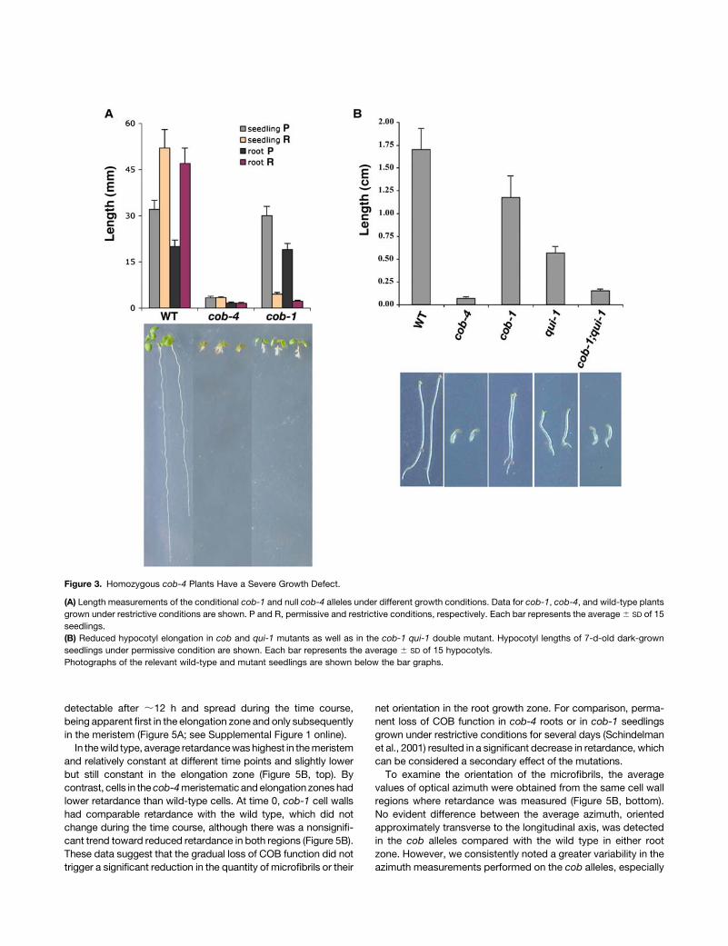
The growth of cob-4 seedlings was reduced dramatically
compared with that of wild-type seedlings in all conditions
tested. When grown on vertical plates, cob-4 seedlings were
;90% shorter (measured from the shoot apical meristem to the
root tip) and significantly thicker. A reduction in length was seen
in all organs (Figure 3A). By contrast, cob-1 seedlings were
similar to the wild type when grown on 0.3% sucrose (the
permissive condition) and were ;15% larger than cob-4 when
grown on 4.5% sucrose (the restrictive condition). Dark-grown
hypocotyls of cob-4 seedlings had an;95% reduction in length
compared with the wild type (Figure 3B). This finding demon-
strates that COB is also essential for the rapid elongation of the
etiolated hypocotyl. By contrast, cob-1 and qui-1 (a conditional
mutant allele of the cellulose synthase subunit AtCESA6) had
only 25 and 67% reductions in hypocotyl length, respectively.
Interestingly, the homozygous cob-1 qui-1 double mutant
showed an additive phenotype, with an 85% reduction in
hypocotyl length (Figure 3B).
Observations of roots from 7-d-old seedlings by scanning
electron microscopy revealed that cob-1 and cob-4 have similar
but not identical root-swelling phenotypes (Figures 4A, 4F, and
4K). Paradoxically, in seedlings continuously grown under re-
strictive conditions, themeristemofcob-1 roots (77 individuals out
of 80) showed abnormal radial expansion, whereas the same
region of the root in cob-4 mutants showed little or no change in
cell shape comparedwith thewild type (43 individuals out of 45). In
both genotypes, root hairs emerged close to the root tip from cells
that appeared to have undergone little elongation. To compare the
cellular morphology in the different zones of the mutants, trans-
verse sections were made at increasing distances from the root
tip. In the root cap and quiescent center region, the mutants were
similar to the wild type (Figures 4B, 4G, and 4L). Differences
between the two alleles became apparent in the meristem, where
cells exhibited abnormal radial expansion in cob-1 but remained
fairly normal in cob-4 (Figures 4C, 4H, and 4M). Furthermore,
cob-1 roots attained theirmaximal diameter in the early elongation
zone (Figures 4D, 4I, and 4N), whereas cob-4 roots expanded
radially throughout the elongation zone, ultimately reaching a
diameter that was nearly twice that of the wild type. Also in
the elongation zone, cells in the cob-4 epidermis tookona charac-
teristic bulging morphology (Figure 4O). In both alleles, cell walls
were occasionally broken, mostly in the epidermal layer. Cell wall
breakagewas rare anddid not seem toderive from incomplete cell
plate formation or abnormal cell divisions.
Whereas cob-1 aerial organs are similar to wild-type organs
(Figure 4P) (Hauser et al., 1995; Schindelman et al., 2001), 7-d-old
cob-4 hypocotyls and cotyledons are much smaller and thicker
than those of the wild type (Figure 4Q). There is also widespread
bulging of epidermal cells (Figures 4S to 4V), although the shoot
apical meristem, leaf primordia, and very young leaves do not
appear to be affected. Longitudinal sections of entire seedlings
at different developmental stages confirmed the absence of
sustained anisotropic cell expansion as well as the ballooning of
epidermal cells (Figures 4R and 4U). The sections also revealed
some internal expansion abnormalities in leaf mesophyll cells,
which reduced or eliminated intercellular air spaces. In the
absence of hypocotyl, petiole, and stem elongation, 3-week-old
cob-4 plants appeared severely stunted, with thick and vitreous
organs. Callus-like clusters often appeared on the adaxial side of
the leaves. Together, these morphological observations highlight
the essential and specific role of COB in the control of anisotropic
expansion during plant morphogenesis.
COB Is Required for the Oriented Deposition of Cellulose
Microfibrils during Rapid Anisotropic Expansion
Previous work has shown that cob-1 has lower levels of crystal-
line cellulose when grown for 1 week under restrictive condi-
tions, suggesting that COB is required for cellulose synthesis
(Schindelman et al., 2001). To investigate this notion further,
we used polarized-light microscopy and digital imaging to deter-
mine the relationship between the expansion defect observed in
cob roots and the amount and degree of organization of cellu-
lose microfibrils. The birefringent retardation (retardance) mea-
sured within a single cell wall is proportional to the amount of
crystalline cellulose and its net degree of orientation (Preston,
1974). We focused our analyses on epidermal cells at the border
of the meristem and elongation zones, where COB expression
is sharply upregulated. At this developmental transition, postmi-
totic cells abruptly accelerate their elongation rate 5- to 10-fold
(van der Weele et al., 2003). In the elongation zone, cellulose
microfibrils (and cortical microtubules) are oriented perpendic-
ular to the direction of rapid elongation (Sugimoto et al., 2000;
Baskin et al., 2004). To follow microfibril behavior during a grad-
ual loss of growth anisotropy resulting from COB deficiency, and
to dissect primary from secondary effects triggered by the
absence of functional COB,we took advantage of the conditional
nature of the cob-1 mutation and performed measurements at
0, 12, 24, and 30 h after a shift from permissive to restrictive
conditions, with equivalentmeasurementsmade for thewild type
and cob-4. At the beginning of the condition-shift experiment
(0 h), cob-1 roots had a wild-type appearance; swelling was first
detectable after ;12 h and spread during the time course,
being apparent first in the elongation zone and only subsequently
in the meristem (Figure 5A; see Supplemental Figure 1 online).
In thewild type, average retardancewashighest in themeristem
and relatively constant at different time points and slightly lower
but still constant in the elongation zone (Figure 5B, top). By
contrast, cells in the cob-4meristematic andelongation zones had
lower retardance than wild-type cells. At time 0, cob-1 cell walls
had comparable retardance with the wild type, which did not
change during the time course, although there was a nonsignifi-
cant trend toward reduced retardance in both regions (Figure 5B).
These data suggest that the gradual loss of COB function did not
trigger a significant reduction in the quantity of microfibrils or their
net orientation in the root growth zone. For comparison, perma-
nent loss of COB function in cob-4 roots or in cob-1 seedlings
grown under restrictive conditions for several days (Schindelman
et al., 2001) resulted in a significant decrease in retardance, which
can be considered a secondary effect of the mutations.
To examine the orientation of the microfibrils, the average
values of optical azimuth were obtained from the same cell wall
regions where retardance was measured (Figure 5B, bottom).
No evident difference between the average azimuth, oriented
approximately transverse to the longitudinal axis, was detected
in the cob alleles compared with the wild type in either root
zone. However, we consistently noted a greater variability in the
azimuth measurements performed on the cob alleles, especially
Figure 3. Homozygous cob-4 Plants Have a Severe Growth Defect.
(A) Length measurements of the conditional cob-1 and null cob-4 alleles under different growth conditions. Data for cob-1, cob-4, and wild-type plants
grown under restrictive conditions are shown. P and R, permissive and restrictive conditions, respectively. Each bar represents the average6 SD of 15
seedlings.
(B) Reduced hypocotyl elongation in cob and qui-1 mutants as well as in the cob-1 qui-1 double mutant. Hypocotyl lengths of 7-d-old dark-grown
seedlings under permissive condition are shown. Each bar represents the average 6 SD of 15 hypocotyls.
Photographs of the relevant wild-type and mutant seedlings are shown below the bar graphs.

in cob-4, in cells from the elongation zone compared with
wild-type cells. The frequency distribution of azimuths for the
measured cell wall regions had a SD of ;208 for the wild type
but was wider and flatter for cob-4 and cob-1 at 30 h (Figure 5C).
This variability indicates that the loss of COB activity disrupts
the processes that align microfibrils, even in cob-1, in which lev-
els of cellulose were apparently maintained.
To complement the data obtained with polarized-light micros-
copy, which responds to the crystalline cellulose throughout the
wall, we performed field emission scanning electron microscopy
(FESEM) to image directly the innermost cellulose microfibrils in
longitudinal walls. As shown in Figures 6A to 6D, in the succes-
sive wild-type root zones, the most recently deposited micro-
fibrils were parallel and oriented transversely to the elongation
axis, as reported previously (Sugimoto et al., 2000). The de-
position patterns of cellulose microfibrils in cells within the cob-4
meristem (Figure 6F) were similar to those of the wild type (Figure
6B). By contrast, in the cob-4 elongation zone, the cell walls
showed apparently randomly oriented microfibrils (Figure 6G).
This disorganization of microfibrils was also visible in cells within
the maturation zone (Figure 6H). FESEM analysis of cob-1 walls
allowed us to image microfibrils resulting from a partial loss of
COB function and in cells exhibiting less dramatic morphological
alterations. Because of the difficulty of the FESEM procedure,
Figure 4. cob-4 Has Dramatic Morphological Alterations in Most Organs.
Root phenotype analysis of wild-type ([A] to [E]), cob-4 ([F] to [J]), and cob-1 ([K] to [O]) plants.
(A), (F), and (K) Scanning electron micrographs of 10-d-old roots. Bar ¼ 100 mm.
(B) to (E), (G) to (J), and (L) to (O) Light micrographs of 3-mm transverse sections in the region of the root cap-quiescent center ([B], [G], and [L]),
meristem ([C], [H], and [M]), early elongation zone ([D], [I], and [N]), and late elongation zone [[E], [J], and [O]). Bar ¼ 100 mm.
(P) to (R) Scanning electron and light micrographs of 1-week-old cob-1 (P), cob-4 (Q), and corresponding longitudinal section (R). Bars ¼ 500 mm.
(S) to (V) Scanning electron ([S] and [U]) and light (V) micrographs of 18-d-old cob-4 aerial organs and corresponding longitudinal section (T). Bars ¼500 mm in (S) and (U) and 100 mm in (T) and (V).

cell walls in cob-1 roots were analyzed only at 24 h after the
condition shift. At this stage, the cob-1 root was significantly
swollen (Figure 6I). In the cob-1 meristem, most of the walls
analyzed displayed parallel cellulose microfibrils that were
oriented perpendicular to the longitudinal axis (Figures 6J and
6J9). However, in the elongation zone, microfibrils were some-
times transverse (Figure 6K), as they are at an approximately
comparable region of the wild type, and sometimes disordered
(Figure 6K9), as in cob-4. Farther away from the tip, cell walls in
cob-1 tended to resemble those in cob-4 (Figures 6L and 6L9). In
summary, 24 h after the condition shift, partial loss of COB
function resulted in local disorganization of cellulose microfibrils.
Together, these data demonstrate that COB is required for the
proper orientation of newly synthesized cellulose microfibrils in
regions that experience rapid expansion.
The Distribution of COB during Cell Elongation Is Dependent
on Cortical Microtubule Organization
The distribution of the COB protein in elongating cells was
analyzed in intact roots using whole-mount indirect immuno-
fluorescence and confocal laser scanning microscopy. In agree-
ment with its expression pattern, COB was detected in the
elongation zone, whereas no signal above background was
observed in the meristem (Figure 7A). Higher up in the elongation
zone, the signal became weaker, and no significant signal was
detected in the maturation zone. As expected from the absence
of signal on protein gel blots, no significant signal was detected
in the cob-4 mutant (data not shown). A closer examination of
the wild-type root revealed that the COB protein was first de-
tectable as a diffuse cytoplasmic and peripheral signal at the
onset of the elongation zone, and soon the majority of the de-
tected protein became organized in discrete domains on or in the
vicinity of the plasma membrane (Figures 7D and 7E). In cells
beginning to expand rapidly, COB patches appeared to be or-
ganized into narrow bands oriented perpendicularly to the longi-
tudinal axis in a pattern reminiscent of cortical microtubules
(Figures 7E and 7F). Double labeling of COB and cortical micro-
tubules (Figures 7B and 7G), although confirming the similar
orientation of cortical microtubules and the COB bands in
elongating cells, only occasionally revealed COB-dependent
fluorescence directly overlying cortical microtubules (Figures
7C and 7H). A similar transverse COB pattern was observed in
cob-1 roots under both permissive and restrictive conditions
(data not shown). The orientation of the cortical microtubules
was not significantly affected in cob-1 (Hauser et al., 1995) or in
cob-4 at the beginning of the elongation zone (data not shown).
The relationship between the transverse COB bands and the
cortical microtubule network was investigated using genetic
and pharmacological approaches. Whole-mount immunolocali-
zation using anti-COB antibodies was performed in weak and
strong alleles of the ton2 mutant, which is characterized by a
Figure 5. Crystalline Cellulose Organization and Root Morphology in the
cob Alleles.
(A) Time-course light micrographs of cob-1 root morphology at 0, 12, 14,
and 30 h after the condition shift. Bars ¼ 100 mm.
(B) Quantification by polarized-light microscopy of the amount of
cellulose (retardance) and its net orientation (azimuth) in the walls of
meristematic and elongating cells. Data from 5-d-old wild-type, cob-1,
and cob-4 seedlings at 0, 12, 24, and 30 h after transfer to high-growth-
rate media are shown. Each data point represents the average6 SD of at
least 15 cell walls.
(C) Frequency distribution of cellulose microfibril-azimuth angles mea-
sured in the elongation zone of wild-type, cob-1, and cob-4 roots after
the condition shift. F-test values (0.2 significance level) indicate that the
modal distributions of azimuth angles are significantly different between
the wild type and cob-4 (P ¼ 0.081) and between cob-4 and cob-1 (24 h)
(P ¼ 0.056), whereas the wild type and cob-1 (24 h) are not significantly
different (P ¼ 0.87). At 30 h after the condition shift, the modal dis-
tributions of azimuth angles between the wild type and cob-1 (30 h) are
significantly different (P ¼ 0.129).

Figure 6. Organization of Cellulose Microfibrils Is Altered in the Elongation Zone of cob Mutants.
FESEM images of the innermost layer of the cell wall of wild type ([A] to [D]), cob-4 ([E] to [H]), and cob-1 ([I] to [L9]) cryoplaned roots. The low-
magnification (3250) images of the root indicate cells in the different root zones from which wall texture was analyzed at higher magnification
(3100,000) FESEM imaging. In the meristem of the different genotypes ([B], [E], [J], and [J9]), microfibrils are approximately parallel and oriented
transversely to the elongation axis. In the wild type, this typical microfibril organization pattern is maintained throughout the elongation zone ([C] and
[D]). In cob-4, microfibrils in the elongation zone (G) and at the border with the maturation zone (H) are distributed randomly. Under conditions similar to
a 24-h period after a shift to restrictive conditions, microfibrils in the cob-1 elongation zone ([K] and [K9]) and at the border with the maturation zone ([L]
and [L9]) show a clear departure from the transverse orientation observed in the wild type. Microfibrils in cells of the cob-1 elongation zone often exhibit
different net orientations in different zones of the same wall ([K] and [K9]). (J) and (J9), (K) and (K9), and (L) and (L9) represent images of two different
areas in the same wall. Bars ¼ 250 nm except in (A), (E), and (I), where they ¼ 100 mm.

disturbance or loss of orientation of the cortical microtubules
(without apparent depolymerization) in elongating cells (Camilleri
et al., 2002). In both ton2-14 and ton2-13, COB was detected
in the compacted root elongation zone (Figures 7I and 7J) and
was more randomly distributed in these cells than in the wild
type (Figure 7K). When wild-type roots were treated for 45 min
with 10 mM oryzalin, a period in which microtubules depolymer-
ized completely (data not shown) but cell swelling was undetect-
able, the COB aggregates appeared to be severely randomized
(Figure 7L). These findings indicate that COB distribution at the
surface of elongating cells depends on the organization of
cortical microtubules.
Figure 7. COB Immunolocalization in 5-d-Old Roots.
Whole-mount confocal scanning micrographs of wild-type roots ([A] to [E]), wild-type elongating cells ([F] to [H] and [L]), and ton2 roots ([I] to [K]).
Bars ¼ 50 mm in (A) to (E) and (I), (J), (L) and (M) and 10 mm in (F) to (H) and (K). Brackets in (D) and (E) indicate the same three elongating cells.
(A), (E), (F), and (J) to (L) Indirect fluorescence immunolocalization of COB using anti-COB antibodies.
(B) and (G) Visualization of cortical microtubules by indirect immunofluorescence.
(C) Combined images of COB (A) and microtubules (B) at the root tip.
(D) and (I) Differential interference contrast micrographs of the wild-type elongation zone and the ton2-14 root tip, respectively.
(H) Combined images of COB (F) and microtubules (G) in an elongating cell.
(L) COB staining after treatment with 10 mM oryzalin for 45 min. Note patches, compared with the normal banding pattern.
(M) COB staining after treatment with 50 mM brefeldin A for 45 min. Note intracellular accumulation of COB-containing clumps.

COB Follows a Typical GPI Secretion Path and Is
Predominantly Associated with the Longitudinal
Cell Walls
As a GPI-anchored protein, COB would be expected to follow
a secretion path and reach the cell periphery through vesicular
trafficking. Treatment of elongating root cells with brefeldin A (a
vesicle-trafficking inhibitor) resulted in a strong decrease of the
COB banding pattern (still visible as weak bands at the cell
surface or as dots along the longitudinal walls) and triggered the
accumulation of COB in large intracellular compartments (Figure
7M). This suggests that the polar secretion of COB is very active
in root elongating cells and shows that the signal observed is
a combination of COB-containing vesicles and COB at the cell
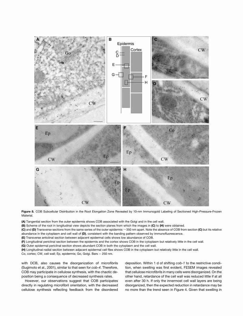
surface. COB distribution at the subcellular level was further
examined by transmission electron microscopy of high-
pressure-frozen, immunogold-labeled material from the elonga-
tion zone of wild-type roots. At the outer face of epidermal cells
(Figure 8A), COB was associated with the Golgi and abundant
in the cell wall, supporting its transit along a Golgi-derived vesi-
cle secretion pathway. Serial sectioning in the transverse plane
(Figures 8C and 8D) revealed that COB was abundant in some
sections and not present in others, consistent with COB’s
distribution to transverse bands seen in immunofluorescence
preparations (Figures 7E and 7F). In contrast with its abundance
at the outer epidermal cell wall, COB was present at lower
abundance in other walls, particularly the radial (Figure 8E) and
even the inner longitudinal (Figures 7H and 8F) walls of the
epidermis. Consistent with the rapid upregulation at the onset of
elongation, in the root division zone COBwas not detected in the
walls and was only occasionally detected close to the Golgi, in
cells near the onset of the elongation zone (data not shown).
In the wall sections examined, COB was present on and at vari-
ous distances from the plasma membrane (Figures 8A, 8D, and
8G), suggesting that it exists in both a GPI-anchored, plasma
membrane–associated form and a cleaved form.
DISCUSSION
COB Is Required for Highly Anisotropic Expansion
throughout Plant Development
The relationship between net cellulose orientation in the primary
wall and the direction of organ growth was established decades
ago (Green, 1980). Nevertheless, themechanisms underlying the
regulation of microfibril orientation in elongating cells remain
largely unknown. The data presented here indicate that the GPI-
anchored protein COB is an important component of these
mechanisms during rapid elongation.
To dissect the molecular mechanisms underlying anisotropic
expansion and to understand more precisely the function of
COB, we characterized a T-DNA insertion allele, cob-4, and took
advantage of the conditional nature of the previously described
cob-1 allele (Schindelman et al., 2001). The extensive swelling
specifically in cells that would otherwise undergo highly aniso-
tropic growth makes cob-4 an intriguing cell expansion mutant
among those characterized to date. Phenotypic analysis showed
that COB is specifically involved in the control of diffuse aniso-
tropic expansion, in agreement with its expression pattern. The
observation of organized and functional meristems and normal
radial patterning in cob roots suggests that cell division is
affected minimally if at all in the mutants. Likewise, root hairs
seem unaffected by cob mutations, and pollen development
appears normal, as judged by the expected transmission ratios
of themutant alleles. These observations indicate that COB is not
involved in the process of tip growth but is required specifically
for the regulation of diffuse anisotropic expansion.
Recently, relative elongation rates have been measured in the
Arabidopsis root at high spatial and temporal resolution (van der
Weele et al., 2003). The authors characterized the growth zone as
comprising two zones of roughly constant relative elongation
rates separated by an abrupt transition. The apical zone (corre-
sponding to the meristem and probably including some post-
mitotic cells) has a low relative rate,;7%/h, and the basal zone
(corresponding to the canonical elongation zone) has a high rate
of ;50%/h. The extent of anisotropy increases concomitantly
with the acceleration of relative elongation rates, because the
tangential expansion rate actually decreases (Baskin et al.,
2004). Strikingly, the position at which anisotropy increases is
approximately the same as where COB expression is abruptly
upregulated. This finding is consistent with our hypothesis that
COB is required to achieve highly anisotropic expansion.
The expansion of leaf pavement cells has also been shown
to involve two mechanistically distinct growth phases (Fu et al.,
2002). Phenotypic analysis of leaf development in cob-4 sug-
gests that COB is required primarily for the second phase, which
is characterized by extensive cell growth, and not for the initial
morphogenesis of the different epidermal cell types. In cob-4, the
shoot apical meristem and leaf primordia appear normal,
whereas in the leaf epidermis, the complete loss of COB function
resulted in leaves with only small, roughly isodiametric shapes,
suggesting that the ability of the cells to expand anisotropically
is lost.
Cell elongation in dark-grown hypocotyls was also described
recently as a biphasic growth pattern, with the growth acceler-
ation second phase gaining ground acropetally from the base
(Refregier et al., 2004). The absence of hypocotyl elongation in
cob-4 is also consistent with an important role for COB during
this striking example of rapid polar growth. Thus, in the leaf,
hypocotyl, and root, it appears to be the phase of maximal and
developmentally regulated anisotropic expansion that requires
COB function.
COB Is Required Primarily for the Orientation and
Secondarily for the Synthesis of Microfibrils
The cob-4 dwarf phenotype with radially expanded cells is
reminiscent of strong alleles of cellulose synthesis–deficient
mutants such as rsw1 (mutant in the cellulose synthase subunit
AtCESA1; Arioli et al., 1998; Williamson et al., 2001) or kor
(mutant in amembrane-boundb-1,4-endoglucanase; Nicol et al.,
1998; Lane et al., 2001). Likewise, cob-1 roots, grown under
restrictive conditions for several days, had less cellulose than did
wild-type roots, as assayed by several independent methods
(Schindelman et al., 2001). Furthermore, reduced cellulose
synthesis, triggered either genetically as in rsw1 or chemically
with DCB, also causes the disorganization of microfibrils
(Sugimoto et al., 2001), similar to that seen for cob-4. Therefore,
COB may participate in cellulose synthesis, with the chaotic de-
position being a consequence of decreased synthesis rates.
However, our observations suggest that COB participates
directly in regulating microfibril orientation, with the decreased
cellulose synthesis reflecting feedback from the disordered
deposition. Within 1 d of shifting cob-1 to the restrictive condi-
tion, when swelling was first evident, FESEM images revealed
that cellulosemicrofibrils inmany cells were disorganized. On the
other hand, retardance of the cell wall was reduced little if at all
even after 30 h. If only the innermost cell wall layers are being
disorganized, then the expected reduction in retardance may be
no more than the trend seen in Figure 4. Given that swelling in
Figure 8. COB Subcellular Distribution in the Root Elongation Zone Revealed by 10-nm Immunogold Labeling of Sectioned High-Pressure-Frozen
Material.
(A) Tangential section from the outer epidermis shows COB associated with the Golgi and in the cell wall.
(B) Scheme of the root in longitudinal view depicts the section planes from which the images in (C) to (H) were obtained.
(C) and (D) Transverse sections from the same series of the outer epidermis;350 nm apart. Note the absence of COB from section (C) but its relative
abundance in the cytoplasm and cell wall of (D), consistent with the banding pattern observed by immunofluorescence.
(E) Transverse anticlinal section between adjacent epidermal cells shows low abundance of COB.
(F) Longitudinal periclinal section between the epidermis and the cortex shows COB in the cytoplasm but relatively little in the cell wall.
(G) Outer epidermal periclinal section shows abundant COB in both the cytoplasm and the cell wall.
(H) Longitudinal-radial section between adjacent epidermal cell files shows COB in the cytoplasm but relatively little in the cell wall.
Co, cortex; CW, cell wall; Ep, epidermis; Go, Golgi. Bars ¼ 250 nm.

cob-1 after 30 h is well advanced, the combination of polarized-
light and FESEM data indicate that the loss of microfibril order
is the primary defect in the mutant, with the loss of cellulose
synthesis occurring secondarily.
Further linking COB function to microfibril orientation, we
found that the net orientation of microfibrils within the subcellular
regions of the cell wall was more variable in cob-4 and in cob-1
after 30 h under restrictive conditions. Because the retardance of
the same regions was reduced not at all (or only slightly), this
finding implies that the net direction of alignment of one patch
of cell wall was different from that of another. Interestingly, a sim-
ilar effect on microfibril alignment also occurs when roots are
exposed to low concentrations of the microtubule inhibitor
oryzalin (Baskin et al., 2004).
Whether the secondary loss of cellulose synthesis results in
the accumulation of amorphous b-glucans by a crystallization
defect, as occurs in rsw2/kor (Peng et al., 2000; His et al., 2001),
remains to be determined. It is interesting that in cob-4 walls
imaged with FESEM, we consistently noted the presence of
globular clusters extending from microfibrillar structures, which
could represent lumps of noncrystallized cellulose.
Cellular Location of COB Function
We have shown that COB is GPI-substituted and N-glycosy-
lated. Proteomics-based approaches demonstrated that other
members of the COB family are also GPI-modified (Borner et al.,
2003; Elortza et al., 2003; Lalanne et al., 2004). Our immunode-
tection analyses revealed that in elongating root cells, COBcould
exist in the different forms normally taken by a GPI-modified
protein. Gold label was detected in the Golgi, and transport
vesicles, presumably identifying the secretory pathway, andboth
glycosylated and nonglycosylated forms were detected in mi-
crosomal membranes. Gold label was also detected at the
plasma membrane as well as within the cell wall. GPI-anchored
proteins can be released from the anchor by PI-specific phos-
pholipases, as yet uncharacterized in plants. The presence of
considerable COB within the cell wall suggests that during
anisotropic expansion, the GPI anchorage is temporary, and
the activity of PI-specific phospholipases may represent a way
of regulating the amount of anchored COB.
This raises the question of the functional form of COB in the
context of microfibril orientation. The fact that in the partial loss-
of-function cob-1 allele COB expression and COB localization
are not visibly affected (data not shown) indicates that the
defects observed are related directly to COB’s function and
not to a problem of protein stability or targeting. Moreover,
engineered versions of COB, either mutated at the potential GPI
cleavage attachment sites (N384T, G385W, and S387P) or
truncated from the probable cleavage site (Asn-384) and driven
by the 35S promoter, were not able to complement the cob
phenotype (0 individuals out of 16 and 15 T2 plants, respectively).
Transient expression assays using green fluorescent protein
(GFP) translationally fused with either the mutated or the trun-
cated C terminus of COB showed GFP mistargeting to endo-
compartments, whereas the GFP fusion with a wild-type COB C
terminus was localized primarily in the plasma membrane (data
not shown). These observations highlight the functional signifi-
cance of GPI substitution and suggest that the membrane-
anchored COB or its wall form, or possibly both, represent
the functional forms. In animals, GPI-anchored proteins are fre-
quently subjected to specific targeting pathways, often pro-
posed to involve sterol- or sphingolipid-rich microdomains of the
plasma membrane (Friedrichson and Kurzchalia, 1998; Varma
and Mayor, 1998; Muniz and Riezman, 2000), and similar
targeting of GPI-anchored proteins may occur in plants (Borner
et al., 2005). In the case of COB, the observed asymmetric
targeting of this protein to the outer epidermal face opens new
avenues of investigation to understand how plant cells take
advantage of glypiation to effect specific localization.
Association between COB and Microtubules
In elongating epidermal cells, the COB protein is distributed
mainly near the cell surface in transverse bands that parallel
cortical microtubules. Based on the transmission electron mi-
croscopy data, it appears that these bands reflect clustering
within the cell wall at variable distances from the plasma mem-
brane. Unfortunately, microtubules were only rarely encountered
in the cryofixed, freeze-substituted material; thus, it could not be
determined with confidence whether there is any positional re-
lation between microtubules and the COB-positive clusters.
When microtubule organization is genetically or pharmaco-
logically challenged, the COB banding pattern becomes dis-
rupted and COB-labeled patches become more prominent in
the cytosol, implying that microtubules are required, directly or
indirectly, for COB localization. Because the banding is disrupted
even after a relatively brief oryzalin treatment, it appears that at
least some of the label seen in the confocal images arises from
cytoplasmic material in addition to plasma membrane and cell
wall–bound material, as shown by the partial loss of the banded
localization of COB after brief treatment with brefeldin A.
Although the mechanism leading to a banded appearance of
COB remains to be established, the microtubule dependence of
COB localization provides an intriguing link between the function
of the protein and cellulose orientation. A recently proposed
model postulates that nascent cellulose microfibrils are oriented
by binding to a scaffold of cell wall polysaccharides or plasma
membrane proteins, which, in turn, are oriented according to
positional information provided by either cortical microtubules or
the cell wall (Baskin, 2001). The COB protein, with its particular
surface localization and distribution during rapid elongation, is
a candidate for one of these scaffolding proteins.
At present, these scaffolds are purely hypothetical, and recent
experiments combining the mor1-1 mutant, in which micro-
tubules are disrupted, and DCB treatment, which disorganizes
microfibrils, suggest that neither preexisting cell wall templates
nor microtubules are required for microfibril alignment
(Himmelspach et al., 2003). This, along with other observations
(Wiedemeier et al., 2002; Sugimoto et al., 2003), has led to a new
model whereby microtubules influence growth anisotropy by
regulating the relative length of microfibrils (Wasteneys, 2004).
According to this model, the microtubule-dependent patterning
of COB could support the formation and extension of relatively
long microfibrils, either by preventing the breakage of newly
formed microfibrils or by regulating the activity of synthase

complexes (Wasteneys, 2004). The potential cellulose binding
capacity of COB (Roudier et al., 2002) and the phenotypic
alterations reported here fit well with this latest model. Loss of
microfibril orientation in the cob mutants would reflect a de-
gradedorientingmechanism,possibly associatedwith a reduced
degree of polymerization. In this scenario, the oriented pattern
of COB at the cell surface may reflect a mechanism for the
patterned delivery of cell wall precursors rather than for direct
microfibril alignment. Despite the present ambiguity about the
connection between microtubule and microfibril alignments,
the experiments reported here establish COB as a pivotal player
in the regulation of the anisotropic expansion of higher plant
cells.
METHODS
Plant Material and Growth Conditions
Arabidopsis thaliana plants were grown as described previously (Benfey
et al., 1993). All of the mutant lines used in this work are in the Columbia
background. The SAIL line 735D10 (Syngenta, San Diego, CA), which
contains a T-DNA insertion in the fifth exon of the COB gene, was
backcrossed twice, propagated as a heterozygote, and renamed cob-4.
GUS Reporter Construct
To generate the PCOB:GUS reporter construct, a �2509 to þ45 fragment
containing the entire 59 flanking region of COB and a 1443-bp fragment
of the 39 end of COB were amplified by PCR from genomic DNA using
the respective primer pairs 59-TTACTAAAACAGCACTAGCCAGC-39/
59-CGGATCCCGATGCCATGGACAATTTGGAGACGATGGAGGTGG-39
and 59-ATTTGCGGCCGCTCGGATTTACGGTTTTGCCACTGG-39/
59-ACGGATGGAAATTGGACTGAAATGC-39, subcloned as HindIII/Bam-
HI and NotI fragments, respectively, and sequenced. The uidA gene
was cloned in frame between the two COB genomic sequences as
a NcoI/BamHI fragment. The entire construct was then excised as a SacI
fragment and cloned into the plant transformation vector pBIN19. The re-
sulting vector was transformed into the Arabidopsis accession Col-0
by the floral-dipping method (Clough and Bent, 1998). GUS staining
was performed on T2 plants as described by Malamy and Benfey (1997).
Light and Scanning Electron Microscopy and Sectioning
For scanning electron microscopy, specimens were either fixed in 2.5%
glutaraldehyde, dehydrated through an ethanol series, and critical point
dried in liquid CO2 or cryofixed in anOxfordCT1500 cryochamber (Oxford
Instruments, Oxon, UK). After sputter coating with gold, samples were
visualized with an Amray FE-1850 scanning electron microscope (KLA-
Tencor, San Jose, CA) operated at 5 kV. For sectioning, fixed plantlets
and roots were embedded in Historesin (Technovit 7100; Kulzer,
Wehrheim, Germany) according to the manufacturer’s instructions.
Three-micrometer plastic sections were stained with toluidine blue
(0.5% [w/v] in 0.1 M phosphate buffer, pH 7.2) to visualize the cell wall.
Light micrographs were taken on a DMRXA2microscope (Leica, Wetzlar,
Germany).
Immunolocalization and Confocal Microscopy
Anti-COB polyclonal IgY antibodies were produced by GenWay Biotech
(San Diego, CA) against amino acids 147 to 297. Antibodies were raised in
Rhode Island Red hens, and total IgY was isolated from egg yolk by
standard methods. COB-specific IgY was further purified through affinity
chromatography using recombinant COB fusion protein produced in
Escherichia coli as ligand. Whole-mount immunolocalizations were per-
formed as described by Friml et al. (2003) except that incubations were
done either overnight at 48Cor for 3 h at room temperature. For oryzalin and
brefeldin A treatments, seedlings were incubated in 1 mL of liquid growth
medium (0.53 MS medium [Sigma-Aldrich, St. Louis, MO] and 1%
sucrose, pH 5.7) containing 10 mM oryzalin (100 mM stock in ethanol;
Duchefa, Haarlem, TheNetherlands) or 50mMbrefeldin A (100mMstock in
DMSO; Molecular Probes, Eugene, OR) and equal amounts of solvent for
control. Washing was done twice for 10 min, and incubation was stopped
by fixation. Antibodies used and final concentrations (v/v) were as follows:
affinity-purified anti-COB IgY antibodies, 1:200; affinity-purified rabbit anti-
chicken IgY Alexa Fluor 647–conjugated secondary antibodies (Genway
Biotech, San Diego, CA), 1:200; anti-a-tubulin antibodies (Sigma-Aldrich),
1:1000; and fluorescein isothiocyanate–conjugated anti-mouse IgG sec-
ondary antibodies (1:200). Fluorescent images were collected on a confo-
cal laser scanningmicroscope (LSM510 system; Carl ZeissMicroImaging,
Jena, Germany). Each image shown represents either a single focal plan or
a projection of individual images taken as a Z-series. Images acquired
using Zeiss software were imported into Adobe Photoshop (Mountain
View, CA) for cropping, contrast adjustment, and assembly.
Biochemical Fractionation, PI-PLC Sensitivity,
and N-Glycosylation Test
Total membrane isolation and Triton X-114 phase separation were
performed as described by Borner et al. (2003). Dextran-PEG partitioning
was performed as described by Sherrier et al. (1999). For PI-PLC
treatment, the washed Triton X-114 phase was split into two aliquots.
PI-PLC (Sigma-Aldrich) was added to one aliquot, and both were
incubated at 378C for 1 h (with intermittent mixing). BSA was added as
carrier protein to the aqueous phases. Proteins were precipitated using
cold acetone and subsequently subjected to SDS-PAGE and protein gel
blot analysis. Anti-cytochromeb5 and anti-ATPase antibodieswere a kind
gift of J. Napier (Rothamsted Research, Harpenden, UK) and M. Boutry
(Universite Catholique de Louvain, Louvain-La-Neuve, Belgium), respec-
tively. To determine whether COB was N-glycosylated, roots of 1-week-
old seedlings were ground and microsomes were prepared as described
by Jinn et al. (2000), then they were resuspended in the denaturing buffer
supplied with the peptide N-glycosidase F and treated according to the
manufacturer’s instructions (New England Biolabs, Beverly, MA).
Polarized-Light Microscopy and FESEM
Birefringent retardation of root cell walls was measured on 2-mm
longitudinal plastic sections using a polarized-light microscope (Jenapol;
Zeiss) equipped for circularly polarized-light quantitative digital imaging
(LC Pol Scope; Cambridge Research Instruments, Cambridge, UK).
Longitudinal cell walls were assayed that were parallel to the plane of
the section in areas devoid of any visible heterogeneity. Retardance and
optical azimuth were calculated from the intensity measurements by the
Pol Scope software, which implements the algorithm described by
Oldenbourg and Mei (1995). We did not observe a significant difference
in retardance between the epidermis and the cortex layer for the three
genotypes tested; data from both cell types were combined to generate
the graphs. For each time point, five root longitudinal sections were
examined and at least three measurements were performed per section
and root zone. Samples were prepared and longitudinal walls were
analyzed by FESEM as described by Himmelspach et al. (2003) using
a Hitachi 4500 field emission scanning electron microscope (Tokyo,
Japan). For each genotype, three roots and at least three different cells for
each root zone were analyzed.

Transmission Electron Microscopy
Roots from wild-type (Col-0) seedlings grown, as described by Sugimoto
et al. (2000), for 5 d under constant light (80 mmol�m�2�s�1) at 218C
were cut into 1.5-mm lengths with a razor blade while submerged in
hexadecane. Cut root tissue was transferred into flat specimen holders
filled with hexadecane and cryofixed in a high-pressure freezing unit
(Bal-Tech HPM010; Balzers, Lichtenstein). For LR White (London Resin
Company, London, UK) embedding, frozen samples were put into
cryogenic vials containing freeze substitution media (0.25% glutaralde-
hyde and 0.1% uranyl acetate in acetone) and placed into a dry ice–
acetone bath as described (Rensing et al., 2002). This bath was placed at
�208C for 7 d and warmed to 48C for 4 h, then brought to room
temperature. The tissue was rinsed with fresh acetone and infiltrated
with LRWhite embedding resin according to the following schedule: drop
by drop for 3 d; then 50, 75, and 85% (v/v) LR White:acetone, each step
for 12 h; and finally 100% LR White resin for 36 h with resin exchanged
every 12 h. Polymerization was achieved at 608C for 24 h. Thin sections
(<70 nm) were cut with an Ultracut E ultramicrotome (Leica) and mounted
on Formvar-coated 200-mesh nickel grids.
For immunogold labeling, all incubations of sections were performed
by placing the grids on droplets of solution section-side-down at room
temperature. Thin sections mounted on grids were incubated for 10 min
on the droplets of 10 mM NH4Cl in TBS (10 mM Tris and 0.25 M NaCl,
pH 7) to block free aldehydes. Nonspecific antibody binding sites on
the sections were blocked by incubating the sections for 30 min in 5%
nonfat dried milk in TBS. The sections were then incubated for 1 h with
chicken anti-COBantibodydiluted 1:100 in 0.5%nonfat driedmilk in TBS.
After rinsing in TBS five times, the sections were incubated for 1 h with
goat anti-chicken IgG conjugated to 10-nm colloidal gold (Aurion,
Wageningen, The Netherlands) diluted 1:100 in 0.5% nonfat dried milk
in TBS. The sections were then rinsed in TBS five times, in distilled water
twice, and air dried. Grids were stained in 2% aqueous uranyl acetate for
30 min and in lead citrate for 5 min and then observed using a Hitachi
H7600 transmission electron microscope.
ACKNOWLEDGMENTS
We thank I. Tan (New York University) for expert technical assistance
with scanning electron microscopy. Casey J. Roehrig is thanked for her
skilled technical assistance. M. Pastuglia (Institut National de la Re-
cherche Agronomique, Versailles) is thanked for the gift of ton2-14 and
ton2-13 seeds. We thank H. Hofte and K. Birnbaum for critical reading of
the manuscript. Polarized-light microscopy done in the Baskin labora-
tory was supported by a grant to T.I.B. from the U.S. Department of
Energy (Award 94ER20146), which does not constitute endorsement by
that department of the views expressed herein. FESEM and trans-
mission electron microscopy done in the Wasteneys laboratory was
supported by grants to G.O.W. from the Australian Research Council
(DP0208872) and the Natural Sciences and Engineering Research
Council of Canada (DP298264-04), respectively. This material is based
on work supported by the National Science Foundation under Grant
0209754 to P.N.B.
Received February 11, 2005; revised February 11, 2005; acceptedMarch
18, 2005; published April 22, 2005.
REFERENCES
Arioli, T., et al. (1998). Molecular analysis of cellulose biosynthesis in
Arabidopsis. Science 279, 717–720.
Baskin, T.I. (2001). On the alignment of cellulose microfibrils by cortical
microtubules: A review and a model. Protoplasma 215, 150–171.
Baskin, T.I., Beemster, G.T.S., Judy-March, J.E., and Marga, F.
(2004). Disorganization of cortical microtubules stimulates tangential
expansion and reduces the uniformity of cellulose microfibril align-
ment among cells in the root of Arabidopsis. Plant Physiol. 135,
2279–2290.
Baskin, T.I., Wilson, J.E., Cork, A., and Williamson, R.E. (1994).
Morphology and microtubule organization in Arabidopsis roots ex-
posed to oryzalin or taxol. Plant Cell Physiol. 35, 935–942.
Benfey, P.N., Linstead, P.J., Roberts, K., Schiefelbein, J.W., Hauser,
M.T., and Aeschbacher, R.A. (1993). Root development in Arabi-
dopsis: Four mutants with dramatically altered root morphogenesis.
Development 119, 57–70.
Bichet, A., Desnos, T., Turner, S., Grandjean, O., and Hofte, H.
(2001). BOTERO1 is required for normal orientation of cortical micro-
tubules and anisotropic cell expansion in Arabidopsis. Plant J. 25,
137–148.
Borner, G.H., Lilley, K.S., Stevens, T.J., and Dupree, P. (2003).
Identification of glycosylphosphatidylinositol-anchored proteins in
Arabidopsis: A proteomic and genomic analysis. Plant Physiol. 132,
568–577.
Borner, G.H., Sherrier, D.J., Stevens, T.J., Arkin, I.T., and Dupree, P.
(2002). Prediction of glycosylphosphatidylinositol-anchored pro-
teins in Arabidopsis: A genomic analysis. Plant Physiol. 129,
486–499.
Borner, G.H., Sherrier, D.J., Weimar, T., Michaelson, L.V., Hawkins,
N.D., Macaskill, A., Napier, J.A., Beale, M.H., Lilley, K.S., and
Dupree, P. (2005). Analysis of detergent-resistant membranes in
Arabidopsis: Evidence for plasma membrane lipid rafts. Plant Physiol.
137, 104–116.
Burk, D.H., and Ye, Z.H. (2002). Alteration of oriented deposition of
cellulose microfibrils by mutation of a katanin-like microtubule-
severing protein. Plant Cell 14, 2145–2160.
Burk, D.H., Liu, B., Zhong, R., Morrison, W.H., and Ye, Z.H. (2001). A
katanin-like protein regulates normal cell wall biosynthesis and cell
elongation. Plant Cell 13, 807–827.
Camilleri, C., Azimzadeh, J., Pastuglia, M., Bellini, C., Grandjean, O.,
and Bouchez, D. (2002). The Arabidopsis TONNEAU2 gene en-
codes a putative novel protein phosphatase 2A regulatory subunit
essential for the control of the cortical cytoskeleton. Plant Cell 14,
833–845.
Carpita, N.C., and Gibeaut, D.M. (1993). Structural models of pri-
mary cell walls in flowering plants: Consistency of molecular structure
with the physical properties of the walls during growth. Plant J. 3,
1–30.
Clough, S.J., and Bent, A.F. (1998). Floral dip: A simplified method for
Agrobacterium-mediated transformation of Arabidopsis thaliana. Plant
J. 16, 735–743.
Cosgrove, D.J. (2000). Loosening of plant cell walls by expansins.
Nature 407, 321–326.
Cosgrove, D.J. (2001). Wall structure and wall loosening: A look
backwards and forwards. Plant Physiol. 125, 131–134.
Doblin, M.S., Kurek, I., Jacob-Wilk, D., and Delmer, D.P. (2002).
Cellulose biosynthesis in plants: From genes to rosettes. Plant Cell
Physiol. 43, 1407–1420.
Elortza, F., Nuhse, T.S., Foster, L.J., Stensballe, A., Peck, S.C., and
Jensen, O.N. (2003). Proteomic analysis of glycosylphosphatidylino-
sitol-anchored membrane proteins. Mol. Cell. Proteomics 2, 1261–
1270.
Friedrichson, T., and Kurzchalia, T.V. (1998). Microdomains of GPI-
anchored proteins in living cells revealed by crosslinking. Nature 394,
802–805.

Friml, J., Benkova, E., Mayer, U., Palme, K., and Muster, G. (2003).
Automated whole mount localisation techniques for plant seedlings.
Plant J. 34, 115–124.
Fu, Y., Li, H., and Yang, Z. (2002). The ROP2 GTPase controls the
formation of cortical fine F-actin and the early phase of directional cell
expansion during Arabidopsis organogenesis. Plant Cell 14, 777–794.
Gardiner, J.C., Taylor, N.G., and Turner, S.R. (2003). Control of
cellulose synthase complex localization in developing xylem. Plant
Cell 15, 1740–1748.
Gendreau, E., Traas, J., Desnos, T., Grandjean, O., Caboche, M.,
and Hofte, H. (1997). Cellular basis of hypocotyl growth in Arabidop-
sis thaliana. Plant Physiol. 114, 295–305.
Giddings, T.H., and Staehelin, L.A. (1991). Microtubule-mediated
control of microfibril deposition: A re-examination of the hypothesis.
In The Cytoskeletal Basis of Plant Growth and Form, C.W. Lloyd, ed
(San Diego: Academic Press), pp. 85–99.
Green, P.B. (1962). Mechanism for plant cellular morphogenesis.
Science 138, 1404–1405.
Green, P.B. (1980). Organogenesis: A biophysical view. Annu. Rev.
Plant Physiol. 31, 51–82.
Hauser, M.T., Morikami, A., and Benfey, P.N. (1995). Conditional root
expansion mutants of Arabidopsis. Development 121, 1237–1252.
Hepler, P.K., and Newcomb, E.H. (1964). Microtubules and fibrils in
the cytoplasm of coleus cells undergoing secondary wall deposition.
J. Cell Biol. 20, 529–532.
Himmelspach, R., Williamson, R.E., and Wasteneys, G.O. (2003).
Cellulose microfibril alignment recovers from DCB-induced disruption
despite microtubule disorganization. Plant J. 36, 565–575.
His, I., Driouich, A., Nicol, F., Jauneau, A., and Hofte, H. (2001).
Altered pectin composition in primary cell walls of korrigan, a dwarf
mutant of Arabidopsis deficient in a membrane-bound endo-1,4-beta-
glucanase. Planta 212, 348–358.
Jinn, T.L., Stone, J.M., and Walker, J.C. (2000). HAESA, an Arabi-
dopsis leucine-rich repeat receptor kinase, controls floral organ
abscission. Genes Dev. 14, 108–117.
Lalanne, E., Honys, D., Johnson, A., Borner, G.H., Lilley, K.S.,
Dupree, P., Grossniklaus, U., and Twell, D. (2004). SETH1 and
SETH2, two components of the glycosylphosphatidylinositol anchor
biosynthetic pathway, are required for pollen germination and tube
growth in Arabidopsis. Plant Cell 16, 229–240.
Lane, D.R., et al. (2001). Temperature-sensitive alleles of RSW2 link
the KORRIGAN endo-1,4-beta-glucanase to cellulose synthesis and
cytokinesis in Arabidopsis. Plant Physiol. 126, 278–288.
Ledbetter, M.C., and Porter, K.R. (1963). A ‘‘microtubule’’ in plant cell
fine structure. J. Cell Biol. 19, 239–250.
Malamy, J.E., and Benfey, P.N. (1997). Analysis of SCARECROW
expression using a rapid system for assessing transgene expression
in Arabidopsis roots. Plant J. 12, 957–963.
Muniz, M., and Riezman, H. (2000). Intracellular transport of GPI-
anchored proteins. EMBO J. 19, 10–15.
Nicol, F., His, I., Jauneau, A., Vernhettes, S., Canut, H., and Hofte, H.
(1998). A plasma membrane-bound putative endo-1,4-beta-d-gluca-
nase is required for normal wall assembly and cell elongation in
Arabidopsis. EMBO J. 17, 5563–5576.
Oldenbourg, R., and Mei, G. (1995). New polarized light microscope
with precision universal compensator. J. Microsc. 180, 140–147.
Peng, L., Hocart, C.H., Redmond, J.W., and Williamson, R.E. (2000).
Fractionation of carbohydrates in Arabidopsis root cell walls shows
that three radial swelling loci are specifically involved in cellulose
production. Planta 211, 406–414.
Preston, R.D. (1974). The Physiological Biology of Plant Cell Walls.
(London: Chapman and Hall), pp. 75–108.
Refregier, G., Pelletier, S., Jaillard, D., and Hofte, H. (2004). In-
teraction between wall deposition and cell elongation in dark-grown
hypocotyl cells in Arabidopsis. Plant Physiol. 135, 959–968.
Rensing, K.H., Samuels, A.L., and Savidge, R.A. (2002). Ultrastructure
of vascular cambial cell cytokinesis in pine seedlings preserved by
cryofixation and substitution. Protoplasma 220, 39–49.
Roberts, K. (1994). The plant extracellular matrix in a new expansive
mood. Curr. Opin. Cell Biol. 6, 688–694.
Roudier, F., Schindelman, G., DeSalle, R., and Benfey, P.N. (2002).
The COBRA family of putative GPI-anchored proteins in Arabidopsis:
A new fellowship in expansion. Plant Physiol. 130, 538–548.
Scheible, W.R., Fry, B., Kochevenko, A., Schindelasch, D., Zimmerli,
L., Somerville, S., Loria, R., and Somerville, C.R. (2003). An
Arabidopsis mutant resistant to thaxtomin A, a cellulose synthesis
inhibitor from Streptomyces species. Plant Cell 15, 1781–1794.
Schindelman, G., Morikami, A., Jung, J., Baskin, T.I., Carpita, N.C.,
Derbyshire, P., McCann, M.C., and Benfey, P.N. (2001). COBRA
encodes a putative GPI-anchored protein, which is polarly localized
and necessary for oriented cell expansion in Arabidopsis. Genes Dev.
15, 1115–1127.
Sessions, A., et al. (2002). A high-throughput Arabidopsis reverse
genetics system. Plant Cell 14, 2985–2994.
Sherrier, D.J., Prime, T.A., and Dupree, P. (1999). Glycosylphosphati-
dylinositol-anchored cell-surface proteins from Arabidopsis. Electro-
phoresis 20, 2027–2035.
Sugimoto, K., Himmelspach, R., Williamson, R.E., and Wasteneys,
G.O. (2003). Mutation or drug-dependent microtubule disruption
causes radial swelling without altering parallel cellulose microfibril
deposition in Arabidopsis root cells. Plant Cell 15, 1414–1429.
Sugimoto, K., Williamson, R.E., and Wasteneys, G.O. (2000). New
techniques enable comparative analysis of microtubule orientation,
wall texture, and growth rate in intact roots of Arabidopsis. Plant
Physiol. 124, 1493–1506.
Sugimoto, K., Williamson, R.E., and Wasteneys, G.O. (2001). Wall
architecture in the cellulose-deficient rsw1 mutant of Arabidopsis
thaliana: Microfibrils but not microtubules lose their transverse align-
ment before microfibrils become unrecognizable in the mitotic and
elongation zones of roots. Protoplasma 215, 172–183.
Taiz, L. (1984). Plant cell expansion: Regulation of cell wall mechanical
properties. Annu. Rev. Plant Physiol. 35, 585–657.
van der Weele, C.M., Jiang, H.S., Palaniappan, K.K., Ivanov, V.B.,
Palaniappan, K., and Baskin, T.I. (2003). A new algorithm for
computational image analysis of deformable motion at high spatial
and temporal resolution applied to root growth: Roughly uniform
elongation in the meristem and also, after an abrupt acceleration, in
the elongation zone. Plant Physiol. 132, 1138–1148.
Varma, R., and Mayor, S. (1998). GPI-anchored proteins are organized
in submicron domains at the cell surface. Nature 394, 798–801.
Wasteneys, G.O. (2004). Progress in understanding the role of micro-
tubules in plant cells. Curr. Opin. Plant Biol. 7, 651–660.
Whittington, A.T., Vugrek, O., Wei, K.J., Hasenbein, N.G., Sugimoto,
K., Rashbrooke, M.C., and Wasteneys, G.O. (2001). MOR1 is
essential for organizing cortical microtubules in plants. Nature 411,
610–613.
Wiedemeier, A.M., Judy-March, J.E., Hocart, C.H., Wasteneys, G.O.,
Williamson, R.E., and Baskin, T.I. (2002). Mutant alleles of Arabi-
dopsis RADIALLY SWOLLEN 4 and 7 reduce growth anisotropy
without altering the transverse orientation of cortical microtubules or
cellulose microfibrils. Development 129, 4821–4830.
Williamson, R.E., Burn, J.E., Birch, R., Baskin, T.I., Arioli, T., Betzner,
A.S., and Cork, A. (2001). Morphology of rsw1, a cellulose-deficient
mutant of Arabidopsis thaliana. Protoplasma 215, 116–127.