A New Lycodon (Serpentes: Colubridae) From Northeast India and Myanmar (Burma)

ORIGINAL ARTICLE
Unique features of myogenesis in Egyptian cobra (Naja haje)(Squamata: Serpentes: Elapidae)
Eraqi R. Khannoon1& Weronika Rupik2
&
Damian Lewandowski3 & Magda Dubińska–Magiera3 &
Elwira Swadźba2 & Małgorzata Daczewska3
Received: 25 February 2015 /Accepted: 21 May 2015# The Author(s) 2015. This article is published with open access at Springerlink.com
Abstract During early stages of myotomal myogenesis, themyotome of Egyptian cobra (Naja haje) is composed of ho-mogenous populations of mononucleated primary myotubes.At later developmental phase, primary myotubes are accom-panied by closely adhering mononucleated cells. Based onlocalization and morphology, we assume that mononucleatedcells share features with satellite cells involved in musclegrowth. An indirect morphological evidence of the fusion ofmononucleated cells with myotubes is the presence of numer-ous vesicles in the subsarcolemmal region of myotubes adja-cent to mononucleated cell. As differentiation proceeded, sec-ondary muscle fibres appeared with considerably smaller di-ameter as compared to primary muscle fibre. Studies onN. haje myotomal myogenesis revealed some unique featuresof muscle differentiation. TEM analysis showed in the N. hajemyotomes two classes of muscle fibres. The first class wascharacterized by typical for fast muscle fibres regular distribu-tion of myofibrils which fill the whole volume of muscle fibresarcoplasm. White muscle fibres in studied species were aprominent group ofmuscles in the myotome. The second classshowed tightly paced myofibrils surrounding the centrally lo-cated nucleus accompanied by numerous vesicles of different
diameter. The sarcoplasm of these cells was characterized bynumerous lipid droplets. Based on morphological features, webelieve that muscle capable of lipid storage belong to slowmuscle fibres and the presence of lipid droplets in the sarco-plasm of these muscles during myogenesis might be a crucialadaptive mechanisms for subsequent hibernation in adults.This phenomenon was, for the first time, described in studieson N. haje myogenesis.
Keywords Myogenesis . Satellite cells . TEM .Naja haje .
Snake
Introduction
Differentiation of the trunk (myotomal) muscles has been in-vestigated in detail in different groups of vertebrates includingfishes, amphibians, birds and mammals. The myotomal mus-cles in all studied vertebrate specimens originate from unseg-mented paraxial mesoderm which, during subsequent stagesof embryonal development, is subdivided into repetitive cellunits called somites. The somites then differentiate into threecompartments: the dermomyotome with a well-developeddorsomedial lip and ventrolateral lip, the sclerotome and af-terwards into the myotome. The main part of the myotome isoccupied by trunk muscles which differentiate in situ(reviewed by Bentzinger et al. 2012). Studies on developmentof amniote (birds and mammals) trunk muscles have revealedthat the dermomyotome is the main source of trunk muscleprogenitor cells, which express the paired-box transcriptionfactors Pax3 and Pax7. It has been shown that Pax3 is requiredto establish the myogenic potential of differentiating cells,whereas Pax7 is required for the specification and mainte-nance of satellite cells during myogenesis (Horst et al. 2006;
Handling Editor: Pavel Dráber
* Małgorzata [email protected]
1 Zoology Department, Faculty of Science, Fayoum University,Fayoum 63514, Egypt
2 Department of Animal Histology and Embryology, University ofSilesia, 9 Bankowa Str., 40-007 Katowice, Poland
3 Department of Animal Developmental Biology, Institute ofExperimental Biology, University of Wrocław, 21 Sienkiewicza Str.,53-335 Wrocław, Poland
ProtoplasmaDOI 10.1007/s00709-015-0840-3

Buckingham and Realaix 2007; Kacperczyk et al. 2009; Sealeet al. 2000; Olguin and Olwin 2004; Zammit et al. 2006).
During the vertebrates’ development, myogenic progeni-tors differentiate into myoblasts, which differentiate intomyotubes, a developing immature skeletal muscle fibres witha centrally located nucleus in the sarcoplasm. The final stageof myogenesis is a formation of mature muscle fibres—cylin-drical multinucleate cells with peripherally located nuclei.Muscles grow through two mechanisms: hypertrophy and hy-perplasia. During hypertrophy, the fibre size increases due tothe addition of new nuclei into sarcoplasm of pre-existingfibres, while hyperplasia is a process of new muscle fibreformation. It is well evidenced that in amniotes, Pax7 muscleprogenitor cells participate in both processes in the prenatalstage. It is noteworthy that, after birth, a number of musclefibres remain unchanged in mammals and birds (Rowe andGoldspink 1969; Fowler et al. 1980; Nimmo and Snow 1983;Brown 1987; Rosenblatt and Woods 1992; Schadereit et al.1995). Postnatal growth is mainly due to hypertrophy, al-though some reports have revealed an increase in fibre numbershortly after birth (Swatland 1975; Rehfeldt and Fiedler 1984;Summers and Medrano 1994; Fiedler et al. 1998).
Among all vertebrates, reptile myogenesis and myotomalmuscle growth are poorly known. Studies on reptilemyogenesis have been conducted on several species (Chinesesoft-shelled turtle Pelodiscus sinensis and sand lizard Lacertaagilis); among them, only sand lizard studies on myogenesisshed light on the origin of muscle progenitor cells and growthof myotomal muscles (Nagashima et al. 2005; Rupik et al.2012). It was found that in the sand lizard, similarly to birdsand mammals, the dermomyotome is the main source of mus-cle Pax3-positive progenitor cells. Studies conducted byRupik et al. (2012) revealed that muscle growth in the sandlizard is due to Pax3/Pax7-positive mononucleated cells. Incomparison to the sand lizard, snakes, e.g. Naja haje, repre-sent a different mode of locomotion. In our studies, weattempted to answer the question whether mode of locomotioninfluences the pattern of muscle differentiation and growth. Inthe present study, we investigated trunk muscle differentiationand growth in the previously unstudied Egyptian cobra(N. haje) in order to demonstrate whether myogenesis in thestudied species displays a unique character, not observed inother vertebrates.
Material and methods
The Egyptian cobra (N. haje L. (1758): Elapidae, Reptilia) isone of the largest cobra species native to Africa. It is terrestrialand crepuscular or nocturnal. Adult female cobras attain sex-ual maturity after 2 to 3 years. They lay 17 to 22 eggs in aclutch. Oviposition occurs 60–100 days after copulation inearly summer (Schleich et al. 1996). Fertilized female
Egyptian cobras for this study were collected from the NileDelta region of Egypt in June 2012–2013. The animals werekept in vivaria in an open farm area, in conditions similar tothose in the wild, until the eggs were laid, and then they werereleased into their native area. All specimens used in the exper-iment were captured according to the Egyptian regulationsconcerning the protection of wild species (Convention on Bio-logical Diversity ratified in 1992 and 1994). The Egyptian co-bra is not included in the Washington Convention of 1973. Theeggs of Egyptian cobra (n=100) after oviposition were careful-ly collected and brought to the laboratory of the Zoology De-partment, Faculty of Science, Fayoum University, where theywere placed in plastic boxes filled with moistened perlite (at85–90 % moisture) and with ventilation holes. The eggs wereincubated at 30 °C, reflecting ambient seasonal temperatures inthe wild. Embryos used for investigation were isolated at reg-ular intervals from egg laying to hatching. Embryonic develop-ment at 30 °C in laboratory conditions from when the eggs arelaid until they hatch lasts 51–54 days (Khannoon and Evans2014). The age of the embryos was calculated using the tablefor N. haje development (Khannoon and Evans 2014). Thestudy ofmyotomalmuscle differentiation and growth ofN. hajeincluded four developmental stages from stages 3 to 6.
For light and electron microscopic techniques, small piecesof embryonic body wall including differentiating muscle tis-sue were fixed in a 1:1 mixture of 2.5 % glutaraldehyde(Sigma-Aldrich) and 2.0 % paraformaldehyde (Sigma-Al-drich, St. Louis, MO, USA) in 0.1 m phosphate buffer,pH 7.4, for 24 h at 4 °C. The material was repeatedly rinsedin the same buffer and post-fixed for 2 h in 1 % OsO4 (Sigma-Aldrich) in 0.1 M phosphate buffer. Following rinsing inphosphate buffer, the material was dehydrated first in a gradedalcohol series and then in acetone and embedded in epoxyresin Epon 812 (Sigma-Aldrich) (Luft 1961). This procedureof fixation appears to be the best for different embryonic rep-tilian tissues (Rupik 2002, 2011, 2012, 2013; Swadźba andRupik 2010, 2012). The Epon blocks were cut on LeicaUltracut UCT (Leica, Wetzlar, Germany) and ReichertUltracut E ultramicrotome (Leica, Wetzlar, Germany). Semi-thin sections (0.6 μm) were collected on glass slides andstained with methylene blue in 1 % borax solution (Sigma-Aldrich) and examined under a light microscope OlympusBX60 and Olympus BHS light microscopes (Olympus Corp.,Tokyo, Japan). Ultrathin sections were collected on 200 meshcopper grids, stained with uranyl acetate and lead citrate ac-cording to the standard protocol (Reynolds 1963) and exam-ined in a Hitachi H500 (Hitachi Ltd., Tokyo, Japan; 75 kV)and Zeiss EM 900 (Carl Zeiss AG, Oberkochen, Germany;80 kV) transmission electron microscopes. Histological mate-rial was processed and analysed in the Department of AnimalHistology and Embryology, University of Silesia, and in theDepartment of Animal Developmental Biology, Institute ofExperimental Biology, University of Wrocław.
E.R. Khannoon et al.

Results
Myotomal muscles differentiation and growth in N. haje
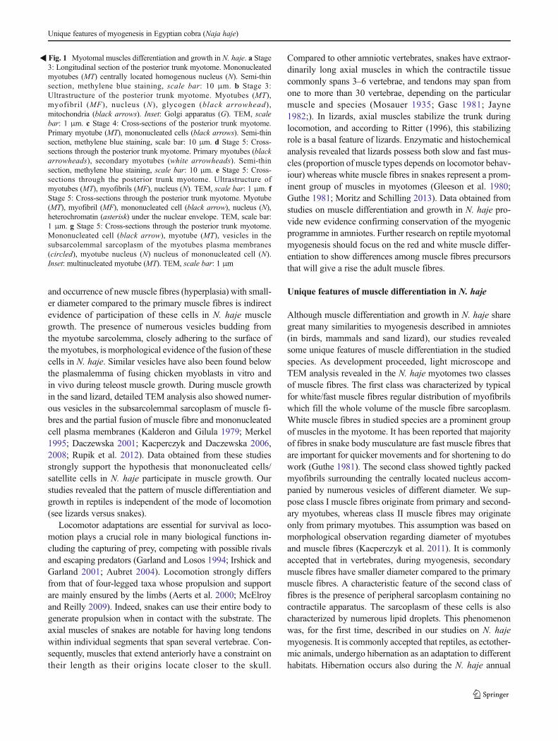
Myotomal muscle formation of N. haje was analysed in thelight microscope and TEM.At stage 3, themyotome ofN. hajeis composed of homogeneous populations of mononucleatedprimary myotubes containing centrally located homogeneousnuclei (Fig. 1a). Sarcoplasm of these cells revealed the pres-ence of a few myofibrils arranged in an irregular way, numer-ous mitochondria, Golgi apparatus and glycogen granules. Atthis stage of development, neighbouring myotubes are closelyattached to each other (Fig. 1b). At later developmental phase(stage 4), light microscope analysis revealed that primarymyotubes are accompanied by closely adhering mononucleat-ed cell (Fig. 1c). As differentiation proceeded (stage 5), sec-ondary muscle fibres appear. The secondary muscle fibres aredistinguished by considerably smaller diameter as comparedto primary muscle fibre (Fig. 1d). At this stage of myogenesis,numbers of myofibrils in sarcoplasm of myotubes were higher(Fig. 1e). Ultrastructural analysis showed that mononucleatedcells closely adhering to myotubes are composed of a promi-nent nucleus with heterochromatin located mainly under thenuclear envelope. The nucleus is surrounded by a narrow rimof cytoplasm devoid of glycogen granules and elements ofcontractile apparatus (Fig. 1f). Localization of mononucleat-ed cells suggests their potential involvement in musclegrowth due to their fusion with myotubes. This is plausiblesince the detailed ultrastructural analysis showed the pres-ence of numerous vesicles in the subsarcolemmal region ofmyotubes adjacent to mononucleated cells. At this devel-opmental stage, as a result of progressing myogenic cellfusion, elongated, multinucleated myotubes appear(Fig. 1g).
Unique features of muscle differentiation in N. haje
During myotomal myogenesis (stage 5), developing musclefibres of N. haje do not form a homogeneous population.Undertaken studies revealed the presence of two classes ofmuscle fibres distinguished by the distribution of myofibrils.Class I is characterized by typical regular distribution of myo-fibrils which fill the whole volume of the muscle fibre sarco-plasm (Fig. 2a). Class II features tightly packed myofibrilssurrounding a centrally located nucleus accompanied by nu-merous vesicles. They are localized only in the proximity ofthe nucleus and do not occur in the peripheral region of thesarcoplasm, which is filled with numerous mitochondria andendoplasmic reticulum. Sarcoplasm of II class of muscle fi-bres contains numerous glycogen granules among myofibrilsand in the peripheral region (Fig. 2b, c). The nuclei of class IImuscle fibres are rich in heterochromatin localized beneath thenuclear envelope and in the internal part of the nucleus
(Fig. 2d). Sarcoplasm of these cells is also characterized bynumerous lipid droplets surrounded by glycogen granules(Fig. 2e, f).
Discussion
Differentiation and growth of muscles in N. haje
Skeletal muscle differentiation and growth in vertebrates havebeen investigated in detail in model species (zebrafish,Xenopus laevis, chick, mouse) (reviewed by Bentzingeret al. 2012; Kiełbówna and Jędrzejowska 2012; Rossi andMessina 2014). These model vertebrate species provided de-tailed information at the morphological and the molecular lev-el of muscle development in this phylum. Studies on reptiliandevelopmental biology including myogenesis are difficult andrarely attempted (Nagashima et al. 2005; Rupik et al. 2012;Kusumi et al. 2013). To better understand the mechanisms oftrunk muscle differentiation and growth in reptiles, we chosethe Egyptian cobra. Our results revealed that in the studiedspecies during early steps of myogenesis, the myotome wascomposed of a homogeneous population of myogenic cellsconsisting of mononucleated primary myotubes. The structureand morphology of these cells in N. haje myotomes resemblemononucleated myocytes observed in primary myotomes inbirds and mammals and also in the sand lizard, a representa-tive of reptilians which, in comparison to Egyptian cobra,represents a different mode of locomotion. During subsequentstages of muscle fibres differentiation, myotubes were accom-panied by mononucleated cells. Based on localization andmorphology, these cells share features with satellite cells, de-scribed in detail in many vertebrates (fish, amphibians, birdsand mammals) (Zammit et al. 2006; Kacperczyk et al. 2009,2011; Daughters et al. 2011; reviewed by Siegiel et al. 2013;Yin et al. 2013). TEM analysis revealed that satellite cells inthe studied species are spindle-shaped composed of a promi-nent nucleus with heterochromatin located beneath the nuclearenvelope. The narrow rim of their cytoplasm is devoid ofglycogen granules and elements of contractile apparatus.Compared to differentiating myotubes, the ultrastructure ofsatellite cells showed an undifferentiated state. In the sandlizard, similarly to birds and mammals, satellite cells that ad-join the differentiating muscle fibres express Pax7 protein(Rupik et al. 2012). The origin of these cells in N. haje hasnot been studied. Many different lines of evidence indicatethat in amniotes, mononucleated cells that adjoin themyotubes and/or muscle fibres originate from thedermomyotome. Studies on sand lizard myogenesis revealedthat, as in other vertebrates, these cells are myogenic precur-sors involved in muscle growth (both hypertrophic and hyper-plastic) (Rupik et al. 2012). Increase in the number of nuclei inmyotubes (hypertrophy) in subsequent stages of myogenesis
Unique features of myogenesis in Egyptian cobra (Naja haje)

E.R. Khannoon et al.
and occurrence of new muscle fibres (hyperplasia) with small-er diameter compared to the primary muscle fibres is indirectevidence of participation of these cells in N. haje musclegrowth. The presence of numerous vesicles budding fromthe myotube sarcolemma, closely adhering to the surface ofthe myotubes, is morphological evidence of the fusion of thesecells in N. haje. Similar vesicles have also been found belowthe plasmalemma of fusing chicken myoblasts in vitro andin vivo during teleost muscle growth. During muscle growthin the sand lizard, detailed TEM analysis also showed numer-ous vesicles in the subsarcolemmal sarcoplasm of muscle fi-bres and the partial fusion of muscle fibre and mononucleatedcell plasma membranes (Kalderon and Gilula 1979; Merkel1995; Daczewska 2001; Kacperczyk and Daczewska 2006,2008; Rupik et al. 2012). Data obtained from these studiesstrongly support the hypothesis that mononucleated cells/satellite cells in N. haje participate in muscle growth. Ourstudies revealed that the pattern of muscle differentiation andgrowth in reptiles is independent of the mode of locomotion(see lizards versus snakes).
Locomotor adaptations are essential for survival as loco-motion plays a crucial role in many biological functions in-cluding the capturing of prey, competing with possible rivalsand escaping predators (Garland and Losos 1994; Irshick andGarland 2001; Aubret 2004). Locomotion strongly differsfrom that of four-legged taxa whose propulsion and supportare mainly ensured by the limbs (Aerts et al. 2000; McElroyand Reilly 2009). Indeed, snakes can use their entire body togenerate propulsion when in contact with the substrate. Theaxial muscles of snakes are notable for having long tendonswithin individual segments that span several vertebrae. Con-sequently, muscles that extend anteriorly have a constraint ontheir length as their origins locate closer to the skull.
Compared to other amniotic vertebrates, snakes have extraor-dinarily long axial muscles in which the contractile tissuecommonly spans 3–6 vertebrae, and tendons may span fromone to more than 30 vertebrae, depending on the particularmuscle and species (Mosauer 1935; Gasc 1981; Jayne1982;). In lizards, axial muscles stabilize the trunk duringlocomotion, and according to Ritter (1996), this stabilizingrole is a basal feature of lizards. Enzymatic and histochemicalanalysis revealed that lizards possess both slow and fast mus-cles (proportion of muscle types depends on locomotor behav-iour) whereas white muscle fibres in snakes represent a prom-inent group of muscles in myotomes (Gleeson et al. 1980;Guthe 1981; Moritz and Schilling 2013). Data obtained fromstudies on muscle differentiation and growth in N. haje pro-vide new evidence confirming conservation of the myogenicprogramme in amniotes. Further research on reptile myotomalmyogenesis should focus on the red and white muscle differ-entiation to show differences among muscle fibres precursorsthat will give a rise the adult muscle fibres.
Unique features of muscle differentiation in N. haje
Although muscle differentiation and growth in N. haje sharegreat many similarities to myogenesis described in amniotes(in birds, mammals and sand lizard), our studies revealedsome unique features of muscle differentiation in the studiedspecies. As development proceeded, light microscope andTEM analysis revealed in the N. haje myotomes two classesof muscle fibres. The first class was characterized by typicalfor white/fast muscle fibres regular distribution of myofibrilswhich fill the whole volume of the muscle fibre sarcoplasm.White muscle fibres in studied species are a prominent groupof muscles in the myotome. It has been reported that majorityof fibres in snake body musculature are fast muscle fibres thatare important for quicker movements and for shortening to dowork (Guthe 1981). The second class showed tightly packedmyofibrils surrounding the centrally located nucleus accom-panied by numerous vesicles of different diameter. We sup-pose class I muscle fibres originate from primary and second-ary myotubes, whereas class II muscle fibres may originateonly from primary myotubes. This assumption was based onmorphological observation regarding diameter of myotubesand muscle fibres (Kacperczyk et al. 2011). It is commonlyaccepted that in vertebrates, during myogenesis, secondarymuscle fibres have smaller diameter compared to the primarymuscle fibres. A characteristic feature of the second class offibres is the presence of peripheral sarcoplasm containing nocontractile apparatus. The sarcoplasm of these cells is alsocharacterized by numerous lipid droplets. This phenomenonwas, for the first time, described in our studies on N. hajemyogenesis. It is commonly accepted that reptiles, as ectother-mic animals, undergo hibernation as an adaptation to differenthabitats. Hibernation occurs also during the N. haje annual
�Fig. 1 Myotomal muscles differentiation and growth in N. haje. a Stage3: Longitudinal section of the posterior trunk myotome. Mononucleatedmyotubes (MT) centrally located homogenous nucleus (N). Semi-thinsection, methylene blue staining, scale bar: 10 μm. b Stage 3:Ultrastructure of the posterior trunk myotome. Myotubes (MT),myofibril (MF), nucleus (N), glycogen (black arrowhead),mitochondria (black arrows). Inset: Golgi apparatus (G). TEM, scalebar: 1 μm. c Stage 4: Cross-sections of the posterior trunk myotome.Primary myotube (MT), mononucleated cells (black arrows). Semi-thinsection, methylene blue staining, scale bar: 10 μm. d Stage 5: Cross-sections through the posterior trunk myotome. Primary myotubes (blackarrowheads), secondary myotubes (white arrowheads). Semi-thinsection, methylene blue staining, scale bar: 10 μm. e Stage 5: Cross-sections through the posterior trunk myotome. Ultrastructure ofmyotubes (MT), myofibrils (MF), nucleus (N). TEM, scale bar: 1 μm. fStage 5: Cross-sections through the posterior trunk myotome. Myotube(MT), myofibril (MF), mononucleated cell (black arrow), nucleus (N),heterochromatin (asterisk) under the nuclear envelope. TEM, scale bar:1 μm. g Stage 5: Cross-sections through the posterior trunk myotome.Mononucleated cell (black arrow), myotube (MT), vesicles in thesubsarcolemmal sarcoplasm of the myotubes plasma membranes(circled), myotube nucleus (N) nucleus of mononucleated cell (N).Inset: multinucleated myotube (MT). TEM, scale bar: 1 μm
Unique features of myogenesis in Egyptian cobra (Naja haje)

E.R. Khannoon et al.

cycle. El-Deib (2005) described the lipid changes in bloodserum and tissues of the Egyptian cobra. Applying physiolog-ical methods, the author determined lipid levels during differ-ent phases of the hibernation cycle (fatty acids, triglycerides,phospholipids and total cholesterol) in blood serum, liver,brain and cardiac and skeletal muscles. Data obtained fromthe study demonstrated that accumulation of lipids in the pre-hibernation phase is significantly high. During hibernation,the value of lipids significantly decreases, which suggeststhe consumption of lipids. Our results showed that duringmyogenesis in N. haje, there are some muscles that are capa-ble of storing lipid droplets as the most economical form ofstoring energy. These muscles are also characterized by spe-cific morphology (tightly packed myofibrils, large rim of sar-coplasm free from contractile apparatus and lipid droplets).Based on these morphological features, we believe that mus-cles capable of lipid storage belong to red/slow muscle fibres.The main part of the myotome in studied species is occupiedbywhite/fast muscle fibres. These data strongly suggested thatpresence of lipid droplets in the sarcoplasm of some musclesduring myogenesis might be a crucial adaptive mechanism forsubsequent hibernation in adults.
Perspectives
Based on biology, reptiles are unique among vertebrate taxa.Snakes, closely related to lizards, are an extremely diversegroup of reptiles. Studies on Egyptian cobra myotomal muscledifferentiation and growth revealed similarities and differ-ences compared to myogenesis described in amniotes includ-ing lizards. Based on our research, we revealed that themusclefibre differentiation in studied species shares features withlizards, e.g. myoblasts fusion leads to multinucleated primary
myotubes formation, and muscle fibre growth is accompaniedby mononucleated cells whereas differences are connectedwith the presence of two classes of muscle fibres. In N. haje,the first class of muscle fibres represent typical muscle fibresmorphology while the second class is characterized by sarco-plasmic lipid droplets surrounded by glycogen granules, thefeature never observed during L. agilis myogenesis(Daczewska, unpublished data).
Our research for the first time revealed that the pattern ofmuscle differentiation depends on specific features of Egyp-tian cobra biology. The results of the present study have pro-vided some answers to questions about the mechanisms ofmuscle differentiation and growth in the Egyptian cobra.However, many questions remain unanswered. Studies onmuscle differentiation in reptiles are incomplete and requiremore detailed investigation, e.g. to find the origin of muscleprogenitor cells in snakes and to define regulatory factors thatcontrol muscle fibre differentiation. The main question is: Inwhat manner may the adaptation to different environmentalconditions influence the mode ofmyogenesis?We believe thatdata obtained from Egyptian cobra myogenesis provide thebasis for further reptilian investigation.
Acknowledgments The authors thank Sylwia Nowak form Laboratoryof Microscopic Techniques (Faculty of Biological Sciences, WroclawUniversity) and Dr. Danuta Urbańska-Jasik from the Department of An-imal Histology and Embryology, University of Silesia for the technicalassistance. We acknowledge the support of the Polish State Committeefor Scientific Research, projects no. 1018/S/IZ/2014 and PSP/1S-0113-001-1-01-06/2014.
Conflict of interest The authors declare that they have no competinginterests.
Open Access This article is distributed under the terms of the CreativeCommons Att r ibut ion 4 .0 In terna t ional License (ht tp : / /creativecommons.org/licenses/by/4.0/), which permits unrestricted use, dis-tribution, and reproduction in any medium, provided you give appropriatecredit to the original author(s) and the source, provide a link to the CreativeCommons license, and indicate if changes were made.
References
Aerts P, Van Damme R, Vanhooydonck B, Zaaf A, Herrel A (2000)Lizard locomotion: how morphology meets ecology. Neth J Zool50:261–277
Aubret F (2004) Aquatic locomotion and behaviour in two disjunct pop-ulations of Western Australian tiger snakes, Notechis ateroccidentalis. Aust J Zool 52:357–368
Bentzinger CF, Wang YX, Rudnicki MA (2012) Building muscle: mo-lecular regulation of myogenesis. Cold Spring Harb Perspect Biol 4:a008342
Brown M (1987) Change in fibre size, not number, in ageing skeletalmuscle. Age Ageing 16:244–248
Buckingham M, Realaix F (2007) The role of Pax genes in the develop-ment of tissues and organs: Pax3 and Pax7 regulate muscle progen-itor cells functions. Annu Rev Cell Dev Biol 23:645–673
�Fig. 2 Unique features of muscle differentiation in N. haje. a Stage 5:Cross-sections through the posterior trunk myotome. Heterogeneouspopulation of muscle fibres in the myotome. First class of muscle fibres(FI), second class of muscle fibres (FII). Semi-thin section, methyleneblue staining, scale bar: 10 μm. b Stage 5: Cross-sections through theposterior trunk myotome. Class II of muscle fibres (FII), vesicles (blackarrows). Semi-thin section, methylene blue staining, scale bar: 10 μm. cStage 5: Cross-sections through the posterior trunk myotome.Ultrastructure of class II of muscle fibres (FII). Myofibrils (MF),vesicles (V), glycogen granules (black arrowheads), mitochondria(black arrows), endoplasmic reticulum (empty arrowheads). Inset:Ultrastructure of class I muscle fibres (FI). Myofibrils (MF), nucleus(N). TEM, scale bar: 1 μm. d Stage 5: Cross-sections through theposterior trunk myotome. Ultrastructure of class II of muscle fibres(FII). Myofibrils (MF) nucleus (N) rich in heterochromatin (asterisk),vesicles (V), glycogen granules (black arrowheads). TEM, scale bar:1 μm. e Stage 6: Cross-sections through the posterior trunk myotome.Numerous lipid droplets (L) in sarcoplasm of class II of muscle fibres(FII). Semi-thin section, methylene blue staining, scale bar: 10 μm. fStage 6: Longitudinal through the posterior trunk myotome.Ultrastructure of class II of muscle fibres. Numerous lipid droplets (L)in the sarcoplasm, myofibrils (MF). TEM, scale bar: 1 μm
Unique features of myogenesis in Egyptian cobra (Naja haje)

Daczewska M (2001) Mechanism of multinucleate myotomal musclefibre formation in Hymenochirus boettgeri (Anura, Pipidae).Zoomorphology 121:27–36
Daughters RS, Chen Y, Slack JMW (2011) Origin of muscle satellite cellsin the Xenopus embryo. Development 138:821–830
El-Deib S (2005) Lipid changes in blood serum and tissues of EgyptianCobraNaja haje haje during the hibernation cycle. J Therm Biol 30:51–59
Fiedler I, Rehfeld C, Ender K, Henning M (1998) Histophysiologicalfeatures of skeletal muscle and adrenal glands in wild-type and do-mestic pigs during growth. Archi Tierzuch 41:489–496
Fowler SP, Campion DR, Marks HL, Reagan JO (1980) An analysis ofskeletal muscle response to selection for rapid growth in quail(Coturnix coturnix japonica). Growth 44:235–252
Garland TJ, Losos JB (1994) Ecological morphology of locomotor per-formance in squamate reptiles. In: Wainwright PC, Reilly SM (eds)Ecological morphology: integrative organismal biology. Universityof Chicago Press, Chicago, pp 240–302
Gasc JP (1981) Axial musculature. In: Gaus C, Parsons TS (eds) Biologyof the reptilia vol. 11. Academic, New York, pp 355–435
Gleeson TT, Putnam RW, Bannett AF (1980) Histochemical, enzymaticand contractile properties of skeletal muscle fibres in the lizardDipsosaurus dorsalis. J Exp Zool 214:293–302
Guthe KF (1981) Reptilian muscle: fine structure and physiological pa-rameters. In: Gans C, Parsons TS (eds) Biology of the reptilia, vol.11. Academic, New York, pp 265–354
Horst D, Ustanina S, Sergi C,MikuzG, Juergens H, Braun T, Vorboyov E(2006) Comparative expression analysis of Pax3 and Pax7 duringmouse myogenesis. Int J Dev Biol 50:47–54
Irshick DJ, Garland TJ (2001) Integrating function and ecology in studiesof adaptation: investigation of locomotor capacity as a model sys-tem. Annu Rev Ecol Syst 32:367–396
Jayne BC (1982) Comparative morphology of the semispinalis-spinalismuscle of snakes and correlation with locomotion and constriction. JMorph 172:83–96
Kacperczyk A, Daczewska M (2006) Mixed mesodermal-mesenchymalorigin of myotomal muscles in pike (Esox lucius: Telostei). AnatHistol Embryol 35:57–65
Kacperczyk A, Daczewska M (2008) The Australian lungfish(Neoceratodus forsteri)-fish or amphibian pattern of muscle devel-opment? Int J Dev Biol 52:279–286
Kacperczyk A, Jagla T, Daczewska M (2009) Pax-3 and Pax-7 labelmuscle progenitor cells during myotomal myogenesis inCoregonus lavaretus (Teleostei: Coregonidae). Anat HistolEmbryol 38:411–418
Kacperczyk A, Jędrzejowska I, Daczewska M (2011) Differentiation andgrowth of myotomal muscles in a non-model tropical fishPterophyllum scalare (Teleostei: Cichlidae). Anat Histol Embryol40:411–418
Kalderon N, Gilula NB (1979) Membrane events involved in myoblastsfusion. J Cell Biol 81:411–425
Khannoon ER, Evans SE (2014) The embryonic development of theEgyptian cobra Naja h. haje (Squamata: Serpentes: Elapidae).Acta Zool 95:472–483
Kiełbówna L, Jędrzejowska I (2012) How is myogenesis initiated inChordates? Folia Biol (Krakow) 60:107–119
Kusumi K, May CM, Eckalbar WL (2013) A large-scale view of theevolution of amniote development: insights from somitogenesis inreptiles. Curr Opin Genet Dev 23:491–497
Luft JH (1961) Improvement in epoxy resin embedding methods. JBiophys Biochem Cytol 9:409–414
McElroy EJ, Reilly SM (2009) The relationship between limb morphol-ogy, kinematics and force during running: the evolution of locomo-tor dynamics in lizards. Biol J Linn Soc 97:634–651
Merkel M (1995) Somitogenesis and myotomal myogenesis in Europeangrayling Thymallus thymallus L. (Telostei). Zool Pol 40:119–130
Moritz S, Schilling N (2013) Fiber-type composition in the perivertebralmusculature of lizards: implications for the evolution of the Diapsidtrunk muscles. J Morph 274:294–306
Mosauer W (1935) The myology of the trunk region of snakes and itssignificance for ophidian taxonomy and phylogeny. Publ Univ CalLos Angeles Biol Sci 1:81–121
Nagashima H, Uchida K, Yamamoto K, Kuraku S, Usuda R, Kuratani S(2005) Turtle-chicken chimera: an experimental approach to under-standing evolutionary innovation in the turtle. Dev Dyn 232:149–161
Nimmo MA, Snow RH (1983) The effect of ageing on skeletal musclefibre characteristic in two inbred strains of mice. J Physiol 40:24–25
Olguin HC, Olwin BB (2004) Pax-7 up-regulation inhibits myogenesisand cell cycle progression in satellite cells: a potential mechanismfor self-renewal. Dev Biol 275:375–388
Rehfeldt C, Fiedler I (1984) Postnatale Entwicklung der Muskelfasern imwachsenden Skelettmuskel der Labormaus. Arch Exp Veterinarmed38:178–192
Reynolds ES (1963) The use of lead citrate at high pH as an electron-opaque stain in electron microscopy. J Cell Biol 17:208–212
Rosenblatt JD, Woods RI (1992) Hypertrophy of rat extensor digitorumlongus muscle injected with bupivacaine. a sequential histochemi-cal, immunohistochemical, histological and morphometric study. JAnat 181:11–27
Rossi G, Messina G (2014) Comparative myogenesis in teleosts andmammals. Cell Mol Life Sci 71:3081–3099
Rowe RWE, Goldspink G (1969) Muscle fibre growth in five differentmuscle in both sexes of mice. J Anat 104:519–530
Rupik W (2002) Early development of the adrenal glands in the grasssnake Natrix natrix L.(Lepidosauria, Serpentes). Adv Anat EmbryolCell Biol 164:1–102
Rupik W (2011) Structural and ultrastructural differentiation of the thy-roid gland during embryogenesis in the grass snakeNatrix natrix L.(Lepidosauria, Serpentes). Zoology 114:284–297
Rupik W (2012) Hollowing or cavitation during follicular lumen forma-tion in the differentiating thyroid of grass snake Natrix natrixL.(Lepidosauria, Serpentes) embryos? an ultrastructural study.Zoology 115:389–397
Rupik W (2013) Ultrastructural studies of cilia formation during thyroidgland differentiation in grass snake embryos. Micron 44:228–237
Rupik W, Swadźba E, Dubińska-Magiera M, Jędrzejowska I, DaczewskaM (2012) Reptilian myotomal myogenesis-lessons from sand lizardLacerta agilis L. (Reptilia, Lacertidae). Zoology 115:330–338
Schadereit R, Klein M, Rehfeldt C, Kreienbring F, Krawietzki K (1995)Influence of nutrient restriction and realimentation on protein energymetabolism, organ weights and muscle structure in growing rats. JAnim Physiol Anim Nutr (Berl) 74:253–268
Schleich HH, Kästle W, Kabisch K (1996) Amphibians and reptiles ofNorth Africa: biology, systematics, field guide. Koeltz ScientificBook, Königstein, pp 1–625
Seale P, Sabourin LA, Girgis-Gabardo A, Mansouri A, Gruss P, RudnickiMA (2000) Pax7 is required for the specification of myogenic sat-ellite cells. Cell 102:777–786
Siegiel AL, Gurevich DB, Currie PD (2013) A myogenic precursor cellthat could contribute to regeneration in zebrafish and its similarity tothe satellite cell. FEBS J 280:4074–4088
Summers PJ, Medrano JF (1994) Morphometric analysis of skeletal mus-cle growth in the high growth mouse. Growth Dev Aging 58:135–148
Swadźba E, Rupik W (2010) Ultrastructural studies of epidermis kerati-nization in grass snake embryosNatrix natrix L. (Lepidosauria,Serpentes) during late embryogenesis. Zoology 113:339–360
Swadźba E, Rupik W (2012) Cross-immunoreactivity between the LH1antibody and cytokeratin epitopes in the differentiating epidermis ofembryos of the grass snakeNatrix natrix L. during the end stages ofembryogenesis. Protoplasma 249:31–42
E.R. Khannoon et al.

Swatland HJ (1975) Myofibre number and myofibrillar development inneonatal pig. Zentralbl Veterinarmed A 22:756–764
Yin H, Price F, Rudnicki MA (2013) Satellite cells and muscle stem cellsniche. Physiol Rev 93:23–67
Zammit PS, Relaix F, Nagata Y, Perez Riuz A, Collins CA,Partridge TA, Beauchamp JR (2006) Pax7 and myogenic pro-gression in skeletal muscle satellite cells. J Cell Sci 119:1824–1832
Unique features of myogenesis in Egyptian cobra (Naja haje)
Copyright © 2022 FDOKUMEN