Effect of different resistance-training regimens on the WNT-signaling pathway

Klhl31 attenuates β-catenin dependent Wnt signaling and regulatesembryo myogenesis
Alaa Abou-Elhamd a,b, Abdulmajeed Fahad Alrefaei a,1, Gi Fay Mok a,1,Carla Garcia-Morales a,2, Muhammad Abu-Elmagd a,3, Grant N. Wheeler a,Andrea E. Münsterberg a,n
a School of Biological Sciences, University of East Anglia, Norwich Research Park, Norwich NR4 7TJ, UKb Department of Anatomy and Histology, Faculty of Veterinary Medicine, Assiut University, Assiut 71526, Egypt
a r t i c l e i n f o
Article history:Received 13 June 2014Received in revised form16 February 2015Accepted 17 February 2015Available online 19 March 2015
Keywords:Wnt signalingKelch-likeKlhl31SomiteMyogenesisChick embryo
a b s t r a c t
Klhl31 is a member of the Kelch-like family in vertebrates, which are characterized by an amino-terminalbroad complex tram-track, bric-a-brac/poxvirus and zinc finger (BTB/POZ) domain, carboxy-terminalKelch repeats and a central linker region (Back domain). In developing somites Klhl31 is highly expressedin the myotome downstream of myogenic regulators (MRF), and it remains expressed in differentiatedskeletal muscle. In vivo gain- and loss-of-function approaches in chick embryos reveal a role of Klhl31 inskeletal myogenesis. Targeted mis-expression of Klhl31 led to a reduced size of dermomyotome andmyotome as indicated by detection of relevant myogenic markers, Pax3, Myf5, myogenin and myosinheavy chain (MF20). The knock-down of Klhl31 in developing somites, using antisense morpholinos(MO), led to an expansion of Pax3, Myf5, MyoD and myogenin expression domains and an increase in thenumber of mitotic cells in the dermomyotome and myotome. The mechanism underlying this phenotypewas examined using complementary approaches, which show that Klhl31 interferes with β-catenindependent Wnt signaling. Klhl31 reduced the Wnt-mediated activation of a luciferase reporter incultured cells. Furthermore, Klhl31 attenuated secondary axis formation in Xenopus embryos inresponse to Wnt1 or β-catenin. Klhl31 mis-expression in the developing neural tube affected itsdorso-ventral patterning and led to reduced dermomyotome and myotome size. Co-transfection of aWnt3a expression vector with Klhl31 in somites or in the neural tube rescued the phenotype andrestored the size of dermomyotome and myotome. Thus, Klhl31 is a novel modulator of canonical Wntsignaling, important for vertebrate myogenesis. We propose that Klhl31 acts in the myotome to supportcell cycle withdrawal and differentiation.
& 2015 Elsevier Inc. All rights reserved.
Introduction
In vertebrate embryos, segmentation of the paraxial mesodermgenerates paired somites on either side of the neural tube. Initially, thesomite consists of columnar epithelial cells with a central cavity (thesomitocoel) and is surrounded by extracellular matrix that forms abasement membrane. Upon differentiation, the dorsal part of thesomite, the dermomytome, will generate the myotome, which givesrise to epaxial back muscle and the hypaxial muscles of the ventral
body wall and the limbs. The ventral somite undergoes an epithelial tomesenchymal transition to form the sclerotome, which is divided intoa ventromedial part containing chondrogenic precursor cells of thevertebral body, pedicles and proximal ribs, and a ventrolateral partgiving rise to the main portion of the ribs (Brand-Saberi et al., 1996;Christ et al., 2004; Scaal and Christ, 2004).
Wnts are a large family of highly conserved glycoproteins, whichactivate a number of signaling pathways involved in many cellularprocesses including the control of gene expression, cell proliferation,cell adhesion, cell polarity and cell fate (Anastas and Moon, 2013;Moon et al., 2002). Wnt signaling pathways are crucial duringembryo myogenesis and during muscle regeneration and repair(von Maltzahn et al., 2012a, 2012b). Wnt signaling via the canonical/β-catenin-dependent pathway leads to nuclear localization of β-catenin and activation of Lef/Tcf transcription factors. In developingsomites and neural tube, activation of Wnt signaling increasesproliferation (Dickinson et al., 1994; Galli et al., 2004; Megason
Contents lists available at ScienceDirect
journal homepage: www.elsevier.com/locate/developmentalbiology
Developmental Biology
http://dx.doi.org/10.1016/j.ydbio.2015.02.0240012-1606/& 2015 Elsevier Inc. All rights reserved.
n Corresponding author. Fax: þ44 1603 592250.E-mail address: [email protected] (A.E. Münsterberg).1 These authors contributed equally.2 Present address: Facultad de Ciencias, UAEMéx, Instituto Literario # 100,
Centro, Toluca, Estado de México C. P. 50000, Mexico.3 Present address: Center of Excellence in Genomic Medicine Research, King
Abdulaziz University, 80216, Jeddah 21589, Saudi Arabia.
Developmental Biology 402 (2015) 61–71

and McMahon, 2002). Manipulation of Wnt signaling in the neuraltube affects the differentiation of adjacent somites and evidencesuggests that Wnt ligands secreted by the dorsal neural tube aredelivered to somites by migrating neural crest cells, which expressthe heparan sulfate proteoglycan GPC4 (Serralbo and Marcelle,2014). Overexpression of Wnt3a in half of the neural tube leads toan increase in the number of dorsal/dermomyotomal cells and amarked enlargement of the myotome (Galli et al., 2004). Consistentwith this finding, retroviral mediated delivery of constitutivelyactive Lef1 results in an increase in the numbers of mitotic cells inthe dermomyotome and myotome. This was also observed withactive forms of Pitx2. In contrast, dominant negative forms of thesetranscription factors inhibit myogenesis (Abu-Elmagd et al., 2010)and in mice the loss of Pitx2c modulates Pax3 expression and thenumber of proliferating myoblasts (Lozano-Velasco et al., 2011). Thiswork suggests an interaction between Pitx2 and β-catenin/Lef1dependent Wnt signaling and an effect on the proliferation of earlymyoblasts.
Wnt signaling is essential for the induction of Pax3 and Pax7expression in myogenic progenitors and in the dorsal neural tube(Alvarez-Medina et al., 2008). Pax3 can activate myogenic markersMyf5 and MyoD in neural tissue (Maroto et al., 1997), it inducesproliferation of myogenic cells (Mennerich et al., 1998) andinhibits their terminal differentiation (Epstein et al., 1995). Thenegative post-transcriptional regulation of Pax3 by microRNAs,including miR-206 and miR-27, is important to regulate thebalance between proliferation and differentiation of myoblasts(Goljanek-Whysall et al., 2011; Crist et al., 2009; Lozano-Velascoet al., 2011).
Kelch-like proteins are also known as BTB-BACK-Kelch (BBK)proteins, based on their characteristic domain structure. They arecomposed of an amino-terminal broad complex tram-track, bric-a-brac/poxvirus and zinc finger (BTB/POZ) domain, a BACK domainand a carboxy-terminal region containing four to seven Kelchmotifs (Stogios and Prive, 2004). The conserved Kelch repeatsgenerate a propeller comprised of a number of β-sheets, likelyinteraction domains for multiple partners (Angers et al., 2006;Gray et al., 2009; Rondou et al., 2008). Kelch-like proteins havediverse cellular and biological roles, and several associate with theactin cytoskeleton, regulate cell adhesion and morphology (Grayet al., 2009; Greenberg et al., 2008; Hara et al., 2004), geneexpression (Cullinan et al., 2004; Yu et al., 2008), cell signaling(Angers et al., 2006) and cell division (Maerki et al., 2009; Sumaraet al., 2007). Many Kelch-like and BTB containing proteins functionas adapter proteins and effect the recruitment of E3 ubiquitinligase complexes to their substrate, leading to its degradation by26S proteasomes (Angers et al., 2006; Perez-Torrado et al., 2006;Pintard et al., 2003; Sumara et al., 2007). While the BTB domain isimplicated as substrate specific adapter for protein ubiquitinyla-tion, the Kelch-domain is presumed to act as a substrate recogni-tion module and the BACK domain is speculated to position theKelch-motif β-propeller and its substrate in the Cul3 E3 complexes(Furukawa et al., 2003; Geyer et al., 2003; Pintard et al., 2003; Pragand Adams, 2003; Stogios et al., 2005; Xu et al., 2003). Specificinteraction partners for Klhl31 have not yet been identified.
Interestingly, many Kelch-like family members are implicated inskeletal muscle disease (Gupta and Beggs, 2014). However, not muchis known about the role of Klhl31, although it was found highlyexpressed in human embryonic skeletal muscle and heart tissues (Yuet al., 2008). This pattern is conserved in chick embryos, where Klhl31is strongly expressed in the somite myotome and the developing heartand later in striated myofibers and in the myocardium (Abou-Elhamdet al., 2009). During embryo development, Klhl31 expression becomesupregulated in developing somites after myogenic commitment inresponse to Wnt and Shh signals, and ectopically in the neural tubeafter electroporation of Myf5 (Abou-Elhamd et al., 2009).
Here, we investigate Klhl31 function during somite myoge-nesis. We used targeted mis-expression by electroporation ofKlhl31 full-length protein. This led to the reduced size of dermo-myotome and myotome. Interestingly, the same result wasobserved following ectopic expression of Klhl31 in the neuraltube, suggesting an indirect effect on the adjacent somite. Gain-of-function experiments were complemented by morpholino (MO)mediated knock-down (KD) of Klhl31 in the myotome. Phenotypiceffects were investigated using dermomyotomal and myogenicmarkers. Klhl31-MOs led to a thickening of the dermomyotomeand myotome, and we show that this correlated with an increasein the number of mitotic cells. To determine the molecularmechanism by which Klhl31 exerts its function, we tested thehypothesis that Klhl31 affects Wnt signaling. This was confirmedusing two independent approaches, a luciferase reporter assay(TOP-flash) and an axis duplication assay in Xenopus laevisembryos. Both suggested that Klhl31 attenuates β-catenin depen-dent Wnt signaling. Rescue experiments, using expression ofsecreted Wnt3a, indicate that this is the mechanism by whichKlhl31 exerts its effects on somite differentiation. Thus, we identifyKlhl31 as a novel modulator of Wnt signaling and propose that thisis important for regulating the balance between proliferation anddifferentiation in embryonic myoblasts.
Experimental procedures
Injection and electroporation into neural tube and somites
Fertilized eggs were incubated at 37 1C until the desired stageof development was reached (Hamburger and Hamilton, 1992).Expression constructs or MOs were injected into the posterior sixsomites of HH14-15 embryos and embryos were electroporatedusing six 10 ms pulses of 60 V. Expression constructs were injectedinto the neural tube of HH14-15 embryos and embryos wereelectroporated using four 50 ms pulses of 25 V. The concentrationused for expression constructs was 2000 ng/μl. Embryos wereharvested after 24 or 48 h for analyses, at least 3 embryos wereexamined per experimental condition and marker gene.
Whole-mount in situ hybridization, cryosections and photography
Embryos were collected into DEPC treated PBS, cleaned andfixed in 4% paraformaldehyde overnight at 4 1C. Whole mountin situ hybridization was performed as previously described(Schmidt et al., 2004). For cryosectioning, embryos wereembedded in OCT (Tissuetec) and 20 mm sections were collectedon TESPA coated slides, washed with PTW, coverslipped withEntellan (Merck, Germany) and examined using an Axioplanmicroscope (Zeiss). Whole mount embryos were photographedon a Zeiss SV11 dissecting microscope with a Micropublisher3.5 camera and acquisition software. Sections were photographedon an Axiovert (Zeiss) using Axiovision software. Montages ofimages were created and labeled using Adobe Photoshop. Analysisof area percentage from in situ hybridization and immunohisto-chemistry was performed using ImageJ.
DNA constructs
The full length coding sequence for Klhl31 was initially clonedas described by Abou-Elhamd et al. (2009). Klhl31 was subclonedinto the pCAβ-IRES-GFP vector using EcoRI and NotI restrictionenzymes. pCIG-mWnt3a-IRES-GFP was kindly provided by ElisaMarti (Alvarez-Medina et al., 2008). Cloning of cWnt3a into thepCAβ-IRES-GFP vector was described by Yue et al. (2008) and
A. Abou-Elhamd et al. / Developmental Biology 402 (2015) 61–7162

constitutively active (ca)-β-catenin was described by Guger andGumbiner (1995).
Transfections and luciferase reporter assay
HEK293 cells were seeded into 24 well plates at 2�105 cells/well, transfection was carried out using Lipofectamine™ 2000(Invitrogen, UK) according to manufacturer's instructions. Thefollowing amounts of plasmid were used: 100 ng pCAβ-Wnt3a-IRES-GFP, 10 ng pCAβ-Klhl31-IRES-GFP, 10 ng Super 8x TOP-flashor 10 ng of Super 8x FOP-flash (kindly provided by Randy Moon),co-transfected pRLTK (3 ng) served as internal control. pCAβ-IRES-GFP was used as a negative control and for normalization.Experiments included triplicate samples and were performedthree times. Dual-luciferase reporter assays were performedaccording to manufacturer's instructions (Promega). Luciferaseactivity was normalized to Renilla activity and to GFP-only control.The mean of three transfections was taken for comparison ofdifferent constructs. Statistical analysis was performed by Stu-dent's t-test.
X. laevis embryo manipulations and microinjections
All experiments were performed in compliance with therelevant laws and institutional guidelines at the University of EastAnglia. X. laevis eggs were fertilized in vitro and incubated in 0.1XMMR solution and de-jellied using 2% L-cysteine (Fluka) asdescribed by Garcia-Morales et al. (2009). Embryos were stagedaccording to the normal table of Niewkoop and Faber. CappedRNAs coding for Wnt1, ca-β-catenin and Klhl31 were synthesizedusing SP6 mMESSAGE mMACHINE™ kit (Ambion). Microinjectionwas performed into the ventral marginal zone of four-cell stageembryos and carried out in 3% MMR containing 3% Ficoll PM400(Sigma). The amounts injected were as follows: Wnt1 0.4 pg, ca-β-catenin 5 pg, Klhl31 between 0.25 and 5 ng, as indicated. Embryoswere left to grow in 0.1X MMR at 22 1C until stage 39–40 and fixedfor 1 h at room temperature in MEMFA. Injections were performedin three independent experiments; statistical analysis was doneusing the Mann–Whitney test.
Immunohistochemistry
Immunohistochemistry was performed as described (Abu-Elmagd et al., 2010). Sections were incubated overnight at 4 1Cwith primary antibodies at the following concentrations: Pax3,Pax7 and MF20 (1:100, Developmental Studies Hybridoma Bank,University of Iowa), anti-rabbit KLHL31 (8 mg/ml, Abcam), anti-rabbit phospho-histone H3 (1 mg/ml). Secondary antibody wasanti-rabbit Alexa Fluor 568 (Invitrogen) 1 mg/ml in 10% goatserum/PBS. DAPI was used in a concentration of 0.1 mg/ml inPBS. Sections were visualized using an Axioscope microscopeusing Axiovision software (Zeiss, Germany). Images were importedinto Adobe Photoshop for labeling. The data was analyzed usingANOVA and post-hoc by Scheffe test using SPSS version 17.
Morpholino injections
Antisense morpholino oligonucleotides (MO) were designedand synthesized by Gene Tools and were FITC-labeled. Klhl31 MO:50-TCACGTTCTTCTT AGGTGCCAT-30 targets the ATG start codon and50 UTR. Klhl31 splice MO: 50-ACAAACAAACAACGAGTTACCTGCA-30.Standard control morpholino: 50-CCTCTTACCTCAGTTACAATTTATA-30. MOs (1 μM) were injected into epithelial somites and electro-porated. After 48 h incubation the MO was observed using a GFPfilter or visualized using an alkaline phosphatase coupled anti-fluorescein antibody (Roche) with Fast red (Sigma) as a substrate.
Results
Targeted mis-expression of Klhl31 protein in somites affectsdermomyotome and myotome size
Within developing somites, Klhl31 expression is detected shortlyafter the first expression of myogenic regulatory factors (MRFs) and isrestricted to the early and late myotome (Supplementary Fig. S1;Abou-Elhamd et al., 2009). To investigate the effect of Klhl31 onmyogenesis, embryos were electroporated into epithelial somites II–Vat stage HH14 with plasmids encoding full length Klhl31 (n¼4). GFPwas expressed from the same vector backbone. Embryos wereanalyzed after 24 h and cryosectioned for immunohistochemistrydetecting Pax3 or myosin heavy chain (MF20), GFP and DAPI (Fig. 1).We also used double whole mount in situ hybridization with GFPprobes and probes detecting Myf5 or Myogenin (Supplementary Fig.S2, n¼9, n¼15). This showed that the dermomyotome and themyotome were smaller on the side electroporated with Klhl31plasmid compared to the contralateral, non-electroporated side(Fig. 1A, and B). Measurements using ImageJ illustrate the reducedsize of the dermomyotome and myotome. The size reduction wasreproducible but variable, which is most likely due to variations inexperimental parameters, such as for example transfection efficiency.
Morpholino mediated knock-down of Klhl31 increases the size of thedermomyotome and myotome
To assess the requirement for Klhl31 in developing somites, weused antisense morpholinos (MOs) to knock-down protein. A
Fig. 1. Klhl31 misexpression in somites leads to decrease in size of dermomyotomeand myotome. pCAβ-Klhl31-GFP was electroporated into HH14 epithelial somites,the electroporated side is indicated with an asterisk (*). Contralateral somites serveas internal control. After 24 h embryos were fixed, sectioned and immunostainedfor Pax3 (A) and MF20 (B) as indicated, nuclei were stained with DAPI (blue).ImageJ was used to measure dermomyotome and myotome size. The area positivefor Pax3 staining was decreased by 35% and MF20 staining was decreased by 36%.Neural tube, nt; notochord, nc; dermomyotome, dm; and myotome, my.
A. Abou-Elhamd et al. / Developmental Biology 402 (2015) 61–71 63

translation-blocking or splice-blocking MO directed against Klhl31was introduced by electroporation into epithelial somites I–V atstage HH14. The embryos were harvested after 48 h and analyzed.Efficiency of Klhl31 MOs was established by immunostaining onsections using a Klhl31 antibody. Klhl31 protein was reduced onthe MO-injected side when compared to the non-injected side(Supplementary Fig. S3, n¼5 for each type of MO). MO electro-poration tends to be mosaic; however, clear effects on proteinexpression were seen with both the translation and splice blockingMO. Control MO had no effect on Klhl31 expression or on theexpression of myogenic markers Pax3 (n¼8), MyoD (n¼7), Myf5(n¼9) or myogenin (n¼6) (Fig. 2A–D).
We next examined the effect of Klhl31 knock-down on somitepatterning and differentiation. Following in situ hybridization andimmunostaining for the FITC-labeled MO, we found that knock-downof Klhl31 using translation-blocking MO (Fig. 2E–H) or splice-blockingMO (Fig. 2I–L) led to an increased thickness of the dermomyotomeand myotome. This was measured using ImageJ to illustrate theexpanded expression domains for the dermomyotomal marker, Pax3(Fig. 2E and I; n¼11, n¼10), and myogenic markers, MyoD (Fig. 2F andJ, n¼12, n¼13), Myf5 (Fig. 2G and K, n¼14, n¼12) and Myogenin(Fig. 2H and L, n¼10, n¼11) on the injected side when compared withthe non-injected side, or with control MO injected embryos (Fig. 2A–D). Effects observed were variable. This is expected and due toexperimental variations. For this reason it is difficult to comparebetween embryos; however comparisons of electroporated and non-electroporated sides showed clear phenotypes, which tended to beless prominent with the splice-MO compared to the translationblocking MO.
Knock-down of Klhl31 results in increased cell proliferation in thedermomyotome and myotome
Some Kelch-like family members play important roles duringmitosis (Maerki et al., 2009; Sumara et al., 2007). To investigatewhether the increased dermomyotome and myotome thicknessfollowing knock-down of Klhl31 was due to an increase in thenumber of proliferating cells, we performed immunostaining forphosphor-histone H3, a marker of mitotic cells. Nuclei in thedermomyotome and myotome were stained with DAPI. Thenumber of phosphor-histone H3 positive cells in the dermomyo-tome and myotome was counted on injected and non-injectedsides of embryos electroporated with Klhl31 translation-blockingMO or Klhl31 splice-blocking MO or control MO. In each case 41sections from nine embryos were counted (three to seven sectionsper embryo). The mean number of phosphor-histone H3 positivecells on the injected and non-injected sides was calculated.Statistical analysis showed that knock-down of Klhl31 with Klhl31MOs led to a 1.6-fold (Po0.01, student's t-test) increase in thenumber of cells in mitosis on the injected side compared to thenon-injected side (Fig. 3I–P, and Q). Control MO injected embryosdid not show a significant difference between the two sides(Fig. 3A–H, and Q).
Klhl31 negatively regulates Wnt-induced expression of a TOP-flashreporter downstream of β-catenin
The canonical Wnt signaling pathway is vital in myogenesis. This isconsistent with the expression of β-catenin and Lef1 in the dorso-medial lip of the dermomyotome (Schmidt et al., 2004, 2000) and theeffects of canonical Wnt signaling via Lef1 on the size of the myotome(Galli et al., 2004; Abu-Elmagd et al., 2010). Interestingly KLHL12,another Kelch-like family member, was shown to downregulate Wntsignaling by recruiting Dsh for ubiquitinylation and degradation(Angers et al., 2006). We therefore asked whether Klhl31 canmodulate β-catenin dependent Wnt signaling. We examined
activation of a TOP-flash reporter, containing multiple Lef/Tcf bindingsites (Super8xTOP-flash), in HEK 293 cells, which were co-transfectedwith pCAβ-Wnt3a-IRES-GFP and the reporter, with or without pCAβ-Klhl31-IRES-GFP plasmid (Fig. 4A). Renilla luciferase vector served asinternal control and Super8xFOP flash, in which the Lef/Tcf bindingsites are mutated, was used as a negative control. The Wnt3a inducedactivation of luciferase expression in the TOP-flash reporter wasreduced to 64% in the presence of pCAβ-Klhl31-IRES-GFP (Fig. 4A,Po0.01).
To determine where in the Wnt pathway Klhl31 might inter-fere, we co-transfected β-catenin with pCAβ-Klhl31-IRES-GFP andwith TOP-flash or FOP-flash reporter and internal Renilla control,in HEK 293 cells. This showed that β-catenin dependent activationof TOP-flash was reduced to 78% by Klhl31 (Po0.01), indicatingthat Klhl31 affects Wnt signaling downstream of β-catenin.
Klhl31 interferes with the Wnt/β-catenin mediated induction of asecondary axis in X. laevis embryos
Induction of a secondary dorsal axis from ventral blastomeres isa functional readout for Wnt activity in vivo (Kuhl and Pandur,2008). To corroborate the results with luciferase reporters, weinvestigated whether Klhl31 could interfere with Wnt-inducedsecondary axis formation in Xenopus embryos. Capped RNA wasgenerated for Klhl31 and injected together with Wnt1 RNA intothe ventral marginal zone of four-cell stage embryos. Embryoswere examined when they reached stage 39–40 (Fig. 4C–E).Injection of Wnt1 RNA (0.4 pg) led to complete axis duplication(cad) in 90.2% of embryos and 9.8% showed partial axis duplication(pad). Co-injection of 1ng Klhl31 did not significantly affect theability of Wnt1 to induce a secondary axis and resulted in similarproportions of embryos with complete (86.6%) or partial axisduplication (2.2%) (Fig. 4D; Supplementary Table 1). However,co-injection of Klhl31 at 2 ng significantly inhibited the effect ofWnt1 (Po0.01) and a smaller proportion of embryos displayedcomplete axis duplication (35.7%), with more embryos displayingpartial axis duplication (51.4%) and some embryos, which werephenotypically normal (10%). Injection of 5 ng of Klhl31 RNA didnot further enhance the ability of Klhl31 to block Wnt1 mediatedinduction of a secondary axis (Fig. 4D). Control GFP RNA wasinjected and all embryos appeared normal (data not shown).
Overexpression of β-catenin in Xenopus ventral blastomeresleads to axis duplication (Funayama et al., 1995; Guger andGumbiner, 1995). Because Klhl31 can modulate activation of aTOP-flash reporter in response to β-catenin (Fig. 4B), we examinedwhether it affected β-catenin-mediated axis duplication. CappedRNA of constitutively active (ca) β-catenin (Guger and Gumbiner,1995) and Klhl31 was co-injected in the ventral marginal zone offour-cell stage embryos (Fig. 4E). As expected, overexpression of β-catenin (5 pg) led to complete axis duplication in 97% of embryos,1.5% had partial axis duplication and 1.5% developed abnormally.Co-injection of Klhl31 (250 pg) significantly (Po0.01) inhibitedthe effect of β-catenin and only 56% of embryos developed with acompletely duplicated axis, 33% with a partially duplicated axisand a small proportion of embryos was normal (5.7%). The abilityof Klhl31 to counteract β-catenin was enhanced when using500 pg of Klhl31 RNA. In 40% of embryos we observed completeaxis duplication, 35% had partial axis duplication and 20% werenormal (Fig. 4E; Supplementary Table 1).
Targeted mis-expression of Klhl31 in the dorsal neural tube affects theadjacent somite
Neural tube derived Wnt signals affect myogenesis in adjacentsomites (Dickinson et al., 1994; Galli et al., 2004; Megason andMcMahon, 2002; Serralbo and Marcelle, 2014). Therefore we next
A. Abou-Elhamd et al. / Developmental Biology 402 (2015) 61–7164

Fig. 2. Morpholino-mediated knock-down of Klhl31 results in formation of a thickened myotome. Control morpholino (A–D), translation start site morpholino Klhl31 MO (E–H) or a splice morpholino (I–L) was electroporated into somites, the electroporated side is indicated with an asterisk (*). Contralateral somites serve as internal control. After48 h embryos were subjected to in situ hybridization with Pax3, MyoD, Myf5 or myogenin (purple), as indicated. MOs were detected using anti-FITC antibody coupled toalkaline phosphatase (red). Some Fast red signal is lost after sectioning procedure. A thickened dermomyotome and myotome are evident after Klhl31 knock-down. Area ofexpression is presented below each panel. Neural tube, nt; notochord, nc; dermomyotome, dm; and myotome, my.
A. Abou-Elhamd et al. / Developmental Biology 402 (2015) 61–71 65

Fig. 3. Klhl31 knock-down in somites results in an increased number of mitotic cells in the dermomyotome and myotome. Morpholinos (MOs) were electroporated intosomites, and detected using anti-FITC alkaline phosphatase coupled antibody and BCIP (turquoise) on cryosections (A, B, I, J). Immunolabeling for phosphor-histone H3reveals mitotic cells in the dermomyotome and myotome, indicated by white arrowheads (C, D, K, L), DAPI staining shows all nuclei (E, F, M, N), overlay is shown in (G, H, O,P). Dotted line indicates outline of dermomyotome and myotome. (Q) Quantification of phosphor-histone H3 positive cells in different conditions, as indicated, knock-downof Klhl31 using MOs led to an increased number of cells in mitosis (Po0.01). Neural tube, nt.
A. Abou-Elhamd et al. / Developmental Biology 402 (2015) 61–7166

Fig. 4. Klhl31 interferes with β-catenin dependent Wnt signaling. (A, B) HEK293 cells were transfected with Super8Topflash (T) or Super8Fopflash control vector (C), together witheither Wnt3a (A) or β-catenin (B) or control vector. pCAβ vector alone or pCAβ vector expressing Klhl31 was co-transfected as indicated. Luciferase activity was measured, counts werenormalized to Renilla and relative values are plotted (Po0.01). (C) Examples of tadpoles with normal axis development, or with partial (pad) or complete axis duplication (cad)induced by activation of canonical Wnt signaling in ventral blastomeres. (D) Percentage of embryos with normal axis, partial or complete axis duplication, after injection of Wnt1 RNAinto ventral blastomeres with increasing amounts of Klhl31 (D). (E) Percentage of embryos with normal axis, partial or complete axis duplication, after injection of β-catenin RNA intoventral blastomeres with increasing amounts of Klhl31 as indicated. Klhl31 proteins attenuated the effect of Wnt1 or β-catenin and greater proportions of normal embryos or embryoswith partial axis duplication were observed. Numbers of embryos injected are shown in Supplementary Table 1.
A. Abou-Elhamd et al. / Developmental Biology 402 (2015) 61–71 67
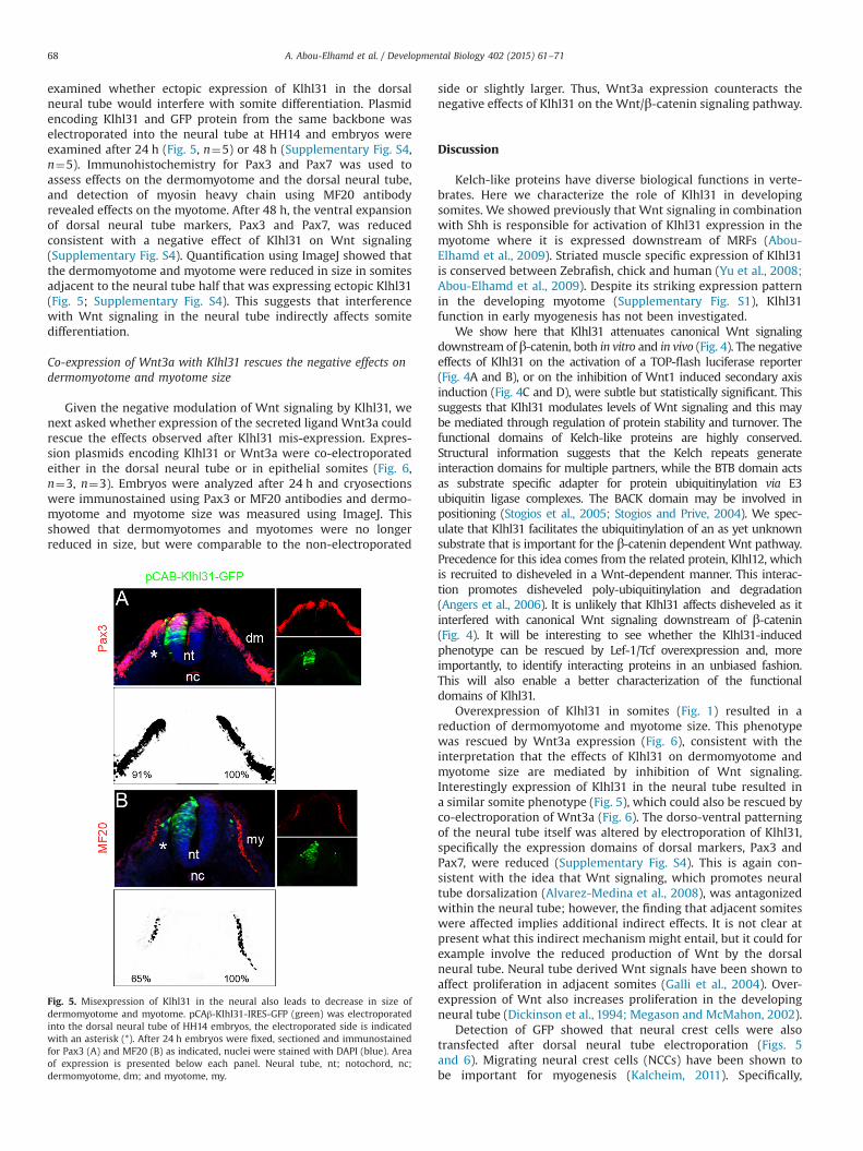
examined whether ectopic expression of Klhl31 in the dorsalneural tube would interfere with somite differentiation. Plasmidencoding Klhl31 and GFP protein from the same backbone waselectroporated into the neural tube at HH14 and embryos wereexamined after 24 h (Fig. 5, n¼5) or 48 h (Supplementary Fig. S4,n¼5). Immunohistochemistry for Pax3 and Pax7 was used toassess effects on the dermomyotome and the dorsal neural tube,and detection of myosin heavy chain using MF20 antibodyrevealed effects on the myotome. After 48 h, the ventral expansionof dorsal neural tube markers, Pax3 and Pax7, was reducedconsistent with a negative effect of Klhl31 on Wnt signaling(Supplementary Fig. S4). Quantification using ImageJ showed thatthe dermomyotome and myotome were reduced in size in somitesadjacent to the neural tube half that was expressing ectopic Klhl31(Fig. 5; Supplementary Fig. S4). This suggests that interferencewith Wnt signaling in the neural tube indirectly affects somitedifferentiation.
Co-expression of Wnt3a with Klhl31 rescues the negative effects ondermomyotome and myotome size
Given the negative modulation of Wnt signaling by Klhl31, wenext asked whether expression of the secreted ligand Wnt3a couldrescue the effects observed after Klhl31 mis-expression. Expres-sion plasmids encoding Klhl31 or Wnt3a were co-electroporatedeither in the dorsal neural tube or in epithelial somites (Fig. 6,n¼3, n¼3). Embryos were analyzed after 24 h and cryosectionswere immunostained using Pax3 or MF20 antibodies and dermo-myotome and myotome size was measured using ImageJ. Thisshowed that dermomyotomes and myotomes were no longerreduced in size, but were comparable to the non-electroporated
side or slightly larger. Thus, Wnt3a expression counteracts thenegative effects of Klhl31 on the Wnt/β-catenin signaling pathway.
Discussion
Kelch-like proteins have diverse biological functions in verte-brates. Here we characterize the role of Klhl31 in developingsomites. We showed previously that Wnt signaling in combinationwith Shh is responsible for activation of Klhl31 expression in themyotome where it is expressed downstream of MRFs (Abou-Elhamd et al., 2009). Striated muscle specific expression of Klhl31is conserved between Zebrafish, chick and human (Yu et al., 2008;Abou-Elhamd et al., 2009). Despite its striking expression patternin the developing myotome (Supplementary Fig. S1), Klhl31function in early myogenesis has not been investigated.
We show here that Klhl31 attenuates canonical Wnt signalingdownstream of β-catenin, both in vitro and in vivo (Fig. 4). The negativeeffects of Klhl31 on the activation of a TOP-flash luciferase reporter(Fig. 4A and B), or on the inhibition of Wnt1 induced secondary axisinduction (Fig. 4C and D), were subtle but statistically significant. Thissuggests that Klhl31 modulates levels of Wnt signaling and this maybe mediated through regulation of protein stability and turnover. Thefunctional domains of Kelch-like proteins are highly conserved.Structural information suggests that the Kelch repeats generateinteraction domains for multiple partners, while the BTB domain actsas substrate specific adapter for protein ubiquitinylation via E3ubiquitin ligase complexes. The BACK domain may be involved inpositioning (Stogios et al., 2005; Stogios and Prive, 2004). We spec-ulate that Klhl31 facilitates the ubiquitinylation of an as yet unknownsubstrate that is important for the β-catenin dependent Wnt pathway.Precedence for this idea comes from the related protein, Klhl12, whichis recruited to disheveled in a Wnt-dependent manner. This interac-tion promotes disheveled poly-ubiquitinylation and degradation(Angers et al., 2006). It is unlikely that Klhl31 affects disheveled as itinterfered with canonical Wnt signaling downstream of β-catenin(Fig. 4). It will be interesting to see whether the Klhl31-inducedphenotype can be rescued by Lef-1/Tcf overexpression and, moreimportantly, to identify interacting proteins in an unbiased fashion.This will also enable a better characterization of the functionaldomains of Klhl31.
Overexpression of Klhl31 in somites (Fig. 1) resulted in areduction of dermomyotome and myotome size. This phenotypewas rescued by Wnt3a expression (Fig. 6), consistent with theinterpretation that the effects of Klhl31 on dermomyotome andmyotome size are mediated by inhibition of Wnt signaling.Interestingly expression of Klhl31 in the neural tube resulted ina similar somite phenotype (Fig. 5), which could also be rescued byco-electroporation of Wnt3a (Fig. 6). The dorso-ventral patterningof the neural tube itself was altered by electroporation of Klhl31,specifically the expression domains of dorsal markers, Pax3 andPax7, were reduced (Supplementary Fig. S4). This is again con-sistent with the idea that Wnt signaling, which promotes neuraltube dorsalization (Alvarez-Medina et al., 2008), was antagonizedwithin the neural tube; however, the finding that adjacent somiteswere affected implies additional indirect effects. It is not clear atpresent what this indirect mechanism might entail, but it could forexample involve the reduced production of Wnt by the dorsalneural tube. Neural tube derived Wnt signals have been shown toaffect proliferation in adjacent somites (Galli et al., 2004). Over-expression of Wnt also increases proliferation in the developingneural tube (Dickinson et al., 1994; Megason and McMahon, 2002).
Detection of GFP showed that neural crest cells were alsotransfected after dorsal neural tube electroporation (Figs. 5and 6). Migrating neural crest cells (NCCs) have been shown tobe important for myogenesis (Kalcheim, 2011). Specifically,
Fig. 5. Misexpression of Klhl31 in the neural also leads to decrease in size ofdermomyotome and myotome. pCAβ-Klhl31-IRES-GFP (green) was electroporatedinto the dorsal neural tube of HH14 embryos, the electroporated side is indicatedwith an asterisk (*). After 24 h embryos were fixed, sectioned and immunostainedfor Pax3 (A) and MF20 (B) as indicated, nuclei were stained with DAPI (blue). Areaof expression is presented below each panel. Neural tube, nt; notochord, nc;dermomyotome, dm; and myotome, my.
A. Abou-Elhamd et al. / Developmental Biology 402 (2015) 61–7168

dorsally migrating NCC signal via delta-like1 (DLL1) to transientlyactivate Notch signaling in myogenic cells in the dermomyotomallip, whereas ventrally migrating NCCs express neuregulin1 (Nrg1),which activates the ErbB3 receptor in the central and hypaxialmyotome (Rios et al., 2011; Van Ho et al., 2011). These interactionsregulate the differentiation of Pax7 positive progenitors and thusthe eventual size of the myotome. Migrating NCCs are alsoimplicated in delivering Wnt protein to pattern somites at adistance (Serralbo and Marcelle, 2014). We show here that Wnt3atransfection could rescue the indirect effects on somites of Klhl31expression in the neural tube and NCCs (Fig. 6A, and B). Wespeculate therefore that in this scenario Klhl31 may affect theproduction or display of myogenic ligands such as Wnt3a, but alsopotentially DLL1 and/or neuregulin1.
Interestingly, filopodia-like protrusions, which invade the sub-ectodermal space, have recently been characterized on dermo-myotomal cells (Sagar et al., 2015). It is reasonable to assume thatfilopodia are similarly involved in contacting migrating NCCs toreceive myogenic stimuli. Our experiments suggest that Klhl31may affect the ability of dorsally migrating NCCs to promotesomite myogenesis (Fig. 5).
It was also recently reported that manipulation of BRE expres-sion (¼Brain-and-Reproductive-Expression) had indirect effects
on adjacent tissues. BRE is an adapter protein involved in stressresponses, DNA repair and maintenance of stemness. Both over-expression and silencing of BRE in the neural tube indirectlyaffected the size of adjacent somites (Wang et al., 2015) andevidence suggests that BRE activates BMP/Smad signaling and NCCmigration.
Klhl31 is first detected in the early myotome (Supplementary Fig.S1). However, we cannot exclude the possibility that Klhl31 isexpressed in myogenic progenitors within the dermomyotome, at alevel that is not detectable by in situ hybridization. Given theexpression pattern and the finding that Klhl31 is upregulated by MRFs(Abou-Elhamd et al., 2009), the main function of Klhl31 is likely to bein committed myoblasts. Its detailed mechanism of action withinmyoblasts remains to be determined, but we propose that Klhl31regulates the balance between myoblast proliferation and differentia-tion by attenuating Wnt signaling (Fig. 6E). Consistent with this, weshow that MO-mediated knock-down of Klhl31 led to a thickenedmyotome (Figs. 2 and 3). Interestingly, Klhl31 knock-down also led toincreased size of the dermomyotome and we measured a small butsignificant increase in the numbers of mitotic cells in the regionencompassing the dermomyotome and myotome (Fig. 3, 1.6 fold,Po0.01). This implies an indirect effect of Klhl31 knock-down in themyotome on the dermomyotome that is currently not understood, but
Fig. 6. Co-expression of Wnt3a with Klhl31 in neural tube and somites rescues the phenotype and restores dermomyotome and myotome size. pCAβ-Klhl31-IRES-GFP wasco-electroporated with pCIG-mWnt3a-IRES-GFP into the neural tube (A, B) and somites (C, D) of HH14 embryos, the electroporated side is indicated with an asterisk (*). Thenon-transfected neural tube half and contralateral somites serve as internal control. After 24 h embryos were fixed, sectioned and immunostained for Pax3 (A, C) and MF20(B, D) as indicated in red, nuclei were stained with DAPI (blue). Area of expression is presented below each panel. (E) The proposed role of Klhl31 after myogenic activation isthe attenuation of the response to Wnt/β-catenin signaling in myogenic cells. Neural tube, nt; notochord, nc; dermomyotome, dm; and myotome, my.
A. Abou-Elhamd et al. / Developmental Biology 402 (2015) 61–71 69

may involve mechanisms similar to those discussed above for theneural tube and NCCs. The overall effect of Klhl31 loss-of-function isenhanced proliferation and myogenesis. This involves direct mechan-isms within the myotome and indirect mechanisms on the dermo-myotome, both of which may include the de-repression of Wntsignaling. In order to determine the underlying molecular effectorsthat mediate these effects, it will be necessary to identify proteins thatinteract with Klhl31 in developing muscle.
Acknowledgments
We thank Paul Thomas for support in the Henry WellcomeLaboratory of Cell Imaging. AAE was funded by a PhD studentshipfrom the Republic of Egypt (GB0035), AFA was funded by a PhDstudentship from Saudi Arabia (U360), GFM was funded by aBBSRC project grant (K003437) to AM, CGM was funded by astudentship from CONACYT Mexico to GNW, MAE was supportedby a project grant from the MRC (G0600757) to AM.
Appendix A. Supporting information
Supplementary data associated with this article can be found inthe online version at http://dx.doi.org/10.1016/j.ydbio.2015.02.024.
References
Abou-Elhamd, A., Cooper, O., Munsterberg, A., 2009. Klhl31 is associated withskeletal myogenesis and its expression is regulated by myogenic signals andMyf-5. Mech. Dev. 126, 852–862.
Abu-Elmagd, M., Robson, L., Sweetman, D., Hadley, J., Francis-West, P., Munsterberg,A., 2010. Wnt/Lef1 signaling acts via Pitx2 to regulate somite myogenesis. Dev.Biol. 337, 211–219.
Alvarez-Medina, R., Cayuso, J., Okubo, T., Takada, S., Marti, E., 2008. Wnt canonicalpathway restricts graded Shh/Gli patterning activity through the regulation ofGli3 expression. Development 135, 237–247.
Anastas, J.N., Moon, R.T., 2013. WNT signaling pathways as therapeutic targets incancer. Nat. Rev. Cancer 13, 11–26.
Angers, S., Thorpe, C.J., Biechele, T.L., Goldenberg, S.J., Zheng, N., MacCoss, M.J.,Moon, R.T., 2006. The KLHL12-Cullin-3 ubiquitin ligase negatively regulates theWnt-beta-catenin pathway by targeting dishevelled for degradation. Nat. CellBiol. 8, 348–357.
Brand-Saberi, B., Wilting, J., Ebensperger, C., Christ, B., 1996. The formation ofsomite compartments in the avian embryo. Int. J. Dev. Biol. 40, 411–420.
Christ, B., Huang, R., Scaal, M., 2004. Formation and differentiation of the aviansclerotome. Anat. Embryol. 208, 333–350.
Crist, C.G., Montarras, D., Pallafacchina, G., Rocancourt, D., Cumano, A., Conway, S.J.,Buckingham, M., 2009. Muscle stem cell behavior is modified by microRNA-27regulation of Pax3 expression. Proc. Natl. Acad. Sci. USA 106, 13383–13387.
Cullinan, S.B., Gordan, J.D., Jin, J., Harper, J.W., Diehl, J.A., 2004. The Keap1-BTBprotein is an adapter that bridges Nrf2 to a Cul3-based E3 ligase: oxidativestress sensing by a Cul3-Keap1 ligase. Mol. Cell. Biol. 24, 8477–8486.
Dickinson, M.E., Krumlauf, R., McMahon, A.P., 1994. Evidence for a mitogenic effectof Wnt-1 in the developing mammalian central nervous system. Development120, 1453–1471.
Epstein, J.A., Lam, P., Jepeal, L., Maas, R.L., Shapiro, D.N., 1995. Pax3 inhibitsmyogenic differentiation of cultured myoblast cells. J. Biol. Chem. 270,11719–11722.
Funayama, N., Fagotto, F., McCrea, P., Gumbiner, B.M., 1995. Embryonic axisinduction by the armadillo repeat domain of beta-catenin: evidence forintracellular signaling. J. Cell Biol. 128, 959–968.
Furukawa, M., He, Y.J., Borchers, C., Xiong, Y., 2003. Targeting of protein ubiquitina-tion by BTB-Cullin 3-Roc1 ubiquitin ligases. Nat. Cell Biol. 5, 1001–1007.
Galli, L.M., Willert, K., Nusse, R., Yablonka-Reuveni, Z., Nohno, T., Denetclaw, W.,Burrus, L.W., 2004. A proliferative role for Wnt-3a in chick somites. Dev. Biol.269, 489–504.
Garcia-Morales, C., Liu, C.H., Abu-Elmagd, M., Hajihosseini, M.K., Wheeler, G.N.,2009. Frizzled-10 promotes sensory neuron development in Xenopus embryos.Dev. Biol. 335, 143–155.
Geyer, R., Wee, S., Anderson, S., Yates, J., Wolf, D.A., 2003. BTB/POZ domain proteinsare putative substrate adaptors for cullin 3 ubiquitin ligases. Mol. Cell 12,783–790.
Goljanek-Whysall, K., Sweetman, D., Abu-Elmagd, M., Chapnik, E., Dalmay, T.,Hornstein, E., Munsterberg, A., 2011. MicroRNA regulation of the paired-box
transcription factor Pax3 confers robustness to developmental timing ofmyogenesis. Proc. Natl. Acad. Sci. USA 108, 11936–11941.
Gray, C.H., McGarry, L.C., Spence, H.J., Riboldi-Tunnicliffe, A., Ozanne, B.W., 2009.Novel beta-propeller of the BTB-Kelch protein Krp1 provides a binding site forLasp-1 that is necessary for pseudopodial extension. J. Biol. Chem. 284,30498–30507.
Greenberg, C.C., Connelly, P.S., Daniels, M.P., Horowits, R., 2008. Krp1 (sarcosin)promotes lateral fusion of myofibril assembly intermediates in cultured mousecardiomyocytes. Exp. Cell Res. 314, 1177–1191.
Guger, K.A., Gumbiner, B.M., 1995. beta-Catenin has Wnt-like activity and mimicsthe Nieuwkoop signaling center in Xenopus dorsal–ventral patterning. Dev.Biol. 172, 115–125.
Gupta, A.V., Beggs, A.H., 2014. Kelch proteins: emerging roles in skeletal muscledevelopment and diseases. Skelet. Muscle 4.
Hamburger, V., Hamilton, H.L., 1992. A series of normal stages in the developmentof the chick embryo. 1951. Dev. Dyn. 195, 231–272.
Hara, T., Ishida, H., Raziuddin, R., Dorkhom, S., Kamijo, K., Miki, T., 2004. Novelkelch-like protein, KLEIP, is involved in actin assembly at cell–cell contact sitesof Madin–Darby canine kidney cells. Mol. Biol. Cell 15, 1172–1184.
Kalcheim, C., 2011. Regulation of trunk myogenesis by the neural crest: a new facetof neural crest-somite interactions. Dev. Cell 21, 187–188.
Kuhl, M., Pandur, P., 2008. Dorsal axis duplication as a functional readout for Wntactivity. Methods Mol. Biol. 469, 467–476.
Lozano-Velasco, E., Contreras, A., Crist, C., Hernandez-Torres, F., Franco, D., Aranega,A.E., 2011. Pitx2c modulates Pax3þ/Pax7þ cell populations and regulates Pax3expression by repressing miR27 expression during myogenesis. Dev. Biol. 357,165–178.
Maerki, S., Olma, M.H., Staubli, T., Steigemann, P., Gerlich, D.W., Quadroni, M.,Sumara, I., Peter, M., 2009. The Cul3-KLHL21 E3 ubiquitin ligase targets aurora Bto midzone microtubules in anaphase and is required for cytokinesis. J. Cell Biol.187, 791–800.
Maroto, M., Reshef, R., Munsterberg, A.E., Koester, S., Goulding, M., Lassar, A.B.,1997. Ectopic Pax-3 activates MyoD and Myf-5 expression in embryonicmesoderm and neural tissue. Cell 89, 139–148.
Megason, S.G., McMahon, A.P., 2002. A mitogen gradient of dorsal midline Wntsorganizes growth in the CNS. Development 129, 2087–2098.
Mennerich, D., Schafer, K., Braun, T., 1998. Pax-3 is necessary but not sufficient forlbx1 expression in myogenic precursor cells of the limb. Mech. Dev. 73,147–158.
Moon, R.T., Bowerman, B., Boutros, M., Perrimon, N., 2002. The promise and perilsof Wnt signaling through beta-catenin. Science 296, 1644–1646.
Perez-Torrado, R., Yamada, D., Defossez, P.A., 2006. Born to bind: the BTB protein–protein interaction domain. Bioessays 28, 1194–1202.
Pintard, L., Willis, J.H., Willems, A., Johnson, J.L., Srayko, M., Kurz, T., Glaser, S.,Mains, P.E., Tyers, M., Bowerman, B., Peter, M., 2003. The BTB protein MEL-26 isa substrate-specific adaptor of the CUL-3 ubiquitin-ligase. Nature 425, 311–316.
Prag, S., Adams, J.C., 2003. Molecular phylogeny of the kelch-repeat superfamilyreveals an expansion of BTB/kelch proteins in animals. BMC Bioinf. 4, 42.
Rios, A.C., Serralbo, O., Salgado, D., Marcelle, C., 2011. Neural crest regulatesmyogenesis through the transient activation of NOTCH. Nature 473, 532–535.
Rondou, P., Haegeman, G., Vanhoenacker, P., Van Craenenbroeck, K., 2008. BTBProtein KLHL12 targets the dopamine D4 receptor for ubiquitination by a Cul3-based E3 ligase. J. Biol. Chem. 283, 11083–11096.
Sagar, Felicitas Pröls, Christoph, Wiegreffe, Martin, Scaal, 2015. Communicationbetween distant epithelial cells by filopodia-like protrusions during embryonicdevelopment. Development 142, 665–671, posted ahead of print January 23,2015, http://dx.doi.org/10.1242/dev.115964.
Scaal, M., Christ, B., 2004. Formation and differentiation of the avian dermomyo-tome. Anat. Embryol. 208, 411–424.
Schmidt, M., Patterson, M., Farrell, E., Munsterberg, A., 2004. Dynamic expression ofLef/Tcf family members and beta-catenin during chick gastrulation, neurula-tion, and early limb development. Dev. Dyn. 229, 703–707.
Schmidt, M., Tanaka, M., Munsterberg, A., 2000. Expression of (beta)-catenin in thedeveloping chick myotome is regulated by myogenic signals. Development 127,4105–4113.
Serralbo, O., Marcelle, C., 2014. Migrating cells mediate long-range WNT signaling.Development 141, 2057–2063.
Stogios, P.J., Downs, G.S., Jauhal, J.J., Nandra, S.K., Prive, G.G., 2005. Sequence andstructural analysis of BTB domain proteins. Genome Biol. 6, R82.
Stogios, P.J., Prive, G.G., 2004. The BACK domain in BTB-kelch proteins. TrendsBiochem. Sci. 29, 634–637.
Sumara, I., Quadroni, M., Frei, C., Olma, M.H., Sumara, G., Ricci, R., Peter, M., 2007. ACul3-based E3 ligase removes Aurora B from mitotic chromosomes, regulatingmitotic progression and completion of cytokinesis in human cells. Dev. Cell 12,887–900.
Van Ho, A.T., Hayashi, S., Brohl, D., Aurade, F., Rattenbach, R., Relaix, F., 2011. Neuralcrest cell lineage restricts skeletal muscle progenitor cell differentiationthrough Neuregulin1-ErbB3 signaling. Dev. Cell 21, 273–287.
von Maltzahn, J., Bentzinger, C.F., Rudnicki, M.A., 2012a. Wnt7a-Fzd7 signallingdirectly activates the Akt/mTOR anabolic growth pathway in skeletal muscle.Nat. Cell Biol. 14, 186–191.
von Maltzahn, J., Chang, N.C., Bentzinger, C.F., Rudnicki, M.A., 2012b. Wnt signalingin myogenesis. Trends Cell Biol. 22, 602–609.
Wang, Guang, Li, Yan, Wang, Xiao-Yu, Chuai, Manli, Chan, John Yeuk-Hon, Lei, Jian,Münsterberg, Andrea, Lee, Kenneth Ka Ho, Yang, Xuesong, 2015. Misexpressionof BRE gene in the developing chick neural tube affects neurulation and
A. Abou-Elhamd et al. / Developmental Biology 402 (2015) 61–7170

somitogenesis. Mol. Biol. Cell 26, 5978–5992, First Published on January 7,2015; http://dx.doi.org/10.1091/mbc.E14-06-1144.
Xu, L., Wei, Y., Reboul, J., Vaglio, P., Shin, T.H., Vidal, M., Elledge, S.J., Harper, J.W.,2003. BTB proteins are substrate-specific adaptors in an SCF-like modularubiquitin ligase containing CUL-3. Nature 425, 316–321.
Yu, W., Li, Y., Zhou, X., Deng, Y., Wang, Z., Yuan, W., Zhao, X., Mo, X., Huang, W., Luo,N., Yan, Y., Ocorr, K., Bodmer, R., Wang, Y., Wu, X., 2008. A novel human BTB-
kelch protein KLHL31, strongly expressed in muscle and heart, inhibitstranscriptional activities of TRE and SRE. Mol. Cells 26, 443–453.
Yue, Q., Wagstaff, L., Yang, X., Weijer, C., Munsterberg, A., 2008. Wnt3a-mediatedchemorepulsion controls movement patterns of cardiac progenitors andrequires RhoA function. Development 135, 1029–1037.
A. Abou-Elhamd et al. / Developmental Biology 402 (2015) 61–71 71
Copyright © 2022 FDOKUMEN