WNT/ΒETA-CATENIN SIGNALING PATHWAY IN HUMAN CANCER AND IN-VITRO CYTOTOXIC ACTIVITY OF TRADESCANTIA...
65
WNT/ΒETA-CATENIN SIGNALING PATHWAY IN HUMAN CANCER AND IN-VITRO CYTOTOXIC ACTIVITY OF TRADESCANTIA SPATHACEA IN A549, PC-3 AND HEPG-2 CELL LINES A PROJECT REPORT Submitted by Mr. R. Prakash (Reg.No.4811210007) In Partial fulfillment of the requirement for the award of the degree Of MASTER OF TECHNOLOGY In BIOTECHNOLOGY VINAYAKA MISSION’S KIRUPANANDA VARIYAR ENGINEERING COLLEGE VINAYAKA MISSION’S UNIVERSITY, SALEM JUNE 2014
-
Upload
vinayakamission -
Category
Documents
-
view
0 -
download
0
Transcript of WNT/ΒETA-CATENIN SIGNALING PATHWAY IN HUMAN CANCER AND IN-VITRO CYTOTOXIC ACTIVITY OF TRADESCANTIA...
WNT/ΒETA-CATENIN SIGNALING PATHWAY IN
HUMAN CANCER AND IN-VITRO CYTOTOXIC
ACTIVITY OF TRADESCANTIA SPATHACEA IN A549,
PC-3 AND HEPG-2 CELL LINES
A PROJECT REPORT
Submitted by
Mr. R. Prakash
(Reg.No.4811210007)
In Partial fulfillment of the requirement for the award of the degree
Of
MASTER OF TECHNOLOGY
In
BIOTECHNOLOGY
VINAYAKA MISSION’S KIRUPANANDA VARIYAR ENGINEERING
COLLEGE
VINAYAKA MISSION’S UNIVERSITY, SALEM
JUNE 2014
JUNE 2014
VINAYAKA MISSIONS UNIVERSITY
VMKV ENGINEERING COLLEGE
DEPARTMENT OF BIOTECHNOLOGY
SALEM 636 308
Dr. C. K. Hindumathi, M.Sc.,M.Phil., Ph.D.,
Dean, Department of Bioscience
BONAFIDE CERTIFICATE
This is to certify that project report titled “WNT/Beta-catenin Signaling Pathway
in Human Cancer and In-vitro Cytotoxic Activity of Tradescantia spathacea in
A549, PC-3 and HePG-2 Cell Lines” is the bonafide work of Mr. R. Prakash
(Reg.No.4811210007) who carried out the project work during the period of 15th
Jan 2014 to 31st May 2014 in the partial fulfillment of the requirements of the M.
Tech. degree course in Biotechnology. This is to certify that this dissertation has
not formed the basis for the award of any other degree, diploma, Fellowship or
other similar title and that the dissertation represents independent work on the part
of the candidate under my guidance.
Signature of the HOD Signature of the Guide
Internal examiner External examiner
DECLARATION
I hereby declare that the project work, entitled “WNT/Beta-catenin Signaling
Pathway in Human Cancer and In-vitro Cytotoxic Activity of Tradescantia
spathacea in A549, PC-3 and HePG-2 Cell Lines” Submitted to Vinayaka Missions
University, Salem. In partial fulfillment of the requirement for the award of the
degree of Master of technology in Biotechnology is a record of original research work
carried out under the guidance of Mrs. V. K. Evanjelene, M.Sc., M.B.A., PGDBI.,
(Ph.D)., Research incharge, Alpha Omega Hi-tech Bio Research Center, Salem-636
008 and Dr. C. K. Hindumathi, M.Sc., M.Phil., Ph.D., PDF., Dean, Department
of Bioscience at Vinayaka Missions University, Salem.
I further declare that this work has not been submitted previously for the
award of any degree, diploma or any other similar title.
Place: Salem Prakash. R
Date:
ACKNOWLEDGEMENT
I express my grateful acknowledge and indebtedness to our management,
Vinayaka Mission’s University, Salem. And other member of management for
their contribution to complete the project work successfully.
I express my sincere thanks to Dr. A. Nagappan, B.E., M.S., Ph.D.,
Principal, Vinayaka Mission’s Kirupananda Variyar Engineering College, Salem.
For giving me this opportunity and encouragement.
I express my sincere thanks to my guide Dr. C. K. Hindumathi, M.Sc.,
M.Phil., Ph.D., PDF., Dean- Bioscience at Vinayaka Missions Kirupananda Variyar
Engineering College , Salem. I am greatly indebted to him for the dexterous and
assiduous guidance at all stages of this work and for the successful completion of the
project work.
I express my sincere thanks to Dr. S. Anandakumar, M.Sc., M.Phil., Ph.D.,
FSAB., Associate professor, Department of Biotechnology, Vinayaka Mission’s
Kirupananda Variyar Engineering College, Salem. For giving me this opportunity and
encouragement.

I express my deep gratitude to all staff members of Vinayaka Mission’s
Kirupananda Variyar Engineering College, Salem for their effective help and
guidance.
I would like to thank my parents and all other family members who helped and
supported me for this project work.
Thanking You,
PRAKASH. R
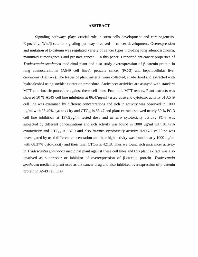
ABSTRACT
Signaling pathways plays crucial role in stem cells development and carcinogenesis.
Especially, Wnt/β-catenin signaling pathway involved in cancer development. Overexpression
and mutation of β-catenin was regulated variety of cancer types including lung adenocarcinoma,
mammary tumorigenesis and prostate cancer. . In this paper, I reported anticancer properties of
Tradescantia spathacea medicinal plant and also study overexpression of β-catenin protein in
lung adenocarcinoma (A549 cell lines), prostate cancer (PC-3) and hepatocellular liver
carcinoma (HePG-2). The leaves of plant material were collected, shade dried and extracted with
hydroalcohol using soxhlet extraction procedure. Anticancer activities are assayed with standard
MTT colorimetric procedure against these cell lines. From this MTT results, Plant extracts was
showed 50 % A549 cell line inhibition at 86.47µg/ml tested dose and cytotoxic activity of A549
cell line was examined by different concentrations and rich in activity was observed in 1000
µg/ml with 95.49% cytotoxicity and CTC50 is 86.47 and plant extracts showed nearly 50 % PC-3
cell line inhibition at 137.9µg/ml tested dose and in-vitro cytotoxicity activity PC-3 was
subjected by different concentrations and rich activity was found in 1000 µg/ml with 81.47%
cytotoxicity and CTC50 is 137.9 and also In-vitro cytotoxicity activity HePG-2 cell line was
investigated by used different concentration and their high activity was found nearly 1000 µg/ml
with 68.37% cytotoxicity and their final CTC50 is 421.8. Thus we found rich anticancer activity
in Tradescantia spathacea medicinal plant against these cell lines and this plant extract was also
involved as suppressor or inhibitor of overexpression of β-catenin protein. Tradescantia
spathacea medicinal plant used as anticancer drug and also inhibited overexpression of β-catenin
protein in A549 cell lines.
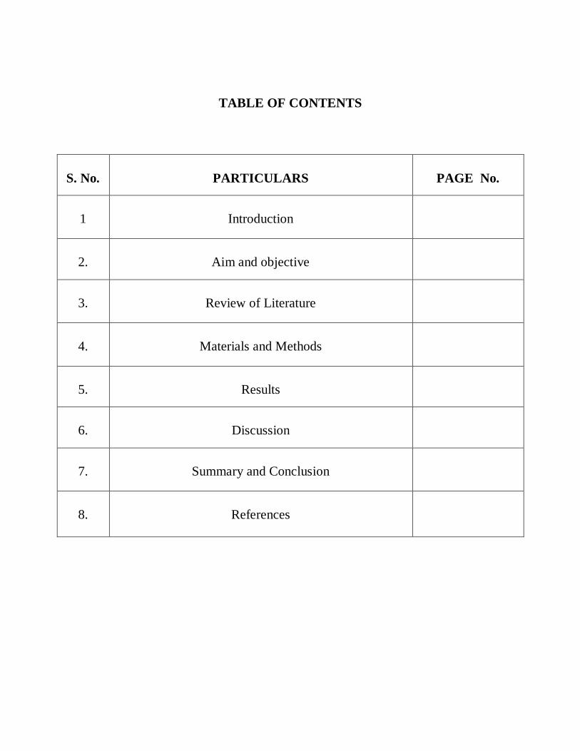
TABLE OF CONTENTS
S. No.
PARTICULARS
PAGE No.
1
Introduction
2.
Aim and objective
3.
Review of Literature
4.
Materials and Methods
5.
Results
6.
Discussion
7.
Summary and Conclusion
8.
References

INTRODUCTION
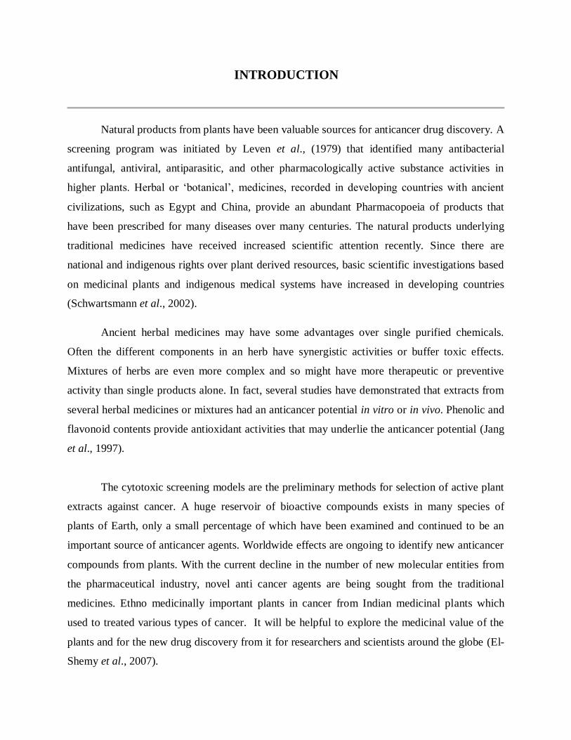
INTRODUCTION
Natural products from plants have been valuable sources for anticancer drug discovery. A
screening program was initiated by Leven et al., (1979) that identified many antibacterial
antifungal, antiviral, antiparasitic, and other pharmacologically active substance activities in
higher plants. Herbal or ‘botanical’, medicines, recorded in developing countries with ancient
civilizations, such as Egypt and China, provide an abundant Pharmacopoeia of products that
have been prescribed for many diseases over many centuries. The natural products underlying
traditional medicines have received increased scientific attention recently. Since there are
national and indigenous rights over plant derived resources, basic scientific investigations based
on medicinal plants and indigenous medical systems have increased in developing countries
(Schwartsmann et al., 2002).
Ancient herbal medicines may have some advantages over single purified chemicals.
Often the different components in an herb have synergistic activities or buffer toxic effects.
Mixtures of herbs are even more complex and so might have more therapeutic or preventive
activity than single products alone. In fact, several studies have demonstrated that extracts from
several herbal medicines or mixtures had an anticancer potential in vitro or in vivo. Phenolic and
flavonoid contents provide antioxidant activities that may underlie the anticancer potential (Jang
et al., 1997).
The cytotoxic screening models are the preliminary methods for selection of active plant
extracts against cancer. A huge reservoir of bioactive compounds exists in many species of
plants of Earth, only a small percentage of which have been examined and continued to be an
important source of anticancer agents. Worldwide effects are ongoing to identify new anticancer
compounds from plants. With the current decline in the number of new molecular entities from
the pharmaceutical industry, novel anti cancer agents are being sought from the traditional
medicines. Ethno medicinally important plants in cancer from Indian medicinal plants which
used to treated various types of cancer. It will be helpful to explore the medicinal value of the
plants and for the new drug discovery from it for researchers and scientists around the globe (El-
Shemy et al., 2007).
Cancer is an abnormal types of tissue growth in which the cells exhibit an uncontrolled
division, relatively in an autonomous fashion, leading to a progressive increase in the number of
dividing cell. The limited success of clinical therapies including radiation, chemotherapy,
immune-modulation and surgery in treating cancer, as evident by the high morbidity and
mortality rates, indicates that there is an imperative need of new cancer management.
Chemoprevention involves the use of pharmacological, dietary bio-factors, phytochemicals and
even whole plant extracts to prevent, arrest, or reverse the cellular and molecular processes of
carcinogenesis due to its multiple intervention strategies (Mehta et al., 2010).
Plants used as remedies and also botanical literature described the application of plant
extracts. Cancer is awful disease and combating this disease is great importance to public health.
Hence, lot of necessity for find new compounds with cytotoxic activity. Treatment of cancer with
the present anticancer drugs was often unsatisfactory, because the problem of cytotoxicity to the
normal cells. Phytochemical investigation has been making rapid progress and herbal products
crucial as sources of credible anticancer agents (Schwartsmann et al., 2002).
For several decades, People used plants for their therapeutic values. Today, nearly 85000
plants have been authenticated for their therapeutic use globally. The World Health Organization
(WHO) estimated that almost 75% of world’s populations have remedial experience with herbal
drugs. In worldwide, cancer is third leading cause of death, only preceded by many other
diseases and disorders including cardiovascular disease, infectious and parasitic disease (Jang et
al., 1997).
Cancer, most dangerous diseases in humans and so scientists were found new anticancer
agents from natural products. The potential use of the natural products as anticancer drugs were
recognized in 1950’s by U.S Natural Cancer Institute (NCI) Since 1950, lot of contributions have
taken for the discovered naturally occurring anticancer drugs (Vickers et al., 2002).
Despite improved imaging and molecular diagnostic techniques, cancer extends to
affected millions of people globally. An efficient molecule, cancer was treated inevitable and
explorations to develop new entities are going on. However, Nature was shown an excellent and
reliable source of new drugs. Plants plays crucial role in anticancer drugs and the metabolism of
interaction between phytochemicals and cancer cells has been studied (Shemy et al., 2007).
Medicinal plants have been used as remedies and botanical literature has described the
usage of plant leaves extracts. Cancer is a awful disease and combating this disease is of great
impact to public health. So, necessities to search of new compounds with cytotoxic activity as
the treatment of cancer with the available anticancer drugs were often unsatisfactory due to the
problem cytotoxicity to the normal cells. Phytochemical examination has been making rapid
progress and herbal products are becoming popular as sources of credible anticancer compounds
(Parag et al. 2010).
Carcinoma of the prostate, breast, colon and lung were highly prevalented malignancies
in the Western nations and accounts for approximately half of the total cancer-related deaths
among men and women (Jemal et al. 2005). The limited success of clinical therapies including
chemotherapy, radiation, immune modulation and surgery in treating cancer, high morbidity and
mortality rates indicates imperative need of new cancer management. Chemoprevention involved
the use of pharmacological, dietary bio-factors, phytochemicals and even whole plant extracts to
prevented, arrested, or reversed the cellular and molecular processes of carcinogenesis due to its
multiple intervention strategies (Mehta et al. 2010).
Phytochemical activity
The phytochemical is a natural bioactive compound found in plants, such as vegetables,
fruits, medicinal plants, flowers, leaves and roots that work with nutrients and fibers to act as an
defense system against disease or more accurately, to protect against disease. Phytochemicals are
divided into two groups, which are primary and secondary constituents; according to their
functions in plant metabolism. Primary constituents comprise common sugars, amino acids,
proteins and chlorophyll while secondary constituents consists of alkaloids, terpenoids and
phenolic compounds and many more such as flavonoids and tannins etc.( Krishnaiah et al.,
2007).
Phytochemicals are non-nutritive plant chemicals that have protective or disease
preventive properties. They are nonessential nutrients, meaning that they are not required by the
human body for sustaining life. It is well-known that plant produces these chemicals to protect
them but recent research demonstrates that they can also protect human beings against diseases
(Cragg et al., 2005). Medicinal activities of plants have long been associated with the production
of secondary metabolites which include tannins, terpenoids, coumarins, alkaloids and flavonoids.
These products help plant to carry out various activities like defense and pollination.
Medicinal plants containing active chemical constituents with high antioxidant property
play an important role in the prevention of various degenerative diseases (Lukmanul et al., 2008)
and have possible benefits to the humanity. A large number of phytochemicals belonging to
several chemical classes have been shown to have inhibitory effects on all types of
microorganisms in vitro. Botanical medicines or phyto medicines refer to the use of seeds,
berries, leaves, bark, root or flowers of any plant for medicinal purposes by significant number of
people. Knowledge of the chemical constituents of plants is desirable because such information
will be value for synthesis of complex chemical substances (Mojab et al., 2003).
Antioxidant activity
Oxidation and the production of free radicals are an integral part of human metabolism.
Oxygen free radicals or more generally reactive oxygen species (ROS), as well as reactive
nitrogen species (RNS) are products of normal cellular metabolism. ROS and RNS are well
recognized for playing a dual role as both deleterious and beneficial species, since they can be
either harmful or beneficial to living system. Reactive oxygen and nitrogen species can attack
various substrates in the body including lipids, nucleic acids and protein. Oxidation of any of
these substrates can theoretically contribute chronic diseases such as cancer, cardiovascular
diseases and age related macular degeneration and to aging (Rice-Evans et al., 1997).
Many medicinal plants contain large amounts of antioxidants such as polyphenols, which
can play an important role in adsorbing and neutralizing free radicals, quenching singlet and
triplet oxygen, or decomposing peroxides. Many of these phytochemicals possess significant
antioxidant capacities that are associated with lower occurrence and lower mortality rates of
several human diseases. It has been reported that there is an inverse relationship between the
antioxidative status occurrences of human diseases. In addition, antioxidant compounds which
are responsible for such antioxidants activity could be isolated and then used as antioxidants for
the prevention and treatment of free radical-related disorders

Anticancer activity in Medicinal plants:
The medicinal plants, besides having natural therapeutic values against various diseases,
also provide high quality of food and raw materials for livelihood. Considerable works have been
done on these plants to treat cancer, and some plant products have been marketed as anticancer
drugs, based on the traditional uses and scientific reports. These plants may promote host
resistance against infection by re-stabilizing body equilibrium and conditioning the body tissues.
Several reports describe that the anticancer activity of medicinal plants is due to the presence of
antioxidants in them. In fact, the medicinal plants are easily available, cheaper and possess no
toxicity as compared to the modern (allopathic) drugs.
The National Cancer Institute collected about 35,000 plant samples from 20 countries and
has screened around 114,000 extracts for anticancer activity (Mohammad, 2006). Over 3000
species of plants with antitumour properties have been reported (Hartwell, 1982). Cancer is one
of the most prominent diseases in humans and currently there is considerable scientific and
commercial interest in the continuing discovery of new anticancer agents from natural product
sources (Kinghorn et al., 2003).
Chemoprevention is recognized as an important approach to control malignancy and
recent studies have focused on the search for desirable chemo-preventive agents. Natural
products, particularly dietary substances, have played an important role in creating new chemo-
preventive agents (Surh, 2003).
Medicinal Plant Tradescantia spathacea
Tradescantia spathacea, or Moses-in-the-Cradle, is a herb native to Mexico with fleshy
rhizomes. It has rosettes of waxy lance-shaped leaves. Leaves are dark to metallic green above,
with glossy purple underneath. These will reach up to 1 foot (30 cm) long by 3 inches (7.5 cm)
wide. They are very attractive foliage plants that will reach 1 foot (30 cm) tall. They are hardy
in USDA zones 9-12. It is invasive exotic to South Florida. The current research was based on
the In-vitro cytotoxicity activity in Tradescantia spathacea. This was first report for the plant
and no systematic work has been undergone in this plant.
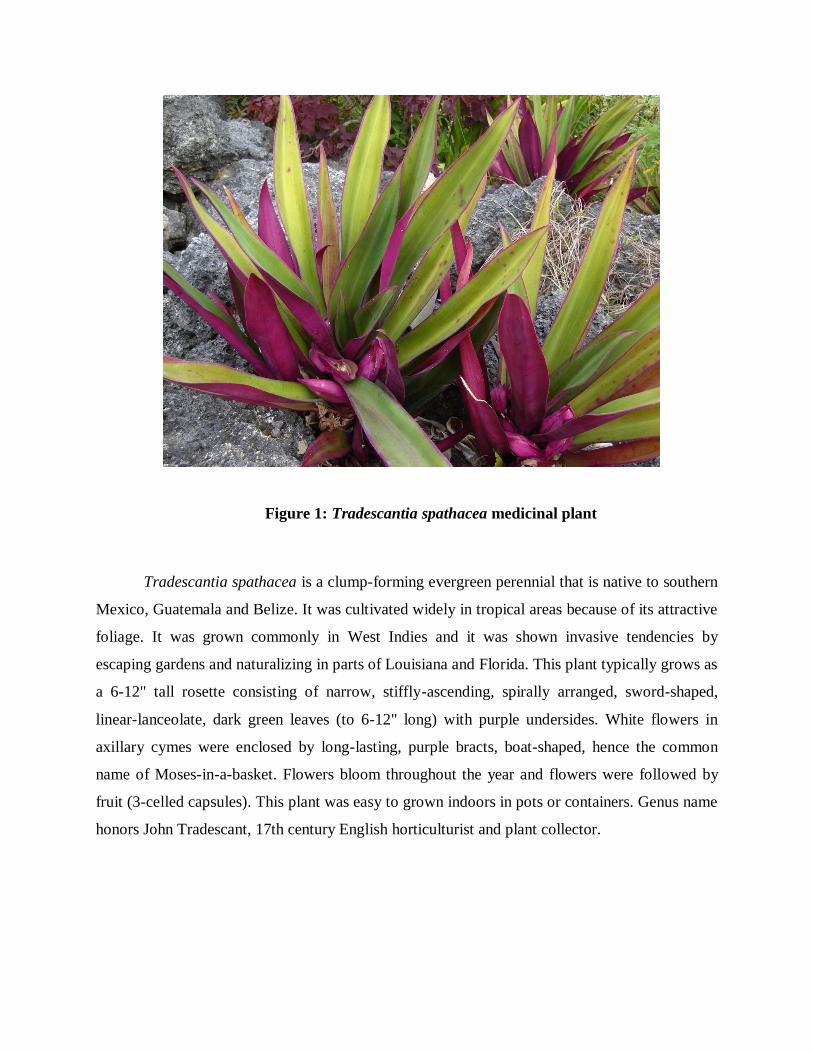
Figure 1: Tradescantia spathacea medicinal plant
Tradescantia spathacea is a clump-forming evergreen perennial that is native to southern
Mexico, Guatemala and Belize. It was cultivated widely in tropical areas because of its attractive
foliage. It was grown commonly in West Indies and it was shown invasive tendencies by
escaping gardens and naturalizing in parts of Louisiana and Florida. This plant typically grows as
a 6-12" tall rosette consisting of narrow, stiffly-ascending, spirally arranged, sword-shaped,
linear-lanceolate, dark green leaves (to 6-12" long) with purple undersides. White flowers in
axillary cymes were enclosed by long-lasting, purple bracts, boat-shaped, hence the common
name of Moses-in-a-basket. Flowers bloom throughout the year and flowers were followed by
fruit (3-celled capsules). This plant was easy to grown indoors in pots or containers. Genus name
honors John Tradescant, 17th century English horticulturist and plant collector.

AIM AND OBJECTIVE
AIM AND OBJECTIVE
To perform qualitative analysis of phytochemical and in-vitro antioxidant test in leaf
extracts of Tradescantia spathacea.
To study the in-vitro anti-cancer properties of Tradescantia spathacea extracts by MTT
cell line such as PC-3, HePG-2, MCF-7 and A549.
To study about overexpression of beta-catenin in PC-3, HePG-2, MCF-7 and A549
cancer cell lines.
To inhibit or suppress the overexpression of beta-catenin using Tradescantia spathacea
medicinal leaves extract as a drug.

REVIEW OF LITERATURE
REVIEW OF LITERATURE
Phytochemical activity
The phytochemicals could be available as dietary supplements, but the potential health
benefits of phytochemicals are derived from consumption of the whole plant. Chemical tests
were carried out on the aqueous extract and on the powdered specimens using standard
procedures to identify the constituents (Rao and Rao, 2007).
The phytochemistry deals with the analysis of plant chemicals called natural products,
and with changes occurring in such chemicals due to alterations in environmental conditions.
These compounds are involved as well in allelopathy, dealing with the interactions between two
plants, which process can change depending upon variations in the phytochemicals produced
under particular environmental conditions (Zobel et al., 1999).
The use of plant extracts and phytochemicals, both with known antimicrobial properties,
can be of great significance in therapeutic treatments. In the last few years, a number of studies
have been conducted in different countries to prove such efficiency. Many plants have been used
because of their antimicrobial traits, which are chiefly due to synthesized during secondary
metabolism of the plant (Prusti, 2008).
The phytochemical of the crude extract of C. alata revealed the presence of carbohydrate
flavonoids and saponins and all extracts tannins, steriols, alkaloids and anthraquinones were not
detected in the hexane extract. The test also revealed that the methanol extract have higher
contents of the phytochemical. Results for the quantitative analysis carried out on the sample of
Bauhinia tomentosa L. flower revealed that Bauhinia tomentosa L. has flavonoid percentage
content of 15.80%, alkaloids percentage content of 5.61% and saponins percentage content of
2.1% (Salihu et al., 2012).
Anti-oxidant and Anti-cancer activity
The reactive oxygen species (ROS) such as •O2 (superoxide anion), H2O2 (hydrogen
peroxide), and •OH (hydroxyl radical) are closely involved in various human diseases such as
Alzheimer's disease, aging, cancer, inflammation, rheumatoid arthritis and atherosclerosis. For
several years, many researchers have been searching for powerful but non-toxic antioxidants
from natural sources, especially edible or medicinal plants. Such natural antioxidants could
prevent the formation of the above reactive species-related disorders in human beings without
the use of synthetic compounds, which may be carcinogenic and harmful to the lungs and liver.
The antioxidants play an important role in nutritional by lengthening the shelf life of food
and reducing nutritional losses and formation of harmful substances. However, the safety of
synthetic antioxidants, such as butylated hydroxyanisole (BHA) and butylated hydroxytoluene
(BHT) are now in doubted. Thus, attention is now increasingly paid to the development and
utilization of more effective and non-toxic antioxidants of natural origin.
A great number of natural medicinal plants have been tested for their antioxidant
activities and results have shown that the raw extracts or isolated pure compounds from them
were more effective antioxidants in vitro than BHT or vitamin E, so, medicinal plants can be a
potential source of natural antioxidants. The high content of antioxidant polyphenolic
compounds, such as catechin, ingested in the human diet represents an important source of non-
nutritional antioxidants (Squadrito and Pryor, 1998).
The polyphenols exert protective effects against the development of cardiovascular
diseases. One of the plants containing polyphenols is Salvia which is one of the wide-spread
members of the Labiatae (Lamiaceae) family. The Labiatae comprise about 900 herbs and
shrubs, growing in the temperate and warmer zones of the world. Some of these species feature
prominently in the pharmacopoeias of many countries throughout the world.
The supernatant was separated and mixed with distilled water (2.5 mL) containing 1%
ferric chloride (0.5 mL). The absorbance of this mixture was measured at 700 nm. The intensity
in absorbance could be the measurement of antioxidant activity of the extract. Finally, it seems
that other types of natural antioxidants are present in the active sub-fractions as well as phenol
compounds (Pyo et al., 2004).
For centuries, plants have been used in traditional medicines for the treatment of different
diseases. In recent years, oriental medicinal herbs have aroused scientific interest as
complementary or alternative medicines. Several chemotherapeutic drugs derived from plants,
such as Vinblastine, Taxol, Camptothecin and Podophyllotoxin are used in medical tumor
management. Using modern analytical and chemical techniques, novel natural compounds from
herbs can be isolated by fractionation and isolation. It has been estimated that only 5-15% of
250,000 species of higher plants have been screened systematically for natural bioactive
compounds. To study new therapeutic approaches, cell lines are used to investigate novel
compounds and their effects on the tumor cells (Svejda, 2010).
WNT/BETA-CATENIN SIGNALING IN VARIETY OF TUMORS
Wnt/β-catenin signaling in Cancer and Cancer Stem Cells
As a central pathway of both development and cancer, Wnt signaling pathway regulated
self-renewal, proliferation and maintained both of normal and cancer stem cells. Mutation and
over expression of β-catenin leads many cancers including breast, lung, prostate, colorectal, liver
and skin cancer (Polakis, 2000).
Breast cancer and cancer stem cells
Aberrant expression and mutation of β-catenin was induced breast carcinogenesis. In
mammary tumorigenesis, both of mouse and human breast cancer is critical. Wnt signaling was
first discovered in mammary tumor when mouse mammary tumor was identified and integrated
into int-1 locus. Overexpression of Wnt1 was induced breast tumor via Wnt/β-catenin signaling.
Despite, the strong evidence of Wnt/β-catenin in mouse mammary tumor model, it was very
importance of Wnt signal in human breast cancer. Enormous reports have identified deregulation
of Wnt/β-catenin signaling pathway in breast cancer. Mutation and aberrant expression of β-
catenin was associated with triple-negative and basal breast cancer (Valkenburg, 2011).
β-catenin was first identified as cell adhesion molecules and further distinguished as
proto-ongogene. In Drosophila, it was identified as homologous protein called as armadillo. β-
catenin protein involved both of transcriptional activation and cell-cell adhesion. A Wnt gene
(Wnt1) was first discovered in mouse mammary tumor (Giles et al., 2003; Chen et al., 2008).
Wnt signaling, β-catenin plays crucial impact in stem cells development and carcinogenesis.
Overexpression and mutation of β-catenin associated many cancer types including malignant
breast cancer, prostate cancer, lung cancer, ovarian and liver cancer. Aberrant expression and
mutation of β-catenin stimulated malignant breast tumors via Wnt/β-catenin signaling pathway
(Behari, 2010).
Colorectal cancer
Wnt/β-catenin signaling was first linked to human colon cancer by the observation of
APC mutation. Aberrations of Wnt signaling have been identified 90% in colon cancer. The
absence of APC protein leads the chronic activation of Wnt signaling, resulting in the secretion
of adenomas, known as adenocarcinoma. Genetic observation in APC mutant, clearly
demonstrate the role of APC mutant in formation of tumor via Wnt signaling pathway. This
mutant APC allows β-catenin to accumulate in cytosol and continuously active Wnt/β-catenin to
form colon cancer. Although β-catenin and APC mutation leads colonic carcinogenesis and
downregulation of other tumor suppressor gene also involved in development of colon cancer
(Chen et al., 2008).
Prostate Cancer
Since 1990s, β-catenin protein was first discovered as a cell adhesion complex and one of
the examples of moonlighting protein. β-catenin protein Involved both of cell-cell adhesion and
gene transcription. In Drosophila, homologous protein was called armadillo (Noordermeer et al.
1994). Wnt signaling pathway, β-catenin regulated intracellular signal and gene transcription.
Mutations and overexpression of β-catenin were implicated many variety of cancer, including
prostate cancer, malignant breast tumors, lung cancer, liver cancer and ovarian cancers. Prostate
cancer, aberrant expression of Wnt proteins stimulated enormous β-catenin lead to cell
aggressive and metastasis. Overexpression and mutation in β-catenin bind to androgen receptors
and activated transcriptional activity to produced enormous cell fate in prostate cancer. β-catenin
also regulated many factors involved in prostate tumorigenesis (Lee et al. 2013).
Prostate cancer is most common cancer for American males. Wnt signals are up-
regulated in prostate cancer. Increased expression level of Wnt1, Wnt5a, Wnt7a, Wnt11 involved
in prostate cancer aggressive and metastasis. In prostate cells, androgen receptor were controlled
the prostate tumor growth. Androgen receptors bind to the β-catenin and stimulated
transcriptional activity. In prostate cancer, overexpression and mutation of β-catenin similarly
bind to the androgen receptors and activated transcriptional activity then produce enormous cell
fate. β-catenin activity is also regulated by other molecules in prostate cancer.
Liver Cancer
In liver, Wnt signaling play crucial role in proliferation during development and also
important function in adult liver. Despite, aberrant reactivation of Wnt/ β-catenin signaling was
stimulated the enormous accumulation of β-catenin in cytosol lead to many different tumors of
liver. Mutation of Axin and β-catenin continuously induced the activation of β-catenin in
hepatocellular carcinoma and hepatoblastomas. Simultaneous mutation of β-catenin and H-ras
leads to 100% of hepatocellular carcinoma. Finally, abnormal regulation of Wnt signaling
pathways have crucial role in the progression of hepatocellular carcinoma (Chen et al., 2008;
Behari, 2010).
In development, Wnt/β-catenin signaling pathway plays crucial impact in stem cells
development and carcinogenesis. Drosophila was significant model for cancer treatment and
homologous protein also called as armadillo. In normal signaling condition, β-catenin protein
was involved in cell-cell adhesion and gene transcription. Wnt/β-catenin signaling also play
important role in adult liver. Despite, overexpression and mutation of β-catenin regulated many
variety of cancer including hepatocellular carcinoma, breast tumors, prostate cancer, lung
adenocarcinoma and ovarian cancer. Mutation of β-catenin highly expressed in hepatocellular
carcinoma and hepatoblastomas. Further, mutations of H-ras and β-catenin caused 100% of
human hepatocellular liver carcinoma (Lai et al., 2011).
Skin Cancer
Wnt signals involved in hair morphogenesis and mutation of β-catenin regulated
abnormal hair follicle morphogenesis. Tumor initiation depends on overexpression and mutation
of β-catenin signaling. Pilomatricoma one of the skin cancer caused by mutation of β-catenin.
Nuclear β-catenin and overexpression level of Axin was also involved in skin cancer. Aberrant
expression of β-catenin was reported in melanoma and non-melanoma skin cancer. Activated β-
catenin or upregulation of Wnt/β-catenin signaling was induced transcriptional activation of
target genes c-myc and c-jun. These gene and Wnt factors including Wnt3, Wnt4 and Wnt10b
were found in skin carcinogenesis (Bhatia and Spiegelman, 2005).
Lung adenocarcinoma cancer
Wnt/β-catenin signaling involved in stem cells and cancer development. As intracellular signal
transducer of β-catenin protein in Wnt signaling, regulated cell-cell adhesion, gene transcription
and it was also proto-ongogene. β-catenin also called as homologous protein of armadillo in
Drosophila (Noordermeer et al. 1994). Aberrant expression of Wnt signaling and also
overexpression and mutation of β-catenin associated many cancers such as lung cancer, prostate
cancer, malignant breast tumors, ovarian and liver cancer. In lung, most of cancers were
stimulated by deregulation of Wnt/β-catenin signaling and aberrant expression of β-catenin
stimulated enormous cell fate in lung development. Wnt signaling protein β-catenin expressed
enormously in lung adenocarcinoma (Nozawa et al., 2006). β-catenin protein was also highly
expressed in non-small cell lung cancer
Other types of cancer
The mutant of β-catenin gene (CTNNB1) involved in many tumor. β-catenin and APC
mutation involved in multiple myeloma and Wnt5a also expressed in human melanoma. In
female and male organs, abnormality Wnt signaling stimulated ovarian carcinomas. Other many
types of cancer stimulated by aberrant expression and mutation of β-catenin such as, lung cancer,
endometrial cancer, glioblastoma, medulloblastoma, basal cell carcinoma, head and neck
squamous cell carcinoma, bladder cancer, gastric cancer, oral cancer, esophageal cancer,
retinoblastoma, pancreatic cancer and renal cell carcinoma (Giles et al., 2003).

MATERIALS AND METHODS
MATERIALS AND METHODS
1. Collection of Plant Materials
The leaves of Tradescantia spathacea Collected and the specimen were deposited in the
Alpha Omega Hi- Tech Bio research centre. The fresh leaves Tradescantia spathacea
were authenticated by ABS Botanical conservation, Research & Training Centre, Salem (Dt).
2. Preparation of Extract
The plant materials were washed using running tap water and shade dried. The leaves
were crushed to coarsely powdered by grinder. These coarse powders (5g) were then subjected
to successive extraction in 250ml of each solvent (hexane, ethyl acetate and methanol) by using
Soxhlet apparatus. The collected plant leaf extracts were stored and taken up for further research.
The DMSO (Dimethyl sufloxide) is act as dissolved solvents in these extracts.
3. Phytochemical screening
Preliminary phytochemical analysis was carried out for the extracts of Tradescantia
spathacea as per standard methods described by Brain and Turner 1975 and Evans 1996.
Detection of alkaloids
Extracts were dissolved individually in dilute hydrochloric acid and filtered. The filtrate
was used to test the presence of alkaloids.
a) Mayer’s test: Filtrates were treated with Mayer’s reagent. Formation of a yellow cream
precipitate indicates the presence of alkaloids.
Mayer’s reagent: Mercuric chloride (1.358g) is dissolved in 60ml of water and
potassium iodide (5g) is dissolved in 10ml of water. The two solutions are mixed and
made up to 100ml with water.
b) Wagner’s test: Filtrates were treated with wagner’s reagent. Formation of brown/
reddish brown precipitate indicates the presence of alkaloids.
Wagner’s reagent: Iodine (1.2g) and potassium iodide (2g) is dissolved in 5ml of water
and made up to 100ml with distilled water.
Detection of Flavonoids
a) Lead acetate test: Extracts were treated with few drops of lead acetate solution.
Formation of yellow color precipitate indicates the presence of flavonoids.
b) H2SO4 test: Extracts were treated with few drops of H2SO4. Formation of orange colour
indicates the presence of flavonoids.
Detection of Steroids
Liebermann- Burchard test: 2ml of acetic anhydride was added to 0.5g of the extracts,
each with 2ml of H2SO4. The color changed from violet to blue or green in some samples
indicate the presence of steroids.
Detection of Terpenoids
Salkowski’s test: 0.2g of the extract of the whole plant sample was mixed with 2ml of
chloroform and concentrated H2SO4 (3ml) was carefully added to form a layer. A reddish brown
coloration of the inner face was indicates the presence of terpenoids.
Detection of Anthroquinones
Borntrager’s test: About 0.2g of the extract was boiled with 10% HCl for few minutes
in a water bath. It was filtered and allowed to cool. Equal volume of CHCl3 was added to the
filtrate. Few drops of 10% NH3 were added to the mixture and heated. Formation of pink color
indicates the presence anthraquinones.
Detection of Phenols
a) Ferric chloride test: Extracts were treated with few drops of 5% ferric chloride solution.
Formation of bluish black color indicates the presence of phenol.
b) Lead acetate test: Extract was treated with few drops of lead acetate solution. Formation
of yellow color precipitate indicates the presence of phenol.
Detection of Saponins
Froth test: About 0.2g of the extract was shaken with 5ml of distilled water. Formation
of frothing (appearance of creamy stable persistent of small bubbles) shows the presence of
saponins.
Detection of Tannins
Ferric chloride test: A small quantity of extract was mixed with water and heated on
water bath. The mixture was filtered and 0.1% ferric chloride was added to the filtrate. A dark
green color formation indicates the presence of tannins.
Detection of Carbohydrates
Fehling’s test: 0.2gm filtrate is boiled on water bath with 0.2ml each of Fehling solutions A and
B. A red precipitate indicates the presence of sugar.
Fehling’s solution A: Copper sulphate (34.66g) is dissolved in distilled water and made up to
500ml using distilled water.
Fehling’s solution B: Pottassium sodium tartarate (173g) and sodium hydroxide (50g) is
dissolved in water and made up to 500ml.
Detection of Oils and Resins
Spot test: Test solution was applied on filter paper. It develops a transparent appearance on the
filter paper. It indicates the presence of oils and resins.
4. In vitro Antioxidant Activity
4.1. Evaluation of Antioxidant activity by reducing power assay
Various concentrarions of the selected plant extracts in the corresponding solvents were
mixed with phosphate buffer(2.5 ml) and potassium ferricyanide(2.5 ml).This mixture was kept
at 50ºC in water bath for 20 minutes. After cooling,2.5 ml of 10% tricholoro acetic acid was
added and centrifuged at 3000 rpm for 10 minutes whenever necessary. The upper layer of
solution(2.5 ml) was mixed with distilled water(2.5 ml) and a freshly prepared ferric chloride
solution(0.5 ml). The absorbance was measured at 700 nm. Control was prepared in similar
manner excluding samples. Ascorbic acid various concentration was used as standard. Increased
absorbance of the reaction mixture indicates increase in reducing power. Reducing power was
measured by varing the concentration of the extract and contact time.
4.2. Evaluation of Total Antioxidant capacity of the extract
An aliquot of 0.1ml of the sample solution containing a reducing species in DMSO was
combined in an Eppendorff tube with 1ml of reagent solution (0.6M Sulphuric acid, 28mM
sodium phosphate, and 4mM ammonium molybdate). The tubes were capped and incubated in
water bath at 95 °C for 90min. The samples were cooled to room temperature, and the
absorbance of each solution was measured at 695nm. The total antioxidant capacity was
expressed as mM equivalent of ascorbic acid (Mojca etal., 2005).
Anticancer activity - MTT-Assay-Chemicals
3-(4,5–dimethyl thiazol–2–yl)–5–diphenyl tetrazolium bromide (MTT), Dulbecco’s
Modified Eagle’s Medium (DMEM), Fetal Bovine serum (FBS), Phosphate Buffered Saline
(PBS) and Trypsin were obtained from Sigma Aldrich Co, St Louis, USA. EDTA, Glucose and
antibiotics from Hi-Media Laboratories Ltd., Mumbai. Dimethyl Sulfoxide (DMSO) and
Propanol from E.Merck Ltd., Mumbai, India.
Cell Lines and Culture Medium
A549, PC-3 and HePG-2 call lines cell cultures were procured from National Centre for
Cell Sciences (NCCS), Pune, India. Stock cells were cultured in Dulbecco’s modified Eagle’s
medium (DMEM). Medium was supplemented with 10% inactivated Fetal Bovine Serum (FBS),
penicillin (100 IU/ml), streptomycin (100 g/ml) and amphotericin B (5 g/ml) in an humidified
atmosphere of 5% CO2 at 37C until confluent. The cells were dissociated with TPVG solution
(0.2% trypsin, 0.02% EDTA, 0.05% glucose in PBS). The stock cultures were grown in 25 cm2
culture flasks and all experiments were carried out in 96 microtitre plates (Tarsons India Pvt.
Ltd., Kolkata, India)
Preparation of Test Solutions
For cytotoxicity studies, each weighed test drugs were separately dissolved in distilled
DMSO and volume was made up with DMEM supplemented with 2% inactivated FBS to obtain
a stock solution of 1 mg/ml concentration and sterilized by filtration. Serially two fold dilutions
were prepared from this for carrying out cytotoxic studies.
Determination of Cell Viability by MTT Assays
The MTT assay, based on the changes of yellow tetrazolium salt to purple-formazan
crystals by metabolically active cells, provided a lot of quantitative determination of viable cells.
Cells were plated on to 96 well plates at a cell density of 2×10⁵ mL-1 per well in 100 µL of
RPMI 1640 and allowed to grow in CO2 incubator for 24 h (37 ˚C, 5 % CO2). The medium was
removed and replaced by different concentrations of fresh medium sample for 48 h. The cells
were incubated for 24-48 h (37 ˚C, 5 % CO2). Then, 20 µL MTT ([3- (4, 5-dimethylthiazol-yl)-
2, 5-diphenyltetrazolium bromide]) stock solution (5 mg/mL in PBS) was added to each well,
then the β- Catenin was added and incubated for 5 h.
The medium was removed and 200 µL DMSO was added to each well to dissolve the
MTT metabolic product. Then the plate was shaken at 150 rpm for 5 min and the optical density
is measured at 560nm. Untreated cells (basal) were used as a control of viability (100 %) and the
results were expressed as % viability (log) relative to the control.
% Growth inhibition = Mean OD of individual test group
100 ×100Mean OD of control group

RESULTS

RESULTS
This project described about the results of the various experiments conducted under the
study of “WNT/Beta-catenin Signaling Pathway in Human Cancer and In-vitro Cytotoxic
Activity of Tradescantia spathacea in A549, PC-3, HePG-2 Cell Lines” to check the
effectiveness of the plant extract against the antibacterial and anticancer activity and then study
overexpression of β-catenin expression in these cell lines.
Preliminary Phytochemical activity: Qualitative analysis
The qualitative phytochemical analysis was done with hydroalcohol extract. The
Phytochemcials Alkaloids, Saponin, Carbohydrates, Steroids and Flavonoids were presented in
hydroalcohol extract, then Terpenoids, Arthroquinone, Phenols, Tanin, Oils and Resins were not
presented in hydroalcohol extract (figure2) (table 1).
Figure 2: Observation of phytochemical qualitative analysis
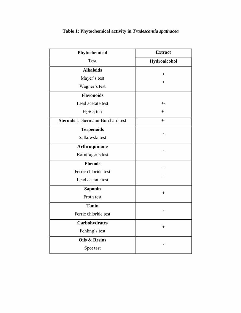
Table 1: Phytochemical activity in Tradescantia spathacea
Phytochemical
Test
Extract
Hydroalcohol
Alkaloids
Mayer’s test
Wagner’s test
+
+
Flavonoids
Lead acetate test
H2SO4 test
+-
+-
Steroids Liebermann-Burchard test +-
Terpenoids
Salkowski test -
Arthroquinone
Borntrager’s test -
Phenols
Ferric chloride test
Lead acetate test
-
-
Saponin
Froth test +
Tanin
Ferric chloride test -
Carbohydrates
Fehling’s test +
Oils & Resins
Spot test -
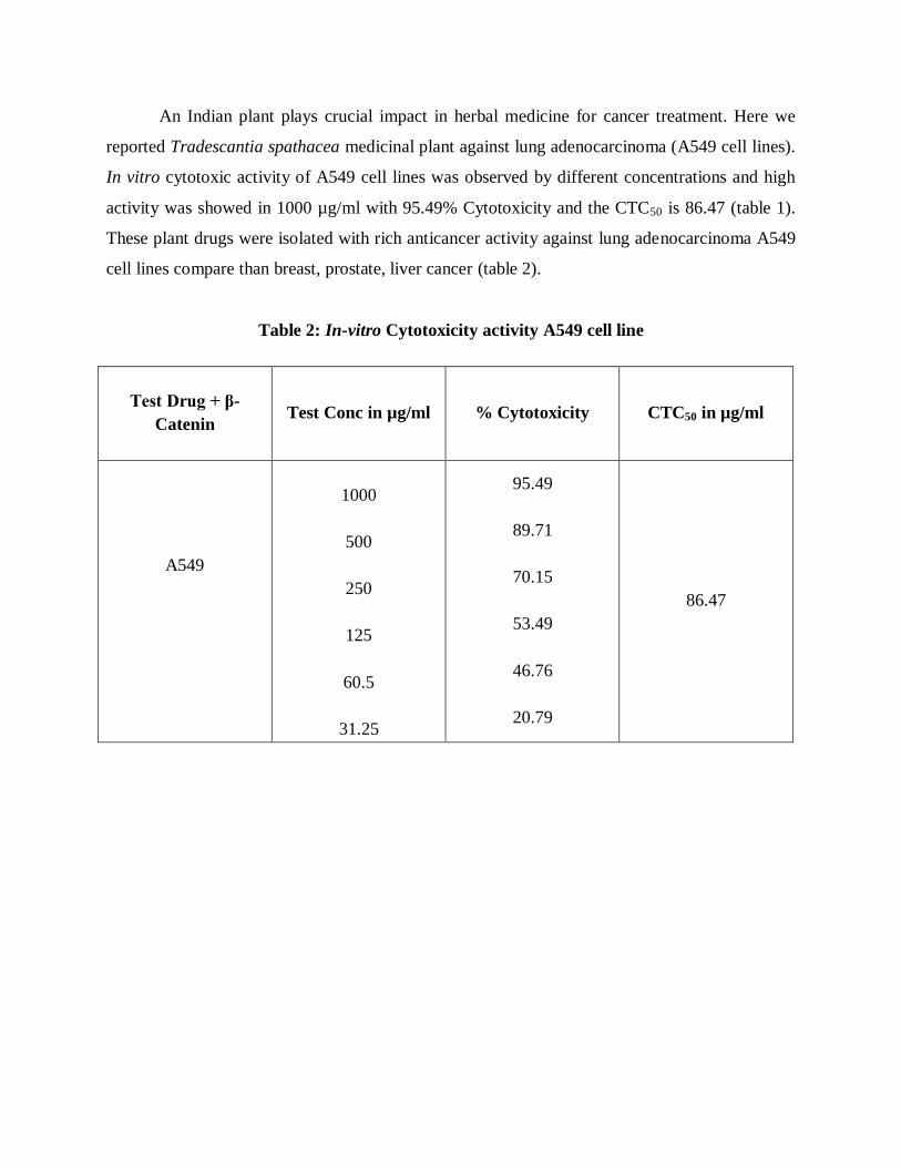
An Indian plant plays crucial impact in herbal medicine for cancer treatment. Here we
reported Tradescantia spathacea medicinal plant against lung adenocarcinoma (A549 cell lines).
In vitro cytotoxic activity of A549 cell lines was observed by different concentrations and high
activity was showed in 1000 µg/ml with 95.49% Cytotoxicity and the CTC50 is 86.47 (table 1).
These plant drugs were isolated with rich anticancer activity against lung adenocarcinoma A549
cell lines compare than breast, prostate, liver cancer (table 2).
Table 2: In-vitro Cytotoxicity activity A549 cell line
Test Drug + β-
Catenin
Test Conc in µg/ml % Cytotoxicity CTC50 in µg/ml
A549
1000
500
250
125
60.5
31.25
95.49
89.71
70.15
53.49
46.76
20.79
86.47
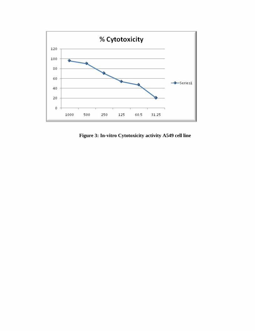
Figure 3: In-vitro Cytotoxicity activity A549 cell line
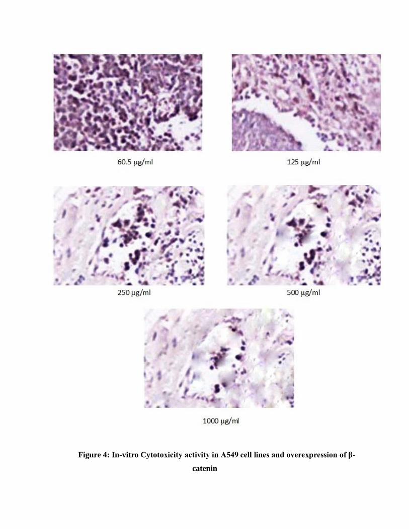
Figure 4: In-vitro Cytotoxicity activity in A549 cell lines and overexpression of β-
catenin
Furthermore, overexpression of β-catenin was also observed in lung adenocarcinoma cell
lines (figure 3). Anticancer activities of Tradescantia spathacea was observed by used different
concentrations and each concentration shown overexpression of β-catenin (fig. 3). Tradescantia
spathacea medicinal plant drug was also suppressed or prevented abnormalities of β-catenin
protein expression by killed lung adenocarcinoma cells (A549) (figure 4).
Anticancer properties of many plant compounds isolated from different Indian plant
leaves extracts have been reported. Research carried out the world to find a lead compound
which can block the development of cancer in humans. Plant-derived natural anticancer products
such as, terpenoids, flavonoids and steroids have received considerable attention owing to their
diverse pharmacological properties, which include cytotoxic and chemo-preventive effects.
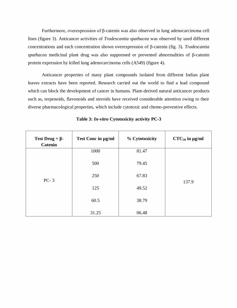
Table 3: In-vitro Cytotoxicity activity PC-3
Test Drug + β-
Catenin
Test Conc in µg/ml % Cytotoxicity CTC50 in µg/ml
PC- 3
1000
500
250
125
60.5
31.25
81.47
79.45
67.83
49.52
38.79
06.48
137.9
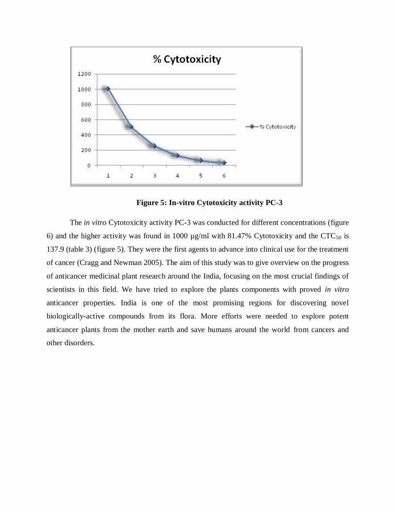
Figure 5: In-vitro Cytotoxicity activity PC-3
The in vitro Cytotoxicity activity PC-3 was conducted for different concentrations (figure
6) and the higher activity was found in 1000 µg/ml with 81.47% Cytotoxicity and the CTC50 is
137.9 (table 3) (figure 5). They were the first agents to advance into clinical use for the treatment
of cancer (Cragg and Newman 2005). The aim of this study was to give overview on the progress
of anticancer medicinal plant research around the India, focusing on the most crucial findings of
scientists in this field. We have tried to explore the plants components with proved in vitro
anticancer properties. India is one of the most promising regions for discovering novel
biologically-active compounds from its flora. More efforts were needed to explore potent
anticancer plants from the mother earth and save humans around the world from cancers and
other disorders.
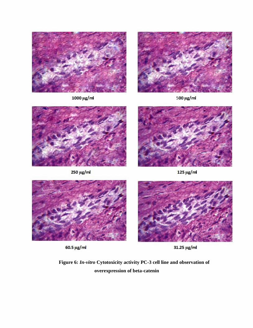
Figure 6: In-vitro Cytotoxicity activity PC-3 cell line and observation of
overexpression of beta-catenin
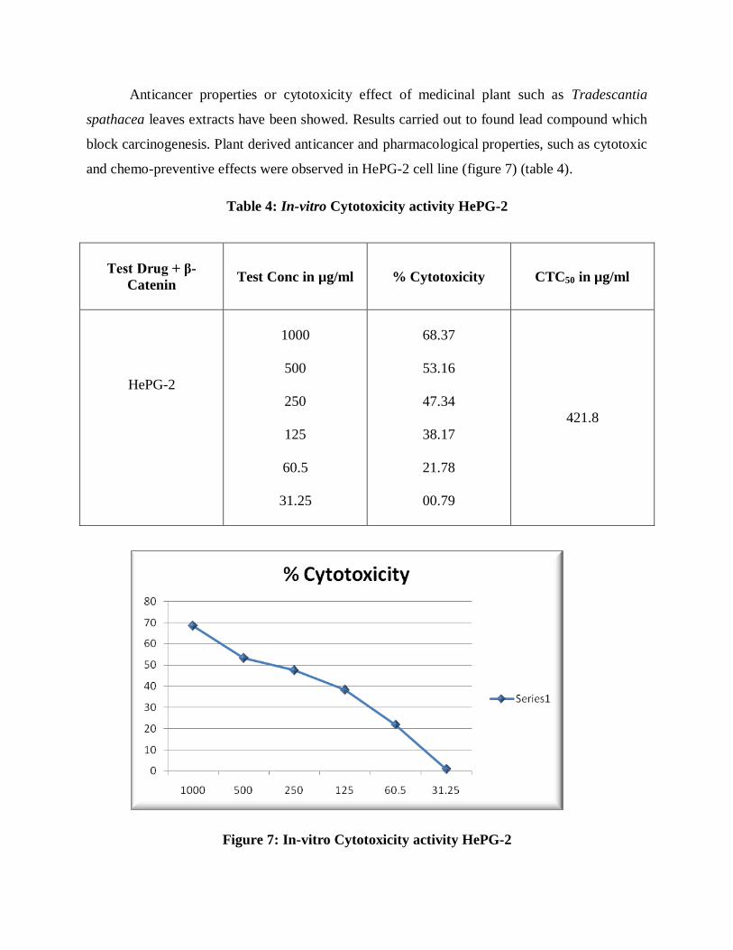
Anticancer properties or cytotoxicity effect of medicinal plant such as Tradescantia
spathacea leaves extracts have been showed. Results carried out to found lead compound which
block carcinogenesis. Plant derived anticancer and pharmacological properties, such as cytotoxic
and chemo-preventive effects were observed in HePG-2 cell line (figure 7) (table 4).
Table 4: In-vitro Cytotoxicity activity HePG-2
Test Drug + β-
Catenin
Test Conc in µg/ml % Cytotoxicity CTC50 in µg/ml
HePG-2
1000
500
250
125
60.5
31.25
68.37
53.16
47.34
38.17
21.78
00.79
421.8
Figure 7: In-vitro Cytotoxicity activity HePG-2
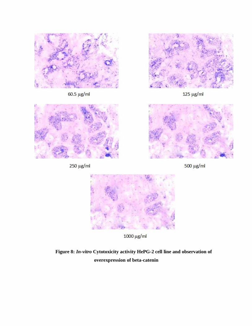
Figure 8: In-vitro Cytotoxicity activity HePG-2 cell line and observation of
overexpression of beta-catenin
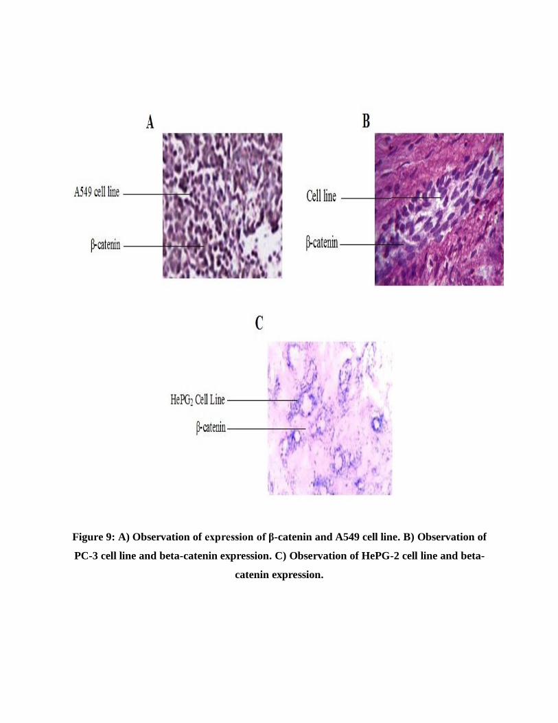
Figure 9: A) Observation of expression of β-catenin and A549 cell line. B) Observation of
PC-3 cell line and beta-catenin expression. C) Observation of HePG-2 cell line and beta-
catenin expression.
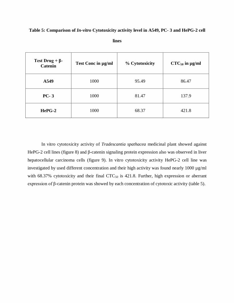
Table 5: Comparison of In-vitro Cytotoxicity activity level in A549, PC- 3 and HePG-2 cell
lines
Test Drug + β-
Catenin
Test Conc in µg/ml % Cytotoxicity CTC50 in µg/ml
A549 1000 95.49 86.47
PC- 3 1000 81.47 137.9
HePG-2 1000 68.37 421.8
In vitro cytotoxicity activity of Tradescantia spathacea medicinal plant showed against
HePG-2 cell lines (figure 8) and β-catenin signaling protein expression also was observed in liver
hepatocellular carcinoma cells (figure 9). In vitro cytotoxicity activity HePG-2 cell line was
investigated by used different concentration and their high activity was found nearly 1000 µg/ml
with 68.37% cytotoxicity and their final CTC50 is 421.8. Further, high expression or aberrant
expression of β-catenin protein was showed by each concentration of cytotoxic activity (table 5).

DISCUSSION
DISCUSSION
Here, In vitro cytotoxic activity and aberrant expression of β-catenin was shown in A549
cell lines. Normally, β-catenin protein was involved in cell proliferation in lung glandular
epithelium. Despite, overexpression and mutation of β-catenin induced lung adenocarcinoma. In
vitro cytotoxic activity of medicinal plant Tradescantia spathacea in A549 cell line was
observed by used different concentrations and final CTC50 is 86.47 (fig. 1).
In vitro cytotoxic activity of Tradescantia spathacea medicinal plant in PC-3 cell line
was observed by used different concentrations and final CTC50 is 137.9. Additionally, signaling
abnormality also observed in prostate cancer cell line. In Wnt/β-catenin signaling has crucial
impact in prostate gland development. Abnormality of β-catenin protein expression induced
prostate cancer. Overexpression of β-catenin protein was shown in PC-3 cell line. In vitro
cytotoxic activity of plant drug observed using different concentrations and also each
concentration indicated overexpression of β-catenin in PC-3 cell line (Kypta and Waxman,
2012).
Medicinal plant Tradescantia spathacea showed cytotoxicity properties against liver
hepatocellular carcinoma (HePG-2 cell lines) and overexpression of β-catenin (Tien et al., 2005).
Cytotoxicity activity of Tradescantia spathacea was showed by used different concentrations.
Final results, 50 % HePG-2 cell lines inhibition occurred at 421.87µg/ml or final CTC50 is 421.8.
Furthermore investigation was carried out against signaling pathway involvement in HePG-2 cell
line. Maximum, cancers developments were regulated by deregulation of signaling pathway.
Overexpression and mutations of signaling proteins caused variety of cancers. Despite,
overexpression or mutation of β-catenin induced cancer development in normal cells. Especially,
β-catenin was also highly expressed in liver hepatocellular carcinoma (Lai et al., 2011).
Anticancer agents were already development against deregulation of Wnt/β-catenin
signaling but they have their own toxicity. Hence, plant derived chemotherapeutic agents
investigated then cytotoxicity activities were showed by using Tradescantia spathacea medicinal
plant. Tradescantia spathacea has crucial impact in Wnt/β-catenin signaling pathway, observed
from the results. Eventually, Tradescantia spathacea was suppressed or prevented cancer cells
development and overexpression of β-catenin in HePG-2 cell lines. At last, final CTC50 is 421.8
and β-catenin expression also suppressed by leaves extracts of Tradescantia spathacea medicinal
plant (Lee et al., 2014).
WNT SIGNALING IN STEM CELLS DEVELOPMENT AND CANCER
A Concise History of WNT
Stem Cells have ability to grow different specialized organs. Adult mature cells does not
have the ability. Wnt signals are widely express in normal and cancer cells. Although wnt signal
has a unique character and function compare than other signaling pathway known as nanog,
hedgehog. The int-1 gene was first identified by activation of mouse mammary tumor virus
(MMTV) in mammary carcinomas. The int-1 gene encodes a glycoprotein as same as in
Drosophila melanogaster wingless (Wg) gene. Those genes were finally named as Wnt gene
(Wingless int gene) (Nusse and Varmus, 1982; Nusse et al., 1991; Nusse and Varmus, 1992).
Stem cells Niche
The stem cells were enhanced from zygote fusion of egg and sperm cell. Zygote has
ability to form whole species called totipotent stem cells. Differentiation of zygote to form
blastomeres, such blstomere contains embryonic stem cells called pluripotent stem cell. Further,
Embryonic stem cells (ESCs) have four types of differentiation such as mesoderm, ectoderm,
endoderm and germ cells. These divisions are finally differentiate to accomplished the formation
of organogenesis (Thomson et al., 1998; Intawicha et al., 2013) The Wnt gene has crucial play
key role in formation of mature adult cells from embryonic stem cells. Moreover, Wnt gene
plays major role in mesoderm and endoderm development and differentiation. Unfortunately,
abnormality of Wnt signaling leads to stimulate the cancer formation and other disorders in the
life cycle (Nusse, 2008; Sui et al., 2013) (figure 10).
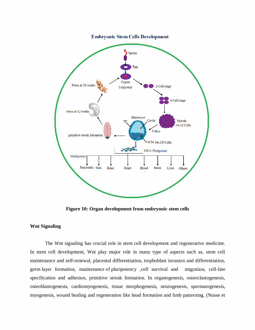
Figure 10: Organ development from embryonic stem cells
Wnt Signaling
The Wnt signaling has crucial role in stem cell development and regenerative medicine.
In stem cell development, Wnt play major role in many type of aspects such as, stem cell
maintenance and self-renewal, placental differentiation, trophoblast invasion and differentiation,
germ layer formation, maintenance of pluripotency ,cell survival and migration, cell-fate
specification and adhesion, primitive streak formation. In organogenesis, osteoclastogenesis,
osteoblastogenesis, cardiomyogenesis, tissue morphogenesis, neurogenesis, spermatogenesis,
myogenesis, wound healing and regeneration like head formation and limb patterning. (Nusse et
al., 2008; Knofler and Pollheimer, 2013). In cancer, Wnt signal act as a carcinogenesis when it is
an abnormal state and other degenerative disorders.
Wnt Signaling Pathway and regulations
The Wnt gene family secreted various Wnt proteins by using multiple Wnt signaling
pathways. In vertebrate, Wnt gene family represents large number of signaling molecules
involved in Wnt signaling pathways. Up to now, 19 Wnt ligands interact with secreted and
membrane associated proteins to initiate signaling process, 10 transmembrane, frizzed
heterodimeric receptor (FZD), 2 low density lipoprotein receptor-related protein co-repressor
(LRP-5 and 6) have been identified in mammals Wnt signaling. Depending on the type of Wnt-
ligand receptor interaction, intracellular signaling molecules, specific target transcriptional
activations, Wnt signaling pathways have been defined. Wnt gene family regulated three
different signaling pathways known as canonical pathway. Other two pathways are commonly
called as non-canonical pathways such as Wnt/planer cell polarity (PCP) pathway and Wnt/Ca2+
pathway. These canonic and non-canonic pathways involved crucial role in different aspects. In
canonical pathway, wnt signal transduced for cell fate determination and non-canonical pathway
regulated the purpose of controlling cell movement and tissue polarity (Sonderegger et al., 2007).
The Canonical Wnt/β-catenin Signaling Pathway
The canonical pathway is stimulated by Wnt protein family. β-catenin has major role in
the canonical pathway. β-catenin has a dual function such act as a intracellular contacts and
intracellular messanger. In canonical pathway, β-catenin act as a transcriptional transactivator.
Stem cell development, β-catenin requires the development of dual function of embryonic stem
cell self-renewal and germ layer formation. Wnt signal, the number of extracellular Wnt-
modulating protein such as DKK1, Crescent, FrzB, SFRP, WIF, Cer, Kremlin, Norrin. Dickkopf
1 (DKK1), most secreted molecules of Wnt antagonist, bind to the low density lipoprotein
receptor such as LRP5/6 to inhibit Wnt signaling. Other extra cellular molecules like Crescent,
FrzB, SFRP, WIF, Cer are inhibited Wnt ligands to bind frizzed heterodimeric receptor (FZD)
(Zhang et al., 2013).
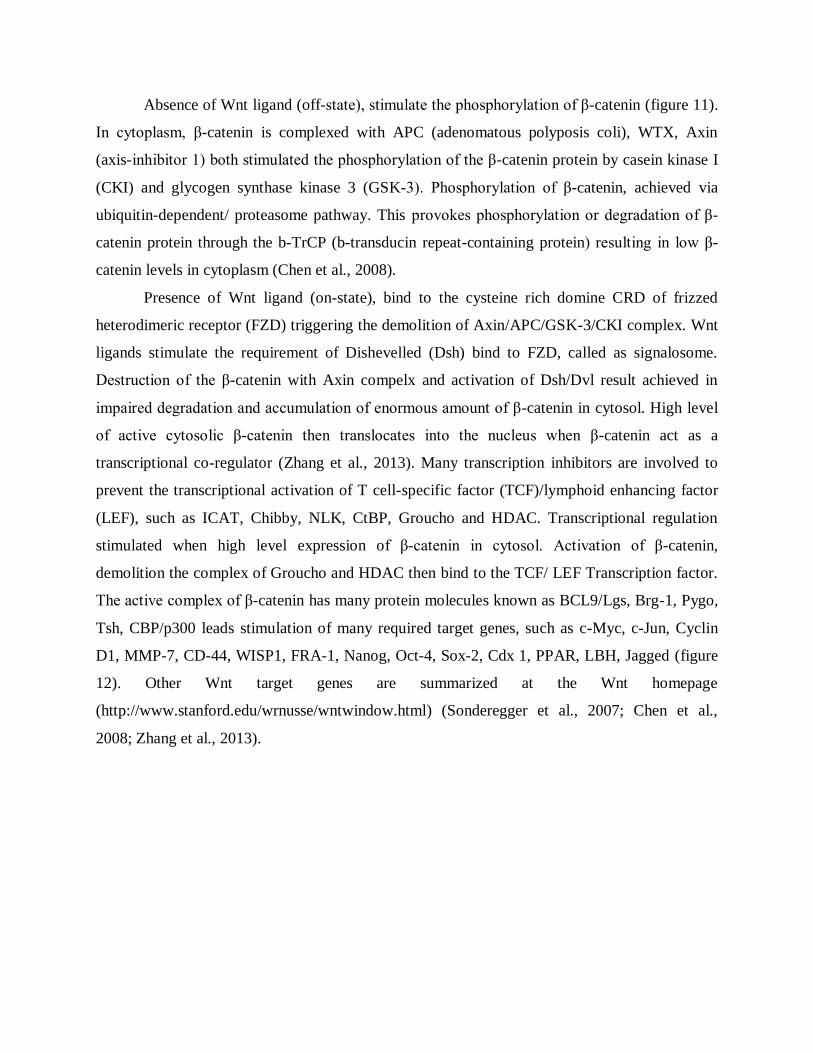
Absence of Wnt ligand (off-state), stimulate the phosphorylation of β-catenin (figure 11).
In cytoplasm, β-catenin is complexed with APC (adenomatous polyposis coli), WTX, Axin
(axis-inhibitor 1) both stimulated the phosphorylation of the β-catenin protein by casein kinase I
(CKI) and glycogen synthase kinase 3 (GSK-3). Phosphorylation of β-catenin, achieved via
ubiquitin-dependent/ proteasome pathway. This provokes phosphorylation or degradation of β-
catenin protein through the b-TrCP (b-transducin repeat-containing protein) resulting in low β-
catenin levels in cytoplasm (Chen et al., 2008).
Presence of Wnt ligand (on-state), bind to the cysteine rich domine CRD of frizzed
heterodimeric receptor (FZD) triggering the demolition of Axin/APC/GSK-3/CKI complex. Wnt
ligands stimulate the requirement of Dishevelled (Dsh) bind to FZD, called as signalosome.
Destruction of the β-catenin with Axin compelx and activation of Dsh/Dvl result achieved in
impaired degradation and accumulation of enormous amount of β-catenin in cytosol. High level
of active cytosolic β-catenin then translocates into the nucleus when β-catenin act as a
transcriptional co-regulator (Zhang et al., 2013). Many transcription inhibitors are involved to
prevent the transcriptional activation of T cell-specific factor (TCF)/lymphoid enhancing factor
(LEF), such as ICAT, Chibby, NLK, CtBP, Groucho and HDAC. Transcriptional regulation
stimulated when high level expression of β-catenin in cytosol. Activation of β-catenin,
demolition the complex of Groucho and HDAC then bind to the TCF/ LEF Transcription factor.
The active complex of β-catenin has many protein molecules known as BCL9/Lgs, Brg-1, Pygo,
Tsh, CBP/p300 leads stimulation of many required target genes, such as c-Myc, c-Jun, Cyclin
D1, MMP-7, CD-44, WISP1, FRA-1, Nanog, Oct-4, Sox-2, Cdx 1, PPAR, LBH, Jagged (figure
12). Other Wnt target genes are summarized at the Wnt homepage
(http://www.stanford.edu/wrnusse/wntwindow.html) (Sonderegger et al., 2007; Chen et al.,
2008; Zhang et al., 2013).
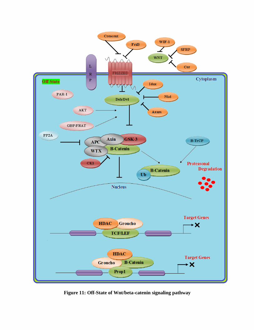
Figure 11: Off-State of Wnt/beta-catenin signaling pathway
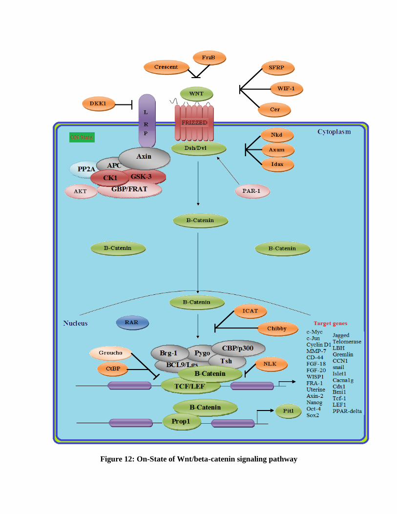
Figure 12: On-State of Wnt/beta-catenin signaling pathway
The Noncanonical-Wnt/Planer Cell Polarity Pathway
The noncanonical Planer Cell Polarity pathway (PCP) is one of the most important
pathway in Wnt signaling.
Figure 13: Wnt/PCP signaling pathway
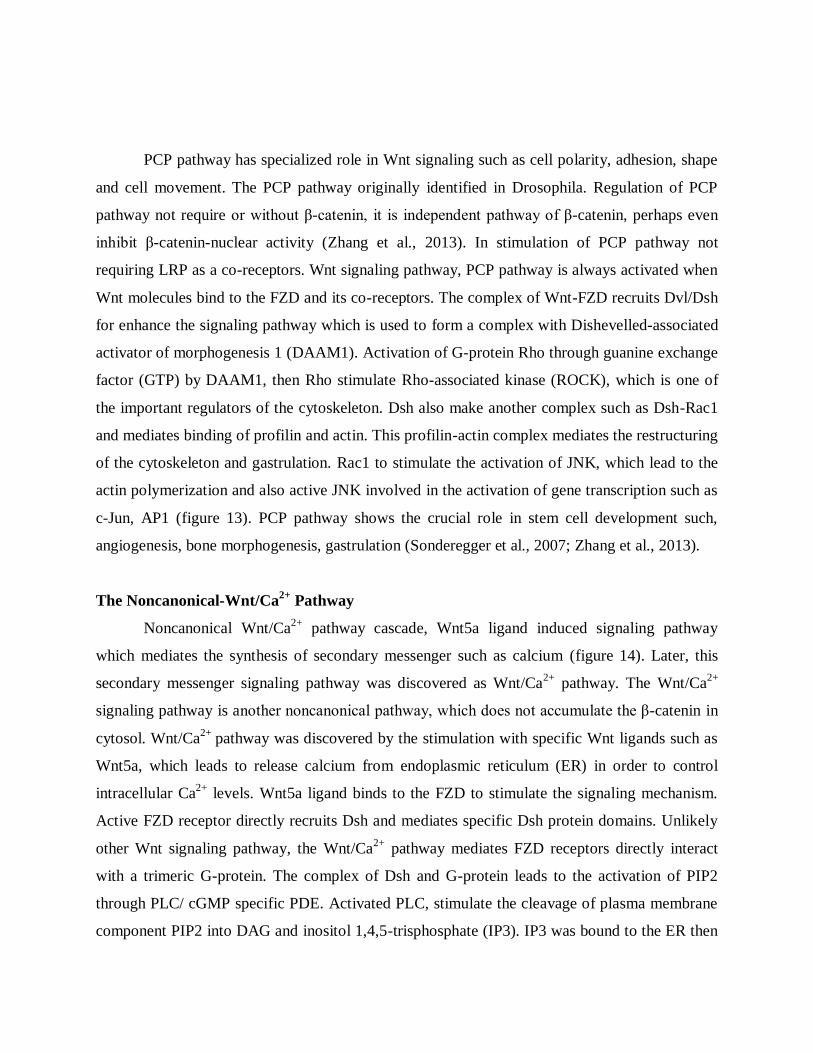
PCP pathway has specialized role in Wnt signaling such as cell polarity, adhesion, shape
and cell movement. The PCP pathway originally identified in Drosophila. Regulation of PCP
pathway not require or without β-catenin, it is independent pathway of β-catenin, perhaps even
inhibit β-catenin-nuclear activity (Zhang et al., 2013). In stimulation of PCP pathway not
requiring LRP as a co-receptors. Wnt signaling pathway, PCP pathway is always activated when
Wnt molecules bind to the FZD and its co-receptors. The complex of Wnt-FZD recruits Dvl/Dsh
for enhance the signaling pathway which is used to form a complex with Dishevelled-associated
activator of morphogenesis 1 (DAAM1). Activation of G-protein Rho through guanine exchange
factor (GTP) by DAAM1, then Rho stimulate Rho-associated kinase (ROCK), which is one of
the important regulators of the cytoskeleton. Dsh also make another complex such as Dsh-Rac1
and mediates binding of profilin and actin. This profilin-actin complex mediates the restructuring
of the cytoskeleton and gastrulation. Rac1 to stimulate the activation of JNK, which lead to the
actin polymerization and also active JNK involved in the activation of gene transcription such as
c-Jun, AP1 (figure 13). PCP pathway shows the crucial role in stem cell development such,
angiogenesis, bone morphogenesis, gastrulation (Sonderegger et al., 2007; Zhang et al., 2013).
The Noncanonical-Wnt/Ca2+
Pathway
Noncanonical Wnt/Ca2+
pathway cascade, Wnt5a ligand induced signaling pathway
which mediates the synthesis of secondary messenger such as calcium (figure 14). Later, this
secondary messenger signaling pathway was discovered as Wnt/Ca2+
pathway. The Wnt/Ca2+
signaling pathway is another noncanonical pathway, which does not accumulate the β-catenin in
cytosol. Wnt/Ca2+
pathway was discovered by the stimulation with specific Wnt ligands such as
Wnt5a, which leads to release calcium from endoplasmic reticulum (ER) in order to control
intracellular Ca2+
levels. Wnt5a ligand binds to the FZD to stimulate the signaling mechanism.
Active FZD receptor directly recruits Dsh and mediates specific Dsh protein domains. Unlikely
other Wnt signaling pathway, the Wnt/Ca2+
pathway mediates FZD receptors directly interact
with a trimeric G-protein. The complex of Dsh and G-protein leads to the activation of PIP2
through PLC/ cGMP specific PDE. Activated PLC, stimulate the cleavage of plasma membrane
component PIP2 into DAG and inositol 1,4,5-trisphosphate (IP3). IP3 was bound to the ER then
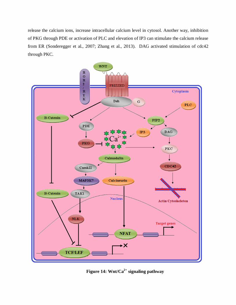
release the calcium ions, increase intracellular calcium level in cytosol. Another way, inhibition
of PKG through PDE or activation of PLC and elevation of IP3 can stimulate the calcium release
from ER (Sonderegger et al., 2007; Zhang et al., 2013). DAG activated stimulation of cdc42
through PKC.
Figure 14: Wnt/Ca2+
signaling pathway
Cdc42 is part of the major regulators of ventral patterning. Increased intracellular calcium ions
level was also stimulated activation of calcineurin and CaMKII through calmodulin. Calcineurin
mediated the activation of nucler factor such as nuclear factor of activated T cells (NFAT),
which regulate ventral patterning. NFAT was also regulated many functions known as regulation
of cell adhesion, migration and tissues separation. CamkII activated the MAP signaling pathway,
which stimulated the activation of TGF-b-activated kinase (TAK1) and Nemo-like kinase
(NEMO). Activated TAK1 induces the NLK expression, which prevents the action of β-catenin
and TCF/LEF transcriptional factor complex. NLK act as an antagonist of Wnt/ β-catenin
signaling pathway by inactivation and phosphorylation of TCF. Despite, if PDE is activated,
inhibition of calcium release is occurred from ER. Activated PDE was mediated the activation of
PKG, which subsequently cause the inhibition of intracellular calcium release from ER (Zhang et
al., 2013; De, 2011).
Wnt Signal in Cancer and therapeutics
Since its initial discovery, Wnt has associated with cancer development from normal
cells. When Wnt1 was first discovered, Wnt1 (Wingless int-1) was first identified proto-
ongogene in mouse mammary tumor virus (MMTV) in mammary carcinomas (Nusse and
Varmus, 1982). In normal conditions, Wnt1 has crucial role in embryonic stem cell development.
Unfortunately, deregulation of Wnt1 leads to unwanted cell growth and movement. APC was
initially defined as a tumor suppressor gene in colon cancer. The complex of β-catenin and APC,
activating mutation was found in β-catenin gene in human colon cancer. Another mutation was
identified in human Axin1 gene, where reported in human hepatocellular carcinoma. In TCF1 act
as s tumor suppressor gene, mutated TCF1 in mice enhanced adenoma in the gut and mammary
glands, so finalize the TCF1 act as a feedback repressor of target gene (Nusse and Varmus,
2012). Cancers have major play role in human death rise in the World. Wnt signals are involved
in many human cancer types. Breast cancer was found in human majority of female in
worldwide. Since many scientists finding regenerative medicine for breast cancer. Expressions of
Wnt/canonical pathway was identified in the development of begin and malignant breast cancer.
Increased level of β-catenin in cytosol or nucleus is involved in breast cancer. Presence of β-
catenin level in the cytoplasm or nucleus was identified by using immunohistochemical staining
and Western blotting. The accumulation of β-catenin due to several impact factor such as lacs of
β-catenin destruction, mutation in β-catenin, mutation in APC, loss of inhibitors, over expression
of Wnt ligands and decreased activity of Wnt/Ca2+
pathway (Howe and Brown, 2004; Clevers
and Nusse, 2012).
Wnt signaling was also involved in the epithelial-mesenchymal transition (EMT) to
enhance metastasize in breast cancer. Repression of Wnt/β-catenin signaling in breast cancer can
prevent EMT and inhibited metastasize (DiMeo et al., 2009). CTNNB1 gene encodes β-catenin,
changes in CTNNB1 gene expression measured not only in breast cancer, but also lung cancer,
melanoma, prostate cancer, colorectal cancer, colon cancer and other several types. Wnt ligands
proteins such as, Win1, Win2 and Wnt7a increased expression have been identified in the
development of ovarian cancer, glioblastoma and oesophageal cancer. Absence of other Wnt
protein functions in Wnt signaling cause different cancer types such proteins as, ROR1, ROR 2,
WIF1, Dsh, Wnt5a and other TCF/LEF family (Anastas Jamie and Moon Randall, 2012; DiMeo
et al., 2009). A variety of compounds are discovered for the purpose of cancer treatment based
on Wnt signal expressions. Imatinib, which was originally identified to inhibit β-catenin activity
in cancer (Rao et al., 2006). Celecoxib, (cyclooxygenase-2 inhibitor), which inhibit β-catenin
activity in human colon carcinoma (Maier et al., 2005). Antisence inhibitors have been
developed to decrease β-catenin activity against several cancer types such as colon, leukemia,
lymphoma cells and esophageal (Phillips et al., 2002). NASAIDs also inhibit the Wnt/β-catenin
signaling pathway (Dihlmann et al., 2001). Thiazolidinedione was completely inhibited
metastasize in colon cancer (Yoshizumi et al., 2004). Many compounds are identified against
cancer including these compounds.

SUMMARY AND CONCLUSION
SUMMARY AND CONCLUSION
Wnt/β-catenin signals are central pathway of stem cells and organ development. In cancer
and cancer stem cells, abnormality of Wnt/β-catenin has crucial role in cancer development.
Aberrant expression and mutation of β-catenin continuously stimulated many types of cancer.
Deregulation of Wnt/β-catenin signaling has crucial impact in cancer stem cells self-renewal and
proliferation. Critically, mutant β-catenin, Axin, APC plays important role in cancer
development. In clinical level, specific drugs were not developed against cancer and cancer stem
cell self-renewal and proliferation carried out by overexpression and mutant β-catenin signaling.
Future research, specialized drug will synthesis against mutant and overexpression of β-catenin
involved in variety of cancer and specially, drug will target to cancer stem cells development.
Signals were involved crucial role in cell-cell communication, metabolic activity and
development but overexpression and mutations of signaling molecules including β-catenin
protein induced lung adenocarcinoma. Natural and synthetic biological or chemical agents were
used as anticancer agents against various cancers. Despite, many anticancer agents have their
toxicity and destroyed normal cells. Thus we found rich anticancer activity in Tradescantia
spathacea medicinal plant against A549 cell lines and this plant extract was also involved as
suppressor or inhibitor of overexpression of β-catenin protein. Tradescantia spathacea medicinal
plant used as anticancer drug and inhibited overexpression of β-catenin protein in A549 cell
lines.
Many cancer treatments, medicinal plants were used as drugs without side effect. Here,
we found rich anticancer activity in Tradescantia spathacea by using PC-3 cell line and CTC50
was showed in 137.9. Signaling pathways plays important role in stem cells development and
carcinogenesis. Especially, aberrant expression of Wnt/β-catenin regulated many cancers
including prostate cancer. Overexpression of β-catenin observed in PC-3 cell line and this plant
drug was used to inhibit β-catenin expression in prostate cancer. In clinical, this drug was used in
prostate cancer treatment and also inhibited overexpression of Wnt/β-catenin signaling.
In cancer treatment, many drugs were found by used many chemical and biological
agents and also they have their own cytotoxic activity against normal cells. Hence, scientist
focused plant chemotherapeutic agents against carcinogenesis. Here, we reported cytotoxic or
anticancer activity of medicinal plant such as Tradescantia spathacea against HePG-2 cell lines
(human liver hepatocellular carcinoma). The plant extracts showed high anticancer activity or
cytotoxic activity level and final CTC50 is 421.8. Wnt/β-catenin signaling pathway also observed
in HePG-2 cell lines and this plant leaves extract proved suppression or prevention of β-catenin
protein by used Tradescantia spathacea medicinal plant.
Wnt signaling is regulated stem cell development and cancer in normal and abnormal
conditions. Therapeutic system is developed a variety of drugs against cancer based on
deregulation of Wnt signaling pathways. Unfortunately, many details are not described about
Wnt signaling such as, where is the evolution origin of Wnt signaling?, How Wnt signals are
activated and which molecules or hormones are needed for this wingless activation?, how wnt
signals are coordinated cell fate changes?, how Wnt signals are involved in self-renewal and
differentiation both stem cells and cancer cells at a similar time?, many Wnt inhibitors are
present inside the cells and why these inhibitors are not active in cancer cells proliferation?.
These details are not evaluated and undetermined up to now. These characters are very
significant for drugs design against cancer.
In the current research deals cancer drugs are directly developed based on the
deregulation of signaling pathway such as Wnt signaling but many drugs are destroying the
cancer cells not cancer stem cells (CSCs). These CSCs are ability to produce lots of cancer cells,
another way CSCs are called as mother of cancer cells. In drug designing, lots of complexity
leads to failure the action of drugs. A Variety of factors are leads to complicate the synthesis of
cancer drugs and each cancer type has specific characters and causing agents. So many scientists
are developing new drugs for each cancer type. In future, development of a new drug for all
types of cancer caused by deregulation of the Wnt signaling pathway.

REFERENCES
REFERENCES
Schwartsmann G, Ratain MJ, Cragg GM, Wong JE, Saijo N, Parkinson DR, et al. Anticancer
drug discovery and development throughout the world. J. ClinOncol 2002; 20(18 Suppl):47S-
59S.
Jang M, Cai L, Udeani GO, Slowing KV and Thomas CF. Cancer chemopreventive activity of
resveratrol, a natural product derived from grapes. Science 1997; 275:218-220.
Vickers A. Botanical medicines for the treatment of cancer: rationale, overview of current data,
and methodological considerations for phase I and II trials. Canc. Investig 2002; 20:1069–1079.
El-Shemy HA, Aboul-Enein AM, Aboul-Enein KM and Fujita K. Willow Leaves Extracts
Contain Anti-Tumor Agents Effective against Three Cell Types. PLoS One 2007 2(1):e178.
Mehta RG, Murillo G, Naithani R and Peng X. Cancer chemoprevention by natural products:
how far have we come? Pharm Res 2010; 27:950-61.
Parag R. Patel1, Bhuvan P. Raval2. , Hamsraj A. Karanth1, Vishal R. Patel3. Potent antitumor
activity of Rubia cordifolia, International Journal of Phytomedicine 2010; 2: 44-46.
Jemal A, Murray T, Ward E, Samuels A, Tiwari RC, Ghafoor A, et al. Cancer statistics 2005.
CA Cancer J Clin 2005; 55(1): 10-30
Krishnaiah D R, Sarbatly and Bono A, Biotechnol. Mol. Biol. Rev 2007; 1 : 97.
Cragg GM, Newman DJ. Plants as a source of anticancer agents. Journal of Ethenopharmacology
2005; 100: 72-79.
Lukmanul H, Girija A, Boopathy R. Antioxidant property of selected Ocimum species and their
secondary metabolite content. J Med Plants Res 2008; 2(9): 250-257.
Mojab F, Kamalinejad M, Ghaderi N, Vanidipour HR. Phytochemicals screening of some
species of Iranian plants. Iran J Pharm Res 2003; 3: 77-82.
Rice-Evans, C. A., Sampson, J., Bramley, P. M., & Holloway, D. E. Why do we expect
carotenoids to be antioxidants in vivo? Free Radical Research 1997; 26 : 381–398.
Mohammad S. Anticancer agents from medicinal plants. Bangladesh J Pharmacol 2006; 1: 35-
41.
Hartwell JL. Plants used against cancer. A survey. Quarterman Publications, Lawrence; 1982.
Kinghorn AD, Farnsworth NR, Soejarto DD, et al., Novel strategies for the discovery of the plant
derived anticancer agents. Pharmaceutic Biol 2003; 41: 53-67.
Surh YJ. Cancer chemoprevention with dietary phytochemicals. Nature Rev Cancer 2003; 3:
768-780.
Noordermeer J, Klingensmith J, Perrimon N, Nusse R. Dishevelled and armadillo act in the
wingless signalling pathway in Drosophila. Nature 1994; 367 (6458): 80–83.
Anastas Jamie N. and Moon Randall T. WNT signalling pathways as therapeutic targets in
cancer. Nature Reviews Cancer 2012; 13 : 11–26.
Clevers H. and Nusse R. Wnt/β-Catenin Signaling and Disease. Cell 2012, 149; 1192-205.
De A.,. Wnt/Ca2+ signaling pathway: a brief overview. Acta Biochim Biophys Sin, 43; 745-56.
Dihlmann S., Siermann A. and von Knebel Doeberitz M., 2001. The nonsteroidal anti-
inflammatory drugs aspirin and indomethacin attenuate betacatenin/ TCF-4 signaling. Oncogene
2011, 20; 645-653.
DiMeo T. A., Anderson K., Phadke P., Feng C., Perou C. M., Naber S. and Kuperwasser C. A
Novel Lung Metastasis Signature Links Wnt Signaling with Cancer Cell Self-Renewal and
Epithelial-Mesenchymal Transition in Basal-like Breast Cancer. Cancer Research 2009, 69;
5364–5373.
Howe Louise. and Brown Anthony. Wnt Signaling and Breast Cancer. Cancer Biology and
Therapy 2004, 3; 36–41.
Intawicha P., Wang SH., Hsieh YC., Lo NW., Lee KH., Huang SY. and Ju JC. Proteomic
Profiling of Rabbit Embryonic Stem Cells Derived from Parthenotes and Fertilized Embryos.
PLoS One 2013, 8; e67772
Knofler M. and Pollheimer J. Human placental trophoblast invasion and differentiation: a
particular focus on Wnt signaling. Front Genet 2013, 4; 190.
Maier TJ., Janssen A., Schmidt R., Geisslinger G. and Grosch S. Targeting the beta-catenin/
APC pathway: a novel mechanism to explain the cyclooxygenase-2- independent
anticarcinogenic effects of celecoxib in human colon carcinoma cells. FASEB J 2005, 19; 1353-
1355.
Nusse R. and Varmus H. E. Wnt genes. Cell 1992, 69; 1073-1087.
Nusse R. and Varmus H.E. Many tumors induced by the mouse mammary tumor virus contain
a provirus integrated in the same region of the host genome. Cell 1982, 31; 99-109.
Nusse R., Brown A., Papkoff J., Scambler P., Shackleford G., McMahon A., Moon R., Varmus
H. A new nomenclature for int-1 and related genes: the Wnt gene family. Cell 1991, 64; 231.
Nusse R., Fuerer C., Ching W., Harnish K., Logan C., Zeng A., ten Berge D. and Kalani Y. Wnt
signaling and stem cell control. Cold Spring Harb Symp Quant Biol 2008, 73; 59-66.
Nusse Roel. and Varmus Harold. Three decades of Wnts: a personal perspective on how a
scientific field developed. The EMBO Journal 2012, 31; 2670–2684.
http://www.ncbi.nlm.nih.gov/pubmed?term=Pollheimer%20J%5BAuthor%5D&cauthor=true&cauthor_uid=24133501
Nusse, R. Wnt signaling in disease and in development. Cell Res 2005, 15; 28–32.
Phillips RK., Wallace MH., Lynch PM., Hawk E., Gordon GB., Saunders BP., Wakabayashi
N., Shen Y., Zimmerman S., Godio L., Rodrigues-Bigas M., Su LK., Sherman J., Kelloff
G., Levin B. and Steinbach G. FAP Study Group.. A randomised, double blind, placebo
controlled study of celecoxib, a selective cyclooxygenase 2 inhibitor, on duodenal polyposis in
familial adenomatous polyposis. Gut 2002, 50; 857-860.
Rao AS., Kremenevskaja N., von Wasielewski R., Jakubcakova V., Kant S., Resch J.
and Brabant G. Wnt/betacatenin signaling mediates antineoplastic effects of imatinib mesylate
(Gleevec) in anaplastic thyroid cancer. J Clin Endocrinol Metab 2006, 91; 159-168.
Sonderegger S., Pollheimer J. and Knofler M. Wnt signalling in implantation, decidualisation
and placental differentiation--review. Placenta 2010, 31; 839-47.
Staal FJ. and Fibbe WE. Wnt cross-talk in the niche. Blood 2012, 119; 1618-9.
Sui L., Bouwens L. and Mfopou JK. Signaling pathways during maintenance and definitive
endoderm differentiation of embryonic stem cells. Int J Dev Biol 2013, 57; 1-12.
Thomson J.A., Itskovitz-Eldor J., Shapiro S.S., Waknitz M.A., Swiergiel J.J., Marshall V.S. and
Jones, J.M. Embryonic stem cell lines derived from human blastocysts. Science 1998, 282;
1145–1147.
Yoshizumi T., Ohta T., Ninomiya I., Terada I., Fushida S., Fujimura T., Nishimura G., Shimizu
K., Yi S. and Miwa K. Thiazolidinedione, a peroxisome proliferator-activated receptor-gamma
ligand, inhibits growth and metastasis of HT-29 human colon cancer cells through
differentiation-promoting effects. Int J Oncol 2004, 25; 631-9
Zhang X., Hao L., Meng L., Liu M., Zhao L., Hu F., Ding C., Wang Y., He B., Pan Y., Fang
W., Chen J., Hu S. and Jia M. Digital gene expression tag profiling analysis of the gene
expression patterns regulating the early stage of mouse spermatogenesis. PLoS One 2013, 8;
e58680
Kypta RM and Waxman J. Wnt/β-catenin signalling in prostate cancer. Nat Rev Urol 2012.
[Epub ahead of print]
Lee E, Madar A, David G, Garabedian MJ, Dasgupta R and Logan SK. Inhibition of androgen
receptor and β-catenin activity in prostate cancer. PNAS 2013. 110 15710-5.
Tien LT, Ito M, Nakao M, Niino D, Serik M, Nakashima M, Wen CY, Yatsuhashi H, Ishibashi
H. Expression of beta-catenin in hepatocellular carcinoma. World J Gastroenterol 2005; 11(16):
2398-401.
Lee JM, Yang J, Newell P, Singh S, Parwani A, Friedman SL, Nejak-Bowen KN, Monga SP. Β-
Catenin signaling in hepatocellular cancer: Implications in inflammation, fibrosis, and
proliferation. Cancer Lett 2014; 343(1): 90-7.
Polakis P. Wnt signaling and cancer. Genes Dev 2000;14:1837-51.
Valkenburg KC, Graveel CR, Zylstra-Diegel CR, Zhong Z, Williams BO. Wnt/β-catenin
Signaling in Normal and Cancer Stem Cells. Cancers (Basel) 2011; 3: 2050-79.
Giles RH, van Es JH, Clevers H. Caught up in a Wnt storm: Wnt signaling in cancer. Biochim
Biophys Acta 2003;1653:1-24.
Behari J. The Wnt/β-catenin signaling pathway in liver biology and disease. Expert Rev
Gastroenterol Hepatol 2010;4:745-56.
Lai TY, Su CC, Kuo WW, Yeh YL, Kuo WH, Tsai FJ, Tsai CH, Weng YJ, Huang CY, Chen
LM. Β-catenin plays a key role in metastasis of human hepatocellular carcinoma. Oncol
Rep 2011; 26(2): 415-22.
Nozawa N, Hashimoto S, Nakashima Y, Matsuo Y, Koga T, Sugio K, Niho Y, Harada
M, Sueishi K. Immunohistochemical alpha- and beta-catenin and E-cadherin expression and their
clinicopathological significance in human lung adenocarcinoma. Pathol Res Pract 2006; 202(9):
639-50.