Clinical Significance and Cross Talk of Wnt Canonical ... - ERA
245
University of Alberta Clinical Significance and Cross Talk of Wnt Canonical Pathway in Cancer by Hanan Adel Ragi Armanious A thesis submitted to the Faculty of Graduate Studies and Research in partial fulfillment of the requirements for the degree of Doctor of Philosophy In Medical Sciences - Laboratory Medicine and Pathology ©Hanan Adel Ragi Armanious Edmonton, Alberta Fall 2011 Permission is hereby granted to the University of Alberta Libraries to reproduce single copies of this thesis and to lend or sell such copies for private, scholarly or scientific research purposes only. Where the thesis is converted to, or otherwise made available in digital form, the University of Alberta will advise potential users of the thesis of these terms. The author reserves all other publication and other rights in association with the copyright in the thesis and, except as herein before provided, neither the thesis nor any substantial portion thereof may be printed or otherwise reproduced in any material form whatsoever without the author's prior written permission.
-
Upload
khangminh22 -
Category
Documents
-
view
7 -
download
0
Transcript of Clinical Significance and Cross Talk of Wnt Canonical ... - ERA

University of Alberta
Clinical Significance and Cross Talk of Wnt Canonical Pathway in Cancer
by
Hanan Adel Ragi Armanious
A thesis submitted to the Faculty of Graduate Studies and Research in partial fulfillment of the requirements for the degree of
Doctor of Philosophy
In
Medical Sciences - Laboratory Medicine and Pathology
©Hanan Adel Ragi Armanious Edmonton, Alberta
Fall 2011
Permission is hereby granted to the University of Alberta Libraries to reproduce single copies of this thesis and to lend or sell such copies for private, scholarly or scientific
research purposes only. Where the thesis is converted to, or otherwise made available in digital form, the University of Alberta will advise potential users of the thesis of these
terms.
The author reserves all other publication and other rights in association with the copyright in the thesis and, except as herein before provided, neither the thesis nor any substantial portion thereof may be printed or otherwise reproduced in any material form
whatsoever without the author's prior written permission.

I dedicate this thesis to my husband and two daughters for all their
love and support

ABSTRACT
Wnt signalling is of great biological importance as it has been implicated in
development and cancer. The Wnt canonical pathway (WCP) is the best
characterized signalling pathway. Based on my preliminary studies in my
first and second years I found that the WCP is not linear and that it is
interacting with many other signalling proteins. Thus, I hypothesized that
WCP is cross talking with oncogenic networks in lymphoid and solid
tumors. Through my work in this thesis I examined different models of
cross talk between WCP and other signalling pathways implicated in
cancer pathogenesis.
The first objective of this thesis examined the biological and clinical
significance of the WCP member pGSK-3β, which also acts as a key
member in the PI3K/Akt pathway. pGSK-3β was shown to be expressed in
two cancer models; breast cancer and mantle cell lymphoma (MCL). In
MCL, pGSK-3β expression was shown to correlate to WCP activation,
however, in breast cancer it correlated to PI3K/Akt activation. Importantly,
pGSK-3β expression correlated with a worse clinical outcome in breast
cancer and MCL patients.
The second objective of this thesis examined the regulation of β-catenin
(WCP member) by signal transducer and activator of transcription 3
(STAT3) in breast cancer. STAT3 was shown to regulate β-catenin at the
transcriptional level, as STAT3 binds to the promoter of β-catenin.

Moreover, STAT3 was shown to correlate with nuclear β-catenin
expression in patient samples.
The third objective of this thesis was to study the regulatory role of β-
catenin on a disintegrin and metalloproteinase 10 (ADAM10) in mantle cell
lymphoma. However, ADAM10 was shown to regulate the TNFα/NFκB
signalling pathway.
The fourth objective of this thesis examined the cross talk between the
WCP and NPM-ALK the major oncogenic protein in ALK+ALCL. In
ALK+ALCL, cross talk between WCP and NPM-ALK through casein kinase
2α was identified.
Overall, the identification of the cross talk between WCP and various
signalling pathways in different cancer models furthers our current
understanding of the importance of the WCP and about the complexity of
signalling networks in cancer. These findings provide a framework for the
development of novel anti-cancer targeting strategies.

ACKNOWLEDGMENTS I wish to express my sincere appreciation and gratitude to the following individuals without whom this thesis would not have been possible. To Dr. Raymond Lai, my supervisor and mentor, I have learnt a lot in your lab and you always challenged me to improve and to think about what I am doing. Your love and passion for science and research have been contagious. Thank you for teaching me about science and life. I am proud to be one of the students graduating from your lab. To Dr. Judith Hugh, my committee member, your continuous encouragement meant a lot to me. Thank you for always having faith in me and I hope I deserved it. To Dr. Jean Deschenes, my committee member, thank you for your continuous advice and guidance. To Dr. Ing Swi Goping and Dr. Shirin Bonni, thank you for serving as examiners for my thesis defence. I really appreciate your time and insightful comments. To Dr. Monica Keelan, thank you for serving as my examination committee chairman and for your assistance and help. To Dr. Pascal Gelebart, thank you for being always there for me and for all the time you spent teaching and discussing with me. You helped in making a better scientist out of me. To all the past and present members of Lai lab, thank you for all your support and assistance. Special thanks to my friend Samar Hegazy, thank you for sharing with me all the tough times during my PhD, you were always a great support for me. Special thanks to Dr. Mona Anand for her advice, assistance and friendship. I would like to thank the Egyptian Government for financially supporting me during my PhD studies. Finally, to my husband Hany, thank you for always encouraging and supporting me in pursuing my endeavours, I am blessed to have you in my life. To my daughters Verena and Marina, your smiles are the strength and energy which kept me going through all my hard times and the thing that always gave me hope.
TABLE OF CONTENTS
Page
Chapter 1: General Introduction……...……………...…..…..1
1.1 Introduction………………..………….……………………........2
1.2. Wnt signalling pathway…..……………………........................5
1.2.1. Wnt canonical signalling……………............................9
(A) Overview…………………………………............................9
(B) Members of the Wnt canonical pathway (WCP)….........11
1.2.2. Non canonical Wnt signalling……………….…..........24
1.3. Wnt inhibitors………………………………….........................29
1.4. Wnt pathway in development………………………………...34
1.4.1 Wnt in haematopoiesis………………………………..34
1.4.2. Wnt in breast development…………………………...34
1.5. Wnt pathway in disease……………………………………….36
1.5.1. Wnt and colorectal cancer…………………………….38
1.5.2. Wnt and breast cancer………………………………...39
1.5.3. Wnt and hematologic malignancies………………....40
1.6. Wnt and cross talk with other signalling pathways in cancer42
1.6.1. Wnt and EGFR ..…………………………………..........42
1.6.2. Wnt and nuclear receptors.……………………............44
1.7. Wnt pathway and cancer therapeutics…………….................46
1.8. References.............................................................................53

Chapter 2: Clinical and biological significance of GSK-3β
inactivation in breast cancer-an immunohistochemical study….....79
2.1. Introduction………………………………………...…………....80
2.2. Materials and Methods……………………………………..….81
2.2.1. Patients and tissue specimens…………………….....81
2.2.2. Immunohistochemistry……………………………...…82
2.2.3. Scoring of the markers and statistical analysis..…….82
2.3. Results……………………………………………....……....…...84
2.3.1. Demographic and pathologic data…………………....84
2.3.2. pGSK-3β is expressed in a subset of breast cancer..86
2.3.3. pGSK-3β correlates with pAkt expression………..…..89
2.3.4. Nuclear localization of β-catenin significantly
correlates with pAkt but not pGSK-3β………………………...89
2.3.5. pAkt but not pGSK-3β correlates with cyclin D1…......90
2.3.6. pGSK-3β does not correlate with Ki67…………..…....90
2.3.7. pGSK-3β significantly correlates with poor survival....90
2.4. Discussion…………………………………………….………....94
2.5. References…………………….………………………………...97
Chapter 3: Biological and clinical significance of GSK-3β in mantle
cell lymphoma-an immunohistochemical study…………………..…101
3.1. Introduction……………………………………………………...102

3.2. Materials and Methods………………………………………103
3.2.1. Mantle cell lymphoma tumors………….………..…103
3.2.2. Immunohistochemistry……………………………....103
3.2.3. Scoring of the markers and statistical analysis....…104
3.3. Results…………………………….………………………...…105
3.3.1. Clinical characteristics of MCL patients……….…....105
3.3.2. pGSK-3β significantly correlates with β-catenin
positivity………………………………………………………...107
3.3.3. pGSK-3β significantly correlates with cyclin D1
expression………………………………………………….…..107
3.3.4. pGSK-3β does not correlate with the Ki67…….……108
3.3.5. pGSK-3β significantly correlates with overall survival
and absolute lymphocytosis……………………………….....112
3.4. Discussion…………………………………………,…………..115
3.5. References…………………………………….……………….118
Chapter 4: STAT3 upregulates the protein expression and
transcriptional activity of β-catenin in breast cancer……………......121
4.1. Introduction…………………………………….…………..…...122
4.2. Materials and Methods………………………………………...124
4.2.1. Cell lines and tissue culture……………………….......124
4.2.2. Subcellular protein fractionation, Western blot analysis
and antibodies…………………………………………...….….124

4.2.3. β-catenin transcriptional activity assessed
by TOPFlash/FOPFlash……………………………..……….125
4.2.4. Gene transfection………………………………….....126
4.2.5. Chromatin immunoprecipitation…………….………126
4.2.6. Short interfering RNA (siRNA)………………..……...127
4.2.7. MTS assay……………………………………………..127
4.2.8. Immunohistochemistry and breast cancer
Specimens…………………………………………………….127
4.2.9. Statistical analysis………………………...…………..128
4.3. Results………………………………………………………….129
4.3.1. STAT3 binds to β-catenin gene promoter…………..129
4.3.2. STAT3 regulates the transcriptional activity and
protein levels of β-catenin………………………………….....132
4.3.3. Nuclear expression of β-catenin significantly correlates
with pSTAT3 expression in breast cancer samples……......136
4.3.4. β-catenin promotes cell growth in breast cancer.......139
4.4. Discussion……………………………………………………...141
4.5. References……………………………………………………..144
Chapter 5: Constitutive activation of metalloproteinase ADAM10 in
mantle cell lymphoma promotes cell growth and activates the TNFα/NFĸB
pathway…………………………………………………………….…...148
5.1. Introduction……………………………….…………………….149

5.2. Materials and Methods………………………………………….152
5.2.1. Cell lines and tissue culture…………………………….152
5.2.2. Subcellular protein fractionation, Western blots
and antibodies…………………………………………………...152
5.2.3. Short interfering RNA (siRNA)…………………………..152
5.2.4. Cell viability……………………………………………….153
5.2.5. Recombinant ADAM10, TNFα assay, bortezomib
and MG132………………………………………………………..153
5.2.6. NFĸB transcriptional activity……………………………..154
5.2.7. Cell cycle analysis by flow cytometry……………….......154
5.2.8. Assessment of cyclin D1 expression using quantitative
RT-PCR……………………………………………………………154
5.2.9. Immunohistochemistry and archival MCL tumors….....155
5.2.10. Statistical analysis……………………………………….156
5.3. Results………………………………………………………….….156
5.3.1. The active/mature form of ADAM10 is expressed in
MCL cells and other B-cell lineage malignancies…………..…156
5.3.2. ADAM10 promotes cell growth in MCL cells…………...161
5.3.3. Downregulation of ADAM10 induces cell cycle arrest
but not apoptosis……………………………………………...….164
5.3.4. ADAM10 regulates the cyclin D1 expression level in
MCL cells……………………………………………………….….167
5.3.5. ADAM10 activates the TNFα/NFĸB axis…………….….170

5.3.6. ADAM10 inhibition enhanced the growth suppressing
effect of the proteasome inhibitors MG132 and bortezomib......174
5.4. Discussion………………………………………………………......176
5.5. References……………………………………………………..…...181
Chapter 6: Casein kinase 2α promotes cell cycle progression and
increases serine phosphorylation of NPM-ALK in ALK-positive anaplastic
large cell lymphoma……………………………..………………………...186
6.1. Introduction…………………………..……………………………..187
6.2. Materials and Methods……………………………...……………..189
6.2.1. Cell lines and tissue culture……………………………….189
6.2.2. Subcellular protein fractionation, immunoprecipitation,
Western blots and antibodies……………………………....……189
6.2.3. Short interfering RNA (siRNA)……………………………..190
6.2.4. Reagents and cell viability……………………………...….190
6.2.5. Gene expression array analysis of the Wnt pathway……191
6.2.6. Cell cycle analysis by flow cytometry……………………..191
6.2.7. β-catenin transcriptional activity assessed by
TOPFlash/FOPFlash………………………………………………..192
6.2.8. Statistical analysis…………………………………………...192
6.3. Results………………………………………………………………..193
6.3.1. Inhibition of NPM-ALK downregulates CK2α in ALK+ALCL
cell lines………………………………………………………………193

6.3.2. Inhibition of CK2α results in a significant reduction in
cell growth……………………………………………………………195
6.3.3. CK2 inhibition leads to decreased nuclear localization
and transcriptional activity of β-catenin…………………………...200
6.3.4. CK2 inhibition leads to decrease NPM-ALK serine
phosphorylation…………………………………………………......202
6.4. Discussion……..…………………………………………………….204
6.5. References…….…………..…………………………………………207
Chapter 7: Discussion………………………………………………212
7.1. Thesis overview………………………………………………...……213
7.2. WCP acts in a non-linear fashion in different cancer models..…215
7.3. Models of cross talk between WCP members and other signalling
pathways……………………………………………………………………...217
7.4. Biologic importance of WCP members involved in the cross talk219
7.5. Limitations and future studies………………………………………219
7.6. Closing remarks……………………………………………………...222
7.7. References……………………………………………………………223

LIST OF TABLES
Page
Table 1.1. List of Wnts, their transforming ability and knockout
mouse phenotypes……………………………………………………….6
Table 1.2. Selected list of Wnt target genes……………………..…...22
Table 1.3. Diseases linked to aberrant Wnt activation………..……..37
Table 2.1. Patients’ demographics and clinical parameters…….….85
Table 2.2. Correlation between pGSK-3β and pAkt with other
markers………………………………………………………………......88
Table 2.3. Results of univariate and multivariate analysis……….…93
Table 3.1. Patients’ Demographics and Clinical Parameters…......106
Table 3.2. Correlation between pGSK-3β and β-catenin…………..109
Table 3.3. Correlation between pGSK-3β and cyclin D1……….…..109
Table 3.4. Correlation between cyclin D1 and β-catenin…………...109
Table 3.5. Cox regression analysis of overall survival ……….……..113
Table 4.1. Putative STAT binding sites on human β-catenin gene
promoter………………………………………………………………....130
Table 4.2. Immunohistochemistry of pSTAT3 and β-catenin in breast
cancer………………………………………………………………..…..137

LIST OF FIGURES
Page
Figure 1.1. The main hallmarks in cancer pathogenesis……..…...….....3
Figure 1.2. Mechanistic basis of neoplastic changes induced by
aberrant Wnt signaling………………………………………………….…...4
Figure 1.3. Wnt proteins secretion and transport……………………........8
Figure 1.4. Overview of the Wnt canonical pathway signalling…….….10
Figure 1.5. Nuclear β-catenin interactions ……………………..….……..17
Figure 1.6. β-catenin choice of binding partners in Wnt signalling and
cell adhesion………………………………………………………………..19
Figure 1.7. Three levels of Wnt target genes…………………………….23
Figure 1.8. Non canonical Wnt signalling: calcium signalling pathway.27
Figure 1.9. Secreted Wnt antagonists………………………….………....30
Figure 1.10. Models of cross talk between signalling pathways…….....43
Figure 1.11. Potential strategies used for targeting the Wnt pathway …47
Figure 2.1. Immunohistochemistry of pGSK-3β, pAkt and β-catenin in
breast cancer………………………………………………………………....87
Figure 2.2. pGSK-3β expression significantly correlates with a worse
clinical outcome……………………………………………………………...92
Figure 3.1(a-d). Immunohistochemistry of pGSK-3β and β-catenin in
MCL…………………………………………………………………………..110
Figure 3.1(e-h). Immunohistochemistry of cyclin D1 and Ki67 in MCL..111
Figure 3.2. pGSK-3β expression significantly correlates with overall
survival…….............................................................................................114

Figure 4.1.STAT3 binds to β-catenin gene promoter……………….....131
Figure 4.2. STAT3 regulates the transcriptional activity and protein levels
of β-catenin…………………………………………………………………133
Figure 4.3. Increase in transcriptional activity of β-catenin after STAT3C
transfection…………………………………………………………………134
Figure 4.4. Decrease in transcriptional activity of β-catenin in STAT3Ctet-off
MCF-7……………………………………………………………………….135
Figure 4.5. Immunohistochemistry for pSTAT3 and β-catenin in breast
tumors……………………………………………………………………….138
Figure 4.6. β-catenin promotes cell growth in breast cancer……...…..140
Figure 5.1. ADAM10 expression in MCL cell lines and patient
samples………………………………………………………………..158-159
Figure 5.2. ADAM10 promoted cell growth in MCL…...…………...162-163
Figure 5.3. ADAM10 induced cell cycle arrest………………………..…165
Figure 5.4. ADAM10 regulated cyclin D1 expression in MCL…….168-169
Figure 5.5. ADAM10 activated the TNFα/ NFĸB axis………......….171-172
Figure 5.6. ADAM10 inhibition enhanced the growth suppressing effects
of proteasome inhibitors………………………………………..……….…175
Supplementary figure 5.1. ADAM10 expression in B-cell non-Hodgkin
lymphomas other than MCL………………………………………….……160
Supplementary figure 5.2. ADAM10 downregulation did not induce
apoptosis………………………………………………………………….....166

Supplementary figure 5.3. Correlation between ADAM10 and NFkBp65
expression…………………………………………………………………..173
Supplementary figure 5.4. No change in ADAM10 protein levels after
downregulation of β-catenin…………………………………………...…180
Figure 6.1. Inhibition of NPM-ALK downregulated CK2α in ALK+ALCL
cell lines……………………………………………………………………..194
Figure 6.2. Inhibition of CK2α resulted in a significant reduction in cell
growth.............................................................................................196-197
Figure 6.3. TBB resulted in cell cycle arrest……...……………......198-199
Figure 6.4. CK2 inhibition led to decrease in nuclear levels and
transcriptional activity of β-catenin……………….……………………....201
Figure 6.5. CK2 inhibition led to decrease NPM-ALK serine
phosphorylation…………………………………………………………….203
Figure 7.1. WCP cross talking with oncogenic networks in cancer…...214
Figure 7.2. Summary of different models of cross talk studied in the
current thesis ……………………………………………………………….218
LIST OF ABBREVIATIONS
ADAM10 - A disintegrin and metalloproteinase 10
ALK – Anaplastic lymphoma kinase
ALK+ALCL - Anaplastic lymphoma kinase positive anaplastic large cell
lymphoma
ALL - Acute lymphoblastic leukemia
AML - Acute myeloid leukemia
ANOVA - Analysis of variance
APC - Adenomatous polyposis coli
BCL - B-cell leukemia
βTrCP - Beta transduction repeat-containing protein
BRG1 - Brahma-related gene 1
CamKII - Calcium-calmodulin-dependent kinase II
CBP - cAMP response-element binding proteins
CCND1- cyclin D1gene
CK1 - Casein kinase 1
CK2 - Casein kinase 2
CLL - Chronic lymphatic leukemia
CML - Chronic myelogenous leukemia
COX-2 - Cyclooxygenase-2
CRC - Colorectal cancer
CRD - Cysteine rich domain
CT - Cycle threshold

CTD - C-terminal domain
DEP - Dvl, Egl-10, Pleckstrin
DFS - Disease free survival
DIX - Dishevelled, Axin
Dkk - Dickkopf
DLBCL - Diffuse large B-cell lymphomas
DMSO - Dimethyl sulfoxide
Dvl - Dishevelled
EGF - Epidermal growth factor
EGFR - Epidermal growth factor receptor
ER - Estrogen receptor
ES - Embryonic stem
FAP - Familial Adenomatous Polyposis
FDA - Food and Drug Administration
FL – Follicular lymphoma
Fz - Frizzled
GBP - GSK-3β binding protein
GSK-3β - Glycogen synthase kinase-3β
H2O2 - Hydrogen peroxide
HDACs - Histone deacetylases
HER2 - Human Epidermal growth factor receptor 2
HSCs - Hematopoietic stem cells
JAK – Janus kinase

IgH - Immunoglobulin heavy chain
JNK - c-Jun N-terminal kinase
Lgs - Legless
LOH - Loss of heterozygosity
LRP5/6 - Low density lipoprotein-related proteins 5 and 6
LSAB - Labeled streptavidin-biotin
MAPK - Mitogen-activated protein kinase
MCL - Mantle cell lymphoma
MMP-7 - Matrix metalloprotease-7
MMTV - Mouse mammary tumor virus Int-1(for ‘integration)
mTOR - Mammalian target of rapamycin
MZL - Marginal zone lymphomas
NFAT - Nuclear factor associated with T cells
NFĸB - Nuclear factor kappa B
NLK - Nemo-like kinase
NPM – Nucleophosmin
NPM-ALK – Nucleophosmin-anaplastic lymphoma kinase
NRs - Nuclear receptors
NSAIDS - Non Steroidal anti-inflammatory drugs
NSCLC - Non small cell lung cancer
OS - Overall survival
PBMCs - Peripheral blood mononuclear cells
PBS - Phosphate buffered saline

PCP - Planar cell polarity
PCR - Polymerase chain reaction
PDZ - Postsynaptic density 95, Discs Large, Zonula occludens-1
pGSK-3β - Phosphorylated GSK-3β
PI - Propidium iodide
PI3K - Phosphatidylinositol 3 kinase
PKC - protein kinase C
PPARγ - Peroxisome proliferator-activated receptor γ
PR - Progesterone receptor
qRT-PCR - Quantitative reverse transcriptase-polymerase chain reaction
RT – Reverse transcription
RYK - Related to tyrosine kinase
SDS-PAGE - Sodium dodecyl sulphate-polyacrylamide gel electrophoresis
Ser/Thr - Serine/threonine
SFRPs - Secreted Frizzled-related proteins
siRNA - Short interfering RNA
STAT - Signal transducers and activators of transcription
STAT3 - Signal transducer and activator of transcription-3
STAT3C – Constitutively active STAT3
TBB - 4,5,6,7-Tetrabromobenzotriazole
TBS - Tris buffered saline
TCF/LEF - T-cell factor/Lymphocyte enhancer factor
TNFα - Tumor necrosis factor alpha

TRE – Tetracycline response element
tTA – Tetracycline-controlled transactivator
VEGF - Vascular endothelial growth factor
WCP - Wnt canonical pathway
Wg - Wingless
WIF-1 - Wnt inhibitory factor-1
Wls/Evi - Wntless/Evenness interrupted

1
Chapter 1
General Introduction

2
1.1. Introduction
The development of cancer in humans has been described as a multistep
process. Moreover, transgenic mouse models have emphasized the
importance of multiple rate limiting events in tumorgenesis (Kinzler and
Vogelstein, 1996; Renan, 1993). The main hallmarks in cancer
pathogenesis have been attributed to disruption in one or more of six
capabilities within normal cells: self-sufficiency in growth signals,
insensitivity to growth-inhibitory signals, evasion of apoptosis, limitless
replication, sustained angiogenesis and tissue invasion and metastasis
(Hanahan and Weinberg, 2000) (Figure 1.1).
Signalling pathways are important for the cellular communication during
development and in mediating homeostasis in adult tissues. Wnt signalling
pathway is one of the best characterized signalling pathways implicated in
both development, as well as in diseases especially cancer (Clevers,
2006). Aberrant activation of the Wnt pathway has been shown in various
cancers including colorectal, breast, pancreatic, prostate, hepatocellular
and thyroid cancers (Abraham et al., 2002; Garcia-Rostan et al., 1999;
Morin et al., 1997; Nagahata et al., 2003; Voeller et al., 1998; Wong et al.,
2001). The mechanism which the Wnt pathway plays in cancer
development has been attributed to its activation of a wide array of target
genes. Through activating these target genes, Wnt regulates various
cellular processes contributing to the features of a malignant phenotype as
described by Hanahan and Weinberg (Ilyas, 2005; Lustig and Behrens,
2003) (Figure 1.2).

3
Figure 1.1. The main hallmarks in cancer pathogenesis
(Reprinted from Cell, Vol. 100, Hanahan, D., and Weinberg, R.A., The
hallmarks of cancer, 57-70, Copyright 2000, with permission from Elsevier)

4
Tissue invasion and metastasis
Limitless replicative potential
Sustained angiogenesis
Self sufficiency of growth signals
Insensitivity of growth
inhibitors
+ Cyclooxygenase-2
+ Survivin
+ Clusterin
- Fas antigen
- Caspase 3
+ Fra-1
+ Osteopontin
+ CD44
+ MMP-7/9/11/14
+ IGFBP-4
+ Met
- Integrin β7
- Alpha e integrin
+ BMP4
- Muc2
- Muc3
- Ephrin B1
+ VEGF
Evasion of apoptosis
Aberrant Wnt signalling
Figure 1.2. Mechanistic basis of neoplastic changes induced by aberrant Wnt signaling. Inappropriate elevation of β-catenin results in either upregulation or downregulation of large number of genes. These genes affect a large number of processes which characterize a malignant tumor. A single target gene may affect a variety of processes (i.e. MMPs) (modified from Ilyas, 2005)(+) indicate upregulation, (-) indicate downregulation

5
There is mounting evidence that Wnt signalling is biologically important in
pathogensis of cancer; however in the era of translational research the
clinical importance of Wnt signalling in cancer has only been partially
elucidated. Also, the regulatory mechanisms of Wnt signalling and its
cross talk with other signalling pathways in cancer need further
investigation. Therefore, further work is required to increase our
understanding of the regulation of Wnt signalling and to underlie its clinical
importance in cancer.
1.2. Wnt signalling pathway
The discovery of the Wnt signalling pathway came from the genetic
studies in Drosophila, which later was found to be highly conserved
throughout the animal kingdom. The identification of Wnt signalling came
through extensive search for the preferential site of integration of the
mouse mammary tumor virus (MMTV) in virally induced breast tumors.
Scientists identified the integration site of the virus in the promoter of a
gene which they called Int-1 (for ‘integration) (Nusse et al., 1984; Nusse
and Varmus, 1982). Furthermore, forced expression of the Int-1 protein in
transgenic mice led to the development of mammary tumors (Tsukamoto
et al., 1988). When sequence analysis was done for the Int-1 gene it was
shown to be the orthologue of the Drosophila segment polarity gene
Wingless (Wg) (Rijsewijk et al., 1987; van 't Veer et al., 1984). The name
Wnt came from the amalgam of Wg and Int-1 genes.
Mammals have 19 Wnt genes, the transforming ability in cell culture and
knockout mouse models were done for most of them (Chien et al., 2009;
Miller, 2002; Nusse, 2001) (Table 1.1). Wnt proteins are mainly defined by
sequence rather than function. Wnts are secreted glycoproteins which
contain a signal sequence and share the presence of 22 cysteine

6
Table 1.1. List of Wnts, their transforming ability and knockout
mouse phenotypes
(Reprinted by permission from Macmillan Publishers Ltd: Journal of
Investigative Dermatology. Chien et al. A Wnt survival guide: from flies to
human disease. 129,1614-1627, copyright 2009)
Official designation
Transformation of C57MG breast cells
Knockout mouse phenotype
Wnt1 Strong Midbrain and hindbrain defectsWnt2 Strong Abnormal placental
developmentWnt3 Strong Abnormal placental
developmentWnt3a Strong Truncated A/P axisWnt4 No No renal tubules or Mullerian
ductsWnt5a No Truncated A/P axis, defects in
lung and pancreas morphogenesis
Wnt5b No ViableWnt6 Weak ViableWnt7a Weak A/P axis patterning defects;
female and male reproductive tract abnormalities
Wnt7b No Abnormal placental development
Wnt8a ND ViableWnt8b ND ViableWnt9a ND Skeletal and synovial
abnormalitiesWnt9b Weak Urogenital defectsWnt10a ND NDWnt10b ND Accelerated myoblast
differentiationWnt11 Yes Renal and ureteral defectsWnt16 ND ViableND: not determined
7
residues which may help in the formation of intramolecular disulphide
bonds (Du et al., 1995; Mason et al., 1992). Although they are secreted,
scientists had difficulty in purifying them. This was attributed to the fact
that they are palmitoylated which makes them hydrophobic (Willert et al.,
2003). Most of the studies trying to delineate the mechanism regulating
the Wnts secretion and transport from Wnt producing cells were done on
the Wg gene in Drosophila. Upon their secretion from the endoplasmic
reticulum, Wnts are lipid modified by a protein called Porcupine (Tanaka et
al., 2002). Another transmembrane protein, Wntless/Evenness interrupted
(Wls/Evi), was shown to be important in promoting Wnt secretion from the
Wnt producing cells (Banziger et al., 2006). Different molecules have been
suggested to enhance Wnt transport such as lipoprotein particles and a
cluster of proteins called the retromer complex (Panakova et al., 2005;
Prasad and Clark, 2006) (figure 1.3).
Regarding Wnt receptors, different receptor types have been suggested to
bind Wnts which dictate whether canonical or non-canonical pathways are
being activated (Mikels and Nusse, 2006). Two important Wnt signalling
pathways are currently characterized: The canonical Wnt pathway (β-
catenin dependent) and the non canonical Wnt pathway (β-catenin
independent). Accordingly, Wnt ligands have been classified into
canonical Wnts (Wnt1, 2, 3, 3a, 8a, 8b, 10a and 10b) activating the
canonical pathway, and non canonical Wnts (Wnt4, 5a, 5b, 6, 7a and 7b),
activating the non canonical pathway. Wnt11 was shown to be an activator
for both the canonical and the non canonical pathways (Staal et al., 2008;
Tao et al., 2005).

8
Figure 1.3. Wnt proteins secretion and transport. In Wnt producing
cells, Wnt proteins become palmitoylated in endoplasmic reticulum by
Porcupine. Further transport and secretion is controlled by Wls/Evi, which
is present in the Golgi and/or plasma membrane. Retromer complex acts
on the Wnt producing cells to generate forms which can be transported to
outside the producing cells.
(Reprinted by permission from Macmillan Publishers Ltd: Oncogene.
Mikels and Nusse. Wnts as ligands: processing, secretion and reception.
25,7461-7468, copyright 2006)

9
1.2.1. Wnt Canonical signalling
(A) Overview of the Wnt canonical pathway (WCP)
In the absence of Wnt signalling, β-catenin is attached to the cell
membrane where it is associated with E-cadherin in adherens junctions.
As shown in Figure 1.4, the rest of cytosolic β-catenin is destroyed by a
multiprotien complex consisting of adenomatous polyposis coli (APC)
protein, axin, casein kinase 1α (CK1α) and glycogen synthase kinase-3β
(GSK-3β). CK1α starts the phosphorylation of β-catenin at residue 45
followed by phosohorylation by GSK-3β at serine/threonine (Ser/Thr)
residues 41,37,33. This phosphorylation targets β-catenin for
ubiquitination by beta transduction repeat-containing protein (βTrCP) and
its degradation by the proteasome pathway. When Wnt is activated,
signalling is initiated by binding of Wnt ligands to Frizzled (Fz) receptors
and low density lipoprotein-related proteins 5 and 6 (LRP5/6) which acts
as coreceptors. This binding induces phosphorylation of Dishevelled (Dvl)
proteins. Recent studies indicate that LRP5/6 interacts at the same time
with Axin with its cytoplasmic tail, thus disrupting the protein destruction
complex leading to accumulation of a pool of cytoplasmic β-catenin which
is not phosphorylated, which then translocates to the nucleus where it
activates the T-cell factor/lymphocyte enhancer factor (TCF/LEF) in the
nucleus, leading to activating downstream target genes (Clevers, 2006;
Gordon and Nusse, 2006; Logan and Nusse, 2004).

10
Figure 1.4. Overview of the Wnt canonical pathway signalling
(Reprinted from Cell, Vol. 127, Clevers H., Wnt/β-Catenin Signaling in
Development and Disease, 469-480, Copyright 2006, with permission from
Elsevier)

11
(B) Members of the WCP
1) Receptors:
As shown in figure 1.4, WCP signalling is initiated when Wnt ligands bind
to Fz receptors. Fz are seven transmembrane proteins, 10 family
members of which have been identified in humans (Bhanot et al., 1996).
They have a long N-terminal cysteine rich domain (CRD) where Wnt
ligands bind.They also have at the C-terminal a consensus PDZ
(Postsynaptic density 95, Discs Large, Zonula occludens-1) domain
binding motif. In cell culture, overexpression of Fz does not activate WCP
unless the Wnt ligand is present (Dann et al., 2001; Rulifson et al., 2000).
In addition to Fz receptors the Wnt ligands bind to LRP5/6 complexes
(which are single pass transmembrane proteins) where they act as co-
receptors with the Fz. The cytoplasmic domain of the LRP5/6 receptor
contains five phosphorylated motifs with flanking Ser/Thr clusters, which
are a target for phosphorylation by casein kinase1γ (CK1γ). Upon Wnt-Fz
binding, LRP5/6 becomes phosphorylated by CK1γ which allows axin
binding to the LRP5/6 tail (Davidson et al., 2005; Zeng et al., 2005). LRP6
mutant mice shows abnormality with striking resemblance to defects in
Wnt1, Wnt3a and Wnt7a knockout mice (Pinson et al., 2000; Tamai et al.,
2000). LRP5/6 importance is emphasized by the finding that the
extracellular inhibitor of the WCP Dickkopf (Dkk) binds to it (Glinka et al.,
1998).
2) Dishevelled:
Study of Dvl in model systems identified it as an intermediate positive
regulator of the WCP, positioned downstream of receptors and upstream
of β-catenin (Noordermeer et al., 1994). Three Dvl forms are identified in
humans: Dvl1, 2 and 3. They share a high sequence homology (Lijam and
Sussman, 1995). Dvl has three conserved domains which are: DIX

12
(Dishevelled, Axin), PDZ and DEP (Dvl, Egl-10, Pleckstrin) domains. The
DIX domain is the one responsible for the interaction with axin and
mutations in this domain diminishes Wnt signalling (Boutros and Mlodzik,
1999). Upon Wnt stimulation, Dvl is recruited to and bound to Fz by its
PDZ domain. Dvl provides a platform for axin and GSK-3β binding (Cliffe
et al., 2003). At the same time Wnt stimulation causes phosphorylation of
Dvl; however currently the exact role of this phosphorylation is not
completely understood. Dvl phosphorylation has been reported to occur by
several kinases including CK1ε, CK1δ and casein kinase 2 (CK2) (Bryja et
al., 2007; Willert et al., 1997).
3) Axin:
Axin was identified as a negative regulator of the WCP in murine models
as its mutation caused axis duplication (Zeng et al., 1997). Similarly,
overexpression of axin led to a decrease in nuclear β-catenin and its
transcriptional activity (Behrens et al., 1998). Axin serves as a scaffolding
protein in the so called “destruction complex”, it binds APC, GSK-3β,
CK1α and β-catenin, where it facilitates the phosphorylation of β-catenin
by GSK-3β (Peifer and Polakis, 2000). Axin has three regions: regulators
of G protein signalling region which binds APC, a central region which
binds β-catenin, GSK-3β and CK1α and a C-terminal DIX domain which is
related to Dvl (Ikeda et al., 1998). The importance of axin as a putative
tumor suppressor gene was highlighted by studies showing its allelic
inactivation in human hepatocellular carcinoma (Satoh et al., 2000).
4) GSK-3β:
GSK-3 is a serine/threonine kinase deriving its name from its substrate
glycogen synthase which is a key enzyme involved in glycogen synthesis
(Embi et al., 1980). GSK-3 is ubiquitously expressed and has two isoforms
13
encoded by two different genes: GSK-3α, a 51 kDa protein, and GSK-3β,
a 47 kDa protein with an 85% amino acid identity to GSK-3α (Woodgett,
1990). Serine phosphorylation of the N-terminal at S21 for GSK-3α, and
S9 for GSK-3β, has an inhibitory effect, while tyrosine phosphorylation of
the C-terminal at Y279 for GSK-3α, and Y216 for GSK-3β causes
activation of the enzyme (Stambolic and Woodgett, 1994; Wang et al.,
1994). GSK-3β has some major differences from other enzymes. First, it is
constitutively active and is inactivated in response to cellular signalling
(Frame et al., 2001). Second, phosphorylation of substrates by GSK-3β
leads to their inactivation (Doble and Woodgett, 2003). Third, GSK-3β
needs a priming phosphorylation of its substrates at phosphorylated
Ser/Thr residues, with a consensus recognition sequence Ser/Thr-(X-X-X)-
pSer/pThr, X being any amino acid and pSer/pThr being the
phosphorylated Ser/Thr which serves as a priming phosphate. Multiple
subsequent phosphorylations are carried by GSK-3β as the
phosphorylated primed site of the substrate interacts with the three
positive residues in GSK-3β, Arg 96, Arg 180 and Lys 205, which form a
binding pocket for the priming phosphate (Dajani et al., 2001).
Early studies in Drosophila identified β-catenin as a GSK-3β substrate
where GSK-3β mediated phosphorylation triggers β-catenin destabilization
(Peifer et al., 1994). In the WCP, GSK-3β forms a part of the ‘destruction
complex’ together with APC, axin and CK1α, where GSK-3β
phosphorylates the N terminal Ser/Thr of β-catenin leading to its
proteasomal degradation. CK1α primes β-catenin at Ser 45 which is
followed by Ser/Thr 41, 37, 33 phosphorylations by GSK-3β (Hagen et al.,
2002). Binding of Wnt ligands induces dissociation of the destruction
complex with the escape of β-catenin from phosphorylation (Jope and
Johnson, 2004; Patel et al., 2004). Another function of GSK-3β, which has
14
been identified in the WCP, is that GSK-3β can phosphorylate LRP6 which
leads to its activation and promotes the engagement of LRP6 with the
scaffolding protein axin (Zeng et al., 2005). Although this activator function
of GSK-3β is opposite to its inhibitory function on β-catenin, it has been
suggested that different pools of GSK-3β have different functions where
cytosolic GSK-3β phosphorylates β-catenin and antagonizes the WCP
signalling, whereas the plasma membrane bound GSK-3β phosphorylates
LRP6 and activates the WCP signalling (Zeng et al., 2005)
5) APC:
APC is an important inhibitor of the WCP, where germ line mutations of
APC gene were shown as a cause of familial adenomatous polyposis
(FAP), which is characterized by multiple polyps in the intestine (Groden et
al., 1991). The APC protein has binding sites for various proteins including
axin, β-catenin, microtubules and cytoskeletal regulating proteins (Bienz,
2002). APC inhibits β-catenin mediated transcription through different
mechanisms. First, APC together with axin and GSK-3β forms the
destruction complex which binds β-catenin promoting its phosphorylation
and subsequent proteasomal degradation (Rubinfeld et al., 1993).
Second, APC has a nuclear export signal which enables it to capture and
shuttle nuclear β-catenin from the nucleus to the cytoplasm (Rosin-
Arbesfeld et al., 2003). Third, APC itself can bind to nuclear β-catenin
sequestering it away from TCF (Neufeld et al., 2000).
6) β-catenin:
β-catenin is the central player in the WCP signalling. The protein is formed
of central 12 repeat regions called the armadillo repeats. The crystal
structure reveals that each of these repeats is formed of three α-helices
which are tightly packed (Huber et al., 1997). The N-and C-terminals of β-

15
catenin are important for its interaction with the transcription regulators
and protein-protein interaction. Different proteins interact with various β-
catenin domains (Choi et al., 2006; Takemaru et al., 2008).
β-catenin has multiple functions being involved both in cell adhesion and
in signalling in the WCP. β-catenin at the cell surface acts as a cell
adhesion molecule at the adherens junction where it binds E-cadherins to
α-catenin which mediates the actin filaments assembly (Nelson and
Nusse, 2004). In the absence of Wnt signalling, cytoplasmic β-catenin is
bound by the destruction complex where CK1α starts the phosphorylation
of β-catenin N-terminal at residue 45 followed by phosohorylation by GSK-
3β at Ser/Thr residues 41,37,33. The phosphorylated S33 and 37 form
docking sites for the E3 ubiquitin ligase βTrCP promoting β-catenin
proteasomal degradation (Liu et al., 2002). The binding of Wnt ligands to
Fz and LRP5/6 receptors leads to dissociation of the destruction complex
with axin binding to the Dvl through its DIX domain, leading to the escape
of β-catenin from being phosphorylated and its subsequent nuclear
translocation (Hoppler and Kavanagh, 2007).
In the nucleus, β-catenin forms a complex with the TCF/LEF family of
transcription factors driving gene expression (Stadeli et al., 2006; Willert
and Jones, 2006). In the absence of β-catenin, TCF/LEF is bound by the
transcriptional repressor Groucho which interacts with histone
deacetylases (HDACs) to compress chromatin and inhibit transcription.
Nuclear β-catenin competes with Groucho for TCF binding (Courey and
Jia, 2001; Daniels and Weis, 2005). As Shown in figure 1.5, several
nuclear factors which interact with β-catenin have been identified
(Mosimann et al., 2009). Among them p300 and cAMP response-element
binding proteins (CBP), which have histone acetyltransferase activity,

16
interact with the C-terminal domain (CTD) of β-catenin enhancing
transcriptional activity (Hecht et al., 2000; Takemaru and Moon, 2000).
Mixed lineage leukaemia proteins are one of the factors shown to interact
with the CTD of β-catenin. They have histone methyltransferase activity
(Milne et al., 2005). Another β-catenin CTD interacting factor is
Parafibromin which forms a part of the polymerase-associated factor 1. It
interacts with the RNA polymerase II important for transcriptional
elongation (Mosimann et al., 2006). Another factor which interacts with
nuclear β-catenin is Brahma-related gene 1 (BRG1) which binds to β-
catenin at the armadillo repeats 7-12 (Barker et al., 2001). BRG1 belongs
to the SWI2/SNF2 family protein of ATPases which help gene transcription
by the disassembling of histone octamers (Roberts and Orkin, 2004).
Chibby is another nuclear protein interacting with the CTD of β-catenin. It
is an inhibitor of the Wnt signalling as it competes with TCF/LEF for β-
catenin binding (Takemaru et al., 2003). BCL9 is another important factor
which binds to the N-terminal domain of β-catenin and acts as an activator
of the Wnt signalling, it is the homologue of the Drosophila gene Legless
(Lgs) (Kramps et al., 2002). BCL9 belongs to B-cell leukemia (BCL) genes
family involved in pathogenesis of lymphomas and leukemia (Rowley,
2001). BCL9 serves as an adaptor molecule to recruit Pygo to β-catenin.
Pygo is a protein which has a PHD finger found in factors responsible for
chromosomal remodelling. In Drosophila studies Pygo and Lgs were
shown to be important for Wnt signalling, however their exact biological
functions in vertebrates remain to be investigated (Kramps et al., 2002).

17
Figure 1.5. Nuclear β-catenin interactions
[Blue bars indicate positive β‑catenin interactors and red bars indicate
negative negative interactors]
(Reprinted by permission from Macmillan Publishers Ltd: Nature Reviews
Molecular Cell Biology. Mosimann C, Hausmann G and Basler K. Beta-
catenin hits chromatin: regulation of Wnt target gene activation. 10, 276-
286, copyright 2009)

18
Several mechanisms determining the switch between the adhesive and
transcriptional function of β-catenin have been proposed (Bienz, 2005).
One mechanism identifies BCL9 as the main player, where tyrosine
phosphorylation of β-catenin at Y142 by tyrosine kinases induces BCL9
binding to β-catenin (Brembeck et al., 2004). Given that α-catenin and
BCL9 have same binding sites on β-catenin, it has been proposed that
BCL9 removes α-catenin and binds in its place as shown in figure 1.6.
Another proposed mechanism suggests the presence of two forms of β-
catenin, one that has adhesive function formed of β-catenin and α-catenin
dimers. In presence of Wnt signalling conformational changes are induced
in the CTD of β-catenin generating another monomeric form of β-catenin
which selectively binds TCF/LEF leading to target gene transcription
(Gottardi and Gumbiner, 2004).

19
Figure 1.6. β-catenin choice of binding partners in Wnt signalling
and cell adhesion. Phosphorylation of tyrosine 142 is suggested to switch
the function of β-catenin from cell adhesion to Wnt signalling.
(Reprinted from Current Biology, Vol. 15, Bienz M., β-Catenin: A Pivot
between Cell Adhesion and Wnt Signalling, R64-67, Copyright 2005, with
permission from Elsevier)

20
7) Casein kinases in WCP:
Several members of the casein kinase family i.e. CK1 and CK2 have been
shown to be involved in modulating the WCP signalling. CK1 is a Ser/Thr
kinase which has seven members in mammals α, β, γ1, γ2, γ3, δ and ε
(Hanks and Hunter, 1995). CK1 involvment in WCP signalling came from
the observation that, in Xenopus embryos CK1 can induce axis duplication
and stabilization of β-catenin (Sakanaka et al., 1999). Different members
of the CK1 family have distinct functions in the WCP. CK1α is involved in
the priming phosphorylation of β-catenin at Ser 45 in the destruction
complex (Liu et al., 2002). CK1ε is involved in phosphorylation of Dvl,
where it was suggested that this phosphorylation leads to recruitment of a
GSK-3β binding protein (GBP) to the destruction complex inhibiting GSK-
3β activity (Lee et al., 2001). CK1 γ is involved in the phosporylation of the
LRP5/6 tail enhancing its interaction with axin during WCP activation
(Davidson et al., 2005).
CK2, another member of the casein kinase family, has been identified as a
positive regulator of the WCP signalling (Seldin et al., 2005). CK2 is a
tetrameric holoenzyme composed of two catalytic alpha and /or alpha` (α
and/or α`) subunits and two regulatory beta (β) subunits (Traugh et al.,
1990). Overexpression of CK2 in Xenopus embryo resulted in ectopic axis
formation (Dominguez et al., 2004). CK2 was shown to bind to and
phosphorylate Dvl (Willert et al., 1997). CK2 was also shown to
phosphorylate β-catenin at Thr393 making it more resistant to proteasomal
degradation and increasing its transcriptional activity (Song et al., 2003).

21
8) Wnt target genes:
Wnt target genes are diverse regulating several aspects from cell
differentiation, proliferation, to cell cycle regulation and apoptosis, a
selected list of Wnt target genes is shown in table 1.2. Vlad et al. have
classified Wnt target genes to three levels (figure 1.7): 1- Primary level,
where TCF/LEF regulate effectors with direct biologic function such as
matrix metalloprotease-7 (MMP-7), transcription factors such as c-myc
and regulators of pathways such as vascular endothelial growth factor
(VEGF). 2- Secondary level, which includes genes regulated by
transcription factors such as p21 and target pathways. Of note, Wnt
pathway itself is the most regulated pathway indicating feedback
regulation which is positive as in case of Fz receptors upregulation, or
negative as in the case of Dkk1 upregulation. 3- Tertiary levels, which
contain effectors of target pathways regulated by Wnt (Vlad et al., 2008).
However, given this diversity of Wnt genes, it should be noted that not all
genes are activated in the same cell, but only some genes are activated in
specific cells, indicating cell type specificity of Wnt signalling (Logan and
Nusse, 2004).

22
Table 1.2. Selected list of Wnt target genes with their
corresponding biochemical function and regulation trend:
(Reprinted from Cellular Signalling, Vol. 20, Vlad A et al., The first five
years of the Wnt targetome, 795-802, Copyright 2008, with permission
from Elsevier)
Function Target gene Trend
Cell cycle kinase
regulators
Cyclin D1
p21
Up
Down
Cell adhesion proteins L1CAM, , Nr-CAM, connexin-30
E-cadherin, periostin
Up
Down
Receptors CD44, Fz7, EGF, Met, Ret, retinoic
acid receptor gamma
Up
Factor synthases COX-2, NOS-2 Up
Hormone, growth factors BMP4, Dkk1, gastrin, FGF20/18/9/4,
IGF-I/II, IL-6, jagged, nanog, SFRP,
VEGF,
Up
Transcription regulators c-myc, c-Jun, Sox-2, SALL4, TCF,
LEF, Fra-1, Twist
Up
Proteases, protease
inhibitors, protease
receptors
CD44, MMP-7, stromelysin-1,
survivin, ADAM10
Up
Matrix proteins Fibronectin, keratin Up
Other MDR1, βTrCP Up

23
Figure 1.7. Three levels of Wnt target genes.
(Reprinted from Cellular Signalling, Vol. 20, Vlad A et al., The first five
years of the Wnt targetome, 795-802, Copyright 2008, with permission
from Elsevier)

24
1.2.2. Non-canonical Wnt signalling
The discovery of non canonical Wnt signalling came from the experiments
in Xenopus embryos which showed that some Wnts as Wnt1 and Wnt8
caused axis duplication through activation of WCP (β-catenin dependent
pathway). On the other hand other Wnts as Wnt4, Wnt 5a and Wnt11
caused defects in convergence and extension of the body axis without an
effect on cell fate (Du et al., 1995; Moon et al., 1993b). Many non-
canonical Wnt signalling pathways have been identified todate some of
which use the same receptors used in WCP while others use alternative
Wnt receptors (van Amerongen and Nusse, 2009). Although, these non-
canonical Wnt pathways were primarily identified by their ability to regulate
morphogenetic processes, recently more evidence suggests that
components of these pathways can promote malignant progression. For
example, Wnt5a expression was associated with tumor cells proliferation
in NSCLC, and stimulated migration and invasiveness in gastric cancer
(Huang et al., 2005; Kurayoshi et al., 2006). The following are known non-
canonical Wnt signalling pathways known:
A) Planar cell polarity signalling:
Planar cell polarity (PCP) is the process in which cells orient themselves
relative to the plane of the tissue in which they reside. PCP pathway is the
best characterized non-canonical Wnt signalling pathway. In vertebrates,
PCP signalling is mediated by non-canonical Wnts such as Wnt5a which
do not signal through β-catenin (Heisenberg et al., 2000; Moon et al.,
1993a). In mice, Wnt5a knockout causes defects in convergence
extension movements (Qian et al., 2007). PCP signalling is mediated
through Fz receptors and Dvl leading to activation of downstream effectors
involved in cellular polarity. DEP domain of Dvl plays an important role in
activation of small GTPases as Rac and Rho (Boutros and Mlodzik, 1999).

25
Dvl forms various complexes with Rac which leads to activation of
downstream targets as c-Jun N-terminal kinase (JNK). Other complexes
are reported between a) Dvl and Rho, and b) Dvl and RING finger protein
XRNF185, however, many aspects of the exact mechanism of action of
these complexes are still enigmatic (Habas et al., 2001; Rosso et al.,
2005; Sugimura and Li).
B) Wnt-Calcium signalling pathway:
In this pathway binding of the Wnt to Fz receptors induces activation of
heterotrimeric G proteins, which stimulates the release of calcium from
intracellular stores (figure 1.8) (Semenov et al., 2007). Calcium release
results in activation of intracellular calcium sensitive enzymes such as
calcineurin, calcium-calmodulin-dependent kinase II (CamKII) and protein
kinase C (PKC) (Kuhl et al., 2000; Sheldahl et al., 1999). Calcineurin is a
protein phosphatase which leads to activation of calcium dependent
molecules as the transcription factor nuclear factor associated with T cells
(NFAT) (Saneyoshi et al., 2002). CamKII in turn can lead to activation of
nemo-like kinase (NLK) and TAK1 kinases which signal in the mitogen-
activated protein kinase (MAPK) pathway (Ishitani et al., 2003). PKC
regulates the small GTPase Cdc42 which has the ability to remodelling the
actin cytoskeleton (Choi and Han, 2002). However, it is controversial
whether Wnt-calcium signalling pathway does in fact need Wnt binding.
Also, conflicting data exist regarding the involvement of Dvl in this pathway
activation; as a Dvl mutant which lacks the DIX domain increases
intracellular calcium influx in Xenopus embryos and loss of Dvl results in a
decrease of membrane translocation of ectopically expressed PKC
(Sheldahl et al., 2003).

26
Cross talk between the WCP and the calcium signalling pathway has been
suggested and data implicated the Wnt-calcium pathway in antagonizing
the WCP. Treatment of NIH3T3 cells with ionomycin led to inhibition of
WCP (Maye et al., 2004). In C.elegans, studies showed that activated
MAPK can phosphorylate β-catenin/TCF complex leading to its inhibition
(Ishitani et al., 1999). Similarly, dominant negative TAK1 was shown to
inhibit β-catenin dependent transcription (Ishitani et al., 2003). In Xenopus
embryos, dominant negative NFAT was shown to stabilize β-catenin and a
constitutively active NFAT inhibited Wnt induced axis duplication
(Saneyoshi et al., 2002).
.

27
Figure 1.8. Non canonical Wnt signalling: calcium signalling
pathway.
(Reprinted from Cell, Vol. 131, Semenov MV et al., SnapShot:
Noncanonical Wnt Signaling Pathways, 1378.e1-1378.e2, Copyright 2007,
with permission from Elsevier)

28
C) Other non-canonical Wnt signalling pathways:
1- Wnt-RYK signalling:
RYK is a tyrosine kinase-like receptor which can bind Wnt ligands and
mediates Wnt-induced repulsion of axons and cell migration (Schmitt et
al., 2006). RYK is related to the Drosophila homologue Derailed which
functions in a Src-dependent manner (Wouda et al., 2008).
2- Wnt-ROR2 signalling:
ROR2 is a single pass receptor tyrosine kinase, which contains a CRD
and binds to Wnt5a. It can also bind Filamin A which is an actin binding
protein promoting filopodia formation (Oishi et al., 2003). In Xenopus
embryos, ROR2 inhibited WCP and influenced convergence and
extension movements by activation of JNK (Yamamoto et al., 2008).
3- Wnt-mTOR signalling:
The mammalian target of rapamycin (mTOR) is an important translation
regulator implicated in tumorigenesis. Wnt through inhibiting GSK-3β
mediated phosphorylation of tuberous sclerosis complex 2, a tumor
suppressor which negatively regulates mTOR, can activate mTOR
mediated translation. Fz, LRP5/6, Dvl and axin but not β-catenin are
required for mTOR activation by Wnt (Inoki et al., 2006).
4- Wnt-GSK-3β-microtubule signalling:
Wnt signalling through Dvl stabilizes microtubules by inhibiting GSK-3β
which phosphorylates the microtubule associated protein 1B. This
pathway is involved in axonogenesis and is a transcriptional independent
mechanism (Ciani et al., 2004).

29
1.3. Wnt inhibitors
Inhibitors of Wnt signalling range fall into two main groups: 1- secreted
Frizzled-related proteins (SFRPs) and Wnt inhibitory factor-1 (WIF-1)
which bind Wnt ligands and/or Fz receptors, 2- Dickkopf (Dkk) and
Sclerostin which bind to LRP5/6 receptors. Also, new inhibitors have been
identified such as Shisa which can entrap Fz in the endoplasmic reticulum
(MacDonald et al., 2009) (figure 1.9).
A) SFRPs:
SFRPs are a family of proteins that have structural similarity to Fz
receptors. Their N-terminal contain a CRD with ten cysteine residues
forming disulphide bridges, which is similar to CRD of Fz receptors
(Roszmusz et al., 2001). They comprise 5 members SFRP1 to SFRP5,
sequence comparison shows that SFRP1, SFRP2 and SFRP5 are closely
related while SFRP3 and SFRP4 cluster together (Bovolenta et al., 2008).
SFRPs were shown to interact physically with Wnt proteins because of
their homology to the Wnt binding region of the Fz receptors (Lin et al.,
1997). Evidence of SFRPs inhibition of Wnt signalling came from studies
showing that SFRP1 and 2 could inhibit Wnt3a signalling and β-catenin
transcriptional activity in cell culture (Wawrzak et al., 2007). Also, some
reports suggest that the CRD of SFRP can dimerize with other SFRPs and
may also bind to Fz receptors itself as an alternative mechanism of
inhibiting Wnt signalling through formation of non-functioning receptor
complexes (Bafico et al., 1999). Loss of SFRPs expression was implicated
in cancer pathogenesis indicating their important role as possible tumor
suppressors. The loss of SFRPs expression in cancer cells can be
attributed to two different mechanisms; either epigenetic inactivation or
allelic loss.

30
Figure 1.9. Secreted Wnt antagonists. WIF-1 and SFRP bind directly
to secreted Wnts and/or Fz. Dkk and SOST proteins bind LRP5/6
preventing Fz-LRP5/6 complex formation. Shisa proteins trap Fz in
endoplasmic reticulum.
(Reprinted from Developmental Cell, Vol. 17, MacDonald BT et al., Wnt/β-
Catenin Signaling: Components, Mechanisms, and Diseases, 9-26,
Copyright 2009, with permission from Elsevier)
31
Epigenetic inactivation of SFRPs due to promoter methylation was shown
for SFRP1, SFRP2, SFRP4 and SFRP5 as they contain rich CpG islands.
Promoter methylation of SFRPs were shown in mesothelioma, cervical,
breast and colorectal cancers (Caldwell et al., 2004; Chung et al., 2009;
Lee et al., 2004; Veeck et al., 2008; Zhou et al., 1998). The other
mechanism of SFRP inactivation is loss of heterozygosity (LOH), SFRP1
gene was shown to lie at the chromosome locus 8p which is commonly
associated with deletions (Armes et al., 2004). LOH of SFRPs was
reported in breast, lung and colorectal cancers (Leach et al., 1996; Ugolini
et al., 1999). Furthermore, SFRP5 promoter methylation was an
independent risk factor for reduced overall survival (OS) in breast cancer
patients (Veeck et al., 2008).
B) WIF-1:
WIF-1 is another secreted protein which has the ability to bind to Wnt
ligands inhibiting Wnt signalling. Unlike SFRPs WIF-1 does not have a
CRD and the exact mechanism of its binding to the Wnt ligands is not
completely understood. WIF-1 has N-terminal signal sequence, a WIF
domain which is highly conserved across species, and five epidermal
growth factor (EGF)-like repeats (Hsieh et al., 1999). The WIF domain is
similar to that found in the RYK (related to tyrosine kinase) receptor
tyrosine kinase, which is one of the receptors implicated in non-canonical
Wnt signalling (Patthy, 2000). Several reports implicate WIF-1 as a tumor
suppressor where it is inactivated through promoter methylation in
different cancers as astrocytoma, mesothelioma, lung cancer,
hepatocellular carcinoma, and breast cancer (Ai et al., 2006; Deng et al.,
2010; Kohno et al., 2010; Mazieres et al., 2004; Yang et al., 2010).
Downregulation of WIF-1 protein expression was found in breast cancer,
prostate cancer, non small cell lung cancer (NSCLC) and urinary bladder

32
cancer as compared to their normal epithelial counterparts (Wissmann et
al., 2003). Furthermore, downregulation of WIF-1 correlated with high
tumor grade in urinary bladder cancer and WIF-1 methylation was an
independent poor prognostic factor for disease free survival (DFS) in acute
promyelocytic leukemia (Chim et al., 2006; Wissmann et al., 2003)
C) Dkk:
Dkk1 was the first member of the Dkk family to be identified by its ability to
block Wnt signalling. Four isoforms are found in humans Dkk1-4 which
contain two CRDs i.e. an N-terminal and C-terminal domain which are
conserved between family members (Glinka et al., 1998). Only Dkk1 and
Dkk4 were reported to suppress Wnt-induced secondary axis duplication
in Xenopus embryos (Krupnik et al., 1999). Dkk1 inhibits Wnt signalling by
binding to the Wnt co-receptor LRP5/6 (Mao et al., 2001).
Dkk have been implicated in several human diseases. Knockdown of Dkk2
in mouse models led to blindness due to keratinization of the epithelium of
the retina secondary to activation of the WCP (Mukhopadhyay et al.,
2006). Dkk loss have also been implicated in cancer as in epithelial
ovarian cancer Dkk3 loss of expression was reported in 66% of patients,
similarly epigenetic inactivation of Dkk3 promoted growth in lung cancer
(You et al., 2011; Yue et al., 2008). Inactivation of Dkk3 by promoter
methylation was an independent prognostic factor for poor OS and short
DFS in breast cancer patients and as poor outcome factor in gastric
cancer patients (Veeck et al., 2009; Yu et al., 2009).
D) Sclerostin:
Sclerostin is a secreted protein product of SOST gene which is mutated in
patients with sclerosteosis (an autosomal recessive disease characterized

33
by overgrowth of bone tissue) (Balemans et al., 2001). It was found that
sclerostin is a Wnt inhibitor which binds to LRP5/6 receptors disrupting Fz-
LRP5/6 interaction. Furthermore, abnormalities seen in sclerosteosis are
similar to the increased bone mass density seen in gain of function
mutation in LRP gene (Semenov et al., 2005). Currently, antibodies
against sclerostin are being tested as a potential therapy for osteoporosis
(Rachner et al., 2011).
E) Shisa:
Shisa is an antagonist for both Wnt and fibroblast growth factor signalling.
Shisa function by trapping of Fz in endoplasmic reticulum inhibiting their
release. It was previously identified as head inducer in Xenopus and its
mouse homologue was identified (Furushima et al., 2007).

34
1.4. Wnt pathway in development
Wnt signalling has been shown to be an important mediator of stem cell
renewal as well as development in various tissues such as hematopoietic
cells and mammary tissues (Nusse, 2008).
1.4.1. Wnt in haematopoiesis
Haematopoietic stem cells (HSCs) are cells which give rise to blood
elements (Baum et al., 1992). Self renewal of HSCs was found to involve
the activation of β-catenin, as overexpression of β-catenin was able to
reconstitute the haematopoietic system in lethally irradiated mice. Ectopic
expression of Wnt pathway inhibitors such as axin inhibited HSCs growth
in vitro and reduced in vivo marrow reconstitution. The same results were
obtained using purified Wnt3a protien (Reya et al., 2003; Willert et al.,
2003).
In T-cells, Wnt signalling was required for differentiation of thymocytes, but
it had no role in biology of mature T cells (Schilham et al., 1998; Verbeek
et al., 1995). Mouse deficient in Wnt1 and Wnt4 had a reduced thymic
cellularity especially those of immature ones. Also, transgenic mice with
increased axin had reduced thymic cellularity and apoptosis (Hsu et al.,
2001; Mulroy et al., 2002). In B-cells, Wnt3a was shown to stimulate pro-B
cells (Reya et al., 2000). Different B-cell progenitor maturation stages
showed differential expression of the Wnt receptors and coreceptors,
suggesting that canonical Wnt signalling regulates early B-cell
lymphopoiesis (Dosen et al., 2006; Wang, 2004).
1.4.2. Wnt in breast development
Wnt signalling has been implicated in different stages of mammary
development from prenatal stage to pregnancy changes. Formation of the

35
mammary rudiments required the Wnt component LEF1, where mice
deficient in LEF1 failed to develop mammary buds. Also, in mice
transgenic for the Wnt antagonist Dkk1 mammary buds were absent (Andl
et al., 2002; van Genderen et al., 1994).
In postnatal development, at puberty several members of the WCP are
expressed. Ectopic expression of Wnt1 in transgenic mouse model led to
ductal outgrowth. Mice which expressed mutated stabilized β-catenin
developed lobuloalveolar hyperplasia similar to that seen in MMTV-Wnt1
transgenic mice (Imbert et al., 2001). Also, it has been shown that the
tumors induced in transgenic mice by Wnt pathway have early
developmental markers which suggest that Wnt induces cancer from
progenitor cells. On the other hand, the small tumors which are induced in
MMTV-Wnt1 or with β-catenin mutation imply that to progress to
carcinoma other additional oncogenic changes are required (Li et al.,
2003). In pregnancy, presence of either ectopic Wnt1 or Wnt10a induced
alveolar hyperplasia in virgin mice similar to that found in mice at mid
pregnancy. Wnt4 overexpression led to ductal hyperbranching and it was
found to be increased in pregnancy (Brisken et al., 2000; Lane and Leder,
1997).

36
1.5. Wnt pathway in disease
Wnt regulates several aspects of development and is important for tissue
homeostasis. Accordingly, it is not surprising that deregulation of the
components of Wnt signalling can lead to development of human diseases
(Clevers, 2006). Table 1.3 shows a list of diseases associated with
deregulation of different members of the Wnt signalling (Barker and
Clevers, 2006; Luo et al., 2007). One of the important consequences of
Wnt deregulation is the development of cancer and a growing list of
cancers has been linked to the deregulation of the Wnt pathway. We will
discuss three important cancers related to deregulation of the Wnt
pathway; colorectal cancer as it represents the paradigm of Wnt
deregulation, breast cancer and hematopoietic malignancies as they are
related to scope of the current thesis.

37
Table 1.3. Diseases linked to aberrant Wnt activation
(Reprinted by permission from Macmillan Publishers Ltd: Nature Reviews
Drug Discovery. Barker N and Clevers H. Mining the Wnt pathway for
cancer therapeutics. 5, 997-1014, copyright 2007)
Pathway component
Observed alteration
Associated disease/cancer
Wnt ligands Increased expression
Barrett’s esophagus, rheumatoid arthritis and schizophreniaColon cancer, breast cancer, melanoma, head & neck cancer, NSCLC, gastric cancer and mesothelioma
Frizzledreceptors
Increased expression
Rheumatoid arthritisColon cancer, breast cancer, head & neck cancer, gastric cancer, synovialsarcomas
Dishevelledfamilymembers
Increasedexpression
Mesothelioma, NSCLC, and cervical cancer
APC Loss-of-functionmutations/reducedexpression
Barrett’s oesophagusColon cancer
β-catenin Gain-of-functionmutations
Colon cancer, gastric cancer,hepatocellular cancer, hepatoblastoma, Wilm’s tumour, endometrial ovarian cancer, adrenocortical tumours andpilomatricoma
Axin 1 Loss-of-functionmutations
Hepatocellular cancer and hepatoblastomas
SFRP familymembers
Reducedexpression
Barrett’s oesophagusColon cancer, breast cancer, gastriccancer, mesothelioma, NSCLC andleukaemia
WIF familymembers
Reducedexpression
Colon cancer, breast cancer,prostate cancer, lung cancer, bladder cancer and mesothelioma
LRP5 Gain-of-functionmutations
Increased bone density

38
1.5.1. Wnt and Colorectal cancer:
Colorectal cancer (CRC) represents the paradigm for the accumulation of
molecular and genetic events underlying tumors formation and
progression (Fearon and Vogelstein, 1990). The discovery that the APC
gene mutation was the culprit in the hereditary syndrome termed FAP
opened the door towards the importance of the Wnt signalling in the
pathogenesis of CRC (Powell et al., 1992). FAP patients develop large
number of polyps in the colon in their early adulthood due to inheriting of
one defective allele of the APC gene. In patients with FAP, some of the
polyps, which are clonal growths, develop inactivation of the second allele
of the APC gene, through a truncating mutation or through LOH leading to
CRC development. Most (~60%) of the mutations in the APC gene occur
in a region referred to as the mutation cluster region, resulting in C-
terminal truncation of the protein, which contains the domain needed for
binding to β-catenin. This consequently induces stabilization and
accumulation of nuclear β-catenin (Beroud and Soussi, 1996).
Furthermore, β-catenin gene mutation was found in 10% of sporadic CRC,
where the mutation affects the phosphorylation site required for its
degradation (Morin et al., 1997). Mutations in the β-catenin and APC
genes are mutually exclusive but they are not entirely equivalent, where β-
catenin mutations are found mainly in microsatellite instability type of CRC
(Fodde et al., 2001). However, although APC or β-catenin gene mutations
are considered important events in development of CRC, alterations
affecting other oncogenic molecules as K-ras and p53 are important for
further tumor progression (Fearon and Vogelstein, 1990).
39
1.5.2. Wnt and breast cancer:
WCP has been shown to be important in normal breast development as
well in the as pathogenesis of breast cancer (Smalley and Dale, 2001).
Constitutive activation of WCP in breast cancer, has been attributed to one
or more of the following mechanisms: 1) increased expression of Wnt
ligands as Wnt3a,Wnt4,Wnt6, Wnt8a, Wnt9 and Wnt10b in breast cancer
cells in comparison to normal mammary cells (Benhaj et al., 2006), 2)
inactivation/gene silencing of negative regulators of WCP such as the
SFRPs and WIF-1 (Ai et al., 2006; Klopocki et al., 2004; Wissmann et al.,
2003), and, 3) upregulation of the Dvl genes and Fz receptors
(Milovanovic et al., 2004; Nagahata et al., 2003). Moreover, no genetic
mutations have been identified in the components of the WCP as APC, β-
catenin or Axin (Ozaki et al., 2005). Correlating with this concept, β-
catenin activation, as evidenced by its cytoplasmic and/or nuclear
accumulation demonstrated by immunohistochemistry, has been reported
in 60% of breast tumors (Lin et al., 2000; Ryo et al., 2001). Different Wnts
(i.e. Wnt1 and Wnt3a) have been shown to transform mouse mammary
epithelial cells in vitro (Wong et al., 1994). Also, in vivo, transgenic mice
overexpressing kinase inactive GSK-3β under the control of MMTV
developed mammary tumors overexpressing β-catenin and cyclin D1
(Farago et al., 2005). Inhibition of Wnt1 using monoclonal antibody or
short inhibitory RNA (siRNA) has been shown to induce apoptosis in
breast cancer cell lines (He et al., 2004; Wieczorek et al., 2008). Lastly, in
several studies, gene methylation of SFRPs has been shown to be an
independent prognostic factor for a shortened survival in breast cancer
patients (Veeck et al., 2008; Veeck et al., 2006). Similarly, cytoplasmic
and/or nuclear accumulation of β-catenin correlated with a poor prognosis
in breast cancer patients (Lin et al., 2000).

40
1.5.3. Wnt and hematologic malignancies:
Wnt signalling has been shown to be important for HSCs self renewal,
accordingly its role in hematologic malignancies has been under
investigation. In acute lymphoblastic leukemia (ALL), up regulation of Wnt
target genes was associated with nuclear localization of β-catenin in the
cell lines. Furthermore, aberrant inactivation of Wnt antagonists was
associated with decreased OS and treatment with Wnt inhibitors induced
apoptosis in ALL cells (Roman-Gomez et al., 2007). In acute myeloid
leukemia (AML), there were aberrant expression of various components of
the Wnt signalling in primary AML cells (Simon et al., 2005). Expression of
β-catenin in AML cells was an independent prognostic factor predicting
event free survival and shortened OS (Ysebaert et al., 2006). Furthermore,
abnormal methylation of Wnt antagonists in AML was associated with
decrease relapse free survival (Valencia et al., 2009). Small molecule
inhibitors for β-catenin induced apoptosis in AML blasts compared to
peripheral blood mononuclear cells (PBMCs) (Minke et al., 2009). In
chronic lymphatic leukemia (CLL), inhibitors of GSK-3β, enhanced survival
of CLL cells, and treatment with R-etodolac (non steroidal anti-
inflammatory drugs shown to inhibit Wnt activity) led to increased
apoptosis in CLL cells (Lu et al., 2004). Treatment with siRNA against β-
catenin inhibited the growth of multiple myeloma in a xenograft model
(Ashihara et al., 2009).
In lymphomas, a study on cutaneous lymphomas found that there is β-
catenin deregulation in 21% of primary cutaneous B-cell lymphomas and
in 42% of primary cutaneous T-cell lymphoma (Bellei et al., 2004). A
recent study from our laboratory showed constitutive activation of WCP in
mantle cell lymphoma (MCL), where we showed expression of various
Wnt ligands and nuclear accumulation of β-catenin in MCL cell lines and

41
patient samples (Gelebart et al., 2008). Recently, we have also reported
that β-catenin is transcriptionally active in anaplastic lymphoma kinase
positive (ALK+) anaplastic large cell lymphoma (ALK+ALCL) cell lines, and
this conclusion was supported by its nuclear localization and
transcriptional activity. Also, we found β-catenin to be biologically
significant in ALK+ALCL, as down-regulation of β-catenin using siRNA
significantly reduced the growth of ALK+ALCL cells (Anand et al., 2011).

42
1.6. Wnt and cross talk with other signalling pathways in cancer
Cross talk between two signalling pathways has been attributed to one or
more of the following mechanisms which are: a) physical interaction
between components of the two pathways, b) one pathway component is
an enzymatic or transcriptional target of the other pathway, c) one
pathway competes with or modulates a key mediator of the other pathway
(figure 1.10) (Guo and Wang, 2009). Several signalling pathways were
shown to cross talk with the Wnt pathway; we will discuss two of the well
characterized pathways.
1.6.1. Wnt and EGFR:
Epidermal growth factor receptor (EGFR) is a glycoprotein which has
extracellular, transmembrane and intracellular domains, with its C terminal
intracellular domain containing multiple phosphorylation sites. It belongs to
the ErbB family of receptor tyrosine kinases, which are activated upon
ligand binding phosphorylating itself and various effector molecules
(Ullrich and Schlessinger, 1990; Wells, 1999). Numerous signalling
pathways are activated downstream of EGFR including ras-MAPK, signal
transducers and activators of transcription (STAT), heterotrimeric G
proteins, phosphatidylinositol 3 kinase (PI3K) and the cytoplasmic tyrosine
kinase src (Alroy and Yarden, 1997; David et al., 1996; Zwick et al., 1999).
Deregulation of ErbB receptors have been implicated in development of
different human cancers and are being used as a target for cancer therapy
(Arteaga, 2003; Herbst and Shin, 2002).
The cross talk between Wnt and EGFR was shown in various cancer
models, in breast cancer, ErbB was shown to associate with β-catenin
(Schroeder et al., 2002) and Wnt was shown to mediate cell proliferation
through the activation of the EGFR pathway (Schlange et al., 2007).

43
Figure 1.10. Models of cross talk between signalling pathways,
where A and B represent two different signalling pathways. A and B
must be connected in at least one of three ways: (a) components of
the two pathways physically interact, (b) components of one pathway
are the enzymatic or transcriptional target of the other and (c) one
pathway modulates or competes for a key modulator (M) of the other
pathway (arrows represent either positive or negative regulation)
(Reprinted by permission from Macmillan Publishers Ltd: Cell Research.
Guo X and Wang XF. Signaling cross-talk between TGF-[beta]/BMP and
other pathways. 19, 71-88, copyright 2009).
44
Furthermore, in mice with transforming growth factor-α (an EGFR ligand)
mutations, latency of mammary tumors were further reduced when Wnt
was induced using MMTV (Schroeder et al., 2000). In CRC, K-ras and
APC mutations act synergistically in development of CRC (Janssen et al.,
2006). In APC deficient mice there is an increase in EGFR activity in the
intestinal enterocytes and adenomas (Moran et al., 2004). In NSCLC, a
positive correlation was found between the nuclear accumulation of β-
catenin and EGFR mutations (Suzuki et al., 2007). The points of possible
convergence and cross talk between the two pathways are through a) Fz
receptors which have structural similarity to G-protein coupled receptors
(Prenzel et al., 1999), b) β-catenin where EGF stimulation can cause its
tyrosine phosphorylation leading to its dissociation from E-cadherin at the
membrane (Piedra et al., 2001).
1.6.2. Wnt and nuclear receptors:
Nuclear receptors (NRs) are a family of transcription factors which are
regulated by ligands such as steroids, vitamin A, vitamin D, peroxisome
proliferator-activated receptor γ (PPARγ) and thyroid hormones. They are
divided into type I receptors which forms homodimers and type II receptors
which forms heterodimers (Escriva et al., 2004). Cross talk between NRs
and Wnt has been implicated in regulation of stem cell differentiation and
cancer. Many of the NRs ligands such as PPARγ, glucocorticoids,
progesterone and retinoic acid can induce the transcription of Wnt
antagonists such as Dkk1 (Tulac et al., 2006; Verani et al., 2007). Also,
retinoic acid was shown to redirect β-catenin to the membrane inducing a
decrease in its nuclear levels which is the mainstay for WCP signalling
(Byers et al., 1996). Furthermore, androgen receptors can directly bind to
β-catenin in the nucleus competing with TCF/LEF binding (Mulholland et
al., 2003). On the other hand, ectopic Wnt expression inhibits expression

45
of PPARγ (Kang et al., 2007). Although, in most cases these 2 pathways
seem to be inhibiting each other, the context is mainly cell type specific as
the interaction in some cases can lead to activation rather than inhibition
(Aguilera et al., 2007).

46
1.7. Wnt pathway and cancer therapeutics
Given the importance of the WCP signalling in cancer development,
several efforts have been made to target this pathway. The drugs targeting
Wnt pathway range from new molecular compounds which are still under
going trials to compounds which are already in the market and being used
(Dihlmann and von Knebel Doeberitz, 2005) (figure 1.11).
A) Non specific Wnt inhibitors:
1. Non Steroidal anti-inflammatory drugs (NSAIDS)
NSAIDs are used clinically for treating different diseases; they also have
been shown to have anticancer effects. Studies showed 45% reduction in
CRC in patients taking NSAIDS. This action was attributed to inhibition of
cyclooxygenase-2 (COX-2) activity in tumor cells. However, several
studies on cell lines and animal models pointed to other possible
mechanisms for their anti-neoplastic effect, as drugs lacking COX-2
activity can inhibit tumors growth and vice versa tumors lacking COX-2
activation are affected by COX-2 inhibitors (Jolly et al., 2002; Marx, 2001).
Recently, it was suggested that NSAIDs act through inhibition of WCP, as
they were found to restrict polyp formation in mouse model with FAP
(Chan, 2003). Indomethacin was shown to arrest the growth of CRC at G1
phase and to reduce nuclear β-catenin (Brown et al., 2001). Nitric oxide
releasing Aspirin is a product with more improved effects than aspirin and
reduced side effects as GIT ulceration and bleeding. In vitro, it is 2500-
5000 times more potent than aspirin in inhibiting the growth of CRC cells;
also it affected β-catenin in a concentration dependent manner (Nath et
al., 2003). Celecoxib, a new selective COX-2 inhibitor reduced polyp
formation by 28% in FAP patients. In 1999, Celecoxib was approved by
the Food and Drug Administration (FDA) for the reduction and regression
of polyps in FAP patients (Phillips et al., 2002; Steinbach et al., 2000).

47
Wnt inhibitors (WIF-1, SFRP and Dkk)
β-catenin
NSAIDs siRNA knockdown
β-catenin/TCF
Small molecule inhibitors
Antibody towards Wnt ligands
Viral therapy
Wnt ligands
Figure 1.11. Potential strategies used for targeting the Wnt pathway

48
Exisulind is a NSAID which lacks COX-2 inhibitory activity; is thought to
have anti-neoplastic effect secondsary to the inhibition of protein kinase G
which can induce phosphorylation of β-catenin. However, in cell lines
expressing a mutation of APC or β-catenin genes, exisulind still induced
cell death (Goluboff, 2001). R-etodolac, another NSAID lacking COX-2
inhibitory activity induced downregulation of β-catenin at physiologic doses
on hepatocellular carcinoma cell lines (Behari et al., 2007). Also, a novel
etodolac analog SDX-308 induced inhibition of growth of multiple myeloma
cells which was associated with the inhibition of the β-catenin/TCF
pathway (Yasui et al., 2007).
2. Vitamins:
Vitamin A is converted in the body to different metabolites called retinoids.
It was shown that agonists of retinoid X receptor induced degradation of β-
catenin and repressed β-catenin mediated transcription. Studies indicate
that this action is due to competition with β-catenin for binding site on TCF
(Xiao et al., 2003). Also, Vitamin D was shown to increase E-cadherin
expression in colon cancer cell lines and consequently inhibiting β-catenin
signalling (Palmer et al., 2001).
3. Endostatin:
Endostatin inhibits blood vessel formation, proliferation of endothelium and
suppresses tumor growth in animal models (O'Reilly et al., 1997).
Endostatin was shown to inhibit WCP by stimulating the degradation of β-
catenin (Hanai et al., 2002).
4. Natural products:

49
Folic acid reduced nuclear translocation of β-catenin in CRC cells
(Jaszewski et al., 2004). Flavonoids are plant derivatives used to provide
color to fruits and vegetables; Quercetin is the major representative of
them. Quercetin was shown to have an antiproliferative effect on CRC and
MCL which is mediated through the inhibition of WCP signalling (Gelebart
et al., 2008; Park et al., 2005a). Curcumin, another natural plant product
that exhibits anti-neoplastic activity, has also been shown to reduce β-
catenin/TCF transcriptional activity (Park et al., 2005b).
B) Inhibitors of other signalling pathways
Gleevec (imatinib) is a small molecule inhibitor with activity against protein
tyrosine kinases. It induced remission in patients with chronic
myelogenous leukemia (CML) (Joensuu and Dimitrijevic, 2001). Studies
have shown that β-catenin is not only regulated through phosphorylation at
Ser/Thr residues, but it also can be affected by tyrosine phosphorylation
through growth factors. Recently, β-catenin was found to be
downregulated by Gleevec in Wnt1 induced cancer cells which led to the
hypothesis that Gleevec has an impact on WCP (Zhou et al., 2003). In
anaplastic thyroid tumors, Gleevec treatment resulted in reduction of β-
catenin expression and its redistribution from the membrane to the
nucleus (Rao et al., 2006).
C) Specific strategies targeting the Wnt pathway
1. Protein knockdown and siRNA targeting β-catenin:
In absence of the Wnt signalling, β-catenin is targeted for destruction
through the ubiquitin system.This destruction is dependent on the Ser/Thr
residues at the N-terminal of the β-catenin. In cancers with β-catenin
mutation, this destruction is ineffective as the mutation usually affects the
N-terminus which is the site for phosphorylation. Recent studies have

50
developed a mechanism to direct β-catenin for ubiquitination regardless of
their state of phosporylation by developing chimeric F box proteins. One of
these studies used a chimera consisting of β-catenin binding domain of E-
cadherin fused to βTrCP ubiquitin protein ligase (Cong et al., 2003). Also,
siRNA against β-catenin has been shown to inhibit tumor cell growth in
CRC and ALK+ALCL (Anand et al., 2011; Verma et al., 2003).
2. Antibodies towards Wnt ligands:
Wnt signalling pathway can be blocked by agents that selectively inhibit
the binding of Wnt proteins to Fz receptors.Wnt1 signalling has an
important role in carcinogenesis, as blocking of Wnt1 signalling by anti-
Wnt1 antibodies induces apoptosis and inhibition of growth in tumor cells
(You et al., 2006). Using anti-Wnt1 antibodies induced apoptosis in head
and neck and breast cancer (Rhee et al., 2002; Wieczorek et al., 2008).
Using anti Wnt2 antibodies induced apoptosis in lung cancer and
melanoma (You et al., 2004a; You et al., 2004b).
3. Small molecule inhibitors:
Interaction of β-catenin with TCF/LEF plays a central role in WCP;
Lepourcelet et al. have used high-throughput screening to test various
compounds for their effect on Wnt signalling. Out of 7000 they examined,
8 compounds showed promising results with their IC50 lower than 10µM.
Three of these compounds (PKF115-584, PKF222-185 and CGP049090)
scored consistently in different assays including reporter gene assay,
electrophoretic mobility shift assay and a GST-pulldown assay
(Lepourcelet et al., 2004). Another group has identified a compound which
can inhibit the interaction of β-catenin/TCF4 with the transcriptional
coactivator CBP. The compound is called ICG-100, it induces apoptosis in
CRC cells in a dose dependent manner with an IC50 of 8.07 µM (Eguchi
51
et al., 2005). Another small molecule which inhibits the interaction
between the Dvl and Fz receptors is NSC668036. It was found to bind to
the PDZ domain of Dvl inhibiting its interaction with Fz receptors. It
blocked axis duplication in Xenopus embryo overexpressing Wnt3a (Shan
et al., 2005). Another compound named FJ9 inhibited the interaction
between Fz7 and PDZ domain of Dvl inducing apoptosis in human cancer
cell lines and inhibiting tumor growth in a mouse xenograft model (Fujii et
al., 2007). Other new small molecule inhibitors have been discovered that
inhibit the activity of Porcupine which is essential for Wnt proteins
secretion (Chen et al., 2009).
4. Viral based therapy:
Oncolytic viruses have been used to interfere with Wnt signalling by using
adenoviral vectors which express cytotoxic genes under control of TCF
promoters. Also, adenoviruses which selectively express Fas associated
death domain have been developed (Chen and McCormick, 2001; Lipinski
et al., 2004). An adenovirus with a TCF response element was used in
anaplastic thyroid tumors which harbour a mutation of β-catenin and
prolonging survival in mouse with tumors (Abbosh et al., 2007).
Despite the diversity of agents that can alter the Wnt pathway, currently
Celecoxib is the only FDA approved drug for the treatment of patients with
FAP (Phillips et al., 2002; Steinbach et al., 2000). Accordingly, until small
molecule inhibitors of Wnt pathway are tested in clinical trials, drugs that
are already available i.e. NSAIDs and vitamins can be used especially the
newer generations with more efficiency and less side effects.

52
Challenges in developing drug targeted therapy to the WCP
Although several efforts have been made in trying to discover agents
which could alter the activated WCP pathway in cancer, several
challenges remains; The successful clinical application of Herceptin in
patients with breast cancer with Human Epidermal growth factor receptor
2 (HER2) positivity and of Gleevec in patients with CML, points to
important principle of targeting specific molecular pathways in cancer
(Chang, 2007; Joensuu and Dimitrijevic, 2001). Accordingly, drugs that are
used to inhibit the Wnt pathway should be only directed to tumors with
known overactivity of WCP i.e. CRC. Drugs should confer a high
selectivity, as β-catenin is an important factor for the adherens junction in
normal cells (Daniels et al., 2001). Moreover, Wnt activity is also important
for stem cell renewal in various tissues such as intestine, HSC and hair
follicles (Huelsken et al., 2001; Reya and Clevers, 2005; Staal and
Clevers, 2005). 3- Drug resistance is an important challenge facing drug
development as Wnt pathway in interacting with other pathways as in case
of CRC where APC gene mutation is the initiating event in CRC, but then
other oncogenes as K-ras and p-53 are affected (Fearon and Vogelstein,
1990). Also, it is known that most cancers are heterogenous in their
composition. A way to overcome this expected type of resistance is by
combining drugs targeting different molecular pathways as using
combination of Gleevec, Wnt inhibitors and other conventional
chemotherapeutic agents.

53
1.8. REFERENCES
Abbosh, P.H., Li, X., Li, L., Gardner, T.A., Kao, C., and Nephew, K.P. (2007). A conditionally replicative, Wnt/beta-catenin pathway-based adenovirus therapy for anaplastic thyroid cancer. Cancer Gene Ther 14, 399-408.
Abraham, S.C., Wu, T.T., Hruban, R.H., Lee, J.H., Yeo, C.J., Conlon, K., Brennan, M., Cameron, J.L., and Klimstra, D.S. (2002). Genetic and immunohistochemical analysis of pancreatic acinar cell carcinoma: frequent allelic loss on chromosome 11p and alterations in the APC/beta-catenin pathway. Am J Pathol 160, 953-962.
Aguilera, O., Pena, C., Garcia, J.M., Larriba, M.J., Ordonez-Moran, P., Navarro, D., Barbachano, A., Lopez de Silanes, I., Ballestar, E., Fraga, M.F., et al. (2007). The Wnt antagonist DICKKOPF-1 gene is induced by 1alpha,25-dihydroxyvitamin D3 associated to the differentiation of human colon cancer cells. Carcinogenesis 28, 1877-1884.
Ai, L., Tao, Q., Zhong, S., Fields, C.R., Kim, W.J., Lee, M.W., Cui, Y., Brown, K.D., and Robertson, K.D. (2006). Inactivation of Wnt inhibitory factor-1 (WIF1) expression by epigenetic silencing is a common event in breast cancer. Carcinogenesis 27, 1341-1348.
Alroy, I., and Yarden, Y. (1997). The ErbB signaling network in embryogenesis and oncogenesis: signal diversification through combinatorial ligand-receptor interactions. FEBS Lett 410, 83-86.
Anand, M., Lai, R., and Gelebart, P. (2011). beta-catenin is constitutively active and increases STAT3 expression/activation in anaplastic lymphoma kinase-positive anaplastic large cell lymphoma. Haematologica 96, 253-261.
Andl, T., Reddy, S.T., Gaddapara, T., and Millar, S.E. (2002). WNT signals are required for the initiation of hair follicle development. Dev Cell 2, 643-653.
Armes, J.E., Hammet, F., de Silva, M., Ciciulla, J., Ramus, S.J., Soo, W.K., Mahoney, A., Yarovaya, N., Henderson, M.A., Gish, K., et al. (2004). Candidate tumor-suppressor genes on chromosome arm 8p in early-onset and high-grade breast cancers. Oncogene 23, 5697-5702.

54
Arteaga, C.L. (2003). EGF receptor as a therapeutic target: patient selection and mechanisms of resistance to receptor-targeted drugs. J Clin Oncol 21, 289s-291s.
Ashihara, E., Kawata, E., Nakagawa, Y., Shimazaski, C., Kuroda, J., Taniguchi, K., Uchiyama, H., Tanaka, R., Yokota, A., Takeuchi, M., et al.(2009). beta-catenin small interfering RNA successfully suppressed progression of multiple myeloma in a mouse model. Clin Cancer Res 15, 2731-2738.
Bafico, A., Gazit, A., Pramila, T., Finch, P.W., Yaniv, A., and Aaronson, S.A. (1999). Interaction of frizzled related protein (FRP) with Wnt ligands and the frizzled receptor suggests alternative mechanisms for FRP inhibition of Wnt signaling. J Biol Chem 274, 16180-16187.
Balemans, W., Ebeling, M., Patel, N., Van Hul, E., Olson, P., Dioszegi, M., Lacza, C., Wuyts, W., Van Den Ende, J., Willems, P., et al. (2001). Increased bone density in sclerosteosis is due to the deficiency of a novel secreted protein (SOST). Hum Mol Genet 10, 537-543.
Banziger, C., Soldini, D., Schutt, C., Zipperlen, P., Hausmann, G., and Basler, K. (2006). Wntless, a conserved membrane protein dedicated to the secretion of Wnt proteins from signaling cells. Cell 125, 509-522.
Barker, N., and Clevers, H. (2006). Mining the Wnt pathway for cancer therapeutics. Nat Rev Drug Discov 5, 997-1014.
Barker, N., Hurlstone, A., Musisi, H., Miles, A., Bienz, M., and Clevers, H. (2001). The chromatin remodelling factor Brg-1 interacts with beta-catenin to promote target gene activation. EMBO J 20, 4935-4943.
Baum, C.M., Weissman, I.L., Tsukamoto, A.S., Buckle, A.M., and Peault, B. (1992). Isolation of a candidate human hematopoietic stem-cell population. Proc Natl Acad Sci U S A 89, 2804-2808.
Behari, J., Zeng, G., Otruba, W., Thompson, M.D., Muller, P., Micsenyi, A., Sekhon, S.S., Leoni, L., and Monga, S.P. (2007). R-Etodolac decreases beta-catenin levels along with survival and proliferation of hepatoma cells. J Hepatol 46, 849-857.
Behrens, J., Jerchow, B.A., Wurtele, M., Grimm, J., Asbrand, C., Wirtz, R., Kuhl, M., Wedlich, D., and Birchmeier, W. (1998). Functional interaction of an axin homolog, conductin, with beta-catenin, APC, and GSK3beta. Science 280, 596-599.

55
Bellei, B., Pacchiarotti, A., Perez, M., and Faraggiana, T. (2004). Frequent beta-catenin overexpression without exon 3 mutation in cutaneous lymphomas. Mod Pathol 17, 1275-1281.
Benhaj, K., Akcali, K.C., and Ozturk, M. (2006). Redundant expression of canonical Wnt ligands in human breast cancer cell lines. Oncol Rep 15, 701-707.
Beroud, C., and Soussi, T. (1996). APC gene: database of germline and somatic mutations in human tumors and cell lines. Nucleic Acids Res 24, 121-124.
Bhanot, P., Brink, M., Samos, C.H., Hsieh, J.C., Wang, Y., Macke, J.P., Andrew, D., Nathans, J., and Nusse, R. (1996). A new member of the frizzled family from Drosophila functions as a Wingless receptor. Nature382, 225-230.
Bienz, M. (2002). The subcellular destinations of APC proteins. Nat Rev Mol Cell Biol 3, 328-338.
Bienz, M. (2005). beta-Catenin: a pivot between cell adhesion and Wnt signalling. Curr Biol 15, R64-67.
Boutros, M., and Mlodzik, M. (1999). Dishevelled: at the crossroads of divergent intracellular signaling pathways. Mech Dev 83, 27-37.
Bovolenta, P., Esteve, P., Ruiz, J.M., Cisneros, E., and Lopez-Rios, J. (2008). Beyond Wnt inhibition: new functions of secreted Frizzled-related proteins in development and disease. J Cell Sci 121, 737-746.
Brembeck, F.H., Schwarz-Romond, T., Bakkers, J., Wilhelm, S., Hammerschmidt, M., and Birchmeier, W. (2004). Essential role of BCL9-2 in the switch between beta-catenin's adhesive and transcriptional functions. Genes Dev 18, 2225-2230.
Brisken, C., Heineman, A., Chavarria, T., Elenbaas, B., Tan, J., Dey, S.K., McMahon, J.A., McMahon, A.P., and Weinberg, R.A. (2000). Essential function of Wnt-4 in mammary gland development downstream of progesterone signaling. Genes Dev 14, 650-654.
Brown, W.A., Skinner, S.A., Vogiagis, D., and O'Brien, P.E. (2001). Inhibition of beta-catenin translocation in rodent colorectal tumors: a novel explanation for the protective effect of nonsteroidal antiinflammatory drugs in colorectal cancer. Dig Dis Sci 46, 2314-2321.

56
Bryja, V., Schulte, G., Rawal, N., Grahn, A., and Arenas, E. (2007). Wnt-5a induces Dishevelled phosphorylation and dopaminergic differentiation via a CK1-dependent mechanism. J Cell Sci 120, 586-595.
Byers, S., Pishvaian, M., Crockett, C., Peer, C., Tozeren, A., Sporn, M., Anzano, M., and Lechleider, R. (1996). Retinoids increase cell-cell adhesion strength, beta-catenin protein stability, and localization to the cell membrane in a breast cancer cell line: a role for serine kinase activity. Endocrinology 137, 3265-3273.
Caldwell, G.M., Jones, C., Gensberg, K., Jan, S., Hardy, R.G., Byrd, P., Chughtai, S., Wallis, Y., Matthews, G.M., and Morton, D.G. (2004). The Wnt antagonist sFRP1 in colorectal tumorigenesis. Cancer Res 64, 883-888.
Chan, T.A. (2003). Cyclooxygenase inhibition and mechanisms of colorectal cancer prevention. Curr Cancer Drug Targets 3, 455-463.
Chang, J.C. (2007). HER2 inhibition: from discovery to clinical practice. Clin Cancer Res 13, 1-3.
Chen, B., Dodge, M.E., Tang, W., Lu, J., Ma, Z., Fan, C.W., Wei, S., Hao, W., Kilgore, J., Williams, N.S., et al. (2009). Small molecule-mediated disruption of Wnt-dependent signaling in tissue regeneration and cancer. Nat Chem Biol 5, 100-107.
Chen, R.H., and McCormick, F. (2001). Selective targeting to the hyperactive beta-catenin/T-cell factor pathway in colon cancer cells. Cancer Res 61, 4445-4449.
Chien, A.J., Conrad, W.H., and Moon, R.T. (2009). A Wnt survival guide: from flies to human disease. J Invest Dermatol 129, 1614-1627.
Chim, C.S., Chan, W.W., Pang, A., and Kwong, Y.L. (2006). Preferential methylation of Wnt inhibitory factor-1 in acute promyelocytic leukemia: an independent poor prognostic factor. Leukemia 20, 907-909.
Choi, H.J., Huber, A.H., and Weis, W.I. (2006). Thermodynamics of beta-catenin-ligand interactions: the roles of the N- and C-terminal tails in modulating binding affinity. J Biol Chem 281, 1027-1038.
Choi, S.C., and Han, J.K. (2002). Xenopus Cdc42 regulates convergent extension movements during gastrulation through Wnt/Ca2+ signaling pathway. Dev Biol 244, 342-357.

57
Chung, M.T., Lai, H.C., Sytwu, H.K., Yan, M.D., Shih, Y.L., Chang, C.C., Yu, M.H., Liu, H.S., Chu, D.W., and Lin, Y.W. (2009). SFRP1 and SFRP2 suppress the transformation and invasion abilities of cervical cancer cells through Wnt signal pathway. Gynecol Oncol 112, 646-653.
Ciani, L., Krylova, O., Smalley, M.J., Dale, T.C., and Salinas, P.C. (2004). A divergent canonical WNT-signaling pathway regulates microtubule dynamics: dishevelled signals locally to stabilize microtubules. J Cell Biol164, 243-253.
Clevers, H. (2006). Wnt/beta-catenin signaling in development and disease. Cell 127, 469-480.
Cliffe, A., Hamada, F., and Bienz, M. (2003). A role of Dishevelled in relocating Axin to the plasma membrane during wingless signaling. Curr Biol 13, 960-966.
Cong, F., Zhang, J., Pao, W., Zhou, P., and Varmus, H. (2003). A protein knockdown strategy to study the function of beta-catenin in tumorigenesis. BMC Mol Biol 4, 10.
Courey, A.J., and Jia, S. (2001). Transcriptional repression: the long and the short of it. Genes Dev 15, 2786-2796.
Dajani, R., Fraser, E., Roe, S.M., Young, N., Good, V., Dale, T.C., and Pearl, L.H. (2001). Crystal structure of glycogen synthase kinase 3 beta: structural basis for phosphate-primed substrate specificity and autoinhibition. Cell 105, 721-732.
Daniels, D.L., Eklof Spink, K., and Weis, W.I. (2001). beta-catenin: molecular plasticity and drug design. Trends Biochem Sci 26, 672-678.
Daniels, D.L., and Weis, W.I. (2005). Beta-catenin directly displaces Groucho/TLE repressors from Tcf/Lef in Wnt-mediated transcription activation. Nat Struct Mol Biol 12, 364-371.
Dann, C.E., Hsieh, J.C., Rattner, A., Sharma, D., Nathans, J., and Leahy, D.J. (2001). Insights into Wnt binding and signalling from the structures of two Frizzled cysteine-rich domains. Nature 412, 86-90.
David, M., Wong, L., Flavell, R., Thompson, S.A., Wells, A., Larner, A.C., and Johnson, G.R. (1996). STAT activation by epidermal growth factor (EGF) and amphiregulin. Requirement for the EGF receptor kinase but not for tyrosine phosphorylation sites or JAK1. J Biol Chem 271, 9185-9188.

58
Davidson, G., Wu, W., Shen, J., Bilic, J., Fenger, U., Stannek, P., Glinka, A., and Niehrs, C. (2005). Casein kinase 1 gamma couples Wnt receptor activation to cytoplasmic signal transduction. Nature 438, 867-872.
Deng, Y., Yu, B., Cheng, Q., Jin, J., You, H., Ke, R., Tang, N., Shen, Q., Shu, H., Yao, G., et al. (2010). Epigenetic silencing of WIF-1 in hepatocellular carcinomas. J Cancer Res Clin Oncol 136, 1161-1167.
Dihlmann, S., and von Knebel Doeberitz, M. (2005). Wnt/beta-catenin-pathway as a molecular target for future anti-cancer therapeutics. Int J Cancer 113, 515-524.
Doble, B.W., and Woodgett, J.R. (2003). GSK-3: tricks of the trade for a multi-tasking kinase. J Cell Sci 116, 1175-1186.
Dominguez, I., Mizuno, J., Wu, H., Song, D.H., Symes, K., and Seldin, D.C. (2004). Protein kinase CK2 is required for dorsal axis formation in Xenopus embryos. Dev Biol 274, 110-124.
Dosen, G., Tenstad, E., Nygren, M.K., Stubberud, H., Funderud, S., and Rian, E. (2006). Wnt expression and canonical Wnt signaling in human bone marrow B lymphopoiesis. BMC Immunol 7, 13.
Du, S.J., Purcell, S.M., Christian, J.L., McGrew, L.L., and Moon, R.T. (1995). Identification of distinct classes and functional domains of Wnts through expression of wild-type and chimeric proteins in Xenopus embryos. Mol Cell Biol 15, 2625-2634.
Eguchi, M., Nguyen, C., Lee, S.C., and Kahn, M. (2005). ICG-001, a novel small molecule regulator of TCF/beta-catenin transcription. Med Chem 1, 467-472.
Embi, N., Rylatt, D.B., and Cohen, P. (1980). Glycogen synthase kinase-3 from rabbit skeletal muscle. Separation from cyclic-AMP-dependent protein kinase and phosphorylase kinase. Eur J Biochem 107, 519-527.
Escriva, H., Bertrand, S., and Laudet, V. (2004). The evolution of the nuclear receptor superfamily. Essays Biochem 40, 11-26.
Farago, M., Dominguez, I., Landesman-Bollag, E., Xu, X., Rosner, A., Cardiff, R.D., and Seldin, D.C. (2005). Kinase-inactive glycogen synthase kinase 3beta promotes Wnt signaling and mammary tumorigenesis. Cancer Res 65, 5792-5801.

59
Fearon, E.R., and Vogelstein, B. (1990). A genetic model for colorectal tumorigenesis. Cell 61, 759-767.
Fodde, R., Smits, R., and Clevers, H. (2001). APC, signal transduction and genetic instability in colorectal cancer. Nat Rev Cancer 1, 55-67.
Frame, S., Cohen, P., and Biondi, R.M. (2001). A common phosphate binding site explains the unique substrate specificity of GSK3 and its inactivation by phosphorylation. Mol Cell 7, 1321-1327.
Fujii, N., You, L., Xu, Z., Uematsu, K., Shan, J., He, B., Mikami, I., Edmondson, L.R., Neale, G., Zheng, J., et al. (2007). An antagonist of dishevelled protein-protein interaction suppresses beta-catenin-dependent tumor cell growth. Cancer Res 67, 573-579.
Furushima, K., Yamamoto, A., Nagano, T., Shibata, M., Miyachi, H., Abe, T., Ohshima, N., Kiyonari, H., and Aizawa, S. (2007). Mouse homologues of Shisa antagonistic to Wnt and Fgf signalings. Dev Biol 306, 480-492.
Garcia-Rostan, G., Tallini, G., Herrero, A., D'Aquila, T.G., Carcangiu, M.L., and Rimm, D.L. (1999). Frequent mutation and nuclear localization of beta-catenin in anaplastic thyroid carcinoma. Cancer Res 59, 1811-1815.
Gelebart, P., Anand, M., Armanious, H., Peters, A.C., Dien Bard, J., Amin, H.M., and Lai, R. (2008). Constitutive activation of the Wnt canonical pathway in mantle cell lymphoma. Blood 112, 5171-5179.
Glinka, A., Wu, W., Delius, H., Monaghan, A.P., Blumenstock, C., and Niehrs, C. (1998). Dickkopf-1 is a member of a new family of secreted proteins and functions in head induction. Nature 391, 357-362.
Goluboff, E.T. (2001). Exisulind, a selective apoptotic antineoplastic drug. Expert Opin Investig Drugs 10, 1875-1882.
Gordon, M.D., and Nusse, R. (2006). Wnt signaling: multiple pathways, multiple receptors, and multiple transcription factors. J Biol Chem 281, 22429-22433.
Gottardi, C.J., and Gumbiner, B.M. (2004). Distinct molecular forms of beta-catenin are targeted to adhesive or transcriptional complexes. J Cell Biol 167, 339-349.
Groden, J., Thliveris, A., Samowitz, W., Carlson, M., Gelbert, L., Albertsen, H., Joslyn, G., Stevens, J., Spirio, L., Robertson, M., et al.

60
(1991). Identification and characterization of the familial adenomatous polyposis coli gene. Cell 66, 589-600.
Guo, X., and Wang, X.F. (2009). Signaling cross-talk between TGF-beta/BMP and other pathways. Cell Res 19, 71-88.
Habas, R., Kato, Y., and He, X. (2001). Wnt/Frizzled activation of Rho regulates vertebrate gastrulation and requires a novel Formin homology protein Daam1. Cell 107, 843-854.
Hagen, T., Di Daniel, E., Culbert, A.A., and Reith, A.D. (2002). Expression and characterization of GSK-3 mutants and their effect on beta-catenin phosphorylation in intact cells. J Biol Chem 277, 23330-23335.
Hanahan, D., and Weinberg, R.A. (2000). The hallmarks of cancer. Cell100, 57-70.
Hanai, J., Gloy, J., Karumanchi, S.A., Kale, S., Tang, J., Hu, G., Chan, B., Ramchandran, R., Jha, V., Sukhatme, V.P., et al. (2002). Endostatin is a potential inhibitor of Wnt signaling. J Cell Biol 158, 529-539.
Hanks, S.K., and Hunter, T. (1995). Protein kinases 6. The eukaryotic protein kinase superfamily: kinase (catalytic) domain structure and classification. FASEB J 9, 576-596.
He, B., You, L., Uematsu, K., Xu, Z., Lee, A.Y., Matsangou, M., McCormick, F., and Jablons, D.M. (2004). A monoclonal antibody against Wnt-1 induces apoptosis in human cancer cells. Neoplasia 6, 7-14.
Hecht, A., Vleminckx, K., Stemmler, M.P., van Roy, F., and Kemler, R. (2000). The p300/CBP acetyltransferases function as transcriptional coactivators of beta-catenin in vertebrates. EMBO J 19, 1839-1850.
Heisenberg, C.P., Tada, M., Rauch, G.J., Saude, L., Concha, M.L., Geisler, R., Stemple, D.L., Smith, J.C., and Wilson, S.W. (2000). Silberblick/Wnt11 mediates convergent extension movements during zebrafish gastrulation. Nature 405, 76-81.
Herbst, R.S., and Shin, D.M. (2002). Monoclonal antibodies to target epidermal growth factor receptor-positive tumors: a new paradigm for cancer therapy. Cancer 94, 1593-1611.
Hoppler, S., and Kavanagh, C.L. (2007). Wnt signalling: variety at the core. J Cell Sci 120, 385-393.

61
Hsieh, J.C., Kodjabachian, L., Rebbert, M.L., Rattner, A., Smallwood, P.M., Samos, C.H., Nusse, R., Dawid, I.B., and Nathans, J. (1999). A new secreted protein that binds to Wnt proteins and inhibits their activities. Nature 398, 431-436.
Hsu, W., Shakya, R., and Costantini, F. (2001). Impaired mammary gland and lymphoid development caused by inducible expression of Axin in transgenic mice. J Cell Biol 155, 1055-1064.
Huang, C.L., Liu, D., Nakano, J., Ishikawa, S., Kontani, K., Yokomise, H., and Ueno, M. (2005). Wnt5a expression is associated with the tumor proliferation and the stromal vascular endothelial growth factor--an expression in non-small-cell lung cancer. J Clin Oncol 23, 8765-8773.
Huber, A.H., Nelson, W.J., and Weis, W.I. (1997). Three-dimensional structure of the armadillo repeat region of beta-catenin. Cell 90, 871-882.
Huelsken, J., Vogel, R., Erdmann, B., Cotsarelis, G., and Birchmeier, W. (2001). beta-Catenin controls hair follicle morphogenesis and stem cell differentiation in the skin. Cell 105, 533-545.
Ikeda, S., Kishida, S., Yamamoto, H., Murai, H., Koyama, S., and Kikuchi, A. (1998). Axin, a negative regulator of the Wnt signaling pathway, forms a complex with GSK-3beta and beta-catenin and promotes GSK-3beta-dependent phosphorylation of beta-catenin. EMBO J 17, 1371-1384.
Ilyas, M. (2005). Wnt signalling and the mechanistic basis of tumour development. J Pathol 205, 130-144.
Imbert, A., Eelkema, R., Jordan, S., Feiner, H., and Cowin, P. (2001). Delta N89 beta-catenin induces precocious development, differentiation, and neoplasia in mammary gland. J Cell Biol 153, 555-568.
Inoki, K., Ouyang, H., Zhu, T., Lindvall, C., Wang, Y., Zhang, X., Yang, Q., Bennett, C., Harada, Y., Stankunas, K., et al. (2006). TSC2 integrates Wnt and energy signals via a coordinated phosphorylation by AMPK and GSK3 to regulate cell growth. Cell 126, 955-968.
Ishitani, T., Kishida, S., Hyodo-Miura, J., Ueno, N., Yasuda, J., Waterman, M., Shibuya, H., Moon, R.T., Ninomiya-Tsuji, J., and Matsumoto, K. (2003). The TAK1-NLK mitogen-activated protein kinase cascade functions in the Wnt-5a/Ca(2+) pathway to antagonize Wnt/beta-catenin signaling. Mol Cell Biol 23, 131-139.

62
Ishitani, T., Ninomiya-Tsuji, J., Nagai, S., Nishita, M., Meneghini, M., Barker, N., Waterman, M., Bowerman, B., Clevers, H., Shibuya, H., et al.(1999). The TAK1-NLK-MAPK-related pathway antagonizes signalling between beta-catenin and transcription factor TCF. Nature 399, 798-802.
Janssen, K.P., Alberici, P., Fsihi, H., Gaspar, C., Breukel, C., Franken, P., Rosty, C., Abal, M., El Marjou, F., Smits, R., et al. (2006). APC and oncogenic KRAS are synergistic in enhancing Wnt signaling in intestinal tumor formation and progression. Gastroenterology 131, 1096-1109.
Jaszewski, R., Millar, B., Hatfield, J.S., Nogothu, K., Finkenauer, R., Rishi, A.K., Naumoff, J.A., Kucuk, O., Axelrod, B.N., and Majumdar, A.P. (2004). Folic acid reduces nuclear translocation of beta-catenin in rectal mucosal crypts of patients with colorectal adenomas. Cancer Lett 206, 27-33.
Joensuu, H., and Dimitrijevic, S. (2001). Tyrosine kinase inhibitor imatinib (STI571) as an anticancer agent for solid tumours. Ann Med 33, 451-455.Jolly, K., Cheng, K.K., and Langman, M.J. (2002). NSAIDs and gastrointestinal cancer prevention. Drugs 62, 945-956.
Jope, R.S., and Johnson, G.V. (2004). The glamour and gloom of glycogen synthase kinase-3. Trends Biochem Sci 29, 95-102.
Kang, S., Bennett, C.N., Gerin, I., Rapp, L.A., Hankenson, K.D., and Macdougald, O.A. (2007). Wnt signaling stimulates osteoblastogenesis of mesenchymal precursors by suppressing CCAAT/enhancer-binding protein alpha and peroxisome proliferator-activated receptor gamma. J Biol Chem 282, 14515-14524.
Kinzler, K.W., and Vogelstein, B. (1996). Lessons from hereditary colorectal cancer. Cell 87, 159-170.
Klopocki, E., Kristiansen, G., Wild, P.J., Klaman, I., Castanos-Velez, E., Singer, G., Stohr, R., Simon, R., Sauter, G., Leibiger, H., et al. (2004). Loss of SFRP1 is associated with breast cancer progression and poor prognosis in early stage tumors. Int J Oncol 25, 641-649.
Kohno, H., Amatya, V.J., Takeshima, Y., Kushitani, K., Hattori, N., Kohno, N., and Inai, K. (2010). Aberrant promoter methylation of WIF-1 and SFRP1, 2, 4 genes in mesothelioma. Oncol Rep 24, 423-431.
Kramps, T., Peter, O., Brunner, E., Nellen, D., Froesch, B., Chatterjee, S., Murone, M., Zullig, S., and Basler, K. (2002). Wnt/wingless signaling

63
requires BCL9/legless-mediated recruitment of pygopus to the nuclear beta-catenin-TCF complex. Cell 109, 47-60.
Krupnik, V.E., Sharp, J.D., Jiang, C., Robison, K., Chickering, T.W., Amaravadi, L., Brown, D.E., Guyot, D., Mays, G., Leiby, K., et al. (1999). Functional and structural diversity of the human Dickkopf gene family. Gene 238, 301-313.
Kuhl, M., Sheldahl, L.C., Malbon, C.C., and Moon, R.T. (2000). Ca(2+)/calmodulin-dependent protein kinase II is stimulated by Wnt and Frizzled homologs and promotes ventral cell fates in Xenopus. J Biol Chem 275, 12701-12711.
Kurayoshi, M., Oue, N., Yamamoto, H., Kishida, M., Inoue, A., Asahara, T., Yasui, W., and Kikuchi, A. (2006). Expression of Wnt-5a is correlated with aggressiveness of gastric cancer by stimulating cell migration and invasion. Cancer Res 66, 10439-10448.
Lane, T.F., and Leder, P. (1997). Wnt-10b directs hypermorphic development and transformation in mammary glands of male and female mice. Oncogene 15, 2133-2144.
Leach, R.J., Banga, S.S., Ben-Othame, K., Chughtai, S., Clarke, R., Daiger, S.P., Kolehmainen, J., Kumar, S., Kuo, M., Macoska, J., et al.(1996). Report of the Third International Workshop on Human Chromosome 8 Mapping. San Antonio, Texas, October 25-27, 1996. Cytogenet Cell Genet 75, 71-84.
Lee, A.Y., He, B., You, L., Dadfarmay, S., Xu, Z., Mazieres, J., Mikami, I., McCormick, F., and Jablons, D.M. (2004). Expression of the secreted frizzled-related protein gene family is downregulated in human mesothelioma. Oncogene 23, 6672-6676.
Lee, E., Salic, A., and Kirschner, M.W. (2001). Physiological regulation of [beta]-catenin stability by Tcf3 and CK1epsilon. J Cell Biol 154, 983-993.
Lepourcelet, M., Chen, Y.N., France, D.S., Wang, H., Crews, P., Petersen, F., Bruseo, C., Wood, A.W., and Shivdasani, R.A. (2004). Small-molecule antagonists of the oncogenic Tcf/beta-catenin protein complex. Cancer Cell 5, 91-102.
Li, Y., Welm, B., Podsypanina, K., Huang, S., Chamorro, M., Zhang, X., Rowlands, T., Egeblad, M., Cowin, P., Werb, Z., et al. (2003). Evidence that transgenes encoding components of the Wnt signaling pathway

64
preferentially induce mammary cancers from progenitor cells. Proc Natl Acad Sci U S A 100, 15853-15858.
Lijam, N., and Sussman, D.J. (1995). Organization and promoter analysis of the mouse dishevelled-1 gene. Genome Res 5, 116-124.
Lin, K., Wang, S., Julius, M.A., Kitajewski, J., Moos, M., Jr., and Luyten, F.P. (1997). The cysteine-rich frizzled domain of Frzb-1 is required and sufficient for modulation of Wnt signaling. Proc Natl Acad Sci U S A 94, 11196-11200.
Lin, S.Y., Xia, W., Wang, J.C., Kwong, K.Y., Spohn, B., Wen, Y., Pestell, R.G., and Hung, M.C. (2000). Beta-catenin, a novel prognostic marker for breast cancer: its roles in cyclin D1 expression and cancer progression. Proc Natl Acad Sci U S A 97, 4262-4266.
Lipinski, K.S., Djeha, H.A., Gawn, J., Cliffe, S., Maitland, N.J., Palmer, D.H., Mountain, A., Irvine, A.S., and Wrighton, C.J. (2004). Optimization of a synthetic beta-catenin-dependent promoter for tumor-specific cancer gene therapy. Mol Ther 10, 150-161.
Liu, C., Li, Y., Semenov, M., Han, C., Baeg, G.H., Tan, Y., Zhang, Z., Lin, X., and He, X. (2002). Control of beta-catenin phosphorylation/degradation by a dual-kinase mechanism. Cell 108, 837-847.
Logan, C.Y., and Nusse, R. (2004). The Wnt signaling pathway in development and disease. Annu Rev Cell Dev Biol 20, 781-810.
Lu, D., Zhao, Y., Tawatao, R., Cottam, H.B., Sen, M., Leoni, L.M., Kipps, T.J., Corr, M., and Carson, D.A. (2004). Activation of the Wnt signaling pathway in chronic lymphocytic leukemia. Proc Natl Acad Sci U S A 101, 3118-3123.
Luo, J., Chen, J., Deng, Z.L., Luo, X., Song, W.X., Sharff, K.A., Tang, N., Haydon, R.C., Luu, H.H., and He, T.C. (2007). Wnt signaling and human diseases: what are the therapeutic implications? Lab Invest 87, 97-103.
Lustig, B., and Behrens, J. (2003). The Wnt signaling pathway and its role in tumor development. J Cancer Res Clin Oncol 129, 199-221.
MacDonald, B.T., Tamai, K., and He, X. (2009). Wnt/beta-catenin signaling: components, mechanisms, and diseases. Dev Cell 17, 9-26.

65
Mao, B., Wu, W., Li, Y., Hoppe, D., Stannek, P., Glinka, A., and Niehrs, C. (2001). LDL-receptor-related protein 6 is a receptor for Dickkopf proteins. Nature 411, 321-325.
Marx, J. (2001). Cancer research. Anti-inflammatories inhibit cancer growth--but how? Science 291, 581-582.
Mason, J.O., Kitajewski, J., and Varmus, H.E. (1992). Mutational analysis of mouse Wnt-1 identifies two temperature-sensitive alleles and attributes of Wnt-1 protein essential for transformation of a mammary cell line. Mol Biol Cell 3, 521-533.
Maye, P., Zheng, J., Li, L., and Wu, D. (2004). Multiple mechanisms for Wnt11-mediated repression of the canonical Wnt signaling pathway. J Biol Chem 279, 24659-24665.
Mazieres, J., He, B., You, L., Xu, Z., Lee, A.Y., Mikami, I., Reguart, N., Rosell, R., McCormick, F., and Jablons, D.M. (2004). Wnt inhibitory factor-1 is silenced by promoter hypermethylation in human lung cancer. Cancer Res 64, 4717-4720.
Mikels, A.J., and Nusse, R. (2006). Wnts as ligands: processing, secretion and reception. Oncogene 25, 7461-7468.
Miller, J.R. (2002). The Wnts. Genome Biol 3, REVIEWS3001.
Milne, T.A., Dou, Y., Martin, M.E., Brock, H.W., Roeder, R.G., and Hess, J.L. (2005). MLL associates specifically with a subset of transcriptionally active target genes. Proc Natl Acad Sci U S A 102, 14765-14770.
Milovanovic, T., Planutis, K., Nguyen, A., Marsh, J.L., Lin, F., Hope, C., and Holcombe, R.F. (2004). Expression of Wnt genes and frizzled 1 and 2 receptors in normal breast epithelium and infiltrating breast carcinoma. Int J Oncol 25, 1337-1342.
Minke, K.S., Staib, P., Puetter, A., Gehrke, I., Gandhirajan, R.K., Schlosser, A., Schmitt, E.K., Hallek, M., and Kreuzer, K.A. (2009). Small molecule inhibitors of WNT signaling effectively induce apoptosis in acute myeloid leukemia cells. Eur J Haematol 82, 165-175.
Moon, R.T., Campbell, R.M., Christian, J.L., McGrew, L.L., Shih, J., and Fraser, S. (1993a). Xwnt-5A: a maternal Wnt that affects morphogenetic movements after overexpression in embryos of Xenopus laevis. Development 119, 97-111.

66
Moon, R.T., DeMarais, A., and Olson, D.J. (1993b). Responses to Wnt signals in vertebrate embryos may involve changes in cell adhesion and cell movement. J Cell Sci Suppl 17, 183-188.
Moran, A.E., Hunt, D.H., Javid, S.H., Redston, M., Carothers, A.M., and Bertagnolli, M.M. (2004). Apc deficiency is associated with increased Egfr activity in the intestinal enterocytes and adenomas of C57BL/6J-Min/+ mice. J Biol Chem 279, 43261-43272.
Morin, P.J., Sparks, A.B., Korinek, V., Barker, N., Clevers, H., Vogelstein, B., and Kinzler, K.W. (1997). Activation of beta-catenin-Tcf signaling in colon cancer by mutations in beta-catenin or APC. Science 275, 1787-1790.
Mosimann, C., Hausmann, G., and Basler, K. (2006). Parafibromin/Hyrax activates Wnt/Wg target gene transcription by direct association with beta-catenin/Armadillo. Cell 125, 327-341.
Mosimann, C., Hausmann, G., and Basler, K. (2009). Beta-catenin hits chromatin: regulation of Wnt target gene activation. Nat Rev Mol Cell Biol10, 276-286.
Mukhopadhyay, M., Gorivodsky, M., Shtrom, S., Grinberg, A., Niehrs, C., Morasso, M.I., and Westphal, H. (2006). Dkk2 plays an essential role in the corneal fate of the ocular surface epithelium. Development 133, 2149-2154.
Mulholland, D.J., Read, J.T., Rennie, P.S., Cox, M.E., and Nelson, C.C. (2003). Functional localization and competition between the androgen receptor and T-cell factor for nuclear beta-catenin: a means for inhibition of the Tcf signaling axis. Oncogene 22, 5602-5613.
Mulroy, T., McMahon, J.A., Burakoff, S.J., McMahon, A.P., and Sen, J. (2002). Wnt-1 and Wnt-4 regulate thymic cellularity. Eur J Immunol 32, 967-971.
Nagahata, T., Shimada, T., Harada, A., Nagai, H., Onda, M., Yokoyama, S., Shiba, T., Jin, E., Kawanami, O., and Emi, M. (2003). Amplification, up-regulation and over-expression of DVL-1, the human counterpart of the Drosophila disheveled gene, in primary breast cancers. Cancer Sci 94, 515-518.
Nath, N., Kashfi, K., Chen, J., and Rigas, B. (2003). Nitric oxide-donating aspirin inhibits beta-catenin/T cell factor (TCF) signaling in SW480 colon

67
cancer cells by disrupting the nuclear beta-catenin-TCF association. Proc Natl Acad Sci U S A 100, 12584-12589.
Nelson, W.J., and Nusse, R. (2004). Convergence of Wnt, beta-catenin, and cadherin pathways. Science 303, 1483-1487.
Neufeld, K.L., Zhang, F., Cullen, B.R., and White, R.L. (2000). APC-mediated downregulation of beta-catenin activity involves nuclear sequestration and nuclear export. EMBO Rep 1, 519-523.
Noordermeer, J., Klingensmith, J., Perrimon, N., and Nusse, R. (1994). dishevelled and armadillo act in the wingless signalling pathway in Drosophila. Nature 367, 80-83.
Nusse, R. (2001). An ancient cluster of Wnt paralogues. Trends Genet 17, 443.
Nusse, R. (2008). Wnt signaling and stem cell control. Cell Res 18, 523-527.
Nusse, R., van Ooyen, A., Cox, D., Fung, Y.K., and Varmus, H. (1984). Mode of proviral activation of a putative mammary oncogene (int-1) on mouse chromosome 15. Nature 307, 131-136.
Nusse, R., and Varmus, H.E. (1982). Many tumors induced by the mouse mammary tumor virus contain a provirus integrated in the same region of the host genome. Cell 31, 99-109.
O'Reilly, M.S., Boehm, T., Shing, Y., Fukai, N., Vasios, G., Lane, W.S., Flynn, E., Birkhead, J.R., Olsen, B.R., and Folkman, J. (1997). Endostatin: an endogenous inhibitor of angiogenesis and tumor growth. Cell 88, 277-285.
Oishi, I., Suzuki, H., Onishi, N., Takada, R., Kani, S., Ohkawara, B., Koshida, I., Suzuki, K., Yamada, G., Schwabe, G.C., et al. (2003). The receptor tyrosine kinase Ror2 is involved in non-canonical Wnt5a/JNK signalling pathway. Genes Cells 8, 645-654.
Ozaki, S., Ikeda, S., Ishizaki, Y., Kurihara, T., Tokumoto, N., Iseki, M., Arihiro, K., Kataoka, T., Okajima, M., and Asahara, T. (2005). Alterations and correlations of the components in the Wnt signaling pathway and its target genes in breast cancer. Oncol Rep 14, 1437-1443.

68
Palmer, H.G., Gonzalez-Sancho, J.M., Espada, J., Berciano, M.T., Puig, I., Baulida, J., Quintanilla, M., Cano, A., de Herreros, A.G., Lafarga, M., et al.(2001). Vitamin D(3) promotes the differentiation of colon carcinoma cells by the induction of E-cadherin and the inhibition of beta-catenin signaling. J Cell Biol 154, 369-387.
Panakova, D., Sprong, H., Marois, E., Thiele, C., and Eaton, S. (2005). Lipoprotein particles are required for Hedgehog and Wingless signalling. Nature 435, 58-65.
Park, C.H., Chang, J.Y., Hahm, E.R., Park, S., Kim, H.K., and Yang, C.H. (2005a). Quercetin, a potent inhibitor against beta-catenin/Tcf signaling in SW480 colon cancer cells. Biochem Biophys Res Commun 328, 227-234.
Park, C.H., Hahm, E.R., Park, S., Kim, H.K., and Yang, C.H. (2005b). The inhibitory mechanism of curcumin and its derivative against beta-catenin/Tcf signaling. FEBS Lett 579, 2965-2971.
Patel, S., Doble, B., and Woodgett, J.R. (2004). Glycogen synthase kinase-3 in insulin and Wnt signalling: a double-edged sword? Biochem Soc Trans 32, 803-808.
Patthy, L. (2000). The WIF module. Trends Biochem Sci 25, 12-13.Peifer, M., Pai, L.M., and Casey, M. (1994). Phosphorylation of the Drosophila adherens junction protein Armadillo: roles for wingless signal and zeste-white 3 kinase. Dev Biol 166, 543-556.
Peifer, M., and Polakis, P. (2000). Wnt signaling in oncogenesis and embryogenesis--a look outside the nucleus. Science 287, 1606-1609.
Phillips, R.K., Wallace, M.H., Lynch, P.M., Hawk, E., Gordon, G.B., Saunders, B.P., Wakabayashi, N., Shen, Y., Zimmerman, S., Godio, L., et al. (2002). A randomised, double blind, placebo controlled study of celecoxib, a selective cyclooxygenase 2 inhibitor, on duodenal polyposis in familial adenomatous polyposis. Gut 50, 857-860.
Piedra, J., Martinez, D., Castano, J., Miravet, S., Dunach, M., and de Herreros, A.G. (2001). Regulation of beta-catenin structure and activity by tyrosine phosphorylation. J Biol Chem 276, 20436-20443.
Pinson, K.I., Brennan, J., Monkley, S., Avery, B.J., and Skarnes, W.C. (2000). An LDL-receptor-related protein mediates Wnt signalling in mice. Nature 407, 535-538.

69
Powell, S.M., Zilz, N., Beazer-Barclay, Y., Bryan, T.M., Hamilton, S.R., Thibodeau, S.N., Vogelstein, B., and Kinzler, K.W. (1992). APC mutations occur early during colorectal tumorigenesis. Nature 359, 235-237.
Prasad, B.C., and Clark, S.G. (2006). Wnt signaling establishes anteroposterior neuronal polarity and requires retromer in C. elegans. Development 133, 1757-1766.
Prenzel, N., Zwick, E., Daub, H., Leserer, M., Abraham, R., Wallasch, C., and Ullrich, A. (1999). EGF receptor transactivation by G-protein-coupled receptors requires metalloproteinase cleavage of proHB-EGF. Nature 402, 884-888.
Qian, D., Jones, C., Rzadzinska, A., Mark, S., Zhang, X., Steel, K.P., Dai, X., and Chen, P. (2007). Wnt5a functions in planar cell polarity regulation in mice. Dev Biol 306, 121-133.
Rachner, T.D., Khosla, S., and Hofbauer, L.C. (2011). Osteoporosis: now and the future. Lancet 377, 1276-1287.
Rao, A.S., Kremenevskaja, N., von Wasielewski, R., Jakubcakova, V., Kant, S., Resch, J., and Brabant, G. (2006). Wnt/beta-catenin signaling mediates antineoplastic effects of imatinib mesylate (gleevec) in anaplastic thyroid cancer. J Clin Endocrinol Metab 91, 159-168.
Renan, M.J. (1993). How many mutations are required for tumorigenesis? Implications from human cancer data. Mol Carcinog 7, 139-146.
Reya, T., and Clevers, H. (2005). Wnt signalling in stem cells and cancer. Nature 434, 843-850.
Reya, T., Duncan, A.W., Ailles, L., Domen, J., Scherer, D.C., Willert, K., Hintz, L., Nusse, R., and Weissman, I.L. (2003). A role for Wnt signalling in self-renewal of haematopoietic stem cells. Nature 423, 409-414.
Reya, T., O'Riordan, M., Okamura, R., Devaney, E., Willert, K., Nusse, R., and Grosschedl, R. (2000). Wnt signaling regulates B lymphocyte proliferation through a LEF-1 dependent mechanism. Immunity 13, 15-24.
Rhee, C.S., Sen, M., Lu, D., Wu, C., Leoni, L., Rubin, J., Corr, M., and Carson, D.A. (2002). Wnt and frizzled receptors as potential targets for immunotherapy in head and neck squamous cell carcinomas. Oncogene21, 6598-6605.

70
Rijsewijk, F., Schuermann, M., Wagenaar, E., Parren, P., Weigel, D., and Nusse, R. (1987). The Drosophila homolog of the mouse mammary oncogene int-1 is identical to the segment polarity gene wingless. Cell 50, 649-657.
Roberts, C.W., and Orkin, S.H. (2004). The SWI/SNF complex--chromatin and cancer. Nat Rev Cancer 4, 133-142.
Roman-Gomez, J., Cordeu, L., Agirre, X., Jimenez-Velasco, A., San Jose-Eneriz, E., Garate, L., Calasanz, M.J., Heiniger, A., Torres, A., and Prosper, F. (2007). Epigenetic regulation of Wnt-signaling pathway in acute lymphoblastic leukemia. Blood 109, 3462-3469.
Rosin-Arbesfeld, R., Cliffe, A., Brabletz, T., and Bienz, M. (2003). Nuclear export of the APC tumour suppressor controls beta-catenin function in transcription. EMBO J 22, 1101-1113.
Rosso, S.B., Sussman, D., Wynshaw-Boris, A., and Salinas, P.C. (2005). Wnt signaling through Dishevelled, Rac and JNK regulates dendritic development. Nat Neurosci 8, 34-42.
Roszmusz, E., Patthy, A., Trexler, M., and Patthy, L. (2001). Localization of disulfide bonds in the frizzled module of Ror1 receptor tyrosine kinase. J Biol Chem 276, 18485-18490.
Rowley, J.D. (2001). Chromosome translocations: dangerous liaisons revisited. Nat Rev Cancer 1, 245-250.
Rubinfeld, B., Souza, B., Albert, I., Muller, O., Chamberlain, S.H., Masiarz, F.R., Munemitsu, S., and Polakis, P. (1993). Association of the APC gene product with beta-catenin. Science 262, 1731-1734.
Rulifson, E.J., Wu, C.H., and Nusse, R. (2000). Pathway specificity by the bifunctional receptor frizzled is determined by affinity for wingless. Mol Cell6, 117-126.
Ryo, A., Nakamura, M., Wulf, G., Liou, Y.C., and Lu, K.P. (2001). Pin1 regulates turnover and subcellular localization of beta-catenin by inhibiting its interaction with APC. Nat Cell Biol 3, 793-801.
Sakanaka, C., Leong, P., Xu, L., Harrison, S.D., and Williams, L.T. (1999). Casein kinase iepsilon in the wnt pathway: regulation of beta-catenin function. Proc Natl Acad Sci U S A 96, 12548-12552.

71
Saneyoshi, T., Kume, S., Amasaki, Y., and Mikoshiba, K. (2002). The Wnt/calcium pathway activates NF-AT and promotes ventral cell fate in Xenopus embryos. Nature 417, 295-299.
Satoh, S., Daigo, Y., Furukawa, Y., Kato, T., Miwa, N., Nishiwaki, T., Kawasoe, T., Ishiguro, H., Fujita, M., Tokino, T., et al. (2000). AXIN1 mutations in hepatocellular carcinomas, and growth suppression in cancer cells by virus-mediated transfer of AXIN1. Nat Genet 24, 245-250.
Schilham, M.W., Wilson, A., Moerer, P., Benaissa-Trouw, B.J., Cumano, A., and Clevers, H.C. (1998). Critical involvement of Tcf-1 in expansion of thymocytes. J Immunol 161, 3984-3991.
Schlange, T., Matsuda, Y., Lienhard, S., Huber, A., and Hynes, N.E. (2007). Autocrine WNT signaling contributes to breast cancer cell proliferation via the canonical WNT pathway and EGFR transactivation. Breast Cancer Res 9, R63.
Schmitt, A.M., Shi, J., Wolf, A.M., Lu, C.C., King, L.A., and Zou, Y. (2006). Wnt-Ryk signalling mediates medial-lateral retinotectal topographic mapping. Nature 439, 31-37.
Schroeder, J.A., Adriance, M.C., McConnell, E.J., Thompson, M.C., Pockaj, B., and Gendler, S.J. (2002). ErbB-beta-catenin complexes are associated with human infiltrating ductal breast and murine mammary tumor virus (MMTV)-Wnt-1 and MMTV-c-Neu transgenic carcinomas. J Biol Chem 277, 22692-22698.
Schroeder, J.A., Troyer, K.L., and Lee, D.C. (2000). Cooperative induction of mammary tumorigenesis by TGFalpha and Wnts. Oncogene 19, 3193-3199.
Seldin, D.C., Landesman-Bollag, E., Farago, M., Currier, N., Lou, D., and Dominguez, I. (2005). CK2 as a positive regulator of Wnt signalling and tumourigenesis. Mol Cell Biochem 274, 63-67.
Semenov, M., Tamai, K., and He, X. (2005). SOST is a ligand for LRP5/LRP6 and a Wnt signaling inhibitor. J Biol Chem 280, 26770-26775.
Semenov, M.V., Habas, R., Macdonald, B.T., and He, X. (2007). SnapShot: Noncanonical Wnt Signaling Pathways. Cell 131, 1378.
72
Shan, J., Shi, D.L., Wang, J., and Zheng, J. (2005). Identification of a specific inhibitor of the dishevelled PDZ domain. Biochemistry 44, 15495-15503.
Sheldahl, L.C., Park, M., Malbon, C.C., and Moon, R.T. (1999). Protein kinase C is differentially stimulated by Wnt and Frizzled homologs in a G-protein-dependent manner. Curr Biol 9, 695-698.
Sheldahl, L.C., Slusarski, D.C., Pandur, P., Miller, J.R., Kuhl, M., and Moon, R.T. (2003). Dishevelled activates Ca2+ flux, PKC, and CamKII in vertebrate embryos. J Cell Biol 161, 769-777.
Simon, M., Grandage, V.L., Linch, D.C., and Khwaja, A. (2005). Constitutive activation of the Wnt/beta-catenin signalling pathway in acute myeloid leukaemia. Oncogene 24, 2410-2420.
Smalley, M.J., and Dale, T.C. (2001). Wnt signaling and mammary tumorigenesis. J Mammary Gland Biol Neoplasia 6, 37-52.
Song, D.H., Dominguez, I., Mizuno, J., Kaut, M., Mohr, S.C., and Seldin, D.C. (2003). CK2 phosphorylation of the armadillo repeat region of beta-catenin potentiates Wnt signaling. J Biol Chem 278, 24018-24025.
Staal, F.J., and Clevers, H.C. (2005). WNT signalling and haematopoiesis: a WNT-WNT situation. Nat Rev Immunol 5, 21-30.
Staal, F.J., Luis, T.C., and Tiemessen, M.M. (2008). WNT signalling in the immune system: WNT is spreading its wings. Nat Rev Immunol 8, 581-593.
Stadeli, R., Hoffmans, R., and Basler, K. (2006). Transcription under the control of nuclear Arm/beta-catenin. Curr Biol 16, R378-385.
Stambolic, V., and Woodgett, J.R. (1994). Mitogen inactivation of glycogen synthase kinase-3 beta in intact cells via serine 9 phosphorylation. Biochem J 303 ( Pt 3), 701-704.
Steinbach, G., Lynch, P.M., Phillips, R.K., Wallace, M.H., Hawk, E., Gordon, G.B., Wakabayashi, N., Saunders, B., Shen, Y., Fujimura, T., et al. (2000). The effect of celecoxib, a cyclooxygenase-2 inhibitor, in familial adenomatous polyposis. N Engl J Med 342, 1946-1952.

73
Sugimura, R., and Li, L. Noncanonical Wnt signaling in vertebrate development, stem cells, and diseases. Birth Defects Res C Embryo Today 90, 243-256.
Suzuki, M., Shigematsu, H., Nakajima, T., Kubo, R., Motohashi, S., Sekine, Y., Shibuya, K., Iizasa, T., Hiroshima, K., Nakatani, Y., et al.(2007). Synchronous alterations of Wnt and epidermal growth factor receptor signaling pathways through aberrant methylation and mutation in non small cell lung cancer. Clin Cancer Res 13, 6087-6092.
Takemaru, K., Yamaguchi, S., Lee, Y.S., Zhang, Y., Carthew, R.W., and Moon, R.T. (2003). Chibby, a nuclear beta-catenin-associated antagonist of the Wnt/Wingless pathway. Nature 422, 905-909.
Takemaru, K.I., and Moon, R.T. (2000). The transcriptional coactivator CBP interacts with beta-catenin to activate gene expression. J Cell Biol149, 249-254.
Takemaru, K.I., Ohmitsu, M., and Li, F.Q. (2008). An oncogenic hub: beta-catenin as a molecular target for cancer therapeutics. Handb Exp Pharmacol, 261-284.
Tamai, K., Semenov, M., Kato, Y., Spokony, R., Liu, C., Katsuyama, Y., Hess, F., Saint-Jeannet, J.P., and He, X. (2000). LDL-receptor-related proteins in Wnt signal transduction. Nature 407, 530-535.
Tanaka, K., Kitagawa, Y., and Kadowaki, T. (2002). Drosophila segment polarity gene product porcupine stimulates the posttranslational N-glycosylation of wingless in the endoplasmic reticulum. J Biol Chem 277, 12816-12823.
Tao, Q., Yokota, C., Puck, H., Kofron, M., Birsoy, B., Yan, D., Asashima, M., Wylie, C.C., Lin, X., and Heasman, J. (2005). Maternal wnt11 activates the canonical wnt signaling pathway required for axis formation in Xenopus embryos. Cell 120, 857-871.
Traugh, J.A., Lin, W.J., Takada-Axelrod, F., and Tuazon, P.T. (1990). Importance of subunit interactions in regulation of casein kinase II. Adv Second Messenger Phosphoprotein Res 24, 224-229.
Tsukamoto, A.S., Grosschedl, R., Guzman, R.C., Parslow, T., and Varmus, H.E. (1988). Expression of the int-1 gene in transgenic mice is associated with mammary gland hyperplasia and adenocarcinomas in male and female mice. Cell 55, 619-625.

74
Tulac, S., Overgaard, M.T., Hamilton, A.E., Jumbe, N.L., Suchanek, E., and Giudice, L.C. (2006). Dickkopf-1, an inhibitor of Wnt signaling, is regulated by progesterone in human endometrial stromal cells. J Clin Endocrinol Metab 91, 1453-1461.
Ugolini, F., Adelaide, J., Charafe-Jauffret, E., Nguyen, C., Jacquemier, J., Jordan, B., Birnbaum, D., and Pebusque, M.J. (1999). Differential expression assay of chromosome arm 8p genes identifies Frizzled-related (FRP1/FRZB) and Fibroblast Growth Factor Receptor 1 (FGFR1) as candidate breast cancer genes. Oncogene 18, 1903-1910.
Ullrich, A., and Schlessinger, J. (1990). Signal transduction by receptors with tyrosine kinase activity. Cell 61, 203-212.
Valencia, A., Roman-Gomez, J., Cervera, J., Such, E., Barragan, E., Bolufer, P., Moscardo, F., Sanz, G.F., and Sanz, M.A. (2009). Wnt signaling pathway is epigenetically regulated by methylation of Wnt antagonists in acute myeloid leukemia. Leukemia 23, 1658-1666.
van 't Veer, L.J., van Kessel, A.G., van Heerikhuizen, H., van Ooyen, A., and Nusse, R. (1984). Molecular cloning and chromosomal assignment of the human homolog of int-1, a mouse gene implicated in mammary tumorigenesis. Mol Cell Biol 4, 2532-2534.
van Amerongen, R., and Nusse, R. (2009). Towards an integrated view of Wnt signaling in development. Development 136, 3205-3214.
van Genderen, C., Okamura, R.M., Farinas, I., Quo, R.G., Parslow, T.G., Bruhn, L., and Grosschedl, R. (1994). Development of several organs that require inductive epithelial-mesenchymal interactions is impaired in LEF-1-deficient mice. Genes Dev 8, 2691-2703.
Veeck, J., Geisler, C., Noetzel, E., Alkaya, S., Hartmann, A., Knuchel, R., and Dahl, E. (2008). Epigenetic inactivation of the secreted frizzled-related protein-5 (SFRP5) gene in human breast cancer is associated with unfavorable prognosis. Carcinogenesis 29, 991-998.
Veeck, J., Niederacher, D., An, H., Klopocki, E., Wiesmann, F., Betz, B., Galm, O., Camara, O., Durst, M., Kristiansen, G., et al. (2006). Aberrant methylation of the Wnt antagonist SFRP1 in breast cancer is associated with unfavourable prognosis. Oncogene 25, 3479-3488.
Veeck, J., Wild, P.J., Fuchs, T., Schuffler, P.J., Hartmann, A., Knuchel, R., and Dahl, E. (2009). Prognostic relevance of Wnt-inhibitory factor-1

75
(WIF1) and Dickkopf-3 (DKK3) promoter methylation in human breast cancer. BMC Cancer 9, 217.
Verani, R., Cappuccio, I., Spinsanti, P., Gradini, R., Caruso, A., Magnotti, M.C., Motolese, M., Nicoletti, F., and Melchiorri, D. (2007). Expression of the Wnt inhibitor Dickkopf-1 is required for the induction of neural markers in mouse embryonic stem cells differentiating in response to retinoic acid. J Neurochem 100, 242-250.
Verbeek, S., Izon, D., Hofhuis, F., Robanus-Maandag, E., te Riele, H., van de Wetering, M., Oosterwegel, M., Wilson, A., MacDonald, H.R., and Clevers, H. (1995). An HMG-box-containing T-cell factor required for thymocyte differentiation. Nature 374, 70-74.
Verma, U.N., Surabhi, R.M., Schmaltieg, A., Becerra, C., and Gaynor, R.B. (2003). Small interfering RNAs directed against beta-catenin inhibit the in vitro and in vivo growth of colon cancer cells. Clin Cancer Res 9, 1291-1300.
Vlad, A., Rohrs, S., Klein-Hitpass, L., and Muller, O. (2008). The first five years of the Wnt targetome. Cell Signal 20, 795-802.
Voeller, H.J., Truica, C.I., and Gelmann, E.P. (1998). Beta-catenin mutations in human prostate cancer. Cancer Res 58, 2520-2523.
Wang, H.Y. (2004). WNT-frizzled signaling via cyclic GMP. Front Biosci 9, 1043-1047.
Wang, Q.M., Fiol, C.J., DePaoli-Roach, A.A., and Roach, P.J. (1994). Glycogen synthase kinase-3 beta is a dual specificity kinase differentially regulated by tyrosine and serine/threonine phosphorylation. J Biol Chem269, 14566-14574.
Wawrzak, D., Metioui, M., Willems, E., Hendrickx, M., de Genst, E., and Leyns, L. (2007). Wnt3a binds to several sFRPs in the nanomolar range. Biochem Biophys Res Commun 357, 1119-1123.
Wells, A. (1999). EGF receptor. Int J Biochem Cell Biol 31, 637-643.Wieczorek, M., Paczkowska, A., Guzenda, P., Majorek, M., Bednarek, A.K., and Lamparska-Przybysz, M. (2008). Silencing of Wnt-1 by siRNA induces apoptosis of MCF-7 human breast cancer cells. Cancer Biol Ther7, 268-274.

76
Willert, K., Brink, M., Wodarz, A., Varmus, H., and Nusse, R. (1997). Casein kinase 2 associates with and phosphorylates dishevelled. EMBO J16, 3089-3096.
Willert, K., Brown, J.D., Danenberg, E., Duncan, A.W., Weissman, I.L., Reya, T., Yates, J.R., 3rd, and Nusse, R. (2003). Wnt proteins are lipid-modified and can act as stem cell growth factors. Nature 423, 448-452.
Willert, K., and Jones, K.A. (2006). Wnt signaling: is the party in the nucleus? Genes Dev 20, 1394-1404.
Wissmann, C., Wild, P.J., Kaiser, S., Roepcke, S., Stoehr, R., Woenckhaus, M., Kristiansen, G., Hsieh, J.C., Hofstaedter, F., Hartmann, A., et al. (2003). WIF1, a component of the Wnt pathway, is down-regulated in prostate, breast, lung, and bladder cancer. J Pathol 201, 204-212.
Wong, C.M., Fan, S.T., and Ng, I.O. (2001). beta-Catenin mutation and overexpression in hepatocellular carcinoma: clinicopathologic and prognostic significance. Cancer 92, 136-145.
Wong, G.T., Gavin, B.J., and McMahon, A.P. (1994). Differential transformation of mammary epithelial cells by Wnt genes. Mol Cell Biol 14, 6278-6286.
Woodgett, J.R. (1990). Molecular cloning and expression of glycogen synthase kinase-3/factor A. EMBO J 9, 2431-2438.
Wouda, R.R., Bansraj, M.R., de Jong, A.W., Noordermeer, J.N., and Fradkin, L.G. (2008). Src family kinases are required for WNT5 signaling through the Derailed/RYK receptor in the Drosophila embryonic central nervous system. Development 135, 2277-2287.
Xiao, J.H., Ghosn, C., Hinchman, C., Forbes, C., Wang, J., Snider, N., Cordrey, A., Zhao, Y., and Chandraratna, R.A. (2003). Adenomatous polyposis coli (APC)-independent regulation of beta-catenin degradation via a retinoid X receptor-mediated pathway. J Biol Chem 278, 29954-29962.
Yamamoto, S., Nishimura, O., Misaki, K., Nishita, M., Minami, Y., Yonemura, S., Tarui, H., and Sasaki, H. (2008). Cthrc1 selectively activates the planar cell polarity pathway of Wnt signaling by stabilizing the Wnt-receptor complex. Dev Cell 15, 23-36.

77
Yang, Z., Wang, Y., Fang, J., Chen, F., Liu, J., Wu, J., Song, T., Zeng, F., and Rao, Y. (2010). Downregulation of WIF-1 by hypermethylation in astrocytomas. Acta Biochim Biophys Sin (Shanghai) 42, 418-425.
Yasui, H., Hideshima, T., Ikeda, H., Ocio, E.M., Kiziltepe, T., Vallet, S., Okawa, Y., Neri, P., Sukhdeo, K., Podar, K., et al. (2007). Novel etodolac analog SDX-308 (CEP-18082) induces cytotoxicity in multiple myeloma cells associated with inhibition of beta-catenin/TCF pathway. Leukemia 21, 535-540.
You, A., Fokas, E., Wang, L.F., He, H., Kleb, B., Niederacher, D., Engenhart-Cabillic, R., and An, H.X. (2011). Expression of the Wnt antagonist DKK3 is frequently suppressed in sporadic epithelial ovarian cancer. J Cancer Res Clin Oncol 137, 621-627.
You, L., He, B., Xu, Z., Uematsu, K., Mazieres, J., Fujii, N., Mikami, I., Reguart, N., McIntosh, J.K., Kashani-Sabet, M., et al. (2004a). An anti-Wnt-2 monoclonal antibody induces apoptosis in malignant melanoma cells and inhibits tumor growth. Cancer Res 64, 5385-5389.
You, L., He, B., Xu, Z., Uematsu, K., Mazieres, J., Mikami, I., Reguart, N., Moody, T.W., Kitajewski, J., McCormick, F., et al. (2004b). Inhibition of Wnt-2-mediated signaling induces programmed cell death in non-small-cell lung cancer cells. Oncogene 23, 6170-6174.
You, L., Kim, J., He, B., Xu, Z., McCormick, F., and Jablons, D.M. (2006). Wnt-1 signal as a potential cancer therapeutic target. Drug News Perspect19, 27-31.
Ysebaert, L., Chicanne, G., Demur, C., De Toni, F., Prade-Houdellier, N., Ruidavets, J.B., Mansat-De Mas, V., Rigal-Huguet, F., Laurent, G., Payrastre, B., et al. (2006). Expression of beta-catenin by acute myeloid leukemia cells predicts enhanced clonogenic capacities and poor prognosis. Leukemia 20, 1211-1216.
Yu, J., Tao, Q., Cheng, Y.Y., Lee, K.Y., Ng, S.S., Cheung, K.F., Tian, L., Rha, S.Y., Neumann, U., Rocken, C., et al. (2009). Promoter methylation of the Wnt/beta-catenin signaling antagonist Dkk-3 is associated with poor survival in gastric cancer. Cancer 115, 49-60.
Yue, W., Sun, Q., Dacic, S., Landreneau, R.J., Siegfried, J.M., Yu, J., and Zhang, L. (2008). Downregulation of Dkk3 activates beta-catenin/TCF-4 signaling in lung cancer. Carcinogenesis 29, 84-92.

78
Zeng, L., Fagotto, F., Zhang, T., Hsu, W., Vasicek, T.J., Perry, W.L., 3rd, Lee, J.J., Tilghman, S.M., Gumbiner, B.M., and Costantini, F. (1997). The mouse Fused locus encodes Axin, an inhibitor of the Wnt signaling pathway that regulates embryonic axis formation. Cell 90, 181-192.
Zeng, X., Tamai, K., Doble, B., Li, S., Huang, H., Habas, R., Okamura, H., Woodgett, J., and He, X. (2005). A dual-kinase mechanism for Wnt co-receptor phosphorylation and activation. Nature 438, 873-877.
Zhou, L., An, N., Haydon, R.C., Zhou, Q., Cheng, H., Peng, Y., Jiang, W., Luu, H.H., Vanichakarn, P., Szatkowski, J.P., et al. (2003). Tyrosine kinase inhibitor STI-571/Gleevec down-regulates the beta-catenin signaling activity. Cancer Lett 193, 161-170.
Zhou, Z., Wang, J., Han, X., Zhou, J., and Linder, S. (1998). Up-regulation of human secreted frizzled homolog in apoptosis and its down-regulation in breast tumors. Int J Cancer 78, 95-99.
Zwick, E., Hackel, P.O., Prenzel, N., and Ullrich, A. (1999). The EGF receptor as central transducer of heterologous signalling systems. Trends Pharmacol Sci 20, 408-412.

79
CLINICAL AND BIOLOGICAL SIGNIFICANCE OF
GSK-3β INACTIVATION IN BREAST CANCER - AN
IMMUNOHISTOCHEMICAL STUDY
A version of this chapter has been published as:
Armanious H, Deschenes J, Gelebart P, Ghosh S, Mackey J, Lai R.
Clinical and biological significance of GSK-3β inactivation in breast
cancer- an immunohistochemical study. Human Pathology. 2010
Dec;41(12):1657-63.
Chapter 2

80
2.1. INTRODUCTION
Glycogen synthase kinase 3β (GSK-3β) was first identified as a
serine/threionine kinase that inactivates glycogen synthase (Embi et al.,
1980; Woodgett and Cohen, 1984). Functional aberrancies of GSK-3β
have been implicated in the pathogenesis of various types of human
disease including cancer (Doble and Woodgett, 2003). In colonic, ovarian
and pancreatic adenocarcinomas, GSK-3β has been reported to promote
cell growth and survival (Cao et al., 2006; Ougolkov et al., 2005; Shakoori
et al., 2005). GSK-3β is known to be phosphorylated and inactivated by
the phosphoinositide 3 kinase (PI3K/Akt) pathway (Cohen et al., 1997;
Pap and Cooper, 1998). In addition, it is also known that GSK-3β is
regulated by the Wnt canonical pathway (WCP). In the absence of
stimulation of the WCP, GSK-3β forms the 'destruction complex' with
various proteins including the adenomatous polyposis coli protein, axin,
and casein kinase 1, thereby promoting the sequestration and proteasome
degradation of cytosolic β-catenin. In the presence of stimulation of the
WCP, the destruction complex falls apart, GSK-3β is
phosphorylated/inactivated, and β-catenin accumulates in the cells and
translocates to the nucleus, where β-catenin acts as a transcriptional co-
activator by heterodimerizing with the T-cell factor/lymphoid enhancer-
binding factor (TCF/LEF). β-catenin has been shown to regulate the
expression of a wide range of important genes including c-myc and cyclin
D1 (Clevers, 2006; Gordon and Nusse, 2006; Mikels and Nusse, 2006;
Miller, 2002). GSK-3β can also mediate biological effects independent of
β-catenin, as GSK-3β has been shown to phosphorylate a number of other
substrates including eukaryotic initiation factor 2B (Jope and Johnson,
2004). The biological effects of GSK-3β appear to be cell-type specific;
while functional GSK-3β promotes cell survival in ovarian and pancreatic
cancer cells, it has been shown to promote apoptosis in cortical astrocytes
following DNA damage in vitro (Cao et al., 2006; Ougolkov et al., 2005;
Sanchez et al., 2003).

81
Recent studies suggest that PI3K/Akt is constitutively activated in a subset
of breast cancer and that its activation can induce resistance to
chemotherapy (Bose et al., 2006; Tokunaga et al., 2006). Thus, it is likely
that GSK-3β is also inactivated in these tumors. The expression of the
inactivated/phosphorylated form of GSK-3β (pGSK-3β) has not been
extensively studied in breast cancer; we found only one paper describing
the expression of pGSK-3β in breast cancer, in which approximately 60%
of the tumors were scored positive by immunohistochemistry (Ding et al.,
2007). However, the focus of this study was to establish the link between
the expression of Mcl-1 and pGSK-3β, and the relationship between
pGSK-3β and the activated form of Akt (pAkt) was not explored. Whether
the pGSK-3β expression correlates with the nuclear localization of β-
catenin was also not examined. Lastly, the relationship between pGSK-3β
and various clinicopathologic parameters was not fully evaluated.
With this background, we assessed the activation status of GSK-3β in a
series of breast carcinomas using immunohistochemistry to detect the
expression of pGSK-3β, which is employed as a surrogate marker for the
inactivation of GSK-3β. We assessed if pGSK-3β correlates with various
clinical and pathologic parameters. In addition, we assessed if pGSK-3β
correlates with the expression of pAkt, nuclear β-catenin, cyclin D1 and
Ki67 (a cell proliferation marker).
2.2. MATERIALS AND METHODS
2.2.1. Patients and tissue specimens
With the approval of our institutional ethics review board, 72 consecutive,
primary invasive breast carcinoma specimens resected in 1997 were
retrieved from the laboratory at the Cross Cancer Institute in Edmonton,
Alberta, Canada. All resection specimens had a minimum of 8 hours
overnight fixation. These cases were constructed into tissue microarrays;
82
two cores from each case as well as cores from benign reactive tonsillar
tissues (served as the controls) were included in the microarray tissue
blocks. Morphologic features, including the histologic grade and the
presence/absence of lymphatic invasion, were reviewed by one of the co-
authors (JD). The hormone receptor expression status was determined
using immunohistochemistry at the time of initial diagnosis. Information
regarding treatment and clinical outcomes was available in 71 cases.
2.2.2. Immunohistochemistry
To detect pGSK-3β, β-catenin and Ki67, antigen retrieval was performed
using microwave-treated citrate buffer (pH 6.0) for 20 minutes. To detect
pAkt, Tris buffer (pH=11.0) and a pressure cooker were employed. To
detect cyclin D1, antigen retrieval was performed using EDTA (pH=8.0).
After antigen retrieval, tissue sections were incubated with 3% hydrogen
peroxide (H2O2) for 10 minutes to block endogenous peroxidase activity.
Subsequently, tissue sections were incubated overnight at 4˚C with a
rabbit polyclonal antibody reactive with pGSK-3β (Ser 9)(1:100, Cell
Signaling Technology, Danvers, MA), a rabbit polyclonal antibody reactive
with pAkt (Ser473)(1:50 dilution, Cell Signaling), anti-cyclin D1 (1:75
dilution, Medicorp, Montreal, Quebec, Canada), anti- β-catenin (1:50
dilution, BD Transduction Laboratories, San Diego, CA) or anti-Ki67 (1:60
dilution, Dako Canada Inc., Mississauga, Ontario, Canada).
Immunostaining was visualized using a labeled streptavidin-biotin (LSAB)
method and DAB as a chromogen (Dako Canada). A case of mantle cell
lymphoma positive for pGSK-3β, as previously shown by Western blots
and immunohistochemistry, served as the positive control (Chung et al.;
Gelebart et al., 2008). Benign tonsillar lymphoid tissues, as previously
shown to be negative by immunohistochemistry and Western blots, served
as the negative control.
2.2.3. Scoring of the markers and statistical analysis

83
Only invasive carcinomas were assessed. The score for pGSK-3β (or
pAkt) for each case was the sum of the following: 3 points x % of tumor
showing 3+ (strong) staining, 2 points x % of tumor showing 2+
(moderate) staining, 1 point x % tumor showing 1+ (weak). Only
cytoplasmic staining was scored for both pGSK-3β and pAkt. Statistical
analysis showed that ≥ 1.7 and >1.0 were the optimal cut-off points for
pGSK-3β and pAkt, respectively. The score for cyclin D1 for each case
was the sum of the following: 2 points x % of tumor showing 2+ (strong)
staining, 1 point x % of tumor showing 1+ (moderate) staining. Statistical
analysis showed that a score of 1.3 was the optimal cut-off. For β-catenin,
only the nuclear staining was assessed, since nuclear localization of β-
catenin likely reflects the transcriptionally active form of β-catenin. The
nuclear staining of β-catenin was initially assessed as 3+ (strong), 2+
(moderate), 1+ (weak) and 0 (no nuclear staining) based on the majority of
the tumor cells. A score of 1 or 0 was considered negative, and a score of
2+ or 3+ was considered positive. Ki67 scoring was achieved by
quantifying the percentage of tumor cells showing positive nuclear
staining; ≥ 24% was the optimal cut-off point based on statistical analysis
for overall survival. Scoring for all of the markers was assessed
independently by two of the authors (RL and HA) and major discrepancies
between the two observers (generally <10% of the cases) were resolved
using double headed microscope. Cut point analysis for overall survival
was used to dichotomize continuous variables, including pGSK-3β, pAkt,
cyclin D1 and Ki67, with the minimum p-value method applied to estimate
the optimum division. A positive ER or PR result was considered where
>1% of stained nuclei were found in the tumor (Yaziji et al., 2008). For
HER2, the staining intensity was scored based on the CAP/ASCO
guidelines (Wolff et al., 2007); cases with 3+ were considered positive for
this current study.

84
Spearman correlation was used for the analysis of continuous variables
which were not normally distributed. A p value of <0.05 was considered to
be statistically significant. Kaplan-Meier analysis was used to model the
overall survival and the Wilcoxon Log-rank test was used to assess
differences between groups. Cox proportional hazard method was used
for univariate and multivariate analysis. Statistical analysis was performed
using the SPSS statistical software version 15 (Chicago, IL).
2.3. RESULTS
2.3.1. Demographic and pathologic data
The demographic and pathologic data of these 72 cases of breast cancer
is summarized in Table 2.1. The median age of patients was 53.5 years
(range, 37-87 years). The median time to death or last follow-up was 66
months (range, 1-362 months). The pathologic features of these 72 tumors
are as follows: 59 (81.9%) invasive ductal carcinomas of no special type, 9
(12.5%) invasive lobular carcinomas, 3 (4.2%) carcinomas of mixed ductal
and lobular carcinomas, and one (1.3%) tubular carcinoma. Approximately
half (32 of 72, 44.4%) of the tumors had a histologic grade of 3. Axillary
lymph node involvement was found in 33 of 60 (55.0%) patients who had
the lymph nodes examined. Using immunohistochemistry, 50 (69.5%) and
42 (58.3%) of the 72 tumors were positive for the estrogen receptor (ER)
and the progesterone receptor (PR), respectively. Results regarding the
HER2 status were available in 64 cases, only 5 tumors were positive (i.e.
3+).
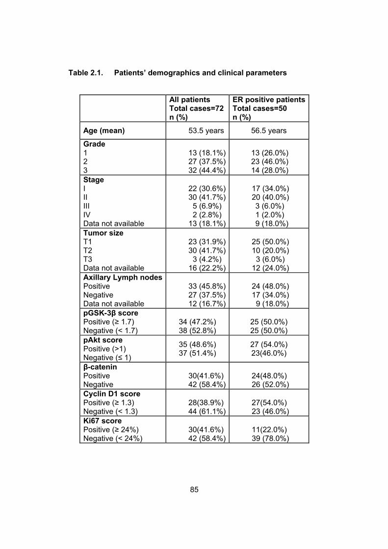
85
Table 2.1. Patients’ demographics and clinical parameters
All patientsTotal cases=72n (%)
ER positive patientsTotal cases=50n (%)
Age (mean) 53.5 years 56.5 years
Grade123
13 (18.1%)27 (37.5%)32 (44.4%)
13 (26.0%)23 (46.0%)14 (28.0%)
StageIIIIIIIVData not available
22 (30.6%)30 (41.7%)
5 (6.9%)2 (2.8%)
13 (18.1%)
17 (34.0%)20 (40.0%)3 (6.0%)1 (2.0%)9 (18.0%)
Tumor sizeT1T2T3Data not available
23 (31.9%)30 (41.7%)
3 (4.2%)16 (22.2%)
25 (50.0%)10 (20.0%)3 (6.0%)
12 (24.0%)Axillary Lymph nodesPositiveNegativeData not available
33 (45.8%)27 (37.5%)12 (16.7%)
24 (48.0%)17 (34.0%)9 (18.0%)
pGSK-3β scorePositive (≥ 1.7)Negative (< 1.7)
34 (47.2%)38 (52.8%)
25 (50.0%)25 (50.0%)
pAkt scorePositive (>1)Negative (≤ 1)
35 (48.6%)37 (51.4%)
27 (54.0%)23(46.0%)
β-cateninPositiveNegative
30(41.6%)42 (58.4%)
24(48.0%)26 (52.0%)
Cyclin D1 scorePositive (≥ 1.3)Negative (< 1.3)
28(38.9%)44 (61.1%)
27(54.0%)23 (46.0%)
Ki67 scorePositive (≥ 24%)Negative (< 24%)
30(41.6%)42 (58.4%)
11(22.0%)39 (78.0%)

86
2.3.2. pGSK-3β is expressed in a subset of breast cancer
We assessed the expression of pGSK-3β in these 72 cases by
immunohistochemistry, and the results are summarized in Table 2.1 and
illustrated in figure 2.1a-c. The immunostaining was heterogeneous
between cases, and sometimes within the same case. Thus, our scoring
method took into account of both the staining intensity and the percentage
of the tumor showing different staining intensity (detailed in Methods and
Materials). Fibroblasts, macrophages and benign lymphocytes were non-
reactive. Overall, pGSK-3β was scored positive, as defined by ≥ the cut off
point of 1.7, in 34 (47.2%) cases. The staining was largely restricted to the
cytoplasm, although a very small number of cases had some degree of
nuclear staining (which was not included in the scoring). As shown in
Table 2.1, the pGSK-3β positivity in the ER-positive group was 50.0% (25
of 50), and that in the ER-negative group was 40.0% (8 of 22). Normal
adjacent ductal epithelial cells were negative or faintly positive for pGSK-
3β.

87
Figure 2.1. Immunohistochemistry for pGSK-3β, pAkt and β-catenin in breast cancer. Figure 2.1a, b, c: Immunohistochemical staining in breast cancer showing a pGSK-3β negative case (a), pGSK-3β weakly positive case (b) and a pGSK-3β strongly positive case (c). The pGSK-3β staining was cytoplasmic in most cases. Figure 2.1 d, e: Immunohistochemical staining in breast cancer showing a pAkt negative case (d) and a pAkt positive case (e). The pAkt staining was mostly cytoplasmic. Figure 2.1 f, g: Immunohistochemical staining in breast cancer showing a β-catenin negative case with only membraneous staining (f) and a β-catenin positive case with prominent nuclear staining (g).

88
Table 2.2. Correlation between pGSK-3β and pAkt with other
markers
* Statistically significant
Markers pGSK-3β p-value (Spearman)
pAkt p-value (Spearman)
Overall group
ER positive group
Overall group
ER positive group
pAkt 0.01* 0.003*pGSK-3β 0.01* 0.003*β-catenin 0.26 0.29 < 0.0001* 0.001*Cyclin D1
0.91 0.68 0.02* 0.28
Ki67 0.48 0.10 0.96 0.08

89
2.3.3. pGSK-3β correlates with pAkt expression
We then assessed if pGSK-3β correlates with pAkt. Similar to pGSK-3β,
the immunostaining was heterogeneous between cases, and sometime,
within the same case. The pAkt immunostaining, as illustrated in figure
2.1d and 1e, was largely restricted to the cytoplasm, although a small
number of cases had some degree of nuclear staining (not included in the
scoring). pAkt was found positive, as defined by >1.0, in 31 (43.1%)
cases. Normal adjacent ductal epithelial cells were either negative or only
faintly positive. There was significant correlation between pAkt and pGSK-
3β in the entire group (p=0.01, Spearman correlation) (Table 2.2). In
addition, in the ER -positive group (n=50), the correlation was found to be
highly significant (p=0.003, Spearman) (Table 2.2). The frequency of pAkt
positivity in ER-positive group was similar to the overall group (54.0% as
opposed to 48.6%). In ER negative group (n=22), the pAkt positivity was
36.4% (8 of 22). However, no significant correlation was found between
pAkt and pGSK-3β in this group.
2.3.4. Nuclear localization of β-catenin significantly correlates with
pAkt but not pGSK-3β
We next assessed the expression of β-catenin, as it has been described to
be negatively regulated by GSK-3β (Hagen et al., 2002). Since β-catenin
is a transcriptional factor, we used the nuclear localization of β-catenin as
a surrogate marker for its transcriptionally active status. The β-catenin
staining is illustrated in Figure 2.1f and 1g. Overall, 30 (41.6%) of cases
were positive. No significant correlation was found between nuclear β-
catenin and pGSK-3β (p=0.26, Spearman) (Table 2.2). Similar results
were obtained in ER-positive group (n=50) (p=0.29, Spearman).
Nevertheless, we identified a statistically significant positive correlation
between the expression of β-catenin and pAkt in the overall group (p<
0.0001, Spearman), as well as in the ER-positive group (p=0.001,
Spearman).

90
2.3.5. pAkt but not pGSK-3β correlates with cyclin D1
Since cyclin D1 is one of the known downstream targets of GSK-3β and β-
catenin, we sought possible correlations among these markers. Cyclin D1
was assessed positive, as defined by ≥ 1.3, in 28 (38.9%) cases. No
significant correlation was found between cyclin D1 and pGSK-3β
(Spearman correlation p=0.91) (Table 2.2). Similar results were obtained
in the ER-positive group (p=0.68, Spearman). Interestingly, we found a
significant correlation between pAkt expression and cyclin D1 in the entire
group (p=0.02, Spearman), but no significant correlation was found in the
ER-positive group (p=0.2, Spearman). We found no significant correlation
between cyclin D1 and β-catenin in the whole group or in the ER positive
group (p=0.1 and 0.25, Spearman, respectively). Lastly, there was a
significant correlation between cyclin D1 positivity and a positive ER status
(p< 0.0001, Spearman).
2.3.6. pGSK-3β does not correlate with Ki67
To assess if pGSK-3β has any impact on cell proliferation, we correlated
high Ki67 staining with that of pGSK-3β. Ki67 was positive, as defined by ≥
24%, in 30 (41.6%) cases. No significant correlation was found between
Ki67 positivity and pGSK-3β positivity in the entire group (p=0.4,
Spearman), or in the ER-positive group (p=0.1 Spearman).
2.3.7. pGSK-3β significantly correlates with poor survival
pGSK-3β significantly correlated with the overall survival (p=0.004, Log
rank) (figure 2.2). However, in the ER positive cases, no significant
correlation was found between survival and pGSK-3β. In addition, we did
not find any significant correlation between the pGSK-3β status and the
tumor size, lymph node status or clinical stage (p=1.0, 0.4 and 1.0
respectively). The expression of pAkt, cyclin D1 and β-catenin did not
significantly correlate with the overall survival. Cox regression analysis

91
was performed to determine if the clinical/pathologic features correlated
with the overall survival. The clinical/pathological features included axillary
lymph node involvement, tumor stage, tumor grade, the ER status, and
biomarkers examined in this study (pGSK-3β, pAkt, cyclin D1, β-catenin
and Ki67). Univariate analysis revealed that pGSK-3β and the clinical
stage significantly correlated with the overall survival. When these
variables were included in the multivariate analysis, only pGSK-3β
significantly correlated with survival (p=0.001; hazard ratio=7.9). Patients
with a high clinical stage (stage II, III or IV) showed a trend for a shorter
survival (p=0.053, hazard ratio=3.3) (Table 2.3).

92
Figure 2.2. pGSK-3β expression significantly correlates with a worse clinical outcome (p=0.004, log-rank). Cut point analysis for overall survival was used to dichotomize continuous pGSK-3β. The optimal cut-off point for pGSK-3β was ≥ 1.7.

93
Table 2.3. Results of univariate and multivariate analysis using
Cox proportional hazards to determine the prognostic value of
different variables in relation to the overall survival
Variable Univariate p-value Multivariate p-valueAxillary lymph node involvement
NS NS
Tumor stage 0.126* 0.053*Tumor grade NS NSpGSK-3β 0.016* 0.001*pAkt NS NSβ-catenin NS NSCyclin D1 NS NSEstrogen receptor status
NS NS
NS=not statistically significant
* Statistically significant

94
2.4. DISCUSSION
In view of the known biological importance of GSK-3β in cancer, we used
immunohistochemistry to assess the abundance and localization of the
inactivated/phosphorylated form of pGSK-3β in a series of 72 primary
breast cancers. We determined that 47.2% of breast cancers in our cohort
were positive for pGSK-3β. This frequency of pGSK-3β positivity is quite
similar to that reported by Ding et. al., who reported 60% in their cohort
(Ding et al., 2007).
One of our key questions was to test the hypothesis that Akt activation
contributes to GSK-3β inactivation in breast tumor samples, as it has been
reported to be the case in various in-vitro models (Cohen et al., 1997; Pap
and Cooper, 1998). Our finding that pGSK-3β significantly correlates with
pAkt supports this hypothesis. Given that this correlation was highly
significant in the ER-positive subgroup (p=0.003, Spearman), we believe
that GSK-3β can be inactivated via other mechanisms (e.g. the WCP),
especially in ER-negative tumors. Other mechanisms that are known to
inactivate GSK-3β, such as cyclic AMP-dependent protein kinase (PKA)
and p90Rsk, may also play a role (Fang et al., 2000; Stambolic and
Woodgett, 1994). These alternative mechanisms may result in the
increased discordance between pGSK-3β and pAkt in breast cancer.
Our study showed that β-catenin is positive in 41.6% of breast cancer.
This finding is slightly lower than those found in 3 other studies (i.e. in the
range of 60-70%) (Chung et al., 2004; Lin et al., 2000; Ryo et al., 2001).
We believe that this discrepancy is most likely due to the fact that the
other three studies scored the staining regardless of its localization (i.e.
cytoplasmic versus nuclear). We scored only the nuclear β-catenin
staining since we believe that the nuclear β-catenin is more representative
of its transcriptional activity status. We found a significant correlation
between β-catenin and pAkt both in the overall cases and in the ER-

95
positive subgroup (p< 0.0001 and 0.001 respectively). However, there was
no significant correlation between β-catenin and pGSK-3β. These results
suggest that β-catenin may be predominantly regulated by pAkt by
mechanisms not involving pGSK-3β. This is in keeping with a study
showing that pAkt can directly enhance the nuclear accumulation and the
transcriptional activity of β-catenin by phosphorylating β-catenin at the
cell-cell junctions, thereby enhancing its release into the cytosol/nuclei
(Fang et al., 2007).
Our study yielded a cyclin D1 positivity of 38.9%, which is similar to what
has been reported in previous studies. In addition, we found a significant
correlation between cyclin D1 overexpression and the positive ER status
(p< 0.0001), and this finding is similar to that previously reported (Elsheikh
et al., 2008). We found no significant correlation between pGSK-3β and
cyclin D1. This result is not overly surprising to us; while previous in-vitro
studies have shown that GSK-3β regulates the protein level, nuclear
localization and phosphorylation of cyclin D1 (Diehl et al., 1998), a more
recent report showed that inhibition of the GSK-3 activity has no influence
on the cyclin D1 level (Yang et al., 2006). In the same study, the authors
proposed that the cyclin D1 levels are regulated in accordance with
various stages of the cell cycle rather than by signaling activity (Yang et
al., 2006).
The biological significance of GSK-3β inactivation appears to be cell-type
specific. In cancers of colon, ovary and pancreas, active GSK-3β is
believed to promote cell survival and proliferation (Ougolkov and
Billadeau, 2006). However, Ding et. al. reported that active GSK-3β
sensitizes breast cancer cells to chemotherapeutic agents by down
regulating Mcl-1, an anti-apoptotic protein (Ding et al., 2007). Thus, it is
conceivable that GSK-3β inactivation may promote tumor cell survival and
perhaps, confer a worse clinical outcome by virtue of its anti-apoptotic

96
effects. In support of this hypothesis, we found that expression of pGSK-
3β significantly correlated with shorter overall survival. To our knowledge,
our study is the first correlating the expression of pGSK-3β and the
survival of cancer patients, and our findings suggest that
immunohistochemical detection of pGSK-3β may provide prognostically
useful information for breast cancer patients. Due to the relatively small
sample size in this study, larger studies with the full representation of
various molecular subtypes of breast cancer are required to confirm the
prognostic significance of pGSK-3β in breast cancer. Of note, we had a
relatively a low number of HER2-positive cases which may be due to
several factors: 1) we did not do in situ hybridization for equivocal cases
(n=6); 2) Only 64 cases had adequate tissues; thus, 8 cases were not
evaluated for HER2 and this may have lowered the number of HER2-
positive cases; 3) 12.5% of cases in our cohort were invasive lobular
carcinomas, which are generally HER2-negative.
The prognostic value of pAkt in breast cancer is controversial in the
literature; while studies found no statistical prognostic value of this marker,
other studies suggested that pAkt is prognostically useful (Panigrahi et al.,
2004; Schmitz et al., 2004). In our study, we found that pAkt is not
prognostically significant. While β-catenin has previously been suggested
to be prognostic in one study (Lin et al., 2000), their method for scoring β-
catenin was different from ours, as described above. Additional studies
may be needed to verify the prognostic value of this marker in breast
cancer.

97
2.5. REFERENCES
Bose, S., Chandran, S., Mirocha, J.M., and Bose, N. (2006). The Akt pathway in human breast cancer: a tissue-array-based analysis. Mod Pathol 19, 238-245.
Cao, Q., Lu, X., and Feng, Y.J. (2006). Glycogen synthase kinase-3beta positively regulates the proliferation of human ovarian cancer cells. Cell Res 16, 671-677.
Chung, G.G., Zerkowski, M.P., Ocal, I.T., Dolled-Filhart, M., Kang, J.Y., Psyrri, A., Camp, R.L., and Rimm, D.L. (2004). beta-Catenin and p53 analyses of a breast carcinoma tissue microarray. Cancer 100, 2084-2092.
Chung, R., Peters, A.C., Armanious, H., Anand, M., Gelebart, P., and Lai, R. Biological and clinical significance of GSK-3beta in mantle cell lymphoma--an immunohistochemical study. Int J Clin Exp Pathol 3, 244-253.
Clevers, H. (2006). Wnt/beta-catenin signaling in development and disease. Cell 127, 469-480.
Cohen, P., Alessi, D.R., and Cross, D.A. (1997). PDK1, one of the missing links in insulin signal transduction? FEBS Lett 410, 3-10.
Diehl, J.A., Cheng, M., Roussel, M.F., and Sherr, C.J. (1998). Glycogen synthase kinase-3beta regulates cyclin D1 proteolysis and subcellular localization. Genes Dev 12, 3499-3511.
Ding, Q., He, X., Xia, W., Hsu, J.M., Chen, C.T., Li, L.Y., Lee, D.F., Yang, J.Y., Xie, X., Liu, J.C., et al. (2007). Myeloid cell leukemia-1 inversely correlates with glycogen synthase kinase-3beta activity and associates with poor prognosis in human breast cancer. Cancer Res 67, 4564-4571.
Doble, B.W., and Woodgett, J.R. (2003). GSK-3: tricks of the trade for a multi-tasking kinase. J Cell Sci 116, 1175-1186.
Elsheikh, S., Green, A.R., Aleskandarany, M.A., Grainge, M., Paish, C.E., Lambros, M.B., Reis-Filho, J.S., and Ellis, I.O. (2008). CCND1 amplification and cyclin D1 expression in breast cancer and their relation with proteomic subgroups and patient outcome. Breast Cancer Res Treat109, 325-335.
Embi, N., Rylatt, D.B., and Cohen, P. (1980). Glycogen synthase kinase-3 from rabbit skeletal muscle. Separation from cyclic-AMP-dependent protein kinase and phosphorylase kinase. Eur J Biochem 107, 519-527.

98
Fang, D., Hawke, D., Zheng, Y., Xia, Y., Meisenhelder, J., Nika, H., Mills, G.B., Kobayashi, R., Hunter, T., and Lu, Z. (2007). Phosphorylation of beta-catenin by AKT promotes beta-catenin transcriptional activity. J Biol Chem 282, 11221-11229.
Fang, X., Yu, S.X., Lu, Y., Bast, R.C., Jr., Woodgett, J.R., and Mills, G.B. (2000). Phosphorylation and inactivation of glycogen synthase kinase 3 by protein kinase A. Proc Natl Acad Sci U S A 97, 11960-11965.
Gelebart, P., Anand, M., Armanious, H., Peters, A.C., Dien Bard, J., Amin, H.M., and Lai, R. (2008). Constitutive activation of the Wnt canonical pathway in mantle cell lymphoma. Blood 112, 5171-5179.
Gordon, M.D., and Nusse, R. (2006). Wnt signaling: multiple pathways, multiple receptors, and multiple transcription factors. J Biol Chem 281, 22429-22433.
Hagen, T., Di Daniel, E., Culbert, A.A., and Reith, A.D. (2002). Expression and characterization of GSK-3 mutants and their effect on beta-catenin phosphorylation in intact cells. J Biol Chem 277, 23330-23335.
Jope, R.S., and Johnson, G.V. (2004). The glamour and gloom of glycogen synthase kinase-3. Trends Biochem Sci 29, 95-102.
Lin, S.Y., Xia, W., Wang, J.C., Kwong, K.Y., Spohn, B., Wen, Y., Pestell, R.G., and Hung, M.C. (2000). Beta-catenin, a novel prognostic marker forbreast cancer: its roles in cyclin D1 expression and cancer progression. Proc Natl Acad Sci U S A 97, 4262-4266.
Mikels, A.J., and Nusse, R. (2006). Wnts as ligands: processing, secretion and reception. Oncogene 25, 7461-7468.
Miller, J.R. (2002). The Wnts. Genome Biol 3, REVIEWS3001.
Ougolkov, A.V., and Billadeau, D.D. (2006). Targeting GSK-3: a promising approach for cancer therapy? Future Oncol 2, 91-100.
Ougolkov, A.V., Fernandez-Zapico, M.E., Savoy, D.N., Urrutia, R.A., and Billadeau, D.D. (2005). Glycogen synthase kinase-3beta participates in nuclear factor kappaB-mediated gene transcription and cell survival in pancreatic cancer cells. Cancer Res 65, 2076-2081.
Panigrahi, A.R., Pinder, S.E., Chan, S.Y., Paish, E.C., Robertson, J.F., and Ellis, I.O. (2004). The role of PTEN and its signalling pathways,
99
including AKT, in breast cancer; an assessment of relationships with other prognostic factors and with outcome. J Pathol 204, 93-100.
Pap, M., and Cooper, G.M. (1998). Role of glycogen synthase kinase-3 in the phosphatidylinositol 3-Kinase/Akt cell survival pathway. J Biol Chem273, 19929-19932.
Ryo, A., Nakamura, M., Wulf, G., Liou, Y.C., and Lu, K.P. (2001). Pin1 regulates turnover and subcellular localization of beta-catenin by inhibiting its interaction with APC. Nat Cell Biol 3, 793-801.
Sanchez, J.F., Sniderhan, L.F., Williamson, A.L., Fan, S., Chakraborty-Sett, S., and Maggirwar, S.B. (2003). Glycogen synthase kinase 3beta-mediated apoptosis of primary cortical astrocytes involves inhibition of nuclear factor kappaB signaling. Mol Cell Biol 23, 4649-4662.
Schmitz, K.J., Otterbach, F., Callies, R., Levkau, B., Holscher, M., Hoffmann, O., Grabellus, F., Kimmig, R., Schmid, K.W., and Baba, H.A. (2004). Prognostic relevance of activated Akt kinase in node-negative breast cancer: a clinicopathological study of 99 cases. Mod Pathol 17, 15-21.
Shakoori, A., Ougolkov, A., Yu, Z.W., Zhang, B., Modarressi, M.H., Billadeau, D.D., Mai, M., Takahashi, Y., and Minamoto, T. (2005). Deregulated GSK3beta activity in colorectal cancer: its association with tumor cell survival and proliferation. Biochem Biophys Res Commun 334, 1365-1373.
Stambolic, V., and Woodgett, J.R. (1994). Mitogen inactivation of glycogen synthase kinase-3 beta in intact cells via serine 9 phosphorylation. Biochem J 303 ( Pt 3), 701-704.
Tokunaga, E., Kimura, Y., Mashino, K., Oki, E., Kataoka, A., Ohno, S., Morita, M., Kakeji, Y., Baba, H., and Maehara, Y. (2006). Activation of PI3K/Akt signaling and hormone resistance in breast cancer. Breast Cancer 13, 137-144.
Wolff, A.C., Hammond, M.E., Schwartz, J.N., Hagerty, K.L., Allred, D.C., Cote, R.J., Dowsett, M., Fitzgibbons, P.L., Hanna, W.M., Langer, A., et al.(2007). American Society of Clinical Oncology/College of American Pathologists guideline recommendations for human epidermal growth factor receptor 2 testing in breast cancer. J Clin Oncol 25, 118-145.
Woodgett, J.R., and Cohen, P. (1984). Multisite phosphorylation of glycogen synthase. Molecular basis for the substrate specificity of

100
glycogen synthase kinase-3 and casein kinase-II (glycogen synthase kinase-5). Biochim Biophys Acta 788, 339-347.
Yang, K., Guo, Y., Stacey, W.C., Harwalkar, J., Fretthold, J., Hitomi, M., and Stacey, D.W. (2006). Glycogen synthase kinase 3 has a limited role in cell cycle regulation of cyclin D1 levels. BMC Cell Biol 7, 33.
Yaziji, H., Taylor, C.R., Goldstein, N.S., Dabbs, D.J., Hammond, E.H., Hewlett, B., Floyd, A.D., Barry, T.S., Martin, A.W., Badve, S., et al. (2008). Consensus recommendations on estrogen receptor testing in breast cancer by immunohistochemistry. Appl Immunohistochem Mol Morphol 16, 513-520.

101
Chapter 3
BIOLOGICAL AND CLINICAL SIGNIFICANCE OF GSK-3β
IN MANTLE CELL LYMPHOMA – AN
IMMUNOHISTOCHEMICAL STUDY
A version of this chapter has been published as:
Chung R*, Peters A*, Armanious H, Anand M, Gelebart P, Lai R. Biological and
clinical significance of GSK-3β in mantle cell lymphoma–an
immunohistochemical study. Int J Clin Exp Pathol 2010;3(3):244-253.
*Chung R and Peters A are first co-authors
Armanious H, is second author and contributed in the immunohistochemical
studies and writing the manuscript

102
3.1. INTRODUCTION:
GSK-3β is an important signalling protein that has been shown to regulate a wide
range of cellular functions including apoptosis and cell proliferation.
Dysregulation of GSK-3β has been implicated in a wide range of human diseases
including diabetes, Alzheimer’s, schizophrenia, and cancer (Jope and Johnson,
2004; Jope et al., 2007; Karim et al., 2004; Kim and Kimmel, 2000). One of the
major biological functions of GSK-3β is to inhibit β-catenin by sequestration and
promotion of its proteasome degradation. GSK-3β itself is known to be regulated
by the PI3K/Akt and the Wnt canonical pathways (Failor et al., 2007; Joshi et al.,
2007; Manoukian and Woodgett, 2002). GSK-3β inactivation is mediated by the
phosphorylation of its serine 9 residue. Upon inactivation, its inhibitory effect on
β-catenin is released, and β-catenin is allowed to accumulate and translocate to
the nucleus, where it upregulates the transcription of multiple genes including
cyclin D1. GSK-3β also has been shown to directly regulate cyclin D1 via
modifying its rate of proteolysis and nuclear export (Diehl et al., 1998; Takahashi-
Yanaga and Sasaguri, 2008).
Mantle cell lymphoma (MCL) is a specific subtype of aggressive B-cell lymphoma
recognized by the World Health Organization Classification Scheme (WHO)
(Jaffe et al., 2004). The genetic hallmark of this disease is the recurrent
chromosomal abnormality, t(11;14)(q13;q32), which brings the cyclin D1(CCND1)
gene under the influence of the enhancer of the immunoglobulin heavy chain
(IGH) gene, leading to cyclin D1 overexpression. Although it has been shown
that cyclin D1 overexpression is not sufficient for the induction of lymphoma in
animal models, this abnormality is considered to be the primary oncogenic event
in MCL (Diehl, 2002; Jares et al., 2007). In view of the roles of GSK-3β in
regulating the expression and subcellular localization of cyclin D1, as discussed
above, we hypothesized that the activation status of GSK-3β is likely to be
relevant to the pathogenesis of MCL. In support of this hypothesis, we have
recently shown that GSK-3β is inactivated and phosphorylated in all MCL cell
lines and a subset of MCL tumors examined (Gelebart et al., 2008). In another

103
study, using a constitutive active GSK-3β construct, Dal Col et. al. (Dal Col and
Dolcetti, 2008) recently provided the first direct evidence that GSK-3β
phosphorylates cyclin D1 at Thr286, thereby promoting its nuclear export in MCL.
Nevertheless, the biological and clinical significance of GSK-3β in MCL has not
been comprehensively examined. Specifically, whether the activation status of
GSK-3β correlates with the nuclear expression of β-catenin, the cyclin D1
expression level and/or other clinicopathologic parameters is not known.
In this study, using immunohistochemistry, we assessed the expression of
phosphorylated/inactive form of GSK-3β (pGSK-3β) in 83 formalin-fixed, paraffin-
embedded MCL tumors. We then correlated the expression of pGSK-3β with the
nuclear expression of β-catenin, the expression level of cyclin D1 and Ki67
labelling in these tumors. We also evaluated whether pGSK-3β correlates with
various clinical parameters including the overall survival.
3.2. MATERIALS AND METHODS
3.2.1. MCL tumors
All cases of MCL were diagnosed at the Cross Cancer Institute (Edmonton,
Alberta, Canada) between 1994 and 2007, and the diagnostic criteria were based
on those described in the World Health Organization Classification Scheme
(Jaffe et al., 2004). All cases were confirmed to express cyclin D1 by
immunohistochemistry. Six of these 83 MCL tumors used in this study were
blastoid variant. The use of these tissues has been approved by our Institutional
Ethics Committee.
3.2.2. Immunohistochemistry
To detect pGSK-3β, β-catenin and Ki67, antigen retrieval was performed using
microwave-treated citrate buffer (pH 6.0) for 20 minutes. To detect cyclin D1,
antigen retrieval was performed using EDTA (pH=8.0). After antigen retrieval,
tissue sections were incubated with 3% hydrogen peroxide (H2O2) for 10 minutes
to block endogenous peroxidase activity. Subsequently, sections were incubated

104
overnight at 4˚C with a rabbit polyclonal antibody reactive with pGSK-3β (Ser
9)(1:50, Cell Signaling Technology, Danvers, MA), a rabbit polyclonal anti-β-
catenin (1:1200 dilution, Sigma, clone C2206, Oakville, Ontario, Canada), anti-
cyclin D1 (1:75 dilution, clone SP4, Medicorp, Montreal, Quebec, Canada) or
anti-Ki67 (1:60 dilution, clone M7240, Dako Canada Inc., Mississauga, Ontario,
Canada). Immunostaining was visualized with a labelled streptavidin-biotin
(LSAB) method using DAB as a chromogen (Dako Canada).
3.2.3. Scoring of the markers and statistical analysis
The cytoplasmic expression of pGSK-3β was assessed as 3+ (strong staining),
2+ (moderate staining), 1+ (weak staining), 0 (no staining). For β-catenin, cases
showing definitive nuclear immunostaining in >20% of the tumor cells were
considered positive, and the β-catenin nuclear staining was scored as 3+ (strong
staining), 2+ (moderate staining) and 1+ (weak staining). Score for pGSK-3β was
calculated as follows: 3 points were given for 3+ staining, 2 points for 2+ staining,
1 point for 1+ staining and 0 for no staining. Cut point analysis for overall survival
was used to dichotomize continuous pGSK-3β with the minimum p-value method
applied to estimate the optimum pGSK-3β division. The analysis found a pGSK-
3β score of 0.6 is the optimal cut point. For nuclear β-catenin, <20% of the tumor
cells showing staining or a score of 1 was considered negative, and a score of 2+
or 3+ was considered positive. The scoring used to assess the expression of
cyclin D1 and Ki67 have been previously detailed (Hui et al., 2005). All of the
staining was reviewed by two observers (RC and RL) independently and
discrepant cases were reviewed under a double-headed microscope to reach a
consensus.
The correlations between pGSK-3β and the other biological markers were
assessed using Fisher's exact test for tables and Spearman rank correlation for
continuous variables. Univariate and multivariate Cox regression analysis was
used to determine the influence of pGSK-3β and various biological and clinical
parameters on overall survival. Overall survival plots of the factor groups were

105
calculated using the Kaplan-Meier method. Statistical tests are two-tailed with a
P value <0.05 considered to be statistically significant. The SAS computer
program SAS (r) 9.2 (TS1M0) was used to perform the analysis
3.3. RESULTS
3.3.1. Clinical characteristics of MCL patients
The clinicopathologic characteristics of the 83 MCL patients included in this study
are summarized in Table 3.1. There were 69 men and 14 women, and the
median age at diagnosis was 64 years (range, 41-88 years). Fifty patients died
during the follow-up and the median time from the initial diagnosis to either the
last follow-up or death was 29 months. Treatment for each MCL patient was
determined during our weekly lymphoma conference based on our provincial
lymphoma treatment protocol. For first-line treatment, the majority of these
patients received CHOP-based (cyclophosphamide, doxorubicin, vincristine,
prednisone) chemotherapy, and small subsets of patients received chlorambucil-
based chemotherapy or more novel treatments such as proteasome inhibitor
PS341 and flavopirodol.

106
Table 3.1. Patients’ Demographics and Clinical Parameters
pGSK3-β
( negative = <0.6 ) Total cases = 27
n (%)
( postive = ≥ 0.6 ) total cases = 56
n (%)
Total cases = 83
Age (mean) 63.3 65.5 83
Gender Female
Male5 (18.5)
22 (81.5)9 (16.1)
47 (83.9)1469
Stage I-II III IV
Missing
4 (14.8)6 (22.2)
15 (55.6)2 (7.4)
4 (7.1)14 (25.0)37 (56.1)
1 (1.8)
820523
B Symptoms Yes No
Missing
8 (29.6)17 (63.0)
2 (7.4)
15 (26.8)39 (69.6)
2 (3.6)
23564
GI Involvement Yes No
Missing
6 (22.2)19 (70.4)
2 (7.4)
6 (10.7)48 (85.7)
2 (3.6)
12674
IPI0 - 2 ≥ 3
missing
15 (55.6)4 (14.8)8 (29.6)
36 (64.3)12 (21.4)8 (14.3)
511616
β-catenin 0-1 2-3
Missing
23 (85.2)3 (70.4)1 (3.7)
27 (48.2)23 (41.1)6 (10.7)
50267
Cyclin D1 1-2 3
Missing
18 (66.7)3 (11.1)6 (22.2)
23 (41.1)26 (46.4)7 (12.5)
412913
Ki67 0-3 4
Missing
21 (77.8)4 (14.8)2 (7.4)
37 (66.1)18 (32.1)
1 (1.8)
58223
107
3.3.2. pGSK-3β significantly correlates with β-catenin positivity
Of the 83 MCL tumors, 56 (67.5%) were considered pGSK-3β positive, based on
a minimum p-value cut-off of ≥ 0.6. The expression of pGSK-3β was found to be
largely localized to the cytoplasm of the MCL cells (Figure 3.1 a, b). Of the 76
MCL tumors that were also evaluated for the expression of β-catenin, 26 (32.5%)
were assessed positive. In contrast with pGSK-3β, β-catenin was largely
expressed in the nuclei of MCL cells (Figure 3.1 c, d). pGSK-3β positivity
significantly correlated with the expression of β-catenin (p = 0.0025, Fisher’s
exact ) (Table 3.2).
3.3.3. pGSK-3β significantly correlates with cyclin D1 expression
As illustrated in Figure 3.1 e and f, the cyclin D1 immunostaining was largely
restricted to the nuclei of MCL cells in all cases. Although all cases were positive
for cyclin D1 (as per definition of MCL), the proportion of strongly positive cells
was highly variable among tumors. To ensure that the assessment of the cyclin
D1 expression status was uniform, we excluded 13 cases for which a different
anti-cyclin D1 antibody was used and insufficient tissues were available to repeat
the staining. Of the remaining 70 cases, 29 (41.4%) were considered high cyclin
D1-expressing. Statistical analysis revealed that the high-expressing cyclin D1
status correlates significantly with positive pGSK-3β (p = 0.0032) (Table 3.3).
Since the gene expression of cyclin D1 has been shown to be up-regulated by β-
catenin in various in-vitro models, we addressed whether there is a significant
correlation between the expression of cyclin D1 and β-catenin positivity. Data for
both markers was available in 63 cases. In the β-catenin positive group (n=23),
13 (56.5%) were high cyclin D1-expressing. In the β-catenin negative group
(n=40), only 12 (30%) were high cyclin D1-expressing; the two markers had a
correlation that was not quite significant (p = 0.06) (Table 3.4).

108
3.3.4. pGSK-3β does not correlate with the Ki67
The percentage of tumor cells with Ki67 expression was assessed in all MCL
tumors and categorized into 2 groups: <50% or ≥ 50% (Figure 3.1 g, h). Three of
83 cases did not have sufficient Ki67 data. Of the remaining 80 cases, 22 had a
≥50% Ki67 labelling. Only 4 (16%) of the 25 pGSK-3β negative cases had a
≥50% Ki67 labelling, as compared to 18 of 55 (49%) pGSK-3β positive cases.
This correlation is not statistically significant (p = 0.177, Fisher’s exact). Ki67 also
did not significantly correlate with β-catenin or cyclin D1.

109
Table 3.2. Correlation between pGSK-3β and β-catenin
β-cateninNegative (0 or 1) Positive (2 or 3)
Total P
pGSK3βnegative (< 0.6)positive (≥ 0.6)
2327
323
2650
0.0025
Total 50 26 76
Table 3.3. Correlation between pGSK-3β and cyclin D1
Cyclin D1Low (1 or 2) High (3)
Total P
pGSK-3βnegative (< 0.6)positive (≥ 0.6)
1823
326
2149
0.0032
Total 41 29 70
Table 3.4. Correlation between cyclin D1 and β-catenin
β-cateninNegative (0 or 1) Positive (2 or 3)
Total P
Cyclin D1Low (1 or 2)
High (3)2812
1013
3825
0.0606
Total 40 23 63

110
Figure 3.1(a-d). Immunohistochemistry of pGSK-3β and β-catenin in MCL. Figure 3.1a, b: Immunohistochemical staining of pGSK-3β in MCL tumors revealed a subset of negative cases and a subset of positive case (a and b respectively). The staining was predominantly cytoplasmic. Figure 3.1c, d: Immunohistochemical staining of β-catenin in MCL tumors revealed a subset of negative cases and a subset of positive case (illustrated in c and d respectively). Of note, the staining in the tumor cells was predominantly nuclear; in contrast, the staining was mostly found in the cytoplasm of the endothelial cells.

111
Figure 3.1(e-h). Immunohistochemistry of cyclin D1 and Ki67 in MCL.Figure 3.1e, f: Immunohistochemical staining of cyclin D1 in MCL tumors was heterogeneous with regard to the proportion of intensely positive cells. The case in 1e was assessed cyclin D1-low, as strongly positive cells were <50% of the neoplastic cell population. The case in figure 3.1f was assessed cyclin D1-high, as strongly positive cells were ≥ 50% of the neoplastic cell population. Figure 3.1 g, h. Immunohistochemical staining of Ki67 in MCL tumors also revealed a high degree of heterogeneity in the proportion of positive cells, showing a Ki67-negative case and a Ki67 positive case (g and h respectively).

112
3.3.5. pGSK-3β significantly correlates with overall survival and absolute
lymphocytosis
Using a pGSK-3β cut point of ≥ 0.6 univariate Cox survival analysis we found a
significant correlation between pGSK-3β and overall survival (p = 0.045, HR =
1.89), with a pGSK-3β negative status associated with a better outcome (Table
3.5). The Kaplan-Meier survival curves are shown in Figure 3.2. In keeping with
our previously published data cases with ≥50% cells positive for Ki67
significantly correlated with a shorter survival (p<0.0001) (Hui et al., 2005).
Multivariate analysis found the prognostic value of pGSK-3β (p = 0.030, HR =
2.10) was independent of Ki67 (p<0.0001, HR = 4.40). There was no significant
interaction effect between pGSK-3β and Ki67 (p = 0.71). Neither cyclin D1 nor β-
catenin shows a significant correlation with the overall survival (Table 3.5).
We also assessed if there is a correlation between the expression of pGSK-3β
and various clinical parameters, including clinical stage, the international
prognostic index, absolute lymphocytosis, elevation of lactate dehydrogenase,
patient age at diagnosis and extra-nodal involvement; only peripheral blood
lymphocytosis (using a cut-off of >6 x 10⁹/L) significantly correlates with pGSK-3β
status (p = 0.0011, Spearman).

113
Table 3.5. Cox regression analysis of overall survival
Unitivariate Multivariate
Total cases assessed
HR ( 95% CI )
P HR ( 95% CI ) P
pGSK-3β < 0.6 ≥ 0.6
83 1.001.89 (1.01-
3.53)0.045
1.002.10 ( 1.075-
4.09)0.030
Ki67 0-3 4
80 1.004.45 ( 2.29 -
8.67)<0.0001
1.004.40 ( 2.24-
8.64)<0.0001
Cyclin D1 1 or 2
370 1.00
1.17 ( 0.79-1.73) 0.442
β-catenin 0 or 1 2 or 3
76 1.001.06 ( 0.78-
1.44)0.732
IPI 0-2 3-5
81 1.002.08 ( 1.08-
4.03)0.030

114
Figure 3.2. pGSK-3β expression significantly correlates with overall
survival (p=0.045, Log rank).

115
3.4. DISCUSSION
GSK-3β is a biologically important signalling protein. In the Wnt canonical
pathway (WCP), it plays a central role in sequestrating β-catenin and promoting
the proteasome destruction of β-catenin (Staal and Clevers, 2005). In this study,
using pGSK-3β as a surrogate marker detectable by an immunohistochemical
method, we comprehensively examined the GSK-3β inactivation status in 83
paraffin-embedded MCL tumors. We found evidence that GSK-3β is inactivated
in two-thirds in this cohort. We have also provided evidence that the pGSK-3β
positive status significantly correlates with the nuclear expression of β-catenin, a
relatively high expression level of cyclin D1, absolute lymphocytosis in the
peripheral blood, and shorter overall survival in MCL patients. In summary, our
data has provided evidence to support that GSK-3β is both biologically and
clinically important in MCL.
With regard to the mechanism by which GSK-3β is inactivated, our group has
recently published evidence of constitutive activation of the WCP in MCL cell
lines and patient tumours, which is likely to be attributed to the autocrine and/or
paracrine stimulation of various Wnt ligands including Wnt3 (Gelebart et al.,
2008). These findings echo those of two recently published studies. In the first
study, the authors showed that RNA-interference to abrogate the expression of
Fz2, a receptor for Wnt ligands, decreases cell proliferation in a MCL cell line
(Ortega-Paino et al., 2008). In the second study, the authors employed gene
expression profiling experiments and identified a significant upregulation of
several genes in the WCP in MCL compared to naïve B cells (Rizzatti et al.,
2005). In addition to the WCP, the PI3K/Akt signalling pathway may also
contribute to GSK-3β inactivation. To this end, it has been previously reported
that the PI3K/Akt signalling pathway is constitutively activated in a subset of MCL
(Rudelius et al., 2006).
In various in-vitro experimental models, β-catenin has been shown to accumulate
and translocate to the nucleus upon inactivation of GSK-3β. This appears to be

116
the case for MCL, since we observed that the pGSK-3β positive status
significantly correlates with the nuclear expression of β-catenin (i.e. p = 0.0025,
Fisher’s exact). In this regard, β-catenin nuclear accumulation coupled with GSK-
3β inactivation is also observed in a number of other types of cancer, including
chronic myeloid leukemia in blast crisis, precursor B-cell acute lymphoblastic
leukemia and specific types of epithelial cancers (Clevers, 2006; Jamieson et al.,
2004; Radich et al., 2006). Nevertheless, in our study, a substantial proportion of
pGSK-3β positive cases, namely 27 of 50 (54%) cases, did not have detectable
β-catenin. One likely explanation is that additional mechanisms may be required
for the nuclear transport of β-catenin, and these mechanisms are not operational
in some MCL tumors.
The finding of heterogeneous cyclin D1 immunostaining in paraffin-embedded
MCL tissues is commonly observed by diagnostic hematopathologists. It is highly
unlikely that this staining heterogeneity is due to variations in fixation or other
technical reasons, since strongly cyclin D1-positive cells were seen in almost all
cases. Thus, we believe that there is a genuine biological difference in the
protein expression level of cyclin D1. Our findings of a significant correlation
between a high level of cyclin D1 expression and the pGSK-3β positive status (p
= 0.0032, Fisher’s exact) support the concept that GSK-3β plays an active role in
regulating the protein level of cyclin D1. Variation in the cyclin D1 protein level in
MCL is also likely to be related to the variations in the cyclin D1 mRNA levels, as
previously reported by multiple groups (Marzec et al., 2006).
Of various clinical parameters analyzed in this study, only absolute
lymphocytosis (arbitrarily defined as >6.0 x 10⁹/L in this study) and overall
survival significantly correlate with pGSK-3β. It is unlikely that the correlation
between lymphocytosis and pGSK-3β is related to the tumor burden, since the
pGSK-3β status does not significantly correlate with the clinical stage. In view of
the fact that GSK-3β has been shown to regulate cell motility and cell-to-cell
adhesions (Bianchi et al., 2005; Etienne-Manneville and Hall, 2003; Jope et al.,
117
2007; Zhou et al., 2004), we have considered the possibility that GSK-3β may
directly modulate the cell migratory and adhesion properties of MCL cells, such
that inactivation of GSK-3β predisposes to a leukemic phase.
One of the key findings in this study is the observation that GSK-3β inactivation is
significantly associated with a worse clinical outcome in MCL patients. Since
pGSK-3β did not significantly correlate with Ki67, it is unlikely that its association
with a worse clinical outcome is directly linked to increased cell proliferation. In
view of the biologic importance of GSK-3β in various normal cellular functions
and the pathogenesis of various cancers, we believe that this finding is rather not
too surprising. Since the expression of β-catenin does not show a significant
correlation with survival, the prognostic value of GSK-3β is likely to be mediated
via downstream effectors other than β-catenin.
In contrast with two other previous studies (Rosenwald et al., 2003; Yatabe et al.,
2000), we did not identify a significant correlation between the high cyclin D1
expression status and overall survival. A plausible explanation for this
discrepancy is related to the difference in the experimental approach employed.
While we measured the protein level expression of cyclin D1 using
immunohistochemistry, the other two studies assessed the levels of cyclin D1
mRNA in MCL tumors. Of note, while MCL cells express two cyclin D1 mRNA
species, namely cyclin D1a and cyclin D1b, only cyclin D1a is translated into
protein in MCL cells (Marzec et al., 2006). Moreover, the anti-cyclin D1 antibody
used in this immunohistochemical study recognizes only the cyclin D1a isoform.
These two factors may have contributed to this discrepancy of our conclusions.
118
3.5. REFERENCES
Bianchi, M., De Lucchini, S., Marin, O., Turner, D.L., Hanks, S.K., and Villa-Moruzzi, E. (2005). Regulation of FAK Ser-722 phosphorylation and kinase activity by GSK3 and PP1 during cell spreading and migration. Biochem J 391, 359-370.
Clevers, H. (2006). Wnt/beta-catenin signaling in development and disease. Cell127, 469-480.
Dal Col, J., and Dolcetti, R. (2008). GSK-3beta inhibition: at the crossroad between Akt and mTOR constitutive activation to enhance cyclin D1 protein stability in mantle cell lymphoma. Cell Cycle 7, 2813-2816.
Diehl, J.A. (2002). Cycling to cancer with cyclin D1. Cancer Biol Ther 1, 226-231.
Diehl, J.A., Cheng, M., Roussel, M.F., and Sherr, C.J. (1998). Glycogen synthase kinase-3beta regulates cyclin D1 proteolysis and subcellular localization. Genes Dev 12, 3499-3511.
Etienne-Manneville, S., and Hall, A. (2003). Cdc42 regulates GSK-3beta and adenomatous polyposis coli to control cell polarity. Nature 421, 753-756.
Failor, K.L., Desyatnikov, Y., Finger, L.A., and Firestone, G.L. (2007). Glucocorticoid-induced degradation of glycogen synthase kinase-3 protein is triggered by serum- and glucocorticoid-induced protein kinase and Akt signaling and controls beta-catenin dynamics and tight junction formation in mammary epithelial tumor cells. Mol Endocrinol 21, 2403-2415.
Gelebart, P., Anand, M., Armanious, H., Peters, A.C., Dien Bard, J., Amin, H.M., and Lai, R. (2008). Constitutive activation of the Wnt canonical pathway in mantle cell lymphoma. Blood 112, 5171-5179.
Hui, D., Reiman, T., Hanson, J., Linford, R., Wong, W., Belch, A., and Lai, R. (2005). Immunohistochemical detection of cdc2 is useful in predicting survival in patients with mantle cell lymphoma. Mod Pathol 18, 1223-1231.
Jaffe, E.S., Banks, P.M., Nathwani, B., Said, J., and Swerdlow, S.H. (2004). Recommendations for the reporting of lymphoid neoplasms: a report from the Association of Directors of Anatomic and Surgical Pathology. Mod Pathol 17, 131-135.
Jamieson, C.H., Ailles, L.E., Dylla, S.J., Muijtjens, M., Jones, C., Zehnder, J.L., Gotlib, J., Li, K., Manz, M.G., Keating, A., et al. (2004). Granulocyte-macrophage progenitors as candidate leukemic stem cells in blast-crisis CML. N Engl J Med351, 657-667.

119
Jares, P., Colomer, D., and Campo, E. (2007). Genetic and molecular pathogenesis of mantle cell lymphoma: perspectives for new targeted therapeutics. Nat Rev Cancer 7, 750-762.
Jope, R.S., and Johnson, G.V. (2004). The glamour and gloom of glycogen synthase kinase-3. Trends Biochem Sci 29, 95-102.
Jope, R.S., Yuskaitis, C.J., and Beurel, E. (2007). Glycogen synthase kinase-3 (GSK3): inflammation, diseases, and therapeutics. Neurochem Res 32, 577-595.
Joshi, M.B., Ivanov, D., Philippova, M., Erne, P., and Resink, T.J. (2007). Integrin-linked kinase is an essential mediator for T-cadherin-dependent signaling via Akt and GSK3beta in endothelial cells. FASEB J 21, 3083-3095.
Karim, R., Tse, G., Putti, T., Scolyer, R., and Lee, S. (2004). The significance of the Wnt pathway in the pathology of human cancers. Pathology 36, 120-128.
Kim, L., and Kimmel, A.R. (2000). GSK3, a master switch regulating cell-fate specification and tumorigenesis. Curr Opin Genet Dev 10, 508-514.
Manoukian, A.S., and Woodgett, J.R. (2002). Role of glycogen synthase kinase-3 in cancer: regulation by Wnts and other signaling pathways. Adv Cancer Res 84, 203-229.
Marzec, M., Kasprzycka, M., Lai, R., Gladden, A.B., Wlodarski, P., Tomczak, E., Nowell, P., Deprimo, S.E., Sadis, S., Eck, S., et al. (2006). Mantle cell lymphoma cells express predominantly cyclin D1a isoform and are highly sensitive to selective inhibition of CDK4 kinase activity. Blood 108, 1744-1750.
Ortega-Paino, E., Fransson, J., Ek, S., and Borrebaeck, C.A. (2008). Functionally associated targets in mantle cell lymphoma as defined by DNA microarrays and RNA interference. Blood 111, 1617-1624.
Radich, J.P., Dai, H., Mao, M., Oehler, V., Schelter, J., Druker, B., Sawyers, C., Shah, N., Stock, W., Willman, C.L., et al. (2006). Gene expression changes associated with progression and response in chronic myeloid leukemia. Proc Natl Acad Sci U S A 103, 2794-2799.
Rizzatti, E.G., Falcao, R.P., Panepucci, R.A., Proto-Siqueira, R., Anselmo-Lima, W.T., Okamoto, O.K., and Zago, M.A. (2005). Gene expression profiling of mantle cell lymphoma cells reveals aberrant expression of genes from the PI3K-AKT, WNT and TGFbeta signalling pathways. Br J Haematol 130, 516-526.
Rosenwald, A., Wright, G., Wiestner, A., Chan, W.C., Connors, J.M., Campo, E., Gascoyne, R.D., Grogan, T.M., Muller-Hermelink, H.K., Smeland, E.B., et al.

120
(2003). The proliferation gene expression signature is a quantitative integrator of oncogenic events that predicts survival in mantle cell lymphoma. Cancer Cell 3, 185-197.
Rudelius, M., Pittaluga, S., Nishizuka, S., Pham, T.H., Fend, F., Jaffe, E.S., Quintanilla-Martinez, L., and Raffeld, M. (2006). Constitutive activation of Akt contributes to the pathogenesis and survival of mantle cell lymphoma. Blood 108, 1668-1676.
Staal, F.J., and Clevers, H.C. (2005). WNT signalling and haematopoiesis: a WNT-WNT situation. Nat Rev Immunol 5, 21-30.
Takahashi-Yanaga, F., and Sasaguri, T. (2008). GSK-3beta regulates cyclin D1 expression: a new target for chemotherapy. Cell Signal 20, 581-589.
Yatabe, Y., Suzuki, R., Tobinai, K., Matsuno, Y., Ichinohasama, R., Okamoto, M., Yamaguchi, M., Tamaru, J., Uike, N., Hashimoto, Y., et al. (2000). Significance of cyclin D1 overexpression for the diagnosis of mantle cell lymphoma: a clinicopathologic comparison of cyclin D1-positive MCL and cyclin D1-negative MCL-like B-cell lymphoma. Blood 95, 2253-2261.
Zhou, B.P., Deng, J., Xia, W., Xu, J., Li, Y.M., Gunduz, M., and Hung, M.C. (2004). Dual regulation of Snail by GSK-3beta-mediated phosphorylation in control of epithelial-mesenchymal transition. Nat Cell Biol 6, 931-940.

121
STAT3 UPREGULATES THE PROTEIN
EXPRESSION AND TRANSCRIPTIONAL ACTIVITY
OF β-CATENIN IN BREAST CANCER
A version of this chapter has been published as:
Armanious H, Gelebart P, Mackey J, Ma Y, Lai R. STAT3 upregulates the
protein expression and the transcriptional activity of β-catenin in breast
cancer. Int J Clin Exp Pathol. 2010 Jul 25;3(7):654-64.
Chapter 4

122
4.1. INTRODUCTION
β-catenin is known to function as an adhesion molecule that is associated
with E-cadherin and actin filaments at the cell membrane (Huber and
Weis, 2001). In addition, it has been shown that β-catenin can act as a
transcriptional factor involved in a number of cellular signaling pathways
such as the Wnt canonical pathway (WCP) (Brennan and Brown, 2004;
Polakis, 2000). In the WCP, β-catenin is normally sequestered by the so-
called 'destruction complex', which consists of glycogen synthase kinase-
3β (GSK3β), the adenomatous polyposis coli, axin and casein kinase 1
(Clevers, 2006; Gordon and Nusse, 2006). Upon ligation of the soluble
Wnt proteins to their receptors, the dishevelled proteins (Dvl's) will
become phosphorylated, which is believed to result in inactivation and
phosphorylation of GSK3β, leading to the dissociation of the destruction
complex. Consequently, β-catenin is allowed to evade proteasome
degradation, accumulate in the cytoplasm and translocate to the nucleus.
Forming heterodimers with T-cell factor/lymphoid enhancer-binding factor
(TCF/LEF) in the nucleus, β-catenin has been shown to regulate the
expression of a wide range of important genes including c-myc and cyclin
D1 (He et al., 1998; Mikels and Nusse, 2006; Miller, 2002; Shtutman et al.,
1999).
β-catenin has been implicated in the pathogenesis of a wide range of
human cancer (Ewan and Dale, 2008). There are also links between β-
catenin and breast cancer. For instance, the expression of a β-catenin
mutant with an abnormally high stability has been shown to induce breast
adenocarcinomas in a transgenic mouse model (Imbert et al., 2001). By
immunohistochemistry, the expression of β-catenin in breast cancer
(reported to be up to 60% of the cases) has been reported to significantly
correlate with a poor prognosis or relapse in breast cancer patients in
previous studies (Asgarian et al., 2009; Lim and Lee, 2002; Lin et al.,
2000). A few previous studies have shed light to the mechanisms

123
underlying the relatively high level of β-catenin expression in a subset of
breast cancer. For instance, the WCP, which is known to regulate the
expression and activity of β-catenin, is known to be constitutively active in
a subset of breast cancer (Benhaj et al., 2006). In another study, it has
been shown that manipulation of the WCP can modulate β-catenin in
breast cancer cells (Schlange et al., 2007). In addition to the WCP, other
mechanisms also may be involved in regulating β-catenin in breast
cancer. For instance, Pin1 was found to promote the dissociation of β-
catenin from the destruction complex, and thus, increasing its stability
(Ryo et al., 2001). Other studies showed that p53 downregulates β-catenin
through ubiquitylation (Levina et al., 2004; Liu et al., 2001). Thus, the high
level of β-catenin expression in a subset of breast cancer may be multi-
factorial.
Signal transducer and activator of transcription-3 (STAT3) belongs to a
family of latent transcription factors the STAT family (Turkson, 2004). In
breast cancer, STAT3 is constitutively activated in approximately 50-60%
of primary breast tumors; downregulation of STAT3 resulted in decrease
in the tumorigenecity of breast cancer cells xenografted in nude mice
(Dien et al., 2006; Kunigal et al., 2009). Blockade of STAT3 using a
dominant negative construct has been recently shown to decrease the
nuclear localization and transcriptional activity of β-catenin in colon cancer
cell lines (Kawada et al., 2006). Given that both β-catenin and STAT3 are
activated in a subset of breast tumors, we hypothesized that STAT3 may
represent another mechanism by which β-catenin is regulated in breast
cancer cells. In addition, we evaluated the biological and clinical
significance of β-catenin in breast cancer.

124
4.2. MATERIALS AND METHODS
4.2.1. Cell lines and tissue culture
MCF-7 and BT-474 cell lines were obtained from American Type Culture
Collection (Manassas, VA, USA). They were grown at 370C and 5% CO2
and maintained in Dulbecco’s modified Eagle’s medium (Sigma-Aldrich,
St. Louis, MO, USA). The culture media were enriched with 10% fetal
bovine serum (Life Technologies, Carlsbad, CA, USA). MCF-7 cells
permanently transfected with the tetracycline-controlled transactivator and
TRE-STAT3C plasmids (labeled STAT3Ctet-off MCF-7) have been
described previously (Dien et al., 2006), this stable cell line was
maintained by the addition of 800 μg/ml geneticin (Life Technologies, Inc.)
to the culture media.
4.2.2. Subcellular protein fractionation, Western blot analysis and
antibodies
For subcellular protein fractionation, we employed a kit purchased from
Active Motif (Carlsbad, CA, USA) and followed the manufacturer’s
instructions. Preparation of cell lysates for Western blots was done as
follows: cells were washed twice with cold phosphate-buffered saline
(PBS, pH=7.0), and scraped in RIPA lysis buffer (150 mM NaCl, 1% NP-
40, 0.5% deoxycholic acid, 0.1% SDS, 50mM Tris pH 8.0) supplemented
with 40.0 μg/mL leupeptin, 1 μM pepstatin, 0.1 mM phenylmethylsulfonyl-
fluoride and sodium orthovandate. Cell lysates were incubated on ice for
30 minutes and centrifuged for 15 minutes at 15000g at 40C. Proteins in
the supernatant were then extracted and quantified using the bicinchoninic
acid protein assay (Pierce, Rockford, IL). Subsequently, cell lysates were
then loaded with 4x loading dye (Tris-HCl pH 7.4, 1%SDS, glycerol,
dithiothreitol, and bromophenol blue), electrophoresed on 8% or 10%
SDS-polyacrylamide gels, and transferred onto nitrocellulose membranes
(Bio-Rad, Richmond, CA, USA). After the membranes were blocked with
5% milk in Tris buffered saline (TBS) with Tween, they were incubated

125
with primary antibodies. After washings with TBS supplemented with
0.001% Tween-20 for 30 minutes between steps, secondary antibody
conjugated with the horseradish peroxidase (Jackson Immunoresearch
Laboratories, West Grove, PA, USA) was added to the membrane.
Proteins were detected using enhanced chemoluminescence detection kit
(Pierce, Rockford, IL). Antibodies employed in this study included anti-β-
catenin (1:4000, BD Biosciences Pharmingen, San Diego, CA, USA), anti-
STAT3 and anti-pSTAT3 (1:500, Santa Cruz Biotechnology, Santa Cruz,
CA, USA), anti-FLAG, anti-HDAC, anti-α-tubulin and anti-β-actin (1:3000,
Sigma-Aldrich).
4.2.3. β-catenin transcriptional activity assessed by
TOPFlash/FOPFlash
To assess the transcriptional activity of β-catenin in breast cancer cell
lines, we employed the TOPFlash/FOPFlash luciferase system. This
method has been previously described in details (Staal et al., 2002). MCF-
7 and BT-474 cells were transiently transfected with β-catenin responsive
firefly luciferase reporter plasmids, TOPFlash (Millipore, Billerica, MA,
USA) or the negative control, FOPFlash (Millipore). After 24 hours, cells
were harvested and cell extracts were prepared using a lysis buffer
purchased from Promega (Madison, WI, USA). The luciferase activity was
assessed using 20 μL of cell lysate and 100 μL of luciferase assay
reagent (Promega). The luciferase activity measured was normalized
against the β-galactosidase activity, which was measured by incubating 20
μL of cell lysates in a 96 well plate with 20 μL of o-nitrophenyl-β-D
galactopyranoside solution (0.8 mg/mL) and 80 μL H2O, absorbance was
measured at 415 nm at 37ºC. Data are reported as means ± standard
deviations of three independent experiments, each of which was
performed in triplicates.

126
4.2.4. Gene transfection
Transient gene transfection of cell lines with various expression vectors
were performed using Lipofectamine 2000 transfection reagent
(Invitrogen, Burlington, Ontario, Canada) according to the manufacture’s
protocol. Briefly, cells were grown in 60 mm culture plates until they are
~90% confluence, culture medium was replaced with serum-free Opti-
MEM (Life Technologies) and cells were transfected with the DNA-
lipofectamine complex. For all in-vitro experiments, STAT3Ctet-off MCF-7
cells were transiently transfected with 3 μg TOPFlash or FOPFlash and 4
μg of β-galactosidase plasmid. To manipulate the expression level of
STAT3C in these cells, various concentrations of tetracycline (Invitrogen)
were added to the cell culture. For MCF-7 and BT-474, 2 μg of TOPFlash
or FOPFlash, 3 μg of β-galactosidase plasmid and 2 μg of STAT3C (or an
empty vector) were transfected.
4.2.5. Chromatin immunoprecipitation
Chromatin immunoprecipitation was performed using a commercially
available kit according to the manufacturer’s protocol (Upstate,
Charlottesville, VA, USA). Briefly, DNA-protein was cross-linked using 1%
formaldehyde for 10 minutes at 370C. Cells were lysed using the SDS
buffer, followed by sonication. Immunoprecipitation was done using
protein A/G agarose beads conjugated with either a rabbit anti-human
STAT3 antibody or a rabbit IgG antibody overnight at 40C. The DNA-
protein-antibody complex was separated and eluted. DNA was extracted
using Phenol/Chloroform/ethanol. Primer pairs were designed by Primer 3
Input 0.4 to detect the β-catenin gene promoter region containing putative
STAT3 binding sites. The primer sequences are as follows: primer 1
forward: 5'-CCGAGCGGTACTCGAAGG-3' and reverse 5'-
GTATCCTCCCCTGTCCCAAG-3'; primer 2 forward: 5'-
CCAAAGAAAAATCCCCACAA-3' and reverse 5'-
TCCTTAGGAGTACCTACTGTGAACAA-3'; and primer 3 forward 5'-

127
AATTGGAGGCTGCTTAATCG-3' and reverse 5'-
TTCCATTTTTATCTGGTTCCAC-3'.
4.2.6. Short interfering RNA (siRNA)
siRNA for β-catenin were purchased from Sigma-Aldrich. siRNA for
STAT3 were purchased from Qiagen Science (Mississauga, ON, Canada)
and used as described before (Sekikawa et al., 2008). Scrambled siRNA
was purchased from Dharmacon (Lafayette, CO, USA). siRNA
transfections were carried out using an electro square electroporator, BTX
ECM 800 (225V, 8.5ms, 3 pulses)(Holliston, MA, USA) according to the
manufacturer’s protocol, the dose of siRNA used was 100 picomole/1x106
cells. Cells were harvested at 24 hours after transfection. The β-catenin or
STAT3 protein levels were assessed by Western blot analysis to evaluate
the efficiency of inhibition.
4.2.7. MTS assay
MCF-7 cells transfected with either β-catenin siRNA or scrambled siRNA
were seeded at 3,000 cells/well in 96-well plates. MTS assay was
conducted following the manufacturer’s instructions (Promega). The
measurements were obtained at a wavelength of 450 nM using a Biorad
Micro plate Reader (Bio-Rad Life Science Research Group, Hercules, CA,
USA). The absorbance values were normalized to the wells with media
only using the microplate Manager 5.2.1 software (Biorad). All
experiments were performed in triplicates.
4.2.8. Immunohistochemistry and breast cancer specimens
A cohort of 129 consecutive, primary breast carcinoma specimens was
retrieved from the centralized hormone receptor laboratory at the Cross
Cancer Institute in Edmonton, Alberta, Canada. Morphologic features,
including the histologic grade and the presence/absence of lymphatic
invasion, were reviewed. The hormone receptor expression status was

128
determined by immunohistochemistry at the time of initial diagnosis. The
use of these human tissue samples has been reviewed and approval by
our institutional ethics board. Immunohistochemistry was performed using
standard techniques. Briefly, formalin-fixed, paraffin-embedded tissue
sections of 4 μM thickness were deparafinized and hydrated. Heat-
induced epitope retrieval was performed using citrate buffer (pH=6) and a
microwave histoprocessor (RHS, Milestone, Bergamo, Italy). After antigen
retrieval, tissue sections were incubated with 3% hydrogen peroxide for 10
minutes to block endogenous peroxidase activity. Tissue sections were
then incubated with anti-β-catenin (1:50) and anti-pSTAT3 (1:50) overnight
in a humidified chamber at 4°C. All of these primary antibodies were the
same as those used for Western blots. Immunostaining was visualized
with a labeled streptavidin-biotin (LSAB) method using 3,3'-
diaminobenzidine as a chromogen (Dako Canada Inc., Mississauga,
Ontario, Canada) and counter-stained with hematoxylin. For pSTAT3, the
absence of nuclear staining or the presence of definitive nuclear staining
in <10% of tumor cells was assessed negative; the presence of nuclear
staining in ≥10% of tumor cells was assessed positive. ALK-positive
anaplastic large cell lymphomas served as the positive control, whereas
the lymphoid cells in benign tonsils served as the negative control. For β-
catenin, only nuclear staining was scored. Moderate to strong nuclear β-
catenin staining was assessed positive whereas the absence or weak (i.e.
not definitive) nuclear staining was scored negative. Epithelial cells in
benign tonsils served as the positive control whereas lymphoid cells in
tonsils served as negative controls.
4.2.9. Statistical analysis
Data are expressed as mean +/- standard derivation. Unless stated
otherwise, statistical significance was determined using Student's t-test
and statistical significance was achieved when the p value is <0.05.

129
4.3. RESULTS
4.3.1. STAT3 binds to β-catenin gene promoter
DNA sequence analysis of the -1000 bases of the β-catenin gene
promoter region revealed 7 consensus sequences for the STAT family,
characterized by TTN (4-6) AA (Seidel et al., 1995). Three of these 7
sequences contained the specific STAT3 binding sequence, namely
TTMXXXDAA (D: A,G, or T;M:A or C)(summarized in Table 4.1) (Ehret et
al., 2001). These putative STAT3 binding sites are located at positions -
314, -856 and -938, upstream of the ATG transcription initiation site. To
provide direct evidence that STAT3 binds to these three sites, we
performed chromatin immunoprecipitation using MCF-7 cells. As shown in
figure 4.1, both primer 2 (to detect STAT3 binding to the -856 site) and
primer 3 (to detect STAT3 binding to the -938 site) showed amplifiable
products. In contrast, no detectable amplification was observed for primer
1 (to detect STAT3 binding to the -314 site). The input lanes were included
as a control for the PCR effectiveness. PCR without the addition of DNA
templates was used as a negative control. The SALL4 primer served as
the positive control, as published previously (Bard et al., 2009).

130
Table 4.1. Putative STAT binding sites on human β-catenin gene
promoter:
Site number Location relative to ATG Consensus sites TTN (4-6)AA
1 -254 TTCCCCAA
2 -314 TTCGGGAAA*
3 -782 TTGTTGAA
4 -856 TTAACCTAA*
5 -938 TTCTCCAAA*
6 -970 TTTCACAAA
7 -1000 TTCTCTATAA
*Specific STAT3 binding site

131
Figure 4.1. STAT3 binds to β-catenin gene promoter (A) Schematic representation of the three primers sets specific for three putative STAT3 binding sites in the β-catenin gene promoter region. (B) Chromatin immunoprecipitation was performed using MCF-7 cells. A rabbit polyclonalantibody against STAT3 was used. Normal rabbit IgG antibody instead of anti-STAT3 served as a negative control. PCR with both primer 2 and primer 3 revealed amplicons. In contrast, no amplicons were detected with primer 1 and in the negative control. SALL4 gene promoter primer served as the positive control.

132
4.3.2. STAT3 regulates the transcriptional activity and protein levels
of β-catenin
To determine if the expression of STAT3 affects the transcriptional activity
and/or protein level of β-catenin, we subjected two breast cancer cell lines
(MCF-7 and BT-474) to STAT3 knock-down using siRNA. As shown in
figure 4.2A, transfection of STAT3 siRNA resulted in a substantial
downregulation in the STAT3 protein levels in both cell lines. In the same
blots, the protein levels of β-catenin were also decreased. We also found
evidence that STAT3 regulates the transcriptional activity of β-catenin. As
shown in Figure 4.2B, downregulating STAT3 using siRNA in MCF-7 cells
resulted in a significant downregulation of the β-catenin transcriptional
activity, as assessed by the TOPFlash/FOPFlash system
(p=0.0006)(Figure 4.2B). Furthermore, transient transfection of STAT3C
(i.e. constitutively active STAT3) in MCF-7 and BT-474 cells led to a
significant increase in the transcriptional activity of β-catenin, as compared
to transfection of an empty vector (p=0.003 for both cell lines) (Figure
4.3A). Also, we performed subcellular fractionation after STAT3C
transfection on MCF-7 but we did not see any change in β-catenin nuclear
translocation (Figure 4.3B). Lastly, to further support that STAT3 regulates
β-catenin, we employed our generated MCF-7 cell clone that has been
stably transfected with an inducible (tetracycline-off) STAT3C expression
vector (labeled as STAT3Ctet-off MCF-7), as previously described (Dien et
al., 2006). As shown in figure 4.4A, increasing levels of tetracycline added
to these cells resulted in a gradual downregulation of the total STAT3 level
as well as the FLAG tag. Using the TOPFlash/FOPFlash system, the
luciferase level from cells treated with 20 μg/ml and 60 μg/ml tetracycline
were significantly lower than that of negative controls (p=0.04 and 0.03
respectively) (Figure 4.4B). These results are derived from triplicate
experiments. Also, we performed subcellular fractionation after
downregulation of STAT3C levels using tetracycline; however we did not
see any change in β-catenin nuclear translocation (data not shown).

133
Figure 4.2. STAT3 regulated the protein levels and transcriptional activity of β-catenin. (A) Significant downregulation of the STAT3 protein level using siRNA in MCF-7 and BT-474 cells led to a substantial downregulation of β-catenin. Results are representative of four independent experiments. β-actin served as the loading control. (B) In MCF-7 cell line, in comparison to cells treated with scrambled siRNA, cells treated with STAT3 siRNA showed a significant decrease in the β-catenin transcriptional activity, as assessed by the TOPFlash/FOPFlash luciferase assay (p=0.0006).
A
B

134
Figure 4.3. Increase in transcriptional activity of β-catenin after STAT3C transfection (A) Transient transfection of STAT3C in MCF-7 and BT-474 led to a significant upregulation of the transcriptional activity of β-catenin, as assessed by the TOPFlash/FOPFlash luciferase assay(p=0.003 for both cell lines). (B) Subcellular fractionation after STAT3C transfection in MCF-7 showing no change in nuclear translocation of β-catenin.
A
B

135
Figure 4.4. Decrease in transcriptional activity of β-catenin inSTAT3Ctet-off MCF-7 (A) Downregulation of exogenous STAT3C using tetracycline in STAT3Ctet-off MCF-7 cells was revealed by western blot analysis, as the expression levels of the FLAG tag and total STAT3 were gradually decreased with increasing concentrations of tetracycline. Cell lysates were prepared 24 hours after tetracycline was added to the cell culture. (B) STAT3Ctet-off MCF-7 cells treated with 20 μg/ml and 60 μg/ml tetracycline had a significant decrease in the transcriptional activity of β-catenin, as compared to cells without tetracycline (p=0.04 and 0.03 respectively). Results were derived from four independent experiments, each performed in triplicate.
A
B
136
4.3.3. Nuclear expression of β-catenin significantly correlates with
pSTAT3 expression in breast cancer samples
Using an anti-β-catenin antibody and immunohistochemistry, we surveyed
the expression of nuclear β-catenin in a cohort of formalin-fixed, paraffin-
embedded breast cancer samples (n=129). Nuclear β-catenin was
detected in 24 (19%) cases. Similarly, we surveyed the expression of
pSTAT3 using a monoclonal antibody and immunohistochemistry.
pSTAT3 was detectable in 61 (47%) cases (Table 4.2). Importantly, the
expression of these two markers significantly correlated with each other
(p=0.003, Fisher exact test). However, the expression of these two
markers did not significantly correlate with the overall survival. The
staining results for pSTAT3 are illustrated in Figure 4.5A and 4.5B. The
staining results for β-catenin are illustrated in Figure 4.5C-F. Cases scored
negative for β-catenin showed no detectable nuclear staining, but some
cases had staining on the cell membrane (Figure 4.5C) whereas other
cases showed both membraneous and cytoplasmic staining (Figure 4.5D).
Cases scored positive for β-catenin showed definitive nuclear staining,
with some cases also showing staining in the cytoplasm (Figure 4.5E)
whereas other cases showing only nuclear staining (Figure 4.5F).
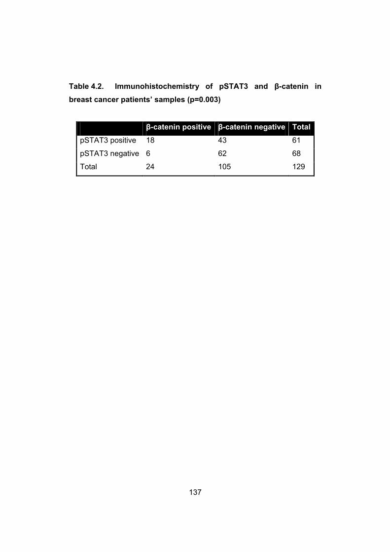
137
Table 4.2. Immunohistochemistry of pSTAT3 and β-catenin in
breast cancer patients’ samples (p=0.003)
β-catenin positive β-catenin negative Total
pSTAT3 positive 18 43 61
pSTAT3 negative 6 62 68
Total 24 105 129

138
Figure 4.5. Immunohistochemistry for pSTAT3 and β-catenin in breast tumors. A pSTAT3-negative case containing only rare positive cells (i.e. <10%) is shown in A, whereas a pSTAT3-positive case containing ≥10% stained cells is shown in B. pSTAT3 staining was largely nuclear. Staining for β-catenin is illustrated in Figure 4.5C-F. Figure 4.5C and 4.5D show two negative cases in which no definitive nuclear staining was detectable. The case shown in 4.5C had membranous staining whereas the case in Figure 4.5D had both membranous and cytoplasmic staining. Figure 4.5E and 4.5F show two positive cases, in which definitive nuclear staining was detectable. The case shown in Figure 4.5E also showed cytoplasmic staining, whereas the case in Figure 4.5F showed nuclear staining only.

139
4.3.4. β-catenin promotes cell growth in breast cancer
To investigate the biological importance of β-catenin on breast cancer, we
downregulated β-catenin levels in MCF-7 using siRNA, and a significant
downregulation of β-catenin was shown on Western blot (Figure 4.6A). An
MTS assay was performed on day 1, 2, and 3 after β-catenin was
downregulated. As shown in Figure 4.6B, there was a significant decrease
in cell growth in cells treated with β-catenin siRNA, as compared to those
treated with scrambled siRNA. Apoptosis, as detected by PARP and
caspase 3 cleavages, was not detectable (not shown).

140
Figure 4.6. β-catenin promotes cell growth in breast cancer. A, B) Downregulation of β-catenin using siRNA led to a significant decrease in cell growth compared to scrambled siRNA, as assessed by the MTS assay.

141
4.4. DISCUSSION
Our presented data support the concept that STAT3 is a regulator of β-
catenin in breast cancer. Specifically, we found that the gene promoter of
β-catenin carries multiple STAT3 consensus sequences and our
chromatin precipitation experiments provided direct evidence of STAT3
binding at two specific sites (-856 and -938) in the β-catenin gene
promoter region. Furthermore, in two different breast cancer cell lines, we
found evidence that the protein level and transcriptional activity of β-
catenin can be modulated in response to a change in the expression
and/or activity of STAT3, in both transient and stable transfection
conditions. In further support that STAT3 regulates β-catenin in breast
cancer, our immunohistochemical studies using a large cohort of breast
tumors revealed a significant correlation between the expression of
pSTAT3 and β-catenin. Our conclusion regarding β-catenin being a
downstream target of STAT3 echoes the findings described in a recent
study of colon cancer, which showed that STAT3 activity
regulates the transcriptional activity of β-catenin in colon cancer cells
(Kawada et al., 2006). In contrast with the same study, we did not detect
any change in the nuclear translocation of β-catenin following STAT3
knockdown using nuclear/cytoplasmic fractionation. Based on our overall
study results, the regulation of the transcriptional activity of β-catenin by
STAT3 in breast cancer appears to be related to the observations that
STAT3 controls the total protein level of β-catenin.
In the present study, we have presented evidence that STAT3 can directly
regulate the gene transcription of β-catenin and its protein expression
level. We are also aware of other mechanisms by which STAT3 can
potentially regulate β-catenin through modulating the upstream of the
WCP. Specifically, the gene promoter of Wnt3a has been shown to carry
the consensus binding sequence for STAT3 (Li et al., 2008). STAT3 also
has been shown to induce the expression of Wnt5a in rat cardiac

142
myocytes (Fujio et al., 2004; Miyagi et al., 2004). Taken together, it is
possible that STAT3 regulates β-catenin via multiple mechanisms: 1)
direct modulation of the gene transcription of β-catenin, and 2) modulate
the secretion of different Wnt's, thereby regulating β-catenin via the WCP.
While we did not observe a prognostic significance for β-catenin in our
cohort of breast cancer patients, we are aware of the results of a
previously published study which showed that β-catenin is prognostically
important in breast cancer (Lin et al., 2000). In contrast with the study by
Lin et al, who scored the β-catenin regardless of the staining pattern (Lin
et al., 2000), we considered β-catenin positivity only when the staining
was definitively nuclear. We also would like to point out that, one of the
authors (JM) in a paper recently published that β-catenin is useful in
predicting relapse in breast cancer patients (Asgarian et al., 2009);
however, no significant correlation between β-catenin nuclear staining and
the overall survival was found (personal communication).
Although the focus of this manuscript is to document the functional
interaction of STAT3 and β-catenin, we also examined whether β-catenin
is biologically important in breast cancer cell lines. As shown in this study,
downregulation of β-catenin using siRNA inhibited cell growth in MCF-7.
Growth inhibition induced by a downregulation of β-catenin has been
observed for esophageal cancer, colon cancer and glioma (Huang et al.,
2007; Pu et al., 2009; Wang et al., 2009). β-catenin was found to promote
cell migration in a breast cell line (Simpson et al., 2008), but we are not
aware of any previous study in which the growth-promoting effect of β-
catenin was examined in breast cancer. Thus, to our knowledge, these
findings represent the first evidence that β-catenin promotes cell growth in
breast cancer.

143
Both β-catenin and STAT3 have been shown to be oncogenic in various
types of human cancer including breast cancer (Imbert et al., 2001;
Kunigal et al., 2009). Our findings described in this manuscript raise the
possibility that the oncogenic effects of STAT3 may synergize with that of
β-catenin. Of interest, it have been reported that β-catenin regulates the
STAT3 transcriptional activity in esophageal cancer cell lines, thus
potentially creating a positive a feedback loop between these two
signaling proteins (Yan et al., 2008). Since specific agents inhibiting
STAT3 and β-catenin are available (Takemaru et al., 2008; Yue and
Turkson, 2009), our data provide the rationale for combining these
inhibitors in treating specific forms of cancer, such as in a subset of breast
cancer in which both STAT3 and β-catenin are activated. In-vitro studies
evaluating the potential synergistic effects of combining these agents will
be of great interest.

144
4.5. REFERENCESAsgarian, N., Hu, X., Aktary, Z., Chapman, K.A., Lam, L., Chibbar, R., Mackey, J., Greiner, R., and Pasdar, M. (2009). Learning to predict relapse in invasive ductal carcinomas based on the subcellular localization of junctional proteins. Breast Cancer Res Treat.
Bard, J.D., Gelebart, P., Amin, H.M., Young, L.C., Ma, Y., and Lai, R. (2009). Signal transducer and activator of transcription 3 is a transcriptional factor regulating the gene expression of SALL4. FASEB J23, 1405-1414.
Benhaj, K., Akcali, K.C., and Ozturk, M. (2006). Redundant expression of canonical Wnt ligands in human breast cancer cell lines. Oncol Rep 15, 701-707.
Brennan, K.R., and Brown, A.M. (2004). Wnt proteins in mammary development and cancer. J Mammary Gland Biol Neoplasia 9, 119-131.
Clevers, H. (2006). Wnt/beta-catenin signaling in development and disease. Cell 127, 469-480.
Dien, J., Amin, H.M., Chiu, N., Wong, W., Frantz, C., Chiu, B., Mackey, J.R., and Lai, R. (2006). Signal transducers and activators of transcription-3 up-regulates tissue inhibitor of metalloproteinase-1 expression and decreases invasiveness of breast cancer. Am J Pathol 169, 633-642.
Ehret, G.B., Reichenbach, P., Schindler, U., Horvath, C.M., Fritz, S., Nabholz, M., and Bucher, P. (2001). DNA binding specificity of different STAT proteins. Comparison of in vitro specificity with natural target sites. J Biol Chem 276, 6675-6688.
Ewan, K.B., and Dale, T.C. (2008). The potential for targeting oncogenic WNT/beta-catenin signaling in therapy. Curr Drug Targets 9, 532-547.
Fujio, Y., Matsuda, T., Oshima, Y., Maeda, M., Mohri, T., Ito, T., Takatani, T., Hirata, M., Nakaoka, Y., Kimura, R., et al. (2004). Signals through gp130 upregulate Wnt5a and contribute to cell adhesion in cardiac myocytes. FEBS Lett 573, 202-206.
Gordon, M.D., and Nusse, R. (2006). Wnt signaling: multiple pathways, multiple receptors, and multiple transcription factors. J Biol Chem 281, 22429-22433.
He, T.C., Sparks, A.B., Rago, C., Hermeking, H., Zawel, L., da Costa, L.T., Morin, P.J., Vogelstein, B., and Kinzler, K.W. (1998). Identification of c-MYC as a target of the APC pathway. Science 281, 1509-1512.

145
Huang, W.S., Wang, J.P., Wang, T., Fang, J.Y., Lan, P., and Ma, J.P. (2007). ShRNA-mediated gene silencing of beta-catenin inhibits growth of human colon cancer cells. World J Gastroenterol 13, 6581-6587.
Huber, A.H., and Weis, W.I. (2001). The structure of the beta-catenin/E-cadherin complex and the molecular basis of diverse ligand recognition by beta-catenin. Cell 105, 391-402.
Imbert, A., Eelkema, R., Jordan, S., Feiner, H., and Cowin, P. (2001). Delta N89 beta-catenin induces precocious development, differentiation, and neoplasia in mammary gland. J Cell Biol 153, 555-568.
Kawada, M., Seno, H., Uenoyama, Y., Sawabu, T., Kanda, N., Fukui, H., Shimahara, Y., and Chiba, T. (2006). Signal transducers and activators of transcription 3 activation is involved in nuclear accumulation of beta-catenin in colorectal cancer. Cancer Res 66, 2913-2917.
Kunigal, S., Lakka, S.S., Sodadasu, P.K., Estes, N., and Rao, J.S. (2009). Stat3-siRNA induces Fas-mediated apoptosis in vitro and in vivo in breast cancer. Int J Oncol 34, 1209-1220.
Levina, E., Oren, M., and Ben-Ze'ev, A. (2004). Downregulation of beta-catenin by p53 involves changes in the rate of beta-catenin phosphorylation and Axin dynamics. Oncogene 23, 4444-4453.
Li, X., Placencio, V., Iturregui, J.M., Uwamariya, C., Sharif-Afshar, A.R., Koyama, T., Hayward, S.W., and Bhowmick, N.A. (2008). Prostate tumor progression is mediated by a paracrine TGF-beta/Wnt3a signaling axis. Oncogene 27, 7118-7130.
Lim, S.C., and Lee, M.S. (2002). Significance of E-cadherin/beta-catenin complex and cyclin D1 in breast cancer. Oncol Rep 9, 915-928.
Lin, S.Y., Xia, W., Wang, J.C., Kwong, K.Y., Spohn, B., Wen, Y., Pestell, R.G., and Hung, M.C. (2000). Beta-catenin, a novel prognostic marker for breast cancer: its roles in cyclin D1 expression and cancer progression. Proc Natl Acad Sci U S A 97, 4262-4266.
Liu, J., Stevens, J., Rote, C.A., Yost, H.J., Hu, Y., Neufeld, K.L., White, R.L., and Matsunami, N. (2001). Siah-1 mediates a novel beta-catenin degradation pathway linking p53 to the adenomatous polyposis coli protein. Mol Cell 7, 927-936.
Mikels, A.J., and Nusse, R. (2006). Wnts as ligands: processing, secretion and reception. Oncogene 25, 7461-7468.
146
Miller, J.R. (2002). The Wnts. Genome Biol 3, REVIEWS3001.
Miyagi, C., Yamashita, S., Ohba, Y., Yoshizaki, H., Matsuda, M., and Hirano, T. (2004). STAT3 noncell-autonomously controls planar cell polarity during zebrafish convergence and extension. J Cell Biol 166, 975-981.
Polakis, P. (2000). Wnt signaling and cancer. Genes Dev 14, 1837-1851.
Pu, P., Zhang, Z., Kang, C., Jiang, R., Jia, Z., Wang, G., and Jiang, H. (2009). Downregulation of Wnt2 and beta-catenin by siRNA suppresses malignant glioma cell growth. Cancer Gene Ther 16, 351-361.
Ryo, A., Nakamura, M., Wulf, G., Liou, Y.C., and Lu, K.P. (2001). Pin1 regulates turnover and subcellular localization of beta-catenin by inhibiting its interaction with APC. Nat Cell Biol 3, 793-801.
Schlange, T., Matsuda, Y., Lienhard, S., Huber, A., and Hynes, N.E. (2007). Autocrine WNT signaling contributes to breast cancer cell proliferation via the canonical WNT pathway and EGFR transactivation. Breast Cancer Res 9, R63.
Seidel, H.M., Milocco, L.H., Lamb, P., Darnell, J.E., Jr., Stein, R.B., and Rosen, J. (1995). Spacing of palindromic half sites as a determinant of selective STAT (signal transducers and activators of transcription) DNA binding and transcriptional activity. Proc Natl Acad Sci U S A 92, 3041-3045.
Sekikawa, A., Fukui, H., Fujii, S., Ichikawa, K., Tomita, S., Imura, J., Chiba, T., and Fujimori, T. (2008). REG Ialpha protein mediates an anti-apoptotic effect of STAT3 signaling in gastric cancer cells. Carcinogenesis29, 76-83.
Shtutman, M., Zhurinsky, J., Simcha, I., Albanese, C., D'Amico, M., Pestell, R., and Ben-Ze'ev, A. (1999). The cyclin D1 gene is a target of the beta-catenin/LEF-1 pathway. Proc Natl Acad Sci U S A 96, 5522-5527.
Simpson, K.J., Selfors, L.M., Bui, J., Reynolds, A., Leake, D., Khvorova, A., and Brugge, J.S. (2008). Identification of genes that regulate epithelial cell migration using an siRNA screening approach. Nat Cell Biol 10, 1027-1038.
Staal, F.J., Noort Mv, M., Strous, G.J., and Clevers, H.C. (2002). Wnt signals are transmitted through N-terminally dephosphorylated beta-catenin. EMBO Rep 3, 63-68.

147
Takemaru, K.I., Ohmitsu, M., and Li, F.Q. (2008). An oncogenic hub: beta-catenin as a molecular target for cancer therapeutics. Handb Exp Pharmacol, 261-284.
Turkson, J. (2004). STAT proteins as novel targets for cancer drug discovery. Expert Opin Ther Targets 8, 409-422.
Wang, J.S., Zheng, C.L., Wang, Y.J., Wen, J.F., Ren, H.Z., Liu, Y., and Jiang, H.Y. (2009). Gene silencing of beta-catenin by RNAi inhibits cell proliferation in human esophageal cancer cells in vitro and in nude mice. Dis Esophagus 22, 151-162.
Yan, S., Zhou, C., Zhang, W., Zhang, G., Zhao, X., Yang, S., Wang, Y., Lu, N., Zhu, H., and Xu, N. (2008). beta-Catenin/TCF pathway upregulates STAT3 expression in human esophageal squamous cell carcinoma. Cancer Lett 271, 85-97.
Yue, P., and Turkson, J. (2009). Targeting STAT3 in cancer: how successful are we? Expert Opin Investig Drugs 18, 45-56.

148
CONSTITUTIVE ACTIVATION OF METALLOPROTEINASE
ADAM10 IN MANTLE CELL LYMPHOMA PROMOTES CELL
GROWTH AND ACTIVATES THE TNFα/NFĸB PATHWAY
A version of this chapter has been published as:
Armanious H, Gelebart P, Anand M, Belch A, Lai R. Constitutive
activation of metalloproteinase ADAM10 in mantle cell lymphoma
promotes cell growth and activates the TNFα/NFĸB pathway. Blood.
2011;117:6237-6246
Chapter 5

149
5.1. INTRODUCTION
A disintegrin and metalloproteinase 10 (ADAM10), a member of the ADAM
family of metalloproteinases, was discovered in the protein extract of brain
myelin membranes and subsequently found to be a homologue of the
Drosophila kuzbanian (kuz) gene (Chantry and Glynn, 1990; Chantry et
al., 1989; Yavari et al., 1998). ADAM10 is secreted as a precursor protein,
and it consists of multiple functional domains including a prodomain,
catalytic domain, cysteine-rich domain, transmembraneous domain and
cytoplasmic domain (Edwards et al., 2008). To become the active/mature
form, the precursor ADAM10 protein needs to be cleaved by proprotein
convertase 7 and furin, both of which remove the prodomain of ADAM10
(Anders et al., 2001). ADAM10 is biologically important, since ADAM10
knockout mice die on day 9 of embryogenesis due to multiple
abnormalities in the nervous and cardiovascular systems (Hartmann et al.,
2002). The key biological function of ADAM10 appears to be attributed to
its enzymatic activity as a metalloproteinase. Specifically, ADAM10 is
involved in the intra-membrane proteolysis process, whereby it mediates
ectodomain shedding of various membrane bound receptors, adhesion
molecules, growth factors and cytokines (Endres and Fahrenholz;
Mochizuki and Okada, 2007; Seals and Courtneidge, 2003). For instance,
ADAM10 is involved in the regulation of the shedding of Notch, HER2,
CD44, IL-6 receptor, amyloid precursor protein and cadherins (Duffy et al.,
2009). Directly relevant to our study, ADAM10 was recently found to be
one of the enzymes responsible for cleaving tumor necrosis factor alpha
(TNFα) and releasing its active form (Hikita et al., 2009; Lunn et al., 1997;
Mezyk-Kopec et al., 2009). Furthermore, it has been reported that
ADAM10 is important for the development of the marginal zone B-cells
(Gibb et al.). By regulating the bioavailability of ligands to various cellular
signaling receptors, ADAM10 modulates the activation status of various
cellular signaling pathways that have an impact on various cellular
responses such as proliferation and migration (Seals and Courtneidge,

150
2003). ADAM10 has been shown to be constitutively active in a number of
solid tumors, and this biochemical defect is implicated in the pathogenesis
of these tumors. For instance, xenografting of colorectal cancer cells with
enforced expression of ADAM10 in nude mice induced the formation of
liver metastasis compared to the negative control cells, and this effect can
be attributed to ADAM10-mediated cleavage and release of L1-CAM, a
cell adhesion molecule (Gavert et al., 2007). In another study, ADAM10
expression in colorectal cancer patient samples detectable by
immunohistochemistry was found to correlate with a higher clinical stage
(Knosel et al., 2005). Using immunohistochemistry, it was also found that
ADAM10 is overexpressed in squamous cell carcinomas of the oral cavity,
as compared to the benign epithelial cells; knockdown of ADAM10
expression using siRNA in the cell lines derived from these tumors was
shown to induce a significant decrease in cell growth (Ko et al., 2007).
Similar findings were made in pancreatic cancer, where inhibition of
ADAM10 expression in pancreatic carcinoma cell lines resulted in a
significant decrease in invasiveness and migration (Gaida et al.). Lastly,
ADAM10-mediated cleavage of N-cadherin was found to regulate the
migratory properties of glioblastoma cells (Kohutek et al., 2009). While the
pathogenetic role of ADAM10 has been well documented in solid tumors,
its role in hematologic malignancies is largely unknown. To our
knowledge, there is only one published study that performed a survey of
the expression of various ADAM family members in benign and malignant
hematopoietic cells using RT-PCR, and the authors reported the
expression of the ADAM10 mRNA in myeloma, erythroleukemia and a
subset of lymphoma cell lines (Wu et al., 1997). Since only RT-PCR was
used in this particular study, whether ADAM10 is in its active form could
not be assessed; the biological significance of ADAM10 in these
malignancies also was not examined (Wu et al., 1997).

151
Mantle cell lymphoma (MCL) is a specific subtype of aggressive B-cell
lymphoma recognized by the World Health Organization Classification
Scheme (Jaffe et al., 2004). The genetic hallmark of this disease is the
recurrent chromosomal abnormality, t(11;14)(q13;q32), which brings the
cyclin D1(CCND1) gene under the influence of the enhancer of the
immunoglobulin heavy chain (IgH) gene, leading to cyclin D1
overexpression (Campo et al., 1999). Although it has been shown that
cyclin D1 overexpression is not sufficient for the induction of lymphoma in
animal models, this abnormality is considered to be the primary oncogenic
event in MCL (Diehl, 2002; Jares et al., 2007). Large-scale cDNA
microarray studies using frozen MCL tumors have revealed a relatively
large number of biochemical abnormalities in MCL, with these defects are
frequently implicated in the regulation of apoptosis, survival and DNA
damage (Hofmann et al., 2001; Martinez et al., 2003; Rosenwald et al.,
2003; Schaffner et al., 2000). An increasing number of cellular signaling
pathways also have been found to be abnormal. For instance, NFĸB has
been reported to be constitutively active in MCL, and this biochemical
defect is biologically important to the pathogenesis of MCL (Pham et al.,
2003). The activation of NFĸB in MCL can be partly attributed to TNFα
(Gelebart et al., 2009; Shishodia et al., 2005), the bioavailability of which
has been shown to be regulated by ADAM10, as mentioned above (Hikita
et al., 2009; Lunn et al., 1997; Mezyk-Kopec et al., 2009).
In this report, we described for the first time that ADAM10 is constitutively
activated in MCL cell lines and tumors. In view of the importance of TNFα
in the biology of MCL, and the previous report that ADAM10 can increase
the bioavailability of TNFα (Hikita et al., 2009; Lunn et al., 1997; Mezyk-
Kopec et al., 2009) , we hypothesize that ADAM10 may be important in
the pathogenesis of MCL.
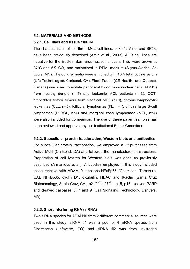
152
5.2. MATERIALS AND METHODS
5.2.1. Cell lines and tissue culture
The characteristics of the three MCL cell lines, Jeko-1, Mino, and SP53,
have been previously described (Amin et al., 2003). All 3 cell lines are
negative for the Epstein-Barr virus nuclear antigen. They were grown at
370C and 5% CO2 and maintained in RPMI medium (Sigma-Aldrich, St.
Louis, MO). The culture media were enriched with 10% fetal bovine serum
(Life Technologies, Carlsbad, CA). Ficoll-Paque (GE Health care, Quebec,
Canada) was used to isolate peripheral blood mononuclear cells (PBMC)
from healthy donors (n=5) and leukemic MCL patients (n=3). OCT-
embedded frozen tumors from classical MCL (n=9), chronic lymphocytic
leukemias (CLL, n=5), follicular lymphomas (FL, n=4), diffuse large B-cell
lymphomas (DLBCL, n=4) and marginal zone lymphomas (MZL, n=4)
were also included for comparison. The use of these patient samples has
been reviewed and approved by our Institutional Ethics Committee.
5.2.2. Subcellular protein fractionation, Western blots and antibodies
For subcellular protein fractionation, we employed a kit purchased from
Active Motif (Carlsbad, CA) and followed the manufacturer’s instructions.
Preparation of cell lysates for Western blots was done as previously
described (Armanious et al.). Antibodies employed in this study included
those reactive with ADAM10, phospho-NFĸBp65 (Chemicon, Temecula,
CA), NFĸBp65, cyclin D1, α-tubulin, HDAC and β-actin (Santa Cruz
Biotechnology, Santa Cruz, CA), p21Waf1, p27Kip1, p15, p16, cleaved PARP
and cleaved caspases 3, 7 and 9 (Cell Signaling Technology, Danvers,
MA).
5.2.3. Short interfering RNA (siRNA)
Two siRNA species for ADAM10 from 2 different commercial sources were
used in this study. siRNA #1 was a pool of 4 siRNA species from
Dharmacon (Lafayette, CO) and siRNA #2 was from Invitrogen

153
(Burlington, Ontario, Canada). Scrambled siRNA was purchased from
Dharmacon. Transfection of siRNA was carried out using an electrosquare
electroporator, BTX ECM 800 (225V, 8.5ms, 3 pulses) (Holliston, MA).
The concentration of siRNA used was 200 pmol/1x106 cells, and cells
were harvested at 48 hours after transfection. The ADAM10 protein levels
were assessed using Western blot to evaluate the efficiency of inhibition.
5.2.4. Cell viability
A total of 100,000 cells suspended in 1 mL of culture medium were plated
in triplicate. To assess cell viability, trypan blue exclusion assay (Sigma-
Aldrich) was performed every 24 hours for up to 3 days following
transfection of ADAM10 siRNA or scrambled siRNA. MTS assay was also
performed during this time frame and the manufacturer’s instructions were
followed (Promega, Madison, WI). The measurements were obtained at a
wavelength of 450 nM using a Bio-Rad Micro plate Reader (Bio-Rad,
Hercules, CA). The absorbance values were normalized to the wells with
media only using the microplate Manager 5.2.1 software (Bio-Rad). All
experiments were performed in triplicates.
5.2.5. Recombinant ADAM10, TNFα assay, bortezomib and MG132
Human recombinant ADAM10 was purchased from BD biosciences
(Pharmingen, San Diego, CA) and it corresponds to the segment spanning
from Thr214 to Glu672, which represents the active/mature form of
ADAM10. It was added at a concentration of 100 ng/ml to 1x10⁶ cells; after
24 hours, cell lysates were prepared and cell count was done. TNFα
secretion was monitored using a commercially available ELISA kit (R&D
Systems Inc., Minneapolis, MN). Aliquots of the culture medium from MCL
cell lines collected 48 hours after transfection with either scrambled siRNA
or ADAM10 siRNA were centrifuged at 15,000 g, and the supernatant was
assayed for the TNFα levels as per manufacturer's protocol. Bortezomib
(LC laboratories, Woburn, MA), a proteasome inhibitor was added at a

154
concentration of 5 nM to MCL cells 24 hours after siRNA transfection, and
cell count using trypan blue exclusion assay was performed 48 hours after
the initiation of the experiment. Another proteasome inhibitor MG132
(Calbiochem, EMD Biosciences, Darmstadt, Germany) was added at a
concentration of 1 μM to MCL cells 24 hours after siRNA transfection, and
cell count using trypan blue exclusion assay was performed 48 hours after
the initiation of the experiment.
5.2.6. NFĸB transcriptional activity
To assess the transcriptional activity of NFĸB in MCL cell lines, we
employed the NFĸB responsive firefly luciferase reporter plasmid and
renilla reporter plasmid (Promega). After 48 hours of transfection of the
reporter plasmid (with the renilla luciferase plasmid as an internal control)
together with either scrambled siRNA or ADAM10 siRNA, MCL cells were
harvested and cell extracts were prepared using a lysis buffer purchased
from Promega. The firefly luciferase activity and renilla luciferase activity
were assessed using the dual luciferase reagent (Promega).
5.2.7. Cell cycle analysis by flow cytometry
Cells transfected with either scrambled siRNA or ADAM10 siRNA were
fixed with ice-cold 70% ethanol 24 hours after gene transfection. These
cells were then subjected to RNase treatment and propidium iodide (PI)
staining. DNA content was determined using a FACSCalibur flow
cytometer (BD Biosciences). Data acquisition was gated to exclude cell
doublets and the cell-cycle phase distribution was determined using the
CellQuest program (20,000 events were counted).
5.2.8. Assessment of cyclin D1 expression using quantitative RT-PCR
The expression of cyclin D1 in MCL cells treated with ADAM10 siRNA was
assessed using quantitative RT-PCR (qRT-PCR). The assay was
performed using the Applied Biosystems 7900 HT, and the SYBR®

155
GreenER™ qPCR SuperMix from Invitrogen. The primer sequence for
ADAM10 was as follows: forward 5’-AGCAACATCTGGGGACAAAC -3’;
reverse 5’-CTTCCCTCTGGTTGATTTGC-3’. The primer sequence for
cyclin D1 was as follows: forward 5’-CAAATGGAGCTGCTCCTGGTG-3’;
reverse 5’-TGGCACCAGCCTCGGCATTTC-3’ (Gelebart et al., 2008).
Triplicate experiments were performed and the statistical significance of
the differences was assessed using Student t-test.
5.2.9. Immunohistochemistry and archival MCL tumors
Formalin-fixed, paraffin-embedded tumors from a cohort of 23 patients
with classical MCL were retrieved from the files at the Cross Cancer
Institute (Edmonton, Alberta, Canada). All MCL primary tumors were
diagnosed at the Cross Cancer Institute and the diagnostic criteria were
based on those described in the World Health Organization Classification
Scheme (Jaffe et al., 2004). All cases were confirmed to express cyclin D1
by immunohistochemistry. The use of these tissues has been approved by
our Institutional Ethics Committee. Immunohistochemistry was performed
using standard techniques. Briefly, formalin-fixed, paraffin-embedded
tissue sections of 4 μM thickness were deparaffinized and hydrated. Heat-
induced epitope retrieval was performed in a pressure cooker using citrate
buffer (pH=6) in a microwave. After antigen retrieval, tissue sections were
incubated with 3% hydrogen peroxide for 10 minutes to block endogenous
peroxidase activity. Tissue sections were then incubated with anti-
ADAM10 antibody (1:200) (same antibody used in western blots) overnight
in a humidified chamber at 4°C. Immunostaining was visualized with a
labeled streptavidin-biotin (LSAB) method using 3,3'-diaminobenzidine as
a chromogen (Dako Canada Inc., Mississauga, Ontario, Canada) and
counter-stained with hematoxylin. Colon carcinoma case served as the
positive control, whereas the lymphoid cells of mantle zone in benign
tonsils served as the negative control.

156
5.2.10. Statistical analysis
Data are expressed as mean +/- standard derivation. Unless stated
otherwise, statistical significance was determined using two-tailed
Student's t-test and statistical significance was achieved when the p value
is <0.05. In experiments where we needed to determine the statistical
significance between more than two groups analysis of variance (ANOVA)
test was applied.
5.3. RESULTS
5.3.1. The active/mature form of ADAM10 is expressed in MCL cells
and other B-cell lineage malignancies
The expression of ADAM10 in 3 MCL cell lines was assessed using
western blots, and the results are illustrated in figure 5.1A. While the
precursor form of ADAM10 at 98 kDa was highly expressed in all 3 cell
lines examined, the expression levels of the active/mature form of
ADAM10 at 72 kDa were variable among these 3 cell lines. Specifically,
although the active/mature form of ADAM10 was readily detectable in
Jeko-1 and Mino cells, it was barely detectable in SP53 cells. MCF-7, a
breast cancer cell line previously reported to have a high level of
active/mature form of ADAM10 (Dittmer et al., 2009), served as the
positive control. In contrast to MCL cells, PBMC from five healthy donors
expressed no detectable active/mature form of ADAM10 at 72 kDa,
although the precursor form was readily detectable in all cases. The
results from 3 of these 5 healthy individuals are illustrated in figure 5.1A
and 5.1B.
Western blot analysis was performed using 9 frozen MCL tumor samples
(patient #1-9) and 3 fresh leukemic MCL blood samples (patient #10-12).
The active/mature form of ADAM10 was readily detectable in all patient
samples, although the level was relatively low in patient #7 (figure 5.1B).
Again, PBMC from healthy donor #2 and #3 had no convincing band at 72

157
kDa. Of note, in contrast with the MCL cell lines (figure 5.1A); most MCL
patient samples expressed the active/mature form of ADAM10 at a higher
level than its precursor form. We also analyzed the expression of both the
precursor and active/mature forms of ADAM10 in frozen tissues derived
from other B-cell lineage malignancies (5 CLL cases, 4 DLBCL cases, 4
FL cases and 4 MZL cases). As shown in supplementary figure 5.1, the
expression of ADAM10 was readily detectable in all of these B-cell
neoplasms, with a pattern similar to that seen in MCL patient samples.
We then performed immunohistochemistry applied to 23 MCL formalin-
fixed/paraffin-embedded tumor samples and 5 cases of benign reactive
tonsils. We found that the mantle zones in benign reactive tonsils were
negative for ADAM10, whereas the germinal centers showed only faint
cytoplasmic staining (figure 5.1C). The ADAM10 immunostaining in MCL
was generally homogenous within the same tumor, and we considered a
MCL tumor to be positive when the majority of the tumor cells showed a
staining intensity higher than that of germinal center cells. With this
criterion, ADAM10 was assessed positive in 20 of 23 (87%) cases. On
higher magnification, the immunostaining was predominantly cytoplasmic
in all cases, while a small subset of cases also showed some degree of
nuclear staining (figure 5.1C). Of note, 5 of these MCL cases used for
immunohistochemical studies were also included in the experiment
illustrated in figure 5.1B (western blot studies); all these 5 cases were
positive by immunohistochemistry and carried the active form of ADAM10
by western blots.

158
Figure 5.1. ADAM10 expression in MCL cell lines and patient samples (continued). A) Western blots showed the expression of the precursor and active/mature form of ADAM10 in three MCL cell lines. While the precursor was highly expressed, the active/mature form was readily detectable only in Jeko-1 and Mino. A faint band at 72 kDa, representing the active/mature form of ADAM10, was also detectable in SP53. MCF-7 cells were used as the positive control. Peripheral blood mononuclear cells (PBMC) from a healthy individual (#1) showed abundant precursor ADAM10 protein but no detectable active/mature ADAM10. B) Western blots showed the presence of the precursor and active/mature form of ADAM10 in all MCL patients’ samples (both frozen #1-9 and leukemic #10-12); PBMC from two healthy individuals (#2 and #3) showed no detectable active/mature ADAM10.
A
B

159
Figure 5.1. ADAM10 expression in MCL cell lines and patient samples. C) Immunohistochemistry showed no detectable signal of ADAM10 in the mantle zone of a reactive tonsil; the germinal centers showed faint staining, 100X (i). A case of MCL tumor showed no detectable ADAM10, 100X (ii). A MCL tumor showed a relatively high level of ADAM10 expression, 100X (iii). On high magnification, the ADAM10 immunostaining pattern was detectable in both of the nucleus and cytoplasm, 1000X (iv).
C

160
Supplementary figure 5.1. ADAM10 expression in B-cell non-Hodgkin lymphomas other than MCL. Western blots showed the presence of the precursor and active/mature form of ADAM10 (with a predominance of the active form) in marginal zone lymphoma, follicular lymphoma, diffuse large B cell lymphoma and chronic lymphocytic leukemia, MCF-7 was used as positive control.

161
5.3.2. ADAM10 promotes cell growth in MCL cells
To assess the functional importance of ADAM10 in MCL cells, we
evaluated if siRNA knockdown of ADAM10 has any impact on the growth
of MCL cells. As shown in figure 5.2A, transfection of siRNA into Jeko-1
and Mino resulted in a reduction of both the precursor and the
active/mature form of ADAM10. With the knockdown of ADAM10, the cell
growth (assessed by the trypan blue exclusion assay) was significantly
decreased, as compared to cells transfected with scrambled siRNA
(p=0.01 and 0.007 for Mino and Jeko-1, respectively) three days after
transfection (figure 5.2B). The decrease in the viable cell count was not
associated with any substantial increase in trypan blue-positive dead cells.
Similar experiments were performed using the MTS assay, and we found
comparable results (p<0.0001 and p=0.01 for Mino and Jeko-1,
respectively) (figure 5.2C). To further strengthen the conclusion that
ADAM10 promotes cell growth in MCL cells, we tested how MCL cells may
respond to recombinant ADAM10. SP53 and Mino cells were used for this
experiment, since both cell lines had a lower level of active ADAM10 than
Jeko-1 cells, and thus, they were expected to show more dramatic
response to recombinant ADAM10. As shown in figure 5.2D, the addition
of recombinant ADAM10 induced a significant increase in cell growth
(p=0.008 and 0.01, respectively).

162
Figure 5.2. ADAM10 promoted cell growth in MCL (continued)A) Western blots showed downregulation of the precursor and active/mature form of ADAM10 in Jeko-1 and Mino cells with the use of siRNA. B) ADAM10 knockdown induced significant inhibition of cell growth in Mino and Jeko-1 cells three days after transfection, as assessed by trypan blue exclusion assay (p=0.01 and 0.007, respectively). Triplicateexperiments were performed.
A
B

163
Figure 5.2. ADAM10 promoted cell growth in MCL. C) ADAM10 knockdown induced significant inhibition of cell growth in Mino and Jeko-1 cells three days after transfection, as assessed by MTS assay (p<0.0001 and p=0.01, respectively). Triplicate experiments were performed. D) Addition of human recombinant ADAM10 (100 ng/ml) to SP53 and Mino cells induced a significant increase in their growth at 24 hours (p=0.008 and 0.01, respectively).
D
C

164
5.3.3. Downregulation of ADAM10 induces cell cycle arrest but not
apoptosis
To characterize the mechanism by which ADAM10 promotes cell growth in
MCL, we performed cell cycle analysis. Transfection of ADAM10 siRNA
into Mino and Jeko-1 cells induced a significant G0/1 arrest and reduced
the proportion of cells in the S phase, as compared to cells transfected
with scrambled siRNA (p=0.02 and 0.05 for Mino and Jeko-1,
respectively). Of note, no appreciable increase in the proportion of cells in
the subG0/1 phase was found. All experiments were performed in
triplicates and representative results are shown in figure 5.3. To further
exclude the occurrence of apoptosis, we performed western blots and
found no detectable cleavage of caspases 3, 7, 9 and PARP
(supplementary figure 5.2). Cells treated with MG132 served as positive
controls.

165
Figure 5.3. ADAM10 induced cell cycle arrest. Cell cycle analysis by flow cytometry using propidium iodide showed significant G0/1 cell cycle arrest in Mino cells following ADAM10 downregulation using siRNA. Of note, no appreciable increase in the fraction of cells in the subG0/1 was noted.

166
Supplementary figure 5.2. ADAM10 downregulation did not induce apoptosis. Western blot studies showed that Jeko-1 cells did not undergo apoptosis after ADAM10 downregulation, as there is no evidence of cleavage of PARP, caspase 3, 7 or 9. Jeko-1 cells treated with proteasome inhibitor MG132 were used as positive controls.

167
5.3.4. ADAM10 regulates the cyclin D1 expression level in MCL cells
Since cyclin D1 overexpression is believed to play an important
pathogenetic role in MCL (Campo et al., 1999), we assessed if ADAM10
mediates any effects on the expression of cyclin D1 in MCL cell lines. As
shown in figure 5.4A, downregulation of ADAM10 using siRNA appreciably
reduced the cyclin D1 protein expression level in Jeko-1 and Mino cells.
This decrease in the cyclin D1 protein level correlated well with our
observations that the cyclin D1 mRNA levels were significantly reduced 24
hours after the treatment of ADAM10 siRNA (figure 5.4B). Specifically, the
cyclin D1 mRNA levels detected by quantitative RT-PCR were reduced by
21% and 35% in Jeko-1 and Mino cells, respectively. Opposite effects
were observed when recombinant ADAM10 (100 ng/ml) was added to
SP53 cells (not shown). Using subcellular fractionation, we found that the
increase in the cyclin D1 protein expression level was largely attributed to
an increase in cyclin D1 in the nuclear fraction (figure 5.4C). We also
examined if downregulation of ADAM10 using siRNA modulates a number
of other cell-cycle regulators including p21Waf1, p27Kip1 , p15 and p16; we
found that there was an upregulation of p21Waf1, an inhibitor of cyclin-
dependent kinase 4 (figure 5.4A); the other three proteins showed no
appreciable change (not shown).

168
Figure 5.4. ADAM10 regulated cyclin D1 expression in MCL (continued). A) Western blots showed downregulation of cyclin D1 and upregulation of p21Waf1 in Jeko-1 and Mino cells following ADAM10 downregulation using siRNA. B) Quantitative RT-PCR showed downregulation of the cyclin D1 mRNA in Jeko-1 and Mino cells 24 hours after downregulation of ADAM10 using siRNA
A
B

169
Figure 5.4. ADAM10 regulated cyclin D1 expression in MCLC) Western blots showed upregulation of the nuclear level of cyclin D1 protein in SP53 cells 24 hours after the addition of human recombinant ADAM10 (100ng/ml); HDAC was used as the loading control for the nuclear extract.
C

170
5.3.5. ADAM10 activates the TNFα/NFĸB axis
In view of the previous literature that ADAM10 regulates the production of
TNFα in murine cell lines and immortalized human cell lines, (Hikita et al.,
2009; Mezyk-Kopec et al., 2009) as well as the importance of the TNFα in
the biology of MCL (Gelebart et al., 2009; Pham et al., 2003; Shishodia et
al., 2005), we hypothesized that ADAM10 may promote the growth of MCL
via upregulation of TNFα. In keeping with this concept, we found that
downregulation of ADAM10 using siRNA in Jeko-1 cells resulted in a
significant decrease in the levels of TNFα present in the culture media
(p=0.001) (figure 5.5A). Since TNFα is an activator of NFĸB (Wullaert et
al., 2006), a signaling protein also strongly implicated in the pathogenesis
of MCL (Pham et al., 2003), we then assessed the relationship between
ADAM10 and this signaling protein. We performed subcellular fractionation
experiments in which we found a correlation among the expression levels
of the phosphorylated NFĸB (active form), total NFĸBp65 and ADAM10 in
3 MCL cell lines (supplementary figure 5.3). Specifically, we found that
Jeko-1 cells, which have a relatively high level of the active/mature form of
ADAM10, carried a higher level of phosphorylated NFĸBp65 than SP53
cells, which have a much lower level of the active/mature form of
ADAM10. As shown in figure 5.5B, there was a significant downregulation
in both the phosphorylated and total NFĸBp65 level in Mino and Jeko-1
cells after ADAM10 siRNA treatment. Using subcellular fractionation, we
found that the protein level of NFĸBp65 in the nuclear fractions was
decreased, more so than that in the cytoplasmic fractions (figure 5.5C).
Lastly, we assessed the transcriptional activity of NFĸB using a
commercially available reporter construct. As shown in figure 5.5D, there
was significant downregulation of NFĸB transcriptional activity following
ADAM10 downregulation in Mino cells. In contrast, addition of human
recombinant ADAM10 (100 ng/mL) resulted in a significant increase in the
NFĸB transcriptional activity in these cells (figure 5.5E).

171
Figure 5.5. ADAM10 activated the TNFα/ NFĸB axis (continued). A) Using an ELISA kit, we showed a significant downregulation of TNFα in Jeko-1 cells 48 hours after the transfection of ADAM10 siRNA, as compared to cells transfected with scrambled siRNA. B) Western blots showed a downregulation of phospho-NFĸBp65 and total NFĸBp65 after ADAM10 downregulation using siRNA, both in the total lysates harvested from Jeko-1 and Mino cells.
A
B

172
Figure 5.5. ADAM10 activated the TNFα/ NFĸB axis. C) Western blots showed a downregulation of total NFĸBp65 and in nuclear extract from Jeko-1 and Mino cells. D) Using a NFĸB reporter vector, dual luciferase assay, we showed a significant downregulation of the NFĸB transcriptional activity in Mino cells after downregulation of ADAM10 using siRNA. E) Using NFĸB reporter vector, dual luciferase assay, we showed a significant upregulation of the NFĸB transcriptional activity after the addition of human recombinant ADAM10 (100 ng/ml) in Mino cells.
C
D
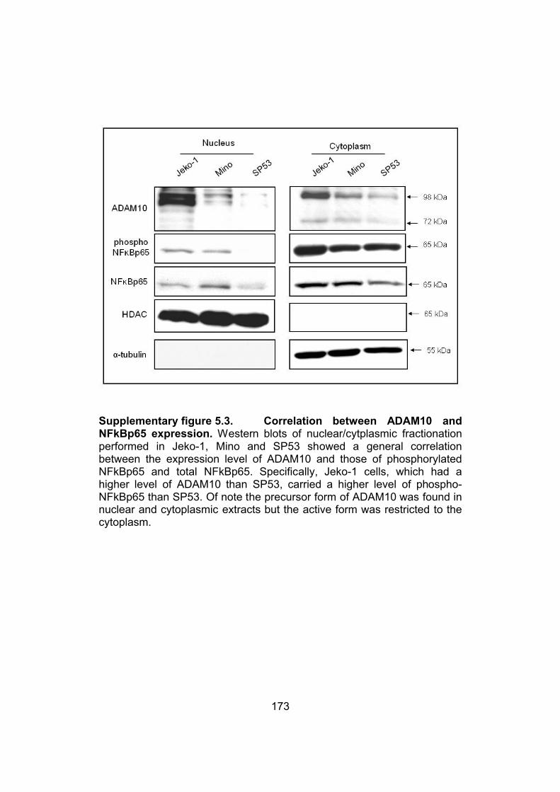
173
Supplementary figure 5.3. Correlation between ADAM10 and NFkBp65 expression. Western blots of nuclear/cytplasmic fractionation performed in Jeko-1, Mino and SP53 showed a general correlation between the expression level of ADAM10 and those of phosphorylated NFkBp65 and total NFkBp65. Specifically, Jeko-1 cells, which had a higher level of ADAM10 than SP53, carried a higher level of phospho-NFkBp65 than SP53. Of note the precursor form of ADAM10 was found in nuclear and cytoplasmic extracts but the active form was restricted to the cytoplasm.

174
5.3.6. ADAM10 inhibition enhanced the growth suppressing effect of
the proteasome inhibitors MG132 and bortezomib
Proteasome inhibitors have been shown to induce apoptosis and cell-cycle
arrest in MCL cells, and these effects are believed to be mediated via an
inhibition of the NFĸB signaling pathway (McConkey and Zhu, 2008).
Since we have shown that ADAM10 mediates its effects via the NFĸB
pathway, we hypothesized that ADAM10 inhibition may enhance the
growth-suppressing effects of proteasome inhibitors such as MG132 in
MCL cells. In our initial experiment, we determined that the inhibitory
concentration at 50% (i.e. IC50) for MG132 was in the range of 1 μM for
Jeko-1 cells (data not shown). Using this concentration of MG132, we
found that Jeko-1 cells transfected with ADAM10 siRNA had a significantly
more reduction in the number of viable cells, as compared to cells
transfected with the scrambled siRNA (p< 0.0001, ANOVA) (figure 5.6A).
We also performed similar experiments using bortezomib, a proteasome
inhibitor currently used in various clinical trials for the treatment of MCL
(Strauss et al., 2006). We used 5 nM for this experiment, since cell growth
inhibition was approximately 15% at this drug concentration. As shown in
figure 5.6B, the combination of bortezomib and ADAM10 siRNA resulted in
a significant reduction in number of viable cells, as compared to the use of
bortezomib or ADAM10 siRNA alone (p< 0.0001, ANOVA).

175
Figure 5.6. ADAM10 inhibition enhanced the growth suppressing effects of proteasome inhibitors bortezomib and MG132 in Jeko-1 cells. A) Jeko-1 cells transfected with either scrambled or ADAM10 siRNA were treated with MG132 (1 μM). At 24 hours after the MG132 treatment, trypan blue exclusion assay was performed and we found that ADAM10 siRNA enhanced the growth suppressing effect of MG132 in Jeko-1 cells (p <0.0001). B) Jeko-1 cells transfected with either scrambled or ADAM10 siRNA were treated with bortezomib (5 nM). At 24 hours after the bortezomib treatment, trypan blue exclusion assay was performed and we found that ADAM10 siRNA enhanced the growth suppressing effect of bortezomib in Jeko-1 cells (p <0.0001, ANOVA).
A
B

176
5.4. DISCUSSION
ADAM10 has recently been implicated in the pathogenesis of several
types of malignant solid tumors such as those arising from colon,
pancreas, ovary, uterus and the oral cavity (Fogel et al., 2003; Gaida et
al.; Gavert et al., 2007; Ko et al., 2007). For the first time, we have
demonstrated that constitutive activation of ADAM10 is a highly frequent
finding in MCL. Our findings also suggest that ADAM10 is biologically
significant in MCL. Specifically, ADAM10 promotes cell-cycle progression
and increased cell growth, which are associated with modulations of two
important cell-cycle regulators (cyclin D1 and p21waf1) and activation of the
TNFα/NFĸB signaling pathway. While MCL is the focus of this study, we
found evidence of constitutive activation of ADAM10 in other types of B-
cell non-Hodgkin lymphomas, although the functional significance of
ADAM10 in these tumors needs to be further examined.
Regarding the association between ADAM10 and TNFα, we would like to
point that, while ADAM10 have been reported previously to be an
important sheddase for TNFα, all of these studies were done using murine
fibroblasts or other immortalized human cell lines such as the human
kidney cell line 293A and human articular chondrocytes (Hikita et al.,
2009; Mezyk-Kopec et al., 2009). In this study, inhibition of ADAM10
expression with the use of siRNA led to downregulation of TNFα secretion.
To our knowledge, this is the first study directly linking ADAM10 and TNFα
in a human cancer model. Furthermore, this is also the first report
describing the link between ADAM10 and NFĸB. The pathogenetic
importance of NFĸB in MCL has been previously reported (Pham et al.,
2003; Shishodia et al., 2005). Specifically, NFĸB appears to confer anti-
apoptotic signal in MCL, as blockade of this signaling protein in MCL cells
induces downregulation of several anti-apoptotic proteins such as bcl-2
and bcl-xL (Pham et al., 2003). The link between TNFα and NFĸB in MCL
also has been previously established in one of our previous studies

177
(Shishodia et al., 2005). Taken together, it appears that ADAM10
upregulates the autocrine production of TNFα, thereby activating the NFĸB
signaling pathway. Interestingly, although pharmacologic inhibition of
NFĸB in MCL has been previously shown to result in significant apoptosis
in MCL cells (Pham et al., 2003; Shishodia et al., 2005), we did not
observe any detectable evidence of apoptosis as a result of ADAM10
knockdown using siRNA. This may be due to the fact that residual NFĸB
activity is sufficient to prevent the activation of the apoptotic pathway.
In the present study, we have shown that ADAM10 is frequently active in
MCL cell lines and tumors, but not in PBMC from healthy donors.
Interestingly, in the vast majority of the patient samples (n=12), the
active/mature form of ADAM10 was expressed at a higher level than the
precursor form of ADAM10. This pattern is in contrast with that of MCL cell
lines, in which the precursor form of ADAM10 was expressed at a higher
level than the active/mature form of ADAM10. While we cannot provide
definitive explanations regarding this discrepancy between cell lines and
tumors, we have considered the possibility that the tumor
microenvironment may be involved in the regulation of ADAM10.
Our immunohistochemical data revealed that ADAM10 was readily
detectable in most MCL tumors examined. This is in contrast with benign
mantle zones, which did not show detectable ADAM10 immunoreactivity.
These findings suggest that ADAM10 is ‘over-expressed’ in MCL cells, as
compared to benign mantle zone cells. Of note, we employed a
commercially available, anti-ADAM10 antibody that recognizes this protein
regardless of its activation status. The ‘overexpression’ of ADAM10 in
MCL tumors per se may be biologically important, since one previous
report showed that overexpression of ADAM10 in colorectal cancer
significantly correlates with a higher clinical stage (Knosel et al., 2005). In
this study, we demonstrated that 5 of 5 cases of MCL showing ADAM10

178
immunoreactivity also expressed the active/mature form of ADAM10.
Taken together, it is likely that most MCL tumors overexpress ADAM10 as
well as carry constitutive activation of this protein. Regarding our
observation that ADAM10 can be localized to the nuclei of MCL cells, we
would like to point out that a previous publication suggests that nuclear
accumulation of ADAM10 in prostate cancer correlated with a higher
Gleason score (Arima et al., 2007). Nevertheless, the biological and/or
clinical significance of the nuclear localization of ADAM10 in MCL needs to
be further studied.
Cyclin D1 overexpression due to t(11;14)(q13;q32), which brings the cyclin
D1(CCND1) gene under the influence of the enhancer of the IgH gene, is
considered the hallmark for MCL (Campo et al., 1999). Cyclin D1 has
been previously shown to be involved in the regulation of MCL cell
proliferation (Klier et al., 2008). In the context of the role of ADAM10 in
regulating cell cycle progression in MCL cells, we investigated if ADAM10
regulates the expression of cyclin D1 and other cell cycle-regulatory
proteins including p21waf1. We observed that ADAM10 downregulation
indeed resulted in a substantial downregulation of cyclin D1 and
upregulation of p21Waf1, which correlated well with cell cycle arrest. Based
on our observation that the downregulation of the cyclin D1 transcripts
occurred within 24 hours after siRNA treatment, we believe that this
change in the cyclin D1 expression is directly due to the downregulation of
ADAM10, rather than a consequence to the cell-cycle arrest. In view of the
established link between the NFĸB pathway and cyclin D1 as well as
p21Waf1 (Basile et al., 2003; Guttridge et al., 1999), it is possible that
ADAM10 modulates the expression of cyclin D1 and p21Waf1 via activating
the NFĸB signaling pathway.
ADAM10 appears to enhance the growth-suppressing effect of bortezomib
and MG132, two proteasome inhibitors. This observation is in keeping with

179
the concept that proteasome inhibitor-induced apoptosis in MCL is at least
partly mediated via a suppression of the NFĸB signaling pathway
(McConkey and Zhu, 2008). Overall, our findings suggest that inhibition of
ADAM10 may be a therapeutically useful strategy in treating MCL patients;
specifically, inhibition of ADAM10 used in combination with proteasome
inhibitors can enhance the overall tumor suppressive effects in MCL. Of
note, pharmacologic inhibitors of ADAM10 are available, one of which is
being tested in a clinical trial for patients with breast cancer (Duffy et al.,
2009).
Different mechanisms have been shown to regulate the ADAM10 protein
expression level; for instance, β-catenin has been shown to modulate the
expression of ADAM10 in colon cancer (Gavert et al., 2005). Interestingly,
β-catenin is known to be activated in a subset of MCL (Gelebart et al.,
2008). We tested if blockade of β-catenin using siRNA in MCL cell lines
can result in a downregulation of ADAM10; no detectable change was
observed (supplementary figure 5.4). Thus, the mechanism by which
ADAM10 expression is modulated may well be cell-type specific.
Regarding the activation of ADAM10, a number of enzymes are known to
activate ADAM10, including proprotein convertase 7 and furin, which
remove the prodomain of ADAM10 (Anders et al., 2001). The
overexpression and/or aberrant activation of these enzymes in MCL may
be involved in the activation of ADAM10. Further studies are needed to
investigate the role of these proteins in MCL.
In conclusion, for the first time, our study describes that constitutive
activation of ADAM10 is a consistent finding in MCL. We have provided
evidence that ADAM10 contributes to the pathogenesis of MCL by
activating the TNFα/NFĸB signaling pathway. Lastly, we have provided
evidence that inhibition of ADAM10 may be a useful approach to enhance
the therapeutic effects of other agents (such as proteasome inhibitors) in
treating MCL.

180
Supplementary figure 5.4. No change in ADAM10 protein levels after downregulation of β-catenin. Western blots showed no change in ADAM10 protein levels in Jeko-1 and Mino cells after downregulation of β-catenin using siRNA.

181
5.5. REFERENCES
Amin, H.M., McDonnell, T.J., Medeiros, L.J., Rassidakis, G.Z., Leventaki, V., O'Connor, S.L., Keating, M.J., and Lai, R. (2003). Characterization of 4 mantle cell lymphoma cell lines. Arch Pathol Lab Med 127, 424-431.
Anders, A., Gilbert, S., Garten, W., Postina, R., and Fahrenholz, F. (2001). Regulation of the alpha-secretase ADAM10 by its prodomain and proprotein convertases. FASEB J 15, 1837-1839.
Arima, T., Enokida, H., Kubo, H., Kagara, I., Matsuda, R., Toki, K., Nishimura, H., Chiyomaru, T., Tatarano, S., Idesako, T., et al. (2007). Nuclear translocation of ADAM-10 contributes to the pathogenesis and progression of human prostate cancer. Cancer Sci 98, 1720-1726.
Armanious, H., Gelebart, P., Mackey, J., Ma, Y., and Lai, R. STAT3 upregulates the protein expression and transcriptional activity of beta-catenin in breast cancer. Int J Clin Exp Pathol 3, 654-664.
Basile, J.R., Eichten, A., Zacny, V., and Munger, K. (2003). NF-kappaB-mediated induction of p21(Cip1/Waf1) by tumor necrosis factor alpha induces growth arrest and cytoprotection in normal human keratinocytes. Mol Cancer Res 1, 262-270.
Campo, E., Raffeld, M., and Jaffe, E.S. (1999). Mantle-cell lymphoma. Semin Hematol 36, 115-127.
Chantry, A., and Glynn, P. (1990). A novel metalloproteinase originally isolated from brain myelin membranes is present in many tissues. Biochem J 268, 245-248.
Chantry, A., Gregson, N.A., and Glynn, P. (1989). A novel metalloproteinase associated with brain myelin membranes. Isolation and characterization. J Biol Chem 264, 21603-21607.
Diehl, J.A. (2002). Cycling to cancer with cyclin D1. Cancer Biol Ther 1, 226-231.
Dittmer, A., Hohlfeld, K., Lutzkendorf, J., Muller, L.P., and Dittmer, J. (2009). Human mesenchymal stem cells induce E-cadherin degradation in breast carcinoma spheroids by activating ADAM10. Cell Mol Life Sci 66, 3053-3065.
Duffy, M.J., McKiernan, E., O'Donovan, N., and McGowan, P.M. (2009). The role of ADAMs in disease pathophysiology. Clin Chim Acta 403, 31-36.

182
Edwards, D.R., Handsley, M.M., and Pennington, C.J. (2008). The ADAM metalloproteinases. Mol Aspects Med 29, 258-289.
Endres, K., and Fahrenholz, F. Upregulation of the alpha-secretase ADAM10--risk or reason for hope? FEBS J 277, 1585-1596.
Fogel, M., Gutwein, P., Mechtersheimer, S., Riedle, S., Stoeck, A., Smirnov, A., Edler, L., Ben-Arie, A., Huszar, M., and Altevogt, P. (2003). L1 expression as a predictor of progression and survival in patients with uterine and ovarian carcinomas. Lancet 362, 869-875.
Gaida, M.M., Haag, N., Gunther, F., Tschaharganeh, D.F., Schirmacher, P., Friess, H., Giese, N.A., Schmidt, J., and Wente, M.N. Expression of A disintegrin and metalloprotease 10 in pancreatic carcinoma. Int J Mol Med26, 281-288.
Gavert, N., Conacci-Sorrell, M., Gast, D., Schneider, A., Altevogt, P., Brabletz, T., and Ben-Ze'ev, A. (2005). L1, a novel target of beta-catenin signaling, transforms cells and is expressed at the invasive front of colon cancers. J Cell Biol 168, 633-642.
Gavert, N., Sheffer, M., Raveh, S., Spaderna, S., Shtutman, M., Brabletz, T., Barany, F., Paty, P., Notterman, D., Domany, E., et al. (2007). Expression of L1-CAM and ADAM10 in human colon cancer cells induces metastasis. Cancer Res 67, 7703-7712.
Gelebart, P., Anand, M., Armanious, H., Peters, A.C., Dien Bard, J., Amin, H.M., and Lai, R. (2008). Constitutive activation of the Wnt canonical pathway in mantle cell lymphoma. Blood 112, 5171-5179.
Gelebart, P., Zak, Z., Anand, M., Dien-Bard, J., Amin, H.M., and Lai, R. (2009). Interleukin-21 effectively induces apoptosis in mantle cell lymphoma through a STAT1-dependent mechanism. Leukemia 23, 1836-1846.
Gibb, D.R., El Shikh, M., Kang, D.J., Rowe, W.J., El Sayed, R., Cichy, J., Yagita, H., Tew, J.G., Dempsey, P.J., Crawford, H.C., et al. ADAM10 is essential for Notch2-dependent marginal zone B cell development and CD23 cleavage in vivo. J Exp Med 207, 623-635.
Guttridge, D.C., Albanese, C., Reuther, J.Y., Pestell, R.G., and Baldwin, A.S., Jr. (1999). NF-kappaB controls cell growth and differentiation through transcriptional regulation of cyclin D1. Mol Cell Biol 19, 5785-5799.

183
Hartmann, D., de Strooper, B., Serneels, L., Craessaerts, K., Herreman, A., Annaert, W., Umans, L., Lubke, T., Lena Illert, A., von Figura, K., et al.(2002). The disintegrin/metalloprotease ADAM 10 is essential for Notch signalling but not for alpha-secretase activity in fibroblasts. Hum Mol Genet 11, 2615-2624.
Hikita, A., Tanaka, N., Yamane, S., Ikeda, Y., Furukawa, H., Tohma, S., Suzuki, R., Tanaka, S., Mitomi, H., and Fukui, N. (2009). Involvement of a disintegrin and metalloproteinase 10 and 17 in shedding of tumor necrosis factor-alpha. Biochem Cell Biol 87, 581-593.
Hofmann, W.K., de Vos, S., Tsukasaki, K., Wachsman, W., Pinkus, G.S., Said, J.W., and Koeffler, H.P. (2001). Altered apoptosis pathways in mantle cell lymphoma detected by oligonucleotide microarray. Blood 98, 787-794.
Jaffe, E.S., Banks, P.M., Nathwani, B., Said, J., and Swerdlow, S.H. (2004). Recommendations for the reporting of lymphoid neoplasms: a report from the Association of Directors of Anatomic and Surgical Pathology. Mod Pathol 17, 131-135.
Jares, P., Colomer, D., and Campo, E. (2007). Genetic and molecular pathogenesis of mantle cell lymphoma: perspectives for new targeted therapeutics. Nat Rev Cancer 7, 750-762.
Klier, M., Anastasov, N., Hermann, A., Meindl, T., Angermeier, D., Raffeld, M., Fend, F., and Quintanilla-Martinez, L. (2008). Specific lentiviral shRNA-mediated knockdown of cyclin D1 in mantle cell lymphoma has minimal effects on cell survival and reveals a regulatory circuit with cyclin D2. Leukemia 22, 2097-2105.
Knosel, T., Emde, A., Schluns, K., Chen, Y., Jurchott, K., Krause, M., Dietel, M., and Petersen, I. (2005). Immunoprofiles of 11 biomarkers using tissue microarrays identify prognostic subgroups in colorectal cancer. Neoplasia 7, 741-747.
Ko, S.Y., Lin, S.C., Wong, Y.K., Liu, C.J., Chang, K.W., and Liu, T.Y. (2007). Increase of disintergin metalloprotease 10 (ADAM10) expression in oral squamous cell carcinoma. Cancer Lett 245, 33-43.
Kohutek, Z.A., diPierro, C.G., Redpath, G.T., and Hussaini, I.M. (2009). ADAM-10-mediated N-cadherin cleavage is protein kinase C-alpha dependent and promotes glioblastoma cell migration. J Neurosci 29, 4605-4615.

184
Lunn, C.A., Fan, X., Dalie, B., Miller, K., Zavodny, P.J., Narula, S.K., and Lundell, D. (1997). Purification of ADAM 10 from bovine spleen as a TNFalpha convertase. FEBS Lett 400, 333-335.
Martinez, N., Camacho, F.I., Algara, P., Rodriguez, A., Dopazo, A., Ruiz-Ballesteros, E., Martin, P., Martinez-Climent, J.A., Garcia-Conde, J., Menarguez, J., et al. (2003). The molecular signature of mantle cell lymphoma reveals multiple signals favoring cell survival. Cancer Res 63, 8226-8232.
McConkey, D.J., and Zhu, K. (2008). Mechanisms of proteasome inhibitor action and resistance in cancer. Drug Resist Updat 11, 164-179.
Mezyk-Kopec, R., Bzowska, M., Stalinska, K., Chelmicki, T., Podkalicki, M., Jucha, J., Kowalczyk, K., Mak, P., and Bereta, J. (2009). Identification of ADAM10 as a major TNF sheddase in ADAM17-deficient fibroblasts. Cytokine 46, 309-315.
Mochizuki, S., and Okada, Y. (2007). ADAMs in cancer cell proliferation and progression. Cancer Sci 98, 621-628.
Pham, L.V., Tamayo, A.T., Yoshimura, L.C., Lo, P., and Ford, R.J. (2003). Inhibition of constitutive NF-kappa B activation in mantle cell lymphoma B cells leads to induction of cell cycle arrest and apoptosis. J Immunol 171, 88-95.
Rosenwald, A., Wright, G., Leroy, K., Yu, X., Gaulard, P., Gascoyne, R.D., Chan, W.C., Zhao, T., Haioun, C., Greiner, T.C., et al. (2003). Molecular diagnosis of primary mediastinal B cell lymphoma identifies a clinically favorable subgroup of diffuse large B cell lymphoma related to Hodgkin lymphoma. J Exp Med 198, 851-862.
Schaffner, C., Idler, I., Stilgenbauer, S., Dohner, H., and Lichter, P. (2000). Mantle cell lymphoma is characterized by inactivation of the ATM gene. Proc Natl Acad Sci U S A 97, 2773-2778.
Seals, D.F., and Courtneidge, S.A. (2003). The ADAMs family of metalloproteases: multidomain proteins with multiple functions. Genes Dev17, 7-30.
Shishodia, S., Amin, H.M., Lai, R., and Aggarwal, B.B. (2005). Curcumin (diferuloylmethane) inhibits constitutive NF-kappaB activation, induces G1/S arrest, suppresses proliferation, and induces apoptosis in mantle cell lymphoma. Biochem Pharmacol 70, 700-713.

185
Strauss, S.J., Maharaj, L., Hoare, S., Johnson, P.W., Radford, J.A., Vinnecombe, S., Millard, L., Rohatiner, A., Boral, A., Trehu, E., et al.(2006). Bortezomib therapy in patients with relapsed or refractory lymphoma: potential correlation of in vitro sensitivity and tumor necrosis factor alpha response with clinical activity. J Clin Oncol 24, 2105-2112.
Wu, E., Croucher, P.I., and McKie, N. (1997). Expression of members of the novel membrane linked metalloproteinase family ADAM in cells derived from a range of haematological malignancies. Biochem Biophys Res Commun 235, 437-442.
Wullaert, A., Heyninck, K., and Beyaert, R. (2006). Mechanisms of crosstalk between TNF-induced NF-kappaB and JNK activation in hepatocytes. Biochem Pharmacol 72, 1090-1101.
Yavari, R., Adida, C., Bray-Ward, P., Brines, M., and Xu, T. (1998). Human metalloprotease-disintegrin Kuzbanian regulates sympathoadrenal cell fate in development and neoplasia. Hum Mol Genet 7, 1161-1167.

186
CASEIN KINASE 2α PROMOTES CELL CYCLE
PROGRESSION AND INCREASES SERINE
PHOSPHORYLATION OF NPM-ALK IN ALK-
POSITIVE ANAPLASTIC LARGE CELL LYMPHOMA
A version of this chapter will be submitted for publication to PLoS ONE Journal:
Armanious H, Gelebart P, Anand M, Lai R. Casein kinase 2α promotes
cell cycle progression and increases serine phosphorylation of NPM-ALK
in ALK positive anaplastic large cell lymphoma.
Chapter 6

187
6.1. INTRODUCTION
Anaplastic lymphoma kinase positive (ALK+) anaplastic large cell
lymphoma (or ALK+ALCL) is a specific type non-Hodgkin lymphoma of
T/null-cell immunophenotype identified by WHO classification (Jaffe et al.,
2004). The expression of NPM-ALK is a result of the reciprocal
chromosomal translocation t(2;5)(p23;q35), which leads to the
juxtaposition of the nucleophosmin (NPM) gene at 5q35 with the ALK
gene at 2p23 (Morris et al., 1994; Shiota et al., 1994). NPM-ALK has been
shown to be oncogenic, and it directly contributes to the pathogenesis of
ALK+ALCL through exerting its constitutively active tyrosine kinase
embedded in the ALK portion of the fusion protein (Bischof et al., 1997;
Duyster et al., 2001; Fujimoto et al., 1996). NPM-ALK is known to bind and
activate a host of cell signaling pathways, including those of JAK/STAT3
(Dien Bard et al., 2009; Zamo et al., 2002), Ras/ERK (Turner et al., 2007)
and PI3K/AKT (Bai et al., 2000; Slupianek et al., 2001), all of which are
known to regulate important cellular functions such as cell cycle
progression and cell survival. Recently, we have reported that β-catenin is
transcriptionally active in ALK+ALCL cells, and this conclusion was
supported by its nuclear localization and the results from the TOP/FOP
luciferase assay. Also, we found it to be biologically significant in
ALK+ALCL, as down-regulation of β-catenin using siRNA significantly
reduced the growth of ALK+ALCL cells. However, although NPM-ALK
downregulation was found to regulate β-catenin transcriptional activity, we
found no direct interaction between NPM-ALK and β-catenin. Moreover,
we did not detect any downregulation of nuclear protein levels of β-catenin
following NPM-ALK downregulation (Anand et al., 2011). Recently, we
have newly described that NPM-ALK can be serine phosphorylated and
shown for the first time that serine phosphorylation of NPM-ALK was
involved in its tumorigenic potential (Wang et al., 2010).

188
Casein kinase 2 (CK2) is a ubiquitously expressed protein kinase which
catalyzes the phosphorylation of serine/threonine residues in proteins
(Meggio and Pinna, 2003; Pinna, 2003). CK2 is a tetrameric holoenzyme
composed of two catalytic alpha and /or alpha’ (α and/or α’) subunits and
two regulatory beta (β) subunits (Traugh et al., 1990). Both subunits of
CK2 enzyme (α and β) have been shown to cause embryonic lethality
(Buchou et al., 2003; Lou et al., 2008). CK2α have been shown to be
overexpressed in various cancer types as breast, lung and squamous cell
carcinoma of the head and neck (Faust et al., 2000; Gapany et al., 1995;
Landesman-Bollag et al., 2001). Interestingly, transgenic mice
overexpressing CK2 in their lymphocytes develop lymphomas (Seldin and
Leder, 1995). Moreover, CK2 has been shown to activate c-myc and
promote T cell lymphoma proliferation in mice (Channavajhala and Seldin,
2002). The link between CK2 and WCP was first identified in studies in
Xenopus laevis embryos as CK2 was shown to modulate dorsal axis
formation, a function known to be regulated by Wnt signaling (Dominguez
et al., 2004). CK2 was shown to be an important positive regulator of the
WCP, as inhibition of CK2 inhibited proliferation of Wnt1 transfected
mouse mammary epithelial cells (Seldin et al., 2005; Song et al., 2000).
CK2 was also shown to phosphorylate β-catenin at Threonine393, which
decreases its affinity for binding to axin (Song et al., 2003; Wu et al.,
2009b).
In the present study we investigated the possible cross talk between NPM-
ALK the major oncogenic protein in ALK+ALCL and Wnt pathway and we
identified CK2α as a possible link. We also investigated the biological
function of CK2α in the biology of ALK+ALCL.

189
6.2. MATERIALS AND METHODS
6.2.1. Cell lines and tissue culture
The characteristics of the three ALK+ALCL cell lines, Karpas 299, SU-
DHL-1 and SUPM2, have been previously described (Pulford et al., 1997).
All 3 cell lines are negative for the Epstein-Barr virus nuclear antigen.
They were grown at 370C and 5% CO2 and maintained in RPMI medium
(Sigma-Aldrich, St. Louis, MO). The culture media contain 2 mM L-
glutamine and was enriched with 10% fetal bovine serum (Gibco,
Carlsbad, CA). Ficoll-Paque (GE Health care, Quebec, Canada) was used
to isolate peripheral blood mononuclear cells (PBMC) from healthy donors
(n=5). Following PBMC isolation, T-cells were purified using a commercial
available kit (Stem Cell Technologies, BC, Canada).
6.2.2. Subcellular protein fractionation, Immunoprecipitation,
Western blots and antibodies
For subcellular protein fractionation, we employed a kit purchased from
Active Motif (Carlsbad, CA, USA) and followed the manufacturer’s
instructions. For co-immunoprecipitation, cells were washed in phosphate
buffered saline (PBS) and lysed using Cell Lytic Buffer M (Sigma-Aldrich)
supplemented with 0.1 mM phenylmethylsulfonyl fluoride (Sigma-Aldrich),
a protease inhibitor mixture (Nacalai Inc., San Diego, CA), and
phosphatase inhibitor mixture (Calbiochem, EMD Biosciences, Darmstadt,
Germany). After incubating the lysate on ice for 30 min, it was centrifuged
at 15,000 X g for 15 min. Two micrograms of the primary antibody was
added to 500 µg of protein lysate and rotated overnight at 4 °C. Negative
control samples with the primary antibody omitted were included. 50 µl of
protein (A/G Plus-agarose) beads (Santa Cruz Biotechnology, Santa Cruz,
CA) was added to both the test and control lysates and rocked for 2 h at 4
°C. The beads were then washed 4 times with cold PBS. For co-
immunoprecipitation experiments, the final wash was done using cold cell

190
lysis buffer. For immunoprecipitation experiments, the final wash was done
using RIPA buffer. Proteins were then eluted from the beads in 20 µl of
SDS protein loading buffer by boiling for 5 min at 100 °C. The complex
was then subjected to SDS-polyacrylamide gel electrophoresis and
Western blotting. Preparation of cell lysates for Western blots was done as
previously described (Gelebart et al., 2008). Antibodies employed in this
study included those reactive with CK2α, ALK, phospho tyrosine, tubulin,
HDAC and β-actin (Santa Cruz Biotechnology), cyclin D3, cleaved PARP
and cleaved caspase 3 (Cell Signaling Technology, Danvers, MA), β-
catenin (BD Biosciences Pharmingen, San Diego, CA, USA) and phospho
serine (Chemicon, Temecula, CA).
6.2.3. Short interfering RNA (siRNA)
siRNAs for CK2 α/α’ and ALK were purchased from (Sigma-Aldrich).
Scrambled siRNA was purchased from Dharmacon (Lafayette, CO).
Transfection of siRNA was carried out using an electrosquare
electroporator, BTX ECM 800 (225V, 8.5ms, 3 pulses) (Holliston, MA,
USA). The concentration of siRNA used was 200 pM/1x106 cells, and cells
were harvested at 48 hours after transfection. Protein levels for either CK2
α/α’ and ALK were assessed by Western blot analysis to evaluate the
efficiency of inhibition.
6.2.4. Reagents and cell viability
4,5,6,7-tetrabromobenzotriazole (TBB), a pharmacologic inhibitor for CK2
was purchased from Calbiochem. Dimethyl sulfoxide (DMSO) was
purchased from Sigma-Aldrich. A total of 50,000 cells/ ml of culture
medium were plated in triplicate and trypan blue (Sigma-Aldrich) exclusion
assay was performed every 24 hours for up to 3 days following
transfection of CK2 α/α’ or scrambled siRNA. In case of TBB or DMSO,

191
cell count was done after 48 hours of adding the drug. All experiments
were performed in triplicates.
6.2.5. Gene expression array analysis of the Wnt pathway
We used the reverse transcription (RT2) Profiler polymerase chain reaction
(PCR) Array Human Wnt Signalling Pathway purchased from SuperArray
(Bioscience Frederick, MD). The complete gene list is available on
http://www.superarray.com. Total RNA from Karpas 299 was isolated
using the TRIZOL Reagent (Invitrogen, Burlington, Ontario, Canada) and
measured using the DU1640 Beckman spectrophotometer (Beckman
Coulter, Mississauga, ON). First strand cDNA synthesis reaction was
performed as follows: 2 µg of extracted RNA was mixed with 10 µL of the
SuperArray RT cocktail mix. The products were then incubated at 37°C for
1 hour and heated at 95°C for 5 minutes. Real-time–based SYBR green
PCR was performed using an ABI 7900HT instrument (Applied
Biosystems, Streetsville, ON) and the following thermal cycling condition
was used: 95°C for 10 minutes, followed by 40 cycles of 95°C for 15
seconds and 60°C for 60 seconds. Data analysis and the cycle threshold
(CT) values, which were defined as the fractional cycle number at which
the fluorescence passes an arbitrarily set threshold, were analyzed using
the SDS (version 2.2.2) program (Applied Biosystems). The CT value of
each gene was normalized to that of GAPDH, which is included in this
commercially available kit.
6.2.6. Cell-cycle analysis by flow cytometry
Cells at a concentration of 106 cells/ml were prepared 48 hours after TBB
treatment. Cells were washed twice with PBS and fixed with 70% cold
ethanol for 2 hours. These cells were then subjected to RNase treatment
and propidium iodide (PI) staining. DNA content was determined using a
FACSCalibur flow cytometer (BD Biosciences). Data acquisition was gated

192
to exclude cell doublets and the cell-cycle phase distribution was
determined using the CellQuest program (20,000 events were counted).
6.2.7. β-catenin transcriptional activity assessed by
TOPFlash/FOPFlash
To assess the transcriptional activity of β-catenin, we employed the
TOPFlash/FOPFlash luciferase system. This method has been previously
described in details (Staal et al., 2002). Karpas 299 was treated for 24
hours with either 50 μM TBB or DMSO then it was transfected with
responsive firefly luciferase reporter plasmids, TOPFlash (Millipore,
Billerica, MA, USA) or the negative control, FOPFlash (Millipore). After 24
hours of transfection, cells were harvested and cell extracts were prepared
using a lysis buffer purchased from Promega (Madison, WI, USA). The
firefly luciferase activity and renilla luciferase activity were assessed using
the dual luciferase reagent (Promega). Data are reported as means ±
standard deviations of three independent experiments, each of which was
performed in triplicates.
6.2.8. Statistical analysis
Data are expressed as mean +/- standard derivation. Unless stated
otherwise, statistical significance was determined using two-tailed
Student's t-test and statistical significance was achieved when the p value
is <0.05.

193
6.3. RESULTS
6.3.1. Inhibition of NPM-ALK downregulates CK2α in ALK+ALCL cell
lines
To investigate if NPM-ALK is linked to the WCP, we employed WCP-
specific oligonucleotide array to evaluate changes in gene expression
before and after NPM-ALK was knocked down by the use of siRNA.
Karpas 299, an ALK+ALCL cell line was used in this experiment. Triplicate
experiments were performed and Student t-test was used for statistical
analysis. Of the 83 genes represented on this array, 5 (5.8%) gene
showed statistically significant changes. The gene showing one important
regulation was CK2α. We confirmed this finding by Western blots. As
shown in figure 6.1A, the protein expression of CK2α was readily
detectable in all 3 ALK+ALCL cell lines examined, including Karpas 299,
SU-DHL-1 and SUPM2. As compared to that of peripheral blood T-cell
from healthy donors, the protein levels of CK2α in ALK+ALCL cell lines
were generally higher. Of note, SU-DHL-1 expressed both the α form (42
kDa) and α’ form (38 kDa) prominently. In contrast, the other two
ALK+ALCL cell lines and normal T-cells expressed only low levels of the α’
form. To validate the relationship between NPM-ALK and CK2α in
ALK+ALCL cells, we found that the protein levels of CK2α were down-
regulated in all 3 ALK+ALCL cell lines after NPM-ALK was knockdown by
siRNA. While the α form was substantially downregulated in this
experiment, no detectable effect was seen on the α’ form (figure 6.1B).

194
Figure 6.1. Inhibition of NPM-ALK downregulated CK2α in ALK+ALCL cell lines. (A) Western blot revealed the strong expression of CK2α in all 3 ALK+ALCL cell lines compared to T-cells from healthy donors. CK2α showed presence of two forms of catalytic subunit α and α’ in SU-DHL-1 cell line. (B) Western blot showed downregulation of CK2α(mainly the α form was downregulated, with no effect on the α’ form in SU-DHL-1) in all 3 ALK+ALCL cell lines after ALK downregulation using siRNA.
A
B

195
6.3.2. Inhibition of CK2α results in a significant reduction in cell
growth
Next, we sought to examine the biological importance of CK2α in
ALK+ALCL. As shown in figure 6.2A, siRNA pool for CK2α/α’ was used in
3 ALK+ALCL cell lines leading to significant downregulation of protein
levels. Three days after the downregulation of CK2α in Karpas 299 and
SUPM2, there was a significant decrease in cell growth, as assessed by
cell counting after trypan blue staining (p=0.003 and 0.002 for Karpas 299
and SUPM2, respectively) (figure 6.2B). Furthermore, the addition of
4,5,6,7-tetrabromobenzotriazole (TBB), a widely used pharmacological
inhibitor of CK2 (Pagano et al., 2008), resulted in a dose-dependent
decrease in cell growth as compared to cells treated with DMSO. The
inhibitory concentration at 50% (IC 50) was approximately 50 μM for
Karpas 299 and SUPM2 (figure 6.2C). To assess the mechanism by which
TBB inhibit the growth of ALK+ALCL in-vitro, we performed assays to
detect evidence of apoptosis. As shown in figure 6.3A, TBB induced
apoptosis, as evidenced by cleaved PARP and caspase 3, at dosages of
100 μM and 150 μM. In contrast, evidence of apoptosis was not detectable
with the use of lower dosage (i.e. 25 μM and 50 μM). Similar observations
were made with the trypan blue exclusion assay. We then performed cell
cycle analysis using flow cytometry on two ALK+ALCL cell lines Karpas
299 and SUPM2. As shown in figure 6.3B, TBB induced G0/1 cell cycle
arrest at 50 μM of TBB in keeping with the lack of apoptosis at this dosage
(as mentioned above) (p=0.02 for Karpas 299), no substantial sub-G0/1
population was detectable. Correlating with TBB-mediated cell cycle
arrest, we found that cyclin D3 protein was down-regulated after a
treatment of TBB at 50 μM TBB (figure 6.3C).
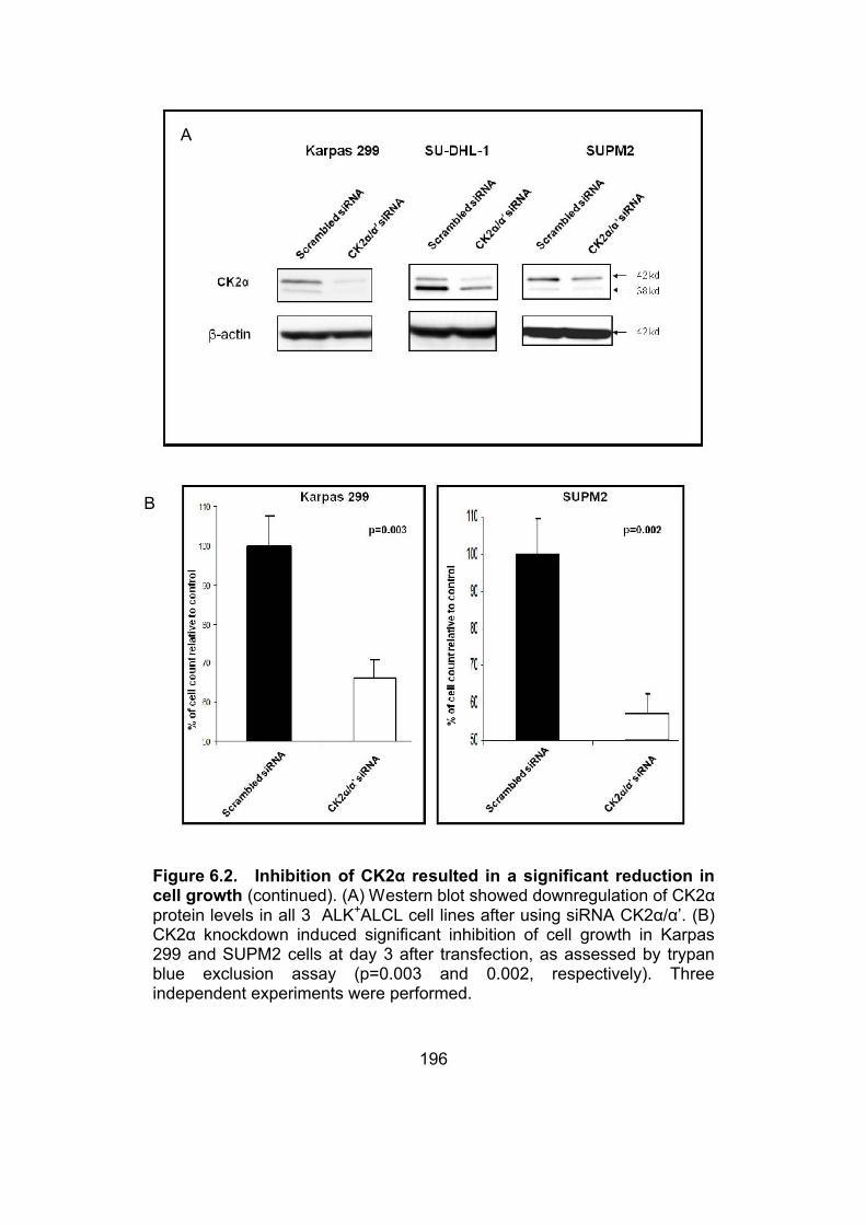
196
Figure 6.2. Inhibition of CK2α resulted in a significant reduction in cell growth (continued). (A) Western blot showed downregulation of CK2α protein levels in all 3 ALK+ALCL cell lines after using siRNA CK2α/α’. (B) CK2α knockdown induced significant inhibition of cell growth in Karpas 299 and SUPM2 cells at day 3 after transfection, as assessed by trypan blue exclusion assay (p=0.003 and 0.002, respectively). Three independent experiments were performed.
A
B

197
Figure 6.2. Inhibition of CK2α resulted in a significant reduction in cell growth. (C) CK2 inhibition using TBB resulted in dose dependent decrease in cell growth after 48 hours in all 3 ALK+ALCL cell lines as assessed by trypan blue exclusion assay (IC 50 was around 50 μM in Karpas 299 and SUPM2).
C

198
Figure 6.3. TBB resulted in cell cycle arrest (continued). (A) Western blot showed presence of cleaved caspase 3 and PARP only in high doses of TBB (i.e. 100 and 150 μM) not at lower doses i.e. 25 μM and 50 μM in Karpas 299 and SUPM2. (B) Cell cycle analysis by flow cytometry using propidium iodide showed significant G0/1 cell cycle arrest in ALK+ALCL cell lines Karpas 299 and SUPM2 following treatment with CK2 inhibitor TBB at a dose of 50 μM, compared to control cells treated with DMSO. Of note, no appreciable increase in the fraction of cells in the subG0/1 was noted.
A
B

199
Figure 6.3. TBB resulted in cell cycle arrest. (C) Western blot showed downregulation of cyclin D3 level at dose of 50 μM TBB in Karpas 299 and SUPM2 cells.
C

200
6.3.3. CK2 inhibition leads to decreased nuclear localization and
transcriptional activity of β-catenin
Addition of TBB at 50 μM for 48 hours resulted in a decrease in the
nuclear fraction of β-catenin in Karpas 299 and SUPM2 (figure 6.4A).
Using the same experimental approach, we evaluated if TBB induces any
change to the transcriptional activity of β-catenin. To achieve it, we
employed the TOPFlash/FOPFlash system, as previously described. As
shown in figure 6.4B, Karpas 299 cells treated with TBB had a significant
downregulation in β-catenin transcriptional activity (p=0.01) as compared
to cells treated with DMSO.

201
Figure 6.4. CK2 inhibition led to decrease in nuclear levels and transcriptional activity of β-catenin. (A) Western blot revealed decrease in nuclear levels of β-catenin in Karpas 299 and SUPM2 cells after treatment with 50 μM TBB compared to cells treated with DMSO. (B) Using TOPFlash/FOPFlash luciferase system, dual luciferase assay showed a significant downregulation of the β-catenin transcriptional activity (p=0.01) in Karpas 299 cells after treatment with 50 μM TBB compared to cells treated with DMSO.
A
B

202
6.3.4. CK2α inhibition leads to decrease NPM-ALK serine
phosphorylation
In view of the importance of NPM-ALK in ALK+ALCL, we asked if CK2α
may modulate the function and/or structure of NPM-ALK. First, we
performed co-immunoprecipitation experiment, and we identified evidence
of physical interaction between NPM-ALK and CK2α (figure 6.5A). We
next sought if CK2α regulates the tyrosine phosphorylation of NPM-ALK
as it was shown that CK2 can also mediate tyrosine phosphorylation (Vilk
et al., 2008). To achieve this goal, we assessed the level of tyrosine
phosphorylation of NPM-ALK using immunoprecipitation and a phospho-
tyrosine specific antibody. No detectable difference was found before and
after the addition siRNA targeted to CK2α. Since we recently reported that
there is serine phosphorylation of NPM-ALK, and serine phosphorylation
of NPM-ALK enhances its tumorgenicity (Wang et al., 2011), we
investigated if CK2α modulates this property. As shown in figure 6.6B,
knockdown of CK2α using resulted in a significant reduction in the level of
serine phosphorylation, by 60% and 20% in SU-DHL-1 and SUPM2 cells,
respectively.

203
Figure 6.5. CK2α inhibition led to decrease NPM-ALK serine phosphorylation. (A) Western blot showed co-immunoprecipitation using CK2α antibody with NPM-ALK, ‘+’ denotes experiments in which anti CK2α antibody was used for immunoprecipitation and ‘-’ denotes experiments in which the CK2α antibody was omitted. (B) Western blot showed immunoprecipitation using ALK antibody after knockdown of CK2α using siRNA CK2α/α’ and blotting with anti phosphoserine antibody showing decrease in serine phosphorylation of NPM- ALK in SU-DHL-1and SUPM2 cell lines (about 60% and 20% in SU-DHL-1 and SUPM2 respectively). Of note the tyrosine phosphorylation of NPM- ALK did not show any change.
A
B

204
6.4. DISCUSSION
One of our recent studies revealed the constitutive activation of β-catenin
in ALK+ALCL cells (Anand et al., 2011). We found that downregulation of
NPM-ALK was able to modulate the transcriptional activity of β-catenin
without having an effect on its protein levels, localization and tyrosine
phosphorylation status. In order to investigate possible mechanism of
regulation of the WCP by the oncoprotein NPM-ALK, we performed
knockdown of NPM-ALK, using siRNA, and checked for changes in
different Wnt genes using Wnt oligonucleotide array. Specifically, we
found that knockdown of NPM-ALK in ALK+ALCL cells resulted in
significant modulations of the expression levels of multiple genes involved
in the WCP, of which CK2α was found to be one of the genes affected
most dramatically. CK2 is a serine/threonine kinase that has biologic
importance as its disruption causes lethality in mouse embryos (Buchou et
al., 2003; Lou et al., 2008). Furthermore, CK2 have been identified as a
positive regulator of the WCP as it phosphorylates some of its key
members like β-catenin (Song et al., 2003; Willert et al., 1997; Wu et al.,
2009b).
In addition to the fact that CK2α represents the gene which is most
affected by the knockdown of NPM-ALK, our choosing of CK2α as the
focus of this study is based on its oncogenic importance demonstrated
previously in other types of cancer (Ruzzene and Pinna, 2010).
Specifically, it has been reported that CK2 is highly expressed in various
types of solid tumors, including carcinomas arising from the breast, lungs
and the head and neck region (Daya-Makin et al., 1994; Faust et al., 2000;
Gapany et al., 1995; Landesman-Bollag et al., 2001). In addition to its role
in the WCP (Seldin et al., 2005), CK2 also has been shown to modulate
other signaling pathways including those involving Akt/PI3K, c-Myc and
NFĸB (Channavajhala and Seldin, 2002; Di Maira et al., 2005; McElhinny
205
et al., 1996). Experiments involving the use of siRNA to knockdown CK2α
have provided support that CK2α indeed contributes to the constitutive
activation of β-catenin in ALK+ALCL cells. However, as multiple genes
involved in the WCP were implicated, it is highly likely that the cross talk
between the WCP and NPM-ALK is established based on multiple
interactions at various levels of the WCP.
Interestingly, a study just came out showing that CK2 associate with and
phosphorylate JAK tyrosine kinases, this would be of interest as
JAK/STAT axis is known to be activated in ALK+ALCL (Zheng et al.,
2011). Of note, CK2 was not previously identified as a NPM-ALK
interacting protein in various proteomics studies, including one performed
by our research group (Wu et al., 2009a). This discrepancy may be related
to the use of different methodologies that carry different sensitivities for
various types of protein. Nevertheless, the interaction between CK2α and
NPM-ALK correlates with our finding that CK2α appears to alter serine
phosphorylation of NPM-ALK. As mentioned, our group recently reported
the evidence of phosphorylation of 3 specific serine residues in NPM-ALK;
importantly, mutations of these sites that abrogate serine phosphorylation
resulted in a significant decrease in the oncogenic potential of NPM-ALK
(Wang et al., 2011). However, using colony formation assay after
downregulation of CK2α using siRNA in ALK+ALCL cell lines we did not
find significant change in tumorigenecity of cells (data not shown), which
could be explained that CK2α is not the only kinase responsible for the
serine phosphorylation of NPM-ALK and that other kinases are also
implicated.
How NPM-ALK promotes an increase in the protein level of CK2α is
unclear. As the STAT3 signaling is probably the most important signaling
pathway implicated in the pathogenesis of ALK+ALCL (Zamo et al., 2002),

206
we investigated if knockdown of STAT3 can result in a downregulation of
CK2α downregulation; however, we did not find any detectable change in
CK2α (data not shown). Thus, the exact mechanism by which NPM-ALK
affects the CK2α protein level still needs to be further investigated.
In view of the importance of CK2 in cancer, it has been identified as a
potential therapeutic target (Bortolato et al., 2008). A recent study showed
that pharmacologic inhibition of CK2 (such as TBB) can induce apoptosis
in chronic lymphocytic leukemia cells, without significant effect on normal
B and T lymphocytes (Martins et al., 2010). The same study emphasizes
the relative selectivity of CK2 inhibitors towards neoplastic T-cells as
compared to the normal T-cells, and this point carries important
therapeutic implications for ALK+ALCL. Regarding the biologic effect of
CK2 inhibition in ALK+ALCL, to our knowledge, there is only one previous
study which used Ellagic acid to inhibit CK2. In the aforementioned study
the authors did a virtual screening experiment targeting the ATP binding
site of CK2 using different databases, they used a panel of ALCL cell lines
both ALK+ and ALK- cell lines to test for the efficacy of identified
compounds (Cozza et al., 2006).
In conclusion, our data revealed a novel functional link between CK2 and
NPM-ALK, and a positive feedback loop between the WCP and NPM-ALK
with CK2α as an important link between the two pathways. Targeting CK2
in ALK+ALCL may prove to be of clinical importance.

207
6.5. REFERENCESAnand, M., Lai, R., and Gelebart, P. (2011). {beta}-catenin is constitutively active and increases STAT3 expression/activation in anaplastic lymphoma kinase-positive anaplastic large cell lymphoma. Haematologica 96, 253-261.
Bai, R.Y., Ouyang, T., Miething, C., Morris, S.W., Peschel, C., and Duyster, J. (2000). Nucleophosmin-anaplastic lymphoma kinase associated with anaplastic large-cell lymphoma activates the phosphatidylinositol 3-kinase/Akt antiapoptotic signaling pathway. Blood96, 4319-4327.
Bischof, D., Pulford, K., Mason, D.Y., and Morris, S.W. (1997). Role of the nucleophosmin (NPM) portion of the non-Hodgkin's lymphoma-associated NPM-anaplastic lymphoma kinase fusion protein in oncogenesis. Mol Cell Biol 17, 2312-2325.
Bortolato, A., Cozza, G., and Moro, S. (2008). Protein kinase CK2 inhibitors: emerging anticancer therapeutic agents? Anticancer Agents Med Chem 8, 798-806.
Buchou, T., Vernet, M., Blond, O., Jensen, H.H., Pointu, H., Olsen, B.B., Cochet, C., Issinger, O.G., and Boldyreff, B. (2003). Disruption of the regulatory beta subunit of protein kinase CK2 in mice leads to a cell-autonomous defect and early embryonic lethality. Mol Cell Biol 23, 908-915.
Channavajhala, P., and Seldin, D.C. (2002). Functional interaction of protein kinase CK2 and c-Myc in lymphomagenesis. Oncogene 21, 5280-5288.
Cozza, G., Bonvini, P., Zorzi, E., Poletto, G., Pagano, M.A., Sarno, S., Donella-Deana, A., Zagotto, G., Rosolen, A., Pinna, L.A., et al. (2006). Identification of ellagic acid as potent inhibitor of protein kinase CK2: a successful example of a virtual screening application. J Med Chem 49, 2363-2366.
Daya-Makin, M., Sanghera, J.S., Mogentale, T.L., Lipp, M., Parchomchuk, J., Hogg, J.C., and Pelech, S.L. (1994). Activation of a tumor-associated protein kinase (p40TAK) and casein kinase 2 in human squamous cell carcinomas and adenocarcinomas of the lung. Cancer Res 54, 2262-2268.
Di Maira, G., Salvi, M., Arrigoni, G., Marin, O., Sarno, S., Brustolon, F., Pinna, L.A., and Ruzzene, M. (2005). Protein kinase CK2 phosphorylates and upregulates Akt/PKB. Cell Death Differ 12, 668-677.

208
Dien Bard, J., Gelebart, P., Anand, M., Zak, Z., Hegazy, S.A., Amin, H.M., and Lai, R. (2009). IL-21 contributes to JAK3/STAT3 activation and promotes cell growth in ALK-positive anaplastic large cell lymphoma. Am J Pathol 175, 825-834.
Dominguez, I., Mizuno, J., Wu, H., Song, D.H., Symes, K., and Seldin, D.C. (2004). Protein kinase CK2 is required for dorsal axis formation in Xenopus embryos. Dev Biol 274, 110-124.
Duyster, J., Bai, R.Y., and Morris, S.W. (2001). Translocations involving anaplastic lymphoma kinase (ALK). Oncogene 20, 5623-5637.
Faust, R.A., Tawfic, S., Davis, A.T., Bubash, L.A., and Ahmed, K. (2000). Antisense oligonucleotides against protein kinase CK2-alpha inhibit growth of squamous cell carcinoma of the head and neck in vitro. Head Neck 22, 341-346.
Fujimoto, J., Shiota, M., Iwahara, T., Seki, N., Satoh, H., Mori, S., and Yamamoto, T. (1996). Characterization of the transforming activity of p80, a hyperphosphorylated protein in a Ki-1 lymphoma cell line with chromosomal translocation t(2;5). Proc Natl Acad Sci U S A 93, 4181-4186.
Gapany, M., Faust, R.A., Tawfic, S., Davis, A., Adams, G.L., and Ahmed, K. (1995). Association of elevated protein kinase CK2 activity with aggressive behavior of squamous cell carcinoma of the head and neck. Mol Med 1, 659-666.
Gelebart, P., Anand, M., Armanious, H., Peters, A.C., Dien Bard, J., Amin, H.M., and Lai, R. (2008). Constitutive activation of the Wnt canonical pathway in mantle cell lymphoma. Blood 112, 5171-5179.
Jaffe, E.S., Banks, P.M., Nathwani, B., Said, J., and Swerdlow, S.H. (2004). Recommendations for the reporting of lymphoid neoplasms: a report from the Association of Directors of Anatomic and Surgical Pathology. Mod Pathol 17, 131-135.
Landesman-Bollag, E., Romieu-Mourez, R., Song, D.H., Sonenshein, G.E., Cardiff, R.D., and Seldin, D.C. (2001). Protein kinase CK2 in mammary gland tumorigenesis. Oncogene 20, 3247-3257.
Lou, D.Y., Dominguez, I., Toselli, P., Landesman-Bollag, E., O'Brien, C., and Seldin, D.C. (2008). The alpha catalytic subunit of protein kinase CK2 is required for mouse embryonic development. Mol Cell Biol 28, 131-139.

209
Martins, L.R., Lucio, P., Silva, M.C., Anderes, K.L., Gameiro, P., Silva, M.G., and Barata, J.T. (2010). Targeting CK2 overexpression and hyperactivation as a novel therapeutic tool in chronic lymphocytic leukemia. Blood 116, 2724-2731.
McElhinny, J.A., Trushin, S.A., Bren, G.D., Chester, N., and Paya, C.V. (1996). Casein kinase II phosphorylates I kappa B alpha at S-283, S-289, S-293, and T-291 and is required for its degradation. Mol Cell Biol 16, 899-906.
Meggio, F., and Pinna, L.A. (2003). One-thousand-and-one substrates of protein kinase CK2? FASEB J 17, 349-368.
Morris, S.W., Kirstein, M.N., Valentine, M.B., Dittmer, K.G., Shapiro, D.N., Saltman, D.L., and Look, A.T. (1994). Fusion of a kinase gene, ALK, to a nucleolar protein gene, NPM, in non-Hodgkin's lymphoma. Science 263, 1281-1284.
Pagano, M.A., Bain, J., Kazimierczuk, Z., Sarno, S., Ruzzene, M., Di Maira, G., Elliott, M., Orzeszko, A., Cozza, G., Meggio, F., et al. (2008). The selectivity of inhibitors of protein kinase CK2: an update. Biochem J415, 353-365.
Pinna, L.A. (2003). The raison d'etre of constitutively active protein kinases: the lesson of CK2. Acc Chem Res 36, 378-384.
Pulford, K., Lamant, L., Morris, S.W., Butler, L.H., Wood, K.M., Stroud, D., Delsol, G., and Mason, D.Y. (1997). Detection of anaplastic lymphoma kinase (ALK) and nucleolar protein nucleophosmin (NPM)-ALK proteins in normal and neoplastic cells with the monoclonal antibody ALK1. Blood 89, 1394-1404.
Ruzzene, M., and Pinna, L.A. (2010). Addiction to protein kinase CK2: a common denominator of diverse cancer cells? Biochim Biophys Acta1804, 499-504.
Seldin, D.C., Landesman-Bollag, E., Farago, M., Currier, N., Lou, D., and Dominguez, I. (2005). CK2 as a positive regulator of Wnt signalling and tumourigenesis. Mol Cell Biochem 274, 63-67.
Seldin, D.C., and Leder, P. (1995). Casein kinase II alpha transgene-induced murine lymphoma: relation to theileriosis in cattle. Science 267, 894-897.

210
Shiota, M., Fujimoto, J., Semba, T., Satoh, H., Yamamoto, T., and Mori, S. (1994). Hyperphosphorylation of a novel 80 kDa protein-tyrosine kinase similar to Ltk in a human Ki-1 lymphoma cell line, AMS3. Oncogene 9, 1567-1574.
Slupianek, A., Nieborowska-Skorska, M., Hoser, G., Morrione, A., Majewski, M., Xue, L., Morris, S.W., Wasik, M.A., and Skorski, T. (2001). Role of phosphatidylinositol 3-kinase-Akt pathway in nucleophosmin/anaplastic lymphoma kinase-mediated lymphomagenesis. Cancer Res 61, 2194-2199.
Song, D.H., Dominguez, I., Mizuno, J., Kaut, M., Mohr, S.C., and Seldin, D.C. (2003). CK2 phosphorylation of the armadillo repeat region of beta-catenin potentiates Wnt signaling. J Biol Chem 278, 24018-24025.
Song, D.H., Sussman, D.J., and Seldin, D.C. (2000). Endogenous protein kinase CK2 participates in Wnt signaling in mammary epithelial cells. J Biol Chem 275, 23790-23797.
Staal, F.J., Noort Mv, M., Strous, G.J., and Clevers, H.C. (2002). Wnt signals are transmitted through N-terminally dephosphorylated beta-catenin. EMBO Rep 3, 63-68.
Traugh, J.A., Lin, W.J., Takada-Axelrod, F., and Tuazon, P.T. (1990). Importance of subunit interactions in regulation of casein kinase II. Adv Second Messenger Phosphoprotein Res 24, 224-229.
Turner, S.D., Yeung, D., Hadfield, K., Cook, S.J., and Alexander, D.R. (2007). The NPM-ALK tyrosine kinase mimics TCR signalling pathways, inducing NFAT and AP-1 by RAS-dependent mechanisms. Cell Signal 19, 740-747.
Vilk, G., Weber, J.E., Turowec, J.P., Duncan, J.S., Wu, C., Derksen, D.R., Zien, P., Sarno, S., Donella-Deana, A., Lajoie, G., et al. (2008). Protein kinase CK2 catalyzes tyrosine phosphorylation in mammalian cells. Cell Signal 20, 1942-1951.
Wang, P., Wu, F., Ma, Y., Li, L., Lai, R., and Young, L.C. (2010). Functional characterization of the kinase activation loop in nucleophosmin (NPM)-anaplastic lymphoma kinase (ALK) using tandem affinity purification and liquid chromatography-mass spectrometry. J Biol Chem285, 95-103.
Wang, P., Wu, F., Zhang, J., McMullen, T., Young, L.C., Ingham, R.J., Li, L., and Lai, R. (2011). Serine phosphorylation of NPM-ALK, which is
211
dependent on the auto-activation of the kinase activation loop, contributes to its oncogenic potential. Carcinogenesis 32, 146-153.
Willert, K., Brink, M., Wodarz, A., Varmus, H., and Nusse, R. (1997). Casein kinase 2 associates with and phosphorylates dishevelled. EMBO J16, 3089-3096.
Wu, F., Wang, P., Young, L.C., Lai, R., and Li, L. (2009a). Proteome-wide identification of novel binding partners to the oncogenic fusion gene protein, NPM-ALK, using tandem affinity purification and mass spectrometry. Am J Pathol 174, 361-370.
Wu, H., Symes, K., Seldin, D.C., and Dominguez, I. (2009b). Threonine 393 of beta-catenin regulates interaction with Axin. J Cell Biochem 108, 52-63.
Zamo, A., Chiarle, R., Piva, R., Howes, J., Fan, Y., Chilosi, M., Levy, D.E., and Inghirami, G. (2002). Anaplastic lymphoma kinase (ALK) activates Stat3 and protects hematopoietic cells from cell death. Oncogene 21, 1038-1047.
Zheng, Y., Qin, H., Frank, S.J., Deng, L., Litchfield, D.W., Tefferi, A., Pardanani, A., Lin, F.T., Li, J., Sha, B., et al. (2011). A CK2-dependent mechanism for activation of the JAK-STAT signaling pathway. Blood.

212
Chapter 7
Discussion

213
7.1. Thesis overview
In this thesis, I have extensively evaluated the different mechanisms of
cross talk between WCP and various oncogenic networks in different
cancer models. I identified novel cross talk between WCP and other
signalling pathways implicated in cancer pathogenesis. Previous studies
demonstrated the aberrant activation of the WCP in various cancer models
including breast cancer and hematopoietic malignancies (Clevers, 2006).
However, the dynamic interaction between WCP and various signalling
molecules implicated in cancer has not been fully elucidated. I
hypothesized the existence of cross talk between various components of
the WCP and various oncogenic signalling pathways in lymphoid and solid
tumors.
As shown in figure 7.1 the interaction between different WCP members
and 4 signalling molecules the PI3K/Akt , STAT3, ADAM10 and NPM-ALK
were studied. I found existence of different models of cross talk between
WCP members and different oncogenic molecules. Through identifying the
cross talk between WCP components and various signalling pathways, we
understand the complexity of signalling in cancer and we are warranted to
develop combination therapies which will be more beneficial in targeting
various signalling pathways.

214
Figure 7.1. WCP cross talking with oncogenic networks in cancer
215
7.2. WCP acts in a non-linear fashion in different cancer models
Historically, most of the studies of the WCP focused on WCP signalling as
being functioning in a linear fashion (Klaus and Birchmeier, 2008).
However, in my studies I showed that various members of the WCP act in
a linear manner as well as acting in a horizontal manner interacting with
other proteins not included in the WCP.
In chapter 2 and 3, I tried to identify whether WCP activation was the main
contributing event for GSK-3β inactivation in vivo in two cancer models
MCL and breast cancer. The decision to use pGSK-3β (the inactive form
of GSK-3β) as a marker in my studies came for various reasons: First, the
antibody used in my immunohistochemical studies was totally novel and
was not used in any in vivo studies before, I had to do all the optimization
for it. Second, until now β-catenin antibodies are the only accepted marker
used for in vivo studies as a marker for WCP activation in patient samples.
Third, although pGSK-3β have been linked to WCP activation,
nevertheless it have also been linked to other signalling pathways mainly
PI3K/Akt activation (Pap and Cooper, 1998). In both chapter 2 and 3 I
found evidence of inactivated GSK-3β (pGSK-3β) in both breast cancer
and MCL (47.2% and 67.5% respectively). In chapter 2, I found that in
breast cancer, the expression of pGSK-3β in vivo correlated with the
activation of PI3K/Akt pathway. On the other hand in chapter 3, I found
that in MCL, the expression of pGSK-3β in vivo correlated with the
activation of the WCP. However, in both studies inactivated GSK-3β
(pGSK-3β) correlated with a poor outcome in patients, which points
towards the importance of pGSK-3β in cancer biology regardless of the
event initiating its inactivation. In these studies I presented the non-linear
interaction between WCP and PI3K/Akt pathway with pGSK-3β acting as a
hub between the two pathways.

216
In chapter 4, I showed that β-catenin, the main player in the WCP, is
transcriptionally regulated by STAT3. I decided to study this interaction in
breast cancer model as both STAT3 and WCP was shown to be activated
in a subset of breast cancer (Kunigal et al., 2009; Lin et al., 2000). This
study clearly shows the non-linear interaction of the WCP member β-
catenin acting as hub molecule in both STAT3 and WCP. This finding is
also of interest as a recently published study showed that STAT3 can be
transcriptionally regulated by β-catenin, which may suggest the presence
of a possible feedback loop between the two pathways (Yan et al., 2008).
In chapter 5, the aim at the start of this study was to elucidate the possible
cross talk between ADAM10 and β-catenin. The background rationale
behind this aim was; first, ADAM10 was shown in colon cancer to be a
target gene of β-catenin/TCF signalling as ADAM10 promoter carried
TCF/LEF binding sites (Gavert et al., 2007). Second, β-catenin itself was
shown to be regulated by ADAM10 as cleaving of cadherin by ADAM10
led to release of membranous β-catenin and increase in its nuclear
translocation (Maretzky et al., 2005; Reiss et al., 2005). At the beginning, I
decided to use MCL cancer model for investigating this possible dual
interaction between ADAM10 and β-catenin as in a recent study our lab
was the first to report the activation of WCP in MCL (Gelebart et al., 2008).
Interestingly, the first goal of the study, which is detecting presence and
activation of ADAM10, was achieved, as I found ADAM10 to be activated
in MCL cell lines and patients’ samples examined. The second goal of my
study was to elucidate the dual interaction between β-catenin and
ADAM10 in MCL. However, using siRNA to inhibit β-catenin level I did not
observe any change in ADAM10 protein or mRNA levels, similarly using
siRNA to inhibit ADAM10 I did not detect and change in nuclear β-catenin

217
levels or its transcriptional activity. Accordingly, I could not detect in this
study any cross talk between WCP and ADAM10.
In chapter 6, I reported the activation of the Wnt pathway in ALK+ALCL
and its cross talk with the oncogenic protein NPM-ALK. The cross talk was
found to be at the level of CK2α where downregulation of ALK using
siRNA led to downregulation of CK2α protein levels and the
downregulation of CK2α led to decrease in serine phosphorylation of
NPM-ALK. In this study WCP was shown to interact with NPM-ALK an
oncogenic tyrosine kinase known for its multiple effectors and binding
partners (Fujimoto et al., 1996). This study shows CK2α as a hub in this
interaction.
7.3. Models of cross talk between WCP members and other signalling
pathways
As discussed in chapter 1, signalling pathways are said to be cross talking
if they are connected in at least one of three ways (Figure 7.2) (Guo and
Wang, 2009). Through my studies in this thesis I represented examples of
each of these models of cross talk. In chapter 2 and 3 I showed pGSK-3β
as a hub where one pathway i.e. PI3K/Akt can modulate a key molecule
(pGSK-3β in this case) of the other pathway i.e. WCP. In chapter 4, I
showed β-catenin acting as a hub between WCP and STAT3 where the
component of one pathway i.e. β-catenin as member in the WCP is the
transcriptional target of the other pathway i.e.STAT3. In chapter 6, I
showed a third model of cross talk where CK2α a component of the WCP
physically interacted with another signalling molecule i.e. NPM-ALK.
These different models of cross talk shows the diversity by which WCP
members are cross talking in non-linear fashion with other pathways in
different cancer models.

218
Figure 7.2. Summary of different models of cross talk studied in the
current thesis. A) WCP member CK2α physically interacts with NPM-
ALK. B) WCP member β-catenin is the transcriptional target for STAT3. C)
WCP member pGSK-3β is modulated by the PI3K/Akt pathway.

219
7.4. Biologic importance of WCP members involved in the cross talk
In all my studies I have demonstrated biologic importance of different
WCP members involved in the cross talk. In chapter 2 and 3, inactivated
GSK-3β (pGSK-3β) correlated with a poor outcome in patients both in
breast cancer and MCL which points towards the possibility of using it as a
prognostic marker. In chapter 4, I demonstrated the biologic importance of
β-catenin in breast cancer as inhibition of β-catenin using siRNA led to
decrease in cell growth. In chapter 6, I showed the importance of CK2α in
the biology of ALK+ALCL as its inhibition caused decrease in cell growth
and cell cycle arrest. Given the biologic importance shown of all the WCP
members acting as a hub for interaction with other pathways, this further
emphasises the importance of identifying these hubs as therapeutic
targets.
7.5. Limitations and future studies
The different studies in this thesis each have its own limitations and
possible future directions. In chapter 2, the limited number of patients’
sample i.e. 72 patients of breast cancer, hindered the statistical analysis of
certain categories. Specifically, in this cohort the number of ER negative
patients was 22 cases and although they had a huge impact in the survival
data I could not perform correlational statistical analysis of
immunohistochemical markers in this group due to small sample size.
Accordingly, future studies can be done using larger cohort of breast
cancer patient samples, especially the ones representing various
molecular subtypes of breast cancer. Also, it will be of interest to plan
further studies to identify the implication of GSK-3β inactivation in patient
treatment. For instance, patients could be stratified into two groups
according to their pGSK-3β status. Given that the positive group will have
possible activation of pAkt pathway, small molecule inhibitors targeting

220
pAkt can be used in positive versus negative groups in combination with
conventional chemotherapy. According to this hypothesis the ones with
positive pGSK-3β will possibly benefit from treatment with pAkt inhibitors
in contrast to the negative ones.
In chapter 2 and 3 in this thesis I identified that inactivated GSK-3β
(pGSK-3β) correlated with a poor outcome in patients. This finding should
warrant caution in using new small molecule inhibitors developed to inhibit
GSK-3β as they could be a double edged sword. While GSK-3β inhibitors
may have beneficial effect in treating diseases as Alzheimer disease and
diabetes, however they may carry a potential hazard for cancer
development (Phukan et al., 2010). Further studies on the effect of these
newly developed small molecule inhibitors of GSK-3β in cancer are
warranted.
In chapter 4, I demonstrated a novel mechanism of regulation of β-catenin
by STAT3 in breast cancer. As β-catenin is the main player in the WCP, so
regulation of its transcriptional and protein levels would consequently have
an effect on the WCP signalling. In this study I identified a subset of breast
cancer which has activation of both STAT3 and β-catenin. This subset of
patients would greatly benefit from the combination therapy between
STAT3 inhibitors and small molecule inhibitors of β-catenin. Of note, this
study was conducted in the context of breast cancer; it would be tempting
to see whether this interaction is occurring in normal mammary cells.
Furthermore, the investigation of the cross talk between the WCP and
STAT3 carries potential interest especially in field of ES cell research as
both pathways are implicated in maintaining self renewal in murine stem
cells (Okita and Yamanaka, 2006).

221
In chapter 5, the aim at the start of this study was to elucidate the possible
cross talk between ADAM10 and β-catenin. However, although I did not
detect any cross talk between ADAM10 and β-catenin in this cancer model
i.e. MCL; this does not totally exclude the existence of this cross talk in
other cancer models. In fact, the discovery of ADAM10 regulation by β-
catenin was made in CRC model which represent the paradigm of WCP
activation. Moreover, activation of different target genes of the Wnt
pathways have been suggested to be cell type specific, as not all genes
will be activated in all cell types, rather than specific genes are activated in
specific cell types (Logan and Nusse, 2004). Importantly, a recently
published study showed that SFRPs, which are known as Wnt inhibitors,
can bind to and inactivate ADAM10 (Esteve et al., 2011). Accordingly, the
next stage of this research will be to further evaluate the relationship
between ADAM10 and other components of the WCP in different cancer
models. Also, it would be of interest to study the possible cross talk
between WCP and TNFα/NFκB pathway with ADAM10 acting as a hub
between the two pathways, especially that both pathways (i.e. TNFα/NFκB
and WCP) and ADAM10 are implicated in cancer pathogenesis.
In chapter 6, I reported the activation of the Wnt pathway in ALK+ALCL
and its cross talk with the oncogenic protein NPM-ALK. The Wnt
oligonucleotide used in this study, showed expression of other WCP
members besides CK2α, among them were Dvl2, Dvl3 and Wnt3a
(unpublished data). These various Wnt members could be studied each
separately to find their biologic significance in ALK+ALCL. It would also be
of interest to study the cross talk of WCP with other oncogenic tyrosine
kinases implicated in cancer.

222
7.6. Closing remarks
Overall, this thesis provided insight into the novel non-linear interactions of
the WCP in cancer. To this point, I have identified novel important cross
talk between WCP and oncogenic networks in different cancer models i.e.
solid and hematopoietic tumors. In the first and second in vivo studies I
conducted I showed that GSK-3β inactivation is likely attributed to WCP in
MCL, however it is likely contributed to PI3K/Akt in breast cancer.
Furthermore, I identified the clinical importance of this event in both cancer
models. In the third study I conducted I showed crosstalk between WCP
and STAT3 at the level of β-catenin, being a transcriptional target for both
pathways. In the fourth study I did not find any cross talk between the
WCP and ADAM10 in MCL. In the fifth study I identified the cross talk
between WCP and NPM-ALK at the level of CK2α.
I believe that the work done in this thesis furthers our knowledge of the
implication and importance of WCP in cancer pathogenesis. This in-depth
understanding of the dynamics of signalling pathway interactions allows us
to identify therapeutic targets. It gives us rationale for the combination of
different pathway inhibitors as an approach in cancer therapeutics,
recognizing the complexity of cross talk between signalling pathways in
cancer. The work in this thesis provides a framework for further studies
identifying critical targets in WCP, encouraging more efforts towards
identifying specific molecular hubs and ultimately developing novel
strategies to target inherent abnormalities of cancer cells.

223
7.7. REFERENCES
Clevers, H. (2006). Wnt/beta-catenin signaling in development and disease. Cell 127, 469-480.
Esteve, P., Sandonis, A., Cardozo, M., Malapeira, J., Ibanez, C., Crespo, I., Marcos, S., Gonzalez-Garcia, S., Toribio, M.L., Arribas, J., et al. (2011). SFRPs act as negative modulators of ADAM10 to regulate retinal neurogenesis. Nat Neurosci 14, 562-569.
Fujimoto, J., Shiota, M., Iwahara, T., Seki, N., Satoh, H., Mori, S., and Yamamoto, T. (1996). Characterization of the transforming activity of p80, a hyperphosphorylated protein in a Ki-1 lymphoma cell line with chromosomal translocation t(2;5). Proc Natl Acad Sci U S A 93, 4181-4186.
Gavert, N., Sheffer, M., Raveh, S., Spaderna, S., Shtutman, M., Brabletz, T., Barany, F., Paty, P., Notterman, D., Domany, E., et al. (2007). Expression of L1-CAM and ADAM10 in human colon cancer cells induces metastasis. Cancer Res 67, 7703-7712.
Gelebart, P., Anand, M., Armanious, H., Peters, A.C., Dien Bard, J., Amin, H.M., and Lai, R. (2008). Constitutive activation of the Wnt canonical pathway in mantle cell lymphoma. Blood 112, 5171-5179.
Guo, X., and Wang, X.F. (2009). Signaling cross-talk between TGF-beta/BMP and other pathways. Cell Res 19, 71-88.
Klaus, A., and Birchmeier, W. (2008). Wnt signalling and its impact on development and cancer. Nat Rev Cancer 8, 387-398.
Kunigal, S., Lakka, S.S., Sodadasu, P.K., Estes, N., and Rao, J.S. (2009). Stat3-siRNA induces Fas-mediated apoptosis in vitro and in vivo in breast cancer. Int J Oncol 34, 1209-1220.
Lin, S.Y., Xia, W., Wang, J.C., Kwong, K.Y., Spohn, B., Wen, Y., Pestell, R.G., and Hung, M.C. (2000). Beta-catenin, a novel prognostic marker for breast cancer: its roles in cyclin D1 expression and cancer progression. Proc Natl Acad Sci U S A 97, 4262-4266.
Logan, C.Y., and Nusse, R. (2004). The Wnt signaling pathway in development and disease. Annu Rev Cell Dev Biol 20, 781-810.
224
Maretzky, T., Reiss, K., Ludwig, A., Buchholz, J., Scholz, F., Proksch, E., de Strooper, B., Hartmann, D., and Saftig, P. (2005). ADAM10 mediates E-cadherin shedding and regulates epithelial cell-cell adhesion, migration, and beta-catenin translocation. Proc Natl Acad Sci U S A 102, 9182-9187.
Okita, K., and Yamanaka, S. (2006). Intracellular signaling pathways regulating pluripotency of embryonic stem cells. Curr Stem Cell Res Ther1, 103-111.
Pap, M., and Cooper, G.M. (1998). Role of glycogen synthase kinase-3 in the phosphatidylinositol 3-Kinase/Akt cell survival pathway. J Biol Chem273, 19929-19932.
Phukan, S., Babu, V.S., Kannoji, A., Hariharan, R., and Balaji, V.N. (2010). GSK3beta: role in therapeutic landscape and development of modulators. Br J Pharmacol 160, 1-19.
Reiss, K., Maretzky, T., Ludwig, A., Tousseyn, T., de Strooper, B., Hartmann, D., and Saftig, P. (2005). ADAM10 cleavage of N-cadherin and regulation of cell-cell adhesion and beta-catenin nuclear signalling. EMBO J 24, 742-752.
Yan, S., Zhou, C., Zhang, W., Zhang, G., Zhao, X., Yang, S., Wang, Y., Lu, N., Zhu, H., and Xu, N. (2008). beta-Catenin/TCF pathway upregulates STAT3 expression in human esophageal squamous cell carcinoma. Cancer Lett 271, 85-97.