Non-canonical inflammasome activation targets caspase-11
6
LETTER doi:10.1038/nature10558 Non-canonical inflammasome activation targets caspase-11 Nobuhiko Kayagaki 1 , Søren Warming 2 , Mohamed Lamkanfi 3,4 , Lieselotte Vande Walle 3,4 , Salina Louie 1 , Jennifer Dong 1 , Kim Newton 1 , Yan Qu 1 , Jinfeng Liu 5 , Sherry Heldens 2 , Juan Zhang 6 , Wyne P. Lee 6 , Merone Roose-Girma 2 & Vishva M. Dixit 1 Caspase-1 activation by inflammasome scaffolds comprised of intracellular nucleotide-binding oligomerization domain (NOD)- like receptors (NLRs) and the adaptor ASC is believed to be essential for production of the pro-inflammatory cytokines interleukin (IL)- 1b and IL-18 during the innate immune response 1–5 . Here we show, with C57BL/6 Casp11 gene-targeted mice, that caspase-11 (also known as caspase-4) 6–8 is critical for caspase-1 activation and IL- 1b production in macrophages infected with Escherichia coli, Citrobacter rodentium or Vibrio cholerae. Strain 129 mice, like Casp11 2/2 mice, exhibited defects in IL-1b production and harboured a mutation in the Casp11 locus that attenuated caspase-11 expression. This finding is important because published targeting of the Casp1 gene was done using strain 129 embryonic stem cells 9,10 . Casp1 and Casp11 are too close in the genome to be segregated by recombination; consequently, the published Casp1 –/– mice lack both caspase-11 and caspase-1. Interestingly, Casp11 –/– macrophages secreted IL-1b normally in response to ATP and monosodium urate, indicating that caspase-11 is engaged by a non-canonical inflammasome. Casp1 –/– Casp11 129mt/129mt macro- phages expressing caspase-11 from a C57BL/6 bacterial artificial chromosome transgene failed to secrete IL-1b regardless of stimulus, confirming an essential role for caspase-1 in IL-1b pro- duction. Caspase-11 rather than caspase-1, however, was required for non-canonical inflammasome-triggered macrophage cell death, indicating that caspase-11 orchestrates both caspase-1-dependent and -independent outputs. Caspase-1 activation by non-canonical stimuli required NLRP3 and ASC, but caspase-11 processing and cell death did not, implying that there is a distinct activator of caspase-11. Lastly, loss of caspase-11 rather than caspase-1 protected mice from a lethal dose of lipopolysaccharide. These data highlight a unique pro-inflammatory role for caspase-11 in the innate immune response to clinically significant bacterial infections. Many bacterial toxins promote inflammasome activation 11–15 , and this is also true for cholera toxin B (CTB), a component of the AB5 holotoxin complex (Fig. 1a and Supplementary Fig. 1). CTB lacks the enzymatic activity necessary for entero-pathogenesis, but binds to GM1 ganglioside on the cell surface to facilitate the entry of enzymatic component cholera toxin A (CTA) 16 . Similar to ATP, which is a known activator of caspase-1 (ref. 17), CTB induced NLRP3- and ASC-dependent processing and secretion of caspase-1 and IL-1b from lipopolysaccharide (LPS)-primed bone-marrow-derived C57BL/6 macrophages (BMDMs). Several other NLR family members (NLRC4, NLRP6, NLRP12, NOD1 and NOD2), inflammasome com- ponent AIM2 (refs 18–20), the ATP-releasing pannexin-1 channel, and the ATP-gated P2RX7 receptor 21 were dispensable for CTB- induced IL-1b secretion (Supplementary Fig. 1a). These data indicate that CTB does not activate the NLRP3 inflammasome indirectly by causing ATP release and P2RX7 receptor stimulation. To investigate other host factors involved in the inflammasome response to CTB, we determined whether macrophages from other mouse strains secrete IL-1b when exposed to CTB. Polymorphisms in the Nlrp1b gene, for example, alter sensitivity to anthrax lethal toxin 12 . LPS-primed 129S6 BMDMs cultured with ATP processed caspase-1 and secreted IL-1b, but they were unresponsive to CTB (Fig. 1b, c). Western blotting for pro-inflammatory adaptor proteins and caspases indicated that 129S6 macrophages were deficient in pro-caspase-11 isoforms 7 p43 and p38 (Fig. 1d). Quantitative polymerase chain reaction with reverse tran- scription (RT–PCR) confirmed attenuated LPS-induced Casp11 messenger RNA expression in 129S6 BMDMs (Fig. 1e). Caspase-11 is similar to caspase-1 (46%), orthologous to human caspase-4 and -5, and has been shown to interact with caspase-1 upon overexpression 6–8 . Although caspase-11 was shown to be essential for LPS-induced IL-1b secretion in vivo 8 , the role of caspase-11 in macrophage inflammasome signalling and its mechanism of activation remain unclear. Primers that amplified the full-length Casp11 mRNA expressed in C57BL/6 BMDMs after culture with either LPS or E. coli recovered only a truncated Casp11 complementary DNA (D110) from similarly treated 129S6 cells (Fig. 1f). Sequencing revealed that the 129S6 Casp11 transcript lacked all sequence encoded by exon 7. Splicing of exon 6 to exon 8 creates a frame-shift after proline 304 and a stop codon occurred after 5 aberrant amino acids (Fig. 1g). Absence of exon 7 expression was confirmed in 129S6 BMDMs by quantitative RT–PCR for Casp11 exon 7 (Supplementary Fig. 2a). Macrophages from three additional 129 substrains (129X1, 129S1 and 129P3) also lacked detectable caspase-11 protein, expressed Casp11 D110 mRNA, and produced negligible IL-1b in response to CTB (Supplementary Fig. 2b–d). Sequencing of 129S1 genomic DNA identified a 5-bp deletion encompassing the exon 7 splice acceptor junction as the origin of the D110 isoform (Fig. 1h). The caspase-11 antibody used in our experi- ments detected the truncated caspase-11 protein encoded by Casp11 D110 when it was overexpressed in 293T cells (Supplemen- tary Fig. 2e) but failed to detect endogenous caspase-11 D110 product in 129 BMDMs (Fig. 1d and Supplementary Fig. 2b), possibly due to nonsense-mediated mRNA decay (Fig. 1e and Supplementary Fig. 2a). Caspase-11 D110 protein product lacks most of the small catalytic subunit so any protein that is expressed should be non-functional (Supplementary Fig. 2f). We explored further the role of caspase-11 in inflammasome activa- tion by targeting Casp11 exon 5 for deletion in C57BL/6 embryonic stem (ES) cells. This exon encodes the critical catalytic residue cysteine 254 (ref. 7) and most of the caspase-11 large catalytic subunit (Sup- plementary Fig. 3). LPS-primed C57BL/6 Casp11 1/1 (wild type) and Casp11 –/– BMDMs were stimulated with ATP to engage the NLRP3-dependent inflammasome, poly(dA:dT) double-stranded DNA or Francisella tularensis to activate the AIM2-dependent inflam- masome, and flagellin or Pseudomonas aeruginosa to engage the NLRC4-dependent inflammasome 18–20 . These canonical stimuli 1 Department of Physiological Chemistry, Genentech Inc., South San Francisco, California 94080, USA. 2 Department of Molecular Biology, Genentech Inc., South San Francisco, California 94080, USA. 3 Department of Medical Protein Research, VIB, B-9000 Ghent, Belgium. 4 Department of Biochemistry, Ghent University, B-9000 Ghent, Belgium. 5 Department of Bioinformatics, Genentech Inc., South San Francisco, California 94080, USA. 6 Department of Immunology, Genentech Inc., South San Francisco, California 94080, USA. 00 MONTH 2011 | VOL 000 | NATURE | 1 Macmillan Publishers Limited. All rights reserved ©2011
Transcript of Non-canonical inflammasome activation targets caspase-11

LETTERdoi:10.1038/nature10558
Non-canonical inflammasome activation targetscaspase-11Nobuhiko Kayagaki1, Søren Warming2, Mohamed Lamkanfi3,4, Lieselotte Vande Walle3,4, Salina Louie1, Jennifer Dong1,Kim Newton1, Yan Qu1, Jinfeng Liu5, Sherry Heldens2, Juan Zhang6, Wyne P. Lee6, Merone Roose-Girma2 & Vishva M. Dixit1
Caspase-1 activation by inflammasome scaffolds comprised ofintracellular nucleotide-binding oligomerization domain (NOD)-like receptors (NLRs) and the adaptor ASC is believed to be essentialfor production of the pro-inflammatory cytokines interleukin (IL)-1b and IL-18 during the innate immune response1–5. Here we show,with C57BL/6 Casp11 gene-targeted mice, that caspase-11 (alsoknown as caspase-4)6–8 is critical for caspase-1 activation and IL-1b production in macrophages infected with Escherichia coli,Citrobacter rodentium or Vibrio cholerae. Strain 129 mice, likeCasp112/2 mice, exhibited defects in IL-1b production andharboured a mutation in the Casp11 locus that attenuatedcaspase-11 expression. This finding is important because publishedtargeting of the Casp1 gene was done using strain 129 embryonicstem cells9,10. Casp1 and Casp11 are too close in the genome to besegregated by recombination; consequently, the published Casp1–/–
mice lack both caspase-11 and caspase-1. Interestingly, Casp11–/–
macrophages secreted IL-1b normally in response to ATP andmonosodium urate, indicating that caspase-11 is engaged by anon-canonical inflammasome. Casp1–/–Casp11129mt/129mt macro-phages expressing caspase-11 from a C57BL/6 bacterial artificialchromosome transgene failed to secrete IL-1b regardless ofstimulus, confirming an essential role for caspase-1 in IL-1b pro-duction. Caspase-11 rather than caspase-1, however, was requiredfor non-canonical inflammasome-triggered macrophage cell death,indicating that caspase-11 orchestrates both caspase-1-dependentand -independent outputs. Caspase-1 activation by non-canonicalstimuli required NLRP3 and ASC, but caspase-11 processingand cell death did not, implying that there is a distinct activatorof caspase-11. Lastly, loss of caspase-11 rather than caspase-1protected mice from a lethal dose of lipopolysaccharide. These datahighlight a unique pro-inflammatory role for caspase-11 in theinnate immune response to clinically significant bacterialinfections.
Many bacterial toxins promote inflammasome activation11–15, andthis is also true for cholera toxin B (CTB), a component of the AB5holotoxin complex (Fig. 1a and Supplementary Fig. 1). CTB lacks theenzymatic activity necessary for entero-pathogenesis, but binds toGM1 ganglioside on the cell surface to facilitate the entry of enzymaticcomponent cholera toxin A (CTA)16. Similar to ATP, which is aknown activator of caspase-1 (ref. 17), CTB induced NLRP3- andASC-dependent processing and secretion of caspase-1 and IL-1b fromlipopolysaccharide (LPS)-primed bone-marrow-derived C57BL/6macrophages (BMDMs). Several other NLR family members(NLRC4, NLRP6, NLRP12, NOD1 and NOD2), inflammasome com-ponent AIM2 (refs 18–20), the ATP-releasing pannexin-1 channel,and the ATP-gated P2RX7 receptor21 were dispensable for CTB-induced IL-1b secretion (Supplementary Fig. 1a). These data indicatethat CTB does not activate the NLRP3 inflammasome indirectly bycausing ATP release and P2RX7 receptor stimulation. To investigate
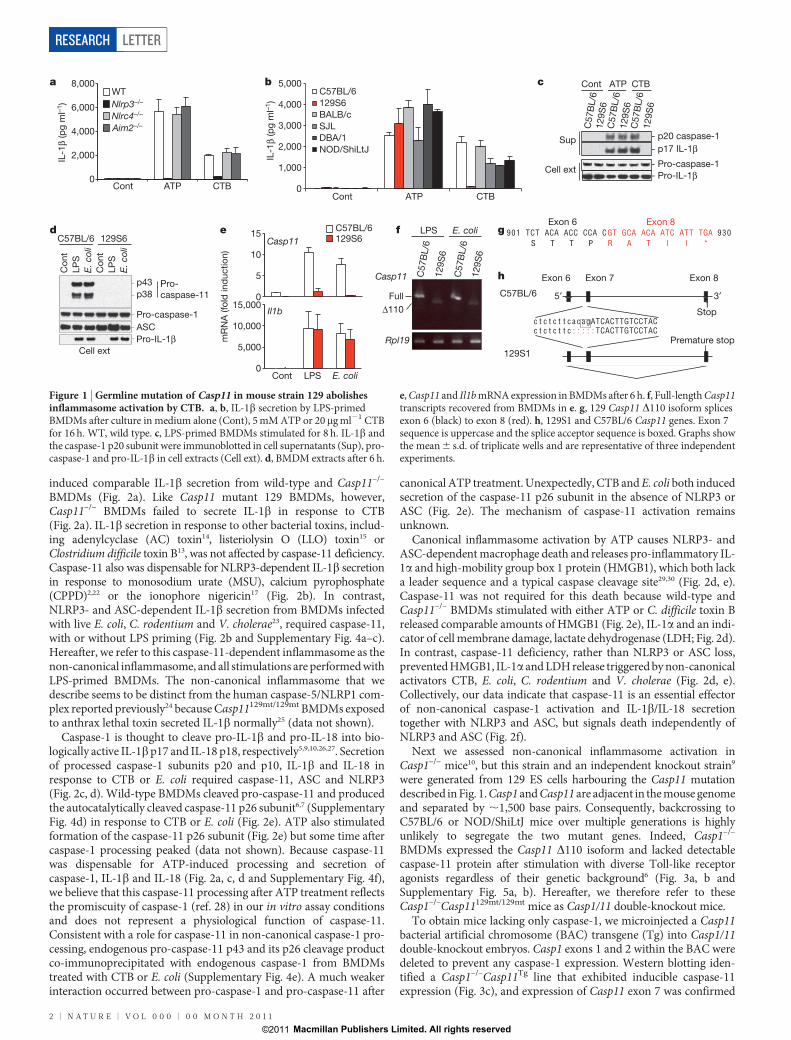
other host factors involved in the inflammasome response to CTB, wedetermined whether macrophages from other mouse strains secreteIL-1b when exposed to CTB. Polymorphisms in the Nlrp1b gene, forexample, alter sensitivity to anthrax lethal toxin12. LPS-primed 129S6BMDMs cultured with ATP processed caspase-1 and secreted IL-1b,but they were unresponsive to CTB (Fig. 1b, c). Western blotting forpro-inflammatory adaptor proteins and caspases indicated that 129S6macrophages were deficient in pro-caspase-11 isoforms7 p43 and p38(Fig. 1d). Quantitative polymerase chain reaction with reverse tran-scription (RT–PCR) confirmed attenuated LPS-induced Casp11messenger RNA expression in 129S6 BMDMs (Fig. 1e). Caspase-11is similar to caspase-1 (46%), orthologous to human caspase-4 and -5,and has been shown to interact with caspase-1 upon overexpression6–8.Although caspase-11 was shown to be essential for LPS-induced IL-1bsecretion in vivo8, the role of caspase-11 in macrophage inflammasomesignalling and its mechanism of activation remain unclear.
Primers that amplified the full-length Casp11 mRNA expressed inC57BL/6 BMDMs after culture with either LPS or E. coli recoveredonly a truncated Casp11 complementary DNA (D110) from similarlytreated 129S6 cells (Fig. 1f). Sequencing revealed that the 129S6 Casp11transcript lacked all sequence encoded by exon 7. Splicing of exon 6 toexon 8 creates a frame-shift after proline 304 and a stop codonoccurred after 5 aberrant amino acids (Fig. 1g). Absence of exon 7expression was confirmed in 129S6 BMDMs by quantitative RT–PCRfor Casp11 exon 7 (Supplementary Fig. 2a). Macrophages from threeadditional 129 substrains (129X1, 129S1 and 129P3) also lackeddetectable caspase-11 protein, expressed Casp11 D110 mRNA, andproduced negligible IL-1b in response to CTB (Supplementary Fig.2b–d). Sequencing of 129S1 genomic DNA identified a 5-bp deletionencompassing the exon 7 splice acceptor junction as the origin of theD110 isoform (Fig. 1h). The caspase-11 antibody used in our experi-ments detected the truncated caspase-11 protein encoded by Casp11D110 when it was overexpressed in 293T cells (Supplemen-tary Fig. 2e) but failed to detect endogenous caspase-11 D110 productin 129 BMDMs (Fig. 1d and Supplementary Fig. 2b), possibly due tononsense-mediated mRNA decay (Fig. 1e and Supplementary Fig. 2a).Caspase-11 D110 protein product lacks most of the small catalyticsubunit so any protein that is expressed should be non-functional(Supplementary Fig. 2f).
We explored further the role of caspase-11 in inflammasome activa-tion by targeting Casp11 exon 5 for deletion in C57BL/6 embryonicstem (ES) cells. This exon encodes the critical catalytic residue cysteine254 (ref. 7) and most of the caspase-11 large catalytic subunit (Sup-plementary Fig. 3). LPS-primed C57BL/6 Casp111/1 (wild type)and Casp11–/– BMDMs were stimulated with ATP to engage theNLRP3-dependent inflammasome, poly(dA:dT) double-strandedDNA or Francisella tularensis to activate the AIM2-dependent inflam-masome, and flagellin or Pseudomonas aeruginosa to engage theNLRC4-dependent inflammasome18–20. These canonical stimuli
1Department of Physiological Chemistry, Genentech Inc., South San Francisco, California 94080, USA. 2Department of Molecular Biology, Genentech Inc., South San Francisco, California 94080, USA.3Department of Medical Protein Research, VIB, B-9000 Ghent, Belgium. 4Department of Biochemistry, Ghent University, B-9000 Ghent, Belgium. 5Department of Bioinformatics, Genentech Inc., South SanFrancisco, California 94080, USA. 6Department of Immunology, Genentech Inc., South San Francisco, California 94080, USA.
0 0 M O N T H 2 0 1 1 | V O L 0 0 0 | N A T U R E | 1
Macmillan Publishers Limited. All rights reserved©2011
induced comparable IL-1b secretion from wild-type and Casp11–/–
BMDMs (Fig. 2a). Like Casp11 mutant 129 BMDMs, however,Casp11–/– BMDMs failed to secrete IL-1b in response to CTB(Fig. 2a). IL-1b secretion in response to other bacterial toxins, includ-ing adenylcyclase (AC) toxin14, listeriolysin O (LLO) toxin15 orClostridium difficile toxin B13, was not affected by caspase-11 deficiency.Caspase-11 also was dispensable for NLRP3-dependent IL-1b secretionin response to monosodium urate (MSU), calcium pyrophosphate(CPPD)2,22 or the ionophore nigericin17 (Fig. 2b). In contrast,NLRP3- and ASC-dependent IL-1b secretion from BMDMs infectedwith live E. coli, C. rodentium and V. cholerae23, required caspase-11,with or without LPS priming (Fig. 2b and Supplementary Fig. 4a–c).Hereafter, we refer to this caspase-11-dependent inflammasome as thenon-canonical inflammasome, and all stimulations are performed withLPS-primed BMDMs. The non-canonical inflammasome that wedescribe seems to be distinct from the human caspase-5/NLRP1 com-plex reported previously24 because Casp11129mt/129mt BMDMs exposedto anthrax lethal toxin secreted IL-1b normally25 (data not shown).
Caspase-1 is thought to cleave pro-IL-1b and pro-IL-18 into bio-logically active IL-1b p17 and IL-18 p18, respectively5,9,10,26,27. Secretionof processed caspase-1 subunits p20 and p10, IL-1b and IL-18 inresponse to CTB or E. coli required caspase-11, ASC and NLRP3(Fig. 2c, d). Wild-type BMDMs cleaved pro-caspase-11 and producedthe autocatalytically cleaved caspase-11 p26 subunit6,7 (SupplementaryFig. 4d) in response to CTB or E. coli (Fig. 2e). ATP also stimulatedformation of the caspase-11 p26 subunit (Fig. 2e) but some time aftercaspase-1 processing peaked (data not shown). Because caspase-11was dispensable for ATP-induced processing and secretion ofcaspase-1, IL-1b and IL-18 (Fig. 2a, c, d and Supplementary Fig. 4f),we believe that this caspase-11 processing after ATP treatment reflectsthe promiscuity of caspase-1 (ref. 28) in our in vitro assay conditionsand does not represent a physiological function of caspase-11.Consistent with a role for caspase-11 in non-canonical caspase-1 pro-cessing, endogenous pro-caspase-11 p43 and its p26 cleavage productco-immunoprecipitated with endogenous caspase-1 from BMDMstreated with CTB or E. coli (Supplementary Fig. 4e). A much weakerinteraction occurred between pro-caspase-1 and pro-caspase-11 after
canonical ATP treatment. Unexpectedly, CTB and E. coli both inducedsecretion of the caspase-11 p26 subunit in the absence of NLRP3 orASC (Fig. 2e). The mechanism of caspase-11 activation remainsunknown.
Canonical inflammasome activation by ATP causes NLRP3- andASC-dependent macrophage death and releases pro-inflammatory IL-1a and high-mobility group box 1 protein (HMGB1), which both lacka leader sequence and a typical caspase cleavage site29,30 (Fig. 2d, e).Caspase-11 was not required for this death because wild-type andCasp11–/– BMDMs stimulated with either ATP or C. difficile toxin Breleased comparable amounts of HMGB1 (Fig. 2e), IL-1a and an indi-cator of cell membrane damage, lactate dehydrogenase (LDH; Fig. 2d).In contrast, caspase-11 deficiency, rather than NLRP3 or ASC loss,prevented HMGB1, IL-1a and LDH release triggered by non-canonicalactivators CTB, E. coli, C. rodentium and V. cholerae (Fig. 2d, e).Collectively, our data indicate that caspase-11 is an essential effectorof non-canonical caspase-1 activation and IL-1b/IL-18 secretiontogether with NLRP3 and ASC, but signals death independently ofNLRP3 and ASC (Fig. 2f).
Next we assessed non-canonical inflammasome activation inCasp1–/– mice10, but this strain and an independent knockout strain9
were generated from 129 ES cells harbouring the Casp11 mutationdescribed in Fig. 1. Casp1 and Casp11 are adjacent in the mouse genomeand separated by ,1,500 base pairs. Consequently, backcrossing toC57BL/6 or NOD/ShiLtJ mice over multiple generations is highlyunlikely to segregate the two mutant genes. Indeed, Casp1–/–
BMDMs expressed the Casp11 D110 isoform and lacked detectablecaspase-11 protein after stimulation with diverse Toll-like receptoragonists regardless of their genetic background6 (Fig. 3a, b andSupplementary Fig. 5a, b). Hereafter, we therefore refer to theseCasp1–/–Casp11129mt/129mt mice as Casp1/11 double-knockout mice.
To obtain mice lacking only caspase-1, we microinjected a Casp11bacterial artificial chromosome (BAC) transgene (Tg) into Casp1/11double-knockout embryos. Casp1 exons 1 and 2 within the BAC weredeleted to prevent any caspase-1 expression. Western blotting iden-tified a Casp1–/–Casp11Tg line that exhibited inducible caspase-11expression (Fig. 3c), and expression of Casp11 exon 7 was confirmed
0
2,000
4,000
6,000
8,000
Cont ATP CTB 0
1,000
2,000
3,000
4,000
5,000
ATP CTB
C57BL/6129S6BALB/cSJLDBA/1NOD/ShiLtJ
IL-1β
(pg
ml–1
)
IL-1β
(pg
ml–1
)
aWTNlrp3–/–
Nlrc4–/–
Aim2–/–
b Cont ATP CTB
C57
BL/
612
9S6
C57
BL/
612
9S6
C57
BL/
612
9S6
- p17 IL-1β- p20 caspase-1Sup
Cell ext - Pro-caspase-1- Pro-IL-1β
c
p43
Con
tLP
SE.
col
i
C57BL/6 129S6
Pro-caspase-1 ASC Pro-IL-1β
Con
tLP
SE.
col
i
Cell ext
Pro-caspase-11 p38 0
5
10
15
0
5,000
10,000
15,000
Cont LPS E. coli
C57BL/6129S6Casp11
Il1b
mR
NA
(fol
d in
duct
ion)
C57
BL/
612
9S6
C57
BL/
612
9S6
LPS E. coli
FullΔ110
Casp11
Rpl19
901 TCT ACA ACC CCA CGT GCA ACA ATC ATT TGA 930S T T P R A T I I *
Exon 6 Exon 8
c t c t c t t c a c a gATCACTTGTCCTACc t c t c t t c : : : : : TCACTTGTCCTAC
C57BL/6
129S1
5′ 3′
Exon 6 Exon 7 Exon 8
Stop
Premature stop
d e g
h
f
Cont
Figure 1 | Germline mutation of Casp11 in mouse strain 129 abolishesinflammasome activation by CTB. a, b, IL-1b secretion by LPS-primedBMDMs after culture in medium alone (Cont), 5 mM ATP or 20mg ml21 CTBfor 16 h. WT, wild type. c, LPS-primed BMDMs stimulated for 8 h. IL-1b andthe caspase-1 p20 subunit were immunoblotted in cell supernatants (Sup), pro-caspase-1 and pro-IL-1b in cell extracts (Cell ext). d, BMDM extracts after 6 h.
e, Casp11 and Il1b mRNA expression in BMDMs after 6 h. f, Full-length Casp11transcripts recovered from BMDMs in e. g, 129 Casp11 D110 isoform splicesexon 6 (black) to exon 8 (red). h, 129S1 and C57BL/6 Casp11 genes. Exon 7sequence is uppercase and the splice acceptor sequence is boxed. Graphs showthe mean 6 s.d. of triplicate wells and are representative of three independentexperiments.
RESEARCH LETTER
2 | N A T U R E | V O L 0 0 0 | 0 0 M O N T H 2 0 1 1
Macmillan Publishers Limited. All rights reserved©2011

by quantitative RT–PCR (Supplementary Fig. 6a). Casp1–/–Casp11Tg
or Casp1/11 double-knockout BMDMs, unlike their wild-type counter-parts, both failed to process and secrete IL-1b and IL-18 in response toATP, C. difficile toxin B, CTB or E. coli (Fig. 3d, e and SupplementaryFig. 6b, c). These data confirm the essential role of caspase-1 in IL-1band IL-18 production. Both caspase-1 and caspase-11 were required forIL-1b and IL-18 secretion triggered by non-canonical stimuli, andalthough recombinant caspase-11 cleaved caspase-1, it processed IL-1b poorly in the absence of caspase-1 (refs 6, 7; data not shown).Therefore, pro-IL-1b probably is a direct substrate of caspase-1 ratherthan caspase-11 in the non-canonical inflammasome pathway.
Next we determined the contribution of caspase-1 to macrophagedeath induced by non-canonical activators CTB and E. coli (Fig. 3f).Casp111/– and Casp1–/–Casp11Tg showed similar LDH release to wild-type BMDMs, whereas Casp1/11 double-knockout and Casp11–/–
BMDMs showed enhanced survival. The Casp11 transgene alsorestored IL-1a (Fig. 3f) and HMGB1 release (Fig. 3g) from Casp1/11double-knockout BMDMs, with caspase-11 p26 being released as inwild-type BMDMs (Fig. 3g). These results indicate that caspase-1 isdispensable for caspase-11 activation and macrophage death inducedby the non-canonical inflammasome (Fig. 2f). In contrast, macrophagedeath plus the release of IL-1a and HMGB1 induced by canonicalactivators ATP and C. difficile toxin B required caspase-1 but notcaspase-11 (Fig. 3f, g). The molecular mechanisms distinguishing
caspase-1 activation in the canonical versus non-canonical pathwaysremains to be determined.
We extended our findings with cultured macrophages by challengingwild type, Casp111/–, Casp11–/–, Casp1/11 double knockout andCasp1–/–Casp11Tg with a lethal dose of LPS (54 mg kg21), which is amodel of acute septic shock (Fig. 4a and Supplementary Table 1).Consistent with previous reports8,10, Casp11–/– and Casp1/11 double-knockout mice were resistant to LPS challenge; whereas six of sixCasp11–/– mice were alive at 40 h after injection, six of six wild-typeand Casp111/– mice had succumbed within 18 h. Notably, noCasp1–/–Casp11Tg mice survived beyond 26 h. We conclude thatcaspase-11 rather than caspase-1 is the dominant effector of LPS-induced lethal septic shock. Previous studies implicating bothcaspase-11 and caspase-1 in this disease model8,10 may have beenmisled by the assumption that published Casp1–/– mice expressedcaspase-11 normally.
Casp11–/–, Casp1/11 double-knockout and Casp1–/–Casp11Tg miceall had less serum IL-1b and IL-18 at 12 h after LPS injection than wild-type or Casp111/2 mice, indicating that both caspase-1 and caspase-11were necessary for IL-1b and IL-18 secretion in vivo8,10,26,27 (Fig. 4b). Itshould be noted that IL-1b and IL-18 are dispensable for LPS-inducedlethality because mice lacking both IL-1b and IL-18 are as susceptibleas wild-type mice30. We speculate that caspase-11-mediated tissuedamage, which probably does not require caspase-1, is responsible
0
1,000
2,000
3,000
4,000
1,000
0
2,000
3,000
4,000
5,000
Cont
Cont
CTB
AC toxin LL
O
C. diffi
cile
toxin
BATP
dsDNA
F. tul
arens
is
P. aeru
ginos
a
Flage
llin0
1,000
2,000
Cont
MSU
Nigeric
inCPPD
E. coli
C. rod
entiu
m
V. cho
lerae
IL-1β
(pg
ml–1
)
IL-1β
(pg
ml–1
)
Caspase-1
- p17 IL-1β- p18 IL-18
Sup- p20- p10
- Pro-caspase-1
- Pro-IL-1β- Pro-IL-18
CTB ATPE. coli
WT
Nlrp
3–/–
Asc
–/–
Cas
p11–/
–
WT
Nlrp
3–/–
Asc
–/–
Cas
p11–/
–
WT
Nlrp
3–/–
Asc
–/–
Cas
p11–/
–
CTB ATPE. coli
WT
Nlrp
3–/–
Asc
–/–
Cas
p11–/
–
WT
Nlrp
3–/–
Asc
–/–
Cas
p11–/
–
WT
Nlrp
3–/–
Asc
–/–
Cas
p11–/
–
Cell ext
Sup
Cell ext
WT Casp11–/– WT Casp11–/–
- HMGB1
- Pro-caspase-11 (p43)
- p26 caspase-11
- HMGB
0
200
400
600
800
0
20
40
60
80
100
200
0
400
600
800
1,000
Cont
ATP
C. diffi
cile t
oxin
BCTB
E. coli
C. rod
entiu
m
V. cho
lerae
WTCasp11–/–
Asc–/–
Nlrp3–/–
PyroptosisIL-1α
HMGB1
LPS + ATP
NLRP3
ASC
Caspase-1
IL-1βIL-18
Canonical
PyroptosisIL-1α
HMGB1
E. coliV. cholerae
LPS + CTBC. rodentium
NLRP3
ASC
Caspase-1
IL-1βIL-18
Non-canonical
?
a b
c
e
d
f
Caspase-11
IL-1
8 (p
g m
l–1)
IL-1α
(pg
ml–1
)C
ytot
oxic
ity(%
LD
H re
leas
e)
Figure 2 | Caspase-11 mediates non-canonical inflammasome activation byCTB, E. coli, C. rodentium and V. cholerae. a, b, IL-1b secretion by LPS-primed BMDMs stimulated as indicated for 16 h. Cont, medium alone.c, e, Immunoblots of IL-1b, IL-18, cleaved caspase-1 subunits (c), caspase-11p26 and HMGB1 (e) released from LPS-primed BMDMs after stimulation for
8 h. d, IL-18, LDH and IL-1a released from LPS-primed BMDMS afterstimulation for 16 h (ATP, for 8 h). f, Model for canonical and non-canonicalinflammasome signalling. Graphs show the mean 6 s.d. of triplicate wells andare representative of three independent experiments.
LETTER RESEARCH
0 0 M O N T H 2 0 1 1 | V O L 0 0 0 | N A T U R E | 3
Macmillan Publishers Limited. All rights reserved©2011

WT
Cas
p1/1
1 dK
O
Cas
p11+/
–
Cas
p11–/
–
Cas
p1–/
– Cas
p11Tg
Cas
p1/1
1 dK
O
Cas
p11+/
–
Cas
p11–/
–
Cas
p1–/
– Cas
p11Tg
Cas
p1/1
1 dK
O
Cas
p11+/
–
Cas
p11–/
–
Cas
p1–/
– Cas
p11Tg
WT
WT
WT
Cas
p1/1
1 dK
O
Cas
p11+/
–
Cas
p11–/
–
Cas
p1–/
– Cas
p11Tg
Cas
p1/1
1 dK
O
Cas
p11+/
–
Cas
p11–/
–
Cas
p1–/
– Cas
p11Tg
Cas
p1/1
1 dK
O
Cas
p11+/
–
Cas
p11–/
–
Cas
p1–/
– Cas
p11Tg
WT
WT
Pro-caspase-11
- Pro-caspase-1- Pro-IL-1β
C57BL/6 NOD/ShiLtJ
Casp1:
Cell ext
Stimulation:
Background:
WT –/
–W
T –/
–W
T –/
–W
T –/
–
FullΔ110
Casp11
Rpl19
Casp1: WT –/–WT –/–
C57BL/6 NOD/ShiLtJBackground:
Pro-caspase-11
- Pro-caspase-1
- Pro-IL-1β
Cell ext
- Actin
Cont E. coliLPS
- HMGB1
- Pro-caspase-11 (p43)
- HMGB1
- p26 caspase-11
0
1,000
2,000
3,000
4,000
CTB
WTCasp11+/–
Casp11–/–
Casp1/11 dKO Casp1–/–Casp11Tg
WTCasp11+/–
Casp11–/–
Casp1/11 dKO Casp1–/–Casp11Tg
0
400
800
1,200
0
20
40
60
a b c
e
d
g
Cont ATP E. coliC. difficiletoxin B
CTBCont ATP E. coliC. difficiletoxin B
- p18 IL-18
- p17 IL-1β
- pro-IL-18
- pro-IL-1β
Sup
Cell ext
Sup
Cell ext
f
CTB ATPE. coli
WT
Cas
p1/1
1 dK
O
Cas
p11+/
–
Cas
p11–/
–
Cas
p1–/
– Cas
p11Tg
Cas
p1/1
1 dK
O
Cas
p11+/
–
Cas
p11–/
–
Cas
p1–/
– Cas
p11Tg
Cas
p1/1
1 dK
O
Cas
p11+/
–
Cas
p11–/
–
Cas
p1–/
– Cas
p11Tg
WT
WT
CTB ATPE. coli
LPS
IL-1α
(pg
ml–1
)C
ytot
oxic
ity(%
LD
H re
leas
e)IL
-1β
(pg
ml–1
)
LPS
E. coli
LPS
E. coli
Figure 3 | Caspase-1 and caspase-11 have stimulus-specific roles duringinflammasome activation. a–c, BMDMs stimulated as indicated for 6 h wereimmunoblotted (a, c) or Casp11 transcripts were recovered by RT–PCR(b). Cont, medium alone. dKO, double knockout. d, IL-1b secretion by LPS-primed BMDMs stimulated for 16 h. e, LPS-primed BMDMs stimulated for 8 h.IL-1b and IL-18 were immunoblotted in cell supernatants (Sup), pro-IL-1b and
pro-IL-18 in cell extracts (Cell ext). f, LDH and IL-1a released from LPS-primed BMDMs after stimulation for 16 h (ATP, for 8 h). g, LPS-primedBMDMs stimulated for 8 h. Caspase-11 p26 and HMGB1 were immunoblottedin cell supernatants, pro-caspase-11 and HMGB1 in cell extracts. Graphs showthe mean 6 s.d. of triplicate wells and are representative of three independentexperiments.
Sur
viva
l (%
)
0
500
1,000
1,500
0
500
1,000
1,500
0
20,000
40,000
60,000
80,000
IL-1β
IL-18 IL-1α
WTCasp11+/–
Casp11–/–
Casp1/11 dKO Casp1–/–Casp11Tg
WTCasp11+/–
Casp11–/–
Casp1/11 dKO Casp1–/–Casp11Tg
Cont LPS i.p.
Cont LPS i.p.
LPS i.p. Cont
a b
0 20 40 60 80 1000
20
40
60
80
100
Hour
(pg
ml–1
)(p
g m
l–1)
(pg
ml–1
)
Figure 4 | Caspase-11 rather than caspase-1 is required for LPS-inducedlethality. a, Survival of mice (n 5 6 per genotype) injected intraperitoneally(i.p.) with 54 mg kg21 LPS. Data are representative of two independentexperiments. Adjusted P values are supplied in Supplementary Table 1.
b, Serum IL-1b, IL-18 and IL-1a at 12 h after injection of 20 mg kg21 LPS.Circles represent individual mice. Error bars represent the mean 6 s.e.m. Dataare representative of two independent experiments.
RESEARCH LETTER
4 | N A T U R E | V O L 0 0 0 | 0 0 M O N T H 2 0 1 1
Macmillan Publishers Limited. All rights reserved©2011

for the lethal septic shock. Intriguingly, IL-1a released from damagedcells into the serum was equivalent in wild-type, Casp111/– andCasp1–/–Casp11Tg mice, whereas very little IL-1a was detected inCasp11–/– or Casp1/11 double-knockout mice (Fig. 4b). Mice lackingthe IL-1 receptor are as susceptible as wild-type mice to LPS-inducedlethal sepsis (data not shown), so it is not clear what causes the lethalitydownstream of caspase-11. Like Casp1–/–Casp11Tg mice, Nlrp3–/– andAsc–/– mice survived only slightly longer than wild-type mice followinghigh dose LPS (Supplementary Fig. 7a), despite failing to make serumIL-1b and IL-18 (Supplementary Fig. 7b). This result confirms pre-vious studies showing that NLRP3 and ASC are essential for LPS-induced caspase-1 activation in vivo17,29, and it supports the notionthat the caspase-1-independent non-canonical pathway elicits lethalseptic shock. The NLRP3–ASC–caspase-1 axis may amplify the shockresponse, however, as Nlrp3–/– or Asc–/– mice were relatively resistantto lower doses of LPS17,18.
Pro-inflammatory caspase-11 triggers caspase-1-independentmacrophage death and caspase-1-dependent IL-1b and IL-18 produc-tion in response to a subset of inflammasome activators that we refer toas non-canonical activators. Our in vivo data indicate, contrary tocurrent thinking, that caspase-11 rather than caspase-1 may be thecritical effector of deleterious inflammatory responses. Therefore,targeting human caspase-4 and caspase-5 may be more effective thancaspase-1 inhibition in patients with sepsis. Our findings also highlightthe need to revisit the role of caspase-1 versus caspase-11 in differentmouse disease models, as so far all studies have used Casp1/11 double-knockout mice.
METHODS SUMMARYMice. Casp11 exon 5 in C57BL/6 ES cells was flanked by loxP sites and then deletedwith Cre recombinase. Casp1–/–Casp11Tg mice were created by introducing aCasp11 BAC transgene into Casp1/11 double-knockout embryos.BMDM culture. BMDMs were primed with 500 ng ml21 LPS (E. coli 0111:B4) for5 h and then stimulated in OPTI-MEM with 500 ng ml21 LPS or infected withbacteria. IL-1b, IL-1a and IL-18 secretion was measured by ELISA. For immuno-blotting, cells were lysed with RIPA buffer. Proteins in culture supernatants wereprecipitated with 7.2% trichloroacetic acid plus 0.15% sodium cholate.
LPS-primed BMDMs were infected with P. aeruginosa (multiplicity of infection(m.o.i.) 25), E. coli (m.o.i. 20), C. rodentium (m.o.i. 20), V. cholerae (m.o.i. 50) orF. tularensis ssp. novidica strain U112 (m.o.i. 40) for 1.5 h and then cultured in100mg ml21 gentamycin. Other stimulations included ATP (5 mM), MSU (100mgml21), CPPD (200mg ml21), CTB (20mg ml21), AC toxin (5mg ml21), C. difficiletoxin B (0.2mg ml21) and LLO toxin (10mg ml21).Endotoxic shock model. Mice (8–10 weeks old) were injected intraperitoneallywith 54 mg kg21 LPS (E. coli 0111:B4) and monitored 8 times daily for a total of6 days. For serum cytokine measurements, a separate cohort of mice received20 mg kg21 LPS and blood was collected 12 h later.
Full Methods and any associated references are available in the online version ofthe paper at www.nature.com/nature.
Received 1 July; accepted 13 September 2011.Published online 16 October 2011.
1. Thornberry, N. A. et al. A novel heterodimeric cysteine protease is required forinterleukin-1 beta processing in monocytes. Nature 356, 768–774 (1992).
2. Schroder, K. & Tschopp, J. The inflammasomes. Cell 140, 821–832 (2010).3. Jin, C. & Flavell, R. A. Molecular mechanism of NLRP3 inflammasome activation.
J. Clin. Immunol. 30, 628–631 (2010).4. Franchi, L., Warner, N., Viani, K. & Nunez, G. Function of Nod-like receptors in
microbial recognition and host defense. Immunol. Rev. 227, 106–128 (2009).5. Dinarello, C. A. Interleukin-1 in the pathogenesis and treatment of inflammatory
diseases. Blood 117, 3720–3732 (2011).6. Kang, S. J. et al. Dual role of caspase-11 in mediating activation of caspase-1 and
caspase-3 under pathological conditions. J. Cell Biol. 149, 613–622 (2000).
7. Wang, S. et al. Identification and characterization of Ich-3, a member of theinterleukin-1b converting enzyme (ICE)/Ced-3 family and an upstream regulatorof ICE. J. Biol. Chem. 271, 20580–20587 (1996).
8. Wang, S. et al. Murine caspase-11, an ICE-interacting protease, is essential for theactivation of ICE. Cell 92, 501–509 (1998).
9. Kuida, K. et al. Altered cytokine export and apoptosis in mice deficient ininterleukin-1b converting enzyme. Science 267, 2000–2003 (1995).
10. Li, P. et al. Mice deficient in IL-1b-converting enzyme are defective in production ofmature IL-1b and resistant to endotoxic shock. Cell 80, 401–411 (1995).
11. Freche, B., Reig, N. & van der Goot, F. G. The role of the inflammasome in cellularresponses to toxins and bacterial effectors. Semin. Immunopathol. 29, 249–260(2007).
12. Boyden, E.D.& Dietrich,W. F.Nalp1bcontrolsmousemacrophage susceptibility toanthrax lethal toxin. Nature Genet. 38, 240–244 (2006).
13. Ng, J. et al. Clostridium difficile toxin-induced inflammation and intestinal injury aremediated by the inflammasome. Gastroenterology 139, 542–552 (2010).
14. Dunne, A. et al. Inflammasome activation by adenylate cyclase toxin directs Th17responses and protection against Bordetella pertussis. J. Immunol. 185,1711–1719 (2010).
15. Meixenberger, K. et al. Listeria monocytogenes-infected human peripheral bloodmononuclear cells produce IL-1b, depending on listeriolysin O and NLRP3.J. Immunol. 184, 922–930 (2010).
16. Beddoe, T., Paton, A. W., Le Nours, J., Rossjohn, J. & Paton, J. C. Structure, biologicalfunctions and applications of the AB5 toxins. Trends Biochem. Sci. 35, 411–418(2010).
17. Mariathasan, S. et al. Cryopyrin activates the inflammasome in response to toxinsand ATP. Nature 440, 228–232 (2006).
18. Mariathasan, S. et al. Differential activation of the inflammasome by caspase-1adaptors ASC and Ipaf. Nature 430, 213–218 (2004).
19. Fernandes-Alnemri, T., Yu, J. W., Datta, P., Wu, J. & Alnemri, E. S. AIM2 activates theinflammasome and cell death in response to cytoplasmic DNA. Nature 458,509–513 (2009).
20. Hornung, V. et al. AIM2 recognizes cytosolic dsDNA and forms a caspase-1-activating inflammasome with ASC. Nature 458, 514–518 (2009).
21. Solle, M. et al. Altered cytokine production in mice lacking P2X7 receptors. J. Biol.Chem. 276, 125–132 (2001).
22. Martinon, F., Petrilli, V., Mayor, A., Tardivel, A. & Tschopp, J. Gout-associated uricacid crystals activate the NALP3 inflammasome. Nature 440, 237–241 (2006).
23. Toma, C. et al. Pathogenic Vibrio activate NLRP3 inflammasome via cytotoxins andTLR/nucleotide-binding oligomerization domain-mediated NF-kB signaling.J. Immunol. 184, 5287–5297 (2010).
24. Martinon, F., Burns, K. & Tschopp, J. The inflammasome: a molecular platformtriggering activation of inflammatory caspases and processing of proIL-b. Mol. Cell10, 417–426 (2002).
25. Wickliffe, K. E., Leppla, S. H. & Moayeri, M. Anthrax lethal toxin-inducedinflammasome formation and caspase-1 activation are late events dependent onion fluxes and the proteasome. Cell. Microbiol. 10, 332–343 (2008).
26. Ghayur, T. et al. Caspase-1 processes IFN-c-inducing factor and regulates LPS-induced IFN-c production. Nature 386, 619–623 (1997).
27. Gu, Y. et al. Activation of interferon-c inducing factor mediated by interleukin-1bconverting enzyme. Science 275, 206–209 (1997).
28. Walsh, J. G., Logue, S. E., Luthi, A. U. & Martin, S. J. Caspase-1 promiscuity iscounterbalanced by rapid inactivation of processed enzyme. J. Biol. Chem. 286,32513–32524 (2011).
29. Sutterwala, F. S. et al. Critical role for NALP3/CIAS1/Cryopyrin in innate andadaptive immunity through its regulation of caspase-1. Immunity 24, 317–327(2006).
30. Lamkanfi, M. et al. Inflammasome-dependent release of the alarmin HMGB1 inendotoxemia. J. Immunol. 185, 4385–4392 (2010).
Supplementary Information is linked to the online version of the paper atwww.nature.com/nature.
Acknowledgements We thank F.-X. Blaudin de The, A. Paler Martinez, R. J. Newman,X.Rairdan,N.Ota, J.Ngo, L.Nguyen,A. Leung,L. Tam,M.Schlatter,H.Nguyen,V.Asghariand K. O’Rourke for technical support, M. van Lookeren Campagne, D. French,S. Mariathasan, T.-D. Kanneganti and D.M. Monack for discussion and reagents.
Author Contributions N.K., M.L., L.V.W., S.L., J.D., Y.Q. and S.H. designed and performedin vitro experiments. N.K., S.L., J.D., J.Z. and W.P.L. designed and performed in vivoexperiments. S.W., M.R.-G. and K.N. generated the Casp11–/– and Casp1–/–Casp11Tg
mice. J.L. performed bioinformatics analyses. N.K., S.W., K.N. and V.M.D. prepared themanuscript. N.K. and V.M.D. contributed to the study design and data analyses.
Author Information Reprints and permissions information is available atwww.nature.com/reprints. The authors declare competing financial interests: detailsaccompany the full-text HTML version of the paper at www.nature.com/nature.Readers are welcome to comment on the online version of this article atwww.nature.com/nature. Correspondence and requests for materials should beaddressed to N.K. ([email protected]) or V.M.D. ([email protected]).
LETTER RESEARCH
0 0 M O N T H 2 0 1 1 | V O L 0 0 0 | N A T U R E | 5
Macmillan Publishers Limited. All rights reserved©2011

METHODSMice. Nlrp3–/–, Nlrc4–/–, Nlrp12–/–, Asc–/–, Aim2–/–, P2rx7 –/– and Panx1–/– micewere described previously31,32 and were backcrossed to C57BL/6 for at least 10generations. Seven-to-ten-week-old male or female mice were used for experi-ments. Other mice included 129X1/SvJ (129X1), 129S1/SvImJ (129S1), 129P3/J(129P3), BALB/c, SJL, DBA/1 and NOD/ShiLtJ Casp1/11 double knockout(Jackson Laboratory), 129S6/SvEvTac (129S6, Taconic), Nlrp6 –/–, Nod1–/– andNod1–/–Nod2–/– double knockout (Thirumala-Devi Kanneganti, St. JudeChildren’s Research Hospital). C57BL/6 Casp1/11 double-knockout (Casp1–/–
Casp11129mt/129mt) mice were obtained by backcrossing NOD/ShiLtJ Casp1/11double knockout to C57BL/6 for more than 10 generations. The Genentech animalcare and use committee approved all mouse studies. Casp11–/– mice were genotypedwith PCR primers (59TGAAATGCATGTACTGAGAGCAAGG; 59CAATTGACTTGGGGATTCTGG and 59GTCAGAGATGAAAGACTTTGCTGC) yieldinga 475-bp wild-type DNA fragment (a 704-bp fragment is possible too) and a 337-bpmutant DNA fragment.Reagents. MSU, CPPD, flagellin (S. thyphimurium) and ultra-pure LPS (E. coli0111:B4) were from Invivogen. Other reagents included CTB, AC toxin, C. difficiletoxin B, LLO toxin (List Biological Laboratories), Nigericin, poly(dA:dT) and ATP(Sigma).BMDM culture. Bone marrow cells were differentiated in DMEM with 10%endotoxin-free fetal bovine serum (Omega Scientific) and 20% M-CSF-conditionedmedium for 5–6 days, then plated at ,1.0 3 106 cells ml21 and cultured overnight.BMDMs were primed with 500 ng ml21 LPS for 5 h and then cultured in OPTI-MEM media (Invitrogen) containing 500 ng ml21 LPS or bacteria (P. aeruginosa,ATCC 10145; E. coli, ATCC 11775; C. rodentium, ATCC 51116; V. cholera, ATCC9459; F. tularensis ssp. novidica strain U112 from D. Monack, Stanford University).IL-1b (Meso), IL-1a (BD Bioscience) and IL-18 (MBL) were measured by ELISA.Cytotoxicity was measured by CytoTox 96 Non-Radioactive Cytotoxicity Assay(Promega).Immunoblotting. Caspase-1 was immunoblotted with rat anti-mouse caspase-1p20 (clone 4B4, Genentech) or rabbit anti-caspase-1 p10 (sc-514, Santa Cruz). Arabbit polyclonal antibody (GeneTex) detected IL-1b. Other antibodies includedrat anti-caspase-11 (clone 17D9, Novus Biologicals), rabbit anti-IL-18 (Biovision),rat anti-ASC (clone 8E4.1, Genentech), rabbit anti-HMGB1 (GeneTex) and rabbitanti-b-actin (Novus Biologicals).RT–PCR. BMDM total RNA was prepared with an RNeasy kit (QIAGEN). Theentire Casp11 coding region was amplified with a SuperScript One-Step RT–PCRfor Long Templates kit (Invitrogen). Primers were: 59ATGGCTGAAAACAAACACCCT and 59TCAGTTGCCAGGAAAGAGGTAG. Primer and probesets used for TaqMan (Applied Biosystems) included Il1b 171 (sense 59GAGTGTGGATCCCAAGCAAT, anti-sense 59TACCAGTTGGGGAACTCTGC,probe 59FAM-TGGAAAAACGGTTTGTCTTCA-TAMRA), Casp11 174 (sense59ACAATGCTGAACGCAGTGAC, anti-sense 59CTGGTTCCTCCATTTCCA
GA, probe 59FAM-CATTCTTCAGTGTGGACCCA-TAMRA), and Rpl19 (sense59GCGCATCCTCATGGAGCACA, anti-sense GGTCAGCCAGGAGCTTCTTG, probe 59FAM-CACAAGCTGAAGGCAGACAAGGCC C-TAMRA). Sampleswere normalized by quantification of Rpl19 mRNA.DNA sequencing. Casp11 cDNAs amplified from C57BL/6 and 129S6 BMDMswere cloned into pcDNA3.11 (Invitrogen) and sequenced. The caspase-11 C254Amutant was created with a QuikChange site-directed mutagenesis kit (Stratagene).Casp11 genome sequence in 129S1 BAC clone (CT7-292K12; Invitrogen) wassequenced with primer 59CATGTCTAACTATATTGAAATGTGAA.BAC transgenic mouse. A C57BL/6 mouse BAC clone (RP23-78A8, Invitrogen)containing 156 kb of the Casp1 and Casp11 genomic region was characterized byDNA fingerprinting and transformed into SW102 cells. This BAC contains 65 kbDNA upstream of Casp11 and 53 kb downstream of Casp11. To create a BACtransgene with functional Casp11 and inactivated Casp1, an Frt-PGK-em7-Neo-Frt cassette was synthesized (Blue Heron/Origene) and used to delete, usingrecombineering, a 1.4-kb region encompassing Casp1 exons 1 and 2 (NCBI37/mm9 assembly chr9: 5,298,191–5,299,550). Correctly targeted BAC DNA wastransformed into Flp-expressing SW105 cells33,34 to remove the Neo selectionmarker. The modified region of the BAC was confirmed by DNA sequencing.Transgenic mice carrying the modified Casp1/Casp11 BAC were obtained usingstandard pronuclear microinjection methods35 using Casp1/11 double-knockoutembryos. Casp1–/–Casp11Tg mice were genotyped with PCR primers (59ACAGAAGAGAGATCTGAGCCTTCA and 59ACACAGACTTGGACCCTGTTAGTAG) yielding a 379 bp Casp11Tg DNA fragment.Endotoxic shock model. Mice (8–10 weeks old) were injected intraperitoneallywith 54 mg kg21 LPS (E. coli 0111:B4; Sigma) and monitored 8 times daily for atotal of 6 days. For serum cytokine measurements, a separate cohort of micereceived 20 mg kg21 LPS and blood was collected 12 h later. Serum cytokines weremeasured by IL-1a ELISA (BD Bioscience), IL-1b ELISA (Meso) and IL-18luminex assay (Bio-Rad).
31. Jones, J. W. et al. Absent in melanoma 2 is required for innate immune recognitionof Francisella tularensis. Proc. Natl Acad. Sci. USA 107, 9771–9776 (2010).
32. Qu, Y. et al. Pannexin-1 is required for ATP release during apoptosis but not forinflammasome activation. J. Immunol. 186, 6553–6561 (2011).
33. Warming, S., Costantino,N., Court,D. L., Jenkins,N. A.& Copeland,N.G. Simpleandhighly efficient BAC recombineering using galK selection. Nucleic Acids Res. 33,e36 (2005).
34. Lee, E. C. et al. A highly efficient Escherichia coli-based chromosome engineeringsystem adapted for recombinogenic targeting and subcloning of BAC DNA.Genomics 73, 56–65 (2001).
35. Van Keuren, M. L., Gavrilina, G. B., Filipiak, W. E., Zeidler, M. G. & Saunders, T. L.Generating transgenic mice from bacterial artificial chromosomes: transgenesisefficiency, integration and expression outcomes. Transgenic Res. 18, 769–785(2009).
RESEARCH LETTER
Macmillan Publishers Limited. All rights reserved©2011