The Drosophila caspase DRONC is regulated by DIAP1
14
The EMBO Journal Vol.19 No.4 pp.598–611, 2000 The Drosophila caspase DRONC is regulated by DIAP1 Pascal Meier 1 , John Silke 2 , Sally J.Leevers 3 and Gerard I.Evan 1,4,5,6 1 Biochemistry of the Cell Nucleus Laboratory, Imperial Cancer Research Fund, 44 Lincoln’s Inn Fields, London WC2A 3PX, 3 Ludwig Institute for Cancer Research, 91 Riding House Street, London W1P 8BT, 4 Department of Biochemistry and Molecular Biology, University College London, Gower Street, London WC1E 5BT, UK and 2 The Walter and Eliza Hall Institute of Medical Research, Post Office Royal Melbourne Hospital, Victoria 3050, Australia 6 Present address: UCSF Cancer Center, 2340 Sutter Street, San Francisco, CA 94143-0128, USA 5 Corresponding author e-mail: [email protected] We have isolated the recently identified Drosophila caspase DRONC through its interaction with the effector caspase drICE. Ectopic expression of DRONC induces cell death in Schizosaccharomyces pombe, mam- malian fibroblasts and the developing Drosophila eye. The caspase inhibitor p35 fails to rescue DRONC- induced cell death in vivo and is not cleaved by DRONC in vitro, making DRONC the first identified p35- resistant caspase. The DRONC pro-domain interacts with Drosphila inhibitor of apoptosis protein 1 (DIAP1), and co-expression of DIAP1 in the developing Droso- phila eye completely reverts the eye ablation phenotype induced by pro-DRONC expression. In contrast, DIAP1 fails to rescue eye ablation induced by DRONC lacking the pro-domain, indicating that interaction of DIAP1 with the pro-domain of DRONC is required for sup- pression of DRONC-mediated cell death. Heterozygos- ity at the diap1 locus enhances the pro-DRONC eye phenotype, consistent with a role for endogenous DIAP1 in suppression of DRONC activation. Both heterozygosity at the dronc locus and expression of dominant-negative DRONC mutants suppress the eye phenotype caused by reaper (RPR) and head involution defective (HID), consistent with the idea that DRONC functions in the RPR and HID pathway. Keywords: apoptosis/caspase/DIAP1/Drosophila melanogaster Introduction In multicellular organisms, homeostasis is established and maintained by a dynamic balance between cell prolifer- ation and cell death. Programmed cell death (PCD) is used as a means to eliminate damaged or supernumerary cells and to sculpt and whittle structures during develop- ment (Evan and Littlewood, 1998; Tschopp et al., 1998; Vaux and Korsmeyer, 1999). In addition, PCD provides an important defence against viral infection and the emergence of cancer (Thompson, 1995). 598 © European Molecular Biology Organization PCD, usually called apoptosis in complex metazoans, is an active process implemented by a machinery that is evolutionarily conserved amongst nematodes, insects and vertebrates. Apoptosis involves execution of a complex and co-ordinated series of events culminating in activation of a family of cysteine proteases called caspases (cysteinyl aspartate-specific proteases) (Thornberry and Lazebnik, 1998). Caspases are expressed as pro-enzymes with little or no intrinsic catalytic activity that comprise three nascent domains: an N-terminal pro-domain, a large subunit con- taining the catalytically active cysteine (~20 kDa) and a C-terminal small subunit (~10 kDa). They are activated by proteolytic cleavages at sites located between these domains that abscize the pro-domain and release the large and small subunits, which then form the active (p20/ p10) 2 caspase hetero-tetramer. The inter-domain sites for proteolytic activation of caspases are themselves caspase consensus cleavage sites, indicating that caspases reside in cascades of auto- and trans-activation that are typically initiated by activation of initiating or ‘apical’ caspases (Alnemri, 1997). Once activated, caspases cleave various cellular substrates, such as lamins, kinases, DNA repair enzymes and proteins involved in mRNA splicing and DNA replication, and this is presumed to trigger many of the morphological processes of cell death defined as apoptosis (Thornberry and Lazebnik, 1998). Genetic studies in the nematode Caenorhabditis elegans provided the first direct evidence for the importance of caspases in PCD. Inactivating mutations in the nematode caspase CED-3 block all of the 131 developmental cell deaths that occur during C.elegans development (Ellis and Horvitz, 1986). Later studies indicated analogous require- ments for caspases in PCD in Drosophila and in mammals. In Drosophila, RPR (reaper), GRIM and HID (head involu- tion defective) proteins have been identified as key activ- ators of the apoptotic machinery (White et al., 1994; Grether et al., 1995; Chen et al., 1996). Embryos with a chromo- somal deletion that includes the rpr , grim and hid loci show essentially no PCD during ontogeny (White et al., 1994). Ectopic expression of RPR, GRIM or HID in the developing Drosophila eye results in a highly efficient and dose- dependent ablation of eye structures. This occurs through activation of a caspase-dependent apoptotic machinery, since PCD induced by each of these pro-apoptotic proteins is blocked by expression of the baculovirus protein p35, a promiscuous caspase inhibitor (Grether et al., 1995; Chen et al., 1996; White et al., 1996). In Drosophila, four caspases have been identified thus far: drICE, DCP-1, DCP-2/DREDD and, most recently, DRONC (Fraser and Evan, 1997; Inohara et al., 1997; Song et al., 1997; Chen et al., 1998; Dorstyn et al., 1999). Both drICE and DCP-1 possess short pro-domains typical of ‘downstream’ or ‘effector’ caspases, such as mammalian caspases-3, -6 and -7, which are activated via proteolytic
Transcript of The Drosophila caspase DRONC is regulated by DIAP1

The EMBO Journal Vol.19 No.4 pp.598–611, 2000
The Drosophila caspase DRONC is regulated byDIAP1
Pascal Meier1, John Silke2, Sally J.Leevers3
and Gerard I.Evan1,4,5,6
1Biochemistry of the Cell Nucleus Laboratory, Imperial CancerResearch Fund, 44 Lincoln’s Inn Fields, London WC2A 3PX, 3LudwigInstitute for Cancer Research, 91 Riding House Street, LondonW1P 8BT, 4Department of Biochemistry and Molecular Biology,University College London, Gower Street, London WC1E 5BT, UKand 2The Walter and Eliza Hall Institute of Medical Research, PostOffice Royal Melbourne Hospital, Victoria 3050, Australia6Present address: UCSF Cancer Center, 2340 Sutter Street,San Francisco, CA 94143-0128, USA5Corresponding authore-mail: [email protected]
We have isolated the recently identified Drosophilacaspase DRONC through its interaction with theeffector caspase drICE. Ectopic expression of DRONCinduces cell death in Schizosaccharomyces pombe, mam-malian fibroblasts and the developing Drosophila eye.The caspase inhibitor p35 fails to rescue DRONC-induced cell death in vivo and is not cleaved by DRONCin vitro, making DRONC the first identified p35-resistant caspase. The DRONC pro-domain interactswith Drosphila inhibitor of apoptosis protein 1 (DIAP1),and co-expression of DIAP1 in the developing Droso-phila eye completely reverts the eye ablation phenotypeinduced by pro-DRONC expression. In contrast, DIAP1fails to rescue eye ablation induced by DRONC lackingthe pro-domain, indicating that interaction of DIAP1with the pro-domain of DRONC is required for sup-pression of DRONC-mediated cell death. Heterozygos-ity at the diap1 locus enhances the pro-DRONC eyephenotype, consistent with a role for endogenousDIAP1 in suppression of DRONC activation. Bothheterozygosity at the dronc locus and expression ofdominant-negative DRONC mutants suppress the eyephenotype caused by reaper (RPR) and head involutiondefective (HID), consistent with the idea that DRONCfunctions in the RPR and HID pathway.Keywords: apoptosis/caspase/DIAP1/Drosophilamelanogaster
Introduction
In multicellular organisms, homeostasis is established andmaintained by a dynamic balance between cell prolifer-ation and cell death. Programmed cell death (PCD) isused as a means to eliminate damaged or supernumerarycells and to sculpt and whittle structures during develop-ment (Evan and Littlewood, 1998; Tschopp et al., 1998;Vaux and Korsmeyer, 1999). In addition, PCD providesan important defence against viral infection and theemergence of cancer (Thompson, 1995).
598 © European Molecular Biology Organization
PCD, usually called apoptosis in complex metazoans,is an active process implemented by a machinery that isevolutionarily conserved amongst nematodes, insects andvertebrates. Apoptosis involves execution of a complexand co-ordinated series of events culminating in activationof a family of cysteine proteases called caspases (cysteinylaspartate-specific proteases) (Thornberry and Lazebnik,1998). Caspases are expressed as pro-enzymes with littleor no intrinsic catalytic activity that comprise three nascentdomains: an N-terminal pro-domain, a large subunit con-taining the catalytically active cysteine (~20 kDa) and aC-terminal small subunit (~10 kDa). They are activatedby proteolytic cleavages at sites located between thesedomains that abscize the pro-domain and release the largeand small subunits, which then form the active (p20/p10)2 caspase hetero-tetramer. The inter-domain sites forproteolytic activation of caspases are themselves caspaseconsensus cleavage sites, indicating that caspases residein cascades of auto- and trans-activation that are typicallyinitiated by activation of initiating or ‘apical’ caspases(Alnemri, 1997). Once activated, caspases cleave variouscellular substrates, such as lamins, kinases, DNA repairenzymes and proteins involved in mRNA splicing andDNA replication, and this is presumed to trigger many ofthe morphological processes of cell death defined asapoptosis (Thornberry and Lazebnik, 1998).
Genetic studies in the nematode Caenorhabditis elegansprovided the first direct evidence for the importance ofcaspases in PCD. Inactivating mutations in the nematodecaspase CED-3 block all of the 131 developmental celldeaths that occur during C.elegans development (Ellis andHorvitz, 1986). Later studies indicated analogous require-ments for caspases in PCD in Drosophila and in mammals.In Drosophila, RPR (reaper), GRIM and HID (head involu-tion defective) proteins have been identified as key activ-ators of the apoptotic machinery (White et al., 1994; Gretheret al., 1995; Chen et al., 1996). Embryos with a chromo-somal deletion that includes the rpr, grim and hid loci showessentially no PCD during ontogeny (White et al., 1994).Ectopic expression of RPR, GRIM or HID in the developingDrosophila eye results in a highly efficient and dose-dependent ablation of eye structures. This occurs throughactivation of a caspase-dependent apoptotic machinery,since PCD induced by each of these pro-apoptotic proteinsis blocked by expression of the baculovirus protein p35, apromiscuous caspase inhibitor (Grether et al., 1995; Chenet al., 1996; White et al., 1996).
In Drosophila, four caspases have been identified thusfar: drICE, DCP-1, DCP-2/DREDD and, most recently,DRONC (Fraser and Evan, 1997; Inohara et al., 1997;Song et al., 1997; Chen et al., 1998; Dorstyn et al., 1999).Both drICE and DCP-1 possess short pro-domains typicalof ‘downstream’ or ‘effector’ caspases, such as mammaliancaspases-3, -6 and -7, which are activated via proteolytic

DRONC is regulated by DIAP1
cleavage by ‘upstream’ caspases. In contrast, DCP-2/DREDD and DRONC contain extensive pro-domains,characteristic of ‘upstream’ or ‘apical’ caspases. TheDRONC pro-domain contains a caspase recruiting domain(CARD), whereas the pro-domain of DREDD shares nosignificant homology, as judged by Pfam analysis (Bate-man et al., 1999), with either the CARD or death effectordomains (DEDs) found in other caspases. Ectopic expres-sion of RPR, GRIM or HID leads to proteolytic cleavageand activation of drICE, DCP-1 and DCP-2/DREDD.However, nothing is known about the hierarchy of caspaseactivation, nor how RPR, GRIM and HID engage theapoptotic machinery. Intriguingly, expression of RPR,GRIM or HID leads to proteolytic cleavage of DREDDeven in the presence of the caspase inhibitor p35 (Chenet al., 1998). Since drICE, DCP-1 and DREDD are eachinhibited by p35, this suggests that DREDD is activatedby a p35-resistant protease.
The role of the recently reported caspase DRONC inPCD of Drosophila has not been established. Duringdevelopment, DRONC is ubiquitously expressed duringembryogenesis as well as in the developing eye, brain andadult egg chambers, all places where PCD naturallyoccurs. Interestingly, in late third instar larvae, DRONCis dramatically up-regulated in salivary glands and mid-gut before histolysis of these tissues occurs during meta-morphosis. Exposure of these tissues to ecdysone leads toa significant increase in dronc mRNA levels, suggestingthat DRONC may be an ecdysone-inducible caspase(Dorstyn et al., 1999).
The inhibitor of apoptosis protein (IAP) family com-prises proteins conserved amongst a wide range of eukary-otic species that suppress apoptosis induced by a varietyof stimuli (Uren et al., 1998; Deveraux and Reed, 1999).In Drosophila, ectopic expression in the developing eyeof the cellular IAPs, DIAP1 or DIAP2, suppresses celldeath induced by RPR or HID (Hay et al., 1995). Further-more, genetic studies of DIAP1 in the eye and ovarysuggest that DIAP1 is essential for ‘normal’ survival ofthese cell types. However, the mechanisms by which IAPssuppress cell death are poorly understood. In lepidopterancells, DIAP1 and DIAP2 interact physically with, andblock, the pro-apoptotic activity of RPR, GRIM and HID(Vucic et al., 1997, 1998a). In addition, DIAP1 inhibitsthe proteolytic activity of active drICE and DCP-1 in vitro(Kaiser et al., 1998; Hawkins et al., 1999).
At present, however, it is unclear how effector caspasesbecome activated in Drosophila, or how the pro-apoptoticproteins RPR, HID and GRIM promote caspase activation,and the DIAP proteins suppress it. To address these issues,we have searched for proteins that interact with the‘effector’ caspase drICE and have identified DRONC. Weshow that DRONC has proteolytic activity that, unlikeother caspases, is not blocked by p35. In addition, weshow that DIAP1 interacts with the pro-domain of DRONCand appears to be a critical regulator of activation of this‘apical’ caspase in vivo. Furthermore, we provide evidencethat supports the idea that DRONC is a rate-limitingcaspase in the RPR and HID pathway.
Results
DRONC interacts with the effector caspase drICEIn Drosophila melanogaster, the pro-apoptotic proteinsRPR, GRIM and HID induce cell death via activation of
599
caspases. However, thus far, little is known concerninghow RPR, GRIM or HID trigger caspase activation. Tostudy the mechanisms underlying caspase activation, wesought to identify molecules that interact with pro-caspasesand, hence, might be involved in their regulation andactivation. As a target pro-caspase we chose the Drosophilacaspase drICE, which has been shown to be required forexecution of apoptosis in certain fly cells in vitro (Fraseret al., 1997).
A catalytically inactive mutant of drICE, in which theactive site cysteine has been changed to alanine (drICEC→A), was used as bait in a yeast two-hybrid assay toscreen a 0–24 h Drosophila embryonic cDNA library.From 2 � 106 yeast transformants, we isolated 34 clonesencoding potential drICE interactors. Three of these cloneswere drICE-derived; one encoded full-length drICE (1–339) whereas the other two encoded N-terminal truncationsof drICE (40–339 and 43–339, respectively). Interactionwith these latter two suggests that pro-drICE can dimerizevia its core region (the protein region without the pro-domain), even in its inactive pro-form.
We further assessed the physical interaction betweenDRONC and drICE by testing for the ability of thetwo proteins to co-immunoprecipitate from cell extracts.FLAG-tagged, full-length, catalytically inactive DRONC(pro-DRONC C→A, 1–451) was co-expressed in 293Tcells together with Myc-tagged catalytically inactive pro-drICE C→A (1–339), ∆N drICE C→A (29–339) orBcl-10 (Figure 1C). The mammalian protein Bcl-10 thatcontains an N-terminal CARD was used as the controlin the co-immunoprecipitation experiments. Pro-DRONCspecifically co-immunoprecipitated both pro-drICE and∆N drICE, but not Bcl-10, indicating that DRONC anddrICE form a stable complex in cell extracts.
Ectopic expression of DRONC is toxic to S.pombe
and induces apoptosis in Rat-1 cells
The fission yeast Schizosaccharomyces pombe is devoidof caspase homologues or caspase-like activities. However,because many active caspases have been demonstrated tobe toxic when expressed in yeast, S.pombe has emergedas a useful and facile model system in which to assesscaspase functionality (Ekert et al., 1999). We insertedsequences encoding pro-DRONC and ∆N DRONC intothe S.pombe expression vector pNeu under the controlof a thiamine-repressible promoter. In the presence ofthiamine, yeast transformed with either of the two con-structs grew normally. However, both pro-DRONC and∆N DRONC expression proved toxic and resulted in atime-dependent inhibition of yeast growth (Figure 2A).This toxicity is dependent on DRONC enzymatic activitysince catalytically inactive DRONC mutants (pro-DRONCC→A or ∆N DRONC C→A) had no effect on yeastgrowth. Both pro-DRONC and ∆N DRONC underwentcatalytic autoprocessing to a similar extent in S.pombe, asjudged by immunoblotting of DRONC from cell extracts.However, when expressed at approximately similar levels,pro-DRONC appeared somewhat more toxic than ∆NDRONC.
Many caspases induce apoptosis when expressed inmammalian cells. We therefore asked whether pro-DRONC, ∆N DRONC or the catalytically inactive mutantof ∆N DRONC (∆N DRONC C→A) killed Rat-1 fibro-

P.Meier et al.
Fig. 1. DRONC is a drICE-interacting caspase. (A) The dendrogramshows the phylogenetic relationships of the core region of caspasefamily members (i.e. the protein sequence without the pro-domain).ClustalX was used for the sequence analysis. (B) Yeast two-hybridanalysis showing that DRONC and drICE interact with each otherthrough their core regions. The extent of the β-galactosidase staining,as detected in filter tests, is indicated: ���, intense blue staining oflarge colonies; ��, light blue staining of medium size colonies.(C) Co-immunoprecipitation from 293T cell extracts. FLAG-taggedfull-length DRONC (pro-DRONC C→A) and Bcl-10 (control) wereco-expressed together with either Myc-tagged pro-drICE C→A,∆N drICE C→A or Bcl-10 (control). Cell lysates were incubatedwith M2 anti-FLAG monoclonal antibody resin, washed, and the co-immunoprecipitated Myc epitope-tagged proteins were detected byimmunoblot analysis using anti-Myc monoclonal antibody (9E10).Expression of FLAG-tagged and Myc-tagged proteins was confirmed.Molecular mass markers in kDa are shown.
600
blasts (Figure 2B). Expression of ∆N DRONC, whichlacks its pro-domain, was very effective at inducing celldeath, as was expression of either of the positive controls,caspase-8 and the Fas pathway adaptor FADD. However,in complete contrast, expression of full-length DRONCexerted no lethal effect. DRONC therefore resemblescaspases-4 and -5 (Munday et al., 1995), both of whichkill mammalian cells only when expressed without theirrespective pro-domains. As in S.pombe, the catalyticallyinactive ∆N DRONC C→A mutant had no effect on Rat-1cell viability, consistent with a requirement for the caspaseactivity of DRONC to induce Rat-1 cell death. The lackof toxicity of full-length DRONC in Rat-1 cells is instark contrast to the situation in S.pombe in which bothpro-DRONC and ∆N DRONC are toxic and undergoautocatalytic activation. One possible explanation for thisdiscrepancy is that mammalian cells contain cellularfactors that suppress pro-DRONC activation by bindingits pro-domain. If true, deletion of the pro-domain in∆N DRONC would then render the caspase no longerinhibitable by such putative factors, resulting in thespontaneous activation of ∆N DRONC and consequentcell death. Cell line-specific variations in levels of suchputative inhibitory factors might explain why the efficacywith which pro-DRONC induces cell death is variableamongst different cell types. In this context, it is note-worthy that although pro-DRONC does not induce celldeath in Rat-1 cells, it is lethal to NIH 3T3 cells (Dorstynet al., 1999).
As DRONC interacts with drICE, we next assayed theability of active DRONC to cleave drICE C→A, laminDmO (Gruenbaum et al., 1988), the DNA fragmentationfactor DREP-1 (Inohara et al., 1998) and the baculoviruscaspase inhibitor p35 (Figure 2C). Both DRONC anddrICE cleaved drICE C→A, lamin DmO and DREP-1.The cleavage products generated by DRONC and drICEwere clearly different, indicating that DRONC and drICEeach cleave lamin DmO and DREP-1 at different sites.Unlike drICE, however, DRONC was unable to cleavep35. Together, these results indicate that dronc encodes acatalytically active protease and that its unique active sitePFCRG pentapeptide confers upon it a different substratespecificity from classical caspases such as drICE that sharethe QAC(R/Q/G)(G/E) active site pentapeptide consensus.
Ectopic expression of DRONC driven by an
eye-specific promoter induces an eye ablation
phenotype in Drosophila
To determine whether ectopic expression of DRONC caninduce cell death in D.melanogaster, we used the GAL4/UAS system to express various forms of DRONC inthe developing Drosophila compound eye. Independenttransgenic Drosophila lines were generated carrying pro-dronc, ∆N dronc, pro-dronc C→A, ∆N dronc C→A ordronc-card (the pro-domain of DRONC on its own)under the control of GAL4-upstream activating sequences(UAS). These flies were then crossed with Drosophilastrains expressing GAL4 under the control of the glassmultimer reporter (GMR-gal4; Hay et al., 1994) in differ-entiating photoreceptors and pigment cells posterior to themorphogenetic furrow in the eye imaginal disc (Ellis et al.,1993). The DRONC-induced phenotypes that we observedwere of variable severity, depending on the insertion line

DRONC is regulated by DIAP1
Fig. 2. Ectopic expression of DRONC induces cell death in yeast andin mammalian Rat-1 cells. (A) Expression of DRONC is toxic toS.pombe. For cytotoxicity assays, yeast from two independent colonieswere grown to log phase, the OD595 of the culture determined and theyeast then plated in serial 10-fold dilutions on selective, inducingmedia. Western blot analysis with anti-FLAG M2 antibody was usedto confirm expression and autoproteolytic cleavage of C-terminallytagged DRONC. (B) Transient transfection of dronc leads to inductionof apoptosis in mammalian Rat-1 fibroblast cells. Various expressionconstructs were co-transfected with a CMV-lacZ reporter plasmid in aratio of 10:1. At 24 h post-transfection, cells were fixed and examinedfor β-galactosidase activity. Shown are the percentage of β-galactosidase-positive cells with apoptotic morphology from threeindependent experiments (mean � SD). (C) DRONC is a cysteineprotease that cleaves drICE C→A, lamin DmO and DREP-1 but notp35 in vitro. In vitro translated substrates were incubated with(1) control (no protease added); (2) pro-DRONC C→A purified fromyeast; (3) pro-DRONC purified from yeast; and (4) purified bacteriallyexpressed drICE. The unprocessed substrate is indicated by an arrowand the cleavage product is denoted by an asterisk.
601
used, presumably because of insertion site-specific effectson the transgene expression level (Spradling and Rubin,1983). Accordingly, one representative weak UAS-pro-dronc (pro-droncW) and one representative strong UAS-pro-dronc line (pro-droncS) were selected for furthercharacterization, along with one UAS-∆N dronc line.
Pro-droncW flies carrying one copy of the transgeneexhibited a ‘spotted eye’ phenotype when crossed withGMR-gal4 flies: although pro-droncW flies are white�,and should therefore have red eyes, their eyes appearedwhite with occasional red spots (Figure 3B). Such eyeshave an essentially normal external morphology and size[compare Figure 3A, F and K (control) with B, G and L],in contrast to eyes expressing RPR under the control ofGMR, which are severely reduced in size (Figure 3E, Jand O). By comparison, pro-droncS and ∆N dronc trans-genic flies exhibited dramatically ‘roughened eyes’ thatwere severely reduced in size (Figure 3C and D). Scanningelectron microscopy (SEM) analysis of pro-droncS and∆N dronc eyes confirmed that surface morphology wasseverely distorted, erupted and rough (Figure 3H and M,and I and N). As with pro-droncW flies, eyes from pro-droncS and ∆N dronc flies were white, not red. Thisconsequence of DRONC expression in eyes is particularlyintriguing given that expression of RPR dramaticallyreduces eye size yet has no effect on eye colour (compareFigure 3B–D with E, and L–N with O). The phenotypesinduced by DRONC expression are a consequence ofDRONC caspase activity since overexpression of cata-lytically inactive C→A mutants of DRONC exerted nodetectable effect on eye development (data not shown).
To investigate in detail the consequences of DRONCexpression on the survival of photoreceptor and pigmentcells underlying the eye surface, we examined transversesections of adult transgenic eyes. Surprisingly, even in thepro-droncW flies, no normal cellular structures of eitherpigment or photoreceptor cells were visible: only remnantsof pigment cells and vacuole-like structures remained(compare Figure 3P with Q–S). These remnant pigmentcells, containing the red pigment pteridine, were respons-ible for the red ‘spots’ observed in the pro-droncW flyeyes (Figure 3B). We therefore conclude that GMR-drivenDRONC expression kills both pigment and photorecep-tor cells.

P.Meier et al.
Fig. 3. Ectopic expression of DRONC in the developing eye causes ablation of all retinal structures resulting in a hollow eye. Phenotypes were analysed bylight microscopy of whole mounts (A–E), tangential thin sections of adult eyes (P–T), scanning electron microscopy (F–O) and acridine orange staining ofeye discs of third instar larvae (U–W) and 60 h after puparium formation (X and Y). (A, F, K, P, U and X) Control flies (�/GMR-gal4). (B, G, L, Q, V andY) The weak pro-droncW transgenic line (GMR-gal4/UAS-pro-droncW) displays a spotted eye phenotype (B) with an essentially normal eye morphology onthe outside (G and L) but a severely malformed cell arrangement in the interior (Q). (C, H, M and R) Pro-droncS transgenic flies that show reduced eye size(C and H) with no defined interior eye structure (R) (GMR-gal4/UAS-pro-droncS). (D, I, N, S and W) Ectopic expression of ∆N DRONC (GMR-gal4/UAS-∆N dronc) causes excessive cell death in the eye disc of third instar larvae (W) resulting in a small eye phenotype (D and I). (E, J, O and T) GMR-rpr fliesdisplay eyes of a reduced size (E and J) but unlike dronc transgenic fly eyes they are red instead of white (E) (GMR-rpr/�). (C, H, M and R) and (D, I, Nand S) represent pictures from animals that were crossed with GMR-gal4 (815, weak) and raised at 18°C. All other images were obtained from animalscrossed with GMR-gal4 (816, strong) and raised at 25°C. In this and the following figures, anterior is to the right and posterior to the left.
602

DRONC is regulated by DIAP1
Fig. 4. DIAP1 physically interacts with DRONC. (A) Seventeen DRONC-interacting clones encoded full-length and N-terminal truncations ofDIAP1 of which seven representative DIAP1 clones are indicated. The positions of the first amino acid of the clones relative to full-length DIAP1are denoted on the left. (B) Various DIAP1 deletion mutants were used in a yeast two-hybrid assay to map the interaction domain betweenDIAP1 and the pro-domain of DRONC. The BIR2 region of DIAP1 was sufficient for the interaction with the pro-domain of DRONC (C) Co-immunoprecipitation of DRONC and DIAP1 from cellular extracts. 293T cells were transiently transfected with plasmids expressing FLAG-taggedDRONC, ∆N DRONC, DRONC-CARD or Bcl-10 (control) and Myc-tagged BIR1/2, BIR1, BIR2 or Bcl-10 in the indicated combinations. Celllysates were immunoprecipitated with anti-FLAG and immunoblotted with anti-Myc as in Figure 1C. Expression of FLAG-tagged and Myc-taggedproteins was confirmed. Molecular mass markers in kDa are shown.
One possibility is that the ablation of internal eyestructures seen in dronc transgenic flies may result fromexcess cell death in the developing eye disc. Wetherefore examined third instar larval eye discs for theappearance of apoptotic cells using acridine orange,which stains apoptotic cells (Abrams et al., 1993).Compared with controls, third larval instar eye discsexpressing ∆N DRONC exhibited dramatic and super-numerary apoptosis posterior to the morphogeneticfurrow (compare Figure 3U with W). In contrast, nosuch sign of excessive apoptosis was evident in eyediscs from third instar larvae expressing full-length pro-droncW (Figure 3V). However, during later development(60 h after puparium formation), eye discs of pro-droncW pupae exhibited a dramatic increase in numbersof apoptotic cells (compare Figure 3X with Y). It ispresumably this very late activation of apoptosis,essentially after the eye lens structure has formed, whichgives the eyes of pro-droncW flies their characteristicmorphology wherein the eyes show an essentially normalouter structure with internal ablation. In contrast, the
603
devastating ‘small eye’ phenotype seen in pro-droncS,∆N dronc or GMR-rpr transgenic flies (Figure 3C–E)is consistent with the observed induction of cell deathmuch earlier during larval eye development.
The pro-domain-less ∆N DRONC generates a consist-ently more severe eye ablation phenotype than doespro-DRONC. Indeed, all ∆N dronc transgenic lines diewhen crossed with GMR-gal4 (816, strong) and main-tained at 25°C, although viability of some of theselines can be sustained by crossing them to a weakGMR-gal4 driver line (815, weak) and maintainingthem at 18°C. The lethality is most likely not to be atrivial result of misexpression of GMR-gal4 in tissuesother than the developing eye but, rather, to be due tothe inability of ∆N DRONC flies to open the pupaecase with their heads because of extreme head malforma-tion. As a consequence, such flies die trapped in theirpupae cases. In confirmation of this, we found that flieswith severely deformed and black eyes could indeed berescued by manually opening the puparium at the endof their development (data not shown).
P.Meier et al.
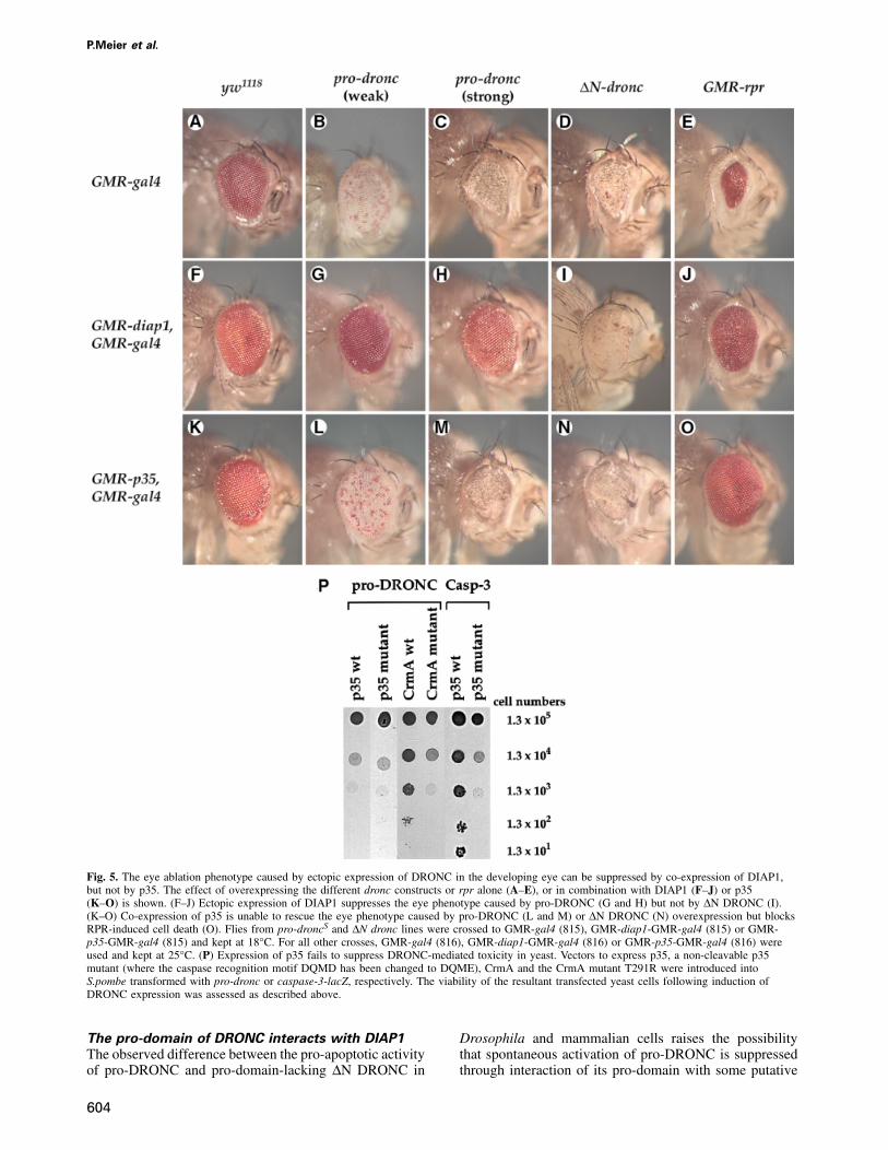
Fig. 5. The eye ablation phenotype caused by ectopic expression of DRONC in the developing eye can be suppressed by co-expression of DIAP1,but not by p35. The effect of overexpressing the different dronc constructs or rpr alone (A–E), or in combination with DIAP1 (F–J) or p35(K–O) is shown. (F–J) Ectopic expression of DIAP1 suppresses the eye phenotype caused by pro-DRONC (G and H) but not by ∆N DRONC (I).(K–O) Co-expression of p35 is unable to rescue the eye phenotype caused by pro-DRONC (L and M) or ∆N DRONC (N) overexpression but blocksRPR-induced cell death (O). Flies from pro-droncS and ∆N dronc lines were crossed to GMR-gal4 (815), GMR-diap1-GMR-gal4 (815) or GMR-p35-GMR-gal4 (815) and kept at 18°C. For all other crosses, GMR-gal4 (816), GMR-diap1-GMR-gal4 (816) or GMR-p35-GMR-gal4 (816) wereused and kept at 25°C. (P) Expression of p35 fails to suppress DRONC-mediated toxicity in yeast. Vectors to express p35, a non-cleavable p35mutant (where the caspase recognition motif DQMD has been changed to DQME), CrmA and the CrmA mutant T291R were introduced intoS.pombe transformed with pro-dronc or caspase-3-lacZ, respectively. The viability of the resultant transfected yeast cells following induction ofDRONC expression was assessed as described above.
The pro-domain of DRONC interacts with DIAP1
The observed difference between the pro-apoptotic activityof pro-DRONC and pro-domain-lacking ∆N DRONC in
604
Drosophila and mammalian cells raises the possibilitythat spontaneous activation of pro-DRONC is suppressedthrough interaction of its pro-domain with some putative

DRONC is regulated by DIAP1
Fig. 6. Heterozygosity at the diap1 locus bearing the deficiency[Df(3L)th102] enhances the eye phenotype caused by DRONCoverexpression. (A) Flies with a 50% reduced diap1 gene dose areviable and show a perfectly normal compound eye [�/SM6;Df(3L)th102/UAS-pro-droncW]. (B) Overexpression of GAL4 inducesa rough eye phenotype in heterozygous diap1 flies [�/GMR-gal4;Df(3L)th102/TM3]. (C) Ectopic expression of pro-droncW induces aspotted eye phenotype (�/GMR-gal4; UAS-pro-droncW/TM6c).Note: these flies show a less prominent spotted eye phenotype whencompared with the flies shown in Figure 5B due to their differentgenetic background. (D) Flies that express pro-droncW and areheterozygous for diap1 [Df(3L)th102] display severely deformed eyesand die trapped in their pupae case [�/GMR-gal4; Df(3L) th102/UAS-pro-droncW]. All flies were embedded in holocarbon oil andphotographed using a stereo-microscope.
cellular inhibitor. To identify such an inhibitor, we searchedfor Drosophila proteins that interact specifically with theDRONC pro-domain in a yeast two-hybrid assay using a0–24 h Drosophila embryonic cDNA library. From 1 � 106
yeast transformants, we recovered 56 DRONC-interactingclones, of which 17 encoded DIAP1 (Figure 4A). Thesecond BIR domain of DIAP1 was necessary and sufficientfor the interaction with the pro-domain of DRONC(DRONC-CARD, Figure 4B). This is particularly intri-guing since the BIR2 region of DIAP1 is also known tointeract physically with, and block the pro-apoptotic activ-ity of, RPR, GRIM and HID (Vucic et al., 1997, 1998a,b).
To verify the observed interaction between DIAP1 andDRONC, we performed co-immunoprecipitation experi-ments on cellular extracts obtained from 293T cells(Figure 4C). FLAG-tagged pro-DRONC C→A, ∆NDRONC C→A and DRONC-CARD (the pro-domain ofDRONC on its own) were each tested for interaction withMyc-tagged DIAP1 deletion mutants (BIR1/2, 1–341;BIR1, 1–146; and BIR2, 177–341; see schematic repre-sentation in Figure 4B). As expected, full-length DRONCand the isolated pro-domain of DRONC (DRONC-CARD)both co-immunoprecipitated with BIR1/2 and BIR2 butnot with BIR1, consistent with our yeast two-hybrid datashowing that the BIR2 domain of DIAP1 is required for
605
the interaction with DRONC. Somewhat surprisingly,however, ∆N DRONC lacking the pro-domain also co-immunoprecipitated with DIAP1, although to a far lesserextent than full-length DRONC or DRONC-CARD. TheBIR2 region of DIAP1 was required for this interactionbetween ∆N DRONC and DIAP1 since ∆N DRONCformed stable complexes only with BIR1/2 and BIR2 andnot with BIR1. Taken together, these results indicate thatDIAP1 physically interacts with unprocessed pro-caspaseDRONC and that the BIR2 region of DIAP1 is able tobind both the pro-domain and the core region of DRONC.
Expression of DIAP1 rescues the eye phenotype
induced by ectopic expression of pro-DRONC but
not ∆N DRONC
To assess the ability of DIAP1 to modulate DRONCactivation in vivo, we co-expressed DIAP1 with DRONCand ∆N DRONC (Figure 5F–I). Ectopic expression ofDIAP1 in the developing eye of pro-droncW transgenicflies completely rescued the phenotype caused by ectopicexpression of pro-DRONC (compare Figure 5B with G).Furthermore, GMR-diap1 also rescued the more severesmall eye phenotype of pro-droncS transgenic flies backto a normal eye size (Figure 5H). In contrast, GMR-diap1failed to rescue the eye phenotype caused by ∆N DRONC(Figure 5I). However, although GMR-diap1/∆N droncflies still contained a severely distorted eye lacking pigmentcells, their eye size was slightly larger than that in ∆Ndronc transgenic flies. This indicates that DIAP1 does, tosome extent, ameliorate the effect of ∆N DRONC (com-pare Figure 5D with I) and is consistent with our observa-tion that DIAP1 does interact weakly with ∆N DRONCin co-immunoprecipitation analyses (see Figure 4C).Together, these results indicate that the pro-domain andthe core region of DRONC are both required for DIAP1 tointeract maximally with DRONC and to inhibit DRONC-induced apoptosis. Presumably, DIAP1 binds to unpro-cessed pro-caspase DRONC and so suppresses its autoprot-eolytic cleavage and activation.
Heterozygosity at the diap1 locus enhances the
eye phenotype caused by pro-DRONC
overexpression
The observation that apoptosis in pro-droncW flies occursrelatively late during eye development might indicate thatactivation of overexpressed pro-DRONC is suppressed, atleast in part, through the interaction of its pro-domainwith endogenous DIAP1. To validate this hypothesis,we assessed genetically whether endogenous DIAP1 isresponsible for the relatively weak eye phenotype observedin pro-droncW flies. Pro-droncW flies were crossed toheterozygous diap1 flies that carry a deletion in thethread [i.e. diap1, Df(3L)th102] locus (Figure 6). Whereasheterozygous diap1 flies were viable and exhibited anormal compound eye, pro-droncW flies with a 50%reduced dosage of the diap1 gene died trapped in theirpupae cases and displayed severely deformed eyes(compare Figure 6A with D). This indicates that a deletionwhich removes DIAP1 converts the weak eye phenotypecaused by ectopic expression of pro-DRONC into a lethalsevere eye phenotype. This is consistent with the notionthat endogenous DIAP1 negatively regulates pro-DRONCactivation and is responsible, at least in part, for the

P.Meier et al.
Fig. 7. DRONC is a rate-limiting caspase in the RPR and HID death pathway. (A–D) Cell death induced by RPR and HID is sensitive to dronc genedosage. Flies with a chromosomal deletion that removes the dronc locus [Df(3L)AC1] show a suppressed RPR and HID eye phenotype. (A) GMR-rpr,�; (B) GMR-rpr,Df(3L)AC1; (C) GMR-hid,�; (D) GMR-hid,Df(3L)AC1. (E and F) The expression of dominant-negative DRONC mutantssuppresses the RPR eye phenotype. (E) GMR-rpr/GMR-gal4, UAS-pro-dronc C→A; (F) GMR-rpr/GMR-gal4, UAS-dronc-card.
relatively late onset of cell death in eyes of pro-droncW
transgenic flies.
The baculovirus caspase inhibitor p35 does not
rescue the eye phenotype induced by DRONC
overexpression
p35 is a promiscuous baculovirus-encoded inhibitor ofcaspases (Hay et al., 1994; Zhou et al., 1998). Becausethe unusual pentapeptide surrounding the active site inDRONC might confer a novel substrate specificity, wewere interested in determining whether p35 rescues theeye phenotype of dronc transgenic flies. Co-expression ofp35 in the developing eye of pro-droncW, pro-droncS or∆N dronc flies failed to rescue the eye phenotype causedby DRONC, although it did, to some extent, ameliorate it(Figure 5K–N). So, for example, p35 slightly increasedthe eye size of pro-droncS or ∆N dronc transgenic fliesalthough all such flies still showed a white and small eyephenotype (Figure 5M and N). In parallel experiments, p35efficiently blocked RPR-induced cell death and completely
606
rescued the small eye phenotype resulting from RPRoverexpression (compare Figure 5E with O). The inabilityof p35 to rescue the DRONC-mediated phenotype wasnot due to insufficient levels of p35 expression since theDRONC eye phenotype was not modified by increasingthe dosage of p35 in these experiments (data not shown).Furthermore, DRONC-induced cell killing was also notblocked by co-expression of p35 in S.pombe (Figure 5P).In contrast to p35, expression of the pox virus caspaseinhibitor CrmA, but not the loss-of-function CrmA mutantT291R, did inhibit DRONC-induced cell death in S.pombe.We conclude that p35 does not inhibit DRONC appre-ciably, making DRONC the first identified caspase resistantto inhibition by p35.
DRONC functions in the RPR and HID pathway
The observation that p35 blocks RPR- but not DRONC-induced cell death raises a question over whether DRONCacts in an RPR-dependent or an RPR-independent deathpathway. To investigate this question more carefully, we

DRONC is regulated by DIAP1
examined whether RPR-induced cell death is sensitive todronc gene dosage. Because no single gene mutations indronc are currently available, we used mutant flies witha larger chromosomal deletion that includes the dronclocus [Df(3L)AC1]. We crossed Df(3L)AC1 to GMR-rprflies and found that flies carrying Df(3L)AC1 show asignificant suppression of the RPR eye phenotype(Figure 7A and B). Furthermore, Df(3L)AC1 also sup-presses HID-mediated cell killing in the eye (Figure 7Cand D). To investigate further whether this observedsuppression is due specifically to loss of dronc, we assessedwhether the expression of dominant-negative DRONCmutants (pro-DRONC C→A and DRONC-CARD) alsosuppresses the RPR eye phenotype. Pro-DRONC C→Astrongly suppressed RPR cell killing, and, surprisingly,the pro-domain of DRONC on its own (DRONC-CARD)completely rescued the RPR eye phenotype (Figure 7Eand F). These results, in which DRONC function is ablatedeither by the Df(3L)AC1 deletion or by the action ofdominant-negative DRONC, are consistent with the notionthat DRONC is a rate-limiting caspase in the RPR andHID death pathway.
Discussion
Apoptosis is a highly conserved process by which eukary-otic cells commit suicide. In D.melanogaster, RPR, GRIMand HID serve as upstream transducers of apoptotic stimulithat induce cell death by triggering caspase activation.Our approach to characterizing the pathways activatingone effector caspase, drICE, was to search for proteinsthat interact with the inactive, unprocessed pro-caspaseand that might, therefore, modulate its activation.
Using a yeast two-hybrid screen with pro-drICE as bait,we identified DRONC as a drICE-interacting caspase.DRONC shares homology with members of the caspasefamily and most closely resembles caspase-9. However,DRONC does not contain a typical caspase active sitepentapeptide QAC(R/Q/G) (G/E) but instead has the novelsequence PFCRG (Dorstyn et al., 1999). Based on theX-ray crystal structure of human caspase-1, the glutamineat position 1 of the pentapeptide forms part of the substrate-binding pocket (Walker et al., 1994; Wilson et al., 1994).A change at this position may therefore indicate thatDRONC has a different substrate specificity from that ofclassical caspases. Our finding that the promiscuous cas-pase inhibitor p35 is neither cleaved by DRONC in vitronor blocks the DRONC activity in vivo supports the notionthat DRONC has a different substrate specificity. Althoughthe physiological cellular substrate(s) for DRONC haveyet to be determined, it may be of note that DRONCcleaves three ascribed caspase substrates, drICE, laminDmO and DREP-1, in an in vitro assay.
We have shown that ectopic DRONC action is lethalto yeast, insect and mammalian cells. However, expressionof full-length pro-DRONC has a more restricted lethality:although it is toxic to yeast cells, it fails to kill Rat-1 cellsand, when expressed in the developing Drosophila eye, itgenerates an unusual phenotype in which the eye exhibitsapparently normal outer morphology with internal ablationof all photoreceptor and pigment cells, resulting in a‘hollow’ eye. Macroscopically, this manifests as whiteeyes with occasional red spots, even though these flies
607
are genetically white� and would therefore be expectedto have red eyes. This ‘spotted eye’ results from ablationof most of the internal eye structures apart from afew remnant red pigment cells. Why should the outermorphology of pro-droncW eyes be maintained whenvirtually all retinal cells are severely affected? Specializa-tions that make the eye a functional organ take placerelatively late during pupal development, with rhabdo-meres, pigment cells and lens structures differentiatingonly after pattern formation is complete, after the firstthird of pupal life. Formation of the compound eyewith its ~800 ommatidia depends on the correct three-dimensional structure of the underlying cluster of photore-ceptor cells. The preservation of external eye structure wesee in pro-droncW eyes indicates that massive cell deathoccurs only very late, after almost the entire eye develop-ment has taken place. This is in contrast to the ablationof eye cells early in development, which is evident inflies expressing RPR or ∆N DRONC and generates smalland abnormally shaped eyes. The ability of DRONC tokill both pigment and photoreceptor cells is similar to thatof CED-4 and differs from RPR, which seems to beselective for photoreceptor cells and generates eyes that,although hypotrophic, remain red (Kanuka et al., 1999).
Previous studies have shown that ectopic expression ofthe promiscuous baculovirus caspase inhibitor p35 inD.melanogaster blocks most of the naturally occurringcell death during development, as well as cell death arisingfrom DNA damage or overexpression of either RPR,GRIM or HID (Hay et al., 1994; Grether et al., 1995;Chen et al., 1996; White et al., 1996). This is consistentwith studies that indicate that p35 is a promiscuousinhibitor of all known mammalian and invertebrate cas-pases. It is therefore surprising that p35 proved unable torevert the spotted or small eye phenotype induced byDRONC. Three lines of evidence indicate that DRONCis not inhibited by p35. First, in Drosophila, p35 does notsuppress the DRONC eye phenotype. This is not simplydue to insufficient expression levels of p35 because eventwo copies of GMR-p35 are still unable to suppressDRONC-induced cell death. Secondly, DRONC-inducedcell death in S.pombe is not inhibited by p35. In contrast,p35 strongly suppressed caspase-3-induced cell death inyeast in parallel experiments. It is of note that the poxvirus caspase inhibitor CrmA does block DRONC-inducedcell death and is an effective inhibitor of DRONC. Thirdly,p35 must be proteolytically processed in order to inhibitcaspases. However, DRONC does not cleave p35 in anin vitro cleavage assay, indicating that p35 is not asubstrate for DRONC.
The fact that p35 completely rescues the phenotypecaused by ectopic expression of either RPR, GRIM orHID yet only slightly ameliorates DRONC eyes raises thepossibility that DRONC is not part of the RPR, GRIM orHID pathway but instead functions in an independentdeath signalling system. However, several lines of evidenceargue that dronc is an important effector of the pro-apoptotic proteins RPR and HID. Specifically, hetero-zygosity at the dronc locus significantly suppresses theeye ablation induced by RPR and HID, and expression ofdominant-negative DRONC mutants suppresses RPR eyes.Strikingly, expression of the DRONC-CARD alone com-pletely suppresses the RPR eye phenotype, indicating that

P.Meier et al.
CARD-containing proteins are critically involved in theapoptotic signal transduction initiated by RPR. Nonethe-less, although we favour the notion that DRONC is aneffector of RPR and HID, it is formally possible thatDRONC functions in a pathway that is additive to, butindependent of, RPR and HID. We currently cannotdistinguish between these two alternatives.
If RPR and HID function through DRONC, why shouldp35 block RPR- and HID-, but not DRONC-induceddeath? During the apoptotic process, caspases are activatedin an amplifying proteolytic cascade, cleaving one anotherin turn. Thus, it may be that the activation of endogenousDRONC by RPR and HID is insufficient to induce celldeath on its own but requires amplification of the apoptoticsignal through the activation of other caspases such asdrICE, DCP-1 or even DCP-2/DREDD. In contrast, whenDRONC is overexpressed, this amplifying proteolyticcascade may not be required to kill the cell. If thedownstream caspases were p35 sensitive, this could explainwhy p35 ameliorates, yet cannot block, DRONC killing.Interestingly, recent studies on DREDD indicate that ap35-resistant caspase (or some other class of protease) isindeed responsible for proteolytic cleavage and activationof DREDD following overexpression of RPR, GRIM orHID (Chen et al., 1998). The initial cleavage and activationof DREDD is not blocked by p35 although p35 doesblock the eventual cell death that would otherwise result.This must mean that a p35-resistant caspase is activatedfollowing RPR, GRIM or HID induction but that itsactivation does not lead to cell death in the presence ofp35. Given its resistance to p35, this makes DRONCan intriguing candidate for such a ‘DREDD-activatingcaspase’.
Several lines of evidence suggest that pro-DRONCactivation is negatively regulated via its pro-domain. Thisis best illustrated by the biology underlying the relativelyweak eye phenotype in flies expressing full length pro-droncW. First, most UAS-pro-dronc lines are viable whencrossed to a strong GMR-gal4 line and kept at 25°C: incontrast, virtually all GMR-driven ∆N dronc transgeniclines tested die under such conditions. Secondly, most pro-dronc lines exhibit essentially normal outer eye structure,whereas rare surviving ∆N dronc transgenic flies neverdisplay this ‘weak’ eye phenotype and have severelydeformed eyes. Thirdly, ectopic expression of pro-DRONCinduces no significant increase of cell death in the eyediscs of third instar larvae, whereas excessive cell deathis evident posterior to the morphogenetic furrow in theeye discs of third instar larvae expressing ∆N DRONC.Finally, ectopic expression of DRONC in mammalianRat-1 cells induces apoptosis only when its pro-domainhad been removed, suggesting the existence of an innateinhibitor of DRONC activation acting through theDRONC-CARD domain. All of these observations implic-ate the DRONC pro-domain in repressing activation ofthe caspase and suggest that DRONC activation is keptin abeyance in metazoan cells through the action of someCARD-binding innate inhibitor. In contrast, our studies ofDRONC in S.pombe unambiguously show that isolatedpro-DRONC is, by itself, perfectly capable of undergoingcatalytic autoprocessing resulting in its activation. Indeed,in yeast, pro-DRONC proved more toxic than ∆N DRONC,suggesting that the presence of the pro-domain may
608
actually enhance DRONC activation in the absence ofother modulating influences.
In insect cells, a candidate for such an innate DRONCrepressor is the inhibitor of apoptosis, DIAP1, which wehave shown to interact with the DRONC pro-domain: co-expression of DIAP1 completely reverts the eye ablationphenotype of pro-droncW flies, whereas the eye ablationphenotype induced by ∆N DRONC is largely unaffected.If endogenous DIAP1, or an analogue, were expressed inthe Drosophila eye until very late in its development, thiswould provide the requisite mechanism for holding theactivity of DRONC in abeyance until very late, so generat-ing the ‘spotted eye’ phenotype we observe. Indeed,heterozygosity at the diap1 locus greatly enhances theeye phenotype induced by pro-DRONC overexpression,indicating that endogenous DIAP1 negatively regulatesDRONC activation in vivo. This is analogous to the wayin which c-IAP1, c-IAP2 and XIAP bind to, and inhibitactivation of, the pro-form of the apical caspase-9 inmammalian cells (Deveraux et al., 1998). The notion thatit is DIAP1, in particular, that most likely fulfils the roleof in vivo suppressor of DRONC is reinforced by ourstudies in yeast which show that whilst DIAP1 bothinteracts with, and protects from the lethal effects of, pro-DRONC, Drosophila DIAP2 and the mammalian IAPhomologues MIHA, MIHB, MIHC, MIHD and XIAPoffer no such protection (data not shown).
Currently, very little is known about how IAPs suppressapoptosis, although the most convincing biological evi-dence for the ability of IAPs to regulate cell death comesfrom genetic studies in D.melanogaster (Hay et al., 1995;Wang et al., 1999; Goyal et al., 2000). Deletion of thechromosomal region encoding DIAP1 enhances cell deathinduced by ectopic expression of RPR, and genetic lossof DIAP1 function leads to early and widespread apoptosis,indicating that DIAP1 is essential for survival of manycell types (Wang et al., 1999; Goyal et al., 2000).Furthermore, overexpression of DIAP1 suppresses celldeath induced by either RPR, GRIM or HID throughdirect interaction between these various pro-apoptoticproteins and the second BIR domain of DIAP1 (Harveyet al., 1997; Vucic et al., 1997, 1998a,b), the same BIRdomain that is sufficient for its interaction with pro-DRONC. It is noteworthy that it is also the second BIRrepeat of the mammalian IAP family members c-IAP1,c-IAP2 and XIAP that appears sufficient for their anti-apoptotic activity (Deveraux et al., 1997; Roy et al., 1997;Takahashi et al., 1998).
Our finding that DIAP1 directly binds to and inhibitscell death caused by ectopic expression of DRONC, aswell as by RPR, GRIM and HID, underscores the keyrole played by DIAP1 in the regulation of apoptosis inD.melanogaster and raises the possibility that RPR, HIDor GRIM may exert some, or all, of their pro-apoptoticaction through displacement of DIAP1 from the pro-domain of DRONC, so allowing activation of the caspaseand consequent cell death (Figure 8). The isolation ofDIAP1 mutants that display greatly reduced binding forRPR, HID and GRIM and significantly suppress RPR,HID and GRIM cell killing strongly supports this idea(Goyal et al., 2000). According to this model, IAPsfunction as ‘guardians’ of the apoptotic machinery, whichact to suppress the chance of spontaneous activation of

DRONC is regulated by DIAP1
Fig. 8. The observation that DIAP1 interacts with the pro-caspaseDRONC as well as with apoptotic inducers such as RPR, GRIM andHID places DIAP1 in a potentially pivotal position to regulateapoptosis. In the proposed model, DIAP1 functions as ‘guardian of thecaspase machinery’ by binding to and suppressing spontaneous pro-caspase activation in the absence of RPR, GRIM and active HID. Asindicated by studies using heterologous systems, DIAP1 may also actby directly inhibiting the proteolytic activity of spontaneouslyactivated caspases (dotted lines) (Kaiser et al., 1998; Hawkins et al.,1999). According to the ‘liberation model’, RPR, GRIM or HID exertsome, or all, of their pro-apoptotic action by liberating initiatorcaspases, such as DRONC, from the activation-inhibitory effect ofDIAP1. This displacement of DIAP1 from DRONC could then resultin the activation of DRONC through DARK, the Drosophilahomologue of Apaf-1/CED-4 (Rodriguez et al., 1999).
the intrinsic cell death machinery by neutralizing pro-apoptotic caspases, so establishing a buffered thresholdthat must be either exceeded or neutralized in order toinitiate the destruction of a cell.
Materials and methods
Yeast two-hybrid assay and cloning of droncThe catalytically inactive mutant of drICE (drICE C→A, described inFraser and Evan, 1997) was cloned into the pAS2.1 vector (Clontech)and used as bait in a yeast two-hybrid screen against a 0–24 hD.melanogaster embryonic cDNA library constructed in pACT2 (kindlyprovided by S.J.Elledge) following the Matchmaker Two-Hybrid Systemprotocol (Clontech). Three drICE cDNA plasmids, encoding amino acids1–339, 40–339 and 43–339, respectively, and one cDNA encoding aminoacids 104–451 of DRONC (pACT2-∆N dronc) were isolated. A BLASTsearch of the GenBank DNA sequence database identified an expressedsequence tag identical to dronc. The corresponding full-length clone(LD28292) was obtained and was used to generate all subsequent droncconstructs. pAS2.1-drICE C→A, pAS2.1-∆N drICE, pAS2.1-BIR1/2,pAS2.1-BIR1 and pAS2.1-BIR2 were generated by PCR. All constructswere verified by DNA sequencing.
Cytotoxicity assay in S.pombeAll transformations of S.pombe were performed using a standard lithiumacetate protocol and cells were then plated on selective medium. Allclones were maintained throughout on selective media. Yeast was grownto log phase, the OD595 of the culture was determined, and the yeastwas then plated in serial 10-fold dilutions on selective and inducingmedia. DNA fragments containing pro-dronc, ∆N dronc, pro-dronc C→Aor ∆N dronc C→A were cloned into the S.pombe expression vectorpNeu. The vector pNeu and the caspase-3-lacZ construct have been
609
described (Ekert et al., 1999). The mutant p35 was created by changingthe caspase cleavage site from DQMD to DQME. All constructs wereverified by restriction digest and sequencing.
Transient transfection of Rat-1 cells and apoptosis assayTransient transfections of Rat-1 cells were performed using the FuGENEtransfection reagent according to the manufacturer’s instructions (Roche).CMV-lacZ reporter DNA was co-transfected with pTracer-CMV2-basedconstructs (pT, Invitrogen) in a ratio of 1:10. At 24 h post-transfection,cells were assayed for β-galactosidase (β-Gal) activity and cell viabilitywas determined based on the morphology of at least 400 β-Gal-positivecells. pT-FADD and pT-caspase-8 were used as positive controls forapoptosis induction. Pro-dronc, ∆N dronc and ∆N dronc C→A wereamplified by PCR and cloned into pTracer plasmids. The catalyticallyinactive mutants pro-dronc C→A and ∆N dronc C→A were generatedusing the Altered Sites II in vitro Mutagenesis System according to themanufacturer’s instructions (Promega). All constructs were verified bysequencing.
Immunoprecipitation and immunoblot analysis293T cells were transiently transfected using Superfect according to themanufacturer’s instructions (Qiagen). Equal amounts of FLAG- and Myc-based pcDNA3.1 construct were used in the transfection experiments. At48 h post-transfection, cells were harvested and lysed in ice-cold lysisbuffer. Aliquots of cell lysates were incubated for 2 h at 4°C with M2anti-FLAG monoclonal antibody resin (Sigma). Co-immunoprecipitatedproteins were analysed by immunoblot analysis using the anti-Mycmonoclonal antibody 9E10.
P-element-mediated germline transformation and transgeneanalysisTransgenic flies were generated by microinjecting pUAST-pro-dronc,pUAST-∆N dronc, pUAST-pro-dronc C→A and pUAST-CARD-only (thepro-domain of DRONC-only) DNA according to standard protocols.Several independent lines were established from each transgene, andtheir integration sites were mapped to individual chromosomes. SixteenpUAST-pro-dronc, seven pUAST-∆N dronc, eleven pUAST- pro-droncC→A and four pUAST-dronc-card independent transgenic lines wereobtained. For ectopic expression of the various transgenes, dronctransgenic flies were crossed with the GAL4 lines: GMR-gal4 (816,strong) and GMR-gal4 (815, weak). To assess the effect of DIAP1 orp35 overexpression on the DRONC-mediated phenotype, GMR-gal4815 and 816 were recombined onto the chromosome bearing GMR-diap1 or GMR-p35 to generate GMR-gal4-GMR-diap1 and GMR-gal4-GMR-p35, respectively. GMR-rpr and GMR-hid were obtained fromM.Grether and H.Steller; GMR-p35 and GMR-diap1 were obtained fromB.Hay and G.Rubin. In addition, the following strains were used:55100 (Df(3L)th102, h[1] kni[ri-1] e[s]/TM6C, cu[1] Sb[1] e[1] ca[1]),Df(3L)AC1 (with the breakpoints 67A2;67D13), 5248 (P{w�mC�GMR-hid}SS1, y1 w* P{ry�t7.2�neoFRT}19A; P{w�m*�GAL4-ey.H}SS5, P{w�mC�UAS-FLP1.D}JD2).
Histology and scanning electron microscopyHistological sections were prepared and analysed as described in Basleret al. (1991). Third instar larval eye discs were stained with acridineorange as described in Abrams et al. (1993). The cytological mapposition for dronc was determined according to standard protocols usinga biotinylated dronc probe (Ashburner, 1989). Flies were prepared forSEM as described in Kimmel et al. (1990).
Purification of processed DRONC and protease assaysFLAG-tagged versions of DRONC and DRONC C→A were expressedusing the S.pombe system described above. At 15 h post-induction ofDRONC expression, DRONC and DRONC C→A were isolated fromyeast extracts by immunoprecipitation using anti-FLAG M2 agaroseaffinity gel (Sigma). Protease assays were performed as described inChen et al. (1998). cDNAs encoding drICE C→A, lamin DmO, p35and DREP-1 were cloned into pBluescript (Stratagene) and in vitrotranscribed/translated using the reticulocyte lysate TnT system (Promega)in the presence of [35S]methionine (Amersham).
Acknowledgements
We would like to thank Drs Steven Elledge for the generous gift of theDrosophila melanogaster embryonic cDNA library, Gabriel Nunez forthe DREP-1 clone, Gerald Rubin for GMR-diap1 and GMR-p35 flies,

P.Meier et al.
Hermann Steller for GMR-rpr and GMR-hid, John Sgouros for helpwith the sequence analysis, Steve Gschmeissner and Jeol (UK) Ltd forhelp with the scanning electron microscopy, Carol Upton for help withsectioning adult fly eyes, Julian Lewis for the use of the digital cameraand stereo-microscope, and David Gubb and Josefa Gonzalez for helpwith the chromosomal in situ analysis. In addition, we are indebted toDrs David Ish-Horowicz, Helen McNeill, Dionisio Martin-Zanca, DamianBrunner and Shila Schneider for helpful discussions and comments onthe manuscript. Finally, we thank members of the Evan lab for supportand helpful discussions during this work. S.J.L is supported by a RoyalSociety University Research Fellowship and G.I.E. is a Royal SocietyNapier Professor.
References
Abrams,J.M., White,K., Fessler,L.I. and Steller,H. (1993) Programmedcell death during Drosophila embryogenesis. Development, 117, 29–43.
Alnemri,E.S. (1997) Mammalian cell death proteases: a family of highlyconserved aspartate specific cysteine proteases. J. Cell. Biochem., 64,33–42.
Ashburner,M. (1989) Drosophila: A Laboratory Manual. Cold SpringHarbor Laboratory Press, Cold Spring Harbor, NY.
Basler,K., Christen,B. and Hafen,E. (1991) Ligand-independentactivation of the sevenless receptor tyrosine kinase changes the fateof cells in the developing Drosophila eye. Cell, 64, 1069–1081.
Bateman,A., Birney,E., Durbin,R., Eddy,S.R., Finn,R.D. andSonnhammer,E.L. (1999) Pfam 3.1: 1313 multiple alignments andprofile HMMs match the majority of proteins. Nucleic Acids Res., 27,260–262.
Chen,P., Nordstrom,W., Gish,B. and Abrams,J.M. (1996) grim, a novelcell death gene in Drosophila. Genes Dev., 10, 1773–1782.
Chen,P., Rodriguez,A., Erskine,R., Thach,T. and Abrams,J.M. (1998)Dredd, a novel effector of the apoptosis activators reaper, grim andhid in Drosophila. Dev. Biol., 201, 202–216.
Deveraux,Q.L. and Reed,J.C. (1999) IAP family proteins—suppressorsof apoptosis. Genes Dev., 13, 239–252.
Deveraux,Q.L., Takahashi,R., Salvesen,G.S. and Reed,J.C. (1997)X-linked IAP is a direct inhibitor of cell-death proteases. Nature, 388,300–304.
Deveraux,Q.L., Roy,N., Stennicke,H.R., Van Arsdale,T., Zhou,Q.,Srinivasula,S.M., Alnemri,E.S., Salvesen,G.S. and Reed,J.C. (1998)IAPs block apoptotic events induced by caspase-8 and cytochrome cby direct inhibition of distinct caspases. EMBO J., 17, 2215–2223.
Dorstyn,L., Colussi,P.A., Quinn,L.M., Richardson,H. and Kumar,S.(1999) DRONC, an ecdysone-inducible Drosophila caspase. Proc.Natl Acad. Sci. USA, 96, 4307–4312.
Ekert,P.G., Silke,J. and Vaux,D.L. (1999) Inhibition of apoptosis andclonogenic survival of cells expressing crmA variants: optimal caspasesubstrates are not necessarily optimal inhibitors. EMBO J., 18,330–338.
Ellis,H.M. and Horvitz,H.R. (1986) Genetic control of programmed celldeath in the nematode C.elegans. Cell, 44, 817–829.
Ellis,M.C., O’Neill,E.M. and Rubin,G.M. (1993) Expression ofDrosophila glass protein and evidence for negative regulation ofits activity in non-neuronal cells by another DNA-binding protein.Development, 119, 855–865.
Evan,G. and Littlewood,T. (1998) A matter of life and cell death.Science, 281, 1317–1322.
Fraser,A.G. and Evan,G.I. (1997) Identification of a Drosophilamelanogaster ICE/CED-3-related protease, drICE. EMBO J., 16,2805–2813.
Fraser,A.G., McCarthy,N.J. and Evan,G.I. (1997) drICE is an essentialcaspase required for apoptotic activity in Drosophila cells. EMBO J.,16, 6192–6199.
Goyal,L., McCall,K., Agapite,J., Hartwieg,E. and Steller,H. (1999)Induction of apoptosis by Drosophila reaper, hid and grim throughinhibition of IAP function. EMBO J., 19, 589–597.
Grether,M.E., Abrams,J.M., Agapite,J., White,K. and Steller,H. (1995)The head involution defective gene of Drosophila melanogasterfunctions in programmed cell death. Genes Dev., 9, 1694–1708.
Gruenbaum,Y. et al. (1988) Drosophila nuclear lamin precursor Dm0 istranslated from either of two developmentally regulated mRNA speciesapparently encoded by a single gene [published erratum appears inJ. Cell Biol., 1988, 106, 2225]. J. Cell Biol., 106, 585–596.
Harvey,A.J., Bidwai,A.P. and Miller,L.K. (1997) Doom, a product ofthe Drosophila mod (mdg4) gene, induces apoptosis and binds to
610
baculovirus inhibitor-of-apoptosis proteins. Mol. Cell. Biol., 17,2835–2843.
Hawkins,C.J., Wang,S.L. and Hay,B.A. (1999) A cloning method toidentify caspases and their regulators in yeast: identification ofDrosophila IAP1 as an inhibitor of the Drosophila caspase DCP-1.Proc. Natl Acad. Sci. USA, 96, 2885–2890.
Hay,B.A., Wolff,T. and Rubin,G.M. (1994) Expression of baculovirusP35 prevents cell death in Drosophila. Development, 120, 2121–2129.
Hay,B.A., Wassarman,D.A. and Rubin,G.M. (1995) Drosophila homologsof baculovirus inhibitor of apoptosis proteins function to block celldeath. Cell, 83, 1253–1262.
Inohara,N., Koseki,T., Hu,Y., Chen,S. and Nunez,G. (1997) CLARP, adeath effector domain-containing protein interacts with caspase-8 andregulates apoptosis. Proc. Natl Acad. Sci. USA, 94, 10717–10722.
Inohara,N., Koseki,T., Chen,S., Wu,X. and Nunez,G. (1998) CIDE, anovel family of cell death activators with homology to the 45 kDasubunit of the DNA fragmentation factor. EMBO J., 17, 2526–2533.
Kaiser,W.J., Vucic,D. and Miller,L.K. (1998) The Drosophila inhibitorof apoptosis D-IAP1 suppresses cell death induced by the caspasedrICE. FEBS Lett., 440, 243–248.
Kanuka,H., Hisahara,S., Sawamoto,K., Shoji,S., Okano,H. and Miura,M.(1999) Proapoptotic activity of Caenorhabditis elegans CED-4 proteinin Drosophila: implicated mechanisms for caspase activation. Proc.Natl Acad. Sci. USA, 96, 145–150.
Kimmel,B.E., Heberlein,U. and Rubin,G.M. (1990) The homeo domainprotein rough is expressed in a subset of cells in the developingDrosophila eye where it can specify photoreceptor cell subtype. GenesDev., 4, 712–727.
Munday,N.A., Vaillancourt,J.P., Ali,A., Casano,F.J., Miller,D.K.,Molineaux,S.M., Yamin,T.T., Yu,V.L. and Nicholson,D.W. (1995)Molecular cloning and pro-apoptotic activity of ICErelII and ICErelIII,members of the ICE/CED-3 family of cysteine proteases. J. Biol.Chem., 270, 15870–15876.
Rodriguez,A., Oliver,H., Zou,H., Chen,P., Wang,X. and Abrams,J.M.(1999) Dark is a Drosophila homologue of Apaf-1/CED-4 andfunctions in an evolutionarily conserved death pathway. Nature CellBiol., 5, 272–279.
Roy,N., Deveraux,Q.L., Takahashi,R., Salvesen,G.S. and Reed,J.C.(1997) The c-IAP-1 and c-IAP-2 proteins are direct inhibitors ofspecific caspases. EMBO J., 16, 6914–6925.
Song,Z., McCall,K. and Steller,H. (1997) DCP-1, a Drosophila celldeath protease essential for development [published erratum appearsin Science, 1997, 277, 167]. Science, 275, 536–540.
Spradling,A.C. and Rubin,G.M. (1983) The effect of chromosomalposition on the expression of the Drosophila xanthine dehydrogenasegene. Cell, 34, 47–57.
Takahashi,R., Deveraux,Q., Tamm,I., Welsh,K., Assa-Munt,N.,Salvesen,G.S. and Reed,J.C. (1998) A single BIR domain of XIAPsufficient for inhibiting caspases. J. Biol. Chem., 273, 7787–7790.
Thompson,C.B. (1995) Apoptosis in the pathogenesis and treatment ofdisease. Science, 267, 1456–1462.
Thornberry,N.A. and Lazebnik,Y. (1998) Caspases: enemies within.Science, 281, 1312–1316.
Tschopp,J., Thome,M., Hofmann,K. and Meinl,E. (1998) The fight ofviruses against apoptosis. Curr. Opin. Genet. Dev., 8, 82–87.
Uren,A.G., Coulson,E.J. and Vaux,D.L. (1998) Conservation ofbaculovirus inhibitor of apoptosis repeat proteins (BIRPs) in viruses,nematodes, vertebrates and yeasts. Trends Biochem. Sci., 23, 159–162.
Vaux,D.L. and Korsmeyer,S.J. (1999) Cell death in development. Cell,96, 245–254.
Vucic,D., Kaiser,W.J., Harvey,A.J. and Miller,L.K. (1997) Inhibition ofreaper-induced apoptosis by interaction with inhibitor of apoptosisproteins (IAPs). Proc. Natl Acad. Sci. USA, 94, 10183–10188.
Vucic,D., Kaiser,W.J. and Miller,L.K. (1998a) Inhibitor of apoptosisproteins physically interact with and block apoptosis induced byDrosophila proteins HID and GRIM. Mol. Cell. Biol., 18, 3300–3309.
Vucic,D., Kaiser,W.J. and Miller,L.K. (1998b) A mutational analysis ofthe baculovirus inhibitor of apoptosis Op-IAP. J. Biol. Chem., 273,33915–33921.
Walker,N.P. et al. (1994) Crystal structure of the cysteine proteaseinterleukin-1 β-converting enzyme: a (p20/p10)2 homodimer. Cell,78, 343–352.
Wang,S.L., Hawkins,C.J., Yoo,S.J., Muller,H.J. and Hay,B.A. (1999)The Drosophila caspase inhibitor DJAP1 is essential for cell survivaland is negatively regulated by HID. Cell, 98, 453–463.
White,K., Grether,M.E., Abrams,J.M., Young,L., Farrell,K. and Steller,H.

DRONC is regulated by DIAP1
(1994) Genetic control of programmed cell death in Drosophila.Science, 264, 677–683.
White,K., Tahaoglu,E. and Steller,H. (1996) Cell killing by theDrosophila gene reaper. Science, 271, 805–807.
Wilson,K.P. et al. (1994) Structure and mechanism of interleukin-1 betaconverting enzyme. Nature, 370, 270–275.
Zhou,Q., Krebs,J.F., Snipas,S.J., Price,A., Alnemri,E.S., Tomaselli,K.J.and Salvesen,G.S. (1998) Interaction of the baculovirus anti-apoptoticprotein p35 with caspases. Specificity, kinetics and characterizationof the caspase/p35 complex. Biochemistry, 37, 10757–10765.
Received July 13, 1999; revised November 19, 1999;accepted November 22, 1999
611